continuing medical education
TRANSCRIPT
159
From the Department of Dermatology, University of CincinnatiCollege of Medicine.
Reprint requests: Diya F. Mutasim, MD, Professor and Chairman,Department of Dermatology, University of Cincinnati, PO Box670592, Cincinnati, OH 45267-0592. E-mail: [email protected].
Copyright © 2000 by the American Academy of Dermatology, Inc.0190-9622/2000/$12.00 + 0 16/2/103582
C onnective tissue diseases (CTDs) are a group
of autoimmune disorders that have overlap-
ping clinical features (Table I). The accurate
diagnosis of a patient with one of these disorders
depends on the evaluation of 4 parameters, namely
clinical findings, histopathology, tissue immunofluo-
rescence, and serologic testing. This article is limited
to the serologic evaluation. Serologic testing does
not substitute for evaluation of the other criteria.
Serologic testing does help to confirm a clinical diag-
nosis and classify subsets of a CTD and thus help
predict prognosis. For example, a patient who pre-
sents with cutaneous lupus erythematosus and who
is found to have significantly high antibody titer to
native DNA (nDNA) (or double-stranded DNA
[dsDNA]) likely has systemic lupus erythematosus
(SLE) with cutaneous involvement.1-4 In addition, a
patient who has cutaneous sclerosis, calcinosis, and
esophageal dysmotility and who is found to have
anticentromere antibodies is much more likely to
have a benign course associated with the CREST
syndrome (calcinosis, Raynaud’s phenomenon,
esophageal dysmotility, sclerodactyly, and telangiec-
tasia)5,6 rather than the usually severe course associ-
ated with systemic sclerosis (SSc).
BIOLOGY OF THE ANTIBODYSPECIFICITY
Patients with CTD have an autoimmune phenom-
enon that results in the production of antibodies
against several self-antigens. These autoantibodies
are directed against all cellular components, that is,
nuclear, cytoplasmic, and cell membrane molecules.
The binding of these antibodies to commercially
available tissue extracts is the basis for serologic test-
ing. Whether these antibodies play a role in the
pathogenesis of the clinical manifestations of the dis-
ease is suspected but not confirmed with certainty.
The most common antibodies that are of diagnostic
value are shown in Table II.
In evaluating the results of these tests, it is impor-
tant to be aware of two findings. First, some of the
antibodies are not unique to patients with CTD and
may be present in the sera of normal persons or per-
sons with other conditions.7-14 Therefore the mere
detection of these antibodies does not always indi-
cate a CTD. In general, however, the total amount of
antibodies to a certain antigen is much larger in
patients with CTD.9,10 The total amount of antibod-
ies is usually indicated by the titer or the absolute
value given to the test. Second, the specificity of
each of the antibodies for the various CTD varies. For
example, some antibodies, such as Sm antibodies
and dsDNA antibodies, are highly specific (for
SLE).11-21 Other antibodies (eg, single-stranded DNA
CONTINUING MEDICAL EDUCATION
A practical guide for serologic evaluation ofautoimmune connective tissue diseases
Diya F. Mutasim, MD, and Brian B. Adams, MD Cincinnati, Ohio
Serologic testing is important in the evaluation of patients with autoimmune connective tissue diseases
(CTD). There are many techniques. Each of the tests has different sensitivity and specificity with varying
diagnostic value. These serologic tests detect antibodies to numerous cellular components. The diagnostic
significance and specificity of each antibody vary. Choosing the appropriate test and understanding its
clinical utility is an important aspect in the diagnostic evaluation of patients with CTD. (J Am Acad Dermatol
2000;42:159-74.)
Learning objective: At the conclusion of this learning activity, participants should be familiar with the
various serologic tests for CTD, should understand the associations of specific antibodies with individual
CTD, and should identify the factors that influence the predictive value of these serologic tests.
slide or plastic plate (for immunofluorescence and
ELISA, respectively). The substrate is incubated
with the patient’s serum. If the serum has antibod-
ies, they will bind to the substrate. This binding is
detected by a series of amplification steps that pro-
duce visible fluorescence (immunofluorescence)
or a colored dye that will be detected and quanti-
fied by a machine’s photometer (ELISA). Both tests
may be quantitative by diluting the serum to vari-
ous titers until the test is negative. The larger the
titer, the higher the amount of antibody in the
serum. ELISA has many advantages. It is cheaper,
less labor intensive, may be used to screen a large
number of sera together, is less subjective (does
not need human technical interpretation), and is
more sensitive.44,46 ELISA, however, is less specific
and results need to be interpreted with caution
(see later).8,44
Radial immunodiffusion takes advantage of the
ability of molecules (both antigen and antibody) to
migrate through agarose gel, bind together, precipi-
tate, and produce a visible line that indicates the
presence of antibodies.47 This test is less sensitive
than immunofluorescence and ELISA but is more
specific. For a serum to be positive by radial immu-
nodiffusion, the serum must contain a relatively
higher amount of antibodies (compared with more
sensitive techniques).10,11 Accordingly, the diagnos-
tic value of a positive test by radial immunodiffusion
is higher than that by ELISA because the diagnostic
value of the antibodies does not solely rely on their
presence, but also on their total amount.
The frequency of positivity for each of the vari-
ous antibodies in the various CTDs varies among
different reports. Some of the figures quoted in
this article are an estimated average of the various
percentages.
[ssDNA] antibodies) are of low diagnostic value
because of their high nonspecificity17,22; they may be
present in the sera of patients with most CTDs.
The type of antibodies present and the frequency
of their occurrence vary among the various CTDs. For
example, patients with mixed CTD (MCTD) have anti-
bodies to nuclear ribonucleoprotein (also known as
uridine-rich ribonucleoprotein [U1RNP]),8,12,13,17,22-27
and patients with CREST syndrome have antibodies
that are almost limited to the centromere.13,19,28,29 In
contrast, patients with SLE may have antibodies to
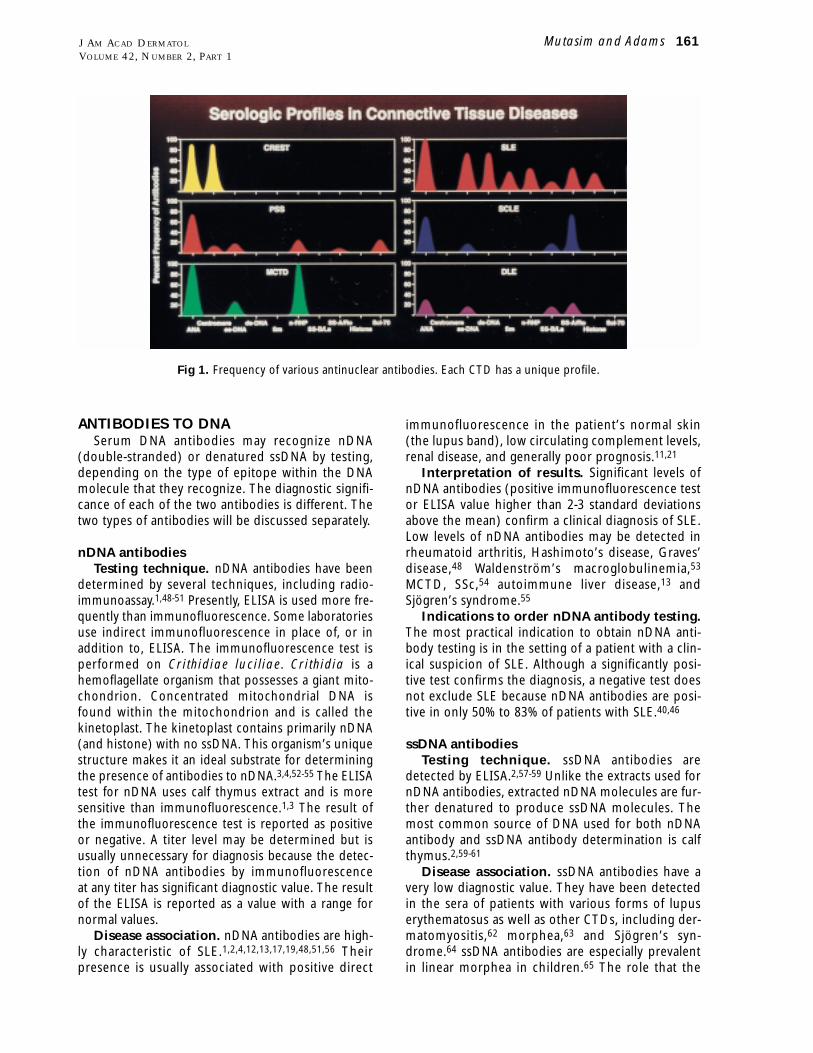
several cellular antigens.7,8,11,12,17,19,21,22,26 Fig 1
reveals the frequency of the various autoantibodies in
6 selected CTDs. Each CTD has a unique profile of
antibodies.
TECHNIQUES FOR SEROLOGIC TESTINGThe methods for the detection of the various
antibodies have changed over the past few decades.
Immunologic techniques that are commonly used
during each time period have been utilized for the
detection of these antibodies. For example, radioim-
munoassay and immunoelectrophoresis were com-
monly used in the past.30-36 Radial immunodiffusion
and immunofluorescence8,10,30,32,37-42 remain of
important value although both (especially the for-
mer) are being slowly replaced by newer techniques
such as the enzyme-linked immunosorbent assay
(ELISA).8,18,32,38,39,43-46 Several types of antibodies
may be detected by multiple techniques. The prin-
ciples of immunofluorescence and ELISA are similar.
An antigen (in the substrate) is placed on a glass
160 Mutasim and Adams J AM ACAD DERMATOL
FEBRUARY 2000
Table I. Autoimmune CTDs
1. LEA. Systemic LEB. Discoid LEC. Subacute cutaneous LED. Neonatal LEE. Overlap of two or more LE subsetsF. Overlap of LE with other CTDs
2. SclerodermaA. Cutaneous scleroderma (morphea)B. Systemic scleroderma
1. Limited disease (acrosclerosis, CREST syndrome)2. Diffuse disease (SSc)
3. Dermatomyositis4. Sjögren’s syndrome (primary and secondary)5. MCTD6. Overlap and undifferentiated CTD
CREST, Calcinosis, Raynaud’s phenomenon, esophageal dysmotility,sclerodactyly, and telangiectasia; CTD, connective tissue disease; LE,lupus erythematosus; MCTD, mixed connective tissue disease; SSc,systemic sclerosis.
Table II. Antibodies in autoimmune CTDs
1. Antibodies to DNAA. Antibodies to nDNA (dsDNA)B. Antibodies to ssDNA
2. Antibodies to small ribonucleoproteinsA. Antibodies to Ro(SS-A)B. Antibodies to La(SS-B)C. Antibodies to U1RNPD. Antibodies to Sm
3. Antibodies to histones4. Antibodies to centromere5. Antibodies to phospholipid (cardiolipin)6. Antibodies to other cellular components
dsDNA, Double-stranded DNA; nDNA, native DNA; ssDNA, single-stranded DNA; U1RNP, uridine-rich ribonucleoprotein.
ANTIBODIES TO DNASerum DNA antibodies may recognize nDNA
(double-stranded) or denatured ssDNA by testing,
depending on the type of epitope within the DNA
molecule that they recognize. The diagnostic signifi-
cance of each of the two antibodies is different. The
two types of antibodies will be discussed separately.
nDNA antibodiesTesting technique. nDNA antibodies have been
determined by several techniques, including radio-
immunoassay.1,48-51 Presently, ELISA is used more fre-
quently than immunofluorescence. Some laboratories
use indirect immunofluorescence in place of, or in
addition to, ELISA. The immunofluorescence test is
performed on Crithidiae luciliae. Crithidia is a
hemoflagellate organism that possesses a giant mito-
chondrion. Concentrated mitochondrial DNA is
found within the mitochondrion and is called the
kinetoplast. The kinetoplast contains primarily nDNA
(and histone) with no ssDNA. This organism’s unique
structure makes it an ideal substrate for determining
the presence of antibodies to nDNA.3,4,52-55 The ELISA
test for nDNA uses calf thymus extract and is more
sensitive than immunofluorescence.1,3 The result of
the immunofluorescence test is reported as positive
or negative. A titer level may be determined but is
usually unnecessary for diagnosis because the detec-
tion of nDNA antibodies by immunofluorescence
at any titer has significant diagnostic value. The result
of the ELISA is reported as a value with a range for
normal values.
Disease association. nDNA antibodies are high-
ly characteristic of SLE.1,2,4,12,13,17,19,48,51,56 Their
presence is usually associated with positive direct
immunofluorescence in the patient’s normal skin
(the lupus band), low circulating complement levels,
renal disease, and generally poor prognosis.11,21
Interpretation of results. Significant levels of
nDNA antibodies (positive immunofluorescence test
or ELISA value higher than 2-3 standard deviations
above the mean) confirm a clinical diagnosis of SLE.
Low levels of nDNA antibodies may be detected in
rheumatoid arthritis, Hashimoto’s disease, Graves’
disease,48 Waldenström’s macroglobulinemia,53
MCTD, SSc,54 autoimmune liver disease,13 and
Sjögren’s syndrome.55
Indications to order nDNA antibody testing.The most practical indication to obtain nDNA anti-
body testing is in the setting of a patient with a clin-
ical suspicion of SLE. Although a significantly posi-
tive test confirms the diagnosis, a negative test does
not exclude SLE because nDNA antibodies are posi-
tive in only 50% to 83% of patients with SLE.40,46
ssDNA antibodiesTesting technique. ssDNA antibodies are
detected by ELISA.2,57-59 Unlike the extracts used for
nDNA antibodies, extracted nDNA molecules are fur-
ther denatured to produce ssDNA molecules. The
most common source of DNA used for both nDNA
antibody and ssDNA antibody determination is calf
thymus.2,59-61
Disease association. ssDNA antibodies have a
very low diagnostic value. They have been detected
in the sera of patients with various forms of lupus
erythematosus as well as other CTDs, including der-
matomyositis,62 morphea,63 and Sjögren’s syn-
drome.64 ssDNA antibodies are especially prevalent
in linear morphea in children.65 The role that the
Mutasim and Adams 161J AM ACAD DERMATOL
VOLUME 42, NUMBER 2, PART 1
Fig 1. Frequency of various antinuclear antibodies. Each CTD has a unique profile.
mation.67,68 Histone antibodies are characteristic of
drug-induced SLE. Drugs that have been reported
with drug-induced SLE are shown in Table III.69-83
Testing technique. Histone antibodies may be
detected by various assays including immunofluores-
cence,68,69,84-87 complement fixation,68,86 radioim-
munoassay,68,70 and ELISA.68,87-89 Quantitative assays
such as ELISA use commercially available histones.
Immunofluorescence assay utilizes animal substrates
such as rat liver.70,84,86,87
Disease association. Histone antibodies are
characteristic of SLE. The majority (approximately
90%) of patients with drug-induced SLE69,90,91 have
antihistone antibodies to the exclusion of other anti-
bodies. Approximately 30% of patients with idio-
pathic SLE also have antihistone antibodies.11 Most
of these patients, however, have other antinuclear
antibodies.11,86,89
Interpretation of results and indications toorder histone antibody testing. Histone antibody
testing is indicated in patients suspected of having
drug-induced SLE. Their presence strongly supports
the diagnosis. Idiopathic SLE, however, cannot be
excluded on the basis of the presence of antihistone
antibodies.
RNP ANTIBODIESOf all the types of cellular RNA, autoantibodies
in patients with CTD are directed to the small
ribonucleoproteins (sRNP). This type constitutes
the smallest portion of cellular RNA (<1% of the
total RNA). sRNP consists of several molecules that
contain RNA and an associated protein, thus the
term ribonucleoprotein.26,92 The protein compo-
nent has enzymatic activity and plays a role in the
processing of the RNA molecule.92 Antibodies to
sRNP are directed against epitopes within the
protein component of the molecules.23,26,92-94
Antibodies to various sRNP molecules are named
after the name of the sRNP molecule, for example,
Ro(SS-A),95-101 La(SS-B),97,102 U1RNP,92,94,103 and
Sm.104-106 The exact role that these antibodies play
in the pathogenesis of the associated CTD is not
clear. The detection of these antibodies, however,
is of value in the diagnosis of the various CTDs.
The diagnostic specificity of each of these antibod-
ies is variable. For example, Sm antibodies are char-
acteristic of SLE,16,107 whereas Ro(SS-A) antibodies
have been reported in various subsets of lupus ery-
thematosus and other CTDs.95-97,101,108-111
There are two major techniques for the detection
of sRNP antibodies. The first is radial immunodiffu-
sion, which has high specificity and low sensitivity;
the other is ELISA, which has higher sensitivity and
less specificity.112 Most large laboratories (usually
antibodies play in the pathogenesis of morphea, if
any, is unknown. ssDNA antibodies as well as nDNA
antibodies may play a role in some of the systemic
manifestations of SLE.11,36
Interpretation of results. Because low levels of
ssDNA antibodies may be detected in persons with-
out CTD, a patient’s level of antibodies should be
much higher than the normal range (>3 standard
deviations above the mean) to be of value in the
diagnosis of CTD.66
Indications to order ssDNA antibody test-ing. Because ssDNA antibodies are nonspecific,
their diagnostic value in the work-up of patients with
CTD is low.
Histone antibodiesHistones are basic proteins that bind the DNA
helical structure to contribute to the supercoil for-
162 Mutasim and Adams J AM ACAD DERMATOL
FEBRUARY 2000
Table III. Drugs reported with drug-induced SLE
Allopurinol76
Captopril83
Chlorpromazine69,76,77,83
Clonidine83
Danazol83
Diphenylhydantoin69
Ethosuximide69,72,76,83
Griseofulvin76,83
Hydralazine69-71,76-79,83
Isoniazid74,76,79,83
Lithium79,83
Lovastatin83
Mephenytoin76
Mesalazine75
Methyldopa79,83
Minocycline82
Oral contraceptives76,83
para-Amino salicylic acid74
Penicillamine76,78,79,83
Penicillin83
Phenothiazines79
Phenylbutazone76
Piroxicam80
Practolol76
Primidone76
Procainamide69-71,76,77,79,83
Propylthiouracil73,76,83
Quinidine76,79,83
Streptomycin76
Sulfasalazine78,79
Sulfonamides76
Tetracycline76,79
Thiamazole73
Trimethadione76
Valproate81
national) utilize ELISA because of the advantages dis-
cussed earlier.
The interpretation of sRNP antibody testing is
technique specific. As mentioned earlier, the mere
presence of antibodies is of less diagnostic value
than the total amount as detected by the quantitative
test. Because of the lower sensitivity of radial
immunodiffusion, a patient’s serum needs to contain
large amounts of antibody for the test to be positive.
Accordingly, a positive test by radial immunodiffu-
sion has a high diagnostic value. On the other hand,
because ELISA is highly sensitive, a positive test by
ELISA is of low diagnostic value. The inherent low
specificity of ELISA is made up for by the ability of
the test to provide a quantitative assessment of the
antibodies that is provided as a value and compared
with the normal range. For an ELISA result to be of
high diagnostic value, the level of antibodies must be
more than 2 to 3 standard deviations above the
mean of the normal range.
Anti-Ro(SS-A) and anti-La(SS-B) antibodiesDisease associations. Anti-Ro(SS-A) antibodies
are characteristic of two CTDs, namely, lupus erythe-
matosus and Sjögren’s syndrome.27,95-97,108,109,113-115
The reported incidence of this antibody varies with
the technique used in the study. The incidence of
positive anti-Ro(SS-A) antibody in a specific disorder
is lower by immunodiffusion compared with ELISA.
Most of the old reports utilized radial immunodiffu-
sion.108-110,113,116-118 The more recent reports pro-
vide incidences based primarily on ELISA test-
ing95,108,119,120 and are therefore higher than those
reported by immunodiffusion. By radial immunodif-
fusion, anti-Ro(SS-A) antibodies are detected in
approximately 50% of patients with Sjögren’s syn-
drome11,95,96,109,113 and a varying percentage of
patients with the various subsets of lupus erythe-
matosus11,96,109,110,115,116,121-123 (Table IV). Anti-
Ro(SS-A) antibodies are strongly associated with
photosensitivity,95,96,118 especially in patients with
subacute cutaneous lupus erythematosus (SCLE)
of the idiopathic as well as the drug-induced
types.95,96,108,124-126 Anti-Ro(SS-A) antibodies may be
associated with a higher incidence of vasculi-
tis.95,96,108,113 There appears to be a genetic predis-
position for the presence of anti-Ro(SS-A) antibod-
ies. Patients have a higher incidence of HLA-DR3,115
-DQ2,96 and -DRw52.95,101
Anti-La(SS-B) antibodies are closely related to
anti-Ro(SS-A) antibodies. More than 90% of sera with
anti-La(SS-B) antibodies are also positive for anti-
Ro(SS-A) antibodies.116 The diseases associated with
antiLa(SS-B) antibodies are similar to those associat-
ed with anti-Ro(SS-A) antibodies, namely, lupus ery-
thematosus and Sjögren’s syndrome. The incidence
of anti-La(SS-B) antibodies in these disorders, how-
ever, is approximately half that of anti-Ro(SS-A) anti-
bodies.101,113,116
Indications for ordering anti-Ro(SS-A) andanti-La(SS-B) antibody testing. There are several
indications in dermatological practice to order anti-
Ro(SS-A) and anti-La(SS-B) antibody testing (Table
V). Anti-Ro(SS-A) and anti-La(SS-B) antibodies are
occasionally helpful in the diagnostic work-up of a
patient with photosensitivity,95,96,118 especially when
the clinical and histologic findings are not character-
istic. Anti-Ro(SS-A) and anti-La(SS-B) antibody testing
may also be helpful in the initial baseline evaluation
of patients with cutaneous lupus erythematosus with
features of photosensitivity. Anti-Ro(SS-A) and anti-
La(SS-B) antibodies are helpful in confirming the
clinical diagnosis of a disease that is known to be
highly associated with these antibodies, such as
SCLE, neonatal lupus erythematosus, and Sjögren’s
syndrome.11,95,96,109,124-126 An occasional patient with
chronic idiopathic vasculitis may be revealed to have
underlying undiagnosed Sjögren’s syndrome, making
it appropriate to obtain testing for anti-Ro(SS-A) and
Mutasim and Adams 163J AM ACAD DERMATOL
VOLUME 42, NUMBER 2, PART 1
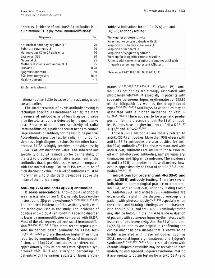
Table IV. Incidence of anti-Ro(SS-A) antibodies inautoimmune CTDs (by radial immunodiffusion)11
Diagnosis %
Antinuclear antibody negative SLE 70Subacute cutaneous LE 70Homozygous C2 or C4 deficiency 70Late onset SLE 80Neonatal LE 95Mothers of infants with neonatal LE 95Discoid LE 0-20Sjögren’s syndrome 50SSc, dermatomyositis RareHealthy persons < 1
SSc, Systemic sclerosis.
Table V. Indications for anti-Ro(SS-A) and anti-La(SS-B) antibody testing*
Work-up for photosensitivityScreening for certain patients with LESuspicion of subacute cutaneous LESuspicion of neonatal LESuspicion of Sjögren’s syndromeWork-up for idiopathic chronic vasculitisPatients with systemic or subacute cutaneous LE with
negative screening fluorescent ANA test
*References 95-97, 102, 108-110, 115-117, 121.
in patients with CTD. The diagnostic value of most of
these antibodies is limited; only two are discussed in
this review. Scl-70 antibodies are directed against the
enzyme topoisomerase-I.138-140 This is a 100-kd basic
protein that affects the tertiary structure of DNA
molecules. Scl-70 antibodies are characteristic of SSc
and help differentiate patients with extensive cuta-
neous and systemic involvement from those with
limited disease.131,141-143 The incidence of Scl-70
antibodies, however, is low (approximately 10%-20%
by radial immunodiffusion).141,142 Scl-70 antibodies
may be viewed as a marker for SSc when compared
with patients with CREST syndrome who have
another marker antibody, namely, anti-centromere
antibody (see section on fluorescent antinuclear
antibody testing).5,28,142-144
Jo-1 antibodies are directed against the enzyme
histidyl tRNA synthetase (150 kd) and are detected in
a small number of patients with dermatomyositis
(and polymyositis).145-148 The presence of Jo-1 anti-
bodies is often associated with pulmonary involve-
ment and possibly the mechanic’s hand skin
lesions.147,149,150
FLUORESCENT ANA TESTThe fluorescent ANA test is a very good screening
test for most of the previously discussed antibodies.
Testing techniqueThe ANA test is an indirect immunofluorescence
test that utilizes a substrate rich in nuclear material.
A positive ANA test indicates the presence of ANAs.
It does not indicate the specific type of antibody,
although close examination of the pattern of positiv-
ity may be helpful in suggesting the specific type of
ANA that is present in the tested serum.
The indications for ordering an ANA test in derma-
tology include the work-up of patients with photo-
sensitivity, work-up of patients with chronic vasculitis,
a baseline for patients with discoid lupus erythemato-
sus, clinical suspicion of CTD, and baseline for
patients undergoing phototherapy (Table VI).
Interpretation of resultsWhen an ANA test result is obtained, 3 parameters
are evaluated; these include the substrate used, the
titer of a positive test, and the pattern of fluores-
cence.
ANA substrate. There are two major types of
substrate for ANA testing. Until two decades ago,
most ANA tests were performed on animal sub-
strates, such as mouse kidney or rat liver.10,11,42,151
Sera of some patients with SLE were reportedly neg-
ative on such substrates. It became clear that human
substrates (cultured human cells) are more sensitive
anti-La(SS-B) antibodies in patients with chronic idio-
pathic vasculitis.95,96,108,113 Finally, anti-Ro(SS-A) and
anti-La(SS-B) antibodies are useful in the evaluation
of a patient with the clinical manifestations of SLE or
SCLE if the screening fluorescent antinuclear anti-
body (ANA) test is negative,11,117,118 since the ANA
test may be negative despite the presence of anti-
Ro(SS-A) and/or anti-La(SS-B) antibodies.
Antibodies to U1RNP and SmAntibodies to U1RNP are present in the sera of
patients with MCTD and SLE. By definition, antibod-
ies to U1RNP are detected in 100% of patients with
MCTD15,23-25,127 and approximately 30% of patients
with SLE.92,93 They have also been reported rarely in
neonatal lupus erythematosus.128,129 As will be dis-
cussed in more detail later, the presence of U1RNP
antibodies in MCTD is to the exclusion of other types
of antinuclear antibodies.23,92 In contrast, patients
with SLE who have U1RNP antibodies usually have
ANAs with other specificities as well.92 This observa-
tion is important when attempting to differentiate
between MCTD and SLE. U1RNP antibodies are very
rarely detected in patients with SSc.130-132 Because
the incidence of SLE is much higher than that of
MCTD, the majority of patients with U1RNP antibod-
ies have SLE rather than MCTD. The presence of
U1RNP antibodies is usually associated with sclero-
dactyly, Raynaud’s phenomenon, esophageal dys-
motility, low incidence of renal disease, pulmonary
dysfunction, arthritis, and myositis.132,133
Antibodies to Sm by immunodiffusion are diag-
nostic of SLE.16,107 They have not been reported in
patients with other CTDs. The incidence of Sm anti-
bodies in SLE is only 15% to 40%.15,16,104,134-136 Most
patients with antibodies to Sm will also have anti-
bodies to U1RNP.93,137 The converse of this observa-
tion, however, is not true. Most patients with U1RNP
antibodies do not have Sm antibodies.127,131
Antibodies to U1RNP and Sm are indicated
when attempting to confirm the diagnosis of
MCTD23-25,92,127 and SLE,16,93,107 respectively.
OTHER AUTOANTIBODIESSeveral other autoantibodies have been reported
164 Mutasim and Adams J AM ACAD DERMATOL
FEBRUARY 2000
Table VI. Indications for fluorescent ANA testing indermatological practice
Work-up for photosensitivityBaseline in patients with discoid LEClinical suspicion of CTDBaseline for phototherapyWork-up of chronic vasculitis
than animal substrates.11,30,32,38 Most SLE sera that
were negative on animal substrates were positive on
human substrate. Because of this observation, most
laboratories use cultured human cell substrates.
Presently, the vast majority of laboratories use a spe-
cific type of cultured human cells referred to as
HEp-2 cells.11,30,32,38 These are obtained from cul-
tured esophageal squamous cell carcinoma cells.
The cells are available commercially, prefixed on
glass slides. Because an occasional laboratory may
still be using animal substrates for ANA testing, it is
essential to pay attention to the substrate being used
by each of the various laboratories from which a
physician may receive results. A serum that is nega-
tive on animal substrate may be positive when tested
on cultured human cells.
ANA titer. As mentioned earlier, the presence of
ANAs is not diagnostic of CTD. The amount of anti-
body (and the specificity) have significant value in
the interpretation of an ANA test.9,11,13 The ANA titer
is an indirect measure of the total amount of serum
antibodies. The higher the titer, the higher the
amount of antibodies. Generally, the ANA test is neg-
ative or very low in young and healthy per-
sons.9,12,152,153 It is generally high in patients with
systemic CTD.11-13 The ANA titer is intermediate in
some patients with CTD as well as in persons with a
wide variety of conditions (Table VII). These include
old age,12,153 pregnancy,154,155 close relatives of
patients with systemic CTD,12,156 patients taking
drugs that are known to induce SLE (who do not
have manifestations of CTD),68-83,157 and healthy
persons.9,11,12,153 The incidence of positive ANA in
healthy persons at various titers is shown in Table
VIII.9 Accordingly, a titer of 1:80 or less is of no diag-
nostic value because of the high prevalence of posi-
tive ANA tests at such titers in the general popula-
tion. A reasonable cut-off point is around 1:160 to
1:320. An ANA test at such titers or higher may help
confirm the clinical diagnosis of a CTD. There are,
however, healthy persons who have ANA titers above
1:320. The diagnosis of a CTD should not be made
solely on the titer of an ANA test.
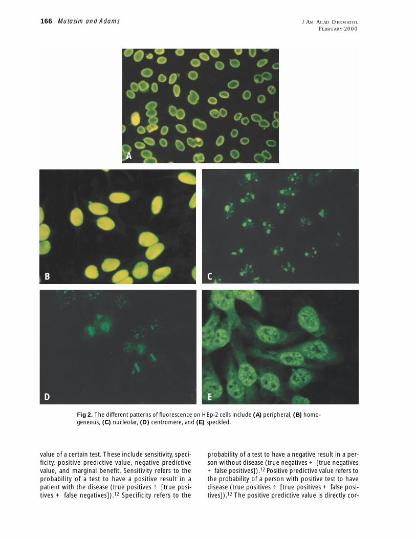
ANA patterns. The patterns of fluorescence of
the nuclei in an ANA test are usually associated with
specific antinuclear antibodies (Table IX) (Fig
2).10,11,14,20,158-161 For example, the peripheral or rim
pattern is associated with antibodies to nDNA and
thus correlates with the diagnosis of SLE. The homo-
geneous pattern is associated with antibodies to
nDNA or antibodies to histones, which are seen fre-
quently in patients with SLE.
ANA-negative SLEANA-negative SLE was reported in patients who
had cutaneous and/or systemic manifestations of SLE,
but who were negative by ANA testing on animal sub-
strates.162,163 Most of these patients were later found
to have positive ANA on human substrate. Many of
these patients had photosensitivity and some of them
were later reported as having SCLE with anti-Ro(SS-A)
antibodies.163 Another reason for the ANA test to be
negative in a patient with SLE is if the patient’s ANAs
are solely against ssDNA. Because the fluorescent
ANA substrate has intact nuclei without single strands
of DNA, the test is expectedly negative.
DIAGNOSTIC VALUE OF THEFLUORESCENT ANA TEST
There are several parameters that indicate the
Mutasim and Adams 165J AM ACAD DERMATOL
VOLUME 42, NUMBER 2, PART 1
Table VII. Conditions other than autoimmuneCTDs with positive ANA
Elderly persons12,153
Pregnant women154,155
Relatives of patients with CTD12,156
Other autoimmune diseases (eg, primary biliary cirrhosis,autoimmune thyroiditis)14,196
Drugs (eg, procainamide, hydralazine)68-83,157
Chronic infections10,14
Neoplasms10,14
Healthy persons9,11,12,153
Table VIII. Positive fluorescent ANA test in healthypersons (on HEp-2 cells)9
Titer Prevalence
1:40 32%1:80 13%1:160 5%1:320 3%
Table IX. ANA patterns and their antigen anddisease associations
Predominant ReferenceANA antigen Disease Nos.
Peripheral nDNA SLE 10,14,161Homogeneous nDNA, histones SLE 14,161Nucleolar Nucleolar RNA SSc, SLE 14,158,161Centromere Kinetochore CREST 14,159Speckled Various ribo- MCTD, 14,161
nucleo- SLE, SSc,proteins Sjögren’s
Syndrome
probability of a test to have a negative result in a per-
son without disease (true negatives ÷ [true negatives
+ false positives]).12 Positive predictive value refers to
the probability of a person with positive test to have
disease (true positives ÷ [true positives + false posi-
tives]).12 The positive predictive value is directly cor-
value of a certain test. These include sensitivity, speci-
ficity, positive predictive value, negative predictive
value, and marginal benefit. Sensitivity refers to the
probability of a test to have a positive result in a
patient with the disease (true positives ÷ [true posi-
tives + false negatives]).12 Specificity refers to the
166 Mutasim and Adams J AM ACAD DERMATOL
FEBRUARY 2000
Fig 2. The different patterns of fluorescence on HEp-2 cells include (A) peripheral, (B) homo-
geneous, (C) nucleolar, (D) centromere, and (E) speckled.
A
B C
D E
related with test sensitivity and prevalence of disease
in the test population.12,164 Negative predictive value
refers to the probability of a person with negative test
to be free of disease (true negatives ÷ [true negatives
+ false negatives]).12 Tests with high specificity will
have high predictive value when positive, since false
positivity is very low. Tests with high sensitivity will
have high predictive value when negative, since false
negativity is very low. Marginal benefit of a test refers
to the posttest disease probability compared with
pretest probability.165
The value of the fluorescent ANA test in the diag-
nosis of SLE and other CTDs has been evaluated
repeatedly. The primary focus of the published stud-
ies is on SLE. The sensitivity of ANA tests for SLE is
very high. Almost all patients with SLE have positive
ANA tests.12,19,164,166 The negative predictive value
for SLE is also very high. A patient with a negative
ANA test is highly unlikely to have SLE.12,164,166 The
positive predictive value for SLE, however, is gener-
ally low, especially at low titers12,164,166,167 because
the specificity of the ANA test for SLE, especially at
low titers, is low. As discussed earlier, a positive ANA
test especially at a low titer may be seen in several
conditions and persons without SLE or other
CTDs.11,12,68-73,75-83,153-157 In the case of a patient
with clinical findings suggestive of SLE or other sys-
temic CTDs in which the ANA test is negative or with
a low titer, more selective testing for individual anti-
nuclear antibodies (eg, DNA, ribonucleoprotein)
may be helpful in confirming the diagnosis.
Of all the parameters to evaluate a test, the
marginal benefit is of high practical value for the
physician who is attempting to confirm or exclude a
diagnosis by ordering a certain test. The marginal
benefit of the fluorescent ANA test is minimal when
the pretest probability of disease is very low or very
high. For example, persons with no clinical findings
to suggest SLE are highly unlikely to benefit from an
ANA test. In such a setting, the test is almost invari-
ably negative or with very low titer and thus will not
confirm a diagnosis of SLE. Similarly, the marginal
benefit from the fluorescent ANA test in a patient
with the characteristic multiple organ involvement of
SLE is low because the diagnosis is already known
and the test will invariably be strongly positive. The
marginal benefit of the fluorescent ANA test is maxi-
mal when the pretest probability of disease is inter-
mediate.168 For example, the diagnosis of a patient
with some cutaneous and/or systemic manifestations
suggestive of SLE may be confirmed or excluded by
the result of an ANA test. A strongly positive ANA test
will help confirm the diagnosis, whereas a negative
test may exclude SLE. These observations were sup-
ported in a recent study in which the usefulness of
the ANA test was investigated in a group of more
than 1000 inpatients and outpatients in whom the
ANA test was ordered.164 One hundred fifty-three
patients with a positive ANA test were compared
with an equal number of patients with a negative
ANA test. Patients with positive ANA were generally
older than those with negative ANA. The ANA test
was ordered primarily in patients suspected of hav-
ing a CTD or vasculitis. The negative predictive value
was 100% for SLE and 97% for other CTDs. The pos-
itive predictive value was 11% for SLE and 22% for
other CTDs. The predictive value was lower for
patients who were older than 65 years compared
with those younger than 65 years. The conclusion of
the study was that the diagnostic value of the ANA
test depends on the clinical setting in which it is
ordered,164 and clinicians should be aware that in
the setting of a low prevalence of CTD an ANA test’s
positive predictive value is low.
RECENT SCREENING ANA TESTSIn the past few years, attempts have been made to
replace the fluorescent ANA test with ELISA screen-
ing tests. There have been many ELISAs that have
been reported to be of value for screening ANA tests.
Some of these ELISAs utilize extracts of tissue con-
taining various nuclear components. Other ELISAs
utilize molecules synthesized by recombinant tech-
nology. Some ELISAs utilize individual recombinant
molecules such as Ro(SS-A), whereas others utilize
combinations of various molecules to increase the
sensitivity of the test. In a recent study, the perfor-
mance of the various ELISA ANA tests was compared
with the “gold standard” fluorescent ANA test.169 Sera
that were positive by fluorescent ANA test were test-
ed by the various ELISA techniques. The agreement
that a serum is ANA positive was 87% to 95% when
comparing the various ELISA tests with the fluores-
cent ANA test results.169 The sensitivity of the various
ELISAs was 69% to 98% and the specificity ranged
between 81% and 98%. These figures were arrived at
using sera that were positive at 1:160 by the fluores-
cent ANA test. The above comparison figures were
much lower for sera with fluorescent ANA titer of
1:40. Many ELISA techniques missed a low titer posi-
tive ANA as well as sera with specific ANAs (eg, anti-
nDNA antibodies). Presently, ELISA screening ANA
tests may be adequate to screen sera with intermedi-
ate to high titer.169 It remains to be seen whether the
performance of screening ANA tests by ELISA would
match that by the fluorescent technique.
SEROLOGIC PROFILES IN CTDSEach CTD has a rather specific autoantibody pro-
file (Fig 1). Some of these profiles are “simple” in
Mutasim and Adams 167J AM ACAD DERMATOL
VOLUME 42, NUMBER 2, PART 1
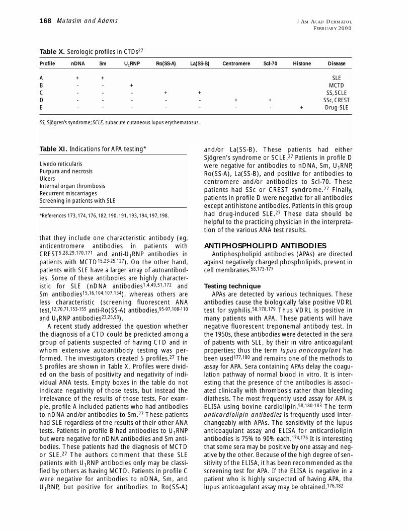
and/or La(SS-B). These patients had either
Sjögren’s syndrome or SCLE.27 Patients in profile D
were negative for antibodies to nDNA, Sm, U1RNP,
Ro(SS-A), La(SS-B), and positive for antibodies to
centromere and/or antibodies to Scl-70. These
patients had SSc or CREST syndrome.27 Finally,
patients in profile D were negative for all antibodies
except antihistone antibodies. Patients in this group
had drug-induced SLE.27 These data should be
helpful to the practicing physician in the interpreta-
tion of the various ANA test results.
ANTIPHOSPHOLIPID ANTIBODIESAntiphospholipid antibodies (APAs) are directed
against negatively charged phospholipids, present in
cell membranes.58,173-177
Testing techniqueAPAs are detected by various techniques. These
antibodies cause the biologically false positive VDRL
test for syphilis.58,178,179 Thus VDRL is positive in
many patients with APA. These patients will have
negative fluorescent treponemal antibody test. In
the 1950s, these antibodies were detected in the sera
of patients with SLE, by their in vitro anticoagulant
properties; thus the term lupus anticoagulant has
been used177,180 and remains one of the methods to
assay for APA. Sera containing APAs delay the coagu-
lation pathway of normal blood in vitro. It is inter-
esting that the presence of the antibodies is associ-
ated clinically with thrombosis rather than bleeding
diathesis. The most frequently used assay for APA is
ELISA using bovine cardiolipin.58,180-183 The term
anticardiolipin antibodies is frequently used inter-
changeably with APAs. The sensitivity of the lupus
anticoagulant assay and ELISA for anticardiolipin
antibodies is 75% to 90% each.174,176 It is interesting
that some sera may be positive by one assay and neg-
ative by the other. Because of the high degree of sen-
sitivity of the ELISA, it has been recommended as the
screening test for APA. If the ELISA is negative in a
patient who is highly suspected of having APA, the
lupus anticoagulant assay may be obtained.176,182
that they include one characteristic antibody (eg,
anticentromere antibodies in patients with
CREST5,28,29,170,171 and anti-U1RNP antibodies in
patients with MCTD15,23-25,127). On the other hand,
patients with SLE have a larger array of autoantibod-
ies. Some of these antibodies are highly character-
istic for SLE (nDNA antibodies1,4,49,51,172 and
Sm antibodies15,16,104,107,134), whereas others are
less characteristic (screening fluorescent ANA
test,12,70,71,153-155 anti-Ro(SS-A) antibodies,95-97,108-110
and U1RNP antibodies23,25,93).
A recent study addressed the question whether
the diagnosis of a CTD could be predicted among a
group of patients suspected of having CTD and in
whom extensive autoantibody testing was per-
formed. The investigators created 5 profiles.27 The
5 profiles are shown in Table X. Profiles were divid-
ed on the basis of positivity and negativity of indi-
vidual ANA tests. Empty boxes in the table do not
indicate negativity of those tests, but instead the
irrelevance of the results of those tests. For exam-
ple, profile A included patients who had antibodies
to nDNA and/or antibodies to Sm.27 These patients
had SLE regardless of the results of their other ANA
tests. Patients in profile B had antibodies to U1RNP
but were negative for nDNA antibodies and Sm anti-
bodies. These patients had the diagnosis of MCTD
or SLE.27 The authors comment that these SLE
patients with U1RNP antibodies only may be classi-
fied by others as having MCTD. Patients in profile C
were negative for antibodies to nDNA, Sm, and
U1RNP, but positive for antibodies to Ro(SS-A)
168 Mutasim and Adams J AM ACAD DERMATOL
FEBRUARY 2000
Table X. Serologic profiles in CTDs27
Profile nDNA Sm U1RNP Ro(SS-A) La(SS-B) Centromere Scl-70 Histone Disease
A + + SLEB - - + MCTDC - - - + + SS, SCLED - - - - - + + SSc, CRESTE - - - - - - - + Drug-SLE
SS, Sjögren’s syndrome; SCLE, subacute cutaneous lupus erythematosus.
Table XI. Indications for APA testing*
Livedo reticularisPurpura and necrosisUlcersInternal organ thrombosisRecurrent miscarriagesScreening in patients with SLE
*References 173, 174, 176, 182, 190, 191, 193, 194, 197, 198.
Disease association
APAs are most prevalent in patients with SLE
(approximately 50%).58,173,174,178,184,185 Patients with
other CTDs have a lower prevalence of these antibod-
ies. APAs may also be seen in patients taking certain
drugs (cocaine, interferon alfa, procainamide,
hydralazine, phenothiazines, quinine, quinidine, fansi-
dar, and phenytoin182), patients with chronic
infections (syphilis, infectious mononucleosis, tuber-
culosis, leprosy, leptospirosis, malaria, typhus, try-
panosomiasis,173 schistosomiasis and filariasis,186
cytomegalovirus infection,187 HIV infection,188 hepati-
tis C189), and occasionally in persons who do not have
an associated condition (primary APA syndrome174,190).
APAs have been associated with arterial and venous
thrombosis in various organs, including the central ner-
vous system, the heart, and the skin.173,174,176,182,191-193
Young women with APAs are predisposed to recurrent
miscarriages.173,174,176,182,192 Many patients with APAs
have thrombocytopenia. Patients with APAs who pre-
sent to dermatological practice usually have livedo
reticularis, purpura, necrosis, and ulcers.182,190,193,194
The indications for obtaining APA testing are shown in
Table XI.
Interpretation of resultsThe results of APA testing should be interpreted
with caution. Low levels may be of no clinical rele-
vance and should not be interpreted as the cause of
leg ulcers and purpura in every patient who has low
levels of antibodies.195
REFERENCES1. Takeuchi Y, Ishikawa O, Miyachi Y. The comparative study of
anti-double stranded DNA antibody levels measured byradioimmunoassay and enzyme-linked immunosorbent assayin systemic lupus erythematosus. J Dermatol 1997;24:297-300.
2. Krippner H, Merle S, Pirlet K. Enzyme immunoassay for IgG andIgM antibodies against dsDNA and ssDNA. Z Rheumatol1983;42:256-60.
3. Wong KH, Lawton JWM, Cheng SKL, Lee SS, Lau CS.Measurement of anti-dsDNA: a comparative study of twoELISA and the Crithidia assay. Pathology 1998;30:57-61.
4. Spronk PE, Bootsma H, Kallenberg CGM. Anti-DNA antibodiesas early predictor for disease exacerbations in SLE: guidelinefor treatment? Clin Rev Allergy Immunol 1998;16:211-8.
5. Chorzelski TP, Jablonska S, Beutner EH, Blaszczyk M, Jarzabek-Chorzelska M, Kencka D, et al. Anticentromere antibody: animmunological marker of a subset of systemic sclerosis. Br JDermatol 1985;113:381-9.
6. Tuffanelli DL, Dubois EL. Cutaneous manifestations of sys-temic lupus erythematosus. Arch Dermatol 1964;90:377-86.
7. Cabral AR, Alarcón-Segovia D. Autoantibodies in systemiclupus erythematosus. Curr Opin Rheumatol 1998;10:409-16.
8. Pahor A, Krajnc I, Gorenjak M, Holc I.The clinical significance ofantinuclear antibodies in connective tissue disease. Wien KlinWochenschr 1998;110:338-41.
9. Tan EM, Feltkamp TEW, Smolen JS, Butcher B, Dawkins R,
Fritzler MJ, et al. Range of antinuclear antibodies in “healthy”individuals. Arthritis Rheum 1997;40:1601-11.
10. Fernandez-Madrid F, Mattioli M. Antinuclear antibodies (ANA):immunologic and clinical significance. Semin Arthritis Rheum1976;6:83-124.
11. Provost TT,Watson R. Antinuclear antibodies in systemic lupuserythematosus. In: Norris DA, editor. Immune mechanisms incutaneous disease. New York: Marcel Dekker; 1989. p. 333-57.
12. Ward MM. Laboratory testing for systemic rheumatic diseases.Postgrad Med 1998;103:93-100.
13. Moder KG. Use and interpretation of rheumatologic tests: aguide for clinicians. Mayo Clin Proc 1996;71:391-6.
14. Sontheimer RD, McCauliffe DP, Zappi E, Targoff I. Antinuclearantibodies: clinical correlations and biologic significance. AdvDermatol 1991;7:3-52.
15. Hamburger M, Hodes S, Barland P. The incidence and clinicalsignificance of antibodies to extractable nuclear antigens. AmJ Med Sci 1977;273:21-8.
16. Field M,Williams DG, Charles P, Maini RN. Specificity of anti-Smantibodies by ELISA for systemic lupus erythematosus:increased sensitivity of detection using purified peptide anti-gens. Ann Rheum Dis 1988;47:820-5.
17. Fritzler MJ. Antinuclear antibodies in the investigation ofrheumatic diseases. Bull Rheum Dis 1985;35:1-10.
18. Fritzler MJ. Clinical relevance of autoantibodies in systemicrheumatic diseases. Mol Biol Rep 1996;23:133-45.
19. Hietarinta M, Lassila O. Clinical significance of antinuclear anti-bodies in systemic rheumatic diseases. Ann Med 1996;28:283-91.
20. Provost TT. Subsets in systemic lupus erythematosus. J InvestDermatol 1979;72:110-3.
21. Thompson D, Juby A, Davis P. The clinical significance ofautoantibody profiles in patients with systemic lupus erythe-matosus. Lupus 1993;2:15-9.
22. Tomer Y, Buskila D, Shoenfeld Y. Pathogenic significance anddiagnostic value of lupus autoantibodies. Int Arch AllergyImmunol 1993;100:293-306.
23. Combe B, Rucheton M, Graafland H, Lussiez V, Brunel C, Sany J.Clinical significance of anti-RNP and anti-Sm autoantibodiesas determined by immunoblotting and immunoprecipitationin sera from patients with connective tissue diseases. Clin ExpImmunol 1989;75:18-24.
24. Gilliam JN, Prystowsky SD. Mixed connective tissue diseasesyndrome: cutaneous manifestations of patients with epider-mal nuclear staining and high titer serum antibody to ribonu-clease-sensitive extractable nuclear antigen. Arch Dermatol1977;113:583-7.
25. Lundberg I, Hedfors E. Clinical course of patients with anti-RNPantibodies: a prospective study of 32 patients. J Rheumatol1991;18:1511-9.
26. Hardin JA, Mimori T. Autoantibodies to ribonucleoproteins.Clin Rheum Dis 1985;11:485-505.
27. Juby AA, Johnston C, Davis P. Specificity, sensitivity and diag-nostic predictive value of selected laboratory generatedautoantibody profiles in patients with connective tissue dis-eases. J Rheumatol 1991;18:354-8.
28. de Rooij DJ, van de Putte LBA, Habets WJ, van Venrooij WJ.Marker antibodies in scleroderma and polymyositis: clinicalassociations. Clin Rheumatol 1989;8:231-7.
29. Tuffanelli DL, McKeon F, Kleinsmith DM, Burnham TK, KirschnerM. Anticentromere and anticentriole antibodies in the sclero-derma spectrum. Arch Dermatol 1983;119:560-6.
30. Cook L. New methods for detection of anti-nuclear antibodies.Clin Immunol Immunopathol 1998;88:211-20.
31. de Rooij DJ, van de Putte LB, Habets WJ, Verbeek AL, vanVenrooij WJ. The use of immunoblotting to detect antibodies
Mutasim and Adams 169J AM ACAD DERMATOL
VOLUME 42, NUMBER 2, PART 1
of anti-native deoxyribonucleic acid antibodies. J ClinMicrobiol 1978;7:219-22.
51. Shen GQ, Shoenfeld Y, Peter JB. Anti-DNA, antihistone, andantinucleosome antibodies in systemic lupus erythematosusand drug-induced lupus. Clin Rev Allergy Immunol 1998;16:321-34.
52. Aarden LA, de Groot ER, Feltkamp TEW. Immunology of DNA.III. Crithidia luciliae, a simple substrate for the determination ofanti-dsDNA with the immunofluorescence technique. Ann N YAcad Sci 1975;254:505-15.
53. Fermand JP, Dannon F, Brouet JC. Characterization of a humanmonoclonal IgM with antibody activity to dsDNA. Clin ExpImmunol 1985;59:467-74.
54. Kumar V, Krasny S, Beutner EH. Specificity of the Crithidia lucil-iae method for detecting anti-DNA antibodies: effect ofabsorption for lipoproteins. Immunol Invest 1985;14:199-210.
55. Jansen EM, Deng J-S, Beutner EH, Kumar V, Chorzelski TP.Comparison of commercial kits for the detection of anti-nDNAantibodies using Crithidia luciliae. Am J Clin Pathol 1987;87:461-9.
56. Cameron JS, Lessof MH, Ogg CS, Williams BD, Williams DG.Disease activity in the nephritis of systemic lupus erythe-matosus in relation to serum complement concentrations:DNA-binding capacity and precipitating anti-DNA antibody.Clin Exp Immunol 1976;25:418-27.
57. Swissa M, Cohen Y, Shoenfeld Y. Autoantibodies in the sera ofpatients with lymphoma. Leuk Lymphoma 1992;7:117-22.
58. Kaburaki J, Kuwana M, Ogasawara T, Takano M, Funatsu Y, TojoT. Specificity of antibodies to single-stranded (ss) DNA in SLEpatients with anti-phospholipid syndrome. Keio J Med 1992;41:10-5.
59. Williams WM, Isenberg DA. A cross-sectional study of anti-DNAantibodies in the serum and IgG and IgM fraction of healthyindividuals, patients with systemic lupus erythematosus andtheir relatives. Lupus 1996;5:576-86.
60. Callen JP, Fowler JF, Kulick KB. Serologic and clinical featuresof patients with discoid lupus erythematosus: relationship ofantibodies to single-stranded deoxyribonucleic acid and ofother antinuclear antibody subsets to clinical manifestations.J Am Acad Dermatol 1985;13:748-55.
61. Sarvas H, Gripenberg M, Leirisalo-Repo M. Anti-DNA antibod-ies: the choice of assays for routine diagnostic work. ActaPathol Microbiol Immunol Scand Sect C 1985;93:13-8.
62. Buskila D, Berezin M, Gur H, Lin HC, Alosachie I,Terrberry JW, etal. Autoantibody profile in the sera of women with hyperpro-lactinemia. J Autoimmun 1995;8:415-24.
63. Falanga V, Medsger TA, Reichlin M. Antinuclear and anti-single-stranded DNA antibodies in morphea and generalized mor-phea. Arch Dermatol 1987;123:350-3.
64. Gripenberg M, Helve T, Kurki P. Profiles of antibodies to his-tones, DNA and IgG in patients with systemic rheumatic dis-eases determined by ELISA. J Rheumatol 1985;12:934-9.
65. Falanga V, Medsger TA, Reichlin M. High titers of antibodies tosingle-stranded DNA in linear scleroderma. Arch Dermatol1985;121:345-7.
66. Yadin O, Sarov B, Naggan L, Shoenfeld Y. Familial studies on theoccurrence of natural autoantibodies. Nat Immun Cell GrowthRegul 1989;8:325-30.
67. Rubin RL, Waga S. Antihistone antibodies in systemic lupuserythematosus. J Rheumatol 1987;14(Suppl 13):118-26.
68. Monestier M, Kotzin BL. Antibodies to histones in systemiclupus erythematosus and drug-induced lupus syndromes.Rheum Dis Clin North Am 1992;18:415-36.
69. Grossman L, Barland P. Histone reactivity of drug-induced anti-nuclear antibodies. Arthritis Rheum 1981;24:927-31.
70. Portanova JP, Rubin RL, Joslin FG, Agnello VD, Tan EM.
to nuclear and cytoplasmic antigens: clinical and serologicalassociations in rheumatic disease. Scand J Rheumatol 1988;17:353-64.
32. Saitta MR, Keene JD. Molecular biology of nuclear antigens.Rheum Dis Clin North Am 1992;18:283-310.
33. Walravens MJF, Vanherrewegen H, Lacquet F, Godefridis G,Korevits G, Stevens E, et al. Counterimmunoelectrophoresiswith serum prediffusion: an improved method for the detec-tion and identification of antibodies against extractablenuclear and cytoplasmic antigens. J Immunol Methods 1997;201:89-98.
34. Schur PH, DeAngelis D, Jackson JM. Immunological detectionof nucleic acids and antibodies to nucleic acids and nuclearantigens by counterimmunoelectrophoresis. Clin ExpImmunol 1974;17:209-18.
35. Kurata N, Tan EM. Identification of antibodies to nuclear acidicantigens by counterimmunoelectrophoresis. Arthritis Rheum1976;19:574-80.
36. Lange A, Jacak A, Garnsarek D. Diagnostic specificity ofautoantibodies. IV. A double antibody solid phase radioim-munoassay for DNA antibodies: significance of dsDNA andssDNA antibodies with test standardization attempts. ArchImmunol Ther Exp (Warsz) 1978;26:893-7.
37. Bizzaro N,Tozzoli R,Tonutti E, Piazza A, Manoni F, Ghirardello A,et al. Variability between methods to determine ANA, anti-dsDNA and anti-ENA autoantibodies: a collaborative studywith the biomedical industry. J Immunol Methods 1998;219:99-107.
38. Evans J. Antinuclear antibody testing in systemic autoimmunedisease. Clin Chest Med 1998;19:613-25.
39. Gniewek RA, Stites DP, McHugh TM, Hilton JF, Nakagawa M.Comparison of antinuclear antibody testing methods:immunofluorescence assay versus enzyme immunoassay. ClinDiagn Lab Immunol 1997;4:185-8.
40. Tikly M, Burgin S, Mohanlal P, Bellingan A, George J.Autoantibodies in Black South Africans with systemic lupuserythematosus: spectrum and clinical associations. ClinRheumatol 1996;15:261-5.
41. Cervera R, Khamashta MA, Font J, Sebastiani GD, Gil A, Lavilla P,et al. Systemic lupus erythematosus: clinical and immunolog-ic patterns of disease expression in a cohort of 1,000 patients.Medicine 1993;72:113-24.
42. Fritzler MJ, Pauls JD, Kinsella TD, Bowen TJ. Antinuclear, anticy-toplasmic, and anti-Sjogren’s syndrome antigen A (SS-A/Ro)antibodies in female blood donors. Clin ImmunolImmunopathol 1985;36:120-8.
43. Emlen W, Jarusiripipat P, Burdick G. A new ELISA for the detec-tion of double-stranded DNA antibodies. J Immunol Methods1990;132:91-101.
44. Carey JL. Enzyme immunoassays for antinuclear antibodies.Clin Lab Med 1997;17:355-65.
45. Bell DA, Smeenk RJT. Clinical connections: assays and assess-ment. Lupus 1997;6:305-6.
46. Hahn BH. Antibodies to DNA. N Engl J Med 1998;338:1359-68.47. Levinson WE, Jawetz E. Medical microbiology and immunolo-
gy. 2nd ed. Norwalk (CT): Appleton & Lange; 1992. p. 302-8.48. Lange A. Evaluation of the simultaneous estimation of anti-
dsDNA and anti-ssDNA antibodies for clinical purposes. ClinExp Immunol 1978;31:472-81.
49. Chuan M-T, Wu Y-C, Tiak-de Ang E, Wang J-H, In K-L, Lü Y-C.Clinical significance of anti-nDNA antibodies in ANA-positivesystemic lupus erythematosus: comparison of the Farrradioimmunoassay and the Crithidia luciliae immunofluores-cent technique. Chin J Microbiol Immunol 1985;18:15-24.
50. Tourville DR, Benn V. Evaluation of a semiautomated pre-standardized immunofluorescence test system for detection
170 Mutasim and Adams J AM ACAD DERMATOL
FEBRUARY 2000
Reactivity of anti-histone antibodies induced by pro-cainamide and hydralazine. Clin Immunol Immunopathol1982;25:67-79.
71. Craft JE, Radding JA, Harding MW, Bernstein RM, Hardin JA.Autoantigenic histone epitopes: a comparison between pro-cainamide- and hydralazine-induced lupus. Arthritis Rheum1987;30:689-94.
72. Teoh PC, Chan HL. Lupus-scleroderma syndrome induced byethosuximide. Arch Dis Child 1975;50:659-61.
73. Sato-Matsumura KC, Koizumi H, Matsumura T, Takahashi T,Adachi K, Ohkawara A. Lupus erythematosus-like syndromeinduced by thiamazole and propylthiouracil. J Dermatol1994;21:501-7.
74. Agarwal MB, Anjaria PD, Mehta BC. Activation of systemiclupus erythematosus by antitubercular drugs. J Postgrad Med1980;26:263-6.
75. Timsit M-A, Anglicheau D, Lioté F, Marteau P, Dryll A.Mesalazine-induced lupus. Rev Rhum Engl Ed 1997;64:586-8.
76. Condemi JJ. SLE: idiopathic or drug-induced? Geriatrics 1980;35:81-8.
77. Price EJ, Venables PJW. Drug-induced lupus. Drug Safety1995;12:283-90.
78. Hobbs RN, Clayton A-L, Bernstein RM. Antibodies to the fivehistones and poly(adenosine diphosphate-ribose) in druginduced lupus: implications for pathogenesis. Ann Rheum Dis1987;46:408-16.
79. Stratton MA. Drug-induced systemic lupus erythematosus.Clin Pharm 1985;4:657-63.
80. Roura M, Lopez-Gil F, Umbert P. Systemic lupus erythematosusexacerbated by piroxicam. Dermatologica 1991;182:56-8.
81. Gigli GL, Scalise A, Pauri F, Silvestri G, Diomedi M, Placidi F, et al.Valproate-induced systemic lupus erythematosus in a patientwith partial trisomy of chromosome 9 and epilepsy. Epilepsia1996;37:587-8.
82. Knights SE, Leandro MJ, Khamashta MA, Hughes GRV.Minocycline-induced arthritis. Clin Exp Rheumatol 1998;16:587-90.
83. Yung RL, Richardson BC. Drug-induced lupus. Rheum Dis ClinNorth Am 1994;20:61-86.
84. Tan EM, Portanova JP.The role of histones as nuclear autoanti-gens in drug-related lupus erythematosus. Arthritis Rheum1981;24:1064-9.
85. Portanova JP, Cheronis JC, Blodgett JK, Kotzin BL. Histoneautoantigens in murine lupus: definition of a major epitopewithin an accessible region of chromatin. J Immunol1990;144:4633-40.
86. Fishbein E, Alarcon-Segovia D,Vega JM. Antibodies to histonesin systemic lupus erythematosus. Clin Exp Immunol1979;36:145-50.
87. Burlingame RW. The clinical utility of antihistone antibodies.Clin Lab Med 1997;17:367-78.
88. Aitkaci A, Monier JC, Mamelle N. Enzyme-linked immunosor-bent assay for anti-histone antibodies and their presence insystemic lupus erythematosus sera. J Immunol Methods1981;44:311-22.
89. Shoenfeld Y, Segol O. Anti-histone antibodies in SLE and otherautoimmune diseases. Clin Exp Rheumatol 1989;7:265-71.
90. Fritzler MJ, Tan EM. Antibodies to histones in drug-inducedand idiopathic lupus erythematosus. J Clin Invest1978;62:560-7.
91. Weinstein A. Drug-induced systemic lupus erythematosus.Prog Clin Immunol 1980;4:1-21.
92. Craft J. Antibodies to snRNPs in systemic lupus erythemato-sus. Rheum Dis Clin North Am 1992;18:311-35.
93. ter Borg EJ, Groen H, Horst G, Limburg PC, Wouda AA,Kallenberg CGM. Clinical associations of antiribonucleopro-
tein antibodies in patients with systemic lupus erythemato-sus. Semin Arthritis Rheum 1990;20:164-73.
94. Habets WJ, Hoet MH, van Venrooij WJ. Epitope patterns of anti-RNP antibodies in rheumatic diseases. Arthritis Rheum 1990;33:834-41.
95. Simmons-O’Brien E, Chen S, Watson R, Antoni C, Petri M,Hochberg M, et al. One hundred anti-Ro (SS-A) antibody posi-tive patients: a 10-year follow-up. Medicine 1995;74:109-30.
96. Provost TT, Watson R, Simmons-O’Brien E. Significance of theanti-Ro(SS-A) antibody in evaluation of patients with cuta-neous manifestations of a connective tissue disease. J AmAcad Dermatol 1996;35:147-69.
97. Ben-Chetrit E. Anti Ro/La antibodies and their clinical associa-tion. Isr J Med Sci 1997;33:251-3.
98. Ben-Chetrit E, Chan EKL, Sullivan KF,Tan EM. A 52-kD protein isa novel component of the SS-A/Ro antigenic particle. J ExpMed 1988;167:1560-71.
99. Deutscher SL, Harley JB, Keene JD. Molecular analysis of the60-kDa human Ro ribonucleoprotein. Proc Natl Acad Sci U S A1988;85:9479-83.
100. Dickey WD, van Egmond JE, Hardgrave KL, Harley JB, ScofieldRH. Presence of anti-La(SS-B) is associated with binding to the13-kD carboxyl terminus of 60-kD Ro(SS-A) in systemic lupuserythematosus. J Invest Dermatol 1993;100:412-6.
101. Alexander EL, McNicholl J, Watson RM, Bias W, Reichlin M,Provost TT. The immunogenetic relationship between anti-Ro(SS-A)/La(SS-B) antibody positive Sjögren’s/lupus erythe-matosus overlap syndrome and the neonatal lupus syndrome.J Invest Dermatol 1989;93:751-6.
102. St Clair EW. Anti-La antibodies. Rheum Dis Clin North Am1992;18:359-76.
103. Habets WJ, de Rooij DJ, Hoet MH, van de Putte LB, van VenrooijWJ. Quantitation of anti-RNP and anti-Sm antibodies in MCTDand SLE patients by immunoblotting. Clin Exp Immunol1985;59:457-66.
104. Homma M, Mimori T, Takeda Y, Akama H,Yoshida T, OgasawaraT, et al. Autoantibodies to the Sm antigen: immunologicalapproach to clinical aspects of systemic lupus erythematosus.J Rheumatol 1987;14(Suppl 13):188-93.
105. Pollard KM, Tan EM. Purification of the Sm nuclear autoanti-gen: detection and clinical significance of IgM antibody. ClinExp Immunol 1985;60:586-96.
106. Gibbons JJ, Augustynek D, Tsai CC, Roodman ST.Characterization of RNP and Sm ribonucleoprotein nuclearantigens. Mol Immunol 1982;19:765-77.
107. Beaufils M, Kouki F, Mignon F, Camus J-P, Morel-Maroger L,Richet G. Clinical significance of anti-Sm antibodies in sys-temic lupus erythematosus. Am J Med 1983;74:201-5.
108. Provost TT, Talal N, Harley JB, Reichlin M, Alexander E. The rela-tionship between anti-Ro (SS-A) antibody-positive Sjögren’ssyndrome and anti-Ro (SS-A) antibody-positive lupus erythe-matosus. Arch Dermatol 1988;124:63-71.
109. Harley JB, Scofield RH, Reichlin M. Anti-Ro in Sjögren’s syn-drome and systemic lupus erythematosus. Rheum Dis ClinNorth Am 1992;18:337-58.
110. Sontheimer RD, Maddison PJ, Reichlin M, Jordon RE, Stastny P.Serologic and HLA associations in subacute cutaneous lupuserythematosus, a clinical subset of lupus erythematosus. AnnIntern Med 1982;97:664-71.
111. Meyer O. Anti-Ro (SS-A). Rev Rheum Engl Ed 1998;65:85-8.112. Froelich CJ, Wallman J, Skosey JL, Teodorescu M. Clinical value
of an integrated ELISA system for the detection of 6 autoanti-bodies (ssDNA, dsDNA, Sm, RNP/Sm, SSA, and SSB). JRheumatol 1990;17:192-200.
113. Alexander EL, Hirsch TJ, Arnett FC, Provost TT, Stevens MB.
Mutasim and Adams 171J AM ACAD DERMATOL
VOLUME 42, NUMBER 2, PART 1
erythematosus by immunodiffusion, ELISA and immunoblot-ting: variability of incidence related to assays and ethnic originof patients. Eur J Clin Invest 1990;20:354-9.
135. López-Longo FJ, Monteagudo I, González CM, Moreno AC,Mahou MR, Grau R, et al. Anti-BB´-Sm antibodies, anticardio-lipin antibodies, and thrombosis in systemic lupus erythe-matosus. J Rheumatol 1998;25:1743-9.
136. Gripenberg M,Teppo A-M, Friman C. Antibodies to Sm and SS-A demonstrated by enzyme immunoassay. Rheumatol Int1991;11:209-13.
137. Swaak AJG, Huysen V, Smeenk RJT. Antinuclear antibodies inroutine analysis: the relevance of putative clinical associations.Ann Rheum Dis 1993;52:110-4.
138. Hildebrandt S, Noell GS, Vazquez-Abad D, Earnshaw WC,Zanetti M, Rothfield NF. Idiotypic analysis of human anti-topo-isomerase I autoantibodies. Autoimmunity 1991;10:41-8.
139. Shero JH, Bordwell B, Rothfield NF, Earnshaw WC. Antibodies totopoisomerase I in sera from patients with scleroderma. JRheumatol 1987;14(Suppl 13):138-40.
140. Juarez C,Vila JL, Gelpi C, Agusti M, Amengual MJ, Martinez MA,et al. Characterization of the antigen reactive with anti-Scl-70antibodies and its application in an enzyme-linked immuno-sorbent assay. Arthritis Rheum 1988;31:108-15.
141. Catoggio LJ, Skinner RP, Maddison PJ. Frequency and clinicalsignificance of anticentromere and anti-Scl-70 antibodies inan English connective tissue disease population. RheumatolInt 1983;3:19-21.
142. Rothfield NF. Autoantibodies in scleroderma. Rheum Dis ClinNorth Am 1992;18:483-98.
143. Jarzabek-Chorzelska M, Blaszczyk M, Jablonska S, Chorzelski T,Kumar V, Beutner EH. Scl 70 antibody: a specific marker of sys-temic sclerosis. Br J Dermatol 1986;115:393-401.
144. Aeschlimann A, Meyer O, Bourgeois P, Haim T, Belmatoug N,Palazzo E, et al. Anti-Scl-70 antibodies detected by immuno-blotting in progressive systemic sclerosis: specificity and clini-cal correlations. Ann Rheum Dis 1989;48:992-7.
145. Nishikal M, Ohya K, Kosaka M, Akiya K, Tojo T. Anti-Jo-1 anti-bodies in polymyositis or dermatomyositis: evaluation byELISA using recombinant fusion protein Jo-1 as antigen. Br JRheumatol 1998;37:357-61.
146. Vázquez-Abad D, Rothfield NF. Sensitivity and specificity ofanti-Jo-1 antibodies in autoimmune diseases with myositis.Arthritis Rheum 1996;39:292-6.
147. Yoshida S, Akizuki M, Mimori T, Yamagata H, Inada S, HommaM. The precipitating antibody to an acidic nuclear proteinantigen, the Jo-1, in connective tissue diseases. ArthritisRheum 1983;26:604-11.
148. Targoff IN. Autoantibodies in polymyositis. Rheum Dis ClinNorth Am 1992;18:455-82.
149. Bernstein RM, Morgan SH, Chapman J, Bunn CC, Mathews MB,Turner-Warwick M, et al. Anti-Jo-1 antibody: a marker for myosi-tis with interstitial lung disease. Br Med J 1984;289:151-2.
150. Hochberg MC, Feldman D, Stevens MB, Arnett FC, Reichlin M.Antibody to Jo-1 in polymyositis/dermatomyositis: associa-tion with interstitial pulmonary disease. J Rheumatol1984;11:663-5.
151. Rowell NR, Beck JW. The diagnostic value of an antinuclearantibody test in clinical dermatology. Arch Dermatol1967;96:290-5.
152. Malleson PN, Sailer M, Mackinnon MJ. Usefulness of antinu-clear antibody testing to screen for rheumatic diseases. ArchDis Child 1997;77:299-304.
153. Xavier RM, Yamauchi Y, Nakamura M, Tanigawa Y, Ishikura H,Tsunematsu T, et al. Antinuclear antibodies in healthy agingpeople: a prospective study. Mech Ageing Dev 1995;78:145-54.
Ro(SSA) and La(SSB) antibodies in the clinical spectrum ofSjögren’s syndrome. J Rheumatol 1982;9:239-46.
114. Wermuth DJ, Geoghegan WD, Jordon RE. Anti-Ro/SSA anti-bodies: association with a particulate (large speckledlikethread) immunofluorescent nuclear staining pattern. ArchDermatol 1985;121:335-8.
115. Reichlin M, Wasicek CA. Clinical and biologic significance ofantibodies to Ro/SSA. Hum Pathol 1983;14:401-5.
116. Maddison PJ, Isenberg DA, Goulding NJ, Leddy J, Skinner RP.Anti La (SSB) identifies a distinctive subgroup of systemiclupus erythematosus. Br J Rheumatol 1988;27:27-31.
117. Doré N, Synkowski D, Provost TT. Antinuclear antibody deter-minations in Ro(SSA)-positive, antinuclear antibody-negativelupus and Sjögren’s syndrome patients. J Am Acad Dermatol1983;8:611-5.
118. Kulick KB, Mogavero H Jr, Provost TT, Reichlin M. Serologicstudies in patients with lupus erythematosus and psoriasis. JAm Acad Dermatol 1983;8:631-4.
119. Zimmermann C, Smolen JS, Graninger W, Petera P, Fabini G,Hassfeld W, et al. Fine specificity of anti-Ro(SSA) autoantibod-ies and clinical manifestations in patients with systemic lupuserythematosus. J Rheumatol 1996;23:1897-903.
120. Dörner T, Hiepe F, Mielke F, Kiessig ST, Lukowsky A, ApostoloffE. Development of a solid phase enzyme immunoassay for thedetection of anti-Ro autoantibodies. Scand J Rheumatol1991;20:267-73.
121. Maddison PJ. Anti-Ro antibodies and neonatal lupus. ClinRheumatol 1990;9:116-22.
122. Bell DA, Maddison PJ. Serologic subsets in systemic lupus ery-thematosus. Arthritis Rheum 1980;23:1268-73.
123. Wechsler HL, Stavrides A. Systemic lupus erythematosus withanti-Ro antibodies: clinical, histologic, and immunologic find-ings. J Am Acad Dermatol 1982;6:73-83.
124. Lee LA, Alvarez K, Gross T, Harley JB.The recognition of human60-kDa Ro ribonucleoprotein particles by antibodies associat-ed with cutaneous lupus and neonatal lupus. J InvestDermatol 1996;107:225-8.
125. Lee LA, Roberts CM, Frank MB, McCubbin VR, Reichlin M. Theautoantibody response to Ro/SSA in cutaneous lupus erythe-matosus. Arch Dermatol 1994;130:1262-8.
126. Valeski JE, Kumar V, Forman AB, Beutner EH, Chorzelski TP. Acharacteristic cutaneous direct immunofluorescent patternassociated with Ro(SS-A) antibodies in subacute cutaneouslupus erythematosus. J Am Acad Dermatol 1992;27:194-8.
127. Sharp GC, Irvin WS, Tan EM, Gould RG, Holman HR. Mixed con-nective tissue disease: an apparently distinct rheumatic dis-ease syndrome associated with a specific antibody to anextractable nuclear antigen (ENA). Am J Med 1972;52:149-59.
128. Provost TT,Watson R, Gaither KK, Harley JB.The neonatal lupuserythematosus syndrome. J Rheumatol 1987;14:199-205.
129. Provost TT, Watson R, Gammon WR, Radowsky M, Harley JB,Reichlin M. The neonatal lupus syndrome associated withU1RNP (nRNP) antibodies. N Engl J Med 1987;316:1135-8.
130. Bunn CC, Denton CP, Shi-Wen X, Knight C, Black CM. Anti-RNApolymerases and other autoantibody specificities in systemicsclerosis. Br J Rheumatol 1998;37:15-20.
131. Riboldi P, Asero R, Origgi L, Crespi S, Meroni PL, Sguotti C, et al.Antinuclear antibodies in progressive systemic sclerosis. ClinExp Rheumatol 1985;3:205-11.
132. Igarashi A, Takehara K, Soma Y, Kikuchi K, Ishibashi Y. Clinicalsignificance of antinuclear antibodies in Japanese patientswith systemic sclerosis. Dermatologica 1990;180:136-40.
133. Tanimoto K. MCTD (mixed connective tissue disease). NipponRinsho 1994;52:2120-2.
134. Abuaf N, Johanet C, Chretien P, Absalon BI, Homberg JC, Buri JF.Detection of autoantibodies to Sm antigen in systemic lupus
172 Mutasim and Adams J AM ACAD DERMATOL
FEBRUARY 2000

154. Kiuttu J, Hartikainen AL, Makitalo R, Ruuska P. The outcome ofpregnancy in antinuclear antibody-positive women. GynecolObstet Invest 1994;37:160-3.
155. Kiuttu J, Hartikainen-Sorri AL, Makitalo R. Occurrence of anti-nuclear antibodies in an unselected pregnancy population.Gynecol Obstet Invest 1992;33:21-5.
156. Maddison PJ, Skinner RP, Pereira RS, Black CM, Ansell BM,Jayson M IV, et al. Antinuclear antibodies in the relatives andspouses of patients with systemic sclerosis. Ann Rheum Dis1986;45:793-9.
157. Wilson JD. Antinuclear antibodies and cardiovascular drugs.Drugs 1980;19:292-305.
158. Blaszczyk M, Jarzabek-Chorzelska M, Jablonska S, Chorzelski T,Kolacinska-Strasz Z, Beutner EH, et al. Autoantibodies to nucle-olar antigens in systemic scleroderma: clinical correlations. BrJ Dermatol 1990;123:421-30.
159. Bernstein RM, Steigerwald JC, Tan EM. Association of antinu-clear and antinucleolar antibodies in progressive systemicsclerosis. Clin Exp Immunol 1982;48:43-51.
160. Kleinsmith DM, Heinzerling RH, Burnham TK. Antinuclear anti-bodies as immunologic markers for a benign subset and dif-ferent clinical characteristics of scleroderma. Arch Dermatol1982;118:882-5.
161. Burnham TK. Antinuclear antibodies: a simplified classificationof the nuclear immunofluorescent patterns. Arch Dermatol1978;114:1343-4.
162. Maddison PJ, Provost TJ, Reichlin M. Serological findings inpatients with “ANA-negative” systemic lupus erythematosus.Medicine 1981;60:87-94.
163. Ahmed AR, Workman S. ANA-negative systemic lupus erythe-matosus. Clin Exp Dermatol 1983;8:369-77.
164. Slater CA, Davis RB, Shmerling RH. Antinuclear antibody test-ing: a study of clinical utility. Arch Intern Med 1996;156:1421-5.
165. Fries JF, Porta J, Liang MH. Marginal benefit of renal biopsy insystemic lupus erythematosus. Arch Intern Med 1978;138:1386-9.
166. Suarez-Almazor ME, Gonzalez-Lopez L, Gamez-Nava JI, BelseckE, Kendall CJ, Davis P. Utilization and predictive value of labo-ratory tests in patients referred to rheumatologists by prima-ry care physicians. J Rheumatol 1998;25:1980-5.
167. Sulcebe G, Morcka K. Diagnostic and prognostic significanceof different antinuclear antibodies in more than 1000 consec-utive Albanian patients with rheumatic diseases. Clin ExpRheumatol 1992;10:255-61.
168. Fletcher RH, Fletcher SW, Wagner EH. Clinical epidemiology:the essentials. 2nd ed. Baltimore: Williams & Wilkins; 1988. p.42-75.
169. Jaskowski TD, Schroder C, Martins TB, Mouritsen CL, Litwin CM,Hill HR. Screening for antinuclear antibodies by enzymeimmunoassay. Am J Clin Pathol 1996;105:468-73.
170. Kallenberg CGM. Anti-centromere antibodies (ACA). ClinRheumatol 1990;9:136-40.
171. Chen Z, Fedrick JA, Pandey JP, Silver R, Maricq HR, FudenbergH, et al. Anticentromere antibody and immunoglobulin allo-types in scleroderma. Arch Dermatol 1985;121:339-44.
172. Witte T, Hartung K, Sachse C, Matthias T, Fricke M, Deicher H, etal. IgM anti-dsDNA antibodies in systemic lupus erythemato-sus: negative association with nephritis. Rheumatol Int1998;18:85-91.
173. Harris EN, Gharavi AE, Hughes GRV. Anti-phospholipid anti-bodies. Clin Rheum Dis 1985;11:591-609.
174. Mackworth-Young CG, Loizou S, Walport MJ. Primaryantiphospholipid syndrome: features of patients with raisedanticardiolipin antibodies and no other disorder. Ann RheumDis 1989;48:362-7.
175. Meroni PL, Tincani A, Harris EN, Valesini G, Hughes GRV,Balestrieri G. The pathophysiology of anti-phospholipid anti-bodies. Clin Exp Rheumatol 1989;7(Suppl 3):81-4.
176. Mackworth-Young C. Antiphospholipid antibodies: more thanjust a disease marker? Immunol Today 1990;11:60-5.
177. Aillaud MF, Reviron D, Alessi MC, Harle JR, Amiral J, Juhan-Vague I. Lupus anticoagulants and antiphospholipid antibod-ies: comparison of clotting tests with an immunological assay.Thromb Res 1990;60:181-3.
178. Koike T, Sueishi M, Funaki H, Tomioka H, Yoshida S. Anti-phos-pholipid antibodies and biological false positive serologicaltest for syphilis in patients with systemic lupus erythemato-sus. Clin Exp Immunol 1984;56:193-9.
179. Colaço CB, Male DK. Anti-phospholipid antibodies in syphilisand a thrombotic subset of SLE: distinct profiles of epitopespecificity. Clin Exp Immunol 1985;59:449-56.
180. Watson KV, Schorer AE. Lupus anticoagulant inhibition of invitro prostacyclin release is associated with a thrombosis-prone subset of patients. Am J Med 1991;90:47-53.
181. Arvieux J, Roussel B, Jacob MC, Colomb MG. Measurement ofanti-phospholipid antibodies by ELISA using β2-glycoprotein Ias an antigen. J Immunol Methods 1991;143:223-9.
182. Bick RL. Antiphospholipid thrombosis syndromes: etiology,pathophysiology, diagnosis and management. Int J Hematol1997;65:193-213.
183. Le Tonquèze M, Dueymes M, Giovangrandi Y, Beigbeder G,Jouquan J, Pennec Y-L, et al. The relationship of anti-endothe-lial cell antibodies to anti-phospholipid antibodies in patientswith giant cell arteritis and/or polymyalgia rheumatica.Autoimmunity 1995;20:59-66.
184. Toschi V, Motta A, Castelli C, Gibelli S, Cimminiello C, Molaro GL,et al. Prevalence and clinical significance of antiphospholipidantibodies to noncardiolipin antigens in systemic lupus ery-thematosus. Haemostasis 1993;23:275-83.
185. Shieh M-T, Pierce C, Bartholomew W, Kumar V. Anti-phospho-lipid antibody profiles of different specificities in syphilis andsystemic lupus erythematosus. Immunol Invest 1990;19:507-18.
186. Thomas MAB, Frampton G, Isenberg DA, Shoenfeld Y, AkinsolaA, Ramzy M, et al. A common anti-DNA antibody idiotype andanti-phospholipid antibodies in sera from patients with schis-tosomiasis and filariasis with and without nephritis. JAutoimmun 1989;2:803-11.
187. Labarca JA, Rabaggliati RM, Radrigan FJ, Rojas PP, Perez CM,Ferrés MV, et al. Antiphospholipid syndrome associated withcytomegalovirus infection: case report and review. Clin InfectDis 1997;24:197-200.
188. Maclean C, Flegg PJ, Kilpatrick DC. Anti-cardiolipin antibodiesand HIV infection. Clin Exp Immunol 1990;81:263-6.
189. Prieto J,Yuste JR, Beloqui O, Civeira MP, Riezu JI, Aguirre B, et al.Anticardiolipin antibodies in chronic hepatitis C: implicationof hepatitis C virus as the cause of the antiphospholipid syn-drome. Hepatology 1996;23:199-204.
190. Grob JJ, San Marco M, Aillaud MF, Andrac L, Gabriel B, Juhan-Vague I, et al. Unfading acral microlivedo. J Am Acad Dermatol1991;24:53-8.
191. Barbui T, Finazzi G, Galli M, Ciavarella N, Schiavoni M, Palareti G,et al. Thrombosis and thrombocytopenia in antiphospholipidsyndrome (idiopathic and secondary to SLE): first report fromthe Italian Registry. Haematologica 1993;78:313-8.
192. De Bandt M, Benali K, Guillevin L, Hachulla E, Job C, Fautrel B, etal. Longitudinal determination of antiphospholipid antibodiesin lupus patients without previous manifestations ofantiphospholipid syndrome: a prospective study. J Rheumatol1999;26:91-6.
Mutasim and Adams 173J AM ACAD DERMATOL
VOLUME 42, NUMBER 2, PART 1
clear antibody positive individuals in a rheumatology outpa-tient clinic. J Rheumatol 1998;25:886-91.
197. Gleicher N. Autoantibodies in normal and abnormal pregnan-cy. Am J Reprod Immunol 1992;28:269-73.
198. Lo SCL, Salem HH, Howard MA, Oldmeadow MJ, Firkin BG.Studies of natural anticoagulant proteins and anticardiolipinantibodies in patients with the lupus anticoagulant. Br JHaematol 1990;76:380-6.
Occlusive vasculopathy in systemic lupus erythematosus.Arch Intern Med 1991;151:389-92.
194. Asherson RA, Mayou SC, Merry P, Black MM, Hughes GR.Spectrum of livedo reticularis and anticardiolipin antibodies.Br J Dermatol 1989;120:215-22.
195. Maneta-Peyret L, Previsani C, Sultan Y, Bezian J-H, Cassagne C.Autoantibodies against all the phospholipids: a comparativesystematic study with systemic lupus erythematosus andhealthy sera. Eur J Clin Chem Clin Biochem 1991;29:39-43.
196. Vlachoyiannopoulos PG, Tzavara V, Dafni U, Spanos E,Moutsopoulos HM. Clinical features and evolution of antinu-
174 Mutasim and Adams J AM ACAD DERMATOL
FEBRUARY 2000
1. c
2. c
3. c
4. b
5. b
6. d
7. e
8. c
9. d
10. e
11. b
12. d
13. d
14. b
15. d
16. c
17. a
18. c
19. c
20. a
21. a
22. a
23. c
24. a
25. d
26. a
27. e
28. b
29. c
Answers to CME examination
Identification No. 800-102
February 2000 issue of the Journal of the American Academy of Dermatology
Questions 1-29, Mutasim DF, Adams BB. J Am Acad Dermatol 2000;42:159-74.
175
Directions for questions 1-13: Give single best response.
1. Each of the following is true about antibodies in con-
nective tissue diseases excepta. the total amount of antibodies in a patient’s serum
is usually indicated by the titer.
b. the specificity of each of the antibodies varies.
c. antibodies are not found in healthy persons.
d. the total amount of antibodies is larger in patients
with connective tissue diseases compared with
others.
e. each connective tissue disease has a unique profile
of antibodies.
2. Which of the following is true regarding radial
immunodiffusion?
a. It is less sensitive and less specific than enzyme-
linked immunosorbent assay (ELISA).
b. It is less sensitive and less specific than immuno-
fluorescence.
c. The diagnostic value of a positive test by radial
immunodiffusion is higher than that by ELISA.
d. Radial immunodiffusion will be positive even when
small amounts of antibodies are presented.
e. It is less subjective than ELISA.
3. Each of the following connective tissue disorders
demonstrates a high incidence of anti-Ro(SS-A) anti-
bodies excepta. neonatal lupus erythematosus (LE)
b. antinuclear antibody (ANA)–negative systemic LE
(SLE)
c. discoid LE
d. mothers of infants with neonatal LE
e. Homozygous C2 or C4 deficiency
4. Compared with radial immunodiffusion, characteris-
tics of ELISA testing include each of the following
excepta. sensitive test
b. specific test
c. less labor intensive
d. easy to screen large number of sera together
e. less subjective
5. Which of the following is not an antibody to small
ribonucleoproteins?
a. Anti-Ro(SS-A) antibody
b. Antihistone antibody
c. Anti-nuclear ribonucleoprotein
d. Anti-Sm antibody
e. Anti-La(SS-B) antibody
6. Each of the following is true regarding native DNA
(nDNA) antibodies excepta. the ELISA for nDNA is more sensitive than indirect
immunofluorescence.
b. nDNA antibodies are characteristic of SLE.
c. nDNA antibodies are associated with renal disease.
d. nDNA antibodies detected by ELISA are diagnostic
of SLE.
e. the indirect immunofluorescence test is per-
formed on Crithidia.
7. Each of the following statements is true excepta. anti-Ro(SS-A) antibodies are associated with pho-
tosensitivity.
b. anti-Ro(SS-A) antibodies are associated with suba-
cute cutaneous LE.
c. there is a genetic disposition for the presence of
anti-Ro(SS-A) antibodies.
d. anti-Ro(SS-A) antibodies are common in neonatal
LE.
e. anti-Ro(SS-A) antibodies can be detected reliably
by the fluorescent ANA test.
8. Anti-Jo-1 antibodies are directed against
a. topoisomerase
b. gyrase
c. histidyl transfer RNA synthetase
d. phospholipase
e. lysyl oxidase
9. ANA is least useful in evaluating
a. patients with photosensitivity
b. patients with chronic vasculitis
c. patients undergoing phototherapy
d. patients with facial eruptions
e. patients with discoid LE
10. HEp-2 cells, used by many laboratories as a substrate
for ANA testing, are obtained from
a. mouse kidney
b. rat liver
c. hybridomas
d. rat bladder
e. cultured human cells
11. Which of the following statements about the ANA test
is correct?
a. The diagnostic value of the ANA test does not
depend on the clinical presentation.
CME examination
Identification No. 800-102
Instructions for Category I CME credit appear in the front advertising section. See last page of Contents for page number.
Questions 1-29, Mutasim DF, Adams BB. J Am Acad Dermatol 2000;42:159-74.

Directions for questions 21-24: For each numbered ANApattern, select the one lettered item most closely related(each letter may be used once, more than once, or not atall).
a. SLE
b. Mixed connective tissue disease
c. CREST syndrome
d. Dermatomyositis
21. Peripheral
22. Homogeneous
23. Centromere
24. Nucleolar
Directions for questions 25-29: For each numbered item,select the one lettered item most closely related (each let-ter may be used once, more than once, or not at all).
a. Anti-Jo-1 antibodies
b. Anti-Ro(SS-A) antibodies
c. Anti-Scl-70 antibodies
d. nRNP antibodies
e. Antihistone antibodies
25. Mixed connective tissue disease
26. Dermatomyositis
27. Drug-induced SLE
28. Neonatal LE
29. Systemic sclerosis
b. The positive predictive value of the ANA test for
SLE is low.
c. The negative predictive value of the ANA test for
SLE is low.
d. The marginal benefit of the ANA test is maximal
when the pretest probability is low.
e. Tests with high sensitivity will have high predictive
value when positive.
12. Regarding antiphospholipid antibodies, which of the
following statements is true?
a. The sensitivity of the ELISA is low.
b. These antibodies are not related to false-positive
VDRL.
c. They are associated with a bleeding diathesis.
d. In vitro these antibodies delay the coagulation
pathway.
e. These antibodies are directed against positively
charged phospholipids.
13. Cutaneous manifestations of antiphospholipid anti-
body syndrome include each of the following excepta. livedo reticularis
b. ulcers
c. purpura
d. calcinosis
e. necrosis
Directions for questions 14-17: For each numbered item,select the one lettered item that reflects the incidence ofanti-Ro(SS-A) antibodies by radial immunodiffusion(each letter may be used once, more than once, or not atall).
a. <5%
b. 50%
c. 70%
d. 95%
14. Sjögren’s syndrome
15. Neonatal LE
16. Subacute cutaneous LE
17. Systemic sclerosis
Directions for questions 18-20: For each numbered ANApattern, select the one lettered answer that reflects theantigen closely associated with the ANA pattern (each let-ter may be used once, more than once, or not at all).
a. Kinetochore
b. Single-stranded DNA
c. Double-stranded DNA
18. Peripheral
19. Homogeneous
20. Centromere
176 CME examination J AM ACAD DERMATOL
FEBRUARY 2000