cytotoxic mediators in paradoxical hiv-tuberculosis immune reconstitution inflammatory syndrome
TRANSCRIPT

The Journal of Immunology
Cytotoxic Mediators in Paradoxical HIV–TuberculosisImmune Reconstitution Inflammatory Syndrome
Katalin A. Wilkinson,*,†,‡,1 Naomi F. Walker,*,x,1 Graeme Meintjes,*,†,x Armin Deffur,*
Mark P. Nicol,*,{,‖ Keira H. Skolimowska,*,x Kerryn Matthews,* Rebecca Tadokera,*
Ronnett Seldon,* Gary Maartens,† Molebogeng X. Rangaka,* Gurdyal S. Besra,# and
Robert J. Wilkinson*,†,‡,x
Tuberculosis-associated immune reconstitution inflammatory syndrome (TB-IRIS) frequently complicates combined antiretroviral ther-
apy and antituberculosis therapy in HIV-1–coinfected tuberculosis patients. The immunopathological mechanisms underlying TB-IRIS
are incompletely defined, and improved understanding is required to derive new treatments and to reduce associated morbidity and
mortality. We performed longitudinal and cross-sectional analyses of human PBMCs from paradoxical TB-IRIS patients and non-IRIS
controls (HIV-TB–coinfected patients commencing antiretroviral therapy who did not develop TB-IRIS). Freshly isolated PBMC
stimulated with heat-killed Mycobacterium tuberculosis H37Rv (hkH37Rv) were used for IFN-g ELISPOT and RNA extraction. Stored
RNAwas used for microarray and RT-PCR, whereas corresponding stored culture supernatants were used for ELISA. Stored PBMC
were used for perforin and granzyme B ELISPOTand flow cytometry. There were significantly increased IFN-g responses to hkH37Rv
in TB-IRIS, compared with non-IRIS PBMC (p = 0.035). Microarray analysis of hkH37Rv-stimulated PBMC indicated that perforin 1
was the most significantly upregulated gene, with granzyme B among the top five (log2 fold difference 3.587 and 2.828, respectively), in
TB-IRIS. Downstream experiments using RT-PCR, ELISA, and ELISPOT confirmed the increased expression and secretion of perforin
and granzyme B. Moreover, granzyme B secretion reduced in PBMC from TB-IRIS patients during corticosteroid treatment. Invariant
NKT cell (CD3+Va24+) proportions were higher in TB-IRIS patients (p = 0.004) and were a source of perforin. Our data implicate the
granule exocytosis pathway in TB-IRIS pathophysiology. Further understanding of the immunopathogenesis of this condition will
facilitate development of specific diagnostic and improved therapeutic options. The Journal of Immunology, 2015, 194: 1748–1754.
Human immunodeficiency virus-1 is recognized as thestrongest predisposing factor to tuberculosis (TB), andTB is the commonest cause of death in HIV-1–infected
persons in Africa (1, 2). However, otherwise beneficial dualtherapy for HIV-1 and TB is frequently complicated by the oc-currence of the TB-associated immune reconstitution inflamma-tory syndrome (TB-IRIS), an early complication of combinationantiretroviral therapy (ART).Two forms of TB-IRIS are recognized: paradoxical, which
occurs in patients established on antituberculosis therapy beforeART, but who develop recurrent or new TB symptoms and clinicalfeatures after ART initiation; and unmasking TB-IRIS in patientsnot receiving treatment for TB when ART is started, but whopresent with active TB within 3 mo of starting ART (3). Para-doxical TB-IRIS affects ∼15.9% of all HIV-1–infected patients
commencing ART while on TB treatment, and up to 54% in somepopulations, causing considerable morbidity and mortality (4, 5).Immunosuppressive corticosteroid therapy improves symptomsand reduces hospital admissions, but is not without adverse events,and is potentially detrimental in cases of drug-resistant TB (6–8).Specific diagnostic tools and treatments for TB-IRIS are lacking,and understanding the pathogenesis of this condition is importantto assist in the development of more specific therapies.Risk factors for TB-IRIS, such as low CD4 count and dissem-
inated TB disease at presentation, suggest that a pathologicalimmune reaction to mycobacterial Ags during immune recovery isresponsible. We previously described highly dynamic Ag-specificCD4 T cell IFN-g responses in the first weeks after ART initiationin both TB-IRIS and control patients in response to early secretoryantigenic target-6, 38-kDa cell wall–associated Ag, and a-crys-
*Clinical Infectious Diseases Research Initiative, University of Cape Town, Cape Town,7925 South Africa; †Department of Medicine, University of Cape Town, Cape Town,7925 South Africa; ‡Medical Research Council National Institute for Medical Research,London NW7 1AA, United Kingdom; xDivision of Medicine, Imperial College London,London W2 1PG, United Kingdom; {Division of Medical Microbiology, University ofCape Town, Cape Town, 7925 South Africa; ‖National Health Laboratory Service, CapeTown, 7925 South Africa; and #School of Biosciences, University of Birmingham,Birmingham B15 2TT, United Kingdom
1K.A.W. and N.F.W. contributed equally to this manuscript.
ORCID: 0000-0002-9796-2040 (K.A.W.).
Received for publication August 19, 2014. Accepted for publication December 6,2014.
This work was supported by Wellcome Trust Grants 081667, 084323, 088316,094000, and 085251; Medical Research Council of the United Kingdom GrantU.1175.02.002.00014.01; European and Developing Countries Clinical TrialsPartnership Grant IP.07.32080.002; and European Union Grants PIRSES-GA-2011-295214 and FP7-Health-F3-2012-305578.
The sequences presented in this article have been submitted to National Center forBiotechnology Information’s Gene Expression Omnibus repository (http://www.ncbi.nlm.nih.gov/geo/query/acc.cgi?acc=GSE48237) under accession number GSE48237.
Preliminary data were reported at the Host Response in Tuberculosis Keystone Sym-posia, March 13–18, 2013 (Poster X7 4041), Whistler, British Columbia, Canada.
Address correspondence and reprint requests to Dr. Katalin A. Wilkinson, RoomN2.09.B3, Wernher Beit North Building, Institute of Infectious Disease and Molec-ular Medicine, Faculty of Health Sciences, Observatory 7925, South Africa. E-mailaddress: [email protected]
The online version of this article contains supplemental material.
Abbreviations used in this article: ART, antiretroviral therapy; hk, heat-killed; iNKT,invariant NKT; IQR, interquartile range; IRIS, immune reconstitution inflammatorysyndrome; MOI, multiplicity of infection; TB, tuberculosis.
This is an open-access article distributed under the terms of the CC-BY 3.0 Unportedlicense.
Copyright � 2015 The Authors 0022-1767/15
www.jimmunol.org/cgi/doi/10.4049/jimmunol.1402105

tallins 1 and 2 (9). However, such PBMC Th1 expansions torecombinant protein Ags of Mycobacterium tuberculosis werecommon to both TB-IRIS patients and controls. We have subse-quently shown a role for hypercytokinaemia, of predominantlymyeloid or dual myeloid/lymphoid origin in TB-IRIS as well asmatrix metalloproteinase dysregulation (10, 11). Moreover, thebeneficial effects of prednisone in TB-IRIS appear to be associ-ated with suppression of proinflammatory cytokine responses ofinnate immune origin (8, 12), suggesting that innate immuneresponses may have a role in TB-IRIS pathophysiology.In the current study, we compared the immune responses in
TB-IRIS patients with non-IRIS controls after restimulation withheat-killed (hk) whole M. tuberculosis bacillus (using the H37Rvlaboratory strain), which contains a wide range of both proteinand nonprotein Ags. We found that restimulation with hkH37Rvresulted in an increased IFN-g release by TB-IRIS PBMC, rais-ing the possibility that a component of the T cell response is di-rected toward nonprotein Ags and may be responsible for thedifferential response. Unbiased analysis of hkH37Rv-stimulatedPBMC by microarray indicated increased abundance of tran-scripts for granzyme B and perforin in TB-IRIS patients. Ourdownstream RT-PCR, ELISA, and ELISPOT analyses confirmedincreased expression as well as secretion, implicating the in-volvement of the granule exocytosis pathway in TB-IRIS patho-physiology. A subset of PBMC expressing both CD3 and theVa24 chain of the TCR, indicative of invariant NKT (iNKT) cells,was increased in TB-IRIS patients and contributed to perforinproduction. Our data support the hypothesis that the granuleexocytosis pathway plays a role in TB-IRIS pathophysiology, andfurther study of this pathway may elucidate novel therapeutictargets in TB-IRIS.
Materials and MethodsParticipants
The University of Cape Town Faculty of Health Sciences Human ResearchEthics Committee (HREC references 337/2004, 173/2005) approved thestudy. Participants provided written informed consent. Blood samples werecollected continuously and prospectively between March 2005 and De-cember 2007 at Ubuntu Clinic, Site B Khayelitsha and G.F. Jooste Hospital.Previous cross-sectional and longitudinal analyses of patients from thiscohort, including a randomized controlled trial of prednisone versus placeboin TB-IRIS patients, have been reported (7, 9, 10). Active TB was diag-nosed on the basis of smear or culture positivity, or in cases of smear-negative TB, according to international guidelines (6, 10, 13). Patientswere started on first-line antituberculosis therapy according to nationalguidelines. All patients were ART naive at enrollment. First-line ART atthe time of the study was most often stavudine, lamivudine, and efavirenz.The diagnosis of TB-IRIS was made according to a consensus case defi-nition that has been independently validated (3, 14, 15). Patients werefollowed up at regular intervals for at least 2 mo post-ART initiation, andthose who did not develop TB-IRIS were designated non-IRIS controls.
The current analyses included 62 patients with TB-IRIS (38 female, 24male, median age 31 y, median baseline CD4 count 57, median number ofdays to development of IRIS symptoms was 14) and 34 non-IRIS patients(25 female, 9 male, median age 35 y, median baseline CD4 count 50)(Supplemental Table I). Among the 62 TB-IRIS patients, active TB wasdiagnosed on the basis of smear or culture positivity in 51 (82.5%), oraccording to international guidelines in cases of smear-negative TB (n = 11,17.5%). Of the non-IRIS controls, 18 patients had microbiological confir-mation of TB. No significant differences were found between IRIS and non-IRIS patients regarding gender, baseline CD4 T cell counts, or age.
Sample collection and processing
Venous blood collected in sodium heparin vacutainers (BD Pharmingen)was processed within 4 h of collection. PBMC were isolated by density-gradient centrifugation over Ficoll. Freshly isolated PBMC were stimulatedwith hkH37Rv and used for RNA extraction (as described below), as well asmeasurement of IFN-g release by ELISPOT analysis, whereas remainingPBMC were cryopreserved in temperature-monitored liquid nitrogen tanks.
ELISPOT analysis
IFN-g ELISPOT was performed using fresh PBMC, as previously de-scribed (16, 17). Perforin and granzyme B ELISPOTs were performedusing stored PBMC (following overnight resting) and the human granzymeB and human perforin ELISPOT kits (Mabtech AB), following the man-ufacturer’s recommendations. Briefly, to detect human granzyme B,polyvinylidene difluoride ELISPOT plates were prewet with 50 ml 70%ethanol for 2 min, followed by washing five times with sterile water at 200ml/well. A total of 15 mg/ml coating Ab (GB10) was added in sterile PBSat 100 ml/well. After overnight incubation at 4˚C, the plate was washed fivetimes with PBS, and wells were blocked using RPMI 1640/10% FCS for30 min at room temperature. Following the removal of the blocking me-dium, cells were added at 250,000 per well in 100 ml RPMI 1640/10%FCS, together with hkH37Rv at multiplicity of infection (MOI) = 1:1,H37Rv to PBMC, or left unstimulated. The plate was incubated overnightin the CO2 incubator at 37˚C. The following morning, the cells were re-moved and the wells were washed five times with PBS. A total of 1 mg/mldetection Ab (GB11-biotin) was added in PBS containing 0.5% FCS at 100ml/well for 2 h at room temperature. The wells were washed, and dilutedstreptavidin–alkaline phosphatase (1:1000) in PBS–0.5% FCS was addedat 100 ml/well for 1 h at room temperature. Following a final wash, thewells were developed using the ready-to-use substrate solution 5-bromo-4-chloro-3-indolyl phosphate/NBT-plus, and color development was stoppedusing extensive washing with tap water. To detect human perforin-secreting cells, mAb Pf-80/164-precoated ELISPOT plates were used,which were washed four times with sterile PBS, followed by blocking withRPMI 1640/10% FCS for 30 min at room temperature. Cells were added at250,000 per well in 100 ml RPMI 1640/10% FCS, together with hkH37Rvat MOI = 1:1, H37Rv to PBMC, or left unstimulated as controls. The platewas incubated in the CO2 incubator at 37˚C for 24 h. Following removal ofthe cells, wells were washed five times with PBS, and the detection Ab (Pf-344-biotin), diluted to 1 mg/ml in PBS–0.5% FCS, was added at 100 ml/well for 2 h at room temperature. Wells were washed again, and strepta-vidin–alkaline phosphatase, diluted 1:1000 in PBS–0.5% FCS, was addedat 100 ml/well for 1 h at room temperature. The wells were developed, asabove. Results were counted using an AID ELISPOT reader (AID GmbHGermany, equipped with software version 5), and are reported as spot-forming cells per million/106 PBMC with the unstimulated backgroundssubtracted. A positive response was defined as .30 spot-forming cells permillion/106 PBMC. All results were checked for data consistency and plausi-bility as per Minimal Information About T Cell Assays reporting requirements.
RNA and protein secretion assays
For transcriptomic analysis, fresh PBMCwere plated at a density of 13 106
cells/ml, 5 ml per well in 6-well plates, rested overnight at 37˚C, and thenstimulated with hkH37Rv (MOI = 1:1, H37Rv to PBMC) for 6 or 24 h, orremained unstimulated. Cells were then lysed in cold RLN buffer (RNeasymini kit for total RNA isolation; Qiagen, Valencia, CA). RNA wasextracted using RNeasy Mini Kit Spin protocol as per the manufacturer’sinstructions (Qiagen), and used for quantitative RT-PCR, as described (8,10). Fold induction over unstimulated cultures was calculated by the DDcycle threshold method, and fold values log transformed to normalize. ForRT-PCR, b-actin was used to normalize values. For protein secretionassays, cell culture supernatants were harvested at 24-h stimulation andstored at280˚C until measurement of granzyme A and B by ELISA (BenderMedSystems, Vienna, Austria), following the manufacturer’s protocol.
Microarray analysis
Samples consisted of RNA extracted from PBMC stimulated withhkH37Rv, as described above, from seven TB-IRIS and seven controlsamples, matched by clinical data (age, sex, duration of antituberculosistherapy, and CD4 count), with one condition per patient (stimulated samplesonly) at a single time point (6 h). Samples were hybridized to an AffymetrixU133+ GeneChip, following standard procedures. Raw data files wereprocessed using the PLIER algorithm, which incorporates backgroundcorrection (ArrayAssist Lite; Stratagene, Cedar Creek, TX). Normalizeddata were log2 transformed; IRIS and non-IRIS samples were paired on thebasis of clinical data and analyzed using Significance Analysis of Micro-arrays 3.0 (18), using the following parameters: seed for random numbergenerator = 1234567, log2 scale = TRUE, median centering of array data =TRUE, analysis type = “Two class paired,” number of permutations = 200,and false discovery rate = 0.000001. In compliance with Minimum In-formation About a Microarray Experiment, the data were deposited in theNational Center for Biotechnology Information’s Gene Expression Om-nibus repository, with accession number GSE48237 (http://www.ncbi.nlm.nih.gov/geo/query/acc.cgi?acc=GSE48237).
The Journal of Immunology 1749
Flow cytometry
Flow cytometric analysis was performed using cryopreserved PBMC.Viability was ascertained by trypan blue exclusion. Cells were washed, andthen stained on ice for 20 min with the following fluorescent Abs in vari-ous combinations: CD3 (PerCP-Cy5.5 or allophycocyanin) or FITC, anti–TCR-ab-1-FITC, and anti–TCR-gd-1-PE (all BD Oncomark); Va24TCR-FITC and Vb11 TCR-PE (Immunotech); CD56-PE, CD107a-PE,CD94-allophycocyanin, CD158b-PE, and CD16–PerCP-Cy5.5 (all fromBD Pharmingen); and CD158a–PerCP-Cy5.5 (eBiosciences). After washing,stained cells were fixed in PBS/2% FCS/1.6% paraformaldehyde and ac-quired on a FACSCalibur flow cytometer (BD Biosciences). For intracel-lular staining, cells were first surface stained, followed by washing andincubation for 30 min on ice with Fix/Perm buffer (eBioscience). Afterwashing in permeabilization buffer, cells were incubated for 30 min on icewith perforin-PE (Perforin reagent set; BD Pharmingen 556437) or IFN-gallophycocyanin. The cells were washed again, fixed in PBS/2% FCS/1.6%paraformaldehyde, and acquired. Data were analyzed using Flowjo soft-ware (Tree Star, Ashland, OR).
Statistical analysis
Statistical analysis was performed using GraphPad Prism. The normality ofdata was assessed by the D’Agostino and Pearson omnibus normality test.Medians are quoted with the interquartile range (IQR). Paired parametricdata were analyzed by Student paired t test, and nonparametric paired datawere analyzed by Wilcoxon matched-pairs test. Unpaired parametric var-iables were assessed by Student unpaired t test, and nonparametric vari-ables were assessed by Mann–Whitney U test. Significance was inferredfrom a p value ,0.05.
ResultsDifferential IFN-g response to hkH37Rv in TB-IRIS patients
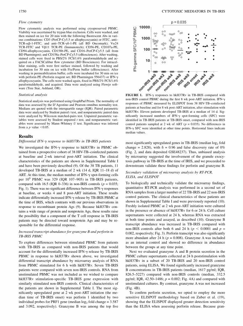
We investigated the IFN-g response to hkH37Rv in PBMC ob-tained from a prospective cohort of 38 HIV-TB–coinfected patientsat baseline and 2-wk interval post-ART initiation. The clinicalcharacteristics of the patients are shown in Supplemental Table Iand have been previously described (9). Of the 38 TB patients, 11developed TB-IRIS at a median of 2 wk (14 d, IQR 11–18 d) ofART. At this time, the median number of IFN-g spot-forming cellsper 106 PBMC was 329 (IQR 107–905) in TB-IRIS patients,compared with 16.5 (IQR 0–336) in non-IRIS controls (p = 0.035;Fig. 1). There was no significant difference between IFN-g responsesat baseline, or weeks 4 and 8 post-ART initiation. These resultsindicate differentially increased IFN-g release by TB-IRIS PBMC atthe time of IRIS, which contrasts with our previous observations inresponse to recombinant protein Ags (9). Because hkH37Rv con-tains a wide range of protein and nonprotein Ags, these results raisethe possibility that a component of the T cell response in TB-IRISpatients may be directed toward nonprotein Ags and may be re-sponsible for the differential response.
Increased transcript abundance for granzyme B and perforin inTB-IRIS PBMC
To explore differences between stimulated PBMC from patientswith TB-IRIS as compared with non-IRIS patients that wouldaccount for the differentially increased IFN-g release by TB-IRISPBMC in response to hkH37Rv shown above, we investigateddifferential transcript abundance by microarray analysis of RNAfrom PBMC stimulated for 6 h with hkH37Rv. Seven TB-IRISpatients were compared with seven non-IRIS controls. RNA fromunstimulated PBMC was not included as we wished to comparehkH37Rv stimulation-specific TB-IRIS gene expression withsimilarly stimulated non-IRIS controls. Clinical characteristics ofthe patients are shown in Supplemental Table I. The most sig-nificantly upregulated gene at 2 wk post-ART initiation (the me-dian time of TB-IRIS onset) was perforin 1 identified by twoindividual probes for PRF1 gene (median log2 fold change = 3.587and 3.092, respectively). Granzyme B was among the top five
most significantly upregulated genes in TB-IRIS (median log2 foldchange = 2.828), with d = 0.96 and false discovery rate of 0%(Fig. 2, and data deposited GSE48237). Thus, unbiased analysisby microarray suggested the involvement of the granule exocy-tosis pathway in TB-IRIS at the time of IRIS, and we proceeded todownstream validate these findings for perforin and granzyme B.
Secondary validation of microarray analysis by RT-PCR,ELISA, and ELISPOT
To biologically and technically validate the microarray findings,quantitative RT-PCR analysis was performed in a second set ofRNA samples from a larger number of 22 TB-IRIS and 22 non-IRIScontrol patients. The clinical characteristics of these patients areshown in Supplemental Table I and were previously reported (10).Freshly isolated PBMC at 2 wk post-ART initiation were culturedin the presence or absence of hkH37Rv for 6 or 24 h. Cell culturesupernatants were collected at 24 h, whereas RNA was extractedat both time points and assayed, as described (10). Granzyme Btranscript abundance was increased in TB-IRIS compared withnon-IRIS controls after both 6 and 24 h (p , 0.0001 and p =0.002, respectively; Fig. 3). Perforin transcript was also significantlymore abundant after 24 h (p = 0.008). Granzyme A was includedas an internal control and showed no difference in abundancebetween the groups at any time point.Next we evaluated granzyme A and B protein secretion in the
PBMC culture supernatants collected at 24 h poststimulation withhkH37Rv in a subset of 20 TB-IRIS and 20 non-IRIS controlpatients, using ELISA. We found significantly increased granzymeB concentrations in TB-IRIS patients (median, 1617 pg/ml; IQR,826.5–5227) compared with non-IRIS controls (median, 332.5pg/ml; IQR, 42.50–1486; p = 0.002; Fig. 4A) and compared withunstimulated cultures. By contrast, granzyme Awas not increased(Fig. 4B).To confirm perforin secretion, we opted to employ the more
sensitive ELISPOT methodology based on Zuber et al. (19),showing that the ELISPOT displayed greater detection sensitivitythan the ELISA when assessing perforin release. Because gran-
FIGURE 1. IFN-g responses to hkH37Rv in TB-IRIS compared with
non-IRIS control PBMC during the first 8 wk post-ART initiation. IFN-g
responses of PBMC measured by ELISPOT from 38 HIV-TB–coinfected
patients at baseline and for 8 wk post-ART initiation, after stimulation with
hkH37Rv. Eleven patients developed TB-IRIS at a median of 14 d. Sig-
nificantly increased numbers of IFN-g spot-forming cells (SFC) were
identified in TB-IRIS patients at TB-IRIS onset, compared with non-IRIS
control patients sampled at 2 wk of ART (p = 0.035). No differences in
IFN-g SFC were identified at other time points. Horizontal lines indicate
median values.
1750 CYTOTOXIC MEDIATORS IN TB-IRIS

zyme A was consistently shown not to be elevated in our internalcontrol experiments, we did not include it in the ELISPOT anal-ysis, but we included measurement of granzyme B to furtherstrengthen our findings. Thus, ELISPOT assay for perforin andgranzyme B was performed in a smaller number of samples, basedon stored PBMC availability. A nonsignificant trend toward in-creased numbers of PBMC-secreting granzyme B and perforin inresponse to hkH37Rv in TB-IRIS was observed (Fig. 4C, 4D). Themedian spot-forming cells per 106 PBMC for granzyme B fromTB-IRIS patients was 92 (IQR, 0–364) compared with 48 (IQR,1.25–94) in non-IRIS patients. For perforin, the median fromTB-IRIS patients was 44 (IQR, 6.5–87) in TB-IRIS and 27 (IQR,4.5–52) in non-IRIS patients.
The effect of in vivo prednisone therapy on granzyme Bsecretion in vitro
Corticosteroid therapy is used as an immunosuppressive therapyin the treatment of TB-IRIS. We evaluated granzyme B secretionby ELISA in the 24-h supernatant of hkH37Rv-stimulated PBMCcultures from a subset of 29 patients enrolled in a randomizeddouble-blind placebo-controlled trial of prednisone for treatmentof TB-IRIS (7, 8). The trial showed an overall reduction in dura-tion of hospitalization and numbers of therapeutic procedures in
prednisone-treated patients, as well as hastened improvement inTB-IRIS symptoms (7, 8), and such trends were evident in thesubset of patients included in this analysis. Thus, we observed thatin TB-IRIS patients receiving prednisone treatment for 2 wk,granzyme B secretion was decreased in vitro (n = 16; median, 233pg/ml; IQR, 66–2312 pg/ml) compared with pretreatment responses(median, 1068; IQR, 6–6672; p = 0.03). This reduction was notevident in PBMC from placebo-treated patients (n = 13; Fig. 5),suggesting a correlation between our in vitro assessment of perforinand granzyme B and improved clinical outcome (7, 8).
Elucidating the cellular source of perforin in TB-IRIS patients
Our findings suggested the differential induction of cytotoxicpathways in TB-IRIS in response to hkH37Rv. We therefore in-vestigated potential cytotoxic cells present in PBMC at 2 wk post-ART initiation in a subgroup of patients using flow cytometry. Asboth CD8 and CD4 T cells are able to upregulate mRNA expressionfor granzyme B and perforin after stimulation with H37Rv (20), wefirst evaluated differences between these cells in TB-IRIS com-pared with non-IRIS controls. CD4 and CD8 T cells were presentin similar proportions in unstimulated PBMC in TB-IRIS and non-IRIS controls (Supplemental Fig. 1A). In a subset of patients, weestablished that both CD4 and CD8 cells contained perforin
FIGURE 2. Representation of significantly differentially expressed genes. Microarray analysis of RNA from seven matched pairs of IRIS and non-IRIS
patients, at 2 wk on ART, showed transcript for perforin and granzyme B genes to be among the four genes most significantly overrepresented in TB-IRIS
PBMC after stimulation with H37Rv. The transcripts for Zinc and ring finger 1 and Cbp/p300-interacting transactivator with Glu/Asp-rich C-terminal
domain 2 were also significantly raised. The genes significantly upregulated in TB-IRIS (with d = 0.96 and false discovery rate of 0.000001%) are rep-
resented on the volcano plot as labeled. The x-axis shows the log2-fold change, and the y-axis shows a measure of significance (2log10 [p value]).
FIGURE 3. Secondary validation of microarray analysis by RT-PCR. PBMC from 22 TB-IRIS and 22 non-IRIS control patients were isolated and
cultured in the presence or absence of hkH37Rv (MOI 1:1, PBMC:H37Rv) for 6 or 24 h. RNAwas extracted and assayed by RT-PCR. b-actin was used to
normalize values. Fold induction over unstimulated cultures was calculated by the DD cycle threshold method, and fold values log10 transformed to
normalize. Granzyme B was increased in TB-IRIS compared with controls after both 6 and 24 h (p, 0.0001 and p = 0.002, respectively). Perforin was also
significantly increased after 24-h stimulation (p = 0.008). Horizontal lines indicate median values.
The Journal of Immunology 1751

(Supplemental Fig. 1B), which decreased on stimulation withhkH37Rv, implying Ag-specific degranulation (as shown in asmaller subset of patients in Supplemental Table II). Next, wequantified NK cell proportions, identified by the absence of CD3,the presence of CD16, and a combination of activating (CD56) orinhibitory (CD94, CD158) cell surface receptors. No difference inNK cell proportions was observed in TB-IRIS, compared withnon-IRIS controls (Supplemental Table III). The g-d (gd) T cellshave been implicated in TB-IRIS pathophysiology, can recognizenonprotein Ags, and express perforin and granzyme B (21, 22).We observed a trend toward decreased proportions of gd TCR-expressing cells in TB-IRIS compared with non-IRIS PBMC(Supplemental Table III; p = 0.089), suggesting that this pop-ulation is less likely to contribute to upregulation of the cytotoxicpathways identified.iNKT cells may express either CD4 and/or CD8 molecules, are
innate lymphocytes with cytotoxic activity, and are characterizedby reactivity to glycolipid Ags. We investigated proportions ofiNKT cells, as determined by the presence of Va24 TCR that, incombination with Vb11, characterizes these cells in humans.Significantly increased proportions of CD3+ Va24+ cells werefound in unstimulated PBMC at 2 wk post-ART initiation fromTB-IRIS patients: median, 0.17% (IQR, 0.09–0.22; n = 15),compared with non-IRIS controls (median, 0.03%; IQR, 0.016–0.106; n = 9; p = 0.004; Fig. 6A). Using the more stringentcombination of Va24+Vb11+ staining on CD3+ cells in a smallernumber of TB-IRIS patients (n = 11), the median CD3+Va24+
Vb11+ frequency was 0.18% (IQR, 0.09–0.4) compared with 0.04%(IQR, 0.03–0.82; n = 9) in nine non-IRIS patients (p = 0.05;Supplemental Fig. 1C). Moreover, intracellular staining in a subsetof these TB-IRIS patient samples identified these cells as a sourceof perforin (Fig. 6B).
DiscussionDefining the pathophysiology of TB-IRIS may facilitate devel-opment of diagnostic tests and specific treatments, which arecurrently lacking. Although dysregulated immune restoration isassociated with dynamic CD4 Th1 expansions and contractions,these relate poorly to symptoms (8, 9). In our previous longitudinalanalysis, we found no evidence for a difference between TB-IRISand non-IRIS patients in response to recombinant protein Ags andpurified protein derivative, suggesting that protein Ag-specific Th1responses may not be the primary determinants of TB-IRIS. Thefinding in our current study, that increased IFN-g responses occurin response to hkH37Rv in TB-IRIS patients at TB-IRIS onsetcompared with non-IRIS controls, raises the possibility that a com-ponent of the T cell response directed toward nonprotein Ags maybe responsible for the differential response.Our microarray analysis, demonstrating perforin 1 and gran-
zyme B to be the most significantly upregulated genes in PBMCfrom TB-IRIS patients, compared with non-IRIS controls, andfurther validation of these results with RT-PCR, ELISA, andELISPOT, highlights a role for the granule–exocytosis pathway inTB-IRIS. Perforin is a cytolytic protein found in granules of
FIGURE 4. Granzyme B and perforin secretion are increased in TB-IRIS. (A and B) Granzyme B secretion measured by ELISA was significantly in-
creased in TB-IRIS patients, in contrast to granzyme A, in hkH37Rv-stimulated PBMC culture supernatants (20 TB-IRIS, 20 controls) compared with
unstimulated supernatants (p , 0.0001), and compared with stimulated PBMC culture supernatants from non-IRIS control patients (p = 0.002). (C and D)
Granzyme B and perforin measured by ELISPOT tended to be increased in TB-IRIS compared with controls (not statistically significant). Horizontal lines
indicate median values.
1752 CYTOTOXIC MEDIATORS IN TB-IRIS

innate lymphocytes (cytotoxic T lymphocytes and NK cells) andneutrophils that have a key role in delivery of contents of cyto-toxic granules to target cell cytoplasm. Granzyme B is a serine
protease, a key component of cytotoxic granules, and activatesapoptosis once in the cytoplasm of the target cell. Potential celltypes that are capable of recognizing nonprotein Ags have cyto-toxic capacity and contain granule-associated perforin and gran-zyme B, including NK cells, gd T cells, and NKT cells (21, 23,24). Two recent studies have examined the potential contributionof NK cells in TB-IRIS and suggested that elevated NK cell ac-tivation and degranulation levels characterize the immunologicalprofile of TB-IRIS patients (25, 26). One previous study alsodemonstrated an association between a subset of gd T cells andTB-IRIS (22). However, we found reduced proportions of thesecells in TB-IRIS, suggesting that they cannot be responsible forthe increased perforin and granzyme B detected in TB-IRISpatients. We demonstrate that both CD4 and CD8 cells werea source of perforin in TB-IRIS patients and that increased pro-portions of iNKT cells are present in PBMC of TB-IRIS patientsat the time of IRIS onset, compared with non-IRIS controls, in twoanalyses (calculating CD3+Va24+ cells and CD3+Va24+Vb11+
cells), both considered to be stringent approaches for quantifyingiNKT cells (27). Although these results implicate iNKT cells asa likely candidate, we are currently conducting a longitudinalstudy addressing further the role of cytotoxic T cells in TB-IRISdevelopment.We have previously demonstrated that TB-IRIS is associated
with hypercytokinaemia in blood and cerebrospinal fluid, of pre-dominantly myeloid or dual myeloid/lymphoid in origin, which ismodulated during treatment of TB-IRIS with corticosteroids (10,12, 28). These and other studies exploring murine models ofTB-IRIS have led to the recent proposal that ART-induced changesin innate immune function contribute substantially to TB-IRIS(11, 28, 29). Our observation that granzyme B gene upregulationand protein secretion occur in PBMC from patients at the time ofTB-IRIS symptom onset, and that secretion is reduced in PBMCfrom patients treated for TB-IRIS by prednisone, which improvessymptoms and reduces hospitalization, supports a role for cyto-toxic mediators in the immunopathology of TB-IRIS. Furthercharacterization of this cytotoxic response, especially at the site ofinfection, is desirable.Our study has a number of limitations, including the limited
number of cells available for in-depth analysis of cytotoxic lym-phocytes. Inclusion of samples based on availability of PBMCmeans that some of the assays were performed in samples fromdifferent patients, possibly resulting in the introduction of se-lection bias. However, it is encouraging to find the same pattern ofmediators in different samples collected over time. Our study designdid not include the evaluation of nonprotein preparations such aslipid fractions, and we acknowledge the fact that, whereas killedmycobacteria will no longer secrete protein Ags, they still containantigenic proteins, some of which will give rise to an immune re-sponse. Additionally, we did not assess the capacity of neutrophils toproduce cytotoxic mediators. Evaluating the role of the two addi-tional genes that were upregulated in TB-IRIS (ZNRF1: Zinc andring finger 1 and CITED2: Cbp/p300-interacting transactivator withGlu/Asp-rich C-terminal domain 2) was outside the scope of thecurrent study and should constitute the subject of further investi-gation.In conclusion, our data indicate differential cytotoxic activity
associated with TB-IRIS, with increased cytotoxic mediators com-pared with non-IRIS patients. This supports further analysis ofcytotoxic pathways, not only in TB-IRIS pathophysiology, but alsoin the pathogenesis of tuberculosis and inflammatory conditions ingeneral. Improved understanding of the mechanisms of pathologyin TB-IRIS is required to derive new treatments to reduce themorbidity and mortality associated with this condition.
FIGURE 5. Effect of prednisone or placebo on granzyme B release
in vitro. Granzyme B secretion (pg/ml) from PBMCs, from TB-IRIS patients
enrolled in a randomized controlled trial of prednisone versus placebo,
measured by ELISA after stimulation with hkH37Rv, with unstimulated
culture results subtracted. Prednisone treatment for 2 wk significantly de-
creased granzyme B secretion from TB-IRIS PBMC compared with pre-
treatment values. This reduction was not evident in PBMC from placebo-
treated TB-IRIS patients.
FIGURE 6. Increased proportions of iNKT cells in TB-IRIS compared
with non-IRIS control patients. Flow cytometric analysis using cell surface
staining for CD3+Va24+ cells identified an increased proportion of CD3+
Va24+ cells in TB-IRIS compared with non-IRIS control patients (median,
0.17%; IQR, 0.09–0.22, versus 0.03%; IQR, 0.016–0.106) (A). Using in-
tracellular staining for perforin, CD3+Va24+ cells from TB-IRIS patients
were identified as a potential source of perforin (B). Horizontal bars in-
dicate median values. Gating strategy was based on selecting CD3+ cells
from a side scatter/CD3 scatter dot blot, followed by gating on Va24 and
either Vb11 or perforin-positive cells.
The Journal of Immunology 1753

AcknowledgmentsWe are grateful to Kevin Rebe and medical staff at GF Jooste Hospital for
assistance with patient recruitment and follow-up.
DisclosuresThe authors have no financial conflicts of interest.
References1. Chaisson, R. E., and N. A. Martinson. 2008. Tuberculosis in Africa—combating
an HIV-driven crisis. N. Engl. J. Med. 358: 1089–1092.2. Schutz, C., G. Meintjes, F. Almajid, R. J. Wilkinson, and A. Pozniak. 2010.
Clinical management of tuberculosis and HIV-1 co-infection. Eur. Respir. J. 36:1460–1481.
3. Meintjes, G., S. D. Lawn, F. Scano, G. Maartens, M. A. French, W. Worodria,J. H. Elliott, D. Murdoch, R. J. Wilkinson, C. Seyler, et al; International Networkfor the Study of HIV-Associated IRIS. 2008. Tuberculosis-associated immunereconstitution inflammatory syndrome: case definitions for use in resource-limited settings. Lancet Infect. Dis. 8: 516–523.
4. M€uller, M., S. Wandel, R. Colebunders, S. Attia, H. Furrer, and M. Egger,IeDEA Southern and Central Africa. 2010. Immune reconstitution inflammatorysyndrome in patients starting antiretroviral therapy for HIV infection: a system-atic review and meta-analysis. Lancet Infect. Dis. 10: 251–261.
5. Narendran, G., B. B. Andrade, B. O. Porter, C. Chandrasekhar, P. Venkatesan,P. A. Menon, S. Subramanian, S. Anbalagan, K. P. Bhavani, S. Sekar, et al. 2013.Paradoxical tuberculosis immune reconstitution inflammatory syndrome (TB-IRIS) in HIV patients with culture confirmed pulmonary tuberculosis in Indiaand the potential role of IL-6 in prediction. PLoS One 8: e63541.
6. Meintjes, G., M. X. Rangaka, G. Maartens, K. Rebe, C. Morroni, D. J. Pepper,K. A. Wilkinson, and R. J. Wilkinson. 2009. Novel relationship between tu-berculosis immune reconstitution inflammatory syndrome and antituberculardrug resistance. Clin. Infect. Dis. 48: 667–676.
7. Meintjes, G., R. J. Wilkinson, C. Morroni, D. J. Pepper, K. Rebe, M. X. Rangaka,T. Oni, and G. Maartens. 2010. Randomized placebo-controlled trial of pred-nisone for paradoxical tuberculosis-associated immune reconstitution inflam-matory syndrome. AIDS 24: 2381–2390.
8. Meintjes, G., K. H. Skolimowska, K. A. Wilkinson, K. Matthews, R. Tadokera,A. Conesa-Botella, R. Seldon, M. X. Rangaka, K. Rebe, D. J. Pepper, et al. 2012.Corticosteroid-modulated immune activation in the tuberculosis immune recon-stitution inflammatory syndrome. Am. J. Respir. Crit. Care Med. 186: 369–377.
9. Meintjes, G., K. A. Wilkinson, M. X. Rangaka, K. Skolimowska, K. van Veen,M. Abrahams, R. Seldon, D. J. Pepper, K. Rebe, P. Mouton, et al. 2008. Type 1helper T cells and FoxP3-positive T cells in HIV-tuberculosis-associated immunereconstitution inflammatory syndrome. Am. J. Respir. Crit. Care Med. 178:1083–1089.
10. Tadokera, R., G. Meintjes, K. H. Skolimowska, K. A. Wilkinson, K. Matthews,R. Seldon, N. N. Chegou, G. Maartens, M. X. Rangaka, K. Rebe, et al. 2011.Hypercytokinaemia accompanies HIV-tuberculosis immune reconstitution in-flammatory syndrome. Eur. Respir. J. 37: 1248–1259.
11. Tadokera, R., G. A. Meintjes, K. A. Wilkinson, K. H. Skolimowska, N. Walker,J. S. Friedland, G. Maartens, P. T. Elkington, and R. J. Wilkinson. 2014. Matrixmetalloproteinases and tissue damage in HIV-tuberculosis immune reconstitu-tion inflammatory syndrome. Eur. J. Immunol. 44: 127–136.
12. Conesa-Botella, A., G. Meintjes, A. K. Coussens, H. van der Plas, R. Goliath,C. Schutz, R. Moreno-Reyes, M. Mehta, A. R. Martineau, R. J. Wilkinson, et al.2012. Corticosteroid therapy, vitamin D status, and inflammatory cytokineprofile in the HIV-tuberculosis immune reconstitution inflammatory syndrome.Clin. Infect. Dis. 55: 1004–1011.
13. Siddiqi, K., M. L. Lambert, and J. Walley. 2003. Clinical diagnosis of smear-negative pulmonary tuberculosis in low-income countries: the current evidence.Lancet Infect. Dis. 3: 288–296.
14. Haddow, L. J., M. Y. Moosa, and P. J. Easterbrook. 2010. Validation of a pub-lished case definition for tuberculosis-associated immune reconstitution in-flammatory syndrome. AIDS 24: 103–108.
15. Manosuthi, W., H. Van Tieu, W. Mankatitham, A. Lueangniyomkul, J. Ananworanich,A. Avihingsanon, U. Siangphoe, S. Klongugkara, S. Likanonsakul, U. Thawornwan,et al; N2R Study Team. 2009. Clinical case definition and manifestations of par-adoxical tuberculosis-associated immune reconstitution inflammatory syndrome.AIDS 23: 2467–2471.
16. Rangaka, M. X., L. Diwakar, R. Seldon, G. van Cutsem, G. A. Meintjes,C. Morroni, P. Mouton, M. S. Shey, G. Maartens, K. A. Wilkinson, andR. J. Wilkinson. 2007. Clinical, immunological, and epidemiological importanceof antituberculosis T cell responses in HIV-infected Africans. Clin. Infect. Dis.44: 1639–1646.
17. Wilkinson, K. A., G. R. Stewart, S. M. Newton, H. M. Vordermeier, J. R. Wain,H. N. Murphy, K. Horner, D. B. Young, and R. J. Wilkinson. 2005. Infectionbiology of a novel alpha-crystallin of Mycobacterium tuberculosis: Acr2. J.Immunol. 174: 4237–4243.
18. Chu, G., B. Narasimhan, R. Tibshirani, and V. Tusher. SAM: SignificanceAnalysis of Microarrays Stanford University. Available at: http://www-stat.stanford.edu/∼tibs/SAM/. Accessed: March 12, 2008.
19. Zuber, B., V. Levitsky, G. Jonsson, S. Paulie, A. Samarina, S. Grundstrom,S. Metkar, H. Norell, G. G. Callender, C. Froelich, and N. Ahlborg. 2005. De-tection of human perforin by ELISpot and ELISA: ex vivo identification of virus-specific cells. J. Immunol. Methods 302: 13–25.
20. Canaday, D. H., R. J. Wilkinson, Q. Li, C. V. Harding, R. F. Silver, andW. H. Boom. 2001. CD4(+) and CD8(+) T cells kill intracellular Mycobacteriumtuberculosis by a perforin and Fas/Fas ligand-independent mechanism. J.Immunol. 167: 2734–2742.
21. Koizumi, H., C. C. Liu, L. M. Zheng, S. V. Joag, N. K. Bayne, J. Holoshitz, andJ. D. Young. 1991. Expression of perforin and serine esterases by human gamma/delta T cells. J. Exp. Med. 173: 499–502.
22. Bourgarit, A., G. Carcelain, A. Samri, C. Parizot, M. Lafaurie, S. Abgrall,V. Delcey, E. Vicaut, D. Sereni, and B. Autran, PARADOX Study Group. 2009.Tuberculosis-associated immune restoration syndrome in HIV-1-infected patientsinvolves tuberculin-specific CD4 Th1 cells and KIR-negative gammadelta T cells.J. Immunol. 183: 3915–3923.
23. Bendelac, A., P. B. Savage, and L. Teyton. 2007. The biology of NKT cells.Annu. Rev. Immunol. 25: 297–336.
24. Moretta, A., E. Marcenaro, S. Parolini, G. Ferlazzo, and L. Moretta. 2008. NKcells at the interface between innate and adaptive immunity. Cell Death Differ.15: 226–233.
25. Pean, P., E. Nerrienet, Y. Madec, L. Borand, D. Laureillard, M. Fernandez,O. Marcy, C. Sarin, K. Phon, S. Taylor, et al; Cambodian Early versus LateIntroduction of Antiretroviral Drugs (CAMELIA) Study Team. 2012. Naturalkiller cell degranulation capacity predicts early onset of the immune reconsti-tution inflammatory syndrome (IRIS) in HIV-infected patients with tuberculosis.Blood 119: 3315–3320.
26. Conradie, F., A. S. Foulkes, P. Ive, X. Yin, K. Roussos, D. K. Glencross,D. Lawrie, W. Stevens, L. J. Montaner, I. Sanne, and L. Azzoni. 2011. Naturalkiller cell activation distinguishes Mycobacterium tuberculosis-mediated im-mune reconstitution syndrome from chronic HIV and HIV/MTB coinfection. J.Acquir. Immune Defic. Syndr. 58: 309–318.
27. Berzins, S. P., M. J. Smyth, and A. G. Baxter. 2011. Presumed guilty: naturalkiller T cell defects and human disease. Nat. Rev. Immunol. 11: 131–142.
28. Marais, S., G. Meintjes, D. J. Pepper, L. E. Dodd, C. Schutz, Z. Ismail,K. A. Wilkinson, and R. J. Wilkinson. 2013. Frequency, severity, and predictionof tuberculous meningitis immune reconstitution inflammatory syndrome. Clin.Infect. Dis. 56: 450–460.
29. Barber, D. L., B. B. Andrade, I. Sereti, and A. Sher. 2012. Immune reconstitutioninflammatory syndrome: the trouble with immunity when you had none. Nat.Rev. Microbiol. 10: 150–156.
1754 CYTOTOXIC MEDIATORS IN TB-IRIS