reconstitution of sec gene product-dependent intercompartmental protein transport
TRANSCRIPT
Cell, Vol. 54, X35-344, July 29, 1999, Copyright 0 1989 by Cell Press
Reconstitution of SEC Gene Product-Dependent Intercompartmental Protein Transport
David Baker, Linda Hicke, Michael Rexach, Manfred Schleyer: and Randy Schekman Department of Biochemistry University of California Berkeley, California 94720
Summary
Transport of a-factor precursor from the endoplasmic reticulum to the .Golgi apparatus has been recon- stituted in gently lysed yeast spheroplasts. Transport is measured through the coupled addition of outer- chain carbohydrate to [3JS]methionine4abeled a-fac- tor precursor translocated into the endoplasmic retic- ulum of broken spheroplasts. The reaction is absolutely dependent on ATP, stimulated 6-fold by cytosol, and occurs between physically separable sealed compart- ments. Transport is inhibii by the guanine nucleotide analog GTPTS. sec23 mutant cells have a tempera- ture-sensitive defect in endoplasmic reticulum-to-Golgi transport in vivo. This defect has been reproduced in vitro using sec23 membranes and cytosol. Transport at 30% with sec23 membranes requires addition of cytosol containing the SEC23 (wild-type) gene prod- uct. This demonstrates that an in vitro interorganelle transport reaction depends on a factor required for transport in vivo. Complementation of set mutations in vitro provides a functional assay for the purification of individual intercompartmental transport factors.
Introduction
Newly synthesized proteins are directed to their proper lo- cations through rapid and precise intercompartmental transport reactions. Secretory and plasma membrane proteins traverse a series of membrane-enclosed com- partments including the endoplasmic reticulum (ER) and the cisternae of the Golgi en route to the cell surface. While transport of proteins between these compartments has been extensively described in vivo (reviewed in Pfeffer and Rothman, 1987) the underlying mechanisms remain obscure. Reconstitution of intercompartmental transport in vitro is required to begin to study the enzymol- ogy of these reactions.
Several intercompartmental protein transport reactions have been reconstituted in mammalian systems. Recon- stitution of transport between Golgi cisternae has led to the purification of at least one transport factor and identifi- cation of several novel transport intermediates (Melancon et al., 1987; Pfeffer and Rothman, 1987; J. Rothman, per-
This paper is dedicated to Arthur Kornberg on the occasion of his seventieth birthday. * Present address: Department of Physiological Chemistry, Physical Biochemistry and Cell Biology, University of Munich, Munich, Federal Republic of Germany.
sonal communication). Purification of transport factors has relied on specific inhibitors to inactivate single com- ponents followed by supplementation with untreated pro- tein fractions. Direct fractionation has proved difficult, probably because of the large number of factors involved. Reconstitution of transport from the ER to the Golgi has recently been achieved using “semi-intact” Chinese ham- ster ovary cells, which have lost most of their cytosol but retain intact organelles (Beckers et al., 1987).
A large number of yeast secretory (set) mutants have been isolated that have temperature-sensitive defects in protein transport (Novick et al., 1980). The motivation for developing an intercompartmental transport assay in yeast is the potential to purify and characterize individual transport factors through complementation of transport- defective mutant reactions. Among the several stages in the yeast secretory pathway, protein transport from the ER to the Golgi is particularly amenable to analysis. Eleven complementation groups have been identified whose products are required for ER-Golgi transport in vivo (No- vick et al., 1981; Newman and Ferro-Novick, 1987). Fur- thermore, the addition of outer-chain carbohydrate to core oligosaccharides serves as a convenient diagnostic of ar- rival in the Golgi (Esmon et al., 1981) (see Figure 1). A yeast ER-Golgi in vitro transport reaction has been previ- ously reported (Haselbeck and Schekman, 1988), but a lack of cytosol dependence and low transport efficiency (<2%) have precluded effective exploitation of the mu- tants.
Here we report an ER-Golgi in vitro transport reaction in which nearly one-third of the core glycosylated transport substrate receives outer-chain carbohydrate. Two innova- tions were essential: a method for preparing semi-intact yeast and a method for following transport in extracts pre- pared from wild-type cells. The reaction is absolutely de- pendent on ATP, stimulated 6-fold by cytosolic proteins, and occurs between physically separable compartments. The reaction is blocked by GTPyS and depends on the SEC23 gene product.
Results
Preparation of Gently Lysed Yeast We sought a method for preparing yeast lysates compe- tent for ER-to-Golgi transport. Recently, two methods for preparing transport-competent, semi-intact mammalian cells were described (Simons and Virta, 1987; Beckers et al., 1987). These methods take advantage of the mono- layer growth habit of the cells and cannot readily be gener- alized to cells such as yeast, which grow in liquid culture. We explored freeze-thaw lysis of spheroplasts as a means of cell breakage. Early studies indicated that the rate of freezing determined the extent of lysis. Slow freezing preserved spheroplasts intact; rapid freezing ruptured spheroplasts and internal organelles. We then varied the freezing rate, aiming for a gentle lysis procedure that released cytosolic proteins but left organelles intact.
Cell 33s
WTOSOL
Translocation
I J
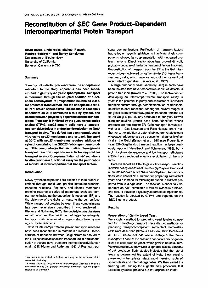
Figure 1. Glycosylation Coupled to Translocation of Proteins into the ER and Transport to the Golgi Core oligosaccharides are added to proteins upon translocation into the ER. Transport to the Golgi is blocked in the set and bet mutants listed. In the Golgi, core oligosaccharides are extended by addition of outer-chain carbohydrate. M, mannose; GNAc, N-acetylglucosamine; Asn, asparagine.
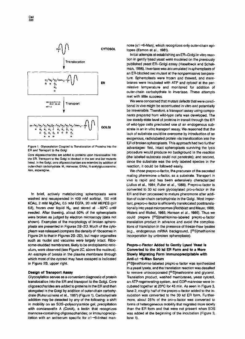
In brief, actively metabolizing spheroplasts were washed and resuspended in 400 mM sorbitol, 150 mM KOAc, 2 mM MgOAc, 0.5 mM EGTA, 20 mM HEPES (pH 6.6), frozen over liquid NP, and stored at -85% until needed. After thawing, about 50% of the spheroplasts were broken as judged by electron microscopy (data not shown). Examples of the morphology of broken sphero- plasts are presented in Figures 2B-2D. Much of the cyto- plasm was released (compare the density of ribosomes in Figure 2A to that in Figures 2B-2D), but major organelles such as nuclei and vacuoles were largely intact. Ribo- some-studded membranes, likely to be endoplasmic retic- ulum, were observed (see Figure 2C, above the nucleus). An example of breaks in the plasma membrane through which most of the cytosol may have escaped is indicated in Figure 26, upper right.
Design of Transport Assay Glycosylation serves as a convenient diagnostic of protein translocation into the ER and transport to the Golgi. Core oligosaccharides are added to proteins in the ER and then elongated in the Golgi by addition of outer-chain carbohy- drate (Kukuruzinska et al., 1987) (Figure 1). Carbohydrate addition may be detected by any of the following: a shift in mobility on an SDS-polyacrylamide gel; precipitation with concanavalin A (ConA), a lectin that recognizes mannose-containing oligosaccharides; or immunoprecip- itation with an antiserum specific for al-6-linked man-
nose (al --Man), which recognizes only outer-chain epi- topes (Esmon et al., 1981).
Initial attempts at establishing an ER-Golgi in vitro reac- tion in gently lysed yeast were modeled on the previously published yeast ER-Golgi assay (Haselbeck and Schek- man, 1986). lnvertase was accumulated in spheroplasts of an ER-blocked set mutant at the nonpermissive tempera- ture. Spheroplasts were frozen and thawed, and mem- branes were incubated with ATP and cytosol at the per- missive temperature and monitored for addition of outer-chain carbohydrate to invertase. These attempts met with little success,
We were concerned that mutant defects that were condi- tional in vivo might be accentuated in vitro and potentially be irreversible. Therefore, a transport assay using compo- nents prepared from wild-type cells was developed. The low steady-state level of proteins in transit through the ER of wild-type cells precluded use of an endogenous sub- strate in an in vitro transport assay. We reasoned that the lack of substrate could be overcome by introduction of an exogenous, radiolabeled protein via translocation into the ER of broken spheroplasts. This approach had two further advantages: first, intact spheroplasts surviving the lysis procedure would produce no background in the reaction (the labeled substrate could not penetrate); and second, since the substrate was the only labeled species in the reaction, it could be followed easily.
We chose prepro-a-factor, the precursor of the secreted mating pheremone a-factor, as a substrate. Transport in vivo is rapid and has been extensively characterized (Julius et al., 1984; Fuller et al., 1988). Prepro-a-factor is converted to 30 kd core glycosylated pro-a-factor in the ER and then processed to mature pheromone after addi- tion of outer-chain carbohydrate in the Golgi. Most impor- tant, prepro-a-factor is efficiently translocated posttransla- tionally into yeast microsomes (Rothblatt and Meyer, 1986; Waters and Blobel, 1986; Hansen et al., 1986). Thus we could prepare [%]methionine-labeled prepro-a-factor translation product in advance and avoid the complica- tions of translation in the presence of freeze-thaw lysates (e.g., endogenous mRNA background, [35S]methionine incorporation by unbroken spheroplasts).
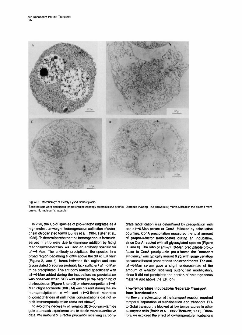
Prepro-a-Factor Added to Gently Lysed Yeast Is Converted to the 30 kd ER Form and to a More Slowly Migrating Form lmmunoprecipitable with Anti-al+-Man Serum [%]methionine-labeled prepro-a-factor was synthesized in a yeast lysate, and the translation reaction was desalted to remove unincorporated [SSJmethionine and glycerol. Translation product, washed membranes, yeast cytosol, an ATP-regenerating system, and GDP-mannose were in- cubated together at 20% for 45 min. As seen in Figure 3, lane 2, roughly half of the prepro-a-factor added to the in- cubation was converted to the 30 kd ER form. Further- more, about 25% of the pro-a-factor was converted to forms of heterogeneous mobility that migrated more slowly than the ER form and that were not present when SDS was added at the beginning of the incubation (Figure 3, lane 1).
set-Dependent Protein Transport 337
Figure 2. Morphology of Gently Lysed Spheroplasts
Spheroplasts were processed for electron microscopy before brane. N, nucleus; V, vacuole
In vivo, the Golgi species of pro-a-factor migrates as a high molecular weight, heterogeneous collection of outer- chain glycosylated forms (Julius et al., 1984; Fuller et al., 1988). To determine whether the heterogeneous forms ob- served in vitro were due to mannose addition by Golgi mannosyltransferases, we used an antibody specific for al--Man. The antibody precipitated the species in a broad region beginning slightly above the 30 kd ER form (Figure 3, lane 4); forms between this region and core glycosylated precursor probably lack sufficient al -+-Man to be precipitated. The antibody reacted specifically with al-6Man added during the incubation: no precipitation was observed when SDS was added at the beginning of the incubation (Figure 3, lane 3) or when competitor al-6 Man oligosaccharide (100 PM) was present during the im- munoprecipitation. a1+2- and al-+3-linked mannose oligosaccharides at millimolar concentrations did not in- hibit immunoprecipitation (data not shown).
To avoid the necessity of running SDS-polyacrylamide gels after each experiment and to obtain more quantitative data, the amount of a-factor precursor receiving carbohy-
(A) and after (B-D) freeze-thawing. The arrow in (13) marks a break in the plasma
drate modification was determined by precipitation with anti-al--Man serum or ConA, followed by scintillation counting. ConA precipitation measured the total amount of prepro-a-factor translocated during an incubation, since ConA reacted with all glycosylated species (Figure 3, lane 8). The ratio of anti-al--Man precipitable pro-a- factor to ConA precipitable pro-a-factor, the “transport efficiency:’ was typically around 0.25, with some variation between different preparations and experiments. The anti- al-+-Man serum gave a slight underestimate of the amount of a-factor receiving outer-chain modification, since it did not precipitate the portion of heterogeneous material just above the ER form.
Low-Temperature lncubatiohs Separate Transport from lkanslocation Further characterization of the transport reaction required temporal separation of translocation and transport. ER- to-Golgi transport is blocked at low temperatures in other eukaryotic cells (Balch et al., 1986; Tartakoff, 1988). There- fore, we explored the effect of low-temperature incubations
Cell 338
Time Course anti- anti- OF al-Wan ConA -- -
0’46’ 0’ 45’ 0’ 45’ 200 -
82.5 -
123456
Figure 3. Prepro-a-Factor Incubated with Gently Lysed Yeast Is Con- verted to the 30 kd ER Form and to a More Slowly Migrating Form That Is lmmunoprecipitable with Anti-al-+-Man Serum
Gently lysed yeast and [35S]methionine-labeled prepro-a-factor were incubated together in the presence of GDP-mannose and an ATP- regenerating system. Laemmli sample buffer was added at the times indicated, and the quenched reactions were heated for 5 min at 9SC. Portions (20 pl) of each reaction were precipitated with anti-a-factor se- rum, anti-al+&Man serum, or ConA. The immunoprecipitates were electrophoresed on an 11.25% polyacrylamide gel. The gel was in- cubated with Amplify, dried, and exposed to X-ray film for 48 hr. Sizes in kd are marked at left.
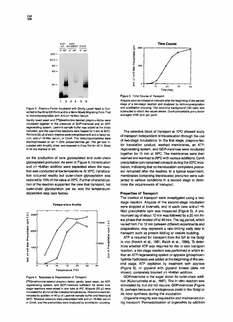
on the production of core glycosyiated and outer-chain giycosyiated precursor. As seen in Figure 4, translocation and al-%-Man addition were separated when the reac- tion was conducted at low temperature. At lo%, transioca- tion occurred readily but outer-chain giycosyiation was reduced to 10% of the value at 20%. Further characteriza- tion of the reaction supported the view that transport, not outer-chain giycosyiation per se, was the temperature- dependent step (see below).
Temperature Profile
I I I I I
;; 3000-
E 4 -10000 ;;
u 0 .<
E 2000 - .k$ 8
u E
P s C
5 - 5000 +$ z
s
1000 - it P a
5 E ‘S 0
I
0 6 10 15 20 26 30
Temperature (“C)
Figure 4. Temperature Dependence of Transport
[35S]methionine-labeled prepro-a-factor, gently lysed yeast, an ATP- regenerating system, and GDP-mannose sufficient for seven one- stage reactions were mixed in one tube at 4%. Aliquots (25 WI) were incubated for 45 min at the indicated temperatures. Reactions were ter- minated by addition of 40 pl of Laemmli sample buffer and heating at 95OC. Reaction products were precipitated with anti-al--Man serum or ConA, and the precipitates were analyzed by scintillation counting.
; oL ./. , . , . 1 0 10 20 30 40
Time (min)
Figure 5. Time Course of Transport
Aliquots were terminated at intervals after the beginning of the second stage of a two-stage reaction and analyzed by immunoprecipitation and scintillation counting. The zero-time background (120 cpm) was subtracted to obtain the values shown. ConA-precipitable pro-a-factor averaged 4739 cpm per point.
The selective block of transport at 10% allowed study of transport independent of transiocation through the use of two-stage incubations. In the first stage, prepro-a-fac- tor translation product, washed membranes, an ATP- regenerating system, and GDP-mannose were incubated together for 15 min at 10°C. The membranes were then washed and warmed to 20% with various additions. ConA precipitable cpm remained constant during the 20% incu- bation, indicating that no transiocation-competent precur- sor remained after the washes. in a typical experiment, membranes containing transiocated precursor were sub- jected to various conditions in a second stage to deter- mine the requirements of transport.
Properties of Transport The kinetics of transport were investigated using a two- stage reaction. Aiiquots of the second-stage incubation were stopped at invervals, and in each case anti-al+- Man precipitable cpm was measured (Figure 5). A pro- nounced lag of about 10 min was followed by a 20 min iin- ear phase that leveled off at 40 min. The lag period, which varied from 7 to 12 min between different experiments and preparations, may represent a rate-limiting early step in transport such as protein folding or vesicle budding.
ATP is required for transport from the ER to the Goigi in vivo (Novick et al., 1981; Balch et al., 1986). To deter- mine whether ATP was required for the in vitro transport reaction, a two-stage reaction was performed in which ei- ther an ATP-regenerating system or apyrase (phosphoan- hydride hydroiase) was added at the beginning of the sec- ond stage. ATP depletion by treatment with apyrase (Figure 6) or glycerol with glycerol kinase (data not shown), completely blocked al--Man addition.
GDP-mannose is the sugar donor for outer-chain addi- tion (Kukuruzinska et al., 1987). The in vitro reaction was stimulated by, but did not require, GDP-mannose (Figure 6) perhaps because of endogenous pools in the Golgi or de novo synthesis during the incubation.
Organelle integrity was required for and maintained dur- ing transport. Permeabilization of organelles by addition
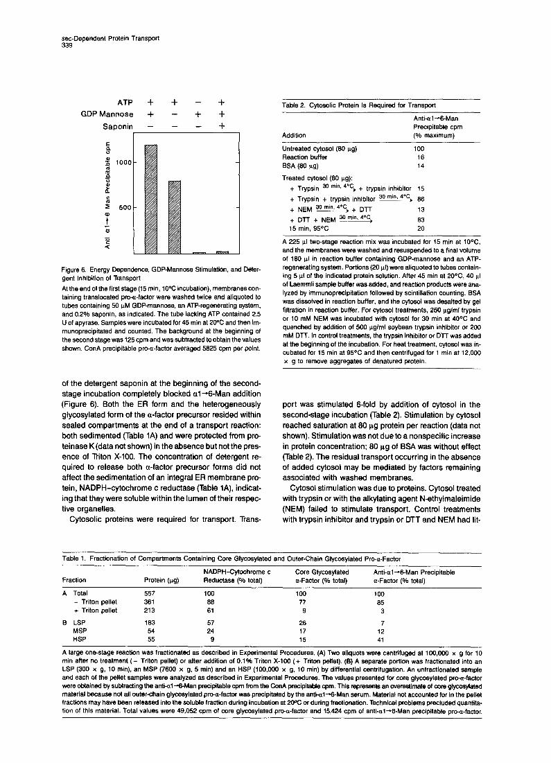
set-Dependent Protein Transport 339
ATP + + - +
GDP Mannose + - + +
Saponin - - - +
Figure 6. Energy Dependence, GDP-Mannose Stimulation, and Deter- gent Inhibition of Transport
At the end of the first stage (15 min, 10°C incubation), membranes con- taining translocated pro-a-factor were washed twice and aliquoted to tubes containing 50 uM GDP-mannose, an ATP-regenerating system, and 0.2% saponin, as indicated. The tube lacking ATP contained 2.5 U of apyrase. Samples were incubated for 45 min at 2oOC and then im- munoprecipitated and counted. The background at the beginning of the second stage was 125 cpm and was subtracted to obtain the values shown. ConA precipitable pro-a-factor averaged 5825 cpm per point.
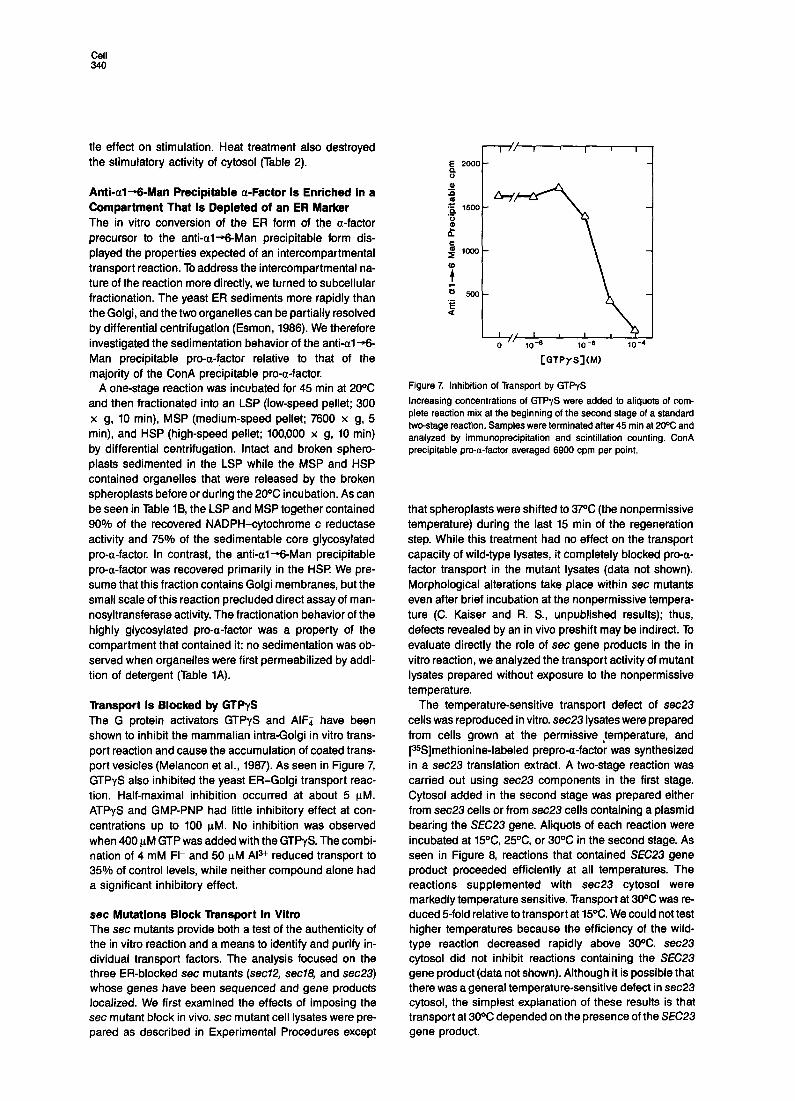
Table 2. Cytosolic Protein Is Required for Transport
Addition
Anti-al-B-Man Precipitable cpm (% maximum)
Untreated cytosol (80 ug) 100 Reaction buffer 16 BSA (‘30 m) 14
Treated cytosol (80 Kg):
+ Trypsin 3o minV 4”c) + trypsin inhibitor 15
+ Trypsin + trypsin inhibitor 3o minz 4”c) 86 + NEM 30 mh 4% + Dn 13 + DJ-T + NEM 30 mh 4”c, 83 15 min, 95OC 20
A 225 ul two-stage reaction mix was incubated for 15 min at 10°C, and the membranes were washed and resuspended to a final volume of 180 ul in reaction buffer containing GDP-mannose and an ATP- regenerating system. Portions (20 ul) were aliquoted to tubes contain- ing 5 ul of the indicated protein solution. After 45 min at 20°C, 40 ul of Laemmli sample buffer was added, and reaction products were ana- lyzed by immunoprecipitation followed by scintillation counting. BSA was dissolved in reaction buffer, and the cytosol was desalted by gel filtration in reaction buffer. For cytosol treatments, 256 &ml trypsin or 10 mM NEM was incubated with cytosol for 30 min at 40°C and quenched by addition of 500 ug/ml soybean trypsin inhibitor or 200 mM DlT. In control treatments, the trypsin inhibitor or DTT was added at the beginning of the incubation. For heat treatment, cytosol was in- cubated for 15 min at 95OC and then centrifuged for 1 min at 12,000 x g to remove aggregates of denatured protein.
of the detergent saponin at the beginning of the second-
stage incubation completely blocked al +-Man addition (Figure 6). Both the ER form and the heterogeneously glycosylated form of the a-factor precursor resided within sealed compartments at the end of a transport reaction: both sedimented (Table 1A) and were protected from pro- teinase K (data not shown) in the absence but not the pres- ence of Triton X-100. The concentration of detergent re- quired to release both a-factor precursor forms did not affect the sedimentation of an integral ER membrane pro- tein, NADPH-cytochrome c reductase (Table IA), indicat- ing that they were soluble within the lumen of their respec- tive organelles.
Cytosolic proteins were required for transport. Trans-
port was stimulated 6-fold by addition of cytosol in the second-stage incubation (Table 2). Stimulation by cytosol reached saturation at 80 ag protein per reaction (data not shown). Stimulation was not due to a nonspecific increase in protein concentration; 80 ag of BSA was without effect (Table 2). The residual transport occurring in the absence of added cytosol may be mediated by factors remaining associated with washed membranes.
Cytosol stimulation was due to proteins. Cytosol treated with trypsin or with the alkylating agent N-ethylmaleimide (NEM) failed to stimulate transport. Control treatments with trypsin inhibitor and trypsin or DTT and NEM had lit-
Table 1. Fractionation of Compartments Containing Core Glycosylated and Outer-Chain Glycosylated Pro-a-Factor
NADPH-Cytochrome c Core Glycosylated Anti-al+6Man Precipitable Fraction Protein (ug) Reductase (% total) a-Factor (o/o total) a-Factor (% total)
A Total 557 100 100 100 - Triton pellet 361 88 77 85 + Triton pellet 213 81 9 3
B LSP 183 57 26 7 MSP 54 24 17 12 HSP 55 9 15 41
A large one-stage reaction was fractionated as described in Experimental Procedures. (A) Two aliquots were centrifuged at 100,009 x g for 10 min after no treatment (- Triton pellet) or after addition of 0.1% Triton X-100 (+ Triton pellet). (8) A separate portion was fractionated into an LSP (300 Y g, 10 min), an MSP (7800 x g, 5 min) and an HSP (100,000 x g, 10 min) by differential centrifugation. An unfractionated sample and each of the pellet samples were analyzed as described in Experimental Procedures. The values presented for core glycosylated pro-a-factor were obtained by subtracting the anti-&+-Man precipitable cpm from the ConA precipitable cpm. This represents an overestimate of core glycosytated material because not all outer-chain glycosylated pro-e-factor was precipitated by the anti-al+Man serum. Material not accounted for in the pellet fractions may have been released into the soluble fraction during incubation at 2oOC or during fractionation. Technical problems precluded quantita- tion of this material. Total values were 49,052 cpm of core glycosylated pro-o-factor and 15,424 cpm of anti-al-6Man precipitable pro-a-factor.
Cdl 340
tie effect on stimulation. Heat treatment also destroyed the stimulatory activity of cytosol (Table 2).
Anti-al--Man Precipitable a-Factor Is Enriched in a Compartment That Is Depleted of an ER Marker The in vitro conversion of the ER form of the a-factor precursor to the anti-al+-Man precipitable form dis- played the properties expected of an intercompartmental transport reaction. To address the intercompartmental na- ture of the reaction more directly, we turned to subcellular fractionation. The yeast ER sediments more rapidly than the Golgi, and the two organelles can be partially resolved by differential centrifugation (Esmon, 1986). We therefore investigated the sedimentation behavior of the anti-al+- Man precipitable pro-a-factor relative to that of the majority of the ConA precipitable pro-a-factor.
A one-stage reaction was incubated for 45 min at 20°C and then fractionated into an LSP (low-speed pellet; 300 x g, 10 min), MSP (medium-speed pellet; 7600 x g, 5 min), and HSP (high-speed pellet; 100,000 x g, IO min) by differential centrifugation. Intact and broken sphero- plasts sedimented in the LSP while the MSP and HSP contained organelles that were released by the broken spheroplasts before or during the 20°C incubation. As can be seen in Table 16, the LSP and MSP together contained 90% of the recovered NADPH-cytochrome c reductase activity and 75% of the sedimentable core glycosylated pro-a-factor. In contrast, the anti-al +-Man precipitable pro-a-factor was recovered primarily in the HSP We pre- sume that this fraction contains Golgi membranes, but the small scale of this reaction precluded direct assay of man- nosyltransferase activity. The fractionation behavior of the highly glycosylated pro-a-factor was a property of the compartment that contained it: no sedimentation was ob- served when organelles were first permeabilized by addi- tion of detergent (Table 1A).
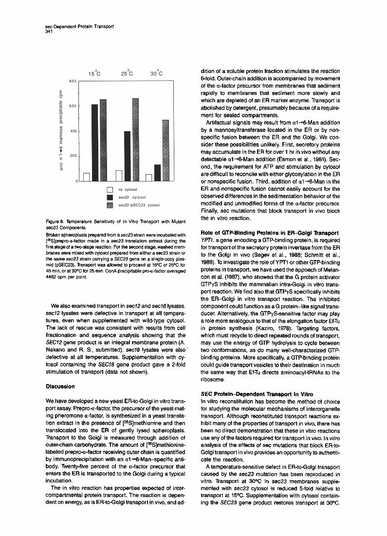
Transport Is Blocked by GTPyS The G protein activators GTPrS and AlFa have been shown to inhibit the mammalian intra-Golgi in vitro trans- port reaction and cause the accumulation of coated trans- port vesicles (Melancon et al., 1987). As seen in Figure 7, GTPyS also inhibited the yeast ER-Golgi transport reac- tion. Half-maximal inhibition occurred at about 5 PM. ATPyS and GMP-PNP had little inhibitory effect at con- centrations up to 100 FM. No inhibition was observed when 400 WM GTP was added with the GTPrS. The combi- nation of 4 mM FI- and 50 uM AP+ reduced transport to 35% of control levels, while neither compound alone had a significant inhibitory effect.
set Mutations Block Transport In Vitro The set mutants provide both a test of the authenticity of the in vitro reaction and a means to identify and purify in- dividual transport factors. The analysis focused on the three ER-blocked set mutants (sec72, sec78, and sec23) whose genes have been sequenced and gene products localized. We first examined the effects of imposing the set mutant block in vivo. set mutant cell lysates were pre- pared as described in Experimental Procedures except
g 2000 0 aI
2
w .= .P 1500 8 & $ 1000
l
.-
E
l\.! 0 10-n 10-e 10-4
CGTPYS](M)
Figure 7. Inhibition of Transport by GTPTS Increasing concentrations of GTPTS were added to aliquots of rom- plete reaction mix at the beginning of the second stage of a standard two-stage reaction. Samples were terminated after 45 min at 20% and analyzed by immunoprecipitation and scintillation counting. ConA precipitable pro-a-factor averaged 6900 cpm per point,
that spheroplasts were shifted to 5pC (the nonpermissive temperature) during the last 15 min of the regeneration step. While this treatment had no effect on the transport capacity of wild-type lysates, it completely blocked pro-a- factor transport in the mutant lysates (data not shown). Morphological alterations take place within set mutants even after brief incubation at the nonpermissive tempera- ture (C. Kaiser and R. S., unpublished results); thus, defects revealed by an in vivo preshift may be indirect. To evaluate directly the role of set gene products in the in vitro reaction, we analyzed the transport activity of mutant lysates prepared without exposure to the nonpermissive temperature.
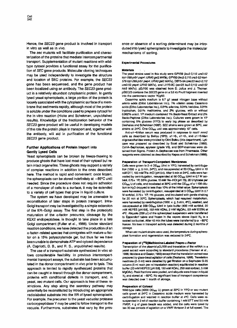
The temperature-sensitive transport defect of sec23 cells was reproduced in vitro. sec23 lysates were prepared from cells grown at the permissive ,temperature, and [35S]methionine-labeled prepro-a-factor was synthesized in a sec23 translation extract. A two-stage reaction was carried out using sec23 components in the first stage. Cytosol added in the second stage was prepared either from sec23 cells or from sec23 cells containing a plasmid bearing the SEC23 gene. Aliquots of each reaction were incubated at 15OC, 25OC, or 30X in the second stage. As seen in Figure 8, reactions that contained SEC23 gene product proceeded efficiently at all temperatures. The reactions supplemented with sec23 cytosol were markedly temperature sensitive. Transport at 30°C was re- duced 5-fold relative to transport at 15OC. We could not test higher temperatures because the efficiency of the wild- type reaction decreased rapidly above 3oOC. sec23 cytosol did not inhibit reactions containing the SEC23 gene product (data not shown). Although it is possible that there was a general temperature-sensitive defect in sec23 cytosol, the simplest explanation of these results is that transport at 30X depended on the presence of the SEC23 gene product.
.ssDependent Protein Transport
600
.-
5
(
15% 25% 3o”c
no Cytosol
sec.23 Cytosol
H sect’3 /pSEC23 cytosol
Figure 8. Temperature Sensitivity of In Vitro Transport with Mutant sec23 Components
Broken spheroplasts prepared from a sec23 strain were incubated with [%]prepro-a-factor made in a sec23 translation extract during the first stage of a two-stage reaction. For the second stage, washed mem- branes were mixed with cytosol prepared from either a sec23 strain or the same sec23 strain carrying a SEC23 gene on a single-copy plas- mid (pSEC23). Transport was allowed to proceed at 15% or 25% for 45 min, or at 30% for 25 min. ConA precipitable pro-a-factor averaged 4482 cpm per point.
We also examined transport in sec72 and sec78 lysates. sec72 lysates were defective in transport at all tempera- tures, even when supplemented with wild-type cytosol. The lack of rescue was consistent with results from cell fractionation and sequence analysis showing that the SEC72 gene product is an integral membrane protein (A. Nakano and R. S., submitted). sec78 lysates were also ’ defective at all temperatures. Supplementation with cy- tosol containing the SEC78 gene product gave a 2-fold stimulation of transport (data not shown).
Discussion
We have developed a new yeast ER-to-Golgi in vitro trans- port assay. Prepro-a-factor, the precursor of the yeast mat- ing pheromone a-factor, is synthesized in a yeast transla- tion extract in the presence of [35S]methionine and then translocated into the ER of gently lysed spheroplasts. Transport to the Golgi is measured through addition of outerchain carbohydrate. The amount of [%]methionine- labeled prepro-a-factor receiving outer chain is quantified by immunoprecipitation with an al-%-Man-specific anti- body. Twenty-five percent of the a-factor precursor that enters the ER is transported to the Golgi during a typical incubation.
The in vitro reaction has properties expected of inter- compartmental protein transport. The reaction is depen- dent on energy, as is ER-to-Golgi transport in vivo, and ad-
dition of a soluble protein fraction stimulates the reaction 6-fold. Outer-chain addition is accompanied by movement of the a-factor precursor from membranes that sediment rapidly to membranes that sediment more slowly and which are depleted of an ER marker enzyme. Transport is abolished by detergent, presumably because of a require- ment for sealed compartments.
Artifactual signals may result from al-+Man addition by a mannosyltransferase located in the ER or by non- specific fusion between the ER and the Golgi. We con- sider these possibilities unlikely. First, secretory proteins may accumulate in the ER for over 1 hr in vivo without any detectable al+Man addition (Esmon et al., 1981). Sec- ond, the requirement for ATP and stimulation by cytosol are difficult to reconcile with either glycosylation in the ER or nonspecific fusion. Third, addition of al -%-Man in the ER and nonspecific fusion cannot easily account for the observed differences in the sedimentation behavior of the modified and unmodified forms of the a-factor precursor. Finally, set mutations that block transport in vivo block the in vitro reaction.
Role of GTP-Binding Proteins in ER-Golgi Transport YPT7, a gene encoding a GTP-binding protein, is required for transport of the secretory protein invertase from the ER to the Golgi in vivo (Segev et al., 1988; Schmitt et al., 1988). To investigate the role of YF’Tl or other GTP-binding proteins in transport, we have used the appoach of Melan- con et al. (1987), who showed that the G protein activator GTPyS inhibits the mammalian intra-Golgi in vitro trans- port reaction. We find also that GTPyS specifically inhibits the ER-Golgi in vitro transport reaction. The inhibited component could function as a G protein-like signal trans- ducer. Alternatively, the GTPYS-sensitive factor may play a role more analogous to that of the elongation factor Ef-Tu in protein synthesis (Kaziro, 1978). Targeting factors, which must recycle to direct repeated rounds of transport, may use the energy of GTP hydrolysis to cycle between two conformations, as do many well-characterized GTP- binding proteins. More specifically, a GTP-binding protein could guide transport vesicles to their destination in much the same way that EfTu directs aminoacyl-tRNAs to the ribosome.
SEC Protein-Dependent Transport In Vitro In vitro reconstitution has become the method of choice for studying the molecular mechanisms of interorganelle transport. Although reconstituted transport reactions ex- hibit many of the properties of transport in vivo, there has been no direct demonstration that these in vitro reactions use any of the factors required for transport in vivo. In vitro analysis of the effects of set mutations that block ER-to- Golgi transport in vivo provides an opportunity to authenti- cate the reaction.
A temperature-sensitive defect in ER-to-Golgi transport caused by the sec23 mutation has been reproduced in vitro. Transport at 30% in sec23 membranes supple- mented with sec23 cytosol is reduced 5-fold relative to transport at 15%. Supplementation with cytosol contain- ing the SEC23 gene product restores transport at 30%.
Hence, the SEC23 gene product is involved in transport in vitro as well as in vivo.
The set mutants will facilitate purification and charac- terization of the proteins that mediate intercompartmental transport. Supplementation of mutant reactions with wild- type cytosol provides a functional assay for the purifica- tion of SEC gene products. Molecular cloning techniques may be used independently to investigate the structure and location of SEC proteins. For example, the SEC23 gene has been sequenced, and the gene product has been localized using an antibody. The SEC23 gene prod- uct is a relatively abundant cytoplasmic protein. In gently lysed yeast spheroplasts, a large portion of the protein is loosely associated with the cytoplasmic surface of a mem- brane that sediments rapidly, although most of the protein is soluble under the conditions used to prepare cytosol for the in vitro reaction (Hicke and Schekman, unpublished results). Knowledge of the fractionation behavior of the SEC23 gene product will be useful in developing models of the role the protein plays in transport and, together with the antibody, will aid in purification of the functional SEC23 gene product.
Further Applications of Protein Import into Gently Lysed Cells Yeast spheroplasts can be broken by freeze-thawing to produce ghosts that have lost most of their cytosol but re- tain intact organelles. These ghosts may support a variety of complex reactions in addition to the ones described here. The method is rapid and convenient: once frozen, the spheroplasts can be stored indefinitely at -85°C until needed. Since the procedure does not require adhesion of a monolayer of cells to a surface, it may be extended to a variety of cell types that grow in liquid culture.
The system we have developed may be amenable to reconstitution of later steps in protein transport. Intra- Golgi transport may be investigated by a simple extension of the ER-Golgi assay. The first step in the proteolytic maturation of the a-factor precursor, cleavage by the KEX2 endopeptidase, is thought to take place in a late Golgi compartment (Fuller et al., 1988). Under standard reaction conditions, we have detected the production of an a-factor-related species that comigrates with mature a-fac- tor on a 15% polyacrylamide gel, but thus far we have been unable to demonstrate ATP and cytosol dependence (A. Capriotti, D. B., and R. S., unpublished results).
The use of a transport substrate synthesized in vitro al- lows considerable flexibility. In previous intercompart- mental transport assays, the substrate has been accumu- lated in the donor compartment in vivo prior to lysis. This approach is limited to rapidly synthesized proteins that can be caught in transit through the donor compartment, proteins with conditional defects in transport, and, in yeast, set mutant cells. Our approach is free of these re- strictions. Any step along the secretory pathway may potentially be reconstituted by introducing an appropriate radiolabeled substrate into the ER of lysed spheroplasts. For example, the precursor to the yeast vacuolar protease carboxypeptidase Y may be used to follow transport to the vacuole. Furthermore, substrates that vary by the pres-
ence or absence of a sorting determinant may be intro- duced into lysed spheroplasts to investigate the molecular mechanisms of sorting.
Experlmental Procedures
Materials _ The yeast strains used in this study were GPY59 (/e&b172 ~83-52 trpl-289 prbl pep4::UkU ga12 MATa), GPY60 (/I%&3, 172 ura3-52 hid- 579 trpl-289prbl pep4::UFUN gal2 MATa), DBY5-3A (se023 leu2-3,112 ura3-52 pep4::URA3 MATa). and LHY3-6C (sec23 leu2-3,112 uta3-52 his3 MATa). pDJlO0 was obtained from D. Julius and J. Thorner. pSEC23 contains the SEC23 gene on a 5.5 kb Pvull fragment inserted into the centromere vector YCp50.
Casamino acids medium is 6.7 g/l yeast nitrogen base without amino acids (Difco Laboratories Inc.), 1% vitamin assay Casamino acids (Difco Laboratories Inc.), 0.01% adenine, 0.01% histidine, 0.01% tryptophan, 0.01% methionine, and 2% glucose, with or without 0.002% uracil. YP medium contained 1% Bacto-Yeast Extract and 2% Bacto-Peptone (Difco Laboratories Inc.). Cultures were grown in YP containing 5% glucose (YPD) to early log phase as described by Deshaies and Schekman (1967). SEC strains were grown at 3ooC, set strains at 24%. One ODsoo unit was approximately 10’ cells.
Anti-al--Man serum was produced in response to mnfll mnn2 cells as described by Ballou (1970). al-+ a1+2-, and al-+-Man oligosaccharides were provided by Clint Ballou (this department). Lyti- case was prepared as described by Scott and Schekman (1960). ConA-Sepharose, apyrase (grade VII), and GDP-mannose were ob- tained from Sigma. Protein A-Sepharose was from Pharmacia. Other reagents were obtained as described by Kepes and Schekman (1966).
Pmparatlon of BansPort-CornP&N Membmnes Cells were grown to 2-4 00, U/ml in YPD, harvested by centrifuga- tion (1000 x g, 5 min, 24Dc), and resuspended at 50 ODm U/ml in 10 mM DTT, 100 mM Tris-HCI (pH 9.4). After 6 min at 24OC, cells were har- vested by centrifugation, resuspended at 50 ODm U/ml in 0.7 M sor- bii, 0.75x VP, 05% glucose, 10 mM liis-HCl (pli 7.5), 20 U lyticase per ODBoo U of cells, and incubated at 3ooc until the ODm of a 1:lOO dilu- tion in t&C dropped to less than 10% of the initial value. Spheroplasts were harvested by centrifugation, resuspended at 5 ODm U/ml in 0.7 M sorbitol, 0.75x VP: 1% glucose, and incubated with gentle shaking for 20 min at 3oOC. Spheroplasts, which had resumed metabolism, were harvested by centrifugation (1000 x g, 5 min, 4oC), washed, and resuspended at 300 ODeoo U/ml in lysis buffer (400 mM sorbitol, 20 mM HEPES [pH 6.6],150 mM KOAc, 2 mM MgOAc, 0.5 mM EGTA) at 4OC. Aliquots (200 )II) of the spheroplast suspension were transferred to Eppendorf tubes and frozen in .the vapors above liquid N2 in a sealed ice bucket. After 45 min the tubes were transferred to a -85OC freezer. No loss in transport activity was detected during 2 months of storage.
When set mutant strains were used, the temperature during sphero- plast formation and regeneration was reduced to 24%.
Preparation of [3SS]Methlonlne-Labeled Pnpm-a-Mctor Transcription of the plasmid pDJlO0 and translation of the mRNA in a yeast extract were according to standard procedures (Hansen et al., 1986; Moldave and Gasior. 1963)excepl that the translation extract was prepared by glass-bead agitation of cells (Deshaies, 1968). Translation reactions (2-5 ml) were desalted by gel filtration on a Sephadex G-25 column (5 ml resin per ml translation reaction) equilibrated in reaction buffer (20 mM HEPES [pH 6.6],150 mM KOAc, 250 mM sorbitol, 5 mM MgOAc). Peak fractions were pooled, and aliquots were frozen in liquid N2 and stored at -65OC. No significant loss of transport competence was detected over 1 month of storage.
Pmprrretion of Cytosol Wild-type cells (4000 ODBoo U) grown at 30% in YPD or set mutant cells grown at 24OC in Casamino acids medium were harvested by centrifugation and washed in reaction buffer at 4%. Cells were re- suspended in 2 ml of reaction buffer containing 1 mM DTT and 0.6 mM PMSF, 4 g of glass beads was added, and the cells were lysed by ten 30 set periods of agitation on a VWR Vortexer 2 at full speed. The
se&Dependent Protein Transport 343
homogenate was clarified by centrifugation at 3000 x g for 5 min and the supernatant fraction further centrifuged at 100,000 x g for 30 min. The resultant 5100 fraction was either desalted by filtration on Sepha- dex G-25 equilibrated in reaction buffer and then frozen in aliquots in liquid NZ, or was aliquoted and frozen immediately. Filtered and un- filtered cytosol fractions stimulated transport to the same extent and were stable over several months of storage at -8VC. Protein concen- tration ranged from 20 to 30 mglml.
In Vitro Transport Reaction For each experiment an aliquot of gently lysed yeast was thawed by immersion in a 25OC water bath, and the required amount of mem- branes (20 pi per reaction) was washed three times by brief (~10 set) centrifugation in a Fisher microcentrifuge and resuspension in 1 ml of reaction buffer at 4OC.
One-stage reactions contained 5 ul of prepro-a-factor translation product (150,000 trichloroacetic acid precipitable cpm), 60 ug of addi- tional cytosol. 50 uM GDP-mannose, 1 mM ATP, 40 mM creatine phos- phate (CP), 200 ug/ml creatine phosphokinase (CPK), and 20 ul (origi- nal volume) of membranes washed as above and resuspended in reaction buffer to bring the reaction volume to 25 ~1. The GDP- mannose, ATP, CP, and CPK were added from a 10x stock solution prepared in reaction buffer and stored in small aliquots at -65OC. After 45 min at 2ooC. reactions were terminated by addition of 40 ul of Laemmli (1970) sample buffer and heated for 5 min at 95OC.
The first stage of two-stage reactions contained 6 ul of prepro-a- factor translation product, 50 pM GDP-mannose, 1 mM ATR 40 mM CR 200 ug/ml CPK, and 20 ul (original volume) of membranes washed as above and resuspended in reaction buffer to bring the reaction volume to 25 ul. The reaction mix was incubated for 15 min at loOC, and the membranes were washed two times by centrifugation and resuspen- sion in reaction buffer at 4OC. A complete second-stage incubation, in a final volume of 25 ~1, contained the following: washed pro-a- factor-containing membranes, 60 ug of cytosol, 1 mM ATR 50 uM GDP-mannose, 40 mM CP, and 200 uglml CPK. Typically, eight to ten reactions were combined in one tube during the first stage, and the washed pro-a-factor-containing membranes were aliquoted to in- dividual tubes with various additions at the beginning of the second- stage incubation. After 45 min at 20°C, the reactions were terminated by addition of Laemmli sample buffer and heated for 5 min at 95’C.
lmmunopreclpitation A 20 ul portion of a terminated reaction was mixed with 35 ul of a 20% (volhrol) suspension of Protein A-Sepharose, 6-10 ul of anti-al +-Man serum or 6 ul of anti-a-factor serum, and I ml of IP buffer (150 mM NaCI, 1% Triton X-100, 0.1% SDS, 15 mM Tris-HCI [pH 7.51) in an Ep- pendorf tube and rotated for 2 hr at room temperature or overnight at ’ 4OC. The immunoprecipitates were collected by centrifugation and washed twice with 1 ml of IP buffer; twice with 1 ml of 2 M urea, 200 mM NaCI, 1% Triton X-100, 100 mM Ths-HCI (pH 7.5); once with 1 ml of 500 mM NaCI, 1% Triton X-100, 20 mM Tris-HCI (pH 7.5); and once with 1 ml of 50 mM NaCI, 10 mM Tris-HCI (pH 7.5).
The washed immunoprecipitates were analyzed either by elec- trophoresis and autoradiography or directly by scintillation counting. lmmunoprecipitates to be analyzed by electrophoresis were heated at 95°C in Laemmli sample buffer and electrophoresed on an 11.25% SDS-polyacrylamide gel (Laemmli, 1970). The gel was treated with Amplify after fixation, dried, and exposed to X-ray film. Immunoprecipi- tates to be analyzed by scintillation counting were heated for 5 min at 95OC in 150 PI of 2% SDS, then transferred to 6 ml scintillation vials. Five milliliters of Aquasol (New England Nuclear) was added to each vial, and the samples were counted in a scintillation counter. The back- ground (typically around 100 cpm) was determined by counting im- munoprecipitates of complete reactions that had been stopped by ad- dition of Laemmli sample buffer at the start of the incubation, and was subtracted from all reported values.
For ConA precipitations, IO ul of a terminated reaction and 30 ul of a 20% (vollvol) suspension of ConA-Sepharose were added to 1 ml of 500 mM NaCI, 1% Triton-X-100. 20 mM Tris-HCI (pH 7.5) and sam- ples were rotated for 2 hr at room temperature or overnight at 4°C. The washes and subsequent analysis were as described for anti- body precipitations. Background, determined as above, was typically around 300 cpm.
Fractionation A one-stage reaction scaled up 6-fold and a parallel mock reaction with desalted cytosol substituted for the prepro-a-factor translation product were incubated for 45 min at PC and then each processed as follows: A 25 ul aliquot treated with 0.1% Triton X-100 and an untreated 25 ul aliquot were diluted to 200 ul with lysis buffer and centrifuged at l8C@JO x g for 10 min. One hundred microlitersof the remaining reac- tion mix was diluted to 1 ml with lysis buffer and centrifuged for 10 min at 300 x g. The LSP was saved and the supernatant fraction cen- trifuged for 5 min at 7600 x g. The MSP was saved and the superna- tant fraction centrifuged for 10 min at 100,000 x g to give the HSP An unfractionated sample and each of the pellet fractions were analyzed by anti-al%Man or ConA precipitation following solubilization in Laemmli sample buffer (complete reaction). Fractions from the mock reaction were used to measure protein (Markwell et al., 1976) and NADPH-cytochrome c reductase (Kubota et al., 1977).
Electron Microscopy Samples were fixed in a mixture of 0.2% paraformaldehyde, 2.5% glutaraldehyde, and 0.1% picric acid at pH 7.2 for 32 hr, followed by 0.9% NaCl washes and fixation in uranyl acetate and then 2% 0~0~. Dehydration was followed by Medcast (Pelco) embedding. Silver sec- tions stained 1 min in lead citrate were photographed in a Philips 301 electron microscope.
Acknowledgments
We thank Carolyn Connell for expert assistance with the electron mi- croscopy, Ann Capriotti for investigating KEX2 processing in vitro, Pam Esmon for the anti-al +-Man serum, Jon Rothblatt for the anti-a-factor serum, Clint Ballou for advice and materials, and Robin Wright for as- sistance in evaluating lysed spheroplasts by light microscopy. We also thank Peggy McCutchan Smith for help in preparing the manuscript. This work was supported by grants from the National Institutes of Health (NIGMS). D. 8. and L. H. were supported by National Science Foundation predoctoral fellowships.
The costs of publication of this article were defrayed in part by the payment of page charges. This article must therefore be hereby marked “adverfisement” in accordance with 16 U.S.C. Section 1734 solely to indicate this fact.
Received April 13, 1988; revised May 11, 1988
References
Balch, W. E., Elliott, M. M., and Keller, D. S. (1986). ATP-coupled trans- port of vesicular stomatitis virus G protein between the endoplasmic reticulum and the Golgi. J. Biol. Chem. 267, 14661-14689.
Ballou, C. (1970). A study of the immunochemistry of three yeast man- nans. J. Biol. Chem. 245, 1197-1203.
Beckers, C. J. M., Keller, D. S., and Balch, W. E. (1967). Semi-intact cells permeable to macromolecules: use in reconstitution of protein transport from the endoplasmic reticulum to the Golgi complex. Cell 50, 523-534.
Deshaies, R. J. (1988). Genes required for protein translocation in the yeast endoplasmic reticulum. Ph.D. thesis, University of California, Berkeley.
Deshaies, Ft. J., and Schekman, Ft. (1987). A yeast mutant defective of an early stage in import of secretory protein precursors into the en- doplasmic reticulum. J. Cell Biol. 105, 633-845.
Esmon, f? C. (1986). Molecular and organelle intermediates in the secretory process, Ph.D. thesis, University of California, Berkeley.
Esmon, B., Novick, P, and Schekman, Ft. (1961). Compartmentalized assembly of oligosaccharides on exported glycoproteins in yeast. Cell 25, 451-460.
Fuller, R. S., Sterne, R. E., and Thorner, J. (1988). Enzymes required for yeast pheromone processing. Annu. Rev. Physiol. 50. 345-362.
Hansen, W., Garcia, P D., and Walter, P. (1966). In vitro protein translo- cation across the yeast endoplasmic reticulum: ATP-dependent post- translational translocation of the prepro-a-factor. Cell 45, 397-406.
Haselbeck, A., and Schekman, R. (1966). lnterorganelle transfer and
Cdl 344
glycosylation of yeast invertase in vitro. Proc. Natl. Acad. Sci. USA 83, 2017-2021.
Julius, D., Schekman, R., and Thorner, J. (1984). Glycosylation and processing of prepro-a-factor through the yeast secretory pathway. Cell 36, 309-318.
Kaziro, Y. (1978). The role of guanosine B’-triphosphate in polypeptide chain elongation. Biochim. Biophys. Acta 505, 95-127.
Kepes, F, and Schekman, Ft. (1986). The yeast SEC53 gene encodes phosphomannomutase. J. Biol. Chem. 263, 9155-9161.
Kubota, S., Yoshida, Y., Kumaoka, H., and Furumichi, A. (1977). Studies on the microsomal electron-transport system of anaerobically grown yeast. V. Purification and characterization of NADPH- cytochrome c reductase. J. Biochem. 187, 197-206.
Kukuruzinska, M. A., Bergh, M. L. E., and Jackson, B. J. (1987). Pro- tein glycosylation in yeast. Annu. Rev. Biochem. 56, 915-944.
Laemmli, U. K. (1970). Cleavage of structural proteins during the as- sembly of the head of bacteriophage T4. Nature 227, 680-685.
Markwell, M. A. K., Haas, S. hf., Bieber, L. L., and Tolbert, N. N. (1978). A modification of the Lowry procedure to simplify protein determination in membrane and lipoprotein samples. Anal. Biochem. 7, 206-210.
Melancon, i?, Glick, B. S., Malhotra, V., Weidman, P J., Serafini, T., Gleason, M L., Orci, L., and Rothman, J. E. (1987). Involvement of GTP-binding “G” proteins in transport through the Golgi stack. Cell 57, 1053-1062.
Moldave, K., and Gasior, E. (1983). Preparation of a cell-free system from S. cerevisiae that translates exogenous messenger ribonucleic acids. Meth. Enzymol. 107, 644-650.
Newman, A. P, and Ferro-Novick, S. (1987). Characterization of new mutants in the early part of the yeast secretory pathway isolated by a [3H]mannose suicide selection. J. Cell Biol. 705, 1587-1594.
Novick, f?, Field, C., and Schekman, R. (1980). Identification of 23 com- plementation groups required for post-translational events in the yeast secretory pathway. Cell 21, 205-215.
Novick, f?, Ferro, S., and Schekman, R. (1981). Order of events in the yeast secretory pathway. Cell 25, 461-469.
Pfeffer, S., and Rothman, J. (1987). Biosynthetic protein transport and sorting by the endoplasmic reticulum and the Golgi. Annu. Rev. Bio- them. 56, 829-852.
Rothblatt, J., and Meyer, D. I. (1986). Secretion in yeast: translocation and glycosylation of prepro-a-factor in vitro can occur via an ATP- dependent post-translational mechanism. EMBO J. 5, IO”*-IO: 3.
Schmitt, H. D., Puzicha, M., and Gallwitz, D. (1988). Study of a temperature-sensitive mutant of the ras-related YP77 gene product in yeast suggests a role in the regulation of intracellular calcium. Cell 53, 635-647.
Scott, J., and Schekman, R. (1980). Lyticase: endoglucanase and pro- tease activities that act together in yeast cell lysis. J. Bacterial. 142, 414-423.
Segev, N., Mulholland, J., and Botstein, D. (1968). The yeast GTP- binding YPTI protein and a mammalian counterpart are associated with the secretion machinery. Cell 52, 915-924.
Simons, K., and Virta, H. (1967). Perforated MDCK cells support intra- cellular transport. EMBO J. 6, 2241-2247.
Tartakoff, A. M. (1986). Temperature and energy dependence of secre- tory protein transport in the exocrine pancreas. EMBO J. 5,1477-1482.
Waters, M. G., and Blobel, G. (1986). Secretory protein translocation in a yeast cell-free system can occur posttranslationally and requires ATP hydrolysis. J. Cell Biol. 102, 1543-1550.
Note Added in Proof
The work referred to as A. Nakano and R. S., submitted, is now in press: Nakano, A., Brada, D., and Schekman, R. (1988). A membrane glycoprotein, SeclPp, required for protein transport from the endoplas- mic reticulum to the Golgi apparatus in yeast. J. Cell Biol., in press.