color changing photonic crystals detect blast exposure
TRANSCRIPT
Color changing photonic crystals detect blast exposure
D. Kacy Cullen1, Yongan Xu2, Dexter V. Reneer3, Kevin D. Browne1, James W. Geddes3,Shu Yang2, and Douglas H. Smith11 Center for Brain Injury and Repair, Dept. of Neurosurgery, School of Medicine, University ofPennsylvania, 105 Hayden Hall, 3320 Smith Walk, Philadelphia, PA 19104, USA2 Dept. of Materials Science and Engineering, School of Engineering and Applied Science,University of Pennsylvania, 3231 Walnut Street, Philadelphia, PA 19104, USA3 Spinal Cord and Brain Injury Research Center, Dept. of Anatomy and Neurobiology, School ofMedicine, University of Kentucky, Chandler Medical Center, B477 Biomed & Biol ScienceResearch Bldg, 741 S. Limestone St., Lexington, KY 40536, USA
AbstractBlast-induced traumatic brain injury (bTBI) is the “signature wound” of the current wars in Iraqand Afghanistan. However, with no objective information of relative blast exposure, warfighterswith bTBI may not receive appropriate medical care and are at risk of being returned to thebattlefield. Accordingly, we have created a colorimetric blast injury dosimeter (BID) that exploitsmaterial failure of photonic crystals to detect blast exposure. Appearing like a colored sticker, theBID is fabricated in photosensitive polymers via multi-beam interference lithography. Althoughvery stable in the presence of heat, cold or physical impact, sculpted micro- and nano-structures ofthe BID are physically altered in a precise manner by blast exposure, resulting in color changesthat correspond with blast intensity. This approach offers a lightweight, power-free sensor that canbe readily interpreted by the naked eye. Importantly, with future refinement this technology maybe deployed to identify soldiers exposed to blast at levels suggested to be supra-threshold for non-impact blast-induced mild TBI.
IntroductionBlast-induced traumatic brain injury (bTBI) is a major source of battlefield morbidity in thecurrent wars in Iraq and Afghanistan (Okie, 2005, Warden et al., 2005, Taber et al., 2006,Warden, 2006). This reflects a stunning number of warfighters who have been exposed toblast shockwave, typically from improvised explosive devices (IEDs). However, little isknown about blast exposure thresholds that induce bTBI. Indeed, many warfighters whodisplay either no overt symptoms or only minor cognitive deficits after blast exposure maynonetheless have suffered brain damage (Ling, 2008). With no objective information ofrelative blast exposure, warfighters with bTBI may not receive appropriate acute or chroniccare. Furthermore, if returned to service, they may be at risk of an exacerbated response withrepetitive blast exposure, as is the case in repetitive head injury in sports (Guskiewicz et al.,
*Corresponding Author: Douglas H. Smith, M.D., Director of PENN’s Center for Brain Injury and Repair, The Robert A. GroffProfessor of Neurosurgery, Vice Chairman for Research and Education, Dept. of Neurosurgery, University of Pennsylvania 105Hayden Hall, 3320 Smith Walk Philadelphia, PA 19104, USA [email protected] 215-898-0881 (phone) 215-573-3808(fax).Publisher's Disclaimer: This is a PDF file of an unedited manuscript that has been accepted for publication. As a service to ourcustomers we are providing this early version of the manuscript. The manuscript will undergo copyediting, typesetting, and review ofthe resulting proof before it is published in its final citable form. Please note that during the production process errors may bediscovered which could affect the content, and all legal disclaimers that apply to the journal pertain.
NIH Public AccessAuthor ManuscriptNeuroimage. Author manuscript; available in PMC 2012 January 1.
Published in final edited form as:Neuroimage. 2011 January ; 54S1: S37–S44. doi:10.1016/j.neuroimage.2010.10.076.
NIH
-PA Author Manuscript
NIH
-PA Author Manuscript
NIH
-PA Author Manuscript
2003, Mori et al., 2006). Accordingly, there is a critical need for a wearable sensor capableof registering the severity of blast exposure in relation to the risk of bTBI.
To address these needs, our objective is to utilize a materials-based strategy that directlyexploits blast energy to induce optical changes in photonic crystalline microstructures. Thelevel of blast exposure would then be observed based on a visible color change. Thistechnology may be used to develop a small wearable blast injury dosimeter (BID) to readilydesignate soldiers exposed to blast conditions associated with TBI and other injuries (patentpending). For this application, we utilized 3-D photonic crystalline microstructures that werefabricated via multi-beam interference lithography (MBIL) of a commercially available,negative-tone photoresist, SU-8, using a visible (λ=532 nm) laser (Campbell et al., 2000,Yang et al., 2002, Miklyaev et al., 2003, Moon and Yang, 2005, Xu et al., 2008). SU-8 is abisphenol-A novolac resin derivative with an average of eight epoxy groups per chain. SU-8is highly soluble in many organic solvents, enabling preparation of ultra-thick films (up to 2mm) that are highly transparent in the near-UV and visible region. SU-8 has been widelyused in microelectromechanical systems (MEMS) (Lorenz et al., 1997), microfluidics(Ribeiro et al., 2005), high-aspect ratio (≥20) microstructures (Lee et al., 1994), and 3-Dphotonic structures (Campbell et al., 2000, Yang et al., 2002, Miklyaev et al., 2003, Moon etal., 2006). Crystalline films made from SU-8 create a colorful reflection due to thediffraction grating of the underlying pattern, which is characteristic of the periodicity (~ 1μm) and refractive index contrast between high (SU-8, n = 1.6) and low (air, n = 1)dielectric materials, and the viewing angle. Importantly, the SU-8 photonic crystals arethermally stable up to 300°C and chemically inert due to aromatic functionality and highcross-link density. Thus, they are highly durable under extreme weather conditions (e.g.,heat, cold, and moisture) or physical impact associated with combat situations. Moreover,the small, lightweight design can easily be accommodated across multiple locations on andin helmets and uniforms, thus rendering the BID useful for in-field interpretation (Fig. 1).
The objective of the current study was to establish proof-of-principle data that our photoniccrystalline microstructures respond specifically to blast exposure. In particular, we assessedcolorimetric changes in custom-engineered photonic crystals based on dynamic overpressureexposure, and correlated this color change with ultrastructural alterations and damage. Thisdemonstration is necessary to enable the ultimate application of this device as a means tomeasure single or cumulative blast exposure supra-threshold for bTBI. Thus, future studieswill directly calibrate these color changes to potentially unique blast-inducedneuropathology to fulfill our objective for a material-based colorimetric Blast InjuryDosimeter.
MethodsPhotonic Crystal Fabrication
Diamond-like photonic crystals consisting of periodic arrangement of polymer and air voidswere fabricated from the negative photoresist, SU-8, by MBIL using the same optical setupreported earlier (Xu et al., 2008). Briefly, SU-8 film was exposed to four umbrella-likevisible laser beams split from one coherent laser source (λ=532nm, power diode-pumpedNd:YVO4 laser). The central beam was circularly polarized and incident perpendicularly tothe photoresist film. The other three beams were polarized linearly in a plane formed by thewave vectors of the central beam and surrounding beam. The wave vector of each beam wask0=π/α[3 3 3], k1=π/α[5 1 1] k2=π/α [1 5 1], and k3=π/α [1 1 5], respectively. Thepolarization vectors of beam 1, 2, and 3 were e1 = [−0.2720.6800.680], e2 =[0.680–0.2720.680], and e3 = [0.6800.680–0.272], respectively. The intensity ratio was 1.8:1:1:1.The circular polarization of the central beam distributes the intensity equally to thesurrounding beams.
Cullen et al. Page 2
Neuroimage. Author manuscript; available in PMC 2012 January 1.
NIH
-PA Author Manuscript
NIH
-PA Author Manuscript
NIH
-PA Author Manuscript
The photoresist was prepared by mixing Epon SU-8 pellets and 2.0 wt % Irgacure 261 (CibaSpecialty Chemicals) as visible photoinitators in γ-butyrolactone (Aldrich) to form 58 wt%solution. Substrates were transparent glass or flexible aclar membranes (SPI Supplies). Toensure good adhesion with the SU-8 film, the substrate was cleaned by ultrasonication inisopropanol and acetone, respectively, followed by oxygen plasma. The photoresist solutionwas spin-coated on the substrate at 2000 rpm for 30 seconds, followed by pre-exposure bakeat 65°C for 3 minutes and 95°C for 40 minutes, respectively, resulting in a film thickness of~6 μm. The film was exposed to the superimposed interference beams (laser output of 1W)for 1–2 seconds. After post-exposure bake at 65°C for 2–4 minutes and 95°C for 2–4minutes, respectively, the exposed film was developed in propylene glycol monomethylether acetate (Aldrich) to remove unexposed or weakly exposed films, resulting in 3-Dmicroporous structures. To prevent the pattern collapse of the 3-D porous film during air-drying, the film was dried using dryer (SAMDRI®-PVT-3D; Tousimis) after thedevelopment.
Colorimetric PropertiesPhotonic crystalline colorimetric properties are an inherent consequence of the periodicmodulation of refractive index arranged in 3-D, where interference of the light waves leadsto stop bands or photonic band gaps. This results in light of a particular range of wavelengthbeing totally reflected in a photonic crystal. Briefly, when light arrives at the surface of theperiodic structures, it is strongly reflected by constructive interference between reflectionsfrom the different interfaces of a stack of thin films (thickness of d) of alternately high andlow refractive index (n), resulting in so-called structural color. According to Bragg’s Law, atthe normal incidence to the (111) plane the reflectance peak wavelength is
[1]
where d111 is the interlayer distance in the [111] direction, and neff is the effective refractiveindex of the film:
[2]
where n1 and n2 are the refractive index of component 1 and 2, respectively, and f1 is thefilling volume fraction of component 1. Here, nSU 8 =1.6 and nair =1.
BID TestingFor design feedback, we evaluated the structural/colorimetric alterations of the photoniccrystalline microstructures following exposure to surrogate blast conditions from (1)targeted blast-like overpressure from single-pulse ultrasonic irradiation, or (2) blast from anexplosive-based shocktube. Single-pulse ultrasonic irradiation. Blast-like overpressureexposure was generated using a modified piezoelectric transducer to generate a rapid, singlepulse (100–200 ms in duration) applied focally using a sonication wand (Fisher ScientificModel 100 Sonic Dismembrator). This employed locally applied stress waves toapproximate the effects of a globally applied blast wave. This process generated extremelyrapid pressure fluctuations that approximate some facets of blast exposure; specifically, bothmay exhibit extreme overpressure magnitudes (up to 1–10 MPa) with rapid pressure changerise-times on the order of ten microseconds. The exposure intensity was based on the poweroutput from the device and ranged from 800 W/m2 to 8000 kW/m2. Explosive-drivenshocktube. The shocktube was cylindrical (21″ L × 6.5″ ID) with a conclave enclosure at one
Cullen et al. Page 3
Neuroimage. Author manuscript; available in PMC 2012 January 1.
NIH
-PA Author Manuscript
NIH
-PA Author Manuscript
NIH
-PA Author Manuscript
end. The explosive material was a gaseous mixture of hydrogen and oxygen generated in acontrolled quantity by the electrolysis of H2O (based on the time and amperage). Theexplosion was initiated by firing a small cordite charge into the collected gases, the quantityof which determined the magnitude of the blast conditions (e.g., peak overpressure). Foreach experiment, high frequency pressure transducers (500 kHz sampling rate; PCBPiezotronics) positioned immediately adjacent to BID arrays measured the local blastparameters (pressure-time functions) face-on. Each sensor was connected via coaxial cableto a PCB signal conditioner (482A21), and then to a digital oscilloscope (DSO-2250, 100MHz bandwidth, 250 MS/s real-time sampling) and computer. The sensor voltage wasconverted into pressure based on the sensor-specific calibration. Following these exposuresto dynamic overpressure fluctuations, alterations in the colorimetric properties andmicrostructure were assessed. Optical Imaging. Light images were taken for each BIDbefore and after surrogate blast or control (sham) conditions. Images were acquired by adigital camera (Sony) mounted on a stereoscope at 10x magnification (Nikon SMZ645).Colorimetric surface plots were generated using ImagePro Plus (Media Cybernetics). Forthese surface plots, the mean pixel intensity was calculated on a point-by-point basis andplotted on the z-axis to create a three-dimensional visualization (with x and y representingposition on the BID surface) of photonic crystalline color changes due to blast exposure.Scanning Electron Microscopy (SEM). SEM ultrastructural analysis was performed for eachBID after surrogate blast or control conditions. High-resolution SEM images were acquiredusing an Strata DB235 Focused Ion Beam system (FEI) at an e-beam voltage of 5 kV. Priorto SEM analysis, the samples were sputter coated with gold (thickness <10nm).
Results and DiscussionPhotonic Crystal Fabrication and Structural/Colorimetric Properties
The premise behind this BID is that supra-threshold blast exposure induces physicalalterations in the photonic crystalline microstructures that manifest as color changes basedon the level of exposure. The BIDs are comprised of arrays of diamond-like photoniccrystals with nano-scale features that reflect light in specific wavelengths across the colorspectrum. These photonic crystalline microstructures were fabricated on glass or thinflexible polymer sheets (<1cm2). Macroscopically, the BIDs resembled small coloredstickers with an overall diameter ranging from 1.0 – 6.5 mm. The final engineeredmicrostructures consisted of several one-micron thick layers (total thickness was typically 6microns) with readily observed colorimetric properties (Fig. 2).
Importantly, the top-down 3-D lithographic method we employed enables precise control ofthe periodic structures at the nano- and micro-scales, including symmetry, periodicity,overall porosity and pore size, and film thickness. Moreover, the response to external energycan be tailored by these 3-D structural and material properties (e.g., Young’s modulus, andthermal conductivity). We exploited this flexibility in fabrication to create microstructuresspecifically tailored for our application of blast exposure detection. In turn, the 3-Dultrastructure determines the colorimetric properties. The key feature of the BID is to exploitblast-induced nano-scale structural alterations to create a color change relative to theseverity of the blast.
BID Testing Using Surrogate Blast ConditionsIn order to apply these 3-D photonic crystals as a BID, we evaluated the physical alterationsand corresponding color change following exposure to dynamic overpressure via (1)targeted blast-like overpressure from single-pulse ultrasonic irradiation, and (2) blast froman explosion-based shocktube. We used performance feedback to engineer BIDs thatexhibited overt colorimetric alterations following blast exposure. Single-pulse ultrasonic
Cullen et al. Page 4
Neuroimage. Author manuscript; available in PMC 2012 January 1.
NIH
-PA Author Manuscript
NIH
-PA Author Manuscript
NIH
-PA Author Manuscript
irradiation induced differential colorimetric and structural alterations proportional to pulsepeak overpressure. BIDs exhibited complete color loss across the entire surface of thematerial with modest material loss at the edges following exposure intensity of 320 kW/m2
(Fig. 3A). With increased dynamic overpressure intensity to 960 kW/m2, BIDs demonstrateda similar color loss across the surface but had an increase in material loss at the center andedges (Fig. 3B). Additionally, colorimetric surface plots were used to map the mean pixelintensity across the face of the samples. This technique demonstrated a precipitousdepression in mean pixel intensity across the entire surface of the samples, with even moredramatic changes in regions potentially experiencing material loss (e.g., center and edges).
An explosive-driven shocktube was then utilized to refine BID responses to more realisticblast conditions. The explosion in this cylindrical shocktube was driven by ignition of agaseous hydrogen-oxygen mixture, generating pressure-time waves that were very similar tothat produced by high-energy plastic explosives (Loubeau et al., 2006, Bauman et al., 2009).This high fidelity blast shockwave consisted of microsecond-scale pressure rise-times andmillisecond-scale overpressure/underpressure components. Traditionally, blast injurythresholds have been based on exposure levels inducing lung damage (e.g., peak incidentpressure, time duration, and subject proximity to reflective surface); however, soldiers arenow surviving more powerful explosions due to advances in body armor and rapid medicalintervention (Martin et al., 2008). Moreover, blast overpressure levels inducing brain injuryhave varied over several orders of magnitude, and are dependent upon the method ofmeasuring pressure (e.g., face-on versus side-on, sampling rate), reported parameters (e.g.,reflected pressure, peak overpressure, or mean sustained overpressure), degree of exposure(e.g., whole body, head, or brain directly) and the sensitivity of particular outcomes (Cernaket al., 2001, Moochhala et al., 2004, Kato et al., 2007, Saljo et al., 2009). Taking thesecaveats into account, we established proof-of-concept performance of our BID followingblast exposure with peak overpressure ranging from approximately 410 – 1090 kPa (59 –158 psi) with mean sustained overpressure ranging from 131 – 310 kPa (19 – 45 psi) lastingapproximately 1 – 2 milliseconds. Following blast exposure at the lower end of this range,BIDs exhibited dramatic colorimetric changes, which, for example, consisted of red/orangehues changing to yellow or blue hues (Fig. 4). Thus, by manufacturing BID with distinctinitial colorimetric and ultrastructural properties, differential blast-induced color changesmay be achieved following the same exposure level. Following higher intensity blastexposure at increased peak overpressures, there were overt colorimetric changes in BIDs, insome cases complete color loss or whitening, with some degree of colorimetric/material lossat the edges (Fig. 4). Of note, the BID remained adhered to the substrate, which itself wasnot overtly damaged, underscoring that the photonic crystals are specifically and preciselyaffected by blast.
BID Testing Using Repeat Blast ExposureSince many warfighters have had multiple blast exposures, another key target of the BIDtechnology is detection of cumulative blast exposure. Accordingly, we exposed BIDs torepeated insults, first ranging the intensity of pressure exposure over three orders ofmagnitude, followed by repeated exposure to a fixed intensity. Low magnitude dynamicoverpressure did not result in material failure or alterations in the colorimetric properties ofthe BIDs. When the same BID was exposed to repeated insults, the colorimetric propertieswere not altered until an exposure threshold was surpassed (Fig. 5). These findingsdemonstrate the durability of the crystalline structure when exposed to low intensity stimuli,and support the possibility to register cumulative responses. Although the pathologicalresponses to repeated blast exposure are unclear, the occurrence of repetitive mild TBI dueto impact/inertial loading has been suggested to increase the susceptibility for a more severeoutcome in response to a reduced insult (i.e. decreased injury thresholds) (Erlanger et al.,
Cullen et al. Page 5
Neuroimage. Author manuscript; available in PMC 2012 January 1.
NIH
-PA Author Manuscript
NIH
-PA Author Manuscript
NIH
-PA Author Manuscript
1999,Cantu, 2003,Guskiewicz et al., 2003,Mori et al., 2006). Moreover, there is a linkbetween conventional TBI and increased risk for later development of progressivedementing disorders such as Alzheimer’s disease (Tokuda et al., 1991,Nemetz et al.,1999,Guo et al., 2000,Jordan, 2000,Plassman et al., 2000,Jellinger et al., 2001,Smith et al.,2003,Guskiewicz et al., 2005,McMurtray et al., 2006); however, the relevance of thisfollowing TBI due to blast is currently unknown.
Mechanisms of Blast-Induced Color ChangeFollowing blast exposure, the ultrastructural mechanisms underlying BID color change andloss was evaluated by scanning electron microscopy (SEM). Color change or diminishedcolor correlated with regions exhibiting nanostructural alterations while color loss correlatedwith regions exhibiting stark microstructural alterations (Fig. 6). On the nano-scale, thesealterations consisted of breakage of the material around the pores (effectively opening up thepore size) and collapse of the columns between layers. In some cases, this resulted in layer-by-layer fracture that correlated with loss of color. Higher intensity overpressure resulted incomplete layer failure, in some cases revealing the base substrate. There were indications ofcleavage fracture habit planes – denoted as (111) in the case of diamond-like structures asshown in Fig. 6, which would be useful in predicting and exploiting mechanical failure on amicro-scale. In addition, color change in some regions was also correlated with decreasedpore size or completely fused pores. We suspect that pore contraction may occur directlydue to blast-associated thermal effects or indirectly by heat generated from blast-inducedacoustic effects (i.e. vibrations) in the materials. Thus, excessive local temperatures couldresult in melting and/or oxidation of the originally highly structured materials. Thismechanism of color change occurred side-by-side with breakage in the horizontal plane orcleavage in the vertical plane. Further investigation of the mechanical behaviors of 3-Dphotonic crystals under different blast conditions will be directly relevant to exploiting BIDcolor change due to blast. Based on this information, photonic crystalline microstructuresmay be designed with specific structural characteristics (e.g., pore size, symmetry, andperiodicity) and thermal and mechanical properties (e.g., yield strength, Young’s modulus,and time-temperature dependence) to tailor the color change in response to specific blastregimes. Thus, tunable structural and mechanical properties will inherently influence therange of shockwaves that are maximally destructive and the degree and mode of failure.
This BID addresses an unmet need for an inexpensive, portable, and lightweight sensor toregister the severity of blast exposure. Large populations of warfighters who display eitherno overt symptoms or more subtle cognitive deficits after blast exposure may nonethelesshave suffered physical brain damage (Ling, 2008, Martin et al., 2008). These warfighterstypically remain in service, potentially being overlooked for diagnostic testing, resulting inlate or no detection and intervention. There is now compelling evidence that many of thesewarfighters returning from theater have sustained mild TBI, with persisting cognitive and/orpsychological symptoms that may prevent their full reintegration into society (Warden,2006, Martin et al., 2008). Diagnosis of mild TBI is challenging even under controlledcircumstances, as subtle or slowly progressive damage to brain tissue occurs in a mannerundetectable by conventional medical imaging. Additionally, there is debate whether mildbTBI symptoms are confused with post-traumatic stress disorder (Hoge et al., 2008,Schneiderman et al., 2008). These factors underscore the need for an objective measure ofblast exposure to ensure patients are appropriately stratified to receive proper care.
ConclusionsWe have engineered a sensor for blast injury detection that exploits blast-induced opticalchanges in a photonic crystalline material. Specifically, we demonstrated that blast exposureinduced alterations in the 3-D photonic crystalline ultrastructure. These alterations consisted
Cullen et al. Page 6
Neuroimage. Author manuscript; available in PMC 2012 January 1.
NIH
-PA Author Manuscript
NIH
-PA Author Manuscript
NIH
-PA Author Manuscript
of pore contraction or fusion as well as loss of local material with graded, layer-by-layerfailure. The extent of these ultrastructural modifications correlated with color change and/orcolor loss. These findings demonstrate the ability of a 3-D photonic crystalline material torespond to blast energy by altering structural properties at the nano-scale, creating colorchanges at the macro-scale. Importantly, these changes in optical characteristics andultrastructure occurred as a function of blast pressure wave characteristics, suggesting thatphysical properties may be tuned to provide dose-dependent responses. By denoting blastexposure beyond pre-calibrated thresholds and making cumulative measurements of blastexposure, this technology may serve multiple purposes: (1) a diagnostic marker to enhancemedical management of our warfighters; (2) an investigative tool to improve ourunderstanding of mechanisms and thresholds for brain injury; and (3) a design tool toprovide an inexpensive yet sensitive way to assess the performance of blast-mitigationstrategies (e.g. helmets, body armor, building safety).
Here, we have demonstrated the efficacy of our material-based strategy using surrogatemodels of blast exposure. However, several long-term challenges remain before thistechnology can have widespread implementation. For example, arrays of multiple photoniccrystalline microstructures will be developed in order to achieve unambiguous color change/loss for a range of single as well as cumulative blast exposure levels. In addition, live-firefield-testing using conventional explosives will be used to further validate this approach andto refine design specifications. Moreover, it will be critical to calibrate the BID colorimetricresponse to specific blast levels (i.e., pressure-time parameters) that induce a range of bTBIseverities. Thus, BID structural/colorimetric changes will be correlated with neurocognitiveand/or histopathological indications of even mild TBI. Finally, the BID must beimplemented in an infield pilot study, possibly using soldiers serving in active combatarenas. This will allow calibration of color change with the severity of bTBI on anindividual basis based on clinical assessment.
AcknowledgmentsThis work was supported in part by the Nanotechnology Institute Proof-of-Concept (PoC) Fund, the Office ofNaval Research (ONR) (grant #N00014-05-0303), the Air Force Office of Scientific Research (AFOSR) (grant #FA9550-06-1-0228), and the National Institutes of Health (NIH) (grant #NS038104 and #NS048949). The authorsthank Matthew Weingard and Xuelian Zhu for assistance with figure preparation. The authors acknowledge R.D.Hisel, GLR Enterprises, Nicholasville KY, for construction of the shocktube, and the Penn RegionalNanotechnology Facility (PRNF) for access to SEM.
ReferencesBauman RA, Ling GS, Tong L, Januszkiewicz A, Agoston D, Delanerolle N, Kim J, Ritzel D, Bell R,
Ecklund JM, Armonda R, Bandak F, Parks S. An introductory characterization of a combat-casualty-care relevant swine model of closed head injury resulting from exposure to explosive blast.J Neurotrauma 2009;26:841–860. [PubMed: 19215189]
Campbell M, Sharp DN, Harrison MT, Denning RG, Turberfield AJ. Fabrication of photonic crystalsfor the visible spectrum by holographic lithography. Nature 2000;404:53–56. [PubMed: 10716437]
Cantu RC. Recurrent athletic head injury: risks and when to retire. Clin Sports Med 2003;22:593–603.x. [PubMed: 12852688]
Cernak I, Wang Z, Jiang J, Bian X, Savic J. Ultrastructural and functional characteristics of blastinjury-induced neurotrauma. J Trauma 2001;50:695–706. [PubMed: 11303167]
Erlanger DM, Kutner KC, Barth JT, Barnes R. Neuropsychology of sports-related head injury:Dementia Pugilistica to Post Concussion Syndrome. Clin Neuropsychol 1999;13:193–209.[PubMed: 10949160]
Guo Z, Cupples LA, Kurz A, Auerbach SH, Volicer L, Chui H, Green RC, Sadovnick AD, Duara R,DeCarli C, Johnson K, Go RC, Growdon JH, Haines JL, Kukull WA, Farrer LA. Head injury andthe risk of AD in the MIRAGE study. Neurology 2000;54:1316–1323. [PubMed: 10746604]
Cullen et al. Page 7
Neuroimage. Author manuscript; available in PMC 2012 January 1.
NIH
-PA Author Manuscript
NIH
-PA Author Manuscript
NIH
-PA Author Manuscript
Guskiewicz KM, Marshall SW, Bailes J, McCrea M, Cantu RC, Randolph C, Jordan BD. Associationbetween recurrent concussion and late-life cognitive impairment in retired professional footballplayers. Neurosurgery 2005;57:719–726. discussion 719–726. [PubMed: 16239884]
Guskiewicz KM, McCrea M, Marshall SW, Cantu RC, Randolph C, Barr W, Onate JA, Kelly JP.Cumulative effects associated with recurrent concussion in collegiate football players: the NCAAConcussion Study. Jama 2003;290:2549–2555. [PubMed: 14625331]
Hoge CW, McGurk D, Thomas JL, Cox AL, Engel CC, Castro CA. Mild traumatic brain injury in U.S.Soldiers returning from Iraq. N Engl J Med 2008;358:453–463. [PubMed: 18234750]
Jellinger KA, Paulus W, Wrocklage C, Litvan I. Effects of closed traumatic brain injury and geneticfactors on the development of Alzheimer’s disease. Eur J Neurol 2001;8:707–710. [PubMed:11784357]
Jordan BD. Chronic traumatic brain injury associated with boxing. Semin Neurol 2000;20:179–185.[PubMed: 10946737]
Kato K, Fujimura M, Nakagawa A, Saito A, Ohki T, Takayama K, Tominaga T. Pressure-dependenteffect of shock waves on rat brain: induction of neuronal apoptosis mediated by a caspase-dependent pathway. J Neurosurg 2007;106:667–676. [PubMed: 17432720]
Lee KY, Rishton SA, Chang THP. High-Aspect-Ratio Aligned Multilayer Microstructure Fabrication.Journal of Vacuum Science & Technology B 1994;12:3425–3430.
Ling, G. Traumatic brain injury and the global war on terror. The 26th Annual National NeurotraumaSymposium; Orlando, FL. 2008.
Lorenz H, Despont M, Fahrni N, LaBianca N, Renaud P, Vettiger P. SU-8: a low-cost negative resistfor MEMS. Journal of Micromechanics and Microengineering 1997;7:121–124.
Loubeau A, Sparrow VW, Pater LL, Wright WM. High-frequency measurements of blast wavepropagation. J Acoust Soc Am 2006;120:EL29–35. [PubMed: 17004495]
Martin EM, Lu WC, Helmick K, French L, Warden DL. Traumatic brain injuries sustained in theAfghanistan and Iraq wars. J Trauma Nurs 2008;15:94–99. quiz 100–101. [PubMed: 18820555]
McMurtray A, Clark DG, Christine D, Mendez MF. Early-onset dementia: frequency and causescompared to late-onset dementia. Dement Geriatr Cogn Disord 2006;21:59–64. [PubMed:16276111]
Miklyaev YV, Meisel DC, Blanco A, von Freymann G, Busch K, Koch W, Enkrich C, Deubel M,Wegener M. Three-dimensional face-centered-cubic photonic crystal templates by laserholography: fabrication, optical characterization, and band-structure calculations. Applied PhysicsLetters 2003;82:1284–1286.
Moochhala SM, Md S, Lu J, Teng CH, Greengrass C. Neuroprotective role of aminoguanidine inbehavioral changes after blast injury. J Trauma 2004;56:393–403. [PubMed: 14960985]
Moon JH, Ford J, Yang S. Fabricating three-dimensional polymer photonic structures by multi-beaminterference lithography. Polym Adv Tech 2006;17:83–93.
Moon JH, Yang S. Creating three-dimensional polymeric microstructures by multi-beam interferencelithography. Journal of Macromolecular Science-Polymer Reviews 2005;C45:351–373.
Mori T, Katayama Y, Kawamata T. Acute hemispheric swelling associated with thin subduralhematomas: pathophysiology of repetitive head injury in sports. Acta Neurochir Suppl2006;96:40–43. [PubMed: 16671421]
Nemetz PN, Leibson C, Naessens JM, Beard M, Kokmen E, Annegers JF, Kurland LT. Traumaticbrain injury and time to onset of Alzheimer’s disease: a population-based study. Am J Epidemiol1999;149:32–40. [PubMed: 9883791]
Okie S. Traumatic brain injury in the war zone. N Engl J Med 2005;352:2043–2047. [PubMed:15901856]
Plassman BL, Havlik RJ, Steffens DC, Helms MJ, Newman TN, Drosdick D, Phillips C, Gau BA,Welsh-Bohmer KA, Burke JR, Guralnik JM, Breitner JC. Documented head injury in earlyadulthood and risk of Alzheimer’s disease and other dementias. Neurology 2000;55:1158–1166.[PubMed: 11071494]
Ribeiro JC, Minas G, Turmezei P, Wolffenbuttel RF, Correia JH. A SU-8 fluidic microsystem forbiological fluids analysis. Sensors and Actuators a-Physical 2005;123–24:77–81.
Cullen et al. Page 8
Neuroimage. Author manuscript; available in PMC 2012 January 1.
NIH
-PA Author Manuscript
NIH
-PA Author Manuscript
NIH
-PA Author Manuscript
Saljo A, Arrhen F, Bolouri H, Mayorga M, Hamberger A. Neuropathology and pressure in the pigbrain resulting from low-impulse noise exposure. J Neurotrauma 2009;25:1397–1406. [PubMed:19146459]
Schneiderman AI, Braver ER, Kang HK. Understanding sequelae of injury mechanisms and mildtraumatic brain injury incurred during the conflicts in Iraq and Afghanistan: persistentpostconcussive symptoms and posttraumatic stress disorder. Am J Epidemiol 2008;167:1446–1452. [PubMed: 18424429]
Smith DH, Chen XH, Iwata A, Graham DI. Amyloid beta accumulation in axons after traumatic braininjury in humans. J Neurosurg 2003;98:1072–1077. [PubMed: 12744368]
Taber KH, Warden DL, Hurley RA. Blast-related traumatic brain injury: what is known? JNeuropsychiatry Clin Neurosci 2006;18:141–145. [PubMed: 16720789]
Tokuda T, Ikeda S, Yanagisawa N, Ihara Y, Glenner GG. Re-examination of ex-boxers’ brains usingimmunohistochemistry with antibodies to amyloid beta-protein and tau protein. Acta Neuropathol1991;82:280–285. [PubMed: 1759560]
Warden D. Military TBI during the Iraq and Afghanistan wars. J Head Trauma Rehabil 2006;21:398–402. [PubMed: 16983225]
Warden DL, Ryan LM, Helmick KM, Schwab K, French L, Lu W, Lux W, Ling G, Ecklund J. Warneurotrauma: the Defense and Veterans Brain Injury Center (DVBIC) experience at Walter ReedArmy Medical Center (WRAMC). J Neurotrauma 2005;22:1178.
Xu Y, Zhu X, Dan Y, Moon JH, Chen VW, Johnson AT, Perry JW, Yang S. Electrodeposition ofthree-dimensional titania photonic crystals from holographically patterned microporous polymertemplates. Chem Mat 2008;20:1816–1823.
Yang S, Megens M, Aizenberg J, Wiltzius P, Chaikin PM, Russel WB. Creating periodic three-dimensional structures by multibeam interference of visible laser. Chem Mat 2002;14:2831–2833.
Cullen et al. Page 9
Neuroimage. Author manuscript; available in PMC 2012 January 1.
NIH
-PA Author Manuscript
NIH
-PA Author Manuscript
NIH
-PA Author Manuscript
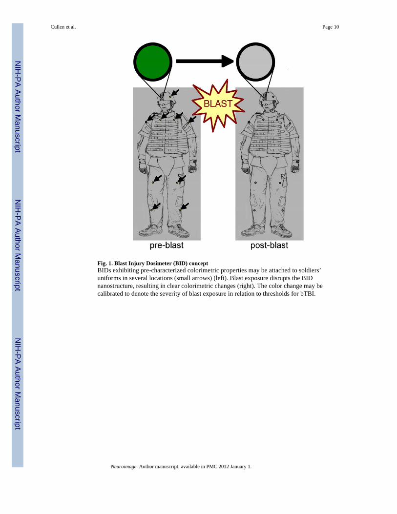
Fig. 1. Blast Injury Dosimeter (BID) conceptBIDs exhibiting pre-characterized colorimetric properties may be attached to soldiers’uniforms in several locations (small arrows) (left). Blast exposure disrupts the BIDnanostructure, resulting in clear colorimetric changes (right). The color change may becalibrated to denote the severity of blast exposure in relation to thresholds for bTBI.
Cullen et al. Page 10
Neuroimage. Author manuscript; available in PMC 2012 January 1.
NIH
-PA Author Manuscript
NIH
-PA Author Manuscript
NIH
-PA Author Manuscript
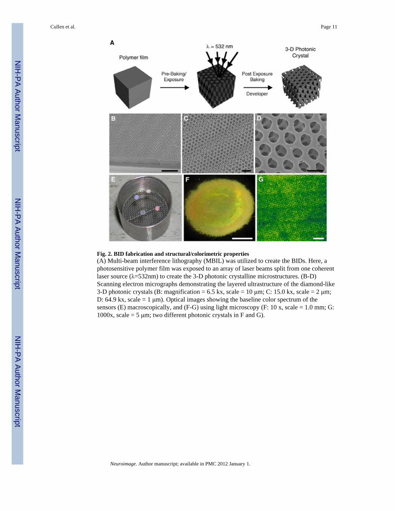
Fig. 2. BID fabrication and structural/colorimetric properties(A) Multi-beam interference lithography (MBIL) was utilized to create the BIDs. Here, aphotosensitive polymer film was exposed to an array of laser beams split from one coherentlaser source (λ=532nm) to create the 3-D photonic crystalline microstructures. (B-D)Scanning electron micrographs demonstrating the layered ultrastructure of the diamond-like3-D photonic crystals (B: magnification = 6.5 kx, scale = 10 μm; C: 15.0 kx, scale = 2 μm;D: 64.9 kx, scale = 1 μm). Optical images showing the baseline color spectrum of thesensors (E) macroscopically, and (F-G) using light microscopy (F: 10 x, scale = 1.0 mm; G:1000x, scale = 5 μm; two different photonic crystals in F and G).
Cullen et al. Page 11
Neuroimage. Author manuscript; available in PMC 2012 January 1.
NIH
-PA Author Manuscript
NIH
-PA Author Manuscript
NIH
-PA Author Manuscript
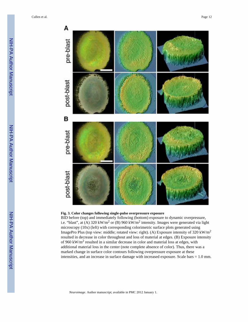
Fig. 3. Color changes following single-pulse overpressure exposureBID before (top) and immediately following (bottom) exposure to dynamic overpressure,i.e. “blast”, at (A) 320 kW/m2 or (B) 960 kW/m2 intensity. Images were generated via lightmicroscopy (10x) (left) with corresponding colorimetric surface plots generated usingImagePro Plus (top view: middle; rotated view: right). (A) Exposure intensity of 320 kW/m2
resulted in decrease in color throughout and loss of material at edges. (B) Exposure intensityof 960 kW/m2 resulted in a similar decrease in color and material loss at edges, withadditional material loss in the center (note complete absence of color). Thus, there was amarked change in surface color contours following overpressure exposure at theseintensities, and an increase in surface damage with increased exposure. Scale bars = 1.0 mm.
Cullen et al. Page 12
Neuroimage. Author manuscript; available in PMC 2012 January 1.
NIH
-PA Author Manuscript
NIH
-PA Author Manuscript
NIH
-PA Author Manuscript
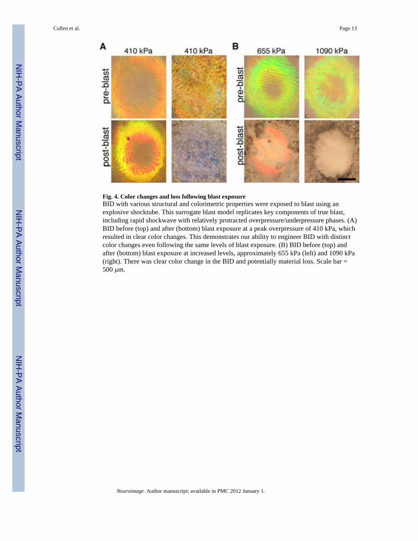
Fig. 4. Color changes and loss following blast exposureBID with various structural and colorimetric properties were exposed to blast using anexplosive shocktube. This surrogate blast model replicates key components of true blast,including rapid shockwave with relatively protracted overpressure/underpressure phases. (A)BID before (top) and after (bottom) blast exposure at a peak overpressure of 410 kPa, whichresulted in clear color changes. This demonstrates our ability to engineer BID with distinctcolor changes even following the same levels of blast exposure. (B) BID before (top) andafter (bottom) blast exposure at increased levels, approximately 655 kPa (left) and 1090 kPa(right). There was clear color change in the BID and potentially material loss. Scale bar =500 μm.
Cullen et al. Page 13
Neuroimage. Author manuscript; available in PMC 2012 January 1.
NIH
-PA Author Manuscript
NIH
-PA Author Manuscript
NIH
-PA Author Manuscript
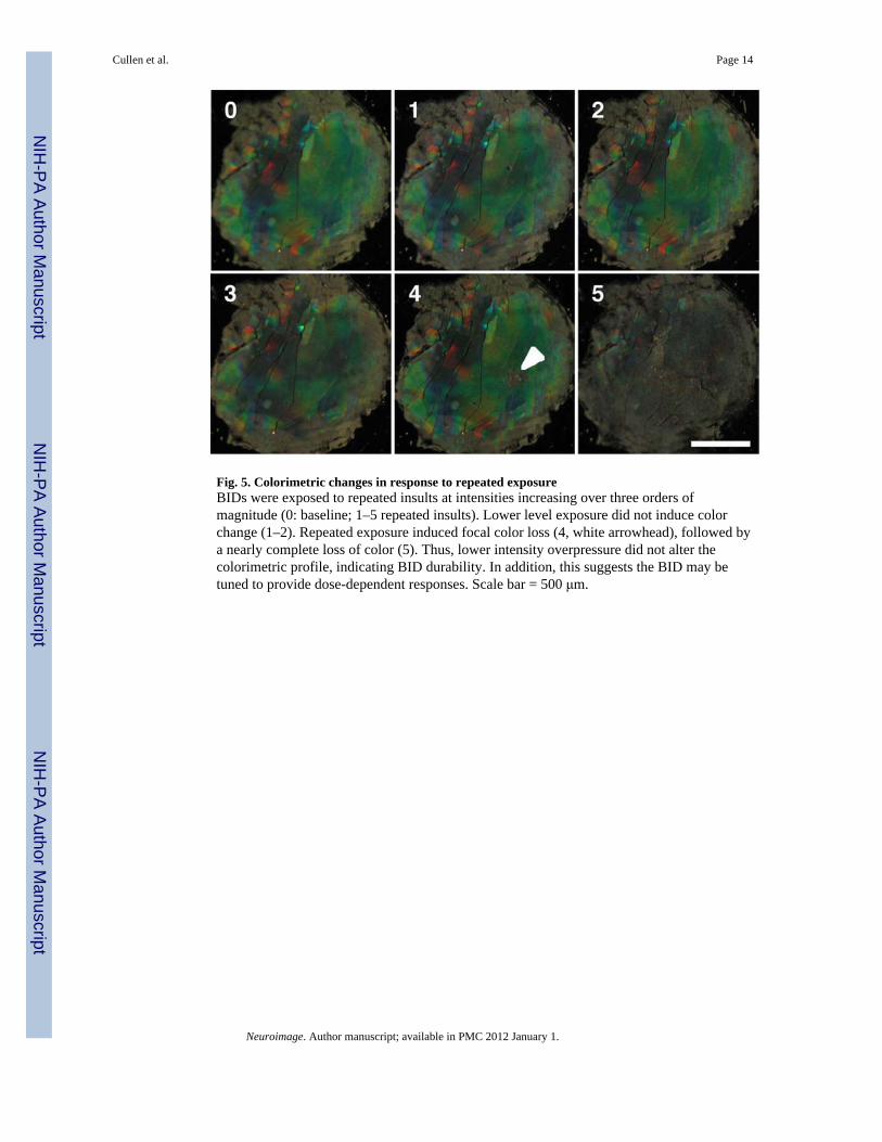
Fig. 5. Colorimetric changes in response to repeated exposureBIDs were exposed to repeated insults at intensities increasing over three orders ofmagnitude (0: baseline; 1–5 repeated insults). Lower level exposure did not induce colorchange (1–2). Repeated exposure induced focal color loss (4, white arrowhead), followed bya nearly complete loss of color (5). Thus, lower intensity overpressure did not alter thecolorimetric profile, indicating BID durability. In addition, this suggests the BID may betuned to provide dose-dependent responses. Scale bar = 500 μm.
Cullen et al. Page 14
Neuroimage. Author manuscript; available in PMC 2012 January 1.
NIH
-PA Author Manuscript
NIH
-PA Author Manuscript
NIH
-PA Author Manuscript
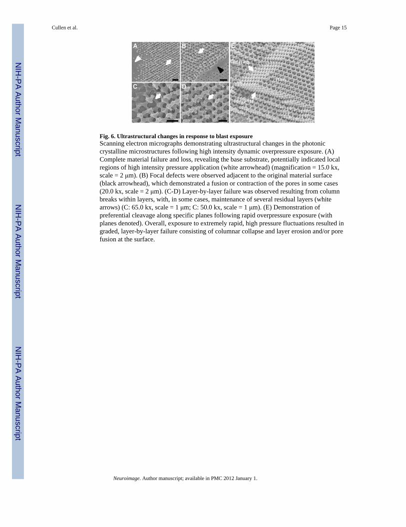
Fig. 6. Ultrastructural changes in response to blast exposureScanning electron micrographs demonstrating ultrastructural changes in the photoniccrystalline microstructures following high intensity dynamic overpressure exposure. (A)Complete material failure and loss, revealing the base substrate, potentially indicated localregions of high intensity pressure application (white arrowhead) (magnification = 15.0 kx,scale = 2 μm). (B) Focal defects were observed adjacent to the original material surface(black arrowhead), which demonstrated a fusion or contraction of the pores in some cases(20.0 kx, scale = 2 μm). (C-D) Layer-by-layer failure was observed resulting from columnbreaks within layers, with, in some cases, maintenance of several residual layers (whitearrows) (C: 65.0 kx, scale = 1 μm; C: 50.0 kx, scale = 1 μm). (E) Demonstration ofpreferential cleavage along specific planes following rapid overpressure exposure (withplanes denoted). Overall, exposure to extremely rapid, high pressure fluctuations resulted ingraded, layer-by-layer failure consisting of columnar collapse and layer erosion and/or porefusion at the surface.
Cullen et al. Page 15
Neuroimage. Author manuscript; available in PMC 2012 January 1.
NIH
-PA Author Manuscript
NIH
-PA Author Manuscript
NIH
-PA Author Manuscript