cognitive motor control in human pre-supplementary motor area studied by subdural recording of...
TRANSCRIPT

Brain (1999),122,915–931
Cognitive motor control in human pre-supplementary motor area studied by subduralrecording of discrimination/selection-relatedpotentialsAkio Ikeda,1 Shogo Yazawa,1 Takeharu Kunieda,2 Shinji Ohara,1 Kiyohito Terada,1 Nobuhiro Mikuni,2
Takashi Nagamine,1 Waro Taki,2 Jun Kimura3 and Hiroshi Shibasaki1
Departments of1Brain Pathophysiology,2Neurosurgery Correspondence to: Akio Ikeda MD, Department of Brainand 3Neurology, Kyoto University School of Medicine, Pathophysiology, Kyoto University School of Medicine,Kyoto, Japan Shogoin, Sakyo-ku, 606, Japan
E-mail: [email protected]
SummaryTo clarify the functional role of human pre-supplementarymotor area (pre-SMA) in ‘cognitive’ motor control ascompared with other non-primary motor cortices (SMA-proper and lateral premotor areas) and prefrontal area,we recorded epicortical field potentials by using subduralelectrodes in five epileptic patients during presurgicalevaluation, whose pre-SMA, SMA-proper, prefrontal andlateral premotor areas were defined by electric corticalstimulation and recent anatomical orientations accordingto the bicommissural plane and callosal grid system. AnS1-Go/NoGo choice and delayed reaction task (S1-choiceparadigm) and a warned choice Go/NoGo reaction task(S2-choice paradigm) with inter-stimulus intervals of 2 swere employed. The results showed (i) transient potentialswith onset and peak latencies of about 200 and 600 ms,respectively, after S1 in the S1-choice paradigm mainlyat pre-SMA and to a lesser degree at the prefrontal and
Keywords: pre-SMA; selection; voluntary movements; subdural recording; choice paradigm
Abbreviations: BP 5 Bereitschaftspotential; CNV5 contingent negative variation; SEP5 somatosensory evoked potential;SMA 5 supplementary motor area
IntroductionCurrently the supplementary motor area (SMA) in non-human primates is divided into the rostral SMA (pre-SMA)and the caudal SMA (SMA-proper) on the basis ofphysiological and anatomical criteria (Wiesendanger, 1986;Rizollati et al., 1990; Tanji, 1994). It has been reported fromstudies using the single cell recording technique that inprimates pre-SMA neurons discharged more frequently inpreparation for the forthcoming reaching movements thanSMA-proper neurons, and the latter were more related toexecution of the reaching movements (Matsuzakaet al.,
© Oxford University Press 1999
lateral premotor areas, but not in the S2-choice paradigm.At SMA-proper, a similar but much smaller potential wasseen after S1 in both S1- and S2-choice paradigms and(ii) slow sustained potentials between S1 and S2 in bothS1- and S2-choice paradigms in all of the non-primarymotor areas investigated (pre-SMA, SMA-proper andlateral premotor areas) and prefrontal area. It isconcluded that pre-SMA plays a more important role incognitive motor control which involves sensorydiscrimination and decision making or motor selectionfor the action after stimuli, whereas SMA-proper is oneof the main generators of Bereitschaftspotential precedingself-paced, voluntary movements. In the more generalanticipation of and attention to the forthcoming stimuli,non-primary motor cortices including pre-SMA, SMA-proper and lateral premotor area, and the prefrontal areaare commonly involved.
1992). Approaches to understanding the function of humanpre-SMA and SMA-proper have included functional imagingstudies such as PET (as reviewed by Picard and Strick,1996), and more recently functional MRI (fMRI) (Hikosakaet al., 1996; Humberstoneet al., 1997). The results fromthese studies suggested that a part of the mesial frontallobe situated rostral to the line drawn through the anteriorcommissure (AC) perpendicular to the line connecting theAC and the posterior commissure (PC), i.e. the VAC line(Talairach and Tournoux, 1988), was significantly active with
by guest on June 8, 2013http://brain.oxfordjournals.org/
Dow
nloaded from

916 A. Ikedaet al.
respect to the higher order aspects of motor control suchas intrinsic movement selection, motor learning, complexmovement and Go/NoGo trials, independent of the body partinvolved (Deiberet al., 1991; Shibasakiet al, 1993; Hikosakaet al., 1996; Picard and Strick, 1996; Humberstoneet al.,1997). Thus, this area is regarded as the pre-SMA in humans.These imaging results, however, also contain a variety offactors related to voluntary motor execution such as attention,discrimination, judgement, decision, preparation, anticipation,execution, assessment of performance and somatosensoryfeedback input mainly due to their limited temporal resolution.
Previously we recorded EEG from the SMA-proper inassociation with spontaneous, voluntary movements inepilepsy patients by using subdural electrodes (Ikedaet al.,1992, 1993, 1995, 1996a). This technique best delineates thetemporal characteristics of the cortical electrical activity ina particular area covered by the subdural electrodes, andthe results demonstrated that the SMA-proper generatedBereitschaftspotential (BP) (Kornhuber and Deecke, 1966;Shibasakiet al., 1980) in accordance with its somatotopy.Subdural EEG recordings in epilepsy patients by employinga warned S2-choice reaction paradigm were also made (Ikedaet al., 1996a,b; Hamanoet al., 1997). We found that (i) indichotomous discrimination tasks which required decisionmaking with regard to the S2 stimuli, bilateral mesial frontalcortices rostral to SMA-proper generated transient fieldpotentials and (ii) orbitofrontal and mesial prefrontal corticesgenerated slow potentials in the period of uncertainty andanticipation preceding the forthcoming informative stimulus(S2). Hamanoet al. (1997) further investigated these decision-related potentials and slow sustained potentials in a largernumber of patients who had subdural electrode grids at thelateral prefrontal, temporal, parietal and occipital associationcortices. These negative transients were not particularlyevident at the primary motor or sensory area, but were widelyobserved at the mesial and basal prefrontal, lateral and mesialparietal, lateral and mesial temporal, and mesial occipitalareas, all of which belonged to non-primary motor, non-primary sensory or association cortices. In addition, slowshifts between S1 and S2 were seen at the prefrontal, lateralpremotor, SMA, primary sensorimotor, mesial temporal andoccipital association areas.
In the three studies described above (Ikedaet al., 1996a,b;Hamanoet al., 1997), however, three problems have notbeen resolved. First, with regard to the mesial frontal area,SMA-proper was defined by electric cortical stimulation, butany further distinction between the SMA-proper, pre-SMAand mesial prefrontal area, especially between the latter twoareas, was not clear. In our study (Ikedaet al., 1996a),among the many subdural electrodes analysed on the mesialfrontal cortex anterior to the SMA-proper, two were located20 mm anterior to an electrode which corresponded to theSMA-proper of the face area, and they generated a transientpotential after dichotomous S2 stimuli with a peak latencyof 350 ms. However, it was unclear as to whether thesetwo particular electrodes belonged to pre-SMA or mesial
prefrontal area as anatomical analysis in relation to the VACline was not performed. A clear distinction among thesestructures is important when considering the functional roleof these individual areas. Secondly, the paradigm adopted inall three studies described was a warned S2-choice Go/NoGoreaction paradigm, which required the subjects to makedichotic discrimination, selection and decision, and also toinstigate motor execution immediately upon the S2-Go signal.Therefore, it was not possible to differentiate between thediscrimination process and the final decision for motorexecution as to Go or NoGo. The distinction would only bepossible by adopting the S1-choice paradigm and comparingit with the results of S2-choice paradigm. Thirdly, no studieshave been done with regard to subdural potentials at thelateral prefrontal, lateral premotor and primary motor areasin association with an S1-choice reaction-time paradigm. We,therefore, conducted the present study paying special attentionto the resolution of these three, especially of the first two,concerns.
Recent analyses of single cell recordings in non-humanprimates demonstrated that, in contrast to SMA-proper cells,pre-SMA cells generated transient activity when shiftingmotor plans in response to the instruction signal (Matsuzakaand Tanji, 1996), and a similar specific activity in pre-SMAwas seen when updating a motor task (Shimaet al., 1996).Updating or shifting a motor task encompasses a selectionprocess which is strongly related to motor preparation.Therefore, in order to clarify how closely the human pre-SMA, strictly defined by the currently accepted anatomicaland functional criteria, is associated with cognitive motorcontrol in voluntary movements when compared with SMA-proper, prefrontal and lateral premotor cortices, we analysedepicortical field potentials directly recorded from humancortex in epilepsy patients. Preliminary results have beenreported elsewhere in an abstract form (Ikedaet al., 1997).The function of human pre-SMA in generating BP precedingspontaneous, voluntary movements based on findingsobtained in patients 1 and 2 of the present series has beendescribed elsewhere (Yazawaet al., 1997b, 1998). Two otherpatients (patients 3 and 4) were discussed (Mikuniet al.,1997; Mimaet al., 1997) and ictal subdural EEG findings inpatients 1–3 were reported elsewhere for entirely differentpurposes (Ikedaet al., 1999).
Material and methodsSubjectsWe recorded field potentials directly from the surface of thefrontal cortex in five right-handed patients with medicallyintractable partial seizures (two men and three women, ageranging from 21 to 53 years with the mean of 33 years). Allfive patients were clinically evaluated for epilepsy surgeryby using subdural electrodes, and informed consent wasobtained from all patients after the purpose and possibleconsequences of the studies were explained, which were
by guest on June 8, 2013http://brain.oxfordjournals.org/
Dow
nloaded from

Cognitive motor control in human pre-SMA 917
according to the Clinical Research Protocol No. 79 approvedby the ethical committee of Kyoto University School ofMedicine.
The cortical electrical potentials were recorded by usingchronically implanted subdural electrodes (AD-TECH) madeof platinum. Each electrode was 3 mm in diameter, and thecentre-to-centre inter-electrode distance was 1 cm. Thisinvasive technique helps to identify (i) the extent of theepileptogenic region by seizure recording, and (ii) the functionof the cortex around the epileptogenic region by electricalcortical stimulation and by recording somatosensory evokedpotentials (SEPs) (Hahn and Lu¨ders, 1987).
The electrodes were placed at both the SMA-proper andpre-SMA in three patients (patients 1 and 2 on the righthemisphere, and patient 5 on the left), the right mesialprefrontal area in one patient (patient 1), the lateral prefrontalarea in two patients (patients 2 and 3, right and lefthemisphere, respectively), the left lateral premotor area inone patient (patient 4) and the right primary motor hand areain one patient (patient 2). The anatomical location of theseelectrodes was defined as described in the Methods sectionon ‘Cortical mapping’ (see below). Four out of five patientshad an increased signal abnormality on T2-weighted MRIwhich was later confirmed by surgery; in the right cingulatecortex (gliosis) (the area corresponding to electrode 3 andits inferior portion, that is partly extended and close toelectrode 10 in Fig. 2) in patient 1, the right lateral premotorcortex (astrocytoma grade II) in patient 2, in the left middleto inferior frontal gyrus (astrocytoma grade II) in patient 4and in the high lateral convexity of the left frontal lobe(cortical dysplasia) in patient 5. The potentials recorded fromthe electrodes placed over these lesions were not used forthe further analysis.
Experimental paradigmsTwo reaction-time paradigms using paired auditory stimuli(S1 and S2) were employed in the present study: (i) warnedchoice Go/NoGo task (S2-choice reaction-time paradigm)and (ii) delayed response paradigm following dichotomouschoice Go/NoGo task (S1-choice reaction-time paradigm).In both paradigms voluntary movements were adopted as aresponse task to S2.
The S2-choice paradigm was adopted from the authors’previous studies as follows (Ikedaet al., 1994, 1996a,b).A pair of tone bursts of different pitch and of a durationof 20 ms were presented for S1 and S2 with an intervalof 2 s. S1 was always a tone burst of 1000 Hz, while S2was either a tone burst of 1500 Hz (Go) or that of 2000Hz (NoGo). Go and NoGo stimuli were presented in arandom order with the same probability, and the motortask employed was middle finger extension of the handcontralateral to the side of the implanted electrodes. Thepatients were instructed to respond as quickly as possibleupon the S2-Go signal, but not to respond upon S2-NoGo.The EEG segment from 1 s before the S1 onset to 1.5 s
after the S2 onset was averaged for Go and NoGo trialsseparately, and the group average across Go and NoGotrials were made. The next warning signal was set to bedelivered at variable intervals between 3.5 and 7.5 s afterthe onset of each S2 signal.
In the S1-choice paradigm, which we adopted partiallyfrom another study of ours (Laiet al., 1997), S1 was eithera tone burst of 1500 Hz (Go) or of 2000 Hz (NoGo) whileS2 was always a tone burst of 1000 Hz. Go and NoGostimuli were presented in a random order with the sameprobability. The subjects were asked to react or not to S2 asquickly as possible, by the same motor task as used for theS2-choice paradigm depending on the S1. The EEG segmentanalysed, the averaging method and the inter-trial intervalwere the same as those employed for the S2-choice paradigm.
The patient was asked to keep quiet during each recordingsession and to postpone the next task if he accidentallymoved prior to the task movement. Before recording sessions,the patient was given a training period until the examinerwas satisfied that the subject consistently produced briskmovements which were preceded and followed by completemuscle relaxation. During the recording, trials in whichapparently significant artefacts and erroneous responses bythe patients were noted were rejected manually to store by aspecial on-line rejection paradigm (Ikedaet al., 1994, 1996a,b). One recording session typically lasted 5–6 min, and wasrepeated four to five times with an intermission of a fewminutes between sessions.
Data acquisition and analysisCortical recordings were done simultaneously from 20–32subdural electrodes in each patient. The subdural electrodeswere all referenced to a scalp electrode placed on the mastoidprocess contralateral to the side of implantation. The bandpassfilters applied for EEGs and EMGs were 0.016–60 Hz and20–60 Hz, respectively. The sensitivity of EEG was set to2 mV full scale (12 bit). Electro-oculograms (EOG) recordedfrom an electrode placed lateral to the right outer canthuswhich was referenced to the mastoid electrode was monitoredsimultaneously with the same filtering and sensitivity as thoseused for EEG recording.
All of the electrographic output signals were digitized atthe sampling rate of 200 Hz per channel, and stored onmagneto-optical disks by a compact evoked potentialrecording machine (DP1100, NEC San-ei) with the aid of aspecial purpose computer program designed for subsequentoff-line analysis. In addition to the special on-line rejectionprogram as described above, an off-line rejection programwas also applied; trials associated with either (i) erroneousresponse by the subjects, or (ii) movements which were notbrisk enough to identify a clear EMG onset, were excludedfrom subsequent analysis. A total of 72–127 trials wereselected for each of the Go and NoGo conditions of S1- andS2-choice paradigms separately. For each subject, a groupaverage waveform was obtained for each condition after
by guest on June 8, 2013http://brain.oxfordjournals.org/
Dow
nloaded from

918 A. Ikedaet al.
confirmation that the two ensemble averaged EEGs werereproducible as in Fig. 2C. Subsequently Go and NoGo trialswere averaged altogether for each of the S1- and S2-choiceparadigms.
Patients 1, 3, 4 and 5 had EEG recordings for bothparadigms, and patient 2 had only the S1-choice paradigmbecause of the patient’s condition.
Cortical mappingFor cortical mapping of the mesial frontal surface, electricalcortical stimulation and the recording of SEPs were donebefore the present experiment. Each subdural electrode wasindividually stimulated to identify cortical function (Lu¨derset al., 1987). Details of the methodology for stimulationand the subsequent cortical mapping have been describedelsewhere (Lu¨ders et al., 1987; Ikedaet al., 1992). TheSMA-proper was identified by its unique responses on themesial surface, consisting of predominantly tonic motorresponses of the upper as well as lower limb and of thetrunk, neck and face, either unilaterally or bilaterally. Thesemotor responses were somatotopically organized within theSMA-proper (Limet al., 1994). A negative motor responsewas defined as the cessation of voluntary tonic musclecontraction or rapid alternating movements without loss ofawareness during the stimulation (Lu¨ders et al., 1987).Cortical SEPs to electrical stimulation of the median nerveat a rate of 1.1 Hz were recorded from the same subduralelectrodes.
In order to define the precise anatomical location of pre-SMA and SMA-proper in patients 1 and 2, a plain lateralview of skull X-ray film, taken after implantation of theelectrodes, was first superimposed upon the sagittal view ofthe T1-weighted MRI taken before surgery by using commonlandmarks, i.e. nasion and inion, and the sizes adjusted toeach other for each patient (Ikedaet al., 1995, 1996a). Inpatient 5, T1-weighted MRI of the sagittal view was takenwhen the patient had the subdural electrodes in the skull, todefine the precise location directly. Based on the atlas ofTalairach and Tournoux (1988) and on the histologicalanalysis in human SMA (Zilleset al., 1996), the electrodeslocated on the mesial surface of the superior frontal gyrusjust anterior to the VAC line were judged to be on the pre-SMA. If the electrodes placed on the mesial surface of thesuperior frontal gyrus showed a negative motor response byelectrical stimulation, then they were also judged to be onthe pre-SMA regardless of their relative location to the VACline. However, the electrodes which elicited a positive motorresponse when stimulated were excluded from pre-SMA.Based on the atlas of Talairach and Tournoux (1988) and onthe recent PET studies (Picard and Strick, 1996), the anteriorborder of pre-SMA was further defined by the line 20 mmanterior and parallel to the VAC line. In patients 1, 2 and 5it was also confirmed that the above boundary between thepre-SMA and prefrontal areas was consistent with the callosalgrid system (Lehmanet al., 1992; Olivier, 1996) in which
the boundary was provided by the line (anterior callosalplane) drawn at the anterior edge of the genu of the corpuscallosum perpendicular to the plane through the lower borderof the genu and splenium of the corpus callosum.
If the electrodes on the mesial surface of the superiorfrontal gyrus were located posterior to the VAC line, andalso if these electrodes did not elicit a negative motorresponse, then those electrodes were judged to be on theSMA-proper. Since a negative motor response by electricalstimulation is not necessarily elicited in every subject evenif the subdural grid electrodes are located on the rostral partof the superior frontal gyrus (Lu¨ders et al., 1987), thepresence of a negative motor response was not regarded asessential for defining pre-SMA in the present study.
ResultsCortical mappingPatient 1 had four 13 4 electrode strips at the right mesialfrontal area, and another 13 4 electrode strip containingelectrodes 5–7 was placed at the boundary between the mesialand lateral parts of the superior frontal gyrus (Figs 1A and2A and B). Patient 2 had one 23 8 electrode grid at theright mesial frontal area, one 43 5 grid at the right lateralfrontal area and one 43 5 grid at the right perirolandic area(Figs 1B and 3). Patient 3 had one 23 8 electrode grid atthe left lateral prefrontal area (Fig. 4), one 23 8 grid at theleft basal frontal area, and one 23 8 grid at the left mesialto lateral temporal area. Patient 4 had two 43 5 electrodegrids at the left lateral premotor area (Fig. 5) and left inferiorto middle frontal gyrus. Patient 5 had one 23 8 electrodegrid at the left mesial frontal area and one 43 5 grid on theleft lateral frontal convexity (Figs 1C and 6).
On electrical cortical stimulation, electrode 2 in patient 1induced tonic abduction of the left arm, and electrode 3elicited a negative motor response in the left arm (Fig. 2Aand B). Other electrodes in patient 1 did not elicit any visibleresponse. In patient 2, electrode 1 elicited tonic musclecontraction of the left arm and foot, electrode 4 elicitednegative motor responses involving all extremities, tongueand eye movements, and electrodes 2, 3 and 5 did not elicitany response (Fig. 3). Electrode 8 elicited a clear positivemotor response in the left arm. In patient 3, electrodes 5, 9,10 and 13 did not induce any response after electricalstimulation (Fig. 4). In patient 4, electrodes 2 and 4 elicitednegative motor responses in the right arm and tongue,respectively (Fig. 5). Electrode 5 elicited a positive motorresponse in the tongue. Electrodes 1, 3 and 6 did not causeany response. In patient 5, electrodes 1–3 elicited no responsesafter cortical stimulation. No stimulation was carried out forelectrodes 4–6. Positive motor responses were observed atelectrode 7 (tonic contraction of the left lower trunk muscles)and electrodes 8 and 9 (tonic abduction of the bilateral feet,more on the left). Positive sensory symptoms in the wholebody were elicited by the stimulation of electrode 10 (Fig. 6).
by guest on June 8, 2013http://brain.oxfordjournals.org/
Dow
nloaded from

Cognitive motor control in human pre-SMA 919
Fig. 1 Anatomical location of the subdural grid electrodes placed in the mesial frontal area for three patients, as shown on eachindividual’s MRI. In patients 1 (A: right hemisphere) and 2 (B: right hemisphere), the sagittal view of the T1-weighted MRI taken beforesurgery was superimposed upon a plain lateral view of skull X-ray film taken after implantation of the electrodes by using commonlandmarks, i.e. nasion and inion, and adjusting the size to each other for each patient. In patient 5 (C: left hemisphere), the T1-weightedMRI of the sagittal view was taken when the patient had the subdural electrodes in the skull, to define the precise location directly.AC–PC line5 a line crossing anterior commissure and posterior commissure in the midline; VAC5 a line on the anterior commissureperpendicular to AC–PC line. [B was modified from fig. 1 in Yazawaet al., (1998) with permission.]
With regard to cortical SEPs, a negative peak (173 ms,37 µV) was seen at electrode 1 after the left median nervestimulation in patient 1. In patient 2, electrodes 1–5 showedpositive peaks (either155 ms or188 ms, 20–30µV) afterthe left median nerve stimulation. In patient 5, a broad, smallnegative peak (130 ms, 6µV) was seen at electrodes 7–9after the right median nerve stimulation.
From the anatomical point of view, the VAC line in patient1 was located between electrodes 3 and 4, and electrodes11–13 were located at or within 20 mm rostral to the VACline, and therefore were on or caudal to the anterior callosalplane according to the callosal grid system (Fig. 1A). The
VAC line in patient 2 was between electrodes 2 and 4 aswell as between electrodes 3 and 5 (Fig. 1B). In patient 5,the VAC line was on electrodes 2 and 5. Brain MRI takenwith the implanted electrodes clearly showed that electrodes1–3 were located on the superior frontal gyrus. Electrodes 1and 4 were located posterior to the anterior callosal plane(Fig. 1C).
Cortical mapping was made by taking all the above findingsinto account as follows. In patient 1, electrodes 1 and 2 werejudged to be on the SMA-proper, electrodes 3–9, 11 and 12on the pre-SMA, electrodes 14–16 on the prefrontal area andelectrodes 10 and 13 on the cingulate sulcus (Figs 1A and
by guest on June 8, 2013http://brain.oxfordjournals.org/
Dow
nloaded from

920 A. Ikedaet al.
2A and B). Electrode 3 showed continuous slow activitymost likely due to the epileptogenicity, and consequently theaveraged waveform did not show reproducible responses. Inpatient 2, electrodes 1–3 were judged to be on the SMA-proper, electrodes 4 and 5 on the pre-SMA, and none wereon the cingulate gyrus (Figs 1B and 3). In addition, electrodes6 and 7 were judged to be on the lateral prefrontal cortex,and electrode 8 on the right primary hand motor area. Inpatient 3, electrodes 5, 9, 10 and 13 were judged to be on
the lateral prefrontal area (Fig. 4). In patient 4, electrodes 2and 4 were judged to be on the lateral premotor areacorresponding to the primary (lateral) negative motor area(Luderset al., 1995), and electrode 5 on the primary motorface area (Fig. 5). In patient 5, electrodes 1 and 2 werejudged to be on the pre-SMA, electrodes 3 and 7–10 on theSMA-proper, electrode 4 on the cingulate sulcus andelectrodes 5 and 6 on the cingulate gyrus (Figs 1Cand 6).
by guest on June 8, 2013http://brain.oxfordjournals.org/
Dow
nloaded from

Cognitive motor control in human pre-SMA 921
Fig. 2 Cortical potentials recorded from subdural electrodes placed on the mesial surface of the right hemisphere in patient 1 (23-year-old female with supplementary motor seizures) in association with S1- (A) and S2- (B) choice reaction-time paradigms. (A) Transientpotentials after S1 are clearly seen at the pre-SMA (electrodes 6, 7, 8 and 12) and also at the prefrontal area to a lesser degree(electrodes 15 and 16) (all indicated by large arrows) and ill defined at the SMA-proper (electrode 2) (indicated by an arrowhead).Sustained negative slow potentials between S1 and S2 are seen at the pre-SMA (electrodes 6, 9 and 12), the prefrontal area (electrode15) and the SMA-proper (electrode 1) (indicated by asterisks). Go and NoGo trials were averaged together after confirming theconsistency of waveforms before S2 stimuli, as shown in (C). (B) No clear transient potentials are seen after S1 either at the pre-SMA orthe prefrontal area, except for electrode 7. At the SMA-proper (electrode 2) a similar, though ill defined, potential (indicated by anarrowhead) is seen. Sustained negative slow potentials between S1 and S2 are seen at the pre-SMA (electrodes 6–9), the prefrontal area(electrode 15) and the SMA-proper (electrode 1) (shown by asterisks). Negative transients upon S2 are seen mainly at the pre-SMA(electrodes 6, 7, 8 and 12) (indicated by a large arrow), which have similar features to the transient potentials seen upon S1 in theS1-choice paradigm (seeA). Go and NoGo trials were averaged together after confirming the consistency of waveforms before the S2stimuli, as shown in (C). (C) Separate representation of EEG waveforms of Go and NoGo trials in both S1- and S2-choice reaction-timeparadigms in the selected electrodes (electrodes 8–13) in patient 1. The transient potentials immediately after S1 in S1-choice task (asseen at electrode 8) were clearly present equally in both Go and NoGo trials. Furthermore, sustained slow potentials between S1 and S2(as seen at electrodes 9 and 12) were also equally present not only in Go but also in NoGo trials. Rectified EMG of the left extensordigitorum muscle for the middle finger was shown in the right lower side in the four sets of EEG waveforms. VAC5 a line on theanterior commissure perpendicular to AC–PC line; VPC5 a line on the posterior commissure perpendicular to AC–PC line.
by guest on June 8, 2013http://brain.oxfordjournals.org/
Dow
nloaded from
922 A. Ikedaet al.
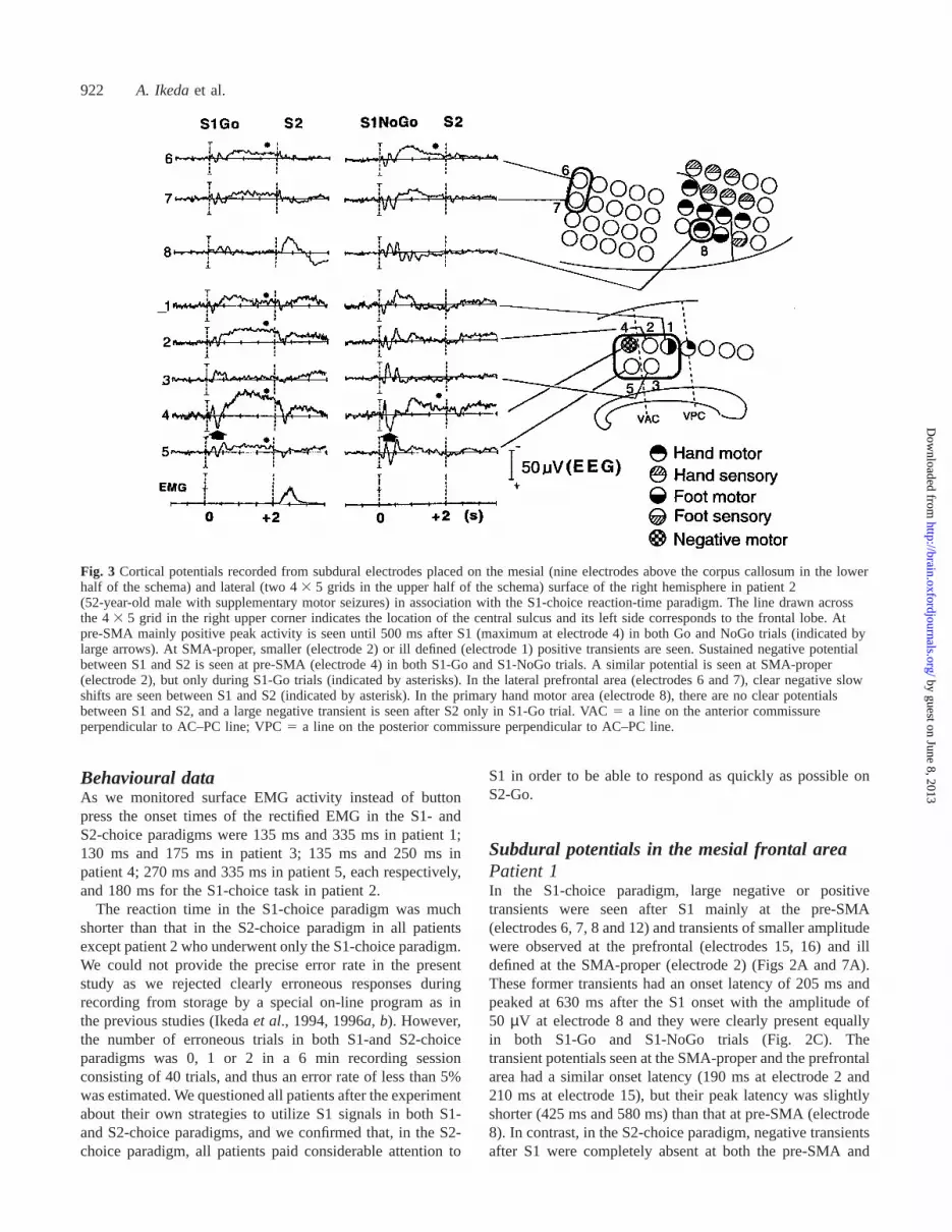
Fig. 3 Cortical potentials recorded from subdural electrodes placed on the mesial (nine electrodes above the corpus callosum in the lowerhalf of the schema) and lateral (two 43 5 grids in the upper half of the schema) surface of the right hemisphere in patient 2(52-year-old male with supplementary motor seizures) in association with the S1-choice reaction-time paradigm. The line drawn acrossthe 43 5 grid in the right upper corner indicates the location of the central sulcus and its left side corresponds to the frontal lobe. Atpre-SMA mainly positive peak activity is seen until 500 ms after S1 (maximum at electrode 4) in both Go and NoGo trials (indicated bylarge arrows). At SMA-proper, smaller (electrode 2) or ill defined (electrode 1) positive transients are seen. Sustained negative potentialbetween S1 and S2 is seen at pre-SMA (electrode 4) in both S1-Go and S1-NoGo trials. A similar potential is seen at SMA-proper(electrode 2), but only during S1-Go trials (indicated by asterisks). In the lateral prefrontal area (electrodes 6 and 7), clear negative slowshifts are seen between S1 and S2 (indicated by asterisk). In the primary hand motor area (electrode 8), there are no clear potentialsbetween S1 and S2, and a large negative transient is seen after S2 only in S1-Go trial. VAC5 a line on the anterior commissureperpendicular to AC–PC line; VPC5 a line on the posterior commissure perpendicular to AC–PC line.
Behavioural dataAs we monitored surface EMG activity instead of buttonpress the onset times of the rectified EMG in the S1- andS2-choice paradigms were 135 ms and 335 ms in patient 1;130 ms and 175 ms in patient 3; 135 ms and 250 ms inpatient 4; 270 ms and 335 ms in patient 5, each respectively,and 180 ms for the S1-choice task in patient 2.
The reaction time in the S1-choice paradigm was muchshorter than that in the S2-choice paradigm in all patientsexcept patient 2 who underwent only the S1-choice paradigm.We could not provide the precise error rate in the presentstudy as we rejected clearly erroneous responses duringrecording from storage by a special on-line program as inthe previous studies (Ikedaet al., 1994, 1996a,b). However,the number of erroneous trials in both S1-and S2-choiceparadigms was 0, 1 or 2 in a 6 min recording sessionconsisting of 40 trials, and thus an error rate of less than 5%was estimated. We questioned all patients after the experimentabout their own strategies to utilize S1 signals in both S1-and S2-choice paradigms, and we confirmed that, in the S2-choice paradigm, all patients paid considerable attention to
S1 in order to be able to respond as quickly as possible onS2-Go.
Subdural potentials in the mesial frontal areaPatient 1In the S1-choice paradigm, large negative or positivetransients were seen after S1 mainly at the pre-SMA(electrodes 6, 7, 8 and 12) and transients of smaller amplitudewere observed at the prefrontal (electrodes 15, 16) and illdefined at the SMA-proper (electrode 2) (Figs 2A and 7A).These former transients had an onset latency of 205 ms andpeaked at 630 ms after the S1 onset with the amplitude of50 µV at electrode 8 and they were clearly present equallyin both S1-Go and S1-NoGo trials (Fig. 2C). Thetransient potentials seen at the SMA-proper and the prefrontalarea had a similar onset latency (190 ms at electrode 2 and210 ms at electrode 15), but their peak latency was slightlyshorter (425 ms and 580 ms) than that at pre-SMA (electrode8). In contrast, in the S2-choice paradigm, negative transientsafter S1 were completely absent at both the pre-SMA and
by guest on June 8, 2013http://brain.oxfordjournals.org/
Dow
nloaded from
Cognitive motor control in human pre-SMA 923
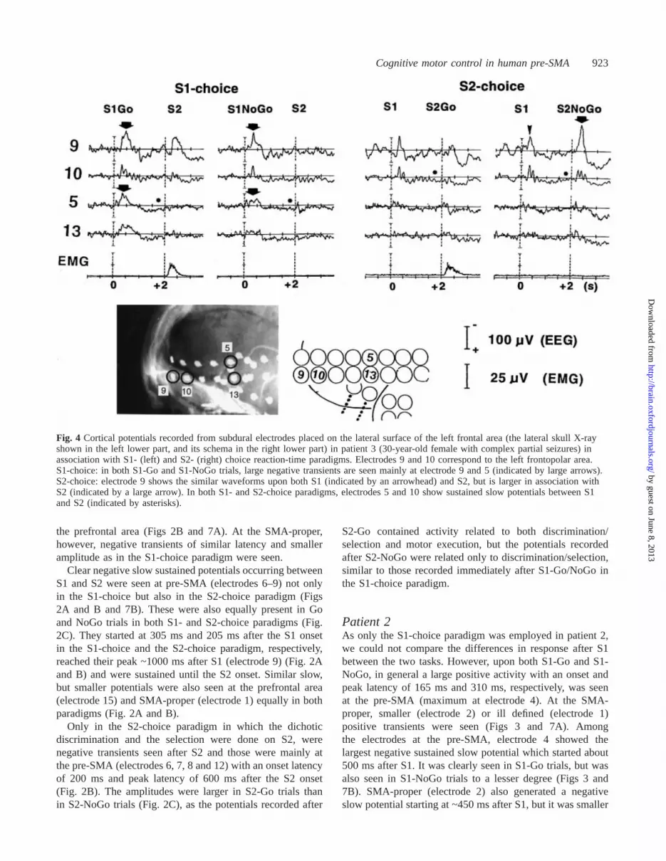
Fig. 4 Cortical potentials recorded from subdural electrodes placed on the lateral surface of the left frontal area (the lateral skull X-rayshown in the left lower part, and its schema in the right lower part) in patient 3 (30-year-old female with complex partial seizures) inassociation with S1- (left) and S2- (right) choice reaction-time paradigms. Electrodes 9 and 10 correspond to the left frontopolar area.S1-choice: in both S1-Go and S1-NoGo trials, large negative transients are seen mainly at electrode 9 and 5 (indicated by large arrows).S2-choice: electrode 9 shows the similar waveforms upon both S1 (indicated by an arrowhead) and S2, but is larger in association withS2 (indicated by a large arrow). In both S1- and S2-choice paradigms, electrodes 5 and 10 show sustained slow potentials between S1and S2 (indicated by asterisks).
the prefrontal area (Figs 2B and 7A). At the SMA-proper,however, negative transients of similar latency and smalleramplitude as in the S1-choice paradigm were seen.
Clear negative slow sustained potentials occurring betweenS1 and S2 were seen at pre-SMA (electrodes 6–9) not onlyin the S1-choice but also in the S2-choice paradigm (Figs2A and B and 7B). These were also equally present in Goand NoGo trials in both S1- and S2-choice paradigms (Fig.2C). They started at 305 ms and 205 ms after the S1 onsetin the S1-choice and the S2-choice paradigm, respectively,reached their peak ~1000 ms after S1 (electrode 9) (Fig. 2Aand B) and were sustained until the S2 onset. Similar slow,but smaller potentials were also seen at the prefrontal area(electrode 15) and SMA-proper (electrode 1) equally in bothparadigms (Fig. 2A and B).
Only in the S2-choice paradigm in which the dichoticdiscrimination and the selection were done on S2, werenegative transients seen after S2 and those were mainly atthe pre-SMA (electrodes 6, 7, 8 and 12) with an onset latencyof 200 ms and peak latency of 600 ms after the S2 onset(Fig. 2B). The amplitudes were larger in S2-Go trials thanin S2-NoGo trials (Fig. 2C), as the potentials recorded after
S2-Go contained activity related to both discrimination/selection and motor execution, but the potentials recordedafter S2-NoGo were related only to discrimination/selection,similar to those recorded immediately after S1-Go/NoGo inthe S1-choice paradigm.
Patient 2As only the S1-choice paradigm was employed in patient 2,we could not compare the differences in response after S1between the two tasks. However, upon both S1-Go and S1-NoGo, in general a large positive activity with an onset andpeak latency of 165 ms and 310 ms, respectively, was seenat the pre-SMA (maximum at electrode 4). At the SMA-proper, smaller (electrode 2) or ill defined (electrode 1)positive transients were seen (Figs 3 and 7A). Amongthe electrodes at the pre-SMA, electrode 4 showed thelargest negative sustained slow potential which started about500 ms after S1. It was clearly seen in S1-Go trials, but wasalso seen in S1-NoGo trials to a lesser degree (Figs 3 and7B). SMA-proper (electrode 2) also generated a negativeslow potential starting at ~450 ms after S1, but it was smaller
by guest on June 8, 2013http://brain.oxfordjournals.org/
Dow
nloaded from

924 A. Ikedaet al.
Fig. 5 Cortical potentials obtained from subdural electrodes placed on the lateral surface of the left frontotemporal area (as shown in theright upper schema) in patient 4 (30-year-old male with complex partial seizures) in association with S1- and S2-choice reaction-timeparadigms. The line drawn across the 43 5 grid indicates the location of the sylvian fissure, and the upper half corresponds to the leftfrontal lobe and its lower half to the temporal lobe. S1-choice: upon S1, large positive and negative complexes (peaks at 280 ms and195 ms, respectively) are seen at electrodes 4 and 5, respectively (indicated by a large arrow). Between S1 and S2, a slow positive shiftis seen at electrode 1 (indicated by an asterisk). S2-choice: upon S2, electrodes 4 and 5 generate the similar potentials to those observedupon S1 in the S1-choice paradigm (indicated by a large arrow), and slow positive shifts between S1 and S2 are seen at electrodes 1, 3,4 and 5 (indicated by asterisks). In both S1- and S2-choice paradigms, electrodes 3 and 6 show a large complex of negative and positivewaves (peak latency at 140 ms and 215 ms, respectively) immediately after both S1 and S2. Go and NoGo trials were averaged afterconfirming the consistency of waveforms before S2 stimuli.
than at the pre-SMA (electrode 4) and was seen only duringthe S1-Go trials.
Patient 5In the S1-choice paradigm, large positive and negativetransients of more than 75µV were seen at electrodes 1 (pre-SMA) and 4, respectively, after S1, and they peaked at500 ms after S1. These potentials were seen in both S1-Goand S1-NoGo trials (the waveforms in Fig. 6 are combinedS1-Go and S1-NoGo sessions). These results suggest thepresence of a dipole generator between electrode 1 (pre-SMA) and 4. These transient potentials are not very obviousat electrodes 3 and 7–10 (SMA-proper) (waveforms atelectrodes 7–10 are not shown in Fig. 6), but similar, smallerones are apparent at electrodes 5 and 6 (anterior cingulategyrus). In contrast, in the S2-choice paradigm, there were nopositive and negative transients after S1 at pre-SMA (elec-trode 1) which is similar to the situation observed withpatient 1 (Fig. 6). At the SMA-proper (electrode 3), and atthe anterior cingulate gyrus (electrodes 5 and 6), however,negative transients of small amplitude were seen similar tothose in the S1-choice paradigm, and their amplitudes (~50µV) were as small as ones seen after S1 in the S1-choiceparadigm.
Slow sustained potentials occurring between S1 and S2
were ill defined in the S1-choice paradigm at any electrode.In the S2-choice paradigm, small, slow potentials were seenonly at electrodes 4–6 which were mainly located in theanterior cingulate gyrus (Fig. 6). The potentials started about1500 ms after the S1 onset, and gradually increased inamplitude.
In the S2-choice paradigm in which dichotic discriminationand response selection were done upon S2, large positiveand negative transients of more than 75µV were seen afterS2 at electrodes 1, 4 and 5 (mainly at pre-SMA). These hada peak latency of 500 ms after S2. These transient potentialsafter S2 were similar to those after S1 in S1-choice paradigm,in terms of the location and temporal relationship to thestimulus, which were strongly related to the discriminationand selection process to dichotic stimuli (Fig. 6)
Subdural potentials in the lateral frontal areaPatient 2In the lateral prefrontal area (electrodes 6 and 7), upon S1in the S1-choice paradigm, small positive-negative complexeswere seen with a peak latency of initial positivity at 175 msafter S1. These were followed by clear negative slow shiftsstarting at 440 ms after S1, and they were sustained until theS2 onset. In the primary hand motor area (electrode 8), there
by guest on June 8, 2013http://brain.oxfordjournals.org/
Dow
nloaded from

Cognitive motor control in human pre-SMA 925
Fig. 6 Cortical potentials recorded from subdural electrodes placed on the mesial surface of the left hemisphere in patient 5 (31-year-oldfemale with supplementary motor seizures) in association with S1- and S2-choice reaction-time paradigms. S1-choice: large positive andnegative transients of.75 µV are seen at electrode 1 (pre-SMA) and 4, respectively, after S1, and they peak at 500 ms after S1(indicated by a large arrow). These transient potentials are not apparent at electrodes 3 and 7–10 (SMA-proper), but there is a similar,smaller response at electrodes 5 and 6 (anterior cingulate gyrus). Sustained negative slow potentials between S1 and S2 are absent atpre-SMA (electrodes 1 and 2) or SMA-proper (at least electrode 3 in the figure). Go and NoGo trials were averaged together afterconfirming the consistency of waveforms before S2. S2-choice: positive and negative transients after S1 are completely absent at thepre-SMA (electrode 1). However, at the SMA-proper (electrode 3), and at the anterior cingulate gyrus (electrodes 5 and 6), negativetransients of small amplitude are seen as in S1-choice paradigm, although the amplitude is smaller. Slow sustained potentials occurringbetween S1 and S2 are seen only at electrodes 4–6 located in the anterior cingulate gyrus (indicated by asterisks). After S2, largepositive and negative transients of.75 µV are seen at electrodes 1, 4 and 5 (mainly at pre-SMA) (indicated by large arrows), whichhave similar features to the transient potentials seen upon S1 in the S1-choice paradigm. Go and NoGo trials were averaged togetherafter confirming the consistency of waveforms before S2. VAC5 a line on the anterior commissure perpendicular to AC–PC line;VPC 5 a line on the posterior commissure perpendicular to AC–PC line. Waveforms at electrodes 7–10 are not shown.
was no clear potential seen between S1 and S2, and a largenegative transient was seen after S2 only in the S1-Go trial.The onset and peak latency of this transient were 165 msand 350 ms after S2, respectively, which were earlier byabout 50–150 ms than those of rectified EMG activity (Figs3 and 7).
Patient 3In both S1-Go and S1-NoGo trials in the S1-choice paradigm,large negative transients were seen after S1 mainly atelectrodes 9 and 5 (Fig. 4). Electrode 9, located more rostrallythan electrode 5, showed a similar response to S1 in the S2-choice paradigm, but it was slightly smaller than in theS1-choice paradigm. Electrode 5 showed no such similarpotentials after S1 in the S2-choice paradigm. Electrodes 5and 10 showed sustained slow potentials between S1 and S2in both S1- and S2-choice paradigms.
Patient 4Large positive and negative complexes were seen at electrodes4 and 5, respectively exclusively upon S1 in the S1-choiceparadigm and upon S2 in the S2-choice paradigm and thepeak latencies were 280 ms and 195 ms after S1 as well asafter S2 (Fig. 5). Between S1 and S2, slow positive potentialwas seen at electrode 1 in the S1-choice paradigm, and atthree more electrodes (electrodes 3, 4 and 5) in the S2-choiceparadigm (Fig. 5). In both S1- and S2-choice paradigms,electrodes 3 and 6 showed large transients of negative andpositive complexes (peak latency at 140 ms and 215 ms,respectively) immediately after both S1 and S2.
DiscussionDiscrimination/selection-related potentialsThe present results are summarized in a schematic illustrationin Fig. 7. Transient potentials after S1 (Figs 7A and 8) had
by guest on June 8, 2013http://brain.oxfordjournals.org/
Dow
nloaded from

926 A. Ikedaet al.
Fig. 7 (A) Schematic representation of cortical generators for transient potentials occurring after S1 in the S1- and S2-choice reaction-time paradigms for each patient. The potentials seen only in the S1-choice paradigm are mainly observed at pre-SMA, and can beregarded as discrimination/selection-related cortical potentials. In patient 2, the schema for the S1-choice paradigm only is shownbecause the S2-choice paradigm was not conducted due to the patient’s condition. (B) Schematic representation of cortical generators forslow potential shifts between S1 and S2 in the S1- and S2-choice reaction-time paradigms for each patient. The slow shifts are observedin all of the non-primary motor cortices investigated (pre-SMA, SMA-proper and lateral premotor areas) and prefrontal areas in both theS1-and S2-choice paradigms. In patient 2, the schema only for S1-choice paradigm is shown because S2-choice paradigm was notconducted due to the patient’s condition.
by guest on June 8, 2013http://brain.oxfordjournals.org/
Dow
nloaded from
Cognitive motor control in human pre-SMA 927
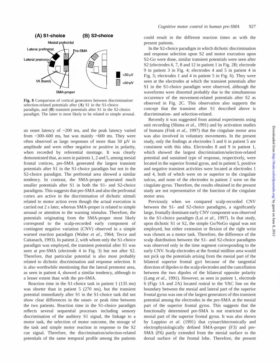
Fig. 8 Comparison of cortical generators between discrimination/selection-related potentials after (A) S1 in the S1-choiceparadigm, and (B) transient potentials after S1 in the S2-choiceparadigm. The latter is most likely to be related to simple arousal.
an onset latency of ~200 ms, and the peak latency variedfrom ~300–600 ms, but was mainly ~600 ms. They wereoften observed as large responses of more than 50µV inamplitude and were either negative or positive in polarity,when recorded by referential montage. It was clearlydemonstrated that, as seen in patients 1, 2 and 5, among mesialfrontal cortices, pre-SMA generated the largest transientpotentials after S1 in the S1-choice paradigm but not in theS2-choice paradigm. The prefrontal area showed a similartendency. In contrast, the SMA-proper generated muchsmaller potentials after S1 in both the S1- and S2-choiceparadigms. This suggests that pre-SMA and also the prefrontalcortex are active in the discrimination of dichotic stimulirelated to motor action even though the actual execution iscarried out 2 s later, whereas SMA-proper is related to simplearousal or attention to the warning stimulus. Therefore, thepotentials originating from the SMA-proper most likelycorrespond to the scalp-recorded early component ofcontingent negative variation (CNV) observed in a simplewarned reaction paradigm (Walteret al., 1964; Tecce andCattanach, 1993). In patient 2, with whom only the S1-choiceparadigm was employed, the transient potential after S1 wasseen at pre-SMA (electrode 4 in Fig. 3) but not after S2.Therefore, that particular potential is also most probablyrelated to dichotic discrimination and response selection. Itis also worthwhile mentioning that the lateral premotor area,as seen in patient 4, showed a similar tendency, although toa lesser extent than with the pre-SMA.
Reaction time in the S1-choice task in patient 1 (135 ms)was shorter than in patient 5 (270 ms), but the transientpotential immediately after S1 in the S1-choice task did notshow clear differences in the onset- or peak time betweenthe two patients. Reaction time in the S1-choice paradigmreflects several sequential processes including sensorydiscrimination of the auditory S1 signal, the linkage to amotor task, the selection of motor outcome, the storage ofthe task and simple motor reaction in response to the S2cue signal. Therefore, the discrimination/selection-relatedpotentials of the same temporal profile among the patients
could result in the different reaction times as with thepresent patients.
In the S2-choice paradigm in which dichotic discriminationand response selection upon S2 and motor execution uponS2-Go were done, similar transient potentials were seen afterS2 (electrodes 6, 7, 8 and 12 in patient 1 in Fig. 2B; electrode9 in patient 3 in Fig. 4; electrodes 4 and 5 in patient 4 inFig. 5; electrodes 1 and 4 in patient 5 in Fig. 6). They wereseen at the electrodes at which the transient potentials afterS1 in the S1-choice paradigm were observed, although thewaveforms were distorted probably due to the simultaneousoccurrence of the movement-related potentials after S2 asobserved in Fig. 2C. This observation also supports theconcept that the transient after S1 described above isdiscrimination- and selection-related.
Recently it was suggested from animal experiments usingunit recording (Shimaet al., 1991) and by activation studiesof humans (Finket al., 1997) that the cingulate motor areawas also involved in voluntary movements. In the presentstudy, only the findings at electrodes 5 and 6 in patient 5 areconsistent with this idea. Electrodes 8 and 9 in patient 1,which showed the largest discrimination/selection-relatedpotential and sustained type of response, respectively, werelocated in the superior frontal gyrus, and in patient 5, positiveand negative transient activities were located at electrodes 1and 4, both of which were on or superior to the cingulatesulcus, and none of the electrodes in patient 2 were on thecingulate gyrus. Therefore, the results obtained in the presentstudy are not representative of the function of the cingulatemotor area.
Previously when we compared scalp-recorded CNVbetween the S1- and S2-choice paradigms, a significantlylarge, frontally dominant early CNV component was observedin the S1-choice paradigm (Laiet al., 1997). In that study,upon dichotic S1 or S2, the simple Go/NoGo signal was notemployed, but either extension or flexion of the right wristwas chosen as a motor task. Therefore, the difference of thescalp distribution between the S1- and S2-choice paradigmswas observed only in the time segment corresponding to theearly CNV. Scalp electrodes at the frontal midline area wouldnot pick up the potentials arising from the mesial part of thebilateral superior frontal gyri because of the tangentialdirection of dipoles to the scalp electrodes and the cancellationbetween the two dipoles of the bilateral opposite polarity(Lang et al., 1991). However, as seen in patient 1, electrode6 (Figs 1A and 2A) located rostral to the VAC line on theboundary between the mesial and lateral part of the superiorfrontal gyrus was one of the largest generators of this transientpotential among the electrodes in the pre-SMA at the mesialpart of the superior frontal gyrus. This suggests that thefunctionally determined pre-SMA is not restricted to themesial part of the superior frontal gyrus. It was also shownby Luppino et al. (1991) that cytoarchitectonically andelectrophysiologically defined SMA-proper (F3) and pre-SMA (F6) partly extended from the mesial surface to thedorsal surface of the frontal lobe. Therefore, the present
by guest on June 8, 2013http://brain.oxfordjournals.org/
Dow
nloaded from

928 A. Ikedaet al.
results could lead us to a conclusion that the differences ofthe early CNVs observed, using the scalp electrodes at thefrontal midline area, between the S1- and S2-choiceparadigms (Laiet al., 1997) was most likely due to theactivity of the pre-SMA and prefrontal cortices.
With regard to the specific function of the pre-SMA, recentsingle cell recording in non-human primates demonstratedthat pre-SMA cells, but not SMA-proper cells, generatedtransient activity when shifting motor plans in response tothe instruction signal (Matsuzaka and Tanji, 1996). Similarspecific activity in pre-SMA was seen only when updatingthe motor task between the different series of sequentialmotor tasks (Shimaet al., 1996). In the study by Matsuzakaand Tanji (1996), 1.5–3 s before the impending signal, a50 Hz tone burst (switching signal) was delivered to monkeysin response to which they had to switch from the currentmotor task to a new one. Thirty-one per cent of the neuronsin the pre-SMA showed a shift-related activity of 500 msduration after the switching signal, whereas only 7% of theSMA-proper had the same neuronal activity. This result isconsistent with the present findings in the pre-SMA andSMA-proper. Since shifting or updating a motor task beforethe motor execution contains similar features to the S1-choice paradigm employed in the present study in terms ofdiscrimination, selection and decision for motor plan, it ismost likely that one of the specific functions of the pre-SMAis ‘cognitive’ processing strongly related to the forthcomingmotor tasks, i.e. ‘cognitive motor control’ in voluntarymovements. They also showed that phasic responses to visualcue signals which indicate the direction of the forthcomingarm-reaching movements were more abundant in the pre-SMA than in the SMA-proper (Matsuzakaet al., 1992).This may be consistent with the present results in terms ofpre-setting the movement direction upon the externalsignal.
A recent study in humans using fMRI showed that, in thesimple Go/NoGo paradigm, the mesial frontal area rostral tothe VAC line (pre-SMA) had an increased signal in associationwith both the Go and NoGo trials, and the area caudal to theVAC line and rostral to paracentral lobule (SMA-proper) wasactivated only with Go trials (Humberstoneet al., 1997). Sincethat particular study employed a single stimulus paradigm inwhich discrimination, selection, decision making and thenmotor execution, if needed, occurred together in response toeach stimulus, and since there was only 3 s oftime resolutionin spite of the single event analysis method, it could notdifferentiate between stimulus- and motor-related activities.Nevertheless, at least from the view point of Go/NoGodiscrimination, it suggests an important role of the pre-SMAas in the present study.
The transient potentials after S1 seen at the mesial pre-frontal areas were smaller than those in the pre-SMA, asseen in patient 1. Previously we showed that, in S2-choiceparadigms, the similar transient potentials after S2 (decision-related potentials) were observed in the mesial prefrontalarea (Ikedaet al., 1996a). Since the pre-SMA received the
direct input from the prefrontal cortex (Luppinoet al., 1993),it is important to link the information about behaviouralcontent to actual processes for motor execution (Fuster,1989; Passingham, 1996). Therefore, it is likely that thediscrimination and selection processes related to motorexecution are conducted in both prefrontal and pre-SMA,and the latter could augment it downstream of the informationprocessing.
Slow shifts between S1 and S2Slow potentials between S1 and S2 were obvious mainly atthe pre-SMA, and also at the prefrontal SMA-proper andlateral premotor areas to a lesser extent. They were seen bothin S1-choice and S2-choice paradigms (Fig. 7B). Theystarted occurring immediately after the disappearance ofdiscrimination/selection-related potentials, or graduallybecame apparent ~400 ms after S1 signal. There were twotypes of developing pattern: (i) as seen in patients 1 and 2,potentials peaked at ~1000 ms after S1 and were sustainedwith the same amplitude until the S2 signal, or they slightlydiminished in amplitude immediately before the S2 signal;(ii) mainly as seen in patients 3–5, slow shifts appeared at~1000 ms or even later, and gradually increased in amplitudebefore the S2 signal. These waveforms are consistent withthe late CNV recorded by scalp electrodes (Walterset al.,1964) as well as by subdural electrodes in our previousstudies (Ikedaet al., 1996a,b). The waveforms were welldocumented in S1-NoGo trials in the S1-choice paradigms(as shown at electrode 9 in patient 1 in Fig. 2C and atelectrode 4 in patient 2 in Fig. 3), strongly suggesting thatthese non-primary motor and prefrontal areas are active invery close association with general anticipation or attentionfor the forthcoming stimuli, but are not strongly or at allrelated to motor preparation at least between S1 and S2 inthis particular paradigm. The S2-choice paradigm causesmore uncertainty before S2 than the S1-choice paradigm,and thus the magnitude or the extent of these slow potentialsmay be proportionally larger in the S2-choice paradigm. Inthe lateral premotor area in patient 4 (Fig. 5), the S2-paradigminvolved three more electrodes generating slower shifts thanin the S1-paradigm, but a similar tendency was not seen atthe pre-SMA or prefrontal area in patients 1 and 2.
Since the waveforms of these slow potentials are consistentwith scalp-recorded late CNV, the present study supports theconcept that scalp-recorded late CNV consists of sub-components, and these are not related just to simple motorpreparation in the setting of CNV paradigm. We also previ-ously showed that orbitofrontal, mesial prefrontal, SMA(Ikeda et al., 1996a,b), lateral premotor, primary sensori-motor, mesial temporal and occipital association areas(Hamanoet al., 1997) generated slow potentials between thetwo signals by using the S2-choice reaction-time paradigm.In the present study, we further clarified the involvement ofthe pre-SMA as one of the most active areas to generatethese slow shifts among those areas, and that since it was
by guest on June 8, 2013http://brain.oxfordjournals.org/
Dow
nloaded from

Cognitive motor control in human pre-SMA 929
active in S1-NoGo trials as well as in S2-NoGo trials, it wasmost likely related to general anticipation, aside from motorpreparation. It is worthwhile stressing here that the presentresults do not deny that pre-SMA is also involved in motorpreparation or execution. Recently we demonstrated that pre-SMA generates BPs before spontaneous voluntary movementsregardless of the site of the movements (Yazawaet al., 1997b),whereas SMA-proper generates BPs in good accordance toits somatotopy (Ikedaet al., 1992). The primary motor handarea in patient 2 in the present study did not show subduralpotentials before S2 in S1-Go trials, but immediately afterS2 the clear transient potentials were seen. Therefore, thepresent paradigms, i.e. CNV paradigms with a paired auditorychoice reaction-time setting, would be suitable to elucidateslow potentials unrelated to motor preparation in the analysissegment between S1 and S2.
In preparation for self-paced, voluntary movements, bothpre-SMA and SMA-proper generated BPs without cleardifferences in onset time between each, although the pre-SMA was always active regardless of the site of movements(Yazawaet al., 1997b). However, once ‘cognitive’ processessuch as discrimination, decision or response selection elicitedby external stimuli were introduced in association with motorcontrol of voluntary movements, the significant functionaldifference between pre-SMA and SMA-proper was clearlydemonstrated. When functionally mapping the cortex inareas other than primary motor and sensory areas by usingchronically implanted subdural electrodes, currently usedtechniques such as electrical cortical stimulation and short-latency evoked potentials to sensory stimulation are by nomeans sufficient. BP recording could be useful for mappingthe primary motor cortex (Yazawaet al., 1997a), but it maynot be clinically sufficient for motor mapping of the non-primary motor cortices in the mesial frontal lobe (Allisonet al., 1996). When precisely delineating the sophisticatedareas of the frontal lobe in relation to higher functions(Goldman-Rakic, 1987; Fuster, 1989) in the field of epilepsysurgery, the analysis of cortical evoked potentials by usingchoice paradigms as in the present study might be promisingfor clinical use.
AcknowledgementsThe authors wish to thank Dr Geoff Barrett for his commentson this manuscript. This study was supported by Grants-in-Aid for Scientific Research (A) 09308031, (A) 08558083, onPriority Areas 08279106 and (C) 10670583 from JapanMinistry of Education, Science, Sports and Culture, Researchfor the Future Program from the Japan Society for thePromotion of Science JSPS-RFTF97L00201, Research Grantfor Treatment of Intractable Epilepsy and General ResearchGrant for Aging and Health from Japan Ministry of Healthand Welfare, and Research Grant from Epilepsy ResearchFoundation.
ReferencesAllison T, McCarthy G, Luby M, Puce A, Spencer DD. Localizationof functional regions of human mesial cortex by somatosensoryevoked potential recording and by cortical stimulation. Electro-encephalogr clin Neurophysiol 1996; 100: 126–40.
Deiber M-P, Passingham RE, Colebatch JG, Friston KJ, Nixon PD,Frackowiak RS. Cortical areas and the selection of movement: astudy with positron emission tomography. Exp Brain Res 1991; 84:393–402.
Fink GR, Frackowiak RS, Pietrzyk U, Passingham RE. Multiplenonprimary motor areas in the human cortex. J Neurophysiol 1997;77: 2164–74.
Fuster JM. The prefrontal cortex. New York: Raven Press; 1989.
Goldman-Rakic PS. Circuitry of primate prefrontal cortex andregulation of behavior by representational memory. In:Mountcastle VB, Plum F, editors. Handbook of physiology; Sect.1, Vol. 5, Pt. 1. Bethesda: (MD). American Physiological Society;1987. p. 373–417.
Hahn JF, Lu¨ders H. Placement of subdural grid electrodes at theCleveland Clinic. In: Engel J Jr, editor. Surgical treatment ofthe epilepsies. New York: Raven Press; 1987: 621–7.
Hamano T, Lu¨ders HO, Ikeda A, Collura T, Comair YG, ShibasakiH. The cortical generators of the contingent negative variation inhumans: a study with subdural electrodes. Electroencephalogr ClinNeurophysiol 1997; 104: 257–68.
Hikosaka O, Sakai K, Miyauchi S, Takino R, Sasaki Y, Pu¨tz B.Activation of human presupplementary motor area in learning ofsequential procedures: a functional MRI study. J Neurophysiol 1996;76: 617–21.
Humberstone M, Sawle GV, Clare S, Hykin J, Coxon R,Bowtell R, et al. Functional magnetic resonance imaging of singlemotor events reveals human presupplementary motor area. AnnNeurol 1997; 42: 632–7.
Ikeda A, Luders HO, Burgess RC, Shibasaki H. Movement-relatedpotentials recorded from supplementary motor area and primarymotor area: role of supplementary motor area in voluntarymovements. Brain 1992; 115: 1017–43.
Ikeda A, Luders HO, Burgess RC, Shibasaki H. Movement-relatedpotentials associated with single and repetitive movements recordedfrom human supplementary motor area. Electroencephalogr ClinNeurophysiol 1993; 89: 269–77.
Ikeda A, Shibasaki H, Nagamine T, Terada K, Kaji R, FukuyamaH, et al. Dissociation between contingent negative variation andBereitschaftspotential in a patient with cerebellar efferent lesion.Electroencephalogr Clin Neurophysiol 1994; 90: 359–64.
Ikeda A, Luders HO, Shibasaki H, Collura TF, Burgess RC, MorrisHH 3rd, et al. Movement-related potentials associated with bilateralsimultaneous and unilateral movement recorded from humansupplementary motor area. Electroencephalogr Clin Neurophysiol1995; 95: 323–34.
Ikeda A, Luders HO, Collura TF, Burgess RC, Morris HH, HamanoT, et al. Subdural potentials at orbitofrontal and mesial prefrontalareas accompanying anticipation and decision making in humans:
by guest on June 8, 2013http://brain.oxfordjournals.org/
Dow
nloaded from

930 A. Ikedaet al.
a comparison with Bereitschaftspotential. Electroencephalogr ClinNeurophysiol 1996a; 98: 206–12.
Ikeda A. Luders HO, Shibasaki H. Generation of contingent negativevariation in the supplementary sensorimotor area. Adv Neurol1996b; 70: 153–9.
Ikeda A, Terada K, Yazawa S, Shibasaki H, Kimura J, Mikuni N,et al. Subdural recording of slow potentials with choice reactionparadigm in human frontal lobe [abstract]. Neurosci Res 1997;Suppl 21: S191.
Ikeda A, Taki W, Kunieda T, Terada K, Mikuni N, Nagamine T,et al. Focal ictal direct current shifts in human epilepsy as studiedby subdural and scalp recording. Brain 1999; 122: 827–38.
Kornhuber HH, Deecke L. Hirnpotentiala¨nderungen beiWillku rbewegungen und passiven Bewegungen des Menschen:Bereitschaftspotential und reafferente Potentiale. Pflu¨ger Arch GesPhysiol 1965; 284: 1–17.
Lai C, Ikeda A, Terada K, Nagamine T, Honda M, Xu X, et al.Event-related potentials associated with judgment: comparison ofS1- and S2-choice conditions in a contingent negative variation(CNV) paradigm. J Clin Neurophysiol 1997; 14: 394–405.
Lang W, Cheyne D, Kristeva R, Beisteiner R, Lindinger G, Deecke L.Three-dimensional localization of SMA activity preceding voluntarymovement. Exp Brain Res 1991; 87: 688–95.
Lehman RM, Olivier A, Moreau J-J, Tampieri D, Henri G. Use ofthe callosal grid system for the preoperative identification of thecentral sulcus. Stereotact Funct Neurosurg 1992; 58: 179–88.
Lim SH, Dinner DS, Pillay PK, Lu¨ders H, Morris HH, Klem G,et al. Functional anatomy of the human supplementary sensorimotorarea: results of extraoperative electrical stimulation. Electro-encephalogr Clin Neurophysiol 1994; 91: 179–93.
Luders H, Lesser RP, Dinner DS, Morris HH, Hahn JF, FriedmanL, et al. Commentary: chronic intracranial recording and stimulationwith subdural electrodes. In: Engel J Jr, editor. Surgical treatmentof the epilepsies. New York: Raven Press; 1987. p. 297–321.
Luders HO, Dinner DS, Morris HH, Wyllie E, Comair YG. Corticalelectrical stimulation in humans: the negative motor areas. AdvNeurol 1995; 67: 115–29.
Luppino G, Matelli M, Camarda RM, Gallese V, Rizzolatti G.Multiple representations of body movements in mesial area 6 andthe adjacent cingulate cortex: an intracortical microstimulation studyin the macaque monkey. J Comp Neurol 1991; 311: 463–82.
Luppino G, Matelli M, Camarda R, Rizzolatti G. Corticocorticalconnections of area F3 (SMA-proper) and area F6 (pre-SMA) inthe macaque monkey. J Comp Neurol 1993; 338: 114–40.
Matsuzaka Y, Tanji J. Changing directions of forthcoming armmovements: neuronal activity in the presupplementary andsupplementary motor area of monkey cerebral cortex. J Neurophysiol1996; 76: 2327–42.
Matsuzaka Y, Aizawa H, Tanji J. A motor area rostral to thesupplementary motor area (presupplementary motor area) in themonkey: neuronal activity during a learned motor task. JNeurophysiol 1992; 68: 653–62.
Mikuni N, Ikeda A, Terada K, Taki W, Kikuchi H, Kimura J, et al.Frontopolar ictal epileptiform discharges on scalpelectroencephalogram in temporal lobe epilepsy. J Clin Neurophysiol1997; 14: 507–12.
Mima T, Ikeda A, Nagamine T, Yazawa S, Kunieda T, Mikuni N,et al. Human second somatosensory area: subdural andmagnetoencephalographic recording of somatosensory evokedresponses. J Neurol Neursurg Psychiatry 1997; 63: 501–5.
Olivier A. Surgical strategies for patients with supplementarysensorimotor area epilepsy. The Montreal experience. Adv Neurol1996; 70: 429–43.
Passingham RE. Functional specialization of the supplementarymotor area in monkeys and humans. Adv Neurol 1996; 70: 105–16.
Picard N, Strick PL. Motor areas of the medial wall: a review oftheir location and functional activation. [Review]. Cereb Cortex1996; 6: 342–53.
Rizzolatti G, Gentilucci M, Camarda RM, Gallese V, Luppino G,Matelli M, et al. Neurons related to reaching-grasping armmovements in the rostral part of area 6 (area 6a beta). Exp BrainRes 1990; 82: 337–50.
Shibasaki H, Barrett G, Halliday E, Halliday AM. Components ofthe movement-related cortical potentials and their scalp topography.Electroencephalogr Clin Neurophysiol 1980; 49: 213–26.
Shibasaki H, Sadato N, Lyshkow H, Yonekura Y, Honda M,Nagamine T, et al. Both primary motor cortex and supplementarymotor area play an important role in complex finger movement.Brain 1993; 116: 1387–98.
Shima K, Aya K, Mushiake H, Inase M, Aizawa H, Tanji J. Twomovement-related foci in the primate cingulate cortex observed insignal-triggered and self-paced forelimb movements. J Neurophysiol1991; 65: 188–202.
Shima K, Mushiake H, Saito N, Tanji J. Role for cells in thepresupplementary motor area in updating motor plans. Proc NatlAcad Sci USA 1996; 93: 8694–8.
Talairach J, Tournoux P. Co-planar stereotaxic atlas of the humanbrain. Stuttgart: Thieme; 1988.
Tanji J. The supplementary motor area in the cerebral cortex.[Review]. Neurosci Res 1994; 19: 251–68.
Tecce JJ, Cattanach L. Contingent negative variation (CNV). In:Niedermeyer E, Lopes da Silva F, editors. Electroencephalography.Basic principles, clinical applications, and related field. 3rd ed.Baltimore: Williams & Wilkins; 1993. p. 887–910.
Walter WG, Cooper R, Aldridge VJ, McCallum WC, Winter AL.Contingent negative variation: an electric sign of sensorimotorassociation and expectancy in the human brain. Nature 1964; 203:380–4.
Wiesendanger M. Recent developments in studies of thesupplementary motor area of primates. [Review]. Rev PhysiolBiochem Pharmacol 1986; 103: 1–59.
Yazawa S, Ikeda A, Terada K, Mima T, Mikuni N, Kunieda T, et al.Subdural recording of Bereitschaftspotential is useful for functionalmapping of the epileptogenic motor area: a case report. Epilepsia1997a; 38: 245–8.
by guest on June 8, 2013http://brain.oxfordjournals.org/
Dow
nloaded from

Cognitive motor control in human pre-SMA 931
Yazawa S, Ikeda A, Kunieda T, Nagamine T, Taki W, Kimura J,et al. Movement preparation in human pre-supplementary motorarea as studied by subdural recording of Bereitschaftspotential[abstract]. Neurosci Res 1997b; Suppl 21: S191.
Yazawa S, Ikeda A, Kunieda T, Mima T, Nagamine T, Ohara S,et al. Human supplementary motor area is active in preparation forboth voluntary muscle relaxation and contraction: subdural recordingof Bereitschaftspotential. Neurosci Lett, 1998; 244: 145–8.
Zilles K, Schlaug G, Geyer S, Luppino G, Matelli M, Qu¨ M, et al.Anatomy and transmitter receptors of the supplementary motorareas in the human and nonhuman primate brain. Adv Neurol 1996;70: 29–43.
Received September 10, 1998. Revised November 16, 1998.Accepted December 14, 1998
by guest on June 8, 2013http://brain.oxfordjournals.org/
Dow
nloaded from