subdural empyema - a clinical study - university of cape town
TRANSCRIPT
Univers
ity of
Cap
e Tow
n
· I
: S (J B _:Q_Q.E.A .. L .. _._.E .. M.:E .. Y..E .M .. A ......... : ... = ............ A.. __ C. .. L .. .I..J~~---I ... C .. A.L.
S T T.-l.D.Y _ ... __ _
ARNOLD BOK M.B.Ch~B-;
F.C.S.(S.A.) Ne~rological
s~rgery
..
The copyright of this thesis vests in the author. No quotation from it or information derived from it is to be published without full acknowledgement of the source. The thesis is to be used for private study or non-commercial research purposes only.
Published by the University of Cape Town (UCT) in terms of the non-exclusive license granted to UCT by the author.
Univers
ity of
Cap
e Tow
n
CONTENTS.
I. Summary.
11. Key Words.
Ill. Introduction.
IV. Clinical - material and methods.
v.
VI.
Results: (a) Clinical findings (b) Hematological investigations. (c) Radiological investigations. (d) Other investigations. (e) Source. ( f) Management. (g) Antibiotics. (h) Surgical drainage.(S.E.) ( i) Surg i ca 1 drainage. (Source) (j) Other medication ~. (k) Complications. (I) Outcome.
Discussion : (a) Historical perspective. (b) Incidence. (c) Predisposing factors. (d) Pathogenesis. (e) Clinical presentation. (f) Differential diagnosis. (g) Diagnosis. (h) Bacteriology. (i) Management. (j) Surgical drainQge of the source. (k) Surgical drainage of the S.E. (1) Steroids. (m) Anticonvulsants. (n) Complications. (o) Outcome.
V.1 I. Conclusion.
VII I. References.
page 1.
page 2.
page 4.
page 7.
page page page page page page page page page page page page
page page page page page page page page page page page page page page page
11. 13. 14. 16. 17. 18. 18. 25, 28. 30. 34. 36.
41. 44. 46. 48. 52. 54. 56. 61. 64. 67. 69 .. 76 : 76. 77. 79,
page 82.
page 84.
( i i ) •
LIST OF TABLES.
Table 1: Clinical findings.
Table 2: Single/ Mixed organisms.
Table 3: (a) Organisms cultured: paranasal source. (b) Organisms cultured: Otogenic source. (c) Organisms cultured: OsteTtis.
Table 4: Surgical drainage procedures performed.
Table 5: Burrhole drainage versus craniectomy/ craniotomy drainage.
Table 6: . Management of the Primary source of S.E.
Table 7: : Anticonvulsant use in S.E.
Table 8: Steroids.
Table 9: Complications.
Table 10: Overall outcome from S.E., comparing various forms of initial surgical drainage procedures.
Table 11: ~elationshi~ of presenting features and treatment modalities to outcome.
Table 12: Outcome with/ without a lumbar puncture.
page 11 .
page 20.
page 22. page 23. page 24.
page 26.
page 27.
page 29.
page 31.
page 33.
page 34.
page 37.
page 38.
page 55.
( i i i ) .
LIST OF FIGURES.
FIGURE 1:
FIGURE 2:
FIGURE 3:
Age distribution of patients.
C.T. Scan showing subdural empyema over the right hemisphere.
C.T. Scan of the same patient two weeks
page 9,
page 73,
later. page 74,
.. . '" ...
' ..
,•
' '
1.
S:C.lMMARY. :
Subdural empyema is a relativ_ely rare condition that carries
a high mortality if not treated adequately. The experience
at Groote Schuur Hospital over 8 years from 1979 to 1986 was
reviewed. 47 cases of subdural empyema following on
contiguous or distant infection, or where the source was not
known, were included in this study. Subdural empyema
following cranial operation, head trauma~, or meningitis was
excluded. Computer Tomographic scanning facilitated early
diagnosis and pinpointed subdural collections, and was used
postoperatively, to locate residual subdural pus, which was
then drained.
The results indicate that an aggressive approach using
modern radiological techniques to guide surgical procedures,
vastly improves the qutcome .from subdural empyema . The
mortality rate was only 8,5%, while 72, .3% of our patients
were cured and returned to pre-disease a~tivity. The
availability of Computer To,ographic scanning in the
management of subdural empy~ma improves the outcome of
patients treated with burrhole drainage and diminishes the
need for craniotomy. Rare cases may even be managed with
antibiotics only .
' •t ·1o ... . '•·
2.
It remains important t~ deal with the source of subdural
empyema - paranasal in 31, otogenic in 10, osteitis in 2 and
not known in 4 of the patients. Anaerobic organisms (28%),
which are difficult to c~lture, and contribute to the high
incidence of sterile cultures (32%),play an important role
in subdural empyema. Chloramphenicol remains the most
useful antibiotic.
'
In the long term only 18,6% of patients had seizures and
only 16,3% had focal neurological signs . Complications,
especially brain abscess developed in 5 cases where pus was
not drained adequ~tely initially, and this contributed to a
poorer outcome. Steroid administration did not seem to
affect the management of subdural empyema.
Repeated surgical drainage and administration of broad
spectrum antibiotics remain the mainstays of the treatment
of subdural empyema.
~ . ;,. ' .
,, '
3.
KEY WC>RDS:
Empyema - Subdural space - source - surgical drainage -
organisms cultured - antibiotics - epilepsy - outcome.
'
.•
. -"' '~
4.
I NTRC>DI.IC....T I C>N -
An empyema is an infeqtion spreading in a preformed space.
Subdural empyema (S.E.) can b~ divided into four groups: . ; I I " ·
1) Following traumq or ·surgery, whe~e the infection is
introduced directly into the subdural space.
2) As a complication of meningitis.
3) Following hematogenous spread from a distant infection.
4) As a result of direct spread from a contiguous
infection, e.g. ainusitus, ~astofditis or local
osteitis.
/ The diagnosis of S.E. arising as a cc~plication of cranial
trauma or operation is often . roade easier by the pr~aence of
external evidence of infection. These patients are already
hospitalised in most c~ses and on antibiotic therapy, which
'
· is also true for ca~es of S.E. complicating meningitis. If
we accept that early diagnosis of S.E; is the key to
adequate and effecetive treatment, then the outcome of S.E.
caused by cranial . trauma .op .operation, or complicating
meningi~is will be influenced by these factors.
' . . . ,
' ..
. •
'
5,
Williams divides S.E. into spontaneous, . i.e. cases as a
complicatio~ of meningitis, contiguous infection or
haematogenous spread of infection, and secondary,· i.e. cases
post trauma or post cranial surgery. He also states that
the diagnosis and treatment of secondary S.E. is relatively
easy and the prognosis relatively good. *(84).
Smith et al exclude cases ~f post-meningitic S.E. that were
not r grossly purulent from thei~ reviews ~*(72), presumabl~ '
becaus~ the treatment of such cases is simplified by the
fact that such collections are easily drained via burrholes.
The incidence of S.E. has not decreased despite prophylactic
surgery tor ear infections and the avai}ability of newer
,nibiotics *(84). The rol~ of surgery and the type of
operation performed for drainage of pus has also been
questioned in the management of S.E. The availability of
computerised tomographic (C.T.) scanning has made the
diagnosis of S.E. easier and this earlier diagnosis may have .
improved the outcome. The role of steroids and of
anticonvu:sants in the management of S.E., and the organisms
responsible and their susceptibility to various antibiotics
also need clarification.
To ans~er these questions, a study was performed of all the
cases of S.E., arising from a contiguous .extradural
infectiop, or from haematogenous sp~ead from a distant
infection, diagnosed at . Groote .S~huur H9spital over an 8
~.~ .... . ' .
·'
.. ' \
6.
year period from January 1979 to December 1986. The
following factors were documented:
, j
1) Incidence of S.E.
2) Age and sex of patient.
3) Presenting symptoms and signs.
4) Duration of cliniqal illness until diagnosis is made.
5) How often lumba~ punctures (L.P.) are performed for an
incorrect diagnosis of meningitis.
6) Whether haemoglobin (Hb), erythocyte sedimentation
rate (E.S.R.), and white cell count (W.C.C.), are
helpful in the management of S.E :
7) C.T. scan findings.
8) Source of S.E.
9) Type of drainage operation perfo~~ed for S.E. and for
the primary cau~e.
10) Organisms cultured from subdural pus.
11) Sensitivity to jarious commonly used antibiotics of
org~nisms cultµred.
12) Type of anticonvul~ant used and duration of use.
13) Outcome after S.E.
14) Incidence of post-S.E. convulsions.
15) Occurance of complications in S.E. patients that !
affected the outcome.
16) The influence of regular C.T. scanning on outcome.
..
·'
... '.
·, '
7,
C1 in.ica.l .----Ma.te..rial. a.n.d, Methods:
All patients admitted durin~ the eight year ~eriod from
January 1979 to December 1986, with S.E. complicating
paranaaal sinus infection, mastoiditis, or skull osteitis
not due to penetrating trauma or sur~ery; or following
he,atogenous spread fro~ a distant source, were included in
the study. S.E. cases following direct skull trauma, post
cranial surgery infection or meningitis were excluded.
Information was collected retrospectively from case notes
for 4 years prior to 1983 and prospectively for 4 years
since 1983.
l , .
. (
', ')<",
' • ' ..
. •
8.
RES UL..T. . .S.. :
47 patients qualified for inclusion into this study - 32
males and 15 females. All ethnic groups were represented,
although the majority of patients belonged to the so-called
coloured group; probably representative of the patient
population at Groote Schuur Hospital. Most patients were in
their teens. (figure 1.)
,
9.
FIGURE 1 :
Number of Coloured: 27. Black: 16.
Patients. ,, White: 4. I
20 ,i·
19
18
17 ~ '/
16 ... ... 16
15
14 -
13 "" 13
12 ~
11 ,_
10 ...
9 >-
8 -8
7 ~ .~
6 r-
5 -
4 -.;
3 ~
3 3 2
2
1 r-
1 1
0 5. 10 15 20 25 3b 3~ 40 45 50
~ A~e. ·..::• . .. ' .
• I
-
·'
1 0,
/ 27 patients lived locally in Cape Town and surrounding areas
and 18 patients were referred from hospitals outside the
Western Cape, mainly from the East London region.
Tne diagnosis of S.E. was not always made early in the
illness and the period from clinical illness to a definite
.diagnosis being made, varied from 1 to 21 days, with an
average of 8,1 days.
Focal signs i.e. hemiparesis, cranial nerve palsies or
swelling of the face help~d in making an early diagnosis.
In patients who presented with the more typical symptoms of
headache, pyrexia _and neck stiffness, the diagnosis was
aften delayed, or a wrong initial diagnosis of meningitis
was made. A decreased level of ~consciousness did not
always help in making the correct qia~nosis. In patients
where ear infections were the source of S.E., the diagnosis
was correctly made earlier than in other patients. (Average '?
7, 2 days) .
•'':"• .
' ..
11.
Presenting symptows and signs are tabulated below:
TABLE 1:
CLINICAL FINDINGS.
Pyrexia
Headache
Neck Stiffness (meningismus)
Decreased level of consciousness
Hemiparesis
Seizures
Swelling of the face
Nausea/vomiting
Infected/discharging ear
Papilloedoema
Cranial nerve palsy
Blocked nose
Dysphasia
Proptosis
Number of Batients.
43
37
33
33
20
19
17
12
10
7
7
3
2
1
-
,/
,•
'
,.
12.
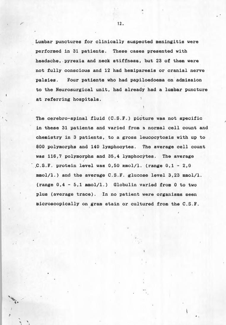
Lumbar punctures for clinically suspected meningitis were
performed in 31 patients. These cases presented with
headache, pyrexia and neck stiffness, but 23 of them were
not fully conscious and 12 had hemiparesis or cranial nerve
palsies. Four patients who had papiloedoema on admission
to the Neurosurgical unit, had already had a lumbar puncture
at referring hospitals.
The cerebro-spinal fluid (C.S . F . ) pi9ture was not specific
in these 31 patients and varied from a normal cell count and
chemistry in 3 patients, to a gross leucocytosis with up to
800 polymorphs and 140 lymphocytes. 'rhe average cell count
was 116,7 polymorphs and 35,4 lymphocytes. The average
.C.S.F. protein level was 0,50 mmol/1. (range 0,1 - 2,0
mmol/1.) and the average C.S.F. glucose level 3,23 mmol/1.
(range 0,4 - 5,1 mmol/1.) Globulin varied from Oto two
plus (average trace). In no patient were organisms seen
microscopically on gram stain or cultured from the C.S.F.
I '
' /
.. , ..... ....
'.
·' ,, ' '
13.
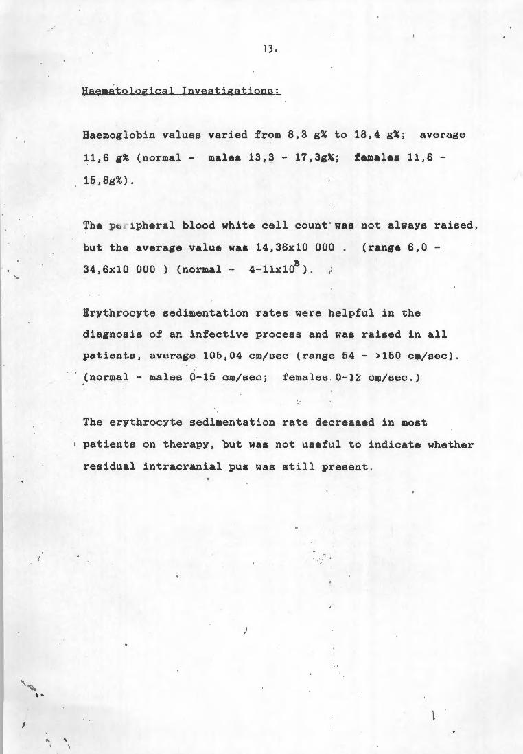
Haematological Investigations~
Haemoglobin values varied from 8,3 g% to 18,4 g%; average
11,6 g% (normal - males 13,3 - 17,3g%; females 11,6 -
15,6g%) .
The p pheral blood white cell count · was not always raised,
but the average value was 14,36x10 000
~ 34,6x10 000) (normal - 4-11x10 ) . •
(range 6,0 -
Erythrocyte sedimentation rates were helpful in the
diagnosis of an infective process and was raised in all
patients, average 105,04 cm/sec (range 54 - >150 cm/sec).
(normal - males 0-15 cm/sec; females . 0-12 cm/sec.)
The erythrocyte sedimentation rate decreased in most
1 patients on therapy, but was not useful to indicate whether
residual intracranial pus was still present.
'
)
(
,•
'
14.
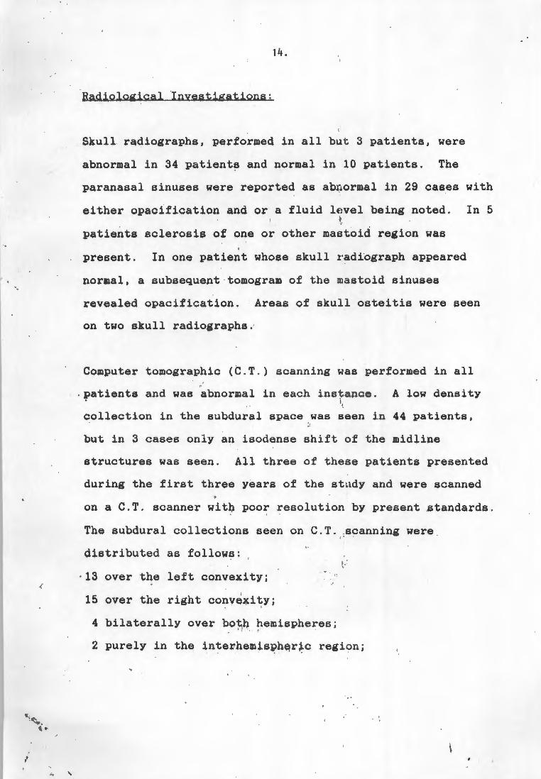
Radiological lnve§tigations:
Skull radiographs, performed in all but 3 patients, were
abnormal in 34 patients and normal in 10 patients. The
paranasal sinuses were reported as abnormal in 29 cases with
either opacification and or a fluid level being noted. In 5 ) ..
patients sclerosis of one or other mastoid region was
present. In one patient whose skull radiograph appeared
normal, a subsequent tomogram of the mastoid sinuses
revealed opacification. Areas of skull osteitis were seen
on two skull radiographs.·
Computer tomographic (C.T.) scanning was performed in all
. patients and was abnormal in each instance. A low density I,
collection in the subdural space was seen in 44 patients,
but in 3 cases only an isodense shift of the midline
structures was seen. All three of these patients presented
during the first three years of the stndy and were scanned
on a C.T. scanner wit~ poor resolution by present atandards.
The subdural collections seen on C.T . . scanning were , . .
distributed as follow~:
·13 over the left convexity;
15 over the right conve~ity;
4 bilaterally over both hemispheres; . 1; . '
2 purely in the interhemis~he.r~c region;
.. . ' •·
·'
15.
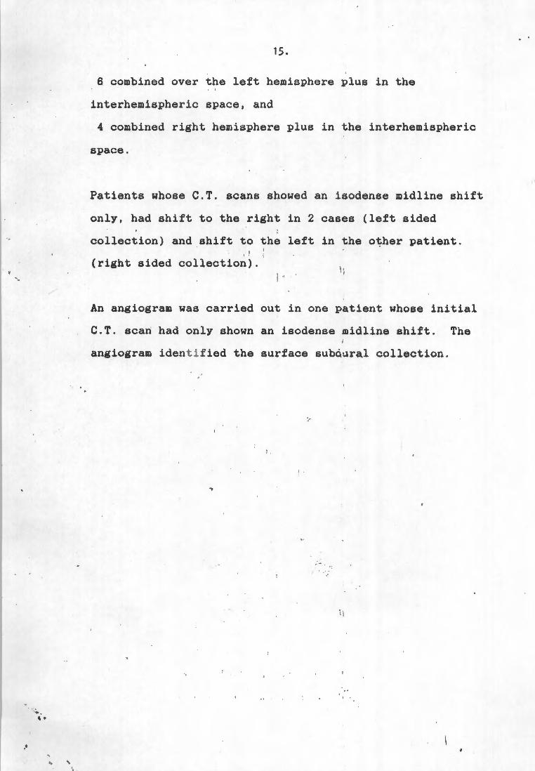
6 combined over the left hemisphere plus in the
interhemispheric space, and
4 combined right hemisphere plus in the interhemispheric
space.
Patients whose C.T. scans showed an isodense midline shift
only, had shift to the right in 2 cases (left sided
collection) and shift to the left in the other patient.
(right sided collection). ', , ,,
An angiogram was carried out in one patient whose initial
C.T. scari had only shown an isodense midline shift. The
angiogram iden i f ied the surface subaural collection .
·I, "" · ' ..
I
,. '
16.
Other Special Investigations:
Radio-isotope brain scans were performed in 2 patients prior
to C.T. scanning, and both revealed a~eas of increased
uptake over the correct hemisphere.

.,
..
. '
•'· . ' .
'
17.
Source.~.
The primary source of the S.E. was iniection in the
paranasal sinuses in 31 cases, chronic ear infection with
mastoiditis in 10 cases and osteitis of the skull in 2
cases. The source of the infection was identified on skull
radiographs, or at operqtion when pus was drained from a
sinus or mastoid that was explored on. clinical sus~icion.
In 4 patients no source for the S.E . qould be identified.
These patients had no evidence of local or systemic
infection that might have caused the S.E .
' ··-:-.... . '.
\ \ '
18.
Management. : ., l
The management o S.E. patients followed recognised
criteria, and dif~ered only in the application of
therapeutic methods.
Antibiotics:
All patients were commenced on intravenous antibiotic t
medication as soon as the diagnosis of S.E. was confirmed.
Antibiotic drugs were started as soon as pus had been sent
for Bacteriological investigation. Antibiotic combinations
differed somewhat . depending on the chol ce of the initial
doctor who saw the patient. A combination of Penicillin G
5mu 6 hourly, Chloramphenicol 500 mg 6 hourly, and
Metronidazole 500 g 8 hourly, was usec in all but 7
patients. Three patients received oni~ Penicillin G and
Chloramphenicol, two patients a combination of Cloxacillin . .
and Chloramphenicol, ' one patient a combination of '
Ampicillin, Sulfadiazine and Metronidazole, and one patient
received Ampicillin, Tobramycin, Chloramphenicol and
Metronidazole.
. /
I
'
19.
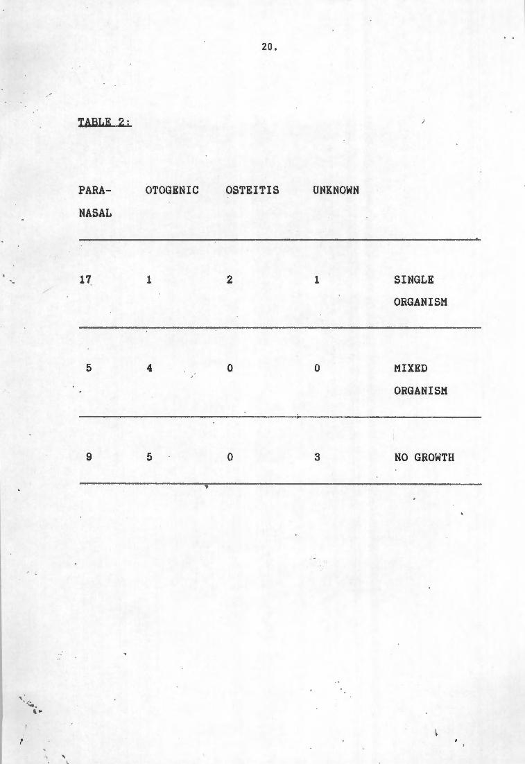
Pus specimens obtained at operation from the paranasal
sinuses, mastoid cavity; and/or subdural space, were sent
for aerobic and anaerobic culture in a!l patients at the
time of operation, and antibiotic regimes were changed if
cultures indicated the need. No growth was obtained in
aerobic or anaerobic culture in 15 cases. Single organisms
grew in 23 cases and mixed organisms in 9 cases . A detailed
analysis of . this data is given in Table 2.
Penicillin resistance occurred in twelve organisms cultured,
and the Penicillin G was changed to Cloxacillin in 11 cases
and to Cephamandole in the other.
Resistance to Metronidazole was present in three organisms
in vitro. These organisms were sensi~ive to
Chloramphenicol. Treatment with Chlor~mphenicol and
Metronidazole was continued in all three patients.
. ' · ..•. ' ..
,,
'
TABLE 2:
PARA
NASAL
17
5
9
OTOGENIC
1
4
5
20.
OSTEITIS
2
0
0
.•
UNKNOWN
1
0
SINGLE
ORGANISM
MIXED
ORGANISM
--------------
3 NO GROWTH
.. , -. ' ..
' .... , '
21.
Tobramycin was later added to the antibiotic regime in 2
patients - in one case because of Tobramycin sensitivity
shown in vitro, and in the other case. on clinical grounds in
a patient who remained pyrexial after surgical drainage of
S.E. and whose culture showed no growth.
Vancomycin was added in one patient where a staphylococcus
aureus which was resistant to Penicillin, but sensitive to
Vancomycin and Cloxacillin was cultured. Th- Penicillin G
was also changed to Cloxacillin in this ~atient.
Chloramphenicol was used in 46 patients and none of the
organisms cultured showed resistance to Chloramphenicol. No
·complications due to Chloramphenicol administration
occurred . :,-
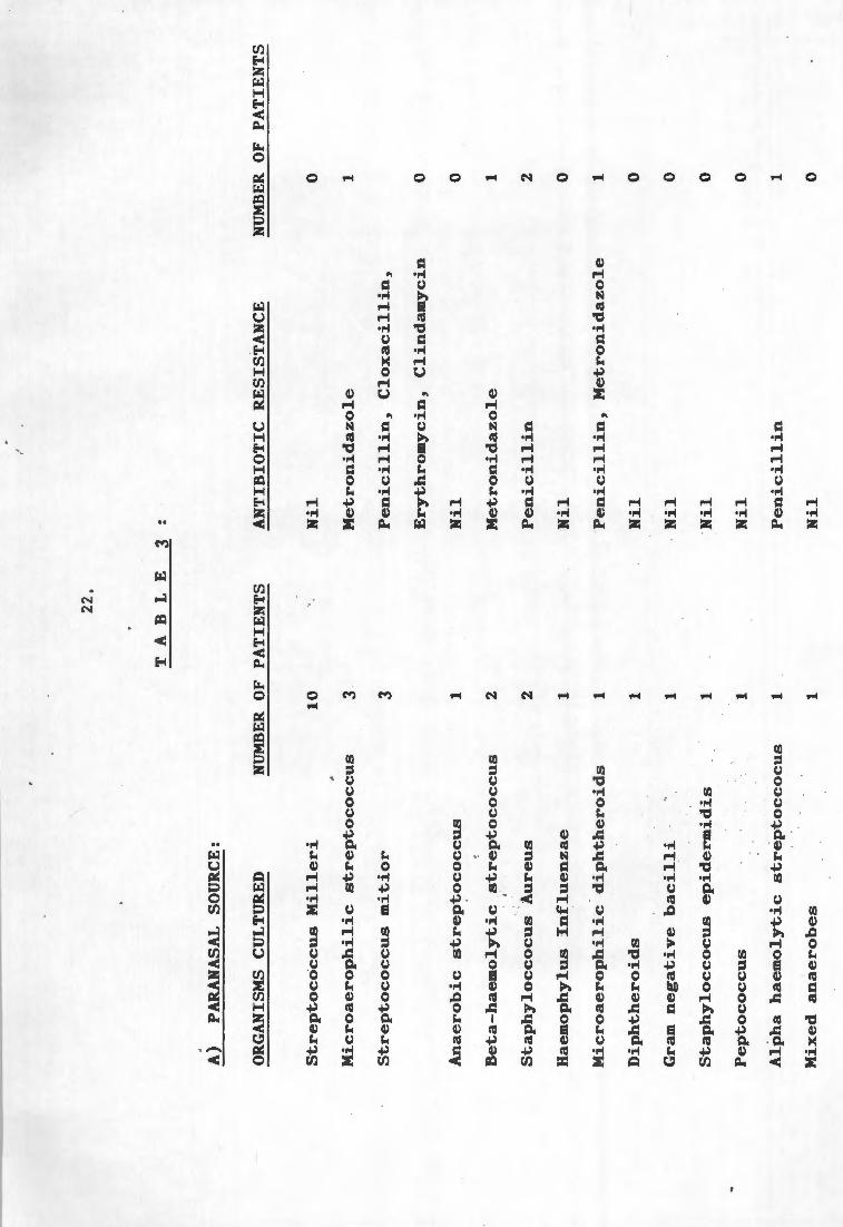
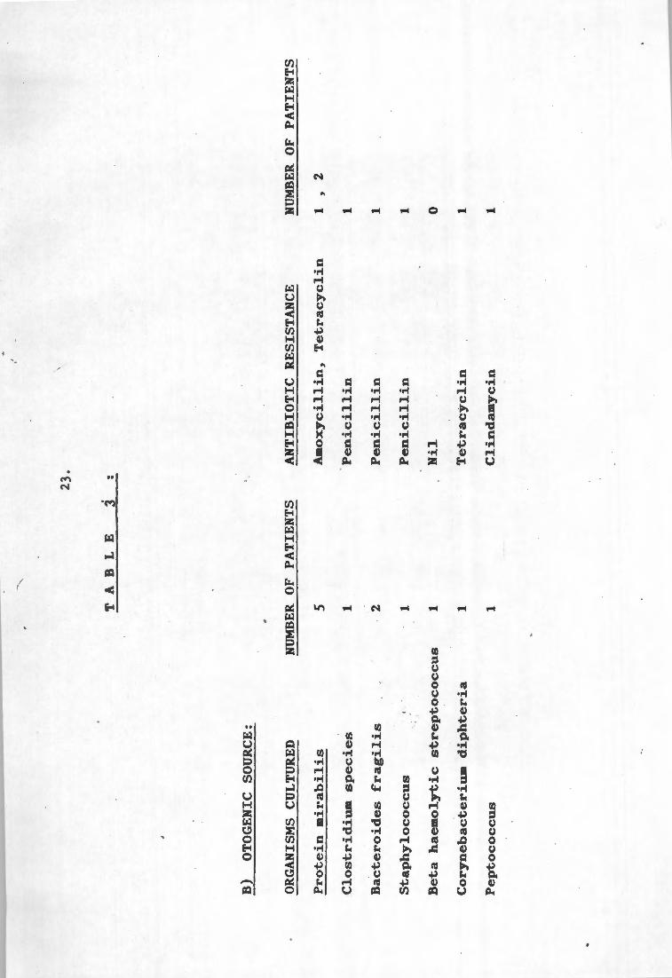
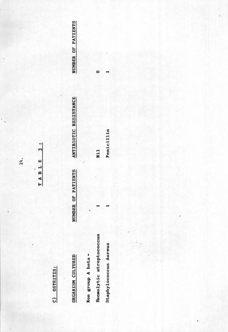
Or.ganisms cultured and the incidence ,of antibiotic
resistance in vitrQ are shown in Tlw],~.
In one patient where no source for the S.E. was indentified,
a blood culture grew a streptococcus milleri, not resistant
in vitro against the standard battery of antibiotics. The
same organism was cultured from the subdural pus .
22.
T
A
B
L
E
3 :
A)
PAR
AN
ASA
L SO
UR
CE
:
OR
GA
NIS
MS
CU
LT
UR
ED
N
UM
BER
O
F P
AT
IEN
TS
A
NT
IBIO
TIC
R
ES
IST
AN
CE
Str
ep
toc6
ccu
s M
ille
ri
Mic
roaero
ph
ilic
str
ep
toco
ccu
s
Str
ep
toco
ccu
s m
itio
r
An
aero
bic
str
ep
toco
ccu
s
Beta
-haem
oly
tic str
ep
toco
ccu
s
'
Sta
ph
ylo
cco
cu
s A
ure
us
Hae
mo
ph
ylu
s In
flu
en
zae
Mic
roaero
ph
ilic
d
iph
thero
ids
Dip
hth
ero
ids
Gra
m n
eg
ati
ve b
acil
li
Sta
ph
ylo
cco
cu
s ep
iderm
idis
Pep
toco
ccu
s
Alp
ha h
aem
oly
tic str
ep
tocco
cu
s
Mix
ed an
aero
bes
10
3 3 1 2 2 1 1 1 1 1 1 1 1
Nil
Metr
on
idazo
le
Pen
icil
lin
, C
lox
acil
lin
,
Ery
thro
my
cin
, C
lin
dam
ycin
Nil
Metr
on
idazo
le
Pen
icil
lin
Nil
Pen
icil
lin
, M
etr
on
idazo
le
Nil
Nil
Nil
Nil
Pen
icil
lin
Nil
NU
MB
ER
OF
PA
TIE
NT
S
0 1 0 0 1 2 0 1 0 0 0 0 1 0
'
T A
B
L
E
B)
OT
OG
EN
IC
SOU
RC
E:
23.
3 :
OR
GA
NIS
MS
CU
LTU
RED
Pro
tein
mir
ab
ilis
Clo
str
idiu
m s
pecie
s
Bacte
roid
es frag
ilis
Sta
ph
ylo
co
ccu
s
NU
MB
ER
OF
PA
TIE
NT
S
5
Beta
h
aem
oly
tic str
ep
toco
ccu
s
Co
ryn
eb
acte
riu
m d
iph
teri
a
Pep
toco
ccu
s
1 2 1 1 1 1
AN
TIB
IOT
IC
RE
SIST
AN
CE
Am
ox
ycil
lin
, T
etr
acy
cli
n
Pen
icil
lin
Pen
icil
lin
Pen
icil
lin
Nil
Tetr
acy
cli
n
Cli
nd
am
yci
n
NU
MBE
R O
F P
AT
IEN
TS
1 ,
2
1 1 1 0 1 1
C)
OS
TE
ITIS
:
OR
GA
NIS
M
CU
LTU
RED
Non
g
rou
p
A b
eta
-
Haem
oly
tic str
ep
toco
ccu
s
Sta
ph
ylo
co
ccu
s A
ure
us
24.
T A
B L
E
NU
MB
ER
OF
PA
TIE
NT
S
1 1
3 :
AN
TIB
IOT
IC
RE
SIS
TA
NC
E
Nil
Pen
icil
lin
NU
MB
ER
OF
PA
TIE
NT
S
0 1
. (
-. ' . . •
'
25,
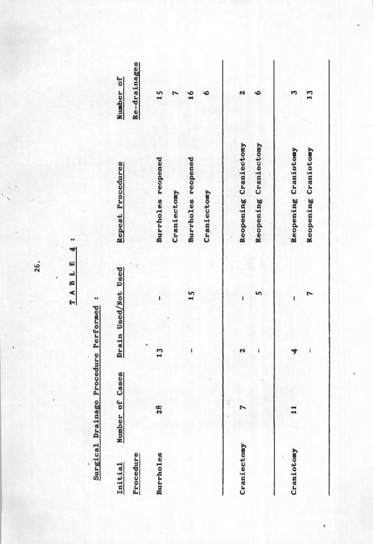
~y.n;iQ_qJ Drainage: J~..,__i.., __ L_
Surgical drainage of the empyema was '~arried out in 46
patients. In one patient the collection was deemed small
enough to manage ~ithout drainage, and only the source, i.e.
the paranasal sinuses in this case, were drained.
Burrholes were used to drain subdural, collections in 28 \.
cases; an initial craniectomy in 7 cases, and a craniotomy
initially in 11 patients. Thirteen patients who initially
only had burrhole drainage, later needed a more extensive
surgical procedure, i.e. a craniectomy or craniotomy . I~~ '
shows the surgic,1 procedures used i~itially and later when
·repeat drainage was indicated. All patients had post
operative C.T. scans and evidence of residual pus in the ' '
subdural space was used to indicate the need for repeat
drainage.
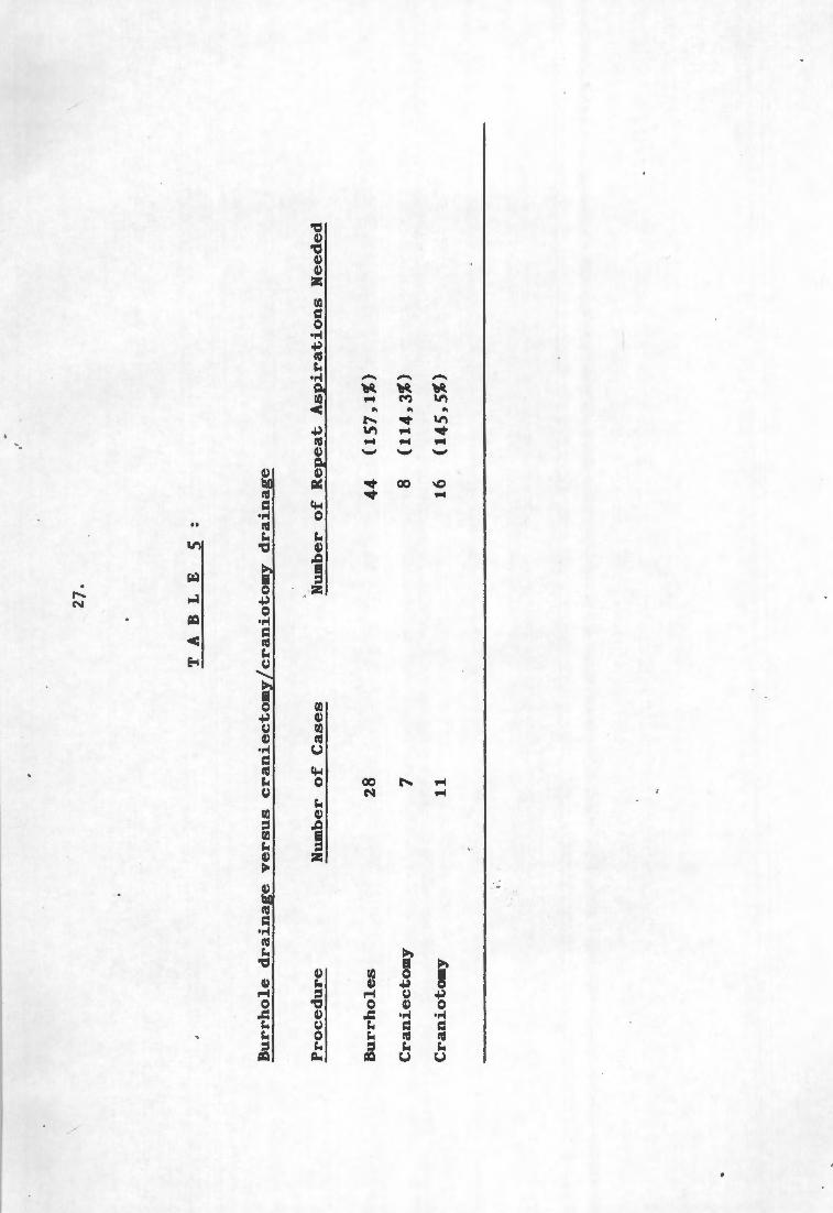
. Adding cases where a craniectomy was perfo~med as a repeat
procedure to those with initial craniectomy or craniotomy,
the incidence of re-aspiration between cases who had
burrholes only and cases with more e~tensive surgical
drainage procedures can be compared. The results are shown ; •
in Table 5.
26.
T
A B
L
E
4
:
Su
rgic
al
Dra
inag
e P
roced
ure
P
erf
orm
ed
:
In
itia
l
Pro
ced
ure
Bu
rr h
ole
s
Cra
nie
cto
my
Cra
nio
tom
y
Nu
mb
er o
f C
ase
s
28 7
11
Dra
in U
sed
/No
t U
sed
13
15
2
5
·4
7
Rep
eat
Pro
ced
ure
s
Bu
rrh
ole
s
reo
pen
ed
Cra
nie
cto
my
Bu
rrh
ole
s
reo
pen
ed
Cra
nie
cto
my
Reo
pen
ing
Cra
nie
cto
my
Reo
pen
ing
Cra
nie
cto
my
Reo
pen
ing
Cra
nio
tom
y
Reo
pen
ing
Cra
nio
tom
y
Hu
mb
er o
f
Re-d
rain
ag
es
15
7
16
6 2 6 3
13
27.
T
A B
L
E
5
:
Bu
rrh
ole
d
rain
ag
e v
ers
us cra
nie
cto
my
/cra
nio
tom
y d
rain
ag
e
Pro
ced
ure
Bu
rr h
ole
s
Cra
nie
cto
my
Cra
nio
tom
y
Nu
mb
er o
f C
ase
s
28 7 11
Nu
mb
er o
f R
ep
eat
Asp
irati
on
s
Need
ed
44
(1
57
,1%
)
8 (1
14
,3%
)
16
(14
5,5
%)
. . .. ' .
. •
'
28.
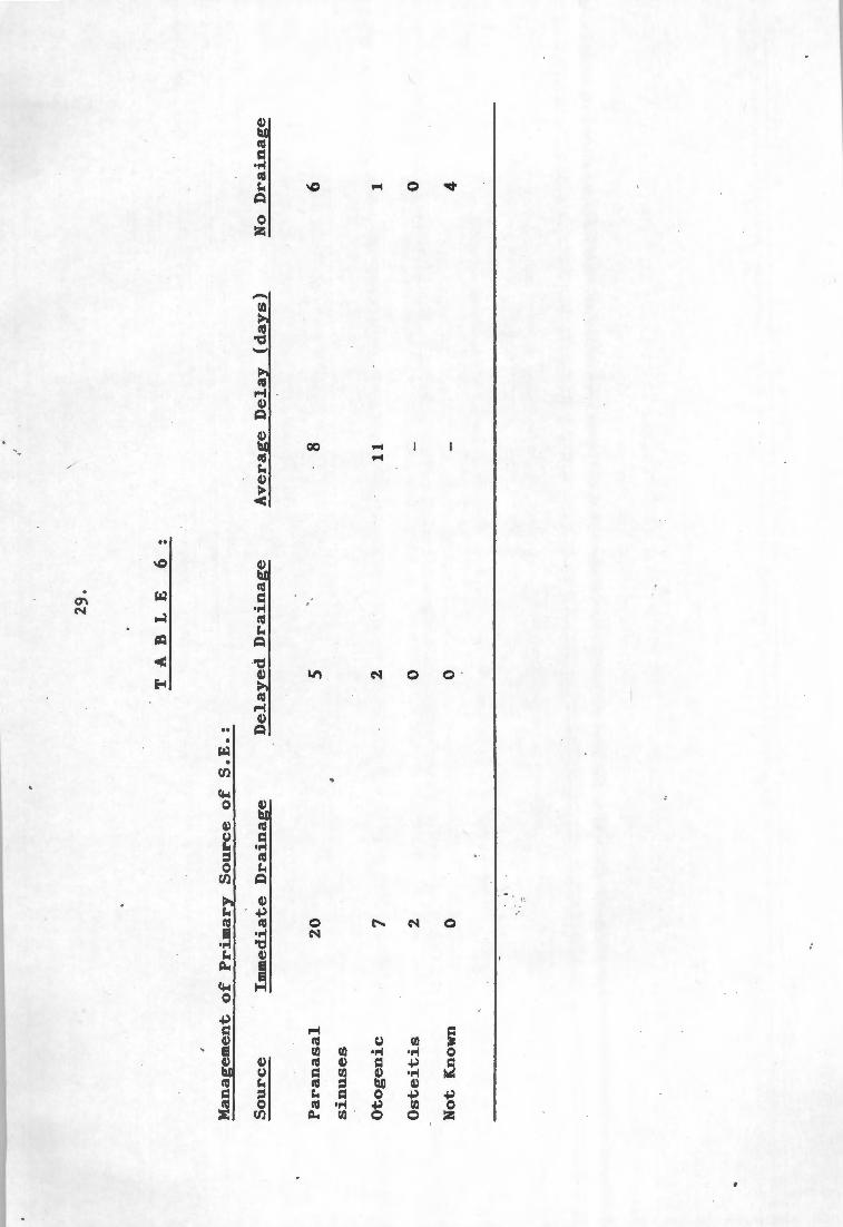
Surgical Drainage: <Source):
The primary source for the subdural empyema was drained
surgically in the majority of cases . Table 6 shows how the
sources were managed. In cases where definite collections
of pus were seen in the paranasal sinuses or mastoid,
drainage was carried out immediately. Where there was doubt
about the source, drainage was sometimes delayed. Pus was
found in all cases where the source was drained.
I .. , ~• , .
29.
T
A B
L
E
6
:
Man
agem
ent
of
Pri
mary
S
ou
rce o
f S
.E.:
So
urc
e
Imm
ed
iate
D
rain
a~
D
e1
ay
ed
D
rain
ag
e
Av
era
ge
De1
ay
(day
s)
No
Dra
inag
e
Para
nasa
1
20
5
8 6
. sin
uses
Oto
gen
ic
7 2
11
1
Oste
itis
2
0 -
0
No
t K
now
n 0
0 -
4
'
30.
OTHER MEDICATI..Qli~
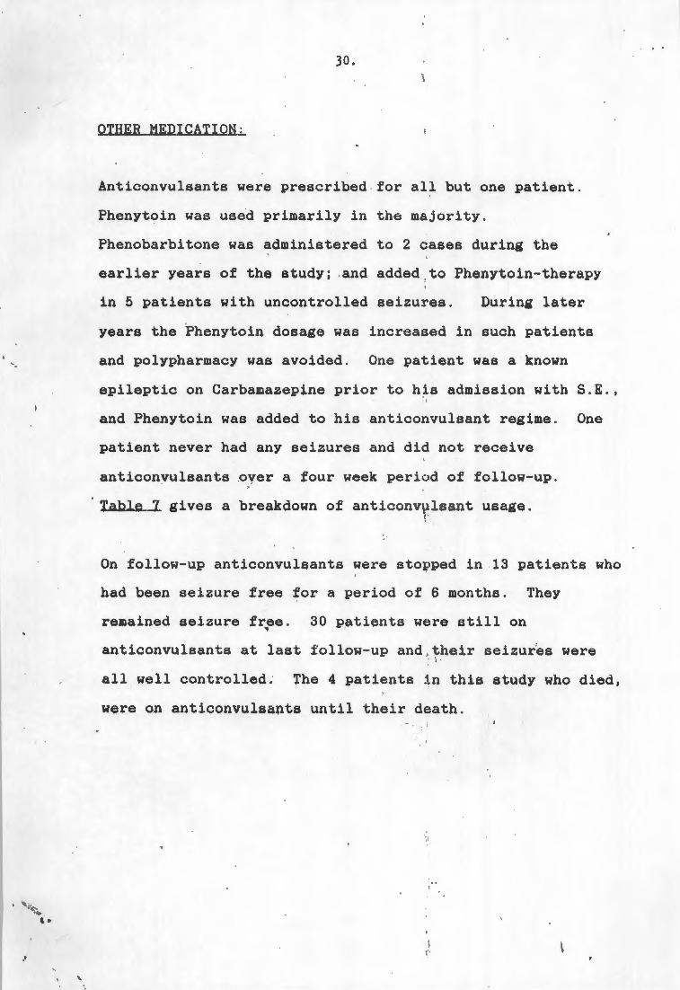
Anticonvulsants were prescribed for all but one patient.
Phenytoin was us~d primarily in the majority.
Phenobarbitone was administered to 2 cases during the
earlier years of the study; .and added ,to Phenytoin-therapy
in 5 patients with uncontrolled seizures. During later
years the Phenytoin dosage was increased in such patients
and polypharmacy was avoided. One patient was a known
epileptic on Carbamazepine prior to his admission with S.E., I
and Phenytoin was added to his anticonvulsant regime. One
patient never had any seizures and did not receive
anticonvulsants over a four week period of follow-up.
Table-1. gives a breakdown of anticonv~lsant usage.
On follow-up anticonvulsants were sto~ped in 13 patients who
had been seizure free for a period of 6 months. They
remained seizure free. 30 p~tients were still on •
anticonvulsants at last follow-up and .their seizures were I
all well controlled. The 4 patients in this study who died,
were on anticonvulsa~ts until their death.
I l '
{",
An
tico
nv
uls
an
t U
sag
e in
S
.E.:
Dru
g U
sed
Ph
en
yto
in
Ph
en
ob
arb
ito
ne
Ph
en
yto
in
+
Ph
en
ob
arb
ito
ne
Ph
en
yto
in
+
Carb
am
azep
ine
Nil
Nu
mb
er o
f
Pati
en
ts
To
tal:
38
2 5 1 1
31.
T
A B
L
E
7
:
Wee
ks
of
Fo
llo
w-u
p
(Av
era
ge}
23
,7
76
46
6
4
Wit
h S
eiz
ure
s
at
Pre
sen
tati
on
16
2
1 1 0
Wit
h S
eiz
ure
s
at
Fo
llo
w-u
p
5 2 1 0 0
'·· ·-' .. ,'
'
32.
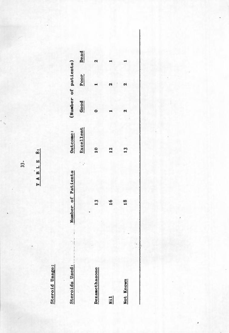
Dexamethasone was used in 1~ patients and not used in 16
patients. The records regarding stero i d administration were
lost in the other 18 patients in this series. There was no
specific indication for Dexamethasone administration and
their use depended on the approach of the initial doctor who
treated the case.
The eventual outcome of cases on steroids and not on
steroids are given in Table 8.
I
33.
TA
BL
E
8:
Ste
roid
Usa
ge:
Ste
roid
s U
sed
: N
um
ber
o
f P
ati
en
ts
Ou
tco
me:
(N
um
ber
o
f p
ati
en
ts)
-· --
. -·-
·---
--
-----
Ex
cell
en
t G
oo
d
Po
or
Dea
d
4
Dex
amet
has
on
e 1
3
10
0
1 2
Nil
1
6
12
1
2 1
No
t K
now
n 1
8
13
2
2 1
'.
,' ., '
34.
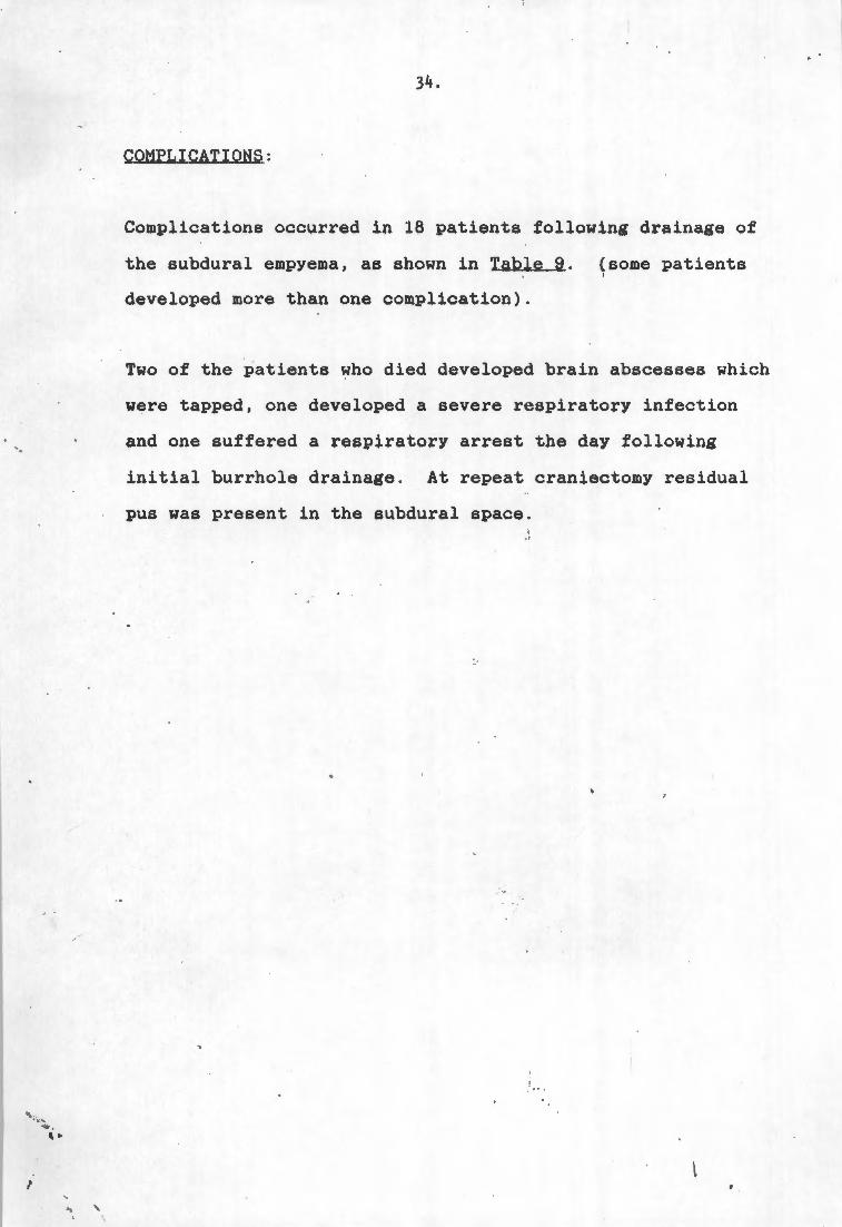
COMPLICATIONS:
Complications occurred in 18 patients following drainage of
the subdural empyema, as shown in Table 9.
developed more than one complication).
(some patients I
Two of the patients ~ho died developed brain abscesses which
were tapped, one developed a severe respiratory infection
and one suffered a respiratory arrest the day following
initial burrhole drainage. At repeat craniectomy residual
pus was present in the subdural space.
. ' • . ' •.
TABLE 9:
· Complications that ~ccurred:
Brain Abscess (frontal)
Ventriculitis
Hydrocephalus requiring a shunt
Delayed skull osteitis
Craniotomy bone flap removed
because of wound infection
Severe respiratory infection
Respiratory Arrest
Subgaleal abscess
Brain infarct on later CT scan
· Subdural haematoma
Phenytoin sensitivity
'
35.
TOTAL Number of Cases:
Burrholes Craniectomy Craniotom
5
2
3
2
2
2
1
1
1
1
1
4
3
1
2
1
1
1
1
1
1
0
3
1
1
1
1
0
0
0
0
0
0
0
1
0
0
0
2
1
0
0
0
0
1
1
' · ..... .... . ' ..
·' '
36.
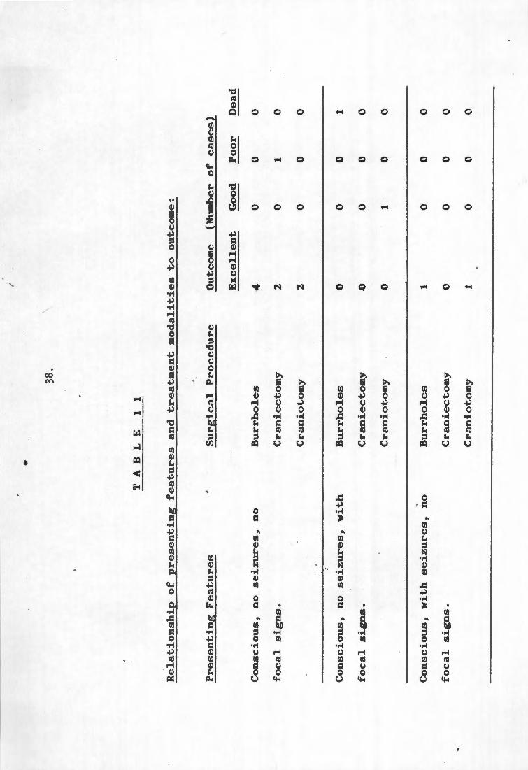
OUTCOME:
Outcome from S.E. in this study was divided into four
groups, as follows:
Excellent: Well, with no focal signs, no seizures, back to
pre-disease activity.
Good; Well, with minimal focal signs, or seizures that are
well controlled on anticonvulsants, back to pre
disease activity.
Poor: Disabled by hemiparesis or hemiplegia, or by seizures
that are difficult to cont~ol. Not able to
function at a pre-disease level.
Dead: Patients who died from their S.E. or of complications
thereof.
The overall outcome in this study is shown in Table .lil..
·Known presenting symptoms or signs and the treatment
modalities that may affect outcome are compared with outcome
in Table 11.

37.
T A
B L
E
1 0
:
Ov
era
ll
ou
tco
me
fro
m S
.E.,
co
mp
ari
ng
vari
ou
s
form
s o
f in
itia
l su
rgic
al
dra
inag
e p
roced
ure
s.
NU
MB
ER ·
oF
CA
SES
OU
TCO
ME
EX
CE
LL
EN
T
GO
OD
PO
OR
Bu
rrh
ole
s 28
20
(7
1,4
%)
2 (7
,1%
) 3
(10
,7%
)
Cra
nie
cto
my
7
5 (7
1,4
%)
1 (1
4,3
%)
1 (1
4,3
%)
Cra
nio
tom
y
11
8
(72
,7%
) 1
(9,1
%)
1 (9
,1%
)
No
su
rgery
1
. 1
(100
%)
0 (0
%)
0 (0
%)
To
tal
47
34
(72
,3%
) 4
(8,5
%)
5 (1
0,6
%)
DIE
D
--
3 (1
0,7%
)
0 (0
%)
1 (9
,1%
)
0 (0
%)
4 (8
,5%
)
• 38
.
T A
B L
E
1
1
Rela
tio
nsh
ip o
f p
resen
tin
g fe
atu
res
an
d tr
eatm
en
t m
od
ali
ties to
o
utc
om
e:
Pre
sen
tin
g F
eatu
res
Co
nscio
us,
no
seiz
ure
s,
no
focal
sig
ns.
Co
nsc
iou
s,
no
seiz
ure
s,
wit
h
focal
sig
ns.
Co
nscio
us,
wit
h seiz
ure
s,
no
focal
sig
ns.
Su
rgic
al
Pro
ced
ure
Bu
rrh
ole
s
Cra
nie
cto
my
Cra
nio
tom
y
Bu
rr h
ole
s
Cra
nie
cto
my
Cra
nio
tom
y
Bu
rrh
ole
s
Cra
nie
cto
my
Cra
nio
tom
y
Ou
tco
me
(Nu
mb
er o
f cases)
Ex
cell
en
t
,.(
2 2 0 -0
0 1 0 1
Go
od
0 0 0 0 0 1 0 0 0
Po
or
0 1 0 0 0 0 0 0 0
Dead
0 0 0 1 0 0 0 0 0
Pre
sen
tin
g F
eatu
res
Co
nsc
iou
s,
wit
h seiz
ure
s,
an
d
wit
h fo
cal
sig
ns.
Un
co
nsc
iou
s,
no
seiz
ure
s,
no
focal
sig
ns.
Un
co
nsc
iou
s,
no
seiz
ure
s,
wit
h
focal
sig
ns.
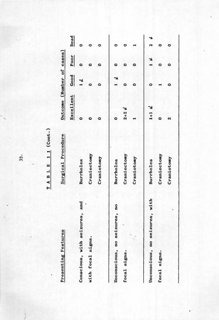
39,
TA
BL
E
1 1
(Co
nt.
)
Su
rgic
al
Pro
ced
ure
Bu
rr h
ole
s
Cra
nie
cto
my
Cra
nio
tom
y
Bu
rr h
ole
s
Cra
nie
cto
my
Cra
nio
tom
y
Bu
rrh
ole
s
Cra
nie
cto
my
Cra
nio
tom
y
Ou
tco
me
(Nu
mb
er o
f cases)
Ex
cell
en
t
0 0 0 0 2+
2 ,J
1 1+
1
V.
0 2
Go
od
1 tL
0 0 1 J.
0 0 0 1 0
Po
or
0 0 0 0 0 0 1 ,t
0 0
Dea
d
0 0 0 0 0 1 2 J,
0 0

Pre
sen
tin
g F
eatu
res
' '
Un
co
nsc
iou
s,
wit
h seiz
ure
s,
no
focal
sig
ns.
Un
co
nsc
iou
s,
wit
h seiz
ure
s,
an
d
wit
h fo
cal
sig
ns
40.
T A
B
L
E
1
1 (C
on
t.)
-------
Su
rgic
al
Pro
ced
ure
Bu
rrh
ole
s
Cra
nie
cto
my
Cra
nio
tom
y
Bu
rr h
ole
s
Cra
nie
cto
my
Cra
nio
tom
y
Ou
tco
me
(Nu
mb
er o
f cases)
Ex
cell
en
t
6+
1 ~
1 1 1+
3
J.
0 1
Go
od
0 0 0 0 0 0
Po
or
1 iJ
0 0 1 ,t
0 1
Dea
d
0 0 0 0 0 0
(Th
e arr
ow
,
ind
icate
s p
ati
en
ts w
ho
h
ad
in
itia
l b
urr
ho
les w
hic
h w
ere
la
ter ex
ten
ded
into
a
cra
nie
cto
my
.)
'•·
,•
'
Il.I. SC t..l S s._i;_ QM.
HISTORICAL PERSPECTIVE:
41.
Although a case of S.E. at autopsy had been described as
early as 1812 *(71), the whole problem of intracranial
sepsis was clarified by the publication of Macewen's now
classical monograph on this topic in ~893. *(57). Prior to
the availability of antibiotics, the ~reatment of brain
abscess was "marsupialisation", with insertion of a drain.
When antibiotics became available, this form of surgical
drai~age was abandoned because of problems with brain
herniation, and the instillation of antibiotics through
cannulae into the abscess cavity became current therapy for
·intrace~ebral abscess.
The same principle was used in the tr~atment of S . E.
Multiple catheters were inserted into the subdural space
through multiple b~rrholes, and used to inject
antibiotics.*(4)(70)(86). This led to an improve~ outcome
from S.E., with some patients surviving, whereas survival
after intracranial infection prior -to antibiotics was rare.
*(49) The problem was not solved however and the mortality '
and morbidity of S.E. remained high .
... . ' .
.'
'
42.
Courville described the preferred route of spread of
subdural pus in 1944. *(19) This extensive spread over the
convexities of both hemispheres, under the frontal lobes,
along the Sylvian fissure and along t4e interhemispheric
fissure, makes adequate drainage of pus by insertion of
catheters not under direct vision difficult. Instilled
antibiotics are also not likely to reach all the organisms
in such a complex space.
Recognition of the problems of S.E. drainage by catheters·
that were inserted blindly, lead to t~e use of extensive
craniectomies to pursue pus in the subdural space. Reports
by Botterel and Drake *(12) and Sterp and Boldrey *(75),
showed results t~at . were superior to those obtained by
burrholes. Extended craniectomy is however a tedious .:··
procedure causing much blood loss and .a poor cosmetic
result. The use of a craniotomy to drain subdural pus
therefore seemed lo~ical. Le Beau , reported the first . sizeable series on craniotomy for dra:j.nage of S. E ., in 1949.
*(51) .
. The advantages of a craniotomy rather. than burrholes or
craniectomy for S.E. drainage, seemed certain after the
report of Bannister et al in 1981 *(5), showing that the
mortality rate was reduced three fold ; by the use of
' , .
' I
.. ,,. '.
/
'
43 •.
craniotomy drainaie versus burrhole-d;.ainage. This however,
.was a retrospective study over 20 years, and many of thejr
patients were treated prior to the availability of C.T .
scanning. /
Kaufman et al suggested in 1983 that the use of C.T.
sc~nning in the treatment of S.E. may make a difference by
enabling surgeons to place burrholes accurately. *(46).
The report by Luken et al however, has cast doubt on the
usefulnes of C.T. scanning in S.E. patients . *(56}.0n the
other hand, Leys et al even advocates managing S.E. only
with antibiotics and follow up C.T. scanning, without the
use of surgical drainage . *(53).
At Groote Schuur Hospital S.E. is a relatively common -
condition, and 47 cases were treated during the eight years • :'
of this study, all with the use of reveated C.T. scanning.
No fixed management policy was used, and the treatment of
patients with S.E. wa~ individualised according tq the
choice of the Neurosurgeon on duty when the patient was
admitted. Thus various forms of therapy can be compared.
. ' ..
..
'
44.
INCIDENCE
' Williams found that the incidence of S.E. was not decr~asing
in his referring region, despite the use of prophylactic
surgery for ear infections and the availability of newer
antibiotics . *(84). He also docu~ented an incidence of 3-4
cases per million population.
•· ,,
The incidence at Groote Schuur Hospital during the period of
this stud~, was nearly 6 cases per ye~r; although the
incidence seemed to decline since 1985, when C.T . scanning
~nd Neurosurgical services became available in the Eastern
Cape . The majority of our cases live locally, which tends
· .to substantiate the observation by Williams *(84) that the
availability of prophylactic suraery for sinus and ear
infections, as well as antibiotic use, do not prevent the
occurence of S . E.
The age distribution of cases in this. study conforms with
that in published reports. *(82). 8b% of cases were under
20 years of age, and 62% were in their teens. Only one
patient was over 35 years of age. A ~triking male
predominance was present, in contrast / to most studies where
the male dominance was slight. *(84)
' 110"'-,,. . ' ..
. • ·, '
45.
The ethnic distribution of patients in this study probably
reflects the referral pattern of patients to Groote Schuur
Hospital. The relatively low incidence amonast the black
population, might indicate that the diagnosis is missed in
areas where sophisticated medical services are not
immediately available. \
When one compares the incidence of cerebral abscess to that
of S.E., the incidence of 5 to 1 differs from that found by
Gurdjian, where brain abscess was twice as common as S.E.
*(35). The study of 79 cases of intracranial suppuration
over 3 years seen at Baragwanath Hospital, Johannesburg,
included 25 cases of S.E. Solitary intracerebral abscesses
were present in 32 of their patients. Trauma, including
penetrating trauma to the skull was, however not excluded as :r
an etiological factor. *(2).
'\ '
''{ .. ' •·
..
'
46.
PREDISPOSING FACTORS: I
;j l
In the large series by Bannister et al the presumed source
of infection in their 66 cases was paranasal in 45 patients
(68%), otogenic in 15 (21%), post tra~ma in 2, other
osteomyelitis in 3, a lung abscess in 1 and congenital heart
disease in 1. *(5). Cyanotic heart disease is a well
described cause for brain abscess. *(39). Haematogenous
spread may also occur from other systemic infections, eg.
dental abscess or renal tract infection. *(14)(30)(41).
Bacterial seeding may be to cortical vessels first, with
resultant vasculitis, vessel rupture and brain abscess, with
secondary breakthrough into the subdural space. *(29).
' Another mechanism that has been suggested, is haematogenous
bacterial seeding to a chronic s~bdural haematoma.
*(17)(18).
r;
S.E. following cran~al surgery, skull traction, penetrating
skull trauma and infected effusions post meningitis are also
included in many studies. This makes ,Jnterpretation of the
outcome from S.E. difficult .because these cases are easier
to diagnose due to a higher clinical suspicion than for S.E.
following paranasal or otogenic infection, or fro_m
haematogenous spread. In this study all cases of subdural
infection ~allowing skull trauma, cranial surgery or
meningitis were therefore excluded.
~
' ..
' '
47,
Paranasal infection is also the main cause of S . E. in this ,
study (66%) followed by otogenic infection (21%). No cause
w~s found in 4 cases in spite of a careful clinical and
radiological search.
S . E. following paranasal infection arises mostly from the
frontal sinuses, *(18)(66) but may aleo &rise from the
ethmoid and sphenoid sinuses, and even from the maxillary
antra. *(84) In our study pus was drained from the frontal
sinuses in most cases, but the ethmoid einuses were
responsible in a few cases, and pansinusitis was present in
some cases. Delayed S.E. after trauma to the frontal
sinuses which interfered with their normal drainage has also
been described. *(64).
. •
'
·,
48.
PATHOGENESIS:
A direct communication between the infected sinus or mastoid
cavity and the extradural space is sometimes found. In the
majority of cases this is not the case however, indicating
that the infection had spread through · in ta.ct skull.
Infection may traverse naturally occurring portals of entry,
such as the olfactory foraminae, the labyrinth and
thereafter the internal auditory meatus and the vestibular
aqueduct, congenital deficiencies, fissure fractures and
suture lines. *(63). Infection may also spread from the
veins of the sinus mucosa, via diploic veins to the
intracranial cavity. *(59).
The second line of defense is th~ dura mater, which offers
considerable protection .against infection due to its
structure and good blood supply . The · dura mater may
completely resist penetration by infection so that an
extradural abscess is formed .
Penetration of the dura mater probably takes place along the
course of small vessels which traverse it, and by developing I
septic thrombophlebitis may provide convenient patl.ways.
The membrane swells as a result of the inflammatory reaction
and granulation tissue forms . This is called
pachyn:i'eningi tis .
, ,-.
:
49,
If the wall of a venous sinus is involved, thrombosis
ensues, which may involve the whole sinus wall and cause
thrombosis, or remain restricted to part of the sinus
circumference. Veins which cross the subdural space may
also be involved, spreading infection in that space. *(29).
A purulent exudate may form on the in~er surface of the
infected dura mater. If this accummulates rapidly, it
spreads widely and a subdural empyema is formed.
There is still no satisfactory explanation for the speed of
spread and lack of encapsulation in some cases and the
definite encapsulation seen in others where localised
subdural abscesses are formed. Virulence of organisms and
~peed of production of granulation tissue may be factors.
*(19)(29). No definite difference in virulence of infecting
organisms has however been shown.
The arachnoid provides further resista~ce to spread of
infection. In subd~ral empyema there is often no
leptomeningeal infection. *(63).
Extension of pus seems to occur mainly in a posterior '
direction, possibly aided by gravity. *(19). The pus tends
to accumulate in the Sylvian fissure, from where it courses
towards the basal qieterns. The ,interhemispheric fissure is
also invad~d. The basal .~urfaces of the fro~tal and
temporal lobes are rarely reached by e~udate, probably due
\ '. ~. ' ..
•'
. ' so.
to the close approximation of these lobes to the internal
surface of the skull.
The extent of spread to the opposite hemisphere varies
greatly, and probably depends on the amount of pus present,
the ease of communication beneath the inferior free margin i
of the falx cerebri ,· and perhaps the duration of infection.
*(19). The cases where bilateral subdural pus was present
in this study, had been ill for 10,1 days on average (range
4-21 days), which is longer than the average for the whole
series, (8,1 days). Bilateral S.E. remains relatively
uncommon. *(30).
Pus is rarely limited to the interhemispheric space.
*(19)(80). This is borne out by the 1ow incidence of only I
two cases (4,3%) in this study.
Osteomyelitis of the skull, whether focal or spreading, is
an uncommon accompaniment of S.E. Conversely, in recognized
cases of osteomyelitis of the skull, ~ubdural infection is \
also not common. From these observations one would conclude
that these two lesiqns ~esult from e~sentially different
routes of extension of infection from the frontal sinus.
*(19).

. ·', .. ' .
·' '
51.
The osteomyelitis of the skull causing S.E. in the two cases
in this series, probably resulted from earlier scalp trauma,
although this was not obvious at the time of presentation .
... . ,, .... '.
' '
52,
CLINICAL PRESENTATI.QN.:
The importance of making a diagnosis of S.E. early in the
course of the disease to effect an impioved outcome, has
been stressed in the literature. *(68). This study does not
support this statement. Patients whose outcome was judged
as excellent had an average duration of illness prior to a
diagnosis of S.E. being made of 7,76 days (range 1-21 days);
the figure for patients with a good outcome was 10,0 days
(range 5-21 days) versus 10,04 days (range 3-21 days) for
those with a poor outcome and 8,44 days (range 3~21 days) .
for patients who died.
·eommon presenting symptoms and signs in the series by
Williams were, in decreasing order of ~requency, impaired
consciousness, headache, hemiparesis, acute seizures,
pyrexia, meningismus, vomiting, papilloedoema,
ophthalmoplegia, he~ianopia and dyspha~ia. *(84). Other
studies also indicate that pyrexia, headache, loss 'of \ '
consciousness, hemiparesis and epilepsy are common
presenting features. *(3)(46)(62)(67)(&0). Smith et al also
found these features commonly in 12-16 year old patients.
In their patients in the 6 weeks to 2 year age group, fever,
neck stiffness, bulging fontanelle, vomiting and lethargy
were most common *(72), although many of these cases of S.E.
were complicating meningitis. A case Qf S.E. presenting
with retinal thrombophlebitis has been 'described. *(74) .
, '
'.
·'
'
53.
In this study pyrexia occurred inn~ rly all the patients.
Headache, neck stiffness and a decre a sed level of
consciousness were also very common f indings. Seizures and
a hemiparesis occurred in less than half of the patients as
presenting features. Although patients with S.E. often have
s~vere neurologic abnormalities when first seen, appropriate
therapy usually produces complete or nearly complete
recovery. *(25).
'
54.
DIFFERENTIAL DIAGNOSIS:
Intracranial suppuration, was suspected in all the patients
in this study. Meningitis was the favoured diagnosis, and
31 (66%) of our cases had lumbar punctures performed before
the diagnosis of S.E. was made. Once meningitis had been
disproved on C.S.F. results, brain ab~cess was usually the
next choice, and arrangements for C.T, scanning and transfer . ,.
to the Neurosurgical Unit were made. It is interesting to
note that lumbar punctures were performed in a significant
number of patients where clinical con·~ra-indications in the
form of a depressed level of consciousness, focal signs or
even papiloedoema were present. Lumb~r puncture in the
~resence of raised intracranial press~re carries well known
risks. *(16)(24)(69). There was )howe?er no definite
incidence of deterioration in the clinical picture of any of
the patients following lumbar punctur~. and the outcome was
in fact worse in cases where no lumbar puncture was
performed, as shown in Table-1.l..
1·
. ~· ' ..
,,
'
55,
TABLE 11:
OUTCOME WITH/WITHOUT LUMBAR _PUNCTURE
after lumbar
puncture
without lumbar
puncture
Total Excellent Go.o.d.
N,Q.._Q,f_
patients
32 26 1
15 8 3
2
3
, .. ,. ,, ' , . ,,
3
1
J
' •.
'
56.
Lumbar puncture and cerebro-spinal fluid CC.S.E.) analY~.
Cerebro-spinal fluid analysis is not helpful in making a
diagnosis of S.E. In this study C.S.F. analysis varied from
completely normal chemistry and cell count to grossly
abnormal protein, globulin and glucos~ levels and severe
pleocytosis. The only distinguishing feature from bacterial
meningitis, with which S.E. is often confused, was the total
abscence of positive organism cultures in the C.S.F .. . This
indicates the barrier effect of the arachnoid layer.
· Reports in the literature show C. S. F . ,l)leocytosis, increased
protein content and increased glucose content to be typical,
but not consistent, and normal C.S.F. analysis was not
encountered. *(18)(46)(68).
Blood Hhik-~.ll coun:t..--i.li.... C. C. l :
The occurrence of normal values in 23,5% of patients in this
series needs to be noted. The limited value of peripheral
white cell counts in th~ diagnosis of intracranial abscess I
has been documented previously. *(29). ,
' . . •
'
57.
Erythrocyte sedim~ntation rate (E,S.R ;..l=
In this series the E.S.R. was a helpful investigation, and
was raised in all cases. It remains~ non-specific
indication of inflammation, and there ' are reports of normal
E.S.R.'s in brain abscess. *(27).
Skull Radiographs:
The value of skull radiographs was found to be significant
in this series, and 77% of cases where these were available,
had abnormalities noted on skull radiographs. These
abnormal findings were related to the paranasal sinuses and
mastoid region, or to areas of skull osteitis and were
therefore not diagnostic of S.E., but of a possible source.
Shift of the pineal to indicate a pos 2- ible extracerebral
collection was not noted in any of our cases. This is due
to the low incidence of pineal gland calcification in such a
young population. ·
Hitchcock et al found that only ·5% of plain x-ray films of
the skull were normal in their series : *(40) . The
abnormalities also pointed more to infection in a possible
contiguous source than t9 S . E. as such. Kaufman et al found
clouding of at least one sinus present in all 15 of their
patiente with S.E. secondary to sinusitis. *(46).
• I
......
' .
58.
Radio-isotope brain scanning:
Thia was helpful in the two cases in this series where it
was performed. C.T. scanning has however replacecd radio
isotope scanning to a large degree. With the availability
of later generation C.T. scanner, the indication for radio
isotope brain scanning . has become tenuous. The past decade
has seen a definite shift away from ra.dio-isotope studies
for nervous system pathology. The technology available for
investigation of central nervous system pathology has
changed and the statement by Crocker ~t al in 1974 that
brain (isotope) scanning "was the most sensitive and
accurate investigation in the early diagnosis and
localization of intracranial abscess" 1 *(21) is no longer
true.
AngiograJili1~:
In only one patient in this series was an angiogram
performed, and in this case the subdural collection was
shown. A C.T. scan in this particular case indicated a
.shift of the midline structures, but did not show the
surface collection. C.T. scanning maqe the diagnosis of
S. E. ,in all other cases in , this serie13, and surface
collections and interhemispheric ~oll~ctions were well shown
on late.,r generation C. T .· scanners .
• I
. , .. ' • ' ..
'
59. ·,,
Our findings differ therefore from those of Luken and
Whelan, who found normal C.T. scans i ~ all four of their
cases with primary S.E. *(56). Other authors have also
documented normal C.T. scans in the presence of S.E. *(88).
Angiograms showed the subdural collections in these cases,
and they therefore came to the conclusion that "reliance on
C.T. scanning is hazardous, and angiography is the procedure
of choice." The use of ai, ography in S. E. is well
documented . *(11)(28)(78)(81).
With the availability of C.T. scanners with better
resolution, angiography, with its inherent risks, is an
unnecessary investigation in S.E. patients according to the
findings in the study.
C......1 ...... Scanning:
• • ·j
!
As indicated above, C.T. scanning is the investigation of
choice in the diagnosis and management .of S.E. N~t only was
the diagnosis made in every case, but .the precise location
of the subdural collection or collections was shown . All
the patients who were scanned on later generation scanners,
needed no further investigation. C.T : scans will also show
interhemispheric S.E . and cerebral angiography no longer
provides the best means of demonstrating an interhemispheric ··.
lesion . as stated by some authors . *(65)(83) .
. ' .
60 .
It is important to note that C.T. scannin~ affects the
management 0£ S.E. patients, because post-operative scans
will show residual pus. The exact location of subdural
collections of pus is shown, and dra+nage procedures can be
planned accordingly. *(45)(76)(90). The use of C.T.
scanning to plan burrholes for drainage of S.E., has
obviated the need for craniotomy drainage, to achieve a
better outcome.
• ~ ~ \ #,
·'
. ' ' • ' .
'
61.
BACTERIOLOGY:
In earlier studies of S.E., the impor,tance of anaerobic
organisms was not realized. The surv,ey of the English
lterature on the bacteriology of S.E. by Yoshikawa et al
found an incidence of only 12% prior :to 1974. In these
published reports there was a high incidence (27%) of
sterile cultures however. *(89). In the study by Heineman
and Braude in 1963 on anaerobic infection of the brain, the
importance of anaerobic infection in brain abscess, was
stressed. Anaerobes were isolated in 15 of their 18 cases.
* (38). This contrasts with the high incidence of reported
· ·sterile cultures from brain abscess, prior to their study,
where the incidence varied from~% to 63%. *(36)(61).
From consideration of the pathogenesis, the prominence of
anaerobes in intrapranial abscess, is not unexpected.
Anaerobes occur frequently in chronic suppuration· of the ear
*(15), paranasal sinuses *(26) and l4na. *(13).
Yoshikawa et al cultured anaerobei in. all four of their
cases of S.E. *(89), and stressed th~ importance of
immediate anaerobic cultures. · The use of antibiotics prior
to taking the pus specimen for culture, and incorrect
transp'ort methods and culture media may also contribute to a
low incidence of anaerobic culture. '*(84). The incidence
• . ' ..
.. ' I
62.
in this study of 32% for sterile c~lt~res remains too high,
and probably explains the incidence of only 28% for
anaerobic organisms on culture . The fact that nearly half
of the patients in this study had received some form of
antibiotic therapy prior to admission to the Neurosurgical
Unit, probably contributed to the high incidence of sterile
.cultures.
' The prevalance of streptococci in S.E. from paranasal
infection, was also documented by oth~r investigators.
*(8)(40)(46)(70)(84)(86). Staphyloco~al infection plays a ,,
minor role in S.E . and in brain abscess, when cases
following on cranial trauma or surgerv are excluded . *(22).
A case of Actinomycotic Subdural empyema has been described.
*(58). The prevalence of streptococci in maxillary sinus •,.
infection was described by Hamory et al . *(37) .
Williams found that the outcome from S.E. was worst in cases
with ·sterile cultures. *(84). This was not the case in
this study where a poor outcome or death was found in 20% of
sterile cultures; in 17,3% of cultures where single
organisms were isolated and in 22%- ot cases where mixed
organisms were isolated.
The 22% incidence of mixed cultures ip cases where organisms
were g!own, correlated with the figur ' found by Williams in
67 cases of intracranial suppuration. *(85). Mixed growths

63.
\ in our series occurred as commonly as in brain abscess,
contrary to the findings of Williams. · * ( 84) .
.. ' ..
..
.. . , ' ..
'
64.
MANAGEMENT:
Antibio~:
This study confirms the value of Chloramphenicol in the
treatment of S.E. No case of organism resistance to
Chloramphenicol was found in vitro. This correlated with
the 96% in vitro sensitivity of organisms to Chlormaphenicol
found by Williams. *(85). There were also no complications
related to Chloramphenicol use.
As documented by De Louvois et al *(23), intracranial
· .infection following sinusitic disease, often yield
penicillin-sensitive ~treptococc!, whilst infection
spreading from otogenic disease, yield a mixed flora.
Vastly improved results were documented in the early days
following Penicillin administration . . *(48). There was,
however, a definite incidence of organism resistance to
Penicillin in this series. This needs to be remembered, as
antibiotic regimes may have to be changed when culture ;
· results become available. The incide~ce of staphpylococcal
S.E. is low if post trauma or craniotQmy cases are excluded,
and the recommendation by Kaufman et al that all S.E.
patients should initially receive an antibiotic combination
that includes a penicillinase resistant penicillin *(46),
seems unnecessary .
'.
' '
65,
Metronidazole resistance occurred, but was uncommon. All
three of these metronidazole-resistant . organisms were
sensitive to Chloramphenicol. Metronidazole crosses the
blood-brain-barrier well and is active· against the majority
of Bacteriodes. *(42). It remains a -valuable antibiotic in
the treatment of S.E. and its use may influence the outcome
from brain abscess. *(1).
The use of Aminoglycosides should be r~served for cases
where in vitro sensitivity of the offending organism has
been shown. Its penetration into subdural pus is poor.
*( 23).
The third generation Cephalosporins have also been used in
-C.N.S. infection. These agents are effective against gram
negative bacilli; and have been useful in patients with
gram negative bacillary meningitis. *(50). Their use in
S.E. should also prbbably be reserved for cases where in
vitro sensitivity has been shown.
This study indicates that a daily co~bination of Penicillin
20 million units, Chloramphenicol 2 grams and Metronidazole
1500grams, g _iven intravenously, is an effective antibiotic
therapeutic schedule. Changes in this . regimen will only be
needed in the minority of cases once qulture results are
available. Chlorampleni9ol administr~tion should not be I
t •
..
'
66.
stopped however, b~cause technical problems may cause
anaerobic cultures to . be negative, even when anaerobic
organisms are present. :
Antibiotic adminstration should be continued until all
subdural pus has been eradicated, as shown on C.T. scanning,
and until the clinical condition of the patient and the
white cell count and E.S.R. are normal .. Intravenous
antibiotics are used for the first two weeks after
admission.
Little is known about the penetration of antibiotics into
subdural pus. Black et al have studied penetration of
antibiotics into brain abscess. *(10).
)
. ' •·
\.
67.
Surgical Drianage of the Source:
A careful search should be made for the source in every case
of S.E. Skull radiographs are helpfui to pinpoint infected
paranasal sinuses and otogenic infections. Once a source
has been established, this should be dealt with immediately,
to prevent re-introduction of septic faterial into the I
subdural space. Williams recommends ~emoval of the '
posterior wall of the frontal sinus at the time of
craniotomy for S.E., if the frontal sinus is the source.
The mucous membrane is stripped out ~nd a drain left from
the sinus cavity through the ostium and via the nose.
*(84). In patients in this study multiple sinuses were
·.often infected, and a better approach . is probably
exploration and drainage of the tnfected sinus cavities by
an ear-nose and throat surgeon under t he same anaesthetic as
that used for drainage of the S.E. To defer the treatment
of the primary focus until the patient has recovered *(73), . may lead to recollection of pus in th~ intracranial space.
The same approach is used if the source is otogenic, and a
radical mastoidectomy is carrie~ out. J Mastoid surg,ry may
rarely cause intracranial complicatio~s, rather than prevent
them. *(6).
All osteomyelitiq , bone shou~d be remo¥ed. This would mean
extending a bu.~rhole into a c;i;-atliectom.y. if the skull
' .
'
68.
overlying the S.E , collection is involved. This was done in
the two patients in this study where skull osteomyelitis was
present. Williams feels that mildly infected bone might
recover with antibiotics, but also advocates bone removal
for extensive osteomyelitis or sequestrum - formation.
*(84)
'.
'
Surgical Drainage of. the S.E.: I ,,
Even after the advent of antibiotics, ·,surgical drainage of
subdural pus remained the mainstay of treatment of S.E. The
report by Banister e~ al in 1981 indicated that a craniotomy
for wide exposure of the subdural empyema, was needed for ' .
sufficient drainage . This resulted i~ . an improved outcome
when compared to the use of burrholes or craniectomy, or
even secondary craniotomy for drainag~ of pus. *(5); The
concept of fasioning osteoplas~ic fla~s to evacuate all the
subdural pus and to decompress the brain, was supported by
Glass *(31) and . Le Beau *(51) in 1947 and 1949
·-respectively. Le Beau et al advised turning a large bone
flap for S.E. rather than burrholes for an improved
prognosis already in 1973 *(52), and Torrens in 1977.
*( 77).
With the more widespread use of C.T. scanning in the
management of S.E. however, it became possible not only to '
diagnose the condition earlier, but also to plan surgical
procedures according to the location of subdural pus. The
C.T. scanner can also be used postoperatively to indicate
whether pus had been drained completely, or wh~ther
loculations of pus still remained in the subdural space .
Re-?perations can therefore be planne~ accordingly. Kaufman
et al stated in 1983 that burrhole drainage . in the
'.
70.
management of S.E. might be appropriate if C.T. scanning was
used to place burrholes accurately. *(46). Joub~rt and
Stephanov had already pointed to the value of C.T . scanning
in the surgical management of intracranial suppuration in I
1977. *(44). Some authors also consider some S.E. patients
too ill to undergo craniotomy drainage . *(55). Performing
a craniotomy to deal with intracranial · infection may also
lead to loss of the bone flap *(33) . This occurred in 2
patients in this series .
This study indicates that burrhole drainage planned with
pre- and post-operative C.T . s canning i s compatable with an
excellent outcome from S.E. The morta l i ty of only 8,5%
overall compares ~ery favourably with o ther series in the
literature where mortality rates ranged . from 18% to 35,3% in -
larger series . *(46)(47). Other authors gave mortality
rates of 21,7%; 27% and 28,8%. *(5)(68)(84). Most of
these studies also found a significant' morbidity. The low
mortality (8.5%) i~ this study is even an improvement · on
that of the best group (primary craniotomy/craniectomy) in
Williams series of 89 cases, where the mortality was 10%.
The morbidity in thiij study was also very low, with . only
19,1% of patients affected - 10,6% severely disabled.
If one compares the outcome from S.E. using different forms
of initial surgical drai~age, the outcome between patients
treated initially with burrholes ~nd patients ireated
\ ~-... '.
'
,.
71.
initially with craniotomy, is similar, ·~ith mortalitJ rates I
of 10,7% and 9,1% respectively. There ·was no death in the
nine patients treated initially by craniectomy.
The group of patients who initially had more extensive
surgery (craniectomy or craniotomy) had a slightly worse
combined morbidity and mortality (27,8%) than the group
treated by burrholes (25%). The number of patients with
severe disabilities were virtually the same in both groups -
10,7% for burrhole patients versus 11,1% for
craniectomy/craniotomy patients. Our numbers are too small
to be of statistical significance however. Waiting for pus
to localise before drainage is performad may be dangerous.
*( 54).
This study indicates that an approach to S.E. that ensures
adequate removal of subdural pus, which can be attained by
regular C.T. scanning to direct surgical procedures, ensures
a good outcome. The surgical procedur~ used for drainage of
pus, is less important. The statemen~ by Banister et al
*(5), or Williams *(84), that craniot9my drainage should
always be used for drainage of S.E. to ensure a good
outcome, can therefore ~ot be substantiated.
The use or not of post-operative drain~ remains
controversial. Willi~ms does not use drains *(84), but
,•
. . ·-'.
'
72.
other authors recommend their use. *(32). In this ~tudy
the number of repeat procedures to drain residual pus was
reduced when drains were used. Local antibiotics are also
used by some authors. *(9)(34)(43)(87).
One patient in this study did not need surgical drainage of
his subdural collection, because the empyema was judged
small enough to be treated with antibiotics only. (Figure
2) Pus was obtained from the source, the ear in this
patient, at radical mastoidectomy, and .grew mixed organisms,
a Proteus mirabilis resistant to Amoxycillin, and a
Bacteroides fragilis resistant to Penicillin. She was
treated with Ampicillin, Chloramphenicol, Metronidazole and
.Tobramycin, and had an excellent outcome with disappearance
of the S.E. on follow-up C.T. scan (F..i!.{ure 3). She was
neurologically normal, seizure free but still on Phenytoin
when last seen 5 weeks after treatment of her S.E.had
started.
There are reports in the recent literature of cases of S.E.
treated with antibiotics only, who were followed-up with
regular C.T. scanning. Leys et al reports seven cases, six
of which needed no surgical drainage. They had no mortality
in this series, and only one patient, who needed delayed
burrhole drainage, is plagued by epilepsy. They state that
treatment of S.E. with antibiotics onl~, gives a beiter
outcome · than surgical drainage plus antibiotics *(53) .
, .
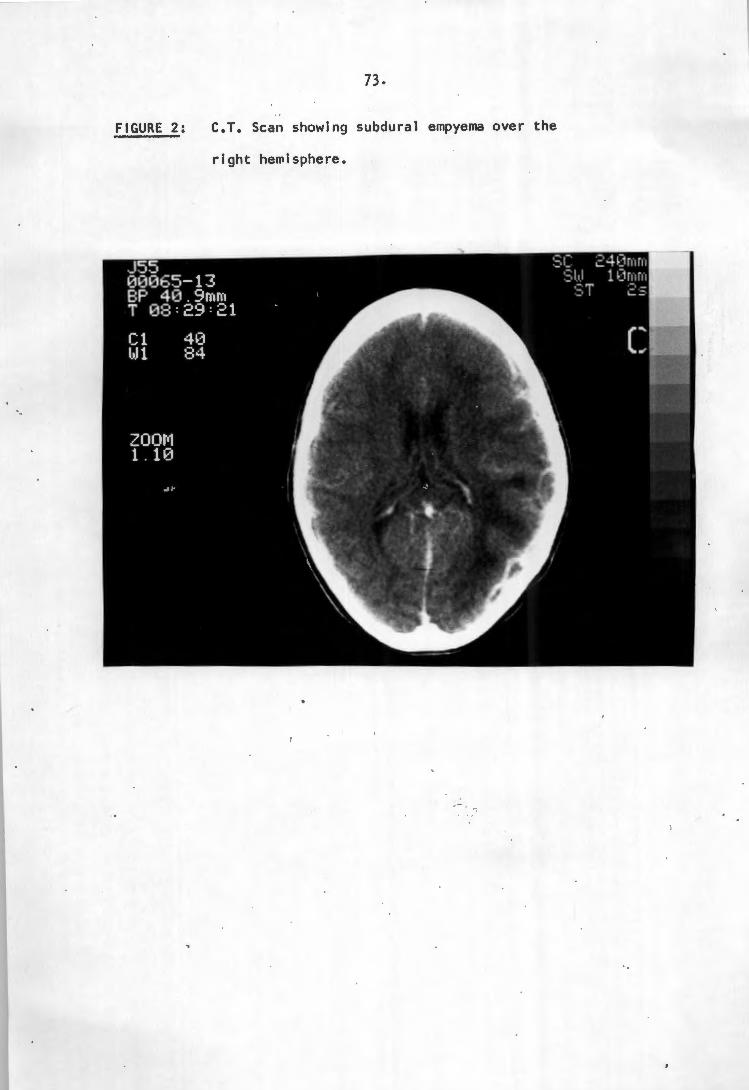
FIGURE 2:
73,
C.T. Scan showing subdural empyema over the
right hemisphere.
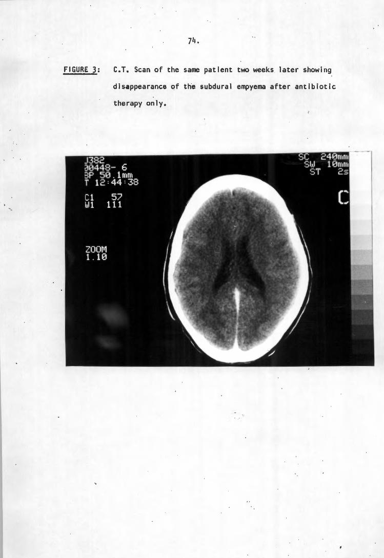
FIGURE 3:
74,
C.T. Scan of the same patient two weeks later showing
disappearance of the subdural empyema after antibiotic
therapy only.
'
.... ' ..
'
75,
The results in this study indicate however, that planned
surgical drainage, using C.T. scanning, improves the outcome
of S.E. treated surgically. The cases who died had residual
pus present at autopsy, in spite of surgical drainage and
antibiotic therapy. Only small subdural collections of pus
should therefore be treated without surgical drainage, as
long as follow-up C.T. scans indicate that the S.E. is
responding. Identifying the offending organism from the
source or elsewhere, is also important, to ensure that
resistance to the commonly used antibiotics, although rare,
is not pre~ent. The statement by Leys et al *(53), that it
is not always necessary to know the causative organism, is
probably in correct, and a careful search for the offending
organism should be made in every pati~nt with S.E. Non
surgical cure of brain abscesses has also been reported.
*( 7) .
-'.
'
76,
Steroids:
In this study Dexamethsone was used in 13 patients for brain
swelling shown on C.T. scan. It was not used in 16 cases,
and the records were lost in 18 cases ; The use or not of
steroids did not significantly affect outcome, with 23,1% of
patients on steroids severely disabled or dead versus 18,8%
of patients not on steroids. Some authors use steroids
routinely in the management of S.E. *(46). There have been
no reports of its use exacerbating the infection.
Anticonvulsants:
These are used prophylactically for patients with S.E. who
do not present with seizures. *{46). This was also the
policy in 26 of the 27 patients in this study who were
seizure free on admission. One patient never received
anticonvulsants, and remained seizure free. '
Patients who present with seizures should be treated with
anticonvulsants, usually Phenytoin. Seizures will stop in
the majority of patients when the acute illness has been
treated, and Williams advocates discontinuing
anticonvulsants 6 to 8 weeks following the acute illness.
*(84). Only 18,6% of the surviving patients in this series
were still plagued by seizures at follow-up; however 65,1%
were still on anticonvulsants.
' .
'
77,
COMPLICATIONS:
Complications following drainage of s:~. occurred in 38,3%
of patients in this study. Serious complication that
affected outcome were the development of a brain abscess,
ventriculitis and hydrocephalus, as well as seve~e
respiratory infection. Of \he five patients who developed a
brain abscess following S.E. drainage, : 2 died, 2 remained
severely disabled~ and one recovered without focal signs or
seizures. One of these patients later developed
ventriculitis, when his abscess ruptured into the lateral
ventricle. This led to the development of hydrocephalus and
necessitated ins~rtion of a V.P. shunt. The patient had a
poor outcome.
One other patient also developed ventriculitis diagnqsed on
C.T. scan, which later caused hydrocephalus. He also needed
a ventriculo-peritorieal shunt and remained severely
disabled.
Two patients developed severe respircatory infections
requiring mechanical ventilation. One of these developed a
brain abscess and has already been mentioned. The other
patient needed ventilation for a chest infection and poor
blood-gases three days following a craniotomy for drainage
~
'.
78,
of subdural pus. His condition eventually improved and he
made an excellent recov~ry.
One patient suffered a respiratory arrest following burrhole
drainage of his S.E. At repeat craniotomy pus was present
in the subdural space. He died one week later.
Serious complications occurred in 19,2% of patients treated
initially with burrholes, and in 15% ~f patients in the
craniectomy group. The patients who had initial
craniotomies had the lowest incidence of serious
complications (9,1%). Although the numbers are small, this
may indicate the importance of adequate removal of subdural
pus. Residual pus was also shown however on C.T. scans in
the patients who developed complications, and earlier
burrhole drainage of · pus shown on post-operative C.T. scans,
would probably have prevented these complications from
occurring.
' . . •
'
79.
OUTCOME: ..
Various factors have been shown in the ; literature to affect
the outcome from S . E. This study did not lend support to
the statement by Bannister et al *(5) and Williams *(84)
that craniotomy drainage improved outcome significantly.
10ther factors that may influence outcome, are the clinical
condition of the patient on admission. Unconscious patients
had a worse outcome in most studies . *(5)(46)(79). In this
series the outcome was also improved in patients who were
conscious on admission, with a mortality rate of 7,1% and
combined severe morbidity/mortality rate of 14,3% versus a
mortality rate of . 9,4% and a combined morbidity/mortality
rate of 21,9% for unconscious patients .
The duration of the illness before a correct diagnosis of
S.~. was made also influenced the outcome in the report by
Renaudin and Frazee : *(68). In this Cape Town series 6
patients were seen within 48 hours of the start of their
illness, and all six had an excellent outcome with no focal
signs or seizures . That contrasts with a 68,3% excellent
outcome in the patients who presented · more than 48 hours
after their, initial illness; and the 72,3% excellent
outcome overall .
.. t •
'
80.
Age was shown to influence outcome, a~d older patients did I
worse in the study by Williams. *(84) 1 In t~e Cape Town
series patients in their first or second decade had a
combined severe morbidity/mortality r~te of 17,9%, versus a
figure of 25% for older patients. Th~ mortality figures
were 7,7% and 12,5% respectiveiy.
Kaufman et al felt that if a lumbar pu~ct~re was performed
in S.E. patients, the outcome was worsened. *(47). In this
study 15 patients did not undergo lumbar punctures. Their
combined morbidity/mortality rate was 26,7% versus a
combined morbidity/mortality rate of 15,6% in patients who
did have a lumbar puncture. No patient in this series
deteriorated immediately after lumbar puncture, although
lumbar puncture in S.E. patients theoretically may cause
deterioration and affect the outcome, and should be l
discouraged.
The occurence of early seizures did not affect outcome in ,
the study by Cowie and Will~ams. *(20). In thi Cape Town
series this was substantiated with death or poor outcome in
15% of patients presenting with seizures, versus 18,5% in 27 '
patients who did not . have initial seiz~res. '
S.E. secondary to paranasal infection showed an improved
outcome in Williams' series. *(84). In the Cape Town study
the combined severe disability/mortali~y ra~e in 31 patients
. ,
' .
'
t•.
81.
with S.E. secondary to paranasal sinus infection was 22,6%
versus 10% for 10 patients with S.E. post otogenic disease.
S.E. secondary to paranasal sinusitis therefore had a worse
outcome than S.E. following on otogenic disease.
Regarding late seizures, Cowie and Williams have shown that
attacks in the follow-up period will have occurred within
the first two years of follow-up in the vast majority of
patients. *(20). Williams advocates , stopping
anticonvulsant treatment 6 to 8 weeks , after recovery from I
the acute illness and reserving such ~reatment for those who
have a subsequent epiieptic attack. t(84). The figures in
this study indicate that seizure-occurrence seldom
continues in the ·tollow~up period, with only 17,0% of cases . ) '
continuing to suffer seizures. It is ·reasonable therefore
to stop anticonvulsant use once the patient has recovered
from the acute illness and is seizure free. Three of the
patients still had seizures more than two years post
treatment of their ~S.E. and remained on anticonvu~sants.
~.
'.
82.
CC>NCLIJSIC>N -
S.E. remains an important intracranial infection that may
cause death and severe disability if not treated adequately.
The advent of C.T. scanning has improved the early diagnosis
of S.E., which improves the outcome. Pus present after
surgical drainage procedures is also shown, and repeat
drainage can be performed. Using C.T. scanning to plan the
surgical approach vastly improves the outcome from S.E., and
diminishes the need for initial craniotomy-drainage to
achieve a satisfactory outcome. Some subdural empyema
collections shown to be small on C.T . . scanning may even be
treated without direct surgical drainage.
Remaining pus in the subdural space qfter initial drainage '
procedures contribute to the development of complications.
C.T. scanning must be performed regularly post-operatively
to find residual pus, and this should be drained surgically.
It is important ~6 note th~t S:E. requires multiple drainage
procedures in most cases, regardless ,of the initial form of
surgery used.
A broad spectrum antibiotic regime remains one of the
mainstays of treatment of S.E. Chloramphenicol was shown to
be most effective. The importance of anaerobic organisms
and the difficulties in culturing these organisms were shown
in this study.
l.
With aggressive early ~anagement of S.E., more than 90% of
patients should survive and 90% of survivors should have no
) or minimal disability, enabling them to return to their pre
disease activity. The incidence of evilepsy in survivors is
low and can be controlled in most cases.
' . . •
84.
REFERENCES:
1. ALDERSON D, STRONG AJ, INGHAM HR, ET AL
Fifteen-year review of the mortality of Brain abscess.
NEUROSURGERY 1981; 8: 1-6,.
2. ALEGRIA C, LIPSCHITZ R, SWONNIKOFF G:
Intracranial suppuration: A review of 79 cases seen at
Baragwanath Hospital over 3 years.
S AFR J SORG 1982;20:25-35.
3. ANAGNOSTOPOULOS DI, GORTVAI P:
Intracranial ~ubdural abscess.
BRIT J SURG 1973;60:50-52.
4. ANDERSON FM :
Subdural empyema secondary to frontal sinusitis.
Review of clin~cal features and report of three cases.
ANN OTOL RHINOL LARYNGOL 1947; 56:5-17.
5. BANNISTER G, WILLIAMS B, SMITHS
Treatment of subdural empyema.
J NEUROSURG 1981;55:82-88.
-' .
'
85.
6. BEEDEN AG, MARSDEN CD, MEADOWS JC, ET AL
Intracranial complications of middle ear disease and
mastoid surgery.
J NEUROL SCI 1969;9:261-272.
7. BERG B, FRANKLIN G, CUNEO R, ET AL
Nonsurgical care of brain abscess. Early diagnosis and
follow-up with computerized tomography.
ANN NEUROL 1978;3:474-478.
8. BHANDARI YS, SARKAR! NBS :
Subdural empyema : a review of 37 cases.
J NEUROSURG 1970;32:35.
9. BIEHL JP •
Subdural empyema secondary to acute frontal sinusitis.
JAMA 1955;158:721-724.
10. BLACK P, GRAYBILL JR, CHARCHE P
Penetration of brain abscess by systemically
administered antibiotics. ,
J NEUROSURG 1973;38:705-709. -
11. BORZONE M, CAPUZZOT T, RIVANO C, ET AL:
Subdural empyema : fourteen cases surgically treated.
SpRG NEUROL 1980;13:449-452.
..• ' .
86.
12. BOTTERELL EH, DRAKE CG:
Localised encephalitis, brain abscess and subdural
empyema.
J NEUROSURG 1952;9:348-366.
13. BROCK RC :
Lung abscess.
OXFORD. BLACKWELL SCIENTIFIC PUBLICATIONS. 1952.
14. BROOK I :
Bacteriology of intracranial .abscess in children.
J NEUROSURG 1981;54:484-488.
15. BRUNNER H
Intracranial complications of Ear, Nose and Throat
infections . .
CHICAGO YEAR BOOK PUBLISHERS INC ~ 1946.
16. CAREY ME, CHOU SN, FRENQH LA:
Experience with brain abscesses.
J NEUROSURG 1972;26:1-9.
17. CASSON IR, PETEL P, BLAIR D, ET AL :
Subdural empyema caused by infection of pre-existing
subdural haematoma.
NY STATE J MED 1981;81:389-391 .
' .
'
18 . COONROD JD, DANS PE
Subdural empyema.
87,
AM J MED 1972;53:85-91.
19. COURVILLE CB:
Subdural empyema secondary to purulent frontal
sinusitis.
ARCH OTQLARYNGOL 1944;39:.211-228.
20. COWIE R, WILLIAMS B:
Late seizures and morbidity after subdural empyema.
J NEUROSURG 1983;58:569-573.
21. CROCKER EF, ,MCLAUGHLIN AF, MORRIS JG, ET AL :
Technetium Brain Scanning in the Diagnosis and
Management of cerebral abscess.
AM J MED 1974;56:192-201.
22. DE LOUVIOS J, GORTVAI P, HURLEY R:
Bacteriology of abscesses of the central nervous system
: a multicentre prospective, study.
BRIT MED J 1977;2:981-984.
23. DE LOUVOIS J, GORTVAI P, HURLEY R
Antibiotic treatment of abscesses of the central
nervous system.
BRIT MED J 1977;2:985-987.
t .
88.
24. DUFFY GP:
Lumbar puncture in the presence of Raised Intracranial
Pressure.
BRIT MED J 1969;1:407-409. I ·~
25. FARMER TW, WISE GR :
Subdural empyema in infants, children and adults.
NEUROL 1973;23:254-261.
26. FREDERICK J, BRAUDE AI
Anaerobic infection of the paranasal sinuses.
N ENGL J .MED 1974;290:135-137. ,.
27. FRENCH LA, CHOU SN
Treatment of brain abscess, in THOMPSON JR, GREEN JR
eds. : Advances in Neu~ology vol.6, Infectious diseases
of the Nervous System p.269-275.
NEW YORK RAVEN °PRESS 1974.
28. GALBRAITH JG, BARR VW :
Epidural abscess and subdural emp:.;rema; in THOMPSON RA,
GREEN JR Eds : Advances in Neurology.
NEW YORK RAVEN PRESS 1974;6:257-267.
. ' .
89,
29. GARFIELD J :
Brain abscess and focal suppurative infections, in
VINKEN PJ, BRUYN GW EDS. Handbook of clinical
neurology. vol.33.
NORTH HOLLAND PUBLISHING COMPANY · 1978.
30. GENEST AS, BINGHAM WG, HAMILTON RD:
Bilateral subdural empyema. Reports of a case with
arteriograms.
J NEUROSURG 1963;20:524-526.
31. GLASS RL :
Osteoplastic flap method in treatment of subdural
abscess.
J NEUROSURG 1947;4:391-393.
32. GOODMAN SJ, STERN WE :
Cranial and intracranial bacterial infections, in
YOUMANS JR, Ed.
Neurological surgery vol.6.
W. B. SAUNDERS COMPANY 1982 ..
33. GROFF RA, GRANT FC :
The surgical treatment of brain abscess by exposure and
enucleation.
ANN SORG 1940;111:925-941 .
'.
'
90.
34. GROSS SW :
Osteomyelitis of the frontal bone and subdural empyema
with recovery.
AM J SORG 1946;71:828-831.
35. GURDJIAN ES, THOMAS LM:
Surgical treatment of cranial and intracranial .
suppuration.
CHARLES C. THOMAS PUBLISHER. SPRINGFIELD. 1969.
36. GURDJIAN ES, WEBSTER JE :
Experiences in the surgical management of intracranial
suppuration.
SORG GYNEC OBSTET 1957;104:205-214.
• ..
37. HAMORY BH, SANDE MA, SYNDOR A, ET AL :
Etiology and antimicrobial therapy of acute maxillary
sinusitis .
J INFECT DIS 1979;139:197-202.
38. HEINEMAN HS, BRAUDE AI
Anaerobic Infection of the Brain . : Observations on
eighteen consecutive cases of Brain Abscess.
AM J MED 1963;35:682-697.
. ,,._ ~
' .
91.
39. HIRSCH JF, RO.OX FX, SAINTE-ROSE CC, ET AL
Brain abscess in childhood.
CHILD'S BRAIN 1983;10:251-265 .
40. HITCHCOCK E, ANDREADIS A
Subdural empyema : a review of 29 cases.
J NEUROL NEUROSURG PSYCHIAT 1964 ~27:422-434 .
41. HOLLIN SA, HAYASHI H, GROSS SW :
Intracranial abscesses of odontogenic origin.
ORAL SORG 1967;23:277-293.
42. INGHAM HR, SELKON JB, ROXBY CM:
Bacteriological study of otogenic cerebral abscesses
chemotherapeutic role of metronidazole.
BR MED J 1977;2:991-993.
43. JACOBSON SA, KANE CA :
Purulent subdural collection.
NEUROLOGY 1954;4:558-562.
44. JOUBERT MJ, STEPHANOV S
Computerised tomography and surgical treatment in
intracranial suppuration.
J NEUROSURG 1977;47:73-78 .
.. '.
_.
'
92.
45. KAUFMAN DM, LEEDS NE :
Computerised tomography in the diagnosis of
intracranial abscesses.
NEUROLOGY (MINNEAP) 1977;27:1069-1073.
46. KAUFMAN DM, LITMAN N, MILLER MH :.
Sinusitis Induced subdural empyema.
NEUROLOGY 1983;33:123-132.
47. KAUFMAN DM, MILLER MH, STEIGBIGEL MD
Subdural empyema : Analysis of 11 recent cases and
review of the literature.
MEDICINE (BALTIMORE) 1975;54:485-498.
48 . . KEITH WS
Subdural empyema.
J NEUROSURG 1949;6:127-139.
49. KUBIK CS, ADAMS RD
Subdural empyema.
BRAIN 1943;66:18.
50. LANDESMAN SH, CORRADO ML, SHAH PM ET AL:
Past and current roles for cephalosporin antibiotics in
treatment of meningitis : emphasis on use in gram
negative bacillary meningitis.
AM J MED 1981;71:693-703 .
. t •
' ... --- ------ ..------,
93,
51. LE BEAU J :
Traitement chirurgical de l'empyeme sous dural et sons
arachnoidien.
REV NEUROL 1949;81:828-851.
52. LE BEAU J, CREISSARD P, HARISPE LET AL
Surgical treatment of brain abscess and subdural
empyema.
J NEUROSURG 1973;38:198-203.
53. LEYS D, DESTEE A, PETIT H, ET AL : • Management of subdural intracranial empyemas should not
always require surgery.
J NEUROL NEUROSURG PSYCHIATRY 1986;49:635-639.
54. LIST CF :
Interhemispheral subdural suppuration.
J NEUROSURG 1950;7:313-324.
55. LIST CF :
Diagnosis and treatment of acute subdural empyema.
NEUROLOGY 1955;5:663-670.
56. LUKEN MG, WHELAN MA :
R~cent diagnostic experience with subdural empyema.
J NEUROSURG 1980;52:764-771 .
.. ' .
. •
94.
57. MACEWEN W:
Pyogenic diseases of the brain and spinal cord.
GLASGOW. MCLEHOSE. 1893 . .
58. MINCY JE, PECK FC :
Actinomycotic subdural empyema. Survival following
surgical drainage and antibiotic treatment.
NY STATE J MED 1966;66:2155-2157.
59. MOHR RM, NELSON LR:
Frontal sinus ablation for frontal osteomyelitis.
LARYNGOSCOPE 1982;92:1006-1015.
60. NAGER GT :
Mastoid and paranasal infections and their relation to
the central nervous system.
CLIN NEUROSURG 1979;14:288-313.
61. NEWTON EJ :
Haematogenous brain abscess in cyanotic congenital
heart disease.
QUART J MED 1956;25:201.
62. NIEBLING HG :
Subdural empyema.
NEUROCHIRURGIA 1959;2:47-54 .
-
' •.
'
95,
63. NORTHFIELD owe The surgery of the central nervous system.
BLACKWELL SCIENTIFIC PUBLICATIONS 1973.
64. OSGOOD CP, DUJOVNY M, HOLME, ET AL :
Delayed post-traumatic subdural e~pyema.
J TRAUMA 1975;15:916-921.
65. PATTON JT, HITCHCOCK E
Angiographic features of falcine subdural empyema.
CLIN RADIOL 1968;19:229-232.
66. RAY BS, PARSON H
Subdural abscess complicating frontal sinusitis.
ARCH OTOLARYNGOL 1943;37:536-551.
67. RENAUDIN JW :
Cranial epidural abscess and subdural empyema ;, in
WILKENS RH, RENGACHARY SS, EDS: .
NEUROSURGERY, VOL.III, p.1961-1963.
MCGRAW-HILL BOOK COMPANY 1986. ·
68. RENAUDIN JW, FRAZEE J
Subdural empyema - Importance of early diagnosis.
NEUROSURGERY 1980;7:477-479.
,1
t
[,.
',
. '.
,•
96,
~9. SAMSON DS, CLARK K :
Accurrent review of Brain Abscess.
AM J. MED 1973;54:201-210.
70. SCHILLER F, CAIRNS H, RUSSEL DS :
Treatment of purulent pachymeningitis and subdural
suppuration with special reference to penicillin.
J NEUROL NEUROSURG PSYCHIATRY 1948;11:143-182.
71. SCHUTZ :
Stirnhohlen abscess ..
ALLG MED ANN 1812; p720
cited by COURVILLE CB *19. L
72. SMITH HP, HENDRICK EB :
Subdural empyema and epidural abscess in children.
J NEUROSURG 1983;58:392-397.
73. STEPHANOV S, JOUBERT MJ, WELCHMAN JM: .
Combined convexity and parafalx subdural empy~ma.
SORG NEUROL 1979;11:147-151.
J4. STEPHENS J, WELCH K :
Subdural abscess with retinal thrombophlebitis.
NEUROLOGY 1956;6:889-890 .
' ..
..
'
97.
75. STERN WE, BOLDREY E :
Subdural purulent collections.
SORG GYNEC OBSTET 1952;95:623-630.
76. SWAIMAN KF, GOLD LHA :
An unusual computed tomographic appearance of a
subdural empyema.
J PEDIATR 1977;91:945-947.
77. TORRENS M :
Subdural empyema : the role of computer assisted
tomography and surgical decompression.
J NEUROL NEUROSURG PSYCHIATRY 1977;40:1023-1024.
78. TORRES H, YARZAGARAY L, WEST C
Subdural empyema, angiographic and clinical
considerations.
NEUROCHIRURGIA 1970;13:201-210.
79. VANALPHEN HAM, DREISSEN JJR:
Brain abscess and subdural empyema - factors
influencing mortality and results of various surgical
techniques.
J NEUROL NEUROSURG PSYCHIATRY 1976;39:481-490.
' .
98.
80. VAN DELLEN JR, BOLES DM, VAN DEN REEVER CM
Interhemispheral Subdural Empyema.
S AFR MED J 1977;52:266-269.
81. VERDURA J, WHITE RJ, RESNIKOFF&, ET AL :
Interhemispheric subdural empyema : angiographic
diagnosis and surgical treatment.
SURG NEUROL 1975;3:89-92.
82. WEINMAN D, SAMARASINGHE HHR
Subdural empyema.
AUSTR NZJ SURG 1972;41:324-330.
83. WILKINS RH, GOREE JA
Interhemispheric subdural empyema
appearance.
J NEUROSURG 1970;32:459-462.
84. WILLIAMS B :
Subdural empyema.
Angiographic
ADVANCES AND TECHNICAL STANDARDS IN NEUROSURGERY VOL.9.
WIEN/NEW YORK. SPRINGER-VERLAG 1983.
85. WILLIAMS B :
The neurosurgery of otorhinological sepsis;
CLIN OTOLARYNGOL 1983;8:121-133.
, '
'.
'
2 2 FEB 1881
99,
86. WOOD PH
Diffuse subdural suppuration.
J LARYNGOL OTOL 1952;66:496-515.
87. WOODHALL B:
Osteomyelitis and epi-, extra-, and subdural abscesses.
CLIN NEUROSURG 1967;14:239-255.
88. WORTZMAN DJ, TUCKER WS, FINLAYSON DM, ET AL
·subdural empyema with negative C.T. scan.
CANAD SCIENCES NEUROL 1980;7:67-69.
89. YOSHIKAWA TT~ CHOW AW, GUZE LB:
Role of anaerobic bacteria in subdural empyema.
AM J MED 1975; 58:99-104.
90. ZIMMERMAN RA, PATELS S, BILANIUK LT
Demonstration of purulent bacterial intracranial
infections by computerized tomography.
AM J ROENTGENOL 1976;127:155-165.