citrus canker—distribution, taxonomy, epidemiology ... - mdpi
TRANSCRIPT
Citation: Naqvi, S.A.H.; Wang, J.;
Malik, M.T.; Umar, U.-U.-D.;
Ateeq-Ur-Rehman; Hasnain, A.;
Sohail, M.A.; Shakeel, M.T.;
Nauman, M.; Hafeez-ur-Rehman;
et al. Citrus Canker—Distribution,
Taxonomy, Epidemiology, Disease
Cycle, Pathogen Biology, Detection,
and Management: A Critical Review
and Future Research Agenda.
Agronomy 2022, 12, 1075. https://
doi.org/10.3390/agronomy12051075
Academic Editors: Subhan Danish,
Anna Gałazka and Syed
Muhammad Zaka
Received: 4 March 2022
Accepted: 23 April 2022
Published: 29 April 2022
Publisher’s Note: MDPI stays neutral
with regard to jurisdictional claims in
published maps and institutional affil-
iations.
Copyright: © 2022 by the authors.
Licensee MDPI, Basel, Switzerland.
This article is an open access article
distributed under the terms and
conditions of the Creative Commons
Attribution (CC BY) license (https://
creativecommons.org/licenses/by/
4.0/).
agronomy
Review
Citrus Canker—Distribution, Taxonomy, Epidemiology, DiseaseCycle, Pathogen Biology, Detection, and Management:A Critical Review and Future Research AgendaSyed Atif Hasan Naqvi 1,† , Jie Wang 2,*,†, Muhammad Tariq Malik 3, Ummad-Ud-Din Umar 1,†,Ateeq-Ur-Rehman 1, Ammarah Hasnain 4, Muhammad Aamir Sohail 5 , Muhammad Taimoor Shakeel 6,Muhammad Nauman 1, Hafeez-ur-Rehman 1, Muhammad Zeeshan Hassan 1, Maheen Fatima 1 and Rahul Datta 7,*
1 Department of Plant Pathology, Faculty of Agricultural Sciences and Technology,Bahauddin Zakariya University, Main Campus, Bosan Road, Multan 60800, Pakistan;[email protected] (S.A.H.N.); [email protected] (U.-U.-D.U.);[email protected] (A.-U.-R.); [email protected] (M.N.);[email protected] (H.-u.-R.); [email protected] (M.Z.H.);[email protected] (M.F.)
2 Key Laboratory of Tobacco Pest Monitoring Controlling Integrated Management, Tobacco Research Instituteof Chinese Academy of Agricultural Sciences, Qingdao 266101, China
3 Mango Research Institute, Old-Shuja-Abad-Road, Multan 60000, Pakistan; [email protected] Institute of Molecular Biology and Biotechnology, The University of Lahore, Lahore 54000, Pakistan;
[email protected] Hubei Key Laboratory of Plant Pathology, Huazhong Agricultural University, Wuhan 430070, China;
[email protected] Department of Plant Pathology, University College of Agriculture and Environmental Sciences,
The Islamia University, Bahawalpur 63100, Pakistan; [email protected] Department of Agrochemistry, Soil Science, Microbiology and Plant Nutrition, Faculty of Agrisciences,
Mendel University in Brno, Zemedelska 1, 61300 Brno, Czech Republic* Correspondence: [email protected] (J.W.); [email protected] (R.D.)† These authors contributed equally to this work.
Abstract: Xanthomonas citri subsp. citri, a causative agent of the citrus canker (CC) disease, belongs toone of the essential groups of the bacterial phytopathogen family, Xanthomonadaceae. It has been apotential threat to the globally significant citrus fruit crop, which has remained under investigationfor disease management and epidemiology since the 1980s. In Pakistan, the average yield of citrusis 11 t/ha, which is lower than other countries, including China, Brazil, and India, having averageproductions of 27, 26, and 22 tons/hectare, respectively. Citrus canker is one of the most devastatingdiseases, posing a significant threat to crop yield and fruit quality. To date, five distinct types (orforms) of the citrus canker have been recognized; the Asiatic (Canker A) form is most destructiveand affects most citrus cultivars. Severe infection outcomes include dieback, defoliation, severelyblemished fruit, premature fruit drop, and reduced fruit quality. The infection increases under humid,warm, cloudy climate, wind, and heavy rainfall. The analysis of plasmid and chromosomal DNAof X. citri subsp. citri depicted an evolutionary relationship among pathovars of Xanthomonas. Theextensive study on the genome of X. citri subsp. citri has contributed to the current knowledge ofplant host recognition of pathogens, host specificities, dissemination, and propagation. Regulatoryprograms, i.e., quarantine or exclusion, continued to be practiced, prohibiting infected citrus plantmaterial into the existing stock. Other measures include removal of inoculums sources, resistanthosts, protective copper-containing sprays, and windbreak systems. In this review, we exploredthe latest trends in the areas of epidemiology, pathogenome, detection, host–pathogen interaction,biofilm formation, and management of X. citri subsp. citri.
Keywords: Xanthomonas citri subsp. citri; bacterium; plant pathogenic; local infection; industry;economic importance; management
Agronomy 2022, 12, 1075. https://doi.org/10.3390/agronomy12051075 https://www.mdpi.com/journal/agronomy
Agronomy 2022, 12, 1075 2 of 31
1. Introduction
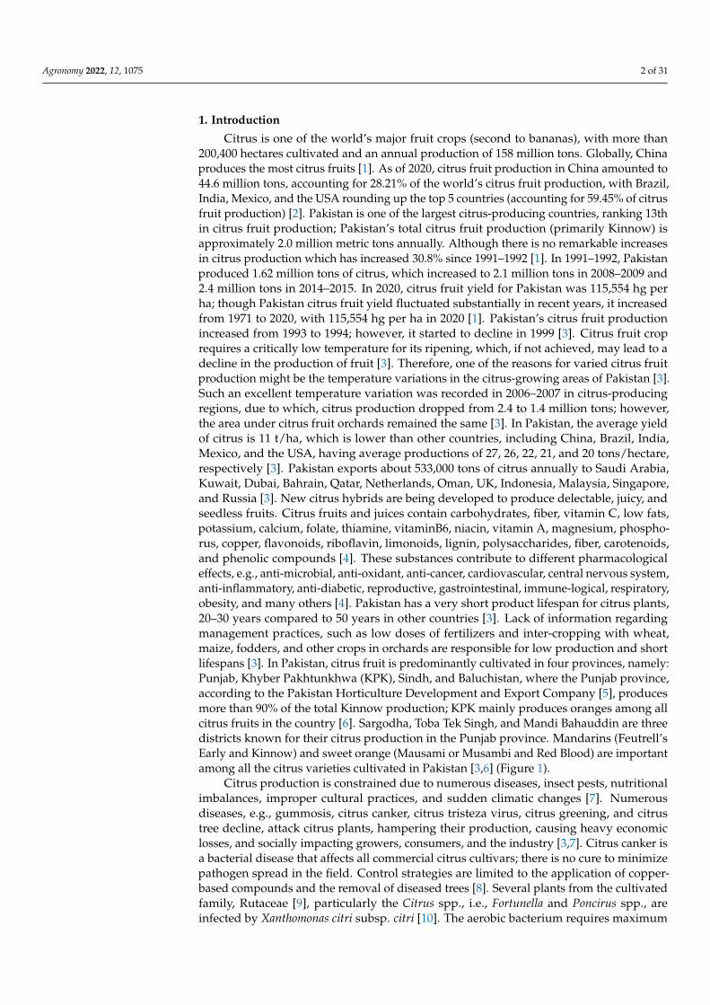
Citrus is one of the world’s major fruit crops (second to bananas), with more than200,400 hectares cultivated and an annual production of 158 million tons. Globally, Chinaproduces the most citrus fruits [1]. As of 2020, citrus fruit production in China amounted to44.6 million tons, accounting for 28.21% of the world’s citrus fruit production, with Brazil,India, Mexico, and the USA rounding up the top 5 countries (accounting for 59.45% of citrusfruit production) [2]. Pakistan is one of the largest citrus-producing countries, ranking 13thin citrus fruit production; Pakistan’s total citrus fruit production (primarily Kinnow) isapproximately 2.0 million metric tons annually. Although there is no remarkable increasesin citrus production which has increased 30.8% since 1991–1992 [1]. In 1991–1992, Pakistanproduced 1.62 million tons of citrus, which increased to 2.1 million tons in 2008–2009 and2.4 million tons in 2014–2015. In 2020, citrus fruit yield for Pakistan was 115,554 hg perha; though Pakistan citrus fruit yield fluctuated substantially in recent years, it increasedfrom 1971 to 2020, with 115,554 hg per ha in 2020 [1]. Pakistan’s citrus fruit productionincreased from 1993 to 1994; however, it started to decline in 1999 [3]. Citrus fruit croprequires a critically low temperature for its ripening, which, if not achieved, may lead to adecline in the production of fruit [3]. Therefore, one of the reasons for varied citrus fruitproduction might be the temperature variations in the citrus-growing areas of Pakistan [3].Such an excellent temperature variation was recorded in 2006–2007 in citrus-producingregions, due to which, citrus production dropped from 2.4 to 1.4 million tons; however,the area under citrus fruit orchards remained the same [3]. In Pakistan, the average yieldof citrus is 11 t/ha, which is lower than other countries, including China, Brazil, India,Mexico, and the USA, having average productions of 27, 26, 22, 21, and 20 tons/hectare,respectively [3]. Pakistan exports about 533,000 tons of citrus annually to Saudi Arabia,Kuwait, Dubai, Bahrain, Qatar, Netherlands, Oman, UK, Indonesia, Malaysia, Singapore,and Russia [3]. New citrus hybrids are being developed to produce delectable, juicy, andseedless fruits. Citrus fruits and juices contain carbohydrates, fiber, vitamin C, low fats,potassium, calcium, folate, thiamine, vitaminB6, niacin, vitamin A, magnesium, phospho-rus, copper, flavonoids, riboflavin, limonoids, lignin, polysaccharides, fiber, carotenoids,and phenolic compounds [4]. These substances contribute to different pharmacologicaleffects, e.g., anti-microbial, anti-oxidant, anti-cancer, cardiovascular, central nervous system,anti-inflammatory, anti-diabetic, reproductive, gastrointestinal, immune-logical, respiratory,obesity, and many others [4]. Pakistan has a very short product lifespan for citrus plants,20–30 years compared to 50 years in other countries [3]. Lack of information regardingmanagement practices, such as low doses of fertilizers and inter-cropping with wheat,maize, fodders, and other crops in orchards are responsible for low production and shortlifespans [3]. In Pakistan, citrus fruit is predominantly cultivated in four provinces, namely:Punjab, Khyber Pakhtunkhwa (KPK), Sindh, and Baluchistan, where the Punjab province,according to the Pakistan Horticulture Development and Export Company [5], producesmore than 90% of the total Kinnow production; KPK mainly produces oranges among allcitrus fruits in the country [6]. Sargodha, Toba Tek Singh, and Mandi Bahauddin are threedistricts known for their citrus production in the Punjab province. Mandarins (Feutrell’sEarly and Kinnow) and sweet orange (Mausami or Musambi and Red Blood) are importantamong all the citrus varieties cultivated in Pakistan [3,6] (Figure 1).
Citrus production is constrained due to numerous diseases, insect pests, nutritionalimbalances, improper cultural practices, and sudden climatic changes [7]. Numerousdiseases, e.g., gummosis, citrus canker, citrus tristeza virus, citrus greening, and citrustree decline, attack citrus plants, hampering their production, causing heavy economiclosses, and socially impacting growers, consumers, and the industry [3,7]. Citrus canker isa bacterial disease that affects all commercial citrus cultivars; there is no cure to minimizepathogen spread in the field. Control strategies are limited to the application of copper-based compounds and the removal of diseased trees [8]. Several plants from the cultivatedfamily, Rutaceae [9], particularly the Citrus spp., i.e., Fortunella and Poncirus spp., areinfected by Xanthomonas citri subsp. citri [10]. The aerobic bacterium requires maximum
Agronomy 2022, 12, 1075 3 of 31
growth temperatures of 35–39 ◦C and optimal temperatures of 28–30 ◦C [11,12]. A widerange of virulence factors is included in CC development, such as structures for surfaceattachment, enzymes for degradation of the cell wall, a few secretion systems and theireffectors, and the diffusible signal factor (DSF), which mediates the quorum sensing (QS)system [13]. Xanthomonas citri subsp. citri, a member of Xanthomonadaceae family, is oneof the largest and most important groups of bacterial phytopathogens; it has been usedas a model organism for pathogenesis and the phylogeny study, and is the causativeagent of citrus canker (CC) disease, which has been the subject of extensive research interms of epidemiology and management [3,7]. There is controversy over the geographicalorigin of citrus canker, and it is assumed Fortunella hindsii may have been a wild hostplant in southern China [14]. Yet, some scientists reported that citrus canker originatedin India; citrus canker was found in the oldest citrus herbarium of Herbaria of the RoyalBotanical Gardens, England [15]. It is assumed that the disease originated first in tropicalregions, such as South China, Indonesia, and India. In 1910, in Florida, the disease wasfirst identified and transported through infected nursery stock imported from Japan in thenineteenth century, and spread throughout the southeastern US [16,17]. The disease alsooccurred in South America [18], South Africa [19], and Australia [20] earlier this century. Insouthern Iran, an atypical strain, XAC, was discovered, which showed extreme virulence onMexican limes, grape fruits, and sweet oranges [21]. In Taiwan, citrus bacterial canker wasfirst reported in 1932 [22]. Citrus canker is prevalent in over thirty citrus-growing countriesin Asia, the Pacific, Indian Oceans, South America, and in the southeastern US [23]. Thetransportation of fruit from an infested zone to a production area free of disease imposestrade restrictions under regulations [24]. The causal agent is considered a quarantineorganism in citrus-producing areas of Europe, where canker has not been reported so far.Exclusion or quarantine practices for X. citri subsp. citri are still being refined wherevercitrus is grown worldwide, while new methods and tools for managing and eradicating CCare being developed [25]. The current review presents recent developments in the researchof X. citri subsp. citri and CC, including taxonomy, distribution, epidemiology, diseasecycle, pathogen biology, detection, and management.
Agronomy 2022, 12, x FOR PEER REVIEW 3 of 30
Figure 1. (A) World citrus production areas and (B) Pakistan’s various districts participating in world citrus production. In Pakistan, citrus fruit is predominantly cultivated in four provinces.
Citrus production is constrained due to numerous diseases, insect pests, nutritional imbalances, improper cultural practices, and sudden climatic changes [7]. Numerous dis-eases, e.g., gummosis, citrus canker, citrus tristeza virus, citrus greening, and citrus tree decline, attack citrus plants, hampering their production, causing heavy economic losses, and socially impacting growers, consumers, and the industry [3,7]. Citrus canker is a bac-terial disease that affects all commercial citrus cultivars; there is no cure to minimize path-ogen spread in the field. Control strategies are limited to the application of copper-based compounds and the removal of diseased trees [8]. Several plants from the cultivated fam-ily, Rutaceae [9], particularly the Citrus spp., i.e., Fortunella and Poncirus spp., are infected by Xanthomonas citri subsp. citri [10]. The aerobic bacterium requires maximum growth temperatures of 35–39 °C and optimal temperatures of 28–30 °C [11,12]. A wide range of virulence factors is included in CC development, such as structures for surface attach-ment, enzymes for degradation of the cell wall, a few secretion systems and their effectors, and the diffusible signal factor (DSF), which mediates the quorum sensing (QS) system [13]. Xanthomonas citri subsp. citri, a member of Xanthomonadaceae family, is one of the largest and most important groups of bacterial phytopathogens; it has been used as a model organism for pathogenesis and the phylogeny study, and is the causative agent of citrus canker (CC) disease, which has been the subject of extensive research in terms of epidemiology and management [3,7]. There is controversy over the geographical origin of citrus canker, and it is assumed Fortunella hindsii may have been a wild host plant in south-ern China [14]. Yet, some scientists reported that citrus canker originated in India ;citrus canker was found in the oldest citrus herbarium of Herbaria of the Royal Botanical Gar-dens, England [15]. It is assumed that the disease originated first in tropical regions, such as South China, Indonesia, and India. In 1910, in Florida, the disease was first identified and transported through infected nursery stock imported from Japan in the nineteenth century, and spread throughout the southeastern US [16,17]. The disease also occurred in South America [18], South Africa [19], and Australia [20] earlier this century. In southern Iran, an atypical strain, XAC, was discovered, which showed extreme virulence on Mexi-can limes, grape fruits, and sweet oranges [21]. In Taiwan, citrus bacterial canker was first reported in 1932 [22]. Citrus canker is prevalent in over thirty citrus-growing countries in
Figure 1. (A) World citrus production areas and (B) Pakistan’s various districts participating in worldcitrus production. In Pakistan, citrus fruit is predominantly cultivated in four provinces.
2. Taxonomy of Citrus Canker Bacterium
Citrus canker, also known as Asiatic CC, was initially reported on in the United Statesof America in the early 1900s following an outbreak in numerous southeastern states [26]. In1914, Hasse received samples from Florida, Texas, and Mississippi, and was able to isolate
Agronomy 2022, 12, 1075 4 of 31
the bacterium [27]. After completing characterization and pathogenicity tests, Hasse calledthe bacterium Pseudomonas citri [27]. Since then, the bacterium has been classified into severalgenera, including Bacterium, Phytomonas, and, finally, Xanthomonas citri in 1939 [8,28,29].Xanthomonas genus consists of 27 phytopathogens that cause critical diseases in ornamentalplants and other crops [8]. The genus has a broad range of 68 host families, over 240 generaand 140 different pathovars [30]. The genus Xanthomonas can infect more than 350 species,including 268 dicots and 124 monocots, including grains, fruits, nuts, and plants belongingto Brassicaceae and Solanaceae families [8]. The strains of Xanthomonas citri have been assignedto the A strain within this species to show that they are linked to Asiatic CC [8]. Two moreCC-producing Xanthomonads were discovered in the 1970s and were first classified as groupC strains, which induce canker lesions solely in key lime (Citrus aurantifolia), and group Bstrains, which have a broader host range [31,32]. CC bacterium continued as X. citri until1978; in the same year, 1978, Dye placed X. citri in X. campestris pv. citri to uphold citri at theinfra subspecific level [33]. CC bacterium was again reassigned the title of X. citri by [34],while the B and C strains were placed in X. campestris pv. aurantifolii. Reference [35] disagreedwith previous research and argued that more work was needed to place CC strains in X. citriand suggested A, B, and C strains continued as X. campestris pv. citri; then [36] performedresearch using DNA–DNA hybridization (DDH) based on renaturation rates with a diversearray of Xanthomonas strains, recommending strain A to X. axonopodis pv. citri and B and Cto X. axonopodis pv. aurantifolii, respectively [7,8]. The research on CC bacterium taxonomycontinued and, Ref. [37] again, using the S1 nuclease DNA–DNA hybridization technique,the researchers recommended X. axonopodis pv. citri strains in X. smithii subsp. smithii and theX. axonopodis pv. aurantifolii strains in X. fuscans subsp. aurantifolii, although the placement ofstrains in X. smithii after due deliberations was later considered illegitimate and was agreedupon by the previous legitimate proposal by [34]. Hence, the authors of reference [38]published an erratum “Emended classification of Xanthomonad pathogens on citrus” insystematic and microbiology and recommended the placement of strains in X. citri. Finally,the authors of reference [39] formally validated X. citri. as X. citri. subsp. citri. Reference [40]proposed important modifications to the taxonomy of Xanthomonads, including within X.citri, recommending the addition of several pathovars within X. axonopodis, as well as theplacement of members of X. fuscans, into X. citri, using a polyphasic approach that includeda multilocus sequence analysis (MLSA), a DDH calculation of whole-genome averagenucleotide identity values, and phenotypic analyses. As a result, it has been suggestedthat X. fuscans subsp. aurantifolii be transferred to X. citri as X. citri pv. aurantifolii [40].The authors submitted their recommendations for these adjustments to the InternationalJournal of Systematic and Evolutionary Microbiology, which were accepted; from now on, theprokaryotic names X. citri subsp. citri (XCC) and X. fuscans subsp. aurantifolii (XFA) will beused in nomenclature for bacteria that cause CC [40]. The bacterium was gram-negative,rod-shaped, and polar flagella. In contrast, colonies of bacterium showed yellow colors onpetri plates due to the presence of a carotenoid pigment called Xanthomonadin. Because ofexopolysaccharide (EPS), it is known as xanthan, showing a glossy appearance, invitro [40].The classification of bacterium consists of kingdom: Prokaryote, phylum: proteo-bacteria,class: Gamma-proteobacteria, order: Xanthomonadales, family: Xanthomonadaceae, genus:Xanthomonas, specie: citri, and subsp.: citri [7] (Table 1).
Table 1. Citrus canker bacterium A strain classification details, from the start of the studies.
Sr. No. Genus Specie *f.sp./*pv/subsp. Year Reference
1. Pseudomonas citri not reported 1915 [27]2. Xanthomonas citri not reported 1915 [27]3. Bacterium citri not reported 1916 [28]4. Bacillus citri not reported 1920 [41]5. Phytomonas citri not reported 1923 [42]6. Xanthomonas citri not reported 1939 [29]7. Xanthomonas citri aurantifolia 1972 [43]
Agronomy 2022, 12, 1075 5 of 31
Table 1. Cont.
Sr. No. Genus Specie *f.sp./*pv/subsp. Year Reference
8. Xanthomonas campestris aurantifolia 1978 [33]9. Xanthomonas campestris citri 1980 [44]10. Xanthomonas citri aurantifolia 1989 [34]11. Xanthomonas axonopodis citri 1995 [36]12. Xanthomonas smithii citri 2005 [37]13. Xanthomonas citri citri 2006 [38]14. Xanthomonas citri subsp. citri 2007 [39]15. Xanthomonas citri subsp. citri 2016 [40]
*f.sp. stands for forma special and *pv for pathovar (classification of a pathogen beyond sub specie levels). Subsp.:Sub specie.
3. Phylogenetically Distinct Groups of CC
X. citri subsp. citri and X. citri subsp. aurantifolii have been further divided into sub-groups based on their significant differences in the host range, which is also a reason for truepathogenic variants [34,45]. It was reported that the division of these groups based on citrushost type and symptoms was made on bacterial isolation on various nutrient media [31].
3.1. Asiatic Citrus Canker Strains
The most important and widespread pathovar is the Asiatic-canker (also namedcancrosis-A or true-canker), X. citri subsp. citri A strain is the most virulent and has awide host range, including cultivars of citrus [46]. South-West Asian strains X. citri subsp.citri A are relatively less widespread [7]. Most recently, in Florida, at one location, a thirdpathogenic strain was found, which was designated Aw, apparently of Asiatic origin [47].
3.2. South-American Canker Strains
There are two types of South American canker strains which causes the same symp-toms on the susceptible host as those produced by X. citri subsp. citri A strains, but allstrains of South America have narrow host ranges [47]. X. citri subsp. aurantifolii B strain,also referred to as false canker or cancrosis B, has a more restricted host range and is foundto primarily infect lemons and limes [32]. The Bstrain (XAUB as an acronym, XAC patho-type B; XAC-B) first appeared in Argentina in 1923 and it eventually extended to nearbyUruguay and Paraguay [32,33]. The B strain generally causes severe infections on lemonfruits (Citrus limon), limes (C. aurantiifolia), sour oranges (C. aurantium), but seldom on sweetoranges (C. sinensis) and pummelo fruit (C. maxima). Moreover, this strain does not infectgrapefruit (C. paradisi) [30,32]. Hence, the Bstrain is not present in nature longer. Mexicanlime cancrosis, CBC-C (XACtype C; XAC-C) or the Cstrain (XAUC as an acronym) wasdiscovered in 1963 and present only in São Paulo, Brazil, where it just infects the Mexicanlime [48]. The B and C strains are currently classified as X. axonopodis subsp. aurantifolii andproduce many similar symptoms on the host as produced by the canker A strain [49,50].The strains XAC, XAUB, and XAUC were compared and analyzed phenotypically andphylogenetically; all three strains were shown to have polar flagella with noticeable motilitywhen cultured in semi-solid media [51]. In the presence of maltose and aspartic acid, onlyXAC can grow and hydrolyze pectate and gel [52]. Polyclonal antisera were preparedagainst XAC, but XAUB and XAUC showed little or no affinity to antisera. In contrast,XAC is susceptible to CP1 and CP2 bacteriophages, and XAUB and XAUC are not affectedby these bacteriophages [53]. It was observed in culture media that XAUB has fastidiousgrowth; XAC and XAUC both grow well in nutrient-agar (NA) and tryptophan–sucroseagar media. Moreover, these three strains show good growth in glutamic-acid rich media. Amolecular analysis confirmed that XAUB and XAUC are more interlinked with one anotherthan XAC [54–56]. In contrast, data obtained from physiological tests, i.e., phage-typing [57]total protein profiles after SDS-PAGE, DNA–DNA solution hybridizations [56,58], plasmid–DNA fingerprints [59], plasmid-based hybridization probes [59], PCR assay [60], DNAfragment sequence of gene hrp, restriction enzymes to analyze DNA fragments [61] confirm
Agronomy 2022, 12, 1075 6 of 31
these conclusions. Furthermore, a gene required to cause CBC symptoms is the pthA gene,which was obtained from the XAC-A strain [62,63]. Total DNA hybridization with a pthAfragment revealed several profiles among XAC-A, XAUB, and XAUC; no hybridizationwith strains of X. axonopodis pv. citrumelo was observed [10].
Provisionally, two more CC strains were classified, named D and E strains, but nowthey do not exist or are categorized differently [64]. The D strain, which is also referredto as bacteriosis, induces disease in limes in Colima (Mexico), but its etiology is not con-firmed yet [65,66]. This disease causes typical leaves and twig lesions, but no symptomsare observed on key Mexican lime fruit [67]. In this area, the suspected citrus pathogen nolonger exists. It is now believed that the disease was caused by Alternaria limicola [68,69].The second pathogen was the E strain, previously identified as a citrus canker in a Floridanursery. The disease is ‘called’ a bacterial spot of citrus produced by X. axonopodis subsp.citrumelo [34,50,70]. It can be distinguished based on these studies that three groupsof X. axonopodis strains, i.e., A strain, B strain (including C and D strains), cause citruscankers [50] and, notably, these strains have controversial taxonomy [71]. This interpretationwas confirmed when the Xanthomonas genus was reclassified based on DNA–DNA hy-bridization and metabolic activity studies [7,8,10]. Moreover, Xanthomonas, causing diseaseson citrus, was transferred from X. campestris to X. axonopodisspecies. Perhaps now, patho-type A, pathotypes B and C, and the CBS strains of X. axonopodis, are named X. axonopodissubsp. citri, X. axonopodis subsp. aurantifolii, and X axonopodis subsp. citrumelo, respectively,but the subcommittee on the taxonomy of plant pathogenic bacteria did not support thisproposal [7,8,71].
4. Symptomatology
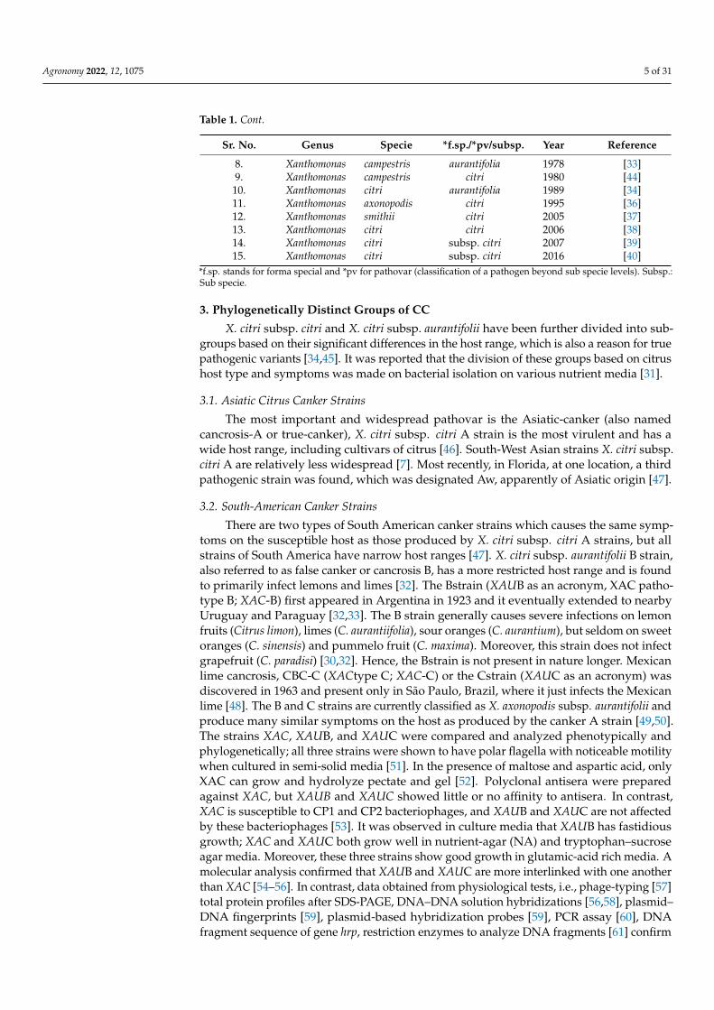
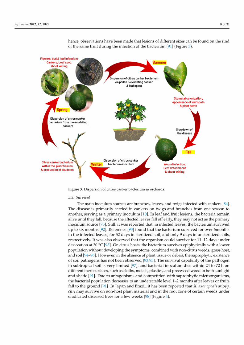
Canker symptoms are observed in all aerial parts of the plant [72]. They are charac-terized mainly by the formation of erumpent, corky, and raised pustules on the surface ofleaves, fruits, and twigs, which serve as sources of bacterial inoculums [72]. Defoliationand fruit drop are also observed as plant responses to the infection [73,74]. Notably, Xan-thomonas citri can survive in such plant debris for two months [75]. Severe symptoms areproduced on trifoliate oranges, grapefruit, Mexican lime, and some sweet oranges; however,the actual host range depends primarily on a strain of citrus canker [74]. Generally, thesusceptibility of young tissues to the citrus canker is more than mature tissues as there is aperiod of vulnerability in each flush around three times a year [76] (Figure 2).
Agronomy 2022, 12, x FOR PEER REVIEW 7 of 30
Figure 2. (A) Raised, corky, and sunken lesions on the upper side of the leaf. (B) Lesions on the lower side of the leaf. (C) Initial lesions on the lower surface of the leaf. (D) Canker symptoms on the fruit.
4.1. Leaf Lesions CC bacterium naturally penetrates the host tissues through stomata [78], hydathodes,
lenticels, or wounds [79]. Citrus canker disease symptoms first appear as tan, brown, or grey-oily circular lesions, 2 to 10 mm in size, depending on the susceptibility of the host, the number of cycles of the infection, and optimal environmental conditions, i.e., the pres-ence of water film and 20 to 30 °C temperature; canker protrudes from both surfaces of leaf tissue around 4–7 days after inoculation [79]. Symptoms might appear after more than 60 days under optimum conditions [80,81]. As the disease advances, host cell expansion (hypertrophy) and cell division (hyperplasia) occur, due to which the lesions become vis-ible from small water-soaked spots and are surrounded by a yellow halo, which turns into slightly raised blister-like lesions and can be viewed with transmitted light [75,81]. The hyperplastic mesophyll tissue is an essential diagnostic symptom of the disease charac-terized by the formation of the canker due to rupturing the epidermis [79] and it releases abundant X. citri subsp. citri on the leaves. These lesions are elevated, are ‘corky’ in leaves, stems, and fruits, and then become dark and thick into the distinctive citrus canker under dry conditions [74]. A wound on the leaves or fruits or an injury by the Asiatic citrus leaf miner (Phyllocnistis citrella) significantly increases symptom severity [82–84].
4.2. Fruit Lesions Fruits are susceptible for 90–120 days when they grow between 2.0 and 6.0 mm in
diameter, depending on citrus species [78]. The lesions in the early stages look similar to large oily glands on the peel and become progressively dark and corky in texture, usually circular, and may occur individually or in groups, leading to premature fruit fall [85].
4.3. Twig Lesions
Figure 2. (A) Raised, corky, and sunken lesions on the upper side of the leaf. (B) Lesions on the lowerside of the leaf. (C) Initial lesions on the lower surface of the leaf. (D) Canker symptoms on the fruit.
Agronomy 2022, 12, 1075 7 of 31
4.1. Leaf Lesions
CC bacterium naturally penetrates the host tissues through stomata [77], hydathodes,lenticels, or wounds [78]. Citrus canker disease symptoms first appear as tan, brown,or grey-oily circular lesions, 2 to 10 mm in size, depending on the susceptibility of thehost, the number of cycles of the infection, and optimal environmental conditions, i.e.,the presence of water film and 20 to 30 ◦C temperature; canker protrudes from bothsurfaces of leaf tissue around 4–7 days after inoculation [78]. Symptoms might appearafter more than 60 days under optimum conditions [79,80]. As the disease advances,host cell expansion (hypertrophy) and cell division (hyperplasia) occur, due to which thelesions become visible from small water-soaked spots and are surrounded by a yellow halo,which turns into slightly raised blister-like lesions and can be viewed with transmittedlight [74,80]. The hyperplastic mesophyll tissue is an essential diagnostic symptom of thedisease characterized by the formation of the canker due to rupturing the epidermis [78]and it releases abundant X. citri subsp. citri on the leaves. These lesions are elevated, are‘corky’ in leaves, stems, and fruits, and then become dark and thick into the distinctive citruscanker under dry conditions [73]. A wound on the leaves or fruits or an injury by the Asiaticcitrus leaf miner (Phyllocnistis citrella) significantly increases symptom severity [81–83].
4.2. Fruit Lesions
Fruits are susceptible for 90–120 days when they grow between 2.0 and 6.0 mm indiameter, depending on citrus species [77]. The lesions in the early stages look similar tolarge oily glands on the peel and become progressively dark and corky in texture, usuallycircular, and may occur individually or in groups, leading to premature fruit fall [84].
4.3. Twig Lesions
Twig lesions generally occur when leaves and fruits pass through one or more cyclesof infection. Similar symptoms are produced on both twigs and fruits; twig lesions arenot surrounded by chlorosis (but fruit lesions do) [24]. Citrus canker is endemic, theinoculum spreads by twig lesions on young shoots, and X. axonopodis subsp. citri. survivalis prolonged in these areas; lesions with raised corky patches may persist for many yearsuntil girdling infections do not kill the twigs [75]. The highest susceptibility of citrus to X.axonopodis subsp. citri infection is during the last half of the growth development phasein all of the above ground citrus tissues [85]. Lesion incidence is seasonal, but sometimessevere precipitation and high temperatures coincide with periods of flush growth [84]. Asleaves, stems, and fruits are fully grown, they become resistant to infection; once leaves areexpanded between 50 and 80%, they become most susceptible [86]. New flushes, tenderleaves, and stems are more likely to be vulnerable to citrus cankers than fully growncitrus [85]. When a pathogen severely attacks the host, it leads to defoliation, dieback, earlyfruit drop, and tree decline; hence, infected fruit is less valuable or unmarketable [87,88].
5. Disease Cycle and Epidemiology5.1. Infection
The bacteria penetrate the host by disrupting the leaf epidermis, inducing cell hy-perplasia, and colonizing the apoplast [89]. Under optimum conditions, the pathogenmultiplies 3 to 4 log units per lesion; for further disease development, bacterial cells mayemerge from stomata openings to provide inoculum within five days [75]. For successfulinfection and lesion formation, free moisture for 20 min is required for the bacterial cellsto ooze out from the lesion. As a result of water congestion, one to two bacterial cellsare released from stomatal openings during inoculation [77,90]. After the initiation ofgrowth of the host, almost all infections take place on stems and leaves within the first sixweeks, while the first 90 days after petals falling is the most crucial time period for fruitinfection [90]. Small and unnoticeable pustules are formed due to infections after this timeperiod [10]. It has been reported that fruits are more susceptible to disease than leaves;
Agronomy 2022, 12, 1075 8 of 31
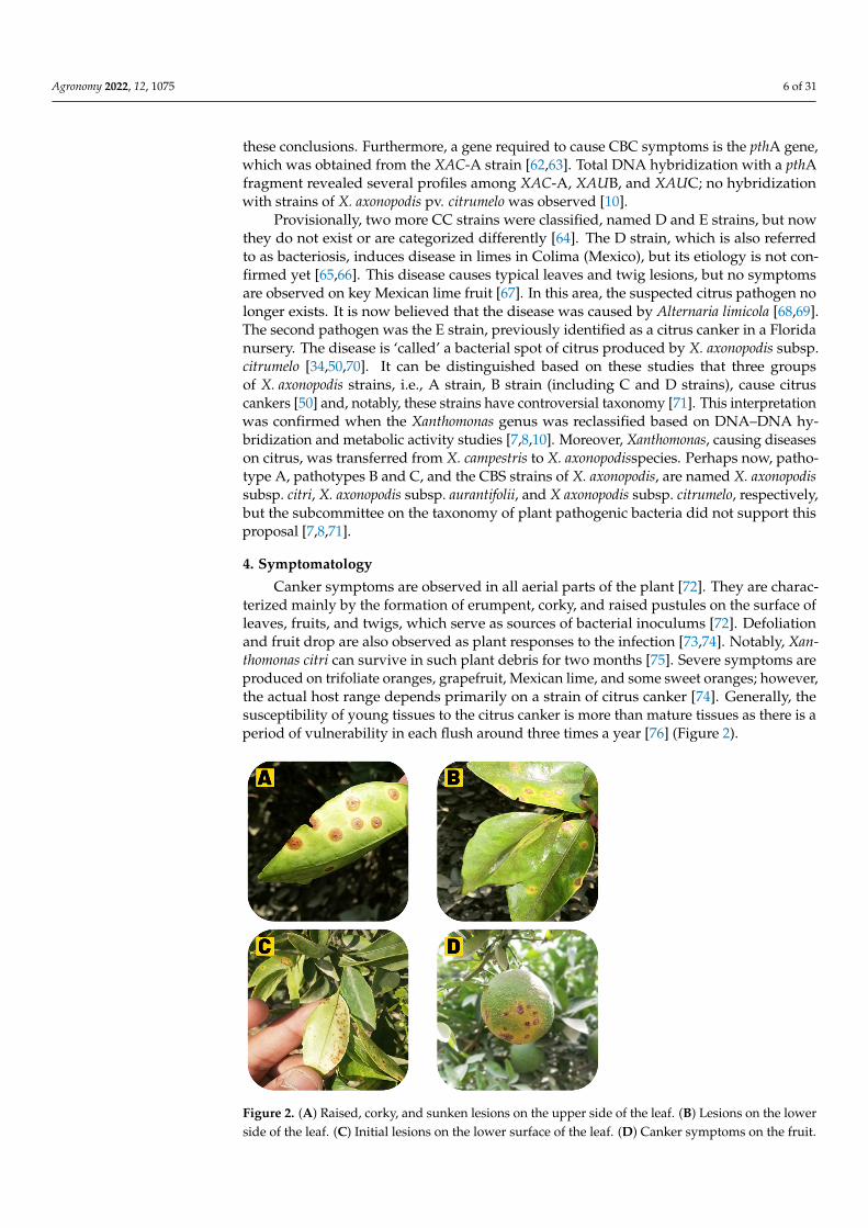
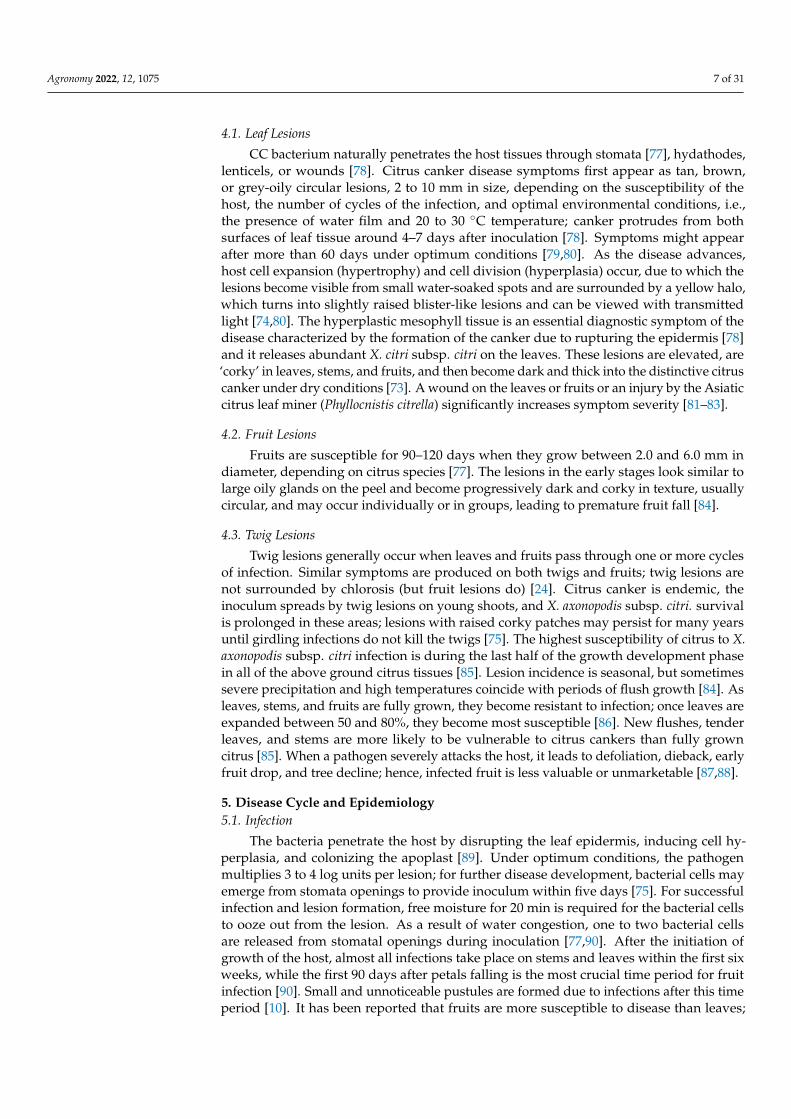
hence, observations have been made that lesions of different sizes can be found on the rindof the same fruit during the infection of the bacterium [91] (Figure 3).
Agronomy 2022, 12, x FOR PEER REVIEW 8 of 30
Twig lesions generally occur when leaves and fruits pass through one or more cycles of infection. Similar symptoms are produced on both twigs and fruits; twig lesions are not surrounded by chlorosis (but fruit lesions do) [24]. Citrus canker is endemic, the inoculum spreads by twig lesions on young shoots, and X. axonopodis subsp. citri. survival is prolonged in these areas; lesions with raised corky patches may persist for many years until girdling infections do not kill the twigs [76]. The highest susceptibility of citrus to X. axonopodis subsp. citri infection is during the last half of the growth development phase in all of the above ground citrus tissues [86]. Lesion incidence is seasonal, but sometimes severe precip-itation and high temperatures coincide with periods of flush growth [85]. As leaves, stems, and fruits are fully grown, they become resistant to infection; once leaves are expanded be-tween 50 and 80%, they become most susceptible [87]. New flushes, tender leaves, and stems are more likely to be vulnerable to citrus cankers than fully grown citrus [86]. When a path-ogen severely attacks the host, it leads to defoliation, dieback, early fruit drop, and tree de-cline; hence, infected fruit is less valuable or unmarketable [88,89].
5. Disease Cycle and Epidemiology 5.1. Infection
The bacteria penetrate the host by disrupting the leaf epidermis, inducing cell hyper-plasia, and colonizing the apoplast [90]. Under optimum conditions, the pathogen multi-plies 3 to 4 log units per lesion; for further disease development, bacterial cells may emerge from stomata openings to provide inoculum within five days [76]. For successful infection and lesion formation, free moisture for 20 min is required for the bacterial cells to ooze out from the lesion. As a result of water congestion, one to two bacterial cells are released from stomatal openings during inoculation [78,91]. After the initiation of growth of the host, almost all infections take place on stems and leaves within the first six weeks, while the first 90 days after petals falling is the most crucial time period for fruit infection [91]. Small and unnoticeable pustules are formed due to infections after this time period [10]. It has been reported that fruits are more susceptible to disease than leaves; hence, observations have been made that lesions of different sizes can be found on the rind of the same fruit during the infection of the bacterium [92] (Figure 3).
Figure 3. Dispersion of citrus canker bacterium in orchards. Figure 3. Dispersion of citrus canker bacterium in orchards.
5.2. Survival
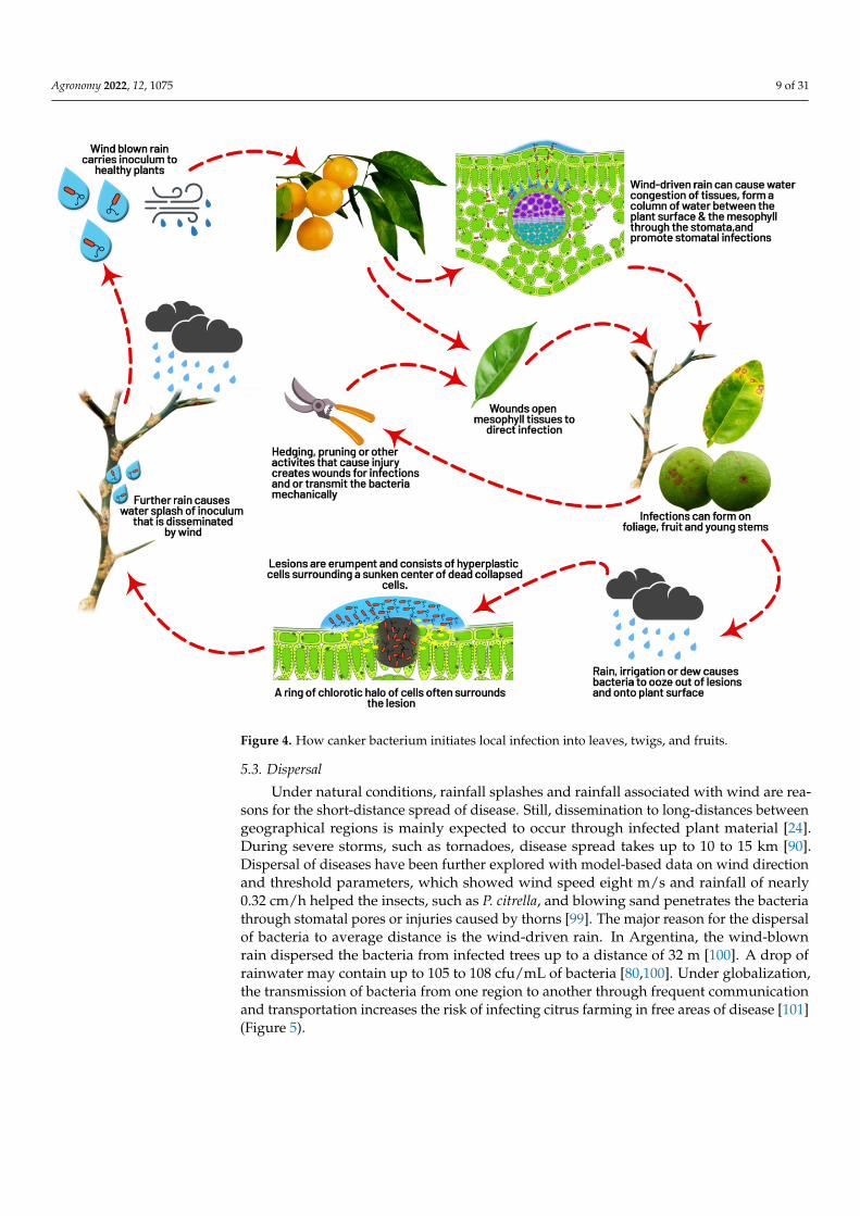
The main inoculum sources are branches, leaves, and twigs infected with cankers [84].The disease is primarily carried in cankers on twigs and branches from one season toanother, serving as a primary inoculum [10]. In leaf and fruit lesions, the bacteria remainalive until they fall; because the affected leaves fall off early, they may not act as the primaryinoculum source [75]. Still, it was reported that, in infected leaves, the bacterium survivedup to six months [92]. Reference [93] found that the bacterium survived for over 6monthsin the infected leaves, for 52 days in sterilized soil, and only 9 days in unsterilized soils,respectively. It was also observed that the organism could survive for 11–12 days underdesiccation at 30 ◦C [93]. On citrus hosts, the bacterium survives epiphytically with a lowerpopulation without developing the symptoms, combined with non-citrus weeds, grass host,and soil [94–96]. However, in the absence of plant tissue or debris, the saprophytic existenceof soil pathogens has not been observed [93,95]. The survival capability of the pathogenin subtropical soil is very limited [97], and bacterial inoculum dies within 24 to 72 h ondifferent inert surfaces, such as cloths, metals, plastics, and processed wood in both sunlightand shade [91]. Due to antagonisms and competition with saprophytic microorganisms,the bacterial population decreases to an undetectable level 1–2 months after leaves or fruitsfall to the ground [91]. In Japan and Brazil, it has been reported that X. axonopodis subsp.citri may survive on non-host plant material and in the root zone of certain weeds undereradicated diseased trees for a few weeks [98] (Figure 4).
Agronomy 2022, 12, 1075 9 of 31
Agronomy 2022, 12, x FOR PEER REVIEW 9 of 30
5.2. Survival The main inoculum sources are branches, leaves, and twigs infected with cankers
[85]. The disease is primarily carried in cankers on twigs and branches from one season to another, serving as a primary inoculum [10]. In leaf and fruit lesions, the bacteria remain alive until they fall; because the affected leaves fall off early, they may not act as the pri-mary inoculum source [76]. Still, it was reported that, in infected leaves, the bacterium survived up to six months [93]. Reference[94] found that the bacterium survived for over 6months in the infected leaves, for 52 days in sterilized soil, and only 9 days in unsterilized soils, respectively. It was also observed that the organism could survive for 11–12 days under desiccation at 30 °C [94]. On citrus hosts, the bacterium survives epiphytically with a lower population without developing the symptoms, combined with non-citrus weeds, grass host, and soil [95–97]. However, in the absence of plant tissue or debris, the sapro-phytic existence of soil pathogens has not been observed [94,96]. The survival capability of the pathogen in subtropical soil is very limited [98], and bacterial inoculum dies within 24 to 72 h on different inert surfaces, such as cloths, metals, plastics, and processed wood in both sunlight and shade [92]. Due to antagonisms and competition with saprophytic microorganisms, the bacterial population decreases to an undetectable level 1–2 months after leaves or fruits fall to the ground [92]. In Japan and Brazil, it has been reported that X. axonopodis subsp. citri may survive on non-host plant material and in the root zone of certain weeds under eradicated diseased trees for a few weeks [99] (Figure 4).
Figure 4. How canker bacterium initiates local infection into leaves, twigs, and fruits.
5.3. Dispersal
Figure 4. How canker bacterium initiates local infection into leaves, twigs, and fruits.
5.3. Dispersal
Under natural conditions, rainfall splashes and rainfall associated with wind are rea-sons for the short-distance spread of disease. Still, dissemination to long-distances betweengeographical regions is mainly expected to occur through infected plant material [24].During severe storms, such as tornadoes, disease spread takes up to 10 to 15 km [90].Dispersal of diseases have been further explored with model-based data on wind directionand threshold parameters, which showed wind speed eight m/s and rainfall of nearly0.32 cm/h helped the insects, such as P. citrella, and blowing sand penetrates the bacteriathrough stomatal pores or injuries caused by thorns [99]. The major reason for the dispersalof bacteria to average distance is the wind-driven rain. In Argentina, the wind-blownrain dispersed the bacteria from infected trees up to a distance of 32 m [100]. A drop ofrainwater may contain up to 105 to 108 cfu/mL of bacteria [80,100]. Under globalization,the transmission of bacteria from one region to another through frequent communicationand transportation increases the risk of infecting citrus farming in free areas of disease [101](Figure 5).
Agronomy 2022, 12, 1075 10 of 31
Agronomy 2022, 12, x FOR PEER REVIEW 10 of 30
Under natural conditions, rainfall splashes and rainfall associated with wind are rea-sons for the short-distance spread of disease. Still, dissemination to long-distances between geographical regions is mainly expected to occur through infected plant material [24]. Dur-ing severe storms, such as tornadoes, disease spread takes up to 10 to 15 km [91]. Dispersal of diseases have been further explored with model-based data on wind direction and thresh-old parameters, which showed wind speed eight m/s and rainfall of nearly 0.32 cm/h helped the insects, such as P. citrella, and blowing sand penetrates the bacteria through stomatal pores or injuries caused by thorns [100]. The major reason for the dispersal of bacteria to average distance is the wind-driven rain. In Argentina, the wind-blown rain dispersed the bacteria from infected trees up to a distance of 32 m [101]. A drop of rainwater may contain up to 105 to 108 cfu/mL of bacteria [81,101]. Under globalization, the transmission of bacteria from one region to another through frequent communication and transportation increases the risk of infecting citrus farming in free areas of disease [102] (Figure 5).
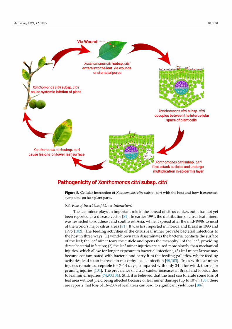
Figure 5. Cellular interaction of Xanthomonas citri subsp. citri with the host and how it expresses symptoms on host plant parts.
5.4. Role of Insect (Leaf Miner Interaction) The leaf miner plays an important role in the spread of citrus canker, but it has not
yet been reported as a disease vector [82]. In earlier 1994, the distribution of citrus leaf miners was restricted to southeast and southwest Asia, while it spread after the mid-1990s to most of the world’s major citrus areas [82]. It was first reported in Florida and Brazil in 1993 and 1996 [103]. The feeding activities of the citrus leaf miner provide bacterial infec-tions to the host in three ways: (1) wind-blown rain disseminates the bacteria, contacts the surface of the leaf; the leaf miner tears the cuticle and opens the mesophyll of the leaf,
Figure 5. Cellular interaction of Xanthomonas citri subsp. citri with the host and how it expressessymptoms on host plant parts.
5.4. Role of Insect (Leaf Miner Interaction)
The leaf miner plays an important role in the spread of citrus canker, but it has not yetbeen reported as a disease vector [81]. In earlier 1994, the distribution of citrus leaf minerswas restricted to southeast and southwest Asia, while it spread after the mid-1990s to mostof the world’s major citrus areas [81]. It was first reported in Florida and Brazil in 1993 and1996 [102]. The feeding activities of the citrus leaf miner provide bacterial infections tothe host in three ways: (1) wind-blown rain disseminates the bacteria, contacts the surfaceof the leaf; the leaf miner tears the cuticle and opens the mesophyll of the leaf, providingdirect bacterial infection; (2) the leaf miner injuries are cured more slowly than mechanicalinjuries, which allow for longer exposure to bacterial infections; (3) leaf miner larvae maybecome contaminated with bacteria and carry it to the feeding galleries, where feedingactivities lead to an increase in mesophyll cells infection [99,103]. Trees with leaf minerinjuries remain susceptible for 7–14 days, compared with only 24 h for wind, thorns, orpruning injuries [104]. The prevalence of citrus canker increases in Brazil and Florida dueto leaf miner injuries [74,90,104]. Still, it is believed that the host can tolerate some loss ofleaf area without yield being affected because of leaf miner damage (up to 10%) [105]; thereare reports that loss of 16–23% of leaf areas can lead to significant yield loss [106].
Agronomy 2022, 12, 1075 11 of 31
6. Detection and Identification of Citrus Bacterial Canker
The diagnosis of CC can be made using various methods and in most circumstances;however, when no official confirmation is required, the disease diagnosis can be madeby recognizing common symptoms [8,107]. It is also possible to confirm the causal agentby isolating XCC from lesions on a solid medium and looking for xanthomonad-likecolonies, which are yellow, convex, circular, semi-translucent, and have regular edges [52].Infiltration of a bacterial suspension adjusted to 108 colony-forming units (CFU)/mL intothe leaf mesophyll, followed by observation of water soaking and raised margins in theinfiltrated portion of the leaf 2–4 days after inoculation, can be used to test pathogenicity insusceptible citrus species [76,107]. DNA-based assays and serological testing are routinelyutilized methods for CC diagnosis when symptoms are atypical or an official confirmationis required for quarantine purposes [107]. Although molecular approaches can identifythe presence of XCC in infected plant tissue before canker lesions occur, serology-basedassays are usually sufficient for detecting XCC in symptomatic tissue [8]. Several primersbased on rDNA sequences, plasmid-borne genes, and pathogenicity regulatory factorshave been devised for polymerase chain reaction (PCR) detection of XCC [54,108–111]. Inrecent years, the introduction of real-time PCR and loop-mediated isothermal amplificationprocedures has improved the accuracy of diagnostic testing for XCC [11,112–115]. Allexisting conventional PCR methods need gel-visualization or primers, but all strains arenot detected [54,90,116]. PCR primers are very effective for X. citri subsp. citri ‘A type’detection, but these primers do not show consistency in X. citri subsp. Aurantifolii ‘B’ or ‘C’strain detections [60,117]. New PCR primers based on the gene-sequence of pthA did noteven detect one canker strain [54]. This technique, based on rep-PCR with BOX and ERICprimers, was developed to separate and distinguish the CBC pathotypes worldwide and thesubgroups of pathotypes of citrus associated within specific geographical areas around theworld [54,118]. Rapid, sensitive, and reliable real-time PCR assays were developed alongwith designed primers to detect all citrus–canker strains, which are important for bothspecificity and sensitivity [75,110]. Real-time PCR is easier to perform, less labor-intensive(no need for agarose gels), and much faster than conventional PCR [110]. If the samplingmethod is performed accurately, exact results can be obtained within 1 h. For plant pathogendetection, real-time PCR is becoming increasingly useful, i.e., for fungi [111,119–220],bacteria [122–124], and viruses [125,126]. A reliable, sensitive, SYBR Green real-timePCR assay in which primers are used to amplify conserved regions of a desired gene ofpathogenicity to detect all known strains of XAC is based on sampling techniques conductedin the field samples [127]. The detection of the bacterium through PCR is based on aninternal standard to make sure the quality of the DNA template for the reaction and ratioof PCR products is used to evaluate the early concentration of bacteria in citrus leaf tissueswith lesions using internal standards and target pathogens [118]. Detection and comparisonof X. axonopodis subsp. citri (XAC) from imported citrus fruits was based on an integratedapproach involving isolation of bacteria from three conventional protocols viz., PCR, real-time PCR with SYBR-green, or a TaqMan-probe in canker lesions and LAMP [8,116,117].The real-time PCR for fresh fruit samples with a TaqMan probe is the fastest screeningmethod for the detection of bacteria [111]. Enzyme-linked immunosorbent assay (ELISA),or serological tests, have also proved to be useful for rapid detection of XCC, whichare based on the ability of an antibody to recognize and bind to a specific antigen [111].These tests are usually performed in the laboratory. Still, they are also available as strip-based kits that are easy to use in the field where the disease is suspected; these kitsdo not require special equipment or training, and the results are obtained within a fewminutes [111,128]. Other older techniques for the detection of XCC have been developed,including physiological characterization, fatty acid profile analyses, protein profiling,hybridization, restriction fragment length polymorphism analysis, and comparison ofplasmid DNA patterns [8,10,111,128] (Table 2).
Agronomy 2022, 12, 1075 12 of 31
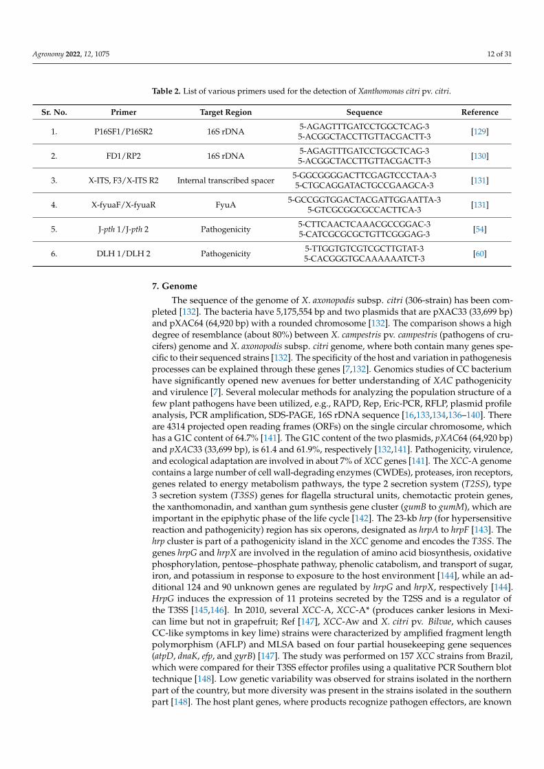
Table 2. List of various primers used for the detection of Xanthomonas citri pv. citri.
Sr. No. Primer Target Region Sequence Reference
1. P16SF1/P16SR2 16S rDNA 5-AGAGTTTGATCCTGGCTCAG-35-ACGGCTACCTTGTTACGACTT-3 [129]
2. FD1/RP2 16S rDNA 5-AGAGTTTGATCCTGGCTCAG-35-ACGGCTACCTTGTTACGACTT-3 [130]
3. X-ITS, F3/X-ITS R2 Internal transcribed spacer 5-GGCGGGGACTTCGAGTCCCTAA-35-CTGCAGGATACTGCCGAAGCA-3 [131]
4. X-fyuaF/X-fyuaR FyuA 5-GCCGGTGGACTACGATTGGAATTA-35-GTCGCGGCGCCACTTCA-3 [131]
5. J-pth 1/J-pth 2 Pathogenicity 5-CTTCAACTCAAACGCCGGAC-35-CATCGCGCGCTGTTCGGGAG-3 [54]
6. DLH 1/DLH 2 Pathogenicity 5-TTGGTGTCGTCGCTTGTAT-35-CACGGGTGCAAAAAATCT-3 [60]
7. Genome
The sequence of the genome of X. axonopodis subsp. citri (306-strain) has been com-pleted [132]. The bacteria have 5,175,554 bp and two plasmids that are pXAC33 (33,699 bp)and pXAC64 (64,920 bp) with a rounded chromosome [132]. The comparison shows a highdegree of resemblance (about 80%) between X. campestris pv. campestris (pathogens of cru-cifers) genome and X. axonopodis subsp. citri genome, where both contain many genes spe-cific to their sequenced strains [132]. The specificity of the host and variation in pathogenesisprocesses can be explained through these genes [7,132]. Genomics studies of CC bacteriumhave significantly opened new avenues for better understanding of XAC pathogenicityand virulence [7]. Several molecular methods for analyzing the population structure of afew plant pathogens have been utilized, e.g., RAPD, Rep, Eric-PCR, RFLP, plasmid profileanalysis, PCR amplification, SDS-PAGE, 16S rDNA sequence [16,133,134,136–140]. Thereare 4314 projected open reading frames (ORFs) on the single circular chromosome, whichhas a G1C content of 64.7% [141]. The G1C content of the two plasmids, pXAC64 (64,920 bp)and pXAC33 (33,699 bp), is 61.4 and 61.9%, respectively [132,141]. Pathogenicity, virulence,and ecological adaptation are involved in about 7% of XCC genes [141]. The XCC-A genomecontains a large number of cell wall-degrading enzymes (CWDEs), proteases, iron receptors,genes related to energy metabolism pathways, the type 2 secretion system (T2SS), type3 secretion system (T3SS) genes for flagella structural units, chemotactic protein genes,the xanthomonadin, and xanthan gum synthesis gene cluster (gumB to gumM), which areimportant in the epiphytic phase of the life cycle [142]. The 23-kb hrp (for hypersensitivereaction and pathogenicity) region has six operons, designated as hrpA to hrpF [143]. Thehrp cluster is part of a pathogenicity island in the XCC genome and encodes the T3SS. Thegenes hrpG and hrpX are involved in the regulation of amino acid biosynthesis, oxidativephosphorylation, pentose–phosphate pathway, phenolic catabolism, and transport of sugar,iron, and potassium in response to exposure to the host environment [144], while an ad-ditional 124 and 90 unknown genes are regulated by hrpG and hrpX, respectively [144].HrpG induces the expression of 11 proteins secreted by the T2SS and is a regulator ofthe T3SS [145,146]. In 2010, several XCC-A, XCC-A* (produces canker lesions in Mexi-can lime but not in grapefruit; Ref [147], XCC-Aw and X. citri pv. Bilvae, which causesCC-like symptoms in key lime) strains were characterized by amplified fragment lengthpolymorphism (AFLP) and MLSA based on four partial housekeeping gene sequences(atpD, dnaK, efp, and gyrB) [147]. The study was performed on 157 XCC strains from Brazil,which were compared for their T3SS effector profiles using a qualitative PCR Southern blottechnique [148]. Low genetic variability was observed for strains isolated in the northernpart of the country, but more diversity was present in the strains isolated in the southernpart [148]. The host plant genes, where products recognize pathogen effectors, are known
Agronomy 2022, 12, 1075 13 of 31
as R genes, and the pathogen pathogenicity(pth) genes, which encode these recognizableeffectors, are also referred to as avirulence (avr) genes [148]. The products of R genes,either directly or indirectly, interact with the products of avr genes; interaction betweenproducts of Rand avr genes is termed gene-for-gene resistance [149] whereas if no R genescorrespond to XCC, then it leads imparting resistance to XCC host plant, which has beenidentified in citrus and citrus-related species [7,8,45].
8. Virulence8.1. Type III Secretion System (T3SS)
Hypersensitive response and pathogenicity (hrp) of CC have a cluster of 24 geneson locus arranged in six operons from hrpA to hrpF, which are regulated by hrpGhrpXgenes and codes T3SS in X. citri subsp. citri [144,150]. It was observed that XAC remainedunsuccessful at inducing disease and HR in the cotton plant by the deletion of hrpB, hrpD,and hrpF operons [151]. This system was presumed to secrete the effector proteins [152,153],and pthA, the member of the avrBs3 gene family, targets the host susceptible gene lateralboundaries of organ 1 [154]. XAC uses T3SS against the host by injecting virulence proteinsinto host plant cells required for canker development in susceptible citrus plants andresistant plants, developing a hypersensitive response (HR) [151,155]. Effectors (virulenceprotein) are transported through specialized T3SS and use a hollow exterior (Hrp-pilus)that crosses the plant cell wall, delivering effectors across the plant plasma membranethrough a translocon [156]. Xanthomonas strains usually harbor about 30 various effectorsbut their molecular activity is still unknown in most cases [156]. Transcription-activator-like(TAL) effectors form a large and important family of effectors found almost exclusivelyin Xanthomonas [157]. They act as transcription factors for plant genes and few induceexpression of sugar exporters [13].
8.2. Citrus Specific pthA and Its Requirement for Canker Development
The first member of the gene family avrBs3/pthA is the pthA gene necessary forpathogenicity and was cloned through screening for pathogenicity [62,63,98]. Genes ofavrBs3 are broadly spread in the genus Xanthomonas, but are not present in all Xanthomon-ads [63]; there are a minimum of 27 cloned members of the avrBs3 family [158]. Withoutevidence of the pth function, mostly avrBs3 members are isolated as avr genes for the firsttime while all genes pthA, pthB, pthC, and pthW of this family induce the citrus canker, andtwo genes, pthN and pthN2, are involved in the induction of cotton blight [159]. Intensivestudies have been reported on the molecular virulence mechanism of the pathogen for thcitrus canker [45,160]. Gene pthA, an effector of the type III secretion system (T3SS), is adeterminant of cancer pathogenicity and is commonly found in Xanthomonas spp., whichcauses citrus canker [54,63,98]. The pthA gene’s exogenous insertion into X. axonopodissubsp. citrumelocauses bacterial citrus spot disease without causing erumpent lesions [62].The transient expression of pthA produces citrus canker symptoms, causing cell hyper-plasia, hypertrophy, and eventually, the plant dies [89]. In addition, pthB and pthC arefunctionally homologous genes that were cloned from X. citri subsp. aurantifolii B and C,and are important in inducing citrus canker [161]. Therefore, all three genes are function-ally exchangeable and can be transmitted horizontally between strains of X. citri and X.campestris subsp. aurantifolii on plasmids [162]. Members of the pthA gene family are morethan 3.8 kb in length and possess a high level of identity with DNA sequences (more than90%) over their total length [138]. It seems that functionally homologous genes based onDNA hybridizations were found in all canker-causing strains and have not been found inisolated citrus strains that do not induce cankers, e.g., X. campestris subsp. citrumelo [55,161];therefore, the single common gene was needed to induce citrus canker by Xanthomonas [45],while the XAC genome possessed several genes coding putative effectors [62,162]. The pthAis the most important effector that induces canker-like symptoms even in the absence of apathogen when expressed transiently in plants [89,98]. Transient expression of pthA in hostplant cells was sufficient at inducing symptoms of citrus canker in 10–14 days and pthA’s
Agronomy 2022, 12, 1075 14 of 31
deletion eliminates the pathogen to cause citrus canker [154]. In all citrus species, pthAinduces canker symptoms, but in other plant species, it changes the plant to immune; hence,this characteristic makes the XAC specific toward the citrus species [53,62,63]. The pthAmutation prevents XAC from inducing hyperplasia, hypertrophy, water soaking symptoms,and pathogen losses of the ability to grow within plants [62,63]. All strains of X. citri subsp.citri group A were examined, which has three pthA alleles in addition to pthA; two of themare slightly functional to produce cankers in citrus, and one seems non-functional [63,163].In the case of host recognition or avoidance, multiple homologs seem to provide rapiddevelopment of new genes of pathogenicity through recombination [164]. X. axonopodissubsp. citri have three homologs of avrBs3/pthA while only ap11 homologue was reportedto participate in virulence to induce canker formation, while the functions of other homol-ogous were insignificant or not assessable [163]. The pthA is transported through T3SS,which consists of hrp gene cluster products, and the transcription of hrp genes is inducedand regulated by the hrpG and hrpX regulators [144,155,165,166]. The hrpG or hrpX genemutations in X. citrus subsp. citri led to a loss of pathogenicity in citrus [144,167]. Othervirulence-related genes are required for X. citri subsp. citri, in addition to the pthA and hrpgenes, to cause disease [45], e.g., it was reported that opsX gene plays an important rolein the production of lipopolysaccharides (LPS) and extracellular polysaccharides (EPS),growth, and virulence in planta [168].
8.3. Adhesion and Extracellular Polysaccharides (EPSs)
Extracellular polysaccharides (EPSs) and lipopolysaccharides (LPSs) defend the bac-terium from unfavorable environmental conditions [169]. Xanthomonas spp. Producecharacteristic EPS and xanthan, which results in bacterial colonies being mucoid; it isknown as xanthan gum [170]. Xanthan is a polymer of repetitive units of pentasaccharide,having a backbone of side chains of cellulose and trisaccharide, used in the nutritionaland pharmaceutical industries commercially as a thickening agent [171,172]. The xanthanproduction is managed by various genetic loci, including the 12 gum gene clusters fromGum B to Gum M that are highly maintained among Xanthomonas spp. [172,173]. Xanthanproduction in Xanthomonas is regulated hierarchically by the gene cluster regulation ofpathogenicity factors (rpf ) [174]. Host plants wilted due to infections caused by vascularpathogens that obstructed the flow of water in xylem vessels due to xanthan produc-tion [175,176]. A plethora of research depicted various gum genes of Xanthomonas spp. e.g.,X. axonopodis subsp. citri, X. axonopodis subsp. manihotis, X. campestris subsp. campestris, andX. oryzae pv. oryzae has been involved in epiphytic survival and the development of bacteriain plants to induce disease symptoms [172,177–181]. It is interesting that gum genes fromX. axonopodis subsp. citri are not essential in Citrus sinensis for disease development andgrowth of bacteria. Still, in Citrus limon, these gum genes play an important role in bacterialvirulence, showing that xanthan virulence depends on the host plant and environmentalconditions [177,178]. The rpf protein has been involved in DSF synthesis to regulate thegenes and determine the synthesis of extracellular polysaccharides [183]. In many xan-thomonads, the gum gene cluster is involved in EPS biosynthesis. The Gum B mutantshowed defective EPS production and biofilms formation, hence, decreased infection inlemon [13,184].
XAC produces abundant extracellular polysaccharides (EPS) [184]. Bacterial cellsare incorporated in a dense matrix of EPS of canker lesions and are disseminated withEPS through rain [13]. The EPS molecules effectively protect the bacteria in water fromthe ‘dilution effect’ and desiccation in air, hence playing an important role in bacterialecology [185]. Bacteria enter the cell through stomata or wounds and remain stick to thehost’s cell wall through an interaction between EPS and citrus agglutinins [186]. Exper-imental evidence suggests that basal plant defense responses are always suppressed byxanthan, e.g., deposition of callose in the plant cell wall. The chelating divalent calciumions in the apoplast of the plant are required to activate plant defense responses [187,188].Further, xanthan plays an important role in biofilms formation in X. axonopodis subsp. citri,
Agronomy 2022, 12, 1075 15 of 31
and X. campestris subsp. campestris [177,189]. Microbes create a biofilm matrix, made upof proteins, extracellular DNA, and polysaccharides, essential for the establishment ofbacterial colonies while polysaccharide overproduction has been shown to alter colonyshape and help to identify certain species [190]. The matrix’s polysaccharide componentcan give a variety of benefits to the biofilm’s cells, including adhesion, protection, andstructure, and aggregative polysaccharides operate as molecular glue, allowing bacteria tostick to both biotic and abiotic surfaces by allowing them to withstand physical stresses,such as fluid movement that might detach them from a nutrition source [191–193].
8.4. Lipopolysaccharides (LPSs)
In plant pathogenic bacteria, the major virulence factor responsible for host infec-tivity is lipopolysaccharides and it is increasingly recognized as a major plant pathogen-associated molecular pattern (PAMP) [98,194]. LPSs are major components and charac-teristic structures of the outer membrane of Gram-negative bacteria [194]. LPS moleculesare usually composed of hydrophilic heteropolysaccharides formed by three major sub-structures, the O-specific polysaccharide (O-antigen), a repetitive sugar sub-unit; the coreoligosaccharide region, which is covalently connected to the lipid A of the glycolipid moiety,and the lipid ‘A’ attached to the external plasma membrane [195,196].
LPSs have been recognized as a virulence factor during plant pathogenic interac-tions and involved in bacterial pathogenicity [197]. LPSs are present in bacteria’s outermembrane, which protects the pathogen from hostile medium found within plant tissues,reduces the membrane permeability, and allows the bacteria to grow under unfavor-able environmental conditions [197]. LPSs can prevent hypersensitive response (HR) inplants by avirulent bacteria that have been widely studied in plant cells [198]. In vari-ous Xanthomonas spp., insilico analyses have been carried out to identify and characterizethe genes involved in LPS biosynthesis, showing wxacO and rfbC genes involved in LPSbiosynthesis that reduces the pathogen motility, lack of resistance to stress, and virulence ingrapefruit [199,200]. Further, the two-component regulatory system (TCRS) ColR/ColS hasbeen reported to play multiple roles in LPSs and catalase production, biofilm formation,resistance to stress, transcription of hrpD6 and hpaF genes, and knockout of either colR andColS, causing loss of pathogenicity in grapefruit [13].
8.5. Quorum Sensing
Virulence factors involve pathogen’s ability to express their genetic, biochemical,and structural features to the host [13]. Quorum sensing (QS) is a mechanism that facil-itates communication among bacterial cells through production and detection of signalmolecules [13,201,202]. Xanthomonas has QS regulatory systems facilitated by molecules ofthe diffusible signal factor (DSF) family, which regulates the QS pathway of Xanthomonasspp. comprising three major QS regulons: RpfF, RpfC, and RpfG [203–205]. Furthermore,transcriptome analysis characterized RpfF, RpfC, and RpfG regulons, which showed RpfF con-trols the unique genes responsible for the QS-pathway complexity and other sensorymechanisms involved in canker development [204]. RpfC and RpfG control individual genesthat play a wider role in gene regulation and their involvement in chemotaxis, motility andflagellate biosynthesis, extracellular enzymes production, adhesion, stress tolerance andregulations, transport, and transport detoxification [206]. The QS process depends on theproduction, release, and detection of small signaling molecules known as autoinducers(AIs) [206]. The MJ Daniels Research Group, for the first time, reported the detection ofactivity of DSF molecules as autoinducers [174]. The synthesis of DSF in X. axonopodissubsp. citri is based on genes rpfF and rpfB, and it is an autoinducer in bacteria and regu-lates quorum sensing [203]. The Rpf/DSF system involves the initial attachment of XACto the host and controls a range of virulence-related characteristics, such as the synthesisof extracellular enzymes (proteases, endoglucanases), extracellular polysaccharides, EPSsbiosynthesis, and biofilm formation in several strains of pathogens [13].

Agronomy 2022, 12, 1075 16 of 31
9. Nutrition
Bacteria can take nutrients from their hosts through enzyme secretions that degradethe host’s cell wall and bacteria utilize the cell wall’s breakdown products as nutritionsources [62]. In XAC genome pectin esterases are not present, but bacteria possess threepectate lyases, six cellulases, five xylanases, and an endoglucanase, while in cellulose,it contains endoglucanaseBcsZ (gi|22001634),which hydrolyzes 1,4-β-D-glucosidic link-ages [62]; moreover, permease, through which degraded pectin products are imported intothe bacterial cells in a hydrogen-transport coupled fashion [62]. The pthA is also necessaryfor optimal growth of the bacteria in the host, which is probably directly injected into thehost cell [62]. X. citri subsp. citri produces less enzymes as compared to X. campestris subsp.campestris, which degrades the cell wall; because of this, the two pathogens may cause theirhosts to suffer from different symptoms [132].
10. Integrated Management Programs
IDM has been introduced to control the occurrence of citrus bacterial canker disease(CBCD) in new seedlings [9]. This program recommends that only citrus bacterial cankerdisease-resistant cultivars may be planted [107]. For commercial cultivation, it is recom-mended to plant sweet oranges, such as Tahiti lime, Pera pre-immunized, FolhaMurcha,Valencia, and Navelina, mandarins, such as Ponkan, Dancy, Loose Jacket, Satsuma, Batan-gas, and Willowleaf accessions [9]. Many studies have been carried out to control CCdisease through cultural, chemical, or biological management strategies but have shownlimited effects [207,208]. When leaf miners attack citrus varieties, or under changingweather conditions, the development of disease becomes far more complex and harder tocontrol the eradication of diseased trees is the only way to control the CC disease [7,9,11].Resistance genotypes in citruses and relative genera have long been researched aroundthe world, and several types of citrus germplasms with certain resistance levels have beenreported, e.g., calamondin (C. mitis) and kumquats (Fortunella spp.) are highly resistant,and mandarins (C. reticulata) were also reported to be resistant [78,162,209]. Suppose plantsare inoculated artificially with the bacterium or planted in combination with sweet oranges.In that case, all plants show characteristic symptoms of citrus cankers without any com-plete or active resistant citrus genotypes [7,9]. As no resistant varieties were identified,breeding efforts have made little progress in the production of resistant cultivars, and fewexperiments in molecular breeding have shown transformants of some resistance throughthe transfer of antibacterial genes to citrus fruits. Still, only a lower disease incidence hasbeen achieved without complete resistance [8,9]. The molecular mechanism of pathogene-sis remains unclear, and there are no resistance genes isolated; it is very tough to obtainresistant genotypes through breeding programs [210,211]. The resistant mandarin varietiesare grown in Southeast Asia, where the climate is most favorable for epidemics; the citruscanker was not a major issue until more vulnerable sweet oranges were brought into thedisease regions of China and Japan [78]. Since the 1950s, eradication/control programshave been established in São Paulo, Brazil, to prevent the pathogen spread in the productionarea of sweet oranges [9]. Contrarily, the nearby zones of Paraná State, Brazil, Misiones,Corrientes, and Argentina have adopted integrated program strategies to efficiently preventand control citrus cankers in sweet oranges [212–214]. The program is mainly concernedwith shifting of citrus plantations to the disease-free area, having resistant citrus varieties,and in these regions, regulations are there, not only to deal with more resistant varieties,but also to produce disease-free nursery trees, as well as other means to exclude the XACfrom citrus plantations [212]. The regulations for the management of CC disease include(i) nurseries must be situated in disease-free areas; (ii) the design of citrus production areasmust be managed in order to decrease the danger of an epidemic of CC by constructingwindbreaks, applying preventive copper sprays, building fences to avoid the entrance ofbacteria to the citrus plantation; (iii) the planting and harvesting tools should be disinfected;workers should also disinfect their clothes, shoes, and gloves; (iv) fresh fruits should bestrictly inspected for domestic and export markets to prevent the fruits in citrus groves from
Agronomy 2022, 12, 1075 17 of 31
citrus cankers; workers should also disinfect the storage and packaging houses; (v) infectedsummer and autumn shoots should be pruned; (vi) disease management forecasts shouldbe considered; (vii) control of citrus leaf miners; (viii) use of chemical inducers, whichinduce systemic acquired resistance (SAR) in plants [215,216].
10.1. Quarantines
Federal quarantine barriers are regulatory responses to diseases, which could be foundin almost any country. Still, the exact locations of such barriers are difficult issues, forbiological and political reasons [9,24]. These barriers are usually placed two or more twomiles away from any known infestation [24]. The distribution of host plant materials islimited within quarantine areas, affecting both the citrus agriculture sector and homeownerswith citrus trees [9]. In commercial production, it is recommended to disinfect the fruits inpacking houses and disinfect the harvesting and transport equipment [24]. Fresh fruits areoften restricted in market distributions from regulated areas [7]. Commercial citrus plantingneeds sanitization stations at plantation doors, a caution that has become a national demand,even outside quarantine areas [7]. Citrus replanting in commercial or residential areasthat have undergone eradication efforts is against the law before the disease is declaredeliminated [7,9]. People are informed in residential areas—that it is illegal to transportfruits to neighbors and family; decontamination of all equipment that is moved betweenproperties during lawn and garden services is required [7,9]. These measures are publicizedthrough intensive media reporting and expertise in community relations [24].
10.2. Cultural Control
Eradication of any disease is the method used to manage that specific problem if it hasnot been endemic in a region [7]. Quarantine and eradication are key measures to controlthe entry and dissemination of pathogens in many countries [9]. Eradication measuresinvolve destruction by cutting and burning citrus species [24]. Sometimes, herbicides areused instead of cutting and burning to kill citrus plants [18]. The infested property isquarantined, followed by the eradication procedure for at least a year without planting orpropagation of citrus fruits, with inspection at least twice a year [24].
Data from Argentina showed that the pathogen could disseminate in rainfall withwind up to 32 m (105 ft), which provided the scientific basis for eradicating this disease [50].This has been translated in the U.S. and many other countries into regulating policies,allowing survey teams to locate diseased citrus trees, to remove and kill the trees, as wellas exposed trees within a radius of 38.1 m (125 ft) of a diseased tree [76,217]. Now, Braziluses a distance of 30 m (98 ft) to remove exposed infected trees. If infections of the Brazilianplants are 0.50% or lower, all trees will be removed within a radius of 30 m of the infectedplantation. When the infection exceeds 0.5%, the whole block will be removed [217]. Newcanker infections occur in known source trees, at about 1900 ft (579 m) [17]. In January2000, a new regulation, “the 1900 ft rule”, was set up. In March 2000, it was implemented,which involved the eradication of infected citrus trees along with healthy trees within1900-ft of an infected tree [17,218]. Each 1900 ft radius circle has a surface area of 1.06 km2
(0.41 miles), leading to the removal of dooryard citrus in infected areas by implementingthe 1900 ft rule [9]. Pruning of infected twigs, along with application of a 1% Bordeauxmixture at regular intervals, before the onset of monsoon, also proved to be very effectivein the management of disease [219–223].
10.3. Chemical Control
For the management of CC, it has been reported that every year, from November toDecember, pruning of infected twigs with three to four sprays of a 1% Bordeaux mixturecould be used to reduce disease [224]. Control of disease by applying four sprays of5000 ppm copper oxychloride or a 1% Bordeaux mixture and two prunings gave excellentresults [225,226]. Chemicals such as Perenox, Ultrasulphur, and a mixture of Blitox + nickelchloride, sodium arsenate + copper sulphate, were used against citrus cankers [227–230].
Agronomy 2022, 12, 1075 18 of 31
The application of 1% glycerin spray and 500–1000 ppm of streptomycin–sulphate wasfound useful in controlling disease on acid lime [231]. Acid lime canker was reducedby six sprays of 1000 ppm streptomycin with two prunings [232]. Streptocycline, incombination with Bordeaux mixture, and Agrimycin, are effective antibiotics againstCC [233]. For field tests with different chemicals, Paushamycin + Blitox and Bordeauxmixture showed the best control of CC [234]. In nurseries, treatments of young plants havebeen reported by applying neem cake solution on the leaves [235,236]. The application ofstreptocycline + copper oxychloride (0.1%), preferably at intervals of seven days or fifteendays, has been found very effective against CC [236]. Integrated application of copperoxychloride (0.3%), streptocycline (100 ppm), and suspension of neem cake on prunedinfected twigs has shown to be very useful to control the disease [237].
In field experiments in Argentina, copper ammonium carbonate with 8% metalliccopper was consistently found better than other products to control CC; regarding fieldtrials on trees of ripened grapefruits, three applications of copper ammonium carbonate(CAC) or copper hydroxide + maneb per season were examined and reduced the numberof lesions found on fruits but not on leaves [238]. The recommendation to add mancozebto copper spray was effective for copper resistance [239]. Sanitary procedures have beenexplained for persons or tools that encounter citrus in quarantine regions, e.g., by applyingsprayable ammonium detergent disinfectants [23].
10.4. Biological Control
There has been a surge in the hunt for more environmentally-friendly plant diseasetreatment methods [7]. Researchers are looking for more ecological approaches to man-age phytopathogens in the field due to chemical residues in soils and water bodies andincreased consumer concerns [7]. Some recent research used the antagonistic activity ofmicroorganisms and plant-derived compounds to suppress citrus cankers [7]. Studies onbiological control are still in the initial phases, to control CC [239,240]. Some bacterialstrains, including Pseudomonas syringae, Erwinia herbicola, Bacillus subtilis, and Pseudomonasfluorescence isolated from citrus phylloplane have been found to be antagonistic to thecitrus canker pathogen, invitro [133,241–244]. However, it appears hard to find antagonisticbacteria that stabilize on mature citrus tree leaves [245]. For example, P. aeruginosa LVproduces a bioactive combination of secondary metabolites, the most important of which isan organocopper antibiotic that reduces the formation of citrus canker lesions in Valenciaoranges by 90% [245]. The authors discovered a bacteriolytic impact on X. citri that wasnot accompanied by any signs of phytotoxicity [246]. At low micromolar concentrations,several secondary metabolites from P. aeruginosa also reduced canker formation [247,248].However, because P. aeruginosa is an opportunistic human infection, its use as a biocontrolagent is fraught with dangers and must be strictly managed [9]. Bacillus spp. isolated fromcitrus rhizosphere and leaves have also been proposed as a citrus canker biocontrol agentbecause they inhibited X. citri growth in vitro and in field circumstances [76,240,249,250].X. citri and several species of Pseudomonas and Bacillus suppressed growth and cankerformation, and Citrobacter isolated from sweet orange phylloplane, by degrading the X.citri quorum sensing molecule DSF [251]. Other bacterial genera, such as Cronobacter andEnterobacter, similarly inhibited X. citri growth in vitro by producing bacteriocins; how-ever, the protective effect of these bacterial species on X. citri-infected citrus trees wasnot assessed [252]. The sensitivity of bacteriophages (phages) can be utilized to identifyintra-species sub-groups in bacteria [252]. Cp1 and Cp2 phages have long been employedto identify X. citri strains [253]. The employment of phages in biological control, on theother hand, poses significant difficulties [254]. Phages have a very short active life on theleaf surface and must be administered at high concentrations to be effective [255]. Theauthors of reference [256] found that the jumbo phage XacN1, originally isolated froman orange orchard soil in Japan, can infect a wide range of X. citri isolates, making it apromising choice for future field experiments. In greenhouse and field testing, a mixture ofphages obtained from orange orchards combined with ASM successfully decreased canker
Agronomy 2022, 12, 1075 19 of 31
symptoms [257]. Compared to copper–mancozeb alone, the combination of phages andcopper–mancozeb did not improve citrus canker control [258]. Interestingly, filamentousintegrative phages, such as XACF1, reduced the virulence of X. citri, suggesting that theycould be used as citrus canker biocontrol agents [259].
10.5. Field Screening
Globally, field screening was carried out to assess the response of citrus varieties inlocal environmental conditions to CC [9,24]. Suppose very intensive control programs havebeen carried out. In that case, highly susceptible varieties, i.e., several early–mid-seasonsweet oranges, grapefruits, and Mexican limes (e.g., Navel, Hamlin), are not recommendedfor planting [212]. Mid and late season oranges, tangerines (tangerine, tangelo, and tanger-ine), and Tahiti limes have been identified with acceptable resistance to citrus cankers inscreening programs [9]. These cultivars may be susceptible in the young stages and need tobe sprayed to control the leaf miner to avoid damage to emerging flushes that predisposethem to infection [9].
10.6. Induced Systemic Resistance
Induced systemic resistance (ISR) is an active resistance mechanism in plants activatedthrough biotic or abiotic infection. The mechanism increases physical or chemical barriersof host plants against infection [260]. Different inducer compounds, e.g., salicylic acid,harpin protein, and benzothiadiazoles are used effectively to induce resistance in plantsagainst diseases [261,262]. ISR mechanisms can simultaneously control the disease anddecrease the risk of developing pathogen resistance [263]. ISR activity may be used early inthe season to complement the protectant activity of Cu, which slows the growth of bacteriain rapidly growing leaves [263]. Chemicals sold for the treatment of citrus canker include‘Actigard’, approved for acibenzolar-S-methyl, a benzothiadiazole in the USA, and ‘Bion’ inEurope and South-America (Syngenta Crop Protection); ‘Messenger’ (Eden Bioscience) isapproved for the harpin protein (a hrp-gene product) [63]. In Florida, several ISR inducers(e.g., Messenger, Nutri-phite, Oxycom, and FNX-100) are evaluated for control of X. citrisubsp. citri [9].
10.7. Leaf Miner Control
Leaf miners do not propagate cankers, but extensive bacterial invasion through leafminer galleries increases inoculum levels significantly, making it difficult to control thedisease [24]. Leaf miner control on the first summer flush can significantly reduce thepressure of the disease, but there is no effective control of leaf miners on late summer flushes.On spring flush, it causes no damage; therefore, control is required [63]. Applicationsof Agri-mek, petroleum oil, Assail, Micromite, or Spintor on time minimize leaf minerdamage [24,63].
10.8. Control through Plant Extracts
To avoid or reduce the deleterious effects of synthetic pesticides on the ecosystem,it is necessary to find alternative approaches to manage plant pathogenic microorgan-isms [264,265]. As an alternative to synthetic pesticides, green plants have proved tobe effective chemo-therapeutics and can be used as valuable sources of natural pesti-cides [266]. The use of several plant byproducts that possess antimicrobial properties onseveral pathogenic bacteria and fungi has been reported by many researchers [267–272]. InPakistan, where the farms are small, and the economic conditions of farmers are also notgood, standard antibiotics, on account of their high costs, are often beyond the reach of anaverage farmer [272]. Under such conditions, the use of plant extracts/diffusates to managebacterial plant diseases appeared to have good potential [273]. Different plant extracts,e.g., Allium sativum L., Allium cepa L., Azadirachta indica, Calotropis gigantea, Dalbrgia sissoo,Eucalyptus camelduensis, Gardenia florida, and Melia azedarach have been used by farmingcommunities to mitigate the multiplication of XCC [273]. Essential oil is a broad word that
Agronomy 2022, 12, 1075 20 of 31
refers to any volatile aromatic molecule generated from plants [274]. Essential oils havelong been known for their antibacterial properties against pathogenic and phytopathogenicmicroorganisms [275]. Many essential oils from Citrus aurantium, Citrus aurantifolia, and For-tunella sp. have been shown to kill X. citri [276]. Citral from C. aurantifolia inhibited X. citrigrowth, the most in disc diffusion experiments, while limonene, geranyl acetate, and trans-caryophyllene from Fortunella sp. had little effect [276]. Citral has a MIC of 0.5 mg/mL,indicating that substantial doses would be needed to control X. citri in the field [276]. Otherplant-derived chemicals may be useful in the fight against citrus canker, e.g., water and ace-tone extracts from the leaf gallnuts of the Chinese “sumac” (Rhus chinensis) suppressed thedevelopment of X. citri at a dose of 1 mg/mL [277]. After further separation of the gallnutleaf extracts, the bioactive chemicals were methyl gallates and gallic acids [277]. Gallic acids(MIC 4 mg/mL) were active at significantly lower quantities (MIC 0.1 mg/mL) than methylgallates (MIC 0.1 mg/mL) [277]. At low micromolar concentrations, synthetic gallates alsoreduced X. citri growth in vitro, but when administered to fully formed cankers, thesechemicals prevented X. citri host colonization after artificial infiltration and reduced thebacterial population [278]. Alkyl gallate amphiphile structures exhibit enhanced chemicalentry in target cells, similar to pyridinium-tailored molecules [279]; membrane perme-abilization and the divisional septum have been identified as primary targets of thesemolecules in X. citri [280]. These compounds were found to be low in toxicity in humancells, and further molecule development led to more lipophilic and lethal monoacetylatedalkyl gallates [280]. Reference [281] studied that C. coriaria is a potential candidate plant formanaging phytopathogenic Xanthomonas. In forest trees, it has been reported that diffusatesof Phyllanthus emblica, Acacia nilotica, Sapindus mukorossi, and Terminalia chebula (large, blacktype) were found to be most effective against XCC [282]. Methanolic leaf extracts of Psidiumguajava L. could be developed as antibacterial agents to control plant pathogenic bacteriaas they were able to inhibit the growth of Xanthomonas spp. with all concentrations [283].
10.9. Factors Affecting Successful Eradication of Citrus Canker
X. citri subsp. Citri possess some unique characteristics that make them desirablefor eradication: (i) for long periods; the bacterium cannot live outside the host lesion;(ii) bacteria lack an effective vector; (iii) the typical elevated lesions are easily recognizedand can be diagnosed quickly and accurately; (iv) the bacterial host range is limited to ahighly valuable perennial fruit plant; (v) many commercial citrus species are moderatelyto highly susceptible. For that reason, disease control measures were moderately efficientand comparatively expensive; during previous campaigns in Florida, Australia, and South-Africa, it has been eliminated successfully [9].
11. Conclusions and Future Prospects
Citrus canker, as the most feared citrus disease worldwide, continues to be a potentialthreat to citriculture. Broad-spectrum pathogenicity and cultivation of susceptible vari-eties of citrus canker and the emergence of new strains are major threats to the world’sproduction of citrus. The new citrus canker strains are also evolving because of mutationsin the genome. Various physiological, biochemical, serological, molecular, and pathogenicvariations are found among these strains. Therefore, detailed biological and molecularcharacterizations of the pathogens and their genomes are crucial for their proper identifi-cation [84]. Recent conclusions have shown that the vascular systems of plants generallycontain internal microbes (endophytes). There is a considerable chance that an antagonisticmicrobe found among these endophytes will help control citrus canker, biologically [84].The risk of citrus canker must be prevented or reduced by establishing and using wind-breaks, constructing barriers to prevent bacterial access to orchards, applying antibiotics,utilizing preventive copper-based sprays, biological control approaches, and developinggenetic engineering-based canker resistant varieties.
Agronomy 2022, 12, 1075 21 of 31
Author Contributions: Conceptualization, S.A.H.N., J.W., M.T.M., U.-U.-D.U., A.-U.-R., M.F. andM.T.S.; methodology, H.-u.-R., A.H., M.Z.H. and M.A.S.; software, S.A.H.N. and U.-U.-D.U.; valida-tion, J.W.; formal analysis, M.N.; investigation, S.A.H.N., M.T.M. and U.-U.-D.U.; resources, A.-U.-R.;data curation, S.A.H.N., M.F. and U.-U.-D.U.; writing—original draft preparation, S.A.H.N., J.W.,M.F., U.-U.-D.U., A.-U.-R., A.H., M.T.S. and R.D.; writing—review and editing, S.A.H.N., J.W., M.F.,U.-U.-D.U., A.-U.-R., A.H. and M.T.S.; supervision, J.W.; project administration, S.A.H.N., U.-U.-D.U.,J.W. and R.D. All authors have read and agreed to the published version of the manuscript.
Funding: Science and Technology Project of Guizhou (201907) and Anhui (202034180004192).
Acknowledgments: Special thanks are extended to the two anonymous reviewers for providingcritical comments and suggestions, which helped improve the review article.
Conflicts of Interest: The authors declare no conflict of interest.
References1. Ministry of National Food Security and Research. Agricultural-Statistics-of-Pakistan-2019–2020; Ministry of National Food Security
and Research: Islamabad, Pakistan, 2020.2. FAO STAT, Statistics Division, Food and Agriculture Organization of the United Nations. Agriculture Statistics 2019–2020; Food
and Agriculture Organization of the United Nations: Rome, Italy, 2020.3. Siddique, M.I.; Garnevska, E. Citrus Value Chain(s): A Survey of Pakistan Citrus Industry. In Agricultural Value Chain; IntechOpen:
London, UK, 2017. [CrossRef]4. Al-Snafi, A.E. Nutritional value and pharmacological importance of citrus species grown in Iraq. IOSR J. Pharm. 2016, 6, 76–108.
[CrossRef]5. Pakistan Horticulture Development Export Company (PHDECo). Citrus Marketing Strategy; Pakistan Horticulture Development
Export Company: Lahore, Pakistan, 2018.6. Agriculture Marketing Information Service of Pakistan. District Wise Data of Citrus; Agriculture Marketing Information Service of
Pakistan: Lahore, Pakistan, 2018.7. Martins, P.M.M.; de Oliveira Andrade, M.; Benedetti, C.E.; de Souza, A.A. Xanthomonas citri subsp. citri: Host interaction and
control strategies. Trop. Plant Pathol. 2020, 45, 213–236. [CrossRef]8. Ference, C.M.; Gochez, A.M.; Behlau, F.; Wang, N.; Graham, J.H.; Jones, J.B. Recent advances in the understanding of Xanthomonas
citri ssp. citri pathogenesis and citrus canker disease management. Mol. Plant Pathol. 2018, 19, 1302–1318. [CrossRef] [PubMed]9. Randsborg, K. Introduction. EPPO Bull. 1979, 9, 341–342. [CrossRef]10. da Gama, M.A.S.; de Lima Ramos Mariano, R.; da Silva J’unior, W.J.; de Farias, A.R.G.; Barbosa, M.A.G.; da Silva Velloso Ferreira, M.A.;
J’unior, C.R.L.C.; Santos, L.A.; de Souza, E.B. Taxonomic Repositioning of Xanthomonas campestris pv. viticola (Nayudu 1972) Dye1978 as Xanthomonas citri pv. viticola (Nayudu 1972) Dye 1978 comb. nov. and Emendation of the Description of Xanthomonas citripv. anacardii to Include Pigmented Isolates Pathogenic to Cashew Plant. Phytopathology 2018, 108, 1143–1153. [PubMed]
11. Mehrotra, R. Bacteria and Bacterial Diseases. In Plant Pathology; Tata McGraw-Hill Pub. Co. Ltd.: New Delhi, India, 1980;pp. 636–638.
12. Whiteside, J.O.; Garnsey, S.M. Compendium of Citrus Diseases; APS Press: College Park, MD, USA, 1988.13. Li, L.; Li, J.; Zhang, Y.; Wang, N. Diffusible signal factor (DSF)-mediated quorum sensing modulates expression of diverse traits in
Xanthomonas citri and responses of citrus plants to promote disease. BMC Genom. 2019, 20, 55. [CrossRef]14. Lee, H.A. Further data on the susceptibility of rutaceous plants to citrus-canker. J. Agric. Res. 1918, 15, 661–665.15. Fawcett, H.S.; Jenkins, A.E. Records of Citrus Canker from Herbarium Specimens of the Genus Citrus in England and the United
States. Phytopathology 1933, 23, 820–824.16. Graham, J.; Hartung, J.; Stall, R.; Chase, A. Pathological, restriction-fragment length polymorphism, and fatty acid profile
relationships between Xanthomonas campestris from citrus and noncitrus hosts. Phytopathology 1990, 80, 829–836. [CrossRef]17. Gottwald, T.R.; Hughes, G.; Graham, J.H.; Sun, X.; Riley, T. The citrus canker epidemic in Florida: The scientific basis of regulatory
eradication policy for an invasive species. Phytopathology 2001, 91, 30–34. [CrossRef]18. Rossetti, V. Citrus canker in Latin America: A review. In Proceedings of the International Society of Citriculture, Orlando, FL,
USA, 1–8 May 1977; pp. 918–924.19. Doidge, E.M. Citrus canker in South Africa. S. Afr. Fruit Grow. 1916, 3, 265–268.20. Garnsey, S.; Ducharme, E.; Lightfield, J.; Seymour, C.; Griffiths, J. Citrus canker. Citrus Ind. 1979, 60, 5.21. Bártová, V.; Bárta, J.; Jarošová, M. Antifungal and antimicrobial proteins and peptides of potato (Solanum tuberosum L.) tubers and
their applications. Appl. Microbiol. Biotechnol. 2019, 103, 5533–5547. [CrossRef] [PubMed]22. Okabe, N. Bacterial Diseases of Plants Occurring in Formosa I. J. Soc. Trop. Agric. 1932, 4, 470–483.23. Del Campo, R.; Russi, P.; Mara, P.; Mara, H.; Peyrou, M.; De León, I.P.; Gaggero, C. Xanthomonas axonopodispv. citri enters the
VBNC state after copper treatment and retains its virulence. FEMS Microbiol. Lett. 2009, 298, 143–148. [CrossRef]24. Schubert, T.S.; Rizvi, S.A.; Sun, X.; Gottwald, T.R.; Graham, J.H.; Dixon, W.N. Meeting the challenge of eradicating citrus canker
in Florida—Again. Plant Dis. 2001, 85, 340–356. [CrossRef]
Agronomy 2022, 12, 1075 22 of 31
25. Naseem, S.; Shah, H.A.; Ali, Z. First report on characterization of citrus disease causing bacteria and related phages isolated inPakistan. Int. J. Agric. Biol. 2017, 19, 857–864. [CrossRef]
26. Stevens, H.E. Citrus canker. A preliminary bulletin. Fla. Agric. Expt. Sta. Bull. 1914, 122, 113–118.27. Hasse, C.H. Pseudomonas citri, the cause of citrus canker—A preliminary report. J. Agric. Res. 1915, 4, 97–100.28. Doidge, E.M. The Origin and Cause of Citrus Canker in South Africa. Union So. Afr. Dept. Agric. Sei. Bul. 1916, 8, 20.29. Dowson, W.J. On the systematic position and generic names of the gram negative bacterial plant pathogens. Zentr. Bakteriol.
Parasitenk. Abt. II 1939, 100, 177–193.30. Sena-Velez, M.; Redondo, C.; Graham, J.H.; Cubero, J. Presence of extracellular DNA during biofilm formation by Xanthomonas
citri subsp citri strains with different host range. PLoS ONE 2016, 11, e0156695. [CrossRef] [PubMed]31. Bansal, K.; Kumar, S.; Patil, P.B. Phylogenomic insights into diversity and evolution of nonpathogenic Xanthomonas strains
associated with citrus. mSphere 2020, 5, e00087-20. [CrossRef] [PubMed]32. Patané, J.S.L.; Martins, J.; Rangel, L.T.; Belasque, J.; Digiampietri, L.A.; Facincani, A.P.; Ferreira, R.M.; Jaciani, F.J.; Zhang, Y.;
Varani, A.M.; et al. Origin and diversification of Xanthomonas citri subsp. citri pathotypes revealed by inclusive phylogenomic,dating, and biogeographic analyses. BMC Genom. 2019, 20, 700. [CrossRef] [PubMed]
33. Young, J.M.; Dye, D.W.; Bradbury, J.F.; Panagopoulos, C.G.; Robbs, C.F. Proposed nomenclature and classification for plantpathogenic bacteria. N. Z. J. Agric. Res. 1978, 21, 153–177. [CrossRef]
34. Gabriel, D.W.; Kingsley, M.T.; Hunter, J.E.; Gottwald, T. Reinstatement of Xanthomonas citri (ex Hasse) and Xanthomonas phaseoli(ex Smith) to species and reclassification of all Xanthomonas campestris pv citri strains. Int. J. Syst. Bacteriol. 1989, 39, 14–22.[CrossRef]
35. Young, J.M.; Bradbury, J.F.; Gardan, L.; Gvozdyak, R.I.; Stead, D.E.; Takikawa, Y.; Vidaver, A.K. Comment on the reinstatement ofXanthomonas citri (Ex Hasse 1915) Gabriel et al. 1989 and X. phaseoli (Ex Smith 1897) Gabriel et al. 1989—Indication of the needfor minimal standards for the genus Xanthomonas. Int. J. Syst. Bacteriol. 1991, 41, 172–177. [CrossRef]
36. Vauterin, L.; Hoste, B.; Kersters, K.; Swings, J. Reclassification of Xanthomonas. Int. J. Syst. Bacteriol. 1995, 45, 472–489. [CrossRef]37. Schaad, N.W.; Postnikova, E.; Lacy, G.; Sechler, A.; Agarkova, I.; Stromberg, P.E.; Stromberg, V.K.; Vidaver, A.K. Reclassification
of Xanthomonas campestris pv. citri (ex Hasse 1915) Dye 1978 forms A, B/C/D, and E as X. smithii subsp. citri (ex Hasse) sp.nov. nom. rev. comb. nov., X. fuscans subsp. aurantifolii (ex Gabriel 1989) sp. nov. nom. rev. comb. nov., and X. alfalfae subsp.citrumelo (ex Riker and Jones) Gabriel et al., 1989 sp. nov. nom. rev. comb. nov.; X. campestris pv. malvacearum (ex Smith 1901)Dye 1978 as X. smithii subsp. smithii nov. comb. nov. nom. nov.; X. campestris pv. alfalfae (ex Riker and Jones, 1935) Dye 1978 asX. alfalfae subsp. alfalfae (ex Riker et al., 1935) sp. nov. nom. rev.; and “var. fuscans” of X. campestris pv. phaseoli (ex Smith,1987) Dye 1978 as X. fuscans subsp. fuscans sp. nov. Syst. Appl. Microbiol. 2005, 28, 494–518.
38. Schaad, N.W.; Postnikova, E.; Lacy, G.; Sechler, A.; Agarkova, I.; Stromberg, P.E.; Stromberg, V.K.; Vidaver, A.K. Emendedclassification of Xanthomonad pathogens on citrus. Syst. Appl. Microbiol. 2006, 29, 690–695. [CrossRef]
39. Euzeby, J. List of new names and new combinations previously effectively, but no validly, published, list. Int. J. Syst. Evol.Microbiol. 2007, 57, 893–897.
40. Constantin, E.C.; Cleenwerck, I.; Maes, M.; Baeyen, S.; Van Malderghem, C.; De Vos, P.; Cottyn, B. Genetic characterizationof strains named as Xanthomonas axonopodis pv. dieffenbachiae leads to a taxonomic revision of the X. axonopodis speciescomplex. Plant Pathol. 2016, 65, 792–806. [CrossRef]
41. Holland, D.F.V. The families and genera of the bacteria. V. Generic index of the commoner forms of bacteria. J. Bacteriol. 1920,5, 191–229.
42. Bergey, D.H.; Harrison, F.C.; Breed, R.S.; Hammer, B.W.; Huntoon, F.M. Bergey’s Manual of Determinative Bacteriology, 1st ed.;Williams Wilkins: Baltimore, MD, USA, 1923.
43. Namekata, T.; Oliveira, A.D. Comparative serological studies between Xanthomonas citri and a bacterium causing canker onMexican lime. In Proceedings of the Third International Conference on Plant Pathogenic Bacteria; Maas Geesteranus, H.P., Ed.;Centre of the Agricultural Publication and Documentation: Wageningen, The Netherlands, 1972; pp. 151–152.
44. Dye, D.W.; Bradbury, J.F.; Goto, M.; Hayward, A.C.; Lelliott, R.A.; Schroth, M.N. International standards for naming pathovars ofphytopathogenic bacteria and a list of pathovar names and pathotypes. Rev. Plant Pathol. 1980, 59, 153–168.
45. Brunings, A.M.; Gabriel, D.W. Xanthomonas citri: Breaking the surface. Mol. Plant Pathol. 2003, 4, 141–157. [CrossRef]46. Maloy, O.; Baudoin, A. Disease control principles. In Enclyclopedia of Plant Pathology; Maloy, O.C., Murray, T.D., Eds.; Wiley:
New York, NY, USA, 2001; pp. 330–332.47. Izadiyan, M.; Taghavi, S.M.; Farahbakhsh, F. Characterization of Xanthomonas citri subsp. CITRI isolated from grapefruit in Iran.
J. Plant Pathol. 2018, 100, 257–267. [CrossRef]48. Civerolo, E. Bacterial canker disease of citrus [Xanthomonas campestris]. J. Rio Gd. Val. Hortic. Soc. 1984, 35, 811–818.49. Civerolo, E. Citrus bacterial canker disease in tropical regions. Colloques-Inra 1994, 66, 45.50. Stall, R.E.; Civerolo, E.L. Research relating to the recent outbreak of citrus canker in Florida. Annu. Rev. Phytopathol. 1991,
29, 399–420. [CrossRef]51. Humphries, J. Bacteriology; John Murray Albermack Street: London, UK, 1974; p. 452.52. Schaad, N.W.; Jones, J.B.; Chun, W. Laboratory Guide for the Identification of Plant Pathogenic Bacteria; American Phytopathological
Society (APS Press): St. Paul, MN, USA, 2001.
Agronomy 2022, 12, 1075 23 of 31
53. Vernière, C.; Hartung, J.S.; Pruvost, O.P.; Civerolo, E.L.; Alvarez, A.M.; Maestri, P.; Luisetti, J. Characterization of phenotypicallydistinct strains of Xanthomonas axonopodispv. citri from Southwest Asia. Eur. J. Plant Pathol. 1998, 104, 477–487. [CrossRef]
54. Cubero, J.; Graham, J. Genetic relationship among worldwide strains of Xanthomonas causing canker in citrus species and designof new primers for their identification by PCR. Appl. Environ. Microbiol. 2002, 68, 1257–1264. [CrossRef] [PubMed]
55. Leite, R.; Minsavage, G.V.; Bonas, U.; Stall, R.E. Detection and identification of phytopathogenic Xanthomonas strains byamplification of DNA sequences related to the hrp genes of Xanthomonas campestris pv. vesicatoria. Appl. Environ. Microbiol.1994, 60, 1068–1077. [CrossRef] [PubMed]
56. Vauterin, L.; Yang, P.; Hoste, B.; Vancanneyt, M.; Civerolo, E.; Swings, J.; Kersters, K. Differentiation of Xanthomonas campestrispv. citri strains by sodium dodecyl sulfate-polyacrylamide gel electrophoresis of proteins, fatty acid analysis, and DNA-DNAhybridization. Int. J. Syst. Evol. Microbiol. 1991, 41, 535–542. [CrossRef]
57. Wu, M.K.; Gee, A.D.; Wesselink, P.; Moorer, W. Fluid transport and bacterial penetration along root canal fillings. Int. Endod. J.1993, 26, 203–208. [CrossRef]
58. Egel, D.; Graham, J.; Stall, R. Genomic relatedness of Xanthomonas campestris strains causing diseases of citrus. Appl. Environ.Microbiol. 1991, 57, 2724–2730. [CrossRef]
59. Pruvost, O.; Hartung, J.; Civerolo, E.; Dubois, C.; Perrier, X. Plasmid DNA fingerprints distinguish pathotypes of Xanthomonascampestris pv. citri, the causal agent of citrus bacterial canker disease. Phytopathology 1992, 82, 485–490. [CrossRef]
60. Hartung, J.; Daniel, J.-F.; Pruvost, O. Detection of Xanthomonas campestris pv. citri by the polymerase chain reaction method.Appl. Environ. Microbiol. 1993, 59, 1143–1148. [CrossRef]
61. Zhang, M.; Meng, Q. Automatic citrus canker detection from leaf images captured in field. Pattern Recognit. Lett. 2011,32, 2036–2046. [CrossRef]
62. Swarup, S.; De Feyter, R.; Brlansky, R.H.; Gabriel, D.W. A pathogenicity locus from Xanthomonas citri enables strains from severalpathovars of X. campestris to elicit cankerlike lesions on citrus. Phytopathology 1991, 81, 802–809. [CrossRef]
63. Swarup, S.; Yang, Y.; Kingsley, M.T.; Gabriel, D.W. An Xanthomonas citri pathogenicity gene, pthA, pleiotropically encodesgratuitous avirulence on nonhosts. Mol. Plant Microbe. Interact. 1992, 5, 204–213. [CrossRef]
64. Medina-Urrutia, V.M.; Stapleton, J.J. Control of Mexican lime bacteriosis with copper-based products. Proc. Fla. State Hortic. Soc.1987, 99, 22–25.
65. Stapleton, J.; Garza-Lopez, J. Epidemiology of a citrus leaf-spot disease in Colima, Mexico. Phytopathology 1988, 78, 440–443.[CrossRef]
66. Urrutia, M. Isolation, pathogenicity, and partial host range of Alternaria limicola, causal agent of mancha foliar de los citricos inMexico. Plant Dis. 1994, 78, 879.
67. Graham, J.H.; Gottwald, T. Research perspectives on eradication of citrus bacterial diseases in Florida. Plant Dis. 1991,75, 1193–1200. [CrossRef]
68. International Standards for Phytosanitary Measures (ISPM) ISPM 27 Diagnostic Protocols, DP 6: Xanthomonas citri subsp. citri; IPPC,FAO: Rome, Italy, 2014.
69. Timmer, L. Anthracnose diseases. In Compendium of Citrus Diseases, 2nd ed.; Timmer, L.W., Garnsey, S.M., Graham, J.H., Eds.; APSPress: St. Paul, MN, USA, 2000; pp. 21–22.
70. Cernadas, R.A.; Benedetti, C.E. Role of auxin and gibberellin in citrus canker development and in the transcriptional control ofcell-wall remodeling genes modulated by Xanthomonas axonopodis pv. citri. Plant Sci. 2009, 177, 190–195. [CrossRef]
71. Swings, J.; Van den Mooter, M.; Vauterin, L.; Hoste, B.; Gillis, M.; Mew, T.; Kersters, K. Reclassification of the Causal Agents ofBacterial Blight (Xanthomonas campestris pv. oryzae) and Bacterial Leaf Streak (Xanthomonas campestris pv. oryzicola) of Rice asPathovars of Xanthomonas oryzae (ex Ishiyama 1922) sp. nov., nom. rev. Int. J. Syst. Evol. Microbiol. 1990, 40, 309–311. [CrossRef]
72. Mahaffee, W.F.; Kloepper, J.W. Temporal changes in the bacterial communities of soil, rhizosphere and endorhiza associated withfield-grown cucumber (Cucumis sativus L.). Microb. Ecol. 1997, 34, 210–223. [CrossRef]
73. Holt, J.G.; Krieg, N.R.; Sneath, P.H.A.; Staley, J.T.; Williams, S.T. Bergey’s Manual of Determinative Bacteriology, 9th ed.; Williams andWilkins: Baltimore, MD, USA, 1994.
74. Gottwald, T.R.; Sun, X.; Riley, T.; Graham, J.H.; Ferrandino, F.; Taylor, E.L. Geo-referenced spatiotemporal analysis of the urbancitrus canker epidemic in Florida. Phytopathology 2002, 92, 361–377. [CrossRef]
75. Graham, J.H.; Gottwald, T.R.; Cubero, J.; Achor, D.S. Xanthomonas axonopodispv. citri: Factors affecting successful eradication ofcitrus canker. Mol. Plant Pathol. 2004, 5, 1–15. [CrossRef]
76. Daungfu, O.; Youpensuk, S.; Lumyong, S. Endophytic Bacteria Isolated from Citrus Plants for Biological Control of Citrus Cankerin Lime Plants. Trop. Life Sci. Res. 2019, 30, 73–88. [CrossRef]
77. Graham, J.; Gottwald, T.; Riley, T.; Achor, D. Penetration through leaf stomata and growth of strains of Xanthomonas campestrisin citrus cultivars varying in susceptibility to bacterial diseases. Phytopathology 1992, 82, 1319–1325. [CrossRef]
78. Koizumi, M. Citrus Canker: The World Situation. Citrus Canker: An International Perspective; Timmer, L.W., Ed.; University of Florida:Lake Alfred, FL, USA, 1985; pp. 2–7.
79. Loucks, K.W. Citrus Canker and Its Eradication in Florida; Department of Agriculture, Division of Plant Industry: St. Gainesville, FL,USA, 1934.
80. Goto, M. Citrus canker. Plant Dis. Int. Importance 1992, 3, 170–208.
Agronomy 2022, 12, 1075 24 of 31
81. Chagas, M.; Parra, J.R.; Namekata, T.; Hartung, J.S.; Yamamoto, P.T. Phyllocnistiscitrella Stainton (Lepidoptera: Gracillariidae)and its relationship with the citrus canker bacterium Xanthomonas axonopodispvcitri in Brazil. Neotrop. Entomol. 2001, 30, 55–59.[CrossRef]
82. Christiano, R.; Dalla Pria, M.; Jesus Junior, W.C.; Parra, J.R.P.; Amorim, L.; Bergamin Filho, A. Effect of citrus leaf-miner damage,mechanical damage and inoculum concentration on severity of symptoms of Asiatic citrus canker in Tahiti lime. Crop Prot. 2007,26, 59–65. [CrossRef]
83. Hall, D.G.; Gottwald, T.R.; Bock, C.H. Exacerbation of citrus canker by citrus leafminerPhyllocnistiscitrella in Florida. Fla. Entomol.2010, 93, 558–566. [CrossRef]
84. Das, A. Citrus canker—A review. J. Appl. Hortic. 2003, 5, 52–60. [CrossRef]85. Stall, R. Xanthomonas campestris pv. citri detection and identification by enzyme-linked immunosorbent assay. Plant Dis. 1982,
231, 231–236.86. Bergamin Filho, A.; Hughes, G. Citrus Canker Epidemiology-Methodologies and Approaches: A Moderated Discussion Session.
In Proceedings of the International Citrus Canker Research Workshop, Ft. Pierce, FL, USA, 20–22 June 2022; pp. 24–25.87. Francis, M.; Redondo, A.; Burns, J.; Graham, J. Soil application of imidacloprid and related SAR-inducing compounds produces
effective and persistent control of citrus canker. Eur. J. Plant Pathol. 2009, 124, 283–292. [CrossRef]88. Dewdney, M.; Graham, J. Florida Citrus Pest Management Guide: Citrus Canker; Institute of Food and Agricultural Sciences,
University of Florida: Gainesville, FL, USA, 2012; p. 4.89. Duan, Y.P.; Castaneda, A.; Zhao, G.; Erdos, G.; Gabriel, D. Expression of a single, host-specific, bacterial pathogenicity gene in
plant cells elicits division, enlargement, and cell death. Mol. Plant-Microbe Interact. 1999, 12, 556–560. [CrossRef]90. Gottwald, T.; Graham, J.; Schubert, T. An epidemiological analysis of the spread of citrus canker in urban Miami, Florida, and
synergistic interaction with the Asian citrus leafminer. Fruits 1997, 6, 383–390.91. Graham, J.; Gottwald, T.; Riley, T.; Cubero, J.; Drouillard, D. Survival of Xanthomonas campestris pv. citri (Xcc) on various surfaces
and chemical control of Asiatic Citrus Canker (ACC). In Proceedings of the International Citrus Canker Research Workshop,Ft. Pierce, FL, USA, 20–22 June 2000; p. 7.
92. Rao, Y.; Hingorani, M. Survival of Xanthomonas citri (Hasse) Dowson in leaves and soil. Indian Phytopath 1963, 16, 362–364.93. Verniere, C.; Gottwald, T.; Pruvost, O. Disease development and symptom expression of Xanthomonas axonopodispv. citri in
various citrus plant tissues. Phytopathology 2003, 93, 832–843. [CrossRef] [PubMed]94. Goto, M.; Serizawa, S.; Morita, M. Studies on Citrus Canker Disease. III. Survival of Xanthomonas Citri (Hasse) Dowson in Soils and on
the Surface of Weeds; Bulletin of the Faculty of Agriculture, Shizuoka University: Shizuoka, Japan, 1970; Volume 20, pp. 21–29.95. Goto, M. Survival of Xanthomonas citri in the bark tissues of citrus trees. Can. J. Bot. 1972, 50, 2629–2635. [CrossRef]96. Leite, R., Jr.; Mohan, S. Evaluation of citrus cultivars for resistance to canker caused by Xanthomonas campestris pv. citri (Hasse)
Dye in the State of Paraná, Brazil. Proc. Int. Soc. Citric. 1984, 1, 385–389.97. Graham, J.; Gottwald, T.; Civerolo, E.; McGuire, R. Population dynamics and survival of Xanthomonas campestris in soil in citrus
nurseries in Maryland and Argentina. Plant Dis. 1989, 43, 423–427. [CrossRef]98. Teper, D.; Pandey, S.S.; Wang, N. The HrpG/HrpX Regulon of Xanthomonads—An Insight to the Complexity of Regulation of
Virulence Traits in Phytopathogenic Bacteria. Microorganisms 2021, 9, 187. [CrossRef]99. Luthra, J.C.; Sattar, A. Citrus canker and its control in Punjab. Punjab Fruit J. 1942, 6, 179–182.100. Stall, R.E.; Miller, J.; Marco, G.M.; de Echenique, B.C. Population dynamics of Xanthomonas citri causing cancrosis of citrus in
Argentina. In Proceedings of the Florida State Horticultural Society; Florida State Horticultural Society: Alexandria, VA, USA,1980; pp. 10–14.
101. Traoré, Y.N.; Ngoc, L.B.T.; Vernière, C.; Pruvost, O. First report of Xanthomonas citripv. citri causing citrus canker in Mali. PlantDis. 2008, 92, 977. [CrossRef]
102. Heppner, J.B. Citrus leafminer, Phyllocnistiscitrella, in Florida (Lepidoptera: Gracillariidae: Phyllocnistinae). Trop. Lepid. Res.1993, 1, 49–64.
103. Parsai, P.S. Citrus canker. In Proceedings of the Seminar on Diseases of Horticultural Plants, Simla, India, 10–15 June 1959;pp. 91–95.
104. Bacon, C.W.; Hinton, D.M. Endophytic and biological control potential of Bacillus mojavensis and related species. Biol. Control2002, 23, 274–284. [CrossRef]
105. Knapp, J. Citrus Leafminer, Phyllocnistiscitrella Stainton: Current Status in Florida-1995; University of Florida: Gainesville, FL,USA, 1995.
106. Peña, J.E.; Hunsberger, A.; Schaffer, B. Citrus leafminer (Lepidoptera: Gracillariidae) density: Effect on yield of ‘Tahiti’ lime. J.Econ. Entomol. 2000, 93, 374–379. [CrossRef] [PubMed]
107. Behlau, F.; Belasque, J. Cancro Citrico—A Doenca e Seu Controle; Fundecitrus: Araraquara, Brazil, 2014.108. Coletta-Filho, H.D.; Takita, M.A.; Souza, A.A.; Neto, J.R.; Destefano, S.A.L.; Hartung, J.S.; Machado, M.A. Primers based on the
rpf gene region provide improved detection of Xanthomonas axonopodis pv. citri in naturally and artificially infected citrusplants. J. Appl. Microbiol. 2006, 100, 279–285. [CrossRef] [PubMed]
109. Hartung, J.S. Plasmid-based hybridization probes for detection and identification of Xanthomonas campestris pv citri. Plant Dis.1992, 76, 889–893. [CrossRef]
Agronomy 2022, 12, 1075 25 of 31
110. Mavrodieva, V.; Levy, L.; Gabriel, D.W. Improved sampling methods for real-time polymerase chain reaction diagnosis of citruscanker from field samples. Phytopathology 2004, 94, 61–68. [CrossRef] [PubMed]
111. Sun, X.; Stall, R.E.; Jones, J.B.; Cubero, J.; Gottwald, T.R.; Graham, J.H.; Dixon, W.N.; Schubert, T.S.; Chaloux, P.H.;Stromberg, V.K.; et al. Detection and characterization of a new strain of citrus canker bacteria from key Mexican lime and alemowin South Florida. Plant Dis. 2004, 88, 1179–1188. [CrossRef] [PubMed]
112. Cubero, J.; Graham, J.H. Quantitative real-time polymerase chain reaction for bacterial enumeration and allelic discrimination todifferentiate Xanthomonas strains on citrus. Phytopathology 2005, 95, 1333–1340. [CrossRef] [PubMed]
113. Golmohammadi, M.; Cubero, J.; Penalver, J.; Quesada, J.M.; Lopez, M.M.; Llop, P. Diagnosis of Xanthomonas axonopodis pv. citri,causal agent of citrus canker, in commercial fruits by isolation and PCR-based methods. J. Appl. Microbiol. 2007, 103, 2309–2315.[CrossRef]
114. Park, D.S.; Wook Hyun, J.; Jin Park, Y.; Sun Kim, J.; Wan Kang, H.; Ho Hahn, J.; Joo Go, S. Sensitive and specific detection ofXanthomonas axonopodis pv. citri by PCR using pathovar specific primers based on hrpW gene sequences. Microbiol. Res. 2006,161, 145–149. [CrossRef]
115. Rigano, L.A.; Siciliano, F.; Enrique, R.; Sendín, L.; Filippone, P.; Torres, P.S.; Qüesta, J.; Dow, J.M.; Castagnaro, A.P.; Vojnov, A.A.Biofilm formation, epiphytic fitness, and canker development in Xant Hartung homonas axonopodispv. citri. Mol. Plant-MicrobeInteract. 2007, 20, 1222–1230. [CrossRef]
116. Hartung, J.S.; Pruvost, O.P.; Villemot, I.; Alvarez, A. Rapid and sensitive colorimetric detection of Xanthomonas axonopodispv.citri by immunocapture and a nested-polymerase chain reaction assay. Pathology 1996, 8695, 101.
117. Gabriel, D.; Gottwald, T.R.; Lopes, S.A.; Wulff, N.A. Bacterial pathogens of citrus: Citrus canker, citrus variegated chlorosis andHuanglongbing. In The Genus Citrus; Woodhead Publishing: Sawston, UK, 2020; pp. 371–389.
118. Cubero, J.; Graham, J.; Gottwald, T. Quantitative PCR method for diagnosis of citrus bacterial canker. Appl. Environ. Microbiol.2001, 67, 2849–2852. [CrossRef] [PubMed]
119. Qi, M.; Yang, Y. Quantification of Magnaporthegrisea during infection of rice plants using real-time polymerase chain reactionand northern blot/phosphoimaging analyses. Phytopathology 2002, 92, 870–876. [CrossRef] [PubMed]
120. Winton, L.; Stone, J.; Watrud, L.; Hansen, E. Simultaneous one-tube quantification of host and pathogen DNA with real-timepolymerase chain reaction. Phytopathology 2002, 92, 112–116. [CrossRef] [PubMed]
121. Vandemark, G.; Barker, B.; Gritsenko, M. Quantifying Aphanomyces euteiches in alfalfa with a fluorescent polymerase chainreaction assay. Phytopathology 2002, 92, 265–272. [CrossRef]
122. Schaad, N.; Berthier-Schaad, Y.; Sechler, A.; Knorr, D. Detection of Clavibactermichiganensis subsp. sepedonicus in potato tubersby BIO-PCR and an automated real-time fluorescence detection system. Plant Dis. 1999, 83, 1095–1100. [CrossRef]
123. Schaad, N.W.; Frederick, R.D. Real-time PCR and its application for rapid plant disease diagnostics. Can. J. Plant Pathol. 2002,24, 250–258. [CrossRef]
124. Weller, S.; Elphinstone, J.; Smith, N.; Stead, D. Detection of Ralstonia solanacearum from potato tissue by post-enrichmentTaqMan PCR. EPPO Bull. 2000, 30, 381–383. [CrossRef]
125. Roberts, C.A.; Dietzgen, R.G.; Heelan, L.A.; Maclean, D.J. Real-time RT-PCR fluorescent detection of tomato spotted wilt virus. J.Virol. Methods 2000, 88, 1–8. [CrossRef]
126. Mumford, R.; Walsh, K.; Boonham, N. A comparison of molecular methods for the routine detection of viroids. EPPO Bull. 2000,30, 431–435. [CrossRef]
127. Mackay, I.M.; Arden, K.E.; Nitsche, A. Real-time PCR in virology. Nucleic Acids Res. 2002, 30, 1292–1305. [CrossRef]128. Al-Saleh, M.A.; Widyawan, A.; Saleh, A.A.; Ibrahim, Y.E. Distribution and pathotype identification of Xanthomonas citri subsp.
citri recovered from southwestern region of Saudi Arabia. Afr. J. Microbiol. Res. 2014, 8, 673–679.129. Adriko, J.; Aritua, V.; Mortensen, C.N.; Tushemereirwe, W.K.; Kubiriba, J.; Lund, O.S. Multiplex PCR for specific and robust
detection of Xanthomonas campestris pv. musacearum in pure culture and infected plant material. Plant Pathol. 2012, 61, 489–497.[CrossRef]
130. Weisburg, W.G.; Barns, S.M.; Pelletier, D.A.; Lane, D.J. 16S ribosomal DNA amplification for phylogenetic study. J. Bacteriol. 1991,173, 697–703. [CrossRef] [PubMed]
131. Adriko, J.; Mbega, E.R.; Mortensen, C.N.; Wulff, E.G.; Tushemereirwe, W.K.; Kubiriba, J.; Lund, O.S. Improved PCR foridentification of members of the genus Xanthomonas. Eur. J. Plant Pathol. 2014, 138, 293–306. [CrossRef]
132. da Silva, A.R.; Ferro, J.A.; Reinach, F.d.C.; Farah, C.S.; Furlan, L.R.; Quaggio, R.B.; Monteiro-Vitorello, C.B.; Van Sluys, M.-A.;Almeida, N.A.; Alves, L. Comparison of the genomes of two Xanthomonas pathogens with differing host specificities. Nature2002, 417, 459–463. [CrossRef] [PubMed]
133. Kalita, P.; Bora, L.C.; Bhagabati, K.N. Phylloplane microflora of citrus and their role in management of citrus canker. IndianPhytopathol. 1996, 49, 234–237.
134. Jalan, N.; Kumar, D.; Andrade, M.O.; Yu, F.; Jones, F.B.; Graham, J.H.; White, F.F.; Setubal, J.C.; Wang, N. Comparative genomicand transcriptome analyses of pathotypes of Xanthomonas citri subsp. citri provide insights into mechanisms of bacterialvirulence and host range. BMC Genom. 2013, 14, 551. [CrossRef]
135. Hartung, J.S.; Civerolo, E. Genomic Fingerprints of Xanthomonas campestris pv. citri Strains. Phytopathology 1987, 77, 282–285.[CrossRef]
Agronomy 2022, 12, 1075 26 of 31
136. Lazo, G.R.; Roffey, R.; Gabriel, D.W. Pathovars of Xanthomonas campestris are distinguishable by restriction fragment-lengthpolymorphism. Int. J. Syst. Evol. Microbiol. 1987, 37, 214–221. [CrossRef]
137. Cooksey, D.A.; Graham, J.H. Genomic fingerprinting of two pathovars of phytopathogenic bacteria by rare-cutting restrictionenzymes and field inversion gel electrophoresis. Phytopathology 1989, 79, 745–750. [CrossRef]
138. Leach, J.; White, F.; Rhoads, M.; Leung, H. A repetitive DNA sequence differentiates Xanthomonas campestris pv. oryzae fromother pathovars of X. campestris. Mol. Plant-Microbe Interact. 1990, 3, 238–246. [CrossRef]
139. Pooler, M.R.; Ritchie, D.F.; Hartung, J.S. Genetic relationships among strains of Xanthomonas fragariae based on random amplifiedpolymorphic DNA PCR, repetitive extragenic palindromic PCR, and enterobacterial repetitive intergenic consensus PCR dataand generation of multiplexed PCR primers useful for the identification of this phytopathogen. Appl. Environ. Microbiol. 1996,62, 3121–3127. [PubMed]
140. Hauben, L.; Vauterin, L.; Swings, J.; Moore, E. Comparison of 16S ribosomal DNA sequences of all Xanthomonas species. Int. J.Syst. Evol. Microbiol. 1997, 47, 328–335. [CrossRef] [PubMed]
141. Van Sluys, M.A.; Monteiro-Vitorello, C.B.; Camargo, L.E.A.; Menck, C.F.M.; Da Silva, A.C.R.; Ferro, J.A.; Simpson, A.J. Compara-tive genomic analysis of plant-associated bacteria. Annu. Rev. Phytopathol. 2002, 40, 169–189. [CrossRef] [PubMed]
142. Dunger, G.; Relling, V.M.; Tondo, M.L.; Barreras, M.; Ielpi, L.; Orellano, E.G.; Ottado, J. Xanthan is not essential for pathogenicityin citrus canker but contributes to Xanthomonas epiphytic survival. Arch. Microbiol. 2007, 188, 127–135. [CrossRef]
143. Rossier, O.; Van den Ackerveken, G.; Bonas, U. HrpB2 and HrpF from Xanthomonas are type III-secreted proteins and essentialfor pathogenicity and recognition by the host plant. Mol. Microbiol. 2000, 38, 828–838. [CrossRef]
144. Guo, Y.; Figueiredo, F.; Jones, J.; Wang, N. HrpG and HrpX play global roles in coordinating different virulence traits ofXanthomonas axonopodispv. citri. Mol. Plant-Microbe Interact. 2011, 24, 649–661. [CrossRef]
145. Yamazaki, A.; Hirata, H.; Tsuyumu, S. HrpG regulates type II secretory proteins in Xanthomonas axonopodis pv. citri. J. Gen.Plant Pathol. 2008, 74, 138–150. [CrossRef]
146. Yamazaki, A.; Hirata, H.; Tsuyumu, S. Type III regulators hrpG and hrpXct control synthesis of alpha-amylase, which is involvedin in planta multiplication of Xanthomonas axonopodis pv. citri. J. Gen. Plant Pathol. 2008, 74, 254–257. [CrossRef]
147. Sturz, A.V.; Christie, B.R.; Nowak, J. Bacterial endophytes: Potential role in developing sustainable systems of crop production.Crit. Rev. Plant Sci. 2000, 19, 1–30. [CrossRef]
148. Jaciani, F.J.; Ferro, J.A.; Ferro, M.I.T.; Vernière, C.; Pruvost, O.; Belasque, J., Jr. Genetic diversity of a Brazilian strain collection ofXanthomonas citri subsp. citri based on the type III effector protein genes. Plant Dis. 2012, 96, 193–203. [CrossRef] [PubMed]
149. Mysore, K.S.; Ryu, C.M. Nonhost resistance: How much do we know? Trends Plant Sci. 2004, 9, 97–104. [CrossRef] [PubMed]150. Weber, E.; Berger, C.; Bonas, U.; Koebnik, R. Refinement of the Xanthomonas campestris pv. vesicatoriahrpD and hrpE operon
structure. Mol. Plant-Microbe Interact. 2007, 20, 559–567. [CrossRef] [PubMed]151. Dunger, G.; Arabolaza, A.; Gottig, N.; Orellano, E.; Ottado, J. Participation of Xanthomonas axonopodispv. citrihrp cluster in
citrus canker and nonhost plant responses. Plant Pathol. 2005, 54, 781–788. [CrossRef]152. Dunger, G.; Garofalo, C.G.; Gottig, N.; Garavaglia, B.S.; Rosa, M.C.P.; Farah, C.S.; Orellano, E.G.; Ottado, J. Analysis of three
Xanthomonas axonopodispv. citri effector proteins in pathogenicity and their interactions with host plant proteins. Mol. PlantPathol. 2012, 13, 865–876. [CrossRef]
153. Sgro, G.G.; Ficarra, F.A.; Dunger, G.; Scarpeci, T.E.; Valle, E.M.; Cortadi, A.; Orellano, E.G.; Gottig, N.; Ottado, J. Contribution of aharpin protein from X anthomonasaxonopodispv. citri to pathogen virulence. Mol. Plant Pathol. 2012, 13, 1047–1059. [CrossRef]
154. Al-Saadi, A.; Reddy, J.D.; Duan, Y.P.; Brunings, A.M.; Yuan, Q.; Gabriel, D.W. All five host-range variants of Xanthomonas citricarry one pthA homolog with 17.5 repeats that determines pathogenicity on citrus, but none determine host-range variation. Mol.Plant-Microbe Interact. 2007, 20, 934–943. [CrossRef]
155. Alegria, M.C.; Docena, C.; Khater, L.; Ramos, C.H.; Da Silva, A.C.; Farah, C.S. New protein-protein interactions identified forthe regulatory and structural components and substrates of the type III Secretion system of the phytopathogen Xanthomonasaxonopodis Pathovar citri. J. Bacteriol. 2004, 186, 6186–6197. [CrossRef]
156. Büttner, D.; Bonas, U. Regulation and secretion of Xanthomonas virulence factors. FEMS Microbiol. Rev. 2010, 34, 107–133.[CrossRef]
157. Boch, J.; Bonas, U. Xanthomonas AvrBs3 family-type III effectors: Discovery and function. Annu. Rev. Phytopathol. 2010,48, 419–436. [CrossRef]
158. Leach, J.E.; White, F.F. Bacterial avirulence genes. Annu. Rev. Phytopathol. 1996, 34, 153–179. [CrossRef] [PubMed]159. Chakrabarty, P.; Duan, Y.; Gabriel, D. Cloning and characterization of a member of the Xanthomonas avr/pth gene family that
evades all commercially utilized cotton R genes in the United States. Phytopathology 1997, 87, 1160–1167. [CrossRef] [PubMed]160. Moreira, L.M.; Almeida, N.F.; Potnis, N.; Digiampietri, L.A.; Adi, S.S.; Bortolossi, J.C.; da Silva, A.C.; da Silva, A.M.;
de Moraes, F.E.; de Oliveira, J.C. Novel insights into the genomic basis of citrus canker based on the genome sequences of twostrains of Xanthomonas fuscans subsp. aurantifolii. BMC Genom. 2010, 11, 238. [CrossRef] [PubMed]
161. Gabriel, D. Why do pathogens carry avirulence genes? Physiol. Mol. Plant Pathol. 1999, 55, 205–214. [CrossRef]162. Koizumi, M.; Kochinotsu, B. Relation of temperature to the development of citrus canker lesions in the spring. Proc. Int. Soc.
Citric. 1977, 3, 924–928.163. Kanamori, H.; Tsuyumu, S. Comparison of nucleotide sequences of canker-forming and non-canker-forming pthA homologues in
Xanthomonas campestris pv. citri. Jpn. J. Phytopathol. 1998, 64, 462–470. [CrossRef]
Agronomy 2022, 12, 1075 27 of 31
164. Yang, B.; Zhu, W.; Johnson, L.B.; White, F.F. The virulence factor AvrXa7 of Xanthomonas oryzaepv. oryzae is a type III secretionpathway-dependent nuclear-localized double-stranded DNA-binding protein. Proc. Natl. Acad. Sci. USA 2000, 97, 9807–9812.[CrossRef]
165. Wengelnik, K.; Bonas, U. HrpXv, an AraC-type regulator, activates expression of five of the six loci in the hrp cluster ofXanthomonas campestris pv. vesicatoria. J. Bacteriol. 1996, 178, 3462–3469. [CrossRef]
166. Wengelnik, K.; Van den Ackerveken, G.; Bonas, U. HrpG, a key hrp regulatory protein of Xanthomonas campestris pv. vesicatorials homologous to two-component response regulators. Mol. Plant-Microbe Interact. 1996, 9, 704–712. [CrossRef]
167. Laia, M.L.; Moreira, L.M.; Dezajacomo, J.; Brigati, J.B.; Ferreira, C.B.; Ferro, M.I.; Silva, A.C.; Ferro, J.A.; Oliveira, J.C. New genesof Xanthomonas citri subsp. citri involved in pathogenesis and adaptation revealed by a transposon-based mutant library. BMCMicrobiol. 2009, 9, 12. [CrossRef]
168. Kingsley, M.T.; Gabriel, D.W.; Marlow, G.C.; Roberts, P.D. The opsX locus of Xanthomonas campestris affects host range andbiosynthesis of lipopolysaccharide and extracellular polysaccharide. J. Bacteriol. 1993, 175, 5839–5850. [CrossRef] [PubMed]
169. Casabuono, A.; Petrocelli, S.; Ottado, J.; Orellano, E.G.; Couto, A.S. Structural analysis and involvement in plant innate immunityof Xanthomonas axonopodispv. citri lipopolysaccharide. J. Biol. Chem. 2011, 286, 25628–25643. [CrossRef] [PubMed]
170. Zimaro, T.; Thomas, L.; Marondedze, C.; Sgro, G.G.; Garofalo, C.G.; Ficarra, F.A.; Gottig, N. The type III protein secretion systemcontributes to Xanthomonas citri subsp. citri biofilm formation. BMC Microbiol. 2014, 14, 1–15. [CrossRef]
171. Jansson, P.-E.; Kenne, L.; Lindberg, B. Structure of the extracellular polysaccharide from Xanthomonas campestris. Carbohydr. Res.1975, 45, 275–282. [CrossRef]
172. Becker, A.; Katzen, F.; Pühler, A.; Ielpi, L. Xanthan gum biosynthesis and application: A biochemical/genetic perspective. Appl.Microbiol. Biotechnol. 1998, 50, 145–152. [CrossRef] [PubMed]
173. Vojnov, A.A.; Zorreguieta, A.; Dow, J.M.; Daniels, M.J.; Dankert, M.A. Evidence for a role for the gumB and gumC gene products inthe formation of xanthan from its pentasaccharide repeating unit by Xanthomonas campestris. Microbiology 1998, 144, 1487–1493.[CrossRef] [PubMed]
174. Tang, J.-L.; Liu, Y.-N.; Barber, C.; Dow, J.; Wootton, J.; Daniels, M. Genetic and molecular analysis of a cluster of rpf genes involvedin positive regulation of synthesis of extracellular enzymes and polysaccharide in Xanthomonas campestris pathovar campestris.Mol. Gen. Genet. MGG 1991, 226, 409–417. [CrossRef]
175. Denny, T. Involvement of bacterial polysaccharides in plant pathogenesis. Annu. Rev. Phytopathol. 1995, 33, 173–197. [CrossRef]176. Chan, J.W.; Goodwin, P.H. The molecular genetics of virulence of Xanthomonas campestris. Biotechnol. Adv. 1999, 17, 489–508.
[CrossRef]177. Hao, G.; Stover, E.; Gupta, G. Overexpression of a modified plant thionin enhances disease resistance to citrus canker and
huanglongbing (HLB). Front. Plant Sci. 2016, 7, 1078. [CrossRef]178. Duan, S.; Jia, H.; Pang, Z.; Teper, D.; White, F.; Jones, J.; Zhou, C.; Wang, N. Functional characterization of the citrus canker
susceptibility gene CsLOB1. Mol. Plant Pathol. 2018, 19, 1908–1916. [CrossRef] [PubMed]179. Chou, F.-L.; Chou, H.-C.; Lin, Y.-S.; Yang, B.-Y.; Lin, N.-T.; Weng, S.-F.; Tseng, Y.-H. TheXanthomonas campestris gumDGene
Required for Synthesis of Xanthan Gum Is Involved in Normal Pigmentation and Virulence in Causing Black Rot. Biochem.Biophys. Res. Commun. 1997, 233, 265–269. [CrossRef] [PubMed]
180. Dharmapuri, S.; Sonti, R.V. A transposon insertion in the gumG homologue of Xanthomonas oryzaepv. oryzae causes loss ofextracellular polysaccharide production and virulence. FEMS Microbiol. Lett. 1999, 179, 53–59. [CrossRef] [PubMed]
181. Kemp, B.P.; Horne, J.; Bryant, A.; Cooper, R.M. Xanthomonas axonopodispv. manihotisgumD gene is essential for EPS productionand pathogenicity and enhances epiphytic survival on cassava (Manihot esculenta). Physiol. Mol. Plant Pathol. 2004, 64, 209–218.[CrossRef]
182. Kim, J.-G.; Li, X.; Roden, J.A.; Taylor, K.W.; Aakre, C.D.; Su, B.; Lalonde, S.; Kirik, A.; Chen, Y.; Baranage, G. Xanthomonas T3Seffector XopN suppresses PAMP-triggered immunity and interacts with a tomato atypical receptor-like kinase and TFT1. PlantCell 2009, 21, 1305–1323. [CrossRef]
183. Siciliano, F.; Torres, P.; Sendín, L.; Bermejo, C.; Filippone, P.; Vellice, G.; Ramallo, J.; Castagnaro, A.; Vojnov, A.; Marano, M.R.Analysis of the molecular basis of Xanthomonas axonopodispv. citri pathogenesis in Citrus limon. Electron. J. Biotechnol. 2006,9, 3–13. [CrossRef]
184. Huang, Q.; Wu, J.; Li, X.; Liu, M.; Kong, Y. Toxicity and biochemical action of amicarthiazol on citrus canker pathogen,Xanthomonas citri ex Hasse. Pesticide Biochem. Physiol. 2006, 84, 188–195. [CrossRef]
185. Goto, M.; Hyodo, H. Role of extracellular polysaccharides of Xanthomonas campestris pv. citri in the early stage of infection. Jpn.J. Phytopathol. 1985, 51, 22–31. [CrossRef]
186. Takahashi, T.; Doke, N. A role of extracellular polysaccharides of Xanthomonas campestris pv. citri in bacterial adhesion to citrusleaf tissues in preinfectious stage. Jpn. J. Phytopathol. 1984, 50, 565–573. [CrossRef]
187. Yun, M.H.; Torres, P.S.; El Oirdi, M.; Rigano, L.A.; Gonzalez-Lamothe, R.; Marano, M.R.; Castagnaro, A.P.; Dankert, M.A.;Bouarab, K.; Vojnov, A.A. Xanthan induces plant susceptibility by suppressing callose deposition. Plant Physiol. 2006, 141, 178–187.[CrossRef]
188. Aslam, S.N.; Newman, M.-A.; Erbs, G.; Morrissey, K.L.; Chinchilla, D.; Boller, T.; Jensen, T.T.; De Castro, C.; Ierano, T.; Molinaro, A.Bacterial polysaccharides suppress induced innate immunity by calcium chelation. Curr. Biol. 2008, 18, 1078–1083. [CrossRef][PubMed]
Agronomy 2022, 12, 1075 28 of 31
189. Dow, J.M.; Crossman, L.; Findlay, K.; He, Y.-Q.; Feng, J.-X.; Tang, J.-L. Biofilm dispersal in Xanthomonas campestris is controlledby cell–cell signaling and is required for full virulence to plants. Proc. Natl. Acad. Sci. USA 2003, 100, 10995–11000. [CrossRef][PubMed]
190. Limoli, D.H.; Jones, C.J.; Wozniak, D.J. Bacterial Extracellular Polysaccharides in Biofilm Formation and Function. Microbiol.Spectr. 2015, 26, 3. [CrossRef]
191. Branda, S.S.; Vik, Å.; Friedman, L.; Kolter, R. Biofilms: The matrix revisited. Trends Microbiol. 2005, 13, 20–26. [CrossRef]192. Sutherland, I.W. Microbial polysaccharides from Gram-negative bacteria. Int. Dairy J. 2001, 11, 663–674. [CrossRef]193. Stoodley, P.; Sauer, K.; Davies, D.G.; Costerton, J.W. Biofilms as complex differentiated communities. Annu. Rev. Microbiol. 2002,
56, 187–209. [CrossRef] [PubMed]194. Hilario, E.; De Keyser, S.; Fan, L. Structural and biochemical characterization of a glutathione transferase from the citrus canker
pathogen Xanthomonas. Acta Crystallogr. Sect. D Struct. Biol. 2020, 76, 778–789. [CrossRef]195. Omar, A.A.; Murata, M.M.; El-Shamy, H.A.; Graham, J.H.; Grosser, J.W. Enhanced resistance to citrus canker in transgenic
mandarin expressing Xa21 from rice. Transgenic Res. 2018, 27, 179–191. [CrossRef]196. Vorhölter, F.-J.; Niehaus, K.; Pühler, A. Lipopolysaccharide biosynthesis in Xanthomonas campestris pv. campestris: A cluster of
15 genes is involved in the biosynthesis of the LPS O-antigen and the LPS core. Mol. Genet. Genom. 2001, 266, 79–95. [CrossRef]197. Dow, J.M.; Osbourn, A.E.; Wilson, T.G.; Daniels, M.J. A locus determining pathogenicity of Xanthomonas campestris is involved
in lipopolysaccharide biosynthesis. MPMI-Mol. Plant Microbe Interact. 1995, 8, 768–777. [CrossRef]198. Petrocelli, S.; Tondo, M.L.; Daurelio, L.D.; Orellano, E.G. Modifications of Xanthomonas axonopodispv. citri lipopolysaccharide
affect the basal response and the virulence process during citrus canker. PLoS ONE 2012, 7, e40051. [CrossRef] [PubMed]199. Patil, P.B.; Bogdanove, A.J.; Sonti, R.V. The role of horizontal transfer in the evolution of a highly variable lipopolysaccharide
biosynthesis locus in xanthomonads that infect rice, citrus and crucifers. BMC Evol. Biol. 2007, 7, 243. [CrossRef] [PubMed]200. Li, J.; Wang, N. Genome-wide mutagenesis of Xanthomonas axonopodispv. citri reveals novel genetic determinants and regulation
mechanisms of biofilm formation. PLoS ONE 2011, 6, e21804.201. Waters, C.M.; Bassler, B.L. Quorum sensing: Cell-to-cell communication in bacteria. Annu. Rev. Cell Dev. Biol. 2005, 21, 319–346.
[CrossRef]202. Papenfort, K.; Bassler, B.L. Quorum sensing signal–response systems in Gram-negative bacteria. Nat. Rev. Microbiol. 2016, 14, 576.
[CrossRef]203. Ryan, R.P.; Vorhölter, F.-J.; Potnis, N.; Jones, J.B.; Van Sluys, M.-A.; Bogdanove, A.J.; Dow, J.M. Pathogenomics of Xanthomonas:
Understanding bacterium–plant interactions. Nat. Rev. Microbiol. 2011, 9, 344–355. [CrossRef]204. Ryan, R.P.; An, S.-Q.; Allan, J.H.; McCarthy, Y.; Dow, J.M. The DSF family of cell–cell signals: An expanding class of bacterial
virulence regulators. PLoS Pathog. 2015, 11, e1004986. [CrossRef]205. Dow, J.M. Diffusible signal factor-dependent quorum sensing in pathogenic bacteria and its exploitation for disease control. J.
Appl. Microbiol. 2017, 122, 2–11. [CrossRef]206. Guo, Y.; Zhang, Y.; Li, J.-L.; Wang, N. Diffusible signal factor-mediated quorum sensing plays a central role in coordinating gene
expression of Xanthomonas citri subsp. citri. Mol. Plant-Microbe Interact. 2012, 25, 165–179. [CrossRef]207. Tan, X.; Huang, S.; Ren, J.; Yan, W.; Cen, Z. Study on a bacterial strain Bt8 for biocontrol against citrus bacterial canker. Wei Sheng
Wuxue Bao Acta Microbiol. 2006, 46, 292–296.208. Balogh, B.; Canteros, B.I.; Stall, R.E.; Jones, J.B. Control of citrus canker and citrus bacterial spot with bacteriophages. Plant Dis.
2008, 92, 1048–1052. [CrossRef] [PubMed]209. McCollum, G.; Bowman, K.; Gottwald, T. (262) Screening Citrus Germplasm for Resistance to Xanthomonas axonopodispv. Citri.
HortScience 2006, 41, 1048E–1049E. [CrossRef]210. Lahaye, T.; Bonas, U. Molecular secrets of bacterial type III effector proteins. Trends Plant Sci. 2001, 6, 479–485. [CrossRef]211. Cernadas, R.A.; Camillo, L.R.; Benedetti, C.E. Transcriptional analysis of the sweet orange interaction with the citrus canker
pathogens Xanthomonas axonopodispv. citri and Xanthomonas axonopodispv. aurantifolii. Mol. Plant Pathol. 2008, 9, 609–631.[CrossRef] [PubMed]
212. Leite, R., Jr.; Mohan, S. Integrated management of the citrus bacterial canker disease caused by Xanthomonas campestris pv. citriin the State of Paraná, Brazil. Crop Prot. 1990, 9, 3–7. [CrossRef]
213. Nazaré, A.C.; Polaquini, C.R.; Cavalca, L.B.; Anselmo, D.B.; Saiki MD, F.C.; Monteiro, D.A.; Zielinska, A.; Rahal, P.; Gomes, E.;Scheffers, D.; et al. Design of antibacterial agents: Alkyl dihydroxybenzoates against Xanthomonas citri subsp. citri. Int. J. Mol.Sci. 2018, 19, 3050. [CrossRef]
214. Stall, R.E.; Seymour, C.P. Canker, a threat to citrus in the Gulf-Coast states. Plant Dis. 1983, 67, 581–585. [CrossRef]215. Behlau, F.; Barelli, N.; Belasque, J., Jr. Lessons from a case of successful eradication of citrus canker in a citrus-producing farm in
São Paulo State, Brazil. J. Plant Pathol. 2014, 96, 561–568.216. Stein, B.; Ramallo, J.; Foguet, L.; Graham, J.H. Citrus leafminer control and copper sprays for management of citrus canker on
lemon in Tucuman, Argentina. Florida State Hortic. Soc. 2007, 120, 127–131.217. McGuire, R.G. Evaluation of bactericidal chemicals for control of Xanthomonas on citrus. Plant Dis. 1988, 72, 1016–1020.
[CrossRef]
Agronomy 2022, 12, 1075 29 of 31
218. Bock, C.H.; Graham, J.H.; Gottwald, T.R.; Cook, A.Z.; Parker, P.E. Wind speed effects on the quantity of Xanthomonas citri subsp.citri dispersed downwind from canopies of grapefruit trees infected with citrus canker. Plant Dis. 2010, 94, 725–736. [CrossRef][PubMed]
219. Talibi, I.; Boubaker, H.; Boudyach, E.H.; Ait Ben Aoumar, A. Alternative methods for the control of postharvest citrus diseases. J.Appl. Microbiol. 2014, 117, 1–17. [CrossRef] [PubMed]
220. Cacciola, S.O.; Lio, G.M.D.S. Management of citrus diseases caused by Phytophthora spp. In Integrated Management of DiseasesCaused by Fungi, Phytoplasma and Bacteria; Springer: Dordrecht, The Netherlands, 2008; pp. 61–84.
221. Luiz, M.H.R.; Takahashi, L.T.; Bassanezi, R.C. Optimal control in citrus diseases. Comput. Appl. Math. 2021, 40, 1–13. [CrossRef]222. Govinda Rao, P. Citrus diseases and their control in Andhra State. Andhra Agric. J. 1954, 1, 187–192.223. Paracer, C. Some important diseases of fruit trees. Punjab Hort. J. 1961, 1, 45–47.224. Graham, J.H.; Johnson, E.G.; Myers, M.E.; Young, M.; Rajasekaran, P.; Das, S.; Santra, S. Potential of nano-formulated zinc oxide
for control of citrus canker on grapefruit trees. Plant Dis. 2016, 100, 2442–2447. [CrossRef]225. Patel, R.; Desai, M. Control of Citrus Canker1. Indian J. Hortic. 1970, 27, 93–98.226. Kishun, R.; Chand, R. Studies on germplasm resistance and chemical control of citrus canker. Indian J. Hortic. 1987, 44, 126–132.227. Chowdhury, S. Citrus canker in Assam. Pl. Prot. Bull 1951, 3, 78–79.228. Nirvan, R. Citrus canker and its control. Hort. Adv. 1961, 5, 171–175.229. Patel, M.; Padhya, A. Sodium arsenite-Copper sulphate spray for the control of citrus canker. Curr. Sci. 1964, 33, 87–88.230. Ram, G.; Nirvan, R.; Saxena, M. Control of citrus canker. Prog. Hort. 1972, 12, 240–243.231. Rangaswami, G.; Rao, R.R.; Lakshaman, A. Studies on the control of citrus canker with Streptomycin. Phytopathology 1959,
49, 224–226.232. Balaraman, K.; Purushotman, R. Control of citrus canker on acid lime. South Indian Hortic. 1981, 29, 175–177.233. El-Goorani, M.A. The occurrence of citrus canker disease in United Arab Emirates (UAE). J. Phytopathol. 1989, 125, 257–264.
[CrossRef]234. Kale, K.; Raut, J.; Ohekar, G. Efficacy of fungicides and antibiotics against acidlime (Citrus aurantifolia (Christm.) swingle) canker.
Pesticides 1988, 22, 26–27.235. Reddy, G.; Rao, A. Control of canker in citrus nurseries. Agric. J. 1960, 7, 1–13.236. Dakshinamurthi, V.; Rao, D. Preliminary studies on the control of citrus Canker on acid lime. Andhra Agric. J. 1959, 6, 145–148.237. Kale, K.; Kolte, S.; Peshney, N. Economics of chemical control of citrus canker caused by Xanthomonas campestris pv. citri under
field conditions. Indian Phytopathol. 1994, 47, 253–255.238. Kumar, A.; Sharma, N.; Ahmad, M.; Siddiqui, M.W. Climate change, food security, and livelihood opportunities in mountain
agriculture. Clim. Dyn. Hortic. Sci. 2015, 28, 349–360.239. Timmer, L. Evaluation of bactericides for control of citrus canker in Argentina. In Proceedings of the Florida State Horticultural
Society; Florida State Horticultural Society: Alexandria, VA, USA, 1988; pp. 6–9.240. Canteros, B.I.; Gochez, A.M.; Moschini, R.C. Management of citrus canker in Argentina, a success story. Plant Pathol. J. 2017,
33, 441. [CrossRef] [PubMed]241. Das, K.; Datta, K.; Karmakar, S.; Datta, S.K. Antimicrobial Peptides—Small but Mighty Weapons for Plants to Fight Phy-
topathogens. Protein Pept. Lett. 2019, 26, 720–742. [CrossRef] [PubMed]242. Das, R.M.B.; Mondal, P.; Khatua, D.; Mukherjee, N. Biological management of citrus canker on acid lime through Bacillus subtilis
(S-12) in West Bengal, India. J. Biopestic. 2013, 7, 38–41.243. Ota, T. Interactions in vitro and in vivo between Xanthomonas campestris pv. citri and Antagonistic pseudomonas sp. Jpn. J.
Phytopathol. 1983, 49, 308–315. [CrossRef]244. Goto, M.; Yaguchi, Y. Relationship between defoliation and disease severity in citrus canker. Jpn. J. Phytopathol. 1979, 45, 689–694.
[CrossRef]245. Unnamalai, N.; Gnanamanickam, S. Pseudomonas fluorescens is an antagonist to Xanthomonas citri (Hasse) Dye, the incitant of
citrus canker. Curr. Sci. 1984, 53, 703–704.246. Marco, G.M.; Stall, R.E. Control of bacterial spot of pepper initiated by strains of Xanthomonas campestris pv. vesicatoria that
differ in sensitivity to copper. Plant Dis. 1983, 67, 779–781. [CrossRef]247. de Oliveira, A.G.; Spago, F.R.; Simionato, A.S.; Navarro, M.O.; da Silva, C.S.; Barazetti, A.R.; Andrade, G. Bioactive organocopper
compound from Pseudomonas aeruginosa inhibits the growth of Xanthomonas citri subsp. citri. Front. Microbiol. 2016, 7, 113.[CrossRef]
248. Murate, L.S.; de Oliveira, A.G.; Higashi, A.Y.; Barazetti, A.R.; Simionato, A.S.; da Silva, C.S.; Simões, G.C.; Santos, I.M.O.D.;Ferreira, M.R.; Cely, M.V.T.; et al. Activity of secondary bacterial metabolites in the control of citrus canker. In EmbrapaSoja-Artigo em anais de congresso (ALICE). Agric. Sci. 2015, 6, 295–303.
249. Spago, F.R.; Mauro, C.I.; Oliveira, A.G.; Beranger JP, O.; Cely MV, T.; Stanganelli, M.M.; Andrade, G. Pseudomonas aeruginosaproduces secondary metabolites that have biological activity against plant pathogenic Xanthomonas species. Crop Prot. 2014,62, 46–54. [CrossRef]
250. Huang, T.P.; Tzeng, D.D.S.; Wong, A.C.L.; Chen, C.H.; Lu, K.M.; Lee, Y.H.; Huang, W.D.; Hwang, B.F.; Tzeng, K.C. DNApolymorphisms and biocontrol of Bacillus antagonistic to citrus bacterial canker with indication of the interference of phyllospherebiofilms. PLoS ONE 2012, 7, e42124. [CrossRef] [PubMed]
Agronomy 2022, 12, 1075 30 of 31
251. Rabbee, M.F.; Ali, M.S.; Baek, K.-H. Endophyte Bacillus velezensis isolated from Citrus spp. Controls streptomycin-resistantXanthomonas citri subsp. citri that causes citrus bacterial canker. Agronomy 2019, 9, 470. [CrossRef]
252. Choi, J.; Moon, E. Identification of novel bioactive hexapeptides against phytopathogenic bacteria through rapid screening of asynthetic combinatorial library. J. Microbiol. Biotechnol. 2009, 19, 792–802. [CrossRef] [PubMed]
253. Gholami, D.; Goodarzi, T.; Aminzadeh, S.; Alavi, S.M.; Kazemipour, N.; Farrokhi, N. Bacterial secretome analysis in hunt fornovel bacteriocins with ability to control Xanthomonas citri subsp. citri. Iran J. Biotechnol. 2015, 13, 10–19. [CrossRef]
254. Ahmad, A.A.; Askora, A.; Kawasaki, T.; Fujie, M.; Yamada, T. The filamentous phage XacF1 causes loss of virulence inXanthomonas axonopodis pv. citri, the causative agent of citrus canker disease. Front. Microbiol. 2014, 5, 321. [CrossRef]
255. Nilsson, A.S. Phage therapy-constraints and possibilities. Upsala J. Med. Sci. 2014, 119, 192–198. [CrossRef]256. Kering, K.K.; Kibii, B.J.; Wei, H. Biocontrol of phytobacteria with bacteriophage cocktails. Pest. Manag. Sci. 2019, 75, 1775–1781.
[CrossRef]257. Yoshikawa, G.; Askora, A.; Blanc-Mathieu, R.; Kawasaki, T.; Li, Y.; Nakano, M.; Ogata, H.; Yamada, T. Xanthomonas citri jumbo
phage XacN1 exhibits a wide host range and high complement of tRNA genes. Sci. Rep. 2018, 8, 4486. [CrossRef]258. Ibrahim, Y.E.; Saleh, A.A.; Al-Saleh, M.A. Management of Asiatic citrus canker under field conditions in Saudi Arabia using
bacteriophages and acibenzolar-s-methyl. Plant Dis. 2017, 101, 761–765. [CrossRef]259. Buttimer, C.; McAuliffe, O.; Ross, R.P.; Hill, C.; O’Mahony, J.; Coffey, A. Bacteriophages and bacterial plant diseases. Front.
Microbiol. 2017, 8, 34–41. [CrossRef]260. Lin, Y.; He, Z.; Rosskopf, E.N.; Conn, K.L.; Powell, C.A.; Lazarovits, G. A nylon membrane bag assay for determination of the
effect of chemicals on soilborne plant pathogens in soil. Plant Dis. 2010, 94, 201–206. [CrossRef] [PubMed]261. Kessmann, H.; Staub, T.; Hofmann, C.; Maetzke, T.; Herzog, J.; Ward, E.; Uknes, S.; Ryals, J. Induction of systemic acquired disease
resistance in plants by chemicals. Annu. Rev. Phytopathol. 1994, 32, 439–459. [CrossRef]262. Romero, A.; Kousik, C.; Ritchie, D. Resistance to bacterial spot in bell pepper induced by acibenzolar-S-methyl. Plant Dis. 2001,
85, 189–194. [CrossRef] [PubMed]263. Wei, Z.-M.; Laby, R.J.; Zumoff, C.H.; Bauer, D.W.; He, S.Y.; Collmer, A.; Beer, S.V. Harpin, elicitor of the hypersensitive response
produced by the plant pathogen Erwinia amylovora. Science 1992, 257, 85–88. [CrossRef] [PubMed]264. Oostendorp, M.; Kunz, W.; Dietrich, B.; Staub, T. Induced disease resistance in plants by chemicals. Eur. J. Plant Pathol. 2001,
107, 19–28. [CrossRef]265. Hostettmann, K.; Wolfender, J.L. The search for biologically active secondary metabolites. Pestic. Sci. 1997, 51, 471–482. [CrossRef]266. Mahajan, A.; Das, S. Plants and microbes-Potential source of pesticide for future use. Pestic. Inf. 2003, 28, 33–38.267. Balandrin, M.F.; Klocke, J.A.; Wurtele, E.S.; Bollinger, W.H. Natural plant chemicals: Sources of industrial and medicinal materials.
Science 1985, 228, 1154–1160. [CrossRef]268. Dorman, H.; Deans, S.G. Antimicrobial agents from plants: Antibacterial activity of plant volatile oils. J. Appl. Microbiol. 2000,
88, 308–316. [CrossRef]269. Parameswari, C.; Tulasi Latha, A. Antibacterial activity of Ricinus communis leaf extract. Indian Drugs 2001, 38, 587–588.270. Rath, C.; Dash, S.; Mishra, R. In vitro susceptibility of Japanese mint (Mentha arvensis L.) essential oil against five human pathogens.
Indian Perfum. 2001, 45, 57–62.271. Britto, S.J.; Senthilkumar, S. Antibacterial activity of Solanum incanum L. leaf extracts. Asian J. Microbiol. Biotechnol. Environ. Sci.
2001, 3, 65–66.272. Bylka, W.; Szaufer-Hajdrych, M.; Matławska, I.; Goslinska, O. Antimicrobial activity of isocytisoside and extracts of Aquilegia
vulgaris L. Lett. Appl. Microbiol. 2004, 39, 93–97. [CrossRef] [PubMed]273. Shimpi, S.; Bendre, R. Stability and antibacterial activity of aqueous extracts of Ocimumcanum leaves. Indian Perfum. 2005,
49, 225.274. Tahir, H.A.; Sahi, S.T.; Habib, A.; Haq, I.U.; Ahmad, A.; Ashraf, W. Evaluation of plant extracts as biocontrol agents against
Xanthomonas axonopodis pv citri the cause of citrus canker. Pak. J. Phytopathol. 2016, 28, 35–43.275. Rios, J.L. Essential oils: What they are and how the terms are used and defined. In Essential Oils in Food Preservation, Flavor and
Safety; Academic Press: London, UK, 2016; pp. 3–10.276. Pandey, A.K.; Kumar, P.; Singh, P.; Tripathi, N.N.; Bajpai, V.K. Essential oils: Sources of antimicrobials and food preservatives.
Front. Microbiol. 2017, 7, 2161. [CrossRef]277. Mirzaei-Najafgholi, H.; Tarighi, S.; Golmohammadi, M.; Taheri, P. The effect of citrus essential oils and their constituents on
growth of Xanthomonas citri subsp. citri. Molecules 2017, 22, 591. [CrossRef]278. Feng, C.T.; Su, H.J.; Chen, C.T.; Ho, W.C.; Tsou, Y.R.; Chern, L.L. Inhibitory effects of Chinese medicinal herbs on plant-pathogenic
bacteria and identification of the active components from gallnuts of Chinese sumac. Plant Dis. 2012, 96, 1193–1197. [CrossRef]279. Silva, I.C.; Regasini, L.O.; Petrãnio, M.S.; Silva, D.H.S.; Bolzani, B.S.; Belasque, J., Jr.; Sacramento, L.V.S.; Ferreira, H. Antibacterial
activity of alkyl gallates against Xanthomonas citri subsp. citri. J. Bacteriol. 2013, 195, 85–94. [CrossRef]280. Wang, P.Y.; Fang, H.S.; Shao, W.B.; Zhou, J.; Chen, Z.; Song, B.A.; Yang, S. Synthesis and biological evaluation of pyridinium-
functionalized carbazole derivatives as promising antibacterial agents. Bioorg Med. Chem. Lett. 2017, 27, 4294–4297. [CrossRef]281. Król, E.; de Sousa Borges, A.; da Silva, I.L.; Polaquini, C.R.; Regasini, L.O.; Ferreira, H.; Scheffers, D.J. Antibacterial activity of
alkyl gallates is a combination of direct targeting of FtsZ and permeabilization of bacterial membranes. Front. Microbiol. 2015,6, 390. [CrossRef] [PubMed]

Agronomy 2022, 12, 1075 31 of 31
282. Mohana, D.; Raveesha, K. Anti-bacterial activity of Caesalpiniacoriaria (Jacq.) Willd. against plant pathogenic Xanthomonaspathovars: An eco-friendly approach. J. Agric. Technol. 2006, 2, 317–327.
283. Akhtar, M.A.; Rahber-Bhatti, M.; Aslam, M. Antibacterial activity of plant diffusate against Xanthomonas campestris pv. citri. Int.J. Pest Manag. 1997, 43, 149–153. [CrossRef]