taxonomy of pfiesteria (dinophyceae
TRANSCRIPT
Taxonomy of Pfiesteria (Dinophyceae)
By: Harold G. Marshall , Paul E. Hargraves, JoAnn M. Burkholder, Matthew W. Parrow, Malte Elbrächter, Elle
H. Allen, Valerie M. Knowlton, Parke A. Rublee, Wayne L. Hynes, Todd A. Egerton, David L.
Remington, K.B. Wyatt, Alan J. Lewitus and Vince C. Henrich
Marshall, H.G., P.E. Hargraves, M. Elbrächter, J.M. Burkholder, M.W. Parrow, E.H. Allen, V.M. Knowlton,
P.A. Rublee, W.L. Hynes, T.A. Egerton, D.L. Remington, K.B. Wyatt, K.J. Coyne, A.J. Lewitus, and
V.C. Henrich. (2006) Taxonomy of Pfiesteria (Dinophyceae). Harmful Algae, 5: 481-486. DOI:
10.1016/j.hal.2006.05.001
Made available courtesy of Elsevier: http://dx.doi.org/10.1016/j.hal.2006.05.001
***Reprinted with permission. No further reproduction is authorized without written permission from
Elsevier. This version of the document is not the version of record. Figures and/or pictures may be
missing from this format of the document.***
Abstract:
The dinoflagellate species originally described as Pfiesteria shumwayae Glasgow et Burkholder, recently
transferred to a new genus, Pseudopfiesteria Litaker et al., is reclassified into the redefined genus Pfiesteria
Steidinger et Burkholder, as Pfiesteria shumwayae within the order Peridiniales. This change is based upon
consideration of a compilation of previous and new morphological analyses and molecular phylogenetic
analyses. Morphological analysis with scanning and transmission electron microscopy supports previous
findings except in the sulcal area. In the cells examined, the sulcus is partly concealed by the peduncle cover
plate (p.c.), which originates at the right side of the sulcus along the left side of the 6c and 5‴ plates. The fine
structure of the p.c. appears similar to that of other thecal plates. The 1″ plate can also extend slightly over the
sulcus. Transmission electron microscopy revealed that Pfiesteria shumwayae can have at least six sulcal plates;
the number remains uncertain and may vary. The sulcal plates of this small, delicately thecate species have not
been clearly discerned by scanning electron microscopy of membrane-stripped and/or suture-swollen cells. The
Kofoidian thecal plate formula for the genus Pfiesteria is Po, cp, X, 4′, la, 5–6″, 6c, p.c., ?s, 5‴, 0p, 2‴′. The
monophyletic grouping of ―pfiesteria-like‖ taxa within the order Peridiniales, as well as the grouping of
Pfiesteria piscicida and Pfiesteria shumwayae within the same genus, is also supported by the preponderance of
previous molecular evidence, and by the phylogenetic trees contributed in the present analysis. Pfiesteria
appears to be closely related to as-yet informally described cryptoperidiniopsoids and calcareous dinoflagellates
such as Thoracosphaera; thus, the family classification requires revision that is beyond the scope of this study.
Keywords: Pfiesteria; Pfiesteria shumwayae; Electron microscopy; Morphology; Plate tabulation; Ribosomal
genes; Phylogenetic trees; Taxonomy; Toxigenic
Abbreviations: a, anterior intercalary plate(s); APC, apical pore complex; bp, base pair; c, cingular plates; cp,
closing plate, part of the APC; ITS, internal transcribed spacer; ITS1, internal transcribed spacer 1; ITS2,
internal transcribed spacer 2; ML, maximum likelihood; p.c., peduncle cover plate; PAUP*, Phylogenetic
Analysis Using Parsimony Program; Po, pore plate; part of the APC; ′, apical plate(s); ″, precingular plates; ‴,
postcingular plates; ‴′, antapical plates; p, posterior intercalary plate(s); s, sulcal plates; SSU rDNA, small
subunit ribosomal DNA; X, canal plate
1. Introduction
The taxonomy of thecate (armored) dinoflagellates historically has been based upon their plate tabulations, most
often following the system proposed by Kofoid (1909—reviewed in Fensome et al., 1993 and Carty, 2003),
coupled with molecular sequence data where available (Fensome et al., 1999). Here we re-examine the
taxonomy of Pfiesteria Steidinger et Burkholder, a toxigenic genus (Burkholder et al., 2005) that has been
considered to include two thinly thecate dinoflagellate species (the type species, Pfiesteria piscicida Steidinger
et Burkholder in Steidinger et al., 1996a, and Pfiesteria shumwayae Glasgow et Burkholder in Glasgow et al.,
2001). The latter species recently was reclassified within a separate genus as Pseudopfiesteria Litaker et al. in
Litaker et al. (2005).
The original description of the genus Pfiesteria and the type species P. piscicida (Steidinger et al., 1996a), the
original description of the second species Pfiesteria shumwayae (Glasgow et al., 2001), and the recent
reclassification of Pseudopfiesteria shumwayae (Glasgow et Burkholder) Litaker et al. (2005) were incomplete
in description of certain morphological features that have since been recognized, such as the uncertainty and the
potential for variation in the number of sulcal plates as recognized for various other dinoflagellate genera
(reviewed in Steidinger and Tangen, 1997). The recent reclassification of Pfiesteria shumwayae secondarily
was based upon a maximum likelihood phylogenetic analysis of rDNA sequence data, interpreted to suggest
that Pfiesteria shumwayae and P. piscicida are no more closely related to one another than to other informally
described ―pfiesteria-like‖ taxa (Litaker et al., 2005), and in contrast to other published research (e.g. Litaker et
al., 1999, Oldach et al., 2000 and Jeong et al., 2005). Since taxa of the genus Pfiesteria are toxigenic and of
economic relevance (Marshall et al., 2000, Burkholder et al., 2005 and Gordon and Dyer, 2005), knowledge of
their morphology and genetic identity is important. Therefore, we reinvestigated Pfiesteria taxonomy based
upon (i) examination of historic information on the morphology and phylogenetics of Pfiesteria, followed by
presentation of (ii) new data on the plate structure of P. piscicida and Pfiesteria shumwayae and (iii) new
phylogenetic analyses of Pfiesteria and ―pfiesteria-like‖ dinoflagellates.
2. Historic information on morphology and phylogenetics
2.1. Plate tabulations of Pfiesteria species
Steidinger et al. (1996a) and Glasgow et al. (2001) reported a plate formula for Pfiesteria piscicida and
Pfiesteria shumwayae, respectively, that lacked a p.c. plate and included only four sulcal plates, and the
findings of Seaborn et al. (2006) on the two Pfiesteria species supported those analyses. Clear micrographs in
support of the described sulcal plates unfortunately were lacking in all three studies. Litaker et al. (2005) and
Mason et al. (2003) reported that the suture swelling (Glasgow et al., 2001) or membrane stripping procedures
(Steidinger et al., 1996b and Truby, 1997), each used alone to prepare cells for plate tabulation analyses of
Pfiesteria spp., were insufficient to enable detection of the delicate sulcal plate sutures. They combined
membrane stripping with cell swelling in attempts to discern the sulcal plates. Regardless of reported
differences in the number of sulcal plates in P. piscicida and P. shumwayae, all five studies reported that the
only difference in major plate number between the two species is the presence of an additional precingular plate
in P. shumwayae which, in turn, affects the shape of the 1a plate.
Litaker et al. (2005) described in P. shumwayae a peduncle cover plate (PC, here p.c.), but were uncertain about
its fine structure. In addition, photomicrographs provided by Mason et al. (2003) and Litaker et al. (2005) were
ambiguous regarding the delineation of some sulcal plates that were described, and the photomicrographs in
Litaker et al. (2005) did not clearly support drawings that were provided. In thinly thecate dinoflagellates,
varying degrees of distortion, shrinkage, and sometimes other preparation artifacts may occur during fixation, as
has been shown for many protists (Gifford and Caron, 2000 and Menden-Deuer et al., 2001) including
Pfiesteria (Steidinger et al., 1996a). The problem of subjectivity in interpretation is also common in membrane-
stripped preparations; by comparison, suture-swelling techniques may be less ambiguous (Glasgow et al., 2001),
but suture swelling of small, thinly thecate dinoflagellates may miss the presence of small sutures such as those
in the sulcal region (Mason et al., 2003). This analysis suggests that membrane stripping, alone or with suture
swelling procedures, also may not provide sufficient resolution of the sulcal region. Overall, surface structure
interpreted by Litaker et al. (2005) as sutures in the sulcal area cannot be clearly discerned as such, as opposed
to artifacts of preparation.
2.2. Previous phylogenetic trees including Pfiesteria spp. and “pfiesteria-like” species
The available molecular sequence data used as the basis for constructing several phylogenetic trees that include
Pfiesteria piscicida, Pfiesteria shumwayae, and various ―pfiesteria-like‖ dinoflagellates were re-evaluated, and
the approaches compared.
2.2.1. Litaker et al. (1999)
A phylogenetic analysis was conducted on a broad spectrum of taxa using the SSU rDNA region with
maximum likelihood (ML) methods, along with some use of parsimony and distance methods. The outgroup
was not specified, but apicomplexans broke out separately and were implicitly treated as a sister group to the
dinoflagellates. Support was very weak for divergence order of major groups within the dinoflagellates. P.
piscicida (AF077055, AF149793) and P. shumwayae (AF080098, then reported as a ―pfiesteria-like‖ species)
were weakly supported as sister taxa.
2.2.2. Oldach et al. (2000)
A minimum-evolution tree was constructed from 2000 bp of the SSU rDNA from dinoflagellates. P.
piscicida and P. shumwayae were weakly supported as sister taxa, with the only other ―pfiesteria-like‖ sequence
in the analysis (an organism submitted to GenBank as Cryptoperidiniopsoid sp. brodyi AF080097 by Litaker et
al., in July 1998; also see Litaker et al., 2000) in a basal position relative to the two Pfiesteria species.
2.2.3. Jakobsen et al. (2002)
A minimum-evolution tree was constructed for dinoflagellates, using the entire SSU rDNA sequence. This
analysis was very similar to that of Oldach et al. (2000), but also included an additional ―pfiesteria-like‖
sequence (CCMP1873). A sister relationship was found for P. piscicida and P. shumwayae, but bootstrap
support was <60%. The CCMP1873 sequence was strongly supported as basal to Pfiesteria spp. and an
unnamed cryptoperidiniopsoid designated as ―cryptoperidiniopsis brodyi.‖
2.2.4. Jeong et al. (2005)
Dinoflagellate SSU rDNA sequences were analyzed (including P. piscicida AY112746 and P. shumwayae
AF080098), using Bayesian, maximum parsimony, and neighbor-joining methods. There was moderate support
for a P. piscicida/P. shumwayae clade nested within a larger ―pfiesteria-like‖ clade that consisted of the two
Pfiesteria species, a cryptoperidiniopsoid, and a ―Lucy‖ sequence. The latter organism, variously named
(―Lucy‖, ―lucy‖, ―lucie‖; e.g. Steidinger et al., 2001), has been described as ―pfiesteria-like‖ (e.g. Litaker et al.,
2005 and Seaborn et al., 2006). A newly described species, Stoeckeria algicida Jeong, was weakly supported as
basal to the ―pfiesteria-like‖ clade plus the ―shepherd's crook‖ sequence (CCMP1829) which, in turn, was
strongly supported as basal to the ―pfiesteria-like‖ clade. It should be noted that bootstrap values in Jeong et
al.'s (2005) Figure 24 for the branch leading to the CCMP1833 [Lucy-3] and CCMP1928
[―Cryptoperidiniopsoid sp. brodyi‖] clade and the branch leading to the ―pfiesteria-like‖ clade were transposed
(H.J. Jeong, personal communication). These results indicate that the correct rooting for the unrooted tree of
Litaker et al. (2005) was along the branch leading to ―shepherd's crook.‖
2.2.5. Litaker et al. (2005)
A ML analysis was performed on five taxa including P. piscicida, P. shumwayae, ―lucy‖, ―shepherd's crook‖
and a cryptoperidiniopsoid. No outgroup was included or specified. P. piscicida and P. shumwayae were
weakly supported as sister taxa. Relative branch lengths alone were used to support classification of P.
shumwayae as a separate genus from Pfiesteria, however, without consideration of topology or questions of root
position. At a minimum, this tree would require an outgroup root in order to provide useful information on
phylogenetic relationships of the taxa, which should be a primary criterion for classification (Felsenstein, 2004).
2.2.6. Rublee et al. (2005)
A neighbor-joining analysis was conducted on a 409 bp SSU rDNA segment. The primary focus was
relationships within the ―pfiesteria-like group,‖ which was sampled more extensively than in the other studies.
P. piscicida and P. shumwayae were indicated as more closely related to each other than to

cryptoperidiniopsoids. The analysis did not show significant bootstrap support for P. piscicida and P.
shumwayae as sister species, but it was complicated by a rather divergent sequence from a set of P. shumwayae
samples from New Zealand.
2.2.7. Zhang et al. (2005)
Phylogenetic analysis was performed on a large (1469 bp) SSU rDNA segment and cob nucleotide and amino
acid sequences, alone and in combination, using ML and Bayesian methods. Results with neighbor-joining and
parsimony methods were also briefly mentioned, and were similar to the Bayesian and ML results. As in Litaker
et al. (1999) and Seaborn et al. (2006), the branching order of the major groups was weakly supported and
varied depending upon which genes were used. The basal and anomalous divergence of Crypthecodinium cohnii
(Seligo) Chatton was possibly an artifact of outgroup rooting. In the combined tree, P. piscicida and P.
shumwayae were moderately supported as sister taxa.
2.2.8. Seaborn et al. (2006)
Phylogenetic analysis was performed on full length SSU rDNA of a small set of taxa, using parsimony and ML
methods. The outgroup consisted of 11 other dinoflagellate taxa. ―Pfiesteria-like‖ dinoflagellates formed a
single cluster similar to that described by Litaker et al. (1999), with a unique unidentified isolate (VDH034:
―Dinophyceae sp. Bullet‖) basal. P. piscicida and P. shumwayae were moderately supported as sister taxa, with
other ―pfiesteria-like‖ sequences basal to the two Pfiesteria species.
2.2.9. Summary
Litaker et al. (2005) asserted that their molecular phylogenetic analysis supported their morphologically based
reassignment of P. shumwayae to a new genus. Yet, that analysis did not consider the importance of root
position in the phylogenetic tree of Pfiesteria and ―pfiesteria-like‖ sequences. The root position is critical in
making phylogenetic inferences and interpretations about relationships among taxa. All seven other previous
studies, using various phylogenetic methods (minimum evolution, neighbor-joining, maximum parsimony,
maximum likelihood, and Bayesian analyses) showed the same consistent topology, and provided weak to
moderate support for P. piscicida and P. shumwayae as sister taxa.
3. Materials and methods
3.1. Isolates and culture conditions for re-analysis of morphology of flagellate cells
The isolates used in the additional morphological and ultrastructural analyses contributed by this study are
given in Table 1. Strains were collected following Burkholder et al. (2001a), or were obtained from the Culture
Collection for Marine Phytoplankton [CCMP], Bigelow Laboratory for Ocean Science, Bigelow, Maine, USA.
Each was cloned using techniques detailed in Parrow and Burkholder (2003) and cultured on a Chinook salmon
(Oncorhynchus tshawytscha Walbaum in Artedi, 1792) cell line (American Type Culture Collection CRL-1681)
as in Parrow et al. (2005), or on cryptomonad cells (Rhodomonas sp. CCMP757) as in Burkholder et al. (2001b).
3.2. Morphological assessment and supporting analyses
3.2.1. Flagellate cells
The size range of flagellate cells from culture sub-samples of three clones each of Pfiesteria piscicida and
Pfiesteria shumwayae (Table 1), 8 h after cryptomonads or fish cells were added (food-replete), were compared
to cell size 5 days after feeding when the populations were prey-limited (N = 100 cells each from prey-replete
and prey-limited cultures; cells were preserved with 0.5% glutaraldehyde, final concentration – it should be
noted that the preservative could have caused some cell shrinkage – e.g. see Choi and Stoecker, 1989). Cells
were measured using an Olympus AX-70 microscope equipped with a water immersion 60 × 0.9 NA objective,
0.8 NA condenser and a DEI-750 cooled chip CCD camera (Optronics Engineering, Goleta, CA).
Small, thinly thecate dinoflagellates typically do not stain well with calcoflour white M2R, and attempts to use
this stain were unsuccessful in revealing fine details of the plate structure of P. shumwayae. For scanning
electron microscopy, culture aliquots were treated with a 40% reduction in salinity for 30 min (i.e. 40%
reduction in osmolality, monitored using a vapor pressure osmometer – Wescor, Inc., Logan, Utah, USA) and
then combined with an equal volume of fixative cocktail (1% OsO4, 2% glutaraldehyde, and 0.1 M sodium
cacodylate final concentration) at 4 °C for 20 min. Fixed cells were filtered onto polycarbonate filters (3 μm
porosity), rinsed in 0.1 M sodium cacodylate, dehydrated through an ethanol series, CO2 critical-point-dried,
sputter-coated with 25 nm Au/Pd, and viewed at 15 kV on a JEOL 5900LV scanning electron microscope. Plate
tabulations for all but the sulcal plates were determined by scanning electron microscopy of suture-swollen cells
as in Parrow et al. (in press). Plates in the sulcal area were examined by stripping the membranes from cells that
had been pre-swollen by mild hypoosmotic treatment, following Mason et al. (2003).
For transmission electron microscopy, fixative stocks and buffers were prepared in sterile-filtered culture media,
with the final pH adjusted to that of the culture. Cells were fixed in glutaraldehyde, osmium tetroxide, and
1.0 M sodium cacodylate buffer at a final concentration of 0.05%, for 10–20 min, and incubated in the dark at
room temperature (Glasgow et al., 2001). Fixed cells were drop-filtered on 13 mm polycarbonate membrane
filters (5 μm pore size); post-fixed in 1% osmium tetroxide in cacodylate buffer for 30 min, and rinsed with
three 0.1 M cacodylate buffer washes. The hydrated filters were embedded between glass slides in warm 2%
agarose prepared in the same buffer, and then chilled to 4 °C. After complete gelling, the filters were gently
removed leaving the agarose embedded cells behind, excess agarose was cut away with a razor blade, and the
sample was cut into 1 mm3 blocks. Blocks were stained and post-fixed in aqueous 2% uranyl acetate for 1 h at
room temperature in a lightproof box. Cells were then rinsed two times in distilled water, dehydrated through a
graded series of ethanol, and embedded in Spurr's resin. Cured blocks were trimmed and cut parallel to the
plane of the filter to maximize the number of cell profiles viewed in the TEM. Ultrathin (750–800 Å) sections
were stained with 4% aqueous uranyl acetate for 1 h in the dark, followed by Reynold's lead citrate for 4 min.
Cells were viewed using a JEOL JEM100S transmission electron microscope at 80 kV.
3.2.2. Amoeboid cells
Pfiesteria originally was placed within the order Dinamoebales (Steidinger et al., 1996a), but amoeboid morphs
have not been found in many strains (Burkholder and Glasgow, 2002). Some sequence data were obtained from
a clonal amoebae culture (NCSU188A), a vahlkampfid-like amoeba (Gymnamoebae – Patterson, 1999) that was
isolated from estuarine sediments of the Chicamacomico River, a tributary of eastern Chesapeake Bay in
Maryland, USA. The organism (length 15–30 μm) was limax in form and had uroidal villi. Its locomotion was
characterized by hemispherical, hyaloplasmic eruptions alternating to either side of the anterior end. It was
cloned and fed cryptomonad microalgae for three months as routine procedure for screening estuarine amoebae
isolates, to ensure that DNA from other Pfiesteria stages would not be present. The culture was observed under
light microscopy in detail by coauthor JMB, and also by coauthor PAR. Only amoeboid forms were found.
DNA from the culture was extracted and amplified by PCR. Purified DNA from the PCR reaction products was
prepared with either P. piscicida-specific forward (108F: 5′-AGTTAGATTGTCTTTGGTGGTCAA-3′) or
reverse (311R: 5′-GATAGGTCAGAAAGTGATATGGTA-3′) primers (Oldach et al., 2000), lyophilized, and
sent to the Arizona State University DNA Laboratory (Dr. Scott Bingham) for sequencing. Amplicons were
sequenced on an ABI Prism Model 377 DNA sequencer using the BigDye™ version 2 Ready Reaction Dye
Terminator Kit (Applied Biosystems, Foster City, CA).
3.3. Phylogenetic analyses
Phylogenetic relationships of Pfiesteria piscicida and Pfiesteria shumwayae versus closely related ―pfiesteria-
like‖ species were re-assessed considering the following small subunit ribosomal sequences from GenBank
(www.ncbi.nlm.gov): ―pfiesteria-like‖ dinoflagellate (―Lucy‖) AY245689; cryptoperidiniopsoid species
AY245690; P. piscicida AY245693; P. shumwayae AY245694; and dinoflagellate (―shepherd's crook‖)
AY590479. Since the interrelationships of these five taxa are not well understood, the dinoflagellate
Karlodinium sp. (AY245692), which is not closely related to Pfiesteria and ―pfiesteria-like‖ taxa (Zhang et al.,
2005 and Seaborn et al., 2006), was used as an outgroup to root the tree. These sequences range from 3209 base
pairs (bp) to 3434 bp and contain complete 5.8S, ITS1, and ITS2 sequences as well as partial 18S- and 28S-
rDNA sequences. Sequences were aligned using ClustalX with default parameters. The alignment file (.aln) was
converted to a Nexus file using MacClade 4.06, and visual examination indicated that no manual alignments
were necessary. The Phylogenetic Analysis Using Parsimony Program (PAUP* version 4.0) was applied for
phylogenetic analysis (Swofford, 2002). A Maximum Likelihood (ML) analysis was run using the factor default
parameters (Substitution model: Ti/tv ratio set to 2; base frequencies A = 0.25346, C = 0.21264, G = 0.26446,
T = 0.26944; molecular clock not enforced; starting branch lengths obtained with the Rogers–Swofford method;
trees rejected if approximate likelihood exceeded the target by more than 5%). One thousand replicates were
used for bootstrap analysis, and all other parameters were left at default. A rooted bootstrap consensus tree was
constructed. This approach was used because an unrooted tree (as in Litaker et al., 2005) only describes a
degree of relatedness between taxa, while a rooted tree explains which species share a common ancestor and in
which direction evolutionary change has taken place (Grauer and Li, 2000).
A maximum parsimony analysis additionally was performed on the data with PAUP*, using default settings,
1000 bootstrap replicates, and rooting the bootstrap consensus tree with Karlodinium sp. as an outgroup. Finally,
to investigate the possibility of intraspecific genetic variation, the 5.8S region of ten strains of P. piscicida and
nine strains of P. shumwayae were compared to the 5.8S data of the ―pfiesteria-like‖ taxa and various other
dinoflagellate species, using maximum parsimony analysis with PAUP*. One hundred bootstrap replicates were
used and the tree was rooted using the dinoflagellate Prorocentrum micans Ehr. (GenBank AY499517) as an
outgroup. We included 5.8S sequences from P. piscicida isolates CCMP2091, CCMP2363, CCMP2423,
CCMP2354 and NCSU2177, and from P. shumwayae isolates CCMP2359, CCMP2089, CCMP2357,
CCMP2360, and NCSU2172. For these isolates, DNA was purified using the DNeasy Plant kit (Qiagen,
Valencia, CA) according to the manufacturer's instructions. A rRNA fragment was amplified by a high-fidelity
Taq supermix (Invitrogen, Carlsbad, CA) using specific forward primers PPF: 5′-
CGATTGAGTGATCCGGTGAATAA-3′ for P. piscicida and PSF: 5′-GCACGCATCCAAGCATCTCAC-3′
for P. shumwayae. The same reverse primer 5′-TTGCTGACCTGACTTCATGTC-3′ was used for both species.
The amplified fragment was cloned into pCR4-TOPO (Invitrogen, Calsbad, CA). After the plasmid was isolated
using the Wizard Plus SV Miniprep kit (Promega, Madison, WI), the rRNA fragment was sequenced using the
standard M13 primers and the ABI BigDye terminator cycle sequencing kit (v3.1). At least two clones from
independent amplifications were sequenced to avoid any Taq-related errors. Reactions were run on an ABI3730
genetic analyzer (Applied Biosystems, Foster City, CA) according to the manufacturer's instructions. Sequences
were initially analyzed using Contig Express component of the Vector NTI suite of programs (Invitrogen,
Carlsbad, CA).
4. Results
4.1. Morphology of Pfiesteria shumwayae
Due to the importance of accurate information about toxigenic organisms such as Pfiesteria, we here provide an
extended description including new information on the plate tabulation.
Pfiesteria shumwayae Glasgow et Burkholder in Glasgow et al. (2001).
Nomenclatural synonym: Pseudopfiesteria shumwayae (Glasgow et Burkholder) Litaker et al. (2005).
Taxonomic synonym: Pfiesteria species B (Kempton, 1999; also in Glasgow, 2000, Glasgow and Burkholder,
2000 and Oldach et al., 2000).
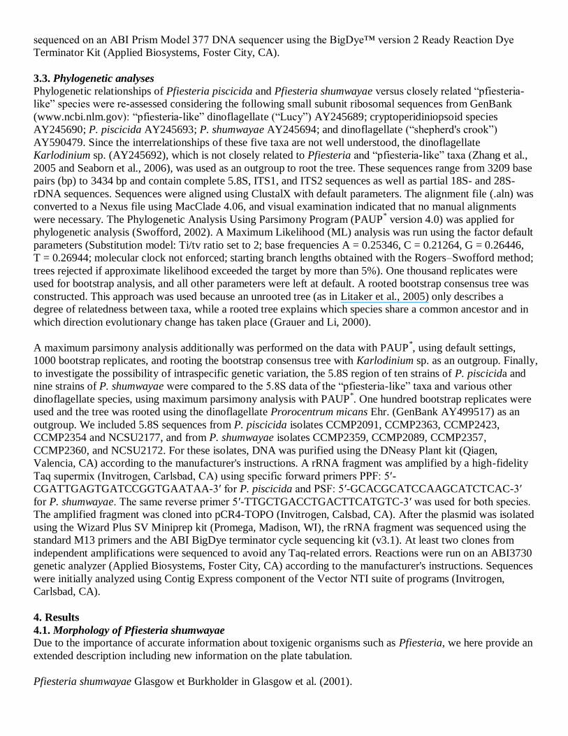
Dinoflagellate with small, oval, cryptic peridinioid flagellate cells having a Kofoidian plate formula of Po, cp, X,
4′, 1a, 6″, 6c, p.c.,?, 5 , 0p, 2 ′. Thecate biflagellate vegetative cells range from 9 to 25 μm in maximum
cell dimension (Table 2). The convex epitheca is approximately equal in size to the hypotheca. The cells may
enlarge by two- to three-fold during feeding (Table 2), as reported previously (Burkholder et al., 2001b and
Parrow and Burkholder, 2003). For example, prey-replete and prey-limited P. shumwayae strain 1050c-b had
cell dimensions (mean ± 1 S.D., n = 50) as 15.1 ± 2.6 μm × 12.3 ± 2.1 μm (estimated biovolume 1780 μm3)
versus 11.0 ± 1.2 μm × 9.1 ± 2.1 μm (estimated biovolume, 720 μm3), respectively. Kleptochloroplastidy has
been reported as an occasional phenomenon (Glasgow et al., 2001).
The small apical pore complex (APC) has a round or oval pore plate (Po) that abuts the 2′, 3′, and 4′ plates, and
a closing plate (c.p.; Fig. 1). The APC is at a slight angle to the canal plate (X). The APC is located between the
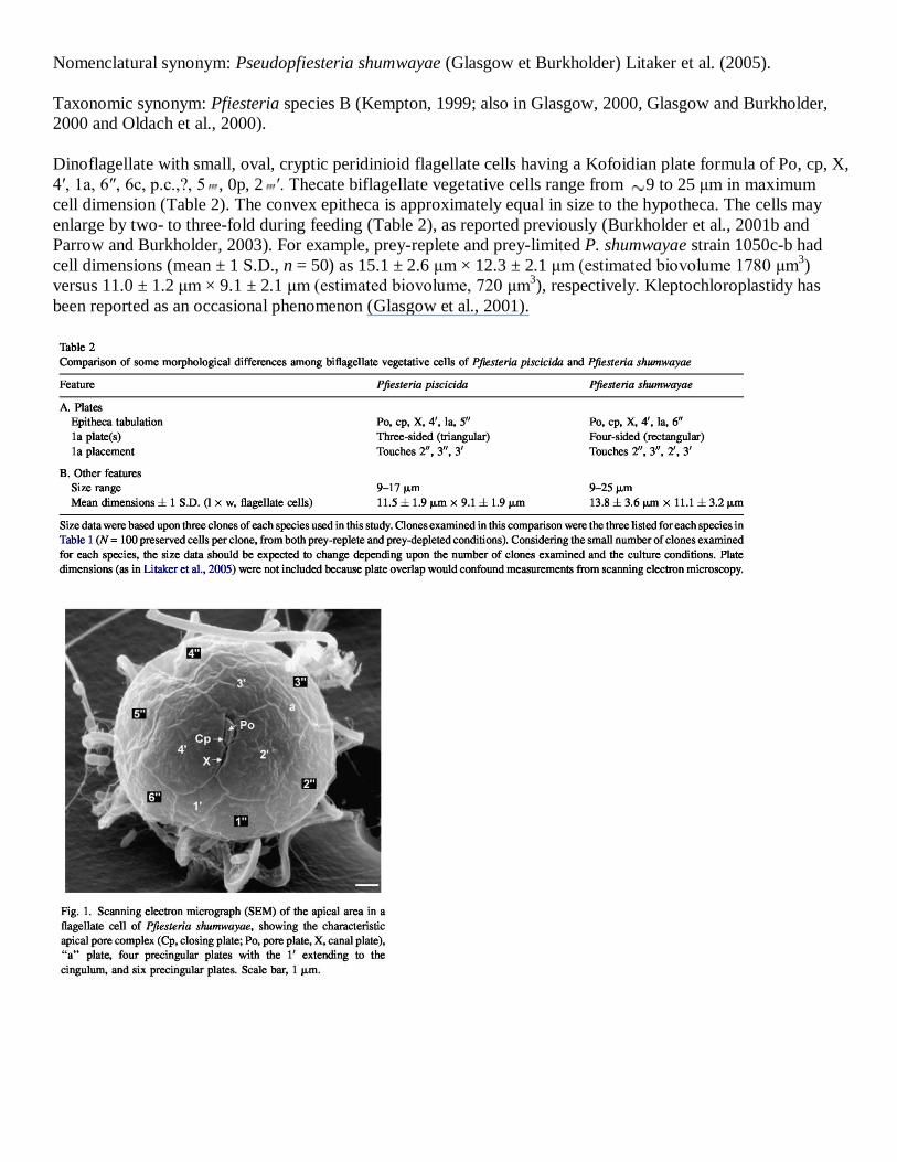
2′, 3′, and 4′ plates, and is connected to 1′ by the X plate. The four-sided (diamond- or rectangular-shaped) 1a
plate can exhibit polymorphism (Fig. 2), and differs from the three-sided (triangular) 1a plate of P. piscicida.
The cingulum of asexual flagellate stages is median, slightly displaced about 0.5× at the sulcus (Fig. 2A) as in P.
piscicida (Steidinger et al., 1996a). The cingulum consists of six plates that are not rimmed by lists. The
hypotheca, typically slightly smaller than the epitheca, is symmetrical and consists of 5 and 2 ′ plates. There
are no posterior intercalary plates. The sulcus is excavated and descends to about three-fourths of the length of
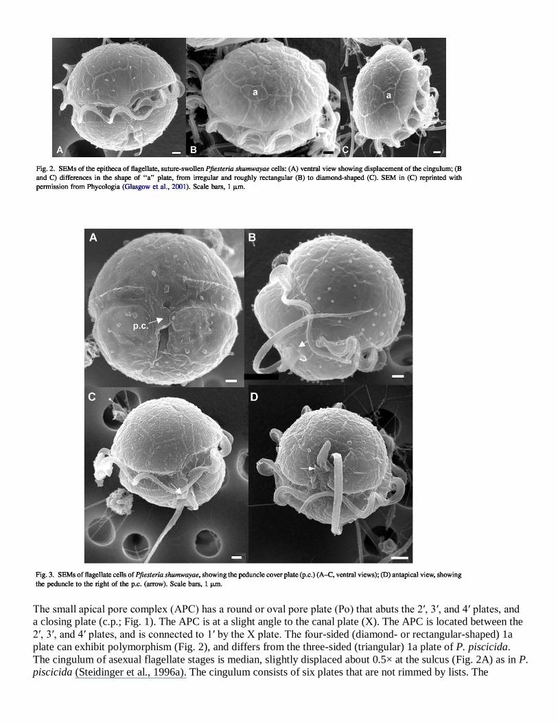
the hypotheca. The sulcus is narrow and offset to the right of the anterior sulcal plate (s.a.), and is partly
concealed by an overlying peduncle cover plate (p.c.) that originates at the right side of the sulcus along the left
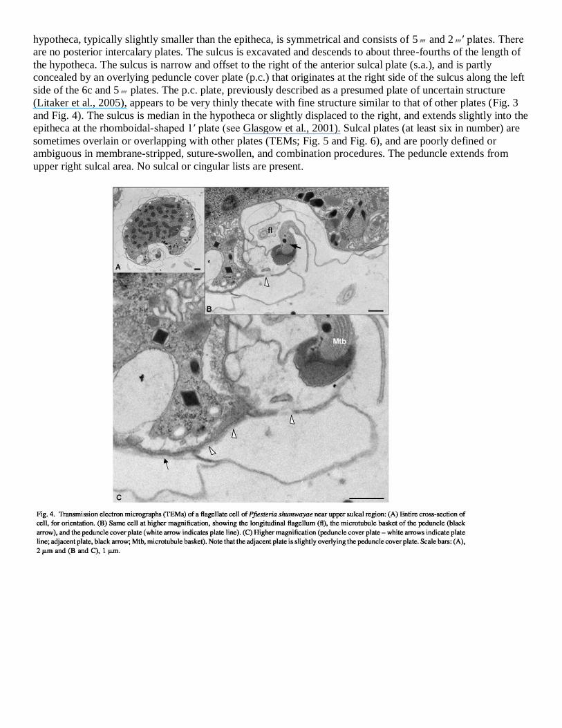
side of the 6c and 5 plates. The p.c. plate, previously described as a presumed plate of uncertain structure
(Litaker et al., 2005), appears to be very thinly thecate with fine structure similar to that of other plates (Fig. 3
and Fig. 4). The sulcus is median in the hypotheca or slightly displaced to the right, and extends slightly into the
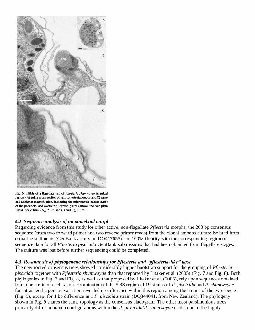
epitheca at the rhomboidal-shaped 1′ plate (see Glasgow et al., 2001). Sulcal plates (at least six in number) are
sometimes overlain or overlapping with other plates (TEMs; Fig. 5 and Fig. 6), and are poorly defined or
ambiguous in membrane-stripped, suture-swollen, and combination procedures. The peduncle extends from
upper right sulcal area. No sulcal or cingular lists are present.
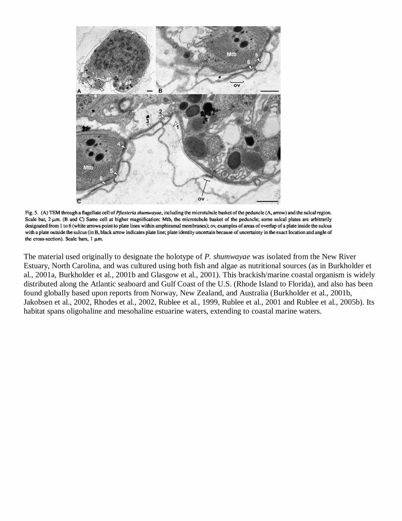
The material used originally to designate the holotype of P. shumwayae was isolated from the New River
Estuary, North Carolina, and was cultured using both fish and algae as nutritional sources (as in Burkholder et
al., 2001a, Burkholder et al., 2001b and Glasgow et al., 2001). This brackish/marine coastal organism is widely
distributed along the Atlantic seaboard and Gulf Coast of the U.S. (Rhode Island to Florida), and also has been
found globally based upon reports from Norway, New Zealand, and Australia (Burkholder et al., 2001b,
Jakobsen et al., 2002, Rhodes et al., 2002, Rublee et al., 1999, Rublee et al., 2001 and Rublee et al., 2005b). Its
habitat spans oligohaline and mesohaline estuarine waters, extending to coastal marine waters.
4.2. Sequence analysis of an amoeboid morph
Regarding evidence from this study for other active, non-flagellate Pfiesteria morphs, the 208 bp consensus
sequence (from two forward primer and two reverse primer reads) from the clonal amoeba culture isolated from
estuarine sediments (GenBank accession DQ417655) had 100% identity with the corresponding region of
sequence data for all Pfiesteria piscicida GenBank submissions that had been obtained from flagellate stages.
The culture was lost before further sequencing could be completed.
4.3. Re-analysis of phylogenetic relationships for Pfiesteria and “pfiesteria-like” taxa
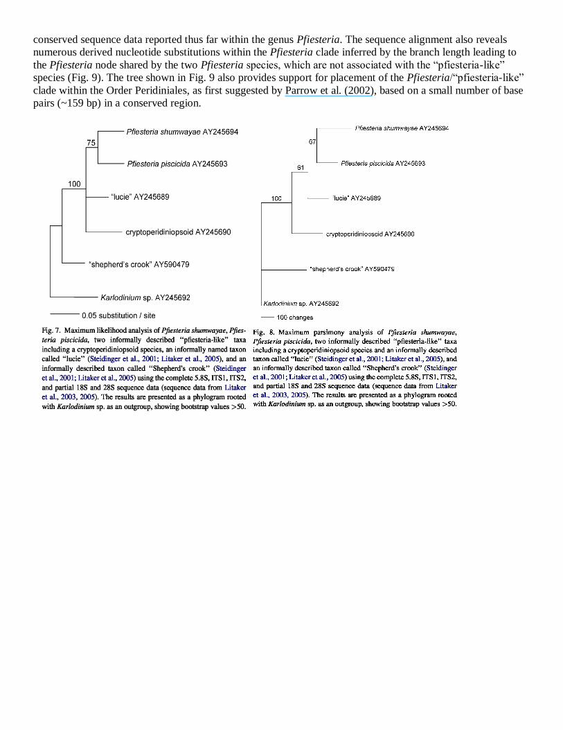
The new rooted consensus trees showed considerably higher bootstrap support for the grouping of Pfiesteria
piscicida together with Pfiesteria shumwayae than that reported by Litaker et al. (2005) (Fig. 7 and Fig. 8). Both
phylogenies in Fig. 7 and Fig. 8, as well as that proposed by Litaker et al. (2005), rely upon sequences obtained
from one strain of each taxon. Examination of the 5.8S region of 19 strains of P. piscicida and P. shumwayae
for intraspecific genetic variation revealed no difference within this region among the strains of the two species
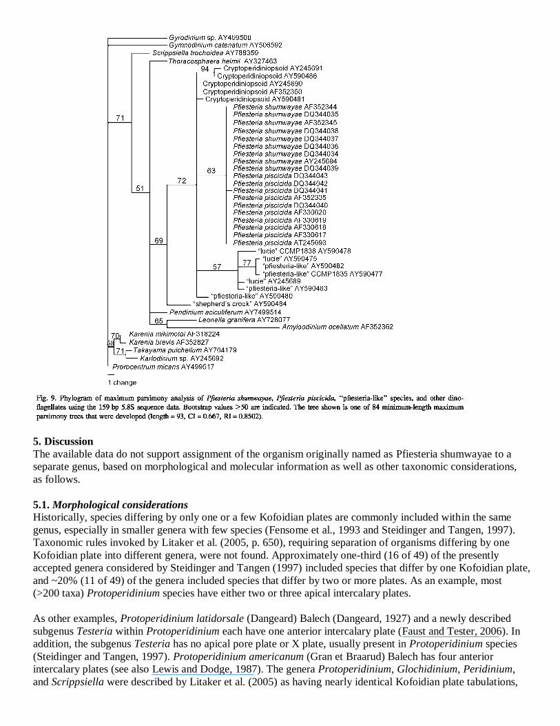
(Fig. 9), except for 1 bp difference in 1 P. piscicida strain (DQ344041, from New Zealand). The phylogeny
shown in Fig. 9 shares the same topology as the consensus cladogram. The other most parsimonious trees
primarily differ in branch configurations within the P. piscicida/P. shumwayae clade, due to the highly
conserved sequence data reported thus far within the genus Pfiesteria. The sequence alignment also reveals
numerous derived nucleotide substitutions within the Pfiesteria clade inferred by the branch length leading to
the Pfiesteria node shared by the two Pfiesteria species, which are not associated with the ―pfiesteria-like‖
species (Fig. 9). The tree shown in Fig. 9 also provides support for placement of the Pfiesteria/―pfiesteria-like‖
clade within the Order Peridiniales, as first suggested by Parrow et al. (2002), based on a small number of base
pairs (~159 bp) in a conserved region.
5. Discussion
The available data do not support assignment of the organism originally named as Pfiesteria shumwayae to a
separate genus, based on morphological and molecular information as well as other taxonomic considerations,
as follows.
5.1. Morphological considerations
Historically, species differing by only one or a few Kofoidian plates are commonly included within the same
genus, especially in smaller genera with few species (Fensome et al., 1993 and Steidinger and Tangen, 1997).
Taxonomic rules invoked by Litaker et al. (2005, p. 650), requiring separation of organisms differing by one
Kofoidian plate into different genera, were not found. Approximately one-third (16 of 49) of the presently
accepted genera considered by Steidinger and Tangen (1997) included species that differ by one Kofoidian plate,
and ~20% (11 of 49) of the genera included species that differ by two or more plates. As an example, most
(>200 taxa) Protoperidinium species have either two or three apical intercalary plates.
As other examples, Protoperidinium latidorsale (Dangeard) Balech (Dangeard, 1927) and a newly described
subgenus Testeria within Protoperidinium each have one anterior intercalary plate (Faust and Tester, 2006). In
addition, the subgenus Testeria has no apical pore plate or X plate, usually present in Protoperidinium species
(Steidinger and Tangen, 1997). Protoperidinium americanum (Gran et Braarud) Balech has four anterior
intercalary plates (see also Lewis and Dodge, 1987). The genera Protoperidinium, Glochidinium, Peridinium,
and Scrippsiella were described by Litaker et al. (2005) as having nearly identical Kofoidian plate tabulations,
with placement into separate genera based upon differing numbers of cingular plates. These genera have been
described to differ, however, by two or more plates (Protoperidinium: 2–4a, 4c, 6s plates; Glochidinium: 0a, 3c,
4s plates; Peridinium: 2–3a, 5–6c, 5–6s plates; Scrippsiella: 3a, 6c, 4–5s plates) (Lewis and Dodge, 1987,
Steidinger and Tangen, 1997, Boltovskoy, 1999 and Steidinger et al., 2001). The genus Amphidiniopsis contains
species that differ in the number of precingular plates and cingular plates (6–8 and 5(4)–8, respectively;
Hoppenrath, 2000), as does the genus Fragilidium (7–9 precingular plates, 9–11 cingular plates; Steidinger and
Tangen, 1997).
Considerable variability in plate number is known, as well, for some species from field samples and from clonal
cultures: vegetative cells and – (―female‖) gametes of Pyrophacus steinii from the same clone have 6–8 apical
plates, 10–13 precingular plates, 12 cingular plates, 11–13 postcingular plates, and 3 posterior intercalary plates.
In contrast, +(―male‖) gametes have 6 apical plates, 8–9 precingular plates, 8–9 cingular plates, 9 postcingular
plates, and 2 posterior intercalary plates (Pholpunthin et al., 1999). In addition, in clonal cultures, the number
(e.g. Morrill and Loeblich, 1981) as well as the shape and contact of some plates (e.g. Elbrächter and Meyer,
2001) can vary substantially (also see Matsuoka, 1985 and Montresor and Marino, 1994; description of
Scrippsiella in Steidinger and Tangen, 1997). If rules required the splitting of taxa based upon a difference of
one in plate number, as asserted by Litaker et al. (2005), cells within the same clone showing such variability
would have to be categorized as separate taxa – which is unacceptable for specimens of the same clone.
5.2. Molecular information
Among eight previous phylogenetic analyses of Pfiesteria and ―pfiesteria-like‖ taxa, only one study (Litaker et
al., 2005) asserted that molecular phylogenetic data supported reassignment of Pfiesteria shumwayae to a new
genus. That analysis did not consider, however, the critical question of root position in the phylogenetic tree of
Pfiesteria and ―pfiesteria-like‖ sequences. All seven other previous studies, using various phylogenetic methods,
presented a largely congruent evaluation of Pfiesteria/―pfiesteria-like‖ phylogeny, with the following features:
first, most taxonomic groupings previously defined from morphology are supported by molecular data. Second,
the order of divergence among the major dinoflagellate groupings is poorly resolved due to short internal branch
lengths and, consequently, weak bootstrap support. The topology of the major dinoflagellate groups is star-like
and suggests a rapid initial radiation from a common ancestor. Third, Pfiesteria piscicida and Pfiesteria
shumwayae consistently are indicated as sister taxa with weak to moderate support, while the informally named
cryptoperidiniopsoids, ―shepherd's crook‖ and ―lucie‖ isolates appear to represent early-diverging taxa within
the ―Pfiesteria/pfiesteria-like‖ lineage. However, the cob trees (Zhang et al., 2005) produce a different weakly
supported topology for the ―Pfiesteria/pfiesteria-like‖ lineage.
The relevance of branch lengths in a phylogenetic tree is dependent in part upon the time since divergence, but
also upon differences in substitution rates among lineages. Thus, unless statistical tests reject rate variability and
in essence support a ―molecular clock,‖ it cannot be argued that branch lengths are proportional to phylogenetic
divergence (Li, 1993). Finally, it should be noted that trees from individual genes may not represent the true
species tree due to lineage sorting, hybridization, and possibly horizontal gene transfer. The existing data are
based heavily upon SSU rDNA sequences, and inclusion of other genes may lead to different conclusions (as in
Zhang et al., 2005). Murray et al. (2005) found evidence of non-independent evolving sites in dinoflagellate
rDNA sequences which may confound phylogenetic analyses. That finding underscores the need to use
sequences from multiple genes when possible.
The trend in classification over the past two decades has been to use phylogenetic systematics, wherein named
classifications have been inferred from monophyletic groups (Judd et al., 2002). Assessment of monophyly
requires a reliable, rooted phylogenetic tree topology (Felsenstein, 2004). The preponderance of previous
research suggests that Pfiesteria piscicida and Pfiesteria shumwayae are sister species, at least among the
species described, and they apparently form a monophyletic group. Since these are the only formally described
species of Pfiesteria, reclassifying one or the other would result in two genera that are trivially monophyletic.
Beyond the issue of monophyly, the criteria for what tree nodes define genera, families, etc. are basically
arbitrary; moreover, Litaker et al.'s (2005) phylogenetic analysis based on relative branch lengths is misleading
because the authors provided no evidence for the root location in their tree. The phylogenetic trees contributed
by the present analysis showed considerably higher bootstrap support for the grouping of Pfiesteria piscicida
together with Pfiesteria shumwayae than that reported by Litaker et al. (2005), and support placement of the
two species within the same genus. The collective evidence for the root position is weak at this time, however,
thus requiring emphasis on morphological data.
5.3. Considerations at the order level
Steidinger et al. (1996a) assigned Pfiesteria to the order Dinamoebales based on amoeboid stages observed in
cultures of some strains. According to Fensome et al. (1993, p. 164), however, the order Dinamoebales was
uncertain: ―The name Dinamoebales is based on a dominantly amoeboid genus that may be part of the life-cycle
of another, ―coccoid‖ genus. Moreover, amoeboid forms are not typical of the order ….‖ Amoeboid cells have
been described in some dinoflagellate taxa since the early 1900s (Pascher, 1916), mostly in ecto- and
endoparasites and some mixotrophic predaceous species (e.g. Pfiester and Popovský, 1979, Buckland-Nicks et
al., 1990, Buckland-Nicks et al., 1997, Buckland-Nicks and Reimchen, 1995 and Appleton and Vickerman,
1998). Popovský and Pfiester (1990, p. 50) asserted that ―many, possibly most, dinoflagellates exhibit amoeboid
stages or tendencies such as the formation of pseudopodia during phagocytosis at some stage in their life
histories.‖ Nevertheless, in dinoflagellates including Pfiesteria, photographic (tracking one cell) or video
sequences of transformations involving amoeboid cells have not been obtained (Burkholder and Glasgow, 2002
and Elbrächter, 2003).
Stages reported by Litaker et al. (2005) in the isolates they examined (in culture for 6–14 years) were similar to
those described for Pfiesteria shumwayae by Parrow and Burkholder (2003), who worked with isolates that
were 2–3 years old. Seaborn et al. (1999), Marshall et al. (2000), Burkholder et al. (2001c) and Glasgow et al.
(2001) found amoeboid cells in some clonal strains of Pfiesteria spp. and ―pfiesteria-like‖ dinoflagellates, but
generally within the first year of isolation. Burkholder et al. (2001c) and Burkholder and Glasgow (2002)
reported that many strains examined since the early work of Steidinger et al. (1996a) have not formed amoeboid
cells. We therefore support the recommendation by Parrow et al. (2002) to move the genus Pfiesteria to the
order Peridiniales based on plate tabulation of flagellate stages, which is the basis for the taxonomy of other
thecate dinoflagellates, and also is based upon the consistent occurrence of flagellate stages in cultures.
5.4. Summary
The consistent morphological analyses showing a difference of only one precingular plate in Pfiesteria piscicida
versus Pfiesteria shumwayae; the common practice of placement of organisms differing by one or more plates
(including precingular plates) within the same genus, particularly in small genera; and the preponderance of
molecular data support placement of these two species within the same genus. In our view, at the present state
of knowledge, the erection of a separate monospecific genus for P. shumwayae is not justified, given the small
differences between P. piscicida and P. shumwayae. Placement of P. piscicida and P. shumwayae within one
genus will prevent formation of two trivially monophyletic genera, and contribute to the nomenclatural stability
of organisms of interest to natural resource and public health managers.
Although molecular methods have become important tools for phylogenetic analysis (genospecies), they have
not supplanted classical approaches (morphospecies), especially in taxa such as the dinoflagellates where
sequence data on a range of genes are relatively scarce. Thus, it is important to use all relevant information
when assessing phylogenetic relationships. Future work with multiple clones over extended time in culture may
reveal more polymorphism in the plate structure of Pfiesteria spp., as has been shown for various other thecate
dinoflagellate species. As additional species are found and/or described within this genus, further molecular
studies involving more sequence data will strengthen insights about phylogenetic relationships among Pfiesteria
spp. and closely related taxa.
Recalling the data presented and discussed above, we propose the following classification of Pfiesteria:
Class: Dinophyceae Pascher 1914
Order: Peridiniales Haeckel 1894
Genus: Pfiesteria Steidinger et Burkholder in Steidinger et al. (1996)
Thus far, two species are formally assigned to the genus: Pfiesteria piscicida Steidinger et Burkholder in
Steidinger et al. (1996a) and Pfiesteria shumwayae Glasgow et Burkholder in Glasgow et al. (2001), as
characterized above.
Based upon present knowledge, we regard it as inappropriate to assign Pfiesteria to a family at this time.
Apparently it is closely related to other, informally named ―pfiesteria-like‖ organisms with unresolved
tabulation patterns (Steidinger et al., 2001 and Seaborn et al., 2006). Recent phylogenetic analyses (Saldarriaga
et al., 2001, Saldarriaga et al., 2004, Gottshling et al., 2005 and Kremp et al., 2005) indicate that Pfiesteria is
closely related to calcareous dinoflagellates such as Thoracosphaera, suggesting that the suprageneric
classification will require fundamental revision that is beyond the scope of this paper. All of these taxa will
have to be placed together within the same family; thus, as the nomenclature and classification of calcareous
dinoflagellates are under revision (Elbrächter et al. in preparation), at present we abstain from assigning
Pfiesteria to a family.
Acknowledgments
We thank the staff and graduate student Hayley Skelton of the Center for Applied Aquatic Ecology for
laboratory assistance; Eric Schaefer, Jason Kempton, and Coy Allen of UNC-Greensboro for sequencing and
PCR analyses; and Scott Bingham at Arizona State University for additional sequencing. Other laboratory
assistance was provided by Andrew Gordon and Brian Dyer of the Department of Biological Sciences at Old
Dominion University. Funding support was provided by the U.S. Environmental Protection Agency, the Centers
for Disease Control and Prevention via the North Carolina Department of Health and Human Services, and the
North Carolina General Assembly. This is Harbor Branch Oceanographic Institution contribution number 1631.
References
Appleton and Vickerman, 1998 P.L. Appleton and K. Vickerman, In vitro cultivation and developmental cycle
in culture of a parasitic dinoflagellate (Hematodinium sp.) associated with mortality of the Norway
lobster (Nephrops norvegicus) in British waters, Parasitology 116 (1998), pp. 115–130.
Boltovskoy, 1999 A. Boltovskoy, The genus Glochidinium gen. nov., with two species: G. penardiforme comb.
nov. and G. platygaster sp. nov. (Peridiniaceae), Grana 38 (1999), pp. 98–107.
Buckland-Nicks and Reimchen, 1995 J. Buckland-Nicks and T. Reimchen, A novel association between an
endemic stickleback and a parasitic dinoflagellate. 3. Details of the life cycle, Arch. Protistenk. 145
(1995), pp. 165–175.
Buckland-Nicks et al., 1997 J. Buckland-Nicks, T.E. Reimchen and D.J. Garbary, Haidadinium ichthyophilum
gen. nov. et sp. nov. (Phytodiniales, Dinophyceae), a freshwater ectoparasite on stickleback
(Gasterosteus aculeatus) from the Queen Charlotte Islands, Canada, Can. J. Bot. 75 (1997), pp. 1936–
1940.
Buckland-Nicks et al., 1990 J. Buckland-Nicks, T.E. Reimchen and F.J.R. Taylor, A novel association between
an endemic stickleback and a parasitic dinoflagellate. 2. Morphology and life cycle, J. Phycol. 26 (1990),
pp. 539–548.
Burkholder and Glasgow, 2002 J.M. Burkholder and H.B. Glasgow, The life cycle and toxicity of Pfiesteria
piscicida, revisited, J. Phycol. 38 (2002), pp. 1261–1267.
Burkholder et al., 2001b J.M. Burkholder, H.B. Glasgow and N.J. Deamer-Melia, Overview and present status
of the toxic Pfiesteria complex, Phycologia 40 (2001), pp. 186–214.
Burkholder et al., 2001c J.M. Burkholder, H.B. Glasgow, N.J. Deamer-Melia, J. Springer, M.W. Parrow, C.
Zheng and P.J. Cancellieri, Species of the toxic Pfiesteria complex, and the importance of functional
type in data interpretations, Environ. Health Perspect. 109 (2001), pp. 667–679.
Burkholder et al., 2005 J.M. Burkholder, A.S. Gordon, P.D. Moeller, J.M. Law, K.J. Coyne, A.J. Lewitus, J.S.
Ramsdell, H.G. Marshall, N.J. Deamer, S.C. Cary, J.W. Kempton, S.L. Morton and P.A. Rublee,
Demonstration of toxicity to fish and to mammalian cells by Pfiesteria species: comparison of assay
methods and multiple strains, Proc. Natl. Acad. Sci. USA 102(2005), pp. 3471–3476.
Burkholder et al., 2001a J.M. Burkholder, H.G. Marshall, H.B. Glasgow, D.W. Seaborn and N.J. Deamer-Melia,
The standardized fish bioassay procedure for detecting and culturing actively toxic Pfiesteria, used by
two reference laboratories for Atlantic and southeastern states, Environ. Health Perspect. 109 (2001), pp.
745–756.
Carty, 2003 S. Carty, Dinoflagellates. In: J.D. Wehr and R.G. Sheath, Editors, Freshwater Algae of North
America – Ecology and Classification, Academic Press (2003), pp. 685–714. Abstract
Choi and Stoecker, 1989 J.W. Choi and D.K. Stoecker, Effects of fixation on cell volume of marine planktonic
protozoa, Appl. Environ. Microbiol. 55 (1989), pp. 1761–1765.
Dangeard, 1927 Dangeard, P., 1927. Phytoplancton de la Croisière du SYLVANA (Février-Juin 1913). -
Annales de l’Institut Océanographique de Monaco 4, pp. 287–407.
Elbrächter, 2003 M. Elbrächter, Dinophyte reproduction: progress and conflicts, J. Phycol. 39 (2003), pp. 629–
632.
Elbrächter and Meyer, 2001 M. Elbrächter and B. Meyer, Plate pattern variability and plate overlap in a clonal
culture of the freshwater dinoflagellate, Peridinium umbonatum Stein species complex (Dinophyta).
Neues Jahrbuch für Palaentologie, Abhandlungen 219 (2001), pp. 221–227.
Faust and Tester, 2006 M.A. Faust and P.A. Tester, Creation of the subgenus Testeria Faust subgen. nov.
Protoperidinium Bergh from the SW Atlantic Ocean: Protoperidinium novella sp. nov. and
Protoperidinium concinna sp. nov. Dinophyceae, Phycologia 45 (2006), pp. 1–9.
Felsenstein, 2004 J. Felsenstein, Inferring Phylogenies, Sinauer Associates, Inc., Sunderland (MA) (2004) 664
pp.
Fensome et al., 1999 R.A. Fensome, J.F. Saldarriaga and F.J.R. Taylor, Dinoflagellate phylogeny revisited:
reconciling morphological and molecular based phylogenies, Grana 38 (1999), pp. 66–80.
Fensome et al., 1993 R.A. Fensome, F.J.R. Taylor, G. Norris, W.A.S. Sarjeant, D.I. Wharton and G.L. Williams,
A classification of living and fossil dinoflagellate. Micropaleontology, Special Publication Number 7,
Sheridan Press, Hanover, PA (1993) 351 pp.
Gifford and Caron, 2000 D.J. Gifford and D.A. Caron, Sampling, preservation, enumeration and biomass of
protozooplankton. In: R.P. Harris, H.-R. Skjoldal, J. Lenz, P.J. Wiebe and M.E. Huntley, Editors, ICES
Zooplankton Methodology Manual, Academic Press, London (2000), pp. 193–221. Abstract
Glasgow, 2000 Glasgow, H.B., 2000. The biology and impacts of toxic Pfiesteria complex species. Ph.D. thesis.
Department of Botany, North Carolina State University, Raleigh, 175 pp.
Glasgow and Burkholder, 2000 H.B. Glasgow and J.M. Burkholder, Water quality trends and management
implications from a five-year study of a eutrophic estuary, Ecol. Appl. 10 (2000), pp. 1024–1046.
Glasgow et al., 2001 H.B. Glasgow, J.M. Burkholder, S.L. Morton and J. Springer, A second species of
ichthyotoxic Pfiesteria (Dinamoebales, Dinophyceae), Phycologia 40 (2001), pp. 234–245.
Gordon and Dyer, 2005 A.S. Gordon and B. Dyer, Relative contribution of exotoxin and micropredation to
ichthyotoxicity of two strains of Pfiesteria shumwayae (Dinophyceae), Harmful Algae 4 (2005), pp.
423–431.
Gottshling et al., 2005 M. Gottshling, H. Keupp, J. Plötner, R. Knopp, H. Willems and M. Kirsch, Phylogeny of
calcareous dinoflagellates as inferred from ITS and ribosomal sequence data, Mole. Phylogenet. Evol. 36
(2005), pp. 444–455.
Grauer and Li, 2000 D. Grauer and W.H. Li, Fundamentals of Molecular Evolution (second ed.), Sinauer
Associates, Inc., Sunderland, MA (2000) 481 pp.
Hoppenrath, 2000 M. Hoppenrath, Morphology and taxonomy of six marine sand-dwelling Amphidiniopsis
species (Dinophyceae, Peridiniales), four of them new, from the German Bight, North Sea, Phycologia
39 (2000), pp. 482–497.
Jakobsen et al., 2002 K.S. Jakobsen, T. Tengs, A. Vatne, H.A. Bowers, D.W. Oldach, J.M. Burkholder, H.B.
Glasgow, P.A. Rublee and D. Klaveness, Discovery of the toxic dinoflagellate, Pfiesteria, from northern
European waters, Proc. Royal Soc. Lond. (B) 269 (2002), pp. 211–214.
Jeong et al., 2005 H.J. Jeong, J. Seong, J.Y. Park, J.H. Kim, S. Kim, I. Lee, S.H. Lee, J.H. Ha and W.H. Yih,
Stoeckeria algicida n. gen., sn. sp. (Dinophyceae) from the coastal waters off southern Korea:

Morphology and small subunit ribosomal DNA gene sequence, J. Eukaryot. Microbiol. 52 (2005), pp.
382–390.
Judd et al., 2002 W.S. Judd, C.S. Campbell, E.A. Kellogg, P.F. Stevens and M.J. Donoghue, Plant Systematics
– a Phylogenetic Approach (second ed.), Sinauer Associates, Sunderland, MA (2002).
Kremp et al., 2005 A. Kremp, M. Elbrächter, M. Schweikert, J.L. Wolny and M. Gottshling, Woloszynskia
halophila (Biecheler) comb. nov.: a bloom-forming cold-water dinoflagellate co-occurring with
Scrippsiella hangoei (Dinophyceae) in the Baltic Sea, J. Phycol. 41 (2005), pp. 629–642.
Kempton, 1999 Kempton, J.W., 1999. PCR and FISH assays for the detection of Pfiesteria piscicida. M.S.
thesis. Department of Biology, University of North Carolina, Greensboro, 83 pp.
Kofoid, 1909 C.A. Kofoid, On Peridinium steini Jörgensen, with a note on the nomenclature of the skeleton of
the Peridinidae, Arch. Protistenk. 16 (1909), pp. 25–47.
Lewis and Dodge, 1987 J. Lewis and J.D. Dodge, The cyst-theca relationship of Protoperidinium americanum
(Gran and Braarud) Balech, J. Micropaleontol. 6 (1987), pp. 113–121.
Li, 1993 W.H. Li, So, what about the molecular clock hypothesis?, Curr. Opin. Genet. Dev. 3 (1993), pp. 896–
901.
Litaker et al., 2005 R.W. Litaker, K.A. Steidinger, P.L. Mason, J.H. Landsberg, J.D. Shields, K.S. Reece, L.W.
Haas, W.K. Vogelbein, M.W. Vandersea, S.R. Kibler and P.A. Tester, The reclassifi-cation of Pfiesteria
shumwayae: Pseudopfiesteria, gen. nov., J. Phycol. 41 (2005), pp. 643–651. View Record in Scopus |
Cited By in Scopus (24)
Litaker et al., 1999 R.W. Litaker, P.A. Tester, A. Colorni, M.G. Levy and E.J. Noga, The phylogenetic
relationship of Pfiesteria piscicida, cryptoperidiniopsoid sp., Amyloodinium ocellatum and a pfiesteria-
like dinoflagellate to other dinoflagellates and apicomplexans, J. Phycol. 35 (1999), pp. 1379–1389.
Litaker et al., 2000 W. Litaker, R. Sundseth, M. Wojciechowski, C. Bonaventura, R. Henkens and P. Tester,
Electrochemical detection of Pfiesteria piscicida and Pfiesteria-like species, Proceedings, Ninth
International Conference on Harmful Algal Blooms Hobart, Tasmania, Australia (2000) (published
abstract).
Litaker et al., 2003 R.W. Litaker, M.W. Vandersea, S.R. Kibler, K.S. Reece, N.A. Stokes, K.A. Steidinger, D.F.
Millie, B. Bendis, R. Pigg and P.A. Tester, Variability among dinoflagellate ITS region sequences:
identification of Pfiesteria piscicida (Dinophyceae) and Pfiesteria-like organisms using ITS-specific
PCR assays, J. Phycol. 39 (2003), pp. 754–761.
Marshall et al., 2000 H.G. Marshall, A.S. Gordon, D.W. Seaborn, B. Dyer, W.M. Dunstan and A.M. Seaborn,
Comparative culture and toxicity studies between the toxic dinoflagellate Pfiesteria piscicida and a
morphologically similar cryptoperidiniopsoid dinoflagellate, J. Exp. Mar. Biol. Ecol. 255 (2000), pp.
51–74.
Mason et al., 2003 P.L. Mason, W.K. Vogelbein, L.W. Haas and J.D. Shields, An improved stripping technique
for lightly armored dinoflagellates, J. Phycol. 39 (2003), pp. 253–258.
Matsuoka, 1985 K. Matsuoka, Cyst and thecate forms of Pyrophacus steinii (Schiller) Wall et Dale, 1971,
Trans. Proc. Palaeontol. Soc. Jpn. NS 140 (1985), pp. 240–262.
Menden-Deuer et al., 2001 S. Menden-Deuer, E.J. Lessard and J. Satterberg, Effect of preservation on
dinoflagellate and diatom cell volume and consequences for carbon biomass predictions, Mar. Ecol.
Prog. Ser. 222 (2001), pp. 41–50.
Montresor and Marino, 1994 M. Montresor and D. Marino, New observation on the life cycle of Pyrophacus
horologium Stein (Dinophyceae), Bollet. Soc. Adriatica di Scienze 75 (1994), pp. 261–268.
Morrill and Loeblich, 1981 L.C. Morrill and A.R. Loeblich III, A survey of body scales in dinoflagellates and a
revision of Cachonina and Heterocapsa (Pyrrhophyta), J. Plankt. Res. 3 (1981), pp. 53–65.
Murray et al., 2005 S. Murray, M.F. Jørgensen, S.Y.W. Ho, D.J. Patterson and L.S. Jermiin, Improving the
analysis of dinoflagellate phylogeny based on rDNA, Protist 156 (2005), pp. 269–286.
Oldach et al., 2000 D.W. Oldach, C.F. Delwiche, K.S. Jakobsen, T. Tengs, E.G. Brown, J.W. Kempton, E.F.
Schaefer, H.A. Bowers, H.B. Glasgow, J.M. Burkholder, K.A. Steidinger and P.A. Rublee,
Heteroduplex mobility assay-guided sequence discovery: elucidation of the small subunit (18S) rDNA
sequences of Pfiesteria piscicida and related dinoflagellates from complex algal culture and
environmental sample DNA pools, Proc. Nat. Acad. Sci. 97 (2000), pp. 4303–4308.
Parrow and Burkholder, 2003 M.W. Parrow and J.M. Burkholder, Reproduction and sexuality in Pfiesteria
shumwayae (Dinophyceae), J. Phycol. 39 (2003), pp. 697–711.
Parrow et al., 2005 M.W. Parrow, J.M. Burkholder, N.J. Deamer and J.S. Ramsdell, Contaminant-free
cultivation of Pfiesteria shumwayae (Dinophyceae) on a fish cell line, Aquat. Microb. Ecol. 39 (2005),
pp. 97–105.
Parrow et al., 2002 M. Parrow, J.M. Burkholder, N.J. Deamer and C. Zhang, Vegetative and sexual
reproduction in Pfiesteria spp. (Dinophyceae) cultured with algal prey, and inferences for their
classification, Harmful Algae 1 (2002), pp. 5–33.
Parrow et al., in press Parrow, M.W., Elbrächter, M., Krause, M.K., Burkholder, J.M., Deamer, N.J., Htyte, N.,
Allen, E.H. The taxonomy and growth of a Crypthecodinium species (Dinophyceae) isolated from a
brackish water fish aquarium. Afr. J. Mar. Sci. 28 (special issue, Proceedings, XIth International
Symposium on Harmful Algal Bloom Proceedings) (in press).
Pascher, 1916 A. Pascher, Uber ene neue Amobe – Dinamoeba (varians) – mit dinoflagellatenartige
Schwarmern, Arch. Protistenk. 36 (1916), pp. 118–136.
Patterson, 1999 D.J. Patterson, The diversity of eukaryotes, Am. Nat. 154 (1999), pp. S96–S124.
Pfiester and Popovský, 1979 L.A. Pfiester and J. Popovský, Parasitic, amoeboid dinoflagellates, Nature 279
(1979), pp. 421–424.
Pholpunthin et al., 1999 P. Pholpunthin, Y. Fukuyo, K. Matsuoka and Y. Nimura, Life history of a marine
dinoflagellate Pyrophacus steinii (Schiller) Wall et Dale, Bot. Mar. 42 (1999), pp. 189–197.
Popovský and Pfiester, 1990 J. Popovský and L.A. Pfiester, Süβwasserflora von Mitteleuropa. Band 6:
Dinophyceae (Dinoflagellida), Gustav Fischer Verlag, Jena (1990) 272 pp.
Rhodes et al., 2002 L.L. Rhodes, J.M. Burkholder, H.B. Glasgow, P.A. Rublee, C. Allen and J.E. Adamson,
Pfiesteria shumwayae (Pfiesteriaceae) in New Zealand, NZ J. Mar. Freshwat. Res. 36 (2002), p. 621.
Rublee et al., 2005b P.A. Rublee, C. Allen, E. Schaefer, L. Rhodes, J. Adamson, C. Lapworth, J. Burkholder
and H. Glasgow, Global distribution of toxic Pfiesteria complex species detected by PCR assay. In: K.A.
Steidinger, J.H. Landsberg, C.R. Tomas and G.A. Vargo, Editors, Harmful Algae 2002, Florida Fish and
Wildlife Cons. Comm., FL Inst. Oceanogr., and Inter-governmental Oceanographic Commission of
UNESCO, St. Petersburg, FL (2005), pp. 320–322.
Rublee et al., 2001 P.A. Rublee, J.W. Kempton, E.F. Schaefer, C. Allen, J. Harris, D.W. Oldach, H. Bowers, T.
Tengs, J.M. Burkholder and H.B. Glasgow, Use of molecular probes to assess geographic distribution of
Pfiesteria species, Environ. Health Perspect. 109 (2001), pp. 765–767.
Rublee et al., 1999 P.A. Rublee, J. Kempton, E. Schaefer, J.M. Burkholder, H.B. Glasgow and D. Oldach, PCR
and FISH detection extends the range of Pfiesteria piscicida in estuarine waters, Va. J. Sci. 50 (1999),
pp. 325–335.
Rublee et al., 2005 P.A. Rublee, D.L. Remington, E.F. Schaefer and M.M. Marshall, Detection of the dinozoans
Pfiesteria piscicida and P. shumwayae: a review of detection methods and geographic distribution, J.
Eukaryot. Microbiol. 52 (2005), pp. 83–89. View Record in Scopus | Cited By in Scopus (21)
Saldarriaga et al., 2004 J.F. Saldarriaga, F.J.R. Taylor, T. Cavalier-Smith, S. Menden-Deuer and P.J. Keeling,
Molecular data and the evolutionary history of dinoflagellates, Eur. J. Protist. 40 (2004), pp. 85–111.
Saldarriaga et al., 2001 J.F. Saldarriaga, F.J.R. Taylor, P.J. Keeling and T. Cavalier-Smith, Dinoflagellate
nuclear SSU rRNA phylogeny suggests multiple plastid losses and replacements, J. Mol. Evol. 53 (2001),
pp. 204–213.
Seaborn et al., 1999 D. Seaborn, A. Seaborn, W. Dunstan and H.G. Marshall, Growth and feeding studies on the
algal feeding stage of a Pfiesteria-like dinoflagellate, Va. J. Sci. 50 (1999), pp. 337–344.
Seaborn et al., 2006 D.W. Seaborn, T. Tengs, S. Cerbin, M. Kokocinski and H.G. Marshall, A group of
dinoflagellates similar to Pfiesteria as defined by morphology and genetic analysis, Harmful Algae 5
(2006), pp. 1–8.
Steidinger et al., 1996a K.A. Steidinger, J.M. Burkholder, H.B. Glasgow, C.H. Hobbs, J.K. Garrett, E.W. Truby,
E.J. Noga and S.A. Smith, Pfiesteria piscicida gen. et sp. nov. (Pfiesteriaceae fam. nov.), a new toxic
dinoflagellate with a complex life cycle and behavior, J. Phycol. 32 (1996), pp. 157–164.
Steidinger et al., 2001 K.A. Steidinger, J.H. Landsberg, R.W. Richardson, E. Truby, B. Blakesley, P. Scott, P.
Tester, T. Tengs, P. Mason, S. Morton, D. Seaborn, W. Litaker, K. Reece, D. Oldach, L. Haas and G.
Vasta, Classification and identification of Pfiesteria and Pfiesteria-like species, Environ. Health
Perspect. 109 (2001), pp. 661–665.
Steidinger et al., 1996b K.A. Steidinger, J.H. Landsberg, E.W. Truby and B.A. Blakesley, The use of scanning
electron microscopy in identifying small ―gymnodinioid‖ dinoflagellates, Nova Hedwigia 112 (1996),
pp. 415–422.
Steidinger and Tangen, 1997 K.A. Steidinger and K. Tangen, Dinoflagellates. In: C.R. Tomas, Editor,
Identifying Marine Diatoms and Dinoflagellates, Academic Press, Inc., New York (1997), pp. 387–584.
Abstract
Swofford, 2002 D.I. Swofford, PAUP* 4.0 – Phylogenetic Analysis Using Parsimony Program. ac. PC (DOS)
UNIX, Sinauer Associates, Sunderland, MA (2002).
Truby, 1997 E.W. Truby, Preparation of single-celled marine dinoflagellates for electron microscopy, Microsc.
Res. Tech. 36 (1997), pp. 337–340.
Zhang et al., 2005 H. Zhang, D. Bhattacharya and S. Lin, Phylogeny of dinoflagellates based on mitochondrial
cytochrome b and nuclear small subunit rDNA sequence comparisons, J. Phycol. 41 (2005), pp. 411–
420.