formal revision of the alexandrium tamarense species complex (dinophyceae) taxonomy: the...
TRANSCRIPT

PhP
O
Ft(IEC
UM
a
b
c
d
e
SM
TddfpPtfnfot
1
e
h1
rotist, Vol. 165, 779–804, December 2014ttp://www.elsevier.de/protisublished online date 13 October 2014
RIGINAL PAPER
ormal Revision of the Alexandriumamarense Species ComplexDinophyceae) Taxonomy: Thentroduction of Five Species withmphasis on Molecular-based (rDNA)lassification
we Johna,1, R. Wayne Litakerb, Marina Montresorc, Shauna Murrayd,ichael L. Brosnahane, and Donald M. Andersone
Alfred Wegener Institute for Polar and Marine Research, Am Handelshafen 12,27570 Bremerhaven, GermanyNational Oceanic and Atmospheric Administration, National Ocean Service,National Centers for Coastal Oceans Science, Center for Fisheries and HabitatResearch, 101 Pivers Island Road, Beaufort, North Carolina 28516, United StatesStazione Zoologica Anton Dohrn, Villa Comunale, 80121 Napoli, ItalyPlant Functional Biology and Climate Change Cluster, University of Technology Sydney,Sydney, PO Box 123 Broadway, NSW 2007, AustraliaWoods Hole Oceanographic Institution, MS # 32, 266 Woods Hole Road, Woods Hole,Massachusetts 02543, United States
ubmitted March 12, 2014; Accepted October 2, 2014onitoring Editor: Mona Hoppenrath
he Alexandrium tamarense species complex is one of the most studied marine dinoflagellate groupsue to its ecological, toxicological and economic importance. Several members of this complex pro-uce saxitoxin and its congeners - potent neurotoxins that cause paralytic shellfish poisoning. Isolates
rom this complex are assigned to A. tamarense, A. fundyense, or A. catenella based on two main mor-hological characters: the ability to form chains and the presence/absence of a ventral pore between
lates 1′ and 4′. However, studies have shown that these characters are not consistent and/or distinc-ive. Further, phylogenies based on multiple regions in the rDNA operon indicate that the sequencesrom morphologically indistinguishable isolates partition into five clades. These clades were initiallyamed based on their presumed geographic distribution, but recently were renamed as Groups I-V
ollowing the discovery of sympatry among some groups. In this study we present data on morphol-gy, ITS/5.8S genetic distances, ITS2 compensatory base changes, mating incompatibilities, toxicity,
he sxtA toxin synthesis gene, and rDNA phylogenies. All results were consistent with each group
Corresponding author; fax +49 471 4831 2115-mail [email protected] (U. John).
ttp://dx.doi.org/10.1016/j.protis.2014.10.001434-4610/© 2014 Elsevier GmbH. All rights reserved.

780 U. John et al.
representing a distinct cryptic species. Accordingly, the groups were assigned species names as fol-lows: Group I, A. fundyense; Group II, A. mediterraneum; Group III, A. tamarense; Group IV, A. pacificum;Group V, A. australiense.© 2014 Elsevier GmbH. All rights reserved.
Key words: Alexandrium fundyense; Alexandrium mediterraneum; Alexandrium tamarense; Alexandrium paci-ficum; Alexandrium australiense.
Introduction
Dinoflagellates are among the most important pri-mary producers in marine systems. A small minorityof these species cause harmful algal blooms(HABs) that adversely impact ecosystem servicesand function, often through the production of phyco-toxins that can accumulate within marine food webs(Sunda et al. 2006). Many of these toxins contam-inate seafood and pose a significant public healththreat (Anderson et al. 2012; Smayda 1997). Toxi-genic dinoflagellates belonging to the Alexandriumtamarense (M. Lebour) Balech species complex areamong the most widely distributed HAB-causingtaxa globally. Currently, the A. tamarense speciescomplex contains three morphologically definedspecies: A. tamarense (M. Lebour) Balech, A.fundyense Balech and A. catenella (Whedon &Kof.) Balech. Numerous strains of each speciesproduce saxitoxin and its congeners (hereafterreferred to collectively as ‘saxitoxins’), a group ofphycotoxins that cause paralytic shellfish poisoning(PSP; Anderson et al. 1994, 2012; Balech 1985).Balech (1995) provided detailed information on themorphology of these species based on extensivesampling from different geographic regions. Thethree morphospecies share the same plate formulaand can be distinguished by differences in theirlength to width ratios, the presence or absence ofa ventral pore along the suture between 1′ and 4′thecal plates, and differences in the shape of thesp and sa thecal plates. Other defining characteris-tics reported by Balech included (1) the ability of A.catenella, but not A. fundyense or A. tamarense toform long chains (more than 4 cells; Balech 1985,1995; Balech and Tangen 1985), (2) the more flat-tened shape of A. catenella cells which exhibitedlower length:width ratios than the other two species,and (3) the presence of a ventral pore only in A.tamarense (Balech 1985).
More recent field and culture studies haverevealed cells exhibiting morphologies intermediateto those described as characteristic for A. catenella,A. fundyense, and A. tamarense. These morpho-logical intermediates are often observed within the
same field population as well as within culturesstarted from a single cell, casting serious doubton the validity of the morphospecies concept ofall three species (Anderson et al. 1994; Balech1985; Cembella and Taylor 1986; Destombe et al.1992; Gayoso and Fulco 2006; Kim et al. 2002;Orlova et al. 2007; Taylor 1984). In contrast, rDNAsequences from the three morphologically definedspecies have consistently fallen into five phyloge-netically discrete, non-overlapping clades, two ofwhich are comprised of strains having different mor-phospecies classifications (John et al. 2003a; Lillyet al. 2007; Miranda et al. 2012; Orr et al. 2011;Scholin et al. 1995; Wang et al. 2014). Thus, therehas been a long-standing discordance betweenthe morphology-based classifications in the A.tamarense species complex and the group’s molec-ular phylogeny. This has led many researchersto adopt a clade-based (Groups I-V) numberingscheme to identify the “species” being studiedwhen reporting the results of ecological, toxicolog-ical, or other studies (e.g. Baggesen et al. 2012;Brosnahan et al. 2010; Collins et al. 2009; Genovesiet al. 2011; Ho et al. 2012; Jedlicki et al. 2012;Lilly et al. 2007; Murray et al. 2012; Orr et al. 2011;Toebe et al. 2013; Touzet et al. 2010).
The “Group” naming scheme was proposed asan ad interim revision to address the specia-tion apparent in their analysis of LSU sequencesfrom globally dispersed Alexandrium tamarensespecies complex isolates (Lilly 2003; Lilly et al.2007). The publication did not provide an author-itative taxonomic revision but recommended thatthe group designations be used until the taxonomywas reevaluated and new species were proposed.Wang et al. (2014) recently proposed speciesnames for Groups I-V based on ITS rDNA phyloge-nies, following previous work that demonstrated theutility of ITS sequences as species-specific DNAbarcodes for dinoflagellates (Adachi et al. 1994;Gottschling and Plötner 2004; Gottschling et al.2005; Litaker et al. 2007). Their results were inagreement with those obtained previously usingSSU and LSU sequences, again indicating thatGroups I-V are distinct species (John et al. 2003a;

Formal Revision of the Alexandrium tamarense Species Complex 781
Lilly et al. 2007; Miranda et al. 2012; Scholin et al.1995). However, none of these workers has under-taken the precise and careful taxonomic revisionrequired by the International Code of Nomencla-ture for algae, fungi, and plants (ICN, McNeill et al.2012) to establish each of these groups as validspecies.
The inability to reliably distinguish A. catenella,A. fundyense and A. tamarense can complicatecell-based HAB monitoring programs used to iden-tify the onset of toxic blooms and provide publichealth officials and resource managers with suffi-cient lead-time to implement direct measurementsof shellfish toxicity and post timely warnings to thepublic. In some coastal regions toxic and non-toxicA. tamarense species complex taxa co-occur, mak-ing it impossible to determine threat levels from cellcounts alone. In these instances, rapid, quantitativespecies-specific or gene specific (sxtA) molecularassays may offer the best means of distinguishingthe toxic potential of incipient blooms (Andersonet al. 1994, 2005; Dyhrman et al. 2006, 2010;Erdner et al. 2010; Godhe et al. 2007; John et al.2003b, 2005; Scholin et al. 1995; Toebe et al. 2013).Development and validation of these assays, how-ever, depends on accurate species definitions.
The subject of this paper is therefore to presenta complete taxonomic revision of the A. tamarensespecies complex, which directly addresses thelong-standing taxonomic difficulties within thisgroup. This analysis includes a review of matingand cell toxicity studies conducted using globallydistributed isolates, toxicity screening using thenewly developed sxtA4 gene marker, and a thor-ough phylogenetic assessment of the rDNA operonincluding LSU, SSU, and ITS sequences, and sec-ondary structure modeling of compensatory basechanges (CBCs) within the ITS2 domain. Thepreponderance of evidence compiled here is con-sistent with the existence of at least five crypticspecies within the A. tamarense species complex,some of which produce PSP toxins and others thatdo not. Type representatives for each of the crypticspecies are presented, including complete morpho-logical descriptions based on light and scanningelectron microscopy. Epitypes are proposed for A.fundyense (Group I) and A. tamarense (Group III)and new species names are assigned to Group II,IV and V.
Results and Discussion
A detailed review of the historical and experi-mental data on rDNA phylogenetic relationships,
ITS/5.8S species-specific genetic distances, ITS2complementary base pair changes (CBCs), mat-ing incompatibilities, and production patterns ofsaxitoxins, were uniformly consistent with the exist-ence of five cryptic species within the A. tamarensespecies complex.
Evidence that the Three Morphologically DefinedSpecies within the A. tamarense Species Complexare Insufficient.
The first name assigned to an A. tamarensespecies complex isolate was Gonyaulax tamaren-sis and was described by Lebour (1925) asfollows: “This little species was found up theRiver Tamar, in estuarine water. Cell roundish,rather longer than broad. No apical horn; 2 verysmall antapical spines. Girdle hardly displacedone girdle width, with no overhang, with no lists.Longitudinal furrow much expanded posteriorly.Plate formula 4′, 0a, 6′′, 6′′′, 1p, 1′′′′. First api-cal rather broad. Theca smooth. Length 36 �m.Found only in the River Tamar estuary, near Plym-outh”. Lebour’s description was accompanied bydrawings showing the plate pattern in ventral, dor-sal, apical (in interior view) and antapical view;the cell depicted in the drawings lacked a ventralpore. The species was subsequently studied byvarious authors who reassigned the species as fol-lows; Gonyaulax tamarensis Lebour var. excavata(Braarud 1945), Gonyaulax excavata (Braarud)Balech (Balech 1971), Gessnerium tamarensis(Loeblich and Loeblich 1979) and Protogonyaulaxexcavata (Braarud) F.J.R.Taylor (Taylor 1979). In1985 Balech transferred Gonyaulax tamarensisM. Lebour, G. excavata (Braarud) Balech and G.catenella Whedon & Kof. to the genus Alexan-drium. Alexandrium excavatum was retained as adistinct species from A. tamarense based on gen-erally well-defined shoulders in the epitheca anda concave antapical portion in A. excavatum. TheBalech (1985) publication also provided the originaldescription of A. fundyense with a very short diag-nosis: ‘Very close to A. excavatum but constantlylacking of ventral pore. Dimensions: L 27-46, A 27-44 �m, Distribution in the Bay of Fundy’. Balechalso added the disclaimer ‘Perhaps a subspecies’.
Based on additional morphological studies,Balech (1995) concluded that A. excavatum shouldbe considered a synonym of A. tamarense. He fur-ther distinguished A. fundyense and A. tamarenseas follows (Balech 1995, page 43), “The primarydifference is the lack of a ventral pore, whichhardly seems enough to separate them as species.However, I believe that the proven constancy ofthis character justifies separation of A. fundyensefrom the Lebour species”. Variability in cell shape

782 U. John et al.
Figure 1. A synthetic representation of the morphological differences among A. catenella, A. fundyense andA. tamarense, as described by Balech (1985; 1990; 1995). The A. fundyense and A. catenella drawingsrepresent the two extreme morphotypes observed in the A. tamarense species complex. po = apical poreplate, sa = anterior sulcal plate, sda = right anterior lateral sulcal plate, sdp = right posterior lateral sulcal plate,sma = anterior median sulcal plate, smp = posterior median sulcal plate, sp = posterior sulcal plate, ssa = leftanterior lateral sulcal plate, ssp = left posterior lateral sulcal plate, vp = ventral pore.
(e.g. Balech 1971; Braarud 1945), cell size, andin the presence or absence of the ventral pore(e.g. Anderson et al. 1994; Loeblich and LoeblichIII 1975; Loeblich et al. 1975), however, was fre-quently reported in A. tamarense. In his extensivestudy of natural samples of A. tamarense andA. fundyense obtained from different geographicareas, Balech (1995) specifically noted that in cer-tain cases these characters were of limited valuewhen identifying species. Similarly, other field andculture studies have recorded cells exhibiting mor-phologies intermediate between those describedfor the A. fundyense and A. tamarense morpho-types (Anderson et al. 1994; Cembella and Taylor
1986; Destombe et al. 1992; Gayoso and Fulco2006; Kim et al. 2002; Orlova et al. 2007; Taylor1984; Fig. 1; Table 1).
Alexandrium catenella was originally describedas Gonyaulax catenella by Whedon and Kofoid(1936) from natural samples collected off SanFrancisco (California) and along the Oregon coast,USA. The detailed description of cell shape, sizeand thecal plates was accompanied by drawings ofcells in ventral, dorsal, apical and antapical view aswell as by a sketch of a 4-cell chain and two draw-ings in which the location of the horseshoe-shapednucleus was illustrated (Whedon and Kofoid 1936).Balech (1995) stated that the distinctive character

Form
al R
evision of
the A
lexandrium tam
arense S
pecies C
omplex
783
Table 1. The morphological descriptions of A. catenella, A. fundyense, and A. tamarense provided by Balech (1995).
Alexandrium catenella Alexandrium fundyense Alexandrium tamarense
Current authority Alexandrium catenella (Whedon & Kofoid) Balech 1985: p.34, figure 2
Alexandrium fundyense Balech 1985: p. 37, figure 18 Alexandrium tamarense (Lebour) Balech 1985, p. 37,figure 20
Cell CharacteristicsFrom Balech 1995
Cell small- to medium-sized, somewhat flattenedanterior-posteriorly. Usually forms curved chairs.Epitheca has more or less noticeable shoulder and arather upraised apical region. Cigulum is very excavated,descending (one, sometimes a little more). It generallyhas an overlapping membrane or curtain fin that extendsfrom the projecting flange on the epitheca to thecorresponding flange on the hypotheca. Sulcus is ratherdeep, abruptly widened on the posterior. The 1 ′ lacks aventral pore and directly contacts the Po. It isasymmetrically rhomboidal. Usually, the anterior rightmargin is clearly concave. Plates 2′, 3′ and 4′ have raisedflanges and support the Po. 2′ is the largest apical plateand usually connected with 3′ by sinuous margin that ismostly concave. 3′ is hexagonal and clearly asymmetricalwith the anterior left side up to twice as long as theanterior right side. 4′ is relatively short and wide. 6′′ ismedium wide; its internal margin border with the S.a. andhas a barely pronounced concavity. In the hypotheca, the5′ ′′ is wide and has a somewhat reinforced internal marginthat is slightly S-shaped and supports a narrow list. 1′′′′ israther narrow, long and very oblique. Its sulcal list ismoderately wide, wider anteriorly than posteriorly. 2′′′′ istransversely elongated.
“By its shape and size, it cannot be distinguished from A.tamarense. Therefore, instead of providing a detaileddescription, I will note only its variations and differencesfrom the last species.” (Balech et al. 1995). Mostspecimens that are not obviously collapsed are as wide orwider than they are long. The L/W ratio is almost alwaysless than one. “The primary difference from A.tamarense is the lack of the ventral pore, which hardlyseems enough to separate them as species. However,I believe that the proven consistency of this characterjustifies separation of A. fundyense from the Lebourspecies. Some other small differences occur, but theyare not consistent.”
Cell small- to relatively large-sized and is somewhatisodiametric. In lateral view, the shape is irregularlypentagonal and convex. The cell frequently has one ortwo shoulders that may or may not be very noticeable.The hypotheca is regularly trapezoidal with convexand sometime irregular sides. A concavity may belocated on the left side of the hypothecae; sometimesa protuberance is above it. The concavity is not deep,but is noticeable. The posterior margin is very oftenasymmetric, sloped forward and to the right. Thedescending cingulum is excavated and has a verynarrow list. The sulcus is variably deep and hasmoderately developed lists. The Po is often very wideand markedly angular. The 1′ has a relativelyvariable width. Usually, the anterior angle and,especially, the posterior angle are rather extensivelytruncated. The anterior right margin is often ratherconcave. Sometimes, the margin is abruptly angled atthe location of the ventral pore. The ventral pore issmall and always exists. Both its position on themargin and the degree of its indentation on the platemay vary. 3′ is always clearly asymmetrical, but thedegree of asymmetry varies. 1′′′′ is somewhatnarrow. The most variable feature is the developmentof the left sulcal list which is rather wide in some casesand rather narrow in others. The right sulcal list,supported by the 5′ ′′ plate, is usually barelyconspicuous. 2′′′′ is variable. It is transverselyextended in some thecae or dorsoventrally in others,with some in transition. This species is widelydistributed and therefore its characteristics aremore variable.
CellularDimensionsFrom (Balech1995)
Length 20-39.5 �m, but generally 24 to 34 �m; width22-44 �m. The length/width (L/W) ratio varies, but thespecies is generally wider than long and theanterior-posterior flattening is often rather conspicuous.However, length can equal the width in somespecimens. Some specimens can be difficult todistinguish from A. tamarense.
Length 39-50 �m, but generally 27 to 37 �m; Width notgiven only the L/W ratio of 0.87-1, but generally 0.93 to0.98. Interestingly, the larger cells usually have a ratherdifferent shape. They are somewhat elongated and the L/W ratio in almost all of them is equal to or greater than one(generally 1.01 to 1.09) averaging approximately 1.04.Additionally, their hypothecae are more regularly convex.
Length 22-51 �m, but generally 28 to 35 �m; Width22-44 �m, but usually between 35 and 44 �m. [Width]is almost equal to L, but may be a little larger orsmaller.

784 U. John et al.
between A. catenella and A. fundyense is the for-mation of chains in the former species, whereconnecting pores are present on both, the outerapical pore (po) and posterior sulcal (sp) plates.However, though long chains are frequentlyrecorded in samples from the natural environment,this character is markedly reduced in culture, wherecells may be single or in couplets (e.g. MacKenzieet al. 2004).
These observations indicate that variations in cellsize and shape, the presence or absence of a smallpore on the margin of the 1′ plate, and the degree ofchain formation originally used to differentiate thesespecies are both more plastic within a single strainand more variable within a species than was origi-nally assumed (Fig. 1; Tables 1, 2). The presenceor absence of the pore along the border between1′ and 4′ plates, which was the primary character-istic separating A. fundyense and A. tamarense,was shown to be particularly unreliable, sometimespresent and other times not. Measurement of all theother diagnostic morphological features also over-lapped between species, indicating that all threespecies instead represent a single, morphologicallyplastic group (Tables 1, 2). Therefore, the origi-nal A. catenella, A. fundyense, and A. tamarensespecies descriptions are not useful, because themorphological characters do not allow an unequiv-ocal species circumscription.
Phylogenetic Relationships within the A.tamarense Species Complex Support theExistence of Five Cryptic Species
Phylogenetic analyses of the rDNA complex genes,including SSU rDNA, ITS1/5.8S/ITS2 rDNA andLSU rDNA consistently recovered five distinctclades (Groups I-V) within the A. tamarensespecies complex that are inconsistent with the mor-phologically based species (Fig. 2
and Supplementary Material Figs S3-S6, S8-S10). Moreover, the genetic distances separatingthe Groups are as large as those observed betweenother Alexandrium species (Fig. 2 and Supple-mentary Material Figs S3-S6; Adachi et al. 1996;Anderson et al. 2012; Guillou et al. 2002; Ho et al.2012; John et al. 2003a; Lilly et al. 2007; MacKenzieet al. 2004; Montresor et al. 2004; Murray et al.2012; Penna et al. 2005, 2008; Ruiz Sebastián et al.2005; Touzet et al. 2008; Wang et al. 2014). Thegreatest divergences within Group I were due to adeletion in some sequences (Fig. 2, Supplemen-tary Material Figs S4, S5). Despite this deletion, allthe sequences fell within Group I and not GroupsII-V. This is consistent with the results of previous
analyses, which found that even highly divergentpseudogenes in Group I genomes never segregatewith those found in the other groups (Ho et al. 2012;Medlin et al. 1998; Miranda et al. 2012; Orr et al.2011; Wang et al. 2014).
Divergence in the ITS/5.8S region has been pro-posed as a way to identify species boundaries indinoflagellates, and as a DNA barcoding region(Litaker et al. 2007). Specifically, average geneticdistances (p values) exceeding 0.04 substitutionsper site have been found to be consistent withspecies level divergences in dinoflagellates (Litakeret al. 2007). The average ITS/5.8S genetic dis-tances among sequences belonging to differentAlexandrium tamarense species complex Groupsin this study ranged from 0.155 to 0.217 substitut-ions per site (Table 3), far exceeding the p>0.04threshold. In contrast, the within Group genetic dis-tances varied from 0.001 to 0.027 substitutions persite, well below the p>0.04 species-level divergencethreshold. Further, these estimates represent themaximum possible divergences because no effortwas made to eliminate pseudogenes from the anal-ysis (i.e., all sequences present in GenBank wereincluded). If the pseudogene sequences had beenomitted, the estimate of within species geneticdistance variation would have been substantiallylower. The fact that average genetic distancesremained below p<0.03 substitutions per site sup-ports the A. tamarense species complex groups I-Vas representing separate species.
Field studies have shown that representatives ofthe Alexandrium tamarense species complex havebeen found in different regions of the world, fre-quently in sympatry, but no hybridization signalshave been detected in those areas (Bolch and deSalas 2007; Collins et al. 2009; Gu et al. 2013;Kamikawa et al. 2007; MacKenzie et al. 2004;Murray et al. 2012; Orlova et al. 2007; Penna et al.2008; Toebe et al. 2013; Touzet et al. 2010; Table 2).A single exception is the report of a Group I-GroupIII hybrid resting cysts in Belfast Lough, North-ern Ireland (Brosnahan et al. 2010). However, asdescribed in more detail below, the same study alsodemonstrated that such hybrids are not capable ofresuming mitotic division when produced in culture,affirming the classification of the parent cells asdistinct species.
Evidence for Hybridization BarriersBetween Alexandrium tamarenseSpecies Complex Groups
For sexual organisms like species of Alexan-drium, genetic recombination occurs via mating, a

Form
al R
evision of
the A
lexandrium tam
arense S
pecies C
omplex
785
Table 2. Comparison of cell size measurements (provided only for the strains selected as epitypes/holotypes of the different species), presenceor absence of a vental pore in the 1’ plate, tendency to form chains (no species forms chains 100% of the time), PSP toxicity, presence of sxtA4,whether on not species overlap (occur sympatrically) in the same geographic ranges, and number of nucleotide differences in rRNA genes amongAlexandrium fundyense, A. mediterraneum, A. pacificum, A. tamarense and A. australiense.
A. fundyense(Group I)
A. mediterraneum(Group II)
A. tamarense(Group III)
A. pacificum(Group IV)
A. australiense(Group V)
Type strain SPE10-03 SZN01 ATSW01-1 ACPP01 ATBB01
Size (mean)length 34.0 ± 2.4 (n=20) 36.8 (n=20) 36.0 ± 2.3 (n=20) 35.7 ± 3.4 (n=20) 32.7 (n=20)width 32.7 ± 2.4 (n=20) 39.6 (n=20) 33.9 ± 2.1 (n=20) 34.3 ± 2.9 (n=20) 31.6 (n=20)L:W ratio 1.04 0.93 1.06 1.04 1.04
Ventral porePresence/ absence present/absent present present/absent present/absent present
Chain formationSingle cells yes yes yes yes yesOccasionally 2-4 cell chain yes yes yesLong chains (> 4 cells) yes yes
Compensatory Base pairChanges in the ITS2region (helix III)
present present absent absent present
PSP toxicityPresence/ absence present absent absent present present/absent
sxtA4 gene presence present absent absent present present
Occurs sympatrically incombination with otherGroups
Groups 2, 3, 4, 5*references1,2,3,4,5
Groups 1, 4*references4,5
Groups 1, 4*references6,7
Groups 1, 3, 5*references8,9
Groups 1, 4*reference5
Ribosomal sequence differences within speciesSSU (1710 bp) 79 bp 12 bp 1 bp 8 bp 1 bpITS (519 bp) 43 bp 2 bp 4 bp 12 bp 2 bpLSU (599 bp) 36 bp 4 bp 7 bp 12 bp 3 bp
*1 Medlin et al. 1998; 2 Brosnahan et al. 2010; 3 Touzet et al. 2010; 4 Toebe et al. 2013; 5Murray et al. 2012; 6Vila et al. 2001; 7Lilly et al. 2002;8Masseret et al. 2009; 9Adachi et al. 1994

786 U. John et al.

Formal Revision of the Alexandrium tamarense Species Complex 787
Table 3. Within and between species genetic distances (p, substitutions per site) calculated using alignedITS1/5.8S/ITS2 rDNA sequences. Pairwise p values were calculated for every possible sequence pair bothwithin and between species. The n value indicated that total number of pairwise comparisions made for eachanalysis. The average p value, as well as the smallest and largestp valued observed (in parentheses) in eachbetween or within species analysis are presented.
A. fundyense(Group I)
A. mediteraneum(Group II)
A. tamarense(Group III)
A. pacificum(Group IV)
A. australiense(Group V)
A. fundyense(Group I)
Average 0.027 Average 0.192 Average 0.196 Average 0.218 Average 0.217
(0.000 - 0.087) (0.179 - 0.232) (0.187 - 0.226) (0.202 - 0.265) (0.206 - 0.253)n = 3,003 n = 539 n = 1,771 n = 9,779 n = 77
A.mediterraneum(Group II)
Average 0.002 Average 0.152 Average 0.196 Average 0.187
0.000 - 0.004 0.151 - 0.158 (0.188 - 0.217) 0.186 - 0.189n = 21 n = 161 n = 910 n = 7
A. tamarense(Group III)
Average 0.001 Average 0.177 Average 0.164
(0.000 - 0.008) (0.169 - 0.198) (0.164 - 0.166)n = 253 n = 2,921 n = 23
A. pacificum(Group IV)
Average 0.009 Average 0.155
(0.000 - 0.038) 0.147 - 0.176n = 8,001 n = 127
A. australiense(Group V)
0.03
n = 2
process that entails the sexual fusion of free-swimming haploid cells to form diploid zygotes,followed by meiosis and the production of newhaploid vegetative cells (Pfiester and Anderson1987). Barriers to the exchange of genetic mate-rial between individuals define the boundariesbetween different species and the characteriza-tion of these barriers typically must consider bothpre-zygotic and post-zygotic mechanisms. Here,we review both direct evidence of pre- and post-zygotic hybridization barriers and also considerthe accumulation of CBCs, a genetic marker thathas been empirically associated with speciationin a diverse array of eukaryotic species (Coleman2009).
Within the A. tamarense species complex, themost thoroughly examined sexual life cycles arethose from Groups I, III and IV. In these species,diploids mature via passage through a resting cystor hypnozygote stage. The resting cyst stage hasspecial adaptive value because it can survive longperiods of poor growth conditions and thereforeenables the persistence of these species in manyareas (Anderson and Wall 1978). Though passagethrough the resting cyst stage may not be nec-essary for all species (several congenerics havebeen shown to exit the sexual cycle without encyst-ment; Anderson et al. 2012), cyst production by twostrains is evidence of a lack of pre-zygotic barriersto hybridization.
➛
Figure 2. Phylogenetic tree of the D1-D2 LSU rDNA utilizing all available Alexandrium tamarense speciescomplex sequences as well as those from other Alexandrium species sequences present in GenBank. RAxMLCAT analyses with 1000x bootstrap support, which is shown at the nodes of the tree. To simplify the analysis,the sequence data were filtered to remove sequences with differences of ≤ 2 bp (see Methods). Once thesegroups were sorted, one representative sequence from each was selected for inclusion in the phylogeneticanalysis. N = the number of sequences of each species included in the analysis. Groups that possess the sxtA4gene and produce saxitoxins are marked as well as those which are non toxic.

788 U. John et al.
In an assessment of compatibility among strainsfrom Groups I-V, Brosnahan et al. (2010) reportedthat 6 of 10 possible combinations form cysts inco-culture. However the same study also foundhigher rates of compatibility within ribosomallydefined Groups than between them. Therefore apre-zygotic barrier limits but does not completelyprevent hybridization between the A. tamarenseGroups. The same study also confirmed zygosisbetween Group I and Group III cells through anested-PCR assay and demonstrated a completefailure of Group I-Group III hybrids to yield new,viable cultures. The latter result indicates a com-plete post-zygotic barrier to hybridization betweenthe Group I and Group III lineages.
No similar effort has been made to character-ize the viability of hybrid cysts produced throughcombinations of the other ribosomally definedA. tamarense Groups but the Group I-GroupIII results are in contrast to studies that havereported production of viable cultures from com-binations of “A. tamarense” and “A. catenella”morphotype, Group IV ribotype clones (MacKenzieet al. 2004) and combinations of “A. tamarense”and “A. fundyense” morphotype, Group I ribotypeclones (Anderson et al. 1994; Brosnahan et al.2010). Successful hybridizations between differentmorphotypes demonstrate the failure of the mor-phological species criteria to predict barriers togenetic recombination between the A. tamarensecomplex species.
The existence of hybridization barriers betweenthe A. tamarense ribosomal Groups is further sup-ported by the presence of compensatory base pairchanges (CBCs) by pairwise comparisons in theITS2 region between Groups I, II, and V (Table 2;Fig. 3; Coleman 2003; Fabry et al. 1999). Thoughnot mechanistically linked to the development ofeither pre- or post-zygotic incompatibility, whenpresent, CBCs or hemi-CBCs consistently correlatewith species-level divergences even among closelyrelated lineages (Klöpper et al. 2013, Ruhl et al.2009, and Vanormelingen et al. 2007). Not all spe-ciation events, however, lead to the developmentof CBCs or hemi-CBCs. Consequently, the lack ofCBCs between Groups II and IV should not be inter-preted as evidence against their being separatespecies.
Broadly, these results are consistent withspecies inferences made through phylogeneticanalyses of the ribosomal DNA operon. Given theclose evolutionary relationships among the clades,it is not surprising that individual clones can some-times initiate hybridization by proceeding throughgamete conjugation and encystment. However,
Figure 3. Secondary structure model of ITS2 regionof rRNA for Alexandrium fundyense, based on typestrain SPE10-03. The helices I-IV are shown. Theboxed insert shows the compensatory base changedetected in helix III for A. mediterraneum and A. aus-traliense (also see Supplementary Material Fig. S11).
direct evidence of Group I-Group III cyst inviabilityand the accumulation of CBCs within three of thefive A. tamarense Groups together provide strongevidence that hybridization barriers exist, affirmingclassification of the Groups as distinct species.
Group-specific Toxin Production
Analyses of toxicity have revealed that Group I andGroup IV (Lilly et al. 2007; Orr et al. 2011) pro-duce saxitoxins, but Group II (John et al. 2003a;Lilly et al. 2007; Orr et al. 2011; Penna et al. 2008)and III do not (Higman et al. 2001; John et al.2003b; Lilly et al. 2007; Orr et al. 2011; Table 2).Group V isolates can produce saxitoxins or not,depending on the strain (Murray et al. 2012). Inthis study, the reference strain representing eachof the groups was screened for the presence orabsence of sxtA4 domain of the sxtA gene, asequence that has been found in all saxitoxin-producing dinoflagellates species, including the A.tamarense species complex, as well as A. minu-tum, A. ostenfeldii, A. tamayavanichi, Pyrodiniumbahamense, and Gymnodinium catenatum, but notfrom non-toxic species (Murray et al. 2011; Orr et al.2013; Stüken et al. 2011). In this study, Group I, IVand V isolates tested positive for the presence ofthe sxtA4 domain, but Group II and III isolates were
Formal Revision of the Alexandrium tamarense Species Complex 789
negative. These results are consistent with previ-ous studies that have found sxtA4 to be present inall strains of Groups I, IV, and V that have beenexamined to date (Murray et al. 2011; Orr et al.2013; Stüken et al. 2011; SM, unpublished data),but contrast with a report from Stüken et al. (2011)that sxtA4 was present in a single Group III strain(CCMP1771). Given that only two Group III strainshave been tested for the presence of sxtA4, thequestion of sxtA4 presence/absence in this speciesis one that requires further examination.
More broadly with respect to the species com-plex, our own sxtA4 screening results wereconsistent with the measurements of the propen-sity of strains to produce saxitoxins. Further, theamplification of sxtA4 from Group V suggests thatthe apparent nontoxicity among some of Group Visolates is due to variability in other genes of thesaxitoxin biosynthetic pathway, post-transcriptionalmechanisms, or other genetic or physiological fac-tors. Overall, the pattern of toxicity and non-toxicitywithin four of the five groups demonstrates thatthe five species are genetically and physiologi-cally different from one another in important ways.Therefore the results of the analyses of toxin pro-duction data support the classification of these fivegroups as separate species.
Taxonomic Revision of the A. tamarenseSpecies Complex
Based on genetic distances between their LSU,ITS/5.8 and SSU sequences, compensatory basechanges within the ITS2 sequences, toxicity data,and apparent mating incompatibilities, we concludethat the five phylogenetic Groups observed withinthe Alexandrium tamarense species complex rep-resent distinct species. Moreover, these speciesare not distinguished reliably using morphologi-cal criteria, so that species boundaries are heredefined primarily on the basis of molecular criteria.
Revision of the species names is thereforeneeded. Such a revision is long overdue, especiallybecause some of these species pose a significantthreat to human health through the production ofsaxitoxins. Current monitoring programs often relyon a combination of approaches, including esti-mates of the abundance of the different speciescomplex taxa, an approach that is particularly prob-lematic in areas where both toxic and non-toxicspecies occur in sympatry. Examples include sym-patric blooms of toxic Group I and non-toxic GroupIII species that co-occur in the North and Irish Seas(Brosnahan et al. 2010; Medlin et al. 1998; Toebeet al. 2013; Touzet et al. 2010), non-toxic Group
II and toxic Group IV that co-occur in the Mediter-ranean Sea (Lilly et al. 2002; Masseret et al. 2009;Vila et al. 2001); blooms of Groups I, III, and IVin Japanese waters (Adachi et al. 1996); and bothtoxic and non-toxic Group V with toxic Group IV insouthern Australian waters (Murray et al. 2012).
Even though significant sympatries exist, thegeographic ranges originally ascribed to A.fundyense and A. tamarense correspond well withthe locations where the majority of Group I andIII isolates have been collected. Consequently,most studies involving Group I isolates identify thespecies involved as A. fundyense and those whereGroup III isolates were present identify them as A.tamarense. Hence, the species designations for A.fundyense (Group I) and A. tamarense (Group III)can be retained with minimal confusion in the liter-ature.
Such is not the case with A. catenella. Theidentity of the type material on which this specieswas based is unclear. Alexandrium catenella wasfirst described from material collected off theCalifornia and Oregon coasts (eastern Pacificcoast; Balech 1995; Whedon and Kofoid 1936);however, species ‘morphologically’ identified asAlexandrium catenella from this region fall withinGroup I (e.g. Garneau et al. 2011; Jester et al.2009; Ruiz Sebastián et al. 2005). One could makean argument that A. catenella should supplant A.fundyense and be applied to all Group I strains,because its original description predates that of A.fundyense by 50 years. However this ignores thelarge number of Group I studies published using thename A. fundyense that would remain valid underour revision, and also the similarly large numberof published Group IV studies whose naming con-vention would be made incorrect. For this reason,we have submitted the proposal to reject the nameAlexandrium catenella under Art. 56 of ICN (Johnet al. 2014) and rename Group IV as A. pacificum.The remaining two Groups (II and V) have alsobeen assigned new species names. Each of thespecies descriptions includes molecular and toxi-cological data that could be used for unambiguousidentification in future biogeographic, ecological,physiological or toxicological studies.
Species Descriptions
Validity of the previously described species.The rules for a valid description of taxa are speci-fied in nomenclatural codes, and dinoflagellate taxahave been described following the rules of boththe International Code of Zoological Nomencla-ture (ICZN, Ride et al. 1999) and what is now the

790 U. John et al.
International Code of Nomenclature for algae,fungi, and plants (ICN, McNeill et al. 2012;previously the International Code of BotanicalNomenclature). The genus Alexandrium was ini-tially described following the ICZN. The publicationof the generic name Alexandrium (Halim 1960) as“Alexandrium minutum nov. g. nov. sp.” is valid,even though it does not include a Latin diagnosis.This Latin diagnosis was a mandatory requirementunder the ICBN at the time, but not by the ICZN(see Art. 13.1 & 16.4 ICZN). The original ICZNgenus description is considered valid under Art.45.1 of the current ICN which states: ‘If a taxonoriginally assigned to a group not covered by thisCode [current ICN] is treated as belonging to thealgae or fungi, any of its names need satisfy only therequirements of the relevant other Code [ICZN] thatthe author was using for status equivalent to validpublication under this Code [current ICN]’. The pub-lication of Gonyaulax tamarensis (Lebour 1925) isalso valid under the ICZN, as was its transfer to thegenus Alexandrium (see Art. 12 ICZN). Similarly,A. tamarense (M. Lebour 1925) Balech 1985 andA. fundyense Balech 1985, are considered validspecies names under the ICZN at that time, eventhough their descriptions do not explicitly designatea type, as required by the current ICN (see Art. 13.1& 16.4 ICZN). As discussed previously, the majorityof studies investigating these two species were con-ducted in the general geographical region wherethe species were originally isolated. Hence there isgood agreement between the species designationsand the corresponding genetic Groups for these twospecies.
In contrast, most of the “A. catenella” GroupIV strains have been isolated in the west andsouth Pacific, even though A. catenella was firstdescribed from material collected off the Californiaand Oregon coast (eastern Pacific coast; Balech1995; Whedon and Kofoid 1936), a region where A.fundyense (Group I) predominates. Consequently,we have proposed rejection of the A. catenellaname (John et al. 2014). In this paper, we amendthe descriptions of A. fundyense and A. tamarenseand describe groups II, IV and V as three newspecies, A. mediterraneum sp. nov., A. pacificumsp. nov. and A. australiense sp. nov., repectively,under the rules of the ICN.
Alexandrium fundyense Balech 1985emended D.M. Anderson (Group I)
Historically, the majority of the work conductedon Group I isolates has been carried out along
the northeastern coast of the U.S. and southeast-ern Canada, where the isolates have largely beenidentified as A. fundyense (e.g., Anderson et al.1994). This work includes significant informationon management of toxic PSP events. Also, thefirst sequences obtained for this group originatedfrom Bay of Fundy isolates (Scholin et al. 1994),which is the type location for A. fundyense Balech(Balech 1985). Consequently, assigning the nameA. fundyense to the Group I isolates provides aclear and logical association between this speciesand a majority of the previous “A. fundyense”studies. For these reasons, we chose to assignthe A. fundyense species name to the Group Iisolates.
LECTOTYPE: No type material for this speciesexists. This designation is required by the ICN priorto designating an epitype that more fully describesthe morphology of this species (ICN Art 9.8).
Alexandrium fundyense Balech in Anderson, &al. (editors), Toxic Dinoflagellates: 37, fig. 18,[Balech E (1985) The genus Alexandrium orGonyaulax of the tamarensis group. In AndersonDM, White AW & Baden DG (eds) Toxic Dinoflag-ellates, Proceedings of the Third InternationalConference on Toxic Dinoflagellates. Elsevier, NewYork, pp 33–38.]
Lectotype designated here: figure 18.EPITYPE: SEM stub of strain SPE10-03 is des-
ignated here as epitype for A. fundyense Balechemend. D.M. Anderson (Fig. 4). It is deposited at theHerbarium Senckenbergianum (FR) in the Centreof Excellence for Dinophyte Taxonomy (identifica-tion number CEDiT2013E25).
ADDITIONAL STRAIN SPE10-03 MATERI-ALS: Formol-preserved cells of strain SPE10-03were deposited in the Centre of Excellencefor Dinophyte Taxonomy (identification numberCEDiT2013RM26). Living cultures of this strainwere deposited at the Roscoff Culture Collection(http://www.sb-roscoff.fr/Phyto/RCC/) as strainRCC4086 and at the National Center for MarineAlgae and Microbiota (https://ncma.bigelow.org/)as strain CCMP3432. Genomic DNA from strainSPE10-03 was deposited at the Grunelius-Möllgard-Labor in the Senckenberg ResearchInstitute and Natural History Museum, Ger-many (http://sesam.senckenberg.de/page/suchen/ergebnisliste.asp#) as FIS-6173.
SUPPORTING PUBLICATIONS: The followingsection contains literature references that provideadditional morphological and genetic informationfor A. fundyense (Group I) as delimited here.
Alexandrium catenella (Whedon & Kof.) Balechsensu Aguilera-Belmonte et. al. (2011), strain

Formal Revision of the Alexandrium tamarense Species Complex 791
Figure 4. Alexandrium fundyense Balech emended D.M. Anderson. SEM (A) and calcofluor-stained cells (B-D)micrographs of type strain SPE10-03. (A) Right lateral to ventral, B) apical ventral, (C) apical and (D) antapicalventral views. The arrow in panel A indicates the presence of a pore in the 1′ plate which is present in somecells and not others. Scale bars = 20 �m.
PFBX illustrated in figure 2 and the ITS phylogenyin figure 4.
Alexandrium catenella (Whedon & Kof.) Balechsensu Varela et al. (2012), strain morphology illus-trated in figure 2, LSU phylogeny in figure 4.
Alexandrium tamarense (Lebour) Balech sensuBaggesen et al. (2012), strain morphology illus-trated in figure 2, LSU phylogeny in figure 3.
Alexandrium tamarense (M. Lebour) Balechsensu Touzet et al. (2008), strains BlB10a, BlB10b.BlD10a with a morphology typical of A. tamarense(figure 3), but corresponding genetically to A.fundyense (Group I), figure 1 SSU tree.
Alexandrium fundyense Balech sensu Wanget al. (2014) strain CCMP1719 morphology illus-trated in Supplemental table 3, ITS phylogeny infigure 3, the SSU phylogeny figure 4 and the partialLSU phylogeny in figure 5.
EMENDED SPECIES DESCRIPTION: Cells areas long as wide or slightly longer then wide; cells ofstrain SPE10-03 are 32.7 ± 2.36 �m (min 30 �m,max 38 �m, n = 20) wide and 34 ± 2.43 �m (min30 �m, max 38 �m, n = 20) long. Cell containsmany golden brown elongated chloroplasts and ahorseshoe-shaped nucleus located in the equato-rial part of the cell. The epicone is helmet-shapedand the hypocone roughly trapezoidal. The cin-gulum is descending about one cingular height.The sulcus broadens in its antapical portion andis delimited on both sides by moderately developedsulcal lists. The cell surface is smooth and orna-mented with many scattered small pores. The plateformula is: Po, 4′, 6′′, 5C, 8-10S, 5′′′, 2′′′′. Po isornamented by several small pores and presents
a comma-shaped foramen. A connecting pore isgenerally not present on Po. Plate 1′ is irregularlyrhomboidal, with longer apical right and antapicalleft sides; in its apical portion, it contacts Po and inits antapical portion it contacts plate Sa. The ven-tral pore along the margin between Plate 1′ and 4′is generally not present. Plate 6′′ is as wide as tallor slightly wider than tall. Plate 2′′′′ variable: it canbe transverally extended (prevalent type) or dorso-ventrally extended, but transitions exist. Plate spis pentagonal, and its length:width ratio is ∼ 1; aconnecting pore is generally not present on the spplate Cells are almost always single, rarely foundin chains of two to eight cells in culture, but canbe found in chains in the field. The cyst is ellip-soidal, with a granular dark brown content and issurrounded by a mucous layer.
MOLECULAR DIAGNOSIS: The species isdefined by the combined nucleotide sequences ofthe epitype strain SPE10-03 D1-D2 LSU (GenBankKF908807), ITS/5.8S (GenBank KF908818), andSSU (GenBank KF908795). Maximum likelihoodphylogenetic trees showing more detail as to whichsequences belong to Group I of the A. tamarensespecies complex are shown in Figure 2 and Sup-plementary Material Figs S3-S6. The complete listof diagnostic D1-D2 LSU, ITS/5.8S and SSU Gen-Bank sequences, which can be used as a genetictype for this species, are reported in SupplementaryMaterial Fig. S7.
EPITYPE LOCALITY: The type strain SPE10-03 was isolated from Salt Pond in Eastham,Massachusetts USA, 41.835◦ N 69.972◦ W on05/12/2001 by K. Gribble.

792 U. John et al.
Figure 5. Alexandrium tamarense (M. Lebour) Balech emended U. John. Calcofluor-stained cells (A, B, C)and SEM (D) micrographs of type strain ATSW01-1. (A) Ventral, (B) apical, (C) antapical and (D) ventral views.Arrow in panel A marks the ventral pore in the 1′ plate. Scale bars = 20 �m.
DISTRIBUTION: Based on genetically identifiedisolates, this species has currently been found inthe following regions: Arctic (Greenland), NorthAtlantic (Canada, Denmark, France, Ireland, Nor-way, United Kingdom, USA - embayments andcoastal regions along the northeast Atlantic (fromNew England to Long Island New York), and Pacificcoasts (from Washington State north includingAlaska), South Atlantic (Argentina, South Africa),Mediterranean (France), North Pacific (China,Japan, Russia, South Korea, USA), South Pacific(Chile) (Supplementary Material Figs S8-S10).
ETYMOLOGY: Named after the Bay of Fundy,the type locality of A. fundyense Balech (Balech1985).
TOXICITY: Isolates are almost always toxic (Lillyet al. 2007). A ‘non toxic’ strain has been reportedas the result of several parent crosses of GroupI strains in mating studies (Cho et al. 2008). TheSPE10-03 isolate produces approximately 70 fmolcell-1 of saxitoxin congeners when growing in logphase and in nutrient-replete media. The toxin pro-file expressed in mole percent of each detectedcongener is: C1 (1.6%), C2 (55.3%), GTX1 (0.2%),GTX3 (1.1%), GTX4 (13.5%), GTX5 (19.9%), andNEO (8.3%) (D.M. Anderson unpublished). Bothtoxin content and toxin profiles differ considerablyamong other A. fundyense strains (Anderson et al.1994; Orlova et al. 2007), and can include the toxincongeners B1, C1, C2, GTX1, 2, 3, 4, 5 and 6,dcSTX, dcGTX3, NEO and STX. Consistent withthese toxin data, the conserved gene sxtA4, whichis critical to saxitoxin production, was success-fully amplified and sequenced from the type isolateSPE10-03 (Accession number: KF985180).
Alexandrium tamarense (M. Lebour1925) Balech 1985 emended U. John(Group III)
A review of the literature indicates that the majorityof Group III isolates have occurred in Northern andWestern European waters, where the isolates weregenerally identified as A. tamarense (Supplemen-tary Materials Figs S8-S10). The first sequencesobtained for this group originated from isolates col-lected in the Tamar River Estuary, England, U.K.(Scholin et al. 1994), which is the type locality ofA. tamarense (M. Lebour) Balech (Balech 1995).All isolates from that locality as well as other GroupIII isolates are non-toxic. Consequently, assigningthe species name A. tamarense to the Group III iso-lates allows a clear and logical association betweenthe new species and a majority of the previous “A.tamarense” studies. For these reasons we chooseto assign the A. tamarense species name to theGroup III isolates.
BASIONYM: Gonyaulax tamarensis M. Lebour(1925) The dinoflagellates of the Northern Seas.Plymouth Marine Biology Association, p. 95.
LECTOTYPE: Plate XIV, figures 1a-1d (Lebour1925) is designated here as the lectotype forGonyaulax tamarensis M. Lebour (ICN Art 9.8).
EPITYPE: SEM stub of strain ATSW01-1 is desig-nated here as epitype for A. tamarense (M. Lebour1925) Balech 1985 emended U. John (Fig. 5). Itis deposited at the Herbarium Senckenbergianum(FR) in the Centre of Excellence for Dinophyte Tax-onomy (identification number CEDiT2013E27).
ADDITIONAL STRAIN ATSW01-1 MATERIALS:Formol-preserved cells of strain ATSW01-1 were

Formal Revision of the Alexandrium tamarense Species Complex 793
deposited in the Centre of Excellence for DinophyteTaxonomy (CEDiT2013RM28). Living cultures ofthis strain are deposited at the Roscoff CultureCollection (http://www.sb-roscoff.fr/Phyto/RCC/) asstrain RCC4087, and at the National Cen-ter for Marine Algae and Microbiota (https://ncma.bigelow.org/) as strain CCMP3431. GenomicDNA from strain ATSW01-1 was depositedat the Grunelius-Möllgard-Labor in the Senck-enberg Research Institute and Natural His-tory Museum (http://sesam.senckenberg.de/page/suchen/ergebnisliste.asp#) as FIS-6174.
SYNONYMS:Gessnerium tamarensis (Lebour) A.R. Loebl. III
& L.A. Loebl. in Loeblich and Loeblich (1979, p. 44).Protogonyaulax tamarensis (M. Lebour) F.J.R.
Taylor in F.J.R.Taylor (1979, p. 51).Gonyaulax tamarensis (M. Lebour var. excavata)
Braarud in Braarud (1945, p. 9).Gonyaulax excavata (Braarud) Balech in Balech
(1971, p. 28).Protogonyaulax excavata (Braarud) F.J.R.Taylor
in F.J.R.Taylor (1979, p. 52).Alexandrium excavatum (Braarud) Balech & Tan-
gen. Balech and Tangen (1985, p. 334).SUPPORTING PUBLICATION: The following
section contains references which provide addi-tional morphological and genetic informationinformation for A. tamarense (Group III) as delim-ited here.
Alexandrium tamarense (Lebour 1925) Balech,sensu Wang et al. (2014), strain morphologyillustrated in Supplementary Material Table S3for strains CCAP1119-1, CCAP1119-20, ITS phy-logeny in figure 3, the SSU phylogeny in figure 4and the partial LSU phylogeny in figure 5.
EMENDED SPECIES DESCRIPTION: Cells areas long as wide or slightly longer then wide; cellsof strain ATWS01-1 are 33.9 ± 2.1 �m (min 30 �m,max 38 �m, n = 20) wide and 36 ± 2.34 �m (min32 �m, max 40 �m, n = 20) long. Cell containsmany golden brown elongated chloroplasts and ahorseshoe-shaped nucleus located in the equato-rial part of the cell. The epicone is helmet-shapedand the hypocone roughly trapezoidal. The cin-gulum is descending about one cingular height.The sulcus broadens in its antapical portion andis delimited on both sides by moderately developedsulcal lists. The cell surface is smooth and orna-mented with many scattered small pores. The plateformula is: Po, 4′, 6′′, 5C, 8-10S, 5′′′, 2′′′′. Po isornamented by several small pores and presentsa comma-shaped foramen. A connecting pore isgenerally not present on Po. Plate 1′ is irregularlyrhomboidal, with longer apical right and antapical
left sides; in its apical portion, it contacts the Poand in its antapical portion it contacts plate Sa. Theventral pore along the margin between Plate 1′ and4′ is generally present. Plate 6′′ is as wide as tallor slightly wider than tall. Plate 2′′′′ variable: it canbe transversally extended (prevalent type) or dorso-ventrally extended, but transitions exist. Plate sp ispentagonal, and its length:width ratio is ∼ 1; a con-necting pore is generally not present on sp. Cellsare almost always single, rarely found in chains oftwo cells in cultures. The cyst is ellipsoidal, with agranular dark brown content and is surrounded bya mucous layer.
MOLECULAR DIAGNOSIS: The species isdefined by the combined nucleotide sequences ofthe epitype Strain ATWS01-1 D1-D2 LSU (Gen-Bank KF908805), ITS/5.8S (GenBank KF908813),and SSU (GenBank KF908799). Maximum likeli-hood phylogenetic trees showing more detail asto which sequences belong to Group III of the A.tamarense species complex are shown in Figure 2and Supplementary Material Figs S3-S6. The com-plete list of diagnostic D1-D2 LSU, ITS/5.8S andSSU GenBank sequences which can be used as agenetic type for this species are reported in Sup-plementary Material Fig. S7.
EPITYPE LOCALITY: Strain ATSW01 wasobtained from germinated cysts collected from Gull-mar Fjord, Essvik, Sweden, 58.28◦ N, 11.58◦ E,on 14/11/1991 by O. Lindahl. Subsequently, DavidKulis isolated a single cell from the ATSW01 cultureto establish strain ATSW01-1.
DISTRIBUTION: Based on genetically identifiedisolates, this species has currently been found inthe following regions: North Atlantic (Northern Ire-land, Spain, Sweden) Mediterranean (France, Italy,Spain) (Supplementary Material Tables S8-S10).
ETYMOLOGY: From the Tamar River mouth, thetype locality of Gonyaulax tamarensis M. Lebour(Lebour 1925).
TOXICITY: Isolates are non-toxic (Higman et al.2001; John et al. 2003a; Lilly et al. 2007; Orret al. 2011). This was consistent with our inabil-ity to detect the domain sxtA4 in the epitype strainATWS01-1, which is crucial to saxitoxin production(see Stüken et al. 2011).
Alexandrium pacificum Litaker sp. nov.(Group IV)
Many of the strains belonging to Group IV haveslightly flattened cell morphology, form chains andlack the ventral pore; all these are character-istic of the original descripton of A. catenella

794 U. John et al.
(Whedon & Kof.) Balech (Fig. 1; Tables 1, 2). How-ever, as illustrated above, the capability to formchains is not a distinctive character of all isolatesbelonging to Group IV, and this character maybecome lost in culture (unpubl. observation). Somestrains matching the morphological description ofA. catenella clustered into Group I, and thus belongto A. fundyense Balech emend. D.M. Anderson(i.e. Aguilera-Belmonte et al. 2011; Varela et al.2012). This applies also to specimens isolated fromCalifornia, the type locality of A. catenella (i.e. RuizSebastián et al. 2005). As it is no longer possibleto establish the identity of the material originallyassigned to A. catenella, we have designated anew species name for Group IV isolates - A. pacifi-cum - and have submitted a proposal to reject thename Alexandrium catenella under Art. 56 of theICN (John et al. 2014).
HOLOTYPE: SEM stub of strain ACPP01 isdesignated here as holotype for A. pacificumLitaker (Fig. 6). It is deposited at the HerbariumSenckenbergianum (FR) in the Centre of Excel-lence for Dinophyte Taxonomy (accession numberCEDiT2013H29).
ISOTYPE: Formol preserved cells of strainACPP01 are deposited at the Herbarium Senck-enbergianum (FR) in the Centre of Excel-lence for Dinophyte Taxonomy (accession numberCEDiT2013I30).
ADDITIONAL STRAIN ACPP01 MATERIALS:Living ex-type cultures of this strain were depositedat the Australian National Algae Culture Collec-tion (http://www.csiro.au/Organisation-Structure/National-Facilities/Australian-National-Algae-Culture-Collection.aspx) CS-313/01, at theRoscoff Culture Collection (http://www.sb-roscoff.fr/Phyto/RCC/) as strain RCC4089, at theNational Center for Marine Algae and Micro-biota (https://ncma.bigelow.org/) as strainCCMP3434. Genomic DNA was deposited atthe Grunelius-Möllgard-Labor in the Sencken-berg Research Institute and Natural HistoryMuseum, Germany (http://sesam.senckenberg.de/page/suchen/ergebnisliste.asp#) as Fis-6170.
SUPPORTING PUBLICATION: The followingpublication provides additional morphological andgenetic information information for A. pacificum(Group IV) as delimited here.
Alexandrium tamarense (Lebour 1925) Balechsensu Wang et al. (2014), morphology illustrated intable 2 for strains ATP, ATCI01, ATDH02, ATMJ01,and ASGX01, rDNA ITS phylogeny in figure 3, SSUphylogeny in figure 4 and partial LSU phylogeny infigure 5.
Alexandrium catenella (Whedon & Kof.) Balechsensu Wang et al. (2014), morphology illustrated intable 2 for strains ACDH01, ACHK, ITS phylogenyin figure 3, SSU phylogeny in figure 4 and partialLSU phylogeny in figure 5.
SPECIES DESCRIPTION: Cells are as wideas long or slightly wider than high. In expo-nentially growing culture, cells of ACPP01 are34.3 ± 2.92 �m (min 30 �m, max 38 �m, n = 20)wide and 35.7 ± 3.39 �m (min 30 �m, max 40 �m,n = 20) long, with a L:W ratio of 1.04. Cell containsmany golden brown elongated chloroplasts and ahorseshoe-shaped nucleus located in the equato-rial part of the cell. The epicone is helmet-shaped,often presenting more or less noticeable shoulders,and the hypocone is roughly trapezoidal. The cin-gulum is descending about one cingular height.The sulcus broadens in its antapical portion andis delimited on both sides by moderately devel-oped sulcal lists. The cell surface is smooth andornamented with many scattered small pores. Theplate formula is: Po, 4′, 6′′, 5C, 8-10S, 5′′′, 2′′′′. Pois rather wide, ornamented by several small poresand presents a comma-shaped foramen. A con-necting pore is generally present on Po. Plate 1′ isirregularly rhomboidal, with longer apical right andantapical left sides; in its apical portion, it contactsPo and in its antapical portion it contacts plate Sa.The ventral pore along the margin between Plate 1′and 4′ is generally not present. Plate 6′′ is as wideas tall or slightly wider than tall. Plate 2′′′′ is gener-ally transversely extended. Plate sp is pentagonal,and its length:width ratio is ∼ 1; a connecting poreis generally present on sp. In natural samples, cellscan be arranged in chains or single. The cyst isellipsoidal, with a granular dark brown content andis surrounded by a mucous layer.
MOLECULAR DIAGNOSIS: The species isdefined by the combined nucleotide sequences ofthe holotype strain ACPP01 D1-D2 LSU (GenBankKF908803), ITS/5.8S (GenBank KF908812), andSSU (GenBank KF908800). Maximum likelihoodphylogenetic trees showing more detail as to whichsequences belong to Group IV of the A. tamarensespecies complex are shown in Figure 2 and Sup-plementary Material Figs S3-S6. The complete listof diagnostic D1-D2 LSU, ITS/5.8S and SSU Gen-Bank sequences which can be used as a genetictype for this species are reported in SupplementaryMaterial Fig. S7.
TYPE LOCALITY: Strain ACPP01 was estab-lished from a single vegetative cell collected atPort Phillip Bay, Victoria, Australia, 38.1500◦ S,144.8667◦ E on 3/3/1988 by S. Blackburn.

Formal Revision of the Alexandrium tamarense Species Complex 795
Figure 6. Alexandrium pacificum Litaker sp. nov. Calcofluor-stained cells (A, B, C) and SEM (D) micrographsof the type strain ACPP01. (A) Ventral, (B) apical, (C) antapical and (D) ventral views.
DISTRIBUTION: Based on genetically identifiedisolates, this species has currently been foundin the following regions: Mediterranean (France,Italy, Spain), North Pacific (China, Japan, Singa-pore, South Korea), South Pacific (Australia, NewZealand), and Antarctic (Drake Passage) (Supple-mentary Material Figs S8-S10).
ETYMOLOGY: The species name is Latinfor ‘from the Pacific Ocean’ and was chosenbecause the strains found to have this geneticsequence were originally isolated from Japanese,Korean, Australian, and western Pacific Oceansites (Scholin et al. 1994).
TOXICITY: All isolates examined to date are toxicto some degree (Hallegraeff et al. 1991; Lilly et al.2007; Negri et al. 2003; Orr et al. 2011). The typestrain ACPP01 has a toxin profile of (in molar per-cent) C1 (3%), C2 (12%), C4 (3.6%), GTX6 (55%),GTX4 (8%), GTX1 (8%), GTX5 (9%) (Negri et al.2003). Other strains of this species can producethe saxitoxin congeners B1, C1, C2, GTX1, 2, 3, 4,5 and 6, dcGTX3, NEO and STX (Hallegraeff et al.1991; MacKenzie et al. 2004; Murray et al. 2011;Negri et al. 2003; Orlova et al. 2007; Orr et al. 2011;Ruiz Sebastián et al. 2005; Scholin et al. 1994).Consistent with this toxicity, sxtA4 sequences werepresent in the type culture (Accession number:KF985177).
Alexandrium mediterraneum U. Johnsp. nov. (Group II)
HOLOTYPE: SEM stub of isolate SZN01 is des-ignated here as holotype for A. mediterraneumU. John (Fig. 7). It is deposited at the Herbar-ium Senckenbergianum (FR) in the Centre of
Excellence for Dinophyte Taxonomy (accessionnumber CEDiT2013H31).
ISOTYPE: Formol-preserved cells of isolateSZN01 are deposited at the Herbarium Senck-enbergianum (FR) in the Centre of Excel-lence for Dinophyte Taxonomy (accession numberCEDiT2013I32).
ADDITIONAL STRAIN SZN01 MATERIALS:Living cultures of this strain were depositedat the Roscoff Culture Collection (http://www.sb-roscoff.fr/Phyto/RCC/) as strain RCC4088,and at the National Center for Marine Algae andMicrobiota (https://ncma.bigelow.org/) as strainCCMP3433. Genomic DNA was deposited at theGrunelius-Möllgard-Labor in the SenckenbergResearch Institute and Natural History Museum,Germany (http://sesam.senckenberg.de/page/suchen/ergebnisliste.asp#) as FIS-6171.
SPECIES DESCRIPTION. Cells are as long aswide or slightly longer then wide; strain SZN01is 37.6 ± 2.8 �m (min 32 �m, max 40 �m, n = 20)wide and 37.2 ± 2.63 �m (min 32 �m, max 40 �m,n = 20) long. Cell contains many golden brownelongated chloroplasts and a horseshoe-shapednucleus located in the equatorial part of the cell.The epicone is helmet-shaped and the hypoconeroughly trapezoidal. The cingulum is descendingabout one cingular height. The sulcus broadensin its antapical portion and is delimited on bothsides by moderately developed sulcal lists. Thecell surface is smooth and ornamented with manyscattered small pores. The plate formula is: Po,4′, 6′′, 5C, 8-10S, 5′′′, 2′′′′. Po is ornamentedby several small pores and presents a comma-shaped foramen. A connecting pore is generallynot present on Po. Plate 1′ is irregularly rhom-boidal, with longer apical right and antapical left

796 U. John et al.
Figure 7. Alexandrium mediterraneum U. John sp. nov. Line drawings (A-D), light micrographs of calcofluor-stained cells of type strain SZN01. (A) Ventral, (B) dorsal, (C) apical, (D) antapical (E) ventral, (F) dorsal,(G) apical and (H) antapical views. Arrows in panels A and E mark the ventral pore in the 1′ plate. Scalebars = 20 �m.
sides; in its apical portion, it contacts Po and inits antapical portion it contacts plate Sa. The ven-tral pore along the margin between Plate 1′ and4′ is generally present. Plate 6′′ is as wide astall or slightly wider than tall. Plate 2′′′′ is trans-versely extended. Plate sp is pentagonal, and itslength:width ratio is ∼ 1; a connecting pore is gen-erally not present on Sp. Cells are almost alwayssingle, rarely found in chains of two cells in cul-tures. The cyst is ellipsoidal, with a granular darkbrown content and is surrounded by a mucouslayer.
MOLECULAR DIAGNOSIS: The species isdefined by the combined nucleotide sequences ofthe holotype strain SZN01 D1-D2 LSU (GenBankKF908808), ITS/5.8S (GenBank KF908815), andSSU (GenBank KF908797). Maximum likelihoodphylogenetic trees showing more detail as to whichsequences belong to Group II of the A. tamarensespecies complex are shown in Figure 2 and Sup-plementary Material Figs S3-S6. The complete listof diagnostic D1-D2 LSU, ITS/5.8S and SSU Gen-Bank sequences which can be used as a genetic
type for this species are reported in SupplementaryMaterial Fig. S7.
TYPE LOCALITY: Strain SZN01 was establishedfrom a single vegetative cell collected at LTERstation MareChiara in the Gulf of Naples (Tyrrhe-nian Sea, Mediterranean Sea), 40.48.5◦ N 14.15◦E on 16/06/1999 by M. Montresor.
DISTRIBUTION: Based on genetically identifiedisolates, this species has only been found in theMediterranean from the coastal waters of Greece,Italy, France and Spain (John et al. 2003a; Lilly et al.2007; Penna et al. 2008) (Supplementary MaterialTables S8-S10).
ETYMOLOGY: Latin for an Alexandrium fromthe Mediterranean Sea in recognition that thefirst sequences available for this species derivedfrom isolates obtained from the Mediterranean Sea(John et al. 2003a).
TOXICITY: Isolates are described to be non-toxicat levels of current detection capabilities (John et al.2003a; Lilly et al. 2007; Orr et al. 2011; Penna et al.2008). Consistent with this observation, the saxi-toxin gene domain sxtA4, which is indicative of the
Formal Revision of the Alexandrium tamarense Species Complex 797
ability to produce saxitoxins, could not be amplifiedfrom SZNB01 and SZNB08.
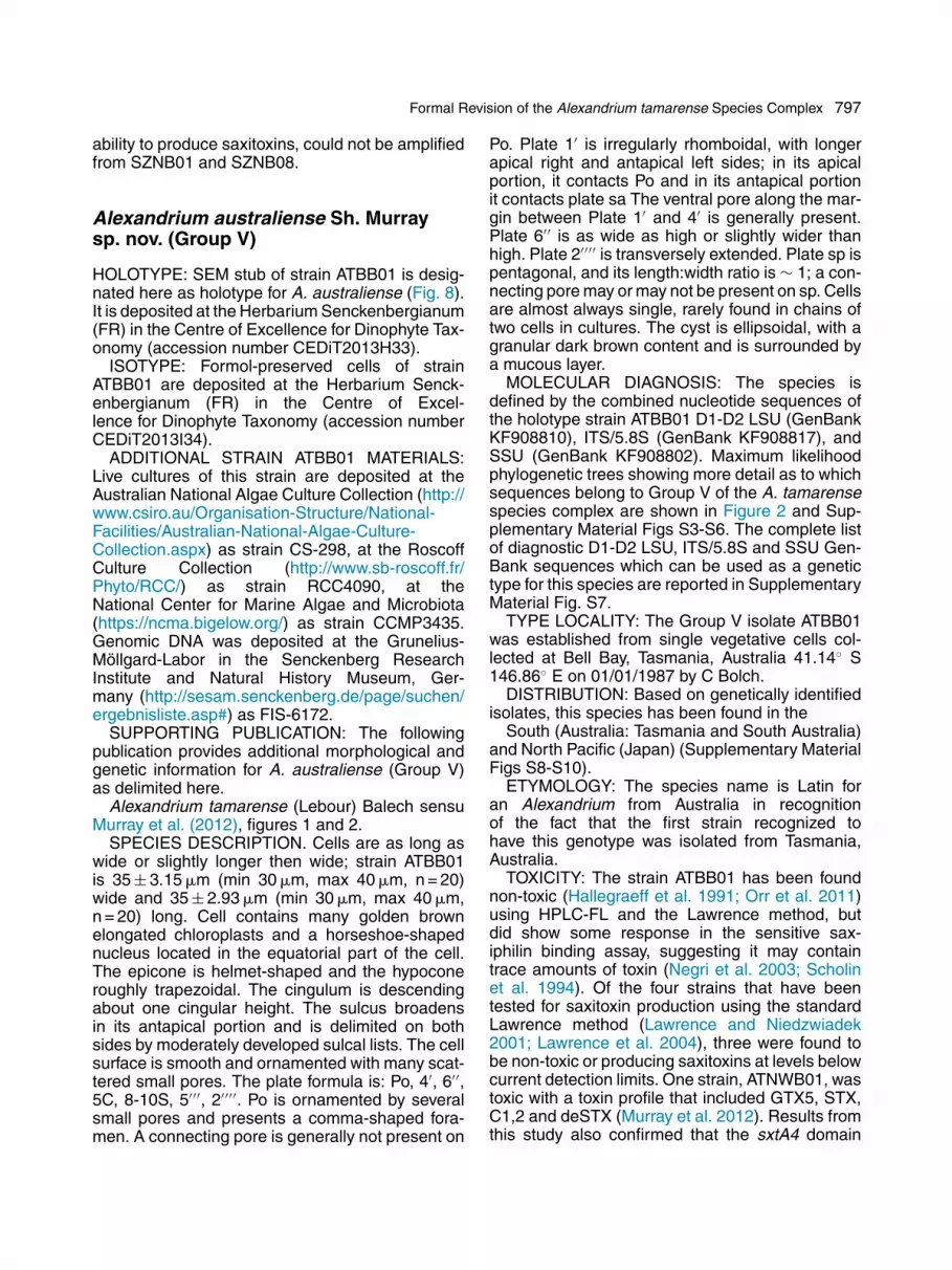
Alexandrium australiense Sh. Murraysp. nov. (Group V)
HOLOTYPE: SEM stub of strain ATBB01 is desig-nated here as holotype for A. australiense (Fig. 8).It is deposited at the Herbarium Senckenbergianum(FR) in the Centre of Excellence for Dinophyte Tax-onomy (accession number CEDiT2013H33).
ISOTYPE: Formol-preserved cells of strainATBB01 are deposited at the Herbarium Senck-enbergianum (FR) in the Centre of Excel-lence for Dinophyte Taxonomy (accession numberCEDiT2013I34).
ADDITIONAL STRAIN ATBB01 MATERIALS:Live cultures of this strain are deposited at theAustralian National Algae Culture Collection (http://www.csiro.au/Organisation-Structure/National-Facilities/Australian-National-Algae-Culture-Collection.aspx) as strain CS-298, at the RoscoffCulture Collection (http://www.sb-roscoff.fr/Phyto/RCC/) as strain RCC4090, at theNational Center for Marine Algae and Microbiota(https://ncma.bigelow.org/) as strain CCMP3435.Genomic DNA was deposited at the Grunelius-Möllgard-Labor in the Senckenberg ResearchInstitute and Natural History Museum, Ger-many (http://sesam.senckenberg.de/page/suchen/ergebnisliste.asp#) as FIS-6172.
SUPPORTING PUBLICATION: The followingpublication provides additional morphological andgenetic information for A. australiense (Group V)as delimited here.
Alexandrium tamarense (Lebour) Balech sensuMurray et al. (2012), figures 1 and 2.
SPECIES DESCRIPTION. Cells are as long aswide or slightly longer then wide; strain ATBB01is 35 ± 3.15 �m (min 30 �m, max 40 �m, n = 20)wide and 35 ± 2.93 �m (min 30 �m, max 40 �m,n = 20) long. Cell contains many golden brownelongated chloroplasts and a horseshoe-shapednucleus located in the equatorial part of the cell.The epicone is helmet-shaped and the hypoconeroughly trapezoidal. The cingulum is descendingabout one cingular height. The sulcus broadensin its antapical portion and is delimited on bothsides by moderately developed sulcal lists. The cellsurface is smooth and ornamented with many scat-tered small pores. The plate formula is: Po, 4′, 6′′,5C, 8-10S, 5′′′, 2′′′′. Po is ornamented by severalsmall pores and presents a comma-shaped fora-men. A connecting pore is generally not present on
Po. Plate 1′ is irregularly rhomboidal, with longerapical right and antapical left sides; in its apicalportion, it contacts Po and in its antapical portionit contacts plate sa The ventral pore along the mar-gin between Plate 1′ and 4′ is generally present.Plate 6′′ is as wide as high or slightly wider thanhigh. Plate 2′′′′ is transversely extended. Plate sp ispentagonal, and its length:width ratio is ∼ 1; a con-necting pore may or may not be present on sp. Cellsare almost always single, rarely found in chains oftwo cells in cultures. The cyst is ellipsoidal, with agranular dark brown content and is surrounded bya mucous layer.
MOLECULAR DIAGNOSIS: The species isdefined by the combined nucleotide sequences ofthe holotype strain ATBB01 D1-D2 LSU (GenBankKF908810), ITS/5.8S (GenBank KF908817), andSSU (GenBank KF908802). Maximum likelihoodphylogenetic trees showing more detail as to whichsequences belong to Group V of the A. tamarensespecies complex are shown in Figure 2 and Sup-plementary Material Figs S3-S6. The complete listof diagnostic D1-D2 LSU, ITS/5.8S and SSU Gen-Bank sequences which can be used as a genetictype for this species are reported in SupplementaryMaterial Fig. S7.
TYPE LOCALITY: The Group V isolate ATBB01was established from single vegetative cells col-lected at Bell Bay, Tasmania, Australia 41.14◦ S146.86◦ E on 01/01/1987 by C Bolch.
DISTRIBUTION: Based on genetically identifiedisolates, this species has been found in the
South (Australia: Tasmania and South Australia)and North Pacific (Japan) (Supplementary MaterialFigs S8-S10).
ETYMOLOGY: The species name is Latin foran Alexandrium from Australia in recognitionof the fact that the first strain recognized tohave this genotype was isolated from Tasmania,Australia.
TOXICITY: The strain ATBB01 has been foundnon-toxic (Hallegraeff et al. 1991; Orr et al. 2011)using HPLC-FL and the Lawrence method, butdid show some response in the sensitive sax-iphilin binding assay, suggesting it may containtrace amounts of toxin (Negri et al. 2003; Scholinet al. 1994). Of the four strains that have beentested for saxitoxin production using the standardLawrence method (Lawrence and Niedzwiadek2001; Lawrence et al. 2004), three were found tobe non-toxic or producing saxitoxins at levels belowcurrent detection limits. One strain, ATNWB01, wastoxic with a toxin profile that included GTX5, STX,C1,2 and deSTX (Murray et al. 2012). Results fromthis study also confirmed that the sxtA4 domain

798 U. John et al.
Figure 8. Alexandrium australiense Sh. Murray sp. nov. Line drawings (A-D) light micrographs of calcofluor-stained cells (E-G) and SEM micrograph (H) of the type strain ATBB01. (A) ventral, (B) dorsal, (C) apical, (D)antapical, (E) ventral, (F) dorsal, (G) apical and (H) antapical view. Panel H is from Murray et al. (2012) fig. 1D.Arrows in panels A and E mark the ventral pore in the 1′ plate. Scale bars = 20 �m.
was present. These data indicate that at least somestrains of this species are toxic.
Conclusions
The goal of this study was to formalize thetaxonomic status of the A. tamarense speciescomplex Groups I-V, which were previously pro-posed as ad interim species. A comprehensivephylogeny was constructed using a broad datasetof ribosomal operon sequences, including con-catenated alignments of the SSU, ITS and theD1-D2 regions of the LSU. The resulting ribo-somal phylogenetic trees always recovered fivedistinct clades (Groups), consistent with previ-ous studies. The genetic distances between theseGroups were calculated from ITS/5.8S sequences,and were equal to or exceeded the genetic dis-tances previously observed among other closelyrelated dinoflagellate species. Additionally, com-pensatory base changes (CBCs) were observedin the secondary structure of the ITS2 region
among several of the Groups. When present, aCBC consistently indicates that the sequencesbeing compared come from genetically isolatedspecies. Similarly, evidence from inter-Group mat-ing studies also supports the classification ofGroups as species according to the ‘biologicalspecies complex’ sensu Mayr. The Groups alsodiffer in the production of saxitoxins and the pres-ence/absence of sxtA4, a critical component ofsaxitoxin biosynthesis pathway. The latter resultsunderscore important differences in the physiol-ogy of Groups I-V that will impact management ofseafood hazards. In contrast to the genetic results,a detailed examination of the morphological crite-ria previously proposed for taxonomic identificationof A. tamarense species-complex species demon-strated that all features are shared among thefive respective ribosomal Groups. The combina-tion of all available data supports the segregationof the Alexandrium species complex into five dis-tinct species according to its ribosomal phylogeny.Consequently, we have amended the diagnosis ofA. fundyense (Group I) and A. tamarense (Group

Formal Revision of the Alexandrium tamarense Species Complex 799
III) and described three new species, A. mediter-raneum (Group II), A. pacificum (Group IV) and A.australiense (Group V).
Methods
Morphological species descriptions: A complete review ofthe morphological data used to establish A. catenella, A.fundyense, and A. tamarense as species was undertaken toevaluate whether the original characteristics (cell size andshape, presence or absence of the ventral pore in the 1′ plate,presence or absence of chain formation) used to distinguishspecies were valid characters (Table 1, Fig. 1). The morpho-logical characteristics were collated from original descriptions(e.g. Balech 1971, 1985, 1995; Lebour 1925; Loeblich III andLoeblich 1979; Parke and Dixon 1976; Taylor 1979; Whedonand Kofoid 1936) and compared with more recent morpholog-ical observations reported in the literature (Aguilera-Belmonteet al. 2011; Cho and Lee 2001; Faust and Gulledge 2002;Fukuyo et al. 1985; Gayoso and Fulco 2006; Hallegraeff et al.1991; Kim et al. 2002; Larsen and Nguyen 2004; Leaw et al.2005; MacKenzie et al. 2004; Murray et al. 2012; Orlova et al.2007), as well as those in AlgaeBase (Guiry and Guiry 2014).
Algal cultures: The clonal strains ACQH01, SPE10-03(Group I), SZN01, SZN08 (Group II), ATSW01-1 (Group III),ACPP01 (also known as CS 313), ATTL01, (Group IV) andATBB01, (also known as CS 298; a Group V strain) were cul-tured for use as epitype or type material (see the followingsection and Table 2) and for obtaining additional sequence datafor analysis. The isolates were grown in K-medium (Keller et al.1987) prepared from filter-sterilized (0.2 �m VacuCap 90 fil-ter units, Pall Corporation, Port Washington) North Sea water(salinity 33) at 15 ◦C, 14:10 light:dark cycle, with an irradianceof 150 �mol photons m-2 s-1. The morphological and geneticcharacterization of these isolates is described below.
Morphological analyses: All material used for the morpho-logical characterizations of the A. tamarense species complextypes came from cultures sampled in mid-exponential growthphase. Cells were fixed with formaldehyde at 1.6% final concen-tration. The length and width of 20 cells were measured by lightmicroscopy at 400x magnification (i.e. 10x ocular and 40x objec-tive; Zeiss Axioplan, Carl Zeiss, Oberkochen, Germany). Fixedcells were also stained with Calcofluor White (Sigma-Aldrich,Hamburg, Germany) according to the protocol of Fritz andTriemer (1985) and examined by epifluorescence microscopy(Zeiss Axioplan, Carl Zeiss, Oberkochen, Germany) to establishthe morphology of diagnostic thecal plates and the presence orabsence of cell chains. Additionally, an LSM confocal micro-scope was used to take 1-3 �m z-stack images of calcofluorstained cells. Z-stacks were reviewed using Zen Black software(Carl Zeiss, Oberkochen, Germany) and 3-D image recons-tructions were exported as flat images to document the thecalstructure of the epitype/holotype strains. Scanning electronmicroscope images of each of the strains were made fromformaldehyde- or gluteraldehyde-fixed samples that were dehy-drated via serial washes in ethanol (25%, 50%, 75%, 90%and 2x100%), then critical point dried and sputter coated withgold for observation under a JEOL JSM 6700F microscope orplatinum for observation using a JEOL 840 microscope (JEOLLtd., Tokyo, Japan). Cells were preserved for epitype/holotypedeposits at the Senckenberg Collection at the Centre of Excel-lence for Dinophyte Taxonomy both as stubs and preservedmaterial (2% formalin-fixed).
Phylogenetic analyses: Ribosomal DNA sequence datafor the phylogenetic analyses were obtained from the Alexan-drium cultures listed above using the following protocol. Initially,50 ml samples of exponentially growing cells were collectedby centrifugation at 3,220 x g for 15 min at room temperature(Eppendorf 5810R, Hamburg, Germany). The cell pellets werefrozen at -20 ◦C for 20 min before extraction of total DNA withthe DNeasy Mini Kit (Qiagen, Hilden, Germany) according to themanufacturer’s instructions. The purity and quantity of the DNAwas assessed by UV-spectroscopy with a NanoDrop ND-1000system (Peqlab, Erlangen, Germany) and the integrity of DNAwas confirmed using 1% agarose gel electrophoresis where amajority of the extracted genomic DNA exceeded 20 kilobases.
The following ribosomal DNA loci were amplified from eachtotal DNA extract by polymerase chain reaction (PCR): the 18Ssmall subunit (SSU), the D1/D2 region of the 28S large subunit(LSU), and the internal transcribed spacer region (including theITS1, 5.8S subunit, and ITS2 sequences; referred to hereafteras ITS/5.8S). The forward and reverse primers for SSU ampli-fication were: 1F (5′- AAC CTG GTT GAT CCT GCC AGT - 3′)and 1528R (5′ - TGA TCC TTC TGC AGG TTC ACC TAC - 3′),respectively. The forward and reverse primers for LSU amplifica-tion were: D1R-F (5′ -ACC CGC TGA ATT TAA GCA TA- 3′) andD2C-R (5′ -CCT TGG TCC GTG TTT CAA GA- 3′), respectively.The forward and reverse primers for ITS/5.8 amplification were:ITS a (5′ -CCA AGC TTC TAG ATC GTA ACA AGG (ACT)TCCGT AGG T- 3′) and ITS b (5′ -CCT GCA GTC GAC A(GT)ATGC TTA A(AG)T TCA GC(AG) GG- 3′), respectively. Reactionconditions were as follows: HotMasterTaq® (5Prime, Hamburg,Germany) buffer 1X, 0.1 mM of dNTPs, 0.1 mM of each for-ward and reverse primer and 1.25 units of Taq polymerase wereadded to 10-30 ng of the extracted genomic DNA in total reac-tion volumes of 50 �L. For 18S and 28S rDNA amplifications,the reactions were subjected to the following thermocyclingconditions: one cycle of 95 ◦C 7 min; 35 cycles 94 ◦C for 45s, 54 ◦C for 2 min, 70 ◦C for 1.5 min; and a final extension 70 ◦C5 min. The thermocycling conditions for the ITS/5.8 amplifica-tions were: one cycle 94 ◦C for 4 min; 9 cycles 94 ◦C for 50s, 60 ◦C for 40 s and 70 ◦C for 1 min; 29 cycles 94 ◦C for 45s, 50 ◦C for 45 s, 70 ◦C for 1 min; and a final 5 min extensionstep at 70 ◦C. Samples were kept at 10 ◦C until prior to anal-ysis on 1% agarose gel to ensure the expected amplificationproducts were present. After PCR amplification and subsequentcloning into the vector provided with the TOPO TA Cloning® kit(Invitrogen, Carlsbad, California, USA), 3 - 8 clones per ampli-con were sequenced using the M13 vector primers suppliedwith the kit. Sequencing was conducted with a standard cyclesequencing chemistry ABI 3.1 (Applied Biosystems, Darmstadt,Germany) and additional internal primers were used for SSUsequencing (Montresor et al. 2004). Cycle sequencing prod-ucts were analyzed on an ABI 3130 XL capillary sequencer(Applied Biosystems, Darmstadt, Germany) and the generatedsequences were assembled with CLC main workbench version6.0 (www.CLCbio.com). The resulting sequences were submit-ted to GenBank (Accession numbers: KF908795-KF908818).
The SSU, ITS/5.8S, and LSU sequences were then com-bined with those available in GenBank and different alignmentsconstructed in order to conduct phylogenetic analyses usingeither single SSU (Supplementary Material Figs S1-S3),ITS/5.8 (Supplementary Materials Figs files S1-2, S4), or LSU(Supplementary Material Figs S1-2, S5) data sets or a con-catenated SSU-ITS/5.8-LSU data set (Supplementary MaterialFigs S1-2, S6). For the concatenated phylogenetic analyses,only the combined sequences from the type species SPE10-03, SZN01, ATBB01, ACPP01, ATSW01-1 as well as those forCCMP115, ACQH01, SZN08, ATTL0, ATNWB01 were included

800 U. John et al.
in the analysis plus outgroup sequences from Alexandriumaffine (H. Inoue & Fukuyo) Balech (CCMP 112). The individ-ual SSU, LSU and ITS/5.8S data sets were separately alignedwith MAFFT using the q-insi option (Katoh et al. 2005) and con-catenated afterwards (Supplementary Material Fig. S2). Thefull ML analysis (GTRGAMMA, 1000 bootstrap replicates) wasconducted with RAxML (Stamatakis et al. 2005). In the casefor the concatenated dataset the RAxML analysis used threepartitions for each gene.
In order to generate a representative phylogeny, all the D1-D2 LSU Alexandrium rDNA sequences available in GenBankwere aligned using ClustalX. The sequences were then sortedinto species groups. This ensured any that sequences whosespecies identification were incorrect in GenBank were assignedto the correct species group. If the D1-D2 sequences variedwithin species, they were further sorted into subgroups that dif-fered from one another by more than 2 bp. For several species,within species sequence divergences were significantly largerand fell into easily identifiable subclades (labeled 1, 2, 3, etc.).In these cases, it was easier to identify sequences varying byless than 2 bp by analyzing the sequences belonging to eachsubclade separately. Specifically, the sequences belonging toeach subclade were transferred to separate alignment files andsorted based on the <2 bp variation criterion. Each of the result-ing <2 bp subgroups were then given a letter desgination (e.g.2a, 2b, etc. for subgroups within subclade 2). For the final analy-sis, a single sequence from every <2 bp subgroup was selectedfor construction of the D1-D2 phylogeny. This filtering procedurecaptured a majority of the within-species variation while reduc-ing the total number of sequences in the analysis for all speciesfrom 638 to 40. These D1-D2 sequences were then combinedwith a representiative sequence from each of the five Alexan-drium type or epitype isolates. The combined data set was thenaligned using ClustalX using an open gap penalty of 10 andextended gap penalty of 5. Obvious misalignments were cor-rected manually (Supplmentary Material Fig. S1) and the finalalignment analysed by RAxML CAT, 1000 bootstrap replicatesas described above (Supplementary Material Fig. S2).
ITS analysis: The ITS/5.8S sequence data were partitionedinto species groups, aligned using ClustalW, and genetic dis-tances (substitutions per site) between each possible sequencecombination was estimated using PAUP* (Swofford 1998). Theprocedure was essentially identical to that employed by Litakeret al. (2007) except that no attempt was made to identifyand eliminate pseudogenes (i.e., all GenBank sequences wereused; see Supplementary Material Fig. S7). The inclusionof these pseudogenes would tend to increase the apparentgenetic distance estimates over those measured in the orig-inal studies. The mean substitutions per site, both within thefive species and between them were then calculated along withthe minimum and maximum values observed in each within- orbetween-group comparison.
ITS2 rRNA secondary structure modeling: The presenceor absence of CBCs was determined as follows. The sequencesof the type strains were aligned with those of other A. tamarensecomplex taxa and strains identified via BLAST searches in Gen-Bank. The beginning and end of the ITS2 region in these strainswere identified via homology with two strains of Alexandriumaffine (one misidentified as “A. tamarense strain CCMP116”)available in the ITS2 database (Koetschan et al. 2010), andwere confirmed by comparison with a broader alignment ofdinoflagellates. The ITS2 region consisted of approximately180 bp in our Alexandrium strains. The ITS2 secondary struc-ture was predicted using the existing structural “a” model forthe A. affine ITS2 region (Koetschan et al. 2010), which was inagreement with the general dinoflagellate structures predictedby Gottschling and Plötner (2004). Structures were visualized
using VARNA (Darty et al. 2009). The compensatory basechanges in the predicted secondary structure models wereidentified using the software 4SALE (Seibel et al. 2006).
Toxicity: Because the production of saxitoxins is variableamong Alexandrium strains/species, the current literature wasreviewed to determine what is known about toxin production byisolates belonging to each of the five genetic groups (sensuspecies) within the A. tamarense species complex. In addi-tion, except for ATBB01 (Stüken et al. 2011) the type isolatesSPE10-03 (Group I), SZN01 (Group II), ATSW01-1 (Group III),and ACPP01 (Group IV) were screened using the followingPCR procedure for the presence of the sxtA4, a conservedregion in the sxtA gene which is a key component of thesaxitoxin synthesis pathway. Briefly, the primer pair used wassxt007 (5′ -ATGCTCAACATGGGAGTCATCC- 3′) and sxt008(5′ -GGGTCCAGTAGATGTTGACGATG- 3′; Stüken et al. 2011).For each 20 �l PCR reaction, HotMasterTaq® (5Prime, Ham-burg, Germany) buffer 1X, 0.1 mM of dNTPs, 0.1 mM of eachforward and reverse primer and 0.75 units of Taq polymerasewere added to 10-30 ng of the extracted genomic DNA. The fol-lowing thermocycling conditions were used: one cycle of 95 ◦Cfor 2 min; 35 cycles 94 ◦C for 30 s, 55 ◦C for 30 s, 70 ◦C for1.5 min; and a final extension 70 ◦C for 10 min. Amplicons werepurified, cloned, sequenced and analysed as described above.The resulting sequences were submitted to GenBank (acces-sion numbers: KF985177- KF985182).
Acknowledgements
We give our sincere thanks to John McNeill forguiding us through the ICN, to Mona Hoppenrathfor her very helpful and precise advice and sharedexpertise, as well as to three anonymous review-ers who helped shape and finalize this ambitiousproject. We also thank Nancy Kühne for her assis-tance in the laboratory, David Kulis for maintenanceand distribution of cultures, and Steve Kibler andMark Vandersea who reviewed the manuscript andprovided helpful suggestions. Financial support toU.J. was provided by the PACES research programof the Alfred-Wegener-Institute Helmholtz-Zentrumfür Polar- und Meeresforschung. Partial supportfor W.L. was provided by North Pacific ResearchBoard Project 1021 publication number 503 andthe NOAA Coastal Science Board, for M.M. bythe Stazione Zoologica Anton Dohrn, and for S.M.and U.J. by the Australian Research council grantnumber DP120103199, an Australian Academy ofScience DAAD German-Australian mobility grantand BMBF PT/DLR IB FKZ 01DR14006 projectn(NSF) Grants OCE- 1128041 and OCE-1314642;and National Institute of Environmental Health Sci-ences (NIEHS) Grant 1-P50-ES021923-01.
Appendix A. Supplementary Data
Supplementary data associated with this article canbe found, in the online version, at http://dx.doi.org/10.1016/j.protis.2014.10.001.

Formal Revision of the Alexandrium tamarense Species Complex 801
References
Adachi M, Sako Y, Ishida Y (1994) Restriction fragment lengthpolymorphism of ribosomal DNA internal transcribed spacerand 5. 8S regions in Japanese Alexandrium species (Dino-phyceae). J Phycol 30:857–863
Adachi M, Sako Y, Ishida Y (1996) Analysis of Alexandrium(Dinophyceae) species using sequences of the 5. 8S ribo-somal DNA and internal transcribed spacer regions. J Phycol32:424–432
Aguilera-Belmonte A, Inostroza I, Franco JM, Riobó P,Gómez PI (2011) The growth, toxicity and genetic charac-terization of seven strains of Alexandrium catenella (Whedonand Kofoid) Balech 1985 (Dinophyceae) isolated during the2009 summer outbreak in southern Chile. Harmful Algae 12:105–112
Anderson DM, Wall D (1978) Potential importance of benthiccysts of Gonyaulax tamarensis and G. excavata in initiating toxicdinoflagellate blooms. J Phycol 13:224–234
Anderson DM, Kulis DM, Doucette GJ, Gallager JC, BalechE (1994) Biogeography of toxic dinoflagellates in the genusAlexandrium from the northeast United States and Canada asdetermined by morphology, bioluminescence, toxin composi-tion, and mating compatibility. Mar Biol 120:467–478
Anderson DM, Alpermann TJ, Cembella AD, Collos Y,Masseret E, Montresor M (2012) The globally distributedgenus Alexandrium: Multifaceted roles in marine ecosystemsand impacts on human health. Harmful Algae 14:10–35
Anderson DM, Kulis DM, Keafer BA, Gribble KE, Marin R,Scholin CA (2005) Identification and enumeration of Alexan-drium spp. from the Gulf of Maine using molecular probes.Deep-Sea Res Pt II 52:2467–2490
Baggesen C, Moestrup Ø, Daugbjerg N, Krock B, CembellaAD, Madsen S (2012) Molecular phylogeny and toxin profilesof Alexandrium tamarense (Lebour) Balech (Dinophyceae) fromthe west coast of Greenland. Harmful Algae 19:108–116
Balech E (1971) Microplancton de la campana oceanograficaProductividad III. Revista del Museo Argentino de Ciencias Nat-urales “Bernardino Rivadavia”. Hidrobiologia 3:1–202
Balech E (1985) The Genus Alexandrium or Gonyaulax of thetamarensis Group. In Anderson DM, White AW, Baden DG(eds) Toxic Dinoflagellates, Proceedings of the Third Interna-tional Conference on Toxic Dinoflagellates. Elsevier, New York,pp 33–38
Balech E (1995) The Genus Alexandrium Halim (Dinoflagel-lata). Sherkin Island Marine Station Publication, Sherkin Island,Co. Cork, Ireland
Balech E, Tangen K (1985) Morphology and taxonomy of toxicspecies in the tamarensis group (Dinophyceae): Alexandriumexcavatum (Braarud) comb. nov. and Alexandrium ostenfeldii(Paulsen) comb. nov. Sarsia 70:333–343
Bolch CJS, de Salas MF (2007) A review of the molecular evi-dence for ballast water introduction of the toxic dinoflagellatesGymnodinium catenatum and the Alexandrium “tamarensiscomplex” to Australia. Harmful Algae 6:465–485
Braarud T (1945) Morphological observations on marinedinoflagellate cultures (Porella perforata, Gonyaulax
tamarensis, Protoceratium reticulatum). Avh Utgitt NorVidensk Akad Oslo, Mat - Naturvidensk Kl 1944 11:1–18
Brosnahan ML, Kulis DM, Solow AR, Erdner DL, PercyL, Lewis J, Anderson DM (2010) Outbreeding lethalitybetween toxic Group I and nontoxic Group III Alexandriumtamarense spp. isolates: Predominance of heterotypic encyst-ment and implications for mating interactions and biogeography.Deep-Sea Res Pt II 57:175–189
Cembella AD, Taylor FJR (1986) Electrophoretic variabil-ity within the Protogonyaulax tamarensis/catenella speciescomplex: Pyridine-linked dehydrogenases. Biochem Syst Ecol14:311–323
Cho ES, Lee HJ (2001) Thecal plates, toxin content and growthof five clones of Alexandrium tamarense (Dinophyceae) iso-lated from Jinhae Bay, Korea. Phycologia 40:435–439
Cho Y, Hiramatsu K, Ogawa M, Omura T, Ishimaru T, OshimaY (2008) Non-toxic and toxic subclones obtained from a toxicclonal culture of Alexandrium tamarense (Dinophyceae): Toxic-ity and molecular biological feature. Harmful Algae 7:740–751
Coleman AW (2003) ITS2 is a double-edged toll for eukaryoticevolutionary comparison. Trends Genet 19:370–375
Coleman AW (2009) Is there a molecular key to the level of “bio-logical species” in eukaryotes? A DNA guide. Mol PhylogenetEvol 50:197–203
Collins C, Graham J, Brown L, Bresnan E, Lacaze JP, TurrellEA (2009) Identification and toxicity of Alexandrium tamarense(Dinophyceae) in Scottish waters. J Phycol 45:692–703
Darty K, Denise A, Ponty Y (2009) VARNA: Interactive draw-ing and editing of the RNA secondary structure. Bioinformatics25:1974–1975
Destombe C, Cembella AD, Murphy CA, Ragan MA (1992)Nucleotide sequence of the 18S ribosomal RNA genes from themarine dinoflagellate Alexandrium tamarense (Gonyaulacales,Dinophyta). Phycologia 31:121–124
Dyhrman ST, Erdner D, La Du J, Galac M, Anderson DM(2006) Molecular quantification of toxic Alexandrium fundyensein the Gulf of Maine using real-time PCR. Harmful Algae5:242–250
Dyhrman ST, Haley ST, Borchert JA, Lona B, Kollars N, Erd-ner DL (2010) Parallel analyses of Alexandrium catenella cellconcentrations and shellfish toxicity in the Puget Sound. ApplEnviron Microbiol 76:4647–4654
Erdner DL, Percy L, Keafer B, Lewis J, Anderson DM(2010) A quantitative real-time PCR assay for the identificationand enumeration of Alexandrium cysts in marine sediments.Deep-Sea Res Part II 57:279–287
Fabry S, Köhler A, Coleman AW (1999) Intraspecies analysis:comparison of ITS sequence data and gene intron sequencedata with breeding data for a worldwide collection of Goniumpectorale. J Mol Evol 48:94–101
Faust MA, Gulledge RA (2002) Identifying Harmful MarineDinoflagellates. Smithsonian Institution, Washington, DC,144 p
Fritz L, Triemer RE (1985) A rapid simple technique utiliz-ing Calcofluor white M2R for the visualization of dinoflagellatethecal plates. J Phycol 21:662–664

802 U. John et al.
Fukuyo Y, Yoshida K, Inoue H (1985) Protogonyaulax inJapanese Coastal Waters. In Anderson DM, White AW, BadenDG (eds) Toxic Dinoflagellates, Proceedings of the Third Inter-national Conference on Toxic Dinoflagellates. Elsevier-NorthHolland, New York, pp 27–32
Garneau ME, Schnetzer A, Countway PD, Jones AC, Seu-bert EL, Caron DA (2011) Examination of the seasonaldynamics of the toxic dinoflagellate Alexandrium catenella atRedondo Beach, California, by quantitative PCR. Appl EnvironMicrobiol 77:7669–7680
Gayoso AM, Fulco VK (2006) Occurrence patterns of Alexan-drium tamarense (Lebour) Balech populations in the GolfoNuevo (Patagonia, Argentina), with observations on ventralpore occurrence in natural and cultured cells. Harmful Algae5:233–241
Genovesi B, Shin-Grzebyk MS, Grzebyk D, Laabir M, Gag-naire PA, Vaquer A, Pastoureaud A, Lasserre B, CollosY, Berrebi P, Masseret E (2011) Assessment of crypticspecies diversity within blooms and cyst bank of the Alexan-drium tamarense complex (Dinophyceae) in a Mediterraneanlagoon facilitated by semi-multiplex PCR. J Plankton Res 33:405–414
Godhe A, Cusack C, Pedersen J, Andersen P, AndersonDM, Bresnan E, Cembella A, Dahl E, Diercks S, ElbraechterM, Edler L, Galluzzi L, Gescher C, Gladstone M, KarlsonB, Kulis D, LeGresley M, Lindahl O, Marin R, McDermott G,Medlin LK, Naustvoll L-J, Penna A, Toebe K (2007) Intercal-ibration of classical and molecular techniques for identificationof Alexandrium fundyense (Dinophyceae) and estimation of celldensities. Harmful Algae 6:56–72
Gottschling M, Plötner J (2004) Secondary structure mod-els of the nuclear internal transcribed spacer regions and 5. 8SrRNA in Calciodinellaoideae (Peridiniaceae) and other dinoflag-ellates. Nucleic Acids Res 32:307–315
Gottschling M, Keupp H, Plotner J, Knop R, Willems H,Kirsch M (2005) Phylogeny of calcareous dinoflagellates asinferred from ITS and ribosomal sequence data. Mol PhylogenetEvol 36:444–455
Gu H, Zeng N, Lui T, Yang W, Müller A, Krock B (2013) Mor-phology, toxicity, and phylogeny of Alexandrium (Dinophyceae)species along the coast of China. Harmful Algae 27:68–681
Guillou L, Nézan E, Cueff V, Erard-Le Denn E,Cambon-Bonavita M-A, Gentien P, Barbier G (2002)Genetic diversity and molecular detection of three toxicdinoflagellate genera (Alexandrium, Dinophysis, and Karenia)from French Coasts. Protist 153:223–238
Guiry MD, Guiry GM (2014) AlgaeBase. World-wide elec-tronic publication, National University of Ireland, Galway.http://www.algaebase.org: accessed on 13 March 2014
Hallegraeff GM, Bolch CJ, Blackburn SI, Oshima Y (1991)Species of the toxigenic dinoflagellate genus Alexandrium insouth eastern Australian waters. Bot Mar 34:575–587
Higman WA, Stone DM, Lewis JM (2001) Sequence com-parisons of toxic and non-toxic Alexandrium tamarense(Dinophyceae) isolates from UK waters. Phycologia 40:256–262
Halim Y (1960) Alexandrium minutum nov. g. nov. sp. dinoflag-ellé provocant des ‘eaux rouges’. Vie Milieu 11:102–105
Ho KC, Lee TCH, Kwok OT, Lee FWF (2012) Phylogeneticanalysis on a strain of Alexandrium tamarense collected fromAntarctic Ocean. Harmful Algae 15:100–108
Jedlicki A, Fernández G, Astorga M, Oyarzún P, ToroJE, Navarro JM, Martínez V (2012) Molecular detection andspecies identification of Alexandrium (Dinophyceae) causingharmful algal blooms along the Chilean coastline. AoB Plantspls033 http://aob.doi.org/10.1093/aobpla/pls033
Jester RJ, Baugh KA, Lefebvre KA (2009) Presence ofAlexandrium catenella and paralytic shellfish toxins, shellfishand rock crabs in Montego Bay, California, USA. Mar Biol156:493–504
John U, Fensome RA, Medlin LK (2003a) The application ofa molecular clock based on molecular sequences and the fossilrecord to explain biogeographic distributions within the Alexan-drium tamarense “species complex” (Dinophyceae). Mol BiolEvol 20:1015–1027
John U, Cembella A, Hummert C, Elbrächter M, Groben R,Medlin L (2003b) Discrimination of the toxigenic dinoflagel-lates Alexandrium tamarense and A. ostenfeldii in co-occurringnatural populations from Scottish coastal waters. Eur J Phycol38:25–40
John U, Medlin LK, Groben R (2005) Development of spe-cific rRNA probes to distinguish between geographic clades ofthe Alexandrium tamarense species complex. J Plankton Res27:199–204
John U, Litaker W, Montresor M, Murray S, Brosnahan ML,Anderson DM (2014) Proposal to reject the name Alexandriumcatenella (Dinophyceae). Taxon 63:932–933
Kamikawa R, Nagai S, Hosoi-Tanabe S, Itakura S, Yam-aguchi M, Uchida Y, Baba T, Sako Y (2007) Application ofreal-time PCR assay for detection and quantification of Alexan-drium tamarense and Alexandrium catenella cysts from marinesediments. Harmful Algae 6:413–420
Katoh K, Kuma K, Toh H, Miyata T (2005) MAFFT version5: improvement in accuracy of multiple sequence alignment.Nucleic Acids Res 33:511–518
Keller MD, Selvin RC, Claus W, Guillard RRL (1987) Media forthe culture of oceanic ultraphytoplankton. J Phycol 23:633–638
Kim KY, Yoshida M, Fukuyo Y, Kim CH (2002) Morphologi-cal observation of Alexandrium tamarense (Lebour) Balech, A.catenella (Whedon et Kofoid) Balech and one related morpho-type (Dinophyceae) in Korea. Algae 17:11–19
Klöpper S, John U, Zingone A, Mangoni O, Kooistra WHCF,Cembella A (2013) Phylogeny and morphology of a Chattonella(Raphidophyceae) species from the Mediterranean Sea: whatis C. subsalsa? Eur J Phycol 48:79–92
Koetschan C, Förster F, Keller A, Schleicher T, RuderischB, Schwarz R, Müller T, Wolf M, Schultz J (2010) The ITS2Database III-sequences and structures for phylogeny. NucleicAcids Res 38:275–276
Larsen J, Nguyen NL (2004) Potentially toxic microalgae ofVietnamese waters. Opera Bot 140:5–216
Lawrence JF, Niedzwiadek B (2001) Quantitative determina-tion of paralytic shellfish poisoning toxins in shellfish by usingprechromatographic oxidation and liquid chromatography withfluorescence detection. AOAC Int 84:1099–1108

Formal Revision of the Alexandrium tamarense Species Complex 803
Lawrence JF, Niedzwiadek B, Menard CJ (2004) Quantitativedetermination of paralytic shellfish poisoning toxins in shellfishusing prechromatographic oxidation and liquid chromatogra-phy with fluorescence detection: interlaboratory study. AOACInt 87:83–100
Leaw CP, Lim PT, Ng BK, Cheah MY, Ahmad A, Usup G(2005) Phylogenetic analysis of Alexandrium species and Pyro-dinium bahamense (Dinophyceae) based on theca morphologyand nuclear ribosomal gene sequence. Phycologia 44:550–565
Lebour MV (1925) The Dinoflagellates of the Northern Seas.Marine Biological Association of the United Kingdom, Plymouth,150 p.
Lilly EL (2003) Phylogeny and Biogeography of the Toxicdinoflagellate Alexandrium. Ph.D. dissertation. MassachusettsInstitute of Technology, Cambridge
Lilly EL, Halanych KM, Anderson DM (2007) Species bound-aries and global biogeography of the Alexandrium tamarensecomplex (Dinophyceae). J Phycol 43:1329–1338
Lilly EL, Kulis DM, Gentien P, Anderson DM (2002)Paralytic shellfish poisoning toxins in France linked to ahuman-introduced strain of Alexandrium catenella from thewestern Pacific: Evidence from DNA and toxin analysis. J Plank-ton Res 24:443–452
Litaker RW, Vandersea MW, Kibler SR, Reece KS, StokesNA, Lutzoni FM, Yonish BA, West MA, Black MND, TesterPA (2007) Recognizing dinoflagellate species using ITS rDNAsequences. J Phycol 43:344–355
Loeblich AR III, Loeblich LA (1979) The Systematics ofGonyaulax with Special Reference to the Toxic Species. InTaylor DL, Seliger HH (eds) Toxic Dinoflagellate Blooms,Proc. Second International Conference on Toxic DinoflagellateBlooms. Elsevier-North Holland, New York, pp 41–46
Loeblich LA, Loeblich AR III (1975) The Organism CausingNew England Red Tides: Gonyaulax excavata. In LoCicero VR(ed) Proceedings of the First International Conference on ToxicDinoflagellate Blooms. Massachusettes Science TechnologyFoundation, Wakefield, MA, pp 207–224
Loeblich LA, Winn GJ, Loeblich AR III (1975) Comparativestudies on Gonyaulax tamarensis and related red-tide species.J Phycol 11(Suppl.):21
MacKenzie L, de Salas M, Adamson J, Beuzenberg V (2004)The dinoflagellate genus Alexandrium (Halim) in New Zealandcoastal waters: comparative morphology, toxicity and moleculargenetics. Harmful Algae 3:71–92
Masseret E, Grzebyk D, Nagai S, Genovesi B, Lasserre B,Laabir M, Collos Y, Vaquer A, Berrebi P (2009) Unexpectedgenetic diversity among and within populations of the toxicdinoflagellate Alexandrium catenella as revealed by nuclearmicrosatellite markers. Appl Environ Microbiol 75:2037–2045
McNeill J, Barrie FR, Buck WR, Demoulin V, Greuter W,Hawksworth DL, Herendeen PS, Knapp S, Marhold K, PradoJ, Prud’homme van Reine WF, Smith GF, Wiersema JH,Turland NJ (2012) International Code of Nomenclature foralgae, fungi and plants (Melbourne Code) adopted by the Eigh-teenth International Botanical Congress Melbourne, Australia,July 2011. Regnum Vegetabile 154:1–240
Medlin L, Lange M, Wellbrock U, Donner G, Elbrächter M,Hummert C, Luckas B (1998) Sequence comparisons link
toxic European isolates of Alexandrium tamarense from theOrkney Islands to toxic North American stocks. Eur J Protistol34:329–335
Miranda LN, Zhuang YY, Zhang H, Lin S (2012) Phyloge-netic analysis guided by intragenomic SSU rDNA polymorphismrefines classification of “Alexandrium tamarense” species com-plex. Harmful Algae 16:35–48
Montresor M, John U, Beran A, Medlin LK (2004)Alexandrium tamutum sp. nov. (Dinophyceae): A new non-toxic species in the genus Alexandrium. J Phycol 40:398–411
Murray S, Wiese M, Stüken A, Brett S, Kellmann R, Hallegra-eff G, Neilan B (2011) sxtA-based quantitive molecular assayto identify saxitoxin producing harmful algal blooms in marinewaters. Appl Environ Microbiol 77: 7050–7057
Murray S, Wiese M, Neilan BA, Orr RJS, Salas M, Brett S,Hallegraeff G (2012) A reinvestigation of saxitoxin productionand sxtA in the ‘non-toxic’ Alexandrium tamarense Group Vclade. Harmful Algae 18:96–104
Negri A, Llewellyn L, Doyle J, Webster N, Frampton D,Blackburn S (2003) Paralytic shellfish toxins are restricted tofew species among Australia’s taxonomic diversity of culturedmicroalgae. J Phycol 39:663–667
Orlova TY, Selina MS, Lilly EL, Kulis DM, Anderson DM(2007) Morphogenetic and toxin composition variability ofAlexandrium tamarense (Dinophyceae) from the east coast ofRussia. Phycologia 46:534–548
Orr RJS, Stüken A, Murray SA, Jakobsen KS (2013) Evo-lutionary acquisition and loss of saxitoxin biosynthesis indinoflagellates: the second “core” gene, sxtG. Appl EnvironMicrobiol 79:2128–2136
Orr RJS, Stüken A, Rundberget T, Eikrem W, Jakobsen KS(2011) Improved phylogenetic resolution of toxic and non-toxicAlexandrium strains using a concatenated rDNA approach.Harmful Algae 10:676–688
Parke M, Dixon PS (1976) Check-list of British marine algae –Third revision. J Mar Biol Assoc UK 56:527–594
Penna A, Garcés E, Vila M, Giacobbe MG, Fraga S, Lugliè A,Bravo I, Bertozzini E, Vernesi C (2005) Alexandrium catenella(Dinophyceae), a toxic ribotype expanding in the NW Mediter-ranean Sea. Mar Biol 148:13–23
Penna A, Fraga S, Maso M, Giacobbe MG, Bravo I, Garces E,Vila M, Bertozzini E, Andreoni F, Luglie A, Vernesi C (2008)Phylogenetic relationships among the Mediterranean Alexan-drium (Dinophyceae) species based on sequences of 5. 8Sgene and Internal Transcript Spacers of the rRNA operon. EurJ Phycol 43:163–178
Pfiester LA, Anderson DM (1987) Dinoflagellate Life Cyclesand their Environmental Control. In Taylor FJR (ed) The Biologyof Dinoflagellates. Blackwell Scientific Publications, Ltd, Oxford,pp 611–648
Ride WDL, Cogger HG, Dupuis C, Kraus O, Minelli A,Thompson FC, Tubbs PK (1999) International Code of Zoo-logical Nomenclature. Cromwell, London
Ruhl MW, Wolf M, Jenkins TM (2009) Compensatory basechanges illuminate taxonomically difficult taxonomy. Mol Phylo-gent Evol 54:664–669

804 U. John et al.
Ruiz Sebastián C, Etheridge SM, Cook PA, O’Ryan C,Pitcher GC (2005) Phylogenetic analysis of toxic Alexandrium(Dinophyceae) isolates from South Africa: Implications for theglobal phylogeography of the Alexandrium tamarense speciescomplex. Phycologia 44:49–60
Scholin C, Herzog AM, Sogin M, Anderson DM (1994)Identification of group- and strain-specific genetic markers forglobally distributed Alexandrium (Dinophyceae). II. Sequenceanalysis of a fragment of the LSU rDNA gene. J Phycol 30:999–1011
Scholin CA, Hallegraeff GM, Anderson DM (1995) Molecu-lar evolution of the Alexandrium tamarense ‘species complex’(Dinophyceae): Dispersal in the North American and WestPacific regions. Phycologia 34:472–485
Seibel PN, Müller T, Dandekar T, Schultz J, Wolf M (2006)4SALE – A tool for synchronous RNA sequence and sec-ondary structure alignment and editing. BMC Bioinformatics7:498 http://www.biomedcentral.com/1471-2105/7/498
Smayda TJ (1997) Harmful algal blooms: Their ecophysiol-ogy and general relevance to phytoplankton blooms in the sea.Limnol Oceanogr 42:1137–1153
Stamatakis A, Ludwig T, Meier H (2005) RAxML-III: a fastprogram for maximum likelihood-based inference of large phy-logenetic trees. Bioinformatics 21:456–463
Stüken A, Orr RJS, Kellmann R, Murray SA, Neilan BA,Jakobsen KS (2011) Discovery of nuclear-encoded genes forthe neurotoxin saxitoxin in dinoflagellates. PLoS ONE 6:e20096
Sunda WG, Granéli E, Gobler CJ (2006) Positive feedbackand the development and persistence of ecosystem disruptivealgal blooms. J Phycol 42:963–974
Swofford DL (1998) PAUP*. Phylogenetic Analysis Using Par-simony (* and other methods). Version 4. Sinauer Associates,Sunderland, Massachusetts
Taylor FJR (1979) The Toxigenic Gonyaulacoid Dinoflagel-lates. In Taylor DL, Seliger HH (eds) Toxic DinoflagellateBlooms, Proceedings of the Second International Confer-ence on Toxic Dinoflagellate Blooms. Elsevier, New York,pp 47–56
Taylor FJR (1984) Toxic Dinoflagellates: Taxonomic and Bio-geographic Aspects with Emphasis on Protogonyaulax. InRagelis EP (ed) Seafood Toxins. American Chemical Society,Washington, D.C., pp 77–97
Toebe K, Alpermann TJ, Tillmann U, Krock B, CembellaAD, John U (2013) Molecular discrimination of toxic andnon-toxic Alexandrium species (Dinophyta) in natural phyto-plankton assemblages from the Scottish coast of the North Sea.Eur J Phycol 48:12–26
Touzet N, Franco JM, Raine R (2008) Morphogenetic diversityand biotoxin composition of Alexandrium (Dinophyceae) in Irishcoastal waters. Harmful Algae 7:782–797
Touzet N, Davidson K, Pete R, Flanagan K, McCoy GR,Amzil Z, Maher M, Chapelle A, Raine R (2010) Co-occurrenceof the West European (Gr. III) and North American (Gr.I) ribo-types of Alexandrium tamarense (Dinophyceae) in Shetland,Scotland. Protist 161:370–384
Vanormelingen P, Hegewald E, Braband A, KitschkeM, Friedel T, Sabbe K, Vyverman W (2007) The sys-tematics of a small spineless Desmodesmus species, D.costato-granulatus (Sphaeropleales, Chlorophyceae), basedon ITS2 rDNA sequence analyses and cell wall morphology.J Phycol 43:378–396
Varela D, Paredes J, Alves-de-Souza C, Seguel M, Sfeir A,Frangopulos M (2012) Intraregional variation among Alexan-drium catenella (Dinophyceae) strains from southern Chile:Morphological, toxicological and genetic diversity. HarmfulAlgae 15:8–18
Vila M, Garcés E, Maso M, Camp J (2001) Is the distribution ofthe toxic dinoflagellate Alexandrium catenella expanding alongthe NW Mediterranean coast? Mar Ecol Prog Ser 222:73–83
Wang L, Zhuang YY, Zhang H, Lin X, Lin S (2014) DNAbarcoding species in Alexandrium tamarense complex usingITS and proposing designation of five species. Harmful Algae31:100–113
Whedon WF, Kofoid CA (1936) Dinoflagellata of the SanFrancisco Region. I. On the skeletal morphology of two newspecies, Gonyaulax catenella and G. acatenella. Univ Calif PublZool 41:25–34
Available online at www.sciencedirect.com
ScienceDirect