the citrus fruit proteome: insights into citrus fruit metabolism
TRANSCRIPT
Planta (2007) 226:989–1005
DOI 10.1007/s00425-007-0545-8ORIGINAL ARTICLE
The citrus fruit proteome: insights into citrus fruit metabolism
E. Katz · M. Fon · Y. J. Lee · B. S. Phinney · A. Sadka · E. Blumwald
Received: 3 April 2007 / Accepted: 5 May 2007 / Published online: 31 May 2007© Springer-Verlag 2007
Abstract Fruit development and ripening are key pro-cesses in the production of the phytonutrients that areessential for a balanced diet and for disease prevention. Thepathways involved in these processes are unique to plantsand vary between species. Climacteric fruit ripening, espe-cially in tomato, has been extensively studied; yet, ripeningof non-climacteric fruit is poorly understood. Although thediVerent species share common pathways; developmentalprograms, physiological, anatomical, biochemical composi-tion and structural diVerences must contribute to the opera-tion of unique pathways, genes and proteins. Citrus has anon-climacteric fruit ripening behavior and has a uniqueanatomical fruit structure. For the last few years a citrusgenome-wide ESTs project has been initiated and consistsof 222,911 clones corresponding to 19,854 contigs and37,138 singletons. Taking advantage of the citrus databasewe analyzed the citrus proteome. Using LC-MS/MS weanalyzed soluble and enriched membrane fractions ofmature citrus fruit to identify the proteome of fruit juicecells. We have identiWed ca. 1,400 proteins from these frac-tions by searching NCBI-nr (green plants) and citrus ESTs
databases, classiWed these proteins according to their puta-tive function and assigned function according to knownbiosynthetic pathways.
Keywords Citrus sinensis · Juice sac cell · LC-MS/MS · Sugar metabolism · Vesicle traYcking · Citrate metabolism
Introduction
Fruit development and ripening are key processes in theproduction of the phytonutrients that are essential for a bal-anced diet and for disease prevention. The pathwaysinvolved in these processes are unique to plants and varybetween species. Climacteric fruit ripening, especially intomato, has been extensively studied; yet, ripening of non-climacteric fruit is poorly understood.
Citrus is the most important evergreen fruit crop in worldtrade and has a non-climacteric fruit ripening behavior and aunique anatomical fruit structure. Morphologically, the citrusfruit is composed of two major sections, the pericarp, and theendocarp, which is the edible part of the fruit (Spiegel-Royand Goldschmidt 1996). The pericarp itself is composed oftwo distinct portions, the epicarp, known also as the ‘Xavedo’and the internal portion, the mesocarp, known as the albedoboth are deWned as the ‘peel.’ During the early stages of fruitdevelopment the albedo, the internal part of the mesocarp,occupy 60–90% of fruit volume. When the pulp grows, thealbedo become gradually thinner and in some cases such asmandarins it is degraded and disappears leaving only the vas-cular bundles between the peel and pulp segments. The pulpsegments Wlled with juice sacs are initiated during Xoweringand gradually develop (Spiegel-Roy and Goldschmidt 1996).Juice sacs accumulate sugars and organic acid and thereforeare the ultimate sink part of the fruit. Fruit sugar content
Electronic supplementary material The online version of this article (doi:10.1007/s00425-007-0545-8) contains supplementary material, which is available to authorized users.
E. Katz · M. Fon · E. Blumwald (&)Department of Plant Sciences, University of California, Davis, CA 95616, USAe-mail: [email protected]
Y. J. Lee · B. S. PhinneyGenome Center, University of California, Davis, CA 95616, USA
A. SadkaDepartment of Fruit Tree Species, ARO, The Volcani Center, 50250 Bet Dagan, Israel
123
990 Planta (2007) 226:989–1005
change during development and determine to a great extentthe TSS (total soluble solids) of the fruit. The TSS, togetherwith the total fruit acidity are key fruit quality determinantsand determine whether the fruit can be marketed. Total sugarcontent in the fruit is determined by the relative inXuence ofthree processes: sugar transport, sugar metabolism, and stor-age. Most of the cell sugars and organic acids are being storedin the vacuoles, which occupy up to 95% of the juice sac cellvolume. The understanding of the mechanisms regulatingsugars and acids metabolism, transport, and storage is vital tothe development of practices that would warrant optimalsugar concentrations and acidity in the fruit at harvest and thedevelopment of post-harvest practices to enhance fruit quality.
Proteomics is becoming a powerful tool in plant researchin the last few years. The development of state-of-the-artLC-MS/MS technology, Wne separation techniques, devel-opment of genomic, and ESTs databases for a variety ofspecies and powerful bio-informatics tools enable theunderstanding and assessment of protein function, their rel-ative abundance, the modiWcations aVecting enzyme activ-ity, their interaction with other proteins and localization.Proteomics research has been conducted in several plantspecies mainly using 2DE gels. Most successful studies arethose which use separation of subcellular compartmentssuch as mitochondria (Bardel et al. 2002; Heazlewood et al.2004; Kruft et al. 2001; Lister et al. 2004; Millar et al.2001), chloroplast (Friso et al. 2004; Giacomelli et al.2006; KleVmann et al. 2004; Koch 2004; Lonosky et al.2004; Peltier et al. 2000), endoplasmic reticulum (Maltmanet al. 2002), peroxisomes (Fukao et al. 2002), cell walls(Slabas et al. 2004), plastoglobules (Ytterberg et al. 2006),and vacuoles (Carter et al. 2004) since they contain a lim-ited number of proteins which help in protein identiWcation.
The large scale sequencing and analysis of the citrusESTs database is a fundamental part of genomics researchto enable gene discovery and annotation. For the last fewyears a citrus genome-wide ESTs project has been initiatedand already consists of 157,608 clones corresponding to19,854 contigs and 37,138 singletons (http://cgf.ucda-vis.edu). Here, we describe the Wrst attempt to analyze cit-rus fruit proteome using LC-MS/MS and the citrusgenome-wide ESTs database, focusing on mature juicecells, and aiming at the identiWcation of pathways acting inthe last phase of citrus fruit development, aVecting fruitquality determined by pre- and post-harvest processes.
Materials and methods
Plant material
Mature Navel orange (Citrus sinensis cv Washington) fruitsat stage III of development (Katz et al. 2004), 200 days
after Xowering, were obtained from the Lindcove Researchand Extension Center, University of California. Juice sactissues were collected and used immediately. Soluble andmembrane-enriched fractions from juice sacs were pre-pared as described elsewhere (Müller et al. 1997) withslight modiWcations. The juice sacs were ground in homog-enization buVer containing 0.5 M MOPS–KOH pH 8.5,1.5% PVPP, 7.5 mM EDTA, 2 mM DTT, 0.1 mM PMSF,and 0.1% (v/v) of protease inhibitor cocktail (Sigma, St.Louis, MO, USA). The homogenates were Wltered throughfour layers of cheesecloth and centrifuged at 1,500g for20 min to eliminate cellular debris and nuclei. The pelletwas discarded and the supernatant was centrifuged at12,000g for 20 min at 4°C. The pellet containing the mito-chondria-enriched fraction was immediately frozen untilfurther use. The supernatant was then subjected to ultracen-trifugation at 100,000g for 60 min at 4°C. The supernatant,containing soluble proteins was treated as described below.The microsomal pellet obtained was resuspended in a buVercontaining 10 mM Tris–Mes pH 7.6, 10% (w/w) glycerol,20 mM KCl, 1 mM EDTA, 2 mM DTT, 0.1 mM PMSF,and 0.1% (v/v) of protease inhibitor cocktail (Sigma) andlayered onto a 20, 34, and 40% sucrose step gradient(Blumwald and Poole 1987). After centrifugation at80,000g for 2.5 h at 4°C, the 0%/20% interface containingtonoplast-enriched membranes, the 20%/34% interfacecontaining ER/Golgi-enriched membranes and the 34%/40% interface containing plasma membrane (PM)-enrichedfractions were recovered. The membranes were dilutedwith buVer containing 5 mM Tris–MES pH 7.6, 10% glyc-erol, and 0.1% (w/w) of protease inhibitor cocktail (Sigma),sedimented at 100,000g and resuspended in 0.4–1.0 ml ofthe same buVer.
Digestion and pre-fractionation of each subcellular fractions
Soluble fraction
Soluble proteins were precipitated in ammonium sulfate(85%) and collected by centrifugation at 12,000£g. Thepellets were resuspended in a buVer containing 10 mMKH2PO4 and 0.5 mM DTT and de-salted with PD-10 col-umns (Amersham Bioscience, GE Healthcare, Piscataway,NJ, USA) according to manufacturer’s instructions. Theresulting elute was then concentrated using Amicon YM-3Centricon concentrators (Millipore, Bedford, MA, USA) at3,000£g. The samples were concentrated to a Wnal volumeof about 1 ml. Protein concentration was determined usingthe detergent-compatible protein assay (Bio-Rad, Hercules,CA, USA) and stored in 50% glycerol at ¡180°C. The sol-uble proteins (SOL) were digested in-solution with an ureadigestion protocol. BrieXy, 1 mg of soluble proteins were
123
Planta (2007) 226:989–1005 991
dissolved in 8 M urea-200 mM Tris buVer (pH 7.8),reduced with dithiothreitol, alkylated by iodoacetamide,diluted to 2 M urea, and treated with trypsin (ModiWedtrypsin, sequencing grade: Promega, Madison, WI, USA) at1:50 enzyme to substrate ratio (w/w) overnight at 37°C.
Membrane fractions
Plasma membrane, tonoplast, and ER/Golgi fraction (ER/Go) were digested with a triple digestion protocol. BrieXy,1 mg of each fraction was dissolved in 2 M CNBr in aceto-nitrile and formic acid at 1:4 ratio (v/v), incubated over-night at room temperature in the dark, and then lyophilized.The dry proteins were washed twice with water in order tocompletely remove CNBr and formic acid, and were re-dis-solved in 8 M urea-200 mM Tris buVer (pH 7.8), reducedwith dithiothreitol, alkylated by iodoacetamide, dilutedwith 4 M urea, and digested with Lys-C (sequencing grade,Sigma) at 1:50 enzyme to substrate ratio (w/w) overnight at37°C. Lys-C was de-activated by boiling for 5 min. Eachsample was diluted with 2 M urea and digested with trypsinovernight at 37°C.
Mitochondria Fraction
Because of diYculties in removing non-protein contamina-tions from the mitochondrial fraction (MIT), the proteinswere Wrst separated in one-dimensional 10% SDS-PAGEgel, the proteins were in-gel digested, and analyzed by massspectrometry. BrieXy, 100 �g of MIT was resolved onSDS-PAGE gel and stained with colloidal Coomassie blue.The gel was cut into 1 mm3 pieces and transferred to micro-centrifuge tubes. Dehydration of the gel pieces was done by100 mM ambic [ammonium bicarbonate, Fluka ChemieGmbH, Steinheim, Germany) for 5 min and then, the buVerdiscarded. The gel pieces were treated with 50 �l 100%acetonitrile for 15 min at room temperature and dried com-pletely in a speadvac. The gel pieces were rehydrated bythe addition of 50 �l of 10 mM DTT in 100 mM ambic, andreduction was performed at 56°C for 30 min. The DTTsolution was decanted, and 100% acetonitrile was addedfollowed by incubation at room temperature for 3–5 min(twice) and drying using a speedvac for 15 min. Alkylationwas done by the addition of 50 �l of 55 mM iodoacetamidein 100 mM ambic for 20 min in the dark at room tempera-ture. After discarding the supernatant, the proteins werewashed brieXy with 100 mM ambic and replaced with fresh100 mM ambic for another wash for 15 min at room tem-perature. Afterwards, the liquid was decanted, 50 �l 100%acetonitrile were added and the mixture was incubated for15 min at room temperature, followed by drying. The gelpieces were rehydrated with a 50 mM ambic buVer contain-ing 13 ng/�l trypsin (sequencing grade modiWed, Promega).
The gel pieces were incubated over night at 37°C, and liq-uid was collected after centrifugation at 13,000£g for3 min. Peptide extraction was performed by adding 15–30 �l of 60% acetonitrile/1%TFA to each of gel pieces,sonication for 10 min and centrifugation for 30 s. Superna-tants were collected and added to the supernatants collectedbefore, the solution dried, 8 �l of 3% TFA in water wereadded, and the samples were sonicated for 5 min. Sampleswere desalted, concentrated and puriWed by ZipTip pipettetips containing C18 reverse phase (RP) media (MilliporeCorporation) according to the manufacturer instructions.
Separation of digested peptides by strong cation exchange chromatography (SCX)
In-solution digested peptides were further fractionated bystrong cation exchange chromatography (SCX) prior to on-line RP LC-MS/MS analysis. Trypsin digested sampleswere dried and redissolved in »200 �l of Solvent A (seebelow Mobile phase A) and then injected onto a polysulfo-ethyl A cation exchange column (100 £ 2.1 mm2, 5 �mdiameter, and 300 Å pore size) from PolyLC, Columbia,MD, USA with a Xow rate of 200 �l/min utilizing themobile phases as described elsewhere (http://www.prote-omecenter.org/): mobile phase A contained 5 mM potas-sium phosphate (pH 3.0) and 25% acetonitrile; mobilephase B contained 5 mM potassium phosphate (pH 3.0),25% acetonitrile and 350 mM potassium chloride. Aftereach sample was loaded, the run was isocratic for 15 min at100% mobile phase A, and peptides were eluted using a lin-ear gradient of 0–25% B over 30 min followed by a lineargradient of 25–100% B in 20 min and then held for 5 min at100% B. Fractions at 2 min intervals were collected andconcentrated by vacuum centrifugation.
LC-MS/MS analysis
SCX fractionated membrane samples
The SCX fractions were loaded sequentially on an home-made on-line trap column (0.25 £ 30 mm2, Magic C18AQ,5 �m, and 100 Å) at a Xow rate of 10 �l/min with buVer A(see below). After application and removal of salt and urea,the Xow rate was decreased to 300 nl/min, and the trap col-umn eZuent was switched to a home-made fritless RPmicrocapillary column (0.1 £ 180 mm2; packed withMagic C18AQ, Michrom Bio Resources, Auburn, CA,USA, 5 �m, and 100 Å) as described elsewhere (Gatlinet al. 1998). The RP separation of peptides was performedusing a Paradigm MG4, Michrom Bio Resources withbuVers of 5% acetonitrile-0.1% formic acid (buVer A) and80% acetonitrile-0.1% formic acid (buVer B) using a150 min gradient (0–10% B for 20 min, 10–45% for
123
992 Planta (2007) 226:989–1005
110 min, and 45–100% B for 20 min). Peptide analysis wasperformed utilizing a LCQ Deca XP Plus (Thermo, SanJose, CA, USA) coupled directly to an LC column. An MSsurvey scan was obtained for the m/z range of 400–1,400,and MS/MS spectra were acquired for the three mostintense ions from the survey scan. An isolation mass win-dow of 3.0 Da was used for the pre-cursor ion selection andnormalized collision energy of 35% was used for the frag-mentation. Dynamic exclusion for 2 min duration was usedto acquire MS/MS spectra from low intensity ions.
In-gel digested mitochondria fraction
Each digested MIT gel band was run over on-line LC-MS/MS without SCX fractionation. Finnigan LTQ-FT, a hybridion trap Fourier transform mass spectrometer (Thermo),connected with Finnigan Micro-AS autosampler and Sur-veyor MS LC pump (Thermo) was used for this analysis.Trap column (0.15 £ 20 mm2; Magic C18AQ, 3 �m, and100 Å) and analytical column (0.07 £ 180 mm2; MagicC18AQ, 3 �m, and 100 Å) were home-made and used.Other LC conditions, both trap and analytical column,buVers, and loading and analytical Xow rates, etc., areessentially the same with those for the Michrom LC systemdescribed above.
An MS survey scan was obtained with Fourier Trans-form mass spectrometer for the m/z range of 300–1,600with resolution setting at 100,000 and MS/MS spectra wereacquired with LTQ ion trap for the ten most intense ionsfrom the survey scan. An isolation mass window of 2.0 Dawas used for the pre-cursor ion selection and normalizedcollision energy of 35% was used for the fragmentation.Dynamic exclusion for 1 min duration and rejection ofacquisition of singly charged ion were used.
Data analysis
Database searching
Tandem mass spectra were extracted by Bioworks 3.2(Thermo-Electron, San Jose CA, USA) and converted toMascot generic format (MGF). Charge state de-convolutionand de-isotoping were not performed. All MS/MS sampleswere analyzed using X!Tandem (www.thegpm.org) Ver-sion 2006.09.15.3 and Mascot (http://www.matrix-science.com) Version 2.1.03 and searched against theCitrus EST database acquired from the University of Cali-fornia at Davis genomics facility (http://cgf.ucdavis.edu)and the University of California at Riverside (HarvEST Cit-rus, http://harvest.ucr.edu), and a database of common lab-oratory artifacts and all currently available green plantsequences in the NCBI non-redundant database. TheFollowing X!Tandem search options were turned on when
performing the search; search for point mutations, searchfor partial cleavage’s and search against reverse databasesequences.
Criteria for protein identiWcation
ScaVold (Version ScaVold-01_06_03, Proteome SoftwareInc., Portland, OR, USA) was used to validate MS/MSbased peptide and protein identiWcations. Peptide identiWca-tions were accepted if they could be established at >95.0%probability as speciWed by the Peptide Prophet algorithm(Keller et al. 2002). Protein identiWcations were accepted ifthey could be established at >99.0% probability and con-tained at least two identiWed peptides. Protein probabilitieswere assigned by the Protein Prophet algorithm (Nesvizh-skii et al. 2003). Proteins that contained similar peptidesand could not be diVerentiated based on MS/MS analysisalone were grouped to satisfy the principles of parsimony.
Results and discussion
Protein identiWcation by searching databases using LC-MS/MS data and functional classiWcation
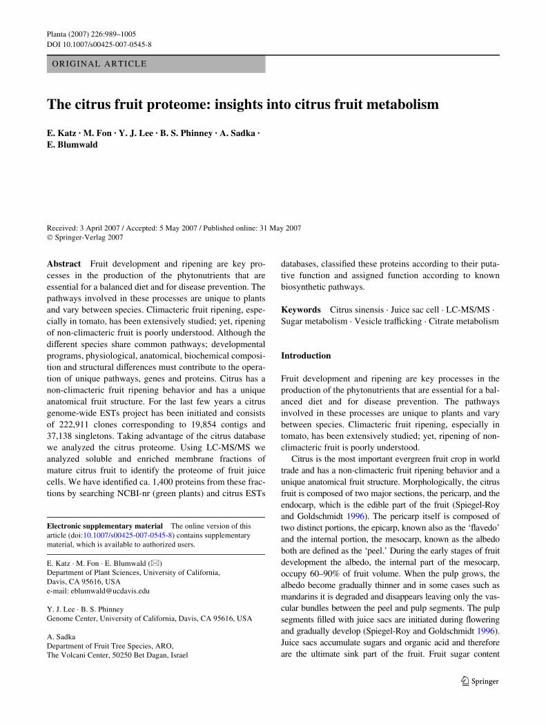
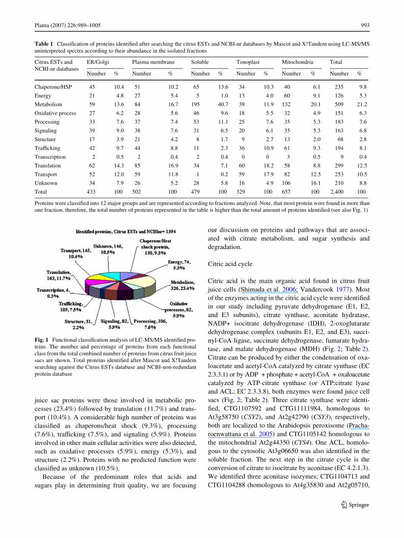
LC-MS/MS analysis of the citrus juice sacs proteinsresulted in the detection of 1,394 unique proteins. The pep-tide sequences were identiWed by searching the databaseswith uninterpreted fragment ion mass spectra using MAS-COT (Perkins et al. 1999). The search was conductedagainst the NCBI non-redundant protein (green plants)database and the Citrus ESTs database (http://cgf.ucda-vis.edu). From the 1,394 proteins identiWed, 433 were ER/Golgi-associated, 502 were PM-associated, 329 were asso-ciated with the tonoplast, 657 were mitochondria-associ-ated, and 479 were soluble proteins (Table 1). It should benoted that our experimental design precluded the diVerenti-ation between cytosolic and other soluble proteins locatedin the diVerent cell compartments’ milleau. The completelist of identiWed proteins is shown in Supplemental Table 1.The identiWed proteins were sorted into functional catego-ries (Fig. 1; Table 1) using TAIR (http://www.arabidopsis.org/), GO (http://www.geneontology.org/), UniProt (http://www.pir.uniprot.org/), Pfam (http://pfam.janelia.org/),InterPro (http://www.ebi.ac.uk/interpro/), and NCBI (http://www.ncbi.nlm.nih.gov/). The assignment of putative rolesinto known eukaryotic biosynthetic pathways was per-formed using KEGG (http://www.genome.jp/kegg/) andTAIR (AraCyc) (http://www.arabidopsis.org/biocyc/index.jsp). We assigned function to 1,247 proteins, while 146proteins remained unclassiWed. To simplify the discussionin this paper, proteins were classiWed into 12 major functionalgroups (Fig. 1; Table 1). The most abundant class of citrus
123
Planta (2007) 226:989–1005 993
juice sac proteins were those involved in metabolic pro-cesses (23.4%) followed by translation (11.7%) and trans-port (10.4%). A considerable high number of proteins wasclassiWed as chaperons/heat shock (9.3%), processing(7.6%), traYcking (7.5%), and signaling (5.9%). Proteinsinvolved in other main cellular activities were also detected,such as oxidative processes (5.9%), energy (5.3%), andstructure (2.2%). Proteins with no predicted function wereclassiWed as unknown (10.5%).
Because of the predominant roles that acids andsugars play in determining fruit quality, we are focusing
our discussion on proteins and pathways that are associ-ated with citrate metabolism, and sugar synthesis anddegradation.
Citric acid cycle
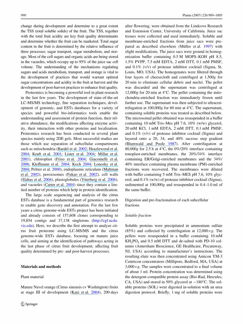
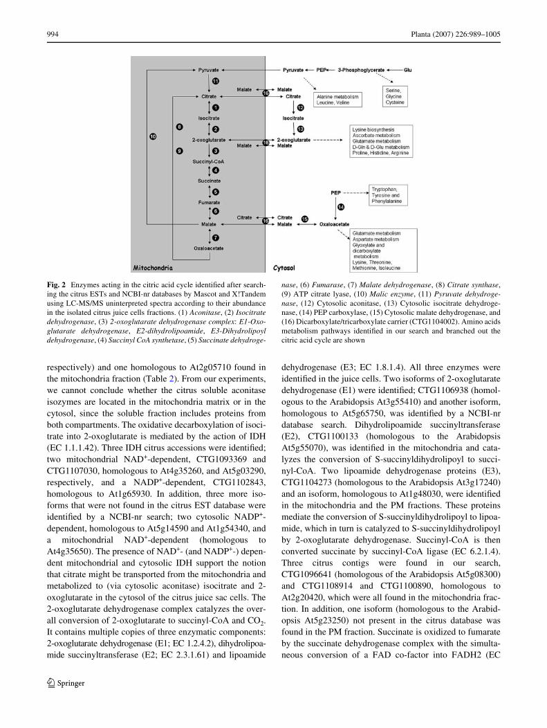
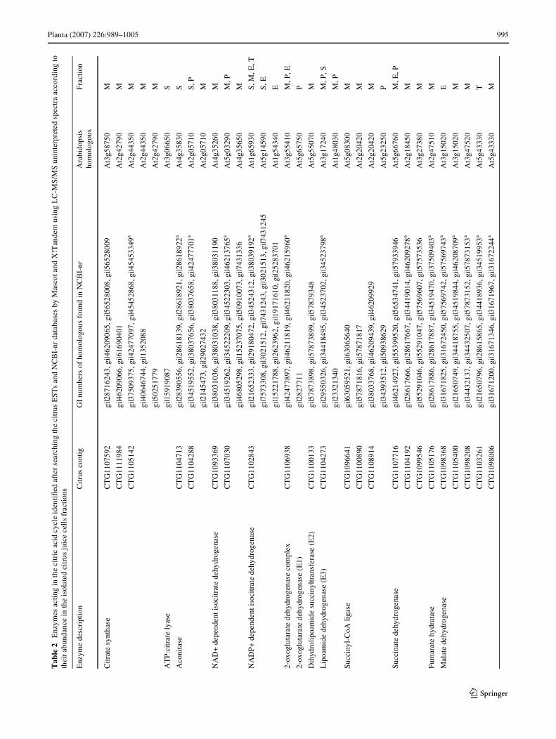
Citric acid is the main organic acid found in citrus fruitjuice cells (Shimada et al. 2006; Vandercook 1977). Mostof the enzymes acting in the citric acid cycle were identiWedin our study including pyruvate dehydrogenase (E1, E2,and E3 subunits), citrate synthase, aconitate hydratase,NADP+ isocitrate dehydrogenase (IDH), 2-oxoglutaratedehydrogenase complex (subunits E1, E2, and E3), succi-nyl-CoA ligase, succinate dehydrogenase, fumarate hydra-tase, and malate dehydrogenase (MDH) (Fig. 2; Table 2).Citrate can be produced by either the condensation of oxa-loacetate and acetyl-CoA catalyzed by citrate synthase (EC2.3.3.1) or by ADP + phosphate + acetyl-CoA + oxaloacetatecatalyzed by ATP-citrate synthase (or ATP:citrate lyaseand ACL; EC 2.3.3.8), both enzymes were found juice cellsacs (Fig. 2; Table 2). Three citrate synthase were identi-Wed, CTG1107592 and CTG11111984, homologous toAt3g58750 (CSY2), and At2g42790 (CSY3), respectively,both are localized to the Arabidopsis peroxisome (Pracha-roenwattana et al. 2005) and CTG1105142 homologous tothe mitochondrial At2g44350 (CYS4). One ACL, homolo-gous to the cytosolic At3g06650 was also identiWed in thesoluble fraction. The next step in the citrate cycle is theconversion of citrate to isocitrate by aconitase (EC 4.2.1.3).We identiWed three aconitase isozymes; CTG1104713 andCTG1104288 (homologous to At4g35830 and At2g05710,
Fig. 1 Functional classiWcation analysis of LC-MS/MS identiWed pro-teins. The number and percentage of proteins from each functionalclass from the total combined number of proteins from citrus fruit juicesacs are shown. Total proteins identiWed after Mascot and X!Tandemsearching against the Citrus ESTs database and NCBI-non-redundantprotein database
Table 1 ClassiWcation of proteins identiWed after searching the citrus ESTs and NCBI-nr databases by Mascot and X!Tandem using LC-MS/MSuninterpreted spectra according to their abundance in the isolated fractions
Proteins were classiWed into 12 major groups and are represented according to fractions analyzed. Note, that most protein were found in more thanone fraction, therefore, the total number of proteins represented in the table is higher than the total amount of proteins identiWed (see also Fig. 1)
Citrus ESTs and NCBI-nr databases
ER/Golgi Plasma membrane Soluble Tonoplast Mitochondria Total
Number % Number % Number % Number % Number % Number %
Chaperone/HSP 45 10.4 51 10.2 65 13.6 34 10.3 40 6.1 235 9.8
Energy 21 4.8 27 5.4 5 1.0 13 4.0 60 9.1 126 5.3
Metabolism 59 13.6 84 16.7 195 40.7 39 11.9 132 20.1 509 21.2
Oxidative process 27 6.2 28 5.6 46 9.6 18 5.5 32 4.9 151 6.3
Processing 33 7.6 37 7.4 53 11.1 25 7.6 35 5.3 183 7.6
Signaling 39 9.0 38 7.6 31 6.5 20 6.1 35 5.3 163 6.8
Structure 17 3.9 21 4.2 8 1.7 9 2.7 13 2.0 68 2.8
TraYcking 42 9.7 44 8.8 11 2.3 36 10.9 61 9.3 194 8.1
Transcription 2 0.5 2 0.4 2 0.4 0 0 3 0.5 9 0.4
Translation 62 14.3 85 16.9 34 7.1 60 18.2 58 8.8 299 12.5
Transport 52 12.0 59 11.8 1 0.2 59 17.9 82 12.5 253 10.5
Unknown 34 7.9 26 5.2 28 5.8 16 4.9 106 16.1 210 8.8
Total 433 100 502 100 479 100 329 100 657 100 2,400 100
123
994 Planta (2007) 226:989–1005
respectively) and one homologous to At2g05710 found inthe mitochondria fraction (Table 2). From our experiments,we cannot conclude whether the citrus soluble aconitaseisozymes are located in the mitochondria matrix or in thecytosol, since the soluble fraction includes proteins fromboth compartments. The oxidative decarboxylation of isoci-trate into 2-oxoglutarate is mediated by the action of IDH(EC 1.1.1.42). Three IDH citrus accessions were identiWed;two mitochondrial NAD+-dependent, CTG1093369 andCTG1107030, homologous to At4g35260, and At5g03290,respectively, and a NADP+-dependent, CTG1102843,homologous to At1g65930. In addition, three more iso-forms that were not found in the citrus EST database wereidentiWed by a NCBI-nr search; two cytosolic NADP+-dependent, homologous to At5g14590 and At1g54340, anda mitochondrial NAD+-dependent (homologous toAt4g35650). The presence of NAD+- (and NADP+-) depen-dent mitochondrial and cytosolic IDH support the notionthat citrate might be transported from the mitochondria andmetabolized to (via cytosolic aconitase) isocitrate and 2-oxoglutarate in the cytosol of the citrus juice sac cells. The2-oxoglutarate dehydrogenase complex catalyzes the over-all conversion of 2-oxoglutarate to succinyl-CoA and CO2.It contains multiple copies of three enzymatic components:2-oxoglutarate dehydrogenase (E1; EC 1.2.4.2), dihydrolipoa-mide succinyltransferase (E2; EC 2.3.1.61) and lipoamide
dehydrogenase (E3; EC 1.8.1.4). All three enzymes wereidentiWed in the juice cells. Two isoforms of 2-oxoglutaratedehydrogenase (E1) were identiWed; CTG1106938 (homol-ogous to the Arabidopsis At3g55410) and another isoform,homologous to At5g65750, was identiWed by a NCBI-nrdatabase search. Dihydrolipoamide succinyltransferase(E2), CTG1100133 (homologous to the ArabidopsisAt5g55070), was identiWed in the mitochondria and cata-lyzes the conversion of S-succinyldihydrolipoyl to succi-nyl-CoA. Two lipoamide dehydrogenase proteins (E3),CTG1104273 (homologous to the Arabidopsis At3g17240)and an isoform, homologous to At1g48030, were identiWedin the mitochondria and the PM fractions. These proteinsmediate the conversion of S-succinyldihydrolipoyl to lipoa-mide, which in turn is catalyzed to S-succinyldihydrolipoylby 2-oxoglutarate dehydrogenase. Succinyl-CoA is thenconverted succinate by succinyl-CoA ligase (EC 6.2.1.4).Three citrus contigs were found in our search,CTG1096641 (homologous of the Arabidopsis At5g08300)and CTG1108914 and CTG1100890, homologous toAt2g20420, which were all found in the mitochondria frac-tion. In addition, one isoform (homologous to the Arabid-opsis At5g23250) not present in the citrus database wasfound in the PM fraction. Succinate is oxidized to fumarateby the succinate dehydrogenase complex with the simulta-neous conversion of a FAD co-factor into FADH2 (EC
Fig. 2 Enzymes acting in the citric acid cycle identiWed after search-ing the citrus ESTs and NCBI-nr databases by Mascot and X!Tandemusing LC-MS/MS uninterpreted spectra according to their abundancein the isolated citrus juice cells fractions. (1) Aconitase, (2) Isocitratedehydrogenase, (3) 2-oxoglutarate dehydrogenase complex: E1-Oxo-glutarate dehydrogenase, E2-dihydrolipoamide, E3-Dihydrolipoyldehydrogenase, (4) Succinyl CoA synthetase, (5) Succinate dehydroge-
nase, (6) Fumarase, (7) Malate dehydrogenase, (8) Citrate synthase,(9) ATP citrate lyase, (10) Malic enzyme, (11) Pyruvate dehydroge-nase, (12) Cytosolic aconitase, (13) Cytosolic isocitrate dehydroge-nase, (14) PEP carboxylase, (15) Cytosolic malate dehydrogenase, and(16) Dicarboxylate/tricarboxylate carrier (CTG1104002). Amino acidsmetabolism pathways identiWed in our search and branched out thecitric acid cycle are shown
123
Planta (2007) 226:989–1005 995
Tab
le2
Enz
ymes
act
ing
in th
e ci
tric
aci
d cy
cle
iden
tiW
ed a
fter
sea
rchi
ng th
e ci
trus
EST
s an
d N
CB
I-nr
dat
abas
es b
y M
asco
t and
X!T
ande
m u
sing
LC
-MS/
MS
unin
terp
rete
d sp
ectr
a ac
cord
ing
toth
eir
abun
danc
e in
the
isol
ated
cit
rus
juic
e ce
lls
frac
tions
Enz
yme
desc
ript
ion
Citr
us c
ontig
GI
num
bers
of
hom
olog
ous
foun
d in
NC
BI-
nrA
rabi
dops
is
hom
olog
ous
Fra
ctio
n
Citr
ate
synt
hase
CT
G11
0759
2gi
|287
1624
3, g
i|462
0906
5, g
i|565
2800
8, g
i|565
2800
9A
t3g5
8750
M
CT
G11
1198
4gi
|462
0906
6, g
i|616
9040
1A
t2g4
2790
M
CT
G11
0514
2gi
|375
0937
5, g
i|424
7709
7, g
i|454
5286
8, g
i|454
5334
9aA
t2g4
4350
M
gi|4
0646
744,
gi|1
3520
88A
t2g4
4350
M
gi|5
0251
779
At2
g427
90M
AT
P:ci
trat
e ly
ase
gi|1
5919
087
At3
g066
50S
Aco
nita
seC
TG
1104
713
gi|2
8390
556,
gi|2
8618
139,
gi|2
8618
921,
gi|2
8618
922a
At4
g358
30S
CT
G11
0428
8gi
|345
1955
2, g
i|380
3765
6, g
i|380
3765
8, g
i|424
7770
1aA
t2g0
5710
S, P
gi|2
1454
73, g
i|290
2743
2A
t2g0
5710
M
NA
D+
dep
ende
nt is
ocitr
ate
dehy
drog
enas
eC
TG
1093
369
gi|3
8031
036,
gi|3
8031
038,
gi|3
8031
188,
gi|3
8031
190
At4
g352
60M
CT
G11
0703
0gi
|345
1926
2, g
i|345
2220
9, g
i|345
2230
3, g
i|462
1376
5aA
t5g0
3290
M, P
gi|4
6805
298,
gi|1
5237
075,
gi|5
0910
073,
gi|7
4313
36A
t4g3
5650
M
NA
DP+
dep
ende
nt is
ocitr
ate
dehy
drog
enas
eC
TG
1102
843
gi|2
1652
333,
gi|2
9180
472,
gi|3
4524
312,
gi|3
8039
192a
At1
g659
30S
, M, E
, T
gi|7
5733
08, g
i|302
1512
, gi|7
4312
43, g
i|302
1513
, gi|7
4312
45A
t5g1
4590
S, E
gi|1
5221
788,
gi|2
6239
62, g
i|191
7161
0, g
i|252
8370
1A
t1g5
4340
E
2-ox
oglu
tara
te d
ehyd
roge
nase
com
plex
CT
G11
0693
8gi
|424
7789
7, g
i|462
1181
9, g
i|462
1182
0, g
i|462
1596
0aA
t3g5
5410
M, P
, E
2-ox
oglu
tara
te d
ehyd
roge
nase
(E
1)gi
|282
7711
At5
g657
50P
Dih
ydro
lipoa
mid
e su
ccin
yltr
ansf
eras
e (E
2)C
TG
1100
133
gi|5
7873
898,
gi|5
7873
899,
gi|5
7879
348
At5
g550
70M
Lip
oam
ide
dehy
drog
enas
e (E
3)C
TG
1104
273
gi|2
9550
326,
gi|3
4418
495,
gi|3
4523
702,
gi|3
4523
798a
At3
g172
40M
, P, S
gi|2
3321
340
At1
g480
30M
, P
Succ
inyl
-CoA
liga
seC
TG
1096
641
gi|6
3059
521,
gi|6
3065
640
At5
g083
00M
CT
G11
0089
0gi
|578
7181
6, g
i|578
7181
7A
t2g2
0420
M
CT
G11
0891
4gi
|380
3376
8, g
i|462
0943
9, g
i|462
0992
9A
t2g2
0420
M
gi|3
4393
512,
gi|5
0938
629
At5
g232
50P
Succ
inat
e de
hydr
ogen
ase
CT
G11
0771
6gi
|462
1492
7, g
i|553
9952
0, g
i|565
3474
1, g
i|579
3394
6A
t5g6
6760
M, E
, P
CT
G11
0419
2gi
|286
1766
6, g
i|286
1766
7, g
i|344
1901
4, g
i|462
0927
8aA
t2g1
8450
M
CT
G10
9954
6gi
|552
9104
6, g
i|552
9104
7, g
i|575
6960
7, g
i|575
7353
6A
t3g2
7380
M
Fum
arat
e hy
drat
ase
CT
G11
0517
6gi
|286
1788
6, g
i|286
1788
7, g
i|345
1947
0, g
i|375
0940
3aA
t2g4
7510
M
Mal
ate
dehy
drog
enas
eC
TG
1098
368
gi|3
1671
825,
gi|3
1672
450,
gi|5
7569
742,
gi|5
7569
743a
At3
g150
20E
CT
G11
0540
0gi
|216
5074
9, g
i|344
1875
5, g
i|345
1984
4, g
i|462
0870
9aA
t3g1
5020
M
CT
G10
9820
8gi
|344
3213
7, g
i|344
3250
7, g
i|578
7315
2, g
i|578
7315
3aA
t3g4
7520
M
CT
G11
0326
1gi
|216
5079
6, g
i|286
1586
5, g
i|344
1893
6, g
i|345
1995
3aA
t5g4
3330
T
CT
G10
9800
6gi
|316
7120
0, g
i|316
7134
6, g
i|316
7196
7, g
i|316
7224
4aA
t5g4
3330
M
123
996 Planta (2007) 226:989–1005
Tab
le2
cont
inue
d
M m
itoc
hond
ria,
S s
olub
le, P
pla
sma
mem
bran
e, T
tono
plas
t, E
ER
/Gol
gia
For
man
y of
the
pept
ide
mas
s sp
ectr
a th
ere
wer
e ad
ditio
nal m
atch
ing
acce
ssio
ns in
the
data
base
s. F
or th
e fu
ll li
st o
f m
atch
ing
prot
eins
see
sup
plem
enta
l Tab
le 1
Enz
yme
desc
ript
ion
Cit
rus
cont
igG
I nu
mbe
rs o
f ho
mol
ogou
s fo
und
in N
CB
I-nr
Ara
bido
psis
ho
mol
ogou
sF
ract
ion
gi|1
5241
21, g
i|228
6153
, gi|2
8270
82, g
i|182
0248
5, g
i|743
1164
aA
t1g0
4410
S
gi|7
4311
79, g
i|282
7078
At2
g227
80M
gi|5
0834
461,
gi|6
4691
39, g
i|605
9347
6, g
i|605
9349
4, g
i|126
894
At5
g096
60M
Mal
ic e
nzym
eC
TG
1095
849
gi|6
3055
838,
gi|6
3056
722,
gi|6
3056
817,
gi|6
3058
812a
At2
g199
00T
, P, S
gi|1
3464
85, g
i|228
412,
gi|2
0469
At1
g797
50S
CT
G11
0905
0gi
|216
5083
1, g
i|345
1899
7, g
i|579
3214
9A
t4g0
0570
M, P
Pyr
uvat
e de
hydr
ogen
ase
CT
G10
9517
5gi
|624
2855
7, g
i|624
2902
5, g
i|681
3898
6A
t1g5
9900
S, E
, P
Com
plex
CT
G11
0432
0gi
|286
1851
1, g
i|287
1605
1, g
i|345
1945
1, g
i|345
2013
8aA
t1g5
9900
M
Pyr
uvat
e de
hydr
ogen
ase
(E1)
CT
G11
0433
0gi
|216
5084
0, g
i|286
1794
2, g
i|286
1794
3, g
i|295
5086
8aA
t5g5
0850
M
gi|1
7094
54A
t5g5
0850
M
gi|5
0948
007
At5
g508
50M
Dih
ydro
lipo
amid
e ac
etyl
tran
sfer
ase
(E2)
gi|1
1994
364,
gi|1
8400
212,
gi|2
0260
138
At3
g139
30M
Dih
ydro
lipo
amid
e de
hydr
ogen
ase
(E3)
CT
G11
0427
3gi
|295
5032
6, g
i|344
1849
5, g
i|345
2370
2, g
i|345
2379
8aA
t3g1
7240
P, M
, S
gi|1
4916
975
At1
g480
30P
gi|2
3321
340
At1
g480
30M
123
Planta (2007) 226:989–1005 997
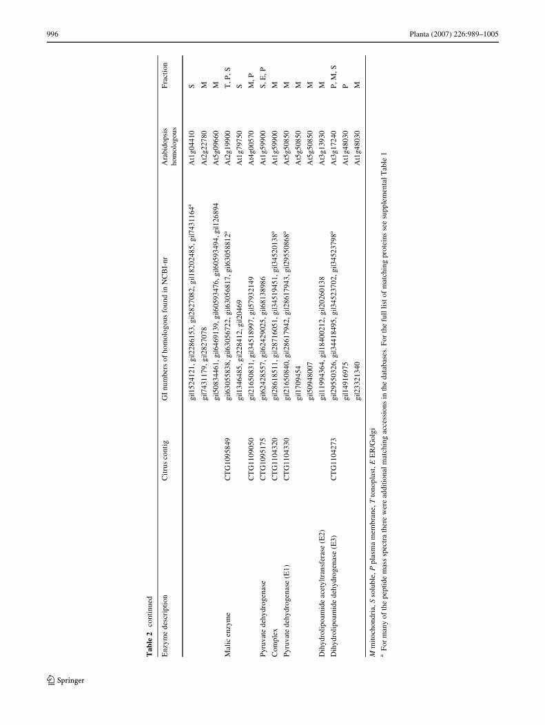
1.3.5.1). We identiWed three proteins assigned as citrusESTs CTG1107716, CTG1104192, and CTG1099546, whichare homologous to the Arabidopsis mitochondrial succinatedehydrogenase At5g66760, At2g18450, and At3g27380,respectively. We also identiWed fumarate hydratase, a mito-chondrial protein encoded by CTG1105176 (homologousto the Arabidopsis At2g47510) that mediates the intercon-version of fumarate to malate. The last step of the pathway,the interconversion of malate to oxaloacetate utilizingNAD+/NADH is catalyzed by MDH (EC 1.1.1.37). Ineukaryotic cells, MDH is usually found in the mitochon-drial matrix and in the cytosol. We identiWed Wve citruscontigs CTG1098368 (soluble) and CTG1105400 (mito-chondrial), both homologous to At3g15020, CTG1098208(homologous to the mitochondrial At3g47520), andCTG1103261 (soluble) and CTG1098006 (mitochondrial)(both homologous to At5g43330). Three additional MDHisoforms homologous to At1g04410 (cytosolic),At2g22780 and At5g09660 (both mitochondrial) wereidentiWed by NCBI-nr search. Most of these proteins werefound mainly in the mitochondria-enriched fraction and inthe soluble fraction.
In addition to the conversion of malate to oxaloacetate,mediated by MDH, malate can also be converted to pyruvateby malic enzyme (ME). Two types of ME are known, NAD-dependent (EC 1.1.1.39) catalyzing the reaction:malate + NAD+ = pyruvate + CO2 + NADH, and NADP-dependent (EC 1.1.1.40): malate + NADP+ = pyruvate+ CO2 +NADPH) (Wheeler et al. 2005). In plants, NAD-dependent isoforms function predominantly in the mitochon-dria while the NADP-dependent isoforms are found in thecytosol and plastids. We identiWed few MEs, CTG1095849(At2g19900 homologous) and an homologous to At1g79750found in the soluble fraction, both are NADP-dependenttype, and known as AtNADP-ME1 and AtNADP-ME4,respectively (Wheeler et al. 2005). CTG1109050 (homolo-gous to At4g00570), an NAD-dependent type, was alsofound in the mitochondria fraction. ME enables plants mito-chondria to metabolize PEP derived from glycolysis via analternative pathway. Malate can be synthesized from PEP inthe cytosol via PEP carboxylase and MDH (Fig. 2). Thesesuggest that some of the MDHs that were identiWed in ourstudy (see above) are cytosolic and act in this pathway. Inaddition, four PEP-carboxylase were identiWed, two in thecitrus database search, CTG1095231 (homologous toAt4g37870) and CTG1105152 (homologous to At2g42600)and two, homologous to At3g14940 and At1g53310 wereidentiWed in the NCBI-nr database search (SupplementalTable 1). In addition, we identiWed a dicarboxylate/tricarb-oxylate carrier or mitochondrial 2-oxoglutarate/malate carrierprotein, CTG1104002 (homologous to At5g19760), whichmight be responsible for transporting malate, 2-oxoglutarateand citrate across the mitochondria inner membrane (Fig. 2).
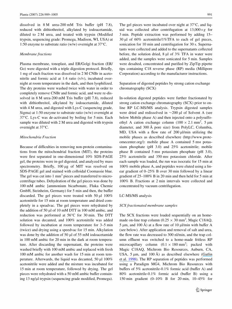
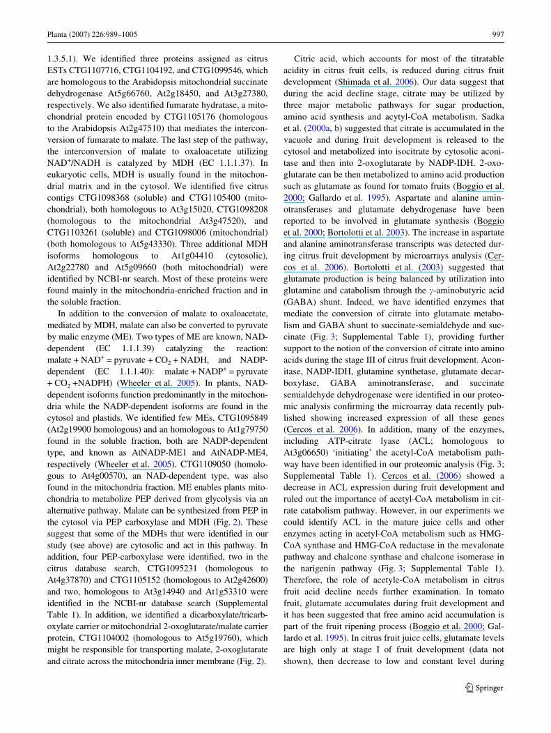
Citric acid, which accounts for most of the titratableacidity in citrus fruit cells, is reduced during citrus fruitdevelopment (Shimada et al. 2006). Our data suggest thatduring the acid decline stage, citrate may be utilized bythree major metabolic pathways for sugar production,amino acid synthesis and acytyl-CoA metabolism. Sadkaet al. (2000a, b) suggested that citrate is accumulated in thevacuole and during fruit development is released to thecytosol and metabolized into isocitrate by cytosolic aconi-tase and then into 2-oxoglutarate by NADP-IDH. 2-oxo-glutarate can be then metabolized to amino acid productionsuch as glutamate as found for tomato fruits (Boggio et al.2000; Gallardo et al. 1995). Aspartate and alanine amin-otransferases and glutamate dehydrogenase have beenreported to be involved in glutamate synthesis (Boggioet al. 2000; Bortolotti et al. 2003). The increase in aspartateand alanine aminotransferase transcripts was detected dur-ing citrus fruit development by microarrays analysis (Cer-cos et al. 2006). Bortolotti et al. (2003) suggested thatglutamate production is being balanced by utilization intoglutamine and catabolism through the �-aminobutyric acid(GABA) shunt. Indeed, we have identiWed enzymes thatmediate the conversion of citrate into glutamate metabo-lism and GABA shunt to succinate-semialdehyde and suc-cinate (Fig. 3; Supplemental Table 1), providing furthersupport to the notion of the conversion of citrate into aminoacids during the stage III of citrus fruit development. Acon-itase, NADP-IDH, glutamine synthetase, glutamate decar-boxylase, GABA aminotransferase, and succinatesemialdehyde dehydrogenase were identiWed in our proteo-mic analysis conWrming the microarray data recently pub-lished showing increased expression of all these genes(Cercos et al. 2006). In addition, many of the enzymes,including ATP-citrate lyase (ACL; homologous toAt3g06650) ‘initiating’ the acetyl-CoA metabolism path-way have been identiWed in our proteomic analysis (Fig. 3;Supplemental Table 1). Cercos et al. (2006) showed adecrease in ACL expression during fruit development andruled out the importance of acetyl-CoA metabolism in cit-rate catabolism pathway. However, in our experiments wecould identify ACL in the mature juice cells and otherenzymes acting in acetyl-CoA metabolism such as HMG-CoA synthase and HMG-CoA reductase in the mevalonatepathway and chalcone synthase and chalcone isomerase inthe narigenin pathway (Fig. 3; Supplemental Table 1).Therefore, the role of acetyle-CoA metabolism in citrusfruit acid decline needs further examination. In tomatofruit, glutamate accumulates during fruit development andit has been suggested that free amino acid accumulation ispart of the fruit ripening process (Boggio et al. 2000; Gal-lardo et al. 1995). In citrus fruit juice cells, glutamate levelsare high only at stage I of fruit development (data notshown), then decrease to low and constant level during
123
998 Planta (2007) 226:989–1005
stages II and III (Cercos et al. 2006). Thus, glutamate utili-zation during fruit development is an important step in thehomeostatic control of glutamate in the mature fruit. Twopathways for glutamate utilization were suggested: conver-sion of glutamate to glutamine and GABA shunt catabolismpathway (Cercos et al. 2006). Our proteomic data supportthis hypothesis revealing the expression of glutaminesynthase acting in the glutamine synthesis pathway andglutamate decarboxylase, GABA amino transferase andsuccinate semialdehyde dehydrogenase in the GABA shuntpathway (Fig. 3; Supplemental Table 1).
Sugar synthesis transport and metabolism
In addition to acid content, sugar content and sugar metabo-lism are major contributors to fruit quality. Sugars aretranslocated by the phloem from source to sink tissues anduploaded into cells either symplasticaly or apoplasticaly byplasmodesmata or the action of sugar transporters, respec-tively (Lalonde et al. 2004). Phloem unloading is a key pro-cess in sugars partitioning because to a large extent itdetermines the movement of assimilates from the sieve ele-ments to the recipient sink cells (Patrick 1997). It has beenshown that unloading routes may diVer according to sinktype, sink development stage, sink function, growth condi-tions, and alternative unloading pathways may even exist insinks with symplastically interconnecting phloem (Oparkaand Turgeon 1999; Patrick 1997; Roberts et al. 1997; Violaet al. 2001). Other studies showed that plasmodesmatalconductivity can be programmatically reduced (Baluskaet al. 2001; Itaya et al. 2002) and that sugar transporters canoperate in parallel to predominant symplastic phloem path-ways (Kuhn et al. 2003). In tomato fruits, phloem unload-ing pathway is symplastic during early stages, andapoplastic during later fruit developmental stages (Ruan
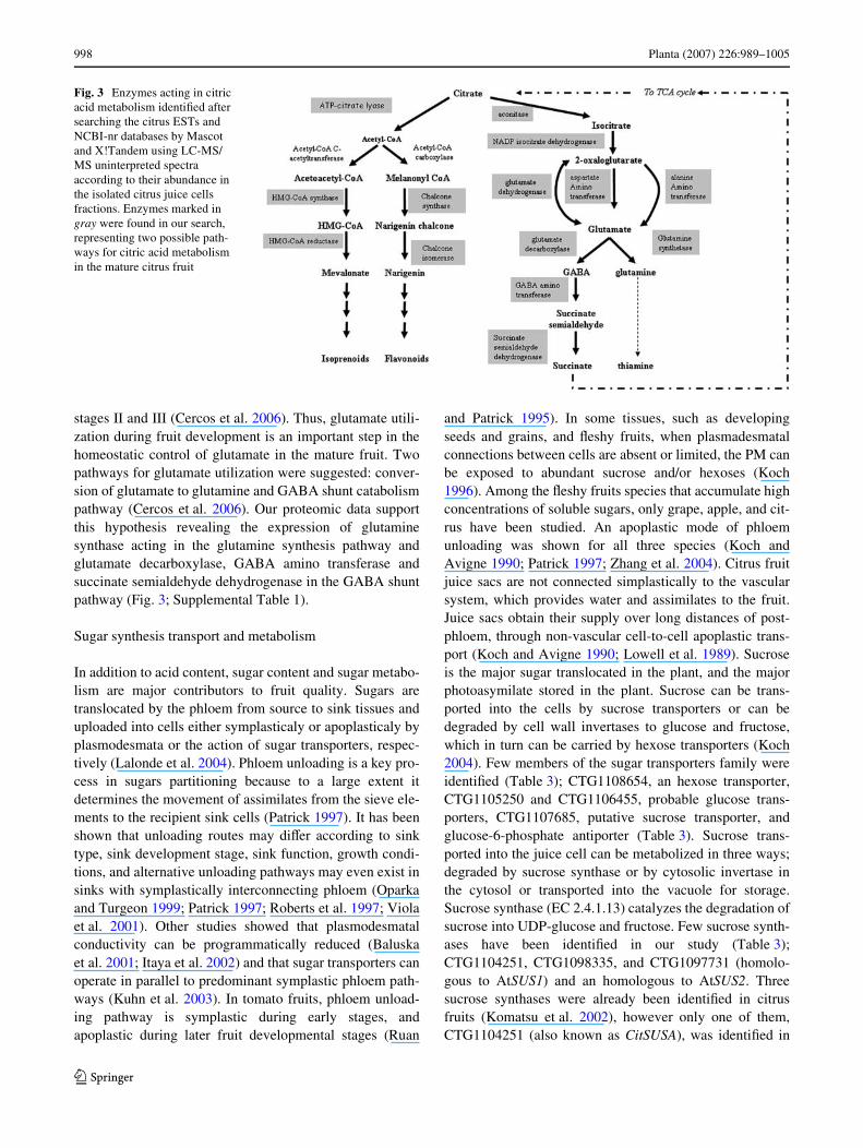
and Patrick 1995). In some tissues, such as developingseeds and grains, and Xeshy fruits, when plasmadesmatalconnections between cells are absent or limited, the PM canbe exposed to abundant sucrose and/or hexoses (Koch1996). Among the Xeshy fruits species that accumulate highconcentrations of soluble sugars, only grape, apple, and cit-rus have been studied. An apoplastic mode of phloemunloading was shown for all three species (Koch andAvigne 1990; Patrick 1997; Zhang et al. 2004). Citrus fruitjuice sacs are not connected simplastically to the vascularsystem, which provides water and assimilates to the fruit.Juice sacs obtain their supply over long distances of post-phloem, through non-vascular cell-to-cell apoplastic trans-port (Koch and Avigne 1990; Lowell et al. 1989). Sucroseis the major sugar translocated in the plant, and the majorphotoasymilate stored in the plant. Sucrose can be trans-ported into the cells by sucrose transporters or can bedegraded by cell wall invertases to glucose and fructose,which in turn can be carried by hexose transporters (Koch2004). Few members of the sugar transporters family wereidentiWed (Table 3); CTG1108654, an hexose transporter,CTG1105250 and CTG1106455, probable glucose trans-porters, CTG1107685, putative sucrose transporter, andglucose-6-phosphate antiporter (Table 3). Sucrose trans-ported into the juice cell can be metabolized in three ways;degraded by sucrose synthase or by cytosolic invertase inthe cytosol or transported into the vacuole for storage.Sucrose synthase (EC 2.4.1.13) catalyzes the degradation ofsucrose into UDP-glucose and fructose. Few sucrose synth-ases have been identiWed in our study (Table 3);CTG1104251, CTG1098335, and CTG1097731 (homolo-gous to AtSUS1) and an homologous to AtSUS2. Threesucrose synthases were already been identiWed in citrusfruits (Komatsu et al. 2002), however only one of them,CTG1104251 (also known as CitSUSA), was identiWed in
Fig. 3 Enzymes acting in citric acid metabolism identiWed after searching the citrus ESTs and NCBI-nr databases by Mascot and X!Tandem using LC-MS/MS uninterpreted spectra according to their abundance in the isolated citrus juice cells fractions. Enzymes marked in gray were found in our search, representing two possible path-ways for citric acid metabolism in the mature citrus fruit
123
Planta (2007) 226:989–1005 999
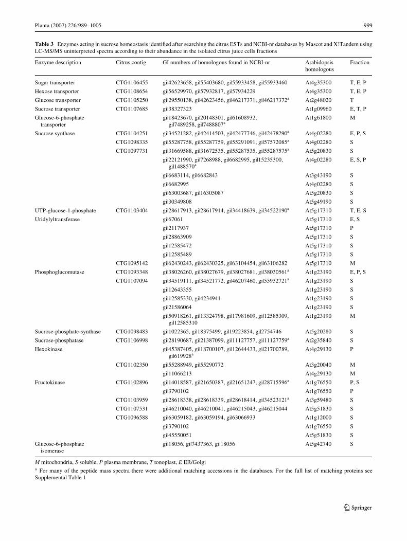
Table 3 Enzymes acting in sucrose homeostasis identiWed after searching the citrus ESTs and NCBI-nr databases by Mascot and X!Tandem usingLC-MS/MS uninterpreted spectra according to their abundance in the isolated citrus juice cells fractions
M mitochondria, S soluble, P plasma membrane, T tonoplast, E ER/Golgia For many of the peptide mass spectra there were additional matching accessions in the databases. For the full list of matching proteins seeSupplemental Table 1
Enzyme description Citrus contig GI numbers of homologous found in NCBI-nr Arabidopsis homologous
Fraction
Sugar transporter CTG1106455 gi|42623658, gi|55403680, gi|55933458, gi|55933460 At4g35300 T, E, P
Hexose transporter CTG1108654 gi|56529970, gi|57932817, gi|57934229 At4g35300 T, E, P
Glucose transporter CTG1105250 gi|29550138, gi|42623456, gi|46217371, gi|46217372a At2g48020 T
Sucrose transporter CTG1107685 gi|38327323 At1g09960 E, T, P
Glucose-6-phosphate transporter
gi|18423670, gi|20148301, gi|61608932, gi|7489258, gi|7488807a
At1g61800 M
Sucrose synthase CTG1104251 gi|34521282, gi|42414503, gi|42477746, gi|42478290a At4g02280 E, P, S
CTG1098335 gi|55287758, gi|55287759, gi|55291091, gi|57572085a At4g02280 S
CTG1097731 gi|31669588, gi|31672535, gi|55287535, gi|55287575a At5g20830 S
gi|22121990, gi|7268988, gi|6682995, gi|15235300, gi|1488570a
At4g02280 E, S, P
gi|6683114, gi|6682843 At3g43190 S
gi|6682995 At4g02280 S
gi|63003687, gi|16305087 At5g20830 S
gi|30349808 At5g49190 S
UTP-glucose-1-phosphate CTG1103404 gi|28617913, gi|28617914, gi|34418639, gi|34522190a At5g17310 T, E, S
Uridylyltransferase gi|67061 At5g17310 E, S
gi|2117937 At5g17310 P
gi|28863909 At5g17310 S
gi|12585472 At5g17310 S
gi|12585489 At5g17310 S
CTG1095142 gi|62430243, gi|62430325, gi|63104454, gi|63106282 At5g17310 M
Phosphoglucomutase CTG1093348 gi|38026260, gi|38027679, gi|38027681, gi|38030561a At1g23190 E, P, S
CTG1107094 gi|34519111, gi|34521772, gi|46207460, gi|55932721a At1g23190 S
gi|12643355 At1g23190 S
gi|12585330, gi|4234941 At1g23190 S
gi|21586064 At1g23190 S
gi|50918261, gi|13324798, gi|17981609, gi|12585309, gi|12585310
At1g23190 M
Sucrose-phosphate-synthase CTG1098483 gi|1022365, gi|18375499, gi|19223854, gi|2754746 At5g20280 S
Sucrose-phosphatase CTG1106998 gi|28190687, gi|21387099, gi|11127757, gi|11127759a At2g35840 S
Hexokinase gi|45387405, gi|18700107, gi|12644433, gi|21700789, gi|619928a
At4g29130 P
CTG1102350 gi|55288949, gi|55290772 At3g20040 M
gi|11066213 At4g29130 M
Fructokinase CTG1102896 gi|14018587, gi|21650387, gi|21651247, gi|28715596a At1g76550 P, S
gi|3790102 At1g76550 P
CTG1103959 gi|28618338, gi|28618339, gi|28618414, gi|34523121a At3g59480 S
CTG1107531 gi|46210040, gi|46210041, gi|46215043, gi|46215044 At5g51830 S
CTG1096588 gi|63059182, gi|63059194, gi|63066933 At1g12000 S
gi|3790102 At1g76550 S
gi|45550051 At5g51830 S
Glucose-6-phosphate isomerase
gi|18056, gi|7437363, gi|18056 At5g42740 S
123
1000 Planta (2007) 226:989–1005
our study. Komatsu et al. (2002) showed that only CitSUSA(CTG1104251) was expressed in mature fruit. However,our data show that citrus has at least two more genes,homologous to the Arabidopsis SUS1 and SUS2 that areexpressed in the mature fruit. Invertases, on the other hand,were not identiWed in mature juice sac cells, in agreementwith previous data shown a decrease in invertase expres-sion and activity during citrus fruit maturation (Echeverriaand Burns 1990; Holland et al. 1999; Lowell et al. 1989;Tomlinson et al. 1991). Our Wndings agree with the invert-ase/sucrose synthase control hypothesis suggesting thatinvertases mediate the initiation and expansion of manynew sinks and later transition to storage and maturationphase is facilitated by shifts from invertase to sucrose syn-thase paths of sucrose cleavage (Koch 2004).
Sucrose, in turn, is derived from hexose phosphatesthrough the combined actions of UTP-glucose-1-phosphateuridylyltransferase (UDP-glucose pyrophosphorylase, EC2.7.7.9), sucrose phosphate synthase (SPS) (EC 2.4.14) andsucrose phosphatase (EC 3.1.3.24) (Fig. 4; Table 3). Hex-ose phosphates are not only common intermediates to thepathways of synthesis and degradation of most carbohy-drates, but they are also the point of convergence of thesepathways. Hexose phosphates are derived either from thebreakdown of sugars and polysaccharides or from triose
phosphates formed during photosynthesis and gluconeo-genesis. They may be used for the synthesis of carbohy-drates or for metabolism through the glycolitic and pentosephosphate pathways. The hexose phosphate pool consists ofthree metabolic intermediates: glucose 1-phosphate, glu-cose 6-phosphate, and fructose 6-phosphate. The threemetabolites are kept in equilibrium through the reversibleaction of phosphoglucomutase and glucose-6-phosphateisomerase. Glucose phosphomutase (EC 5.4.2.2;CTG1093348 and CTG1107094; Table 3), which was alsoidentiWed in our study, is an enzyme responsible for theconversion of D-glucose 1-phosphate into D-glucose 6-phosphate. Glucose phosphomutase participates in both thebreakdown and synthesis of glucose and also in starchmetabolism together with starch phosphorylase (EC2.4.1.1; At3g46970), which act in starch degradation. Glu-cose-6-phosphate isomerase, which is involved in glycoly-sis and in gluconeogenesis and catalyse the conversion ofD-glucose 6-phosphate to D-fructose 6-phosphate was alsoidentiWed (Table 3).
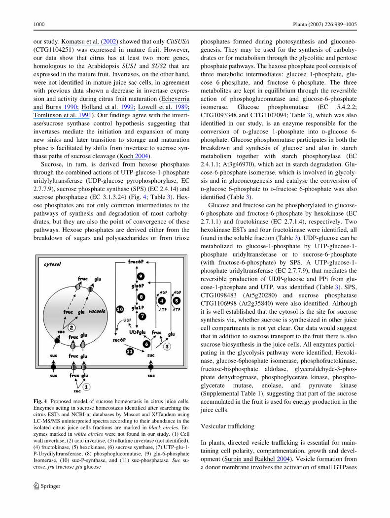
Glucose and fructose can be phosphorylated to glucose-6-phosphate and fructose-6-phosphate by hexokinase (EC2.7.1.1) and fructokinase (EC 2.7.1.4), respectively. Twohexokinase ESTs and four fructokinase were identiWed, allfound in the soluble fraction (Table 3). UDP-glucose can bemetabolized to glucose-1-phosphate by UTP-glucose-1-phosphate uridyltransferase or to sucrose-6-phosphate(with fructose-6-phosphate) by SPS. A UTP-glucose-1-phosphate uridyltransferase (EC 2.7.7.9), that mediates thereversible production of UDP-glucose and PPi from glu-cose-1-phosphate and UTP, was identiWed (Table 3). SPS,CTG1098483 (At5g20280) and sucrose phosphataseCTG1106998 (At2g35840) were also identiWed. Althoughit is well established that the cytosol is the site for sucrosesynthesis via, whether sucrose is synthesized in other juicecell compartments is not yet clear. Our data would suggestthat in addition to sucrose transport to the fruit there is alsosucrose biosynthesis in the juice cells. All enzymes partici-pating in the glycolysis pathway were identiWed; Hexoki-nase, glucose-6phosphate isomerase, phosphofructokinase,fructose-bisphosphate aldolase, glyceraldehyde-3-phos-phate dehydrogrnase, phosphoglycerate kinase, phospho-glycerate mutase, enolase, and pyruvate kinase(Supplemental Table 1), suggesting that part of the sucroseaccumulated in the fruit is used for energy production in thejuice cells.
Vesicular traYcking
In plants, directed vesicle traYcking is essential for main-taining cell polarity, compartmentation, growth and devel-opment (Surpin and Raikhel 2004). Vesicle formation froma donor membrane involves the activation of small GTPases
Fig. 4 Proposed model of sucrose homeostasis in citrus juice cells.Enzymes acting in sucrose homeostasis identiWed after searching thecitrus ESTs and NCBI-nr databases by Mascot and X!Tandem usingLC-MS/MS uninterpreted spectra according to their abundance in theisolated citrus juice cells fractions are marked in black circles. En-zymes marked in white circles were not found in our study. (1) Cellwall invertase, (2) acid invertase, (3) alkaline invertase (not identiWed),(4) fructokinase, (5) hexokinase, (6) sucrose synthase, (7) UTP-glu-1-P-Urydilyltransferase, (8) phosphoglucomutase, (9) glu-6-phosphateIsomerase, (10) suc-P-synthase, and (11) suc-phosphatase. Suc su-crose, fru fructose glu glucose
123
Planta (2007) 226:989–1005 1001
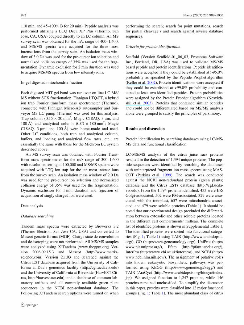
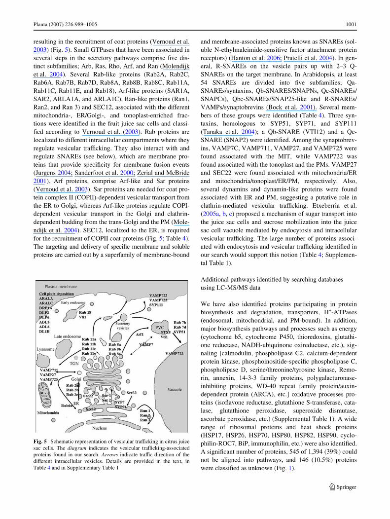
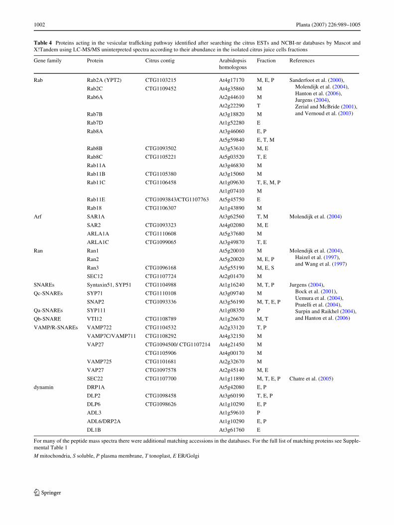
resulting in the recruitment of coat proteins (Vernoud et al.2003) (Fig. 5). Small GTPases that have been associated inseveral steps in the secretory pathways comprise Wve dis-tinct subfamilies; Arb, Ras, Rho, Arf, and Ran (Molendijket al. 2004). Several Rab-like proteins (Rab2A, Rab2C,Rab6A, Rab7B, Rab7D, Rab8A, Rab8B, Rab8C, Rab11A,Rab11C, Rab11E, and Rab18), Arf-like proteins (SAR1A,SAR2, ARLA1A, and ARLA1C), Ran-like proteins (Ran1,Ran2, and Ran 3) and SEC12, associated with the diVerentmitochondria-, ER/Golgi-, and tonoplast-enriched frac-tions were identiWed in the fruit juice sac cells and classi-Wed according to Vernoud et al. (2003). Rab proteins arelocalized to diVerent intracellular compartments where theyregulate vesicular traYcking. They also interact with andregulate SNAREs (see below), which are membrane pro-teins that provide speciWcity for membrane fusion events(Jurgens 2004; Sanderfoot et al. 2000; Zerial and McBride2001). Arf proteins, comprise Arf-like and Sar proteins(Vernoud et al. 2003). Sar proteins are needed for coat pro-tein complex II (COPII)-dependent vesicular transport fromthe ER to Golgi, whereas Arf-like proteins regulate COPI-dependent vesicular transport in the Golgi and clathrin-dependent budding from the trans-Golgi and the PM (Mole-ndijk et al. 2004). SEC12, localized to the ER, is requiredfor the recruitment of COPII coat proteins (Fig. 5; Table 4).The targeting and delivery of speciWc membrane and solubleproteins are carried out by a superfamily of membrane-bound
and membrane-associated proteins known as SNAREs (sol-uble N-ethylmaleimide-sensitive factor attachment proteinreceptors) (Hanton et al. 2006; Pratelli et al. 2004). In gen-eral, R-SNAREs on the vesicle pairs up with 2–3 Q-SNAREs on the target membrane. In Arabidopsis, at least54 SNAREs are divided into Wve subfamilies; Qa-SNAREs/syntaxins, Qb-SNARES/SNAPNs, Qc-SNAREs/SNAPCs), Qbc-SNAREs/SNAP25-like and R-SNAREs/VAMPs/synaptobrevins (Bock et al. 2001). Several mem-bers of these groups were identiWed (Table 4). Three syn-taxins, homologous to SYP51, SYP71, and SYP111(Tanaka et al. 2004); a Qb-SNARE (VTI12) and a Qc-SNARE (SNAP2) were identiWed. Among the synaptobrev-ins, VAMP7C, VAMP711, VAMP27, and VAMP725 werefound associated with the MIT, while VAMP722 wasfound associated with the tonoplast and the PMs. VAMP27and SEC22 were found associated with mitochondria/ERand mitochondria/tonoplast/ER/PM, respectively. Also,several dynamins and dynamin-like proteins were foundassociated with ER and PM, suggesting a putative role inclathrin-mediated vesicular traYcking. Etxeberria et al.(2005a, b, c) proposed a mechanism of sugar transport intothe juice sac cells and sucrose mobilization into the juicesac cell vacuole mediated by endocytosis and intracellularvesicular traYcking. The large number of proteins associ-ated with endocytosis and vesicular traYcking identiWed inour search would support this notion (Table 4; Supplemen-tal Table 1).
Additional pathways identiWed by searching databases using LC-MS/MS data
We have also identiWed proteins participating in proteinbiosynthesis and degradation, transporters, H+-ATPases(endosomal, mitochondrial, and PM-bound). In addition,major biosynthesis pathways and processes such as energy(cytochome b5, cytochrome P450, thioredoxins, glutathi-one reductase, NADH-ubiquinone oxireductase, etc.), sig-naling [calmodulin, phospholipase C2, calcium-dependentprotein kinase, phosphoinositide-speciWc phospholipase C,phospholipase D, serine/threonine/tyrosine kinase, Remo-rin, annexin, 14-3-3 family proteins, polygalacturonase-inhibiting proteins, WD-40 repeat family protein/auxin-dependent protein (ARCA), etc.] oxidative processes pro-teins (isoXavone reductase, glutathione S-transferase, cata-lase, glutathione peroxidase, superoxide dismutase,ascorbate peroxidase, etc.) (Supplemental Table 1). A widerange of ribosomal proteins and heat shock proteins(HSP17, HSP26, HSP70, HSP80, HSP82, HSP90, cyclo-philin-ROC7, BiP, immunophilin, etc.) were also identiWed.A signiWcant number of proteins, 545 of 1,394 (39%) couldnot be aligned into pathways, and 146 (10.5%) proteinswere classiWed as unknown (Fig. 1).
Fig. 5 Schematic representation of vesicular traYcking in citrus juicesac cells. The diagram indicates the vesicular traYcking-associatedproteins found in our search. Arrows indicate traYc direction of thediVerent intracellular vesicles. Details are provided in the text, inTable 4 and in Supplementary Table 1
123
1002 Planta (2007) 226:989–1005
Table 4 Proteins acting in the vesicular traYcking pathway identiWed after searching the citrus ESTs and NCBI-nr databases by Mascot andX!Tandem using LC-MS/MS uninterpreted spectra according to their abundance in the isolated citrus juice cells fractions
For many of the peptide mass spectra there were additional matching accessions in the databases. For the full list of matching proteins see Supple-mental Table 1
M mitochondria, S soluble, P plasma membrane, T tonoplast, E ER/Golgi
Gene family Protein Citrus contig Arabidopsis homologous
Fraction References
Rab Rab2A (YPT2) CTG1103215 At4g17170 M, E, P Sanderfoot et al. (2000), Molendijk et al. (2004), Hanton et al. (2006), Jurgens (2004), Zerial and McBride (2001), and Vernoud et al. (2003)
Rab2C CTG1109452 At4g35860 M
Rab6A At2g44610 M
At2g22290 T
Rab7B At3g18820 M
Rab7D At1g52280 E
Rab8A At3g46060 E, P
At5g59840 E, T, M
Rab8B CTG1093502 At3g53610 M, E
Rab8C CTG1105221 At5g03520 T, E
Rab11A At3g46830 M
Rab11B CTG1105380 At3g15060 M
Rab11C CTG1106458 At1g09630 T, E, M, P
At1g07410 M
Rab11E CTG1093843/CTG1107763 At5g45750 E
Rab18 CTG1106307 At1g43890 M
Arf SAR1A At3g62560 T, M Molendijk et al. (2004)
SAR2 CTG1093323 At4g02080 M, E
ARLA1A CTG1110608 At5g37680 M
ARLA1C CTG1099065 At3g49870 T, E
Ran Ran1 At5g20010 M Molendijk et al. (2004), Haizel et al. (1997), and Wang et al. (1997)
Ran2 At5g20020 M, E, P
Ran3 CTG1096168 At5g55190 M, E, S
SEC12 CTG1107724 At2g01470 M
SNAREs Syntaxin51, SYP51 CTG1104988 At1g16240 M, T, P Jurgens (2004), Bock et al. (2001), Uemura et al. (2004), Pratelli et al. (2004), Surpin and Raikhel (2004), and Hanton et al. (2006)
Qc-SNAREs SYP71 CTG1110108 At3g09740 M
SNAP2 CTG1093336 At3g56190 M, T, E, P
Qa-SNAREs SYP111 At1g08350 P
Qb-SNARE VTI12 CTG1108789 At1g26670 M, T
VAMP/R-SNAREs VAMP722 CTG1104532 At2g33120 T, P
VAMP7C/VAMP711 CTG1108292 At4g32150 M
VAP27 CTG1094500/ CTG1107214 At4g21450 M
CTG1105906 At4g00170 M
VAMP725 CTG1101681 At2g32670 M
VAP27 CTG1097578 At2g45140 M, E
SEC22 CTG1107700 At1g11890 M, T, E, P Chatre et al. (2005)
dynamin DRP1A At5g42080 E, P
DLP2 CTG1098458 At3g60190 T, E, P
DLP6 CTG1098626 At1g10290 E, P
ADL3 At1g59610 P
ADL6/DRP2A At1g10290 E, P
DL1B At3g61760 E
123
Planta (2007) 226:989–1005 1003
Conclusions
In this study we present a Wrst high-throughput attempt toreveal the citrus fruit proteome. The well-developed citrusESTs database allowed the identiWcation of biosyntheticpathways operating in the mature fruit. The fractionation ofthe diVerent soluble and membrane-bound protein fractionsimproved the LC-MS/MS analysis and peptide identiWca-tion. In spite of the possible cross-contamination of the pro-tein fractions, and the fact that the citrus ESTs database isstill not complete and is limited to sequences isolated fromspeciWc libraries, the identiWcation of proteins associatedwith diVerent cell compartments was achieved. Our datashed light on a few processes aVecting citrus fruit quality.The proteomic analysis of mature juice sac cells showedthat citric acid can be utilized for the synthesis of aminoacids and sugars (acid decline stage). The presence ofsucrose synthase, associated with sucrose degradation, SPSand sucrose phosphatase, that mediate sucrose synthesis,suggests a role of these processes, in addition to sugartransport, in maintaining juice sac cell sugar homeostasis.Noteworthy, the presence of all of the enzymes associatedwith the glycolytic pathway would suggest the capacity ofthe juice sac cell for energy production. Interestingly, thelarge number of proteins associated with protein traYckingsuggest an extensive vesicle transport in mature juice saccells, providing further support to the notion of sucrosetransport into citrus juice cells via an endocytic transportsystem (Etxeberria et al. 2005b). The proteomic analysis ofthe citrus fruit, initiated here, together with the future iden-tiWcation of the fruit metabolic pools will contribute to thefurther understanding of pre- and post-harvest processesand factors aVecting fruit development and ripening, allow-ing for the development of new practices for fruit qualityimprovement.
Acknowledgments This work was supported by grant No. 5000-117from the California Citrus Research Board, by a Research Grant No.US-3575-04R from BARD, the United States-Israel Binational Agri-cultural Research and Development Fund, and by the Will W. LesterEndowment, University of California.
References
Baluska F, Cvrckova F, Kendrick-Jones J, Volkmann D (2001) Sinkplasmodesmata as gateways for phloem unloading. Myosin VIIIand calreticulin as molecular determinants of sink strength? PlantPhysiol 126:39–46
Bardel J, Louwagie M, Jaquinod M, Jourdain J, Luche S, Rabilloud T,Macherel D, Garin J, Bourguignon J (2002) A survey of the plantmitochondrial proteome in relation to development. Proteomics2:880–898
Blumwald E, Poole RJ (1987) Salt tolerance in suspension cultures ofsugar beet: induction of Na+/H+ antiport activity at the tonoplastby growth in salt. Plant Physiol 83:884–887
Bock JB, Matern HT, Peden AA, Scheller RH (2001) A genomic per-spective on membrane compartment organization. Nature409:839–841
Boggio SB, Palatnik JF, Heldt HW, Valle EM (2000) Changes inamino acid composition and nitrogen metabolizing enzymes inripening fruits of Lycopersicon esculentum Mill. Plant Sci159:125–133
Bortolotti S, Boggio SB, Delgado L, Orellano EG, Valle EM (2003)DiVerent induction patterns of glutamate metabolizing enzymesin ripening fruits of the tomato mutant green Xesh. Physiol Plant119:384–391
Carter C, Pan S, Zouhar J, Avila EL, Girke T, Raikhel NV (2004) Thevegetative vacuole proteome of Arabidopsis thaliana reveals pre-dicted and unexpected proteins. Plant Cell 16:3285–3303
Cercos M, Soler G, Iglesias DJ, Gadea J, Forment J, Talon M (2006)Global analysis of gene expression during development and rip-ening of citrus fruit Xesh a proposed mechanism for citric acid uti-lization. Plant Mol Biol 62:513–527
Chatre L, Brandizzi F, Hocquellet A, Hawes C, Moreau P (2005)Sec22 and Memb11 are v-SNAREs of the anterograde endoplas-mic reticulum-golgi pathway in tobacco leaf epidermal cells.Plant Physiol 139:1244–1254
Echeverria E, Burns JK (1990) Sucrose breakdown in relation to fruitgrowth of acid lime (Citrus aurantifolia). J Exp Bot 41:705–708
Etxeberria E, Baroja-Fernandez E, Munoz FJ, Pozueta-Romero J(2005a) Sucrose-inducible endocytosis as a mechanism for nutri-ent uptake in heterotrophic plant cells. Plant Cell Physiol 46:474–481
Etxeberria E, Gonzalez P, Pozueta-Romero J (2005b) Sucrose trans-port into citrus juice cells: evidence for an endocytic transportsystem. J Am Soc Hortic Sci 130:269–274
Etxeberria E, Gonzalez P, Tomlinson P, Pozueta-Romero J (2005c)Existence of two parallel mechanisms for glucose uptake in het-erotrophic plant cells. J Exp Bot 56:1905–1912
Friso G, Giacomelli L, Ytterberg AJ, Peltier J-B, Rudella A, Sun Q,Wijk KJv (2004) In-depth analysis of the thylakoid membraneproteome of arabidopsis thaliana chloroplasts: new proteins, newfunctions, and a plastid proteome database. Plant Cell 16:478–499
Fukao Y, Hayashi M, Nishimura M (2002) Proteomic analysis of leafperoxisomal proteins in greening cotyledons of Arabidopsis tha-liana. Plant Cell Physiol 43:689–696
Gallardo F, Galvez S, Gadal P, Canovas FM (1995) Changes inNADP(+)-linked isocitrate dehydrogenase during tomato fruitripening—characterization of the predominant cytosolic enzymefrom green and ripe pericarp. Planta 196:148–154
Gatlin CL, Kleemann GR, Hays LG, Link AJ, Yates JR (1998) ProteinidentiWcation at the low femtomole level from silver-stained gelsusing a new fritless electrospray interface for liquid chromatogra-phy microspray and nanospray mass spectrometry. Anal Biochem263:93–101
Giacomelli L, Rudella A, van Wijk KJ (2006) High light response ofthe thylakoid proteome in arabidopsis wild type and the ascor-bate-deWcient mutant vtc2-2. A comparative proteomics study.Plant Physiol 141:685–701
Haizel T, Merkle T, Pay A, Fejes E, Nagy F (1997) Characterization ofproteins that interact with the GTPbound form of the regulatoryGTPase Ran in Arabidopsis. Plant J 11:93–103
Hanton SL, Matheson LA, Brandizzi F (2006) Seeking a way out: ex-port of proteins from the plant endoplasmic reticulum. TrendsPlant Sci 11:335–343
Heazlewood JL, Tonti-Filippini JS, Gout AM, Day DA, Whelan J, Mil-lar AH (2004) Experimental analysis of the arabidopsis mitochon-drial proteome highlights signaling and regulatory components,provides assessment of targeting prediction programs, and indi-cates plant-speciWc mitochondrial proteins. Plant Cell 16:241–256
123
1004 Planta (2007) 226:989–1005
Holland N, Sala JM, Menezes HC, Lafuente MT (1999) Carbohydratecontent and metabolism as related to maturity and chilling sensi-tivity of cv. fortune mandarins. J Agric Food Chem 47:2513–2518
Itaya A, Ma F, Qi Y, Matsuda Y, Zhu Y, Liang G, Ding B (2002) Plas-modesma-mediated selective protein traYc between symplasmi-cally isolated cells probed by a viral movement protein. Plant Cell14:2071–2083
Jurgens G (2004) Membrane traYcking in plants. Annu Rev Cell DevBiol 20:481–504
Katz E, Lagunes PM, Riov J, Weiss D, Goldschmidt EE (2004) Molec-ular and physiological evidence suggests the existence of a sys-tem II-like pathway of ethylene production in non-climactericCitrus fruit. Planta 219:243–252
Keller A, Nesvizhskii AI, Kolker E, Aebersold R (2002) Empirical sta-tistical model to estimate the accuracy of peptide identiWcationsmade by MS/MS and database search. Anal Chem 74:5383–5392
KleVmann T, Russenberger D, von Zychlinski A, Christopher W, Sjo-lander K, Gruissem W, Baginsky S (2004) The Arabidopsis thali-ana chloroplast proteome reveals pathway abundance and novelprotein functions. Curr Biol 14:354–362
Koch K (2004) Sucrose metabolism: regulatory mechanisms and piv-otal roles in sugar sensing and plant development. Curr Opin PlantBiol 7:235–246
Koch KE (1996) Carbohydrate-modulated gene expression in plants.Annu Rev Plant Physiol Plant Mol Biol 47:509–540
Koch KE, Avigne WT (1990) Postphloem, nonvascular transfer in cit-rus: kinetics, metabolism, and sugar gradients. Plant Physiol93:1405–1416
Komatsu A, Moriguchi T, Koyama K, Omura M, Akihama T (2002)Analysis of sucrose synthase genes in citrus suggests diVerentroles and phylogenetic relationships. J Exp Bot 53:61–71
Kruft V, Eubel H, Jansch L, Werhahn W, Braun H-P (2001) Proteomicapproach to identify novel mitochondrial proteins in Arabidopsis.Plant Physiol 127:1694–1710
Kuhn C, Hajirezaei M-R, Fernie AR, Roessner-Tunali U, Czechow-ski T, Hirner B, Frommer WB (2003) The sucrose transporterstsut1 localizes to sieve elements in potato tuber phloem andinXuences tuber physiology and development. Plant Physiol131:102–113
Lalonde S, Wipf D, Frommer WB (2004) Transport mechanisms fororganic forms of carbon and nitrogen between source and sink.Annu Rev Plant Biol 55:341–372
Lister R, Chew O, Lee M-N, Heazlewood JL, Clifton R, Parker KL,Millar AH, Whelan J (2004) A transcriptomic and proteomiccharacterization of the Arabidopsis mitochondrial protein importapparatus and its response to mitochondrial dysfunction. PlantPhysiol 134:777–789
Lonosky PM, Zhang X, Honavar VG, Dobbs DL, Fu A, Rodermel SR(2004) A proteomic analysis of maize chloroplast biogenesis.Plant Physiol 134:560–574
Lowell CA, Tomlinson PT, Koch KE (1989) Sucrose-metabolizing en-zymes in transport tissues and adjacent sink structures in develop-ing citrus fruit. Plant Physiol 90:1394–1402
Maltman DJ, Simon WJ, Wheeler CH, Dunn MJ, Wait R, Slabas AR(2002) Proteomic analysis of the endoplasmic reticulum fromdeveloping and germinating seed of castor (Ricinus communis).Electrophoresis 23:626–639
Millar AH, Sweetlove LJ, Giege P, Leaver CJ (2001) Analysis of theArabidopsis mitochondrial proteome. Plant Physiol 127:1711–1727
Molendijk AJ, Ruperti B, Palme K (2004) Small GTPases in vesicletraYcking. Curr Opin Plant Biol 7:694–700
Müller ML, Irkens-Kiesecker U, Kramer D, Taiz L (1997) PuriWcationand reconstitution of the vacuolar H+-ATPases from lemon fruitsand epicotyls. J Biol Chem 272:12762–12770
Nesvizhskii AI, Keller A, Kolker E, Aebersold R (2003) A statisticalmodel for identifying proteins by tandem mass spectrometry.Anal Chem 75:4646–4658
Oparka KJ, Turgeon R (1999) Sieve elements and companion cells-traYc control centers of the phloem. Plant Cell 11:739–750
Patrick JW (1997) Phloem unloading: sieve element unloading andpost-sieve element transport. Annu Rev Plant Physiol Plant MolBiol 48:191–222
Peltier JB, Friso G, Kalume DE, RoepstorV P, Nilsson F, Adamska I,van Wijk KJ (2000) Proteomics of the chloroplast: systematicidentiWcation and targeting analysis of lumenal and peripheralthylakoid proteins. Plant Cell 12:319–341
Perkins DN, Pappin DJC, Creasy DM, Cottrell JS (1999) Probability-based protein identiWcation by searching sequence databases us-ing mass spectrometry data. Electrophoresis 20:3551–3567
Pracharoenwattana I, Cornah JE, Smith SM (2005) Arabidopsis perox-isomal citrate synthase is required for fatty acid respiration andseed germination. Plant Cell 17:2037–2048
Pratelli R, Sutter JU, Blatt MR (2004) A new catch in the SNARE.Trends Plant Sci 9:187–195
Roberts AG, Cruz SS, Roberts IM, Prior D, Turgeon R, Oparka KJ(1997) Phloem unloading in sink leaves of Nicotiana Benthami-ana: comparison of a Xuorescent solute with a Xuorescent virus.Plant Cell 9:1381–1396
Ruan Y-L, Patrick JW (1995) The cellular pathway of postphloem sug-ar transport in developing tomato fruit. Planta V196:434–444
Sadka A, Dahan E, Cohen L, Marsh KB (2000a) Aconitase activity andexpression during the development of lemon fruit. Physiol Plant108:255–262
Sadka A, Dahan E, Or E, Cohen L (2000b) NADP(+)-isocitrate dehy-drogenase gene expression and isozyme activity during citrusfruit development. Plant Sci 158:173–181
Sanderfoot AA, Assaad FF, Raikhel NV (2000) The Arabidopsis ge-nome. An abundance of soluble N-ethylmaleimide-sensitive fac-tor adaptor protein receptors. Plant Physiol 124:1558–1569
Shimada T, Nakano R, Shulaev V, Sadka A, Blumwald E (2006) Vac-uolar citrate/H+ symporter of citrus juice cells. Planta 224:472–480
Slabas AR, Ndimba B, Simon WJ, Chivasa S (2004) Proteomic analy-sis of the Arabidopsis cell wall reveals unexpected proteins withnew cellular locations. Biochem Soc Trans 32:524–528
Spiegel-Roy P, Goldschmidt EE (1996) Biology of citrus. CambridgeUniversity Press, Cambridge
Surpin M, Raikhel N (2004) TraYc jams aVect plant development andsignal transduction. Nat Rev Mol Cell Biol 5:100–109
Tanaka N, Fujita M, Handa H, Murayama S, Uemura M, Kawamura Y,Mitsui T, Mikami S, Tozawa Y, Yoshinaga T, Komatsu S (2004)Proteomics of the rice cell: systematic identiWcation of the proteinpopulations in subcellular compartments. Mol Genet Genomics271:566–576
Tomlinson PT, Duke ER, Nolte KD, Koch KE (1991) Sucrose synthaseand invertase in isolated vascular bundles. Plant Physiol 97:1249–1252
Uemura T, Ueda T, Ohniwa RL, Nakano AKT, Sato MH (2004) Sys-tematic analysis of SNARE molecules in Arabidopsis: dissectionof the post-golgi network in plant cells. Cell Struct Func 29:49–65
Vandercook CE (1977) Organic acids. In: Nagy S, Shaw PE, VeldhiusMK (eds) Citrus fruit technology. Avi Publishing, Westport, CT,pp 209–227
Vernoud V, Horton AC, Yang Z, Nielsen E (2003) Analysis of thesmall GTPase gene superfamily of Arabidopsis. Plant Physiol131:1191–1208
Viola R, Roberts AG, Haupt S, Gazzani S, Hancock RD, Marmiroli N,Machray GC, Oparka KJ (2001) Tuberization in potato involvesa switch from apoplastic to symplastic phloem unloading. PlantCell 13:385–398
123
Planta (2007) 226:989–1005 1005
Wang X, Xu YY, Han Y, Bao SL, Du JZ, Yuan M, Xu ZH, Chong K(2006) Overexpression of RAN1 in rice and Arabidopsis altersprimordial meristem, mitotic progress, and sensitivity to auxin.Plant Physiol 140:91–101
Wheeler MCG, Tronconi MA, Drincovich MF, Andreo CS, FluggeU-I, Maurino VG (2005) A comprehensive analysis of theNADP-malic enzyme gene family of Arabidopsis. Plant Physiol139:39–51
Ytterberg AJ, Peltier J-B, van Wijk KJ (2006) Protein proWling of plas-toglobules in chloroplasts and chromoplasts. A surprising site for
diVerential accumulation of metabolic enzymes. Plant Physiol140:984–997
Zerial M, McBride H (2001) Rab proteins as membrane organizers.Nat Rev Mol Cell Biol 2:107–117
Zhang L-Y, Peng Y-B, Pelleschi-Travier S, Fan Y, Lu Y-F, Lu Y-M,Gao X-P, Shen Y-Y, Delrot S, Zhang D-P (2004) Evidence forapoplasmic phloem unloading in developing apple fruit. PlantPhysiol 135:574–586
123