characterization of poly(vinyl alcohol)/poly(ethylene glycol) hydrogels and pva-derived hybrids by...
TRANSCRIPT
Characterization of poly(vinyl alcohol)/poly(ethylene glycol) hydrogels
and PVA-derived hybrids by small-angle X-ray scattering
and FTIR spectroscopy
Herman S. Mansur*, Rodrigo L. Orefice, Alexandra A.P. Mansur
Department of Metallurgical and Materials Engineering, Federal University of Minas Gerais, Rua Espırito Santo, 35/28 Andar, 30160030 Centro,
Belo Horizonte, MG, Brazil
Received 15 June 2004; received in revised form 12 August 2004; accepted 17 August 2004
Abstract
The purpose of this study is to develop novel poly(vinyl alcohol) (PVA)/poly(ethylene glycol) (PEG) hydrogel blends and PVA-derived
organic–inorganic hybrid materials and perform nanostructural characterizations. PVA and PEG hydrogels were prepared by dissolving the
polymer in aqueous solution, followed by addition of glutaraldehyde (GA) chemical crosslinker. Hybrids were synthesized by reacting PVA
in aqueous solution with tetraethoxysilane (TEOS). PVA/TEOS were also modified in the nanometer-scale by crosslinking with GA during
the synthesis reaction. Hydrogels and hybrids were characterized by using small-angle X-ray scattering synchrotron radiation (SAXS) and
Fourier transform infrared spectroscopy (FTIR). Thin film samples were prepared for SAXS experiments. SAXS results have indicated
different nano-ordered disperse phases for hydrogels made of PVA, PEG, PVA/GA, PVA/PEG. Also, PVA/TEOS and PVA/TEOS/GA
hybrids have indicated different X-ray scattering patterns. FTIR spectra have showed major vibration bands associated with organic–
inorganic chemical groups present in the hybrid nanocomposites PVA/TEOS and PVA/TEOS/GA. PVA/PEG hydrogels and PVA-derived
hybrid materials were successfully produced with GA crosslinking in nanometer-scale network.
q 2004 Elsevier Ltd. All rights reserved.
Keywords: Hybrids; Nanocomposite; Hydrogel
1. Introduction
Recently, the field of material science has witnessed the
emergence of both hydrogels and novel class of materials
called organic–inorganic hybrids. Hydrogels and hybrid
materials are of intensive interest in contemporary material
chemistry as these materials have potential applications in
biomedical devices, matrices for drug delivery systems,
carrier for cells immobilization, carrier for signaling
molecules, and bioseparation membranes [1–6]. The major
driving forces behind the intense activities in this area are
the new and different properties of these materials, which
the traditional composites and conventional materials do not
have. Hybrids would combine properties of organic
0032-3861/$ - see front matter q 2004 Elsevier Ltd. All rights reserved.
doi:10.1016/j.polymer.2004.08.036
* Corresponding author. Tel.: C55-31-3238-1843; fax: C55-31-3238-
1815.
E-mail address: [email protected] (H.S. Mansur).
polymers with ceramics. These different components can
be mixed at length scales ranging from nanometer to
micrometer, in virtually any ratio leading to the so-called
hybrid organic–inorganic materials. They are also termed as
‘ceramers’ and ‘ormosils’ (organically modified silicates) or
‘ormocers’ (organically modified ceramics), which are
normally nanocomposites [4]. The hybrids having such
combined characteristics of organic and inorganic sub-
stances promise new high performance or high functional
materials to fully exploit this technical opportunity with
benefits of the better of the two worlds. On the other hand,
hydrogels are three-dimensional, hydrophilic polymeric
networks capable of absorbing and retaining different
amounts of water or biological fluids. The networks are
insoluble due to the presence of chemical crosslinks
(junctions, tie-points) or physical crosslinks (crystallites,
entanglement), which permit hydrogels to be thermodyna-
mically compatible with water [7–9]. As a result, in
Polymer 45 (2004) 7193–7202
www.elsevier.com/locate/polymer

H.S. Mansur et al. / Polymer 45 (2004) 7193–72027194
comparison to other synthetic materials, hydrogels resemble
nature living tissue closely in their physical properties due
to their high water contents and softness, which also
contribute to their biocompatibility and biodegradability.
PVA and PEG hydrogels have been widely explored as
water-soluble polymers for numerous biomedical and
pharmaceutical applications due to the advantages of non-
toxic, non-carcinogenic and bioadhesive properties [10,11].
A great variety of methods to establish crosslinking have
indeed been used to prepare hydrogels and organic–
inorganic nanocomposite systems. Water-soluble polymers
with hydroxyl groups (e.g. PVA and PEG) can be
chemically crosslinked with several reagents such as
glutaraldehyde, succinyl chloride among several others.
Fig. 1 shows an illustration of hydrogels produced by
physical and chemical crosslinking of polymer chains.
Chemical crosslinking is a highly versatile method to create
and modify polymers, where properties can be improved,
such as mechanical, thermal and chemical stability. Mostly
water-soluble polymers have been used as reagents that
would undergo physical or chemical crosslinking processes.
They can also be blended with other water-soluble polymers
and again undergo crosslinking process either physically or
chemically [10,11]. Polymer blends are produced by
physical mixing of two or more existing polymers. It is a
convenient route to develop new polymeric materials, which
combine the properties of more than one existing polymer.
This strategy is usually cheaper and less time-consuming
than the development of new monomers and/or new
polymerization routes. A wide range of material properties
can be obtained by merely changing the blend composition.
So, hydrogels based on PVA/PEG, with different cross-
linked nanostructure, create unique opportunities for con-
trolling biodegradability, pH sensitive drug carriers, and
Fig. 1. Illustrations of (a) water-soluble polymer chains; (b) hydrogel
chemically crosslinked network; (c) hydrogel physically crosslinked
network.
designing tissue engineering scaffolds. Despite of the
tremendous advances that have been made in these rapidly
growing fields some important challenges have yet to be
overcome. Most of these processes take place at nanometric
scale due to interactions between the material and the
biological system. Therefore, nanoscience and nanoengi-
neering will play a crucial role on understanding and
designing novel materials modified for specific functions.
The nanostructure of hydrogels and hybrids are very
complex and up to now are not properly understood. As a
result, the characterization of such polymeric hydrophilic
networks and hybrids in nano-order scale would allow
researchers to design new systems tailoring their properties
for different applications. In order to understand their
unusual properties, knowledge about their network is
generally required. Because of the nanometric and rather
disordered nature of precursors, intermediate materials and
final products, their structural characterization is a challenge
for material scientists. If the relevant structural features are
at a super-atomic level, from 1 to 100 nm, small-angle X-ray
scattering (SAXS) is the most broadly used technique
[10–12]. The SAXS synchrotron radiation provides statisti-
cal and overall information averaged in a volume in the
order of 1 mm3. SAXS beamlines in synchrotron radiation
laboratories provide very intense monochromatic X-ray
beams that make studies of weak scatterer materials possible
and, also, in situ analyses of structural transformations with
a high time resolution. Besides providing a high photon flux,
the nature of synchrotron radiation emission spectrum
allows one to use the effect of anomalous scattering for
many useful applications. Fourier transform infrared
spectroscopy (FTIR) can be performed in many cases
because, it is sensitive on changing the local chemical
environment, being an extremely useful complement for
scattering investigations.
In summary, poly(vinyl alcohol) and poly(ethylene glycol)
hydrogels were produced by glutaraldehyde crosslinking
reactions and polymer blending. Also, organic–inorganic
hybrids derived from poly(vinyl alcohol) and tetraethoxy-
silane were synthesized and crosslinked using glutaraldehyde
PVA/TEOS/GA. The PVA/PEG hydrogel networks and
PVA/TEOS hybrid matrices formed were characterized at
the nanosize level by SAXS and FTIR spectroscopy.
2. Experimental section
2.1. Hydrogels and hybrids synthesis
Poly(vinyl alcohol) (PVA-CPQ Chemical Industry,
Brazil) was obtained as a 90C% hydrolyzed powder with
!1% residual acetate groups and a reported average
molecular weight of 72,000 g/mol. Glutaraldehyde or 1,5-
pentanadial (Sigma-Aldrich) was obtained as a 25% (w/w)
aqueous solution. Poly(ethylene glycol) (PEG, MwZ1500 g/mol) denoted as PEG1500 was obtained from

H.S. Mansur et al. / Polymer 45 (2004) 7193–7202 7195
LabSynth, Brazil. Tetraethoxysilane Si(OC2H5)4 (TEOSO98%) was supplied by Sigma-Aldrich. 96-well polystyrene
microplates (Nunc MaxiSorp) were used as molds. Milli-Q
deionized water was used in all aqueous solutions
(18.0 MU).
PVA hydrogel was prepared by fully dissolving 5.0 g of
polymer powder without further purification in 100 ml of
Milli-Q deionized water, under magnetic stirring, at
temperature of 60G2 8C. PVA 5 wt% solution was let to
cool down to room temperature and the pH was corrected to
2.00G0.05 with 1.0 M HCl. PEG1500 hydrogel was
prepared by completely dissolving 5.0 g of polymer powder
without further purification in 100 ml of Milli-Q deionized
water, under magnetic stirring, at room temperature, and the
pH was corrected to 2.00G0.05 with 1.0 M HCl. PVA and
PEG solutions were cast into 96-well polystyrene micro-
plates, with 100 ml/well, where solidification has occurred
(24–72 h). Also, hydrogels systems based upon blending of
PVA and PEG polymers were produced by mixing equal
volumes of polymer precursors aqueous solutions (PVA/-
PEG-50/50 v/v%). PEG/PVA (50/50) solution was poured
into 96-well polystyrene microplate, where solidification
has occurred within 72 h. A schematic representation of
usual procedures to obtain hydrogels is summarized in Fig.
1. PEG or PVA water-soluble polymer chains (Fig. 1(a)),
hydrogel chemically crosslinked (e.g. glutaraldehyde) net-
work (Fig. 1(b)) and hydrogel physically crosslinked (e.g.
blending) network (Fig. 1(c)). Hybrids derived from
poly(vinyl alcohol) and tetraethoxysilane were synthesized
via aqueous routes. Under steady stirring, 5.0 ml of
tetraethoxysilane was gently added to previously prepared
PVA acid solution at temperature of 25G1 8C. PVA/TEOS
solution was poured into a 96-well polystyrene microplate,
with 100 ml/well, and allowed to solidify for 24–72 h.
Crosslinked hybrids were prepared by mixing 20.0 ml of
PVA/TEOS aqueous solution with 5.0 ml of glutaraldehyde.
The procedure was conducted under moderated stirring at
temperature of 25G1 8C. PVA/TEOS/GA solution was cast
into a 96-well polystyrene microplate, with 100 ml/well,
where gelation and solidification have occurred (24–72 h).
For SAXS experiments, PVA, PVA/GA and PVA/TEOS/
GA thin film samples were also prepared by spreading few
droplets of each solution onto microscopy glass slides,
allowing them to solidify for 24–72 h.
2.2. Hydrogels and hybrids characterization
2.2.1. FTIR spectroscopy
FTIR was used to characterize the presence of specific
chemical groups in the PVA/PEG hydrogels and hybrid
networks, reflecting the effectiveness of the developed
procedure for producing different nanostructured materials.
FTIR spectra were obtained within the range between 4000
and 400 cmK1 (Perkin–Elmer, Paragon 1000), using diffuse
reflectance spectroscopy method (DRIFTS-FTIR). Hybrids
were milled and mixed with dried KBr powder. Samples
were placed in a sampling cup and 32 scans were acquired at
2 cmK1 resolution with the subtraction of KBr background.
Samples of PVA and PEG were prepared by spreading few
droplets of the polymer aqueous solution onto ATR crystals
(attenuated total reflection Fourier transform infrared (ATR-
FTIR)) and let them dry for 24 h before FTIR
measurements.
2.2.2. Synchrotron small angle X-ray scattering analysis
The measurements of SAXS spectra were performed
using the SAS beam line of the National Synchrotron Light
Laboratory (LNLS, Campinas, Brazil). The photon beam
used in the LNLS SAXS beamline comes from one of the 12
bending magnets of the electron storage ring. The white
photon beam is extracted from the ring through a high-
vacuum path. After passing through a thin beryllium
window, the beam is monochromatized (lZ1.608 A) and
horizontally focused by a cylindrically bent and asymme-
trically cut silicon single crystal. The focus is located at the
detection plane. The reflection plane is (111), the asym-
metry angle equal to 108 in condensing mode, the energy
range is 6–12 keV (1–2 A) and the energy resolution:
(E/DE) is about 1000 for typical detector-to-sample distance
[5]. A set of slits defines the beam vertically. A position
sensitive X-ray detector (PSD) and a multichannel analyzer
were used to determine the SAXS intensity. The X-ray
scattering intensity, I(q), is experimentally determined as a
function of the scattering vector ‘q’ whose modulus is given
by qZ ð4p=lÞsinðqÞ; where l is the X-ray wavelength and q
being half the scattering angle. Each SAXS pattern
corresponds to a data collection time of 900 s. From the
experimental scattering intensity produced by all the studied
samples the parasitic scattering intensity produced by the
collimating slits was subtracted. All SAXS patterns were
corrected for the non-constant sensitivity of the PSD, for the
time varying intensity of the direct synchrotron beam and
for differences in sample thickness. Because of the normal-
ization procedure, the SAXS intensity was determined for
all samples in the same arbitrary units so that they can be
directly compared [13–15].
3. Results and discussion
Hydrogels were produced based on PVA and PEG via
aqueous route by polymer blending and glutaraldehyde
chemical crosslinking. We have also synthesized hybrids
samples via chemical reaction of organic polymer (PVA)
with silicon alcoxide (TEOS) and crosslinked by glutar-
aldehyde. TEOS hydrolysis and policondensation reactions
have occurred into poly(vinyl alcohol) acid aqueous
solution. Disc-like samples were produced with average
weight of 10G2 mg, 1.0 mm thick, and 5.0 mm diameter.
They were found to be optically transparent to visible light
and mechanically stable to be handled. A schematic
representation of the hybrid network based on
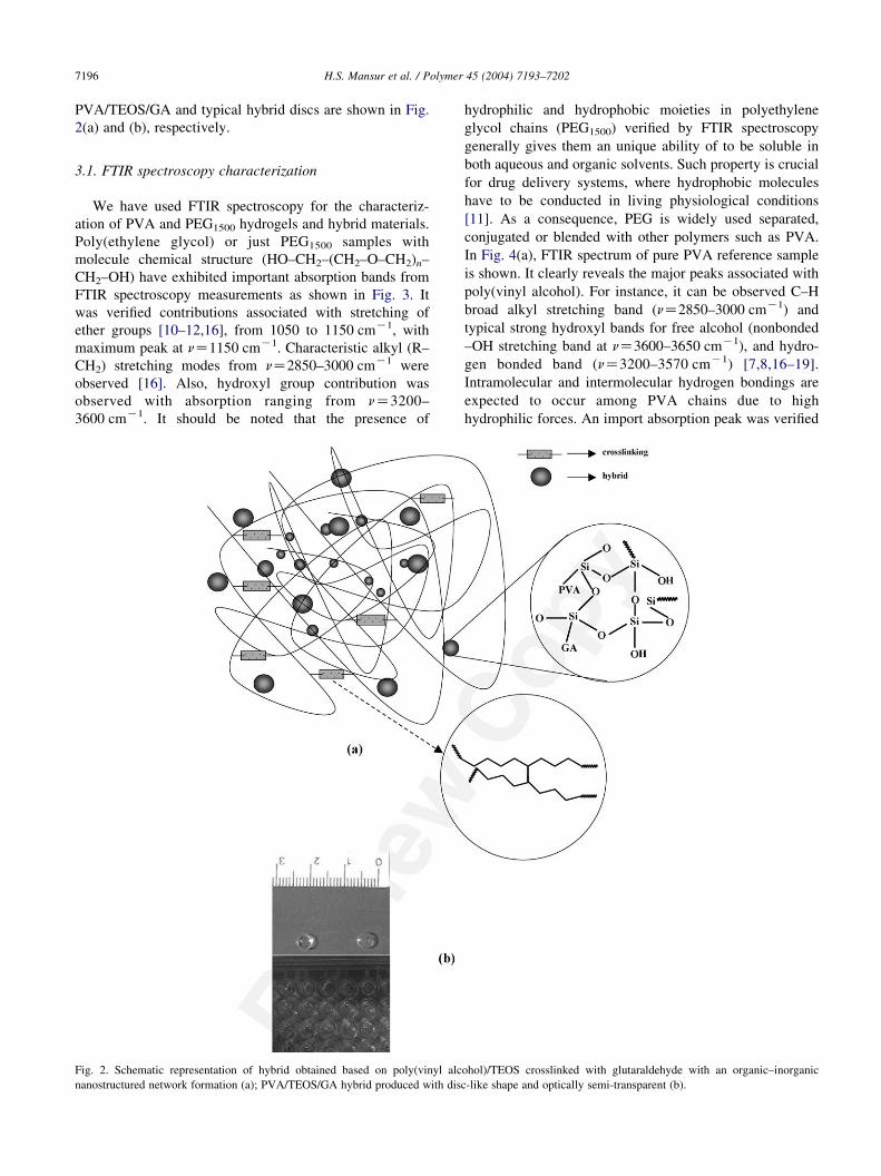
H.S. Mansur et al. / Polymer 45 (2004) 7193–72027196
PVA/TEOS/GA and typical hybrid discs are shown in Fig.
2(a) and (b), respectively.
3.1. FTIR spectroscopy characterization
We have used FTIR spectroscopy for the characteriz-
ation of PVA and PEG1500 hydrogels and hybrid materials.
Poly(ethylene glycol) or just PEG1500 samples with
molecule chemical structure (HO–CH2–(CH2–O–CH2)n–
CH2–OH) have exhibited important absorption bands from
FTIR spectroscopy measurements as shown in Fig. 3. It
was verified contributions associated with stretching of
ether groups [10–12,16], from 1050 to 1150 cmK1, with
maximum peak at nZ1150 cmK1. Characteristic alkyl (R–
CH2) stretching modes from nZ2850–3000 cmK1 were
observed [16]. Also, hydroxyl group contribution was
observed with absorption ranging from nZ3200–
3600 cmK1. It should be noted that the presence of
Fig. 2. Schematic representation of hybrid obtained based on poly(vinyl alc
nanostructured network formation (a); PVA/TEOS/GA hybrid produced with dis
hydrophilic and hydrophobic moieties in polyethylene
glycol chains (PEG1500) verified by FTIR spectroscopy
generally gives them an unique ability of to be soluble in
both aqueous and organic solvents. Such property is crucial
for drug delivery systems, where hydrophobic molecules
have to be conducted in living physiological conditions
[11]. As a consequence, PEG is widely used separated,
conjugated or blended with other polymers such as PVA.
In Fig. 4(a), FTIR spectrum of pure PVA reference sample
is shown. It clearly reveals the major peaks associated with
poly(vinyl alcohol). For instance, it can be observed C–H
broad alkyl stretching band (nZ2850–3000 cmK1) and
typical strong hydroxyl bands for free alcohol (nonbonded
–OH stretching band at nZ3600–3650 cmK1), and hydro-
gen bonded band (nZ3200–3570 cmK1) [7,8,16–19].
Intramolecular and intermolecular hydrogen bondings are
expected to occur among PVA chains due to high
hydrophilic forces. An import absorption peak was verified
ohol)/TEOS crosslinked with glutaraldehyde with an organic–inorganic
c-like shape and optically semi-transparent (b).

Fig. 3. FTIR spectra of poly(ethylene glycol). Hydroxyl vibration band
(left) and ether group absorption region (right). Diffuse reflectance mode
and KBr background.
H.S. Mansur et al. / Polymer 45 (2004) 7193–7202 7197
at a frequency of nZ1142 cmK1 (C–O, nZ1090–
1150 cmK1). According to the literature [7,8,17–19], this
vibrational band is mostly attributed to the crystallinity of
the PVA, related to carboxyl stretching band (C–O). Such
Fig. 4. FTIR spectra of (a) poly(vinyl alcohol) and (b) PVA wit
absorption band at nZ1142 cmK1 has been used as an
assessment tool of poly(vinyl alcohol) structure because it
is a semicrystalline synthetic polymer able to form some
domains depending on several process parameters [21].
FTIR spectrum of hybrid made of PVA/TEOS is showed in
Fig. 4(b). It can be observed that major vibration bands
(Si–O–Si, nZ1080 and 450 cmK1; Si–OH, nZ950 cmK1)
associated with polysiloxane (TEOS) reactions of hydroly-
sis and condensation added to PVA polymer solution. Also,
in the frequency range from 3000 to 3650 cmK1, mainly
related to hydroxyl groups [16], a broader band was noted
for PVA/TEOS hybrid spectrum (Fig. 4(b)) compared to
PVA (Fig. 4(a)). Such result is believed to be due to the
TEOS sol–gel reactions that have altered PVA chains tri-
dimensional structure. PVA molecular entanglements and
crystallinity depend on hydrophilic/hydrophobic force
balance. Hydrogen bonds play a crucial role in such
conformational arrangements, creating hydrophically
associated domains [20]. Therefore, introducing of Si–
OH and Si–O–Si through hydrolysis and condensation
reactions of TEOS has modified PVA semi-crystalline
structure. Such broad band observed on FTIR spectrum of
PVA/GA has also some contribution of physically and
chemically water incorporated during the hybrid synthesis.
These results have clearly indicated that an organic–
inorganic hybrid network was achieved based on PVA and
TEOS (Fig. 2(a)). In Fig. 5, FTIR spectra of hybrid
crosslinked PVA/TEOS/GA and PVA/GA are shown.
FTIR spectrum in Fig. 5(a) is associated with PVA
crosslinked by glutaraldehyde. It can be observed that
two important peaks at nZ2850 and 2750 cmK1 of C–H
stretching are related to aldehydes [16]. Also, strong band
h TEOS. Diffuse reflectance mode and KBr background.
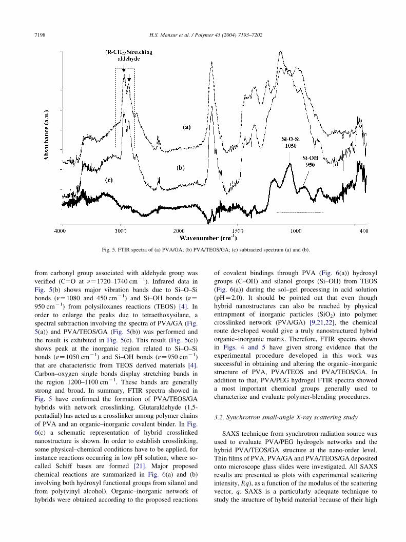
Fig. 5. FTIR spectra of (a) PVA/GA; (b) PVA/TEOS/GA; (c) subtracted spectrum (a) and (b).
H.S. Mansur et al. / Polymer 45 (2004) 7193–72027198
from carbonyl group associated with aldehyde group was
verified (CaO at nZ1720–1740 cmK1). Infrared data in
Fig. 5(b) shows major vibration bands due to Si–O–Si
bonds (nZ1080 and 450 cmK1) and Si–OH bonds (nZ950 cmK1) from polysiloxanes reactions (TEOS) [4]. In
order to enlarge the peaks due to tetraethoxysilane, a
spectral subtraction involving the spectra of PVA/GA (Fig.
5(a)) and PVA/TEOS/GA (Fig. 5(b)) was performed and
the result is exhibited in Fig. 5(c). This result (Fig. 5(c))
shows peak at the inorganic region related to Si–O–Si
bonds (nZ1050 cmK1) and Si–OH bonds (nZ950 cmK1)
that are characteristic from TEOS derived materials [4].
Carbon–oxygen single bonds display stretching bands in
the region 1200–1100 cmK1. These bands are generally
strong and broad. In summary, FTIR spectra showed in
Fig. 5 have confirmed the formation of PVA/TEOS/GA
hybrids with network crosslinking. Glutaraldehyde (1,5-
pentadial) has acted as a crosslinker among polymer chains
of PVA and an organic–inorganic covalent binder. In Fig.
6(c) a schematic representation of hybrid crosslinked
nanostructure is shown. In order to establish crosslinking,
some physical–chemical conditions have to be applied, for
instance reactions occurring in low pH solution, where so-
called Schiff bases are formed [21]. Major proposed
chemical reactions are summarized in Fig. 6(a) and (b)
involving both hydroxyl functional groups from silanol and
from poly(vinyl alcohol). Organic–inorganic network of
hybrids were obtained according to the proposed reactions
of covalent bindings through PVA (Fig. 6(a)) hydroxyl
groups (C–OH) and silanol groups (Si–OH) from TEOS
(Fig. 6(a)) during the sol–gel processing in acid solution
(pHZ2.0). It should be pointed out that even though
hybrid nanostructures can also be reached by physical
entrapment of inorganic particles (SiO2) into polymer
crosslinked network (PVA/GA) [9,21,22], the chemical
route developed would give a truly nanostructured hybrid
organic–inorganic matrix. Therefore, FTIR spectra shown
in Figs. 4 and 5 have given strong evidence that the
experimental procedure developed in this work was
successful in obtaining and altering the organic–inorganic
structure of PVA, PVA/TEOS and PVA/TEOS/GA. In
addition to that, PVA/PEG hydrogel FTIR spectra showed
a most important chemical groups generally used to
characterize and evaluate polymer-blending procedures.
3.2. Synchrotron small-angle X-ray scattering study
SAXS technique from synchrotron radiation source was
used to evaluate PVA/PEG hydrogels networks and the
hybrid PVA/TEOS/GA structure at the nano-order level.
Thin films of PVA, PVA/GA and PVA/TEOS/GA deposited
onto microscope glass slides were investigated. All SAXS
results are presented as plots with experimental scattering
intensity, I(q), as a function of the modulus of the scattering
vector, q. SAXS is a particularly adequate technique to
study the structure of hybrid material because of their high
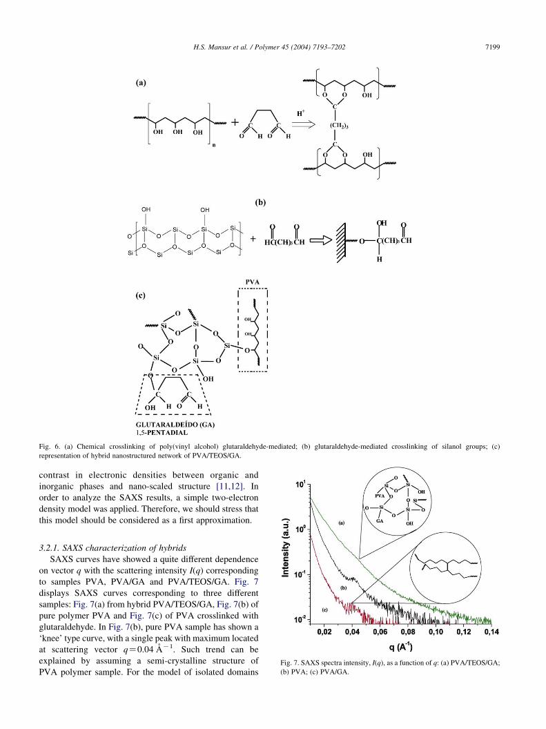
Fig. 6. (a) Chemical crosslinking of poly(vinyl alcohol) glutaraldehyde-mediated; (b) glutaraldehyde-mediated crosslinking of silanol groups; (c)
representation of hybrid nanostructured network of PVA/TEOS/GA.
H.S. Mansur et al. / Polymer 45 (2004) 7193–7202 7199
contrast in electronic densities between organic and
inorganic phases and nano-scaled structure [11,12]. In
order to analyze the SAXS results, a simple two-electron
density model was applied. Therefore, we should stress that
this model should be considered as a first approximation.
Fig. 7. SAXS spectra intensity, I(q), as a function of q: (a) PVA/TEOS/GA;
(b) PVA; (c) PVA/GA.
3.2.1. SAXS characterization of hybrids
SAXS curves have showed a quite different dependence
on vector q with the scattering intensity I(q) corresponding
to samples PVA, PVA/GA and PVA/TEOS/GA. Fig. 7
displays SAXS curves corresponding to three different
samples: Fig. 7(a) from hybrid PVA/TEOS/GA, Fig. 7(b) of
pure polymer PVA and Fig. 7(c) of PVA crosslinked with
glutaraldehyde. In Fig. 7(b), pure PVA sample has shown a
‘knee’ type curve, with a single peak with maximum located
at scattering vector qZ0.04 AK1. Such trend can be
explained by assuming a semi-crystalline structure of
PVA polymer sample. For the model of isolated domains
H.S. Mansur et al. / Polymer 45 (2004) 7193–72027200
embedded in a continuous matrix, the average distance
between domains, d, can be estimated by using the simple
equation (Eq. (1)) given by:
d Z 2p=qmax (1)
where, qmax is the modulus of the scattering vector
corresponding to the maximum of the SAXS intensity
function [12].
The average size of the domain is determined by
assuming spherical entities, with a radius R, forming a
compact arrangement. Based on Eq. (1), we would have an
average size of 15 nm for PVA nanocrystallites (qZ0.04 AK1; Fig. 7(b)). This result has strong correlation
with values reported in recent publications [1,3,6], where
the PVA crystals were found to be in the range from 7 to
20 nm, obtained by X-ray diffraction technique from PVA
based hydrogels. In spite of much research into the
microstructure and nanostructure of crystalline polymers
some details are still to be deeply investigated [20].
Hydrogen bonds are very important on stabilizing polymer
structures. Therefore, the number of hydrogen bonds present
in poly(vinyl alcohol) chain will be maximized, causing all
of them to occur in parallel sheets, with strong intermole-
cular forces, stabilizing polymer crystals [23,24]. The usual
form of such crystal domains is lamellar, occurring in thin
plates or sheets. These lamellar are typically 10–20 nm in
size. In summary, SAXS curve obtained for PVA sample
has clearly indicated the formation of nanocrystalline
domains with estimated average size of 15 nm. For that
reason, it is assumed that PVA network is made of a
crystalline phase embedded in a continuous amorphous
polymer matrix [22–24]. In Fig. 7(a), no evidence of
scattering q vector maximum peak was verified associated
with PVA/TEOS/GA hybrid sample. SAXS curve of PVA/
TEOS/GA sample (Fig. 7(a)) has presented a typical power-
law dependence on vector q [11,12] when compared to
curves of poly(vinyl alcohol) (Fig. 7(b)) and PVA modified
with GA (Fig. 7(c)). Such trend is assumed to be caused by
breaking most nano-ordered tri-dimensional organic struc-
tured previously found in the PVA sample. Due to siloxane
hydrolysis and policondensation reactions with PVA
aqueous solution, several new chemical covalent bonds
have been created (Si–C, Si–OH, Si–O–Si) reducing the
hydrogen bond formation between polymer chains. Briefly,
for PVA/TEOS/GA samples, we consider that the network
is basically composed of some multi-dispersed nanocrystal-
line PVA domains embedded into a continuous amorphous
organic–inorganic hybrid matrix. Besides that, such effect
on reducing the crystalline domains formation of PVA was
further observed by adding glutaraldehyde. The crosslinking
of polymer chains have occurred, causing less flexibility for
spatial conformational mobility. As a consequence of such
organic–inorganic nanostructure, no specific maximum
value for vector q of synchrotron radiation was detected
(Fig. 7(a)). The proposed hybrid organic–inorganic
chemical structure is shown in Fig. 6(c). The explanation
suggested in this work for the differences observed between
PVA and PVA/TEOS/GA hybrid samples were also
confirmed by crosslinking poly(vinyl alcohol) with GA.
SAXS curve shown in Fig. 7(c) clearly reveals a broader
band for the maximum value vector q, varying from 0.03 to
0.04 AK1, when compared to pure PVA (qZ0.04 AK1).
The addition of strong crosslinker agent (GA) has
‘hardened’ the PVA chain structure reducing the possibility
of hydrogen bonds formation. Therefore, less well-defined
nanocrystalline PVA domains were formed. In summary,
for PVA/GA samples, we consider that the network is
basically composed of some multi-dispersed nanocrystal-
line PVA domains embedded into a continuous amorphous
polymeric matrix. Another information about the domain
arrangement can be obtained from the width of SAXS peak
[5]. Similarly to the determination of the crystallite size in
polycrystals, a rough estimate of the average size of the
correlation domains, Lc, associated with the spatial
distribution of agglomerates, can be obtained by applying
Scherrer [5,11] equation (Eq. (2)):
Lc Z 4p=Dq (2)
where, Dq is the full width at half-maximum of the
correlation peak of the measured SAXS function.
Assuming DqZ0.01 AK1, vector q, varying from 0.03 to
0.04 AK1, we have calculated an average size LcZ120 nm
of distribution among nanocrystallites of PVA/GA. Again,
such results has confirmed the loss of crystallinity and nano-
ordered structure of PVA by crosslinking with GA.
3.2.2. SAXS characterization of hydrogels
Hydrogels based on polymer blending are quite complex
systems and have been investigated for decades [19].
Recently, the tri-dimensional distribution of semicrystalline
polymer constituents forming blended nanostructure was
studied by small-angle neutron scattering and SAXS [11,
12]. In the present study, we have investigated the
nanostructure of PVA/PEG1500 hydrogels through synchro-
tron radiation SAXS experiments. SAXS data collected for
PVA/PEG synthesized hydrogels are shown in Fig. 8. In Fig.
8, the important pattern alterations of SAXS curves from
pure PVA (Fig. 8(c)) compared to PVA/PEG blended
hydrogel (Fig. 8(b)) and PVA/PEG/GA chemically cross-
linked hydrogel (Fig. 8(a)) can be observed. The curves
showed noticeable maximum at low scattering vector q. It
can be noted that all scattering patterns have a single peak
with maximum located at approximately qZ0.04 AK1.
Such trend can be attributed to semicrystalline domains as
expected to be found on both PVA and PEG components.
However, the broadening of the q vector peaks was
obviously verified on PVA/PEG (Fig. 8(b)) and PVA/
PEG/GA (Fig. 8(a)) curves when compared to pure PVA
curve (Fig. 8(c)). Such behavior could be explained by
assuming that pure PVA has a spatial distribution between

Fig. 8. SAXS spectra intensity, I(q), as a function of q: (a) PVA/PEG/GA; (b) PVA/PEG; (c) PVA.
H.S. Mansur et al. / Polymer 45 (2004) 7193–7202 7201
crystalline domains embedded in amorphous polymeric
matrix. On the other hand, PVA blending with PEG1500 is
likely to have modified PVA such crystallinity and lamellae
packing. As reported in the literature, both PVA and PEG
have semicrystalline structures [7,10,11,17]. So, a hom-
ogenous blended system made of PVA and PEG chains is
expected to be found with an average contribution from both
polymer constituents. On a molecular level, the crystallites
of PVA and PEG can be described as a layered structure
[10–12,14,17]. A double layer of chains is held together by
hydrogen bonds while weaker van der Waals forces operate
between the double layers [10,11]. The folded chains of
PVA chains and PEG lead to crystallites, which are small,
ordered regions, in an unordered, amorphous polymer
matrix [10]. As a consequence, PEG1500 addition in PVA
would have caused a wider crystalline size distribution
resulting on the SAXS scattering pattern verified for
blended hydrogels (Fig. 8(a) and (b)). Chemical cross-
linking of PVA/PEG1500 with glutaraldehyde (Fig. 8(a))
seemed to have minor effect on altering the scattering
performance of PVA/PEG blend. Interestingly, as reported
in the literature [11], researchers found the formation of
crystallites during the dehydration and annealing of PVA
hydrogels has served as crosslinks in addition to the ones
formed through chemical reactions. Thus, the SAXS
scattering behavior presented by PVA/PEG and PVA/
PEG/GA were likely to be governed by average contribution
of blend components. That means, the overall scattering
patterns observed for PVA/PEG hydrogels are due key
factors such as lamellae size, spatial distribution, crystal-
linity in lamellar stacks and the degree of crystallinity
compared to amorphous polymer matrix, from contributions
of both PVA and PEG1500 components. Further investi-
gation based on density measurements, calorimetric
methods, and X-ray diffraction analysis would bring more
understanding on the PVA/PEG blending to nanometric
scale.
4. Conclusion
We have effectively produced PVA/PEG hydrogels and
chemically crosslinked with glutaraldehyde via aqueous
route. PVA/PEG hydrogel blends were properly character-
ized by using SAXS and FTIR spectroscopy techniques.
SAXS and FTIR spectroscopy characterizations have also
confirmed that hybrid organic–inorganic materials were
successfully obtained based on the combination of PVA and
TEOS with glutaraldehyde crosslinked nanometer-scale
network. In addition to that, SAXS synchrotron radiation
associated with FTIR spectroscopy have proven to be a
powerful tools for nanoscience investigation.
Acknowledgements
The authors acknowledge CNPq/FAPEMIG/CAPES for
financial support on this project. The authors are also
particularly grateful for the important contribution from
LNLS staff and for synchrotron SAXS facilities.
References
[1] Yano S, Kurita K, Iwata K, Furukawa T, Kodomari M. Polymer 2003;
44:3515–22.
[2] Kickelbick G. Prog Polym Sci 2003;28:83–114.
[3] Matejka L, Dukh O, Hlavata D. Macromolecules 2004;36:7977–85.
[4] Mansur HS, Vasconcelos WL, Orefice R. J Non-Cryst Solids 2000;
273:109–13.
[5] Sarmento VHV, Dahmouche K, Santilli CV, Pulcinelli SH,
Craievich AF. J Appl Cryst 2003;36:473–7.
[6] Ricciaedi R, Auriemma F, De Rosa C, Laupretre F. Macromolecules
2004;37:1921–7.
[7] J.D. Thomas, MSc Thesis, Sep 2001, Drexel University, Novel
associated PVA/PVP hydrogels for nucleus pulposus replacement,
Philadelphia, PA, USA.
[8] Hassan CM, Peppas NA. Adv Polym Sci 2000;153:37–65.
[9] Hoffman A. Adv Drug Deliv Rev 2000;43:3–12.

H.S. Mansur et al. / Polymer 45 (2004) 7193–72027202
[10] Sahlin JJ, Peppas NA. J Appl Polym Sci 1997;63:103–10.
[11] Roberts MJ, Bently MD, Harris JM. Adv Drug Deliv Rev 2002;54:
459–76.
[12] Singh TJ, Bhat SV. Bull Mater Sci 2003;26(7):707–14.
[13] Chiavacci LA, Dahmouche K, Santilli CV, Bermudez Z, Carlos LD,
Briois V, Craievich AF. J Appl Cryst 2003;36:405–9.
[14] Chaker JA, Dahmouhce K, Craievich AF, Santilli CV, Pulcinelli SH.
J Appl Cryst 2000;33:700–3.
[15] Orthaber DA, Bergmann A, Glatter O. J Appl Cryst 2000;33:218–25.
[16] Coates J. In: Meyers RA, editor. Encyclopaedia of analytical
chemistry. Chichester: Wiley; 2000. p. 10815–37.
[17] Peppas NA. Polymer 1977;18:403–8.
[18] Peppas NA. Makromol Chem 1977;178:595–601.
[19] Peppas NA, Wright L. Macromolecules 1996;29:8798–804.
[20] Mills NJ, editor. Plastics microstructure and engineering applications.
2nd ed. London: Edward Arnold Publishers; 1993.
[21] Hennink WE, Nostrum CF. Adv Drug Deliv Rev 2002;54:13–36.
[22] Schottner G. Chem Mater 2001;13:3422–35.
[23] Hodge RM. Polymer 1996;37:1371–6.
[24] Ram S, Mandal TK. Chem Phys 2004;303:121–8.