characterization of cellobiose dehydrogenase from a biotechnologically important cerrena unicolor...
TRANSCRIPT
1 23
Applied Biochemistry andBiotechnologyPart A: Enzyme Engineering andBiotechnology ISSN 0273-2289 Appl Biochem BiotechnolDOI 10.1007/s12010-015-1667-2
Characterization of CellobioseDehydrogenase from a BiotechnologicallyImportant Cerrena unicolor Strain
Justyna Sulej, Grzegorz Janusz, MonikaOsińska-Jaroszuk, Patrycja Rachubik,Andrzej Mazur, Iwona Komaniecka,Adam Choma, et al.

1 23
Your article is published under the Creative
Commons Attribution license which allows
users to read, copy, distribute and make
derivative works, as long as the author of
the original work is cited. You may self-
archive this article on your own website, an
institutional repository or funder’s repository
and make it publicly available immediately.

Characterization of Cellobiose Dehydrogenasefrom a Biotechnologically Important Cerrena unicolorStrain
Justyna Sulej1 & Grzegorz Janusz1 & Monika Osińska-Jaroszuk1&
Patrycja Rachubik1& Andrzej Mazur2 & Iwona Komaniecka2
&
Adam Choma2 & Jerzy Rogalski1
Received: 10 March 2015 /Accepted: 12 May 2015# The Author(s) 2015. This article is published with open access at Springerlink.com
Abstract Cellobiose dehydrogenase (CDH), a secreted flavocytochrome produced by a num-ber of wood-degrading fungi, was detected in the culture supernatant of a biotechnologicallyimportant strain of Cerrena unicolor grown in a modified cellulose-based liquid medium. Theenzyme was purified as two active fractions: CuCDH-FAD (flavin domain) (1.51-fold) withrecovery of 8.35 % and CuCDH (flavo-heme enzyme) (21.21-fold) with recovery of 73.41 %.As CDH from other wood-rotting fungi, the intact form of cellobiose dehydrogenase ofC. unicolor is a monomeric protein containing one flavin and one heme b with molecular mass97 kDa and pI=4.55. The enzyme is glycosylated (8.2 %) mainly with mannose and glucos-amine residues. Moreover, the cellobiose dehydrogenase gene cdh1 and its correspondingcDNA from the fungus C. unicolor were isolated, cloned, and characterized. The 2316-bpfull-length cDNA of cdh1 encoded a mature CDH protein containing 771 amino acids precededby a signal peptide consisting of 18 amino acids. Moreover, both active fractions werecharacterized in terms of kinetics, temperature and pH optima, and antioxidant properties.
Keywords Cellobiose dehydrogenase .Cerrena unicolor . Purification . Gene . Fungi
Introduction
Fungi form an important group of microorganisms that have beneficial effects on the envi-ronment and human life. In forest ecosystems, they are mostly responsible for breakdown of
Appl Biochem BiotechnolDOI 10.1007/s12010-015-1667-2
* Grzegorz [email protected]
1 Department of Biochemistry, Maria Curie-Skłodowska University, Akademicka 19 St.,20-033 Lublin, Poland
2 Department of Genetics and Microbiology, M. Curie-Skłodowska University, Akademicka 19 St.,20-033 Lublin, Poland
abundant large biopolymers such as cellulose, hemicellulose, and lignin [1]. White-rot basid-iomycetes are a group of fungi comprising from 1600 up to 1700 species characterized by theability to depolymerize and mineralize lignin using a set of extracellular ligninolytic enzymesand low molecular compounds [2, 3]. At the same time, in modern biotechnology, filamentousfungi are major sources of bioactive metabolites, including proteins, peptides, glycoproteins,polysaccharides, lipopolysaccharides, phenolic compounds, triterpenoids, lectins, lipids, andtheir derivatives [4].
Among the many hitherto-characterized fungal species, Cerrena unicolor was described inliterature as one of the best laccase producers [5]. Moreover, this species belonging toAphyllophorales was proved to secrete extracellular manganese peroxidase, versatile peroxi-dases [6], and xylanase or cellulase when grown on cellulose [7]. This fungus commonlycalled Bmossy maze polypore^ may be found on dead northern hardwood tree species asmaple, birch, or alder, where it causes white rot [8]. Besides extracellular enzymes, C. unicolormay be a source of polysaccharides [9] or low molecular fractions of secondary metabolites[10], which possess interesting biomedical and bioelectrochemical properties. However, up todate, cellobiose dehydrogenase, which was proven a crucial enzyme in decomposition of bothcellulose and lignin, has not been described in cultures of the genus Cerrena.
Cellobiose dehydrogenase (CDH; EC 1.1.99.18; cellobiose [acceptor] 1-oxidoreductase) isa fungal extracellular hemoflavoprotein, which was discovered in 1974 by Westermark andEriksson in white rot fungi Trametes versicolor [11] and Phanerochaete chrysosporium(Sporotrichum pulverulentum) [12]. CDHs are usually monomeric enzymes that belong tothe glucose–methanol–choline (GMC) family together with other sugar oxidoreductases likethe catalytically related enzymes glucose oxidase, pyranose dehydrogenase, and pyranose-2oxidase [13]. It is composed of two prosthetic groups, a heme type b (ferriprotoporphyrin IX)and a flavin adenine dinucleotide (FAD) [14] connected through a flexible polypeptide linkerregion enriched in hydroxy amino acids [15]. This enzyme catalyzes the oxidation of thereducing end of cellobiose and higher cellodextrins in vivo, whereas in vitro lactose andother oligosaccharides with β-1,4-glycosidic linkages are acceptable substrates [16]. Thecatalytic cycle of CDH involves oxidation of sugar substrates to corresponding 1,5-lactonesusing various electron acceptors with concomitant reduction of flavin to FADH2 [17].Lactones are finally converted to their carboxylic acids, and flavin is reoxidized by the hemegroup in two single-electron steps reactions [18]. Phylogenetic analysis of all known cdhgenes showed division of the enzymes into three distinct classes: class I, representing onlybasidiomycetous CDHs; class II, exclusively comprising ascomycetous CDHs; and class III,containing so far uncharacterized or actively expressed CDHs [19]. Although the physiolog-ical function of this enzyme has not yet been revealed, our current knowledge points to itsparticipation in the degradation and modification of lignocellulose by generating hydroxylradicals via the Fenton reaction [20]. Recently, an interaction of CDH with copper-dependentpolysaccharide monooxygenases (PMOs) involved in the degradation of cellulose has beenproposed [21, 22]. This model for oxidative cellulose degradation may be widespreadthroughout the fungal kingdom in parallel with the better described hydrolytic cellulaseenzyme system [23]. Recent papers have reported successful application of cellobiosedehydrogenase in a large variety of bioprocesses such as biocatalysis, bioremediation, orproduction of lactobionic acid [24]. The unique catalytic and bioelectrochemical properties ofCDH have been used in biosensors for detection of cellodextrins [25], maltose [26], lactose[27, 28], diphenolic compounds [29], and catecholamines [30] in biofuel cells [15, 31] or inbiomedical applications [32, 33].
Appl Biochem Biotechnol

Given the widespread biotechnological application of cellobiose dehydrogenase, newsources of this enzyme are being constantly searched. Recently, C. unicolor strain FCL139has been found to be a producer of laccase, a unique enzyme in many biotechnologicalapplications. Hereby, we successfully attempted to purify and characterize cellobiose dehy-drogenase from this strain. Moreover, the corresponding cdh gene and cDNAwere sequencedand analyzed.
Materials and Methods
Microorganism and Culture Conditions
The white rot fungus C. unicolor was obtained from the culture collection of the RegensburgUniversity and deposited in the fungal collection at the Department of Biochemistry (MariaCurie-Sklodowska University, Poland) under the strain number 139. The fungus was main-tained on 4 % (w/v) malt agar plate. To obtain the inocula, pieces of agar plates with the funguswere grown in the Lindenberg and Holm [34] medium in conical flasks for 10 days at 25 °C.Ten-day-old mycelia were homogenized in a disperser homogenizer T18 basic ULTRA-TURRAX (IKA, Staufen, Germany). The fragmented mycelial culture (10 %v/v) was used as astandard inoculum for further studies.
In order to obtain the high level of CDH, the strain of C. unicolor (FCL139) was grown insubmerged culture for 10 days on a cellulose-based medium [35] with authors’ modifications.The medium had the following composition (1 l): 5 g Avicel, 10 g (NH4)2HPO4, 1 g KH2PO4,0.3 g MgSO4×7H2O, 0.08 g CaCl2, 5 mg ZnSO4×7H2O, 1.5 mg MnSO4×4H2O, 1.5 mgCoCl2×6H2O, 5 mg FeSO4×7H2O, 100 mg yeast extract, and 0.1 mg thiamine. The pH wasadjusted to 6.5 with 5 M HCl. After inoculation, the cultures were incubated at 28 °C in anincubator shaker Multitron (Infors, Bottmingen, Switzerland) at 120 rpm.
Enzyme Purification Procedure
The culture supernatant (6 l) was collected on day 10 from the cellulose medium aftercentrifugation (12,000×g for 30 min) on a 6K15 (Sigma, Osterode am Harz, Germany). Theclear supernatant was concentrated to 300 ml by the Prep/Scale TFF Cartridge PTGC 10 kpolyethersulfone (Millipore, Bedford, MA) and used as a source of a crude enzyme. Theproteins in the crude preparation were precipitated by the addition of solid ammonium sulfatein the range of 15–85 % saturation. The resulting suspension was collected by centrifugation,and the protein pellet was resolved in 100 ml deionized water and desalted using a preparativechromatography column (8×30 cm) filled with a Sephadex G-50 carrier. Fractions containingthe protein were concentrated and applied to an anion-exchange DEAE-Sepharose (fast flow)column (2.5×15) connected to Econo System (Bio-Rad, Richmond, VA). The column waspreviously equilibrated with a 50-mM sodium acetate buffer (pH 5.0), and the proteins boundon the chromatography matrix were eluted using a linear gradient of 0 to 0.5 M NaCl in thesame buffer at a flow rate of 1 ml/min. Fractions containing cellobiose dehydrogenase activitywere pooled and concentrated in an Amicon-stirred cell using a polyethersulfone membrane(10 kDa cutoff). In the next step, affinity chromatography was performed. The concentratedprotein was loaded onto a lactose-CPG column (1.5×8 cm) equilibrated with buffer A (50 mMsodium acetate buffer (pH 5.5)), washed with buffer B (50 mM sodium acetate buffer
Appl Biochem Biotechnol
(pH 4.0)), eluted with buffer C (200 mM sodium acetate buffer (pH 4.0)), and applied 0.7 Mammonium sulfate. The eluent was collected in 0.5 ml portions. Fractions containing CDHactivity (obtained from lactose-CPG) were collected, and a chromatofocusing analysis wasperformed on an Econo-chromatography column (Bio-Rad, Richmond, VA, USA; 130 cm,packed to a bed height of 20 cm) with a Polybuffer exchanger PBE 94 equilibrated with250 ml of 0.025 M imidazole-HCl buffer (pH 7.4). Samples from lactose-CPG chromatogra-phy showing CDH activity (5 ml) were injected onto the column, and the enzyme wasdesorbed by elution with 200 ml Polybuffer 74-HCl (pH 3.0) at a flow rate of 0.5 ml/min.The active fractions were pooled out, and the purified enzyme solutions were used for kineticexperiments.
Synthesis of Lactose-CPG
The controlled porous glass (CPG) (Cormay, Lublin, Poland) was prepared according to themethod described previously [36]. The support was activated by γ-aminopropyltriethoxysilane(γ-APTES) according to a method that permits a high density of amino groups on the glasssurface [37]. The activated support (APTES-CPG) was further used for affinity chromatogra-phy by binding the CDH substrate lactose to the activated support according to a methoddescribed in detail elsewhere [38]. The resulting sorbent lactose-CPG was used in the affinitychromatography.
Enzyme Assays and Protein Determination
The activity of cellobiose dehydrogenase was assayed according to Baminger et al. [1] withslight modifications. CDH activity was specifically determined by monitoring the reduction ofthe electron acceptor 2,6-dichloroindophenol (DCIP) (Sigma Chemical Co., St. Louis, MO,USA) at 520 nm (ε520=6.8 mM−1 cm−1), pH 4.5, and 30 °C using a Shimadzu UV-160A(Shimadzu, Tokyo, Japan) spectrophotometer. The reaction mixture (1 ml) contained 50 μl of3 mM DCIP (solution in water containing 10 % v/v ethanol), 100 μl lactose (300 mM in100 mM sodium acetate buffer, pH 4.5), 50 μl NaF (80 mM NaF) in water, and an appropriateamount of the same buffer. After temperature adjustment, the reaction was started by theaddition of an appropriately diluted CDH sample (100 μl) and the decrease in absorbance wasmonitored during the first 60 s. The final enzyme activity was expressed as nkat per liter.
Alternatively, CDH activity was selectively determined by following the reduction of20 μM cytochrome c at λ=550 nm and 30 °C (Sigma Chemical Co., St. Louis, MO, USA).The reaction was performed in 100 mM sodium acetate buffer, pH 4.5, containing 30 mMlactose and 4 mM NaF. The extinction coefficient (ε) was 19.6 mM−1 cm−1 [39]. This assaydetermined the activity of the intact protein containing both the flavin and the heme domains.
The protein concentration was determined using the Bradford method [40] with crystallinebovine serum albumin (BSA) as a standard or by monitoring the ultraviolet (UV) absorbanceat 280 nm.
Spectral Characterization
The spectrum of CDH purified to homogeneity was recorded from 250 to 650 nm in both theoxidized and the reduced states using a Shimadzu UV-160A spectrophotometer (Shimadzu,Tokyo, Japan). Purified CDH was diluted in 100 mM sodium acetate buffer, pH 4.5, to an
Appl Biochem Biotechnol

absorbance of ∼2.5 at 280 nm, and the spectrum was recorded before and immediately after theaddition of an approximately 1000-fold molar excess of lactose to the cuvette. The index ofpurity (RZ) of the oxidized CDH was calculated as the ratio of the absorbance at 420 nm to theabsorbance at 280 nm [19].
Effect of Temperature and pH on CDH Activity and Stability
DCIP and cytochrome c as electron acceptors and lactose as a substrate were used for assessingthe effect of pH and temperature on the cellobiose dehydrogenase activity and stability. Theeffect of pH on enzyme activity was estimated in the range from 2.5 to 8.0 in 0.1 MMcIlvaine’s buffer. Dependence of stability on pH was determined at 30 °C by incubationin variable pH ranges (pH 2.0–9.0 0.1 M Britton-Robinson buffer) for 12 h followed bymeasurement of the residual activities every 30 min.
The optimum temperature of the purified CDHwas determined by performing enzymatic assaysat different temperatures (4–80 °C). The thermal stabilitywas investigated by incubating the enzymesolution in a 0.1M sodium acetate buffer (pH 4.5) at various temperatures (30–90 °C); aliquots weredrawn every 30 min for 12 h, and their residual enzyme activities were measured. Controls werecarried out using the enzyme solutions without preheating, and its activity was taken as 100 %.
Determination of Kinetic Constants
Kinetic constants were determined for various concentrations of CDH substrates (0.1 to10 mM) and DCIP and cyt c as electron acceptors. All measurements were performed intriplicates. The Km and Vmax for the purified enzyme were calculated by nonlinear least-squares regression, fitting the observed data to the Michaelis–Menten equation. The OriginPro8 software (OriginLab Corporation, Northhampton, MA, USA) was used for data analysis.
Effect of Metal Ions and Potential Inhibitors
Effects of various metal ions and other reagents on CDH activity were investigated by addinginorganic salts, imidazole, and SDS (to the final concentration from 0.1 to 100 mM) to thesamples of the enzyme dissolved in 100 mM sodium acetate buffer (pH 4.5) to the total volumeof 1 ml. These assays were performed with lactose as a substrate and DCIP as an electronacceptor. Control tests were performed in parallel in the absence of metal ions and inhibitors.
Electrophoresis and Peptide Sequencing by LC-MS/MS
Sodium dodecylsulfate-polyacrylamide gel electrophoresis (SDS-PAGE) (10 %) was performedas described by Laemmli [41]. Proteins were visualized by silver staining [42] and CoomassieBrilliant Blue G250 using PageRuler Prestained Protein Ladder (Fermentas, Glen Burnie, MA,USA). After the electrophoretic separation of the samples, equal pieces of 2×7 mm were cut outfrom the gel lanes. The spectrometric analysis of polypeptides was carried out in the Environ-mental Laboratory of Mass Spectrometry, Institute of Biochemistry and Biophysics of the PolishAcademy of Sciences in Warsaw (Poland). The equipment used was sponsored in part by theCentre for Preclinical Research and Technology (CePT), a project co-sponsored by EuropeanRegional Development Fund and Innovative Economy, The National Cohesion Strategy ofPoland. The samples were analyzed by HPLC coupled with tandem mass spectrometry (liquid
Appl Biochem Biotechnol
chromatography/two stage mass spectrometry - LC-MS/MS) according to Kordan et al. [39]. Theoutput list of precursor and product ions was compared with the protein database of the NationalCenter for Biotechnology (NCBI, USA) using the MASCOT local server.
Analysis of the CDH Carbohydrate Moiety
For sugar analysis, the CDH sample was hydrolyzed with 2 M trifluoroacetic acid (TFA)(100 °C, 4 h). The liberated monosaccharides were reduced with NaBD4 and converted intoalditol acetates [43]. The components were identified on the basis of retention times and massspectra of authentic standards using the gas chromatography-mass spectroscopy technique.GC-MS was carried out on an Agilent Technologies gas chromatograph (7890A) connected toa mass selective detector (inert XL EI\CI MSD 5975C). The chromatographwas equippedwitha capillary column HP-5MS (30 m×0.25 mm, film thickness 0.25 μm) (Agilent Technologies,Santa Clara, CA, USA). The carrier gas was helium with a flow rate of 0.7 ml min−1. Thetemperature program was as follows: 150 °C for 5 min, raised to 310 °C at 5 °Cmin−1, and keptfor 10 min. Total carbohydrates were determined by phenol–sulfuric method (Dubois) [44]. Astandard curve was prepared to quantify mannose.
Antioxidant Activity Assays
The antioxidant properties of C. unicolor CDH was investigated in the presence of cellobioseand lactose as the substrates of enzyme as we described previously [45]. The standards (Troloxand vitamin C) well known for their strong antioxidant activity were used as a positive control.All measurements were performed in triplicate.
DPPH Free Radical-Scavenging Test
The antioxidant activity of cellobiose dehydrogenase was determined using the DPPH equiv-alent, according to an adapted colorimetric procedure described by Paduch et al. [46] withslight modification. This method is based on the ability of 1,1-diphenyl-2-picrylhydrazyl(DPPH), a stable free radical, to decolorize in the presence of antioxidants. The testedcompound (0.1 ml) at concentrations ranging from 6.25 to 800 μg/ml was added to 0.1 mlof DPPH. solution (0.2 mg/ml in ethanol). The absorbance was measured spectrophotometri-cally at 515 nm using a Microplate Reader Elx800 (BioTek, Winooski, VT, USA) after 15 min(the time required to achieve the reaction plateau) of incubation at room temperature.
The capability of scavenging DPPH. radicals was calculated by the following formula:
DPPH:scavenging effect %ð Þ ¼ A0−A1ð Þ=A0½ � � 100
where A0 means the absorbance of the control sample and A1 means the absorbance of thestandards or tested compounds.
ABTS Free Radical-Scavenging Test
The ABTS (2,2′-azinobis (3-ethylbenzothiazoline-6-sulfonic acid) diammonium salt) radical-scavenging ability of cellobiose dehydrogenase was recorded according to the procedure of Reet al. [47] with some modification. For detection of the antioxidant capacity, 10 μL of the
Appl Biochem Biotechnol

investigated compounds at concentrations ranging from 6.25 to 800 μg/mL was mixed with990 μL of the ABTS radical solution. The percentage of ABTS oxidation was calculated bythe presented formula:
ABTS:þscavenging effect %ð Þ ¼ A0−A1ð Þ=A0½ � � 100
where A0 means the absorbance of the control samples and A1 is the absorbance at 734 nm ofthe investigated compounds/standards.
The EC50 value, defined as the amount of the antioxidant necessary to decrease the initialDPPH and ABTS concentration by 50 %, was calculated from the results. The inhibitioncurves were prepared, and EC50 values were obtained as described previously [10].
DNA Manipulation Techniques
Standard techniques for plasmid isolation, agarose gel electrophoresis, and DNA cloning wereemployed [48]. Automatic sequencing was performed using the BigDyeTM Terminator CycleSequencing Kit and an ABI PRISM 310 sequencer or ABI PRISM 3730 XL (AppliedBiosystems, Carlsband, CA, USA).
Preparation of Total mRNA, cDNA Synthesis, and Amplification
Total mRNA and cDNA synthesis and amplification were performed, as described previously[45]. To amplify the CDH 3′ cDNA fragment, degenerate primer GSP1 was designed using CDHgene sequences available in GenBank. To amplify complete CDH cDNA, gene-specific primers(GSP) were designed on the basis of an available sequenced CDH 3′ cDNA fragment (Table 1).
Genomic DNA Isolation, Amplification, and Cloning of the cdh1 Gene
DNA from C. unicolor was isolated according to Borges et al. [49], as described previously [45].To amplify the cellobiose dehydrogenase gene, two pairs of primers genCerCDH (Table 1) weredesigned on the basis of already sequenced CDH cDNA. All PCR amplifications were carried outusing Sigma RedTaq in a Tpersonal thermal cycler (Biometra, Goettingen, Germany). SpecificPCR products were purified using the Cleanup kit (A&A Biotechnology, Gdynia, Poland) andinserted into the pTZ57R/T vector from the InsTAclone kit (Fermentas, Glen Burnie, MA, USA).Clones with target fragments were analyzed by sequencing.
Table 1 Gene-specific primer sequences and annealing temperatures
Primer Sequence 5′–3′ Tm [°C]
CerCDHGSP1 GCCCAGTTWTCWTANGCWTCGAT 53.5–55.3
CerCDHGSP2 CGAAGGGTTGTCCGACACAGCCTGCCC 67.3
CerCDHGSP3 TCGACCGACGGCCAGCGCTACCTCG 67.5
CerCDHGSP4 GGGAGGTTCGCCGCCGCGGTG 66.1
genDNA1F GCCCTGTTTCAGCTCTCC 52.6
genDNA1R ACCGAAAGCATGATCTTTGAAGTCCG 58
genDNA2F GGTGGACCAAGTACGGCTGAAAAG 59,1
genDNA2R ATTGTCGAGATAACATCCTTGAGTGC 56,4
Appl Biochem Biotechnol
Nucleotide Sequence Accession Numbers
The following GenBank accession numbers were given to the CDH nucleotide sequencesdetermined in this study: KC862284—C. unicolor strain FCL139 cellobiose dehydrogenasegene (cdh), complete cds; KC862282—C. unicolor strain FCL139 cellobiose dehydrogenasemRNA (cdh), complete cds.
Bioinformatics Tools
Nucleic acid sequences were analyzed using Lasergene v.8.0 analysis software (DNASTAR,Inc, Madision, WI, USA). Database searches were performed with the BLAST and FASTAprograms at the National Centre for Biotechnology Information (Bethesda, MD, USA) andEuropean Bioinformatics Institute (Hinxton, UK), respectively. Multiple DNA and proteinsequence alignments were performed with the Clustal-W algorithm [50]. Phylogenetic treevisualization was performed using the TreeView applet [51]. Glycosylation sites were detectedwith NetNGlyc v.1.0 (http://www.cbs.dtu.dk/services/NetNGlyc/) and NetOGlyc v.4.0 [52].Conserved domains were analyzed by CDART [53].
Statistical Analysis
All presented results are expressed as a mean±SD from three independent experiments (n=3).The mean values as well as standard deviation were calculated by the Excel program (MicrosoftOffice 2010 package), and only values of p≤0.05 were considered as statistically significant.
Results and Discussion
Production and Purification of Cellobiose Dehydrogenase
Cellobiose dehydrogenase production by C. unicolor strain FLC139 was performed in shakingflasks on the cellulose-based medium. The mycelium was grown for 10 days, and then theculture liquid was collected, concentrated, and used as a source of crude enzyme for furtherpurification steps and other studies. CDH was partially purified by ammonium sulfate precip-itation in the range of 15 to 85 % saturation with a purification factor of 2.07-fold and arecovery of 84.93 % (Table 2). The resulting precipitate was dissolved in 100 ml distilled waterand desalted on the Sephadex G-50 column. The protein fractions obtained from ammoniumsulfate precipitation were applied to a DEAE-Sepharose chromatographic column. The elutionprofile from the ion-exchange chromatography on the DEAE-Sepharose column showed theCDH activity as a single peak, which was purified 28.97-fold with a yield of 70.34 %. Theactive fractions of cellobiose dehydrogenase were combined, concentrated on the stirredultrafiltration cell equipped with a 10-kDa cutoff polyethersulfone membrane, and used inthe subsequent step of chromatography on the lactose-CPG column. The affinity fractionation(lactose-CPG) gave only one cellobiose dehydrogenase activity peak purified approximately53-fold in yields of 59 %. The active fractions of cellobiose dehydrogenase were pooled outand further fractionated by chromatofocusing on a Polybuffer exchanger PBE 94. A summaryof the purification procedures of the cellobiose dehydrogenase is presented in Table 2. The lastpurification step (chromatofocusing) resulted in separation of two active fractions: CuCDH-
Appl Biochem Biotechnol
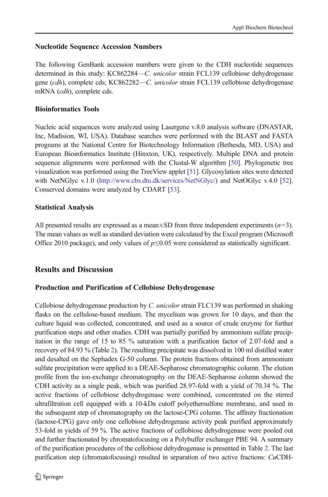
FAD (a flavin-only fragment of the enzyme), i.e., a very small fraction purified with recoveryof 1.5 %, and CuCDH (the intact enzyme) purified with a yield of 21.2 % (73.4-fold). TwoCDH activities were also obtained from fungi Irpex lacteus [54], T. versicolor [55], andPycnoporus sanguineus [45]; however, Humicola insolens contained three CDH fractionswhile P. chrysosporium contained only one [56]. In this study, the major fraction of CDH(CuCDH) was used for further investigation as CDH from C. unicolor. The enzyme waspurified from the culture supernatant to apparent homogeneity (Fig 1). The high spectral ratioA420/A280 (RZ value) is generally accepted as an indication of the absence of contaminatingproteins [57]. The purified intact CDH from C. unicolor showed an A420/A280 ratio of 0.57,whereas the flavin fragment has a low RZ value (0.21). Besides the purity factor (RZ), themost important was the ratio of DCIP and cyt c activity (value around 1), which determines therate of degradation of the intact enzyme on the flavin and heme domains [58]. The factorobtained in this study indicates that the intact CDH was rather stable in the culture conditions
Table 2 Purification of CDH from Cerrena unicolor strain FCL139 culture filtrate
Purification step Total protein(mg)
Total activity(nkat)
Specific activity(nkat/mg)
Yield (%) Purificationfold
Culture filtrate 1800.00 21180.00 11.77 100 1
Ultrafiltration (10 kDa) 1275.00 20377.50 15.98 96.21 1.36
Precipitation (NH4)2SO4 737.00 17988.00 24.41 84.93 2.07
DEAE-sepharosechromatography
43.70 14898.48 340.93 70.34 28.97
Lactose-CPG chromatography 20.30 12598.60 620.62 59.48 52.74
Chromatofocusing PBE-94CuCDH-FAD
3.25 319.15 98.20 1.51 8.35
Chromatofocusing PBE-94 CuCDH 5.20 4491.50 863.75 21.21 73.41
Fig. 1 Activity staining (A) and SDS-PAGE (B) of the purified fractions cellobiose dehydrogenase from Cerrenaunicolor: CuCDH-FAD (I) and CuCDH (II). PageRulerPrestained Protein Ladder (Fermentas, Glen Burnie, MA,USA) (lane 1), purified CDH (lane 2)
Appl Biochem Biotechnol

and during the purification procedure. The proteolytic cleavage to the DCIP active flavinfragment is negligible.
Enzyme Functional Parameters
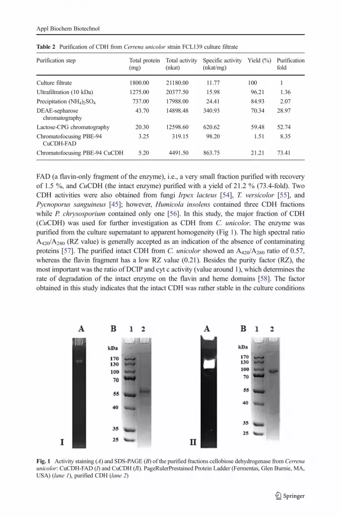
The optimum temperature of CDH was evaluated by measuring the activity of the purifiedenzyme at different temperatures. The enzyme activity was investigated with lactose as asubstrate and two different electron acceptors (DCIP and cyt c). The maximum activity wasrecorded at 60 °C for both substances (Fig. 2A). Similar results were obtained for cellobiosedehydrogenase from Ceriporiopsis subvermispora [57]. Thermostability was examined bymeasurement of the activity over time (Fig. 3a). Complete loss of enzyme activity wasrecorded at 90 °C after heat exposure for 30 min and at 50 °C after 12 h. The enzyme seemedto be more stable than cellobiose dehydrogenase from Pycnoporus cinnabarinus [59].
A
B
0
20
40
60
80
100
4 15 20 30 40 50 60 70 80
Rel
ativ
e ac
tivi
ty [
%]
Temperature [°C]
DCIP cytochrome
0
20
40
60
80
100
2.5 3 3.5 4 4.5 5 5.5 6 6.5 7 7.5 8
Rel
ativ
e ac
tivi
ty [%
]
pH
DCIP cytochrome
Fig. 2 Effect of temperature (a) and pH (b) on activity of Cerrena unicolor CDH. Values represent the mean±SD of triplicate samples
Appl Biochem Biotechnol
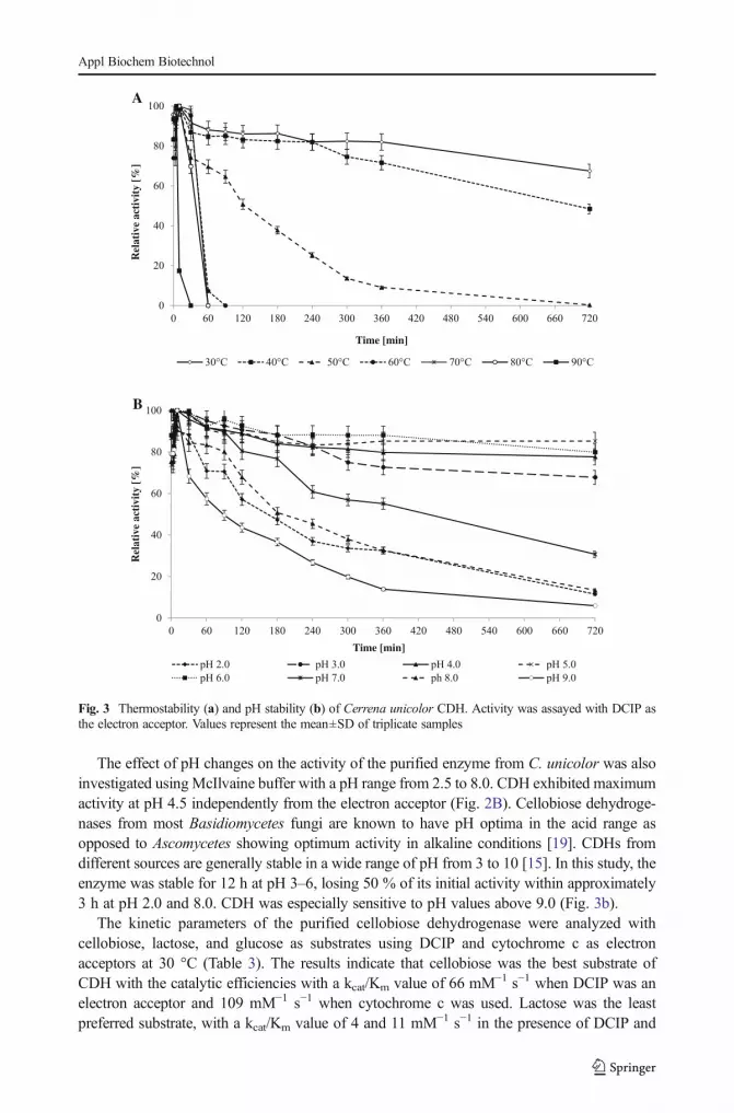
The effect of pH changes on the activity of the purified enzyme from C. unicolor was alsoinvestigated usingMcIlvaine buffer with a pH range from 2.5 to 8.0. CDH exhibited maximumactivity at pH 4.5 independently from the electron acceptor (Fig. 2B). Cellobiose dehydroge-nases from most Basidiomycetes fungi are known to have pH optima in the acid range asopposed to Ascomycetes showing optimum activity in alkaline conditions [19]. CDHs fromdifferent sources are generally stable in a wide range of pH from 3 to 10 [15]. In this study, theenzyme was stable for 12 h at pH 3–6, losing 50 % of its initial activity within approximately3 h at pH 2.0 and 8.0. CDH was especially sensitive to pH values above 9.0 (Fig. 3b).
The kinetic parameters of the purified cellobiose dehydrogenase were analyzed withcellobiose, lactose, and glucose as substrates using DCIP and cytochrome c as electronacceptors at 30 °C (Table 3). The results indicate that cellobiose was the best substrate ofCDH with the catalytic efficiencies with a kcat/Km value of 66 mM−1 s−1 when DCIP was anelectron acceptor and 109 mM−1 s−1 when cytochrome c was used. Lactose was the leastpreferred substrate, with a kcat/Km value of 4 and 11 mM−1 s−1 in the presence of DCIP and
A
B
0
20
40
60
80
100
0 60 120 180 240 300 360 420 480 540 600 660 720
Rel
ativ
e ac
tivi
ty [
%]
Time [min]
30°C 40°C 50°C 60°C 70°C 80°C 90°C
0
20
40
60
80
100
0 60 120 180 240 300 360 420 480 540 600 660 720
Rel
ativ
e ac
tivi
ty [%
]
Time [min]
pH 2.0 pH 3.0 pH 4.0 pH 5.0
pH 6.0 pH 7.0 ph 8.0 pH 9.0
Fig. 3 Thermostability (a) and pH stability (b) of Cerrena unicolor CDH. Activity was assayed with DCIP asthe electron acceptor. Values represent the mean±SD of triplicate samples
Appl Biochem Biotechnol
cytochrome c, respectively. The results obtained suggested that CDH from C. unicolor strainFCL139 was unable to oxidize glucose. Strong discrimination of glucose as a substrate is acharacteristic for the Basidiomycete enzymes belonging to the class I CDHs [15]. Similarvalues of kinetic constants were reported for other fungal cellobiose dehydrogenases [60, 61].
The influence of metal ions and substances that are potential inhibitors of different enzymeson the activity of cellobiose dehydrogenase was tested (Table 4). The activating/inhibitingeffect of the analyzed substances on CDH was dependent on their concentration. The enzymefrom C. unicolor is sensitive to higher concentrations of SDS and CuCl2, similarly to theprotein from P. sanguineus [45]. Azide and cyanide have a slight inhibitory effect just like theCDH from Schizophyllum commune [61]. A similar activating effect in the case of divalentcations was observed by [40]. The other investigated reagents did not have any significanteffect on the CDH activity.
C. unicolor Cellobiose Dehydrogenase Structure
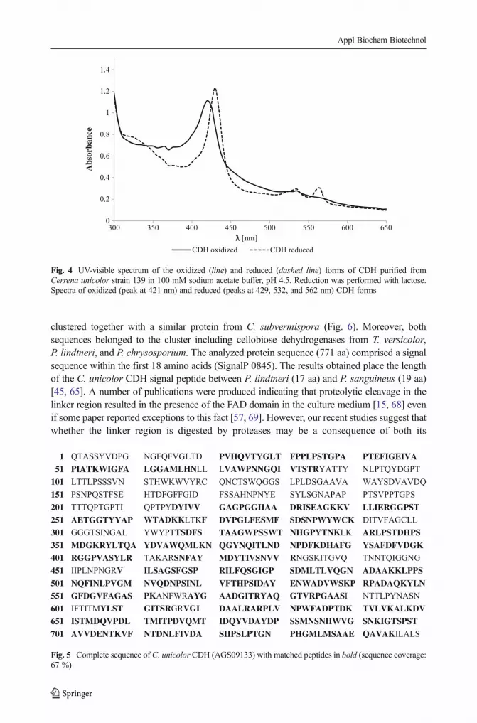
The UV–vis spectra of the oxidized and reduced states of the purified CDH from C. unicolorindicated the presence of heme and flavin cofactors in the protein (Fig. 4). The main absorptionpeak of the oxidized enzyme appearing at 421 nm is typical for heme b, whereas theabsorbance occurring in the region between 450 and 500 nm is mainly attributed to theFAD group [57, 62]. Reduction of the enzyme by addition of lactose resulted in appearanceof peaks at 429, 532, and 562 nm and a decrease in absorbance at wavelengths between 450and 500 nm, which probably represented the reduced form of FAD [62].
The molecular weight of both fragments (CuCDH-FAD and CuCDH) was predicted to be58 and 97 kDa, respectively, as determined by SDS-PAGE analysis (Fig. 1). Most fungalCDHs are monomeric proteins with molecular masses between 80 and 115 kDa [15]. NativePAGE was also performed to identify the enzymatic activities of the proteins (Fig. 1).
Chromatofocusing was used to determine the isoelectric points for CuCDH-FAD andCuCDH, which were detected at pH 5.50 and 4.55, respectively. Many other intact CDHs fromBasidiomycetes, such as P. chrysosporium, I. lacteus, T. versicolor, C. subvermispora, Phlebialindtneri, and P. sanguineus, have acidic isoelectric points ranging from 3.0 to 5.1 [15, 45, 57,63–65]. The protein containing only the FAD domain has a higher pI (5.5–6.7) [45, 63, 64].
Monosaccharide analysis of CDH from C. unicolor strain FCL139 showed that the samplecontained mainly mannose (Man, 74.1 %). Small amounts of glucose (Glc, 6.2 %), galactose(Gal, 2.5 %), and glucosamine (GlcN, 17.2 %) were also present. Summarizing, the
Table 3 Kinetic constants of cellobiose dehydrogenase for carbohydrate substrates
Enzyme Substrate Electron acceptor Km[mM]
Vmax[μM/min]
kcat[s−1]
kcat/Km[mM−1 s−1]
CuCDH-FAD Cellobiose DCIP 0.158 0.034 4.83 30.59
Cyt c – – – –
Lactose DCIP 10.121 0.052 7.39 0.73
Cyt c – – – –
CuCDH Cellobiose DCIP 0.285 0.189 18.86 66.18
Cyt c 0.175 0.191 19.06 108.92
Lactose DCIP 5.241 0.209 20.86 3.98
Cyt c 1.850 0.195 19.46 10.52
Appl Biochem Biotechnol

carbohydrate content of CDH was estimated at 8.2 % using the Dubois method with mannoseas a standard. The up-to-date characterized fungal cellobiose dehydrogenases comprise from8.9 up to 19 % of sugar moiety [66, 67].
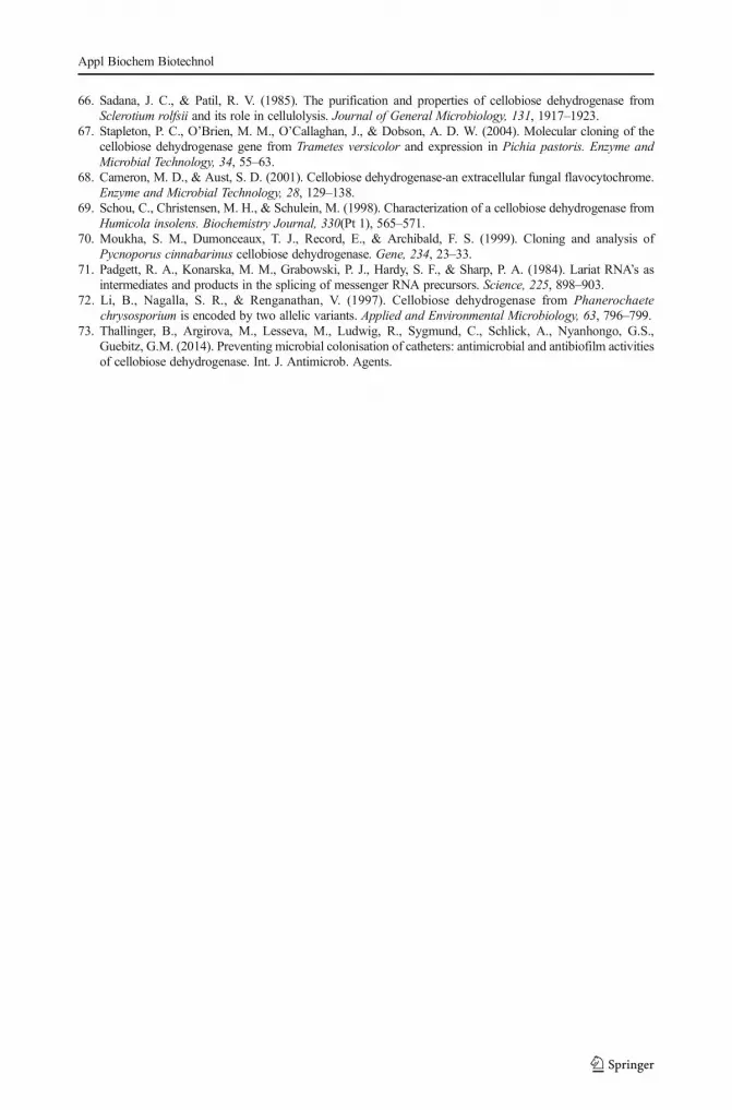
The identity of cellobiose dehydrogenase from C. unicolor was further proved by LC-MS/MS spectrometry analysis of the protein band observed in SDS-PAGE. The MS/MS raw dataobtained were used to search against the NCBI protein database. The analyzed protein wasidentified when the MASCOT probability-based score (p<0.05) was greater than 52. Theprotein from the gel slice was identified as CDH from C. unicolor with a MASCOT score of55,248 and sequence coverage of 67 % (Fig. 5). The deduced molecular mass of C. unicolorCDH (80.9 kDa) was very similar to that determined by in silico analysis of the CDH aminoacid sequence (82.6 kDa).
Molecular Properties of C. unicolor CDH
To our knowledge, this is the first in silico analysis of the Cerrena cellobiose dehydrogenasegene. Analysis of sequenced full-length cDNA of the cdh1 gene from C. unicolor strainFCL139 revealed one open reading frame (ORF) of 2316 bp. The deduced protein sequence ofcdh1 shared similarity of 72 % with P. lindtneri cellobiose dehydrogenase (accession numberAGE45679). The dendrogram obtained from the alignments of 12 cellobiose dehydrogenaseamino acid sequences of Basidiomycetes and Ascomycetes showed that the putative CDH1
Table 4 Effect of metal ions and some reagents on the cellobiose dehydrogenase activity
Relative activity (%)
0.1 mM 1 mM 5 mM 10 mM 50 mM 100 mM
None 100±0.00 100±0.00 100±0.00 100±0.00 100±0.00 100±0.00
Imidazol 110±0.73 109±0.35 107±5.31 102±3.80 97±0.01 67±0.89
EDTA 100±3.58 100±2.11 98±0.25 97±2.48 91±7.44 70±8.57
NaF 100±0.43 96±3.73 93±3.59 93±1.29 58±1.86 46±4.88
KF 100±4.41 99±1.88 95±1.81 89±2.35 58±0.29 49±0.45
KCN 98±1.74 98±2.71 98±0.77 97±0.39 91±0.58 33±1.94
SDS 95±2.41 71±1.30 5±0.01 3±0.01 3±0.56 0±0.00
NH4Cl 102±3.62 102±1.50 102±1.09 99±1.67 97±0.26 97±1.42
Na2SO4 106±5.57 109±1.54 108±8.42 101±0.37 104±0.77 105±2.25
NaCl 94±0.25 92±0.53 94±2.33 94±2.26 97±0.02 97±1.56
KCl 102±0.02 101±0.32 100±0.65 100±3.30 103±1.01 105±2.02
MgCl2 99±0.01 98±1.40 98±1.37 96±1.15 87±0.29 85±1.46
CaCl2 99±1.21 99±0.61 98±3.02 96±2.24 94±1.04 93±0.58
CoCl2 105±0.02 104±0.84 102±0.81 101±0.81 102±2.07 97±0.22
MnCl2 100±1.65 100±1.43 100±2.65 100±2.36 99±3.30 97±3.32
CuCl2 98±0.67 98±1.54 93±0.59 84±2.20 7±0.18 1±1.15
ZnCl2 99±5.11 99±1.48. 99±1.49 97±2.10 95±1.01 95±1.65
MgSO4 101±3.83 101±0.89 101±1.59 101±0.01 100±0.22 99±1.79
MnSO4 102±1.36 101±3.19 103±0.69 100±0.45 100±2.96 100±0.91
CuSO4 101±1.33 96±2.41 96±0.02 91±2.80 86±1.04 86±0.60
ZnSO4 104±1.12 103±2.88 104±3.11 100±0.66 101±0.22 102±3.99
Appl Biochem Biotechnol
clustered together with a similar protein from C. subvermispora (Fig. 6). Moreover, bothsequences belonged to the cluster including cellobiose dehydrogenases from T. versicolor,P. lindtneri, and P. chrysosporium. The analyzed protein sequence (771 aa) comprised a signalsequence within the first 18 amino acids (SignalP 0845). The results obtained place the lengthof the C. unicolor CDH signal peptide between P. lindtneri (17 aa) and P. sanguineus (19 aa)[45, 65]. A number of publications were produced indicating that proteolytic cleavage in thelinker region resulted in the presence of the FAD domain in the culture medium [15, 68] evenif some paper reported exceptions to this fact [57, 69]. However, our recent studies suggest thatwhether the linker region is digested by proteases may be a consequence of both its
0
0.2
0.4
0.6
0.8
1
1.2
1.4
300 350 400 450 500 550 600 650
Abs
orba
nce
[nm]
CDH oxidized CDH reduced
Fig. 4 UV-visible spectrum of the oxidized (line) and reduced (dashed line) forms of CDH purified fromCerrena unicolor strain 139 in 100 mM sodium acetate buffer, pH 4.5. Reduction was performed with lactose.Spectra of oxidized (peak at 421 nm) and reduced (peaks at 429, 532, and 562 nm) CDH forms
1 QTASSYVDPG NGFQFVGLTD PVHQVTYGLT FPPLPSTGPA PTEFIGEIVA
51 PIATKWIGFA LGGAMLHNLL LVAWPNNGQI VTSTRYATTY NLPTQYDGPT
101 LTTLPSSSVN STHWKWVYRC QNCTSWQGGS LPLDSGAAVA WAYSDVAVDQ
151 PSNPQSTFSE HTDFGFFGID FSSAHNPNYE SYLSGNAPAP PTSVPPTGPS
201 TTTQPTGPTI QPTPYDYIVV GAGPGGIIAA DRISEAGKKV LLIERGGPST
251 AETGGTYYAP WTADKKLTKF DVPGLFESMF SDSNPWYWCK DITVFAGCLL
301 GGGTSINGAL YWYPTTSDFS TAAGWPSSWT NHGPYTNKLK ARLPSTDHPS
351 MDGKRYLTQA YDVAWQMLKN QGYNQITLND NPDFKDHAFG YSAFDFVDGK
401 RGGPVASYLR TAKARSNFAY MDYTIVSNVV RNGSKITGVQ TNNTQIGGNG
451 IIPLNPNGRV ILSAGSFGSP RILFQSGIGP SDMLTLVQGN ADAAKKLPPS
501 NQFINLPVGM NVQDNPSINL VFTHPSIDAY ENWADVWSKP RPADAQKYLN
551 GFDGVFAGAS PKANFWRAYG AADGITRYAQ GTVRPGAASI NTTLPYNASN
601 IFTITMYLST GITSRGRVGI DAALRARPLV NPWFADPTDK TVLVKALKDV
651 ISTMDQVPDL TMITPDVQMT IDQYVDAYDP SSMNSNHWVG SNKIGTSPST
701 AVVDENTKVF NTDNLFIVDA SIIPSLPTGN PHGMLMSAAE QAVAKILALS
Fig. 5 Complete sequence of C. unicolor CDH (AGS09133) with matched peptides in bold (sequence coverage:67 %)
Appl Biochem Biotechnol

vulnerability and higher proteolytic activities in the cellulose-based medium. It is probable thatamong many proteases produced by white rot fungi only, one fraction is capable of cleavingcellobiose dehydrogenase. The problem should be addressed in detail in future studiescomprising various techniques and fungal species.
Within the putative protein sequence of C. unicolor CDH, conserved domains typical forfungal cellobiose dehydrogenase were found at positions 23 to 189 aa (heme-bindingcytochrome domain) from 206 to 231 aa (the linker region) and 233 to 769 aa (choline andflavoproteins domain). Moukha et al. [70] and Harreither et al. [19] proposed conservedresidues constituting a putative cellulose binding module in basidiomycetous cellobiosedehydrogenases, which was also found in P. sanguineus CDH [45]. A similar module wasdetected in C. unicolor CDH in positions Tyr-275, Trp-279, Phe-288, Phe-294, Phe-298, Trp-304, Trp-306, and Phe-313. Analysis of N-glycosylation sites (Asn-X-Thr/Ser) showed gly-cosylation points at positions Asn-128, Asn-140, Asn-450, Asn-533, and Asn-615. In com-parison with the P. sanguineus cellobiose dehydrogenase [45], only ten O-glycosylation siteswere found within the linker region.
The complete C. unicolor CDH gene (3038 bp) was amplified by PCR using a genomicDNA as a template and primers designed on the basis of the nucleotide sequence of the cdh1cDNA, as described in the BMaterials and Methods^ section. The position of putative intronswithin the cellobiose dehydrogenase gene was determined by comparison of the genomicDNA and cDNA sequences. Eleven introns were found, ranging in size from 53 to 92 bp andall of them fell into the GT-AG rule [71]. Similarly to P. sanguineus, the cellobiose dehydro-genase gene contains fewer introns, likewise those of P. cinnabarinus [70] or P. chrysosporium[72]; however, the last intron is exceptionally long (92 bp).
Antioxidant Activity Assays
Fungi are producers of a large number of bioactive compounds with antioxidant properties. Inthe earlier studies, we have shown strong antioxidant capability of the fungal CDH fromP. sanguineus [45]. Determination of the antioxidant activity of the newly isolated cellobiosedehydrogenase from C. unicolor is important for future research on its biotechnological
Fig. 6 Unrooted UPGMA-based phylogenetic trees constructed with 12 protein sequences. The dendrogram ofseveral cellobiose dehydrogenases from fungi. AGE97206- Phlebia lindtneri, AAB61455.1- Phanerochaetechrysosporium, AAC50004.1- Trametes versicolor, AGS09133- Cerrena unicolor, ACF60617.1-Ceriporiopsissubvermispora, EJD48894- Auricularia delicata TFB-10046 SS5, EAA27355.1- Neurospora crassa OR74A,ELQ44991.1- Magnaporthe oryzae Y34, EEY23987.1- Verticillium alfalfae VaMs.102, ENH81675.1-Colletotrichum orbiculare MAFF 240422, ENH71366.1- Fusarium oxysporum f. sp. cubense race 1,AAF69005.1- Humicola insolens
Appl Biochem Biotechnol
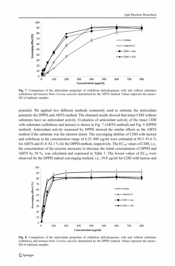
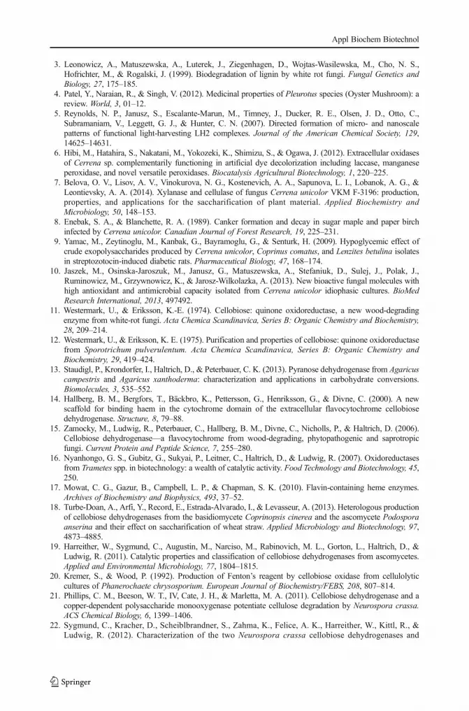
potential. We applied two different methods commonly used to estimate the antioxidantpotential: the DPPH and ABTS method. The obtained results showed that intact CDH withoutsubstrates have no antioxidant activity. Evaluation of antioxidant activity of the intact CDHwith substrates (cellobiose and lactose) is shown in Fig. 7 (ABTS method) and Fig. 8 (DPPHmethod). Antioxidant activity measured by DPPH showed the similar effects as the ABTSmethod if the substrate was the electron donor. The scavenging abilities of CDH with lactoseand cellobiose at the concentration range of 6.25–800 μg/ml were estimated at 89.3–91.6 %for ABTS and 81.8–82.1 % for the DPPH method, respectively. The EC50 values of CDH, i.e.,the concentration of the enzyme necessary to decrease the initial concentration of DPPH andABTS by 50 %, was calculated and expressed in Table 5. The lowest values of EC50 wereobserved for the DPPH radical scavenging method, i.e., 39.8 μg/ml for CDH with lactose and
Fig. 7 Comparison of the antioxidant properties of cellobiose dehydrogenase with and without substrates(cellobiose and lactose) from Cerrena unicolor determined by the ABTS method. Values represent the mean±SD of triplicate samples
0
10
20
30
40
50
60
70
80
90
100
0 100 200 300 400 500 600 700 800
Scav
engi
ng e
ffec
t [%
]
Concentration [µg/ml]
trolox
vitamin C
CDH + Lac
CDH + Cel
Fig. 8 Comparison of the antioxidant properties of cellobiose dehydrogenase with and without substrates(cellobiose and lactose) from Cerrena unicolor determined by the DPPH method. Values represent the mean±SD of triplicate samples
Appl Biochem Biotechnol

48.5 μg/ml for CDH with cellobiose. The EC50 values for the ABTS method used for testingthe CDH antioxidative properties were 103.7 and 93 μg/ml, respectively. All the resultsobtained indicate strong redox potential of the CDH enzyme only in the presence of lactoseand cellobiose substrates. Therefore, it would be interesting to investigate whether C. unicolorCDH may be applied as an antimicrobial agent, as recently described for Myriococcumthermophilum cellobiose dehydrogenase [73].
C. unicolor strain FCL139 was proven to produce not only biotechnologically importantlaccase but also cellobiose dehydrogenase with interesting features as well. Given the size andgene content of the available C. unicolor genome, its CDH is only part of the wood degradingmachinery and it would be interesting to characterize in detail the abilities of this white rotfungus of decompose lignocellulose. The recent explosion of interest in cellobiose dehydro-genase, which was proven to act in concert with LPMO (lytic polysaccharide monooxygenase)in cellulose breakdown and was successfully applied in different biotechnological areas,encourages scientists all over the world to search for new CDH with exceptional features.By applying high throughput techniques, new insight into the wood decomposition is possible.In consequence, the acquired knowledge will contribute to accelerating the application of thediscovered and characterized enzymes in new biotechnological areas.
Acknowledgments This research was supported by the grant Preludium (2011/01/N/NZ1/03458) from theNational Science Centre in Poland and the research program BS/UMCS.
Open Access This article is distributed under the terms of the Creative Commons Attribution 4.0 InternationalLicense (http://creativecommons.org/licenses/by/4.0/), which permits unrestricted use, distribution, and repro-duction in any medium, provided you give appropriate credit to the original author(s) and the source, provide alink to the Creative Commons license, and indicate if changes were made.
References
1. Kellner, H., Zak, D. R., & Vandenbol, M. (2010). Correction: fungi unearthed: transcripts encodinglignocellulolytic and chitinolytic enzymes in forest soil. PLoS One, 5, 1–7.
2. Novotný, Č., Cajthaml, T., Svobodova, K., Šušla, M., & Šašek, V. (2009). Irpex lacteus, a white-rot funguswith biotechnological potential—review. Folia Microbiologica, 54, 375–390.
Table 5 EC50 values (effective concentration at which the radicals present in the investigated samples werescavenged by 50 %; the antioxidant activity was 50 %) of CDH isolated from C. unicolor submerged cultures incomparison to Trolox and vitamin C
EC50 (μg/mL)
ABTS radical scavenging DPPH radical scavenging
Trolox 251.6±7.86 28.4±0.72
Vitamin C 151.3±5.14 25.1±0.61
CDH – –
CDH+lactose 103.7±3.76 39.8±1.42
CDH+cellobiose 93.0±2.53 48.5±1.56
All results are expressed as mean±SD from three experiments (n=3). Values within the column and the row forinvestigated samples are significantly different (P≤0.05)
Appl Biochem Biotechnol
3. Leonowicz, A., Matuszewska, A., Luterek, J., Ziegenhagen, D., Wojtas-Wasilewska, M., Cho, N. S.,Hofrichter, M., & Rogalski, J. (1999). Biodegradation of lignin by white rot fungi. Fungal Genetics andBiology, 27, 175–185.
4. Patel, Y., Naraian, R., & Singh, V. (2012). Medicinal properties of Pleurotus species (Oyster Mushroom): areview. World, 3, 01–12.
5. Reynolds, N. P., Janusz, S., Escalante-Marun, M., Timney, J., Ducker, R. E., Olsen, J. D., Otto, C.,Subramaniam, V., Leggett, G. J., & Hunter, C. N. (2007). Directed formation of micro- and nanoscalepatterns of functional light-harvesting LH2 complexes. Journal of the American Chemical Society, 129,14625–14631.
6. Hibi, M., Hatahira, S., Nakatani, M., Yokozeki, K., Shimizu, S., & Ogawa, J. (2012). Extracellular oxidasesof Cerrena sp. complementarily functioning in artificial dye decolorization including laccase, manganeseperoxidase, and novel versatile peroxidases. Biocatalysis Agricultural Biotechnology, 1, 220–225.
7. Belova, O. V., Lisov, A. V., Vinokurova, N. G., Kostenevich, A. A., Sapunova, L. I., Lobanok, A. G., &Leontievsky, A. A. (2014). Xylanase and cellulase of fungus Cerrena unicolor VKM F-3196: production,properties, and applications for the saccharification of plant material. Applied Biochemistry andMicrobiology, 50, 148–153.
8. Enebak, S. A., & Blanchette, R. A. (1989). Canker formation and decay in sugar maple and paper birchinfected by Cerrena unicolor. Canadian Journal of Forest Research, 19, 225–231.
9. Yamac, M., Zeytinoglu, M., Kanbak, G., Bayramoglu, G., & Senturk, H. (2009). Hypoglycemic effect ofcrude exopolysaccharides produced by Cerrena unicolor, Coprinus comatus, and Lenzites betulina isolatesin streptozotocin-induced diabetic rats. Pharmaceutical Biology, 47, 168–174.
10. Jaszek, M., Osinska-Jaroszuk, M., Janusz, G., Matuszewska, A., Stefaniuk, D., Sulej, J., Polak, J.,Ruminowicz, M., Grzywnowicz, K., & Jarosz-Wilkolazka, A. (2013). New bioactive fungal molecules withhigh antioxidant and antimicrobial capacity isolated from Cerrena unicolor idiophasic cultures. BioMedResearch International, 2013, 497492.
11. Westermark, U., & Eriksson, K.-E. (1974). Cellobiose: quinone oxidoreductase, a new wood-degradingenzyme from white-rot fungi. Acta Chemica Scandinavica, Series B: Organic Chemistry and Biochemistry,28, 209–214.
12. Westermark, U., & Eriksson, K. E. (1975). Purification and properties of cellobiose: quinone oxidoreductasefrom Sporotrichum pulverulentum. Acta Chemica Scandinavica, Series B: Organic Chemistry andBiochemistry, 29, 419–424.
13. Staudigl, P., Krondorfer, I., Haltrich, D., & Peterbauer, C. K. (2013). Pyranose dehydrogenase from Agaricuscampestris and Agaricus xanthoderma: characterization and applications in carbohydrate conversions.Biomolecules, 3, 535–552.
14. Hallberg, B. M., Bergfors, T., Bäckbro, K., Pettersson, G., Henriksson, G., & Divne, C. (2000). A newscaffold for binding haem in the cytochrome domain of the extracellular flavocytochrome cellobiosedehydrogenase. Structure, 8, 79–88.
15. Zamocky, M., Ludwig, R., Peterbauer, C., Hallberg, B. M., Divne, C., Nicholls, P., & Haltrich, D. (2006).Cellobiose dehydrogenase—a flavocytochrome from wood-degrading, phytopathogenic and saprotropicfungi. Current Protein and Peptide Science, 7, 255–280.
16. Nyanhongo, G. S., Gubitz, G., Sukyai, P., Leitner, C., Haltrich, D., & Ludwig, R. (2007). Oxidoreductasesfrom Trametes spp. in biotechnology: a wealth of catalytic activity. Food Technology and Biotechnology, 45,250.
17. Mowat, C. G., Gazur, B., Campbell, L. P., & Chapman, S. K. (2010). Flavin-containing heme enzymes.Archives of Biochemistry and Biophysics, 493, 37–52.
18. Turbe-Doan, A., Arfi, Y., Record, E., Estrada-Alvarado, I., & Levasseur, A. (2013). Heterologous productionof cellobiose dehydrogenases from the basidiomycete Coprinopsis cinerea and the ascomycete Podosporaanserina and their effect on saccharification of wheat straw. Applied Microbiology and Biotechnology, 97,4873–4885.
19. Harreither, W., Sygmund, C., Augustin, M., Narciso, M., Rabinovich, M. L., Gorton, L., Haltrich, D., &Ludwig, R. (2011). Catalytic properties and classification of cellobiose dehydrogenases from ascomycetes.Applied and Environmental Microbiology, 77, 1804–1815.
20. Kremer, S., & Wood, P. (1992). Production of Fenton’s reagent by cellobiose oxidase from cellulolyticcultures of Phanerochaete chrysosporium. European Journal of Biochemistry/FEBS, 208, 807–814.
21. Phillips, C. M., Beeson, W. T., IV, Cate, J. H., & Marletta, M. A. (2011). Cellobiose dehydrogenase and acopper-dependent polysaccharide monooxygenase potentiate cellulose degradation by Neurospora crassa.ACS Chemical Biology, 6, 1399–1406.
22. Sygmund, C., Kracher, D., Scheiblbrandner, S., Zahma, K., Felice, A. K., Harreither, W., Kittl, R., &Ludwig, R. (2012). Characterization of the two Neurospora crassa cellobiose dehydrogenases and
Appl Biochem Biotechnol

their connection to oxidative cellulose degradation. Applied and Environmental Microbiology, 78,6161–6171.
23. Bey, M., Zhou, S., Poidevin, L., Henrissat, B., Coutinho, P. M., Berrin, J.-G., & Sigoillot, J.-C.(2013). Cello-oligosaccharide oxidation reveals differences between two lytic polysaccharidemonooxygenases (family GH61) from Podospora anserina. Applied and EnvironmentalMicrobiology, 79, 488–496.
24. Ludwig, R., Ozga, M., Zámocky, M., Peterbauer, C., Kulbe, K. D., & Haltrich, D. (2004). Continuousenzymatic regeneration of electron acceptors used by flavoenzymes: cellobiose dehydrogenase-catalyzedproduction of lactobionic acid as an example. Biocatalysis and Biotransformation, 22, 97–104.
25. Hilden, L., & Johansson, G. (2004). Recent developments on cellulases and carbohydrate-binding moduleswith cellulose affinity. Biotechnology Letters, 26, 1683–1693.
26. Harreither, W., Coman, V., Ludwig, R., Haltrich, D., & Gorton, L. (2007). Investigation of graphiteelectrodes modified with cellobiose dehydrogenase from the ascomycete Myriococcum thermophilum.Electroanalysis, 19, 172–180.
27. Safina, G., Ludwig, R., & Gorton, L. (2010). A simple and sensitive method for lactose detection based ondirect electron transfer between immobilised cellobiose dehydrogenase and screen-printed carbon electrodes.Electrochimica Acta, 55, 7690–7695.
28. Stoica, L., Ludwig, R., Haltrich, D., & Gorton, L. (2006). Third-generation biosensor for lactose based onnewly discovered cellobiose dehydrogenase. Analytical Chemistry, 78, 393–398.
29. Nistor, C., Rose, A., Farré, M., Stoica, L., Wollenberger, U., Ruzgas, T., Pfeiffer, D., Barceló, D., Gorton, L.,& Emnéus, J. (2002). In-field monitoring of cleaning efficiency in waste water treatment plants using twophenol-sensitive biosensors. Analytica Chimica Acta, 456, 3–17.
30. Rabinovich, M. L., Vasil’chenko, L. G., Karapetyan, K. N., Shumakovich, G. P., Yershevich, O. P., Ludwig,R., Haltrich, D., Hadar, Y., Kozlov, Y. P., & Yaropolov, A. I. (2007). Application of cellulose-based self-assembled tri-enzyme system in a pseudo-reagent-less biosensor for biogenic catecholamine detection.Biotechnology Journal, 2, 546–558.
31. Ludwig, R., Ortiz, R., Schulz, C., Harreither, W., Sygmund, C., & Gorton, L. (2013). Cellobiose dehydro-genase modified electrodes: advances by materials science and biochemical engineering. Analytical andBioanalytical Chemistry, 405, 3637–3658.
32. Nyanhongo, G. S., Sygmund, C., Ludwig, R., Prasetyo, E. N., & Guebitz, G. M. (2013). Synthesis ofmultifunctional bioresponsive polymers for the management of chronic wounds. Journal of BiomedicalMaterials Research Part B: Applied Biomaterials, 101, 882–891.
33. Nyanhongo, G. S., Sygmund, C., Ludwig, R., Prasetyo, E. N., & Guebitz, G. M. (2013). An antioxidantregenerating system for continuous quenching of free radicals in chronic wounds. European Journal ofPharmaceutics and Biopharmaceutics, 83, 396–404.
34. Lindeberg, G., & Holm, G. (1952). Occurrence of tyrosinase and laccase in fruit bodies and mycelia of someHymenomycetes. Physiologia Plantarum, 5, 100–114.
35. Fang, J., Huang, F., & Gao, P. J. (1999). Optimization of cellobiose dehydrogenase production bySchizophyllum commune and effect of the enzyme on kraft pulp bleaching by ligninases. ProcessBiochemistry (Amsterdam, Netherlands), 34, 957–961.
36. Rogalski, J., & Dawidowicz, A. (1989). Controlled porous glass (CPG) with reactive epoxy groups assupport for affinity chromatography I. Optimization of CPG modification and the binding of glucose withmodified surface. Acta Biotechnologica, 9, 275–283.
37. Dawidowicz, A., & Rogalski, J. (1988). Sposób otrzymywania nośników z powierzchnią modyfikowanąalkoksysilanami. (ed Maria Curie-Sklodowska University, L., Poland). Poland.
38. Rogalski, J. and Dawidowicz, A. (1990) The preparation of supports with the reactive epoxy groups. (edMaria Curie-Sklodowska University, L., Poland). Poland.
39. Kordan, W., Malinowska, A., Lecewicz, M., Wysocki, P., Fraser, L., & Strzezek, J. (2007). The structure ofplatelet-activating factor acetylhydrolase (PAF-AH) isolated from boar seminal plasma and examined usingmass spectrometry. Animal Science Papers and Reports, 25, 289–295.
40. Kracher, D., Zahma, K., Schulz, C., Sygmund, C., Gorton, L. and Ludwig, R. (2015). Interdomain electrontransfer in cellobiose dehydrogenase: modulation by pH and divalent cations. FEBS J.
41. Laemmli, U. K. (1970). Cleavage of structural proteins during the assembly of the head of bacteriophage T4.Nature, 227, 680–685.
42. Walker, J. M. (2002). The protein protocols handbook (2nd ed.). Totowa: Humana Press.43. Sawardeker, J. S., Sloneker, J., & Jeanes, A. (1965). Quantitative determination of monosaccharides as their
alditol acetates by gas liquid chromatography. Analytical Chemistry, 37, 1602–1604.44. Dubois, M., Gilles, K. A., Hamilton, J. K., Rebers, P., & Smith, F. (1956). Colorimetric method for
determination of sugars and related substances. Analytical Chemistry, 28, 350–356.
Appl Biochem Biotechnol

45. Sulej, J., Janusz, G., Osinska-Jaroszuk, M., Malek, P., Mazur, A., Komaniecka, I., Choma, A., & Rogalski, J.(2013). Characterization of cellobiose dehydrogenase and its FAD-domain from the ligninolytic basidiomy-cete Pycnoporus sanguineus. Enzyme and Microbial Technology, 53, 427–437.
46. Paduch, R., Matysik, G., Wojciak-Kosior, M., Kandefer-Szerszen, M., Skalska-Kaminska, A., Nowak-Kryska, M., & Niedziela, P. (2008). Lamium album extracts express free radical scavenging and cytotoxicactivities. Polish Journal of Environmental Studies, 17, 569–580.
47. Re, R., Pellegrini, N., Proteggente, A., Pannala, A., Yang, M., & Rice-Evans, C. (1999). Antioxidant activityapplying an improved ABTS radical cation decolorization assay. Free Radical Biology and Medicine, 26,1231–1237.
48. Sambrook, J., & Russell, D.W. (2006). The condensed protocols from molecular cloning: a laboratorymanual. ed. Cold Spring Harbor Laboratory Press, Cold Spring Harbor, N.Y.
49. Borges, M. J., Azevedo, M. O., Bonatelli, J. R., Felipe, M. S. S., & Astolfi-Filho, S. (1990). A practicalmethod for the preparation of total DNA from filamentous fungi. Fungal General Newsletter, 10, 11.
50. Thompson JD, Higgins DG, Gibson TJ. Clustal-W improving the sensitivity of progressive multiplesequence alignment through sequence weighting, position-specific gap penaltiesand weight matrix choice.Nucleic Acids Res 1994;22:4673-4680
51. Page RDM. TreeView: An application to display phylogenetic trees on personal computers. ComputerApplications in the Biosciences 1996;12:357-358
52. Steentoft, C., Vakhrushev, S. Y., Joshi, H. J., Kong, Y., Vester-Christensen, M. B., Schjoldager, K. T.,Lavrsen, K., Dabelsteen, S., Pedersen, N. B., Marcos-Silva, L., Gupta, R., Bennett, E. P., Mandel, U.,Brunak, S., Wandall, H. H., Levery, S. B., & Clausen, H. (2013). Precision mapping of the human O-GalNAc glycoproteome through SimpleCell technology. The EMBO Journal, 32, 1478–1488.
53. Geer, L. Y., Domrachev, M., Lipman, D. J., & Bryant, S. H. (2002). CDART: protein homology by domainarchitecture. Genetical Research, 12, 1619–1623.
54. Hai, P. Q., Nozaki, K., Amano, Y., & Kanda, T. (2000). Purification and characterization of cellobiosedehydrogenase from Irpex lacteus and its adsorption on cellulose. Journal of Applied Glycoscience, 47, 311–318.
55. Roy, B. P., Dumonceaux, T., Koukoulas, A. A., & Archibald, F. S. (1996). Purification and characterizationof cellobiose dehydrogenases from the white rot fungus Trametes versicolor. Applied and EnvironmentalMicrobiology, 62, 4417–4427.
56. Igarashi, K., Verhagen, M. F., Samejima, M., Schulein, M., Eriksson, K. E., & Nishino, T. (1999). Cellobiosedehydrogenase from the fungi Phanerochaete chrysosporium and Humicola insolens. A flavohemoproteinfrom Humicola insolens contains 6-hydroxy-FAD as the dominant active cofactor. Journal of BiologicalChemistry, 274, 3338–3344.
57. Harreither, W., Sygmund, C., Dunhofen, E., Vicuna, R., Haltrich, D., & Ludwig, R. (2009). Cellobiosedehydrogenase from the ligninolytic basidiomycete Ceriporiopsis subvermispora. Applied andEnvironmental Microbiology, 75, 2750–2757.
58. Ludwig, R., & Haltrich, D. (2002). Cellobiose dehydrogenase production by Sclerotium species pathogenicto plants. Letters in Applied Microbiology, 35, 261–266.
59. Sigoillot, C., Lomascolo, A., Record, E., Robert, J. L., Asther, M., & Sigoillot, J. C. (2002). Lignocellulolyticand hemicellulolytic system of Pycnoporus cinnabarinus: isolation and characterization of a cellobiosedehydrogenase and a new xylanase. Enzyme and Microbial Technology, 31, 876–883.
60. Ludwig, R., Salamon, A., Varga, J., Zamocky, M., Peterbauer, C., Kulbe, K., & Haltrich, D. (2004).Characterisation of cellobiose dehydrogenases from the white-rot fungi Trametes pubescens and Trametesvillosa. Applied Microbiology and Biotechnology, 64, 213–222.
61. Fang, J., Liu, W., & Gao, P. (1998). Cellobiose dehydrogenase from Schizophyllum commune: purificationand study of some catalytic, inactivation, and cellulose-binding properties. Archives of Biochemistry andBiophysics, 353, 37–46.
62. Baminger, U., Subramaniam, S. S., Renganathan, V., & Haltrich, D. (2001). Purification and characterizationof cellobiose dehydrogenase from the plant pathogen Sclerotium (Athelia) rolfsii. Applied and EnvironmentalMicrobiology, 67, 1766–1774.
63. Mikiashvili, N., Elisashvili, V., Worku, M., Davitashvili, E., & Isikhuemhen, O. S. (2009). Purification andcharacterization of a lectin isolated from the submerged cultivated mycelium of grey polypore Cerrenaunicolor (Bull.) Murrill (Aphyllophoromycetideae). International Journal of Medicinal Mushrooms, 11, 61–68.
64. Barron, G.L. (1999). Mushrooms of Northeast North America: Midwest to New England. ed. LonePine Pub.
65. Sulej, J., Janusz, G., Mazur, A., Zuber, K., Zebracka, A., & Rogalski, J. (2013). Cellobiose dehydrogenasefrom the ligninolytic basidiomycete Phlebia lindtneri. Process Biochemistry, 48, 1715–1723.
Appl Biochem Biotechnol
66. Sadana, J. C., & Patil, R. V. (1985). The purification and properties of cellobiose dehydrogenase fromSclerotium rolfsii and its role in cellulolysis. Journal of General Microbiology, 131, 1917–1923.
67. Stapleton, P. C., O’Brien, M. M., O’Callaghan, J., & Dobson, A. D. W. (2004). Molecular cloning of thecellobiose dehydrogenase gene from Trametes versicolor and expression in Pichia pastoris. Enzyme andMicrobial Technology, 34, 55–63.
68. Cameron, M. D., & Aust, S. D. (2001). Cellobiose dehydrogenase-an extracellular fungal flavocytochrome.Enzyme and Microbial Technology, 28, 129–138.
69. Schou, C., Christensen, M. H., & Schulein, M. (1998). Characterization of a cellobiose dehydrogenase fromHumicola insolens. Biochemistry Journal, 330(Pt 1), 565–571.
70. Moukha, S. M., Dumonceaux, T. J., Record, E., & Archibald, F. S. (1999). Cloning and analysis ofPycnoporus cinnabarinus cellobiose dehydrogenase. Gene, 234, 23–33.
71. Padgett, R. A., Konarska, M. M., Grabowski, P. J., Hardy, S. F., & Sharp, P. A. (1984). Lariat RNA’s asintermediates and products in the splicing of messenger RNA precursors. Science, 225, 898–903.
72. Li, B., Nagalla, S. R., & Renganathan, V. (1997). Cellobiose dehydrogenase from Phanerochaetechrysosporium is encoded by two allelic variants. Applied and Environmental Microbiology, 63, 796–799.
73. Thallinger, B., Argirova, M., Lesseva, M., Ludwig, R., Sygmund, C., Schlick, A., Nyanhongo, G.S.,Guebitz, G.M. (2014). Preventing microbial colonisation of catheters: antimicrobial and antibiofilm activitiesof cellobiose dehydrogenase. Int. J. Antimicrob. Agents.
Appl Biochem Biotechnol