selenium-containing xanthine dehydrogenase from eubacterium barkeri
TRANSCRIPT

Selenium-containing xanthine dehydrogenase from Eubacterium barkeri
Thomas SchraÈ der1, Annette RienhoÈ fer2 and Jan R. Andreesen1
1Institut fuÈr Mikrobiologie, Martin-Luther-UniversitaÈt Halle, Halle, Germany; 2Institut fuÈr Mikrobiologie, UniversitaÈt GoÈttingen, GoÈtingen, Germany
A specific dehydrogenase, different from nicotinic acid hydroxylase, was induced during growth of Eubacterium
barkeri on xanthine. The protein designated as xanthine dehydrogenase was enriched 39-fold to apparent
homogeneity using a three-step purification scheme. It exhibited an NADP-dependent specific activity of
164 mmol xanthine oxidized per min and per mg of protein. In addition it showed an NADPH-dependent oxidase
and diaphorase activity. A molecular mass of 530 kDa was determined for the native enzyme and SDS/PAGE
revealed three types of subunits with molecular masses of 17.5, 30 and 81 kDa indicating a dodecameric native
structure. Molybdopterin was identified as the molybdenum-complexing cofactor using activity reconstitution
experiments and fluorescence measurements after KI/I2 oxidation. The molecular mass of the cofactor indicated
that it is of the dinucleotide type. The enzyme contained iron, acid-labile sulfur, molybdenum, tungsten, selenium
and FAD at molar ratios of 17.5, 18.4, 2.3, 1.1, 0.95 and 2.8 per mol of native enzyme. Xanthine dehydrogenase
was inactivated upon incubation with arsenite, cyanide and different purine analogs. Reconstitution experiments
of xanthine dehydrogenase activity by addition of selenide and selenite performed with cyanide-inactivated
enzyme and with chloramphenicol-treated cells, respectively, indicated that selenium is not attached to the
protein in a covalently bound form such as selenocysteine.
Keywords: Eubacterium barkeri; molybdenum; selenium; tungsten; xanthine dehydrogenase.
Xanthine dehydrogenases (XDH) catalyzing the oxidation ofhypoxanthine to xanthine and of xanthine to uric acid wereisolated from different eukaryotic and prokaryotic sources [1].According to the structure of the molybdenum center at theactive site, XDHs belong to the xanthine oxidase family ofmononuclear molybdoenzymes [1]. These enzymes containmolybdenum coordinated to the dithiolene moiety of the pterincofactor and to an oxo-, sulfido- and water-ligand [1]. Theeukaryotic enzymes are composed of two identical subunits of< 150 kDa containing a molybdopterin, two 2Fe/2S centersand an FAD [2]. The same cofactors were found in theirprokaryotic counterparts, except that the correspondingdomains were located on one, two or three different subunits[3±7]. Different reports exist about the chemical structure ofthe pterin cofactor of bacterial XDHs. In the case of, forexample, Veillonella atypica it was identified as molybdopterindinucleotide, whereas Pseudomonas putida 86 contains amolybdopterin [4,5]. Quite unusual XDHs were reported tobe present in P. putida Fu1, Clostridium acidiurici andC. cylindrosporum, respectively [8±10]. The former enzymeis composed of two different subunits and contains cytochromeb instead of the flavin [10]. Evidence was presented that theXDHs of the two purinolytic Clostridium species requireselenium for activity [8,9]. Selenium was also found in othermolybdoenzymes, for example, formate dehydrogenases fromEscherichia coli and nicotinic acid hydroxylase (NAH) from
Eubacterium barkeri (formerly C. barkeri; [11,12±15]). How-ever, in case of formate dehydrogenase from E. coli, theselenium is covalently bound to the protein in the form ofselenocysteine [14]. Analysis of the crystal structure showedthat the selenium is coordinated to the molybdenum andinvolved in the catalytic mechanism [16]. NAH from E. barkericatalyzes the initial NADP-dependent oxidation of nicotinicacid [17]. Growth of E. barkeri on nicotinic acid was dependenton selenium supplementation of the medium and was due to thepresence of a selenium cofactor in NAH [12,13]. Furtherinvestigations revealed that NAH consists of four dissimilarsubunits and that the selenium could be released from theprotein as a low molecular mass compound [15,18]. EPRstudies showed that the selenium moiety that seems to bedifferent from selenocysteine is directly coordinated to themolybdenum [18]. Thus, nicotinic acid hydroxylase fromE. barkeri was the first enzyme shown to contain selenium ina form different from selenocysteine. The structure of theselenium moiety has not been resolved until now, although ithas been suggested that it is a ligand of the molybdenum atomas observed for sulfur or oxygen in other molybdoenzymes [1].
The results presented here clearly demonstrate that E. barkeriis able to express XDH as a second selenium-containing andmolybdopterin-containing enzyme under appropriate growthconditions such as providing purine compounds as the growthsubstrate. This enzyme has some characteristic features bywhich it can be clearly distinguished from NAH. Evidence ispresented that the selenium is incorporated post-translationallyinto the protein.
E X P E R I M E N T A L P R O C E D U R E S
Materials
DEAE±Sephacel, phenyl±Sepharose and Sepharose CL-6Bwere from Pharmacia Biotech (Freiburg, Germany). Molecular
Eur. J. Biochem. 264, 862±871 (1999) q FEBS 1999
Correspondence to J. R. Andreesen, Institut fuÈr Mikrobiologie,
UniversitaÈt Halle, Kurt-Mothes-Str. 3, D-06099 Halle, Germany.
Fax: +49 345 552 7010, Tel.: + 49 345 552 6350,
E-mail: [email protected]
Abbreviations: NAH, nicotinic acid hydroxylase; XDH, xanthine
dehydrogenase.
Enzymes: nicotinic acid hydroxylase (EC not yet assigned); xanthine
dehydrogenase (EC 1.1.1.204).
(Received 22 March 1999, revised 21 May 1999, accepted 28 June 1999)

q FEBS 1999 Xanthine dehydrogenase from Eubacterium barkeri (Eur. J. Biochem. 264) 863
mass standards for SDS/PAGE, silver-stain kits and HPLCcolumns were obtained from Bio-Rad (MuÈnchen, Germany).Hypoxanthine, xanthine and chloramphenicol were purchasedfrom Sigma Chemie (Deisenhofen, Germany). NAD(P) wasfrom Boehringer (Mannheim, Germany). Benzylviologen andmethylviologen were obtained from Fluka (Heidelberg,Germany). All chemicals were at least of analytical reagentgrade.
Growth conditions and preparation of cell extract
E. barkeri, DSM 1223 [11,19], was routinely grown anae-robically [20] in a mineral salts medium [12,19] supplementedwith 1% yeast extract, 0.05% cysteine as reducing agent,Na2MoO4, Na2WO4 and Na2SeO3 (each 1027 m). The pH wasadjusted to 8.2 and the temperature was set at 37 8C. Inductionof XDH was achieved using uric acid (12 mm) xanthine(13 mm) or hypoxanthine (14.7 mm), respectively, as thegrowth substrate. Nicotinic acid (40 mm) was used as asubstrate to induce NAH. Growth was monitored by measuringthe increase in A600. The degradation of uric acid, xanthine andhypoxanthine was followed by the decrease in A290, A278 andA250, respectively. The influence of selenite on the activity ofxanthine dehydrogenase was investigated by omitting it fromthe medium. Cells were harvested from 20 L cultures at the endof the logarithmic growth phase and immediately frozen at220 8C under anaerobic conditions until used.
Preparation of cell extracts was carried out under anaerobicconditions. Frozen cells of E. barkeri were suspended in 50 mmanaerobic potassium phosphate buffer, pH 7.8 (buffer A)containing 2 mm dithiothreitol in a ratio of 1 g of cells (wetweight) per 2 mL buffer. After addition of 0.5 mg lysozymeand 0.05 mg DNase I per g of cells, the suspension wasincubated at 37 8C for 15 min under nitrogen. Subsequently, thesuspension was passed twice through a French pressure cell at140 MPa (American Instruments Inc., USA). Cell debris wereremoved by centrifugation at 55 000 g (30 min, 4 8C). Thesupernatant (cell extract) was stored anaerobically at 220 8C orused directly.
Enzyme assay
The standard assay mixture for determination of XDH activitycontained 5 mm xanthine and 0.5 mm NADP in buffer A. Allsolutions were prepared in the absence of oxygen. The reactionwas started either by the addition of 1±5 mL of the enzymesolution or the substrate. Activity was monitored by recordingthe increase in A365 using an 1 of 3.5 mm21´cm21. One unit ofspecific activity was defined as 1 mmol of NADP reduced permin and mg of protein at 37 8C. NAH activity was estimatedunder the same conditions using nicotinic acid (5 mm) as asubstrate. Protein was determined according to the methoddescribed by Lowry et al. [21]. The substrate specificity ofXDH was investigated using the standard assay, except thatxanthine was replaced by the corresponding compound. Unlessindicated otherwise, inhibition of XDH was estimated byadding the putative inhibitor to the standard assay.
The diaphorase activity of XDH was estimated anaerobicallyin buffer A containing 5 mm electron acceptor (2,6-dichloro-phenol indophenol, methyl viologen, benzyl viologen ormethylene blue) and 0.3 mm NADPH. The reaction wasinitiated by addition of the enzyme as indicated above.
NADPH oxidase activity was recorded under aerobicconditions using buffer A containing 0.3 mm NADPH at
saturating oxygen concentrations. The decrease in A365 wasdetermined after addition of the enzyme solution.
Enzyme purification
All purification steps, unless stated otherwise, were carried outin a glove box (Mecaplex, Switzerland) under anaerobicconditions at 4 8C. The cell extract was brought to a finalconcentration of 50% ammonium sulfate by addition of thesolid salt. After stirring for 20 min, the precipitated protein wascollected by centrifugation at 40 000 g and resolved in a lowvolume of buffer A, containing 1 mm sodium dithionite and0.05 mm methyl viologen as redox indicator (buffer B). Theremaining salt was removed by dialysis for 12 h againstbuffer B. Subsequently, the protein solution was applied to aDEAE±Sephacel column equilibrated previously with buffer B.Unbound protein was eluted by washing with < 3 columnvolumes of equilibration buffer. The XDH was eluted by anincreasing gradient of 0.2±0.4 m potassium chloride in bufferB. Active fractions were pooled and concentrated by ultra-filtration. The protein was then applied to a Sepharose CL-6Bcolumn equilibrated with buffer B. Fractions containing XDHactivity were analyzed for homogeneity by native-agarose [22]and SDS/PAGE [23]. Gels were stained with either CoomassieBrilliant Blue [24] or silver stain [25]. Activity staining wasperformed after native-gel electrophoresis under anaerobicconditions in buffer A containing 5 mm xanthine or nicotinicacid and 4 mm methylviologen. Homogeneous fractions werepooled and stored frozen at 220 8C under anaerobic conditions.
Molecular mass determination
The molecular masses of the subunits of XDH were determinedby SDS/PAGE using 10% separating gels and appropriatemarker proteins. The native molecular mass of XDH wasestimated by gel filtration on a Bio-Sil TSK 400 column(300 � 7.5 mm; Bio-Rad) equilibrated with 20 mm potassiumphosphate buffer pH 7.0, containing 0.1 m potassium sulfate.Proteins used for calibration were: apoferritin (460 kDa),catalase (240 kDa), aldolase (158 kDa) and carboanhydrase(30 kDa). Alternatively the native molecular mass wasdetermined by linear PAGE as described previously [26].
Absorption spectra
Absorption spectra of XDH and activity measurements wereperformed with an Uvicon 930 spectrometer (KontronInstruments) in cells of 1-cm path length.
Cofactor determination
The molybdopterin cofactor of XDH from E. barkeri wasanalyzed by reconstitution experiments with apo-nitratereductase obtained from cell-free extracts of the Neurosporacrassa nit-1 mutant [27±29]. After heat denaturation of XDHthe protein was removed by centrifugation and the supernatant(varying amounts) was mixed with cell-free extracts fromN. crassa nit-1 (100 mL) and reconstitution buffer (100 mL;100 mm potassium phosphate buffer, pH 6.8, containing 20 mmNa2MoO4 and 50 mm EDTA). The reconstituted activity ofnitrate reductase was determined by adding 100 mL of thismixture to 60 mm potassium phosphate buffer, pH 7.2 contain-ing 10 mm NaNO3, 5 mm FAD, 100 mm NADPH, 5 mmNa2SO3 and incubation at 30 8C for 1 h. Subsequently,0.5 mL of sulfanilic acid solution (1%) and 0.5 mL of
864 T. SchraÈder et al. (Eur. J. Biochem. 264) q FEBS 1999
2-naphthylamine solution (0.12% in 3% acetic acid) wereadded to 0.5 mL of the reaction mixture and the amount ofnitrite formed was determined by the increase in A520.
The presence of the molybdenum-complexing pterin wasalso investigated by detection of the typical blue fluorescence(form A; [30]), obtained after oxidation with KI and I2 asdescribed previously [10]. After removal of the protein theneutralized supernatant was analyzed by TLC on silica gelplates using 3% NH4Cl as solvent. Pterin-6-carboxylate,biopterin, pterin and FAD were used as markers and thefluorescence was detected at an excitation wavelength of380 nm.
The molecular mass of the pterin cofactor was determinedafter denaturation of XDH in 50 mm potassium phosphatebuffer, pH 1.2, containing 4 m guanidine hydrochloride and1 mm 2-mercaptoethanol. FMN and riboflavin were used inaddition to the markers mentioned above for estimation of themolecular mass on a Sephadex G-15 column.
The flavin cofactor was identified by TLC as describedpreviously [31]. The amount of flavin present in XDH wascalculated after its release from the protein using an 1450nm of10.3 mm21´cm21.
The iron content of purified XDH was estimated using aniron detection kit obtained from Merck (Merckotest) and acid-labile sulfur was determined in analogy to Beinert [32].Molybdenum and tungsten were analyzed as describedpreviously [33]. Selenium was detected according to themethod reported by Haddad and Smythe [34].
Preparation of antibodies
Antisera were raised in a rabbit (6 months old; Thomae) bysubcutaneous injection of homogeneous preparations of nativeXDH (1 mg) together with complete Freund's adjuvant. Abooster injection (150 mg of protein) together with incompleteFreund's adjuvant was given 3 weeks later. Another 2 weekslater the rabbit was bled. Pre-immune sera showed no cross-reactions with crude extract from E. barkeri. Production ofantibodies against XDH was determined using doubleimmunodiffusion tests [35]. The antibodies were purifiedfrom each serum as described previously [36]. Detection ofcross-reactivity by rocket immunoelectrophoresis was per-formed as reported previously [37,38].
Activity reconstitution experiments
The influence of selenium on XDH activity was investigatedusing different experimental conditions. First, cells grown inthe absence of selenium were supplemented with chloram-phenicol (0.15 g´L21) after 3.5 h of growth. After 10.5 h ofgrowth, one culture was supplemented with selenite (1027 m),whereas no selenite was added to a corresponding control.Aliquots of each culture were removed at appropriate timeintervals and analyzed for protein concentration and XDHactivity in the respective crude extracts. Furthermore theNADPH-dependent oxidase and diaphorase activity weredetermined in both cultures.
Alternatively, selenite or selenide (1.0±10.0 mm) was addedto crude extracts prepared from cells grown in the presence orabsence of selenite, respectively. Subsequently, the XDHactivity was estimated in both extracts over 8 h.
Furthermore, partially purified XDH (14.9 mg) was inacti-vated by the addition of cyanide (15 mm) under anaerobicconditions. After 4.5 h the enzyme had lost < 80% of theinitial activity and cyanide was removed by gel filtration on aSephadex G25 column. Reactivation experiments were per-formed by addition of sulfide, selenide or selenite (each 10 mm)to the inactivated enzyme and XDH activity was determined atappropriate time intervals. These experiments were carried outin the presence or absence of dithionite (1 mm).
R E S U LT S
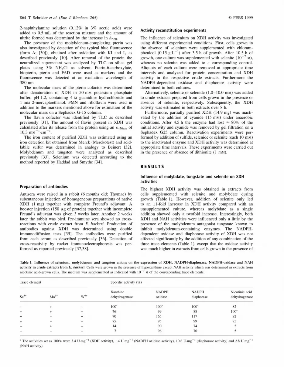
Influence of molybdate, tungstate and selenite on XDHactivities
The highest XDH activity was obtained in extracts fromcells supplemented with selenite and molybdate duringgrowth (Table 1). However, addition of selenite only ledto an 11-fold increase in XDH activity compared with anunsupplemented culture, whereas molybdate as a singleaddition showed only a twofold increase. Interestingly, bothXDH and NAH activities were influenced only a little by thepresence of the molybdenum antagonist tungstate known toinhibit molybdenum-containing enzymes. The NADPH-dependent oxidase and diaphorase activity of XDH was notaffected significantly by the addition of any combination of thethree trace elements (Table 1), except that the oxidase activitywas much higher in extracts from cells grown in the presence of
Table 1. Influence of selenium, molybdenum and tungsten anions on the expression of XDH, NADPH-diaphorase, NADPH-oxidase and NAH
activity in crude extracts from E. barkeri. Cells were grown in the presence of hypoxanthine except NAH activity which was determined in extracts from
nicotinic acid-grown cells. The medium was supplemented as indicated with 1027 m of the corresponding trace elements.
Trace element Specific activity (%)
Xanthine NADPH NADPH Nicotinic acid
Se4+ Mo6+ W6+ dehydrogenase oxidase diaphorase dehydrogenase
+ + ± 100a 100a 100a 82
+ + + 76 99 88 100a
+ ± + 70 165 117 82
+ ± ± 75 95 99 75
± + ± 14 90 74 5
± ± ± 7 96 70 5
a The activities set as 100% were 3.4 U´mg21 (XDH activity), 1.4 U´mg21 (NADPH oxidase activity), 10.6 U´mg21 (diaphorase activity) and 2.8 U´mg21
(NAH activity).

q FEBS 1999 Xanthine dehydrogenase from Eubacterium barkeri (Eur. J. Biochem. 264) 865
selenite and tungstate. NAH activity determined in extractsfrom respective nicotinic acid-grown cells was used as acontrol.
Purification of XDH
It was shown previously that E. barkeri induces molybdenum-stimulated and selenium-stimulated NAH during growth onnicotinic acid [12,13]. Thus, activity staining was performedafter separation of cell-free extracts prepared from cells grownon nicotinic acid or hypoxanthine, respectively, by nativeagarose/polyacrylamide gels. The data obtained showed that theorganism induced two substrate-specific enzymes, which canbe clearly separated by native agarose/PAGE. The Rf valuescalculated for NAH and XDH were 0.75 and 0.56, respectively.Furthermore, the specific activity and amount of each enzymepresent in hypoxanthine and nicotinic acid-grown cells wasdetermined (see below). The results obtained corroborated theobservation that E. barkeri contains two different enzymes eachresponsible for the conversion of their specific substrate,purines or nicotinic acid. Thus, NAH and XDH activity werecompletely separated during the initial ammonium sulfatefractionation.
A major problem during purification of XDH fromE. barkeri was the high oxygen sensitivity of the enzymeactivity. Thus, all purification steps had to be carried out underanaerobic conditions. Investigations on the stability of XDHactivity in crude extracts showed that the lowest loss in activitywas obtained if the protein was stored in buffer B. After3 weeks' storage at 4 8C < 80% of the initial activity wasrecovered under these conditions. Interestingly, the stability ofXDH activity was significantly lower if the enzyme was storedat 220 8C. The addition of dithionite in concentrations higherthan 1 mm led to a decreased stability of the enzyme activity.Therefore, dithionite was not routinely added.
A typical purification scheme for XDH from E. barkeriemploying ammonium sulfate fractionation, anion exchangechromatography on DEAE±Sephacel and gel filtration onSephacel CL-6B is depicted in Table 2. The resulting enzyme
preparation exhibited a maximal specific activity of< 160 U´mg21 of protein and was apparently homogeneous(Fig. 1). Attempts to improve this purification scheme by, forexample, hydrophobic interaction and/or different affinitychromatographies were without success. However, the obtainedyield of 38% of the initial activity was similar to the valuereported for NAH from E. barkeri after recent improvement ofthe purification scheme [15].
Molecular mass and subunit composition
The native molecular mass of XDH from E. barkeri wasestimated to be 530 kDa by gel filtration and linearpolyacrylamide gradient gel electrophoresis. SDS/PAGE ofthe homogeneous protein revealed that it consists of subunits of81, 30 and 17.5 kDa, respectively (Fig. 1). These data indicatedthat XDH is composed of four protomers exhibiting an abgstructure.
Absorption spectra
The UV/Vis spectrum of the yellow±brown XDH showed anabsorption maximum at 275 nm. A broad double peak wasobtained at 420 and 454 nm and shoulders were present at 395and 540 nm (data not shown). These spectral properties arecharacteristic for enzymes containing FAD and iron±sulfurclusters. The ratio A275/A450 was < 7.1 indicating that theprotein had lost some of its flavin during purification.
Cofactor content
Different methods were used to determine the type ofmolybdopterin present in XDH from E. barkeri. Cofactors ofpurified XDH were released by heat denaturation. The super-natant obtained was able to reconstitute N. crassa nit-1 mutants
Table 2. Purification of the NADP-dependent XDH from E. barkeri.
Purification
step
Total
protein
(mg)
Total
activity
(U)
Specific
activity
(U´mg21)
Yield
(%)
Purification
(-fold)
Cell extract 7500 31 425 4�.2 100 1
Ammonium sulfate 2700 30 269 10�.9 96 2�.6
DEAE±Sephacel 430 19 536 45�.4 62 10�.8
Sepharose CL-6B 73 11 972 164�.0 38 39
Fig. 1. SDS/PAGE analysis and the corresponding densitogram at
609 nm of purified XDH from E. barkeri.
Table 3. Substrate spectrum of purified XDH from E. barkeri.
Substrate Activitya (%)
Hypoxanthine 100
Xanthine 91
Purine 60
4-Aminoimidazole-5-carboxylamide 38
N-methylnicotinamideb 8�.1
Pyrazine-2-carboxylic acid 0�.8
Nicotinic acid 0
a The activity of xanthine dehydrogenase with different substrates is given
relative to the activity determined with hypoxanthine. b N-methylnicotina-
mide (58 mm) was only a substrate above pH 8.0. The value is given for
pH 10.5.

866 T. SchraÈder et al. (Eur. J. Biochem. 264) q FEBS 1999
to form a holoenzyme of nitrate reductase. Thus, addition ofincreasing amounts of XDH supernatant led to a linear increasein nitrite formation (data not shown). After oxidation of themolybdopterin cofactor by KI and I2 and separation by TLC, atypical blue fluorescent spot was detected using an excitationwavelength of 360 nm (data not shown). To differentiatebetween a simple molybdopterin or a molybdopterin dinucleo-tide, the molecular mass of the molybdopterin present in XDHfrom E. barkeri was determined by gel filtration. A molecularmass of 720 Da was determined, being in agreement with amolybdopterin of the dinucleotide type [39].
The spectral properties of native XDH and the isolatedcofactors indicated that the enzyme contained a flavin cofactor.The flavin was identified as FAD by TLC and quantificationrevealed an amount of 2.8 mol FAD´mol21 of native XDH asisolated. The substoichiometric amount of FAD is in agreementwith the data obtained by spectral investigations and might beexplained by a loss of the flavin during purification.
The amount of molybdenum, tungsten, iron, acid-labilesulfur and selenium was determined with homogeneouspreparations of XDH (2.6 mg´mL21). Based on a nativemolecular mass of 530 kDa, values of 17.5 mol iron,18.4 mol acid-labile sulfur, 2.3 mol molybdenum, 1.1 moltungsten and 0.95 mol selenium were calculated for theisolated enzyme. The spectral properties of XDH, as well asEPR-studies (R. Cammack, personal communication), indi-cated that the iron is bound to the protein as a 2Fe/2S cluster.Thus, the enzyme should contain two 2Fe/2S clusters perprotomer. It was interesting to note that XDH from E. barkerialso contained tungsten, being in most cases an antagonist ofmolybdenum [8]. Both ions were present in equimolarconcentrations during growth. Together both metals gave anamount of 3.4 g-atom´mol21 of XDH as isolated. From thesedata it might be concluded that the native enzyme contains onemolybdenum/tungsten atom per protomer.
Substrate specificity and electron acceptors
The highest specific activity of XDH from E. barkeri,determined using the standard assay, was obtained with threedifferent purine compounds (Table 3). However, significantactivity was also estimated with certain N-heterocycliccompounds. No activity was detected at different pH values ifnicotinic acid was used as a substrate. Allopurinol, which is aknown inhibitor of XDH, was initially converted with 8.5%of the activity obtained with hypoxanthine. The NADPH-dependent reduction of uric acid catalyzed by XDH proceededwith 14% of the activity determined for the oxidative reaction.XDH activity was strictly specific for NADP, but it could bereplaced by methyl viologen and benzyl viologen leading tosimilar conversion rates. The apparent Km values calculatedfrom Lineweaver-Burk plots for hypoxanthine, xanthine andNADP, were 210, 67 and 38 mm, respectively. An NADPH-dependent diaphorase activity of XDH was obtained with 2,6-dichlorophenol indophenol, methyl viologen, benzyl viologenand methylene blue as electron acceptors. The purifiedenzyme also exhibited an NADPH oxidase activity(60 U´mg21).
Inhibitors
As was also observed for XDHs from other sources, the activityof XDH from E. barkeri was significantly inhibited by theaddition of allopurinol to the standard assay containinghypoxanthine as a substrate (Table 4) [40,41]. The purine
derivatives adenine and 6-mercaptopurine also had a stronginhibitory effect on XDH activity. Methanol, known to be aninhibitor of molybdenum-containing enzymes [42], had noeffect on XDH activity. Arsenite and cyanide were also reportedto cause inactivation of molybdoenzymes by direct interactionwith the active site of the enzyme [42±44]. During incubationof purified XDH (1.35 mg´mL21) at pH 7.8 and 22 8C in thepresence of 1 mm arsenite, the enzyme lost half of its initialactivity after 1 min. Under anaerobic conditions at pH 7.8 and22 8C, a concentration-dependent inactivation of XDH fromE. barkeri was observed in the presence of cyanide.
Investigations on the selenium moiety of XDH
Growth experiments carried out in the presence or absence ofselenium indicated that XDH from E. barkeri required thistrace element for activity (Table 1). In principle selenium canbe covalently bound to the protein as selenocysteine asreported, for example, for the molybdoenzyme formatedehydrogenase H from E. coli [14,45] or in a labile formattached to a low molecular mass compound as found in NAH
Table 4. Inhibition of XDH from E. barkeri by different compounds.
Investigations of different inhibitors were performed with the purified
enzyme.
Compound added Inhibitiona (%)
None 0
6-Mercaptopurine 90
Allopurinol 95
Adenine 96
a The different purine derivatives were added to the standard enzyme assay
(each 1 mm) and XDH activity was determined with hypoxanthine as a
substrate.
Fig. 2. Influence of selenite addition on XDH activity in chloramphe-
nicol-treated cells. Cells of E. barkeri were grown on a hypoxanthine-
containing (14.7 mm) complex medium without selenium supplementation.
Chloramphenicol (CAM; 0.15 g´L21) and selenite (1027 m) were added to
the cultures as indicated. XDH activity was determined in cell-free extracts
using the standard assay (B). Extracts prepared from cells grown without
selenite supplementation were used as a control (X).

q FEBS 1999 Xanthine dehydrogenase from Eubacterium barkeri (Eur. J. Biochem. 264) 867
from E. barkeri [13,15]. The following experiments wereperformed to obtain some information on the selenium moietyof XDH from E. barkeri. If selenite or selenide (each 1, 2, 5 or10 mm) was added to extracts prepared anaerobically from cellsgrown in the presence or absence of selenium, the initial XDHactivity of 0.3 U´mg21 (±Se grown) and 5.0 U´mg21 (+Segrown) did not increase under any conditions at this stage (datanot shown). These data suggested that the enzyme was onlyinduced at a low level without selenium added and/or that theselenium moiety of XDH could not simply be incorporatedunder the conditions used.
In order to investigate the requirements for a seleniuminsertion mechanism of XDH, protein synthesis was inhibitedby the addition of chloramphenicol. Cells were grown oncomplex medium without selenium supplementation. Afterchloramphenicol (0.15 g´L21) had been added, incubation wascontinued for another 7 h. Subsequently, one culture wassupplemented with selenite (1027 m) and a second continued tobe incubated without additions. XDH activity was determinedin cell extracts prepared from aliquots (800 mL) removed fromboth cultures at several time intervals. Growth and protein
synthesis were significantly inhibited after addition of chloram-phenicol (data not shown). However, the data obtained clearlyshowed that XDH activity in chloramphenicol-treated cellspreviously grown in the absence of selenium was increasedsignificantly by the addition of selenite (Fig. 2). After < 10 hof incubation the determined XDH activity was about 20-foldhigher in selenite-treated cells compared with the corres-ponding control without selenite addition. Post-translationalincorporation of a selenium moiety would be consistent withthe observed increase in XDH activity. The addition of selenitehad no influence on the NADPH-dependent oxidase anddiaphorase activity also catalyzed XDH (data not shown).Thus, the increase in XDH activity cannot be explained by aspecific increase in the amount of enzyme present.
Inactivation of molybdoenzymes by cyanide was shown to bedue to the removal of sulfur from the molybdenum cofactor asthiocyanate [46]. Although it could not be proven until now,due to the lability of selenium compounds, it was supposed forNAH from E. barkeri that selenium as selenide might replacethe cyanolysable sulfur ligand of the molybdenum [18]. Thus,we tried to reactivate cyanide-inactivated XDH by the additionof different selenium-containing or sulfur-containing com-pounds. After the removal of cyanide, reactivation experimentswere performed in the presence or absence of 1 mm dithioniteby adding sulfide, selenite or selenide (each 10 mm) to theinactivated enzyme. Aliquots were removed from each reactionmixture at appropriate time intervals and the recovered activitywas determined using the standard assay. As shown in Fig. 3,addition of selenide in the presence of dithionite led to arecovery of < 67% of the initial XDH activity, whereas noreactivation was obtained with selenide if dithionite wasomitted. Addition of selenite or sulfide plus dithionite, aswell as selenite without dithionite, resulted in only a slightincrease in XDH activity after 1 h of incubation. However, noactivity was determined in these mixtures after a furtherincubation for 1 h (Fig. 3). Further controls were performed byincubating XDH as isolated under the conditions described forthe cyanide-inactivated enzyme. No increase in enzyme activitywas obtained if the enzyme was incubated with selenide orselenite or sulfide in the presence or absence of dithionite (datanot shown). These results suggested that in case of XDH fromE. barkeri an active enzyme could only be reconstituted byselenide using freshly prepared cyanide-treated protein andproper reducing conditions.
Immunological investigations
The specificity of the antibodies prepared against the homo-geneous XDH was shown by their strong inhibitory effect onXDH activity. Increasing amounts of antibodies led to a lineardecrease in XDH activity (data not shown). Interestingly, theantibodies also cross-reacted with partially purified NAHindicating that both enzymes were structurally related (datanot shown). This result prompted us to investigate the amountof each protein induced under the specific growth conditions inextracts obtained from hypoxanthine-grown or nicotinic acid-grown cells. First, both enzymes were separated in 1%agarose gels and the cross-reacting material was subsequentlyquantified in a second dimension by rocket immunoelec-trophoresis. The specific activity and amount of each enzymepresent after growth on different substrates are given inTable 5. These data further support the observation thatE. barkeri is able to induce two different, but structurallyrelated, selenium-containing molybdoenzymes under appro-priate growth conditions.
Fig. 3. Reconstitution of cyanide-inactivated XDH by the addition of
selenide. XDH was first inactivated by the addition of 15 mm cyanide (X).
Cyanide was removed by Sephadex G25 (!) and the following compounds
were subsequently added to the obtained enzyme solution under anaerobic
conditions: 1 mm dithionite plus 10 mm each (W) selenide or (K) selenite
and, in the absence of dithionite, (B) selenide or (A) sulfide. Addition of
selenite without dithionite and sulfide with dithionite resulted in about the
same inactivation kinetics obtained for selenite plus dithionite. The
untreated enzyme (V) was used as a control.
Table 5. Specific activity and amount of cross-reacting material (CRM)
against XDH from E. barkeri in crude extracts from cells grown on
nicotinic acid or hypoxanthine. ND, not detectable.
Growth substrate
Nicotinic acid Hypoxanthine
Assay substrate
Activity
(U´mg21)
CRM
(%)
Activity
(U´mg21)
CRM
(%)
Nicotinic acid 2�.5 2�.8 0�.4 0�.8
Xanthine 0�.04 ND 4�.2 3�.4

868 T. SchraÈder et al. (Eur. J. Biochem. 264) q FEBS 1999
As mentioned above, the addition of molybdenum, andespecially of selenium, to the growth medium had a significantinfluence on XDH activity as detected in extracts fromhypoxanthine-grown cells. Therefore, it was investigatedwhether these trace elements were also involved in theregulation of gene expression, or whether the difference inXDH activity was only due to, for example, a lack ofmolybdenum and selenium, respectively, at the active site.Antibodies raised against XDH and rocket immunoelectro-phoresis were used to quantify the cross-reacting protein inextracts prepared from cells grown on hypoxanthine in thepresence or absence of selenium, and/or molybdenum, and/ortungsten. The data obtained showed that the basic level of XDHrepresenting < 1.5±3.7% of the soluble cell protein was quiteindependent from the supplementation with the latter twoelements. However, addition of selenite in combination withmolybdate led to an increase of < 3.4±5.8% of the solubleprotein. During growth on hypoxanthine, XDH activity wasincreased by a factor of < 10 by the availability of selenium astrace element compared with the determined cross-reactingmaterial present in the respective extracts.
D I S C U S S I O N
The enzymatic and immunologic investigations performed withextracts from cells of E. barkeri grown on hypoxanthine andnicotinic acid, respectively, clearly showed that a selenium-dependent molybdoenzyme similar to NAH is induced duringgrowth on purine compounds. The purification of this XDHto apparent homogeneity was achieved by a three-steppurification scheme. Using anaerobic conditions, the enzymewas reasonably stable during purification at 4 8C. XDH fromC. acidiurici, which was also positively affected by selenium[8], was very unstable during purification [9]. Instability wasalso observed during the first purification experiments of theselenium-containing NAH from E. barkeri [13,17]. The finalrecovery was , 10% of the initial activity. An improvedpurification procedure developed for NAH led to a significantincrease in the obtained yield of enzyme activity [15].
The native molecular mass and subunit structure of XDHsisolated from different prokaryotic sources revealed an apparentdiversity. In the case of Streptomyces cyanogenus, P. putida 40and Rhodobacter capsulatus Al these enzymes were reported tobe composed of a single subunit exhibiting an a2, a3 or a4
structure, respectively [3,47,48]. For the proteins fromR. capsulatus B10S and Comamonas acidovorans an a2b2
structure was reported [6,7], whereas the enzymes fromP. putida Fu1 and 86 exhibited an a4b4 structure [4,10]. Theclassical (abg) or (abg)2 structure of many hydroxylatingmolybdenum-containing dehydrogenases [1,31] has so far beenfound only for XDH from V. atypica [5]. The 52 kDa subunit ofXDH from P. putida 86 seems to represent a fusion of the smalland middle-sized subunit, whereas in the XDHs fromeukaryotes all three subunits are fused to give one large singlesubunit of < 150 kDa [1,43]. Thus, the native a4b4g4 structureof XDH from E. barkeri, as reported, now represents thesecond example of a XDH composed of three dissimilarsubunits. The subunit structure reported for XDH preparationsfrom C. acidiurici consisted of five different polypeptides withmolecular masses of 110, 83, 56, 53 and 26 kDa [9] that mightbe due to proteolytic degradation. The NAH from E. barkeriwas immunologically quite similar to the XDH, but wasreported to be composed of four different subunits of 50, 37, 33and 23 kDa [18]. However, it was suggested that the latter
protein might be a degradation product of one of the othersubunits [15].
The iron and acid-labile sulfur content of XDH fromE. barkeri, as well as EPR-spectra (R. Cammack, personalcommunication), indicated that the enzyme might contain two2Fe/2S clusters per protomer. This is in agreement with the datareported for most XDHs and related enzymes investigated sofar [1,7,49]. However, there seems to be only a single 2Fe/2Scluster present in XDH from V. atypica [5]. NAH fromE. barkeri contains 5±7 mol of iron per mol of protomer andit was suggested that the enzyme might have a Fe center or Fe/S-center in addition to the two 2Fe/2S clusters [15]. A commonfeature of XDHs is the presence of a noncovalently bound FADas redox-active component [1]. The only reported exception isthe XDH from P. putida Fu1 which contains cytochrome binstead of the flavin [10].
Characterization of the molybdenum-complexing cofactor ofXDH from E. barkeri revealed that it is of the dinuleotide form.Molybdopterin dinucleotides are common to prokaryoticmolybdoenzymes as detected in XDHs from V. atypica andP. putida Fu1 and in NAH from E. barkeri [5,10,15]. However,different XDHs from prokaryotic sources such as P. putida 86,P. aeroginosa, R. capsulatus and C. acidovorans were shownto contain the simple molybdopterin as usually present ineukaryotic molybdoenzymes [6,7,50,51].
A substochiometric amount of molybdenum was oftendetermined for molybdenum-containing enzymes. This is dueto the loss of this element during purification [1,43]. However,it was an interesting observation that XDH from E. barkericontained tungsten in addition to molybdenum. Molybdenumand tungsten are very similar chemically and tungsten canreplace molybdenum usually leading to an inactive enzyme[52,53]. However, the addition of tungstate to the growthmedium had no significant influence on the XDH activityof E. barkeri. Purinolytic clostridia supplemented with185W-labeled tungsten did not incorporate it into fractionscontaining XDH activity in contrast to those containingformate dehydrogenase activity [54]. The anaerobic bacteriumC. formicoaceticum is able to express two different aldehydeoxidoreductases and the archaea Methanobacterium thermo-autotrophicum and M. wolfei were shown to express twodissimilar formylmethanofuran dehydrogenases, one of eachcontaining molybdenum and the second one containingtungsten [55±57]. For the formylmethanofuran dehydrogenasesit was also shown that molybdenum can replace tungsten andvice versa to give a still active enzyme [58]. Quite recentlyan enzymatically efficient, tungsten-substituted molybdenumtrimethylamine N-oxide reductase was obtained from E. coli[59]. Thus, it might be possible that the active XDH ofE. barkeri contains both molybdenum and tungsten. Our datadid not indicate whether the tungsten-containing speciesexhibited XDH activity. Both metals add up to an amount of3.4 mol per mol of native enzyme. This value is close to theexpected value of one molybdenum/tungsten per protomer. Theeffect of molybdate alone on the induction of XDH was low incontrast to data obtained, for example, for XDH from P. putidaFu1 or sulfite oxidase [10,60] where molybdenum was requiredfor expression of the respective enzyme. However, owing to thepresence of 1% yeast extract in the growth medium, the amountof molybdenum and tungsten might be partly sufficient forinduction of XDH.
As observed for XDHs from other sources, the enzyme fromE. barkeri was inactivated upon incubation with differentsubstrate analogs or products of the catalyzed oxidationreaction [40]. Inactivation of XDH also occurred in the

q FEBS 1999 Xanthine dehydrogenase from Eubacterium barkeri (Eur. J. Biochem. 264) 869
presence of arsenite and cyanide, which were both known tointeract directly with the cyanolysable sulfur at the active site[42,44]. This result was quite different from reports for theNAH from E. barkeri [15]. No significant inactivation of thelatter enzyme was obtained even in the presence of 100 mmcyanide. However, it was shown that the selenium moiety beingessential for the hydroxylase activity could easily be removedfrom the protein as a compound of low molecular mass [15].Furthermore, direct coordination of the selenium to themolybdenum was indicated by EPR studies performed withpurified protein [18]. Together with the fact that selenium andsulfur are chemically quite similar, these data indicated that, inthe case of NAH, selenium replaces the sulfur commonlypresent in the molybdopterin of molybdoenzymes of thexanthine oxidase family [1]. A similar situation might beanticipated for XDH from E. barkeri. At least, addition of justselenide or selenite did not increase XDH activity in crudeextracts obtained from selenium-deficient cells although thepresence of cross-reacting material was shown by immuno-logical studies. This might be due to the requirement of anenzyme-catalyzed system for the incorporation of selenium aspreviously shown for sulfur incorporation [61], or forbiosynthesis of the active molybdenum cofactor [62]. Further-more, a specific chaperone facilitating the assembly of acomplex molybdoenzyme, such as XDH, might be necessaryfor formation of the active enzyme [63]. Selenium was aspecific effector of XDH activity if chloramphenicol-treatedcells were incubated in the presence of selenite, thus, excludingthe involvement of protein biosynthesis and of selenocysteineas a reactive selenium moiety. This is further corroborated bythe fact that selenium had only a small effect on the amount ofXDH in cell extracts as determined by immunologicalinvestigations. Furthermore, the observation that theNADPH-oxidase and diaphorase activity of XDH were notinfluenced by the addition of this trace element indicated thatpartly functional XDH protein was formed in the absence ofselenium. The rather specific reconstitution of cyanide-inactivated XDH by selenide (under appropriate conditionssuch as the presence of dithionite) strongly suggested thatselenium is coordinated to the active site close to themolybdenum cofactor [1]. Very recently crystallographicstudies on the molybdo iron±sulfur flavoprotein carbonmonoxide dehydrogenase revealed the presence of a S-selanyl-cysteine where the selenium is bound to the sulfur of a cysteineresidue [64], in a way quite similar to the former proposal of apersulfide present at the reactive side [43]. An analogoussituation should also be valid for XDH from E. barkeri. Theobserved high oxygen sensitivity of both selenium-containingNAH and XDH should constitute no problem in vivo, forE. barkeri is an anaerobic bacterium. Because of the quitelow redox potential Eo
0 of the pair uric acid/xanthine of< ±440 mV [43,65] and owing to the lower redox-potentialof selenium compared to sulfur [66], the catalytic efficiency ofXDH and NAH might be improved by selenium and, thus, thesubstrate flow will be increased. At least, the specific activityof XDH from E. barkeri determined in crude extracts is< 10-fold higher than estimated for the corresponding enzymesfrom other sources. The substoichiometric amounts of moly-bdenum, selenium and flavin determined for the purified XDHmight be explained by their partial loss during purification asalso observed for NAH from E. barkeri [15]. Thus, the catalyticefficiency might even be higher in vivo. Furthermore, the dataobtained indicated that the cell extract already contained XDHwhich was deficient in one or more cofactors. Although XDHfrom E. barkeri shares basic properties with molybdoenzymes
from other sources, it is quite unique in containing selenium ina labile, bound form. In this aspect it is quite similar to NAHisolated from the same organism as also revealed by ourimmunological studies. So far these two enzymes representexamples of selenoproteins where selenium is apparentlybiologically active but not present as selenocysteine.
A C K N O W L E D G E M E N T S
The authors thank Dorothee Imhoff-Struckle for providing some of the
results. This work was supported by grants from the Deutsche
Forschungsgemeinschaft and the Fonds der Chemischen Industrie.
R E F E R E N C E S
1. Hille, R. (1996) The mononuclear molybdenum enzymes. Chem. Rev.
96, 2757±2816.
2. Bray, R.C., Bennett, B., Burke, J.F., Chovnick, A., Doyle, W.A. &
Howes, B.D. (1996) Recent studies on xanthine oxidase and related
enzymes. Biochem. Soc. Trans. 24, 99±105.
3. Woolfolk, C.A. (1985) Purification and properties of a novel
ferricyanide-linked xanthine dehydrogenase from Pseudomonas
putida 40. J. Bacteriol. 163, 600±609.
4. Hettrich, D. & Lingens, F. (1991) Microbial metabolism of quinoline
and related compounds. VIII. Xanthine dehydrogenase from a
quinoline utilizing Pseudomonas putida strain. Biol. Chem. Hoppe-
Seyler 372, 203±211.
5. Gremer, L. & Meyer, O. (1996) Characterization of xanthine
dehydrogenase from the anaerobic bacterium Veillonella atypica
and identification of a molybdopterin-cytosine-dinucleotide-
containing molybdenum cofactor. Eur. J. Biochem. 238, 862±866.
6. Xiang, Q. & Edmondson, D.E. (1996) Purification and characterization
of a prokaryotic xanthine dehydrogenase from Comamonas acid-
ovorans. Biochemistry 35, 5441±5450.
7. LeimkuÈhler, S., Kern, M., Solomon, P.S., McEwan, A.G., Schwarz, G.,
Mendel, R.R. & Klipp, W. (1998) Xanthine dehydrogenase from the
phototrophic purple bacterium Rhodobacter capsulatus is more
similar to its eukaryotic counterparts than to prokaryotic moly-
bdenum enzymes. Mol. Microbiol. 27, 853±869.
8. Wagner, R. & Andreesen, J.R. (1979) Selenium requirement for active
xanthine dehydrogenase from Clostridium acidiurici and Clostridium
cylindrosporum. Arch. Microbiol. 121, 255±259.
9. Wagner, R., Cammack, R. & Andreesen, J.R. (1984) Purification and
characterization of xanthine dehydrogenase from Clostridium
acidiurici grown in the presence of selenium. Biochim. Biophys.
Acta 791, 63±74.
10. Koenig, K. & Andreesen, J.R. (1990) Xanthine dehydrogenase and
2-furoyl-coenzyme A dehydrogenase from Pseudomonas putida Fu1:
two molybdenum-containing dehydrogenases of novel structural
composition. J. Bacteriol. 172, 5999±6009.
11. Collins, M.D., Lawson, P.A., Willems, A., Cordoba, J.J., Fernandez-
Garayzabal, J., Garcia, P., Cai, J., Hippe, H. & Farrow, J.A. (1994)
The phylogeny of the genus Clostridium: proposal of five new
genera and eleven new species combinations. Int. J. Syst.
Bacteriol. 44, 812±826.
12. Imhoff, D. & Andreesen, J.R. (1979) Nicotinic acid hydroxylase from
Clostridium barkeri: selenium-dependent formation of active
enzyme. FEMS Microbiol. Lett. 5, 155±158.
13. Dilworth, G.L. (1982) Properties of the selenium-containing moiety of
nicotinic acid hydroxylase from Clostridium barkeri. Arch. Biochem.
Biophys. 219, 30±38.
14. Zinoni, F., Birkman, A., Stadtman, T.C. & BoÈck, A. (1986) Nucleotide
sequence and expression of the selenocysteine-containing poly-
peptide of formate dehydrogenase (formate-hydrogen-lyase-linked)
from Escherichia coli. Proc. Natl Acad. Sci. USA 83, 4650±4654.
15. Gladyshev, V.N., Khangulov, S.V. & Stadtman, T.C. (1996) Properties
of the selenium- and molybdenum-containing nicotinic acid
hydroxylase from. Clostridium barkeri. Biochemistry 35, 212±223.

870 T. SchraÈder et al. (Eur. J. Biochem. 264) q FEBS 1999
16. Boyington, J.C., Gladyshev, V.N., Khangulov, S.V., Stadtman, T.C. &
Sun, P.D. (1997) Crystal structure of formate dehydrogenase H:
catalysis involving Mo, molybdopterin, selenocysteine, and an Fe4S4
cluster. Science 275, 1305±1308.
17. Holcenberg, J.S. & Stadtman, E.R. (1969) Nicotinic acid metabolism.
3. Purification and properties of a nicotinic acid hydroxylase. J. Biol.
Chem. 244, 1194±1203.
18. Gladyshev, V.N., Khangulov, S.V. & Stadtman, T.C. (1994) Nicotinic
acid hydroxylase from Clostridium barkeri: electron paramagnetic
resonance studies show that selenium is coordinated with moly-
bdenum in the catalytically active selenium-dependent enzyme. Proc.
Natl Acad. Sci. USA 91, 232±236.
19. Stadtman, E.R., Stadtman, T.C., Pastan, J. & Smith, L.D.S. (1972)
Clostridium barkeri sp. nov. J. Bacteriol. 110, 758±760.
20. Bryant, M.P. (1972) Commentary on the Hungate technique for culture
of anaerobic bacteria. Am. J. Clin. Nutr. 25, 1324±1328.
21. Lowry, M.O., Rosebrough, N.J., Farr, A.L. & Randall, R.J. (1951)
Protein measurement with folin±phenol reagent. J. Biol. Chem. 193,
265±275.
22. Peacock, A.C. & Dingman, C.W. (1968) Molecular weight estimation
and separation of ribonucleic acid by electrophoresis in agarose±
acrylamide composite gels. Biochemistry 7, 668±674.
23. Laemmli, U.K. (1970) Cleavage of structural proteins during the
assembly of the head of bacteriophage T4. Nature 227, 680±685.
24. Weber, K. & Osborn, M. (1969) The reliability of molecular weight
determinations by dodecyl sulfate±polyacrylamide gel electrophor-
esis. J. Biol. Chem. 224, 4406±4412.
25. Blum, H., Beier, H. & Gross, H.J. (1987) Improved silver staining of
plant proteins, RNA, and DNA in polyacrylamide gels. Electro-
phoresis 8, 93±99.
26. Margolis, J. & Kenrick, K.G. (1967) Polyacrylamide gel electro-
phoresis across a molecular sieve gradient. Nature 214, 1334±1336.
27. Nason, A., Antoine, A.D., Ketchum, P.A., Frazier, W.A. & Lee, D.K.
(1970) Formation of assimilatory nitrate reductase by in vitro inter-
cistronic complementation in Neurospora crassa. Proc. Natl Acad.
Sci. USA 65, 137±144.
28. Amy, N.K. & Rajagopalan, K.V. (1979) Characterization of molyb-
denum cofactor from Escherichia coli. J. Bacteriol. 140, 114±124.
29. Johnson, J.L. (1980) The molybdenum cofactor common to nitrate
reductase, xanthine dehydrogenase and sulfite oxidase. In Molyb-
denum and Molybdenum Containing Enzymes (Coughlan, M.P.,
ed.), pp. 345±385. Pergamon Press, New York.
30. Johnson, J.L. & Rajagopalan, K.V. (1982) Structural and metabolic
relationship between the molybdenum cofactor and urothione. Proc.
Natl Acad. Sci. USA 79, 6856±6860.
31. Kretzer, A., Frunzke, K. & Andreesen, J.R. (1993) Catabolism of
isonicotinate by Mycobacterium sp. INA1: extended description of
the pathway and purification of the molybdoenzyme isonicotinate
dehydrogenase. J. Gen. Microbiol. 139, 2763±2772.
32. Beinert, H. (1983) Semi-micro methods for analysis of labile sulfide
and of labile sulfide plus sulfane sulfur in unusually stable iron±
sulfur proteins. Anal. Biochem. 131, 373±378.
33. Cardenas, J. & Mortenson, L.E. (1974) Determination of molybdenum
and tungsten in biological materials. Anal. Biochem. 60, 372±381.
34. Haddad, R.E. & Smythe, L.E. (1974) A critical evaluation of
fluorometric methods for determination of selenium in plant
materials with 2,3-diaminonaphtalene. Talenta 21, 859±865.
35. Ouchterlony, OÈ . (1949) Antigen±antibody reactions in gels. Acta
Pathol. Microbiol. Scand. 26, 507±515.
36. Dietrichs, D. & Andreesen, J.R. (1990) Purification and comparative
studies of dihydrolipoamide dehydrogenases from the anaerobic,
glycine-utilizing bacteria Peptostreptococcus glycinophilus, Clost-
ridium cylindrosporum, and Clostridium sporogenes. J. Bacteriol.
172, 243±251.
37. Dietrichs, D., Meyer, M., Schmidt, B. & Andreesen, J.R. (1990)
Purification of NADPH-dependent electron-transferring flavo-
proteins and N-terminal protein sequence data of dihydrolipoamide
dehydrogenases from anaerobic, glycine-utilizing bacteria. J. Bac-
teriol. 172, 2088±2095.
38. Freudenberg, W., Dietrichs, D., Lebertz, H. & Andreesen, J.R. (1989)
Isolation of an atypically small lipoamide dehydrogenase involved in
the glycine decarboxylase complex from Eubacterium acidamino-
philum. J. Bacteriol. 171, 1346±1354.
39. KruÈger, B. & Meyer, O. (1986) The pterin (bactopterin) of carbon
monoxide dehydrogenase from Pseudomonas carboxydoflava. Eur. J.
Biochem. 157, 121±128.
40. Massey, V., Komai, H. & Palmer, G. (1970) On the mechanism of
inactivation of xanthine oxidase by allopurinol and other pyrazolo
[3,4-d]pyrimidines. J. Biol. Chem. 245, 2837±2844.
41. Ni FhaolaÂin, I. & Coughlan, M.P. (1978) Effects of allopurinol and of
oxypurinol on turkey liver xanthine dehydrogenase. FEBS Lett. 90,
305±308.
42. Coughlan, M.P., Rajagopalan, K.V. & Handler, P. (1969) The role of
molybdenum in xanthine oxidase and related enzymes. Reactivity
with cyanide, arsenite, and methanol. J. Biol. Chem. 244, 2658±2663.
43. Coughlan, M.P. (1980) Aldehyde oxidase, xanthine oxidase and
xanthine dehydrogenase: hydroxylases containing molybdenum,
iron±sulphur and flavin. In Molybdenum and Molybdenum Contain-
ing Enzymes (Coughlan, M.P., ed.), pp. 119±185. Pergamon Press,
New York.
44. George, G.N. & Bray, R.C. (1983) Reaction of arsenite ions with the
molybdenum center of milk xanthine oxidase. Biochemistry 22,
1013±1021.
45. BoÈck, A., Forchhammer, K., Heider, J., Leinfelder, W., Sawers, G.,
Veprek, B. & Zinoni, F. (1991) Selenocysteine: the 21st amino acid.
Mol. Microbiol. 5, 515±520.
46. Branzoli, U. & Massey, V. (1974) Preparation of aldehyde oxidase in its
native and deflavo forms. Comparison of spectroscopic and catalytic
properties. J. Biol. Chem. 249, 4339±4345.
47. Ohe, T. & Watanabe, Y. (1979) Purification and properties of
xanthine dehydrogenase from Streptomyces cyanogenus. J. Biochem.
86, 45±53.
48. Aretz, W., Kaspari, H. & Klemme, J.H. (1981) Molecular and
kinetic characterization of xanthine dehydrogenase from the photo-
trophic bacterium Rhodopseudomonas capsulata. Z. Naturforsch. 36,
933±941.
49. Canne, C., Stephan, I., Finsterbusch, J., Lingens, F., Kappl, R., Fetzner,
S. & HuÈttermann, J. (1997) Comparative EPR and redox studies of
three prokaryotic enzymes of the xanthine oxidase family: quinoline
2-oxidoreductase, quinaldine 4-oxidase, and isoquinoline 1-oxidor-
eductase. Biochemistry 36, 9780±9790.
50. Hettrich, D., Peschke, B., Tshisuaka, B. & Lingens, F. (1991) Microbial
metabolism of quinoline and related compounds. X. The molybdop-
terin cofactors of quinoline oxidoreductases from Pseudomonas
putida 86 and Rhodococcus spec. B1 and of xanthine dehydrogenase
from Pseudomonas putida 86. Biol. Chem. Hoppe-Seyler 372,
513±517.
51. Johnson, J.L., Chaudhury, M. & Rajagopalan, K.V. (1991) Identifi-
cation of a molybdopterin-containing molybdenum cofactor in
xanthine dehydrogenase from Pseudomonas aeruginosa. Biofactors
3, 103±107.
52. Johnson, J.L., Rajagopalan, K.V. & Cohen, H.J. (1974) Molecular basis
of the biological function of molybdenum. Molybdenum-free sulfite
oxidase from livers of tungsten-treated rats. J. Biol. Chem. 249,
5046±5055.
53. da Silva, J.J.R.F. & Williams, R.J.P. (1991) The Biological Chemistry of
the Elements, pp. 411±435. Clarendon Press, Oxford.
54. Wagner, R. & Andreesen, J.R. (1987) Accumulation and incorporation
of 185W-tungsten into proteins of Clostridium acidiurici and
Clostridium cylindrosporum. Arch. Microbiol. 147, 295±299.
55. Huber, C., Caldeira, J., Jongejan, J.A. & Simon, H. (1994) Further
characterization of two different, reversible aldehyde oxidoreductases
from Clostridium formicoaceticum, one containing tungstate and the
other molybdenum. Arch. Microbiol. 162, 303±309.
56. Hochheimer, A., Linder, D., Thauer, R.K. & Hedderich, R. (1996)
The molybdenum formylmethanofuran dehydrogenase operon and
the tungsten formylmethanofuran dehydrogenase operon from

q FEBS 1999 Xanthine dehydrogenase from Eubacterium barkeri (Eur. J. Biochem. 264) 871
Methanobacterium thermoautotrophicum. Structures and transcrip-
tional regulation. Eur. J. Biochem. 242, 156±162.
57. Hochheimer, A., Hedderich, R. & Thauer, R.K. (1998) The
formylmethanofuran dehydrogenase isoenzymes in Methano-
bacterium wolfei and Methanobacterium thermoautotrophicum:
induction of the molybdenum isoenzyme by molybdate and
constitutive synthesis of the tungsten isoenzyme. Arch. Microbiol.
170, 389±393.
58. Schmitz, R.A., Albracht, S.P. & Thauer, R.K. (1992) Properties of
the tungsten-substituted molybdenum formylmethanofuran
dehydrogenase from Methanobacterium wolfei. FEBS Lett. 309,
78±81.
59. Buc, J., Santini, C.L., Giordani, R., Czjzek, M., Wu, L.F. & Giordano,
G. (1999) Enzymatic and physiological properties of the tungsten-
substituted molybdenum TMAO reductase from. Eschericia coli.
Mol. Microbiol. 32, 159±168.
60. Jones, H.P., Johnson, J.L. & Rajagopalan, K.V. (1977) In vitro
reconstitution of demolybdosulfite oxidase by molybdate. J. Biol.
Chem. 252, 4988±4993.
61. Wahl, R.C., Warner, C.K., Finnerty, V. & Rajagopalan, K.V. (1982)
Drosophila melanogaster ma-l mutants are defective in the sul-
furation of desulfo Mo hydroxylases. J. Biol. Chem. 257, 3958±3962.
62. Mendel, R.R. (1997) Molybdenum cofactor of higher plants: biosyn-
thesis and molecular biology. Planta 203, 399±405.
63. LeimkuÈhler, S. & Klipp, W. (1999) Role of XDHC in molybdenum
cofactor insertion into xanthine dehydrogenase of Rhodobacter
capsulatus. J. Bacteriol. 181, 2745±2751.
64. Dobbek, H., Gremer, L., Meyer, O. & Huber, R. (1999) Crystal
structure of CO dehydrogenase, a molybdo iron±sulfur flavoprotein
containing S-selanylcysteine. Proc. Natl Acad. Sci. USA 96,
8884±8889.
65. Thauer, R.K., Jungermann, K. & Decker, K. (1977) Energy conser-
vation in chemotrophic anaerobic bacteria. Bacteriol. Rev. 41, 100±180.
66. Stadtman, T.C. (1996) Selenocysteine. Annu. Rev. Biochem. 65,
83±100.