identification of isobutyryl-coa dehydrogenase and its deficiency in humans
TRANSCRIPT
Identification of isobutyryl-CoA dehydrogenaseand its deficiency in humans
Tien V. Nguyen,a Brage S. Andresen,b,c Thomas J. Corydon,b Sandro Ghisla,d
Nasser Abd-El Razik,d Al-Walid A. Mohsen,a Stephen D. Cederbaum,e Diane S. Roe,f
Charles R. Roe,f Nicolas J. Lench,g and Jerry Vockleya,*
a Department of Medical Genetics, Mayo Clinic, Rochester, MN 55905, USAb Institute for Human Genetics, Aarhus University, Aarhus, Denmark
c Research Unit for Molecular Medicine, Skejby Sygehus and Aarhus University, Aarhus, Denmarkd Faculty of Biology, University of Konstanz, Konstanz, Germany
e Department of Pediatrics, UCLA Medical Center, Los Angeles, CA, USAf Institute of Metabolic Disease, Baylor University, Dallas, TX, USA
g Molecular Medicine Unit, University of Leeds, St. James� University Hospital, Leeds, UK
Received 19 July 2002; received in revised form 31 July 2002; accepted 1 August 2002
Abstract
The acyl-CoA dehydrogenases (ACDs) are a family of related enzymes that catalyze the a,b-dehydrogenation of acyl-CoA esters.Two homologues active in branched chain amino acid metabolism have previously been identified. We have used expression in
Escherichia coli to produce a previously uncharacterized ACD-like sequence (ACAD8) and define its substrate specificity. Purified
recombinant enzyme had a kcat=Km of 0.8, 0.23, and 0.04 (lM�1 s�1) with isobutyryl-CoA, (S) 2-methylbutyryl-CoA, and n-propionyl-CoA, respectively, as substrates. Thus, this enzyme is an isobutyryl-CoAdehydrogenase. A single patient has previously been described
whose fibroblasts exhibit a specific deficit in the oxidation of valine. AmplifiedACAD8 cDNAmade frompatient fibroblastmRNAwas
homozygous for a single nucleotide change (905G > A) in theACAD8 coding region compared to the sequence from control cells. Thisencodes an Arg302Gln substitution in the full-length protein (position 280 in the mature protein), a position predicted by molecular
modeling tobe important in subunit interactions. Themutant enzymewas stable but inactivewhen expressed inE. coli. Itwas also stable
and appropriately targeted to mitochondria, but inactive when expressed in mammalian cells. These data confirm further the presence
of a separatedACD in humans specific to valine catabolism (isobutyryl-CoAdehydrogenase, IBDH), alongwith the first enzymatic and
molecular confirmation of a deficiency of this enzyme in a patient. � 2002 Elsevier Science (USA). All rights reserved.
Keywords: Isobutyryl-CoA dehydrogenase; Acyl-CoA dehydrogenase: ACAD8, b-Oxidation; Valine metabolism; Inborn error of metabolism; Valine
1. Introduction
The acyl-CoA dehydrogenases (EC 1.3.99.3) are afamily of nuclear encoded, mitochondrial flavoenzymesthat catalyze the a,b-dehydrogenation of acyl-CoA in-termediates in the catabolism of fatty acids and branchedchain amino acids [1–7]. Inherited deficiencies of theseenzymes are important causes of human disease [8–10].
Early studies of ACDs1 isolated from rat liver mito-chondria suggested the existence of a single enzyme(called 2-methyl-branched chain acyl-CoA dehydrogen-ase) that could utilize both isobutyryl- and S-2-methyl-butyryl-CoAs from the valine and isoleucine pathways,respectively, equally well as substrates [3]. More recently,
Molecular Genetics and Metabolism 77 (2002) 68–79
www.academicpress.com
*Corresponding author. Fax: 1-507-284-4601.
E-mail address: [email protected] (J. Vockley).
1 Abbreviations used: ACD, acyl-coenzyme A dehydrogenase; ETF,
electron transfer flavoprotein; IBDH, isobutyryl-coenzyme A dehy-
drogenase; SBCADH, short–branched chain acyl-coenzyme A dehy-
drogenase; IVD, isovaleryl-CoA dehydrogenase; SCADH, short chain
acyl-CoA dehydrogenase; MCADH, medium chain acyl-CoA dehy-
drogenase; VLCAD, very long chain acyl-CoA dehydrogenase; FAD,
flavin adenine dinucleotide.
1096-7192/02/$ - see front matter � 2002 Elsevier Science (USA). All rights reserved.PII: S1096 -7192 (02 )00152-X
the rat and human cDNAs for this enzyme have beencloned and the gene was named short–branched chainacyl-CoA dehydrogenase (ACADSB; see Table 1 for asummary of genetic nomenclature and protein designa-tions) [6,7,11]. Recombinant rat SBCADH produced inEscherichia coli, like its native counterpart, could effi-ciently utilize both isobutyryl- and 2-methylbutyryl-CoAas substrate. In contrast, the recombinant human enzymedid not efficiently utilize isobutyryl-CoA as substrate,raising the possibility that another ACD specific to valinemetabolism might exist in humans. Two patients withinactivating mutations in the ACADSB gene have re-cently been identified [12,13]. In the first of these patients,valinemetabolismwas shown to be normal, while this wasnot examined in the second patient. Finally, a patient hasbeen identified in whom metabolic loading studies in fi-broblasts revealed decreased oxidation of labeled valine,with an increase in accumulation of isobutyryl carnitine.Metabolism of labeled isoleucine and leucine was normaland a defect in a valine specific ACD was proposed [14].A mapping study of human chromosome 11q25 has
identified a novel gene that shares strong homology toother members of the human ACD gene family [15].Initial studies of ACAD8 cDNA revealed that the pro-tein expressed in an eukaryotic system had high activitytowards both 2-methylbutyryl-CoA and isobutyryl-CoAin crude cellular extracts [13]. We now report expressionof the cDNA for ACAD8 in E. coli, purification of therecombinant enzyme to homogeneity, and character-ization of the kinetic properties and substrate specificityof the purified enzyme. We also report the gene structureof the human ACD gene. Mutation analysis of ACAD8from the patient with a proposed defect in valine me-tabolism revealed a mutation in the ACAD8 coding re-gion leading to loss of enzymatic activity. Our findingsidentify ACAD8 as an isobutyryl-CoA dehydrogenase(IBDH) active in valine catabolism, as well as the firstpatient deficient in this enzyme.
2. Materials and methods
2.1. Construction of wild type human IBDH expressionplasmid
PCR primers were designed to amplify the predicted1182 bp of the mature coding region of ACAD8 cDNA.
The 50-primer consisted of 47 nucleotides, including nu-cleotides 67–95 of the precursor coding sequence fol-lowed by EcoRI (underlined) and NdeI (bold) restrictionsites (50-GAC GAT GAA TTC CAT ATG 1CTC GTCCAG ACC GGC CAC CGG AGC TTG AC-30). The 30-primer consisted of the last 15 nucleotides of the codingregion (stop codon in antisense direction is bolded) fol-lowed by a HindIII restriction site (underlined) (50-AATGAG AAG CTT CTA CTA CTC CTG AAG CAG-30).A human liver Marathon-ready cDNA from Clontech(Palo Alto, CA) was used as template for PCR, whichwas performed with 30 cycles of annealing 60 �C for 30 s,extension 68 �C for 4min, and denaturing 94 �C for 30 susing the Advantage cDNA PCR Kit with PolymeraseMix (Clontech, Palo Alto, CA). PCR products werepurified by electrophoresis on a 1.5% low melting aga-rose gel and the desired DNA band recovered using theQIAquick Gel Extraction Kit 50 (QIAGEN, Valencia,CA). The recovered fragment was digested with EcoRIand HindIII, and inserted into the prokaryotic expres-sion vector pET-21a (+) (Novagen, Madison, WI). Theplasmid containing the mature IBDH insert (pmIBDH)was used for expression in E. coli. To construct thevariant IBDH plasmid, a BseRI and NsiI restrictionfragment containing the IBDH patient mutation wassubstituted into the same sites in the wild type vector.Precursor wild type IBDH was expressed in COS-7 cellsusing a pcDNA3.1(+) vector as previously described[12,13]. The patient mutation was introduced into theprecursor IBDH sequence via the replacement of a BsmIandNsiI fragment with the same fragment containing thepatient mutation.
2.2. Amplification of ACAD8 sequences made fromfibroblast mRNA
mRNA was isolated from control and patient cul-tured fibroblasts using the QuickPrep Micro mRNAPurification Kit (Amersham Pharmacia Biotech., Pist-cataway, NJ), and first strand cDNA was synthesizedwith the First-Strand cDNA Synthesis Kit (AmershamPharmacia Biotech.). ACAD8 cDNA sequences wereamplified by 30 cycles of PCR: 62 �C, 4min, annealing;72 �C, 7min, extension; and 94 �C, 30 s denaturing.Amplified products were separated and purified as be-fore, and subjected to automated DNA sequencing bythe Molecular Biology Core Facility of the Mayo Clinic.
Table 1
Genetic loci and common enzyme names for acyl-CoA dehydrogenases involved in short and branched chain amino acid catabolism
Genetic locus Enzyme name Catabolic pathway
IVD Isovaleryl-CoA dehydrogenase (IVDH) Leucine
ACADSB Short–branched chain acyl-CoA dehydrogenase (SBCADH) Isoleucine
2-Methyl-branched chain acyl-CoA dehydrogenase
ACAD8 Isobutyryl-CoA dehydrogenase (IBDH) Valine
ACADS Short chain acyl-CoA dehydrogenase (SCADH) Mitochondrial fatty acid b-oxidation
T.V. Nguyen et al. / Molecular Genetics and Metabolism 77 (2002) 68–79 69
2.3. Identification and characterization of the humanACAD8 gene structure and sequence analysis of patientand control genomic DNA
tBlastn homology searches of the HTGS and GSSdatabases in GenBank with the predicted amino acidsequence of the human ACAD8 were used to identifytwo BAC clones containing part of the human ACAD8gene: AC018780 and AP000859. The AP000859 BAChas been mapped to chromosome 11q25, which is con-sistent with the previous mapping of the human ACAD8gene [15]. Intron sizes were estimated on the basis ofmigration in agarose gels of PCR products amplifiedwith primers located in separate exons and from thesequence of AC018780 and AP000859. Primers weredesigned for PCR amplification of the 11 exons and partof the flanking intron sequences of the human ACAD8gene (primer sequences are available on [email protected]). Genomic DNA was isolated fromcultured fibroblasts or blood samples according tostandard methods [16]. PCR reactions were performedunder standard conditions in an automated Thermalcycler 480 (Perkin–Elmer, Norwalk, CT) and the PCRproducts were subjected to direct bi-directional cyclesequencing as described above.
2.4. Expression of wild type and mutant IBDH
pmIBDH was transformed into E. coli host strainBL21(DE3; Novagen, Madison, WI), crude extracts ofinduced cells were made from 25ml of cultures grown in2� YT (31 g/L, BIO 101, Vista, CA) with 80 lg/mlampicillin. Cultures of E. coli were grown to mid-logphase (absorbance 550nm > 0:5), induced by the addi-tion of IPTG to 0.5mM final concentration, and incu-bated with shaking at 37 �C for 4 h or overnight. Cellswere harvested by centrifugation and lysed by sonica-tion after treatment with lysozyme as previously de-scribed [17–20]. For large scale purification, the wildtype and Arg280Gln mutant IBDH plasmids were co-expressed with the bacterial chaperonins GroEL/ES,grown at 37 �C, and harvested after 4 h induction.Expression in CHANG cells, immunostaining, andconfocal laser scanning microscopy were performed asdescribed [13,21]. The presence of IBDH protein inprokaryotic and eukaryotic cell extracts was determinedthrough Western blotting with IBDH specific antisera aspreviously described [12,13].
2.5. Purification of IBDH protein
Wild type IBDH protein was purified from inducedE. coli cultures by DEAE chromatography, fractiona-tion with ammonium sulfate, and chromatography on10 lm hydroxyapatite as previously described [17–19].To prevent loss of FAD from the enzyme molecule,
20 lMFAD was added to the buffer during elution fromthe hydroxyapatite column. Free FAD was removedfrom final sample by filtration on Superdex G-200 in50mM potassium phosphate, pH 7.5, 0.1M KCl.
2.6. Enzyme assays
ACD activity was measured with the anaerobicelectron transfer flavoprotein (ETF) reduction assayusing an LS50B fluorescence spectrophotometer fromPerkin–Elmer (Norwalk, CT) with a heated cuvetteblock set to 32 �C as described [22]. Final substrateconcentration in the assay mixtures was 50 lM. Foractivity units (U) see [22].
2.7. Molecular modeling of IBDH structure
A prediction of the three-dimensional structure ofIBDH was obtained with the Insight II 2000 package ofmodeling software from Accelrys (San Diego, CA) and aSilicon Graphics O2 workstation (Mountain View, CA).Modeling based on the published structures of humanisovaleryl-CoA dehydrogenase (IVDH), porcine me-dium chain acyl-CoA dehydrogenase (MCADH), andbutyryl-CoA dehydrogenase from M. elsdenii [23–26]was performed using the Homology and Modelermodules included with this software as previously de-scribed [22]. The ‘‘Manual Rotomer’’ option was used tooptimize the position of atoms of the side chains ofspecific amino acid residues and examine the energyminima of the various possible conformations.
2.8. Computational protein sequence analysis
The protein sequences of 22 selected ACDs wereidentified from different species via a standard BLASTsearch of the NCBI databases and compared with thehuman IBDH sequence using the MacVector softwarepackage version 7.0 with the Clustal W algorithm v 1.4and distance matrix methods. The multiple sequenceswere aligned and a phylogenetic tree constructed usingthe following parameters: pairwise alignment mode,slow; open gap penalty, 10.0; extend gap penalty, 0.1;delay divergent, 40%; gap distance, 8; and similaritymatrix, blosum. Table 2 shows the GenBank accessionnumbers of protein sequences analyzed.
3. Results
The clinical history of the patient studied has previ-ously been reported [14]. Briefly, she presented at2-years of age to first cousin parents of Hispanic origins.She was well for the first one and one-half months of lifeon breast feeding, then developed feeding intolerance onformula. At 11 months of age she was found to have
70 T.V. Nguyen et al. / Molecular Genetics and Metabolism 77 (2002) 68–79

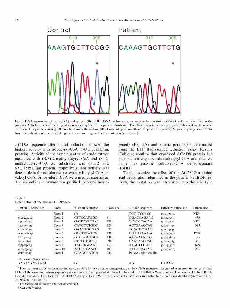
failure to thrive, a severe carnitine deficiency, and di-lated cardiomyopathy. She responded well to carnitinetherapy and has been well without episodes of decom-pensation since. She is now 6 years old with normalgrowth and development. Metabolic flux studies origi-nally revealed a defect in valine catabolism, and theexistence of a valine specific acyl-CoA dehydrogenasewas suggested. We hypothesized that the recently iden-tified ACAD8might be such an enzyme and that it mightbe deficient in this patient. To examine this, we amplifiedACAD8 sequences from control and patient fibroblasts.Amplification of ACAD8 from cDNA made from con-trol fibroblast mRNA yielded a fragment of 1250 bp insize, in good agreement with the size of the predictedprecursor form of ACAD8 [15]. Direct DNA sequencingof the amplified product confirmed that the sequence ofthe PCR product matched that published for ACAD8(not shown). In contrast, ACAD8 sequences amplifiedfrom cDNA made from patient fibroblasts revealed ahomozygous substitution of a guanine by an adenineresidue at position 905 (905G > A) of the precursorcoding region (Fig. 1). This leads to an Arg302Gln al-teration in the precursor, full-length protein, whichcorresponds to amino acid 280 in the predicted matureprotein.To characterize this mutation in genomic DNA, we
defined the genomic structure of the human ACAD8gene (Table 3; Accession Nos. AF260679–AF260689).The human ACAD8 gene structure was confirmed byPCR and direct sequencing of all exons from genomicDNA from several control samples. Exon 1 is located in
AC018780 (Homo sapiens chromosome 11 clone RP11-153cl4). Exons 2–11 are located in AP000859 (mappedto 11q25). PCR amplification and sequence analysis ofall 11 exons of the ACAD8 gene from the index patientshowed that the 905G > A mutation observed in patientACAD8 cDNA was also present in homozygous form inACAD8 exon 8 in genomic DNA (not shown). Bothparents of the index patient were heterozygous for the905G > A mutation by sequence analysis of exon 8amplified from genomic DNA. Sequence analysis ofexon 8 amplified from genomic DNA from 59 controlindividuals (118 alleles) showed that the 905 G > A al-teration was not present, though the samples were notmatched for ethnic origin.Extracts from CHANG cells expressing ACAD8 were
previously reported to have nearly equal activity usingisobutyryl-CoA and 2-methylbutyryl-CoA as substratesat high concentrations [12,13]. To characterize better thesubstrate specificity of ACAD8 and the effect of theamino acid substitution on its activity, the predictedmature coding region of ACAD8 (beginning with aminoacid residue Leu23 of the precursor as predicted byconsensus processing signals for mitochondrial precur-sor proteins) was amplified via PCR and cloned into aprokaryotic expression vector. Expression of the insertwas induced with IPTG following transformation intoE. coli, crude cellular extracts were prepared and theACD activity of the extracts was measured in triplicatewith a variety of acyl-CoA substrates using the sensitiveand highly specific anaerobic ETF fluorescence reduc-tion assay. Extracts from cells containing the wild type
Table 2
Species of origin and accession numbers of ACD-like protein sequences
Species name and enzyme Abbreviation in Fig. 6 Accession Nos.
Arabidopsis thaliana (plant) IVDH IVDH A.t. CAA73227
Bacillus halodurans IBDHa ACDH B.h.1 BAB07517
Bacillus halodurans SBCADHa ACDH B.h.2 BAB07518
C. elegans IVDH IVDH C.e. T16568
C. elegans SBCADHa SBCADH C.e. T15088
Drosophila melanogaster IVDH IVDH D.m. AAF50398
Drosophila melanogaster SBCADH SBCADH D.m. AAF49216
Drosophila melanogaster SCADH SCADH D.m. AAF55709
Human IBDH IBDH human AAF12736
Human IVDH IVDH human P26440
Human SBCADH SBCADH human AAA74424
Human SCADH SCADH human P16219
Mouse IVDH IVDH mouse AAF35888
Mouse SCADH SCADH mouse AAA16714
Mycobacterium tuberculosis IBDHa IBDH M.t. C07825
Pig SCADH SCADH pig BAA13964
Pisum sativum (pea) IVDH IVDH pea CAB55554
Potato IVDH IVDH potato CAC08233
Pseudomonas aeruginosa IBDHa IBDH P.a. AAG04135
Pseudomonas aeruginosa IVDH IVDH P.a. AAG05403
Rat IVDH IVDH rat P12007
Rat SBCADH SBCADH rat AAB17136
Rat SCADH SCADH rat B30605
aMost likely predicted enzyme.
T.V. Nguyen et al. / Molecular Genetics and Metabolism 77 (2002) 68–79 71
ACAD8 sequence after 4 h of induction showed thehighest activity with isobutyryl-CoA (149� 37mU/mgprotein). Activity of the same quantity of crude extractmeasured with (R/S) 2-methylbutyryl-CoA and (S) 2-methylbutyryl-CoA as substrates was 63� 2 and69� 15mU/mg protein, respectively. No activity wasdetectable in the cellular extract when n-butyryl-CoA, n-valeryl-CoA, or isovaleryl-CoA were used as substrates.The recombinant enzyme was purified to >95% homo-
geneity (Fig. 2A) and kinetic parameters determinedusing the ETF fluorescence reduction assay. Results(Table 4) confirm that expressed ACAD8 protein hasmaximal activity towards isobutyryl-CoA and thus wename this enzyme isobutyryl-CoA dehydrogenase(IBDH).To characterize the effect of the Arg280Gln amino
acid substitution identified in the patient on IBDH ac-tivity, the mutation was introduced into the wild type
Table 3
Organization of the human ACAD8 gene
Intron 30 splice site Exona 50 Exon sequence Exon size 30 Exon sequence Intron 50 splice site Intron size
Exon 1 (b) TGCATCGACC gtaaggatct NDc
ctgcccacag Exon 2 CTTCCATGGG 111 GGACCAGAAG gtaggcgttt 439
ttgtacatag Exon 3 GAGCTGTTCC 170 GCATCCACAA gtgagtgccc NDc
tacctatcag Exon 4 CATGTGTGCC 120 ACTGAACCAG gtgaatttgc 383
ccctcctcag Exon 5 GAAGTGGGAG 77 TGGCTCCAAG gtactagcgt 51
acccccacag Exon 6 GCCTTCATCA 138 GGAGAAAAAG gtgagtggct 1238
tttttgccag Exon 7 GTGGGGTGGA 136 ATCAATATTG gtgagatacg 95
tcccctctag Exon 8 CTTCCTGCTC 98 CAGTAACCAG gtaacctctg 351
tgtgctgcag Exon 9 TACTTGCAAT 153 ATGCTTTGCC gtaagtgatt 629
ctccctgcag Exon 10 ATCTGCAACC 103 ATTCTAGAAG gtaaaaattg 2225
cctcttacag Exon 11 GTAGCAATGA 943 Poly(A) addition site
Consensus Splice signal
YYYYYYYYYNAG G AG GTRAGT
aThe start position of each exon is indicated relative to the corresponding position in the cDNA sequence. Intron and exon sizes are indicated, and
10 bp of the exon and intron sequences at each junction are presented. Exon 1 is located in AC018780 (Homo sapiens chromosome 11 clone RP11-
153c14). Exons 2–11 are located in AP000859, mapped to 11q25. The sequence data have been submitted to the GenBank database (Accession Nos.
AF260668 –AF260678).b Transcription initiation site not determined.cNot determined.
Fig. 1. DNA sequencing of control (A) and patient (B) IBDH cDNA. A homozygous nucleotide substitution (905 G > A) was identified in the
patient cDNA by direct sequencing of sequences amplified from patient fibroblasts. The chromatogram shows a sequence obtained in the reverse
direction. This predicts an Arg280Gln alteration in the mature IBDH subunit (position 302 of the precursor protein). Sequencing of genomic DNA
from the patient confirmed that the patient was homozygous for the mutation (not shown).
72 T.V. Nguyen et al. / Molecular Genetics and Metabolism 77 (2002) 68–79
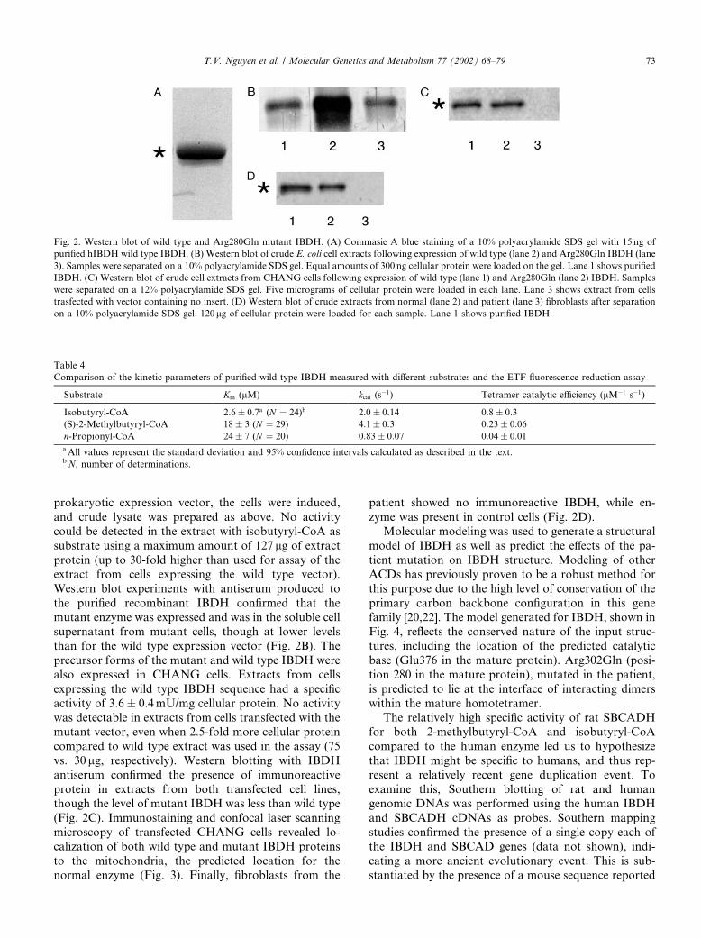
prokaryotic expression vector, the cells were induced,and crude lysate was prepared as above. No activitycould be detected in the extract with isobutyryl-CoA assubstrate using a maximum amount of 127 lg of extractprotein (up to 30-fold higher than used for assay of theextract from cells expressing the wild type vector).Western blot experiments with antiserum produced tothe purified recombinant IBDH confirmed that themutant enzyme was expressed and was in the soluble cellsupernatant from mutant cells, though at lower levelsthan for the wild type expression vector (Fig. 2B). Theprecursor forms of the mutant and wild type IBDH werealso expressed in CHANG cells. Extracts from cellsexpressing the wild type IBDH sequence had a specificactivity of 3:6� 0:4mU/mg cellular protein. No activitywas detectable in extracts from cells transfected with themutant vector, even when 2.5-fold more cellular proteincompared to wild type extract was used in the assay (75vs. 30 lg, respectively). Western blotting with IBDHantiserum confirmed the presence of immunoreactiveprotein in extracts from both transfected cell lines,though the level of mutant IBDH was less than wild type(Fig. 2C). Immunostaining and confocal laser scanningmicroscopy of transfected CHANG cells revealed lo-calization of both wild type and mutant IBDH proteinsto the mitochondria, the predicted location for thenormal enzyme (Fig. 3). Finally, fibroblasts from the
patient showed no immunoreactive IBDH, while en-zyme was present in control cells (Fig. 2D).Molecular modeling was used to generate a structural
model of IBDH as well as predict the effects of the pa-tient mutation on IBDH structure. Modeling of otherACDs has previously proven to be a robust method forthis purpose due to the high level of conservation of theprimary carbon backbone configuration in this genefamily [20,22]. The model generated for IBDH, shown inFig. 4, reflects the conserved nature of the input struc-tures, including the location of the predicted catalyticbase (Glu376 in the mature protein). Arg302Gln (posi-tion 280 in the mature protein), mutated in the patient,is predicted to lie at the interface of interacting dimerswithin the mature homotetramer.The relatively high specific activity of rat SBCADH
for both 2-methylbutyryl-CoA and isobutyryl-CoAcompared to the human enzyme led us to hypothesizethat IBDH might be specific to humans, and thus rep-resent a relatively recent gene duplication event. Toexamine this, Southern blotting of rat and humangenomic DNAs was performed using the human IBDHand SBCADH cDNAs as probes. Southern mappingstudies confirmed the presence of a single copy each ofthe IBDH and SBCAD genes (data not shown), indi-cating a more ancient evolutionary event. This is sub-stantiated by the presence of a mouse sequence reported
Fig. 2. Western blot of wild type and Arg280Gln mutant IBDH. (A) Commasie A blue staining of a 10% polyacrylamide SDS gel with 15 ng of
purified hIBDH wild type IBDH. (B) Western blot of crude E. coli cell extracts following expression of wild type (lane 2) and Arg280Gln IBDH (lane
3). Samples were separated on a 10% polyacrylamide SDS gel. Equal amounts of 300 ng cellular protein were loaded on the gel. Lane 1 shows purified
IBDH. (C) Western blot of crude cell extracts from CHANG cells following expression of wild type (lane 1) and Arg280Gln (lane 2) IBDH. Samples
were separated on a 12% polyacrylamide SDS gel. Five micrograms of cellular protein were loaded in each lane. Lane 3 shows extract from cells
trasfected with vector containing no insert. (D) Western blot of crude extracts from normal (lane 2) and patient (lane 3) fibroblasts after separation
on a 10% polyacrylamide SDS gel. 120lg of cellular protein were loaded for each sample. Lane 1 shows purified IBDH.
Table 4
Comparison of the kinetic parameters of purified wild type IBDH measured with different substrates and the ETF fluorescence reduction assay
Substrate Km (lM) kcat (s�1) Tetramer catalytic efficiency (lM�1 s�1)
Isobutyryl-CoA 2:6� 0:7a (N ¼ 24)b 2:0� 0:14 0:8� 0:3(S)-2-Methylbutyryl-CoA 18� 3 (N ¼ 29) 4:1� 0:3 0:23� 0:06n-Propionyl-CoA 24� 7 (N ¼ 20) 0:83� 0:07 0:04� 0:01aAll values represent the standard deviation and 95% confidence intervals calculated as described in the text.bN, number of determinations.
T.V. Nguyen et al. / Molecular Genetics and Metabolism 77 (2002) 68–79 73

in the genetic databases with up to 90% homology tohuman IBDH (Stratagene mouse macrophage Musmusculus cDNA clone #937306).To examine this question further, we searched Gen-
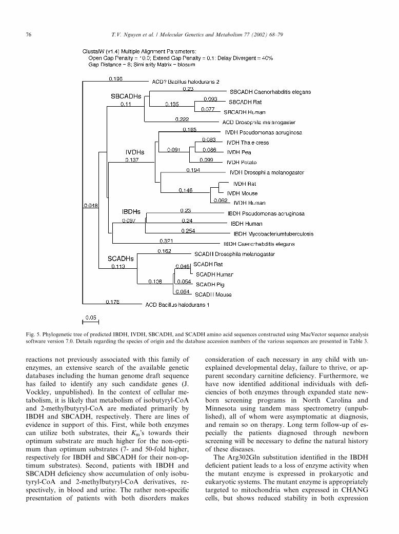
Bank for sequences related to the ACDs (Table 2) andclassified them on the basis of homology to each of theindividual gene family members. Substrate specificitywas then predicted on the basis of overall sequencehomology, as well as conservation of key residues pre-viously identified to be important in determining thisfeature. A phylogenetic tree constructed from 23 ACDsfrom various species predicted to have branched chainactivity is shown in Fig. 5. Full length coding sequenceslikely to be IVDHs were found in at least nine species,including four already shown by us to be IVDHs (hu-man, rat, Caenorhabditis elegans, and pea). Sequencespredicted to be IBDHs and SBCADHs were also clearlyidentified in a similarly wide range of species. Theseanalyses identify human IBDH as belonging to a distinctbranch of the ACD gene family with 20% amino acididentity and 32% similarity to other family members. Itis more homologous to SCADH and SBCADH thanIVDH. Interestingly, bacterial proteins from Mycobac-terium tuberculosis and Pseudomonas aeruginosa sharethe highest overall homology of the identified ACDs toIBDH (64%), and thus might be potential candidates tobe IBDH homologues. Highly conserved key residues inthe various ACDs are shown in Fig. 6.
4. Discussion
Dehydrogenation of 2-methylbutyryl-CoA and iso-butyryl-CoA in the catabolism of isoleucine and valinewas originally postulated to be mediated by a singleenzyme, termed 2-methyl-branched chain acyl-CoA de-hydrogenase [3]. The gene for this enzyme was subse-quently termed ACADSB (denoting short–branchedchain acyl-CoA dehydrogenase) to reflect the broadsubstrate specificity of the enzyme purified after ex-pression in E. coli [6,7]. More recently, we have sug-gested that separate enzymes might exist to catalyzeeach reaction in the isoleucine and valine pathways[6,7,12–14], and preliminary studies of the substratespecificity of human ACAD8 overexpressed in CHANGcells indicated that it had significant enzyme activitywith isobutyryl-CoA [13]. The current report confirmsthe existence of an isobutyryl-CoA dehydrogenase(IBDH) specific to valine metabolism, unequivocallydemonstrates identification of an ACD with highestrelative activity towards isobutyryl-CoA as substrate,and characterizes a mutation in the gene for this enzymein a patient with cellular based evidence of a specificdefect in valine metabolism [14]. The identification ofIBDH completes the complement of ACDs for reactionsknown to be catalyzed by this family of enzymes. Whileit is, of course, possible that tissue specific forms of oneor more of the ACDs may exist, as well as ACDs for
Fig. 3. Localization of wild type and mutant IBDH to mitochondria after expression in transfected CHANG cells. Forty-eight hours after trans-
fection with wild type IBDH (A)–(C) and Arg280Gln (D)–(F) expression vectors, cells were incubated with the rhodamine (red) labeled MitoTracker
(B) and (E) for 30min at 37 �C. Following fixation and permeabilization, cells were immunostained using anti-IBDH polyclonal primary antibody,detected by an Alexa 488-conjugated (green) secondary antibody (A) and (D) and analyzed by CLSM. (C) and (F) Overlay of the different optical
sections shown in (A), (B) and (D), (E), respectively. Nuclear DNA is counterstained with Hoechst 33258 (blue). Original magnification 1000�.
74 T.V. Nguyen et al. / Molecular Genetics and Metabolism 77 (2002) 68–79
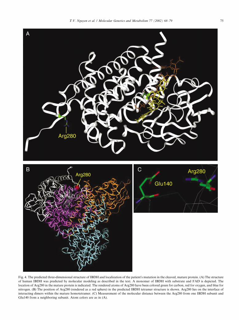
Fig. 4. The predicted three-dimensional structure of IBDH and localization of the patient�s mutation in the cleaved, mature protein. (A) The structureof human IBDH was predicted by molecular modeling as described in the text. A monomer of IBDH with substrate and FAD is depicted. The
location of Arg280 in the mature protein is indicated. The rendered atoms of Arg280 have been colored green for carbon, red for oxygen, and blue for
nitrogen. (B) The position of Arg280 (rendered as a red sphere) in the predicted IBDH tetramer structure is shown. Arg280 lies on the interface of
interacting dimers within the mature homotetramer. (C) Measurement of the molecular distance between the Arg280 from one IBDH subunit and
Glu140 from a neighboring subunit. Atom colors are as in (A).
T.V. Nguyen et al. / Molecular Genetics and Metabolism 77 (2002) 68–79 75
reactions not previously associated with this family ofenzymes, an extensive search of the available geneticdatabases including the human genome draft sequencehas failed to identify any such candidate genes (J.Vockley, unpublished). In the context of cellular me-tabolism, it is likely that metabolism of isobutyryl-CoAand 2-methylbutyryl-CoA are mediated primarily byIBDH and SBCADH, respectively. There are lines ofevidence in support of this. First, while both enzymescan utilize both substrates, their Km�s towards theiroptimum substrate are much higher for the non-opti-mum than optimum substrates (7- and 50-fold higher,respectively for IBDH and SBCADH for their non-op-timum substrates). Second, patients with IBDH andSBCADH deficiency show accumulation of only isobu-tyryl-CoA and 2-methylbutyryl-CoA derivatives, re-spectively, in blood and urine. The rather non-specificpresentation of patients with both disorders makes
consideration of each necessary in any child with un-explained developmental delay, failure to thrive, or ap-parent secondary carnitine deficiency. Furthermore, wehave now identified additional individuals with defi-ciencies of both enzymes through expanded state new-born screening programs in North Carolina andMinnesota using tandem mass spectrometry (unpub-lished), all of whom were asymptomatic at diagnosis,and remain so on therapy. Long term follow-up of es-pecially the patients diagnosed through newbornscreening will be necessary to define the natural historyof these diseases.The Arg302Gln substitution identified in the IBDH
deficient patient leads to a loss of enzyme activity whenthe mutant enzyme is expressed in prokaryotic andeukaryotic systems. The mutant enzyme is appropriatelytargeted to mitochondria when expressed in CHANGcells, but shows reduced stability in both expression
Fig. 5. Phylogenetic tree of predicted IBDH, IVDH, SBCADH, and SCADH amino acid sequences constructed using MacVector sequence analysis
software version 7.0. Details regarding the species of origin and the database accession numbers of the various sequences are presented in Table 3.
76 T.V. Nguyen et al. / Molecular Genetics and Metabolism 77 (2002) 68–79
systems and patient fibroblasts. Molecular modelingoffers some insight into this phenomenon. The mutatedamino acid residue at position 280 in the mature proteinis conserved in IVDH and SBCADH, homologous toArg280 of IVDH, and Arg286 of SBCADH. In theIBDH model, the distance from the catalytic site makesa direct effect of the patient mutation on substrateconversion unlikely. Rather, this residue lies in a posi-tion to interact with Glu140 of the opposite maturesubunit (Fig. 4), and likely plays a role in the interactionof the enzyme monomers/dimers, thus affecting theirstability. Though the predicted distance between theseresidues in our model is greater than is optimal for suchan interaction (4.6 vs <3�AA), it is important to note thatthe only positions of the amino acid residues within theindividual subunits, and not their relative position toone another, have been optimized in the model. Thus itis possible that the two residues are actually in closerapproximation than seen in our model. Consistent withthis, it is has been suggested, based on the crystalstructure of porcine MCADH, that the homologousarginine in this mature enzyme (Arg281) is importantfor FAD binding and monomer dimerization, forming ahydrogen bond with the pyrophosphate moiety of FADof the neighbouring subunit of the MCAD dimer [24].Interestingly, mutation of the homologous residue hasalso been observed in patients with VLCADH (Arg326in the mature protein) and IVDH (Arg282 in the mature
protein) deficiency, underscoring the importance of thisresidue for correct ACD function [20,27,28].Examination of ACDs in the phylogenetic tree pre-
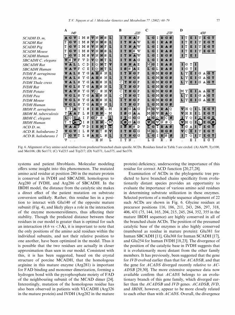
dicted to have branched chains specificity from evolu-tionarily distant species provides an opportunity toevaluate the importance of various amino acid residuesin determining substrate utilization in these enzymes.Selected portions of a multiple sequence alignment of 22such ACDs are shown in Fig. 6. Glycine residues atprecursor positions 116, 186, 211, 243, 266, 297, 318,406, 431 (75, 144, 165, 204, 215, 245, 264, 352, 355 in themature IBDH sequence) are highly conserved in all ofthe branched chain ACDs. The position of the presumedcatalytic base of the enzymes is also highly conserved(numbered as residue in mature protein): Glu381 forhuman SBCADH [11], Glu368 for human SCADH [17],and Glu254 for human IVDH [18,23]. The divergence ofthe position of the catalytic base in IVDH suggests thatit is evolutionarily more distant from the other familymembers. It has previously, been suggested that the genefor IVD evolved earlier than that for ACADSB, and thatthe gene for ACADS diverged recently relative to AC-ADSB [29,30]. The more extensive sequence data nowavailable confirm that ACADS belongs to an evolu-tionary branch of this gene family, which diverged ear-lier than the ACADSB and IVD genes. ACADSB, IVD,and IBDH, however, appear to be more closely relatedto each other than with ACADS. Overall, the divergence
Fig. 6. Alignment of key amino acid residues from predicted branched chain specific ACDs. Residues listed in Table 5 are circled. (A) Ala99, Tyr100,
and Met106; (B) Ser171; (C) Val215 and Trp217; (D) Val371, Leu375, and Ser378.
T.V. Nguyen et al. / Molecular Genetics and Metabolism 77 (2002) 68–79 77
in the branched chain specific ACDs appears to be anevolutionarily ancient event as evidenced by the pres-ence of apparent IBDH sequences inM. tuberculosis andP. aeruginosa.Comparison of the known structure of human IVDH
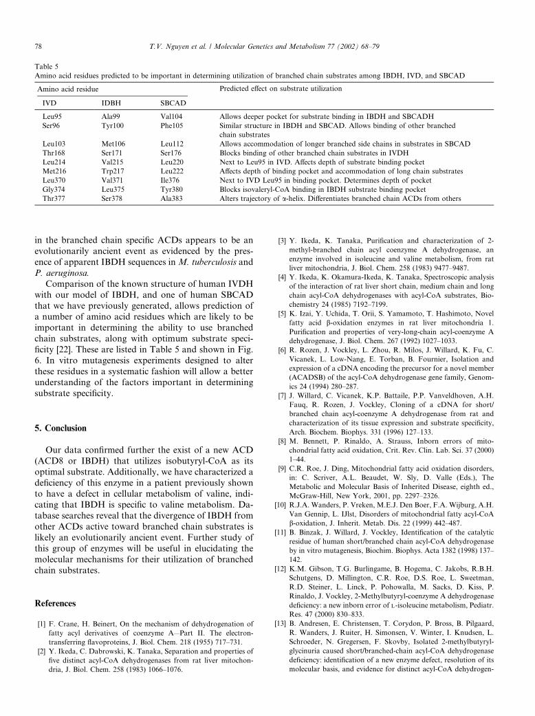
with our model of IBDH, and one of human SBCADthat we have previously generated, allows prediction ofa number of amino acid residues which are likely to beimportant in determining the ability to use branchedchain substrates, along with optimum substrate speci-ficity [22]. These are listed in Table 5 and shown in Fig.6. In vitro mutagenesis experiments designed to alterthese residues in a systematic fashion will allow a betterunderstanding of the factors important in determiningsubstrate specificity.
5. Conclusion
Our data confirmed further the exist of a new ACD(ACD8 or IBDH) that utilizes isobutyryl-CoA as itsoptimal substrate. Additionally, we have characterized adeficiency of this enzyme in a patient previously shownto have a defect in cellular metabolism of valine, indi-cating that IBDH is specific to valine metabolism. Da-tabase searches reveal that the divergence of IBDH fromother ACDs active toward branched chain substrates islikely an evolutionarily ancient event. Further study ofthis group of enzymes will be useful in elucidating themolecular mechanisms for their utilization of branchedchain substrates.
References
[1] F. Crane, H. Beinert, On the mechanism of dehydrogenation of
fatty acyl derivatives of coenzyme A—Part II. The electron-
transferring flavoproteins, J. Biol. Chem. 218 (1955) 717–731.
[2] Y. Ikeda, C. Dabrowski, K. Tanaka, Separation and properties of
five distinct acyl-CoA dehydrogenases from rat liver mitochon-
dria, J. Biol. Chem. 258 (1983) 1066–1076.
[3] Y. Ikeda, K. Tanaka, Purification and characterization of 2-
methyl-branched chain acyl coenzyme A dehydrogenase, an
enzyme involved in isoleucine and valine metabolism, from rat
liver mitochondria, J. Biol. Chem. 258 (1983) 9477–9487.
[4] Y. Ikeda, K. Okamura-Ikeda, K. Tanaka, Spectroscopic analysis
of the interaction of rat liver short chain, medium chain and long
chain acyl-CoA dehydrogenases with acyl-CoA substrates, Bio-
chemistry 24 (1985) 7192–7199.
[5] K. Izai, Y. Uchida, T. Orii, S. Yamamoto, T. Hashimoto, Novel
fatty acid b-oxidation enzymes in rat liver mitochondria 1.Purification and properties of very-long-chain acyl-coenzyme A
dehydrogenase, J. Biol. Chem. 267 (1992) 1027–1033.
[6] R. Rozen, J. Vockley, L. Zhou, R. Milos, J. Willard, K. Fu, C.
Vicanek, L. Low-Nang, E. Torban, B. Fournier, Isolation and
expression of a cDNA encoding the precursor for a novel member
(ACADSB) of the acyl-CoA dehydrogenase gene family, Genom-
ics 24 (1994) 280–287.
[7] J. Willard, C. Vicanek, K.P. Battaile, P.P. Vanveldhoven, A.H.
Fauq, R. Rozen, J. Vockley, Cloning of a cDNA for short/
branched chain acyl-coenzyme A dehydrogenase from rat and
characterization of its tissue expression and substrate specificity,
Arch. Biochem. Biophys. 331 (1996) 127–133.
[8] M. Bennett, P. Rinaldo, A. Strauss, Inborn errors of mito-
chondrial fatty acid oxidation, Crit. Rev. Clin. Lab. Sci. 37 (2000)
1–44.
[9] C.R. Roe, J. Ding, Mitochondrial fatty acid oxidation disorders,
in: C. Scriver, A.L. Beaudet, W. Sly, D. Valle (Eds.), The
Metabolic and Molecular Basis of Inherited Disease, eighth ed.,
McGraw-Hill, New York, 2001, pp. 2297–2326.
[10] R.J.A. Wanders, P. Vreken, M.E.J. Den Boer, F.A. Wijburg, A.H.
Van Gennip, L. IJlst, Disorders of mitochondrial fatty acyl-CoA
b-oxidation, J. Inherit. Metab. Dis. 22 (1999) 442–487.[11] B. Binzak, J. Willard, J. Vockley, Identification of the catalytic
residue of human short/branched chain acyl-CoA dehydrogenase
by in vitro mutagenesis, Biochim. Biophys. Acta 1382 (1998) 137–
142.
[12] K.M. Gibson, T.G. Burlingame, B. Hogema, C. Jakobs, R.B.H.
Schutgens, D. Millington, C.R. Roe, D.S. Roe, L. Sweetman,
R.D. Steiner, L. Linck, P. Pohowalla, M. Sacks, D. Kiss, P.
Rinaldo, J. Vockley, 2-Methylbutyryl-coenzyme A dehydrogenase
deficiency: a new inborn error of L-isoleucine metabolism, Pediatr.
Res. 47 (2000) 830–833.
[13] B. Andresen, E. Christensen, T. Corydon, P. Bross, B. Pilgaard,
R. Wanders, J. Ruiter, H. Simonsen, V. Winter, I. Knudsen, L.
Schroeder, N. Gregersen, F. Skovby, Isolated 2-methylbutyryl-
glycinuria caused short/branched-chain acyl-CoA dehydrogenase
deficiency: identification of a new enzyme defect, resolution of its
molecular basis, and evidence for distinct acyl-CoA dehydrogen-
Table 5
Amino acid residues predicted to be important in determining utilization of branched chain substrates among IBDH, IVD, and SBCAD
Amino acid residue Predicted effect on substrate utilization
IVD IDBH SBCAD
Leu95 Ala99 Val104 Allows deeper pocket for substrate binding in IBDH and SBCADH
Ser96 Tyr100 Phe105 Similar structure in IBDH and SBCAD. Allows binding of other branched
chain substrates
Leu103 Met106 Leu112 Allows accommodation of longer branched side chains in substrates in SBCAD
Thr168 Ser171 Ser176 Blocks binding of other branched chain substrates in IVDH
Leu214 Val215 Leu220 Next to Leu95 in IVD. Affects depth of substrate binding pocket
Met216 Trp217 Leu222 Affects depth of binding pocket and accommodation of long chain substrates
Leu370 Val371 Ile376 Next to IVD Leu95 in binding pocket. Determines depth of pocket
Gly374 Leu375 Tyr380 Blocks isovaleryl-CoA binding in IBDH substrate binding pocket
Thr377 Ser378 Ala383 Alters trajectory of a-helix. Differentiates branched chain ACDs from others
78 T.V. Nguyen et al. / Molecular Genetics and Metabolism 77 (2002) 68–79

ases in isoleucine and valine metabolism, Am. J. Hum. Genet. 67
(2000) 1095–1103.
[14] C.R. Roe, S.D. Cederbaum, D.S. Roe, R. Mardach, A. Galindo,
L. Sweetman, Isolated isobutyryl-CoA dehydrogenase deficiency:
an unrecognized defect in human valine metabolism, Mol. Genet.
Metabol. 65 (1998) 264–271.
[15] E.A. Telford, L.M. Moynihan, A.F. Markham, N.J. Lench,
Isolation and characterisation of a cDNA encoding the precursor
for a novel member of the acyl-CoA dehydrogenase gene family,
Biochim. Biophys. Acta 1446 (1999) 371–376.
[16] S. Gustafson, J.A. Proper, E.J. Bowie, S.S. Sommer, Parameters
affecting the yield of DNA from human blood, Anal. Biochem.
165 (1987) 294–299.
[17] K. Battaile, A.-W. Mohsen, J. Vockley, Functional role of the
active site glutamate-368 in rat short chain acyl-CoA dehydro-
genase, Biochemistry 35 (1996) 15356–15363.
[18] A.-W.A. Mohsen, J. Vockley, Identification of the active site
catalytic residue in human isovaleryl-CoA dehydrogenase, Bio-
chemistry 34 (1995) 10146–10152.
[19] A.A. Mohsen, J. Vockley, High-level expression of an altered
cDNA encoding human isovaleryl-CoA dehydrogenase in Esch-
erichia coli, Gene 160 (1995) 263–267.
[20] A.-W. Mohsen, B. Anderson, S. Volchenboum, K. Battaile, K.
Tiffany, D. Roberts, J.-J. Kim, J. Vockley, Characterization of
molecular defects in isovaleryl-CoA dehydrogenase in patients
with isovaleric acidemia, Biochemistry 37 (1998) 10325–10335.
[21] T.J. Corydon, M. Wilsbech, C. Jespersgaard, B.S. Andresen, A.D.
Borglom, S. Pedersen, L. Bolund, N. Gregersen, P. Bross, Human
and mouse mitochondrial orthologs of bacterial ClpX, Mamm.
Genome 11 (2000) 899–905.
[22] J. Vockley, A.W. Mohsen, B. Binzak, J. Willard, A. Fauq,
Mammalian branched-chain acyl-CoA dehydrogenases: molecular
cloning and characterization of recombinant enzymes, Methods
Enzymol. 324 (2000) 241–258.
[23] K.A. Tiffany, D.L. Roberts, M. Wang, R. Paschke, A.W.A.
Mohsen, J. Vockley, J.J.P. Kim, Structure of human isovaleryl-
coA dehydrogenase at 2.6 angstrom resolution—basis for sub-
strate specificity, Biochemistry 36 (1997) 8455–8464.
[24] J.-J. Kim, J. Wu, Structure of the medium chain acyl-CoA
dehydrogenase from pig liver mitochondria at 3-A resolution,
Proc. Natl. Acad. Sci. USA 84 (1988) 6677–6681.
[25] J.J.P. Kim, M. Wang, R. Paschke, Crystal structures of medium-
chain acyl-CoA dehydrogenase from pig liver mitochondria with
and without substrate, Proc. Natl. Acad. Sci. USA 90 (1993)
7523–7527.
[26] S. Djordjevic, C.P. Pace, M.T. Stankovich, J.J.P. Kim, Three-
dimensional structure of butyryl-CoA dehydrogenase from Me-
gasphaera esdenii, Biochemistry 34 (1995) 2163–2171.
[27] B.S. Andresen, P. Bross, C. Vianeysaban, P. Divry, M.T.
Zabot, C.R. Roe, M.A. Nada, A. Byskov, T.A. Kruse, S. Neve,
K. Kristiansen, I. Knudsen, M.J. Corydon, N. Gregersen,
Cloning and characterization of human very-long-chain acyl-
CoA dehydrogenase cDNA, chromosomal assignment of the
gene and identification in four patients of nine different
mutations within the VLCAD gene, Hum. Mol. Genet. 5
(1996) 461–472.
[28] B.S. Andresen, C. Vianeysaban, P. Bross, P. Divry, C.R. Roe,
M.A. Nada, I. Knudsen, N. Gregersen, The mutational spectrum
in very long-chain acyl-CoA dehydrogenase deficiency, J. Inherit.
Metab. Dis. 19 (1996) 169–172.
[29] A. Nandy, B. Kuchler, S. Ghisla, Molecular evolution and
substrate specificity of acyl-CoA dehydrogenases: chimaeric
�medium/long� chain specific enzyme from medium-chain acyl-
CoA dehydrogenase, Biochem. Soc. Trans. 24 (1996) 105–
110.
[30] K. Tanaka, Y. Indo, Evolution of the acyl-CoA dehydrogenase/
oxidase superfamily, Prog. Clin. Biol. Res.—New Dev. Fatty Acid
Oxidation 375 (1992) 95–110.
T.V. Nguyen et al. / Molecular Genetics and Metabolism 77 (2002) 68–79 79