cardiac mitochondrial connexin 43 regulates apoptosis
TRANSCRIPT
Cardiac Mitochondrial Connexin 43 Regulates Apoptosis
Farida Goubaeva1, Maya Mikami1, Sarah Giardina1,+, Bo Ding3, Junichi Abe3, and JayYang1,4,*1Department of Anesthesiology Columbia University Medical Center New York, NY 10032
4Department of Pathology and Cell Biology Columbia University Medical Center New York, NY10032
3Center for Cardiovascular Research University of Rochester Medical Center Rochester, NY 14342
AbstractConnexin 43 (Cx43) is thought to be present largely in the plasma membrane and its function solelyto provide low resistance electrical connection between myocytes. A recent report suggested thepresence of Cx43 in the mitochondria as well. We confirmed the presence of Cx43 in the mitochondriaisolated from adult rat ventricles with the Cx43 immunoreactivity fractionating to the outermitochondrial membrane. Mitochondrial Cx43 is mostly phosphorylated only detected by a phospho-specific antibody. Using a Ca++-sensitive electrode and Western blot, we showed that the gap junctioninhibitors 18-β-glycyrrhetinic acid (β-GA), oleamide, and heptanol all induced concomitant releaseof Ca++ and cytochrome C in isolated mitochondria whereas the inactive analog 18-β-glycyrrhizicacid failed to do so. In low density neonatal myocyte culture with no appreciable cell-cell contacts,β-GA induced apoptosis as assessed by TUNEL staining. Our results suggest a novel role of Cx43as a regulator of mitochondrial physiology and myocyte apoptosis.
Keywordsmitochondria; connexins; apoptosis; β-glycyrrhetinic acid; gap junction inhibitors; cytochrome C;TUNEL; cardiac myocytes
IntroductionCellular apoptosis plays a critical role in cardiovascular diseases [1,2]. While many regulatorsof this programmed cell death pathway have been identified the list is clearly incomplete.Connexin (Cx) 43, the major gap junction forming protein in the adult cardiac ventricles, playsa pivotal role in mediating tissue injury and post-ischemic cardiac dysfunction, however, themechanism by which Cx43 modulates cellular injury is not known.
Most of the function ascribed to Cx43 in cardiac pathophysiology is within the context of itsrole in forming the gap junction [3]. However, recent literature reports atypical Cx43 functionsin roles outside of the traditional gap junction [4,5]. We recently discovered that Cx43,independent of its ability to form functional gap junctions, regulates susceptibility of cells toseveral cell injury paradigms suggesting a novel function of this protein in modulating cell
*Corresponding Author, Dr. Jay Yang Department of Anesthesiology Columbia University Medical Center 630 West 168th Street, PH-5New York, NY 10032 212-342-0023 (Telephone) 212-305-0777 (FAX)[email protected]+Present AddressDepartment of Microbiology Cornell University School of Medicine New York, NY 10021Publisher's Disclaimer: This is a PDF file of an unedited manuscript that has been accepted for publication. As a service to our customerswe are providing this early version of the manuscript. The manuscript will undergo copyediting, typesetting, and review of the resultingproof before it is published in its final citable form. Please note that during the production process errorsmaybe discovered which couldaffect the content, and all legal disclaimers that apply to the journal pertain.
NIH Public AccessAuthor ManuscriptBiochem Biophys Res Commun. Author manuscript; available in PMC 2007 March 22.
Published in final edited form as:Biochem Biophys Res Commun. 2007 January 5; 352(1): 97–103.
NIH
-PA Author Manuscript
NIH
-PA Author Manuscript
NIH
-PA Author Manuscript
death [6]. Cx43 is abundantly expressed in the adult cardiac ventricles, yet little study existsaddressing the role of cardiac Cx43 in modulating myocyte death. Myocyte apoptosis is nowrecognized to mediate cell death in a variety of acute and chronic heart diseases andunderstanding how Cx regulates apoptosis may lead to a new novel approach to preventingmyocyte death.
A recent report suggested that Cx43 is present in the mitochondria and may play a role inmediating the cardioprotective effect of ischemic preconditioning [7]. In the present study, weconfirmed the presence of Cx43 immunoreactivity in the outer membrane of the mitochondriaisolated from adult cardiac ventricles and provide evidence that mitochondrial Cx43 as a novelregulator of mitochondrial function where inhibition results in the release of cytochrome C andmyocyte apoptosis.
MethodsIsolation of rat ventricular mitochondria and fractionation
All procedures were conducted in accordance with institutional animal care regulations.Minced rat ventricular tissue chunks were suspended in a mannitol-sucrose (M/S) buffer (inmM): 255 mannitol, 10 sucrose, 0.5 EGTA, 1 glutathione, 10 HEPES, pH 7.4, and cells lysedby 10 strokes of a Dounce homogenizer. The first low speed centrifugation at 2000g x 10 minseparated the unlysed cells and the nuclei. The supernatant from the first spin (S1) was re-spunat 6000g x 10 min and the pellet (P2) was collected as the fraction enriched in mitochondria.For the mitochondria functional assay, the P2 pellet was resuspended in the M/S buffer withoutEGTA and oxygen applied by blow-by until used. For protein isolation, the P2 pellet wasresuspended in RIPA buffer (1% Nonidet P40, 10mM Tris pH 7.6, 50mM NaCl, 30mM NaPPi,50mM NaF, 1% Triton X100, and 0.1% sodium dodecyl-sulfate) and sonicated (50% powerfor 2 second x 10 pulses, Cole Palmer Model 130 Ultrasonic Processor). All procedures weredone at 4C° or on ice.
Subcellular fractionation of the organelles by idioxanol gradient (19-27%) centrifugation wascarried out according to manufacturer’s instructions (Axis-Shield, Oslow, Norway). For thesub-mitochondrial fractionation, the P2 pellet suspended in M/S buffer supplemented with 1mM DTT and 0.2 mM PMSF was sonicated, as noted above, followed by 3 freeze-thaw cycles(modified from Beutner et. al., [8]). The mitochondrial fragments were loaded on a sucrosegradient (from 60% to 30%) in 10 mM HEPES, pH7.4, supplemented with a protease inhibitorcocktail (Complete, Roche Applied Science, Indianapolis, IN, USA), and centrifuged at100,000g for 3 hours at 4C° (Beckman Optima TLX centrifuge with a TL55 swinging bucketrotor).
Western blottingProteins subjected to SDS-PAGE and transferred to a nitrocellulose membrane was blockedin 3% milk-TBST and probed with the following primary antibodies: ANT (1:500, Santa CruzBiotech, Santa Cruz, CA, USA; sc-11433); Calreticulin (1:500, Affinity Bioreagents, Golden,CO, USA; #PA1-903); Cx43 (1:500, Chemicon International, Temecula, CA, USA;MAB3067), GAPDH (1:100,000, Advance Immunochemical, Long Beach, CA, USA;#RGM2); N-cadherin (1:1000, Zymed Lab, South San Francisco, CA, USA; #18-0224),VDAC (1:1000, Oncogene, Boston, MA, USA; #PC548), all in 3% milk-PBST, and reactedwith the horseradish peroxidase-conjugated secondary antibody (1:2000) in 1% milk-TBST.After reaction with the Western Lightning chemiluminescence reagent (NEN Life ScienceProducts, Boston, MA, USA), the images were captured on the EpiChemi Darkroom System(UVP Inc, Upland, CA). The commercially available anti-Cx43 antibody raised against apeptide-epitope (pan-Cx43) used in this study is well-characterized specifically recognizing
Goubaeva et al. Page 2
Biochem Biophys Res Commun. Author manuscript; available in PMC 2007 March 22.
NIH
-PA Author Manuscript
NIH
-PA Author Manuscript
NIH
-PA Author Manuscript
both the non-phosphorylated and phosphorylated Cx43 protein with little non-specificreactivity [9]. The Cx-1B1 anti-Cx43 antibody (#13-8300, Zymed Lab,) only recognizes Cx43when the serine at residue 368 is unphosphorylated (P0-Cx43) [10]. Dephosphorylation wasaccomplished by incubating mitochondrial lysate with calf intestinal alkaline phosphatase (10U) at 37C° for 20 hours and separated by a 12% SDS-PAGE to optimize separation of multiplebands in the 40-50kD range.
ImmunohistochemistryCultured cardiomyocytes were fixed with 4% paraformaldehyde in 0.1M PB for 15 minutes.After permeablization with 0.2% Tween-20 in phosphate buffered saline (PBST), and blocking(10% normal goat serum in PBST) both for 15 minutes at room temperature, the samples wereincubated with the following antibodies: Cx43 antibody (1:200), VDAC (1:200), and α-actinin(1:500, Sigma-Aldrich, St.Louis, MO, USA) all in PBST with 2% normal goat serum for 2hours at room temperature. After washing, the cells were reacted with the appropriatesecondary antibody conjugated to Alexa 487 or 594 (1:500, Molecular Probes, Portland, OR,USA), and visualized under a Zeiss LSM 510 NLO confocal microscope (Oberkochen,Germany) and psuedo-colored in Photoshop.
Measurement of mitochondrial calcium uptakeMitochondrial suspension in MS buffer without EGTA was placed in a stirred container andthe free-[Ca++] within the solution adjusted to approximately 10 μM and thereaftercontinuously monitored by a Ca++-sensitive electrode (MI-600, Microelectrodes, Inc, Bedford,NH, USA) connected to an Accumet 25 pH meter (Fisher Scientific, Pittsburg, PA, USA). Thevoltage output was calibrated using solutions of known free-[Ca++] (WP Instruments, Sarasota,FL, USA) and demonstrated a superior log-linearity over a wide range of [Ca++].
Myocyte culture and apoptosis cell count assayRat neonatal ventricular myocytes were isolated from day 2-4 neonatal pups by enzymaticdigestion following a procedure recommended by the manufacturer (Worthington, Lakewood,NJ, USA). After pre-panning the remaining myocyte-enriched suspension was plated at a lowdensity (50,000 cells/35mm well). The growth of background cells was inhibited by additionof cytosine arabinoside (1 μM) 24 hours after plating and thereafter, the culture was maintainedin DMEM supplemented with 10% newborn calf serum, 10% equine serum, and 1% penicillin/streptomycin.
For the apoptotic cell count assay, myocytes were treated with β-GA (0-100 μM) or its inactiveanalogue (GZ, 100 μM) for 24h in the serum containing medium. The presence of the serumreduced the total number of apoptotic cells making the visualization and counting of apoptoticcells easier. Fixed (4% PFA) myocytes were subjected to the terminal deoxyribonucleotidetransferase (TdT)-mediated dUTP nick-end labeling (TUNEL) assay as per manufacturer’sinstructions (Promega, Madison, WI, USA). The cells counts were taken from no less than 12random fields, where the total number of myocytes counted was typically 1000 cells. Data(mean ± SEM) is expressed as % TUNEL-positive.
ChemicalsAll chemicals were purchased from Sigma-Aldrich except for carbonlycyanide-4-(trifuloromethoxy)-phenylhydrazone (FCCP) (Biomol, Plymouth Meeting, PA, USA) andMitoTracker (Invitrogen, Carlsbad, CA, USA).
Goubaeva et al. Page 3
Biochem Biophys Res Commun. Author manuscript; available in PMC 2007 March 22.
NIH
-PA Author Manuscript
NIH
-PA Author Manuscript
NIH
-PA Author Manuscript
ResultsAnti-Cx43 immunoreactivity in the cardiac mitochondria:
Immunoreactivity to anti-Cx43 antibody was investigated in cultured rat neonatal ventricularmyocytes. The culture prepared by a standard method consisted of myocytes and non-myocytesupport cells; however, the myocytes were easily identified because of their characteristicstellate morphology and the immunoreactivity to anti-α-actinin antibody. Immunostaining withanti-Cx43 antibody (Figure 1A, red) demonstrated numerous punctate widely distributedimmunoreactive spots in addition to the expected linear streaks at the cell borders. The punctateimmunoreactive spots resembled the distribution of mitochondria in these cells which wasconfirmed by MitoTracker staining (Figure 1A, green) and by anti-VDAC antibodyimmunoreactivity (data not shown). However, inspection of the images indicated that the twomarkers only partially overlapped.
Localization of Cx43 in low density cultured neonatal rat ventricular myocytes maybeabnormal, therefore, we attempted to demonstrate the presence of anti-Cx43 immunoreactivityin the adult ventricular mitochondria by biochemical organelle and protein fractionationmethods. A crude fractionation of cellular organelles was accomplished by a simple 2-stepcentrifugation and the isolated fractions probed by immunoblotting (Figure 1B). The followingimmunoreactive signals were used as markers: anti-VDAC (mitochondrial marker), anti-GAPDH (cytosolic marker), and N-cadherin (plasma membrane marker). The P2 pellet isolatedby this method mostly separated the cytosolic and the plasma membrane compartments awayas demonstrated by the minimal GAPDH and N-cadherin immunoreactivity in this fraction.The P1 fraction with the plasma membrane exhibited robust anti-Cx43 immunoreactivity asexpected. However, the P2 fraction also demonstrated robust immunoreactivity suggesting thepresence of Cx43 in a non-plasma membrane and non-cytosolic compartment of the ventricularcells.
Further fractionation of the organelles was attempted using the P2 pellet as the starting material.Fractions collected after an iodixanol-gradient centrifugation demonstrated anti-Cx43, anti-VDAC, and anti-calreticulin (endoplasmic reticulum marker) immunoreactivity in overlappingfractions (Figure 1C). However, the anti-Cx43 immunoreactive fractions tended towards theless dense fraction compared to the overlapping fractions immunoreactive to anti-VDAC andanti-calreticulin. As expected, we saw no immunoreactivity to N-cadherin or GAPDH in thefractions since the starting P2 pellet was mostly devoid of these signals.
Next we followed the anti-Cx43 immunoreactivity in sub-fractions of the mitochondria againstarting with the P2 pellet but by separating the sub-mitochondrial protein compartments bysucrose gradient centrifugation after lysis of the mitochondria. Immunoblotting with anti-VDAC as a marker for the outer mitochondrial membrane and with anti-ANT as a marker forthe inner mitochondrial membrane confirmed the proper isolation of the sub-mitochondrialcompartments (Figure 1D). The contact-site (CS) showed immunoreactivity to both the inner-and outer-mitochondrial membrane markers as expected. The anti-Cx43 immunoreactivity inthe total mitochondria appeared as multiple distinct bands (arrows) with apparent massesslightly greater than the expected 43kD. Immunoreactivity in the fractionated samples waspresent in the CS and a stronger signal in the outer mitochondrial membrane but only of thelarger apparent mass. Since Cx43 is a phosphoprotein that exhibits a well describedphosphorylation-induced mobility shift, we examined the phosphorylation-status of themitochondrial Cx43 by probing with a P0-specific anti-Cx43 antibody. Mitochondrial lysateprobed with the pan-Cx43 antibody detected multiple bands (Figure 1E, arrows) that collapsedto a single band on alkaline phosphatase treatment. The anti-P0-Cx43 antibody detected a bandonly when dephosphorylated indicating that majority of the Cx43 immunoreactive speciespresent in the mitochondria is phosphorylated. These observations confirmed the report that
Goubaeva et al. Page 4
Biochem Biophys Res Commun. Author manuscript; available in PMC 2007 March 22.
NIH
-PA Author Manuscript
NIH
-PA Author Manuscript
NIH
-PA Author Manuscript
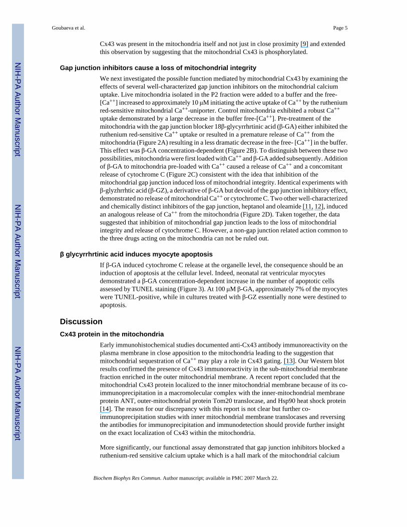
Cx43 was present in the mitochondria itself and not just in close proximity [9] and extendedthis observation by suggesting that the mitochondrial Cx43 is phosphorylated.
Gap junction inhibitors cause a loss of mitochondrial integrityWe next investigated the possible function mediated by mitochondrial Cx43 by examining theeffects of several well-characterized gap junction inhibitors on the mitochondrial calciumuptake. Live mitochondria isolated in the P2 fraction were added to a buffer and the free-[Ca++] increased to approximately 10 μM initiating the active uptake of Ca++ by the rutheniumred-sensitive mitochondrial Ca++-uniporter. Control mitochondria exhibited a robust Ca++
uptake demonstrated by a large decrease in the buffer free-[Ca++]. Pre-treatment of themitochondria with the gap junction blocker 18β-glycyrrhrtinic acid (β-GA) either inhibited theruthenium red-sensitive Ca++ uptake or resulted in a premature release of Ca++ from themitochondria (Figure 2A) resulting in a less dramatic decrease in the free- [Ca++] in the buffer.This effect was β-GA concentration-dependent (Figure 2B). To distinguish between these twopossibilities, mitochondria were first loaded with Ca++ and β-GA added subsequently. Additionof β-GA to mitochondria pre-loaded with Ca++ caused a release of Ca++ and a concomitantrelease of cytochrome C (Figure 2C) consistent with the idea that inhibition of themitochondrial gap junction induced loss of mitochondrial integrity. Identical experiments withβ-glyzhrrhtic acid (β-GZ), a derivative of β-GA but devoid of the gap junction inhibitory effect,demonstrated no release of mitochondrial Ca++ or cytochrome C. Two other well-characterizedand chemically distinct inhibitors of the gap junction, heptanol and oleamide [11, 12], inducedan analogous release of Ca++ from the mitochondria (Figure 2D). Taken together, the datasuggested that inhibition of mitochondrial gap junction leads to the loss of mitochondrialintegrity and release of cytochrome C. However, a non-gap junction related action common tothe three drugs acting on the mitochondria can not be ruled out.
β glycyrrhrtinic acid induces myocyte apoptosisIf β-GA induced cytochrome C release at the organelle level, the consequence should be aninduction of apoptosis at the cellular level. Indeed, neonatal rat ventricular myocytesdemonstrated a β-GA concentration-dependent increase in the number of apoptotic cellsassessed by TUNEL staining (Figure 3). At 100 μM β-GA, approximately 7% of the myocyteswere TUNEL-positive, while in cultures treated with β-GZ essentially none were destined toapoptosis.
DiscussionCx43 protein in the mitochondria
Early immunohistochemical studies documented anti-Cx43 antibody immunoreactivity on theplasma membrane in close apposition to the mitochondria leading to the suggestion thatmitochondrial sequestration of Ca++ may play a role in Cx43 gating. [13]. Our Western blotresults confirmed the presence of Cx43 immunoreactivity in the sub-mitochondrial membranefraction enriched in the outer mitochondrial membrane. A recent report concluded that themitochondrial Cx43 protein localized to the inner mitochondrial membrane because of its co-immunoprecipitation in a macromolecular complex with the inner-mitochondrial membraneprotein ANT, outer-mitochondrial protein Tom20 translocase, and Hsp90 heat shock protein[14]. The reason for our discrepancy with this report is not clear but further co-immunoprecipitation studies with inner mitochondrial membrane translocases and reversingthe antibodies for immunoprecipitation and immunodetection should provide further insighton the exact localization of Cx43 within the mitochondria.
More significantly, our functional assay demonstrated that gap junction inhibitors blocked aruthenium-red sensitive calcium uptake which is a hall mark of the mitochondrial calcium
Goubaeva et al. Page 5
Biochem Biophys Res Commun. Author manuscript; available in PMC 2007 March 22.
NIH
-PA Author Manuscript
NIH
-PA Author Manuscript
NIH
-PA Author Manuscript
uniporter [15] in contradistinction to the SERCA-ATPase responsible for the calcium uptakeinto the endoplasmic reticulum [16]. Taken together, our data suggested the presence of Cx43in the mitochondria itself and not just in close proximity, exists largely as phospho-Cx43, andplays a role in the regulation of mitochondrial function.
The MitoProt II software (www.mips.biochem.mpg,de/cgi-bin/proj/medgen/mitofilter)predicts mitochondrial localization of a protein based on an analysis of the signal peptide[17]. A query for connexins returns a probability of mitochondrial-targeting greater than 0.20for connexin 25, 31.1, 31, 32, and 45 suggesting the possibility of these connexin isoforms alsobeing present in the mitochondria. However, the mitochondrial-targeting probability is only0.03 for the full length Cx43 protein. It is likely that Cx43, particularly when phosphorylated,may be imported into the mitochondria by specific translocases independent of the N-terminalsignal sequence like many other mitochondrial proteins [18] possibly in association with achaperone protein [14].
Functional consequence of inhibition of mitochondrial gap junctionEvidence for a functional role of mitochondrial Cx43 comes from studies investigating theeffects of gap junction active-drugs on mitochondrial function. In rat liver mitochondria, β-GA was shown to induce MPT and release cytochrome C [19]. However, in their discussion,no association was made between the well known gap junction inhibiting-property of β-GAand the observed effect on mitochondrial physiology. We expanded upon this observation andpresented in our present work that the parent glycyrrhizic acid (GZ) incapable of gap junctioninhibition does not induce mitochondrial Ca++release. Furthermore, two other chemicallydistinct gap junction inhibitors, heptanol and oleamide, induced mitochondrial calcium release.However, no drugs exhibit absolute specificity and further studies using highly specificmolecular reagents will be necessary to confirm our observations being mindful of thelikelihood that in organs expressing multiple isoforms of Cx, simultaneous use of multipleselective inhibitors may be necessary to alter the mitochondrial function. In fact, theobservation that β-GA induces MPT in the liver where the predominant Cx isoform is Cx32and the lack of gross developmental abnormality in Cx43 null-mutant mice allowing survivalto term [20] support the idea that multiple Cx isoforms must regulate mitochondrial physiology.
The hexameric Cx43 protein complex connexon, both as a gap junction and as a hemichannel,forms large-conductance ion channel with chemical gating similar to the Bcl-2 channels. Inaddition, Cx43 channels are voltage gated perhaps allowing sensing of the mitochondrialmembrane potential in addition to the chemical environment. Given the well-documented roleof mitochondrial ion channels such as Bcl-2 in modulating apoptosis, it is highly probable thatthe ion channels formed by the Cx43 protein may also play a role in modulating apoptosis,however, no evidence exists to indicate that the mitochondrial Cx43 forms an ion channel.Proving this hypothesis will require further experiments with a targeted mitochondrial over-expression of dominant-negative Cx43 mutants (e.g. L160M-mutant) incapable of forming ionchannels [6] and examining the apoptotic phenotype in such cells.
Acknowledgements
We thank Dr. Shey Shing Sheu (University of Rochester) for introducing the area of mitochondrial physiology to thesenior author and Mahmud Uzzaman for participating in early experiments. This work was partially supported by NIHRO1 GM071485 (JY).
References[1]. Foo RS, Mani K, Kitsis RN. Death begets failure in the heart. J Clin Invest 2005;115:565–71.
[PubMed: 15765138]
Goubaeva et al. Page 6
Biochem Biophys Res Commun. Author manuscript; available in PMC 2007 March 22.
NIH
-PA Author Manuscript
NIH
-PA Author Manuscript
NIH
-PA Author Manuscript

[2]. Gonzalez A, Fortuno MA, Querejeta R, Ravassa S, Lopez B, Lopez N, Diez J. Cardiomyocyteapoptosis in hypertensive cardiomyopathy. Cardiovasc Res 2003;59:549–62. [PubMed: 14499856]
[3]. Severs NJ, Coppen SR, Dupont E, Yeh HI, Ko YS, Matsushita T. Gap junction alterations in humancardiac disease. Cardiovasc Res 2004;62:368–77. [PubMed: 15094356]
[4]. Kalvelyte A, Imbrasaite A, Bukauskiene A, Verselis VK, Bukauskas FF. Connexins and apoptotictransformation. Biochem Pharmacol 2003;66:1661–72. [PubMed: 14555247]
[5]. Plotkin LI, Bellido T. Bisphosphonate-induced, hemi-channel-mediated anti-apoptosis through theSrc/ERK pathway: A gap junction-independent action of connexin 43. Cell Comm Adhes2001;8:377–82.
[6]. Lin JH, Yang J, Liu S, Takano T, Wang X, Gao Q, Willecke K, Nedergaard M. Connexin mediatesgap junction-independent resistance to cellular injury. J Neurosci 2003;23:430–41. [PubMed:12533603]
[7]. Boengler K, Dodoni G, Rodriguez-Sinovas A, Cabestrero A, Ruiz-Meana R, Gres P, Konietzka I,Lopez-Iglesias C, Garcia-Dorado D, Di Lisa F, Heusch G, Schulz R. Connexin 43 in cardiomyocytemitochondria and its increase by ischemic preconditioning. Cardiovasc Res 2005;67:234–44.[PubMed: 15919068]
[8]. Beutner G, Sharma VK, Giovannucci DR, Yule DI, Sheu SS. Identification of a ryanodine receptorin rat heart mitochondria. J Biol Chem 2001;276:21482–8. [PubMed: 11297554]
[9]. Beyer EC, Kistler J, Paul DL, Goodenough DA. Antisera directed against connexin43 peptide reactwith a 43-kD protein localized to gap junctions in myocardium and other tissues. J Cell Biol1989;108:595–05. [PubMed: 2537319]
[10]. Nagy JI, Li WEI, Roy C, Doble BW, Gilchrist JS, Kardami E, Hertzberg EL. Selective monoclonalantibody recognition and cellular localization of an unphosphorylated form of connexin 43. ExpCell Res 1997;236:127–36. [PubMed: 9344592]
[11]. Rozental R, Srinivas M, Spray DC. How to close a gap junction channel. Efficacies and potenciesof uncoupling agents. Methods in Molecular Biology 2001;154:447–76. [PubMed: 11218664]
[12]. Boger DL, Patterson JE, Guan X, Cravatt BF, Lerner RA, Gilula NB. Chemical requirements forinhibition of gap junction communication by the biologically active lipid oleamide. Proc Natl AcadSci 1998;95:4810–5. [PubMed: 9560184]
[13]. Forbes MS, Sperelakis N. Association between mitochondria and gap junctions in mammalianmyocardial cells. Tissue Cell 1982;14:25–37. [PubMed: 7089964]
[14]. Rodriguez-Sinovas A, Boengler K, Cabestrero A, Gres P, Morente M, Ruiz-Meana M, KonietzkaI, Miro E, Totzeck A, Heusch G, Schulz R, Garcia-Dorado D. Translocation of connexin 43 to theinner mitochondrial membrane of cardiomyocytes through the heat shock protein 90-dependentTOM pathway and its importance for cardioprotection. Circulation Res 2006;99:1–9. [PubMed:16825582]
[15]. Kirichok Y, Krapivinsky G, Clapham DE. The mitochondrial calcium uniporter is a highly selectiveion channel. Nature 2004;427:360–4. [PubMed: 14737170]
[16]. Berridge MJ. The endoplasmic reticulum: a multifunctional signaling organelle. Cell Calcium2002;32:235–49. [PubMed: 12543086]
[17]. Claros MG, Vincens P. Computational method to predict mitochondrially imported proteins andtheir targeting sequences. Eur J Biochem 1996;241:779–86. [PubMed: 8944766]
[18]. Rehling P, Brandner K, Pfanner N. Mitochondrial import and the twin-pore translocase. Nat RevMol Cell Biol 2004;5:519–530. [PubMed: 15232570]
[19]. Salvi M, Fiore C, Armanini D, Toninello A. Glycyrrhetinic acid-induced permeability transition inrat liver mitochondria. Biochem Pharmacol 2003;66:2375–9. [PubMed: 14637195]
[20]. Reaume AG, de Soursa PA, Kulkarni S, Langille BL, Zhu D, Davies TC, Juneja SC, Kidder GM,Rossant J. Caridac malformation in neonatal mice lacking connexin 43. Science 1995;267:1831–4. [PubMed: 7892609]
Goubaeva et al. Page 7
Biochem Biophys Res Commun. Author manuscript; available in PMC 2007 March 22.
NIH
-PA Author Manuscript
NIH
-PA Author Manuscript
NIH
-PA Author Manuscript
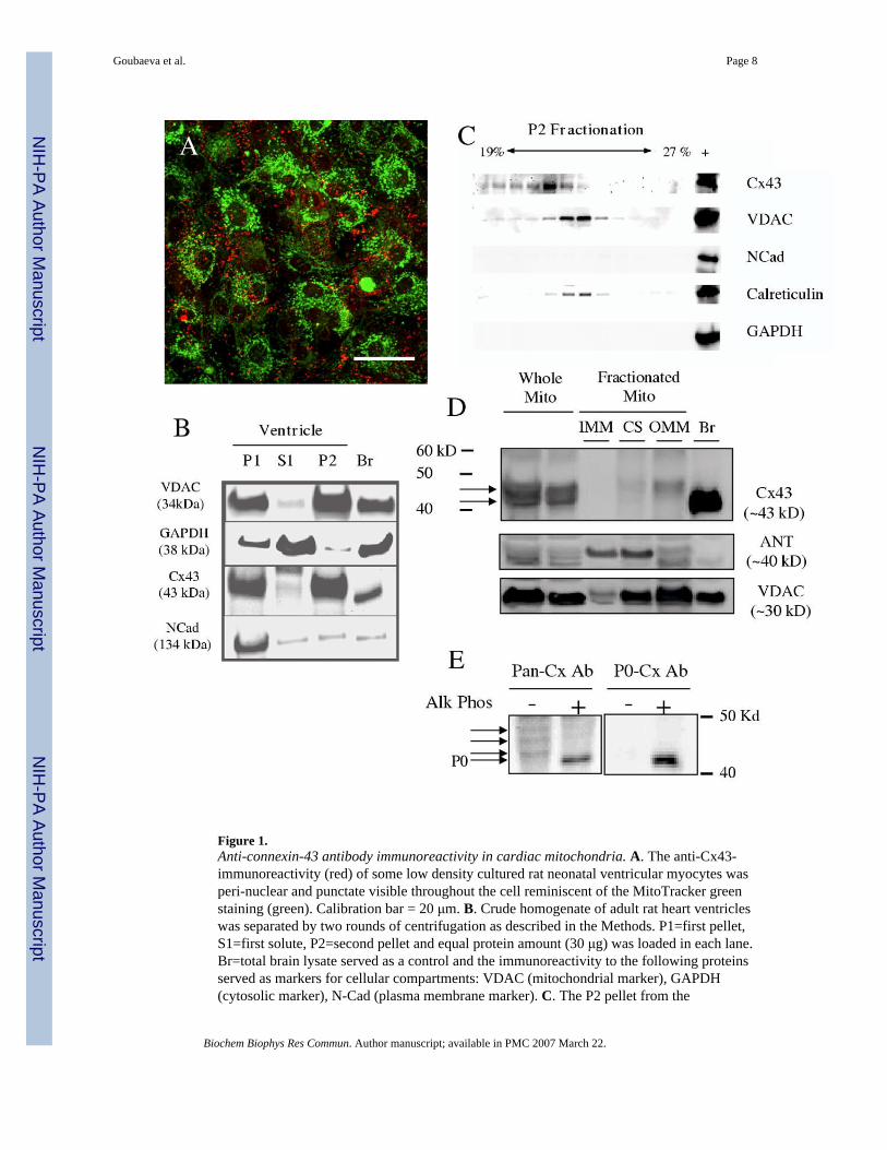
Figure 1.Anti-connexin-43 antibody immunoreactivity in cardiac mitochondria. A. The anti-Cx43-immunoreactivity (red) of some low density cultured rat neonatal ventricular myocytes wasperi-nuclear and punctate visible throughout the cell reminiscent of the MitoTracker greenstaining (green). Calibration bar = 20 μm. B. Crude homogenate of adult rat heart ventricleswas separated by two rounds of centrifugation as described in the Methods. P1=first pellet,S1=first solute, P2=second pellet and equal protein amount (30 μg) was loaded in each lane.Br=total brain lysate served as a control and the immunoreactivity to the following proteinsserved as markers for cellular compartments: VDAC (mitochondrial marker), GAPDH(cytosolic marker), N-Cad (plasma membrane marker). C. The P2 pellet from the
Goubaeva et al. Page 8
Biochem Biophys Res Commun. Author manuscript; available in PMC 2007 March 22.
NIH
-PA Author Manuscript
NIH
-PA Author Manuscript
NIH
-PA Author Manuscript
centrifugation was further fractionated by iodixanol gradient and equal volume from the 13collected fractions was loaded and proteins analyzed by immunoblotting. The far right lane ofthe crude homogenate served as a positive control for the respective antibodyimmunoreactivity. D. The mitochondria present in the P2 pellet was separated into the sub-mitochondrial fractions as described in Methods. Equal protein amount (15 μg) from therespective fractions was loaded and proteins analyzed by immunoblotting. ANT (innermitochondrial membrane marker), VDAC (outer mitochondrial membrane marker), IMM(inner mitochondrial membrane), CS (contact site), OMM (outer mitochondrial membrane),Br (brain). Representative of 3 Western blots from two separate mitochondrial membranefractionation preparations. E. Mitochondrial lysate was subjected to dephosphorylation byalkaline phosphatase and probed with antibody recognizing all Cx43 regardless of thephosphorylation status of the protein (Pan-Cx Ab) or with non-phosphorylated-species specificantibody (P0-Cx Ab). Arrows point to the putative phosphorylated Cx43 species.
Goubaeva et al. Page 9
Biochem Biophys Res Commun. Author manuscript; available in PMC 2007 March 22.
NIH
-PA Author Manuscript
NIH
-PA Author Manuscript
NIH
-PA Author Manuscript
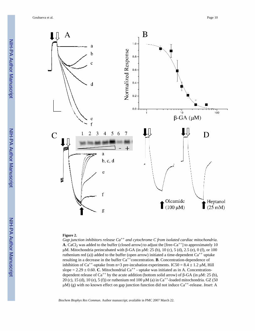
Figure 2.Gap junction inhibitors release Ca++ and cytochrome C from isolated cardiac mitochondria.A. CaCl2 was added to the buffer (closed arrow) to adjust the [free-Ca++] to approximately 10μM. Mitochondria preincubated with β-GA (in μM: 25 (b), 10 (c), 5 (d), 2.5 (e), 0 (f), or 100ruthenium red (a)) added to the buffer (open arrow) initiated a time-dependent Ca++ uptakeresulting in a decrease in the buffer Ca++concentration. B. Concentration-dependence ofinhibition of Ca++-uptake from n=3 pre-incubation experiments. IC50 = 8.4 ± 1.2 μM, Hillslope = 2.29 ± 0.60. C. Mitochondrial Ca++ - uptake was initiated as in A. Concentration-dependent release of Ca++ by the acute addition (bottom solid arrow) of β-GA (in μM: 25 (b),20 (c), 15 (d), 10 (e), 5 (f)) or ruthenium red 100 μM (a) in Ca++-loaded mitochondria. GZ (50μM) (g) with no known effect on gap junction function did not induce Ca++-release. Inset: A
Goubaeva et al. Page 10
Biochem Biophys Res Commun. Author manuscript; available in PMC 2007 March 22.
NIH
-PA Author Manuscript
NIH
-PA Author Manuscript
NIH
-PA Author Manuscript
Western blot for cytochrome C released by the mitochondria (1: none, 2: 5μM, 3: 10μM, 4:25μM, 5: 50 μM all β-GA, 6: 50 μM GZ, 7: 100μM FCCP. Equal protein loading was confirmedby probing for GAPDH (not shown). D. Two other well known gap junction inhibitors ofdistinct chemical structures release Ca++ from the mitochondria in the same protocol asdescribed for C.
Goubaeva et al. Page 11
Biochem Biophys Res Commun. Author manuscript; available in PMC 2007 March 22.
NIH
-PA Author Manuscript
NIH
-PA Author Manuscript
NIH
-PA Author Manuscript
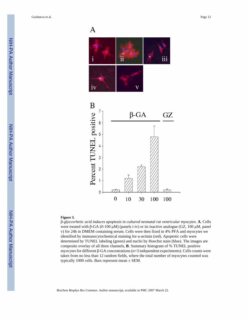
Figure 3.β-glyccerhetic acid induces apoptosis in cultured neonatal rat ventricular myocytes. A. Cellswere treated with β-GA (0-100 μM) (panels i-iv) or its inactive analogue (GZ, 100 μM, panelv) for 24h in DMEM containing serum. Cells were then fixed in 4% PFA and myocytes weidentified by immunocytochemical staining for α-actinin (red). Apoptotic cells weredetermined by TUNEL labeling (green) and nuclei by Hoechst stain (blue). The images arecomposite overlay of all three channels, B. Summary histogram of % TUNEL positivemyocytes for different β-GA concentrations (n=3 independent experiments). Cells counts weretaken from no less than 12 random fields, where the total number of myocytes counted wastypically 1000 cells. Bars represent mean ± SEM.
Goubaeva et al. Page 12
Biochem Biophys Res Commun. Author manuscript; available in PMC 2007 March 22.
NIH
-PA Author Manuscript
NIH
-PA Author Manuscript
NIH
-PA Author Manuscript