immunogold evidence that neuronal gap junctions in adult rat brain and spinal cord contain...
TRANSCRIPT
Immunogold evidence that neuronal gap junctions inadult rat brain and spinal cord contain connexin-36but not connexin-32 or connexin-43J. E. Rash*†‡, W. A. Staines§, T. Yasumura*, D. Patel§, C. S. Furman*, G. L. Stelmack¶, and J. I. Nagy¶i
*Department of Anatomy and Neurobiology, †Program in Molecular, Cellular and Integrative Neurosciences, and ‡Program in Cell and Molecular Biology,Colorado State University, Fort Collins, CO 80523; §Department of Cellular and Molecular Medicine, University of Ottawa, Ottawa, ON K1H 8M5, Canada;and ¶Department of Physiology, Faculty of Medicine, University of Manitoba, 730 William Avenue, Winnipeg, MB R3E 3J7, Canada
Communicated by Thomas S. Reese, National Institutes of Health, Bethesda, MD, April 20, 2000 (received for review January 9, 2000)
Physiological and ultrastructural evidence indicates that gap junc-tions link many classes of neurons in mammalian central nervoussystem (CNS), allowing direct electrical and metabolic communica-tion. Among at least six gap junction-forming connexin proteins inadult rat brain, connexin- (Cx) 32, Cx36, and Cx43 have beenreported to occur in neurons. However, no connexin has beendocumented at ultrastructurally defined neuronal gap junctions.To address this question directly, freeze-fracture replica immuno-gold labeling (FRIL) and immunofluorescence (IF) were used tovisualize the subcellular and regional localization of Cx36 in ratbrain and spinal cord. Three antibodies were generated againstdifferent sequences in Cx36. By Western blotting, these antibodiesdetected protein at 36 and 66 kDa, corresponding to Cx36 mono-mer and dimer forms, respectively. After double-labeling for Cx36and Cx43 by FRIL, neuronal gap junctions in inferior olive, spinalcord, and retina were consistently immunogold-labeled for Cx36,but none were labeled for Cx43. Conversely, Cx43 but not Cx36 wasdetected in astrocyte and ependymocyte gap junctions. In >250Cx32yCx43 single- and double-labeled replicas from 10 CNS re-gions, no neuronal gap junctions were labeled for either Cx32 orCx43. Instead, Cx32 and Cx43 were restricted to glial gap junctions.By IF, Cx36 labeling was widely distributed in neuropil, includingalong dendritic processes and within neuronal somata. On thebasis of FRIL identification of Cx36 in neuronal gap junctions andIF imaging of Cx36 throughout rat brain and spinal cord, neuronalgap junctions containing Cx36 appear to occur in sufficient densityto provide widespread electrical and metabolic coupling inadult CNS.
Gap junctions are composed of hexamers of ‘‘connexin’’proteins that bond head-to-head across the extracellular
space, thereby forming channels that permit intercellular ex-change of ions and small molecules (1–3). In nervous tissue, gapjunctions are a structural correlate of electrical synapses, andevidence has accumulated for the existence of such synapsesthroughout the mammalian central nervous system (CNS) (4–10). In addition, gap junctions too small to be seen by conven-tional thin section electron microscopy have been described atfreeze-fractured ‘‘mixed’’ (chemical plus electrical) synapses inmammalian spinal cord (10). Indeed, on the basis of electro-physiological, dye-coupling, and ultrastructural analyses, virtu-ally all major CNS structures from retina and olfactory bulb tospinal cord and primary sensory ganglia have been suggested tocontain neurons linked by gap junctions (9). The functionalsignificance of these electrical and mixed synapses in adultmammalian CNS has been difficult to assess because of theabsence of practical methods for documenting, quantifying, ormapping the distribution of neuronal gap junctions. In addition,there has been substantial uncertainty concerning the identity ofconnexins that form neuronal gap junctions. For example, theconsensus glial connexins Cx32 and Cx43 (11–19) also werereported to be expressed in neurons of adult rat or mouse (11,
20–25). However, neither of these connexins has been demon-strated to occur at ultrastructurally defined gap junctions inunambiguously identified neurons, which is essential for desig-nating connexins as neuronal (13, 15).
Among six members of the connexin family identified inCNS tissues, Cx36 and its homologs Cx35 and Cx34.7 in fishhave recently emerged as prime candidates for neuronalconnexins (26–29). Cx36 was identified by reverse transcrip-tion–PCR of mRNA extracted from rat inferior olive (IO),then cloned from a mouse genomic library (26), and subse-quently confirmed to be highly expressed in neural tissue,particularly retina (30). In situ hybridization revealed Cx36mRNA in neuronal cell bodies in various regions of rat brain,and neuronal lesions of IO depleted it of Cx36, furthersuggesting a neuronal source of Cx36 mRNA (26).
To investigate Cx36 protein localization in neural tissue ofadult rat, antibodies (Abs) against Cx36 were developed andused to localize Cx36 within individual gap junction plaquesusing freeze-fracture replica immunogold labeling (FRIL) (31–33). In addition, FRIL was used for single- and double-labelingof Cx32, Cx43, and Cx36 to determine the ultrastructural local-ization of the former two connexins in relation to that of Cx36.These three connexins are now documented to occur separatelyin each of three specific cell types and, in the case of Cx43, in twotypes of macroglial cells. Our data reveal that of these threeconnexins, only Cx36 is present in gap junctions of ultrastruc-turally defined neurons. An abstract of these results has beenpresented.**
Materials and MethodsCx36 Abs and Western Blotting. Three anti-Cx36 Abs were gener-ated in rabbits immunized with keyhole limpet hemocyanin-conjugated synthetic peptides and then affinity-purified. Ab298was against a peptide corresponding to amino acids 298–318 inCx36. Ab51-6200 was against a cytoplasmic loop domain, andAb51-6300 was against the carboxyl terminus. These latter twoAbs are commercially available from Zymed. Brain tissues frommale Sprague–Dawley rats (300–350 g) were homogenized inHepes buffer [20 mM Hepes, pH 7.2y0.1 M NaCly2 mMEDTAy1 mM phenylmethylsulfonyl f luoride (PMSF)] and cen-trifuged. Pellets were treated with 1% Nonidet P-40 (IgepalCA-630; Sigma), centrifuged, and resuspended in Hepes buffer.
Abbreviations: Cx, connexin; IF, immunofluorescence; CNS, central nervous system; FRIL,freeze-fracture replica immunogold labeling; Ab, antibody; IO, inferior olive; IMP, in-tramembrane particle; LM, light microscopy.
iTo whom reprint requests should be addressed. E-mail: [email protected].
**Rash, J. E., Yasumura, T., Staines, W. A., Patel, D. & Nagy, J. I. (1999) Mol. Biol. Cell, 10,404a.
The publication costs of this article were defrayed in part by page charge payment. Thisarticle must therefore be hereby marked “advertisement” in accordance with 18 U.S.C.§1734 solely to indicate this fact.
PNAS u June 20, 2000 u vol. 97 u no. 13 u 7573–7578
NEU
ROBI
OLO
GY

Total protein was determined with the Bio-Rad DC proteinassay. Western blotting was conducted as described (12), withtransblotted membranes processed by enhanced chemilumines-cence. For analysis of specificity by peptide preadsorption, Ab (3mg) was diluted in 100 ml of Tris-buffered saline, pH 7.4, aloneor in combination with 100 mg of Cx36 peptide, and incubatedovernight at 4°C before final dilution for use in primary solutions(1 mgyml).
Freeze-Fracture and Immunogold Labeling. Twenty-six male andfemale Sprague–Dawley rats (145–585 g) were deeply anesthe-tized and fixed for 3–10 min via transcardiac perfusion with 1%or 0.1% formaldehyde in 0.15 M Sorenson’s phosphate buffer.More than 250 Vibratome slices (150 mm thick) from sevenregions of adult rat brain (IO, hippocampus, suprachiasmaticnucleus, supraoptic nucleus, paraventricular nucleus, cerebel-lum, and retina), and from spinal cord were freeze-fractured andreplicated. A gold ‘‘index’’ grid was bonded to the frozen sampleby using Lexan plastic dissolved in dichloroethane; the sampleswere thawed and ‘‘grid-mapped’’ by confocal microscopy (10,34), and cellular material was removed by vigorous washing withSDS detergent (ref. 31; as modified in ref. 33).
For FRIL, residual connexin proteins adhering to the replicaafter SDS washing (Fig. 1) were single- or double-labeled usingcombinations of primary monoclonal and polyclonal anti-connexin Abs, followed by secondary labeling with anti-rabbit,anti-mouse, and anti-sheep Abs conjugated to 10 and 20 nm gold(33). For Cx36, only Ab298 was used for FRIL experiments.Monoclonal and rabbit polyclonal anti-Cx43 Abs were fromChemicon (Temecula, CA); mAbs to Cx32 were from Chemiconand Zymed; rabbit polyclonal Abs to Cx32 were from Chemicon,Sigma, and Zymed; and sheep anti-Cx32 was from Biogenesis(Poole, United Kingdom). Secondary Abs (10- and 20-nmimmunogold goat anti-rabbit, goat anti-mouse, and donkeyanti-sheep) were from Chemicon (now obtainable from JacksonLaboratories, Westgrove, PA.).
Using a JEOL 2000 EX-II transmission electron microscope,we photographed replicas as stereoscopic pairs to distinguishspecific labeling (occurring on tissue-side only) from nonspecificlabeling [.90% on the nontissueyLexan side (33)]. Freeze-fractured neurons, glial cells, and vascular endothelia wereidentified according to 24 criteria (see table 2 in ref. 35). Nerveterminals and dendrites were identified by the presence of .25uniform-diameter synaptic vesicles in the cytoplasm, distinctiveclusters of E- or P-face intramembrane particles (IMPs) thatcorrespond in location to postsynaptic densities seen in conven-tional thin sections, specialized appositions (‘‘active zones’’)shared between dendrite and one or more nerve terminals, and
absence of glial markers. Astrocytes were identified by aqua-porin 4 square arrays in the plasma membranes, distinctive glialfibrillary acidic protein (GFAP) filaments in the cytoplasm, andhigh density of IMPs in their plasma membranes, particularly inE-faces (35–38). Oligodendrocytes were identified by low densityof IMPs in both E- and P-faces, distinctive ‘‘reciprocal patches’’of mixed IMPs and pits in both E- and P-faces, and unique‘‘necklaces’’ of IMPs surrounding gap junction E-faces (35, 37).Ependymocytes lined the third ventricle and spinal central canal,and microvilli projected from their apical surfaces (35, 36).
Immunofluorescence (IF). Anesthetized rats were perfusedtranscardially with 50 ml of 50 mM sodium phosphate buffer(PB), pH 7.2, containing 0.9% saline (PBS), followed by perfu-sion with 4% formaldehyde in PB. Cryostat sections (3–15 mm)were collected on gelatin-coated slides and incubated for 16 h at4°C with anti-Cx36 Abs diluted 1:500 in PBS containing 0.3%Triton X-100 (PBST). Sections were washed in PBST, incubatedfor 1.5 h with Cy3-conjugated donkey anti-rabbit IgG diluted1:200 in PBST, washed, air-dried, and coverslipped.
ResultsAb Characterization. Western blots with three sequence-specificAbs to Cx36 are shown in Fig. 2. Ab298 (Fig. 2 A), Ab51-6200
Fig. 1. Diagram of the SDS-FRIL technique. (A and B) Gap junction before A and after B freeze-fracturing, with dashed line indicating the two permissiblefracture planes within the lipid bilayers. Regardless of whether the fracture plane splits the plasma membrane of the upper cell, the lower cell, or jumps fromone cell to the other, all connexons of the upper gap junction plaque remain with the upper cell and all connexons of the lower plaque remain with the lowercell. (C) After replication of E- and P-faces, the samples are thawed and washed with SDS detergent, leaving a small amount of membrane proteins adheringbeneath both E-face pits and P-face particles. The platinum (Pt) replica delineates the outer leaflet (membrane E-face) of the upper cell and the inner leaflet(P-face) of the lower cell. Residual connexin proteins are labeled by primary Abs (Y) and detected by labeling with gold-conjugated, species-specific secondaryAbs (Y with F). For reasons that are not yet clear, labeling is usually greater at the periphery of gap junctions (see Figs. 3 and 4).
Fig. 2. Western blots of tissues probed with anti-Cx36 Abs generated againstthree different sequences within Cx36. (A–C) Three blots containing homog-enates of retina, olfactory bulb (OB), and IO probed with Ab298 (A), Ab51-6200 (B), and Ab51-6300 (C). The Abs detected Cx36 monomer migrating at 36kDa in all three tissues. A Cx36 dimer migrating at 66 kDa also was detectedto various degrees. The band at 48 kDa was seen after omission of primary Ab(not shown) and is nonspecific. Ab298 detected an unidentified protein at 40kDa. (D) Recognition of Cx36 and the cross-reacting protein at 40 kDa (retina)was eliminated after preadsorption of Ab298 with synthetic peptide antigen(retina, pa).
7574 u www.pnas.org Rash et al.
(Fig. 2B), and Ab51-6300 (Fig. 2C) detect monomeric Cx36migrating at 36 kDa in homogenates of retina, olfactory bulb,and IO. In each tissue, the dimeric form of Cx36 migrating at 66kDa was recognized by Ab298 and to various degrees by theother two Abs. An additional unidentified protein migrating at40 kDa was recognized by Ab298 in retina and olfactory bulb, butwas absent in IO, and was not recognized by the other Abs. Afteromission of primary Ab, blots probed with secondary Ab weredevoid of all signal (not shown) except for a nonspecific band at48 kDa in IO. Preadsorption of Ab298 (Fig. 2D) and Ab51-6300(not shown) with synthetic peptide antigen eliminated detectionof Cx36 as well as the band detected by Ab298 at 40 kDa.
Detection of Neuronal and Glial Connexins by FRIL. Neurons and gliain selected areas of brain and spinal cord were examined in .165replicas labeled for Cx43, 88 replicas labeled for Cx32, and 8replicas double-labeled for Cx32yCx43. Neurons and glia alsowere examined in 5 replicas of IO, 1 of spinal cord, and 4 of retinathat were double-labeled for Cx36yCx43, and in 1 sample of IOthat was single-labeled for Cx36.
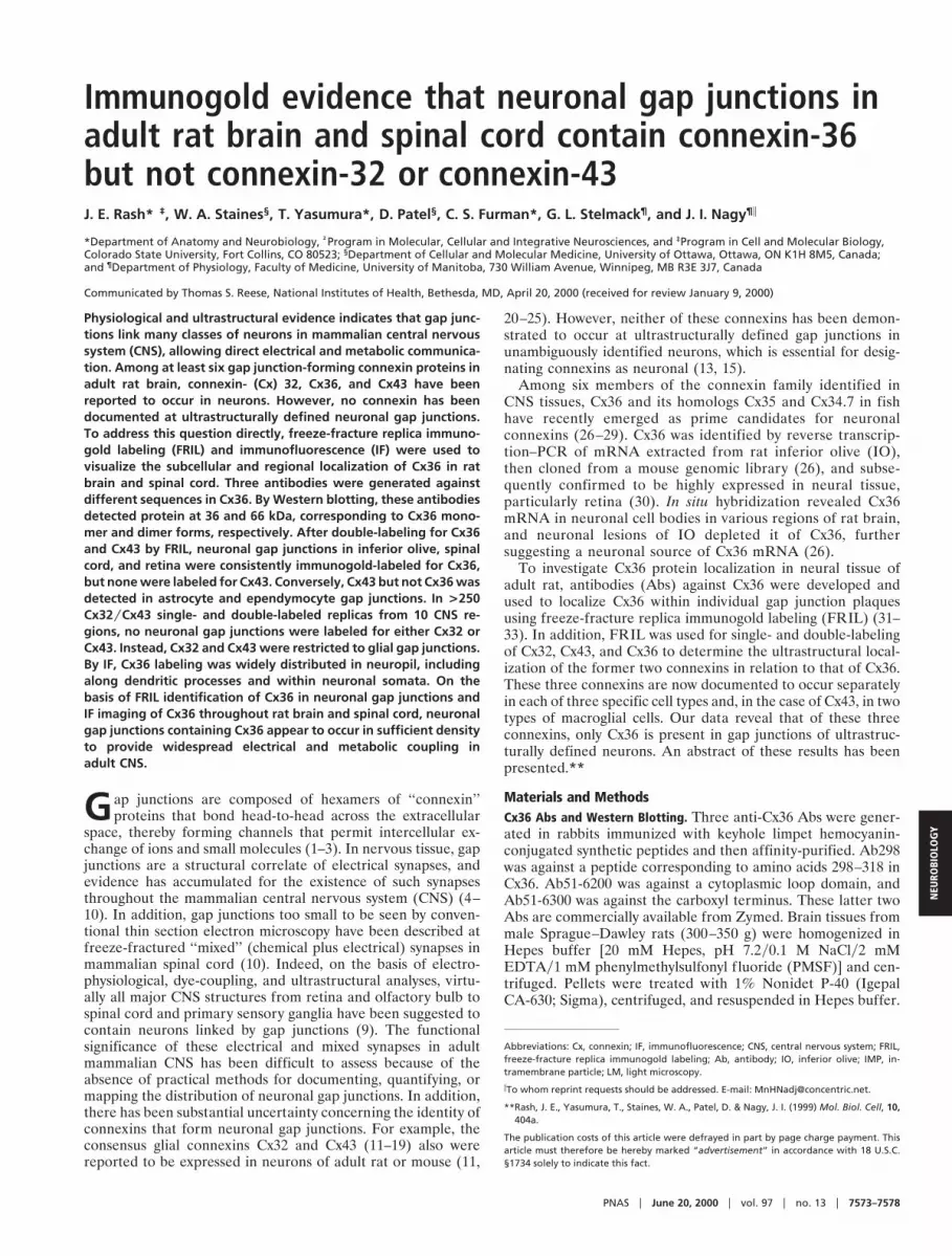
In Cx36yCx43 double-labeled replicas, stereoscopic imagingrevealed Cx36 immunogold beneath 24 of 26 neuronal gapjunctions in IO (Fig. 3 A–D), beneath 10 of 10 in retina (Fig. 3E),and beneath 2 of 2 in spinal cord (Fig. 4A). No neuronal gapjunctions were labeled for Cx43. In the same replicas, .90% ofastrocyte gap junctions were labeled for Cx43 (Fig. 4B), but none
were labeled for Cx36. The probabilistic nature of immunogoldlabeling of SDS-washed samples, combined with typical labelingefficiencies of approximately 1:40, occasionally resulted in un-labeled gap junctions (33), particularly those containing fewerthan 50 connexons (see Fig. 4B for labeled and unlabeledjunctions in the same astrocyte).
Two gap junctions were observed in each of three nerveterminals (two in IO and one in spinal cord), and five of thosesix gap junctions were labeled for Cx36 (Figs. 3 A–C and 4A),demonstrating that multiple gap junctions in the same neuronalprocesses contained Cx36. Although no protein is replicatedwithin E-face pits, E-faces are labeled and often with higherefficiency than P-faces (Figs. 3A–D and 4A–C). The basis for thisseeming contradiction is illustrated in Fig. 1. Overall, the 36Cx36-labeled and two unlabeled neuronal gap junctions had atotal of 259 gold beads beneath '9,800 connexons [average, 258connexons and 7.2 gold beads per gap junction, or 1 gold beadper 38 connexons, which is approximately the same as reportedfor other connexins (31, 32, 38, 39)]. Interestingly, the 24 labeledand 2 unlabeled gap junctions in electrical synapses of IO had anaverage of 247 connexons, which is 5 times greater than theaverage number of connexons in gap junctions at mixed synapsesin spinal cord (10).
To test Cx32 and Cx43 as possible neuronal vs. glial connexins,.200 single- and double-labeled replicas from throughout brainand spinal cord were examined. In these replicas, .3,000 astro-
Fig. 3. FRIL immunogold localization of Cx36 (20-nm gold) in rat IO and retina. (A) Stereoscopic image of two Cx36-labeled gap junctions in a large area ofdendrite E-face. Two (of five) nerve terminals are numbered. Two nonspecific gold beads are at the base of a cross-fractured dendritic spine (DS). (B and C)Magnified images of gap junctions in inscribed areas of A. Six 20-nm gold beads localizing Cx36 are beneath both gap junction E-faces. (D) Gap junction in IOlabeled for Cx36 by more than one dozen gold beads. (E) Stereoscopic image of three linked nerve terminals in retina, with two linked at a chemical synapse(1 and 2) and two connected by gap junctions (2 and 3). The nerve terminal on the left (1) is presynaptic to the middle terminal (2), as identified by the postsynapticdensity (PSD). Two nonspecifically bound gold beads are on the Lexan side of the replica (arrow). In FRIL images, all calibration bars are 0.1 mm unless otherwiseindicated.
Rash et al. PNAS u June 20, 2000 u vol. 97 u no. 13 u 7575
NEU
ROBI
OLO
GY

cyte gap junctions (Fig. 4B) and .100 ependymocyte gapjunctions were labeled for Cx43 (also see refs. 13, 15, and 33).However, no Cx43-labeled neuronal gap junctions were de-tected, even as ‘‘false positives’’ (defined and illustrated in ref.33). Cx32 was found in .100 oligodendrocyte gap junctions (Fig.4C) but was never detected in neuronal gap junctions (13, 15, 33)or in astrocyte (Fig. 4C) or ependymocyte gap junctions. How-ever, more than one dozen unlabeled neuronal gap junctionswere found in the same samples (negative data not shown).
IF Labeling. On the basis of the above results, light microscopy(LM) immunolabeling was used to examine neuronal localiza-tion of Cx36 protein and to investigate the extent of its distri-bution in adult brain and spinal cord. Regionally and qualita-tively similar punctate IF staining was obtained with all threeCx36 Abs used in this initial study; however, only imagesobtained with Ab51-6200 are presented here.
In the retina, Cx36-labeling was intense and stratified. Rela-tively large and brilliant neuronal puncta were seen within theouter plexiform layer (Fig. 5A), whereas a more extensive fieldof fine puncta typified labeling of the inner plexiform layer (Fig.5A). Cx36 IF was densely distributed in the IO, which showedmuch higher Cx36-immunoreactivity than other brainstem re-gions at this level of section (Fig. 5B). Labeling in the IOconsisted of puncta within the neuropil, as well as IF withinneuronal somata (Fig. 5C), thereby providing data not obtain-able by the membrane labeling methods of FRIL. In the spinalcord, punctate Cx36 IF was much more evident in gray matterthan in white matter, and both the dorsal and ventral hornshowed moderate immunoreactivity, paralleling observations of
neuronal gap junctions in conventional freeze-fracture replicas(10). As had been noted in the IO, perikaryal staining was seenin large neurons within the ventral horn (Fig. 5D, arrows). In twoareas not investigated by FRIL, the striatum and globus pallidus(Fig. 5 E and F), distinctly different subcellular distributionswere seen, with a preponderance of punctate staining in theneuropil of the striatum vs. a preponderance of cytoplasmiclabeling of neurons in globus pallidus. IF data showed a similarlystriking regional and cellular heterogeneity in the distribution ofCx36 throughout many other brain regions, notably the hip-pocampus, thalamus, and brainstem.
DiscussionFRIL analysis of three regions of adult rat CNS revealed thatCx36 is present in gap junctions linking neurons. In contrast,neither Cx32 nor Cx43 was detectable in neuronal gap junctionsin any of seven regions of brain or three regions of spinal cord.These results indicate that: (i) neuronal gap junctions containCx36, but not Cx32 or Cx43; (ii) astrocyte gap junctions containCx43, but not Cx36 or Cx32; and (iii) oligodendrocyte gapjunctions contain Cx32, but not Cx36 or Cx43. The present FRILdata are consistent with our previous estimates of .1,000-foldmore glial than neuronal gap junctions in CNS tissue (15, 35). Incomparison with freeze-fractured astrocyte junctions, where10–50 gap junctions were commonly seen in single grid openings,neuronal gap junctions were encountered, on average, in '1y100grid openings (10, 15). However, in the broader views affordedby LM, widespread IF labeling for Cx36 was seen in neuropil, inneuronal cell bodies, and along dendrites.
Fig. 4. Images from spinal cord labeled for Cx32 and Cx43 and from cerebellum labeled for Cx32. (A) Stereoscopic view (left and center images) and intaglioview (center and right images) of two Cx36-labeled neuronal gap junctions in spinal cord. The larger junction (arrow 1) and the smaller junction [four or fiveconnexon imprints (arrow 2)] are labeled with 20-nm gold beads. The intaglio perspective reveals the ‘‘sidedness’’ of labeling and the topological deformationof membranes at gap junction close contacts. (B) Two astrocyte gap junctions in the same replica as A. One plaque has three 10-nm gold beads (white arrow),and one is unlabeled (black arrow). Aquaporin 4 square array (arrowhead) is a definitive marker for astrocytes (38). (C) Three gap junctions in oligodendrocyteP-faces (Ol P) in cerebellum are labeled for Cx32 by 12, 6, and 2 gold beads. One Cx32-labeled oligodendrocyte gap junction is enlarged (Right Inset). Nearbyastrocyte gap junction (As P, and Left Inset) is not labeled for Cx32.
7576 u www.pnas.org Rash et al.

Our Western blotting results extend earlier reports (26, 30) ofwidespread Cx36 mRNA expression in rodent neural tissue bydemonstrating Cx36 protein in homogenates from several CNSregions. Detection of similar protein bands with three Absdirected against different sites in Cx36, in combination withFRIL localization of one of those Abs to ultrastructurallydefined neuronal gap junction plaques, leaves little possibility ofprotein misidentification. Moreover, the high levels of Cx36protein expression in retina are consistent with the abundance ofCx36 mRNA reported in that tissue (30). More generally,detection of Cx36 on Western blots of several brain regionsconfirms and extends data obtained for Cx36 expression by insitu hybridization (26).
Previous studies, using LM methods, have reported expressionof Cx32 and Cx43 protein andyor mRNA in neurons (11, 16, 20,21, 24, 25, 40–45). However, inherent limits of resolution of LMpreclude assignment to structures smaller than 0.3 mm. Astrocyteand oligodendrocyte processes, which are well established tocontain Cx43 and Cx32 (13, 16–19, 24), often are thinner than0.2 mm. Where those thin processes follow the contours ofneurons, it would be difficult to distinguish whether connexin IFis localized to oligodendrocyte, astrocyte, or neuronal plasmamembranes.
In this study, no evidence for Cx32 or Cx43 in neuronal gapjunctions was found in .250 single- and double-labeled FRILreplicas. To date we have evaluated .3,000 labeled glial gapjunctions (this study), .1,000 unlabeled glial gap junctions (35),.100 unlabeled neuronal gap junctions (10, 15), and 36 (of 38)labeled neuronal gap junctions (this study) in 10 regions of brainand spinal cord. In this sampling, no evidence was found for gapjunctions between glial cells and neurons. These results and our
previous estimates of 1,000-fold greater number of glial thanneuronal gap junctions contrast with reports that 17.9% of allgap junctions in adult rat cortex represent neuronalyglial junc-tions and that neuronalyglial gap junctions contain Cx32 andCx43 (21). Thus, it is possible that either gap junctions or cellslinked by gap junctions were misidentified in previous reports, oralternatively, that putative neuronalyglial gap junctions were notrecognized in our FRIL replicas, and if they exist, that they useconnexins other than Cx32, Cx36, or Cx43.
With increasing evidence for multiple connexins in individualgap junction plaques in diverse cell types (19, 39), our data donot exclude the possibility that neuronal gap junctions containadditional as-yet-unidentified connexins that may participate inthe formation of novel gap junctions. Also requiring furtheranalyses are the subcellular sites to which neuronally expressedconnexins are targeted. These may be localized at, e.g., typicalelectrical synapses that have been described in mammalian brain(reviewed in ref. 9) or at mixed synapses in spinal cord (10, 15).FRIL methods should be of value in assessing each of thoseissues. In FRIL, each gold bead acts as an independentlytargeted probe. Thus, multiple gold beads on the same gapjunction plaque provide multiple independent confirmations oftarget connexins in individual gap junction plaques of identifiedcells (33). Despite its relatively low labeling efficiency ('1 goldbead per 40 connexins), FRIL is a highly specific techniquefor ‘‘f lagging’’ even the smallest gap junctions, identifyingthe constituent connexins, and identifying the coupled cells(15, 31, 33).
Connexin immunostaining by LM usually appears punctate inmost tissue (9, 13), and those puncta have been presumed toreflect gap junctions. Although the degree to which Cx36-
Fig. 5. Distribution of Cx36 in rat retina, IO, and spinal cord by IF labeling. (A) The retina displays Cx36-IF in both the inner and outer plexiform layers (ipl andopl), with only moderate IF in the inner nuclear layer (inl) and very little IF in the outer nuclear layer (onl). Prominent IF is seen in the outer part of the opl (arrow).(B) The IO is delineated by more intense Cx36-IF than other brainstem regions (arrow). (C) The neuropil of the IO contains fine punctate immunolabeling andCx36-IF within individual olivary neurons (arrow). (D) Cx36-IF is widely distributed in spinal cord gray matter (gm) and very sparse within white matter (wm).Cx36-IF also is seen within individual ventral horn neurons (arrows). bv, Blood vessel. (E) The striatum (ST) exhibits a greater density of Cx36-IF than the globuspallidus (GP). (F) In globus pallidus, Cx36-IF is seen within neuronal somata (large arrows) and along dendrites (small arrow). (Calibration bars: A, D, E, and F 550 mm; B 5 200 mm; and C 5 20 mm.)
Rash et al. PNAS u June 20, 2000 u vol. 97 u no. 13 u 7577
NEU
ROBI
OLO
GY

positive puncta correspond to neuronal gap junctions will re-quire correlative LM-transmission electron microscopy analysis,confirmation of such a correspondence would indicate thatneurons in diverse areas of mammalian brain are relatively richlyinvested with gap junctions. Intracellular labeling for Cx36 alsowas seen, and this may be fortunate as it will enable LMcorrelation of Cx36-positive neurons with appropriate transmit-ter markers, just as FRIL will allow colocalization of Cx36 andneurotransmitter receptors within individual mixed synapses(unpublished results). As with other proteins expressed byneurons, only a portion of neurons expressing Cx36 may showrecognizable levels of the protein in the perikaryon. For exam-ple, both Cx36-positive puncta and neuronal somata were seenin the globus pallidus, whereas Cx36-positive somata were notseen in the striatum, which nevertheless contained a high densityof puncta. As one possibility, these cytoplasmic differences mayreflect high vs. low rates of connexin turnover through cytomem-brane pathways.
The present FRIL and IF data, as well as in situ hybridizationresults (26) suggest that adult mammalian CNS contains suffi-cient neuronal gap junctions to provide structural pathways forwidespread electrical andyor metabolic integration betweenneurons. Our identification of Cx36 in ultrastructurally definedneuronal gap junction plaques provides the basis for obtainingdetailed immunohistochemical maps of the entire CNS to es-tablish the regional and cellular densities of neuronal Cx36. Suchmaps will be of value in illuminating areas in which to focusfunctional studies of the roles of neuronal gap junctions insynaptic integration and metabolic coupling in the CNS.
We thank K. Mandelic and B. Tinner for outstanding technicalassistance. This work was supported by National Institutes of HealthGrants NS-31027 and NS-39040 (to J.E.R.) and MH-59995 (to F. E.Dudek, Colorado State University), and by grants from the MedicalResearch Council of Canada (to J.I.N. and to W.A.S.). Additionalhigher-resolution images related to this report are available at:http:yymicroscopy.cvmbs.colostate.eduyimagefilesyrashy.
1. Goodenough, D. A., Goliger, J. A. & Paul, D. L. (1996) Annu. Rev. Biochem.65, 475–502.
2. Gilula, N. B., Reeves, O. R. & Steinbach, A. (1972) Nature (London) 235,262–265.
3. Bruzzone, R., White, T. W. & Paul, D. L. (1996) Eur. J. Biochem. 238, 1–27.4. Bennett, M. V. L. (1977) in Electrical Transmission: A Functional Analysis and
Comparison with Chemical Transmission, ed. Kandel, E. R. (Am. Physiol. Soc.,Bethesda, MD), pp. 357–416.
5. Bennett, M. V. L. & Goodenough, D. A. (1978) Neurosci. Res. Prog. Bull. 16,373–486.
6. Bennett, M. V. L. (1997) J. Neurocytol. 26, 349–366.7. Llinas, R., Baker, R. & Sotelo, C. (1974) J. Neurophysiol. 37, 560–571.8. Sotelo, C. & Korn, H. (1978) Int. Rev. Cytol. 55, 67–107.9. Nagy, J. I. & Dermietzel, R. (2000) in Advances in Molecular and Cell Biology,
ed. Hertzberg, E. L. (JAI Press, New York), Vol. 30, pp. 323–396.10. Rash, J. E., Dillman, R. K., Bilhartz, B. L., Duffy, H. S., Whalen, L. R. &
Yasumura, T. (1996) Proc. Natl. Acad. Sci. USA 93, 4235–4239.11. Dermietzel, R., Traub, O., Hwang, T. K., Beyer, E., Bennett, M. V. L., Spray,
D. C. & Willecke, K. (1989) Proc. Natl. Acad. Sci. USA 86, 10148–10152.12. Li, J., Hertzberg, E. L. & Nagy, J. I. (1997) J. Comp. Neurol. 379, 571–591.13. Nagy, J. I. & Rash, J. E. (2000) Brain Res. Rev. 32, 29–44.14. Ochalski, P. A. Y., Frankenstein, U. N., Hertzberg, E. L. & Nagy, J. I. (1997)
Neuroscience 76, 931–945.15. Rash, J. E., Yasumura, T. & Dudek, F. E. (1998) Cell Biol. Int. 22, 731–749.16. Dermietzel, R. & Spray, D. C. (1993) Trends Neurosci. 16, 186–192.17. Dermietzel, R., Farooq, M., Kessler, J. A., Hertzberg, E. L. & Spray, D. C.
(1997) Glia 20, 101–114.18. Yamamoto, T., Ochalski, A., Hertzberg, E. L. & Nagy, J. I. (1990) J. Comp.
Neurol. 302, 853–883.19. Nagy, J. I., Patel, D., Ochalski, P. A. Y. & Stelmack, G. L. (1999) Neuroscience
88, 447–468.20. Micevych, P. E. & Abelson, L. (1991) J. Comp. Neurol. 305, 96–118.21. Nadarajah, B., Thomaidou, D., Evans, W. H. & Parnavelas, J. G. (1996)
J. Comp. Neurol. 376, 326–342.22. Micevych, P. E., Popper, P. & Hatton, G. I. (1996) Neuroendocrinology 63,
39–45.23. Bennett, M. V. L., Barrio, L. C., Bargiello, T. A., Spray, D. C., Hertzberg, E. L.
& Saez, J. Z. (1991) Neuron 6, 305–320.
24. Dermietzel, R. (1998) Cell Biol. Int. 22, 719–730.25. Dermietzel, R. & Spray, D. C. (1998) Glia 24, 1–7.26. Condorelli, D. F., Parenti, R., Spinella, F., Salinaro, A. T., Belluardo, N.,
Cardile, V. & Cicirata, F. (1998) Eur. J. Neurosci. 10, 1202–1208.27. O’Brien, J., Al-Ubaidi, M. R. & Ripps, H. (1996) Mol. Biol. Cell 7, 233–243.28. O’Brien, J., White, T. W., Al-Ubaidi, M. R. & Ripps, H. (1998) J. Neurosci. 18,
7625–7637.29. Srinivas, M., Rozental, R., Kojima, T., Dermietzel, R., Mehler, M., Condorelli,
D. F., Kessler, J. A. & Spray, D. C. (1999) J. Neurosci. 19, 9848–9855.30. Sohl, G., Degen, J., Teubner, B. & Willecke, K. (1998) FEBS Lett. 428, 27–31.31. Fujimoto, K. (1995) J. Cell Sci. 108, 3443–3449.32. Fujimoto, K. (1997) Histochem. Cell Biol. 107, 87–96.33. Rash, J. E. & Yasumura, T. (1999) Cell Tissue Res. 296, 307–321.34. Rash, J. E., Dillman, R. K., Morita, M., Whalen, L. R., Guthrie, P. B.,
Fay-Guthrie, D. & Wheeler, D. W. (1995) in Rapid Freezing, Freeze Fracture,and Deep Etching, eds. Severs, N. J. & Shotton, D. M. (Wiley, New York), pp.127–150.
35. Rash, J. E., Duffy, H. S., Dudek, F. E., Bilhartz, B. L., Whalen, L. R. &Yasumura, T. (1997) J. Comp. Neurol. 388, 265–292.
36. Peters, A., Palay, S. L. & Webster, H. D. (1991) The Fine Structure of theNervous System, Neurons and Their Supporting Cells (Oxford Univ. Press, NewYork).
37. Massa, P. T. & Mugnaini, E. (1982) Neuroscience 7, 523–538.38. Rash, J. E., Yasumura, T., Hudson, C. S., Agre, P. & Nielsen, S. (1998) Proc.
Natl. Acad. Sci. USA 95, 11981–11986.39. Severs, N. J. (1999) Novartis Found. Symp. 219, 188–206.40. Chang, Q., Gonzalez, M., Pinter, M. J. & Balice-Gordon, R. J. (1999)
J. Neurosci. 19, 10813–10828.41. Nadarajah, B. & Parnavelas, J. G. (1999) Novartis Found. Symp. 219, 157–170.42. Nadarajah, B., Jones, A. M., Evans, W. H. & Parnavelas, J. G. (1998)
J. Neurosci. 17, 3096–3111.43. Matsumoto, A., Arai, Y., Urano, A. & Hyodo, S. (1992) Neurosci. Res. 14,
133–144.44. Fisher, R. S. & Micevych, P. E. (1993) in Progress in Cell Research: Gap
Junctions, eds. Hall, J. E., Zampighi, G. A. & Davis, R. M. (Elsevier, NewYork), Vol. 3, pp. 141–148.
45. Simburger, E., Stang, A., Kremer, M. & Dermietzel, R. (1997) Histochem. CellBiol. 107, 127–137.
7578 u www.pnas.org Rash et al.