altered differentiation and clustering of sertoli cells in transgenic mice showing a sertoli cell...
TRANSCRIPT

Differentiation 82 (2011) 38–49
Contents lists available at ScienceDirect
Differentiation
0301-46
Join the
doi:10.1
Abbre
beta-ga
serum a
acid; FC
shock p
knockou
control;
ribonuc
connexi
deoxyn
WT, wiln Corr
E-m
martin.
sarah.gi
florian.g
klaus.fa
ralph.br
journal homepage: www.elsevier.com/locate/diff
Altered differentiation and clustering of Sertoli cells in transgenic miceshowing a Sertoli cell specific knockout of the connexin 43 gene
Karola Weider a,n, Martin Bergmann b, Sarah Giese b, Florian Guillou c, Klaus Failing d, Ralph Brehm a
a Institute of Anatomy, University of Veterinary Medicine Hannover, Bischofsholer Damm 15, 30173 Hannover, Germanyb Institute of Veterinary Anatomy, Histology and Embryology, University of Giessen, Frankfurter Straße 98, 35392 Giessen, Germanyc INRA, UMR85, CNRS, UMR6175, Universite Franc-ois-Rabelais de Tours, Haras Nationaux, Physiologie de la Reproduction et des Comportements, 37380 Nouzilly, Franced Unit for Biomathematics, Faculty of Veterinary Medicine, University of Giessen, Frankfurter Straße 95, 35392 Giessen, Germany
a r t i c l e i n f o
Article history:
Received 18 August 2010
Received in revised form
8 March 2011
Accepted 17 March 2011Available online 13 April 2011
Keywords:
Sertoli
Connexin 43
Knockout
Mouse
Differentiation
Cell cluster
81/$ - see front matter & 2011 International
International Society for Differentiation (ww
016/j.diff.2011.03.001
viations: AMH, anti-Muellerian hormone; AR
lactosidase; 3b-HSD, 3-beta-hydroxysteroid d
lbumin; Cx, connexin; BTB, blood–testis-barr
, fold change; GC, germ cell; HE, hematoxyli
rotein 90 kDa alpha, class B member 1; IHC,
t; LC, Leydig cell; M, marker; NBF, neutral bu
pc, postcoitum; PCR, polymerase chain reac
leic acid; SC, Sertoli cell; SCCx43KO, Sertoli c
n 43 gene; SD, standard deviation; T3, triiod
ucleotidyl transferase-mediated dUTP nick end
dtype
esponding author. Tel.: þ49 511 856 7483; f
ail addresses: [email protected]
[email protected] (M. Bergm
[email protected] (S. Giese),
[email protected] (F. Guillou),
[email protected] (K. Failing),
[email protected] (R. Brehm).
a b s t r a c t
Histological analysis revealed that Sertoli cell specific knockout of the predominant testicular gap
junction protein connexin 43 results in a spermatogenic arrest at the level of spermatogonia or Sertoli
cell-only syndrome, intratubular cell clusters and still proliferating adult Sertoli cells, implying an
important role for connexin 43 in the Sertoli and germ cell development. This study aimed to determine
the (1) Sertoli cell maturation state, (2) time of occurrence and (3) composition, differentiation and fate
of clustered cells in knockout mice. Using immunohistochemistry connexin 43 deficient Sertoli cells
showed an accurate start of the mature markers androgen receptor and GATA-1 during puberty and a
vimentin expression from neonatal to adult. Expression of anti-Muellerian hormone, as a marker of
Sertoli cell immaturity, was finally down-regulated during puberty, but its disappearance was delayed.
This observed extended anti-Mullerian hormone synthesis during puberty was confirmed by western
blot and Real-Time PCR and suggests a partial alteration in the Sertoli cell differentiation program.
Additionally, Sertoli cells of adult knockouts showed a permanent and uniform expression of GATA-1 at
protein and mRNA level, maybe caused by the lack of maturing germ cells and missing negative
feedback signals. At ultrastructural level, basally located adult Sertoli cells obtained their mature
appearance, demonstrated by the tripartite nucleolus as a typical feature of differentiated Sertoli cells.
Intratubular clustered cells were mainly formed by abnormal Sertoli cells and single attached apoptotic
germ cells, verified by immunohistochemistry, TUNEL staining and transmission electron microscopy.
Clusters first appeared during puberty and became more numerous in adulthood with increasing cell
numbers per cluster suggesting an age-related process. In conclusion, adult connexin 43 deficient
Sertoli cells seem to proliferate while maintaining expression of mature markers and their adult
morphology, indicating a unique and abnormal intermediate phenotype with characteristics common
to both undifferentiated and differentiated Sertoli cells.
& 2011 International Society of Differentiation. Published by Elsevier Ltd. All rights reserved.
Society of Differentiation. Publish
w.isdifferentiation.org)
, androgen receptor; b-Gal,
ehydrogenase; BSA, bovine
ier; DNA, deoxyribonucleic
n and eosin; HSP90ab1, heat
immunohistochemistry; KO,
ffered formalin; NC, negative
tion; pp, postpartum; RNA,
ell specific knockout of
othyronine; TUNEL, terminal
-labeling; WB, western blot;
ax: þ49 511 856 7683.
e (K. Weider),
ann),
1. Introduction
In the testis, intercellular communication is regulated throughcertain mechanisms between different cell populations. The directcommunication via gap junction channels allows intercellularpassage of signaling molecules (o1000 Da) and regulates essen-tial processes during cell proliferation and differentiation, home-ostasis and oncogenic transformation (Kumar and Gilula, 1996).A gap junction channel is composed of two hemichannels orconnexons, contributed separately by each of the two participat-ing cells and each connexon is again formed by the aggregation ofsix protein subunits termed Cxs (Bruzzone et al., 1996). The Cxfamily consists of at least 20 members in the mouse and 21members in the human genome (Sohl and Willecke, 2004). Thegap junctional protein Cx43 is the predominant Cx in the testis of
ed by Elsevier Ltd. All rights reserved.

K. Weider et al. / Differentiation 82 (2011) 38–49 39
different species (Batias et al., 1999; Pelletier, 1995; Risley et al.,1992; Steger et al., 1999a), in mouse testes it is expressed fromday 11.5 pc to all postnatal stages (Perez-Armendariz et al., 2001).Cx43 based gap junctions are located between neighboring SCs,between SCs and spermatogonia and primary spermatocytes(Decrouy et al., 2004).
Total disruption of the Cx43 gene leads to altered cardiacmorphology and to sudden perinatal death (Reaume et al., 1995).In order to circumvent the problems of the general Cx43 defi-ciency and to clarify the SC specific roles of Cx43 on spermato-genesis and SC maturation in vivo, a viable conditional Cx43 KOmouse line, which lacks the Cx43 gene solely in SCs, has beengenerated using the Cre/loxP recombination system (Brehm et al.,2007; Sridharan et al., 2007). Histological analysis of adultSCCx43KO testes revealed that SC specific deletion of Cx43 mostlyresults in an arrest of spermatogenesis at the level of spermato-gonia or SC-only syndrome, abnormal intratubular cell clusters,increased SC numbers and reduced number of spermatogoniaper seminiferous tubule. Furthermore, SCs were found to be stillproliferating in adult KO mice (Sridharan et al., 2007) emphasiz-ing the critical contribution of Cx43 to the normal maturationalprogression of SCs, which normally results in the cessation of SCmitosis during the pubertal period.
Within the life cycle of a somatic SC puberty represents themost important time period, as this cell develops from animmature into a mature, morphologic and functional adult SCsupporting spermatogenesis (Sharpe et al., 2003). Herein pubertyis defined as the period when spermatogenesis starts and GCsenter meiosis, in mice around day 8-10 pp, until first elongatedspermatids occur, thus spermatogenesis is found to be complete,in mice by day 35 pp (Bellve et al., 1977; Vergouwen et al., 1993).Maturation of SCs during puberty is accompanied with (ultra)-structural and functional changes including (1) an increase in cellsize and the development of extensive cytoplasmic processesbetween differentiating GCs, (2) a change from a more centrallylocated nucleus with several small nucleoli to a basal nucleuswith the typical tripartite nucleolus, (3) the formation of the BTBand tubular lumen, (4) a loss of their proliferative activity and(5) a loss or gain of expression of certain differentiation markers(for review, see Gondos and Berndston, 1993).
Onset of spermatogenesis and differentiating GCs may repre-sent a possible reason for the induction of the SC maturation at thebeginning of puberty. For example, the presence of meiotic andpost-meiotic GCs has been demonstrated to exert profound effectson the function of adult SCs, while absence of these GC types maylead to secondary changes in SCs, displaying functional resem-blance to immature pre-SCs (Boujrad et al., 1995; Guitton et al.,2000; Sharpe et al., 1993) or features of dedifferentiation of adultSCs (Bergmann and Kliesch, 1994; Brehm et al., 2002; Klieschet al., 1998; Steger et al., 1996, 1999b). Without the physical andmetabolic support of the mature SC, differentiation of GCs intomature spermatozoa would not occur (Sharpe, 1994). However, itis known from both animal models and human patients thatabsence of GCs does not necessarily result in a failure of SCmaturation (Handel and Eppig, 1979; Sharpe et al., 2003).
Taken these results into account, the study of SC maturation inthis new transgenic mouse model is clearly needed to elucidatewhether still proliferating Cx43 deficient adult SCs exhibit addi-tional immature features at functional and/or morphologicallevel. Are they, for example, primarily immature or secondarilydedifferentiated cells and for that reason incapable to supportspermatogenesis? Therefore, our principal aim was to determinethe state of SC differentiation at ultrastructural, protein andmRNA level. Furthermore, we investigated the time of occurrence,composition, differentiation and fate of cells belonging to theobserved intratubular cell clusters in SCCx43KO mice.
2. Materials and methods
2.1. Generation of SCCx43KO mice
Using the Cre/loxP recombination system, we generated suc-cessfully a viable conditional KO mouse line, which lacks the Cx43gene solely in SCs. Full details of our breeding strategy, PCRgenotyping and confirmation of Cx43 gene loss by b-Gal IHC aredescribed elsewhere (Brehm et al., 2007).
2.2. Tissue sampling and treatment
Animals at different postnatal ages were euthanized with anintraperitoneal injection of an overdose cocktail of ketaminehydrochloride (Medistar, Holzwickede, Germany) and xylazine(Serumwerk Bernburg, Bernburg, Germany). Both testes fromeach mouse were immediately removed and accordingly fixed.One testis was either snap-frozen in liquid nitrogen and thenstored at �80 1C until RNA and protein extraction or was placedfor 4 h at 4 1C in Yellow Fix (4% paraformaldehyde, 0.02% picricacid, 2% glutaraldehyde) for ultrastructural experiments. For IHCand HE staining the second testis was fixed in Bouin’s solution(10% formaldehyde, 4% picric acid, 5% acetic acid) over night or for2 h in 10% NBF, followed by paraffin embedding according tostandard techniques.
2.3. Morphological evaluation by HE staining
In order to analyze the structural SC differentiation at lightmicroscopic level and the time of occurrence of intratubular cellclusters in KO mice, 5 mm sections of Bouin-fixed paraffin-embedded testes from day 2 pp up to adult (days in detail: 2–5,7–19, 21–23, 25, 27, 30, 31, 33, 35, 54, 72, 91, 124; n¼1-7 miceper genotype and age group, average 2,83 KO and 4,14 WT mice)were stained with HE using standard methods. Morphologicalevaluation was performed using a Leica DM LB microscope (Leica,Wetzlar, Germany) at a magnification of �400.
2.4. Immunohistochemistry
Immunostaining was used to assess the biochemical SC differ-entiation state, the composition and differentiation of intratubularclustered cells. The state of SC differentiation was compared andcharacterized in WT and KO mice using the various generallyaccepted SC differentiation markers vimentin, AMH, AR and GATA-1. Their expression is clearly associated with immaturity or maturityof the SC. Vimentin is expressed from fetal up to adult, AMH isswitched off during puberty and AR and GATA-1 are switched onaround the time of SC maturation. In addition, these proteins werechosen because of their SC specificity and their different locations ofprotein expression in SCs, two nuclear (AR and GATA-1) versus twocytoplasmic (vimentin and AMH) markers.
Vimentin, AMH and AR IHC were performed on Bouin-fixed,GATA-1 IHC on NBF-fixed, paraffin-embedded 5 mm testicular sec-tions from KOs and controls (WT littermates). Mice were chosenfrom postnatal day 2 up to day 166 to cover the prepubertal (days indetail: 2, 5 and 7), pubertal (days in detail: 8, 10, 12, 15, 17, 19, 21and 31) and adult time period. For biological replicates, n¼4 testesper genotype were evaluated from 5 days old, 15 days old and adultmice; in the other age groups one animal per genotype wasexamined. To determine the composition and differentiation stateof clustered cells, vimentin, AR and GATA-1 IHC were also used, aswell as the expression of 3b-HSD, b-Gal and Ki-67 on Bouin-fixed(3b-HSD, b-Gal) or NBF-fixed (Ki-67) paraffin-embedded 5 mmtesticular sections from n¼3 (3b-HSD), n¼4 (Ki-67) and n¼20(b-Gal) adult KO mice.

K. Weider et al. / Differentiation 82 (2011) 38–4940
After deparaffinization and rehydration all sections (excludingAMH IHC) were first microwave-treated for 15 min at 465 W inboiling sodium citrate buffer (pH 6.0) for antigen retrieval. Thiswas followed by endogenous peroxidase blocking in 3% H2O2 for30 min at room temperature. All sections were then blocked withTris buffered saline containing 5% BSA for 30 min at roomtemperature and incubated with the corresponding primary anti-body overnight at 4 1C. Antibodies used in this study were: (1)vimentin (C-20) sc-7557, rabbit polyclonal, dilution 1:100,(2) AMH (C-20) sc-6886, goat polyclonal, 1:500, (3) AR (N-20)sc-816, rabbit polyclonal, 1:250, (4) GATA-1 (N6) sc-265, ratmonoclonal, 1:50 (all obtained from Santa Cruz Biotechnology,Heidelberg, Germany), (5) 3b-HSD, rabbit polyclonal, 1:3000(generous gift from Professor J.I. Mason, University of Edinburgh,Scotland; Mason et al., 2004) and (6) Ki-67 (SP6), rabbit mono-clonal, 1:2000 (DCS—Innovative Diagnostik-Systeme, Hamburg,Germany). Negative controls were processed without the primaryantibody. Sections were then exposed to a compatible biotiny-lated secondary antibody and to the avidin–biotin-peroxidasecomplex (Vectastain Elite ABC standard kit; Vector Laboratories,Burlingame, USA) for 60 min each. The secondary antibodieswere: (1) goat anti-rabbit IgG, E0432 (DAKO, Hamburg, Germany)for vimentin (dilution 1:100), AR (1:400), 3b-HSD (1:200) andKi-67 (1:200), (2) rabbit anti-goat IgG, E0466 (DAKO) for AMH(1:800) and (3) rabbit anti-rat IgG, BA-4000 (Vector Laboratories,Burlingame, USA) for GATA-1 (1:250). Immunoreactivity wasfinally visualized by 3-amino-9-ethylcarbazole (Peroxidase Sub-stratkit AEC, biologo, Kronshagen, Germany) or 3,30-diaminoben-zidine (Peroxidase Substratkit DAB, biologo). Testicular sectionswere counterstained with hematoxylin.
2.5. Semi-thin sections and transmission electron microscopy
Experiments at semi- and ultra-thin section level were carriedout to evaluate the morphological SC differentiation state and thecomposition and differentiation of cell clusters. Therefore andafter several washes in 0.1 M of phosphate buffer (pH 7.2), YellowFix-fixed testes from adult mice (n¼5 KO and 4 WT) were postfixedfor 2 h in 1% osmium tetroxide (OsO4), washed carefully andrepeatedly in 0.1 M of phosphate buffer and dehydrated in aseries of graded ethanol. Subsequently, specimens were embeddedin Epon (Serva, Heidelberg, Germany). Polymerization was per-formed at 60 1C for 20 h. Thin sections were cut with a diamondknife (451, Diatome, Switzerland) on an Ultracut (Reichert-Jung,Germany). Semi-thin sections (1 mm) were stained with Richardson(1% methylene blue, 1% borax, 1% azure II). Ultra-thin sections(80 nm) were counterstained with uranyl acetate and leadcitrate (Reichert Ultrostainer, Leica, Germany) and examined in aZeiss EM 109 transmission electron microscope (Zeiss, Oberkochen,Germany).
2.6. TUNEL assay
Due to the observed apoptotic morphology of intratubularcluster-attached cells, we further characterized the fate of thesecells using the TUNEL method. Therefore, Bouin-fixed and paraf-fin-embedded 5 mm sections of n¼20 adult KO testes weretreated according to the manufacturer’s instructions (ApopTags
In Situ Apoptosis Detection Kit, S7100, Millipore, Schwalbach,Germany).
2.7. Protein and RNA extraction with TRIzols Reagent
As a result of the detected differences between KO and WTmice in the expression pattern of AMH and GATA-1 by IHC, theseSC differentiation markers were additionally analyzed using WB
method and Real-Time PCR. For both, AMH and GATA-1, n¼4mice per genotype and method were examined (AMH: 15 daysold, GATA-1: adult mice).
Sequential protein and RNA isolation was performed withTRIzols Reagent (Invitrogen, Karlsruhe, Germany) following themanufacturer’s protocol. After isolation and up to further proces-sing, the protein samples for WB analysis were stored at �20 1Cand the RNA samples for Real-Time PCR at �80 1C.
2.8. Western blot analysis
TRIzols isolated proteins were homogenized in an appropriatevolume of 4�NuPAGEs LDS Sample Buffer (Invitrogen) supple-mented with 10�NuPAGEs Sample Reducing Agent (Invitrogen).The protein samples were heated at 70 1C for 10 min and loadedon a NuPAGEs Novexs 10% Bis–Tris gel (1.0 mm�12 wells;Invitrogen), together with PageRulerTM Prestained Protein Ladder(Fermentas, St. Leon-Rot, Germany), used as the molecular weightmarker. After gel electrophoresis (60 min runtime, 200 V con-stant) in 1�NuPAGEs MOPS SDS Running Buffer using the XCellSureLockTM (Invitrogen), proteins were transferred to a Nitrocel-lulose Membrane (0.45 mm pore size; Invitrogen). Briefly, FilterPaper, Nitrocellulose Membrane and Sponge Pads (Invitrogen)were first wetted in 1�NuPAGEs transfer buffer containing 10%methanol and 0.1% NuPAGEs Antioxidant (Invitrogen) and thenaccordingly placed in the XCell IITM Blot Module (Invitrogen).After electroblotting (60 min runtime, 30 V constant) in 1�NuPAGEs transfer buffer the membrane was blocked in phos-phate buffered saline with 5% BSA and 5% non-fat dry milk for30 min. After blocking, the membrane was incubated with thecorresponding primary antibody (AMH: (C-20) sc-6886, dilution1:200; GATA-1: (N6) sc-265, 1:100) overnight at room tempera-ture. Subsequently, the membrane was exposed to a compatiblebiotinylated secondary antibody (AMH: rabbit anti-goat IgG,E0466, 1:3000; GATA-1: rabbit anti-rat IgG, BA-4000, 1:1000)and to the avidin–biotin-peroxidase complex (Vectastain EliteABC standard kit, Vector Laboratories) for 45 min each. Immunor-eactivity was finally visualized by TrueBlueTM Peroxidase Sub-strate (KPL, Maryland, USA). Negative controls were processedwithout the primary antibody and incubated with phosphatebuffered saline with 1% BSA and 0.1% Tweens 20 (Sigma-Aldrich,Steinheim, Germany) overnight at room temperature.
2.9. DNase digestion, cDNA synthesis (RT-PCR), Real-Time PCR and
sequence analysis
200 ng of the TRIzols isolated total RNA was treated withDNase I, RNase free (ROCHE Diagnostics—Applied Science, Man-nheim, Germany) to eliminate genomic DNA-contaminations,according to the manufacturer’s protocol. First-strand cDNAsynthesis was performed using MultiScribeTM Reverse Transcrip-tase (Applied Biosystems, Darmstadt, Germany) and then purifiedwith the QIAquick PCR Purification Kit (Qiagen, Dusseldorf,Germany), according to the manufacturer’s protocols.
Real-Time PCR was performed using CFX96TM Real-Time PCRDetection System (Bio-Rad Laboratories, Munchen, Germany) anddata analysis was produced using CFX ManagerTM Software 1.6(Bio-Rad). The gene expression value of AMH and GATA-1 mRNAwas normalized to the amount of two housekeeping genes(Hsp90ab1 and b-Actin) by DDC(t) method, to calculate a relativeamount of RNA in each sample. Before calculation of the normal-ized gene expression, the assessment of the exact amplificationefficiencies of target and reference genes were carried out usingthe standard curve method with different dilution steps (undi-luted, 1:10, 1:100, 1:1000 and 1:10000). Then, all reactions(included negative controls lacking cDNA) were done in triplicate
K. Weider et al. / Differentiation 82 (2011) 38–49 41
with a 20 ml reaction volume each, containing 10 ml of iQTMSYBRs Green Supermix (Bio-Rad), 0.6 ml of each primer (10 pmol),7.8 ml of sterile bidest water and 1 ml of cDNA. Amplification wascarried out as follows: initial template denaturation and activa-tion of the hot-start iTaq DNA polymerase for 3 min at 95 1Cfollowed by 40 cycles at 95 1C for 10 s (denaturation of template)and 60 1C for 60 s (annealing of primers and extension ofproduct). After 95 1C for 10 s melt curve was produced with65–95 1C and an increment of 0.5 1C every 5 s. Specific primerpairs were designed using Beacon Designer Software (Bio-Rad) asdescribed by the manufacturer. Primer sequences were as fol-lows: AMH forward primer: 50-CCAACGACTCCCGCAGCTC-30 andreverse primer: 50-CTTCCCGCCCATGCCACTC-30; GATA-1 forwardprimer: 50-ACTCCCCAGTCTTTCAGGTGTATC-30 and reverse pri-mer: 50-TAAGGTGAGCCCCCAGGAAT-30; b-Actin forward primer:50-TTCCTTCTTGGGCATGGAGT-30 and reverse primer: 50-TACAG-GTCTTTGCGGATGTC-30; Hsp90ab1 forward primer: 50-AAGAGAG-CAAGGCAAAGTTTGAG-30 and reverse primer: 50-TGGTCACAATG-CAGCAAGGT-30.
Sequence analysis for the AMH and GATA-1 amplicon wasperformed using a separate PCR and agarose gel electrophoresisfollowed by gel extraction using GeneJET Gel Extraction Kit(Fermentas), according to the manufacturer’s protocol. Sequen-cing was done by Scientific Research and Development GmbH,Bad Homburg, Germany. PCR conditions were: one cycle of 95 1Cfor 2 min; then 40 cycles of 95 1C denaturation for 30 s, 60 1Cannealing for 30 s and 72 1C extension for 30 s, with a finalextension cycle of 72 1C for 7 min. PCR products were separatedand visualized in a 2% ethidium bromide agarose gel.
2.10. Statistical analysis
For the AMH and GATA-1 relative gene expression by Real-Time PCR data were analyzed using the statistical softwarepackage BMDP, release 8.1 (BMDP Statistical Software, LosAngeles, CA). Looking at the design of the experiment using pairsof littermates, one representing a KO mouse and the other a WT,
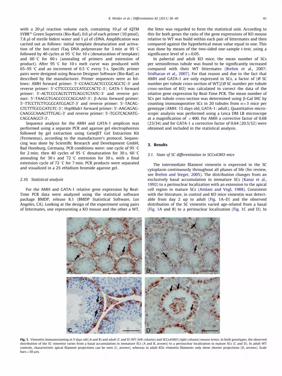
Fig. 1. Vimentin immunostaining in 5 days old (A and B) and adult (C and D) WT (left co
distribution of the SC vimentin varies from a basal accumulation in immature SCs (A
controls, characteristic apical filament projections can be seen (C, arrows), whereas i
bars¼50 mm.
the litter was regarded to form the statistical unit. According tothis for both genes the ratio of the gene expressions of KO mouserelative to WT was build within each pair of littermates and thencompared against the hypothetical mean value equal to one. Thiswas done by means of the two-sided one-sample t-test, using asignificance level of a¼0.05.
In pubertal and adult KO mice, the mean number of SCsper seminiferous tubule was found to be significantly increasedcompared with their WT littermates (Brehm et al., 2007;Sridharan et al., 2007). For that reason and due to the fact thatAMH and GATA-1 are only expressed in SCs, a factor of (Ø SCnumber per tubule cross-section of WT)/(Ø SC number per tubulecross-section of KO) was calculated to correct the data of therelative gene expression by Real-Time PCR. The mean number ofSCs per tubule cross-section was determined using b-Gal IHC bycounting immunopositive SCs in 20 tubules from n¼3 mice pergenotype (AMH: 15 days old, GATA-1: adult). Quantitative micro-scopic analysis was performed using a Leica DM LB microscopeat a magnification of �400. For AMH a corrective factor of 0.68(23/34) and for GATA-1 a corrective factor of 0.64 (20.5/32) wereobtained and included in the statistical analysis.
3. Results
3.1. State of SC differentiation in SCCx43KO mice
The intermediate filament vimentin is expressed in the SCcytoplasm continuously throughout all phases of life (for review,see Brehm and Steger, 2005). The distribution changes from anexclusively basal accumulation in immature SCs (Kanai et al.,1992) to a perinuclear localization with an extension to the apicalcell region in mature SCs (Amlani and Vogl, 1988). Consistentwith the literature, in control and KO mice vimentin was detect-able from day 2 up to adult (Fig. 1A–D) and the observeddistribution of the SC vimentin varied age-related from a basal(Fig. 1A and B) to a perinuclear localization (Fig. 1C and D). In
lumn) and SCCx43KO (right column) mouse testes. In both genotypes, the observed
and B, arrows) to a perinuclear localization in mature SCs (C and D). In adult WT
n adult KOs vimentin filaments only show shorter projections (D, arrows). Scale

K. Weider et al. / Differentiation 82 (2011) 38–4942
adult controls, characteristic apical filament projections wereobserved (Fig. 1C, arrows), whereas in adult KO mice vimentinfilaments only formed shorter extensions (Fig. 1D, arrows).
AMH, also called Mullerian inhibiting substance, is a memberof the transforming growth factor-b family, its expression in theSC cytoplasm starts very early in fetal life and persists untilpuberty (Josso et al., 2001; Tan et al., 2005). According to this,cytoplasmic AMH immunostaining was detectable in SCs of both,WT and KO mice at days 2, 5 (Fig. 2A and B), 7 and 8. Duringpubertal development in WT mice first AMH negative tubules
NCM KO WT M
50 kDa
65 kDa80 kDa
Fig. 2. IHC of AMH in 5 days old (A and B), 15 days old (C and D) and adult (E and F) WT
old testis homogenates. Both genotypes show a cytoplasmic AMH immunoreactivity on
representative pictures (C and D) for the observed extended period of AMH synthesis
tubules in contrast to same-aged KO littermates (D) which still show AMH immunoreac
AMH at approximately 59.8 kDa in the KO mouse, suggesting an increased protein exp
with advanced spermatogenesis appeared at days 10, 12 and 15(Fig. 2C) and AMH immunostaining was no longer observed in anyseminiferous tubule at day 17 and thereafter (Fig. 2E). In contrast,AMH immunoreactivity in KO mice remained positive in alltubules at days 10, 12, 15 (Fig. 2D), 17 and 19 and was non-detectable thereafter (Fig. 2F). Differences in the AMH protein andmRNA expression between WT and KO mice during puberty wereadditionally detected by WB (Fig. 2G) and Real-Time PCR. Real-Time PCR, followed by a successful sequence analysis (data notshown), revealed a statistically significant increase in AMH mRNA
(left column) and KO (right column) mouse testes. AMH WB analysis (G) of 15 days
day 5 (A and B) and all tubules are AMH-negative in adulthood (E and F). Note the
in KO mice during puberty. 15 days old WT mice (C) exhibit first AMH negative
tivity in all tubules. Scale bars¼50 mm. (G) Note the clear immunoreactive band for
ression on day 15. WB results are representative of n¼4 mice per genotype.

Fig. 3. Immunoexpression profile for the AR in 5 days old (A and B), 15 days old (C and D) and adult (E and F) WT (left column) and KO (right column) testes. AR nuclear
staining is absent in SCs from both genotypes at day 5 (A and B) whereas SCs of 15 days old (C and D) and adult mice (E and F) are immunopositive. Note the uniform
expression of the AR in peritubular cells (A–F, arrows) and LCs (A–F, arrowheads) at all ages. Scale bars¼50 mm.
K. Weider et al. / Differentiation 82 (2011) 38–49 43
expression in 15 days old KO mice: FC¼1.7038, SD¼0.2642,p¼0.0129 for n¼4 mice per genotype.
Comparable with the findings of Tan et al. (2005), SCs ofcontrol mice showed no nuclear AR immunostaining at days 2, 5(Fig. 3A) and 7. At day 8, simultaneously with the onset ofpuberty, first SCs revealed a faint nuclear reaction. From day 10onwards all SCs per seminiferous tubule were immunopositive(Fig. 3C and E). An identical age-dependent pattern of AR expres-sion was found in SCCx43KO mice (Fig. 3B, D and F). Beyond thatthere was a uniform and permanent expression of the AR inperitubular cells and LCs in all WT and KO litters (Fig. 3A–F).
The zinc finger transcription factor GATA-1 is first expressed inSC nuclei concomitantly with the first wave of spermatogenesis(Yomogida et al., 1994). During puberty the GATA-1 protein isuniformly produced by all SCs contrary to adulthood where itsexpression becomes stage-dependent (Yomogida et al., 1994).Consistent with the literature, in SCs of WT and KO mice theGATA-1 protein was nondetectable at days 2, 5 (Fig. 4A and B) and7 but was prominent in all SCs at day 8 and thereafter (Fig. 4C andD). In adulthood, as spermatogenesis expands, the GATA-1expression in SCs of WT mice became stage-dependent (Fig. 4E,arrows). In contrast adult SCCx43KO males showed a uniform andpermanent expression in all SCs (Fig. 4F). Differences in the GATA-1 protein and mRNA expression between adult WT and KO micewere additionally detected by WB (Fig. 4G) and Real-Time PCR.Real-Time PCR, followed by a successful sequence analysis (data
not shown), revealed a statistically significant increase in GATA-1mRNA expression in adult KO mice: FC¼1.9986, SD¼0.4910,p¼0.0268 for n¼4 mice per genotype.
One typical morphological feature of mature SCs is the largebasally located nucleus with a tripartite nucleolus (Gondos andBerndston, 1993). Consistent with this, semi-thin sections (Fig. 5Aand B) and electron micrographs (Fig. 5C and D) of adult mousetestes revealed in both genotypes this characteristic maturenuclear shape. Comparable to previous findings by Handel andEppig (1979), a tubular lumen formation was present from day 12onwards in both normal and mutant mice (Fig. 5E and F, asterisks).In adult KO mice, all seminiferous tubules show a clear lumen.
3.2. Composition, differentiation and fate of intratubular clustered
cells in SCCx43KO mice
Most cells per intratubular cell cluster were immunopositivefor the SC markers vimentin, GATA-1 and AR (data not shown),and always negative for the LC specific marker 3b-HSD (Fig. 6A,arrow), indicating that these cells represent SCs and not LCs. Themorphological investigation at semi- and ultra-thin section levelrevealed that cell clumps were mainly formed by abnormal SCs(Fig. 6B, arrowheads) with only single attached GCs (Fig. 6B; blackarrow). Unlike basal SC nuclei (Fig. 6B, white arrow) and thoselocated at the periphery of clusters showing normal morphology,SC nuclei within clusters appeared smaller and more convoluted

M KO WT M NC
50 kDa
65 kDa
40 kDa
Fig. 4. IHC of GATA-1 in NBF-fixed 5 days old (A and B), 15 days old (C and D) and adult (E and F) WT (left column) and KO (right column) mouse testes. GATA-1 WB
analysis (G) of adult testis homogenates. In both genotypes GATA-1 is nondetectable at day 5 (A and B) but prominent in all SCs at day 15 (C and D). In adulthood the
GATA-1 expression in SCs of WT mice is stage-dependent (E, arrows). In contrast, adult KO males show a uniform and permanent expression of GATA-1 (F). Scale
bars¼50 mm. (G) Note the clear immunoreactive band for GATA-1 at approximately 47 kDa in the KO mouse, suggesting an increased protein expression in adulthood. WB
results are representative of n¼4 mice per genotype.
K. Weider et al. / Differentiation 82 (2011) 38–4944
with numerous heterochromatic patches along the nuclear mem-brane (Fig. 6 B and C, arrowheads). These SCs became immuno-negative for b-Gal with increasing morphological alterations(Fig. 6D, arrow). In addition, cell clumps were always immuno-negative for the proliferation marker Ki-67 (Fig. 6E, arrow).Clustered cells, which morphologically correspond to SCs, wereTUNEL negative (Fig. 6F, arrow). Single TUNEL positive cells weredetectable either basally or only cluster-attached (Fig. 6F, arrow-heads). At ultrastructural level, most of these cluster-attachedcells revealed typical morphological signs of apoptosis like high
condensation of chromatin with a concave shape resembling asickle (Fig. 6G, arrow).
3.3. Time of occurrence of intratubular cell clusters in SCCx43KO
mice
From day 2 up to day 11, none of the examined mouse testesshowed intratubular cell clusters (Fig. 6H). First small clustersappeared in a few tubules from day 12 up to day 30, consisting ofapproximately 3–8 cells (Fig. 6I, arrows). However, in this time

Fig. 5. Semi-thin sections (A and B), electron micrographs (C and D) and HE staining (E and F) of adult (A–D) and 12 days old (E and F) WT (left column) and KO (right
column) mouse testes. (A–D) Both genotypes show large, basally located SC nuclei with a tripartite nucleolus, as a typical feature of mature SCs. The insets in image A and B
are magnifications of the corresponding circled areas. (E and F, asterisks) By postnatal day 12, a clear lumen formation can be observed in seminiferous tubules of both,
WT and KO mice. Scale bars¼20 mm (A and B), 3 mm (C and D) and 50 mm (E and F).
K. Weider et al. / Differentiation 82 (2011) 38–49 45
period most of the investigated KO testes were still ‘‘cluster free’’.From day 31 onwards cell clumps occurred regularly in nearly alltubules per testis, with partially 2-3 cell clumps per seminiferoustubule (Fig. 6J, arrows). Additionally, the number of cells percluster clearly increased up to 25 cells.
4. Discussion
Our present in vivo study highlights that the SC specific loss ofCx43 leads to an absence of maturing GCs in mice, resulting in apartly abnormal development of somatic SCs. Postpubertal basallylocated SCs in the SCCx43KO mouse model show features of bothmature and immature SC phenotypes, indicating that theydevelop an intermediate state of differentiation. This was demon-strated by the (1) formation of the tripartite nucleolus and(2) tubular lumen, (3) timely switch-on from AR and GATA-1during puberty, (4) temporally correct expression of vimentinfrom postnatal up to adult, (5) down-regulation of AMH duringpuberty but (6) increased SC numbers per tubule (Brehm et al.,2007) and (7) continued SC proliferation in adult KO mice(Sridharan et al., 2007). Additionally, in collaboration with
Carette et al. (2010) we have shown that the integrity of theBTB is given in our adult KO mice by using lanthanum tracer andhypertonic glucose perfusion. However, it can be supposed asincreased levels of, e.g. occludin or N-cadherin, were found in theSCCx43KO mice that these alterations mark an impairment in thedynamic process of opening and closing of this barrier and/orrepresent a sign for a permanent BTB closure (Carette et al., 2010).The formation of the BTB between adult SCs is known to be afunctional feature for SC maturation. In the present context,altered (molecular) composition of this barrier in the KO micemay be interpreted as an additional sign for the observedabnormal intermediate SC phenotype.
Altogether, SCs of SCCx43KO mice proliferate after pubertydespite the fact that they differentiate, suggesting that SCs arerather arrested proliferative cells than terminally differentiatedpostmitotic quiescent cells as previously assumed. Similar to ourmodel, in the Djungarian hamster proliferation of adult SCs hasalso been reported (Tarulli et al., 2006). Beyond that, recentin vitro studies showed that adult murine SCs can continuemitosis (Ahmed et al., 2009; Chaudhary et al., 2005; Gilleronet al., 2009) and that Cx43 participates in a negative control of SCproliferation (Gilleron et al., 2009).

A B CA B C
F
GD E G
JH I JFig. 6. 3b-HSD (A), b-Gal (D) and Ki-67 (E) IHC, semi-thin section (B), electron micrographs (C and G) and TUNEL staining (F) of intratubular cell clusters. HE staining of
testes from 10 (H), 14 (I) and 124 days old (J) SCCx43KOs. Clustered cells are always negative for the LC specific marker 3b-HSD (A, arrow); stained cells are interstitial LCs
(A, asterisk). Cell clumps are mainly formed by SCs (B, arrowheads) and only single attached GCs (B, black arrow). Unlike basal SC nuclei showing normal morphology
(B, white arrow), clustered SC nuclei appear smaller and more convoluted with numerous heterochromatic patches along the nuclear membrane (B and C, arrowheads).
These clustered SCs become immunonegative for b-Gal accompanied with increasing morphological alterations (D, arrow); note the b-Gal negative spermatogonia
(D, arrowhead). In addition, cell clumps are always immunonegative for the proliferation marker Ki-67 (E, arrow); note the Ki-67 positive cell at the margin of the tubule
(E, arrowhead). Clustered cells, which morphologically correspond to SCs, are TUNEL negative (F, arrow). The few TUNEL positive cells are either basally located or cluster-
attached (F, arrowheads). Representative electron micrograph (G) of a cluster-attached degenerating cell showing highly condensed chromatin with a concave shape
resembling a sickle (G, arrow). In 10 days old testis (representing the examined period from day 2 up to day 11) no cell clusters were observed (H). From day 12 up to day
30 only in a few tubules small cell clusters appeared, as seen in this 14 days old testis (I, arrows). From day 31 onwards, cell clumps occurred regularly in most tubules per
testis and the number of cells within the cluster increased, represented by this 124 days old testis (J, arrows). Scale bars¼100 mm (J), 50 mm (A, D, E, F, H and I), 20 mm (B),
3 mm (C and G).
K. Weider et al. / Differentiation 82 (2011) 38–4946
Regarding our further results that basal SCs in SCCx43KO micedisplay (1) shorter apical cell extensions of vimentin filaments,(2) a prolonged period of AMH synthesis during puberty and (3)a uniform and permanent GATA-1 expression in adulthood, wehypothesize that these alterations in the spatio-temporal expres-sion pattern are due to the observed GC deficiency primarilycaused by lack of Cx43 in SCs.
For vimentin, it was shown that apical cell extensions of theseintermediate filaments in mature SCs are dependent on theseminiferous epithelial cycle and seem to be related to thelocation of the elongated spermatid heads (Amlani and Vogl,1988). Additionally, earlier studies have demonstrated that loss ofstructural integrity of seminiferous epithelium in cryptorchid rattestes (Kopecky et al., 2005; Wang et al., 2002) or after treatmentwith different chemicals (Dalgaard et al., 2001; Johnson et al.,1991; Kopecky et al., 2005; Richburg and Boekelheide, 1996)correlates with a collapse of apical vimentin extensions. Takentogether, these results suggest that the lack of elongated sperma-tids in our mouse model may be one cause of the observed shorterapical cell extensions in KO males.
The prolonged period of AMH synthesis in our transgenic miceindicates an altered SC differentiation state and a temporal delayin the intrinsic differentiation program during pubertal develop-ment. However, it is difficult to make a clear statement aboutthe underlying molecular mechanisms for the failure of thetemporally correct AMH down-regulation. In human SCs, AMHexpression seems to be primarily regulated by androgens andAMH down-regulation occurs at the same time when a functionalAR is expressed in SCs and testosterone levels increase. But evenin human, there might exist additional mechanisms for AMHdown-regulation as in infertile men showing an arrest of sperma-togenesis at the premeiotic level, SCs continued to express AMH(Steger et al., 1996). Furthermore, there seem to exist speciesspecific differences in the regulation of AMH expression. In mice,there is evidence that a synergistic effect of different known (e.g.androgens, GATA-1 expression and meiotic entry) and unknownfactors is necessary to achieve the termination of AMH expression(Al-Attar et al., 1997; Beau et al., 2000). For example, Al-Attaret al. (1997) showed in postnatal mice that the meiotic entry actsin synergy with androgens to inhibit AMH synthesis and that the

K. Weider et al. / Differentiation 82 (2011) 38–49 47
failure of GCs to enter meiosis (as can also be seen in ourSCCx43KO mice) results in an incomplete inhibition of AMHexpression. In addition, male XXSxrb mice, which develop testeswith normal androgenic function, but the presence of twoX chromosomes, show a GC failure before meiotic entry. In thesemice, AMH expression persists in pubertal XXSxrb testes despiteproduction of adequate androgen levels and AR expression in SCs.These results may indicate synergistic or hierarchical regulatorymechanisms comparable to the AR and AMH results found inSCCx43KO males. Furthermore, it was shown in ARKO (totalablation of AR) and SCARKO (SC specific ablation of AR) mice(De Gendt et al., 2004) that neither direct nor indirect androgenaction is required for appropriate down-regulation of AMH as inboth KO models AMH expression decreased normally at the sameage as in controls.
Beyond that, testosterone may not be the only hormoneregulating AMH production but also FSH and thyroid hormoneT3 are involved. T3 is a major regulator of SC proliferation andmaturation. Hypothyroidism is associated with an increasedperiod of neonatal proliferation and a large increase in adult SCnumbers. In contrast, hyperthyroidism accelerates SC differentia-tion or maturation resulting in lower numbers of SCs per testis. Forexample, the fall in AMH mRNA expression is known to be delayedin hypothyroidism in vivo indicating that T3 could regulate AMHmRNA. Hypothyroidism lengthens the mitogenic period of SCs, isassociated with delays in SC maturation and neonatal hypothyr-oidism prolongs expression of AMH and thyroid hormone receptora (Bunick et al., 1994; Van Haaster et al., 1992). In addition,hormonal regulation of AMH mRNA expression was confirmed incell cultures derived from 2 day old rats (Arambepola et al., 1998).In these studies, T3 negatively stimulated AMH mRNA expressionas cultures treated with T3 showed a more dramatic decrease inAMH mRNA levels during 4 days of culture than controls. FSH wasshown to have a similar effect in vitro as T3, and T3 and FSH werefurther shown to have an additive effect on AMH expression incultured SCs. It was shown in SCCx43KO mice by Sridharan et al.(2007) that thyroid hormone receptor a mRNA as a sensitivemarker/indicator for proliferating immature SCs was still high inadult KO mice. In addition, T3 seems to exert both genomic andnongenomic effects on Cx43 in SCs in vivo, with the genomiceffects leading to increased levels of Cx43 in SCs associated with areduced rate of cell proliferation (Gilleron et al., 2006). Theseresults were confirmed in vitro using the SC line 42GPA9 demon-strating that T3 increased Cx43 expression and reduced SC pro-liferation. In conclusion, these data suggest that Cx43 in SCs couldrepresent an intermediate target for T3 inhibition of neonatal SCproliferation. Thus it may be speculated that the loss of the T3mediator Cx43 solely in SCs mimics a state of hypothyroidismresulting in a prolonged period of SC proliferation and a prolongedexpression of thyroid hormone receptor a(shown by Sridharanet al., 2007) that is accompanied by a delayed disappearance ofAMH at puberty as seen in the work from Bunick et al. (1994) andthe present study.
Unpublished data about the effect of the SC specific KO of Cx43on testicular gene expression in mice at 8 days of age usingmicroarray and Real-Time PCR revealed that the SC number wascomparable between 8 days old KO and WT mice. However, alreadyat that age a significant upregulation of AMH mRNA was detectablein SCCx43KO mice indicating an altered regulation of AMH expres-sion. As it was suggested by Al-Attar et al. (1997) that AMHproduction by SCs is regulated mainly at mRNA level it may bepossible that a preceding early increase in AMH mRNA levels resultsin higher AMH protein synthesis and proceeds in a delay of AMHdown-regulation as demonstrated in the present work.
The observed permanent and uniform GATA-1 expression inadult SCCx43KOs could also be based on the absence of maturing
GCs and their missing negative feedback signal for the GATA-1suppression. In the past, this secondary modulation has alreadybeen demonstrated in various mutant mouse strains (Yomogidaet al., 1994).
Our results regarding the formation of intratubular cell clus-ters in SCCx43KO mice confirm but also extend previous findings(Carette et al., 2010; Sridharan et al., 2007). In the present study,we show for the first time that cell clumps are not only formed bySCs but also single attached GCs. Similar to SCs being in normalposition, no major alterations in the biochemical differentiationstatus of clustered SCs have been detected, they retain mature (ARand GATA-1) and loose immature (AMH) markers. Interestingly,in contrast to basal SCs but with increasing morphologicalchanges similar to those observed by Russell et al. (1996) andtypical of immaturity (Gondos and Berndston, 1993), clusteredSCs lose their ability to express the LacZ reporter gene, which is areliable proof of successful and permanent deletion of the Cx43gene in our transgenic mouse model. Furthermore, clustered Cx43deficient SCs are non-proliferating cells indicating that abnormalSC division in adult KO mice (Sridharan et al., 2007) may occurprior to cluster formation. As a sign of epithelial disorganization,first small cell clumps appear during puberty and become morenumerous in adulthood with increasing cell numbers per cluster,indicating an age-related process. Sridharan et al. (2007) hypothe-sized that the continued proliferation of SCs in SCCx43KO miceinto adulthood may lead to an excessive build-up of SCs along thebasement membrane of the tubule resulting in sloughing of someSCs. However, it remains to be elucidated whether SCs really loosecontact to the basement membrane as a complete detachmentwould most likely result in cell death. There is evidence thatcontact to this membrane is necessary for SC survival in vitro
(Dirami et al., 1995). In our KO mice, only single cluster-attachedcells showed both morphological signs of apoptosis and a positiveTUNEL signal supposing that these cells are more vulnerable GCsand not clustered SCs. This conclusion is further based on thefact that apoptosis represents the dominant pathway for elim-inating GCs (Rodriguez et al., 1997). To the best of our knowledge,typical features of SCs undergoing cell death by apoptosis haveonly very rarely been demonstrated in vivo (Sasso-Cerri and Cerri,2008).
Formation of SC clusters seems to be common in GC depletedsituations as shown in different mouse and rat models ofimpaired spermatogenesis (Kopecky et al., 2005; Mazaud-Guittot et al., 2010; Schrans-Stassen et al., 2001). Although thisphenomenon seems to be associated with GC deficiency theunderlying mechanisms for the formation of cell clumps in ourKOs are unknown. Changes in cell shape can be associated with analtered expression, distribution and localization of intermediatefilaments. In SCs, vimentin filaments have been demonstrated toplay an important role in the maintenance of spermatogenesis(Kopecky et al., 2005; Sasso-Cerri and Cerri 2008). Thus, theobserved collapse of apical vimentin filaments in our KO micemight not only be associated with a disintegration of the semi-niferous epithelium but also with cluster formation. Presence ofaggregated SCs in the lumen of seminiferous tubules finallyimplies that Cx43 is required for normal behavior of at leastsingle SCs and that loss of Cx43 may have a dynamic impact on SCtopography despite normal integrity and functionality of the BTB(Carette et al., 2010).
Among different transgenic mouse models that are used toelucidate the versatile reasons for human male infertility, theSCCx43KO mice display a unique testicular and SC phenotype inwhich the primary defects are SC clustering, continued mitosisafter puberty (Sridharan et al., 2007) and partial epithelialdisorganization in the face of adult differentiation marker expres-sion. These KO mice provide an inimitable model of impaired

K. Weider et al. / Differentiation 82 (2011) 38–4948
spermatogenesis associated with an abnormal development ofsomatic SCs which is directly relevant in some cases of humansterility and the pathogenesis of human GC tumors.
Acknowledgments
We thank Professor J.I. Mason (Centre for Reproductive Biol-ogy, University of Edinburgh, Scotland) for the generous gift of3b-HSD primary antibody; Professor K. Willecke (Institute ofGenetics, University of Bonn, Germany) for the generous provisionof the floxed Cx43-LacZ transgenic mice; D. Schaefer (AnimalFacility, University of Marburg, Germany) for his skillful andoutstanding assistance with and treatment of our transgenicmice.
This work was supported by the DFG (BR 3365/2-1 and KFO181/1) and by the Dr. med. vet. Hans-Joachim und GertrudEngemann-Stiftung, Giessen, Germany.
References
Ahmed, E.A., Barten-van Rijbroek, A.D., Kal, H.B., Sadri-Ardekani, H., Mizrak, S.C.,van Pelt, A.M., de Rooij, D.G., 2009. Proliferative activity in vitro and DNArepair indicate that adult mouse and human Sertoli cells are not terminallydifferentiated, quiescent cells. Biol. Reprod. 80, 1084–1091.
Al-Attar, L., Noel, K., Dutertre, M., Belville, C., Forest, M.G., Burgoyne, P.S., Josso, N.,Rey, R., 1997. Hormonal and cellular regulation of Sertoli cell anti-Mullerianhormone production in the postnatal mouse. J. Clin. Invest. 100, 1335–1343.
Amlani, S., Vogl, A.W., 1988. Changes in the distribution of microtubules andintermediate filaments in mammalian Sertoli cells during spermatogenesis.Anat. Rec. 220, 143–160.
Arambepola, N.K., Bunick, D., Cooke, P.S., 1998. Thyroid hormone and follicle-stimulating hormone regulate Mullerian-inhibiting substance messengerribonucleic acid expression in cultured neonatal rat Sertoli cells. Endocrinol-ogy 139, 4489–4495.
Batias, C., Defamie, N., Lablack, A., Thepot, D., Fenichel, P., Segretain, D., Pointis, G.,1999. Modified expression of testicular gap-junction connexin 43 duringnormal spermatogenic cycle and in altered spermatogenesis. Cell Tissue Res.298, 113–121.
Beau, C., Rauch, M., Joulin, V., Jegou, B., Guerrier, D., 2000. GATA-1 is a potentialrepressor of anti-Mullerian hormone expression during the establishment ofpuberty in the mouse. Mol. Reprod. Dev. 56, 124–138.
Bellve, A.R., Cavicchia, J.C., Millette, C.F., O’Brien, D.A., Bhatnagar, Y.M., Dym, M.,1977. Spermatogenic cells of the prepubertal mouse. isolation and morpholo-gical characterization. J. Cell Biol. 74, 68–85.
Bergmann, M., Kliesch, S., 1994. The distribution pattern of cytokeratin andvimentin immunoreactivity in testicular biopsies of infertile men. Anat.Embryol. 190, 515–520.
Boujrad, N., Hochereau-de Reviers, M.T., Carreau, S., 1995. Evidence for germ cellcontrol of Sertoli cell function in three models of germ cell depletion in adultrat. Biol. Reprod. 53, 1345–1352.
Brehm, R., Marks, A., Rey, R., Kliesch, S., Bergmann, M., Steger, K., 2002. Alteredexpression of connexins 26 and 43 in Sertoli cells in seminiferous tubulesinfiltrated with carcinoma-in-situ or seminoma. J. Pathol. 197, 647–653.
Brehm, R., Steger, K., 2005. Regulation of Sertoli cell and Germ Cell Differentiation,Advances in Anatomy, Embryology and Cell Biology, 181. Springer Verlag,Berlin.
Brehm, R., Zeiler, M., Ruttinger, C., Herde, K., Kibschull, M., Winterhager, E.,Willecke, K., Guillou, F., Lecureuil, C., Steger, K., Konrad, L., Biermann, K.,Failing, K., Bergmann, M., 2007. A sertoli cell-specific knockout of connexin43prevents initiation of spermatogenesis. Am. J. Pathol. 171, 19–31.
Bruzzone, R., White, T.W., Paul, D.L., 1996. Connections with connexins: themolecular basis of direct intercellular signalling. Eur. J. Biochem. 238, 1–27.
Bunick, D., Kirby, J., Hess, R.A., Cooke, P.S., 1994. Developmental expression oftestis messenger ribonucleic acids in the rat following propylthiouracil-induced neonatal hypothyroidism. Biol. Reprod. 51, 706–713.
Carette, D., Weider, K., Gilleron, J., Giese, S., Dompierre, J., Bergmann, M., Brehm, R.,Denizot, J.P., Segretain, D., Pointis, G., 2010. Major involvement of connexin 43in seminiferous epithelial junction dynamics and male fertility. Dev. Biol. 346,54–67.
Chaudhary, J., Sadler-Riggleman, I., Ague, J.M., Skinner, M.K., 2005. The helix–loop–helix inhibitor of differentiation (ID) proteins induce post-mitotic terminallydifferentiated Sertoli cells to re-enter the cell cycle and proliferate. Biol.Reprod. 72, 1205–1217.
Dalgaard, M., Hossaini, A., Hougaard, K.S., Hass, U., Ladefoged, O., 2001. Develop-mental toxicity of toluene in male rats: effects on semen quality, testismorphology, and apoptotic neurodegeneration. Arch. Toxicol. 75, 103–109.
Decrouy, X., Gasc, J.M., Pointis, G., Segretain, D., 2004. Functional characterization ofCx43 based gap junctions during spermatogenesis. J. Cell Physiol. 200, 146–154.
De Gendt, K., Swinnen, J.V., Saunders, P.T., Schoonjans, L., Dewerchin, M., Devos, A.,Tan, K., Atanassova, N., Claessens, F., Lecureuil, C., Heyns, W., Carmeliet, P.,Guillou, F., Sharpe, R.M., Verhoeven, G., 2004. A Sertoli cell-selective knockoutof the androgen receptor causes spermatogenic arrest in meiosis. Proc. Natl.Acad. Sci. USA 101, 1327–1332.
Dirami, G., Ravindranath, N., Kleinman, H.K., Dym, M., 1995. Evidence thatbasement membrane prevents apoptosis of Sertoli cells in vitro in the absenceof known regulators of Sertoli cell function. Endocrinology 136, 4439–4447.
Gilleron, J., Nebout, M., Scarabelli, L., Senegas-Balas, F., Palmero, S., Segretain, D.,Pointis, G., 2006. A potential novel mechanism involving connexin 43 gapjunction for control of sertoli cell proliferation by thyroid hormones. J. CellPhysiol. 209, 153–161.
Gilleron, J., Carette, D., Durand, P., Pointis, G., Segretain, D., 2009. Connexin 43 apotential regulator of cell proliferation and apoptosis within the seminiferousepithelium. Int. J. Biochem. Cell Biol. 41, 1381–1390.
Gondos, B., Berndston, W.E., 1993. Postnatal and pubertal development. In: Griswold,M.D., Russell, L.D. (Eds.), The Sertoli cell. Cache River Press, Clearwater, pp.115–154.
Guitton, N., Touzalin, A.M., Sharpe, R.M., Cheng, C.Y., Pinon-Lataillade, G., Meritte,H., Chenal, C., Jegou, B., 2000. Regulatory influence of germ cells on sertoli cellfunction in the pre-pubertal rat after acute irradiation of the testis. Int. J.Androl. 23, 332–339.
Handel, M.A., Eppig, J.J., 1979. Sertoli cell differentiation in the testes of micegenetically deficient in germ cells. Biol. Reprod. 20, 1031–1038.
Johnson, K.J., Hall, E.S., Boekelheide, K., 1991. 2,5-Hexanedione exposure alters therat Sertoli cell cytoskeleton. I. Microtubules and seminiferous tubule fluidsecretion. Toxicol. Appl. Pharmacol. 111, 432–442.
Josso, N., di Clemente, N., Gouedard, L., 2001. Anti-Mullerian hormone and itsreceptors. Mol. Cell. Endocrinol. 179, 25–32.
Kanai, Y., Kawakami, H., Takata, K., Kurohmaru, M., Hirano, H., Hayashi, Y., 1992.Involvement of actin filaments in mouse testicular cord organization in vivoand in vitro. Biol. Reprod. 46, 233–245.
Kliesch, S., Behre, H.M., Hertle, L., Bergmann, M., 1998. Alteration of Sertoli celldifferentiation in the presence of carcinoma in situ in human testis. J. Urol.160, 1894–1898.
Kopecky, M., Semecky, V., Nachtigal, P., 2005. Vimentin expression during alteredspermatogenesis in rats. Acta Histochem. 107, 279–289.
Kumar, N.M., Gilula, N.B., 1996. The gap junction communication channel. Cell 84,381–388.
Mason, J.I., Howe, B.E., Howie, A.F., Morley, S.D., Nicol, M.R., Payne, A.H., 2004.Promiscuous 3beta-hydroxysteroid dehydrogenases: testosterone 17beta-hydroxysteroid dehydrogenase activities of mouse type I and VI 3beta-hydroxysteroid dehydrogenases. Endocr. Res. 30, 709–714.
Mazaud-Guittot, S., Meugnier, E., Pesenti, S., Wu, X., Vidal, H., Gow, A.,Le Magueresse-Battistoni, B., 2010. Claudin 11 deficiency in mice results inloss of the Sertoli cell epithelial phenotype in the testis. Biol. Reprod. 82,202–213.
Pelletier, R.M., 1995. The distribution of connexin 43 is associated withthe germ cell differentiation and with the modulation of the Sertoli celljunctional barrier in continual (guinea pig) and seasonal breeders’ (mink)testes. J. Androl. 16, 400–409.
Perez-Armendariz, E.M., Lamoyi, E., Mason, J.I., Cisneros-Armas, D., Luu-The, V.,Bravo-Moreno, J.F., 2001. Developmental regulation of connexin43 expressionin fetal mouse testicular cells. Anat. Rec. 264, 237–246.
Reaume, A.G., DeSousa, P.A., Kulkarni, S., Langille, B.L., Zhu, D., Davies, T.C., Juneja,S.C., Kidder, G.M., Rossant, J., 1995. Cardiac malformation in neonatal micelacking connexin43. Science 267, 1831–1834.
Richburg, J.H., Boekelheide, K., 1996. Mono-(2-ethylhexyl) phthalate rapidly altersboth Sertoli cell vimentin filaments and germ cell apoptosis in young rattestes. Toxicol. Appl. Pharmacol. 137, 42–50.
Risley, M.S., Tan, I.P., Roy, C., Saez, J.C., 1992. Cell-, age- and stage-dependentdistribution of connexin43 gap junctions in testes. J. Cell Sci. 103, 81–96.
Rodriguez, I., Ody, C., Araki, K., Garcia, I., Vassalli, P., 1997. An early and massivewave of germinal cell apoptosis is required for the development of functionalspermatogenesis. EMBO J. 16, 2262–2270.
Russell, L.D., Franc-a, L.R., Brinster, R.L., 1996. Ultrastructural observations ofspermatogenesis in mice resulting from transplantation of mouse spermato-gonia. J. Androl. 17, 603–614.
Sasso-Cerri, E., Cerri, P.S., 2008. Morphological evidences indicate thatthe interference of cimetidine on the peritubular components is responsiblefor detachment and apoptosis of Sertoli cells. Reprod. Biol. Endocrinol.6, 18.
Schrans-Stassen, B.H., Saunders, P.T., Cooke, H.J., de Rooij, D.G., 2001. Nature of thespermatogenic arrest in Dazl �/� mice. Biol. Reprod. 65, 771–776.
Sharpe, R.M., Millar, M., McKinnell, C., 1993. Relative roles of testosterone and thegerm cell complement in determining stage-dependent changes in proteinsecretion by isolated rat seminiferous tubules. Int. J. Androl. 16, 71–81.
Sharpe, R.M., 1994. Regulation of spermatogenesis. In: Knobil, E., Neill, J.D. (Eds.),The Physiology of Reproduction. Raven Press, New York, pp. 1363–1434.
Sharpe, R.M., McKinnell, C., Kivlin, C., Fisher, J.S., 2003. Proliferation and functionalmaturation of Sertoli cells, and their relevance to disorders of testis function inadulthood. Reproduction 125, 769–784.
Sohl, G., Willecke, K., 2004. Gap junctions and the connexin protein family.Cardiovasc. Res. 62, 228–232.
Sridharan, S., Simon, L., Meling, D.D., Cyr, D.G., Gutstein, D.E., Fishman, G.I., Guillou,F., Cooke, P.S., 2007. Proliferation of adult Sertoli cells following conditional

K. Weider et al. / Differentiation 82 (2011) 38–49 49
knockout of the gap junctional protein gja1 (connexin 43). Biol. Reprod. 76,804–812.
Steger, K., Rey, R., Kliesch, S., Louis, F., Schleicher, G., Bergmann, M., 1996.Immunohistochemical detection of immature Sertoli cell markers in testiculartissue of infertile men: a preliminary study. Int. J. Androl. 19, 122–128.
Steger, K., Tetens, F., Bergmann, M., 1999a. Expression of connexin 43 in humantestis. Histochem. Cell Biol. 112, 215–220.
Steger, K., Rey, R., Louis, F., Kliesch, S., Behre, H.M., Nieschlag, E., Hoepffner, W.,Bailey, D., Marks, A., Bergmann, M., 1999b. Reversion of the differentiatedphenotype and maturation block in Sertoli cells in pathological human testis.Hum. Reprod. 14, 136–143.
Tan, K.A., De Gendt, K., Atanassova, N., Walker, M., Sharpe, R.M., Saunders, P.T.,Denolet, E., Verhoeven, G., 2005. The role of androgens in sertoli cell proliferationand functional maturation: studies in mice with total or Sertoli cell-selectiveablation of the androgen receptor. Endocrinology 146, 2674–2683.
Tarulli, G.A., Stanton, P.G., Lerchl, A., Meachem, S.J., 2006. Adult sertoli cells are notterminally differentiated in the Djungarian hamster: effect of FSH. Biol.Reprod. 74, 798–806.
Van Haaster, L.H., De Jong, F.H., Docter, R., De Rooij, D.G., 1992. The effect ofhypothyroidism on Sertoli cell proliferation and differentiation and hormonelevels during testicular development in the rat. Endocrinology 131, 1574–1576.
Vergouwen, R.P., Huiskamp, R., Bas, R.J., Roepers-Gajadien, H.L., Davids, J.A., deRooij, D.G., 1993. Postnatal development of testicular cell populations in mice.J. Reprod. Fertil. 99, 479–485.
Wang, Z.Q., Watanabe, Y., Toki, A., Itano, T., 2002. Altered distribution of Sertolicell vimentin and increased apoptosis in cryptorchid rats. J. Pediatr. Surg. 37,648–652.
Yomogida, K., Ohtani, H., Harigae, H., Ito, E., Nishimune, Y., Engel, J.D., Yamamoto, M.,1994. Developmental stage- and spermatogenic cycle-specific expression oftranscription factor GATA-1 in mouse Sertoli cells. Development 120, 1759–1766.