brain areas controlling heart rate variability in tinnitus and tinnitus-related distress
TRANSCRIPT
Brain Areas Controlling Heart Rate Variability in Tinnitusand Tinnitus-Related DistressSven Vanneste1,2*, Dirk De Ridder1
1 Brai2n, Tinnitus Research Initiative Clinic Antwerp & Department of Neurosurgery, University Hospital Antwerp, Antwerp, Belgium, 2 Department of Translational
Neuroscience, Faculty of Medicine, University of Antwerp, Antwerp, Belgium
Abstract
Background: Tinnitus is defined as an intrinsic sound perception that cannot be attributed to an external sound source.Distress in tinnitus patients is related to increased beta activity in the dorsal part of the anterior cingulate and the amount ofdistress correlates with network activity consisting of the amygdala-anterior cingulate cortex-insula-parahippocampus.Previous research also revealed that distress is associated to a higher sympathetic (OS) tone in tinnitus patients and tinnitussuppression to increased parasympathetic (PS) tone.
Methodology: The aim of the present study is to investigate the relationship between tinnitus distress and the autonomicnervous system and find out which cortical areas are involved in the autonomic nervous system influences in tinnitusdistress by the use of source localized resting state electroencephalogram (EEG) recordings and electrocardiogram (ECG).Twenty-one tinnitus patients were included in this study.
Conclusions: The results indicate that the dorsal and subgenual anterior cingulate, as well as the left and right insula areimportant in the central control of heart rate variability in tinnitus patients. Whereas the sympathovagal balance iscontrolled by the subgenual and pregenual anterior cingulate cortex, the right insula controls sympathetic activity and theleft insula the parasympathetic activity. The perceived distress in tinnitus patients seems to be sympathetically mediated.
Citation: Vanneste S, De Ridder D (2013) Brain Areas Controlling Heart Rate Variability in Tinnitus and Tinnitus-Related Distress. PLoS ONE 8(3): e59728.doi:10.1371/journal.pone.0059728
Editor: Mathias Baumert, University of Adelaide, Australia
Received July 9, 2012; Accepted February 21, 2013; Published March 22, 2013
Copyright: � 2013 Vanneste, De Ridder. This is an open-access article distributed under the terms of the Creative Commons Attribution License, which permitsunrestricted use, distribution, and reproduction in any medium, provided the original author and source are credited.
Funding: This work was supported by Research Foundation Flanders (FWO). The funders had no role in study design, data collection and analysis, decision topublish, or preparation of the manuscript.
Competing Interests: The authors have declared that no competing interests exist.
* E-mail: [email protected]
Introduction
Tinnitus is defined as an intrinsic sound perception that cannot
be attributed to an external sound source. This phantom
perception is a common disorder. The American Tinnitus
Association estimates that 50 million Americans are affected by
it, and that 12 million of these people seek medical attention
because of their tinnitus [1]. In about 6 to 25% of the affected
people tinnitus causes a considerable amount of distress [2–4],
resulting in about 2–4% of the population who are severely
impaired in their quality of life [5]. Tinnitus can interfere with
sleep and concentration, social interaction and work [6]. Increased
prevalence rates of anxiety and depression are reported among
tinnitus patients [7,8].
Distress in tinnitus patients is related to increased beta activity in
the dorsal part of the anterior cingulate and the amount of distress
correlates with an EEG alpha network activity consisting of the
amygdala-anterior cingulate cortex-insula-parahippocampus as
demonstrated both by source analysis of Fourier based data [9]
and independent component analysis [10]. Using MEG, long-
range coupling between frontal, parietal and cingulate brain areas
in alpha and gamma phase synchronization has been shown to be
related to tinnitus distress [11]. The distress in tinnitus patients also
correlates with an increase in incoming and outgoing connections
in the gamma band in the prefrontal cortex and the parieto-
occipital region [12].
Adaptation under conditions of stress is a priority for all
organisms. Stress can be broadly defined as an actual or
anticipated disruption of homeostasis or an anticipated threat to
well-being [13]. Stressor-related information from all major
sensory systems is conveyed to the brain, which recruits neural
and neuroendocrine systems (effectors) to minimize the net cost to
the animal. The physiological response to stress involves an
efficient and highly conserved set of interlocking systems and aims
to maintain physiological integrity even in the most demanding of
circumstances [13].
The autonomic nervous system provides the most immediate
response to stressor exposure - through its sympathetic and
parasympathetic arms, which provoke rapid alterations in
physiological states through neural innervation of end organs.
The autonomic nervous system is a collection of afferent and
efferent neurons that link the central nervous system with visceral
effectors. The two efferent arms of the autonomic nervous system -
the sympathetic and parasympathetic arms - consist of parallel and
differentially regulated pathways made up of cholinergic neurons
(preganglionic neurons) located within the central nervous system
that innervate ganglia (for example, para- or pre-vertebral
sympathetic ganglia), glands (adrenal glands) or neural networks
PLOS ONE | www.plosone.org 1 March 2013 | Volume 8 | Issue 3 | e59728
of varying complexity (enteric or cardiac ganglionic networks).
These peripheral ganglia and networks contain the motor neurons
(ganglionic neurons) that control smooth muscles and other
visceral targets. The sympathetic ganglionic neurons that control
cardiovascular targets are primarily noradrenergic [14]. The
sympatho-adrenomedullary arm can rapidly (in seconds) increase
heart rate and blood pressure by exciting the cardiovascular
system. Importantly, excitation of the autonomic nervous system
wanes quickly - owing to reflex parasympathetic activation -
resulting in short-lived responses [13]. Previous research also
revealed that distress is associated to a higher sympathetic (OS)
tone in tinnitus patients [15] and tinnitus suppression to increased
parasympathetic (PS) tone [16]. The heart is dually innervated by
the autonomic nervous system such that relative increases in
sympathetic activity are associated with heart rate increases and
relative increases in parasympathetic activity are associated with
heart rate decreases. In addition, human lesion and electrical
stimulation studies have revealed that the right insula controls
cardiac sympathetic activity whereas the left insula is predomi-
nantly associated to parasympathetic activity [17–19].
Heart rate variability (HRV) is a physiological phenomenon
where the time interval between heart beats varies. It is measured
by the variation in the beat-to-beat interval. HRV is a simple and
non-invasive quantitative marker of autonomic function. As a
result of continuous variations of the balance between OS and PS
neural activity influencing heart rate, intervals between consecu-
tive heartbeats (RR intervals) show spontaneously occurring
oscillations. For HRV analysis, a Fourier-based spectral analysis
is performed of the beat to beat intervals, yielding two main
frequencies: a low frequency range (LF: 0.05–0.15 Hz) and a high
frequency range (HF 0.15–0.4 Hz) [20]. The high frequency
component of HRV is believed to be influenced by vagal activity
and is also related to the frequency of respiration [21]. Low-
frequency (LF) power is modulated by baroreceptor activities and
fluctuations in heart rate in the LF range reflect OS as well as PS
influences. Low-frequency power, therefore, cannot be considered
to reflect selective OS activity. However if normalized units of LF
and HF are considered, the OS and PS influences respectively are
emphasized [20]. In HRV frequency domain, normalized units of
LF and HF components therefore reflect OS and PS influences
respectively.
In two recent PET studies it was demonstrated that inducing a
certain amount of stress, HRV correlates positively with activity in
the anterior cingulate cortex, caudate nucleus, insula, medial
prefrontal cortex extending into the dorsal prefrontal cortex
[22,23]. These areas are also involved in tinnitus related distress
[9]. Using similar PET studies, the neural correlates of the HF
component (PS) have been delineated as the caudate nucleus,
periaqueductal gray and left mid-insula [23], while in fMRI the
HF component correlates positively with activity in the hypothal-
amus, amygdala and anterior hippocampal area, dorsomedial/
dorsolateral prefrontal cortex and negatively with the cerebellum,
parabrachial nucleus/locus coeruleus, periaqueductal gray, poste-
rior parahippocampal area, thalamus, posterior insular and middle
temporal cortices [24]. The left inferior part of the pregenual
anterior cingulate cortex also correlates with the HF component of
the HRV [25]. The increased LF/HF-ratio (in rectal distension) is
correlated with activity in the bilateral insula, putamen, thalamus,
midbrain, pons, and cerebellum [26].
The aim of the present study is to investigate the relationship
between tinnitus distress and the autonomic nervous system and
find out which cortical areas are involved in the autonomic
nervous system influences in tinnitus distress by the use of source
localized resting state electroencephalogram (EEG) recordings and
electrocardiogram (ECG).
Quantitative analysis of EEG is a low-cost and useful
neurophysiological approach to study the brain physiology and
pathology [27]. Cortical sources of the EEG rhythms were
estimated by standardized low-resolution brain electromagnetic
tomography (sLORETA) [28]. sLORETA is a functional imaging
technique estimating maximally smoothed linear inverse solutions
accounting for distributed EEG sources within Montreal Neuro-
logical Institute (MNI) space [28]. This feature is of special
importance for the comparison of EEG results with those of most
structural and functional neuroimaging studies. sLORETA has
been successfully used in recent EEG studies on tinnitus [29]. In
this study we investigate which brain areas are involved in tinnitus
distress and in the autonomic nervous system control of the
distress.
Materials and Methods
ParticipantsTwenty-one patients (N = 21; 15 males and 6 females) with a
mean age of 47.44 (Sd = 12.72 were selected from the multidisci-
plinary Tinnitus Research Initiative (TRI) Clinic of the University
Hospital of Antwerp, Belgium. Tinnitus lateralization and tinnitus
type was verified by asking the patient in which ear they perceived
the tinnitus and whether they perceived a tone or a noise-like
sound. Six patients presented with unilateral tinnitus and 15
patients with bilateral tinnitus. Nine patients perceived a pure tone
phantom sound and 16 patients a narrow band noise (hearing a
noise-like tone within a certain frequency range). No patients
included in the study perceived their tinnitus centrally in the head.
Individuals with pulsatile tinnitus, Meniere’s disease, otosclerosis,
chronic headache, neurological disorders such as brain tumors,
and individuals being treated for mental disorders were not
included in the study.
Participants were requested to refrain from alcohol consump-
tion 24 hours prior to recording, and from caffeinated beverages
consumption on the day of recording. Patients were also given the
validated Dutch version of the Tinnitus Questionnaire [30]
originally published by Goebel and Hiller [31]. Goebel and Hiller
described this TQ as a global index of distress and the Dutch
version was further confirmed as a reliable measure for tinnitus-
related distress [30,32]. At the moment of the study patients did
not take any pharmacological agent.
This study was approved by the local ethical committee
(Antwerp University Hospital) and was in accordance with the
declaration of Helsinki. Written informed consent was obtained
from all patients.
EEG/ECG Data CollectionRecordings (Mitsar-201, NovaTech http://www.novatecheeg.
com/) were obtained in a fully lighted room with each participant
sitting upright on a small but comfortable chair. The actual
recording lasted approximately 5 min. The EEG was sampled
with 19 electrodes (Fp1, Fp2, F7, F3, Fz, F4, F8, T7, C3, Cz, C4,
T8, P7, P3, Pz, P4, P8, O1 O2) in the standard 10–20
International placement referenced to linked ears and impedances
were checked to remain below 5 kV. Two ECG electrodes were
place on the heart axis. EEG and ECG were measured for 5
minutes. Data were collected eyes-closed (sampling
rate = 1024 Hz, band passed 0.15–200 Hz). To minimize respira-
tory influences on HRV, respiration is controlled at 12 beats per
minute using auditory cues (i.e. tone 1000 Hz). We selected
auditory cues as this is the standard method when collecting ECG
Brain and Heart Rate Variability in Tinnitus
PLOS ONE | www.plosone.org 2 March 2013 | Volume 8 | Issue 3 | e59728
data during eyes closed EEG [33]. No patients indicate that this
auditory cue interfered with the tinnitus perception or auditory
attention to the tinnitus.
EEG AnalysisData were resampled to 128 Hz, band-pass filtered (fast Fourier
transform filter) to 2–44 Hz and subsequently transposed into
Eureka! Software [34], plotted and carefully inspected for manual
artifact-rejection. All episodic artifacts including eye blinks, eye
movements, teeth clenching, or body movement were removed
from the stream of the EEG. Average Fourier cross-spectral
matrices were computed for bands delta (2–3.5 Hz), theta (4–
7.5 Hz), alpha (8–12 Hz), low beta (13–21 Hz), high beta (21.5–
30 Hz) and gamma (30.5–44 Hz) [28,35].
ECG AnalysisECG signals are processed by frequency domain methods as
recommended by the Task force [20]: QRS complexes are
recognized from the short-term artifact-free ECG recordings from
which peaks (R-waves) are detected and from which intervals
between two consecutive peaks (RR intervals) are calculated. Once
HRV time series are extracted they are analyzed in frequency
domain using HRV Analysis Software 1.1 for windows developed
by The Biomedical Signal Analysis Group, Department of Applied
Physics, University of Kuopio, Finland (see http://kubios.uku.fi/)
and generating low frequency (LF: 05–.15 Hz ) and high
frequency (HF:.15–.40 Hz). Also the LF/HF-ratio was calculated.
Source LocalizationStandardized low-resolution brain electromagnetic tomography
(sLORETA) was used to estimate the intracerebral electrical
sources that generated the scalp-recorded activity in each of the
eight frequency bands [28]. sLORETA computes electric neuro-
nal activity as current density (A/m2) without assuming a
predefined number of active sources. The sLORETA solution
space consists of 6239 voxels (voxel size: 56565 mm) and is
restricted to cortical gray matter and hippocampi, as defined by
digitized MNI152 template [36].
The tomography sLORETA has received considerable valida-
tion from studies combining LORETA with other more estab-
lished localization methods, such as functional Magnetic Reso-
nance Imaging (fMRI) [37,38], structural MRI [39], Positron
Emission Tomography (PET) [40–42]. Further sLORETA vali-
dation has been based on accepting as ground truth the
localization findings obtained from invasive, implanted depth
electrodes, in which case there are several studies in epilepsy
[43,44] and cognitive ERPs [45]. It is worth emphasizing those
deep structures such as the anterior cingulate cortex [46], and
mesial temporal lobes [47] can be correctly localized with these
methods.
Region of Interest AnalysisThe log-transformed electric current density was averaged
across all voxels belonging to the region of interest. Regions of
interest were defined based on previous brain research on HRV as
well as tinnitus related distress. Regions of interest were
respectively the left insula (LI) and right insula (RI)(BA13) [9],
dorsal anterior cingulate cortex (BA24 left and right) [9] and
subgenual anterior cingulate cortex (BA25 left and right) [9],
primary (BA41) and secondary (BA21) auditory cortex [48] and
the orbitofrontal cortex (BA10) [12]. Region of interest analyses
were computed for the different frequency bands separately.
A lateralization index for the insula was calculated for each
frequency band,
Lateralization index~(LI{RI)
(LIzRI)
where LI and RI are the log-transformed electrical current density
in the left and right insula, respectively. This method is similar to
Weisz et al. [49]. Pearson Correlations were calculated and
corrections for multiple comparisons for the frequency bands (i.e.
Bonferroni) were applied.
Statistical AnalysesWe conducted a whole brain analysis and region of interest
(ROI) analyses. Whole-brain analysis is automated and un-biased,
making no assumptions about any regions of particular interest.
However, this technique requires a great number of subjects to
achieve statistical significance and it is possible that smaller
changes may not be easily identified. This is one of the reasons
why it is common practice to conduct a secondary ROI analysis,
testing for statistically significant differences only in the voxels that
are deemed of interest by an a priori hypothesis. An ROI analysis
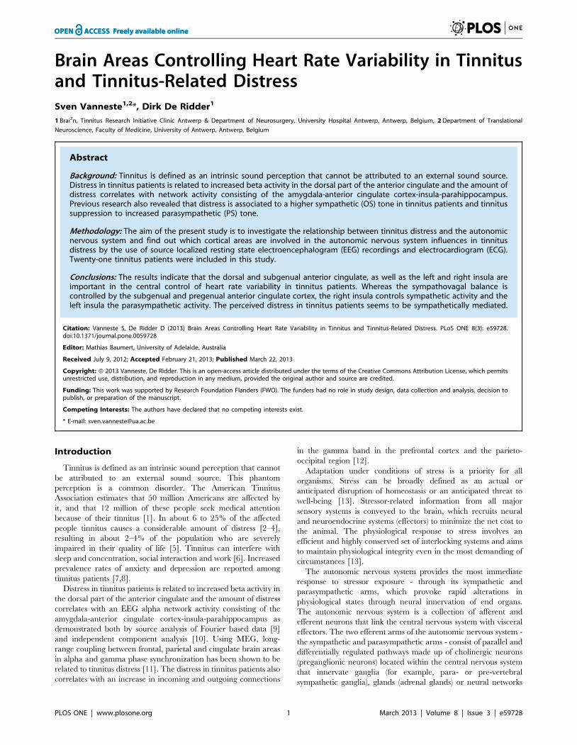
Figure 1. Negative correlation between LF (sympathetic+parasympathetic) HRV and Alpha activity in the left insula (BA13),indicating that decreased alpha activity in the left insula goes together with increased LF-HRV.doi:10.1371/journal.pone.0059728.g001
Brain and Heart Rate Variability in Tinnitus
PLOS ONE | www.plosone.org 3 March 2013 | Volume 8 | Issue 3 | e59728
can be used to corroborate the findings of previous studies, or
those obtained during the whole-brain analysis. This is of special
importance in studies with a small sample size.
First, correlations are calculated between respectively LF, HF,
LF/HF and distress with brain activity (whole brain analysis). The
methodology used for the sLORETA correlations is non-
parametric. It is based on estimating, via randomization, the
empirical probability distribution for the max-statistic, under the
null hypothesis comparisons [50]. This methodology corrects for
multiple testing (i.e., for the collection of tests performed for all
voxels, and for all frequency bands). Due to the non-parametric
nature of the method, its validity does not rely on any assumption
of Gaussianity [50]. sLORETA statistical contrast maps were
calculated through multiple voxel-by-voxel comparisons in a
logarithm of F-ratio. The significance threshold was based on a
permutation test with 5000 permutations.
Secondly, Pearson correlations are calculated between the
lateralization index of the insula and LF/HF-ratio. Based on these
findings we conducted a third step, a multivariate ANOVA with
LF/HF-ratio and TQ as dependent variables and the lateraliza-
tion index of the insula in both the alpha and gamma band as
independent variables. The reason to include these frequency
bands was that both frequency bands correlated with the LF/HF-
ratio.
In addition a Pearson correlation analysis was conducted
between the dorsal, subgenual and pregenual anterior cingulate
cortex, primary and secondary auditory cortex and the orbito-
frontal cortex with the respectively the LF, HF, LF/HF-ratio and
the TQ, to validate previously obtained results [9,10].
Lastly, we applied a median-split on both the TQ and the LF/
HF-ratio. A median split [51] is a data driven post-hoc
stratification that allows us to test the group difference between
a low versus high TQ (i.e. distress) and low and high LF/HF-ratio.
We applied an ANOVA with TQ (low vs. high) and LF/HF-ration
(low vs. high) as independent variables and log-transformed
current density at the pregenual anterior cingulate cortex for
respectively the high beta and gamma band as dependent variable.
We opt to do this analysis for the pregenual anterior cingulate
cortex as region of interest for these specific frequency bands, as
both frequencies correlated with the TQ and LF/HF ratio.
Results
Tinnitus QuestionnaireThe mean TQ was 39.22 (Sd = 15.13). No correlation could be
found between the TQ and respectively LF, HF and LF/HF-ratio
HRV.
Whole Brain and HRV1. LF-HRV. Analysis of LF-HRV (a combination of sympa-
thetic and parasympathetic activity) and the left insula (BA13) for
alpha activity revealed a significant negative correlation (r = 2.42,
p,.05) indicating that decreased alpha activity in the left insula
goes together with increased LF-HRV (see Figure 1).
No significant correlation could be retrieved in delta, theta, low
beta, high beta and gamma frequency bands.
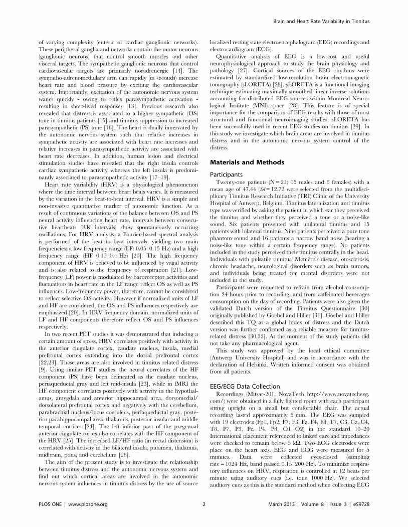
2. HF-HRV. Results yielded as a significant positive correla-
tion (r = .68, p,.05) between HF-HRV (i.e. sympathetic activity)
and rostral portions of the superior temporal gyrus and the middle
temporal gyrus (BA21/38) for alpha activity (see Figure 2). That is
increased activity in the rostral portions of the superior temporal
gyrus and the middle temporal gyrus goes together with increased
HF-HRV.
No significant correlation could be retrieved in delta, theta, low
beta, high beta and gamma frequency bands.
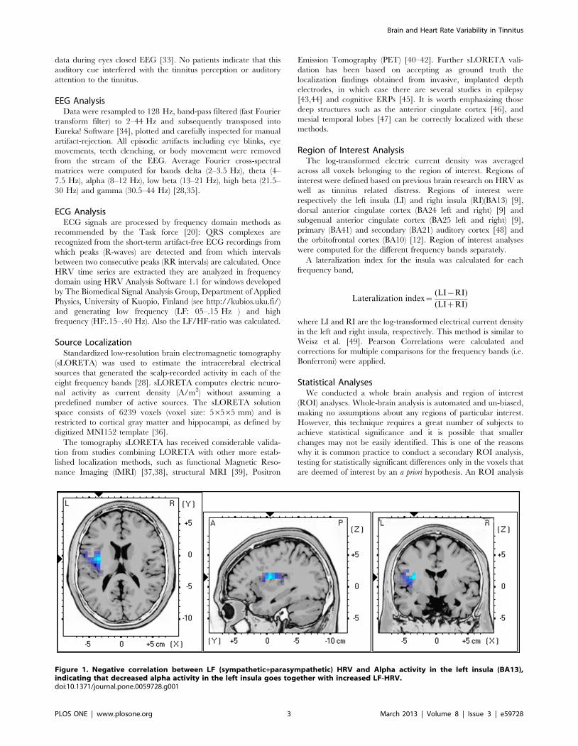
3. LF/HF-ratio. Analysis demonstrated a positive correlation
between LF/HF-ratio (i.e. high numbers mean dominance of
sympathetic activity while low numbers mean dominance of the
para-sympathetic activity) and Theta activity (r = .43, p,.05) in
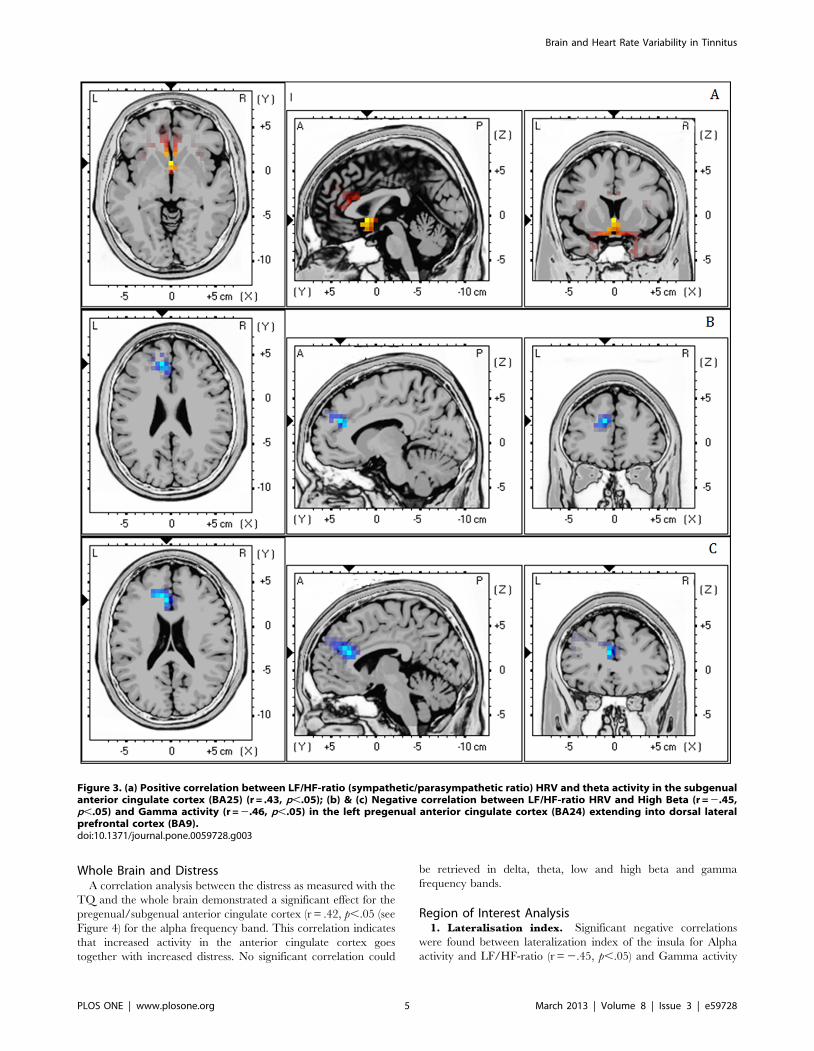
the subgenual anterior cingulate cortex (BA25) (see Figure 3a).
This correlation indicates that increased activity in the subgenual
anterior cingulate cortex goes together with increased LF/HF-
ratio. Also a negative correlation was revealed between LF/HF-
ratio HRV and high Beta (r = 2.45, p,.05) and Gamma
(r = 2.46, p,.05) activity in the pregenual anterior cingulate
cortex (BA24) extending into dorsal lateral prefrontal cortex (BA9)
(see Figure 3b&c). This latter correlation shows that decreased
activity in the pregenual anterior cingulate cortex goes together
with increased LF/HF-ratio. No significant correlation could be
retrieved in delta, theta and low beta frequency bands.
Figure 2. Positive correlation between HF (parasympathetic) HRV and Alpha activity in the rostral portions of the superiortemporal gyrus and the middle temporal gyrus (BA21/38). That is increased activity in the rostral portions of the superior temporal gyrus andthe middle temporal gyrus goes together with increased HF-HRV.doi:10.1371/journal.pone.0059728.g002
Brain and Heart Rate Variability in Tinnitus
PLOS ONE | www.plosone.org 4 March 2013 | Volume 8 | Issue 3 | e59728
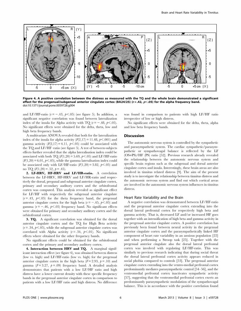
Whole Brain and DistressA correlation analysis between the distress as measured with the
TQ and the whole brain demonstrated a significant effect for the
pregenual/subgenual anterior cingulate cortex (r = .42, p,.05 (see
Figure 4) for the alpha frequency band. This correlation indicates
that increased activity in the anterior cingulate cortex goes
together with increased distress. No significant correlation could
be retrieved in delta, theta, low and high beta and gamma
frequency bands.
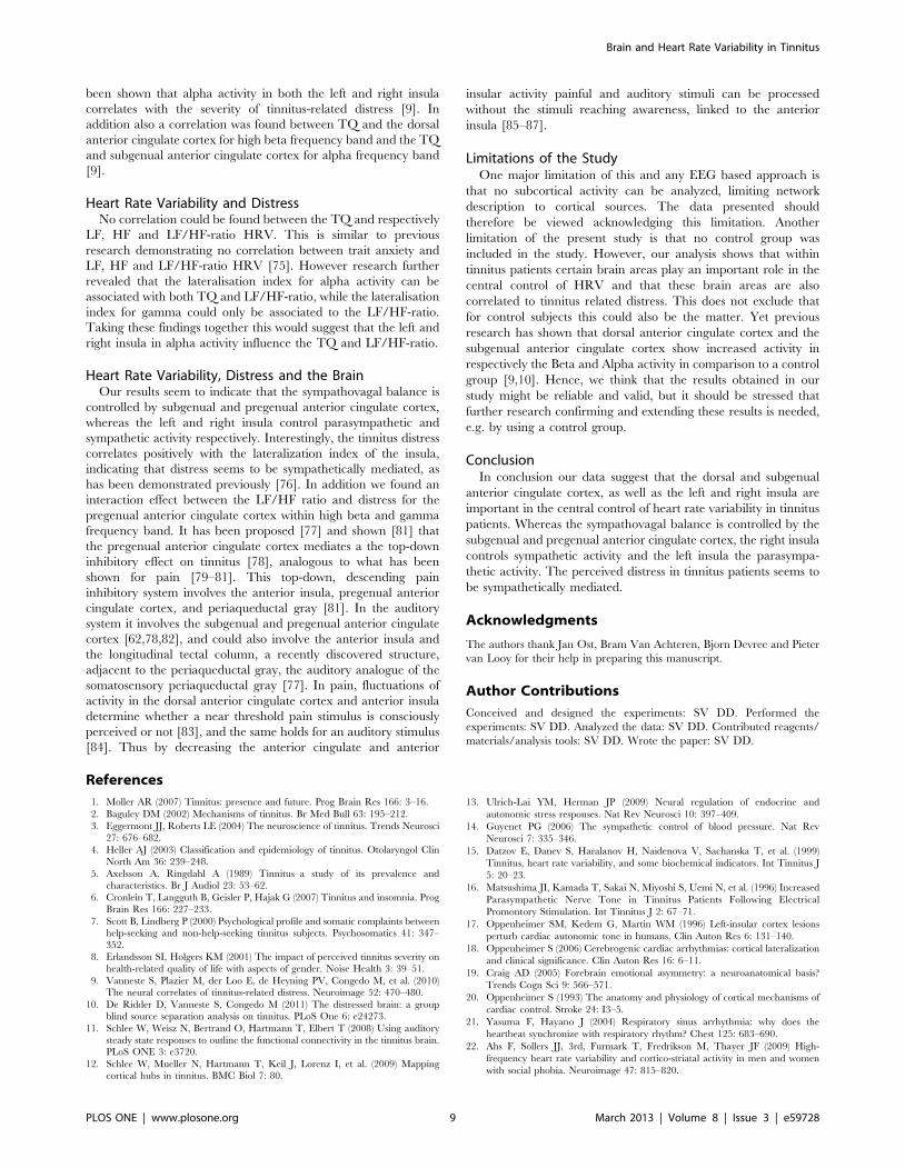
Region of Interest Analysis1. Lateralisation index. Significant negative correlations
were found between lateralization index of the insula for Alpha
activity and LF/HF-ratio (r = 2.45, p,.05) and Gamma activity
Figure 3. (a) Positive correlation between LF/HF-ratio (sympathetic/parasympathetic ratio) HRV and theta activity in the subgenualanterior cingulate cortex (BA25) (r = .43, p,.05); (b) & (c) Negative correlation between LF/HF-ratio HRV and High Beta (r = 2.45,p,.05) and Gamma activity (r = 2.46, p,.05) in the left pregenual anterior cingulate cortex (BA24) extending into dorsal lateralprefrontal cortex (BA9).doi:10.1371/journal.pone.0059728.g003
Brain and Heart Rate Variability in Tinnitus
PLOS ONE | www.plosone.org 5 March 2013 | Volume 8 | Issue 3 | e59728
and LF/HF-ratio (r = 2.43, p,.05) (see figure 5). In addition, a
significant negative correlation was found between lateralization
index of the insula for Alpha activity with TQ (r = 2.48, p,.05).
No significant effects were obtained for the delta, theta, low and
high beta frequency bands.
A multivariate ANOVA revealed that both for the lateralization
index of the insula for alpha activity (F(2,17) = 11.48, p,.001) and
gamma activity (F(2,17) = 4.11, p,.05) could be associated with
the TQ and LF/HF–ratio (see figure 5). A test of between-subjects
effects further revealed that the alpha lateralisation index could be
associated with both TQ (F(1,20) = 5.69, p,.05) and LF/HF-ratio
(F(1,20) = 6.01, p,.05), while the gamma lateralisation index could
be associated only with LF/HF-ratio (F(1,20) = 3.82, p,.05) and
no TQ (F(1,20) = .52, p = .45).
2. LF-HRV, HF-HRV and LF/HR-ratio. A correlation
between the LF-HRV, HF-HRV and LF/HR-ratio and respec-
tively the dorsal, pregunal and subgenual anterior cingulate cortex,
primary and secondary auditory cortex and the orbitofrontal
cortex was computed. This analysis revealed as significant effect
for LF/HF with respectively the subgenual anterior cingulate
(r = .43, p,.05) for the theta frequency band, the pregenual
anterior cingulate cortex for the high beta (r = 2.45, p,.05) and
gamma (r = 2.46, p,.05) frequency band. No significant effects
were obtained for primary and secondary auditory cortex and the
orbitofrontal cortex.
3. TQ. A significant correlation was obtained for the dorsal
anterior cingulate cortex and the TQ for High Beta activity
(r = .34, p,.05), while the subgenual anterior cingulate cortex was
correlated with Alpha activity (r = .34, p,.01). No significant
effects where obtained for the other frequency bands.
No significant effects could be obtained for the orbitofrontal
cortex and the primary and secondary auditory cortex.
4. Interaction between HRV and TQ. A marginal signif-
icant interaction effect (see figure 6), was obtained between distress
(low vs. high) and LF/HF-ratio (low vs. high) for the pregenual
anterior cingulate cortex in the high beta (F = 2.93, p = .10) and
gamma (F = 3.27, p = .09) frequency band. A detailed analysis
demonstrates that patients with a low LF/HF ratio and high
distress have a lower current density with these specific frequency
bands in the pregenual anterior cingulate cortex in comparison to
patients with a low LF/HF ratio and high distress. No difference
was found in comparison to patients with high LF/HF ratio
irrespective of low or high distress.
No significant effects were obtained for the delta, theta, alpha
and low beta frequency bands.
Discussion
The autonomic nervous system is controlled by the sympathetic
and parasympathetic system. The cardiac sympathetic/parasym-
pathetic or sympathovagal balance is reflected by the LF
(OS+PS)/HF (PS) ratio [52]. Previous research already revealed
the relationship between the autonomic nervous system and
specific brain regions such as the subgenual and dorsal anterior
cingulate cortex and insula. Interestingly, these brain areas are also
involved in tinnitus related distress [9]. The aim of the present
study is to investigate the relationship between tinnitus distress and
the autonomic nervous system and find out which cortical areas
are involved in the autonomic nervous system influences in tinnitus
distress.
Heart Rate Variability and the BrainA negative correlation was demonstrated between LF/HF-ratio
and the pregenual anterior cingulate cortex extending into the
dorsal lateral prefrontal cortex for respectively high beta and
gamma activity. That is, decreased LF and/or increased HF goes
together with an intensification of high beta and gamma activity in
the pregenual anterior cingulate cortex. A positive correlation has
previously been found between neural activity in the pregenual
anterior cingulate cortex and the parasympathetically linked HF
component of heart rate variability in an anxious population [22]
and when performing a Stroop task [25]. Together with the
pregenual anterior cingulate also the dorsal lateral prefrontal
cortex was involved with regulating LF/HF-ratio. This was
similarly to previous research indicating that during social threat
the dorsal lateral prefrontal cortex activity appears reduced in
social phobia compared to controls [53]. The pregenual anterior
cingulate cortex extending into the ventro-medial prefrontal cortex
predominantly mediates parasympathetic control [54–56], and the
ventromedial prefrontal cortex inactivates sympathetic activity
[57], suggesting that the ventromedial prefrontal cortex exerts an
predominantly parasympathetic modulation of the sympathovagal
balance. This is in accordance with the positive correlation found
Figure 4. A positive correlation between the distress as measured with the TQ and the whole brain demonstrated a significanteffect for the pregenual/subgenual anterior cingulate cortex (BA24/25) (r = .42, p,.05) for the alpha frequency band.doi:10.1371/journal.pone.0059728.g004
Brain and Heart Rate Variability in Tinnitus
PLOS ONE | www.plosone.org 6 March 2013 | Volume 8 | Issue 3 | e59728

Figure 5. Scatterplots and regression lines for respectively the lateralization index of the insula for Alpha and LF/HF-ratio.Significant negative correlation were found between lateralization index of the insula for Alpha activity and LF/HF-ratio (r = 2.45, p,.05) and Gammaactivity and LF/HF-ratio (r = 2.43, p,.05).doi:10.1371/journal.pone.0059728.g005
Brain and Heart Rate Variability in Tinnitus
PLOS ONE | www.plosone.org 7 March 2013 | Volume 8 | Issue 3 | e59728
between LF/HF-ratio and the subgenual anterior cingulate cortex
for theta activity, revealing that increased LF and decrease HF
goes together with an increase of theta activity in the subgenual
anterior cingulate cortex. A fMRI study also related HRV and
sympathetic cardiac influence with the subgenual anterior
cingulate cortex [58]. It has also been shown that increased
activity in posterior subgenual anterior cingulate cortex extending
into nucleus accumbens-ventral tegmental area is involved in
processing of aversive sounds [59] and unpleasant music [60] and
this area has been implicated in mediating limbic-autonomic
interactions in tinnitus as well [61,62]. This area in animals has
been considered a visceromotor cortex, due to its connections with
the parasympathetic nucleus tractus solitarius [63] and the
sympathetic areas in the periaquaductal grey [64]. Furthermore
it is functionally connected to the insula and anticorrelated to the
dorsal anterior cingulate cortex [65–67]. Dorsal anterior cingulate
activity covaries with blood pressure, emotional heart rate
changes, cardiac sympathetic tone and pupillary changes [68].
Our results further revealed that increased LF goes together
with a decrease in activity in the left insula. Furthermore tinnitus
distress correlates negatively with the lateralization index of the
insula for alpha, revealing that increased tinnitus distress is
associated with a decrease in the lateralization index in the insula.
These findings are in accordance with previous research revealing
that increased insular activity is associated with subjective
emotional and bodily awareness, as well as interoception [69].
The insula has been implicated in autonomic nervous system
control [20,68,70,71] and might therefore be related to the
autonomic components involved in distress [72,73], induced by
the phantom sound. In a recent study it was further revealed that
the insula is involved in pain sensitivity [74]. In addition, a region
of interest analysis revealed that LF/HF-ratio correlates negatively
with the lateralization index for alpha and gamma activity in the
insula. This latter result demonstrates that increased LF and(or)
decreased HF goes together with a decrease of activity in the left
insula and an increase of activity in the right insula.
Distress and the BrainTinnitus distress, as reflected by the TQ, correlates positively
with the lateralization index of the insula in alpha, indicating that
an increase in right insula and/or a decrease in left insula go
together with an increase in tinnitus related distress. It has already
Figure 6. A marginal significant interaction effect between distress (low vs. high) and LF/HF-ratio (low vs. high) for the pregenualanterior cingulate cortex in the high beta (F = 2.93, p = .10) and gamma (F = 3.27, p = .09) frequency band.doi:10.1371/journal.pone.0059728.g006
Brain and Heart Rate Variability in Tinnitus
PLOS ONE | www.plosone.org 8 March 2013 | Volume 8 | Issue 3 | e59728
been shown that alpha activity in both the left and right insula
correlates with the severity of tinnitus-related distress [9]. In
addition also a correlation was found between TQ and the dorsal
anterior cingulate cortex for high beta frequency band and the TQ
and subgenual anterior cingulate cortex for alpha frequency band
[9].
Heart Rate Variability and DistressNo correlation could be found between the TQ and respectively
LF, HF and LF/HF-ratio HRV. This is similar to previous
research demonstrating no correlation between trait anxiety and
LF, HF and LF/HF-ratio HRV [75]. However research further
revealed that the lateralisation index for alpha activity can be
associated with both TQ and LF/HF-ratio, while the lateralisation
index for gamma could only be associated to the LF/HF-ratio.
Taking these findings together this would suggest that the left and
right insula in alpha activity influence the TQ and LF/HF-ratio.
Heart Rate Variability, Distress and the BrainOur results seem to indicate that the sympathovagal balance is
controlled by subgenual and pregenual anterior cingulate cortex,
whereas the left and right insula control parasympathetic and
sympathetic activity respectively. Interestingly, the tinnitus distress
correlates positively with the lateralization index of the insula,
indicating that distress seems to be sympathetically mediated, as
has been demonstrated previously [76]. In addition we found an
interaction effect between the LF/HF ratio and distress for the
pregenual anterior cingulate cortex within high beta and gamma
frequency band. It has been proposed [77] and shown [81] that
the pregenual anterior cingulate cortex mediates a the top-down
inhibitory effect on tinnitus [78], analogous to what has been
shown for pain [79–81]. This top-down, descending pain
inhibitory system involves the anterior insula, pregenual anterior
cingulate cortex, and periaqueductal gray [81]. In the auditory
system it involves the subgenual and pregenual anterior cingulate
cortex [62,78,82], and could also involve the anterior insula and
the longitudinal tectal column, a recently discovered structure,
adjacent to the periaqueductal gray, the auditory analogue of the
somatosensory periaqueductal gray [77]. In pain, fluctuations of
activity in the dorsal anterior cingulate cortex and anterior insula
determine whether a near threshold pain stimulus is consciously
perceived or not [83], and the same holds for an auditory stimulus
[84]. Thus by decreasing the anterior cingulate and anterior
insular activity painful and auditory stimuli can be processed
without the stimuli reaching awareness, linked to the anterior
insula [85–87].
Limitations of the StudyOne major limitation of this and any EEG based approach is
that no subcortical activity can be analyzed, limiting network
description to cortical sources. The data presented should
therefore be viewed acknowledging this limitation. Another
limitation of the present study is that no control group was
included in the study. However, our analysis shows that within
tinnitus patients certain brain areas play an important role in the
central control of HRV and that these brain areas are also
correlated to tinnitus related distress. This does not exclude that
for control subjects this could also be the matter. Yet previous
research has shown that dorsal anterior cingulate cortex and the
subgenual anterior cingulate cortex show increased activity in
respectively the Beta and Alpha activity in comparison to a control
group [9,10]. Hence, we think that the results obtained in our
study might be reliable and valid, but it should be stressed that
further research confirming and extending these results is needed,
e.g. by using a control group.
ConclusionIn conclusion our data suggest that the dorsal and subgenual
anterior cingulate cortex, as well as the left and right insula are
important in the central control of heart rate variability in tinnitus
patients. Whereas the sympathovagal balance is controlled by the
subgenual and pregenual anterior cingulate cortex, the right insula
controls sympathetic activity and the left insula the parasympa-
thetic activity. The perceived distress in tinnitus patients seems to
be sympathetically mediated.
Acknowledgments
The authors thank Jan Ost, Bram Van Achteren, Bjorn Devree and Pieter
van Looy for their help in preparing this manuscript.
Author Contributions
Conceived and designed the experiments: SV DD. Performed the
experiments: SV DD. Analyzed the data: SV DD. Contributed reagents/
materials/analysis tools: SV DD. Wrote the paper: SV DD.
References
1. Moller AR (2007) Tinnitus: presence and future. Prog Brain Res 166: 3–16.
2. Baguley DM (2002) Mechanisms of tinnitus. Br Med Bull 63: 195–212.
3. Eggermont JJ, Roberts LE (2004) The neuroscience of tinnitus. Trends Neurosci27: 676–682.
4. Heller AJ (2003) Classification and epidemiology of tinnitus. Otolaryngol Clin
North Am 36: 239–248.
5. Axelsson A, Ringdahl A (1989) Tinnitus–a study of its prevalence and
characteristics. Br J Audiol 23: 53–62.
6. Cronlein T, Langguth B, Geisler P, Hajak G (2007) Tinnitus and insomnia. Prog
Brain Res 166: 227–233.
7. Scott B, Lindberg P (2000) Psychological profile and somatic complaints betweenhelp-seeking and non-help-seeking tinnitus subjects. Psychosomatics 41: 347–
352.
8. Erlandsson SI, Holgers KM (2001) The impact of perceived tinnitus severity on
health-related quality of life with aspects of gender. Noise Health 3: 39–51.
9. Vanneste S, Plazier M, der Loo E, de Heyning PV, Congedo M, et al. (2010)The neural correlates of tinnitus-related distress. Neuroimage 52: 470–480.
10. De Ridder D, Vanneste S, Congedo M (2011) The distressed brain: a groupblind source separation analysis on tinnitus. PLoS One 6: e24273.
11. Schlee W, Weisz N, Bertrand O, Hartmann T, Elbert T (2008) Using auditory
steady state responses to outline the functional connectivity in the tinnitus brain.PLoS ONE 3: e3720.
12. Schlee W, Mueller N, Hartmann T, Keil J, Lorenz I, et al. (2009) Mappingcortical hubs in tinnitus. BMC Biol 7: 80.
13. Ulrich-Lai YM, Herman JP (2009) Neural regulation of endocrine and
autonomic stress responses. Nat Rev Neurosci 10: 397–409.
14. Guyenet PG (2006) The sympathetic control of blood pressure. Nat Rev
Neurosci 7: 335–346.
15. Datzov E, Danev S, Haralanov H, Naidenova V, Sachanska T, et al. (1999)
Tinnitus, heart rate variability, and some biochemical indicators. Int Tinnitus J
5: 20–23.
16. Matsushima JI, Kamada T, Sakai N, Miyoshi S, Uemi N, et al. (1996) Increased
Parasympathetic Nerve Tone in Tinnitus Patients Following Electrical
Promontory Stimulation. Int Tinnitus J 2: 67–71.
17. Oppenheimer SM, Kedem G, Martin WM (1996) Left-insular cortex lesions
perturb cardiac autonomic tone in humans. Clin Auton Res 6: 131–140.
18. Oppenheimer S (2006) Cerebrogenic cardiac arrhythmias: cortical lateralization
and clinical significance. Clin Auton Res 16: 6–11.
19. Craig AD (2005) Forebrain emotional asymmetry: a neuroanatomical basis?
Trends Cogn Sci 9: 566–571.
20. Oppenheimer S (1993) The anatomy and physiology of cortical mechanisms of
cardiac control. Stroke 24: I3–5.
21. Yasuma F, Hayano J (2004) Respiratory sinus arrhythmia: why does the
heartbeat synchronize with respiratory rhythm? Chest 125: 683–690.
22. Ahs F, Sollers JJ, 3rd, Furmark T, Fredrikson M, Thayer JF (2009) High-
frequency heart rate variability and cortico-striatal activity in men and women
with social phobia. Neuroimage 47: 815–820.
Brain and Heart Rate Variability in Tinnitus
PLOS ONE | www.plosone.org 9 March 2013 | Volume 8 | Issue 3 | e59728
23. Lane RD, McRae K, Reiman EM, Chen K, Ahern GL, et al. (2009) Neural
correlates of heart rate variability during emotion. Neuroimage 44: 213–222.
24. Napadow V, Dhond R, Conti G, Makris N, Brown EN, et al. (2008) Brain
correlates of autonomic modulation: combining heart rate variability with fMRI.
Neuroimage 42: 169–177.
25. Matthews SC, Paulus MP, Simmons AN, Nelesen RA, Dimsdale JE (2004)
Functional subdivisions within anterior cingulate cortex and their relationship to
autonomic nervous system function. Neuroimage 22: 1151–1156.
26. Suzuki H, Watanabe S, Hamaguchi T, Mine H, Terui T, et al. (2009) Brain
activation associated with changes in heart rate, heart rate variability, and
plasma catecholamines during rectal distention. Psychosom Med 71: 619–626.
27. Babiloni C, Binetti G, Cassarino A, Dal Forno G, Del Percio C, et al. (2006)
Sources of cortical rhythms in adults during physiological aging: a multicentric
EEG study. Hum Brain Mapp 27: 162–172.
28. Pascual-Marqui RD (2002) Standardized low-resolution brain electromagnetic
tomography (sLORETA): technical details. Methods Find Exp Clin Pharmacol
24 Suppl D: 5–12.
29. Moazami-Goudarzi M, Michels L, Weisz N, Jeanmonod D (2010) Temporo-
insular enhancement of EEG low and high frequencies in patients with chronic
tinnitus. QEEG study of chronic tinnitus patients. BMC Neurosci 11: 40.
30. Meeus O, Blaivie C, Van de Heyning P (2007) Validation of the Dutch and the
French version of the Tinnitus Questionnaire. B-ENT 3 Suppl 7: 11–17.
31. Goebel G, Hiller W (1994) [The tinnitus questionnaire. A standard instrument
for grading the degree of tinnitus. Results of a multicenter study with the tinnitus
questionnaire]. HNO 42: 166–172.
32. Vanneste S, Plazier M, van der Loo E, Ost J, Meeus O, et al. (2010) Validation
of the Mini-TQ in a Dutch-speaking population. A rapid assessment for tinnitus-
related distress. B-ENT.
33. van der Loo E, Congedo M, Vanneste S, De Heyning PV, De Ridder D (2011)
Insular lateralization in tinnitus distress. Auton Neurosci.
34. Congedo M (2002) EureKa! (Version 3.0) [Computer Software]. Knoxville, TN:
NovaTech EEG Inc. Freeware available at www.NovaTechEEG. Accessed
2013 Feb 26.
35. Pascual-Marqui RD, Esslen M, Kochi K, Lehmann D (2002) Functional
imaging with low-resolution brain electromagnetic tomography (LORETA): a
review. Methods Find Exp Clin Pharmacol 24 Suppl C: 91–95.
36. Fuchs M, Kastner J, Wagner M, Hawes S, Ebersole JS (2002) A standardized
boundary element method volume conductor model. Clin Neurophysiol 113:
702–712.
37. Mulert C, Jager L, Schmitt R, Bussfeld P, Pogarell O, et al. (2004) Integration of
fMRI and simultaneous EEG: towards a comprehensive understanding of
localization and time-course of brain activity in target detection. Neuroimage 22:
83–94.
38. Vitacco D, Brandeis D, Pascual-Marqui R, Martin E (2002) Correspondence of
event-related potential tomography and functional magnetic resonance imaging
during language processing. Hum Brain Mapp 17: 4–12.
39. Worrell GA, Lagerlund TD, Sharbrough FW, Brinkmann BH, Busacker NE, et
al. (2000) Localization of the epileptic focus by low-resolution electromagnetic
tomography in patients with a lesion demonstrated by MRI. Brain Topogr 12:
273–282.
40. Dierks T, Jelic V, Pascual-Marqui RD, Wahlund L, Julin P, et al. (2000) Spatial
pattern of cerebral glucose metabolism (PET) correlates with localization of
intracerebral EEG-generators in Alzheimer’s disease. Clin Neurophysiol 111:
1817–1824.
41. Pizzagalli DA, Oakes TR, Fox AS, Chung MK, Larson CL, et al. (2004)
Functional but not structural subgenual prefrontal cortex abnormalities in
melancholia. Mol Psychiatry 9: 325, 393–405.
42. Zumsteg D, Wennberg RA, Treyer V, Buck A, Wieser HG (2005) H2(15)O or
13NH3 PET and electromagnetic tomography (LORETA) during partial status
epilepticus. Neurology 65: 1657–1660.
43. Zumsteg D, Lozano AM, Wennberg RA (2006) Depth electrode recorded
cerebral responses with deep brain stimulation of the anterior thalamus for
epilepsy. Clin Neurophysiol 117: 1602–1609.
44. Zumsteg D, Lozano AM, Wieser HG, Wennberg RA (2006) Cortical activation
with deep brain stimulation of the anterior thalamus for epilepsy. Clin
Neurophysiol 117: 192–207.
45. Volpe U, Mucci A, Bucci P, Merlotti E, Galderisi S, et al. (2007) The cortical
generators of P3a and P3b: a LORETA study. Brain Res Bull 73: 220–230.
46. Pizzagalli D, Pascual-Marqui RD, Nitschke JB, Oakes TR, Larson CL, et al.
(2001) Anterior cingulate activity as a predictor of degree of treatment response
in major depression: evidence from brain electrical tomography analysis.
Am J Psychiatry 158: 405–415.
47. Zumsteg D, Lozano AM, Wennberg RA (2006) Mesial temporal inhibition in a
patient with deep brain stimulation of the anterior thalamus for epilepsy.
Epilepsia 47: 1958–1962.
48. Weisz N, Moratti S, Meinzer M, Dohrmann K, Elbert T (2005) Tinnitus
perception and distress is related to abnormal spontaneous brain activity as
measured by magnetoencephalography. PLoS Med 2: e153.
49. Weisz N, Muller S, Schlee W, Dohrmann K, Hartmann T, et al. (2007) The
neural code of auditory phantom perception. J Neurosci 27: 1479–1484.
50. Nichols TE, Holmes AP (2002) Nonparametric permutation tests for functional
neuroimaging: a primer with examples. Hum Brain Mapp 15: 1–25.
51. Schlee W, Leirer V, Kolassa IT, Weisz N, Elbert T (2012) Age-related changesin neural functional connectivity and its behavioral relevance. BMC Neurosci
13: 16.
52. Kamath MV, Fallen EL (1993) Power spectral analysis of heart rate variability: a
noninvasive signature of cardiac autonomic function. Crit Rev Biomed Eng 21:245–311.
53. Goldin PR, Manber T, Hakimi S, Canli T, Gross JJ (2009) Neural bases of social
anxiety disorder: emotional reactivity and cognitive regulation during social andphysical threat. Arch Gen Psychiatry 66: 170–180.
54. Wong SW, Masse N, Kimmerly DS, Menon RS, Shoemaker JK (2007) Ventral
medial prefrontal cortex and cardiovagal control in conscious humans.Neuroimage 35: 698–708.
55. Hansel A, von Kanel R (2008) The ventro-medial prefrontal cortex: a major link
between the autonomic nervous system, regulation of emotion, and stressreactivity? Biopsychosoc Med 2: 21.
56. Nicotra A, Critchley HD, Mathias CJ, Dolan RJ (2006) Emotional and
autonomic consequences of spinal cord injury explored using functional brainimaging. Brain 129: 718–728.
57. Verberne AJ, Owens NC (1998) Cortical modulation of the cardiovascular
system. Prog Neurobiol 54: 149–168.
58. Critchley HD, Mathias CJ, Josephs O, O’Doherty J, Zanini S, et al. (2003)
Human cingulate cortex and autonomic control: converging neuroimaging and
clinical evidence. Brain 126: 2139–2152.
59. Zald DH, Pardo JV (2002) The neural correlates of aversive auditory
stimulation. Neuroimage 16: 746–753.
60. Blood AJ, Zatorre RJ, Bermudez P, Evans AC (1999) Emotional responses to
pleasant and unpleasant music correlate with activity in paralimbic brainregions. Nat Neurosci 2: 382–387.
61. Muhlau M, Rauschecker JP, Oestreicher E, Gaser C, Rottinger M, et al. (2006)
Structural brain changes in tinnitus. Cereb Cortex 16: 1283–1288.
62. Rauschecker JP, leaver AM, Muhlau M (2010) Tuning Out the Noise: Limbic-
Auditory Interactions in Tinnitus. Neuron 66: 819–826.
63. Frysztak RJ, Neafsey EJ (1994) The effect of medial frontal cortex lesions oncardiovascular conditioned emotional responses in the rat. Brain Res 643: 181–
193.
64. Ongur D, Price JL (2000) The organization of networks within the orbital andmedial prefrontal cortex of rats, monkeys and humans. Cereb Cortex 10: 206–
219.
65. Stein JL, Wiedholz LM, Bassett DS, Weinberger DR, Zink CF, et al. (2007) Avalidated network of effective amygdala connectivity. Neuroimage 36: 736–745.
66. Margulies DS, Kelly AM, Uddin LQ, Biswal BB, Castellanos FX, et al. (2007)
Mapping the functional connectivity of anterior cingulate cortex. Neuroimage37: 579–588.
67. Kahn I, Andrews-Hanna JR, Vincent JL, Snyder AZ, Buckner RL (2008)
Distinct cortical anatomy linked to subregions of the medial temporal loberevealed by intrinsic functional connectivity. J Neurophysiol 100: 129–139.
68. Critchley HD (2005) Neural mechanisms of autonomic, affective, and cognitive
integration. J Comp Neurol 493: 154–166.
69. Craig AD (2003) Interoception: the sense of the physiological condition of the
body. Curr Opin Neurobiol 13: 500–505.
70. Oppenheimer SM, Gelb A, Girvin JP, Hachinski VC (1992) Cardiovasculareffects of human insular cortex stimulation. Neurology 42: 1727–1732.
71. Critchley HD, Wiens S, Rotshtein P, Ohman A, Dolan RJ (2004) Neural systems
supporting interoceptive awareness. Nat Neurosci 7: 189–195.
72. Wang J, Rao H, Wetmore GS, Furlan PM, Korczykowski M, et al. (2005)
Perfusion functional MRI reveals cerebral blood flow pattern under psycholog-
ical stress. Proc Natl Acad Sci U S A 102: 17804–17809.
73. Critchley HD, Corfield DR, Chandler MP, Mathias CJ, Dolan RJ (2000)
Cerebral correlates of autonomic cardiovascular arousal: a functional neuroim-
aging investigation in humans. J Physiol 523 Pt 1: 259–270.
74. Baliki MN, Geha PY, Apkarian AV (2009) Parsing pain perception betweennociceptive representation and magnitude estimation. J Neurophysiol 101: 875–
887.
75. Tolkunov D, Rubin D, Mujica-Parodi L (2010) Power spectrum scale invariancequantifies limbic dysregulation in trait anxious adults using fMRI: adapting
methods optimized for characterizing autonomic dysregulation to neural
dynamic time series. Neuroimage 50: 72–80.
76. van der Loo E, Congedo M, Vanneste S, De Heyning PV, De Ridder D (2011)
Insular lateralization in tinnitus distress. Auton Neurosci 165: 191–194.
77. De Ridder D, Vanneste S, Menovsky T, Langguth B (2012) Surgical brainmodulation for tinnitus: the past, present and future. J Neurosurg Sci 56: 323–
340.
78. Vanneste S, De Ridder D (2011) Bifrontal transcranial direct current stimulationmodulates tinnitus intensity and tinnitus-distress-related brain activity.
Eur J Neurosci 34: 605–614.
79. Kong J, Loggia ML, Zyloney C, Tu P, Laviolette P, et al. (2010) Exploring the
brain in pain: activations, deactivations and their relation. Pain 148: 257–267.
80. Bingel U, Tracey I (2008) Imaging CNS modulation of pain in humans.
Physiology (Bethesda) 23: 371–380.
81. Fields H (2004) State-dependent opioid control of pain. Nat Rev Neurosci 5:
565–575.
82. Leaver AM, Renier L, Chevillet MA, Morgan S, Kim HJ, et al. (2011)
Dysregulation of limbic and auditory networks in tinnitus. Neuron 69: 33–43.
Brain and Heart Rate Variability in Tinnitus
PLOS ONE | www.plosone.org 10 March 2013 | Volume 8 | Issue 3 | e59728
83. Boly M, Balteau E, Schnakers C, Degueldre C, Moonen G, et al. (2007) Baseline
brain activity fluctuations predict somatosensory perception in humans. Proc
Natl Acad Sci U S A 104: 12187–12192.
84. Sadaghiani S, Hesselmann G, Kleinschmidt A (2009) Distributed and
antagonistic contributions of ongoing activity fluctuations to auditory stimulus
detection. J Neurosci 29: 13410–13417.
85. Craig AD (2002) How do you feel? Interoception: the sense of the physiological
condition of the body. Nat Rev Neurosci 3: 655–666.86. Bamiou DE, Musiek FE, Luxon LM (2003) The insula (Island of Reil) and its
role in auditory processing. Literature review. Brain Res Brain Res Rev 42: 143–
154.87. Fifer RC (1993) Insular stroke causing unilateral auditory processing disorder:
case report. J Am Acad Audiol 4: 364–369.
Brain and Heart Rate Variability in Tinnitus
PLOS ONE | www.plosone.org 11 March 2013 | Volume 8 | Issue 3 | e59728