tinnitus and temporomandibular joint disorder
TRANSCRIPT
1
TINNITUS AND TEMPOROMANDIBULAR JOINT DISORDER
SUBTYPES
SUSEE PRIYANKA RAVURI
A thesis
Submitted in partial fulfillment of the
requirements for the degree of
MASTER OF SCIENCE IN DENTISTRY
University of Washington
2017
Committee
Edmond L. Truelove
Peggy Lee
Lloyd A. Mancl
Program Authorized to Offer Degree:
Oral Medicine

2
© Copyright 2017
Susee Priyanka Ravuri
3
University of Washington
ABSTRACT
Tinnitus And Temporomandibular Joint Disorder Subtypes
Susee Priyanka Ravuri
Edmond L. Truelove B.S., D.D.S., M.S.D.
Oral Medicine
OBJECTIVE: The purpose of this study was to assess the prevalence of tinnitus within a TMD
population and to determine an association between the presence of tinnitus and type of TMD
diagnoses.
METHODS: A secondary data analysis was performed using data from ‘Research Diagnostic
Criteria for Temporomandibular Disorders (RDC/TMD) baseline (Validation project) study and
follow up (Impact project) study. Self-reported questionnaires for reporting tinnitus and medical
history and gold standard diagnoses after clinical examination were used. Log-binomial
regression was used to compute risk ratios for tinnitus by TMD subtype and adjusted for patient
characteristics. All statistical analysis was performed using SAS 9.3 software (SAS Institute),
and a two-sided significance level of 0.05 to determined statistical significance (p<0.05).
RESULTS: At baseline, 614 subjects met required criteria for TMD diagnosis. Prevalence of
tinnitus within sample was 41% (253 of 614). Approximately 80% of TMD subjects received a
MPD diagnosis. Tinnitus frequency in the MPD group was 48% (238/495) while subjects
without MPD diagnosis the rate of tinnitus was 13% (15 of 119). Using log-binomial regression
analysis, the risk ratio for tinnitus was calculated. The relative risk for tinnitus by number of sites
4
painful to palpation by TMD diagnosis for MPD, DD and DJD groups was 1.03 (95% CI: 0.97,
1.10; p=0.28), 1.24 (95% CI: 1.05, 1.46; p = .0086) and 1.20 (95% CI: 1.01, 1.43; p = .033),
respectively, when adjusted for age, gender, study site and somatization. Among the population,
207 subjects received a TMD diagnosis also reported headaches. The adjusted risk ratio for
tinnitus among subjects with TMD diagnosis and headache was 4.52 (95% CI: 1.67, 12.19; p =
.0002) higher than in subjects with only TMD diagnosis (RR=3.8) or only headaches (RR=1.4)
Similarly, the adjusted risk ratio for tinnitus among subjects with an MPD diagnosis and
headaches was 3.59 (95%CI: 1.82, 7.06) higher than in subjects with only MPD diagnosis
(RR=3.02) and only headaches (RR= 1.75)
CONCLUSION: These findings suggest higher rate of tinnitus among subjects with MPD than
other forms of TMD. Moreover, the risk for tinnitus is six times higher if subject has TMD
diagnosis and headache and three times higher in myofascial group with headaches.

5
DEDICATIONS
I dedicate my thesis to my Mother and my Brother.
6
ACKNOWLEDGEMENTS
I would like to express my sincere gratitude to my advisors Dr. Edmond L. Truelove, Dr.
Lloyd A.Mancl and Dr. Peggy Lee for giving me the opportunity to be a part of their team and
this project and for their encouragement to learn and to provide meaningful results. Their
knowledge and suggestions were fundamental throughout this work.
I would also like to thank my research group members, Dr. Linda LeResche and Dr. Lisa J.
Heaton for helping me with the research project and providing me a great source of support and
inspiration.
Thanks to the Department of Oral Medicine, University of Washington for overall support.
7
TABLE OF CONTENTS
ABSTRACT……………………………………………………………………………….……4
DEDICATION ……………………………………………………………………………........6
ACKNOWLEDGEMENTS…………………………………………………………………….7
TABLE OF CONTENTS
LIST OF FIGURES...………………………………………………………………………….10
LIST OF TABLES..…………………………………………………………………………....11
CHAPTER
I. INTRODUCTION
1.1 Research Objectives ……..……………………………………………......12
1.2 Problem Description …………….………………………………………..12
1.3 Structure of Thesis………………………….……………………………..13
II. TEMPOROMANDIBULAR JOINT DISORDERS
2.1 Definitions…………………………………………………..……………..15
2.2 Epidemiology ……………………………………………………………..18
2.3 Etiology And Risk Factors ………………………………………………..18
2.4 Treatments And Management……………………………………………..19
III. TINNITUS
3.1 Definitions…………………………………………………………………20
3.2 Epidemiology ……………………………………………………………..22
3.3 Etiology And Risk Factors ………………………………………………..22
3.4 Treatments And Management……………………………………………..23
IV. TINNITUS AND TEMPOROMANDIBULAR JOINT DISORDER CO-EXISTENCE
4.1 Casual Concepts …………………………………………….…………….24
4.2 Other Concepts…………………………………………………………….27
V. METHODS AND MATERIAL
8
5.1 Study Design ……………………………………………......…………….29
5.2 Study Population ………………………………………………………….29
5.3 Study Data And Measures…………………………………………………30
5.4 Inclusion Criteria…………………………………………………………..30
5.5 Exclusion Criteria …………………………………………………………32
5.6 Data Collection ……………………………………………………………33
5.7 Statistical Analysis ………………………………………………………..34
VI. RESULTS
6.1 Tinnitus In Baseline Study…………………………….…………………..35
6.2 Tinnitus Among Temporomandibular Disorder Subtypes At Baseline …..37
6.3 Attributed Risk Factors
a. Painful Site On Palpation…………………….…………..………….39
b. Headaches …………………………………………………………..41
c. Oral Habits ………………………………………………………….42
d. Anxiety, Depression, Characteristic Pain Intensity and Interference At
Baseline………………………………………………………………...43
e. Jaw Movements….…………………………………………………..45
6.4 Tinnitus In Follow Up Study ………………………………………………47
6.5 Tinnitus Among Temporomandibular Disorder Subtypes At Follow Up….47
6.6 Characteristic Pain Intensity, Disability Days And Interference With Daily
Activity At Follow Up………………………………………………………….51
VII. CONCLUSION AND RECOMMENDATIONS ……………………………………53
VIII. SUMMARY………………………………………………………………………….57
NOMENCLATURE …………………………………………………………………………...58
REFERENCES…………………………………………………………………………………59
VITA……………………………………………………………………………………………62
9
LIST OF FIGURES
1. Temporomandibular Joint Sagittal Schematic (Int J Oral Maxillofac Implants. 2013)
2. Temporomandibular Joint Disorders Classification (DC/TMD)
3. Masticatory Muscle Disorders Classification (DC/TMD)
4. Anatomy Of The Ear (Pearson Cummings 2006)
5. Proximity Of TMJ To Ear (Mayo 2015)
6. Neuromuscular Theory Flowchart Representation
7. Somatosensorial Theory Flowchart Representation
8. Anatomic Theory Flowchart Representation
9. Anatomical And Functional Aspects Of Ligaments Between The Malleus And The
Temporomandibular Joint
10. TMD Subtype Distribution
11. Age (In Years) Distribution In Baseline TMD Population
12. Mean Number Of Painful Sites To Palpation In TMD Subtypes
13. Comparing Tinnitus At Baseline And Follow Up
14. Comparing Tinnitus Between MPD And Non-MPD Groups
10
LIST OF TABLES
1. Gender And Age Distribution At Baseline
2. Rate Of Tinnitus In TMD Subtypes
3. Tinnitus By Type Of TMD Diagnosis
4. Risk Ratios For Tinnitus By Number Of Sites Painful To Palpation At Baseline
5. Tinnitus By TMD Diagnosis And Headaches TMD Subjects
6. Tinnitus By TMD Diagnosis And Headaches MPD Subjects
7. Mean Values Of Oral Habits In Baseline Subjects
8. Anxiety (SCL-90) In Baseline TMD Subjects
9. Depression (SCL-90) In Baseline TMD Subjects
10. Characteristic Pain Intensity In Baseline TMD Subjects
11. Interference In Baseline TMD Subjects
12. Disability Days In Baseline TMD Subjects
13. Jaw Movements And Tinnitus By TMD And MPD Diagnosis
14. TMD And Tinnitus At Follow-Up
15. TMD And Tinnitus At Baseline And Follow-Up
16. Tinnitus Among MPD Subjects From Baseline To Follow Up
17. Comparing Change In Tinnitus Among MPD Subjects From Baseline To Follow Up
18. Characteristic Pain Intensity (CPI) In Follow-Up Subjects
19. Interference In Follow-Up TMD Subjects
20. Disability Days In Follow-Up TMD Subjects
11
CHAPTER 1
INTRODUCTION
1.1 RESEARCH OBJECTIVES
The main objective of this study is to assess the prevalence of tinnitus in patients with
temporomandibular jaw disorder and to analyze if prevalence of tinnitus varies among
temporomandibular jaw disorder subtypes. The study also focuses on identifying potential risk
factors among this population that contribute to tinnitus. After extensive literature review, the
study aims to provide an insight to one of the various theories discussed in relation to tinnitus in
temporomandibular joint disorders. This study hypothesizes that subjects with myofascial pain
type TMD diagnosis are more likely to report tinnitus than Disk displacement type TMD or
DJD/arthralgia type TMD. Clinically, this study examines the potential value of screening for
TMD and MPD in patients with tinnitus of unknown etiology.
1.2 PROBLEM DESCRIPTION
Although there is vast literature available related to the clinical problem of tinnitus, little is
known about the relationship of this condition in patients who suffer from both tinnitus
and Temporomandibular disorders. Moreover, in the past, research exploring the
relationship between TMD and tinnitus has focused on TMD as whole and rarely TMD
subtypes. Of the several theories proposed about the relationship of tinnitus and TMD, most of
them are confined to association between the tinnitus and TMD as whole but not the subtypes.
Our study aims to provide a better understanding of the association between tinnitus and
TMD, its subtypes and other patient reported factors.
12
1.3 STRUCTURE OF THESIS
Chapter one introduces the research idea and the problem concerning. Chapter two and three will
elaborate on temporomandibular joint disorders and tinnitus. These chapters will discuss key
points concerning TMD and Tinnitus causes, types, signs and symptoms and management
protocols. Chapter four will discuss the co-occurrence of tinnitus and TMD and the literature
available so far on the association between the two. Chapter five describes the methods and
methodology used to gather date performs analysis. Chapter six presents the results of this study
and finally Chapter seven will discuss the conclusions and recommendations from this study.
13
CHAPTER TWO
TEMPOROMANDIBULAR JOINT DISORDERS
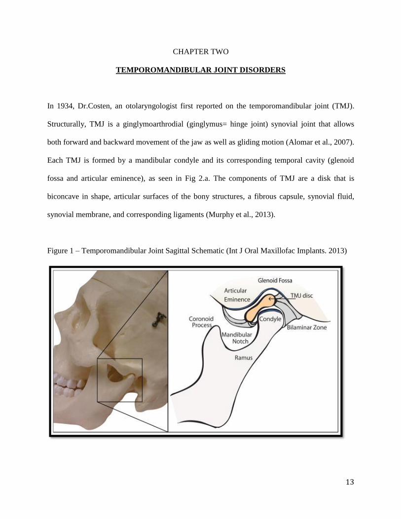
In 1934, Dr.Costen, an otolaryngologist first reported on the temporomandibular joint (TMJ).
Structurally, TMJ is a ginglymoarthrodial (ginglymus= hinge joint) synovial joint that allows
both forward and backward movement of the jaw as well as gliding motion (Alomar et al., 2007).
Each TMJ is formed by a mandibular condyle and its corresponding temporal cavity (glenoid
fossa and articular eminence), as seen in Fig 2.a. The components of TMJ are a disk that is
biconcave in shape, articular surfaces of the bony structures, a fibrous capsule, synovial fluid,
synovial membrane, and corresponding ligaments (Murphy et al., 2013).
Figure 1 – Temporomandibular Joint Sagittal Schematic (Int J Oral Maxillofac Implants. 2013)
14
2.1 DEFINITION
In the past century, knowledge relating to disorders of the temporomandibular joint was limited
to testimonials and clinical estimations rather than on scientific research studied. It was only in
1975, the American Academy of Craniomandibular Disorders was founded to recommend the
need for a scientific approach to temporomandibular joint disorders (TMD) (McNeill et al.,
1980). According to Schiffman et al., 2014, temporomandibular disorders (TMDs) is an umbrella
term for clinical conditions affecting the muscles of mastication, the temporomandibular joint
(TMJ), and the related structures. Also known as temporomandibular joint dysfunction (TMJD),
TMD refers to a wide range of clinical pathologies affecting the muscles of mastication and the
TMJ. In 2011, the Research Diagnostic Criteria for Temporomandibular disorders (RDC/TMD)
was established that included clinical examinations (under Axis I) and behavioral profile
assessment (under Axis II) of patients suffering from TMD. Accordingly, TMD Axis I included
myofascial pain disorder (with or without limited opening), disc displacement (with or without
reduction and with or without limited opening), and arthralgia (arthritis and arthrosis). Axis II
included assessment of behavioral, psychological and social factors (Dworkin et al., 2002).
Diagnostically, TMD was divided into three groups and into further subdivisions. The groups
are,
1. Group I Muscle Disorders:
a. Myofascial pain
b. Myofascial pain with limited opening
2. Group II Disc Displacements:
a. Disc displacement with reduction;
b. Disc displacement without reduction with limited opening
15
c. Disc displacement without reduction without limited opening.
3. Group III Arthralgia, Arthritis, Arthrosis:
a. Arthralgia
b. Osteoarthritis
c. Osteoarthrosis.
In 2014, Schiffman et al., 2014 recommended a revised version of RDC/TMD, called the
Diagnostic criteria for temporomandibular disorders (DC/TMD). According to DC/TMD, TMD
is broadly classified into four groups. The following flow chart represents classification
according to DC/TMD.
1. Temporomandibular Joint Disorders
2. Masticatory Muscle Disorders
3. Headaches attributed to TMD
4. Associated structures -Coronoid hyperplasia
16
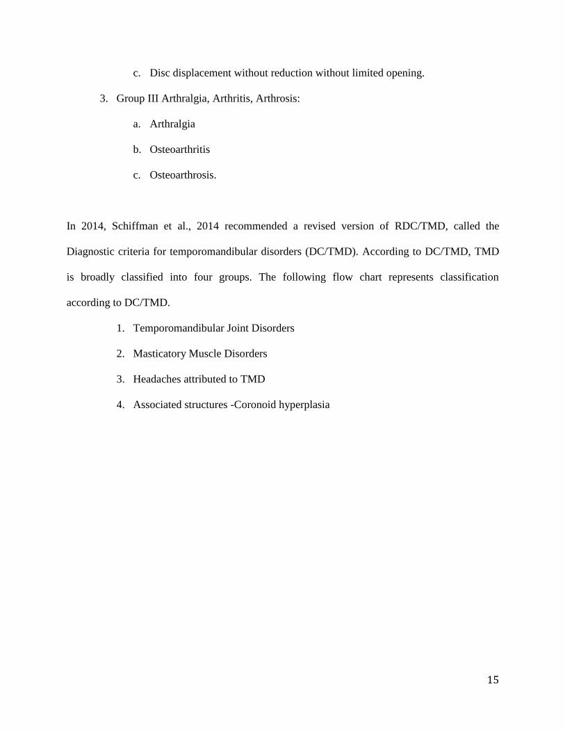
Figure 2- Temporomandibular Joint Disorders Classification (DC/TMD)
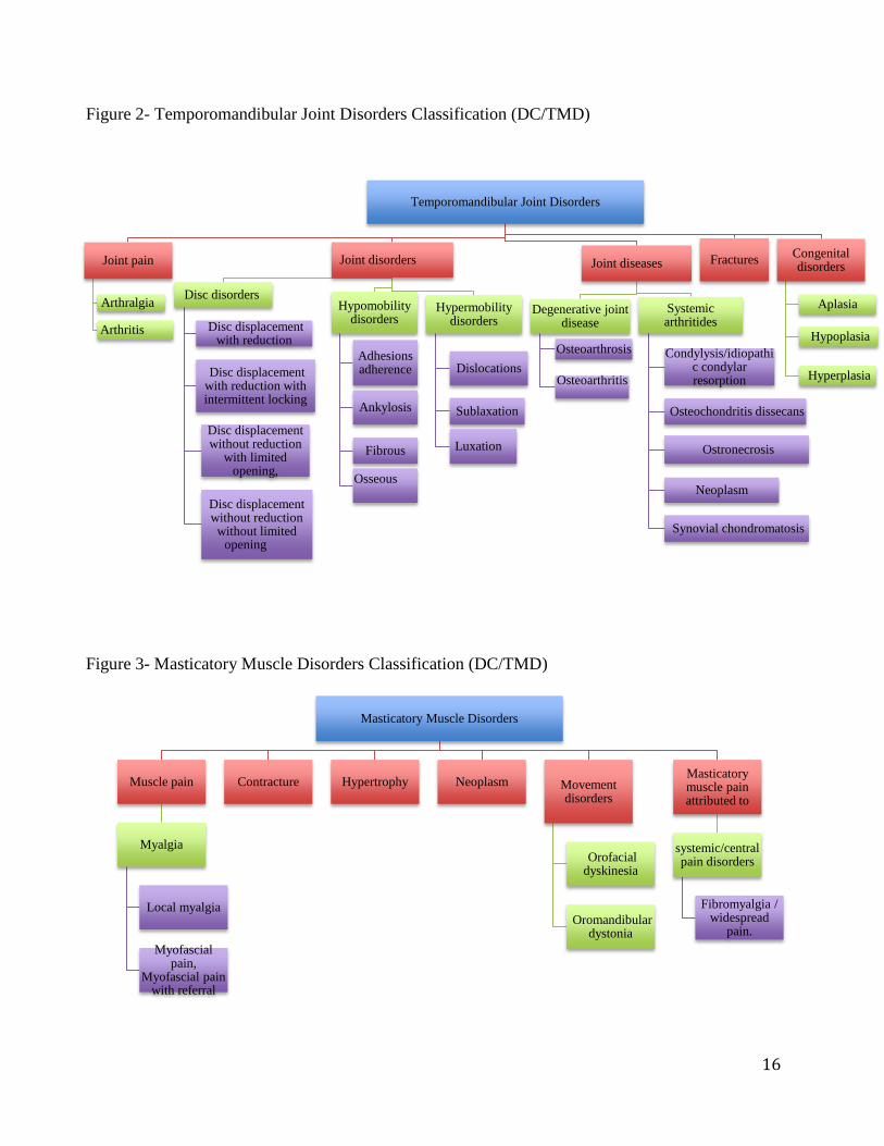
Figure 3- Masticatory Muscle Disorders Classification (DC/TMD)
Masticatory Muscle Disorders
Muscle pain
Myalgia
Local myalgia
Myofascial pain,
Myofascial pain with referral
Contracture Hypertrophy Neoplasm Movement disorders
Orofacial dyskinesia
Oromandibular dystonia
Masticatory muscle pain attributed to
systemic/central pain disorders
Fibromyalgia / widespread
pain.
Temporomandibular Joint Disorders
Joint pain
Arthralgia
Arthritis
Joint disorders
Disc disorders
Disc displacement with reduction
Disc displacement with reduction with intermittent locking
Disc displacement without reduction
with limited opening,
Disc displacement without reduction without limited
opening
Hypomobility disorders
Adhesions adherence
Ankylosis
Fibrous
Osseous
Hypermobility disorders
Dislocations
Sublaxation
Luxation
Joint diseases
Degenerative joint disease
Osteoarthrosis
Osteoarthritis
Systemic arthritides
Condylysis/idiopathic condylar resorption
Osteochondritis dissecans
Ostronecrosis
Neoplasm
Synovial chondromatosis
Fractures Congenital disorders
Aplasia
Hypoplasia
Hyperplasia
17
As mentioned previously, thorough history taking along with detailed clinical examination plays
a crucial role in diagnosing TMD. On clinical examination, pain in muscles of mastication and/or
the temporomandibular joints, dysfunction of the temporomandibular joint, decrease in the range
of motion of the jaw, intermittent locking of the jaw and temporomandibular joint noises such as
crepitus are some of the most common symptoms documented. Other symptoms of TMD include
but are not limited to headaches, tinnitus, dizziness, vertigo and hearing loss (Kitsoulis et al.,
2011).
2.2 EPIDEMIOLOGY
According to National Institute of Dental and Craniofacial Research in 2014, the prevalence of
TMD in general population above age of 18 years is ranged from 5% to 12%. TMD is considered
the second most common musculoskeletal condition only after chronic lower back pain and that
results in pain and disability. In 1996, the National Institutes of Health estimated that 10 million
of American population complained of painful temporomandibular joint. According to Detamore
MS et al., in 2003, higher rate of tinnitus was prevalent among younger population ranging 18 to
34 years of age and was twice as prevalent in women as men. He also reported that at least 20–
25% of the population showed of at least one of the symptoms of TMJ dysfunction while it is
estimated that 30 million American population suffered from it, with approximately one million
new patients identified yearly.
2.3 ETIOLOGY AND RISK FACTORS
In general, complex diseases rarely have a single factor sufficient for “causing” the disease.
Rather, the etiology of a complex disease is best explained as multiple risk factors acting
18
together within a web (Rothman et al., 2005). Similarly in TMD, findings from the OPPERA
study have reinforced identification of TMD as a complex disorder within a bio-psycho-social
illness model, confirming that, TMDs are conditions that are not localized to pathology of
orofacial structures. Trauma to the mandible, the temporomandibular joints, or muscles in head
and neck region, malocclusion either dental or skeletal, habits such as grinding or clenching of
teeth, biological process such as aging and/or stress either induced by anxiety and depression are
some of the common causes of temporomandibular joint disorders (Ingawale et al., 2009).
2.4 TREATMENTS AND MANAGEMENT
The primary goal of treatment of TMD is to provide symptomatic relief from pain. Conservative
treatment modalities include managing Axis I and Axis II issues. Patient education and behavior
management, physiotherapy consisting of passive jaw stretching exercises, medications such as
analgesics, muscle relaxants and oral appliances such as occlusal splints are some of the
approaches to treat TMD (Dworkin et al., 2002).
19
CHAPTER THREE
TINNITUS
3.1 DEFINITION
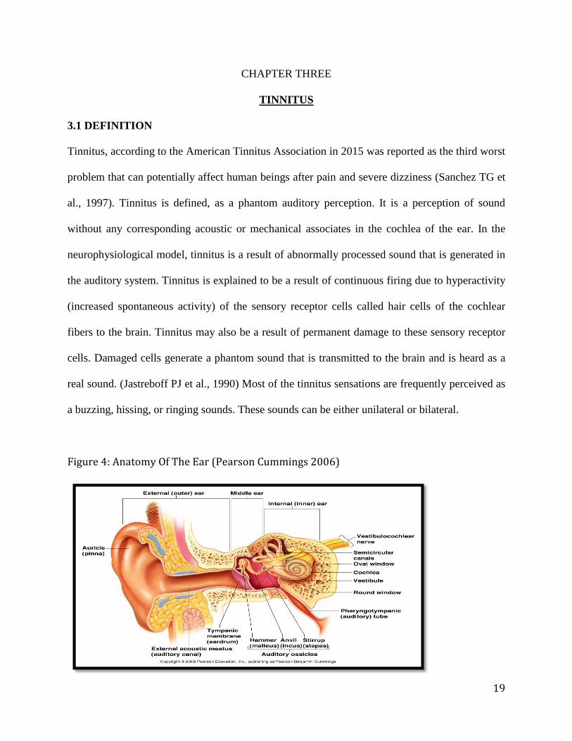
Tinnitus, according to the American Tinnitus Association in 2015 was reported as the third worst
problem that can potentially affect human beings after pain and severe dizziness (Sanchez TG et
al., 1997). Tinnitus is defined, as a phantom auditory perception. It is a perception of sound
without any corresponding acoustic or mechanical associates in the cochlea of the ear. In the
neurophysiological model, tinnitus is a result of abnormally processed sound that is generated in
the auditory system. Tinnitus is explained to be a result of continuous firing due to hyperactivity
(increased spontaneous activity) of the sensory receptor cells called hair cells of the cochlear
fibers to the brain. Tinnitus may also be a result of permanent damage to these sensory receptor
cells. Damaged cells generate a phantom sound that is transmitted to the brain and is heard as a
real sound. (Jastreboff PJ et al., 1990) Most of the tinnitus sensations are frequently perceived as
a buzzing, hissing, or ringing sounds. These sounds can be either unilateral or bilateral.
Figure 4: Anatomy Of The Ear (Pearson Cummings 2006)
20
Tinnitus is generally divided into two categories: objective and subjective. Objective tinnitus,
which is also known as somatosound, is defined, as a sound that is generated in the body and at
the same time is audible by the examiner. For example, myoclonic contractions of the tensor
tympani muscle or altered blood flow in vessels near the ear are considered objective findings.
Tinnitus is subjective if it is audible only to the patient. Subjective tinnitus is measured to be
devoid of an acoustic etiology and any related movements in the cochlear partition or the
cochlear fluid within the internal ear. Most of the current practitioners use the term tinnitus to
designate subjective tinnitus while the term somatosound is used to designate objective tinnitus
(Dobie RA et al., 2004). Since tinnitus is most of the times being measured, quantified and
described based on responses of the subjects/patients, many physicians consider tinnitus as a
subjective phenomenon, which is difficult to evaluate objectively.
Figure 5: Proximity Of TMJ To Ear (Mayo 2015)
21
3.2 EPIDEMIOLOGY
According to the National Health and Nutritional Examinations Survey (NHANES) in 1999-
2004, an estimate of over 45 million Americans reported tinnitus. Tinnitus is thus rated as one of
the most common health conditions in the USA. According to NHANES in 1999, the prevalence
of tinnitus was 25.3%, which corresponds to a national estimate of about 50 million adults aged
of 20 years or above. Also, the survey concluded that the prevalence of tinnitus increases with an
increase in age until 60-69 years and then gradually decreases as age progressed. Within the
population of USA, tinnitus is more prevalent among males than females and more prevalent
among non –Hispanic whites than non-Hispanic blacks and Hispanics (Shargorodsky J et al.,
2010).
3.3 ETIOLOGY
Like TMD, the cause of tinnitus is clinically heterogeneous and more than one cause may be
present in same patient. Causes of tinnitus can be broadly grouped into otologic (pertaining to
ear), neurologic (relating to central and peripheral nervous system), infectious causes,
pharmacological, psychiatric and dental causes (Steinmetz LG et al., 2008).
Otologic causes of tinnitus include presbycusis (age related hearing loss), otosclerosis, otitis,
impacted cerumen in the ear, and/or sudden deafness. Neurologic causes include injury to the
head, whiplash blow, conditions such as multiple sclerosis, vestibular schwannoma and/or other
cerebellopontine-angle tumors. Some of the infectious causes include otitis media and sequelae
of Lyme disease, meningitis, and syphilis. Among the pharmacological causes where tinnitus is a
side effect of drugs, salicylates, non-steroidal anti-inflammatory drugs (NSAIDS), antibiotics,
diuretics, and chemotherapy agents such as cisplatins and vincristine are some of the common
drugs (Byung IH et al., 2009). In some cases, stress and emotional issues are also responsible for
22
causing tinnitus (Hinton DE et al., 2005).
In regard to dental causes, temporomandibular joint dysfunction (TMD) is considered primary
cause for tinnitus. TMD may also cause aural symptoms such as earache, vertigo, dizziness,
referred pain and headaches apart from tinnitus (Isabela PT et al., 2016). Other dental causes
include patient habits such as clenching and grinding of teeth. High-pitched noise from dental
drills is also considered a possible cause for tinnitus (Messano GA et al., 2012).
3.4 TREATMENTS AND MANAGEMENT
Treatment for tinnitus is primarily targeted either to reduce the intensity of sound disturbance or
to relieve the irritation related with tinnitus. Some medications such as tricyclic antidepressants
(nortriptyline and amitriptyline) and benzodiazepines (alprazolam, clonazepam, and oxazepam)
can help mitigate tinnitus. Electric suppression can sometimes help with reducing the intensity of
sound. Cognitive behavior therapy, sound therapy and use of hearing aids can also help reduce
the associated displeasure (Han BI et al., 2009).
23
CHAPTER FOUR
TINNITUS AND TMD COEXISTENCE
TMD patients, along with classic signs and symptoms of TMD, also complain of sudden hearing
impairment or loss, plugged ear sensations and earache, a sore or burning throat, difficulties
swallowing, tinnitus, and vertigo (Morais AA et al., 2012). The most frequent aural symptoms
among TMD patients include tinnitus, and otalgia (Felicio CM et al., 2004). Some investigations
suggested that at least more than one-third of TMD patients report tinnitus (Lam DK et al, 2012).
The relationship between the etiology of tinnitus and TMD is not well known. In order to
understand this relationship, a brief review of the embryology and anatomy of ear and TMJ
region may be necessary. Anatomically, the ear is divided into external ear, middle ear and inner
ear. The external ear consists of auricle and external auditory canal, middle ear or tympanic
cavity consists of tympani membrane and 3 auditory ossicles- malleus, stapes and incus, the
inner ear consists of cochlea and labyrinth. There are 2 muscles that attach to the ossicles of the
middle ear- the tensor tympani muscle and the stapedius muscle. Tensor tympani muscle is
supplied by the 5th
cranial nerve and it is responsible for tensing the tympanic membrane of the
middle ear.
4.1 CAUSAL CONCEPTS
There has been a significant increase in understanding the relationship between ear symptoms
and the craniomandibular disorders. However at present, very little data is available concerning
the prevalence of tinnitus in TMD condition. Thus it may be difficult to assess the association
between tinnitus and TMD (Harold et al., 1997).
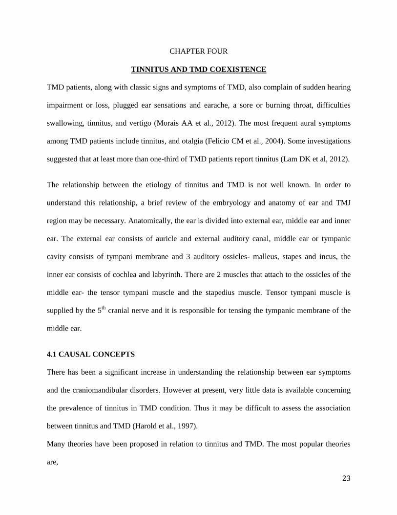
Many theories have been proposed in relation to tinnitus and TMD. The most popular theories
are,
24
1. The Neuromuscular Theory: It is believed that tinnitus is linked structurally and
functionally to Tensor tympani (V3) muscle. According to this theory proposed by
Attanasio G, et al, contractions in tensor tympani muscle, which originates from the
cartilage of auditory tube (part of middle ear) and inserts on the handle of the malleus of
the ear causes movement of the tympani membrane thus producing tinnitus. Tensor
tympani, tensor palatine and the muscles of mastication are innervated by the trigeminal
nerve. Also, these muscles share a common embryologic origin, which is the first
pharyngeal arch. Neuromuscular theory is based on this embryological and functional
relationship between middle ear muscles and masticatory muscles. Contractions due to
chronic irritation in one group of muscles may be conveyed to the other muscles due to
their close proximity.
Figure 6:-Neuromuscular Theory Flowchart Representation
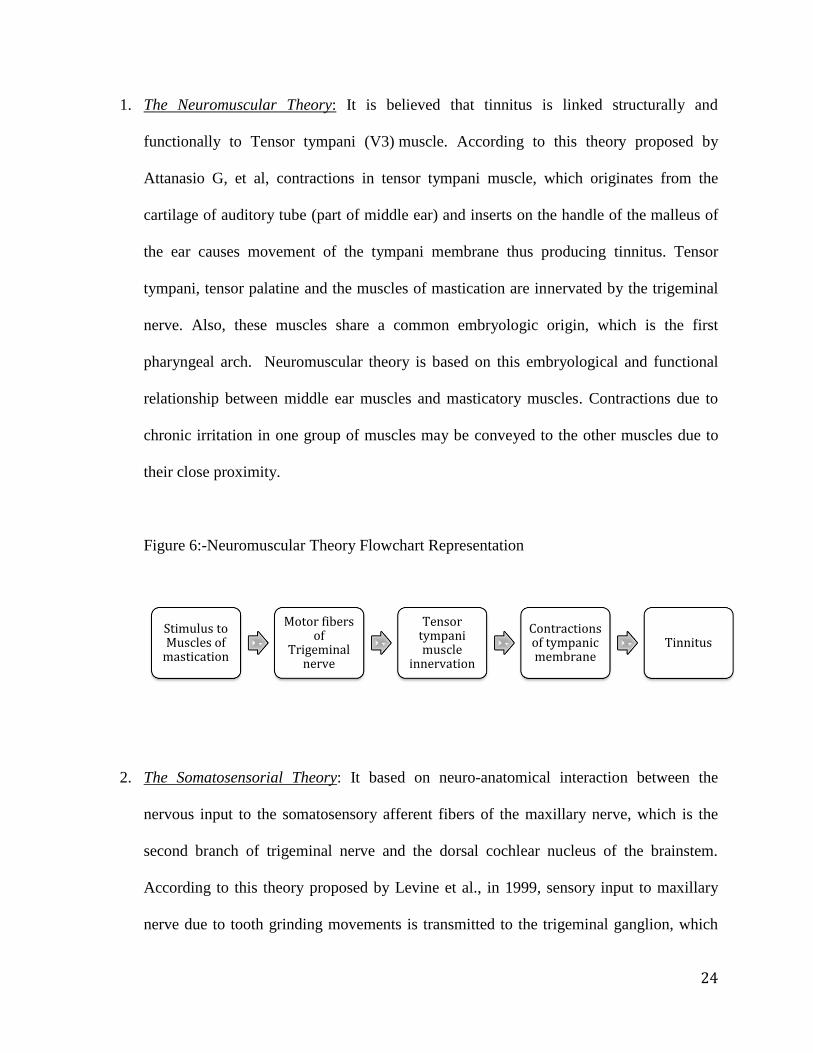
2. The Somatosensorial Theory: It based on neuro-anatomical interaction between the
nervous input to the somatosensory afferent fibers of the maxillary nerve, which is the
second branch of trigeminal nerve and the dorsal cochlear nucleus of the brainstem.
According to this theory proposed by Levine et al., in 1999, sensory input to maxillary
nerve due to tooth grinding movements is transmitted to the trigeminal ganglion, which
Stimulus to Muscles of
mastication
Motor fibers of
Trigeminal nerve
Tensor tympani muscle
innervation
Contractions of tympanic membrane
Tinnitus
25
also innervates the dorsal cochlear nucleus. This interaction may affect both hearing and
the interpretation of sound.
Figure 7:- Somatosensorial Theory Flowchart Representation
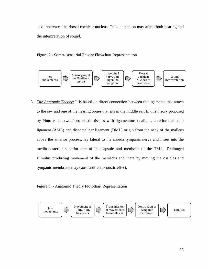
3. The Anatomic Theory: It is based on direct connection between the ligaments that attach
to the jaw and one of the hearing bones that sits in the middle ear. In this theory proposed
by Pinto et al., two fibro elastic tissues with ligamentous qualities, anterior malleolar
ligament (AML) and discomallear ligament (DML) origin from the neck of the malleus
above the anterior process, lay lateral to the chorda tympanic nerve and insert into the
medio-posterior superior part of the capsule and meniscus of the TMJ. Prolonged
stimulus producing movement of the meniscus and there by moving the ossicles and
tympanic membrane may cause a direct acoustic effect.
Figure 8: - Anatomic Theory Flowchart Representation
Jaw movements
Sensory input to Maxillary
nerve
trigeminal nerve and Trigeminal
ganglion
Dorsal Cochlear
Nucleus of brain stem
Sound interpretation
Jaw movements
Movement of DML , AML ligaments
Transmission of movements to middle ear
Contraction of tympanic
membrane Tinnitus
26
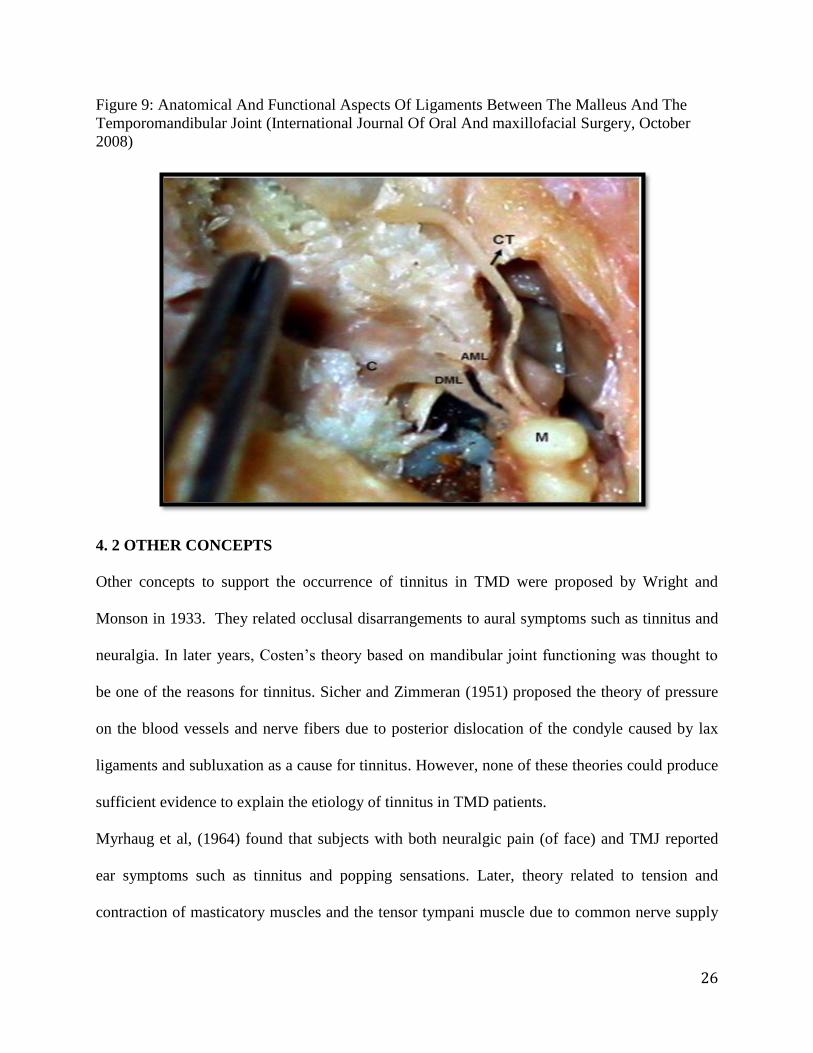
Figure 9: Anatomical And Functional Aspects Of Ligaments Between The Malleus And The
Temporomandibular Joint (International Journal Of Oral And maxillofacial Surgery, October
2008)
4. 2 OTHER CONCEPTS
Other concepts to support the occurrence of tinnitus in TMD were proposed by Wright and
Monson in 1933. They related occlusal disarrangements to aural symptoms such as tinnitus and
neuralgia. In later years, Costen’s theory based on mandibular joint functioning was thought to
be one of the reasons for tinnitus. Sicher and Zimmeran (1951) proposed the theory of pressure
on the blood vessels and nerve fibers due to posterior dislocation of the condyle caused by lax
ligaments and subluxation as a cause for tinnitus. However, none of these theories could produce
sufficient evidence to explain the etiology of tinnitus in TMD patients.
Myrhaug et al, (1964) found that subjects with both neuralgic pain (of face) and TMJ reported
ear symptoms such as tinnitus and popping sensations. Later, theory related to tension and
contraction of masticatory muscles and the tensor tympani muscle due to common nerve supply
27
from trigeminal nerve became popular. This theory was related to changes in the bite that caused
tension in muscles. Another theory considered tinnitus as a fatigue response to prolonged
irritation and stress that developed in the masticatory muscles along with tensor tympani muscle
that are innervated by the trigeminal nerve. Furthermore, another theory proposed that bnormal
spams in the muscles of mastication due to occlusal changes was a prominent cause of arthritis in
the temporomandibular joint and ear symptoms like tinnitus (Jonck et al., 1978).
With Pinto’s cadaver study findings in 1962, that examined ligament attachments between the
middle ear and TMJ, a structural concept was addressed. Komori et al., (1986) reported
ligaments that attach from intra articular disk of the TMJ via the petro tympanic fissure to the
malleus in the middle ear. Movement of the TMJ was considered to cause movements of the
ligaments, followed by movements in the chain of ossicles of the middle ear and thus the
tympanic membrane leading to tinnitus. Additionally, fibrous connective tissue connection
between the sphenomandibular ligaments and the anterior ligament of the malleus was also
considered to play an important role in producing tinnitus (Bleiker et al., 1988).
Young et al (1958) believed that pertotympanic fissure is an outlet from the middle ear into the
gleniod fossa transmitting through the tegmen tympani bone that contains lymphatic channels
from the petrous bone. Compression of these lymphatic channels during closed bite and /or
mandibular retrusion caused increase fluid pressure in the inner ear leading to ear symptoms.
Furthermore, it was believed that mechanical impingement of the lymphatics could impair the
lymphatic drainage mechanism. Nevertheless, these concepts on TMJ and tinnitus coexistence
have not been supported with sufficient data from studies.
28
CHAPTER FIVE
METHODS AND MATERIAL
5.1 STUDY DESIGN
This secondary analysis study is designed to measure the association between the presence of
tinnitus and TMD diagnoses in a TMD population evaluated for the Research Diagnostic Criteria
for Temporomandibular disorder study. The RDC/TMD study consists of questionnaires and
highly structured clinical and radiographic assessments. As part of the RDC/TMD study,
extensive self reported data was collected including data on tinnitus. The purpose of the
validation study was to define the diagnostic validity of the existing examination protocol and
panel's recommendations for further revision of the Axis I diagnostic algorithms were assessed
for reliability by using newly collected data from the ongoing TMJ Impact Project—the follow-
up study to the Validation Project. (Schiffman et al., 2014). For convenience, our study refers to
the Validation project as baseline study and the Impact study as follow up study.
5.2 STUDY POPULATION
Subjects from RDC/ TMD baseline and follow up study were included. Subjects for baseline
and follow up study were from august 2003 till September 2006 and from June 2012- 2013
respectively at three sites- University of Minnesota, University at Buffalo and University of
Washington.
29
5.3 STUDY DATA AND MEASURES
For measuring different TMD diagnoses and tinnitus, the following data from the Validation and
Impact studies was used. The measures include clinical examination and self reported
questionnaires.
Baseline study
RDC/TMD examination raw data
History Questionnaire: “Do you have noises or ringing in your ears?” (15f)
Medical history: RDC/ TMD Questionnaire
Supplemental History: Initial Questionnaire
Gold standard Diagnoses RDC/TMD
Oral Behaviors Checklist
SCL-90-R
Follow up study
CPSQ impact study – “ ringing in your ears- yes or no (18f)”
Diagnostic Criteria for Temporomandibular Disorders -Patient History Questionnaire
DC/TMD Examination Form: Impact Study
5.4 INLCLUSION CRITERIA
The inclusion criteria as defined by Schiffman et al:
Cases:
Subjects ages 18-70 years of age with at least 1 of the 3 cardinal signs and symptoms of TMD
jaw pain, limited mouth opening or TMJ noise
30
Controls:
A. History
1) No lifetime history of TMD symptoms (“supercontrols”)
Absence of TMJ noise, locking or catching of the jaw, and
Absence of pain in the jaw or the temporal area, and
Absence of headaches affected by jaw movement, function, or parafunction.
2) Prior history of TMD symptoms (“controls”)
In the last 6 months, no history of TMD symptoms
Prior to 6 months ago:
No more than 5 isolated episodes of TMJ noise, with each episode lasting less
than 1 day and not associated with jaw pain or limited mouth opening, and
No more than 1–2 isolated episodes of locking or catching of the jaw in the
wide-open mouth position, and
No headaches in the temporal area affected by jaw movement, function, or
parafunction.
B. Clinical examination
Any pain produced by procedures must be nonfamiliar, and
No TMJ clicking, popping, or snapping noises with more than 1 movement,
and
31
No coarse crepitus with any movement.
C. Imaging
TMJ MRI is negative for anterior disc displacement, and
TMJ CT is negative for osteoarthrosis.
5.5 EXCLUSION CRITERIA
Exclusion criteria as defined by Schiffman et al.,
I. History
1. Systemic rheumatic, neurologic/neuropathic, endocrine, or immune/autoimmune
diseases or wide spread pain. (Exception: subjects with medical documentation of
rheumatoid arthritis or fibromyalgia).
2. Pathologic processes found on imaging including neoplasm (Exception: Disc
displacements and osteoarthritis/ osteoarthrosis)
3. Radiation treatment to head and neck.
4. TMJ surgery.
5. Trauma to jaw in the last 2 months (exclusion regardless of time: jaw trauma from
auto accident).
6. Presence of non-TMD orofacial pain disorders.
7. Pregnancy.
8. Unable to participate due to language barrier or mental/intellectual incompetence.
9. Use of narcotic pain medication, muscle relaxants or steroid therapy unless
discontinued for 1 week prior to examination.
32
10. Use of antidepressant drugs unless the participant has been on a stable dose for 60
days.
11. Use of prescription or over-the-counter nonsteroidal anti-inflammatory
medications unless the medication(s) were discontinued for 3 days prior to the
examination (use of acetaminophen was allowed as a rescue drug).
12. Drug abuse.
13. Ongoing dental treatments.
14. Wearing dentures.
15. Contraindications for imaging.
16. Ongoing TMD treatments unless on a stable regimen for at least 2 months.
17. Unable or unwilling to give informed consent.
II. Clinical examination
1. Presence of non-TMD orofacial pain disorders.
III. Imaging
1. TMJ MRI is positive for pathology other than disc displacements.
2. TMJ CT is positive for osseous pathology other than osteoarthritis or
ostoeathrosis.
3. Panoramic radiograph is positive for osseous (non-TMJ related) or odontogenic
lesions.
5.6 DATA COLLECTION
33
All data from the TMD/ RDC baseline and follow up study was gathered and stored in an
encrypted drive and used for further analysis. Study data was accessible to the thesis
group.
5.7 STATISTICAL ANLYSIS
From baseline study data, descriptive analyses were performed to summarize subject
characteristics, such as age and sex, type of TMD diagnoses and tinnitus frequency. Log-
binominal regression analysis was done to assess the risk factors and calculate adjusted relative
risks (Daddens et al., 2004) with 0.05 level of significance (p<0.05). Major risk factors analyzed
were,
1. Tenderness or pain on palpation in extra oral, intra oral and joint site muscles on
palpation
2. Headaches in the temporal region
3. Oral habits such as grinding / clenching of the jaw
4. Anxiety, Depression and Characteristic pain intensity.
5. Jaw Movements- Left and right extrusion movements and protrusion.
At follow up, Data from the Impact study was similarly analyzed to measure the change in rate
of tinnitus. Also, additional analysis was performed to measure the rate of tinnitus within the
different TMD diagnoses at follow up. All statistical analysis was done using SAS 9.3 software
(SAS Institute, Cary, NC).
34
CHAPTER SIX
RESULTS
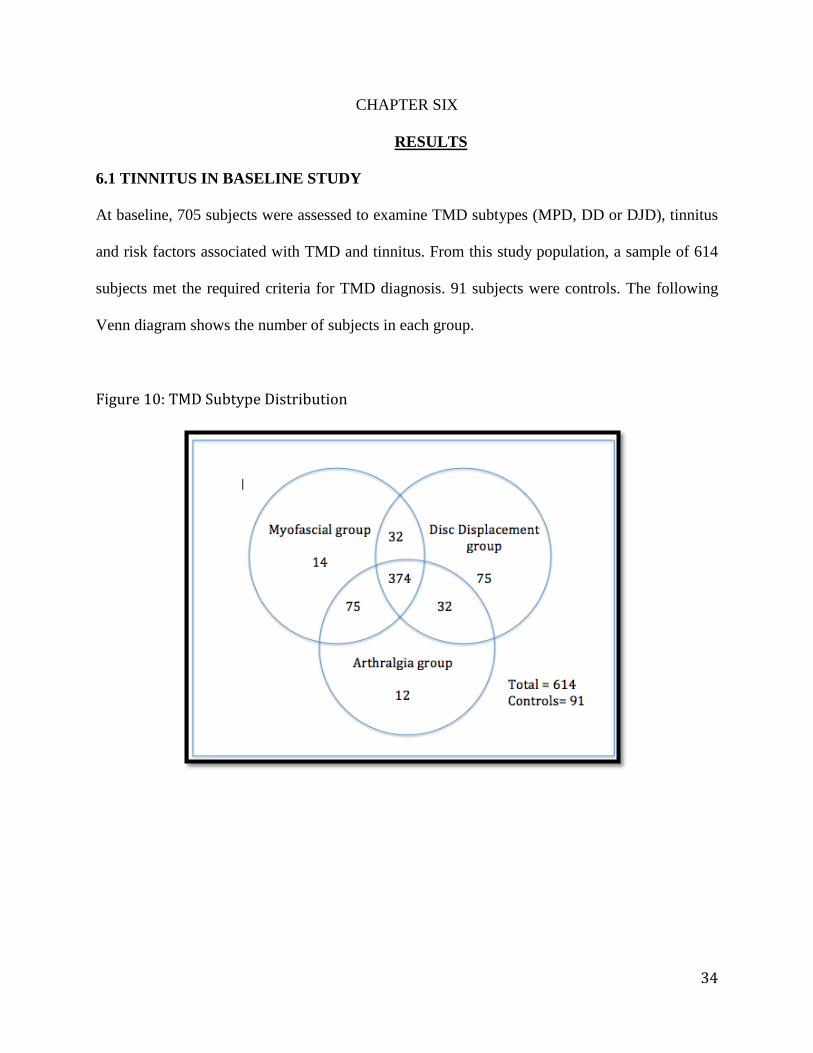
6.1 TINNITUS IN BASELINE STUDY
At baseline, 705 subjects were assessed to examine TMD subtypes (MPD, DD or DJD), tinnitus
and risk factors associated with TMD and tinnitus. From this study population, a sample of 614
subjects met the required criteria for TMD diagnosis. 91 subjects were controls. The following
Venn diagram shows the number of subjects in each group.
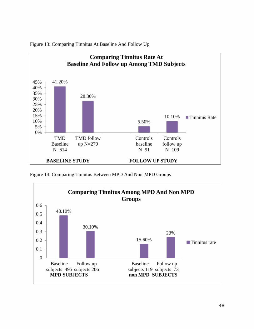
Figure 10: TMD Subtype Distribution
35
Among 614 subjects with TMD, about 60% (374) of subjects were diagnosed with all subtypes
of TMD (MPD+DD+DJD) and the rest received at least one or more TMD diagnosis.
Approximately, four fifths of the sample, 495 (80.6%) had MPD diagnosis in combination with
DD and/or DJD. Of the remaining 119 subjects, 75 received only DD diagnosis, 12 only DJD
and 32 had DD along with DJD diagnosis.
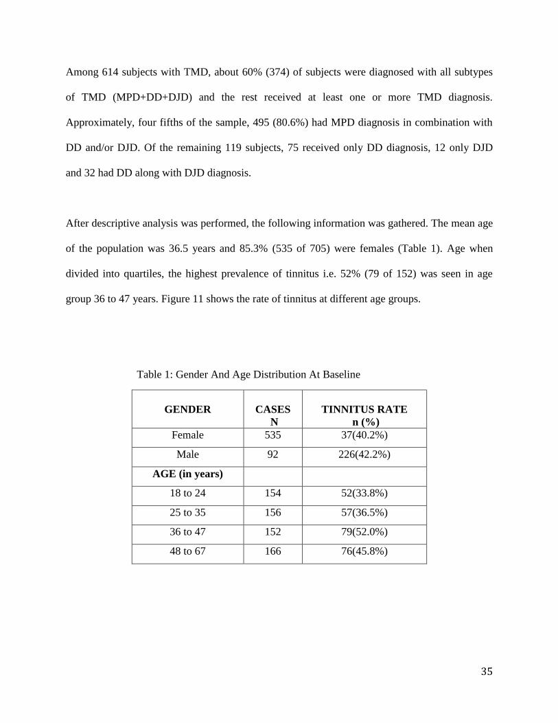
After descriptive analysis was performed, the following information was gathered. The mean age
of the population was 36.5 years and 85.3% (535 of 705) were females (Table 1). Age when
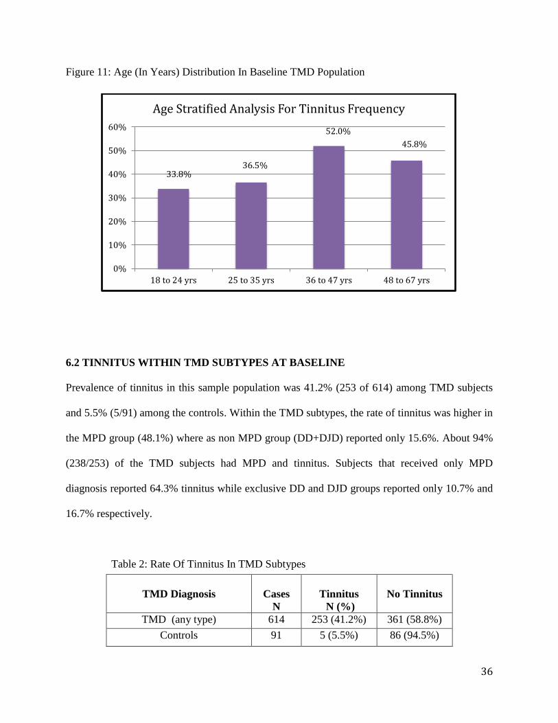
divided into quartiles, the highest prevalence of tinnitus i.e. 52% (79 of 152) was seen in age
group 36 to 47 years. Figure 11 shows the rate of tinnitus at different age groups.
Table 1: Gender And Age Distribution At Baseline
GENDER
CASES
N
TINNITUS RATE
n (%)
Female 535 37(40.2%)
Male 92 226(42.2%)
AGE (in years)
18 to 24 154 52(33.8%)
25 to 35 156 57(36.5%)
36 to 47 152 79(52.0%)
48 to 67 166 76(45.8%)
36
Figure 11: Age (In Years) Distribution In Baseline TMD Population
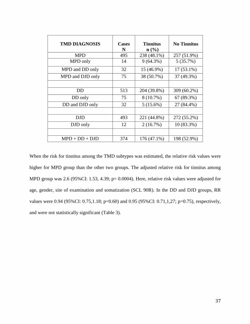
6.2 TINNITUS WITHIN TMD SUBTYPES AT BASELINE
Prevalence of tinnitus in this sample population was 41.2% (253 of 614) among TMD subjects
and 5.5% (5/91) among the controls. Within the TMD subtypes, the rate of tinnitus was higher in
the MPD group (48.1%) where as non MPD group (DD+DJD) reported only 15.6%. About 94%
(238/253) of the TMD subjects had MPD and tinnitus. Subjects that received only MPD
diagnosis reported 64.3% tinnitus while exclusive DD and DJD groups reported only 10.7% and
16.7% respectively.
Table 2: Rate Of Tinnitus In TMD Subtypes
TMD Diagnosis
Cases
N
Tinnitus
N (%)
No Tinnitus
TMD (any type) 614 253 (41.2%) 361 (58.8%)
Controls 91 5 (5.5%) 86 (94.5%)
33.8% 36.5%
52.0%
45.8%
0%
10%
20%
30%
40%
50%
60%
18 to 24 yrs 25 to 35 yrs 36 to 47 yrs 48 to 67 yrs
Age Stratified Analysis For Tinnitus Frequency
37
TMD DIAGNOSIS
Cases
N
Tinnitus
n (%)
No Tinnitus
MPD 495 238 (48.1%) 257 (51.9%)
MPD only 14 9 (64.3%) 5 (35.7%)
MPD and DD only 32 15 (46.9%) 17 (53.1%)
MPD and DJD only 75 38 (50.7%) 37 (49.3%)
DD 513 204 (39.8%) 309 (60.2%)
DD only 75 8 (10.7%) 67 (89.3%)
DD and DJD only 32 5 (15.6%) 27 (84.4%)
DJD 493 221 (44.8%) 272 (55.2%)
DJD only 12 2 (16.7%) 10 (83.3%)
MPD + DD + DJD 374 176 (47.1%) 198 (52.9%)
When the risk for tinnitus among the TMD subtypes was estimated, the relative risk values were
higher for MPD group than the other two groups. The adjusted relative risk for tinnitus among
MPD group was 2.6 (95%CI: 1.53, 4.39; p= 0.0004). Here, relative risk values were adjusted for
age, gender, site of examination and somatization (SCL 90R). In the DD and DJD groups, RR
values were 0.94 (95%CI: 0.75,1.18; p=0.60) and 0.95 (95%CI: 0.71,1,27; p=0.75), respectively,
and were not statistically significant (Table 3).
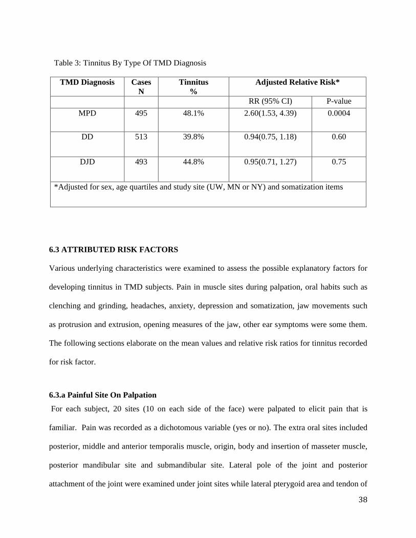
38
Table 3: Tinnitus By Type Of TMD Diagnosis
TMD Diagnosis Cases
N
Tinnitus
%
Adjusted Relative Risk*
RR (95% CI) P-value
MPD 495
48.1%
2.60(1.53, 4.39) 0.0004
DD 513
39.8%
0.94(0.75, 1.18) 0.60
DJD 493
44.8% 0.95(0.71, 1.27) 0.75
*Adjusted for sex, age quartiles and study site (UW, MN or NY) and somatization items
6.3 ATTRIBUTED RISK FACTORS
Various underlying characteristics were examined to assess the possible explanatory factors for
developing tinnitus in TMD subjects. Pain in muscle sites during palpation, oral habits such as
clenching and grinding, headaches, anxiety, depression and somatization, jaw movements such
as protrusion and extrusion, opening measures of the jaw, other ear symptoms were some them.
The following sections elaborate on the mean values and relative risk ratios for tinnitus recorded
for risk factor.
6.3.a Painful Site On Palpation
For each subject, 20 sites (10 on each side of the face) were palpated to elicit pain that is
familiar. Pain was recorded as a dichotomous variable (yes or no). The extra oral sites included
posterior, middle and anterior temporalis muscle, origin, body and insertion of masseter muscle,
posterior mandibular site and submandibular site. Lateral pole of the joint and posterior
attachment of the joint were examined under joint sites while lateral pterygoid area and tendon of
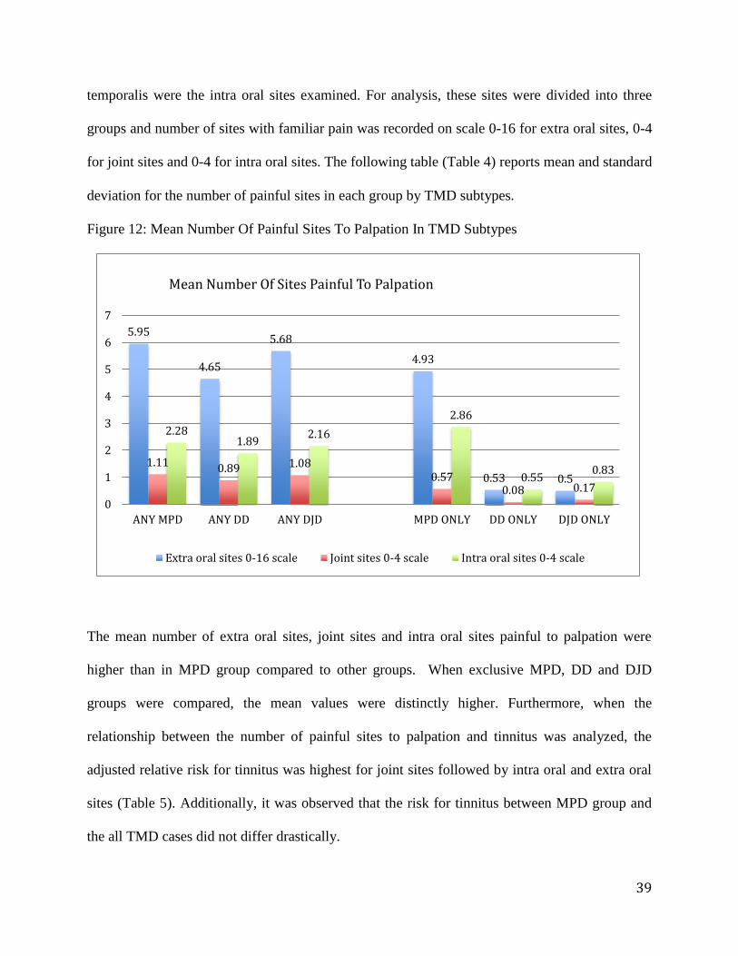
39
temporalis were the intra oral sites examined. For analysis, these sites were divided into three
groups and number of sites with familiar pain was recorded on scale 0-16 for extra oral sites, 0-4
for joint sites and 0-4 for intra oral sites. The following table (Table 4) reports mean and standard
deviation for the number of painful sites in each group by TMD subtypes.
Figure 12: Mean Number Of Painful Sites To Palpation In TMD Subtypes
The mean number of extra oral sites, joint sites and intra oral sites painful to palpation were
higher than in MPD group compared to other groups. When exclusive MPD, DD and DJD
groups were compared, the mean values were distinctly higher. Furthermore, when the
relationship between the number of painful sites to palpation and tinnitus was analyzed, the
adjusted relative risk for tinnitus was highest for joint sites followed by intra oral and extra oral
sites (Table 5). Additionally, it was observed that the risk for tinnitus between MPD group and
the all TMD cases did not differ drastically.
5.95
4.65
5.68
4.93
0.53 0.5
1.11 0.89 1.08 0.57
0.08 0.17
2.28 1.89
2.16
2.86
0.55 0.83
0
1
2
3
4
5
6
7
ANY MPD ANY DD ANY DJD MPD ONLY DD ONLY DJD ONLY
Mean Number Of Sites Painful To Palpation
Extra oral sites 0-16 scale Joint sites 0-4 scale Intra oral sites 0-4 scale
40
6.3.b Headaches
Headaches in the temporalis region were examined as a risk factor for tinnitus. When descriptive
and regression analysis were performed, within the baseline population, 427 had headaches with
47.1% (201/427) reporting tinnitus. Among subjects with a TMD diagnosis and headaches in the
temporal region, the relative risk for tinnitus was 4.52, which was much higher than subjects
with only HA (RR=0.73), and only TMD (RR=3.17) as shown in Table 5. Similarly, when
examined within the TMD diagnoses, MPD group with headaches was at a higher risk for
tinnitus (RR =3.59) than only headaches (RR =1.75) or only MPD (RR = 3.02) as shown in
Table 6 below.
Table 4: Risk Ratios For Tinnitus By Number Of Sites Painful To Palpation At Baseline
Cases
N
Adjusted Risk Ratio
RR (95% CI) P-value
Extra oral sites
(0-16 scale)
TMD 1.05 (1.02,1.07)
<. 0001
MPD 1.03 (1.01, 1.05)
0.0083
Joint sites
(0-4 scale)
TMD 1.14 (1.05, 1.22)
0.0006
MPD 1.08 (1.00, 1.16)
0.044
Intra oral sites
(0-4 scale)
TMD 1.11 (1.04, 1.18)
0.0004
MPD 1.08 (1.01, 1.15)
0.011
RR, relative risk (estimated using log-linear regression analysis). Adjusted for sex, age quartiles,
study site (UW, MN or NY) and somatization. TMD N= 614 and MPD N= 495
41
Table 5: Tinnitus By TMD Diagnosis And Headaches For TMD Subjetcts
Diagnosis Cases
N
Tinnitus
n (%)
Adjusted RR
(95% CI)
P-value
<0.001
Headache only (controls) 31 6 (19.4%) 0.73
(0.09, 5.66)
0.76
TMD only 119 44 (37.0%) 3.17
(1.18, 8.46)
0.022
TMD and headache 376 194 (51.6%) 4.52
(1.67, 12.19)
0.0028
TMD DIAGNOSIS X HEADACHE INTERACTION
0.47
Without headache: TMD
diagnosis
154
53 (25.6%
3.17
(1.18, 8.46)
.022
With headache: TMD
diagnosis
207
200 (49.1%)
6.20
(0.96, 39.7)
.054
Adjusted RR for age, sex, study site, somatization and other TMD diagnoses.
Table 6: Tinnitus By TMD Diagnosis And Headaches For MPD Baseline Subjects
Diagnosis Cases
N
Tinnitus
(%)
Adjusted RR
(95% CI)
P-value
Headache only 19 5.0% 1.75
(0.68, 4.49)
0.24
MP only 154 25.6% 3.02
(1.51, 6.01)
0.0016
MP and headache 207 49.1% 3.59
(1.82, 7.06)
0.0002
MYOFASCIAL PAIN X HEADACHE INTERACTION 0.44
Without headache: Myofascial pain 119
37.0% 3.08
(1.48, 6.39)
With headache: Myofascial pain 376
51.6% 2.02
(0.96, 4.21)
Adjusted for age, sex, study site, somatization and other TMD diagnoses.
6.3.c Oral Habits
The oral behavior checklist was used to assess the relationship between tinnitus and habits. This
21 checklist examines the most frequent positioning of the jaw at sleep and during waken hours.
Although each item on the checklist were examined, there was no habit identified that had a
significant association with tinnitus.
42
Table 7: Mean Values Of Oral Habits In Baseline Subjects
Number of oral behaviors
(0-21)
Tinnitus N Mean Std Min Max
All 628 12.83 3.52 0.00 21.00
No 364 12.33 3.40 4.00 21.00
Yes 264 13.52 3.57 0.00 20.00
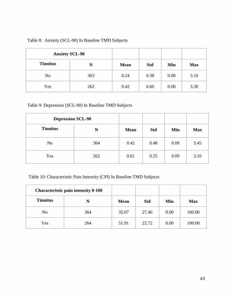
6.3.d Anxiety, Depression, Characteristic Pain Intensity And Interference At Baseline
The following tables represent the mean values and standard deviation of anxiety, depression,
characteristic pain intensity (CPI), disability days and interference of pain with daily activity
values recorded at baseline among both TMD subjects and MPD subjects. Data from SCL 90R
questionnaire was used to perform descriptive analysis was performed to calculate mean values.
Mean anxiety values were twice in subjects with tinnitus (0.42) than without tinnitus (0.24).
Mean depression values were recorded higher in subjects with tinnitus (0.61) than without
tinnitus (0.42). Similarly, mean values of CPI, disability days and interference of pain were
observed higher in t subjects with tinnitus than without. CPI disability days and interference of
pain scores were further analyzed at follow up for better understanding.
43
Table 8: Anxiety (SCL-90) In Baseline TMD Subjects
Anxiety SCL-90
Tinnitus N Mean Std Min Max
No 363 0.24 0.38 0.00 3.10
Yes 262 0.42 0.60 0.00 3.30
Table 9: Depression (SCL-90) In Baseline TMD Subjects
Depression SCL-90
Tinnitus N Mean Std Min Max
No 364 0.42 0.48 0.00 3.45
Yes 262 0.61 0.55 0.00 3.10
Table 10: Characteristic Pain Intensity (CPI) In Baseline TMD Subjects
Characteristic pain intensity 0-100
Tinnitus N Mean Std Min Max
No 364 35.07 27.46 0.00 100.00
Yes 264 51.91 22.72 0.00 100.00
44
Table 11: Interference In Baseline TMD Subjects
Interference score (0-100)
Tinnitus N Mean Std Min Max
No 364 12.01 18.69 0.00 90.00
Yes 264 21.93 23.94 0.00 100.00
Table 12: Disability Days In Baseline TMD Subjects
Disability days
Tinnitus N Mean Std Min Max
No 362 0.17 0.61 0.00 3.00
Yes 262 0.45 0.93 0.00 3.00
6.3.e Jaw Movements
Protrusion and left and right extrusion of the jaw were examined to measure the mean value of
range of motion. The mean values did not differ from subjects with and without tinnitus.
Moreover, when examined in subjects with MPD diagnosis, the mean values did not vary with
tinnitus diagnosis. The following table represents the mean values of jaw movements.
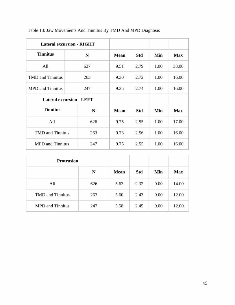
45
Table 13: Jaw Movements And Tinnitus By TMD And MPD Diagnosis
Lateral excursion - RIGHT
Tinnitus N Mean Std Min Max
All 627 9.51 2.79 1.00 38.00
TMD and Tinnitus 263 9.30 2.72 1.00 16.00
MPD and Tinnitus 247 9.35 2.74 1.00 16.00
Lateral excursion - LEFT
Tinnitus N Mean Std Min Max
All 626 9.75 2.55 1.00 17.00
TMD and Tinnitus 263 9.73 2.56 1.00 16.00
MPD and Tinnitus 247 9.75 2.55 1.00 16.00
Protrusion
N Mean Std Min Max
All 626 5.63 2.32 0.00 14.00
TMD and Tinnitus 263 5.60 2.43 0.00 12.00
MPD and Tinnitus 247 5.58 2.45 0.00 12.00
46
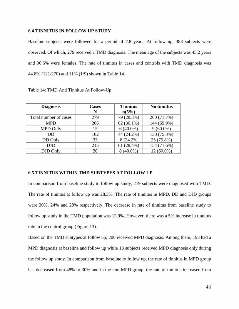
6.4 TINNITUS IN FOLLOW UP STUDY
Baseline subjects were followed for a period of 7.8 years. At follow up, 388 subjects were
observed. Of which, 279 received a TMD diagnosis. The mean age of the subjects was 45.2 years
and 90.6% were females. The rate of tinnitus in cases and controls with TMD diagnosis was
44.8% (121/270) and 11% (1/9) shown in Table 14.
Table 14: TMD And Tinnitus At Follow-Up
Diagnosis Cases
N
Tinnitus
n(5%)
No tinnitus
Total number of cases 279 79 (28.3%) 200 (71.7%)
MPD 206 62 (30.1%) 144 (69.9%)
MPD Only 15 6 (40.0%) 9 (60.0%)
DD 182 44 (24.2%) 138 (75.8%)
DD Only 33 8 (24.2% 25 (75.8%)
DJD 215 61 (28.4%) 154 (71.6%)
DJD Only 20 8 (40.0%) 12 (60.0%)
6.5 TINNITUS WITHIN TMD SUBTYPES AT FOLLOW UP
In comparison from baseline study to follow up study, 279 subjects were diagnosed with TMD.
The rate of tinnitus at follow up was 28.3%. The rate of tinnitus in MPD, DD and DJD groups
were 30%, 24% and 28% respectively. The decrease in rate of tinnitus from baseline study to
follow up study in the TMD population was 12.9%. However, there was a 5% increase in tinnitus
rate in the control group (Figure 13).
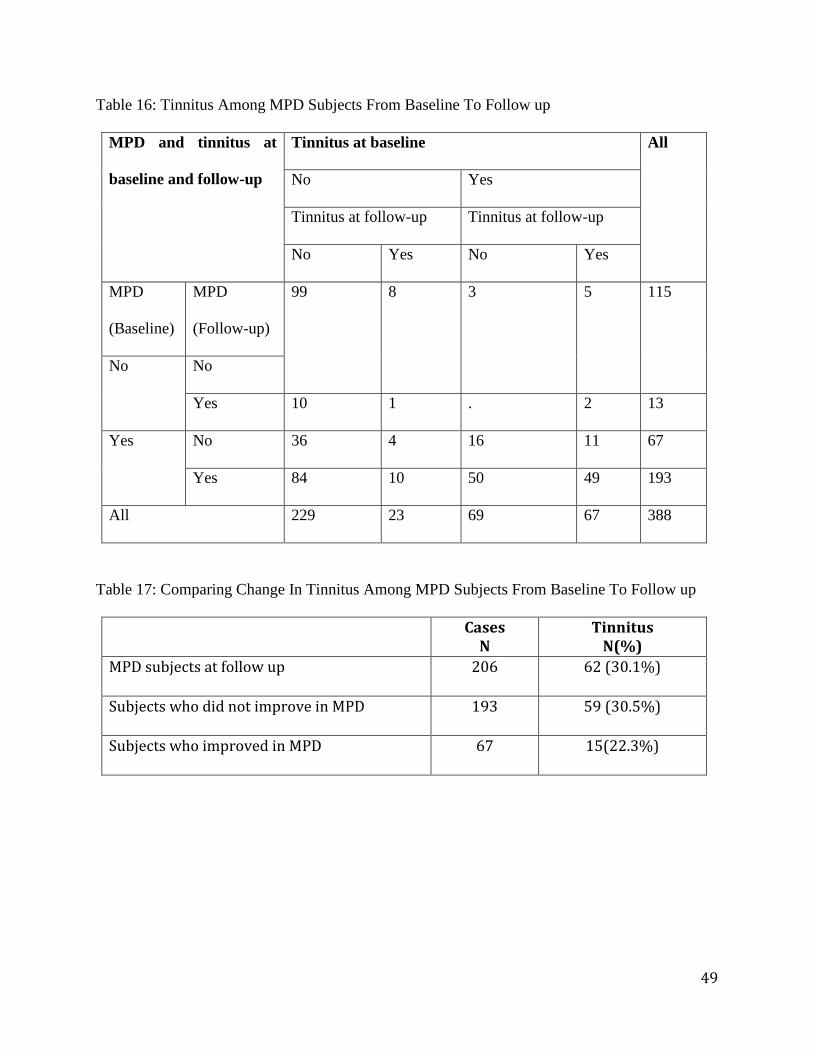
Based on the TMD subtypes at follow up, 206 received MPD diagnosis. Among them, 193 had a
MPD diagnosis at baseline and follow up while 13 subjects received MPD diagnosis only during
the follow up study. In comparison from baseline to follow up, the rate of tinnitus in MPD group
has decreased from 48% to 30% and in the non MPD group, the rate of tinnitus increased from
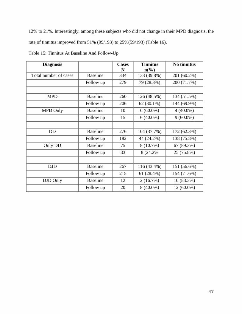
47
12% to 21%. Interestingly, among these subjects who did not change in their MPD diagnosis, the
rate of tinnitus improved from 51% (99/193) to 25%(59/193) (Table 16).
Table 15: Tinnitus At Baseline And Follow-Up
Diagnosis Cases
N
Tinnitus
n(%)
No tinnitus
Total number of cases Baseline 334 133 (39.8%) 201 (60.2%)
Follow up 279 79 (28.3%) 200 (71.7%)
MPD Baseline 260 126 (48.5%) 134 (51.5%)
Follow up 206 62 (30.1%) 144 (69.9%)
MPD Only Baseline 10 6 (60.0%) 4 (40.0%)
Follow up 15 6 (40.0%) 9 (60.0%)
DD Baseline 276 104 (37.7%) 172 (62.3%)
Follow up 182 44 (24.2%) 138 (75.8%)
Only DD Baseline 75 8 (10.7%) 67 (89.3%)
Follow up 33 8 (24.2% 25 (75.8%)
DJD Baseline 267 116 (43.4%) 151 (56.6%)
Follow up 215 61 (28.4%) 154 (71.6%)
DJD Only Baseline 12 2 (16.7%) 10 (83.3%)
Follow up 20 8 (40.0%) 12 (60.0%)
48
Figure 13: Comparing Tinnitus At Baseline And Follow Up
Figure 14: Comparing Tinnitus Between MPD And Non-MPD Groups
41.20%
28.30%
5.50% 10.10%
0%5%
10%15%20%25%30%35%40%45%
TMD
Baseline
N=614
TMD follow
up N=279
Controls
baseline
N=91
Controls
follow up
N=109
BASELINE STUDY FOLLOW UP STUDY
Comparing Tinnitus Rate At
Baseline And Follow up Among TMD Subjects
Tinnitus Rate
48.10%
30.10%
15.60%
23%
0
0.1
0.2
0.3
0.4
0.5
0.6
Baseline
subjects 495
Follow up
subjects 206
Baseline
subjects 119
Follow up
subjects 73MPD SUBJECTS non MPD SUBJECTS
Comparing Tinnitus Among MPD And Non MPD
Groups
Tinnitus rate
49
Table 16: Tinnitus Among MPD Subjects From Baseline To Follow up
MPD and tinnitus at
baseline and follow-up
Tinnitus at baseline All
No Yes
Tinnitus at follow-up Tinnitus at follow-up
No Yes No Yes
MPD
(Baseline)
MPD
(Follow-up)
99 8 3 5 115
No No
Yes 10 1 . 2 13
Yes No 36 4 16 11 67
Yes 84 10 50 49 193
All 229 23 69 67 388
Table 17: Comparing Change In Tinnitus Among MPD Subjects From Baseline To Follow up
Cases N
Tinnitus N(%)
MPD subjects at follow up 206 62 (30.1%)
Subjects who did not improve in MPD 193 59 (30.5%)
Subjects who improved in MPD 67 15(22.3%)
50
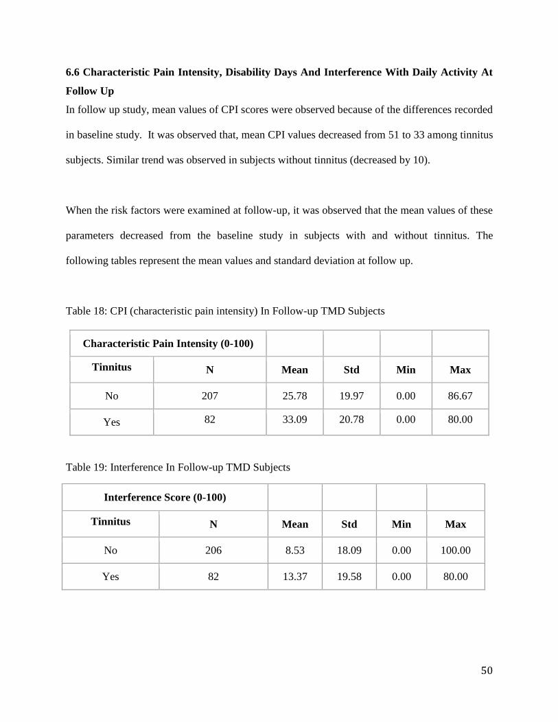
6.6 Characteristic Pain Intensity, Disability Days And Interference With Daily Activity At
Follow Up
In follow up study, mean values of CPI scores were observed because of the differences recorded
in baseline study. It was observed that, mean CPI values decreased from 51 to 33 among tinnitus
subjects. Similar trend was observed in subjects without tinnitus (decreased by 10).
When the risk factors were examined at follow-up, it was observed that the mean values of these
parameters decreased from the baseline study in subjects with and without tinnitus. The
following tables represent the mean values and standard deviation at follow up.
Table 18: CPI (characteristic pain intensity) In Follow-up TMD Subjects
Characteristic Pain Intensity (0-100)
Tinnitus N Mean Std Min Max
No 207 25.78 19.97 0.00 86.67
Yes 82 33.09 20.78 0.00 80.00
Table 19: Interference In Follow-up TMD Subjects
Interference Score (0-100)
Tinnitus N Mean Std Min Max
No 206 8.53 18.09 0.00 100.00
Yes 82 13.37 19.58 0.00 80.00
51
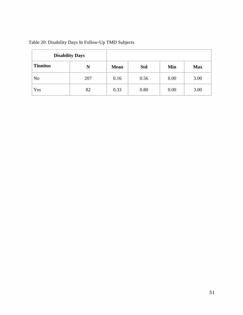
Table 20: Disability Days In Follow-Up TMD Subjects
Disability Days
Tinnitus N Mean Std Min Max
No 207 0.16 0.56 0.00 3.00
Yes 82 0.33 0.80 0.00 3.00
52
CHAPTER SEVEN
CONCLUSION AND RECOMMENDATIONS
Tinnitus is one of the common ear symptoms among TMD patients. As mentioned before,
association between ear symptoms and TMD was first hypothesized in early 1934. Despite the
underlying association, researchers failed to explain a cause effect relationship between tinnitus
and TMD. The literature suggests that tinnitus is more prevalent among TMD patients than those
without TMD (Bush FM et al., 189987). A recent study by Buergers R et al., in 2014 suggested
that tinnitus is eight times more prevalent in subjects with TMD than those without TMD.
However, few studies evaluated tinnitus within TMD subgroups. Our study examined tinnitus
report among different TMD subtypes rather than as a whole and the attributed risk factors.
The prevalence of tinnitus in the over all study population was 41.2% with at least 85% females.
And age related prevalence pattern of tinnitus ranged from 33 to 52% within the four age groups.
The rate of tinnitus among our TMD population is consistent with previous literature on tinnitus
rate ranging from 31 to 64% (Manfredinin D et al., 2012). The decrease rate of tinnitus with
increased age was consistent with previous reports on tinnitus. It is interesting to note that
although there were more number of subjects in the 4th
quartile (age ranging from 48-67 years)
the rate of tinnitus reported was less than the 3rd
quartile (36-47 years)
Then, rate of tinnitus was high in the myofascial TMD than DD and DJD type. Moreover, the
risk for tinnitus was doubled with MPD while this trend was not seen in the other two groups.
Such finding may have important clinical implications, particularly with regard to the possible
mechanism underlying the causal relationship between the tinnitus and TMD. It can be
53
speculated that clinical findings of tinnitus in the MPD group age 36-47 years may be due to a
chronic tension of jaw muscles that provoke an abnormal response on the muscles regulating ear
function.
To analyze attributed risk factors, sites painful to palpation were examined. Interestingly, the risk
for tinnitus did not differ much in terms of painful sites to palpation among the different TMD
sub types. The risk for tinnitus was slightly higher in painful joint sites (RR=1.14) as compared
to muscle sites (extra oral sites- RR=1.05 and intra oral sites- RR=1.11). Based on
neuromuscular theory, one would expect MPD group to be at a higher risk than DD and/or DJD.
But since DJD included painful joint sites (arthralgia), the results did not differ much from MPD
group.
With the data available, our study further evaluated risk factors such as headaches and oral
habits. Based on the theory that abnormal dental occlusion and habitual jaw positioning may
displace the condyle posteriorly thus producing ear symptoms, one may expect that oral habits
such as clenching and grinding may also alter the condylar position. Although, studies speculated
the role of oral habits in TMD, our study did not find any strong association between reported
oral habits and tinnitus.
In regard to headaches and tinnitus, the literature suggests that tinnitus and headaches may have
a common underlying pathophysiological mechanism and may not be purely coincidental
(Berthold L et al., 2015). The findings of our study suggest that TMD patients with headaches
are more likely to report tinnitus than TMD patients without headaches. Additionally, MPD
patients with headaches are at least three times at a higher risk for tinnitus than MPD patients
54
without headaches (RR less than 1 in DD and DJD groups). One could imagine chronic stimulus
to the muscles of the jaw; particularly the temporal region would sensitize the trigeminal system
thus facilitating the development of tinnitus. However, it may be important to note that the test
for interactions between TMD & headaches and MPD & headaches were not statistically
significant (p=0.47). However, the unadjusted results, as well as the two RRs by headache and
without headache, indicate that the risk of tinnitus with a TMD diagnosis is higher if the subject
also has a headache than without a headache. Further electrophysiological or neuroimaging
studies may be needed to evaluate this neuromuscular theory behind tinnitus, headaches and
TMD.
From the follow up study, we observed that the frequency of tinnitus among the TMD population
increased by at least 3%. Interestingly, in the MPD group, tinnitus dropped by at least 20 % from
baseline to follow up. As MPD improved over time (80 to 73%), we noticed that tinnitus rate in
this population also improved (48 to 30%). This improvement was not observed in DD and DJD
groups. In fact, tinnitus rate increased in DD and DJD groups at follow up. Such findings may
help contribute to the neuromuscular theory (where chronic stimulus to muscles may trigger
tinnitus). As pain in the muscles improved, tinnitus decreased accordingly. Nevertheless, it is
interesting to note that at follow up, mean values of characteristic pain intensity improved from
baseline. This may suggest that as pain decreased, tinnitus decreased or that subjects were less
likely to report tinnitus or be bothered enough by tinnitus to report its presence. Additional
studies explaining the role of psychological factors and over all impact of pain may be necessary.
55
While being a cohort study and long term follow up are some of the merits of the study, the
methodology of tinnitus report and loss of subjects to follow up and some of the limitations.
Furthermore, tinnitus was a dichotomous variable where it was reported as either present or
absent but in our follow up study, tinnitus was reported within the questions of any noises in the
ear. In future studies, it may be worthwhile to differentiate tinnitus from other noises. Also other
studies that score frequency of tinnitus or measure the impact of tinnitus on daily activity may be
useful.
Although our study had a larger population, it was difficult to explore factors associated with
tinnitus in patients with exclusive MPD due to smaller sample size. Long follow up studies
including subjects with exclusive myofascial pain may be useful to support the role of
neuromuscular system in tinnitus.
56
CHAPTER EIGHT
SUMMARY
From this large prospective multicenter TMD study, we found that the prevalence of tinnitus is at
least three times higher in TMD population than healthy individuals. Risk for tinnitus is higher in
MPD group than the other two groups and is six times higher in patients with a TMD diagnosis
and headaches. A thorough TMD screening in patients with tinnitus is recommended.
Notwithstanding that, our study showed significant correlation of tinnitus with MPD and
headaches, thus providing support for neuromuscular theory; future research that assesses
clinical correlation is demanded.
57
NOMENCLATURE
TMJ Temporomandibular joint
TMD Temporomandibular joint disorders
MPD Myofascial pain disorder
DD Disk displacement
DJD Degenerative Joint Disease/Arthralgia
DML Discomallear ligament
AML Anterior malleolar ligament
CT Chorda Tympani
M Malleus of the ear
N Number of Individuals
Std Standard Deviation
Min Minimum range value
Max Maximum range value
58
REFERENCES
1. Larry J (1994) peterson principle of oral and maxillofacial surgery. J.b. Lippincott
company, pg 1905–1931
2. Ingawale ́ S, Goswami T (2009) Temporomandibular joint: disorders, treatments, and
biomechanics. Ann Biomed Eng 379:76–96
3. Buescher JJ (2007) Temporomandibular joint disorders. Am Fam Physician 76:1477–
1482
4. Johansson A, et al: gender difference in symptoms related to temporomandibular
disorders in a population of 50-year-old subjects. Journal of orofacial pain 2003;
17:29-35.
5. Messano GA, Petti S. General Dental Practitioners and Hearing Impairment, J
Dent. 2012 Oct;40(10):821-8.
6. Manfredini D. Prevalence of tinnitus in patients with different temporomandibular
disorders symptoms. Int tinnitus J.2015; 19(2):47-51.
7. Runge M, Sadowsky C, Sakols E (1989) The relationship between
temporomandibular joint sounds and malocclusion. Am J Orthod 96:36–42
8. Fillingim RB, Maixner W, Kincaid S, Sigurdsson A, Harris MB (1996) Pain
sensitivity in patients with temporomandibular dis- orders: relationship to clinical and
psychosocial factors. Clin J Pain 12:260–269
9. Filipo R, Attanasio G, Cagnoni L, Masci E, Russo FY, Cartocci G, et al: Long-term
results of intratympanic prednisolone injection in patients with idiopathic sudden
sensorineural hearing loss. Acta Otolaryngol 133(9): 900e904, 2013.
10. Langguth B, Kreuzer PM, Kleinjung T, De Ridder D (2013) Tinnitus: causes and
clinical management. Lancet Neurol 12:920–930
11. Gelb H, Gelb ML, Wagner ML: The relationship of tinnitus to Craniocervical
Mandibular Disorder. Cranio 15(2): 136e143, 1997
12. Baguley D, McFerran D, Hall D (2013) Tinnitus. Lancet 382:1600–1607
13. Eggermont J, Roberts L (2004) The neuroscience of tinnitus.
14. Rdc-tmdinternational.org.
15. Attanasio G, et al. Tinnitus in patients with temporo-mandibular joint disorder:
proposal for a new treatment protocol. Journal of craniomaxillofacial surgery. 2015
June; 43(5): 724-7
16. Costen JB (1934) A syndrome of ear and sinus symptoms dependent upon disturbed
function of the temporomandibular joint. Ann Otol Rhinol Laryngol 106:805–819
17. Wing, G. (1959), The status of Costen's Syndrome. Australian Dental Journal, 4: 98–
103. doi:10.1111/j.1834-7819.1959.tb01877.
18. Myrhaug H.(1964) The incidence of ear symptoms in cases of malocclusion and
temporo-mandibular joint disturbances . Br J Oral Surg. 1964 Jul; 2(1): 28–32.
19. Levine RA. Somatic (craniocervical) tinnitus and the dorsal cochlear nucleus
hypothesis. Am J Otolaryngol. 1999;20:351–62. 10.1016/S0196-0709(99)90074-1
20. Jonck Lm: Ear symptoms in temporomandibular joint disturbances. S Afr Med J
1978; 54: 782
21. Komori E, Sugisaki M, Tanabe H, Katoh S: Discomalleolar ligament in the adult
human J Carniomandib Pract 1986;4:300-305
59
22. Bleiker ross: Ear disturbances of temporomandibular join origins JADA
1938;September:25
23. Pinto O (1962) a new structure related to the temporomandibular joint and middle ear.
J prosthetic dentistry 12:95–103
24. Eckerdal O (1991) The petrotympanic fissure: a link connecting the tympanic cavity
and the temporomandibular joint. Cranio 9:15–22
25. Wright EF, Bifano SL (1997) The relationship between tinnitus and
temporomandibular disorder (TMD) therapy. Int Tinnitus J 3:55–61
26. Kanji a et al. Clinical signs and symptoms of tinnitus in temporomandibular joint
disorders: a pilot study comparing patients and non-patients. South African journal of
community disorders. 2013 dec; 60:16-20.
27. Tuz HH et al. Prevalence of otologic complaints in patients with temporomandibular
disorder. Journal of orthodontics and dentofacial orthopedics. 2003 June; 123(6): 620-
3.
28. Bernhardt O et al. Signs and symptoms of temporomandibular disorders and the
incidence of tinnitus. Journal of oral rehabilitation. 2011 December; 38(12): 891-901.
29. Bernhardt O et al. Signs and symptoms of temporomandibular disorders and the
incidence of tinnitus. Journal of oral rehabilitation. 2004 April; 31(4): 311-9.
30. Fernandes g et al. Association between painful temporomandibular disorders, sleep
bruxism and tinnitus. Braz oral res. 2014;28.
31. Calderon pdos s et al. Influence of tinnitus on pain severity and quality of life in
patients with temporomandibular disorders. Journal of applied oral sciences. 2012
mar-apr; 20(2): 170-3.
32. Wright EF et al. The relationship between tinnitus and temporomandibular disorder
(TMD) therapy. Quintessence int. 2007 October; 38(9): e564-71.
33. Parker WS et al. Tinnitus, vertigo, and temporomandibular disorders. . Journal of
orthodontics and dentofacial orthopedics. 1995 February; 107(2):153-8.
34. Buergers r et al. Is there a link between tinnitus and temporomandibular disorders.
Journal of prosthetic dentistry. 2014 mar; 111(3): 222-7.
35. Attanasio et al. The mozart effect in patients suffering from tinnitus. Acta
otolaryngology, 132 (11) (2012), pp. 1172–1177
36. Fernandes g et al. Painful temporomandibular disorders, self reported tinnitus, and
depression is highly associated. Arq neuropsiquiatr. 2013 dec; 71(12): 943-7.
37. Saldanha et al. Are temporomandibular disorders and tinnitus associated? Cranio
surgery. 2012 July; 30(3): 166-71.
38. Morais aa et al. Tinnitus in individuals without hearing loss and its relationship with
temporomandibular dysfunction. Brazilian journal of otorhinolaryngology. 2012
April; 78(2): 59-65.
39. John Phillips et al. Tinnitus and disorders of the temporo-mandibular joint (TMJ) and
neck. Updated May 2015. To be reviewed march 2016.british tinnitus association.
40. E. Ferendiuk et al. Incidence of otolaryngological symptoms in patients with
temporomandibular joint dysfunctions. biomed res int, 2014 (2014), p.1.
41. G. Fernandes, j.t. Siqueira, d.a. Godoi gonçalves, et al.association between painful
temporomandibular disorders, sleep bruxism and tinnitus Br oral res, 28 (2014),
60
42. G. Fernandes, d.a. Gonçalves, j.t. De siquera, et al.painful temporomandibular
disorder, self reported tinnitus and depression are highly associated. arq
neuropsiquiatr, 71 (2013), p. 943
43. C.m. Felício, t.g. Faria, m.a.m. Rodrigues da silva, et al. Temporomandibular
disorder: relationship between otologic and orofacial symptoms.rev bras
otorhinolaryngology, 70 (2004),
B. Çakur, m.a. Sümbüllü, d. Durna, et al. Prevalence of the types of the petrotympanic fissure in
the temporomandibular joint dysfunction radiology, 52 (2011), p. 562
44. Sato, h. Arai, r. Asaumi, et al. Classifications of tunnel-like structure of human
petrotympanic fissure by cone beam ct surgical radiology anatomy, 30 (2008), p. 323
45. Hilgenberg PB, Saldanha AD, Cunha CO, Rubo JH, Conti PC (2012)
Temporomandibular disorders, otologic symptoms and depression levels in tinnitus
patients. J Oral Rehabil 39:239–244
46. Deddens, James A, and Martin R Petersen. "Re: "Estimating the Relative Risk in
Cohort Studies and Clinical Trials of Common Outcomes"." American Journal of
Epidemiology. 159.2: 213-4; Author Reply 214. Web.
61
VITA
Name Susee Priyanka Ravuri
Address 5123 24th
Ave Ne, unit 6
Seattle, Washington, 98105
Education Bachelor of Dental Surgery
Panineeya Dental College, Hyderabad 2012
Master’s in Public Administration
San Diego State University – 2015
Master of Science in Dentistry –Oral Medicine
University of Washington 2017
Email [email protected]