temporomandibular disorders: priorities for research and care
TRANSCRIPT
DETAILS
Distribution, posting, or copying of this PDF is strictly prohibited without written permission of the National Academies Press. (Request Permission) Unless otherwise indicated, all materials in this PDF are copyrighted by the National Academy of Sciences.
Copyright © National Academy of Sciences. All rights reserved.
THE NATIONAL ACADEMIES PRESS
Visit the National Academies Press at NAP.edu and login or register to get:
– Access to free PDF downloads of thousands of scientific reports
– 10% off the price of print titles
– Email or social media notifications of new titles related to your interests
– Special offers and discounts
GET THIS BOOK
FIND RELATED TITLES
This PDF is available at SHARE
CONTRIBUTORS
SUGGESTED CITATION
http://nap.edu/25652
Temporomandibular Disorders: Priorities for Research andCare (2020)
426 pages | 6 x 9 | PAPERBACKISBN 978-0-309-67048-7 | DOI 10.17226/25652
Enriqueta C. Bond, Sean Mackey, Rebecca English, Cathy T. Liverman, and OliviaYost, Editors; Committee on Temporomandibular Disorders (TMDs): From ResearchDiscoveries to Clinical Treatment; Board on Health Sciences Policy; Board onHealth Care Services; Health and Medicine Division; National Academies ofSciences, Engineering, and Medicine
National Academies of Sciences, Engineering, and Medicine 2020.Temporomandibular Disorders: Priorities for Research and Care. Washington,DC: The National Academies Press. https://doi.org/10.17226/25652.
Temporomandibular Disorders: Priorities for Research and Care
Copyright National Academy of Sciences. All rights reserved.
Enriqueta C. Bond, Sean Mackey, Rebecca English, Cathy T. Liverman, and Olivia Yost, Editors
Committee on Temporomandibular Disorders (TMDs): From Research Discoveries to Clinical Treatment
Board on Health Sciences Policy
Board on Health Care Services
Health and Medicine Division
A Consensus Study Report of
Temporomandibular Disorders
Priorities for Research and Care
Temporomandibular Disorders: Priorities for Research and Care
Copyright National Academy of Sciences. All rights reserved.
THE NATIONAL ACADEMIES PRESS 500 Fifth Street, NW Washington, DC 20001
This activity was supported by a contract between the National Academy of Sciences and the U.S. Department of Health and Human Services: National Institutes of Health (NIH) (Office of the NIH Director and the National Institute of Dental and Craniofacial Research). Any opinions, findings, conclusions, or recommendations expressed in this publication do not necessarily reflect the views of any organization or agency that provided support for the project.
International Standard Book Number-13: 978-0-309-67048-7International Standard Book Number-10: 0-309-67048-9Digital Object Identifier: https://doi.org/10.17226/25652Library of Congress Control Number: 2020934005
Additional copies of this publication are available from the National Academies Press, 500 Fifth Street, NW, Keck 360, Washington, DC 20001; (800) 624-6242 or (202) 334-3313; http://www.nap.edu.
Copyright 2020 by the National Academy of Sciences. All rights reserved.
Printed in the United States of America
Suggested citation: National Academies of Sciences, Engineering, and Medicine. 2020. Temporomandibular disorders: Priorities for research and care. Washington, DC: The National Academies Press. https://doi.org/10.17226/25652.

Temporomandibular Disorders: Priorities for Research and Care
Copyright National Academy of Sciences. All rights reserved.
The National Academy of Sciences was established in 1863 by an Act of Congress, signed by President Lincoln, as a private, nongovernmental institu-tion to advise the nation on issues related to science and technology. Members are elected by their peers for outstanding contributions to research. Dr. Marcia McNutt is president.
The National Academy of Engineering was established in 1964 under the char-ter of the National Academy of Sciences to bring the practices of engineering to advising the nation. Members are elected by their peers for extraordinary contributions to engineering. Dr. John L. Anderson is president.
The National Academy of Medicine (formerly the Institute of Medicine) was established in 1970 under the charter of the National Academy of Sciences to advise the nation on medical and health issues. Members are elected by their peers for distinguished contributions to medicine and health. Dr. Victor J. Dzau is president.
The three Academies work together as the National Academies of Sciences, Engineering, and Medicine to provide independent, objective analysis and advice to the nation and conduct other activities to solve complex problems and inform public policy decisions. The National Academies also encourage education and research, recognize outstanding contributions to knowledge, and increase public understanding in matters of science, engineering, and medicine.
Learn more about the National Academies of Sciences, Engineering, and Medicine at www.nationalacademies.org.

Temporomandibular Disorders: Priorities for Research and Care
Copyright National Academy of Sciences. All rights reserved.
Consensus Study Reports published by the National Academies of Sciences, Engineering, and Medicine document the evidence-based consensus on the study’s statement of task by an authoring committee of experts. Reports typi-cally include findings, conclusions, and recommendations based on information gathered by the committee and the committee’s deliberations. Each report has been subjected to a rigorous and independent peer-review process and it represents the position of the National Academies on the statement of task.
Proceedings published by the National Academies of Sciences, Engineering, and Medicine chronicle the presentations and discussions at a workshop, symposium, or other event convened by the National Academies. The statements and opinions contained in proceedings are those of the participants and are not endorsed by other participants, the planning committee, or the National Academies.
For information about other products and activities of the National Academies, please visit www.nationalacademies.org/about/whatwedo.
Temporomandibular Disorders: Priorities for Research and Care
Copyright National Academy of Sciences. All rights reserved.
v
COMMITTEE ON TEMPOROMANDIBULAR DISORDERS (TMDs): FROM RESEARCH DISCOVERIES TO CLINICAL TREATMENT
ENRIQUETA C. BOND (Chair), President Emeritus, Burroughs Wellcome Fund, QE Philanthropic Advisors
SEAN MACKEY (Vice Chair), Chief, Division of Pain Medicine, Stanford University Medical Center
PENNEY COWAN, Founder and Chief Executive Officer, American Chronic Pain Association
DAVID DEITZ, Principal, David Deitz & Associates, LLCFRANCESCA C. DWAMENA, Professor of Medicine, Michigan State
UniversityROGER B. FILLINGIM, Distinguished Professor, University of FloridaMARGARET M. HEITKEMPER, Professor and Chair, Biobehavioral
Nursing & Health Systems, University of WashingtonFRANCIS KEEFE, Professor of Psychiatry and Behavioral Sciences, Duke
University Medical CenterKATE LORIG, Professor Emerita, Stanford University School of
MedicineRICHARD OHRBACH, Professor, Department of Oral Diagnostic
Sciences, University at Buffalo School of Dental MedicineAMANDA C. PUSTILNIK, Professor of Law, University of Maryland
Carey School of LawSRINIVASA N. RAJA, Professor of Anesthesiology and Critical Care,
Medicine and Neurology, Johns Hopkins University School of Medicine
CORY M. RESNICK, Assistant Professor of Oral and Maxillofacial Surgery, Harvard School of Dental Medicine
ANTONY ROSEN, Vice Dean for Research, Mary Betty Stevens Professor of Medicine, Johns Hopkins University School of Medicine
KATHLEEN A. SLUKA, Kate Daum Research Professor, Department of Physical Therapy and Rehabilitation Science, University of Iowa Carver College of Medicine
BARBARA G. VICKREY, Professor and System Chair, Neurology Department, Icahn School of Medicine at Mount Sinai
ROBERT WEYANT, Professor, University of Pittsburgh School of Dental Medicine
HAI YAO, Ernest R. Norville Endowed Chair and Professor of Bioengineering and Craniofacial Biology, Clemson University and Medical University of South Carolina
Temporomandibular Disorders: Priorities for Research and Care
Copyright National Academy of Sciences. All rights reserved.
vi
Study Staff
CATHY T. LIVERMAN, Study DirectorREBECCA ENGLISH, Study DirectorOLIVIA YOST, Associate Program OfficerSIOBHAN ADDIE, Program OfficerKENDALL LOGAN, Senior Program AssistantANDREW M. POPE, Senior Director, Board on Health Sciences PolicySHARYL NASS, Director, Board on Health Care Services
Consultants
JUSTIN DURHAM, School of Dental Sciences, Newcastle UniversityERIN HAMMERS FORSTAG, Common Good ConsultingGARY SLADE, University of North Carolina School of Dentistry
Temporomandibular Disorders: Priorities for Research and Care
Copyright National Academy of Sciences. All rights reserved.
vii
Reviewers
This Consensus Study Report was reviewed in draft form by individuals chosen for their diverse perspectives and technical expertise. The purpose of this independent review is to provide candid and critical comments that will assist the National Academies of Sciences, Engineering, and Medicine in making each published report as sound as possible and to ensure that it meets the institutional standards for quality, objectivity, evidence, and responsiveness to the study charge. The review comments and draft manu-script remain confidential to protect the integrity of the deliberative process.
We thank the following individuals for their review of this report:
ALLAN BASBAUM, University of California, San FranciscoDANIEL B. CARR, Tufts University School of MedicineTERRIE COWLEY, The TMJ AssociationDANIEL DERKSEN, University of ArizonaLUDA DIATCHENKO, McGill UniversitySCOTT FISHMAN, University of California, DavisCHARLES S. GREENE, University of Illinois at Chicago JENNIFER A. HAYTHORNTHWAITE, Johns Hopkins University
School of Medicine KEVIN D. HUFF, Center for Advanced General DentistryJOHN KUSIAK, National Institute of Dental and Craniofacial
Research (retired)CATO T. LAURENCIN, University of ConnecticutWILLIAM MAIXNER, Duke University School of MedicineJEFFREY P. OKESON, University of Kentucky
Temporomandibular Disorders: Priorities for Research and Care
Copyright National Academy of Sciences. All rights reserved.
viii
CHRISTIAN STOHLER, Columbia University Medical CenterDENNIS C. TURK, University of Washington
Although the reviewers listed above provided many constructive com-ments and suggestions, they were not asked to endorse the conclusions or recommendations of this report nor did they see the final draft before its release. The review of this report was overseen by DAN G. BLAZER II, Duke University School of Medicine, and MICHELLE M. MELLO, Stanford Law School and Stanford University School of Medicine. They were responsible for making certain that an independent examination of this report was carried out in accordance with the standards of the National Academies and that all review comments were carefully consid-ered. Responsibility for the final content rests entirely with the authoring committee and the National Academies.
Temporomandibular Disorders: Priorities for Research and Care
Copyright National Academy of Sciences. All rights reserved.
ix
Contents
PREFACE xiii
SUMMARY 1
1 INTRODUCTION 17Complexity of TMDs, 20Challenges in Care: Patient Experiences, 22Identifying the Appropriate Model for TMD Care and
Research, 24Key Themes, 26Organization of This Report, 28References, 29
2 DEFINITIONS AND SCOPE: WHAT ARE TMDs? 31Terminology: What Term Should Be Used for This Set of
Disorders?, 32Diagnostic Criteria to Categorize Types of TMDs, 36Types of TMDs, 45Understanding the Etiologies of TMDs, 50Factors in the Disease Course of a TMD, 55What Can Be Learned from Other Pain Conditions?, 62What Can Be Learned from Other Orthopedic Conditions?, 64Conclusions and Research Priorities, 66References, 68
Temporomandibular Disorders: Priorities for Research and Care
Copyright National Academy of Sciences. All rights reserved.
x CONTENTS
3 INDIVIDUAL AND SOCIETAL BURDEN OF TMDs 81Prevalence of TMDs, 82Incidence of TMDs, 89Burden and Costs of TMDs, 95Comorbidities, 102Risk Factors for TMDs, 107Conclusions and Research Priorities, 110References, 112
4 STATE OF THE SCIENCE ON TMDs 123Recent Biomechanical and Bioengineering Research on the
TMJ, 124Neurobiology of Orofacial Pain and TMDs, 135Neuroimmune Interactions and TMDs, 149Neuroendocrine Interactions, Stress Response, and TMDs, 152Biomarkers and Molecular Genetics of TMDs, 156Neuroimaging of the Central Nervous System: Exploration
of TMD Phenotypes, 163Psychosocial Factors Underlying TMDs, 166Application of Data Science Methodologies and Novel
Technologies to TMD Research, 167TMD Research Funding, 168Moving the Research Enterprise Forward, 171A National Collaborative Research Consortium and
Framework for Action, 174Conclusions, 181References, 181
5 CARING FOR INDIVIDUALS WITH A TMD 197First, Do No Harm, 198Prevention and Early Detection, 200Assessment and Diagnosis of TMDs, 202TMD Treatments, 204Improving and Disseminating Evidence, 229Conclusions, 240References, 241
6 IMPROVING TMD HEALTH CARE: PRACTICE, EDUCATION, ACCESS, AND COVERAGE 255TMD Health Care Pathways, 255Improving Interprofessional Education and Collaboration, 259Strengthening Education and Training, 262Improving Access to Specialty Care, 275
Temporomandibular Disorders: Priorities for Research and Care
Copyright National Academy of Sciences. All rights reserved.
CONTENTS xi
Improving Payment and Coverage, 282Conclusions and Research Priorities, 290References, 292
7 IMPROVING PATIENT, FAMILY, AND PUBLIC EDUCATION AND AWARENESS ABOUT TMDs 297Overcoming Stigma, 299Increasing Patient and Family Education and Improving
Communication with Health Care Professionals, 302Raising Public Awareness and Knowledge About TMDs, 310Conclusion and Research Priorities, 314References, 316
8 NEXT STEPS AND RECOMMENDATIONS 321Build and Sustain Collaborative and Multidisciplinary
Research, 323Improve Access to and Quality of TMD Health Care, 329Improve Health Care Professional Education About TMDs, 333Raise Awareness, Improve Education, and Reduce Stigma, 336Opportunities for Action, 337References, 340
APPENDIXES
A Workshop and Open Session Agendas 341B Committee Biographical Sketches 351C Commissioned Paper by Gary Slade and Justin Durham 361D Masticatory System: Anatomy and Function 403

Temporomandibular Disorders: Priorities for Research and Care
Copyright National Academy of Sciences. All rights reserved.
Temporomandibular Disorders: Priorities for Research and Care
Copyright National Academy of Sciences. All rights reserved.
xiii
Everyday activities, including eating and talking, are often difficult for people with temporomandibular disorders (TMDs), and many of them suffer severe chronic pain due to this condition. Common social activities that most people take for granted, such as smiling, laughing, and kissing, can become unbearable. This dysfunction and pain, and its associated suf-fering, take a terrible toll on affected individuals, their families, and their friends. Individuals with TMDs often feel stigmatized and invalidated in their experiences by their family, friends, and, particularly, a health care community that frequently relies on “seeing” a condition in order to treat it. Misjudgments and a failure to understand the nature and depths of TMDs can have severe consequences—more pain and more suffering—for individuals, their families, and our society. People with TMDs, desperate for solutions, often seek out multiple clinicians, turning to dubious treat-ments in search of a cure, which can potentially lead to iatrogenic injury and costly, yet ineffective, treatments.
This study—focused on improving TMD care and identifying research directions—occurs at a time of both challenges and opportunities for prog-ress in this field. TMDs are especially challenging because they often require care across medicine, dentistry, and other fields of health, and yet, given the current divide between the medical and dental fields in the United States, such coordinated care rarely happens. The medical–dental divide is further exacerbated by a payment system that inadequately reimburses for the complex care needed by people with TMDs. Clinicians can be affected by bias, limitations in their knowledge and training, and differences in the systems in which different types of clinicians work. Efforts are needed to
Preface
Temporomandibular Disorders: Priorities for Research and Care
Copyright National Academy of Sciences. All rights reserved.
xiv PREFACE
break down these silos and devote research efforts aimed at understanding this complex set of disorders and improving patient care. Furthermore, research is needed to learn more about the structure and function of the temporomandibular joint and the management of its associated disorders. Many TMDs do not exist in isolation, but rather are frequently associ-ated with other painful conditions such as headache, neck and back pain, irritable bowel syndrome, fibromyalgia, sleep disorders, and chronic fatigue syndrome. Despite these challenges, a number of opportunities exist, such as in increasing patient involvement in order to help move health care for-ward and in new research tools and technologies that can expand the cur-rent understanding of the etiology and progression of TMDs. In responding to the study’s broad task—which stretches from research to education to care—this report aims to provide an overview of the current state of knowl-edge on TMDs and to focus its recommendations on near- and long-term actions to move the field forward in such a way that it improves care for individuals with a TMD as rapidly as possible.
The committee’s work greatly benefited from the compelling insights that were so graciously shared by many individuals with TMDs and their family members. These individuals described their often arduous and costly experiences in living with these often complex conditions, including the challenges of trying to navigate through fragmented and divided dental and medical health care systems and frequently dealing with health professionals who were largely unfamiliar with TMDs. We are grateful to these people for sharing their stories, hopes, disappointment, and anger in their written comments and testimonials. We kept those shared messages at the forefront of our deliberations and focus while creating this report.
The committee also greatly appreciates the information provided by workshop speakers as well as by many others who shared information with the committee. The feedback from the report reviewers was invaluable. We especially thank the study sponsors for their work on TMDs and for their support of this study: the Office of the Director at the National Institutes of Health and the National Institute of Dental and Craniofacial Research.
It was our great privilege to work with such dedicated committee mem-bers, each of whom thoroughly engaged in the study, generously shared their expertise, and contributed significant time and effort to this endeavor. This was a complex task, and the committee members stepped up to meet the challenge. Their reasoned and thoughtful discussions made this report possible. We were all fortunate to work with a diligent and outstand-ing team of National Academies of Sciences, Engineering, and Medicine staff, and we deeply thank Cathy Liverman, Rebecca English, Olivia Yost, Kendall Logan, and Siobhan Addie, led by Andrew Pope and Sharyl Nass, board directors in the Health and Medicine Division. We also thank Erin Hammers Forstag for her writing and editing work and Daniel Bearss of
Temporomandibular Disorders: Priorities for Research and Care
Copyright National Academy of Sciences. All rights reserved.
PREFACE xv
the National Academies library staff for his assistance conducting detailed literature searches for the committee and staff.
The committee worked to develop this report in an objective manner based on the available evidence and knowledge. During this process we were acutely aware of the limitations in existing evidence and the data to support that evidence. These limitations and the opportunities to trans-form our understanding of TMDs helped guide our recommendations. TMDs result from a complex interplay between biological, biomechanical, psycho logical, and social factors that transcend simple explanations. Efforts are needed to enhance our understanding of TMDs from cells to society, taking advantage of team science approaches to these complex problems. Further progress will be made through the development and use of new tools, metrics, and biomarkers to diagnose TMDs and forecast their trajec-tory, predict treatment efficacy, and monitor advances in improving health and well-being. The education and training of health care professionals about TMDs and incentivizing them to work individually and in teams will be critical for making improvements in providing care of individuals with recent onset TMDs, chronic TMDs, or high-impact TMDs. Enhanced models of care will incentivize health care professionals to provide the most optimal care for people with TMDs—and do so in a way that is culturally sensitive and patient-centric. It is the committee’s hope that this report will provide a springboard to move this field forward.
Enriqueta C. Bond, ChairSean Mackey, Vice ChairCommittee on Temporomandibular Disorders (TMDs): From Research Discoveries to Clinical Treatment

Temporomandibular Disorders: Priorities for Research and Care
Copyright National Academy of Sciences. All rights reserved.
Temporomandibular Disorders: Priorities for Research and Care
Copyright National Academy of Sciences. All rights reserved.
1
Summary
Consider the joints of the human body. What might first come to mind are the hips and knees—the large joints that support us in our mobility—followed by the wrists, ankles, elbows, fingers, and toes. What can be overlooked, although clearly evident in the mirror, is one of the most used, most necessary, and perhaps most misunderstood set of joints—those of the jaw—which are critical to the vital work of human life, including eating, talking, kissing, and even breathing.
This report focuses on temporomandibular disorders (TMDs), a set of more than 30 health disorders associated with both the temporomandibular joints (TMJs) and the muscles and tissues of the jaw. TMDs have a range of causes and often co-occur with a number of overlapping medical condi-tions, including headaches, fibromyalgia, back pain, and irritable bowel syndrome. TMDs can be transient or long lasting and may be associated with problems that range from an occasional click of the jaw to severe chronic pain involving the entire orofacial region.
The national prevalence of TMDs is difficult to estimate due to chal-lenges in conducting clinical examinations on a large scale, such that most prevalence data are based on self-reported symptoms associated with TMDs rather than examiner-verified classification. For example, one analysis of 2018 data found that an estimated 4.8 percent of U.S. adults (an estimated 11.2 to 12.4 million U.S. adults in 2018) had pain in the region of the TMJ that could be related to TMDs (see Chapter 3). Orofacial pain symptoms may or may not be related to TMDs. As discussed throughout this report, TMDs are a set of diverse and multifactorial conditions that can occur at
Temporomandibular Disorders: Priorities for Research and Care
Copyright National Academy of Sciences. All rights reserved.
2 TEMPOROMANDIBULAR DISORDERS
different stages in an individual’s life with a range of manifestations and impacts on quality of life.
Action is urgently needed to improve care for individuals with a TMD. Too long compartmentalized as a dental issue, both the clinical manage-ment of and research addressing TMDs need to implement a holistic and multidisciplinary approach. Individuals with TMD symptoms often encoun-ter health professionals (across medicine, dentistry, and beyond) that are unfamiliar with TMDs and do not know where best to refer patients for further diagnosis and treatment. The divide between medical and dental care is currently vast in the United States and much of the world, and is a divide that profoundly affects care systems, payment mechanisms, and professional education and training.
This report explores a broad range of issues relevant to improving the health and well-being of individuals with a TMD. To address the study’s Statement of Task (see Chapter 1), the National Academies of Sciences, Engineer ing, and Medicine appointed an 18-member committee with ex-pertise in public health; pain medicine; basic, translational, and clinical research; patient advocacy; physical therapy; dentistry; self-management; TMDs and orofacial pain; oral and maxillofacial surgery; health care services; internal medicine; endocrinology; rheumatology; law; nursing; psychiatry; and communications. The study was sponsored by the Office of the Director of the National Institutes of Health and the National Institute of Dental and Craniofacial Research.
CHALLENGES IN CARE: PATIENT EXPERIENCES
The committee greatly benefited from the input of individuals with a TMD and family members, many of whom face significant day-to-day challenges in living with a TMD. These challenges include difficulties in eating, in personal and social interactions, and in talking, which are often accompanied by severe ongoing pain. The committee received input from more than 110 individuals through in-person and online opportunities to testify at the committee’s public workshop (see Appendix A) and through written submissions to the study’s public access file.1 Among the many issues raised in these testimonies, several focused on the health care system and the care of individuals with a TMD:
• Lack of coordinated care and abandonment—Individuals reported that they were often shuffled back and forth between clinicians in the medical and dental fields with little to no attention paid to
1 The study’s public access file is available through the National Academies Public Access Records Office ([email protected]).
Temporomandibular Disorders: Priorities for Research and Care
Copyright National Academy of Sciences. All rights reserved.
SUMMARY 3
a comprehensive approach to coordinated care. Patients also re-ported being abandoned by their dentists and other clinicians when the treatments did not work, with no referrals or other options provided.
• Over-treatment/harmful treatment—Many patients reported on hav-ing endured multiple TMD-related surgeries (in some cases more than 20), often with no resolution to their pain or with worsening symptoms. Other individuals reported that they had not had surgery but had had a removable oral appliance, orthodontic correction of the teeth, replacement of teeth, or some combination of these treatments.
• Impact on quality of life—Individuals with a TMD described how having a TMD has profound impacts on the quality of their day-to-day lives, from struggling in pain to kiss a loved one to challenges in dining out with friends or simply eating solid foods. Some indi-viduals noted that the disorder affected their ability to work and to care for their families. Many described challenges in dealing with the emotional consequences of their condition and its treatment and with the episodic or ongoing pain that they experience.
• Expense—The financial burden of seeking and receiving care for a TMD was noted by individuals and family members. Some people said that they had received limited insurance coverage, but, for the most part, the coverage was paid out of pocket by the individual at costs of up to tens of thousands of dollars.
• Identifying qualified health care professionals—Individuals with a TMD and their families often expressed their frustration at not knowing where to turn for quality care. Primary care and internal medicine clinicians and general dentists often did not know how to help them locate qualified specialists. Patients were highly aware of the TMJ implant failures of the 1970s and 1980s and conveyed their concerns about the lack of quality treatment options for TMDs. Additionally, they noted that misleading advertising practices—in which clinicians claim to be experts but do not have the proper experi ence or evidence-based practices—further complicate access to quality care.
• Comorbidities—Many individuals with a TMD noted challenges with comorbid conditions, including fatigue, widespread pain, fibro-myalgia, depression, anxiety, and arthritic conditions.
This brief overview highlights only some of the challenges that continue to be faced by individuals with a TMD and by clinicians in diagnosing TMDs and identifying appropriate care for them. A part of the history of the treatment of TMDs centers on the synthetic implants often used from

Temporomandibular Disorders: Priorities for Research and Care
Copyright National Academy of Sciences. All rights reserved.
4 TEMPOROMANDIBULAR DISORDERS
the late 1960s to early 1990s to replace the condyle, fossa, and articular disc of the TMJ. Many of these implants were either recalled by the Food and Drug Administration or voluntarily withdrawn from the market after they caused a range of adverse health outcomes including severe pain and functional joint impairment (see Chapter 5).
Patients have played and continue to play a major role in bringing attention to the need to advance the understanding of and ability to treat TMDs.
COMPLEXITY OF TMDs
The TMJs are among the most frequently used joints in the body, often opening and closing approximately 2,000 times daily. All of the critical activities of this joint, ranging from verbal and nonverbal communications to the demanding movements of chewing to the more subtle function of breathing, require healthy functioning of both the TMJs and associated tissues. Additionally, the joints are vital to interpersonal interactions, to the facial expressions of emotions such as joy or sadness, and to self-esteem and self-identification.
During joint movement, the two TMJs act through parallel efforts to move the semi-rigid jaw and connect the mandible to the temporal bone of the skull. The complexity of the varied conditions that are included in the set of disorders known as TMDs has been a challenge for individuals with a TMD, their family members, health care professionals, and researchers. Although these disorders have sometimes been lumped together as one entity (with terms such as temporomandibular joint disorder), recent efforts have focused on emphasizing that this is a set of disorders (see Chapter 2) and therefore that there is no one treatment or one care pathway for TMDs—one “size” does not fit all.
Upon being diagnosed with a TMD, the goals are for each patient to know the specific type of disorder (or multiple TMDs) that he or she has and to be provided with an appropriate treatment plan specific to that diag-nosis. The challenge (as described in Chapter 5) is that the evidence base for matching a specific treatment (or group of treatments) with a specific diagnosis is not yet fully developed so that in some cases, particularly for chronic conditions, much remains to be learned. While a small number of abnormalities of the TMJ require specific surgical operations to correct, the majority of TMDs have diffuse symptoms and may not respond pre-dictably to one specific intervention. As discussed in Chapter 3, much also remains to be learned about the prevalence of specific TMDs.
The committee uses the broad definition of TMDs as a set of diseases and disorders related to alterations in the structure, function, or physiology of the masticatory system and that may be associated with other systemic
Temporomandibular Disorders: Priorities for Research and Care
Copyright National Academy of Sciences. All rights reserved.
SUMMARY 5
and comorbid medical conditions. The term “TMDs” is used as an um-brella term to encompass disorders that can range from muscle or joint pain to joint disorders (including hypomobility or hypermobility of the joint) to joint diseases (including osteoarthritis) (see Chapter 2). The pain associated with TMDs can range from none to severe high-impact pain. TMDs can range from a single isolated condition to multi-system involvement and can be associated with other comorbid and systemic disorders and overlapping pain conditions (e.g., fibromyalgia, back pain, headache, irritable bowel syndrome, inflammatory arthritis).
The committee supports a biopsychosocial model of TMDs that is inter disciplinary and can be used across medicine and dentistry to focus on the total person’s health and well-being. The biopsychosocial approach is a broad model that can encompass the range of TMDs and apply the best science from medicine, dentistry, physical therapy, integrative health, and multiple other fields to the care of individuals with a TMD. This approach acknowledges that TMDs are not a single entity and consequently most often have varying causes (e.g., trauma, genetics, environmental etiolo-gies) that affect differing parts of the masticatory system and potentially other body systems and require varied, and sometimes multiple, treatment modali ties (see Chapter 5).
NEXT STEPS AND RECOMMENDATIONS
The committee worked to review the scientific literature; to seek infor-mation from patients and their family members, researchers, clinicians, policy makers, research funders, and others; to analyze the data; and to develop its conclusions and recommendations.
The recommendations below focus on the actions that many organiza-tions and agencies should take to improve TMD research and care. The committee also emphasizes the critical role that individuals with a TMD and their family members have played—and hopefully will continue to play—in bringing TMD issues to the attention of policy makers and health professionals and moving the research and care agenda forward on multiple levels in the public and private sectors. These efforts are to be commended and are encouraged to continue and expand. Specifically, it is hoped that individuals with a TMD and their families will be able to partner with their health care professionals to find the best options for care, to continue to actively participate in patient support networks, to explore ways to be a participating voice in research efforts, and to be active advocates for im-provements in care and services for themselves, their family members, and other people with a TMD. The goals of the following recommendations are to build a strong base of knowledge about TMDs and to facilitate actions needed to improve the overall health and well-being of individuals with a
Temporomandibular Disorders: Priorities for Research and Care
Copyright National Academy of Sciences. All rights reserved.
6 TEMPOROMANDIBULAR DISORDERS
TMD. Some of these recommendations can be accomplished rapidly with actions by key decision makers. Other recommendations are more aspira-tional and will require the collaboration and commitment of multiple or-ganizations and dedicated resources—including investments of time, funds, and innovative energies—to accomplish these goals. The committee has provided both short-term and longer-term priorities (see Chapter 8) to be used as starting points and long-range planning points. Key to making a difference in improving care for individuals with a TMD will be:
• pioneering pathways that span medicine, dentistry, physical therapy, and other fields of health care to provide holistic, comprehensive approaches to care—interprofessional and interdisciplinary efforts are of critical importance;
• willingness of health care agencies, organizations, and professionals to commit the resources needed to address this long neglected and often dismissed area of health care; and
• openness and commitment to using and strengthening the evidence base on TMD treatment and changing practice as needed.
Build and Sustain Collaborative and Multidisciplinary Research
Despite investment in research directly and indirectly related to TMDs—most significantly in the field of orofacial pain—researchers have yet to unravel the etiologies and pathophysiologies of TMDs or to translate, in a meaningful way, research findings into improved clinical care practices. Over the past decade, research on TMDs has centered on the biological mechanisms underlying the development and persistence of orofacial pain and on the structure and function of the joint and its tissues, while more recent research has begun to examine the molecular genetics, biomarkers, and biopsychosocial risk factors of TMDs and common comorbidities. Broadly, the research foundation relating to TMDs, as has been the case with other complex, stigmatized conditions, has suffered from the siloing of disciplines and from a lack of clear direction—thus stunting the potential clinical impact of the research. In the case of TMDs, these difficulties have been heightened by a significant dental–medical divide that affects both research and clinical care.
Engagement by multiple stakeholders will be required to dismantle the silos keeping research fields isolated and to advance TMD research and care. A broad range of interrelated research priorities are explored in the report across the research-to-clinical-care continuum. Chapter 4 high-lights research priorities, including those that overlap with those of more broadly funded health concerns, such as chronic pain, and emphasizes the importance of keeping patient needs central to the process of research.
Temporomandibular Disorders: Priorities for Research and Care
Copyright National Academy of Sciences. All rights reserved.
SUMMARY 7
The committee recommends that a research consortium be established to bring together relevant National Institutes of Health (NIH) institutes and centers and other stakeholders from the public and private sectors to focus future research efforts on filling key evidence gaps in TMD research and care and to ensure that clinically meaningful, patient-centered outcomes are prioritized. The committee stresses the importance of an organized research approach for TMDs, but the mechanism to carry this out should be flexible.
Fresh ideas and multiple disciplines are needed to advance TMD re-search to improve patient care. NIH provides approximately one-third of all biomedical research funding in the United States and, therefore, the interests and priorities of NIH institutes and centers can stimulate research interests and training programs throughout the country. TMDs are not the primary mission of any NIH center or institute. NIH funding for TMD research falls largely within the National Institute of Dental and Cranio-facial Research (which has one of the smallest research budgets of the NIH institutes) with a total budget of approximately $461 million compared to the National Cancer Institute’s budget of $5.99 billion for fiscal year 2019. Given the number of individuals suffering from TMDs, the severity of some of the disorders, and the substantial public health burden of TMDs, there is a significant opportunity for NIH and other biomedical research institu-tions to drive increased funding to TMDs in order to spark new research interest and discoveries. Efforts are needed to ensure that TMD research is incorporated into NIH-wide initiatives, including the NIH Pain Consor-tium. Furthermore, as noted in Public Law 116-94, an NIH inter-institute working group is being called on to focus on coordinating TMD research across the multiple NIH institutes and centers relevant to this field. Details on each of these recommendations is provided in Chapter 8.
Recommendation 1: Create and Sustain a National Collaborative Research Consortium for Temporomandibular Disorders (TMDs)
A National Collaborative Research Consortium for TMDs should be established and sustained to coordinate, fund, and translate basic and clinical research (including behavioral, population-based, and imple-mentation research) to address evidence gaps, generate clinically mean-ingful knowledge, identify safe and effective treatments, and improve the quality of TMD care. The consortium would:
• Establish and implement a national research framework for TMDs;• Provide infrastructure for the implementation of research projects; • Establish milestones and timelines;
Temporomandibular Disorders: Priorities for Research and Care
Copyright National Academy of Sciences. All rights reserved.
8 TEMPOROMANDIBULAR DISORDERS
• Facilitate research collaborations; • Develop public–private partnerships; • Develop and test evidence-based strategies for knowledge transfer; • Support the development of a multidisciplinary research workforce
for TMDs through existing and new training and center initiatives; and
• Evaluate progress and disseminate research findings.
Recommendations 2 to 4: Coordinate and Expand Research on Temporomandibular Disorders (TMDs)
The National Collaborative Research Consortium for TMDs, led by the National Institutes of Health (NIH) along with other funders, should fund and strengthen:
• Basic research efforts and the translation of that research as part of a patient-focused, multidisciplinary research agenda on TMDs to address evidence gaps, generate clinically meaningful knowledge, identify effective treatments, and improve quality of care;
• The collection, assessment, and dissemination of population-based data on the burden and costs of TMDs and the effects of TMDs on patient outcomes in order to improve the prevention (primary, secondary, and tertiary) and management of TMDs; and
• Clinical and implementation research to clearly define effective treat-ments and continuously improve the quality of care for patients with a TMD.
(See Recommendations 2 to 4 in Chapter 8 for lists of research priori-ties and actions.)
Improve Access and Quality for TMD Health Care
The multiple types of TMDs and the extensive comorbidities often seen in patients with TMDs have posed a challenge to clinicians for decades. Correct diagnosis is the first barrier and is complicated further by confusing terminology and a lack of clarity around the causes and development of the disorders (see Chapter 2). Management strategies are equally unclear, with limited or poor-quality data to support treatment decisions and siloed prac-tices that limit the interactions of dental and medical clinicians. Throughout this report, the committee emphasizes a number of important elements of TMD care and awareness, including:
Temporomandibular Disorders: Priorities for Research and Care
Copyright National Academy of Sciences. All rights reserved.
SUMMARY 9
• Patient centeredness, recognizing that individuals with a TMD are more than their medical condition and that quality-of-life factors are important;
• Coordinated and multidisciplinary care as needed that may involve a team of professionals across disciplines; and
• A focus on education, in order to improve clinicians’ knowledge and skills, the general public’s awareness and understanding of TMDs, and the self-management skills of individuals with a TMD.
An important challenge in ensuring the availability of high-quality care for TMDs, particularly for those who have a TMD that is not easily resolved, is making sure that patients have access to coordinated care across medicine, dentistry, and other health professions. Innovative approaches and interprofessional efforts will be needed. Specialized TMD centers, especially for individuals that need multiple types of care, would be vital and could contribute significantly to telehealth options for improving access to specialty care as well as to innovative approaches to health professional education, clinical research, and data collection and analysis. Much remains to be learned about how to individualize patient care to the extent possible in order to provide the most effective management and treatment options for that individual. Details on the following recommendations are provided in Chapter 8.
Recommendation 5: Improve the Assessment and Risk Stratification of Temporomandibular Disorders (TMDs) to Advance Patient Care
The International Network for Orofacial Pain and Related Disorders Methodology, American Dental Association, American Academy of Orofacial Pain, and The TMJ Association, in collaboration with the American Academy of Family Physicians, Society of General Internal Medicine, American College of Rheumatology, and other relevant pro-fessional organizations and stakeholders, should develop diagnostic, screening, and risk stratification tools, including a list of high-risk/red-flag symptoms for health care professionals (primary care and dentists) for TMDs. Diagnostic tools and resources for TMDs should be improved for the initial assessment by primary care clinicians and dentists and for referrals to specialists as needed. These efforts should include the development of decision criteria for risk stratification to aid in identifying patients who are likely to escalate from self-limiting and localized symptoms to a systemic pain condition and then to high-impact pain.
Temporomandibular Disorders: Priorities for Research and Care
Copyright National Academy of Sciences. All rights reserved.
10 TEMPOROMANDIBULAR DISORDERS
Recommendation 6: Develop and Disseminate Evidence-Based Clinical Practice Guidelines and Quality Metrics for Care of Temporomandibular Disorders (TMDs)
The International Association for the Study of Pain, American Academy of Pain Medicine, American Academy of Orofacial Pain, International Network for Orofacial Pain and Related Disorders Methodology, and American Chronic Pain Association should convene stakeholders to develop evidence-based consensus clinical practice guidelines for dentists and primary care clinicians to guide diagnosis, initial treatment, and referral strategies for patients with TMD symptoms. Clinical practice guidelines should be developed and widely disseminated that provide evidence-based pathways for the initial recognition and stepped care management of TMDs and for specialty care for patients with TMDs. Once clinical practice guidelines are developed, clinical performance measures should be deployed in quality improvement initiatives.
Recommendation 7: Improve Reimbursement and Access to High- Quality Assessment, Treatment, and Management of Temporo mandibular Disorders (TMDs)
The American Dental Association, in collaboration with The TMJ Asso ciation and private and public health insurers (including Medicare and Medicaid) and health professional associations should convene a working group across public and private health and dental insurers and health care systems to develop mechanisms for providing access to con-sistent, fair, equitable, and appropriate insurance coverage for safe and effective treatments for TMDs. The Center for Medicare & Medicaid Innovation should also conduct demonstration projects that would ex-plore new delivery and payment models for Medicare, Medicaid, and the Children’s Health Insurance Program to improve access, quality, and coverage for TMD care.
Recommendation 8: Develop Centers of Excellence for Temporo-mandibular Disorders (TMDs) and Orofacial Pain
The American Academy of Orofacial Pain and the existing orofacial pain programs in academic health centers, working with other relevant medical and dental professional associations and with patient advocacy organizations, should develop Centers of Excellence for TMDs and Orofacial Pain to provide comprehensive evaluations and treatment of individuals with TMDs; to serve as a resource for clinicians (includ-ing interprofessional consultations and telehealth opportunities); to
Temporomandibular Disorders: Priorities for Research and Care
Copyright National Academy of Sciences. All rights reserved.
SUMMARY 11
contribute to the research base for TMDs; and to provide onsite and virtual education and training, particularly continuing education, for a range of health care professionals. Centers should involve a range of specialists across medicine, dentistry, and other areas of health care and should include patient representatives in the planning and implemen-tation. National Institutes of Health institutes and centers and other research funders should support center-related research through the use of P50 center grants and other relevant funding mechanisms.
Improve Health Care Professional Education About TMDs
A critically important component of improving care for TMD patients is ensuring that health care professionals (across medicine and dentistry) have the professional education and training they need on TMDs—that they have basic knowledge about the set of TMDs and that they are up to date on current research findings and best practices for TMD care. Primary care clinicians—including family physicians, pediatricians, general dentists, nurse practitioners, and physician assistants—need to be well aware that a wide array of disorders are grouped as TMDs and that there are initial care practices (including self-management) that can be useful to many patients. Furthermore, they need to know when to refer patients for specialty care and to which specialists to refer patients.
Additionally, relatively few orofacial pain and TMD specialists are cre-dentialed by independent organizations to provide TMD care. The recom-mendations below point to actions needed to increase the number of qualified specialists and to provide those specialists with the interprofessional training and expertise needed to equip them to help patients bridge the gaps across medicine and dentistry and obtain full and complete care. Further details on the following recommendations are provided in Chapter 8.
Recommendation 9: Improve Education and Training on Temporo-mandibular Disorders (TMDs) for Health Care Professionals
Health professional schools and relevant professional associations and organizations across medicine, dentistry, nursing, physical therapy, and all other relevant areas of health care should strengthen under-graduate, graduate, pre- and postdoctoral, residency, and continuing education curricula in pain management, orofacial pain, and TMD care for health professionals and work to ensure interprofessional and interdisciplinary training opportunities.
• Deans of health professional schools (across medicine, dentistry, nursing, physical therapy, and all relevant areas of health) should
Temporomandibular Disorders: Priorities for Research and Care
Copyright National Academy of Sciences. All rights reserved.
12 TEMPOROMANDIBULAR DISORDERS
ensure that their schools’ curricula include attention to TMDs and cover the physiology, pathophysiology, and assessment, referral, and management of related conditions.
• Health professional licensing organizations (including the organiza-tions administering the National Board Dental Examinations, National Council Licensure Examination, U.S. Medical Licensing Examination, and National Physical Therapy Exam) should expand and improve exam questions about pain management and TMDs, moving beyond physiology and diagnosis and toward treatment and management.
• The Commission on Dental Accreditation should amend the accredi-tation standards for predoctoral dental programs to include screen-ing, risk assessment, and appropriate evidence-based interventions for TMDs.
• Health professional associations should ensure that all continu-ing education courses on TMDs for health care professionals are evidence based and reflect and promote current research, clinical guidelines, and best practices.
Recommendation 10: Establish and Strengthen Advanced/Specialized Training in Care of Orofacial Pain and Temporomandibular Disorders (TMDs)
The number and quality of health care professionals with specialized training in pain management, orofacial pain, and TMDs should be increased, recognizing the existence of such barriers as reimbursement and recognition of the practice of orofacial pain.
• The American Dental Association’s National Commission on Recognition of Dental Specialties and Certifying Boards should rec-ognize orofacial pain as a dental specialty.
• The American Board of Medical Specialties, the Accreditation Council for Graduate Medical Education, and the American Society for Pain Management Nursing/American Nurses Credentialing Center’s certification in pain management should ensure that TMDs and TMD care are sufficiently covered in its requirements and cer-tification examination.
• The Commission on Dental Accreditation should work with oral and maxillofacial surgery programs to ensure that participants receive comprehensive training on the surgical and non-surgical manage-ment of TMDs, including referral to other health care professionals when appropriate.
• Relevant professional associations should expand and improve op-portunities for all health professionals to pursue clinical rotations
Temporomandibular Disorders: Priorities for Research and Care
Copyright National Academy of Sciences. All rights reserved.
SUMMARY 13
and fellowships in pain management, orofacial pain, and TMD care that emphasize interprofessional care.
Raise Awareness, Improve Education, and Reduce Stigma
Individuals with a TMD and their families have contributed signifi-cantly to the progress that has been made in TMD research and care. They are among the most persuasive advocates and educators as they have a firsthand picture of the disorder and its impact. There is a need for patients and their families to have consumer-friendly tools and educational resources to enable them to become more informed for their own well-being and, if they so decide, to inform others and advocate for change. Furthermore, efforts are needed to reduce the stigma that is often associated with TMDs. Although there is a limited amount of research on stigma that is specific to TMDs, research on the impact of stigma from chronic pain, together with patient testimony provided to the committee, eloquently document the stigma suffered by individuals with a TMD and its consequences for patients. The committee believes that efforts to increase professional educa-tion and awareness about TMDs across the dental and medical professions (see Chapter 6) as well as actions to improve the education of patients, fam-ilies, and the general public (see Chapter 7) are part of the efforts needed to help reduce the stigma of TMDs and improve patient health and well-being. Chapter 8 provides additional details on implementation actions.
Recommendation 11: Raise Awareness, Improve Education, and Re-duce Stigma
The TMJ Association, American Dental Education Association, TMJ Patient-Led RoundTable, American Chronic Pain Association, and American Academy of Orofacial Pain should lead efforts in collaboration with other relevant stakeholders to develop, update, and widely dissemi-nate evidence-based communications and patient-focused tools related to temporomandibular disorders (TMDs). These tools should be strength-ened, promoted, and widely disseminated through multiple avenues for adults and youth of all health literacy levels and in multiple languages to raise public awareness about TMDs, improve the resources available to patients and families, and reduce the stigma related to TMDs.
OPPORTUNITIES FOR ACTION
Through commitment, dedicated efforts, and interdisciplinary collabora-tions, the bold goals outlined in this report (and briefly outlined in Box S-1) can be accomplished to improve the lives of individuals with a TMD.
Temporomandibular Disorders: Priorities for Research and Care
Copyright National Academy of Sciences. All rights reserved.
14 TEMPOROMANDIBULAR DISORDERS
BOX S-1 Recommended Opportunities for Action
As noted above, and further detailed in Chapter 8, the committee’s recom-mendations call on a number of stakeholders—across medicine, dentistry, and other fields—to improve the health and well-being of individuals with a temporo-mandibular disorder (TMD). This box provides only a brief overview. The efforts of many additional organizations and agencies will be needed. Actions for specific stakeholders include the following:
Patient advocacy and patient-focused organizations (including The TMJ Asso ciation, the TMJ Patient-Led RoundTable, and the American Chronic Pain Association):
• Continue to be involved in efforts across the spectrum of TMD research and care to promote patient-centered care
• Provide input on research planning, patient registry development, and standards of care
• Work with researchers and developers on improving communication ave-nues regarding TMD awareness and care
Health care professionals (including general dentists, primary care and internal medicine clinicians, pain specialists, and oral and maxillofacial surgeons):
• Stay current on the evidence base on TMDs and TMD care• Provide evidence-based information on TMDs to patients and help them
navigate care pathways• Work to establish relationships with colleagues across professions and
provide coordinated interprofessional TMD care
Research funders and researchers (including relevant National Institutes of Health institutes and centers, Department of Veterans Affairs, Centers for Disease Control and Prevention, Department of Defense, private-sector research funders, academic research centers, research foundations, and professional associations):
• Establish and sustain a National Collaborative Research Consortium for TMDs to coordinate and translate basic and clinical research
• Strengthen basic research focused on improving clinical outcomes• Expand population-based research to further understand the burden and
costs of TMDs and identify areas for improving prevention and access to care
• Conduct pragmatic trials and other comparative effectiveness research on TMD treatments
• Develop a set of common data elements for clinical research on TMDs• Test novel self-management strategies and disseminate effective interventions
Temporomandibular Disorders: Priorities for Research and Care
Copyright National Academy of Sciences. All rights reserved.
SUMMARY 15
• Develop and implement a national TMD patient registry• Explore communications research needs for improving patient and public
awareness of TMDs and evidence-based care• Expand the work in practice-based networks (dental and medical) on TMDs
Health professional associations and organizations (across dentistry, medi-cine, and other health professions) and health professional licensing boards and organizations (including but not limited to the American Dental Association, Ameri-can Dental Education Association, American Academy of Orofacial Pain, organi-zations administering the National Board Dental Examinations, the United States Medical Licensing Examination, and the National Physical Therapy Examination):
• Recognize orofacial pain as a dental specialty• Expand and improve licensing exam questions about pain management
and TMDs• Ensure that continuing education programs on TMD care are evidence
based• Develop and disseminate evidence-based information and resources on
TMDs for patients and families and explore the feasibility of a public aware-ness campaign in collaboration with patient advocacy organizations
• Work with academic health centers to establish Centers of Excellence for TMDs and Orofacial Pain
• Improve TMD diagnostic and risk stratification tools
Health care professional schools (including schools of dentistry, medicine, nursing, and physical therapy):
• Assess and improve curricula on TMD and pain management and care• Promote interprofessional education and practice• Ensure that continuing education programs on TMD care are evidence
based• Improve opportunities in many health professions for clinical rotations and
fellowships in pain management, orofacial pain, and TMD care• Work to establish Centers of Excellence for TMDs and Orofacial Pain
Health care systems and private and public dental and medical insurers, including the Centers for Medicare & Medicaid Services:
• Develop mechanisms for providing access to consistent, fair, equitable, and appropriate insurance coverage for safe and effective treatments for TMDs
• Explore new delivery and payment models for Medicare, Medicaid, and the Children’s Health Insurance Program to improve access, quality, and coverage for TMD care
• Explore—through pilot projects in health systems that integrate medicine and dentistry and other opportunities—effective TMD care pathways

Temporomandibular Disorders: Priorities for Research and Care
Copyright National Academy of Sciences. All rights reserved.
Temporomandibular Disorders: Priorities for Research and Care
Copyright National Academy of Sciences. All rights reserved.
17
TMD has affected every aspect of my life: physically, emotionally, finan-cially, psychologically, professionally, and it has affected my relation-ships, my passions, my independence, and at times my dignity. It cut me off at the knees and changed the landscape of my life, and what I imagined my life would be. I have had to accept that, we’ve all had no choice but to accept that.
—Adriana V.
Consider the joints of the human body. What might first come to mind are the hips and knees—the large joints that support us in our mobility—followed by the wrists, ankles, elbows, fingers, and toes—the smaller joints that support nearly everything else. What can be overlooked, although clearly evident in the mirror, is one of the most used, most necessary, and perhaps most misunderstood set of joints—those of the jaw—which are critical to the vital work of human life, including eating, talking, kissing, and even breathing.
This report focuses on temporomandibular disorders (TMDs), a set of more than 30 health disorders associated with both the temporo mandibular joint (TMJ) and the muscles and tissues of the jaw. TMDs have a range of causes and often co-occur with a number of overlapping medical condi-tions, including headaches, fibromyalgia, back pain, and irritable bowel syndrome. Both the range of causes and the overlapping conditions con-tribute to widespread misunderstandings regarding the importance and function of the jaw joints. TMDs can be transient or long lasting and may
1
Introduction
Temporomandibular Disorders: Priorities for Research and Care
Copyright National Academy of Sciences. All rights reserved.
18 TEMPOROMANDIBULAR DISORDERS
be associated with problems that range from an occasional click of the jaw to severe chronic pain involving the entire orofacial region. Often one of the biggest challenges facing an individual with a TMD or TMD-related symptoms is finding the appropriate diagnosis and treatment, particularly given the divide between medicine and dentistry in the United States and much of the world—a divide that profoundly affects care systems, payment mechanisms, and professional education and training.
The national prevalence of TMDs is difficult to estimate due to chal-lenges in conducting clinical examinations on a large scale, such that most prevalence data are based on self-reported symptoms associated with TMDs rather than examiner-verified classification. For example, one analysis of 2018 data found that an estimated 4.8 percent of U.S. adults (an estimated 11.2 to 12.4 million U.S. adults in 2018) had pain in the region of the TMJ that could be related to TMDs (see Chapter 3). Orofacial pain symptoms may or may not be related to TMDs. As discussed throughout this report, TMDs are a set of diverse and multifactorial conditions that can occur at different stages in an individual’s life with a range of manifestations and impacts on quality of life.
This report explores a broad range of issues relevant to improving the health and well-being of individuals with a TMD. To address the study’s Statement of Task (see Box 1-1), the National Academies of Sciences, Engi-neering, and Medicine appointed an 18-member committee with expertise in public health; pain medicine; basic, translational, and clinical research; patient advocacy; physical therapy; dentistry; self-management; TMDs and orofacial pain; oral and maxillofacial surgery; health care services; inter-nal medicine; endocrinology; rheumatology; law; nursing; psychiatry; and communications. The study was sponsored by the Office of the Director at the National Institutes of Health and the National Institute of Dental and Craniofacial Research.
The committee held five in-person meetings during the course of its work, including a public workshop in March 2019 during which a number of speakers provided their expertise on study topics and individuals with a TMD provided their insights on living with these disorders. Additionally, the committee heard from speakers at their first meeting (January 2019) and through two public web conference call meetings in June and July 2019 (see agendas in Appendix A). Furthermore, the committee gained many insights from public testimony provided in written format. The committee’s work involved extensive scientific literature searches and the review of a range of materials.
Temporomandibular Disorders: Priorities for Research and Care
Copyright National Academy of Sciences. All rights reserved.
INTRODUCTION 19
BOX 1-1 Statement of Task
Temporomandibular Disorders (TMDs): From Research Discoveries to Clinical Treatment
An ad hoc committee under the auspices of the National Academies of Sciences, Engineering, and Medicine’s Health and Medicine Division will convene to address the current state of knowledge regarding TMD research, education and training, safety and efficacy of clinical treatments of TMDs, and burden and costs associated with TMDs. The ad hoc committee will identify approaches to advance basic, translational, and clinical research in the field. The committee’s findings, conclusions, and recommendations will also inform development of policies related to evidence-based treatment and clinical management of TMD patients.
Specifically, the committee will:
• Review and estimate the public health significance of TMDs, including prevalence, incidence, burden, and costs; and review challenges to data collection and reliability.
• Evaluate the evidence base for assessment, diagnosis, treatment, and management of acute and chronic TMD. Recognizing that TMDs are diverse and multifactorial conditions influenced by genetics, sex and gender, envi-ronmental, physiological, and psychological factors, this effort will:
o Address patient heterogeneity and challenges to patient stratification to better target therapies toward patients.
o Identify similarities and differences between chronic TMD, other chronic pain states (as well as chronic overlapping pain conditions), and other joint disorders such as phenotypic features that might predict respon-siveness to treatments.
o Identify and characterize other non-pain comorbidities that diminish quality of life, including those that affect etiology and influence resil-ience, such as nutritional challenges and other neurological, metabolic, and mental health conditions (e.g., anxiety, depression).
o Examine the evidence base for defining chronic TMD as a multi-system disorder that necessitates multidisciplinary research and interventions.
• Identify barriers to appropriate patient-centered TMD care, in the presence and absence of an evidence base, and strategies to reduce these barriers along the continuum of TMD pain. This effort will:
o Evaluate elements and outcomes of patient-centered TMD care. o Identify challenges to dissemination and implementation of evidence-
based treatments and prevention strategies that are safe and effective. o Determine and characterize health inequities in clinical TMD management.• Review the state of science for TMDs and provide an overview of basic,
translational, and clinical research for TMDs. This effort will: o Examine existing or emerging TMD animal models and their preclinical
utility.
continued
Temporomandibular Disorders: Priorities for Research and Care
Copyright National Academy of Sciences. All rights reserved.
20 TEMPOROMANDIBULAR DISORDERS
o Identify gaps and opportunities in TMD research relating to central and peripheral mechanisms, genetic/epigenetic contributions, heterogeneity of molecular mechanisms, joint mechanics, neuroimmune processes, endocrine influences, role of the microbiome, and endogenous mecha-nisms of resilience.
o Assess the intersection of sex differences in immune/neuroimmune and inflammatory responses in chronic TMDs with other autoimmune diseases that are more prevalent in females or males.
o Assess progress on identification and validation of targets and bio-markers (genetic, neuroinflammation, neuroimaging, proteomic, behav-ioral, etc.) for use in establishing risk, diagnoses, treatment, outcomes, and reoccurrence.
o Identify potential approaches to using artificial intelligence for pattern recognition in patient datasets (e.g., genetic, biological, psychological, social traits, electronic health records, and patient-reported outcomes) to distinguish disease subtypes, develop individualized clinical decision support, and predict patient responses.
o Identify new and rapidly evolving tools and technologies with potential to significantly advance research, diagnosis, and treatment of TMDs.
• Identify opportunities and challenges for development, dissemination, and clinical implementation of safe and effective clinical treatments for TMDs, including pharmacological agents, regenerative medicine, behavioral inter-ventions, and complementary and integrative approaches.
• Identify scientific and clinical disciplines needed to advance TMD science and the development, dissemination, and implementation of safe and effec-tive treatments, as well as strategies to enhance education and training in these disciplines.
• Identify multidisciplinary/interdisciplinary research approaches necessary in the short and long term to advance basic, translational, and clinical TMD research and to improve the assessment, diagnosis, treatment, and management of TMDs.
BOX 1-1 Continued
COMPLEXITY OF TMDs
The TMJs and associated structures are critically important to the func-tion of the face, head, and entire human body. Not only do the movements of this joint support the survival functions of eating, drinking, breathing, and speaking, but facial movements are also essential for expressing human feelings and emotions.
The TMJs are among the most frequently used joints in the body, often opening and closing approximately 2,000 times daily (Hoppenfeld, 1976; Magee, 1999). Facial expression is critical for self-esteem and
Temporomandibular Disorders: Priorities for Research and Care
Copyright National Academy of Sciences. All rights reserved.
INTRODUCTION 21
self- identification as well as for expressing essential human emotions, such as joy or sadness, which form the basis for interpersonal interactions. All of these activities, ranging from the most demanding of chewing to the most subtle of breathing, require healthy functioning of both the TMJs and asso-ciated tissues. During joint movement, the two TMJs act through parallel efforts to move the semi-rigid jaw and connect the mandible to the temporal bone of the skull (see Chapter 2 and Appendix D).
The complexity of the varied conditions that are included in the set of disorders known as TMDs has been a challenge for individuals with a TMD, their family members, health care professionals,1 and researchers. Although these disorders have sometimes been lumped together as one entity (with terms such as temporomandibular joint disorder), recent efforts have focused on emphasizing that this is a set of disorders (see Chapter 2), and therefore that there is no one treatment or one care pathway for TMDs—one “size” does not fit all. Upon being diagnosed with a TMD, the goals are for each patient to know the specific type of disorder (or multiple TMDs) that he or she has and to be provided with an appropriate treatment plan specific to that diagnosis. The challenge (as described in Chapter 5) is that the evi-dence base for matching a specific treatment (or group of treatments) with a specific diagnosis is not yet fully developed so that in some cases, particularly for chronic conditions, much remains to be learned. While a small number of abnormalities of the TMJ require specific surgical operations to correct, the majority of TMDs have diffuse symptoms and may not respond predictably to one specific intervention. As discussed in Chapter 3, much also remains to be learned about the prevalence of specific TMDs.
The committee uses the broad definition of TMDs as a set of diseases and disorders related to alterations in the structure, function, or physiology of the masticatory system and that may be associated with other systemic and comorbid medical conditions. The term “TMDs” is used as an um-brella term to encompass disorders that can range from muscle or joint pain to joint disorders (including hypomobility or hypermobility of the joint) to joint diseases (including osteoarthritis) (see Chapter 2). The pain associated with TMDs can range from none to severe high-impact pain. TMDs can range from a single isolated condition to multi-system involve-ment and can be associated with other comorbid and systemic disorders and overlapping pain conditions (e.g., fibromyalgia, back pain, headache, irritable bowel syndrome, inflammatory arthritis). Emphasizing the plural-ity of conditions is important. TMD is not a single diagnosis, but requires
1 The committee uses the term “health care professionals” throughout the report to encom-pass all persons working in multiple health care fields including medicine, dentistry, nursing, physical therapy, dietary health, speech therapy, behavioral health, and complementary and integrative health.
Temporomandibular Disorders: Priorities for Research and Care
Copyright National Academy of Sciences. All rights reserved.
22 TEMPOROMANDIBULAR DISORDERS
further diagnostic work to identify the specific disease or disorder and the appropriate type of treatment. These issues are expanded on in Chapter 2.
CHALLENGES IN CARE: PATIENT EXPERIENCES
The committee greatly benefited from the input of individuals with a TMD and family members, many of whom face significant day-to-day chal-lenges in living with a TMD. These challenges include difficulties in eating, in personal and social interactions, and in talking, which are often accom-panied by severe ongoing pain. The committee received input from more than 110 individuals through in-person and online opportunities to testify at the committee’s public workshop (see Appendix A) and through written submissions to the study’s public access file.2 Among the many issues raised in these testimonies, several focused on the health care system and the care of individuals with a TMD. In particular, many individuals with a TMD or their family members commented on:
• Lack of coordinated care and abandonment—Individuals reported that they were often shuffled back and forth between clinicians in the medical and dental fields with little to no attention paid to a compre-hensive approach to coordinated care. Patients also reported being abandoned by their dentists and other clinicians when the treatments did not work, with no referrals or other options provided.
• Over-treatment/harmful treatment—Many patients reported on hav-ing endured multiple TMD-related surgeries (in some cases more than 20), often with no resolution to their pain or with worsening symptoms. Other individuals reported that they had not had surgery but had had a removable oral appliance, orthodontic correction of the teeth, replacement of teeth, or some combination of these treatments.
• Impact on quality of life—Individuals with a TMD described how having a TMD has profound impacts on the quality of their day-to-day lives, from struggling in pain to kiss a loved one to challenges in dining out with friends or simply eating solid foods. Some indi-viduals noted that the disorder affected their ability to work and to care for their families. Many described challenges in dealing with the emotional consequences of their condition and its treatment and with the episodic or ongoing pain that they experience.
• Expense—The financial burden of seeking and receiving care for a TMD was noted by individuals and family members. Some people
2 The study’s public access file is available through the National Academies Public Access Records Office ([email protected]).
Temporomandibular Disorders: Priorities for Research and Care
Copyright National Academy of Sciences. All rights reserved.
INTRODUCTION 23
said that they had received limited insurance coverage, but for the most part, the coverage was paid out of pocket by the individual at costs of up to tens of thousands of dollars.
• Identifying qualified health care professionals—Individuals with a TMD and their families often expressed their frustration at not knowing where to turn for quality care. Primary care and internal medicine clinicians and general dentists often did not know how to help them locate qualified specialists. Patients were highly aware of the temporomandibular joint implant failures of the 1970s and 1980s and conveyed their concerns about the lack of quality treat-ment options for TMDs. Additionally, they noted that misleading advertising practices—in which clinicians claim to be experts but do not have the proper experience or evidence-based practices—further complicate access to quality care.
• Comorbidities—Many individuals with a TMD noted challenges with comorbid conditions including fatigue, widespread pain, fibro-myalgia, depression, anxiety, and arthritic conditions.
Throughout the report, the committee has included a number of quotes excerpted from the testimony provided both by individuals with a TMD and by family members who consented to share their words in the hopes of moving this field forward and improving the prevention and care of TMDs.
This brief overview highlights only some of the challenges that continue to be faced by individuals with a TMD and by clinicians in diagnosing TMDs and identifying appropriate care for them. A part of the history of the treatment of TMDs centers on the synthetic implants often used from the late 1960s to early 1990s to replace the condyle, fossa, and articular disc of the TMJ (Myers et al., 2007). Many of these implants were either recalled by the Food and Drug Administration or voluntarily withdrawn from the market after they caused a range of adverse health outcomes in-cluding severe pain and functional joint impairment (see Chapter 5).
Patients have played and continue to play a major role in bringing attention to the need to advance the understanding of and ability to treat TMDs. The TMJ Association was founded in 1986 and is the major patient advocacy organization working on these issues and advocating for further research efforts and improvements in care in addition to providing support for individuals with a TMD and their family members (The TMJ Associa-tion, 2019a). The TMJ Association has worked with patients, federal agen-cies, researchers, clinicians, and manufacturers to develop and implement the TMJ Patient-Led RoundTable, a public–private collaboration working through the Medical Device Epidemiology Network (Kusiak et al., 2018), and it has organized a series of scientific conferences (The TMJ Association, 2019b). Other patient advocacy groups working on chronic pain issues
Temporomandibular Disorders: Priorities for Research and Care
Copyright National Academy of Sciences. All rights reserved.
24 TEMPOROMANDIBULAR DISORDERS
include the American Chronic Pain Association, U.S. Pain Foundation, and Chronic Pain Research Alliance.
IDENTIFYING THE APPROPRIATE MODEL FOR TMD CARE AND RESEARCH
Because TMDs are not one disorder or disease, patients vary consider-ably in their initial complaints and in the type of health care professional from whom they first seek care. Also for this reason, neither the dental nor the medical model of care alone truly fits the needs of many TMD patients. The committee supports a biopsychosocial model of TMDs that is inter-disciplinary and can be used across medicine and dentistry to focus on the total person’s health and well-being (see Chapter 6). The biopsychosocial model of pain provides a comprehensive heuristic for understanding and managing pain (Gatchel et al., 2007). It assumes that pain and its associated disability are the result of complex and dynamic interactions among physi-ological, psychological, and social factors that can maintain and amplify pain and disability. The use of a biopsychosocial model (see Figure 1-1) brings together the biological, psychological, and social influences and determinants of health and aims for a comprehensive approach to patient care (Engel, 1977). This model highlights the range of factors and inter-actions that may need to be considered in the care of individuals with a TMD. The diversity of concerns and symptoms often means that an inter-professional approach spanning dentistry and multiple fields of medicine is required to ensure that a TMD receives appropriate diagnosis and treat-ment. While many disorders and conditions benefit from a biopsychosocial model, TMDs provide a unique opportunity to explore the bridging of medical and dental models of care to benefit individuals.
The dental model of care is focused primarily on the physical restora-tion of the normal anatomy and movement of the facial structures, teeth, and bite. In the past TMDs were often viewed by patients and health care professionals as primarily within the scope of dental practice. Conse-quently, alterations to the occlusion as well as intraoral appliances (often termed mouth guards or oral splints) have frequently been the starting point in dentistry for addressing pain or other concerns related to TMDs. Patients who do not experience relief with these measures may be referred to an oral and maxillofacial surgeon for an escalation of care. Oral and maxillofacial surgeons are dental specialists, often with medical degrees and extensive surgical training, with a focus on the surgical restoration of facial anatomy and function. Historically, however, this referral pathway within dentistry has led to a focus on interventions intended to restore altered facial joint anatomy. An improved understanding of TMDs has led to the realization that an expanded care model is necessary to provide the
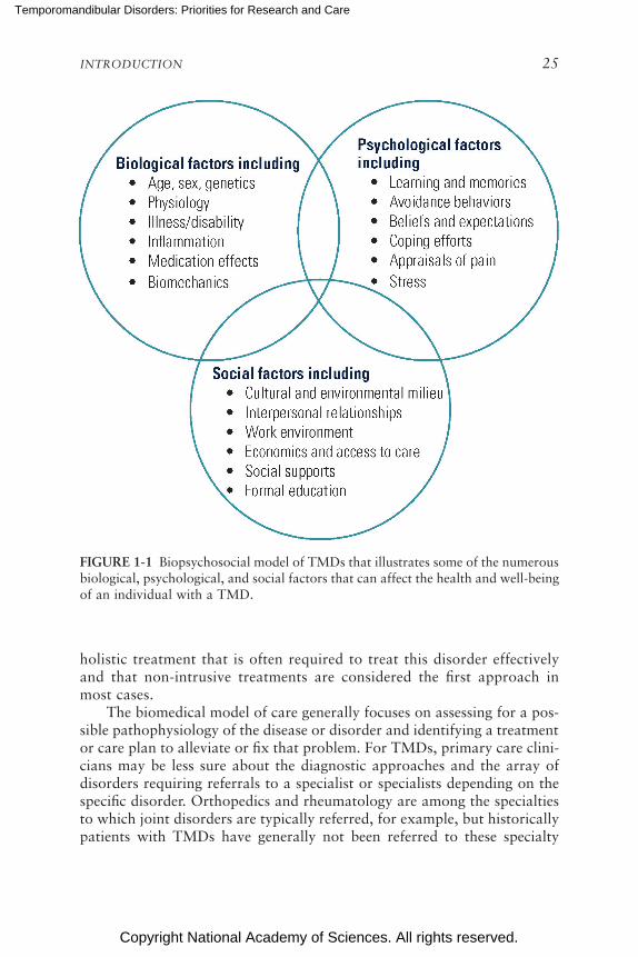
Temporomandibular Disorders: Priorities for Research and Care
Copyright National Academy of Sciences. All rights reserved.
INTRODUCTION 25
FIGURE 1-1 Biopsychosocial model of TMDs that illustrates some of the numerous biological, psychological, and social factors that can affect the health and well-being of an individual with a TMD.
holistic treatment that is often required to treat this disorder effectively and that non-intrusive treatments are considered the first approach in most cases.
The biomedical model of care generally focuses on assessing for a pos-sible pathophysiology of the disease or disorder and identifying a treatment or care plan to alleviate or fix that problem. For TMDs, primary care clini-cians may be less sure about the diagnostic approaches and the array of disorders requiring referrals to a specialist or specialists depending on the specific disorder. Orthopedics and rheumatology are among the specialties to which joint disorders are typically referred, for example, but historically patients with TMDs have generally not been referred to these specialty
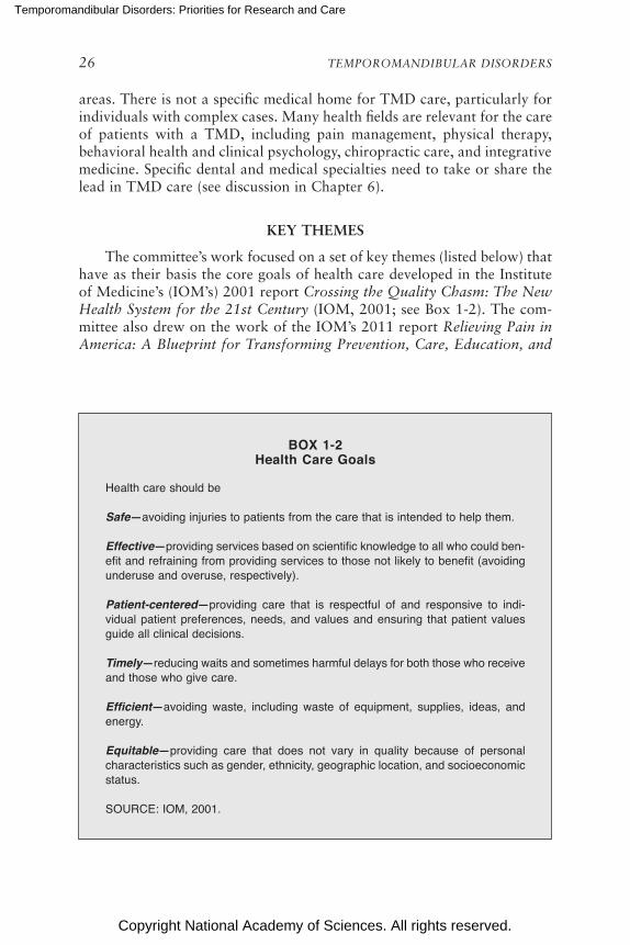
Temporomandibular Disorders: Priorities for Research and Care
Copyright National Academy of Sciences. All rights reserved.
26 TEMPOROMANDIBULAR DISORDERS
areas. There is not a specific medical home for TMD care, particularly for individuals with complex cases. Many health fields are relevant for the care of patients with a TMD, including pain management, physical therapy, behavioral health and clinical psychology, chiropractic care, and integrative medicine. Specific dental and medical specialties need to take or share the lead in TMD care (see discussion in Chapter 6).
KEY THEMES
The committee’s work focused on a set of key themes (listed below) that have as their basis the core goals of health care developed in the Institute of Medicine’s (IOM’s) 2001 report Crossing the Quality Chasm: The New Health System for the 21st Century (IOM, 2001; see Box 1-2). The com-mittee also drew on the work of the IOM’s 2011 report Relieving Pain in America: A Blueprint for Transforming Prevention, Care, Education, and
BOX 1-2 Health Care Goals
Health care should be
Safe—avoiding injuries to patients from the care that is intended to help them.
Effective—providing services based on scientific knowledge to all who could ben-efit and refraining from providing services to those not likely to benefit (avoiding underuse and overuse, respectively).
Patient-centered—providing care that is respectful of and responsive to indi-vidual patient preferences, needs, and values and ensuring that patient values guide all clinical decisions.
Timely—reducing waits and sometimes harmful delays for both those who receive and those who give care.
Efficient—avoiding waste, including waste of equipment, supplies, ideas, and energy.
Equitable—providing care that does not vary in quality because of personal characteristics such as gender, ethnicity, geographic location, and socioeconomic status.
SOURCE: IOM, 2001.
Temporomandibular Disorders: Priorities for Research and Care
Copyright National Academy of Sciences. All rights reserved.
INTRODUCTION 27
Research (IOM, 2011), the National Pain Strategy (HHS, 2016), and the Federal Pain Research Strategy (IPRCC, 2017).
For this report on TMDs, the committee focused on the following key themes:
• Recognize the spectrum of TMDs across medicine and dentistry. TMDs are a complex, heterogeneous, multifactorial set of disorders with varying treatments depending on the specific disorder. Depend-ing on the specific type of TMD and its course, an interdisciplinary approach to care is often needed that includes multiple health care clinicians across medicine, dentistry, physical therapy, behavioral health, and integrative health.
• Emphasize person-centered care. The complexity of TMDs and the frequency of other comorbid health conditions necessitates an ap-proach to care that focuses on the total health and well-being needs of each indi vidual; accomplishing that principle requires adequate time for assessment and discussion with the patient.
• Ensure careful diagnosis and avoid harm. Diverse opinions and approaches to practice within the dental community and the lack of widely adopted evidence-based care pathways3 have led to poor treat-ment outcomes and overly aggressive treatment for many individuals with a TMD. Despite the best intentions of many dentists, this lack of applying non-intrusive treatment methods as the first step in treatment has often harmed individuals. Due to the complexities of TMDs, the committee urges an emphasis on prevention strategies, correct diag-nosis, and thoughtful evidence-based treatment approaches.
• Foster an interdisciplinary approach to TMD care. Many areas of medicine, dentistry, nursing, behavioral health, physical therapy, and integrative health, as well as other health fields, contribute to TMD research and care of individuals with a TMD. Going beyond tradi-tional silos and bridging the gaps between professions will be the key to making progress, as will be education and training for those individuals in TMD research and care.
• Explore the numerous research horizons. Significant opportunities are available for research across many fields of medicine, dentistry, other health sciences, and other areas of science to gain a better understanding of the mechanisms underlying TMDs, develop an evidence base for care, and improve the implementation of best practices of care for individuals with a TMD.
3 In this report, evidence-based care is defined as care that uses current best evidence from well-designed studies, clinician expertise, and patient values and preferences in the care of individual patients and the delivery of health care services.
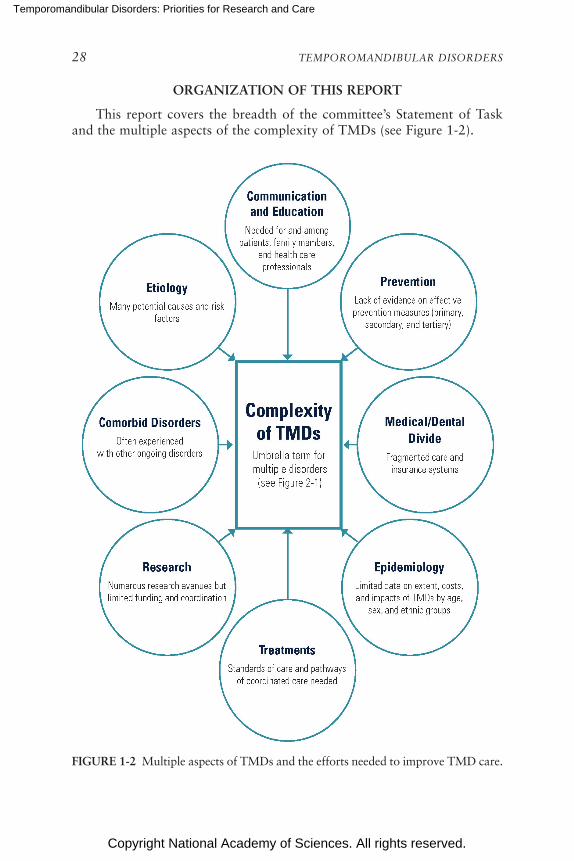
Temporomandibular Disorders: Priorities for Research and Care
Copyright National Academy of Sciences. All rights reserved.
28 TEMPOROMANDIBULAR DISORDERS
ORGANIZATION OF THIS REPORT
This report covers the breadth of the committee’s Statement of Task and the multiple aspects of the complexity of TMDs (see Figure 1-2).
FIGURE 1-2 Multiple aspects of TMDs and the efforts needed to improve TMD care.
Temporomandibular Disorders: Priorities for Research and Care
Copyright National Academy of Sciences. All rights reserved.
INTRODUCTION 29
In Chapter 2 the committee explores the definition and scope of TMDs, and in Chapter 3 it delves into what is known about the burden and costs of TMDs, with a focus on population-based studies. The broad scope and spectrum of research on TMDs is discussed in Chapter 4, including discus-sions on new horizons for TMD research. In Chapters 5 and 6 the care of individuals with TMDs is the focus, with discussions specific to improving the quality, access, and value of TMD care. Professional education and training are discussed in depth in Chapter 6, with an emphasis on health professional education. Raising awareness and increasing knowledge about TMDs for patients and for the general public is the focus of Chapter 7, with key messages identified. The report concludes in Chapter 8 with the committee’s recommendations for the short- and long-term actions that are needed to improve the health and well-being of individuals with a TMD.
REFERENCES
Engel, G. L. 1977. The need for a new medical model: A challenge for biomedicine. Science 196(4286):129–136.
Gatchel, R. J., Y. B. Peng, M. L. Peters, P. N. Fuchs, and D. C. Turk. 2007. The biopsycho-social approach to chronic pain: Scientific advances and future directions. Psychological Bulletin 133(4):581–624.
HHS (Department of Health and Human Services). 2016. National pain strategy: A compre-hensive population health-level strategy for pain. https://www.iprcc.nih.gov/sites/default/files/HHSNational_Pain_Strategy_508C.pdf (accessed October 3, 2019).
Hoppenfeld, S. 1976. Physical examination of the cervical spine and temporomandibular joint. In Physical examination of the spine and extremities. Norwalk, CT: Appleton and Lange. Pp. 105–132.
IOM (Institute of Medicine). 2001. Crossing the quality chasm: The new health system for the 21st century. Washington, DC: National Academy Press.
IOM. 2011. Relieving pain in America: A blueprint for transforming prevention, care, educa-tion, and research. Washington, DC: The National Academies Press.
IPRCC (Interagency Pain Research Coordinating Committee). 2017. Federal pain research strat-egy. https://www.iprcc.nih.gov/sites/default/files/iprcc/FPRS_Research_ Recommendations_Final_508C.pdf (accessed October 3, 2019).
Kusiak, J. W., C. Veasley, W. Maixner, R. B. Fillingim, J. S. Mogil, L, Diatchenko, I. Belfer, M. Kaseta, T. Kalinowski, J. Gagnier, N. Hu, M. Reardon, C. Greene, and T. Cowley. 2018. The TMJ Patient-Led RoundTable: A history and summary of work. http:// mdepinet.org/wp-content/uploads/TMJ-Patient-RoundTable-Briefing-Report_9_25_18.pdf (accessed September 4, 2019).
Magee, D. J. 1999. Temporomandibular joint. In Orthopedic physical assessment. D. J. Magee, ed. Philadelphia, PA: W. B. Saunders Co. Pp. 224–251.
Myers, S., S. Kaimal, J. Springsteen, J. Ferreira, C-C. Ko, and J. Fricton. 2007. Development of a national TMJ implant registry and repository: NIDCR’s TIRR. Northwest Dentistry 86(6):13–18.
The TMJ Association. 2019a. TMJ Association. http://www.tmj.org (accessed September 6, 2019).
The TMJ Association. 2019b. TMJA science meetings. http://www.tmj.org/Page/53/34 (ac-cessed September 6, 2019).

Temporomandibular Disorders: Priorities for Research and Care
Copyright National Academy of Sciences. All rights reserved.
Temporomandibular Disorders: Priorities for Research and Care
Copyright National Academy of Sciences. All rights reserved.
31
Because of the severity of the pain it is sometimes impossible to eat or to talk. The pain from simply smiling can reduce me to tears. As you can imagine this severely cuts into my interactions with others. I cannot work. I cannot go to social events like Sunday morning church service. For those who know me you know how I love to laugh and gab and sing. I can no longer do this, the pain is often too much. I have become very depressed.
—Betty
The masticatory system supports many functions vital to human health and well-being, including chewing, drinking, speech, and facial and emo-tional expression. Disorders of the masticatory system (generally termed temporomandibular disorders, or TMDs) involve the muscles of masti-cation (chewing), the temporomandibular joints (TMJs), and associated nerves and tissues (see Appendix D). Important outcomes of changes in the functioning of the masticatory system may include a reduction in the ability to use the jaw, ongoing pain, or many other subsequent impacts on an individual’s overall quality of life. These changes to the quality of life can include a decrease in the pleasure that one obtains from eating and an altera tion in eating behaviors, not just in what is consumed but also in where, when, and with whom a person eats. Such changes affect the indi-vidual’s work and social life. Also, while much more needs to be learned about the impact of TMDs and orofacial pain on people’s lives, the evi-dence is clear that cultural, geographic, socioeconomic, and gender factors contribute to the impact of altered masticatory function on an individual’s
2
Definitions and Scope: What Are TMDs?
Temporomandibular Disorders: Priorities for Research and Care
Copyright National Academy of Sciences. All rights reserved.
32 TEMPOROMANDIBULAR DISORDERS
self-image, health, and well-being. This report focuses on the impact that TMDs can have on many aspects of a person’s health and well-being, which go well beyond the face and jaw. Many TMDs are multi-faceted and need interdisciplinary attention from clinicians in medicine, dentistry, and other health care fields. The goals of this chapter are to describe the complexities underlying TMDs and to establish the terminology and classification frame-work that will be used in the remainder of this report. The chapter discusses the scope, definitions, assessments, and classification of the disorders.
TERMINOLOGY: WHAT TERM SHOULD BE USED FOR THIS SET OF DISORDERS?
The musculoskeletal structures of the masticatory system and the neu-rological structures that control a wide diversity of functions are complex, and much about them remains to be investigated (see description of the anatomy in Appendix D and research directions discussion in Chapter 4). Additionally, the cervical system has a critical role in both the normal and abnormal functioning of the TMJ. As detailed in the overview of the his-tory of the terminology (see Box 2-1), the labeling of the group of disorders affecting the masticatory system has varied across the years, reflecting the complexity of the disorders and adding to the confusion regarding what is a disorder, how the disorder should be treated, and by whom. These problems continue to the present.
This report follows the recommendations of the research community—as well as of the consensus report from the American Dental Association in 1983—and uses the term “temporomandibular disorders” (Laskin et al., 1983). TMDs are defined as a set of diseases and disorders that are related to alterations in the structure, function, or physiology of the masticatory system and that may be associated with other systemic and comorbid medi-cal conditions. The committee emphasizes the multiple disorders that are encompassed by the umbrella term TMDs and the multiple causes of these disorders. Emphasizing the plurality of the conditions is important, as there are more than 30 individual TMDs (see discussion later in this chapter). The committee emphasizes that the single term “TMD” should only be used when referring to a specific TMD, such as myofascial pain of the mastica-tory muscles. It is important to note that neither “TMD” nor “TMDs” is a diagnostic term. Each condition, as based on the most current full tax-onomy, has established diagnostic criteria, and the validity of the criteria range from untested, to tested and poor, to tested and excellent. “TMD” is not a single diagnosis but requires further diagnostic work to identify the specific disorder—or disorders—that an individual is experiencing, the potential involvement of multiple body systems and comorbid conditions, and the appropriate approaches to treatment or management. Patients often
Temporomandibular Disorders: Priorities for Research and Care
Copyright National Academy of Sciences. All rights reserved.
DEFINITIONS AND SCOPE 33
BOX 2-1 Overview of the Evolution of the Terminology
The terminology used to describe temporomandibular disorders (TMDs) has generally been associated with the prevailing ideas at the time on the causes of the disorders. The first known published observation, in 1887, of a masticatory system disorder identified internal derangements—an altered position of the disc in the temporomandibular joint (TMJ)—in an anatomically descriptive manner consistent with current knowledge (Annandale, 1887). However, a 1934 case series concluded that the ear symptoms being examined were due to the ill-fitting height of the patient’s removable dentures, resulting in a disturbance of the TMJ; the proposed treatment was to change the size of the dentures in order to alter the position of the TMJ condyle relative to the ear structures (Costen, 1934). By 1948, however, it had been determined that an underlying anatomical basis for those proposed mechanisms did not exist (Sicher, 1948). Nevertheless, the seem-ing importance of the TMJ in this newly discovered class of disorders quickly led to the dominance of terms such as “TMJ syndrome” (Klasser and Greene, 2009), later shortened to “TMJ” by the early 1950s, and established the clinical justifica-tion for therapy to be based on alterations to the occlusion (the way the teeth meet when the lower jaw and upper jaw come together) in general. Later, the focus on the occlusion included attention to minute aspects of occlusion as the cause for otherwise unexplained ear and facial pain symptoms (Molin, 1999).
The TMJ terminology was furthered in the book TMJ Pain Dysfunction Syndrome, published in 1956, which ironically focused not on the joint, but on the muscles as the source of the symptoms and identified multi-causal mechanisms, which pointed attention away from the joint itself (Schwartz, 1956). However, the use of TMJ in the title of that syndrome perpetuated the profession’s focus on the joint. Shortly thereafter, occlusal equilibration (a dentist-made change to the chewing surfaces of the teeth in order to change how they fit together) was strongly advocated as the treatment for problems affecting the TMJ (Shore, 1959). Apparent supporting data for the role of the dental occlusion in TMDs appeared by the 1960s (Ramfjord, 1961a). This study proved to be influential to the clinical practice of dentistry and to the view that structural change of the occlusion was necessary for treatment of masticatory system disorders. While this study has been cited many times in support of occlusal treatment for sleep bruxism or for jaw pain, only seldom (e.g., Skármeta, 2017) is its anecdotal nature highlighted in the literature: there is no causal evidence from this study because it had no control group. Other studies regarding the relationship between occlusal equili-bration and TMDs are case series (e.g., Racich, 2005, 2018) and do not provide causal evidence (Mohlin and Kurol, 2003; Skármeta, 2017). This continues to be a pressing and controversial issue in the treatment of TMDs (see Chapter 5).
The lack of an evidence basis underlying the assumptions that TMDs were based on either the jaw joint or the dental occlusion was countered in the late 1960s by a psychophysiological model that blended behavior with physiology and emphasized that these disorders represent changes in function rather than struc-tural changes (Laskin, 1969). This model suggested that perceived stress led to
continued
Temporomandibular Disorders: Priorities for Research and Care
Copyright National Academy of Sciences. All rights reserved.
34 TEMPOROMANDIBULAR DISORDERS
behavioral responses in the form of oral habits such as tooth clenching and that such oral habits led to muscle fatigue and myospasm; the myospasm, in turn, led to pain as well as changes in the occlusion and alterations in chewing pattern, which together contributed to perpetuation of the problem. This model, the myofascial pain dysfunction syndrome, is largely consistent with the current understanding of masticatory system pathophysiology (Murray and Peck, 2018). Subsequent well-designed experimental studies across a broad range of putative interventions pro-vided support for the psychophysiological aspect of the model (Greene et al., 1969, 1982a,b; Greene and Laskin, 1971, 1974, 1983). This model was not well received by the dental profession, as judged by the subsequent reviews that highlighted the persistent bias in the dental literature that has focused on the mechanistic perspec-tives despite the absence of supporting evidence for those perspectives (Greene, 1981, 1983; Clark et al., 1999; Greene and Obrez, 2015).
In response to the confusion regarding the nature of the masticatory system disorders as well as increasing contentiousness within the practicing dental pro-fession, in 1983 the American Dental Association (ADA) published conference-based guidelines that summarized both science and opinion (Laskin et al., 1983). One major and highly useful outcome of that conference was the establishment of the term temporomandibular disorders (TMDs) as the umbrella for the differ-ent disorders. This change implicitly (though not explicitly) established an early diagnostic tree comprised of multiple disorders, the possibility for multiple overlap-ping diagnoses, and the need to improve the quality of diagnosis. The science regarding TMDs at the time of the ADA publication was just developing, whereas the prevailing opinion in the profession was strong; consequently, the mix of sci-ence and opinion in the published ADA guidelines placed opinion on a relatively even plane with the science, thereby communicating, perhaps unintentionally, to the profession that clinical opinion was an adequate form of evidence on which to base diagnosis and treatment. A focus on the structural causes of TMDs, even in the midst of a dearth of evidence, continues to be reflected in current practice and in treatments aimed at restoring the occlusion to an idealized configuration that had little to no supporting evidence at the time and which still lacks evidence (Solberg et al., 1972; Clarke, 1982; Zarb and Mohl, 1988; Seligman and Pullinger, 1991; Schiffman et al., 1992; Pullinger et al., 1993; de Boever et al., 2000a,b; Magnusson et al., 2000; Tallents et al., 2000; Fujii, 2002; Egermark et al., 2003;
BOX 2-1 Continued
present with more than one TMD as well as comorbid health conditions (see Chapter 3).
The use of the term “TMDs” should not be construed to reflect less attention by this committee on the importance of the TMJ and the mechanical problems involving the TMJ that lead to functional limitations, as compared with TMDs that are primarily characterized by pain and that
Temporomandibular Disorders: Priorities for Research and Care
Copyright National Academy of Sciences. All rights reserved.
DEFINITIONS AND SCOPE 35
Mohlin and Kurol, 2003; Fricton, 2006; Suvinen and Kemppainen, 2007; Cairns et al., 2010; Türp and Schindler, 2012).
Another potential structural cause for TMDs has centered on the articular disc. By the early 1980s displacements of the TMJ disc were widely viewed within the clinical profession as a necessary and sufficient pathophysiological basis for persistent facial pain. The mechanistic solution, like that of treating abnormalities in the dental occlusion, was to relocate the disc (via intraoral appliances or TMJ surgery) to its anatomically normal position or to replace the disc with an implant. The impact of disc repositioning and replacement procedures on pain, however, was disappointing, in part because of insufficient evidence that the treatment achieved its goal of returning joint morphology to normal, and in part because of substantially increased treatment needs via orthodontic repositioning of the teeth or extensive prosthetic restoration following such disc repositioning, and with no evidence that this long and expensive process was successful and biologically jus-tified. Most importantly, the impact of disc replacement procedures was filled with complications from poorly planned or executed procedures that were devastating for some patients (Dolwick and Dimitroulis, 1994). Further studies emphasized that surgical correction of internal derangements as a treatment for pain may not be warranted (Emshoff et al., 2003). Additionally, a number of implants were recalled after causing significant damage to many patients (see Chapter 5).
While progress was being made in understanding the TMD pain disorders, particularly highlighted by the publication of the Research Diagnostic Criteria for Temporomandibular Disorders in 1992 (Dworkin and LeResche, 1992), the absence of progress with the mechanical joint disorders and their associated pain led to modifying the term TMD to become temporomandibular joint disorders ( TMJDs) by the 1990s as a means to focus greater attention on the joint. The obvious consequence of ignoring muscles and their disorders, arguably far more prevalent compared to mechanical joint problems, was to modify the umbrella term yet again to temporomandibular joint and muscle disorders (TMJMDs).
The profusion of terms, including the continued use of TMJ as well as TMD, TMJD, and TMJMD across multiple levels of the profession within the United States (but importantly, not used internationally to any great extent), confused cli-nicians, academicians, and patients. The use of TMJMDs to identify both muscle and joint disorders brought the discussion back full circle, as that was exactly the intent of TMD when it was established by consensus in 1983. The international academic research community continues to use the term TMDs (Ohrbach et al., 2010b), and TMDs is the term used in this report.
have received far more research attention and have, as a result, seen much more progress to date (Ohrbach and Dworkin, 2016).
Conclusion 2-1: Multiple disorders are encompassed by the terms “temporomandibular disorders” or “TMDs.” The committee defines TMDs as a set of diseases or disorders that are related to
Temporomandibular Disorders: Priorities for Research and Care
Copyright National Academy of Sciences. All rights reserved.
36 TEMPOROMANDIBULAR DISORDERS
alterations in structure, function, or physiology of the mastica-tory system and that may be associated with other systemic and co morbid medical conditions. TMDs can be usefully separated into two groups: the common TMDs with validated diagnostic criteria and the uncommon TMDs that do not yet have validated diagnostic criteria due to the challenges of conducting research on rare conditions. When possible, a patient’s diagnosis needs to be focused on the specific TMD or TMDs. “TMD” should not be used as a diagnostic term. An individual patient may have more than one TMD and may also have comorbid conditions.
DIAGNOSTIC CRITERIA TO CATEGORIZE TYPES OF TMDs
Substantial efforts have been made to categorize the multiple types of TMDs and to develop validated diagnostic criteria. Additionally, attention has been given to exploring how the types of TMDs that are painful fit into broader categorizations of orofacial pain disorders. The following section provides a brief overview of the history of the categorization systems; more details are available (Ohrbach and Dworkin, 2016). Notably across this his-tory the same dominant symptoms and signs of a small number of TMDs are seen in the general population and in those individuals seeking care. It is the manner in which those symptoms and signs are interpreted that changes across time and settings. These disorders are discussed in greater detail below. In addition to the categorizations developed by clinicians and researchers, patients often develop their own perspectives in their attempts to understand the disorder from the lived experience. Many of the patient quotes in this report highlight some of those disease perspectives.
Overview of the History of Categorization and Diagnostic Criteria for TMDs
1950s to 1980s
Diagnosis of TMDs between the 1950s and the 1980s followed sev-eral pathways. The TMJ syndrome approach (discussed in Box 2-1) tied the diagnosis specifically to various occlusal features. However, this was not found to be a workable approach for diagnosis because “abnormal” occlusal features can be found in most individuals (Proffit et al., 2013). A second pathway required appropriate history-taking coupled with an examination restricted to those parts of the masticatory system central to the disorder definition in order to determine if myofascial pain dysfunction syndrome was present; this pathway, resembling medical management, was less familiar to most dentists. In either of these first two approaches, there
Temporomandibular Disorders: Priorities for Research and Care
Copyright National Academy of Sciences. All rights reserved.
DEFINITIONS AND SCOPE 37
was one diagnosis. A third pathway focused on the position of the TMJ disc, which led to two different diagnostic and treatment approaches. One involved a focus on TMJ surgery to repair or replace the disc. However, problems were identified with joint implants, and many patients experi-enced serious adverse effects from those implants (see Chapter 5). The second approach classified individuals based on the nature of the occlusion and its purported effects on the position of the clicking disc and the bony TMJ. This approach led to extensive (and often very expensive) orthodontic and prosthetic treatments in order to alter the occlusion (Bellavia and Missert, 1985; Laurell, 1985; Lundh et al., 1985; Kurita et al., 2001). A retrospective assessment of disc repositioning treatments clearly indicates that it was an unnecessary intervention (Greene and Obrez, 2015).
By the late 1980s, nine different published categorization systems had emerged for the diagnosis of TMDs. One evaluation of these systems used the following criteria: methodological considerations (sampling method, research suitability, and inter-rater reliability of clinical evaluation), diag-nostic validity (specificity, inter-rater reliability of diagnosis), and clinical considerations (biological plausibility, exhaustive diagnostic framework, provision for multiple diagnoses, and clinical decision making) (Dworkin and LeResche, 1992). None of the evaluated systems met the criteria re-quired of a diagnostic system.
Research Diagnostic Criteria for Temporomandibular Disorders and Initial Guidelines from the American Academy of Orofacial Pain
In 1992 the Research Diagnostic Criteria for Temporomandibular Dis-orders (RDC/TMD) was released (Dworkin and LeResche, 1992). This effort, which was supported by funding from the National Institutes of Health, was based on the biopsychosocial model of health and disease and relied on epidemiological data. The RDC/TMD incorporated a dual axis system: Axis I focused on clinical history and physical examination find-ings in support of non-overlapping diagnoses, and Axis II focused on pain parameters, mandibular jaw function, psychological status, and the level of psychosocial function (Dworkin, 2010). This approach used strongly operationalized criteria in order to facilitate inter-examiner reliability, and it allowed for the provision of multiple diagnoses. The RDC/TMD was developed to provide an instrument to be used in epidemiological, obser-vational, and clinical trials research (Dworkin, 2010). Use of the dual-axis system with TMDs was considered controversial by clinical dentists at the time of publication, but, in the intervening years, the dual-axis approach has become more accepted within dentistry and has become a model for other pain classification systems (Garofalo and Wesley, 1997; Deyo et al., 2014; Ohrbach and Dworkin, 2016).
Temporomandibular Disorders: Priorities for Research and Care
Copyright National Academy of Sciences. All rights reserved.
38 TEMPOROMANDIBULAR DISORDERS
Three critical characteristics of the RDC/TMD were apparent subse-quent to the 1992 publication. The first was the recognition that continued research was needed in order for this descriptive classification system to evolve. The second was that the RDC/TMD was not intended to be either inclusive of all TMDs or self-sufficient with regard to differential diagno-sis requirements for distinguishing a potential diagnosis of a TMD from other diagnoses. And the third was that only the more common TMDs were included in the RDC/TMD because the prevalence of the uncommon TMDs was so low as to preclude effective research at a single clinic. The assessment and diagnostic reliability of the RDC/TMD was found to be ac-ceptable (Lobbezoo et al., 2004; John et al., 2005; List et al., 2006; Look et al., 2010), but the diagnostic validity needed to be evaluated. The latter required clear decision rules for non-overlapping diagnoses.
Also during the early 1990s the American Academy of Orofacial Pain developed a clinical diagnostic system that aimed to be comprehensive of all the known TMDs. Disorders were defined in an inclusive manner of multiple features, and a diagnosis was based on presence of any of those features. However, the system was hampered by several problems that limited its validity.
Diagnostic Criteria for Temporomandibular Disorders (DC/TMD)
In 2001 a major multi-site assessment of the RDC/TMD was initiated (funded by the National Institute of Dental and Craniofacial Research), which led to published outcomes, a public symposium in 2008, and an international consensus workshop in 2009 (Ahmad et al., 2009; Anderson et al., 2010; Dworkin, 2010; Haythornthwaite, 2010; John, 2010; List and Greene, 2010; Lobbezoo et al., 2010; Look et al., 2010; Ohrbach et al., 2010a; Schiffman et al., 2010a,b; Stegenga, 2010; Truelove et al., 2010). The workshop was led by the International RDC/TMD Consortium Net-work of the International Association for Dental Research (now named INfORM [International Network for Orofacial Pain and Related Disorders Methodology]) and the Orofacial Pain Special Interest Group of the Inter-national Association for the Study of Pain (IASP), along with individuals representing a range of specialty areas. The working groups expanded and refined the TMD taxonomy, and their work led to the eventual publica-tion in 2014 of the Diagnostic Criteria for Temporomandibular Disorders ( DC/TMD) (Schiffman et al., 2014b), which maintained or improved all of the foundational principles underlying the RDC/TMD for the common TMDs. The Axis II assessment procedures were expanded and improved. The DC/TMD delineates 12 disorders, each based on clearly defined crite-ria; each criterion is well operationalized with regard to the required clinical procedures (Ohrbach et al., 2014), which permits reliable classification with
Temporomandibular Disorders: Priorities for Research and Care
Copyright National Academy of Sciences. All rights reserved.
DEFINITIONS AND SCOPE 39
known validity for 10 of those disorders. DC/TMD-relevant interpretation of any indicated imaging is also available (Ahmad et al., 2009). To date, no other diagnostic approach for TMDs exhibits these characteristics of an integrated system for classification. Few other diagnostic systems for TMDs clearly identify and operationally define psychosocial constructs relevant to pain that need to also be assessed. While weaknesses of the DC/TMD are subsequently discussed, the importance of such a system cannot be overstated at this stage in the development of yet better diagnostic methods and more effective treatments. The literature and the Internet contain an abundance of diagnostic “classification systems” for TMDs (some of which are briefly summarized below), yet epidemiological data for incidence and persistence, clear operationalization of criteria, coherence between criteria and definition of the disorder, reliability, and validity are consistently miss-ing from such systems.
Additional subsequent work has led to an updated and expanded DC/TMD taxonomy for the other TMDs which are far less common. Opera tionalizable criteria and clear decision rules, consistent with the defi-nition, were created for each of these uncommon disorders.
In total—and depending on how one considers the disorders as orga-nized in a hierarchical framework—more than 30 TMDs have been identi-fied through the DC/TMD and expanded taxonomy. The criteria continue to be evaluated through ongoing research efforts. The resulting classifica-tion of the full set of TMDs is depicted in Figure 2-1 and encompasses the range of muscular, joint, headache, and other disorders that are considered TMDs. Other extensions of the DC/TMD include the:
• American Academy of Orofacial Pain’s Guidelines for Assessment, Diagnosis, and Management, now in its sixth edition, which includes the expanded DC/TMD (de Leeuw and Klasser, 2018);
• International Classification of Orofacial Pain, which includes the pain diagnoses from the DC/TMD into the broader pain taxonomy developed by the IASP and merges with the International Classifica-tion of Diseases, 11th Revision (ICD-11) (Benoliel et al., 2020); and
• ACTTION-APS1 pain taxonomy, which has a specific focus on the chronic painful TMDs (Ohrbach and Dworkin, 2019).
Since the initial review of TMD diagnostic systems in 1992, some of the systems have persisted, and new diagnostic or patient classification systems have emerged (reviewed in Klasser et al., 2018). Separate from the DC/TMD and its extensions, these other systems have varying levels of evidence for
1 Analgesic, Anesthetic, and Addiction Clinical Trial Translations, Innovations, Opportuni-ties, and Networks (ACTTION) and American Pain Society (APS).
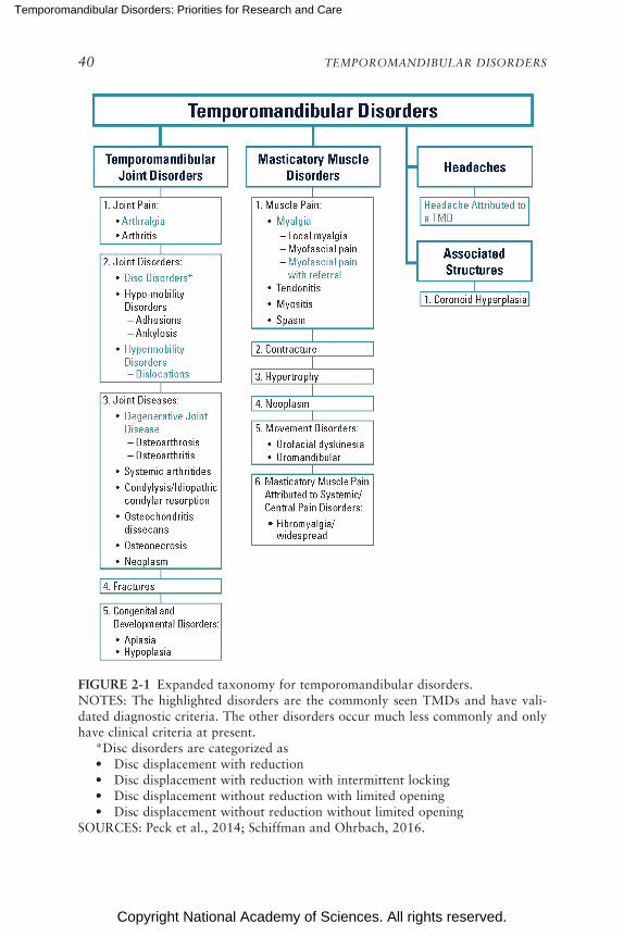
Temporomandibular Disorders: Priorities for Research and Care
Copyright National Academy of Sciences. All rights reserved.
40 TEMPOROMANDIBULAR DISORDERS
FIGURE 2-1 Expanded taxonomy for temporomandibular disorders.NOTES: The highlighted disorders are the commonly seen TMDs and have vali-dated diagnostic criteria. The other disorders occur much less commonly and only have clinical criteria at present. *Disc disorders are categorized as • Disc displacement with reduction • Disc displacement with reduction with intermittent locking • Disc displacement without reduction with limited opening • Disc displacement without reduction without limited openingSOURCES: Peck et al., 2014; Schiffman and Ohrbach, 2016.
Temporomandibular Disorders: Priorities for Research and Care
Copyright National Academy of Sciences. All rights reserved.
DEFINITIONS AND SCOPE 41
diagnostic reliability and validity (as distinguished from technical reliability and validity) (Wilkes, 1989; Pertes and Gross, 1995; Suvinen et al., 2005; Woda et al., 2005; Okeson, 2008; Simmons and AACP, 2009; Benoliel and Sharav, 2010; Stegenga, 2010; Cooper, 2011; de Silva Machado et al., 2012; Monaco et al., 2017; AES, 2019; Piper, 2019). Further contributions to the understanding of pain, clinical dysfunction associated with the mas-ticatory system, diagnostic utility, and the bio psycho social model applied to TMDs are promising for some of the systems (Suvinen et al., 2005; Woda et al., 2005; Benoliel and Sharav, 2010; Stegenga, 2010; de Silva Machado et al., 2012; Monaco et al., 2017), while the others are character-ized by little to no evidence or have been surpassed by other developments. At present, only the DC/TMD (and its extensions) for the common TMDs meets the diagnostic system criteria pertaining to methodological consid-erations, diag nostic validity, and clinical considerations. Strong diagnostic systems call for further validation research through self-evaluation as part of planned future revisions.
Learning from Classification Approaches to Low Back Pain
An approach very similar to the DC/TMD has been taken for back pain. A consensus task force appointed by the National Institutes of Health Pain Consortium drafted standards for research on chronic low back pain with three sets of recommendations (Deyo et al., 2014). The first part of the recommendations of this task force focused on the classification of chronic low back pain and the classification of the person with this disorder. The recommendations included the following: operationalized criteria defining chronic low back pain; classification by its impact; and a minimum set of measures to characterize individuals with chronic low back pain, includ-ing medical history, physical examination, diagnostic testing, self-reported functional status, psychosocial factors, and mood disturbance. The second part of the recommendations focused on best practices for outcomes mea-sures. The third part of the recommendations focused on the projected research by which the recommendations could be empirically evaluated.
Of particular relevance to this report, the task force on chronic low back pain also identified a number of key principles underlying the struc-ture of the recommendations:
• Guidelines should be evidence-based and incorporate a biopsycho-social model of chronic pain.
• The absence of an identified pathology should not lead to the assump-tion that the pain is psychological or somatoform.
• The classification should incorporate the impact of pain on function. • A minimal set of measures should be routinely used.
Temporomandibular Disorders: Priorities for Research and Care
Copyright National Academy of Sciences. All rights reserved.
42 TEMPOROMANDIBULAR DISORDERS
• The approach should be appropriate for population, observational, and interventional research.
• The evaluation should include both biomedical and psychosocial variables.
In addition, for further consideration, prognostic variables need to be defined, and research standards should evolve.
It is worth highlighting that, in parallel with information accompanying the DC/TMD framework, the report on chronic low back pain emphasized the role of a practical and evidence-based diagnostic system in moving forward to improve patient care and outcomes.
Next Steps for TMD Diagnostic Criteria
The diagnostic validity of the DC/TMD is excellent for painful TMDs, excellent for subluxation, good for disc displacement without reduction in the acute phase of limited jaw movement, and poor for other disc dis-placements and degenerative joint disease, with the stated recommendation within the DC/TMD to use appropriate imaging for the disorders that have poor diagnostic validity when a definitive diagnosis is clinically required. Further efforts to build on and improve the DC/TMD include:
• Better understanding of the orofacial pain disorders and how painful TMDs (a type of musculoskeletal pain) fit within that broader set of pain disorders (Benoliel et al., 2019);
• Better understanding of the interplay between pain mechanisms and mechanical problems, such as disc displacement and degenerative changes in the TMJ, and of where such pains fit into an orofacial pain disorder classification;
• Exploring how to categorize the painful TMDs within the IASP classification of similar pain disorders elsewhere in the body and ensuring that the linkage to ICD-11 (and beyond) facilitates better health care for TMDs (Benoliel et al., 2020);
• Highlighting the importance of chronic primary pain as a disorder and implementing that in health care settings for early and rational recognition of chronic TMDs (Nicholas et al., 2019); and
• Extending beyond the current two-axis (physical diagnosis, psycho-social and functional status) approach of the DC/TMD, and inves-tigating additional axes such as genomic classification, mechanisms, and role of comorbid pain and general health problems, consistent with current approaches to all chronic pain problems; such expansion has implications for improved patient assessment, classification, and management (Fillingim et al., 2014; Ohrbach and Dworkin, 2019).
Temporomandibular Disorders: Priorities for Research and Care
Copyright National Academy of Sciences. All rights reserved.
DEFINITIONS AND SCOPE 43
Efforts in the field of biomedical ontology—a field that develops frame-works and terminology—are expected to clarify and enhance the under-standing of what the disorders are and their diagnostic criteria (Ohrbach and Dworkin, 2016). The differences in the diagnostic validity of the vari-ous subtypes of TMD as defined in the DC/TMD indicate that changes will likely be made to the current constructs within TMDs as more is learned about the underlying pathology and pathophysiology of TMDs, so that the defined disorders and their criteria can better represent the actual disease processes. More clinical and basic research will be required to identify all etiological processes that lead to TMDs and the pathological processes that develop from them. However, care must be taken that the terminology used to report such new insights is clear and at the correct level of detail. The basic formal ontology outlined in the ISO standard (ISO, 2019) in particu-lar may play an important role. Not only does the basic formal ontology offer the vocabulary to represent the various biomedical entities involved in TMDs at the necessary level of granularity, but it does so in a way that the consequences are computable and predictable.
The joint disorders identified within the DC/TMD are disc displace-ments and other degenerative changes in the joint that are based on decades of research. The expanded DC/TMD adds another 14 joint disorders (Peck et al., 2014), but collectively the group represents conventional approaches to classification. The slow advances in understanding of disorders specifi-cally of the TMJ suggest that the current approach to classification warrants further examination. For example, the attempted integration of tissue sys-tems by Stegenga (2010) represents an obvious departure from the conven-tional classification based on simple changes in the anatomy. Findings from the RDC/TMD Validation Project point to possibly different constellations of signs and symptoms for defining soft-tissue disorders (currently, inter-nal derangements) and hard-tissue disorders (currently, degenerative joint disease) (Schiffman et al., 2010a,b). Other approaches for the classification of pain disorders rely on a hierarchical modeling of clinical and imaging findings (Rudy et al., 1988, 1990).
Additionally, the central role of pain in persistent disorders of the TMJ may also be reconsidered in light of the primary findings from the Orofacial Pain Prospective Evaluation and Risk Assessment (OPPERA) study regarding the incidence of a painful TMD: TMDs seldom exist as isolated conditions and that general indicators of poor health, including comorbid conditions, increase the risk of developing a TMD (Slade et al., 2016). The implication is that the biopsychosocial model needs to be fully used with complex con-ditions such as TMDs, and one possibility is that the application of a full biopsychosocial model with early TMDs could prevent persistent disorders of the TMJ from occurring. Another possibility is that the medical and surgical treatments used to date to deal with the progressive disorders of the TMJ
Temporomandibular Disorders: Priorities for Research and Care
Copyright National Academy of Sciences. All rights reserved.
44 TEMPOROMANDIBULAR DISORDERS
have had limited success because the treatment has been provided in a bio-medical context rather that the biopsychosocial context, the latter pointing to the simultaneous involvement of other forms of treatment.
For any advancements to occur, diagnostic test reliability, clear deci-sion rules, and diagnostic validity are required before routine clinical use can be considered. The evidence-based DC/TMD is appropriate for use in both clinical and research settings for the common TMDs (Schiffman et al., 2014b). However, the DC/TMD, and in particular the use of Axis II, is pres-ently under-utilized in most relevant clinical settings (Visscher et al., 2018; Sharma et al., 2019b). Looking ahead, a potentially more useful approach will integrate joint neurophysiology, the complex biomechanics exhibited by the TMJ, interactions of the peripheral and central nervous system, be-havioral patterns, and longitudinal considerations such as life-course and psychosocial factors that increase risk for onset, transition to chronicity, and maintenance of chronicity—all of which bear on the status of the joint. Finally, a useful diagnostic system for TMDs will need to integrate behav-ioral, functional, biological, pharmacological, and surgical approaches to therapy, and those approaches must be linked to the proposed diagnoses if one is to fully understand the pathophysiology of these disorders and identify realistic expectations for which form of therapy is appropriate for which aspects of these disorders, and for which patient, given that psycho-social factors are critical for the expression and course of pain.
At present, the DC/TMD classification of TMDs falls short with regard to the additional considerations raised here, and more needs to be done to facilitate the use of reliable and valid patient classification by clinicians. The DC/TMD does fulfill the goal of classifying the most prevalent hard- and soft-tissue disorders, and with suitable training for the use of these tools (as with any new procedure that clinicians adopt) the DC/TMD is an excellent tool within the scope of its design. The intent is to assist clinicians in iden-tifying the pertinent disorder and selecting appropriate treatments based on currently available information (where the clinical treatment studies use the DC/TMD almost exclusively), and to at least do no harm via unnecessary or inappropriately aggressive therapies. The widespread use of the DC/TMD for patient classification in clinical trials research further points to the ben-efits for clinicians to use the same tool, which would enhance transfer from research to clinical practice.
Another limitation of the DC/TMD is its conventional approach to ana-tomical separation into disorder groups. Further challenges center around the assumption that diagnostic systems will embed etiology and pain and disorder mechanisms, to the extent that such information is available. While the OPPERA study (described in more detail in Chapter 3) has pro-vided etiological information on the painful TMDs (Slade et al., 2013a, 2016; Meloto et al., 2019; Sharma et al., 2019b; Ohrbach et al., in press),
Temporomandibular Disorders: Priorities for Research and Care
Copyright National Academy of Sciences. All rights reserved.
DEFINITIONS AND SCOPE 45
an attempt to incorporate such information into a revised TMD diagnostic system might be premature at this time.
Conclusion 2-2: The Diagnostic Criteria for Temporomandibular Disorders (DC/TMD) provides the most thorough and accurate diag nostic criteria for the most common types of TMDs. Addi-tional work is needed on the diagnostic criteria for other types of TMDs that are uncommon. Efforts to increase its utility to clini-cians are priorities, including the brief DC/TMD assessment tool that is currently in development. Widespread dissemination of these tools, coupled with provision of training in order to maximize the information yield from these tools, to general practice dentists is needed; primary care clinicians need to be apprised of these devel opments so that targeted referrals and appropriate expecta-tions of good clinical practice will occur.
TYPES OF TMDs
Most masticatory system disorders fall into two groups: those associ-ated with pain and those associated with functional or structural changes in the TMJ (and which may also include pain) (Scrivani et al., 2008; Schiffman et al., 2014a; Ohrbach and Dworkin, 2016). There has been substantial research and thus knowledge about the first group. In contrast, knowledge regarding the functional and structural changes in the TMJ lags (Ohrbach and Dworkin, 2016), for reasons that are addressed throughout this report. Each of these two major groups includes common disorders that account for the majority of the complexity in diagnosis and treatment of the patient, burden to the health care system, and controversy within the profession. There is also a larger number of uncommon masticatory system disorders about which less is known. The following descriptions highlight several types of TMDs as indicated above in Figure 2-1; these descriptions use readily agreed upon characteristics rather than specific diagnostic criteria.
As noted throughout this report, TMDs are often complex disorders that can have multi-system components (described further below) and multiple comorbid medical conditions. One patient often has multiple diagnoses (e.g., myalgia and disc displacement), with substantial overlap in history and impact. It is not unusual for the multiple diagnoses of myo-fascial pain, arthralgia, disc displacement with reduction, and headache attributed to a TMD to be present in the same individual. Such overlap can make it difficult to distinguish which specific diagnosis is primary or which is necessarily the best target for treatment. This overlap of diagnoses may be analogous to what has been observed in low back pain where “non-specific low back pain” is an established and useful term for early diagnostic and
Temporomandibular Disorders: Priorities for Research and Care
Copyright National Academy of Sciences. All rights reserved.
46 TEMPOROMANDIBULAR DISORDERS
treatment stages. See Chapter 5 for further information regarding the im-portance of diagnostic overlap among TMDs.
Myalgia and Myofascial Pain
Both myalgia and myofascial pain refer to pain originating from muscle. While differences in their respective proposed mechanisms are suspected, the terms are often used interchangeably, and the distinctions in clinical char-acteristics and proposed mechanisms may have little clinical significance. Myalgia refers to pain in the muscle without a specific mechanism causing the pain. Generally, myalgia is identified by complaint of pain localized to a particular area and the presence of enhanced pain upon provocation. Provocation can be tested either by the application of pressure to the skin overlying the muscle or by testing the individual’s range of motion. Myo-fascial pain is denoted by spreading pain (extension of pain beyond the initial focal point) or pain referral (pain located in another body structure, remote to the source). Because of the lack of evidence for specific mecha-nisms for myofascial pain, the diagnosis of myofascial pain is controversial (Cohen and Quintner, 2008; Quintner et al., 2015). No substantial evi-dence is available demonstrating that different treatments must be used for myalgia versus myofascial pain. Consequently, myalgia is often the preferred diagnostic term. While myalgia and myofascial pain can persist for years or even decades, there is no evidence that there is a progressive underlying disease; rather, the pain is more accurately considered to be chronic primary pain (Nicholas et al., 2019). Chronic primary pain includes the subtype of high-impact chronic pain, which is associated with higher usage of health care for pain, lower quality of life, more pain-related interference with activi ties, and more frequently reported pain at multiple anatomic locations (Von Korff et al., 2016). Jaw injury has been strongly associated with inci-dent TMD (Sharma et al., 2019a), and stress can impact behavior (e.g., in the form of oral parafunction), which increases TMD pain (Ohrbach and Michelotti, 2018). However, the cumulative impact of multiple risk factors has greater evidence (Vlaeyen and Linton, 2000; Slade et al., 2016). A dis-tinction between initiating factors for initial onset and perpetuating factors for the continuation of the condition is a dominant theme for this type of pain (Fricton, 1985; Simons, 1985). The current model of care for myalgia emphasizes the importance of self-management for symptom control and of addressing the behavioral factors that contribute to persistence.
Arthralgia
Arthralgia refers to pain in the joint. The characteristics are parallel to those for myalgia. While there might be value in differentiating the source
Temporomandibular Disorders: Priorities for Research and Care
Copyright National Academy of Sciences. All rights reserved.
DEFINITIONS AND SCOPE 47
of the pain within the TMJ to include the specific structure where pain is present, the small size of the joint renders such distinctions neither reliable nor clinically useful, as based on the available evidence. The same caveats regarding treatments for myalgia apply to arthralgia. However, arthralgia may also accompany TMJ disc disorders or degenerative joint disease (see sections below).
Headache Secondary to TMD Pain
Whether headache secondary to a painful TMD is a headache disorder or a TMD, headache and TMD pain overlap and share underlying patho-physiological mechanisms, clinical characteristics, and neurovascular anat-omy as noted by Benoliel and Sharav (2010). It is one of the few identified comorbidities of TMDs that has a specific name and criteria for diagnosis. The headache may be of any type (e.g., migraine, tension type), a painful TMD diagnosis must be present, and the headache pain must be repli-cated by clinical examination procedures normally used to provoke pain identified as myalgia or arthralgia. One primary goal in making this type of diagnosis is to better integrate the list of disorders affecting a specific individual with the goal of clarifying what kind of pain is present and what treatments are needed. For example, if headache is secondary to a painful TMD, it may not require any specific treatment beyond that indicated for the painful TMD. The reverse can also occur.
Disc Disorders
Internal derangement of the disc refers to the displacement of the articular disc from its normal functional relationship with the mandibular condyle. Disc displacements of at least one of the joints are common and are estimated to occur in about one-third of the adult population. For the majority of those with disc displacement of the TMJ, there is little to no functional impact and no pain. Other TMD problems may co-exist with a symptom-free disc; this type of joint condition generally requires no treatment.
For a small number of individuals, the disc displacement is associated with substantial pain, limitation, and disability. The cause of disc displace-ments is largely unknown; growth discrepancies between the condyle and the developing occlusion have been suspected, but the available evidence does not support this (Farella et al., 2007). Trauma—particularly that asso ciated with whiplash-associated disorders—has also been proposed as a cause of disc displacements, but the evidence is limited by the pre-dominantly cross-sectional study designs whereas prospective designs are essential (Lee et al., 2018).
Temporomandibular Disorders: Priorities for Research and Care
Copyright National Academy of Sciences. All rights reserved.
48 TEMPOROMANDIBULAR DISORDERS
In the mild form of the disorder, the disc typically returns to its normal position during movement of the jaw as the condyle moves forward; a popping or clicking noise may accompany the reduction (i.e., the return of the disc to the normal position) during opening or closing or may ac-company the displacement (the return of the disc to the abnormal posi-tion) during closing. This disorder is diagnosed using magnetic resonance imaging (MRI). In a more severe form of the disorder, the disc remains displaced throughout the maximum opening cycle. In the more acute phase of the severe form, the displaced disc results in a mechanical obstruc-tion to opening and is symptomatic, while in the more chronic phase the posterior attachment to the disc stretches and normal mobility is usually restored, though normal function may be limited and symptoms may re-main. Histologic evidence indicates that the posterior attachment of the disc may undergo change or remodeling from elastic connective tissue to dense connective tissue (Scapino, 1983). Based on clinical data, it is likely that individuals with such remodeling of the disc tissues into a pseudo-disc regain full masticatory function as well as normal mobility of the mandible (de Leeuw et al., 1994).
None of the disc displacements can be diagnosed based on a clinical assessment of clicking sounds or jaw deviation during opening; while diag-nosis has been attempted with various instruments, MRI remains the stan-dard method for diagnosis (Li et al., 2012; Sharma et al., 2013; Schiffman et al., 2014b) and should be used for significant mechanical joint problems, suspicion of significant disease, or when treatment has been unsuccessful (Schiffman and Ohrbach, 2016) and only if the prognosis or selection of treatment will depend on an exact diagnosis (Schiffman et al., 2014b).
The human TMJ is a unique structure, and clinical disorders associ-ated with disc displacement are poorly understood. For most individuals disc displacements are relatively minor and self-limiting, but for some indi viduals disc displacements represent substantial problems. It is not yet known which types of disc displacement in the early stages are indicative of later problems.
Degenerative Joint Disease
Breakdown of the cortical bone of the TMJ condyle has been termed osteoarthritis, osteoarthrosis, and degenerative joint disease. All three terms refer to the same underlying bony changes. The term osteoarthritis is used when pain is present, while the diagnostic term osteoarthrosis is used when pain is absent; these terms are used in this distinctive manner within the research literature pertaining to the TMJ, perhaps because asymptomatic adaptive bony changes are common to the TMJ. By contrast, osteoarthritis and osteoarthrosis are used interchangeably within the medical literature.
Temporomandibular Disorders: Priorities for Research and Care
Copyright National Academy of Sciences. All rights reserved.
DEFINITIONS AND SCOPE 49
Degenerative joint disease is the term used by the DC/TMD, based on ex-tensive considerations regarding terminology.
Degenerative joint disease in the TMJ, as in other joints, is the con-sequence of chronic abnormal mechanical loading to the joint. This may occur as a result of a long-term advanced internal derangement in the TMJ, but only about 15 percent of persons with internal derangements are so affected. When degeneration does occur, it may lead to pain and further mechanical joint dysfunction. Such degeneration may require surgical treat-ment to improve pain and function, such as arthrocentesis, arthroscopy, open joint arthroplasty, or total joint replacement. Due to its chronicity, however, osteoarthritis is frequently associated with pain comorbidities, and evidence from other load-bearing joints such as the knee clearly indi-cates that pain and disability associated with degenerative joint disease are not predicted by the extent of bony destruction but rather by the same full range of biopsychosocial factors applicable to TMDs as a whole (Summers et al., 1988; Salaffi et al., 1991; Dekker et al., 1993; McAlindon et al., 1993).
TMJ Subluxation and Luxation
As noted in Appendix D, the condyle has an expected extent of motion; however, in some cases the condyle exceeds that range and prob-lems can ensue. In some individuals the additional movement can result in the condyle being momentarily stuck in that position, which is termed subluxation, or the condyle can be stuck in a more extreme manner and may require manual reduction (external assistance) to relocate the condyle back to the fossa, which is termed luxation or dislocation. The literature is not consistent in how these three terms are defined. This condition is typi-cally highly distressing and often painful. In other individuals, such extreme movement of the condyle is not associated with dislocation. There are a few suspected causes for this condition. Angulation of the eminence that bounds the anterior extent of the joint space is believed to contribute to disloca-tion, especially to recurrent dislocations, which occur in a small number of individuals, but this observation is largely anecdotal; nevertheless, surgical correction of the bony shape, bony augmentation, or injection therapies appear to reduce if not eliminate the frequency of recurrent dislocations (Fernandez-Sanroman, 1997; Moore and Wood, 1997; Undt et al., 1997; Caminiti and Weinberg, 1998). Recurrent yawning or external injury to the jaw can contribute to this condition; however, for most individuals the onset is without an identified contributing factor. While this type of prob-lem has a set of reasonably applicable treatment procedures, recurrent dis-locations are nevertheless believed to be associated with a stretching of the capsule and TMJ ligaments and thereby represent a form of joint instability.
Temporomandibular Disorders: Priorities for Research and Care
Copyright National Academy of Sciences. All rights reserved.
50 TEMPOROMANDIBULAR DISORDERS
Consequently, exercises for joint stability as well as injections for facilitat-ing connective tissue growth in the joint capsule and consequent decrease in joint mobility have been proposed (Bell, 1979; Zhou et al., 2014), but they are not well understood, and this lack of understanding highlights the gap between the apparent physiology of the TMJ and its surprising complexity.
Relationship of TMDs to Orofacial Pain Disorders
The common painful TMDs (myalgia, myofascial pain, and arthralgia) are similar to pain disorders found elsewhere in the body, and the same diagnostic principles and treatment principles are applicable. The transla-tion of the current state of the scientific evidence among medical and dental researchers and clinicians is key to furthering the understanding of these pain conditions. The major classes of orofacial pains are musculoskeletal, neuropathic, and visceral. However, several types of TMDs overlap these areas, particularly the musculoskeletal pain conditions. Currently the pain-ful conditions within TMDs are now simultaneously also a subgroup of a broader set of orofacial pain conditions within the International Clas-sification of Orofacial Pain (Benoliel et al., 2020). Such pains, whether primarily identified as muscle (or fascial) in origin or as stemming from the TMJ (such as during function) should be fully assessed within the bio-psycho social framework.
The mechanical disorders may also exhibit pain, but pain is typically not a required diagnostic criterion. Consequently, attempts to organize TMDs as part of orofacial pains more broadly have only been partially suc-cessful. The mechanical TMJ problems do not align well with the orofacial pains and rather should be considered as primarily orthopedic joint disor-ders. In summary, TMDs comprise two large classes of disorders: painful disorders and mechanical joint disorders. Evidence from other joints indi-cates that mechanical TMJ problems should also be fully assessed within the biopsychosocial framework.
UNDERSTANDING THE ETIOLOGIES OF TMDs
As noted above regarding the evolution of terminology for this set of disorders, varying approaches have been explored over the years as to what causes TMDs and, as a result, where the areas of emphasis should be concerning TMD management and treatment. Ongoing controversies and divisions within the practice of dentistry continue regarding these issues; Box 2-2 provides an overview of some of the more frequently encountered approaches that are not evidence based with respect to the required criteria stated in this chapter which a diagnostic system must meet. In addition the issue of occlusion is discussed separately in the next section because it has
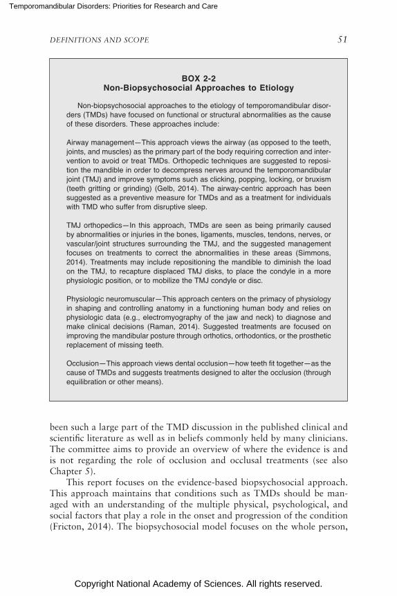
Temporomandibular Disorders: Priorities for Research and Care
Copyright National Academy of Sciences. All rights reserved.
DEFINITIONS AND SCOPE 51
BOX 2-2 Non-Biopsychosocial Approaches to Etiology
Non-biopsychosocial approaches to the etiology of temporomandibular disor-ders (TMDs) have focused on functional or structural abnormalities as the cause of these disorders. These approaches include:
Airway management—This approach views the airway (as opposed to the teeth, joints, and muscles) as the primary part of the body requiring correction and inter-vention to avoid or treat TMDs. Orthopedic techniques are suggested to reposi-tion the mandible in order to decompress nerves around the temporomandibular joint (TMJ) and improve symptoms such as clicking, popping, locking, or bruxism (teeth gritting or grinding) (Gelb, 2014). The airway-centric approach has been suggested as a preventive measure for TMDs and as a treatment for individuals with TMD who suffer from disruptive sleep.
TMJ orthopedics—In this approach, TMDs are seen as being primarily caused by abnormalities or injuries in the bones, ligaments, muscles, tendons, nerves, or vascular/joint structures surrounding the TMJ, and the suggested management focuses on treatments to correct the abnormalities in these areas (Simmons, 2014). Treatments may include repositioning the mandible to diminish the load on the TMJ, to recapture displaced TMJ disks, to place the condyle in a more physiologic position, or to mobilize the TMJ condyle or disc.
Physiologic neuromuscular—This approach centers on the primacy of physiology in shaping and controlling anatomy in a functioning human body and relies on physiologic data (e.g., electromyography of the jaw and neck) to diagnose and make clinical decisions (Raman, 2014). Suggested treatments are focused on improving the mandibular posture through orthotics, orthodontics, or the prosthetic replacement of missing teeth.
Occlusion—This approach views dental occlusion—how teeth fit together—as the cause of TMDs and suggests treatments designed to alter the occlusion (through equilibration or other means).
been such a large part of the TMD discussion in the published clinical and scientific literature as well as in beliefs commonly held by many clinicians. The committee aims to provide an overview of where the evidence is and is not regarding the role of occlusion and occlusal treatments (see also Chapter 5).
This report focuses on the evidence-based biopsychosocial approach. This approach maintains that conditions such as TMDs should be man-aged with an understanding of the multiple physical, psychological, and social factors that play a role in the onset and progression of the condition (Fricton, 2014). The biopsychosocial model focuses on the whole person,
Temporomandibular Disorders: Priorities for Research and Care
Copyright National Academy of Sciences. All rights reserved.
52 TEMPOROMANDIBULAR DISORDERS
including the mind, body, emotions, spirituality, lifestyle, social relation-ships, and physical environment. At present, only the biospsychosocial ap-proach has both strong evidence and strong theory relating clinical findings to symptoms and approaches to treatment that are consistent with what is known about chronic pain elsewhere in the body.
Occlusion
Because this has been an area of ongoing discussion, particularly within the field of dentistry, the committee focuses this section on occlusion be-fore discussing its adoption of the biopsychosocial model for etiology and treatment.
The field of dentistry has historically focused to a great extent on dental occlusion—how teeth fit together—as an assumed cause of TMDs and a basis for diagnosis of a TMD, and consequently emphasized treatments for TMDs designed to alter the occlusion. Dental occlusion also includes the anterior-posterior position of the lower jaw relative to the upper jaw and, more broadly, skeletal alignment between the upper and lower jaw. Two publications foundational for the clinical practice of dentistry described the purported role of a specific antero-posterior reference jaw position as causal to TMDs (Ramfjord, 1961a,b), but both publications used inadequate research designs and thereby did not provide any causal evidence. Those assertions and others based on equally poorly-designed studies continue to be published (e.g., Dawson, 1996; Racich, 2018).
In contrast, in a critical review of 68 years of research regarding TMDs and occlusal interferences—probably the most common characteristic of occlusion that dentists focus on and investigate—Clark and colleagues (1999) evaluated 18 human and 10 animal studies that examined experi-mental occlusal interferences and did not find evidence in this narrative review that these interferences resulted in TMDs. Structured systematic reviews as well as major textbooks and other narrative reviews have consis-tently come to the same conclusions: there is a notable absence of sufficient evidence that deviations in the dental occlusion are an important contribu-tor toward TMDs (Clarke, 1982; Mohl et al., 1988; Zarb et al., 1994; Tallents et al., 2000; Fricton, 2006; Klasser and Greene, 2009; Manfredini et al., 2012; Türp and Schindler, 2012). A few specific studies, taken from different geographic regions and investigator teams, may be illustrative regarding the relationship of occlusal characteristics to TMDs.
In perhaps one of the most comprehensive studies of occlusal attributes, five characteristics were identified in a U.S. study that had a relationship to various types of TMDs; these characteristics included an asymmetric discrepancy (the so-called “slide”) of at least 2 mm from a ligament- determined posterior position of the mandible to where the teeth maximally
Temporomandibular Disorders: Priorities for Research and Care
Copyright National Academy of Sciences. All rights reserved.
DEFINITIONS AND SCOPE 53
came together, excessive horizontal space between the lower anterior teeth and the upper anterior teeth, an anterior open bite (i.e., inability to incise food with the anterior teeth), five or more missing posterior teeth, and chewing on one side of the mouth (Pullinger et al., 1988; Seligman and Pullinger, 1991). The latter three abnormalities may lead to altered com-pensatory function. Collectively, these abnormal characteristics only ac-counted for 5 percent of the variability in the clinical signs and symptoms of TMDs, which meant that 95 percent of the variability was due to other non-occlusal factors, highlighting that occlusion alone had a weak relation-ship to TMDs. Similar findings were noted elsewhere in a study conducted in Italy; while the same “slide” had an odds ratio of 2.6 in favor of a TMD, the diagnostic value was equivocal, with 72 percent sensitivity and 58 per-cent specificity, making such findings poor to useless for diagnosis (Landi et al., 2004). In a study in Finland, abnormal occlusal characteristics were, again, not associated with pain or TMDs but were associated with quality of life in males but not females (Rusanen et al., 2012). Finally, studies of masticatory function indicate that it is pain, not the nature of the occlusion, that can affect the ability to chew when some types of TMDs are present. As Chapter 5 will address, treatment of the occlusion for TMDs also has no supporting evidence.
The use of devices (to measure muscle activity, to track jaw movement magnetically or optically, to measure vibrations from the TMJ) continues to be considered an important and valid approach to diagnosing TMDs by different parts of the dental profession. These tests are often used as proxies for demonstrating the need for treating the occlusion as a purported cause of TMD. However, the evidence demonstrates that such measurements have little or no diagnostic utility for TMDs beyond established methods defined by, for example, the DC/TMD (Mohl et al., 1990a,b; Manfredini et al., 2011; Sharma et al., 2013, 2017).
Efforts to move away from the focus on occlusion—as either a cause of TMD or a treatment objective—are needed in clinical practice and in dental training and education. The structure of dental education relies heavily on clinical training, and efforts are needed to ensure that the transfer of formal evidence to current disease models is conveyed to students. The evidence base also needs to be emphasized in continuing education curricula. One example of knowledge that has been discovered but not applied to clinical practice concerns the interactions between the cervical and masticatory sys-tems; of relevance here, the status of the cervical system affects the dental occlusion (Mohl, 1984), yet clinical management of the occlusion typically ignores head posture and health of the cervical structures.
Recent experimental studies of occlusal deviations in maximal closure lead to some important insights of where occlusion might matter and indi-cate that acute alterations of the occlusion in individuals without current
Temporomandibular Disorders: Priorities for Research and Care
Copyright National Academy of Sciences. All rights reserved.
54 TEMPOROMANDIBULAR DISORDERS
TMD symptoms result in a decreased activation of the masticatory muscles (i.e., avoidance behavior) during sleep (Michelotti et al., 2005), which is the opposite of what would be expected based on theories about the occlusion and TMDs. Current experimental evidence, reviews, and weak occlusal theory indicate that occlusion should not be considered a contributing cause for the common TMDs. To the degree that dental structure may matter for TMDs, fresh research is needed, starting with a better conceptual analysis of the problem, followed by developing rigorous operational definitions and establishing reliable clinical measurements. Whereas occlusion research to date has focused on characteristics that have not led to any real under-standing (Clark et al., 1999), it might be more productive to start with the concept of occlusal stability. As Skármeta (2017) noted, occlusion lies in front of each clinician, but a critical characteristic, simple stability, seems to be poorly operationalized, poorly understood, and, in the end, ignored. In contrast to the focus on occlusion and modification of the occlusion (adjustment, orthodontics) in some parts of dental care, the larger pain field has clearly organized treatment recommendations within the biopsycho-social model of pain management, emphasizing the necessity for integrative treatment across multiple levels. As discussed further in Chapter 5, a range of treatments is available and needs further research to allow clinicians to most effectively target specific types of TMDs.
Furthering the Evidence Base
The biopsychosocial approach was adopted by the committee because it is a broad model that can encompass the range of TMDs and apply the best science from medicine, dentistry, physical therapy, integrative health, and multiple other fields to the care of individuals with a TMD. This ap-proach acknowledges that TMDs are not a single entity and consequently most often have varying causes (e.g., trauma, genetics, environmental eti-ologies) that affect differing parts of the masticatory system and potentially other body systems and require varied, and sometimes multiple, treatment modalities (see Chapter 5). As knowledge is gained across a number of scientific disciplines (see Chapter 4), the understanding of the etiologies of types of TMDs will continue to evolve and will provide more of the insights that are necessary to improve treatments.
Conclusion 2-3: The biopsychosocial model is most closely aligned with and has the best evidence for addressing the range of temporo-mandibular disorders (TMDs) with the goal of improving quality of life—including physical, psychological, and social function—for individuals with a TMD. It will be necessary to incorporate evidence-based medicine principles into all theoretical views in
Temporomandibular Disorders: Priorities for Research and Care
Copyright National Academy of Sciences. All rights reserved.
DEFINITIONS AND SCOPE 55
order to address the long-held divisions within the dental profes-sion regarding the causes of TMDs. Clinical experience, while im-portant, must be augmented by epidemiological data and controlled evidence regarding disease mechanisms, appropriate and necessary diagnostic methods, and strongly theory-based interventions.
FACTORS IN THE DISEASE COURSE OF A TMD
Individuals with a TMD vary in the nature of their symptoms, the severity of the disorder’s impact on their daily lives and health, the dura-tion of the disorder, and the extent of the pain—all of which are part of the disease course. This section focuses on four issues that are the focus of ongoing research (see Chapter 4). These issues—acute versus chronic disorders, high-impact chronic pain, multi-system disorders, and pain as a disease—are not independent but instead overlap with differing emphases. A disease course can be formally defined as “the totality of all processes through which a given disease instance is realized” (Scheuermann et al., 2009, p. 118), and for a pain disorder the disease course typically refers to time-dependent changes in symptoms.
Acute Versus Chronic Disorders
The terms “acute” and “chronic” are often used in reference to pain, and they point to a temporal spectrum from symptom onset to chronic disor-der. Acute pain refers to pain of recent onset. A substantial proportion of the population experiences symptoms of TMDs but does not meet the criteria for a diagnosis of a TMD; that is, they have subclinical TMD symptoms and such individuals would not be considered to have a condition. Using data from the OPPERA study, Slade and colleagues (2013b) found that one-third of individuals with no history of a painful TMD reported at least one episode of TMD pain symptoms during the follow-up period (median follow-up time was 2.3 years), with nearly 15 percent of people report ing two or more episodes. The vast majority of these episodes remained sub-clinical, as only 18 percent of episodes culminated in classification of an acute TMD according to the RDC/TMD. There were no differences by sex, but older and African American individuals had higher episode rates. More than two-thirds of the TMD pain symptom episodes were accompanied by other bodily pain. The results also showed that the subclinical episodes of TMD symptoms were associated with a greater use of analgesics and with health care attendance. These findings demonstrate that subclinical TMD symptoms occur frequently, are accompanied by other bodily pains, and are associated with increased health care use. Thus, even among people without a diagnosed TMD, symptoms of TMDs can be an important health concern.
Temporomandibular Disorders: Priorities for Research and Care
Copyright National Academy of Sciences. All rights reserved.
56 TEMPOROMANDIBULAR DISORDERS
When acute pain is accompanied by overt tissue damage as well as other characteristics associated with tissue damage (heat, redness, or swell-ing of the tissues localized to the pain complaint), then an acute condition is present. When the pain and the signs of heat, redness, and swelling have resolved and when active tissue damage is no longer apparent, then the acute condition is considered to be coming to an end. If pain is still present but the signs of tissue damage have resolved, then the condition becomes an acute pain disorder, which may then transition to a chronic pain disorder (Wall, 1979). However, the onset of acute TMD pain most commonly occurs without any evidence of tissue damage. Without markers for underlying biological correlates of the pain experience, there is no gen-eral agreement regarding when the acute phase of a painful TMD ends and when chronic begins (Kent et al., 2017). The consequence is that the end of acute TMD pain is most often indeterminate, which has a major impact on research into the disease course of painful TMDs and their transition to chronicity.
Chronic pain, which may arise from acute injury, is often defined (particularly in the absence of identified initiating factors) simply based on time since onset, such as pain persisting longer than 3 months (IASP, 2017). A period of 3 months for “acute” clinical pain allows time for the patient to engage in treatment and offers the potential for recovery before the “chronic” label is assigned. Because of inadequacies with a purely time-since-onset definition for chronic pain, the National Pain Strategy defined chronic pain as “pain that occurs on at least half of the days for 6 months or more” (Deyo et al., 2014; HHS, 2016; Von Korff et al., 2016).
In looking at the time course of a TMD, it may be helpful to character-ize the disorder as acute or chronic. Disc displacements of the TMJ clearly have an acute phase with a sudden onset of clicking or locking (with a diag-nosis confirmed by imaging), and they may later go into a chronic phase. A set of criteria (Wilkes Criteria) has been developed to assess the stages of disc displacement (Wilkes, 1989). While the distinctions of acute versus chronic may apply to degenerative joint disease, the period of develop-ment of degenerative changes (as compared to when they can be detected on imaging for a confirmed diagnosis) may preclude actually making the distinction and the diagnosis of degenerative joint disease is most likely to refer to a chronic stage. In contrast, more is known about the time course of painful TMDs. Following initial lifetime onset, approximately 50 percent will continue to have sufficient pain at about 8 months to still be classified as having a disorder; the other 50 percent probably remitted across the 8-month period between observations (Meloto et al., 2019). Once TMD pain becomes chronic both pain and disorder fluctuate (see Chapter 3 for additional discussion on course of the disorders). “Recur-rent” and “persistent” are also terms used to characterize pain disorders.
Temporomandibular Disorders: Priorities for Research and Care
Copyright National Academy of Sciences. All rights reserved.
DEFINITIONS AND SCOPE 57
Recurrent refers to a disorder that has clear onset and offset for major bouts that recur across time; if that time extends to years, such a disorder could be considered chronic based on the period of years, yet each episode may be like an acute episode. Importantly, a long-term recurrent disorder, if it occurs on less than half of the days, would not be considered chronic pain from the perspective of the National Pain Strategy. Menstrual migraine is an example of what may be a non-progressive and isolated recurrent pain disorder. Some patients with a TMD also can exhibit this recurrent pattern extending over years to decades. Persistent pain refers to pain that never goes away. How a patient copes with pain can heavily influence the report-ing of episodes and whether a chronic pain is persistent or is not persistent but chronic (i.e., with clear pain-free periods). Effective coping skills, such as distraction or behavioral activation, can make persistent pain appear to be episodic because of the sufficient blunting of low-intensity periods, often referred to as background pain.
High-Impact Chronic Pain
High-impact chronic pain has been defined as persistent pain with “substantial restriction of participation in work, social, and self-care ac-tivities for 6 months or more” (HHS, 2016, p. 11). Previously known as pain-related disability, whose severity was often measured with the Graded Chronic Pain Scale (Von Korff et al., 1992), the construct of high-impact chronic pain focuses on the impact of pain on day-to-day activities of the patient. High-impact chronic pain can be assessed based on a patient’s responses to questions such as how often, over the prior 6 months, pain limited his or her life or work activities, including household chores. When the response is “usually” (compared to never, rarely, or sometimes) or the patient indicates “severe” interference with these activities (compared with none, mild, or moderate), the pain is considered to be high-impact chronic pain (Von Korff et al., 2016).
As succinctly reviewed for chronic low back pain by Deyo and col-leagues (2014), high-impact chronic pain is the major complication asso-ciated with pain localized to the region of the back, and such pain has poor correspondence with physical findings based on either examination or imaging. Similarly, the evidence suggests that high-impact chronic pain asso ciated with the TMJ also has poor correspondence with physical find-ings of disease within the joint. However, as indicated by the description and available evidence regarding complex anatomy and bilateral function-ing of the TMJs (see Appendix D), the mechanical function within the TMJ is complex, and mechanical TMJ problems often trigger a cascade of events that lead to worse functioning within the joint.
Temporomandibular Disorders: Priorities for Research and Care
Copyright National Academy of Sciences. All rights reserved.
58 TEMPOROMANDIBULAR DISORDERS
Multi-System Disorders
Any major pain condition, including TMDs, can manifest as either a localized or a multi-system disorder. Chronicity and high-impact pain are part of this constellation of multi-system disorders that are characterized not only by overlapping diagnoses but also by multiple dimensions of vul-nerability for broad domains of symptoms and other disorders ( Naliboff, 2007). Another related consideration is the primary versus secondary nature of the pain (see Box 2-3).
A localized TMD is more likely to be acute, is clearly isolated to a spe-cific part of the masticatory system, and often can be attributed to a specific event (e.g., a recent injury, such as a ball hitting the face, or a dental event such as a broken tooth that leads to an immediate alteration in chewing pattern). In general, localized TMDs are not associated with comorbidities such as other pain conditions, depressive symptoms, anxiety symptoms, general stress reactivity, multiple unexplained physical symptoms, and sleep
BOX 2-3 Primary and Secondary Disorders
Because disorders often occur in relation to another disorder, the language of primary and secondary disorders pertains to, but expands on, the localized disorder.
Primary temporomandibular disorders (TMDs) are musculoskeletal disorders related to specific alterations in the structure, function, or physiology of the masti-catory system. For example, localized pain in the temporomandibular joint follow-ing a sprain from injury (e.g., from biting on an unexpected olive pit) that recovers within 1 week and has no other consequences is a primary TMD. Another example is that a painful TMD such as myofascial pain in the masticatory muscles can be the primary disorder, which results in headache as a secondary disorder ( Schiffman et al., 2012).
Secondary TMDs will have the same diagnostic characteristics as primary TMDs, but they are related to another primary disorder (e.g., headache) in a specific causal pattern (Olesen et al., 2009) such as any of the following: initial onset of both primary and the secondary disorders at the same time; current episodes of the primary disorder lead to onset or worsening of the TMD condi-tion; overall worsening of the primary disorder leading to overall worsening of the TMD condition; or overall improvement of the primary disorder leading to overall improvement of the TMD condition. For example, osteoarthritis in the neck often leads to compensatory masticatory muscle problems (a secondary disorder) in the form of contraction of both masticatory and cervical muscles as a type of guarding response to the painful cervical joints. Another, perhaps more common, example is the development of TMD pain (the secondary disorder) as a result of a chronic migraine headache.
Temporomandibular Disorders: Priorities for Research and Care
Copyright National Academy of Sciences. All rights reserved.
DEFINITIONS AND SCOPE 59
disorders. The presence of such comorbid conditions increases the prob-ability that a TMD, regardless of local causes, should not be considered as a localized condition. Moreover, the presence of a comorbid condition increases the probability that a localized condition at onset is more likely to act like a chronic pain disorder.
In contrast, a TMD with multi-system components is characterized by an identifiable TMD (meeting the diagnostic criteria for a localized disorder) associated with, in particular, comorbid pain conditions as well as with other systemic or behavioral disorders. Chronic overlapping pain con-ditions appear to be more common than any single chronic pain condition (Mayer and Bushnell, 2009; see also Chapter 3), and there is substantial overlap in the condition-specific pain intensity and pain interference mea-sures (Ohrbach et al., in press), which highlights the mutual contributions that multiple pain disorders make to each other. Other comorbidities, such as depression or anxiety, appear to exhibit the same type of impact on a given pain condition, such as a painful TMD (Fillingim et al., 2011, 2013).
Available information suggests that a TMD with multi-system compo-nents occurs most often as a result of a single underlying process. Rather than two disorders existing with one as primary and the other as secondary, both disorders might have a common underlying cause or mechanism. For example, in the case of a painful TMD co-existing with low back pain, it could be that the TMD is secondary to the low back pain, but it could also be that the low back pain occurred after the TMD onset and that the TMD flareups aggravate the low back pain, with low back pain secondary to the TMD. Or both disorders could represent a process, perhaps triggered by re-gional trauma, in which manifestations of common mechanisms have been facilitated by each of the disorders. Such mechanisms include central ner-vous system dysregulation, which could result in the perpetuation of both a TMD and low back pain in an individual. In such instances, the process that initiates the pain and that which maintains the pain may be different. The peripheral nervous system may play a role in both, and it is suspected that in some cases, including fibromyalgia among the chronic overlapping pain conditions, the central nervous system may be an autonomous site of pain amplification or generation.
Conclusion 2-4: In many individuals, temporomandibular disorders (TMDs) resolve without medical or dental treatment interventions. In other individuals, TMDs progress to becoming chronic condi-tions; in addition, TMDs may be components of a multi- system disorder across biopsychosocial domains. Research is needed to identify why symptoms resolve in some cases and progress in others and how to better target different types, intensities, and timings of interventions.
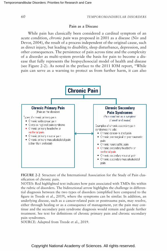
Temporomandibular Disorders: Priorities for Research and Care
Copyright National Academy of Sciences. All rights reserved.
60 TEMPOROMANDIBULAR DISORDERS
Pain as a Disease
While pain has classically been considered a cardinal symptom of an acute condition, chronic pain was proposed in 2001 as a disease (Niv and Devor, 2004), the result of a process independent of the original cause, such as direct injury, but leading to disability, sleep disturbance, depression, and other consequences. The persistence of pain across time and the complexity of a disorder as multi-system provide the basis for pain to become a dis-ease that fully represents the biopsychosocial model of health and disease (see Figure 2-2). As noted in the preface to the 2011 IOM report, “While pain can serve as a warning to protect us from further harm, it can also
FIGURE 2-2 Structure of the International Association for the Study of Pain clas-sification of chronic pain. NOTES: Red highlighted text indicates how pain associated with TMDs fits within the rubric of disorders. The bidirectional arrow highlights the challenge in differen-tial diagnosis between the two types of disorders (simplified here compared to the figure in Treede et al., 2019), where the symptoms can be similar. In addition, an underlying disease, such as a cancer-related pain or posttrauma pain, may resolve, either through healing or as a consequence of management, yet the pain may con-tinue and the secondary pain syndrome diagnosis would remain and guide further treatment. See text for definitions of chronic primary pain and chronic secondary pain syndromes. SOURCE: Adapted from Treede et al., 2019.
Temporomandibular Disorders: Priorities for Research and Care
Copyright National Academy of Sciences. All rights reserved.
DEFINITIONS AND SCOPE 61
contribute to severe and even relentless suffering, surpassing its underly-ing cause to become a disease in its own domains and dimensions” (IOM, 2011, p. ix). Chronic pain as a disease has become an increasingly accepted perspective, although not without critical discussion as well as criticism (IOM, 2011; Cohen et al., 2013; Taylor et al., 2015; Treede et al., 2019).
The well-documented functional and structural brain changes that take place in response to chronic pain may represent the mechanisms underlying pain as a disease, or they may represent the role of adaptive responses to pain (Tracey and Bushnell, 2009). The latter, however, may also justify dis-ease status in the sense that the feedback loops between response patterns and disease perpetuation may be among the many components constituting a complex disease (e.g., TMDs). Finally, evidence for similar responses to the same treatment across different pain disorders suggests common mecha-nisms, which may underlie chronic pain as a disease (Turk and Rudy, 1990; Rudy et al., 1995). The mechanisms of nociceptive and nociplastic pain are discussed in detail in Chapter 4.
In a clinical setting, determining whether a specific patient’s pain is, at one extreme, a localized condition or, at the other extreme, an indicator for pain as a disease can be challenging. One proposed solution to this challenge is the new ICD-11 diagnosis of chronic primary pain, which is defined as pain in one or more anatomical regions that has been present (continuous or intermittent) for at least 3 months, associated with emo-tional distress and/or functional disability, and not better accounted for by another diagnosis (Nicholas et al., 2019). The intention of the use of the term chronic primary pain is to replace other established but vague terms such as “ somatoform,” “nonspecific,” or “functional,” which are often used to classify a bodily pain when the clinician (or researcher) is not certain about a better diagnosis. It should be noted, however, that the diagnostic criteria for the most common painful TMDs are substantially better in terms of rigorous validation procedures than most of the other conditions subsumed within the chronic primary pain rubric. Nevertheless, the application of the term “chronic primary pain” for settings where clini-cal expertise, such as those needed to use the DC/TMD, is absent represents a substantial improvement over current practices, in that chronic primary pain explicitly incorporates the biopsychosocial model and thereby sets the direction for treatment. If acute pain from jaw injury, for example, was re-garded as solely related to injury-related tissue damage and was to persist, the new terminology would classify such pain as chronic secondary pain (Treede et al., 2019).
To look at this topic from another perspective, consider the question, If chronic pain is not a disease but rather only a symptom, then what is it a symptom of? The poor correspondence between physical examination find-ings and reported pain highlight the problems inherent in diagnosis when
Temporomandibular Disorders: Priorities for Research and Care
Copyright National Academy of Sciences. All rights reserved.
62 TEMPOROMANDIBULAR DISORDERS
pain becomes chronic. The criteria developed for the painful TMDs in the DC/TMD use measurements that exhibit high reliability, yet the determi-nant of the measurements is the report of pain as influenced by pain pro-cessing. Considering chronic pain to be a disease helps the clinician move out of the biomedical model and instead link management of the disorder to the biopsychosocial contributors to ongoing pain experience (Taylor et al., 2015), that is, to treat the pain disorder as such rather than continue further diagnostic testing to identify underlying presumed ongoing tissue damage as the cause (IOM, 2011).
Yet, the determination of whether chronic pain in a given individual is a symptom of an as-yet undiagnosed primary disorder or it is a disease remains complex, and the pain may be both at the same time as well, particularly in the early stage when an anatomical derangement is clearly identifiable. An illustrative example is diabetes. The disease of diabetes does not typically start as a disease; rather, a pre-diabetes state of insulin resistance or impaired glucose tolerance may exist for years, which if left untreated can become the identified disease of diabetes that affects multiple organ systems beyond the endocrine system. In the same way, chronic pain can start as a symptom of another condition, but if the pain is untreated then over time it can become the basis of pain as a disease.
WHAT CAN BE LEARNED FROM OTHER PAIN CONDITIONS?
There is considerable variation in how any of the TMDs may manifest, be experienced by an individual, appear clinically to the observer, and be associated with measurable physical changes. For example, the four issues regarding disease course discussed in the prior section indicate that a recent onset TMD in the presence of other already established pain disorders might be considered chronic pain from the beginning simply because mul-tiple risk factors for chronicity are already active. When pain persists there are many opportunities for it to influence and be influenced by psychologi-cal and social factors (Fordyce, 1976). Numerous studies have examined psychosocial factors in other chronic pain conditions, and emergent themes can provide insights into how individuals adjust to chronic TMDs. Among these factors, there are three that are particularly consistent and salient for TMDs.
• Adjustment to living with persistent pain: Individuals can differ markedly in how they adjust to living with persistent pain ( Karayannis et al., 2019; Mun et al., 2019). These variations in adjustment are evident in persons with TMDs and, as in other chronically painful diseases (e.g., arthritis or cancer), are not well explained on the basis of medical or background factors such as the severity of the injury,
Temporomandibular Disorders: Priorities for Research and Care
Copyright National Academy of Sciences. All rights reserved.
DEFINITIONS AND SCOPE 63
disease activity, or the duration of pain. The manner of adjustment or coping and its emergence across the life-span vary enormously across individuals, to the point that it is not possible to describe an overall natural development or even categories of natural history trajectories.
• Role of psychological factors in adjustment: Historically it was believed that longstanding trait-like personality factors play a key role in shaping how one adjusts to persistent pain. Over the past two decades, however, it has become increasingly evident that more dynamic psychological factors are important in explaining individual differences in the impact of pain. These factors include a person’s choice of pain coping strategies, the person’s beliefs in his or her own abilities to control pain (i.e., self-efficacy), and the person’s tendency to ruminate about and feel helpless (or not) about pain, and emotional distress (e.g., anxiety) (Turner et al., 2007; Fillingim et al., 2011).
• Role of social context in adjustment: Epidemiological studies have found that the prevalence of chronic pain conditions is much higher among those who have a low socioeconomic status, limited educa-tion, and limited access to satisfying work (Bergman et al., 2001; Brekke et al., 2002). Direct observational studies of patients with chronic pain and their partners (or caregivers) have highlighted the impact of the partner’s responses to pain (e.g., invalidating the pain, being supportive, or offering empathy) on the patient’s pain experi-ence (e.g., Verhofstadt et al., 2016). Although partners and care-givers clearly can be affected by the individual with a chronic TMD, there is a dearth of research in TMDs using novel observational methods to better understand patient–partner interactions relevant to TMDs. Such research is important because it could be used to tailor new interventions designed to help patients and their families learn how to work together to optimize adjustment to a TMD.
TMDs include several features common to other chronic pain condi-tions, such as back pain, widespread pain, and headache, among others. All of the major chronic pain conditions are quite heterogeneous with respect to the severity of the symptoms, the quality of life, and the psychosocial impacts, and a substantial proportion of individuals with these conditions experience marked impairment in physical or psychosocial function, as is the case with other conditions (Dworkin and Massoth, 1994; Manfredini et al., 2010). The development and persistence of these conditions are driven by complex interactions among multiple biological (e.g., genetics, nociception), psychological (emotional distress, coping), and social (socio-economic status, social support) factors (Dworkin, 1994; Furquim et al.,
Temporomandibular Disorders: Priorities for Research and Care
Copyright National Academy of Sciences. All rights reserved.
64 TEMPOROMANDIBULAR DISORDERS
2015; Maixner et al., 2016). Specific common risk factors across these conditions include female sex, enhanced pain sensitivity, a family history of chronic pain and mood disturbance, adverse childhood experiences, and multiple somatic symptoms (Clauw, 2015; Harper et al., 2016).
However, in many instances, TMDs represent a distinct group of con-ditions that differ from other pain conditions in important ways. First, the symptoms of TMDs occur in the masticatory system, which includes arguably the most complex joint in the body, combined with an intricate neuromuscular apparatus that must be effectively coordinated for healthy functioning. Hence, understanding the factors contributing to TMDs and their associated symptoms requires a consideration of this complex musculo skeletal system. Second, TMDs are associated with fewer disability days than back pain and have a lower pain impact than either headache or low back pain (Dworkin and Massoth, 1994), suggesting that despite comparable pain and psychological distress, individuals with TMDs may continue to function more than their counterparts with some other pain conditions. Third, for many individuals with a TMD, as with many other chronic pain conditions, there are no observable physical changes. This may contribute to the stigma and the need for those with a TMD to feel that they must convince others of the symptoms and impact (see Chapter 7 for discussion on stigma). These various factors affect the self-image of individuals with chronic pain. Whether the higher level of function among individuals with painful TMDs represents, for example, healthy adapta-tion, symptom repression, or the impact of stigma is not known. Fourth, a significant proportion of TMDs appear to be self-limiting, such that the prevalence of TMDs declines later in life (see Chapter 3), which stands in contrast to the pattern observed with some other conditions, such as low back pain, chronic widespread pain, and osteoarthritis. This emphasizes the importance of avoiding harm when providing treatment for TMDs. Finally, among chronic pain conditions, TMDs are unique in their man-agement being carried out largely within the dental rather than medical setting. This can create considerable challenges with access to care, and the dental– medical divide can impose substantial negative impacts on the effective management of people with TMDs (see Chapter 6).
WHAT CAN BE LEARNED FROM OTHER ORTHOPEDIC CONDITIONS?
As noted throughout this chapter and in Appendix D, the complexity of the masticatory system permits a wide range of functions that include far more than only mastication. In terms of the complex muscle vectors required for joint stability, the scapular system (supporting the shoulder) has some similarities to the functional requirements of the TMJ. Despite
Temporomandibular Disorders: Priorities for Research and Care
Copyright National Academy of Sciences. All rights reserved.
DEFINITIONS AND SCOPE 65
the importance of the masticatory system, the TMJ, as a joint, has not been well studied compared with other synovial orthopedic joint systems such as the knee, shoulder, and hip joints, all of which are far better understood. Knowledge about these other joint systems cannot, however, be directly translated to the TMJ because of the following considerations:
• Striking differences, based on genetic analyses, indicate that the TMJ develops in response to distinct molecular biological mechanisms.
• The TMJ has a uniquely complex anatomy involving bilateral joints that function as one, resulting in a transmission of load to one side whereas traction may be the force in the joint on the opposite side.
• The contacting components in the movable joint are often not well matched; incongruent surfaces within the components of the jaw are therefore not able to truly minimize stresses and strains during condyle movement, resulting in shear forces.
• TMJ movements involve both rotation and translation combined with six degrees of freedom (that is, movement in three planes), requiring complex coordination of an extensive muscle system, as described in Appendix D.
• The masticatory system has multiple muscles containing muscle fiber groups that can be activated in a highly variable manner across indi viduals, yet can accomplish similar behaviors and oral functions. This variation among people makes it challenging to identify uni-versal principles that may underlie TMJ function and its disorders.
These anatomical and functional distinctions of the TMJ, in contrast to other joints, underlie part of the complexity of TMD when it is a local condition, and these distinctions are further compounded when TMD be-comes a multi-system disorder because of overlapping comorbidity. Conse-quently, there is great potential for unique interactions between the complex subsystems making up the masticatory system and comorbid disorders, and knowledge obtained from other pain conditions may well not help in the development of a better understanding of the masticatory system and of the TMJ in particular. A discussion of relevant biomechanics research on the TMJ can be found in Chapter 4. Since the review of the masticatory system published in 2008 (Scrivani et al., 2008), little has been added to our understanding of the TMJ at a complex system level. And while degenera-tive joint disease of the knee has been studied extensively (e.g., Mora et al., 2018), considerable controversy continues regarding its pathophysiology and natural history. Knee osteoarthritis is influenced by local, systemic, and external factors, and both its progression and its response to treatment vary across individuals. Current management is primarily oriented toward symp-tom reduction. Non-pharmacological treatments include avoiding excessive
Temporomandibular Disorders: Priorities for Research and Care
Copyright National Academy of Sciences. All rights reserved.
66 TEMPOROMANDIBULAR DISORDERS
joint loading and routine exercise. Pharmacological treatments include limited use of non-steroidal anti-inflammatory medications and corticoid injections. Most findings from soft-tissue imaging of the knee joint are un-related to the symptoms (Kornaat et al., 2006). Overall, knee osteoarthritis and TMJ osteoarthritis appear to share the same major characteristics. Self-management approaches to osteoarthritis, through group approaches and Internet-based education, have been successful, with improvements in both multiple health-status measures and in self-efficacy (Lorig et al., 2008), with both outcomes consistent with more than just symptom relief. Taken together, the evidence supports the further development of self-management approaches for TMDs as a viable treatment direction that can have appro-priate impact at multiple levels (Nicolakis et al., 2002; Mulet et al., 2007; Riley et al., 2007; Lindfors et al., 2019).
CONCLUSIONS AND RESEARCH PRIORITIES
As noted throughout this chapter, TMDs are a set of disorders that are often complex and overlapping. The committee’s conclusions for this chapter are restated here, followed by thoughts on research priorities (see Box 2-4) relevant to this chapter.
Conclusion 2-1: Multiple disorders are encompassed by the terms “temporomandibular disorders” or “TMDs.” The committee defines temporomandibular disorders (TMDs) as a set of diseases or disorders that are related to alterations in structure, function, or physiology of the masticatory system and that may be associated with other systemic and comorbid medical conditions. TMDs can be usefully separated into two groups: the common TMDs with validated diagnostic criteria and the uncommon TMDs that do not yet have validated diagnostic criteria due to the challenges of conducting research on rare conditions. When possible, a patient’s diagnosis needs to be focused on the specific TMD or TMDs. “TMD” should not be used as a diagnostic term. An individual patient may have more than one TMD and may also have co-morbid conditions.
Conclusion 2-2: The Diagnostic Criteria for Temporo mandibular Disorders (DC/TMD) provides the most thorough and accu-rate diag nostic criteria for the most common types of temporo-mandibular disorders (TMDs). Additional work is needed on the diagnostic criteria for other types of TMDs that are uncommon. Efforts to increase its utility to clinicians are priorities, including the brief DC/TMD assessment tool that is currently in development.
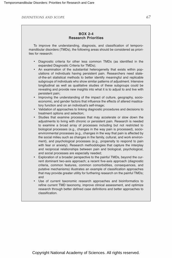
Temporomandibular Disorders: Priorities for Research and Care
Copyright National Academy of Sciences. All rights reserved.
DEFINITIONS AND SCOPE 67
BOX 2-4 Research Priorities
To improve the understanding, diagnosis, and classification of temporo-mandibular disorders (TMDs), the following areas should be considered as priori-ties for research:
• Diagnostic criteria for other less common TMDs (as identified in the expanded Diagnostic Criteria for TMDs);
• An examination of the substantial heterogeneity that exists within pop-ulations of individuals having persistent pain. Researchers need state-of-the-art statistical methods to better identify meaningful and replicable subgroups of individuals who show similar patterns of adjustment. Intensive longitudinal as well as qualitative studies of these subgroups could be revealing and provide new insights into what it is to adjust to and live with persistent pain;
• Improving the understanding of the impact of culture, geography, socio-economic, and gender factors that influence the effects of altered mastica-tory function and on an individual’s self-image;
• Validation of approaches to linking diagnostic procedures and decisions to treatment options and selection;
• Studies that examine processes that may accelerate or slow down the adjustments to living with chronic or persistent pain. Research is needed to examine a broad array of processes including but not restricted to biological processes (e.g., changes in the way pain is processed), socio-environmental processes (e.g., changes in the way that pain is affected by the social milieu such as changes in the family, cultural, and work environ-ment), and psychological processes (e.g., propensity to respond to pain with fear or anxiety). Research methodologies that capture the interplay and reciprocal relationships between pain and biological, psychological, and social processes are especially needed;
• Exploration of a broader perspective to the painful TMDs, beyond the cur-rent dominant two-axis approach; a recent five-axis approach (diagnostic criteria, common features, common comorbidities, consequences, and putative mechanisms) illustrates an example of classification approaches that may provide greater utility for furthering research on the painful TMDs; and
• Use of current taxonomic research approaches and bioinformatics to refine current TMD taxonomy, improve clinical assessment, and optimize research through better defined case definitions and better approaches to classification.
Temporomandibular Disorders: Priorities for Research and Care
Copyright National Academy of Sciences. All rights reserved.
68 TEMPOROMANDIBULAR DISORDERS
Widespread dissemination of these tools, coupled with provision of training in order to maximize the information yield from these tools, to general practice dentists is needed; primary care clinicians need to be apprised of these developments so that targeted referrals and appropriate expectations of good clinical practice will occur.
Conclusion 2-3: The biopsychosocial model is most closely aligned with and has the best evidence for addressing the range of temporo-mandibular disorders (TMDs) with the goal of improving quality of life—including physical, psychological, and social function—for individuals with a TMD. It will be necessary to incorporate evidence-based medicine principles into all theoretical views in order to address the long-held divisions within the dental profes-sion regarding the causes of TMDs. Clinical experience, while im-portant, must be augmented by epidemiological data and controlled evidence regarding disease mechanisms, appropriate and necessary diagnostic methods, and strongly theory-based interventions.
Conclusion 2-4: In many individuals, temporomandibular disorders (TMDs) resolve without medical or dental treatment interventions. In other individuals, TMDs progress to becoming chronic condi-tions; in addition, TMDs may be components of a multi-system disorder across biopsychosocial domains. Research is needed to identify why symptoms resolve in some cases and progress in others and how to better target different types, intensities, and timings of interventions.
REFERENCES
AES (American Equilibration Society). 2019. American Equilibration Society. http://www.aes-tmj.org (accessed October 23, 2019).
Ahmad, M., L. Hollender, Q. Anderson, K. Kartha, R. Ohrbach, E. L. Truelove, M. T. John, and E. L. Schiffman. 2009. Research Diagnostic Criteria for Temporomandibular Dis-orders (RDC/TMD): Development of image analysis criteria and examiner reliability for image analysis. Oral Surgery, Oral Medicine, Oral Pathology, Oral Radiology, and Endodontics 107:844–860.
Anderson, G. C., Y. M. Gonzalez, R. Ohrbach, E. L. Truelove, E. Sommers, J. O. Look, and E. L. Schiffman. 2010. The Research Diagnostic Criteria for Temporomandibular Disorders. VI: Future directions. Journal of Orofacial Pain 24:79–88.
Annandale, T. 1887. Displacement of the inter-articular cartilage of the lower jaw, and its treatment by operation. Lancet 129:411.
Bell, W. E. 1979. Orofacial pains: Differential diagnosis. Chicago, IL: Year Book Medical Publishers.
Bellavia, W. D., and W. Missert. 1985. Repositioning the mandible anteriorly with fixed com-posite overlays. Journal of Craniomandibular Practice 3:172–178.
Temporomandibular Disorders: Priorities for Research and Care
Copyright National Academy of Sciences. All rights reserved.
DEFINITIONS AND SCOPE 69
Benoliel, R., and Y. Sharav. 2010. Chronic orofacial pain. Current Pain and Headache Reports 14:33–40.
Benoliel, R., P. Svensson, S. Evers, S. J. Wang, A. Barke, B. Korwisi, W. Rief, R. D. Treede, and the International Association for the Study of Pain (IASP) Taskforce for Classifica-tion of Chronic Pain. 2019. The IASP classification of chronic pain for ICD-11: Chronic secondary headache or orofacial pain. Pain 160:60–68.
Benoliel, R., P. Svensson, and the Orofacial Pain Classifcation Committee. 2020. International Classification of Orofacial Pain, 1st edition. Cephalalgia 40(2):129–221.
Bergman, S., P. Herrström, K. Högström, I. F. Petersson, B. Svensson, and L. T. Jacobsson. 2001. Chronic musculoskeletal pain, prevalence rates, and sociodemographic associa-tions in a Swedish population study. Journal of Rheumatology 28:1369–1377.
Brekke, M., P. Hjortdahl, and T. K. Kvien. 2002. Severity of musculoskeletal pain: Relations to socioeconomic inequality. Social Science & Medicine 54(2):221–228.
Cairns, B., T. List, A. Michelotti, R. Ohrbach, and P. Svensson. 2010. JOR-CORE recommen-dations on rehabilitation of temporomandibular disorders. Journal of Oral Rehabilita-tion 37:481–489.
Caminiti, M. F., and S. Weinberg. 1998. Chronic mandibular dislocation: The role of non-surgical and surgical treatment. Journal of the Canadian Dental Association 64:484–491.
Clark, G. T., Y. Tsukiyama, K. Baba, and T. Watanabe. 1999. Sixty-eight years of experi mental occlusal interference studies: What have we learned? Journal of Prosthetic Dentistry 82:704–713.
Clarke, N. G. 1982. Occlusion and myofascial pain dysfunction: Is there a relationship? Journal of the American Dental Association 104:443–446.
Clauw, D. J. 2015. Diagnosing and treating chronic musculoskeletal pain based on the under-lying mechanism(s). Best Practices & Research. Clinical Rheumatology 29:6–19.
Cohen, M., and J. Quintner. 2008. The horse is dead: Let myofascial pain syndrome rest in peace. Pain Medicine 9:464–465.
Cohen, M., J. Quintner, and D. Buchanan. 2013. Is chronic pain a disease? Pain Medicine 14:1284–1288.
Cooper, B. C. 2011. Temporomandibular disorders: A position paper of the International College of Cranio-Mandibular Orthopedics (ICCMO). Cranio 29:237–244.
Costen, J. B. 1934. A syndrome of ear and sinus symptoms dependent upon disturbed function of the temporoandibular joint. Annals of Otology, Rhinology, and Laryngology 43:1–15.
Dawson, P. E. 1996. A classification system for occlusions that relates maximal intercuspation to the position and condition of the temporomandibular joints. Journal of Prosthetic Dentistry 75(1):60–66.
de Boever, J. A., G. E. Carlsson, and I. J. Klineberg. 2000a. Need for occlusal therapy and prosthodontic treatment in the management of temporomandibular disorders. Part I. Occlusal interferences and occlusal adjustment. Journal of Oral Rehabilitation 27:367–379.
de Boever, J. A., G. E. Carlsson, and I. J. Klineberg. 2000b. Need for occlusal therapy and prosthodontic treatment in the management of temporomandibular disorders. Part II: Tooth loss and prosthodontic treatment. Journal of Oral Rehabilitation 27:647–659.
de Leeuw, R., and G. D. Klasser. 2018. Orofacial pain: Guidelines for assessment, diagnosis, and management. Hanover Park, IL: Quintessence Publishing.
de Leeuw, R., G. Boering, B. Stegenga, and L. G. M. de Bont. 1994. Clinical signs of TMJ osteoarthrosis and internal derangement 30 years after nonsurgical treatment. Journal of Orofacial Pain 8:18–24.
de Silva Machado, L. P., M. B. de Macedo Nery, C. de Gois Nery, and C. R. Leles. 2012. Profiling the clinical presentation of diagnostic characteristics of a sample of symptomatic TMD patients. BMC Oral Health 12:1–8.
Temporomandibular Disorders: Priorities for Research and Care
Copyright National Academy of Sciences. All rights reserved.
70 TEMPOROMANDIBULAR DISORDERS
Dekker, J., P. Tola, G. Aufdemkampe, and M. Winckers. 1993. Negative affect, pain and disability in osteoarthritis patients: The mediating role of muscle weakness. Behaviour Research and Therapy 31:203–206.
Deyo, R. A., S. F. Dworkin, D. Amtmann, G. Andersoon, D. Borenstein, E. Carragee, J. Carrino, R. Chou, K. Cook, A. Delittlo, C. Goertz, P. Khalsa, J. Loeser, S. Mackey, J. Panagis, J. Rainville, T. Tosteson, D. Turk, M. Von Korff, and D. K. Weiner. 2014. Report of the NIH Task Force on Research Standards for Chronic Low Back Pain. Spine 39:1128–1143.
Dolwick, M., and G. Dimitroulis. 1994. Is there a role for temporomandibular joint surgery? British Journal of Oral and Maxillofacial Surgery 32:307–313.
Dworkin, S. F. 1994. Perspectives on the interaction of biological, psychological and social factors in TMD. Journal of the American Dental Association 125:856–863.
Dworkin, S. F. 2010. Research Diagnostic Criteria for Temporomandibular Disorders: Current status and future relevance. Journal of Oral Rehabilitation 37:734–743.
Dworkin, S. F., and L. LeResche. 1992. Research Diagnostic Criteria for Temporomandibular Disorders: Review, criteria, examinations and specifications, critique. Journal of Cranio-mandibular Disorders: Facial and Oral Pain 6:301–355.
Dworkin, S. F., and D. L. Massoth. 1994. Temporomandibular disorders and chronic pain: Disease or illness? Journal of Prosthetic Dentistry 72:29–38.
Egermark, I., T. Magnusson, and G. E. Carlsson. 2003. A 20-year follow-up of signs and symptoms of temporomandibular disorders and malocclusions in subjects with and with-out orthodontic treatment in childhood. The Angle Orthodontist 73:109–115.
Emshoff, R., I. Brandlmaier, S. Gerhard, H. Strobl, S. Bertram, and A. Rudisch. 2003. Mag-netic resonance imaging predictors of temporomandibular joint pain. Journal of the American Dental Association 134:705–714.
Farella, M., A. Michelotti, G. Ildice, S. Milani, and R. Martina. 2007. Unilateral posterior crossbite is not associated with TMJ clicking in young adolescents. Journal of Dental Research 86:137–141.
Fernandez-Sanroman, J. 1997. Surgical treatment of recurrent mandibular dislocation by augmentation of the articular eminence with cranial bone. Journal of Oral and Maxillo-facial Surgery 55:333–338.
Fillingim, R. B., R. Ohrbach, J. D. Greenspan, C. Knott, R. Dubner, E. Bair, C. Baraian, G. D. Slade, and W. Maixner. 2011. Potential psychosocial risk factors for chronic TMD: Descriptive data and empirically identified domains from the OPPERA case-control study. Journal of Pain 12:T46–T60.
Fillingim, R. B., R. Ohrbach, J. D. Greenspan, C. Knott, L. Diatchenko, R. Dubner, E. Bair, C. Baraian, N. Mack, G. D. Slade, and W. Maixner. 2013. Psychological factors associ-ated with development of TMD: The OPPERA prospective cohort study. Journal of Pain 14:T75–T90.
Fillingim, R. B., S. Bruehl, R. H. Dworkin, S. F. Dworkin, J. D. Loeser, D. C. Turk, E. Widerstrom-Noga, L. Arnold, R. Bennett, R. R. Edwards, R. Freeman, J. Gewandter, S. Hertz, M. Hochberg, E. Krane, P. W. Mantyh, J. Markman, T. Neogi, R. Ohrbach, J. A, Paice, B. A. Rappaport, S. M. Smith, T. J. Smith, M. D. Sullivan, G. N. Verne, A. D. Wasan, and U. Wesselmann. 2014. The ACTTION-American Pain Society Pain Taxonomy (AAPT): An evidence-based and multidimensional approach to classifying chronic pain conditions. Journal of Pain 15:241–249.
Fordyce, W. E. 1976. Behavioral methods for chronic pain and illness. St. Louis, MO: C.V. Mosby Company.
Fricton, J. 1985. Behavioral and psychosocial factors in chronic craniofacial pain. Anesthesia Progress 32:7–12.
Temporomandibular Disorders: Priorities for Research and Care
Copyright National Academy of Sciences. All rights reserved.
DEFINITIONS AND SCOPE 71
Fricton, J. 2006. Current evidence providing clarity in management of temporomandibular disorders: Summary of a systematic review of randomized clinical trials for intra-oral appliances and occlusal therapies. Journal of Evidence Based Dental Practice 6:48–52.
Fricton, J. 2014. Temporomandibular disorders: A human systems approach. Journal of the California Dental Association 42(8):523–535.
Fujii, T. 2002. Occlusal conditions just after the relief of temporomandibular joint and mas-ticatory muscle pain. Journal of Oral Rehabilitation 29:323–329.
Furquim, B. D., L. M. S. P. Flamengui, and P. C. R. Conti. 2015. TMD and chronic pain: A current view. Dental Press Journal of Orthodontics 20:127–133.
Garofalo, J. P., and L. Wesley. 1997. Research diagnostic criteria for temporomandibular disorders: Reflection of the physical-psychological interface. APS Bulletin May/June:4–8.
Gelb, M. L. 2014. Airway centric TMJ philosophy. Journal of the California Dental Associa-tion 42(8):551–562.
Greene, C. S. 1981. Where does the holistic end and the quackery begin? Journal of the American Dental Association 102:25–28.
Greene, C. S. 1983. A critique of unconventional treatment concepts and procedures for TMD. In D. M. Laskin, W. Greenfield, E. Gale, J. Rugh, P. Neff, and C. Alling, eds. President’s Conference on Examination, Diagnosis, and Management of TMD. Chicago, IL: Ameri-can Dental Association. Pp. 177–181.
Greene, C. S., and D. M. Laskin. 1971. Meprobamate therapy for the myofascial pain- dysfunction (MPD) syndrome: A double-blind evaluation. Journal of the American Dental Association 82:587–687.
Greene, C. S., and D. M. Laskin. 1974. Long-term evaluation of conservative treatment for myofascial pain-dysfunction syndrome. Journal of the American Dental Association 69:1365–1368.
Greene, C. S., and D. M. Laskin. 1983. Long-term evaluation of treatment for myofascial pain-dysfunction syndrome: A comparative analysis. Journal of the American Dental Association 107:235–238.
Greene, C. S., and A. Obrez. 2015. Treating temporomandibular disorders with permanent mandibular repositioning: Is it medically necessary? Oral Surgery, Oral Medicine, Oral Pathology and Oral Radiology 119:489–498.
Greene, C. S., M. D. Lerman, H. D. Sutcher, and D. M. Laskin. 1969. The TMJ pain- dysfunction syndrome: Heterogeneity of the patient population. Journal of the American Dental Association 79:1168–1172.
Greene, C. S., R. E. Olson, and D. M. Laskin. 1982a. Psychological factors in the etiology, progression, and treatment of MPD syndrome. Journal of the American Dental Associa-tion 105:443–448.
Greene, C. S., C. Turner, and D. M. Laskin. 1982b. Long-term outcome of TMJ clicking in 100 MPD patients. Journal of Dental Research 61:218.
Harper, D. E., A. Schrepf, and D. J. Clauw. 2016. Pain mechanisms and centralized pain in temporomandibular disorders. Journal of Dental Research 95:1102–1108.
Haythornthwaite, J. 2010. IMMPACT recommendations for clinical trials: Opportunities for the RDC/TMD. Journal of Oral Rehabilitation 37:799–806.
HHS (Department of Health and Human Services). 2016. National pain strategy: A compre-hensive population health-level strategy for pain. https://www.iprcc.nih.gov/sites/default/files/HHSNational_Pain_Strategy_508C.pdf (accessed October 3, 2019).
IASP (International Association for the Study of Pain). 2017. IASP terminology. https://www.iasp-pain.org/Education/Content.aspx?ItemNumber=1698 (accessed October 3, 2019).
IOM (Institute of Medicine). 2011. Relieving pain in America: A blueprint for transforming prevention, care, education, and research. Washington, DC: The National Academies Press.
Temporomandibular Disorders: Priorities for Research and Care
Copyright National Academy of Sciences. All rights reserved.
72 TEMPOROMANDIBULAR DISORDERS
ISO (International Organization for Standardization). 2019. Information technology—Top-level ontologies—Part 2: Basic formal ontology. https://www.iso.org/standard/74572.html (accessed November 20, 2019).
John, M. 2010. Improving TMD classification using the Delphi technique. Journal of Oral Rehabilitation 37:766–770.
John, M. T., S. F. Dworkin, and L. A. Mancl. 2005. Reliability of clinical temporomandibular disorder diagnoses. Pain 118:61–69.
Karayannis, N. V., I. Baumann, J. A. Sturgeon, M. Melloh, and S. C. Mackey. 2019. The im-pact of social isolation on pain interference: A longitudinal study. Annals of Behavioral Medicine 53(1):65–74.
Kent, M. L., P. J. Tighe, I. Belfer, T. J. Brennan, S. Bruehl, C. M. Brummett, C. C. Buckenmaier 3rd, A. Buvanendran, R. I. Cohen, P. Desiardins, D. Edwards, R. Fillingim, J. Gewandter, D. B. Gordon, R. W. Hurley, H. Kehlet, J. D. Loeser, S. Mackey, S. A. McLean, R. Polomano, S. Rahman, S. Raja, M. Rowbotham, S. Suresh, B. Schachtel, K. Schrieber, M. Schumacher, B. Stacey, S. Stanos, K. Todd, D. C. Turk, S. J. Weisman, C. Wu, D. B. Carr, R. H. Dworkin, and G. Terman. 2017. The ACTTION–APS–AAPM Pain Tax-onomy (AAAPT) multidimensional approach to classifying acute pain conditions. Pain Medicine 18(5):947–958.
Klasser, G. D., and C. S. Greene. 2009. The changing field of temporomandibular disorders: What dentists need to know. Journal of the Canadian Dental Association 75:49–53.
Klasser, G., D. Manfredini, J. P. Goulet, and A. De Laat. 2018. Oro-facial pain and temporo-mandibular disorders classification systems: A critical appraisal and future directions. Journal of Oral Rehabilitation 45:258–268.
Kornaat, P. R., J. L. Bloem, R. Y. Ceulemans, N. Riyazi, F. R. Rosendaal, R. G. Nelissen, W. O. Carter, M. P. Hellio Le Graverand, and M. Kloppenburg. 2006. Osteoarthritis of the knee: Association between clinical features and MR imaging findings. Radiology 239:811–817.
Kurita, H., A. Ohtsuka, K. Kurashina, and S. Kopp. 2001. A study of factors for success-ful splint capture of anteriorly disc displaced temporomandibular joint disc with disc reposition ing appliance. Journal of Oral Rehabilitation 28:651–657.
Landi, N., D. Manfredini, F. Tognini, M. Romagnoli, and M. Bosco. 2004. Quantification of the relative risk of multiple occlusal variables for muscle disorders of the stomatognathic system. Journal of Prosthetic Dentistry 92(2):190–195.
Laskin, D. M. 1969. Etiology of the pain-dysfunction syndrome. Journal of the American Dental Association 79:147–153.
Laskin, D. M., W. Greenfield, E. Gale, J. Rugh, P. Neff, and C. Alling, eds. 1983. President’s Conference on Examination, Diagnosis, and Management of TMD. Chicago, IL: American Dental Association.
Laurell, K. A. 1985. Construction of a ceramometal mandibular repositioning splint. Journal of Prosthetic Dentistry 54:439–442.
Lee, Y-H., K. M. Lee, Q-S. Auh, and J-P. Hong. 2018. Magnetic resonance imaging-based pre-diction of the relationship between whiplash injury and temporomandibular disorders. Frontiers in Neurology 8:725.
Li, C., N. Su, X. Yang, X. Yang, Z. Shi, and L. Li. 2012. Ultrasonography for detection of disc displacement of temporomandibular joint: A systematic review and meta-analysis. Journal of Oral and Maxillofacial Surgery 70:1300–1309.
Lindfors, E., T. Arima, L. Baad-Hansen, M. Bakke, A. De Laat, N. N. Giannakopoulos, A. Glaros, A. S. Guimaraes, A. Johansson, Y. Le Bell, F. Lobbezoo, A. Michelotti, F. Muller, R. Ohrbach, A. Wanman, T. Magnusson, and M. Emberg. 2019. Jaw exercises in the treatment of temporomandibular disorders—An international modified Delphi study. Journal of Oral & Facial Pain and Headache 33(4):389–398.
Temporomandibular Disorders: Priorities for Research and Care
Copyright National Academy of Sciences. All rights reserved.
DEFINITIONS AND SCOPE 73
List, T., and C. S. Greene. 2010. Moving forward with the RDC/TMD. Journal of Oral Rehabilitation 37:731–733.
List, T., M. T. John, S. F. Dworkin, and P. Svensson. 2006. Recalibration improves inter-examiner reliability of TMD examination. Acta Odontologica Scandinavica 64:146–152.
Lobbezoo, F., M. K. A. van Selms, M. T. John, K. H. Huggins, R. Ohrbach, C. M. Visscher, J. van der Zaag, M. H. van der Meulen, M. Naeije, and S. F. Dworkin. 2004. Use of the Research Diagnostic Criteria for Temporomandibular Disorders for multinational research. Translation efforts and reliability assessments in The Netherlands. Journal of Orofacial Pain 19:301–308.
Lobbezoo, F., C. M. Visscher, and M. Naeije. 2010. Some remarks on the RDC/TMD Vali-dation Project: Report an IADR/Toronto-2008 Workshop discussion. Journal of Oral Rehabilitation 37:779–783.
Look, J. O., E. L. Schiffman, E. L. Truelove, and M. Ahmad. 2010. Reliability and validity of Axis I of the Research Diagnostic Criteria for Temporomandibular Disorders (RDC/TMD) with proposed revisions. Journal of Oral Rehabilitation 37:744–759.
Lorig, K. R., P. L. Ritter, D. D. Laurent, and K. Plant. 2008. The internet-based arthritis self-management program: A one-year randomized trial for patients with arthritis or fibromyalgia. Arthritis & Rheumatism 59:1009–1017.
Lundh, H., P. L. Westesson, S. Kopp, and B. Tillstrom. 1985. Anterior repositioning splint in the treatment of temporomandibular joints with reciprocal clicking: Comparison with a flat occlusal splint and an untreated control group. Oral Surgery, Oral Medicine, and Oral Pathology 60:131–136.
Magnusson, T., I. Egermark, and G. E. Carlsson. 2000. A longitudinal epidemiologic study of signs and symptoms of temporomandibular disorders from 15 to 35 years of age. Journal of Orofacial Pain 14:310–319.
Maixner, W., R. B. Fillingim, D. A. Williams, S. B. Smith, G. D. Slade. 2016. Overlapping chronic pain conditions: Implications for diagnosis and classification. Journal of Pain 17:T93–T107.
Manfredini, D., E. Winocur, J. Ahlberg, L. Guarda-Nardini, and F. Lobbezoo. 2010. Psycho-social impairment in temporomandibular disorders patients. RDC/TMD axis II findings from a multicentre study. Journal of Dentistry 38:765–772.
Manfredini, D., F. Cocilovo, L. Favero, G. Ferronato, S. Tonello, and L. Guarda-Nardini. 2011. Surface electromyography of jaw muscles and kinesiographic recordings: Diag-nostic accuracy for myofascial pain. Journal of Oral Rehabilitation 38:791–799.
Manfredini, D., T. Castroflorio, G. Perinetti, and L. Guarda-Nardini. 2012. Dental occlusion, body posture and temporomandibular disorders: Where we are now and where we are heading for. Journal of Oral Rehabilitation 39(6):463–471.
Mayer, E. A., and M. C Bushnell. 2009. Functional pain disorders: Time for a paradigm shift. In E. A. Mayer and M. C. Bushnell, eds. Functional pain syndromes: Presentation and pathophysiology. Seattle, WA: IASP Press. Pp. 531–565.
McAlindon, T. E., C. Cooper, J. R. Kirwan, and P. A. Dieppe. 1993. Determinants of disability in osteoarthritis of the knee. Annals of the Rheumatic Diseases 52:258–262.
Meloto, C. B., R. N. Lichtenwalter, E. Bair, N. Rathnayaka, L. Diatchenko, J. D. Greenspan, W. Maixner, R. B. Fillingim, and R. Ohrbach. 2019. Clinical predictors of persistent TMD in people with first-onset TMD: A prospective case-control study. Journal of the American Dental Association 150(7):572–581.
Michelotti, A., M. Farella, L. M. Gallo, A. Veltri, S. Palla, and R. Martina. 2005. Effect of occlusal interference on habitual activity of human masseter. Journal of Dental Research 84:644–648.
Mohl, N. D. 1984. The role of head posture in mandibular function. In W. K. Solberg and G. T. Clark, eds. Abnormal jaw mechanics: Diagnosis and treatment. Chicago, IL: Quintessence. Pp. 97–116.
Temporomandibular Disorders: Priorities for Research and Care
Copyright National Academy of Sciences. All rights reserved.
74 TEMPOROMANDIBULAR DISORDERS
Mohl, N. D., G. A. Zarb, G. E. Carlsson, and J. D. Rugh, eds. 1988. A textbook of occlusion. Chicago, IL: Quintessence.
Mohl, N. D., W. D. McCall, Jr., J. P. Lund, and O. Plesh. 1990a. Devices for the diagnosis and treatment of temporomandibular disorders. Part I: Introduction, scientific evidence, and jaw tracking. Journal of Prosthetic Dentistry 63:198–201.
Mohl, N. D., J. P. Lund, C. G. Widmer, and W. D. McCall, Jr. 1990b. Devices for the diag-nosis and treatment of temporomandibular disorders. Part II: Electromyography and sonography. Journal of Prosthetic Dentistry 63:332–335.
Mohlin, B., and J. Kurol. 2003. To what extent do deviations from an ideal occlusion consti-tute a health risk? Swedish Dental Journal 27:1–10.
Molin, C. 1999. From bite to mind: TMD—A personal and literature review. International Journal of Prosthodontics 12:279–288.
Monaco, A., R. Cattaneo, M. C. Marci, D. Pietropaoli, and E. Ortu. 2017. Central sensitization-based classification for temporomandibular disorders: A pathogenetic hypothesis. Pain Research and Management 5957076.
Moore, A., and G. Wood. 1997. Medical treatment of recurrent temporomandibular joint dislocation using botulinum toxin A. British Dental Journal 183(11–12):415–417.
Mora, J. C., R. Przkora, and Y. Cruz-Almeida. 2018. Knee osteoarthritis: Pathophysiology and current treatment modalities. Journal of Pain Research 11:2189–2196.
Mulet, M., K. L. Decker, J. O. Look, P. A. Lenton, and E. L. Schiffman. 2007. A randomized clinical trial assessing the efficacy of adding 6 x 6 exercises to self-care for the treatment of masticatory myofascial pain. Journal of Orofacial Pain 21(4):318–328.
Mun, C. J., L. Ruehlman, and P. Karoly. 2019. Examining the adjustment patterns of adults with multiple chronic pain conditions and multiple pain sites: More pain, no gain. Jour-nal of Pain. doi: 10.1016/j.jpain.2019.06.002.
Murray, G. M., and C. C. Peck. 2018. Physiology of the masticatory system. In H. A. Gremillion and G. D. Klasser, eds. Temporomandibular disorders: A translational approach from basic science to clinical applicability. Cham, Switzerland: Springer Inter-national Publishing. Pp. 35–64.
Naliboff, B. D. 2007. Towards a nondualistic approach to multisystem illness. American Journal of Gastroenterology 102(12):2777–2780.
Nicholas, M. A., J. W. Vlaeyen, W. E. Rief, A. E. Barke, Q. F. Aziz, R. G. Benoliel, M. Cohen, S. Evers, M. A. Giamberardino, A. Goebel, B. Korwisi, S. Perrot, P. Svensson, S. J. Wang, R. D. Treede, and the IASP Taskforce for the Classification of Chronic Pain. 2019. The IASP classification of chronic pain for ICD-11: Chronic primary pain. Pain 160(1):28–37.
Nicolakis, P., B. Erdogmus, A. Kopf, M. Nicolakis, E. Piehslinger, and V. Fialka-Moser. 2002. Effectiveness of exercise therapy in patients with myofascial pain dysfunction syndrome. Journal of Oral Rehabilitation 29(4):362–368.
Niv, D., and M. Devor. 2004. Chronic pain as a disease in its own right. Pain Practice 4:179–181.
Ohrbach, R., and S. F. Dworkin. 2016. The evolution of TMD diagnosis: Past, present, future. Journal of Dental Research 95:1093–1101.
Ohrbach, R., and S. F. Dworkin. 2019. AAPT Diagnostic Criteria for Chronic Painful Temporo mandibular Disorders. Journal of Pain 20(11):1276–1292.
Ohrbach, R., and A. Michelotti. 2018. The role of stress in the etiology of oral para-function and myofascial pain. Oral and Maxillofacial Surgery Clinics of North America 30(3):369–379.
Ohrbach, R., J. A. Turner, J. J. Sherman, L. A. Mancl, E. L. Truelove, E. L. Schiffman, and S. F. Dworkin. 2010a. Research Diagnostic Criteria for Temporomandibular Disorders. IV: Evaluation of psychometric properties of the Axis II measures. Journal of Orofacial Pain 24:48–62.
Temporomandibular Disorders: Priorities for Research and Care
Copyright National Academy of Sciences. All rights reserved.
DEFINITIONS AND SCOPE 75
Ohrbach, R., T. List, J. P. Goulet, and P. Svensson. 2010b. Recommendations from the Inter-national Consensus Workshop: Convergence on an orofacial pain taxonomy. Journal of Oral Rehabilitation 37:807–812.
Ohrbach, R., Y. Gonzalez, T. List, A. Michelotti, and E. Schiffman. 2014. Diagnostic Criteria for Temporomandibular Disorders (DC/TMD) Clinical Examination Protocol. http://www.rdc-tmdinternational.org (accessed December 1, 2019).
Ohrbach, R., S. Sharma, R. B. Fillingim, J. D. Greenspan, and G. Slade. In press. Clinical characteristics of pain among five comorbid idiopathic pain conditions. Journal of Oral & Facial Pain & Headache.
Okeson, J. P. 2008. The classification of orofacial pains. Oral and Maxillofacial Surgery Clin-ics of North America 20:133–144.
Olesen, J., T. Steiner, M. Bousser, H. Diener, D. Dodick, M. First, P. J. Goadsby, H. Gobel, M. J. Lainez, R. B. Lipton, G. Nappi, F. Sakai, J. Schoenen, and S. D. Silberstein. 2009. Proposals for new standardized general diagnostic criteria for the secondary headaches. Cephalalgia 29:1331–1336.
Peck, C. C., J. P. Goulet, F. Lobbezoo, E. L. Schiffman, P. Alstergren, G. C. Anderson, R. de Leeuw, R. Jensen, A. Michelotti, R. Ohrbach, A. Petersson, and T. List. 2014. Expand-ing the taxonomy of the diagnostic criteria for temporomandibular disorders. Journal of Oral Rehabilitation 41:2–23.
Pertes, R. A., and S. G. Gross. 1995. Clinical management of temporomandibular disorders and orofacial pain. Chicago, IL: Quintessence.
Piper, M. A. 2019. Piper’s classification of TMJ disorders. http://www.pipererc.com/tmj.asp (accessed October 3, 2019).
Proffit, W. R., H. W. Fields, D. M. Sarver, and J. L. Ackerman. 2013. Contemporary ortho-dontics. 5th edition. St. Louis, MO: Elsevier Mosby.
Pullinger, A. G., D. A. Seligman, and W. K. Solberg. 1988. Temporomandibular disorders. Part II: Occlusal factors associated with temporomandibular joint tenderness and dys-function. Journal of Prosthetic Dentistry 59:363–367.
Pullinger, A. G., D. A. Seligman, and J. A. Gornbein. 1993. A multiple logistic regression analysis of the risk and cumulative odds of temporomandibular disorders as a function of common occlusal features. Journal of Dental Research 72:968–979.
Quintner, J. L., G. M. Bove, and M. L. Cohen. 2015. A critical evaluation of the trigger point phenomenon. Rheumatology 54:392–399.
Racich, M. J. 2005. Orofacial pain and occlusion: Is there a link? An overview of current concepts and the clinical implications. Journal of Prosthetic Dentistry 93:189–196.
Racich, M. J. 2018. Occlusion, temporomandibular disorders, and orofacial pain: An evidence-based overview and update with recommendations. Journal of Prosthetic Dentistry 120:678–685.
Raman, P. 2014. Physiologic neuromuscular dental paradigm for the diagnosis and treat-ment of temporomandibular disorders. Journal of the California Dental Association 42(8):563–571.
Ramfjord, S. P. 1961a. Dysfunctional temporomandibular joint and muscle pain. Journal of Prosthetic Dentistry 11:353–374.
Ramfjord, S. P. 1961b. Bruxism, a clinical and electromyographic study. Journal of the Ameri-can Dental Association 62:21–44.
Riley, J. L. III, C. D. Myers, T. P. Currie, O. Mayoral, R. G. Harris, J. A. Fisher, H. A. Gremillion, and M. E. Robinson. 2007. Self-care behaviors associated with myofascial temporomandibular disorder pain. Journal of Orofacial Pain 21(3):194–202.
Rudy, T. E., D. C. Turk, and S. F. Brena. 1988. Differential utility of medical procedures in the assessment of chronic pain patients. Pain 34:53–60.
Temporomandibular Disorders: Priorities for Research and Care
Copyright National Academy of Sciences. All rights reserved.
76 TEMPOROMANDIBULAR DISORDERS
Rudy, T. E., D. C. Turk, S. F. Brena, R. L. Stieg, and M. C. Brody. 1990. Quantification of biomedical findings of chronic pain patients: Development of an index of pathology. Pain 42:167–182.
Rudy, T. E., D. C. Turk, J. A. Kubinski, and H. S. Zaki. 1995. Differential treatment responses of TMD patients as a function of psychological characteristics. Pain 61:103–112.
Rusanen, J., A. S. Silvola, M. Tolvanen, P. Pirttiniemi, S. Lahti, and K. Sipilä. 2012. Pathways between temporomandibular disorders, occlusal characteristics, facial pain, and oral health-related quality of life among patients with severe malocclusion. European Journal of Orthodontics 34(4):512–517.
Salaffi, F., F. Cavalieri, M. Nolli, and G. Ferraccioli. 1991. Analysis of disability in knee osteo-arthritis. Relationship with age and psychological variables but not with radiographic score. Journal of Rheumatology 18:1581–1586.
Scapino, R. P. 1983. Histopatholgy associated with malposition of the human temporo-mandibular joint disc. Oral Surgery, Oral Medicine, and Oral Pathology 55:382–397.
Scheuermann, R. H., W. Ceusters, and B. Smith. 2009. Toward an ontological treatment of disease and diagnosis. Proceedings of the 2009 AMIA Summit on Translational Bio-informatics. Pp. 116–120.
Schiffman, E., and R. Ohrbach. 2016. Executive summary of the Diagnostic Criteria for Temporo mandibular Disorders for clinical and research applications. Journal of the American Dental Association 147(6):438–445.
Schiffman, E. L., J. R. Fricton, and D. Haley. 1992. The relationship of occlusion, para-functional habits and recent life events to mandibular dysfunction in a non-patient population. Journal of Oral Rehabilitation 19:201–223.
Schiffman, E. L., E. L. Truelove, R. Ohrbach, G. C. Anderson, M. T. John, T. List, and J. O. Look. 2010a. The Research Diagnostic Criteria for Temporomandibular Disorders. I: Overview and methodology for assessment of validity. Journal of Orofacial Pain 24(1):7–24.
Schiffman, E. L., R. Ohrbach, E. L. Truelove, F. Tai, G. C. Anderson, W. Pan, Y. M. Gonzalez, M. T. John, E. Sommers, T. List, A. M. Velly, W. Kang, and J. O. Look. 2010b. The Research Diagnostic Criteria for Temporomandibular Disorders. V: Methods used to establish and validate revised Axis I diagnostic algorithms. Journal of Orofacial Pain 24(1):63–78.
Schiffman, E., R. Ohrbach, T. List, G. Anderson, R. Jensen, M. T. John, D. Nixford, J. P. Goulet, E. Truelove, A. Clavel, J. Fricton, and J. Look. 2012. Diagnostic criteria for headache attributed to temporomandibular disorders. Cephalalgia 32:683–692.
Schiffman, E. L., A. M. Velly, J. O. Look, J. S. Hodges, J. Q. Swift, K. L. Decker, Q. N. Anderson, R. B. Templeton, P. A. Lenton, W. Kang, and J. R. Fricton. 2014a. Effects of four treatment strategies for temporomandibular joint closed lock. International Journal of Oral & Maxillofacial Surgery 43:217–226.
Schiffman, E., R. Ohrbach, E. Truelove, J. Look, G. Anderson, J. P. Goulet, T. List, P. Svensson, F. Lobbezoo, A. Michelotti, S. L. Brooks, W. Ceusters, M. Drangsholt, D. Ettlin, C. Gaul, L. J. Goldberg, J. A. Haythornthwaite, L. Hollender, R. Jensen, M. T. John, A. De Laat, R. de Leeuw, W. Maixner, M. vaqn der Meulen, G. M. Murray, D. R. Nixdorf, S. Palla, A. Petersson, P. Pionchon, B. Smith, C. M. Visscher, J. Zakrzewska, S. F. Dworkin, International RDC/TMD Consortium Network, International Association for Dental Research, Orofacial Pain Special Interest Group, and International Associa-tion for the Study of Pain. 2014b. Diagnostic Criteria for Temporomandibular Disorders (DC/TMD) for clinical and research applications: Recommendations of the International RDC/TMD Consortium Network and Orofacial Pain Special Interest Group. Journal of Oral & Facial Pain and Headache 28(1):6–27.
Temporomandibular Disorders: Priorities for Research and Care
Copyright National Academy of Sciences. All rights reserved.
DEFINITIONS AND SCOPE 77
Schwartz, L. L. 1956. A temporomandibular joint pain-dysfunction syndrome. Journal of Chronic Diseases 3:284–293.
Scrivani, S. J., D. A. Keith, and L. B. Kaban. 2008. Temporomandibular disorders. New England Journal of Medicine 359:2693–2705.
Seligman, D. A., and A. G. Pullinger. 1991. The role of functional occlusal relationships in temporomandibular disorders: A review. Journal of Craniomandibular Disorders: Facial & Oral Pain 5(4):265–279.
Sharma, S., H. C. Crow, W. D. McCall, Jr., and Y. M. Gonzalez. 2013. Systematic review of reliability and diagnostic validity of joint vibration analysis. Journal of Orofacial Pain 27(1):51–60.
Sharma, S., H. C. Crow, K. Kartha, W. McCall, and Y. M. Gonzalez. 2017. Reliability and diagnostic validity of a joint vibration analysis device. BMC Oral Health 17(10):56.
Sharma, S., J. Wactawski-Wende, M. J. LaMonte, J. Zhao, G. D. Slade, E. Bair, J. D. Greenspan, R. B. Fillingim, W. Maixner, and R. Ohrbach. 2019a. Incident injury is strongly associated with subsequent incident temporomandibular disorder: Results from the OPPERA study. Pain 160(7):1551–1561.
Sharma, S., M. Breckons, B. Bronnimann-Lambelet, J. W. Chung, T. List, F. Lobbezoo, D. R. Nixdorf, J. F. Oyarzo, C. Peck, Y. Tsukiyama, and R. Ohrbach. 2019b. Challenges in the clinical implementation of a biopsychosocial model for assessment and management of orofacial pain. Journal of Oral Rehabilitation 47(1):87–100.
Shore, N. A. 1959. Occlusal equilibration and temporomandibular joint dysfunction. Philadelphia, PA: Lippincott.
Sicher, H. 1948. Temporomandibular articulation in mandibular overclosure. Journal of the American Dental Association 36:131–139.
Simmons, C. 2014. Temporomandibular joint orthopedics with anterior repositioning appli-ance therapy and therapeutic injections. Journal of the California Dental Association 42(8):537–547.
Simmons, H. C., and AACP (American Academy of Craniofacial Pain), eds. 2009. Cranio-facial pain: A handbook for assessment, diagnosis and management. Chattanooga, TN: Chroma, Inc.
Simons, D. G. 1985. Myofascial pain syndromes due to trigger points: 1. Principles, diagnosis, and perpetuating factors. Journal of Manual Medicine 1:67–71.
Skármeta, N. P. 2017. Occlusal stability and mandibular stability: The major part of dentistry we are still neglecting. Cranio 35(4):201–203.
Slade, G. D., R. B. Fillingim, A. E. Sanders, E. Bair, J. D. Greenspan, R. Ohrbach, R. Dubner, L. Diatchenko, S. B. Smith, C. Knott, and W. Maixner. 2013a. Summary of findings from the OPPERA prospective cohort study of incidence of first-onset temporomandibular disorder: Implications and future directions. Journal of Pain 14(12 Suppl):T116–T124.
Slade, G., A. Sanders, E. Bair, N. C. Brownstein, D. Dampier, C. Knott, R. Fillingim, W. O. Maixner, S. Smith, J. Greenspan, R. Dubner, and R. Ohrbach. 2013b. Preclinical epi-sodes of orofacial pain symptoms and their association with healthcare behaviors in the OPPERA prospective cohort study. Pain 154(5):750–760.
Slade, G. D., R. Ohrbach, J. D. Greenspan, R. B. Fillingim, E. Bair, A. E. Sanders, R. Dubner, L. Diatchenko, C. B. Meloto, S. Smith, and W. Maixner. 2016. Painful temporo-mandibular disorder: Decade of discovery from OPPERA studies. Journal of Dental Research 95:1084–1092.
Solberg, W. K., R. T. Flint, and J. P. Brantner. 1972. Temporomandibular joint pain and dys-function: A clinical study of emotional and occlusal components. Journal of Prosthetic Dentistry 28:412–422.
Stegenga, B. 2010. Nomenclature and classification of temporomandibular joint disorders. Journal of Oral Rehabilitation 37:760–765.
Temporomandibular Disorders: Priorities for Research and Care
Copyright National Academy of Sciences. All rights reserved.
78 TEMPOROMANDIBULAR DISORDERS
Summers, M. N., W. E. Haley, J. D. Reveille, and G. S. Alarcón. 1988. Radiographic assess-ment and psychologic variables as predictors of pain and functional impairment in osteoarthritis of the knee or hip. Arthritis and Rheumatism 31(2):204–209.
Suvinen, T. I., and P. Kemppainen. 2007. Review of clinical EMG studies related to muscle and occlusal factors in healthy and TMD subjects. Journal of Oral Rehabilitation 34:631–644.
Suvinen, T. I., P. C. Reade, P. Kemppainen, M. Kononen, and S. F. Dworkin. 2005. Review of aetiological concepts of temporomandibular pain disorders: Towards a biopsychosocial model for integration of physical disorder factors with psychological and psychosocial illness impact factors. European Journal of Pain 41:115–121.
Tallents, R. H., S. I. Stein, and M. E. Moss. 2000. The role of occlusion in temporomandibular disorders. In Fonseca, R. J., ed. Oral and maxillofacial surgery: Temporomandibular disorders, volume 4. Philadelphia, PA: W.B. Saunders. Pp. 194–237.
Taylor, A. M., K. Phillips, J. O. Taylor, J. A. Singh, P. G. Conaghan, E. H. Choy, P. S. Tugwell, U. Kaiser, V. Strand, L. S. Simon, and P. J. Mease. 2015. Is chronic pain a disease in its own right? Discussions from a pre-OMERACT 2014 workshop on chronic pain. Journal of Rheumatology 42(10):1947–1953.
Tracey, I., and M. C. Bushnell. 2009. How neuroimaging studies have challenged us to rethink: Is chronic pain a disease? Journal of Pain 10(11):1113–1120.
Treede, R., W. Rief, A. Barke, Q. Aziz, M. I. Bennett, R. Benoliel, M. Cohen, S. Evers, N. B. Finnerup, M. B. First, M. A. Giamberardino, S. Kaasa, B. Korwisi, E. Kosek, P. Lavand’homme, M. Nicholas, S. Perrot, J. Scholz, S. Schug, B. H. Smith, P. Svensson, and J. W. S. Vlaeyen. 2019. Chronic pain as a symptom or a disease: The IASP Classifica-tion of Chronic Pain for the International Classification of Diseases (ICD-11). Pain 160(1):19–27.
Truelove, E., W. Pan, J. O. Look, L. A. Mancl, R. K. Ohrbach, A. M. Velly, K. H. Huggins, P. Lenton, and D. L. Shiffman. 2010. The Research Diagnostic Criteria for Temporo-mandibular Disorders. III: Validity of Axis I diagnoses. Journal of Orofacial Pain 24:35–47.
Turk, D. C., and T. E. Rudy. 1990. The robustness of an empirically derived taxonomy of chronic pain patients. Pain 43:27–35.
Turner, J. A., S. Holtzman, and L. Mancl. 2007. Mediators, moderators, and predictors of therapeutic change in cognitive-behavioral therapy for chronic pain. Pain 127:276–286.
Türp, J., and H. Schindler. 2012. The dental occlusion as a suspected cause for TMDs: Epi-demiological and etiological considerations. Journal of Oral Rehabilitation 39:502–512.
Undt, G., C. Kermer, and M. Rasse. 1997. Treatment of recurrent mandibular dislocation, Part II: Eminectomy. International Journal of Oral and Maxillofacial Surgery 26:98–102.
Verhofstadt, L., I. Devoldre, A. Buysse, M. Stevens, C. Hinnekens, W. Ickes, and M. Davis. 2016. The role of cognitive and affective empathy in spouses’ support interactions: An observational study. PLoS One 11(2):e0149944.
Visscher, C. M., L. Baad-Hansen, J. Durham, J. P. Goulet, A. Michelotti, C. Roldan-Barraza, B. Haggman-Henrikson, E. Ekberg, and K. G. Raphael. 2018. Benefits of implementing psychosocial assessment in dental practice. Journal of the American Dental Association 149(6):422–431.
Vlaeyen, J. W., and S. J. Linton. 2000. Fear-avoidance and its consequences in chronic musculo skeletal pain: A state of the art. Pain 85:317–332.
Von Korff, M., J. Ormel, F. J. Keefe, and S. F. Dworkin. 1992. Grading the severity of chronic pain. Pain 50:133–149.
Von Korff, M., A. I. Scher, C. Helmick, O. Carter-Pokras, D. W. Dodick, J. Goulet, R. Hamill-Ruth, L. LeResche, L. Porter, R. Tait, G. Terman, C. Veasley, and S. Mackey. 2016. United States National Pain Strategy for Population Research: Concepts, definitions, and pilot data. Journal of Pain 17(10):1068–1080.

Temporomandibular Disorders: Priorities for Research and Care
Copyright National Academy of Sciences. All rights reserved.
DEFINITIONS AND SCOPE 79
Wall, P. D. 1979. On the relation of injury to pain. Pain 6:253–264.Wilkes, C. H. 1989. Internal derangements of the temporomandibular joint: Pathological
variations. Archives of Otolaryngology–Head & Neck Surgery 115:469–477.Woda, A., S. Tubert-Jeannin, D. Bouhassira, N. Attal, B. Fleiter, J. P. Goulet, C. Gremeau-
Richard, M. L. Navez, P. Picard, P. Pionchon, and E. Albuisson. 2005. Towards a new taxonomy of idiopathic orofacial pain. Pain 116:396–406.
Zarb, G. A., and N. D. Mohl. 1988. Occlusion and temporomandibular disorders: A prologue. In N. D. Mohl, G. A. Zarb, G. E. Carlsson, and J. D. Rugh, eds. Textbook of occlusion. Chicago, IL: Quintessence, Pp. 373–383.
Zarb, G. A., G. E. Carlsson, B. J. Sessle, and N. D. Mohl, eds. 1994. Temporomandibular joint and masticatory muscle disorders. Copenhagen, Denmark: Munksgaard.
Zhou, H., K. Hu, and Y. Ding. 2014. Modified dextrose prolotherapy for recurrent temporo-mandibular joint dislocation. British Journal of Oral and Maxillofacial Surgery 52:63–66.

Temporomandibular Disorders: Priorities for Research and Care
Copyright National Academy of Sciences. All rights reserved.
Temporomandibular Disorders: Priorities for Research and Care
Copyright National Academy of Sciences. All rights reserved.
81
I am 31 years old and have suffered from TMD since I was 8 years old. My jaw cracks and pops, and I live with almost constant pain. My jaw is stiff and sensitive to the touch. The tension and pain often causes neck pain, tension headaches, and migraines. My jaw often causes so much discomfort while I am eating that I have to stop or avoid certain foods altogether. I have tried soft food diets and pain medication, but it is a daily struggle. The lack of appropriate care and knowledge about the disorder make it difficult to live with.
—Lauren W.
This chapter reviews temporomandibular disorders (TMDs) from a public health perspective—examining the individual and societal burdens of living with a TMD.1 The chapter begins by summarizing TMD prevalence estimates (i.e., the number of TMD cases present in a population at a given time) for adults and children based on nationally representative population-based studies of the United States (and other countries) as well as examples from smaller regional or clinic-based surveys using more extensive assess-ments of TMD symptoms. These studies highlight the large, often two-fold or greater, differences in the prevalence of TMD symptoms commonly found across demographic groups by factors such as age, sex, and ethnicity.
1 This chapter draws on a paper commissioned by the Committee on Temporomandibular Disorders (TMDs): From Research Discoveries to Clinical Treatment on “Prevalence, Impact, and Costs of Treatment for Temporomandibular Disorders,” by Gary Slade and Justin Durham (see Appendix C).
3
Individual and Societal Burden of TMDs
Temporomandibular Disorders: Priorities for Research and Care
Copyright National Academy of Sciences. All rights reserved.
82 TEMPOROMANDIBULAR DISORDERS
Incident TMD (i.e., the rate at which new cases develop) is also reviewed, but those estimates are based on relatively few studies. Following this, the individual and social burdens of TMD are reviewed, again demonstrating the scarcity of research in this area. Finally, the chapter closes with a sum-mary of what is known about TMD risk factors and how TMDs fit into the larger multi-system schema and other comorbidities.
PREVALENCE OF TMDs
The national prevalence of TMDs is difficult to estimate due to chal-lenges in conducting clinical examinations on a large scale, such that most prevalence data are based on self-reported symptoms associated with TMDs rather than examiner-verified classification. For example, one analysis found that an estimated 11.2 to 12.4 million U.S. adults (4.8 percent of the population) in 20182 had pain in the region of the temporomandibular joint (TMJ) that could be related to TMDs (Slade and Durham, 2020). Orofacial pain symptoms may or may not be related to TMDs. These self-reported symptoms of pain in the TMJ region are not equivalent to an examiner-verified diagnosis but rather indicate the possibility of an underlying TMD being present. As discussed in Chapter 2, TMDs represent a range of diverse and multifactorial disorders that can affect individuals across the general age range of adolescence to elderly and that can have significant impacts on an individual’s health and quality of life.
Prevalence estimates of TMDs also delineate the wide range in the extent of the severity and impact of these disorders on individuals—some individuals with a TMD have an intermittent or treatable manifestation of the disorder, whereas others suffer from more severe disorders that are often intractable, persistent, and lead to significant impairment and disruption of life. By some estimates, up to 40 percent of patients with signs and symp-toms of a TMD will have their symptoms resolve spontaneously (Scrivani et al., 2008). Sounds in the TMJ and deviation on opening the jaw appear
2 The prevalence estimate takes into account the survey sampling variability. The estimate is based on 2017–2018 National Health Interview Study (NHIS) data. These data are self-reported and therefore there are limitations in the data in the form of recall bias, the lack of examiner-verified diagnosis, and the fact that children, individuals in assisted living facilities, military personnel, and the incarcerated are not included in the NHIS survey population. It is also the case that the NHIS survey question related to orofacial pain symptoms is not specific to TMD and lacks the specificity to rule out non-TMD conditions. Given that many individuals may have intermittent pain that does not progress to a diagnosed TMD, the NHIS question about whether an individual experienced pain in the temporomandibular joint region for one whole day may lead an overestimation of individuals with orofacial pain. However, major health surveys such as NHIS are common sources for estimating the national or state-level prevalence of various diseases and behaviors, and the committee believes this prevalence estimate provides a valid analysis using some of the best national data available.
Temporomandibular Disorders: Priorities for Research and Care
Copyright National Academy of Sciences. All rights reserved.
INDIVIDUAL AND SOCIETAL BURDEN OF TMDs 83
frequently (approximately 50 percent of the population) and are considered normal and not requiring treatment. More concerning signs and symptoms include decreased mouth opening and occlusal changes ( approximately 5 percent of the population) (Scrivani et al., 2008). A complex pattern of change in biopsychosocial function is often associated with changes in TMD status (Ohrbach and Dworkin, 1998; Fillingim et al., 2018). Aside from broad patterns observed across individuals experiencing a TMD, little is known about the immediate period of painful TMD onset, the subsequent period for some individuals when acute pain transitions to chronic pain, or the later stage when chronic pain either improves or continues.
TMDs are part of the larger burden of pain. Chronic pain is estimated to affect approximately 50 million to 100 million adults in the United States (IOM, 2011; Dahlhamer et al., 2018). Annual national costs associ-ated with chronic pain were estimated to be $560 billion to $635 billion in 2011 (IOM, 2011). The prevalence of other common chronic pain condi-tions such as fibromyalgia (Clauw, 2014), chronic low back pain (Meucci et al., 2015), and migraine (Burch et al., 2018) is likely comparable to that of pain in the area of the TMJ in U.S. adults. While there are limitations in comparing the relative magnitude of painful conditions (e.g., how the ques-tion was asked of study or survey participants), the committee believes esti-mates of the prevalence of orofacial pain or TMD symptoms place TMDs well within the context of other highly prevalent conditions and therefore demanding equal attention and care in addressing the burden of TMDs.
High-Impact Pain and TMDs
While existing data demonstrate that TMDs are highly prevalent, it is also important to consider the severity of TMD symptoms and their impact on daily life and function. Velly and colleagues (2011) recruited a mixed community-based and clinical sample of 480 adults in the Minneapolis/St. Paul, Minnesota, area with muscle or joint pain due to a TMD. At baseline, 42 percent of individuals reported high pain intensity, but only 12 percent of the sample reported higher levels of disability. At 18-month follow-up, 26 percent of the sample reported high pain intensity, and 6 per-cent reported higher levels of disability. Another study, which recruited 399 patients seeking care for a TMD, found that 61 percent reported no dis-ability, 27 percent low disability, and 12 percent high disability ( Kotiranta et al., 2015).
More recently, data from the Orofacial Pain Prospective Evaluation and Risk Assessment (OPPERA) study investigated the prevalence and pre-dictors of high-impact pain among 846 people with a TMD (Miller et al., 2019). High-impact pain was defined as either having high pain intensity and low pain-related interference or having moderate to high levels of
Temporomandibular Disorders: Priorities for Research and Care
Copyright National Academy of Sciences. All rights reserved.
84 TEMPOROMANDIBULAR DISORDERS
self-reported, pain-related interference. They found that approximately one-third of the study population had high-impact pain, and individuals with high-impact pain showed greater limitations in jaw function, higher pain sensitization, and greater tenderness to palpation of multiple body sites. According to 2016 NHIS data of 42,370 adult participants, the prevalence of high-impact chronic pain was also elevated nearly four-fold (26.9 percent versus 7.0 percent) in people with orofacial pain symptoms (Slade and Durham, 2020). The TMJ may be part of a constellation of anatomic sites of high-impact chronic pain with possibly shared underlying etiology and resulting in moderate to severe pain and some level of pain-related disability for some individuals with a TMD.
Challenges Estimating TMD Prevalence
The prevalence of pain conditions such as TMDs is usually measured in cross-sectional health surveys that ask respondents about pain symptoms that are characteristic of the particular condition—in this case, TMDs. In some instances a clinical evaluation will also be conducted, with the goal of properly distinguishing pain symptoms caused by a TMD from pain symptoms caused by other types of pathology. The primary require-ments for the valid estimation of TMD prevalence are a selection of a representative sample of study participants from the target population of interest; accepted case definitions based on valid and reliable questions or examination methods to classify the presence or absence of TMD pain in each study participant; and sufficient numbers of study participants to estimate the prevalence with reasonable precision. The literature reporting the prevalence of TMDs varies substantially across studies, with much of the variability attributable to differences in the methodologies used, par-ticularly differences in case definitions (see Chapter 2 for an overview of the issues and evolution of research and diagnostic criteria used to estab-lish case definitions for TMDs) and study populations. Sampling strategies vary considerably, ranging from large population-based studies using form survey sampling methodology to convenience samples from small clinical populations. Moreover, TMD prevalence varies as a feature of demographic factors such as age, sex, race, and ethnicity, which makes clear the need for reporting findings within demographic strata. Given these issues, making valid comparison of prevalence estimates across studies must be done with careful attention to underlying methodological differences.
Conclusion 3-1: The prevalence of temporomandibular disorder (TMD) symptoms varies widely across studies depending on the assessment used and the population studied. Based on one analy-sis of 2017–2018 data, an estimated 4.8 percent of U.S. adults
Temporomandibular Disorders: Priorities for Research and Care
Copyright National Academy of Sciences. All rights reserved.
INDIVIDUAL AND SOCIETAL BURDEN OF TMDs 85
(an esti mated 11.2 to 12.4 million U.S. adults) had pain in the region of the temporomandibular joint that could be related to TMDs. Based on this information, it is likely that TMDs are the most prevalent type of chronic orofacial pain and TMDs may be comparable in prevalence to other chronic pain conditions such as fibromyalgia, chronic low back pain, and migraine disease.
Prevalence of TMDs in Adults
National Population-Based Studies
NHIS is a federally sponsored recurring survey that provides nationally representative measures of many health conditions, along with health-re-lated behaviors and socio-demographics. The NHIS uses rigorous sampling methodology to collect data annually from approximately 87,500 civilian, non-institutionalized persons. In most of its annual surveys conducted since 1987, orofacial pain symptoms have been assessed using a single-item ques-tion asked of all respondents age 18 years or older:
The following questions are about pain you may have experienced in the PAST <REFERENCE PERIOD>. Please refer to pain that LASTED A WHOLE DAY OR MORE. Do not report aches and pains that are fleeting or minor. During the PAST <REFERENCE PERIOD>, did you have facial ache or pain in the jaw muscles or the joint in front of the ear?
This question serves as the assessment tool for TMDs for NHIS sur-veys. A limitation of this assessment tool is that it lacks sufficient specificity to rule out other non-TMD conditions that can present with similar symp-toms (e.g., other orofacial pain conditions), likely leading to an overestima-tion of TMD prevalence. It may also miss non-painful TMDs and will not reflect the various types of TMD an individual may have (see Chapter 2 for an overview of TMD types).
The period prevalence of occurrence of at least 1 day of symptoms depends on the timeframe over which the respondents are asked about hav-ing a 1-day episode of symptoms. In 1989, when the reference period was 6 months, the period prevalence3 of at least 1 day of TMD symptoms in the U.S. adult population was 6.0 percent (see Appendix C). In subsequent years, the reference period was reduced to 3 months, resulting in somewhat lower prevalence estimates, ranging from 4.3 percent in 1999 to 5.2 percent
3 Period prevalence is the proportion of a population that has the condition at some time during a given period (e.g., 12-month prevalence), and includes people who already have the condition at the start of the study period as well as those who acquire it during that period.
Temporomandibular Disorders: Priorities for Research and Care
Copyright National Academy of Sciences. All rights reserved.
86 TEMPOROMANDIBULAR DISORDERS
in 2018, a numerical difference that is not statistically different. Overall, the findings represent a fairly consistent period prevalence over the past 20 years.
Socio-Demographic Variation in Prevalence of TMD Symptoms (NHIS 2017–2018)
The NHIS collects extensive data about socio-demographic characteris-tics, other health conditions, and the health care usage of study participants, making it possible to examine cross-sectional variations in the prevalence of TMD symptoms (pain) according to those characteristics. However, it should be emphasized that any observed cross-sectional associations do not necessarily signify a causal relationship, in either direction, between those characteristics and TMD symptoms.
In 2017–2018 the 3-month period prevalence of orofacial pain symp-toms, according to the NHIS wording, differed appreciably according to age, gender, race, and income (see Table 3-1; see Appendix C). Specifically, prevalence was elevated approximately two-fold in females compared to males, whites compared to Asian Americans, and individuals in low-income households compared with those in high-income households. There was an inverted-U relationship with age, with prevalence greatest in 45- to 54-year-olds and lower in both the youngest (18 to 24 years) and oldest age groups. In contrast, the prevalence did not vary appreciably according to ethnicity or geographic region.
Prevalence of TMD Symptoms According to Health Care Usage and Other Pain Conditions (NHIS 2017–2018)
In 2017–2018 the prevalence of orofacial pain symptoms tended to be greater among people who had used health care in the preceding year than among those who had not (see Table C-3 in Appendix C). Specifically, there was an approximately two-fold higher orofacial pain symptom prevalence associated with having seen a physical or occupational therapist, chiro-practor, or medical specialist and a 1.5-fold higher orofacial pain symptom prevalence associated with having seen a general physician. In contrast, the prevalence of orofacial pain symptoms did not differ according to whether participants had seen a dentist within the preceding year. It must be emphasized that the 2017–2018 surveys did not inquire as to the reasons for health care visits or, in particular, whether people with orofacial pain symptoms sought health care because of those symptoms.
Larger differences in prevalence were seen relative to the presence of other pain conditions (i.e., other than orofacial pain symptoms; see Table C-3 in Appendix C). People reporting headache or pain symptoms
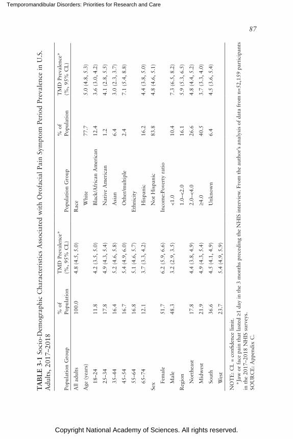
Temporomandibular Disorders: Priorities for Research and Care
Copyright National Academy of Sciences. All rights reserved.
87
TA
BL
E 3
-1 S
ocio
-Dem
ogra
phic
Cha
ract
eris
tics
Ass
ocia
ted
wit
h O
rofa
cial
Pai
n Sy
mpt
om P
erio
d Pr
eval
ence
in
U.S
. A
dult
s, 2
017–
2018
Popu
lati
on G
roup
% o
f Po
pula
tion
TM
D P
reva
lenc
e*
(%,
95%
CL
)Po
pula
tion
Gro
up%
of
Popu
lati
onT
MD
Pre
vale
nce*
(%
, 95
% C
L)
All
adul
ts10
0.0
4.8
(4.5
, 5.
0)R
ace
Age
(ye
ars)
Whi
te77
.75.
0 (4
.8,
5.3)
18–2
411
.84.
2 (3
.5,
5.0)
Bla
ck/A
fric
an A
mer
ican
12.4
3.6
(3.0
, 4.
2)
25–3
417
.84.
9 (4
.3,
5.4)
Nat
ive
Am
eric
an1.
24.
1 (2
.8,
5.5)
35–4
416
.45.
2 (4
.6,
5.8)
Asi
an6.
43.
0 (2
.3,
3.7)
45–5
416
.75.
4 (4
.9,
6.0)
Oth
er/m
ulti
ple
2.4
7.1
(5.4
, 8.
8)
55–6
416
.85.
1 (4
.6,
5.7)
Eth
nici
ty
65–7
412
.13.
7 (3
.3,
4.2)
His
pani
c16
.24.
4 (3
.8,
5.0)
Sex
Not
His
pani
c83
.84.
8 (4
.6,
5.1)
Fem
ale
51.7
6.2
(5.9
, 6.
6)In
com
e:Po
vert
y ra
tio
Mal
e48
.33.
2 (2
.9,
3.5)
<1.0
10.4
7.3
(6.5
, 8.
2)
Reg
ion
1.0–
<2.0
16.1
5.9
(5.3
, 6.
5)
Nor
thea
st17
.84.
4 (3
.8,
4.9)
2.0–
<4.0
26.6
4.8
(4.4
, 5.
2)
Mid
wes
t21
.94.
9 (4
.3,
5.4)
≥4.0
40.5
3.7
(3.3
, 4.
0)
Sout
h36
.64.
5 (4
.1,
4.9)
Unk
now
n6.
44.
5 (3
.6,
5.4)
Wes
t23
.75.
4 (4
.9,
5.9)
NO
TE
: C
L =
con
fiden
ce l
imit
.
*Jaw
or
face
pai
n th
at la
sted
≥1
day
in t
he 3
mon
ths
prec
edin
g th
e N
HIS
inte
rvie
w. F
rom
the
aut
hor’
s an
alys
is o
f da
ta f
rom
n=5
2,15
9 pa
rtic
ipan
ts
in t
he 2
017–
2018
NH
IS s
urve
ys.
SOU
RC
E:
App
endi
x C
.
Temporomandibular Disorders: Priorities for Research and Care
Copyright National Academy of Sciences. All rights reserved.
88 TEMPOROMANDIBULAR DISORDERS
in the neck, back, or joints had at least three times the prevalence of oro-facial pain symptoms as people without those respective symptoms (see Table C-3 in Appendix C). Using a simple count of those four body pain symptoms, the 3-month period prevalence of orofacial pain symptoms was 32.4 percent among people with all four body pain symptoms, compared with 1.1 percent among people with no body pain symptoms.
Community-Based Studies
Smaller regional studies of TMD prevalence have used varying assess-ment methods and case definitions. For example, Manfredini and colleagues (2011) conducted an analysis that included 21 papers, 15 of which dealt with populations of patients with a TMD who underwent clinical exami-nation and 6 of which examined community-based samples. The studies of patient populations reported an average prevalence of 45.3 percent for TMD-related muscle disorder diagnoses, 41.1 percent for disc displace-ments, and 30.1 percent for TMD-related joint disorders. Results from the community studies showed an overall 9.7 percent prevalence for muscle disorder diagnoses. The prevalence estimates by subtypes of TMD diag-noses showed substantial variation across these diagnostic groups.
Due to their similarities to U.S. populations, results from regional studies in Canada are worth examining. Using formal sampling methodol-ogy, Locker and Slade (1988) assessed TMD-associated symptom preva-lence in adults 18 years and older in Toronto, Canada. Using a self-reported measure of pain in the region of the TMJ (i.e., “pain in front of the ear”), they reported a prevalence of 5.0 percent among men and 9.5 percent among women, whereas functional pain (i.e., “pain while chewing”) was similar between men and women, at 7.4 percent and 7.7 percent, respec-tively. No difference in pain measures was found between the younger (<45 years of age) and older (≥45 years of age) age groups. When study participants were given a list of nine questions about TMD symptoms, 48 percent endorsed at least one symptom, with joint sounds, tiredness or stiffness of jaw muscles, and an uncomfortable bite being most common (Locker and Slade, 1988). A telephone survey of a representative sample of the French-speaking population of Quebec (Goulet et al., 1995) reported a similar overall prevalence among men (5 percent) and women (9 percent), with no age trend noted for either group. Frequent episodes of TMJ clicking and difficulty in jaw opening were found in 9 percent and 4 percent of the respondents, respectively.
Temporomandibular Disorders: Priorities for Research and Care
Copyright National Academy of Sciences. All rights reserved.
INDIVIDUAL AND SOCIETAL BURDEN OF TMDs 89
Prevalence of TMDs in Children and Adolescents
Reports of the prevalence of TMDs in children and adolescents vary widely. A systematic review (Christidis et al., 2019) that included six studies (List et al., 1999; Nilsson, 2007; Wu and Hirsch, 2010; Franco-Micheloni et al., 2015; Al-Khotani et al., 2016; Graue et al., 2016) reported prevalence in children or adolescents based on clinical evaluations using the Research Diagnostic Criteria for Temporomandibular Disorders (RDC/TMD) or the Diagnostic Criteria for Temporomandibular Disorders (DC/TMD) measures (see Table 3-2). Overall prevalence estimates ranged from 7.3 percent to 30.4 percent. A systematic review by da Silva and colleagues (2016), which was also based on the use of the RDC/TMD and which included a clini-cal evaluation, reported the results from 11 studies from 8 countries. The prevalence estimates ranged from 4.0 percent to 42.7 percent. The meta-analysis associated with this study reported an overall prevalence across all 11 studies of 16 percent, with clicking and jaw locking being the most commonly reported symptoms. There was no overlap in the included studies between the da Silva and colleagues’ (2016) and Christidis and colleagues’ (2019) systematic reviews.
Studies of the prevalence of TMDs among children have reported mixed results on gender differences, with some studies reporting no dif-ference in prevalence (LeResche, 1997; List et al., 1999; Magnusson et al., 2005; Köhler et al., 2009; Marpaung et al., 2019). Care seeking and pain intensity were reported to be higher among girls. There is evidence that the prevalence of TMDs in children and adolescents increases with age ( Magnusson et al., 2005; Köhler et al., 2009; da Silva et al., 2016; Marpaung et al., 2019). One study reported that pain severity was greater in older than in younger adolescents (Howard, 2013).
INCIDENCE OF TMDs
Determining the incidence (i.e., the rate at which new cases develop) of TMDs requires longitudinal (cohort) studies, where individuals are fol-lowed over time and their symptoms assessed periodically. No nationally representative cohort studies of TMD have been reported. Incidence data, consequently, comes from smaller, community-based studies such as the OPPERA study. This study provides the latest and best estimates to date of the incidence of TMDs (see Box 3-1 for an overview of the OPPERA study).
Early Studies of TMD Incidence
Prior to the OPPERA study, investigators at the North Carolina study site conducted a 3-year prospective cohort study of women age 18 to 34
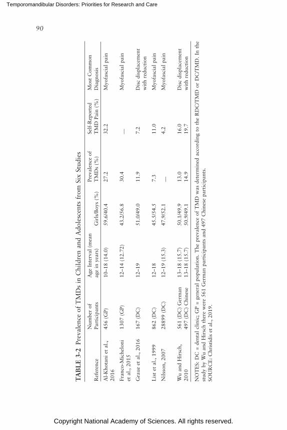
Temporomandibular Disorders: Priorities for Research and Care
Copyright National Academy of Sciences. All rights reserved.
90
TA
BL
E 3
-2 P
reva
lenc
e of
TM
Ds
in C
hild
ren
and
Ado
lesc
ents
fro
m S
ix S
tudi
es
Ref
eren
ceN
umbe
r of
Pa
rtic
ipan
tsA
ge I
nter
val
(mea
n ag
e in
yea
rs)
Gir
ls/B
oys
(%)
Prev
alen
ce o
f T
MD
s (%
)Se
lf-R
epor
ted
TM
D P
ain
(%)
Mos
t C
omm
on
Dia
gnos
is
Al-
Kho
tani
et
al.,
2016
456
(GP)
10–1
8 (1
4.0)
59.6
/40.
427
.232
.2M
yofa
scia
l pa
in
Fran
co-M
iche
loni
et
al.,
201
513
07 (
GP)
12–1
4 (1
2.72
)43
.2/5
6.8
30.4
__M
yofa
scia
l pa
in
Gra
ue e
t al
., 20
1616
7 (D
C)
12–1
951
.0/4
9.0
11.9
7.2
Dis
c di
spla
cem
ent
wit
h re
duct
ion
Lis
t et
al.,
199
986
2 (D
C)
12–1
845
.5/5
4.5
7.3
11.0
Myo
fasc
ial
pain
Nils
son,
200
728
899
(DC
)12
–19
(15.
3)47
.9/5
2.1
__4.
2M
yofa
scia
l pa
in
Wu
and
Hir
sch,
20
1056
1 (D
C)
Ger
man
497
(DC
) C
hine
se13
–18
(15.
7)13
–18
(15.
7)50
.1/4
9.9
50.9
/49.
113
.014
.916
.019
.7D
isc
disp
lace
men
t w
ith
redu
ctio
n
NO
TE
S: D
C =
den
tal
clin
ic;
GP
= ge
nera
l po
pula
tion
. T
he p
reva
lenc
e of
TM
D w
as d
eter
min
ed a
ccor
ding
to
the
RD
C/T
MD
or
DC
/TM
D.
In t
he
stud
y by
Wu
and
Hir
sch
ther
e w
ere
561
Ger
man
par
tici
pant
s an
d 49
7 C
hine
se p
arti
cipa
nts.
SOU
RC
E:
Chr
isti
dis
et a
l., 2
019.
Temporomandibular Disorders: Priorities for Research and Care
Copyright National Academy of Sciences. All rights reserved.
INDIVIDUAL AND SOCIETAL BURDEN OF TMDs 91
BOX 3-1 Orofacial Pain Prospective Evaluation and Risk
Assessment Study: Overview and Methods
The Orofacial Pain Prospective Evaluation and Risk Assessment (OPPERA) study was a 7-year research study funded by the National Institute of Dental and Craniofacial Research that began in 2005 seeking to identify potential risk factors, genetic mechanisms, and clinical characteristics of temporomandibular disorders (TMDs). Specifically, investigators were interested in developing further evidence related to demographic factors that may be predictive for TMDs, the role of height-ened responsiveness to noxious stimuli, psychosocial factors typically associated with pain, and identifying the pathways in which genetic variants can influence TMD onset and risk (Maixner et al., 2011). A major impetus behind the study was the lack of robust data on risk factors for TMD onset because, to that point, many of the studies looking at TMDs had focused on symptomatic individuals (Maixner et al., 2011). Historically, the field had also struggled to come to consensus on the development of comprehensive diagnostic classifications and criteria (Dworkin, 2011). The larger OPPERA effort comprised four observational studies including a prospective cohort study of the first onset of TMDs, a baseline case–control study of chronic TMD, a matched case–control study of incident TMDs, and a prospective case–cohort study looking at the course of TMDs.
From 2006 to 2008 a diverse group of study participants were recruited from four large academic health centers in the United States (University of Maryland, Baltimore; University of Buffalo; University of Florida; and University of North Carolina at Chapel Hill). Recruited individuals included 185 individuals with con-firmed cases of TMDs based on the Research Diagnostic Criteria for Temporo-mandibular Disorders by Dworkin and LeResche (1992) and 3,263 individuals who did not have a TMD diagnosis or symptoms suggestive of TMDs at the beginning of the study. To reduce the potential for assessment variability, clinical examiners were trained together at the same location, were calibrated to a reference ex-aminer, and performed mock examinations of volunteers who were not part of the study in order to ensure reliability (Slade et al., 2011). A centralized data coordinating center managed biological and data samples from all of the centers.
Prospective Cohort of First Onset: Eligible participants for this study included individuals between the ages of 18 and 44 years without a diagnosis or symptoms suggestive of TMDs. Upon completion of in-home questionnaires assessing TMD symptom status, prospective study participants underwent a clinical evaluation composed of psychological and physical examinations. Individuals selected to participate in the study were asked to complete 28 entries in a pain diary within a 4-week period and received follow-up questionnaires every 3 months (over the duration of 5 years) to evaluate potential symptoms that suggested a TMD. If symptoms were suggestive, a follow-up clinical examination was performed that evaluated physical and psychological symptoms, sensitivity, autonomic function, and genetic factors (Slade et al., 2011). Results from the study showed that about 3.5 percent of the participants developed TMDs over the course of the study (Sanders et al., 2013a).
continued
Temporomandibular Disorders: Priorities for Research and Care
Copyright National Academy of Sciences. All rights reserved.
92 TEMPOROMANDIBULAR DISORDERS
Baseline Case–Control Study (Chronic TMDs): Fifty percent of the TMD symptom-free participants from the prospective cohort (1,633 individuals) were randomly selected, and an additional cohort of 185 individuals with a confirmed TMD (present for at least 6 months) were selected to participate. The majority were between the ages of 18 and 24 years and were non-Hispanic white or African American. Results from the study indicated a higher prevalence of TMDs with in-creasing age and female sex and a higher incidence of TMDs in the non-Hispanic white population (Slade et al., 2011).
Case–Control Study of Incident TMDs: Individuals in the prospective cohort of first onset who developed a TMD were matched with TMD-free controls, and both groups underwent a clinical evaluation to assess their symptoms.
Prospective Case–Cohort (TMD Course): Individuals in the prospective cohort of first onset who developed a TMD were matched with TMD-free controls, and both groups were invited to complete quarterly questionnaires detailing their health and symptom updates. Six months after a participant was determined to have a classified case of TMD that developed during the study, he or she was invited to one of the four study sites to undergo a clinical evaluation to confirm the TMD diagnosis.
BOX 3-1 Continued
years at the time of enrollment (Slade et al., 2007). When a participant was enrolled, examiners verified that she did not have TMD, and symptoms were monitored during follow-up with quarterly questionnaires. Any symp-tomatic subjects were re-examined to determine the incidence of examiner-classified TMD. The annual incidence rate that this approach yielded was 3.5 percent.
In a prospective cohort study of 11-year-olds who were enrollees in the Group Health Cooperative in Washington State, 6.8 percent developed examiner-verified TMD during the 3-year follow-up period, for an annual-ized incidence rate of 2.3 percent (LeResche et al., 2007). Study participants were monitored during the follow-up period using quarterly questionnaires to screen for new symptoms of TMDs, similar to the methodology used in the OPPERA study. The incidence rate in adolescents was nearly twice as high in female as males, and it was greater in whites than in other racial groups. Among the strongest predictors of elevated incidence was the pres-ence of other pain conditions at baseline (i.e., headache, back pain, and stomach pain).
Adult enrollees in the same health maintenance organization who had first been enrolled in 1986 were also followed prospectively and re- interviewed after 3 years, with no intervening surveys (Von Korff et al.,
Temporomandibular Disorders: Priorities for Research and Care
Copyright National Academy of Sciences. All rights reserved.
INDIVIDUAL AND SOCIETAL BURDEN OF TMDs 93
1993). Among those subjects who had no history of a TMD when enrolled, 6.5 percent reported TMD symptoms 3 years later.
Another community-based study (Plesh et al., 2012) reported a similar overall annual incidence (3.9 percent) of facial pain in a biracial cohort; the prevalence and incidence were higher in white women than in black women.
TMD Incidence in the OPPERA Study
In the OPPERA prospective cohort study, facial pain symptoms were assessed by questionnaire once every 3 months among 2,719 adults aged 18 to 44 years who had no history of a TMD when enrolled (Slade et al., 2013c). During the median 2.3-year follow-up period, one-third of cohort members developed at least one symptom episode (e.g., facial pain for ≥5 days per month for ≥1 month during a 3-month reporting period). This represented an initial symptom rate of 18.8 episodes per 100 people per annum. For those who developed one such episode, the rate of recurrence doubled, and it doubled again in the follow-up of those with recurrent symptoms. For one-quarter of the episodes, symptom severity was rated as 7 or higher using a 0 to 10 rating scale, consistent with “severe” clinical pain (Slade et al., 2013c).
A large majority of these symptom episodes were subclinical, in that a subsequent examination found that the episode in question did not meet the criteria for a clinical TMD, as determined using the RDC/TMD (Slade et al., 2013c). These criteria are both the self-reported symptom episodes, as defined in the preceding paragraph, and examiner findings of arthralgia (i.e., pain in the TMJ) during jaw maneuver or digital palpation or myalgia (i.e., pain during jaw maneuver or digital palpation in ≥3 of 8 muscle groups, each assessed bilaterally: temporalis, masseter, lateral pterygoid, submandibular) or both. The annual incidence rate of clinically classi-fied TMD was 3.9 percent per annum, which was one-fifth of the rate of symptom onset (Slade et al., 2013a). This discrepancy in rates is one reason that the impact of TMD in the community at large represents a “symptom iceberg,” a term referring to symptoms that are not managed by health care professionals (Slade et al., 2013c, 2016).
Stated another way, the 3.9 percent per annum rate of examiner-clas-sified TMD means that for every 100 TMD-free people enrolled, nearly 4 individuals per year developed the condition. Among those who became symptomatic and were found to have an examiner-classified TMD, pain occurred as a singular episode in 12.3 percent, as a recurrent episode in 65 percent, and as a persistent episode in 19.2 percent. Among all inci-dent cases in the OPPERA study, pain was most commonly reported as arthralgia and myalgia (73 percent of the incident cases); the next most
Temporomandibular Disorders: Priorities for Research and Care
Copyright National Academy of Sciences. All rights reserved.
94 TEMPOROMANDIBULAR DISORDERS
common presentation was myalgia alone (23 percent of incident cases). The incidence was greater in the older age groups, but it did not vary signifi-cantly by gender. With respect to race and ethnicity, there was a three-fold greater incidence in the highest group (blacks) than in the lowest (Asians). The threshold for incident case classification was ≥5 days with TMD pain symptoms per month over more than 1 month during a 3-month reporting period (Slade et al., 2013a).
From the original group of 260 people with first-onset TMDs, 147 were re-examined 6 months later, and 49 percent of those (n=72) had persistent TMDs and 51 percent (n=75) had transient TMDs (Meloto et al., 2019). Persistence of symptoms was more likely in the younger age groups, in fe-males, and in non-Hispanic whites. Several other characteristics were also predictive of persistence, including clinical pain and the degree of limitation in jaw opening. However, DC/TMD psychosocial variables did not improve the ability to predict an individual’s risk of developing a persistent TMD (Meloto et al., 2019).
Data Collection Challenges and Opportunities: Prevalence and Incidence Studies
The overall prevalence estimates from the NHIS have been consistent for decades and suggest that additional cross-sectional national surveys of TMDs (when defined as symptoms related to facial pain) are unlikely to result in substantially different findings. As with many other studies there are limitations in the NHIS datasets:
• The data are self-reported and subject to recall bias.• The data are cross-sectional, precluding causal inferences. This may
be particularly relevant for many of the associated factors such as socio economic status, which can be both a risk factor for TMDs and a result of TMDs.
• There was no TMD treatment information to assess the prevalence of TMDs among those with and without treatment.
• NHIS excludes important populations, including active-duty mili-tary, residents of long-term care facilities or prisons, and children.
As made clear by the data above, one message that emerges from preva-lence and incidence studies of TMDs in adults and children is that pain in the region of the TMJ is common, although it varies significantly by demo-graphic groupings. For example, in most studies women report experiencing symptoms more often than men, African Americans report fewer symptoms than whites, and the overall prevalence varies with age although not always in consistent ways. The case definitions for diagnosing a TMD include a
Temporomandibular Disorders: Priorities for Research and Care
Copyright National Academy of Sciences. All rights reserved.
INDIVIDUAL AND SOCIETAL BURDEN OF TMDs 95
physical examination in addition to symptom elicitation, and these have been included in several high-quality incidence studies but appear infrequently in population-based prevalence studies. There are also important differences in prevalence reported between child and adolescent populations and adult populations.
The above research suggests that there are important subtypes of TMDs that have highly variable clinical presentations and natural histories. Iden-tifying a patient’s disease subtype is likely to be indispensable for assigning patients to appropriate treatment pathways. Unfortunately, little is now known regarding how to assign patients to appropriate care pathways. The current lack of population-based longitudinal studies is a clear gap in TMD research. This gap hampers efforts to understand risk and prognostic factors, which impedes the selection of optimal treatment pathways. Addi-tional clinical and epidemiological research is needed to close this research gap. Longitudinal, population-based research could identify predictive fac-tors or prodromal stages that precede clinical disease manifestation and guide the development of prevention interventions.
When the prevalence of signs and symptoms varies across demographic categories, there is a suggestion that non-clinical factors (e.g., social deter-minants) may also play a role in the onset and persistence (clinical course) of TMD symptoms. Thus, additional epidemiological studies that include non-clinical factors such as social determinants could shed new light on the etiology and prognosis of the various TMDs.
As with all epidemiological and clinical research, standardized defini-tions and methods aid in comparing research results across studies. Varia-tion in case definitions among TMD studies has historically been one of the biggest challenges in this regard. Fortunately, efforts to develop a con-sistent and reliable case definition have seen the TMD-focused clinical and research communities begin to come together with the development and use of the DC/TMD (see Chapter 2). This should lead to improved research methods and, ideally, to better outcomes from TMD treatment.
Conclusion 3-2: Nationally representative longitudinal studies of the incidence, prevalence, and disease course of temporo mandibular dis-orders (TMDs) using the Diagnostic Criteria for Temporo mandibular Disorders would advance understanding of TMD etiology, risk, and prognosis and support the ability to develop clinical practice guide-lines and treatment pathways.
BURDEN AND COSTS OF TMDs
The committee heard from a number of individuals about the high financial and emotional toll of living with a TMD. A number of patients
Temporomandibular Disorders: Priorities for Research and Care
Copyright National Academy of Sciences. All rights reserved.
96 TEMPOROMANDIBULAR DISORDERS
reported having had significant challenges working with dental and medical insurance companies to cover TMD-related care. Patients said they spent many thousands of dollars (e.g., $25,000) out of pocket for tests, appli-ances, and care not covered by insurance. Some patients noted that they had to quit their jobs because the symptoms made working unbearable. In addition to financial burdens associated with living with a TMD, the voices of patients provide real-world situations and experiences that illuminate and provide important context to the scientific literature. These individual experiences have been shared through the TMJ Patient-Led RoundTable (Kusiak et al., 2018, pp. 9–11) and include the following barriers, chal-lenges, and experiences:
• “Women treated in a male-dominated environment; • Failure of health professionals to acknowledge or explain the sever-
ity and complexity of TMD in marketing to the public;• Chaos and controversy that abounds in the TMD treatment arena
where patients receive different diagnoses and treatment plans from different practitioners, risking patient healthcare decisions in the face of sometimes conflicting information;
• Patient abandonment when the treatments prescribed by the pro-vider doesn’t alleviate their condition or worsen it;
• Patients blamed when the treatments fail; • Financial loss and bankruptcy due to the costs of TMD health care,
unpredictable insurance coverage for TMD treatments, requirement by practitioners for patients to pay for services in cash in advance, encouraging patients to take personal loans, and sign contracts with financial companies affiliated with the dental practice;
• Harm from treatments that received FDA approval; • Betrayal by and loss of trust in dentists and other practitioners with
whom they have entrusted their well-being; • Desperation to get relief trying any treatment, regardless of its sci-
entific validity; • The stigma of a condition that isn’t readily obvious to friends, fam-
ily, and the general public; • Social isolation from friends and family leading to loneliness, anxi-
ety, and depression; • Dramatic changes in physical appearance resulting from the disorder,
treatment, nutritional problems, and severe weight gain/loss. Facial deformities causing diminished self-esteem, shame and revulsion, the shock of no longer recognizing themselves when looking in the mirror, and the ultimate shame of being stared at in public;
• Social consequences such as: job loss; divorce; abandonment of career, educational, and personal ambitions; abandoning the idea
Temporomandibular Disorders: Priorities for Research and Care
Copyright National Academy of Sciences. All rights reserved.
INDIVIDUAL AND SOCIETAL BURDEN OF TMDs 97
of having children; inability to assume household and child-rearing responsibilities; and changed family roles;
• Physical inability for restaurant dining—society’s way of interacting in a social or business setting. Those who feel like going out suffer the embarrassment imposed by the masticatory inadequacy, such as having food fall out of their mouths or choking;
• Loss of valuable friendships and inability to participate in daily ex-periences and pleasures normal people take for granted;
• The effect TMD on the sex lives of both the patient and partner—the once pleasurable sensations of being touched, hugged, kissed, having one’s face stroked, and all the things that are an integral part of love-making and affection sharing, are, for many, excruciatingly painful;
• Thoughts and attempts of ending one’s life/suicide.”
The burden, costs, and public health significance of TMDs in the general population can be directly quantified using objective measures of treatment costs available through clinical studies or insurance claims data on usage (e.g., the number of visits to health care professionals for TMD-associated services). More challenging to obtain are estimates of indirect or opportunity costs such as time lost from work, but these too can be translated into economic terms.
Subjective measures such as quality of life are important to measure when considering TMDs, as they are associated not only with facial pain and functional limitations but also with alterations in activities of daily living, disruption of work and social life, poor sleep, and other disrupted activities. These impacts on quality of life can be converted to quality- adjusted life years or disability-adjusted life years for use in economic analysis. An assessment of the burden of TMDs begins with the prevalence estimates discussed above, which indicate how significant a percentage of the population has suffered at any one time from TMD symptoms.
The costs of care associated with TMDs are not well captured in insur-ance claims data in the United States for several reasons. First, TMDs are managed in both dental and medical settings, leading to a split in where cost information is located. Furthermore, when care is paid out of pocket, as much of dental care is, there is little opportunity to capture the cost.
TMD Care Usage and Costs
The health impacts of TMDs have been examined primarily in smaller clinical studies or with surveys, with the general finding that both acute and chronic TMD-associated pain motivate most individuals to seek pro-fessional health care. Moreover, chronic TMD pain is often comorbid with migraine, fibromyalgia, and other forms of widespread pain.
Temporomandibular Disorders: Priorities for Research and Care
Copyright National Academy of Sciences. All rights reserved.
98 TEMPOROMANDIBULAR DISORDERS
In a study of chronic orofacial pain (not specific to TMDs), the Devel-oping Effective and Efficient Care Pathways for Patients with Chronic Pain (DEEP) study, Durham and colleagues (2016b) estimated the direct costs for care in a primary care population of 198 patients recruited across 10 dental and 25 medical practices in the United Kingdom. Individuals were also recruited from secondary care facilities including emergency dental clinics. The costs were calculated for three categories of care: consultation costs (visits to health care professionals for discussion), medication costs, and appliance (dental/surgical) and intervention (dental/medical/surgical) costs. The mean duration of pain in these patients was 108.4 months, and they averaged consultations with four health care professionals during this time, with 93 percent receiving at least one treatment. Women made up 81 percent of the patients seeking treatment. The total health care usage costs (compiled since each patient’s orofacial complaints began) averaged £1,751 (approximately $2,280), with consultations having the highest cost. The DEEP study found that orofacial pain had a substantial impact on the individual and the UK economy through lost productivity and on the health care system due to disorganized care pathways increasing the number of consultations required to diagnose the condition and care for the patient. (See Slade and Durham, 2020, in Appendix C for a detailed report of the UK study’s methodology and outcomes.) The direct costs from DEEP are not easily translated from the United Kingdom to the United States due to the substantial differences in how health care is paid for in the two coun-tries. It is also possible that the care pathways typical for TMD patients in the United Kingdom differ from those in the United States. Further research using representative datasets or cohorts within the United States will be required to fully understand the care usage and costs for TMDs in this country.
Katsoulis and colleagues (2012) reported cost results from a Swiss study of 242 clinical TMD patients and found that the average cost for just the dental treatment (splint, findings, diagnostics, and planning and manu-facturing splints) was 1,778 Swiss francs (approximately $1,800). However, for many patients additional costs were incurred for ancillary, non-dental services such as physiotherapy, physician’s services, and loss of earnings.
Riley and colleagues (1999) measured health care usage related to pain-ful orofacial symptoms, including jaw joint pain, in a telephone survey of 1,636 older adults (≥65 years of age) in the United States. The researchers found that 125 (7.6 percent) reported jaw joint pain and 56 percent of those with jaw-related symptoms reported using health care services in the past 12 months with 41 percent visiting a physician, 11 percent a dentist, 6 percent a nurse practitioner, and 11 percent other caregivers. Those with jaw joint pain who reported service use averaged 6.7 visits with health care professionals in the prior 12 months.
Temporomandibular Disorders: Priorities for Research and Care
Copyright National Academy of Sciences. All rights reserved.
INDIVIDUAL AND SOCIETAL BURDEN OF TMDs 99
Macfarlane and colleagues (2002) surveyed 2,504 adults (18 to 65 years of age) from general medical practices in the United Kingdom with re-spect to broadly defined orofacial pain. The overall prevalence of orofacial pain was 26 percent with symptoms decreasing with age and being more common in women (30 percent) than in men (21 percent). Among all oro-facial pain patients, 46 percent sought advice from dentists or physicians, and 17 percent took time off from work or had a disruption of activities of daily living as a consequence of their pain.
Hobson and colleagues (2008) found that patients with a TMD used 10 to 20 percent more general dental services than individuals without a TMD, and White and colleagues (2001) found that patients with a TMD used more health services overall. In a survey sent to school-aged adoles-cents, those with TMD pain reported more school absences than healthy age- and sex-matched individuals (Nilsson, 2007).
It is clear both from the quantitative data presented in the DEEP study and from other reviews of the qualitative data that the journey to seek appropriate diagnosis and care can be long and costly in terms of both the impact on the individual and the effect on an individual’s personal finances. This is mirrored on a societal level in the health care usage costs and the economic costs of TMDs. The personal impact on an individual’s quality of life is consistent over the entire course of their search for diagnosis and care, and it is similar to the impact of other, more well-known conditions such as arthritis and depression. The health care usage costs remain consistent over time and are all dominated by the cost of multiple consultations with different specialties or providers. Despite the level of intervention received, within the DEEP dataset, at least, it seems that the probability of improve-ment from high-impact pain was low (48 percent probability of moving from a high score on the Graded Chronic Pain Scale to a low score on this scale over a 6-month period) (Durham et al., 2016b).
In a secondary analysis of results from the DEEP study that used the re-sults for participants indicating TMD/musculoskeletal as the source of their persistent orofacial pain (Slade and Durham, 2020), researchers found that those individuals living with TMDs differ from those with other persistent pain conditions in that they have an exceedingly low absenteeism rate, but the quality and quantity of the work that they can provide for their em-ployer is affected (12 percent decrease for each) (Slade and Durham, 2020). This results in a considerable “hidden” cost to the employer—calculated to be between £584 and £1,225 in lost productivity for each 6-month period they are at work with a TMD (Slade and Durham, 2020). The researchers found that these data on absenteeism were less than work absenteeism for individuals with migraines (Slade and Durham, 2020).
Temporomandibular Disorders: Priorities for Research and Care
Copyright National Academy of Sciences. All rights reserved.
100 TEMPOROMANDIBULAR DISORDERS
Quality-of-Life Burden
TMDs are characterized by and often defined by a wide range of symp-toms. These can include acute or chronic pain in masticatory muscles and the pre-auricular region, jaw muscle soreness, limited range of jaw move-ment, and TMJ noises. Those with a TMD often have comorbid conditions such as headache, sleep disturbances, and bruxism as well as more general-ized conditions (e.g., fibromyalgia). The impact of these TMD-associated symptoms on an individual’s quality of life varies by the specific symptoms as well as by their severity and chronicity.
Quality of life can be measured using simple questionnaires asking an individual to endorse items from a list of symptoms and inferring their impact. This approach tends not to be very generalizable, because the im-pact of signs or symptoms of a disease on overall quality of life can vary in unknown ways among individuals and across cultures. As a result, in-struments have been developed specifically to measure quality of life. Such instruments tend to be of two types, generic health-related, quality-of-life measures and disease-focused or topically focused instruments such as the Oral Health Impact Profile (Locker and Slade, 1993).
Generic health-related, quality-of-life instruments can be as simple as a single item asking a person to rate his or her overall well-being or as complex as instruments that assess quality of life across multiple domains. These instruments vary in their validity and in their mode of administration (e.g., self, interviewer, telephone). Detailed descriptions of the numerous health-related, quality-of-life instruments is beyond the scope of this report, but further information is available on quality-of-life instruments as applied to TMDs (Ohrbach, 2010). It can be argued that overall health-related quality of life is in fact the most important thing to measure if one wants to understand the extent to which a disease state affects an individual’s psychosocial well-being. Oral-health-focused instruments were developed out of concern that generic health-related, quality-of-life instruments might be insensitive to oral health status. With their focus on oral health condi-tions and concerns, the instruments were thought to be useful in directing attention to oral health and to have the sensitivity to measure changes in oral health status over time. Commonly reported instruments include the Oral Health Impact Profile (Locker and Slade, 1993) and the Oral Health Impact Profile-14 (Locker and Allen, 2002).
Psychosocial Measures of TMD Impact
In the UK DEEP study (Durham et al., 2016b; Slade and Durham, 2020), quality of life was consistent across study time points. When the results were pooled across all five time points (347 complete observations),
Temporomandibular Disorders: Priorities for Research and Care
Copyright National Academy of Sciences. All rights reserved.
INDIVIDUAL AND SOCIETAL BURDEN OF TMDs 101
the mean utility value of an individual’s quality of life was 0.68 (95% con-fidence interval [CI] 0.66–0.71). Compared with other datasets from the United Kingdom, this impact on the quality of life was similar to that ex-erted by diabetes (0.72), arthritis (0.64), depression (0.64), and myo cardial infarction (0.64) and was greater than that of stroke (0.80) and lower than that of back pain (0.47). There was also a degree of consistency across time points in the multidimensional nature of the pain. When the data were pooled across time points (358 complete observations), the mean (95% CIs) scores per domain were pain severity (39.4, 95% CI 37.4–41.2); interfer-ence (36.8, 95% CI 34.9–38.6); life control (61.9, 95% CI 59.8–64.1); affective distress (46.2, 95% CI 44.3–48.0); and support (49.8, 95% CI 47.0–52.7). The DEEP study compared these values with normative values for low back pain, burning mouth syndrome, and fibromyalgia and found a comparable pain intensity, affective distress, and level of support for the patient between burning mouth syndrome and painful TMDs. TMDs are associated with less loss of control in life circumstances than burning mouth syndrome, but they exert higher levels of interference in daily activities. In comparison to the more generalized persistent pains of low back pain and fibromyalgia, TMDs seem to exert less impact across most domains, with the exception of affective distress, where it would appear they cause more affective distress.
Several recent systematic reviews have confirmed that TMDs are associ-ated with a decrease in oral health–related quality of life. Bitiniene and col-leagues in a 2018 systematic review of 12 studies reported that 10 studies documented correlations between TMDs and lower quality of life. Similar results were found in a 2010 systematic review (Dahlstrom and Carlsson, 2010), where all 12 of the reviewed studies found oral health–related qual-ity of life was negatively impacted in individuals diagnosed with a TMD. The reviewers did not report the magnitude of the impact, but they reported that pain was the most important aspect of TMDs associated with reduced oral health–related quality of life. John and colleagues (2007), using the 49-item Oral Health Impact Profile, reported that quality of life was negatively impacted in TMD patients; disc displacement with reduction had the least impact on quality of life of all of the RDC/TMD diagnoses.
Many of the studies of quality of life report no difference between the sexes. Increasing age has been shown to be associated with worse quality-of-life measures (John et al., 2007; Rener-Sitar et al., 2013). There was also a clear dose–response relationship reported across 12 studies in the systematic review by Bitiniene and colleagues (2018), where the more severe the TMD symptoms, the lower the quality of life. Importantly, the impact of TMDs on oral health–related quality of life is reported to be greater than almost all other orofacial diseases and illnesses or conditions (Dahlstrom and Carlsson, 2010).
Temporomandibular Disorders: Priorities for Research and Care
Copyright National Academy of Sciences. All rights reserved.
102 TEMPOROMANDIBULAR DISORDERS
Data Collection Challenges and Opportunities: Cost Studies
Studies assessing the direct and indirect costs specific to a TMD diagno-sis are rare. Research into the direct and indirect costs of TMD is needed, especially in light of the changing policies around health care delivery. As value-based care takes hold, the personal, social, and economic impact of chronic TMD will need to be included as part of the value proposition for health care coverage. Associated with this will be the need to accurately characterize patient outcomes of care so that interventions can be assessed through comparative effectiveness studies.
COMORBIDITIES
TMDs have a high comorbidity with multiple medical conditions, in-cluding other idiopathic pain conditions, systemic medical conditions that include pain as a primary symptom, and health conditions whose primary symptoms are not pain (Hoffmann et al., 2011; see Box 3-2). For example, results from the OPPERA study demonstrate that individuals with a painful TMD reported more pain conditions (e.g., back pain, irritable bowel syn-drome, headaches) and a greater number of medical comorbidities, particu-larly neural/sensory and respiratory conditions, than did controls (Ohrbach et al., 2011). Also, individuals with a TMD reported significantly poorer general health than controls. Similarly, population data from the NHIS re-vealed significant comorbidity of jaw/face pain with other pain conditions (back pain, neck pain, headache, joint pain) and with non-painful medical conditions (e.g., hypertension, heart disease, asthma, sinusitis) (Plesh et al., 2011b; Maixner et al., 2016). More recently, in a primary care setting, treatment-seeking patients with a TMD with low or high levels of disability reported more comorbid pain conditions and more general health-related diagnoses than patients with no disability (Kotiranta et al., 2018). It is important to recognize that these comorbidities could reflect pre-existing conditions that increase the risk of a TMD. For example, systemic condi-tions such as rheumatoid arthritis or Ehlers-Danlos syndrome could lead to the development of TMD symptoms. Alternatively, TMDs may predate and potentially increase the risk of other conditions, such as headache condi-tions or psychological symptoms.
TMDs and Painful Comorbidities
TMDs are among the group of chronic pain conditions that have been identified as chronic overlapping pain conditions due to their fre-quent comorbidity and shared risk factors (Maixner et al., 2016). Other chronic overlapping pain conditions frequently included in this group are
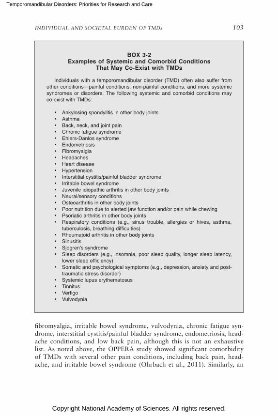
Temporomandibular Disorders: Priorities for Research and Care
Copyright National Academy of Sciences. All rights reserved.
INDIVIDUAL AND SOCIETAL BURDEN OF TMDs 103
BOX 3-2 Examples of Systemic and Comorbid Conditions
That May Co-Exist with TMDs
Individuals with a temporomandibular disorder (TMD) often also suffer from other conditions—painful conditions, non-painful conditions, and more systemic syndromes or disorders. The following systemic and comorbid conditions may co-exist with TMDs:
• Ankylosing spondylitis in other body joints• Asthma• Back, neck, and joint pain• Chronic fatigue syndrome• Ehlers-Danlos syndrome• Endometriosis• Fibromyalgia• Headaches• Heart disease• Hypertension• Interstitial cystitis/painful bladder syndrome• Irritable bowel syndrome• Juvenile idiopathic arthritis in other body joints• Neural/sensory conditions • Osteoarthritis in other body joints• Poor nutrition due to alerted jaw function and/or pain while chewing• Psoriatic arthritis in other body joints• Respiratory conditions (e.g., sinus trouble, allergies or hives, asthma,
tuberculosis, breathing difficulties)• Rheumatoid arthritis in other body joints• Sinusitis• Sjogren’s syndrome• Sleep disorders (e.g., insomnia, poor sleep quality, longer sleep latency,
lower sleep efficiency)• Somatic and psychological symptoms (e.g., depression, anxiety and post-
traumatic stress disorder)• Systemic lupus erythematosus• Tinnitus• Vertigo• Vulvodynia
fibromyalgia, irritable bowel syndrome, vulvodynia, chronic fatigue syn-drome, interstitial cystitis/painful bladder syndrome, endometriosis, head-ache conditions, and low back pain, although this is not an exhaustive list. As noted above, the OPPERA study showed significant comorbidity of TMDs with several other pain conditions, including back pain, head-ache, and irritable bowel syndrome (Ohrbach et al., 2011). Similarly, an
Temporomandibular Disorders: Priorities for Research and Care
Copyright National Academy of Sciences. All rights reserved.
104 TEMPOROMANDIBULAR DISORDERS
analysis of the NHIS data revealed a significantly increased risk of TMDs among individuals with headache, neck pain, low back pain, or pain-ful joints (Maixner et al., 2016). Other studies have found that among people with fibro myalgia, back pain, headache, irritable bowel syndrome, chronic fatigue syndrome, and vulvodynia, the likelihood of having a TMD is significantly greater than in the general population (Aaron et al., 2000; Whitehead et al., 2002; Wiesinger et al., 2007; Nguyen et al., 2013; Robinson et al., 2016; Florencio et al., 2017; Gallotta et al., 2017). More-over, the presence of comorbid pain conditions is associated with a greater severity of TMD symptoms (Visscher et al., 2016; Florencio et al., 2017). The associations of TMDs with these comorbid pain conditions are likely bidirectional in nature, such that these other pain conditions may develop following TMD onset; however, some evidence suggests that premorbid presence of other pain conditions increases the risk of developing a TMD (Sanders et al., 2013a).
TMDs have also been associated with a variety of systemic conditions that often include pain as a common symptom. Several rheumatologic diseases show significant comorbidity with TMDs, including rheuma-toid arthri tis, ankylosing spondylitis, systemic lupus erythematosus, and Sjogren’s syndrome (Aliko et al., 2011; Sidebottom and Salha, 2013; Yildizer Keris et al., 2017; Zhang et al., 2017). TMJ osteoarthritis is one subtype of TMD (Wang et al., 2015), but TMD symptoms are also more prevalent in patients with osteoarthritis at other sites such as the hand ( Abrahamsson et al., 2017) and knee (Zhang et al., 2017). Moreover, TMDs are substantially more prevalent in patients with rheumatoid arthri-tis than in the general population (Bracco et al., 2010; Mortazavi et al., 2018), and rheumatoid arthritis disease activity has been correlated with the severity of TMD symptoms (Alstergren et al., 2008; Ahmed et al., 2013, 2015). A high prevalence of TMDs has been reported in children with newly diagnosed juvenile idiopathic arthritis (Weiss et al., 2008; Muller et al., 2009; Stoll et al., 2018). TMD signs and symptoms are also more common in patients with Sjogren’s syndrome and psoriatic arthritis than in controls (Crincoli et al., 2015; Zanin et al., 2019).
TMDs and Non-Painful Comorbidities
TMDs have also been associated with health conditions that do not include pain as a primary symptom. An analysis of the NHIS data revealed an association of TMDs with a variety of non-painful medical conditions (Maixner et al., 2016).
Temporomandibular Disorders: Priorities for Research and Care
Copyright National Academy of Sciences. All rights reserved.
INDIVIDUAL AND SOCIETAL BURDEN OF TMDs 105
Respiratory and Sleep Conditions
The OPPERA study demonstrated a link between TMDs and various respiratory conditions (Ohrbach et al., 2011). Specifically, individuals re-porting at least one of five respiratory conditions (sinus trouble, allergies or hives, asthma, tuberculosis, breathing difficulties) were 2.5 times more likely to be chronic TMD cases than were controls. In addition, the oc-currence of a TMD was 3.1 times higher among those reporting a history of obstructive sleep apnea. Similarly, a population-based study in Korea reported increased odds of TMDs among individuals with asthma and rhino sinusitis (Song et al., 2018a), and a recent case–control study reported an association between having a TMD and pneumonia, asthma, and aller-gies (Fredricson et al., 2018). These cross-sectional findings do not address the direction of the association between TMDs and respiratory symptoms; however, findings from the OPPERA prospective cohort study showed that the presence of one or more respiratory conditions predicted the future development of a TMD (Sanders et al., 2013a).
Sleep disorders also show high comorbidity with TMDs (Olmos, 2016; Almoznino et al., 2017). Smith and colleagues (2009) found that the major-ity of people with a TMD met the criteria for at least one sleep disor-der, and primary insomnia was associated with increased pain sensitivity. More generally, patients with a TMD report poorer sleep quality, longer sleep latency, and lower sleep efficiency (Sener and Guler, 2012; Lavigne and Sessle, 2016). Furthermore, women with a TMD showed increased respiratory-related arousals during sleep (Dubrovsky et al., 2014). As noted above, self-reported obstructive sleep apnea was associated with a chronic TMD in the OPPERA study (Ohrbach et al., 2011), and more symptoms of sleep apnea conferred an increased risk of future TMD onset (Sanders et al., 2013b). Sleep bruxism has also been linked with TMDs in some studies; however, recent meta-analyses describe the evidence linking sleep bruxism and TMD pain as inconclusive (Jimenez-Silva et al., 2017; Baad-Hansen et al., 2019). Sleep and pain are likely reciprocally related, such that sleep disturbance may be not only a consequence but also a risk factor for TMDs. Indeed, the OPPERA findings showed that reduced sleep quality and sleep apnea were pre-existing risk factors for TMD onset (Sanders et al., 2013b).
Hypermobility and Ehlers-Danlos Syndromes
Ehlers-Danlos syndrome, a group of heritable connective tissue dis-orders, represents another set of systemic conditions associated with an increased risk of TMDs and other chronic pain conditions (De Coster et al., 2005; Chopra et al., 2017; Mitakides and Tinkle, 2017). Chronic pain is highly prevalent in this syndrome, including both regional and widespread
Temporomandibular Disorders: Priorities for Research and Care
Copyright National Academy of Sciences. All rights reserved.
106 TEMPOROMANDIBULAR DISORDERS
pain, and TMD pain has been reported in up to 71 percent of patients with Ehlers-Danlos syndrome (De Coster et al., 2005). TMD symptoms in this syndrome are generally attributed to joint hypermobility and the resultant instability of the mandible during masticatory function as well as during maximal opening, which leads to protective muscle contraction and subsequent further problems in functioning (see Chapter 2). Previous findings have demonstrated associations between joint hypermobility and TMD symptoms among individuals without Ehlers-Danlos (Perrini et al., 1997; Ogren et al., 2012). However, pain in the syndrome has also been associated with other mechanisms, including neuropathic features and signs of central sensitization (Syx et al., 2017; Benistan and Martinez, 2019) as well as myofascial pain due to protective muscle contraction.
Nutritional Challenges
Altered jaw function, including pain during eating and chewing, can substantially affect nutritional habits among people with a TMD, which can in turn reduce eating-related quality of life (Nasri-Heir et al., 2016). However, information regarding the nutritional habits in patients with a TMD is quite limited. A small cohort study of patients seeking treatment for TMDs reported that eating was a problem for the vast majority of the patients and that most reported eating a softer diet (Irving et al., 1999). Raphael and colleagues (2002) reported that higher pain severity was asso-ciated with a lower intake of dietary fiber among people with myofascial TMD pain. While nutritional modifications are often a consequence of the “soft diet” component of most TMD self-management programs (Durham et al., 2016a; The TMJ Association, 2017), little evidence has addressed the benefits and adverse effects of addressing nutritional needs (Durham et al., 2015; Nasri-Heir et al., 2016). Additional research is needed to elucidate the role of nutritional factors in TMDs and to determine the clinical benefit of nutritional interventions in these conditions.
Tinnitus and Vertigo
TMD has been associated with multiple otologic symptoms, such as tinnitus and vertigo (Porto De Toledo et al., 2017; Manfredini, 2019). Recent meta-analyses have reported bidirectional associations between tinnitus and TMDs, with tinnitus being substantially more frequent in patients with a TMD than in controls and TMDs being more common among individuals with tinnitus than in those without (Bousema et al., 2018; Omidvar and Jafari, 2019). Furthermore, the severity and duration of TMD pain have been related to tinnitus in some studies (Hilgenberg et al., 2012; Akhter et al., 2013). Other otologic symptoms have also been
Temporomandibular Disorders: Priorities for Research and Care
Copyright National Academy of Sciences. All rights reserved.
INDIVIDUAL AND SOCIETAL BURDEN OF TMDs 107
associated with TMDs, including hearing loss and vertigo/dizziness (Pekkan et al., 2010; Effat, 2016). Regarding the direction of association, one study found that palpation tenderness in the masticatory muscles predicted an increased risk of future development of tinnitus over the ensuing 5 years (Bernhardt et al., 2011).
General Somatic and Psychological Symptoms
Individuals with TMDs report a greater number of subclinical somatic symptoms assessed via questionnaires (de Leeuw et al., 2005; Fillingim et al., 2011; Chen et al., 2012). These assessments include many of the symptoms described above (e.g., otologic symptoms, respiratory symp-toms) as well as symptoms affecting other bodily systems (e.g., cardio-vascular, gastro intestinal, musculoskeletal). TMDs are also associated with an increased likelihood of psychological symptoms, including depression, anxiety, and posttraumatic stress disorder, perceived stress, and pain-related psychological processes, such as pain catastrophizing (negative cognitive-emotional processing including rumination about pain) and kinesiophobia (fear of movement) (Manfredini et al., 2009; Fillingim et al., 2011; Bertoli and de Leeuw, 2016; Reiter et al., 2018; Tay et al., 2019). Moreover, pain severity has been positively associated with these psychological factors (Guarda-Nardini et al., 2012; Su et al., 2017; Natu et al., 2018). While these somatic and psychological symptoms may reflect the consequences of TMDs, as described below, some of these symptoms also represent impor-tant risk factors for the future development of a TMD.
Conclusion 3-3: Even with a fragmented understanding of temporo -mandibular disorders (TMDs) according to traditional public health measures (e.g., prevalence, incidence), it is clear that TMDs have a large public health impact and a significant health, social, financial, and emotional burden on many individuals and families.
RISK FACTORS FOR TMDs
As described above, multiple painful and non-painful conditions and symptoms have been associated with TMDs. It is impossible to determine from cross-sectional studies whether these represent risk factors, conse-quences, or coincidences, but numerous prospective studies have identified premorbid factors that confer an increased risk of TMD onset or persis-tence, or both. Furthermore, several socio-demographic factors are known to be related to TMD risk.
Temporomandibular Disorders: Priorities for Research and Care
Copyright National Academy of Sciences. All rights reserved.
108 TEMPOROMANDIBULAR DISORDERS
Family History and Genetic Factors
While chronic pain conditions have been found to show familial aggre-gation (Matsudaira et al., 2014; Zadro et al., 2017), few studies have examined this in people with painful TMDs. It is likely that TMDs have a polygenetic underpinning although much remains to be discovered (see Chapter 4 for more discussion of TMD genetics). Raphael and colleagues (1999) found that TMDs and other painful conditions were no more com-mon in first-degree relatives of people with a history of TMDs than in first-degree relatives of people with no history of TMDs. Several twin studies have also reported that TMDs appear to have limited heritabil-ity, although these studies have generally been small and underpowered ( Visscher and Lobbezoo, 2015). A more recent and larger twin study sug-gested that 27 percent of the variance in TMD pain can be attributed to genetic factors (Plesh et al., 2012). Genetic contributions to TMDs have also been explored in candidate gene association studies, which have found evidence that serotonergic and catacholaminergic genes are associated with TMDs (Diatchenko et al., 2013; Visscher and Lobbezoo, 2015). More recently, genome-wide association studies have identified novel genetic pathways that may be related to TMDs, including the sarcoglycan alpha gene ( Sanders et al., 2017) and the muscle RAS oncogene homolog (MRAS) gene (Smith et al., 2019). Notably, several of these studies have reported that associations of some genetic factors with TMDs vary based on other risk factors, such as sex and psychological factors (Belfer et al., 2013; Slade et al., 2015; Meloto et al., 2016; Sanders et al., 2017; Smith et al., 2019). One study illustrated the potential for the existence of a gene–environment interaction that influences TMD risk (Slade et al., 2008). In this prospec-tive cohort study of 186 females, individuals with a genetic variant associ-ated with pain responsiveness had a significantly greater risk of developing TMD if they had reported a history of orthodontic treatment compared to subjects who did not (Slade et al., 2008).
TMDs in Females
TMDs are significantly more common in females than in males, with population-based studies indicating that females are at approximately twice the risk of experiencing a TMD as males (LeResche, 1997; Bueno et al., 2018). The OPPERA study observed a slightly but non-significant increased incidence of first-onset TMDs in females, while female sex was strongly associated with chronic TMDs, suggesting that females have an increased risk of TMD persistence (Slade et al., 2013a,b, 2016). Indeed, in OPPERA’s nested case–control study, 54 percent of females transitioned from first onset to persistent TMDs, as compared with 41 percent of males (Slade et
Temporomandibular Disorders: Priorities for Research and Care
Copyright National Academy of Sciences. All rights reserved.
INDIVIDUAL AND SOCIETAL BURDEN OF TMDs 109
al., 2016). This is consistent with prior findings that among patients with acute TMDs, women were more likely than men to progress to chronic TMDs (Garofalo et al., 1998), and that this sex differential may increase as chronicity persists. This increased risk of TMDs among females is observed primarily during the reproductive years (LeResche, 1997; LeResche et al., 2005; Slade et al., 2011; Song et al., 2018b). Age has also been shown to be a factor in the incidence of TMDs, with peak prevalence occurring in women in the 35–44 age group (Slade et al., 2011) and decreasing beyond reproductive age (Plesh et al., 2011a). The OPPERA study reported that TMD incidence increased with age across the age range from 18 to 44 years (Slade et al., 2013a,b), but age-related incidence information beyond 44 years of age was not available.
Women exposed to emotional, physical, or sexual abuse may also be at an increased risk for TMDs. In one study of 40 women of ages 16 to 45 years with an idiopathic TMD, the women were more likely to report emotional abuse, exposure to insults, and being diminished or humiliated in front of other people than women without a TMD (Grossi et al., 2018).
TMDs in Different Races and Ethnicities
The association of race/ethnicity with TMDs is currently not well under stood. Janal and colleagues (2008) reported that myofascial TMDs were more common among black women and Hispanic women than among white women. In a study of 4- to 6-year-olds, TMD symptoms were found to be more common among African American children than among Caucasian children (Widmalm et al., 1995). In contrast, Plesh and colleagues (2002) found a lower prevalence of TMDs among African Americans than among Caucasians after controlling for socioeconomic status. As yet another contrast, in the OPPERA study non-white racial/ethnic groups had significantly lower odds of chronic TMDs than whites, while African Americans showed an increased incidence of first-onset TMDs compared with whites (Slade et al., 2011, 2013a,b). This paradox is explained by lower risk of symptom persistence in African Americans, as OPPERA data showed that after onset, TMDs persisted in 61 percent of whites versus 35 percent of African Americans (Slade et al., 2016). The association between race/ethnicity and TMDs likely involves the contribu-tion of other underlying factors, such as socioeconomic status (Poleshuck and Green, 2008). Recent findings demonstrate that lower socioeconomic status (i.e., education and wealth) is associated with a higher prevalence and severity of general chronic pain (Grol-Prokopczyk, 2017), and racial/ethnic differences in pain were not significant after controlling for these socioeconomic influences. Specific to TMDs, the OPPERA study found that low satisfaction with material standards conferred an increased risk
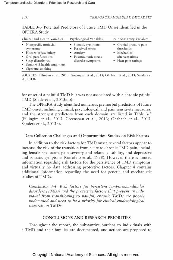
Temporomandibular Disorders: Priorities for Research and Care
Copyright National Academy of Sciences. All rights reserved.
110 TEMPOROMANDIBULAR DISORDERS
for onset of a painful TMD but was not associated with a chronic painful TMD (Slade et al., 2013a,b).
The OPPERA study identified numerous premorbid predictors of future TMD onset, including clinical, psychological, and pain sensitivity measures, and the strongest predictors from each domain are listed in Table 3-3 ( Fillingim et al., 2013; Greenspan et al., 2013; Ohrbach et al., 2013; Sanders et al., 2013b).
Data Collection Challenges and Opportunities: Studies on Risk Factors
In addition to the risk factors for TMD onset, several factors appear to increase the risk of the transition from acute to chronic TMD pain, includ-ing female sex, acute pain severity and related disability, and depressive and somatic symptoms (Garofalo et al., 1998). However, there is limited information regarding risk factors for the persistence of TMD symptoms, and virtually no data addressing protective factors. Chapter 4 contains addi tional information regarding the need for genetic and mechanistic studies of TMDs.
Conclusion 3-4: Risk factors for persistent temporomandibular disorders (TMDs) and the protective factors that prevent an indi-vidual from transitioning to painful, chronic TMDs are poorly understood and need to be a priority for clinical epidemiological research on TMDs.
CONCLUSIONS AND RESEARCH PRIORITIES
Throughout the report, the substantive burdens to individuals with a TMD and their families are documented, and actions are proposed to
TABLE 3-3 Potential Predictors of Future TMD Onset Identified in the OPPERA Study
Clinical and Health Variables Psychological Variables Pain Sensitivity Variables
• Nonspecific orofacial symptoms
• History of jaw injury• Oral parafunctions• Sleep disturbance• Comorbid health conditions • Cigarette smoking
• Somatic symptoms• Perceived stress• Anxiety• Posttraumatic stress
disorder symptoms
• Cranial pressure pain thresholds
• Mechanical aftersensations
• Heat pain ratings
SOURCES: Fillingim et al., 2013; Greenspan et al., 2013; Ohrbach et al., 2013; Sanders et al., 2013b.
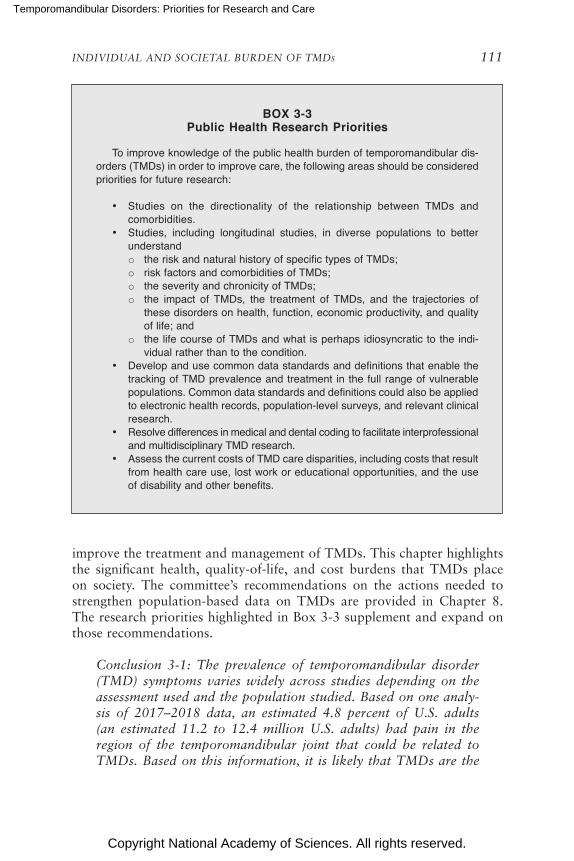
Temporomandibular Disorders: Priorities for Research and Care
Copyright National Academy of Sciences. All rights reserved.
INDIVIDUAL AND SOCIETAL BURDEN OF TMDs 111
BOX 3-3 Public Health Research Priorities
To improve knowledge of the public health burden of temporomandibular dis-orders (TMDs) in order to improve care, the following areas should be considered priorities for future research:
• Studies on the directionality of the relationship between TMDs and comorbidities.
• Studies, including longitudinal studies, in diverse populations to better understand
o the risk and natural history of specific types of TMDs; o risk factors and comorbidities of TMDs; o the severity and chronicity of TMDs; o the impact of TMDs, the treatment of TMDs, and the trajectories of
these disorders on health, function, economic productivity, and quality of life; and
o the life course of TMDs and what is perhaps idiosyncratic to the indi-vidual rather than to the condition.
• Develop and use common data standards and definitions that enable the tracking of TMD prevalence and treatment in the full range of vulnerable populations. Common data standards and definitions could also be applied to electronic health records, population-level surveys, and relevant clinical research.
• Resolve differences in medical and dental coding to facilitate inter professional and multidisciplinary TMD research.
• Assess the current costs of TMD care disparities, including costs that result from health care use, lost work or educational opportunities, and the use of disability and other benefits.
improve the treatment and management of TMDs. This chapter highlights the significant health, quality-of-life, and cost burdens that TMDs place on society. The committee’s recommendations on the actions needed to strengthen population-based data on TMDs are provided in Chapter 8. The research priorities highlighted in Box 3-3 supplement and expand on those recommendations.
Conclusion 3-1: The prevalence of temporomandibular disorder (TMD) symptoms varies widely across studies depending on the assess ment used and the population studied. Based on one analy-sis of 2017–2018 data, an estimated 4.8 percent of U.S. adults (an esti mated 11.2 to 12.4 million U.S. adults) had pain in the region of the temporomandibular joint that could be related to TMDs. Based on this information, it is likely that TMDs are the
Temporomandibular Disorders: Priorities for Research and Care
Copyright National Academy of Sciences. All rights reserved.
112 TEMPOROMANDIBULAR DISORDERS
most prevalent type of chronic orofacial pain and TMDs may be comparable in prevalence to other chronic pain conditions such as fibromyalgia, chronic low back pain, and migraine disease.
Conclusion 3-2: Nationally representative longitudinal studies of the incidence, prevalence, and disease course of temporo mandibular disorders (TMDs) using the Diagnostic Criteria for Temporo-mandibular Disorders would advance understanding of TMD etiol-ogy, risk, and prognosis and support the ability to develop clinical practice guidelines and treatment pathways.
Conclusion 3-3: Even with a fragmented understanding of temporo-mandibular disorders (TMDs) according to traditional public health measures (e.g., prevalence, incidence), it is clear that TMDs have a large public health impact and a significant health, social, financial, and emotional burden on many individuals and families.
Conclusion 3-4: Risk factors for persistent temporomandibular dis-orders (TMDs) and the protective factors that prevent an indi vidual from transitioning to painful, chronic TMDs are poorly understood and need to be a priority for clinical epidemiological research on TMDs.
REFERENCES
Aaron, L. A., M. M. Burke, and D. Buchwald. 2000. Overlapping conditions among patients with chronic fatigue syndrome, fibromyalgia, and temporomandibular disorder. Archives of Internal Medicine 160(2):221–227.
Abrahamsson, A. K., M. Kristensen, L. Z. Arvidsson, T. K. Kvien, T. A. Larheim, and I. K. Haugen. 2017. Frequency of temporomandibular joint osteoarthritis and related symp-toms in a hand osteoarthritis cohort. Osteoarthritis and Cartilage 25(5):654–657.
Ahmed, N., H. M. Mustafa, A. I. Catrina, and P. Alstergren. 2013. Impact of temporo-mandibular joint pain in rheumatoid arthritis. Mediators of Inflammation 2013:597419.
Ahmed, N., A. I. Catrina, A. O. Alyamani, H. Mustafa, and P. Alstergren. 2015. Deficient cytokine control modulates temporomandibular joint pain in rheumatoid arthritis. Euro-pean Journal of Oral Sciences 123(4):235–241.
Akhter, R., M. Morita, D. Ekuni, N. M. Hassan, M. Furuta, R. Yamanaka, Y. Matsuka, and D. Wilson. 2013. Self-reported aural symptoms, headache and temporomandibular dis-orders in Japanese young adults. BMC Musculoskeletal Disorders 14:58.
Aliko, A., R. Ciancaglini, A. Alushi, A. Tafaj, and D. Ruci. 2011. Temporomandibular joint involvement in rheumatoid arthritis, systemic lupus erythematosus and systemic sclerosis. International Journal of Oral and Maxillofacial Surgery 40(7):704–709.
Al-Khotani, A., A. Naimi-Akbar, E. Albadawi, M. Ernberg, B. Hedenberg-Magnusson, and N. Christidis. 2016. Prevalence of diagnosed temporomandibular disorders among Saudi Arabian children and adolescents. Journal of Headache Pain 17:41.
Temporomandibular Disorders: Priorities for Research and Care
Copyright National Academy of Sciences. All rights reserved.
INDIVIDUAL AND SOCIETAL BURDEN OF TMDs 113
Almoznino, G., R. Benoliel, Y. Sharav, and Y. Haviv. 2017. Sleep disorders and chronic cranio facial pain: Characteristics and management possibilities. Sleep Medicine Reviews 33:39–50.
Alstergren, P., L. Fredriksson, and S. Kopp. 2008. Temporomandibular joint pressure pain threshold is systemically modulated in rheumatoid arthritis. Journal of Orofacial Pain 22(3):231–238.
Baad-Hansen, L., M. Thymi, F. Lobbezoo, and P. Svensson. 2019. To what extent is bruxism associated with musculoskeletal signs and symptoms? A systematic review. Journal of Oral Rehabilitation 46(9):845–861.
Belfer, I., S. K. Segall, W. R. Lariviere, S. B. Smith, F. Dai, G. D. Slade, N. U. Rashid, J. S. Mogil, C. M. Campbell, R. R. Edwards, Q. Liu, E. Bair, W. Maixner, and L. Diatchenko. 2013. Pain modality- and sex-specific effects of COMT genetic functional variants. Pain 154(8):1368–1376.
Benistan, K., and V. Martinez. 2019. Pain in hypermobile Ehlers-Danlos syndrome: New insights using new criteria. American Journal of Medical Genetics Part A 179(7):1226–1234.
Bernhardt, O., T. Mundt, A. Welk, N. Koppl, T. Kocher, G. Meyer, and C. Schwahn. 2011. Signs and symptoms of temporomandibular disorders and the incidence of tinnitus. Journal of Oral Rehabilitation 38(12):891–901.
Bertoli, E., and R. de Leeuw. 2016. Prevalence of suicidal ideation, depression, and anxiety in chronic temporomandibular disorder patients. Journal of Oral & Facial Pain and Headache 30(4):296–301.
Bitiniene, D., R. Zamaliauskiene, R. Kubilius, M. Leketas, T. Gailius, and K. Smirovaite. 2018. Quality of life in patients with temporomandibular disorders: A systematic review. Stomatologija 20(1):3–9.
Bousema, E. J., E. A. Koops, P. van Dijk, and P. U. Dijkstra. 2018. Association between sub-jective tinnitus and cervical spine or temporomandibular disorders: A systematic review. Trends in Hearing 22:1–15.
Bracco, P., C. Debernardi, M. Grazia Piancino, M. Francesca Cirigliano, G. Salvetti, and L. Bazzichi. 2010. Evaluation of the stomatognathic system in patients with rheumatoid arthri tis according to the Research Diagnostic Criteria for Temporomandibular Disor-ders. Journal of Craniomandibular & Sleep Practice 28(3):181–186.
Bueno, C. H., D. D. Pereira, M. P. Pattussi, P. K. Grossi, and M. L. Grossi. 2018. Gender differences in temporomandibular disorders in adult populational studies: A systematic review and meta-analysis. Journal of Oral Rehabilation 45(9):720–729.
Burch, R., P. Rizzoli, and E. Loder. 2018. The prevalence and impact of migraine and severe headache in the United States: Figures and trends from government health studies. Headache 58(4):496–505.
Chen, H., G. Slade, P. F. Lim, V. Miller, W. Maixner, and L. Diatchenko. 2012. Relationship between temporomandibular disorders, widespread palpation tenderness, and multiple pain conditions: A case-control study. Journal of Pain 13(10):1016–1027.
Chopra, P., B. Tinkle, C. Hamonet, I. Brock, A. Gompel, A. Bulbena, and C. Francomano. 2017. Pain management in the Ehlers-Danlos syndromes. American Journal of Medical Genetics. Part C. Seminars in Medical Genetics 175(1):212–219.
Christidis, N., E. Lindstrom Ndanshau, A. Sandberg, and G. Tsilingaridis. 2019. Prevalence and treatment strategies regarding temporomandibular disorders in children and adoles-cents: A systematic review. Journal of Oral Rehabilitation 46:291–301.
Clauw, D. J. 2014. Fibromyalgia: A clinical review. JAMA 311(15):1547–1555.Crincoli, V., M. Di Comite, M. B. Di Bisceglie, L. Fatone, and G. Favia. 2015. Temporo-
mandibular disorders in psoriasis patients with and without psoriatic arthritis: An obser-vational study. International Journal of Medical Sciences 12(4):341–348.
Temporomandibular Disorders: Priorities for Research and Care
Copyright National Academy of Sciences. All rights reserved.
114 TEMPOROMANDIBULAR DISORDERS
da Silva, C. G., C. Pacheco-Pereira, A. L. Porporatti, M. G. Savi, M. A. Peres, C. Flores-Mir, and L. Canto Gde. 2016. Prevalence of clinical signs of intra-articular temporo-mandibular disorders in children and adolescents: A systematic review and meta-analysis. Journal of the American Dental Association 147:10–18.
Dahlhamer, J., J. Lucas, C. Zelaya, R. Nahin, S. Mackey, L. DeBar, R. Kerns, M. Von Korff, L. Porter, and C. Helmick. 2018. Prevalence of chronic pain and high-impact chronic pain among adults—United States, 2016. Morbidity and Mortality Weekly Report 67(36):1001–1006.
Dahlstrom, L., and G. E. Carlsson. 2010. Temporomandibular disorders and oral health-related quality of life. A systematic review. Acta Odontologica Scandinavica 68(2):80–85.
De Coster, P. J., L. I. Van den Berghe, and L. C. Martens. 2005. Generalized joint hyper-mobility and temporomandibular disorders: Inherited connective tissue disease as a model with maximum expression. Journal of Orofacial Pain 19(1):47–57.
de Leeuw, R., J. L. Studts, and C. R. Carlson. 2005. Fatigue and fatigue-related symptoms in an orofacial pain population. Oral Surgery, Oral Medicine, Oral Pathology, Oral Radiol-ogy, and Endodontics 99(2):168–174.
Diatchenko, L., R. B. Fillingim, S. B. Smith, and W. Maixner. 2013. The phenotypic and genetic signatures of common musculoskeletal pain conditions. Nature Reviews Rheu-matology 9(6):340–350.
Dubrovsky, B., K. G. Raphael, G. J. Lavigne, M. N. Janal, D. A. Sirois, P. E. Wigren, L. V. Nemelivsky, J. J. Klausner, and A. C. Krieger. 2014. Polysomnographic investigation of sleep and respiratory parameters in women with temporomandibular pain disorders. Journal of Clinical Sleep Medicine 10(2):195–201.
Durham, J., R. Touger-Decker, D. R. Nixdorf, D. Rigassio-Radler, and P. Moynihan. 2015. Oro-facial pain and nutrition: A forgotten relationship? Journal of Oral Rehabilitation 42(1):75–80.
Durham, J., M. Al-Baghdadi, L. Baad-Hansen, M. Breckons, J. P. Goulet, F. Lobbezoo, T. List, A. Michelotti, D. R. Nixdorf, C. C. Peck, K. Raphael, E. Schiffman, J. G. Steele, W. Story, and R. Ohrbach. 2016a. Self-management programmes in temporomandibular disorders: Results from an international delphi process. Journal of Oral Rehabilitation 43(12):929–936.
Durham, J., J. Shen, M. Breckons, J. G. Steele, V. Araujo-Soares, C. Exley, and L. Vale. 2016b. Healthcare cost and impact of persistent orofacial pain: The DEEP study cohort. Journal of Dental Research 95(10):1147–1154.
Dworkin, S. F. 2011. The OPPERA study: Act one. Journal of Pain 12(11):T1–T3.Dworkin, S. F., and L. LeResche. 1992. Research Diagnostic Criteria for Temporomandibular
Disorders: Review, criteria, examinations and specifications, critique. Journal of Cranio-mandibular Disorders 6:301–355.
Effat, K. G. 2016. Otological symptoms and audiometric findings in patients with temporo-mandibular disorders: Costen’s syndrome revisited. Journal of Laryngology and Otology 130(12):1137–1141.
Fillingim, R. B., R. Ohrbach, J. D. Greenspan, C. Knott, R. Dubner, E. Bair, C. Baraian, G. D. Slade, and W. Maixner. 2011. Potential psychosocial risk factors for chronic TMD: Descriptive data and empirically identified domains from the OPPERA case-control study. Journal of Pain 12:T46–T60.
Fillingim, R. B., R. Ohrbach, J. D. Greenspan, C. Knott, L. Diatchenko, R. Dubner, E. Bair, C. Baraian, N. Mack, G. D. Slade, and W. Maixner. 2013. Psychological factors associ-ated with development of TMD: The OPPERA prospective cohort study. Journal of Pain 14:T75–T90.
Temporomandibular Disorders: Priorities for Research and Care
Copyright National Academy of Sciences. All rights reserved.
INDIVIDUAL AND SOCIETAL BURDEN OF TMDs 115
Fillingim, R. B., G. D. Slade, J. D. Greenspan, R. Dubner, W. Maixner, E. Bair, and R. Ohrbach. 2018. Long-term changes in biopsychosocial characteristics related to temporo-mandibular disorder: Findings from the OPPERA study. Pain 159(11):2403–2413.
Florencio, L. L., A. S. de Oliveira, G. F. Carvalho, F. Dach, M. E. Bigal, C. Fernandez-de-Las-Penas, and D. Bevilaqua-Grossi. 2017. Association between severity of temporo-mandibular disorders and the frequency of headache attacks in women with migraine: A cross-sectional study. Journal of Manipulative and Physiological Therapeutics 40(4):250–254.
Franco-Micheloni, A. L., G. Fernandes, D. A. de Godoi Goncalves, and C. M. Camparis. 2015. Temporomandibular disorders in a young adolescent Brazilian population: Epidemiologic characterization and associated factors. Journal of Oral & Facial Pain and Headache 29(3):242–249.
Fredricson, A. S., F. Khodabandehlou, C. K. Weiner, A. Naimi-Akbar, J. Adami, and A. Rosen. 2018. Are there early signs that predict development of temporomandibular joint disease? Journal of Oral Sciences 60(2):194–200.
Gallotta, S., V. Bruno, S. Catapano, N. Mobilio, C. Ciacci, and P. Iovino. 2017. High risk of temporomandibular disorder in irritable bowel syndrome: Is there a correlation with greater illness severity? World Journal of Gastroenterology 23(1):103–109.
Garofalo, J. P., R. J. Gatchel, A. L. Wesley, and E. Ellis. 1998. Predicting chronicity in acute temporomandibular joint disorders using the research diagnostic criteria. Journal of the American Dental Association 129(4):438–447.
Goulet, J. P., G. J. Lavigne, and J. P. Lund. 1995. Jaw pain prevalence among French-speaking Canadians in Quebec and related symptoms of temporomandibular disorders. Journal of Dental Research 74:1738–1744.
Graue, A. M., A. Jokstad, J. Assmus, and M. S. Skeie. 2016. Prevalence among adolescents in Bergen, Western Norway, of temporomandibular disorders according to the DC/TMD criteria and examination protocol. Acta Odontologica Scandinavica 74:449–555.
Greenspan, J. D., G. D. Slade, E. Bair, R. Dubner, R. B. Fillingim, R. Ohrbach, C. Knott, L. Diatchenko, Q. Liu, and W. Maixner. 2013. Pain sensitivity and autonomic factors asso-ciated with development of TMD: The OPPERA prospective cohort study. Journal of Pain 14(12 Suppl):T63–T74.
Grol-Prokopczyk, H. 2017. Sociodemographic disparities in chronic pain, based on 12-year longitudinal data. Pain 158(2):313–322.
Grossi, P. K., C. H. Bueno, M. A. de Abreu Silva, E. P. Pellizzer, and M. L. Grossi. 2018. Evaluation of sexual, physical, and emotional abuse in women diagnosed with temporo-mandibular disorders: A case-control study. International Journal of Prosthodontics 31(6):543–551.
Guarda-Nardini, L., C. Pavan, N. Arveda, G. Ferronato, and D. Manfredini. 2012. Psycho-metric features of temporomandibular disorders patients in relation to pain diffusion, location, intensity and duration. Journal of Oral Rehabilitation 39(10):737–743.
Hilgenberg, P. B., A. D. Saldanha, C. O. Cunha, J. H. Rubo, and P. C. Conti. 2012. Temporo-mandibular disorders, otologic symptoms and depression levels in tinnitus patients. Journal of Oral Rehabilitation 39(4):239–244.
Hobson, K. A., G. J. Huang, and D. A. Covell, Jr. 2008. Patterns of dental care utiliza-tion among patients with temporomandibular disorders. Journal of Orofacial Pain 22(2):108–114.
Hoffmann, R. G., J. M. Kotchen, T. A. Kotchen, T. Cowley, M. Dasgupta, and A. W. Cowley Jr. 2011. Temporomandibular disorders and associated clinical comorbidities. Clinical Journal of Pain 27(3):268–274.
Howard, J. A. 2013. Temporomandibular joint disorders in children. Dental Clinics of North Amercia 57:99–127.
Temporomandibular Disorders: Priorities for Research and Care
Copyright National Academy of Sciences. All rights reserved.
116 TEMPOROMANDIBULAR DISORDERS
IOM (Institute of Medicine). 2011. Relieving pain in America: A blueprint for transforming prevention, care, education, and research. Washington, DC: The National Academies Press.
Irving, J., G. D. Wood, and A. F. Hackett. 1999. Does temporomandibular disorder pain dysfunction syndrome affect dietary intake? Dental Update 26(9):405–407.
Janal, M. N., K. G. Raphael, S. Nayak, and J. Klausner. 2008. Prevalence of myofascial temporomandibular disorder in US community women. Journal of Oral Rehabilitation 35(11):801–809.
Jimenez-Silva, A., C. Pena-Duran, J. Tobar-Reyes, and R. Frugone-Zambra. 2017. Sleep and awake bruxism in adults and its relationship with temporomandibular disorders: A systematic review from 2003 to 2014. Acta Odontologica Scandinavica 75(1):36–58.
John, M. T., D. R. Reissmann, O. Schierz, and R. W. Wassell. 2007. Oral health-related quality of life in patients with temporomandibular disorders. Journal of Orofacial Pain 21(1):46–54.
Katsoulis, K., R. Bassetti, I. Windecker-Getaz, R. Mericske-Stern, and J. Katsoulis. 2012. Temporomandibular disorders/myoarthropathy of the masticatory system. Schweizer Monatsschrift fuer Zahnmedizin 22(6):510–518.
Köhler, A. A., A. N. Helkimo, T. Magnusson, and A. Hugoson. 2009. Prevalence of symptoms and signs indicative of temporomandibular disorders in children and adolescents. A cross-sectional epidemiological investigation covering two decades. European Archives of Paediatric Dentistry 10(Suppl 1):16–25.
Kotiranta, U., T. Suvinen, T. Kauko, Y. Le Bell, P. Kemppainen, J. Suni, and H. Forssell. 2015. Subtyping patients with temporomandibular disorders in a primary health care setting on the basis of the Research Diagnostic Criteria for Temporomandibular Disorders Axis II pain-related disability: A step toward tailored treatment planning? Journal of Oral & Facial Pain and Headache 29(2):126–134.
Kotiranta, U., H. Forssell, and T. Kauppila. 2018. Painful temporomandibular disorders (TMD) and comorbidities in primary care: Associations with pain-related disability. Acta Odontologica Scandinavica Sept 28:1–6.
Kusiak, J. W., C. Veasley, W. Maixner, R. B. Fillingim, J. S. Mogil, L. Diatchenko, C. B. Meloto, I. Belfer, M. Kaseta, T. Kalinowski, J. Gagnier, N. Hu, C. Alvarez-Garriga, M. Bayona, B. Eloff, M. Reardon, C. Greene, A. Bertagna, T. Cowley, J. Wilentz, and D. Clare. 2018. The TMJ Patient-Led RoundTable: A history and summary of work. http://mdepinet.org/wp-content/uploads/TMJ-Patient-RoundTable-Briefing-Report_9_25_18.pdf (accessed January 31, 2020).
Lavigne, G. J., and B. J. Sessle. 2016. The neurobiology of orofacial pain and sleep and their interactions. Journal of Dental Research 95(10):1109–1116.
LeResche, L. 1997. Epidemiology of temporomandibular disorders: Implications for the inves-tigation of etiologic factors. Critical Reviews in Oral Biology and Medicine 8(3):291–305.
LeResche, L., L. A. Mancl, M. T. Drangsholt, K. Saunders, and M. Von Korff. 2005. Rela tionship of pain and symptoms to pubertal development in adolescents. Pain 118(1–2):201–209.
LeResche, L., L. A. Mancl, M. T. Drangsholt, G. Huang, and M. Von Korff. 2007. Predictors of onset of facial pain and temporomandibular disorders in early adolescence. Pain 129(3):269–278.
List, T., K. Wahlund, B. Wenneberg, and S. F. Dworkin. 1999. TMD in children and adoles-cents: Prevalence of pain, gender differences, and perceived treatment need. Journal of Orofacial Pain 13(1):9–20.
Locker, D., and P. F. Allen. 2002. Developing short-form measures of oral health-related qua-lity of life. Journal of Public Health Dentistry 62(1):13–20.
Temporomandibular Disorders: Priorities for Research and Care
Copyright National Academy of Sciences. All rights reserved.
INDIVIDUAL AND SOCIETAL BURDEN OF TMDs 117
Locker, D., and G. Slade. 1988. Prevalence of symptoms associated with temporomandibular disorders in a Canadian population. Community Dentistry and Oral Epidemiology 16(5):310–313.
Locker, D., and G. Slade. 1993. Oral health and the quality of life among older adults: The Oral Health Impact Profile. Journal of the Canadian Dental Association 59(10):830–833, 837–838, 844.
Macfarlane, T. V., A. S. Blinkhorn, R. M. Davies, J., Kincey, and H. V. Worthington. 2002. Oro-facial pain in the community: Prevalence and associated impact. Community Den-tistry and Oral Epidemiology 30(1):52–60.
Magnusson, T., I. Egermarki, and G. E. Carlsson. 2005. A prospective investigation over two decades on signs and symptoms of temporomandibular disorders and associated vari-ables. A final summary. Acta Odontologica Scandinavica 63(2):99–109.
Maixner, W., L. Diatchenko, R. Dubner, R. B. Fillingim, J. D. Greenspan, C. Knott, R. Ohrbach, B. Weir, and G. D. Slade. 2011. Orofacial Pain Prospective Evaluation and Risk Assessment study: The OPPERA study. Journal of Pain 12(11 Suppl):T4–T11.
Maixner, W., R. B. Fillingim, D. A. Williams, S. B. Smith, and G. D. Slade. 2016. Overlapping chronic pain conditions: Implications for diagnosis and classification. Journal of Pain 17(9 Suppl):T93–T107.
Manfredini, D. 2019. Tinnitus in temporomandibular disorders patients: Any clinical implica-tions from research findings? Evidence Based Dentistry 20(1):30–31.
Manfredini, D., M. Marini, C. Pavan, L. Pavan, and L. Guarda-Nardini. 2009. Psychosocial profiles of painful TMD patients. Journal of Oral Rehabilitation 36(3):193–198.
Manfredini, D., L. Guarda-Nardini, E. Winocur, F. Piccotti, J. Ahlberg, and F. Lobbezoo. 2011. Research diagnostic criteria for temporomandibular disorders: A systematic review of Axis I epidemiologic findings. Oral Surgery, Oral Medicine, Oral Pathology, Oral Radiology, and Endodontology 112(4):453–462.
Marpaung, C., M. K. A. van Selms, and F. Lobbezoo. 2019. Temporomandibular joint anterior disc displacement with reduction in a young population: Prevalence and risk indicators. International Journal of Paediatric Dentistry 29(1):66–73.
Matsudaira, K., H. Konishi, K. Miyoshi, T. Isomura, and K. Inuzuka. 2014. Potential risk fac-tors of persistent low back pain developing from mild low back pain in urban Japanese workers. PLoS One 9(4):e93924.
Meloto, C. B., A. V. Bortsov, E. Bair, E. Helgeson, C. Ostrom, S. B. Smith, R. Dubner, G. D. Slade, R. B. Fillingim, J. D. Greenspan, R. Ohrbach, W. Maixner, S. A. McLean, and L. Diatchenko. 2016. Modification of COMT-dependent pain sensitivity by psychological stress and sex. Pain 157(4):858–867.
Meloto, C. B., G. D. Slade, R. N. Lichtenwalter, E. Bair, N. Rathnayaka, L. Diatchenko, J. D. Greenspan, W. Maixner, R. B. Fillingim, and R. Ohrbach. 2019. Clinical predictors of persistent temporomandibular disorder in people with first-onset temporomandibular disorder: A prospective case-control study. Journal of the American Dental Association 150(7):572–581.
Meucci, R. D., A. G. Fassa, and N. M. Faria. 2015. Prevalence of chronic low back pain: Systematic review. Revista de Saude Publica 49:1.
Miller, V. E., C. Poole, Y. Golightly, D. Barrett, D. G. Chen, R. Ohrbach, J. D. Greenspan, R. B. Fillingim, and G. D. Slade. 2019. Characteristics associated with high-impact pain in people with temporomandibular disorder: A cross-sectional study. Journal of Pain 20(3):288–300.
Mitakides, J., and B. T. Tinkle. 2017. Oral and mandibular manifestations in the Ehlers-Danlos syndromes. American Journal of Medical Genetics. Part C, Seminars in Medical Genetics 175(1):220–225.
Temporomandibular Disorders: Priorities for Research and Care
Copyright National Academy of Sciences. All rights reserved.
118 TEMPOROMANDIBULAR DISORDERS
Mortazavi, N., M. Babaei, N. Babaee, H. H. Kazemi, R. Mortazavi, and A. Mostafazadeh. 2018. Evaluation of the prevalence of temporomandibular joint involvement in rheu-matoid arthritis using Research Diagnostic Criteria for Temporomandibular Disorders. Journal of Dentistry (Tehran) 15(6):332–338.
Muller, L., C. J. Kellenberger, E. Cannizzaro, D. Ettlin, T. Schraner, I. B. Bolt, T. Peltomaki, and R. K. Saurenmann. 2009. Early diagnosis of temporomandibular joint involvement in juvenile idiopathic arthritis: A pilot study comparing clinical examination and ultrasound to magnetic resonance imaging. Rheumatology 48(6):680–685.
Nasri-Heir, C., J. B. Epstein, R. Touger-Decker, and R. Benoliel. 2016. What should we tell patients with painful temporomandibular disorders about what to eat? Journal of the American Dental Association 147(8):667–671.
Natu, V. P., A. U. Yap, M. H. Su, N. M. Irfan Ali, and A. Ansari. 2018. Temporomandibular disorder symptoms and their association with quality of life, emotional states and sleep quality in South-East Asian youths. Journal of Oral Rehabilitation 45(10):756–763.
Nguyen, R. H., C. Veasley, and D. Smolenski. 2013. Latent class analysis of comorbidity patterns among women with generalized and localized vulvodynia: Preliminary findings. Journal of Pain Research 6:303–309.
Nilsson, I. M. 2007. Reliability, validity, incidence and impact of temporomandibular pain disorders in adolescents. Swedish Dental Journal Supplement 183:7–86.
Ogren, M., C. Faltmars, B. Lund, and A. Holmlund. 2012. Hypermobility and trauma as etiologic factors in patients with disc derangements of the temporomandibular joint. International Journal of Oral and Maxillofacial Surgery 41(9):1046–1050.
Ohrbach, R. 2010. Disability assessment in temporomandibular disorders and masticatory system rehabilitation. Journal of Oral Rehabilitation 37(6):452–480.
Ohrbach, R., and S. F. Dworkin. 1998. Five-year outcomes in TMD: Relationship of changes in pain to changes in physical and psychological variables. Pain 74(2–3):315–326.
Ohrbach, R., R. B. Fillingim, F. Mulkey, Y. Gonzalez, S. Gordon, H. Gremillion, P. F. Lim, M. Ribeiro-Dasilva, J. D. Greenspan, C. Knott, W. Maixner, and G. Slade. 2011. Clinical findings and pain symptoms as potential risk factors for chronic TMD: Descriptive data and empirically identified domains from the OPPERA case-control study. Journal of Pain 12(11 Suppl):T27–T45.
Ohrbach, R., E. Bair, R. B. Fillingim, Y. Gonzalez, S. M. Gordon, P. F. Lim, M. Ribeiro- Dasilva, L. Diatchenko, R. Dubner, J. D. Greenspan, C. Knott, W. Maixner, S. B. Smith, and G. D. Slade. 2013. Clinical orofacial characteristics associated with risk of first-onset TMD: The OPPERA prospective cohort study. Journal of Pain 14(12 Suppl):T33–T50.
Olmos, S. R. 2016. Comorbidities of chronic facial pain and obstructive sleep apnea. Current Opinion in Pulmonary Medicine 22(6):570–575.
Omidvar, S., and Z. Jafari. 2019. Association between tinnitus and temporomandibular disorders: A systematic review and meta-analysis. Annals of Otology, Rhinology, and Laryngology 128(7):662–675.
Pekkan, G., S. Aksoy, C. Hekimoglu, and F. Oghan. 2010. Comparative audiometric evalua-tion of temporomandibular disorder patients with otological symptoms. Journal of Cranio-Maxillo-Facial Surgery 38(3):231–234.
Perrini, F., R. H. Tallents, R. W. Katzberg, R. F. Ribeiro, S. Kyrkanides, and M. E. Moss. 1997. Generalized joint laxity and temporomandibular disorders. Journal of Orofacial Pain 11(3):215–221.
Plesh, O., P. B. Crawford, and S. A. Gansky. 2002. Chronic pain in a biracial population of young women. Pain 99(3):515–523.
Plesh, O., S. H. Adams, and S. A. Gansky. 2011a. Racial/ethnic and gender prevalences in reported common pains in a national sample. Journal of Orofacial Pain 25(1):25–31.
Temporomandibular Disorders: Priorities for Research and Care
Copyright National Academy of Sciences. All rights reserved.
INDIVIDUAL AND SOCIETAL BURDEN OF TMDs 119
Plesh, O., S. H. Adams, and S. A. Gansky. 2011b. Temporomandibular joint and muscle disorder-type pain and comorbid pains in a national US sample. Journal of Orofacial Pain 25(3):190–198.
Plesh, O., C. Noonan, D. S. Buchwald, J. Goldberg, and N. Afari. 2012. Temporomandibular disorder-type pain and migraine headache in women: A preliminary twin study. Journal of Orofacial Pain 26(2):91–98.
Poleshuck, E. L., and C. R. Green. 2008. Socioeconomic disadvantage and pain. Pain 136(3):235–238.
Porto De Toledo, I., F. M. Stefani, A. L. Porporatti, L. A. Mezzomo, M. A. Peres, C. Flores-Mir, and G. De Luca Canto G. 2017. Prevalence of otologic signs and symptoms in adult patients with temporomandibular disorders: A systematic review and meta-analysis. Clinical Oral Investigations 21(2):597–605.
Raphael, K. G., J. J. Marbach, R. M. Gallagher, and B. P. Dohrenwend. 1999. Myofascial TMD does not run in families. Pain 80(1–2):15–22.
Raphael, K. G., J. J. Marbach, and R. Touger-Decker. 2002. Dietary fiber intake in patients with myofascial face pain. Journal of Orofacial Pain 16(1):39–47.
Reiter, S., I. Eli, M. Mahameed, A. Emodi-Perlman, P. Friedman-Rubin, M. A. Reiter, and E. Winocur. 2018. Pain catastrophizing and pain persistence in temporomandibular disorder patients. Journal of Oral & Facial Pain and Headache 32(3):309–320.
Rener-Sitar, K., A. Celebić, K. Mehulić, and N. Petricević. 2013. Factors related to oral health related quality of life in TMD patients. Collegium Antropologicum 37(2):407–413.
Riley, J. L., G. H. Gilbert, and M. W. Heft. 1999. Health care utilization by older adults in response to painful orofacial symptoms. Pain 81:67–75.
Robinson, L. J., J. Durham, and J. L. Newton. 2016. A systematic review of the comorbidity between temporomandibular disorders and chronic fatigue syndrome. Journal of Oral Rehabilitation 43(4):306–316.
Sanders, A. E., G. D. Slade, E. Bair, R. B. Fillingim, C. Knott, R. Dubner, J. D. Greenspan, W. Maixner, and R. Ohrbach. 2013a. General health status and incidence of first-onset temporomandibular disorder: The OPPERA prospective cohort study. Journal of Pain 14(12 Suppl):T51–T62.
Sanders, A. E., G. K. Essick, R. Fillingim, C. Knott, R. Ohrbach, J. D. Greenspan, L. Diatchenko, W. Maixner, R. Dubner, E. Bair, V. E. Miller, and G. D. Slade. 2013b. Sleep apnea symptoms and risk of temporomandibular disorder: OPPERA cohort. Journal of Dental Research 92(7 Suppl):70S–77S.
Sanders, A. E., D. Jain, T. Sofer, K. F. Kerr, C. C. Laurie, J. R. Shaffer, M. L. Marazita, L. M. Kaste, G. D. Slade, R. B. Fillingim, R. Ohrbach, W. Maixner, T. Kocher, O. Bernhardt, A. Teumer, C. Schwahn, K. Sipila, R. Lahdesmaki, M. Mannikko, P. Pesonen, M. Jarvelin, C. M. Rizzatti-Barbosa, C. B. Meloto, M. Ribeiro-Dasilva, L. Diatchenko, P. Serrano, and S. B. Smith. 2017. GWAS identifies new loci for painful temporomandibular disorder: Hispanic community health study/study of Latinos. Journal of Dental Research 96(3):277–284.
Scrivani, S. J., D. A. Keith, and L. B. Kaban. 2008. Temporomandibular disorders. New England Journal of Medicine 359:2693–2705.
Sener, S., and O. Guler. 2012. Self-reported data on sleep quality and psychologic characteris-tics in patients with myofascial pain and disc displacement versus asymptomatic controls. International Journal of Prosthodontics 25(4):348–352.
Sidebottom, A. J., and R. Salha. 2013. Management of the temporomandibular joint in rheumatoid disorders. British Journal of Oral & Maxillofacial Surgery 51(3):191–198.
Slade, G., and J. Durham. 2020. Prevalence, impact, and costs of treatment for temporo-mandibular disorders. Paper commissioned by the Committee on Temporomandibular Disorders (TMDs): From Research Discoveries to Clinical Treatment (see Appendix C).
Temporomandibular Disorders: Priorities for Research and Care
Copyright National Academy of Sciences. All rights reserved.
120 TEMPOROMANDIBULAR DISORDERS
Slade, G. D., L. Diatchenko, K. Bhalang, A. Sigurdsson, R. B. Fillingim, I. Belfer, M. B. Max, D. Goldman, and W. Maixner. 2007. Influence of psychosocial factors on risk of temporo mandibular disorders. Journal of Dental Research 86(11):1120–1125.
Slade, G. D., L. Diatchenko, R. Ohrbach, and W. Maixner. 2008. Orthodontic treatment, genetic factors and risk of temporomandibular disorder. Seminars in Orthodontics 14(2):146–156.
Slade, G. D., E. Bair, K. By, F. Mulkey, C. Baraian, R. Rothwell, M. Reynolds, V. Miller, Y. Gonzalez, S. Gordon, M. Ribeiro-Dasilva, P. F. Lim, J. D. Greenspan, R. Dubner, R. B. Fillingim, L. Diatchenko, W. Maixner, D. Dampier, C. Knott, and R. Ohrbach. 2011. Study methods, recruitment, sociodemographic findings, and demographic representative-ness in the OPPERA study. Journal of Pain 12(11 Suppl):T12–T26.
Slade, G. D., E. Bair, J. D. Greenspan, R. Dubner, R. B. Fillingim, L. Diatchenko, W. Maixner, C. Knott, and R. Ohrbach. 2013a. Signs and symptoms of first-onset TMD and socio-demographic predictors of its development: The OPPERA prospective cohort study. Journal of Pain 14:T20–T32.
Slade, G. D., R. B. Fillingim, A. E. Sanders, E. Bair, J. D. Greenspan, R. Ohrbach, R. Dubner, L. Diatchenko, S. B. Smith, C. Knott, and W. Maixner. 2013b. Summary of findings from the OPPERA prospective cohort study of incidence of first-onset temporomandibular disorder: Implications and future directions. Journal of Pain 14(12 Suppl):T116–T124.
Slade, G., A. Sanders, E. Bair, N. C. Brownstein, D. Dampier, C. Knott, R. Fillingim, W. O. Maixner, S. Smith, J. Greenspan, R. Dubner, and R. Ohrbach. 2013c. Preclinical epi-sodes of orofacial pain symptoms and their association with healthcare behaviors in the OPPERA prospective cohort study. Pain 154(5):750–760.
Slade, G. D., A. E. Sanders, R. Ohrbach, E. Bair, W. Maixner, J. D. Greenspan, R. B. Fillingim, S. Smith, and L. Diatchenko. 2015. COMT diplotype amplifies effect of stress on risk of temporomandibular pain. Journal of Dental Research 94(9):1187–1195.
Slade, G. D., R. Ohrbach, J. D. Greenspan, R. B. Fillingim, E. Bair, A. E. Sanders, R. Dubner, L. Diatchenko, C. B. Meloto, S. Smith, and W. Maixner. 2016. Painful temporo-mandibular disorder: Decade of discovery from OPPERA studies. Journal of Dental Research 95:1084–1092.
Smith, M. T., E. M. Wickwire, E. G. Grace, R. R. Edwards, L. F. Buenaver, S. Peterson, B. Klick, and J. A. Haythornthwaite. 2009. Sleep disorders and their association with laboratory pain sensitivity in temporomandibular joint disorder. Sleep 32(6):779–790.
Smith, S. B., M. Parisien, E. Bair, I. Belfer, A. J. Chabot-Dore, P. Gris, S. Khoury, S. Tansley, Y. Torosyan, D. V. Zaykin, O. Bernhardt, P. de Oliveira Serrano, R. H. Gracely, D. Jain, M. R. Jarvelin, L. M. Kaste, K. F. Kerr, T. Kocher, R. Lahdesmaki, N. Laniado, C. C. Laurie, C. A. Laurie, M. Mannikko, C. B. Meloto, A. G. Nackley, S. C. Nelson, P. Pesonen, M. C. Ribeiro-Dasilva, C. M. Rizzatti-Barbosa, A. E. Sanders, C. Schwahn, K. Sipila, T. Sofer, A. Teumer, J. S. Mogil, R. B. Fillingim, J. D. Greenspan, R. Ohrbach, G. D. Slade, W. Maixner, and L. Diatchenko. 2019. Genome-wide association reveals contribu-tion of MRAS to painful temporomandibular disorder in males. Pain 160(3):579–591.
Song, H. S., J. S. Shin, J. Lee, Y. J. Lee, M. R. Kim, J. H. Cho, K. W. Kim, Y. Park, H. J. Song, S. Y. Park, S. Kim, M. Kim, and I. H. Ha. 2018a. Association between temporo mandibular disorders, chronic diseases, and ophthalmologic and otolaryngologic disorders in Korean adults: A cross-sectional study. PLoS One 13(1):e0191336.
Song, Y. L., A. U. Yap, and J. C. Turp. 2018b. Association between temporomandibular dis-orders and pubertal development: A systematic review. Journal of Oral Rehabilitation 45(12):1007–1015.
Stoll, M. L., C. H. Kau, P. D. Waite, and R. Q. Cron. 2018. Temporomandibular joint arthri-tis in juvenile idiopathic arthritis, now what? Pediatric Rheumatology Online Journal 16(1):32.
Temporomandibular Disorders: Priorities for Research and Care
Copyright National Academy of Sciences. All rights reserved.
INDIVIDUAL AND SOCIETAL BURDEN OF TMDs 121
Su, N., F. Lobbezoo, A. van Wijk, G. J. van der Heijden, and C. M. Visscher. 2017. Asso-ciations of pain intensity and pain-related disability with psychological and socio- demographic factors in patients with temporomandibular disorders: A cross-sectional study at a specialised dental clinic. Journal of Oral Rehabilitation 44(3):187–196.
Syx, D., I. De Wandele, L. Rombaut, and F. Malfait. 2017. Hypermobility, the Ehlers-Danlos syndromes and chronic pain. Clinical and Experimental Rheumatology Suppl 107(5):116–122.
Tay, K. J., A. U. Yap, J. C. M. Wong, K. B. C. Tan, and P. F. Allen. 2019. Associations between symptoms of temporomandibular disorders, quality of life and psychological states in Asian military personnel. Journal of Oral Rehabilitation 46(4):330–339.
The TMJ Association. 2017. TMD nutrition and you. http://www.tmj.org/site/page?pageId=318 (accessed January 31, 2020).
Velly, A. M., J. O. Look, C. Carlson, P. A. Lenton, W. Kang, C. A. Holcroft, and J. R. Fricton. 2011. The effect of catastrophizing and depression on chronic pain: A prospective cohort study of temporomandibular muscle and joint pain disorders. Pain 152:2377–2383.
Visscher, C. M., and F. Lobbezoo. 2015. TMD pain is partly heritable. A systematic review of family studies and genetic association studies. Journal of Oral Rehabilitation 42(5):386–399.
Visscher, C. M., E. A. van Wesemael-Suijkerbuijk, and F. Lobbezoo. 2016. Is the experience of pain in patients with temporomandibular disorder associated with the presence of comorbidity? European Journal of Oral Sciences 124(5):459–464.
Von Korff, M., L. LeResche, and S. F. Dworkin. 1993. First onset of common pain symptoms: A prospective study of depression as a risk factor. Pain 55(2):251–258.
Wang, X. D., J. N. Zhang, Y. H. Gan, and Y. H. Zhou. 2015. Current understanding of patho-genesis and treatment of TMJ osteoarthritis. Journal of Dental Research 94(5):666–673.
Weiss, P. F., B. Arabshahi, A. Johnson, L. T. Bilaniuk, D. Zarnow, A. M. Cahill, C. Feudtner, and R. Q. Cron. 2008. High prevalence of temporomandibular joint arthritis at disease onset in children with juvenile idiopathic arthritis, as detected by magnetic resonance imaging but not by ultrasound. Arthritis and Rheumatism 58(4):1189–1196.
White, B. A., L. A. Williams, and J. R. Leben. 2001. Health care utilization and cost among health maintenance organization members with temporomandibular disorders. Journal of Orofacial Pain 15(2):158–169.
Whitehead, W. E., O. Palsson, and K. R. Jones. 2002. Systematic review of the comorbidity of irritable bowel syndrome with other disorders: What are the causes and implications? Gastroenterology 122(4):1140–1156.
Widmalm, S. E., R. L. Christiansen, and S. M. Gunn. 1995. Race and gender as TMD risk factors in children. Cranio 13(3):163–166.
Wiesinger, B., H. Malker, E. Englund, and A. Wanman. 2007. Back pain in relation to musculo-skeletal disorders in the jaw-face: A matched case-control study. Pain 131(3):311–319.
Wu, N., and C. Hirsch. 2010. Temporomandibular disorders in German and Chinese adoles-cents. Journal of Orofacial Orthopedica 71(3):187–198.
Yildizer Keris, E., S. D. Yaman, M. D. Demirag, and S. Haznedaroglu. 2017. Temporo-mandibular joint findings in patients with rheumatoid arthritis, ankylosing spondylitis, and primary Sjogren’s syndrome. Journal of Investigative and Clinical Dentistry 8(4).
Zadro, J. R., D. Shirley, J. F. Sanchez-Romera, J. R. Ordonana, and P. H. Ferreira. 2017. Does familial aggregation of chronic low back pain affect recovery?: A population-based twin study. Spine 42(17):1295–1301.
Zanin, M. C., D. M. Garcia, E. M. Rocha, and C. M. de Felicio. 2019. Orofacial motor func-tions and temporomandibular disorders in patients with Sjogren’s syndrome. Arthritis Care Research June 17.

Temporomandibular Disorders: Priorities for Research and Care
Copyright National Academy of Sciences. All rights reserved.
122 TEMPOROMANDIBULAR DISORDERS
Zhang, X., F. Chen, L. Chen, B. Li, S. Xu, D. Cui, L. Yu, M. Liu, X. Shi, Q. Li, and Y. Li. 2017. Symptoms and signs of temporomandibular disorders in patients with knee osteoarthritis. International Dental Journal 67(2):78–84.
Temporomandibular Disorders: Priorities for Research and Care
Copyright National Academy of Sciences. All rights reserved.
123
What I want to see going forward is research on just what temporo-mandibular disorders are—research that validates the safety and effec-tiveness of every treatment for TMDs. I want research on every aspect of a TMJ device and related patient care. I want better practices and protocols for all stages of TMJ surgical procedures and a formal collaboration with the musculoskeletal branches in medicine. There is no reason we should not have the research on the TM joint that exists on every other joint in the body.
—Lutricia M.
Despite investment in research directly and indirectly related to temporo mandibular disorders (TMDs)—most significantly in the field of orofacial pain—researchers have yet to unravel the etiologies and patho-physiologies of TMDs or to translate, in a meaningful way, research find-ings into improved clinical care practices. Over the past decade the body of research on TMDs centered on the biological mechanisms underlying the development and persistence of orofacial pain and on the structure and function of the joint and its tissues, while more recent research has begun to examine the molecular genetics, biomarkers, and biopsychosocial risk factors of TMDs and common comorbidities. Broadly, the research founda-tion relating to TMDs, as has been the case with other complex, stigmatized conditions, has suffered from the siloing of disciplines and from a lack of clear direction—thus stunting the potential clinical impact of the research. In the case of TMDs, these difficulties have been heightened by a significant
4
State of the Science on TMDs
Temporomandibular Disorders: Priorities for Research and Care
Copyright National Academy of Sciences. All rights reserved.
124 TEMPOROMANDIBULAR DISORDERS
dental–medical divide that affects both research and clinical care. Funda-mentally, the development of safe and effective treatments and therapies for TMDs necessitates the existence of a robust research base spanning multiple disciplines—a foundation that is currently lacking in many areas.
This chapter provides the reader with an overview of recent basic sci-ence research related to the pathophysiologies of TMDs and orofacial pain, with a primary focus on the state of the evidence as it relates to the devel-opment and growth of temporomandibular joint (TMJ) tissues in health and disease and the mechanisms underpinning pain and tissue dysfunction. This chapter is not intended to be an exhaustive systematic review of all of the scientific evidence available on TMDs or a clinical review; rather, the chapter highlights significant research gaps identified by the committee that need to be addressed to develop a robust evidence base for TMDs. Following an overview of the state of the science and a discussion of over-arching research gaps and priorities—primarily focused on the scarcity of multidisciplinary research efforts dedicated to understanding TMDs from the cellular to the societal level—the chapter explores the organizational, financial, and cultural changes within the TMD research enterprise that will be necessary for the development of a research enterprise that priori-tizes the needs of the patient. The chapter will conclude by exploring how a patient-centered research framework could be structured to address the short- and long-term research priorities identified throughout this report. Additional evidence and research priorities related to TMD epidemiology, clinical care, and education and training will be covered in greater detail in their respective chapters.
RECENT BIOMECHANICAL AND BIOENGINEERING RESEARCH ON THE TMJ
As detailed in Chapter 2 and Appendix D, the TMJ, or jaw joint, has a unique morphology and function. This complex joint is composed of bone and muscle juxtaposed to cartilaginous tissues that allow for translational and rotational movements of the mandible. The major components of the TMJ include the mandibular condyle, glenoid fossa of the temporal bone, articular disc, synovial fluid encapsulated by the fibrous joint capsule, adjacent ligaments, and retrodiscal tissue (see Figure D-1 in Appendix D). TMJ movement is maintained by the synchronous coordination of a group of bilateral muscles, and TMJ movement is limited by TMJ ligament range. Sensory innervation of the joint is derived from the mandibular branch of the trigeminal nerve, which also provides innervation to the face and mouth (Sessle, 2011). The surrounding tissues are innervated by proprioceptive and mechanosensitive nerve endings, which are important in initiating movement and controlling the mechanics of the joint, as well
Temporomandibular Disorders: Priorities for Research and Care
Copyright National Academy of Sciences. All rights reserved.
STATE OF THE SCIENCE ON TMDs 125
as by nociceptive afferents, which are important for transmitting noxious stimuli to the central nervous system (CNS) (Sessle, 2011).
The articular disc within the joint is composed of fibrocartilage—type I and type II collagens—which provides both strength and flexibility. These tissues have only limited regenerative capacity (Roberts and Stocum, 2018); therefore, to support the high cell density in the disc and metabolically demanding joint function, the synovial fluid within the joint provides es-sential nutrients and lubrication. Without this the increased friction and shear stress would lead to disc degeneration (Wang et al., 2015; Wu et al., 2019). The TMJ is subjected to mechanical loading from use of the jaw, which is critical for cartilage and bone maintenance and ongoing tissue remodeling during growth and repair. However, when the mechanical loads on the TMJ are too large or too frequent, both cartilage and bone tissues may be compromised, and irreversible damage may ensue (Iwasaki et al., 2017; Nickel et al., 2018).
Neuromuscular and musculoskeletal disorders that affect TMJ and jaw function are common conditions (see Chapter 3). Despite this, the TMJ has not been well studied compared to other synovial joint systems, such as the knee, shoulder, and hip joints (Hinton, 2014). The study of the joint is complicated by its complex anatomy, which has a unique fibrocartilage structure and dense neurovascular system (blood vessels and nerves). To-gether, these complexities impede in vivo experimental measurements of the TMJ components’ articular space, contact forces, stress distribution, and nutrient supply (Nickel et al., 2018). These measurement properties, however, are a key to defining TMJ physiology and pathophysiology, such as wear, fatigue, and degeneration mechanisms.
Developmental Biology and Physiology of the TMJ in Health and Disease
The developmental biology and physiology of the TMJ are differ-ent in many ways from those of the limb joints (e.g., knee, shoulder, and hip). Understanding the unique aspects of the joint’s development, post-natal growth, and function are foundational to gaining a comprehensive clinical understanding of the joint and its pathologies. There are several major differences between the TMJ and the other synovial joints. First, the fibrocartilage of the TMJ develops from the neural crest, whereas the hyaline cartilage of the limb joints develops from mesoderm (Somoza et al., 2014). Second, the bony aspects of the TMJ—mandibular condyles and temporal eminences—are formed from secondary cartilage, whereas the limb joints and long bones are formed from primary cartilage (Hinton, 2014). Third, the fibrocartilage of the mandibular condylar and temporal eminence provides the unique and important hybrid physiological func-tions of both articulation and growth, whereas in the limbs, these two
Temporomandibular Disorders: Priorities for Research and Care
Copyright National Academy of Sciences. All rights reserved.
126 TEMPOROMANDIBULAR DISORDERS
functions are provided separately—by articular hyaline cartilage on joint surfaces and by the hyaline cartilage of the growth plate in long bones, respectively (Stocum and Roberts, 2018). Fourth, while the mandibular condyle and both sides of limb joints can form de novo, the temporal emi-nence of the TMJ does not appear de novo (Stocum and Roberts, 2018). Instead, both the mandibular condyle and the temporal eminence require mechanical loading to persist. Fifth, unlike the articular surfaces of limb joints, which are lined by hyaline cartilage, the articular surface of the TMJ consists of fibrocartilage and is unique in that it contains both types I and II collagen. Articular hyaline cartilage does not typically contain type I collagen (Wadhwa and Kapila, 2008). This organization of collagen fiber alignment and type, which provides the TMJ with the functional capacity to withstand tensile loading better than hyaline cartilage, highlights a key distinction in the demands on TMJ function versus that of other joints. Finally, while the TMJ development and growth are regulated by some of the same genes as those in limb joints, other genes have been identified as unique to TMJ development or have different actions (Hinton et al., 2015).
Genetic Influences on TMJ Development, Growth, and Degeneration
The unique origin of the TMJ’s skeletal and connective tissues from neural crest cells rather than from the mesoderm indicates the involvement of different genetic drivers and pathways for the joint’s development and growth (Hinton, 2014). Understanding the role and interactions of these factors in driving morphological variations of the TMJ provides insight into the potential etiologies of certain TMDs and identifies potential future targets for regenerative medicine. The roles of specific genes in driving the growth of mandibular condylar cartilage, the mandibular fossa, and the articular disc and joint cavitation have been studied to varying degrees using animal models, with two comprehensive reviews recently completed by Hinton (2014) and Scariot and collegues (2018). This body of research has identified various genes, such as Runx2, Sox9, BMPr1a, and members of the TGF-β/BMP family, as individual drivers of chondrogenesis (Hinton, 2014). Beyond these potential drivers of chondrogenesis, it is thought that other genes may act as mediators within the morphogenic pathway, such as Indian hedgehog (Ihh), which plays a role in the formation of the articular disc and cavitation (Gu et al., 2014; Hinton, 2014; Hinton et al., 2015), and Osterix (Osx), which may play a part in the regulation of bone forma-tion during postnatal TMJ growth (Jing et al., 2014).
The morphogenesis of the secondary cartilage found in the TMJ has also been a major focus of research in TMJ developmental biology, as it is the most important component of the development hierarchy of the joint tissues (Hinton, 2014). Hinton and colleagues (2015) write, “Recent studies
Temporomandibular Disorders: Priorities for Research and Care
Copyright National Academy of Sciences. All rights reserved.
STATE OF THE SCIENCE ON TMDs 127
of the genetic regulation of the TMJ morphogenesis and growth (and lately of its degradation) have identified a patchwork of interacting and hierarchi-cal players that usually but not always resemble those that we know from limb cartilages.” However, the innovation of bioengineering techniques to repair or replace damaged tissues will require additional systematic review of these and additional genes to truly understand the genetic regulation of TMJ development and its role in the health and diseases of the joint (Hinton et al., 2015). Studies using mouse models have indicated that the forma-tion of the fibrocartilage of the mandibular condyle is regulated by Runx2 and Sox9 (Shibata et al., 2004; Oka et al., 2008; Hinton, 2014), with their inactivation resulting in agenesis of condylar cartilage (Mori-Akiyama et al., 2003; Shibata et al., 2004; Wang et al., 2011). Certain members of the TGF-β family are also thought to play a role in the formation of condylar cartilage by interfering with the regulation of Runx2 and Sox9 (Oka et al., 2008). Other significant findings include the potential role of genes like Ihh, Shox2, and Trps in secondary cartilage formation, with studies showing that mice that lack these genes present with delayed or impaired condylar cartilage and disruption in the formation of the articular disc and cavitation (Wang et al., 2011; Gu et al., 2014; Hinton, 2014; Ishizuka et al., 2014). Other genes that have been studied that are involved in the formation of secondary cartilage, mandibular fossa, and the articular disc in the TMJ are listed in Table 4-1.
In the case of TMDs, which tend to appear later in life rather than to present congenitally, a deep understanding of the postnatal roles of genes in TMJ tissue development and the role of genes in regulating degenera-tive changes will be critical to achieving future clinical applications. Cer-tain genes, such as Ihh, may also play a significant role in postnatal TMJ growth. In a study by Ochiai and colleagues (2010), Ihh knockout mice showed disrupted mandibular condylar architecture and reduced Sox9 and Col2 expression. Additionally, the postnatal interruption of BMPr1a has been shown in mouse models to negatively affect the length of the man-dible and the presence of condylar cartilage (Jing et al., 2014). As for the degradation of the articular disc, genes Fgfr3 and Prg4 may play a regula-tory role (Yasuda et al., 2012; Koyama et al., 2014; Komori, 2020). Other potentially significant genes and pathways of recent study include fibroblast growth factors, Sprouty genes (Purcell et al., 2012), parathyroid hormone–related protein feedback loops, and BMPr1a, among others (see Table 4-1).
The etiology of TMJ degeneration is complex and involves alterations in the functional environment of the joint (such as changes to mechanical loading or trauma) and in biological mechanisms that trigger responses. The drivers and pathways of TMJ tissue degeneration have been a focus of recent studies using animal models. In one study using osteoarthritic-prone SAMP8 mice, Ihh expression was significantly reduced, as was
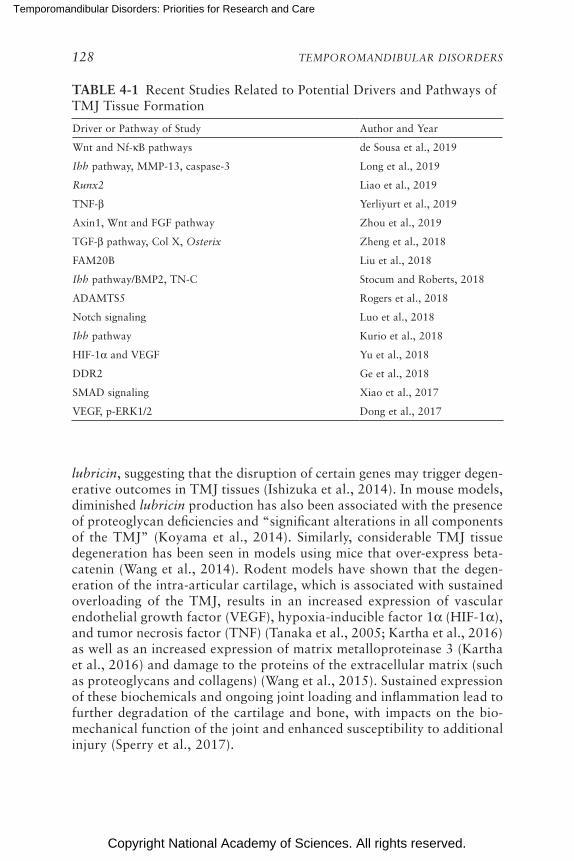
Temporomandibular Disorders: Priorities for Research and Care
Copyright National Academy of Sciences. All rights reserved.
128 TEMPOROMANDIBULAR DISORDERS
TABLE 4-1 Recent Studies Related to Potential Drivers and Pathways of TMJ Tissue Formation
Driver or Pathway of Study Author and Year
Wnt and Nf-κB pathways de Sousa et al., 2019
Ihh pathway, MMP-13, caspase-3 Long et al., 2019
Runx2 Liao et al., 2019
TNF-β Yerliyurt et al., 2019
Axin1, Wnt and FGF pathway Zhou et al., 2019
TGF-β pathway, Col X, Osterix Zheng et al., 2018
FAM20B Liu et al., 2018
Ihh pathway/BMP2, TN-C Stocum and Roberts, 2018
ADAMTS5 Rogers et al., 2018
Notch signaling Luo et al., 2018
Ihh pathway Kurio et al., 2018
HIF-1α and VEGF Yu et al., 2018
DDR2 Ge et al., 2018
SMAD signaling Xiao et al., 2017
VEGF, p-ERK1/2 Dong et al., 2017
lubricin, suggesting that the disruption of certain genes may trigger degen-erative outcomes in TMJ tissues (Ishizuka et al., 2014). In mouse models, diminished lubricin production has also been associated with the presence of proteoglycan deficiencies and “significant alterations in all components of the TMJ” (Koyama et al., 2014). Similarly, considerable TMJ tissue degeneration has been seen in models using mice that over-express beta-catenin (Wang et al., 2014). Rodent models have shown that the degen-eration of the intra-articular cartilage, which is associated with sustained overloading of the TMJ, results in an increased expression of vascular endothelial growth factor (VEGF), hypoxia-inducible factor 1α (HIF-1α), and tumor necrosis factor (TNF) (Tanaka et al., 2005; Kartha et al., 2016) as well as an increased expression of matrix metalloproteinase 3 (Kartha et al., 2016) and damage to the proteins of the extracellular matrix (such as proteoglycans and collagens) (Wang et al., 2015). Sustained expression of these biochemicals and ongoing joint loading and inflammation lead to further degradation of the cartilage and bone, with impacts on the bio-mechanical function of the joint and enhanced susceptibility to additional injury (Sperry et al., 2017).
Temporomandibular Disorders: Priorities for Research and Care
Copyright National Academy of Sciences. All rights reserved.
STATE OF THE SCIENCE ON TMDs 129
Sex Differences of TMJ Formation, Growth, and Function
Significant sex-specific differences exist in TMJ morphology (Iwasaki et al., 2017; Coogan et al., 2018; Coombs et al., 2019). Male condyles are on average larger than female condyles, with complex differences in the microstructural traits. Such sex differences in TMJ morphology, including in the condyle, disc, and fossa, have a direct impact on TMJ biomechanics and, by extension, on the health of these tissues. The available evidence suggests that the initiation of TMJ cartilage degeneration may be associ-ated with repeated mechanical overloading of the TMJ at loads higher than normal physiological conditions (Sperry et al., 2017; Nickel et al., 2018). Such overloading of the TMJ has been associated with oxidative stress and inflammation, which could in turn lead to damage to tissues. In a study by Iwasaki and colleagues (2017), mechanical energy densities imposed on the disc in healthy TMJs were significantly larger in women than in men, which could predispose women to a higher rate of mechanical fatigue of the TMJ disc. Early mechanical fatigue of the disc tissues has been associated with degenerative joint disease of the TMJ (Iwasaki et al., 2017). Similarly, a 2018 study found that, during asymmetrical jaw closing, energy densities were significantly larger in female participants than in male participants, and the authors suggested that this presents a higher risk of mechanical fatigue (Gallo et al., 2018). These findings have led to the implication of sex hormones in playing a role in TMJ remodeling and tissue degeneration; however, this work remains inconclusive and requires additional research. TMJ morphological and biomechanical differences between the sexes need to be identified and explored to develop an understanding of how they may contribute to a female predisposition to tissue dysfunction or chronic orofacial pain.
Biomechanical Function, Joint Tissue Degeneration, and Orofacial Pain Processing
The relationship between biomechanical function, joint tissue degenera-tion or injury, and pain processing is an expanding field of research that seeks to unravel the complex interactions between the tissues of synovial joints and the nervous system in order to understand their impact on joint function and on the initiation, maintenance, and suppression of pain. From another direction, increasing evidence suggests that regular physical activity and exercise can reduce pain, and treatments are often aimed at improving biomechanical function in this population. However, very few studies have explored the interface among pain, biomechanics, and TMJ function in humans, and little is known about whether the addition or removal of pain results in predictable or sustained changes in joint function or vice versa.
Temporomandibular Disorders: Priorities for Research and Care
Copyright National Academy of Sciences. All rights reserved.
130 TEMPOROMANDIBULAR DISORDERS
The loss of the integrity of the articulating surfaces of synovial joints is consistent with the classical model of mechanical fatigue (Vazquez et al., 2019). There are analogies that can be made between the mechanical fatigue model of materials and the biopsychosocial model of chronic pain. The fatigue of materials depends on the magnitude and frequency of the mechanical load, with the material properties known as “stress raisers” act-ing as covariates (Beatty et al., 2008). From a mechanical perspective, stress raisers are entities, such as physical defects, produced during the manufac-turing or development of the material or caused by traumatic loads that lead to an increased susceptibility of the material to damage and fatigue failure through normal function (Beatty et al., 2008). Similarly, the bio psycho social model of pain depends on the magnitude and frequency of the primary neuron afferent barrage to peripheral ganglia and dorsal horn/trigeminal secondary interneurons (Staud, 2011b). From a chronic pain perspective, stress raisers are entities that contribute to neuroinflammation, such as in-creased sympathetic nervous system input to glial cells (Russo et al., 2018). To date, few studies on the development of chronic pain have quantified the magnitude and frequency of primary afferent input while controlling for co-variable stress raisers. Little is known about whether the removal of the afferent barrage, while controlling co-variable stress raisers, results in predictable or sustained changes in chronic pain and tissue function (Russo et al., 2018). Thus, there is a need to move beyond the separate examina-tion of joint tissue failure and pain by using a biomechano- allostatic model, which combines the important variables in the development of degenerative joint disease with and without chronic pain.
The connection between biomechanical function of the TMJ and oro-facial pain has been established in certain animal models (Sperry et al., 2017). TMD models using repeated mouth opening to defined loads in rodents result in hyperalgesia and osteoarthritis-like degeneration of the joint. Rats that experienced higher magnitudes of TMJ loading experi-enced longer periods of sustained pain (14+ days versus 7 days) than rats with lower levels of loading (Kartha et al., 2016). However, what is less understood is whether the interface of biomechanics and neurobiology, at a mechanistic level, can be manipulated to reduce afferent input and neuro inflammation in ways that attenuate pain signaling and improve tissue integrity. Rodent models have demonstrated that abnormal loading of the TMJ results in inflammatory changes to the cartilage and the upregulation of certain drivers such as VEGF and HIF-1α as well as the upregulation of matrix metalloproteinases, which are associated with degradation found in osteoarthritis of the TMJ (Kartha et al., 2016) and other conditions (Shen et al., 2015; Bechtold et al., 2016). The upregulation of these biochemicals may also mediate the response of nociceptors located in the TMJ tissues (Sperry et al., 2017).
Temporomandibular Disorders: Priorities for Research and Care
Copyright National Academy of Sciences. All rights reserved.
STATE OF THE SCIENCE ON TMDs 131
The interface of biomechanical function and pain has been studied in other synovial joints (i.e., the knee and the facet joints of the spine), and it may be possible that the methods and tools used in these studies could be applied similarly to study the TMJ. In some cases, there is a correla-tion between the biomechanical and functional changes of the joint and pain, yet in many cases, particularly when pain becomes chronic, these relationships may not hold. It is also clear that there are multiple ways to interpret biomechanical function, including strength and endurance testing of muscle. More research is needed to understand how the biomechanical environment of the TMJ relates to joint function and its susceptibility to damage or injury and its relationship to pain signaling. Additionally, there is value in exploring individual-level differences in the biomechanical func-tion of the TMJ and how these differences may affect peripheral and central pain signal processing in light of individual differences in allostasis, the processes by which the body copes with stressors in its efforts to maintain homeostasis.
Animal Models for TMJ Tissue Engineering and Regenerative Medicine Research
Those in the TMJ biomechanics and tissue engineering fields have studied a variety of animal models, including mice, rabbits, dogs, goats, pigs, minipigs, and sheep. Important factors when assessing animal models include anatomy/physiology, mechanical properties, chewing patterns, dietary composition, cost, and ease of surgical access (Donahue et al., 2019). A recent systematic review that examined preclinical TMJ tissue engineering studies noted a lack of standardization regarding animal models (Helgeland et al., 2018), an issue that may be impeding the translation of TMJ bioengineering approaches.
Several studies have examined similarities and differences in temporo-mandibular and craniofacial anatomy across species including rabbits, pigs, cows, goats, dogs, minipigs, and humans (Herring et al., 2002; Kalpakci et al., 2011; Vapniarsky et al., 2017). The pig is considered to be the gold standard of a non-primate, large-animal TMJ model due to its similarities to human anatomy, function, and tissue material properties (Almarza et al., 2018). Pigs are omnivores, which is one reason their TMJ disc and condyle are similar in structure to those of humans (Bermejo et al., 1993). Recently, the TMJ anatomy, histology, and biomechanics of black Merino sheep have been shown to also be similar to humans, although their diets differ from humans (Angelo et al., 2016). Further comparative studies of TMJ structures in black Merino sheep and pigs will be useful and may help the field reach consensus on the best model system for preclinical studies of TMJ tissue engineering approaches.
Temporomandibular Disorders: Priorities for Research and Care
Copyright National Academy of Sciences. All rights reserved.
132 TEMPOROMANDIBULAR DISORDERS
A noted challenge in the use of animal models is the difficulty in repre-senting the pathology of the disease in the animal model. However, recent research suggests that some animals, such as dogs, cats, and horses, can develop certain TMJ pathologies such as osteoarthritis, ankylosis, luxation, fracture, and neoplasm (Almarza et al., 2018). Although the anatomic and physiological features of the TMJ may differ between humans and animals, these pathologies may have pathogeneses that are similar to those of TMDs in humans. Specifically, studying TMJ pathologies naturally occurring in animals may explain not only the pathogenesis of TMDs in humans but also the response to treatment.
Additionally, researchers have noted the challenges associated with the lack of established and relevant preclinical animal models for assessing novel approaches for TMJ tissue regeneration, which require larger animal models than those models employed for the study of joint disease (Almarza et al., 2018).
Overview of TMJ Tissue Engineering and Regenerative Medicine Research
In the future, the ability to engineer and regenerate temporomandibular tissues could provide an alternative treatment strategy to address certain TMDs and patients. Disc engineering and regeneration has been a major focus of recent TMJ tissue engineering research (Hunziker et al., 2015; Donahue et al., 2019; Melville et al., 2019). Despite promising results, the ability to develop these engineered constructs for clinical use has been limited by an incomplete understanding of the developmental biology of the TMJ tissues—a limitation that is further compounded by a lack of clinically relevant animal models and challenges with measuring function and forces within the joint space.
It is thought that disc dysfunction can be an early event that may lead to degenerative changes to the TMJ, implying that regeneration of disc tis-sues would be a valuable target for research. However, a major challenge with developing bioengineered TMJ discs is that they not only need to have similar morphological and histological characteristics to the native disc, but also must achieve the same mechanical function. Developing engineered TMJ disc constructs requires the selection of appropriate cell type or types, a biocompatible scaffold or scaffold-free support structure, and the proper biochemical or mechanical stimuli (Hunziker et al., 2015; Donahue et al., 2019; Melville et al., 2019). In addition, there remain concerns with the attachment of these engineered tissues and their capacity for tolerating early shear and torque that can occur during functional loading of the mandible (Vapniarsky et al., 2018; Almarza et al., 2019). More importantly, patients
Temporomandibular Disorders: Priorities for Research and Care
Copyright National Academy of Sciences. All rights reserved.
STATE OF THE SCIENCE ON TMDs 133
will need to be selected carefully, as comorbid conditions could play a sig-nificant role in the success of healing.
Selection of Cells
In TMJ bioengineering, the selection of cell source remains a critical challenge. Compared to native tissues, bioengineered or regenerated TMJ components not only should have similar morphological and histological characteristics, but must also achieve the same mechanical function. Several cell types have been tested, including native TMJ disc cells (Ronald and Mills, 2016), TMJ fibrocartilage stem cells (Embree et al., 2016), costal chondrocytes (Vapniarsky et al., 2018), co-cultured articular chondrocytes and knee meniscus cells (MacBarb et al., 2013), and mesenchymal stem cells (MSCs) from either autologous or allogeneic sources. Cells obtained from MSC-based approaches have demonstrated similarities to native tissues in terms of morphological and biochemical characteristics, but further re-search is needed to determine if these MSC-based constructs can withstand the early shear and torque experienced during mandibular functional load-ing. Further work on progenitor cell populations and their differentiation into TMJ-like cells will be important to move this area of research forward.
Scaffolds and Scaffold-Free Approaches
Scaffolds serve as a surface on which cells can form new tissues and which can carry bioactive molecules that help to influence cell behavior (e.g., migration, stem cell differentiation). Additional research is needed to further refine scaffold fabrication methods such as nanoassembly, a process that builds scaffolds layer by layer, and three-dimensional (3D) printing (Donahue et al., 2019). Scaffold-free approaches are also being developed as a way to reduce complications with scaffold degradation and fabrication byproducts (Donahue et al., 2019).
Biochemical and Mechanical Stimuli
Stimulating bioengineered TMJ discs with growth factors, such as insulin-like growth factor I and TGF-β, has been shown to result in greater levels of collagen synthesis and improved mechanical properties (Detamore and Athanasiou, 2005). In addition to such biochemical stimuli, mechanical stimuli (e.g., hydrostatic pressure) have also been applied, typically to help recapitulate native tissue structure–function relationships in bioengineered TMJ discs (Gunja et al., 2009). Further exploratory research aimed at determining the appropriate combination of biochemical and mechanical stimuli is needed.
Temporomandibular Disorders: Priorities for Research and Care
Copyright National Academy of Sciences. All rights reserved.
134 TEMPOROMANDIBULAR DISORDERS
Other Tissues: Mandibular Condyle and Condylar Cartilage
Research on regenerative approaches for TMJ tissues beyond the disc is limited. Two areas of focus that have received less attention than disc regeneration include tissue engineering for the mandibular condyle and condylar cartilage. As with TMJ disc engineering, selecting the appropri-ate cell source has been challenging (Willard et al., 2011). In the case of condylar cartilage, a recent study showed tissue growth and regeneration in goat condylar cartilage using synthetic and natural scaffolds (Chin et al., 2018). Early studies such as these are promising, but further attention to these areas is needed to move the field forward.
Moving Toward Translation
Past and future research breakthroughs in TMJ bioengineering must be examined using orthotropic animal models, and once safety and effec-tiveness has been established, these approaches can be tested in humans. The location of the TMJ and its proximity to the brain may require more stringent safety requirements for bioengineered products (Donahue et al., 2019). Careful patient selection will be necessary in clinical studies, as it is unknown whether the presence of commonly comorbid conditions such as fibromyalgia could play a significant role in the successful regeneration of damaged tissues. Furthermore, metaplasia (the conversion of one cell type to another type), ossification (bone remodeling), and angiogenesis (development of new blood vessels) may be concerns for specific patients (Detamore et al., 2007).
Regenerative Capacity of Endogenous Cells
Recent work showed that endogenous fibrocartilage stem cells may be useful for regenerating and repairing cartilage in the TMJ condyle (Embree et al., 2016). Terminal differentiation of chondrocytes is thought to drive aspects of osteoarthritis in the TMJ, and another recent study demonstrated that inhibiting Ihh signaling protected chondrocytes (Yang et al., 2019). Additional research is needed on how endogenous cells and the native TMJ microenvironment can be therapeutically exploited to repair or regenerate damaged tissue.
Looking Forward: Research Priorities for Biomechanics and Regenerative Medicine Research on TMDs
More research is needed to better define the unique mechanics of joint and tissue interfaces within the TMJ and the function of the joint in health
Temporomandibular Disorders: Priorities for Research and Care
Copyright National Academy of Sciences. All rights reserved.
STATE OF THE SCIENCE ON TMDs 135
and in disease. Too little is known about the function of the genes involved in the development of the TMJ and its tissues, specifically about how varia-tions in TMJ development may predispose the joint to future dysfunction. Such research could lead to improved regeneration methods for damaged tissues. Furthermore, only limited research has been conducted to under-stand neurological control and pain sensitization in the orofacial region. Of critical importance is the exploration of the relationship between pain and function, specifically an understanding of innervation and its interac-tions with the many tissues and vascular structure of the TMJ, which can inform methods for joint repair and regeneration. While tissue turnover in and the repair of bone, cartilage, and ligaments of the TMJ have been studied to some extent, a better understanding of these repair processes is needed. Specifically, a better understanding is required of the integration of mechanical cues and pathological joint loading on tissue-remodeling processes. Additionally, there has not been enough research performed on mechanotransduction to allow an understanding of the relationship between joint motion, forces, and the physical properties of tissues and the relation of these to cellular mechanotransduction.
The development of new therapies to repair and prevent joint dys-function and to regenerate new and functional tissues safely will require a strong foundation of basic and clinical research on the biology of the TMJ in health and in disease. Addressing existing research gaps will demand new approaches and instrumentations for in vivo measurement of joint health, disease, and function. It is clear that there will be value in going beyond static imaging of the joint to examine the dynamic joint function, in using 3D imaging of joint structure and joint quality, and in leveraging imaging technologies and datasets from other disciplines (i.e., cohorts with imag-ing of brains with neurological disease). Moreover, due to the limitation of direct in vivo measurement, the development of in silico models and the use of virtual human trials would provide significant opportunities to understand TMD risk factors and the etiology of TMD development and progression. Such models might also help explain the increased prevalence of TMDs among women and other known and unknown disparities and musculoskeletal and psychosocial factors. Additionally, researchers could use novel biomedical techniques for modeling the TMJ, such as biocomput-ing, and molecular imaging for tissues and structures. See Box 4-1 for a list of research priorities.
NEUROBIOLOGY OF OROFACIAL PAIN AND TMDs
The larger field of pain research has worked to define the mechanisms of neuropathic and inflammatory pain, and, as with many complex disor-ders featuring acute and chronic pain, understanding the pathophysiology
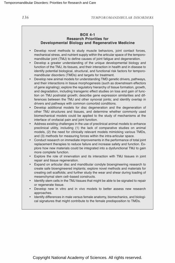
Temporomandibular Disorders: Priorities for Research and Care
Copyright National Academy of Sciences. All rights reserved.
136 TEMPOROMANDIBULAR DISORDERS
BOX 4-1 Research Priorities for
Developmental Biology and Regenerative Medicine
• Develop novel methods to study muscle behaviors, joint contact forces, mechanical stress, and nutrient supply within the articular space of the temporo-mandibular joint (TMJ) to define causes of joint fatigue and degeneration.
• Develop a greater understanding of the unique developmental biology and function of the TMJ, its tissues, and their interaction in health and in disease to identify potential biological, structural, and functional risk factors for temporo-mandibular disorders (TMDs) and targets for treatment.
• Develop new animal models for understanding TMD genetic drivers, pathways, and their interactions in tissue morphogenesis (such as downstream effectors of gene signaling); explore the regulatory hierarchy of tissue formation, growth, and degradation, including transgenic effect studies on loss and gain of func-tion on TMJ postnatal growth; describe gene expression similarities and dif-ferences between the TMJ and other synovial joints; and identify overlap in drivers and pathways with common comorbid conditions.
• Develop additional models for disc degeneration and the degeneration of other TMJ structures and tissues, and determine whether commonly used bio mechanical models could be applied to the study of mechanisms at the interface of orofacial pain and joint function.
• Address existing challenges in the use of preclinical animal models to enhance preclinical utility, including (1) the lack of comparative studies on animal models, (2) the need for clinically relevant models mimicking various TMDs, and (3) methods for measuring forces within the intra-articular space.
• Conduct research on immediate improvements in the performance of total joint replacement therapies to reduce failure and increase safety and function. Ex-plore how new materials could be integrated into a dysfunctional TMJ to gain more complete function.
• Explore the role of innervation and its interaction with TMJ tissues in joint repair and tissue regeneration.
• Expand on articular disc and mandibular condyle bioengineering research to create safe bioengineered implants; explore novel methods and materials for creating cell scaffolds; and further study the wear and shear during loading of mesenchymal stem cell–based constructs.
• Identify stem cells in the TMJ tissues that might be able to be signaled to repair or regenerate tissue.
• Develop new in vitro and in vivo models to better assess new research approaches.
• Identify differences in male versus female anatomy, biomechanics, and biologi-cal signatures that might contribute to the female predisposition to TMDs.
Temporomandibular Disorders: Priorities for Research and Care
Copyright National Academy of Sciences. All rights reserved.
STATE OF THE SCIENCE ON TMDs 137
of TMDs requires an in-depth understanding of the mechanisms of pain and their role at different levels, from the cellular to an inter-system level. Despite the significant burden and often life-changing impact of orofacial pain, relatively little is known about the neurobiology of TMJ tissues in health and in disease. This is, in part, due to the highly complex, multi-dimensional nature of acute and chronic pain and the numerous bio psycho-social underpinnings that mediate the experience and perception of pain. Also, little is known regarding biomechanical dysfunction (see above sec-tion on tissue engineering) and how this relates to or contributes directly to orofacial pain. This dearth in knowledge is in part due to the significant siloing of fields of research, with researchers working on tissue bioengineer-ing and those working on pain biology needing to find opportunities to col-laborate on TMD research. The following sections will explore the findings of recent research on TMDs and orofacial pain, including:
• Animal models for orofacial pain, • Overviews of peripheral and central mechanisms of acute and
chronic orofacial pain,• Pain signaling and biomechanics of the TMJ,• Trigeminal versus extra-cranial pain signaling systems, and• Commonalities in neuronal pathways and central sensitization
within TMDs and other chronic pain conditions.
Current Animal Models of Orofacial Pain
Animal models provide critical opportunities for studying the biologi-cal and cellular mechanisms of pain and help advance the development of pharma cological treatments. However, the barriers to translation from studies in experimental animals to clinical drug development are numer-ous and are a result both of the challenges of using animal models in general and of more systemic issues related to shortcomings in the research ecosystem. Compared to pain experienced in other areas of the body, specific animal models for orofacial pain have been less extensively studied ( Krzyzanowska and Avendaño, 2012). While broader pain research find-ings and methodologies can be applied to the study of orofacial pain, it is essential that mechanisms unique to the orofacial area be explored using anatomically relevant animal models to understand the pathogenesis of inflammatory and neuropathic orofacial pain common to TMDs and to identify effective existing or novel treatment modalities.
In order to study the mechanisms of pain associated with TMDs, commonly used mouse and rat models attempt to mimic inflammatory orofacial pain through the injection of various chemicals or irritants into the masseter muscle or the TMJ (Martínez-García et al., 2019). Models
Temporomandibular Disorders: Priorities for Research and Care
Copyright National Academy of Sciences. All rights reserved.
138 TEMPOROMANDIBULAR DISORDERS
for inflammatory pain require an initial tissue injury or irritant injected directly into the muscle or joint to trigger a selected area or cell type into activating primary sensory neurons and attract immune response cells that release substances like cytokines, which can then be studied. The methods for triggering an inflammatory pain response range from general chemical irritants (carrageenan and formalin) to agents that directly target specific sensory neurons (capsaicin and mustard oil) (Krzyzanowska and Avendaño, 2012). Alternatively, mechanical trauma such as repeated daily mouth opening, intraoral appliance placement, disc displacement, or ligation of the tendon of the masseter muscle have been used to mimic and study TMJ dysfunction (Guo et al., 2010; Araújo-Filho et al., 2018).
Other animal models designed to explore the pathophysiology of neuro pathic orofacial pain involve the constriction, transection, or com-pression of the distal branches of the trigeminal nerve, such as the infra-orbital, inferior alveolar, mandibular, or mental nerves. Of these animal models, the chronic constriction injury (CCI) of the infra-orbital nerve is one of the most commonly used orofacial pain models (Krzyzanowska and Avendaño, 2012; Araújo-Filho et al., 2018; Martínez-García et al., 2019). These models of partial nerve injury attempt to simulate the neuropathic symptoms reported by some patients with a TMD.
Depending on the study, a variety of outcome measures have been used to assess nociceptive responses in awake behaving non-human animals, including behavioral outcome models. Common behavioral outcome mea-sures include evoked withdrawal responses to mechanical, cold, or heat stimuli; non-evoked spontaneous behavior such as face grooming; and other types of operant behavioral assessments (Neubert et al., 2005; Langford et al., 2010; Romero-Reyes et al., 2013). In the TMJ arthritis model, for example, meal pattern analysis (i.e., meal duration, total number of meals, and total time spent eating) is monitored to assess feeding behavior. Ob-servations of feeding behaviors do appear to have value as a behavioral outcome for TMDs. While numerous behavioral outcomes can be used to assess nociceptive responses, these outcomes are difficult to quantify and may lack sensitivity (Krzyzanowska and Avendaño, 2012). These behav-ioral response studies are in contrast to the study of reflex responses to external stimuli, which do not require the engagement of cerebral processes. It has been argued that spontaneous pain behaviors and complex operant behaviors that involve cortical processing and decision making may be more relevant in providing insights into human orofacial pain conditions.
The pain community has made significant advances in the understand-ing of pain through the development of diverse animal models used to examine the environmental and biological processes underlying acute and chronic pain. The use of inflammatory and neuropathic pain models has re-vealed a number of potential mechanisms and pathways involved in painful
Temporomandibular Disorders: Priorities for Research and Care
Copyright National Academy of Sciences. All rights reserved.
STATE OF THE SCIENCE ON TMDs 139
TMDs, and it provides useful information for those conditions associated with inflammation and neuropathic pain. However, TMDs are heteroge-neous and involve biomechanical and functional alterations, psychosocial factors, and environmental factors and stressors—some factors of which may be difficult to capture and measure in preclinical models. Historically, the field of pain research has focused on factors involved in the transduction of nociceptive stimuli and the induction of acute pain, but more emphasis is needed to understand factors that both promote and prevent the transition from acute to chronic pain and that promote recovery from chronic pain. Recent animal models of musculoskeletal pain have begun to combine nega-tive stress, such as sound or fatigue (Chen et al., 2011; Alvarez et al., 2013; Sluka and Clauw, 2016), with tissue insults, and they show enhanced and more widespread hyperalgesia; the use of these models in TMD research is just emerging (Traub et al., 2014) and requires additional focuses. Further-more, recent studies in animal models of pain unrelated to TMDs have identified lifestyle factors such as physical activity and biological factors such as anti-inflammatory mediators (e.g., interleukin-10) that can protect both animals and humans from the development of chronic pain (Kavelaars et al., 2011; Leung et al., 2016; Sluka et al., 2018). Unique animal models need to be developed that take into account the complex nature of TMDs, the multiple stressors that can initiate the development and persistence of pain, and the biomechanical and structural findings observed in individuals with TMDs. Collaborations with researchers using other pain models—like post-surgical tissue trauma and burns—and those studying TMJ bio engineering and tissue regeneration could provide potentially valuable new models. Such new models would benefit from the development and use of standard outcome measures for the assessment of orofacial pain and TMJ function.
Overview of Peripheral Mechanisms of Orofacial Pain
As described earlier in the chapter, the orofacial tissues are innervated by the trigeminal nerve and its nociceptive endings, which terminate in the orofacial tissues and can be activated by mechanical stimuli or injury, inflammatory processes, or exposure to an irritant or inhospitable environ-ment. This system of mechanisms is inherently vulnerable to modulation because each component of the system interacts within and is influenced by the complex biochemical environment—endocrine, immune, and other systems—of the human body (Sessle, 2011).
Over the past several decades, research into the central and peripheral mechanisms of acute and chronic orofacial pain has identified a network of chemical mediators, receptors, channels, and interactions that influence the activation and sensitization of nociceptive pathways (Sessle, 2011). The involvement of certain inflammatory mediators—such as prostaglandins
Temporomandibular Disorders: Priorities for Research and Care
Copyright National Academy of Sciences. All rights reserved.
140 TEMPOROMANDIBULAR DISORDERS
and bradykinins—in activating nociceptive nerve endings in orofacial tis-sues has been well established (Sessle, 2011). In addition, other peripheral mediators released from mast and immune cells and macrophages—such as 5- hydroxytryptamine (5-HT), histamine, TNF-α, and interleukins (ILs)—act by increasing the excitability of the nociceptive nerve endings at the injury location. This multi-variable mechanistic process of peripheral sen-sitization is mediated by interactions with the immune, cardiovascular, and endocrine systems (Sessle, 2011). The processes by which these numerous mediators act involve a multitude of receptors and ion channels on the nociceptive endings; they also act via signaling and second messenger sys-tems and through the associated intracellular matrix (Sessle, 2011). During the process of peripheral sensitization, these inflammatory mediators can increase the excitability of stimulated nociceptive endings in the initial site and those adjacent through interactions with certain ion channels and sig-naling systems (Sessle, 2011). Additionally, exposure to certain inflamma-tory mediators can result in phenotypic changes within nociceptors, which can fundamentally modify ion channels (e.g., sodium channels) and the ex-pression of certain receptors. Therefore, there is significant value in under-standing the role of these mediators and how they act upon the nociceptive pathway to modulate pain response and induce lasting hypersensitivity, because many TMDs feature both acute and chronic inflammatory pain as a primary feature (see Chapter 2).
Overview of Inflammatory Mediators
Certain chemical mediators play a significant role in the activation or sensitization of nociceptors and, by extension, the inflammatory pain characteristic of many TMDs. As such, recent research has focused in-creasingly on understanding the specific role of these individual mediators and receptor expression on the pathogenesis of orofacial pain. Multiple chemical mediators have been the focus of investigation. Among those, gamma amino butyric acid (GABA) receptor subunit alpha (Gabrα6) has been shown to be expressed by the trigeminal neurons, although the role of this expression on orofacial pain remains unclear. A study in rats by Puri and colleagues (2012) showed that Gabrα6 expression may play a role in hypersensitivity of the TMJ by inhibiting afferents in the trigeminal pathway and reducing inflammatory orofacial nociception. Additionally, a 2019 study suggests that the activation of inflammasomes—via the secre-tion of IL-1β and IL-18—could play a significant role in the elicitation of an inflammatory response across a range of metabolic, cardiovascular, and neurodegenerative disorders as well as TMDs (Ibi, 2019).
Beyond their role in triggering and maintaining inflammation, the pres-ence of these mediators in orofacial tissues could be an indication of the
Temporomandibular Disorders: Priorities for Research and Care
Copyright National Academy of Sciences. All rights reserved.
STATE OF THE SCIENCE ON TMDs 141
health of the tissue and act as diagnostic markers or therapeutic targets. In his 2011 review of central and peripheral mechanisms of orofacial pain, Sessle noted that mediators such as 5-HT, IL-1β, and prostaglandins are not present in the TMJ synovial fluid of healthy subjects, but they are detectable in inflamed TMJ synovial fluid (Sessle, 2011). Sessle also suggested that gluta-mate levels in the jaw muscles of patients with certain TMDs may indicate the presence of an inflammatory response (Castrillon et al., 2010; Sessle, 2011).
Receptors and Ion Channels
The role of receptors and ion channels, particularly the function of sodium ion channels like Nav1.7, has been and remains a significant focus of orofacial pain research. Recent studies have demonstrated that Nav1.7 signaling plays a significant role in pain processing, with point mutations resulting in either intractable pain or a complete absence of pain through the amplification of otherwise weak stimuli (Cummins et al., 2007). That ion channel may also play a role in visceral pain processing (Hockley et al., 2017) associated with certain chronic overlapping pain conditions, such as irritable bowel syndrome (IBS) (Campaniello et al., 2016). As shown in knockdown and knockout studies in mice, Nav1.7 interferes with mechanical and thermal inflammatory pain responses (Nassar et al., 2004). In the trigeminal nerve, chronic inflammation induced by Freund’s adjuvant injection into the TMJ upregulated Nav1.7 mRNA and protein in neurons innervating the TMJ while blocking the Nav1.7 sodium channel in the trigeminal ganglion, which significantly reduced pain response in the joint (Bi et al., 2013). These findings suggest that Na channel subtypes play a role in the pain response in TMD. Research has also considered the role of calcium-permeable ion channels expressed by trigeminal ganglia sensory neurons, such as TRPV4, in pain behavior. A 2013 study in mice by Chen and colleagues (2013) demonstrated that the expression of TRPV4 is asso-ciated with nociceptive response after TMJ inflammation.
In addition to examining the roles that Nav1.7 and TRPV4 play in pain perception within the TMJ, additional research has sought to describe how these channels are regulated and the mechanisms underlying this regula-tion. As described above, multiple studies have pointed to the regulation of Nav1.7 by pro-inflammatory mediators such as nerve growth factor (NGF), TNF-α, 5-HT, prostaglandins, and cytokines (IL-1β) (Tamura et al., 2014; Isensee et al., 2017) (see section above).
While the etiologies of TMDs remain elusive, the recent explorations of these mechanisms, receptors, and ion channels indicate potential path-ways through which chronic inflammation may amplify pain responses. To illustrate such a pathway, a 2017 study by Kobayashi and colleagues dem-onstrated that synovial cells in samples of human TMJ tissues can release
Temporomandibular Disorders: Priorities for Research and Care
Copyright National Academy of Sciences. All rights reserved.
142 TEMPOROMANDIBULAR DISORDERS
inflammatory mediators—bioactive peptides called elastin-derived peptides (EDPs)—during the degradation of the extracellular matrix. EDPs promote the upregulation of IL-6 and elastin-degrading matrix metalloproteinase-12 (MMP-12) (Kobayashi et al., 2017). The presence of EDPs was found to be correlated with both the duration of jaw locking and IL-6 expression. These findings suggest an environment within TMJ where the activation of an elastin-binding protein signaling cascade, as a result of harmful mechani-cal stimuli, triggers a pro-inflammatory cascade and MMP-12 expression, which may create a positive feedback loop of chronic inflammation within the joint (Kobayashi et al., 2017).
Overview of Mechanisms of Neuropathic Pain and TMDs
As described earlier, several models of partial injury to branches of the trigeminal nerve have been used in rodents to study the disabling neuro-pathic pain observed in a subset of patients with TMDs. The behavioral re-sponses observed in these models include mechanical hyperalgesia, air puff allodynia, and paraesthesias/dysaesthesias. Studies suggest that microglial activation plays an important role in the development and maintenance of central sensitization, as evidenced by the reversal of mechanical allodynia through the suppression of microglial activation (Ma et al., 2012). Expres-sion of phosphorylated extracellular signal-regulated kinase (pERK) in the dorsal horn and its phosphorylation is implicated in pain since pharmaco-logical blockade reduces pain behavior in the infraorbital chronic construc-tion injury (CCI) model. Ito and colleagues (2013) observed an upregulation of P2X(7)R, membrane-bound TNF-α, and soluble TNF-α in the trigeminal sensory nuclear complex after a CCI of the infraorbital nerve. Antago-nists of the P2X(7) receptor and inhibitors of the p hosphorylated (p)-p38 mitogen-activated protein kinase (MAPK) inhibited the tactile allodynia in this model, suggesting that phosphorylation of p38 MAPK via P2X(7)R may play a critical role in the mechanisms of hypersensitivity. Shibuta and colleagues (2012) reported the presence of hyperactive microglial cells and a large number of pERK-immunoreactive (IR) cells in trigeminal spinal sub-nucleus caudalis and the upper cervical spinal cord (C1) after infra-orbital CCI. Minocycline significantly reduced the activation of microglial cells and the number of pERK-IR cells at these sites, suggesting that the activation of microglial cells in the trigeminal and upper cervical regions is involved in the increased neuronal excitability associated with the neuropathic pain.
Central Mechanisms of Pain
Beyond the induction of pain in the periphery, peripheral nociceptive stimuli can also affect CNS neurons (Cairns, 2010) via the trigeminal
Temporomandibular Disorders: Priorities for Research and Care
Copyright National Academy of Sciences. All rights reserved.
STATE OF THE SCIENCE ON TMDs 143
ganglion to the subnucleus caudalis—an important site for the transmis-sion of information from the periphery to the brain—which contains many CNS pain receptors. Receptors and ligands that may be involved include N-methyl-D-aspartate (NMDA), GABA, 5-HT, glutamate, and certain neuropeptides (Cairns, 2010; Sessle, 2011). Many of the neurons of the C1 and C2 dorsal horns and the subnucleus caudalis receive direct inputs from the peripheral afferents in the surface tissues surrounding the TMJ and play a critical role in the CNS processing of pain in these orofacial tis-sues ( Chichorro et al., 2017). Using injection of glutamate into the TMJ in animal models demonstrates an expansion of the neuronal receptive field and sensitivity in regional tissues via the sensitization of subnucleus caudalis neurons, in addition to the activation of TMJ nociceptors. This suggests the presence of central sensitization (Lam et al., 2009). Neurons of the subnucleus caudalis and the C1–C2 dorsal horns and caudalis/interpolaris transition zone also process nociceptive information from deep tissues—such as facial muscles and the TMJ—which results in complex patterns of convergence in the processing of superficial and deep tissue afferent inputs. These patterns require further study, as these may offer further understand-ing concerning the localization and expansion of pain in various orofacial pain states (Chichorro et al., 2017).
Evidence of CNS pain processing has also been noted in studies of pain thresholds and electrical stimulation of the TMJ, which found indications of lower pain thresholds among individuals with arthralgia of the TMJ compared with healthy controls (Cairns, 2010). Such findings are highly relevant to understanding the experience of orofacial pain at a mechanistic level, as many individuals with orofacial pain experience increased regional sensitivity in the skin and muscles around the TMJ.
Of interest to central sensitization is the process of reversible and non-reversible neuroplastic changes in nociceptive afferents, which has been shown to be initiated by certain neurochemicals of the nociceptive process. For example, glutamate activates NMDA receptors and can result in neuro-plastic changes indicative of central sensitization (Chichorro et al., 2017). Chichorro and colleagues (2017) write that this process “underscores the point that the nociceptive pathways and processes in the CNS are not ‘hard-wired’ but rather are ‘plastic’ and modifiable by events associated with injury or inflammation in peripheral tissues” (p. 617). Central sensitization involves the prolonged and increased excitability of neurons and increased synapse function in central nociceptive pathways (Woolf, 2011), which can result in pain experienced that may not match or require a noxious stimulus. This process is known to result in pain hypersensitivity and pres-sure hyperalgesia, and it enhances temporal summation as well as causing secondary changes in brain activity (see section on neuroimaging for brain-based biomarkers of pain) and contributing to diverse pain conditions, such
Temporomandibular Disorders: Priorities for Research and Care
Copyright National Academy of Sciences. All rights reserved.
144 TEMPOROMANDIBULAR DISORDERS
as fibromyalgia, osteoarthritis, and TMDs, among others (Woolf, 2011). Woolf (2011, p. 4) writes,
Central sensitization introduces another dimension, one where the CNS can change, distort, or amplify pain, increasing its degree, duration, and spatial extent in a manner that no longer directly reflects the specific qualities of peripheral noxious stimuli, but rather the particular functional states of circuits in the CNS…. This does not mean that the pain is not real, just that it is not activated by noxious stimuli.
An understanding of the mechanistic differences and convergence across the many peripheral and central mechanisms underlying acute and chronic pain is critical to the identification of clinically meaningful pain phenotypes within conditions to guide further research and clinical care.
Orofacial Pain Modulation
The modification of pain on an individual level is a product of the convergence of modifiable and non-modifiable biopsychosocial factors. Current research suggests that various interacting mechanisms play a role in the generation, maintenance, and suppression of pain by the CNS, which can, as described above, signal a mismatch between the peripheral nociceptive inputs and the perception of pain by an individual. Hence, the pain experience is highly heterogeneous across individuals and difficult to measure objectively (Harper et al., 2016a). Advances in the understanding of pain signaling mechanisms in both healthy and disease states has led to the concept of pain modulation, where CNS mechanisms can generate and maintain pain. This means that the presence of an injury or inflammation within the peripheral system may not necessarily translate into pain, just as an individual could experience intense pain in the absence of injury and inflammation.
Past studies in humans have compared endogenous pain modulation (pain facilitation or inhibition) in individuals with TMDs versus in indi-viduals without TMDs. Conditioned pain modulation (CPM), an index of pain inhibition, is studied by examining the reduction in pain to a noxious stimulus by a concurrent or prior noxious stimulus at a distant site. Pain facilitation is examined using a temporal stimulation paradigm, where a fixed-intensity noxious stimulus is repeated at frequencies that result in increased pain. Some studies have reported TMD patients feeling greater pain than controls in response to such stimuli. However, these observations have not been consistent across all studies (Greenspan et al., 2011; Moana-Filho et al., 2018), with some findings suggesting impaired CPM effects and others reporting similar CPM in both people with TMDs and healthy
Temporomandibular Disorders: Priorities for Research and Care
Copyright National Academy of Sciences. All rights reserved.
STATE OF THE SCIENCE ON TMDs 145
controls (Kothari et al., 2015; Moana-Filho and Herrero Babiloni, 2019). This lack of a significant difference suggests that not all painful TMDs are associated with compromised endogenous pain inhibitory systems and, by extension, that there are likely to be subgroups of individuals with TMDs who feature unique combinations of biopsychosocial mechanisms that con-tribute to the generation and maintenance of pain (Harper et al., 2016a; Moana-Filho et al., 2018). These findings support the suggestion that pain related to TMDs exists on a spectrum from cases where pain is generated in the periphery through to cases where pain is the result of an exaggerated response in the CNS, with most cases having a combination of peripheral and central changes (Harper et al., 2016a) (see Chapter 2 for the discussion of pain as a chronic disease).
Over the past decade, TMD and chronic pain researchers have become increasingly interested in developing a more complete understanding of the different biopsychosocial mechanisms that are responsible for the genera-tion and maintenance of pain, rather than continuing to rely on exclusively anatomical classifications of pain (i.e., low back pain, jaw pain) (Bair et al., 2016). This approach also allows for further exploration of both the unique and shared mechanisms between TMDs and other commonly co-morbid pain conditions, such as IBS, fibromyalgia, and chronic pelvic pain. Clauw writes,
A critical construct is that, within any specific diagnostic category … indi-vidual patients may have markedly different peripheral/nociceptive and neural contributions to their pain. Thus, just as low back pain has long been acknowledged to have multiple potential mechanisms, so also is this true of all chronic pain states. (Clauw, 2015, p. 6) (see Table 4-2)
Most notably, as part of the Orofacial Pain Prospective Evaluation and Risk Assessment (OPPERA) study, cluster analysis performed on data from a case–control study (1,031 chronic TMD cases and 3,247 controls) identi-fied three distinct patient subgroups across an array of biopsychosocial fac-tors. Their findings suggest that classification of individuals into clinically relevant and mechanistically based subgroups using biopsychosocial risk factors could provide a better and more personalized approach for under-standing orofacial pain etiology and, in the future, for the development and application of more targeted treatments (Bair et al., 2016). The authors suggest that such a method prioritizes mechanistic and etiological distinc-tions for pain more effectively than grouping by a specific TMD diagnosis as mechanisms within a specific pain diagnosis can vary by individual.
The centralized, multi-focal pain that is characteristic of many patients with TMDs—in addition to many other symptoms, traits, and factors—is a feature of many other overlapping chronic pain conditions (Williams
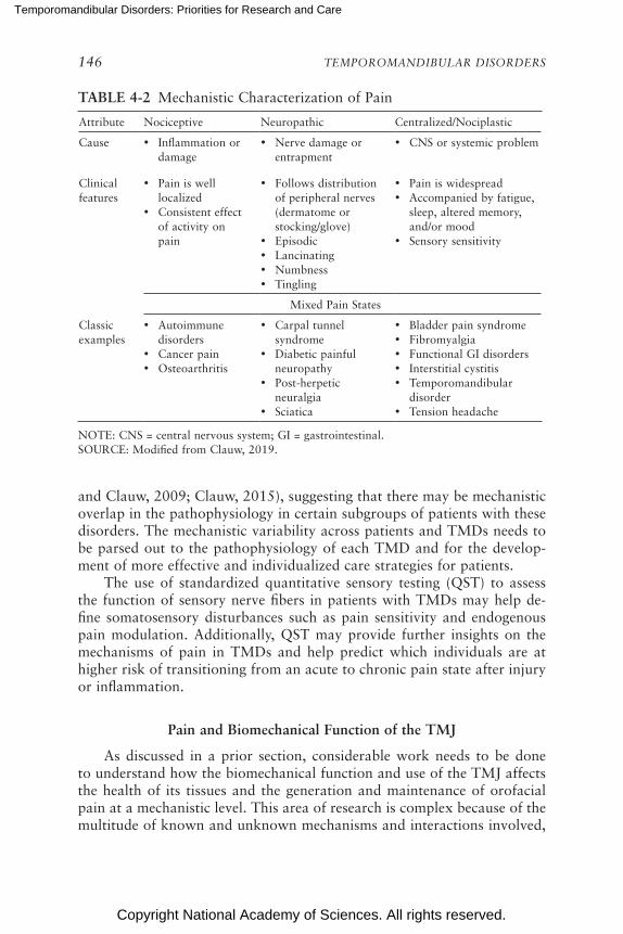
Temporomandibular Disorders: Priorities for Research and Care
Copyright National Academy of Sciences. All rights reserved.
146 TEMPOROMANDIBULAR DISORDERS
TABLE 4-2 Mechanistic Characterization of Pain
Attribute Nociceptive Neuropathic Centralized/Nociplastic
Cause • Inflammation or damage
• Nerve damage or entrapment
• CNS or systemic problem
Clinical features
• Pain is well localized
• Consistent effect of activity on pain
• Follows distribution of peripheral nerves (dermatome or stocking/glove)
• Episodic• Lancinating• Numbness• Tingling
• Pain is widespread• Accompanied by fatigue,
sleep, altered memory, and/or mood
• Sensory sensitivity
Mixed Pain States
Classic examples
• Autoimmune disorders
• Cancer pain• Osteoarthritis
• Carpal tunnel syndrome
• Diabetic painful neuropathy
• Post-herpetic neuralgia
• Sciatica
• Bladder pain syndrome• Fibromyalgia• Functional GI disorders• Interstitial cystitis• Temporomandibular
disorder• Tension headache
NOTE: CNS = central nervous system; GI = gastrointestinal.SOURCE: Modified from Clauw, 2019.
and Clauw, 2009; Clauw, 2015), suggesting that there may be mechanistic overlap in the pathophysiology in certain subgroups of patients with these disorders. The mechanistic variability across patients and TMDs needs to be parsed out to the pathophysiology of each TMD and for the develop-ment of more effective and individualized care strategies for patients.
The use of standardized quantitative sensory testing (QST) to assess the function of sensory nerve fibers in patients with TMDs may help de-fine somatosensory disturbances such as pain sensitivity and endogenous pain modulation. Additionally, QST may provide further insights on the mechanisms of pain in TMDs and help predict which individuals are at higher risk of transitioning from an acute to chronic pain state after injury or inflammation.
Pain and Biomechanical Function of the TMJ
As discussed in a prior section, considerable work needs to be done to understand how the biomechanical function and use of the TMJ affects the health of its tissues and the generation and maintenance of orofacial pain at a mechanistic level. This area of research is complex because of the multitude of known and unknown mechanisms and interactions involved,
Temporomandibular Disorders: Priorities for Research and Care
Copyright National Academy of Sciences. All rights reserved.
STATE OF THE SCIENCE ON TMDs 147
including mechanical forces, biochemical mediators, peripheral and CNS interactions, and psychosocial factors affecting the use of the joint and the experience of pain. The relationship between biomechanical control or function and pain has been of interest to researchers in other areas of study, such as low back pain (Hodges and Moseley, 2003) and pain in the hip and knee (Powers, 2010). However, relatively little is known about the mechanisms underlying these relationships and how these relate to the etiologies of TMDs, and there is a lack of consensus in the research regard-ing a correlation between biomechanical function and pain.
In the case of the TMJ’s biomechanical function and orofacial pain, research has shown that pathological changes to joint tissues, such as the degeneration of tissues resulting from the overloading of the TMJ, alters the biochemical environment of the joint and has some degree of impact on the mediation of peripheral and central signaling processes that initiate and maintain pain (Sperry et al., 2017). However, the presence of tissue degeneration does not equate to a predictable level or the presence of pain, which supports the concept that other biochemical pathways could also be affected by biochemical degenerative changes and play a contributing role (i.e., inflammatory cytokines) in the generation and maintenance of pain (Sperry et al., 2017). Other mediators associated with degeneration (collagenase and the increased expression of pERK) may also play a role in nociception (Gao and Ji, 2009; Adães et al., 2014), although more research is required to understand their role in the sensitization of neurons (Sperry et al., 2017). Furthermore, relatively little is known about the relationship between pain and parafunction of the masticatory muscles. There is some evidence suggesting that TMD pain may be associated with increased pro-tective muscle reflexes, but this requires further study (Cairns, 2010).
This type of research represents an area of significant value for under-standing the complexities of TMDs from a biopsychosocial perspective, particularly as researchers continue to elucidate the role of psychosocial factors in relation to the experience of pain and explore pain’s underlying biological mechanisms.
Trigeminal Versus Extra-Cranial Pain Signaling Systems
Pain in the orofacial region is signaled via the trigeminal ganglia (TG), in contrast to pain signaling from the rest of the body, which occurs via the sensory dorsal root ganglia (DRG). Studies in pain-free human subjects in-dicate regional differences in endogenous pain inhibition, with significantly weaker inhibitory pain modulation in areas innervated by the trigeminal nerve than in other body regions (Levy et al., 2018). Differences in the origins of DRG and TG neurons have been identified. While DRG neurons are derived primarily from the neural crest, TG neurons have a dual origin
Temporomandibular Disorders: Priorities for Research and Care
Copyright National Academy of Sciences. All rights reserved.
148 TEMPOROMANDIBULAR DISORDERS
and contain cells originating both from the cranial neural crest and from trigeminal ectodermal placodes (Erzurumlu et al., 2010; Lopes et al., 2017).
Recent efforts have attempted to characterize the gene expression profiles of the sensory neurons present in DRG and TG to determine if they have distinct expression profiles and unique molecular fingerprints. Transcriptome analyses of neurons in mice reveal that, although the cells at these two sites are fundamentally similar in gene expression, 24 genes were found exclusively in either of the ganglia and a number of genes were differentially expressed in DRG and TG neurons, including ion channels and genes reportedly involved in pain processing (Lopes et al., 2017). These differences in the expression of inflammatory mediators and pain sensitiv-ity testing need to be explored. Additionally, large-scale population studies would be needed to determine whether there is a unique genetic profile and how this profile may predispose or protect an individual from experiencing trigeminal pain; however, this would require accurate methods of quantify-ing trigeminal pain.
Commonalities in Neuronal Pathways and Central Sensitization Within TMDs and Other Chronic Pain Conditions
The relationship between TMDs and other overlapping chronic pain conditions, such as primary headaches, cervical spine disease, IBS, and fibromyalgia, imply the existence of common neuronal pathways and central sensitization processes (see the example in Box 4-2). Central sensitization and an impaired descending pain inhibitory system have been postulated as potential shared pathophysiological mechanisms. Other poten tial mecha-nisms, such as peripheral sensitization and neuroimmune inter actions, have also been considered as mechanisms overlapping TMD and comorbid pain conditions (Costa et al., 2017). Further research is needed to understand these shared mechanisms and to what extent the effective treatment of a comorbid condition would affect the pathophysiology of a co-occurring TMD.
Looking Forward: Priorities for Orofacial Pain Research
As seen in other chronic pain conditions, the contribution of centralized pain mechanisms is often greater than the initial inciting trigger of the pain. As such, these pain syndromes can be difficult to replicate in animal models. The relationship between centralized pain mechanisms and the inciting disease is evident in the clinical research of disorders classified within the Diagnostic Criteria for Temporomandibular Disorders. This classification relies on both Axis I, which provides characterization of the disease in the joint and muscle, and Axis II, which assesses psychosocial and pain-related
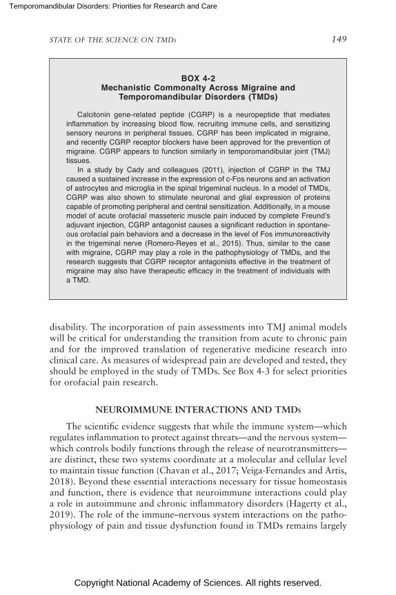
Temporomandibular Disorders: Priorities for Research and Care
Copyright National Academy of Sciences. All rights reserved.
STATE OF THE SCIENCE ON TMDs 149
BOX 4-2 Mechanistic Commonalty Across Migraine and
Temporomandibular Disorders (TMDs)
Calcitonin gene-related peptide (CGRP) is a neuropeptide that mediates inflammation by increasing blood flow, recruiting immune cells, and sensitizing sensory neurons in peripheral tissues. CGRP has been implicated in migraine, and recently CGRP receptor blockers have been approved for the prevention of migraine. CGRP appears to function similarly in temporomandibular joint (TMJ) tissues.
In a study by Cady and colleagues (2011), injection of CGRP in the TMJ caused a sustained increase in the expression of c-Fos neurons and an activation of astrocytes and microglia in the spinal trigeminal nucleus. In a model of TMDs, CGRP was also shown to stimulate neuronal and glial expression of proteins capable of promoting peripheral and central sensitization. Additionally, in a mouse model of acute orofacial masseteric muscle pain induced by complete Freund’s adjuvant injection, CGRP antagonist causes a significant reduction in spontane-ous orofacial pain behaviors and a decrease in the level of Fos immunoreactivity in the trigeminal nerve (Romero-Reyes et al., 2015). Thus, similar to the case with migraine, CGRP may play a role in the pathophysiology of TMDs, and the research suggests that CGRP receptor antagonists effective in the treatment of migraine may also have therapeutic efficacy in the treatment of individuals with a TMD.
disability. The incorporation of pain assessments into TMJ animal models will be critical for understanding the transition from acute to chronic pain and for the improved translation of regenerative medicine research into clinical care. As measures of widespread pain are developed and tested, they should be employed in the study of TMDs. See Box 4-3 for select priorities for orofacial pain research.
NEUROIMMUNE INTERACTIONS AND TMDs
The scientific evidence suggests that while the immune system—which regulates inflammation to protect against threats—and the nervous system—which controls bodily functions through the release of neurotransmitters—are distinct, these two systems coordinate at a molecular and cellular level to maintain tissue function (Chavan et al., 2017; Veiga-Fernandes and Artis, 2018). Beyond these essential interactions necessary for tissue homeostasis and function, there is evidence that neuroimmune interactions could play a role in autoimmune and chronic inflammatory disorders (Hagerty et al., 2019). The role of the immune–nervous system interactions on the patho-physiology of pain and tissue dysfunction found in TMDs remains largely
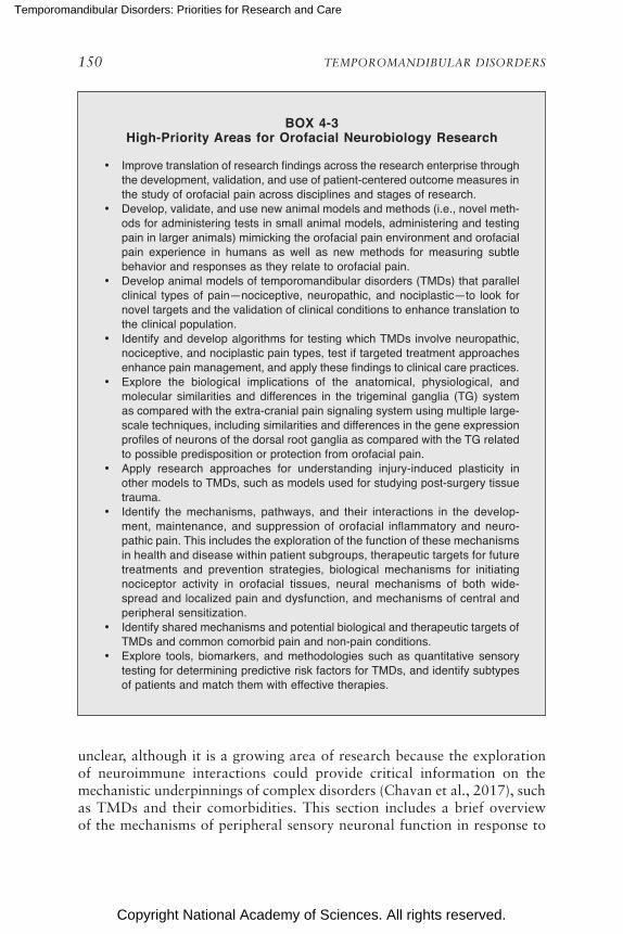
Temporomandibular Disorders: Priorities for Research and Care
Copyright National Academy of Sciences. All rights reserved.
150 TEMPOROMANDIBULAR DISORDERS
BOX 4-3 High-Priority Areas for Orofacial Neurobiology Research
• Improve translation of research findings across the research enterprise through the development, validation, and use of patient-centered outcome measures in the study of orofacial pain across disciplines and stages of research.
• Develop, validate, and use new animal models and methods (i.e., novel meth-ods for administering tests in small animal models, administering and testing pain in larger animals) mimicking the orofacial pain environment and orofacial pain experience in humans as well as new methods for measuring subtle behavior and responses as they relate to orofacial pain.
• Develop animal models of temporomandibular disorders (TMDs) that parallel clinical types of pain—nociceptive, neuropathic, and nociplastic—to look for novel targets and the validation of clinical conditions to enhance translation to the clinical population.
• Identify and develop algorithms for testing which TMDs involve neuropathic, nociceptive, and nociplastic pain types, test if targeted treatment approaches enhance pain management, and apply these findings to clinical care practices.
• Explore the biological implications of the anatomical, physiological, and molecular similarities and differences in the trigeminal ganglia (TG) system as compared with the extra-cranial pain signaling system using multiple large-scale techniques, including similarities and differences in the gene expression profiles of neurons of the dorsal root ganglia as compared with the TG related to possible predisposition or protection from orofacial pain.
• Apply research approaches for understanding injury-induced plasticity in other models to TMDs, such as models used for studying post-surgery tissue trauma.
• Identify the mechanisms, pathways, and their interactions in the develop-ment, maintenance, and suppression of orofacial inflammatory and neuro-pathic pain. This includes the exploration of the function of these mechanisms in health and disease within patient subgroups, therapeutic targets for future treatments and prevention strategies, biological mechanisms for initiating nociceptor activity in orofacial tissues, neural mechanisms of both wide-spread and localized pain and dysfunction, and mechanisms of central and peripheral sensitization.
• Identify shared mechanisms and potential biological and therapeutic targets of TMDs and common comorbid pain and non-pain conditions.
• Explore tools, biomarkers, and methodologies such as quantitative sensory testing for determining predictive risk factors for TMDs, and identify subtypes of patients and match them with effective therapies.
unclear, although it is a growing area of research because the exploration of neuroimmune interactions could provide critical information on the mechanistic underpinnings of complex disorders (Chavan et al., 2017), such as TMDs and their comorbidities. This section includes a brief overview of the mechanisms of peripheral sensory neuronal function in response to
Temporomandibular Disorders: Priorities for Research and Care
Copyright National Academy of Sciences. All rights reserved.
STATE OF THE SCIENCE ON TMDs 151
immune challenges, the neural regulation of immunity and inflammation, and the implications of these findings for treatment.
There are multiple points of interaction and communication between the nervous system and the immune system. Most relevant are the im-mune cell activation influences on neuronal circuits (such as changes to nociceptive signaling thresholds), which subsequently modulate both innate and adaptive immune responses. This shared molecular signaling—immune cells can produce what are classically defined as neurotransmitters, and neurons and their supporting cells release what are classically defined as cytokines/chemokines—is of particular interest. Different immune–neuronal signaling interfaces might be augmented in tissue environments responding to distinct injuries, and therefore an analysis of the mediators in different subjects with distinct phenotypes might provide opportunities to stratify patients and try novel therapeutic approaches.
Although research in this field directly related to TMDs is limited, the existing studies do shed some light on the potential value of this research. For example, in a mouse model of sustained mouth opening, increased macrophage/microglia activation was observed in the trigeminal subnucleus caudalis (Hawkins and Durham, 2016). Furthermore, inhibiting macro-phage and microglial activation prevented the development of orofacial mechanical hypersensitivity.
Furthermore, neuroimmune interactions may play an important role in pain chronicity. Therefore, it may be possible that chronic inflammation and responses to injury, surgery, implants, and devices may play a role in establishing chronic symptoms of TMDs. This is based on the concept that the circuits present in particular patients may favor a greater likelihood of chronic pain and other symptoms than appear in other patients.
To apply neuroimmune interactions in a clinically meaningful way, it is critical that the genetic framework of key inflammatory, immune, and resolution pathways in patients with various subgroups of TMDs be understood. Because there is significant complexity and heterogeneity within these neuroimmune interactions at different sites and for different stimuli, it is therefore important that researchers work toward defining those pathways and circuits that are relevant to TMD in humans to provide a foundational understanding. Despite the various unknowns in this area, enough is known to say that there appears to be potential clinical value for individuals with inflammatory TMDs. Indeed, recent clinical trials using bioelectric devices to modulate the neuroimmune pathways as a treatment strategy for inflammatory diseases such as rheumatoid arthritis and inflam-matory bowel disease have demonstrated some success (Brinkman et al., 2019; Payne et al., 2019).
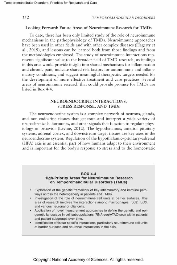
Temporomandibular Disorders: Priorities for Research and Care
Copyright National Academy of Sciences. All rights reserved.
152 TEMPOROMANDIBULAR DISORDERS
Looking Forward: Future Areas of Neuroimmune Research for TMDs
To date, there has been only limited study of the role of neuroimmune mechanisms in the pathophysiology of TMDs. Neuroimmune approaches have been used in other fields and with other complex diseases (Hagerty et al., 2019), and lessons can be learned both from those findings and from the methodologies employed. The study of neuroimmune interactions rep-resents significant value to the broader field of TMD research, as findings in this area would provide insight into shared mechanisms for inflammation and chronic pain, indicate shared risk factors for autoimmune and inflam-matory conditions, and suggest meaningful therapeutic targets needed for the development of more effective treatment and care practices. Several areas of neuroimmune research that could provide promise for TMDs are listed in Box 4-4.
NEUROENDOCRINE INTERACTIONS, STRESS RESPONSE, AND TMDs
The neuroendocrine system is a complex network of neurons, glands, and non-endocrine tissues that generate and interpret a wide variety of neuro chemicals, hormones, and other signals that function to regulate phys-iology or behavior (Levine, 2012). The hypothalamus, anterior pituitary systems, adrenal cortex, and downstream target tissues are key axes in the neuroendocrine system. Regulation of the hypothalamic–pituitary–adrenal (HPA) axis is an essential part of how humans adapt to their environment and is important for the body’s response to stress and to the homeostatic
BOX 4-4 High-Priority Areas for Neuroimmune Research
on Temporomandibular Disorders (TMDs)
• Exploration of the genetic framework of key inflammatory and immune path-ways across the heterogeneity in patients and TMDs.
• Investigation of the role of neuroimmune cell units at barrier surfaces. This area of research involves the interactions among macrophages, ILC2, ILC3, and various neuronal or glial cells.
• Application of novel measurement approaches to define the genetic and epi-genetic landscape in cell subpopulations (RNA-seq/ATAC-seq) within patients and patient subgroups over time.
• Identification of tissue-specific interactions, particularly neuroimmune cell units at barrier surfaces and neuronal interactions in the skin.
Temporomandibular Disorders: Priorities for Research and Care
Copyright National Academy of Sciences. All rights reserved.
STATE OF THE SCIENCE ON TMDs 153
regulation of the metabolic, cardiovascular, immune, reproductive, and central nervous systems (Smith and Vale, 2006).
Recently there has been growing scientific interest in the role of stress, activation of the HPA axis, and downstream inflammation on the initia-tion and progression of TMDs. For example, one study showed that indi-viduals with a TMD exhibited higher scores on both the Hospital Anxiety and Depression Scale as well as on “pain-catastrophizing events”—both scores of which may possibly contribute to the upregulation of the HPA axis (Staniszewski et al., 2018). Laboratory studies have also demonstrated the relationship between cortisol and circulating levels of pro- and anti-inflammatory cytokines (e.g., IL-6, TNF-α). Further study is needed to better understand the relationship between stress-related hormones and the onset and severity of TMD symptoms and across subgroups of patients with TMDs.
Other multi-system, chronic pain conditions such as fibromyalgia ex-hibit altered neuroendocrine and autonomic nervous system function, with both hypo- and hyperactive stress responses having been reported (Adler et al., 2002). Heart rate variability has been used as an indicator of the balance between the parasympathetic and sympathetic nervous system, the two branches of the autonomic nervous system. As measured by 24-hour heart rate monitoring, heart rate variability is lower in persons with a TMD (Chinthakanan et al., 2018), suggesting reduced vagal tone. Additional re-search on autonomic nervous system function in individuals with a TMD is needed to better understand how that function is altered and if it can be a target of therapeutic interventions.
Chronic pain disorders such as IBS, fibromyalgia, migraine, and interstitial cystitis are present at higher rates in females. In a rat model the estrous cycle and fluctuations in estrogen levels seemed to be linked to pain sensitivity fluctuations (Moloney et al., 2016). There are indications that TMD pain symptoms vary across the menstrual cycle, peaking in the late luteal phase and at menses (LeResche et al., 2003). These phases represent the time of declining or low estrogen levels. Additional research is needed to gain clarity on the effect of estrogen levels and the menstrual cycle on TMD initiation, progression, and response to treatment. The next section covers research on sex differences observed with TMDs in greater detail.
Finally, rigor and reproducibility are two important methodological considerations to take into account with regard to studies on the body’s stress response and its relationship to pain in the context of complex disor-ders such as TMDs. This is because the stress itself can produce hyperalgesia or analgesia, which raises the question of appropriate comparison groups that consist not only of healthy controls but of healthy controls with similar self-reported stress levels. Furthermore, while there is evidence that patients with a TMD report higher levels of anxiety and depression on standardized
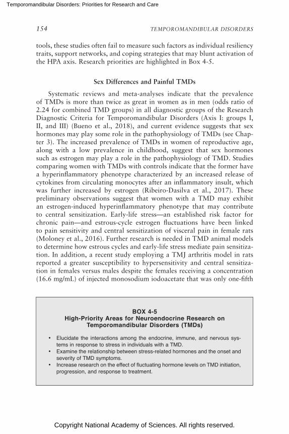
Temporomandibular Disorders: Priorities for Research and Care
Copyright National Academy of Sciences. All rights reserved.
154 TEMPOROMANDIBULAR DISORDERS
BOX 4-5 High-Priority Areas for Neuroendocrine Research on
Temporomandibular Disorders (TMDs)
• Elucidate the interactions among the endocrine, immune, and nervous sys-tems in response to stress in individuals with a TMD.
• Examine the relationship between stress-related hormones and the onset and severity of TMD symptoms.
• Increase research on the effect of fluctuating hormone levels on TMD initiation, progression, and response to treatment.
tools, these studies often fail to measure such factors as individual resiliency traits, support networks, and coping strategies that may blunt activation of the HPA axis. Research priorities are highlighted in Box 4-5.
Sex Differences and Painful TMDs
Systematic reviews and meta-analyses indicate that the prevalence of TMDs is more than twice as great in women as in men (odds ratio of 2.24 for combined TMD groups) in all diagnostic groups of the Research Diagnostic Criteria for Temporomandibular Disorders (Axis I: groups I, II, and III) (Bueno et al., 2018), and current evidence suggests that sex hormones may play some role in the pathophysiology of TMDs (see Chap-ter 3). The increased prevalence of TMDs in women of reproductive age, along with a low prevalence in childhood, suggest that sex hormones such as estrogen may play a role in the pathophysiology of TMD. Studies comparing women with TMDs with controls indicate that the former have a hyperinflammatory phenotype characterized by an increased release of cytokines from circulating monocytes after an inflammatory insult, which was further increased by estrogen (Ribeiro-Dasilva et al., 2017). These preliminary observations suggest that women with a TMD may exhibit an estrogen-induced hyperinflammatory phenotype that may contribute to central sensitization. Early-life stress—an established risk factor for chronic pain—and estrous-cycle estrogen fluctuations have been linked to pain sensitivity and central sensitization of visceral pain in female rats (Moloney et al., 2016). Further research is needed in TMD animal models to determine how estrous cycles and early-life stress mediate pain sensitiza-tion. In addition, a recent study employing a TMJ arthritis model in rats reported a greater susceptibility to hypersensitivity and central sensitiza-tion in females versus males despite the females receiving a concentration (16.6 mg/mL) of injected mono sodium iodoacetate that was only one-fifth
Temporomandibular Disorders: Priorities for Research and Care
Copyright National Academy of Sciences. All rights reserved.
STATE OF THE SCIENCE ON TMDs 155
the concentration in the males (80 mg/mL). Female rats developed more widespread pain hypersensitivity following acute inflammation, suggesting that TMJ osteoarthritis may serve as a model for future studies of sex dif-ferences (Sannajust et al., 2019).
Additionally, there is some evidence that sex differences exist with regard to estrogen and neuropathic pain signaling. As described above, the voltage-gated sodium channel acts as the threshold channel for the firing of action potentials and is thought to play an important role in pain sig-naling. The effects of estrogen on the Nav1.7 sodium channel expression in the trigeminal ganglion have been examined in experimental models. In rodents, estradiol-enhanced TMJ inflammation induced the upregulation of Nav1.7 in the trigeminal ganglion (Bi et al., 2017). These observations are consistent with the hypothesis that estrogen enhances hyperalgesia of an inflamed TMJ by modulating the expression or channel threshold of Nav1.7 in the trigeminal ganglion.
There is also evidence of sex differences in spinal cord pain process-ing mediated by the NMDA receptor, with NMDA antagonism reducing nociceptive responses more in males than in females (Del Rivero et al., 2019). Building off prior research indicating that the blockage of TNF receptor 1 (TNFR1) signaling in mice resulted in a failure to develop neuro-pathic pain or depressive symptoms following chronic constriction injury, Del Rivero and colleagues (2019) tested the therapeutic efficacy of a drug targeted to inactivate TNF. The results indicated that only male mice experi-enced accelerated recovery from neuropathic pain following administration of the therapeutic agent, while females did not, suggesting that TFNR1 signaling is associated with pain following nerve injury in males but not in females. The failure of the agent to work in female mice is thought to be due to inhibition of TFNR1 signaling and decreased NMDA receptor levels following injury (Del Rivero et al., 2019). While this specific mechanistic difference is valuable in its own right, this dichotomy in response between the sexes indicates more broadly the importance of conducting research into sex-specific mechanisms of pain as a critical component in the development of effective therapeutics for TMDs.
Looking Forward
Sex differences play a significant role in the mechanisms underlying chronic pain and will require additional focused study if safe and effective treatments targeted for high-risk groups, such as females, are to be devel-oped. Animal models specifically designed to explore sex differences in the development of chronic pain, such as the methodology used by Sannajust and colleagues (2019), are needed for TMDs (see Box 4-6).
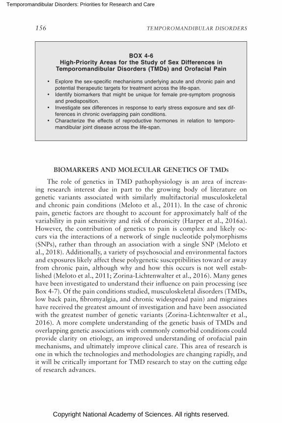
Temporomandibular Disorders: Priorities for Research and Care
Copyright National Academy of Sciences. All rights reserved.
156 TEMPOROMANDIBULAR DISORDERS
BOX 4-6 High-Priority Areas for the Study of Sex Differences in
Temporomandibular Disorders (TMDs) and Orofacial Pain
• Explore the sex-specific mechanisms underlying acute and chronic pain and potential therapeutic targets for treatment across the life-span.
• Identify biomarkers that might be unique for female pre-symptom prognosis and predisposition.
• Investigate sex differences in response to early stress exposure and sex dif-ferences in chronic overlapping pain conditions.
• Characterize the effects of reproductive hormones in relation to temporo-mandibular joint disease across the life-span.
BIOMARKERS AND MOLECULAR GENETICS OF TMDs
The role of genetics in TMD pathophysiology is an area of increas-ing research interest due in part to the growing body of literature on genetic variants associated with similarly multifactorial musculoskeletal and chronic pain conditions (Meloto et al., 2011). In the case of chronic pain, genetic factors are thought to account for approximately half of the variability in pain sensitivity and risk of chronicity (Harper et al., 2016a). However, the contribution of genetics to pain is complex and likely oc-curs via the interactions of a network of single nucleotide polymorphisms (SNPs), rather than through an association with a single SNP (Meloto et al., 2018). Additionally, a variety of psychosocial and environmental factors and exposures likely affect these polygenetic susceptibilities toward or away from chronic pain, although why and how this occurs is not well estab-lished (Meloto et al., 2011; Zorina-Lichtenwalter et al., 2016). Many genes have been investigated to understand their influence on pain processing (see Box 4-7). Of the pain conditions studied, musculoskeletal disorders (TMDs, low back pain, fibromyalgia, and chronic widespread pain) and migraines have received the greatest amount of investigation and have been associated with the greatest number of genetic variants (Zorina-Lichtenwalter et al., 2016). A more complete understanding of the genetic basis of TMDs and overlapping genetic associations with commonly comorbid conditions could provide clarity on etiology, an improved understanding of orofacial pain mechanisms, and ultimately improve clinical care. This area of research is one in which the technologies and methodologies are changing rapidly, and it will be critically important for TMD research to stay on the cutting edge of research advances.
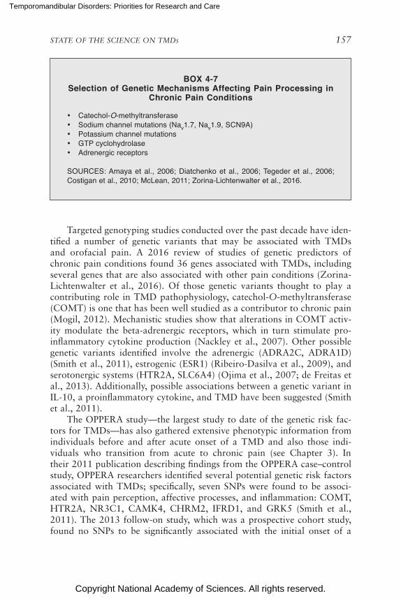
Temporomandibular Disorders: Priorities for Research and Care
Copyright National Academy of Sciences. All rights reserved.
STATE OF THE SCIENCE ON TMDs 157
BOX 4-7 Selection of Genetic Mechanisms Affecting Pain Processing in
Chronic Pain Conditions
• Catechol-O-methyltransferase • Sodium channel mutations (Nav1.7, Nav1.9, SCN9A)• Potassium channel mutations• GTP cyclohydrolase• Adrenergic receptors
SOURCES: Amaya et al., 2006; Diatchenko et al., 2006; Tegeder et al., 2006; Costigan et al., 2010; McLean, 2011; Zorina-Lichtenwalter et al., 2016.
Targeted genotyping studies conducted over the past decade have iden-tified a number of genetic variants that may be associated with TMDs and orofacial pain. A 2016 review of studies of genetic predictors of chronic pain conditions found 36 genes associated with TMDs, including several genes that are also associated with other pain conditions (Zorina- Lichtenwalter et al., 2016). Of those genetic variants thought to play a contributing role in TMD pathophysiology, catechol-O-methyltransferase (COMT) is one that has been well studied as a contributor to chronic pain (Mogil, 2012). Mechanistic studies show that alterations in COMT activ-ity modulate the beta-adrenergic receptors, which in turn stimulate pro-inflammatory cytokine production (Nackley et al., 2007). Other possible genetic variants identified involve the adrenergic (ADRA2C, ADRA1D) (Smith et al., 2011), estrogenic (ESR1) (Ribeiro-Dasilva et al., 2009), and serotonergic systems (HTR2A, SLC6A4) (Ojima et al., 2007; de Freitas et al., 2013). Additionally, possible associations between a genetic variant in IL-10, a proinflammatory cytokine, and TMD have been suggested (Smith et al., 2011).
The OPPERA study—the largest study to date of the genetic risk fac-tors for TMDs—has also gathered extensive phenotypic information from individuals before and after acute onset of a TMD and also those indi-viduals who transition from acute to chronic pain (see Chapter 3). In their 2011 publication describing findings from the OPPERA case–control study, OPPERA researchers identified several potential genetic risk factors associated with TMDs; specifically, seven SNPs were found to be associ-ated with pain perception, affective processes, and inflammation: COMT, HTR2A, NR3C1, CAMK4, CHRM2, IFRD1, and GRK5 (Smith et al., 2011). The 2013 follow-on study, which was a prospective cohort study, found no SNPs to be significantly associated with the initial onset of a
Temporomandibular Disorders: Priorities for Research and Care
Copyright National Academy of Sciences. All rights reserved.
158 TEMPOROMANDIBULAR DISORDERS
TMD; however, significant associations were noted in SCN1A and ACE2 with non-painful orofacial symptoms. Mutations in SCN1A and ACE2 have been associated with changes in pain processing (Smith et al., 2013).
Several genetic variants associated with TMDs have also been impli-cated in targeted genetic association studies for other chronic pain condi-tions, although the meaning of these overlapping genetic variants has yet to be unraveled. COMT, as mentioned above, has been associated with fibromyalgia (Vargas-Alarcón et al., 2007) and stress-induced chronic pain (McLean, 2011), while ADRB2, an adrenergic mechanism, has been re-ported in genetic association studies of fibromyalgia (Vargas-Alarcón et al., 2009) and low back pain (Skouen et al., 2012). Further research is neces-sary to identify and investigate how these polygenetic associations relate to the pathophysiology of TMDs and comorbid conditions.
Studies of genetic associations with TMDs encounter several chal-lenges. Most significant is the lack of studies using large populations. With the excep tion of OPPERA, most studies have had small participant sizes and used targeted genotyping approaches, making it difficult to identify novel genes associated with TMDs and potential biomarkers. Additional exploratory research looking across the entire genome (e.g., using next- generation sequencing and genome-wide association studies) may facilitate a better understanding of the genetic architecture of TMDs and those of other pain conditions.
Biomarkers for TMDs
Researchers face multiple challenges in developing safe and effective therapies for TMDs. These challenges include the current limited under-standing of the mechanisms of TMDs, a lack of and poor translation of preclinical and clinical data, too few robust clinical trials, and a lack of validated biomarkers to predict treatment response and stratify patients into clinically meaningful and mechanistically based subgroups (Harper et al., 2016a; Doshi et al., 2020). (See Box 4-8 for descriptions of biomarkers and their uses.) Other clinical fields, such as oncology and cardiovascular and metabolic diseases, have demonstrated the value of biomarkers in pre-dicting treatment response (Ferber, 2002).
Several studies have targeted potential TMD biomarkers for investiga-tion; however, most of these studies featured very small sample sizes and have not been replicated in other populations. In a case–control study (n=30) of plasma levels of dopamine and serotonin, Dawson and colleagues (2016) found significantly higher levels of dopamine in individuals with a painful TMD than in health controls, as well as a correlation between heightened levels of dopamine and increased pain intensity and perceived stress. However, further research is needed to understand the function of
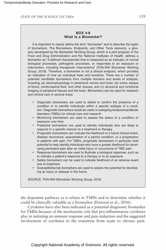
Temporomandibular Disorders: Priorities for Research and Care
Copyright National Academy of Sciences. All rights reserved.
STATE OF THE SCIENCE ON TMDs 159
BOX 4-8 What Is a Biomarker?
It is important to clearly define the term “biomarker” and to describe the types of biomarkers. The Biomarkers, Endpoints, and Other Tools resource, a glos-sary developed by the Biomarker Working Group, which is a joint program of the Food and Drug Administration and the National Institutes of Health, defines a biomarker as “A defined characteristic that is measured as an indicator of normal biological processes, pathogenic processes, or responses to an exposure or intervention, including therapeutic interventions” (FDA-NIH Biomarker Working Group, 2018). Therefore, a biomarker is not a clinical endpoint, which provides an indication of how an individual feels and functions. There are a number of potential candidate biomarkers from multiple domains and levels of analysis, including (a) electrophysiology in peripheral nerves and brain; (b) omics assays of blood, cerebro spinal fluid, and other tissues; and (c) structural and functional imaging of peripheral tissues and the brain. Biomarkers can be used for research and clinical care in several ways:
• Diagnostic biomarkers are used to detect or confirm the presence of a condition or to identify individuals within a specific subtype of a condi-tion. Diagnostic biomarkers would be useful in subtyping temporo mandibular disorders (TMDs) for clinical care and research.
• Monitoring biomarkers are used to assess the status of a condition or exposure over time.
• Predictive biomarkers are used to identity individuals who are likely to respond in a specific manner to a treatment or therapy.
• Prognostic biomarkers can indicate the likelihood of a future clinical event, disease recurrence, exacerbation of a painful condition, or a progression in patients with pain. For TMDs, prognostic biomarkers in particular hold potential to help identify individuals who have a greater likelihood for devel-oping persistent pain after an initial injury or occurrence of TMD pain.
• Response biomarkers are used to illustrate a biological response, such as to indicate a patient’s response to a therapy or to an exposure.
• Safety biomarkers can be used to indicate likelihood of an adverse event due to treatment.
• Susceptibility/risk biomarkers are used to assess the potential for develop-ing an injury or disease in the future.
SOURCE: FDA-NIH Biomarker Working Group, 2018.
the dopamine pathway as it relates to TMDs and to determine whether it could be clinically valuable as a biomarker (Dawson et al., 2016).
Cytokines have also been indicated as a potential diagnostic biomarker for TMDs because of the mechanistic role that pro-inflammatory cytokines play in initiating an immune response and pain induction and the suggested involvement of cytokines in the transition from acute to chronic pain.
Temporomandibular Disorders: Priorities for Research and Care
Copyright National Academy of Sciences. All rights reserved.
160 TEMPOROMANDIBULAR DISORDERS
Furthermore, past research has indicated that cytokine levels are elevated within the TMJ of individuals with TMDs (Kaneyama et al., 2002; Mat-sumoto et al., 2006; Kobayashi et al., 2017) and points to an association between cytokines and increased pain sensitivity. A heightened production of cytokine levels has also been associated with psychosocial factors, such as perceived stress (Maes et al., 1998) and depression (Maes, 1999), which are associated with painful TMDs. A 2011 case–control study (n=344) found that “localized and anatomically widespread patterns of chronic pain are associated with distinctive profiles of inflammatory biomarkers at protein, transcription factor activity, and gene levels” (Slade et al., 2011, p. 12). Specifically, localized TMDs were associated with an anti-inflammatory cytokine, IL-1RA, and TMDs with widespread palpation tenderness were associated with another cytokine, IL-8. Such findings suggest that cytokines could function as diagnostic biomarkers in the future (Slade et al., 2011; Kobayashi et al., 2017), although additional research is needed. Other inflammatory mediators such as 5-HT, TNFα, and prostaglandins have been explored as potential biomarkers for oro facial pain because of their presence in the synovial fluid of inflamed joints and their absence in healthy individuals, as have certain neuropeptides such as substance P and CGRP (Sessle, 2011).
Unfortunately, the field of TMD research and clinical care does not yet have rigorously validated biomarkers (Nagakura, 2017). It is unlikely that a single biomarker for TMDs will be found due to the complex biopsycho-social nature of TMDs and pain, which cannot be broken down into distinct, independent components. At this point, there are no confirmed bio-markers for TMDs, although there are a number of inflammatory markers that have been suggested as potential biomarkers.
Application of Novel Approaches to the Study of TMDs
The application of omics—genomics, proteomics, transcriptomics, metabolomics, etc.—approaches offers the possibility to collect large amounts of data and map molecular patterns within complex disorders (Hasin et al., 2017) like TMDs and other chronic pain conditions. These data, with proper translation, aid in the ability to identify clinically mean-ingful biomarkers, stratify patients based on mechanistically relevant fac-tors rather than by diagnoses, and identify therapeutic targets (Gazerani and Vinterhøj, 2016), among others. The clinical value of omics research has been demonstrated. In their 2011 review of genomics research on TMDs, Meloto and colleagues point to pharmacogenomics research that described the outsized role of genetic factors (specifically two genes) on the optimal dosing of the anticoagulant warfarin as an example of similar research that could be conducted for TMDs. Currently, the literature contains few
Temporomandibular Disorders: Priorities for Research and Care
Copyright National Academy of Sciences. All rights reserved.
STATE OF THE SCIENCE ON TMDs 161
examinations of blood, synovial fluid, or tissue from human subjects with TMDs that use large-scale, non-targeted approaches in the areas of genom-ics, epigenetics, proteomics, lipidomics, immune profiling, transcriptomics, metabolomics, and immunophenotyping, despite such approaches having resulted in key scientific breakthroughs in other fields.
Omics approaches are now being applied to the field of pain research through the exploration of DNA, RNA, protein, and metabolic changes in both animal models and in human subjects (Gomez-Varela and Schmidt, 2018); some of the findings of those studies hold potential value for TMD research. Proteomic analysis, which provides data on the expression, func-tion, and regulation of proteins, can provide insights into disease patho-physiology, biomarkers, and treatment response (Gazerani and Vinterhøj, 2016). A proteomic analysis of urine from women with IBS, a chronic pain-related condition that is frequently comorbid with TMD, highlighted several possible protein differences between well-phenotyped subgroups of IBS patients and healthy controls (Goo et al., 2012). This initial analysis found 18 proteins that differed between participants with IBS and healthy controls, and a follow-up study with one of these proteins, trefoil factor 3, which is known to play an important role in gut barrier protection, revealed strong associations between this protein and fecal microbiome taxa ( Heitkemper et al., 2018). Similarly, among those with chronic widespread pain, a chronic overlapping pain condition often found in those with fibro myalgia, muscle biopsies identified 17 proteins that were different from those in healthy controls and were associated with metabolism, muscle damage, stress, and inflammation (Olausson et al., 2015). These approaches have also been ap-plied in the Multidisciplinary Approach to the Study of Chronic Pelvic Pain (MAPP) Research Network study, a longitudinal analysis following chronic pelvic pain, which used pain testing and clinical phenotyping to under stand the complexities of symptom flares over time (Harte et al., 2019).
Metabolomics is another emerging area of research in the study of chronic pain that could provide insights for TMD research. This field investi gates the molecular products of the metabolic process within fluids and tissues and can provide data on cellular states and phenotypes ( Gazerani and Vinterhøj, 2016). In their review, Gazerani and Vinterhøj (2016) ex-plain that metabolomics can provide mechanistic insight into the relation-ship between disease phenotypes and biochemical changes. Metabolomic techniques have been used to identify a marker of neuropathic pain in rats (Patti et al., 2012), an indication of its potential as a future tool to differ-entiate pain types and inform treatment (Gazerani and Vinterhøj, 2016).
Omics approaches such as those described above would be valuable in the study of TMDs, particularly for understanding shared mechanisms with other chronic overlapping conditions, peripheral and CNS responses, and novel mechanistic and therapeutic targets.
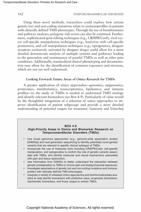
Temporomandibular Disorders: Priorities for Research and Care
Copyright National Academy of Sciences. All rights reserved.
162 TEMPOROMANDIBULAR DISORDERS
Using these novel methods, researchers could explore how certain genetic loci and non-coding mutations relate to immunoprofiles in patients with clinically defined TMD phenotypes. Through the use of bioinformatics and pathway analyses, polygenic risk scores can also be examined. Further-more, sophisticated gene-editing techniques (e.g., CRISPR/Cas9), viral vec-tor cell-specific manipulation techniques (e.g., lentivirus with cell-specific promotors), and cell manipulation techniques (e.g., optogenetics, designer receptors exclusively activated by designer drugs) could allow for a more detailed downstream analysis of multiple systems and pathways leading to the generation and maintenance of painful TMDs as well as other pain conditions. Additionally, standardized clinical phenotyping and documenta-tion may allow for the identification of common exposures and stressors, which are not yet well understood.
Looking Forward: Future Areas of Omics Research for TMDs
A greater application of omics approaches—genomics, epigenomics, proteomics, metabolomics, transcriptomics, lipidomics, and immune profiles—to the study of TMDs is needed to understand TMD etiology and identify relevant biomarkers (see Box 4-9). Particularly of value would be the thoughtful integration of a selection of omics approaches to im-prove identification of patient subgroups and provide a more detailed understanding of potential targets for treatment. Gazerani and Vinterhøj
BOX 4-9 High-Priority Areas in Omics and Biomarker Research on
Temporomandibular Disorders (TMDs)
• Use broad genomics approaches (e.g., genome-wide association studies [GWASs]) and next-generation sequencing to identify potentially novel genetic variants that are relevant to specific clinical subtypes of TMDs.
• Incorporate the use of molecular tools including CRISPR/Cas9, cell-specific manipulation, and optogenetics to confirm the role of genetic variants associ-ated with TMDs, and identify molecular and neural mechanisms associated with pain and tissue dysfunction.
• Use information from GWASs to better understand the interaction between genetic predisposition to TMDs or chronic pain and biopsychosocial exposures.
• Investigate associations of genetic loci and non-coding mutations and immuno-profiles with clinically defined TMD phenotypes.
• Integrate a variety of unbiased omics approaches and bioinformatics/data ana-lytics to help identify biomarkers with predictive value, prognostic biomarkers, mechanistic biomarkers, and those unique to certain TMDs.
Temporomandibular Disorders: Priorities for Research and Care
Copyright National Academy of Sciences. All rights reserved.
STATE OF THE SCIENCE ON TMDs 163
(2016, p. 262) note that the integration of proteomics and metabolomics into genomics research on pain “enhances [the] quality and validity of big data application in terms of efficacy and safety of approaches taken toward pain and its treatment.” Additionally, because TMDs often present with comorbid pain conditions, a focus on targets beyond TMDs could provide new mechanistic insights and potential therapeutic targets. The committee recommends carrying out non-targeted omics approaches to examine and compare local tissue with more systemic biomarkers in human subjects and animals. Once potential predictor, prognostic, and resilience markers have been identified in human subjects, these should be subsequently vali-dated as therapeutic targets in animal models to aid translational research. Such translational studies act as bridging mechanisms and will be essential to pushing the research forward. These future TMD biomarkers can help drive the discovery of new therapies and define more targeted and person-alized approaches to patient care based on a unique TMD “bio signature.” Such future biomarkers will need to address acute, chronic, and high- impact TMDs as well as vulnerability to the development of TMDs, recovery, and treatment outcomes. Combined with appropriate clinical endpoints, these biomarkers could help improve the classification of TMD subtypes and predict TMD progression. Patient stratification biomarkers—which could place indi viduals into clinically meaningful and mechanistically based subgroups—would be particularly useful in informing the design of clinical trials by improving patient selection and reducing expenses by allow ing for more targeted and smaller clinical trials within these subgroups.
NEUROIMAGING OF THE CNS: EXPLORATION OF TMD PHENOTYPES
Neuroimaging has opened a window to the brain for the non-invasive study of both structure and function and has expanded understanding of how pain processing is linked to the CNS, how it is disrupted, and how those disruptions occur with the chronification of pain (Davis and Moayedi, 2013; Nash et al., 2013; Cowen et al., 2015; Martucci and Mackey, 2016, 2018; Martucci et al., 2019; Weber et al., 2018). Furthermore, neuro-imaging allows a new perspective and a deeper understanding of the com-plex nature of chronic pain experience. This has led to the adoption of a whole-brain approach to the study and treatment of chronic pain and the development of novel technologies and analytic techniques, which could have major potential for the development of new diagnostics and more effective therapies. Various neuroimaging modalities have been used, in-cluding positron emission tomography (PET), electroencephalograpy (Diers et al., 2007), magnetoencephalography, single-photon emission computed tomography (Harisankar et al., 2012), and magnetic resonance imaging
Temporomandibular Disorders: Priorities for Research and Care
Copyright National Academy of Sciences. All rights reserved.
164 TEMPOROMANDIBULAR DISORDERS
(MRI). These techniques have been used to study several chronic pain states, including TMDs (Shibukawa et al., 2007; Younger et al., 2010; Gerstner et al., 2011; Moayedi et al., 2012; He et al., 2014, 2018; Lin, 2014; Monaco et al., 2015; Wilcox et al., 2015; Harper et al., 2016b; Mupparapu et al., 2019; Ozdiler et al., 2019), chronic low back pain (Ung et al., 2012), fibromyalgia (Staud, 2011a), osteoarthritis (Howard et al., 2012), complex regional pain syndrome (Schwenkreis et al., 2009; Barad et al., 2014), phantom-limb pain, chronic migraine (Chiapparini et al., 2010), chronic pelvic pain (Farmer et al., 2011; Kairys et al., 2015), and peripheral neuropathy (Moisset and Bouhassira, 2007), among others.
Structural Neuroimaging for TMDs
Researchers have found structural brain differences in gray matter density, gray matter volume, and cortical thickness, among other differ-ences, between people with chronic TMD pain and healthy volunteers (Younger et al., 2010; Moayedi et al., 2011; Lin, 2014; Wilcox et al., 2015). Structural changes have been identified in both the brain (primary somatosensory cortex, cingulate cortex, thalamus, putamen, pallidus, anterior insula) and the brainstem (trigeminal sensory nuclei, medullary dorsal horn) (Younger et al., 2010; Moayedi et al., 2011; Lin, 2014; Wilcox et al., 2015). These findings suggest that there is an underly-ing structural plasticity within the brain and that changes in cellular composition within the brain may occur in individuals with a TMD. However, the underlying physiological changes that contribute to these differences in gray matter remain unknown. Researchers have hypoth-esized that changes in gray matter may be the result of changes in gray matter microstructure and the prevalence of glial and other supporting and neuroimmune cells within the brain, in addition to other possible mechanisms (Zatorre et al., 2012). Finally, researchers have investigated white matter abnormalities using diffusion tensor imaging to investigate TMDs. Moayedi and colleagues (2012) found that people with painful TMDs have lower fractional anisotropy in the bilateral trigeminal nerve and diffuse abnormalities in the microstructure of white matter tracts related to sensory, motor, cognitive, and pain functions. These structural MRI findings point to gray and white matter abnormalities in the brain and brainstem systems responsible for the experience and modulation of pain. While these associations have been well established, the causal role of these changes still must be determined. It is important to understand whether the brain changes result from TMDs or whether these differences confer a vulnerability that contributes to the development of a TMD or to the transition from acute to chronic forms of TMDs. Additionally, the reversibility of these changes with therapies is of interest.
Temporomandibular Disorders: Priorities for Research and Care
Copyright National Academy of Sciences. All rights reserved.
STATE OF THE SCIENCE ON TMDs 165
Functional Neuroimaging for TMDs
Researchers have used neuroimaging techniques to study functional differences in the brain experiencing chronic pain, including TMDs, versus healthy states. Many of these studies used functional MRI (fMRI) tech-niques to investigate both abnormalities in brain function in TMDs and also the effect of treatment, specifically functional orthodontic treatments (He et al., 2014, 2018). Collectively, the functional neuroimaging research on pain points toward a heightened responsivity of the CNS to afferent noxious and innocuous stimuli in chronic pain. Additionally, more research with robust participant selection criteria and methodologies is needed to determine the extent that MRI brain-based biomarkers are useful for treat-ment prediction.
The emergence of resting state fMRI for studying non-evoked brain activity and functional connectivity has allowed many investigations of chronic pain to gain a broader understanding of brain processes, as opposed to only those processes related to noxious stimuli. Several studies applied resting-state fMRI to characterize differences in non-evoked (i.e., resting) brain activity among people with TMDs (He et al., 2018). Participants with TMDs exhibited reduced network functional connectivity, supporting the suggestion that TMDs are associated with reduced functional connectivities in brain corticostriatal networks and that these reduced functional connec-tivities may underlie motor control deficits, pain processing, and cognition in individuals with TMDs, although additional research is needed.
The Future of Neuroimaging and TMDs
Other advances in the field of pain neuroimaging include combining multiple neuroimaging modalities with large-scale, multi-site investigations. Neuroimaging researchers are increasingly using analytical methods that combine multiple neuroimaging modalities to understand chronic pain. For example, a study by Schrepf and colleagues (2016) used combined PET imaging and fMRI to identify increased µ-opioid receptor availability and evoked pain brain activity (blood oxygenation level dependent) co-occurring in the anterior insula of individuals with fibromyalgia (Schrepf et al., 2016). The neuroimaging of pain is also being included as a major component of large-scale, multi-site investigations that focus on idiopathic chronic pain conditions such as urological chronic pelvic pain (i.e., intersti-tial cystitis, chronic prostatitis, bladder pain syndrome) (Alger et al., 2016). These collaborative multi-site investigations are also including longitudinal investigations that illustrate changes in brain activity to track symptom profiles over time (Kutch et al., 2017b). Such multimodal, multi-site col-laborations offer tremendous opportunities for the study of TMDs.

Temporomandibular Disorders: Priorities for Research and Care
Copyright National Academy of Sciences. All rights reserved.
166 TEMPOROMANDIBULAR DISORDERS
Future of Brain-Based Biomarkers of Pain and TMDs
MRI has opened a window to the brain by allowing for the non- invasive study of both structure and function and the validation of the role of the CNS in chronic pain. MRI offers a significant opportunity to identify and validate neuroimaging-based biomarkers and surrogate endpoints for pain. Preliminary brain biomarkers have been identified in individuals expe-riencing acute and chronic pain (Marquand et al., 2010; Brown et al., 2011; Brodersen et al., 2012; Wager et al., 2013; Bagarinao et al., 2014; Woo et al., 2015, 2017; Kutch et al., 2017a,b; Lopez-Sola et al., 2017; Cheng et al., 2018; Zhong et al., 2018). These biomarkers would be of value to clinical and research communities by aiding prognosis (Baliki et al., 2012; Mutso et al., 2014) and understanding pain progression (Mackey, 2014, 2016; Von Korff et al., 2016), predicting response to a treatment, ascertaining a diag-nosis, identifying targets for treatment, and defining surrogate endpoints and predicting clinical benefit.
PSYCHOSOCIAL FACTORS UNDERLYING TMDs
The biopsychosocial model seeks to take into account all factors— biological, psychological, and social—that may play a role in the onset and progression of TMDs (see Chapter 2 for an overview of this approach). Unraveling the biological mechanisms underlying the pathophysiologies of TMDs has been the greatest focus of basic research thus far; however, the role of psychosocial factors has increasingly been elevated in importance as the biopsychosocial model is absorbed into research and care philosophies. Despite acknowledgment of the contributing role of psychosocial factors in the experience of pain, this area of research remains out of balance with the comparatively large body of research on biological mechanisms.
How psychosocial factors affect pain and tissue dysfunction associ-ated with TMDs remains unknown, although the significant overlap in psychosocial risk factors between TMDs and other chronic pain condi-tions (see Chapter 3 for discussion of psychosocial risk factors) suggests that similar underpinnings may be at work. Psychosocial factors (i.e., anxiety, depression, negative affect, and symptom burden) and their im-pact on the generation, maintenance, suppression, and perception of pain have been studied across overlapping conditions like fibromyalgia (Giesecke et al., 2005), interstitial cystitis (Nickel et al., 2015), IBS, and headache (Kato et al., 2009). This small body of research suggests that, as it relates to the experience of chronic pain, certain psychosocial fac-tors may mediate activity in the various parts of the brain responsible for processing the sensory versus the affective aspects of pain (Giesecke et al., 2005; Harper et al., 2016a).
Temporomandibular Disorders: Priorities for Research and Care
Copyright National Academy of Sciences. All rights reserved.
STATE OF THE SCIENCE ON TMDs 167
There is some evidence that psychosocial factors may play a role in the amplification and maintenance of orofacial pain. In a study of 163 individuals with a TMD, those who score high on the pain catastrophizing scale had a six-fold increase in risk of developing persistent pain (Reiter et al., 2015). In another study, orofacial pain response was associated with depression in participants with TMDs (Sherman et al., 2004). The presence of high symptom burden was also associated with increased TMD incidence and decreased improvement in orofacial pain after 5 years (Ohrbach and Dworkin, 1998). These findings for orofacial pain support prior research by Giesecke and colleagues (2005), which demonstrated that individuals with fibromyalgia and depression experience increased pain amplification by the CNS compared with individuals with fibromyalgia but no reported psychological comorbidity.
Findings from the OPPERA study indicate that psychological distress and other somatic symptoms may act as predictive risk factors for develop-ing a painful TMD (Fillingim et al., 2011). The OPPERA study extensively phenotyped individuals based on an array of biopsychosocial risk factors in individuals with and without TMDs (see Chapter 3). The outcome of this phenotyping was the identification of clusters across which symptoms and factors could be compared. Of the three clusters identified—adaptive, pain-sensitive, and global symptoms clusters—individuals in the global symptoms cluster presented with more psychological distress and greater pain sensitiv-ity than the other two clusters. These findings point to the likely presence of multimodal predictors and a range of environmental, physiological, and psychological variables that each contribute to TMDs (Bair et al., 2016). The use of phenotypes that take into account bio psychosocial factors in a clinical care setting could add significant value to the care of patients with TMDs by targeting treatment and predicting outcomes based on the presenting phenotype of the patient; however, more research is required. It should be noted that temporomandibular pain can occur for multiple rea-sons, including nerve injury and joint conditions such as osteo arthritis, or be non-specific, and thus there may be different underlying mechanisms and combinations of mechanisms for each TMD and across individuals.
APPLICATION OF DATA SCIENCE METHODOLOGIES AND NOVEL TECHNOLOGIES TO TMD RESEARCH
The data science methodologies of machine learning, informatics, and artificial intelligence are increasingly used to unravel complex problems related to etiology, risk, prognosis, and treatment effectiveness. In pain research these methods have been used by researchers to better detect previ-ously unseen patterns in data and to identify subgroups within the data that could inform future research and clinical care (Lötsch and Ultsch, 2018).
Temporomandibular Disorders: Priorities for Research and Care
Copyright National Academy of Sciences. All rights reserved.
168 TEMPOROMANDIBULAR DISORDERS
The value of these approaches is the ability to identify new clinically relevant and mechanistically based phenotypes within datasets that could indicate or predict response to treatment. Machine learning methods can also be used to identify previously unknown biopsychosocial parameters of complex condi-tions (Lötsch and Ultsch, 2018) such as TMDs and chronic pain. However, these methods require access to large-scale, high-quality datasets, which are lacking for TMDs (Lötsch and Ultsch, 2018). Useful data could be collected in several ways, including the addition of TMD-related questions to national surveys, the mining of insurance databases and electronic health records and International Classification of Diseases codes, and the creation of national or regional patient registries. The value of these approaches wholly relies on high-quality data inputs, and the research and clinical ecosystems need to be able to manage this. TMD research requires the adoption of these new technologies and approaches, like advanced data analysis, machine learning, artificial intelligence, clustering methods, expression quantitative trait loci (eQTL) analyses, polygenic approaches, and pathway analyses to unravel the complexities of this group of disorders, identify patient subgroups, and develop safe and effective treatments.
TMD RESEARCH FUNDING
Current Funding for TMD Basic Research and Translation
The National Institutes of Health (NIH) provides only about one-third of biomedical research funding in the United States (IOM, 2011), and its impact has a ripple effect that stimulates research interests and training programs across the nation. TMDs are not the primary mission of any NIH institute; however, funding for these disorders primarily falls within the National Institute of Dental and Craniofacial Research ( NIDCR). In fiscal year (FY) 2018, NIH awarded approximately $13.7 million for TMD research. Of that amount, approximately $12 million came from NIDCR (NIH, 2019a). The other five institutes (National Institute of General Medical Sciences; National Institute of Neurological Disorders and Stroke; National Institute of Nursing Research; National Heart, Lung, and Blood Institute; and National Institute on Deafness and Other Communication Disorders) contributed remaining funding for TMDs in FY 2018 (NIH, 2019a). While funding estimates for FY 2019 and FY 2020 across NIH indicate a slight upward trend when compared to levels of funding for 2017 and 2018 (see Figure 4-1), recent discussions have highlighted challenges that need to be addressed to move the field forward (MDEpiNet TMJ Patient-Led RoundTable, 2018a,b).
In 2018, NIH’s TMD-targeted funding focused on topics such as cel-lular and mechanical mechanisms, genetics, emotion dysregulation, and
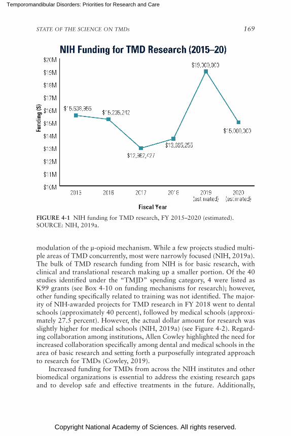
Temporomandibular Disorders: Priorities for Research and Care
Copyright National Academy of Sciences. All rights reserved.
STATE OF THE SCIENCE ON TMDs 169
FIGURE 4-1 NIH funding for TMD research, FY 2015–2020 (estimated).SOURCE: NIH, 2019a.
modulation of the µ-opioid mechanism. While a few projects studied multi-ple areas of TMD concurrently, most were narrowly focused (NIH, 2019a). The bulk of TMD research funding from NIH is for basic research, with clinical and translational research making up a smaller portion. Of the 40 studies identified under the “TMJD” spending category, 4 were listed as K99 grants (see Box 4-10 on funding mechanisms for research); however, other funding specifically related to training was not identified. The major-ity of NIH-awarded projects for TMD research in FY 2018 went to dental schools (approximately 40 percent), followed by medical schools (approxi-mately 27.5 percent). However, the actual dollar amount for research was slightly higher for medical schools (NIH, 2019a) (see Figure 4-2). Regard-ing collaboration among institutions, Allen Cowley highlighted the need for increased collaboration specifically among dental and medical schools in the area of basic research and setting forth a purposefully integrated approach to research for TMDs (Cowley, 2019).
Increased funding for TMDs from across the NIH institutes and other biomedical organizations is essential to address the existing research gaps and to develop safe and effective treatments in the future. Additionally,
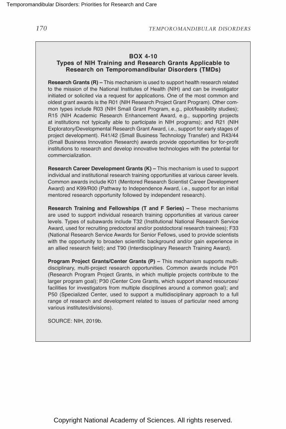
Temporomandibular Disorders: Priorities for Research and Care
Copyright National Academy of Sciences. All rights reserved.
170 TEMPOROMANDIBULAR DISORDERS
BOX 4-10 Types of NIH Training and Research Grants Applicable to
Research on Temporomandibular Disorders (TMDs)
Research Grants (R) – This mechanism is used to support health research related to the mission of the National Institutes of Health (NIH) and can be investigator initiated or solicited via a request for applications. One of the most common and oldest grant awards is the R01 (NIH Research Project Grant Program). Other com-mon types include R03 (NIH Small Grant Program, e.g., pilot/feasibility studies); R15 (NIH Academic Research Enhancement Award, e.g., supporting projects at institutions not typically able to participate in NIH programs); and R21 (NIH Exploratory/Developmental Research Grant Award, i.e., support for early stages of project development). R41/42 (Small Business Technology Transfer) and R43/44 (Small Business Innovation Research) awards provide opportunities for for-profit institutions to research and develop innovative technologies with the potential for commercialization.
Research Career Development Grants (K) – This mechanism is used to support individual and institutional research training opportunities at various career levels. Common awards include K01 (Mentored Research Scientist Career Development Award) and K99/R00 (Pathway to Independence Award, i.e., support for an initial mentored research opportunity followed by independent research).
Research Training and Fellowships (T and F Series) – These mechanisms are used to support individual research training opportunities at various career levels. Types of subawards include T32 (Institutional National Research Service Award, used for recruiting predoctoral and/or postdoctoral research trainees); F33 ( National Research Service Awards for Senior Fellows, used to provide scientists with the opportunity to broaden scientific background and/or gain experience in an allied research field); and T90 (Interdisciplinary Research Training Award).
Program Project Grants/Center Grants (P) – This mechanism supports multi-disciplinary, multi-project research opportunities. Common awards include P01 (Research Program Project Grants, in which multiple projects contribute to the larger program goal); P30 (Center Core Grants, which support shared resources/facilities for investigators from multiple disciplines around a common goal); and P50 (Specialized Center, used to support a multidisciplinary approach to a full range of research and development related to issues of particular need among various institutes/divisions).
SOURCE: NIH, 2019b.
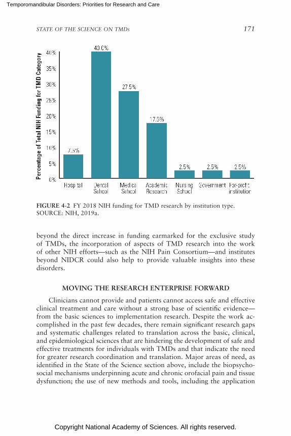
Temporomandibular Disorders: Priorities for Research and Care
Copyright National Academy of Sciences. All rights reserved.
STATE OF THE SCIENCE ON TMDs 171
FIGURE 4-2 FY 2018 NIH funding for TMD research by institution type.SOURCE: NIH, 2019a.
beyond the direct increase in funding earmarked for the exclusive study of TMDs, the incorporation of aspects of TMD research into the work of other NIH efforts—such as the NIH Pain Consortium—and institutes beyond NIDCR could also help to provide valuable insights into these disorders.
MOVING THE RESEARCH ENTERPRISE FORWARD
Clinicians cannot provide and patients cannot access safe and effective clinical treatment and care without a strong base of scientific evidence—from the basic sciences to implementation research. Despite the work ac-complished in the past few decades, there remain significant research gaps and systematic challenges related to translation across the basic, clinical, and epidemiological sciences that are hindering the development of safe and effective treatments for individuals with TMDs and that indicate the need for greater research coordination and translation. Major areas of need, as identified in the State of the Science section above, include the biopsycho-social mechanisms underpinning acute and chronic orofacial pain and tissue dysfunction; the use of new methods and tools, including the application
Temporomandibular Disorders: Priorities for Research and Care
Copyright National Academy of Sciences. All rights reserved.
172 TEMPOROMANDIBULAR DISORDERS
of molecular and cellular approaches to understand TMD genetics and biomarkers associated with TMD diagnosis, prognosis, treatment outcome, and resilience; and a greater understanding of the tissues of the TMJ joint, its function, and the relationship between function and pain. Additionally, the committee noted the need for sophisticated and consistent outcome measures for assessment of TMD pain and function from preclinical models through to clinical research. It is critically important that pain measures be incorporated into translational models of TMJ degenerative diseases, but such incorporation is currently lacking.
The committee’s overview of the recent literature on TMDs revealed that a concerted effort to develop a more comprehensive foundation of clinically meaningful evidence around the pathophysiologies of TMDs will require a commitment to both the funding and the implementation of multi-disciplinary research and to the purposeful transfer of knowledge across the research enterprise. Effectively addressing priority research areas will re-quire a coordinated effort by a diverse group of stakeholders to develop and implement a patient-focused research agenda, cultivate a multi disciplinary research culture, and align system incentives to ensure that novel research findings are transferred from one stage of the research enterprise to the next. Similar efforts to define research priorities for complex disorders, such as the Federal Pain Research Strategy, have also sought to achieve these aims by providing an actionable research framework to guide and prioritize patient-focused research across the research enterprise. A description of the role of the proposed research consortium (see Recommendations 1 and 2 in Chapter 8) and the example research framework for TMD research, which unifies research priorities within patient-centered needs, can be found below.
Conclusion 4-1: The siloed approach to research on temporo-mandibular disorders (TMDs) has detrimental effects on transla-tion across the research enterprise and limits access to the financial, educational, and intellectual resources needed to cultivate a robust research base. Addressing these challenges will require a multi-stakeholder collaboration to define patient-focused research pri-orities and implement systemic change to the conduct of TMD research to improve data quality and comparability, incentivize and support novel and collaborative research, and integrate evidence into clinical care and policy.
Elucidation of Biopsychosocial Mechanisms
The value of identifying and prescribing clinical meaning to biological mechanisms and pathways and understanding how these interact with other
Temporomandibular Disorders: Priorities for Research and Care
Copyright National Academy of Sciences. All rights reserved.
STATE OF THE SCIENCE ON TMDs 173
biological, psychological, and environmental factors in relation to TMDs cannot be overstated. However, the value of understanding these mechanistic underpinnings goes beyond the benefit of the scientific knowledge itself; given access to the right tools and data, such an understanding could point to clinically meaningful and mechanistically based subgroups of indi viduals and inform targeted treatments. At the present time, the current state of our col-lective understanding of these mechanisms as they relate to subtypes of TMDs is insufficient to provide clinical value. Considerable time and resources have been invested in defining potential mechanisms; however, more research needs to be conducted to confirm the role of these mechanisms across other patient groups and within other TMDs. This includes determining the biopsycho-social mechanisms of resilience to both orofacial pain and tissue dysfunc-tion. Additionally, because preclinical animal models demonstrate that such mechanisms may prove to be meaningful clinical targets, addi tional studies in the translational and clinical research should be carried out to capitalize on the potential for diagnostic, prognostic, or therapeutic value.
Conclusion 4-2: Elucidating the biopsychosocial mechanisms be-hind the generation, maintenance, and suppression of temporo-mandibular disorder (TMD)-related pain and dysfunction will be essential to understanding the complex pathophysiologies within TMDs, identifying clinically meaningful and mechanistically valid patient subgroups, and developing safe, effective, and personalized therapeutics and treatments. Doing so will require expanding cur-rent approaches and the use of innovative research methods and tools.
Application of Novel Methods and Tools
The study of TMDs would significantly benefit from the development of novel tools and approaches to understand all aspects of TMDs and from the application of concepts, methods, and tools already being applied in other fields. There exists a multitude of areas where the use of new tools or approaches could provide value. The advancement of our understanding of TMDs would be enhanced by the incorporation of newer molecular and cellular approaches targeting RNA, DNA, and the epigenome, metabolome, and proteome as well as cell-specific activation and silencing techniques. These approaches should be applied to the study of TMDs using relevant animal models and should translate to carefully phenotyped human sub-jects using tissue and blood samples as appropriate. A better understanding of the mechanisms that lead to development of acute and chronic pain, of the peripheral and CNS responses to nociceptive input, and of long-term changes will be critical. Sophisticated gene-editing techniques (e.g.,
Temporomandibular Disorders: Priorities for Research and Care
Copyright National Academy of Sciences. All rights reserved.
174 TEMPOROMANDIBULAR DISORDERS
CRISPR/Cas9), viral vector cell-specific manipulation techniques (e.g., len-tivirus with cell-specific promotors), and cell manipulation techniques (e.g., optogenetics and designer receptors exclusively activated by designer drugs) will allow for a more detailed analysis of multiple systems and their role in the generation and maintenance of TMD and other pain conditions.
Because TMDs, when chronic, often present with comorbidities and multi-system components, a focus on targets outside the TMD regions or a systemic focus could provide novel mechanistic insights. The use of non- targeted approaches examining and comparing local tissue with more systemic biomarkers using a variety of approaches in human subjects and animals is recommended. These approaches could include genomics, epigenomics, proteomics, transcriptomics, lipidomics, and immune profiles. Potential predictors and resilience markers should be identified in human subjects and subsequently validated as therapeutic targets in animal models. Translational studies bridging mechanisms between animal and human TMD pain mechanisms will be critical to moving the field forward by iden-tifying clinically meaningful and mechanistically based patient subgroups and developing new therapeutic approaches.
Research on TMJ Development, Biomechanical Function, and Relationship with Pain
To advance the understanding of TMJ function in health and disease, contemporary, multidisciplinary research focused on the biology of TMJ tissues, the interactions of these tissues, the innervation of the musculo-skeletal component and vascularization of the joint, and the normal devel-opmental processes and disease progression will be key research areas for expansion. Additionally, while bone/cartilage/ligament turnover and repair have been studied to some extent, a better understanding of the TMJ repair processes will be needed to integrate mechanical cues and pathologi-cal loading of the joint into bone/cartilage/ligament remodeling activities. Valuable information from these explorations and from the development and use of new in vitro and in vivo models could then inform the design and testing of new scaffolds and materials for regenerative medicine ap-proaches. Most critically and where possible, the focus of these multiple areas of research should be on the translation from small-animal models to large-animal models and ultimately to humans.
A NATIONAL COLLABORATIVE RESEARCH CONSORTIUM AND FRAMEWORK FOR ACTION
Any research study of complex disorders, such as TMDs, must be considered from the perspective of the full biopsychosocial framework.
Temporomandibular Disorders: Priorities for Research and Care
Copyright National Academy of Sciences. All rights reserved.
STATE OF THE SCIENCE ON TMDs 175
Consequently, studying TMDs from only a singular research perspective has been insufficient to fully understand the etiology of TMDs and how these disorders affect patients’ lives (MDEpiNet TMD Patient-Led RoundTable, 2018b; Cowley, 2019). The available body of recent laboratory research overviewed in this chapter related to TMDs and orofacial pain illustrates this issue, with many studies focusing on only one aspect of a TMD or conducting highly discipline-specific research. For example, dentists may study TMDs as a jaw issue, psychologists may exclusively consider the mental health comorbidities of TMDs, and medical researchers may study the mechanics of pain modulation, all of them without considering the larger biopsychosocial considerations of TMDs. This is not to say that these individual studies are not valuable—each plays a role in growing the scientific evidence base related to TMDs—but each alone has proven to be insufficient for developing a full understanding of TMD pathophysiologies. The 2011 Institute of Medicine (IOM) report Relieving Pain in America acknowledged the need for collaborative research in the field of pain and concluded that “research is needed to document and assess this full spec-trum” of associated problems with acute and chronic pain and that this type of integrated, collaborative research would “enable the development of interventions that would address all aspects of the pain condition” (IOM, 2011, p. 228). In his remarks to the committee, Allen Cowley echoed the need for this approach for research on TMDs and emphasized the paltry research collaborations, particularly in the basic sciences, between dental and medical school researchers, adding that efforts to stimulate such an integrated approach to research on TMDs are lacking. He went on to say that basic research in dental schools would greatly benefit from collabo-rations with medical school faculty, schools of bioengineering, pharmacy schools, and other related schools (Cowley, 2019).
The 2011 IOM report also noted that research more broadly will in-creasingly require bioinformatics to aid in the analysis and interpretation of large datasets and that the field requires more scientists from diverse backgrounds (IOM, 2011). This underlines the point that a successful trans-disciplinary research agenda would require the cultivation and engagement of both young and established researchers from across dental, medical, bio-engineering, neurology, physical therapy, nursing, and psychology who have skills in a broad array of research and data techniques. This will in turn require a lasting financial commitment to research training and funding for research. Several existing funding structures within NIH are well positioned to do this, but funds are not currently earmarked for research on TMDs. (See Box 4-10 for examples of applicable training and research grants for developing a research workforce.)
Additionally, given the significant areas of research overlap between TMDs and other conditions, the TMD research enterprise is well positioned
Temporomandibular Disorders: Priorities for Research and Care
Copyright National Academy of Sciences. All rights reserved.
176 TEMPOROMANDIBULAR DISORDERS
to align itself within complementary research agendas and initiatives, such as the National Pain Strategy, to access both scientific and clinical knowl-edge and funding. Funding research on pain is one major area in which NIH can, without significant additional financial cost, expand the evidence base for TMDs by facilitating the transfer of potentially relevant findings from the pain field to researchers engaged in existing TMD research and allowing greater opportunities for researchers involved in pain research to conduct novel research on TMDs. To illustrate this point, in FY 2018 NIH reported funding approximately $605 million within the category of “pain research” and $474 million within the category of “chronic pain” (NIH, 2019a)—as compared to just more than $14 million for TMDs. TMD patients deal with both acute and chronic pain, and the research in this area may benefit patients, even when it is not specifically focused on TMDs (Cowley, 2019).
Conclusion 4-3: Research on temporomandibular disorders (TMDs) would benefit from alignment with an established research agenda with continuity of oversight, financial support, and a col-laborative institutional structure that can guide a research agenda and support the integration into clinical practice. A TMD Research Consortium would be well positioned to convene stakeholders and launch a TMD research framework.
Conclusion 4-4: The level and appropriation of funding for research on temporomandibular disorders does not reflect the complexities of these disorders, their prevalence and impact, or the need for trans-disciplinary research with a focus on clinical care impacts.
Role of a TMD Research Consortium and Example Research Framework for TMDs
A unified, transdisciplinary research framework for TMDs developed and implemented by a national collaborative research consortium (see Figure 4-3) could address these barriers by embedding a commitment to and the tools needed for the translation of research across disciplines and the research enterprise into the fabric of the framework and drive the devel opment of a research program designed to address patient needs (see Box 4-11). There have already been calls for the implementation of a com-prehensive research agenda that addresses the needs of TMD research. The TMJ Patient-Led RoundTable, a public–private partnership within the Food and Drug Administration composed of a variety of stakeholders, has taken the first steps in formalizing a proposed interagency research plan and has called repeatedly for a transformation of TMD research (MDEpiNet TMD Patient-Led RoundTable, 2018a).
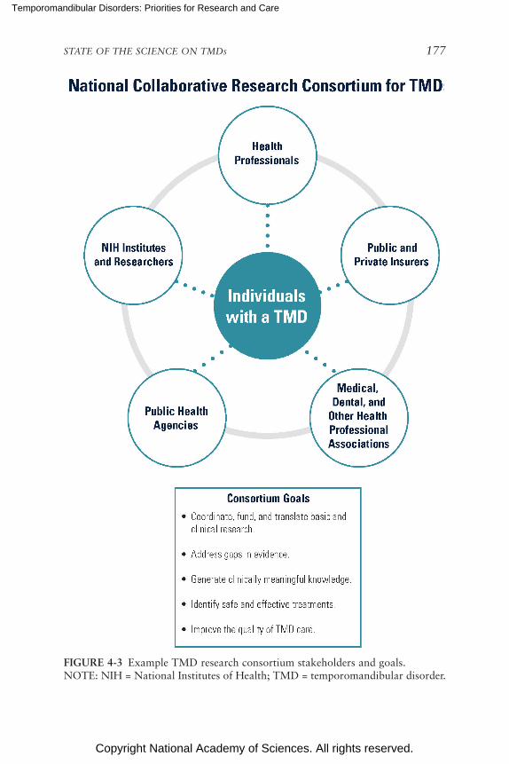
Temporomandibular Disorders: Priorities for Research and Care
Copyright National Academy of Sciences. All rights reserved.
STATE OF THE SCIENCE ON TMDs 177
FIGURE 4-3 Example TMD research consortium stakeholders and goals.NOTE: NIH = National Institutes of Health; TMD = temporomandibular disorder.
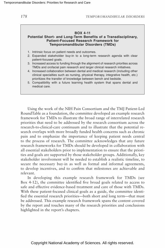
Temporomandibular Disorders: Priorities for Research and Care
Copyright National Academy of Sciences. All rights reserved.
178 TEMPOROMANDIBULAR DISORDERS
BOX 4-11 Potential Short- and Long-Term Benefits of a Transdisciplinary,
Patient-Focused Research Framework for Temporomandibular Disorders (TMDs)
1. Intrinsic focus on patient needs and outcomes.2. Expanded stakeholder buy-in to a long-term research agenda with clear
patient-focused goals.3. Increased access to funding through the alignment of research priorities across
TMDs and orofacial pain research and larger clinical research initiatives.4. Increased collaboration between dental and medical research (including other
clinical specialties such as nursing, physical therapy, integrative health, etc.) prioritizes the transfer of knowledge between bench and bedside.
5. Compatibility with a future learning health system that spans dental and medical care.
Using the work of the NIH Pain Consortium and the TMJ Patient-Led RoundTable as a foundation, the committee developed an example research framework for TMDs to illustrate the broad range of interrelated research priorities that need to be addressed by the research consortium across the research-to-clinical-care continuum and to illustrate that the potential re-search overlaps with more broadly funded health concerns such as chronic pain and to emphasize the importance of keeping patient needs central to the process of research. The committee acknowledges that any future research frameworks for TMDs should be developed in collaboration with all essential stakeholders prior to implementation to ensure that the priori-ties and goals are supported by those stakeholder groups. Additionally, key stakeholder involvement will be needed to establish a realistic timeline, to secure the necessary buy-in as well as formal and informal agreements, to develop incentives, and to confirm that milestones are achievable and relevant.
In developing this example research framework for TMDs (see Box 4-12), the committee identified five broad goals related to access to safe and effective evidence-based treatment and care of those with TMDs. With these patient-focused clinical goals as a guide, the committee identi-fied the essential research priorities—both short and long term—that must be addressed. This example research framework spans the content covered by the report and touches many of the research priorities and conclusions highlighted in the report’s chapters.
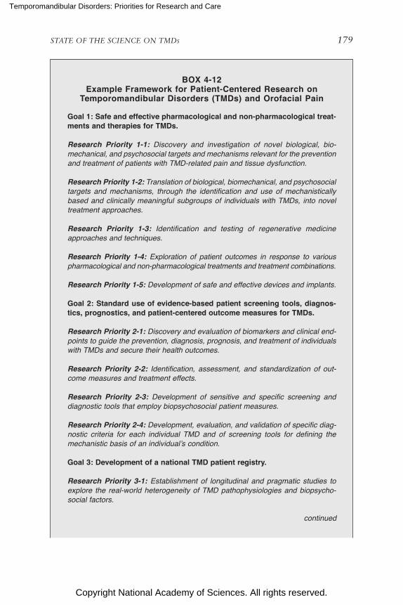
Temporomandibular Disorders: Priorities for Research and Care
Copyright National Academy of Sciences. All rights reserved.
STATE OF THE SCIENCE ON TMDs 179
BOX 4-12 Example Framework for Patient-Centered Research on
Temporomandibular Disorders (TMDs) and Orofacial Pain
Goal 1: Safe and effective pharmacological and non-pharmacological treat-ments and therapies for TMDs.
Research Priority 1-1: Discovery and investigation of novel biological, biomechanical, and psychosocial targets and mechanisms relevant for the prevention and treatment of patients with TMDrelated pain and tissue dysfunction.
Research Priority 1-2: Translation of biological, biomechanical, and psychosocial targets and mechanisms, through the identification and use of mechanistically based and clinically meaningful subgroups of individuals with TMDs, into novel treatment approaches.
Research Priority 1-3: Identification and testing of regenerative medicine approaches and techniques.
Research Priority 1-4: Exploration of patient outcomes in response to various pharmacological and nonpharmacological treatments and treatment combinations.
Research Priority 1-5: Development of safe and effective devices and implants.
Goal 2: Standard use of evidence-based patient screening tools, diagnos-tics, prognostics, and patient-centered outcome measures for TMDs.
Research Priority 2-1: Discovery and evaluation of biomarkers and clinical endpoints to guide the prevention, diagnosis, prognosis, and treatment of individuals with TMDs and secure their health outcomes.
Research Priority 2-2: Identification, assessment, and standardization of outcome measures and treatment effects.
Research Priority 2-3: Development of sensitive and specific screening and diagnostic tools that employ biopsychosocial patient measures.
Research Priority 2-4: Development, evaluation, and validation of specific diagnostic criteria for each individual TMD and of screening tools for defining the mechanistic basis of an individual’s condition.
Goal 3: Development of a national TMD patient registry.
Research Priority 3-1: Establishment of longitudinal and pragmatic studies to explore the realworld heterogeneity of TMD pathophysiologies and biopsychosocial factors.
continued
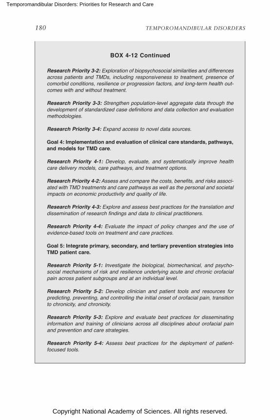
Temporomandibular Disorders: Priorities for Research and Care
Copyright National Academy of Sciences. All rights reserved.
180 TEMPOROMANDIBULAR DISORDERS
Research Priority 3-2: Exploration of biopsychosocial similarities and differences across patients and TMDs, including responsiveness to treatment, presence of comorbid conditions, resilience or progression factors, and longterm health outcomes with and without treatment.
Research Priority 3-3: Strengthen populationlevel aggregate data through the development of standardized case definitions and data collection and evaluation methodologies.
Research Priority 3-4: Expand access to novel data sources.
Goal 4: Implementation and evaluation of clinical care standards, pathways, and models for TMD care.
Research Priority 4-1: Develop, evaluate, and systematically improve health care delivery models, care pathways, and treatment options.
Research Priority 4-2: Assess and compare the costs, benefits, and risks associated with TMD treatments and care pathways as well as the personal and societal impacts on economic productivity and quality of life.
Research Priority 4-3: Explore and assess best practices for the translation and dissemination of research findings and data to clinical practitioners.
Research Priority 4-4: Evaluate the impact of policy changes and the use of evidencebased tools on treatment and care practices.
Goal 5: Integrate primary, secondary, and tertiary prevention strategies into TMD patient care.
Research Priority 5-1: Investigate the biological, biomechanical, and psychosocial mechanisms of risk and resilience underlying acute and chronic orofacial pain across patient subgroups and at an individual level.
Research Priority 5-2: Develop clinician and patient tools and resources for predicting, preventing, and controlling the initial onset of orofacial pain, transition to chronicity, and chronicity.
Research Priority 5-3: Explore and evaluate best practices for disseminating information and training of clinicians across all disciplines about orofacial pain and prevention and care strategies.
Research Priority 5-4: Assess best practices for the deployment of patientfocused tools.
BOX 4-12 Continued
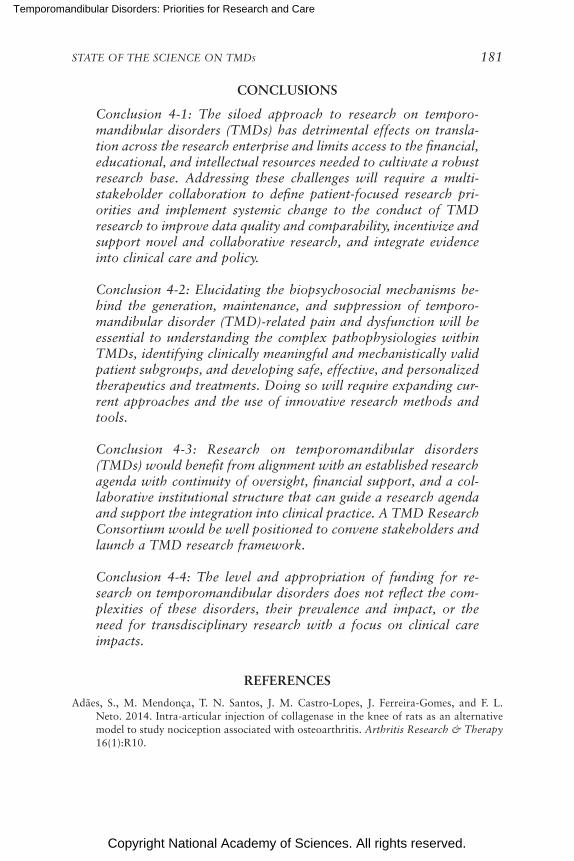
Temporomandibular Disorders: Priorities for Research and Care
Copyright National Academy of Sciences. All rights reserved.
STATE OF THE SCIENCE ON TMDs 181
CONCLUSIONS
Conclusion 4-1: The siloed approach to research on temporo-mandibular disorders (TMDs) has detrimental effects on transla-tion across the research enterprise and limits access to the financial, educational, and intellectual resources needed to cultivate a robust research base. Addressing these challenges will require a multi-stakeholder collaboration to define patient-focused research pri-orities and implement systemic change to the conduct of TMD research to improve data quality and comparability, incentivize and support novel and collaborative research, and integrate evidence into clinical care and policy.
Conclusion 4-2: Elucidating the biopsychosocial mechanisms be-hind the generation, maintenance, and suppression of temporo-mandibular disorder (TMD)-related pain and dysfunction will be essential to understanding the complex pathophysiologies within TMDs, identifying clinically meaningful and mechanistically valid patient subgroups, and developing safe, effective, and personalized therapeutics and treatments. Doing so will require expanding cur-rent approaches and the use of innovative research methods and tools.
Conclusion 4-3: Research on temporomandibular disorders (TMDs) would benefit from alignment with an established research agenda with continuity of oversight, financial support, and a col-laborative institutional structure that can guide a research agenda and support the integration into clinical practice. A TMD Research Consortium would be well positioned to convene stakeholders and launch a TMD research framework.
Conclusion 4-4: The level and appropriation of funding for re-search on temporomandibular disorders does not reflect the com-plexities of these disorders, their prevalence and impact, or the need for transdisciplinary research with a focus on clinical care impacts.
REFERENCES
Adães, S., M. Mendonça, T. N. Santos, J. M. Castro-Lopes, J. Ferreira-Gomes, and F. L. Neto. 2014. Intra-articular injection of collagenase in the knee of rats as an alternative model to study nociception associated with osteoarthritis. Arthritis Research & Therapy 16(1):R10.
Temporomandibular Disorders: Priorities for Research and Care
Copyright National Academy of Sciences. All rights reserved.
182 TEMPOROMANDIBULAR DISORDERS
Adler, G. K., V. F. Manfredsdottir, and K. W. Creskoff. 2002. Neuroendocrine abnormalities in fibromyalgia. Current Pain Headache Reports 6(4):289–298.
Alger, J. R., B. M. Ellingson, C. Ashe-McNalley, D. C. Woodworth, J. S. Labus, M. Farmer, L. Huang, A. V. Apkarian, K. A. Johnson, S. C. Mackey, T. J. Ness, G. Deutsch, R. E. Harris, D. J. Clauw, G. H. Glover, T. B. Parrish, J. Hollander, J. W. Kusek, C. Mullins, E. A. Mayer, and MAPP Research Network Investigators. 2016. Multisite, multimodal neuroimaging of chronic urological pelvic pain: Methodology of the MAPP Research Network. Neuroimage Clincal 12:65–77.
Almarza, A. J., B. N. Brown, B. Arzi, D. F. Angelo, W. Chung, S. F. Badylak, and M. Detamore. 2018. Preclinical animal models for temporomandibular joint tissue engineering. Tissue Engineering—Part B, Reviews 24(3):171–178.
Almarza, A., L. Mercuri, B. Arzi, L. M. Gallo, E. Granquist, S. Kapila, and M. Detamore. 2019. State of TMJ bioengineering: Working together toward improving clinical out-comes. Journal of Biomechanical Engineering [Epub ahead of print].
Alvarez, P., P. G. Green, and J. D. Levine. 2013. Stress in the adult rat exacerbates muscle pain induced by early-life stress. Biological Psychiatry 74(9):688–695.
Amaya, F., H. Wang, M. Costigan, A. J. Allchorne, J. P. Hatcher, J. Egerton, T. Stean, V. Morisset, D. Grose, M. J. Gunthorpe, I. P. Chessell, S. Tate, P. J. Green, and C. J. Woolf. 2006. The voltage-gated sodium channel Na(v)1.9 is an effector of peripheral inflamma-tory pain hypersensitivity. Journal of Neuroscience 26(50):12852–12860.
Angelo, D. F., P. Morouco, N. Alves, T. Viana, F. Santos, R. Gonzalez, F. Monje, D. Macias, B. Carrapico, R. Sousa, S. Cavaco-Goncalves, F. Salvado, C. Peleteiro, and M. Pinho. 2016. Choosing sheep (Ovis aries) as animal model for temporomandibular joint re-search: Morphological, histological and biomechanical characterization of the joint disc. Morphologie 100(331):223–233.
Araújo-Filho, H. G., E. W. M. Pereira, A. R. Campos, L. J. Quintans-Júnior, and J. S. S. Quintans. 2018. Chronic orofacial pain animal models—Progress and challenges. Expert Opinion on Drug Discovery 13(10):949–964.
Bagarinao, E., K. A. Johnson, K. T. Martucci, E. Ichesco, M. A. Farmer, J. Labus, T. J. Ness, R. Harris, G. Deutsch, and A. V. Apkarian. 2014. Preliminary structural MRI based brain classification of chronic pelvic pain: A MAPP Network study. Pain 155(12):2502–2509.
Bair, E., S. Gaynor, G. D. Slade, R. Ohrbach, R. B. Fillingim, J. D. Greenspan, R. Dubner, S. B. Smith, L. Diatchenko, and W. Maixner. 2016. Identification of clusters of indi viduals rel-evant to temporomandibular disorders and other chronic pain conditions: The OPPERA study. Pain 157(6):1266–1278.
Baliki, M. N., B. Petre, S. Torbey, K. M. Herrmann, L. Huang, T. J. Schnitzer, H. L. Fields, and A. V. Apkarian. 2012. Corticostriatal functional connectivity predicts transition to chronic back pain. Nature Neuroscience 15(8):1117–1119.
Barad, M. J., T. Ueno, J. Younger, N. Chatterjee, and S. Mackey. 2014. Complex regional pain syndrome is associated with structural abnormalities in pain-related regions of the human brain. Journal of Pain 15(2):197–203.
Beatty, M. W., R. H. Hohl, J. C. Nickel, L. R. Iwasaki, and R. M. Pidaparti. 2008. Mode I and mode III fractures in intermediate zone of full-thickness porcine temporomandibular joint discs. Annals Biomedical Engineering 36(5):801–812.
Bechtold, T. E., C. Saunders, R. S. Decker, H.-B. Um, N. Cottingham, I. Salhab, N. Kurio, P. C. Billings, M. Pacifici, H.-D. Nah, and E. Koyama. 2016. Osteophyte formation and matrix mineralization in a TMJ osteoarthritis mouse model are associated with ectopic hedgehog signaling. Matrix Biology 52–54:339–354.
Bermejo, A., O. Gonzalez, and J. M. Gonzalez. 1993. The pig as an animal model for experi-mentation on the temporomandibular articular complex. Oral Surgery, Oral Medicine, and Oral Pathology 75(1):18–23.
Temporomandibular Disorders: Priorities for Research and Care
Copyright National Academy of Sciences. All rights reserved.
STATE OF THE SCIENCE ON TMDs 183
Bi, R. Y., X. X. Kou, Z. Meng, X. D. Wang, Y. Ding, and Y. H. Gan. 2013. Involvement of trigeminal ganglionic Nav1.7 in hyperalgesia of inflamed temporomandibular joint is dependent on ERK1/2 phosphorylation of glial cells in rats. European Journal of Pain 17(7):983–994.
Bi, R. Y., Z. Meng, P. Zhang, X. D. Wang, Y. Ding, and Y. H. Gan. 2017. Estradiol upregulates voltage-gated sodium channel 1.7 in trigeminal ganglion contributing to hyperalgesia of inflamed TMJ. PLoS One 12(6):e0178589.
Brinkman, D. J., A. S. Ten Hove, M. J. Vervoordeldonk, M. D. Luyer, and W. J. de Jonge. 2019. Neuroimmune interactions in the gut and their significance for intestinal immunity. Cells 8(7):670.
Brodersen, K. H., K. Wiech, E. I. Lomakina, C. S. Lin, J. M. Buhmann, U. Bingel, M. Ploner, K. E. Stephan, and I. Tracey. 2012. Decoding the perception of pain from fMRI using multivariate pattern analysis. Neuroimage 63(3):1162–1170.
Brown, J. E., N. Chatterjee, J. Younger, and S. Mackey. 2011. Towards a physiology-based measure of pain: Patterns of human brain activity distinguish painful from non-painful thermal stimulation. PLoS One 6(9):e24124.
Bueno, C. H., D. D. Pereira, M. P. Pattussi, P. K. Grossi, and M. L. Grossi. 2018. Gender differences in temporomandibular disorders in adult populational studies: A systematic review and meta-analysis. Journal of Oral Rehabilitation 45(9):720–729.
Cady, R. J., J. R. Glenn, K. M. Smith, and P. L. Durham. 2011. Calcitonin gene-related peptide promotes cellular changes in trigeminal neurons and glia implicated in peripheral and central sensitization. Molecular Pain 7:1–12.
Cairns, B. E. 2010. Pathophysiology of TMD pain—basic mechanisms and their implications for pharmacotherapy. Journal of Oral Rehabilitation 37(6):391–410.
Campaniello, M. A., A. M. Harrington, C. M. Martin, L. Ashley Blackshaw, S. M. Brierley, and P. A. Hughes. 2016. Activation of colo-rectal high-threshold afferent nerves by Inter-leukin-2 is tetrodotoxin-sensitive and upregulated in a mouse model of chronic visceral hypersensitivity. Neurogastroenterology & Motility 28(1):54–63.
Castrillon, E. E., M. Ernberg, B. E. Cairns, K. Wang, B. J. Sessle, L. Arendt-Nielsen, and P. Svensson. 2010. Interstitial glutamate concentration is elevated in the masseter mus-cle of myofascial temporomandibular disorder patients. Journal of Orofacial Pain 24(4):350–360.
Chavan, S. S., V. A. Pavlov, and K. J. Tracey. 2017. Mechanisms and therapeutic relevance of neuro-immune communication. Immunity 46(6):927–942.
Chen, X., P. G. Green, and J. D. Levine. 2011. Stress enhances muscle nociceptor activity in the rat. Neuroscience 185:166–173.
Chen, Y., S. H. Williams, A. L. McNulty, J. H. Hong, S. H. Lee, N. E. Rothfusz, P. K. Parekh, C. Moore, I. R. W. Gereau, A. B. Taylor, F. Wang, F. Guilak, and W. Liedtke. 2013. Temporomandibular joint pain: A critical role for Trpv4 in the trigeminal ganglion. Pain 154(8):1295–1304.
Cheng, J. C., A. Rogachov, K. S. Hemington, A. Kucyi, R. L. Bosma, M. A. Lindquist, R. D. Inman, and K. D. Davis. 2018. Multivariate machine learning distinguishes cross- network dynamic functional connectivity patterns in state and trait neuropathic pain. Pain 159(9):1764–1776.
Chiapparini, L., S. Ferraro, L. Grazzi, and G. Bussone. 2010. Neuroimaging in chronic migraine. Neurological Sciences 31(Suppl 1):S19–S22.
Chichorro, J. G., F. Porreca, and B. Sessle. 2017. Mechanisms of craniofacial pain. Cephalalgia 37(7):613–626.
Chin, A. R., J. Gao, Y. Wang, J. M. Taboas, and A. J. Almarza. 2018. Regenerative potential of various soft polymeric scaffolds in the temporomandibular joint condyle. Journal of Oral and Maxillofacial Surgery 76(9):2019–2026.
Temporomandibular Disorders: Priorities for Research and Care
Copyright National Academy of Sciences. All rights reserved.
184 TEMPOROMANDIBULAR DISORDERS
Chinthakanan, S., K. Laosuwan, P. Boonyawong, S. Kumfu, N. Chattipakorn, and S. C. Chattipakorn. 2018. Reduced heart rate variability and increased saliva cortisol in patients with TMD. Archives of Oral Biology 90:125–129.
Clauw, D. J. 2015. Diagnosing and treating chronic musculoskeletal pain based on the under-lying mechanism(s). Best Practice & Research Clinical Rheumatology 29(1):6–19.
Clauw, D. J. 2019. Defining TMD for clinical care. Presentation at the March 28, 2019, public meeting of the National Academies of Sciences, Engineering, and Medicine’s Committee on Temporomandibular Disorders (TMDs): From Research Discoveries to Clinical Treat-ment, Washington, DC.
Coogan, J. S., D.-G. Kim, T. L. Bredbenner, and D. P. Nicolella. 2018. Determination of sex differences of human cadaveric mandibular condyles using statistical shape and trait modeling. Bone 106:35–41.
Coombs, M. C., X. She, T. R. Brown, E. H. Slate, J. S. Lee, and H. Yao. 2019. Temporo-mandibular joint condyle-disc morphometric sexual dimorphisms independent of skull scaling. Journal of Oral and Maxillofacial Surgery 77(11):2245–2257.
Costa, Y. M., P. C. Conti, F. A. de Faria, and L. R. Bonjardim. 2017. Temporomandibular disorders and painful comorbidities: Clinical association and underlying mechanisms. Oral Surgery, Oral Medicine, Oral Pathology, and Oral Radiology 123(3):288–297.
Costigan, M., I. Belfer, R. S. Griffin, F. Dai, L. B. Barrett, G. Coppola, T. Wu, C. Kiselycznyk, M. Poddar, Y. Lu, L. Diatchenko, S. Smith, E. J. Cobos, D. Zaykin, A. Allchorne, E. Gershon, J. Livneh, P. H. Shen, L. Nikolajsen, J. Karppinen, M. Männikkö, A. Kelempisioti, D. Goldman, W. Maixner, D. H. Geschwind, M. B. Max, Z. Seltzer, and C. J. Woolf. 2010. Multiple chronic pain states are associated with a common amino acid-changing allele in KCNS1. Brain 133(9):2519–2527.
Cowen, R., M. K. Stasiowska, H. Laycock, and C. Bantel. 2015. Assessing pain objectively: The use of physiological markers. Anaesthesia 70(7):828–847.
Cowley, A. 2019. TMD research over the years—Gaps, priorities, opportunities. Presentation at the March 28, 2019, public meeting of the National Academies of Sciences, Engi-neering, and Medicine’s Committee on Temporomandibular Disorders (TMDs): From Research Discoveries to Clinical Treatment, Washington, DC.
Cummins, T. R., P. L. Sheets, and S. G. Waxman. 2007. The roles of sodium channels in nociception: Implications for mechanisms of pain. Pain 131(3):243–257.
Davis, K. D., and M. Moayedi. 2013. Central mechanisms of pain revealed through functional and structural MRI. Journal of Neuroimmune Pharmacology 8(3):518–534.
Dawson, A., N. Stensson, B. Ghafouri, B. Gerdle, T. List, P. Svensson, and M. Ernberg. 2016. Dopamine in plasma—A biomarker for myofascial TMD pain? Journal of Headache and Pain 17(1).
de Freitas, L. V., A. C. Lopes, V. B. Piatto, and J. V. Maniglia. 2013. Association of temporo-mandibular dysfunction with the 102T-C polymorphism in the serotonin receptor gene in Brazilian patients. Archive of Medical Science 9(6):1013–1018.
de Sousa, L. M., J. M. Dos Santos Alves, C. da Silva Martins, K. M. A. Pereira, P. Goes, and D. V. Gondim. 2019. Immunoexpression of canonical Wnt and NK-κB signaling pathways in the temporomandibular joint of arthritic rats. Inflammation Research 68(10):889–900.
Del Rivero, T., R. Fischer, F. Yang, K. A. Swanson, and J. R. Bethea. 2019. Tumor necrosis fac-tor receptor 1 inhibition is therapeutic for neuropathic pain in males but not in females. Pain 160(4):922–931.
Detamore, M. S., and K. A. Athanasiou. 2005. Evaluation of three growth factors for TMJ disc tissue engineering. Annals of Biomedical Engineering 33(3):383–390.
Detamore, M. S., K. A. Athanasiou, and J. Mao. 2007. A call to action for bioengineers and dental professionals: Directives for the future of TMJ bioengineering. Annals of Bio-medical Engineering 35(8):1301–1311.
Temporomandibular Disorders: Priorities for Research and Care
Copyright National Academy of Sciences. All rights reserved.
STATE OF THE SCIENCE ON TMDs 185
Diatchenko, L., A. G. Nackley, G. D. Slade, K. Bhalang, I. Belfer, M. B. Max, D. Goldman, and W. Maixner. 2006. Catechol-o-methyltransferase gene polymorphisms are associated with multiple pain-evoking stimuli. Pain 125(3):216–224.
Diers, M., C. Koeppe, E. Diesch, A. M. Stolle, R. Holzl, M. Schiltenwolf, K. van Ackern, and H. Flor. 2007. Central processing of acute muscle pain in chronic low back pain patients: An EEG mapping study. Journal of Clinical Neurophysiology 24(1):76–83.
Donahue, R. P., J. C. Hu, and K. A. Athanasiou. 2019. Remaining hurdles for tissue- engineering the temporomandibular joint disc. Trends in Molecular Medicine 25(3):241–256.
Dong, Y., G. Wu, T. Zhu, H. Chen, Y. Zhu, G. Zhu, F. Han, and H. Zhao. 2017. VEGF promotes cartilage angiogenesis by phospho-ERK1/2 activation of Dll4 signaling in temporo mandibular joint osteoarthritis caused by chronic sleep disturbance in Wistar rats. Oncotarget 8(11):17849–17861.
Doshi, T. L., D. R. Nixdorf, C. M. Campbell, and S. N. Raja. 2020. Biomarkers in temporo-mandibular disorder and trigeminal neuralgia: A conceptual framework for understand-ing chronic pain. Canadian Journal of Pain 4(1):1–18.
Embree, M. C., M. Chen, S. Pylawka, D. Kong, G. M. Iwaoka, I. Kalajzic, H. Yao, C. Shi, D. Sun, T. J. Sheu, D. A. Koslovsky, A. Koch, and J. J. Mao. 2016. Exploiting endogenous fibrocartilage stem cells to regenerate cartilage and repair joint injury. Nature Commu-nications 7:13073.
Erzurumlu, R. S., Y. Murakami, and F. M. Rijli. 2010. Mapping the face in the somatosensory brainstem. Nature Reviews Neuroscience 11(4):252–263.
Farmer, M. A., M. L. Chanda, E. L. Parks, M. N. Baliki, A. V. Apkarian, and A. J. Schaeffer. 2011. Brain functional and anatomical changes in chronic prostatitis/chronic pelvic pain syndrome. Journal of Urology 186(1):117–124.
FDA-NIH (Food and Drug Administration and National Institutes of Health) Biomarker Working Group. 2018. BEST (Biomarkers, EndpointS, and other Tools) resource. https://www.ncbi.nlm.nih.gov/books/NBK326791/pdf/Bookshelf_NBK326791.pdf (accessed January 9, 2020).
Ferber, G. 2002. Biomarkers and proof of concept. Methods and Findings in Experimental and Clinical Pharmacology 24(Suppl C):35–40.
Fillingim, R. B., R. Ohrbach, J. D. Greenspan, C. Knott, R. Dubner, E. Bair, C. Baraian, G. D. Slade, and W. Maixner. 2011. Potential psychosocial risk factors for chronic TMD: Descriptive data and empirically identified domains from the OPPERA case-control study. Journal of Pain 12(11 Suppl):T46–T60.
Gallo, L. M., N. Fankhauser, Y. M. Gonzalez, H. Liu, Y. Liu, J. C. Nickel, and L. R. Iwasaki. 2018. Jaw closing movement and sex differences in temporomandibular joint energy densities. Journal of Oral Rehabilitation 45(2):97–103.
Gao, Y.-J., and R.-R. Ji. 2009. c-Fos and pERK, which is a better marker for neuronal acti-vation and central sensitization after noxious stimulation and tissue injury? The Open Pain Journal 2:11–17.
Gazerani, P., and H. S. H. Vinterhøj. 2016. “Omics”: An emerging field in pain research and management. Future Neurology 11(4):255–265.
Ge, C., F. Mohamed, A. Binrayes, S. Kapila, and R. T. Franceschi. 2018. Selective role of discoidin domain receptor 2 in murine temporomandibular joint development and aging. Journal of Dental Research 97(3):321–328.
Gerstner, G., E. Ichesco, A. Quintero, and T. Schmidt-Wilcke. 2011. Changes in regional gray and white matter volume in patients with myofascial-type temporomandibular disorders: A voxel-based morphometry study. Journal of Orofacial Pain 25(2):99–106.
Giesecke, T., R. H. Gracely, D. A. Williams, M. E. Geisser, F. W. Petzke, and D. J. Clauw. 2005. The relationship between depression, clinical pain, and experimental pain in a chronic pain cohort. Arthritis and Rheumatism 52(5):1577–1584.
Temporomandibular Disorders: Priorities for Research and Care
Copyright National Academy of Sciences. All rights reserved.
186 TEMPOROMANDIBULAR DISORDERS
Gomez-Varela, D., and M. Schmidt. 2018. The proteomics and metabolomics of pain— opportunities for systems medicine. In J. N. Wood, ed. Oxford handbook of the neuro-biology of pain. Oxford, UK: Oxford University Press.
Goo, Y. A., K. Cain, M. Jarrett, L. Smith, J. Voss, E. Tolentino, J. Tsuji, Y. S. Tsai, A. Panchaud, D. R. Goodlett, R. J. Shulman, and M. Heitkemper. 2012. Urinary proteome analysis of irritable bowel syndrome (IBS) symptom subgroups. Journal of Proteome Research 11(12):5650–5662.
Greenspan, J. D., G. D. Slade, E. Bair, R. Dubner, R. B. Fillingim, R. Ohrbach, C. Knott, F. Mulkey, R. Rothwell, and W. Maixner. 2011. Pain sensitivity risk factors for chronic TMD: Descriptive data and empirically identified domains from the OPPERA case con-trol study. Journal of Pain 12(11 Suppl):T61–T74.
Gu, S., W. Wu, C. Liu, L. Yang, C. Sun, W. Ye, X. Li, J. Chen, F. Long, and Y. Chen. 2014. BMPRIA mediated signaling is essential for temporomandibular joint development in mice. PLoS One 9(8).
Gunja, N. J., R. K. Uthamanthil, and K. A. Athanasiou. 2009. Effects of TGF-β1 and hydrostatic pressure on meniscus cell-seeded scaffolds. Biomaterials 30(4):565–573.
Guo, W., H. Wang, S. Zou, F. Wei, R. Dubner, and K. Ren. 2010. Long lasting pain hyper-sensitivity following ligation of the tendon of the masseter muscle in rats: A model of myogenic orofacial pain. Molecular Pain 6:40.
Hagerty, S. L., J. M. Ellingson, T. B. Helmuth, L. C. Bidwell, K. E. Hutchison, and A. D. Bryan. 2019. An overview and proposed research framework for studying co- occurring mental- and physical-health dysfunction. Perspectives on Psychological Science 14(4):633–645.
Harisankar, C. N., B. R. Mittal, A. Bhattacharya, P. Singh, and R. Sen. 2012. Utility of single photon emission computed tomography/computed tomography imaging in evaluation of chronic low back pain. Indian Journal of Nuclear Medicine 27(3):156–163.
Harper, D. E., A. Schrepf, and D. J. Clauw. 2016a. Pain mechanisms and centralized pain in temporomandibular disorders. Journal of Dental Research 95(10):1102–1108.
Harper, D. E., Y. Shah, E. Ichesco, G. E. Gerstner, and S. J. Peltier. 2016b. Multivariate classi-fication of pain-evoked brain activity in temporomandibular disorder. Pain Reports 1(3).
Harte, S. E., A. Schrepf, R. Gallop, G. H. Kruger, H. H. H. Lai, S. Sutcliffe, M. Halvorson, E. Ichesco, B. D. Naliboff, N. Afari, R. E. Harris, J. T. Farrar, F. Tu, J. R. Landis, and D. J. Clauw. 2019. Quantitative assessment of nonpelvic pressure pain sensitivity in urologic chronic pelvic pain syndrome: A MAPP research network study. Pain 160(6):1270–1280.
Hasin, Y., M. Seldin, and A. Lusis. 2017. Multi-omics approaches to disease. Genome Biology 18(1):1–15.
Hawkins, J. L., and P. L. Durham. 2016. Prolonged jaw opening promotes nociception and enhanced cytokine expression. Journal of Oral and Facial Pain and Headache 30(1):34–41.
He, S. S., F. Li, F. Song, S. Wu, J. Y. Chen, N. He, S. J. Zou, X. Q. Huang, S. Lui, Q. Y. Gong, and S. Chen. 2014. Spontaneous neural activity alterations in temporomandibular disorders: A cross-sectional and longitudinal resting-state functional magnetic resonance imaging study. Neuroscience 278:1–10.
He, S., F. Li, T. Gu, H. Ma, X. Li, S. Zou, X. Huang, S. Lui, Q. Gong, and S. Chen. 2018. Reduced corticostriatal functional connectivity in temporomandibular disorders. Human Brain Mapping 39(6):2563–2572.
Heitkemper, M. M., K. C. Cain, R. J. Shulman, R. L. Burr, C. Ko, E. B. Hollister, N. Callen, J. Zia, C. J. Han, and M. E. Jarrett. 2018. Stool and urine trefoil factor 3 levels: Asso-ciations with symptoms, intestinal permeability, and microbial diversity in irritable bowel syndrome. Beneficial Microbes 9(3):345–355.
Temporomandibular Disorders: Priorities for Research and Care
Copyright National Academy of Sciences. All rights reserved.
STATE OF THE SCIENCE ON TMDs 187
Helgeland, E., S. Shanbhag, T. O. Pedersen, K. Mustafa, and A. Rosen. 2018. Scaffold- based temporomandibular joint tissue regeneration in experimental animal models: A systematic review. Tissue Engineering—Part B: Reviews 24(4):300–316.
Herring, S. W., J. D. Decker, Z. J. Liu, and T. Ma. 2002. Temporomandibular joint in miniature pigs: Anatomy, cell replication, and relation to loading. The Anatomical Record 266(3):152–166.
Hinton, R. J. 2014. Genes that regulate morphogenesis and growth of the temporomandibular joint: A review. Developmental Dynamics 243(7):864–874.
Hinton, R. J., J. Jing, and J. Q. Feng. 2015. Genetic influences on temporomandibular joint development and growth. In Y. Chai, ed. Current topics in developmental biology. Vol. 115. Academic Press. Pp. 85–109.
Hockley, J. R. F., R. González-Cano, S. McMurray, M. A. Tejada-Giraldez, C. McGuire, A. Torres, A. L. Wilbrey, V. Cibert-Goton, F. R. Nieto, T. Pitcher, C. H. Knowles, J. M. Baeyens, J. N. Wood, W. J. Winchester, D. C. Bulmer, C. M. Cendán, and G. McMurray. 2017. Visceral and somatic pain modalities reveal NaV1.7-independent visceral nociceptive pathways. Journal of Physiology 595(8):2661–2679.
Hodges, P. W., and G. L. Moseley. 2003. Pain and motor control of the lumbopelvic region: Effect and possible mechanisms. Journal of Electromyography and Kinesiology 13(4):361–370.
Howard, M. A., D. Sanders, K. Krause, J. O’Muircheartaigh, A. Fotopoulou, F. Zelaya, M. Thacker, N. Massat, J. P. Huggins, W. Vennart, E. Choy, M. Daniels, and S. C. Williams. 2012. Alterations in resting-state regional cerebral blood flow demonstrate ongoing pain in osteoarthritis: An arterial spin-labeled magnetic resonance imaging study. Arthritis and Rheumatism 64(12):3936–3946.
Hunziker, R., N. Lumelsky, and F. Wang. 2015. Editorial: Scaffolds for regenerative medicine: A special issue of the Annals of Biomedical Engineering. Annals of Biomedical Enginee-ring 43(3):487–488.
Ibi, M. 2019. Inflammation and temporomandibular joint derangement. Biological and Pharmaceutical Bulletin 42(4):538–542.
IOM (Institute of Medicine). 2011. Relieving pain in America: A blueprint for transforming prevention, care, education, and research. Washington, DC: The National Academies Press.
Isensee, J., L. Krahé, K. Moeller, V. Pereira, J. E. Sexton, X. Sun, E. Emery, J. N. Wood, and T. Hucho. 2017. Synergistic regulation of serotonin and opioid signaling contributes to pain insensitivity in Nav1.7 knockout mice. Science Signaling 10(461):eaah4874.
Ishizuka, Y., Y. Shibukawa, M. Nagayama, R. Decker, T. Kinumatsu, A. Saito, M. Pacifici, and E. Koyama. 2014. TMJ degeneration in SAMP8 mice is accompanied by deranged Ihh signaling. Journal of Dental Research 93(3):281–287.
Ito, G., Y. Suekawa, M. Watanabe, K. Takahashi, T. Inubushi, K. Murasaki, N. Hirose, S. Hiyama, T. Uchida, and K. Tanne. 2013. P2X7 receptor in the trigeminal sensory nuclear complex contributes to tactile allodynia/hyperalgesia following trigeminal nerve injury. European Journal of Pain 17(2):185–199.
Iwasaki, L. R., Y. M. Gonzalez, Y. Liu, H. Liu, M. Markova, L. M. Gallo, and J. C. Nickel. 2017. TMJ energy densities in healthy men and women. Osteoarthritis and Cartilage 25(6):846–849.
Jing, J., R. J. Hinton, Y. Mishina, Y. Liu, X. Zhou, and J. Q. Feng. 2014. Critical role of BMPr1a in mandibular condyle growth. Connective Tissue Research 55(01):73–78.
Temporomandibular Disorders: Priorities for Research and Care
Copyright National Academy of Sciences. All rights reserved.
188 TEMPOROMANDIBULAR DISORDERS
Kairys, A. E., T. Schmidt-Wilcke, T. Puiu, E. Ichesco, J. S. Labus, K. Martucci, M. A. Farmer, T. J. Ness, G. Deutsch, E. A. Mayer, S. Mackey, A. V. Apkarian, K. Maravilla, D. J. Clauw, and R. E. Harris. 2015. Increased brain gray matter in the primary somatosensory cortex is asso ciated with increased pain and mood disturbance in interstitial cystitis/painful bladder syndrome patients. Journal of Urology 193(1):131–137.
Kalpakci, K. N., V. P. Willard, M. E. Wong, and K. A. Athanasiou. 2011. An interspecies com-parison of the temporomandibular joint disc. Journal of Dental Research 90(2):193–198.
Kaneyama, K., N. Segami, M. Nishimura, T. Suzuki, and J. Sato. 2002. Importance of pro-inflammatory cytokines in synovial fluid from 121 joints with temporomandibular disor-ders. British Journal of Oral & Maxillofacial Surgery 40(5):418–423.
Kartha, S., T. Zhou, E. J. Granquist, and B. A. Winkelstein. 2016. Development of a rat model of mechanically induced tunable pain and associated temporomandibular joint responses. Journal of Oral & Maxillofacial Surgery 74(1):54.e1–54.e10.
Kato, K., P. F. Sullivan, B. Evengård, and N. L. Pedersen. 2009. A population-based twin study of functional somatic syndromes. Psychological Medicine 39(3):497–505.
Kavelaars, A., N. Eijkelkamp, H. L. D. M. Willemen, H. Wang, A. G. Carbajal, and C. J. Heijnen. 2011. Microglial GRK2: A novel regulator of transition from acute to chronic pain. Brain, Behavior, and Immunity 25(6):1055–1060.
Kobayashi, K., R. Jokaji, M. Miyazawa-Hira, S. Takatsuka, A. Tanaka, K. Ooi, H. Nakamura, and S. Kawashiri. 2017. Elastin-derived peptides are involved in the processes of human temporomandibular disorder by inducing inflammatory responses in synovial cells. Molecular Medicine Reports 16(3):3147–3154.
Komori, T. 2020. Molecular mechanism of Runx2-dependent bone development. Molecules and Cells 1–8.
Kothari, S. F., L. Baad-Hansen, Y. Oono, and P. Svensson. 2015. Somatosensory assessment and conditioned pain modulation in temporomandibular disorders pain patients. Pain 156(12):2545–2555.
Koyama, E., C. Saunders, I. Salhab, R. S. Decker, I. Chen, H. Um, M. Pacifici, and H. D. Nah. 2014. Lubricin is required for the structural integrity and post-natal maintenance of TMJ. Journal of Dental Research 93(7):663–670.
Krzyzanowska, A., and C. Avendaño. 2012. Behavioral testing in rodent models of orofacial neuropathic and inflammatory pain. Brain and Behavior 2(5):678–697.
Kurio, N., C. Saunders, T. E. Bechtold, I. Salhab, H. D. Nah, S. Sinha, P. C. Billings, M. Pacifici, and E. Koyama. 2018. Roles of Ihh signaling in chondroprogenitor function in postnatal condylar cartilage. Matrix Biology 67:15–31.
Kutch, J. J., E. Ichesco, J. P. Hampson, J. S. Labus, M. A. Farmer, K. T. Martucci, T. J. Ness, G. Deutsch, A. V. Apkarian, S. C. Mackey, D. J. Klumpp, A. J. Schaeffer, L. V. Rodriguez, K. J. Kreder, D. Buchwald, G. L. Andriole, H. H. Lai, C. Mullins, J. W. Kusek, J. R. Landis, E. A. Mayer, J. Q. Clemens, D. J. Clauw, R. E. Harris, and MAPP Research Network. 2017a. Brain signature and functional impact of centralized pain: A multidisciplinary approach to the study of chronic pelvic pain (MAPP) network study. Pain 158(10):1979–1991.
Kutch, J. J., J. S. Labus, R. E. Harris, K. T. Martucci, M. A. Farmer, S. Fenske, C. Fling, E. Ichesco, S. Peltier, B. Petre, W. Guo, X. Hou, A. J. Stephens, C. Mullins, D. J. Clauw, S. C. Mackey, A. V. Apkarian, J. R. Landis, E. A. Mayer, and Mapp Research Network. 2017b. Resting-state functional connectivity predicts longitudinal pain symptom change in urologic chronic pelvic pain syndrome: A MAPP network study. Pain 158(6):1069–1082.
Lam, D. K., B. J. Sessle, and J. W. Hu. 2009. Glutamate and capsaicin effects on trigeminal nociception II: Activation and central sensitization in brainstem neurons with deep cranio facial afferent input. Brain Research 1253:48–59.
Temporomandibular Disorders: Priorities for Research and Care
Copyright National Academy of Sciences. All rights reserved.
STATE OF THE SCIENCE ON TMDs 189
Langford, D. J., A. L. Bailey, M. L. Chanda, S. E. Clarke, T. E. Drummond, S. Echols, S. Glick, J. Ingrao, T. Klassen-Ross, M. L. Lacroix-Fralish, L. Matsumiya, R. E. Sorge, S. G. Sotocinal, J. M. Tabaka, D. Wong, A. M. van den Maagdenberg, M. D. Ferrari, K. D. Craig, and J. S. Mogil. 2010. Coding of facial expressions of pain in the laboratory mouse. Nature Methods 7(6):447–449.
LeResche, L., L. Mancl, J. J. Sherman, B. Gandara, and S. F. Dworkin. 2003. Changes in temporomandibular pain and other symptoms across the menstrual cycle. Pain 106(3):253–261.
Leung, A., N. S. Gregory, L. A. Allen, and K. A. Sluka. 2016. Regular physical activity pre-vents chronic pain by altering resident muscle macrophage phenotype and increasing interleukin-10 in mice. Pain 157(1):70–79.
Levine, J. E. 2012. Chapter 1, an introduction to neuroendocrine systems. In G. Fink, D. W. Pfaff, and J. E. Levine, eds. Handbook of neuroendocrinology. San Diego, CA: Academic Press. Pp. 3–19.
Levy, D., L. Abdian, M. Dekel-Steinkeller, and R. Defrin. 2018. Experimental evidence for weaker endogenous inhibition of trigeminal pain than extra-trigeminal pain in healthy individuals. Cephalalgia 38(7):1307–1315.
Liao, L., S. Zhang, G. Q. Zhou, L. Ye, J. Huang, L. Zhao, and D. Chen. 2019. Deletion of Runx2 in condylar chondrocytes disrupts TMJ tissue homeostasis. Journal of Cellular Physiology 234(4):3436–3444.
Lin, C. S. 2014. Brain signature of chronic orofacial pain: A systematic review and meta- analysis on neuroimaging research of trigeminal neuropathic pain and temporo-mandibular joint disorders. PLoS One 9(4):e94300.
Liu, X., N. Li, H. Zhang, J. Liu, N. Zhou, C. Ran, X. Chen, Y. Lu, X. Wang, C. Qin, J. Xiao, and C. Liu. 2018. Inactivation of Fam20b in the neural crest-derived mesenchyme of mouse causes multiple craniofacial defects. European Journal of Oral Sciences 126(5):433–436.
Long, H. Q., P. F. Tian, Y. X. Guan, L. X. Liu, X. P. Wu, and B. Li. 2019. Expression of Ihh signaling pathway in condylar cartilage after bite-raising in adult rats. Journal of Molecular Histology 50(5):459–470.
Lopes, D. M., F. Denk, and S. B. McMahon. 2017. The molecular fingerprint of dorsal root and trigeminal ganglion neurons. Frontiers in Molecular Neuroscience 10:1–11.
Lopez-Sola, M., C. W. Woo, J. Pujol, J. Deus, B. J. Harrison, J. Monfort, and T. D. Wager. 2017. Towards a neurophysiological signature for fibromyalgia. Pain 158(1):34–47.
Lötsch, J., and A. Ultsch. 2018. Machine learning in pain research. Pain 159(4):623–630.Luo, X., Y. Jiang, R. Bi, N. Jiang, and S. Zhu. 2018. Inhibition of notch signaling pathway
temporally postpones the cartilage degradation progress of temporomandibular joint arthritis in mice. Journal of Cranio-Maxillofacial Surgery 46(7):1132–1138.
Ma, F., L. Zhang, D. Lyons, and K. N. Westlund. 2012. Orofacial neuropathic pain mouse model induced by trigeminal inflammatory compression (TIC) of the infraorbital nerve. Molecular Brain 5:44.
MacBarb, R. F., A. L. Chen, J. C. Hu, and K. A. Athanasiou. 2013. Engineering functional anisotropy in fibrocartilage neotissues. Biomaterials 34(38):9980–9989.
Mackey, S. 2014. National Pain Strategy Task Force: The strategic plan for the IOM pain report. Pain Medicine 15(7):1070–1071.
Mackey, S. 2016. Future directions for pain management: Lessons from the Institute of Medi-cine pain report and the National Pain Strategy. Hand Clinics 32(1):91–98.
Maes, M. 1999. Major depression and activation of the inflammatory response system. Advances in Experimental Medicine and Biology 461:25–46.
Temporomandibular Disorders: Priorities for Research and Care
Copyright National Academy of Sciences. All rights reserved.
190 TEMPOROMANDIBULAR DISORDERS
Maes, M., C. Song, A. Lin, R. De Jongh, A. Van Gastel, G. Kenis, E. Bosmans, I. De Meester, I. Benoy, H. Neels, P. Demedts, A. Janca, S. Scharpe, and R. S. Smith. 1998. The effects of psychological stress on humans: Increased production of pro-inflammatory cytokines and a Th1-like response in stress-induced anxiety. Cytokine 10(4):313–318.
Marquand, A., M. Howard, M. Brammer, C. Chu, S. Coen, and J. Mourao-Miranda. 2010. Quantitative prediction of subjective pain intensity from whole-brain fMRI data using Gaussian processes. Neuroimage 49(3):2178–2189.
Martínez-García, M.-Á., B. C. Migueláñez-Medrán, and C. Goicoechea. 2019. Animal models in the study and treatment of orofacial pain. Journal of Clinical and Experimental Dentistry 11(4):e382–e390.
Martucci, K. T., and S. C. Mackey. 2016. Imaging pain. Anesthesiology Clinics 34(2):255–269.Martucci, K. T., and S. C. Mackey. 2018. Neuroimaging of pain: Human evidence and
clinical relevance of central nervous system processes and modulation. Anesthesiology 128(6):1241–1254.
Martucci, K. T., K. A. Weber, 2nd, and S. C. Mackey. 2019. Altered cervical spinal cord resting-state activity in fibromyalgia. Arthritis & Rheumatology 71(3):441–450.
Matsumoto, K., K. Honda, M. Ohshima, Y. Yamaguchi, I. Nakajima, P. Micke, and K. Otsuka. 2006. Cytokine profile in synovial fluid from patients with internal derangement of the temporomandibular joint: A preliminary study. Dentomaxillofacial Radiology 35(6):432–441.
McLean, S. A. 2011. The potential contribution of stress systems to the transition to chronic whiplash-associated disorders. Spine 36(25 Suppl):S226–S232.
MDEpiNet TMJ Patient-Led RoundTable. 2018a. A national interagency temporo mandibular disorder research plan: MDEpiNet TMJ Patient-Led RoundTable. http://mdepinet.org/wp-content/uploads/A-Priority-Listing-of-Research-Recommendations-Agency- Assignments_Final.pdf (accessed January 27, 2020).
MDEpiNet TMJ Patient-Led RoundTable. 2018b. The TMJ Patient-Led RoundTable: A history and summary of work. http://mdepinet.org/wp-content/uploads/TMJ-Patient- RoundTable-Briefing-Report_9_25_18.pdf (accessed January 27, 2020).
Meloto, C. B., P. O. Serrano, M. C. Ribeiro-DaSilva, and C. M. Rizzatti-Barbosa. 2011. Genomics and the new perspectives for temporomandibular disorders. Archives of Oral Biology 56(11):1181–1191.
Meloto, C. B., R. Benavides, R. N. Lichtenwalter, X. Wen, N. Tugarinov, K. Zorina- Lichtenwalter, A.-J. Chabot-Doré, M. H. Piltonen, S. Cattaneo, V. Verma, R. Klares, 3rd, S. Khoury, M. Parisien, and L. Diatchenko. 2018. Human Pain Genetics Database: A resource dedicated to human pain genetics research. Pain 159(4):749–763.
Melville, J. C., V. A. Manon, C. Blackburn, and S. Young. 2019. Current methods of maxillo facial tissue engineering. Oral Maxillofacial Surgery Clinics of North America 31(4):579–591.
Moana-Filho, E. J., and A. Herrero Babiloni. 2019. Endogenous pain modulation in chronic temporomandibular disorders: Derivation of pain modulation profiles and assess-ment of its relationship with clinical characteristics. Journal of Oral Rehabilitation 46(3):219–232.
Moana-Filho, E. J., A. Herrero Babiloni, and N. R. Theis-Mahon. 2018. Endogenous pain modulation in chronic orofacial pain: A systematic review and meta-analysis. Pain 159(8):1441–1455.
Moayedi, M., I. Weissman-Fogel, A. P. Crawley, M. B. Goldberg, B. V. Freeman, H. C. Tenenbaum, and K. D. Davis. 2011. Contribution of chronic pain and neuroticism to abnormal forebrain gray matter in patients with temporomandibular disorder. Neuro image 55(1):277–286.
Temporomandibular Disorders: Priorities for Research and Care
Copyright National Academy of Sciences. All rights reserved.
STATE OF THE SCIENCE ON TMDs 191
Moayedi, M., I. Weissman-Fogel, T. V. Salomons, A. P. Crawley, M. B. Goldberg, B. V. Freeman, H. C. Tenenbaum, and K. D. Davis. 2012. White matter brain and trigeminal nerve abnormalities in temporomandibular disorder. Pain 153(7):1467–1477.
Mogil, J. S. 2012. Pain genetics: Past, present and future. Trends in Genetics 28(6):258–266.Moisset, X., and D. Bouhassira. 2007. Brain imaging of neuropathic pain. Neuroimage
37(Suppl 1):S80–S88.Moloney, R. D., J. Sajjad, T. Foley, V. D. Felice, T. G. Dinan, J. F. Cryan, and S. M. O’Mahony.
2016. Estrous cycle influences excitatory amino acid transport and visceral pain sensitiv-ity in the rat: Effects of early-life stress. Biology of Sex Differences 7:33.
Monaco, A., R. Cattaneo, L. Mesin, E. Ortu, M. Giannoni, and D. Pietropaoli. 2015. Dys-regulation of the descending pain system in temporomandibular disorders revealed by low-frequency sensory transcutaneous electrical nerve stimulation: A pupillometric study. PLoS One 10(4):e0122826.
Mori-Akiyama, Y., H. Akiyama, D. H. Rowitch, and B. de Crombrugghe. 2003. Sox9 is required for determination of the chondrogenic cell lineage in the cranial neural crest. Proceedings of the National Academy of Sciences 100(16):9360–9365.
Mupparapu, M., S. Oak, Y. C. Chang, and A. Alavi. 2019. Conventional and functional im-aging in the evaluation of temporomandibular joint rheumatoid arthritis: A systematic review. Quintessence International 50(9):742–753.
Mutso, A. A., B. Petre, L. Huang, M. N. Baliki, S. Torbey, K. M. Herrmann, T. J. Schnitzer, and A. V. Apkarian. 2014. Reorganization of hippocampal functional connectivity with transition to chronic back pain. Journal of Neurophysiology 111(5):1065–1076.
Nackley, A. G., K. S. Tan, K. Fecho, P. Flood, L. Diatchenko, and W. Maixner. 2007. Catechol-O-methyltransferase inhibition increases pain sensitivity through activation of both beta2- and beta3-adrenergic receptors. Pain 128(3):199–208.
Nagakura, Y. 2017. The need for fundamental reforms in the pain research field to develop innovative drugs. Expert Opinion on Drug Discovery 12(1):39–46.
Nash, P., K. Wiley, J. Brown, R. Shinaman, D. Ludlow, A.-M. Sawyer, G. Glover, and S. Mackey. 2013. Functional magnetic resonance imaging identifies somatotopic organiza-tion of nociception in the human spinal cord. Pain 154(6):776–781.
Nassar, M. A., L. C. Stirling, G. Forlani, M. D. Baker, E. A. Matthews, A. H. Dickenson, and J. N. Wood. 2004. Nociceptor-specific gene deletion reveals a major role for Nav1.7 (PN1) in acute and inflammatory pain. Proceedings of the National Academy of Sciences 101(34):12706–12711.
Neubert, J. K., C. G. Widmer, W. Malphurs, H. L. Rossi, C. J. Vierck, Jr., and R. M. Caudle. 2005. Use of a novel thermal operant behavioral assay for characterization of orofacial pain sensitivity. Pain 116(3):386–395.
Nickel, J. C., D. A. Tripp, L. K. Carr, M. Pontari, R. Moldwin, R. Mayer, R. Doggweiler, C. C. Yang, N. Mishra, and J. Nordling. 2015. Clinical and psychological parameters associated with pain pattern phenotypes in women with interstitial cystitis/bladder pain syndrome. Journal of Urology 193(1):138–144.
Nickel, J. C., L. R. Iwasaki, Y. M. Gonzalez, L. M. Gallo, and H. Yao. 2018. Mechano-behavior and ontogenesis of the temporomandibular joint. Journal of Dental Research 97(11):1185–1192.
NIH (National Institutes of Health). 2019a. Estimates of funding for various research, condi-tion, and disease categories (RCDC). https://report.nih.gov/categorical_spending.aspx (accessed January 14, 2020).
NIH. 2019b. Types of grant programs. https://grants.nih.gov/grants/funding/funding_program.htm (accessed January 31, 2020).
Temporomandibular Disorders: Priorities for Research and Care
Copyright National Academy of Sciences. All rights reserved.
192 TEMPOROMANDIBULAR DISORDERS
Ochiai, T., Y. Shibukawa, M. Nagayama, C. Mundy, T. Yasuda, T. Okabe, K. Shimono, M. Kanyama, H. Hasegawa, Y. Maeda, B. Lanske, M. Pacifici, and E. Koyama. 2010. Indian hedgehog roles in post-natal TMJ development and organization. Journal of Dental Research 89(4):349–354.
Ohrbach, R., and S. F. Dworkin. 1998. Five-year outcomes in TMD: Relationship of changes in pain to changes in physical and psychological variables. Pain 74(2-3):315–326.
Ojima, K., N. Watanabe, N. Narita, and M. Narita. 2007. Temporomandibular disorder is associated with a serotonin transporter gene polymorphism in the Japanese population. Biopsychosocial Medicine 1:3.
Oka, K., S. Oka, R. Hosokawa, P. Bringas, Jr., H. C. Brockhoff, 2nd, K. Nonaka, and Y. Chai. 2008. TGF-beta mediated Dlx5 signaling plays a crucial role in osteo- chondroprogenitor cell lineage determination during mandible development. Developmental Biology 321(2):303–309.
Olausson, P., B. Gerdle, N. Ghafouri, D. Sjöström, E. Blixt, and B. Ghafouri. 2015. Protein alterations in women with chronic widespread pain—An explorative proteomic study of the trapezius muscle. Scientific Reports 5(1):11894.
Ozdiler, O., K. Orhan, E. Cesur, A. Koklu, and O. Algin. 2019. Evaluation of temporo-mandibular joint, masticatory muscle, and brain cortex activity in patients treated by removable functional appliances: A prospective fMRI study. Dentomaxillofacial Radio-logy 48(7).
Patti, G. J., O. Yanes, L. P. Shriver, J.-P. Courade, R. Tautenhahn, M. Manchester, and G. Siuzdak. 2012. Metabolomics implicates altered sphingolipids in chronic pain of neuro-pathic origin. Nature Chemical Biology 8(3):232–234.
Payne, S. C., J. B. Furness, and M. J. Stebbing. 2019. Bioelectric neuromodulation for gastro-intestinal disorders: Effectiveness and mechanisms. Nature Reviews Gastroenterology & Hepatology 16(2):89–105.
Powers, C. M. 2010. The influence of abnormal hip mechanics on knee injury: A bio-mechanical perspective. Journal of Orthopaedic & Sports Physical Therapy 40(2):42–51.
Purcell, P., A. Jheon, M. P. Vivero, H. Rahimi, A. Joo, and O. D. Klein. 2012. Spry1 and Spry2 are essential for development of the temporomandibular joint. Journal of Dental Research 91(4):387–393.
Puri, J., P. Vinothini, J. Reuben, L. L. Bellinger, L. Ailing, Y. B. Peng, and P. R. Kramer. 2012. Reduced GABAA receptor α6 expression in the trigeminal ganglion alters inflammatory TMJ hypersensitivity. Neuroscience 213:179–190.
Reiter, S., A. Emodi-Perlman, C. Goldsmith, P. Friedman-Rubin, and E. Winocur. 2015. Co-morbidity between depression and anxiety in patients with temporomandibular disorders according to the research diagnostic criteria for temporomandibular disorders. Journal of Oral & Facial Pain and Headache 29(2):135–143.
Ribeiro-Dasilva, M. C., S. R. Peres Line, M. C. Leme Godoy dos Santos, M. T. Arthuri, W. Hou, R. B. Fillingim, and C. M. Rizzatti Barbosa. 2009. Estrogen receptor-alpha poly-morphisms and predisposition to TMJ disorder. Journal of Pain 10(5):527–533.
Ribeiro-Dasilva, M. C., R. B. Fillingim, and S. M. Wallet. 2017. Estrogen-induced monocytic response correlates with temporomandibular disorder pain. Journal of Dental Research 96(3):285–291.
Roberts, W. E., and D. L. Stocum. 2018. Part II: Temporomandibular joint (TMJ)- regeneration, degeneration, and adaptation. Current Osteoporosis Reports 16(4):369–379.
Rogers, A. W., S. E. Cisewski, and C. B. Kern. 2018. The zonal architecture of the mandibular condyle requires ADAMTS5. Journal of Dental Research 97(12):1383–1390.
Romero-Reyes, M., S. Akerman, E. Nguyen, A. Vijjeswarapu, B. Hom, H.-W. Dong, and A. C. Charles. 2013. Spontaneous behavioral responses in the orofacial region: A model of trigeminal pain in mouse. Headache 53(1):137–151.
Temporomandibular Disorders: Priorities for Research and Care
Copyright National Academy of Sciences. All rights reserved.
STATE OF THE SCIENCE ON TMDs 193
Romero-Reyes, M., V. Pardi, and S. Akerman. 2015. A potent and selective calcitonin gene-related peptide (CGRP) receptor antagonist, MK-8825, inhibits responses to nociceptive trigeminal activation: Role of CGRP in orofacial pain. Experimental Neurology 271:95–103.
Ronald, S., and D. K. Mills. 2016. Fibrochondrocyte growth and functionality on TiO2 nanothin films. Journal of Functional Biomaterials 7(2):14.
Russo, M., P. Georgius, and D. M. Santarelli. 2018. A new hypothesis for the pathophysiology of complex regional pain syndrome. Medical Hypotheses 119:41–53.
Sannajust, S., I. Imbert, V. Eaton, T. Henderson, L. Liaw, M. May, M. F. Barbe, and T. King. 2019. Females have greater susceptibility to develop ongoing pain and central sensitiza-tion in a rat model of temporomandibular joint pain. Pain 160(9):2036–2049.
Scariot, R., P. Corso, A. M. Sebastiani, and A. R. Vieira. 2018. The many faces of genetic contributions to temporomandibular joint disorder: An updated review. Orthodontics & Craniofacial Research 21(4):186–201.
Schrepf, A., D. E. Harper, S. E. Harte, H. Wang, E. Ichesco, J. P. Hampson, J. K. Zubieta, D. J. Clauw, and R. E. Harris. 2016. Endogenous opioidergic dysregulation of pain in fibromyalgia: A PET and fMRI study. Pain 157(10):2217–2225.
Schwenkreis, P., C. Maier, and M. Tegenthoff. 2009. Functional imaging of central nervous system involvement in complex regional pain syndrome. American Journal of Neuro-radiology 30(7):1279–1284.
Sessle, B. J. 2011. Peripheral and central mechanisms of orofacial inflammatory pain. Inter-national Review of Neurobiology 97:179–206.
Shen, P., Z. Jiao, J. S. Zheng, W. F. Xu, S. Y. Zhang, A. Qin, and C. Yang. 2015. Injecting vas-cular endothelial growth factor into the temporomandibular joint induces osteoarthritis in mice. Scientific Reports 5(1):16244.
Sherman, J. J., L. LeResche, K. H. Huggins, L. A. Mancl, J. C. Sage, and S. F. Dworkin. 2004. The relationship of somatization and depression to experimental pain response in women with temporomandibular disorders. Psychosomatic Medicine 66(6):852–860.
Shibata, S., N. Suda, S. Yoda, H. Fukuoka, K. Ohyama, Y. Yamashita, and T. Komori. 2004. Runx2-deficient mice lack mandibular condylar cartilage and have deformed Meckel’s cartilage. Anatomy and Embryology 208(4):273–280.
Shibukawa, Y., T. Ishikawa, Y. Kato, Z. K. Zhang, T. Jiang, M. Shintani, M. Shimono, T. Kumai, T. Suzuki, M. Kato, and Y. Nakamura. 2007. Cerebral cortical dysfunction in patients with temporomandibular disorders in association with jaw movement observa-tion. Pain 128(1–2):180–188.
Shibuta, K., I. Suzuki, M. Shinoda, Y. Tsuboi, K. Honda, N. Shimizu, B. J. Sessle, and K. Iwata. 2012. Organization of hyperactive microglial cells in trigeminal spinal subnucleus caudalis and upper cervical spinal cord associated with orofacial neuropathic pain. Brain Research 1451:74–86.
Skouen, J. S., A. J. Smith, N. M. Warrington, P. B. O’ Sullivan, L. McKenzie, C. E. Pennell, and L. M. Straker. 2012. Genetic variation in the beta-2 adrenergic receptor is associ-ated with chronic musculoskeletal complaints in adolescents. European Journal of Pain 16(9):1232–1242.
Slade, G. D., M. S. Conrad, L. Diatchenko, N. U. Rashid, S. Zhong, S. Smith, J. Rhodes, A. Medvedev, S. Makarov, W. Maixner, and A. G. Nackley. 2011. Cytokine biomarkers and chronic pain: Association of genes, transcription, and circulating proteins with temporo-mandibular disorders and widespread palpation tenderness. Pain 152(12):2802–2812.
Sluka, K. A., and D. J. Clauw. 2016. Neurobiology of fibromyalgia and chronic widespread pain. Neuroscience 338:114–129.
Sluka, K. A., L. Frey-Law, and M. Hoeger Bement. 2018. Exercise-induced pain and analgesia? Underlying mechanisms and clinical translation. Pain 159(Suppl 1):S91–S97.
Temporomandibular Disorders: Priorities for Research and Care
Copyright National Academy of Sciences. All rights reserved.
194 TEMPOROMANDIBULAR DISORDERS
Smith, S. B., D. W. Maixner, J. D. Greenspan, R. Dubner, R. B. Fillingim, R. Ohrbach, C. Knott, G. D. Slade, E. Bair, D. G. Gibson, D. V. Zaykin, B. S. Weir, W. Maixner, and L. Diatchenko. 2011. Potential genetic risk factors for chronic TMD: Genetic associations from the OPPERA case control study. Journal of Pain 12(11 Suppl):T92–T101.
Smith, S. B., E. Mir, E. Bair, G. D. Slade, R. Dubner, R. B. Fillingim, J. D. Greenspan, R. Ohrbach, C. Knott, B. Weir, W. Maixner, and L. Diatchenko. 2013. Genetic variants associated with development of TMD and its intermediate phenotypes: The genetic architecture of TMD in the OPPERA prospective cohort study. Journal of Pain 14(12 Suppl):T91–T101.
Smith, S. M., and W. W. Vale. 2006. The role of the hypothalamic-pituitary-adrenal axis in neuroendocrine responses to stress. Dialogues in Clinical Neuroscience 8(4):383–395.
Somoza, R. A., J. F. Welter, D. Correa, and A. I. Caplan. 2014. Chondrogenic differentiation of mesenchymal stem cells: Challenges and unfulfilled expectations. Tissue Engineering—Part B, Reviews 20(6):596–608.
Sperry, M. M., M. E. Ita, S. Kartha, S. Zhang, Y. H. Yu, and B. Winkelstein. 2017. The inter-face of mechanics and nociception in joint pathophysiology: Insights from the facet and temporomandibular joints. Journal of Biomechanical Engineering 139(2).
Staniszewski, K., H. Lygre, E. Bifulco, S. Kvinnsland, L. Willassen, E. Helgeland, T. Berge, and A. Rosén. 2018. Temporomandibular disorders related to stress and HPA-axis regulation. Pain Research & Management 2018:1–7.
Staud, R. 2011a. Brain imaging in fibromyalgia syndrome. Clinical and Experimental Rheu-matology 29(6 Suppl 69):S109–S117.
Staud, R. 2011b. Evidence for shared pain mechanisms in osteoarthritis, low back pain, and fibromyalgia. Current Rheumatology Reports 13(6):513–520.
Stocum, D. L., and W. E. Roberts. 2018. Part I: Development and physiology of the temporo-mandibular joint. Current Osteoporosis Reports 16(4):360–368.
Tamura, R., T. Nemoto, T. Maruta, S. Onizuka, T. Yanagita, A. Wada, M. Murakami, and I. Tsuneyoshi. 2014. Up-regulation of Nav1.7 sodium channels expression by tumor necrosis factor-α in cultured bovine adrenal chromaffin cells and rat dorsal root ganglion neurons. Anesthesia and Analgesia 118(2):318–324.
Tanaka, E., J. Aoyama, M. Miyauchi, T. Takata, K. Hanaoka, T. Iwabe, and K. Tanne. 2005. Vascular endothelial growth factor plays an important autocrine/paracrine role in the progression of osteoarthritis. Histochemistry and Cell Biology 123(3):275–281.
Tegeder, I., M. Costigan, R. S. Griffin, A. Abele, I. Belfer, H. Schmidt, C. Ehnert, J. Nejim, C. Marian, J. Scholz, T. Wu, A. Allchorne, L. Diatchenko, A. M. Binshtok, D. Goldman, J. Adolph, S. Sama, S. J. Atlas, W. A. Carlezon, A. Parsegian, J. Lotsch, R. B. Fillingim, W. Maixner, G. Geisslinger, M. B. Max, and C. J. Woolf. 2006. GTP cyclohydrolase and tetrahydrobiopterin regulate pain sensitivity and persistence. Nature Medicine 12(11):1269–1277.
Traub, R. J., D.-Y. Cao, J. Karpowicz, S. Pandya, Y. Ji, S. G. Dorsey, and D. Dessem. 2014. A clinically relevant animal model of temporomandibular disorder and irritable bowel syndrome comorbidity. Journal of Pain 15(9):956–966.
Ung, H., J. E. Brown, K. A. Johnson, J. Younger, J. Hush, and S. Mackey. 2012. Multi variate classification of structural MRI data detects chronic low back pain. Cerebral Cortex 24(4):1037–1044.
Vapniarsky, N., A. Aryaei, B. Arzi, D. C. Hatcher, J. C. Hu, and K. A. Athanasiou. 2017. The Yucatan minipig temporomandibular joint disc structure-function relationships support its suitability for human comparative studies. Tissue Engineering—Part C: Methods 23(11):700–709.
Temporomandibular Disorders: Priorities for Research and Care
Copyright National Academy of Sciences. All rights reserved.
STATE OF THE SCIENCE ON TMDs 195
Vapniarsky, N., L. W. Huwe, B. Arzi, M. K. Houghton, M. E. Wong, J. W. Wilson, D. C. Hatcher, J. C. Hu, and K. A. Athanasiou. 2018. Tissue engineering toward temporo-mandibular joint disc regeneration. Science Translational Medicine 10(446):1–10.
Vargas-Alarcón, G., J.-M. Fragoso, D. Cruz-Robles, A. Vargas, A. Vargas, J.-I. Lao- Villadóniga, F. García-Fructuoso, M. Ramos-Kuri, F. Hernández, R. Springall, R. Bojalil, M. Vallejo, and M. Martínez-Lavín. 2007. Catechol-o-methyltransferase gene haplotypes in Mexican and Spanish patients with fibromyalgia. Arthritis Research & Therapy 9(5):1–7.
Vargas-Alarcón, G., J.-M. Fragoso, D. Cruz-Robles, A. Vargas, A. Martinez, J.-I. Lao- Villadóniga, F. García-Fructuoso, M. Vallejo, and M. Martínez-Lavín. 2009. Association of adrenergic receptor gene polymorphisms with different fibromyalgia syndrome domains. Arthritis and Rheumatism 60(7):2169–2173.
Vazquez, K. J., J. T. Andreae, and C. R. Henak. 2019. Cartilage-on-cartilage cyclic loading induces mechanical and structural damage. Journal of the Mechanical Behavior of Bio-medical Materials 98:262–267.
Veiga-Fernandes, H., and D. Artis. 2018. Neuronal–immune system cross-talk in homeostasis. Science 359(6383):1465.
Von Korff, M., A. I. Scher, C. Helmick, O. Carter-Pokras, D. W. Dodick, J. Goulet, R. Hamill-Ruth, L. LeResche, L. Porter, R. Tait, G. Terman, C. Veasley, and S. Mackey. 2016. United States National Pain Strategy for population research: Concepts, definitions, and pilot data. Journal of Pain 17(10):1068–1080.
Wadhwa, S., and S. Kapila. 2008. TMJ disorders: Future innovations in diagnostics and thera-peutics. Journal of Dental Education 72(8):930–947.
Wager, T. D., L. Y. Atlas, M. A. Lindquist, M. Roy, C. W. Woo, and E. Kross. 2013. An fMRI-based neurologic signature of physical pain. New England Journal of Medicine 368(15):1388–1397.
Wang, M., S. Li, W. Xie, J. Shen, H. J. Im, J. D. Holz, M. Wang, T. G. H. Diekwisch, and D. Chen. 2014. Activation of β-catenin signalling leads to temporomandibular joint defects. European Cells and Materials 28:223–235.
Wang, X. D., J. N. Zhang, Y. H. Gan, and Y. H. Zhou. 2015. Current understanding of patho-genesis and treatment of TMJ osteoarthritis. Journal of Dental Research 94(5):666–673.
Wang, Y., C. Liu, J. Rohr, H. Liu, F. He, J. Yu, C. Sun, L. Li, S. Gu, and Y. Chen. 2011. Tissue interaction is required for glenoid fossa development during temporomandibular joint formation. Developmental Dynamics 240(11):2466–2473.
Weber, K. A., 2nd, A. I. Sentis, O. N. Bernadel-Huey, Y. Chen, X. Wang, T. B. Parrish, and S. Mackey. 2018. Thermal stimulation alters cervical spinal cord functional connectivity in humans. Neuroscience 369:40–50.
Wilcox, S. L., S. M. Gustin, P. M. Macey, C. C. Peck, G. M. Murray, and L. A. Henderson. 2015. Anatomical changes within the medullary dorsal horn in chronic temporo-mandibular disorder pain. Neuroimage 117:258–266.
Willard, V. P., L. Zhang, and K. A. Athanasiou. 2011. Tissue engineering of the temporo-mandibular joint. In Comprehensive biomaterials. Vol. 5. Pp. 221–235.
Williams, D. A., and D. J. Clauw. 2009. Understanding fibromyalgia: Lessons from the broader pain research community. Journal of Pain 10(8):777–791.
Woo, C. W., M. Roy, J. T. Buhle, and T. D. Wager. 2015. Distinct brain systems mediate the effects of nociceptive input and self-regulation on pain. PLoS Biology 13(1):e1002036.
Woo, C. W., L. Schmidt, A. Krishnan, M. Jepma, M. Roy, M. A. Lindquist, L. Y. Atlas, and T. D. Wager. 2017. Quantifying cerebral contributions to pain beyond nociception. Nature Communications 8:14211.
Woolf, C. J. 2011. Central sensitization: Implications for the diagnosis and treatment of pain. Pain 152(3 Suppl):S2–S15.
Temporomandibular Disorders: Priorities for Research and Care
Copyright National Academy of Sciences. All rights reserved.
196 TEMPOROMANDIBULAR DISORDERS
Wu, Y., S. E. Cisewski, M. C. Coombs, M. H. Brown, F. Wei, X. She, M. J. Kern, Y. M. Gonzalez, L. M. Gallo, V. Colombo, L. R. Iwasaki, J. C. Nickel, and H. Yao. 2019. Effect of sustained joint loading on TMJ disc nutrient environment. Journal of Dental Research 98(8):888–895.
Xiao, D., R. Wang, J. Hu, and H. Quan. 2017. Spatial and temporal expression of Smad sig-naling members during the development of mandibular condylar cartilage. Experimental and Therapeutic Medicine 14(5):4967–4971.
Yang, H., M. Zhang, Q. Liu, H. Zhang, J. Zhang, L. Lu, M. Xie, D. Chen, and M. Wang. 2019. Inhibition of Ihh reverses temporomandibular joint osteoarthritis via a PTH1R sig-naling dependent mechanism. International Journal of Molecular Sciences 20(15):3797.
Yasuda, T., H. D. Nah, J. Laurita, T. Kinumatsu, Y. Shibukawa, T. Shibutani, N. Minugh- Purvis, M. Pacifici, and E. Koyama. 2012. Muenke syndrome mutation, FgfR3P244R causes TMJ defects. Journal of Dental Research 91(7):683–689.
Yerliyurt, K., A. F. Nursal, A. Tekcan, N. Karakus, M. K. Tumer, and S. Yigit. 2019. Effect of a functional variant of tumor necrosis factor-β gene in temporomandibular disorders: A pilot study. Journal of Clinical Laboratory Analysis 33(1):e22641.
Younger, J. W., Y. F. Shen, G. Goddard, and S. C. Mackey. 2010. Chronic myofascial temporo-mandibular pain is associated with neural abnormalities in the trigeminal and limbic systems. Pain 149(2):222–228.
Yu, J., F. Liang, H. Huang, P. Pirttiniemi, and D. Yu. 2018. Effects of loading on chondrocyte hypoxia, HIF-1α and VEGF in the mandibular condylar cartilage of young rats. Ortho-dontics & Craniofacial Research 21(1):41–47.
Zatorre, R. J., R. D. Fields, and H. Johansen-Berg. 2012. Plasticity in gray and white: Neuroimaging changes in brain structure during learning. Nature Neuroscience 15(4):528–536.
Zheng, L., C. Pi, J. Zhang, Y. Fan, C. Cui, Y. Zhou, J. Sun, Q. Yuan, X. Xu, L. Ye, X. Cao, and X. Zhou. 2018. Aberrant activation of latent transforming growth factor-β initiates the onset of temporomandibular joint osteoarthritis. Bone Research 6:26.
Zhong, J., D. Q. Chen, P. S. Hung, D. J. Hayes, K. E. Liang, K. D. Davis, and M. Hodaie. 2018. Multivariate pattern classification of brain white matter connectivity predicts classic trigeminal neuralgia. Pain 159(10):2076–2087.
Zhou, Y., B. Shu, R. Xie, J. Huang, L. Zheng, X. Zhou, G. Xiao, L. Zhao, and D. Chen. 2019. Deletion of Axin1 in condylar chondrocytes leads to osteoarthritis-like phenotype in temporomandibular joint via activation of β-catenin and FGF signaling. Journal of Cellular Physiology 234(2):1720–1729.
Zorina-Lichtenwalter, K., C. B. Meloto, S. Khoury, and L. Diatchenko. 2016. Genetic predic-tors of human chronic pain conditions. Neuroscience 338:36–62.
Temporomandibular Disorders: Priorities for Research and Care
Copyright National Academy of Sciences. All rights reserved.
197
Many patients here and around the world are lost, forgotten, and suffer-ing. I cannot imagine that all of us here don’t share the hope that someday the approach to TMDs will be drastically different from what we have experienced, and continue to experience today.
—Michelle and Alexandra
Paired with the guilt we experience from doing “bad” things (talking too much, eating a sandwich), seeking out treatments that may help or may very well do harm, and from the stress we create that exacerbates our symptoms—this makes it particularly hard to be a TMJ sufferer.
—Sophia S.
Historically, the care of some individuals with temporomandibular disorders (TMDs), especially those with chronic and painful TMDs, has been fraught with challenges and complications. The committee identified several stumbling blocks in the evolution of effective care for individuals with a TMD, including the rise of multiple competing theories among different groups regarding what causes TMDs and how best to treat indi-viduals with TMDs; minimal high-quality evidence about which treatments are appropriate for which patients; patient abandonment by clinicians who have exhausted their treatment capabilities; and a clouding of the role of surgery in care of patients with TMDs by harmful devices such as Proplast/Teflon- or silastic-based TMJ implants in the 1970s and 1980s.
5
Caring for Individuals with a TMD
Temporomandibular Disorders: Priorities for Research and Care
Copyright National Academy of Sciences. All rights reserved.
198 TEMPOROMANDIBULAR DISORDERS
This chapter discusses these challenges and describes the current state of prevention, detection, assessment, diagnosis, and treatment of TMDs. The final section of the chapter explores approaches for improving the evi-dence base for TMD treatments and patient care: conducting clinical trials, building a TMD patient registry, and developing clinical practice guidelines. Chapter 6 addresses other challenges to the caring for individuals with a TMD, including provider education, the medical–dental divide, the lack of access to specialty care, and payment and coverage issues.
FIRST, DO NO HARM
The multiple types of TMDs (see Chapter 2) and the extensive co-morbidities often seen in patients with TMDs have posed a challenge to clinicians for decades. Correct diagnosis is the first barrier, and it is complicated further by confusing terminology and a lack of clarity about the causes and development of the disorders (see Chapter 2). As discussed in this chapter, management strategies are equally unclear, with limited or poor-quality data to support treatment decisions and siloed practices that limit the interactions of dental and medical health care professionals. Despite the best intentions of many of these professionals to improve the lives of individuals with a TMD and the positive treatment outcomes that many individuals with TMDs have achieved, significant challenges have led to inappropriate treatment and life-altering harm for some individuals.
Historically, patients suffering from a TMD have turned to dental and medical professionals for help, often to find little expertise available. Some are given non-evidence-based interventions, which can lead to a worsening of the disorder and unintended harm. One important histori-cal example of a treatment approach that resulted in significant harm to patients involved the use of Proplast/Teflon-based implants in the temporo-mandibular joint (TMJ) in the 1970s and 1980s. These early TMJ im-plants reached the market through a streamlined regulatory pathway that required only demonstration of substantial equivalence to a device already on the market. Many patients who received Proplast/Teflon-based TMJ implants experienced serious adverse health events before the Food and Drug Admin istration (FDA) recalled these devices in 1990 (Ferreira et al., 2008). Because these ineffective and harmful implants have been removed from the marketplace, improved prosthetics have been developed. (See sec-tion below on implants.)
In addition to learning about this history of implants, the committee heard from many individuals with a TMD who have experienced a seem-ingly endless stream of interventions, continual frustration, and provider abandonment. Ineffective treatment often leads the patient to consult an increasingly diverse range of providers, and frustration and a sense of “not
Temporomandibular Disorders: Priorities for Research and Care
Copyright National Academy of Sciences. All rights reserved.
CARING FOR INDIVIDUALS WITH A TMD 199
being heard” can occur. This can be particularly true if a person’s original pain worsened or a new TMJ-related pain emerged as a result of treatment. Patients with chronic orofacial pain have said that it is important for health care professionals to be empathetic even when no effective treatments can be offered (Breckons et al., 2017). Some patients with a TMD grapple with resulting facial deformities, concerns about the long-term impact of implantable materials on physical health, and nutritional deficits. Persons with a TMD may face stigma for multiple reasons, including chronic pain, problems with chewing or speech, and alterations in their expressive facial features (see Chapter 7). Because pain is invisible, some stigma stems from a feeling of not being believed; some individuals describe being regarded as a malingerer by family, friends, or health care professionals. Given societal views regarding the importance of facial presentation, there is the additional potential toll of damaged self-concept, loss of self-esteem, and possibly shame due to changes in appearance.
Thanks to a combination of factors—patient advocacy groups such as The TMJ Association bringing concerns regarding harmful treatment to the forefront, the appearance of rigorous outcomes evaluation, honest results reporting by leaders in the TMD research community, and improvements in the basic science understanding of TMD—the field of TMD care has slowly become more evidence based; however, variations in care practices still exist. The recognition that some TMDs are systemic pain conditions with local manifestations around the TMJ, rather than a primarily orthopedic condition, has resulted in a shift away from surgery as a first-line treat-ment for most patients. As with the similar evolution of the management of low back pain (Deyo et al., 2014), surgery remains a critical component of TMD care for properly selected patients, but it is the primary treatment for few. Improvements in the understanding of joint physiology and in the diagnosis of TMD have supported these changes, bringing emphasis to holistic, patient-centered treatment and the avoidance of multiple and non-indicated invasive procedures.
When assessing the impact of disease management on the lives of patients, it is important to remember that harm can be caused either directly or indirectly. In the assessment of literature pertaining to treatments for individuals with TMDs, rarely can a small research study prove direct harm; rather, the outcome measure typically relates to treatment effective-ness. Proving direct harm from an intervention usually requires very large and well-designed studies—a rarity in the world of TMDs. Many treatment studies of TMDs have been generally poor with regard to adverse event col-lection methods and reporting (Gewandter et al., 2015). Moreover, indirect harm can also be caused by ineffective treatments. Most notably, the pursuit of ineffective treatment delays the receipt of optimal management. This delay can create disability, as preventing the exacerbation of symptoms and
Temporomandibular Disorders: Priorities for Research and Care
Copyright National Academy of Sciences. All rights reserved.
200 TEMPOROMANDIBULAR DISORDERS
dysfunction is lost. Furthermore, providing ineffective treatments can instill false hope in suffering individuals and their families as well as increase the costs of health care. It is through this lens that the committee examines management options for patients with TMDs in this chapter.
Chapter 6 discusses improvements needed in the health care manage-ment of individuals with TMDs including the proposal of centers of excel-lence for TMD care. The establishment of professional societies, such as the American Academy of Orofacial Pain, has helped to advance greater understanding and adoption of the role of scientific evidence in making clinical decisions by dentists and physical therapists who treat TMDs. Yet, many challenges remain in the optimization of TMD care. Among them is the minimal amount of high-quality data to guide clinical decision mak-ing, particularly regarding what treatment approaches are best for each specific type of TMD. This leaves well-meaning providers without reliable treatment strategies. When dentists and physicians feel handicapped by this lack of clarity, they must always fall back on their core principle of primum non nocere, “first, do no harm.” Cautious and collaborative management is the rule.
PREVENTION AND EARLY DETECTION
The cornerstone of the progressive management of disease is preven-tion. Some individuals with a TMD report a history of non-painful TMJ mechanical dysfunction and joint noise, often in their teenage years. While many patients who describe these findings will not progress to painful TMD, some individuals will experience a significant escalation of the pain and disability with no obvious external impetus or event. The committee received reports from individuals with a TMD who experienced a physi-cal trauma to the TMJ area or face, a prolonged or unanticipated dental procedure that sparked their TMD pain, or no particular event that led to their TMD. Primary prevention strategies are handicapped by the lack of research aimed at more fully understanding the role of various physi-cal traumas or prolonged dental experiences in leading to chronic, painful TMDs in some individuals.
With so little known about the causes and development of TMDs, pri-mary prevention strategies have focused mainly on behaviors such as eating soft food and avoiding items like apples or large sandwiches that require excessive jaw opening. Although commonly suggested as a potential cause, no studies have implicated routine orthodontic treatment in the develop-ment of a TMD. Major head trauma may not be preventable, but it may be reduced by traffic safety and substance abuse laws. Minor jaw trauma has also been proposed as an etiologic mechanism of TMDs, and it may be avoidable via educational strategies. Examples of such minor trauma are
Temporomandibular Disorders: Priorities for Research and Care
Copyright National Academy of Sciences. All rights reserved.
CARING FOR INDIVIDUALS WITH A TMD 201
a traumatic intubation during an operation, excessively long restorative dental treatment requiring sustained wide mouth opening, poorly designed intraoral splint therapies, and similar interventions. While no data have confirmed that any symptoms from these minor traumatic events signify that these events have an etiological role in the development of TMDs, educating providers about these possible risks and about strategies to avoid such trauma when possible may be beneficial. For instance, dentists can offer a simple bite prop during longer dental treatments that require wide mouth opening.
Another prevention strategy is early recognition and management of the biological and psychosocial contributors to TMDs, including comorbid medical conditions such as juvenile idiopathic arthritis and other rheuma-tologic diseases. While juvenile idiopathic arthritis can affect any synovial joint, the TMJ can be disproportionally involved. Across all subtypes of juvenile idiopathic arthritis, the rate of TMJ involvement ranges from 39 to 75 percent (Resnick et al., 2016). In a study of adult patients with a history of childhood juvenile idiopathic arthritis, 62 percent reported TMJ pain, 43 percent had functional limitation of the jaw, and 76 percent had lower facial asymmetry (Resnick et al., 2017). Recent work on increased recognition and early diagnosis of TMJ involvement (Resnick et al., 2016) has led to more aggressive treatment with medications that target the inflammation caused by the condition, which may decrease the incidence of future TMDs in this population. A similar push toward the early diagnosis and management of other factors that cause or contribute to TMDs may decrease future morbidity.
The early treatment of malocclusion through orthodontic treatments was previously considered a viable preventive treatment for TMDs. How-ever, the evidence was clear decades ago that orthodontic repositioning of teeth does not prevent the onset of a TMD (McNamara, 1997). Neverthe-less, some dentists have the outdated belief that orthodontic treatment will prevent TMDs.
Prevention must not stop at the onset of a TMD. While data are start-ing to emerge from observational studies regarding the premorbid risk fac-tors for TMDs (Ohrbach et al., 2020), there is little evidence concerning the early recognition and prevention of TMDs, so much of the prevention effort must take place after diagnosis. In a patient who has already devel-oped the symptoms of a TMD, the prevention strategy becomes aimed at avoiding the progression of the disorder from a localized issue of the TMJ to a systemic pain condition that also affects regions of the body outside of the face. This secondary prevention approach requires close collaboration between the individual and his or her health care professional to avoid over-treatment, iatrogenic harm, or an aggravation of a TMD and to identify self-care or other interventions that may decrease the negative impact of the
Temporomandibular Disorders: Priorities for Research and Care
Copyright National Academy of Sciences. All rights reserved.
202 TEMPOROMANDIBULAR DISORDERS
disorder on that individual. Finally, in those patients who have developed TMDs along with widespread or multiple-site pain, a tertiary prevention strategy aims to minimize escalation to high-impact pain, which causes dis-ability that limits work productivity and the ability to enjoy life.
ASSESSMENT AND DIAGNOSIS OF TMDs
Following the publication of the Diagnostic Criteria for Temporo-mandibular Disorders (DC/TMD) (see Chapter 2), a central concern was whether the DC/TMD in its present form is appropriate for clinical use. One question common to various critiques is: When is a diagnostic system “good enough” to rely on for clinical diagnosis? Part of the answer revolves around the further question: What is the alternative? Clinical dentists have reported, across many countries and health care systems, not using the DC/TMD because they deem it to be too complicated or incapable of diag-nosing all TMDs. As noted in Chapter 2, there are more than 30 specific TMDs. Furthermore, some of these TMDs will occur simultaneously in the same patient. At present there is no empirical evidence indicating which treatments are most effective for a given DC/TMD classification.
A limitation common to all classification systems is that biology and pathology are continuously variable, while classification systems, in order to enhance their reliability and validity, construct disorders with fixed boundaries. Individuals will have disorders that fall outside the classifi-cation boundaries. For pain disorders, there may be notable variability around an identified phenotype, which may require particularly diffuse boundaries for a given diagnosis. Users of the diagnostic test need to have excellent decision-making skills as well as tools that can aid in those deci-sions. Challenges to using a diagnostic system such as the DC/TMD in clinical care include:
• The need for clinicians to have diagnostic tests that are simple and fast;
• Current reimbursement schedules for dentists that are focused on oral examination;
• Substantial clinical time required for a comprehensive history and adequate physical examination;
• Limitations in training on TMDs;• The difficulties that clinicians face in having the time to consider the
clinical implications of reliability and validity, the probabilistic basis underlying sensitivity and specificity, and the base rate influence on predictive values, with regard to whether a diagnostic test is useful; and
• The current lack of formal decision rules to assist clinicians.
Temporomandibular Disorders: Priorities for Research and Care
Copyright National Academy of Sciences. All rights reserved.
CARING FOR INDIVIDUALS WITH A TMD 203
No system can substitute for the provider’s critical decision-making skills, but decision making is greatly enhanced when reliable and valid procedures are used and when decision-making tools are built for practical use in clinical situations (Kassirer and Kopelman, 1991). Implementation research is needed, as discussed in Chapter 4, to assist clinicians in assessing complex disorders such as TMDs.
A clinical assessment for TMDs should include talking with the patient to hear the history of the symptoms and problems, an examination, spe-cial tests such as imaging when indicated, and psychosocial assessment ( Schiffman et al., 2014). A pain history is recommended as a necessary part of the DC/TMD (Schiffman et al., 2014). This requires knowledge of differential diagnoses and of pain characteristics (Blau, 1982). An adequate pain history provides the necessary level of detail within each of the follow-ing attributes: timing (onset, duration, periodicity); location and radiation; quality and severity; relieving and aggravating factors (e.g., the effect of hot or sweet foods, prolonged chewing, eating, brushing of teeth, touching the face, weather, physical activity, posture, stress, and fatigue); associated factors (e.g., taste, salivary flow, clenching, bruxing habits, locking or click-ing of jaw joint, altered sensation, and nasal, eye, or ear symptoms); other pain conditions (e.g., headaches, back pain, chronic widespread pain, and fibromyalgia); and other aspects of pain (e.g., sleep, mood, concentration, beliefs, and the quality of life). In diagnosing a pain disorder, the pain his-tory is key because there are no confirmatory examination procedures or tests; to diagnose a specific type of TMD this history is also critical because only the history can provide the necessary information to guide prognosis and treatment selection.
While medical history taking is part of most practitioners’ training, taking a pain history may not be, and a psychosocial history is quite often outside a practitioner’s skills, or it may be set aside due to time constraints. Moreover, it may be difficult for a dentist to assess psychosocial status, as patients often regard it as unexpected if not inappropriate in a dental setting, while dentists often regard it as not part of dentistry. Actions for overcoming barriers to implementing the biopsychosocial model in clinical practice settings are being explored (Sharma et al., 2019).
The current standard for an examination pertinent to TMDs includes specific tests of the masticatory system and, when indicated, TMJ imaging. The examination should assess facial symmetry and extraoral soft tissues, jaw mobility and functional impact (e.g., limited mouth opening), possible disc disorders (TMJ noises, condylar deflection while opening during the acute stage), and the overall pattern of replication of pain from jaw mobil-ity testing and extraoral muscle palpation. As described by Schiffman and colleagues (2014), an evaluation for overt changes in the stability of the occlusion as a consequence of degenerative joint changes is warranted as
Temporomandibular Disorders: Priorities for Research and Care
Copyright National Academy of Sciences. All rights reserved.
204 TEMPOROMANDIBULAR DISORDERS
part of an initial examination. The clinical tests and imaging protocols and interpretation standards are well described (Ahmad et al., 2009; Li et al., 2012).
To address the time limitations faced by the practicing dentist, a brief form of the DC/TMD examination procedures is currently being developed by an international group via the International Network for Orofacial Pain and Related Disorders Methodology. The brief procedures are expected to yield sufficient examiner reliability and diagnostic validity for the diag-nosis of DC/TMD pain disorders. The reliability and validity of the clinical procedures for internal displacements and degenerative joint disease of the TMJ are expected to remain poor, and clinical decision-making skills will be required, as they are now, to decide when the history and clinical examina-tion need to be accompanied by imaging of the TMJ. For the uncommon TMDs, other clinical tests are required (Peck et al., 2014), but these are not yet operationalized and hence not considered reliable. Nevertheless, these indicated tests represent the current best practice for the assessment of the uncommon TMDs.
Conclusion 5-1: Clinical assessment using the Diagnostic Criteria for Temporomandibular Disorders (DC/TMD) allows for the iden-tification of patients with temporomandibular disorders ranging from simple to complex presentations, and the DC/TMD is appro-priate for use in a range of clinical settings. Ideally, the DC/TMD would be used during the first patient visit and selectively thereafter for monitoring treatment progress.
TMD TREATMENTS
There are a wide variety of potential treatments for TMD, includ-ing self-management, physical therapy, medications, occlusal adjustments, intra oral appliances, and surgery. Evidence-based clinical practice guide-lines for the treatment of TMDs do not currently exist, despite the fact that treatment is common. In one study of people who developed a TMD, 57 percent reported having received one or more treatments during a 6-month follow-up period (Slade et al., 2016). Evidence about the safety and efficacy of these treatments is sparse; many of the research studies that have been conducted are insufficiently powered to produce solid conclu-sions, lack appropriate comparison or control groups, are missing standard-ized outcome measures, or focus on individual interventions without the context of holistic patient care.
Many studies use pain intensity or similar measures as the outcome of interest, rather than measuring such outcomes as quality of life; physi-cal, social, and psychological well-being; or restoration of function. While
Temporomandibular Disorders: Priorities for Research and Care
Copyright National Academy of Sciences. All rights reserved.
CARING FOR INDIVIDUALS WITH A TMD 205
reducing pain is an important goal for many patients, patients may also benefit from treatments that restore their ability to live, work, and play. Similar to the evolution in understanding of outcome measures for indi-viduals suffering from low back pain, the goals of TMD care should focus on functional rehabilitation, that is, an individual’s ability to thrive despite the presence of the disease.
The following sections describe common TMD treatments and the evi-dence that is available about these treatments. This section is not intended to serve as a systematic literature review, but rather as a summary of pub-lished data. Recent systematic reviews are included where available as are Cochrane Collaboration reviews and meta-analyses; a frequent conclusion in the systematic reviews regarding clinical trials of treatments for TMDs is that methodological quality is generally low.
The treatments in this section are organized by type of intervention as follows: (1) psychological/behavioral/self-management, (2) physical, (3) complementary, (4) pharmacological, and (5) interventional. It is im-portant to note that most current TMD treatments lack strong evidence to support or reject their use. The two exceptions to this rule are (1) self-management, for which there is strong evidence for improved outcomes in patients with chronic pain, although much of this evidence is not specific to individuals with TMDs, and (2) occlusal treatments, concerning which a large body of research exists, with the effectiveness of occlusal manage-ment in individuals with TMDs not having been consistently demonstrated.
In considering the appropriate role for evidence in making treatment decisions, strong evidence supporting the use of a particular type of treat-ment (e.g., self-management) and strong evidence against the use of a particular type of treatment (e.g., occlusal treatments) should be regarded as a starting point in choosing treatments in the spirit of the requirement to “do no harm.” Despite the evidence that is available, outdated beliefs about commonly recommended therapies can result in harm to individuals with a TMD. It is notable that the TMJ Patient-Led RoundTable reviewed information from 24 professional organizations claiming to diagnose and manage TMDs and found a wide variety of beliefs and guidance (Kusiak et al., 2018).
Historically, a lack of recognition and adoption of available evidence related to TMDs led many clinicians to avoid acceptance of new science which has led to poor treatment decisions and outcomes for many indi-viduals with a TMD. When strong evidence becomes available about TMD treatments, interventions need to be implemented at the level of medical and dental school curriculum and residency. Waiting until clinicians are established in practice to try and motivate changes in their behavior may be too late. In addition, multiple system-level drivers impact the treatment approaches chosen for individuals with a TMD. The dental education and
Temporomandibular Disorders: Priorities for Research and Care
Copyright National Academy of Sciences. All rights reserved.
206 TEMPOROMANDIBULAR DISORDERS
care delivery has much room for improvement to (1) foster a culture of support for evidence-based treatments during training and once a dentist is in private practice, and (2) influence clinician behavior through system-level changes (e.g., paying for appropriate care and not paying for inappropriate care).
Psychological/Behavioral/Self-Management Treatments
Self-Management
Self-management, alone or in conjunction with other treatments, is a keystone of care for many chronic conditions, including TMDs (Dworkin et al., 2002; Türp et al., 2007; Greene, 2010; Kotiranta et al., 2014). Self-management refers to the tasks an individual engages in to live with a chronic condition (Adams, 2010). When one thinks of the self-management of TMDs, the tasks that typically come to mind are those related to medical management (e.g., taking one’s medications, doing jaw positioning exer-cises, and returning for follow-up visits). When TMD symptoms persist, however, individuals are often dealing with additional tasks such as manag-ing their roles (e.g., as partners or workers) and managing emotions (e.g., dealing with emotional distress).
For some, the term “self-management” has negative connotations. First, some patients and health care professionals may believe that self-management means that patients should cope with their conditions primar-ily on their own. Instead, self-management is best understood and practiced as a way of expanding the patient’s agency over his or her condition and its treatment, in partnership with medical specialists and others in their network.
Second, individuals may believe that “self-management” means that they must triumph over or somehow conquer all aspects of their conditions. As a result, they may interpret their own inability to avoid certain nega-tive consequences of a medical condition (e.g., flares in pain due to joint degeneration) as personal failures (i.e., that they are “poor self-managers”). Yet, a key element of self-management is educating individuals about their conditions so that they can better understand and prepare for outcomes that are part of the disease trajectory.
Learning self-management A variety of ways are available to learn how to engage in the self-management of TMDs, including self-, peer-, and therapist-guided approaches. Many people with a TMD use self-guided approaches to learn some aspects of self-management on their own. For example, individuals with a TMD might find that the strategies they have found effective in dealing with other challenges in their lives (e.g., using
Temporomandibular Disorders: Priorities for Research and Care
Copyright National Academy of Sciences. All rights reserved.
CARING FOR INDIVIDUALS WITH A TMD 207
meditation or relaxation strategies, setting goals, staying involved in mean-ingful activities) are effective in managing a TMD. Alternatively, indi viduals might acquire strategies for managing a TMD from outside their own experi ences, for example, by drawing on educational resources provided by a provider or on the Internet, reading self-help books on TMDs, or observing others who have coped with a TMD or other chronic medical conditions. Peer-guided self-management, on the other hand, is a more formal approach that uses a curriculum to educate patients about their conditions and teach self-management skills in group sessions. It features peer leaders who, because of their lived experience, have high credibility and can be powerful sources of support for helping individuals identify and cope with obstacles to self-management efforts. Peer training is often pro-vided in a format that fits with participants’ language and cultural needs. Finally, therapist-guided self-management approaches combine a psycho-educational rationale with techniques drawn from cognitive-behavioral therapy (CBT) and biofeedback. These approaches are systematic (e.g., use standardized treatment protocols and manuals) and emphasize experiential learning (therapist modeling, guided practice, and supportive or corrective feedback) and the importance of home practice in mastering learned skills. Therapist-guided approaches have the advantage of being led by a trained health professional, often a psychologist. A professional is often in a better position to tailor training to the participants’ needs and past experiences, to provide knowledgeable feedback, and to recommend novel approaches that the participants might not have considered.
Evidence of efficacy A number of meta-analyses and systematic reviews have examined the effectiveness of self-management approaches for chronic pain conditions in general (Hoffman et al., 2007; Williams et al., 2012; Morley et al., 2013; Pike et al., 2016) and chronic orofacial pain and TMDs more specifically (Aggarwal et al., 2011; Liu et al., 2012; Randhawa et al., 2016).
The most recent systematic review—by Aggarwal and colleagues (2019)—sought to examine whether formal training in self-management techniques (primarily CBT and biofeedback) is more effective than control conditions in improving long-term outcomes (>3 months) in terms of pain intensity and psychosocial well-being. The researchers’ search of the litera-ture yielded a total of 14 randomized clinical trials that met their criteria for inclusion. All of these trials tested the effects of CBT, biofeedback, or both in patients with chronic orofacial pain or a TMD (12 of 14 studies focused on individuals with a TMD). Table 5-1 presents the CBT and biofeedback protocols used in several of these trials as well as a summary of the findings. The protocols shared several key features: (1) they were led by experienced and trained therapists (usually psychologists), (2) they provided participants
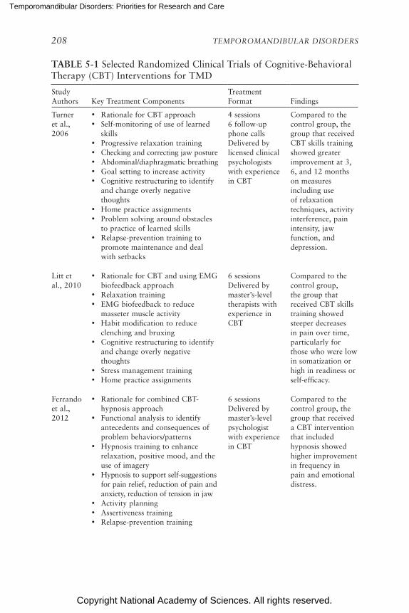
Temporomandibular Disorders: Priorities for Research and Care
Copyright National Academy of Sciences. All rights reserved.
208 TEMPOROMANDIBULAR DISORDERS
TABLE 5-1 Selected Randomized Clinical Trials of Cognitive-Behavioral Therapy (CBT) Interventions for TMD
Study Authors Key Treatment Components
Treatment Format Findings
Turner et al., 2006
• Rationale for CBT approach• Self-monitoring of use of learned
skills• Progressive relaxation training• Checking and correcting jaw posture• Abdominal/diaphragmatic breathing• Goal setting to increase activity• Cognitive restructuring to identify
and change overly negative thoughts
• Home practice assignments • Problem solving around obstacles
to practice of learned skills• Relapse-prevention training to
promote maintenance and deal with setbacks
4 sessions6 follow-up phone callsDelivered by licensed clinical psychologists with experience in CBT
Compared to the control group, the group that received CBT skills training showed greater improvement at 3, 6, and 12 months on measures including use of relaxation techniques, activity interference, pain intensity, jaw function, and depression.
Litt et al., 2010
• Rationale for CBT and using EMG biofeedback approach
• Relaxation training• EMG biofeedback to reduce
masseter muscle activity• Habit modification to reduce
clenching and bruxing• Cognitive restructuring to identify
and change overly negative thoughts
• Stress management training• Home practice assignments
6 sessionsDelivered by master’s-level therapists with experience in CBT
Compared to the control group, the group that received CBT skills training showed steeper decreases in pain over time, particularly for those who were low in somatization or high in readiness or self-efficacy.
Ferrando et al., 2012
• Rationale for combined CBT-hypnosis approach
• Functional analysis to identify antecedents and consequences of problem behaviors/patterns
• Hypnosis training to enhance relaxation, positive mood, and the use of imagery
• Hypnosis to support self-suggestions for pain relief, reduction of pain and anxiety, reduction of tension in jaw
• Activity planning• Assertiveness training• Relapse-prevention training
6 sessionsDelivered by master’s-level psychologist with experience in CBT
Compared to the control group, the group that received a CBT intervention that included hypnosis showed higher improvement in frequency in pain and emotional distress.
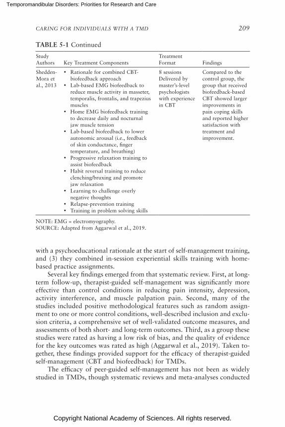
Temporomandibular Disorders: Priorities for Research and Care
Copyright National Academy of Sciences. All rights reserved.
CARING FOR INDIVIDUALS WITH A TMD 209
Study Authors Key Treatment Components
Treatment Format Findings
Shedden-Mora et al., 2013
• Rationale for combined CBT-biofeedback approach
• Lab-based EMG biofeedback to reduce muscle activity in masseter, temporalis, frontalis, and trapezius muscles
• Home EMG biofeedback training to decrease daily and nocturnal jaw muscle tension
• Lab-based biofeedback to lower autonomic arousal (i.e., feedback of skin conductance, finger temperature, and breathing)
• Progressive relaxation training to assist biofeedback
• Habit reversal training to reduce clenching/bruxing and promote jaw relaxation
• Learning to challenge overly negative thoughts
• Relapse-prevention training• Training in problem solving skills
8 sessionsDelivered by master’s-level psychologists with experience in CBT
Compared to the control group, the group that received biofeedback-based CBT showed larger improvements in pain coping skills and reported higher satisfaction with treatment and improvement.
NOTE: EMG = electromyography.SOURCE: Adapted from Aggarwal et al., 2019.
TABLE 5-1 Continued
with a psychoeducational rationale at the start of self-management training, and (3) they combined in-session experiential skills training with home-based practice assignments.
Several key findings emerged from that systematic review. First, at long-term follow-up, therapist-guided self-management was significantly more effective than control conditions in reducing pain intensity, depression, activity interference, and muscle palpation pain. Second, many of the studies included positive methodological features such as random assign-ment to one or more control conditions, well-described inclusion and exclu-sion criteria, a comprehensive set of well-validated outcome measures, and assessments of both short- and long-term outcomes. Third, as a group these studies were rated as having a low risk of bias, and the quality of evidence for the key outcomes was rated as high (Aggarwal et al., 2019). Taken to-gether, these findings provided support for the efficacy of therapist-guided self-management (CBT and biofeedback) for TMDs.
The efficacy of peer-guided self-management has not been as widely studied in TMDs, though systematic reviews and meta-analyses conducted
Temporomandibular Disorders: Priorities for Research and Care
Copyright National Academy of Sciences. All rights reserved.
210 TEMPOROMANDIBULAR DISORDERS
on this approach in other chronically painful conditions (Jackson et al., 2014) and chronic diseases (Holden, 1991) have provided empirical sup-port for its efficacy.
The future of self-management Considered overall, it seems fair to con-clude that there is growing recognition that self-management is important in care of TMDs. Persons with a TMD can learn self-management skills in a variety of ways. To date, evidence for the efficacy of self- management training has come primarily from randomized controlled studies of therapist-guided self-management training. Although the results are sup-portive, the total number of studies is relatively small, with most of them focused on CBT and biofeedback.
Much more needs to be done to advance the practice and science of self-management of TMDs. First, there is a need to involve patients, their families, health care professionals, and other key stakeholders in the review and evaluation of current TMD self-management materials and resources and peer-led and therapist-led self-management training proto-cols. Such a review could lead to the development of updated and tailored self- management materials. The availability of high-quality resources not only could heighten the impact of self-management approaches on patients and their families, but also could enhance health care providers’ aware-ness of self-management and knowledge about and skills in fostering self- management. Second, there is a need for programmatic research to develop and test novel self-management programs for individuals with a TMD. Novel psychosocial interventions (e.g., acceptance and commitment therapy, partner-assisted and couples-based training in self-management) and novel treatment delivery formats (e.g., eHealth approaches such as video over the Internet) could be more widely explored in TMD care. Third, although novel theoretical frameworks are available to guide the development of self-management interventions (e.g., the National Institutes of Health [NIH] stage model) that can be readily disseminated, these have not received much attention in the TMD area. These theoretical frame-works are important in guiding treatment development and in addressing the key limitations of the current TMD literature, including the need to involve patients and real-world providers in the development of training materials and methods, the need to link intervention components to theory, and the need to streamline treatments to make them easier to disseminate. Fourth, with the growing recognition that TMDs are a set of complex and multidimensional conditions has come heightened interest in the variance in how people respond to training in self-management. Finally, although there is evidence that formal training in self-management can be effective, much less is known about how it works. Research suggests that changes in measures of self-efficacy or in the perceived ability to control pain occurring
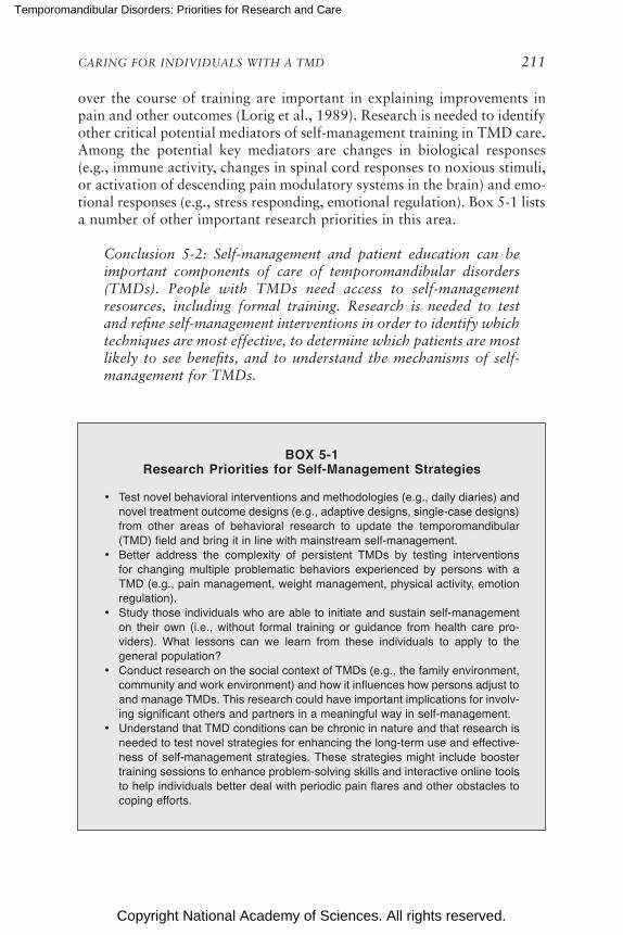
Temporomandibular Disorders: Priorities for Research and Care
Copyright National Academy of Sciences. All rights reserved.
CARING FOR INDIVIDUALS WITH A TMD 211
BOX 5-1 Research Priorities for Self-Management Strategies
• Test novel behavioral interventions and methodologies (e.g., daily diaries) and novel treatment outcome designs (e.g., adaptive designs, single-case designs) from other areas of behavioral research to update the temporomandibular (TMD) field and bring it in line with mainstream self-management.
• Better address the complexity of persistent TMDs by testing interventions for changing multiple problematic behaviors experienced by persons with a TMD (e.g., pain management, weight management, physical activity, emotion regulation).
• Study those individuals who are able to initiate and sustain self-management on their own (i.e., without formal training or guidance from health care pro-viders). What lessons can we learn from these individuals to apply to the general population?
• Conduct research on the social context of TMDs (e.g., the family environment, community and work environment) and how it influences how persons adjust to and manage TMDs. This research could have important implications for involv-ing significant others and partners in a meaningful way in self-management.
• Understand that TMD conditions can be chronic in nature and that research is needed to test novel strategies for enhancing the long-term use and effective-ness of self-management strategies. These strategies might include booster training sessions to enhance problem-solving skills and interactive online tools to help individuals better deal with periodic pain flares and other obstacles to coping efforts.
over the course of training are important in explaining improvements in pain and other outcomes (Lorig et al., 1989). Research is needed to identify other critical potential mediators of self-management training in TMD care. Among the potential key mediators are changes in biological responses (e.g., immune activity, changes in spinal cord responses to noxious stimuli, or activation of descending pain modulatory systems in the brain) and emo-tional responses (e.g., stress responding, emotional regulation). Box 5-1 lists a number of other important research priorities in this area.
Conclusion 5-2: Self-management and patient education can be important components of care of temporomandibular disorders (TMDs). People with TMDs need access to self-management resources, including formal training. Research is needed to test and refine self-management interventions in order to identify which techniques are most effective, to determine which patients are most likely to see benefits, and to understand the mechanisms of self-management for TMDs.
Temporomandibular Disorders: Priorities for Research and Care
Copyright National Academy of Sciences. All rights reserved.
212 TEMPOROMANDIBULAR DISORDERS
Physical Treatments
Occlusal Treatments
As described in Chapter 2, discrepancies between an individual’s dental occlusion (how teeth fit together) and the ideal occlusion, as defined by a range of attributes, have been a target for TMD treatment for more than 50 years. Occlusal treatments modify the teeth and bite. Intraoral appli-ances fit over the teeth and do not modify the teeth or bite (see section below on intraoral appliances). A 2017 protocol for evaluating the effec-tiveness of occlusal interventions for managing TMDs has been approved by Cochrane, but the review is not yet complete (Singh et al., 2017). Occlusal approaches, and available evidence-based status, include:
• Occlusal adjustment: modifying the teeth through the addition of fixed crowns or removable devices in order to change the positioning of the lower jaw relative to the upper jaw. A 2003 Cochrane review found no difference between the group receiving occlusal adjust-ment and the control group (Koh and Robinson, 2003); the review was withdrawn in 2016 due to being out of date and not meeting Cochrane’s current methodological standards (Cochrane Library, 2016a).
• Occlusal equilibration: adjusting the occlusion by removing enamel from the chewing surface of the tooth in order to modify the manner in which the teeth achieve full closure or the manner in which the teeth move past each other as the lower jaw is moved to the side or to the front. This approach has also been called selective grinding—when a dentist grinds one or a few biting surfaces of an individual’s teeth to improve the interaction of those teeth with the teeth on the opposing jaw. A 2018 assessment of the literature found no evidence to support its use (Manfredini, 2018).
• Orthodontic treatments: the repositioning of some or all of the teeth with the goal of improving an individual’s bite. A Cochrane review (Luther et al., 2010) found that there were insufficient data avail-able to inform clinical practice on the effectiveness of orthodontic treatments in reducing TMD symptoms. In 2016 the review was withdrawn due to being out of date and not meeting Cochrane’s current methodological standards (Cochrane Library, 2016b).
The time and cost for these different treatments vary. Full orthodontic treatment typically requires 2 to 3 years, and the cost depends on the geo-graphic area. Adjusting the occlusion can require anywhere from a single treatment session to a series of recurring sessions over many years, which
Temporomandibular Disorders: Priorities for Research and Care
Copyright National Academy of Sciences. All rights reserved.
CARING FOR INDIVIDUALS WITH A TMD 213
are often stopped not because of therapeutic success but because of the tooth sensitivity that is inevitable with enough removal of enamel. Modify-ing the teeth through crowns or removable devices can require up to several years of treatment and cost thousands of dollars.
As discussed in Chapter 2, the current Diagnostic Criteria for Temporo-mandibular Disorders (DC/TMD) considers the stability of the occlusion as a common symptom of degenerative joint disease but not as part of the diagnosis of a TMD. Over recent decades, research has not found sufficient evidence to support claims that dental occlusion is an important contributor toward TMDs (Clarke, 1982; Clark et al., 1999; Fricton, 2006; Klasser and Greene, 2009; Türp and Schindler, 2012). However, publications and posi-tion papers within the dental community often continue to promote the role of occlusion in diagnosing and treating individuals with TMDs (Dawson, 1996; Cooper, 2011; Racich, 2018).
Several studies have found that occlusion and a particular position of the TMJs (which includes freedom to move posteriorly relative to the fully closed occlusion) may actually be a protective factor for TMDs and an important adaptive response of the body (Levy, 1975; Pullinger et al., 1988). This research would suggest that treatment to correct the so-called “slide” in mandibular position (see Chapter 2) is misdirected as a treatment for TMD. The relationship between head posture and the closing position of the mandible and resultant stability of contact between lower and upper teeth reflects a complex interaction between cervical and masticatory sys-tems (Mohl, 1984; Southard et al., 1990), highlighting the importance of understanding TMDs within broader frameworks (e.g., the role of the cervi-cal system for both pain and mechanical jaw function). This contrasts with the common but unsupported belief that head or body posture problems are caused by TMDs (Manfredini et al., 2012), which is sometimes used to justify TMD treatments. Collectively, this type of information continues to not be typically incorporated into working clinical knowledge and research attention to these areas has waned over the years.
Intraoral Appliances (Splints)
Intraoral appliances, or laboratory-fabricated devices that fit over the teeth, are known by a wide variety of names such as splints, stabilization appliances, occlusal splints, occlusal appliances, interocclusal splints, fully balanced splints, repositioning splints, bruxism splints, nightguards, and several names denoting the commercial vendors of particular splint designs; the selected name usually relates to the perceived mechanism of action, for which evidence remains absent. Therefore, the theory-neutral term “intra-oral appliance” is used. Intraoral appliance treatment is distinctly differ-ent than treatments that modify the occlusion (e.g., occlusal adjustment,
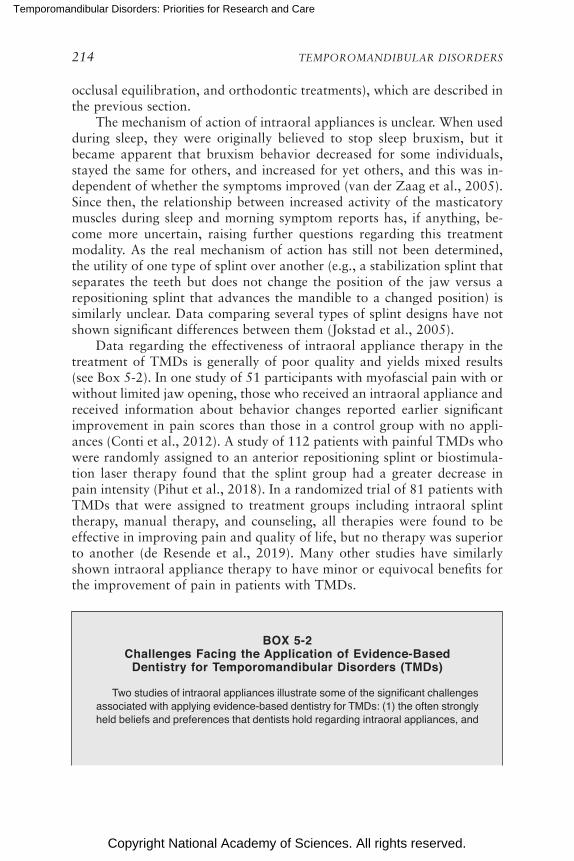
Temporomandibular Disorders: Priorities for Research and Care
Copyright National Academy of Sciences. All rights reserved.
214 TEMPOROMANDIBULAR DISORDERS
occlusal equilibration, and orthodontic treatments), which are described in the previous section.
The mechanism of action of intraoral appliances is unclear. When used during sleep, they were originally believed to stop sleep bruxism, but it became apparent that bruxism behavior decreased for some individuals, stayed the same for others, and increased for yet others, and this was in-dependent of whether the symptoms improved (van der Zaag et al., 2005). Since then, the relationship between increased activity of the masticatory muscles during sleep and morning symptom reports has, if anything, be-come more uncertain, raising further questions regarding this treatment modality. As the real mechanism of action has still not been determined, the utility of one type of splint over another (e.g., a stabilization splint that separates the teeth but does not change the position of the jaw versus a repositioning splint that advances the mandible to a changed position) is similarly unclear. Data comparing several types of splint designs have not shown significant differences between them (Jokstad et al., 2005).
Data regarding the effectiveness of intraoral appliance therapy in the treatment of TMDs is generally of poor quality and yields mixed results (see Box 5-2). In one study of 51 participants with myofascial pain with or without limited jaw opening, those who received an intraoral appliance and received information about behavior changes reported earlier significant improvement in pain scores than those in a control group with no appli-ances (Conti et al., 2012). A study of 112 patients with painful TMDs who were randomly assigned to an anterior repositioning splint or biostimula-tion laser therapy found that the splint group had a greater decrease in pain intensity (Pihut et al., 2018). In a randomized trial of 81 patients with TMDs that were assigned to treatment groups including intraoral splint therapy, manual therapy, and counseling, all therapies were found to be effective in improving pain and quality of life, but no therapy was superior to another (de Resende et al., 2019). Many other studies have similarly shown intraoral appliance therapy to have minor or equivocal benefits for the improvement of pain in patients with TMDs.
BOX 5-2 Challenges Facing the Application of Evidence-Based
Dentistry for Temporomandibular Disorders (TMDs)
Two studies of intraoral appliances illustrate some of the significant challenges associated with applying evidence-based dentistry for TMDs: (1) the often strongly held beliefs and preferences that dentists hold regarding intraoral appliances, and
Temporomandibular Disorders: Priorities for Research and Care
Copyright National Academy of Sciences. All rights reserved.
CARING FOR INDIVIDUALS WITH A TMD 215
(2) why dentists are often reluctant to incorporate a more biopsychosocial model for treatment of TMDs.
In the first study, 200 patients with myofascial pain of the masticatory muscles (either temporomandibular joint [TMJ] arthralgia or disc displacement with reduc-tion) were randomized to three different treatment groups: usual self-care treat-ment alone, self-care with a custom laboratory appliance, or self-care with a common athletic mouthguard (Truelove et al., 2006). The intraoral appliances were used during sleep. The outcomes across multiple variables improved equally for all three treatment groups. One conclusion drawn from the study was that costly appliances had no additional benefit beyond the very inexpensive athletic mouthguard. A second conclusion was that appliances of any sort were not necessarily needed. However, because of the diagnostic heterogeneity of TMDs enrolled in the study, a deeply held view or preference by some dentists regarding a particular appliance for a particular diagnosis would result in rejecting the find-ings. In short, some dentists believe that uncontrolled observations of uncontrolled treatments in a single treatment setting are more valid than a clinical trial that has clumped different diagnoses together. However, randomization, diagnostic equivalence between treatment groups, and the role of statistics that accounts for that heterogeneity should lead to much greater confidence in such findings, versus simple clinical observation that is prone to well-known biases.
In a second study, the belief that intraoral appliances need to be designed in a particular manner was tested by using two contrasting appliance designs (Rugh et al., 1989). The ideal form of the anterior part of the appliance that guides how the teeth in the lower jaw move forward or the side, while contacting the appli-ance attached to the upper teeth, was alternated by creating the same movement guidance on the molar teeth; prevailing theory at that time was that such an appli ance design would substantially worsen outcomes. After wearing one type of appliance during sleep for several weeks, the outcomes in terms of magnitude or frequency of sleep bruxism were measured; this was repeated for the other type of appliance. The appliance using the guidance based on the molar teeth exhibited the same outcomes as the appliance using the ideal guidance on the front of the appliance. Yet, dentists continue to believe that their particular appli-ance design is superior and effective, despite the research literature that takes a broader perspective.
Many dentists believe that particular TMDs are best treated by a unique type of intraoral appliance—that is, the appliance has to be designed in a particular manner—such that some aspect of improving the dental occlusion is incorporated into the appliance. Any treatment study that does not isolate the particular TMD of interest and treat it with the unique appliance of interest will be automatically rejected by the dentist as inadequate. Because of the hundreds of variations in intraoral appliance design, it is unlikely that any study could ever be conducted that will be considered sufficient to a particular dentist with a pre-existing belief about the effectiveness of one appliance. Simply stated, some dentists regard the evidence associated with procedural-based treatments as suspect unless the treatment was done “in my hands.”
BOX 5-2 Continued
Temporomandibular Disorders: Priorities for Research and Care
Copyright National Academy of Sciences. All rights reserved.
216 TEMPOROMANDIBULAR DISORDERS
While harms associated with TMJ surgery have been extensively investi gated, the harms associated with intraoral appliances are less well understood. The committee was not aware of any specific literature that describes complications associated with intraoral appliances. Anecdotally, these harms include alteration in the occlusion or position of the teeth, aspiration of small appliances worn on the anterior teeth, dependence on a device and not acquiring self-management skills, and perpetuation of a belief that something is wrong with the masticatory system such that the appliance is necessary to “fix” it. Systematic research is needed to assess harms associated with occlusal appliances.
Several attempts have been made at meta-analyses and systematic reviews of the utility of intraoral splints in patients with TMDs. In 1999 a qualitative systematic review concluded that intraoral splints may be of some benefit in the treatment of TMDs but that the evidence to definitively support this conclusion was lacking (Forssell et al., 1999). In 2004 another qualitative systematic review indicated that most individuals with mastica-tory muscle pain are helped by intraoral appliances such as a stabilization splint but concluded that the evidence was uncertain as to whether the improvement in symptoms was caused by a specific effect of the appliance (Türp et al., 2004). A 2017 meta-analysis that included 30 randomized con-trolled trials found stabilization splints to have short-term benefits on pain reduction and pain intensity but no differences in long-term outcomes with other types of treatments (Kuzmanovic Pficer et al., 2017). In summary, intraoral splint therapy may confer a small benefit for the management of pain in individuals with TMDs, but the evidence for this is generally poor and mixed.
Physical Therapy
Physical therapists are fully integrated into the modern health system and work to meet the interdisciplinary and interprofessional management needs of individuals with chronic pain. Physical therapists are increasingly serving as primary care providers and may provide a gateway into the health care system for people with a TMD. Physical therapists use mul-tiple approaches to the management of TMDs, including exercise, manual therapy, and education on self-management skills (see section on self-management above). Other therapies offered by physical therapists include electrotherapy and dry needling; these treatments need more evidence to support their use (see below).
Exercise Exercise is considered a first-line treatment in the physical therapy management of TMDs. For other chronic pain conditions, like fibro myalgia and low back pain, clinical practice guidelines recommend
Temporomandibular Disorders: Priorities for Research and Care
Copyright National Academy of Sciences. All rights reserved.
CARING FOR INDIVIDUALS WITH A TMD 217
exercise as one of the first-line treatments (Kia and Choy, 2017; Oliveira et al., 2018). There are several types of exercise that may be effective in the management of TMDs. These include jaw strengthening, jaw stretching, and postural exercises.
• Jaw strengthening exercises: Some studies support the effective-ness of exercises to build jaw strength and endurance, with or without stretching exercises, in improving pain, jaw opening, and TMJ sounds/clicking (Haggman-Henrikson et al., 2018; Wanman and Marklund, 2019). A study using supervised exercise training showed greater improvements than home exercises alone (Wanman and Marklund, 2019). Other publications, however, have not dem-onstrated as much benefit (Craane et al., 2012).
• Jaw stretching exercises: Some clinical trials show that home exercise aimed at stretching the soft tissue around the jaw muscles increases jaw opening and may decrease pain. However, other studies do not show an effect of stretching exercises, whether applied by a physical therapist or by the patient in a home program (Armijo-Olivo et al., 2016). Meta-analyses, however, indicate overall benefit of stretching to regain normal jaw opening for both myalgia and clicking prob-lems (Armijo Olivo et al., 2016).
• Postural exercises: Individual studies and reviews have demonstrated some effectiveness of active and passive exercises that improve pos-ture in reducing pain and improving the range of motion (McNeely et al., 2006; Medlicott and Harris, 2006). However, the method-ological quality of the studies was found lacking (McNeely et al., 2006; Medlicott and Harris, 2006).
A recent systematic review and meta-analysis of exercise for TMDs (Armijo-Olivo et al., 2016) performed an extensive analysis of multiple types of exercise therapy. The review showed that jaw exercises were asso-ciated with a clinically significant increase in pain-free mouth opening and that postural exercises were associated with clinically significant increases in pain-free mouth opening as well as reductions in disturbances in daily living. It should be noted, however, that the researchers identified that no high-quality evidence was found and caution is warranted due to the low quality of the studies, large heterogeneity, and the small number of subjects per study.
One benefit of exercise in general (e.g., possibly more than jaw stretch-ing or strengthening exercises) for patients with TMDs may be exercise-induced analgesia, the underlying mechanisms of which recent research has begun to uncover. In addition to improving function by increasing the range of motion, strength, and motor coordination of the joint and
Temporomandibular Disorders: Priorities for Research and Care
Copyright National Academy of Sciences. All rights reserved.
218 TEMPOROMANDIBULAR DISORDERS
muscles, exercise can also alter the immune system and the nervous systems in ways that promote healing and reduce pain (Brito et al., 2017; Lima et al., 2017a,b; Sluka et al., 2018). In addition, in chronic pain conditions, there are decreases in serotonin and increases in the serotonin transporter, both of which are normalized by regular exercise (Brito et al., 2017; Lima et al., 2017a,b; Sluka et al., 2018). In human subjects with chronic pain, researchers have found an interaction between the serotonin transporter gene and the mu-opioid gene that is associated with increased analgesia (Tour et al., 2017).
Understanding the underlying mechanisms by which exercise reduces pain and improves function may be important when designing exercise pro-grams for individuals with TMDs. For example, if individuals with TMDs have a comorbid inflammatory condition or widespread pain with altered central nervous system processing, an aerobic conditioning program may be useful in both improving function and reducing pain. On the other hand, if there is significant weakness of the jaw muscles, more localized stretching and strengthening exercises are likely to improve function and may have a secondary effect of reducing pain.
Manual therapy The aim of manual therapy is to increase motion by stretching the soft tissues and muscles surrounding a joint. Recent evidence suggests that manual therapy also directly affects nociceptor and central nervous system activity to reduce pain (Skyba et al., 2003; Bialosky et al., 2009; Martins et al., 2011, 2012, 2013a,b). Stretching of peripheral tissues can reduce inflammation, remodel fibroblast connective tissue structure, and increase the expression of muscle healing genes (Langevin et al., 2013; Berrueta et al., 2016). As stretching of peripheral tissues is an active com-ponent of manual therapy, these mechanisms may be particularly important in the resolution of a TMD with a peripheral inflammatory or connective tissue component. A recent systematic review found that manual therapy (soft tissue mobilization) can produce a clinically significant reduction in pain (Armijo-Olivo et al., 2016). Cervical mobilization in those with myo-fascial origin or craniofacial pain led to a clinically significant reduction in pain in one preliminary study (Armijo-Olivo et al., 2016). Unfortunately, the quality of the evidence in support of these treatments is poor, partially due to large heterogeneity between the studies and their small sample sizes.
Manual therapy and exercise are often combined; neither is frequently done as a stand-alone treatment. The review by Armijo-Olivo and col-leagues (2016) examined data from seven studies and showed that manual therapy with exercise decreased symptoms of TMDs and increased the mouth opening and range of motion. These changes were of moderate effect sizes.
Temporomandibular Disorders: Priorities for Research and Care
Copyright National Academy of Sciences. All rights reserved.
CARING FOR INDIVIDUALS WITH A TMD 219
Conclusion 5-3: Some elements of physical therapy—including exercise and manual therapy—have been shown to improve pain and functional outcomes for individuals with temporomandibular disorders. However, many of the studies are of low quality and further research is needed to support the use of these treatment modalities.
Other Physical Therapy Interventions
In addition to exercise and manual therapy, there are a number of other physical therapy treatments for TMDs that are used clinically but which lack clinical evidence or have varying degrees of evidence. These include the use of heat and cold therapy, low-level laser therapy, and dry needling. Heat and cold therapy are often recommended as part of a self-management program and are inexpensive with minimal risk. However, there is no re-search to support or refute their effectiveness for TMDs. Systematic reviews have shown that low-level laser therapy was somewhat better than placebo in reducing pain from TMDs, and improving related outcomes (Maia et al., 2012; de Pedro et al., 2019). Dry needling is becoming increasingly accepted and used as a treatment for myofascial pain and TMDs. A recent systematic review considered 18 studies examining the effectiveness of dry needling and injection with different substances (Machado et al., 2018). The review concluded that while dry needling and local anesthetic injec-tions seem promising, there is a need to conduct randomized clinical trials with larger sample sizes and longer follow-up times to truly evaluate the effectiveness of these techniques. A 2019 study in five women with chronic masticatory myofascial pain found that deep dry needling increased the individual’s pressure pain threshold (Tesch et al., 2019). Among the limita-tions noted by the authors was that increasing pressure pain thresholds is an isolated outcome that is not directly comparable with more general outcomes such as the intensity and severity of the pain experienced or a patient’s perception of the treatment’s efficacy and impact on quality of life.
Complementary Treatments
Acupuncture
Acupuncture is another treatment approach that has been used for TMD but for which evidence is limited. A 2017 meta-analysis found a sig-nificant one-point difference (out of 10) on the visual analog scale of pain between acupuncture and sham acupuncture for TMDs (Wu et al., 2017). Interestingly, the effect was only found when the comparison was with non-penetrating sham acupuncture; the difference between acupuncture
Temporomandibular Disorders: Priorities for Research and Care
Copyright National Academy of Sciences. All rights reserved.
220 TEMPOROMANDIBULAR DISORDERS
and penetrating sham acupuncture was not significant. The meta-analysis also found that acupuncture was more effective for patients with TMDs that were related to masticatory muscle disorders versus related to the TMJ itself.
Dietary Intake and Nutrition
Changing one’s diet is a type of behavioral modification some indi-viduals with a TMD might undertake to relieve or avoid pain. Pursuing a soft diet and seeking pain-free chewing has been related to important nutritional challenges faced by individuals with a TMD. Given that TMDs can affect an individual’s ability to chew and swallow, serious nutritional challenges and deficiencies can occur. The TMJ Association advises a soft diet for individuals who are able to adequately open their mouths and have minimal pain and a pureed diet for those who cannot tolerate a soft diet (The TMJ Association, 2017). Because evidence-based dietary guidelines for patients with chronic orofacial pain do not exist, clinicians may pro-vide customized advice for individuals based on the challenges they report (Durham et al., 2015; Nasri-Heir et al., 2016). While dietary interventions are commonly recommended for patients with a TMD, more research is needed on whether and how dietary changes have an impact.
Electrotherapy
Transcutaneous electrical nerve stimulation (TENS) is a form of neuro-modulation that applies electrical current through the skin for pain control. The mechanisms of TENS analgesia have been extensively studied and involve the activation of endogenous opioid, serotonin, and GABAergic pathways in a frequency-dependent manner which can reduce central excit-ability and central sensitization (Vance et al., 2014). High-frequency TENS has been shown to reduce pain and decrease electromyography (EMG) activity of the masticatory muscles in people with TMDs (Rodrigues et al., 2004). Low-frequency TENS reduces EMG activity (Kamyszek et al., 2001). A single treatment of either sensory-stimulation or motor-stimulation low-frequency TENS reduces the EMG activity of the masticatory muscles simi-larly and improves mouth opening (Monaco et al., 2013). Additionally, two 30-minute TENS treatments in combination with pharmaceutical treatment provide additional pain relief when compared with pharmaceutical treat-ment alone (Shanavas et al., 2014). However, it should be noted that these studies were small with short-duration TENS treatments.
Temporomandibular Disorders: Priorities for Research and Care
Copyright National Academy of Sciences. All rights reserved.
CARING FOR INDIVIDUALS WITH A TMD 221
Pharmacological Treatments
Oral Medication
A variety of pharmacological treatments have been suggested for the management of the pain and symptoms associated with TMDs. Most studies on pharmacological treatments have focused on evaluating the efficacy of various drugs in relieving pain, which is the primary reason for which indi-viduals with a TMD seek medical care. However, there are no drugs specifi-cally approved by FDA for this disorder, and the evidence for the effi cacy of many of the recommended treatments is weak. Pharmacotherapies recom-mended for patients with a TMD have generally been based on drugs that have been shown to be efficacious for other musculoskeletal conditions or neuropathic pain states. Commonly recommended pharmacological agents for TMDs, based on expert opinion, include acetaminophen, non-steroidal anti-inflammatory drugs (NSAIDs), corticosteroids, antidepressants, anti-convulsants, benzodiazepines, muscle relaxants, opioid analgesics, tricyclic antidepressants, and topical lidocaine patches.
To assess the effectiveness of pharmacological interventions in indi-viduals with a TMD, a Cochrane Collaboration literature review was conducted and found that most trials had few participants and there was insufficient evidence to support or refute the effectiveness of the reported drugs (Mujakperuo et al., 2010). Large-scale efficacy and effectiveness trials of pharmacological agents in the treatment of TMDs are needed.
A brief summary of select pharmacological approaches to TMD pain relief and relevant studies is presented below.
Non-steroidal anti-inflammatory drugs (NSAIDs) NSAIDs are used to relieve the pain and inflammation in the TMJ and muscles of the jaw, face, and neck in individuals with a TMD. Patients with suspected early disc displacement, synovitis, and arthritis have been suggested as appropriate candidates for early treatment with NSAIDs (Gauer and Semidey, 2015). However, placebo-controlled randomized trials reported mixed results ( Mujakperuo et al., 2010; Ouanounou et al., 2017). A randomized compar-ative trial suggested that dual COX-1 and COX-2 inhibition with naproxen was more effective for the treatment of painful TMDs than celecoxib and placebo, as judged by the degree of improvement in the clinical signs and symptoms of TMJ disc disorder (Ta and Dionne, 2004). Based on these mixed results, there is a need for more rigorous examination of NSAID use for TMD-related pain. NSAIDs need to be used with caution because of their possible adverse effects, including exacerbation of hypertension, gastritis, ulcers and gastrointestinal bleeding, and nephrotoxicity.
Temporomandibular Disorders: Priorities for Research and Care
Copyright National Academy of Sciences. All rights reserved.
222 TEMPOROMANDIBULAR DISORDERS
Opioids The inconsistent efficacy of NSAIDs in relieving chronic pain asso ciated with severe TMDs has led to exploration of alternatives, includ-ing the use of opioid analgesics (Ouanounou et al., 2017). One random-ized trial with 80 subjects provided moderate evidence that a combination analgesic product (NSAID and low-dose codeine) delivered relief from pain associated with TMDs (Shaheed et al., 2019). A recent review identified placebo-controlled studies that reported improved outcomes with mor-phine and fentanyl for TMJ arthrocentesis (Gopalakrishnan et al., 2018). Evidence supporting the use of opioids to manage chronic pain is lacking (NASEM, 2017). Given the high rates of opioid use disorder and opioid-related deaths, the use of opioid analgesics for TMDs should not be a first-line pharmaceutical treatment. The guidelines by the Centers for Disease Control and Prevention emphasize the improvements needed in prescribing opioids for chronic pain (CDC, 2019).
Antidepressants Antidepressants are used as adjuvant treatment for chronic TMD pain, based on both known efficacy in neuropathic and musculo skeletal pains, including fibromyalgia, and their beneficial effects on comorbid de-pression and sleep disturbance. The tricyclic antidepressant amitriptyline was shown to decrease pain and discomfort in a small controlled study in patients with chronic TMD (Rizzatti-Barbosa et al., 2003). In a small study of 29 patients, a selective serotonin and norepinephrine reuptake inhibitor, duloxetine, was effective at relieving chronic pain in the orofacial region compared to baseline (Nagashima et al., 2012). Although there is anecdotal evidence of individual patients experiencing pain relief from paroxetine, a selective serotonin reuptake inhibitor (SSRI) (Inagaki et al., 2007), SSRIs have the potential to cause increased bruxism and exacerbate pain (Rajan and Sun, 2017). Additional well-controlled clinical trials are needed to assess the potential benefits of tricyclic antidepressants in TMD patients.
Anticonvulsants Anticonvulsant medications, such as gabapentin and pregabalin, are commonly used to treat neuropathic pain; however, their mechanism of action is unclear (Ouanounou et al., 2017). In one random-ized controlled trial, gabapentin was found to be clinically and statistically superior to placebo in reducing pain and masticatory muscle hyperalgesia, and in improving daily functioning in patients with chronic pain in the masticatory muscles (Kimos et al., 2007). However, a recent systematic review on the efficacy of anticonvulsants on orofacial pain found limited to moderate evidence supporting the use of anticonvulsants for treatment of patients with orofacial pain disorders (Martin and Forouzanfar, 2011).
Benzodiazepines Benzodiazepines have been suggested as treatment options for chronic orofacial pain associated with TMDs despite weak
Temporomandibular Disorders: Priorities for Research and Care
Copyright National Academy of Sciences. All rights reserved.
CARING FOR INDIVIDUALS WITH A TMD 223
evidence for their efficacy in controlled trials. One randomized trial found diazepam to be efficacious in the short-term management of chronic oro-facial muscle pain (Singer and Dionne, 1997). Other muscle relaxants (e.g., cyclobenzaprine) are also recommended for treating patients with orofacial muscle spasms, though the quality of the evidence for the efficacy of these drugs is weak. A 2009 Cochrane review cited two randomized controlled trials reporting the use of cyclobenzaprine for the treatment of myofascial pain. The two studies included in the review examined a total of 79 par-ticipants; however, there was insufficient evidence to support the use of cyclobenzaprine for the treatment of myofascial pain (Leite et al., 2009).
Cannabis Medical cannabis has been proposed to reduce pain and poten-tiate the effects of other pain control regimens for several painful conditions (Hill et al., 2017). Most cannabis research is outside of the arena of TMDs. One study, however, investigated the utility of cannabinoids, the active components of cannabis, for TMJ-related nociception in rats; it found that the cannabinoids potentiated the effects of other medications in decreasing inflammatory pain of the TMJ (Lee et al., 2008).
Glucosamine Two studies cited by a systematic literature review found that glucosamine supplements were as effective at reducing pain and im-proving mouth opening as ibuprofen taken two to three times daily over 12 weeks (Melo et al., 2018). A third study cited in the same review did not find significant differences in pain reduction or maximum mouth opening between a group receiving glucosamine and a group receiving a placebo over 6 weeks of medication administration. Melo and colleagues (2018) cautioned that the results of the studies were at risk of bias and should be interpreted carefully.
Topical Medication
Capsaicin (topical) Capsaicin is a chemical agent derived from chili peppers that has been shown to have analgesic properties due to its inter-actions with the TRP vanilloid subfamily member 1 (TRPV1) ion channel. A small study (n=15 individuals with a TMD) showed some pain relief after a 1-week period following a single topical application of 8 percent capsaicin cream (Campbell et al., 2017), but further studies with additional patients are needed to confirm this finding.
Intramuscular Medication
Botulinum toxin Intramuscular injections are primarily used for the man-agement of musculoskeletal pain. The injection of botulinum toxin Type A
Temporomandibular Disorders: Priorities for Research and Care
Copyright National Academy of Sciences. All rights reserved.
224 TEMPOROMANDIBULAR DISORDERS
into the muscles of mastication has been proposed to decrease muscle spasm and pain. Botulinum toxin is a neurotoxic protein that prevents the release of the neurotransmitter acetylcholine, thereby impairing muscle contraction. Some studies have reported improvement in facial pain in some patients with TMDs from botulinum toxin Type A injection into the muscles of mastication (Chaurand et al., 2017; Khawaja et al., 2017), but others have reported equivocal results (Chen et al., 2015; Keenan, 2015); the data are limited and often of poor quality. A Cochrane review (233 subjects in 4 trials) found inconclusive evidence concerning the effective-ness of botulinum toxin for myofascial pain in the neck and head muscles (Soares et al., 2014).
Navarrete and colleagues (2013) reported a study in which rabbits were injected with either botulinum toxin Type A or saline solution into the masseter muscle. The result was a dramatic loss of bite force, after 3 weeks, in the rabbits receiving botulinum toxin Type A injections. While this study, and others, indicated success in using botulinum toxin Type A to unload, or temporarily paralyze, the muscle and relieve the jaw joint, The TMJ Asso-ciation issued caution to individuals with TMD pursuing treatment with botulinum toxin Type A due to the loss of bone strength resulting from the treatment in rabbits. There is concern for the health of the TMJ in humans using botulinum toxin Type A in the long term given the osteoporotic con-dition of the TMJ in rabbits (The TMJ Association, 2016).
Prolotherapy
Other intra-articular injection agents have been proposed for specific subdiagnoses of TMDs. In the case of chronic dislocation and hyper-mobility of the TMJ, for example, prolotherapy (injection of an irritant solu tion to promote a reparative immune response) has shown promise (Zhou et al., 2014; Cezairli et al., 2017; Refai, 2017). Two studies demon-strated a success rate of 80 percent in reducing or eliminating dislocations by 1 year after injection of autologous blood to the superior TMJ space and the joint capsule (Machon et al., 2009; Daif, 2010).
Interventional Treatments
Arthrocentesis and Arthroscopy
In patients who have not achieved relief from or who are not can-didates for noninvasive treatment, minimally invasive surgical treatment may be indicated. The simplest of these minimally invasive operations is arthrocentesis. The indications for arthrocentesis include an identification of an intra-articular pathology such as disc dislocation in combination with
Temporomandibular Disorders: Priorities for Research and Care
Copyright National Academy of Sciences. All rights reserved.
CARING FOR INDIVIDUALS WITH A TMD 225
pain or joint dysfunction. Musculoskeletal pain may not be improved by arthrocentesis, and this treatment is typically used as only one component of a comprehensive management strategy.
Arthrocentesis involves the insertion of a needle into the superior TMJ space, followed by joint lavage. This can be accomplished with either a single needle for both the inflow and outflow of lavage fluid or by using two separate puncture sites to allow the inflow and outflow ports to be separated. The idea behind this intervention is to flush viscous synovium and inflammatory mediators from the joint and to release joint adhesions via hydraulic pressure. Arthrocentesis has been shown to be effective in relieving pain and improving mouth opening in patients with temporo-mandibular disc displacements (Dimitroulis et al., 1995; Nitzan and Price, 2001; Nitzan et al., 2017), though its benefits may decrease after 6 months (Bjørnland et al., 2007).
Arthroscopy, which involves the insertion of a small video endoscope into the superior TMJ space, is similar to arthrocentesis but with the added benefit of indirect visualization of the joint. The ability to visualize the joint space provides confirmation of joint access, enhances diagnosis, and allows additional therapy such as the laser resection of adhesions and disc repositioning (McCain and Hossameldin, 2011). The short-term outcomes of arthroscopy with regard to pain and function have been found to be similar to those for arthrocentesis (Fridrich et al., 1996), with both showing moderate effectiveness.
Both arthrocentesis and arthroscopy are often, but not always, com-bined with an injection of medication into the superior joint compartment. The most frequently injected medications are corticosteroid and hyaluronic acid. The reported results of these injections are mixed, with some studies demonstrating superior outcomes for pain relief with corticosteroid and hyaluronate (Liu et al., 2018), others purporting better outcomes with hyaluronic acid (Bjørnland et al., 2007), and several showing no signifi-cant difference among corticosteroid, hyaluronic acid, and saline alone (de Souza et al., 2012; Bouloux et al., 2017a,b; Davoudi et al., 2018).
Operations with Direct Access to the TMJ
“Open operations,” or surgical procedures that provide direct access to the joint, are reserved for individuals with severe and irreversible destruc-tion of the TMJ and those who have persistent debilitating symptoms or dysfunction of the joint despite other treatment. Less than 3 percent of 2,104 individuals who completed treatment for a TMD received an open surgical procedure in a multi-site analysis (Brown and Gaudet, 2002).
The selection of an operation is based on the severity of the symptoms and the health of the joint components. Several classification systems exist
Temporomandibular Disorders: Priorities for Research and Care
Copyright National Academy of Sciences. All rights reserved.
226 TEMPOROMANDIBULAR DISORDERS
to guide treatment decisions (see Chapter 2), but there is no universally ac-cepted protocol for operative management of a TMD. Imaging, particularly magnetic resonance imaging, plays a major role in the staging of the disease and the determination of the appropriate surgical treatment approach. Surgical treatment is sometimes graded, beginning with minimally invasive procedures and progressing to more aggressive operations as needed. Evi-dence has emerged, however, that shows that patients who have had fewer total TMJ operations have superior surgical outcomes than those who have had many smaller operations (Mercuri, 1999). This indicates the need to make an accurate diagnosis and choose the appropriate surgical interven-tion, when indicated, carefully.
Open operations are used when irreversible joint destruction has occurred and associated symptoms and dysfunction are present. Open opera tions require direct access to the TMJ via an incision adjacent to the ear. The diagnosis then dictates the procedure. When the TMJ disc is displaced from its normal position, it can be repositioned; when the disc is damaged, it can either be repaired or be removed (discectomy), with or without replacement. When the mandibular condyle or glenoid fossa has been eroded or free pieces of bone (osteophytes) are within the joint, a modification of the bony components of the joint, including the mandibular condyle, glenoid fossa, or articular eminence, may be indicated. Many other similar procedures can be performed within the joint once direct access has been obtained, depending on the objective of the operation.
When multiple components of the joint are severely damaged, recon-struction may be required. This can be accomplished with autologous tissues such as costochondral (rib) grafts or alloplastic materials (TMJ implants). TMJ implants will be discussed further in the next section.
Several subdiagnoses that may mimic TMDs but are not considered to be TMDs in this report require variations of the surgical considerations described above, each with corresponding evidence within the applicable subspecialty. In patients with a TMJ pathology such as a cyst or a tumor, an open operation may be required as the primary treatment. In patients with progressive condylar resorption disorders, including idiopathic condylar resorption and juvenile idiopathic arthritis affecting the TMJ, open opera-tions for replacement of the degenerated joint components using autogenous or alloplastic implants may be required. Similarly, in patients with con-genital anomalies of the TMJ, such as hemifacial microsomia, bilateral cranio facial microsomia, and Treacher Collins syndrome, construction of the joint with autogenous or alloplastic materials may be indicated as the first-line treatment. In patients with TMJ ankylosis, most commonly a result of trauma or infection, aggressive joint debridement and reconstruction is necessary. Patients with hypermobility syndromes such as Ehlers-Danlos and recurrent dislocation may require strategies to induce fibrosis of the
Temporomandibular Disorders: Priorities for Research and Care
Copyright National Academy of Sciences. All rights reserved.
CARING FOR INDIVIDUALS WITH A TMD 227
peri-articular tissues, such as autologous blood injection (Daif, 2010), and to eliminate anatomic factors that lead to dislocation, which may include resection of the articular eminence (eminectomy) (Tocaciu et al., 2019).
TMJ Implants
Patients with bony ankylosis, condylar injuries, developmental abnor-malities, functional deformity, severe inflammatory conditions, and/or pain-ful or dysfunctional internal derangements after failed conservative and surgical treatment, all of whom have not responded to less invasive treat-ments, may be candidates for a TMJ replacement with alloplastic implants (Sidebottom et al., 2008; NICE, 2014). Commensurate with improvements in biomaterials and long-term data demonstrating favorable outcomes in pain reduction, improved function (Wolford et al., 2015), and improved quality of life (Kunjur et al., 2016), the use of total alloplastic TMJ replace-ments has recently increased (Onoriobe et al., 2016). Several total alloplastic TMJ implants are currently available in the United States and are generally categorized as either stock implants (those that come in standard sizes and are then modified to fit the patient’s anatomy) or custom implants (those that are produced prior to implantation in a customized patient-specific shape and size based on preoperative imaging).
The TMJ implants that are currently on the market have been sub-jected to testing by manufacturers in compliance with FDA requirements for Class III devices. A follow-up study of 56 patients with implants found positive outcomes and stability at least 20 years after insertion, but only 50 percent of the initial cohort of 111 patients could be contacted and met study criteria (Wolford et al., 2015). However, earlier implants, which have since been removed from the market (discussed below), were associated with severe complications, which often went unrecognized until years after surgery. Unfortunately, this led to significant harm for some patients and clouded the field of TMJ surgery. The committee heard from indi viduals with TMDs and TMD patient representatives who reported significant challenges in the implant recall process. Not all patients who received the recalled device were properly notified by their oral surgeon. Even though the device was recalled by FDA in 1990, TMD patient representatives told the committee that some patients are just being notified now that they re-ceived the faulty device.
As introduced above, the use of Proplast/Teflon-based TMJ implants in the 1970s and 1980s resulted in severe adverse events and the need for corrective surgery in some patients (AAOMS, 1993; Lypka and Yamashita, 2007). In especially severe cases, implant recipients required multiple follow-up operations to fix the damage (Henry and Wolford, 1993). Many affected patients continue to suffer from irreversible iatrogenic damage and
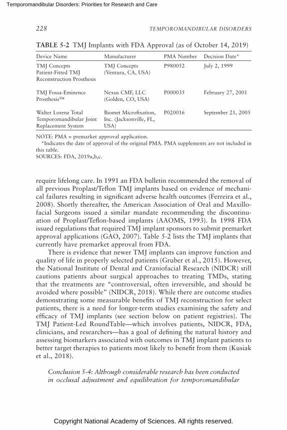
Temporomandibular Disorders: Priorities for Research and Care
Copyright National Academy of Sciences. All rights reserved.
228 TEMPOROMANDIBULAR DISORDERS
require lifelong care. In 1991 an FDA bulletin recommended the removal of all previous Proplast/Teflon TMJ implants based on evidence of mechani-cal failures resulting in significant adverse health outcomes (Ferreira et al., 2008). Shortly thereafter, the American Association of Oral and Maxillo-facial Surgeons issued a similar mandate recommending the discontinu-ation of Proplast/Teflon-based implants (AAOMS, 1993). In 1998 FDA issued regulations that required TMJ implant sponsors to submit premarket approval applications (GAO, 2007). Table 5-2 lists the TMJ implants that currently have premarket approval from FDA.
There is evidence that newer TMJ implants can improve function and quality of life in properly selected patients (Gruber et al., 2015). However, the National Institute of Dental and Craniofacial Research (NIDCR) still cautions patients about surgical approaches to treating TMDs, stating that the treatments are “controversial, often irreversible, and should be avoided where possible” (NIDCR, 2018). While there are outcome studies demonstrating some measurable benefits of TMJ reconstruction for select patients, there is a need for longer-term studies examining the safety and efficacy of TMJ implants (see section below on patient registries). The TMJ Patient-Led RoundTable—which involves patients, NIDCR, FDA, clinicians, and researchers—has a goal of defining the natural history and assessing biomarkers associated with outcomes in TMJ implant patients to better target therapies to patients most likely to benefit from them (Kusiak et al., 2018).
Conclusion 5-4: Although considerable research has been conducted in occlusal adjustment and equilibration for temporo mandibular
TABLE 5-2 TMJ Implants with FDA Approval (as of October 14, 2019)
Device Name Manufacturer PMA Number Decision Date*
TMJ Concepts Patient-Fitted TMJ Reconstruction Prosthesis
TMJ Concepts (Ventura, CA, USA)
P980052 July 2, 1999
TMJ Fossa-Eminence Prosthesis™
Nexus CMF, LLC (Golden, CO, USA)
P000035 February 27, 2001
Walter Lorenz Total Temporomandibular Joint Replacement System
Biomet Microfixation, Inc. (Jacksonville, FL, USA)
P020016 September 21, 2005
NOTE: PMA = premarket approval application. *Indicates the date of approval of the original PMA. PMA supplements are not included in this table.SOURCES: FDA, 2019a,b,c.
Temporomandibular Disorders: Priorities for Research and Care
Copyright National Academy of Sciences. All rights reserved.
CARING FOR INDIVIDUALS WITH A TMD 229
disorders (TMDs), these treatments have not been found to be effective. Evidence-based findings need to be widely disseminated to dentists and other clinicians to ensure that the treatment ap-proaches individuals with a TMD receive are consistently based on the best available evidence and focused on starting with conserva-tive approaches.
Conclusion 5-5: Data are inadequate and are of poor quality for most treatments for temporomandibular disorders (TMDs). Research is needed to determine safe and effective treatments for TMDs. Systematic reviews and methodologically rigorous new studies are needed.
IMPROVING AND DISSEMINATING EVIDENCE
As discussed above, the evidence base for many TMD treatments is poor. Clinicians tend “to see what they treat and treat what they see,” based on the theory of TMDs in which they are educated and trained. To improve care and outcomes for people with TMDs, it is critical that researchers gather and disseminate high-quality evidence about these treatments. Three approaches for doing so are discussed here: conducting clinical trials, building a patient registry, and developing and implementing clinical practice guidelines.
Conducting Clinical Trials
Randomized controlled trials (RCTs) are considered the gold standard for assessing the effectiveness of a particular medication or treatment ap-proach (Bothwell et al., 2016). RCTs in general can be challenging to carry out for several reasons, including difficulty with endpoint selection, inade-quate randomization or blinding, and other logistical problems (Nichol et al., 2010). Although some RCTs for TMD treatments have demonstrated small to moderate reductions in pain intensity (as described earlier in this chapter), the quality of evidence is limited, and some of the studies have been plagued by methodological shortcomings (e.g., insufficient blinding, small sample sizes, a range of outcome measures and control treatments, and short follow-up times). Furthermore, many RCTs for TMD treatments are carried out in tertiary care centers (centers that focus on specialized care), and few, if any, take place in a primary dental care setting, which is often where patients go first if they begin experiencing orofacial pain (Velly et al., 2013). As of October 2019, 191 clinical trials were listed on clini-caltrials.gov for a variety of interventions for TMDs. Of those 191 trials, 29 were actively recruiting, and 4 were taking place in the United States. Given the currently inadequate research base for TMD treatments, more
Temporomandibular Disorders: Priorities for Research and Care
Copyright National Academy of Sciences. All rights reserved.
230 TEMPOROMANDIBULAR DISORDERS
well-designed clinical trials are needed in the future to inform evidence-based care for individuals with a TMD.
Ideal Characteristics of Clinical Trials for TMD Treatments
Ideally, clinical trials for TMD treatments would focus on outcomes that are meaningful to patients, such as improvements in health and quality of life, reduction in pain, restoration of function, and physical, social, and psychological well-being, and they would examine TMDs in the context of the real world in which patients live. The incorporation of patient preference data and real-world evidence and experiences is one of the key objectives of the TMJ Patient-Led RoundTable. Clinical trials for TMDs could be improved through the adoption of a universal TMD case classification system, such as DC/TMD, across all trials and could incorporate broad eligibility criteria as a way to reach a large, diverse patient population and increase the generalizability of the trial results. Detecting, measuring, and taking into account the comorbidities often seen in TMD patients is an important step toward improving the quality of clinical studies in this area.
Conducting pragmatic trials that incorporate real-world evidence may represent an opportunity to gather better information about the effective-ness of TMD treatments. Pragmatic clinical trials test an intervention in a real-world clinical setting (e.g., hospitals, clinics, primary care providers) and differ from traditional RCTs, which take place in a highly controlled setting (Weinfurt et al., 2017). One type of pragmatic trial that has been proposed for TMD clinical research involves cluster-randomized stepped-wedge blinded controlled trials. This type of trial attempts to alleviate flaws such as information, inferential, and selection bias and to improve reporting quality (Chiappelli et al., 2015).
Opportunity for a National TMD Clinical Trials Consortium
Bringing together researchers through a national TMD clinical trials consortium could provide opportunities to improve the quality of clini-cal studies of TMD treatments. The committee identified the following groups that could be beneficial to include in a national TMD clinical trial consortium:
• NIH Health Care Systems Research Collaboratory. It aims to im-prove the conduct of clinical trials through the development of a new infrastructure for clinical research with health care systems. The collaboratory supports the design and execution of pragmatic clinical trial demonstration projects.
Temporomandibular Disorders: Priorities for Research and Care
Copyright National Academy of Sciences. All rights reserved.
CARING FOR INDIVIDUALS WITH A TMD 231
• The Initiative on Methods, Measurement, and Pain Assessment in Clinical Trials (IMMPACT). The initiative develops consensus guidelines regarding how to improve the design, execution, and inter pretation of clinical trials for the treatment of pain (IMMPACT, 2019). The IMMPACT recommendations for routine clinical mea-sures (Dworkin et al., 2005) have been incorporated into Axis 2 of the DC/TMD (Schiffman et al., 2014). Ongoing IMMPACT develop-ments will continue to provide guidance for the best approaches to measuring and assessing pain in clinical trials for TMD treatments.
• The Analgesic, Anesthetic, and Addiction Clinical Trial Transla-tions, Innovations, Opportunities, and Networks (ACTTION). A public–private partnership with FDA, ACTTION’s mission is to “identify, prioritize, sponsor, coordinate, and promote innovative activities—with a special interest in optimizing clinical trials—that will expedite the discovery and development of improved analgesic, anesthetic, addiction, and peripheral neuropathy treatments for the benefit of the public health” (ACTTION, 2020).
• Dental Practice-Based Research Networks (PBRNs). The major PBRN in the dental field is the National Dental Practice-Based Research Network with participation by more than 6,500 general dentists and practitioners who engage in clinical research (National Dental PBRN, 2019). Additionally, a number of other PBRNs are actively engaged in many areas of clinical research.
• National Patient-Centered Clinical Research Network (PCORnet). This network aims to use a patient-centered approach to increase the efficiency of clinical research. PCORnet consists of nine large clinical research networks, two health plan research networks, a coordinat-ing center, and a central office (PCORnet, 2019). Clinical research carried out through PCORnet incorporates data from electronic health records, claims databases, and patient registries.
Building a Patient Registry
A patient registry offers a way to collect real-world data about clini-cal practices, patient outcomes, safety, and the effectiveness of treatments (Gliklich et al., 2014). Patient registries have been developed for a number of health conditions and take different forms depending on the specific con-dition and population. Patient registries can collect a wide variety of infor-mation, such as symptoms, pain levels, day-to-day functioning, treatments, and patient perceptions and goals. Developing common data elements is one way of improving the efficiency of the registries as is being done through the Core Outcome Measures in Effectiveness Trials (COMET) Initiative (COMET Initiative, 2020). There are platforms that are designed
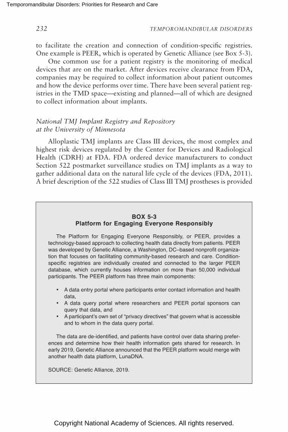
Temporomandibular Disorders: Priorities for Research and Care
Copyright National Academy of Sciences. All rights reserved.
232 TEMPOROMANDIBULAR DISORDERS
to facilitate the creation and connection of condition-specific registries. One example is PEER, which is operated by Genetic Alliance (see Box 5-3).
One common use for a patient registry is the monitoring of medical devices that are on the market. After devices receive clearance from FDA, companies may be required to collect information about patient outcomes and how the device performs over time. There have been several patient reg-istries in the TMD space—existing and planned—all of which are designed to collect information about implants.
National TMJ Implant Registry and Repository at the University of Minnesota
Alloplastic TMJ implants are Class III devices, the most complex and highest risk devices regulated by the Center for Devices and Radiological Health (CDRH) at FDA. FDA ordered device manufacturers to conduct Section 522 postmarket surveillance studies on TMJ implants as a way to gather additional data on the natural life cycle of the devices (FDA, 2011). A brief description of the 522 studies of Class III TMJ prostheses is provided
BOX 5-3 Platform for Engaging Everyone Responsibly
The Platform for Engaging Everyone Responsibly, or PEER, provides a technology-based approach to collecting health data directly from patients. PEER was developed by Genetic Alliance, a Washington, DC–based nonprofit organiza-tion that focuses on facilitating community-based research and care. Condition-specific registries are individually created and connected to the larger PEER database, which currently houses information on more than 50,000 individual participants. The PEER platform has three main components:
• A data entry portal where participants enter contact information and health data,
• A data query portal where researchers and PEER portal sponsors can query that data, and
• A participant’s own set of “privacy directives” that govern what is accessible and to whom in the data query portal.
The data are de-identified, and patients have control over data sharing prefer-ences and determine how their health information gets shared for research. In early 2019, Genetic Alliance announced that the PEER platform would merge with another health data platform, LunaDNA.
SOURCE: Genetic Alliance, 2019.
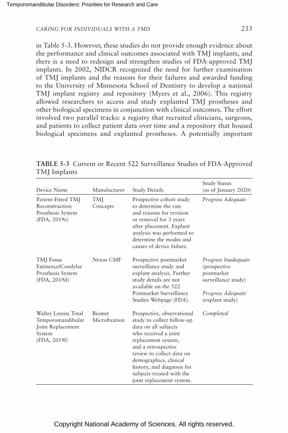
Temporomandibular Disorders: Priorities for Research and Care
Copyright National Academy of Sciences. All rights reserved.
CARING FOR INDIVIDUALS WITH A TMD 233
in Table 5-3. However, these studies do not provide enough evidence about the performance and clinical outcomes associated with TMJ implants, and there is a need to redesign and strengthen studies of FDA-approved TMJ implants. In 2002, NIDCR recognized the need for further examination of TMJ implants and the reasons for their failures and awarded funding to the University of Minnesota School of Dentistry to develop a national TMJ implant registry and repository (Myers et al., 2006). This registry allowed researchers to access and study explanted TMJ prostheses and other biological specimens in conjunction with clinical outcomes. The effort involved two parallel tracks: a registry that recruited clinicians, surgeons, and patients to collect patient data over time and a repository that housed biological specimens and explanted prostheses. A potentially important
TABLE 5-3 Current or Recent 522 Surveillance Studies of FDA-Approved TMJ Implants
Device Name Manufacturer Study DetailsStudy Status (as of January 2020)
Patient-Fitted TMJ Reconstruction Prosthesis System(FDA, 2019e)
TMJ Concepts
Prospective cohort study to determine the rate and reasons for revision or removal for 3 years after placement. Explant analysis was performed to determine the modes and causes of device failure.
Progress Adequate
TMJ Fossa Eminence/Condylar Prosthesis System (FDA, 2019d)
Nexus CMF Prospective postmarket surveillance study and explant analysis. Further study details are not available on the 522 Postmarket Surveillance Studies Webpage (FDA).
Progress Inadequate (prospective postmarket surveillance study)
Progress Adequate (explant study)
Walter Lorenz Total Temporomandibular Joint Replacement System(FDA, 2019f)
Biomet Microfixation
Prospective, observational study to collect follow-up data on all subjects who received a joint replacement system, and a retrospective review to collect data on demographics, clinical history, and diagnosis for subjects treated with the joint replacement system.
Completed

Temporomandibular Disorders: Priorities for Research and Care
Copyright National Academy of Sciences. All rights reserved.
234 TEMPOROMANDIBULAR DISORDERS
distinction is that this registry did not include samples from patients who received total joint implants, but rather only samples from those patients who had disc replacements. Funding for the registry ended in 2010, but the biospecimens remain available at the University of Minnesota and represent a potentially valuable research tool.
FDA is also working toward increasing the quality of data on the safety and effectiveness of TMJ implants through the Medical Device Epidemiol-ogy Network (MDEpiNet), a global public–private partnership initiated in 2010 (MDEpiNet, 2019a). MDEpiNet aims to advance the collection and use of real-world data from routine patient care in order to improve outcomes. In 2014, MDEpiNet convened a Medical Device Registries Task Force with the goal of strengthening the medical device postmarket surveil-lance system as a way to support better regulatory decisions and patient care (Krucoff et al., 2015). The Medical Device Registries Task Force pro-posed strategically coordinated registry networks (CRNs) as a way to link existing complementary registries and provide interoperability solutions to data-related challenges including disparate and potentially incomplete data sources. In 2017 the Department of Health and Human Services’ Office of the Assistant Secretary for Planning and Evaluation awarded FDA/CDRH funds to develop a CRN community of practice, which is comprised of 14 CRNs operating or in development, one of which is focused on TMDs (MDEpiNet, 2019b). One key stakeholder involved in developing the TMD CRN is the TMJ Patient-Led RoundTable. The TMD CRN aims to do the following:
• Create a standardized data infrastructure,• Develop new and more effective ways to incorporate patient and
real-world evidence data in clinical trials,• Support the design of predictive analytics algorithms,• Foster evidence-based protocols and best practices for inclusion into
health care, and• Promote collaborative multidisciplinary research (MDEpiNet, 2019c).
Efforts to build the TMD CRN also include coordinating with other registries and databases that collect information on the comorbidities pres-ent in TMD patients. Care is being taken to align the minimum core data-sets to the greatest extent possible. The TMD CRN is also enlisting help from patients via The TMJ Association, which has amassed a great deal of information directly from individuals who are affected by a TMD (The TMJ Association, 2019a). The involvement of public and private payers will be key to the success of the TMD CRN because they can provide valu-able input on TMD claims data and how best to standardize the collection of that information.
Temporomandibular Disorders: Priorities for Research and Care
Copyright National Academy of Sciences. All rights reserved.
CARING FOR INDIVIDUALS WITH A TMD 235
Need for a Patient Registry
Despite these important existing efforts, the committee believes that it would be beneficial to explore the development of a comprehensive national registry that would collect a wide variety of data from all types of TMD patients and would incorporate a core set of data elements. Ideally, this registry would be easily accessible for individuals with a TMD and their care providers in order to facilitate data collection. An important first step would be to gather information about best practices and lessons learned from other and ongoing patient registry efforts, including, but not limited to, the University of Minnesota implant registry, the TMD CRN, and the PEER platform. (See recommendations in Chapter 8.)
Developing and Implementing Clinical Practice Guidelines
There are currently no formal clinical practice guidelines that provide evidence about effective TMD treatments, for whom or for what specific types of TMDs particular treatments may be effective, and criteria for when to escalate treatment beyond the initial conservative approaches. Given the historical misunderstanding of TMDs and misguided treatment approaches that have led to iatrogenic harm for some, and as current diagnosis and treatment remains disparate and heterogeneous, TMD clinical practice guidelines are greatly needed.
Some professional associations, federal research agencies, and patient advocacy organizations have developed general guidance for treating TMDs. However, this guidance is largely focused on self-care, is not firmly grounded in evidence, and is in some cases inaccurate or contradictory (see Table 5-4). The American Dental Association (ADA), as part of the Choosing Wisely campaign, released guidance in 2016 on TMDs. The Choosing Wisely cam-paign is aimed at improving communication between providers and patients and encouraging patients to choose treatments that are evidence based, free from harm, and truly necessary. ADA’s guidance on TMDs encourages patients to avoid irreversible procedures as a first-line treatment and notes that many TMDs resolve spontaneously without treatment (see Table 5-4).
The American Academy of Orofacial Pain has published six editions of guidelines for assessing, diagnosing, and managing orofacial pain (de Leeuw and Klasser, 2018). The guidelines are geared toward a professional audience and have increasingly focused on rigorous incorporation of sound evidence.
The publication of these types of guidelines for diagnosing and treat-ing individuals with a TMD has elicited controversy. For example, in 2010 the American Association for Dental Research (AADR) approved a new guideline for the care of patients with TMDs (Greene, 2010). The
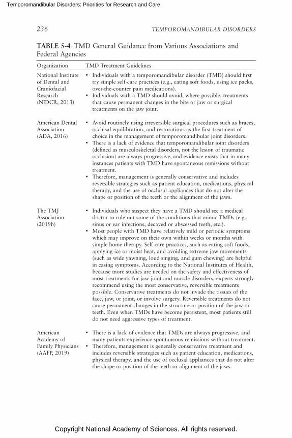
Temporomandibular Disorders: Priorities for Research and Care
Copyright National Academy of Sciences. All rights reserved.
236 TEMPOROMANDIBULAR DISORDERS
TABLE 5-4 TMD General Guidance from Various Associations and Federal Agencies
Organization TMD Treatment Guidelines
National Institute of Dental and Craniofacial Research (NIDCR, 2013)
• Individuals with a temporomandibular disorder (TMD) should first try simple self-care practices (e.g., eating soft foods, using ice packs, over-the-counter pain medications).
• Individuals with a TMD should avoid, where possible, treatments that cause permanent changes in the bite or jaw or surgical treatments on the jaw joint.
American Dental Association (ADA, 2016)
• Avoid routinely using irreversible surgical procedures such as braces, occlusal equilibration, and restorations as the first treatment of choice in the management of temporomandibular joint disorders.
• There is a lack of evidence that temporomandibular joint disorders (defined as musculoskeletal disorders, not the lesion of traumatic occlusion) are always progressive, and evidence exists that in many instances patients with TMD have spontaneous remissions without treatment.
• Therefore, management is generally conservative and includes reversible strategies such as patient education, medications, physical therapy, and the use of occlusal appliances that do not alter the shape or position of the teeth or the alignment of the jaws.
The TMJ Association (2019b)
• Individuals who suspect they have a TMD should see a medical doctor to rule out some of the conditions that mimic TMDs (e.g., sinus or ear infections, decayed or abscessed teeth, etc.).
• Most people with TMD have relatively mild or periodic symptoms which may improve on their own within weeks or months with simple home therapy. Self-care practices, such as eating soft foods, applying ice or moist heat, and avoiding extreme jaw movements (such as wide yawning, loud singing, and gum chewing) are helpful in easing symptoms. According to the National Institutes of Health, because more studies are needed on the safety and effectiveness of most treatments for jaw joint and muscle disorders, experts strongly recommend using the most conservative, reversible treatments possible. Conservative treatments do not invade the tissues of the face, jaw, or joint, or involve surgery. Reversible treatments do not cause permanent changes in the structure or position of the jaw or teeth. Even when TMDs have become persistent, most patients still do not need aggressive types of treatment.
American Academy of Family Physicians (AAFP, 2019)
• There is a lack of evidence that TMDs are always progressive, and many patients experience spontaneous remissions without treatment.
• Therefore, management is generally conservative treatment and includes reversible strategies such as patient education, medications, physical therapy, and the use of occlusal appliances that do not alter the shape or position of the teeth or alignment of the jaws.
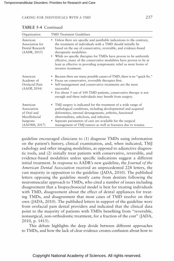
Temporomandibular Disorders: Priorities for Research and Care
Copyright National Academy of Sciences. All rights reserved.
CARING FOR INDIVIDUALS WITH A TMD 237
Organization TMD Treatment Guidelines
American Association for Dental Research (AADR, 2015)
• Unless there are specific and justifiable indications to the contrary, the treatment of individuals with a TMD should initially be based on the use of conservative, reversible, and evidence-based therapeutic modalities.
• While no specific therapies for TMDs have proven to be uniformly effective, many of the conservative modalities have proven to be at least as effective in providing symptomatic relief as most forms of invasive treatment.
American Academy of Orofacial Pain (AAOP, 2014)
• Because there are many possible causes of TMD, there is no “quick fix.”• Focus on conservative, reversible therapies first. • Self-management and conservative treatments are the most
successful.• For about 5 out of 100 TMD patients, conservative therapy is not
enough and these individuals may benefit from surgery.
American Association of Oral and Maxillofacial Surgeons (AAOMS, 2017)
• TMJ surgery is indicated for the treatment of a wide range of pathological conditions, including developmental and acquired deformities, internal derangements, arthritis, functional abnormalities, ankylosis, and infection.
• Separate parameters of care are available for the surgical management of TMJ tumors as well as fractures due to trauma.
TABLE 5-4 Continued
guideline encouraged clinicians to (1) diagnose TMDs using information on the patient’s history, clinical examination, and, when indicated, TMJ radiology and other imaging modalities, as opposed to adjunctive diagnos-tic tools, and (2) initially treat patients with conservative, reversible, and evidence-based modalities unless specific indications suggest a different initial treatment. In response to AADR’s new guideline, the Journal of the American Dental Association received an unprecedented 228 letters, the vast majority in opposition to the guideline (JADA, 2010). The published letters opposing the guideline mostly came from dentists following the neuromuscular approach to TMDs, who cited a number of issues including disagreement that a biopsychosocial model is best for treating individuals with TMD, disagreement about the effect of dental appliances for treat-ing TMDs, and disagreement that most cases of TMD resolve on their own (JADA, 2010). The published letters in support of the guideline were from orofacial pain dental providers and indicated that the clinical data point to the majority of patients with TMDs benefiting from “reversible, nonsurgical, non-orthodontic treatment, for a fraction of the cost” (JADA, 2010, p. 1415).
This debate highlights the deep divide between different approaches to TMDs, and how the lack of clear evidence creates confusion about how to
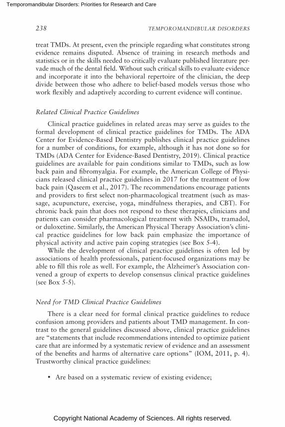
Temporomandibular Disorders: Priorities for Research and Care
Copyright National Academy of Sciences. All rights reserved.
238 TEMPOROMANDIBULAR DISORDERS
treat TMDs. At present, even the principle regarding what constitutes strong evidence remains disputed. Absence of training in research methods and statistics or in the skills needed to critically evaluate published literature per-vade much of the dental field. Without such critical skills to evaluate evidence and incorporate it into the behavioral repertoire of the clinician, the deep divide between those who adhere to belief-based models versus those who work flexibly and adaptively according to current evidence will continue.
Related Clinical Practice Guidelines
Clinical practice guidelines in related areas may serve as guides to the formal development of clinical practice guidelines for TMDs. The ADA Center for Evidence-Based Dentistry publishes clinical practice guidelines for a number of conditions, for example, although it has not done so for TMDs (ADA Center for Evidence-Based Dentistry, 2019). Clinical practice guidelines are available for pain conditions similar to TMDs, such as low back pain and fibromyalgia. For example, the American College of Physi-cians released clinical practice guidelines in 2017 for the treatment of low back pain (Qaseem et al., 2017). The recommendations encourage patients and providers to first select non-pharmacological treatment (such as mas-sage, acupuncture, exercise, yoga, mindfulness therapies, and CBT). For chronic back pain that does not respond to these therapies, clinicians and patients can consider pharmacological treatment with NSAIDs, tramadol, or duloxetine. Similarly, the American Physical Therapy Association’s clini-cal practice guidelines for low back pain emphasize the importance of physical activity and active pain coping strategies (see Box 5-4).
While the development of clinical practice guidelines is often led by associations of health professionals, patient-focused organizations may be able to fill this role as well. For example, the Alzheimer’s Association con-vened a group of experts to develop consensus clinical practice guidelines (see Box 5-5).
Need for TMD Clinical Practice Guidelines
There is a clear need for formal clinical practice guidelines to reduce confusion among providers and patients about TMD management. In con-trast to the general guidelines discussed above, clinical practice guidelines are “statements that include recommendations intended to optimize patient care that are informed by a systematic review of evidence and an assessment of the benefits and harms of alternative care options” (IOM, 2011, p. 4). Trustworthy clinical practice guidelines:
• Are based on a systematic review of existing evidence;
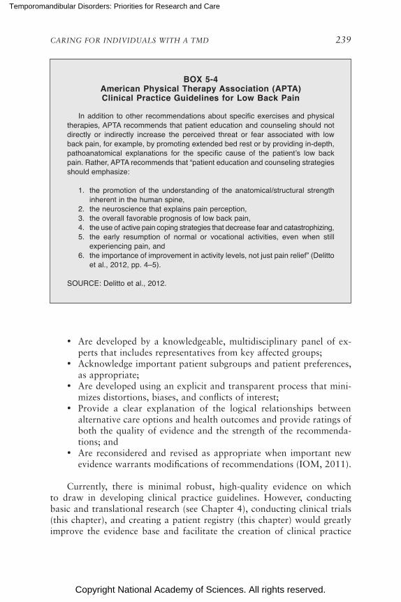
Temporomandibular Disorders: Priorities for Research and Care
Copyright National Academy of Sciences. All rights reserved.
CARING FOR INDIVIDUALS WITH A TMD 239
BOX 5-4 American Physical Therapy Association (APTA) Clinical Practice Guidelines for Low Back Pain
In addition to other recommendations about specific exercises and physical therapies, APTA recommends that patient education and counseling should not directly or indirectly increase the perceived threat or fear associated with low back pain, for example, by promoting extended bed rest or by providing in-depth, pathoanatomical explanations for the specific cause of the patient’s low back pain. Rather, APTA recommends that “patient education and counseling strategies should emphasize:
1. the promotion of the understanding of the anatomical/structural strength inherent in the human spine,
2. the neuroscience that explains pain perception, 3. the overall favorable prognosis of low back pain, 4. the use of active pain coping strategies that decrease fear and catastrophizing, 5. the early resumption of normal or vocational activities, even when still
experiencing pain, and 6. the importance of improvement in activity levels, not just pain relief” (Delitto
et al., 2012, pp. 4–5).
SOURCE: Delitto et al., 2012.
• Are developed by a knowledgeable, multidisciplinary panel of ex-perts that includes representatives from key affected groups;
• Acknowledge important patient subgroups and patient preferences, as appropriate;
• Are developed using an explicit and transparent process that mini-mizes distortions, biases, and conflicts of interest;
• Provide a clear explanation of the logical relationships between alternative care options and health outcomes and provide ratings of both the quality of evidence and the strength of the recommenda-tions; and
• Are reconsidered and revised as appropriate when important new evidence warrants modifications of recommendations (IOM, 2011).
Currently, there is minimal robust, high-quality evidence on which to draw in developing clinical practice guidelines. However, conducting basic and translational research (see Chapter 4), conducting clinical trials (this chapter), and creating a patient registry (this chapter) would greatly improve the evidence base and facilitate the creation of clinical practice
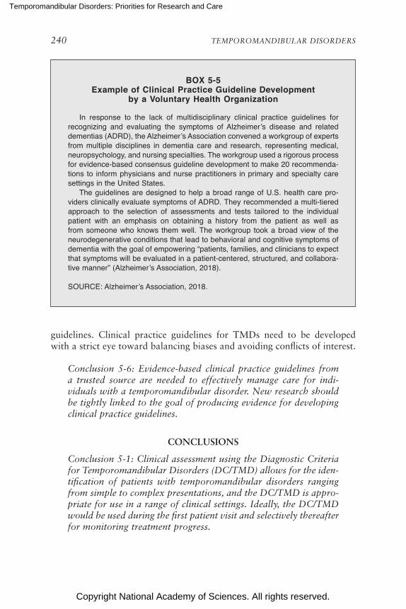
Temporomandibular Disorders: Priorities for Research and Care
Copyright National Academy of Sciences. All rights reserved.
240 TEMPOROMANDIBULAR DISORDERS
BOX 5-5 Example of Clinical Practice Guideline Development
by a Voluntary Health Organization
In response to the lack of multidisciplinary clinical practice guidelines for recognizing and evaluating the symptoms of Alzheimer’s disease and related dementias (ADRD), the Alzheimer’s Association convened a workgroup of experts from multiple disciplines in dementia care and research, representing medical, neuropsychology, and nursing specialties. The workgroup used a rigorous process for evidence-based consensus guideline development to make 20 recommenda-tions to inform physicians and nurse practitioners in primary and specialty care settings in the United States.
The guidelines are designed to help a broad range of U.S. health care pro-viders clinically evaluate symptoms of ADRD. They recommended a multi-tiered approach to the selection of assessments and tests tailored to the individual patient with an emphasis on obtaining a history from the patient as well as from someone who knows them well. The workgroup took a broad view of the neuro degenerative conditions that lead to behavioral and cognitive symptoms of dementia with the goal of empowering “patients, families, and clinicians to expect that symptoms will be evaluated in a patient-centered, structured, and collabora-tive manner” (Alzheimer’s Association, 2018).
SOURCE: Alzheimer’s Association, 2018.
guidelines. Clinical practice guidelines for TMDs need to be developed with a strict eye toward balancing biases and avoiding conflicts of interest.
Conclusion 5-6: Evidence-based clinical practice guidelines from a trusted source are needed to effectively manage care for indi-viduals with a temporomandibular disorder. New research should be tightly linked to the goal of producing evidence for developing clinical practice guidelines.
CONCLUSIONS
Conclusion 5-1: Clinical assessment using the Diagnostic Criteria for Temporomandibular Disorders (DC/TMD) allows for the iden-tification of patients with temporomandibular disorders ranging from simple to complex presentations, and the DC/TMD is appro-priate for use in a range of clinical settings. Ideally, the DC/TMD would be used during the first patient visit and selectively thereafter for monitoring treatment progress.
Temporomandibular Disorders: Priorities for Research and Care
Copyright National Academy of Sciences. All rights reserved.
CARING FOR INDIVIDUALS WITH A TMD 241
Conclusion 5-2: Self-management and patient education can be important components of care of temporomandibular disorders (TMDs). People with TMDs need access to self-management resources, including formal training. Research is needed to test and refine self-management interventions in order to identify which techniques are most effective, to determine which patients are most likely to see benefits, and to understand the mechanisms of self-management for TMDs.
Conclusion 5-3: Some elements of physical therapy—including exercise and manual therapy—have been shown to improve pain and functional outcomes for individuals with temporomandibular disorders. However, many of the studies are of low quality and further research is needed to support the use of these treatment modalities.
Conclusion 5-4: Although considerable research has been con-ducted in occlusal adjustment and equilibration for temporo-mandibular disorders (TMDs), these treatments have not been found to be effective. Evidence-based findings need to be widely disseminated to dentists and other clinicians to ensure that the treatment approaches individuals with a TMD receive are consis-tently based on the best available evidence and focused on starting with conservative approaches.
Conclusion 5-5: Data are inadequate and are of poor quality for most treatments for temporomandibular disorders (TMDs). Research is needed to determine safe and effective treatments for TMDs. Systematic reviews and methodologically rigorous new studies are needed.
Conclusion 5-6: Evidence-based clinical practice guidelines from a trusted source are needed to effectively manage care for indi-viduals with a temporomandibular disorder. New research should be tightly linked to the goal of producing evidence for developing clinical practice guidelines.
REFERENCES
AADR (American Association for Dental Research). 2015. Temporomandibular disor-ders (TMDs). http://www.iadr.org/AADR/About-Us/Policy-Statements/Science-Policy/ Temporomandibular-Disorders-TMD (accessed August 30, 2019).
Temporomandibular Disorders: Priorities for Research and Care
Copyright National Academy of Sciences. All rights reserved.
242 TEMPOROMANDIBULAR DISORDERS
AAFP (American Academy of Family Physicians). 2019. Choosing wisely: Avoid routinely using irreversible surgical procedures such as braces, occlusal equilibration, and restora tions as the first treatment of choice in the management of temporo mandibular join disorders. https://www.aafp.org/afp/recommendations/viewRecommendation.htm?recommendationId=313# (accessed August 30, 2019).
AAOMS (American Association of Oral and Maxillofacial Surgeons). 1993. Recommenda-tions for management of patients with temporomandibular joint implants. Temporo-mandibular Joint Implant Surgery Workshop. Journal of Oral and Maxillofacial Surgery 51(10):1164–1172.
AAOMS. 2017. Parameters of care: Clinical practice guidelines for oral and maxillofacial surgery. Temporomandibular joint surgery. https://www.aaoms.org/docs/member-center/parcare/parcare_8_temporomandibular.pdf (accessed August 30, 2019).
AAOP (American Academy of Orofacial Pain). 2014. Temporomandibular joint disorders (patient brochure). https://aaop.clubexpress.com/content.aspx?page_id=22&club_id=508439&module_id=108085 (accessed February 2, 2020).
ACTTION (Analgesic, Anesthetic, and Addiction Clinical Trial Translations, Innovations, Opportunities, and Networks). 2020. ACTTION. http://www.acttion.org (accessed Feb-ruary 2, 2020).
ADA (American Dental Association). 2016. Five things dentists and patients should ques-tion. http://www.choosingwisely.org/societies/american-dental-association (accessed Decem ber 30, 2019).
ADA Center for Evidence-Based Dentistry. 2019. Clinical practice guidelines. https://ebd.ada.org/en/evidence/guidelines (accessed December 30, 2019).
Adams, R. J. 2010. Improving health outcomes with better patient understanding and educa-tion. Risk Management and Healthcare Policy 3:61–72.
Aggarwal, V. R., K. Lovell, S. Peters, H. Javidi, A. Joughin, and J. Goldthorpe. 2011. Psycho-social interventions for the management of chronic orofacial pain. Cochrane Database of Systematic Reviews 11:CD008456.
Aggarwal, V. R., Y. Fu, C. J. Main, and J. Wu. 2019. The effectiveness of self-management interventions in adults with chronic orofacial pain: A systematic review, meta-analysis, and meta-regression. European Journal of Pain 23(5):849–865.
Ahmad, M., L. Hollender, Q. Anderson, K. Kartha, R. Ohrbach, E. L. Truelove, M. T. John, and E. L. Schiffman. 2009. Research Diagnostic Criteria for Temporomandibular Dis-orders (RDC/TMD): Development of image analysis criteria and examiner reliability for image analysis. Oral Surgery, Oral Medicine, Oral Pathology, Oral Radiology, and Endodontics 107:844–860.
Alzheimer’s Association. 2018. Alzheimer’s Association workgroup reports recommenda-tions to improve timely and accurate diagnosis and disclosure. https://www.alz.org/aaic/releases_2018/AAIC18-Sun-clinical-practice-guidelines.asp (accessed October 4, 2019).
Armijo-Olivo, S., L. Potance, V. Singh, F. Neto, N. Thie, and A. Michelotti. 2016. Effectiveness of manual therapy and therapeutic exercise for temporomandibular disorders: Systematic review and meta-analysis. Physical Therapy 96(1):9–25.
Berrueta, L., I. Muskaj, S. Olenich, T. Butler, G. J. Badger, R. A. Colas, M. Spite, C. N. Serhan, and H. M. Langevin. 2016. Stretching impacts inflammation resolution in connective tissue. Cell Physiology 231(7):1621–1627.
Bialosky, J. E., M. D. Bishop, D. D. Price, M. E. Robinson, and S. Z. George. 2009. The mechanisms of manual therapy in the treatment of musculoskeletal pain: A comprehen-sive model. Manual Therapy 14(5):531–538.
Bjørnland, T., A. Gjaerum, and A. Møystad. 2007. Osteoarthritis of the temporomandibular joint: An evaluation of the effects and complications of corticosteroid injection compared with injection with sodium hyaluronate. Journal of Oral Rehabilitation 34(8):583–589.
Temporomandibular Disorders: Priorities for Research and Care
Copyright National Academy of Sciences. All rights reserved.
CARING FOR INDIVIDUALS WITH A TMD 243
Blau, J. N. 1982. How to take a history of head or facial pain. British Medical Journal 285:1249–1251.
Bothwell, L. E., J. A. Greene, S. H. Podolsky, and D. S. Jones. 2016. Assessing the gold standard: Lessons from the history of RCTS. New England Journal of Medicine 374(22):2175–2181.
Bouloux, G. F., J. Chou, D. Krishnan, T. Aghaloo, N. Kahenasa, J. A. Smith, and H. Giannakopoulos. 2017a. Is hyaluronic acid or corticosteroid superior to lactated Ringer solution in the short-term reduction of temporomandibular joint pain after arthro centesis? Part 1. Journal of Oral and Maxillofacial Surgery 75(1):52–62.
Bouloux, G. F., J. Chou, D. Krishnan, T. Aghaloo, N. Kahenasa, J. A. Smith, and H. Giannakopoulos. 2017b. Is hyaluronic acid or corticosteroid superior to lactated Ringer solution in the short term for improving function and quality of life after arthrocentesis? Part 2. Journal of Oral and Maxillofacial Surgery 75(1):63–72.
Breckons, M., S. M. Bissett, C. Exley, V. Arauio-Soares, and J. Durham. 2017. Care pathways in persistent orofacial pain: Qualitative evidence from the DEEP study. Journal of Dental Researach—Clinical and Translational Research 2(1):48–57.
Brito, R. G., L. A. Rasmussen, and K. A. Sluka. 2017. Regular physical activity prevents development of chronic muscle pain through modulation of supraspinal opioid and serotonergic mechanisms. Pain Reports 2(5):e618.
Brown, D. T., and E. L. Gaudet, Jr. 2002. Temporomandibular disorder treatment outcomes: Second report of a large-scale prospective clinical study. Cranio 20:244–253.
Campbell, B. K., R. B. Fillingim, S. Lee, R. Brao, D. D. Price, and J. K. Neubert. 2017. Effects of high-dose capsaicin on TMD subjects: A randomized clinical study. Journal of Dental Research, Clinical and Translational Research 2(1):58–65.
CDC (Centers for Disease Control and Prevention). 2019. Guideline for prescribing opioids for chronic pain. https://www.cdc.gov/drugoverdose/prescribing/guideline.html (accessed January 24, 2020).
Cezairli, B., E. C. Sivrikaya, M. M. Omezli, F. Ayranci, and N. S. Cezairli. 2017. Results of combined, single-session arthrocentesis and dextrose prolotherapy for symptomatic temporomandibular joint syndrome: A case series. Journal of Alternative and Comple-mentary Medicine 23:771–777.
Chaurand, J., L. Pacheco-Ruíz, H. Orozco-Saldívar, and J. López-Valdés. 2017. Efficacy of botulinum toxin therapy in treatment of myofascial pain. Journal of Oral Science 59:351–356.
Chen, Y-W., Y-W. Chiu, C-Y. Chen, and S-K. Chuang. 2015. Botulinum toxin therapy for temporomandibular joint disorders: A systematic review of randomized controlled trials. International Journal of Oral and Maxillofacial Surgery 44:1018–1026.
Chiappelli, F., A. Barkhordarian, G. Demerjian, Q. Bach, and V. Kasar. 2015. Cluster random-ized stepped wedge blinded controlled trials (CRSWBCT) in comparative effectiveness research (CER). Part II: Implications for temporomandibular joint disorders (TMD) research. Translational Medicine 5:e131.
Clark, G. T., Y. Tsukiyama, K. Baba, and T. Watanabe. 1999. Sixty-eight years of experi mental occlusal interference studies: What have we learned? Journal of Prosthetic Dentistry 82:704–713.
Clarke, N. G. 1982. Occlusion and myofascial pain dysfunction: Is there a relationship? Jour-nal of the American Dental Association 104:443–446.
Cochrane Library. 2016a. Occlusal adjustment for treating and preventing temporomandibular joint disorders—reason for withdrawal from publication. https://www.cochranelibrary.com/cdsr/doi/10.1002/14651858.CD003812.pub2/full (accessed December 11, 2019).
Temporomandibular Disorders: Priorities for Research and Care
Copyright National Academy of Sciences. All rights reserved.
244 TEMPOROMANDIBULAR DISORDERS
Cochrane Library. 2016b. Orthodontics for treating temporomandibular joint (TMJ) disorders—reason for withdrawal from publication. https://www.cochranelibrary.com/cdsr/doi/10.1002/14651858.CD006541.pub3/full/es (accessed December 11, 2019).
COMET (Core Outcome Measures in Effectiveness Trials) Initiative. 2020. Core Outcome Measures in Effectiveness Trials. http://www.comet-initiative.org (accessed February 14, 2020).
Conti, P. C. R., E. N. De Alencar, A. S. Da Mota Correa, J. R. P. Lauris, A. L. Porporatti, and Y. M. Costa. 2012. Behavioural changes and occlusal splints are effective in the management of masticatory myofascial pain: A short-term evaluation. Journal of Oral Rehabilitation 39(10):754–760.
Cooper, B. C. 2011. Temporomandibular disorders: A position paper of the International Col-lege of Cranio-Mandibular Orthopedics (ICCMO). Cranio 29:237–244.
Craane, B., P. U. Dijkstra, K. Stappaerts, and A. De Laat. 2012. One-year evaluation of the effect of physical therapy for masticatory muscle pain: A randomized controlled trial. European Journal of Pain 16(5):737–747.
Daif, E. T. 2010. Autologous blood injection as a new treatment modality for chronic recurrent temporomandibular joint dislocation. Oral Surgery, Oral Medicine, Oral Pathology, Oral Radiology, and Endodontology 109(1):31–36.
Davoudi, A., H. Khaki, I. Mohammadi, M. Daneshmand, A. Tamizifar, M. Bigdelou, and F. Ansaripoor. 2018. Is arthrocentesis of temporomandibular joint with cortico-steroids beneficial? A systematic review. Medicina Oral, Patologia Oral y Cirugia Bucal 23:e367–e375.
Dawson, P. E. 1996. A classification system for occlusions that relates maximal intercuspation to the position and condition of the temporomandibular joints. Journal of Prosthetic Dentistry 75:60–66.
de Leeuw, R and G. Klasser. 2018. Orofacial pain: guidelines for assessment, diagnosis, and management, sixth edition. Hanover Park, IL: Quintessence Publishing.
de Pedro, M., R. M. López-Pintor, J. L. de la Hoz-Aizpurua, E. Casañas, and G. Hernández. 2019. Efficacy of low-level laser therapy for the therapeutic management of neuro-pathic orofacial pain: A systematic review. Journal of Oral & Facial Pain & Headache 34(1):13–30.
de Resende, C. M. B. M., F. G. L. De Oliveira Medeiros, C. R. De Figueiredo Rego, A. De Sousa Leite Bispo, G. A. S. Barbosa, and E. O. De Almeida. 2019. Short-term effective-ness of conservative therapies in pain, quality of life, and sleep in patients with temporo-mandibular disorders: A randomized clinical trial. Journal of Craniomandibular & Sleep Practice June 15. [Epub ahead of print].
de Souza, R. F., C. H. L. da Silva, M. Nasser, Z. Fedorowicz, and M. A. Al-Muharraqi. 2012. Interventions for managing temporomandibular joint osteoarthritis. Cochrane Database of Systematic Reviews CD007261.
Delitto, A., S. Z. George, L. Van Dillen, J. M. Whitman, G. Sowa, P. Shekelle, T. R. Denninger, J. J. Godges, and Orthopaedic Section of the American Physical Therapy Association. 2012. Low back pain: Clinical practice guidelines linked to the International Classifica-tion of Functioning, Disability, and Health from the Orthopaedic Section of the American Physical Therapy Association. Journal of Orthopaedic and Sports Physical Therapy 42(4):A1–A57.
Deyo, R. A., S. F. Dworkin, D. Amtmann, G. Andersoon, D. Borenstein, E. Carragee, J. Carrino, R. Chou, K. Cook, A. Delittlo, C. Goertz, P. Khalsa, J. Loeser, S. Mackey, J. Panagis, J. Rainville, T. Tosteson, D. Turk, M. Von Korff, and D. K. Weiner. 2014. Re-port of the NIH Task Force on Research Standards for Chronic Low Back Pain. Spine 39:1128–1143.
Temporomandibular Disorders: Priorities for Research and Care
Copyright National Academy of Sciences. All rights reserved.
CARING FOR INDIVIDUALS WITH A TMD 245
Dimitroulis, G., M. Dolwick, and A. Martinez. 1995. Temporomandibular joint arthrocentesis and lavage for the treatment of closed lock: A follow-up study. British Journal of Oral and Maxillofacial Surgery 33:23–27.
Durham, J., R. Touger-Decker, D. R. Nixdorf, D. Rigassio-Radler, and P. Moynihan. 2015. Oro-facial pain and nutrition: A forgotten relationship. Journal of Oral Rehabilitation 42(1):75–80.
Dworkin, R. H., D. C. Turk, J. T. Farrar, J. A. Haythornthwaite, M. P. Jensen, N. P. Katz, R. D. Kerns, G. Stucki, R. R. Allen, N. Bellamy, D. B. Carr, J. Chandler, P. Cowan, R. Dionne, B. S. Galer, S. Hertz, A. R. Jadad, L. D. Kramer, D. C. Manning, S. Martin, C. G. McCormick, M. P. McDermott, P. McGrath, S. Quessy, B. A. Rappaport, W. Robbins, J. P. Robinson, M. Rothman, M. A. Royal, L. Simon, J. W. Stauffer, W. Stein, J. Tollett, J. Wernicke, J. Witter, and IMMPACT. 2005. Core outcome measures for chronic pain clinical trials: IMMPACT recommendations. Pain 113(1–2):9–19.
Dworkin, S. F., K. H. Huggins, L. Wilson, L. Manci, J. Turner, D. Massoth, L. LeResche, and E. Truelove. 2002. A randomized clinical trial using research diagnostic criteria for temporomandibular disorders-axis II to target clinic cases for a tailored self-care TMD treatment program. Journal of Orofacial Pain 16:48–63.
FDA (Food and Drug Administration). 2011. FDA orders postmarket surveillance of certain TMJ implants. https://wayback.archive-it.org/7993/20170114063520/http:/www.fda.gov/NewsEvents/Newsroom/PressAnnouncements/ucm242421.htm (accessed June 12, 2019).
FDA. 2019a. Premarket approval P980052. https://www.accessdata.fda.gov/scripts/cdrh/ cfdocs/cfpma/pma.cfm?id=P980052 (accessed November 20, 2019).
FDA. 2019b. Premarket approval P000035. https://www.accessdata.fda.gov/scripts/cdrh/ cfdocs/cfpma/pma.cfm?id=P000035 (accessed November 20, 2019).
FDA. 2019c. Premarket approval P020016. https://www.accessdata.fda.gov/scripts/cdrh/ cfdocs/cfpma/pma.cfm?id=P020016 (accessed November 20, 2019).
FDA. 2019d. 522 postmarket surveillance studies. https://www.accessdata.fda.gov/scripts/cdrh/cfdocs/cfPMA/pss.cfm (accessed November 20, 2019).
FDA. 2019e. 522 postmarket surveillance studies: PS110003/PSS001. https://www. accessdata.fda.gov/scripts/cdrh/cfdocs/cfPMA/pss.cfm?t_id=40&c_id=393 (accessed January 2, 2020).
FDA. 2019f. 522 postmarket surveillance studies: PS110004/PSS001. https://www. accessdata.fda.gov/scripts/cdrh/cfdocs/cfPMA/pss.cfm?t_id=41&c_id=394 (accessed January 2, 2020).
Ferrando, M., M. J. Galdon, E. Dura, Y. Andreu, Y. Jiminez, and R. Poveda. 2012. Enhancing the efficacy of treatment for temporomandibular patients with muscle diagnosis through cognitive-behavioral intervention, including hypnosis: A randomized study. Oral Medi-cine 113:81–89.
Ferreira, J. N., C. C. Ko, S. Myers, J. Swift, and J. R. Fricton. 2008. Evaluation of surgically retrieved temporomandibular joint alloplastic implants: Pilot study. Journal of Oral and Maxillofacial Surgery 66(6):1112–1124.
Forssell, H., E. Kalso, P. Koskela, R. Vehmanen, P. Puukka, and P. Alanen. 1999. Occlusal treatments in temporomandibular disorders: A qualitative systematic review of random-ized controlled trials. Pain 83(3):549–560.
Fricton, J. 2006. Current evidence providing clarity in management of temporomandibular disorders: Summary of a systematic review of randomized clinical trials for intra-oral appliances and occlusal therapies. Journal of Evidence Based Dental Practice 6:48–52.
Fridrich, K. L., J. M. Wise, and D. L. Zeitler. 1996. Prospective comparison of arthroscopy and arthrocentesis for temporomandibular joint disorders. Journal of Oral and Maxillofacial Surgery 54(7):816–820.
Temporomandibular Disorders: Priorities for Research and Care
Copyright National Academy of Sciences. All rights reserved.
246 TEMPOROMANDIBULAR DISORDERS
GAO (Government Accountability Office). 2007. Medical devices: FDA’s approval of four tem-poromandibular joint implants. GAO-0-996. https://www.gao.gov/assets/270/266885.pdf (accessed December 12, 2019).
Gauer, R. L., and M. J. Semidey. 2015. Diagnosis and treatment of temporomandibular dis-orders. American Family Physician 91(6):378–386.
Genetic Alliance. 2019. PEER: Platform for Engaging Everyone Responsibly. http://www.geneticalliance.org/programs/biotrust/peer (accessed October 14, 2019).
Gewandter, J. S., S. M. Smith, A. McKeown, K. Edwards, A. Narula, J. R. Pawlowski, D. Rothstein, P. J. Desjardins, S. F. Dworkin, R. A. Gross, R. Ohrbach, B. A. Rappaport, B. J. Sessle, D. C. Turk, and R. H. Dworkin. 2015. Reporting of adverse events and statistical details of efficacy estimates in randomized clinical trials of pain in temporo-mandibular disorders: Analgesic, Anesthetic, and Addiction Clinical Trial Translations, Innovations, Opportunities, and Networks systematic review. Journal of the American Dental Association 146(4):246–254.
Gliklich, R. E., N. A. Dreyer, and M. B. Leavy, eds. 2014. Registries for evaluating patient outcomes: A user’s guide, 3rd edition. https://effectivehealthcare.ahrq.gov/products/ registries-guide-3rd-edition/research (accessed December 12, 2019).
Gopalakrishnan, V., S. A. Nagori, S. K. Roy Chowdhury, and V. Saxena. 2018. The use of intra-articular analgesics to improve outcomes after temporomandibular joint arthrocentesis: A review. Oral and Maxillofacial Surgery 22(4):357–364.
Greene, C. S. 2010. Managing the care of patients with temporomandibular disorders: A new guideline for care. Journal of the American Dental Association 141:1086–1088.
Gruber, E. A., J. McCullough, and A. J. Sidebottom. 2015. Medium-term outcomes and complications after total replacement of the temporomandibular joint. Prospective out-come analysis after 3 and 5 years. British Journal of Oral and Maxillofacial Surgery 53(5):412–415.
Haggman-Henrikson, B., B. Wiesinger, and A. Wanman. 2018. The effect of supervised exercise on localized TMD pain and TMD pain associated with generalized pain. Acta Odontologica Scandinavica 76(1):6–12.
Henry, C. H., and L. M. Wolford. 1993. Treatment outcomes for temporomandibular joint reconstruction after Proplast-Teflon implant failure. Journal of Oral and Maxillofacial Surgery 51(4):352–360.
Hill, K. P., M. D. Palastro, B. Johnson, and J. W. Ditre. 2017. Cannabis and pain: A clinical review. Cannabis and Cannabinoid Research 2(1):96–104.
Hoffman, B. M., R. K. Papas, D. K. Chatkoff, and R. D. Kerns. 2007. Meta-analysis of psychological interventions for chronic low back pain. Health Psychology 26(1):1–9.
Holden, G. 1991. The relationship of self-efficacy appraisals to subsequent health related outcomes: A meta-analysis. Social Work in Health Care 16(1):53–93.
IMMPACT (Initiative on Methods, Measurement, and Pain Assessment in Clinical Trials). 2019. IMMPACT. http://www.immpact.org (accessed October 4, 2019).
Inagaki, T., T. Miyaoka, H. Shinno, J. Horiguchi, S. Matsuda, and H. Yoshikawa. 2007. Treatment of temporomandibular pain with the selective serotonin reuptake inhibitor paroxetine. Primary Care Companion to the Journal of Clinical Psychiatry 9(1):69–70.
IOM (Institute of Medicine). 2011. Clinical practice guidelines we can trust. Washington, DC: The National Academies Press.
Jackson, T., Y. Wang, Y. Wang, and H. Fan. 2014. Self-efficacy and chronic pain outcomes: A meta-analytic review. Journal of Pain 15(8):800–814.
JADA (Journal of the American Dental Association). 2010. Letters. Journal of the American Dental Association 141(12):1407–1417.
Temporomandibular Disorders: Priorities for Research and Care
Copyright National Academy of Sciences. All rights reserved.
CARING FOR INDIVIDUALS WITH A TMD 247
Jokstad, A., A. Mo, and B. S. Krogstad. 2005. Clinical comparison between two different splint designs for temporomandibular disorder therapy. Acta Odontologica Scandinavica 63(4):218–226.
Kamyszek, G., R. Ketcham, R. Garcia, Jr., and J. Radke. 2001. Electromyographic evidence of reduced muscle activity when ULF-TENS is applied to the Vth and VIIth crian nerves. Cranio 19(3):162–168.
Kassirer, J. P., and R. I. Kopelman. 1991. Learning clinical reasoning. Baltimore, MD: Williams and Wilkins.
Keenan, J. R. 2015. Unclear results for the use of botulinum toxin therapy for TMD pain. Evidence-Based Dentistry 16:122.
Khawaja, S. N., S. J. Scrivani, N. Holland, and D. A. Keith. 2017. Effectiveness, safety, and predictors of response to botulinum toxin type A in refractory masticatory myalgia: A retrospective study. Journal of Oral and Maxillofacial Surgery 75:2307–2315.
Kia, S., and E. Choy. 2017. Update on treatment guideline in fibromyalgia syndrome with focus on pharmacology. Biomedicines 5(2):20.
Kimos, P., C. Biggs, J. Mah, G. Heo, S. Rashiq, N. M. Thie, and P. W. Major. 2007. Analgesic action of gabapentin on chronic pain in the masticatory muscles: A randomized con-trolled trial. Pain 127(1–2):151–160.
Klasser, G. D., and C. S. Greene. 2009. The changing field of temporomandibular disorders: What dentists need to know. Journal of the Canadian Dental Association 75:49–53.
Koh, H., and P. G. Robinson. 2003. Occlusal adjustment for treating and preventing temporo-mandibular joint disorders. Cochrane Database of Systematic Reviews CD003812.
Kotiranta, U., T. Suvinen, and H. Forssell. 2014. Tailored treatments in temporomandibular disorders: Where are we now? A systematic qualitative literature review. Journal of Oral & Facial Pain and Headache 28(1):28–37.
Krucoff, M. W., A. Sedrakyan, and S. T. Normand. 2015. Bridging unmet medical device eco-system needs with strategically coordinated registries networks. JAMA 314(16):1691–1692.
Kunjur, J., R. Niziol, and N. S. Matthews. 2016. Quality of life: Patient-reported outcomes after total replacement of the temporomandibular joint. British Journal of Oral and Maxillofacial Surgery 54(7):762–766.
Kusiak, J. W., C. Veasley, W. Maixner, R. B. Fillingim, J. S. Mogil, L. Diatchenko, C. B. Meloto, I. Belfer, M. Kaseta, T. Kalinowski, J. Gagnier, N. Hu, C. Alvarez-Garriga, M. Bayona, B. Eloff, M. Reardon, C. Greene, A. Bertagna, T. Cowley, J. Wilentz, and D. Clare. 2018. The TMJ Patient-Led RoundTable: A History and Summary of Work. http://mdepinet.org/wp-content/uploads/TMJ-Patient-RoundTable-Briefing-Report_9_25_18.pdf (accessed January 31, 2020).
Kuzmanovic Pficer, J., S. Dodic, V. Lazic, G. Trajkovic, N. Milic, and B. Milic. 2017. Occlusal stabilization splint for patients with temporomandibular disorders: Meta-analysis of short and long term effects. PLoS One 12(2):e0171296.
Langevin, H. M., T. Fujita, N. A. Bouffard, T. Takano, C. Koptiuch, G. J. Badger, and M. Nedergaard. 2013. Fibroblast cytoskeletal remodeling induced by tissue stretch involves ATP signaling. Cell Physiology 228(9):1922–1926.
Lee, M. K., B. Y. Choi, and D. K. Ahn. 2008. Low doses of cannabinoids enhance the anti-nociceptive effects of intracisternally administered mGluRs groups II and III agonists in formalin-induced TMJ nociception in rats. Pain 139(2):367–375.
Leite, F. M. G., Á. Atallah, R. El Dib, E. Grossmann, E. Januzzi, R. B. Andriolo, and E. M. K. da Silva. 2009. Cyclobenzaprine for the treatment of myofascial pain in adults. Cochrane Database of Systematic Reviews CD006830.
Levy, P. H. 1975. Clinical implications of mandibular repositioning and the concept of an alterable centric relation. Dental Clinics of North America 19:543–570.
Temporomandibular Disorders: Priorities for Research and Care
Copyright National Academy of Sciences. All rights reserved.
248 TEMPOROMANDIBULAR DISORDERS
Li, C., N. Su, X. Yang, X. Yang, Z. Shi, and L. Li. 2012. Ultrasonography for detection of disc displacement of temporomandibular joint: A systematic review and meta-analysis. Journal of Oral and Maxillofacial Surgery 70:1300–1309.
Lima, L. V., T. S. S. Abner, and K. A. Sluka. 2017a. Does exercise increase or decrease pain? Central mechanisms underlying these two phenomena. Journal of Physiology 595(13):4141–4150.
Lima, L. V., J. M. DeSantana, L. A. Rasmussen, and K. A. Sluka. 2017b. Short-duration physical activity prevents the development of activity-induced hyperalgesia through opioid and serotoninergic mechanisms. Pain 158(9):1697–1710.
Litt, M. D., D. M. Shafer, and D. L. Kreutzer. 2010. Brief cognitive-behavioral treatment for TMD pain: Long-term outcomes and moderators of treatment. Pain 151(1):110–116.
Liu, H., Q. Liang, P. Xiao, H. Jiao, Y. Gao, and C. Ahmetjiang. 2012. The effectiveness of cognitive–behavioral therapy for temporomandibular disorders: A systematic review. Journal of Oral Rehabilitation 39(1):55–62.
Liu, Y., J. Wu, W. Fei, X. Cen, Y. Xiong, S. Wang, Y. Tang, and X. Liang. 2018. Is there a difference in intra-articular injections of corticosteroids, hyaluronate, or placebo for temporo-mandibular osteoarthritis? Journal of Oral and Maxillofacial Surgery 76(3):504–514.
Lorig, K., R. L. Chastain, E. Ung, S. Shoor, and H. R. Holman. 1989. Development and evaluation of a scale to measure perceived self‐efficacy in people with arthritis. Arthritis and Rheumatism 32(1):37–44.
Luther, F., S. Layton, and F. McDonald. 2010. Orthodontics for treating temporomandibular joint (TMJ) disorders. Cochrane Database of Systematic Reviews CD006541.
Lypka, M., and D. D. Yamashita. 2007. Exuberant foreign body giant cell reaction to a Teflon/Proplast temporomandibular joint implant: Report of a case. Journal of Oral and Maxillo facial Surgery 65(9):1680–1684.
Machado, E., P. Machado, V. F. Wandscher, A. M. E. Marchionatti, F. B. Zanatta, and O. B. Kaiser. 2018. A systematic review of different substance injection and dry needling for treatment of temporomandibular myofascial pain. International Journal of Oral and Maxillofacial Surgery 47(11):1420–1432.
Machon, V., S. Abramowicz, J. Paska, and M. F. Dolwick. 2009. Autologous blood injection for the treatment of chronic recurrent temporomandibular joint dislocation. Journal of Oral and Maxillofacial Surgery 67(1):114–119.
Maia, M. L., L. R. Bonjardim, J. Quintans, M. Ribeiro, L. Maia, and P. C. Conti. 2012. Effects of low-level laser therapy on pain levels in patients with temporomandibular disorders: A systematic review. Journal of Applied Oral Science 20(6):594–602.
Manfredini, D. 2018. Occlusal equilibration for the management of temporomandibular disorders. Oral and Maxillofacial Surgery Clinics of North America 30(3):257–264.
Manfredini, D., T. Castroflorio, G. Perinetti, and L. Guarda-Nardini. 2012. Dental occlusion, body posture and temporomandibular disorders: Where we are now and where we are heading for. Journal of Oral Rehabilitation 39:463–471.
Martin, W. J., and T. Forouzanfar. 2011. The efficacy of anticonvulsants on orofacial pain: A systematic review. Oral Surgery, Oral Medicine, Oral Pathology, Oral Radiology, and Endodontology 111(5):627–633.
Martins, D. F., L. Mazzardo-Martins, V. M. Gadotti, F. P. Nascimento, D. A. Lima, B. Speckhann, G. A. Favretto, E. Cargnin-Ferreira, E. Bressan, R. C. Dutra, J. B. Calixto, and A. R. Santos. 2011. Ankle joint mobilization reduces axonotmesis-induced neuropathic pain and glial activation in the spinal cord and enhances nerve regeneration in rats. Pain 152(11):2653–2661.
Temporomandibular Disorders: Priorities for Research and Care
Copyright National Academy of Sciences. All rights reserved.
CARING FOR INDIVIDUALS WITH A TMD 249
Martins, D. F., F. Bobinski, L. Mazzardo-Martins, F. J. Cidral-Filho, F. P. Nascimento, V. M. Gadotti, and A. R. Santos. 2012. Ankle joint mobilization decreases hypersensitivity by activation of peripheral opioid receptors in a mouse model of postoperative pain. Pain Medicine 13(8):1049–1058.
Martins, D. F., L. Mazzardo-Martins, F. J. Cidral-Filho, J. Stramosk, and A. R. Santos. 2013a. Ankle joint mobilization affects postoperative pain through peripheral and central adenosine A1 receptors. Physical Therapy 93:401–412.
Martins, D. F., L. Mazzardo-Martins, F. J. Cidral-Filho, V. M. Gadotti, and A. R. Santos. 2013b. Peripheral and spinal activation of cannabinoid receptors by joint mobilization alleviates postoperative pain in mice. Neuroscience 255:110–121.
McCain, J. P., and R. H. Hossameldin. 2011. Advanced arthroscopy of the temporo-mandibular joint. Atlas of the Oral and Maxillofacial Surgery Clinics of North America 19(2):145–167.
McNamara, J. A. 1997. Orthodontic treatment and temporomandibular disorders. Oral Surgery Oral Medicine Oral Pathology Oral Radiology & Endodontics 83:107–117.
McNeely, M. L., O. S. Armijo, and D. J. Magee. 2006. A systematic review of the effectiveness of physical therapy interventions for temporomandibular disorders. Physical Therapy 86:710–725.
MDEpiNet (Medical Device Epidemiology Network). 2019a. The medical device epidemiology network. http://mdepinet.org (accessed December 30, 2019).
MDEpiNet. 2019b. CRN community of practice. http://mdepinet.org/crn-community-practice (accessed December 30, 2019).
MDEpiNet. 2019c. TMJ CRN. mdepinet.org/TMJ-crn (accessed December 30, 2019). Medlicott, M. S., and S. R. Harris. 2006. A systematic review of the effectiveness of exercise,
manual therapy, electrotherapy, relaxation training, and biofeedback in the management of temporomandibular disorder. Physical Therapy 86(7):955–973.
Melo, G., E. Casett, J. Stuginski-Barbosa, E. N. S. Guerra, D. A. Fernandes, A. L. Porporatti, C. Flores-Mir, and G. De Luca Canto. 2018. Effects of glucosamine supplements on painful temporomandibular joint osteoarthritis: A systematic review. Journal of Oral Rehabilitation 45(5):414–422.
Mercuri, L. G. 1999. Subjective and objective outcomes in patients reconstructed with a custom-fitted alloplastic temporomandibular joint prosthesis. Journal of Oral and Maxillo facial Surgery 57:1427–1430.
Mohl, N. D. 1984. The role of head posture in mandibular function. In W. K. Solberg and G. T. Clark, eds. Abnormal jaw mechanics: Diagnosis and treatment. Chicago, IL: Quintessence. Pp. 97–116.
Monaco, A., F. Sgolastra, D. Pietropaoli, M. Giannoni, and R. Cattaneo. 2013. Comparison between sensory and motor transcutaneous electrical nervous stimulation on electro-myographic and kinesiographic activity of patients with temporomandibular disorder: A controlled clinical trial. BMC Musculoskeletal Disorders 14:168.
Morley, S., A. C. Williams, and C. Eccleston. 2013. Examining the evidence of psychological treatments for chronic pain: Time for a paradigm shift? Pain 154(10):1929–1931.
Mujakperuo, H. R., M. Watson, R. Morrison, and T. V. Macfarlane. 2010. Pharmacological interventions for pain in patients with temporomandibular disorders. Cochrane Database Systematic Reviews 10:CD004715.
Myers, S., S. Kaimal, J. Springsteen, J. Ferreira, C. C. Ko, and J. Fricton. 2006. Develop-ment of a National TMJ Implant Registry and Repository: NIDCR’s TIRR. Northwest Dentistry 86(6):13–18.
Nagashima, W., H. Kimura, M. Ito, T. Tokura, M. Arao, B. Aleksic, K. Yoshida, K. Kurita, and N. Ozaki. 2012. Effectiveness of duloxetine for the treatment of chronic nonorganic orofacial pain. Clinical Neuropharmacology 35(6):273–277.
Temporomandibular Disorders: Priorities for Research and Care
Copyright National Academy of Sciences. All rights reserved.
250 TEMPOROMANDIBULAR DISORDERS
NASEM (National Academies of Sciences, Engineering, and Medicine). 2017. Pain manage-ment and the opioid epidemic: Balancing societal and individual benefits and risks of prescription opioid use. Washington, DC: The National Academies Press.
Nasri-Heir, C., J. B. Epstein, R. Touger-Decker, and R. Benoliel. 2016. What should we tell patients with painful temporomandibular painful temporomandibular disorders about what to eat? Journal of the American Dental Association 47(8):667–671.
National Dental PBRN (Practice-Based Research Network). 2019. National Dental Practice-Based Research Network. http://nationaldentalpbrn.org (accessed January 24, 2020).
Navarrete, A. L., K. L. Rafferty, Z. J. Liu, W. Ye, G. M. Greenlee, and S. W. Herring. 2013. Botulinum neurotoxin type A in the masseter muscle: Effects on incisor eruption in rabbits. American Journal of Orthodontics and Dentofacial Orthopedics 143(4):499–506.
NICE (National Institute for Health and Care Excellence). 2014. Overview: Total prosthetic replacement of the temporomandibular joint: Guidance. www.nice.org.uk/guidance/ipg500 (accessed October 3, 2019).
Nichol, A. D., M. Bailey, D. J. Cooper, POLAR, and EPO Investigators. 2010. Challenging issues in randomized controlled trials. Injury 41(Suppl 1):S20–S23.
NIDCR (National Institute of Dental and Craniofacial Research). 2013. Less is often best in treating TMJ disorders. https://www.nidcr.nih.gov/sites/default/files/2017-09/less-is-best-tmj.pdf (accessed August 30, 2019).
NIDCR. 2018. TMJ (Temporomandibular Joint & Muscle Disorders). https://www.nidcr.nih.gov/health-info/tmj/more-info (accessed November 18, 2019).
Nitzan, D. W., and A. Price. 2001. The use of arthrocentesis for the treatment of osteoarthritic temporomandibular joints. Journal of Oral and Maxillofacial Surgery 59:1154–1159.
Nitzan, D. W., J. Svidovsky, A. Zini, and Y. Zadik. 2017. Effect of arthrocentesis on symp-tomatic osteoarthritis of the temporomandibular joint and analysis of the effect of preoperative clinical and radiologic features. Journal of Oral and Maxillofacial Surgery 75:260–267.
Ohrbach, R., G. Slade, E. Bair, N. Rathnayaka, L. Diatchenko, J. D. Greenspan, W. Maixner, and R. B. Fillingim. 2020. Premorbid and concurrent predictors of TMD onset and per-sistence. European Journal of Pain 24(1):145–158.
Oliveira, C. B., C. G. Maher, R. Z. Pinto, A. C. Traeger, C. C. Lin, J. F. Chenot, M. van Tulder, and B. W. Koes. 2018. Clinical practice guidelines for the management of non-specific low back pain in primary care: An updated overview. European Spine Journal 27(11):2791–2803.
Onoriobe, U., M. Miloro, C. Sukotjo, L. G. Mercuri, A. Lotesto, and R. Eke. 2016. How many temporomandibular joint total joint alloplastic implants will be placed in the United States in 2030? Journal of Oral and Maxillofacial Surgery 74(8):1531–1538.
Ouanounou, A., M. Goldberg, and D. A. Haas. 2017. Pharmacotherapy in temporo mandibular disorders: A review. Journal of the Canadian Dental Association 83:h7.
PCORNet (National Patient-Centered Clinical Research Network). 2019. PCORnet. https://pcornet.org/clinical-research-network (accessed October 4, 2019).
Peck, C. C., J-P. Goulet, F. Lobbezoo, E. L. Schiffman, P. Alstergren, G. C. Anderson, R. de Leeuw, R. Jensen, A. Michelotti, R. Ohrbach, A. Petersson, and T. List. 2014. Expanding the taxonomy of the diagnostic criteria for temporomandibular disorders. Journal of Oral Rehabilitation 41:2–23.
Pihut, M., M. Gorecka, P. Ceranowicz, and M. Wieckiewicz. 2018. The efficiency of anterior repositioning splints in the management of pain related to temporomandibular joint disc displacement with reduction. Pain Research and Management 9089286.
Pike, A., L. Hearn, and A. C. Williams. 2016. Effectiveness of psychological interventions for chronic pain on health care use and work absence: Systematic review and meta-analysis. Pain 157(4):777–785.
Temporomandibular Disorders: Priorities for Research and Care
Copyright National Academy of Sciences. All rights reserved.
CARING FOR INDIVIDUALS WITH A TMD 251
Pullinger, A. G., D. A. Seligman, and W. K. Solberg. 1988. Temporomandibular disorders. Part II: Occlusal factors associated with temporomandibular joint tenderness and dys-function. Journal of Prosthetic Dentistry 59:363–367.
Qaseem, A., T. J. Wilt, R. M. McLean, M. A. Forciea, and Clinical Guidelines Committee of the American College of Physicians. 2017. Noninvasive treatments for acute, subacute, and chronic low back pain: A clinical practice guideline from the American College of Physicians. Annals of Internal Medicine 166(7):514–530.
Racich, M. J. 2018. Occlusion, temporomandibular disorders, and orofacial pain: An evidence-based overview and update with recommendations. Journal of Prosthetic Dentistry 120:678–685.
Rajan, R., and Y. M. Sun. 2017. Reevaluating antidepressant selection in patients with bruxism and temporomandibular joint disorder. Journal of Psychiatric Practice 23(3):173–179.
Randhawa, K., R. Bohay, P. Cote, G. van der Velde, D. Sutton, J. J. Wong, H. Yu, D. Southerst, S. Varatharajan, S. Mior, M. Stupar, H. M. Shearer, C. Jacobs, and A. Taylor-Vaisey. 2016. The effectiveness of noninvasive interventions for temporomandibular disorders: A systematic review by the Ontario Protocol for Traffic Injury Management (OPTIMa) Collaboration. Clinical Journal of Pain 32(3):260–278.
Refai, H. 2017. Long-term therapeutic effects of dextrose prolotherapy in patients with hyper-mobility of the temporomandibular joint: A single-arm study with 1–4 years’ follow up. British Journal of Oral and Maxillofacial Surgery 55:465–470.
Resnick, C. M., P. M. Vakliain, M. Breen, D. Zurakowski, P. Caruso, L. Henderson, P. A. Nigrovic, L. B. Kaban, and Z. S. Peacock. 2016. Quantifying temporomandibular joint synovitis in children with juvenile idiopathic arthritis. Arthritis Care Research 68(12):1795–1802.
Resnick, C. M., R. Dang, L. A. Henderson, D. A. Zander, K. M. Daniels, P. A. Nigrovic, and L. B. Kaban. 2017. Frequency and morbidity of temporomandibular joint involvement in adult patients with a history of juvenile idiopathic arthritis. Journal of Oral and Maxillo-facial Surgery 75(6):1191–1200.
Rizzatti-Barbosa, C. M., M. T. Nogueira, E. D. de Andrade, G. M. Ambrosano, and J. R. de Barbosa. 2003. Clinical evaluation of amitriptyline for the control of chronic pain caused by temporomandibular joint disorders. Cranio 21(3):221–225.
Rodrigues, D., A. O. Siriani, and F. Bérzin. 2004. Effect of conventional TENS on pain and electromyographic activity of masticatory muscles in TMD patients. Brazilian Oral Research 18(4):290–295.
Rugh, J. D., G. S. Graham, J. C. Smith, and R. K. Ohrbach. 1989. Effects of canine versus molar occlusal splint guidance on nocturnal bruxism and craniomandibular symptom-atology. Journal of Craniomandibular Disorders 3(4):203–210.
Schiffman, E., R. Ohrbach, E. Truelove, J. Look, G. Anderson, J-P. Goulet, T. List, P. Svensson, F. Lobbezoo, A. Michelotti, S. L. Brooks, W. Ceusters, M. Drangsholt, D. Ettlin, C. Gaul, L. J. Goldberg, J. A. Haythornthwaite, L. Hollender, R. Jensen, M. T. John, A. De Laat, R. de Leeuw, W. Maixner, M. vaqn der Meulen, G. M. Murray, D. R. Nixdorf, S. Palla, A. Petersson, P. Pionchon, B. Smith, C. M. Visscher, J. Zakrzewska, S. F. Dworkin, International RDC/TMD Consortium Network, International Association for Dental Research, Orofacial Pain Special Interest Group, and International Association for the Study of Pain. 2014. Diagnostic Criteria for Temporomandibular Disorders (DC/TMD) for Clinical and Research Applications: Recommendations of the International RDC/TMD Consortium Network and Orofacial Pain Special Interest Group. Journal of Oral and Facial Pain and Headache 28(1):6–27.
Shaheed, C. A., C. G. Maher, and A. J. McLachlan. 2019. Efficacy and safety of low-dose codeine-containing combination analgesics for pain: Systematic review and meta-analysis. Clinical Journal of Pain 35(10):836–843.
Temporomandibular Disorders: Priorities for Research and Care
Copyright National Academy of Sciences. All rights reserved.
252 TEMPOROMANDIBULAR DISORDERS
Shanavas, M., L. Chatra, P. Shenai, P. K. Rao, V. Jagathisth, S. P. Kumar, and B. Naduvakkattu. 2014. Transcutaneous electrical nerve stimulation therapy: An adjuvant pain control-ling modality in TMD patients—a clinical study. Dental Research Journal (Isfahan) 11(6):676–679.
Sharma, S., M. Breckons, B. Bronnimann-Lambelet, J. W. Chung, T. List, F. Lobbezoo, D. R. Nixdorf, J. F. Oyarzo, C. Peck, Y. Tsukiyama, and R. Ohrbach. 2019. Challenges in the clinical implementation of a biopsychosocial model for assessment and management of orofacial pain. Journal of Oral Rehabilitation 47(1):87–100.
Shedden-Mora, M. C., D. Weber, A. Neff, and W. Rief. 2013. Biofeedback-based cognitive-behavioral treatment compared with occlusal splint for temporomandibular disorder: A randomized controlled trial. Clinical Journal of Pain 29(12):1057–1065.
Sidebottom, A. J., UK TMJ Replacement Surgeons, and British Association of Oral and Maxillo facial Surgeons. 2008. Guidelines for the replacement of temporo mandibular joints in the United Kingdom. British Journal of Oral and Maxillofacial Surgery 46(2):146–147.
Singer, E., and R. Dionne. 1997. A controlled evaluation of ibuprofen and diazepam for chronic orofacial muscle pain. Journal of Orofacial Pain 11(2):139–146.
Singh, B. P., S. Jayaraman, R. Kirubakaran, S. Joseph, M. S. Muthu, H. Jivnani, F. Hua, and N. Singh. 2017. Occlusal interventions for managing temporomandibular disorders (protocol). Cochrane Database of Systematic Reviews 11:CD012850.
Skyba, D. A., R. Radhakrishnan, J. J. Rohlwing, A. Wright, and K. A. Sluka. 2003. Joint manipulation reduces hyperalgesia by activation of monoamine receptors but not opioid or GABA receptors in the spinal cord. Pain 106(1–2):159–168.
Slade, G. D., R. Ohrbach, J. D. Greenspan, R. B. Fillingim, E. Bair, A. E. Sanders, R. Dubner, L. Diatchenko, C. B. Meloto, S. Smith, and W. Maixner. 2016. Painful temporo-mandibular disorder: Decade of discovery from OPPERA studies. Journal of Dental Research 95(10):1084–1092.
Sluka, K. A., L. Frey-Law, and M. Hoeger Bement. 2018. Exercise-induced pain and analgesia? Underlying mechanisms and clinical translation. Pain 159(Suppl 1):S91–S97.
Soares, A., R. B. Andriolo, A. N. Atallah, and E. M. da Silva. 2014. Botulinum toxin for myo-fascial pain syndromes in adults. Cochrane Database Systematic Reviews 7:CD007533.
Southard, T. E., K. A. Southard, and E. A. Tolley. 1990. Variation of approximal tooth contact tightness with postural change. Journal of Dental Research 69:1776–1779.
Ta, L. E., and R. A. Dionne. 2004. Treatment of painful temporomandibular joints with a cyclooxygenase-2 inhibitor: A randomized placebo-controlled comparison of celecoxib to naproxen. Pain 111(1):13–21.
Tesch, R. S., L. C. D. S. P. Macedo, F. S. Fernandes, G. S. Goffredo Filho, and C. P. Q. F. Goes. 2019. Effectiveness of dry needling on the local pressure pain threshold in patients with masticatory myofascial pain. Systematic review and preliminary clinical trial. Cranio 27:1–9.
The TMJ Association. 2016. Botox’s effect on the jaw joint. http://www.tmj.org/site/page?pageId=218 (accessed February 3, 2020).
The TMJ Association. 2017. TMD nutrition and you. http://www.tmj.org/site/page?pageId=318 (accessed October 3, 2019).
The TMJ Association. 2019a. TMJ Patient RoundTable Project: Status update. http://www.tmj.org/site/page?pageId=408 (accessed December 30, 2019).
The TMJ Association. 2019b. TMD treatments. http://www.tmj.org/Page/52/20 (accessed December 30, 2019).
Tocaciu, S., M. J. McCullough, and G. Dimitroulis. 2019. Surgical management of recurrent TMJ dislocation: A systematic review. Oral and Maxillofacial Surgery 23(1):35–45.
Temporomandibular Disorders: Priorities for Research and Care
Copyright National Academy of Sciences. All rights reserved.
CARING FOR INDIVIDUALS WITH A TMD 253
Tour, J., M. Lofgren, K. Mannerkorpi, B. Gerdle, A. Larsson, A. Palstam, I. Bilevicite-Ljungar, J. Bjersing, I. Martin, M. Ernberg, M. Schalling, and E. Kosek. 2017. Gene-to-gene interactions regulate endogenous pain modulation in fibromyalgia patients and healthy controls-antagonistic effects between opioid and serotonin-related genes. Pain 158(7):1194–1203.
Truelove, E., K. H. Huggins, L. Mancl, and S. F. Dworkin. 2006. The efficacy of traditional, low-cost and nonsplint therapies for temporomandibular disorder: A randomized con-trolled trial. Journal of the American Dental Association 137:1099–1107.
Turner, J. A., L. Mancl, and L. A. Aaron. 2006. Short- and long-term efficacy of brief cognitive-behavioral therapy for patients with chronic temporomandibular disorder pain: A randomized controlled trial. Pain 121(3):181–194.
Türp, J., and H. Schindler. 2012. The dental occlusion as a suspected cause for TMDs: Epi-demiological and etiological considerations. Journal of Oral Rehabilitation 39:502–512.
Türp J. C., F. Komine, and A. Hugger. 2004. Efficacy of stabilization splints for the manage-ment of patients with masticatory muscle pain: A qualitative systematic review. Clinical Oral Investigations 8(4):179–195.
Türp, J. C., A. Jokstad, E. Motschall, H. J. Schindler, I. Windecker-Getaz, and D. A. Ettlin. 2007. Is there a superiority of multimodal as opposed to simple therapy in patients with temporomandibular disorders? A qualitative systematic review of the literature. Clinical Oral Implants Research 18(Suppl 3):138–150.
van der Zaag, J., F. Lobbezoo, D. J. Wicks, C. M. Visscher, H. L. Hamburger, and M. Naeije. 2005. Controlled assessment of the efficacy of occlusal stabilization splints on sleep bruxism. Journal of Orofacial Pain 19(2):151–158.
Vance, C. G., D. L. Dailey, B. A. Rakel, and K. A. Sluka. 2014. Using TENS for pain control: The state of the evidence. Pain Management 4:197–209.
Velly, A. M., E. L. Schiffman, D. B. Rindal, J. Cunha-Cruz, G. H. Gilbert, M. Lehmann, A. Horowitz, and J. Fricton. 2013. The feasibility of a clinical trial of pain related to temporo mandibular muscle and joint disorders: The results of a survey from the collabo-ration on networked dental and oral research dental practice-based research networks. Journal of the American Dental Association 144(1):e1–e10.
Wanman, A., and S. Marklund. 2019. Treatment outcome of supervised exercise, home exercise and bite splint therapy, respectively, in patients with symptomatic disc displacement with reduction: A randomised clinical trial. Journal of Oral Rehabilitation 47(2):143–149.
Weinfurt, K. P., A. F. Hernandez, G. D. Coronado, L. L. DeBar, L. M. Dember, B. B. Green, P. J. Heagerty, S. S. Huang, K. T. James, J. G. Jarvik, E. B. Larson, V. Mor, R. Platt, G. E. Rosenthal, E. J. Septimus, G. E. Simon, K. L. Staman, J. Sugarman, M. Vazquez, D. Zatzick, and L. H. Curtis. 2017. Pragmatic clinical trials embedded in healthcare systems: Generalizable lessons from the NIH Collaboratory. BMC Medical Research Methodology 17(1):144.
Williams, A. C., C. Eccleston, and S. Morley. 2012. Psychological therapies for the manage-ment of chronic pain (excluding headache) in adults. Cochrane Database of Systematic Reviews 11:D007407.
Wolford, L. M., L. G. Mercuri, E. D. Schneiderman, R. Movahed, and W. Allen. 2015. Twenty-year follow-up study on a patient-fitted temporomandibular joint prosthesis: The Techmedica/TMJ Concepts device. Journal of Oral and Maxillofacial Surgery 73(5):952–960.
Wu, J. Y., C. Zhang, Y. P. Xu, Y. Y. Yu, L. Peng, W. D. Leng, Y. M. Niu, and M. H. Deng. 2017. Acupuncture therapy in the management of the clinical outcomes for temporomandibular disorders: A PRISMA-compliant meta-analysis. Medicine (Baltimore) 96(9):e6064.
Zhou, H., K. Hu, and Y. Ding. 2014. Modified dextrose prolotherapy for recurrent temporo-mandibular joint dislocation. British Journal of Oral and Maxillofacial Surgery 52:63–66.

Temporomandibular Disorders: Priorities for Research and Care
Copyright National Academy of Sciences. All rights reserved.
Temporomandibular Disorders: Priorities for Research and Care
Copyright National Academy of Sciences. All rights reserved.
255
When we began our journey consulting “medical professionals” (den-tists, oral surgeons, rheumatologists, physical therapists, chiropractors, etc.), there was lack of knowledge and understanding, lack of standards of care, mixed diagnosis, conflicting treatments options, etc. There was very little (if any) collaboration with other health care professionals, and poor communication.
—Michelle and Alexandra
Our current “system” (for lack of a better word) for treating TMD is not only broken but it’s fragmented, and patients are falling through the cracks and left feeling abandoned and alone.
—Adriana V.
TMD HEALTH CARE PATHWAYS
Current Care Pathways
Individuals seeking care for temporomandibular disorder (TMD)- related symptoms enter the health care system through various entrance points and have a wide variety of experiences in locating health care professionals who are knowledgeable about TMDs and who can provide quality care. For some patients, the initial evaluation and management is performed by a primary care physician or a general dentist. This management is effective for some patients, but for patients whose TMD condition is chronic and does not
6
Improving TMD Health Care: Practice, Education, Access,
and Coverage
Temporomandibular Disorders: Priorities for Research and Care
Copyright National Academy of Sciences. All rights reserved.
256 TEMPOROMANDIBULAR DISORDERS
respond to conservative care, multiple other specialties may be engaged. Patients may seek care from a dentist who focuses on TMDs and orofacial pain, an orthodontist, an oral and maxillofacial surgeon, a physical thera-pist, a behavioral therapist, a chiropractor, or another type of health care professional. No standardized referral pattern for the escalation of diagnosis and treatment exists. Fragmented and siloed care models may lead to delays in diagnosis and management for some patients and frustration by both patients and clinicians. Health care professionals from different disciplines have varied approaches to TMDs, and even clinicians within the same dis-cipline may have disparate ideas about appropriate care options (Greene and Bertagna, 2019). For example, dentists who hold an occlusal viewpoint might recommend adjustments to the teeth or jaw, while other dentists who follow a biopsychosocial model might opt for conservative treatments and psychological supports, and oral and maxillofacial surgeons may lean toward performing injections or surgery (Greene and Bertagna, 2019). As Table 6-1 demonstrates, an individual may visit multiple types of health care professionals and receive very different treatments from each—some of which may be beneficial and others that may have no added value or be harmful. A challenge for patients with the most serious and complex TMD cases is that currently there is not a medical or dental home for TMD care. TMD and orofacial pain centers of excellence (discussed below) as well as expanded availability of orofacial pain specialists could help address the need for TMD patients to be able to access the expertise and coordinated care they need in bridging the medical and dental divide.
Most health care professionals, including dentists, receive minimal or no training in TMDs or pain management during their entry-level education programs, residency and post-graduate training, or continuing education. Clinicians who have received more intensive training in TMDs and oro-facial pain are limited in number. Because there are relatively few clinicians specializing in these conditions, geography may be a barrier; as for many other health issues, patients in rural areas face particular obstacles accessing quality care. Insurance reimbursement for TMD treatments can be complex and difficult to navigate, and the quality of care varies from one clinician to the next. Often there is little collaboration among dentists, physicians, and other health care professionals, so patients may bounce back and forth between multiple professionals without ever receiving a satisfactory diag-nosis or treatment.
Qualitative interviews with patients with chronic TMDs have revealed three themes about the current care system (Breckons et al., 2017):
1. The fluidity of the care pathway: patients move among health care professionals within and between the dental and medical systems as they try to have their pain diagnosed and managed;
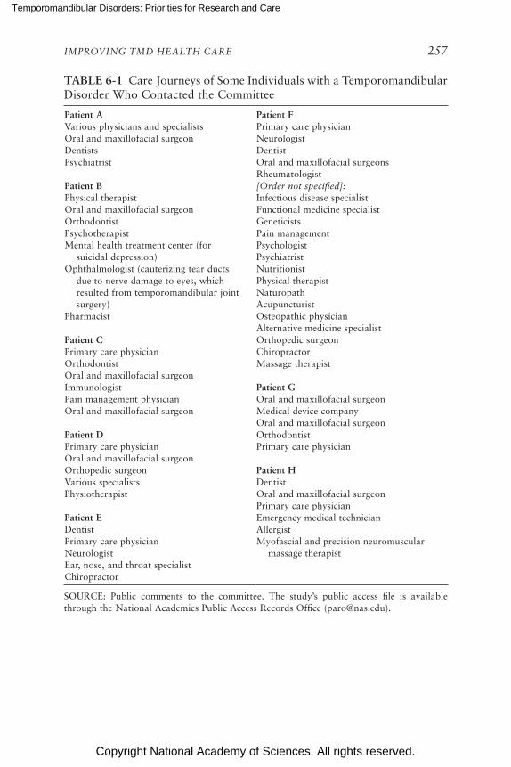
Temporomandibular Disorders: Priorities for Research and Care
Copyright National Academy of Sciences. All rights reserved.
IMPROVING TMD HEALTH CARE 257
TABLE 6-1 Care Journeys of Some Individuals with a Temporomandibular Disorder Who Contacted the Committee
Patient AVarious physicians and specialistsOral and maxillofacial surgeonDentistsPsychiatrist
Patient BPhysical therapistOral and maxillofacial surgeonOrthodontistPsychotherapistMental health treatment center (for
suicidal depression)Ophthalmologist (cauterizing tear ducts
due to nerve damage to eyes, which resulted from temporomandibular joint surgery)
Pharmacist
Patient CPrimary care physicianOrthodontistOral and maxillofacial surgeonImmunologistPain management physicianOral and maxillofacial surgeon
Patient DPrimary care physicianOral and maxillofacial surgeonOrthopedic surgeonVarious specialistsPhysiotherapist
Patient EDentistPrimary care physicianNeurologistEar, nose, and throat specialistChiropractor
Patient FPrimary care physicianNeurologistDentistOral and maxillofacial surgeonsRheumatologist[Order not specified]:Infectious disease specialistFunctional medicine specialistGeneticistsPain managementPsychologistPsychiatristNutritionistPhysical therapistNaturopathAcupuncturistOsteopathic physicianAlternative medicine specialistOrthopedic surgeonChiropractorMassage therapist
Patient GOral and maxillofacial surgeonMedical device companyOral and maxillofacial surgeonOrthodontistPrimary care physician
Patient HDentistOral and maxillofacial surgeonPrimary care physicianEmergency medical technicianAllergistMyofascial and precision neuromuscular
massage therapist
SOURCE: Public comments to the committee. The study’s public access file is available through the National Academies Public Access Records Office ([email protected]).
Temporomandibular Disorders: Priorities for Research and Care
Copyright National Academy of Sciences. All rights reserved.
258 TEMPOROMANDIBULAR DISORDERS
2. A failure to progress: despite multiple appointments, patients are frustrated with the delays they encounter in obtaining a diagnosis and effective treatment for their pain; and
3. The effects of unmanaged pain: the longer that pain is unmanaged, the greater its potential to negatively affect patients’ lives.
Vision of a Patient-Centered Care Pathway
In contrast to the typical care pathway outlined above, in an ideal world a patient who is experiencing symptoms of a TMD would visit a primary health care professional who would be knowledgeable about the basics of TMD just as other health conditions, including what patients can do with self-care and when a referral to a specialist is indicated. Unless there was unusual severity or complexity to the symptoms or examination findings, the initial starting point for many patients would be treatments such as self-management and physical therapy (see Chapter 5). If these initial treatments did not improve symptoms within a defined time period, the patient could be referred to a specialist with more advanced training in TMDs. Ideally, such specialty referrals would be coordinated across disciplines so that referring practitioners would work together to find appropriate treatments.
Specialty care might be provided by an individual health care profes-sional, an interprofessional practice of dentists and physicians, a center for excellence in TMDs and orofacial pain, or another type of collabora-tive practice. Care from these specialists would be available in person or through telehealth platforms. The collaborative approach would allow a patient to access treatments as varied as rehabilitative exercises, cognitive-behavioral therapy, physical therapy, surgery, medications, or acupuncture, depending on the patient’s preferences and the specialists’ assessment of the patient’s particular condition. The details of these treatments would all be readily accessible to all health care professionals through an integrated health record system, and the patient’s progress would be monitored by a primary health care professional who would serve as a navigator and liaison between the various specialists. Payment for services would be straightfor-ward and consistent, and the role of the primary clinician as navigator and liaison would be fully covered and understood by the payment mechanism.
Moving Toward This Future
The committee identified several barriers that prevent this patient-centered care pathway from becoming reality. These include:
• a lack of training in TMDs for frontline health care professionals such as general dentists, primary care and internal medicine clinicians,
Temporomandibular Disorders: Priorities for Research and Care
Copyright National Academy of Sciences. All rights reserved.
IMPROVING TMD HEALTH CARE 259
nurse practitioners, and physician assistants, who typically field the initial complaints and manage patients with TMDs;
• difficulty finding health care professionals with specialized training and education in TMDs and orofacial pain;
• poor integration between medical and dental practice and insurance coverage, and poor communication and collaboration with other health care professionals and specialists; and
• medical and dental reimbursement systems that are complex, dif-ficult to navigate, exclusionary (i.e., treatments of the temporo-mandibular joint [TMJ] are often not covered), and often structured to reward interventions regardless of likely effectiveness.
This chapter highlights ways to address these barriers and to improve care for individuals with a TMD by focusing on making improvements in the following areas:
• interprofessional collaboration,• the education and training of health care professionals,• access to specialty care, and• payment and coverage.
IMPROVING INTERPROFESSIONAL EDUCATION AND COLLABORATION
As discussed in Chapter 2, TMDs are a set of diverse disorders affecting the masticatory system. The more than 30 disorders in the set of TMDs have varied symptoms and care needs, and thus individuals with TMDs need a vari ety of management and treatment options. Some patients with a TMD may respond quickly to an initial treatment that is recommended by a general dentist or primary care clinician. Other patients may need more individualized care from specialist health care professionals, such as those in surgery or physical therapy. Still other patients may have a TMD that mani-fests with symptoms affecting multiple systems; these patients may require care from multiple specialists. As seen in Table 6-1, while some patients seek care from multiple clinicians, this care is often not coordinated between the clinicians. An individual health care professional—whether a dentist, oral and maxillofacial surgeon, or physical therapist—will be most familiar with and likely to recommend treatments that fall within his or her area of training and expertise. If assessment and patient care were coordinated between health care professionals—or, better yet, provided by an integrated interprofessional team—patients could access treatments tailored to their specific needs rather than treatments offered solely by a specific clinician. An interprofessional approach, including physicians, dentists, nurses, mental
Temporomandibular Disorders: Priorities for Research and Care
Copyright National Academy of Sciences. All rights reserved.
260 TEMPOROMANDIBULAR DISORDERS
health professionals, physical therapists, nutritionists, and others, would allow health care professionals to collaborate and communicate about the patient’s needs and to learn more about other professionals’ knowledge about and approach to TMDs.
Interprofessional approaches are under way in many areas of pain man-agement. Pain is a “complex sensory and emotional experience” (Klasser and Gremillion, 2013, p. 398). Models of pain management have been devel oped that focus on the physical, psychosocial, and behavioral aspects of pain management; the involvement of multiple types of clinicians not only ensures a breadth of knowledge and experience, but also contributes “to the integration of that information into a multifaceted team approach” (Klasser and Gremillion, 2013, p. 398). Beginning collaboration early, with interprofessional education during professional school, is essential in foster-ing understandings between the professions and respect for the roles of other health care professionals (Klasser and Gremillion, 2013). Expansion of these types of educational programs would allow health professionals from many disciplines to prepare for treating a patient in an interprofes-sional, collaborative team that takes a holistic view of patients and their symptoms.
Dental–Medical Divide
One of the major barriers to effective TMD care is the dental–medical divide (Powers et al., 2017). In most places in the United States, dentistry and medicine have separate education systems, separate practices, and separate financing arrangements. This divide has significant consequences for the health of patients, particularly given the wealth of information available on the impact of oral care on an individual’s overall health. Each year, 108 million Americans visit a physician but not a dentist, and primary care physicians rarely ask patients about oral health signs and symptoms (Atchison et al., 2018). Conversely, each year 27 million Americans visit a dentist but not a physician, and dentists rarely ask patients about preventive health behaviors, such as flu shots, human papillomavirus vaccines, mam-mograms, or screening for diabetes (Atchison et al., 2018).
For patients with a TMD, this divide can be even more consequential because TMDs can involve multiple systems that are traditionally separated from each other but which are inexorably intertwined. The masticatory system is an integrated system that involves the TMJ, masticatory muscles, and teeth (see Appendix D); that is connected to the body through the head and neck; and that is controlled by the peripheral and central nervous system. The symptoms of TMDs can affect oral, physical, and psycho-logical health and well-being; for example, patients with a TMD can have limitations in dental hygiene, talking, eating, smiling, and sexual activities.
Temporomandibular Disorders: Priorities for Research and Care
Copyright National Academy of Sciences. All rights reserved.
IMPROVING TMD HEALTH CARE 261
However, because of the siloed nature of dentistry and medicine, patients usually either receive care from only one type of health care professional or else must navigate and coordinate care between multiple health care professionals themselves. Many dentists and physicians are likely to focus on the systems and symptoms of TMDs that fall within their specialized area of training, rather than viewing the TMD condition and the patient in a holistic, integrated manner. The financing mechanisms for dental and medical care are also generally separate, making interprofessional collabo-ration and team care even more difficult.
One approach for bridging the dental–medical divide is to integrate dental and medical practices within the same health system or have them use the same insurance program. For example, Kaiser Permanente in the Pacific Northwest integrates dental and medical care (Kaiser Permanente Dental, 2019). This allows dentists to access medical records and doc-tors to access dental records, and medical and dental care are often in the same building, allowing for real-time interprofessional collaboration and patient consultation and treatment. One benefit to the integration of dental and medical services is that it allows team members to address care gaps. Kaiser’s patient portal can be used by dentists and dental hygienists to remind patients of medical appointments, needed screenings and care (e.g., vaccines), and follow-up reminders. At some Kaiser locations all patients receive a blood pressure screening at dental exams and are referred to their primary health care professionals if the reading is elevated. This type of model could allow physicians and dentists to work together to address the multiple systems, symptoms, and needs of some patients with TMDs.
Other approaches for bridging the dental–medical divide include:
• interprofessional education during graduate school that teaches den-tists and physicians about the unique knowledge and skills that each brings to the table and prepares them to work together as a team;
• interprofessional continuing education to bring practicing physicians and dentists together to discuss and explore ways to better collabo-rate; and
• formal agreements between dental and medical practices regarding referrals, communication, and collaboration.
Conclusion 6-1: Due to education, training, and financing mecha-nisms, there is often a lack of collaboration between clinicians, particularly dentists and physicians, and this divide can make it challenging for patients with temporomandibular disorders to access and coordinate care.
Temporomandibular Disorders: Priorities for Research and Care
Copyright National Academy of Sciences. All rights reserved.
262 TEMPOROMANDIBULAR DISORDERS
STRENGTHENING EDUCATION AND TRAINING
Although primary care and internal medicine clinicians and general dentists are typically the entry point for patients seeking care, most of these clinicians receive little to no education and training in the diagnosis or treat-ment of TMDs. Other health care professionals (including nurses, nurse practitioners, psychologists, physical therapists, and physician assistants) also receive little to no education and training in TMDs. A number of ap-proaches could be taken to improve education and training for health care professionals, including adding TMD information to health professional school curricula, adding TMD care as an area tested in licensing exams, expanding post-graduate fellowship and residency opportunities, and im-proving continuing education. These potential areas for improvement are explored below for the fields of medicine, dentistry, physical therapy, and nursing.
Medicine
Curriculum
Medical education and training currently includes little training about diagnosing and treating TMDs, orofacial pain, or pain management in general (IOM, 2011). The curriculum requirements for U.S. medical schools do not include specific education in TMDs, and neither TMDs nor oral health are included by the Association of American Medical Colleges or the American Association of Colleges of Osteopathic Medicine in their list of most frequently included subjects (AACOM, 2019; AAMC, 2019). Pain also receives inadequate attention in medical school, despite pain being the most common reason that people visit a health care professional (Fishman et al., 2013). Studies of medical schools in Canada, the United Kingdom, and the United States have found that few schools require much, if any, dedicated course time on pain, and students report feeling inadequately prepared to manage patients’ pain (Fishman et al., 2013). However, there is an increasing awareness of the need to improve pain education in medical schools nationwide, and some schools have begun to add additional content on pain management. The committee acknowledges the challenges of add-ing more content to already full curricula but given the impact of TMDs on health and well-being, the recommendations provided in Chapter 8 urge medical, nursing, and other health professional schools to provide the information that health care professionals need on TMDs.
Furthermore, there is an increasing emphasis on interprofessional educa-tion within medical and other health professional schools, with 143 U.S. allo-pathic medical schools requiring this component as of 2018 (AAMC, 2018).
Temporomandibular Disorders: Priorities for Research and Care
Copyright National Academy of Sciences. All rights reserved.
IMPROVING TMD HEALTH CARE 263
While not specific to TMDs, improving the broad area of pain manage-ment education within medical schools could be an initial step toward im-proving the management of all physicians of TMDs. Consensus-based core competencies in pain management were developed by an interprofessional group of pain experts and have been endorsed by many professional orga-nizations (Fishman et al., 2013). The endorsement and adoption of pain competencies and curriculum guidelines by the American Medical Associa-tion would provide an initial step toward recognition by medical schools of the need to improve pain science education within the curriculum. As part of a pain management curriculum, there could be specific recommendations for education on TMDs along with other high-impact pain conditions such as low back pain and fibromyalgia.
Another path for changing curricula would be by changing accredita-tion standards. Medical schools in the United States are accredited either by the Liaison Committee on Medical Education (LCME) (for M.D. pro-grams), or the Commission on Osteopathic College Accreditation (COCA) of the American Osteopathic Association (for D.O. programs). Neither the LCME nor the COCA accreditation standards detail specific areas of cur-riculum or specific competencies that must be covered in medical school. Rather, the faculty at a medical school is responsible for defining the competencies to be achieved, and is responsible for the design and imple-mentation of the elements of the curriculum that will enable its students to fully achieve those competencies and objectives (COCA, 2019; LCME, 2019). The LCME provides a few guidelines on general areas that should be covered in the curriculum (e.g., prevention, diagnosis, treatment, and problem-solving skills) (LCME, 2019). COCA states that curriculum must develop the seven core competencies of osteopathy (medical knowledge, patient care, communication, professionalism, practice-based learning, systems-based practice, and osteopathic principles and practice/osteopathic manipulative treatment) (COCA, 2019). However, because the specific cur-riculum at a medical school is dictated by the faculty rather than through accreditation standards, it is unlikely that changing accreditation standards would be a fruitful way to improve TMD education in medical schools.
Licensing Exams
Every physician in the United States must pass the United States Medi-cal Licensing Examination (USMLE) or the Comprehensive Osteopathic Medical Licensing Examination of the United States (COMLEX-USA) in order to practice medicine. The exact questions of the exams vary from year to year, but every year the content covers a wide range of medical knowledge. There has not been a comprehensive study of TMD-related questions on these exams, although study guides do include TMDs as a
Temporomandibular Disorders: Priorities for Research and Care
Copyright National Academy of Sciences. All rights reserved.
264 TEMPOROMANDIBULAR DISORDERS
topic (MedBullets, 2019). However, an evaluation of the scope and nature of pain-related questions on the USMLE, which may be similar to TMD-related questions, indicates that while pain-related questions were common on the USMLE, the focus of most of the questions was on the recognition and assessment of pain, rather than on safe and effective pain management (Fishman et al., 2018). The authors of that paper have used the results to begin conversations with medical school deans and USMLE officials about changing the pain-related content of the exam. A similar study and ap-proach could be used to improve the number and quality of questions about TMDs on the USMLE and the COMLEX-USA.
Post-Graduate Training
After completing 4 years of medical school, physicians go on to com-plete a residency in their chosen specialty; residencies are usually 3 to 7 years in length. After residency (and possibly additional training in a subspecialty) physicians take the board exam(s) for their specialty. These exams are administered by the American Board of Medical Specialties, the American Osteopathic Association’s Bureau of Osteopathic Specialists, and the American Board of Physician Specialties. Upon passing the exam, the physician is a board-certified specialist in that specialty field. Depending on the specialty chosen, a physician may receive some training on TMDs during residency. Physicians specializing in such areas as pain medicine, otolaryngology, or physical medicine and rehabilitation may receive more training in TMDs than other specialists; however, their training is likely to be limited to their specialty. For example, in pain medicine the specialty training might focus on the pain aspects of TMDs, even though not all TMD patients experience pain. Physicians who specialize in family medi-cine or internal medicine may receive no training at all on TMDs, despite the fact that many patients first go to their primary health care or internal medicine professionals when experiencing symptoms. The Accreditation Council for Graduate Medical Education lists no specific curriculum re-quirements for training in TMDs, oral health, or orofacial pain for any primary care specialty.
There are, however, a few specialties in which competency in orofacial pain is required. For example, competency in the diagnosis and manage-ment of orofacial pain is a required curriculum component for the pain medicine subspecialty (ACGME, 2019). The United Council for Neurologic Subspecialties requires that trainees in headache medicine understand that secondary headache syndromes (including TMDs) have knowledge about headache classification and diagnosis and about the pathophysiology of headache due to dental disease. However, there is no requirement for train-ing in the evaluation, diagnosis, and management of TMDs.
Temporomandibular Disorders: Priorities for Research and Care
Copyright National Academy of Sciences. All rights reserved.
IMPROVING TMD HEALTH CARE 265
Expanded opportunities for post-graduate training in TMDs and oro-facial pain are needed, in coordination with the appropriate accrediting organizations and certifying boards. Where the management of TMDs includes pain management, particularly relating to chronic TMDs, it may be useful to coordinate with the recommendations and efforts related to the Department of Health and Human Services’ National Pain Strategy (HHS, 2016), which seeks to improve both curricula and core competencies for all professionals in the management of pain.
A challenge to the TMD field is to find innovative ways to cross the medical and dental divides in training and in practice and to find opportu-nities for post-graduate physicians, nurses, and other health providers to become more engaged—through fellowships, continuing education courses, integrative research, and other efforts—in TMD care and research. Given the great strides in care for knees, shoulders, and other joints of the body, the committee urges similar levels of attention to the TMJ and TMDs.
Continuing Medical Education
Continuing medical education on TMDs for physicians is available although much of the TMD-related continuing education is focused on den-tists. As discussed below the ready perusal of the syllabi of many continuing dental education courses indicates that an adequate evidence basis is absent; moreover, information from individuals with a TMD suggests that not all diagnosis and treatment recommendations from practitioners conducting these courses are evidence based. Focusing on a biopsychosocial approach to TMDs and emphasizing that TMD care often begins with conservative, non-intrusive treatment approaches is critically important.
Dentistry
Curriculum
General dental education programs may provide some exposure to TMDs, depending in part on whether the dental school has any faculty trained in TMDs or orofacial pain, but there is no national curriculum re-quirement specifically addressing learning objectives for TMDs or orofacial pain. Currently, the amount of training on TMDs or orofacial pain varies by school. For example, the Harvard Dental School has about 10 hours of didactic education on TMDs over 4 years but no formal clinical training in TMDs, whereas the University at Buffalo School of Dental Medicine offers about 32 hours of didactic education in TMDs and two semesters of rota-tion in the specialty clinic. Klasser and Greene (2007) conducted a survey of U.S. and Canadian dental schools and found that while predoctoral
Temporomandibular Disorders: Priorities for Research and Care
Copyright National Academy of Sciences. All rights reserved.
266 TEMPOROMANDIBULAR DISORDERS
education on TMDs has improved over the past several decades, the topic is still inadequately addressed by some schools. Of the 53 dental schools that responded to the survey, only 3 reported that their system of teaching TMDs was ideal; the remaining schools reported that their system was frag-mented, reported that there were competing viewpoints presented within the curriculum, or did not answer the question. The authors concluded that although progress has been made in predoctoral dental education on TMDs it is “far from optimal, and, in some schools, the teaching effort is too minimal, too outdated, and too narrowly focused” (Klasser and Greene, 2007, p. 236). In contrast to primary care medicine, where almost all pri-mary care physicians will complete a residency prior to entering practice, many dentists begin practice immediately following dental school. Thus, adding training on TMDs and orofacial pain to dental school curricula assumes particular importance. Including discussions on the ethical issues and controversies relevant to TMD care in ethics courses in dental schools is also of critical value.
Curricula at dental schools are largely determined by the dean and faculty of each school; however, the curriculum—and the competencies of graduates—must meet the accreditation standards of the Commission on Dental Accreditation (CODA). CODA is responsible for the accredita-tion of all accredited dental schools in the United States and has detailed requirements for accreditation, including areas in which dental school graduates must be competent. One way to increase TMD education and training would be for CODA to include TMD knowledge in its list of re-quired competencies (“must” statements) for graduates. Currently, CODA standard 2-24 requires that dental school graduates are competent in the following areas:
• patient assessment, diagnosis, comprehensive treatment planning, prognosis, and informed consent;
• screening and risk assessment for head and neck cancer; • recognizing the complexity of patient treatment and identifying
when a referral is indicated; • health promotion and disease prevention; • local anesthesia and pain and anxiety control, including consid-
eration of the impact of prescribing practices and substance use disorder;
• restoration of teeth; • communicating and managing dental laboratory procedures in sup-
port of patient care; • replacement of teeth, including fixed, removable, and dental implant
prosthodontic therapies; • periodontal therapy;
Temporomandibular Disorders: Priorities for Research and Care
Copyright National Academy of Sciences. All rights reserved.
IMPROVING TMD HEALTH CARE 267
• pulpal therapy; • oral mucosal and osseous disorders; • hard and soft tissue surgery;• dental emergencies; • malocclusion and space management; and • evaluation of the outcomes of treatment, recall strategies, and prog-
nosis (CODA, 2019).
Dental schools are free to add to these competencies, and some have added competency in management of TMDs. For example, the University of Washington School of Dentistry has added “the management of TMD as it presents in general dental practice” to the list of competencies that a gradu-ate must have (University of Washington, 2019). TMD knowledge is also a part of the American Dental Education Association’s (ADEA’s) competen-cies for the new general dentist, which state that dental school graduates must be competent to “prevent, diagnose, and manage temporomandibular disorders” (ADEA, 2008). However, it is not mandatory for dental schools to follow ADEA competencies; rather, they serve as non-binding guidance.
Some have called for CODA to change its accreditation standards to add competency in TMDs and orofacial pain. Klasser and Greene (2007) called for the implementation of minimum time and content standards for orofacial pain education in predoctoral programs and proposed that TMDs and orofacial pain be included among accreditation standards for pre doctoral dental education. Revisions to the CODA accreditation stan-dards have been proposed including the addition of the phrase “screening, risk assessment, prevention, and early intervention of temporomandibular disorders” to Standard 2-24.1 In August 2019, CODA approved putting this proposal out for public comment. Adding TMDs and orofacial pain to the required competencies of predoctoral dental programs would help ensure that all dentists—regardless of specialty or further training—are equipped with the core knowledge of how to assess and manage patients with TMDs.
Licensing Exam
Another option for improving the preparation of dentists on the subject of TMDs would be to have more TMD-specific questions on the National Board Dental Examination (NBDE), which every dentist must pass in order to be state licensed. Until recently the NBDE was given in two parts, one after the second year of dental school and the other between the third and
1 Chen, H. 2019. Letter to CODA. May 31. Available through the National Academies Public Access Records Office ([email protected]).
Temporomandibular Disorders: Priorities for Research and Care
Copyright National Academy of Sciences. All rights reserved.
268 TEMPOROMANDIBULAR DISORDERS
fourth years. Questions about the TMJ did appear on the exam; Part I in-cluded questions about the TMJ in the areas of gross anatomy, oral histol-ogy, and masticatory physiology and biomechanics, while Part II included questions about the diagnosis and management of orofacial pain, TMJ pain and surgery, anxiety and pain control, and TMJ dysfunction. The NBDE has been completely revised and will now be called the Integrated National Board Dental Examination; the content is under development. Adding more TMD-specific questions to the new licensing exam would emphasize the health impacts and clinical importance of these disorders and necessitate changes to dental school curricula in order to make sure that students are prepared to help patients with TMDs. Exam questions on the national boards drive the educational content taught in dental and medical schools. If there is a greater emphasis of questions on TMDs, that will drive increased educational content directed at TMDs, require hiring of faculty with expertise in TMDs, and encourage more faculty research on the topic as a downstream effect.
Post-Graduate Training
Most traditional post-graduate dental fellowships and residencies (such as orthodontics, periodontics, or prosthodontics) also contain little TMD content. While patients with TMDs are often referred to and treated by oral and maxillofacial surgeons, programs in oral and maxillofacial surgery have little focus on TMDs. Surgical residency programs are a mixture of M.D.-integrated and single-degree programs, and there are no fellowship programs specific to TMDs. Experience in TMJ surgery is extremely varied among programs and depends on the faculty at each training program. CODA requirements for the residencies discuss pain only in the context of anesthesia training, and the TMJ is mentioned in only two requirements: fellows must get experience in the management of TMJ pathology and at least three other types of procedures, and experience in reconstructive sur-gery could include TMJ reconstruction (CODA, 2017).
Post-graduate programs in orofacial pain provide the most focused training on TMDs. Orofacial pain fellowships are 1- or 2-year fellowships following dental school, with around half of the emphasis on the diagnosis and management of TMDs. There is often a focus on multimodal treat-ment, although the programs are housed in specific departments, such as oral medicine, endodontics, or oral and maxillofacial surgery. There are currently 12 fellowship programs across the country which train about 30 dentists per year (AAOP, 2018). These programs could serve as initial pilot sites for centers of excellence in TMDs and orofacial pain (see below).
The American Board of Orofacial Pain offers board certification for dentists who either have attended one of these programs or have practiced
Temporomandibular Disorders: Priorities for Research and Care
Copyright National Academy of Sciences. All rights reserved.
IMPROVING TMD HEALTH CARE 269
in orofacial pain dentistry for at least 2 years and have completed 400 hours of continuing education in orofacial pain. Upon completion of oral and written examinations, dentists may be certified as diplomates who have demonstrated competency in orofacial pain. Diplomates must meet continu-ing education requirements to maintain their certification.
While these programs are beneficial in that they provide advanced training for dentists as well as appropriate care for patients, the programs do not produce a large number of dentists who remain working in the field of TMDs or orofacial pain. The programs have had challenges in attract-ing dentists due to the lack of adequate and consistent reimbursement for TMD care. Furthermore, there is an absence of a clear career pathway in TMD care because there is not an American Dental Association (ADA)-recognized specialty in TMDs and orofacial pain. Currently, nearly all of the graduates are foreign dental students who often have difficulty gaining a license to practice their specialty in the United States. There is a need to address these barriers in order to fully realize the potential of post- graduate programs in TMDs and orofacial pain and to expand fellowship and resi-dency opportunities for dentists who want to study TMDs. The official recognition by ADA of orofacial pain as a specialty of dentistry may spur the expansion of programs and may lead to the resolution of some of the barriers to practice (see discussion below on specialty certification).
Continuing Dental Education
There are no requirements for continuing dental education (CDE) in TMDs. Courses are available from a variety of institutions, both public and private, but it is entirely up to individual dentists how much additional train-ing they wish to receive. As noted in Chapter 5, there are no best practice standards to inform the content of these continuing education offerings. ADA has an evidence-based criterion for CDE that many organizations sponsoring CDE align with, but the interpretation of the standard is incon-sistent, resulting in “approved” education that may have no evidential basis.
Continuing dental education in TMDs is available via both online and in-person courses. However, the content of these courses varies widely. Ready perusal of the syllabi of many continuing education courses indi-cates that an adequate evidence basis is absent; moreover, information from individuals with a TMD suggests that not all diagnosis and treatment recommendations from practitioners conducting these courses are evidence based. Efforts need to be made to ensure that relevant continuing educa-tion courses convey the evidence base on TMDs so that a biopsychosocial approach is used that begins with conservative, non-intrusive treatment approaches and engages relevant medical expertise as needed to address pain and comorbid conditions.
Temporomandibular Disorders: Priorities for Research and Care
Copyright National Academy of Sciences. All rights reserved.
270 TEMPOROMANDIBULAR DISORDERS
Physical Therapy
While many patients visit a physician, nurse practitioner, or dentist first when experiencing symptoms of TMDs, physical therapists are also a point of entry to the health care system for many patients (APTA, 2013). Additionally, dentists and primary care clinicians who see individuals with pain, including those with TMDs, often refer their patients to physical therapists, and physical therapists are routinely part of an interdisciplinary plan of care. Thus, it is critical to improve the TMD education of physical therapists. As discussed below, the field of physical therapy has already taken significant steps at a national level to incorporate improved pain education in both entry-level and post-graduate education.
Curriculum
The management of TMDs is generally integrated within existing coursework in the curriculum of physical therapy schools. A survey of the 224 accredited, entry-level U.S. doctor of physical therapy professional programs found that TMD content is covered in almost all (more than 98 percent) of the responding programs (84 of 224 responded) (Prodoehl et al., 2019). An average of 12 hours of TMD-specific content is provided in the physical therapy education curricula, and the majority of respondents used established evidence-based diagnostic criteria for TMDs (Prodoehl et al., 2019) (see Chapter 2).
A survey of pain education curricula as part of accredited doctorate of physical therapy schools in the United States was published in 2015 (Hoeger Bement and Sluka, 2015). The survey found that only 61 percent of respondents believed that their students received adequate education in pain management. While the majority of schools that responded covered the science of pain assessment and management, there was a large range of content coverage. Less than 50 percent of respondents were aware of the Institute of Medicine report on pain (IOM, 2011) or the International Asso ciation for the Study of Pain (IASP) guidelines for physical therapy pain education (IASP, 2018).
After the publication of the interprofessional pain competencies ( Fishman et al., 2013), a position paper was written by leading physical therapy educators on how these could be adopted into entry-level cur-ricula (Hoeger Bement et al., 2014). In 2015 the American Council of Academic Physical Therapy endorsed the pain competencies, which led to an improved awareness of the need for pain education. Perhaps most im-pactful, in 2018 the house of delegates of the American Physical Therapy Association (APTA) adopted and agreed to promote the IASP’s pain cur-riculum guidelines and the pain competencies. Approval and adoption by
Temporomandibular Disorders: Priorities for Research and Care
Copyright National Academy of Sciences. All rights reserved.
IMPROVING TMD HEALTH CARE 271
the national organization has led to greater awareness and new ongoing initiatives to provide resources and training over the next several years to promote the adoption of pain science education into entry-level and post-graduate physical therapy curriculum. While these steps are ongoing, there is substantial support by the organization and leading educators to improve pain education within the profession. However, it should be noted that as with medical and dental schools, the curriculum is up to the individual schools and is not mandated by the national organization. Currently the amount of pain or TMD education in physical therapy schools is highly variable, with some schools having only a few hours on pain assessment, others providing integration throughout their curriculum, and a few with stand-alone pain management courses.
The APTA is currently developing and promoting improved pain educa-tion nationally that is based on the IASP curriculum and pain competencies. This support has been and will be critical for the continued growth and development of an educated physical therapy profession.
Licensing Exam
All physical therapists must take the National Physical Therapy Examina tion to be able to practice physical therapy within the United States. It is unclear at present what is covered on this exam with regard to pain management or TMDs or if what is covered reflects current evidence and guidelines. An examination of the current exam and recommendations for additional TMD-related questions could lead to an improved uptake of pain science and TMD education within the curriculum.
Post-Graduate Training
The physical therapy profession has advanced residency, fellowship training, and board certification. Several pain residencies and fellowships in pain management are available for physical therapists. These are not asso ciated with national organizations but rather with private organizations or health systems; as such, none are accredited by the American Board of Physical Therapy Residency and Fellowship Education, which requires the development and approval of practice standards. There are nine areas for board certification. While there is no board certification specifically in pain, the orthopedic board certification has both TMDs and pain competencies embedded within the required knowledge base. The certification exam in orthopedics currently includes questions on craniofacial pain and pain in general.
Certifying residencies, fellowships, and board certification requires conducting a practice analysis as a first step. The pain management special
Temporomandibular Disorders: Priorities for Research and Care
Copyright National Academy of Sciences. All rights reserved.
272 TEMPOROMANDIBULAR DISORDERS
interest group in the Academy of Orthopaedic Physical Therapy has begun the process of conducting a practice analysis (to be completed by 2020) with the ultimate goal of creating a board certification in pain, as well as petitioning to accredit residency and fellowship programs.
Several potential paths could be taken to improve post-graduate train-ing in pain and TMDs for physical therapists. Because a practice analysis is already performed for pain, this could include an analysis on TMDs, with appropriate content in the area to appear on a specialization exam. There is a need for pain residencies and fellowships in pain management from the American Board of Physical Therapy Residency and Fellowship Education to include the appropriate management of TMDs within the educational and exam process. Because these certifications are currently being devel-oped, the physical therapy profession has a unique opportunity to develop and include appropriate content on TMDs from the beginning.
Continuing Education
Currently a number of pain-focused continuing education courses are available to physical therapists; most are offered by for-profit organizations. Additionally, the APTA has two meetings per year, where content can be provided and suggested by members, in addition to access to an educa-tional portal. The APTA also has academies related to various specialties (e.g., orthopedics, neurology). At present there is no academy devoted to pain; however, there is a pain special interest group within the Academy of Orthopaedic Physical Therapy, which is active in promoting better pain edu cation, management, advocacy, and research. This organization provides online continuing education courses, including one on pain management.
Nursing
Curriculum
The education and training of nurses varies, depending on the type of nursing degree program. Registered nurses (RNs) can have a diploma in nursing, an associate’s degree in nursing, or a bachelor’s degree in nursing. Advanced practice registered nurses (APRNs) have a master of science in nursing, a doctor of nursing practice, or a Ph.D. in nursing science or a related field.
Because pain management is central to nursing practice, the assess-ment and management of acute and chronic pain are taught at all levels of nursing education (Campbell, 2019). The pathophysiology of pain, assess ment techniques, pharmacological and non-pharmacological manage-ment, patient- and family-centered care, and effective communication are
Temporomandibular Disorders: Priorities for Research and Care
Copyright National Academy of Sciences. All rights reserved.
IMPROVING TMD HEALTH CARE 273
included in nursing curricula. The depth of these topics is dependent on the type and level of nursing program. APRNs who specialize in pain are part of an interdisciplinary team in clinics and practices that specialize in pain management (Jones et al., 2019). Another important focus of nursing education is symptom management. Patients with TMDs often report other symptoms including fatigue, musculoskeletal and headache pain, and sleep disturbances. These topics are relevant to the care of a patient with an acute or chronic TMD condition as well as other comorbid conditions.
Several efforts in recent years have promoted pain education in nurs-ing. For example, the American Society for Pain Management Nursing has produced a core curriculum to advance and promote optimal nursing care for people affected by pain, and the National Institute of Nursing Research participated in the funding of the National Institutes of Health’s Centers of Excellence in Pain Education.
Licensing Exam
Once educational requirements are completed, an aspiring nurse must take the appropriate licensing or certification exam. The exam for RNs is administered by the National Council of State Boards of Nursing and is called the NCLEX-RN. APRNs take one of four different certification exams, depending on the specialty track chosen. APRNs take the certifica-tion exam that is appropriate to their area of specialization (clinical nurse specialist, nurse practitioner, nurse anesthetist, nurse midwife) and popula-tion (pediatrics, adult/gerontology, family, psychiatric/mental health).
Post-Graduate Training
During post-graduate training, some RNs may focus on a specific area that is relevant to TMDs. A certification in pain management is avail-able through the American Society for Pain Management Nursing and the American Nurses Credentialing Center.
However, there are no requirements that they receive training in TMDs or orofacial pain, so they may or may not be exposed to patients with TMDs and TMD-specific clinical training. For example, a certified registered nurse anesthetist might be trained in how to avoid causing or exacerbating TMDs while anesthetizing patients. A nurse practitioner might specialize in acute care and see patients who are suffering from high-impact TMD pain or joint dysfunction or might specialize in adult care or family practice, where he or she could care for patients who are seeking first-line treatment for their TMD issues. Clinical rotations for family nurse practitioners, pediatric nurse practitioners, and adult-geriatric nurse practitioners might include exposure to and participation
Temporomandibular Disorders: Priorities for Research and Care
Copyright National Academy of Sciences. All rights reserved.
274 TEMPOROMANDIBULAR DISORDERS
in interdisciplinary specialty clinics focused on the care of patients with a TMD.
Continuing Education
Continuing education requirements are determined by the state in which the nurse is licensed. A variety of continuing nursing education programs in pain management are available, but none identified by the committee are specific to TMDs.
Other Types of Health Care Professionals
In addition to the professions discussed above, other types of health care professionals may be involved in the treatment of patients with TMDs, such as chiropractors, acupuncturists, dieticians, and naturopaths. This committee has not found evidence that there is specific education or training in TMDs or orofacial pain for any of these professions. While there is a modest body of literature on the use of chiropractic and acu-puncture in the management of TMDs (see Chapter 5), there appear to be no specific curriculum or training requirements regarding proficiency in TMDs for becoming licensed in either of these disciplines. Similarly, in naturopathy there are no specific standards regarding education in TMD (CNME, 2017).
Conclusions on Education and Training
The committee reached the conclusions below regarding the gaps and opportunities for education and training. Chapter 8 provides the commit-tee’s recommendations for next steps in improving education and training on TMDs both within and across health care professional schools. The need to ensure that the disorders related to the TMJ are studied in parallel fash-ion with other joints of the body is critical and would be greatly enhanced through opportunities for interprofessional training opportunities across medicine, health, and dentistry.
Conclusion 6-2: Health care professionals—including physicians, dentists, nurses, physician’s assistants, and physical therapists—need better education and training in the assessment, treatment, management, and referral of patients with pain, orofacial pain, and temporomandibular disorders with attention to interprofessional education opportunities where possible. The extent of training will vary depending on the specialty and the nature of the practice.
Temporomandibular Disorders: Priorities for Research and Care
Copyright National Academy of Sciences. All rights reserved.
IMPROVING TMD HEALTH CARE 275
Conclusion 6-3: Some dentists continue to use harmful or costly treatment approaches that are known to be ineffective for temporo-mandibular disorders (TMDs). As discussed in Chapters 2 and 5, editorials and letters to the editor in dental journals suggest resistance to the less invasive but more evidence-based treatment approaches for TMDs. The reliance on aggressive or invasive treat-ment methods as the first step rather than starting with behavioral or rehabilitative treatments follows the decades-long adherence to structural models that have been shown to be non-intrusive con-servative treatments. Education efforts, particularly basic dental education and continuing education classes for practicing den-tists, need to focus on ensuring that dentists are fully informed about the complexity of pain and movement as identified for other musculo skeletal disorders and current evidence-based treatments for chronic pain in general and TMDs in particular.
Conclusion 6-4: Most general dentists and many specialists re-ceive inadequate education and training in temporomandibular disorders (TMDs) and orofacial pain and this leads to inconsistent and sometimes harmful and unnecessary treatments provided to individuals with a TMD. A transformation in dental education is urgently needed to ensure that dentists receive updated, evidence-based information about the management of patients with TMDs during predoctoral education, post-graduate training, and continu-ing education. This transformation can only be accomplished if systematic and ongoing efforts by relevant stakeholders work to ensure that improved education standards, metrics, and monitoring are developed and implemented. Commission on Dental Accredita-tion standards for the accreditation of predoctoral dental schools do not currently address education regarding TMDs.
IMPROVING ACCESS TO SPECIALTY CARE
A consistent theme expressed by many patients with a chronic TMD has been the difficulty in finding practitioners with particular expertise in TMDs. For the individual with a chronic TMD or with high-impact chronic pain,2 resources for escalation of care are needed. Given that reimburse-ment issues will play a role in any effort to improve access to specialty care, one approach to improving TMD care would be to increase access to health care professionals with specialized training in TMDs and orofacial pain.
2 High-impact chronic pain is associated with a substantial restriction of participation in work, social, and self-care activities for 6 months or more (HHS, 2016).
Temporomandibular Disorders: Priorities for Research and Care
Copyright National Academy of Sciences. All rights reserved.
276 TEMPOROMANDIBULAR DISORDERS
This could be accomplished through several avenues, including creating an ADA-recognized specialty, establishing centers of excellence in TMDs and orofacial pain, and improving access for rural and underserved populations.
Currently, many clinicians who advertise that they are “specialists” in the care of people with TMDs may not actually have any advanced training in TMDs or orofacial pain and may not be aware of or follow the current approach to TMDs. A recent study to determine the accuracy of information provided on 255 websites of dental professionals who adver-tise themselves as “specialists” in treating individuals with a TMD found that (1) more than two-thirds of the dental professionals who advertised specialty services in TMDs were general dentists; (2) 66.7 percent of the websites attributed TMDs to problems with occlusion; (3) 38.8 percent of the websites labeled TMDs as a single disorder rather than a group of disorders; and (4) 54.5 percent of the websites recommended occlusal approaches to alleviate TMDs (Desai et al., 2016). The study authors ex-pressed concern for individuals with a TMD seeking care, given that many general dentists are advertising expertise in TMDs but are displaying sig-nificant inaccuracies in the diagnosis and management of TMDs on their websites and do not seem to be following current concepts about TMDs. Creating an ADA-recognized official specialty in TMDs and orofacial pain could help alleviate this problem by giving patients and other health care professionals the ability to quickly ascertain whether a dentist is a certified specialist.
Creating an ADA-Recognized Specialty
Unlike physicians, the majority of dentists practice general dentistry, with only 21 percent of dentists practicing in a recognized specialty (ADA, 2019). After completing 4 years of dental school or, in some states, 4 years of dental school followed by 1 year of general dentistry residency, dentists are eligible to be licensed by the state. While all states require dentists to pass the NBDE (introduced above), other state requirements vary. After licensure, dentists can choose to focus on a specialty through additional years of education or residency, or both. The dentist then takes the board certification examination from the relevant specialty board. Specialization or certification is not required for licensure or insurance reimbursement. Depending on state dental practice acts, dental board rules, and dental board policies, dentists who advertise as specialists and meet the state-determined requirements for specialty may or may not be able to practice general dentistry.
Ten dental specialties are recognized by ADA’s National Commission on Recognition of Dental Specialties and Certifying Boards (NCRDSCB): dental public health, endodontics, oral and maxillofacial pathology, oral and
Temporomandibular Disorders: Priorities for Research and Care
Copyright National Academy of Sciences. All rights reserved.
IMPROVING TMD HEALTH CARE 277
maxillofacial radiology, oral and maxillofacial surgery, orthodontics, pedi-atric dentistry, periodontology, prosthodontics, and dental anesthesiology.
In addition to these NCRDSCB-recognized dental specialties, dentists can also become board certified in general dentistry through the American Board of General Dentistry or in four additional specialties offered by the American Board of Dental Specialties (ABDS). The ABDS was founded by several professional organizations that were frustrated with ADA’s con-tinued refusal to recognize dental anesthesiology as a specialty. ABDS recognizes specialties based on diplomate boards without requiring ADA acceptance. ABDS recognizes specialties in the areas of anesthesiology, oral implantology/implant dentistry, orofacial pain, and oral medicine.
ADA, in response to the creation of ABDS, passed two new resolu-tions. First, it is no longer considered unethical for a dentist to advertise as a specialist for a specialty that is not recognized by ADA (Resolution 65H-2016). Second, ADA created the NCRDSCB and removed the ADA House of Delegates from the recognition process (Resolution 30H-2017). State boards are also addressing these issues.
In April 2019 the American Academy of Orofacial Pain (AAOP) sub-mitted an application to the NCRDSCB for recognition of the specialty of orofacial pain (AAOP, 2019). According to the AAOP application, the recognition of an orofacial pain specialty by the NCRDSCB could have a number of potential benefits for both patients and health care professionals, including:
• encouraging more dentists to enter advanced education programs in TMDs and orofacial pain;
• improving the public’s access to the care of these conditions;• encouraging more dental schools to train specialists in TMDs and
orofacial pain;• increasing the confidence of dentists and physicians in referring their
patients to qualified specialists;• ensuring that specialists use evidence-based therapies and adhere to
a standard of care;• enabling patients, health care professionals, and insurers to identify
practitioners with knowledge and experience in managing chronic pain conditions; and
• providing a resource for general practitioners and specialists to refer patients who are not responding to initial management (AAOP, 2019).
An additional important benefit of creating an ADA-recognized special-ization in TMDs and orofacial pain could be a realignment of incentives so that patients are receiving the best care, rather than the care that is most
Temporomandibular Disorders: Priorities for Research and Care
Copyright National Academy of Sciences. All rights reserved.
278 TEMPOROMANDIBULAR DISORDERS
readily available or best reimbursed. Currently, if a patient is seeking a TMD “specialist,” he or she might be referred to a variety of health care professionals, including oral and maxillofacial surgeons, orthodontists, or individuals who advertise as “focusing on TMDs” as a result of some type of training, such as a weekend CDE course. These health care pro fessionals, based on their training and experience, may be likely to recommend the treatments that are in line with their practice and that are covered by insur-ance (i.e., interventions). Creating a specialty would allow patients to see clinicians who are trained in a wide variety of approaches for managing and treating TMDs, and it could open the door to improved insurance coverage for these treatments. However, if insurance reimbursement is not addressed, the lack of potential for a viable and sustainable dental practice may prevent dentists from pursuing this specialty.
Importantly, creating a specialty would allow patients to identify clini-cians who are experts in TMDs and in orofacial pain disorders and could greatly enhance the effectiveness and efficiency of referrals to the special-ists needed by some TMD patients. Currently, patients, primary care and internal medicine clinicians, and general dentists often do not know where to turn for specialty care. Patients have reported that they are often shuttled between various types of health care providers with no specialty area focused on their concerns.
Ideally, a specialty on TMDs and orofacial pain would span medicine and dentistry and other health professions. However, given the current divides in the educational structures, provision of care, and insurance systems in the United States between dentistry and health care, the com-mittee could not identify a cross-professional path forward for a TMD and orofacial pain specialty that crosses dentistry and medicine. As detailed in Chapter 8, the committee recommends an ADA-approved specialty in oro-facial pain and TMDs. This is a starting point for specialization in this area, and the committee hopes that this specialty would emphasize a strong inter-professional focus. Barriers need to come down between health care and dental care and a strong step forward with an interprofessional approach for TMD care would be a solid step in the right direction on breaking down some of the current barriers.
Establishing Centers of Excellence
Centers of excellence, which are multidisciplinary centers for the evalu-ation and management of specific acute and chronic disorders, have existed for decades for various disorders. These centers use a model of coordinated care across multiple disciplines, and proponents of centers of excellence posit that this model yields better outcomes than usual care, which is typi-cally uncoordinated across clinicians and settings (Elrod and Fortenberry,
Temporomandibular Disorders: Priorities for Research and Care
Copyright National Academy of Sciences. All rights reserved.
IMPROVING TMD HEALTH CARE 279
2017). Centers of excellence are patient focused; that is, they are “assembled to supply an exceptionally high concentration of expertise and related re-sources centered on a particular area of medicine, delivering asso ciated care in a comprehensive, interdisciplinary fashion to afford the best patient outcomes possible” (Elrod and Fortenberry, 2017, p. 16). Centers of excel-lence may encompass the full disease course of a disorder from the time of diagnosis forward, such as with treatment centers for amyotrophic lateral sclerosis (ALS Association, 2019), or else be designed to accept referrals at a stage at which a disease is not responsive to initial management provided by frontline clinicians and generalists, such as with the National Associa-tion of Epilepsy Centers (NAEC, 2019). Other disorders for which centers of excellence are designated include stroke, muscular dystrophy, and cystic fibrosis. There is some evidence that the quality of care is higher in these types of contexts for some of these conditions, with the evidence for this conclusion typically obtained through quasi- experimental program evalu-ation designs (Anderson et al., 2002; Mogayzel et al., 2014). Most of the disease-specific centers are established and certified by foundations for those diseases, with funding provided for systems or groups that meet the particular program’s criteria.
Creating centers of excellence for TMDs and orofacial pain would have multiple benefits for both clinicians and patients. Centers of excel-lence could:
• provide multidisciplinary coordinated care teams, involving spe-cialists across various areas including medicine, dentistry, physical therapy, psychology, neurology, nursing, and complementary and alternative medicine;
• focus on patients with a TMD who do not have successful outcomes from initial interventions and management;
• serve as a resource for health care professionals by, for example, creating a clear referral pathway or by collaborating with distant cli-nicians by offering consultation via phone, video, or other telehealth opportunities;
• work with other professionals to develop and disseminate clinical practice guidelines and standards of care for TMD patients;
• conduct research on TMDs;• publicly report on a standard set of quality, outcome, and health
services data; and• provide onsite and virtual education and training, particularly continu-
ing education, for a range of health and human services professionals.
There are currently 12 post-graduate fellowship programs in orofacial pain housed at academic institutions across the United States (see discussion
Temporomandibular Disorders: Priorities for Research and Care
Copyright National Academy of Sciences. All rights reserved.
280 TEMPOROMANDIBULAR DISORDERS
above in the section on dental post-graduate training). These programs already carry out some of the functions of centers of excellence, such as providing evidence-based patient care and serving as training grounds for health professionals. Using one or more of these programs as a pilot site for the development of a center of excellence in TMDs and orofacial pain would be a way to leverage existing resources to expand care, research, and resources for patients with TMDs. Additionally, existing medical pain man-agement and research programs could add a focus on TMDs and oro facial pain. Efforts to break through the medical and dental silos are needed.
One significant barrier for TMD care is the lack of overlap between medical and dental coverage for most Americans; the multidisciplinary care model of centers of excellence must by necessity span health care pro-fessionals across these fields and be able to link them through electronic medical records and incentivize the coordination of services. A center of excellence must be able to recoup the costs of providing comprehensive services, which might include self-management education from a health educator, nurse, or community health worker; access to community services with a social worker; mental health services; and support from primary clinicians in the location where the patient and family reside. Some recent incremental changes in health care financing (e.g., new Current Procedural Terminology codes that pay for physician-to-physician advisory consults, virtual visits, and virtual check-ins) can help support centers of excellence but fall short of providing the necessary financial resources to fully support such models. The introduction of value-based payments, in which health systems are incentivized to maximize care quality and outcomes and given the flexibility to design the most cost-effective model, would be an ideal first step in improving TMD care and outcomes. The involvement of persons with expertise in health economics, qualitative research, and health-related quality of life and outcomes assessment should be integral to the centers.
Centers of excellence should not be created for severe TMDs without a broader strategy for treatment of all TMD patients. Given that many patients’ symptoms resolve with minimal intervention or with the use of initial, low-risk therapies, centers of excellence would need to be part of a broader strategy that includes broadly disseminated, professional society–endorsed clinical practice guidelines for frontline health care professionals and patients (see Chapter 5), with staged management and stepped care referrals as needed to appropriate centers for those patients whose symp-toms are not responsive.
Reaching Rural and Underserved Populations
Given the currently limited number of programs in orofacial pain and other specialties related to TMDs, patients from rural areas and in
Temporomandibular Disorders: Priorities for Research and Care
Copyright National Academy of Sciences. All rights reserved.
IMPROVING TMD HEALTH CARE 281
underserved populations may have difficulty accessing specialty care. Im-proved telehealth options may be one solution for reaching patients who do not have easy access to specialists. Evidence regarding the effectiveness of telehealth programs, including for behavioral health consultations, is growing (Shigekawa et al., 2018). Telehealth offers opportunities to im-prove TMD care by improving access to care for patients in rural areas or for those who have trouble with transportation, by providing expanded opportunities for patient education and coaching in self-management, and by providing opportunities for dentists and other health care professionals to consult with specialists.
Creating centers of excellence for TMDs would be one way to help reduce barriers to care for TMD patients in rural and underserved popula-tions. Although these patients would be unlikely to be able to visit a center in person, the centers of excellence network could improve care through:
• Offering telehealth consultations to clinicians who do not have the specialized expertise to treat their TMD patients,
• Offering telehealth consultations directly to patients,• Helping to develop clinical practice guidelines and standards of
care that can be disseminated to health care professionals across the country, and
• Offering distance continuing education opportunities so that health care professionals can bring TMD expertise back into their communities.
An additional way in which centers of excellence could improve the care of rural and underserved populations could be by implementing a program such as Project ECHO, which was initially created to improve treatment of patients with hepatitis C in New Mexico. Project ECHO uses technology to connect groups of community health care professionals with specialists at different centers in real-time collaborative sessions. Rather than using the traditional model of telemedicine, in which a specialist as-sumes the care of a distant patient, Project ECHO helps local health care professionals gain the skills and knowledge necessary to provide care to local patients (Project ECHO, 2019). Centers of excellence in TMDs and orofacial pain could implement this type of program to reach and educate health care professionals around the country to reach patients who may have difficulty accessing care.
Conclusions on Specialty Care
Conclusion 6-5: Even many dental specialists (e.g., oral and maxillo facial surgeons) do not receive broad, comprehensive
Temporomandibular Disorders: Priorities for Research and Care
Copyright National Academy of Sciences. All rights reserved.
282 TEMPOROMANDIBULAR DISORDERS
education and training in diagnosing, treating, and managing temporo mandibular disorders (TMDs). Improved post-graduate training opportunities are needed, including specializations specific to TMDs and orofacial pain.
Conclusion 6-6: Central resources are needed to provide inter-disciplinary care to patients with temporomandibular disorders (TMDs), to gather and disseminate information about best prac-tices for TMDs, and to coordinate research priorities. Centers of excellence have proven to be successful for other diseases and disorders in improving care, and a similar model could be effective for improving TMD care.
IMPROVING PAYMENT AND COVERAGE
Paying for care for TMDs can be difficult and complex. Patients may see a variety of clinicians in the pursuit of treatment, including dentists, physicians, physical therapists, and more, and each of these health care professionals may or may not be covered under the patient’s insurance plan. Furthermore, different treatments for TMDs—from massage to intraoral appliances to surgery—may or may not be covered. Some of the patients who talked with the committee reported extensive bills for TMD care that they had to pay out of pocket with no reimbursement, into the thousands of dollars. When the patient’s TMD condition is caused or exacerbated by an accident or by medical malpractice, navigating the legal system in order to receive compensation can be challenging and may result in less than ideal care. As an illustration of this problem faced by many patients, one promi-nent national dental plan considers intraoral appliances for the treatment of TMDs to be medical care and therefore not covered, but the Centers for Medicare & Medicaid Services considers intraoral appliances for TMDs to be dental care and therefore not covered (UHC, 2019).
Insurance Coverage
Unlike the case with most medical conditions, coverages for TMD care vary widely by state and by insurance provider. The principal reason for this is the position that TMDs occupy on the medical–dental divide. The TMJ and its disorders are considered by many health insurers to be part of the structures supporting teeth and therefore excluded from medical coverage; in contrast, treatments for TMDs such as physiotherapy, cognitive- behavioral therapies, or injections are performed by non-dentists and therefore considered outside of the scope of coverage for most dental insurance plans. In addition, there is little professional consensus on which
Temporomandibular Disorders: Priorities for Research and Care
Copyright National Academy of Sciences. All rights reserved.
IMPROVING TMD HEALTH CARE 283
treatments are useful for TMDs, and some common treatments may even be harmful (see Chapter 5). Ideally, insurance would not cover treatments that are ineffective or harmful and would incentivize patients and clini-cians to choose effective treatments. However, the lack of consensus on the appropriate clinical care for TMDs hinders the ability of insurers to make consistent, evidence-based decisions.
One particular challenge is insurance reimbursement for low-risk, early interventions. As discussed in Chapter 5, self-management can be a suc-cessful, low-cost, early intervention that patients use to manage and im-prove their TMD symptoms. Self-management includes techniques such as education, cognitive-behavioral therapy, exercise, and skill building. While these are ultimately carried out by the patient, there is a need for clinician education and guidance on using this approach. However, the time that a clinician spends discussing these tools with a patient is often not considered reimbursable by insurance. Another low-risk, effective strategy for TMDs is physical therapy, but, again, payment issues can prevent patients from fully using this resource. The model for physical therapy often involves weekly visits over the course of weeks or even months. Co-pays for these visits vary, depending on insurance plan, but can range from $20 to $100 per visit, which can become a substantial barrier over repeated visits. There is a need to realign financial incentives so that patients have better access to these types of treatments.
Patients with TMDs shoulder the burden of navigating this complex system, and there are few resources available to assist them. Prior to receiv-ing care, patients may need to communicate with their insurance providers (both dental and medical) to determine what initial visitation and testing services will be covered and what evidence needs to be collected in advance of future treatment. For patients with persistent symptoms, this process of wrangling multiple parties (medical doctors, dentists, insurers, billing de-partments, etc.) to prove medical necessity and pushing their claims through appeals processes can have a significant emotional and financial toll.
Medicaid
Coverage determinations for Medicaid are made on a state-by-state basis. While all states provide dental services for children under Medicaid, currently only 27 states plus the District of Columbia offer non-emergency dental care for adults (Singhal et al., 2017). Medicaid reimbursement for services is poor for both dental and medical providers, and it may be especially challenging for patients to locate a dental professional who will accept Medicaid reimbursement even in states where dental benefits are included; thus, an expansion of dental benefits to more patients in more states is not likely to solve all access problems (Singhal et al., 2017).
Temporomandibular Disorders: Priorities for Research and Care
Copyright National Academy of Sciences. All rights reserved.
284 TEMPOROMANDIBULAR DISORDERS
Medicare
Medicare’s original charter includes a statutory dental exclusion that precludes payment for services involving “teeth or structures directly sup-porting teeth” (CMS, 2013). Medicare does, however, provide coverage for the manipulation of the occipitocervical or temporomandibular regions of the head for conditions affecting these locations.
Medicare dental coverage pays for dental services that are an integral part of a covered procedure (e.g., reconstruction of the jaw following accidental injury) (CMS, 2013). Primary and secondary services related to the teeth or the structures supporting the teeth are excluded from coverage, unless part of a covered service (such as a tumor removal) is performed at the same time and by the same dentist or physician.
Private Insurance
The coverage for TMD care offered by private insurers varies widely, although state insurance commissions can mandate that private health insurance providers that issue policies within the state cover certain proce-dures.3 Prior to the passage of the Patient Protection and Affordable Care Act (ACA), 20 states mandated coverage of TMDs (ADA, 2004). Under the ACA, states develop their own benchmark plans that set the bar for health plans in the state. As of 2015, 34 states have included TMDs in these plans or have determined TMDs to be an essential health benefit that must be covered (Nierman, 2015). However, the actual treatments covered by these plans vary widely.
Some states have mandates that require coverage for medically neces-sary procedures for the TMJ for certain types of plans, such as group health benefit plans, or for other specified situations. In contrast to Medicare, many private insurers do provide coverage for both procedural and non-procedural treatments for TMDs, including intraoral appliances, muscle relaxants, physical therapy, and, in some instances, biofeedback and cogni-tive-behavioral therapies (see Box 6-1).
When a health insurance company denies care for TMDs, this denial of coverage is most frequently challenged on the grounds that while TMDs are frequently treated by dentists, it should not be excluded from coverage by medical insurance because the TMJ is similar to the other joints in the body. When a person is covered by private insurance through his or her employer, this insurance is governed by the Employee Retirement Income Security Act, and the denial of coverage may be challenged in federal court.
3 A list of state statutes related to TMD coverage can be found at http://www.tmjoints.org/policy/TMJState.htm (accessed November 13, 2019).
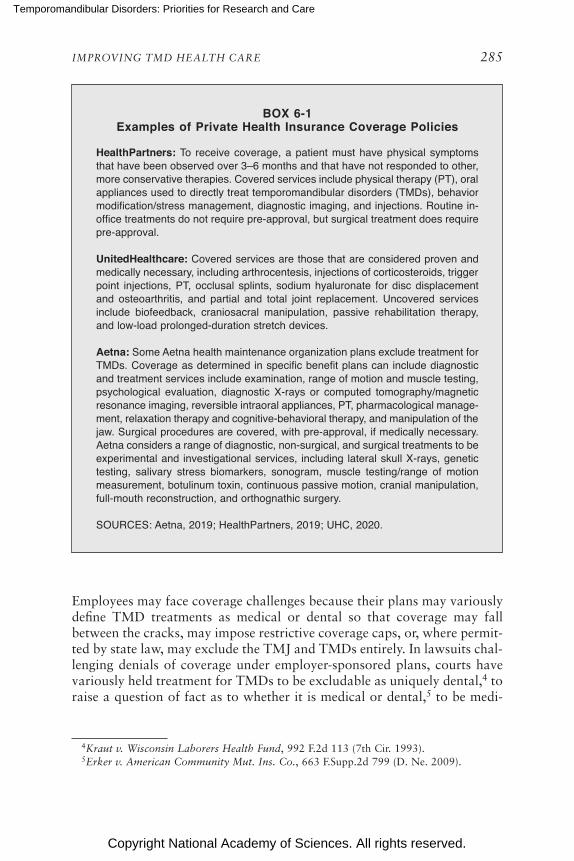
Temporomandibular Disorders: Priorities for Research and Care
Copyright National Academy of Sciences. All rights reserved.
IMPROVING TMD HEALTH CARE 285
BOX 6-1 Examples of Private Health Insurance Coverage Policies
HealthPartners: To receive coverage, a patient must have physical symptoms that have been observed over 3–6 months and that have not responded to other, more conservative therapies. Covered services include physical therapy (PT), oral appliances used to directly treat temporomandibular disorders (TMDs), behavior modification/stress management, diagnostic imaging, and injections. Routine in-office treatments do not require pre-approval, but surgical treatment does require pre-approval.
UnitedHealthcare: Covered services are those that are considered proven and medically necessary, including arthrocentesis, injections of corticosteroids, trigger point injections, PT, occlusal splints, sodium hyaluronate for disc displacement and osteoarthritis, and partial and total joint replacement. Uncovered services include biofeedback, craniosacral manipulation, passive rehabilitation therapy, and low-load prolonged-duration stretch devices.
Aetna: Some Aetna health maintenance organization plans exclude treatment for TMDs. Coverage as determined in specific benefit plans can include diagnostic and treatment services include examination, range of motion and muscle testing, psychological evaluation, diagnostic X-rays or computed tomography/magnetic resonance imaging, reversible intraoral appliances, PT, pharmacological manage-ment, relaxation therapy and cognitive-behavioral therapy, and manipulation of the jaw. Surgical procedures are covered, with pre-approval, if medically necessary. Aetna considers a range of diagnostic, non-surgical, and surgical treatments to be experimental and investigational services, including lateral skull X-rays, genetic testing, salivary stress biomarkers, sonogram, muscle testing/range of motion measurement, botulinum toxin, continuous passive motion, cranial manipulation, full-mouth reconstruction, and orthognathic surgery.
SOURCES: Aetna, 2019; HealthPartners, 2019; UHC, 2020.
Employees may face coverage challenges because their plans may variously define TMD treatments as medical or dental so that coverage may fall between the cracks, may impose restrictive coverage caps, or, where permit-ted by state law, may exclude the TMJ and TMDs entirely. In lawsuits chal-lenging denials of coverage under employer-sponsored plans, courts have variously held treatment for TMDs to be excludable as uniquely dental,4 to raise a question of fact as to whether it is medical or dental,5 to be medi-
4 Kraut v. Wisconsin Laborers Health Fund, 992 F.2d 113 (7th Cir. 1993).5 Erker v. American Community Mut. Ins. Co., 663 F.Supp.2d 799 (D. Ne. 2009).
Temporomandibular Disorders: Priorities for Research and Care
Copyright National Academy of Sciences. All rights reserved.
286 TEMPOROMANDIBULAR DISORDERS
cal or dental depending on the treatment,6 to be subject to strict coverage limitations,7 or to be entirely excludable under the authority of the plan administrators to limit covered conditions.8 The opinion of this committee is that TMDs should not be considered exclusively dental disorders.
Veterans Health Administration
The Veterans Health Administration provides health care to more than 9 million Americans, either through TRICARE for active and retired service members or CHAMPVA for certain civilians. TRICARE covers some TMD treatment but only if it is a medical issue that involves immediate relief of pain. Both CHAMPVA and TRICARE cover X-rays, up to four office visits, and construction of an intraoral appliance (VA, 2013; TRICARE, 2019).
Other Coverage and Payment Mechanisms
Federal Disability Insurance
Individuals who are disabled and unable to work because of a TMD may be eligible to receive support under either of two federal programs, Social Security Disability Insurance (SSDI; available to individuals with a certain number of work credits) or Supplemental Security Income (SSI; available to individuals with limited or no work history). Yet, claimants with a TMD may face challenges within the disability system, some of which are unique to their disorder and others that are shared with other chronic pain conditions. Under SSDI and SSI, a claimant may be eligible for benefits if he or she has a disabling condition that meets specific criteria (SSA, 2020). Individuals may apply based on one or more medical condi-tions listed in the Social Security Blue Book, which is the federal compen-dium of covered conditions and their eligibility criteria, or may apply based on non-listed conditions that are medically equivalent to listed conditions and that cause the individual to have a residual functional capacity preclud-ing employment (SSA, 2012).
However, claimants and disability examiners alike have little guidance in how to set forth or to evaluate a disability claim grounded in a TMD
6 Boyd v. Peoria Journal Star, Inc., 679 N.E.2d 788 (Ill. App. 3d 1997) (holding that the plan could not deny coverage for surgery to remove a failed TMJ implant, as the procedure was to correct a failed device, not treat TMD itself).
7 Midwest Sec. Life Ins. Co. v. Stroup, 730 N.E.2d 163 (In. 2000) (enforcing a $1,000 annual cap on TMD treatment under employer plan). See also, e.g., Solger v. Wal-Mart Stores, Inc. Associates Health and Welfare Plan, 144 F.3d 567 (8th Cir. 1998) (enforcing $5,000 cover-age cap under plan for employee who required jaw surgery to remove failed TMJ implant).
8 Stratton v. E.I. DuPont De Nemours & Co., 363 F.3d 250 (3d Cir. 2004).
Temporomandibular Disorders: Priorities for Research and Care
Copyright National Academy of Sciences. All rights reserved.
IMPROVING TMD HEALTH CARE 287
and in TMD-related chronic pain, as the same anatomical findings may be associated with very different degrees of pain across individuals. The dif-ficulty of evaluating cases with TMDs under the present framework—and their high costs to individuals and to the administrative and federal court systems—is in part shown by federal courts’ not-infrequent reversals of TMD disability denials9 (see Box 6-2).
Medical Malpractice
Some TMDs arise due to medical malpractice—for example, improper intubation or oral surgery that results in new or exacerbated TMD symp-toms or from inappropriate occlusal therapies. Court cases involving medi-cal malpractice related to a TMD demonstrate a degree of confusion over the nature of TMDs as a medical or dental condition and even as to the existence of a standard of care for TMDs against which a malpractice claim could be evaluated. For general medicine and dentistry, courts look to the standard of care in the community where treatment was sought or provided. For medical and dental specialties, courts look to a national standard of care. In several cases, courts have held either that TMD treatment is not a specialty and thus must be evaluated based on a community standard of care or else that a triable factual dispute exists as to the appropriate standard of care.10 In at least one recent case, a state court of appeals held that there is no national standard of care for TMDs below which a treatment could fall.11
Coverage Due to Injuries and Accidents
While the majority of TMDs are not directly attributable to a traumatic incident, studies indicate that for some individuals, trauma—particularly collisions and motor vehicle accidents—is strongly associated with the subsequent development of TMDs (Sharma et al., 2019). For injuries that
9 See, e.g., Sorber v. Commissioner of Social Security Administration, 362 F. Supp. 3d 712 (D. Az. 2019); Cindy F. v. Berryhill, 367 F. Supp. 3d 1195 (D. Or. 2019); Tilton v. Colvin, 184 F. Supp. 3d (M.D. Pa. 2016); Cumella v. Colvin, 936 F.Supp.2d 1120 (D.S.D. 2013); Lorence v. Astrue, 691 F.Supp.2d 1008 (D. Mn. 2010); Walterich v. Astrue, 578 F.Supp.2d 482 (W.D.N.Y. 2008); Bragg v. Commissioner of Social Security Admin., 567 F.Supp.2d 893 (N.D. Tx 2008).
10 See, e.g., Spivey v. James, 1 S.W.3d 380 (Ct. App. Tx. 1999) (issue of fact over who can be qualified as an expert in TMD); Burlingham v. Mintz, 891 P.2d 527 (Mo. 1995) (standard of care for TMD not established); Herpin v. Witherspoon, 664 So.2d 515 (Ct. App. La. 3d 1995) (holding that locality rule applies; TMD treatment is not a “specialty”).
11 Saucier v. Hawkins, 2013 113 So.3d 1277 (Ct. App. Ms. 2013) (affirming trial court’s directed verdict for defendant on the ground that the plaintiff had not established the existence of a national standard of care for TMD, where the plaintiff’s expert outlined the existence of guidance on TMD treatment from professional organizations but testified that treatment prac-tices vary widely).

Temporomandibular Disorders: Priorities for Research and Care
Copyright National Academy of Sciences. All rights reserved.
288 TEMPOROMANDIBULAR DISORDERS
BOX 6-2 Qualifying for Disability Insurance with a
Temporomandibular Disorder (TMD)
TMDs are among the conditions listed in the Social Security Administration’s Blue Book, but they may overlap with some listings. Claimants with a TMD seek-ing to qualify under Social Security Disability Insurance (SSDI) or Supplemental Security Income (SSI) may either attempt to demonstrate that their TMD symp-toms meet the criteria of listed conditions or, alternatively, that their pain and other impairments are medically equivalent to a listed condition and that they lack the residual functional capacity to be employed. Where the primary disabling symp-tom is severe chronic pain, a claimant with a TMD must pursue the second path, as chronic pain is specifically excluded from listed conditions.
The exclusion of chronic pain from the listings is particularly significant in a TMD, as pain often is the major, disabling symptom of the condition. Often, apart from pain, the impairments experienced by an individual with a TMD might not be disabling. An individual with limited range of motion of the jaw but with little or no pain may be able to speak and eat well enough to maintain a job and relation-ships, but an individual with the same or greater range of motion and extreme pain with speaking or eating, or at rest, may be unable to work or engage in other major life activities.
A claimant whose chief symptom is pain or whose other, non-pain impair-ments do not satisfy listing criteria can argue for the medical equivalence of their conditions and show residual functional capacity precluding employment. Where pain is the basis of, or a substantial part of, an alleged disability, the pain must be attributable to an objectively demonstrated medical condition capable of giv-ing rise to the pain. If the claimant’s pain is greater than would be expected for the condition, the pain is considered disproportionate and subjective. Disability examiners may not disregard complaints of subjective and disproportionate pain, but neither must they credit them. Examiners must ensure that sufficient medical evidence supports a claim, evaluate the credibility of a claimant, and consider the possible factors contributing to the etiology of the alleged medical complaints. The procedure for evaluating TMD pain and other chronic pain conditions is challeng-ing for examiners and claimants alike. The SSDI/SSI emphasis on imaging-based tests, including X-rays and magnetic resonance imaging, may bias outcomes toward claims involving gross, anatomical findings over claims grounded in less obvious but potentially more severe conditions involving soft tissue, the central nervous system, or a neuroimmune injury or disorder. The regulations’ direction to examiners to determine whether claimed pain is proportionate to the condition giving rise to the pain makes these cases complex to evaluate and uncertain in their outcome.
Temporomandibular Disorders: Priorities for Research and Care
Copyright National Academy of Sciences. All rights reserved.
IMPROVING TMD HEALTH CARE 289
are work related, workers compensation insurance provides full coverage with no deductibles in all states, as long as the causation standard is met for that jurisdiction (Melhorn et al., 2013). The relationship between whiplash resulting from motor vehicle accidents and the subsequent development of TMDs has been debated for years, with widely varying estimates of prevalence. While treatments related to motor vehicle accidents will likely be covered, most individual auto insurance policies have limits on medical payments, typically $10,000 to $20,000. Where coverage is insufficient, particularly for lost past and future earnings, individuals with a TMD may resort to the court system. In cases involving negligence, including road accidents or other traumatic injuries, plaintiffs may recover substantial damages, particularly if a TMD co-occurs with other serious injuries.12 However, judgments in numerous cases reveal confusion about TMDs and, in particular, the relationship of chronic pain to psychological disorders, with courts at times attributing the former to the latter.13
Conclusions on Insurance Coverage
Conclusion 6-7: Comprehensive insurance coverage for care of temporomandibular disorders (TMDs) is lacking. Patients must navigate coverage decisions between health and dental insurance and may be left to assume all costs. For patients with persistent TMD symptoms, this process of working with multiple parties (medical doctors, dentists, insurers, billing departments, etc.) to prove medical necessity and pushing their claims through appeals processes can have a significant emotional and financial toll. While anecdotal, the committee heard numerous communications from patients whose out-of-pocket costs for treatment of TMDs were an extreme financial burden.
Conclusion 6-8: Insurance coverage for care of temporo mandibular disorders is not consistent and may not provide coverage for low-risk effective treatments (such as self-management and physical therapy), while higher-risk treatments (such as medications and surgery) are covered. Misalignment may result in patients receiving the care that is best reimbursed, rather than the care that is best.
12 See, e.g., Desselle v. LaFleur, Court of Appeal of Louisiana, Third Circuit. February 4, 2004, 865 So.2d 954 2004 WL 205728 (affirming award of $350,000 to plaintiff who suffered back and neck injuries, and TMD, incident to automobile accident).
13 See, e.g., Torno v. Hayek, Court of Appeals of Washington, Division 3. May 25, 2006, 133 Wash.App. 244 135 P.3d 536; Commonwealth Department of Corrections v. Workers Compensation Appeals Board (Wagner-Stover), Commonwealth Court of Pennsylvania. 2010 6 A.3d 603 2010 WL 3811308.
Temporomandibular Disorders: Priorities for Research and Care
Copyright National Academy of Sciences. All rights reserved.
290 TEMPOROMANDIBULAR DISORDERS
CONCLUSIONS AND RESEARCH PRIORITIES
TMD care is generally fragmented between dentistry and medicine, often leaving patients to navigate among clinicians with little guidance or efforts at coordinated care. Furthermore, insurance coverage is often lack-ing for TMD care, leaving patients to bear significant out-of-pocket finan-cial burdens. Efforts are needed at multiple levels to improve the training and education of health professionals on TMD care, to provide specialist certification for professionals in TMD with independent accreditation, to coordinate centers of excellence in TMD care, and to improve insurance coverage. Research to inform those efforts is also needed (see Box 6-3). The committee’s recommendations for future actions in these areas are detailed in Chapter 8.
Conclusion 6-1: Due to education, training, and financing mecha-nisms, there is often a lack of collaboration between clinicians, particularly dentists and physicians, and this divide can make it challenging for patients with temporomandibular disorders to access and coordinate care.
Conclusion 6-2: Health care professionals—including physicians, dentists, nurses, physician’s assistants, and physical therapists—need better education and training in the assessment, treatment, management, and referral of patients with pain, orofacial pain, and temporomandibular disorders with attention to interprofessional education opportunities where possible. The extent of training will vary depending on the specialty and the nature of the practice.
Conclusion 6-3: Some dentists continue to use harmful or costly treatment approaches that are known to be ineffective for temporo-mandibular disorders (TMDs). As discussed in Chapters 2 and 5, editorials and letters to the editor in dental journals suggest resistance to the less invasive but more evidence-based treatment approaches for TMDs. The reliance on aggressive or invasive treat-ment methods as the first step rather than starting with behavioral or rehabilitative treatments follows the decades-long adherence to structural models that have been shown to be non-intrusive con-servative treatments. Education efforts, particularly basic dental education and continuing education classes for practicing den-tists, need to focus on ensuring that dentists are fully informed about the complexity of pain and movement as identified for other musculo skeletal disorders and current evidence-based treatments for chronic pain in general and TMDs in particular.
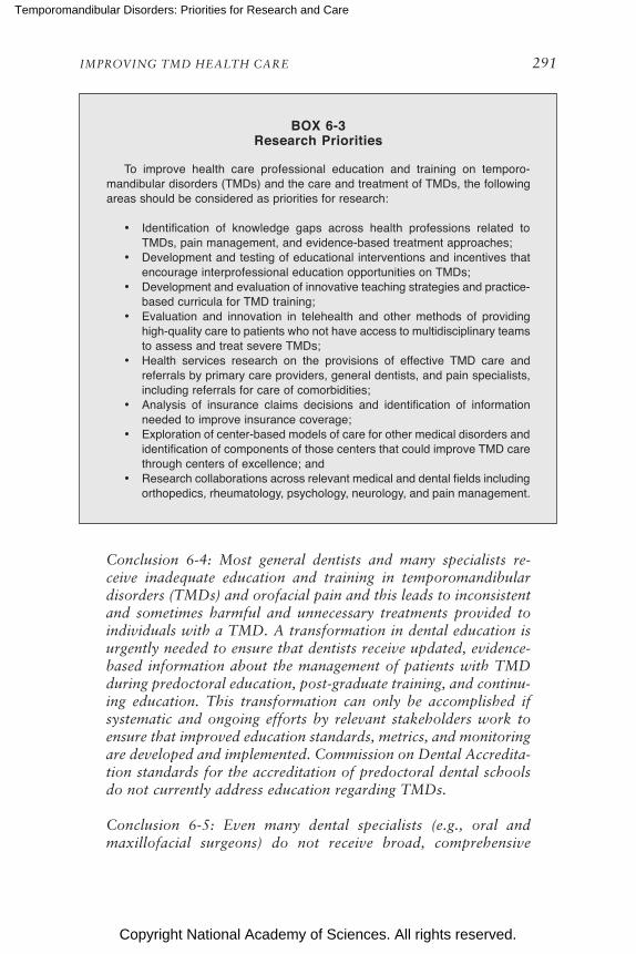
Temporomandibular Disorders: Priorities for Research and Care
Copyright National Academy of Sciences. All rights reserved.
IMPROVING TMD HEALTH CARE 291
BOX 6-3 Research Priorities
To improve health care professional education and training on temporo-mandibular disorders (TMDs) and the care and treatment of TMDs, the following areas should be considered as priorities for research:
• Identification of knowledge gaps across health professions related to TMDs, pain management, and evidence-based treatment approaches;
• Development and testing of educational interventions and incentives that encourage interprofessional education opportunities on TMDs;
• Development and evaluation of innovative teaching strategies and practice-based curricula for TMD training;
• Evaluation and innovation in telehealth and other methods of providing high-quality care to patients who not have access to multidisciplinary teams to assess and treat severe TMDs;
• Health services research on the provisions of effective TMD care and referrals by primary care providers, general dentists, and pain specialists, including referrals for care of comorbidities;
• Analysis of insurance claims decisions and identification of information needed to improve insurance coverage;
• Exploration of center-based models of care for other medical disorders and identification of components of those centers that could improve TMD care through centers of excellence; and
• Research collaborations across relevant medical and dental fields including orthopedics, rheumatology, psychology, neurology, and pain management.
Conclusion 6-4: Most general dentists and many specialists re-ceive inadequate education and training in temporomandibular disorders (TMDs) and orofacial pain and this leads to inconsistent and sometimes harmful and unnecessary treatments provided to individuals with a TMD. A transformation in dental education is urgently needed to ensure that dentists receive updated, evidence-based information about the management of patients with TMD during predoctoral education, post-graduate training, and continu-ing education. This transformation can only be accomplished if systematic and ongoing efforts by relevant stakeholders work to ensure that improved education standards, metrics, and monitoring are developed and implemented. Commission on Dental Accredita-tion standards for the accreditation of predoctoral dental schools do not currently address education regarding TMDs.
Conclusion 6-5: Even many dental specialists (e.g., oral and maxillo facial surgeons) do not receive broad, comprehensive
Temporomandibular Disorders: Priorities for Research and Care
Copyright National Academy of Sciences. All rights reserved.
292 TEMPOROMANDIBULAR DISORDERS
education and training in diagnosing, treating, and managing temporo mandibular disorders (TMDs). Improved post-graduate training opportunities are needed, including specializations specific to TMDs and orofacial pain.
Conclusion 6-6: Central resources are needed to provide inter-disciplinary care to patients with temporomandibular disorders (TMDs), to gather and disseminate information about best prac-tices for TMDs, and to coordinate research priorities. Centers of excellence have proven to be successful for other diseases and disorders in improving care, and a similar model could be effective for improving TMD care.
Conclusion 6-7: Comprehensive insurance coverage for care of temporomandibular disorders (TMDs) is lacking. Patients must navigate coverage decisions between health and dental insurance and may be left to assume all costs. For patients with persistent TMD symptoms, this process of working with multiple parties (medical doctors, dentists, insurers, billing departments, etc.) to prove medical necessity and pushing their claims through appeals processes can have a significant emotional and financial toll. While anecdotal, the committee heard numerous communications from patients whose out-of-pocket costs for treatment of TMDs were an extreme financial burden.
Conclusion 6-8: Insurance coverage for care of temporo mandibular disorders is not consistent and may not provide coverage for low-risk effective treatments (such as self-management and physical therapy), while higher-risk treatments (such as medications and surgery) are covered. Misalignment may result in patients receiving the care that is best reimbursed, rather than the care that is best.
REFERENCES
AACOM (American Association of Colleges of Osteopathic Medicine). 2019. AACOM re-ports on curriculum. https://www.aacom.org/reports-programs-initiatives/aacom-reports/curriculum (accessed November 19, 2019).
AAMC (Association of American Medical Colleges). 2018. Interprofessional education re-quirements at US medical schools. https://www.aamc.org/initiatives/cir/403572/02.html (accessed November 19, 2019).
AAMC. 2019. Content documentation in required courses and elective courses. https://www.aamc.org/initiatives/cir/406462/06a.html (accessed November 19, 2019).
Temporomandibular Disorders: Priorities for Research and Care
Copyright National Academy of Sciences. All rights reserved.
IMPROVING TMD HEALTH CARE 293
AAOP (American Academy of Orofacial Pain). 2018. Orofacial pain programs. https://aaop.clubexpress.com/content.aspx?page_id=22&club_id=508439&module_id=107337 (ac-cessed September 5, 2019).
AAOP. 2019. Application for recognition of orofacial pain. Submitted by the American Academy of Orofacial Pain, April 2019. https://www.ada.org/~/media/NCRDSCB/Files/Orofacial_Pain_Speciality_Application.pdf?la=en (accessed November 18, 2019).
ACGME (Accreditation Council for Graduate Medical Education). 2019. ACGME program requirements for graduate medical education in pain medicine (Subspecialty of Anesthe-siology, Child Neurology, Neurology, or Physical Medicine and Rehabilitation). https://www.acgme.org/Portals/0/PFAssets/ProgramRequirements/530_Pain%20Medicine_2019.pdf?ver=2019-06-26-115917-800 (accessed November 13, 2019).
ADA (American Dental Association). 2004. TM treatment and third party insurance coverage. https://www.ada.org/~/media/ADA/Advocacy/Files/thirdparty_TMD_coverage.pdf?la=en (accessed October 10, 2019).
ADA. 2019. Workforce. https://www.ada.org/en/science-research/health-policy-institute/ dental-statistics/workforce (accessed October 10, 2019).
ADEA (American Dental Education Association). 2008. Competencies for the new general dentist. https://www.adea.org/about_adea/governance/Pages/Competencies-for-the-New-General-Dentist.aspx (accessed October 10, 2019).
Aetna. 2019. Temporomandibular disorders. http://www.aetna.com/cpb/medical/data/1_99/0028.html (accessed November 19, 2019).
ALS Association. 2019. ALS Association centers and clinics. http://www.alsa.org/community/centers-clinics/centers-and-clinics-descriptions.html (accessed November 18, 2019).
Anderson, R. T., C. S. Weisman, S. Hudson Scholle, J. T. Henderson, R. Oldendick, and F. Camacho. 2002. Evaluation of the quality of care in the clinical care centers of the National Centers of Excellence in Women’s Health. Women’s Health Issues 12(6):309–326.
APTA (American Physical Therapy Association). 2013. Vision statement for the physical therapy profession and guiding principles to achieve the vision. https://www.apta.org/Vision (accessed November 19, 2019).
Atchison, K. A., R. G. Rozier, and J. A. Weintraub. 2018. Integration of oral health and pri-mary care: Communication, coordination, and referral. NAM Perspectives. Washington, DC: National Academy of Medicine.
Breckons, M., S. M. Bissett, C. Exley, V. Araujo-Soares, and J. Durham. 2017. Care pathways in persistent orofacial pain: Qualitative evidence from the DEEP study. Journal of Dental Research: Clinical and Translational Research 2(1):48–57.
Campbell, E. 2019. Faculty perspectives of teaching pain management to nursing students. Pain Management Nursing PMID:31492600.
CMS (Centers for Medicare & Medicaid Services). 2013. Medicare dental coverage. https://www.cms.gov/Medicare/Coverage/MedicareDentalCoverage/index.html (accessed Octo-ber 3, 2019).
CNME (Council on Naturopathic Medical Education). 2017. Handbook of accreditation for naturopathic medicine. https://cnme.org/wp-content/uploads/2017/09/2017_cnme_ handbook_of_accreditation.pdf (accessed November 18, 2019).
COCA (Commission on Osteopathic College Accreditation). 2019. Accreditation of colleges of osteopathic medicine: COM continuing accreditation standards. https://osteopathic.org/wp-content/uploads/2018/02/com-continuing-accreditation-standards.pdf (accessed October 23, 2019).
CODA (Commission on Dental Accreditation). 2017. Accreditation standards for advanced specialty education programs in oral and maxillofacial surgery. https://www.ada.org/~/media/CODA/Files/2018_oms.pdf?la=en (accessed November 19, 2019).
Temporomandibular Disorders: Priorities for Research and Care
Copyright National Academy of Sciences. All rights reserved.
294 TEMPOROMANDIBULAR DISORDERS
CODA. 2019. Accreditation standards for dental education programs. https://www.ada.org/~/media/CODA/Files/pde.pdf?la=en (accessed October 23, 2019).
Desai, B., N. Alkandari, and D. M. Laskin. 2016. How accurate is information about diagno-sis and management of temporomandibular disorders on dentist websites? Oral Surgery, Oral Medicine, Oral Pathology, and Oral Radiology 122(3):306–309.
Elrod, J. K. and J. L. Fortenberry, Jr. 2017. Centers of excellence in healthcare institutions: What they are and how to assemble them. BMC Health Services Research 17(Suppl 1):425.
Fishman, S. M., H. M. Young, E. L. Arwood, R. Chou, K. Herr, B. B. Murinson, J. Watt-Watson, D. B. Carr, D. B. Gordon, B. J. Stevens, D. Bakerjian, J. C. Ballantyne, M. Courtenay, M. Djukic, I. J. Koebner, J. M. Mongoven, J. A. Paice, R. Prasad, N. Singh, K. A. Sluka, B. St. Marie, and S. A. Strassels. 2013. Core competencies for pain manage-ment: Results of an interprofessional consensus summit. Pain Medicine 14:971–981.
Fishman, S. M., D. B. Carr, B. Hogans, M. Cheatle, R. M. Gallagher, J. Katzman, S. Mackey, R. Polomano, A. Popescu, J. P. Rathmell, R. W. Rosenquist, D. Tauben, L. Beckett, Y. Li, J. M. Mongoven, and H. M. Young. 2018. Scope and nature of pain- and analgesia-related content of the United States Medical Licensing Examination (USMLE). Pain Medicine 19:449–459.
Greene, C. S., and A. E. Bertagna. 2019. Seeking treatment for temporomandibular disorders: What patients can expect from non-dental health care providers. Oral Surgery, Oral Medicine, Oral Pathology, and Oral Radiology 127(5):399–407.
HealthPartners. 2019. Temporomandibular disorder (TMD) treatments. https://www. healthpartners.com/public/coverage-criteria/policy.html?contentid=AENTRY_046169 (accessed November 19, 2019).
HHS (Department of Health and Human Services). 2016. National pain strategy: A compre-hensive population health-level strategy for pain. https://www.iprcc.nih.gov/sites/default/files/HHSNational_Pain_Strategy_508C.pdf (accessed October 3, 2019).
Hoeger Bement, M. K., and K. A. Sluka. 2015. The current state of physical therapy pain curricula in the United States: A faculty survey. Journal of Pain 16(2):144–152.
Hoeger Bement, M. K., B. J. St. Marie, T. M. Nordstrom, N. Christensen, J. M. Mongoven, I. J. Koebner, S. M. Fishman, and K. A. Sluka. 2014. An interprofessional consensus of core competencies for prelicensure education in pain management: Curriculum applica-tion for physical therapy. Physical Therapy 94(4):451–465.
IASP (International Association for the Study of Pain). 2018. IASP curriculum outline on pain for physical therapy. https://www.iasp-pain.org/Education/Content.aspx?ItemNumber=1580 (accessed November 19, 2019).
IOM (Institute of Medicine). 2011. Relieving pain in America: A blueprint for transforming prevention, care, education, and research. Washington, DC: The National Academies Press.
Jones, L. K., M. E. Lussier, J. Brar, M. C. Byrne, M. Durham, F. Kiokemeister, K. Kjaer, H. Le, C. Magee, E. McKnight, N. Mehta, J. Papp, E. Pastwa, P. Radovich, K. Ravin, M. Ruther, G. Woodie, S. Wrona, G. Yousefvand, and G. Greskovic. 2019. Current interventions to promote safe and appropriate pain management. American Journal of Health-System Pharmacy 76(11):829–834.
Kaiser Permanente Dental. 2019. About us. https://kaiserpermanentedentalnw.org/about-us (accessed November 19, 2019).
Klasser, G. D., and C. S. Greene. 2007. Predoctoral teaching of temporomandibular disorders: A survey of U.S. and Canadian dental schools. Journal of the American Dental Associa-tion 138(2):231–237.
Klasser, G. D., and H. A. Gremillion. 2013. Past, present, and future of predoctoral dental education in orofacial pain and TMDs: A call for interprofessional education. Journal of Dental Education 77(4):395–400.
Temporomandibular Disorders: Priorities for Research and Care
Copyright National Academy of Sciences. All rights reserved.
IMPROVING TMD HEALTH CARE 295
LCME (Liaison Committee on Medical Education). 2019. Functions and structure of a medical school. https://lcme.org/wp-content/uploads/filebase/standards/2020-21_Functions-and-Structure_2019-10-04.docx (accessed November 19, 2019).
MedBullets. 2019. Temporomandibular disorders. https://step2.medbullets.com/ear-nose-throat/121832/temporomandibular-joint-disease-tmd (accessed November 19, 2019).
Melhorn, J. M., W. E. Ackerman III, L. S. Glass, D. C. Deitz, and S. Babitsky. 2013. Under-standing work-relatedness. In J. M. Melhorn and J. B. Talmage, eds. AMA guides to the evaluation of disease and injury causation, 2nd ed. Chicago, IL: American Medical Association.
Mogayzel, P. J., J. Dunitz, L. C. Marrow, and L. A. Hazle. 2014. Improving chronic care delivery and outcomes: The impact of the Cystic Fibrosis Care Center Network. BMJ Quality and Safety 23(Suppl 1):i3–i8.
NAEC (National Association of Epilepsy Centers). 2019. National Association of Epilepsy Centers. https://www.naec-epilepsy.org (accessed November 18, 2019).
Nierman, R. 2015. Does new legislation mandate TMJ treatments in your state? https://dentalsleeppractice.com/practice-management/roses-reimbursement-rules (accessed No-vember 18, 2019).
Powers, B., R. B. Donoff, and S. H. Jain. 2017. Bridging the dental divide: Overcom-ing barriers to integrating oral health and primary care. https://www.healthaffairs.org/do/10.1377/hblog20170119.058211/full (accessed November 19, 2019).
Prodoehl, J., S. Kraus, G. D. Klasser, and K. D. Hall. 2019. Temporomandibular disorder content in the curricula of physical therapist professional programs in the United States. Cranio January 4:1–13.
Project ECHO. 2019. How it works. https://echo.unm.edu (accessed November 18, 2019). Sharma, S., J. Wactawski-Wende, M. LaMonte, J. Zhao, G. D. Slade, E. Bair, J. D. Greenspan,
R. B. Fillingim, W. Maixner, and R. Ohrbach. 2019. Incident injury is strongly associated with subsequent incident temporomandibular disorder: Results from the OPPERA study. Pain 160(7):1551–1561.
Shigekawa, E., M. Fix, G. Corbett, D. H. Roby, and J. Coffman. 2018. The current state of telehealth evidence: A rapid review. Health Affairs 37(12):1975–1982.
Singhal, A., P. Damiano, and L. Sabik. 2017. Medicaid adult dental benefits increase use of dental care, but expansion of dental services use was mixed. Health Affairs 36(4):723–732.
SSA (Social Security Administration). 2012. Residual functional capacity. https://www.ssa.gov/OP_Home/cfr20/416/416-0945.htm (accessed November 19, 2019).
SSA. 2020. Disability evaluation under Social Security. https://www.ssa.gov/disability/ professionals/bluebook/general-info.htm (accessed January 29, 2020).
TRICARE. 2019. Temporomandibular joint syndrome treatment. https://tricare.mil/ CoveredServices/IsItCovered/TempMandJoint?p= (accessed October 14, 2019).
UHC (UnitedHealthcare). 2019. Occlusal guards. https://www.uhcprovider.com/content/ provider/en/viewer.html?file=%2Fcontent%2Fdam%2Fprovider%2Fdocs%2Fpublic% 2Fpolicies%2Fdental%2Focclusal-guards.pdf (accessed November 19, 2019).
UHC. 2020. Temporomandibular joint disorders. https://www.uhcprovider.com/content/dam/provider/docs/public/policies/comm-medical-drug/temporomandibular-joint-disorders.pdf (accessed January 27, 2020).
University of Washington School of Dentistry. 2019. Clinical competencies. https://dental.washington.edu/policies/clinic-policy-manual/clinical-competencies (accessed October 14, 2019).
VA (Department of Veterans Affairs). 2013. Temporomandibular joint disorder. https://www.vha.cc.va.gov/system/templates/selfservice/va_ssnew/help/customer/locale/en-US/ portal/554400000001036/content/554400000009176/020502- TEMPOROMANDIBULAR-JOINT-DISORDER-TMD (accessed November 19, 2019).

Temporomandibular Disorders: Priorities for Research and Care
Copyright National Academy of Sciences. All rights reserved.
Temporomandibular Disorders: Priorities for Research and Care
Copyright National Academy of Sciences. All rights reserved.
297
The lack of credible information relayed to the public and professionals about the reality of the impact that TMD can have on one’s overall health and life results in patients continuing to suffer in silence and isolation because nobody—including family, friends, employers, etc.—can compre-hend how multiple therapies, including years of treatments at exorbitant costs, cannot fix or alleviate the problem but seem to worsen it. It is always the fault of the patient when things don’t work out like they were told it would. Leaving the patient to question what they did wrong.
—The TMJ Association
It was difficult to get people to come and give recognition to this disorder. They don’t understand the seriousness.
—Beth B.
Patients, their families, the general public, and health care professionals need evidence-based information about temporomandibular disorders (TMDs). As detailed in the prior chapters, patients and their families face challenges in accessing appropriate diagnoses and care. Additionally, be-cause orofacial pain and the symptoms potentially related to TMDs are experienced by a large number of individuals in the United States and globally, there is a need for raising awareness in the general public about these disorders and for providing accurate information and resources to help individuals cope with these conditions. As discussed in Chapter 6,
7
Improving Patient, Family, and Public Education and Awareness
About TMDs
Temporomandibular Disorders: Priorities for Research and Care
Copyright National Academy of Sciences. All rights reserved.
298 TEMPOROMANDIBULAR DISORDERS
increased efforts are also needed to improve education and training on TMDs for a range of health care professionals.
The reasons identified by the committee for focusing efforts on patient and public education on TMDs include:
• The frequency and complexity of the symptoms of temporo mandibular disorders: As discussed in Chapter 3, TMDs are common, not rare, and the public needs to know how to access the right treatment at the right time.
• Empowering the patient: Patients need to have a strong and knowl-edgeable voice in their care and need to be informed about the day-to-day efforts they can make to try to live their best possible lives with a TMD.
• Support from family and friends: Similarly, family members and friends who are well informed about TMDs can provide support.
• The range of TMDs and varying treatment approaches: As discussed in Chapter 2, TMDs are heterogeneous conditions that range from simple to complex. People who have pain or mechanical problems affecting the masticatory system need resources to allow them to determine if and when to seek a professional opinion, what type of health care professional to consult, and how to evaluate treatment options. Patients will need to navigate both dental and medical care systems to find the best care until these systems are coordinated in TMD assessment and management.
• Reducing stigma and misconceptions: TMDs, chronic pain, and chronic illnesses are often associated with misperceptions and mis-understandings. Because the condition can often not be seen, patients may be viewed by family, friends, and even health professionals as malingerers or not having a “real” condition.
• Raising awareness of the public and health care professionals: A public education campaign can educate the public and health care professionals about this complex condition. In addition, it can help mitigate the stigma discussed above.
The role of patients has taken on new dimensions in recent years as patient education has expanded into patient engagement and activation across the spectrum of involvement in research, prevention, and treatment opportunities (IOM, 2011a, 2014). As discussed in Chapters 5 and 6, patient-centered care is the focus of efforts in health care, including dental care, to involve patients in decisions on their treatment goals and options. In addition, patients must take an active part in day-to-day TMD manage-ment. Patients and stakeholders in the public are increasingly being valued in all facets of research and in policy development. These individuals are
Temporomandibular Disorders: Priorities for Research and Care
Copyright National Academy of Sciences. All rights reserved.
IMPROVING PATIENT, FAMILY, AND PUBLIC EDUCATION AND AWARENESS 299
valued for the insights they provide (including understanding the realities of living with disease, focusing on culturally relevant and patient-relevant questions, and incorporating outcome measures that assess differences that really matter). Patients and stakeholders also are involved in actions that can foster the implementation of efforts to improve health in communi-ties (Woolf et al., 2016; Forsythe et al., 2019). Using tools to assess patient activation, researchers have found that patients who are more engaged in their care are more likely to have healthier outcomes in terms of general indicators of health, are more likely to engage in higher levels of healthy behaviors, and are less likely to have a hospitalization or emergency depart-ment visit; the combined effects result in cost savings (Greene et al., 2015; Okunrintemi et al., 2017).
The committee greatly benefited from the public testimony of a number of individuals with a TMD who described their experiences and lessons learned while speaking at the committee meetings or by providing written comments to the committee (see Chapter 1 and Appendix A).
This chapter identifies goals and priorities for providing evidence-based information on TMDs to patients, their families, health care professionals, and the general public and for engaging patients and other stakeholders in being a part of prevention, treatment, research, and communication efforts.
OVERCOMING STIGMA
There is a limited amount of research on stigma that is specific to TMDs. However, research on the impact of stigma from chronic pain, to-gether with patient testimony provided to the committee, eloquently docu-ment the stigma suffered by individuals with a TMD and its consequences for patients. This chapter begins with a discussion of stigma, then turns to a discussion of actions that can be taken to help individuals, family mem-bers, and others become more knowledgeable about TMDs and proactive about self-care.
Stigma refers to attitudes and beliefs that lead people to reject, avoid, or fear those they perceive as being different. As outlined by Pescosolido and Martin (2015, p. 6), stigma involves “(a) distinguishing and labeling differences, (b) associating human differences with negative attributions or stereotypes, (c) separating ‘us’ from ‘them,’ and (d) experiencing status loss and discrimination.”
Stigmatizing attitudes and beliefs about TMDs can originate both internally—from the beliefs and attitudes of the individual experiencing a TMD—and externally—from the beliefs and opinions held by others, includ ing family members, friends, health care professionals, employers and coworkers, the general public, and the media (see Figure 7-1). Chronic and complex TMDs represent a form of chronic pain that can profoundly
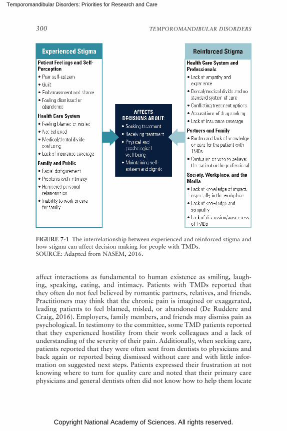
Temporomandibular Disorders: Priorities for Research and Care
Copyright National Academy of Sciences. All rights reserved.
300 TEMPOROMANDIBULAR DISORDERS
FIGURE 7-1 The interrelationship between experienced and reinforced stigma and how stigma can affect decision making for people with TMDs. SOURCE: Adapted from NASEM, 2016.
affect interactions as fundamental to human existence as smiling, laugh-ing, speaking, eating, and intimacy. Patients with TMDs reported that they often do not feel believed by romantic partners, relatives, and friends. Practitioners may think that the chronic pain is imagined or exaggerated, leading patients to feel blamed, misled, or abandoned (De Ruddere and Craig, 2016). Employers, family members, and friends may dismiss pain as psychological. In testimony to the committee, some TMD patients reported that they experienced hostility from their work colleagues and a lack of understanding of the severity of their pain. Additionally, when seeking care, patients reported that they were often sent from dentists to physicians and back again or reported being dismissed without care and with little infor-mation on suggested next steps. Patients expressed their frustration at not knowing where to turn for quality care and noted that their primary care physicians and general dentists often did not know how to help them locate
Temporomandibular Disorders: Priorities for Research and Care
Copyright National Academy of Sciences. All rights reserved.
IMPROVING PATIENT, FAMILY, AND PUBLIC EDUCATION AND AWARENESS 301
qualified specialists. The opportunity for adequate treatment and follow-up of TMD care may be thwarted by the combination of uncertain diagnosis and the stigma perceived by the patient, family member, or public. Indi-viduals with a TMD may hold back on sharing their symptoms, thoughts, or feelings with others because of concerns about stigmatization. Stigma can lead to poorer physical and psychological well-being, diminished self-esteem, and a loss of dignity. Thus, stigma adds to the burden of the condi-tion and significantly affects health and quality of life.
Patients with other forms of chronic pain also report suffering from the stigmatization of their condition. As discussed further below, pain is invis-ible and, as such, is hard for family members, work supervisors, and others to quantify and understand. De Ruddere and Craig (2016, p. 1607) write,
In the context of chronic nonmalignant pain, an absence of clear tissue damage deviates from the widely held biomedical model, which presumes that clear physiological pathology underlies the pain experience. As well, most people understand pain through acute pain experience, which resolves relatively rapidly over time.
A survey of adults with chronic pain found that more than one-third experienced a sense of stigma in which they felt alienated, became socially withdrawn, or saw the legitimacy of their condition as being questioned (Waugh et al., 2014). A study by Marbach and colleagues (1990) explored the sources of stigma perceived by patients with TMD. The researchers found that often, TMD stigma was experienced by patients because of the lack of a known etiology for their condition and that, moreover, when the TMD was attributed to psychological causes, the patient experienced stigma.
As discussed in Chapter 3, sex differences have been reported in the prevalence of TMDs, with higher numbers of women reporting these dis-orders. Some patients who provided testimony to the committee noted that they experienced stigma associated with being a female with a TMD, which was manifest by the dismissiveness they experienced from some health care professionals or by being told that their disorder was largely a mental health concern. The stigma associated with chronic pain conditions has been well documented (Wakefield et al., 2018; Scott et al., 2019), and some evidence suggests that women may experience greater pain-related stigma than their male counterparts (Hoffmann and Tarzian, 2001; Driscoll et al., 2018). A study of 151 female patients with chronic facial pain found that the stigma they experienced largely resulted from pejorative labeling by clinicians (Marbach et al., 1990). Diagnostic and treatment decisions for chronic pain are also influenced by the gender and related stereotypes that may be held by some health care professionals (LeResche, 2011).
Temporomandibular Disorders: Priorities for Research and Care
Copyright National Academy of Sciences. All rights reserved.
302 TEMPOROMANDIBULAR DISORDERS
Stigma is also suffered by many individuals with other health condi-tions including epilepsy, hearing loss, mental illnesses, fibromyalgia, and HIV/AIDS (IOM, 2012; Corrigan, 2014; NASEM, 2016; Armentor, 2017; Ring and Lawn, 2019). Various types of approaches have been implemented to overcome the stigma attached to these conditions, including patient education efforts, public awareness campaigns, professional education and training, involving celebrities and media champions, and policy change advocacy. The results of anti-stigma interventions are difficult to measure and evaluate, and varying degrees of success have been seen in overcom-ing health-related stigmas to date (Cook et al., 2014; Corrigan, 2014). New avenues for research on understanding and overcoming stigma have been proposed, as has a public health framework for intervention strate-gies to reduce stigma using a multilevel approach focused on intrapersonal efforts (patient education and self-management, patient empowerment), inter personal actions (support groups, broader education including profes-sional and public education and awareness), and structural interventions (e.g., advocacy, changes in policies, leadership) (Slade et al., 2009; Cook et al., 2014; Pescosolido and Martin, 2015; De Ruddere and Craig, 2016; Dubin, 2017; Scott et al., 2019).
The committee believes that efforts to increase professional education and awareness about TMDs across the health care professions, including dentistry (see Chapter 6), as well as actions to improve the education of patients, fami-lies, and the general public (see next section) are part of the efforts needed to help reduce the stigma of TMDs and improve patient health and well-being.
INCREASING PATIENT AND FAMILY EDUCATION AND IMPROVING COMMUNICATION WITH
HEALTH CARE PROFESSIONALS
Patients with a TMD and their families need to be able to make in-formed decisions when it comes to treatment and care. Education is key to this process.
For people living with chronic pain, patient education resources have been found to help in identifying ways to control and cope with pain, in connecting with psychosocial supports, and in encouraging the use of self-management strategies for the patient to take an active role in the recovery process (IOM, 2011b; see Chapter 5). People with TMD symptoms need resources that help them explore all of the options for treatment or non-treatment and that empower them not only to ask the questions that can determine the most effective course of action for them, but also to develop and sustain effective coping strategies.
In addition to the critical need for high-quality patient education re-sources and opportunities, it is also important to have evidence-based
Temporomandibular Disorders: Priorities for Research and Care
Copyright National Academy of Sciences. All rights reserved.
IMPROVING PATIENT, FAMILY, AND PUBLIC EDUCATION AND AWARENESS 303
resources for family members and caregivers, who can play an important role in supporting the person with pain. A 1997 American Chronic Pain Association (ACPA) survey of family members living with a person with pain found that family members often experience emotions and fears that are similar to those felt by the person with pain, even though they them-selves do not feel the physical pain (Cowan et al., 1998). In response to this important information, the association developed a Family Matters video series that explored the challenges that families deal with regarding a family member in pain and efforts to improve the quality of family life (ACPA, 2019e). In discussing the costs of pain, especially chronic pain, the Institute of Medicine (IOM) report on pain stated,
Family members find that their relationship with their loved one changes, and to the extent that they must take on new roles (as caregiver and morale booster) and greater responsibilities in the family (e.g., grocery shopping, chores, errands), the burden on them increases. (IOM, 2011b, p. 94)
Where do most people get their information about TMDs? The lit-erature review by the committee did not identify any published studies specific to information-seeking behaviors for people with a TMD. Overall, the public finds health information through an array of sources, including the Internet and health care professionals, with a resulting potential for inequities and disparities in information access for disadvantaged consumer groups (Ramsey et al., 2017). In a study examining the information-seeking experiences of 190 people with fibromyalgia, the source that participants turned to most frequently was the Internet, but participants also sought out information from health care professionals, family members, and friends (Chen, 2012). Similar information avenues are also likely sought by indi-viduals with a TMD and their families.
With a wide variety of available health-related websites, the accuracy of the health information available on the Internet is quite varied and has been a source of ongoing concern for patients and clinicians (Gottlieb, 2000; Storino et al., 2016). Thus, having credible evidence-based websites on TMDs is critical for accurate patient- and family-based education. A study examining the websites of 255 dental professionals found that a large percentage had misinformation on the diagnosis and management of TMDs (Desai et al., 2016). The researchers noted inaccuracies in attributing TMDs solely to occlusal problems or malocclusion and in labeling TMDs as a singular disorder.
Identifying the key components contributing to the quality of patient communications continues to be an area of extensive activity and research. Given the complexity of the health care field and the ever evolving knowl-edge base, it is critical that patient resources be understandable, culturally
Temporomandibular Disorders: Priorities for Research and Care
Copyright National Academy of Sciences. All rights reserved.
304 TEMPOROMANDIBULAR DISORDERS
sensitive, and available in multiple languages and formats and that they recognize that audiences may vary in educational level, age, location, and access to technology (CDC, 2019b). A 2019 National Academies work-shop focused on strategies for improving the public’s health by increasing awareness (via resources that adhere to health literacy guidelines) about the impacts that oral health has on overall health and well-being and also on how overall health affects oral health (NASEM, 2019).
The committee heard a number of patients report that they and their families found it quite challenging to navigate through the array of informa-tion about TMDs and to identify what information was valid and evi-dence based. The ideas presented below are meant as a starting point for discussion and actions on improving patient and family information resources.
Communication Resources and Tools
One problem that many patients encounter is describing their pain and its impact on their lives. Pain is invisible and cannot be validated other than to believe the report of pain by the patient. Continuing to improve resources to help patients describe their pain and the symptoms (including TMD- and comorbidity-related symptoms) being experienced is important in moving this field forward. Several brief assessment tools are used in clinical settings that ask patients to convey the intensity of the pain being experienced through visual or numeric measures and have been found to be fairly reliable, including the Brief Pain Inventory and the Visual Analogue Scale (Bijur et al., 2001; Keller et al., 2004). Other pain assessment mea-sures are developed to focus specifically on the pain experienced due to a given health condition (Hawker et al., 2011). Face depiction scales (e.g., the Wong-Baker faces pain-rating scale) have also been used in health care, most often with children (Khatri and Kalra, 2012; Afsal et al., 2019).
Improved resources and tools are needed for use by patients with TMD symptoms. Examples of resources that could be used, adapted, and evalu-ated for use by patients with a TMD in tracking their pain and in talking with their health care professional include a set of pain management and communication tools developed by the ACPA (2019d). The ACPA interac-tive web-based Head Pain Map allows the person with pain to identify on the diagram of the head the specific places on the face where pain is felt, the intensity of that pain, the nature of the pain (e.g., steady, throbbing, burn-ing), and associated symptoms (ACPA, 2019a). The map and information can be printed out to discuss with the patient’s health care providers. The Diagnostic Criteria for Temporomandibular Disorders includes a pain map for paper administration, with separate sections for the oral cavity, face, and body (due to the associated required level of detail for each region); it
Temporomandibular Disorders: Priorities for Research and Care
Copyright National Academy of Sciences. All rights reserved.
IMPROVING PATIENT, FAMILY, AND PUBLIC EDUCATION AND AWARENESS 305
has been translated into 15 languages (INfORM, 2013). Pain logs are also useful and are available as interactive tools for patients to record and keep track of the nature, intensity, and functional impacts of their pain (ACPA, 2019b; see examples in Figures 7-2 and 7-3).
The development and evaluation of TMD-related educational resources should focus on the full range of information sought by patients and their families, from the initial exploration of symptoms to valid diagnostic and treatment approaches, to sources of peer support and consumer roadmaps for the types of health care professionals to consult with and seek for treat-ment. Ideally these roadmaps would be adapted from the multistakeholder-developed, comprehensive care guidelines that are a recommendation of this report (see Chapter 5). The demographics of patients with TMDs (see Chapter 3) make this an especially relevant area of information seeking and gathering for teens and young adults. Strategies and campaigns focused on disseminating evidence-based data and resources via social media will be important. Ensuring that resources and communication tools are designed with these target audiences in mind, in addition to broader age groups, is critical, as are communications about what is known regarding the transi-tions to early and later adulthood with a TMD.
Peer Support
As described by patients with a TMD who provided testimony to the committee, a serious consequence of living with chronic TMD pain is that it can be isolating. Because of the invisibility of pain, its impact on daily life, and the challenges in communicating about pain severity, patients can feel cut off from family, friends, and activities. Peer support has been shown to be an effective means of establishing connections with other people who are also experiencing pain or other health problems, and the sharing of experiences can bring important quality-of-life benefits (Heisler, 2010; Fisher et al., 2015). Peer groups can also provide opportunities for teaching the self-management skills needed to cope with the many issues of living with pain. For example, in a study of veterans living with chronic pain, the benefits of participating in peer support groups were identified as making interpersonal connections, providing and receiving encouragement and sup-port, and facilitating the use of pain self-management strategies (Matthias et al., 2016; see also the self-management section in Chapter 5).
Peer support groups, both online and in person, are vital to the work and outreach of many patient advocacy organizations. The TMJ Associa-tion has been active in this area and hosts on their website several ways to connect with others experiencing TMDs. The TMJ Association’s online TMJ Café is a discussion community where users can ask questions, dis-cuss concerns and share experiences, and find out about social media peer
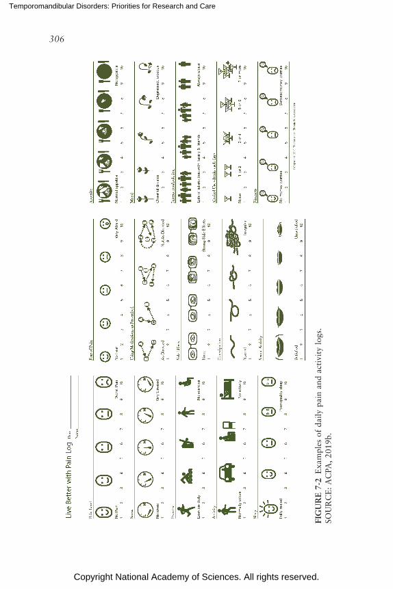
Temporomandibular Disorders: Priorities for Research and Care
Copyright National Academy of Sciences. All rights reserved.
306
FIG
UR
E 7
-2 E
xam
ples
of
daily
pai
n an
d ac
tivi
ty l
ogs.
SOU
RC
E:
AC
PA,
2019
b.
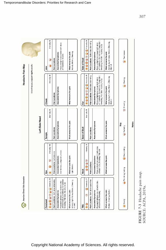
Temporomandibular Disorders: Priorities for Research and Care
Copyright National Academy of Sciences. All rights reserved.
307
FIG
UR
E 7
-3 H
eada
che
pain
map
. SO
UR
CE
: A
CPA
, 20
19a.
Temporomandibular Disorders: Priorities for Research and Care
Copyright National Academy of Sciences. All rights reserved.
308 TEMPOROMANDIBULAR DISORDERS
networks (The TMJ Association, 2019a). Additionally, The TMJ Asso-ciation offers a way to connect with a TMJ support contact person through individual patient support (The TMJ Association, 2019b). Peer support groups and related resources are also available through the ACPA (2019c) and other relevant patient-focused organizations active on social media platforms.
Communications Among Patients and Health Care Professionals
The patient–clinician exchange is paramount to the patient-centered care model, and the reciprocal nature of this interaction brings with it responsi bilities on both sides. This chapter is focused on actions for patients and their families and the general public, but one key to making commu-nications successful is improving education and training for health care professionals on TMDs and enhancing their communication skills (see Box 7-1; Chapter 6). Patients also have responsibilities to be engaged and to express their concerns. Additionally, patients have rights to expect listening, engagement, and communication from their health care professionals (see Box 7-1). Although patient–clinician communication is generally thought of as a one-on-one encounter between the patient and clinician, there are also
BOX 7-1 Improving Patient–Clinician Communication
A critically important part of health professional education is learning how to ef-fectively interact with patients, listen to their questions and concerns, and involve them in decision making. Training in communication with patients and families has been shown to be effective, and when it is combined with condition-specific train-ing, it can improve outcomes. Patients often offer clues to underlying concerns that are either not recognized or ignored by clinicians who focus primarily on symptom relief. Effective patient-centered interviewing and patient–clinician com-munications provide a solid foundation for patients with symptoms of a temporo-mandibular disorder to discuss their symptoms, concerns, and comorbid disorders and to work with clinicians on identifying next steps (Racich, 2009).
Examples of communication approaches include the Naming, Understand-ing, Respecting, Supporting, and Exploring approach that focuses on a patient’s delineation of his or her symptoms and concerns being addressed interactively with information from health care professionals providing support and discussing potential avenues for treatment (Back et al., 2009; Fortin et al., 2012). The Ask-Tell-Ask approach begins with a question to the patient, followed by a back-and-forth exchange of information and discussion, and ending with decisions that have been discussed and where the patient feels knowledgeable about next steps and feels that her or his questions have been addressed.
Temporomandibular Disorders: Priorities for Research and Care
Copyright National Academy of Sciences. All rights reserved.
IMPROVING PATIENT, FAMILY, AND PUBLIC EDUCATION AND AWARENESS 309
responsibilities for health care organizations to facilitate patient-centered communications through multiple avenues, including patient education materials, organizational signage, patient forums, and training health care professionals to improve their communication skills (Wynia and Osborn, 2010) and to incorporate validated shared decision-making tools into such considerations as whether to have surgery.
Researchers have found that a patient’s active participation in a health care visit increases a physician’s patient-centered communication (Cegala and Post, 2009). In a study of more than 6,000 patients with athero-sclerotic cardiovascular disease, researchers found a strong relationship between patient–clinician communication and improvements in outcomes, including the use of health care resources, health care costs (decreases in costs), and the use of evidence-based therapies (Okunrintemi et al., 2017). Efforts are being made to improve communication between patients and health care professionals. A study by Sondell and Soderfeldt (1997) found differences in how medical and dental consultations and care are generally organized and provided. Medical and other health consultations are often more sequentially organized, aimed at diagnosis, prescribing treatment, providing treatment, and reviewing progress. Dental consultations may be more parallel in structure, often occurring at the same time as treatments and often in the location where the procedures are or will be occurring. These variations may affect the nature and extent of patient–clinician com-munications. Training can be provided to patients so that they can help in effectively setting agendas and delivering concise biopsychosocial histories that will lead to more accurate diagnoses and better partnerships with their health care professionals (Dwamena et al., 2009).
The Institute for Healthcare Improvement developed an Ask Me 3 campaign to encourage people to ask three basic questions of their health care professionals:
1. What is my main problem?2. What do I need to do?3. Why is it important for me to do this? (IHI, 2019)
Many factors are important in patients finding value in talking with health care professionals. A study by Haverfield and colleagues (2017) found that patients recognized that it was their responsibility to be open to sharing their experiences with the clinician, and they identified key elements that enhanced communications. The key elements included the patients’ interest in talking with a health care professional who avoids judg-ments in discussions of pain, with whom they have established trust (often through time), and who is willing to talk about preferences and engage in shared decision making. Patients also indicated that the clinician’s listening
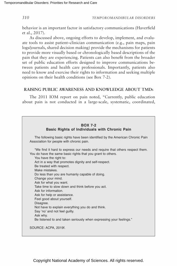
Temporomandibular Disorders: Priorities for Research and Care
Copyright National Academy of Sciences. All rights reserved.
310 TEMPOROMANDIBULAR DISORDERS
behavior is an important factor in satisfactory communications (Haverfield et al., 2017).
As discussed above, ongoing efforts to develop, implement, and evalu-ate tools to assist patient–clinician communication (e.g., pain maps, pain logs/journals, shared decision making) provide the mechanisms for patients to provide more visually based or chronologically based descriptions of the pain that they are experiencing. Patients can also benefit from the broader set of public education efforts designed to improve communications be-tween patients and health care professionals. Importantly, patients also need to know and exercise their rights to information and seeking multiple opinions on their health conditions (see Box 7-2).
RAISING PUBLIC AWARENESS AND KNOWLEDGE ABOUT TMDs
The 2011 IOM report on pain noted, “Currently, public education about pain is not conducted in a large-scale, systematic, coordinated,
BOX 7-2 Basic Rights of Individuals with Chronic Pain
The following basic rights have been identified by the American Chronic Pain Association for people with chronic pain.
“We find it hard to express our needs and require that others respect them. You do have the same basic rights that you grant to others.
You have the right to:Act in a way that promotes dignity and self-respect.Be treated with respect.Make mistakes.Do less than you are humanly capable of doing.Change your mind.Ask for what you want.Take time to slow down and think before you act.Ask for information.Ask for help or assistance.Feel good about yourself.Disagree.Not have to explain everything you do and think.Say ‘no’ and not feel guilty.Ask why.Be listened to and taken seriously when expressing your feelings.”
SOURCE: ACPA, 2019f.
Temporomandibular Disorders: Priorities for Research and Care
Copyright National Academy of Sciences. All rights reserved.
IMPROVING PATIENT, FAMILY, AND PUBLIC EDUCATION AND AWARENESS 311
and strategic way” (IOM, 2011b, p. 189), and compared the lack of an organized approach with the more organized efforts on smoking cessation, cancer, end of life, and Alzheimer’s disease. The extent of public awareness about TMDs has not been assessed as far as the committee could determine. However, given the challenges expressed to the committee by patients and health care professionals, the need for increased public awareness has be-come evident.
The avenues for raising public awareness and improving knowledge that have been successfully used for other health conditions include:
• Public awareness campaigns.• Designating a specific month or day to focus on the disease or dis-
order and planning community-based activities or campaigns at that time. September is designated as Pain Awareness Month.
• Identifying high-profile individuals with a connection to TMDs who can share their personal stories and act as spokespeople.
• Additionally, the 2011 IOM report included the following list of educational tools that are useful in reaching the public:
o website content, listservs, videos, and social media; o fact sheets and leaflets distributed to target audiences and avail-
able at multiple locations, including schools, health facilities, workplaces, wellness classes, places of worship, and other public venues;
o informational reports and studies; o signs at health facilities and in health professionals’ offices; and o media outreach including to television and movie scriptwriters.
One avenue for raising public awareness is through public education campaigns, which are often multimedia efforts to engage the general public and provide one or more key messages about the health condition, with links provided for those interested in further information. Public health campaigns are often initiated as government agency efforts or through public–private partnerships because of the broad constituencies needed to develop, evaluate, fund, and carry out these efforts on varying media plat-forms. Formative and ongoing evaluations of the campaigns are critically important to keeping the messages relevant and tracking health or health behavior outcomes. For example, the Centers for Disease Control and Prevention’s Screen for Life: National Colorectal Cancer Action Campaign started in 1999 with more than 200 focus groups that assessed knowledge and messaging, and there have been ongoing evaluations of the campaign (CDC, 2019a). The National Pain Strategy outlines a detailed set of objec-tives, strategies, and deliverables for a national public awareness campaign on the seriousness and impact of chronic pain (see Box 7-3) that can be
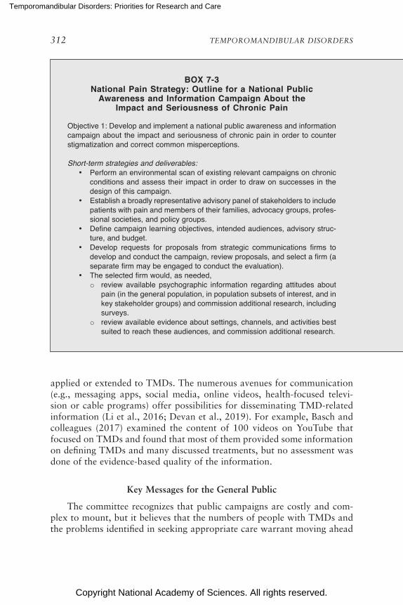
Temporomandibular Disorders: Priorities for Research and Care
Copyright National Academy of Sciences. All rights reserved.
312 TEMPOROMANDIBULAR DISORDERS
BOX 7-3 National Pain Strategy: Outline for a National Public
Awareness and Information Campaign About the Impact and Seriousness of Chronic Pain
Objective 1: Develop and implement a national public awareness and information campaign about the impact and seriousness of chronic pain in order to counter stigmatization and correct common misperceptions.
Shortterm strategies and deliverables:• Perform an environmental scan of existing relevant campaigns on chronic
conditions and assess their impact in order to draw on successes in the design of this campaign.
• Establish a broadly representative advisory panel of stakeholders to include patients with pain and members of their families, advocacy groups, profes-sional societies, and policy groups.
• Define campaign learning objectives, intended audiences, advisory struc-ture, and budget.
• Develop requests for proposals from strategic communications firms to develop and conduct the campaign, review proposals, and select a firm (a separate firm may be engaged to conduct the evaluation).
• The selected firm would, as needed, o review available psychographic information regarding attitudes about
pain (in the general population, in population subsets of interest, and in key stakeholder groups) and commission additional research, including surveys.
o review available evidence about settings, channels, and activities best suited to reach these audiences, and commission additional research.
applied or extended to TMDs. The numerous avenues for communication (e.g., messaging apps, social media, online videos, health-focused televi-sion or cable programs) offer possibilities for disseminating TMD-related information (Li et al., 2016; Devan et al., 2019). For example, Basch and colleagues (2017) examined the content of 100 videos on YouTube that focused on TMDs and found that most of them provided some information on defining TMDs and many discussed treatments, but no assessment was done of the evidence-based quality of the information.
Key Messages for the General Public
The committee recognizes that public campaigns are costly and com-plex to mount, but it believes that the numbers of people with TMDs and the problems identified in seeking appropriate care warrant moving ahead
Temporomandibular Disorders: Priorities for Research and Care
Copyright National Academy of Sciences. All rights reserved.
IMPROVING PATIENT, FAMILY, AND PUBLIC EDUCATION AND AWARENESS 313
o review existing information and educational materials. o develop a communications strategy based on behavior change theories
for each targeted audience. o work with the advisory board to identify and recruit partner organizations
and define their roles in the campaign.• Based on this preliminary work, develop and pretest messages and mate-
rials using, wherever possible, information developed by other components of the National Pain Strategy.
Mediumterm strategies and deliverables:• Implement the program, including partner participation strategies, spokes-
person training, and program-related services (e.g., pain self-management programs), media (news, entertainment, social) strategies, and promotional materials.
• Monitor audience reach, feedback, and partner engagement; adjust strate-gies as necessary.
Longterm (within 5 years) strategies and deliverables:• Conduct an outcome evaluation to assess campaign effectiveness, as
measured by changes in public opinion related to the campaign’s learning objectives.
• Prepare a report based on the campaign evaluations for submission to a peer-reviewed scientific journal.
• As funds are available, continue to monitor, implement, assess, and adapt campaign components, as needed, and report on campaign outcomes in a peer-reviewed journal.
SOURCE: NIH, 2016.
with exploring a campaign that would need to be validated and well tested, grounded in the evidence base, and attentive to delivering clear messages that are understandable to individuals with different levels of health literacy (IOM, 2004).
Some key messages to include in public messaging are noted below, but these should be augmented and refined by appropriate stakeholders, includ-ing patients, health care professionals, health agencies, and media groups. Chapter 5 provides more details on self-management messages. Possible key messages include the following:
• Not all TMDs are the same. A variety of disorders can affect the jaw joint or its associated muscles and nerves. TMDs can range from those that are short term and require no treatment to those that are long term, complex, and require more involved treatments.
Temporomandibular Disorders: Priorities for Research and Care
Copyright National Academy of Sciences. All rights reserved.
314 TEMPOROMANDIBULAR DISORDERS
• Avoid harm. Historically, some treatments have led to worsening rather than alleviating TMD symptoms and dysfunction. An im-proved understanding of treatment options and outcomes for TMDs has led to an increased focus on holistic management and avoidance of aggressive surgical interventions early in the disease process. Surgical treatment is indicated for some patients, but it is rarely the complete solution. When needed, surgery should be performed by specialists with knowledge and experience in this condition. As noted in the NIH brochure on TMD treatment, “less is often best” in treating these disorders (NIDCR, 2013).
• Explore options for care. Be informed and seek evidence-based and interprofessional approaches to care (across medicine, dentistry, nursing, physical therapy, integrative health, and other relevant care pathways), particularly for severe pain and complex cases. Inquire about different treatment options and self-care approaches. When symptoms are severe or have not been relieved with the initial treat-ment, consider seeking care from health care professionals with spe-cialty training in orofacial pain or in a dedicated multidisciplinary program for the treatment of orofacial pain.
• Be empowered to ask questions, seek person-centered care, and get multiple opinions. A basic right of patients is the right to ask for information to make an informed decision and to seek more than one opinion from health professionals and other patients. The focus of care should be on the total health and well-being needs of each individual, with time taken for discussion with and input from the patient. Frequently, persons with TMDs may also have other related health conditions, and so this holistic approach is needed. Further-more, patients who are in pain need to be validated and to know they can seek more information and additional opinions.
• The knowledge base underlying the biology of TMDs and the care of patients is in its infancy. Research on the mechanisms and biol-ogy of TMDs, their epidemiology, and approaches to TMD care are needed before evidence-based guidelines can be developed and implemented. This will make navigating the current care landscape difficult and underscores the need for conservative treatments as starting points for TMD care.
CONCLUSION AND RESEARCH PRIORITIES
The lack of awareness about TMDs was identified as a barrier to patient care and to patients and families making informed decisions. The commit-tee’s discussions focused on the need to overcome stigmas that may be associated with TMDs and with facial pain and to provide evidence-based
Temporomandibular Disorders: Priorities for Research and Care
Copyright National Academy of Sciences. All rights reserved.
IMPROVING PATIENT, FAMILY, AND PUBLIC EDUCATION AND AWARENESS 315
resources for patients, their families, and the general public. The commit-tee acknowledges that significant amounts of time, energy, and financial resources and commitments are required to carry out these efforts and urges strong collaborative efforts involving professional associations and patient advocacy groups, along with federal agency and other partners, that are needed to move this work forward.
Research on the educational aspects of TMD diagnosis and care has been limited. Throughout this chapter, the committee has identified im-provements that are needed to fully educate potential patients, families, health care professionals, and the general public on TMDs.
Conclusion 7-1: Building evidence-based communication resources will require attention to:
• Developing guidelines for the evaluation of communica-tion resources on temporomandibular disorders (TMDs) (including health literacy evaluations) and then using the guidelines to evaluate existing educational resources and tools as a basis for improvements and for building new resources;
• Developing evidence-based consumer roadmaps and websites to help individuals and families identify trusted sources for evidence-based information and referrals; and
• Working with advocacy organizations and health profes-sional associations to ensure widespread dissemination of evidence-based information on TMDs.
The committee provides the following research priorities (see Box 7-4). As outlined in Chapter 8, there are a number of actions that can be taken to improve patient, family, and professional education; to raise awareness in the general public; and to reduce stigma.
Temporomandibular Disorders: Priorities for Research and Care
Copyright National Academy of Sciences. All rights reserved.
316 TEMPOROMANDIBULAR DISORDERS
BOX 7-4 Research Priorities for Improving Patient and Family
Education and Raising Public Awareness
To improve the education of people with a temporomandibular disorder (TMD) and their families and to increase awareness of TMDs in the general public, the following areas should be considered as priorities for research:
• Assessment of the information needs of specific subpopulations, including adolescents, young adults, rural populations, women, and underserved communities.
• Identification of best practices, effective strategies and formats, and inno-va tions for educating patients and families, particularly individuals in under-served populations, and also health care professionals.
• Evaluation of current education programs and resources.• Evaluations of websites seeking to promote evidence-based knowledge
about TMDs.• Formative and implementation research to develop public awareness cam-
paigns about TMDs and to understand best practices, strategies, and mechanisms for public education.
REFERENCES
ACPA (American Chronic Pain Association). 2019a. Head pain map. http://www.theacpa.org/pain-management-tools/communication-tools/tracking-tools/head-pain-map (accessed July 10, 2019).
ACPA. 2019b. Pain log. https://www.theacpa.org/pain-management-tools/communication-tools/tracking-tools/pain-log (accessed July 10, 2019).
ACPA. 2019c. Support groups. https://www.theacpa.org/about-us/support-groups (accessed July 10, 2019).
ACPA. 2019d. Pain management tools. https://www.theacpa.org/pain-management-tools (ac-cessed July 10, 2019).
ACPA. 2019e. Family matters series. https://www.theacpa.org/pain-management-tools/videos/support (accessed September 23, 2019).
ACPA. 2019f. Basic rights. https://www.theacpa.org/pain-management-tools/basic-rights (ac-cessed July 10, 2019).
Afsal, M. M., A. Khatri, N. Kalra, R. Tyagi, and D. Khandelwal. 2019. Pain perception and efficacy of local analgesia using 2% lignocaine, buffered lignocaine, and 4% articaine in pediatric dental procedures. Journal of Dental Anesthesia and Pain Medi-cine 19(2):101–109.
Armentor, J. L. 2017. Living with a contested, stigmatized illness: Experiences of man-aging relationships among women with fibromyalgia. Qualitative Health Research 27(4):462–473.
Back, A. L., R. M. Arnold, W. F. Baile, J. A. Tulsky, and K. Frayer-Edwards. 2005. Approach-ing difficult communication tasks in oncology. CA: A Cancer Journal for Clinicians 55(3):164–177.
Temporomandibular Disorders: Priorities for Research and Care
Copyright National Academy of Sciences. All rights reserved.
IMPROVING PATIENT, FAMILY, AND PUBLIC EDUCATION AND AWARENESS 317
Basch, C. H., J. Yin, N. D. Walker, A. J. de Leon, and I. C. Fung. 2017. TMJ online: Investigat-ing temporomandibular disorders as “TMJ” on YouTube. Journal of Oral Rehabilitation 45:34–40.
Bijur, P. E., W. Silver, and E. J. Gallagher. 2001. Reliability of the visual analog scale for mea-surement of acute pain. Academic Emergency Medicine 8(12):1153–1157.
CDC (Centers for Disease Control and Prevention). 2019a. About the Screen for Life cam-paign. https://www.cdc.gov/cancer/colorectal/sfl/about.htm (accessed July 18, 2019).
CDC. 2019b. Health literacy. https://www.cdc.gov/healthliteracy/index.html (accessed Decem-ber 3, 2019).
Cegala, D. J., and D. M. Post. 2009. The impact of patients’ participation on physicians’ patient-centered communication. Patient Education and Counseling 77(2):202–208.
Chen, A. T. 2012. Information seeking over the course of illness: The experience of people with fibromyalgia. Musculoskeletal Care 10(4):212–220.
Cook, J. E., V. Purdie-Vaughns, I. H. Meyer, and J. T. A. Busch. 2014. Intervening within and across levels: A multilevel approach to stigma and public health. Social Science & Medicine 103:101–109.
Corrigan, P. W. 2014. The stigma of disease and disability: Understanding causes and over-coming injustices. Washington, DC: American Psychological Association.
Cowan, P., N. Kelly, C. Passero, and E. Covington. 1998. ACPA Family Manual A manual for families of person with pain. Rocklin, CA: ACPA.
De Ruddere, L., and K. D. Craig. 2016. Understanding stigma and chronic pain: A state-of-the art review. Pain 157(8):1607–1610.
Desai, B., N. Alkandari, and D. M. Laskin. 2016. How accurate is information about diag-nosis and management of temporomandibular disorders on dentist websites? Oral Surgery, Oral Medicine, Oral Pathology, and Oral Radiology 122(3):306–309.
Devan, H., H. K. Godfrey, M. A. Perry, D. Hempel, B. Saipe, L. Hale, and R. Grainger. 2019. Current practices of health care providers in recommending online resources for chronic pain management. Journal of Pain Research 12:2457–2472.
Driscoll, M. A., M. T. Knobf, D. M. Higgins, A. Heapy, A. Lee, and S. Haskell. 2018. Patient experiences navigating chronic pain management in an integrated health care system: A qualitative investigation of women and men. Pain Medicine 19(Suppl 1):S19–S29.
Dubin, R. E. 2017. Acknowledging stigma: Its presence in patient care and medical education. Canadian Family Physician 63:906–908.
Dwamena, F. C., B. Mavis, M. Holmes-Rovner, and A. Loyson. 2009. Teaching medical inter-viewing to patients: The other side of the encounter. Patient Education and Counseling 76(3):380–384.
Fisher, E. B., G. X. Ayala, L. Ibarra, A. L. Cherrington, J. P. Elder, T. S. Tang, M. Heisler, M. M. Safford, and D. Simmons. 2015. Contributions of peer support to health, health care, and prevention: Papers from Peers for Progress. Annals of Family Medicine 13(Suppl 1):S2–S8.
Forsythe, L. P., K. L. Carman, V. Szydlowski, L. Fayish, L. Davidson, D. H. Hickam, C. Hall, G. Bhat, D. Neu, L. Stewart, M. Jalowsky, N. Aronson, and C. U. Anyanwu. 2019. Patient engagement in research: Early findings from the Patient-Centered Outcomes Research Institute. Health Affairs 38(3):359–367.
Fortin, A. H., F. C. Dwamena, R. M. Frankel, and R. C. Smith. 2012. Smith’s patient centered interviewing: An evidence-based method. Third edition. New York: McGraw-Hill.
Gottlieb, S. 2000. Health information on internet is often unreliable. BMJ [online]. https://www.bmj.com/content/321/7254/136.3.full (accessed July 15, 2019).
Greene, J., J. H. Hibbard, R. Sacks, V. Overton, and C. D. Parrotta. 2015. When patient activa-tion levels change, health outcomes and costs change, too. Health Affairs 34(3):431–437.
Temporomandibular Disorders: Priorities for Research and Care
Copyright National Academy of Sciences. All rights reserved.
318 TEMPOROMANDIBULAR DISORDERS
Haverfield, M. C., K. Giannitrapini, C. Timko, and K. Lorenz. 2017. Patient-centered pain management communication from the patient perspective. Journal of General Internal Medicine 33(8):1374–1380.
Hawker, G. A., S. Mian, T. Kendzerska, and M. French. 2011. Measures of adult pain. Arthritis Care & Research 63(S11):S240–S252.
Heisler, M. 2010. Different models to mobilize peer support to improve diabetes self- management and clinical outcomes: Evidence, logistics, evaluation considerations and needs for future research. Family Practice 27(Suppl 1):i23–i32.
Hoffmann, D. E., and A. J. Tarzian. 2001. The girl who cried pain: A bias against women in the treatment of pain. The Journal of Law, Medicine, & Ethics 29(1):13–27.
IHI (Institute for Healthcare Improvement). 2019. Ask Me 3: Good questions for your good health. http://www.ihi.org/resources/Pages/Tools/Ask-Me-3-Good-Questions-for-Your-Good-Health.aspx (accessed July 11, 2019).
INfORM (International Network for Orofacial Pain and Related Disorders Methodology). 2013. Pain map. https://ubwp.buffalo.edu/rdc-tmdinternational/tmd-assessmentdiagnosis/dc-tmd (accessed November 1, 2019).
IOM (Institute of Medicine). 2004. Health literacy: A prescription to end confusion. Washington, DC: The National Academies Press.
IOM. 2011a. Patients charting the course: Citizen engagement and the learning health system: Workshop summary. Washington, DC: The National Academies Press.
IOM. 2011b. Relieving pain in America: A blueprint for transforming prevention, care, educa-tion, and research. Washington, DC: The National Academies Press.
IOM. 2012. Epilepsy across the spectrum: Promoting health and understanding. Washington, DC: The National Academies Press.
IOM. 2014. Partnering with patients to drive shared decisions, better value, and care improve-ment: Workshop proceedings. Washington, DC: The National Academies Press.
Keller, S., C. M. Bann, S. L. Dodd, J. Schein, T. R. Mendoza, and C. S. Cleeland. 2004. Validity of the Brief Pain Inventory for use in documenting the outcomes of patients with non-cancer pain. Clinical Journal of Pain 20(5):309–318.
Khatri, A., and N. Kalra. 2012. A comparison of two pain scales in the assessment of dental pain in East Delhi children. ISRN Dentistry. doi: 10.5402/2012/247351.
LeResche, L. 2011. Defining gender disparities in pain management. Clinical Orthopaedics and Related Research 469(7):1871–1877.
Li, X., Z. R. Xu, N. Tang, C. Ye, X. L. Zhu, T. Zhou, and Z. H. Zhao. 2016. Effect of inter-vention using a messaging app on compliance and duration of treatment in orthodontic patients. Clinical Oral Investigations 20(8):1849–1859.
Marbach, J. J., M. C. Lennon, B. G. Link, and B. P. Dohrenwend. 1990. Losing face: Sources of stigma as perceived by chronic facial pain patients. Journal of Behavioral Medicine 13(6):583–604.
Matthias, M. S., M. Kukla, A. B. McGuire, and M. J. Bair. 2016. How do patients with chronic pain benefit from a peer-supported self-management intervention? A qualitative investigation. Pain Medicine 17:2247–2255.
NASEM (National Academies of Sciences, Engineering, and Medicine). 2016. Hearing health care for adults: Priorities for improving access and affordability. Washington, DC: The National Academies Press.
NASEM. 2019. Integrating oral and general health through health literacy practices. Washington, DC: The National Academies Press.
NIDCR (National Institute of Dental and Craniofacial Research). 2013. Less is often best in treating TMJ disorders. https://www.nidcr.nih.gov/sites/default/files/2017-09/less-is-best-tmj.pdf (accessed August 30, 2019).
Temporomandibular Disorders: Priorities for Research and Care
Copyright National Academy of Sciences. All rights reserved.
IMPROVING PATIENT, FAMILY, AND PUBLIC EDUCATION AND AWARENESS 319
NIH (National Institutes of Health). 2016. National pain strategy. https://www.iprcc.nih.gov/sites/default/files/HHSNational_Pain_Strategy_508C.pdf (accessed July 18, 2019).
Okunrintemi, V., E. S. Spatz, P. Di Capua, J. A. Salami, J. Valero-Elizondo, H. Warraich, S. S. Virani, M. J. Blaha, R. Blankstein, A. A. Butt, W. B. Borden, K. Dharmarajan, H. Ting, H. M. Krumholz, and K. Nasir. 2017. Patient-provider communication and health outcomes among individuals with atherosclerotic cardiovascular disease in the United States. Circulation: Cardiovascular Quality and Outcomes 10(4).
Pescosolido, B. A., and J. K. Martin. 2015. The stigma complex. Annual Review of Sociology 41:87–116.
Racich, M. J. 2009. What are 3 key elements of the TMD patient interview. Journal of the Canadian Dental Association 75(3):197–198.
Ramsey, I., N. Corsini, M. D. J. Peters, and M. Eckert. 2017. A rapid review of con-sumer health information needs and preferences. Patient Education and Counseling 100(9):1634–1642.
Ring, D., and S. Lawn. 2019. Stigma perpetuation at the interface of mental health care: A review to compare patient and clinician perspectives of stigma and borderline personality disorder. Journal of Mental Health 12:1–21.
Scott, W., L. Yu, S. Patel., and L. M. McCracken. 2019. Measuring stigma in chronic pain: Preliminary investigation of instrument psychometrics, correlates, and magnitude of change in a prospective cohort attending interdisciplinary treatment. Journal of Pain 20(10):1164–1175.
Slade, S. C., E. Molloy, and J. L. Keating. 2009. Stigma experienced by people with nonspecific chronic low back pain: A qualitative study. Pain Medicine 10(1):143–154.
Sondell, K., and B. Soderfeldt. 1997. Dentist-patient communication: A review of relevant models. Acta Odontologica Scandinavica 55:116–126.
Storino, A., J. Castillo-Angeles, A. A. Watkins, C. Vargas, J. D. Mancias, A. Bullock, A. Demirjian, J. Moser, and T. S. Kent. 2016. Assessing the accuracy and readability of online health information for patients with pancreatic cancer. JAMA Surgery 151(9):831–837.
The TMJ Association. 2019a. TMJ Café: Temporomandibular disorders support and discus-sion community. https://www.inspire.com/groups/tmjcafe (accessed July 11, 2019).
The TMJ Association. 2019b. TMJ support community. http://www.tmj.org/Page/29/14 (ac-cessed July 11, 2019).
Wakefield, E. O., W. T. Zempsky, R. M. Puhl, and M. D. Litt. 2018. Conceptualizing pain-related stigma in adolescent chronic pain: A literature review and preliminary focus group findings. Pain Reports 3(Suppl 1):e679.
Waugh, O. C., D. G. Byrne, and M. K. Nicholas. 2014. Internalized stigma in people living with chronic pain. Journal of Pain 15(5):550e1–550e9.
Woolf, S. H., E. Zimmerman, A. Haley, and A. H. Krist. 2016. Authentic engagement of patients and communities can transform research, practice, and policy. Health Affairs 35(4):590–594.
Wynia, M. K., and C. Y. Osborn. 2010. Health literacy and communication quality in health care organizations. Journal of Health Communications 15(Suppl 2):102–115.

Temporomandibular Disorders: Priorities for Research and Care
Copyright National Academy of Sciences. All rights reserved.
Temporomandibular Disorders: Priorities for Research and Care
Copyright National Academy of Sciences. All rights reserved.
321
Living with TMD has been a roller coaster for me…. Forming words can be difficult, as can drinking from a glass without a straw due to lack of muscle control inside of my mouth. Eating can be difficult, which is made worse by a lack of taste that was apparently damaged in my six surgeries.
—Lynne P.
We need to know what causes TMD, why it is more prominent in women, how to treat it, and long-term effects from current treatments out there. We need better treatment options. We need you to listen to the patients. We need your help. This is a very complex condition that needs medical experts from numerous specialties not just dentistry and oral facial pain medicine.
—Heather
Action is urgently needed to improve care for individuals with a temporo mandibular disorder (TMD). Too long compartmentalized as a dental issue, both the clinical management of and the research addressing TMDs need to implement a holistic and multidisciplinary approach. Indi-viduals with TMD symptoms often encounter health professionals (across medicine, dentistry, and beyond) that are unfamiliar with TMDs and do not know where best to refer patients for further diagnosis and treatment. The gaps and divides between medical and dental care are currently vast in the United States (IOM, 2011; Mertz, 2016) and, as a result, patients can get lost between the two systems or not receive the multidisciplinary care
8
Next Steps and Recommendations
Temporomandibular Disorders: Priorities for Research and Care
Copyright National Academy of Sciences. All rights reserved.
322 TEMPOROMANDIBULAR DISORDERS
they need. Evidence-based clinical practice guidelines for TMD care are needed as well as efforts to enhance accessibility to high-quality, patient-centered care.
Because TMDs represent a grouping of varied disorders—with diverse etiologies, symptoms, and treatments—rather than a single disorder, health professionals and researchers need to focus on learning more about indi-vidual TMDs (e.g., myalgia, disc disorders, arthralgia) and on the specific treatments for each. Much of TMD research is siloed and simultane-ously fragmented across niche areas of dentistry or medicine and needs a coordinated approach focused on patient-centered and clinically meaningful outcomes.
The committee worked to review the scientific literature; to seek informa tion from patients and their family members, researchers, clini-cians, policy makers, research funders, and others; to analyze the data; and to develop its conclusions and recommendations. The recommendations in this chapter focus on the actions that many organizations and agen-cies should take to improve TMD research and care. The committee also emphasizes the critical role that individuals with a TMD and their family members have played—and hopefully will continue to play—in bringing TMD issues to the attention of policy makers and health professionals and moving the research and care agenda forward on multiple levels in the public and private sectors. These efforts are to be commended and are en-couraged to continue and expand. It is the committee’s hope that increased opportunities will be available for individuals with a TMD and their family members to engage in these important efforts to improve the state of TMD research and care. Specifically, it is hoped that individuals with a TMD and their families will be able to partner with their health care professionals to find the best options for care, to continue to actively participate in patient support networks, to explore ways to be a participating voice in research efforts (such as serving as a patient representative in research design), and to be active advocates for improvements in care and services for themselves, their family members, and other people with a TMD.
The goals of the following recommendations are to build a strong base of knowledge about TMDs and to facilitate actions needed to improve the overall health and well-being of individuals with a TMD. Some of these recommendations can be accomplished rapidly with actions by key decision makers. Other recommendations are more aspirational and will require the collaboration and commitment of multiple organizations and dedicated resources—including investments of time, funds, and innovative energies—to accomplish these goals. The committee has provided both short-term and longer-term priorities to be used as starting points and long-range planning points. Key to making a difference in improving care for individuals with a TMD will be:
Temporomandibular Disorders: Priorities for Research and Care
Copyright National Academy of Sciences. All rights reserved.
NEXT STEPS AND RECOMMENDATIONS 323
• pioneering pathways that span medicine, dentistry, physical therapy, and other fields of health care to provide holistic, comprehensive approaches to care—interprofessional and interdisciplinary efforts are of critical importance;
• willingness of health care agencies, organizations, and professionals to commit the resources needed to address this long-neglected and often dismissed area of health care; and
• openness and commitment to using and strengthening the evidence base on TMD treatment and changing practice as needed.
BUILD AND SUSTAIN COLLABORATIVE AND MULTIDISCIPLINARY RESEARCH
Engagement by multiple stakeholders will be required to dismantle the siloes keeping research fields isolated and to advance TMD research and care. It will be necessary to apply innovative research models and theory-based research designs to the challenges of TMDs. Patients, dentists, physicians, and other health professionals must be involved in advanc-ing research. The committee recommends that a research consortium be established to bring together relevant National Institutes of Health (NIH) institutes and centers and other stakeholders from the public and private sectors to focus future research efforts on filling key evidence gaps in TMD research and care and to ensure that clinically meaningful, patient-centered outcomes are prioritized. The committee stresses the importance of an organized research approach for TMDs, but the mechanism to carry this out should be flexible (i.e., if not an NIH-led consortium, another trusted leader in the field could organize a research network with the goals outlined in Recommendation 1).
Fresh ideas and multiple disciplines are needed to advance TMD re-search to improve patient care. NIH provides approximately one-third of all biomedical research funding in the United States (IOM, 2011) and there-fore, the interests and priorities of NIH institutes and centers can stimulate research interests and training programs throughout the country. TMDs are not the primary mission of any NIH center or institute. NIH fund-ing for TMD research falls largely within the National Institute of Dental and Craniofacial Research (NIDCR) (which has one of the smallest re-search budgets of the NIH institutes) with a total budget of approximately $461 million compared to the National Cancer Institute’s $5.99 billion for fiscal year 2019 (NIH, 2020). Given the number of individuals suffer-ing from TMDs, the severity of some of the disorders, and the substantial public health burden of TMDs, there is a significant opportunity for NIH and other biomedical research institutions to drive increased funding to TMDs in order to spark new research interest and discoveries. Efforts are
Temporomandibular Disorders: Priorities for Research and Care
Copyright National Academy of Sciences. All rights reserved.
324 TEMPOROMANDIBULAR DISORDERS
needed to ensure that TMD research is incorporated into NIH-wide initia-tives, including the NIH Pain Consortium. Furthermore, as noted in Public Law 116-94, an NIH inter-institute working group is being called on to focus on coordinating TMD research across the multiple NIH institutes and centers relevant to this field (see Recommendation 1 for details).
Recommendation 1: Create and Sustain a National Collaborative Research Consortium for Temporomandibular Disorders (TMDs)
A National Collaborative Research Consortium for TMDs should be established and sustained to coordinate, fund, and translate basic and clinical research (including behavioral, population-based, and imple-mentation research) to address evidence gaps, generate clinically mean-ingful knowledge, identify safe and effective treatments, and improve the quality of TMD care.
The consortium would:
• Establish and implement a national research framework for TMDs;• Provide infrastructure for the implementation of research projects; • Establish milestones and timelines; • Facilitate research collaborations; • Develop public–private partnerships; • Develop and test evidence-based strategies for knowledge transfer; • Support the development of a multidisciplinary research workforce
for TMDs through existing and new training and center initiatives; and
• Evaluate progress and disseminate research findings.
Near-term implementation actions:• The National Institutes of Health (NIH) Office of the Director and
the National Institute of Dental and Craniofacial Research should lead an effort to establish the National Collaborative Research Consortium for TMDs by convening relevant stake holders and re-search funders (including but not limited to the Agency for Healthcare Research and Quality [AHRQ], the NIH Pain Consortium, the Patient-Centered Outcomes Research Institute, the Health Resources and Services Administration, the Centers for Medicare & Medicaid Services, the Centers for Disease Control and Prevention, the Department of Defense [DoD], the Department of Veterans Affairs [VA], and relevant NIH institutes including the National Institute of Neurological Disorders and Stroke, the National Institute of Mental Health, the National Institute of Arthritis and Musculoskeletal
Temporomandibular Disorders: Priorities for Research and Care
Copyright National Academy of Sciences. All rights reserved.
NEXT STEPS AND RECOMMENDATIONS 325
and Skin Diseases, the National Center for Complementary and Integrative Health, the National Institute of Biomedical Imaging and Bioengineering, the National Institute of Nursing Research, NIH Office of Nutrition Research, NIH Office of Research on Women’s Health, NIH Office of Behavioral and Social Sciences Research, TMJ Patient-Led RoundTable, Chronic Pain Research Alliance, The TMJ Association, academic research centers, research foundations, private-sector research funders, professional health care associations, private and public insurers, and patient advocacy organi zations) to:
o Determine the governing structure (including the vision and mis-sion statement) and strategic plan;
o Design and implement a national research framework for TMDs that would establish short- and long-term research priorities and cultivate multidisciplinary research focused on the needs of the patient; and
o Conduct an annual symposium to explore promising research directions, methods, and tools for TMD research and to foster multidisciplinary, and possibly international, collaborations.
Medium- to long-term implementation actions: • Once established, the National Collaborative Research Consortium
for TMDs should implement the research framework by synchro-nizing and guiding research efforts across stakeholders and research areas;
• Encourage and incentivize multidisciplinary collaborations; • Leverage funding resources to address research priorities that may
overlap with and be part of larger research initiatives (e.g., HEAL initiative);
• Develop public–private partnerships across relevant disciplines, especially medicine and dentistry, to support research on TMDs;
• Disseminate research findings and implement evidence-based strate-gies for knowledge translation from research to clinical and public health practice and policy; and
• Set priorities and direct funds to support multidisciplinary research training and workforce development in the TMD field using existing NIH and other funding mechanisms.
Recommendation 2: Strengthen Basic Research and Translational Efforts
The National Collaborative Research Consortium for temporo-mandibular disorders (TMDs) along with other funders should fund
Temporomandibular Disorders: Priorities for Research and Care
Copyright National Academy of Sciences. All rights reserved.
326 TEMPOROMANDIBULAR DISORDERS
basic research efforts and ensure its translation as part of a patient- focused, multidisciplinary research agenda on TMDs to address evi-dence gaps, generate clinically meaningful knowledge, identify effective treatments, and improve quality of care. Research priorities should include:
• Biopsychosocial mechanisms related to the development of tissue dysfunction and pain generation, maintenance, and suppression across TMDs and patient groups;
• Basic and translational behavioral and social sciences; • Factors that promote risk for symptom induction;• Sex differences related to tissue dysfunction and pain generation,
maintenance, and suppression across TMDs and patient groups;• Biomarkers and predictive factors for diagnosis, prognosis, response
to treatment, and resilience;• Neuroimmune mechanisms in etiology, progression, and treatment;• Neuroendocrine system interactions and stress-induced mechanisms
in the progression of a specific TMD;• Shared biopsychosocial mechanisms of TMDs and comorbid
conditions;• Integrative research to understand how risk factors interact and how
clinical, psychological, and genetic factors confer different levels of TMD risk in different groups of individuals;
• Biomechanics research, including the development of new tools to measure intra-articular space;
• Regenerative medicine research, including research involving the use of stem cells;
• Artificial intelligence and novel data approaches, including identify-ing patterns in genomic and proteomics data;
• Prevention of transitions from acute to chronic pain; and • Clinically relevant and mechanistically anchored patient subgroups
and framing of future research to improve treatments that match to identified subgroups.
Recommendation 3: Strengthen Population-Based Research on the Public Health Burden of Temporomandibular Disorders (TMDs)
The National Collaborative Research Consortium for TMDs along with other funders should expand and strengthen the collection, assess-ment, and dissemination of population-based data on the burden and costs of TMDs and the effects of TMDs on patient outcomes in order to improve the prevention (primary, secondary, and tertiary) and man-agement of TMDs.
Temporomandibular Disorders: Priorities for Research and Care
Copyright National Academy of Sciences. All rights reserved.
NEXT STEPS AND RECOMMENDATIONS 327
Near-term implementation actions:• The National Collaborative Research Consortium for TMDs and
other relevant organizations and agencies should convene key stake-holders to:
o Review questions related to TMDs in current national popula-tion surveys and make recommendations regarding the stan-dardization, addition, or refinement of questions and use of case definitions; and
o Develop guidelines encouraging future research proposals to use standardized case definitions and diagnostic criteria.
Medium- to long-term implementation actions:• The National Collaborative Research Consortium for TMDs in col-
laboration with other relevant stakeholders (e.g., large integrated health care systems such as Kaiser Permanente and the Department of Veterans Affairs) should fund and evaluate:
o Projects to explore the linkage and use of electronic health and dental records with payer claims databases and other electronic repositories to gain insights into the prevalence of individual TMDs and the burden and impact of TMDs;
o Work that builds on the Orofacial Pain Prospective Evaluation and Risk Assessment study and further explores the directional-ity of the relationship between TMDs and comorbidities. Future studies should seek to identify prognostic biomarkers of TMDs in order to understand how risk factors interact and how clini-cal, psychological, and genetic factors confer different levels of TMD risk in different groups of individuals;
o Research that focuses on longitudinal studies in diverse popula-tions to better understand the natural history of common and uncommon TMDs and the interactions of risk factors to inform whether, when, and how to manage TMDs; and
o The impact of TMDs, treatment of TMDs, and trajectories of these disorders on health, function, economic productivity, and quality of life.
Recommendation 4: Bolster Clinical Research Efforts to Build the Evi-dence Base for Patient-Centered Care and Public Health Interventions for Temporomandibular Disorders (TMDs)
The National Collaborative Research Consortium for TMDs along with other funders should fund clinical and implementation research to clearly define effective treatments and continuously improve the quality of care for patients with a TMD. These near-term efforts should:
Temporomandibular Disorders: Priorities for Research and Care
Copyright National Academy of Sciences. All rights reserved.
328 TEMPOROMANDIBULAR DISORDERS
• Explore and develop pilot projects on TMD treatment effective-ness and implementation through the National Institutes of Health Collaboratory on Pragmatic Trials; the Analgesic, Anesthetic, and Addiction Clinical Trial Translations, Innovations, Opportunities, and Networks public–private network; the Initiative on Methods, Measurement, and Pain Assessment in Clinical Trials; the National Patient-Centered Clinical Research Network; and other relevant clinical research networks;
• Develop a set of common data elements for use in clinical research on TMDs, which could include validating existing relevant measures (such as the Patient-Reported Outcomes Measurement Information System or the TMJ Patient-Led RoundTable patient outcomes) and the development of new measures for consistent use in all studies on the TMD population and determining which components are most relevant for patients;
• Prioritize funding for innovative TMD clinical research projects that focus on meaningful outcomes that are important to patients and patient care, including pragmatic trials and other comparative effectiveness research;
• Expand the scope of research on TMDs conducted via the Dental Practice Based Research Network to enhance evidence regarding treatment outcomes for TMDs in routine practice;
• Test novel self-management interventions (using intervention devel-opment theoretical frameworks) that foster the translation of theory and basic science to improve TMD care; and
• Explore and support the development and implementation of a national TMD patient registry:
o National Institute of Dental and Craniofacial Research, The TMJ Association, TMJ Patient-Led RoundTable, and Medical Device Epidemiology Network Initiative (MDEpiNet), in col-laboration with professional associations and TMD centers of excellence (see Recommendation 8), should convene a workshop to:
Explore various options for a TMD patient registry; Obtain input on best practices and lessons learned from
other and ongoing patient registry efforts on TMDs and other diseases and disorders (including, but not limited to, MDEpiNet’s work on a TMD registry and the Platform for Engaging Everyone Responsibly, operated by the Genetic Alliance); and
Outline next steps for the development of a national patient registry.
Temporomandibular Disorders: Priorities for Research and Care
Copyright National Academy of Sciences. All rights reserved.
NEXT STEPS AND RECOMMENDATIONS 329
IMPROVE ACCESS TO AND QUALITY OF TMD HEALTH CARE
Throughout this report, the committee emphasizes a number of impor-tant elements of TMD care, including:
• Patient centeredness, recognizing that individuals with a TMD are more than their medical condition and that quality-of-life factors are important;
• Coordinated and multidisciplinary care as needed that may involve a team of professionals across disciplines; and
• A focus on education, in order to improve clinicians’ knowledge and skills, the general public’s awareness and understanding of TMDs, and the self-management skills of individuals with a TMD.
An important challenge in ensuring the availability of high-quality care for TMDs, particularly for those who have a TMD that is not easily resolved, is making sure that patients have access to coordinated care across medicine, dentistry, and other health professions. Innovative approaches and interprofessional efforts will be needed. Specialized TMD centers, especially for individuals that need multiple types of care, would be vital and could contribute significantly to telehealth options for improving access to specialty care as well as to innovative approaches to health professional education, clinical research, and data collection and analysis. Much remains to be learned about how to individualize patient care to the extent possible in order to provide the most effective management and treatment options for that individual.
Recommendation 5: Improve the Assessment and Risk Stratification of Temporomandibular Disorders (TMDs) to Advance Patient Care
Diagnostic tools and resources for TMDs should be improved for the initial assessment by primary care clinicians and dentists and for refer-rals to specialists as needed. These efforts should include the devel-opment of decision criteria for risk stratification to aid in identifying patients who are likely to escalate from self-limiting and localized symptoms to a systemic pain condition and then to high-impact pain. Initial instruments will be based on the current understanding of TMD science, though limited, and should be informed by the science as it evolves.
Near-term implementation actions: • The International Network for Orofacial Pain and Related Disorders
Methodology (INfORM), the American Dental Association
Temporomandibular Disorders: Priorities for Research and Care
Copyright National Academy of Sciences. All rights reserved.
330 TEMPOROMANDIBULAR DISORDERS
(ADA), the American Academy of Orofacial Pain, and The TMJ Association, in collaboration with the American Academy of Family Physicians, Society of General Internal Medicine, American College of Rheumatology, and other relevant professional organizations and stakeholders should:
o Develop diagnostic and screening tools, including a list of high-risk/red-flag symptoms for health care professionals (primary care and dentists) for TMDs with attention to:
Strategies to identify common and concerning alternative diag noses that can mimic TMDs (i.e., neoplasm, rheumatic disease) so that these can be ruled out prior to initiating TMD treatment and
Criteria for referrals from primary care professionals to specialists.
o Determine next steps for developing diagnostic criteria for the subtypes of TMDs that are less common and validating those criteria for reliability and validity.
Medium- to long-term implementation actions: • Dentists, primary care clinicians, and other relevant health care
professionals should: o Implement assessment tools in clinical practice and evaluate
changes to practice and patient outcomes and o Combine evolving science regarding biology and mechanisms
of TMDs with clinical findings to improve diagnosis and risk stratification.
Recommendation 6: Develop and Disseminate Evidence-Based Clinical Practice Guidelines and Quality Metrics for Care of Temporomandibu-lar Disorders (TMDs)
Clinical practice guidelines should be developed and widely dissemi-nated that provide evidence-based pathways for the initial recognition and stepped care management of TMDs and for specialty care for patients with TMDs. Once clinical practice guidelines are developed, clinical performance measures should be deployed in quality improve-ment initiatives.
Near-term implementation actions:• The International Association for the Study of Pain, American
Academy of Pain Medicine, American Academy of Orofacial Pain, International Network for Orofacial Pain and Related Disorders Methodology, and the American Chronic Pain Association should
Temporomandibular Disorders: Priorities for Research and Care
Copyright National Academy of Sciences. All rights reserved.
NEXT STEPS AND RECOMMENDATIONS 331
convene stakeholders (including the American Dental Association [ADA], American Academy of Family Physicians, Society of General Internal Medicine, American Association of Nurse Practitioners, and The TMJ Association) to develop evidence-based consensus clinical practice guidelines for dentists and primary care clinicians to guide diagnosis, initial treatment, and referral strategies for patients with TMD symptoms.
Medium- to long-term implementation actions:• ADA, in conjunction with the American Association for Dental
Research and the International Association for Dental Research, the American Academy of Family Physicians, Society of General Internal Medicine, the Agency for Healthcare Research and Quality, the National Committee for Quality Assurance, The TMJ Association, and other professional organizations involved in quality metrics and in the care of TMDs should:
o Develop a national quality measurement and improvement strat-egy for TMD care. These efforts would include ongoing efforts to disseminate and refine the clinical practice guidelines as well as the development and establishment of performance metrics for quality TMD care. This work should be done in collabora-tion with independent organizations with expertise in quality measurement and improvement.
o Develop, implement, and evaluate clinical decision tools for TMDs for use in electronic health records (both medical and dental) that are based on the clinical practice guidelines.
• Health systems that provide integrated medical and dental care (e.g., the Department of Defense, Kaiser Permanente, and Marshfield Clinic) should use TMD care as a pilot case study for their innova-tive and continuing efforts to coordinate medical and dental care. These institutions are also well poised to study the effectiveness of building clinical practice guidelines for TMDs into clinical decision support tools for staff physicians and dentists.
Recommendation 7: Improve Reimbursement and Access to High- Quality Assessment, Treatment, and Management of Temporo mandibular Disorders (TMDs)
Insurers and health care systems across dentistry and medicine should provide consistent, fair, equitable, and appropriate insurance coverage for safe and effective treatments for TMDs.
Temporomandibular Disorders: Priorities for Research and Care
Copyright National Academy of Sciences. All rights reserved.
332 TEMPOROMANDIBULAR DISORDERS
Near-term implementation actions:• The American Dental Association, in collaboration with The
TMJ Association and private and public health insurers (includ-ing Medicare and Medicaid) and health professional associations, should convene a working group across public and private health and dental insurers and health care systems to:
o Realign financial incentives to improve reimbursement and reduce patient out-of-pocket costs to ensure that patients can access appro priate and evidence-based care;
o Develop recommendations for overcoming discrepancies and gaps in medical and dental billing practices for health care pro-fessionals who provide TMD care; and
o Explore novel reimbursement structures for the provision of patient education and self-management training relevant to TMD care.
• The Center for Medicare & Medicaid Innovation should: o Conduct demonstration projects that would explore new delivery
and payment models for Medicare, Medicaid, and the Children’s Health Insurance Program to improve access, quality, and cover-age for TMD care; and
o Explore next steps for the development of value-based reimburse-ment models for TMD care coverage.
• The Social Security Administration should explore ways to support the parity of TMDs with other similar conditions in access to dis-ability benefits.
Medium- to long-term implementation actions:• Implement lessons learned in demonstration projects and conduct
cost–benefit analyses and other health services research to determine the impact of reimbursement changes.
Recommendation 8: Develop Centers of Excellence for Temporoman-dibular Disorders (TMDs) and Orofacial Pain
Centers of Excellence for TMDs and Orofacial Pain should be estab-lished to provide comprehensive evaluations and treatment of indi-viduals with TMDs; to serve as a resource for clinicians (including interprofessional consultations and telehealth opportunities); to contrib-ute to the research base for TMDs; and to provide onsite and virtual education and training, particularly continuing education, for a range of health care professionals. Centers should involve a range of specialists across medicine, dentistry, and other areas of health care and should include patient representatives in the planning and implementation.
Temporomandibular Disorders: Priorities for Research and Care
Copyright National Academy of Sciences. All rights reserved.
NEXT STEPS AND RECOMMENDATIONS 333
Near-term implementation actions:• The American Academy of Orofacial Pain and the existing orofacial
pain programs in academic health centers, working with other rel-evant medical and dental professional associations and with patient advocacy organizations, should:
o Develop a strategic plan for the implementation of these centers, including funding and sustainability mechanisms;
o Develop an accreditation process for the centers involving an independent accrediting organization;
o Identify sites for pilot centers of excellence and initiate the work of the centers; and
o Integrate best practices from centers of excellence for other dis-eases and disorders and their accreditation programs.
• National Institutes of Health institutes and centers and other re-search funders should support center-related research through the use of P50 center grants and other relevant funding mechanisms.
Medium- to long-term implementation actions:• The network of Centers of Excellence for TMDs and Orofacial Pain
should: o Develop the metrics and processes to publicly report on a stan-
dard set of quality, outcome, and health services data; o Serve as a pilot site for novel insurance approaches; o Be actively involved in basic, translational, and clinical research; o Expand and continually improve the quality of care offered in-
house and via online resources; o Expand and continually improve the interprofessional educa-
tion and training offered on TMD care to a range of health and human services professionals; and
o Be actively engaged in the dissemination of information about TMDs to the public.
IMPROVE HEALTH CARE PROFESSIONAL EDUCATION ABOUT TMDs
A critically important component of improving care for TMD patients is ensuring that health care professionals (across medicine and dentistry) have the professional education and training they need on TMDs—that they have the basic knowledge about the set of TMDs and that they are up to date on current research findings and best practices for TMD care. Primary care clinicians—including family physicians, pediatricians, general dentists, nurse practitioners, and physician assistants—need to be well aware that a wide array of disorders are grouped as TMDs and that there
Temporomandibular Disorders: Priorities for Research and Care
Copyright National Academy of Sciences. All rights reserved.
334 TEMPOROMANDIBULAR DISORDERS
are initial care practices (including self-management) that can be useful to many patients. Furthermore, they need to know when to refer patients for specialty care and to which specialists to refer patients.
Additionally, relatively few orofacial pain and TMD specialists are creden tialed by independent organizations to provide TMD care. The rec-ommendations below point to actions needed to increase the number of qualified specialists and to provide those specialists with the inter professional training and expertise needed to equip them to help patients bridge the gaps across medicine and dentistry and obtain full and complete care.
Recommendation 9: Improve Education and Training on Temporo-mandibular Disorders (TMDs) for Health Care Professionals
Health professional schools and relevant professional associations and organizations across medicine, dentistry, nursing, physical therapy, and all other relevant areas of health care should strengthen undergraduate, graduate, pre- and postdoctoral, residency, and continuing education curricula in pain management, orofacial pain, and TMD care for health professionals and work to ensure interprofessional and interdisciplinary training opportunities.
Near-term implementation actions:• Deans of health professional schools (across medicine, dentistry, nurs-
ing, physical therapy, and all relevant areas of health) should ensure that their schools’ curricula include attention to TMDs and cover the physiology, pathophysiology, and assessment, referral, and manage-ment of related conditions. Efforts to improve curricula and education should:
o Assess the curricula on TMDs for their quality and strength of evidence;
o Ensure the availability of faculty members who have specialized knowledge and experience in pain management, orofacial pain, and TMDs;
o Ensure that education materials and programs for health pro-fessionals reflect current research, clinical guidelines, and best practices;
o Explore and promote opportunities to expand interprofessional education and collaborations, particularly across dentistry and medicine education and training; and
o Ensure that the curricula have training on patient–clinician com-munications and patient-centered care.
• Health professional licensing organizations (including the organi-zations administering the National Board Dental Examinations,
Temporomandibular Disorders: Priorities for Research and Care
Copyright National Academy of Sciences. All rights reserved.
NEXT STEPS AND RECOMMENDATIONS 335
National Council Licensure Examination, United States Medical Licensing Examination, and National Physical Therapy Exam) should expand and improve exam questions about pain management and TMDs, moving beyond physiology and diagnosis and toward treatment and management.
• The Commission on Dental Accreditation should amend the accredi-tation standards for predoctoral dental programs to include screen-ing, risk assessment, and appropriate evidence-based interventions for TMDs.
• Health professional associations should ensure that all continu-ing education courses on TMDs for health care professionals are evidence-based and reflect and promote current research, clinical guidelines, and best practices.
Medium- to long-term implementation actions:• The American Dental Association, in conjunction with relevant pro-
fessional associations and patient advocacy groups, should develop innovative approaches to interprofessional continuing education on TMDs that convey evidence-based information on the treatment and management of TMDs, disseminate guidelines for appropriate refer-ral to other health care professionals, and encourage and facilitate interprofessional collaboration for patient care.
Recommendation 10: Establish and Strengthen Advanced/Specialized Training in Care of Orofacial Pain and Temporomandibular Disorders (TMDs)
The number and quality of health care professionals with specialized training in pain management, orofacial pain, and TMDs should be increased, recognizing the existence of such barriers as reimbursement and recognition of the practice of orofacial pain.
Near-term implementation actions:• The American Dental Association’s National Commission on
Recognition of Dental Specialties and Certifying Boards should rec-ognize orofacial pain as a dental specialty.
• The American Board of Medical Specialties, Accreditation Council for Graduate Medical Education, and American Society for Pain Management Nursing/American Nurses Credentialing Center’s certifi-cation in pain management should ensure that TMDs and TMD care are sufficiently covered in its requirements and certification examination.
• The Commission on Dental Accreditation should work with oral and maxillofacial surgery programs to ensure that participants receive
Temporomandibular Disorders: Priorities for Research and Care
Copyright National Academy of Sciences. All rights reserved.
336 TEMPOROMANDIBULAR DISORDERS
comprehensive training on the surgical and non-surgical manage-ment of TMDs, including referral to other health care professionals when appropriate.
• Relevant professional associations should expand and improve oppor tunities for all health professionals to pursue clinical rotations and fellowships in pain management, orofacial pain, and TMD care that emphasize interprofessional care.
RAISE AWARENESS, IMPROVE EDUCATION, AND REDUCE STIGMA
Individuals with a TMD and their families have contributed signifi-cantly to the progress that has been made in TMD research and care. They are among the most persuasive advocates and educators as they have a firsthand picture of the disorder and its impact. There is a need for patients and their families to have consumer-friendly tools and educational resources to enable them to become more informed for their own well-being and, if they so decide, to inform others and advocate for change.
Recommendation 11: Raise Awareness, Improve Education, and Reduce Stigma
Evidence-based communications and patient-focused tools related to temporomandibular disorders (TMDs) should be strengthened, pro-moted, and widely disseminated through multiple avenues for adults and youth of all health literacy levels and in multiple languages to raise public awareness about TMDs, improve the resources available to patients and families, and reduce the stigma related to TMDs. Specific implementation steps should include:
Near-term implementation actions:• The TMJ Association, American Dental Education Association,
TMJ Patient-Led RoundTable, American Chronic Pain Association, and American Academy of Orofacial Pain should lead efforts in col-laboration with other relevant stakeholders to:
o Develop, update, and widely disseminate evidence-based informa-tion and resources to patients and family members (in multiple languages and health literacy levels) by:
Reviewing and evaluating existing patient- and family- focused materials and resources relevant to TMDs, including commu-nity programs in self-management and cognitive behavioral therapy;
Updating materials as needed and then widely disseminating patient-focused tools and toolkits across various media; and
Temporomandibular Disorders: Priorities for Research and Care
Copyright National Academy of Sciences. All rights reserved.
NEXT STEPS AND RECOMMENDATIONS 337
Developing a consumer roadmap for accessing TMD-related care that provides access to evidence-based clinical practice guidelines (see Recommendation 6).
o Conduct a baseline survey of health care professionals to assess their biases, attitudes, beliefs, knowledge, and behavior regard-ing people with TMDs.
o Explore the feasibility of a public awareness campaign on TMDs, and, as appropriate, plan next steps for formative research toward that long-term goal.
o Explore and implement additional mechanisms and best prac-tices for raising public awareness of TMDs and destigmatizing the condition. Efforts should be made to:
Incorporate TMD awareness into Pain Awareness Month, Bolster social media and other communications efforts to
raise awareness about TMDs, and Raise employer awareness of TMDs through organizations
such as the National Business Group on Health and the Chamber of Commerce.
Medium- to long-term implementation actions:• Stakeholders should develop (with formative studies) and evaluate
(with outcome studies) new TMD self-management materials and workshop and training protocols.
• Implement a public awareness program if deemed appropriate. • Track and evaluate longitudinal outcomes from educational pro-
grams and materials.
OPPORTUNITIES FOR ACTION
Through commitment, dedicated efforts, and interdisciplinary col-laborations, the bold goals outlined in this report (and briefly outlined in Box 8-1) can be accomplished to improve the lives of individuals with a TMD.
Temporomandibular Disorders: Priorities for Research and Care
Copyright National Academy of Sciences. All rights reserved.
338 TEMPOROMANDIBULAR DISORDERS
BOX 8-1 Recommended Opportunities for Action
As noted above, and further detailed in Chapter 8, the committee’s recom-mendations call on a number of stakeholders—across medicine, dentistry, and other fields—to improve the health and well-being of individuals with a temporo-mandibular disorder (TMD). This box provides only a brief overview. The efforts of many additional organizations and agencies will be needed. Actions for specific stakeholders include:
Patient advocacy and patient-focused organizations (including The TMJ Asso ciation, the TMJ Patient-Led RoundTable, and the American Chronic Pain Association):
• Continue to be involved in efforts across the spectrum of TMD research and care to promote patient-centered care
• Provide input on research planning, patient registry development, and standards of care
• Work with researchers and developers on improving communication ave-nues regarding TMD awareness and care
Health care professionals (including general dentists, primary care and internal medicine clinicians, pain specialists, and oral and maxillofacial surgeons):
• Stay current on the evidence base on TMDs and TMD care• Provide evidence-based information on TMDs to patients and help them
navigate care pathways• Work to establish relationships with colleagues across professions and
provide coordinated interprofessional TMD care
Research funders and researchers (including relevant National Institutes of Health institutes and centers, Department of Veterans Affairs, Centers for Disease Control and Prevention, Department of Defense, private-sector research funders, academic research centers, research foundations, and professional associations):
• Establish and sustain a National Collaborative Research Consortium for TMDs to coordinate and translate basic and clinical research
• Strengthen basic research focused on improving clinical outcomes• Expand population-based research to further understand the burden and
costs of TMDs and identify areas for improving prevention and access to care
• Conduct pragmatic trials and other comparative effectiveness research on TMD treatments
• Develop a set of common data elements for clinical research on TMDs• Test novel self-management strategies and disseminate effective interventions
Temporomandibular Disorders: Priorities for Research and Care
Copyright National Academy of Sciences. All rights reserved.
NEXT STEPS AND RECOMMENDATIONS 339
• Develop and implement a national TMD patient registry• Explore communications research needs for improving patient and public
awareness of TMDs and evidence-based care• Expand the work in practice-based networks (dental and medical) on TMDs
Health professional associations and organizations (across dentistry, medi-cine, and other health professions) and health professional licensing boards and organizations (including but not limited to the American Dental Association, Ameri-can Dental Education Association, American Academy of Orofacial Pain, organi-zations administering the National Board Dental Examinations, the United States Medical Licensing Examination, and the National Physical Therapy Examination):
• Recognize orofacial pain as a dental specialty• Expand and improve licensing exam questions about pain management
and TMDs• Ensure that continuing education programs on TMD care are evidence
based• Develop and disseminate evidence-based information and resources on
TMDs for patients and families and explore the feasibility of a public aware-ness campaign in collaboration with patient advocacy organizations
• Work with academic health centers to establish Centers of Excellence for TMDs and Orofacial Pain
• Improve TMD diagnostic and risk stratification tools
Health care professional schools (including schools of dentistry, medicine, nursing, and physical therapy):
• Assess and improve curricula on TMD and pain management and care• Promote interprofessional education and practice• Ensure that continuing education programs on TMD care are evidence
based• Improve opportunities in many health professions for clinical rotations and
fellowships in pain management, orofacial pain, and TMD care• Work to establish Centers of Excellence for TMDs and Orofacial Pain
Health care systems and private and public dental and medical insurers, including the Centers for Medicare & Medicaid Services:
• Develop mechanisms for providing access to consistent, fair, equitable, and appropriate insurance coverage for safe and effective treatments for TMDs
• Explore new delivery and payment models for Medicare, Medicaid, and the Children’s Health Insurance Program to improve access, quality, and coverage for TMD care
• Explore—through pilot projects in health systems that integrate medicine and dentistry and other opportunities—effective TMD care pathways

Temporomandibular Disorders: Priorities for Research and Care
Copyright National Academy of Sciences. All rights reserved.
340 TEMPOROMANDIBULAR DISORDERS
REFERENCES
IOM (Institute of Medicine). 2011. Advancing oral health in America. Washington, DC: The National Academies Press.
Mertz, E. A. 2016. The dental-medical divide. Health Affairs 35(12):2168–2175. NIH (National Institutes of Health). 2020. Actual total obligations by institute and center,
FY 2001—FY 2019. https://officeofbudget.od.nih.gov/pdfs/FY21/spending-hist/ Actual%20Obligations%20By%20IC%20FY%202000%20-%20FY%202019%20(V).pdf (accessed March 4, 2020).
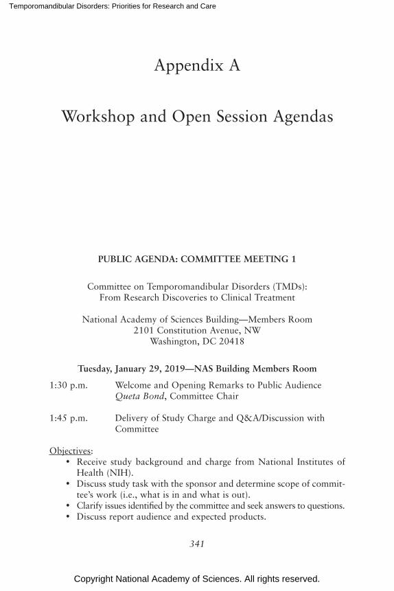
Temporomandibular Disorders: Priorities for Research and Care
Copyright National Academy of Sciences. All rights reserved.
341
PUBLIC AGENDA: COMMITTEE MEETING 1
Committee on Temporomandibular Disorders (TMDs): From Research Discoveries to Clinical Treatment
National Academy of Sciences Building—Members Room2101 Constitution Avenue, NW
Washington, DC 20418
Tuesday, January 29, 2019—NAS Building Members Room
1:30 p.m. Welcome and Opening Remarks to Public AudienceQueta Bond, Committee Chair
1:45 p.m. Delivery of Study Charge and Q&A/Discussion with Committee
Objectives:• Receive study background and charge from National Institutes of
Health (NIH).• Discuss study task with the sponsor and determine scope of commit-
tee’s work (i.e., what is in and what is out).• Clarify issues identified by the committee and seek answers to questions.• Discuss report audience and expected products.
Appendix A
Workshop and Open Session Agendas
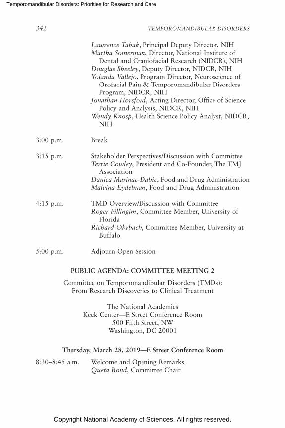
Temporomandibular Disorders: Priorities for Research and Care
Copyright National Academy of Sciences. All rights reserved.
342 TEMPOROMANDIBULAR DISORDERS
Lawrence Tabak, Principal Deputy Director, NIHMartha Somerman, Director, National Institute of
Dental and Craniofacial Research (NIDCR), NIHDouglas Sheeley, Deputy Director, NIDCR, NIHYolanda Vallejo, Program Director, Neuroscience of
Orofacial Pain & Temporomandibular Disorders Program, NIDCR, NIH
Jonathan Horsford, Acting Director, Office of Science Policy and Analysis, NIDCR, NIH
Wendy Knosp, Health Science Policy Analyst, NIDCR, NIH
3:00 p.m. Break
3:15 p.m. Stakeholder Perspectives/Discussion with CommitteeTerrie Cowley, President and Co-Founder, The TMJ
AssociationDanica Marinac-Dabic, Food and Drug Administration Malvina Eydelman, Food and Drug Administration
4:15 p.m. TMD Overview/Discussion with CommitteeRoger Fillingim, Committee Member, University of
FloridaRichard Ohrbach, Committee Member, University at
Buffalo
5:00 p.m. Adjourn Open Session
PUBLIC AGENDA: COMMITTEE MEETING 2
Committee on Temporomandibular Disorders (TMDs): From Research Discoveries to Clinical Treatment
The National AcademiesKeck Center—E Street Conference Room
500 Fifth Street, NWWashington, DC 20001
Thursday, March 28, 2019—E Street Conference Room
8:30–8:45 a.m. Welcome and Opening RemarksQueta Bond, Committee Chair
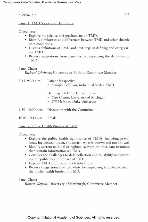
Temporomandibular Disorders: Priorities for Research and Care
Copyright National Academy of Sciences. All rights reserved.
APPENDIX A 343
Panel 1: TMD Scope and Definitions
Objectives:• Explore the science and mechanisms of TMD. • Identify similarities and differences between TMD and other chronic
pain conditions.• Discuss definitions of TMD and next steps in defining and categoriz-
ing TMD.• Receive suggestions from panelists for improving the definition of
TMD.
Panel Chair: Richard Ohrbach, University of Buffalo, Committee Member
8:45–9:30 a.m. Patient Perspective• Jennifer Feldman, individual with a TMD
Defining TMD for Clinical Care• Dan Clauw, University of Michigan • Bill Maixner, Duke University
9:30–10:00 a.m. Discussion with the Committee
10:00–10:15 a.m. Break
Panel 2: Public Health Burden of TMD
Objectives:• Explore the public health significance of TMDs, including preva-
lence, incidence, burden, and costs—what is known and not known?• Identify current national or regional surveys or other data resources
that contain information on TMD.• Consider the challenges to data collection and reliability in estimat-
ing the public health impact of TMD.• Explore TMD and disability classifications.• Receive suggestions from panelists for improving knowledge about
the public health burden of TMD.
Panel Chair: Robert Weyant, University of Pittsburgh, Committee Member
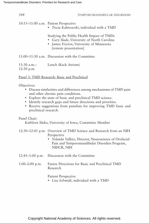
Temporomandibular Disorders: Priorities for Research and Care
Copyright National Academy of Sciences. All rights reserved.
344 TEMPOROMANDIBULAR DISORDERS
10:15–11:00 a.m. Patient Perspective• Tricia Kalinowski, individual with a TMD
Studying the Public Health Impact of TMDs• Gary Slade, University of North Carolina • James Fricton, University of Minnesota
[remote presentation]
11:00–11:30 a.m. Discussion with the Committee
11:30 a.m.– Lunch (Keck Atrium)12:30 p.m.
Panel 3: TMD Research: Basic and Preclinical
Objectives:• Discuss similarities and differences among mechanisms of TMD pain
and other chronic pain conditions.• Explore the state of basic and preclinical TMD science.• Identify research gaps and future directions and priorities. • Receive suggestions from panelists for improving TMD basic and
preclinical research.
Panel Chair: Kathleen Sluka, University of Iowa, Committee Member
12:30–12:45 p.m. Overview of TMD Science and Research from an NIH Perspective• Yolanda Vallejo, Director, Neuroscience of Orofacial
Pain and Temporomandibular Disorders Program, NIDCR, NIH
12:45–1:00 p.m. Discussion with the Committee
1:00–2:00 p.m. Future Directions for Basic and Preclinical TMD Research
Patient Perspective• Lisa Schmidt, individual with a TMD
Temporomandibular Disorders: Priorities for Research and Care
Copyright National Academy of Sciences. All rights reserved.
APPENDIX A 345
Neuroimmunology and Transitioning from Acute to Chronic Pain• Annemieke Kavelaars, MD Anderson [remote
presentation]
Utility of Animal Models for TMD and Orofacial Pain• Ke Ren, University of Maryland
TMD Research Over the Years—Gaps and Future Priorities• Allen Cowley, Medical College of Wisconsin
2:00–2:30 p.m. Discussion with the Committee
2:30–2:45 p.m. Break
2:45–3:00 p.m. Ontology of Orofacial Pain to Improve Patient CareModerated by Richard Ohrbach
• Werner Ceusters, University at Buffalo [remote presentation]
3:00–3:15 p.m. Discussion with the Committee
Panel 4: TMD Research: Clinical and Translational
Objectives:• Discuss similarities and differences among mechanisms of TMD pain
and other chronic pain conditions.• Explore the state of clinical and translational TMD science.• Identify research gaps and future directions and priorities. • Receive suggestions from panelists for improving TMD clinical and
translational research and treatment.
Panel Chair: Roger Fillingim, University of Florida, Committee Member
3:15–3:30 p.m. Overview of Evidence Base for Current TMD Treatments • Mark Drangsholt, University of Washington
3:30–3:45 p.m. Discussion with the Committee
Temporomandibular Disorders: Priorities for Research and Care
Copyright National Academy of Sciences. All rights reserved.
346 TEMPOROMANDIBULAR DISORDERS
3:45–4:45 p.m. Future Directions for Clinical and Translational TMD Research
Caregiver Perspective• Michelle and Alexandra Reardon, family member
and individual with a TMD
Complementary and Integrative Health Approaches • Helene Langevin, National Center for
Complementary and Integrative Health, NIH
Neuroimaging Research in TMD• Alexandre DaSilva, University of Michigan
Lessons Learned from Advancements in Orthopedics• Constance Chu, Stanford University [remote
participation]
4:45–5:15 p.m. Discussion with the Committee
5:15–6:15 p.m. Public comments by registered speakers (3–5 minutes each)
Moderators: Queta Bond and Sean Mackey
6:15 p.m. Day 1 Public Session Adjourns
Friday, March 29, 2019—E Street Conference Room
8:30–8:40 a.m. Welcome and Opening Remarks Queta Bond, Committee Chair
Panel 5: TMD Care Pathways
Objectives:• Discuss current TMD treatments and standards of care—what is
known about their effectiveness?• Explore the current pathways and practitioners TMD patients use
to receive care and recommend improved pathways.• Explore the separation of dental and medical practice in the United
States and implications for TMD patient care.• Receive suggestions from panelists as to how future TMD care can
be improved.
Temporomandibular Disorders: Priorities for Research and Care
Copyright National Academy of Sciences. All rights reserved.
APPENDIX A 347
Panel Chair: David Deitz, David Deitz and Associates, Committee Member
8:40–9:10 a.m. Improving TMD Care Pathways
TMD Care Pathways—A UK Perspective • Justin Durham, Newcastle University [remote
participation]
TMD Care Pathways—A Patient Perspective• Adriana van Ineveld, individual with a TMD
9:10–10:00 a.m. Improving TMD Care Pathways—Roles of Dentistry and Medicine• American Dental Association o Deepak Kademani, North Memorial Medical
Center [remote participation]• American Association of Oral and Maxillofacial
Surgeons o Gary Bouloux, Emory University School of
Medicineo Gregory Ness, The Ohio State University College
of Dentistry• American Association of Public Health Dentistryo Hong Chen, University of Iowa College
of Dentistry and Dental Clinics [remote participation]
• American Academy of Pain Medicineo Meredith Barad, Stanford University [remote
participation]• Aetna Inc.o Jeffrey Livovich, Aetna Inc. [remote
participation]
10:00–10:30 a.m. Discussion with the Committee
10:30–11:30 a.m. Public Comments by Registered, Remote Speakers (3–5 minutes each)
Moderators: Queta Bond and Sean Mackey
11:30 a.m. Public Session Adjourns
Temporomandibular Disorders: Priorities for Research and Care
Copyright National Academy of Sciences. All rights reserved.
348 TEMPOROMANDIBULAR DISORDERS
PUBLIC WEBINAR: TMD PROFESSIONAL EDUCATION, RESEARCH, AND SPECIALIZATION
Committee on Temporomandibular Disorders (TMDs): From Research Discoveries to Clinical Treatment
Webinar: TMD Professional Education, Research and SpecializationWednesday, July 31, 2019: 3:00–5:00 p.m. (EDT)
The objective of this webinar is to receive a variety of perspectives from dental educators and policy makers about professional education, research, and specialization related to TMD. Specifically, the committee is looking for perspectives and ideas in the following areas:
1. Interprofessional education—What are your ideas on how to increase and incentivize interprofessional education on TMD and orofacial pain?
2. TMD education and training—What needs to be done to improve TMD education and training?
3. Dental practice research—What are the challenges and opportunities for improving dental practice research?
4. Specialization—What are the needs, benefits, and challenges of im-plementing a specialization in orofacial pain?
5. Continuing education—What role should academia play in con-tinuing education for physicians, nurses, dentists, and other health professionals?
3:00 p.m. Welcome and Process for Webinar Queta Bond, Committee Chair
3:10 p.m. Panel Introduction Robert Weyant, University of Pittsburgh,
Committee Member
3:15 p.m. Panel 1• Laurie McCauley, University of Michigan• Cecile Feldman, Rutgers University• Henry Gremillion, Louisiana State University
3:40 p.m. Discussion with Committee
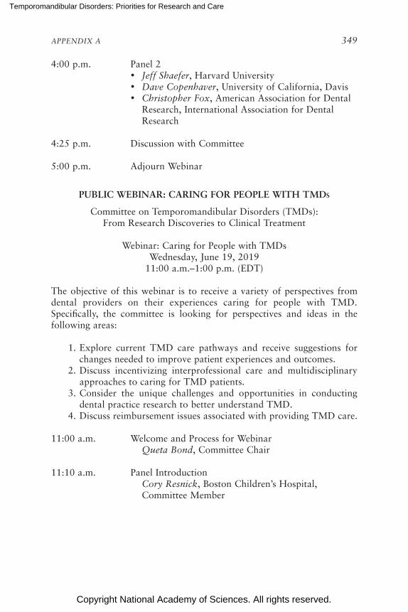
Temporomandibular Disorders: Priorities for Research and Care
Copyright National Academy of Sciences. All rights reserved.
APPENDIX A 349
4:00 p.m. Panel 2• Jeff Shaefer, Harvard University• Dave Copenhaver, University of California, Davis• Christopher Fox, American Association for Dental
Research, International Association for Dental Research
4:25 p.m. Discussion with Committee
5:00 p.m. Adjourn Webinar
PUBLIC WEBINAR: CARING FOR PEOPLE WITH TMDs
Committee on Temporomandibular Disorders (TMDs): From Research Discoveries to Clinical Treatment
Webinar: Caring for People with TMDsWednesday, June 19, 2019
11:00 a.m.–1:00 p.m. (EDT)
The objective of this webinar is to receive a variety of perspectives from dental providers on their experiences caring for people with TMD. Specifically, the committee is looking for perspectives and ideas in the following areas:
1. Explore current TMD care pathways and receive suggestions for changes needed to improve patient experiences and outcomes.
2. Discuss incentivizing interprofessional care and multidisciplinary approaches to caring for TMD patients.
3. Consider the unique challenges and opportunities in conducting dental practice research to better understand TMD.
4. Discuss reimbursement issues associated with providing TMD care.
11:00 a.m. Welcome and Process for Webinar Queta Bond, Committee Chair
11:10 a.m. Panel Introduction Cory Resnick, Boston Children’s Hospital,
Committee Member

Temporomandibular Disorders: Priorities for Research and Care
Copyright National Academy of Sciences. All rights reserved.
350 TEMPOROMANDIBULAR DISORDERS
11:15 a.m. Panel 1: Provider Perspectives on Caring for People with TMD• Jeff Okeson, University of Kentucky• Marcela Romero-Reyes, University of Maryland• Jeremy Abbott, Orofacial Pain Specialist
11:40 a.m. Discussion with Committee
12:00 p.m. Panel 2: Provider Perspectives on Caring for People with TMD• Anthony Schwartz, Johns Hopkins Medicine• Kevin Huff, General Dentist, Dover, Ohio
12:25 p.m. Discussion with Committee
1:00 p.m. Adjourn Webinar
Temporomandibular Disorders: Priorities for Research and Care
Copyright National Academy of Sciences. All rights reserved.
351
Enriqueta C. Bond, Ph.D., M.A. (Chair), served from 1994 to 2008 as the first full-time president of the Burroughs Wellcome Fund (BWF), a private, independent foundation dedicated to advancing the medical sciences by supporting research and other scientific and educational activities. During her presidency, Dr. Bond guided BWF in its transition from a corporate to a private foundation. Prior to joining BWF, Dr. Bond served as the execu-tive officer for the Institute of Medicine. Dr. Bond currently is a founding partner of QE Philanthropic Advisors, which provides consulting services to philanthropic and nonprofit organizations on program development, gov-ernance, leadership, and organizational structure and function. Dr. Bond was the chair of the National Research Council’s Board on African Science Academy Development and a former member of the Forum on Microbial Threats. She is a past member of the National Academies’ Report Review Committee as well as many other consensus study committees. Dr. Bond is the recipient of numerous honors, including the 2008 Order of the Long Leaf Pine award from the state of North Carolina—the highest honor the governor can bestow upon a citizen. This award was given to Dr. Bond for her efforts to improve science education for the children of North Carolina. In 1997 she was elected as a member of the National Academy of Medicine. In 2004 she was elected as a fellow of the American Association for the Advance ment of Science for her distinguished contributions to the study and analysis of policy for the advancement of the health sciences. She received her bachelor’s degree from Wellesley College, her M.A. from the University of Virginia, and her Ph.D. in molecular biology and biochemical genetics from Georgetown University.
Appendix B
Committee Biographical Sketches
Temporomandibular Disorders: Priorities for Research and Care
Copyright National Academy of Sciences. All rights reserved.
352 TEMPOROMANDIBULAR DISORDERS
Sean Mackey, M.D., Ph.D. (Vice-Chair), is the chief of the Division of Pain Medicine and the Redlich Professor of Anesthesiology, Perioperative and Pain Medicine, Neurosciences and Neurology at Stanford University. Dr. Mackey received his B.S.E. and M.S.E. in bioengineering from the University of Pennsylvania and his Ph.D. in electrical and computer engineering and his M.D. from the University of Arizona. Dr. Mackey directs the Stanford Systems Neuroscience and Pain Laboratory with a broad range of pain re-search on understanding the mechanisms of pain and improving patients’ quality of life. The lab’s research includes mapping the specific brain and spinal cord regions that perceive and process pain and the development of a multidisciplinary treatment model that translates basic science research into innovative therapies to provide more effective, personalized treatments for patients with chronic pain. Dr. Mackey is the author of more than 200 journal articles and book chapters in addition to numerous national and international lectures. Dr. Mackey has served as the principal investigator for multiple National Institutes of Health (NIH) and foundation research grants investigating chronic pain and novel analgesics for acute and chronic pain. He is the past president of the American Academy of Pain Medicine. In 2011, Dr. Mackey served as a member of the Institute of Medicine committee that issued the report Relieving Pain in America. He was the co-chair of the oversight committee for the NIH/Health and Human Ser-vices National Pain Strategy (NPS), an effort to establish a national health strategy for pain care, education, and research. In the past 2 years he has received the American Pain Society Wilbert E. Fordyce Clinical Investigator Award, the Pain Medicine Fellowship Award and the Distinguished Service Award from the American Association of Physicists in Medicine, and the NIH Directors’ Award for his efforts on the NPS.
Penney Cowan is the founder and chief executive officer of the American Chronic Pain Association (ACPA). She herself is a person with chronic pain and established the ACPA in 1980 to help others living with the condition. The ACPA provides peer support and education in pain management skills to people with pain and their families. The ACPA also works to build awareness about chronic pain among professionals, decision makers, and the general public. Since 1980 Ms. Cowan has been an advocate and con-sumer representative for pain issues. She was awarded the Jefferson Medal for Outstanding Citizen by the Institute for Public Service, Washington, and is listed in Who’s Who in America, 24th Edition. The American Pain Society awarded her the 2005 John and Emma Bonica Public Service Award and the Elizabeth Narcessian Award for Outstanding Educational Achieve-ments in 2013, and she received the Presidential Commendation from the American Academy of Pain Medicine, also in 2013. She has served as the consumer representative for the Food and Drug Administration’s Center for
Temporomandibular Disorders: Priorities for Research and Care
Copyright National Academy of Sciences. All rights reserved.
APPENDIX B 353
Drug Evaluation and Research’s Anesthetic and Analgesic Drug Products Advisory Committee for 2012 and was a member of the Interagency Pain Research Coordinating Committee of the National Institutes of Health from 2013 to 2015. Ms. Cowan began the Partners for Understanding Pain campaign in 2002 in an attempt to raise awareness about the need to better understand, assess, and treat pain. There are more than 80 partner organizations. The campaign, under the direction of the ACPA, successfully established September as Pain Awareness Month.
David Deitz, M.D., Ph.D., is the principal of David Deitz & Associates, a consulting firm focused on helping clients design and implement strategies to obtain high-value health outcomes for patients and employers. Dr. Deitz is a nationally recognized expert with 25 years of experience in managed medical care, health care outcomes evaluation and reporting, and usage management systems in both workers compensation and group health. In this role he has worked with employers, labor, state regulators, and lawmakers in more than 20 states on reform efforts, and he has served on a number of task forces and committees nationwide. He was engaged for many years as the national medical director of commercial insurance claims for Liberty Mutual Insurance, where he was responsible for medical review operations, medical networks, and medical policy for workers com-pensation. Prior to joining Liberty Mutual, he worked in the private sector overseeing content development of managed care software and reporting systems, which were successfully deployed at insurers nationwide. In addi-tion to this work, Dr. Deitz spent two decades as a practicing physician in both emergency and internal medicine. He completed his post-graduate training at the University of Miami and Harvard Medical School and is board certified in internal medicine.
Francesca C. Dwamena, M.D., M.S., FACP, FAACH, is a professor and the chair of the Department of Medicine at Michigan State University. Dr. Dwamena also is an adjunct professor of psychiatry and an attending physician for the Michigan State University Health Team and at Sparrow Hospital. She specializes in psychosocial medicine and has authored more than 40 published works on clinical and primary medicine, including on the identification of and treatment for medically unexplained symptoms. She is a co-author of two books on evidence-based patient-centered interviewing and the essentials of psychiatry in primary care. In addition to her clinical practice and research, she has held a variety of leadership positions and volunteer roles with various national and state-level professional societies and nonprofit organizations, including Harvard Medical School, the Arnold P. Gold Foundation, the Society of General Medicine, and the American College of Physicians. Additionally, she has served as a committee member
Temporomandibular Disorders: Priorities for Research and Care
Copyright National Academy of Sciences. All rights reserved.
354 TEMPOROMANDIBULAR DISORDERS
for the Institute of Medicine consensus committee that produced the re-port Gulf War and Health: Treatment of Chronic Multisymptom Illness. Dr. Dwamena received her M.D. from the Howard University College of Medicine and her M.S. in epidemiology and a certificate in psychosocial medicine from Michigan State University.
Roger B. Fillingim, Ph.D., is a distinguished professor and the director of the Pain Research and Intervention Center of Excellence (PRICE) at the University of Florida (UF). He served as the president of the American Pain Society from 2012 to 2014, served as co-chair of the Federal Pain Research Strategy Disparities Workgroup, and is currently a member of the U.S. Department of Health and Human Services Interagency Pain Research Coordinating Committee. PRICE provides resources in support of clinical and translational pain research and facilitates collaborations among UF pain researchers and other UF programs of research excel-lence. A clinical psychologist by training, he has had a longstanding interest in research to address the nation’s most disabling and expensive public health problem, chronic pain. Dr. Fillingim’s research program has focused on understanding the factors that contribute to individual differ-ences in the experience of pain. He and his colleagues have shown consis-tent sex differences in pain responses, which may contribute to females’ increased risk for chronic pain. In addition, his work has identified several genetic factors that contribute to pain perception and analgesic responses. His current grants include a MERIT Award from the National Institute on Aging, which investigates biological and psychosocial factors contrib-uting to ethnic group differences in osteoarthritis pain. He also served as the principal investigator for the Florida site of the National Institute of Dental and Craniofacial Research–funded OPPERA (Orofacial Pain: Prospective Evaluation and Risk Assessment) study, which was created to identify risk factors for the development of temporomandibular disorders and related pain conditions. Dr. Fillingim has received several awards, including a UF Foundation Preeminence Term Professorship as well as the 2009 Wilbert E. Fordyce Clinical Investigator Award and the Distin-guished Service Award, both from the American Pain Society. He earned his doctoral degree in clinical psychology from the University of Alabama at Birmingham, followed by a postdoctoral fellowship in pain research at the University of North Carolina.
Margaret M. Heitkemper, R.N., FAAN, Ph.D., is a professor and the chair of the Department of Biobehavioral Nursing and Health Informatics where she also is the co-director of the Center for Innovations in Sleep Self-Management and an adjunct professor in the Division of Gastro-enterology at the University of Washington. She received her B.S.N. from
Temporomandibular Disorders: Priorities for Research and Care
Copyright National Academy of Sciences. All rights reserved.
APPENDIX B 355
Seattle University, her master’s degree in nursing from the University of Washington, and a Ph.D. in physiology and biophysics from the University of Illinois at Chicago. She leads an interdisciplinary team focused on the study of the pathophysiology and non-pharmacological management of indi viduals with chronic abdominal pain and irritable bowel syndrome (IBS). Her research in this area has included both descriptive and mecha-nistic studies focused on the role of general and lifestyle factors in chronic pain conditions. Dr. Heitkemper’s research has included a focus on genetic and potential proteomic markers of chronic abdominal pain. Her team has also conducted randomized clinical trials of cognitive-behavioral therapy for persons with IBS. She served on the Institute of Medicine committee that issued the report Relieving Pain in America.
Francis Keefe, Ph.D., is a professor in the Department of Psychiatry and Behavioral Sciences at the Duke University Medical Center and a member of the Cancer Prevention, Detection, and Control Program of the Duke Comprehensive Cancer Center. Dr. Keefe is the director of the Duke Pain Prevention and Treatment Research Program, an active clinical research program supported by the NIH and concerned with the behavioral assess-ment and treatment of patients having acute and persistent pain. Dr. Keefe played a key role in the development of the clinical pain services and pain research programs at the Duke University Medical Center. For 20 years he directed the pain management program and was a leader in the devel-opment of the Duke University Medical Center’s multi disciplinary pain programs (both out-patient and in-patient). Dr. Keefe has developed and refined a number of treatment protocols for persistent pain conditions (e.g., cancer, arthritis), including spouse- and partner-assisted pain- coping skills training interventions. He has conducted a number of randomized clinical trials testing the effi cacy of these and other behavioral inter ventions (e.g., aerobic exercise protocols, yoga-based interventions, mindfulness-based interventions, forgiveness-based interventions, loving kindness medita-tion, and emotional disclosure). Dr. Keefe has served on numerous study sections and has chaired two NIH study sections ( Behavioral Medicine: Interventions and Outcomes, and Psychological Risk and Disease Preven-tion). Dr. Keefe has published more than 370 papers as well as 60 book chapters and 3 books on topics ranging from pain during mammography to the assessment and treatment of cancer pain at end of life. He currently serves as the editor-in-chief of the journal Pain, the leading journal in the field of pain research. In 2012 he was awarded the John D. Loeser award for excellence in clinical pain research by the International Association for the Study of Pain. In 2016 he was awarded the Distinguished Scientist Award by the Society of Behavioral Medicine.

Temporomandibular Disorders: Priorities for Research and Care
Copyright National Academy of Sciences. All rights reserved.
356 TEMPOROMANDIBULAR DISORDERS
Kate Lorig, Dr.P.H., is a professor emerita (acting) at the Stanford Univer-sity School of Medicine and the past director of the Stanford Patient Educa-tion Research Center. She has a master’s degree in nursing and a doctorate in public health with a specialty in health education. For more than four decades, using a public health approach, she has developed and evaluated community-based patient education programs in English and Spanish for people with chronic conditions including arthritis, heart disease, lung dis-ease, diabetes, and AIDS. In recent years, this work has been extended to similar programs offered via the Internet. Her present research includes the development and evaluation of programs for cancer survivors as well as for caregivers of people with posttraumatic stress disorder, traumatic brain injury, and other cognitive impairments. These programs are offered largely over the Internet. Most recently, she has been involved in studying how to translate programs from the academic setting to the larger community.
Richard Ohrbach, D.D.S., Ph.D., earned a D.D.S. (University of North Carolina at Chapel Hill), a certificate in pain management (University of California, Los Angeles), an M.S. in oral sciences, and a Ph.D. in clinical psychology (University at Buffalo) and performed postdoctoral studies in behavioral epidemiology (University of Washington). One of his primary re-search objectives has been the development of new diagnostic standards for temporomandibular disorders (TMDs). The new standards, whose devel-opment was supported by the National Institute of Dental and Cranio-facial Research and which are now internationally recognized, offer both researchers and health professionals (1) an improved screening tool to identify painful TMDs, (2) validated diagnostic criteria grounded in sup-portive scientific evidence that more readily differentiate the most common forms of TMDs, and (3) improved psychosocial assessment tools. The other primary research objective, via the OPPERA (Orofacial Pain: Prospective Evaluation and Risk Assessment) study, has been cross-sectional and lon-gitudinal analyses related to incident cases with TMDs as well as chronic TMDs as well as of physical–psychological interactions relevant to the development and persistence of pain disorders. Dr. Ohrbach is a member of the American Academy of Orofacial Pain, American Pain Society, Inter-national Association for Dental Research, International Association for the Study of Pain, and International Network for Orofacial Pain and Related Disorders Methodology (INfORM). From 2003 to 2006 he served as the director of the International Research Diagnostic Criteria for Temporo-mandibular Disorders Consortium (now INfORM), an International Asso-ciation for Dental Research scientific group/network that he co-founded.
Amanda C. Pustilnik, J.D., is a professor of law at the University of Maryland School of Law and faculty at the Center for Law, Brain & Behavior at
Temporomandibular Disorders: Priorities for Research and Care
Copyright National Academy of Sciences. All rights reserved.
APPENDIX B 357
Massachusetts General Hospital. Her work focuses on the intersections of law, science, and culture, with a particular emphasis on the brain. In 2015 she served as Harvard Law School’s first senior fellow on law and applied neuroscience. Her collaborations with scientists on brain imaging of pain and addiction led to her recent work on opioids on behalf of the Aspen Institute. Prior to entering the academy, Professor Pustilnik practiced litigation at Sullivan & Cromwell, clerked on the Second Circuit Court of Appeals, and worked as a management consultant at McKinsey & Co., in New York. She attended Harvard College, Yale Law School, and the Uni-versity of Cambridge, where she studied history and philosophy of science. Her work has been published in numerous law reviews and peer-reviewed scientific journals, including Nature.
Srinivasa N. Raja, M.B.B.S., is a professor of anesthesiology and neurology and the director of pain research at the Johns Hopkins University School of Medicine. Dr. Raja received his residency training in anesthesiology at the University of Washington, Seattle, and postdoctoral training at the Uni-versity of Virginia School of Medicine in Charlottesville. Dr. Raja’s clinical and research interests include the management of chronic neuropathic pain states, understanding the peripheral and central mechanisms of neuropathic pain, and identifying novel peripheral targets for alleviating chronic neuro-pathic pain. Dr. Raja served as an editor of Anesthesiology from 1998 to 2006 and a section editor for the journal Pain from 2012 to 2014. He was a member of the Food and Drug Administration’s scientific advisory panel for the Center for Drug Evaluation and Research. Dr. Raja served as a member of the scientific advisory committee for the World Congress on Pain from 2008 to 2012 and as the chair of the scientific program com-mittee for the 15th World Congress on Pain in Buenos Aires in 2014. He was the chair of NeuPSIG, the neuropathic pain special interest group of the International Association for the Study of Pain, and the secretary of the International Association for the Study of Pain, from 2014 to 2016. In 2015 he was appointed as a member of the Federal Pain Research Strategy Steering Committee, which is charged with coordinating the research efforts in the field of pain across all U.S. federal funding agencies. Dr. Raja and his collaborators have received funding from the National Institutes of Health for their research for nearly 30 years and have published more than 200 original articles in peer-reviewed journals. Dr. Raja has been invited as a visiting professor to several institutions, including the Benjamin G. Covino Lecture at Harvard University (2007) and the John J. Bonica Lecture at the University of Washington (2008) and Heidelberg University (2019). He was the recipient of the Wilbert E. Fordyce Clinical Investigator Award from the American Pain Society (2008) and the prestigious John J. Bonica Award from the American Society of Regional Anesthesia and Pain Medicine
Temporomandibular Disorders: Priorities for Research and Care
Copyright National Academy of Sciences. All rights reserved.
358 TEMPOROMANDIBULAR DISORDERS
(2010). He will be the recipient of the John J. Bonica Distinguished Lecture award at the World Congress on Pain (Amsterdam, 2020).
Cory M. Resnick, M.D., D.M.D., F.A.C.S., is an assistant professor of oral and maxillofacial surgery at Harvard Medical School and the Harvard School of Dental Medicine, and he practices pediatric oral and maxillo-facial surgery at Boston Children’s Hospital. His clinical practice empha-sizes the surgical management of craniofacial anomalies and syndromes, orthognathic (jaw) surgery, pediatric obstructive sleep apnea, and temporo-mandibular disorders. Dr. Resnick is an active member of the multidis-ciplinary craniofacial and vascular anomalies teams. In addition to Dr. Resnick’s busy clinical practice, he is dedicated to innovation and improve-ment in his field through research and education. Dr. Resnick is the author of many publications in peer-reviewed journals within and outside the scope of oral surgery as well as chapters in multiple textbooks. He lectures to medical and dental students at the Harvard Medical School and Harvard School of Dental Medicine, and he mentors oral and maxillofacial surgery residents through the Massachusetts General Hospital residency program. Dr. Resnick received his undergraduate and dental degrees from the Uni-versity of Pennsylvania and his medical degree from Harvard Medical School. He then received general surgery and oral and maxillofacial surgery residency training at the Massachusetts General Hospital. Dr. Resnick is certified by the American Board of Oral and Maxillofacial Surgery and was inducted as a fellow of the American College of Surgeons.
Antony Rosen, M.B.Ch.B., B.Sc. (Hons), is the vice-dean for research and the director of the Division of Rheumatology at the Johns Hopkins Uni-versity School of Medicine, where he is the Mary Betty Stevens Professor of Medicine and a professor of pathology and cell biology. He leads Johns Hopkins in Health, the enterprise program in precision medicine and indi-vidualized health. Dr. Rosen completed medical school at the University of Cape Town in South Africa, was a postdoctoral fellow in immunology at The Rockefeller University, and pursued internal medicine residency and rheumatology fellowship at Johns Hopkins University. Dr. Rosen’s interests are in the autoimmune rheumatic diseases and their mechanisms, and he is focused on understanding the specificity of the immune response in the dif-ferent rheumatic syndromes, including scleroderma, myositis, and Sjogren’s syndrome. Dr. Rosen was a Pew Scholar in the biomedical sciences, a Burroughs Wellcome translational research scholar, received a National Insti tutes of Health MERIT award from the National Institute of Dental and Craniofacial Research (NIDCR), and is a member of the American Society for Clinical Investigation and Association of American Physicians. He chaired the scientific advisory committee of the NIDCR-funded Sjogren’s
Temporomandibular Disorders: Priorities for Research and Care
Copyright National Academy of Sciences. All rights reserved.
APPENDIX B 359
International Collaborative Clinical Alliance from 2004 to 2007, was a member of the American College of Rheumatology’s board of directors, and is currently chair of the board of scientific counselors of the National Institute of Arthritis and Musculoskeletal and Skin Diseases.
Kathleen A. Sluka, PT, Ph.D., is a professor of physical therapy and rehabili-tation sciences at the University of Iowa Carver College of Medicine and a member of the Pain Research Program at the University of Iowa. Dr. Sluka has published more than 200 peer-reviewed manuscripts and book chapters as well as an evidence-based textbook titled Pain Mechanisms and Management for the Physical Therapist, which covers basic science mechanisms, physical therapy treatments, interdisciplinary care, and pain syndromes. Dr. Sluka’s translational research laboratory focuses on the neurobiology of musculo-skeletal pain and non-pharmacological treatments using animal models and the translation of mechanisms to human subjects, clinical trials for non-pharmacological treatments for chronic pain, and implementation of effective non-pharmacological treatments to clinical practice. Her research methods include cell culture, molecular biology, genetic manipulations, behavioral pharmacology, clinical trial methodology, and implementation science. She has received numerous awards, including the Marian Williams Award for Research in Physical Therapy and the Catherine Worthingham Fellow Award from the American Physical Therapy Association and the Kerr Basic Science Research Award from the American Pain Society. She received a physical therapy degree from Georgia State University and a Ph.D. in anatomy from the University of Texas Medical Branch in Galveston.
Barbara G. Vickrey, M.D., M.P.H., specializes in translating clinical evidence into improvements in routine medical practice that benefit patients’ health. Her wide-ranging accomplishments include demonstrating that collabora-tion among health care systems, community organizations, and caregivers can improve quality of care and outcomes for dementia patients. She has also designed health care delivery innovations ranging from better con-trol of post-stroke risk factors in underserved populations to new ways to care for veterans with Parkinson’s disease. Dr. Vickrey was elected to the National Academy of Medicine in 2011. Dr. Vickrey leads a multi-year, stroke prevention/intervention research program in health disparities funded by the National Institute of Neurological Disorders and Stroke. She serves on the science committee of the American Academy of Neurology and is im-mediate past president of the American Neurological Association. Dr. Vickrey served for 25 years on the faculty of the University of California, Los Angeles (UCLA), where she was a professor of neurology and the director of the departmental Health Services Research Program. She was also the associate director for research at the Greater Los Angeles Veterans Administration
Temporomandibular Disorders: Priorities for Research and Care
Copyright National Academy of Sciences. All rights reserved.
360 TEMPOROMANDIBULAR DISORDERS
Parkinson’s Disease Research, Education and Clinical Center. Dr. Vickrey earned her M.D. at the Duke University School of Medicine and her M.P.H. at the UCLA School of Public Health. She completed post-graduate clinical training in medicine and neurology at the University of Washington in Seattle and then research fellowships in the Robert Wood Johnson Clinical Scholars Program at UCLA and the RAND/UCLA Center for Health Policy Study.
Robert Weyant, D.M.D., Dr.P.H., is the associate dean for public health and a professor and the chair of the Department of Dental Public Health at the University of Pittsburgh School of Dental Medicine. He is also a professor of epidemiology in the Graduate School of Public Health and on the faculty of the Clinical and Translational Sciences Institute. Dr. Weyant is a former U.S. Navy dental officer and Veterans Health Administration dentist and has been a diplomate of the American Board of Dental Public Health since 1987. Dr. Weyant is a past president of the American Asso-ciation for Public Health Dentistry and is the current editor-in-chief of the Journal of Public Health Dentistry. Dr. Weyant’s research involves general and social epidemiological research related to oral health disparities and oral disease etiology. He is presently a principal investigator or co-principal investigator on several National Institutes of Health–funded studies of oral disease etiology and also the project director of a Health Resources and Ser-vices Administration funded training grant for dental students. Dr. Weyant teaches and conducts research in evidence-based practices and has authored four systematic reviews and associated clinical practices guidelines. Dr. Weyant has served on numerous local, state, and national committees aimed at reducing oral health disparities, improving evidence-based prac-tice, increasing the dental workforce, and improving access to oral care. He received his M.P.H. and his dental degree from the University of Pittsburgh and his doctorate in epidemiology from the University of Michigan.
Hai Yao, Ph.D., is the Ernest R. Norville Endowed Chair in Biomedical Engineer ing at Clemson University and the associate chair of the Clemson University–Medical University of South Carolina Joint Bioengineering Pro-gram. He is also the director of the National Institutes of Health (NIH) Center of Biomedical Research Excellence for Translational Research Im-proving Musculoskeletal Health. Dr. Yao’s tissue biomechanics laboratory focuses on the biomechanics and mechanobiology of the temporo mandibular joint (TMJ) in order to understand the pathophysiology of TMJ degenera-tion to facilitate earlier diagnosis and management of temporo mandibular disorders. In 2013 Dr. Yao participated in the NIH TMJ Roundtable, which provided research recommendations to NIH as well as the broader scientific community. Dr. Yao received his Ph.D. in biomedical engineering from the University of Miami.
Temporomandibular Disorders: Priorities for Research and Care
Copyright National Academy of Sciences. All rights reserved.
361
Appendix C
Prevalence, Impact, and Costs of Treatment for
Temporomandibular DisordersBy Gary Slade and Justin Durham*
ABSTRACT
In nationally representative surveys, 5 percent of U.S. adults report having experienced orofacial pain in the preceding 3 months, a prevalence rate that has persisted for three decades. The prevalence of orofacial pain symptoms varies substantially according to gender, age, and household in-come. There are smaller relative differences according to race, ethnicity, and geographic region, although in a large study focusing solely on Hispanics, prevalence varied from 4 percent among South American Hispanics to 7 percent among Puerto Ricans. Most people with orofacial pain report symptoms that “come and go,” their average rating of pain severity at its worst being 7 on a scale from 1 (mild) to 10 (severe), and one in six have chronic symptoms. Half of those with symptoms use over-the-counter analgesics and one-fifth use prescription medication to manage the symp-toms. A minority report social impacts, including staying at home more than usual (8 percent), avoiding family or friends (6 percent), and taking time off work (5 percent).
Because they represent a “snapshot” of the population at a single point in time, these prevalence rates mask a high rate of intermittent symptoms in the population. In one longitudinal study, symptoms developed at a
*Suggested citation: Slade, G., and J. Durham. 2020. Prevalence, impact, and costs of treatment for temporomandibular disorders. Paper commissioned by the Committee on Temporo mandibular Dis orders (TMDs): From Research Discoveries to Clinical Treat-ment. In Temporomandibular disorders: Priorities for research and care (see Appendix C). Washington, DC: The National Academies Press.
Temporomandibular Disorders: Priorities for Research and Care
Copyright National Academy of Sciences. All rights reserved.
362 TEMPOROMANDIBULAR DISORDERS
rate of 18.8 percent per annum, many of which resolved, and when study participants were examined using validated criteria for clinical temporo-mandibular disorders (TMDs), the incidence rate was 3.9 percent per annum. Six months later, half of incident cases had persistent TMDs, and 7 years later, approximately half of incident cases again had a TMD (although many of them did not have a persistent TMD at 6 months). By implication, fluctuation and remission continue through life, such that prev-alence in any single cross-sectional survey is a function of an individual’s history of TMD onset, remission, and persistence.
The factor most strongly associated with the prevalence of orofacial pain is the presence of pain elsewhere in the body: nationally, the prevalence is 32 percent among adults who have all four of headache, neck pain, back pain, and joint pain, whereas prevalence is only 1 percent among adults with none of those pain conditions. Many of those other pains themselves are chronic in nature, with the consequence that people with TMDs are more likely than not to have chronic pain from any anatomical location. Moreover, one-quarter of people with orofacial pain symptoms report high-impact chronic pain from any anatomical location, compared with only 7 percent for people without orofacial pain symptoms.
The cost burdens of TMDs have been investigated most thoroughly in a study of adults with persistent orofacial pain living in the northeast of England. Total costs per 6-month period varied from £321 to £519 per person during the 2-year period of the study, with the major driver being consultation costs and, especially, specialist consultation costs. Direct out-of-pocket costs averaged £334 per person per 6-month period. Indirect costs included employer-related costs due to work loss, which averaged £74 per person per 6-month period. However, the largest contribution to indirect costs was due to “presenteeism” (i.e., reduced productivity due to problems with concentration or decision making while at work), which averaged £905 per person. Overall, TMDs exerted a substantial impact on the individual and economy through lost productivity and on the health care system due to disorganized care pathways increasing the number of consultations required to achieve either diagnosis or care for the condition. Given that the data are from a national and linked health care service, which is free at the point of delivery, it is reasonable to assume that costs are not driven by profit. It is possible that costs differ in other systems of wholly privately delivered health care.
INTRODUCTION
This is a report commissioned by the National Academies of Sciences, Engineering, and Medicine’s Committee on Temporomandibular Disorders (TMDs): From Research Discoveries to Clinical Treatment. It uses data
Temporomandibular Disorders: Priorities for Research and Care
Copyright National Academy of Sciences. All rights reserved.
APPENDIX C 363
from observational studies to describe the impact of TMDs in the popula-tion, with a focus on three topics: public health burden, burden on patients, and costs. The information comes primarily from published studies and from a new analysis of data from the U.S. National Health Interview Survey (NHIS) and two studies conducted by the authors:
• The U.S.-based OPPERA study1: Orofacial Pain: Prospective Evalu-ation and Risk Assessment
• The UK-based DEEP study2: Developing Effective and Efficient Care Pathways for Patients with Chronic Pain
The report is limited to studies of painful TMDs where the defining features are pain in the joints and/or muscles of the jaws; and limitation in jaw function due to the pain. In clinical studies, painful TMDs are classified as arthralgia (joint pain) and myalgia (muscle pain).
PUBLIC HEALTH BURDEN
Prevalence of TMDs
Prevalence of Orofacial Pain Symptoms in the U.S. National Health Interview Survey
Prevalence refers to the proportion of people in a defined population who have a health condition during a specified time period. The prevalence of pain conditions such as TMDs is usually measured in cross-sectional health surveys that ask respondents about pain symptoms that are char-acteristic of TMDs (hereafter labeled “orofacial pain”). In some instances a clinical examination is also conducted, with the goal to properly distin-guish pain symptoms caused by TMDs from pain symptoms caused by other types of pathology. The primary requirements for a valid estimation of TMD prevalence are selection of a random sample of study partici-pants from the target population of interest; valid and reliable questions or exami nation methods to classify the presence or absence of TMD pain in each study participant; and a sufficient number of study participants to estimate the prevalence with reasonable precision. “Reasonable preci-sion” is signified by a relatively narrow 95 percent confidence limit (95% CL) around the estimate of prevalence. To make valid conclusions when comparing findings from different populations or different points in time, it is critical that the case classifications use consistent criteria, including the reference period used to specify the “time period” in which pain was experienced. In studies of TMD, it is common for the reference period to encompass the 3 or 6 months prior to the survey interview or examination.
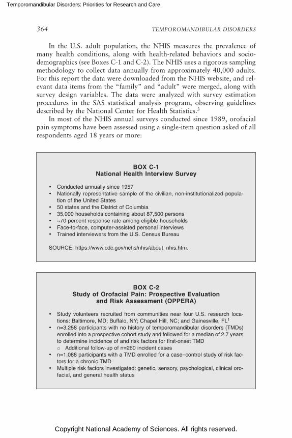
Temporomandibular Disorders: Priorities for Research and Care
Copyright National Academy of Sciences. All rights reserved.
364 TEMPOROMANDIBULAR DISORDERS
In the U.S. adult population, the NHIS measures the prevalence of many health conditions, along with health-related behaviors and socio-demographics (see Boxes C-1 and C-2). The NHIS uses a rigorous sampling methodology to collect data annually from approximately 40,000 adults. For this report the data were downloaded from the NHIS website, and rel-evant data items from the “family” and “adult” were merged, along with survey design variables. The data were analyzed with survey estimation procedures in the SAS statistical analysis program, observing guidelines described by the National Center for Health Statistics.3
In most of the NHIS annual surveys conducted since 1989, orofacial pain symptoms have been assessed using a single-item question asked of all respondents aged 18 years or more:
BOX C-1 National Health Interview Survey
• Conducted annually since 1957• Nationally representative sample of the civilian, non-institutionalized popula-
tion of the United States• 50 states and the District of Columbia• 35,000 households containing about 87,500 persons• ~70 percent response rate among eligible households• Face-to-face, computer-assisted personal interviews• Trained interviewers from the U.S. Census Bureau
SOURCE: https://www.cdc.gov/nchs/nhis/about_nhis.htm.
BOX C-2 Study of Orofacial Pain: Prospective Evaluation
and Risk Assessment (OPPERA)
• Study volunteers recruited from communities near four U.S. research loca-tions: Baltimore, MD; Buffalo, NY; Chapel Hill, NC; and Gainesville, FL1
• n=3,258 participants with no history of temporomandibular disorders (TMDs) enrolled into a prospective cohort study and followed for a median of 2.7 years to determine incidence of and risk factors for first-onset TMD
o Additional follow-up of n=260 incident cases• n=1,088 participants with a TMD enrolled for a case–control study of risk fac-
tors for a chronic TMD• Multiple risk factors investigated: genetic, sensory, psychological, clinical oro-
facial, and general health status
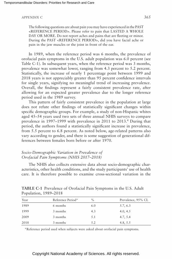
Temporomandibular Disorders: Priorities for Research and Care
Copyright National Academy of Sciences. All rights reserved.
APPENDIX C 365
The following questions are about pain you may have experienced in the PAST <REFERENCE PERIOD>. Please refer to pain that LASTED A WHOLE DAY OR MORE. Do not report aches and pains that are fleeting or minor. During the PAST <REFERENCE PERIOD>, did you have facial ache or pain in the jaw muscles or the joint in front of the ear.
In 1989, when the reference period was 6 months, the prevalence of orofacial pain symptoms in the U.S. adult population was 6.0 percent (see Table C-1). In subsequent years, when the reference period was 3 months, prevalence was somewhat lower, ranging from 4.3 percent to 5.2 percent. Statistically, the increase of nearly 1 percentage point between 1999 and 2018 years is not appreciably greater than 95 percent confidence intervals for single years, signifying no meaningful trend of increasing prevalence. Overall, the findings represent a fairly consistent prevalence rate, after allow ing for an expected greater prevalence due to the longer reference period used in the 1989 survey.
This pattern of fairly consistent prevalence in the population at large does not refute other findings of statistically significant changes within specific demographic groups. For example, a study of non-Hispanic whites aged 45–54 years used two sets of three annual NHIS surveys to compare prevalence in 1997–1999 with prevalence in 2011 to 2013.4 During that period, the authors found a statistically significant increase in prevalence, from 5.5 percent to 6.8 percent. As noted below, age-related patterns also vary according to gender, and there is some suggestion of generational dif-ferences between females born before or after 1970.
Socio-Demographic Variation in Prevalence of Orofacial Pain Symptoms (NHIS 2017–2018)
The NHIS also collects extensive data about socio-demographic char-acteristics, other health conditions, and the study participants’ use of health care. It is therefore possible to examine cross-sectional variation in the
TABLE C-1 Prevalence of Orofacial Pain Symptoms in the U.S. Adult Population, 1989–2018
Year Reference Period* % Prevalence, 95% CL
1989 6 months 6.0 5.7, 6.3
1999 3 months 4.3 4.0, 4.5
2009 3 months 5.1 4.7, 5.4
2018 3 months 5.2 4.8, 5.5
*Reference period used when subjects were asked about orofacial pain symptoms.
Temporomandibular Disorders: Priorities for Research and Care
Copyright National Academy of Sciences. All rights reserved.
366 TEMPOROMANDIBULAR DISORDERS
prevalence of orofacial pain symptoms according to those characteristics. However, it should be emphasized that any observed cross-sectional asso-ciations do not necessarily signify a causal relationship, in either direction, between those characteristics and orofacial pain symptoms. For example, as noted below, TMD prevalence is inversely associated with income, but that does not necessarily mean that low income contributes causally to the symptoms, nor that orofacial pain symptoms reduce people’s income, notwithstanding that both causal processes are plausible.
In 2017–2018, the prevalence of orofacial pain symptoms among U.S. adults was 4.8 percent (95% CL=4.5, 5.0; see Table C-2). When expressed as absolute numbers, orofacial pain symptoms were experienced by ap-proximately 11.8 million U.S. adults (95% CL=11.2 million, 12.4 million). In other words, the absolute number was most likely between 11.2 mil-lion and 12.4 million, after considering survey sampling variability. The prevalence varied appreciably according to age, gender, race, and income. Specifically, the prevalence was elevated approximately two-fold in females compared with males, whites compared with Asian Americans, and low-income compared with high-income households. There was an apparent inverted-U relationship with age, such that the prevalence was greatest in 45- to 54-year-olds but lower in both the youngest (18–24 years) and oldest (≥75 years) age groups. In contrast, the prevalence did not vary appreciably according to ethnicity or geographic region.
Focus on Age and Gender with Orofacial Pain (NHIS Surveys)
Figure C-1 focuses on the variation in orofacial pain prevalence accord-ing to both gender and age because they are such fundamental demographic characteristics. The first noteworthy feature is that the inverted-U associa-tion between age and prevalence is seen only in females (see Figure C-1A). That is, among males the prevalence differences between age groups are inconsistent and small, whereas among females prevalence clearly peaks in middle age, whereas it is lower in the youngest age group and the two oldest age groups.
It is tempting to conclude that aging accounts for the differences seen among female age groups in Figure C-1A. However, the pattern might just as well be due to generational differences between women born in differ-ent historical periods. For example, women aged 55–64 in 2017 were born between 1953 and 1962, and hence entered their 20s in the early 1970s. It follows that they experienced vastly different social, economic, and health care circumstances than the women entering their 20s in 2017, who were born at the end of the 20th century. Comparisons of the two generations in a single 2017 survey could readily be influenced as much by those his-torical circumstances as any effects of aging. However, those effects can be
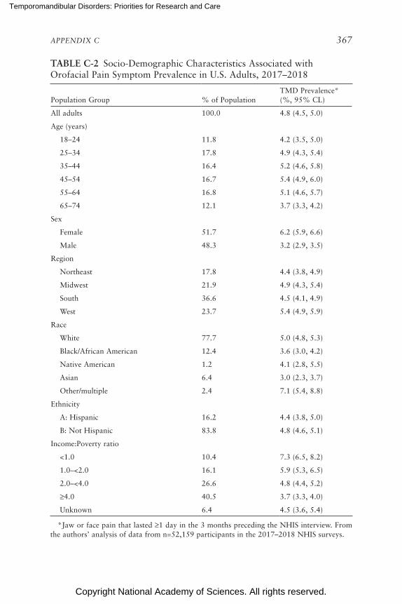
Temporomandibular Disorders: Priorities for Research and Care
Copyright National Academy of Sciences. All rights reserved.
APPENDIX C 367
TABLE C-2 Socio-Demographic Characteristics Associated with Orofacial Pain Symptom Prevalence in U.S. Adults, 2017–2018
Population Group % of PopulationTMD Prevalence* (%, 95% CL)
All adults 100.0 4.8 (4.5, 5.0)
Age (years)
18–24 11.8 4.2 (3.5, 5.0)
25–34 17.8 4.9 (4.3, 5.4)
35–44 16.4 5.2 (4.6, 5.8)
45–54 16.7 5.4 (4.9, 6.0)
55–64 16.8 5.1 (4.6, 5.7)
65–74 12.1 3.7 (3.3, 4.2)
Sex
Female 51.7 6.2 (5.9, 6.6)
Male 48.3 3.2 (2.9, 3.5)
Region
Northeast 17.8 4.4 (3.8, 4.9)
Midwest 21.9 4.9 (4.3, 5.4)
South 36.6 4.5 (4.1, 4.9)
West 23.7 5.4 (4.9, 5.9)
Race
White 77.7 5.0 (4.8, 5.3)
Black/African American 12.4 3.6 (3.0, 4.2)
Native American 1.2 4.1 (2.8, 5.5)
Asian 6.4 3.0 (2.3, 3.7)
Other/multiple 2.4 7.1 (5.4, 8.8)
Ethnicity
A: Hispanic 16.2 4.4 (3.8, 5.0)
B: Not Hispanic 83.8 4.8 (4.6, 5.1)
Income:Poverty ratio
<1.0 10.4 7.3 (6.5, 8.2)
1.0–<2.0 16.1 5.9 (5.3, 6.5)
2.0–<4.0 26.6 4.8 (4.4, 5.2)
≥4.0 40.5 3.7 (3.3, 4.0)
Unknown 6.4 4.5 (3.6, 5.4)
*Jaw or face pain that lasted ≥1 day in the 3 months preceding the NHIS interview. From the authors’ analysis of data from n=52,159 participants in the 2017–2018 NHIS surveys.
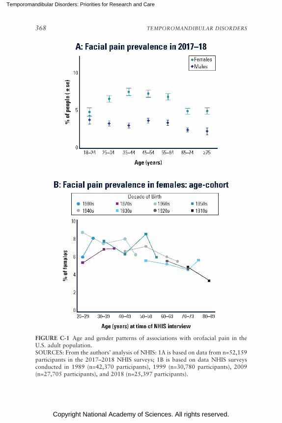
Temporomandibular Disorders: Priorities for Research and Care
Copyright National Academy of Sciences. All rights reserved.
368 TEMPOROMANDIBULAR DISORDERS
FIGURE C-1 Age and gender patterns of associations with orofacial pain in the U.S. adult population.SOURCES: From the authors’ analysis of NHIS: 1A is based on data from n=52,159 participants in the 2017–2018 NHIS surveys; 1B is based on data NHIS surveys conducted in 1989 (n=42,370 participants), 1999 (n=30,780 participants), 2009 (n=27,705 participants), and 2018 (n=25,397 participants).
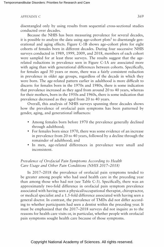
Temporomandibular Disorders: Priorities for Research and Care
Copyright National Academy of Sciences. All rights reserved.
APPENDIX C 369
disentangled only by using results from sequential cross-sectional studies conducted over decades.
Because the NHIS has been measuring prevalence for several decades, it is possible to analyze the data using age–cohort plots5 to disentangle gen-erational and aging effects. Figure C-1B shows age–cohort plots for eight cohorts of females born in different decades. During four successive NHIS surveys conducted in 1989, 1999, 2009, and 2018, members of each cohort were sampled for at least three surveys. The results suggest that the age-related reductions in prevalence seen in Figure C-1A are associated more with aging than with generational differences between cohorts. Specifically, for females aged 50 years or more, there was a fairly consistent reduction in prevalence in older age groups, regardless of the decade in which they were born. The age-related pattern earlier in adulthood is more difficult to discern: for females born in the 1970s and 1980s, there is some indication that prevalence increased as they aged from around 20 to 40 years, whereas for their mothers, born in the 1950s and 1960s, there is some indication that prevalence decreased as they aged from 20 to 40 years.
Overall, this analysis of NHIS surveys spanning three decades shows how the prevalence of orofacial pain symptoms has been patterned by gender, aging, and generational influences:
• Among females born before 1970 the prevalence generally declined through adulthood;
• For females born since 1970, there was some evidence of an increase in prevalence from 20 to 40 years, followed by a decline through the remainder of adulthood; and
• In men, age-related differences in prevalence were small and inconsistent.
Prevalence of Orofacial Pain Symptoms According to Health Care Usage and Other Pain Conditions (NHIS 2017–2018)
In 2017–2018 the prevalence of orofacial pain symptoms tended to be greater among people who had used health care in the preceding year than among those who had not (see Table C-3). Specifically, there was an approximately two-fold difference in orofacial pain symptom prevalence associated with having seen a physical/occupational therapist, chiro practor, or medical specialist and a 1.5-fold difference associated with having seen a general doctor. In contrast, the prevalence of TMDs did not differ accord-ing to whether participants had seen a dentist within the preceding year. It must be emphasized that the 2017–2018 surveys did not inquire as to the reasons for health care visits or, in particular, whether people with orofacial pain symptoms sought health care because of those symptoms.
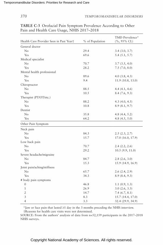
Temporomandibular Disorders: Priorities for Research and Care
Copyright National Academy of Sciences. All rights reserved.
370 TEMPOROMANDIBULAR DISORDERS
TABLE C-3 Orofacial Pain Symptom Prevalence According to Other Pain and Health Care Usage, NHIS 2017–2018
Health Care Provider Seen in Past Year† % of PopulationTMD Prevalence* (%, 95% CL)
General doctorNo 29.4 3.4 (3.0, 3.7)Yes 69.6 5.4 (5.1, 5.7)
Medical specialistNo 70.7 3.7 (3.5, 4.0)Yes 28.2 7.5 (7.0, 8.0)
Mental health professionalNo 89.6 4.0 (3.8, 4.3)Yes 9.4 11.9 (10.8, 13.0)
ChiropractorNo 88.5 4.4 (4.1, 4.6)Yes 10.5 8.4 (7.6, 9.3)
Therapist (PT/OT/etc.) No 88.2 4.3 (4.0, 4.5)Yes 10.8 8.9 (8.1, 9.7)
DentistNo 35.8 4.8 (4.4, 5.2)Yes 64.2 4.8 (4.5, 5.0)
Other Pain Symptom
Neck painNo 84.3 2.5 (2.3, 2.7)Yes 15.7 17.0 (16.0, 17.9)
Low back painNo 70.7 2.4 (2.2, 2.6)Yes 29.2 10.5 (9.9, 11.0)
Severe headache/migraineNo 84.7 2.8 (2.6, 3.0)Yes 15.3 15.9 (14.9, 16.9)
Joint pain/aching/stiffnessNo 65.7 2.6 (2.4, 2.9)Yes 34.3 8.9 (8.4, 9.3)
# body pain symptoms0 46.8 1.1 (0.9, 1.3)1 26.9 3.0 (2.6, 3.3)2 14.7 7.4 (6.7, 8.1)3 8.3 15.7 (14.4, 17.0)4 3.3 32.4 (29.9, 34.9)
*Jaw or face pain that lasted ≥1 day in the 3 months preceding the NHIS interview. †Reasons for health care visits were not determined.SOURCE: From the authors’ analysis of data from n=52,159 participants in the 2017–2018 NHIS surveys.
Temporomandibular Disorders: Priorities for Research and Care
Copyright National Academy of Sciences. All rights reserved.
APPENDIX C 371
Larger differences in prevalence were seen in relation to the presence of other pain conditions (i.e., other than orofacial pain symptoms; see Table C-3). People reporting headache or pain symptoms in the neck, back, or joints had at least three times the prevalence of orofacial pain symptoms as people without those body pain symptoms (see Table C-3). Using a simple count of those four body pain symptoms, the prevalence of orofacial pain symptoms increased markedly from 1.1 percent among people with no body pain symptoms to 32.4 percent among people with all four body pain symptoms.
Prevalence of Orofacial Pain Symptoms Reported in Other U.S. Studies
The first population-based survey of orofacial pain symptoms in the United States was conducted in 1986 among adult enrollees in a health maintenance organization in Washington State.6 It used a single screening question to determine the presence of pain in the face or jaw for at least 1 day in the preceding 6 months, excluding minor or fleeting pain. Oro-facial pain symptoms in the prior 6 months were reported by 12 percent of participants, approximately twice the prevalence reported in the 1989 NHIS. It is worth noting the sample studied in Washington also had nearly twice the prevalence of headache and back pain as reported in NHIS.7 A separate study of adolescents (i.e., aged 11–17 years) enrolled in the same health maintenance organization also investigated orofacial pain symp-toms.8 The prevalence of orofacial pain symptoms increased from 4 percent in pre-pubertal children to 14 percent in adolescents who had completed pubertal development.
A study of adults of ages ≥45 years living in disadvantaged rural areas of Florida asked about several orofacial pain symptoms in the preceding 6 months. The prevalence of jaw joint pain was 8.3 percent, and the preva-lence of face pain was 3.1 percent. Another study used similar questions in a telephone interview survey of people aged ≥65 years living in northern Florida.9 The prevalence of jaw joint pain was 7.7 percent, and the preva-lence of face pain was 6.9 percent. A recent survey of adult dental patients found that prevalence of orofacial pain symptoms was 6.6 percent among those attending dental providers in the Northwest Practice-Based Research Collaborative in Evidence-Based Dentistry.10
Although NHIS surveys show small differences in orofacial pain symp-tom prevalence between Hispanic and non-Hispanic adults, the survey lacks the precision needed to evaluate heterogeneity within the Hispanic popu-lation. Instead, the Hispanic Community Health Study/Study of Latinos (HCHS/SoL) is a large, population-based survey of health status among Hispanics sampled at four U.S. study sites, which is intended to capture a diversity of Hispanic heritage. Between 2008 and 2011, interviewers queried
Temporomandibular Disorders: Priorities for Research and Care
Copyright National Academy of Sciences. All rights reserved.
372 TEMPOROMANDIBULAR DISORDERS
16,415 study participants aged 18–74 years about orofacial pain symptoms during the preceding 12 months. Questions were asked separately about pain in the face (with an estimated 9.5 percent of the population answering affirmatively) and pain in the jaw joint (14.6 percent), yielding an overall prevalence of 5.1 percent (95% CL=4.7, 5.5) responding positively to both questions.11 The prevalence varied from a low of 3.8 percent among South American Hispanics to 7.0 percent among Puerto Ricans. The patterns of socio-demographic variation were similar to those seen for the population at large: in the HCHS/SoL study, prevalence was greater in women (6.7 per-cent) than men (3.4 percent) and varied approximately two-fold between the lowest-income group (6.4 percent among people with annual income less than $15,000) and the highest-income group (3.5 percent among people with annual income of $40,000 or more).
Prevalence of Examiner-Classified TMDs and Concordance with Self-Reported Symptoms of TMD
The prevalence of examiner-classified TMDs has been reported in only one study of a random sample of U.S. adults.12 It was limited to women aged 18 years or more living in New York City, New York, or Newark, New Jersey. In 2000–2001 phone numbers were sampled at random, and eligible women completed a screening interview to determine presence of orofacial pain symptoms in the preceding 6 months. Specifically, respon-dents were asked, “Other than a toothache or sinus pain, did you have pain in your face, in the front of your ear or jaw, more than one time, in the last 6 months?” Respondents were also invited to a research dental clinic where examiners determined presence or absence of TMD using Research Diagnostic Criteria for Temporomandibular Disorders (RDC/TMD) crite-ria for myofascial subtype of TMDs (the most frequent subtype of TMD myalgia). The researchers also evaluated the screening validity of TMDs, as determined by self-reported symptoms, compared with the reference standard of examiner-determined TMDs.
The prevalence of orofacial pain symptoms in the population of women studied was 10.1 percent, which was very similar to the 10.5 per-cent of participants with examiner-classified TMDs (see Table C-4). This high degree of concordance in prevalence occurred despite the finding that orofacial pain symptom reporting had low sensitivity (0.427), mean-ing that more than half of the people with examiner-classified TMDs responded negatively to the screening question (i.e., they were false nega-tives). However, the specificity was 0.947, which meant that false positives occurred for only 1 in 20 of the people who did not have a TMD when examined. In the overall population of women, the number of false posi-tives and false negatives mostly cancelled one another. The consequence
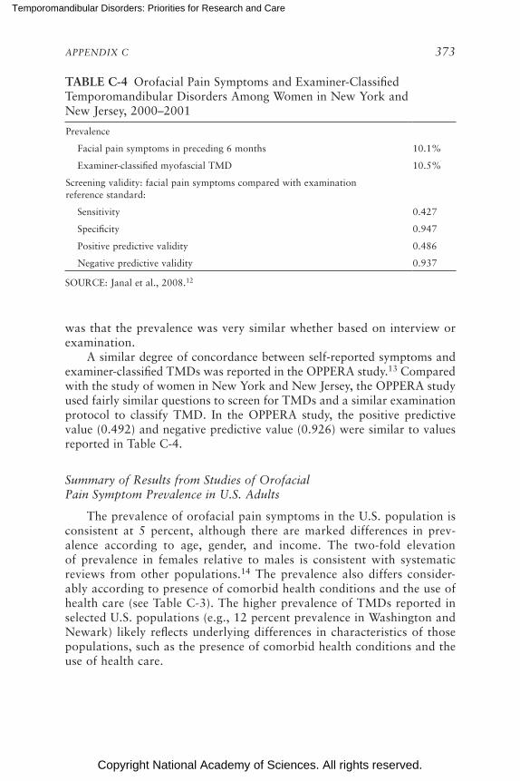
Temporomandibular Disorders: Priorities for Research and Care
Copyright National Academy of Sciences. All rights reserved.
APPENDIX C 373
TABLE C-4 Orofacial Pain Symptoms and Examiner-Classified Temporomandibular Disorders Among Women in New York and New Jersey, 2000–2001
Prevalence
Facial pain symptoms in preceding 6 months 10.1%
Examiner-classified myofascial TMD 10.5%
Screening validity: facial pain symptoms compared with examination reference standard:
Sensitivity 0.427
Specificity 0.947
Positive predictive validity 0.486
Negative predictive validity 0.937
SOURCE: Janal et al., 2008.12
was that the prevalence was very similar whether based on interview or examination.
A similar degree of concordance between self-reported symptoms and examiner-classified TMDs was reported in the OPPERA study.13 Compared with the study of women in New York and New Jersey, the OPPERA study used fairly similar questions to screen for TMDs and a similar examination protocol to classify TMD. In the OPPERA study, the positive predictive value (0.492) and negative predictive value (0.926) were similar to values reported in Table C-4.
Summary of Results from Studies of Orofacial Pain Symptom Prevalence in U.S. Adults
The prevalence of orofacial pain symptoms in the U.S. population is consistent at 5 percent, although there are marked differences in prev-alence according to age, gender, and income. The two-fold elevation of prevalence in females relative to males is consistent with systematic reviews from other populations.14 The prevalence also differs consider-ably according to presence of comorbid health conditions and the use of health care (see Table C-3). The higher prevalence of TMDs reported in selected U.S. populations (e.g., 12 percent prevalence in Washington and Newark) likely reflects underlying differences in characteristics of those populations, such as the presence of comorbid health conditions and the use of health care.
Temporomandibular Disorders: Priorities for Research and Care
Copyright National Academy of Sciences. All rights reserved.
374 TEMPOROMANDIBULAR DISORDERS
Incidence of Temporomandibular Disorders
Incidence refers to the rate at which a disease develops within a popu-lation during a specified time interval. Studies of incidence are more diffi-cult to conduct than cross-sectional studies of prevalence because incidence has to be measured over time, using a prospective cohort study design. At baseline, it is also important to enroll study participants who do not have the disease. The goal during the follow-up period is to enumerate all new events of disease in the study cohort. In most studies of TMD, a follow-up interval of at least 2 years is required. The incidence rate is then calculated to represent the probability of developing disease during a specified time period. To make valid estimates of incidence, it is important to use valid and reliable methods to classify TMD. Furthermore, given that symptoms can come and go within a few months, it is important that study par-ticipants be re-assessed at sufficiently short intervals to enumerate all new events of TMD.
TMD Incidence in the OPPERA Study
In the OPPERA prospective cohort study, facial pain symptoms were as-sessed by questionnaire, once every quarter (3-month period) among 2,719 adults aged 18–44 years who had no history of a TMD when enrolled. Dur-ing the median 2.3-year follow-up period, one-third of the cohort members developed ≥1 symptom episodes (i.e., facial pain for ≥5 days per month for ≥1 months during a 3-month reporting period).15 This represented an initial symptom episodes rate of 18.8 percent per annum. For those who developed one such episode, the rate of recurrence doubled, and it doubled again in follow-up of those with recurrent symptoms. For one-quarter of episodes, the symptom severity was rated as 7 or higher using a 0-to-10 rating scale, which is consistent with “severe” clinical pain.16
A large majority of symptom episodes were subclinical, in that sub-sequent examinations found that most episodes did not meet the criteria for clinical TMDs, as determined using the RDC/TMD protocol.17 As a consequence, the annual incidence rate of clinically classified TMDs was 3.9 percent per annum,18 which is one-fifth of the rate of symptom onset. This discrepancy in rates is one reason that the impact of TMD in the community at large represents a “symptom iceberg,”19 a term referring to symptoms that are not managed by health care professionals.
Stated another way, the 3.9 percent per annum rate of examiner- classified TMDs means that for every 100 TMD-free people enrolled, nearly four individuals per year developed the condition. The incidence was greater in older age groups, but it did not vary significantly by gender (see Table C-5). However, there was a two-fold difference in incidence between
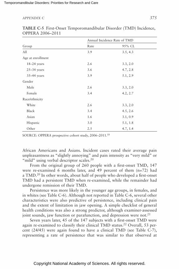
Temporomandibular Disorders: Priorities for Research and Care
Copyright National Academy of Sciences. All rights reserved.
APPENDIX C 375
TABLE C-5 First-Onset Temporomandibular Disorder (TMD) Incidence, OPPERA 2006–2011
Annual Incidence Rate of TMD
Group Rate 95% CL
All 3.9 3.5, 4.3
Age at enrollment
18–24 years 2.6 3.3, 2.0
25–34 years 3.6 4.7, 2.8
35–44 years 3.9 5.1, 2.9
Gender
Male 2.6 3.3, 2.0
Female 3.4 4.2, 2.7
Race/ethnicity
White 2.6 3.3, 2.0
Black 3.4 4.5, 2.6
Asian 1.6 3.1, 0.9
Hispanic 3.0 5.1, 1.8
Other 2.5 4.7, 1.4
SOURCE: OPPERA prospective cohort study, 2006–2011.18
African Americans and Asians. Incident cases rated their average pain unpleasant ness as “slightly annoying” and pain intensity as “very mild” or “mild” using verbal descriptor scales.20
From the original group of 260 people with a first-onset TMD, 147 were re-examined 6 months later, and 49 percent of them (n=72) had a TMD.20 In other words, about half of people who developed a first-onset TMD had a persistent TMD when re-examined, while the remainder had undergone remission of their TMD.
Persistence was more likely in the younger age groups, in females, and in whites (see Table C-6). Although not reported in Table C-6, several other characteristics were also predictive of persistence, including clinical pain and the extent of limitation in jaw opening. A simple checklist of general health conditions was also a strong predictor, although examiner-assessed joint sounds, jaw function or parafunction, and depression were not.21
Seven years later, 45 of the 147 subjects with a first-onset TMD were again re-examined to classify their clinical TMD status.22 Overall, 53 per-cent (24/45) were again found to have a clinical TMD (see Table C-7), representing a rate of persistence that was similar to that observed at
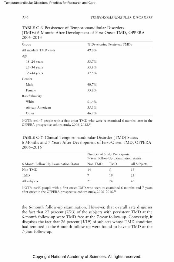
Temporomandibular Disorders: Priorities for Research and Care
Copyright National Academy of Sciences. All rights reserved.
376 TEMPOROMANDIBULAR DISORDERS
TABLE C-6 Persistence of Temporomandibular Disorders (TMDs) 6 Months After Development of First-Onset TMD, OPPERA 2006–2013
Group % Developing Persistent TMDs
All incident TMD cases 49.0%
Age
18−24 years 53.7%
25−34 years 55.6%
35−44 years 37.5%
Gender
Male 40.7%
Female 53.8%
Race/ethnicity
White 61.4%
African American 35.5%
Other 46.7%
NOTE: n=147 people with a first-onset TMD who were re-examined 6 months later in the OPPERA prospective cohort study, 2006–2013.21
the 6-month follow-up examination. However, that overall rate disguises the fact that 27 percent (7/23) of the subjects with persistent TMD at the 6-month follow-up were TMD free at the 7-year follow-up. Conversely, it disguises the fact that 26 percent (5/19) of subjects whose TMD condition had remitted at the 6-month follow-up were found to have a TMD at the 7-year follow-up.
TABLE C-7 Clinical Temporomandibular Disorder (TMD) Status 6 Months and 7 Years After Development of First-Onset TMD, OPPERA 2006–2016
Number of Study Participants: 7-Year Follow-Up Examination Status
6-Month Follow-Up Examination Status Non-TMD TMD All Subjects
Non-TMD 14 5 19
TMD 7 19 26
All subjects 21 24 45
NOTE: n=45 people with a first-onset TMD who were re-examined 6 months and 7 years after onset in the OPPERA prospective cohort study, 2006–2016.22
Temporomandibular Disorders: Priorities for Research and Care
Copyright National Academy of Sciences. All rights reserved.
APPENDIX C 377
Notwithstanding the imprecision inherent in these percentages based on a small cohort of 45 people, the results suggest that, for a sizable minority of people, fluctuation and remission of clinical TMD status occur for months or years after a TMD first develops. By implication, fluctua-tion and remission continue through life, such that prevalence in any single cross-sectional survey is a function of an individual’s history of TMD onset, remission, and persistence.
Other Studies of TMD Incidence
Prior to the OPPERA study, investigators at the North Carolina study site conducted a 3-year prospective cohort study of women aged 18–34 years at the time of enrollment.23 At enrollment, examiners verified that study participants did not have a TMD, and during the follow-up symp-toms were monitored using quarterly questionnaires. Symptomatic subjects were re-examined to determine incidence of examiner-classified TMDs, which yielded an annual incidence rate of 3.5 percent.
In a prospective cohort study of 11-year-olds who were enrollees of the Group Health Cooperative in Washington State, 6.8 percent developed ex-aminer-verified TMDs during the 3-year follow-up period (i.e., an annualized incidence rate of 2.3 percent).24 Study participants were monitored during the follow-up period using quarterly (3-monthly) questionnaires to screen for new symptoms of TMDs, similar to the methodology used in the OPPERA study. The incidence rate in adolescents was nearly twice as high in female as males, and it was greater in whites than in other racial groups. Among the strongest predictors of elevated incidence was the presence of other pain conditions at baseline (i.e., headache, back pain, and stomach pain).
Adult enrollees in the same health maintenance organization who were first enrolled in 1986 were also followed prospectively and re-interviewed after 3 years.25 However, there were no questionnaires administered during the intervening period within the 3 years of follow-up. Among subjects who had no history of a TMD when enrolled, 6.5 percent reported orofacial pain symptoms 3 years later. Although the rate did not differ to a statisti-cally significant degree, it was inversely associated with age, and it was 7.7 percent in females compared to 4.8 percent in males.
PAIN IMPACT AND PAIN BURDEN ON PEOPLE WITH TMDs
Characteristics of Orofacial Pain and Responses to Orofacial Pain Symptoms in the U.S. Population—NHIS 1989
The 1989 NHIS included supplementary questions about orofacial pain that provide more detail about the impact of TMDs than subsequent NHIS
Temporomandibular Disorders: Priorities for Research and Care
Copyright National Academy of Sciences. All rights reserved.
378 TEMPOROMANDIBULAR DISORDERS
surveys. As noted above, the 1989 NHIS also differed from subsequent sur-veys in its use of a 6-month reference period to inquire about orofacial pain symptoms. In the adult population overall, prevalence was 6.0 percent and, as seen in other NHIS surveys, prevalence varied almost two-fold according to gender (7.7 percent in females; 4.1 percent in males).
For people who reported orofacial pain, the supplementary questions asked about characteristics of orofacial pain and behavioral response to the pain (see Table C-8). The most common pattern of occurrence was symp-toms that “come and go,” and about two-thirds of people with symptoms had experienced them for 10 days or fewer within the preceding 6 months. However, a little more than half said that the symptoms occurred in at least 3 of the preceding 6 months. Those with pain were also asked to rate their pain based on this question: “On a scale of 1 to 10, where 1 is mild and 10 is severe, how would you rate this pain at its worst?” One-third of people with orofacial pain rated the severity as 7 or more (see Table C-8), a threshold found to be consistent with “severe” pain in studies that use the more conventional 0–10 rating scale.16
The most commonly reported behavioral response to orofacial pain symptoms was use of over-the-counter medication (50.7 percent) and/or prescription medication (22.9 percent). Social impacts were reported by fewer than 10 percent, and included staying at home more than usual (8.3 percent), avoiding family or friends (6.3 percent), and taking time off work (4.8 percent). Conversely, nearly one-quarter of subjects reported none of the behavioral responses listed in Table C-8. Meanwhile, nearly half of people with orofacial pain symptoms reported having seen a health care provider for those symptoms during the preceding 12 months. Dentists were somewhat more likely to be consulted (32.8 percent) then medical doctors (23.8 percent). (Note that, unlike subsequent NHIS sur-veys, the supplementary questions in the 1989 survey asked about visits to health care providers that were made specifically because of orofacial pain.)
Chronic TMD and High-Impact Chronic TMD (NHIS 1989)
The National Pain Strategy defines chronic pain as “pain that occurs on at least half the days for six months or more” and high-impact chronic pain as chronic pain that is “associated with substantial restriction of par-ticipation in work, social, and self-care activities for six months or more.”26 Data in Table C-8 provide some insight into duration and impact of TMD pain symptoms in the U.S. population. Specifically, 7.5 percent of people with orofacial pain symptoms reported pain for every day in the preced-ing 6 months, and a further 9.4 percent reported pain for at least 46 days (i.e., one-quarter of the days during the 6-month reference period). If the
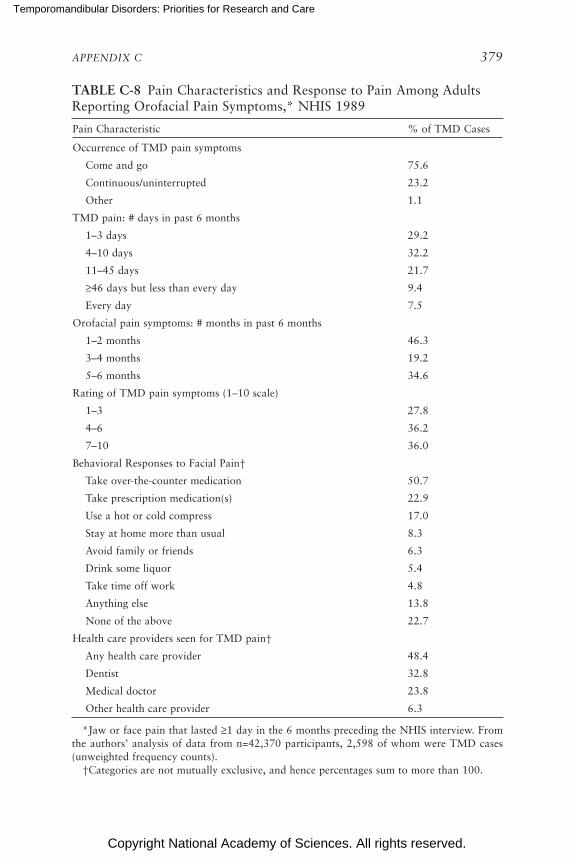
Temporomandibular Disorders: Priorities for Research and Care
Copyright National Academy of Sciences. All rights reserved.
APPENDIX C 379
TABLE C-8 Pain Characteristics and Response to Pain Among Adults Reporting Orofacial Pain Symptoms,* NHIS 1989
Pain Characteristic % of TMD Cases
Occurrence of TMD pain symptoms
Come and go 75.6
Continuous/uninterrupted 23.2
Other 1.1
TMD pain: # days in past 6 months
1–3 days 29.2
4–10 days 32.2
11–45 days 21.7
≥46 days but less than every day 9.4
Every day 7.5
Orofacial pain symptoms: # months in past 6 months
1–2 months 46.3
3–4 months 19.2
5–6 months 34.6
Rating of TMD pain symptoms (1–10 scale)
1–3 27.8
4–6 36.2
7–10 36.0
Behavioral Responses to Facial Pain†
Take over-the-counter medication 50.7
Take prescription medication(s) 22.9
Use a hot or cold compress 17.0
Stay at home more than usual 8.3
Avoid family or friends 6.3
Drink some liquor 5.4
Take time off work 4.8
Anything else 13.8
None of the above 22.7
Health care providers seen for TMD pain†
Any health care provider 48.4
Dentist 32.8
Medical doctor 23.8
Other health care provider 6.3
*Jaw or face pain that lasted ≥1 day in the 6 months preceding the NHIS interview. From the authors’ analysis of data from n=42,370 participants, 2,598 of whom were TMD cases (unweighted frequency counts). †Categories are not mutually exclusive, and hence percentages sum to more than 100.
Temporomandibular Disorders: Priorities for Research and Care
Copyright National Academy of Sciences. All rights reserved.
380 TEMPOROMANDIBULAR DISORDERS
two criteria are combined to define chronic TMD pain as face or jaw pain symptoms experienced for at least one-quarter of the days in the preceding 6 months, prevalence of chronic TMD in the U.S. adult population in 1989 was 1.0 percent (95% CL=0.8, 1.1). Furthermore, if high-impact TMDs are classified as one or more of the three behavioral impacts due to TMDs re-ported in Table C-8 (i.e., staying at home more than usual, avoiding family or friends, or taking time off work), prevalence of high-impact chronic TMDs in 1989 was 0.2 percent (95% CL=0.1, 0.2).
Prevalence of Chronic Pain and High-Impact Pain from Any Anatomical Location in TMD Cases and Controls (NHIS 2016)
The NHIS 2016 survey provided the first opportunity to estimate the prevalence of chronic pain and high-impact chronic pain as defined by the National Pain Strategy.26 (Note that the NHIS asked about the dura-tion and impact of pain experienced at any and all anatomical locations—it did not inquire about duration and impact of pain symptoms at specific locations, such as the face and jaw.) The National Pain Strategy’s defini-tions were operationalized by Dahlhamer et al.27 in their analysis of the 2016 NHIS survey. Specifically, they defined chronic pain from any and all anatomical locations as pain “that occurred on most days or every day in the past 6 months.” High-impact chronic pain was defined as chronic pain that furthermore “limited life or work activities on most days or every day during the past 6 months.” Applying those definitions to adults in the U.S. population, they found that the prevalence of chronic pain from any loca-tion was 20.4 percent, while the prevalence of high-impact chronic pain from any location was 8.0 percent.
The same 2016 NHIS data were analyzed for this report, focusing on the relationship between orofacial pain symptoms and chronic pain or high-impact chronic pain from any anatomical location. The prevalence of chronic pain at any anatomical location was elevated nearly three-fold in people with orofacial pain symptoms (52.7 percent) compared with people who did not have orofacial pain symptoms (18.8 percent) (see Table C-9). The prevalence of high-impact chronic pain was elevated nearly four-fold (26.9 percent versus 7.0 percent).
High-Impact TMD Pain and Effects of Pain on Quality of Life: OPPERA Study
Three decades ago, in what can be viewed as an early precedent of the National Pain Strategy’s focus on high-impact chronic pain, Von Korff et al. created the graded chronic pain scale (GCPS).28 It is a seven-item question-naire that asks about pain intensity, pain interference in everyday activities,
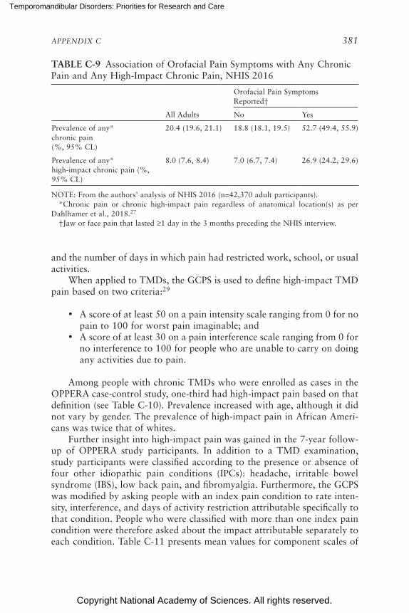
Temporomandibular Disorders: Priorities for Research and Care
Copyright National Academy of Sciences. All rights reserved.
APPENDIX C 381
TABLE C-9 Association of Orofacial Pain Symptoms with Any Chronic Pain and Any High-Impact Chronic Pain, NHIS 2016
Orofacial Pain Symptoms Reported†
All Adults No Yes
Prevalence of any* chronic pain (%, 95% CL)
20.4 (19.6, 21.1) 18.8 (18.1, 19.5) 52.7 (49.4, 55.9)
Prevalence of any* high-impact chronic pain (%, 95% CL)
8.0 (7.6, 8.4) 7.0 (6.7, 7.4) 26.9 (24.2, 29.6)
NOTE: From the authors’ analysis of NHIS 2016 (n=42,370 adult participants). *Chronic pain or chronic high-impact pain regardless of anatomical location(s) as per Dahlhamer et al., 2018.27 †Jaw or face pain that lasted ≥1 day in the 3 months preceding the NHIS interview.
and the number of days in which pain had restricted work, school, or usual activities.
When applied to TMDs, the GCPS is used to define high-impact TMD pain based on two criteria:29
• A score of at least 50 on a pain intensity scale ranging from 0 for no pain to 100 for worst pain imaginable; and
• A score of at least 30 on a pain interference scale ranging from 0 for no interference to 100 for people who are unable to carry on doing any activities due to pain.
Among people with chronic TMDs who were enrolled as cases in the OPPERA case-control study, one-third had high-impact pain based on that definition (see Table C-10). Prevalence increased with age, although it did not vary by gender. The prevalence of high-impact pain in African Ameri-cans was twice that of whites.
Further insight into high-impact pain was gained in the 7-year follow-up of OPPERA study participants. In addition to a TMD examination, study participants were classified according to the presence or absence of four other idiopathic pain conditions (IPCs): headache, irritable bowel syndrome (IBS), low back pain, and fibromyalgia. Furthermore, the GCPS was modified by asking people with an index pain condition to rate inten-sity, interference, and days of activity restriction attributable specifically to that condition. People who were classified with more than one index pain condition were therefore asked about the impact attributable separately to each condition. Table C-11 presents mean values for component scales of
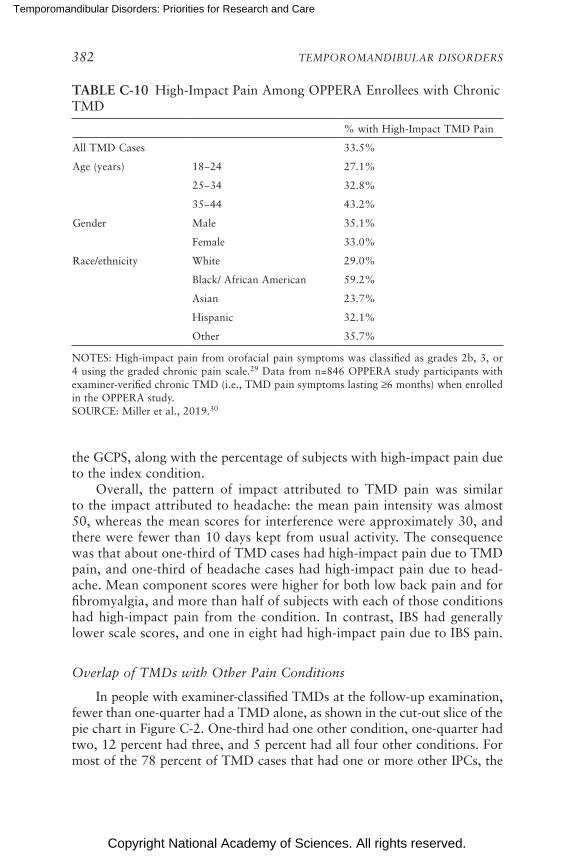
Temporomandibular Disorders: Priorities for Research and Care
Copyright National Academy of Sciences. All rights reserved.
382 TEMPOROMANDIBULAR DISORDERS
TABLE C-10 High-Impact Pain Among OPPERA Enrollees with Chronic TMD
% with High-Impact TMD Pain
All TMD Cases 33.5%
Age (years) 18−24 27.1%
25−34 32.8%
35−44 43.2%
Gender Male 35.1%
Female 33.0%
Race/ethnicity White 29.0%
Black/ African American 59.2%
Asian 23.7%
Hispanic 32.1%
Other 35.7%
NOTES: High-impact pain from orofacial pain symptoms was classified as grades 2b, 3, or 4 using the graded chronic pain scale.29 Data from n=846 OPPERA study participants with examiner-verified chronic TMD (i.e., TMD pain symptoms lasting ≥6 months) when enrolled in the OPPERA study. SOURCE: Miller et al., 2019.30
the GCPS, along with the percentage of subjects with high-impact pain due to the index condition.
Overall, the pattern of impact attributed to TMD pain was similar to the impact attributed to headache: the mean pain intensity was almost 50, whereas the mean scores for interference were approximately 30, and there were fewer than 10 days kept from usual activity. The consequence was that about one-third of TMD cases had high-impact pain due to TMD pain, and one-third of headache cases had high-impact pain due to head-ache. Mean component scores were higher for both low back pain and for fibromyalgia, and more than half of subjects with each of those conditions had high-impact pain from the condition. In contrast, IBS had generally lower scale scores, and one in eight had high-impact pain due to IBS pain.
Overlap of TMDs with Other Pain Conditions
In people with examiner-classified TMDs at the follow-up examination, fewer than one-quarter had a TMD alone, as shown in the cut-out slice of the pie chart in Figure C-2. One-third had one other condition, one-quarter had two, 12 percent had three, and 5 percent had all four other conditions. For most of the 78 percent of TMD cases that had one or more other IPCs, the
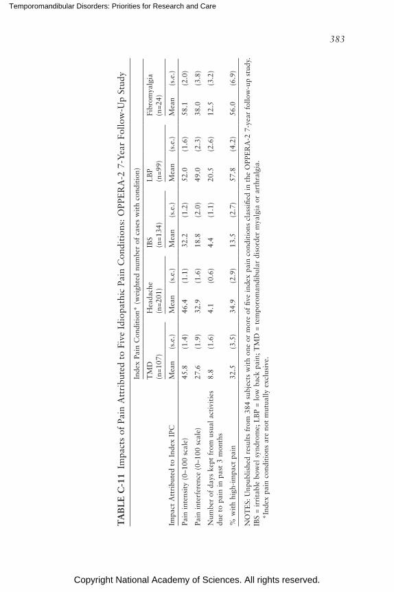
Temporomandibular Disorders: Priorities for Research and Care
Copyright National Academy of Sciences. All rights reserved.
383
TA
BL
E C
-11
Impa
cts
of P
ain
Att
ribu
ted
to F
ive
Idio
path
ic P
ain
Con
diti
ons:
OPP
ER
A-2
7-Y
ear
Follo
w-U
p St
udy
Inde
x Pa
in C
ondi
tion
* (w
eigh
ted
num
ber
of c
ases
wit
h co
ndit
ion)
Impa
ct A
ttri
bute
d to
Ind
ex I
PC
TM
D
(n=1
07)
Hea
dach
e (n
=201
)IB
S
(n=1
34)
LB
P
(n=9
9)Fi
brom
yalg
ia
(n=2
4)
Mea
n(s
.e.)
Mea
n(s
.e.)
Mea
n(s
.e.)
Mea
n(s
.e.)
Mea
n(s
.e.)
Pain
int
ensi
ty (
0–10
0 sc
ale)
45.8
(1.4
)46
.4(1
.1)
32.2
(1.2
)52
.0(1
.6)
58.1
(2.0
)
Pain
int
erfe
renc
e (0
–100
sca
le)
27.6
(1.9
)32
.9(1
.6)
18.8
(2.0
)49
.0(2
.3)
38.0
(3.8
)
Num
ber
of d
ays
kept
fro
m u
sual
act
ivit
ies
due
to p
ain
in p
ast
3 m
onth
s8.
8(1
.6)
4.1
(0.6
)4.
4(1
.1)
20.5
(2.6
)12
.5(3
.2)
% w
ith
high
-im
pact
pai
n32
.5(3
.5)
34.9
(2.9
)13
.5(2
.7)
57.8
(4.2
)56
.0(6
.9)
NO
TE
S: U
npub
lishe
d re
sult
s fr
om 3
84 s
ubje
cts
wit
h on
e or
mor
e of
five
inde
x pa
in c
ondi
tion
s cl
assi
fied
in t
he O
PPE
RA
-2 7
-yea
r fo
llow
-up
stud
y.
IBS
= ir
rita
ble
bow
el s
yndr
ome;
LB
P =
low
bac
k pa
in;
TM
D =
tem
poro
man
dibu
lar
diso
rder
mya
lgia
or
arth
ralg
ia.
*I
ndex
pai
n co
ndit
ions
are
not
mut
ually
exc
lusi
ve.
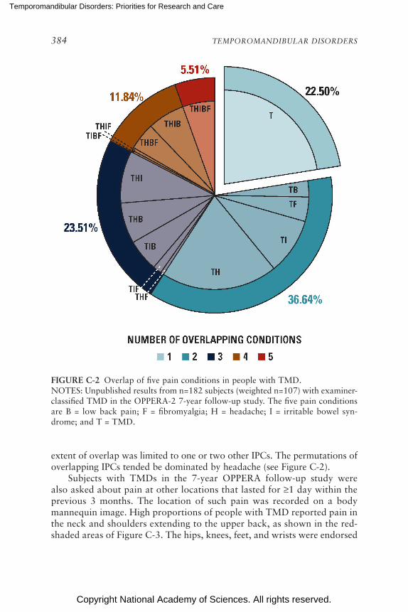
Temporomandibular Disorders: Priorities for Research and Care
Copyright National Academy of Sciences. All rights reserved.
384 TEMPOROMANDIBULAR DISORDERS
FIGURE C-2 Overlap of five pain conditions in people with TMD.NOTES: Unpublished results from n=182 subjects (weighted n=107) with examiner-classified TMD in the OPPERA-2 7-year follow-up study. The five pain conditions are B = low back pain; F = fibromyalgia; H = headache; I = irritable bowel syn-drome; and T = TMD.
extent of overlap was limited to one or two other IPCs. The permutations of overlapping IPCs tended be dominated by headache (see Figure C-2).
Subjects with TMDs in the 7-year OPPERA follow-up study were also asked about pain at other locations that lasted for ≥1 day within the previous 3 months. The location of such pain was recorded on a body mannequin image. High proportions of people with TMD reported pain in the neck and shoulders extending to the upper back, as shown in the red-shaded areas of Figure C-3. The hips, knees, feet, and wrists were endorsed
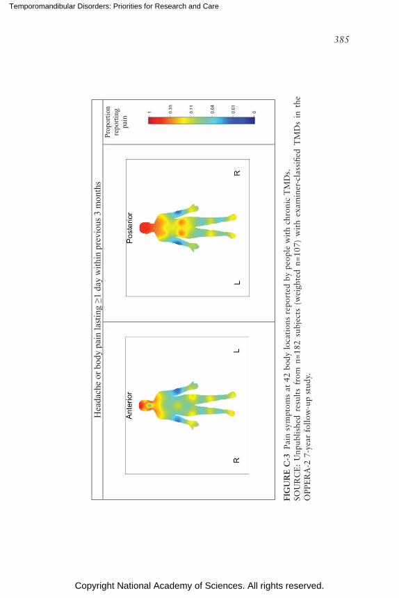
Temporomandibular Disorders: Priorities for Research and Care
Copyright National Academy of Sciences. All rights reserved.
385
FIG
UR
E C
-3 P
ain
sym
ptom
s at
42
body
loc
atio
ns r
epor
ted
by p
eopl
e w
ith
chro
nic
TM
Ds.
SOU
RC
E:
Unp
ublis
hed
resu
lts
from
n=1
82 s
ubje
cts
(wei
ghte
d n=
107)
wit
h ex
amin
er-c
lass
ified
TM
Ds
in t
he
OPP
ER
A-2
7-y
ear
follo
w-u
p st
udy.
Hea
dach
e or
bod
y pa
in la
stin
g ≥1
day
with
in p
revi
ous 3
mon
ths
Prop
ortio
nre
porti
ngpa
in
Temporomandibular Disorders: Priorities for Research and Care
Copyright National Academy of Sciences. All rights reserved.
386 TEMPOROMANDIBULAR DISORDERS
by around 20 percent. This represents another type of symptom iceberg, in which people with TMDs often experience pain elsewhere in their body.
Summary: High-Impact Pain in People with TMD
Despite differences in study years, data sources, and the criteria used to define impact, there is a consistent message from these results from the NHIS and OPPERA:
• In the U.S. adult population in 1989, approximately 17 percent of people with orofacial pain symptoms reported having experienced the symptoms for more than 45 days in the preceding 6 months (i.e., at least one-quarter of days) (see Table C-8).
• Using that threshold to define chronic pain, the corresponding preva-lence of chronic TMDs in the U.S. adult population is approximately 1 percent.
• Meanwhile, using the higher threshold of at least one-half of the days in the preceding 6 months to define chronic pain as per the National Pain Strategy, the prevalence of chronic pain from any anatomical location was 20 percent in 2016.27
• Of note, the prevalence of chronic pain, regardless of its anatomical location, was elevated nearly threefold, to 53 percent, among people with orofacial pain symptoms (see Table C-9).
• At first appearance, it might appear paradoxical that only 17 percent of TMD cases have chronic TMDs, whereas 53 percent of them have chronic pain.
o Specifically, people with TMDs are more likely than not to have chronic pain from any anatomical location, despite the fact that a minority (17 percent) experience chronic orofacial pain symptoms.
• The explanation lies in the considerable degree of overlap between TMDs and other commonly occurring chronic pain conditions (see Figures C-2 and C-3).
These observations are also salient when considering high-impact pain: in the U.S. population, one-quarter of people with TMDs experience high-impact chronic pain regardless of its anatomical location (see Table C-9). Yet, using a less stringent threshold to define impact, one-third of OPPERA study participants report high-impact pain. The explanation lies in the fact that only a minority of people with TMD experience symptoms during a sufficient number of days to qualify as chronic pain, based on the National Pain Strategy’s threshold.26 Nonetheless, when the impact is classified using the graded chronic pain scale, TMD pain causes levels of pain intensity, interference, and restriction that mimic the impact of headache.
Temporomandibular Disorders: Priorities for Research and Care
Copyright National Academy of Sciences. All rights reserved.
APPENDIX C 387
HEALTH CARE COSTS, OTHER DIRECT COSTS, AND INDIRECT COSTS OF TMD IN THE DEEP STUDY
Overview of Study Design
The DEEP study was a prospective cohort and cost analysis study examining the direct, indirect, and out-of-pocket costs of those living with persistent orofacial pain across the northeast of England. Persistent oro-facial pain was defined as pain lasting ≥3 months31 arising from a musculo-skeletal, neuropathic, or neurovascular origin. The origin of the pain was identified using validated screening instruments32,33 and if odontogenic pain was identified, individuals were excluded from participating.
No changes to individuals’ care pathways were made, and they pro-gressed within existing structures in both community (“primary”) care and hospital specialist–based (“secondary”) care within a state-funded health care system: the National Health Service (NHS) in the United Kingdom. Within the NHS individuals do not pay for any medical or surgical treatment, but, depending on their income, they may pay some of the costs for dental care and a standardized cost per prescription-only medicine (£8.80 per item of prescription-only medication in 2018). Individuals are also free to seek, with-out prejudice, private health care of any form outside of the NHS.
Individuals completed up to seven instruments every 6 months for a 24-month period, including a further census at 14 months to examine their time and travel costs related to health care. The instruments used were EQ-5D-5L for generic quality of life,34 GCPS,28 West Haven–Yale Multi-dimensional Pain Inventory (WHYMPI, version 335), Patient Health Ques-tionnaire-4 (PHQ-436), Use of Services and Productivity Questionnaire,37 and Illness Perceptions Questionnaire–Revised Form.38 Descriptions of the instruments’ scoring and interpretation are freely available in the appendix to one of the published papers from the study.2 The T&T census included a validated measure of how the patients’ pain affected the quality and quantity of work they could complete while at their place of employment (“QQ method”39).
A priori, a sample size of 200 patients was determined to be the correct size to detect with 80 percent power, a moderate effect size of 0.4 between two groups;40 as it was envisaged there may be differences between pri-mary and secondary care. This sample size also allowed for up to up to 30 predictors of costs with regression analyses,41 with the a priori predictor of interest being GCPS status as determined by the Graded Chronic Pain Scale due to its proven prognostic validity and ability to predict treatment need.29,42,43 GCPS provides an ordinal outcome of 0, I, IIa, IIb, III, IV. This range of ordinal outcomes begins at 0 (no pain-related disability), with IV (high pain-related disability) being the maximum. Grades IIb–IV have been
Temporomandibular Disorders: Priorities for Research and Care
Copyright National Academy of Sciences. All rights reserved.
388 TEMPOROMANDIBULAR DISORDERS
shown to have substantially more risk of chronification29,42,43 and therefore it is possible to collapse the five ordinal outcomes to “low” GCPS (0–IIa) and “high” GCPS (IIb–IV), which have been shown to predict treatment need and prognosis.29
The data provided in this report are a secondary analysis of those only screening positive for musculoskeletal/TMD diagnoses. Some analyses such as regression are therefore not possible because of the decrease in sample size. Unit costs, their sources, and their use are described in full in Durham et al.2 and Breckons et al.44 In brief the unit costs were determined and then multiplied against appropriate health care usage data from the U.S. Patients Quarterly or the data from the T&T census for, respectively, direct or indirect and out-of-pocket costs. Deterministic sensitivity analyses were conducted on variables whenever uncertainty existed concerning the unit cost. Costs collected over the 24-month study period were in 2012 prices (the year the study started) and adjusted using the Consumer Price Index for inflation (CPI; Breckons et al. gives a description of the method used44). The final costs were then all converted to the latest price year with the most recent CPI figure available and are therefore presented in 2018 pounds sterling throughout the report (average UK exchange rate for 2018: UK £1.00 = US $1.33).
Sample Characteristics—DEEP Study
Of the 279 individuals screened to enter the DEEP study, 268 met the inclusion criteria and 239 agreed to participate, with 198 returning data at M0. There were no significant differences in age, gender, or origin of pain between those who agreed to participate and those who did not. There was sequential dropout over the study at M0, M6, M12, M18, and M24, but significant differences in age, gender, ethnicity, duration of pain, and origin of pain were only identified between those remaining and those dropping out at M0, when a significantly younger cohort dropped out (p<0.01); at M12, when those with a significantly longer duration of pain were more likely to drop out (p<0.05); and at M18 and M24, when significantly more white British dropped out (p<0.01).
For the data presented within this report, the sample at M0 (n=198) was limited to those screening positive for painful TMDs and a musculo-skeletal origin for their pain using the screening instruments,32,33 whose sensitivity and specificity were demonstrated at 63 percent and 86 percent respectively.2 Any of those screening positive for neuropathic/vascular or a combined origin for their pain were removed from the sample used for analyses for this report. This left 87 individuals in the sample at M0. The sample’s socio-demographic characteristics are given in Table C-12.
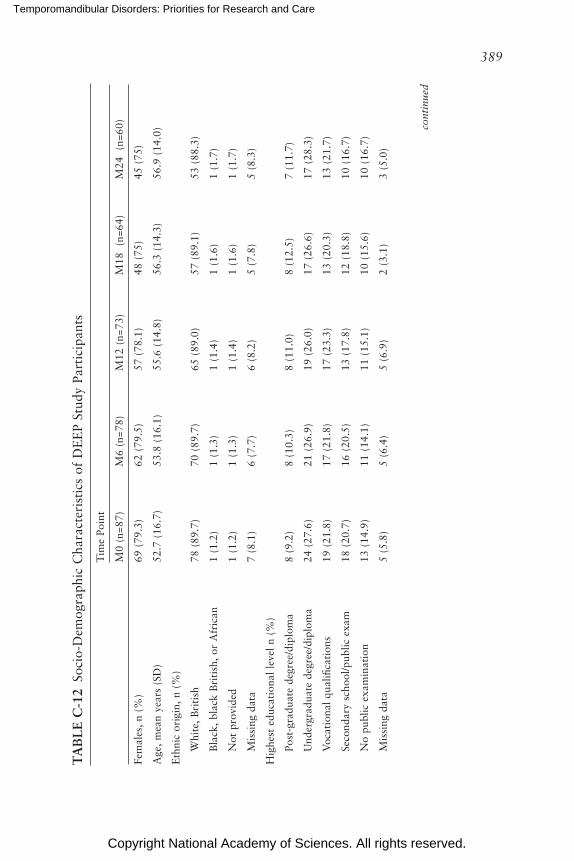
Temporomandibular Disorders: Priorities for Research and Care
Copyright National Academy of Sciences. All rights reserved.
389
TA
BL
E C
-12
Soci
o-D
emog
raph
ic C
hara
cter
isti
cs o
f D
EE
P St
udy
Part
icip
ants
Tim
e Po
int
M0
(n=8
7)M
6 (n
=78)
M12
(n=
73)
M18
(n
=64)
M24
(n
=60)
Fem
ales
, n
(%)
69 (
79.3
)62
(79
.5)
57 (
78.1
)48
(75
)45
(75
)
Age
, m
ean
year
s (S
D)
52.7
(16
.7)
53.8
(16
.1)
55.6
(14
.8)
56.3
(14
.3)
56.9
(14
.0)
Eth
nic
orig
in,
n (%
)
Whi
te,
Bri
tish
78
(89
.7)
70 (
89.7
)65
(89
.0)
57 (
89.1
)53
(88
.3)
Bla
ck,
blac
k B
riti
sh,
or A
fric
an
1 (1
.2)
1 (1
.3)
1 (1
.4)
1 (1
.6)
1 (1
.7)
Not
pro
vide
d 1
(1.2
)1
(1.3
)1
(1.4
)1
(1.6
) 1
(1.7
)
Mis
sing
dat
a 7
(8.1
)6
(7.7
)6
(8.2
)5
(7.8
)5
(8.3
)
Hig
hest
edu
cati
onal
lev
el n
(%
)
Post
-gra
duat
e de
gree
/dip
lom
a 8
(9.2
)8
(10.
3)8
(11.
0)8
(12.
5)7
(11.
7)
Und
ergr
adua
te d
egre
e/di
plom
a 24
(27
.6)
21 (
26.9
)19
(26
.0)
17 (
26.6
)17
(28
.3)
Voc
atio
nal
qual
ifica
tion
s 19
(21
.8)
17 (
21.8
)17
(23
.3)
13 (
20.3
)13
(21
.7)
Seco
ndar
y sc
hool
/pub
lic e
xam
18 (
20.7
)16
(20
.5)
13 (
17.8
)12
(18
.8)
10 (
16.7
)
No
publ
ic e
xam
inat
ion
13 (
14.9
)11
(14
.1)
11 (
15.1
)10
(15
.6)
10 (
16.7
)
Mis
sing
dat
a 5
(5.8
)5
(6.4
)5
(6.9
)2
(3.1
)3
(5.0
)
cont
inue
d
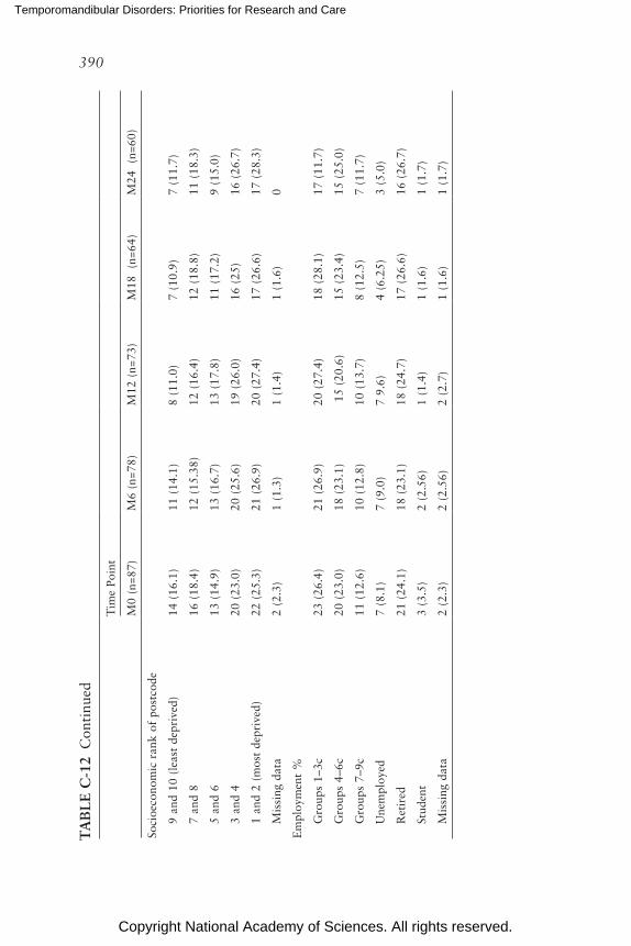
Temporomandibular Disorders: Priorities for Research and Care
Copyright National Academy of Sciences. All rights reserved.
390
Tim
e Po
int
M0
(n=8
7)M
6 (n
=78)
M12
(n=
73)
M18
(n
=64)
M24
(n
=60)
Soci
oeco
nom
ic r
ank
of p
ostc
ode
9 an
d 10
(le
ast
depr
ived
) 14
(16
.1)
11 (
14.1
)8
(11.
0)7
(10.
9)7
(11.
7)
7 an
d 8
16 (
18.4
)12
(15
.38)
12 (
16.4
)12
(18
.8)
11 (
18.3
)
5 an
d 6
13 (
14.9
)13
(16
.7)
13 (
17.8
)11
(17
.2)
9 (1
5.0)
3 an
d 4
20 (
23.0
)20
(25
.6)
19 (
26.0
)16
(25
)16
(26
.7)
1 an
d 2
(mos
t de
priv
ed)
22 (
25.3
)21
(26
.9)
20 (
27.4
)17
(26
.6)
17 (
28.3
)
Mis
sing
dat
a 2
(2.3
)1
(1.3
)1
(1.4
)1
(1.6
)0
Em
ploy
men
t %
Gro
ups
1–3c
23 (
26.4
)21
(26
.9)
20 (
27.4
)18
(28
.1)
17 (
11.7
)
Gro
ups
4–6c
20 (
23.0
) 18
(23
.1)
15
(20.
6)
15 (
23.4
)15
(25
.0)
Gro
ups
7–9c
11 (
12.6
)10
(12
.8)
10 (
13.7
)8
(12.
5)7
(11.
7)
Une
mpl
oyed
7 (8
.1)
7 (9
.0)
7 9.
6)4
(6.2
5)3
(5.0
)
Ret
ired
21 (
24.1
)18
(23
.1)
18 (
24.7
)17
(26
.6)
16 (
26.7
)
Stud
ent
3 (3
.5)
2 (2
.56)
1 (1
.4)
1 (1
.6)
1 (1
.7)
Mis
sing
dat
a2
(2.3
)2
(2.5
6)2
(2.7
)1
(1.6
)1
(1.7
)
TA
BL
E C
-12
Con
tinu
ed
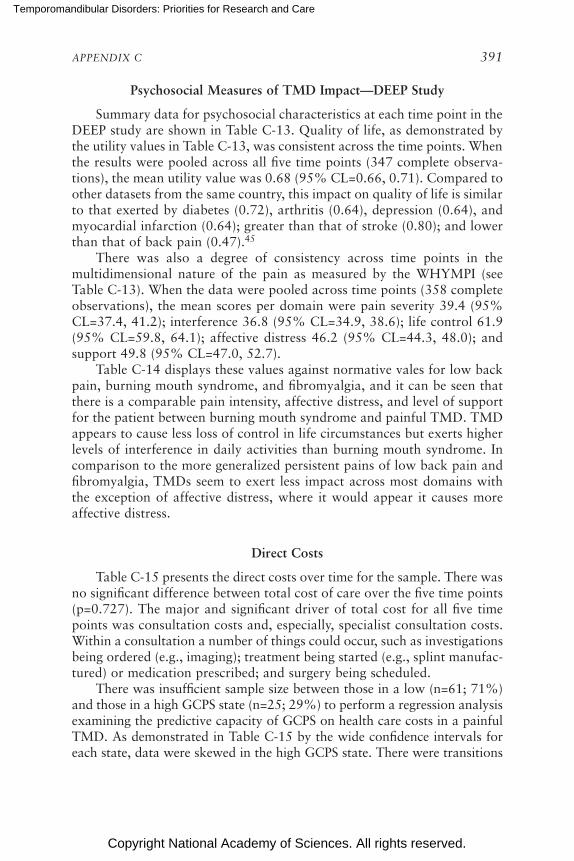
Temporomandibular Disorders: Priorities for Research and Care
Copyright National Academy of Sciences. All rights reserved.
APPENDIX C 391
Psychosocial Measures of TMD Impact—DEEP Study
Summary data for psychosocial characteristics at each time point in the DEEP study are shown in Table C-13. Quality of life, as demonstrated by the utility values in Table C-13, was consistent across the time points. When the results were pooled across all five time points (347 complete observa-tions), the mean utility value was 0.68 (95% CL=0.66, 0.71). Compared to other datasets from the same country, this impact on quality of life is similar to that exerted by diabetes (0.72), arthritis (0.64), depression (0.64), and myocardial infarction (0.64); greater than that of stroke (0.80); and lower than that of back pain (0.47).45
There was also a degree of consistency across time points in the multi dimensional nature of the pain as measured by the WHYMPI (see Table C-13). When the data were pooled across time points (358 complete observations), the mean scores per domain were pain severity 39.4 (95% CL=37.4, 41.2); interference 36.8 (95% CL=34.9, 38.6); life control 61.9 (95% CL=59.8, 64.1); affective distress 46.2 (95% CL=44.3, 48.0); and support 49.8 (95% CL=47.0, 52.7).
Table C-14 displays these values against normative vales for low back pain, burning mouth syndrome, and fibromyalgia, and it can be seen that there is a comparable pain intensity, affective distress, and level of support for the patient between burning mouth syndrome and painful TMD. TMD appears to cause less loss of control in life circumstances but exerts higher levels of interference in daily activities than burning mouth syndrome. In comparison to the more generalized persistent pains of low back pain and fibromyalgia, TMDs seem to exert less impact across most domains with the exception of affective distress, where it would appear it causes more affective distress.
Direct Costs
Table C-15 presents the direct costs over time for the sample. There was no significant difference between total cost of care over the five time points (p=0.727). The major and significant driver of total cost for all five time points was consultation costs and, especially, specialist consultation costs. Within a consultation a number of things could occur, such as investigations being ordered (e.g., imaging); treatment being started (e.g., splint manufac-tured) or medication prescribed; and surgery being scheduled.
There was insufficient sample size between those in a low (n=61; 71%) and those in a high GCPS state (n=25; 29%) to perform a regression analysis examining the predictive capacity of GCPS on health care costs in a painful TMD. As demonstrated in Table C-15 by the wide confidence intervals for each state, data were skewed in the high GCPS state. There were transitions
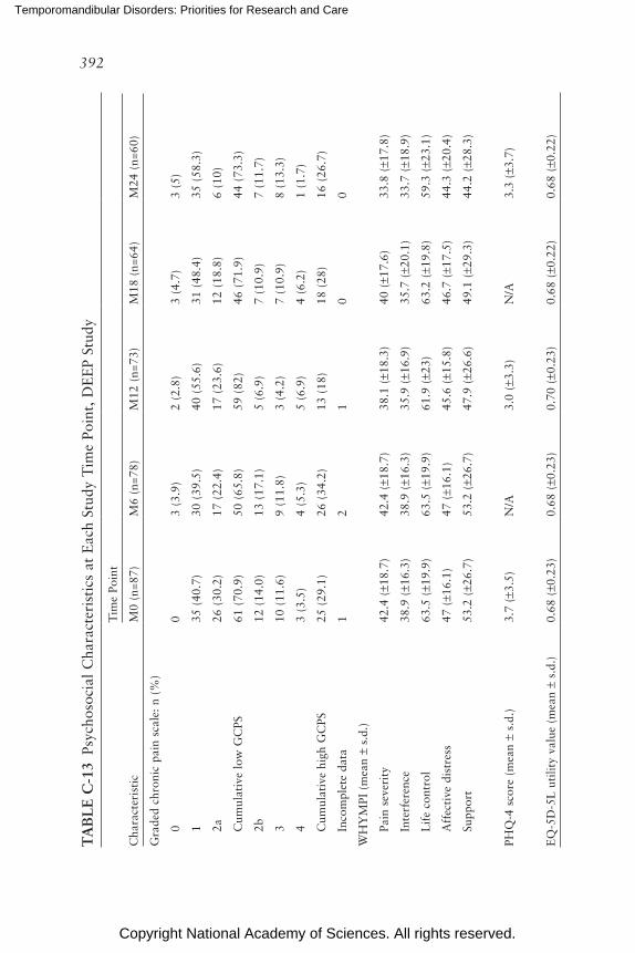
Temporomandibular Disorders: Priorities for Research and Care
Copyright National Academy of Sciences. All rights reserved.
392
TA
BL
E C
-13
Psyc
hoso
cial
Cha
ract
eris
tics
at
Eac
h St
udy
Tim
e Po
int,
DE
EP
Stud
y
Tim
e Po
int
Cha
ract
eris
tic
M0
(n=8
7)M
6 (n
=78)
M12
(n=
73)
M18
(n=
64)
M24
(n=
60)
Gra
ded
chro
nic
pain
sca
le:
n (%
)
00
3 (3
.9)
2 (2
.8)
3 (4
.7)
3 (5
)
135
(40
.7)
30 (
39.5
)40
(55
.6)
31 (
48.4
)35
(58
.3)
2a26
(30
.2)
17 (
22.4
)17
(23
.6)
12 (
18.8
)6
(10)
Cum
ulat
ive
low
GC
PS61
(70
.9)
50 (
65.8
)59
(82
)46
(71
.9)
44 (
73.3
)
2b12
(14
.0)
13 (
17.1
)5
(6.9
)7
(10.
9)7
(11.
7)
310
(11
.6)
9 (1
1.8)
3 (4
.2)
7 (1
0.9)
8 (1
3.3)
43
(3.5
)4
(5.3
)5
(6.9
)4
(6.2
)1
(1.7
)
Cum
ulat
ive
high
GC
PS25
(29
.1)
26 (
34.2
)13
(18
)18
(28
)16
(26
.7)
Inco
mpl
ete
data
12
10
0
WH
YM
PI (
mea
n ±
s.d.
)
Pain
sev
erit
y42
.4 (
±18.
7)42
.4 (
±18.
7)38
.1 (
±18.
3)40
(±1
7.6)
33.8
(±1
7.8)
Inte
rfer
ence
38.9
(±1
6.3)
38.9
(±1
6.3)
35.9
(±1
6.9)
35.7
(±2
0.1)
33.7
(±1
8.9)
Lif
e co
ntro
l63
.5 (
±19.
9)63
.5 (
±19.
9)61
.9 (
±23)
63.2
(±1
9.8)
59.3
(±2
3.1)
Aff
ecti
ve d
istr
ess
47 (
±16.
1)47
(±1
6.1)
45.6
(±1
5.8)
46.7
(±1
7.5)
44.3
(±2
0.4)
Supp
ort
53.2
(±2
6.7)
53.2
(±2
6.7)
47.9
(±2
6.6)
49.1
(±2
9.3)
44.2
(±2
8.3)
PHQ
-4 s
core
(m
ean
± s.
d.)
3.7
(±3.
5)N
/A3.
0 (±
3.3)
N/A
3.3
(±3.
7)
EQ
-5D
-5L
uti
lity
valu
e (m
ean
± s.
d.)
0.68
(±0
.23)
0.68
(±0
.23)
0.70
(±0
.23)
0.68
(±0
.22)
0.68
(±0
.22)
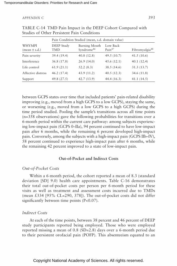
Temporomandibular Disorders: Priorities for Research and Care
Copyright National Academy of Sciences. All rights reserved.
APPENDIX C 393
TABLE C-14 TMD Pain Impact in the DEEP Cohort Compared with Studies of Other Persistent Pain Conditions
Pain Condition Studied (mean, s.d. domain value)
WHYMPI (mean ± s.d.)
DEEP Study TMD
Burning Mouth Syndrome46
Low Back Pain47 Fibromyalgia48
Pain severity 39.4 (19.4) 40.8 (12.8) 49.5 (10.7) 41.5 (10.6)
Interference 36.8 (17.8) 26.9 (14.0) 45.6 (12.1) 40.1 (12.4)
Life control 61.9 (21.1) 52.2 (8.3) 38.5 (14.6) 31.3 (13.7)
Affective distress 46.2 (17.4) 43.9 (11.2) 40.5 (12.3) 34.6 (11.8)
Support 49.8 (27.5) 42.7 (13.9) 44.4 (16.3) 41.1 (14.5)
between GCPS states over time that included patients’ pain-related disability improving (e.g., moved from a high GCPS to a low GCPS), staying the same, or worsening (e.g., moved from a low GCPS to a high GCPS) during the time period studied. Pooling the sample’s transitions across all time points (n=358 observations) gave the following probabilities for transitions over a 6-month period within the current care pathway: among subjects experienc-ing low-impact pain (GCPS 0–IIa), 94 percent continued to have low-impact pain after 6 months, while the remaining 6 percent developed high-impact pain. Conversely, among the subjects with a high-impact pain (GCPS IIb–IV), 58 percent continued to experience high-impact pain after 6 months, while the remaining 42 percent improved to a state of low-impact pain.
Out-of-Pocket and Indirect Costs
Out-of-Pocket Costs
Within a 6-month period, the cohort reported a mean of 8.3 (standard deviation [SD] 9.0) health care appointments. Table C-16 demonstrates their total out-of-pocket costs per person per 6-month period for these visits as well as treatment and assessment costs incurred due to TMDs (mean £334 [95% CL=290, 378]). The out-of-pocket costs did not differ significantly between time points (P<0.07).
Indirect Costs
At each of the time points, between 38 percent and 46 percent of DEEP study participants reported being employed. Those who were employed reported missing a mean of 0.8 (SD=2.8) days over a 6-month period due to their persistent orofacial pain (POFP). This absenteeism equated to an
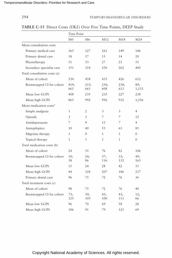
Temporomandibular Disorders: Priorities for Research and Care
Copyright National Academy of Sciences. All rights reserved.
394 TEMPOROMANDIBULAR DISORDERS
TABLE C-15 Direct Costs (UK£) Over Five Time Points, DEEP Study
Time Point
M0 M6 M12 M18 M24
Mean consultation costs
Primary medical care 165 127 161 149 146
Primary dental care 18 17 15 14 20
Physiotherapy 51 53 27 21 51
Secondary specialist care 351 314 256 262 445
Total consultation costs (a)
Mean of cohort 534 458 433 426 612
Bootstrapped CI for cohort 419; 661
253; 661
256; 608
238; 613
89; 1,133
Mean low GCPS 408 235 235 227 238
Mean high GCPS 865 992 956 932 1,556
Mean medication costs*
Simple analgesia 1 2 3 3 4
Opioids 3 3 7 7 12
Antidepressants 7 9 13 7 4
Antiepileptics 10 40 53 63 85
Migraine therapy 1 0 1 1 0
Topical therapy 1 1 1 1 0
Total medication costs (b)
Mean of cohort 24 55 76 82 106
Bootstrapped CI for cohort 10; 38
24; 86
37; 116
33; 132
49; 163
Mean low GCPS 15 24 28 42 55
Mean high GCPS 44 128 207 186 237
Primary dental care 96 75 72 76 36
Total treatment costs (c)
Mean of cohort 98 75 72 76 40
Bootstrapped CI for cohort 73; 125
50; 103
43; 100
43; 111
12; 66
Mean low GCPS 96 70 69 58 28
Mean high GCPS 106 91 79 125 69
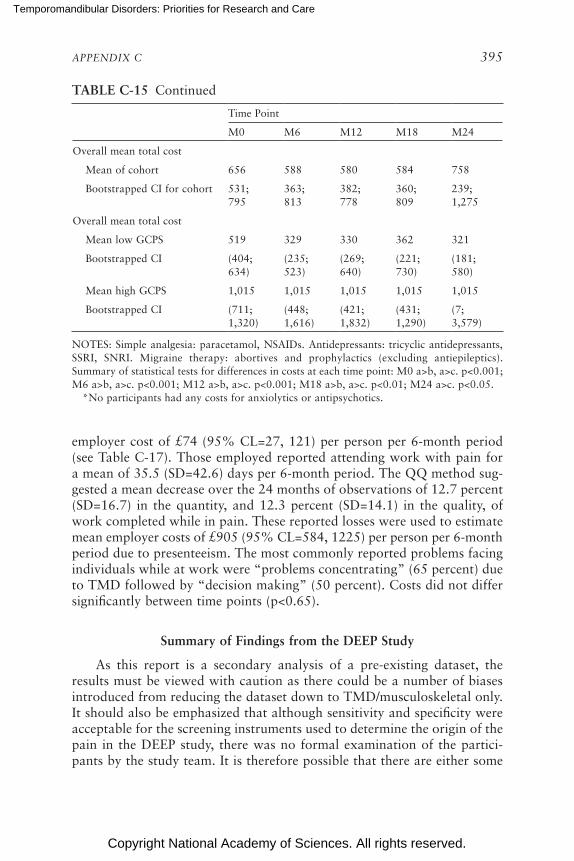
Temporomandibular Disorders: Priorities for Research and Care
Copyright National Academy of Sciences. All rights reserved.
APPENDIX C 395
Time Point
M0 M6 M12 M18 M24
Overall mean total cost
Mean of cohort 656 588 580 584 758
Bootstrapped CI for cohort 531; 795
363; 813
382; 778
360; 809
239; 1,275
Overall mean total cost
Mean low GCPS 519 329 330 362 321
Bootstrapped CI (404; 634)
(235; 523)
(269; 640)
(221; 730)
(181; 580)
Mean high GCPS 1,015 1,015 1,015 1,015 1,015
Bootstrapped CI (711; 1,320)
(448; 1,616)
(421; 1,832)
(431; 1,290)
(7; 3,579)
NOTES: Simple analgesia: paracetamol, NSAIDs. Antidepressants: tricyclic antidepressants, SSRI, SNRI. Migraine therapy: abortives and prophylactics (excluding antiepileptics). Summary of statistical tests for differences in costs at each time point: M0 a>b, a>c. p<0.001; M6 a>b, a>c. p<0.001; M12 a>b, a>c. p<0.001; M18 a>b, a>c. p<0.01; M24 a>c. p<0.05. *No participants had any costs for anxiolytics or antipsychotics.
TABLE C-15 Continued
employer cost of £74 (95% CL=27, 121) per person per 6-month period (see Table C-17). Those employed reported attending work with pain for a mean of 35.5 (SD=42.6) days per 6-month period. The QQ method sug-gested a mean decrease over the 24 months of observations of 12.7 percent (SD=16.7) in the quantity, and 12.3 percent (SD=14.1) in the quality, of work completed while in pain. These reported losses were used to estimate mean employer costs of £905 (95% CL=584, 1225) per person per 6-month period due to presenteeism. The most commonly reported problems facing individuals while at work were “problems concentrating” (65 percent) due to TMD followed by “decision making” (50 percent). Costs did not differ significantly between time points (p<0.65).
Summary of Findings from the DEEP Study
As this report is a secondary analysis of a pre-existing dataset, the results must be viewed with caution as there could be a number of biases introduced from reducing the dataset down to TMD/musculoskeletal only. It should also be emphasized that although sensitivity and specificity were acceptable for the screening instruments used to determine the origin of the pain in the DEEP study, there was no formal examination of the partici-pants by the study team. It is therefore possible that there are either some
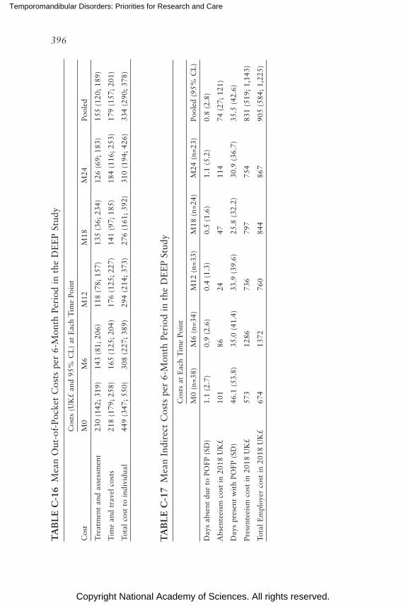
Temporomandibular Disorders: Priorities for Research and Care
Copyright National Academy of Sciences. All rights reserved.
396
TA
BL
E C
-16
Mea
n O
ut-o
f-Po
cket
Cos
ts p
er 6
-Mon
th P
erio
d in
the
DE
EP
Stud
y
Cos
ts (
UK
£ an
d 95
% C
L)
at E
ach
Tim
e Po
int
Cos
tM
0M
6M
12M
18M
24Po
oled
Tre
atm
ent
and
asse
ssm
ent
230
(142
; 31
9)14
3 (8
1; 2
06)
118
(78;
157
)13
5 (3
6; 2
34)
126
(69;
183
)15
5 (1
20;
189)
Tim
e an
d tr
avel
cos
ts
218
(179
; 25
8)16
5 (1
25;
204)
176
(125
; 22
7)14
1 (9
7; 1
85)
184
(116
; 25
3)17
9 (1
57;
201)
Tota
l co
st t
o in
divi
dual
449
(347
; 55
0)30
8 (2
27;
389)
294
(214
; 37
3)27
6 (1
61;
392)
310
(194
; 42
6)33
4 (2
90;
378)
TA
BL
E C
-17
Mea
n In
dire
ct C
osts
per
6-M
onth
Per
iod
in t
he D
EE
P St
udy
Cos
ts a
t E
ach
Tim
e Po
int
M0
(n=3
8)M
6 (n
=34)
M12
(n=
33)
M18
(n=
24)
M24
(n=
23)
Pool
ed (
95%
CL
)
Day
s ab
sent
due
to
POFP
(SD
)1.
1 (2
.7)
0.9
(2.6
)0.
4 (1
.3)
0.5
(1.6
)1.
1 (5
.2)
0.8
(2.8
)
Abs
ente
eism
cos
t in
201
8 U
K£
101
8624
4711
474
(27
; 12
1)
Day
s pr
esen
t w
ith
POFP
(SD
)46
.1 (
53.8
)35
.0 (
41.4
)33
.9 (
39.6
)25
.8 (
32.2
)30
.9 (
36.7
)35
.5 (
42.6
)
Pres
ente
eism
cos
t in
201
8 U
K£
573
1286
736
797
754
831
(519
; 1,
143)
Tota
l E
mpl
oyer
cos
t in
201
8 U
K£
674
1372
760
844
867
905
(584
; 1,
225)
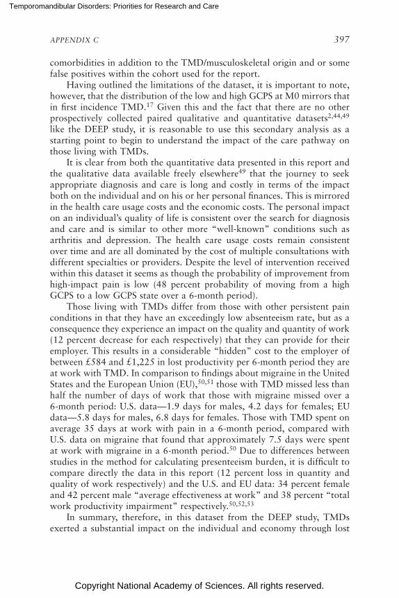
Temporomandibular Disorders: Priorities for Research and Care
Copyright National Academy of Sciences. All rights reserved.
APPENDIX C 397
comorbidities in addition to the TMD/musculoskeletal origin and or some false positives within the cohort used for the report.
Having outlined the limitations of the dataset, it is important to note, however, that the distribution of the low and high GCPS at M0 mirrors that in first incidence TMD.17 Given this and the fact that there are no other prospectively collected paired qualitative and quantitative datasets2,44,49 like the DEEP study, it is reasonable to use this secondary analysis as a starting point to begin to understand the impact of the care pathway on those living with TMDs.
It is clear from both the quantitative data presented in this report and the qualitative data available freely elsewhere49 that the journey to seek appro priate diagnosis and care is long and costly in terms of the impact both on the individual and on his or her personal finances. This is mirrored in the health care usage costs and the economic costs. The personal impact on an individual’s quality of life is consistent over the search for diagnosis and care and is similar to other more “well-known” conditions such as arthritis and depression. The health care usage costs remain consistent over time and are all dominated by the cost of multiple consultations with different specialties or providers. Despite the level of intervention received within this dataset it seems as though the probability of improvement from high-impact pain is low (48 percent probability of moving from a high GCPS to a low GCPS state over a 6-month period).
Those living with TMDs differ from those with other persistent pain conditions in that they have an exceedingly low absenteeism rate, but as a consequence they experience an impact on the quality and quantity of work (12 percent decrease for each respectively) that they can provide for their employer. This results in a considerable “hidden” cost to the employer of between £584 and £1,225 in lost productivity per 6-month period they are at work with TMD. In comparison to findings about migraine in the United States and the European Union (EU),50,51 those with TMD missed less than half the number of days of work that those with migraine missed over a 6-month period: U.S. data—1.9 days for males, 4.2 days for females; EU data—5.8 days for males, 6.8 days for females. Those with TMD spent on average 35 days at work with pain in a 6-month period, compared with U.S. data on migraine that found that approximately 7.5 days were spent at work with migraine in a 6-month period.50 Due to differences between studies in the method for calculating presenteeism burden, it is difficult to compare directly the data in this report (12 percent loss in quantity and quality of work respectively) and the U.S. and EU data: 34 percent female and 42 percent male “average effectiveness at work” and 38 percent “total work productivity impairment” respectively.50,52,53
In summary, therefore, in this dataset from the DEEP study, TMDs exerted a substantial impact on the individual and economy through lost
Temporomandibular Disorders: Priorities for Research and Care
Copyright National Academy of Sciences. All rights reserved.
398 TEMPOROMANDIBULAR DISORDERS
productivity and on the health care system due to disorganized care path-ways increasing the number of consultations required to achieve either diagnosis or care for the condition. Given that the data are from a national and linked health care service, which is free at the point of delivery, it is reasonable to assume that costs are not driven by profit. It may, however, be that in wholly privately delivered health care differences exist. Further research using representative datasets or cohorts within the United States will be required in order to fully understand the care pathways for TMDs.
REFERENCES
1. Slade GD, Ohrbach R, Greenspan JD, Fillingim RB, Bair E, Sanders AE, Dubner R, et al. Painful temporomandibular disorder: Decade of discovery from OPPERA studies. J Dent Res. 2016;95(10):1084-92. PMCID: PMC5004239.
2. Durham J, Shen J, Breckons M, Steele JG, Araujo-Soares V, Exley C, Vale L. Health-care cost and impact of persistent orofacial pain: The DEEP study cohort. J Dent Res. 2016;95(10):1147-54.
3. CDC/National Center for Health Statistics. Variance Estimation Guidance, NHIS 2016-2017. 2018. https://www.cdc.gov/nchs/data/nhis/2016var.pdf cited November 19, 2019.
4. Case A, Deaton A. Rising morbidity and mortality in midlife among white non- Hispanic Americans in the 21st century. Proc Natl Acad Sci USA. 2015;112(49):15078-83. PMCID: PMC4679063.
5. MacMahon B, Pugh TF, Ipsen J. Epidemiologic methods. Boston: Little, Brown and Company; 1960.
6. Von Korff M, Dworkin SF, Le Resche L, Kruger A. An epidemiologic comparison of pain complaints. Pain. 1988;32(2):173-83.
7. Maixner W, Fillingim RB, Williams DA, Smith SB, Slade GD. Overlapping chronic pain conditions: Implications for diagnosis and classification. J Pain. 2016;17(9 Suppl):T93-T107. PMCID: PMC6193199.
8. LeResche L, Mancl LA, Drangsholt MT, Saunders K, Von Korff M. Relationship of pain and symptoms to pubertal development in adolescents. Pain. 2005;118(1-2):201-9.
9. Riley JL, 3rd, Gilbert GH, Heft MW. Orofacial pain symptom prevalence: Selective sex differences in the elderly? Pain. 1998;76(1-2):97-104.
10. Horst OV, Cunha-Cruz J, Zhou L, Manning W, Mancl L, DeRouen TA. Prevalence of pain in the orofacial regions in patients visiting general dentists in the Northwest Practice-based REsearch Collaborative in Evidence-based DENTistry research network. J Am Dent Assoc. 2015;146(10):721-83.
11. Sanders AE, Jain D, Sofer T, Kerr KF, Laurie CC, Shaffer JR, Marazita ML, et al. GWAS identifies new loci for painful temporomandibular disorder: Hispanic community health study/study of Latinos. J Dent Res. 2017;96(3):277-84. PMCID: PMC5298397.
12. Janal MN, Raphael KG, Nayak S, Klausner J. Prevalence of myofascial temporo-mandibular disorder in US community women. J Oral Rehabil. 2008;35(11):801-9.
13. Bair E, Brownstein NC, Ohrbach R, Greenspan JD, Dubner R, Fillingim RB, Maixner W, et al. Study protocol, sample characteristics, and loss to follow-up: The OPPERA prospective cohort study. J Pain. 2013;14(12 Suppl):T2-T19. PMCID: PMC3855654.
14. Bueno CH, Pereira DD, Pattussi MP, Grossi PK, Grossi ML. Gender differences in temporo mandibular disorders in adult populational studies: A systematic review and meta-analysis. J Oral Rehabil. 2018;45(9):720-9.
Temporomandibular Disorders: Priorities for Research and Care
Copyright National Academy of Sciences. All rights reserved.
APPENDIX C 399
15. Slade GD, Sanders AE, Bair E, Brownstein N, Dampier D, Knott C, Fillingim R, et al. Preclinical episodes of orofacial pain symptoms and their association with health care behaviors in the OPPERA prospective cohort study. Pain. 2013;154(5):750-60. PMCID: NIHMS445953.
16. Collins SL, Moore RA, McQuay HJ. The visual analogue pain intensity scale: What is moderate pain in millimetres? Pain. 1997;72(1-2):95-7.
17. Dworkin SF, LeResche L. Research diagnostic criteria for temporomandibular disor-ders: Review, criteria, examinations and specifications, critique. J Craniomandib Disord. 1992;6(4):301-55.
18. Slade G, Bair E, Greenspan J, Dubner R, Fillingim R, Diatchenko L, Maixner W, et al. Signs and symptoms of first-onset TMD and sociodemographic predictors of its develop-ment: The OPPERA prospective cohort study. J Pain. 2013;14(12 Suppl):T20-T32.
19. Hannay DR. The “iceberg” of illness and “trivial” consultations. J R Coll Gen Pract. 1980;30(218):551-4. PMCID: PMC2159711.
20. Gracely RH, McGrath F, Dubner R. Ratio scales of sensory and affective verbal pain descriptors. Pain. 1978;5(1):5-18.
21. Meloto CB, Slade GD, Lichtenwalter RN, Bair E, Rathnayaka N, Diatchenko L, Greenspan JD, et al. Clinical predictors of persistent temporomandibular disorder in people with first-onset temporomandibular disorder: A prospective case-control study. J Am Dent Assoc. 2019;150(7):572-81.
22. Fillingim RB, Slade GD, Greenspan JD, Dubner R, Maixner W, Bair E, Ohrbach R. Long-term changes in biopsychosocial characteristics related to temporomandibular disorder: Findings from the OPPERA study. Pain. 2018;159(11):2403-13. PMCID: PMC6193833.
23. Diatchenko L, Slade GD, Nackley AG, Bhalang K, Sigurdsson A, Belfer I, Goldman D, et al. Genetic basis for individual variations in pain perception and the development of a chronic pain condition. Hum Mol Genet. 2005;14(1):135-43.
24. LeResche L, Mancl LA, Drangsholt MT, Huang G, Von Korff M. Predictors of onset of facial pain and temporomandibular disorders in early adolescence. Pain. 2007;129(3):269-78. PMCID: 1979093.
25. Von Korff M, Le Resche L, Dworkin SF. First onset of common pain symptoms: A prospective study of depression as a risk factor. Pain. 1993;55(2):251-8.
26. Interagency Pain Research Coordinating Committee. National pain strategy: A com-prehensive population health-level strategy for pain. In: U.S. Department of Health and Human Services NIoH, editor. Washington, DC; 2016.
27. Dahlhamer J, Lucas J, Zelaya C, Nahin R, Mackey S, DeBar L, Kerns R, et al. Prevalence of chronic pain and high-impact chronic pain among adults—United States, 2016. Morb Mortal Wkly Rep. 2018;67(36):1001-6. PMCID: PMC6146950.
28. Von Korff M, Ormel J, Keefe FJ, Dworkin SF. Grading the severity of chronic pain. Pain. 1992;50(2):133-49.
29. Dworkin SF, Huggins KH, Wilson L, Mancl L, Turner J, Massoth D, LeResche L, et al. A randomized clinical trial using research diagnostic criteria for temporomandibular disorders-axis II to target clinic cases for a tailored self-care TMD treatment program. J Orofac Pain. 2002;16(1):48-63.
30. Miller VE, Poole C, Golightly Y, Barrett D, Chen DG, Ohrbach R, Greenspan JD, et al. Characteristics associated with high-impact pain in people with temporomandibular disorder: A cross-sectional study. J Pain. 2019;20(3):288-300. PMCID: PMC6424335.
31. IASP. International Association for the Study of Pain - Pain Terminology 2007 cited October 30, 2007; February 22, 2007: Available from: http://www.iasp-pain.org/AM/Template.cfm?Section=Pain_Definitions&Template=/CM/HTMLDisplay.cfm&ContentID=1728#Pain.
Temporomandibular Disorders: Priorities for Research and Care
Copyright National Academy of Sciences. All rights reserved.
400 TEMPOROMANDIBULAR DISORDERS
32. Gonzalez Y, Schiffman E, Gordon S, Seago B, Truelove E, Slade G, Ohrbach R. Develop-ment of a brief and effective temporomandibular disorder pain screening questionnaire: Reliability and validity. J Am Dent Assoc. 2011;142(10):1183-91.
33. Hapak L, Gordon A, Locker D, Shandling M, Mock D, Tenenbaum H. Differentiation between musculoligamentous, dentoalveolar, and neurologically based craniofacial pain with a diagnostic questionnaire. J Orofac Pain. 1994;8(4):357-68.
34. Herdman M, Gudex C, Lloyd A, Janssen M, Kind P, Parkin D, Bonsel G, et al. Develop-ment and preliminary testing of the new five-level version of EQ-5D (EQ-5D-5L). Qual Life Res. 2011;20(10):1727-36.
35. Kerns R, Turk D, Rudy T. The West Haven-Yale Multidimensional Pain Inventory (WHYMPI). Pain. 1985;23(4):345-56.
36. Kroenke K, Spitzer R, Williams J, Lowe B. An ultra-brief screening scale for anxiety and depression: The PHQ-4. Psychosomatics. 2009;50(6):613-21.
37. Wordsworth S, Thompson S. An annotated cost questionnaire for patients: Results of piloting. HERU Dicussion Paper 2001;03/01.
38. Moss-Morris R, Weinman J, Petrie KJ, Horne R, Cameron LD, Buick D. The revised illness perception questionnaire (IPQ-R). Psychol Health. 2002;17(1):1-16.
39. Brouwer W, Koopmanschap M, Rutten F. Productivity losses without absence: Measure-ment validation and empirical evidence. Health Policy. 1999;48(1):13-27.
40. Cohen J. Statistical power analysis for the behavioral sciences (2nd ed.) New Jersey: Lawrence Erlbaum; 1988.
41. Green SB. How many subjects does it take to do a regression analysis. Multivariate Behav Res. 1991;26(3):499-510.
42. Epker J, Gatchel R, Ellis Er. A model for predicting chronic TMD: Practical application in clinical settings. J Am Dent Assoc. 1999;130(10):1470-5.
43. Garofalo JP, Gatchel RJ, Wesley AL, Ellis E, 3rd. Predicting chronicity in acute temporo-mandibular joint disorders using the research diagnostic criteria. J Am Dent Assoc. 1998;129(4):438-47. PMCID: 9573694.
44. Breckons M, Shen J, Bunga J, Vale L, Durham J. DEEP study: Indirect and out-of-pocket costs of persistent orofacial pain. J Dent Res. 2018;97(11):1200-6.
45. van Hout B, Janssen M, Feng Y, Kohlmann T, Busschbach J, Golicki D, Lloyd A, et al. Interim scoring for the EQ-5D-5L: Mapping the EQ-5D-5L to EQ-5D-3L value sets. Value Health. 2012;15(5):708-15.
46. Carlson C, Miller C, Reid K. Psychosocial profiles of patients with burning mouth syn-drome. J Orofac Pain. 2000;14(1):59-64.
47. Bernstein I, Jaremko M, Hinkley B. On the utility of the West Haven-Yale Multi-dimensional Pain Inventory. Spine (Phila Pa 1976). 1995;20(8):956-63.
48. Turk D, Okifuji A, Sinclair J, Starz T. Pain, disability, and physical functioning in sub-groups of patients with fibromyalgia. J Rheumatol. 1996;23(7):1255-62.
49. Breckons M, Bissett S, Exley C, Araujo-Soares V, Durham J. Care pathways in persis-tent orofacial pain: Qualitative evidence from the DEEP study. JDR Clin Trans Res. 2017;2(1):48-57.
50. Hu X, Markson L, Lipton R, Stewart W, Berger M. Burden of migraine in the United States: Disability and economic costs. Arch Intern Med. 1999;159(8):813-8.
51. Steiner T, Stovner L, Katsarava Z, Lainez J, Lampl C, Lantéri-Minet M, Rastenyte D, et al. The impact of headache in Europe: Principal results of the Eurolight project. J Headache Pain. 2014;15:31.
52. Stewart W, Lipton R, Simon D. Work-related disability: Results from the American migraine study. Cephalalgia. 1996;16(4):231-8; discussion 15.
Temporomandibular Disorders: Priorities for Research and Care
Copyright National Academy of Sciences. All rights reserved.
APPENDIX C 401
53. Vo P, Fang J, Bilitou A, Laflamme A, Gupta S. Patients’ perspective on the burden of migraine in Europe: A cross-sectional analysis of survey data in France, Germany, Italy, Spain, and the United Kingdom. J Headache Pain. 2018;19(1):82.
Potential conflicts of interest: Robert Kerns reports receiving honoraria for serving as a mem-ber of a research grant review board for the American Pain Society and as senior editor for the journal Pain Medicine. Michael Von Korff reports receipt of grants from Pfizer Inc. as an investigator of use and misuse of opioids at Kaiser Permanente Washington Health Research Institute and from inVentive as co-investigator for Food and Drug Administration–mandated postmarketing surveillance studies of extended release opioids. No other conflicts of interest were reported.

Temporomandibular Disorders: Priorities for Research and Care
Copyright National Academy of Sciences. All rights reserved.
Temporomandibular Disorders: Priorities for Research and Care
Copyright National Academy of Sciences. All rights reserved.
403
The musculoskeletal structures of the masticatory system and the neu-rological controls supporting a wide diversity of functions are complex, and much remains to be investigated (Gremillion and Klasser, 2018; see Chapter 4). The following description of the anatomy is intended to high-light the complexity of this system and some of its unique characteristics (see Figure D-1).
BONES
The three major bones of the masticatory system are the maxilla, or upper jaw; the mandible, or lower jaw; and the temporal bone, which is connected to the upper jaw and thereby forms the temporomandibular joint (TMJ) with the mandible. A minor bone, the hyoid, floats beneath the mandible and serves as an attachment point for muscles and ligaments that link the mandible with several other structures. One additional bone, the sphenoid, is fixed between the upper jaw and the temporal bone.
MASTICATORY MUSCLES
The muscles associated with mandibular movement are organized into five groups according to their major functions. Muscles in each group exist and function as pairs, one on the right side and one on the left side of the body:
Appendix D
Masticatory System: Anatomy and Function
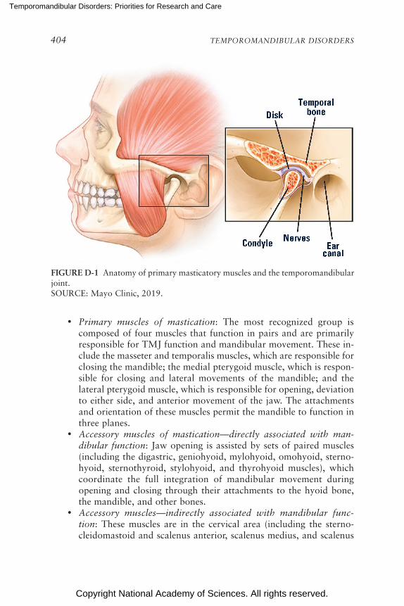
Temporomandibular Disorders: Priorities for Research and Care
Copyright National Academy of Sciences. All rights reserved.
404 TEMPOROMANDIBULAR DISORDERS
FIGURE D-1 Anatomy of primary masticatory muscles and the temporo mandibular joint.SOURCE: Mayo Clinic, 2019.
• Primary muscles of mastication: The most recognized group is composed of four muscles that function in pairs and are primarily responsible for TMJ function and mandibular movement. These in-clude the masseter and temporalis muscles, which are responsible for closing the mandible; the medial pterygoid muscle, which is respon-sible for closing and lateral movements of the mandible; and the lateral pterygoid muscle, which is responsible for opening, deviation to either side, and anterior movement of the jaw. The attachments and orientation of these muscles permit the mandible to function in three planes.
• Accessory muscles of mastication—directly associated with man-dibular function: Jaw opening is assisted by sets of paired muscles (including the digastric, geniohyoid, mylohyoid, omohyoid, sterno-hyoid, sternothyroid, stylohyoid, and thyrohyoid muscles), which coordinate the full integration of mandibular movement during opening and closing through their attachments to the hyoid bone, the mandible, and other bones.
• Accessory muscles—indirectly associated with mandibular func-tion: These muscles are in the cervical area (including the sterno-cleido mastoid and scalenus anterior, scalenus medius, and scalenus
Temporomandibular Disorders: Priorities for Research and Care
Copyright National Academy of Sciences. All rights reserved.
APPENDIX D 405
posterior muscles) and attach to the sternum, temporal bone, clavicle, cervical vertebrae 1–7, and ribs 1–2. These muscles stabilize the skull and neck and allow the mandible to move relative to the skull.
• Extrinsic muscles for the tongue: These muscles function to elevate, depress, withdraw, or protrude the tongue.
• Intrinsic muscles within the tongue: These muscles permit the tongue to create shapes (such as curling) and move from cheek to cheek.
Because the jaw moves in three dimensions (resulting in translational [gliding] and rotational movements), the muscle contraction necessary to create movement of the mandible (in particular, for chewing) must occur simultaneously with other contractions of the muscles to control the com-ponents of the TMJ. A healthy masticatory system appears to function effortlessly; by contrast, a compromised system can produce pain and dysfunction.
TEMPOROMANDIBULAR JOINTS
TMJs include the rounded end of the mandible (mandibular condyle), the edge of the temporal bone (glenoid fossa), and the articular disc that is positioned between the condyle and fossa. The temporomandibular liga-ment overlies the joint and provides some lateral stability. The joint itself is surrounded by a capsule that provides a small amount of stability and that also contains the synovial fluid. The synovial fluid provides essential nutrients and lubrication. Without this fluid there would be increased fric-tion and shear stress leading to disc degeneration.
The articular disc, located in the joint capsule, is shaped like a donut but is thinner in the middle and thicker on the edges. The disc is composed of fibrocartilage (which is different from the hyaline cartilage in other joints such as the knee or hip). The articular disc plays a major role in accommo-dating the movement of the jaw, absorbing shock, and distributing loads. The disc is attached on either side via ligaments to the mandible’s condyle. Blood supply to the joint and surrounding muscles is provided from the external carotid and maxillary arteries and its branches. The central portion of this disc does not contain a blood supply. The anterior and posterior of the disc are not firmly attached to the condyle; rather, these areas are free of firm attachment in order for the disc to pivot as the condyle moves. This lack of attachment may contribute to disc instability.
The articular disc separates the joint into two fluid-filled compart-ments. Each compartment has a different purpose. The lower compartment (the articular disc, mandibular condyle, and relevant ligaments and other structures) is involved in rotational movements. This rotational movement allows the mandible to move from a closed position to a partially open one.
Temporomandibular Disorders: Priorities for Research and Care
Copyright National Academy of Sciences. All rights reserved.
406 TEMPOROMANDIBULAR DISORDERS
The upper compartment (the articular disc, temporal bone, and relevant ligaments and other structures) is involved in the translational or gliding motion of the joint, in which the disc slides against the temporal bone and the condyle moves anteriorly down the slope of the joint, allowing further mouth opening. Normal joint motion transitions seamlessly from rotation to translation to provide maximal mouth opening.
In children, the primary growth center for the mandible lies immedi-ately below the fibrocartilaginous lining, making it susceptible to damage from pathological conditions. Throughout an individual’s life-span, the mandibular condyle and the articular disc are capable of adaptive remodel-ing to accommodate the functional demands of the masticatory system. Losing molars on one side without replacement, for example, will induce adaptive change in the condyles and discs on each side; this adaptive change is typically termed osteoarthrosis. When demands for adaptation exceed the biological capacity of an individual, a complex pattern of breakdown occurs in the cortical bone of the condyle, the articular disc, and the cap-sular and ligamentous tissues of the joint, which is termed osteoarthritis or degenerative joint disease.
INNERVATION
The innervation of the masticatory muscles (motor) and the TMJ (sensory) is derived from the mandibular branch of the trigeminal nerve, the largest of the cranial nerves. Surrounding tissues are innervated by nerve endings that allow the brain to monitor the effects of movement, which is important in initiating movement and controlling the mechanics of the joint. The sensory innervation of the accessory muscles includes both the trigeminal and cervical nerves. The blood supply to the joint and surrounding muscles is provided from the external carotid and maxillary arteries and their branches.
NEURAL CONTROL
The neurophysiological controls of mandibular sensation and move-ment are complex. The primary masticatory muscles contain motor units (a group of fibers that contract along with the motor nerve that controls the contraction of all of the fibers in the group) that are intermediate in size (500–1,000 fibers) between the very small units for muscles that control eye movements and the very large units for muscles in the lower limbs. A motor unit in the masseter muscle is about 5 mm in length and height and is interspersed with muscle fibers from other units. Through recruitment of multiple motor units, the masseter muscle can exert very high forces, and, via the small physical size of each unit as well as the initial recruitment of
Temporomandibular Disorders: Priorities for Research and Care
Copyright National Academy of Sciences. All rights reserved.
APPENDIX D 407
the motor units with the least number of fibers, the masseter muscle can also exert very low forces with incredible precision, such as during speech. The brain can selectively activate different parts of the primary masticatory muscles in order to achieve the desired movement. This selective activation also occurs as a form of adaptation in response to pain.
Movement is of three forms: voluntary (such as opening or closing the jaw), cyclic (during mastication), and reflex (which contributes to the refined control of both voluntary and cyclic movements). In addition to the motor cortex controlling voluntary mandibular movements, a well-studied area of the brainstem called the central pattern generator controls the cyclic masticatory movements of the jaw, face, and tongue during chew-ing. These movements are simultaneously under voluntary control. The central pattern generator receives input from the teeth (to protect the teeth from excessive loads), the muscle spindles (for muscle length and rate of change), tendons (as force output from the muscles), and the capsule of the TMJ (for position and load). The central pattern generator is highly adap-tive and can respond to immediate changes in food textures and to changes in the dental occlusion over time.
Reflex controls occur at muscle spindles and at brainstem interneurons. Muscle spindles contribute to the control of contraction, velocity, and mus-cle length. The reflex controls provide, for example, sustained and increas-ing force in order to bite into a hard food as well as interruptions in the chewing cycle if conditions change, such as encountering unexpected texture in a food bolus or biting the cheek. A jaw-opening reflex would normally be triggered by many of the sensory inputs activated during the closing phase of chewing; the central pattern generator actively suppresses that reflex in order for chewing to occur. Similarly, in response to informa-tion from the muscle spindles, a reflex activation of the closing muscles would occur during the rapid opening phase of chewing; the central pat-tern generator similarly actively suppresses the potential activation of the closing muscles. The presence of pain profoundly alters these internal controls; the muscle coordination decreases, and the reciprocal controls be-tween the opening and closing muscles during the chewing cycles lose their effective ness, contributing to a degradation of function during both simple movements of opening and closing and complex movements of chewing. Adaptations to these degradations—made so that a person can continue to chew, open, and close the mandible—lead to further demands on the muscles and, eventually, overuse.
The extensive number of orofacial sensory receptors encode peripheral information and inform the brain regarding how the teeth are about to con-tact or are contacting, the texture of the food bolus, mandibular position, and the acceleration and velocity of mandibular movement. This informa-tion serves in a feedback loop to allow further control of movement. The
Temporomandibular Disorders: Priorities for Research and Care
Copyright National Academy of Sciences. All rights reserved.
408 TEMPOROMANDIBULAR DISORDERS
sensory system is capable of considerable adaptation, such as adjusting to the substantial change in sensory feedback to the brain that comes from dentures and from dental implants (where the normal ligament connect-ing tooth to bone, which provides sensory information regarding contact to a tooth, has been lost). An older model of motor control suggested that pain exerted a necessary inhibition on motor contraction. However, motivated behavior and coping mechanisms can substantially modify the impact that pain may have in inhibiting movement, and muscle activity can be reorganized to compensate for pain as well. Intense or persistent stimulation from the periphery, such as from trauma to the jaw or sus-tained stretching of the masticatory muscles, can transform the function-ing of second-order neurons into central sensitization, which represents an increased level of excitability to subsequent stimuli. Clinically, this can appear as light touch to the face being experienced as pain, and the hyper-excitability of the nerves also leads to spontaneous activity and pain. The unmasking of neurons that were previously silent occurs with central sensitization, and the brain can misinterpret sensory inputs that may be unrelated to the original injury. These processes can persist even after the original injury heals and can be amplified by other regions of the brain (e.g., by fear of movement or by anxiety). These many processes will affect the functioning of the central pattern generator, altering mastication, leading to further dysregulation in mandibular movement as an attempt to continue to function, and thereby serve as contributing factors for the perpetuation of pain and alteration in TMJ mechanics in temporomandibular disorders.
CERVICAL SYSTEM
The cervical system has a critical role in both normal and abnormal functioning of the TMJ. Head posture, for example, will influence condylar position and therefore the arc of the open and closing movements of the mandible as well as where the upper and lower teeth make first contact. The joint adjacent to the TMJ is the joint between the skull and the first cervical vertebrae, and controlled jaw function requires participation by much more than just the muscles that directly move the jaw. The group of accessory muscles that are indirectly associated with mandibular func-tion attach to the sternum, temporal bone, clavicle, cervical vertebrae 1–7, and ribs 1–2. Their innervation includes C2–C8 and the spinal accessory nerve.
It is notable that the coordination between the masticatory and the cervical systems is substantially altered when pain is present in either sys-tem. The presence of a dependency between a masticatory system disorder and other disorders would likely shift the manifestation of the particular masticatory disorder from localized to multi-system.
Temporomandibular Disorders: Priorities for Research and Care
Copyright National Academy of Sciences. All rights reserved.
APPENDIX D 409
The stability of a joint is achieved at the expense of the possible range of motion allowed by that joint. Just as in the shoulder, the design of the human TMJ is a balance of structural stability and freedom of movement. The stability of the TMJ is achieved via coordinated action of the muscles of mastication, some of which extend to the chest and to the shoulder. Consequently, healthy function in the TMJ is achieved through a complex layering of muscle activity.
REFERENCES
Gremillion, H. A., and G. D. Klasser, eds. 2018. Temporomandibular disorders: A transla-tional approach from basic science to clinical applicability. Cham, Switzerland: Springer International Publishing.
Mayo Clinic. 2019. TMJ disorders. https://www.mayoclinic.org/ diseases-conditions/tmj/ symptoms-causes/syc-20350941 (accessed December 31, 2019).

Temporomandibular Disorders: Priorities for Research and Care
Copyright National Academy of Sciences. All rights reserved.