botulinum neurotoxin for pain management: insights from animal models
TRANSCRIPT
Toxins 2010, 2, 2890-2913; doi:10.3390/toxins2122890
toxinsISSN 2072-6651
www.mdpi.com/journal/toxins
Review
Botulinum Neurotoxin for Pain Management: Insights from
Animal Models
Flaminia Pavone * and Siro Luvisetto
CNR, Institute of Neuroscience-Roma, via del Fosso di Fiorano 64, I-00143 Roma, Italy;
E-Mail: [email protected]
* Author to whom correspondence should be addressed; E-Mail: [email protected];
Tel.: +39-06-501703272; Fax: +39-06-501703304.
Received: 18 November 2010; in revised form: 17 December 2010 / Accepted: 20 December 2010 /
Published: 21 December 2010
Abstract: The action of botulinum neurotoxins (BoNTs) at the neuromuscular junction has
been extensively investigated and knowledge gained in this field laid the foundation for the
use of BoNTs in human pathologies characterized by excessive muscle contractions.
Although much more is known about the action of BoNTs on the peripheral system,
growing evidence has demonstrated several effects also at the central level. Pain
conditions, with special regard to neuropathic and intractable pain, are some of the
pathological states that have been recently treated with BoNTs with beneficial effects. The
knowledge of the action and potentiality of BoNTs utilization against pain, with emphasis
for its possible use in modulation and alleviation of chronic pain, still represents an
outstanding challenge for experimental research. This review highlights recent findings on
the effects of BoNTs in animal pain models.
Keywords: botulinum toxin; SNARE; pain; animal model; analgesia; inflammatory pain;
chronic pain; peripheral sensitization; central sensitization; retrograde axonal transport
1. Historical Overview: From Sausages to Universal Drug
Botulinum neurotoxins (BoNTs) are produced by anaerobic bacteria of the genus Clostridium and
are the most poisonous biological substances known [1,2]. BoNTs are responsible for botulism, a
neuroparalytic syndrome characterized by flaccid paralysis, originating in the peripheral nervous
OPEN ACCESS

Toxins 2010, 2
2891
system, and by dysfunctions of the autonomic nervous system [3–6]. In food borne botulism, the most
common form of botulism, BoNTs are ingested with food in which spores have germinated and the
organism has grown. Food-borne botulism has accompanied mankind since the beginning (see [7] for
extensive review). At the end of the 18th century, some well-documented outbreaks of sausage
poisoning in Württemberg, Southern Germany, prompted early systematic botulinum toxin research.
Between 1817 and 1822, the German poet and district medical officer Justinus Kerner published the
first accurate and complete description of the symptoms of food-borne botulism. He did not succeed in
defining the suspected biological poison that he called ―sausage poison‖ or ―fatty poison‖; however, he
developed the idea of a possible therapeutic use of the toxin. In 1895, Emile Pierre van Ermengem,
Professor of bacteriology at the University of Ghent, following a botulism outbreak after a funeral
dinner with smoked ham in the small Belgian village of Ellezelles, discovered the pathogen anaerobic
gram positive bacterium Clostridium botulinum. The bacterium was so called because of its
pathological association with the sausages (Latin word for sausage = botulus).
It was not until the middle of 20th century that BoNTs were used as therapeutic drugs in medicine.
Research began when Dr. Edward J. Schantz and his colleagues were able to purify botulinum toxin
serotype A (BoNT/A) into crystalline form. In 1953, the physiologist Dr. Vernon Brooks discovered
that the injection of small amounts of BoNT/A into a hyperactive muscle blocked the release of
acetylcholine (ACh) from motor nerve endings, causing temporary ―relaxation‖. In the 1960s, the
ophthalmologist Dr. Alan B. Scott started treating monkeys with BoNT/A, theorizing that its
muscle-relaxing effects might help in the treatment of crossed eyes (or strabismus). In the 1980s, Alan
Scott introduced the use of BoNT/A as an alternative to conventional surgery to treat human
strabismus. After these pioneering studies, therapeutic applications of BoNT/A were extended to a
wide variety of neurological disorders associated to spasmodic muscle contractions, for example, the
spasmodic torticollis, blefarospams, facial emispams, and dystonia [8]. Nowadays, the clinical
indications for BoNT/A are constantly growing, ranging from treatment of overactive skeletal and
smooth muscles, to management of hypersecretory and painful disorders such as migraine, trigeminal
neuralgia and the myofascial pain syndrome [9–11].
This review is focused on studies in which BoNTs, mainly BoNT/A, were used as pharmacological
treatment against pain in animal models.
2. Cellular Mechanism: General Considerations and Different BoNTs Serotypes
From the plethora of toxins produced by Clostridium botulinum, only toxins whose action is
restricted to the nervous system, such as BoNTs, are strictly defined as neurotoxins. Other toxins, such
as botulinolisin, toxin-C2, exoenzyme-C3, which target different type of cells, mostly non-neuronal,
are not included in the BoNTs family. Seven different serotypes of BoNTs have so far been
characterized (A-G), and these serotypes are active on many different types of vertebrates [3,4].
BoNTs are secreted as multimolecular complexes together with non toxic accessory proteins
(emagglutinins) that do not contribute to the paralyzing effects of BoNTs but protect them from the
passage through the acidic and proteolytic environment of the stomach after the ingestion of
spore-contaminated food [12,13].
Toxins 2010, 2
2892
BoNTs are proteins of about 1,300 amino acids and consist of three domains of similar size
(50 kDa). The NH2-terminal domain, which is named L-chain (L = light) domain, is a zinc
endopeptidase that represents the catalytic domain expressing the protease activity. The other two
domains, which are covalently bound to form the H-chain (H = heavy), are the central domain,
responsible for the membrane translocation of the L-chain into the neuronal cytosol, and the
COOH-terminal domain, which consists of two equally sized subdomains, responsible for the
neurospecific binding [14–16].
The cellular action of BoNTs occurs as a four-step mechanism: (i) binding of BoNTs on the
neuronal presynaptic membrane, via interaction with gangliosides, synaptic vesicle protein 2 and/or
synaptotagmin, depending on the serotype [16–18]; (ii) internalization of BoNTs via endocytosis of
the BoNTs-receptor complex inside the neurons; (iii) translocation of BoNTs L-chain from
endocytosed vesicle to the neuronal cytosol; and, finally, (iv) zinc-endopeptidase activity on cellular
targets [13,14,25]. Intracellular targets of BoNTs are three proteins involved in neuroexocytosis of the
neurotransmitter synaptic vesicles. These proteins are: SNAP-25 (synaptosomal associated protein
of 25 kDa), VAMP (vesicle associated membrane protein), also called synaptobrevin, and sintaxin. All
these proteins are involved into assembly of the SNARE (soluble N-ethylmaleimide-sensitive factor
attachment protein receptors) protein core complex, which is fundamental for correct docking and
fusion of neurotransmitter vesicles with neuronal membranes [13–20]. The peculiar characteristic of
BoNTs resides in their high affinity for one of the three SNARE proteins: SNAP-25 is cleaved by
BoNT/A (the serotype most commonly used in clinical practice), /E and /C; VAMP/sinaptobrevin is
the target of BoNT/B (another serotype used in clinic), /D, /F and /G; and sintaxin is cleaved only by
BoNT/C. The cleavage of one of these proteins is sufficient to prevent the correct assembly of the
SNARE core complex and the consequent fusion of synaptic vesicles with the neuronal presynaptic
membrane, thus inhibiting the neurotransmitter release [19,20]. This effect is reversible, and the
duration of action is dependent on the serotype. Further details on binding, internalization and mode of
action of the different BoNTs serotypes can be found in other reviews [21–26].
3. Beyond Muscular Effects: Involvement of Molecules Modulating Pain
Studies on the pathophysiology of botulism revealed that neuromuscular paralysis is due to
selective inhibition of evoked ACh release from cholinergic nerve endings at the skeletal
neuromuscular junction [1]. This canonical effect of BoNTs gave the opportunity to use them as
therapeutic agents in a variety of neurological disorders due to hyperfunctionality of cholinergic
terminals [8]. However, BoNTs cannot be considered exclusively as ‗cholinergic‘ toxins. Though they
act preferentially on nerve terminals between motoneurons and muscle fibers, BoNTs can also block
the neural transmission at other peripheral synapses, cholinergic or not [9]. This has indirectly been
proved also by symptoms observed during botulinal neuromuscular paralysis, where autonomic
nervous system dysfunction (parasympathetic, i.e., cholinergic, and sympathetic, i.e., adrenergic and
noradrenergic) coexists with neuro-motor alterations [4,13].
Extensive in vitro and in vivo studies demonstrated that BoNTs (mainly BoNT/A) are able to block
the Ca2+
-evoked release of neurotransmitters, other than ACh [27]. As for examples, in vitro studies
demonstrated that BoNTs inhibit various neurotransmitters‘ release, including glutamate, GABA,

Toxins 2010, 2
2893
aspartate, catecholamine, and noradrenaline, from cerebral synaptosomes, chromaffin cells, central
neurons and hippocampal slice cultures [28–38]. In vivo studies, mainly using microdialysis
techniques, demonstrated that BoNTs block both dopamine and monoamine release under various
conditions [39–42]. Moreover, BoNTs block glutamate release also from non-neuronal cells, such as
astrocytes [43–45]. Finally, there are many evidences in literature that BoNTs are able to block
the release not only of classical neurotransmitters but also of neuropetides, such as Substance P
(SP) [46–49] and calcitonin gene-related peptide (CGRP) [50–55]; neuropeptides whose role in pain
modulation is known. These latter findings gave strong input in searches for alternative therapy against
trigeminal neuralgia and various types of headache and migraine—neurological diseases where SP and
CGRP release often act as a trigger point to exacerbate disease attacks. In these pathologies, BoNT/A
probably exerts analgesia by blocking the neuropeptides‘ release at the nociceptive nerve endings.
Some encouraging results have been obtained in humans [56,57]. The effect of BoNT/A in
tension-type headache, chronic daily headache, and/or migraine is not further considered in the
present review.
BoNT/A is effective in relieving pain in spastic and non-spastic muscle conditions in humans. This
analgesic effect has generally been attributed to muscular relaxation. Notwithstanding, there are
reports in the literature stating that patients experience pain relief shortly after BoNT/A
treatment [58,59], i.e., before of any muscle-relaxing action of the toxin, or that the pain relief is still
maintained after muscle power returned to normality [60]. These results suggest that the analgesia
attributed to BoNT/A may be due to more complex mechanisms than the simple muscular relaxation.
In such cases, the pain relief cannot be ascribed to abolition of those factors, such as muscle tone,
excessive muscle contraction and spasms, which are due to muscle hyperactivity [61]. In the next
sections, we will discuss the evidences from animal models in favor of an analgesic action of BoNTs,
with particular emphasis on pain not directly related to abnormal muscle hyperactivity, such as
inflammatory and neuropathic pain. Animal model studies have been mainly performed with BoNT/A
and /B and have been conducted by using the 150 kDa purified toxin or equivalent commercial products.
In the following, we will indicate BoNT/A, or /B, indifferently from which preparation has been
actually utilized.
4. Animal Models Predicting the Therapeutic Use of BoNTs against Pain
Generally, pain is divided into nociceptive and pathological pain. Nociceptive pain is caused by the
sustained activation of peripheral nociceptors in response to peripheral tissue injury and can be
classified as somatic or visceral pain according to where the pain arises. The extent of nociceptive pain
normally reflects the extent of tissue damage and it disappears when the origin of nociceptive stimuli
disappears. On the other hand, pathological pain often evolves toward a chronic condition in which
pain exceeds the extent of tissue damage. Independently from the organs or tissues involved, chronic
pain may be a consequence of either inflammatory processes (for example, rheumatoid arthritis in
humans) and/or injury to the nervous system, both peripheral (PNS) (for example, postherpetic
neuralgia) and central (CNS) (for example, spinal cord injury) nervous system, which is commonly
known as neuropathic pain. A common feature of pain-related mechanisms is the activation of
sensitization processes, plastic changes in PNS and CNS (adaptative or pathological) that lead to an
Toxins 2010, 2
2894
enhanced response and/or decreased threshold to nociceptive stimuli. For example, under neuropathic
pain conditions, both hyperalgesia (an exaggerated pain perception in response to a painful stimulus)
and allodynia (the perception as painful of a stimulus that does not usually provoke pain) are
commonly developed. In basic science, many animal pain models have been developed in order to
mimic human pain conditions. In the next paragraphs of this section, an overview of the studies
involving the use of BoNTs in animal pain models will be presented and relevant results discussed.
4.1. Peripheral Inflammatory Pain Model
A direct involvement of BoNTs in mechanisms involved in pain modulation was first described by
Cui et al. [62], who analyzed the effects of BoNT/A in formalin-induced inflammation as an animal
model of inflammatory pain. Formalin-induced inflammatory pain model is extensively used for the
evaluation of possible analgesic effects of test compounds. It is performed by injecting a diluted
formalin solution into one hindpaw of rats, or mice, and by recording the formalin-evoked behaviors.
The most characteristic behavioral response of rodents to formalin injection is the licking of the
injected paw. This response appears biphasic, with an early phase of extensive licking due to
peripheral sensitization of nociceptors, followed by an interphase with almost absence of licking
activity due to activation of inhibitory descending pathways, and finally by a second phase of
extensive licking, which essentially reflects the progression of peripheral inflammation together with
the activation of central sensitization processes [63]. Cui et al. [62] found that a single peripheral
subcutaneous injection of BoNT/A was able to reduce licking activity during the second phase, in a
dose-dependent manner, with the absence of any obvious muscle weakness at doses below 30 U/kg.
BoNT/A, administered at different time intervals prior to formalin testing, induced long-lasting
analgesic effects, starting within the first 24 hours and lasting at least 12 days, the latest time point
tested. The reduction of licking activity was accompanied by the inhibition of formalin-induced
peripheral release of glutamate in the rat paw, and by the reduction of paw edema that is usually
observed as a consequence of neurogenic inflammatory processes. This latter effect suggests that
BoNT/A may reduce not only the release of glutamate, but also of SP and CGRP (not directly
measured by the authors), involved in SP-mediated plasma extravasation and CGRP-mediated
vasodilatation, respectively. Interestingly, BoNT/A did not reduce the initial phase of licking behavior
and thermal sensitivity, indicating no effects of BoNT/A injection on acute pain. The authors
postulated that BoNT/A is able to exert analgesic effects on formalin-induced inflammatory pain by
inhibiting the peripheral stimulation-induced release of neurotransmitters, such as glutamate, and
probably also of the neuropeptides SP and CGRP. Under this view, the analgesic activity of BoNT/A
has been considered as a consequence of the inhibition of peripheral sensitization, resulting in indirect
reduction of central sensitization processes [64,65].
Luvisetto et al. in mice [66] have essentially confirmed results of Cui et al. in rats [62]. These
authors extended the analysis of BoNTs on formalin-induced inflammatory pain by considering for the
first time the central (intracerebroventricular) versus peripheral (subcutaneous into hindpaw) effects.
Moreover, they investigated two BoNTs serotypes, namely BoNT/A and BoNT/B. The authors
demonstrated that pre-treatment (3 days) with BoNT/A is able to reduce the second phase of formalin
independently from the route of administration, demonstrating that BoNT/A may act not only
Toxins 2010, 2
2895
peripherally but also at the central level. On the contrary, BoNT/B did not reduce licking activity and,
when centrally injected, had a hyperalgesic effect on the interphase of the formalin test. These results
indicate a different mechanism of action of BoNTs, depending on the serotype and route of
administration, and suggest that BoNT/A and BoNT/B are not interchangeable based on simple dose
ratio, a point that is extremely important to take into account in a therapeutic perspective. Moreover,
these observations were fundamental for the comprehension of the mechanism involved in the action
of BoNTs: BoNT/A and BoNT/B may interact with inhibitory and/or excitatory systems in modulating
persistent inflammatory pain. The different effects of the two serotypes may reflect differences in the
expression of receptors and cellular targets of BoNT/A and BoNT/B (i.e., SNAP-25 and
VAMP/synaptobrevin, respectively), as well as in the expression of these targets into different
neuronal populations.
A series of studies [67–70] showed that the vesicle recycling in hippocampal glutamatergic neurons
is blocked by BoNT/A and BoNT/B while the same processes in hippocampal GABAergic neurons are
blocked by BoNT/B but not by BoNT/A. Staining with various antibodies directed against SNAP-25
reveals the specific presence of this protein in glutamatergic neurons, while GABAergic neurons lack
immunoreactivity for SNAP-25. In contrast, staining with antibodies directed against SNAP-23, a
homolog of SNAP-25, shows its presence in both glutamatergic and GABAergic neurons. BoNT/A
does not cleave SNAP-23 and this explains why BoNT/A is not able to block GABAergic neurons. On
the other hand, BoNT/B acting on VAMP/synaptobrevin—another protein of the SNARE complex—is
able to block GABAergic neurons. Considering all these pieces of evidence, the effect of BoNT/B on
the interphase of the formalin test could be partially ascribed to a functional block of GABA inhibition
on the primary afferent fibers in spinal dorsal horn.
The study on the analgesic effects of BoNT/A on inflammatory pain was not restricted to
formalin-induced pain and inflammation but was extended also to other models of inflammatory pain,
such as that induced by carrageenan and capsaicin. Peripheral application of carrageenan or capsaicin
produces hypersensitivity to thermal and mechanical stimuli as a consequence of inflammation due to
injected chemicals irritants. Carrageenan promotes inflammation by activating proinflammatory cells
and the resulting inflammatory edema is more extensive than edema induced by formalin. It causes
hyperalgesia by promoting the peripheral release of mediators such as SP, glutamate, prostaglandins,
histamine and serotonin, as well as release of glutamate, aspartate, SP, CGRP, nitric oxide, and
prostaglandin E2 (PGE2) in the dorsal horn of lumbar spinal cord. On the other hand, capsaicin excites
sensory neurons directly by acting on vanilloid receptors type 1 (VR-1), mostly expressed on C-fibers.
VR-1 receptors are present on nerve fibers containing SP and CGRP that are released from nerve
terminals after capsaicin application. Bach-Rojecky and Lacković [71] found that pre-treatment
(6 days) with BoNT/A significantly reduced or completely abolished the enhanced sensitivity to
mechanical and thermal stimuli provoked by peripheral carrageenan or capsaicin injections in rats,
demonstrating the efficacy of peripheral BoNT/A pretreatment on the pain component of inflammatory
process in experimental animals. In contrast with Cui et al. [62], BoNT/A had no effect on the
carrageenan-induced paw edema. Following this last observation, in another study, Back-Rojecky
et al. [72] analyzed if the analgesic effect of BoNT/A on carrageenan- and capsaicin-inflammatory
pain was directly due to the anti-nociceptive effect or indirectly due to the anti-inflammatory effect.
The authors confirmed that pre-treatment with the same dose of BoNT/A effective in reducing

Toxins 2010, 2
2896
pain [71], had no effect on the size of carrageenan-induced paw edema, measured as paw volume and
weight, or on the capsaicin-induced plasma extravasations (PE), measured by Evans blue as a marker
of protein leakage. From this finding, they concluded that BoNT/A does not have a significant
anti-inflammatory effect on the inflammation induced by capsaicin and carrageenan, indicating a
possible dissociation between the effect of BoNT/A on pain and on inflammation: only the former was
affected by BoNT/A through the inhibition of the release of neurotransmitters from the peripheral
endings of sensory nerves. Similar results were obtained also by Favre-Guilmard [73], who reported
that BoNT/A was not able to reduce carrageenan edema, indicating that the antihyperalgesic effects of
BoNT/A are independent of an anti-inflammatory activity related to increased vascular permeability
and adherence/infiltration of inflammatory cells. On the contrary, Carmichael et al. [74] evidenced
contrasting results on the anti-inflammatory effects of BoNT/A on neurogenic inflammation. These
authors found that peripheral subcutaneous application of BoNT/A reduced vasodilatation and PE
evoked by saphenous nerve stimulation or topical administration of capsaicin in the rat hindpaw skin,
while it had no effect on SP-induced PE or CGRP-induced vasodilatation.
Although some controversies appear to be still unsolved, studies presented in this paragraph are
clearly in favor of BoNT/A as a powerful drug against inflammatory pain and future research is
necessary to better elucidate the mechanisms involved.
4.2. Visceral Pain Model
The effect of BoNTs on visceral pain models has not been extensively studied. However, the
efficacy of BoNTs on relieving visceral pain has been analyzed in models mimicking pain symptoms
due to lower urinary tract disorder in humans, such as that due to bladder hyperactivity and prostatitis.
It was observed that intravesicular administration of BoNT/A produced analgesia against acetic
acid-induced bladder pain in rats by inhibiting the CGRP release from afferent nerve terminals [75].
In another model, namely the cyclophosphamide-induced cystitis in the bladder of rats [76], BoNT/A
inhibited the cyclooxygenase 2 (COX-2) and the prostaglandin EP(4) receptor expression and
suppressed bladder hyperactivity. Moreover, in a model of prostatitis, obtained by intraprostatic
capsaicin injection in rats, BoNT/A inhibited COX-2 expression and suppressed prostatic pain [77].
These studies in animal models (see also [54]) have paved the way toward an extensive therapeutic
application of BoNT/A in clinical treatment of human disorder related to lower urinary tract
disorder [78].
4.3. Neuropathic Pain Model
Neuropathic pain is a kind of chronic pain resulting from injury to the peripheral or central nervous
system. Common examples of neuropathic pain include postherpetic neuralgia, diabetic neuropathy,
complex regional pain syndrome, and pain associated with spinal cord injuries. Evidence for utility of
BoNT/A in relieving neuropathic pain symptoms in humans was first reported by Klein [79],
who demonstrated the effectiveness of BoNT/A in four clinical cases in alleviating symptoms of
different neuropathic pain, such as relapsing-remitting multiple sclerosis, postherpetic neuralgia,
peripheral neuropathy, and severe tingling caused by herniation of cervical vertebrae at the level of C8.
Since this initial observation, the action of BoNT/A in neuropathic pain has been also the focus of

Toxins 2010, 2
2897
experimental studies with the aim to better define its role as an analgesic and to investigate the neural
mechanisms involved.
Many models of neuropathic pain are currently in use both in rats and mice to mimic human
peripheral neuropathic conditions [80]. Depending on the site of injury, the animal models of
neuropathic pain are generally subdivided in central pain models (mainly based on spinal cord injury),
peripheral nerve injury models (mainly injuries at the level of sciatic or spinal nerves), and peripheral
neuropathy induced by diseases (postherpetic neuralgia and diabetic neuropathic pain models,
chemotherapy-induced peripheral neuropathy models) [81–86]. Thermal and mechanical hyperalgesia
as well as allodynia are hallmark properties of the experimental neuropathies.
The effect of BoNT/A on neuropathic pain was first analyzed on the peripheral neuropathy induced
by partial sciatic nerve transection in rats [87]. In this study, a single peripheral injection of BoNT/A
was sufficient to reduce both thermal and mechanical hyperalgesia without any apparent muscle
weakness. The effect of BoNT/A became evident five days after toxin application and lasted for more
than 10 days. In accordance with previous observations in other pain models [62,66], the authors found
that BoNT/A had no effects on thermal and mechanical thresholds of sham operated rats, confirming
that BoNT/A per se does not change nociception.
A second neuropathic pain model, used to test the efficacy of BoNT/A as an analgesic, was the
L5/L6 spinal nerve ligation (SNL). In this study, Park et al. [88] considered allodynia rather than
hyperalgesia as a behavioral parameter to assess the peripheral neuropathy. The authors found that a
single peripheral administration (into the plantar skin of one hindpaw) of BoNT/A, dose-dependently
reduced both mechanical and cold allodynia in SNL neuropathic rats.
Finally, a third animal model of peripheral neuropathy, the chronic constriction injury (CCI) of the
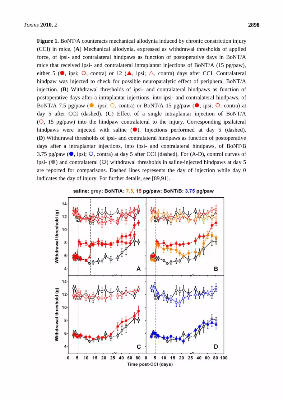
sciatic nerve, was considered in some recent studies [89–91]. In these studies, authors found that a
single injection of BoNT/A into plantar surface of mice injured paws, performed at five or 12 days
after CCI, markedly antagonized mechanical allodynia induced by CCI [Figure 1(A)]. This effect was
already evident 24 hours after BoNT/A injection, lasted for at least three weeks, and was
dose-dependent [Figure 1(B)]. Interestingly, differently from BoNT/A treatment post surgery, if the
BoNT/A injection preceded (3 days) the CCI, the antiallodynic effect was absent. These later findings
underline the difference to what is observed in inflammatory pain models where BoNT/A is able to
inhibit the occurrence of inflammatory process when injected prior to painful stimulus [62,66,71,72].
BoNT/A was able to reduce neuropathic symptoms only after neuropathy was already established but
was unable to preventively protect against the onset of neuropathy. If BoNT/A was injected
contralaterally to the lesion, anti-allodynic effects were not observed [Figure 1(C)]—an important
point demonstrating the absence of a systemic diffusion in these experimental conditions. By using the
same neuropathic pain model, authors observed that peripheral administration of BoNT/B did not exert
antiallodynic effects [90] [Figure 1(D)]. This is in agreement with previous research of the same
authors showing different effects of the two serotypes in inflammatory pain models [66]. It should be
remembered that usable doses of BoNT/B are lower than those of BoNT/A since the serotype B shows
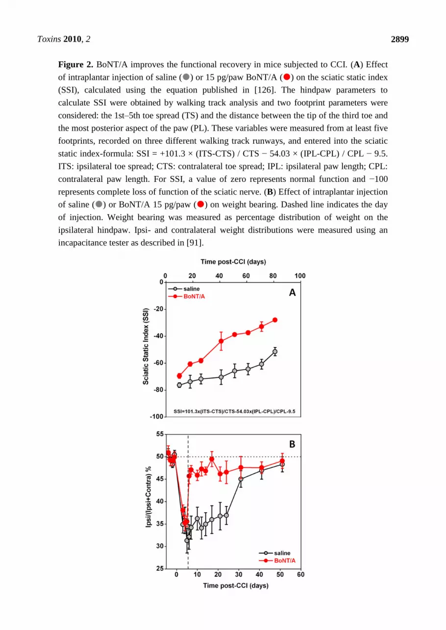
a higher toxicity, thus limiting its use [66,90]. Furthermore, Marinelli et al. [91] observed that BoNT/A
significantly improved the functional recovery of the injured paw, as assessed in mice by the sciatic
static index of functional recovery, examined through the footprint walking tracks of both ipsi- and
contralateral paws [Figure 2(A)], and by the weight-bearing incapacitance test [Figure 2(B)].
Toxins 2010, 2
2898
Figure 1. BoNT/A counteracts mechanical allodynia induced by chronic constriction injury
(CCI) in mice. (A) Mechanical allodynia, expressed as withdrawal thresholds of applied
force, of ipsi- and contralateral hindpaws as function of postoperative days in BoNT/A
mice that received ipsi- and contralateral intraplantar injections of BoNT/A (15 pg/paw),
either 5 (, ipsi; , contra) or 12 (, ipsi; , contra) days after CCI. Contralateral
hindpaw was injected to check for possible neuroparalytic effect of peripheral BoNT/A
injection. (B) Withdrawal thresholds of ipsi- and contralateral hindpaws as function of
postoperative days after a intraplantar injections, into ipsi- and contralateral hindpaws, of
BoNT/A 7.5 pg/paw (, ipsi; , contra) or BoNT/A 15 pg/paw (, ipsi; , contra) at
day 5 after CCI (dashed). (C) Effect of a single intraplantar injection of BoNT/A
(, 15 pg/paw) into the hindpaw contralateral to the injury. Corresponding ipsilateral
hindpaws were injected with saline (). Injections performed at day 5 (dashed).
(D) Withdrawal thresholds of ipsi- and contralateral hindpaws as function of postoperative
days after a intraplantar injections, into ipsi- and contralateral hindpaws, of BoNT/B
3.75 pg/paw (, ipsi; , contra) at day 5 after CCI (dashed). For (A-D), control curves of
ipsi- () and contralateral () withdrawal thresholds in saline-injected hindpaws at day 5
are reported for comparisons. Dashed lines represents the day of injection while day 0
indicates the day of injury. For further details, see [89,91].
Toxins 2010, 2
2899
Figure 2. BoNT/A improves the functional recovery in mice subjected to CCI. (A) Effect
of intraplantar injection of saline () or 15 pg/paw BoNT/A () on the sciatic static index
(SSI), calculated using the equation published in [126]. The hindpaw parameters to
calculate SSI were obtained by walking track analysis and two footprint parameters were
considered: the 1st–5th toe spread (TS) and the distance between the tip of the third toe and
the most posterior aspect of the paw (PL). These variables were measured from at least five
footprints, recorded on three different walking track runways, and entered into the sciatic
static index-formula: SSI = +101.3 × (ITS-CTS) / CTS − 54.03 × (IPL-CPL) / CPL − 9.5.
ITS: ipsilateral toe spread; CTS: contralateral toe spread; IPL: ipsilateral paw length; CPL:
contralateral paw length. For SSI, a value of zero represents normal function and −100
represents complete loss of function of the sciatic nerve. (B) Effect of intraplantar injection
of saline () or BoNT/A 15 pg/paw () on weight bearing. Dashed line indicates the day
of injection. Weight bearing was measured as percentage distribution of weight on the
ipsilateral hindpaw. Ipsi- and contralateral weight distributions were measured using an
incapacitance tester as described in [91].
Toxins 2010, 2
2900
In addition to behavioral effects, some interesting data are emerging about the ability of BoNT/A to
interfere with regenerative processes after nerve injury. It is known that the capacity of regenerating
tissue is present in the peripheral nervous system [92]. When neuropathy is induced in animals, a
composite process occurs that, together with axonal degeneration, is associated with infiltration of cells
of the immune system such as macrophages and glial cells [93]. Schwann cells (SC) also have an
important role in both degenerative and regenerative processes [94] and their interaction with
macrophages is a determinant to provide a favorable microenvironment to axonal sprouting, elongation
and maturation [95]. Marked changes in different gene and protein expression in sensory neurons are
associated with nerve injury and the consequent process related to nerve regeneration. Marinelli
et al. [91] have observed a significantly higher level of Cdc2, a prototypical cyclin-dependent kinase
that regulates the mitotic phase of the cell cycle [96] and the cell migration processes [97], in nerve
samples from neuropathic mice treated with BoNT/A with respect to samples from saline-injected
mice. A recent paper from Han and colleagues [98] provides insight into the mechanisms of nerve
regeneration, showing a new function of Cdc2 and other Cdk family members in the nervous system.
They demonstrated that following sciatic nerve injury, isolated SC show elevated Cdc2 expression and
enhanced migration and that inhibition of Cdc2 can block this effect, whereas increased Cdc2
expression enhances cell migration. As previously reported, SC play a role in injured nerves: they
dedifferentiate to immature SC, acquire again the expression of molecules characteristic of embryonic
development and up-regulate cytoskeleton constituents such as glial fibrillary acidic protein
(GFAP) [99]. They regain capacity to proliferate, and their migration facilitates peripheral nerve
regeneration after injury. A gradually enhanced expression of SC markers such as S100β and GFAP,
proteins identifying myelinating and non-myelinating SC, respectively, also expressed in
dedifferentiated cells, is observed after sciatic nerve injury [100–102]. This increase post-CCI is due to
proliferative state of SC after injury. Marinelli et al. [91] demonstrated a role of BoNT/A in the
modulation of these regenerative processes and their relationship with SC, showing a further enhanced
expression of these proteins after botulinum treatment.
Mika et al. [103] have added insights in the mechanism of action of BoNT/A, investigating
molecular changes occurring in DRG and spinal cord after CCI to the sciatic nerve in rats. They
observed that in the ipsilateral lumbar spinal cord of neuropathic rats, SNAP-25, prodynorphin and
microglial (C1q) marker mRNAs were upregulated, while no changes occurred in neuronal (NOS1)
and inducible (NOS2) nitric oxide synthase, GFAP, proenkephalin and pronociceptin mRNAs. In the
DRG, ipsilateral upregulation of prodynorphin, pronociceptin, NOS1, NOS2, C1q and GFAP mRNAs,
downregulation of proenkephalin and no changes in SNAP-25 mRNA were observed. A single
intraplantar BoNT/A (75 pg/paw) injection induced long-lasting antinociception in the CCI model.
BoNT/A diminished injury-induced ipsilateral spinal upregulation of SNAP-25 and C1q mRNAs and,
in the ipsilateral DRG, reduced SNAP-25 and upregulated prodynorphin, pronociceptin and NOS1
mRNAs, with a significant decrease of C1q-positive cell activation. These evidences demonstrate that
peripheral administration of BoNT/A attenuates neuropathic pain-related behavior by modulating
several proteins in DRG and spinal cord, structures distant from the peripheral injection site. Silenced
microglia/macrophages after BoNT/A administration could be secondary to the inhibition of neuronal
activity, and such a decrease of neuroimmune interactions could be the key for a long-lasting BoNT/A
effect in neuropathic pain.
Toxins 2010, 2
2901
Trigeminal neuralgia is a neuropathic pain disorder characterized by recurrent episodes of intense,
lancinating pain felt in one or more divisions of the trigeminal distribution, whose onset may be
spontaneous or due to stimulation of a trigger point on the face or in the oral cavity. An animal model
of such pathology is the unilateral infraorbital nerve constriction (IoNC) in rats [104]. IoNC produces
long-lasting neuropathy behavior, characterized by the head withdrawal to mechanical stimulation in
the whiskers pad area, concomitant with faster onset and increased magnitude of transmitter release
from somata of trigeminal ganglion (TRG). By using the IoNC pain model, Kitamura et al. [105]
found that peripheral injection of BoNT/A, performed three days postinjury, alleviated the
IoNC-induced neuropathy behaviors and decreased the exaggerated neurotransmitter release in
neurons acutely isolated from TRG ipsilateral to IoNC; the anti-allodynic effect being maintained for
at least two weeks.
In another study, Favre-Guilmard et al. [73] compared different commercial preparations of
BoNT/A on mechanical hyperalgesia induced by paclitaxel in rats. This model mimics the induction of
peripheral polineuropathy that follows chemotherapy, which represents a significant limiting problem
in clinical therapy. In this research, the reliable bilateral mechanical hyperalgesia induced by repeated
injections of paclitaxel was affected by the injection of BoNT/A that produced a significant
antihyperalgesic effect in the injected paw of neuropathic animals three days after administration. The
effects of BoNT/A was also tested in an experimental diabetic neuropathy model in rats [106]. In this
study, rats were made diabetic by a single intraperitoneal injection of streptozotocin, and developed
hyperalgesic behavior in sensitivity to mechanical, thermal and chemical noxious stimuli; BoNT/A
was effective in reducing hyperalgesia at day 5 after the peripheral toxin injection and 24 hours after
intrathecal injection [107].
4.4. Other Pain Models
BoNT/A has demonstrated efficacy in relieving pain symptoms also of inflammatory arthritic
pain [108]. Two murine models were considered: (i) the acute inflammatory arthritis produced by
intra-articular injection of carrageenan and, (ii) the chronic inflammatory arthritis by intra-articular
injection of Freund's complete adjuvant (CFA) [109]. In these models, pain relief was assessed by
tenderness (evoked pain by touching the affected area) measures and correlated to spontaneous
nocturnal wheel-running. Narcotic analgesics were effective in both models, but in fully analgesic
doses they impaired wheel-running activity. Intra-articular injection of BoNT/A significantly reduced
arthritis joint tenderness, both in acute and chronic inflammatory arthritis, and normalized impaired
spontaneous wheel running in mice with chronic inflammatory arthritis but not in those with acute
inflammatory arthritis. These results suggest that intra-articular injection of BoNT/A is a promising
therapy for chronic inflammatory arthritis but may not be effective for acute arthritis pain.
The post-surgical pain is a type of intense pain affecting nearly 50% of patients subjected to
surgical operations. As with other kinds of pain, also post-surgical pain is treated with opioid and
non-opioid drugs, but it often persists and gives rise to primary and secondary hyperalgesia. The most
common experimental approach to study postsurgical hyperalgesia is the incisional model of
pain [110]. Diverse drugs reduce incision-induced mechanical hyperalgesia in rats, but only morphine
has been proven to be 100% effective, however beneficial effects last only for a few hours. Filipovic

Toxins 2010, 2
2902
et al. [111] reported that a single subcutaneous injection of BoNT/A into plantar surface of the injured
hindpaw, completely abolished secondary hyperalgesia after gastrocnemius incision in rats. What is
more interesting, is that a single injection was enough to induce antihyperalgesic effects and that these
effects lasted for at least 10 days starting from day 5 after injection.
5. BoNT/A-Induced Analgesia: A Closer Look
All the evidence in favor of analgesic properties of BoNT/A in a wide variety of pain models pose
some interesting questions about the mechanism of action. In the recent years, two main points
emerged about the effects of botulinum neurotoxins: the first one is that these molecules are effective
not only in peripheral but also in the central nervous system. The second important point is that part of
the effects induced by BoNT/A administration are observable distant from the site of injection.
Under therapeutic treatment of muscle hyperactivity conditions, BoNT/A is locally applied, with
little or absent diffusion, and its paralyzing action remains confined to the nerve-muscle junction, close
to the injection site. This assumption constituted a ―dogma‖ for many years of the use of these
neurotoxins in human therapy. Some experimental evidence obtained in recent years from basic
scientific research challenge this dogma and raise concern that while most of the effects are localized
close to the injection site, BoNT/A can also act at distant sites. Actually, the possibility that BoNT/A
could reach the CNS by retrograde transport was already suggested many years ago by experiments
with radiolabeled BoNT/A [112,113]; however, it was observed that the retrograde axonal transport
was so slow that the toxin was likely to be inactivated before it reached the cell soma [114].
More recent studies support the retrograde transport of BoNT/A. In particular, Antonucci
et al. [115] demonstrated that BoNT/A may be retrogradely transported by central neurons and
motoneurons and then transcytosed to afferent synapses. In their very elegant work, the authors
presented three pieces of evidence in favor of axonal migration of BoNT/A. First, they showed that
after a unilateral intrahippocampal injection of BoNT/A, cleaved SNAP-25 was detectable also in the
contralateral untreated hemisphere. Second, after injection of BoNT/A into the optic tectum, cleaved
SNAP-25 appeared also in synaptic terminals within the retina. Since the natural target of BoNT/A is
the neuromuscular junction, in a third experiment, authors chose to test the spread of toxin in the facial
motoneurons projecting to the whisker muscles. After BoNT/A injection at the center of the whisker
pad, cleaved SNAP-25 was detected in the facial nucleus, confirming a possible migration of toxin also
along motoneurons. Summarizing, after BoNT/A injection into central and peripheral regions,
truncated SNAP-25—the target protein of SNARE complex selectively cleaved by BoNT/A—appeared
not only at the injection site but also in distant regions projecting to the infusion area. The retrograde
spread was blocked by the microtubule depolymerizing agent colchicine, pointing to an involvement of
microtubule-dependent axonal transport. In another research, when BoNT/A was injected in the
whisker pad area, Kitamura et al. [105] observed a strong inhibition of the increased KCl-evoked
neurotransmitter release from trigeminal ganglion neurons after infraorbital nerve constriction.
Similarly to Antonucci et al. [115], the results reported in another study [116] may not be
completely accounted for without considering a possible axonal transport of the toxin distant from the
injection site. Bach-Rojecky and Lackovic [116] used the model of unilateral injection of acidic saline
solution into gastrocnemius muscle as an animal model of muscle hyperalgesia. In this model, both ipsi
Toxins 2010, 2
2903
and contralateral hyperalgesia are detectable, with contralateral hyperalgesia being considered as
centrally mediated secondary hyperalgesia. BoNT/A, subcutaneously injected into ipsilateral hindpaw,
exerted antihyperalgesia both on ipsi- and contralateral hindpaws. The effect on both sides was evident
on day 5 and was of similar intensity. Interestingly, when colchicine was injected into ipsilateral sciatic
nerve, one day before the ipsilateral BoNT/A injection, the antihyperalgsic effect of BoNT/A on both
sides was not observed. On the contrary, if colchicine was injected into contralateral side, opposite to
the site of pain induction and BoNT/A injection, it did not prevent the antihyperalgesic effects of
BoNT/A on both sides. Finally, when BoNT/A was applied intrathecally, the bilateral hyperalgesia
was also reduced. Altogether, these results cannot be explained without the involvement of the central
nervous system and without the assumption that BoNT/A is transported from the site of injection.
Bilateral effects after unilateral BoNT/A injection were also observed in paclitaxel-induced neuropathy
in rats [73], and in streptozotocin induced diabetic neuropathy in rats [107].
Even if an easy generalization of the results derived from animal studies to humans is not
recommended and a number of factors (anatomical differences, doses, volume of dilution, etc.) have to
be taken into account (see [117]), these studies represent a crucial step in the comprehension of
botulinum neurotoxin transport mechanism.
6. Proposed Mechanism of Action for BoNT/A Analgesia
Considering all the results reviewed, we suggest a possible model of action of BoNT/A on pain
transmission, as in the scheme depicted in Figure 3. In this scheme, two pain conditions are
considered: inflammatory, such as that induced by chemicals (formalin, capsaicin, etc.) or neuropathic
pain derived from nerve injury (CCI and spinal nerve ligation or transection). BoNT/A may be
administered peripherally by subcutaneous or intraplantar injections, or centrally by intrathecal or
intracerebroventricular injections. Depending on the site of injection, BoNT/A may differently exert its
effects. By inhibiting the release of neurotransmitter and/or neuropeptides from nociceptive endings,
peripherally injected BoNT/A may reduce directly the peripheral sensitization and indirectly the
central sensitization, (Figure 3, point 1). In the same way, intrathecally injected BoNT/A may inhibit
the release of neurotransmitters and/or neuropeptides from central terminals of nociceptive afferents,
reducing the central sensitization (Figure 3, point 2). Finally, peripherally injected BoNT/A may be
retrogradely transported along axons of peripheral nerves, thus acting also on central sensitization
processes (Figure 3, point 3). This retrograde transport may be responsible for the displacement of
peripheral injected BoNT/A at the level of the spinal dorsal horn where it exerts a possible inhibition
of spinal release of neurotransmitters. Moreover, in a neuropathic pain model, the retrograde transport
of BoNT/A, after being peripherally injected at the level of the paw, may be involved in the observed
regenerative processes at the level of injured nerve [91] and the inhibitory effects at level of DRG and
spinal cord [103].
Whatever the mechanism, the analgesic effects observed, as well as the lack of deleterious side
effects, imply that, while experimental research is going to continue, the potential use of BoNT/A for
therapy is strongly supported.

Toxins 2010, 2
2904
Figure 3. Proposed mechanism of action of BoNT/A in pain modulation. In this scheme,
the analgesic effects of BoNT/A is thought to be exerted through the inhibition of the
release of neurotrasmitters and/or neuropeptides from nociceptive neurons both at
peripheral and/or central level, depending on the route of administration. (1) The peripheral
analgesic effect of BoNT/A, as observed after subcutaneous or intraplantar injection, may
be a direct consequence of the reduced release of neuromodulators from nociceptive
endings. By inhibiting this release, peripherally injected BoNT/A may reduce directly the
peripheral sensitization. (2) When intrathecally injected, BoNT/A may inhibit the release of
neurotransmitters and/or neuropeptides from central terminals of nociceptive afferents
reducing the central sensitization. (3) Central analgesic effects may be also induced by
retrograde transport along axons of peripherally injected BoNT/A. These peripheral and
central effects may occur, and partially overlap between them, both in inflammatory and
neuropathic pain models. Symbols: AMPA, 2-amino-3-(5-methyl-3-oxo-1,2-oxazol-4-yl)
propanoic acid receptors of glutamate; BDNF, brain derived neutrophic factor; CCI,
chronic constriction injury; CGRP, calcitonin gene related protein; CRLR, calcitonin-
receptor like receptor; DRG, dorsal root ganglions; Glu, glutamate; mGluR, metabotropic
glutamate receptors; NK1, neurokinin 1 receptors; NMDA, N-methyl-D-Aspartate
receptors of gluatamate; SNT, spinal nerve transection; SP, substance P; TrkB, tyrosin
kinase B receptors.
Toxins 2010, 2
2905
7. Conclusions and Future Perspectives
In summary, BoNTs are zinc metalloendoproteases that exhibit extraordinary specificities for
proteins involved in the neurotransmitter release process, whose toxicity makes them responsible for
animal and human botulism. However, these extremely poisonous molecules can become useful
therapeutic agents in a number of expanding applications in human medicine, including modulation
and alleviation of pain conditions. This is particularly evident for one of the BoNTs serotypes, the
serotype A. One of the reasons for this success is the evident advantage of using a drug with a
prolonged duration of action, thus allowing a long interval between treatments, as demonstrated by all
the studies reviewed in this present review.
Another important aspect is that a single injection of BoNT/A is able to induce long-term analgesic
effects in many different pain models, from inflammatory to neuropathic. The indications for BoNTs
therapy will, no doubt, continue to expand and ongoing efforts to elucidate BoNTs‘ mechanisms of
action for reducing pain will provide an essential foundation for developing future therapeutic
strategies. Although BoNTs may benefit pain syndromes and can theoretically be administered to treat
many pain conditions, its use appears restricted to peripheral administration and the possible lethal
consequences of systemic administration of the toxin limit its potential for clinical trials. It should be
recognized that peripheral administration is also not completely devoid of risk and needs further
investigations, with particular reference to the central action. Much evidence has been presented for
retrograde transport of BoNT/A distant from the site of injection and more in depth studies will be
necessary to characterize the mechanisms of this retrograde transport. Among others, as suggested by
Caleo et al. [118], an important aspect that has to be better clarified is whether the central effects
depend on the dose used as well as on density of innervations and levels of expression of toxin receptors.
In an attempt to bypass the risks due to toxicity, a desirable characteristic for future BoNTs
products would be an increased specificity for pain conditions. This may be achieved by replacing the
native binding domain with another protein to re-direct the light chain to a different nerve or cell [119].
Such modifications may enable BoNTs to treat pain without engendering weakness of nearby muscles.
In this context, an innovative class of biopharmaceuticals obtained by recombinant techniques has
been recently proposed [120–125]. These recombinant proteins, also named as ―targeted secretion
inhibitors‖, incorporate the light chain fragment of BoNT/A expressing the endopeptidase activity
fused with a protein that selectively binds to specific neuronal or non-neuronal cells. A promising
example of these proteins is the recombinant protein obtained by fusion of the light chain of BoNT/A
with lecitin from Erythrina cristagalli, which binds to galactose-containing carbohydrates selectively
present on nociceptive afferents in the central and peripheral nervous system. This recombinant protein
has been demonstrated to be effective in inhibiting the release of SP and glutamate from DRG neurons
in culture [122] and to relieve pain symptoms in different pain models [123].
Since BoNT/A is actually under clinical trials for treatment of pain in various types of headache and
migraine conditions [57], the translation of encouraging results from preclinical studies in
inflammatory and neuropathic animal pain models, to clinical treatments of chronic pain in humans
can be considered a crucial step for human health. However, more in depth researches are still
necessary to better establish the exact mechanism responsible for analgesic effects of BoNT/A. This
will improve our knowledge about this relevant thematic of research and ongoing efforts to elucidate

Toxins 2010, 2
2906
BoNTs‘ mechanisms of action for reducing pain will provide an essential foundation for developing
future therapeutic strategies.
References
1. Simpson, L.L. The origin, structure, and pharmacological activity of botulinum toxin.
Pharmacol. Rev. 1981, 33, 155–188.
2. Gill, D.M. Bacterial toxins: A table of lethal amounts. Microbiol. Rev. 1982, 46, 86–94.
3. Smith, L.D.S.; Sugiyama, H. Botulism: The Organism, Its Toxins, the Disease, 2nd ed.;
Charles C. Thomas, Publisher Ltd.: Springfield, IL, USA, 1988.
4. Hatheway, C.L. Botulism: The present status of the disease. Curr. Top. Microbiol. Immunol.
1995, 195, 55–75.
5. Cherington, M. Clinical spectrum of botulism. Muscle Nerve 1988, 21, 701–710.
6. Sobel, J. Botulism. Clin. Infect. Dis. 2005, 41, 1167–1173.
7. Erbguth, F.J. Historical notes on botulism, Clostridium botulinum, botulinum toxin, and the idea
of the therapeutic use of the toxin. Mov. Disord. 2004, 19, S2–S6.
8. Jankovic, J.; Brin, M.F. Botulinum Toxin: Historical Perspective and Potential New Indications.
Muscle Nerve 1997, 20, S129–S145.
9. Montecucco, C.; Molgò, J. Botulinal neurotoxins: Revival of an old killer. Curr. Opin.
Pharmacol. 2005, 5, 274–279.
10. Truong, D.D.; Jost, W.H. Botulinum toxin: Clinical use. Parkinsonism Relat. Disord. 2006, 12,
331–355.
11. Jabbari, B. Botulinum neurotoxins in the treatment of refractory pain. Nature Clin. Pract.
Neurology. 2008, 4, 676–685.
12. Montecucco, C.; Schiavo, G.; Tugnoli, V.; de Grandis, D. Botulinum neurotoxins: Mechanism of
action and therapeutic applications. Mol. Med. Today 1996, 2, 418–424.
13. Humeau, Y.; Doussau, F.; Grant, N.J.; Poulain, B. How botulinum and tetanus neurotoxins block
neurotransmitter release. Biochimie 2000, 82, 427–446.
14. Schiavo, G.; Matteoli, M.; Montecucco, C. Neurotoxins affecting neuroexocytosis. Physiol. Rev.
2000, 80, 717–766.
15. Johnson, E.A.; Montecucco, C. Chapter 11 botulism. Handb. Clin. Neurol. 2008, 91, 333–368.
16. Montal, M. Botulinum neurotoxin: A marvel of protein design. Annu. Rev. Biochem. 2010, 79,
591–617.
17. Montecucco, C.; Rossetto, O.; Schiavo, G. Presynaptic receptor arrays for clostridial neurotoxins.
Trends Microbiol. 2004, 12, 442–446.
18. Baldwin, M.R.; Barbieri, J.T. Association of botulinum neurotoxins with synaptic vesicle protein
complexes. Toxicon 2009, 54, 570–574.
19. Brunger, A.T. Structural insights into the molecular mechanism of Ca2+
-dependent exocytosis.
Curr. Opin. Neurobiol. 2000, 10, 293–302.
20. Sudhof, T.C. The synaptic vesicle cycle. Annu. Rev. Neurosci. 2004, 27, 509–547.
21. Grumelli, C.; Verderio, C.; Pozzi, D.; Rossetto, O.; Montecucco, C.; Matteoli, M. Internalization
and mechanism of action of clostridial toxins in neurons. Neurotoxicology 2005, 26, 761–767.

Toxins 2010, 2
2907
22. Turton, K.; Chaddock, J.A.; Acharya, K.R. Botulinum and tetanus neurotoxins: Structure,
function and therapeutic utility. Trends Biochem. Sci. 2002, 27, 552–557.
23. Davletov, B.; Bajohrs, M.; Binz, T. Beyond Botox: Advantages and limitations of individual
botulinum neurotoxins. Trends Neurosci. 2005, 28, 446–452.
24. Verderio, C.; Rossetto, O.; Grumelli, C.; Frassoni, C.; Montecucco, C.; Matteoli, M. Entering
neurons: Botulinum toxins and synaptic vesicle recycling. EMBO Rep. 2006, 7, 995–999.
25. Koussoulakos, S. Botulinum neurotoxin: The ugly duckling. Eur. Neurol. 2009, 61, 331–342.
26. Lalli, G.; Bohnert, S.; Deinhardt, K.; Verastegui, C.; Schiavo, G. The journey of tetanus and
botulinum neurotoxins in neurons. Trends Microbiol. 2003, 11, 431–37.
27. Popoff, M.R.; Poulain, B. Bacterial toxins and the nervous system: Neurotoxins and
multipotential toxins interacting with neuronal cells. Toxins 2010, 2, 683–737.
28. Sanchez-Prieto, J.; Sihra, T.S.; Evans, D.; Ashton, A.; Dolly, J.O.; Nicholls, D.G. Botulinum
toxin A blocks glutamate exocytosis from guinea-pig cerebral cortical synaptosomes. Eur. J.
Biochem. 1987, 165, 675–681.
29. Ashton, A.C.; Dolly, J.O. Characterization of the inhibitory action of botulinum neurotoxin type
A on the release of several transmitters from rat cerebrocortical synaptosomes. J. Neurochem.
1988, 50, 1808–1816.
30. McMahon, H.T.; Foran, P.; Dolly, J.O.; Verhage, M.; Wiegant, V.M.; Nicholls, D.G. Tetanus
toxin and botulinum toxins type A and B inhibit glutamate, gamma-aminobutyric acid, aspartate,
and met-enkephalin release from synaptosomes. Clues to the locus of action. J. Biol. Chem.
1992, 267, 21338–21343.
31. Blasi, J.; Binz, T.; Yamasaki, S.; Link, E.; Niemann, H.; Jahn, R. Inhibition of neurotransmitter
release by clostridial neurotoxins correlates with specific proteolysis of synaptosomal proteins.
J. Physiol. Paris 1994, 88, 235–241.
32. Hausinger, A.; Volknandt, W.; Zimmermann, H.; Habermann, E. Inhibition by clostridial
neurotoxins of calcium-independent [3H]noradrenaline outflow from freeze-thawed synaptosomes:
Comparison with synaptobrevin hydrolysis. Toxicon 1995, 33, 1519–1530.
33. Williamson, L.C.; Halpern, J.L.; Montecucco, C.; Brown, J.E; Neale, E.A. Clostridial
neurotoxins and substrate proteolysis in intact neurons. Botulinum neurotoxin C acts on
synaptosomal-associated protein of 25 kDa. J. Biol. Chem. 1996, 271, 7694–7699.
34. Keller, J.E.; Cai, F.; Neale, E.A. Uptake of botulinum neurotoxin into cultured neurons.
Biochemistry 2004, 43, 526–532.
35. Foran, P.G.; Mohammed, N.; Lisk, G.O.; Nagwaney, S.; Lawrence, G.W.; Johnson, E.;
Smith, L.; Aoki, K.R.; Dolly, J.O. Evaluation of the therapeutic usefulness of botulinum
neurotoxin B, C1, E, and F compared with the long lasting type A. Basis for distinct durations on
inhibition of exocytosis in central neurons. J. Biol. Chem. 2003, 278, 1363–1371.
36. Horton, N.; Quick, M.W. Syntxin 1-A up-regulates GABA transporter expression by subcellular
redistribution. Mol. Membr. Biol. 2001, 18, 39–44.
37. Foran, P.; Lawrence, G.; Dolly, J.O. Blockade by botulinum neurotoxin B of catecholamine
release from adrenochromaffin cells correlates with its cleavage of synaptobrevin and a
homologue present on the granules. Biochemistry 1995, 34, 5494–5503.
Toxins 2010, 2
2908
38. Capogna, M.; McKinney, R.A.; O‘Connor, V.; Gahwiler, B.H.; Thompson, S.M. Ca2+
or Sr2+
partially rescues synaptic transmission in hippocampal cultures treated with botulinum toxin A
and C, but not tetanus toxin. J. Neurosci. 1997, 17, 7190–7202.
39. Bergquist, F.; Niazi, H.S.; Nissbrandt, H. Evidence for different exocytosis pathways in dendritic
and terminal dopamine release in vivo. Brain Res. 2002, 950, 245–253.
40. Zhu, G.; Okada, M.; Yoshida, S.; Hirose, S.; Kaneko, S. Determination of exocytosis
mechanisms of DOPA in rat striatum using in vivo microdialysis. Neurosci. Lett. 2004, 367,
241–245.
41. Fortin, G.D.; Desrosiers, C.C.; Yamaguchi, N.; Trudeau, L.E. Basal somatodendritic dopamine
release requires snare proteins. J. Neurochem. 2006, 96, 1740–1749.
42. Murakami, T.; Okada, M.; Kawata, Y.; Zhu, G.; Kamata, A.; Kaneko, S. Determination of effects
of antiepileptic drugs on SNAREs-mediated hippocampal monoamine release using in vivo
microdialysis. Br. J. Pharmacol. 2001, 134, 507–520.
43. Jeftinija, S.D.; Jeftinija, K.V.; Stafanovic, G. Cultured astrocytes express proteins involved in
vescicular glutamate release. Brain Res. 1997, 750, 41–47.
44. Verderio, C.; Coco, S.; Rossetto, O.; Montecucco, C.; Matteoli, M. Internalization and
proteolytic action of botulinum toxins in CNS neurons and astrocytes. J. Neurochem. 1999, 73,
372–379.
45. Araque, A.; Li, N.; Doyle, R.T.; Haydon, P.G. SNARE protein-dependent glutamate release from
astrocytes. J. Neurosci. 2000, 20, 666–673.
46. Ishikawa, H.; Mitsui, Y.; Yoshitomi, T.; Mashimo, K.; Aoki, S.; Mukuno, K.; Shimizu, K.
Presynaptic effects of botulinum toxin type A on the neuronally evoked response of albino and
pigmented rabbit iris sphincter and dilator muscles. Jpn. J. Ophthalmol. 2000, 44, 106–109.
47. Welch, M.J.; Purkiss, J.R.; Foster, K.A. Sensitivity of embryonic rat dorsal root ganglia neurons
to Clostridium botulinum neurotoxins. Toxicon 2000, 38, 245–258.
48. Purkiss, J.; Welch, M.; Doward, S.; Foster, K. Capsaicin-stimulated release of substance P from
cultured dorsal root ganglion neurons: involvement of two distinct mechanisms. Biochem.
Pharmacol. 2000, 59, 1403–1406.
49. Hou, Y.P.; Zhang, Y.P.; Song, Y.F.; Zhu, C.M.; Wang, Y.C.; Xie, G.L. Botulinum toxin type A
inhibits rat pyloric myoelectrical activity and substance P release in vivo. Can. J. Physiol.
Pharmacol. 2007, 85, 209–214.
50. Durham, P.L.; Cady, R.; Cady, R. Regulation of calcitonin gene-related peptide secretion from
trigeminal nerve cells by botulinum toxin type A: Implications for migraine therapy. Headache
2004, 44, 35–43.
51. Chuang, Y.C.; Yoshimura, N.; Huang, C.C.; Chiang, P.H.; Chancellor, M.B. Intravesical
botulinum toxin a administration produces analgesia against acetic acid induced bladder pain
responses in rats. J. Urol. 2004, 172, 1529–1532.
52. Rapp, D.E.; Turk, K.W.; Bales, G.T.; Cook, S.P. Botulinum toxin type a inhibits calcitonin
gene-related peptide release from isolated rat bladder. J. Urol. 2006, 175, 1138–1142.
53. Meng, J.; Wang, J.; Lawrence, G.; Dolly, J.O. Synaptobrevin I mediates exocytosis of CGRP
from sensory neurons and inhibition by botulinum toxins reflects their anti-nociceptive potential.
J. Cell. Sci. 2007, 120, 2864–2874.
Toxins 2010, 2
2909
54. Lucioni, A.; Bales, G.T.; Lotan, T.L.; McGehee, D.S.; Cook, S.P.; Rapp, D.E. Botulinum toxin
type A inhibits sensory neuropeptide release in rat bladder models of acute injury and chronic
inflammation. BJU Int. 2008, 101, 366–370.
55. Meng, J.; Ovsepian, S.V.; Wang, J.; Pickering, M.; Sasse, A.; Aoki, K.R.; Lawrence, G.W.;
Dolly, J.O. Activation of TRPV1 mediates calcitonin gene-related peptide release, which
excites trigeminal sensory neurons and is attenuated by a retargeted botulinum toxin with
anti-nociceptive potential. J. Neurosci. 2009, 29, 4981–4992.
56. Gobel, H.; Heinze, A.; Heinze-Khun, K.; Austermannn, K. Botulinum toxin A in the treatment of
headache syndromes and pericranial pain syndromes. Pain 2001, 91, 195–199.
57. Ashkenazi, A. Botulinum toxin type a for chronic migraine. Curr. Neurol. Neurosci. Rep. 2010,
10, 140–146.
58. Brin, M.F.; Fahn, S.; Moskowitz, C.; Friedman, A.; Shale, H.M.; Greene, P.E.; Blitzer, A.;
List, T.; Lange, D.; Lovelace, R.E. Localized injections of botulinum toxin for the treatment of
focal dystonia and hemifacial spasm. Adv. Neurol. 1988, 50, 599–608.
59. Tarsy, D.; First, E.R. Painful cervical dystonia: Clinical features and response to treatment with
botulinum toxin. Mov. Disord. 1999, 14, 1043–1045.
60. Freund, B.; Schwartz, M. Temporal relationship of muscle weakness and pain reduction in
subjects treated with botulinum toxin A. J. Pain 2003, 4, 159–165.
61. Mense, S. Neurobiological basis for the use of botulinum toxin in pain therapy. J. Neurol. 2004,
251, I/1–I/7.
62. Cui, M.; Khanijou, S.; Rubino, J.; Aoki, K.R. Subcutaneous administration of botulinum toxin A
reduces formalin-induced pain. Pain 2004, 107, 125–133.
63. Porro, C.A.; Cavazzuti, M. Spatial and temporal aspects of spinal cord and brainstem activation
in the formalin pain model. Prog. Neurobiol. 1993, 41, 565–607.
64. Aoki, K.R. Evidence for antinociceptive activity of botulinum toxin type A in pain management.
Headache 2003, 43, S9–S15.
65. Aoki, K.R. Review of a proposed mechanism for the antinociceptive action of botulinum toxin
type A. Neurotoxicology 2005, 26, 785–793.
66. Luvisetto, S.; Marinelli, S.; Lucchetti, F.; Marchi, F.; Cobianchi, S.; Rossetto, O.;
Montecucco, C.; Pavone, F. Botulinum neurotoxins and formalin-induced pain: central vs.
peripheral effects in mice. Brain Res. 2006, 1082, 124–131.
67. Verderio, C.; Pozzi, D.; Pravettoni, E.; Inverardi, F.; Schenk, U.; Coco, S.; Proux-Gillardeaux,
V.; Galli, T.; Rossetto, O.; Frassoni, C.; Matteoli, M. SNAP-25 modulation of calcium dynamics
underlies differences in GABAergic and glutamatergic responsiveness to depolarization. Neuron
2004, 41, 599–610.
68. Frassoni, C.; Inverardi, F.; Coco, S.; Ortino, B.; Grumelli, C.; Pozzi, D.; Verderio, C.;
Matteoli, M. Analysis of SNAP-25 immunoreactivity in hippocampal inhibitory neurons during
development in culture and in situ. Neuroscience 2005, 131, 813–823.
69. Verderio, C.; Grumelli, C.; Raiteri, L.; Coco, S.; Paluzzi, S.; Caccin, P.; Rossetto, O.;
Bonanno, G.; Montecucco, C.; Matteoli, M. Traffic of botulinum toxins A and E in excitatory
and inhibitory neurons. Traffic 2007, 8, 142–153.

Toxins 2010, 2
2910
70. Grumelli, C.; Corradini, I.; Matteoli, M.; Verderio, C. Intrinsic calcium dynamics control
botulinum toxin A susceptibility in distinct neuronal populations. Cell Calcium 2010, 47,
419–424.
71. Bach-Rojecky, L.; Lacković, Z. Antinociceptive effect of botulinum toxin type A in rat model of
carrageenan and capsaicin induced pain. Croat. Med. J. 2005, 46, 201–208.
72. Bach-Rojecky, L.; Dominis, M.; Lacković, Z. Lack of anti-inflammatory effect of botulinum
toxin type A in experimental models of inflammation. Fundam. Clin. Pharmacol. 2008, 22,
503–509.
73. Favre-Guilmard, C.; Auguet, M.; Chabrier, P.E. Different antinociceptive effects of botulinum
toxin type A in inflammatory and peripheral polyneuropathic rat models. Eur. J. Pharmacol.
2009, 617, 48–53.
74. Carmichael, N.M.; Dostrovsky, J.O.; Charlton, M.P. Peptide-mediated transdermal delivery of
botulinum neurotoxin type A reduces neurogenic inflammation in the skin. Pain 2010, 149,
316–324.
75. Chuang, Y.C.; Yoshimura, N.; Huang, C.C.; Chiang, P.H.; Chancellor, M.B. Intravesical
botulinum toxin a administration produces analgesia against acetic acid induced bladder pain
responses in rats. J. Urol. 2004, 172, 1529–1532.
76. Chuang, Y.C.; Yoshimura, N.; Huang, C.C.; Wu, M.; Chiang, P.H.; Chancellor, M.B.
Intravesical botulinum toxin A administration inhibits COX-2 and EP4 expression and
suppresses bladder hyperactivity in cyclophosphamide-induced cystitis in rats. Eur. Urol. 2009,
56, 159–166.
77. Chuang, Y.C.; Yoshimura, N.; Huang, C.C.; Wu, M.; Chiang, P.H.; Chancellor, M.B.
Intraprostatic botulinum toxin a injection inhibits cyclooxygenase-2 expression and suppresses
prostatic pain on capsaicin induced prostatitis model in rat. J. Urol. 2008, 180, 742–748.
78. Chancellor, M.B.; Fowler, C.J.; Apostolidis, A.; de Groat, W.C.; Smith, C.P.; Somogyi, G.T.;
Aoki, K.R. Drug Insight: Biological effects of botulinum toxin A in the lower urinary tract.
Nat. Clin. Pract. Urol. 2008, 5, 319–328.
79. Klein, A.W. The therapeutic potential of botulinum toxin. Dermatol. Surg. 2004, 30, 452–455.
80. Mogil, J.S. Animal model of pain: Progress and challenge. Nature Rev. Neurosci. 2009, 10,
283–294.
81. Wang, L.X.; Wang, Z.J. Animal and cellular model of chronic pain. Adv. Drug Deliv. Rev. 2003,
55, 949–965.
82. Wall, P.D.; Devor, M.; Inbal, R.; Scadding, J.W.; Schonfeld, D.; Seltzer, Z.; Tomkiewicz, M.M.
Autotomy following peripheral nerve lesions: Experimental anaesthesia dolorosa. Pain 1979, 7,
103–111.
83. Lindenlaub, T.; Sommer, C. Partial sciatic nerve transection as a model of neruopathic pain:
A qualitative and quantitative neuropathological study. Pain 2000, 89, 97–106.
84. Bennett, G.J.; Xie, Y.K. A peripheral mononeuropathy in rat that produces disorders of pain
sensation like those seen in man. Pain 1988, 33, 87–107.
85. Seltzer, Z.; Dubner, R; Shir, Y. A novel behavioral model of neuropathic pain disorders produced
in rats by partial sciatic nerve injury. Pain 1990, 43, 205–218.

Toxins 2010, 2
2911
86. Kim, S.H.; Chung, J.M. An experimental model for peripheral neuropathy produced by
segmental spinal nerve ligation in the rat. Pain 1992, 50, 355–363.
87. Bach-Rojecky, L.; Relja, M.; Lacković, Z. Botulinum toxin type A in experimental neuropathic
pain. J. Neural. Transm. 2005, 112, 215–219.
88. Park, H.J.; Lee, Y.; Lee, J.; Park, C.; Moon, D.E. The effects of botulinum toxin A on
mechanical and cold allodynia in a rat model of neuropathic pain. Can. J. Anaesth. 2006, 53,
470–477.
89. Luvisetto, S.; Marinelli, S.; Cobianchi, S.; Pavone, F. Anti-allodynic efficacy of botulinum
neurotoxin A in a model of neuropathic pain. Neuroscience 2007, 14, 1–4.
90. Luvisetto, S.; Marinelli, S.; Cobianchi, S.; Makuch, W.; Obara, I.; Przewlocka, B.; Pavone, F.
Botulimun neurotoxin serotype A and B differently modulate neuropathic pain in animal models.
FENS. Abstr. 2008, 4, 055.18.
91. Marinelli, S.; Luvisetto, S.; Cobianchi, S.; Makuch, W.; Obara, I.; Mezzaroma, E.; Caruso, M.;
Straface, E.; Przewlocka, B.; Pavone, F. Botulinum neurotoxin type A counteracts neuropathic
pain and facilitates functional recovery after peripheral nerve injury in animal models.
Neuroscience 2010, 171, 316–328.
92. Vargas, M.E.; Barres, B.A. Why is wallerian degeneration in the CNS so slow? Annu. Rev.
Neurosci. 2007, 30, 153–179.
93. Mika, J.; Korostynski, M.; Kaminska, D.; Wawrzczak-Bargiela, A.; Osikowicz, M.; Makuch, W.;
Przewlocki, R.; Przewlocka, B. Interleukin-1 has antiallodynic and antihyperalgesic activities in
a rat neuropathic pain model. Pain 2008, 138, 587–597.
94. Gupta, R.; Rummler, L.S.; Palispis, W.; Truong, L.; Chao, T.; Rowshan, K.; Mozaffar, T.;
Steward, O. Local down-regulation of myelin-associated glycoprotein permits axonal sprouting
with chronic nerve compression injury. Exp. Neurol. 2006, 200, 418–29.
95. de la Hoz, C.L.; Oliveira, A.L.; Queiroz, L.S.; Langone, F. Wallerian degeneration in C57BL/6J
and A/J mice: Differences in time course of neurofilament and myelin breakdown, macrophage
recruitment and iNOS expression. J. Anat. 2003, 203, 567–578.
96. Pines, J. Four-dimensional control of the cell cycle. Nat. Cell. Biol. 1999, 1, 73–79.
97. Manes, T.; Zheng, D.Q.; Tognin, S.; Woodard, A.S.; Marchisio, P.C.; Languino, L.R.
Alpha(v)beta3 integrin expression up-regulates Cdc2, which modulates cell migration. J. Cell.
Biol. 2003, 161, 817–826.
98. Han, I.S.; Seo, T.B.; Kim, K.H.; Yoon, J.H.; Yoon, S.J.; Namgung, U. Cdc2-mediated Schwann
cell migration during peripheral nerve regeneration. J. Cell Sci. 2007, 120, 246–255.
99. Bhatheja, K.; Field, J. Schwann cells: Origins and role in axonal maintenance and regeneration.
Int. J. Biochem. Cell Biol. 2006, 38, 1995–1999.
100. Hayashi, A.; Koob, J.W.; Liu, D.Z.; Tong, A.Y.; Hunter, D.A.; Parsadanian, A.;
Mackinnon, S.E.; Myckatyn, T.M. A double-transgenic mouse used to track migrating Schwann
cells and regenerating axons following engraftment of injured nerves. Exp. Neurol. 2007, 207,
128–138.
101. Jessen, K.R.; Mirsky, R. The origin and development of glial cells in peripheral nerves. Nat. Rev.
Neurosci. 2005, 6, 671–682.
Toxins 2010, 2
2912
102. Triolo, D.; Dina, G.; Lorenzetti, I.; Malaguti, M.; Morana, P.; del Carro, U.; Comi, G.;
Messing, A.; Quattrini, A.; Previtali, S.C. Loss of glial fibrillary acidic protein (GFAP) impairs
Schwann cell proliferation and delays nerve regeneration after damage. J. Cell Sci. 2006, 119,
3981–3993.
103. Mika, J.; Rojewska, E.; Makuch, W.; Korostynski, M.; Luvisetto, S.; Marinelli, S.; Pavone, F.;
Przewlocka, B. The effect of botulinum neurotoxin A on sciatic nerve injury-induced
neuroimmunological changes in rat dorsal root ganglia and spinal cord. Neuroscience 2010,
doi:10.1016/j.neuroscience.2010.11.040.
104. Vos, B.P.; Strassman, A.W.; Maciewicz, R.J. Behavioral evidence of trigeminal neuropathic pain
following chronic constriction injury to the rat‘s infraorbital nerve. J. Neurosci. 1994, 14,
2708–2723.
105. Kitamura, Y.; Matsuka, Y.; Spigelman, I.; Ishihara, Y.; Yamamoto, Y.; Sonoyama, W.;
Kamioka, H.; Yamashiro, T.; Kuboki, T.; Oguma, K. Botulinum toxin type a (150 kDa)
decreases exaggerated neurotransmitter release from trigeminal ganglion neurons and relieves
neuropathy behaviors induced by infraorbital nerve constriction. Neuroscience 2009, 159,
1422–1429.
106. Calcutt, N.A. Experimental models of painful diabetic neuropathy. J. Neurol. Sci. 2004, 220,
137–139.
107. Bach-Rojecky, L.; Salković-Petrisić, M.; Lacković, Z. Botulinum toxin type A reduces pain
supersensitivity in experimental diabetic neuropathy: Bilateral effect after unilateral injection.
Eur. J. Pharmacol. 2010, 633, 10–14.
108. Mahowald, M.L.; Krug, H.E.; Singh, J.A.; Dykstra, D. Intra-articular botulinum toxin type A:
A new approach to treat arthritis joint pain. Toxicon 2009, 54, 658–667.
109. Krug, H.E.; Frizelle, S.; McGarraugh, P.; Mahowald, M.L. Pain behavior measures to quantitate
joint pain and response to neurotoxin treatment in murine models of arthritis. Pain Med. 2009,
10, 1218–1228.
110. Pogatzki, E.M.; Niemeir, J.S.; Brennan, T.J. Persistent secondary hyperalgesia after
gastrocnemius incision in rat. Eur. J. Pain 2002, 6, 295–305.
111. Filipović, B.; Bach-Rojecky, L.; Lacković, Z. Lasting reduction of postsurgical hyperalgesia after
single injection of botulinum toxin type A in rat. Fundam. Clin. Pharmacol. 2010, 24, 43–45.
112. Habermann, E. 125I-labeled neurotoxin from Clostridium botulinum A: Preparation, binding to
synaptosomes and ascent to the spinal cord. Naunyn Schmiedebergs Arch. Pharmacol. 1974, 281,
47–56.
113. Wiegand, H.; Erdmann, G., Wellhöner, H.H. 125I-labelled botulinum A neurotoxin:
Pharmacokinetics in cats after intramuscular injection. Naunyn Schmiedebergs Arch. Pharmacol.
1976, 292, 161–165.
114. Black, J.D.; Dolly, J.O. Interaction of 125I-labeled botulinum neurotoxins with nerve terminals.
I. Ultrastructural autoradiographic localization and quantitation of distinct membrane acceptors
for types A and B on motor nerves. J. Cell Biol. 1986, 103, 521–534.
115. Antonucci, F.; Rossi, C.; Gianfranceschi, L.; Rossetto, O.; Caleo M. Long-distance retrograde
effects of botulinum neurotoxin A. J. Neurosci. 2008, 28, 3689–3696.
Toxins 2010, 2
2913
116. Bach-Rojecky, L.; Lacković, Z. Central origin of the antinociceptive action of botulinum toxin
type A. Pharmacol. Biochem. Behav. 2009, 94, 234–238.
117. Currà, A.; Berardelli, A. Do unintended actions of botulinum toxin at distant sites have clinical
implications? Neurology 2009, 72, 1095–1099.
118. Caleo, M.; Antonucci, F.; Restani, L.; Mazzocchio, R. A reappraisal of the central effects of
botulinum neurotoxin type A: By what mechanism? J. Neurochem. 2009, 109, 15–24.
119. Pickett, A. Re-engineering clostridial neurotoxins for treatment of chronic pain. Biodrugs 2010,
24, 173–182.
120. Chaddock, J.A.; Purkiss, J.R.; Friis, L.M.; Broadbridge, J.D.; Duggan, M.J.; Fooks, S.J.;
Shone, C.C.; Quinn, C.P.; Foster, K.A. Inhibition of vesicular secretion in both neuronal and
nonneuronal cells by a retargeted endopeptidase derivative of Clostridium botulinum neurotoxin
type A. Infect. Immun. 2000, 68, 2587–2593.
121. Chaddock, J.A.; Purkiss, J.R.; Duggan, M.J.; Quinn, C.P.; Shone, C.C.; Foster, K.A. A conjugate
composed of nerve growth factor coupled to a non-toxic derivative of Clostridium botulinum
neurotoxin type A can inhibit neurotransmitter release in vitro. Growth Factors 2000, 18,
147–155.
122. Duggan, M.J.; Quinn, C.P.; Chaddock, J.A.; Purkiss, J.R.; Alexander, F.C.; Doward, S.;
Fooks, S.J.; Friis, L.M.; Hall, Y.H.; Kirby, E.R.; Leeds, N.; Moulsdale, H.J.; Dickenson, A.;
Green, G.M.; Rahman, W.; Suzuki, R.; Shone, C.C.; Foster, K.A. Inhibition of release of
neurotransmitters from rat dorsal root ganglia by a novel conjugate of a Clostridium botulinum
toxin A endopeptidase fragment and Erythrina cristagalli lectin. J. Biol. Chem. 2002, 277,
34846–34852.
123. Chaddock, J.A.; Purkiss, J.R.; Alexander, F.C.; Doward, S.; Fooks, S.J.; Friis, L.M.; Hall, Y.H.;
Kirby, E.R.; Leeds, N.; Moulsdale, H.J.; Dickenson, A.; Green, G.M.; Rahman, W.; Suzuki, R.;
Duggan, M.J.; Quinn, C.P.; Shone, C.C.; Foster, K.A. Retargeted clostridial endopeptidases:
Inhibition of nociceptive neurotransmitter release in vitro, and antinociceptive activity in in vivo
models of pain. Mov. Disord. 2004, 19, S42–S47.
124. Sutton, J.M.; Wayne, J.; Scott-Tucker, A.; O'Brien, S.M.; Marks, P.M.; Alexander, F.C.;
Shone, C.C.; Chaddock, J.A. Preparation of specifically activatable endopeptidase derivatives of
Clostridium botulinum toxins type A, B, and C and their applications. Protein Expr. Purif. 2005,
40, 31–41.
125. Foster, K.A.; Adams, E.J.; Durose, L., Cruttwell, C.J.; Marks, E.; Shone, C.C.; Chaddock, J.A.;
Cox, C.L.; Heaton, C.; Sutton, J.M.; Wayne, J.; Alexander, F.C.; Rogers, D.F. Re-engineering
the target specificity of Clostridial neurotoxins—A route to novel therapeutics. Neurotox. Res.
2006, 9, 101–107.
126. Baptista, A.F.; de Souza Gomes, J.R.; Oliveira, J.T.; Santos, S.M.G.; Vannier-Santos, M.A.;
Martinez, A.M.B. A new approach to assess function after sciatic nerve lesion in the
mouse-Adaptation of the sciatic static index. J. Neurosci. Meth. 2007, 161, 259–264.
© 2010 by the authors; licensee MDPI, Basel, Switzerland. This article is an open access article
distributed under the terms and conditions of the Creative Commons Attribution license
(http://creativecommons.org/licenses/by/3.0/).