a monoclonal antibody based capture elisa for botulinum neurotoxin serotype b: toxin detection in...
TRANSCRIPT
Toxins 2013, 5, 2212-2226; doi:10.3390/toxins5112212
toxins ISSN 2072-6651
www.mdpi.com/journal/toxins
Article
A Monoclonal Antibody Based Capture ELISA for Botulinum Neurotoxin Serotype B: Toxin Detection in Food
Larry H. Stanker 1,*, Miles C. Scotcher 1,2, Luisa Cheng 1, Kathryn Ching 1, Jeffery McGarvey 1,
David Hodge 3 and Robert Hnasko 1
1 Foodborne Toxin Detection and Prevention Unit, United States Department of Agriculture,
Agricultural Research Service, 800 Buchanan Street, Albany, CA 94510, USA;
E-Mails: [email protected] (M.C.S.); [email protected] (L.C.);
[email protected] (K.C.); [email protected] (J.M.);
[email protected] (R.H.) 2 DuPont Industrial Biosciences, Palo Alto, CA 94304, USA 3 United States Department of Homeland Security, Washington, DC 20528, USA;
E-Mail: [email protected]
* Author to whom correspondence should be addressed; E-Mail: [email protected];
Tel.: +1-510-559-5984; Fax: +1-510-559-6429.
Received: 6 September 2013; in revised form: 30 October 2013 / Accepted: 7 November 2013 /
Published: 18 November 2013
Abstract: Botulism is a serious foodborne neuroparalytic disease, caused by botulinum
neurotoxin (BoNT), produced by the anaerobic bacterium Clostridium botulinum. Seven toxin serotypes (A–H) have been described. The majority of human cases of botulism are
caused by serotypes A and B followed by E and F. We report here a group of serotype B
specific monoclonal antibodies (mAbs) capable of binding toxin under physiological
conditions. Thus, they serve as capture antibodies for a sandwich (capture) ELISA. The
antibodies were generated using recombinant peptide fragments corresponding to the
receptor-binding domain of the toxin heavy chain as immunogen. Their binding properties
suggest that they bind a complex epitope with dissociation constants (KD’s) for individual
antibodies ranging from 10 to 48 × 10−11 M. Assay performance for all possible
combinations of capture-detector antibody pairs was evaluated and the antibody pair
resulting in the lowest level of detection (L.O.D.), ~20 pg/mL was determined. Toxin
was detected in spiked dairy samples with good recoveries at concentrations as low as
0.5 pg/mL and in ground beef samples at levels as low as 2 ng/g. Thus, the sandwich
ELISA described here uses mAb for both the capture and detector antibodies (binding
OPEN ACCESS

Toxins 2013, 5 2213
different epitopes on the toxin molecule) and readily detects toxin in those food samples
tested.
Keywords: monoclonal antibodies; botulinum neurotoxin serotype B; capture ELISA;
toxin detection in food
1. Introduction
Foodborne botulism is a serious condition that is often fatal if untreated. The causative agent,
botulinum neurotoxin (BoNT, EC 3.4.24.69) is produced by Clostridium botulinum. Seven serotypes
of BoNT (A–G) have been described [1], A, B, E, and F are most frequently associated with human
cases of botulism [2,3]. A total of 139 cases of Foodborne botulism were reported in the US between
2001 and 2007: 76 cases caused by intoxication of BoNT/A; 46 cases with BoNT/E; and 10 cases with
BoNT/B. In the same time period, BoNT/B was responsible for 387 of the 663 cases of infant botulism
reported by the CDC [4]. Foodborne botulism associated with serotype B is less common, however,
the largest outbreaks occurred in the United States and the United Kingdom. There were 59 cases
diagnosed in 1977 with type B botulism in Michigan, and 27 patients (one death) in 1989 with BoNT
in the UK [5,6].
BoNTs are dichain protein toxins consisting of an ~100 kDa heavy chain (HC) and ~50 kDa light
chain (LC) linked by a single disulfide bond. The HC functions by binding nerve cells and facilitates
the internalization of the LC, a zinc metalloprotease, into pre-synaptic neurons at the neuromuscular
junction [7,8]. The LC of BoNT/A cleaves synaptosomal-associated protein 25 (SNAP-25) whereas
the LC of BoNT/B cleaves synaptobrevin-2 [9,10]. Either cleavage event prevents the docking of
acetylcholine-carrying vesicles with the presynaptic membrane, effectively blocking the release of the
neurotransmitter into the neuromuscular junction and ultimately prohibiting the contraction of the
muscle [8].
We recently reported development of a sandwich ELISA for the detection of BoNT/A [11]. Using
multiple mAbs, one for capture and others for detection, we demonstrated a limit of detection in
chemiluminescent ELISA and in an electrochemical luminescent assay in the low pg/mL range and
applied this assay to different food matrices [12]. Similar efforts with the B serotype of BoNT met
with only partial success [13]. A series of mAbs specific for BoNT/B were identified that bound toxin
in direct binding ELISA and on Western blots but these antibodies failed to bind toxin in solution.
Modification of the screening assay to detect antibodies able to capture toxin under physiological
conditions resulted in identification of a single mAb that was able to capture toxin. The resulting
sandwich ELISA relied on a polyclonal antibody for the detector reagent.
Here we report the development of additional BoNT/B mAbs capable of binding toxin in solution.
Characterization of these antibodies, their binding specificity and affinity constants is reported.
Development of a mAb/mAb capture ELISA capable of detecting BoNT/B in the low pg/mL range in
buffer and in complex food matrices is presented.
Toxins 2013, 5 2214
2. Materials and Methods
2.1. Reagents
Solutions at 1 mg·mL−1 of botulinum neurotoxin serotypes A–G and BoNT/A toxoid were
purchased from Metabiologics Inc. (Madison, WI, USA). Bovine serum albumin (BSA), ovalbumin
(OVA), goat anti-mouse immunoglobulin G conjugated to horseradish peroxidase (IgG-peroxidase)
#A-4416, polyoxyethylene sorbitan monolaurate (Tween-20), Sigma Adjuvant System #6322, Protein-G
conjugated Sepharose #P-32196, and the following buffers: 0.01 M phosphate buffered saline (PBS)
#P-3813, 0.138 M NaCl, 0.0027 M KCl, pH 7.4; and 0.02 M TRIS-buffered saline (TBS) #T-5912, 0.9%
NaCl, pH 7.4 were purchased from Sigma Chemical Co. (St. Louis, MO, USA). Black, Maxisorp
96-well Nunc microtiter plates were obtained from PGC Scientific (Gaithersburg, MD, USA), and
SuperSignal Femto Max Sensitivity substrate was purchased from Pierce Inc. (Rockford, IL, USA).
Non-fat dry milk (NFDM) and milk samples used in spiking experiments were purchased from a local
food store. Luminescence was measured using a Perkin-Elmer Victor-3 microplate reader. Data were
exported to Microsoft Excel for further analysis.
2.2. Recombinant BoNT/B-GST Fusion Proteins
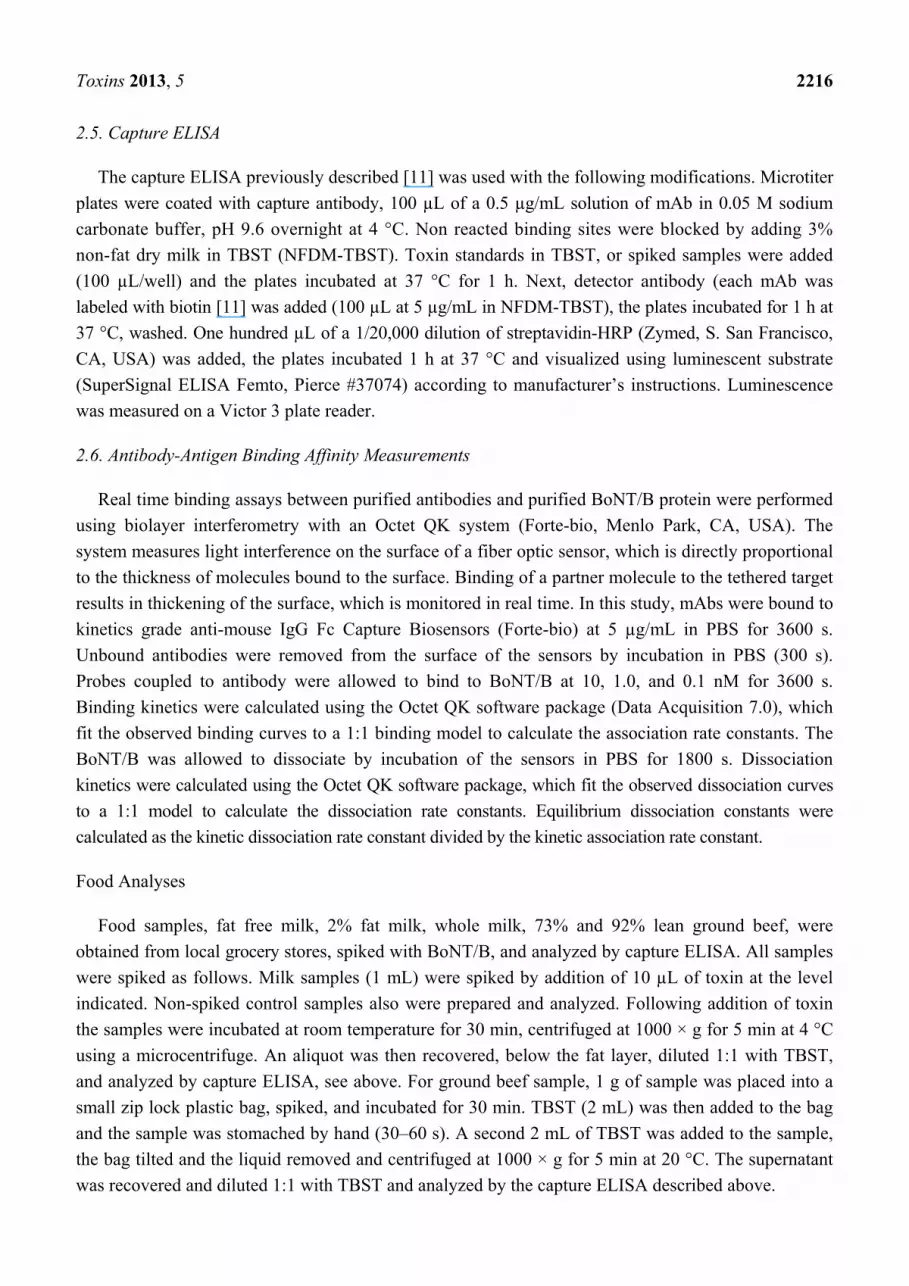
Recombinant fragments of BoNT/B light and heavy chain (Lc1, Lc2, Hc1, Hc2, Hc3, Hc4, and Hc5)
fused with glutathione transferase (recBoNT/B-GTS) were generated (see Figure 1), expressed in
Escherichia coli, purified, and used to immunize mice as previously described [13]. These toxin
fragments are referred to as recLc1-GST, recLc2-GST, recHc(1-5)-GST. The Institutional Biosafety
Committee approved Recombinant DNA methods used in this study. DNA sequences of the mAbs
described in this study have been deposited in GenBank and accession numbers are listed in Table 1.
Figure 1. Schematic of recombinant-GST peptide fragments of BoNT/B light and heavy
chains. Pepide fagmens were expressed as fusions to GST at the N-terminal.
Toxins 2013, 5 2215
2.3. Monoclonal Antibody Production and Screening Assay
Monoclonal antibody production was as previously described [11,13]. Briefly, recombinant peptide
fragment recHc5-GST (265 µg/mL) was mixed with Sigma Adjuvant System according to
manufacturer’s instructions (Sigma-Aldrich, St. Louis, MO, USA). Female BALB/cJ mice (Simonsen
Laboratories, Gilroy, CA, USA) were immunized three times at 2-week intervals by intraperitoneal
injection (i.p.) (100 µL). Following immunization with the recombinant recHc5-GST fragment,
hybridomas were generated and supernatants from the cell fusion plates were evaluated for mAbs
using a capture-capture ELISA screen. The Institutional Animal Care and Use Committee of the
United States Department of Agriculture, Western Regional Research Center approved the
experimental procedures used in these studies (protocol #04-1-H-05, 09-3). All animal experiments
and husbandry involved in the studies presented in this manuscript were conducted under the
guidelines of the U.S. Government Principles for the Utilization and Care of Vertebrate Animals Used
in Testing, Research and Training.
2.4. Capture-Capture Screening ELISA
The capture-capture ELISA screening method used was previously described [13]. Briefly
microtiter plates were coated with a 1 µg/mL solution of goat anti-mouse IgG Fc gamma #AP127
specific antibody (Millipore, Billerica, MA, USA) in 0.05 M sodium carbonate buffer, pH 9.6 overnight
at 4 °C. Non reacted binding sites were blocked by adding 3% non-fat dry milk in Tris-buffered saline
containing 0.05% Tween-20 (NFDM-TBST). Following a 1 h incubation at 37 °C culture supernatants
from the fusion plates were added and the microtiter plates were incubated at 37 °C for 1 h. Plates
were washed three times in TBST and a solution of BoNT/B in NFDM-TBST (200 ng/mL) was added
and the plates were incubated at 37 °C for 1 h. Plates were washed three times as before, then a 1 µg/mL
solution of anti-BoNT/B rabbit polyclonal antibodies (Metabiologics) in NFDM-TBST was added and
the plates were incubated at 37 °C for 1 h. Plates were washed three times as before, then a 1 µg/mL
solution of goat anti-rabbit HRP-conjugated polyclonal antibodies #A6154 (Sigma-Aldrich) was added
and the plates were incubated at 37 °C for 1 h. Plates were again washed three times, and binding was
visualized using SuperSignal West Dura Extended Duration Substrate (Pierce, Rockford, IL, USA)
according to manufacturer’s instructions.
Cells from the wells giving positive signals for antibody production were cloned by limiting
dilution, hybridomas expanded, and small amounts (usually less than 10 mL) of ascites fluids obtained
(Covance Research Products, Inc., Denver, PA, USA). Antibodies were purified by affinity
chromatography on Protein-G Sepharose. Bound antibody was eluted with 0.1 M glycine-HCl, pH 2.7
and dialyzed overnight versus PBS. Protein concentrations were determined with a BCA-kit (Pierce)
using the microplate method suggested by the manufacturer. The isotype of each antibody was
determined using the SBA Clonotyping System/HRP in ELISA format, according to manufacturer’s
instructions (Southern Biotech, Birmingham, AL, USA). Western blots were as described [11]
Toxins 2013, 5 2216
2.5. Capture ELISA
The capture ELISA previously described [11] was used with the following modifications. Microtiter
plates were coated with capture antibody, 100 µL of a 0.5 µg/mL solution of mAb in 0.05 M sodium
carbonate buffer, pH 9.6 overnight at 4 °C. Non reacted binding sites were blocked by adding 3%
non-fat dry milk in TBST (NFDM-TBST). Toxin standards in TBST, or spiked samples were added
(100 µL/well) and the plates incubated at 37 °C for 1 h. Next, detector antibody (each mAb was
labeled with biotin [11] was added (100 µL at 5 µg/mL in NFDM-TBST), the plates incubated for 1 h at
37 °C, washed. One hundred µL of a 1/20,000 dilution of streptavidin-HRP (Zymed, S. San Francisco,
CA, USA) was added, the plates incubated 1 h at 37 °C and visualized using luminescent substrate
(SuperSignal ELISA Femto, Pierce #37074) according to manufacturer’s instructions. Luminescence
was measured on a Victor 3 plate reader.
2.6. Antibody-Antigen Binding Affinity Measurements
Real time binding assays between purified antibodies and purified BoNT/B protein were performed
using biolayer interferometry with an Octet QK system (Forte-bio, Menlo Park, CA, USA). The
system measures light interference on the surface of a fiber optic sensor, which is directly proportional
to the thickness of molecules bound to the surface. Binding of a partner molecule to the tethered target
results in thickening of the surface, which is monitored in real time. In this study, mAbs were bound to
kinetics grade anti-mouse IgG Fc Capture Biosensors (Forte-bio) at 5 µg/mL in PBS for 3600 s.
Unbound antibodies were removed from the surface of the sensors by incubation in PBS (300 s).
Probes coupled to antibody were allowed to bind to BoNT/B at 10, 1.0, and 0.1 nM for 3600 s.
Binding kinetics were calculated using the Octet QK software package (Data Acquisition 7.0), which
fit the observed binding curves to a 1:1 binding model to calculate the association rate constants. The
BoNT/B was allowed to dissociate by incubation of the sensors in PBS for 1800 s. Dissociation
kinetics were calculated using the Octet QK software package, which fit the observed dissociation curves
to a 1:1 model to calculate the dissociation rate constants. Equilibrium dissociation constants were
calculated as the kinetic dissociation rate constant divided by the kinetic association rate constant.
Food Analyses
Food samples, fat free milk, 2% fat milk, whole milk, 73% and 92% lean ground beef, were
obtained from local grocery stores, spiked with BoNT/B, and analyzed by capture ELISA. All samples
were spiked as follows. Milk samples (1 mL) were spiked by addition of 10 µL of toxin at the level
indicated. Non-spiked control samples also were prepared and analyzed. Following addition of toxin
the samples were incubated at room temperature for 30 min, centrifuged at 1000 × g for 5 min at 4 °C
using a microcentrifuge. An aliquot was then recovered, below the fat layer, diluted 1:1 with TBST,
and analyzed by capture ELISA, see above. For ground beef sample, 1 g of sample was placed into a
small zip lock plastic bag, spiked, and incubated for 30 min. TBST (2 mL) was then added to the bag
and the sample was stomached by hand (30–60 s). A second 2 mL of TBST was added to the sample,
the bag tilted and the liquid removed and centrifuged at 1000 × g for 5 min at 20 °C. The supernatant
was recovered and diluted 1:1 with TBST and analyzed by the capture ELISA described above.
Toxins 2013, 5 2217
3. Results
3.1. Isolation and Characterization of Monoclonal Antibodies
Earlier studies aimed at development of a sensitive capture ELISA for BoNT/B using mAbs for
both the capture and detection reagents meet with only partial success [13]. Using a standard ELISA
with immobilized antigen for screening cell fusion experiments, a number of mAbs were isolated.
However, none of the antibodies so produced effectively bond toxin in solution under physiological
conditions, and thereby were not useful as a capture antibodies in a sandwich ELISA. Furthermore,
none of the mAbs produced in this earlier study were able to function as a detector antibody (even
though they bound toxin in ELISA and on Western blots [13]. A single mAb able to capture toxin from
solution eventually was identified but only after incorporating a double-capture ELISA screen [13]. In
the studies described here the double-capture ELISA screen was used in an effort to identify additional
capture and detector antibodies and useful antibody pairs for measurement of BoNT/B.
Following cell fusions, putative capture mAbs were trapped on microassay plates pre-coated with
anti-mouse Ig (Fc) specific antiserum (see methods). We identified positive responses in 18 of
~2000 fusion wells screened. Wells were considered positive if the measured activity was at least
2–3 times the average response of all wells on the plate. Incorporating authentic BoNT/B into the
screen (see methods) eliminates isolation of peptide specific mAbs in favor of those mAbs capable of
binding the intact toxin molecule. It was hoped that this approach would guarantee that all of the mAbs
detected in the screen were capable of capturing BoNT/B in solution. Equally important, the
capture-capture screen does not detect the vast majority of monoclonal antibodies that are detected in a
traditional ELISA screen but fail to capture antigen in solution. None of the 18 mAbs identified in the
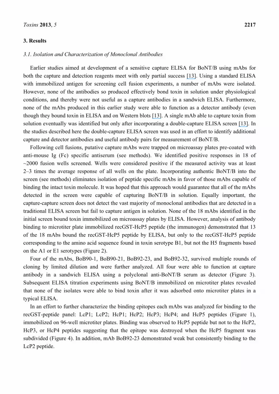
initial screen bound toxin immobilized on microassay plates by ELISA. However, analysis of antibody
binding to microtiter plate immobilized recGST-HcP5 peptide (the immunogen) demonstrated that 13
of the 18 mAbs bound the recGST-HcP5 peptide by ELISA, but only to the recGST-HcP5 peptide
corresponding to the amino acid sequence found in toxin serotype B1, but not the H5 fragments based
on the A1 or E1 serotypes (Figure 2).
Four of the mAbs, BoB90-1, BoB90-21, BoB92-23, and BoB92-32, survived multiple rounds of
cloning by limited dilution and were further analyzed. All four were able to function at capture
antibody in a sandwich ELISA using a polyclonal anti-BoNT/B serum as detector (Figure 3).
Subsequent ELISA titration experiments using BoNT/B immobilized on microtiter plates revealed
that none of the isolates were able to bind toxin after it was adsorbed onto microtiter plates in a
typical ELISA.
In an effort to further characterize the binding epitopes each mAbs was analyzed for binding to the
recGST-peptide panel: LcP1; LcP2; HcP1; HcP2; HcP3; HcP4; and HcP5 peptides (Figure 1),
immobilized on 96-well microtiter plates. Binding was observed to HcP5 peptide but not to the HcP2,
HcP3, or HcP4 peptides suggesting that the epitope was destroyed when the HcP5 fragment was
subdivided (Figure 4). In addition, mAb BoB92-23 demonstrated weak but consistently binding to the
LcP2 peptide.
Toxins 2013, 5 2218
Figure 2. Antibody binding to recombinant BoNT-GST fusion proteins representing the
receptor binding domain of the toxin HC for serotypes A, B, and E. Hybridoma
supernatants analyzed by ELISA using recombinant peptides as antigen.
Figure 3. Capture-Capture sandwich ELISA. Identification of capture mAbs immobilized
on anti-mouse Ig coated microtiter plates. Detection of bound toxin using a polyclonal
anti-BoNT/B antiserum with mAb capture antibodies indicated. Data represents average of
three replicates, error bars = ±standard deviation.

Toxins 2013, 5 2219
Figure 4. Monoclonal antibody binding to recombinant peptides of BoNT/B (see Figure 1).
Antibodies MCS6-27 and F27-33 described earlier [13].
Bilayer interferometry was used to measure the affinity constant (KD) of each of the four mAbs
described here (Table 1). BoB92-23 showed the strongest binding, with a dissociation constant of
10.2 × 10⁻¹¹ M. Weaker affinity constants were measured for the other mAbs. Isotype analysis
indicated that BoB92-23 is an IgG2b while the remaining three mAbs are IgG1, all having kappa light
chains (Table 1).
Table 1. Characteristics of BoNT/B monoclonal antibodies.
Antibody Isotype KD (×10−11 M) BoB90-1 IgG1, kappa 23.0
BoB90-21 IgG1, kappa 48.0
BoB92-23 IgG1, kappa 10.2 BoB92-32 IgG2b, kappa 48.3 MCS6-27 IgG1, kappa 8.6
Weak antibody binding to the 50 kDa toxin Hc and to the 150 kDa non-reduced holotoxin was
observed on Western blots (Figure 5) following electrophoresis in the presence of DTT. The weak
binding on Western blots was expected, since antibody binding to microtiter plate immobilized toxin
by ELISA was minimal. The long exposure times needed to visualize bands in the Western blots
suggests that heating toxin in SDS-PAGE sample buffer causes partial disruption of the antibody
binding epitopes. Clearly SDS-PAGE modifies proteins, altering their surface charge and potentially
denaturing tertiary structures. Changes of surface charge and/or structural changes that can eliminate
binding epitopes also can occur following adsorption to a solid phase [14].
The ability of the four mAbs (mAbs BoB90-1, BoB90-21, BoB92-23, and BoB92-32) to function as
detector antibodies using a previously produced capture mAb (MCS6-27) (Scotcher et al. 2010) was
evaluated. These experiments, summarized in Figure 6, revealed that only one of the antibodies,
biotin-labeled BoB92-32 functioned as a detector antibody.
Toxins 2013, 5 2220
Figure 5. Western blot analysis of anti-botulinum serotype B mAbs. Lane 1, BoB90-1;
Lane 2, BoB90-21; Lane 3, BoB92-23; Lane 4, BoB92-32. Each lane contained BoNT/B at
10 µg/mL plus DTT and probed with 10 µg/mL antibody. All lanes exposed for 10 min.
Standards on right side represent Kilo Daltons.
Figure 6. BoNT/B detection in a capture ELISA using mAb MCS6-27 as capture antibody
and the four mAbs described here as detector antibodies. Points for MCS 92-23 and 90-1
hidden by the points for MCS 90-21. Line represents a linear curve fit, R2 = 0.996.
3.2. Capture ELISA
These data are consistent with competition ELISA experiments (Figure 7) demonstrating that mAb
BoB92-32 did not compete with mAb MCS6-27 for toxin binding at any of the concentrations tested.
In contrast, mAbs BoB90-1, BoB90-21, and BoB92-23 competed with MCS6-27 for toxin binding
suggesting that they bind the same or closely related epitopes.
All possible combinations of the four mAbs described here plus mAb MCS6-27 were evaluated for
development of a sandwich ELISA. These experiments (data not shown) demonstrated that only two
capture/detector antibody pairs (BoB92-23/BoB92-32 and MCS6-27/BoB92-32) were useful for
development of a capture ELISA. Careful titration experiments using BoNT/B holotoxin suggest that
the combination BoB92-23 (capture) and BoB92-32 (detector) resulted in a more sensitive assay than
pairing MCS6-27 (capture) with BoB92-32 (detector) (Figure 8).
Toxins 2013, 5 2221
Figure 7. Competition capture -ELISA using mAb MCS6-27 as capture antibody. Toxin
mixed with increasing concentrations of competing mAb (indicated on figure) and
immediately added to the capture antibody coated well. Toxin detection using a polyclonal
anti-BoNT/B.
Figure 8. Capture ELISA comparing assay performance using either MCS6-27 (open blue
circle) or BoB92-23 (open orange square) as capture antibody and BoB92-32 as detector
antibody. Bars represent standard deviation (N = 3). Data fitted using 4-paramater-curve
fitting. The average of the zero toxin control plus three standard deviations represented by
the horizontal lines for BoB92-21 (solid) and MCS6-27 (dashed).
In an effort to determine if the four mAbs represented separate fusion products of a clonally
expanded lymphocyte population, the amino acid sequences of the light and heavy chain variable
regions were determined. Inspection of the translated amino acid sequences of the combining region of
each antibody (Figure 9) suggests that mAbs BoB92-23 and BoB92-31 are unique antibodies. MAb
BoB90-1 and BoB90-21 have identical Lc sequences, but sequence information for the Hc of BoB90-21
was not obtained so they could represent independent hybridomas resulting from fusion of a clonally
expanded lymphocyte population. The amino acid sequences of the variable region of the four
antibodies described here are different from that of mAb MCS6-27 shown for convenience.
0
20
40
60
80
100
0.01 0.1 1
Per
cen
t In
hib
itio
n o
f C
on
tro
l
Competing Antibody Concentration
MCS90-1
MCS90-21
MCS6-27
MCS92-23
MCS92-32
1,000
10,000
100,000
1,000,000
0.01 0.1 1 10
Rel
ativ
e Li
gh
t U
nit
s
BoNT/B (ng/mL)

Toxins 2013, 5 2222
3.3. Food Analysis
The assay was next applied for evaluation of foods fortified at different concentrations with
BoNT/B. In these experiments, all samples were fortified with BoNT/B at a constant volume (10 µL)
of toxin and the samples were incubated at room temperature for 30 min before processing. These data
(Supplemental Figure 1) were used to establish the L.O.D. (average of the buffer control plus three
standard deviations). In all cases, the signal observed at the lowest spike level was higher than the
L.O.D. Thus, the percentage toxin recovery (Table 2) could be calculated for each toxin concentration.
In those dairy products tested, the percentage recovery varied from 63.8% to 131%. However, at
the 50 and 5.0 ng/mL spike levels the recoveries were close to 100% (only one sample was below
89%). Greater variations of the percentage recovery were observed following analysis of the
ground beef sample.
Figure 9. Clustal W2 alignment of the amino acid sequence deduced from the cDNA of
the variable regions of the anti-BoNT/B monoclonal antibodies. The Framework Regions
(FWR) and Complementarity Determining Regions (CD-1, -2, and -3) are indicated
(boxed), bold text indicates constant region sequences.
Heavy Chains Kabat definition Antibody FWR-1 CDR-1 FWR-2 CDR-2 BoB-90-1 EVKLEESGAELVRPGTSVKMSCKAAGYTFN-YWITGWVKERPGHGLEWIGDIFPGGGFTN 59 BoB92-23 -------------------MSCKAAGYTFTN-YWIGWVKERPGHGLEWIGDIYPGGGFTN 40 BoB92-32 QVQLQQAGTELVKPGASVKLSCKASGFTFNN-YYLFWVKQRPGQGLEWIGGINPSNGGFN 59 MCS6-27 EVQLEQSGPGLVKPSQSLSLTCTVTGYSITNDYAWNWIRQFPGNKLEWMGYINYS-GTTS 59 FWR-3 CDR-3 FWR-4 BoB-90 YNENFKGKATLTADTSSSPAYMQLSSMTSEDSSIYHCARGRGHNSSTYFDSWGQGTTLTS 119BoB92-23 YNENFKGKATLTADTSSSTAYMQLSSLTSEDSAIYHCARGIYYNSSPYFDSWGQGTTLTV 100BoB-92-32 FNEKFKNKATLTVGKSSSTVYMQLSSLTSEDSAVYYCTRR--NLGLRAMDYWGQGTSVTF 117MCS6-27 YNPSLKSRISITRDTSKNQFFLQFNSVTTEDTATYYCARS-------PLAYWGQGTLVTV 112 BoB-90-1 PA----------- 121 BoB92-23 SSAKTTPPSVY-- 111 BoB-92-32 SSK---------- 120 MCS6-27 SAAKTTPPSVYR 125
Light Chains Kabat definition Antibody FWR-1 CDR-1 FWR-2 CDR-2 BoB90-21 DIVMTQTTASLAVSLGQRATISCRASESVDS-YGNSFMHWYQQKPGQPPKLLIYRASNLES 60 BoB90-1 DIVLTQTTASLAVSLGQRATISCRASESVDS-YGNSFMHWYQQKPGQPPKLLIYRASNLES 60 MCS6-27 DIVLTQTPASLSVSVGETVTITCRASENIYS-----HLAWYQQKQGKSPQLLVYTATNLAD 56 BoB92-23 -----VTPFCI---CGRYVTITCRASQNIYS-----HLTWFQQRQGRSPQLLVYTATNLAD 48 BoB92-32 -------HFSILS-LWRSVSISCRTSQSLVNNKGNTYLSWYLHKPGQSPQLLIYGISNRFS 53 FWR-3 CDR-3 FWR-4 BoB90-21 GIPARFSGSGSRTDFTLTINPVEADDVATYYCQQSNEDPHS-DRNRATNLKRTEM----- 115 BoB90-1 GIPARFSGSGSRTDFTLTINPVEADDVATYYCQQSNEDPHS-DRNRATNLKRTEM----- 115 MCS6-27 GVPSRFSGRGSGTQYSLKINSLQSEDFGIYFCQHFWGTPWTFGGGTKLEIKRADAAPTVS 116 BoB92-23 GVPARFSGSGSGTQYSLKINNLQSEDFGTYYCQHFWGTPWTFGGGTKLEIKRADAAPTVS 108 BoB92-32 GVPDRFSGSGSGTDFTLKISTIKPEDLGMYYCFQGTHQPFTFGAGTKLELKRADAAPTVS 113
Toxins 2013, 5 2223
Table 2. Percentage recovery of BoNT/B spiked into different food matrices.
Percentage Recovery
Matrix/Spike Level 50 ng/mL 10 ng/mL 5 ng/mL 2 ng/mL 0.5 ng/mL
Non Fat Milk 108 (54)1 nd2 89 (4.45) nd 131 (0.655)
2% Milk 97 (48.5) nd 76 (3.35) 73 (0.365)
Whole Milk 101 (50.5) nd 105 (5.25) nd 116 (0.58)
Ground Beef 95% Lean 63.8 (31.9) 116 (5.8) nd 100 (2)3 nd
Notes: 1. Numbers in brackets refer to toxin level measured at each spike concentration using a buffer standard curve.
Percent recovery = measured concentration/spike concentration × 100; 2. nd = not done; 3. The signal from beef sample
spiked at 2 ng/mL was just at the L.O.D. for standard.
4. Discussion
We have previously used recombinant GST-fusion peptides of BoNT/B fragments for the
successful generation of mAbs that bind intact neurotoxin. While these studies demonstrated the
feasibility of using recombinant peptides of BoNT/B as immunogens, all of the resulting mAbs,
except one, were unable to bind toxin in solution or act as a capture or detector antibody in a
sandwich ELISA [13]. In these earlier experiments screening was based upon a traditional ELISA, i.e.,
BoNT/B was immobilized in microtiter plates by absorption and hybridoma supernatants evaluated for
mAb binding. Proteins absorbed to plastic surfaces can undergo structural modifications that can hide
or destroy antibody-binding epitopes [14]. By modifying the screening strategy to a capture-capture
assay one mAb (MCS6-27) that could be utilized as a capture antibody was identified (Scotcher et al.
2010). Likewise, none of the antibodies isolated from these earlier fusions functioned as detector
antibodies. Thus, our initial goal of generating a mAb-based sensitive capture ELISA for BoTN/B was
only partially meet. To identify additional capture and detector antibodies the capture-capture screen
described here was incorporated to screen cell fusion experiments.
The poor binding observed following toxin adsorption to microassay plates and on Western blots
following SDS-PAGE suggests that the binding epitope, localized to the receptor binding domain of
the toxin Hc, is disrupted or masked by these treatments and each mAb is binding a
conformational epitope. This is consistent with antibody binding to only the recombinant peptide
corresponding to the entire receptor-binding domain of the toxin but not peptides H2 and H3 that
divide the receptor-binding domain.
The four new capture mAbs described here have epitopes located in the receptor-binding domain of
BoNT/B Hc. Sequence analysis of the combining regions suggests that these antibodies are unique and
different from our earlier developed capture mAb MCS6-27. However, competition ELISA
experiments demonstrate that BoB90-1, BoB90-21, and BoB92-21 compete with mAb6-27 for toxin
binding. In fact, BoB92-21 has a substantially stronger competition curve than seen when MCS6-27
competes with itself for toxin binding. These data and the assay performance observed in a sandwich
ELISA using MCS6-27 vs. BoB92-21 as capture antibody suggest that BoB92-21 as capture antibody
results in lower limits of detection.
Preliminary results indicate that a prophylactic, intravenous (iv) injection of µg amounts of BoB90-1,
BoB90-21, BoB92-23, and BoB92-32 per mouse protected 100% of the mice studied from death or
any symptoms of intoxication from an iv injection of 460 pg (100 mouse iv LD50 doses) of BoNT/B,
Toxins 2013, 5 2224
consistent with the observation that these mAbs could bind BoNT/B in vitro under physiological
conditions [15]. Research to more clearly define the parameters controlling the ability of these
antibodies to neutralize toxin and rescue animals following exposure is the subject of future studies.
However, these observations are consistent with earlier studies [13] suggesting that antibodies that
failed to bind BoNT/B in physiological buffer did not bind toxin in vivo and failed to protect mice
from the neurotoxic effects of BoNT/B. The ability of a mAb to capture antigen from solution appears
to be an indicator for toxin neutralization potential.
Using our previously developed mAb MCS6-27 as the capture antibody, only mAb BoB92-32 was
able to function as a detector antibody (Figure 6). These data suggest that the other three mAbs bind an
epitope that is not available when toxin is first bound by mAb MCS6-27, or that BoB90-1, BoB90-21,
and BoB92-23 bind the same or a closely related epitope.
The ability of BoB92-32 to act as a detector antibody is consistent with its failure to compete with
MCS6-27 for toxin binding. Of the remaining three mAbs, BoB92-21 showed the strongest
competition, greater than competing MCS6-27 with itself. The exact nature of this competition is not
clear. The DNA sequences of the combining regions of these antibodies suggest that each represents a
uniquely derived monoclonal antibody.
Evaluation of all possible capture/detector combinations demonstrated that the best performance of
a sandwich ELISA was observed using either BoB92-23 or MCS6-27 as capture antibody coupled
with BoB92-32 as detector antibody. The combination BoB92-23/BoB92-32 showed slightly better
performance than MCS6/27/BoB92-32 (Figure 8) consistent with the competition ELISA experiments.
Using this antibody pair the L.O.D. was determined to be approximately 10–20 pg/mL. The mouse
LD50 of the BoNT/B preparations used in our laboratory, when injected intraperitoneally was found to
be ~10 pg/mL [15]. The sandwich ELISA described here has a comparable L.O.D. as the mouse
bioassay (MBA) but requires only a few hours to complete versus 2–4 days. Analysis of spiked dairy
products and ground beef at 50–0.5 ng/mL suggest that acceptable sample recoveries are obtained with
the assay and that the sandwich ELISA represents a useful tool for rapid analysis of food for BoNT/B.
In summary, we have developed a sensitive sandwich ELISA specific for BoNT/B that is both rapid
and easy to use. This assay is based on monoclonal antibodies for both the capture and detector
reagents and thus represents an assay that can be consistently produced over time without the use of
animals and the variability inherent in polyclonal antisera. We are currently exploring other labels and
assay formats, including a lateral flow immunoassay able to detect and distinguish BoNT/A and
BoNT/B in a few minutes [16].
Acknowledgements
We gratefully thank Thomas Henderson for assistance with recombinant peptide expression and
purification and Paul Merrill for antibody purification and biotin conjugation. We acknowledge
funding by the U.S. Department of Agriculture, Agriculture Research Service, CRIS project
5325-42000-048-00D, and U.S. Department of Homeland Security IAA #40768.
Conflicts of Interest
The authors declare no conflict of interest.

Toxins 2013, 5 2225
References
1. Gill, M. Bacterial toxins: A table of lethal amounts. Microbiol. Rev. 1982, 46, 86–94.
2. Hatheway, C.L. Toxigenic clostridia. Clin. Microbiol. Rev. 1990, 3, 66–98.
3. Ferreira, J.L. Comparison of amplified ELISA and mouse bioassay procedures for determination
of botulinum toxins A, B, E, and F. J. AOAC Int. 2001, 84, 85–88.
4. National Botulism Surveillance (2001–2007) Botulism Surveillance Summary Council of State
and Territorial Epidemiologists (CSTE), Centers for Disease Control and Prevention. Available
online: www.cdc.gov/nationalsurveillance/botulism_surveillance.html (accessed on 15 November
2013).
5. Terranova, W.; Breman, J.G.; Locey, R.P.; Speck, S. Botulism type B: Epidemiologic aspects of
an extensive outbreak. Am. J. Epidemiol. 1978, 108, 150–156.
6. O’Mahony, M.; Mitchell, E.; Gilbert, R.J.; Hutchinson, D.N.; Begg, N.T.; Rodhouse, J.C.;
Morris, J.E. An outbreak of foodborne botulism associated with contaminated hazelnut yoghurt.
Epidemiol. Infect. 1990, 104, 389–395.
7. Montecucco, C.; Schiavo, G. Mechanism of action of tetanus and botulinum neurotoxins. Mol.
Microbiol. 1994, 13, 1–8.
8. Simpson, L.L. Identification of the major steps in botulinum toxin action. Annu. Rev. Pharmacol.
Toxicol. 2004, 44, 167–193.
9. Schiavo, G.; Benfenati, F.; Poulain, B.; Rossetto, O.; Polverino De Laureto, P.; DasGupta, B.R.;
Montecucco, C. Tetanus and botulinum-B neurotoxins block neurotransmitter release by
proteolytic cleavage of synaptobrevin. Nature 1992, 359, 832–835.
10. Blasi, J.; Chapman, E.R.; Link, E.; Binz, T.; Yamasaki, S.; De Camilli, P.; Sudhof, T.C.; Niemann,
H.; Jahn, R. Botulinum neurotoxin A selectively cleaves the synaptic protein SNAP-25. Nature
1993, 365, 160–163.
11. Stanker, L.H.; Merrill, P.; Scotcher, M.C.; Cheng, L.W. Development and partial characterization
of high-affinity monoclonal antibodies for botulinum toxin type A and their use in analysis of
milk by sandwich ELISA. J. Immunol. Methods 2008, 336, 1–8.
12. Chen, L.; Stanker, L.H. Detection of botulinum neurotoxin serotypes A and B using
chemiluminescent versus electrochemiluminescent immunoassay in food and serum. J. Agric.
Food Chem. 2012, 61, 755–760.
13. Scotcher, M.C.; Cheng, L.W.; Stanker, L.H. Detection of botulinum neurotoxin serotype B at sub
mouse LD50 levels by a sandwich immunoassay and its application to toxin detection in milk.
PLoS ONE 2010, 5, 1–10.
14. He, J. Practical guide to ELISA development. In The Immunoassay Handbook, 4th ed.; Wild,
D.G., Ed.; Elsevier: Amsterdam, The Netherlands, 2013; pp. 381–395.
15. Cheng, L.W.; Henderson, T.D.; Patfield, S.; Stanker, L.H.; He, X. Mouse in vivo neutralization of
Escherichia coli shiga toxin 2 with monoclonal antibodies. Toxins 2013, 5, 1845–1858.
16. Ching, K.H.; Lin, A.; McGarvey, J.A.; Stanker, L.H.; Hnasko, R. Rapid and selective detection of
botulinum neurotoxin serotype-A and -B with a single immunochromatographic test strip J. Immunol.
Methods 2012, 380, 23–29.
Toxins 2013, 5 2226
Supplemental Figures
Supplemental Figure 1. (A, B). Detection of BoNT/B spiked into food by capture ELISA.
© 2013 by the authors; licensee MDPI, Basel, Switzerland. This article is an open access article
distributed under the terms and conditions of the Creative Commons Attribution license
(http://creativecommons.org/licenses/by/3.0/).
100
1000
104
105
106
0.1 1 10 100
Dairy Products Diluted 1:1
non-fat milk2% milkbuffer standardwhole milk
Rel
ativ
e Li
ght U
nits
w
BoNT/B (ng/mL)
A
0
5000
1 104
1.5 104
2 104
2.5 104
3 104
0 10 20 30 40 50 60
92% Lean Ground Beef
Rel
ativ
e Li
ght
Uni
ts
BoNT/B (ng/mL)
B