the ultimate radiochemical nightmare: upon radio-iodination of botulinum neurotoxin a, the...
TRANSCRIPT

van Uhm et al. EJNMMI Research (2015) 5:5 DOI 10.1186/s13550-015-0083-5
ORIGINAL RESEARCH Open Access
The ultimate radiochemical nightmare: uponradio-iodination of Botulinum neurotoxin A, theintroduced iodine atom itself seems to be fatalfor the bioactivity of this macromoleculeJanneke IM van Uhm1*, Gerard WM Visser2, Marcel J van der Schans3, Albert A Geldof1,2, Eric JH Meuleman1
and Jakko A Nieuwenhuijzen1
Abstract
Background: Botulinum neurotoxin A (BoNT-A) is a highly neurotoxic drug and frequently used in patients.Knowledge on the optimal way of administration of BoNT-A and its subsequent distribution is still rather limited. Anaccurate method for monitoring these processes might be the use of radiolabelled BoNT-A. In this paper, we reportour feasibility study on labelling BoNT-A with high-dose iodine-125 (125I) via IODOGEN-coated BoNT-A method.
Methods: Using cetuximab as model substrate for BoNT-A, a miniaturization of the IODOGEN-coated mAb methodwas developed with special attention to the minimum required amount of the oxidant IODOGEN, while the amountof substrate, reaction volume and reaction time were downsized. Labelling efficiency and radiochemical purity weredetermined by TLC, integrity by SDS-PAGE and HPLC and immunoreactivity by cell-binding assay. BoNT-A (50 μg)was labelled with 125I by coating with 2.5 μg IODOGEN, in a total reaction volume of 250 μL and a reaction time of90 s. 125I-BoNT-A was purified by size exclusion chromatography (PD10 column) using ascorbic acid solution(5 mg/ml, pH = 5) as eluent. Quality analysis of 125I-BoNT-A was performed by an in vitro bladder strip model, anelectrochemiluminescence assay and an Endopep assay.
Results: Cetuximab (50 μg) labelling with 125I (15 to 150 MBq) resulted in a labelling efficiency of 70% to 80%, aradiochemical purity of >99%, an immunoreactivity of >95% and a retained integrity on SDS; HPLC analysis revealedpartly affected integrity when 110 to 150 MBq 125I was used, i.e. when the averaged I/mAb molar ratio exceeded 3.Addition of HEPES (20 mM) and lactose (1.25%) (lyophilized BoNT-A contains HEPES and lactose) decreased thelabelling efficiency to 44% to 54%. BoNT-A (50 μg) labelling with 125I (97.2 to 98.3 MBq) resulted in labellingefficiency of 51% to 52% with a radiochemical purity >98.5%, a specific activity of 150.5 to 152.9 MBq/nmol and anI/BoNT-A molar ratio of 1.86 to 1.90. The in vitro bladder strip model showed no bioactivity of 125I-BoNT-A whencompared to unlabelled BoNT-A. The electrochemiluminescence and Endopep assay demonstrated around 10%and 15% bioactivity of 125I-BoNT-A compared to unlabelled BoNT-A, respectively. The remaining bioactivitycorrelates within the Poisson distribution with the amount of BoNT-A molecules that does not bear an iodine atom.
Conclusions: BoNT-A was successfully radio-iodinated with an activity high enough to enable in vivo measurementof nanograms of BoNT-A, which could be used in studying optimization of administration techniques of BoNT-A.The bioactivity of a BoNT-A molecule is, however, lost upon the introduction of an iodine atom into the tyrosinemoiety of this sensitive molecule.
Keywords: Botulinum neurotoxin A; Iodine-125; Radio-iodination; Monoclonal antibody; PET research
* Correspondence: [email protected] of Urology, VU University Medical Center, PO Box 7057,1007 MB Amsterdam, The NetherlandsFull list of author information is available at the end of the article
© 2015 van Uhm et al.; licensee Springer. ThisAttribution License (http://creativecommons.orin any medium, provided the original work is p
is an Open Access article distributed under the terms of the Creative Commonsg/licenses/by/4.0), which permits unrestricted use, distribution, and reproductionroperly credited.
van Uhm et al. EJNMMI Research (2015) 5:5 Page 2 of 9
BackgroundBotulinum neurotoxin A (BoNT-A) was introduced forthe treatment of overactive bladder (OAB) by Schurchet al. at the beginning of this century [1]. Since then,multiple clinical trials have proven BoNT-A to be aneffective therapy for refractory OAB [2]. These clinicaltrials also demonstrated differences in efficacy and du-ration of the response. The most effective dose, volumeof dilution, number and sites of injection required foreffective treatment are not well known [3]. Notwith-standing evolving knowledge about the mechanism ofaction of BoNT-A on neurotransmitter release and re-ceptor activation, the distribution of BoNT-A after intra-vesical injection remains largely unknown [4,5]. If thedistribution in the bladder wall after administration ofBoNT-A could be monitored, the differences in clinicaleffect among patients might be better understood. Be-sides, monitoring would enable investigation of the mostoptimal technique of administrating BoNT-A.An accurate monitoring approach would be to label
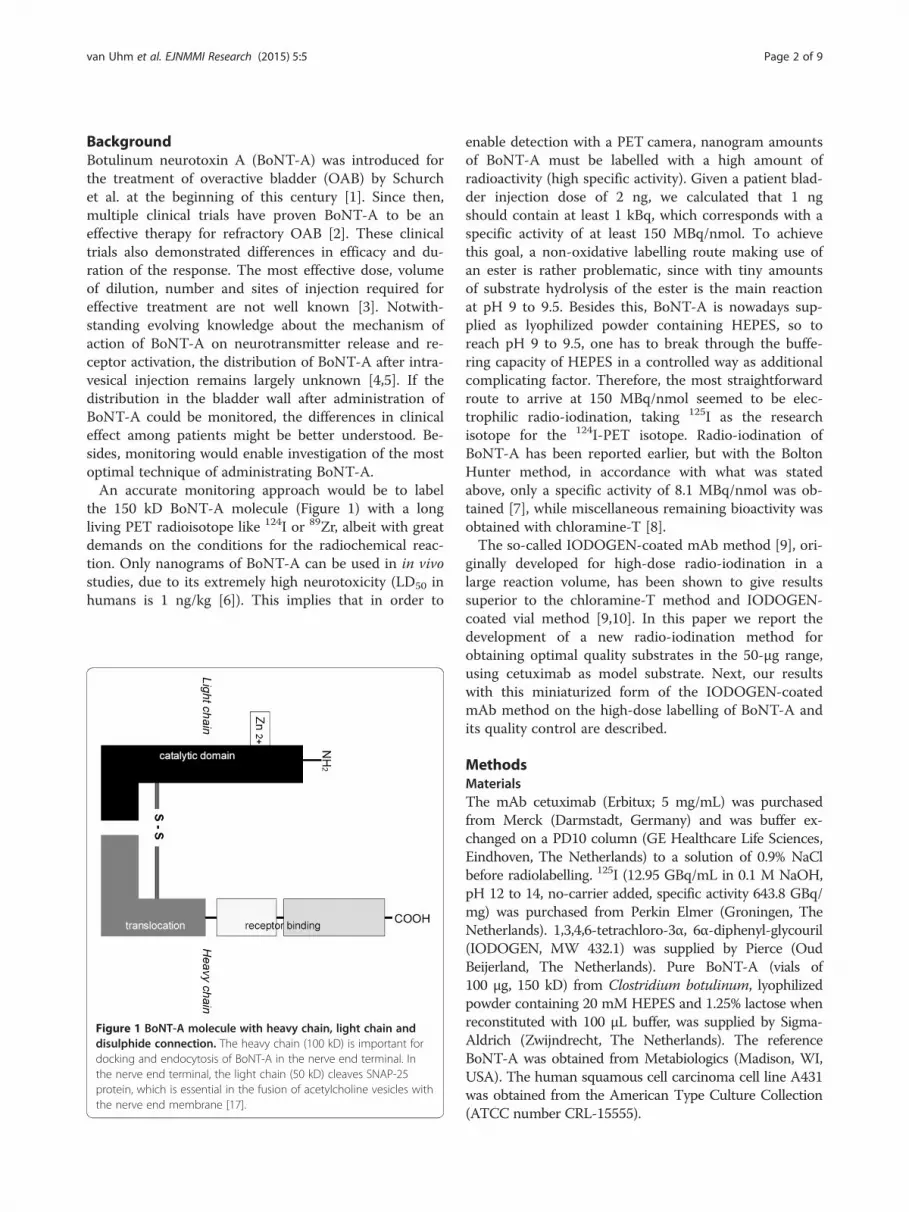
the 150 kD BoNT-A molecule (Figure 1) with a longliving PET radioisotope like 124I or 89Zr, albeit with greatdemands on the conditions for the radiochemical reac-tion. Only nanograms of BoNT-A can be used in in vivostudies, due to its extremely high neurotoxicity (LD50 inhumans is 1 ng/kg [6]). This implies that in order to
Figure 1 BoNT-A molecule with heavy chain, light chain anddisulphide connection. The heavy chain (100 kD) is important fordocking and endocytosis of BoNT-A in the nerve end terminal. Inthe nerve end terminal, the light chain (50 kD) cleaves SNAP-25protein, which is essential in the fusion of acetylcholine vesicles withthe nerve end membrane [17].
enable detection with a PET camera, nanogram amountsof BoNT-A must be labelled with a high amount ofradioactivity (high specific activity). Given a patient blad-der injection dose of 2 ng, we calculated that 1 ngshould contain at least 1 kBq, which corresponds with aspecific activity of at least 150 MBq/nmol. To achievethis goal, a non-oxidative labelling route making use ofan ester is rather problematic, since with tiny amountsof substrate hydrolysis of the ester is the main reactionat pH 9 to 9.5. Besides this, BoNT-A is nowadays sup-plied as lyophilized powder containing HEPES, so toreach pH 9 to 9.5, one has to break through the buffe-ring capacity of HEPES in a controlled way as additionalcomplicating factor. Therefore, the most straightforwardroute to arrive at 150 MBq/nmol seemed to be elec-trophilic radio-iodination, taking 125I as the researchisotope for the 124I-PET isotope. Radio-iodination ofBoNT-A has been reported earlier, but with the BoltonHunter method, in accordance with what was statedabove, only a specific activity of 8.1 MBq/nmol was ob-tained [7], while miscellaneous remaining bioactivity wasobtained with chloramine-T [8].The so-called IODOGEN-coated mAb method [9], ori-
ginally developed for high-dose radio-iodination in alarge reaction volume, has been shown to give resultssuperior to the chloramine-T method and IODOGEN-coated vial method [9,10]. In this paper we report thedevelopment of a new radio-iodination method forobtaining optimal quality substrates in the 50-μg range,using cetuximab as model substrate. Next, our resultswith this miniaturized form of the IODOGEN-coatedmAb method on the high-dose labelling of BoNT-A andits quality control are described.
MethodsMaterialsThe mAb cetuximab (Erbitux; 5 mg/mL) was purchasedfrom Merck (Darmstadt, Germany) and was buffer ex-changed on a PD10 column (GE Healthcare Life Sciences,Eindhoven, The Netherlands) to a solution of 0.9% NaClbefore radiolabelling. 125I (12.95 GBq/mL in 0.1 M NaOH,pH 12 to 14, no-carrier added, specific activity 643.8 GBq/mg) was purchased from Perkin Elmer (Groningen, TheNetherlands). 1,3,4,6-tetrachloro-3α, 6α-diphenyl-glycouril(IODOGEN, MW 432.1) was supplied by Pierce (OudBeijerland, The Netherlands). Pure BoNT-A (vials of100 μg, 150 kD) from Clostridium botulinum, lyophilizedpowder containing 20 mM HEPES and 1.25% lactose whenreconstituted with 100 μL buffer, was supplied by Sigma-Aldrich (Zwijndrecht, The Netherlands). The referenceBoNT-A was obtained from Metabiologics (Madison, WI,USA). The human squamous cell carcinoma cell line A431was obtained from the American Type Culture Collection(ATCC number CRL-15555).

Table 1 Technical protocol 125I labelling of BoNT-Aaccording to IODOGEN-coated method
Step
1. X μL 125I is added to wobbling reaction vial
2. 250 μL phosphate buffer (0.5 M, pH 7.4) is added
3. 50 μL BoNT-A/ phosphate buffer (=50 μg BoNT-A) is addeda
4. 115-X μL phosphate buffer (0.1 M, pH 6.8) is added to adjustthe reaction volume to 215 μL
5. 17.5 μL freshly prepared IODOGEN/acetonitrile solution(2.5 μg/35 μL) is added into the reaction mixture (start ofreaction, t = 0)
6. At t = 45 s, another 17.5 μL IODOGEN/acetonitrile solution(2.5 μg/35 μL) is added
7. At t = 90 s, 100 μL ascorbic acid solution (25 mg/mL, pH 5.0)is added to stop the reaction and to reduce any formedS-Cl bonds
8. At t = 4.30 min, 10 μL BSA (50 mg/mL) is addedb
9. At t = 5.30 min, samples are taken for ITLC for determinationof the labelling efficiency
10. At t = 10 min, 360 μL reaction mixture is transferred to asyringe connected to a filterc
11. The reaction vial is rinsed by 640 μL phosphate buffer(0.1M, pH 6.8) and also transferred to the syringe connectedto the filterc
12. The combined solution in the syringe is filtered and purifiedon a PD10 column with ascorbic acid solution (5 mg/mL,pH 5.0) as eluent, collected fractions were 0.5 mL
13. Fractions with highest amount of 125I-BoNT-A and the highestradiochemical purity are pooled
14. Samples of the pooled 125I-BoNT-A (fractions 6, 7, 8) are takenfor ITLC
15. Final product is diluted by BSA (1 mg/mL) till a concentrationof 1 μg/100 μL and stored at 20°C
t = time.aVial of 100 μg lyophilized BoNT-A powder is reconstituted with 100 μLphosphate buffer (0.1 M, pH 6.8).bFor encapsulation of the product to protect against radiation damage and toprevent absorption of material on the surface of the vials/syringes. Instead ofBSA (bovine serum albumin), HSA (human serum albumin) or any othermacromolecule can be used.cFilter of 0.2 μm (Acrodisc Gelman Sciences, Ann Arbor, MI, USA).
van Uhm et al. EJNMMI Research (2015) 5:5 Page 3 of 9
Development of the labelling methodTo avoid working routinely with 50 μg portions of ex-tremely neurotoxic and expensive BoNT-A (LD50 inhumans is about 75 ng [6]), developing and establishingthe optimal method of labelling BoNT-A with 125I wasdone with cetuximab.The coating approach was evaluated by several sets of
labelling experiments within the following general frame:1) every reaction was performed in a sterilized glass vialof 20 mL, which was wobbling continuously during thereaction; 2) the total reaction volume was 250 μL andconsecutively consisted of X μL 125I in 0.1 M NaOH,50 μL 0.5 M phosphate buffer (pH 7.4), 25 μL mAb solu-tion (50 μg mAb, 2 mg/mL), 140-X μL 0.1 M phosphatebuffer (pH 6.8) and 35 μL of IODOGEN/acetonitrile so-lution (1 mg/mL stock IODOGEN/acetonitrile solutionwas diluted to the desired amount of IODOGEN); 3) thereaction was started by IODOGEN/acetonitrile solutionand abrogated with 100 μL ascorbic acid (25 mg/mL, pH5); 4) labelling yields of all experiments were assessedwith instant thin-layer chromatography (ITLC).In the first set, labelling efficiency was determined upon
decreasing the amount of IODOGEN (35, 20, 10, 5 and2.5 μg), using 15 MBq 125I (75 MBq 125I/100 μL) and a re-action time of 180 s. In the second set, labelling kineticswas evaluated for 2.5 μg IODOGEN and a reaction timeof 60, 90 and 180 s. In the third set, the amount of radio-activity of 125I was increased from 15 to 150 MBq. AfterPD10 column purification, the integrity was evaluated byhigh-performance liquid chromatography (HPLC) andsodium dodecylsulfate-polyacrylamide gel electrophoresis(SDS-PAGE) and the immunoreactivity by a cell-bindingassay. In the fourth set, 50 μL HEPES (20 mM) and12.5 μL lactose solution (50 mg/mL) was included in thereaction mixture of the cetuximab labelling. The influenceof HEPES and lactose was studied, because lyophilizedBoNT-A also contained these amounts of HEPES and lac-tose. The total dose of IODOGEN was either 2.5 μg inone portion, or it was added in two portions; 1.25 μg/17.5μL IODOGEN/acetonitrile at the start of the reaction anda second portion after 45 s. Ten microlitres of bovineserum albumin (BSA pH 6.8; 50 mg/mL) was added to thereaction mixture before the PD10 column purification.Again, integrity was analysed by HPLC and SDS-PAGE. Inthe fifth set, 50 μg BoNT-A (50 μL) was labelled with 97.2to 98.3 MBq 125I (10 μL) using the protocol described inTable 1. Special procedures were applied due to highneurotoxicity of BoNT-A, including the use of a biologicalsafety cabinet, double rubber gloves, double-containmentand contamination-free pipettes (Microman, Gilson,France). CAUTION: during try-outs with cetuximab, itwas found that the advised safety measures (hypochloritecontaining waste bin, spraying of vessels that came out ofthe biological safety cabinet with hypochlorite solution)
completely destroyed the immunoreactivity of this mAb;So, these safety measures had to be left out. Geometry andcalibration experiments were performed with 125I-labelledcetuximab to correct for self-absorption of the 125I-gamma’s in the various double containment vessels. Reco-very of the 125I-BoNT-A mass was calculated as 50 μgtimes [MBq 125I in product] / [MBq 125I in starting vialtimes the labelling efficiency factor]. 125I-BoNT-A and un-labelled BoNT-A were diluted in BSA/phosphate buffer(1 mg/mL) to a concentration of 1 μg/100 μL, and thesebatches were stored at −20°C.
Quality analysis of mAb labellingConjugates were analysed by ITLC for labelling effi-ciency and radiochemical purity, by HPLC and SDS-
van Uhm et al. EJNMMI Research (2015) 5:5 Page 4 of 9
PAGE followed by phosphor imager analysis for integrityand by a cell-binding assay for immunoreactivity. ITLCanalysis of radiolabelled cetuximab was performed onsilica gel-impregnated glass fibre sheets (PI MedicalDiagnostic Equipment BV, Tijnje, The Netherlands), withcitrate buffer (20 mM, pH 5.0) as the mobile phase.HPLC of the conjugates was performed on a JASCOBenelux BV HPLC (De Meern, The Netherlands) with adiode array detector system and an inline radiodetector(Raytest Isotopenmessgeräte GmbH, Straubenhardt,Germany) using a Superdex 200 10/300 GL size exclu-sion column (GE Healthcare Life Sciences, Eindhoven,The Netherlands). The eluent consisted of 0.05 M so-dium phosphate/0.15 M sodium chloride plus 0.05%sodium azide (pH 6.8), and the flow was set at a rate of0.5 mL/min. HPLC measurements were performed atA = 280 nm to measure mAb absorption. Gel elec-trophoresis was performed on a Pharmacia PhastgelSystem using 7.5% SDS-PAGE gel electrophoresis gels(Amersham Biosciences, Roosendaal, The Netherlands)under non-reducing conditions and analyzed on a phos-phor imager (Molecular Dynamics, Zoetermeer, TheNetherlands) and quantified with ImageQuant software.In vitro binding characteristics of radiolabelled cetu-ximab were determined in an immunoreactivity assay es-sentially as described before [11], using A431 cells fixedwith 2% paraformaldehyde. Binding data were graphic-ally analysed in a modified Lineweaver-Burk (double-reciprocal) plot, and the immunoreactive fraction wasdetermined by linear extrapolation to conditions repre-senting infinite antigen excess.
Quality analysis of BoNT-A labellingThe bioactivity of 125I-BoNT-A was measured by anin vitro bladder strip model, an electrochemilumines-cence (ECL) assay to measure the antibody-antigen de-tection on the heavy chain and an Endopep assay tomeasure the cleavage activity of the light chain.The animal experiments were performed according to
the Dutch National Institutes of Health principles of la-boratory animal care and Dutch national (‘Wet op dedierproeven,’ Stb 1985, 336). The bladders of femaleWistar rats (n = 2, 250 to 300 g, Harlan®, Horst, TheNetherlands) were used for the in vitro bladder stripexperiments previously described [12]. In short: twobladder strips of one rat bladder were mounted in twoparallel organ baths. One strip was incubated in 125I-BoNT-A (0.3 mM) and one in unlabelled BoNT-A (0.3mM). The contraction force of bladder strips due toelectric field stimulation (EFS) was measured before andafter incubation. The second quality analysis of 125I-BoNT-A was by ECL assay [13]. The ECL assay is basedon antibody docking of the heavy chain of BoNT-A,which is in this case the antigen. ECL 96-well plates
were coated with BoNT-A capture antibodies. The un-labelled BoNT-A, reference BoNT-A (TNO, Rijswijk, TheNetherlands) and 125I-BoNT-A were diluted in concentra-tions of 20, 5, 1.25, 0.31, 0.078, 0.02 and 0.005 ng/mL.These various concentrations were pipetted in the wells,and the plate was stirred and washed. The wells were filledwith MSD-labelled detection antibody, stirred and washed.Finally, the wells were filled with READ buffer and the sig-nals could be read with the ECL instrument (PR-100,Meso Scale Discovery, Gaithersburg, MD, USA).The third quality analysis of 125I-BoNT-A was by
Endopep assay [14]. This assay is based on the cleavageactivity of the light chain. Unlabelled BoNT-A, referenceBoNT-A and 125I-BoNT-A were diluted in concentrationsof 0.5, 0.2, 0.1, and 0.02 ng/μL. These samples were mixedwith 20 μL Endopep reaction buffer (20 mM HEPES pH7.3, 200 μM ZnCl2, 1 mg/mL BSA and 10 mM DTT). Sub-strate peptide (KGSNRTRIDQGNQRATR-NLeu-LGGK);10 μL, 100 μM) was added, and the mixture was incubatedat 36°C overnight. The activity of the light chain of BoNT-A was measured in a capillary electrophoresis instrumentwith laser-induced fluorescence (LIF) detection (BeckmanInstruments, Fullerton, CA, USA). The excitation wave-length was 488 nm, and the emission wavelength was520 nm. The total length of the fused silica capillary (i.e.75 μm, PolyMicro, Phoenix, AZ, USA) was 67 cm; effec-tive length to the detector was 60 cm. The separation volt-age was 15 kV. Samples were introduced by pressureinjection for 5 s at 0.5 psi. The running buffer consisted of60 mM sodium hydroxide and 18 mM phosphoric acid,pH 12.
ResultsRadiolabelling of mAb and BoNT-ALabelling of 50 μg cetuximab with 15 MBq 125I usingIODOGEN-coated mAb method in 250 μL reaction vol-ume and reaction time of 90 s resulted in a labellingyield of 70% to 80%. There was no difference in labellingyield by using 35 μg till 2.5 μg IODO-GEN. Also, nosubstantial increase in labelling yield was observed for areaction time above 90 s. The same conditions and anincrease of amount of radioactivity to 150 MBq resultedin a labelling yield of 71% to 81% (Table 2). ITLCshowed that the radiochemical purity of the productswas ≥99% after size exclusion chromatography (PD10).The phosphor imager analysis of the SDS-PAGE gel ofthe 73.1 MBq, 112.2 MBq and 150.0 MBq reactions isdepicted in Figure 2. It revealed unaffected integrity withrespect to molecular weight: only the presence of themajor 150 kD BoNT-A band and a low amount (0.1% to0.4%) of free iodine was observed. The HPLC analysisresulted in an unaffected integrity up to the 73.1 MBqreaction and a partly impaired integrity increasing forthe 112.2 MBq and 150.0 MBq reactions (shown for 73.1
Table 2 Results of iodination of cetuximab with increasing amount of 125I
Cetuximab (μg) 125I (MBq) Labelling efficiency (%) Radiochemical purity (%) Immunoreactivity (%) 125I/mAb molar ratioa
50 15.0 71.4 99.1 95.2 0.40
50 48.4 75.2 99.7 ND 1.36
50 73.1 81.9 99.3 96.9 2.29
50 112.2 72.9 99.8 ND 3.05
50 150.0 77.0 99.4 96.8 4.30
Conditions: 2.5 μg IODOGEN, 250 μL reaction volume and 90-s reaction time. ND = not determined.a100 MBq 125I corresponds with 1.24 nmol I atoms.
van Uhm et al. EJNMMI Research (2015) 5:5 Page 5 of 9
and 150.0 MBq reaction in Figure 3). Since the molecu-lar weight was unaffected (Figure 2) and any formed S-Cl bonds are reduced by ascorbic acid, this remarkablefinding indicates that a too heavy load of iodine atomsinduces a conformational change of the mAb molecule.It was, therefore, decided that BoNT-A should not be la-belled with an I/BoNT-A molar ratio higher than two,even though the immunoreactivity of 125I-cetuximab didnot change by increasing the amount of radioactivity(Table 2). To determine the influence of HEPES and lac-tose on the labelling reaction, both were added to the re-action mixture. Lactose had no effect on the labellingyield, but addition of HEPES induced a decrease in
Figure 2 Phosphor imager picture of SDS-PAGE gel of the productfrom the 125I-cetuximab labelling reaction. The 125I-cetuximablabelling reaction with 73.1, 112.2 and 150.0 MBq with miniaturizedIODOGEN-coated mAb method demonstrating unaffected integritywith respect to the molecular weight. Conditions: 50 μg (0.33 nmol)cetuximab, 2.5 μg (5.7 nmol) IODOGEN, 250 μL reaction volume, 90-sreaction time.
labelling yield from 80.7% to 43.5% (Table 3). Theaddition of IODOGEN/acetonitrile reagent in two por-tions increased the labelling efficiency from 43.5% to51.8%. The radiochemical purity was >98.5%. As before,the product with 125I/mAb molar ratio of 2.3 showedimmunoreactivity of ≥95% and unaffected integrity ofthe product.BoNT-A (50 μg) labelling with 125I (97.2 to 98.3 MBq)
by the developed method resulted in a labelling effi-ciency of 51.5% to 51.8% and a radiochemical purityof 98.9% (Table 3). The specific activity was 150.5 to152.9 MBq/nmol BoNT-A. Unlike unlabelled BoNT-A(0.3 mM), 125I-BoNT-A (0.3 mM) did not result in in-hibition of bladder strip contraction induced by elec-trical field stimulation (Figure 4). The ECL signal from125I-BoNT-A was about 10% of the signal of unlabelledBoNT-A and of the reference-BoNT-A (Figure 5A). TheEndopep signal from 125I-BoNT-A was about 15% ofthe Endopep signal of unlabelled BoNT-A and of thereference-BoNT-A (Figure 5B).
DiscussionIn this article, a novel method is described for high-dose125I labelling of macromolecules on microgram scale.A special feature of this miniaturized form of theIODOGEN-coated mAb method is the low amount (5.7nmol) of the oxidizing agent IODOGEN required to ar-rive at efficient labelling yields. For cetuximab labelling,yields of 70% to 80% were obtained; in the presence ofHEPES and lactose, the yields were 43% to 52%. Conju-gates with high specific activity and with full retention ofimmunoreactivity and integrity were produced, providedthe averaged I/mAb molar ratio was kept below 3. Thelabelling of BoNT-A to a level of specific activity ne-cessary for sensitive detection by PET, i.e. 1 kBq/ngBoNT-A, was accomplished by this IODOGEN-coatedBoNT-A method in a yield of 51% to 53%. The bioac-tivity of 125I-BoNT-A, however, was found to be ser-iously impaired when compared to unlabelled BoNT-Ain three different assays. In an in vitro rat bladder stripmodel, contraction inhibition was not observed in case of125I-BoNT-A. A reason could be a lower total amount of125I-BoNT-A or a change of bioactivity of 125I-BoNT-A.

Figure 3 HPLC chromatograms of 125I-cetuximab after PD10 column purification. Channel A shows the UV absorption of cetuximab at 280nm at a retention time of 26 min (large peak at 42 min is from ascorbic acid). Channels B and C represent the radioactive signal of 125I-cetuximabfrom the 73.1 MBq and 150.0 MBq reaction demonstrating retained and impaired integrity, respectively.
van Uhm et al. EJNMMI Research (2015) 5:5 Page 6 of 9
To rule out the possibility of a concentration mishap, abatch of dry powder BoNT-A (100 μg) was dissolved in100 μL phosphate buffer, in a weighing controlled way.Again controlled by weighing, 50 μL of this solution wasused for a labelling reaction whereas the remaining 50 μLwas diluted to a concentration of 1 μg/100 μL unlabelledBoNT-A and compared with a reference sample ofBoNT-A (TNO) by determining the heavy and lightchain bioactivity by antibody-antigen detection withECL - and Endopep assay. Both assays gave for the un-labelled BoNT-A similar results as the reference BoNT-A,indicating that no dissolution mishap had occurred andthat the amount of BoNT-A at the start of the labelling re-action was indeed the anticipated 50 μg. At the same time,
Table 3 Results of iodination of cetuximab with addition of H
Product (50 μg) HEPES (μmol) Lactose (mg) IODOGEN (μg)
Cetuximab 0 0 2.5
Cetuximab 0 0.625 2.5
Cetuximab 1 0 2.5
Cetuximab 1 0.625 2.5
Cetuximab 1 0.625 1.25 + 1.25
Cetuximab 1 0.625 1.25 + 1.25
Cetuximab 1 0.625 1.25 + 1.25
BoNT-A -a -a 1.25 + 1.25
BoNT-A -a -a 1.25 + 1.25
Conditions: 2.5 μg IODOGEN in 35 μL IODOGEN/acetonitrile added at t = 0 or 1.25 μconditions: 250 μL reaction volume and 90-s reaction time. Radiochemical purity waaReconstituted BoNT-A contained HEPES (20 mM) and lactose (1.25%); this was not
the results of the ECL assay and the Endopep assay there-fore implied 125I-BoNT-A had lost a great deal of itsbioactivity.In the 50 μg BoNT-A situation, the IODOGEN/BoNT-A
molar ratio was 17; and under these conditions, no detect-able harm to cetuximab was observed with respect to im-munoreactivity or integrity. This and the fact that in 1983Williams et al. [8] found remaining bioactivity even atchloramine-T/BoNT-A molar ratios of 200 to 300 makethat damage from chemical oxidation cannot be the mainreason for the huge loss of bioactivity. Also, the mosteffective chemoprotection against radiation damage(no Cl− containing PBS but phosphate buffer [15],addition of ascorbic acid and BSA [9]) before, during and
EPES and/or lactose and iodination of BoNT-A125I (MBq) Labelling efficiency (%) 125I/product molar ratio
19.6 80.7 0.59
19.3 78.3 0.56
15.7 43.5 0.25
30.2 43.5 0.49
15.6 50.2 0.29
93.4 51.8 1.80
146.2 43.0 2.34
97.2 51.5 1.86
98.3 51.8 1.90
g IODOGEN in 17.5 μL IODOGEN/acetonitrile added at t = 0 and at t = 45. Others >98.5% in all experiments.purposely added.
Figure 4 The results of the rat bladder strip in vitro model. Rat bladder strip mounted in two parallel organ baths and incubated in 125I-BoNT-A(A) showing no inhibition of contraction force induced by electrical field stimulation (EFS) in time and unlabelled BoNT-A (B) showing inhibition ofcontraction force induced by EFS in time. Both strips are simultaneously stimulated by potassium chloride (KCl; three times), carbachol (CCh; threetimes), EFS (repetitive) and CCh (three times). The CCh-induced strip contraction before and after incubation is a viability check of the strip duringthe experiment.
van Uhm et al. EJNMMI Research (2015) 5:5 Page 7 of 9
after PD10 column purification of the 125I-BoNT-A wasimplemented, ruling out radiation damage, if any, as pos-sible reason.Hence, we arrive at the conclusion that the main loss
of bioactivity does not originate from oxidative or radi-ation damage but from the introduction of the iodineatom itself, i.e. from the introduction of an apolar heavyatom combined with the effect of the decrease of thepKa of the iodinated phenol moiety from 10.0 to 8.9. Astrong extra argument in favour of this conclusion canbe derived from the fact that the averaged I/BoNT-Amolar ratio of our 125I-BoNT-A with specific activity of
150.5 to 152.9 MBq/nmol is 1.86 to 1.90 (Table 3). Thismeans that within the Poisson distribution, around 15%of the BoNT-A molecules do not bear an iodine atom, apercentage that fairly correlates with the remaining bio-activity in the ECL and Endopep assay. Also, the mostoptimal findings of Williams et al. [8], obtained with achloramine-T/BoNT-A molar ratio of 23 to 35, fit withthis concept. Their 125I-BoNT-A products with a specificactivity of 700 to 712 and 1,532 to 1,750 Ci/mmol cor-relate with an averaged I/BoNT-A molar ratio of 0.32and 0.75, respectively, or within the Poisson distributionwith a zero iodination fraction of around 73% and 47%.

Figure 5 The results of the ECL and Endopep assay. The ECL signal of unlabelled BoNT-A, 125I-BoNT-A and reference BoNT-A (A). The Endopepsignal of unlabelled BoNT-A, 125I-BoNT-A and reference BoNT-A (B).
van Uhm et al. EJNMMI Research (2015) 5:5 Page 8 of 9
This corresponds remarkably well with the reportedremaining bioactivity of around 85% and 60%, respec-tively, bearing in mind that in those days the startingproduct contained a certain amount of contaminatingprotein which was bio-inactive at forehand.Labelling with 124I requires the addition of 200 pmol
iodine carrier to get efficient yields [16]. Our establishedspecific activity results with 125I-BoNT-A imply that thiscarrier aspect will not lead to a labelling problem. 124I-BoNT-A might therefore contribute to setting up a proto-col for optimal dose injection and quantification in thebladder wall in a selected panel of patients. It is very un-certain, however, whether 124I-BoNT-A is representative
for the distribution of BoNT-A through the bladder wallafter intravesical injection. The heavy chain is importantfor docking and endocytosis of BoNT-A in the nerve endterminal, while the light chain cleaves SNAP-25 protein inthe nerve end terminal, SNAP-25 being essential in the fu-sion of acetylcholine vesicles with the nerve end terminalmembrane [17]. When an iodine atom on essential tyro-sine moieties blocks these subtle processes, it means thatthe PET signal of the 124I-BoNT-A molecule does not re-flect the behaviour of an unlabelled BoNT-A molecule atcellular level.On the basis of earlier studies with mAbs, we could
state (1) that one cannot unlimitedly change the positive

van Uhm et al. EJNMMI Research (2015) 5:5 Page 9 of 9
surrounding of lysine groups (for mice, the limit waseight groups; for patients, four groups before seriouslyaffecting pharmacokinetics) [18], and (2) that the intro-duction of a large hydrophobic group like IRDyeCW800is limited to 1 [19]. The findings in this study allow usto add a third statement, namely that the more specificthe macromolecule the less alterations, even small ones,are permitted.
ConclusionsWe have developed a mild reliable and efficient electro-philic radio-iodination procedure for GMP-compliantlabelling of macromolecules on microgram scale. The in-tegrity of a mAb molecule becomes impaired when onaverage the number of introduced iodine atoms is ≥3.For the very sensitive macromolecule BoNT-A, however,a single introduced iodine atom already destroys thebioactivity.
Competing interestsThe authors declare that they have no competing interests.
Authors’ contributionsJIMU, AAG, EJHM and JAN participated in the conception of the study. Thestudy is designed by JIMU and GWMV. The data are collected by JIMU, AAGand MJS. The data are analyzed by JIMU, MJS and GWMV. The manuscriptwriting is done by JIMU and GWMV. The manuscript is critically revised byAAG, EJHM and JAN. All authors read and approved the final manuscript.
Authors’ informationJAN is a consultant for Astellas. He performed teaching courses for Coloplast,Allergan and AstraTech.
Author details1Department of Urology, VU University Medical Center, PO Box 7057,1007 MB Amsterdam, The Netherlands. 2Department of Radiology andNuclear Medicine, VU University Medical Center, Amsterdam, TheNetherlands. 3TNO Defence, Security and Safety, Rijswijk, The Netherlands.
Received: 19 November 2014 Accepted: 22 January 2015
References1. Schurch B, Stohrer M, Kramer G, Schmid DM, Gaul G, Hauri D. Botulinum-A
toxin for treating detrusor hyperreflexia in spinal cord injured patients:a new alternative to anticholinergic drugs? Preliminary results. J Urol.2000;164:692–7.
2. Duthie JB, Vincent M, Herbison GP, Wilson DI, Wilson D. Botulinum toxininjections for adults with overactive bladder syndrome. Cochrane DatabaseSyst Rev. 2011;12:CD005493.
3. Mangera A, Apostolidis A, Andersson KE, Dasgupta P, Giannantoni A,Roehrborn C, et al. An updated systematic review and statistical comparisonof standardised mean outcomes for the use of botulinum toxin in themanagement of lower urinary tract disorders. Eur Urol. 2014;65:981–90.
4. Apostolidis A, Dasgupta P, Fowler CJ. Proposed mechanism for the efficacyof injected botulinum toxin in the treatment of human detrusoroveractivity. Eur Urol. 2006;49:644–50.
5. Coelho A, Cruz F, Cruz CD, Avelino A. Spread of onabotulinumtoxinA afterbladder injection. Experimental study using the distribution of cleavedSNAP-25 as the marker of the toxin action. Eur Urol. 2012;61:1178–1184.6.
6. Gill DM. Bacterial toxins: a table of lethal amounts. Microbiol Rev.1982;46:86–94.
7. Ravichandran E, Gong Y, Al Saleem FH, Ancharski DM, Joshi SG, Simpson LL.An initial assessment of the systemic pharmacokinetics of botulinum toxin.J Pharmacol Exp Ther. 2006;318:1343–51.
8. Williams RS, Tse CK, Dolly JO, Hambleton P, Melling J. Radioiodination ofbotulinum neurotoxin type A with retention of biological activity and itsbinding to brain synaptosomes. Eur J Biochem. 1983;131:437–45.
9. Visser GW, Klok RP, Gebbinck JW, Ter LT, van Dongen GA, Molthoff CF.Optimal quality (131)I-monoclonal antibodies on high-dose labeling in alarge reaction volume and temporarily coating the antibody withIODO-GEN. J Nucl Med. 2001;42:509–19.
10. Tran L, Baars JW, Maessen HJ, Hoefnagel CA, Beijnen JH, Huitema AD. Asimple and safe method for 131I radiolabeling of rituximab formyeloablative high-dose radioimmunotherapy. Cancer Biother Radiopharm.2009;24:103–10.
11. Lindmo T, Boven E, Cuttitta F, Fedorko J, Bunn Jr PA. Determination of theimmunoreactive fraction of radiolabeled monoclonal antibodies by linearextrapolation to binding at infinite antigen excess. J Immunol Methods.1984;72:77–89.
12. Van Uhm JIM, Beckers GM, van der Laarse WJ, Meuleman EJ, Geldof AA,Nieuwenhuijzen JA. Development of an in vitro model to measurebioactivity of botulinum neurotoxin A in rat bladder muscle strips.BMC Urol. 2014;14:37.
13. Rivera VR, Gamez FJ, Keener WK, White JA, Poli MA. Rapid detection ofClostridium botulinum toxins A, B, E, and F in clinical samples, selected foodmatrices, and buffer using paramagnetic bead-based electrochemiluminescencedetection. Anal Biochem. 2006;353:248–56.
14. Kalb SR, Moura H, Boyer AE, McWilliams LG, Pirkle JL, Barr JR. The use ofEndopep-MS for the detection of botulinum toxins A, B, E, and F in serumand stool samples. Anal Biochem. 2006;351:84–92.
15. Perk LR, Vosjan MWD, Visser GWM, Budde M, Jurek P, Kiefer GE, et al.p-Isothiocyanatobenzyl-desferrioxamine: a new bifunctional chelate for facileradiolabeling of monoclonal antibodies with zirconium-89 for immuno-PETimaging. EJNMMI. 2010;37:250–9.
16. Tijink BM, Perk LR, Budde M, Stigter-van Walsum M, Visser GWM, Kloet RW,et al. 124I-L19-SIP for immuno-PET imaging of tumor vasculature andguidance of 131I-L19-SIP radioimmunotherapy. EJNMMI. 2009;36:1235–44.
17. Dolly JO, Aoki KR. The structure and mode of action of different botulinumtoxins. Eur J Neurol. 2006;13 Suppl 4:1–9.
18. Van Gog FB, Visser GW, Stroomer JW, Roos JC, Snow GB, van Dongen GA.High dose rhenium-186-labeling of monoclonal antibodies for clinicalapplication: pitfalls and solutions. Cancer. 1997;80(12 Suppl):2360–70.
19. Cohen R, Stammes MA, De Roos IHC, Stigter-Van Walsum M, Visser GWM,Van Dongen GAMS. Inert coupling of IRDye800CW to monoclonal antibodiesfor clinical optical imaging of tumor targets. EJNMMI Res. 2011;1:31.
Submit your manuscript to a journal and benefi t from:
7 Convenient online submission
7 Rigorous peer review
7 Immediate publication on acceptance
7 Open access: articles freely available online
7 High visibility within the fi eld
7 Retaining the copyright to your article
Submit your next manuscript at 7 springeropen.com