an overview of biological macromolecule crystallization - mdpi
TRANSCRIPT
Int. J. Mol. Sci. 2013, 14, 11643-11691; doi:10.3390/ijms140611643
International Journal of
Molecular Sciences ISSN 1422-0067
www.mdpi.com/journal/ijms
Review
An Overview of Biological Macromolecule Crystallization
Irene Russo Krauss 1, Antonello Merlino 1,2, Alessandro Vergara 1,2 and Filomena Sica 1,2,*
1 Department of Chemical Sciences, University of Naples Federico II, Complesso Universitario di
Monte Sant’Angelo, Via Cintia, Napoli I-80126, Italy; E-Mails: [email protected] (I.R.K.);
[email protected] (A.M.); [email protected] (A.V.) 2 Institute of Biostructures and Bioimages, C.N.R, Via Mezzocannone 16, Napoli I-80134, Italy
* Author to whom correspondence should be addressed; E-Mail: [email protected];
Tel.: +39-81-674-479; Fax: +39-81-674-090.
Received: 26 March 2013; in revised form: 8 May 2013 / Accepted: 20 May 2013 /
Published: 31 May 2013
Abstract: The elucidation of the three dimensional structure of biological macromolecules
has provided an important contribution to our current understanding of many basic
mechanisms involved in life processes. This enormous impact largely results from the
ability of X-ray crystallography to provide accurate structural details at atomic resolution
that are a prerequisite for a deeper insight on the way in which bio-macromolecules interact
with each other to build up supramolecular nano-machines capable of performing specialized
biological functions. With the advent of high-energy synchrotron sources and the
development of sophisticated software to solve X-ray and neutron crystal structures of
large molecules, the crystallization step has become even more the bottleneck of a
successful structure determination. This review introduces the general aspects of protein
crystallization, summarizes conventional and innovative crystallization methods and
focuses on the new strategies utilized to improve the success rate of experiments and
increase crystal diffraction quality.
Keywords: protein; nucleic acid; membrane; crystallization; new methodologies
1. Introduction
Genome-sequencing projects have provided a near complete list of the molecules that are present or
potentially present in an organism, and post-genomic projects are aimed at cataloguing the
OPEN ACCESS
Int. J. Mol. Sci. 2013, 14 11644
relationships between them. Understanding metabolic and signaling pathways or gene-regulatory
networks relies on a detailed knowledge of protein-metabolite, protein-protein and protein-nucleic acid
interactions. X-ray diffraction from high quality crystals remains the most reliable approach to obtain
detailed structural information that provides powerful insight into the molecular mechanisms
underlying the function of bio-macromolecules and the way they interact to form complex
supramolecular assemblies [1]. Furthermore, knowledge of binding sites at atomic details allows a
rational drug design, which is fundamental for searching of new medicines [2]. For this purpose,
protein crystallography is now used at all levels, including target identification and selection [3–5].
The comparison between the number of non-redundant protein sequences deposited in Uniprot
(>20 million) and protein structures deposited in the PDB (about 90,000, as of April 2013) clearly
shows that the challenge in determining the crystal structure of all the proteins from an organism is
arduous. The determination of the 3D structure by X-ray crystallography involves essentially six steps:
(I) purification from source or cloning, expression and purification of target macromolecule;
(II) search of initial crystallization conditions; (III) optimization of crystal quality; (IV) diffraction data
collection; (V) structure determination and refinement of the 3D model; (VI) analysis of the refined
model. The advent of high-throughput methods has made the process more efficient [6,7]. Indeed,
notable advances in tools for X-ray data collection have been made, including synchrotron beam
lines [8], sensitive X-ray detectors, and improved cryogenic and mounting procedures for crystals.
Once good quality crystals are obtained, the subsequent steps of structure determination can be more
safely carried out. However, many times the high-throughput protein structure determination does not
give positive results, due to the difficulties of finding conditions that promote growth of high-quality
protein crystals.
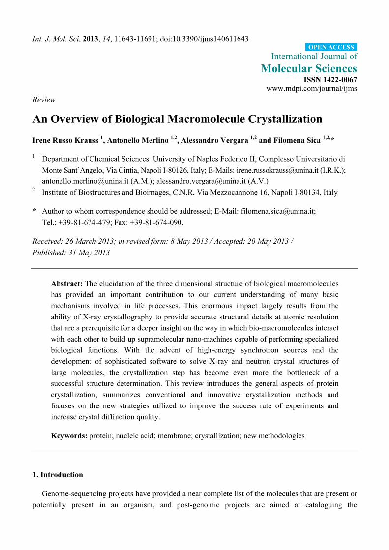
The three stages of crystallization common to all molecules (Figure 1) are nucleation, crystal
growth and cessation of growth [9–12]. During nucleation an adequate amount of molecules associate
in three dimensions to form a thermodynamically stable aggregate, the so called critical nucleus, which
provides surfaces suitable for crystal growth. The growth stage, which immediately follows the
nucleation, is governed by the diffusion of particles to the surface of the critical nuclei and their
ordered assembling onto the growing crystal (Figure 1).
Protein crystal formation requires interactions that are specific, highly directional and organized in a
manner that is appropriate for three-dimensional crystal lattice formation. Crystal growth ends when
the solution is sufficiently depleted of protein molecules, deformation-induced strain destabilizes
the lattice, or the growing crystal faces become poisoned by impurities. The crystallizability of a
protein is strictly affected by the chemical and conformational purity and the oligomeric homogeneity
of the sample.
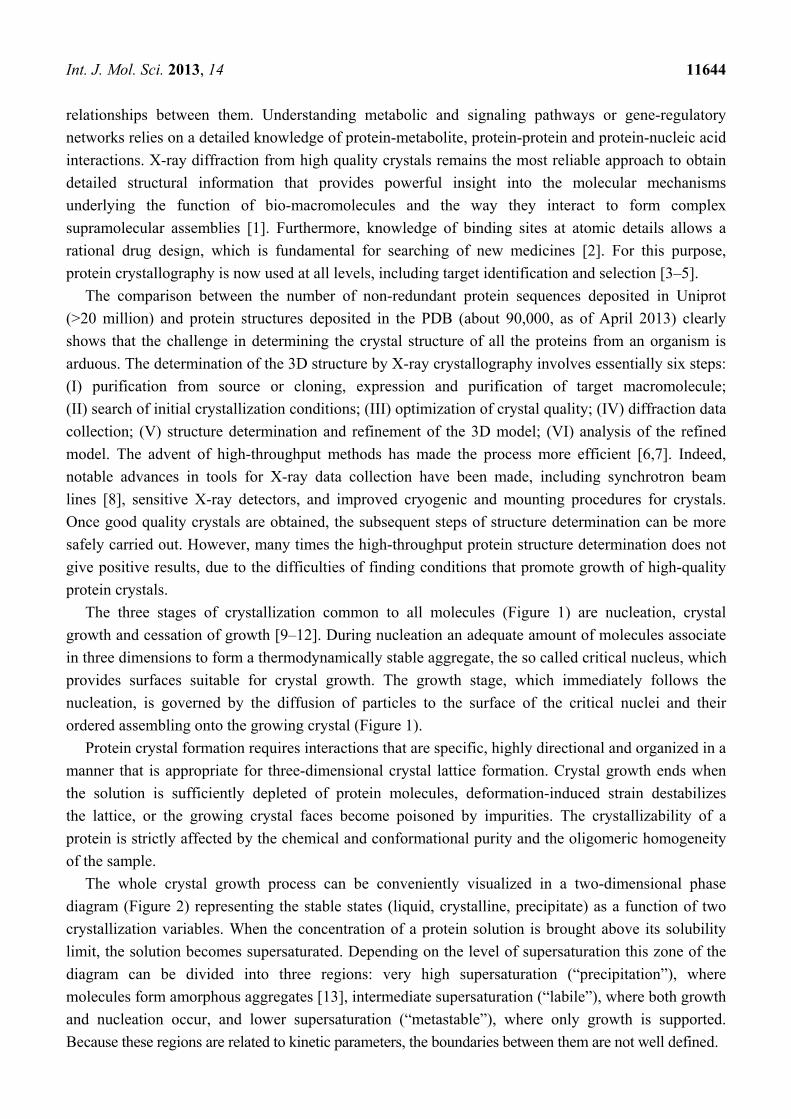
The whole crystal growth process can be conveniently visualized in a two-dimensional phase
diagram (Figure 2) representing the stable states (liquid, crystalline, precipitate) as a function of two
crystallization variables. When the concentration of a protein solution is brought above its solubility
limit, the solution becomes supersaturated. Depending on the level of supersaturation this zone of the
diagram can be divided into three regions: very high supersaturation (“precipitation”), where
molecules form amorphous aggregates [13], intermediate supersaturation (“labile”), where both growth
and nucleation occur, and lower supersaturation (“metastable”), where only growth is supported.
Because these regions are related to kinetic parameters, the boundaries between them are not well defined.
Int. J. Mol. Sci. 2013, 14 11645
Figure 1. Fluid aggregate model. Increasing supersaturation promotes molecule
association, which begins to organize into large disordered aggregates (a) molecules within
the cores of such aggregates reorient, redistribute and form more geometrically rigorous
interactions; (b) These latter interactions tend to order and stabilize the aggregate core,
which increases to produce a critical nucleus; (c) This ultimately develops into a true
crystal; (d) Free molecules are then adsorbed to the crystal surface and increase its size by
their incorporation into its lattice.
Figure 2. Schematic solubility curve for a protein, as a function of the protein concentration.
The best strategy that should be employed is to induce nucleation at the lowest level of
supersaturation just within the labile region. Following nuclei formation, the concentration of protein
in the solution gradually decreases, driving the system into the metastable zone, where growth occurs
slowly. However, it is very difficult to identify these ideal conditions and in order to obtain
high-quality crystals it could be necessary to physically separate nucleation and growth steps.
Chemical space in crystallization experiments is multidimensional, and several zones may correspond
to nucleation and growth of different crystal forms. It is not yet possible to predict the conditions
Int. J. Mol. Sci. 2013, 14 11646
required to crystallize a protein from its chemico-physical properties. Changes in a single experimental
parameter can simultaneously influence several aspects of a crystallization experiment.
Despite the importance of protein crystallization, the insights into this process are still limited and
currently there are no systematic methods to ensure that ordered three-dimensional crystals will be
obtained. This problem has stimulated many efforts to improve the success of protein crystal
growth experiments.
The present work will review some of the basic ideas and principles of biological macromolecule
crystallization, summarize the standard approaches in crystal growth and illustrate novel tools and
strategies to increase the rate of positive results and the diffraction quality of crystals.
This manuscript focuses mainly on soluble proteins, but covers also the crystallization of membrane
proteins, nucleic acids and nucleic acids/protein complexes, and discusses general physico-chemical
aspects of the crystallization mechanisms.
2. Factors Affecting Crystallization of Bio-Macromolecules
Crystal growth of bio-macromolecules is a multi-parametric process as it depends on several
factors, such as sample concentration, temperature, pH value, precipitant, buffer, additive, detergent,
physical fields, pressure etc. (Table 1).
Table 1. Parameters affecting crystallization process [10,12,14,15].
Physical factors Chemical factors Biochemical factors
Temperature Precipitant type Sample purity Pressure Precipitant concentration Sample homogeneity Gravity pH Sequence modifications
Magnetic fields Buffer type Posttranslational modifications Electric fields Ionic strength Chemical modifications
Dielectric properties Sample concentration Aggregation Viscosity Metal ions Proteolysis
Vibrations and sound Polyions Sample pI Time Detergents Ligands, co-factors, inhibitors
Equilibration rate Heavy metals Nucleants Small molecule impurities
Methodology Crosslinkers Surface of crystallization device Reagent source
Sample handling Reagent formulation
All these thermodynamic and dynamics variables can be used to control supersaturation in the
system and, thus, indirectly, the rates of nucleation and growth. In the following sections we will
describe in details some parameters affecting protein crystallization, which also govern crystallization
of other biological macromolecules, such as nucleic acids or macromolecular assemblies.
2.1. Sample Purity and Homogeneity
Chemical and conformational purity of the sample strongly affects the ability to grow crystals. It
can be simply assessed by sodium dodecylsulfate-polyacrylamide gel electrophoresis and mass
Int. J. Mol. Sci. 2013, 14 11647
spectrometry (MS). As regards the secondary structure analysis, two techniques are currently used:
Fourier Transform infrared Spectroscopy (FT-IR) and UV circular dichroism (UV-CD). However,
UV-CD is the most useful, mainly due to the high concentration of sample required for FT-IR
studies [16]. Sample homogeneity, another fundamental precept of crystallization, can be determined
with dynamic (DLS) and/or static light (SLS) scattering. The light scattered from a solution may be
analyzed either in terms of its intensity or in terms of its fluctuations. In the former method, which is
called static light scattering, the measure of scattered light intensity as function of angle is used to find
the molar mass, the mean squared radius of gyration and the second viral coefficient (B22). The
measurements may be also performed at a single angle, provided that the concentration and the
refractive index are known. A B22 value in the range −0.8 × 10⁻⁴ to −8 × 10⁻⁴ mol mL g−2 indicates the
possibility of a successful crystallization, whereas a value outside this range will probably result in
crystallization failure. This procedure has been used to investigate solubility and crystallizability of
many macromolecules [17–20]. The results of SLS experiments can be used as a quality control of
protein preparation and to investigate the solution oligomeric state (monomer/dimer, etc.). On the other
hand, DLS detects the fluctuations of the scattering intensity due to the Brownian motion of molecules
in solution [21,22]. The degree of these fluctuations depends on the diffusion coefficient of the
scattering particle, a quantity which is related to the hydrodynamic radius through the Stokes-Einstein
equation. The mathematical treatment of the data also permits to assess the degree of homogeneity
(polydispersity index) as well as to construct size distribution. Furthermore, Saridakis et al. [23]
demonstrated that the DLS-based separation of nucleation and crystal growth processes can result in
protein crystals with improved size.
2.2. Temperature
Temperature governs the balance between enthalpy and entropy effects on the free energy.
Depending on whether crystallization is enthalpy- or entropy-driven, proteins may become either more
or less soluble at higher temperature. Some proteins display a characteristic increased solubility with
increasing temperature, whereas others display a decreased, or retrograde solubility [24]. The
dependence of protein solubility on temperature is due to the variation of the acid/base reaction
constant of the protein side chains as function of temperature [25]. Furthermore, pKa values of
ionizable groups are strictly related to the medium ionic strength. As a consequence, in the case of
proteins with normal solubility, it increases with a temperature increment at low ionic strength, for
example if the solution contains components with low dielectric constant, whereas decreases
at high ionic strength. In the latter case, however, the solubility variation is very small. The
temperature-solubility function is not a property of the protein itself, but is subtly related to the
protein-solution system. Equally relevant is the influence of temperature on the rates of nucleation and
growth, and on the equilibrium position of the trial. Generally, in a crystallization laboratory,
experiments are performed at two different temperatures (4 °C and 20 °C). However, recently many
crystallization devices with a fine control of the temperature have been developed to take advantage of
the effects of this parameter on the growth mechanism and the crystal form.
Int. J. Mol. Sci. 2013, 14 11648
2.3. pH
Proteins generally contain numerous ionizable groups, which have a variety of pKas. As a
consequence, protein solubility can dramatically change as pH is altered even by only 0.5 pH units and
in some cases it varies for very small pH changes (0.1 units). The pH affects the detailed nature of
protein-protein interactions modifying the possibilities of forming salt bridges and hydrogen bonds
crucial to the formation of specific crystal contacts [26]. Electrostatic interactions, which depend on
the protonation state of aminoacid side chains, play a key role in the binding specificity, in protein
hydration and in the interactions with small molecules and ions that sometimes mediate the crystal
packing contacts. At a pH characteristic for each protein, called the isoelectric point (pI), the positive
charges of the molecule exactly balance the negative ones. This would seem to be the best situation for
crystal growth as no overall electrostatic repulsion between protein molecules is present.
Unfortunately, this idea was not confirmed by an analysis of crystallization conditions of almost ten
thousand unique protein crystal forms [27]. Consequently, a wide pH range has to be explored in the
crystallization experiments, but only pH values that maintain the folded structure of the protein are
acceptable conditions for protein crystal growth.
2.4. Thermal Stability
The correlation between protein thermal stability and probability of yielding crystals is
controversial [28]. However, in many cases pre-crystallization screening based on stability has
substantially increased the crystallization success rate [29,30]. A rapid and low-cost method able to
determine protein stability is the fluorescence-based thermal shift assay, also referred to as a
“differential scanning fluorimetry” (DSF). This method measures the melting temperature of a protein
by monitoring the signal of an external fluorescent probe which interacts with hydrophobic core
residues when they become solvent-exposed during the unfolding process [31]. The low quantity of
starting material required for an average thermal shift experiment makes DSF particularly suitable for
use in the screening of optimal conditions for protein crystallization targets.
2.5. Precipitant
Chemical compounds that reduce protein solubility are referred to as crystallizing (or precipitating)
agents. They reinforce the attractions among bio-macromolecules and act either by altering the activity
coefficient of water (salts) [32], or by changing the dielectric constant of the solvating medium
(organic solvents) or by increasing molecular crowding (high molecular weight polymers like
polyethylene glycol, PEG) [33]. Precipitants that act by different mechanisms show little exchangeability:
crystals obtained with one type of precipitant do not commonly form if the precipitant is changed with
a functionally different one. However, it has been exhaustively demonstrated that combinations of
mechanistically distinct precipitating agents can be synergistic and increase the probability of crystal
growth [34] (Table 2).
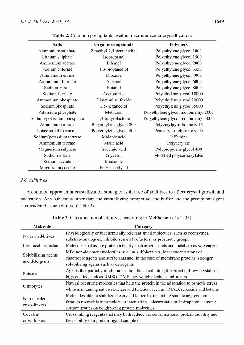
Int. J. Mol. Sci. 2013, 14 11649
Table 2. Common precipitants used in macromolecular crystallization.
Salts Organic compounds Polymers
Ammonium sulphate 2-methyl-2,4-pentanediol Polyethylene glycol 1000 Lithium sulphate Isopropanol Polyethylene glycol 1500
Ammonium acetate Ethanol Polyethylene glycol 2000 Sodium chloride 1,3-propanediol Polyethylene glycol 3350
Ammonium citrate Dioxane Polyethylene glycol 4000 Ammonium formate Acetone Polyethylene glycol 6000
Sodium citrate Butanol Polyethylene glycol 8000 Sodium formate Acetonitrile Polyethylene glycol 10000
Ammonium phosphate Dimethyl sulfoxide Polyethylene glycol 20000 Sodium phosphate 2,5-hexanediol Polyethylene glycol 35000
Potassium phosphate Methanol Polyethylene glycol monomethyl 2000 Sodium/potassium phosphate 1,3-butyrolactone Polyethylene glycol monomethyl 5000
Ammonium nitrate Polyethylene glycol 200 Polyvinylpyrrolidone K 15 Potassium thiocyanate Polyethylene glycol 400 Pentaerythritolpropoxylate
Sodium/potassium tartrate Malonic acid Jeffamine Ammonium tartrate Malic acid Polyacrylate Magnesium sulphate Succinic acid Polypropylene glycol 400
Sodium nitrate Glycerol Modified polycarboxylates Sodium acetate Imidazole
Magnesium acetate Ethylene glycol
2.6. Additives
A common approach in crystallization strategies is the use of additives to affect crystal growth and
nucleation. Any substance other than the crystallizing compound, the buffer and the precipitant agent
is considered as an additive (Table 3).
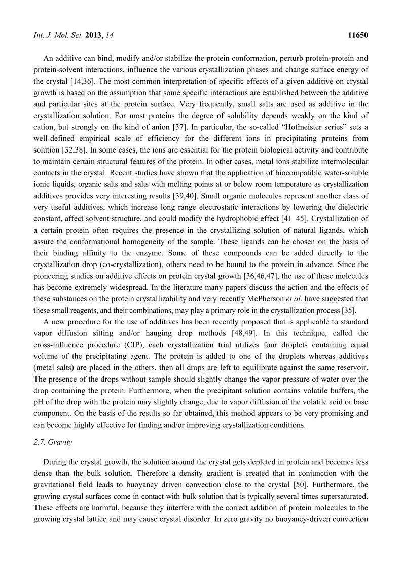
Table 3. Classification of additives according to McPherson et al. [35].
Molecule Category
Natural additives Physiologically or biochemically relevant small molecules, such as coenzymes, substrate analogues, inhibitors, metal cofactors, or prosthetic groups
Chemical protectants Molecules that assure protein integrity such as reductants and metal atoms scavengers
Solubilizing agents and detergents
Mild non-detergent molecules, such as sulfobetaines, low concentrations of chaotropic agents and surfactants and, in the case of membrane proteins, stronger solubilizing agents such as detergents
Poisons Agents that partially inhibit nucleation thus facilitating the growth of few crystals of high quality, such as DMSO, DMF, low weigh alcohols and sugars
Osmolytes Natural occurring molecules that help the protein in the adaptation to osmotic stress while maintaining native structure and function, such as TMAO, sarcosine and betaine
Non-covalent cross-linkers
Molecules able to stabilize the crystal lattice by mediating sample aggregation through reversible intermolecular interactions, electrostatic or hydrophobic, among surface groups on neighboring protein molecules
Covalent cross-linkers
Crosslinking reagents that may both reduce the conformational protein mobility and the stability of a protein-ligand complex
Int. J. Mol. Sci. 2013, 14 11650
An additive can bind, modify and/or stabilize the protein conformation, perturb protein-protein and
protein-solvent interactions, influence the various crystallization phases and change surface energy of
the crystal [14,36]. The most common interpretation of specific effects of a given additive on crystal
growth is based on the assumption that some specific interactions are established between the additive
and particular sites at the protein surface. Very frequently, small salts are used as additive in the
crystallization solution. For most proteins the degree of solubility depends weakly on the kind of
cation, but strongly on the kind of anion [37]. In particular, the so-called “Hofmeister series” sets a
well-defined empirical scale of efficiency for the different ions in precipitating proteins from
solution [32,38]. In some cases, the ions are essential for the protein biological activity and contribute
to maintain certain structural features of the protein. In other cases, metal ions stabilize intermolecular
contacts in the crystal. Recent studies have shown that the application of biocompatible water-soluble
ionic liquids, organic salts and salts with melting points at or below room temperature as crystallization
additives provides very interesting results [39,40]. Small organic molecules represent another class of
very useful additives, which increase long range electrostatic interactions by lowering the dielectric
constant, affect solvent structure, and could modify the hydrophobic effect [41–45]. Crystallization of
a certain protein often requires the presence in the crystallizing solution of natural ligands, which
assure the conformational homogeneity of the sample. These ligands can be chosen on the basis of
their binding affinity to the enzyme. Some of these compounds can be added directly to the
crystallization drop (co-crystallization), others need to be bound to the protein in advance. Since the
pioneering studies on additive effects on protein crystal growth [36,46,47], the use of these molecules
has become extremely widespread. In the literature many papers discuss the action and the effects of
these substances on the protein crystallizability and very recently McPherson et al. have suggested that
these small reagents, and their combinations, may play a primary role in the crystallization process [35].
A new procedure for the use of additives has been recently proposed that is applicable to standard
vapor diffusion sitting and/or hanging drop methods [48,49]. In this technique, called the
cross-influence procedure (CIP), each crystallization trial utilizes four droplets containing equal
volume of the precipitating agent. The protein is added to one of the droplets whereas additives
(metal salts) are placed in the others, then all drops are left to equilibrate against the same reservoir.
The presence of the drops without sample should slightly change the vapor pressure of water over the
drop containing the protein. Furthermore, when the precipitant solution contains volatile buffers, the
pH of the drop with the protein may slightly change, due to vapor diffusion of the volatile acid or base
component. On the basis of the results so far obtained, this method appears to be very promising and
can become highly effective for finding and/or improving crystallization conditions.
2.7. Gravity
During the crystal growth, the solution around the crystal gets depleted in protein and becomes less
dense than the bulk solution. Therefore a density gradient is created that in conjunction with the
gravitational field leads to buoyancy driven convection close to the crystal [50]. Furthermore, the
growing crystal surfaces come in contact with bulk solution that is typically several times supersaturated.
These effects are harmful, because they interfere with the correct addition of protein molecules to the
growing crystal lattice and may cause crystal disorder. In zero gravity no buoyancy-driven convection
Int. J. Mol. Sci. 2013, 14 11651
occurs and the growing crystal does not move with respect to the surrounding fluid. The matter is
transported in a purely diffusive way [51] and the crystal growth takes place under ideal conditions
where the growing surface is in contact with a solution that is slightly supersaturated. Another negative
effect of gravity on growing crystal is sedimentation that may induce the partially formed crystals to
fall on top of one another where they continue to grow. However, the results of crystallization
experiments performed in space are controversial and indicated that about 30% of space-grown protein
crystals yield better X-ray diffraction data than the best crystals grown on the earth [50,52,53]. This
because aboard orbiting space crafts all objects experience forces that adversely affect both the
crystallization process and the crystal quality by inducing deviations from the ideal diffusion controlled
growth and movements of crystals. The forces derive from random accelerations, called g-jitters,
produced by mechanical vibrations caused by operation of equipment. Furthermore, experimentation in
space has restricted access and is extremely expensive and difficult to control.
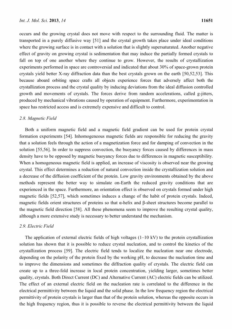
2.8. Magnetic Field
Both a uniform magnetic field and a magnetic field gradient can be used for protein crystal
formation experiments [54]. Inhomogeneous magnetic fields are responsible for reducing the gravity
that a solution feels through the action of a magnetization force and for damping of convection in the
solution [55,56]. In order to suppress convection, the buoyancy forces caused by differences in mass
density have to be opposed by magnetic buoyancy forces due to differences in magnetic susceptibility.
When a homogeneous magnetic field is applied, an increase of viscosity is observed near the growing
crystal. This effect determines a reduction of natural convection inside the crystallization solution and
a decrease of the diffusion coefficient of the protein. Low gravity environments obtained by the above
methods represent the better way to simulate on-Earth the reduced gravity conditions that are
experienced in the space. Furthermore, an orientation effect is observed on crystals formed under high
magnetic fields [52,57], which sometimes induces a change of the habit of protein crystals. Indeed,
magnetic fields orient structures of proteins so that α-helix and β-sheet structures become parallel to
the magnetic field direction [58]. All these phenomena seem to improve the resulting crystal quality,
although a more extensive study is necessary to better understand the mechanism.
2.9. Electric Field
The application of external electric fields of high voltages (1–10 kV) to the protein crystallization
solution has shown that it is possible to reduce crystal nucleation, and to control the kinetics of the
crystallization process [59]. The electric field tends to localize the nucleation near one electrode,
depending on the polarity of the protein fixed by the working pH, to decrease the nucleation time and
to improve the dimensions and sometimes the diffraction quality of crystals. The electric field can
create up to a three-fold increase in local protein concentration, yielding larger, sometimes better
quality, crystals. Both Direct Current (DC) and Alternative Current (AC) electric fields can be utilized.
The effect of an external electric field on the nucleation rate is correlated to the difference in the
electrical permittivity between the liquid and the solid phase. In the low frequency region the electrical
permittivity of protein crystals is larger than that of the protein solution, whereas the opposite occurs in
the high frequency region, thus it is possible to reverse the electrical permittivity between the liquid
Int. J. Mol. Sci. 2013, 14 11652
and the solid by changing frequency. The effect of the external electric field on the nucleation rate
increases with an increase in the concentration of the precipitant: a larger ionic strength of
the precipitant corresponds to a greater effect of the external electric field on the crystallization
process [60]. Comparison of the results of crystallization trials performed with the same precipitant but
with different frequencies indicates that the nucleation rate can be controlled by imposing an external
electric field with an appropriate frequency. The nucleation rate of hen egg white lysozyme was
successfully controlled, in terms of both increase and decrease in nucleation, by application of an AC
electric field [61]. A number of devices have been developed to set-up experiments under electric
field [62–68]. Electric field-induced crystallization experiments can be also performed by putting the
electrodes directly in contact with the protein solution (called internal EF) [69,70]. Since electrochemical
reactions could occur on the electrodes, this technique needs precise control in applying the adequate
current or potential. The main features of the internal EF protein crystallization, that are fast
crystallization and good quality of the crystals, are associated with a higher flux of mass from the bulk
of the cell to the region near one of the electrodes, where the crystallization is being favored. However,
the details of the processes that cause crystallization in an electric field remain unclear, so most
successes using this technique are the result of a trial and error procedure. To date these studies have
only dealt with model proteins, but the remarkable results on nucleation control, size distribution and
diffraction quality of crystals make the use of electric field for crystal growth very promising.
2.10. Stirring
Stirring of crystallization solution has been found to affect crystal nucleation and growth. Stirring
enables the control of crystal growth by minimizing concentration gradient across the crystal surface. It
prevents excess spontaneous nucleation and accelerates the growth of protein crystals, thus resulting in
the growth of large and high-quality crystals and, in some cases, also in the production of crystals of
molecules that do not easily crystallize using conventional techniques. It has been supposed that
stirring increases the interactions between protein molecules, which might associate leading to the
development of large clusters that might evolve into stable critical nuclei [71]. However, it is necessary
to stir gently the sample solution, to avoid problems such as spontaneous nucleation, protein denaturation,
and damaged crystals [72]. It is important to remember that crystal growth and crystal morphologies in
a stirred solution are strongly dependent on the stirring speed [73]. Several experimental set-ups
are used to apply the desired mild stirring to crystallization solutions. In particular, it is worth to
mention two simple methods: the micro-stirring and the micro-floating and stirring (micro-FAST)
techniques ([72] and references therein). In the micro-stirring technique the crystallization mixture is
gently rotated by using a common rotating or vibrating shaker. This procedure enables an easy mixing
of the solution and a control of the forced flow conditions by changing the rotation speed and type of
shaker. In a micro-FAST experiment the protein solution is added to an insoluble and highly dense
liquid, from which it separates: protein solution floats on top of this liquid, forming an interface. The
protein crystals that grew at the interface did not contact the vessel, resulting in regular crystals with
improved crystallinity. The liquid of the lower layer is mixed using a magnetic stirrer.
Several examples of successful application of stirring methods are reported in the literature: the
crystal quality of human triosephosphate isomerase [74], adenosine deaminase [71], Src homology
Int. J. Mol. Sci. 2013, 14 11653
2 domain-containing protein tyrosine phosphatase substrate-1 [75] was dramatically improved in
this way.
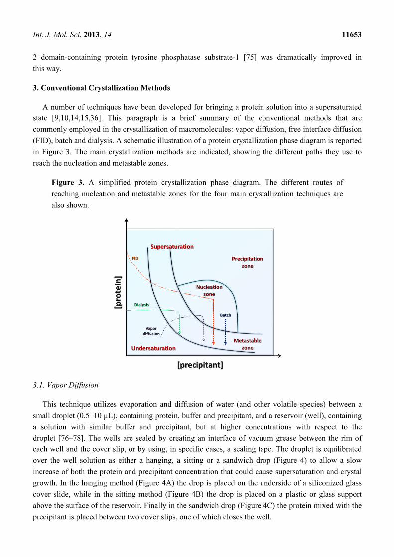
3. Conventional Crystallization Methods
A number of techniques have been developed for bringing a protein solution into a supersaturated
state [9,10,14,15,36]. This paragraph is a brief summary of the conventional methods that are
commonly employed in the crystallization of macromolecules: vapor diffusion, free interface diffusion
(FID), batch and dialysis. A schematic illustration of a protein crystallization phase diagram is reported
in Figure 3. The main crystallization methods are indicated, showing the different paths they use to
reach the nucleation and metastable zones.
Figure 3. A simplified protein crystallization phase diagram. The different routes of
reaching nucleation and metastable zones for the four main crystallization techniques are
also shown.
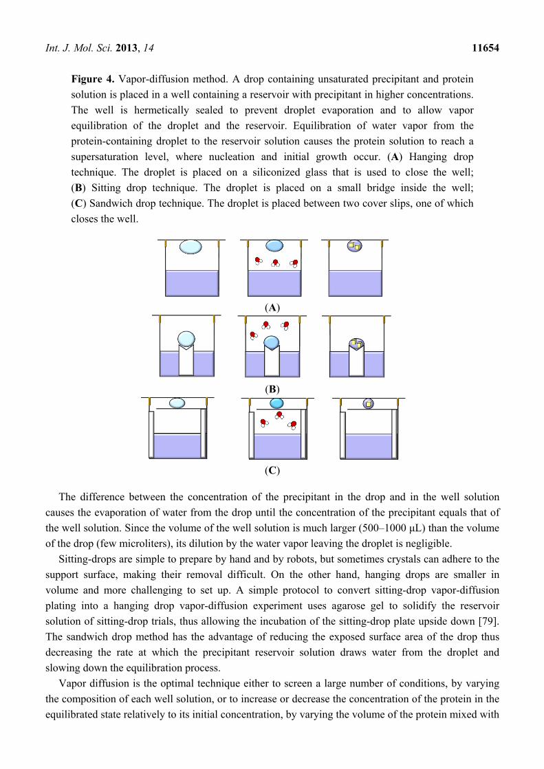
3.1. Vapor Diffusion
This technique utilizes evaporation and diffusion of water (and other volatile species) between a
small droplet (0.5–10 μL), containing protein, buffer and precipitant, and a reservoir (well), containing
a solution with similar buffer and precipitant, but at higher concentrations with respect to the
droplet [76–78]. The wells are sealed by creating an interface of vacuum grease between the rim of
each well and the cover slip, or by using, in specific cases, a sealing tape. The droplet is equilibrated
over the well solution as either a hanging, a sitting or a sandwich drop (Figure 4) to allow a slow
increase of both the protein and precipitant concentration that could cause supersaturation and crystal
growth. In the hanging method (Figure 4A) the drop is placed on the underside of a siliconized glass
cover slide, while in the sitting method (Figure 4B) the drop is placed on a plastic or glass support
above the surface of the reservoir. Finally in the sandwich drop (Figure 4C) the protein mixed with the
precipitant is placed between two cover slips, one of which closes the well.
Int. J. Mol. Sci. 2013, 14 11654
Figure 4. Vapor-diffusion method. A drop containing unsaturated precipitant and protein
solution is placed in a well containing a reservoir with precipitant in higher concentrations.
The well is hermetically sealed to prevent droplet evaporation and to allow vapor
equilibration of the droplet and the reservoir. Equilibration of water vapor from the
protein-containing droplet to the reservoir solution causes the protein solution to reach a
supersaturation level, where nucleation and initial growth occur. (A) Hanging drop
technique. The droplet is placed on a siliconized glass that is used to close the well;
(B) Sitting drop technique. The droplet is placed on a small bridge inside the well;
(C) Sandwich drop technique. The droplet is placed between two cover slips, one of which
closes the well.
(A)
(B)
(C)
The difference between the concentration of the precipitant in the drop and in the well solution
causes the evaporation of water from the drop until the concentration of the precipitant equals that of
the well solution. Since the volume of the well solution is much larger (500–1000 μL) than the volume
of the drop (few microliters), its dilution by the water vapor leaving the droplet is negligible.
Sitting-drops are simple to prepare by hand and by robots, but sometimes crystals can adhere to the
support surface, making their removal difficult. On the other hand, hanging drops are smaller in
volume and more challenging to set up. A simple protocol to convert sitting-drop vapor-diffusion
plating into a hanging drop vapor-diffusion experiment uses agarose gel to solidify the reservoir
solution of sitting-drop trials, thus allowing the incubation of the sitting-drop plate upside down [79].
The sandwich drop method has the advantage of reducing the exposed surface area of the drop thus
decreasing the rate at which the precipitant reservoir solution draws water from the droplet and
slowing down the equilibration process.
Vapor diffusion is the optimal technique either to screen a large number of conditions, by varying
the composition of each well solution, or to increase or decrease the concentration of the protein in the
equilibrated state relatively to its initial concentration, by varying the volume of the protein mixed with
Int. J. Mol. Sci. 2013, 14 11655
the well solution when the drop is initially setup. In a recent study, the effects of diluting either protein
or crystallization agents in the droplets on reproducibility and screening efficiency of protein
crystallization was investigated [80]. The results indicate that reducing the initial concentration of the
crystallization agent in the droplet leaving the protein concentration unchanged increases the rate of
vapor diffusion and produces higher success rate of protein crystallization. In contrast, dilution of the
protein while the initial concentration of the crystallization agent remains unchanged produces effects
that depend on the kind of protein. These findings seem to be in contrast with those reported by
Chayen et al., who found that slowing down the equilibration rate, by varying the distance between the
reservoir and the crystallization drop or by inserting an oil barrier over the reservoir, improves the
crystals size and quality [81,82]. These data clearly confirm the complexity of understanding and
controlling the various factors intervening in the protein crystallization process.
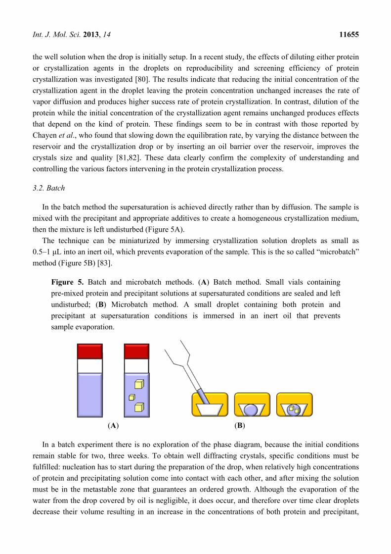
3.2. Batch
In the batch method the supersaturation is achieved directly rather than by diffusion. The sample is
mixed with the precipitant and appropriate additives to create a homogeneous crystallization medium,
then the mixture is left undisturbed (Figure 5A).
The technique can be miniaturized by immersing crystallization solution droplets as small as
0.5–1 μL into an inert oil, which prevents evaporation of the sample. This is the so called “microbatch”
method (Figure 5B) [83].
Figure 5. Batch and microbatch methods. (A) Batch method. Small vials containing
pre-mixed protein and precipitant solutions at supersaturated conditions are sealed and left
undisturbed; (B) Microbatch method. A small droplet containing both protein and
precipitant at supersaturation conditions is immersed in an inert oil that prevents
sample evaporation.
(A) (B)
In a batch experiment there is no exploration of the phase diagram, because the initial conditions
remain stable for two, three weeks. To obtain well diffracting crystals, specific conditions must be
fulfilled: nucleation has to start during the preparation of the drop, when relatively high concentrations
of protein and precipitating solution come into contact with each other, and after mixing the solution
must be in the metastable zone that guarantees an ordered growth. Although the evaporation of the
water from the drop covered by oil is negligible, it does occur, and therefore over time clear droplets
decrease their volume resulting in an increase in the concentrations of both protein and precipitant,
Int. J. Mol. Sci. 2013, 14 11656
which can yield precipitate or protein crystals. The main disadvantage of this method is that in many
cases equilibration occurs very rapidly, thus affecting the rate of crystal growth and consequently the
quality of crystals. Furthermore, alcohols, detergents, and lipids can diffuse into the oil. To avoid
drying out of the droplets over prolonged periods it is possible to control the rate of decrease in volume
of the droplets by regulating the vapor pressure within the crystallization tray [84]. On the other hand,
varying ratios of water permeable oils to control water diffusion into the environment, it is possible to
obtain more rapid crystallization [84–86].
Very recently, a new approach to perform microbatch crystallization has been proposed, the
without-oil microbatch method [87]. This method is based on the idea that is possible to adapt the
crystal growth conditions found with vapor diffusion experiments to the batch technique without
changing the device and preserving almost the same composition of the reservoir solution. Protein and
precipitant are mixed to their final concentration at the beginning of the experiment. The drop is then
stored in the presence of a reservoir with the same precipitant concentration, to avoid drop evaporation.
Crystallization usually occurs using 60%–80% of the concentration of the precipitant required in the
hanging drop experiment. The advantage of this procedure is eliminating the most significant
drawback of the under-oil microbatch method, i.e., the slow evaporation of water from the
crystallization drops that sometime results in the formation of salt deposits that interfere with protein
crystal growth.
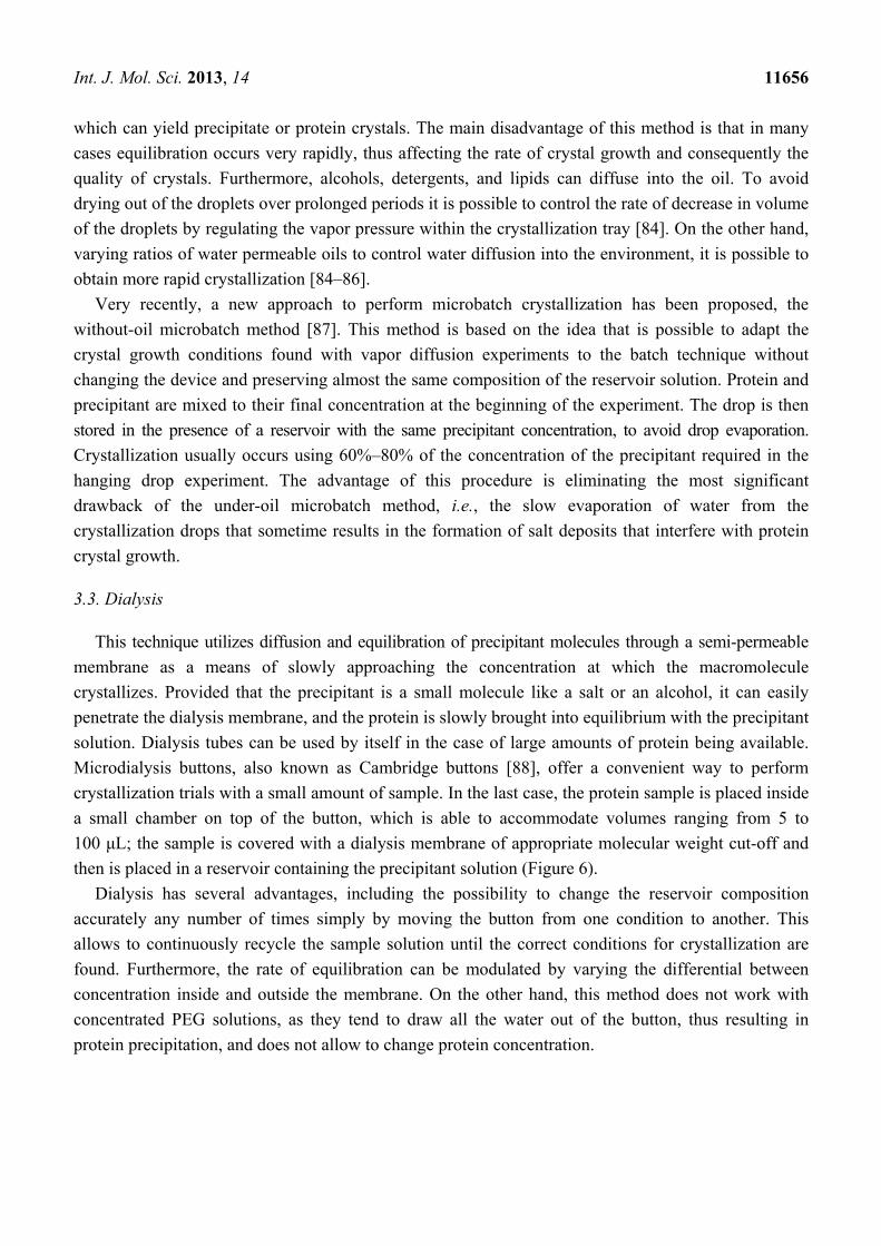
3.3. Dialysis
This technique utilizes diffusion and equilibration of precipitant molecules through a semi-permeable
membrane as a means of slowly approaching the concentration at which the macromolecule
crystallizes. Provided that the precipitant is a small molecule like a salt or an alcohol, it can easily
penetrate the dialysis membrane, and the protein is slowly brought into equilibrium with the precipitant
solution. Dialysis tubes can be used by itself in the case of large amounts of protein being available.
Microdialysis buttons, also known as Cambridge buttons [88], offer a convenient way to perform
crystallization trials with a small amount of sample. In the last case, the protein sample is placed inside
a small chamber on top of the button, which is able to accommodate volumes ranging from 5 to
100 μL; the sample is covered with a dialysis membrane of appropriate molecular weight cut-off and
then is placed in a reservoir containing the precipitant solution (Figure 6).
Dialysis has several advantages, including the possibility to change the reservoir composition
accurately any number of times simply by moving the button from one condition to another. This
allows to continuously recycle the sample solution until the correct conditions for crystallization are
found. Furthermore, the rate of equilibration can be modulated by varying the differential between
concentration inside and outside the membrane. On the other hand, this method does not work with
concentrated PEG solutions, as they tend to draw all the water out of the button, thus resulting in
protein precipitation, and does not allow to change protein concentration.
Int. J. Mol. Sci. 2013, 14 11657
Figure 6. Microdialysis method. The droplet of protein solution is placed in a small button,
which is covered with a dialysis membrane and closed with a thin elastic band. The button
is immersed in the precipitant solution and the equilibration of precipitant molecules occurs
through the membrane.
The dialysis equilibration kinetics depend on the molecular weight cut-off of the membrane, the
precipitant, the concentration of components inside and outside the membrane, the dimensions of
button, the reservoir volume, the viscosity of the solution, and on variation of chemical physics
parameters. The rate of equilibration can be reduced to provide enhanced control over the
crystallization process by a double dialysis: the button is placed inside a reservoir, which contains a
certain concentration of crystallizing agent and is sealed with a dialysis membrane, which is in turn
placed inside a second reservoir with a higher concentration of crystallizing agent [89].
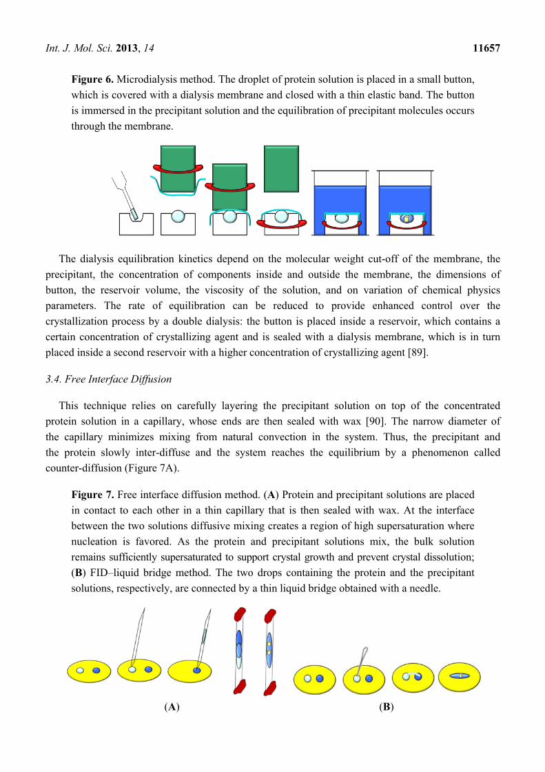
3.4. Free Interface Diffusion
This technique relies on carefully layering the precipitant solution on top of the concentrated
protein solution in a capillary, whose ends are then sealed with wax [90]. The narrow diameter of
the capillary minimizes mixing from natural convection in the system. Thus, the precipitant and
the protein slowly inter-diffuse and the system reaches the equilibrium by a phenomenon called
counter-diffusion (Figure 7A).
Figure 7. Free interface diffusion method. (A) Protein and precipitant solutions are placed
in contact to each other in a thin capillary that is then sealed with wax. At the interface
between the two solutions diffusive mixing creates a region of high supersaturation where
nucleation is favored. As the protein and precipitant solutions mix, the bulk solution
remains sufficiently supersaturated to support crystal growth and prevent crystal dissolution;
(B) FID–liquid bridge method. The two drops containing the protein and the precipitant
solutions, respectively, are connected by a thin liquid bridge obtained with a needle.
(A) (B)

Int. J. Mol. Sci. 2013, 14 11658
When the solutions initially come into contact and diffusive mixing occurs, the region of the protein
solution in the neighborhood of the interface becomes supersaturated and the ideal conditions for
nuclei formation are created. As time proceeds, the two solutions inter-diffuse along the axis of the
capillary and dilute each other, thus promoting the dissolution of the smaller nuclei and the growth of
the larger ones. The achievement, by the free liquid diffusion (FID), of transient nucleation conditions
in most cases allows to obtain high quality crystals. Thus FID can be view as a rational crystallization
approach to minimize supersaturation and impurity levels at the crystal growth front and to ensure
steadiness of both values. However, this method is not widely utilized by crystal growers due to the
large sample volume that is needed, to the difficulty to set up a good interface, and to the presence of
convective currents associated to density differences in the two solutions that disturbs the interface.
The last problem can be partially solved by putting the more dense solution on the bottom or by
freezing one of the two solutions [91,92]. Usually, the capillary is filled with the precipitant solution
and placed in a freezer at 193 K; once the solution is completely frozen, the capillary is moved in a bed
of dry ice for the addition of protein sample. At this time the system is left to equilibrate at the desired
temperature. As it is demonstrated in various papers [51,93], a better way to solve the problems of the
free diffusion method is to perform the experiments in microgravity, where gravity induced convection
is eliminated. Currently, the nanoliter scale FID is considered particularly attractive and efficient for
crystallization. It is performed by using microfluidic devices [94] that allow an ultra-small volume
screening of protein crystallization conditions. Once chemical conditions conducive to crystallization
have been identified, they may be exported to traditional FID by subsequent refinement. In some cases,
diffraction-quality crystals have been also grown and harvested from such nanoliter-volume trials.
A variant of the FID method is the liquid bridge [95], in which a drop of protein sample and a drop
of precipitant solution are placed in close proximity on a cover glass and then connected by a thin
liquid bridge, obtained with the help of a small needle (Figure 7B). The liquid diffusion between the
two droplets, sealed from air, may induce crystal growth.
3.5. Gel Crystallization
In the course of crystallogenesis investigations good results have been obtained by using gelified
media [96,97]. This technique is currently utilized to lower the percentage of growth defects and to
control the nucleation effectively, as in gel media the gravity-induced convection is suppressed [98].
The nuclei are trapped in the pores of gel matrix and as a consequence crystals cannot move or
sedimentate [99–101]. Such a stable growth environment may favor high internal order and generate
crystals with low mosaicity, with a reduced incorporation of impurities and homogeneously grown in
the three dimensions. The supersaturation in a gel bead can be dynamically controlled by changing the
precipitant and/or the protein concentration in the bulk solution. Manipulation of the supersaturation
allows the nucleation rate to be varied and eventually leads to the production of large crystals, which
are more homogeneously distributed in the gel bead. Hydrogels also provide an efficient protection of
crystalline samples during handling and transport, without affecting their diffraction quality. In
addition, hydrogels are completely transparent to X-rays and their presence can be crucial for
preserving the diffraction properties during cryo-cooling, as was shown for instance in the case of
aspartyl-tRNA synthetase crystals [102]. In many cases crystals grown in gels show a higher
Int. J. Mol. Sci. 2013, 14 11659
diffraction limit compared with crystals grown in solution [101,103], but this benefit is largely
underexploited by protein crystal growers because the procedures are complicated and require large
quantities (>10 µL) of sample [104]. For these reasons, in the past few years the use of gels has greatly
improved by miniaturization, automation [105] and development of new approaches [106,107]. Among the
typical gel precursors used for protein crystallization, such as agarose, silica, acrylamide, or sephadex,
agarose gels are the most widely used hydrogels [108]. This is because agarose is stable over a wide
pH range (3.0–9.0), does not release any byproducts during solidification and has a low gelling
temperature (~28 °C) that makes possible its use also in the presence of heat-sensitive macromolecules.
Furthermore, agarose gels show high mechanical resistance as well as elasticity even at low agarose
concentrations (<6% w/v), which provides mechanical protection [105].
3.6. Counter-Diffusion in Gel
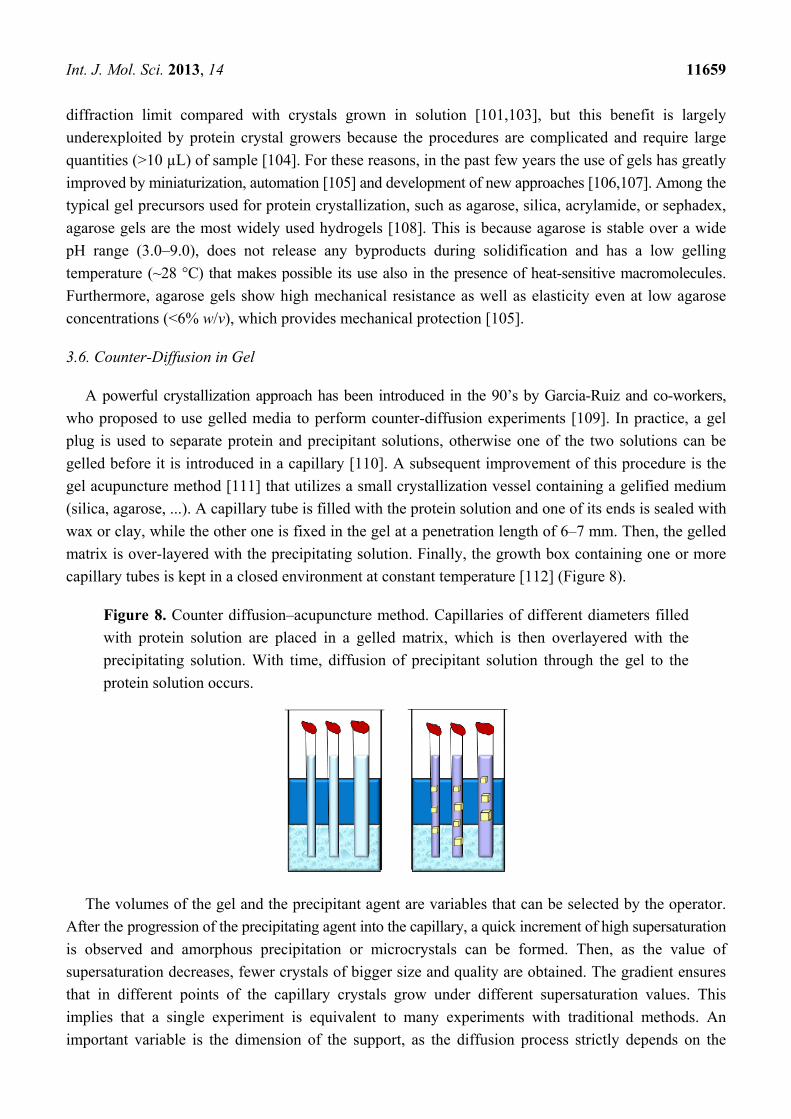
A powerful crystallization approach has been introduced in the 90’s by Garcia-Ruiz and co-workers,
who proposed to use gelled media to perform counter-diffusion experiments [109]. In practice, a gel
plug is used to separate protein and precipitant solutions, otherwise one of the two solutions can be
gelled before it is introduced in a capillary [110]. A subsequent improvement of this procedure is the
gel acupuncture method [111] that utilizes a small crystallization vessel containing a gelified medium
(silica, agarose, ...). A capillary tube is filled with the protein solution and one of its ends is sealed with
wax or clay, while the other one is fixed in the gel at a penetration length of 6–7 mm. Then, the gelled
matrix is over-layered with the precipitating solution. Finally, the growth box containing one or more
capillary tubes is kept in a closed environment at constant temperature [112] (Figure 8).
Figure 8. Counter diffusion–acupuncture method. Capillaries of different diameters filled
with protein solution are placed in a gelled matrix, which is then overlayered with the
precipitating solution. With time, diffusion of precipitant solution through the gel to the
protein solution occurs.
The volumes of the gel and the precipitant agent are variables that can be selected by the operator.
After the progression of the precipitating agent into the capillary, a quick increment of high supersaturation
is observed and amorphous precipitation or microcrystals can be formed. Then, as the value of
supersaturation decreases, fewer crystals of bigger size and quality are obtained. The gradient ensures
that in different points of the capillary crystals grow under different supersaturation values. This
implies that a single experiment is equivalent to many experiments with traditional methods. An
important variable is the dimension of the support, as the diffusion process strictly depends on the
Int. J. Mol. Sci. 2013, 14 11660
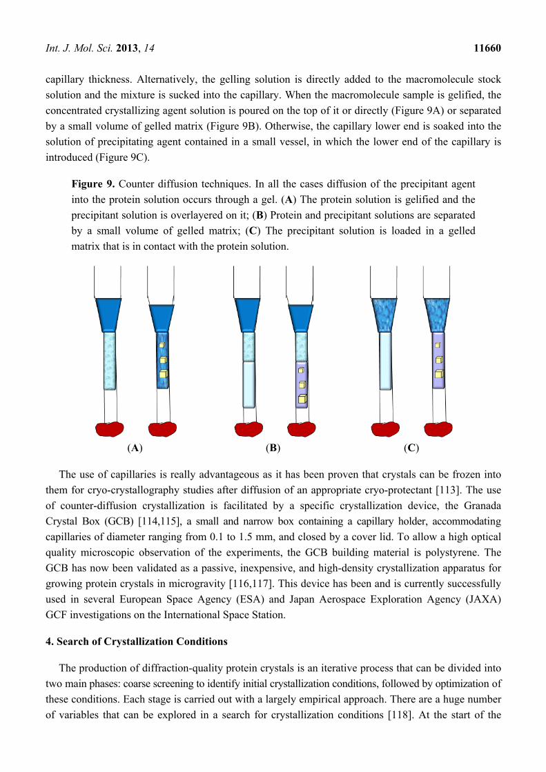
capillary thickness. Alternatively, the gelling solution is directly added to the macromolecule stock
solution and the mixture is sucked into the capillary. When the macromolecule sample is gelified, the
concentrated crystallizing agent solution is poured on the top of it or directly (Figure 9A) or separated
by a small volume of gelled matrix (Figure 9B). Otherwise, the capillary lower end is soaked into the
solution of precipitating agent contained in a small vessel, in which the lower end of the capillary is
introduced (Figure 9C).
Figure 9. Counter diffusion techniques. In all the cases diffusion of the precipitant agent
into the protein solution occurs through a gel. (A) The protein solution is gelified and the
precipitant solution is overlayered on it; (B) Protein and precipitant solutions are separated
by a small volume of gelled matrix; (C) The precipitant solution is loaded in a gelled
matrix that is in contact with the protein solution.
(A) (B) (C)
The use of capillaries is really advantageous as it has been proven that crystals can be frozen into
them for cryo-crystallography studies after diffusion of an appropriate cryo-protectant [113]. The use
of counter-diffusion crystallization is facilitated by a specific crystallization device, the Granada
Crystal Box (GCB) [114,115], a small and narrow box containing a capillary holder, accommodating
capillaries of diameter ranging from 0.1 to 1.5 mm, and closed by a cover lid. To allow a high optical
quality microscopic observation of the experiments, the GCB building material is polystyrene. The
GCB has now been validated as a passive, inexpensive, and high-density crystallization apparatus for
growing protein crystals in microgravity [116,117]. This device has been and is currently successfully
used in several European Space Agency (ESA) and Japan Aerospace Exploration Agency (JAXA)
GCF investigations on the International Space Station.
4. Search of Crystallization Conditions
The production of diffraction-quality protein crystals is an iterative process that can be divided into
two main phases: coarse screening to identify initial crystallization conditions, followed by optimization of
these conditions. Each stage is carried out with a largely empirical approach. There are a huge number
of variables that can be explored in a search for crystallization conditions [118]. At the start of the
Int. J. Mol. Sci. 2013, 14 11661
crystallization of a new protein, one faces the choice of where to begin the trial and error process. Until
recently, the search for crystallization conditions was initially based upon biochemical properties learned
during the purification and the general biochemical characterization of that macromolecule [36]. One
of the earliest screening methods, sometimes called the grid screen, employs a sampling protocol in
which 24 closely related experiments are performed at a time, by varying only two factors [119]. If
the results are negative the procedure continues with a second choice of factors and so on until success
is achieved.
The most popular approach is based on the use of sparse-matrix screens [120]. Such screens contain
a number of formulations for initial crystallization trials based on conditions that were known to have
had success in protein crystal growth. Specifically, crystallization conditions that produced diffraction
quality crystals are searched in the literature and the subset which samples the widest range of buffers
and precipitants is identified [120]. The idea is to provide a broad enough sampling of parameter space
by random combination of conditions to yield initial crystals, which may be improved later. There are
many useful kits for crystallization screening available in the market today. Their use is simple,
convenient and fast, but they are discontinuous in many parameters and can miss useful conditions.
This problem has been partly solved by the construction of automated systems (Robots) able to rapidly
perform a large number of crystallization trials and to dispense nanoliter droplets. These Robots reduce
the amount of protein required for crystallization screening. Moreover, robotic crystallization trials
allow for less error, enable more systematic and routine experimentation, give the possibility to collect
extensive data, which can be analyzed to rationalize the approach. Finally, crystallization trials using
sub-microliter drops reduce costs, make possible the exploration of a larger crystallization parameter
space, shorten time of crystal formation, thus allowing a more rapid analysis of results.
An additional method is the incomplete factorial approach, a procedure that, chosen the variables to
be changed, defines how to sample the variables with a minimum number of experiments [121]. Using
statistical methods to analyze the results, it is possible to identify variables that are correlated, and in
later stages to concentrate on their variation to optimize crystallization conditions.
The task of screen is to identify a starting point for optimization of the crystallization conditions
that is usually made by a systematic approach [122]. Both screening and optimization can be
performed by batch, vapor diffusion, dialysis or counter-diffusion techniques.
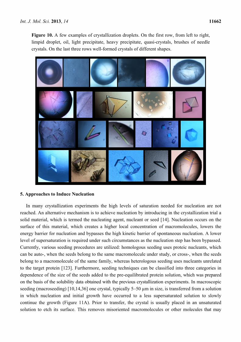
A fundamental step in the search of crystallization conditions is the inspection and scoring of the
experiments. Each trial might: (1) be clear, (2) contain amorphous/gelatinous precipitate, (3) present an
heavy precipitate, (4) have oily structures and (5) contain crystal-like material or well-formed crystals,
that is the best result (Figure 10).
These observations are necessary to define the variables that affect the solubility of the protein
under study, to identify the more promising crystallization conditions and to determine a rational
optimization strategy.
Int. J. Mol. Sci. 2013, 14 11662
Figure 10. A few examples of crystallization droplets. On the first row, from left to right,
limpid droplet, oil, light precipitate, heavy precipitate, quasi-crystals, brushes of needle
crystals. On the last three rows well-formed crystals of different shapes.
5. Approaches to Induce Nucleation
In many crystallization experiments the high levels of saturation needed for nucleation are not
reached. An alternative mechanism is to achieve nucleation by introducing in the crystallization trial a
solid material, which is termed the nucleating agent, nucleant or seed [14]. Nucleation occurs on the
surface of this material, which creates a higher local concentration of macromolecules, lowers the
energy barrier for nucleation and bypasses the high kinetic barrier of spontaneous nucleation. A lower
level of supersaturation is required under such circumstances as the nucleation step has been bypassed.
Currently, various seeding procedures are utilized: homologous seeding uses proteic nucleants, which
can be auto-, when the seeds belong to the same macromolecule under study, or cross-, when the seeds
belong to a macromolecule of the same family, whereas heterologous seeding uses nucleants unrelated
to the target protein [123]. Furthermore, seeding techniques can be classified into three categories in
dependence of the size of the seeds added to the pre-equilibrated protein solution, which was prepared
on the basis of the solubility data obtained with the previous crystallization experiments. In macroscopic
seeding (macroseeding) [10,14,36] one crystal, typically 5–50 µm in size, is transferred from a solution
in which nucleation and initial growth have occurred to a less supersaturated solution to slowly
continue the growth (Figure 11A). Prior to transfer, the crystal is usually placed in an unsaturated
solution to etch its surface. This removes misoriented macromolecules or other molecules that may
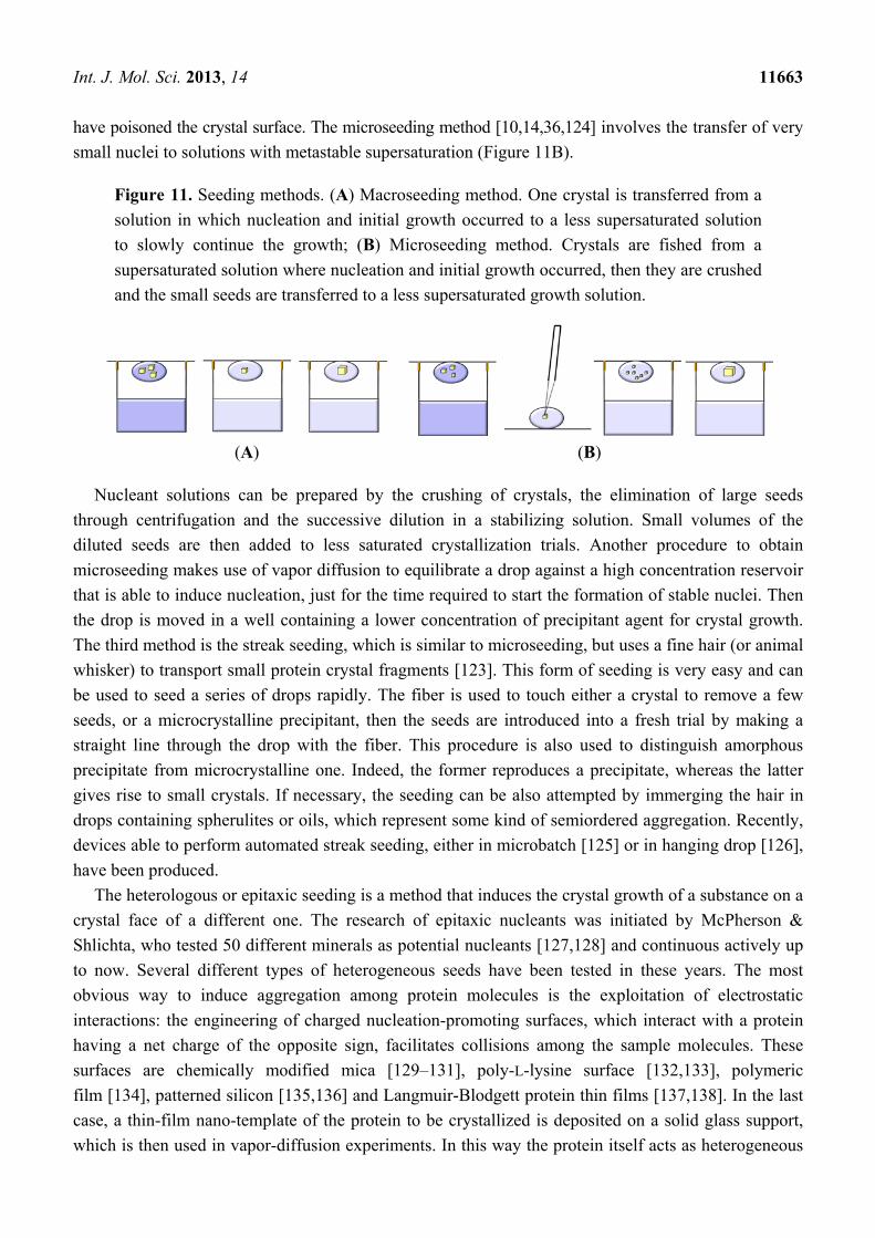
Int. J. Mol. Sci. 2013, 14 11663
have poisoned the crystal surface. The microseeding method [10,14,36,124] involves the transfer of very
small nuclei to solutions with metastable supersaturation (Figure 11B).
Figure 11. Seeding methods. (A) Macroseeding method. One crystal is transferred from a
solution in which nucleation and initial growth occurred to a less supersaturated solution
to slowly continue the growth; (B) Microseeding method. Crystals are fished from a
supersaturated solution where nucleation and initial growth occurred, then they are crushed
and the small seeds are transferred to a less supersaturated growth solution.
(A) (B)
Nucleant solutions can be prepared by the crushing of crystals, the elimination of large seeds
through centrifugation and the successive dilution in a stabilizing solution. Small volumes of the
diluted seeds are then added to less saturated crystallization trials. Another procedure to obtain
microseeding makes use of vapor diffusion to equilibrate a drop against a high concentration reservoir
that is able to induce nucleation, just for the time required to start the formation of stable nuclei. Then
the drop is moved in a well containing a lower concentration of precipitant agent for crystal growth.
The third method is the streak seeding, which is similar to microseeding, but uses a fine hair (or animal
whisker) to transport small protein crystal fragments [123]. This form of seeding is very easy and can
be used to seed a series of drops rapidly. The fiber is used to touch either a crystal to remove a few
seeds, or a microcrystalline precipitant, then the seeds are introduced into a fresh trial by making a
straight line through the drop with the fiber. This procedure is also used to distinguish amorphous
precipitate from microcrystalline one. Indeed, the former reproduces a precipitate, whereas the latter
gives rise to small crystals. If necessary, the seeding can be also attempted by immerging the hair in
drops containing spherulites or oils, which represent some kind of semiordered aggregation. Recently,
devices able to perform automated streak seeding, either in microbatch [125] or in hanging drop [126],
have been produced.
The heterologous or epitaxic seeding is a method that induces the crystal growth of a substance on a
crystal face of a different one. The research of epitaxic nucleants was initiated by McPherson &
Shlichta, who tested 50 different minerals as potential nucleants [127,128] and continuous actively up
to now. Several different types of heterogeneous seeds have been tested in these years. The most
obvious way to induce aggregation among protein molecules is the exploitation of electrostatic
interactions: the engineering of charged nucleation-promoting surfaces, which interact with a protein
having a net charge of the opposite sign, facilitates collisions among the sample molecules. These
surfaces are chemically modified mica [129–131], poly-L-lysine surface [132,133], polymeric
film [134], patterned silicon [135,136] and Langmuir-Blodgett protein thin films [137,138]. In the last
case, a thin-film nano-template of the protein to be crystallized is deposited on a solid glass support,
which is then used in vapor-diffusion experiments. In this way the protein itself acts as heterogeneous
Int. J. Mol. Sci. 2013, 14 11664
nucleant. Porous media, such as porous silicon [139–141], bioactive gel-glass [142], carbon nanotube
based materials [143], Sephadex beads of various sizes, carbon powder, alumino-silicates, mesoporous
molecular sieves [144] and zeolites of various mesh sizes, have been also successfully used, as the
protein aggregation inside the porous is facilitated. Other efficient promoters of heterogeneous
nucleation are the porous hydrophobic membranes that considerably reduce the time needed for crystal
growth [137,138]. A number of different fibrous materials (rat whiskers, horse hair, fragments of
human hair and dried seaweed) have provided really positive results [86,145], thanks to some of their
properties that match those required to an efficient nucleant: ordered surface at molecular level,
ionizable groups, lipid layers, local concentration cavities, nano and mesoscopic structure. The
development of lithography techniques allowed the fabrication of a variety of silicon substrates with
different surface characteristics [136,146] that seem to be interesting heterogeneous nucleant materials.
Very recently, Saridakis et al. [147] have shown that molecularly imprinted polymers (MIPs) may be
used as nucleation-inducing substrates. MIPs are prepared by copolymerizing functional and
cross-linking monomers in the presence of a molecular template, thus creating micro-cavities with a
three-dimensional structure complementary in both shape and chemical functionality to that of the
template. In this case MIPs have been designed to specifically attract their template, the protein to be
crystallized, and have resulted able to rebind the template and to make the crystal nucleation more
effective. Interestingly, MIPs tailored for specific cognate proteins have given positive results with
other proteins with a molecular weight of the same order of magnitude.
Many of the heterogeneous seeds tested in these years have provided well diffracting crystals with
low mosaicity in conditions that in their absence do not induce nucleation at all. This have suggested
the use of heterogeneous seeding also in initial screening experiments. In fact, recently heterogeneous
nucleating agents have been added to a sparse matrix crystallization screen [148] and crystallization
plates that are locally coated with fragments of human hair have been prepared [149].
6. Crystals for Neutron Crystallography
Determining the position of hydrogen atoms is essential to define protonation states of catalytically
important residues, hydrogen bonding and solvation effects. In order to observe hydrogen atoms via
X-ray crystallography, diffraction data beyond 1.0 Å are required. These high-resolution data can be
achieved only with highly ordered crystals, which account for less than 1% of all crystallized proteins.
A good alternative is Neutron Macromolecular Crystallography, which can provide accurate hydrogen
atom coordinates, as well as hydrogen/deuterium exchange information in macromolecular crystals at a
moderate 2 Å resolution. In the past, due to the lack of efficient instrumentation and the need for large
crystals (~1 mm3), relatively few biological macromolecules have been studied by single-crystal
neutron crystallography. Recent technical developments have provided orders-of-magnitude gains in
efficiency and have stimulated an increasing interest in neutron protein crystallography [150]. However,
the low flux of the available neutron beams still requires large fully or partially deuterated crystals.
A rational way to find the proper conditions to grow large and well-formed single crystals is to
determine the crystallization phase diagram, which allows a punctual control of the parameters
affecting the crystal growth process. In particular, the aim is to induct the nucleation at supersaturated
Int. J. Mol. Sci. 2013, 14 11665
conditions close to the solubility boundary between labile and metastable regions, by modulation of
specific physical parameters.
The detailed knowledge of the phase diagram is at the basis of the temperature-controlled
experiment (TCE), a new procedure to grow large crystals that combines the use of temperature
control and seeding [151]. A crystallization drop in the metastable zone is seeded with small protein
crystals. The seeds are maintained inside this region for as long as possible with the aid of temperature
variations just after the crystal solution equilibrium is achieved. The temperature variation is repeated
until crystals of suitable size for diffraction measurement are obtained.
Several other techniques have been developed in order to grow large protein crystals [152,153],
including the two-liquid system, the top-seeded solution growth (TSSG) and the crystallization in the
presence of the semisolid agarose gel (SSAG). In the two-liquid system procedure, the protein solution
is added to an insoluble, transparent, colorless and highly dense liquid. The drop containing the
crystallization solution separates from the liquid and floats on top of it. In this way the grown crystals
do not contact the vessel and can be easily manipulated. This procedure can be applied to perform both
microbatch and vapor diffusion experiments. To improve dimensions and quality of crystals, the
protein can be gently stirred to minimize the concentration gradient across the crystal surface and/or
slow-cooled [72]. In TSSG method the growth vessel contains a layer of an insoluble, transparent,
colorless and highly dense liquid on which the protein/precipitant solution is stratified [152]. A seed
crystal is hung by a thin crystal holder from the top of the vessel and is immersed in the mildly stirred
crystallizing solution. With respect to other seeding procedures, this method prevents the formation of
polycrystals, provides macrocrystals and allows a successful control of the crystal shape by changing
the seed orientation. Finally, it has been shown that protein crystallization in the presence of a
semi-solid agarose gel (agarose greater than 1.0% w/v) promotes nucleation and produces big crystals
with high quality [154]. The combined use of a femtosecond laser technique realizes protein nucleation
at lower supersaturation [155]. Focused irradiation of laser causes the formation of cavitation bubbles,
which create a local high concentration of protein that stimulates nucleation. The presence of an high
percentage agarose gel guarantees the spatial control of nucleation, the low diffusion of protein molecules
and consequently the formation of large well-diffracting crystals with mechanical stability [106].
7. Nucleic Acid in Free or Liganded State
Crystallization of nucleic acid-containing systems presents additional problems to be taken in
account with respect to crystallization of proteins. First of all, obtaining a sample that is homogeneous
for length, sequence and three-dimensional structure is not trivial. Depending on their length, DNA
and RNA molecules can be synthetized from constituent nucleotides, enzymatically generated in vitro
by polymerase chain reaction (PCR) or produced in vivo by recombinant expression. Although
synthetic methods give purer samples with respect to other approaches, they cannot be used for
production of long chains. On the other hand, enzymatically generated nucleic acids often possess 3'
and/or 5' heterogeneities, which are not desired [156]. Moreover, nucleic acid molecules, especially
RNA, can adopt a wide variety of conformations with different associated functions. Indeed, in
addition to various duplex structures, single-stranded nucleic acids can fold into a variety of structures,
including hairpin, triplex, G-quadruplex, and i-motif structures [157], containing non-canonical base
Int. J. Mol. Sci. 2013, 14 11666
pairs. A proper choice of length and sequence of the oligonucleotide is the first step to obtain a stable
well defined topology. A homogeneous folding can be induced by a controlled unfolding-refolding
process. Annealing protocols are the most used: heating at 80/90 °C and subsequent slow cooling at
room temperature is usually effective to obtain duplex structures, in particular in the case of DNA,
whereas a flash cooling by transfer to ice water is supposed to promote hairpin formation [158].
Finally, urea-induced denaturation followed by refolding through gradual dialysis against buffer
lacking urea is a less common method, but useful in the case of some RNAs [159].
The chemical nature of nucleic acids represents a hindrance to crystallization. The repetitive array
of negatively charged phosphate groups on the surface of the molecule makes crystal packing particularly
difficult [160]. For these reasons, it is common to engineer the constructs in order to favor the
crystallizability of the molecule, rather than testing many different crystallization conditions [161].
The simplest way to engineer a nucleic acid molecule is defining its minimal 5' and 3' boundaries.
Some biological interesting RNAs are functional domains embedded within larger RNAs. In this case
it is possible either deleting functionally dispensable elements, or replacing them with motifs that
promote intermolecular contacts or with specific protein binding sites in order to use RNA binding
proteins to improve potential lattice contacts. Indeed, it has been hypothesized that introduction of a
chemically differentiated surface, such as those provided by RNA tertiary interactions or by
RNA-binding proteins, may facilitate the growth of well-ordered crystals [162].
Numerous commercial kits are specifically designed for crystallization of nucleic acids. Common
precipitating agents are MPD and low molecular weight PEGs. Moreover, various additives have been
found very effective for the growth of high quality crystals. These additives include polyamines such
as spermine, spermidine or cobaltic hexamine chloride, non-volatile organics, and divalent cations
such as zinc, calcium or magnesium.
7.1. DNA and RNA Quadruplexes as an Example of Nucleic Acid Structures
G-rich sequences able to form complex helical structures, known as G-quadruplexes, have been
identified in many areas of the chromosomes including the telomere, gene promoters [163] and
mini-satellite regions [164] and more recently in telomeric RNA [165]. These structures contain two or
more stacked G-quartets and are stabilized by the presence of positive ions between them. Targeting of
G-quadruplexes offers promising opportunities for treatment of several pathologies, in particular
tumors, since such structures inhibit the activity of telomerase, an enzyme that is selectively expressed
in cancer cells [166]. These results have stimulated much effort towards the search of G-quadruplex
specific binding molecules able to stabilize the quadruplex fold. Design of small ligands that
specifically and strongly bind G-quadruplexes requires a deep knowledge of these structures [167], as
can only be obtained by X-ray crystallography. However crystallization of G-quadruplexes is
particularly challenging, as several parameters need to be carefully controlled. Indeed, G-quadruplexes
are able to adopt different topologies depending on the presence of cations and their nature, crowding,
annealing procedures and so on. The stability of G-quadruplexes also varies widely; it depends not
only on the identity of the stabilizing cation, but also on the DNA length and sequence [168,169]. A
proper choice of the length and sequence of the oligonucleotide, in particular the length of intervening
loops, and the strand stoichiometry and alignment, is the first step to obtain a stable well defined
Int. J. Mol. Sci. 2013, 14 11667
quadruplex topology. Secondly, a careful identification of the identity of the stabilizing cation is
needed. Potassium ions are known to strongly stabilizes G-quadruplexes [170] and potassium buffers
are usually to be preferred with respect to those containing sodium ions. Finally, as for other nucleic
acids, a correct annealing protocol is severely requested to obtain a homogeneous G-quadruplex
sample. Moreover, in the case of co-crystallization experiments it should be defined if the ligand has to
be added to the nucleic acid before or after the annealing.
Commercial kits, which are specifically designed for crystallization of nucleic acids, do not work
equally well for G-quadruplexes [171]. For example, the high concentrations of divalent cations may
be detrimental for quadruplex crystallization. It is better to design home-made screening kits taking
into account that: (1) exploration of a wide pH range is not necessary because most of quadruplexes
crystallize in narrow neutral-slightly acidic conditions; (2) the precipitant agents giving good results
are essentially MPD, PEGs with molecular weight ranging from 200 to 4000 Da and ammonium
sulphate; (3) monovalent cations can be used as additives when their effectiveness in stabilization
of the quadruplex structure is known; (4) divalent cations have to be used at very low
concentrations [159]. The experimental conditions for G-quadruplex crystallization are rather limited
in comparison with those of proteins, but this does not mean that the production of G-quadruplex
crystals is easier. The nucleation conditions must be very finely screened and the growth step is usually
really slow: in many cases crystals grow after several months.
7.2. Protein-Nucleic Acid Complexes
Complexes containing nucleic acids are inherently difficult to prepare and are stable only in some
specific conditions. For these reasons in the case of protein-nucleic acid complexes a crystallization
experiment requires a large preliminary work [172]. First of all, an accurate choice of both components
of the complex is highly recommended. Concerning proteins that bind DNA or RNA molecules, they
often have disordered or flexible loops that may interfere with the formation of a well-ordered crystal
lattice [173]. Thus, to overcome this problem, identification of the minimal folded protein fragment
able to exert its binding properties should be carried out by using, for example, limited proteolysis on
the free or liganded protein. Beginning with the minimal sequence needed for binding, one can
experiment sequences of increasing length. Considerations previously discussed in the case of free
nucleic acids, also apply to protein-nucleic acid complexes. Moreover, the precise length and composition
of the oligonucleotide must be experimentally determined for each new protein of interest [174]. As
regards DNA, lengths that have been particularly successful in crystallization trials correspond to
multiples of approximate integral or half integral turns of the DNA. In the case of RNA complexes, the
choice of the oligonucleotide is even harder. First of all, most RNA-binding proteins have much less
well-defined binding sites than DNA-binding proteins and identifying suitable RNA fragments that can
reconstitute the functional complex requires considerable biochemical study. Furthermore, the intrinsic
conformational flexibility of single-stranded RNA can complicate the reconstitution of complexes [175].
The complexes are usually obtained by mixing protein and nucleic acid at 1:1.2 to 1:1.5 molar
ratios [176,177]. This because: (a) usually not 100% of the nucleic acid is perfectly folded,
(b) estimation of either the protein or the nucleic acid component concentration is often approximate,
and (c) an excess of nucleic acids does not represent a problem as they rarely crystallize as pure
Int. J. Mol. Sci. 2013, 14 11668
materials. Depending on the solubility of the protein, one can mix it with DNA or RNA either
immediately at high concentrations or at low concentrations and then concentrate the sample by
dialysis and/or by use of centricon. Once the complex is formed, a possible concern is whether purification
is necessary. It has been found out that purity of single constituents is very important [178], but
purification of their complex is rarely needed. Only in the case of a very stable complex, characterized
by a Kd less than nanomolar, it could be purified by gel filtration before crystallization. It should be
kept in mind that phosphate buffers are better to be avoided during protein purification and complex
formation because phosphate ions could bind at recognition sites and prevent protein-substrate binding.
High salt concentrations are also detrimental, since they may shield charges on the protein surface
needed for DNA or RNA binding. For the same reason it was found that salts as precipitants have not
generated as many crystals as PEG or MPD [179], even if important exceptions have also been reported
(see as an example [180]). Ideal pH is neutral or slightly acidic, probably due to polar interactions
between negatively charged DNA/RNA backbones and positively charged protein side chains.
If the crystallization trials are unsuccessful, another variable to analyze is the sequence of the
“extra” DNA or RNA that flanks the central essential region, particularly bases at the ends of the
nucleic acid molecule, which often stabilize crystal packing by making interactions with other
oligonucleotide ends or protein molecules. For this reason, the use of sticky ends, single-stranded bases
on nucleic acid ends, has been introduced for controlling the crystallization of protein-nucleic acids
complexes [181,182].
Recently, many investigators have been involved in the search of the crystallization conditions
of protein-quadruplex complexes. At the best of our knowledge, up to now only a dozen of
protein-quadruplex complexes have been crystallized. The reported successful crystallizations of these
complexes underline, once again, the importance of the steps regarding nucleic acid annealing and
preparation of the complex [183–185].
8. Membrane Proteins
Membrane proteins are particularly difficult to handle, because they possess both a hydrophobic
surface, which is in contact with the alkyl chains of lipids, and a polar surface, exposed to the aqueous
phases on both sides of the membrane. For these reasons, the first membrane protein structure was
determined only in 1980 [186]. These proteins can be extracted from natural sources by using
detergents, even though their concentrations, with only few exceptions, are very low. As regards the
recombinant expression, the efficiency of heterologous overproduction may be very low because of
subtle differences between signal-recognition particles, cellular chaperones and foldases [187].
Furthermore, during purification steps a special attention has to be devoted to avoid unfolding,
aggregation and precipitation [188]. Once a pure and homogenous sample has been obtained, the
operator has to choose among five strategies for crystallization of membrane proteins, each of which
presents advantages and disadvantages: detergent-based [188,189], antibody-fragment-mediated [190],
lipidic cubic phase (LCP) [191], lipidic sponge phase (LSP) [192] and bicelle [193] crystallization.
The latter three may be included in the general approach “crystallization in a bilayer environment”.
Int. J. Mol. Sci. 2013, 14 11669
8.1. Detergent-Based
The oldest method, and so far the most popular and successful one, is the detergent-based
crystallization [189,194]. Membrane proteins are isolated from the cell using detergents and the
purified detergent-solubilized protein is crystallized using techniques and experimental settings
routinely applied for crystallization of soluble proteins. This easy approach presents several
drawbacks. First of all, detergent micelles can mask much of the surface area available for crystal
contacts, hindering the formation of well-ordered crystals. Moreover, since detergents are not perfect
mimic of natural bilayer, the membrane proteins may not be stable enough to produce high quality
crystals, and, more seriously, they may also unfold [195]. To overcome these problems crystallization
in bilayer environments has been introduced.
8.2. Lipidic Cubic Phase
The lipidic cubic phase (LCP), introduced by Landau and Rosenbusch [191], consists of a single
lipid bilayer bent into a highly organized three dimensional lattice permeated by aqueous
channels [196]. This bicontinuous lipidic mesophase is more ordered than a liquid but less ordered
than a solid. The membrane protein is incorporated into this semi-solid cubic phase by mixing a buffer
containing the purified protein with the component of LCP (usually monoolein) in a proper ratio [191].
On the basis of temperature-composition phase diagram for the monoolein/water system, the relevant
cubic phase forms spontaneously at 20 °C above a hydration level of ~35% (w/w) water. Included
into these matrices soluble and membrane proteins retain native conformation and enzymatic
activity [197,198]. From 1996 to present this in-meso crystallization method has been greatly
improved [199–203] and it has seen numerous successes in elucidating mechanisms of action of
several microbial rhodopsins [204–208], in defining the crystal structure of the photosynthetic reaction
centre from Rhodobacter sphaeroides [209], the high-resolution details of human G protein-coupled
receptors bound to ligands [210], and of other free or liganded human proteins, such as the human
A2A Adenosine Receptor Bound to an Antagonist [211], the human histamine H1 receptor complex
with doxepin [212], the human M2 muscarinic acetylcholine receptor bound to an antagonist [213],
etc. Recently, many efforts have been done to obtain an automatic high-throughput mode to perform
cubic phase experiments [214–217]. The protein containing LCP is used in crystallization experiments
with the advantage of obtaining crystals that are stabilized through extensive contacts in both the
hydrophilic and hydrophobic protein regions [196]. Drawbacks are related to the practical difficulties
of handling the highly viscous LCP [218], the need of specialized tools for all steps of crystallization,
the use of lipases and special treatments to fish crystals out of the lipidic medium [219].
8.3. Lipidic Sponge Phase
In some cases the semisolid LCP liquefies before crystal formation, giving rise to a lipidic sponge
phase (LSP). LSP may be thought as a swollen cubic phase with larger aqueous pores and lower
long-range order. Thanks to its liquid state, LSP is easier to handle with respect to LCP and is suitable
for traditional vapor-diffusion experiments [220].
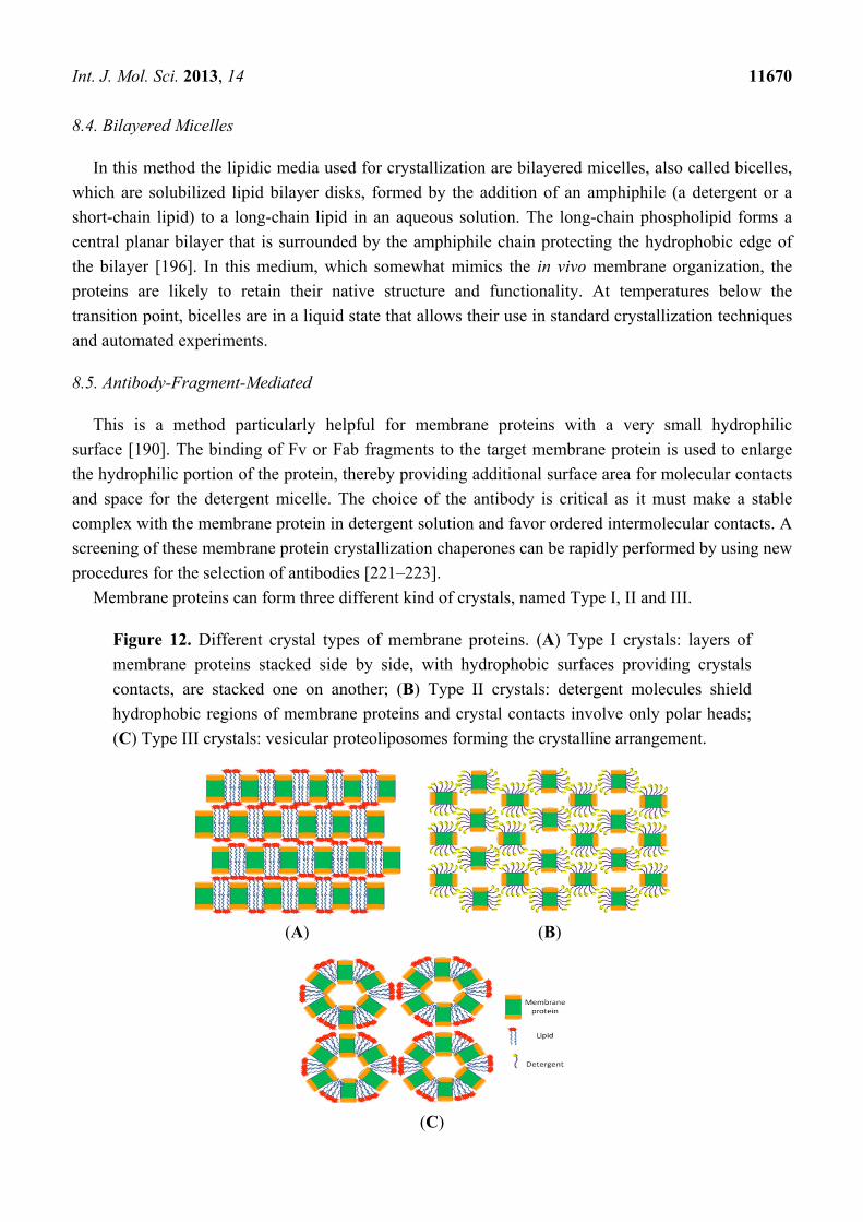
Int. J. Mol. Sci. 2013, 14 11670
8.4. Bilayered Micelles
In this method the lipidic media used for crystallization are bilayered micelles, also called bicelles,
which are solubilized lipid bilayer disks, formed by the addition of an amphiphile (a detergent or a
short-chain lipid) to a long-chain lipid in an aqueous solution. The long-chain phospholipid forms a
central planar bilayer that is surrounded by the amphiphile chain protecting the hydrophobic edge of
the bilayer [196]. In this medium, which somewhat mimics the in vivo membrane organization, the
proteins are likely to retain their native structure and functionality. At temperatures below the
transition point, bicelles are in a liquid state that allows their use in standard crystallization techniques
and automated experiments.
8.5. Antibody-Fragment-Mediated
This is a method particularly helpful for membrane proteins with a very small hydrophilic
surface [190]. The binding of Fv or Fab fragments to the target membrane protein is used to enlarge
the hydrophilic portion of the protein, thereby providing additional surface area for molecular contacts
and space for the detergent micelle. The choice of the antibody is critical as it must make a stable
complex with the membrane protein in detergent solution and favor ordered intermolecular contacts. A
screening of these membrane protein crystallization chaperones can be rapidly performed by using new
procedures for the selection of antibodies [221–223].
Membrane proteins can form three different kind of crystals, named Type I, II and III.
Figure 12. Different crystal types of membrane proteins. (A) Type I crystals: layers of
membrane proteins stacked side by side, with hydrophobic surfaces providing crystals
contacts, are stacked one on another; (B) Type II crystals: detergent molecules shield
hydrophobic regions of membrane proteins and crystal contacts involve only polar heads;
(C) Type III crystals: vesicular proteoliposomes forming the crystalline arrangement.
(A) (B)
(C)
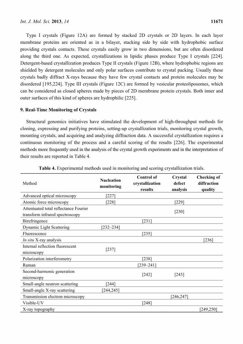
Int. J. Mol. Sci. 2013, 14 11671
Type I crystals (Figure 12A) are formed by stacked 2D crystals or 2D layers. In each layer
membrane proteins are oriented as in a bilayer, stacking side by side with hydrophobic surface
providing crystals contacts. These crystals easily grow in two dimensions, but are often disordered
along the third one. As expected, crystallizations in lipidic phases produce Type I crystals [224].
Detergent-based crystallization produces Type II crystals (Figure 12B), where hydrophobic regions are
shielded by detergent molecules and only polar surfaces contribute to crystal packing. Usually these
crystals badly diffract X-rays because they have few crystal contacts and protein molecules may be
disordered [195,224]. Type III crystals (Figure 12C) are formed by vesicular proteoliposomes, which
can be considered as closed spheres made by pieces of 2D membrane protein crystals. Both inner and
outer surfaces of this kind of spheres are hydrophilic [225].
9. Real-Time Monitoring of Crystals
Structural genomics initiatives have stimulated the development of high-throughput methods for
cloning, expressing and purifying proteins, setting-up crystallization trials, monitoring crystal growth,
mounting crystals, and acquiring and analyzing diffraction data. A successful crystallization requires a
continuous monitoring of the process and a careful scoring of the results [226]. The experimental
methods more frequently used in the analysis of the crystal growth experiments and in the interpretation of
their results are reported in Table 4.
Table 4. Experimental methods used in monitoring and scoring crystallization trials.
Method Nucleation monitoring
Control of crystallization
results
Crystal defect
analysis
Checking of diffraction
quality
Advanced optical microscopy [227]
Atomic force microscopy [228] [229]
Attentuated total reflectance Fourier transform infrared spectroscopy
[230]
Birefringence [231]
Dynamic Light Scattering [232–234]
Fluorescence [235]
In situ X-ray analysis [236]
Internal reflection fluorescent microscopy
[237]
Polarization interferometry [238]
Raman [239–241]
Second-harmonic generation microscopy
[242] [243]
Small-angle neutron scattering [244]
Small-angle X-ray scattering [244,245]
Transmission electron microscopy [246,247]
Visible-UV [248]
X-ray topography [249,250]
Int. J. Mol. Sci. 2013, 14 11672
One of the most critical steps is the automated observation and scoring of crystallization trials,
because protein crystal morphology is large and complicated. Several approaches can be used, but
unfortunately, a universal, easy strategy is still lacking. Common crystal-growth monitoring robots
collect white-light macroscopic images of crystallization drops. The images can then be analysed using
pattern recognition programs for the presence of crystals, microcrystals, precipitate, etc. However, it is
extremely difficult to come up with a unique set of descriptors for crystals and a manual inspection of
crystallization trials is in any case required. An alternative technique is based on the observation of
crystal or microcrystal birefringence [231]. Of course, this method requires special plates and devices,
since birefringence due to plastic ware can interfere. Moreover, it is known that monometric crystals,
and crystals with lower symmetry in particular orientations, are not birefringent and cannot be detected
by this method. A better performance is obtained by using polarization interferometry (DPI). This
method allows the correlation of changes in refractive index to the thickness and density of surface
layers immediately above a waveguide surface. DPI has been recently used to discriminate between the
formation of crystalline and amorphous material in real time [238]. The automated setup allows the
screening of a continuous or stepwise concentration gradient of a precipitating agent in each trial. In
this way, DPI provides useful information for the study of nucleation and crystal growth optimization.
Importantly, this technique can be used in the case of most of the crystallization methods. Another tool
for routine analysis, scoring and optimization of the crystallization processes is dynamic light
scattering (DLS). It has been demonstrated that the rate of the particle size growth is a good predictor
of the outcome for the crystallization experiment and allows to assess the probability of obtaining
macromolecule crystals before their growth starts [234]. In the evaluation of the crystallization trials
it is also possible to utilize the fluorescence of tryptophan residues under UV light. However,
UV-fluorescence has several drawbacks: emission of plastic ware, buffers and impurities may interfere
with protein fluorescence, irradiation with UV light may damage proteins or trigger reactions, fluorescence
can be quenched by various mechanisms and the protein may not contain tryptophan [251]. The
deficiency in tryptophan or another fluorescent component can be effectively overcome by the addition
of fluorescent probes that bind to the biomolecule under study. A more efficient screening is obtained
by two-photon excited UV fluorescence; this method is characterized by insensitivity to optical scattering,
which allows detection also in turbid solutions, elimination of potentially damaging out-of-plane UV
excitation, and improved signal to noise ratio [252]. Finally, also Raman scattering in near-IR region
represents an useful tool to identify protein crystal. In this case spontaneous fluorescence is minimal
and a spatial map of the intensity of Raman bands characteristic of the protein can be produced to
reveal the protein distribution, as well as to have information on secondary structures. The ideal band
to be monitored is the amide I, because it is broad and intense, change very little in different proteins
and does not suffer from interference of commonly used buffers, salts and precipitants. In particular,
Raman confocal microscopy is ideal for monitoring crystal growth since it is non-destructive,
inexpensive and rapid. It is possible to focus through transparent capillary (e.g., in FID) or directly on
crystals kept in the crystallization reactor (e.g., in vapor diffusion experiments) allowing straight
analysis on the sample. Indeed, no special handling is required in manipulating crystals for Raman
measurements. In co-crystallization experiments two independent Raman spectra are collected on
native and derivative crystals, using the same mother liquor. A Raman spectrum provides a lot of
information: energy shift, intensity and polarization of the scattered light and width of the peak. Up to
Int. J. Mol. Sci. 2013, 14 11673
now, Raman microscopy proved to be a valuable support to protein crystallography in all the steps of
the 3D structure determination, from the preparation of the derivative crystals [240,253] up to the
interpretation of the electron density maps [254,255]. In the case of metal proteins, when resonance
Raman conditions are achieved, (exciting with a wavelength within one electronic absorption band of
the sample) accurate information on the coordination, spin and oxidation state of the metal can be also
obtained [239].
10. Improvement of Crystallizability and of X-Ray Diffraction Limits
One of the most effective approach to promote crystallization uses protein engineering. In the case
of proteins with highly flexible regions, such as loops or N- and C-terminal segments, these regions
can be deleted after an accurate identification of the mobile region by limited protease digestion and
sequence alignment of analogous proteins from different species. When the flexible fragment connects
distinct domains, a useful strategy is to express and crystallize each domain independently. Other
sources of sample heterogeneity are post-translational modifications, which can be eliminated or by
action of enzymes that are specific for the particular chemical group or by mutation of residues that are
subjected to post-translational modification. Point mutation is also applied to change surface residues
that can interfere with crystal packing and/or charged residues. In this last case a net variation of
protein solubility and formation of more favorable crystal contacts can be achieved. Engineering of
protein surface can be also obtained by chemical modification of flexible amino acid side chains,
whose mobility can hinder the formation of well-ordered crystal lattice [256]. Purified proteins can be
treated with formaldehyde and dimethylamine borane complex to form a covalent bond between the
amine nitrogen of lysine residues and the methyl group [257,258]. The hydrophobic nature of the
dimethylated lysines reduces the solubility and in some cases favors protein-protein interactions and
thus the crystallization process. A complete overview of these procedures can be found in the papers of
Heras & Martin [259] and Derewenda & Vekilov [260].
Methods exist to improve the X-ray diffraction power of existing crystals. Post-crystallization
methods are more crucial for large proteins and macromolecular complexes since a new, and possibly
better diffracting, crystal form is usually more difficult to obtain compared to small protein targets.
The most used post-crystallization methods for improvement of the diffraction properties include
soaking, cross-linking, annealing [261,262] and controlled dehydration [263,264]. Soaking of protein
crystals with their product analogs, inhibitors, or strong ligands can be used to stabilize protein
conformation and increase the crystalline lattice regularity. Diffusion of additives, heavy metals,
divalent metals into pre-grown crystals can also be tested to verify if they are able to reinforce
protein-protein interface and increase the diffraction power.
The formation of intermolecular covalent bonds represents another possible way to obtain high
resolution diffraction data. Cross-linkers are either homo- or hetero-bifunctional reagents able to
establish inter-molecular cross-linkages. Homo-bifunctional reagents, specifically reacting with
primary amine groups (i.e., ε-amino groups of lysine residues) and soluble in aqueous solvents, such as
glutaraldehyde, have been used several times to guarantee the structural rigidity of the protein. The
effect depends on position and number of lysines per asymmetric unit and on the pH of harvesting
Int. J. Mol. Sci. 2013, 14 11674
solution, as the crosslinking under alkaline or acidic conditions happens by different mechanisms and
gives different end products [265].
The discovery in recent years that diffraction properties of flash-cooled protein crystals may
improve when they are brought to room temperature and then re-flash-cooled has had an high
impact on biocrystallography [262]. Two different procedures can be used: the flash-cooled crystal is
removed from the cold stream, transferred in a cryo-protectant solution for few minutes and then
re-cooled [261,266] or, alternatively, the cold stream is blocked for a definite time and then restarted to
re-cool the crystal. The success of this manipulation is unpredictable as it depends on the solvent
content of the crystal, the cooling protocol and the solution composition. However, it should be tested,
particularly when diffraction quality is low, as in many cases the static disorder of crystals is reduced
during the procedure. Cryo-annealing can be considered as a fairly brutal dehydration process. In
general, crystal dehydration procedures may produce very interesting results especially when the
decrease of crystal solvent content is performed in a systematic and controlled manner. In the fortunate
cases, this procedure induces an internal rearrangement of the crystal that results in a significantly
improved quality of the diffraction data. The various techniques for dehydrating crystals and the
proteins whose diffraction pattern was improved through this procedure have been summarized in a
recent paper [263]. To assure the control and reproducibility of dehydration, a number of dedicated
devices have been produced [267–270], some of which monitor in real time the X-ray diffraction
pattern of the crystal [271–273].
11. Concluding Remarks
This review ends with few indications on the new tendencies in the crystallization of
bio-macromolecules. A method with great potentialities is the high-pressure crystallography, which
utilizes the ability of pressure to promote the crystal growth and to modify the 3D-structure [274,275].
Pressure modulation allows to explore the conformational space of the macromolecule and its
dynamics, it shows conformational substates not visible with other approaches and provides very
useful information for protein engineering [276]. Recently, droplet-based microfluidic systems are
studied as an interesting platform to grow crystals. Microfluidics is a technology that allows users to
handle small amounts of liquids inside channels of tens to hundreds of micrometers [277,278]. This
enables the performance of miniaturized experiments with accurate and precise liquid transfer on
scales from pico- to microliters. Microfluidic chip platforms made by X-Ray transparent material are
now available [279]. Finally, a very impressive development of high-throughput pipelines for crystal
growth and X-ray structure determination is in progress. The use of these robotic systems is becoming
more widespread among the crystallographers; this will greatly improve the efficiency and reproducibility
of experiments and will significantly reduce the time required to obtain an accurate crystallographic
model of a specific bio-macromolecule.
Acknowledgments
The authors thank Lelio Mazzarella for his very helpful comments, suggestions, and valuable
assistance in the preparation of this paper.
Int. J. Mol. Sci. 2013, 14 11675
Conflict of Interest
The authors declare no conflict of interest.
References and Notes
1. McPherson, A. Protein crystallization in the structural genomics era. J. Struct. Funct. Genomics
2004, 5, 3–12.
2. Hoffman, I.D. Protein crystallization for structure-based drug design. Methods Mol. Biol. 2012,
841, 67–91.
3. Deschamps, J.R. The role of crystallography in drug design. AAPS J. 2005, 7, E813–E819.
4. Cachau, R.E.; Podjarny, A.D. High-resolution crystallography and drug design. J. Mol. Recognit.
2005, 18, 196–202.
5. Blundell, T.L.; Patel, S. High-throughput X-ray crystallography for drug discovery. Curr. Opin.
Pharmacol. 2004, 4, 490–496.
6. Heinemann, U.; Bussow, K.; Mueller, U.; Umbach, P. Facilities and methods for the high-throughput
crystal structural analysis of human proteins. Acc. Chem. Res. 2003, 36, 157–163.
7. Abola, E.; Kuhn, P.; Earnest, T.; Stevens, R.C. Automation of X-ray crystallography. Nat. Struct.
Biol. 2000, 7, 973–977.
8. Jaskolski, M. From atomic resolution to molecular giants: An overview of crystallographic
studies of biological macromolecules with synchrotron radiation. Synchrotron. Radiat. Nat. Sci.
2010, 9, 1–2.
9. Bergfors, T.M. Protein Crystallization: Techniques, Strategies and Tips; International University
Line: La Jolla, CA, USA, 1999.
10. Ducruix, A.; Giege, R. Crystallization of Nucleic Acids and Proteins, A Practical Approach,
2nd ed.; Oxford University Press: New York, NY, USA, 2000.
11. Sauter, C.; Lorber, B.; McPherson, A.; Giege, R. Crystallization. General methods. In
International Tables for Crystallography. Volume F: Crystallography of Biological Macromolecules,
2nd ed.; Arnold, E., Himmel, D.M., Rossmann, M.G., Eds.; Wiley: Chichester, NH, USA, 2012;
pp. 99–121.
12. Rupp, B. Biomolecular Crystallography: Principles, Practice, and Application to Structural
Biology; Garland Publishing: New York, NY, USA, 2010.
13. Amorphous precipitate is the result of a disordered aggregation of folded molecules, it is usually
white and could still give crystals, differently from precipitate of denaturated proteins, which
forms in conditions where the native state is not stable. Moreover some precipitates are actually
formed by submicroscopic crystals (microcrystalline precipitate).
14. McPherson, A. Crystallization of Biological Macromolecules; Cold Spring Harbor Laboratory
Press: Cold Spring Harbor: New York, NY, USA, 1999.
15. McPherson, A. Introduction to protein crystallization. Methods 2004, 34, 254–265.
16. Kelly, S.M.; Price, N.C. The use of circular dichroism in the investigation of protein structure
and function. Curr. Protein Pept. Sci. 2000, 1, 349–384.
Int. J. Mol. Sci. 2013, 14 11676
17. George, A.; Chiang, Y.; Guo, B.; Arabshahi, A.; Cai, Z.; Wilson, W.W. Second virial coefficient
as predictor in protein crystal growth. Methods Enzymol. 1997, 276, 100–110.
18. George, A.; Wilson, W.W. Predicting protein crystallization from a dilute solution property.
Acta Crystallogr. D 1994, 50, 361–365.
19. Bonnete, F.; Vivares, D. Interest of the normalized second virial coefficient and interaction
potentials for crystallizing large macromolecules. Acta Crystallogr. D 2002, 58, 1571–1575.
20. Deszczynski, M.; Harding, S.E.; Winzor, D.J. Negative second virial coefficients as predictors of
protein crystal growth: Evidence from sedimentation equilibrium studies that refutes the
designation of those light scattering parameters as osmotic virial coefficients. Biophys. Chem.
2006, 120, 106–113.
21. Berne, B.J.; Pecora, R. Dynamic Light Scattering with Applications to Chemistry, Biology, and
Physics; John Wiley & Sons: New York, NY, USA, 2000.
22. Pecora, R. Dynamic Light Scattering: Applications of Photon Correlation Spectroscopy; Plenum
Press: New York, NY, USA, 1985.
23. Saridakis, E.; Dierks, K.; Moreno, A.; Dieckmann, M.W.; Chayen, N.E. Separating nucleation
and growth in protein crystallization using dynamic light scattering. Acta Crystallogr. D 2002,
58, 1597–600.
24. Rosenberger, F.; Howard, S.B.; Sowers, J.W.; Nyce, T.A. Temperature dependence of protein
solubility: Determination and application to crystallization in X-ray capillaries. J. Cryst. Growth
1993, 129, 1–12.
25. Chernov, A.A.; Komatsu, H. Principles of Crystal Growth in Protein Crystallization. In Science
and Technology of Crystal Growth; van der Eerden, J.P., Bruinsma, O.S.L., Eds.; Kluwer
Academic Publishers: Dordrecht, The Netherlands, 1995.
26. Dumetz, A.C.; Chockla, A.M.; Kaler, E.W.; Lenhoff, A.M. Effects of pH on protein-protein
interactions and implications for protein phase behavior. Biochim. Biophys. Acta 2008, 1784,
600–610.
27. Kantardjieff, K.A.; Rupp, B. Protein isoelectric point as a predictor for increased crystallization
screening efficiency. Bioinformatics 2004, 20, 2162–2168.
28. Dupeux, F.; Rower, M.; Seroul, G.; Blot, D.; Marquez, J.A. A thermal stability assay can help to
estimate the crystallization likelihood of biological samples. Acta Crystallogr. D 2011, 67,
915–919.
29. Malawski, G.A.; Hillig, R.C.; Monteclaro, F.; Eberspaecher, U.; Schmitz, A.A.; Crusius, K.;
Huber, M.; Egner, U.; Donner, P.; Muller-Tiemann, B. Identifying protein construct variants with
increased crystallization propensity-a case study. Protein Sci. 2006, 15, 2718–2728.
30. Ericsson, U.B.; Hallberg, B.M.; Detitta, G.T.; Dekker, N.; Nordlund, P. Thermofluor-based
high-throughput stability optimization of proteins for structural studies. Anal. BioChem. 2006,
357, 289–298.
31. Doerr, A. Widening the protein crystallization bottleneck. Nat. Methods 2006, 3, 961.
32. Hofmeister, F. Zur Lehre von der Wirkung der Salze (About the science of the effect of salts).
Arch. Exp. Pathol. Pharmakol. 1888, 24, 247–260.
33. McPherson, A. Crystallization of proteins from polyethylene glycol. J. Biol. Chem. 1976, 251,
6300–6303.
Int. J. Mol. Sci. 2013, 14 11677
34. Majeed, S.; Ofek, G.; Belachew, A.; Huang, C.C.; Zhou, T.; Kwong, P.D. Enhancing protein
crystallization through precipitant synergy. Structure 2003, 11, 1061–1070.
35. McPherson, A.; Nguyen, C.; Cudney, R.; Larson, S.B. The role of small molecule additives and
chemical modification in protein crystallization. Cryst. Growth Des. 2011, 11, 1469–1474.
36. McPherson, A. The Preparation and Analysis of Protein Crystals; John Wiley and Sons: New
York, NY, USA, 1982.
37. Piazza, R.; Pierno, M. Protein interactions near crystallization: A microscopic approach to the
Hofmeister series. J. Phys. Condens. Matter 2000, 12, A443–A449.
38. Zhang, Y.; Cremer, P.S. Interactions between macromolecules and ions: The Hofmeister series.
Curr. Opin. Chem. Biol. 2006, 10, 658–663.
39. Hekmat, D.; Hebel, D.; Joswig, S.; Schmidt, M.; Weuster-Botz, D. Advanced protein
crystallization using water-soluble ionic liquids as crystallization additives. Biotechnol. Lett.
2007, 29, 1703–1711.
40. Judge, R.A.; Takahashi, S.; Longenecker, K.L.; Fry, E.H.; Abad-Zapatero, C.; Chiu, M.L. The
effect of ionic liquids on protein crystallization and X-ray diffraction resolution. Cryst. Growth
Des. 2009, 9, 3463–3469.
41. Larson, S.B.; Day, J.S.; Cudney, R.; McPherson, A. A novel strategy for the crystallization of
proteins: X-ray diffraction validation. Acta Crystallogr. D 2007, 63, 310–318.
42. Sauter, C.; Lorber, B.; Kern, D.; Cavarelli, J.; Moras, D.; Giege, R. Crystallogenesis studies on
yeast aspartyl-tRNA synthetase: Use of phase diagram to improve crystal quality.
Acta Crystallogr. D 1999, 55, 149–156.
43. Sauter, C.; Ng, J.D.; Lorber, B.; Keith, G.; Brion, P.; Hosseini, M.W.; Lehn, J.-M.; Giege, R.
Additives for the crystallization of proteins and nucleic acids. J. Cryst. Growth 1999, 196,
365–376.
44. Shim, J.U.; Cristobal, G.; Link, D.R.; Thorsen, T.; Fraden, S. Using microfluidics to decouple
nucleation and growth of protein crystals. Cryst. Growth Des. 2007, 7, 2192–2194.
45. Tanaka, S.; Ataka, M.; Ito, K. Pattern formation and coarsening during metastable phase
separation in lysozyme solutions. Phys. Rev. E 2002, 65, 051804.
46. Sica, F.; Adinolfi, S.; Berisio, R.; de Lorenzo, C.; Mazzarella, L.; Piccoli, R.; Vitagliano, L.;
Zagari, A. Crystallization of multiple forms of bovine seminal ribonuclease in the liganded and
unliganded state. J. Cryst. Growth 1999, 196, 305–312.
47. Sousa, R. Use of glycerol, polyols and other protein structure stabilizing agents in protein
crystallization. Acta Crystallogr. D 1995, 51, 271–277.
48. Tomčová, I.; Branca, R.M.; Bodo, G.; Bagyinka, C.; Smatanová, I.K. Cross-crystallization
method used for the crystallization and preliminary diffraction analysis of a novel di-haem
cytochrome c4. Acta Crystallogr. Sect. F 2006, 62, 820–824.
49. Tomčová, I.; Smatanová, I.K. Copper co-crystallization and divalent metal salts cross-influence
effect: A new optimization tool improving crystal morphology and diffraction quality. J. Cryst.
Growth 2007, 306, 383–389.
50. Kundrot, C.E.; Judge, R.A.; Pusey, M.L.; Snell, E.H. Microgravity and macromolecular
crystallography. Cryst. Growth Des. 2001, 1, 87–99.
Int. J. Mol. Sci. 2013, 14 11678
51. Otalora, F.; Novella, M.L.; Gavira, J.A.; Thomas, B.R.; Garcia Ruiz, J.M. Experimental evidence
for the stability of the depletion zone around a growing protein crystal under microgravity. Acta
Crystallogr. D 2001, 57, 412–417.
52. Wakayama, N.I.; Yin, D.C.; Harata, K.; Kiyoshi, T.; Fujiwara, M.; Tanimoto, Y.
Macromolecular crystallization in microgravity generated by a superconducting magnet.
Ann. N. Y. Acad. Sci. 2006, 1077, 184–193.
53. Vergara, A.; Lorber, B.; Sauter, C.; Giege, R.; Zagari, A. Lessons from crystals grown in the
advanced protein crystallisation facility for conventional crystallisation applied to structural
biology. Biophys. Chem. 2005, 118, 102–112.
54. Sazaki, G. Crystal quality enhancement by magnetic fields. Prog. Biophys. Mol. Biol. 2009, 101,
45–55.
55. Yin, D.C.; Wakayama, N.I.; Harata, K.; Fujiwara, M.; Kiyoshi, T.; Wada, H.; Niimura, N.;
Arai, S.; Huang, W.D.; Tanimoto, Y. Formation of protein crystals (orthorhombic lysozyme) in
quasi-microgravity environment obtained by superconducting magnet. J. Cryst. Growth 2004,
270, 184–191.
56. Okada, H.; Hirota, N.; Matsumoto, S.; Wada, H. Simulation of fluid flow during protein crystal
growth in magnetic fields. J. Appl. Phys. 2011, 110, 043903–043906
57. Ataka, M.; Katoh, E.; Wakayama, N.I. Magnetic orientation as a tool to study the initial stage of
crystallization of lysozyme. J. Cryst. Growth 1997, 173, 592–596.
58. Gavira, J.A.; Garcia-Ruiz, J.M. Effects of a magnetic field on lysozyme crystal nucleation and
growth in a diffusive environment. Cryst. Growth Design 2009, 9, 2610–2615.
59. Penkova, A.; Pan, W.; Hodjaoglu, F.; Vekilov, P.G. Nucleation of protein crystals under the
influence of solution shear flow. Ann. N. Y. Acad. Sci. 2006, 1077, 214–231.
60. Koizumi, H.; Uda, S.; Fujiwara, K.; Nozawa, J. Control of effect on the nucleation rate for hen
egg white lysozyme crystals under application of an external ac electric field. Langmuir 2011,
27, 8333–8338.
61. Koizumi, H.; Fujiwara, K.; Uda, S. Control of nucleation rate for tetragonal hen-egg white
lysozyme crystals by application of an electric field with variable frequencies. Cryst. Growth Des.
2009, 9, 2420–2424.
62. Taleb, M.; Didierjean, C.; Jelsch, C.; Mangeot, J.P.; Aubry, A. Equilibrium kinetics of lysozyme
crystallization under an external electric field. J. Cryst. Growth 2001, 232, 250–255.
63. Taleb, M.; Didierjean, C.; Jelsch, C.; Mangeot, J.P.; Capelle, B.; Aubry, A. Crystallization of
proteins under an external electric field. J. Cryst. Growth 1999, 200, 575–582.
64. Nanev, C.N.; Penkova, A. Nucleation of lysozyme crystals under external electric and ultrasonic
fields. J. Cryst. Growth 2001, 232, 285–293.
65. Charron, C.; Didierjean, C.; Mangeot, J.P.; Aubry, A. The “Octopus” plate for protein
crystallization under an electric field. J. Appl. Crystallogr. 2003, 36, 1482–1483.
66. Mirkin, N.; Frontana-Uribe, B.A.; Rodrıguez-Romero, A.; Hernandez-Santoyo, A.; Moreno, A.
The influence of an internal electric field upon protein crystallization using the gel-acupunture
method. Acta Crystallogr. D 2003, D59, 1533–1538.
Int. J. Mol. Sci. 2013, 14 11679
67. Al-Haq, M.I.; Lebrasseur, E.; Choi, W.-K.; Tsuchiya, H.; Torii, T.; Yamazaki, H.; Shinohara, E.
An apparatus for electric-field-induced protein crystallization. J. Appl. Crystallogr. 2007, 40,
199–201.
68. Sazaki, G.; Moreno, A.; Nakajima, K. Novel coupling effects of the magnetic and electric fields
on protein crystallization. J. Cryst. Growth 2004, 262, 499–502.
69. Hammadi, Z.; Veesler, S. New approaches on crystallization under electric fields.
Progr. Biophys. Mol. Biol. 2009, 101, 38–44.
70. Gutiérrez-Quezada, A.E.; Arreguín-Espinosa, R.; Moreno, A.; Natarajan, S.; Kalkura, S.N.;
Celestian, A.J.; Parise, J.B.; Clearfield, A.; Nikl, M.; Vedda, A; et al. Special Topics in Crystal
Growth. In Springer Handbook of Crystal Growth; Dhanaraj, G., Byrappa, K., Prasad, V.,
Dudley, M., Eds.; Springer: New York, NY, USA, 2010; Volume H, pp. 1583–1736.
71. Adachi, H.; Takano, K.; Niino, A.; Matsumura, H.; Kinoshita, T.; Warizaya, M.; Inoue, T.; Mori, Y.;
Sasaki, T. Solution stirring initiates nucleation and improves the quality of adenosine deaminase
crystals. Acta Crystallogr. D 2005, 61, 759–762.
72. Adachi, H.; Takano, K.; Matsumura, H.; Inoue, T.; Mori, Y.; Sasaki, T. Protein crystal growth
with a two-liquid system and stirring solution. J. Synchrotron. Rad. 2004, 11, 121–124.
73. Yaoi, M.; Adachi, H.; Takano, K.; Matsumura, H.; Inoue, T.; Mori, Y.; Sasaki, T. The effects of
solution stirring on protein crystal growth. Jpn. J. Appl. Phys. 2004, 43, L686–L688.
74. Adachi, H.; Niino, A.; Kinoshita, T.; Warizaya, M.; Maruki, R.; Takano, K.; Matsumura, H.;
Inoue, T.; Murakami, S.; Mori, Y.; et al. Solution-stirring method improves crystal quality of
human triosephosphate isomerase. J. BioSci. Bioeng. 2006, 101, 83–86.
75. Nagata, A.; Ohnishi, H.; Yoshimura, M.; Ogawa, A.; Ujita, S.; Adachi, H.; Okada, M.;
Matozaki, T.; Nakagawa, A. Crystallization and preliminary X-ray analysis of rat SHPS-1. Acta
Crystallogr. Sect. F 2006, 62, 189–191.
76. Chayen, N.E. Comparative studies of protein crystallization by vapour-diffusion and microbatch
techniques. Acta Crystallogr. D 1998, 1, 8–15.
77. Benvenuti, M.; Mangani, S. Crystallization of soluble proteins in vapor diffusion for X-ray
crystallography. Nat. Prot. 2007, 2, 1633–1651.
78. Korczynska, J.; Hu, T.C.; Smith, D.K.; Jenkins, J.; Lewis, R.; Edwards, T.; Brzozowski, A.M.
Microscale vapour diffusion for protein crystallization. Acta Crystallogr. D 2007, 63,
1009–1015.
79. Whon, T.W.; Lee, Y.H.; An, D.S.; Song, H.K.; Kim, S.G. A simple technique to convert
sitting-drop vapor diffusion into hanging-drop vapor diffusion by solidifying the reservoir
solution with agarose. J. Appl. Crystallogr. 2009, 42, 975–976.
80. Lu, Q.Q.; Yin, D.C.; Xie, S.X.; Liu, Y.M.; Chen, R.Q. The effect of diluting crystallization
droplets on protein crystallization in vapor diffusion method. Cryst. Res. Technol. 2011, 46,
917–925.
81. Chayen, N.E. A novel technique to control the rate of vapor diffusion, giving larger protein
crystals. J. Appl. Crystallogr. 1997, 30, 198–201.
82. Chayen, N.E. Crystallization with oils: A new dimension in macromolecular crystal growth.
J. Cryst. Growth 1999, 196, 434–441.
Int. J. Mol. Sci. 2013, 14 11680
83. Chayen, N.E.; Shaw Stewart, P.D.; Maeder, D.L.; Blow, D.M. An automated system for
micro-batch protein crystallization and screening. J. Appl. Crystallogr. 1990, 23, 297–302.
84. Brumshtein, B.; Greenblatt, H.M.; Futerman, A.H.; Silman, I.; Sussman, J.L. Control of the rate
of evaporation in protein crystallization by the “microbatch under oil” method. J. Appl.
Crystallogr. 2008, 41, 969–971.
85. D’Arcy, A.; Elmore, C.; Stihle, M.; Johnston, J.E. A novel approach to crystallising proteins
under oil. J. Cryst. Growth 1996, 168, 175–180.
86. D’Arcy, A.; Mac Sweeney, A.; Stihle, M.; Haber, A. The advantages of using a modified
microbatch method for rapid screening of protein crystallization conditions. Acta Crystallogr. D
2003, 59, 396–399.
87. Merlino, A.; Russo Krauss, I.; Albino, A.; Pica, A.; Vergara, A.; Masullo, M.; de Vendittis, E.;
Sica, F. Improving protein crystal quality by the without-oil microbatch method:
Crystallization and preliminary X-ray diffraction analysis of glutathione synthetase from
Pseudoalteromonas haloplanktis. Int. J. Mol. Sci. 2011, 12, 6312–6319.
88. Reid, B.R.; Koch, G.L.; Boulanger, Y.; Hartley, B.S.; Blow, D.M. Letter: Crystallization
and preliminary X-ray diffraction studies on tyrosyl-transfer RNA synthetase from
Bacillus stearothermophilus. J. Mol. Biol. 1973, 80, 199–201.
89. Thomas, D.H.; Rob, A.; Rice, D.W. A novel dialysis procedure for the crystallization of proteins.
Protein Eng. 1989, 2, 489–491.
90. Salemme, F.R. A free interface diffusion technique for the crystallization of proteins for X-ray
crystallography. Arch. BioChem. Biophys. 1972, 151, 533–539.
91. Berisio, R.; Lamzin, V.S.; Sica, F.; Wilson, K.S.; Zagari, A.; Mazzarella, L. Protein titration in
the crystal state. J. Mol. Biol. 1999, 292, 845–854.
92. Vitagliano, L.; Merlino, A.; Zagari, A.; Mazzarella, L. Productive and nonproductive binding to
ribonuclease A: X-ray structure of two complexes with uridylyl(2',5')guanosine. Protein Sci.
2000, 9, 1217–1225.
93. Garcia-Ruiz, J.M. Counter-diffusion methods for macromolecular crystallization. Methods
Enzymol. 2003, 368, 130–154.
94. Li, L.; Du, W.; Ismagilov, R.F. Multiparameter screening on SlipChip used for nanoliter protein
crystallization combining free interface diffusion and microbatch methods. J. Am. Chem. Soc.
2010, 132, 112–119.
95. McPherson, A. Effects of a microgravity environment on the crystallization of biological
macromolecules. Presented at VIII European Symposium on Materials and Fluid Sciences in
Microgravity, Free University of Brussels, Brussels, Belgium, 12–16 April 1992; Legros, J.C.,
ed.; Volume II, 619–626.
96. Bernard, Y.; Degoy, S.; Lefaucheux, F.; Robert, M.C. A gel-mediated feeding technique for
protein crystal growth from hanging drops. Acta Crystallogr. D 1994, 50, 504–507.
97. Lorber, B.; Sauter, C.; Theobald-Dietrich, A.; Moreno, A.; Schellenberger, P.; Robert, M.C.;
Capelle, B.; Sanglier, S.; Potier, N.; Giege, R. Crystal growth of proteins, nucleic acids, and
viruses in gels. Prog. Biophys. Mol. Biol. 2009, 101, 13–25.
98. Robert, M.C.; Lefaucheux, F. Crystal growth in gels: Principle and applications. J. Cryst. Growth
1988, 90, 358–367.
Int. J. Mol. Sci. 2013, 14 11681
99. Garcia-Ruiz, J.M.; Otálora, F.; Novella, M.L.; Gavira, J.A.; Sauter, C.; Vidal, O. A
supersaturation wave of protein crystallization. J. Cryst. Growth 2001, 232, 165–172.
100. Lorber, B.; Giege, R. Nucleation and growth of thaumatin crystals within a gel under
microgravity on STS-95 mission vs. under Earth's gravity. J. Cryst. Growth 2001, 231, 252–261.
101. Sauter, C.; Lorber, B.; Giege, R. Towards atomic resolution with crystals grown in gel: The case
of thaumatin seen at room temperature. Proteins 2002, 48, 146–150.
102. Zhu, D.W.; Lorber, B.; Sauter, C.; Ng, J.D.; Benas, P.; Le Grimellec, C.; Giege, R. Growth
kinetics, diffraction properties and effect of agarose on the stability of a novel crystal form of
Thermus thermophilus aspartyl-tRNA synthetase-1. Acta Crystallogr. D 2001, 57, 552–558.
103. Moreno, A.; Yokaichiya, F.; Dimasi, E.; Stojanoff, V. Growth and characterization of
high-quality protein crystals for X-ray crystallography. Ann. N. Y. Acad. Sci. 2009, 1161, 429–436.
104. Chayen, N.E. Turning protein crystallisation from an art into a science. Curr. Opin. Struct. Biol.
2004, 14, 577–583.
105. Biertumpfel, C.; Basquin, J.; Suck, D.; Sauter, C. Crystallization of biological macromolecules
using agarose gel. Acta Crystallogr. D 2002, 58, 1657–1659.
106. Sugiyama, S.; Maruyama, M.; Sazaki, G.; Hirose, M.; Adachi, H.; Takano, K.; Murakami, S.;
Inoue, T.; Mori, Y.; Matsumura, H. Growth of protein crystals in hydrogels prevents osmotic
shock. J. Am. Chem. Soc. 2012, 134, 5786–5789.
107. Hasenaka, H.; Sugiyama, S.; Hirose, M.; Shimizu, N.; Kitatani, T.; Takahashi, Y.; Adachi, H.;
Takano, K.; Murakami, S.; Inoue, T.; et al. Femtosecond laser processing of protein crystals
grown in agarose gel. J. Cryst. Growth 2010, 312, 73–78.
108. Van Driessche, A.E. S.; Otalora, F.; Gavira, J.A.; Sazaki, G. Is agarose an impurity or an
impurity filter? In situ observation of the joint gel/impurity effect on protein crystal growth
kinetics. Cryst. Growth Design 2008, 8, 3623–3629.
109. Garcia Ruiz, J.M. The uses of crystal growth in gels and other diffusing-reacting systems. Key
Eng. Mater. 1991, 58, 87–106.
110. Otalora, F.; Gavira, J.A.; Ng, J.D.; Garcia-Ruiz, J.M. Counterdiffusion methods applied to
protein crystallization. Prog. Biophys. Mol. Biol. 2009, 101, 26–37.
111. Garcia Ruiz, J.M.; Moreno, A.; Viedma, C.; Coll, M. Crystal quality of lysozyme single crystals
grown by the gel acupuncture method. Mater. Res. Bull. 1993, 28, 541–546.
112. Oberthuer, D.; Melero-Garcia, E.; Dierks, K.; Meyer, A.; Betzel, C.; Garcia-Caballero, A.;
Gavira, J.A. Monitoring and scoring counter-diffusion protein crystallization experiments in
capillaries by In situ dynamic light scattering. PLoS One 2012, 7, e33545.
113. Ng, J.D.; Gavira, J.A.; Garcia-Ruiz, J.M. Protein crystallization by capillary counter-diffusion for
applied crystallographic structure determination. J. Struct. Biol. 2003, 142, 218–231.
114. Garcia Ruiz, J.M.; Gonzales-Ramirez, J.A.; Gavira, J.A.; Otalora, F. Granada Crystallisation
Box: A new device for protein crystallisation by counter-diffusion techniques. Acta Crystallogr. D
2002, D58, 1638–1642.
115. Vergara, A.; Castagnolo, D.; Carotenuto, L.; Vitagliano, L.; Berisio, R.; Sorrentino, G.;
Gonzalez-Ramirez, L.; Garcia-Ruiz, J.; Zagari, A. Phase behavior and crystallogenesis under
counter-diffusion conditions of the collagen-model peptide (Pro-Pro-Gly)(10). J. Cryst. Growth
2009, 311, 304–309.
Int. J. Mol. Sci. 2013, 14 11682
116. Malecki, P.H.; Rypniewski, W.; Szymanski, M.; Barciszewski, J.; Meyer, A. Binding of the plant
hormone kinetin in the active site of Mistletoe Lectin I from Viscum album. Biochim. Biophys.
Acta 2012, 1824, 334–338.
117. Meyer, A.; Rypniewski, W.; Szymanski, M.; Voelter, W.; Barciszewski, J.; Betzel, C. Structure
of mistletoe lectin I from Viscum album in complex with the phytohormone zeatin. Biochim.
Biophys. Acta 2008, 1784, 1590–1595.
118. Shieh, H.S.; Stallings, W.C.; Stevens, A.M.; Stegeman, R.A. Using sampling techniques in
protein crystallization. Acta Crystallogr. D 1995, 51, 305–310.
119. Kingston, R.L.; Baker, H.M.; Baker, E.N. Search designs for protein crystallization based on
orthogonal arrays. Acta Crystallogr. D 1994, 50, 429–440.
120. Jancarik, J.; Kim, S.-H. Sparse matrix sampling: A screening method for crystallization of
proteins. J. Appl. Crystallogr. 1991, 24, 409–411.
121. Carter, C.W., Jr.; Carter, C.W. Protein crystallization using incomplete factorial experiments.
J. Biol. Chem. 1979, 254, 12219–12223.
122. Cudney, R.; Patel, S.; Weisgraber, K.; Newhouse, Y.; McPherson, A. Screening and optimization
strategies for macromolecular crystal growth. Acta Crystallogr. D 1994, 50, 414–423.
123. Stura, E.A.; Wilson, I.A. Applications of the streak seeding technique in protein crystallization.
J. Cryst. Growth 1991, 110, 270–282.
124. Stewart, P.D. S.; Kolek, S.A.; Briggs, R.A.; Chayen, N.E.; Baldock, P.F.M. Random
microseeding: A theoretical and practical exploration of seed stability and seeding techniques for
successful protein crystallization. Cryst. Growth Design 2011, 11, 3432–3441.
125. Khurshid, S.; Haire, L.F.; Chayen, N.E. Automated seeding for the optimization of crystal
quality. J. Appl. Crystallogr. 2010, 43, 752–756.
126. Georgiev, A.; Vorobiev, S.; Edstrom, W.; Song, T.; Laine, A.; Hunt, J.; Allen, P. Automated
streak-seeding with micromachined silicon tools. Acta Crystallogr. D 2006, D62, 1039–1045.
127. McPherson, A.; Shlichta, P. Heterogeneous and epitaxial nucleation of protein crystals on
mineral surfaces. Science 1988, 239, 385–387.
128. McPherson, A.; Shlichta, P.J. Facilitation of the growth of protein crystals by
heterogeneous/epitaxial nucleation. J. Cryst. Growth 1987, 85, 206–214.
129. Falini, G.; Fermani, S.; Conforti, G.; Ripamonti, A. Protein crystallisation on chemically
modified mica surfaces. Acta Crystallogr. D 2002, 58, 1649–1652.
130. Tang, L.; Huang, Y.B.; Liu, D.Q.; Li, J.L.; Mao, K.; Liu, L.; Cheng, Z.J.; Gong, W.M.; Hu, J.;
He, J.H. Effects of the silanized mica surface on protein crystallization. Acta Crystallogr. D
2005, 61, 53–59.
131. Tosi, G.; Fermani, S.; Falini, G.; Gavira Gallardo, J.A.; Garcia Ruiz, J.M. Crystallization of
proteins on functionalized surfaces. Acta Crystallogr. D 2008, 64, 1054–1061.
132. Rong, L.; Komatsu, H.; Natsuisaka, M.; Yoda, S. Epitaxial nucleation of protein crystal on
poly-L-lysine modified surface. Jpn. J. Appl. Phys. 2001, 40, 6677–6678.
133. Rong, L.; Komatsu, H.; Yoda, S. Control of heterogeneous nucleation of lysozyme crystals by
using Poly-L-Lysine modified substrate. J. Cryst. Growth 2002, 235, 489–493.
134. Fermani, S.; Falini, G.; Minnucci, M.; Ripamonti, A. Protein crystallization on polymeric film
surfaces. J. Cryst. Growth 2001, 224, 327–334.
Int. J. Mol. Sci. 2013, 14 11683
135. Sanjoh, A.; Tsukihara, T. Spatiotemporal protein crystal growth studies using microfluidic
silicon devices. J. Cryst. Growth 1999, 196, 691–702.
136. Sanjoh, A.; Tsukihara, T.; Gorti, S. Surface-potential controlled Si-microarray devices for
heterogeneous protein crystallization screening. J. Cryst. Growth 2001, 232, 618–628.
137. Di Profio, G.; Curcio, E.; Drioli, E. Trypsin crystallization by membrane-based techniques.
J. Struct. Biol. 2005, 150, 41–49.
138. Curcio, E.; Fontananova, E.; di Profio, G.; Drioli, E. Influence of the structural properties of
poly(vinylidene fluoride) membranes on the heterogeneous nucleation rate of protein crystals.
J. Phys. Chem. B 2006, 110, 12438–12445.
139. Chayen, N.E.; Saridakis, E.; El-Bahar, R.; Nemirovsky, Y. Porous silicon: An effective
nucleation-inducing material for protein crystallization. J. Mol. Biol. 2001, 312, 591–595.
140. Stolyarova, S.; Saridakis, E.; Chayen, N.E.; Nemirovsky, Y. A model for enhanced nucleation of
protein crystals on a fractal porous substrate. Biophys. J. 2006, 91, 3857–3863.
141. Rong, L.; Komatsu, H.; Yoshizaki, I.; Kadowaki, A.; Yoda, S. Protein crystallization by using
porous glass substrate. J. Synchrotron. Rad. 2004, 11, 27–29.
142. Chayen, N.E.; Saridakis, E.; Sear, R.P. Experiment and theory for heterogeneous nucleation of
protein crystals in a porous medium. Proc. Natl. Acad. Sci. USA 2006, 103, 597–601.
143. Asanithi, P.; Saridakis, E.; Govada, L.; Jurewicz, I.; Brunner, E.W.; Ponnusamy, R.; Cleaver, J.A.;
Dalton, A.B.; Chayen, N.E.; Sear, R.P. Carbon-nanotube-based materials for protein
crystallization. ACS Appl. Mater. Interfaces 2009, 1, 1203–1210.
144. Sugahara, M.; Asada, Y.; Morikawa, Y.; Kageyama, Y.; Kunishima, N. Nucleant-mediated
protein crystallization with the application of microporous synthetic zeolites. Acta Crystallogr. D
2008, 64, 686–695.
145. Georgieva, D.G.; Kuil, M.E.; Oosterkamp, T.H.; Zandbergen, H.W.; Abrahams, J.P.
Heterogeneous nucleation of three-dimensional protein nanocrystals. Acta Crystallogr. D 2007,
63, 564–570.
146. Takehara, M.; Ino, K.; Takakusagi, Y.; Oshikane, H.; Nureki, O.; Ebina, T.; Mizukami, F.;
Sakaguchi, K. Use of layer silicate for protein crystallization: Effects of micromica and chlorite
powders in hanging drops. Anal. BioChem. 2008, 373, 322–329.
147. Saridakis, E.; Khurshid, S.; Govada, L.; Phan, Q.; Hawkins, D.; Crichlow, G.V.; Lolis, E.;
Reddy, S.M.; Chayen, N.E. Protein crystallization facilitated by molecularly imprinted polymers.
Proc. Natl. Acad. Sci. USA 2011, 108, 11081–11086.
148. Thakur, A.S.; Robin, G.; Guncar, G.; Saunders, N.F.; Newman, J.; Martin, J.L.; Kobe, B.
Improved success of sparse matrix protein crystallization screening with heterogeneous
nucleating agents. PLoS One 2007, 2, e1091.
149. Nederlof, I.; Hosseini, R.; Georgieva, D.; Luo, J.; Li, D.; Abrahams, J.P. A straightforward and
robust method for introducing human hair as a nucleant into high throughput crystallization
trials. Cryst. Growth Des. 2011, 11, 1170–1176.
150. Munshi, P.; Chung, S.L.; Blakeley, M.P.; Weiss, K.L.; Myles, D.A.; Meilleur, F. Rapid
visualization of hydrogen positions in protein neutron crystallographic structures.
Acta Crystallogr. D 2012, 68, 35–41.
Int. J. Mol. Sci. 2013, 14 11684
151. Budayova-Spano, M.; Dauvergne, F.; Audiffren, M.; Bactivelane, T.; Cusack, S. A methodology
and an instrument for the temperature-controlled optimization of crystal growth.
Acta Crystallogr. D 2007, 63, 339–347.
152. Matsumura, H.; Kakiniuchi, K.; Nakamura, T.; Sugiyama, S.; Maruyama, M.; Adachi, H.;
Takano, K.; Murakami, S.; Inoue, T.; Mori, Y. Growth of protein crystals by syringe-type
top-seeded solution growth. Cryst. Growth Des. 2011, 11, 1486–1492.
153. Matsumura, H.; Sugiyama, S.; Hirose, M.; Kakinouchi, K.; Maruyama, M.; Murai, R.; Adachi, H.;
Takano, K.; Murakami, S.; Mori, Y.; et al. Approach for growth of high-quality and large protein
crystals. J. Synchrotron. Rad. 2011, 18, 16–19.
154. Sugiyama, S.; Hasenaka, H.; Hirose, M.; Shimizu, N.; Kitatani, T.; Takahashi, Y.; Adachi, H.;
Takano, K.; Murakami, S.; Inoue, T.; et al. Femtosecond laser processing of agarose gel
surrounding protein crystals for development of an automated crystal capturing system. Jpn. J.
Appl. Phys. 2009, 48, 105502–105506.
155. Murai, R.; Yoshikawa, H.Y.; Takahashi, Y.; Maruyama, M.; Sugiyama, S.; Sazaki, G.; Adachi, H.;
Takano, K.; Matsumura, H.; Murakami, S.; et al. Enhancement of femtosecond laser-induced
nucleation of protein in a gel solution. Appl. Phys. Lett. 2010, 96, 043702–043704.
156. Sherlin, L.D.; Bullock, T.L.; Nissan, T.A.; Perona, J.J.; Lariviere, F.J.; Uhlenbeck, O.C.;
Scaringe, S.A. Chemical and enzymatic synthesis of tRNAs for high-throughput crystallization.
RNA 2001, 7, 1671–1678.
157. Choi, J.; Majima, T. Conformational changes of non-B DNA. Chem. Soc. Rev. 2011, 40,
5893–5909.
158. Mooers, B.H. Crystallographic studies of DNA and RNA. Methods 2009, 47, 168–176.
159. Batey, R.T.; Rambo, R.P.; Lucast, L.; Rha, B.; Doudna, J.A. Crystal structure of the
ribonucleoprotein core of the signal recognition particle. Science 2000, 287, 1232–1239.
160. Ferre-D’Amare, A.R.; Zhou, K.; Doudna, J.A. A general module for RNA crystallization. J. Mol.
Biol. 1998, 279, 621–631.
161. Rupert, P.B.; Ferre-D’Amare, A.R. Crystallization of the hairpin ribozyme: Illustrative protocols.
Methods Mol. Biol. 2004, 252, 303–311.
162. Ferre-D’Amare, A.R. Use of the spliceosomal protein U1A to facilitate crystallization and
structure determination of complex RNAs. Methods 2010, 52, 159–167.
163. Patel, D.J.; Phan, A.T.; Kuryavyi, V. Human telomere, oncogenic promoter and 5'-UTR
G-quadruplexes: Diverse higher order DNA and RNA targets for cancer therapeutics. Nucleic
Acids Res. 2007, 35, 7429–7455.
164. Lech, C.J.; Heddi, B.; Phan, A.T. Guanine base stacking in G-quadruplex nucleic acids. Nucleic
Acids Res. 2013, 41, 2034–2046.
165. Xu, Y.; Komiyama, M. Structure, function and targeting of human telomere RNA. Methods
2012, 57, 100–105.
166. Neidle, S. Human telomeric G-quadruplex: The current status of telomeric G-quadruplexes as
therapeutic targets in human cancer. FEBS J. 2010, 277, 1118–1125.
167. Haider, S.M.; Neidle, S.; Parkinson, G.N. A structural analysis of G-quadruplex/ligand
interactions. Biochimie 2011, 93, 1239–1251.
Int. J. Mol. Sci. 2013, 14 11685
168. Lane, A.N.; Chaires, J.B.; Gray, R.D.; Trent, J.O. Stability and kinetics of G-quadruplex
structures. Nucleic Acids Res. 2008, 36, 5482–5515.
169. Bugaut, A.; Balasubramanian, S. A sequence-independent study of the influence of short loop
lengths on the stability and topology of intramolecular DNA G-quadruplexes. BioChemistry
2008, 47, 689–697.
170. Hud, N.V.; Smith, F.W.; Anet, F.A.; Feigon, J. The selectivity for K+ versus Na+ in DNA
quadruplexes is dominated by relative free energies of hydration: A thermodynamic analysis by
1H NMR. BioChemistry 1996, 35, 15383–15390.
171. Campbell, N.H.; Parkinson, G.N. Crystallographic studies of quadruplex nucleic acids. Methods
2007, 43, 252–263.
172. Perederina, A.; Krasilnikov, A.S. Crystallization of RNA-protein complexes: From synthesis and
purification of individual components to crystals. Methods Mol. Biol. 2012, 905, 123–143.
173. Tan, S.; Hunziker, Y.; Pellegrini, L.; Richmond, T.J. Crystallization of the yeast
MATalpha2/MCM1/DNA ternary complex: General methods and principles for protein/DNA
cocrystallization. J. Mol. Biol. 2000, 297, 947–959.
174. Jordan, S.R.; Whitcombe, T.V.; Berg, J.M.; Pabo, C.O. Systematic variation in DNA length
yields highly ordered repressor-operator cocrystals. Science 1985, 230, 1383–1385.
175. Ke, A.; Doudna, J.A. Crystallization of RNA and RNA-protein complexes. Methods 2004, 34,
408–414.
176. Brown, D.G.; Freemont, P.S. Crystallography in the study of protein-DNA interactions.
Methods Mol. Biol. 1996, 56, 293–318.
177. Hollis, T. Crystallization of Protein-DNA Complexes. In Macromolecular Crystallography
Protocols: Volume 1, Preparation and Crystallization of Macromolecules; Springer: New York,
NY, USA, 2007; Volume 363.
178. Conlin, R.M.; Brown, R.S. Reconstitution of Protein-DNA Complexes for Crystallization. In
DNA-Protein Interactions: Principles and Protocols; Springer: New York, NY, USA, 1994;
Volume 30.
179. Nadassy, K.; Wodak, S.J.; Janin, J. Structural features of protein-nucleic acid recognition sites.
BioChemistry 1999, 38, 1999–2017.
180. Yang, X.L.; Otero, F.J.; Ewalt, K.L.; Liu, J.; Swairjo, M.A.; Kohrer, C.; RajBhandary, U.L.;
Skene, R.J.; McRee, D.E.; Schimmel, P. Two conformations of a crystalline human tRNA
synthetase-tRNA complex: Implications for protein synthesis. EMBO J. 2006, 25, 2919–2929.
181. Rice, P.A.; Yang, S.; Mizuuchi, K.; Nash, H.A. Crystal structure of an IHF-DNA complex: A
protein-induced DNA U-turn. Cell 1996, 87, 1295–1306.
182. Wedekind, J.E.; McKay, D.B. Crystal structure of a lead-dependent ribozyme revealing metal
binding sites relevant to catalysis. Nat. Struct. Biol. 1999, 6, 261–268.
183. Russo Krauss, I.; Merlino, A.; Randazzo, A.; Mazzarella, L.; Sica, F. Crystallization and preliminary
X-ray analysis of the complex of human alpha-thrombin with a modified thrombin-binding
aptamer. Acta Crystallogr. Sect. F Struct. Biol. Cryst. Commun. 2010, 66, 961–963.
184. Russo Krauss, I.; Merlino, A.; Giancola, C.; Randazzo, A.; Mazzarella, L.; Sica, F.
Thrombin-aptamer recognition: A revealed ambiguity. Nucleic Acids Res. 2011, 39, 7858–7867.
Int. J. Mol. Sci. 2013, 14 11686
185. Russo Krauss, I.; Merlino, A.; Randazzo, A.; Novellino, E.; Mazzarella, L.; Sica, F. High-resolution
structures of two complexes between thrombin and thrombin-binding aptamer shed light on the
role of cations in the aptamer inhibitory activity. Nucleic Acids Res. 2012, 40, 8119–8128.
186. Garavito, R.M.; Rosenbusch, J.P. Three-dimensional crystals of an integral membrane protein:
An initial x-ray analysis. J. Cell Biol. 1980, 86, 327–329.
187. Bill, R.M.; Henderson, P.J.; Iwata, S.; Kunji, E.R.; Michel, H.; Neutze, R.; Newstead, S.;
Poolman, B.; Tate, C.G.; Vogel, H. Overcoming barriers to membrane protein structure
determination. Nat. Biotechnol. 2011, 29, 335–340.
188. Prive, G.G. Detergents for the stabilization and crystallization of membrane proteins. Methods
2007, 41, 388–397.
189. Newstead, S.; Ferrandon, S.; Iwata, S. Rationalizing alpha-helical membrane protein
crystallization. Protein Sci. 2008, 17, 466–472.
190. Hunte, C.; Michel, H. Crystallisation of membrane proteins mediated by antibody fragments.
Curr. Opin. Struct. Biol. 2002, 12, 503–508.
191. Landau, E.M.; Rosenbusch, J.P. Lipidic cubic phases: A novel concept for the crystallization of
membrane proteins. Proc. Natl. Acad. Sci. USA 1996, 93, 14532–14535.
192. Wadsten, P.; Wohri, A.B.; Snijder, A.; Katona, G.; Gardiner, A.T.; Cogdell, R.J.; Neutze, R.;
Engstrom, S. Lipidic sponge phase crystallization of membrane proteins. J. Mol. Biol. 2006, 364,
44–53.
193. Faham, S.; Bowie, J.U. Bicelle crystallization: A new method for crystallizing membrane
proteins yields a monomeric bacteriorhodopsin structure. J. Mol. Biol. 2002, 316, 1–6.
194. Wiener, M.C. A pedestrian guide to membrane protein crystallization. Methods 2004, 34,
364–372.
195. Michel, H. Crystallization of membrane proteins. Trends BioChem. Sci. 1983, 8, 56–59.
196. Ujwal, R.; Bowie, J.U. Crystallizing membrane proteins using lipidic bicelles. Methods 2011, 55,
337–341.
197. Luecke, H.; Schobert, B.; Richter, H.T.; Cartailler, J.P.; Lanyi, J.K. Structural changes in
bacteriorhodopsin during ion transport at 2 angstrom resolution. Science 1999, 286, 255–261.
198. Katona, G.; Snijder, A.; Gourdon, P.; Andreasson, U.; Hansson, O.; Andreasson, L.E.; Neutze, R.
Conformational regulation of charge recombination reactions in a photosynthetic bacterial
reaction center. Nat. Struct. Mol. Biol. 2005, 12, 630–631.
199. Caffrey, M.; Cherezov, V. Crystallizing membrane proteins using lipidic mesophases.
Nat. Protocols 2009, 4, 706–731.
200. Caffrey, M. Crystallizing membrane proteins for structure-function studies using lipidic
mesophases. BioChem. Soc. Trans. 2011, 39, 725–732.
201. Cherezov, V. Lipidic cubic phase technologies for membrane protein structural studies.
Curr. Opin. Struct. Biol. 2011, 21, 559–566.
202. Li, D.; Caffrey, M. Lipid cubic phase as a membrane mimetic for integral membrane protein
enzymes. Proc. Natl. Acad. Sci. USA 2011, 108, 8639–8644.
203. Aherne, M.; Lyons, J.A.; Caffrey, M. A fast, simple and robust protocol for growing crystals in
the lipidic cubic phase. J. Appl. Crystallogr. 2012, 45, 1330–1333.
Int. J. Mol. Sci. 2013, 14 11687
204. Pebay-Peyroula, E.; Rummel, G.; Rosenbusch, J.P.; Landau, E.M. X-ray structure of
bacteriorhodopsin at 2.5 angstroms from microcrystals grown in lipidic cubic phases. Science
1997, 277, 1676–1681.
205. Royant, A.; Nollert, P.; Edman, K.; Neutze, R.; Landau, E.M.; Pebay-Peyroula, E.; Navarro, J.
X-ray structure of sensory rhodopsin II at 2.1-A resolution. Proc. Natl. Acad. Sci. USA 2001, 98,
10131–10136.
206. Gordeliy, V.I.; Labahn, J.; Moukhametzianov, R.; Efremov, R.; Granzin, J.; Schlesinger, R.;
Buldt, G.; Savopol, T.; Scheidig, A.J.; Klare, J.P.; et al. Molecular basis of transmembrane
signalling by sensory rhodopsin II-transducer complex. Nature 2002, 419, 484–487.
207. Vogeley, L.; Sineshchekov, O.A.; Trivedi, V.D.; Sasaki, J.; Spudich, J.L.; Luecke, H. Anabaena
sensory rhodopsin: A photochromic color sensor at 2.0 A. Science 2004, 306, 1390–1393.
208. Kato, H.E.; Zhang, F.; Yizhar, O.; Ramakrishnan, C.; Nishizawa, T.; Hirata, K.; Ito, J.; Aita, Y.;
Tsukazaki, T.; Hayashi, S.; et al. Crystal structure of the channelrhodopsin light-gated cation
channel. Nature 2012, 482, 369–374.
209. Katona, G.; Andreasson, U.; Landau, E.M.; Andreasson, L.E.; Neutze, R. Lipidic cubic phase
crystal structure of the photosynthetic reaction centre from Rhodobacter sphaeroides at 2.35A
resolution. J. Mol. Biol. 2003, 331, 681–692.
210. Cherezov, V.; Rosenbaum, D.M.; Hanson, M.A.; Rasmussen, S.G.; Thian, F.S.; Kobilka, T.S.;
Choi, H.J.; Kuhn, P.; Weis, W.I.; Kobilka, B.K.; et al. High-resolution crystal structure of an
engineered human beta2-adrenergic G protein-coupled receptor. Science 2007, 318, 1258–1265.
211. Jaakola, V.P.; Griffith, M.T.; Hanson, M.A.; Cherezov, V.; Chien, E.Y.; Lane, J.R.; Ijzerman, A.P.;
Stevens, R.C. The 2.6 angstrom crystal structure of a human A2A adenosine receptor bound to an
antagonist. Science 2008, 322, 1211–1217.
212. Shimamura, T.; Shiroishi, M.; Weyand, S.; Tsujimoto, H.; Winter, G.; Katritch, V.; Abagyan, R.;
Cherezov, V.; Liu, W.; Han, G.W.; et al. Structure of the human histamine H1 receptor complex
with doxepin. Nature 2011, 475, 65–70.
213. Haga, K.; Kruse, A.C.; Asada, H.; Yurugi-Kobayashi, T.; Shiroishi, M.; Zhang, C.; Weis, W.I.;
Okada, T.; Kobilka, B.K.; Haga, T.; et al. Structure of the human M2 muscarinic acetylcholine
receptor bound to an antagonist. Nature 2012, 482, 547–551.
214. Liu, W.; Cherezov, V. Crystallization of membrane proteins in lipidic mesophases. J. Visual.
Exp. JoVE 2011, 49, 1259–1270
215. Li, D.; Boland, C.; Walsh, K.; Caffrey, M. Use of a robot for high-throughput crystallization of
membrane proteins in lipidic mesophases. J. Visual. Exp. JoVE 2012, 67, e4000.
216. Xu, F.; Liu, W.; Hanson, M.A.; Stevens, R.C.; Cherezov, V. Development of an automated high
throughput LCP-FRAP assay to guide membrane protein crystallization in lipid mesophases.
Cryst. Growth Des. 2011, 11, 1193–1201.
217. Kubicek, J.; Schlesinger, R.; Baeken, C.; Buldt, G.; Schafer, F.; Labahn, J. Controlled in meso
phase crystallization–a method for the structural investigation of membrane proteins. PLoS One
2012, 7, e35458.
218. Li, D.; Boland, C.; Aragao, D.; Walsh, K.; Caffrey, M. Harvesting and cryo-cooling crystals of
membrane proteins grown in lipidic mesophases for structure determination by macromolecular
crystallography. J. Visual. Exp. JoVE 2012, 67, e4001.
Int. J. Mol. Sci. 2013, 14 11688
219. Nollert, P.; Navarro, J.; Landau, E.M. Crystallization of membrane proteins in cubo.
Methods Enzymol. 2002, 343, 183–199.
220. Johansson, L.C.; Wohri, A.B.; Katona, G.; Engstrom, S.; Neutze, R. Membrane protein
crystallization from lipidic phases. Curr. Opin. Struct. Biol. 2009, 19, 372–378.
221. Fellouse, F.A.; Esaki, K.; Birtalan, S.; Raptis, D.; Cancasci, V.J.; Koide, A.; Jhurani, P.; Vasser, M.;
Wiesmann, C.; Kossiakoff, A.A.; et al. High-throughput generation of synthetic antibodies from
highly functional minimalist phage-displayed libraries. J. Mol. Biol. 2007, 373, 924–940.
222. Lim, H.H.; Fang, Y.; Williams, C. High-efficiency screening of monoclonal antibodies for
membrane protein crystallography. PLoS One 2011, 6, e24653.
223. Sennhauser, G.; Grutter, M.G. Chaperone-assisted crystallography with DARPins. Structure
2008, 16, 1443–1453.
224. Ostermeier, C.; Michel, H. Crystallization of membrane proteins. Curr. Opin. Struct. Biol. 1997,
7, 697–701.
225. Liu, Z.; Yan, H.; Wang, K.; Kuang, T.; Zhang, J.; Gui, L.; An, X.; Chang, W. Crystal structure of
spinach major light-harvesting complex at 2.72 Å resolution. Nature 2004, 428, 287–292.
226. Newman, J.; Bolton, E.E.; Muller-Dieckmann, J.; Fazio, V.J.; Gallagher, D.T.; Lovell, D.;
Luft, J.R.; Peat, T.S.; Ratcliffe, D.; Sayle, R.A.; et al. On the need for an international effort to
capture, share and use crystallization screening data. Acta Crystallogr. Sect. F 2012, 68,
253–258.
227. Sazaki, G.; Van Driessche, A.E.; Dai, G.; Okada, M.; Matsui, T.; Otalora, F.; Tsukamoto, K.;
Nakajima, K. In situ observation of elementary growth processes of protein crystals by advanced
optical microscopy. Protein Peptide Lett. 2012, 19, 743–760.
228. Wiencek, J.M. New strategies for protein crystal growth. Ann. Rev. Biomed. Eng. 1999, 1,
505–534.
229. Qutub, Y.; Reviakine, I.; Maxwell, C.; Navarro, J.; Landau, E.M.; Vekilov, P.G. Crystallization
of transmembrane proteins in cubo: Mechanisms of crystal growth and defect formation. J. Mol.
Biol. 2004, 343, 1243–1254.
230. Glassford, S.E.; Govada, L.; Chayen, N.E.; Byrne, B.; Kazarian, S.G. Micro ATR FTIR imaging
of hanging drop protein crystallisation. Vib. Spectrosc. 2012, 63, 492–498.
231. Echalier, A.; Glazer, R.L.; Fulop, V.; Geday, M.A. Assessing crystallization droplets using
birefringence. Acta Crystallogr. D 2004, 60, 696–702.
232. Wilson, W.W. Light scattering as a diagnostic for protein crystal growth—A practical approach.
J. Struct. Biol. 2003, 142, 56–65.
233. Gliko, O.; Pan, W.; Katsonis, P.; Neumaier, N.; Galkin, O.; Weinkauf, S.; Vekilov, P.G.
Metastable liquid clusters in super- and undersaturated protein solutions. J. Phys. Chem. B 2007,
111, 3106–3114.
234. Dierks, K.; Meyer, A.; Einspahr, H.; Betzel, C. Dynamic light scattering in protein crystallization
droplets: Adaptations for analysis and optimization of crystallization processes. Cryst. Growth
Des. 2008, 8, 1628–1634.
235. Watts, D.; Muller-Dieckmann, J.; Tsakanova, G.; Lamzin, V.S.; Groves, M.R. Quantitive
evaluation of macromolecular crystallization experiments using 1,8-ANS fluorescence.
Acta Crystallogr. D 2010, 66, 901–908.
Int. J. Mol. Sci. 2013, 14 11689
236. Fiedler, S.; Müller-Dieckmann, J.; Watts, D.; Lamzin, V.S.; Groves, M.R. In situ Protein Crystal
Diffraction Screening’. In Crystallography, Research Technology and Applications;
Nova Science Publishers: Hamburg, Germany, 2012; pp. 31–44.
237. Trache, A.; Meininger, G.A. Total internal reflection fluorescence (TIRF) microscopy.
Curr. Protocols Microbiol. 2008, doi:10.1002/9780471729259.mc02a02s10.
238. Boudjemline, A.; Saridakis, E.; Swann, M.J.; Govada, L.; Mavridis, I.M.; Chayen, N.E. Use of
dual polarization interferometry as a diagnostic tool for protein crystallization. Anal. Chem.
2011, 83, 7881–7887.
239. Merlino, A.; Vitagliano, L.; Balsamo, A.; Nicoletti, F.P.; Howes, B.D.; Giordano, D.;
Coppola, D.; di Prisco, G.; Verde, C.; Smulevich, G.; et al. Crystallization, preliminary X-ray
diffraction studies and Raman microscopy of the major haemoglobin from the sub-Antarctic fish
Eleginops maclovinus in the carbomonoxy form. Acta Crystallogr. Sect. F 2011, 66, 1536–1540.
240. Vergara, A.; Merlino, A.; Pizzo, E.; D’Alessio, G.; Mazzarella, L. A novel method for detection
of selenomethionine incorporation in protein crystals via Raman microscopy. Acta Crystallogr.
D 2008, 64, 167–171.
241. Merlino, A.; Sica, F.; Vergara, A. Monitoring Preparation of Derivative Protein Crystals via
Raman Microscopy. In Current Trends in X-Ray Crystallography; Chandrasekaran, D.A., Ed.;
InTech: Rijeka, Croatia, 2011; pp. 393–408.
242. DeWalt, E.L.; Begue, V.J.; Ronau, J.A.; Sullivan, S.Z.; Das, C.; Simpson, G.J. Polarization-resolved
second-harmonic generation microscopy as a method to visualize protein-crystal domains.
Acta Crystallogr. D 2013, 69, 74–81.
243. Wampler, R.D.; Kissick, D.J.; Dehen, C.J.; Gualtieri, E.J.; Grey, J.L.; Wang, H.F.;
Thompson, D.H.; Cheng, J.X.; Simpson, G.J. Selective detection of protein crystals by second
harmonic microscopy. J. Am. Chem. Soc. 2008, 130, 14076–14077.
244. Stradner, A.; Sedgwick, H.; Cardinaux, F.; Poon, W.C.; Egelhaaf, S.U.; Schurtenberger, P.
Equilibrium cluster formation in concentrated protein solutions and colloids. Nature 2004, 432,
492–495.
245. Cardinaux, F.; Zaccarelli, E.; Stradner, A.; Bucciarelli, S.; Farago, B.; Egelhaaf, S.U.; Sciortino, F.;
Schurtenberger, P. Cluster-driven dynamical arrest in concentrated lysozyme solutions. J. Phys.
Chem. B 2011, 115, 7227–7237.
246. Devaud, G.; Furcinitti, P.S.; Fleming, J.C.; Lyon, M.K.; Douglas, K. Direct observation of defect
structure in protein crystals by atomic force and transmission electron microscopy. Biophys. J.
1992, 63, 630–638.
247. Gomery, K.; Humphrey, E.C.; Herring, R. Imaging and diffraction of protein crystallization
using TEM. J. Electron. Microsc. 2012, doi:10.1093/jmicro/dfs088.
248. Desbois, S.; Seabrook, S.A.; Newman, J. Some practical guidelines for UV imaging in the
protein crystallization laboratory. Acta Crystallogr. Sect. F 2013, 69, 201–208.
249. Hu, Z.W.; Thomas, B.R.; Chernov, A.A. Laboratory multiple-crystal X-ray topography and
reciprocal-space mapping of protein crystals: Influence of impurities on crystal perfection.
Acta Crystallogr. D 2001, 57, 840–846.
Int. J. Mol. Sci. 2013, 14 11690
250. Capelle, B.; Epelboin, Y.; Hartwig, J.; Moraleda, A.B.; Otalora, F.; Stojanoff, V.
Characterization of dislocations in protein crystals by means of synchrotron double-crystal
topography. J. Appl. Cryst. 2004, 37, 67–71.
251. Gill, H.S. Evaluating the efficacy of tryptophan fluorescence and absorbance as a selection tool
for identifying protein crystals. Acta Crystallogr. Sect. F 2010, 66, 364–372.
252. Madden, J.T.; DeWalt, E.L.; Simpson, G.J. Two-photon excited UV fluorescence for protein
crystal detection. Acta Crystallogr. D 2011, 67, 839–846.
253. Carpentier, P.; Royant, A.; Ohana, J.; Bourgeois, D. Advances in spectroscopic methods for
biological crystals. 2. Raman spectroscopy. J. Appl. Crystallogr. 2007, 40, 1113–1122.
254. Vergara, A.; Vitagliano, L.; Merlino, A.; Sica, F.; Marino, K.; Verde, C.; di Prisco, G.;
Mazzarella, L. An order-disorder transition plays a role in switching off the Root effect in fish
hemoglobins. J. Biol. Chem. 2010, 285, 32568–32575.
255. Katona, G.; Carpentier, P.; Niviere, V.; Amara, P.; Adam, V.; Ohana, J.; Tsanov, N.; Bourgeois, D.
Raman-assisted crystallography reveals end-on peroxide intermediates in a nonheme iron
enzyme. Science 2007, 316, 449–453.
256. Derewenda, Z.S. Rational protein crystallization by mutational surface engineering. Structure
2004, 12, 529–535.
257. Kobayashi, M.; Kubota, M.; Matsuura, Y. Crystallization and improvement of crystal quality for
x-ray diffraction of maltooligosyl trehalose synthase by reductive methylation of lysine residues.
Acta Crystallogr. D 1999, 55, 931–933.
258. Walter, T.S.; Meier, C.; Assenberg, R.; Au, K.F.; Ren, J.; Verma, A.; Nettleship, J.E.;
Owens, R.J.; Stuart, D.I.; Grimes, J.M. Lysine methylation as a routine rescue strategy for
protein crystallization. Structure 2006, 14, 1617–1622.
259. Heras, B.; Martin, J.L. Post-crystallization treatments for improving diffraction quality of protein
crystals. Acta Crystallogr. D 2005, 61, 1173–1180.
260. Derewenda, Z.S.; Vekilov, P.G. Entropy and surface engineering in protein crystallization.
Acta Crystallogr. D 2006, 62, 116–124.
261. Harp, J.M.; Hanson, B.L.; Timm, D.E.; Bunick, G.J. Macromolecular crystal annealing:
Evaluation of techniques and variables. Acta Crystallogr. D 1999, 55, 1329–1334.
262. Kriminski, S.; Caylor, C.L.; Nonato, M.C.; Finkelstein, K.D.; Thorne, R.E. Flash-cooling and
annealing of protein crystals. Acta Crystallogr. D 2002, 58, 459–471.
263. Russo Krauss, I.; Sica, F.; Mattia, C.A.; Merlino, A. Increasing the X-ray diffraction power of
protein crystals by dehydration: The case of bovine serum albumin and a survey of literature
data. Int. J. Mol. Sci. 2012, 13, 3782–3800.
264. Newman, J. A review of techniques for maximizing diffraction from a protein crystal in stilla.
Acta Crystallogr. D 2006, 62, 27–31.
265. Wine, Y.; Cohen-Hadar, N.; Freeman, A.; Frolow, F. Elucidation of the mechanism and end
products of glutaraldehyde crosslinking reaction by X-ray structure analysis. Biotechnol. Bioeng.
2007, 98, 711–718.
266. Harp, J.M.; Timm, D.E.; Bunick, G.J. Macromolecular crystal annealing: Overcoming increased
mosaicity associated with cryocrystallography. Acta Crystallogr. D 1998, 54, 622–628.
Int. J. Mol. Sci. 2013, 14 11691
267. Einstein, J.R. Humidity control device for the Buerger precession camera. J. Sci. Instrum. 1961,
38, 449.
268. Huxley, H.E.; Kendrew, J.C. Discontinuous lattice changes in haemoglobin crystals. Acta Cryst.
1953, 6, 76–80.
269. Pickford, M.G.; Garman, E.F.; Jones, E.Y.; Stuart, D.I. A design of crystal mounting cell that
allows the controlled variation of humidity at the protein crystal during X-ray diffraction. J. Appl.
Crystallogr. 1993, 26, 465–466.
270. Sjögren, T.; Carlsson, G.; Larsson, G.; Hajdu, A.; Andersson, C.; Pettersson, H.; Hajdu, J.
Protein crystallography in a vapour stream: Data collection, reaction initiation and intermediate
trapping in naked hydrated protein crystals. J. Appl. Crystallogr. 2002, 35, 113–116.
271. Kiefersauer, R.; Stetefeld, J.; Gomis-Rüth, F.X.; Romão, M.J.; Lottspeich, F.; Huber, R.
Protein-crystal density by volume measurement and amino-acid analysis. J. Appl. Crystallogr.
1996, 29, 311–317.
272. Kiefersauer, R.; Than, M.E.; Dobbek, H.; Gremer, L.; Melero, M.; Strobl, S.; Dias, J.M.;
Soulimane, T.; Huber, R. A novel free-mounting system for protein crystals: Transformation and
improvement of diffraction power by accurately controlled humidity changes. J. Appl. Crystallogr.
2000, 33, 1223–1230.
273. AA.VV. Transformation and improvement of macromolecular crystal diffraction through
accurately controlled humidity changes: Proteros Free Mounting System. Rigaku J. 2005, 22,
46–49.
274. Collins, M.D.; Kim, C.U.; Gruner, S.M. High-pressure protein crystallography and NMR to
explore protein conformations. Ann. Rev. Biophys. 2011, 40, 81–98.
275. Suzuki, Y.; Sazaki, G.; Miyashita, S.; Sawada, T.; Tamura, K.; Komatsu, H. Protein
crystallization under high pressure. Biochim. Biophys. Acta 2002, 1595, 345–356.
276. Suzuki, Y. Protein Crystal Growth under High Pressure. In Modern Aspects of Bulk Crystal and
Thin Film Preparation; InTech: Rijeka, Croatia, 2012; pp. 439–462.
277. Li, L.; Ismagilov, R.F. Protein crystallization using microfluidic technologies based on valves,
droplets, and SlipChip. Ann. Rev. Biophys. 2010, 39, 139–518.
278. Yamaguchi, H.; Maeki, M.; Yamashita, K.; Nakamura, H.; Miyazaki, M.; Maeda, H. Controlling
one protein crystal growth by droplet-based microfluidic system. J. BioChem. 2013, 153,
339–346.
279. Guha, S.; Perry, S.L.; Pawate, A.S.; Kenis, P.J.A. Fabrication of X-ray compatible microfluidic
platforms for protein crystallization. Sensor Actuat. B. 2012, 174, 1–9.
© 2013 by the authors; licensee MDPI, Basel, Switzerland. This article is an open access article
distributed under the terms and conditions of the Creative Commons Attribution license
(http://creativecommons.org/licenses/by/3.0/).