bone involvement and osteoporosis in mastocytosis
TRANSCRIPT

Bone Involvement andOsteoporosis in Mastocytosis
Maurizio Rossini, MD*, Roberta Zanotti, MD,Ombretta Viapiana, MD, Gaia Tripi, MD, Giovanni Orsolini, MD,Luca Idolazzi, MD, Patrizia Bonadonna, MD,Donatella Schena, MD, Luis Escribano, MD, PhD, Silvano Adami, MD,Davide Gatti, MD
KEYWORDS
� Mastocytosis � Osteoporosis � Osteosclerosis � Fracture � Bone mineral density� Bone turnover markers
KEY POINTS
� Bone involvement is frequent in patients with systemic mastocytosis.
� Osteoporosis is the most prevalent bone manifestation, but diffuse osteosclerosis or focalosteolytic or osteosclerotic lesions are not infrequent.
� The risk of osteoporotic fractures is high, especially at the spine and in men.
� Routine measurements of bone mineral density and vertebral morphometry arewarranted.
� The bone turnover markers indicate involvement of complex bone metabolism inmastocytosis-related manifestations.
� Bisphosphonates represent the first-line treatment for mastocytosis-related osteoporosis.
EPIDEMIOLOGY
Bone manifestations are one of the frequent symptoms of systemic mastocytosis(SM), particularly in adults. Patients may present with poorly localized bone pain,diffuse osteopenia or osteoporosis with fragility or pathologic fractures, diffuse osteo-sclerosis, or both focal osteolytic and osteosclerotic bone lesions.SM has long been identified as a potential cause of osteoporosis. Nevertheless, until
recently the bone involvement frequently has been described only in case reports or insmall groups of patients.1–14 In recent years studies on larger numbers of patients15–18
and use of the dual X-ray absorptiometry (DXA) technique, which is universallyaccepted as the gold standard for assessing bone mass, have become available.19
The authors have nothing to disclose.University of Verona, Policlinico Borgo Roma, Piazzale Scuro, 10, Verona 37134, Italy* Corresponding author.E-mail address: [email protected]
Immunol Allergy Clin N Am 34 (2014) 383–396http://dx.doi.org/10.1016/j.iac.2014.01.011 immunology.theclinics.com0889-8561/14/$ – see front matter � 2014 Elsevier Inc. All rights reserved.

Rossini et al384
According to the traditional World Health Organization (WHO) criteria (T score, stan-dard deviation [SD] below the mean of young healthy adults <�2.5),19 osteoporosisranged from 18% to 31% (Fig. 1). However, in these studies the reports also includedelderly patients, so the real incidence of mastocytosis-related osteoporosis remainsunclear. In previous published experience17 the authors decided, in accord withguidelines of the International Society for Clinical Bone Densitometry,20 to use the Zscore (SD below the age- and gender-matched mean reference value) in addition tothe WHO osteoporosis definition based on the T score. The threshold for that valueto diagnose mastocytosis-related low bone mineral density (BMD) was set at lessthan �2, similar, for example, to what has been done for bone mineral classificationin cystic fibrosis.21 Adopting the traditional WHO criteria, osteoporosis was found in20% of patients with indolent SM (ISM), whereas with the more accurate criteria,mastocytosis-related low BMD was found in 9% of women and 28% of men.17
The higher prevalence of osteoporosis in men compared with women, recentlyconfirmed by others,18 is somewhat consistent with the 9% prevalence of mastocyto-sis observed in bone biopsies in men with idiopathic osteoporosis.22
The percentage of bone involvement in ‘the authors’ series of ISM was 36%, lowerthan that reported by Barete and colleagues16 (46%), who included a higher number ofSM variants associated with a poorer prognosis.Also observed was that patients without skin involvement, 55% versus only 5% in
the study of Barete and colleagues,16 have the same risk of osteoporosis as patientswith skin lesions17: this is an important issue because in the absence of trigger factorsfor anaphylaxis, osteoporosis might be the only manifestation of a latent ISM. ISMwithout mastocytosis in the skin might be a challenge for the physician,23 and theprevalence is possibly underestimated.24 The authors’ study indicates that osteopo-rosis of unknown etiology should lead to the suspicion of bone marrow mastocytosis.Moreover, the Z score at the total hip was significantly lower in those patients who didnot have previous anaphylaxis when compared with those who did. This finding mightsimply reflect an earlier diagnosis in the latter group of patients.25
A recent update of data from this ISM cohort confirmed the results of the previousanalysis carried out on a smaller group. The authors have records from 199 patients(81 women, mean age 53 years, age range 23–84 years; 118 men, mean age 49 years,age range 20–82 years). Among these subjects 65% had anaphylactic reaction to hy-menoptera bite or drugs, and skin involvement was evident in 51%. Serum tryptaselevel higher than 20 mg/L was observed in 70% of cases, whereas in 9 patients itwas below the more restrictive threshold of 11.4 mg/L. Osteoporosis, defined byOMS criteria (T score <�2.5), was documented more frequently in women (Fig. 2),but on average they were older than the men and most of themwere already postmen-opausal. Indeed, lower lumbar or femoral BMD, in comparison with age-matched and
Fig. 1. Prevalence of osteoporosis in the largest available studies.

Fig. 2. Prevalence of osteoporosis, mastocytosis-related low bone mineral density (BMD),and vertebral fractures in the authors’ 199 patients (81 women, 118 men) with indolentsystemic mastocytosis.
Bone Involvement and Osteoporosis in Mastocytosis 385
sex-matched healthy controls (Z score <�2), were found more frequently in male thanin female patients, especially at the lumbar site (33% vs 12%, respectively) (seeFig. 2). Moreover, the prevalence of at least 1 vertebral fracture was 20% in menand 14% in women (see Fig. 2), as already reported by the authors and otherinvestigators.17,18
In the authors’ experience about 1 male patient out of 5 had multiple vertebral frac-tures at the time of SM diagnosis, similar to what is seen in glucocorticoid-inducedosteoporosis (Fig. 3), and this indicates the need for an earlier diagnosis. All vertebralsites, including the cervical spine, can be involved. There was no difference in theprevalence of low BMD between patients with or without skin involvement, whereasit was greater in the subgroup with no personal history of anaphylaxis.In a recent study,18 fracture prevalence was assessed in 157 patients (65 men, 92
women) affected by ISM with mean age 54 � 12 years. A total of 235 fractureswere reported, 140 of which were secondary to low-energy trauma. Of these 62%were vertebral, 1% pelvic, and 36% other nonvertebral. The prevalence of osteopo-rosis as diagnosed by DXA or osteoporotic fractures was 28% and 37% of patients,respectively. Prevalence of osteoporotic manifestation in men (46% <50 years, 73%�50 years) was much greater than in women (18% <50 years, 58% �50 years), as

Fig. 3. Vertebral fracture assessment by dual X-ray absorptiometry; on the right, imagingoutcomes of vertebroplasty are visible.
Rossini et al386
observed in a previous study.17 Old age, male gender, and high urinary methylhist-amine levels were independently correlated with bone porotic manifestations.18 Inthe study of van der Veer and colleagues,18 consistent with another study,26 ISM pa-tients without urticaria pigmentosa had a higher prevalence of osteoporotic fracturesor osteoporosis at DXA, which could be due to an earlier diagnosis in patients withmore evident clinical manifestations.Diffuse osteosclerosis was described in 2% to 8% of patients with SM,15–17,27 with a
female/male ratio of 3:1.26 Of importance, fragility fractures were not reported in thepatients with ISM-related osteosclerosis. Overall, the reported percentages of focalor diffuse osteosclerosis ranged from 8% to 19% in series of patients with variousforms of SM.15,16,28 It should be borne in mind that, at variance with osteoporosis,this condition is apparently asymptomatic and, therefore, it is likely that a large propor-tion of the affected patients remains undiagnosed.In SM either focal osteolytic or osteosclerotic bone lesions have been reported,15,16
and the coexistence of these lesions is frequent: in a study of 75 adult mastocytosispatients, 37 had bone involvement, 23 had osteoporosis, 6 had axial osteosclerosis,1 had an osteolytic lesion, and 3 had a mixed pattern.16 Probably the prevalence ofthese focal bone lesions is not well known because in most of the studies a full skeletalradiographic examination in all patients is lacking.The main limitation of the available studies is their cross-sectional nature, which
does not allow establishment of the relationship between actual bone involvementand progression of the disease. Longitudinal studies are needed to understand theevolution of skeletal involvement in patients with SM.
PATHOPHYSIOLOGY
Osteoporosis was more frequently identified at the spine than at the hip, with fragilityfractures most often involving the vertebral bodies. This finding suggests a prevalent

Bone Involvement and Osteoporosis in Mastocytosis 387
involvement of trabecular bone tissue, although the reasons for this selective involve-ment remain unclear. Possible reasons include the higher propensity of clonal mastcells to colonize the bone marrow, or a rapid progression of the bone loss, with a prev-alent involvement of the most metabolically active bone tissue. However, one cannotexclude that purely cortical bone loss may occur in mastocytosis, as previouslysuggested,29 or based on the recently observed high incidence of nonvertebralfractures.18
Osteoporosis in SM has been attributed to either neoplastic infiltration or the localrelease of mediators (histamine, heparin, tryptase, lipid mediators, and cytokines).In particular, cytokines such as tumor necrosis factor a (TNF-a), interleukin (IL)-1,and IL-6, promotion of osteoclast activity, or inhibition of osteoblast function havebeen suggested to play a role.30–35 Histamine is the most abundant product andacts directly on both osteoclasts and their precursors through autocrine/paracrinemechanisms.33,34 Some mast cell products, such as histamine, have complex effectson bone remodeling. In the knock-out mice for histamine decarboxylase, the enzymeresponsible for the production of histamine, osteoclast number is decreased and boneformation is increased.36 A positive correlation between histamine metabolites and therisk of osteoporotic manifestation has been reported.18
Men with ISM are more prone than women with ISM to osteoporotic involve-ment,17,18 suggesting a possibility that the influence of mast cell mediators on bonemetabolism and the release of these mediators are different between the sexes.Focusing on the bone histomorphometric analysis, in a few patients with osteopo-
rosis in whom a bone biopsy was obtained, an increased1,2,26,37 or normal27 number ofosteoclasts was reported. Regarding the histologic aspect,26 an important article hasbeen recently published about a quantitative histomorphometric technique applied toa large number of patients. Because 66% of patients were affected by ISMwithout thetypical skin lesions, an analysis has been carried out comparing forms with or withoutcutaneous involvement. Histomorphometric analysis revealed that both forms of ISMare characterized by deterioration of bone structure (decrease in bone trabeculae) andincreased amount of osteoid and bone cell (both osteoclasts and osteoblasts), withoutsignificant differences between patients with or without cutaneous involvement.Thus, even if some morphologic and histologic features, such as reduction in
trabeculae number and thickness, are similar to those observed in glucocorticoid-induced osteoporosis, others are completely different, especially the presence of os-teoblasts that are typically decreased by glucocorticoids. ISM is associated with highbone turnover, and the extent of bone cell activity depends on mast cell number ratherthan cutaneous involvement.Surprisingly, in mastocytosis osteoporosis the increase in osteoclast and osteoblast
number was greater when mast cells were gathered to form granulomas, rather thanscattered. Therefore, severity of bone involvement depended more on mast cell num-ber and distribution in the bone marrow than on the presence or absence of skinmanifestations.Systemic bone-remodeling activity can be quantified using biochemical bone turn-
over markers (BTM), such as bone-specific alkaline phosphatase (bALP) or C-telopep-tides of type I collagen (CTX). However, as previously reported,17 in the patients withosteoporotic manifestations serum BTM often were found to be normal, sometimesabove (and in such cases associated with increased uptake on bone scintiscan) oreven below the normal range; unlike other forms of osteoporosis, such as postmeno-pausal, they did not correlate with BMD (Fig. 4). Moreover, in the authors’ experience,as recently confirmed by others,26 none of these serum markers were predictivefor vertebral fractures in patients with ISM. These results might suggest that

Fig. 4. Correlation between C-telopeptides of type I collagen (CTX) and bone mineral den-sity (BMD) Z score in the authors’ ISM patients.
Rossini et al388
mastocytosis-related osteoporosis could be characterized both by absolute and rela-tive prevalence of osteoclastic activity, consistent with the positive results reportedwith bisphosphonate treatment.16,29
In a recent study38 the behavior of serum BTM has been investigated in 45 adult pa-tients with different categories of mastocytosis. SerumBTM assessing resorption (CTX,deoxypyridinoline), neoformation (bALP), or a serum osteoclast function–modulatingcytokine (osteoprotegerin [OPG]) were significantly increased in patients comparedwith controls, although they showed a wide variability. CTX andOPG levels were higherin patients with aggressive SM than with cutaneous or indolent systemic forms; more-over, they significantly correlated with serum tryptase level. The correlations betweenserum tryptase and BTM levels, as well as among BTM levels and disease aggressive-ness, support the existence of a link between the bone-remodeling process and thenumber of mast cells.Similar to previous reports that elevated tryptase levels can be associated with
greater bone density in patients with SM,8 in the subgroup of ISM patients with oste-oporosis there was a significant positive correlation between tryptase serum levelsand Z-score BMD (Fig. 5). Of note, in these patients serum tryptase levels also signif-icantly correlated with bALP serum levels (Fig. 6), a marker of bone formation, but notwith CTX.In the patients with diffuse osteosclerosis secondary to SM, serum tryptase levels
were particularly high.17 Moreover, in these patients the markers of bone turnoverwere particularly elevated, consistent with an increased and diffuse uptake at the scin-tiscan17: this was the first complete documentation that the diffuse osteosclerosisassociated with SM is not an “osteopetrosis-like osteopathy,” as previously re-ported,39 but a skeletal disease characterized by increased bone turnover.

Fig. 5. Correlation between serum tryptase and Z score in a subgroup of the authors’ ISMpatients with osteoporosis.
Bone Involvement and Osteoporosis in Mastocytosis 389
This finding has been recently histologically confirmed on biopsies of patients withosteosclerosis in which, in addition to an increase in trabecular bone volume, therewas an elevation of histologic bone turnover features,26 according to what wasrevealed by the measurement of serum BTM or scintiscan.17 Therefore, it appears
Fig. 6. Correlation between serum tryptase and bone-specific alkaline phosphatase (bALP)levels in a subgroup of the authors’ ISM patients with osteoporosis.

Rossini et al390
that an increase in numbers of osteoclasts and osteoblasts is a common feature ofboth osteoporotic and osteosclerosing forms.The pathophysiology of SM-related osteosclerosis remains obscure, although it is
known that mast cells can exert a direct stimulatory effect on osteoblast proliferation,recruitment, and activity.40 On the other hand mast cell products, possibly via tryptaserelease, were shown to activate osteoblasts and to increase osteoprotegerin produc-tion, thereby limiting osteoclast-mediated bone resorption.40 The authors have noobvious explanation for the findings of both increased and decreased bone-formation markers in the same disease. One may surmise that in this condition, oppo-site effects on bone turnover may be observed in relation to the number of mast cellsand their secreting activity.Themain signaling pathways regulating bone remodeling are theRANKL/OPG/RANK
and the canonical WNT pathways promoting predominantly osteoclast bone resorptionand osteoblast bone formation, respectively.41 Osteoclast precursors express RANK,and the ligand that activates RANK (RANKL) is responsible for the recruitment, activa-tion, and survival of osteoclasts. RANKL is secreted by osteoblasts but also by othercells including bone marrow stromal cells. These cells express also OPG, a decoyreceptor for RANKL, and thus a physiologic inhibitor of RANK-RANKL signaling. Ofinterest, the same cell, the osteoblast, is able to express both the stimulator (RANKL)and the suppressor (OPG) of RANK and consequently to affect osteoclast differentia-tion and activity. Osteoblast differentiation is predominantly regulated by the canonicalWNT pathway, which acts as the master regulator of osteogenesis together with bonemorphogenetic proteins. The WNT pathway plays a key role in determining the fate ofmesenchymal stem cells. In the absence of b-catenin, these cells do not differentiateinto mature osteocalcin-expressing osteoblasts but only into chondrocytes. Thispathway also promotes osteoblastogenesis by suppressing adipogenesis, throughperoxisome proliferator activated receptor g (PPARg) inhibition. Increased WNTsignaling might also, in some circumstances, result in a reduced osteoclastogenesisand bone resorption by promoting the osteoblast expression of OPG. The regulationof WNT pathway in bone is predominantly driven by the production of receptor inhibi-tors such as DKK1 and sclerostin (SOST). Assays for these cytokines controlling oste-oclast (OPG and RANKL) and osteoblast (WNT pathway inhibitors such as SOST andDKK1) activity have recently become available. Some investigators reported an in-crease in OPG and RANKL levels in patients affected by mastocytosis, suggestingthe involvement of the RANKL/RANK/OPG pathway in mastocytosis-related osteopo-rosis.38,42 Of note, mast cells themselves in SM produce RANKL and OPG.42
In addition, a positive correlation among serum OPG, tryptase, and BTM was re-ported.38 Thus, the predominance of either OPG or RANKL would direct bone involve-ment in SM toward a bone sclerosis or wasting evolution, respectively.Bone cytokines produced by mast cells may modulate the bone turnover in various
ways. IL-6 and RANKL may induce activation and differentiation of osteoclasts. OPGmay inhibit maturation of osteoclasts, reducing bone resorption. Moreover, enhancedlevels of SOST, but not DKK1, were recently reported in patients with ISM,42 suggest-ing that the WNT/b-catenin pathway is also affected in patients with SM, with conse-quent inadequate bone formation, contributing to the presence of low bone mass.Unfortunately, the value of the latter observation is weakened by the inadequateage matching of the study population, with patients being on average 15 years olderthan controls, and it has been described in many studies that SOST increases in tan-dem with age. In their cohort of ISM patients with osteopenia or osteoporosis, the au-thors have been unable to detect differences in a comparison with perfectly matchedhealthy subjects, and have confirmed a positive significant correlation between age
Bone Involvement and Osteoporosis in Mastocytosis 391
and SOST.43 The authors observed in the data of Rabenhorst and colleagues42 thatthe serum levels of SOST and 25-hydroxyvitamin D were negatively correlated, whichmight suggest that vitamin D levels are determinants of SOST levels, at least in ISMpatients.43
CLINICAL FEATURES
The principal clinical manifestation of bone involvement in SM is the fragility fracture, inparticular at the vertebral bodies. According to experience, at the time of diagnosis20% of male patients had multiple deformities, with medial collapses resemblingthose typically seen in glucocorticoid-induced osteoporosis (see Fig. 3).Recently it has been observed in ISM patients that 36% of fragility fractures occurs
at nonvertebral sites,18 suggesting a relevant involvement of cortical bone also.Patients frequently (54%) report bone pain, described as severe or intolerable in
18% of cases.44
Nearly nothing is known regarding the clinical evolution of focal lesions, especially ofsclerosing lesions, in SM patients. Forms of diffuse osteosclerosis usually are asymp-tomatic, even if in the authors’ experience they seem to be at risk of developing earlyosteoarthritic features.
DIAGNOSIS
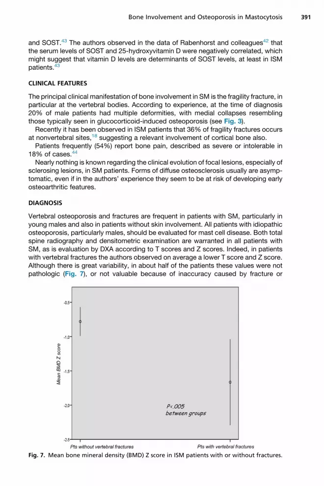
Vertebral osteoporosis and fractures are frequent in patients with SM, particularly inyoung males and also in patients without skin involvement. All patients with idiopathicosteoporosis, particularly males, should be evaluated for mast cell disease. Both totalspine radiography and densitometric examination are warranted in all patients withSM, as is evaluation by DXA according to T scores and Z scores. Indeed, in patientswith vertebral fractures the authors observed on average a lower T score and Z score.Although there is great variability, in about half of the patients these values were notpathologic (Fig. 7), or not valuable because of inaccuracy caused by fracture or
Fig. 7. Mean bone mineral density (BMD) Z score in ISM patients with or without fractures.

Rossini et al392
osteoarthritis deformities. Therefore, DXA sensitivity and specificity to predict verte-bral fracture is limited. In SM the association between fracture and DXA diagnosisof osteoporosis is inconstant, likely because of the patchy distribution of neoplasticmast cells, or because there are other bone features, in addition to BMD, affected;these should be evaluated with a different approach. In the absence of other diag-nostic tools for bone assessment, the authors recommend obtaining a total spineradiograph for vertebral morphometry in all SM patients. Vertebral fractures areassessed visually. The semiquantitative system of Genant is commonly used.45 Avertebral deformity of at least 20% loss in height is typically considered a fracture.This system grades the deformities from I to III, with grade I representing a 20% to24% reduction in vertebral height and ranging up to grade III, which is a 40% or greaterreduction in height. Vertebral fracture assessment by DXA (see Fig. 3) can be useful inthe evaluation of vertebral fractures in these patients, especially in the follow-up. Theauthors typically perform a DXA scan once every year until stability is achieved, withless frequent monitoring thereafter.The observation that less than 5% of patients with ISM had a normal serum tryptase
level suggests that this assay can be used confidently for screening in selected casesof unexplained osteoporosis, even if the serum tryptase level in the normal rangecannot exclude SM diagnosis. On the other hand, high levels of serum tryptase canbe found in other diseases such as chronic urticaria, renal insufficiency, other hema-tologic diseases, onchocercosis, ischemic myocardial disease, or in the presence ofheterophilic antibodies. In the authors’ experience, serum tryptase levels cannot beused as a predictor of osteoporosis involvement in patients with SM. It has been re-ported that the urinary histamine metabolites, in particular methylhistamine, are asso-ciated with a higher risk of osteoporotic manifestations.18
In SM, tryptase levels, which reflect mast cell burden, correlate significantly withBTM.38 The role of BTM in clinical practice remains uncertain. However, high serumlevels of BTM are suspicious for mastocytosis bone involvement, and in this conditiona scintigraphy bone scan is recommended. Increased uptake at the scintiscan candetect focal and asymptomatic bone lesions, to be subjected to targeted radiographicassessment, whereas a diffuse uptake has a prognostic value.46
In patients with SM, very high BTM and serum tryptase levels are suspicious fordiffuse osteosclerotic features.17
MANAGEMENT
Early institution of antimediator therapy may reverse some disease-related bonechanges47; however, there are no conclusive studies in this regard. Bisphosphonatetherapy (alendronate, 70 mg every week; risedronate 35 mg every week; pamidronicacid, 90 mg intravenously every 4 weeks) is effective in increasing vertebral BMD, buthas considerably less efficacy in increasing femoral neck BMD.16,29,48,49 In the absenceof a specific study in patients with SM, the use of zoledronic acid (ZOL) at the dose usedin multiple myeloma (4 mg intravenously every 4 weeks) is recommended.50
Evidence supporting bisphosphonate therapy is limited to case reports or studiesof very small groups (Table 1). Moreover, the problem of poor compliance withbisphosphonates, whether given weekly or monthly, is well documented, and lessfrequent administration would be very attractive to patients and might further increasecompliance. In therapy carried out in 25 ISM patients with osteoporosis, one 5-mgintravenous infusion of ZOL guaranteed a therapeutic effect on surrogate markers(BMD and BTM) of antifracture efficacy for at least 1 year (Rossini, unpublisheddata, 2013). Mean BMD gain was at least twice as great as those reported in previous

Table 1Evidence supporting bisphosphonate therapy
Authors,Ref. Year Treatment Cases (N)
Cundy et al,29 1987 Oral clodronate 1
Marshall et al,49 1997 IV pamidronate 3
Brumsen et al,48 2002 IV 1 oral pamidronate 1
Lim et al,7 2005 IV pamidronate 1 oral alendronate 6
Laroche et al,54 2007 Interferon-a 1 IV pamidronate 4
Laroche et al,55 2011 IV pamidronate � interferon-a 10
Abbreviation: IV, intravenous.
Bone Involvement and Osteoporosis in Mastocytosis 393
small series with daily oral treatment with alendronate or with monthly pamidronateinfusions, both in lumbar and femoral sites. Moreover, at 1 year after the only ZOL infu-sion the BTM were still suppressed, suggesting that there may be a safe time windowduring which subsequent infusions may be administered.In severe and bisphosphonate-refractory cases and in mastocytosis-related bone
pain, interferon-a can be useful.50–55
REFERENCES
1. Fallon MD, Whyte MP, Teitelbaum SL. Systemic mastocytosis associated withgeneralized osteopenia. Histopathological characterization of the skeletal lesionusing undecalcified bone from two patients. Hum Pathol 1981;12:813–20.
2. Chines A, Pacifici R, Avioli LV, et al. Systemic mastocytosis presenting as oste-oporosis: a clinical and histomorphometric study. J Clin Endocrinol Metab 1991;72:140–4.
3. Schoenaers P, De Clerck LS, Timmermans U, et al. Systemic mastocytosis, anunusual cause of osteoporosis. Clin Rheumatol 1987;6:458–62.
4. Harvey JA, Anderson HC, Borek D, et al. Osteoporosis associated with masto-cytosis confined to bone: report of two cases. Bone 1989;10:237–41.
5. Lidor C, Frisch B, Gazit D, et al. Osteoporosis as the sole presentation of bonemarrow mastocytosis. J Bone Miner Res 1990;5:871–6.
6. Floman Y, Amir G. Systemic mastocytosis presenting with severe spinal osteo-penia and multiple compression fractures. J Spinal Disord 1991;4:369–73.
7. LimAY,OstorAJ,LoveS, et al.Systemicmastocytosis: a rarecauseofosteoporosisand its response to bisphosphonate treatment. Ann Rheum Dis 2005;64:965–6.
8. Kushnir-Sukhov NM, Brittain E, Reynolds JC, et al. Elevated tryptase levels areassociated with greater bone density in a cohort of patients with mastocytosis.Int Arch Allergy Immunol 2006;139:265–70.
9. Salles M, Holgado S, Navarro JT, et al. Osteoporosis as a first manifestation ofsystemic mastocytosis. Study of 6 cases. Med Clin (Barc) 2007;128:216–8.
10. King JJ, Crawford EA, Iwenofu OH, et al. Pathologic long bone fracture in a pa-tient with systemic mastocytosis. Clin Orthop Relat Res 2007;459:263–9.
11. Donker ML, Bakker NA, Jaspers WJ, et al. Two patients with osteoporosis: initialpresentation of systemic mastocytosis. J Bone Miner Metab 2008;26:199–202.
12. Stein JA, Kamino H, Walters RF, et al. Mastocytosis with urticaria pigmentosaand osteoporosis. Dermatol Online J 2008;14:2.
13. Mathew R, Dhillon V, Shepherd P. Systemic mastocytosis presenting asosteoporosis-a case report. Clin Rheumatol 2009;28:865–6.

Rossini et al394
14. Manara M, Varenna M, Cantoni S, et al. Osteoporosis with vertebral fractures inyoung males, due to bone marrow mastocytosis: a report of two cases. Clin ExpRheumatol 2010;28:97–100.
15. Escribano L, Alvarez-Twose I, Sanchez-Munoz L, et al. Prognosis in adult indo-lent systemic mastocytosis: a long-term study of the Spanish Network on Mas-tocytosis in a series of 145 patients. J Allergy Clin Immunol 2009;124:514–21.
16. Barete S, Assous N, de Gennes C, et al. Systemic mastocytosis and boneinvolvement in a cohort of 75 patients. Ann Rheum Dis 2010;69:1838–41.
17. Rossini M, Zanotti R, Bonadonna P, et al. Bone mineral density, bone turnovermarkers and fractures in patients with indolent systemic mastocytosis. Bone2011;49:880–5.
18. van der Veer E, van der Goot W, de Monchy JG, et al. High prevalence of frac-tures and osteoporosis in patients with indolent systemic mastocytosis. Allergy2012;67:431–8.
19. Kanis JA. Assessment of fracture risk and its application to screening for post-menopausal osteoporosis: synopsis of a WHO report. WHO study Group. Os-teoporos Int 1994;4:368–81.
20. Lewiecki EM, Gordon CM, Baim S, et al. International society for clinical densi-tometry 2007 adult and pediatric official positions. Bone 2008;43:1115–21.
21. Report of the UK Cystic Fibrosis Trust Bone Mineralisation Working Group. Bonemineralisation in cystic fibrosis. Cystic Fibrosis Trust 2007. Available at: www.cftrust.org.uk. Accessed February 20, 2014.
22. Brumsen C, Papapoulos SE, Lentjes EG, et al. A potential role for the mast cell inthe pathogenesis of idiopathic osteoporosis in men. Bone 2002;31:556–61.
23. Valent P, Akin C, Escribano L, et al. Standards and standardization in mastocy-tosis: consensus statements on diagnostics, treatment recommendations andresponse criteria. Eur J Clin Invest 2007;37:435–53.
24. Zanotti R, Bonadonna P, Bonifacio M, et al. Isolated bone marrow mastocytosis:an underestimated subvariant of indolent systemic mastocytosis. Haematolog-ica 2011;96:482–4.
25. Bonadonna P, Perbellini O, Passalacqua G, et al. Clonal mast cell disorders inpatients with systemic reactions to hymenoptera stings and raised serum tryp-tase. J Allergy Clin Immunol 2009;123:680–6.
26. Seitz S, Barvencik F, Koehne T, et al. Increased osteoblast and osteoclastindices in individuals with systemic mastocytosis. Osteoporos Int 2013;24:2325–34.
27. Delling G, Ritzel H, Werner M. Histological characteristics and prevalence ofsecondary osteoporosis in systemic mastocytosis. A retrospective analysis of158 cases. Pathologe 2001;22:132–40.
28. Travis WD, Li CY, Bergstralh EJ, et al. Systemic mast cell disease. Analysis of 58cases and literature review. Medicine (Baltimore) 1988;67:345–68.
29. Cundy T, Beneton MN, Darby AJ, et al. Osteopenia in systemic mastocytosis:natural history and responses to treatment with inhibitors of bone resorption.Bone 1987;8:149–55.
30. Metcalfe DD. Mast cells and mastocytosis. Blood 2008;112:946–55.31. Theoharides TC, Boucher W, Spear K. Serum interleukin-6 reflects disease
severity and osteoporosis in mastocytosis patients. Int Arch Allergy Immunol2002;128:344–50.
32. Brockow K, Akin C, Huber M, et al. IL-6 levels predict disease variant and extentof organ involvement in patients with mastocytosis. Clin Immunol 2005;115:216–23.

Bone Involvement and Osteoporosis in Mastocytosis 395
33. Dobigny C, Saffar JL. H1 and H2 histamine receptors modulate osteoclasticresorption by different pathways: evidence obtained by using receptor antago-nists in a rat synchronized resorption model. J Cell Physiol 1997;173:10–8.
34. Biosse-Duplan M, Baroukh B, Dy M, et al. Histamine promotes osteoclastogen-esis through the differential expression of histamine receptors on osteoclastsand osteoblasts. Am J Pathol 2009;174:1426–34.
35. Kanzaki S, Takahashi T, Kanno T, et al. Heparin inhibits BMP-2 osteogenic bioac-tivity by binding to both BMP-2 and BMP receptor. J Cell Physiol 2008;216:844–50.
36. Fitzpatrick LA, Buzas E, Gagne TJ, et al. Targeted deletion of histamine decar-boxylase gene in mice increases bone formation and protects againstovariectomy-induced bone loss. Proc Natl Acad Sci U S A 2003;100:6027–32.
37. DeGennesC,KuntzD, deVernejoulMC.Bonemastocytosis.A report of nine caseswith a bone histomorphometric study. Clin Orthop Relat Res 1992;279:281–91.
38. Guillaume N, Desoutter J, Chandesris O, et al. Bone complications of mastocy-tosis: a link between clinical and biological characteristics. Am J Med 2013;126:75.e1–7.
39. Reinacher-Schick A, Petrasch S, Longley BJ, et al. c-Kit mutation andosteopetrosis-like osteopathy in a patient with systemic mast cell disease.Ann Hematol 1998;77:131–4.
40. Chiappetta N, Gruber B. The role of mast cells in osteoporosis. Semin ArthritisRheum 2006;36:32–6.
41. Rossini M, Gatti D, Adami S. Involvement of WNT/b-catenin signaling in the treat-ment of osteoporosis. Calcif Tissue Int 2013;93:121–32.
42. Rabenhorst A, Christopeit B, Leja S, et al. Serum levels of bone cytokines areincreased in indolent systemic mastocytosis associated with osteopenia or oste-oporosis. J Allergy Clin Immunol 2013;132:1234–7.
43. Rossini M, Adami S, Zanotti R, et al. Serum levels of bone cytokines in indolentsystemic mastocytosis associated with osteopenia or osteoporosis. J AllergyClin Immunol 2013. [Epub ahead of print]. http://dx.doi.org/10.1016/j.jaci.2013.12.007.
44. Hermine O, Lortholary O, Leventhal PS, et al. Case-control cohort study of pa-tients’ perceptions of disability in mastocytosis. PLoS One 2008;3(5):e2266.
45. Genant HK, Wu CY, van Kuijk C, et al. Vertebral fracture assessment using asemiquantitative technique. J Bone Miner Res 1993;8:1137–48.
46. Chen CC, Andrich MP, Mican JM, et al. A retrospective analysis of bone scanabnormalities in mastocytosis: correlation with disease category and prognosis.J Nucl Med 1994;35:1471–5.
47. Graves L III, Stechschulte DJ, Morris DC, et al. Inhibition of mediator release insystemic mastocytosis is associated with reversal of bone changes. J BoneMiner Res 1990;5:1113–9.
48. Brumsen C, Hamdy NA, Papapoulos SE. Osteoporosis and bone marrow mas-tocytosis: dissociation of skeletal responses and mast cell activity during long-term bisphosphonate therapy. J Bone Miner Res 2002;17:567–9.
49. Marshall A, Kavanagh RT, Crisp AJ. The effect of pamidronate on lumbar spinebone density and pain in osteoporosis secondary to systemic mastocytosis. Br JRheumatol 1997;36:393–6.
50. Pardanani A. How I treat patients with indolent and smoldering mastocytosis(rare conditions but difficult to manage). Blood 2013;121:3085–94.
51. Weide R, Ehlenz K, Lorenz W, et al. Successful treatment of osteoporosis in sys-temic mastocytosis with interferon alpha-2b. Ann Hematol 1996;72:41–3.

Rossini et al396
52. Lehmann T, Beyeler C, Lammle B, et al. Severe osteoporosis due to systemicmast cell disease: successful treatment with interferon alpha-2B. Br J Rheumatol1996;35:898–900.
53. Butterfield JH. Interferon treatment for hypereosinophilic syndromes and sys-temic mastocytosis. Acta Haematol 2005;114:26–40.
54. Laroche M, Bret J, Brouchet A, et al. Clinical and densitometric efficacy of theassociation of interferon alpha and pamidronate in the treatment of osteoporosisin patients with systemic mastocytosis. Clin Rheumatol 2007;26:242–3.
55. Laroche M, Livideanu C, Paul C, et al. Interferon alpha and pamidronate in oste-oporosis with fracture secondary to mastocytosis. Am J Med 2011;124:776–8.