bioavailability of apocynin through its conversion to glycoconjugate but not to diapocynin
TRANSCRIPT

Bioavailabity of apocynin through its conversion toglycoconjugate but not to diapocynin
Qun Wanga,#, Robert E. Smithc, Ron Luchtefeldc, Albert Y. Sunb, Agnes Simonyia,Rensheng Luod, and Grace Y. Suna,*
a Department of Biochemistry, University of Missouri, Columbia, MO 65212, USAb Department of Medical Pharmacology and Physiology, University of Missouri, Columbia, MO65212, USAc The U.S. Department of Food and Drug Administration, Lenexa, KS 66214, USAd Department of Chemistry and Biochemistry, University of Missouri – St. Louis, MO 63121, USA
AbstractApocynin (4-hydroxy-3-methoxyacetophenone) is a major active ingredient from the rhizomes ofPicrorhiza kurroa, a botanical plant used as an herbal medicine for treatment of a number ofinflammatory diseases. Recently, apocynin is regarded as a specific inhibitor for NADPH oxidasein cell and animal models. In vitro studies indicated conversion of apocynin to diapocynin in thepresence of peroxidases, e.g., myloperoxidase, posing the possibility that diapocynin alsocontributes to the anti-oxidative action of apocynin. The objectives of this study are to examinethe bioavailability of apocynin to plasma, liver and brain tissue after intraperitoneal (i.p.) injection,and to examine whether apocynin is converted to diapocynin in vivo. Diapocynin was chemicallysynthetized and characterized by NMR and IR. Apocynin (5 mg/kg body wt) was injected i.p. toadult male Sprague-Dawley rats and plasma, liver and brain were collected at different times (30min, 1 h and 2 h) after injection. Samples were treated with β-glucuronidase to hydrolyze theglycosyl linkage and analyzed by HPLC/MS. At 30 min and 1 h after injection, approximately50% of apocynin was converted to its glycosyl derivative and was distributed in plasma, liver andbrain. No diapocynin was detected in any samples. These results indicate rapid glycosylation ofapocynin and its transport to blood and other organs but no apparent conversion to diapocynin invivo.
KeywordsApocynin; NADPH oxidase inhibitor; bioavailability; glycoconjugates; serum; brain
© 2007 Elsevier GmbH. All rights reserved.*Corresponding author: Grace Y. Sun, Ph.D. Professor Biochemistry Department M743 Medical Sciences Building University ofMissouri – Columbia Columbia, MO 65212 Tel: 573−882−5377 Fax: 573−884−4597 E-mail: [email protected].#Qun Wang's current address: The Neurological Institute of New York Department of Neurology Columbia University MedicalCenter 710 West 168th Street, New York, NY 10032 Tel: 212−305−4056 (o) E-mail: [email protected]
Publisher's Disclaimer: This is a PDF file of an unedited manuscript that has been accepted for publication. As a service to ourcustomers we are providing this early version of the manuscript. The manuscript will undergo copyediting, typesetting, and review ofthe resulting proof before it is published in its final citable form. Please note that during the production process errors may bediscovered which could affect the content, and all legal disclaimers that apply to the journal pertain.
NIH Public AccessAuthor ManuscriptPhytomedicine. Author manuscript; available in PMC 2009 June 01.
Published in final edited form as:Phytomedicine. 2008 June ; 15(0): 496–503. doi:10.1016/j.phymed.2007.09.019.
NIH
-PA Author Manuscript
NIH
-PA Author Manuscript
NIH
-PA Author Manuscript

IntroductionReactive oxygen species (ROS), including superoxide anions, hydrogen peroxide, andhydroxyl radicals are produced by enzymic and non-enzymic mechanisms in mammaliancells. Although some of the ROS serve as signaling molecules in the cells, excessiveproduction is detrimental and has been implicated to play an important role in theprogression of many disease processes (Sun and Chen, 1998; Chan, 2001). In the pastdecade, substantial interest has been focused on developing novel phytocompounds thatpossess antioxidant properties to combat oxidative effects of ROS.
Nicotinamide adenine dinucleotide phosphate (NADPH) oxidase has recently been regardedas an important source of ROS in a number of cell systems including cells in the centralnervous system (CNS) (Bedard and Krause, 2007). NADPH oxidase is comprised of multi-subunits and activation of this enzyme involves the translocation of the cytosolic subunits(p47/p67/p40 phox) to the membrane subunits (gp91 phox/p22 phox) and subsequenttransfer of electrons from NADPH to molecular oxygen to produce superoxide anions.Recent studies have highlighted the mechanisms for activation of NADPH oxidase in thebrain, which subsequently promotes oxidative injury, microglial activation, andneurodegeneration (Infanger et al., 2006).
Apocynin (4-hydroxy-3-methoxy-acetophenone) is discovered during activity-guidedisolation of immunomodulatory constituents from the rhizome of Picrorhiza kurroa, acreeping plant native to the mountains of India, Nepal, Tibet and Pakistan (Picrorhizakurroa, 2001). Picrorhiza kurroa is a medicinal herb containing a number of activeingredients, including acetophenone derivatives (which have anti-asthmatic properties),kutkin (kutkoside and iridioids, which are hepato-protective), and curcubitacins (which haveanti-tumor effects) (Picrorhiza kurroa, 2001). In the traditional Chinese and Ayurvedicsystem of medicine, Picrorhiza has been used to treat liver diseases, upper respiratory tractdisorders, chronic diarrhea, hemorrhoids, epilepsy, scorpion sting and fever (Picrorhizakurroa, 2001).
In common with the polyphenolic compounds, apocynin has multiple biological actions(Barbieri et al., 2004). Particularly, it exhibits powerful anti-inflammatory and anti-oxidanteffects in a variety of cell and animal models (Elmarakby et al., 2005; Hougee et al., 2006).Our recent study demonstrated the ability of apocynin to protect against oxidative damageinduced by cerebral ischemia in gerbils (Wang et al., 2006). Since there is evidence thatapocynin may be converted to diapocynin through peroxidases, e.g., myeloperoxidase(Ximenes et al., 2007), it is not clear whether the inhibitory effect for apocynin is due to itsmetabolite or both. Furthermore, information on bioavailability of apocynin and its kineticsin different organs has not been investigated in detail. In this study, we conducted a timecourse study to evaluate the bioavailability of apocynin in serum, liver and brain in rats. Inaddition, we chemically synthesized diapocynin from apocynin and used the purifiedcompound to determine whether apocynin is converted to diapocynin in vivo after i.p.injection.
Materials and methodsAnimals and sample preparation
Adult male Sprague-Dawley (250−300 g body wt) (Harlan, Indianapolis, IN) were housed inthe Small Animal Facilities of the University of Missouri-Columbia. Rats were given freeaccess to water and lab chow and maintained in a colony at 22 ± 2°C with a constanthumidity under a 12:12 h light:dark cycle. The animal protocol was approved by theUniversity of Missouri-Columbia Animal Care and Use Committee (Protocol #1741).
Wang et al. Page 2
Phytomedicine. Author manuscript; available in PMC 2009 June 01.
NIH
-PA Author Manuscript
NIH
-PA Author Manuscript
NIH
-PA Author Manuscript

Experiments were carried out in accordance with the NIH Guidelines for the Care and Useof Laboratory Animals.
To assess the bioavailability of apocynin, rats (3 rats per group) were injected i.p. (5 mg/kgbody wt) with apocynin (Sigma-Aldrich, St. Louis, MO) which was dissolved in normalsaline. Animals were decapitated at 30 min, 1 h and 2 h after drug administration. Controlswere injected similarly with saline. Blood was removed by cardiac puncture with heparin-moistened syringes and plasma was obtained by centrifugation at 2000 × g for 10 min. Aftercollecting blood samples from the heart, animals were transcardially perfused with 200 mlof heparinized saline, and liver and brain were removed. The right lobe of liver and a portionof the frontal cortex of the brain (approx. 1 g) were removed and homogenized in 10 vol of17 mM NH4OH (pH 8.0).
Extraction of apocynin from plasma and tissuesAliquots of plasma and tissue homogenates were incubated at 37°C overnight in a systemcontaining 1.0 M ammonium acetate buffer (pH 5.0) and ∼10 unit/ml of β-glucuronidase(Sigma). After incubation, samples were centrifuged at 14,000 rpm for 20 min. In order toremove lipids, CHCl3:CH3OH (3:1 v/v) was added to the supernatant and samples weremixed and centrifuged at 3000 rpm for 20 min. An aliquot of the upper aqueous phase wasused for analysis.
Synthesis and characterization of diapocyninFor chemical synthesis of diapocynin, apocynin was recrystallized from water and dried in adesiccator before use. Diapocynin was synthesized by dissolving 2 g of recrystallizedapocynin in 200 ml of deionized water with stirring and heating until the solution wasboiling gently. After adding 0.15 g of ferrous sulfate heptahydrate and 1.6 g of sodiumpersulfate, a brown precipitate was formed. After cooling for 5 min, the solution filtered.The precipitate was dissolved in 3 N NH4OH and then re-precipitated by adding 6 N HCl.The precipitate was filtered and washed three times with 100 ml of boiling water. Thediapocynin precipitate was further purified by washing three times with 100 ml of boilingmethanol.
1H NMR and 1H-decoupled 13C-NMR spectra of diapocynin in DMSO-d6 were obtainedusing a Bruker ARX 500 MHz NMR. A 30° pulse width was used for the 1H NMR, with a 1sec pulse delay. A 30° pulse width was used for the 13C-NMR spectra, with a 2 sec pulsedelay. The hydrogen and carbon chemical shifts were referenced to the DMSO peaks, whichwere set to 2.50 ppm for hydrogen and 39.50 ppm for carbon respectively. The AttachedProton Test (APT) was used to distinguish between two groups of signals, namely, methyl/methine and methylene/quaternary. An FTIR spectrum was obtained using a Varian 800FTIR (Palo Alto, CA) with an attenuated total reflectance (ATR) accessory, from PIKETechnologies (Madison, WI).
HPLC/MS analysis of apocynin and diapocyninFor detection, LC-MS analysis used selected ion monitoring (SIM) of ions with m/z =163.5−164.5, corresponding to the [M-H]− ion produced by apocynin and m/z = 327.5 −330.5 for diapocynin. An octadecyl silica (C18) column, 10 cm × 2.1 mm i.d., 100Å poreC18 Ace® analytical column and guard column, from MacMod Analytical, Inc (ChaddsFord, PA) was used for analysis. The LC mobile phase consisted of a gradient elution.Solvent A was 480:20:0.38 water:methanol:ammonium acetate (v/v/w) and solvent B was20:480:0.38 water:methanol:ammonium acetate (v/v/w). The mobile phase started with100% A for the first min, followed by a linear increase to 100% B from 1−16 min. This was
Wang et al. Page 3
Phytomedicine. Author manuscript; available in PMC 2009 June 01.
NIH
-PA Author Manuscript
NIH
-PA Author Manuscript
NIH
-PA Author Manuscript

followed by 100% B from 16−31 min, then a linear decrease to 100% A from 31− 40 min.The injection volume was 20 μl and the eluent flow rate was 0.25 ml/min.
ResultsChemical synthesis of diapocynin
In this study, we used a modified protocol for the synthesis of diapocynin from apocyninbased on the synthesis of a similar compound, dehydrodivanillin (Elbs and Lerch, 1916).Structures of these compounds are shown in Figure 1. This is an oxidation-reductionreaction, requiring the in situ generation of sulfate radicals, which remove a single hydrogenfrom each molecule of apocynin, producing diapocynin. The reaction was conducted inboiling water for 5 min instead of the original 30 min in hot water (van den Worm, et al.;Elbs and Lerch, 1916). Using sodium salt of persulfate instead of the potassium saltproduced higher yield. Also, the diapocynin was re-dissolved in 3 M NH4OH, instead ofNaOH, because NaOH often is contaminated with chloride and carbonate (Weiss, 2004).Finally, this newer method of synthesis included washing the diapocynin three times eachwith boiling water and boiling methanol. Analysis of apocynin and diapocynin by HPLCshowed retention times of 9.5 and 10.8 min respectively (Fig. 2). Both compounds showedlinear relationship between concentrations and peak area (Fig. 2).
The 1H NMR spectrum (Dasari et al., 2008) of diapocynin contained peaks with thefollowing chemical shifts, in ppm, and assignments, in parentheses: 2.494 (-CH3), 3.895 (-OCH3), 7.451 and 7.463 (aromatic CH) and 9.465 (-OH). The 13C-NMR spectrum (Fig. 3A)contained peaks with the following chemical shifts, in ppm, and assignments by comparingwith the APT spectrum (Fig. 3B), in parentheses: 26.25 (-CH3), 55.98 (-CH3), 109.66(aromatic C-2, CH), 124.46 (C-5, C), 125.25 (C-6, CH), 127.86 (C1, C), 147.44 (C-4, C),149.11 (C-3, C) and 196.15 (C$#xFF1D;O). The FTIR spectrum (Dasari et al., 2008)contained the following peaks: λmax cm−1 3318 (OH), 1666 (C$#xFF1D;O), 1588 (aryl C$#xFF1D;C), 1286, 1204, 1127, 1083, 910. The negative ion LC-MS analysis of diapocynin(Fig. 4) indicated that diapocynin has a molecular weight of 330, since a [M-H−] ion wasproduced with m/z = 329. The UV spectrum indicated that diapocynin had a substitutedbenzene structure, similar to apocynin (Fig. 4).
Bioavailability of apocynin in plasma, liver and brainAn initial study was performed to analyze free apocynin in plasma at different times afteri.p. injection (5 mg/kg). As shown in Figure 5, apocynin with a retention time of 9.6 minappeared in plasma as early as 30 min, peaked at 1 h and declined to low levels after 2 h.Spiking apocynin and diapocynin standards to plasma samples showed appearance of therespective standards at the expected retention time (data not shown). Although analysis ofplasma samples showed a peak at 10.8 min, corresponding to that of diapocynin standard,this peak was observed in controls and did not change with time after apocynin injection(Fig 5). Since diapocynin is more non-polar than apocynin, analysis of other fractions of theextraction medium also showed no evidence of diapocynin in the samples. Therefore, thesedata demonstrate that apocynin was not actively converted to diapocynin in vivo.
Study was performed to test whether apocynin is converted to glycoconjugate by comparingplasma samples with or without β-glucuronidase treatment. As shown in Figure 6, levels offree apocynin in plasma at 30 and 60 min after injection (5 mg/kg, i.p., n=3) were 20−30 ng/ml as compared to 60−70 ng/ml after β-glucuronidase treatment.
Results in Figure 7 show the HPLC analysis of apocynin in β-glucuronidase-treated plasma,brain and liver samples in controls and after injection of apocynin (30 min). Data for 2 hsamples were not shown because a large part of the apocynin had already diminished at this
Wang et al. Page 4
Phytomedicine. Author manuscript; available in PMC 2009 June 01.
NIH
-PA Author Manuscript
NIH
-PA Author Manuscript
NIH
-PA Author Manuscript

time (data not shown). Levels of apocynin in the liver were at least 3−5 times higher thanthose in the brain.
DiscussionDue to their potentially beneficial impact on human health, plant extracts have come into thefocus of medicinal interest. However, data on the bioavailability of these plant extracts arelargely lacking (Loew and Kaszkin, 2002; Simonyi et al., 2005; Williamson and Manach,2005). In our previous work, we investigated the bioavailability of two plant polyphenoliccompounds, resveratrol and curcumin, extracted from grapes and turmeric, respectively(Wang et al., 2002; Wang et al., 2005). We observed that both polyphenolic compoundsform glycoconjugates which were distributed to other organs including the brain. In thepresent study, we show that apocynin was also rapidly converted to glycoconjugates. Similarto resveratrol and curcumin, apocynin was readily distributed in plasma, liver and brain andreached peak levels around 1 h after injection. The appearance of apocynin in brain is inagreement that this compound can cross the blood–brain barrier and incorporated into braintissue. The apocynin in brain tissue is unlikely to be due to blood contamination becauseanalysis was carried out after perfusing animal with saline.
In studies in vitro, apocynin was shown to undergo oxidation and converted to diapocynin(Ximenes et al., 2007). In our analysis with plasma samples, we observed a peakcorresponding to the retention time for diapocynin in the extract. However, the same peakcould be found upon analysis of blank and control samples (without injection of apocynin).Therefore, it is concluded that apocynin was not significantly converted to diapocynin invivo under these circumstances.
Apocynin is known to inhibit NAPDH oxidase activity by interfering with the assembly ofthe cytosolic NADPH oxidase components with the membrane components (Stolk et al.,1994). NADPH oxidase has emerged as a major source of oxidative stress in the brain,particularly in neurodegenerative diseases, such as ischemic stroke, Alzheimer's andParkinson's diseases, HIV dementia, multiple sclerosis, etc (Bedard and Krause, 2007).Recent studies have shown that apocynin placed in the drinking water or in diet are effectiveat reducing superoxide and oxidative stress in vivo (Cotter and Cameron, 2003; Elmarakbyet al., 2005; Hougee et al., 2006; Paliege et al., 2006). No adverse effects were observedwith apocynin doses ranging from 2.5 mg/kg/day to 100mg/kg/day and duration between 4days and 6 weeks (Cotter and Cameron, 2003; Elmarakby et al., 2005; Hougee et al., 2006;Paliege et al., 2006). The therapeutic potential of systemic single administration of apocyninhas been demonstrated in a dose range of 2.5−12 mg/kg in different animal models (Sonta etal., 2004; Kimura et al., 2005). In support for the bioavailability of apocynin to the brain,results of our previous study also provided strong evidence for apocynin to offerneuroprotective effects against ischemia-induced neuronal damage (Wang et al., 2006).Taken together, there is increasing support to consider using this phyto-compound as atherapeutic agent for treatment of inflammatory diseases including those in the brain.
AcknowledgmentsThis work was supported in part by P01 AG18357 from NIH. This work should not be taken as having an impact onFDA policy or regulations.
ReferencesBarbieri SS, Cavalca V, Eligini S, Brambilla M, Caiani A, Tremoli E, Colli S. Apocynin prevents
cyclooxygenase 2 expression in human monocytes through NADPH oxidase and glutathione redox-dependent mechanisms. Free Radic. Biol. Med. 2004; 37:156–165. [PubMed: 15203187]
Wang et al. Page 5
Phytomedicine. Author manuscript; available in PMC 2009 June 01.
NIH
-PA Author Manuscript
NIH
-PA Author Manuscript
NIH
-PA Author Manuscript

Bedard K, Krause KH. The NOX family of ROS-generating NADPH oxidases: physiology andpathophysiology. Physiol. Rev. 2007; 87:245–313. [PubMed: 17237347]
Chan PH. Reactive oxygen radicals in signaling and damage in the ischemic brain. J. Cereb. BloodFlow Metab. 2001; 21:2–14. [PubMed: 11149664]
Cotter MA, Cameron NE. Effect of the NAD(P)H oxidase inhibitor, apocynin, on peripheral nerveperfusion and function in diabetic rats. Life Sci. 2003; 73:1813–1824. [PubMed: 12888120]
Dasari M, Richards KM, Alt MA, Crawford CFP, Schleiden A, Ingram J, Aziz A, Hamidou AAA,Williams A, Chernovitz PA, Luo R, Sun GY, Luchtefeld R, Smith RE. Diapocynin Synthesis. J.Chem. Ed. 2008 in press.
Elbs K, Lerch H. Über dehydrodivanillin. J. Prakt. Chem. 1916; 93:1–9.
Elmarakby AA, Loomis ED, Pollock JS, Pollock DM. NADPH oxidase inhibition attenuates oxidativestress but not hypertension produced by chronic ET-1. Hypertension. 2005; 45:283–287. [PubMed:15623539]
Hougee S, Hartog A, Sanders A, Graus YM, Hoijer MA, Garssen J, van den Berg WB, van BeuningenHM, Smit HF. Oral administration of the NADPH-oxidase inhibitor apocynin partially restoresdiminished cartilage proteoglycan synthesis and reduces inflammation in mice. Eur. J. Pharmacol.2006; 531:264–269. [PubMed: 16405885]
Infanger DW, Sharma RV, Davisson RL. NADPH oxidases of the brain: distribution, regulation, andfunction. Antioxid. Redox Signal. 2006; 8:1583–1596. [PubMed: 16987013]
Kimura H, Liu S, Yamada S, Uchida K, Matsumoto K, Mukaida M, Yoshida K. Rapid increase inserum lipid peroxide 4-hydroxynonenal (HNE) through monocyte NADPH oxidase in early endo-toxemia. Free Radic. Res. 2005; 39:845–851. [PubMed: 16036364]
Loew D, Kaszkin M. Approaching the problem of bioequivalence of herbal medicinal products.Phytother. Res. 2002; 16:705–711. [PubMed: 12458470]
Paliege A, Pasumarthy A, Mizel D, Yang T, Schnermann J, Bachmann S. Effect of apocynin treatmenton renal expression of COX-2, NOS1, and renin in Wistar-Kyoto and spontaneously hypertensiverats. Am. J. Physiol. Regul. Integr. Comp. Physiol. 2006; 290:R694–R700. [PubMed: 16467505]
Picrorhiza kurroa. Monograph. Altern. Med. Rev. 2001; 6:319–321. [PubMed: 11410077]
Simonyi A, Wang Q, Miller RL, Yusof M, Shelat PB, Sun AY, Sun GY. Polyphenols in cerebralischemia: novel targets for neuroprotection. Mol. Neurobiol. 2005; 31:135–147. [PubMed:15953817]
Sonta T, Inoguchi T, Tsubouchi H, Sekiguchi N, Kobayashi K, Matsumoto S, Utsumi H, Nawata H.Evidence for contribution of vascular NAD(P)H oxidase to increased oxidative stress in animalmodels of diabetes and obesity. Free Radic. Biol. Med. 2004; 37:115–123. [PubMed: 15183199]
Stolk J, Hiltermann TJ, Dijkman JH, Verhoeven AJ. Characteristics of the inhibition of NADPHoxidase activation in neutrophils by apocynin, a methoxy-substituted catechol. Am. J. Respir. CellMol. Biol. 1994; 11:95–102. [PubMed: 8018341]
Sun AY, Chen YM. Oxidative stress and neurodegenerative disorders. J. Biomed. Sci. 1998; 5:401–414. [PubMed: 9845843]
van den Worm E, van den Berg AJJ, Kemeling GM, Beukelman CJ, Halkes SBA, Labadie RP, vanDijk H. Isolation, characterization and activity of diapocynin, an apocynin metabolite. Chapter 5.http://igiturarchive.library.uu.nl/dissertations/1957866/c5.pdf
Wang Q, Xu J, Rottinghaus GE, Simonyi A, Lubahn D, Sun GY, Sun AY. Resveratrol protects againstglobal cerebral ischemic injury in gerbils. Brain Res. 2002; 958:439–447. [PubMed: 12470882]
Wang Q, Sun AY, Simonyi A, Jensen MD, Shelat PB, Rottinghaus GE, MacDonald RS, Miller DK,Lubahn DE, Weisman GA, Sun GY. Neuroprotective mechanisms of curcumin against cerebralischemia-induced neuronal apoptosis and behavioral deficits. J. Neurosci. Res. 2005; 82:138–148.[PubMed: 16075466]
Wang Q, Tompkins KD, Simonyi A, Korthuis RJ, Sun AY, Sun GY. Apocynin protects against globalcerebral ischemia-reperfusion-induced oxidative stress and injury in the gerbil hippocampus. BrainRes. 2006; 1090:182–189. [PubMed: 16650838]
Weiss, J. Handbook of Ion Chromatography. Weinheim; Wiley-VCH: 2004. p. 196
Williamson G, Manach C. Bioavailability and bioefficacy of polyphenols in humans. II. Review of 93intervention studies. Am. J. Clin. Nutr. 2005; 81:243S–255S. [PubMed: 15640487]
Wang et al. Page 6
Phytomedicine. Author manuscript; available in PMC 2009 June 01.
NIH
-PA Author Manuscript
NIH
-PA Author Manuscript
NIH
-PA Author Manuscript

Ximenes VF, Kanegae MP, Rissato SR, Galhiane MS. The oxidation of apocynin catalyzed bymyeloperoxidase: proposal for NADPH oxidase inhibition. Arch. Biochem. Biophys. 2007;457:134–141. [PubMed: 17166480]
Wang et al. Page 7
Phytomedicine. Author manuscript; available in PMC 2009 June 01.
NIH
-PA Author Manuscript
NIH
-PA Author Manuscript
NIH
-PA Author Manuscript

Fig. 1.Reaction for conversion of apocynin to diapocynin.
Wang et al. Page 8
Phytomedicine. Author manuscript; available in PMC 2009 June 01.
NIH
-PA Author Manuscript
NIH
-PA Author Manuscript
NIH
-PA Author Manuscript

Fig. 2.Analysis of apocynin and diapocynin standards by HPLC. See text for details of theanalysis.
Wang et al. Page 9
Phytomedicine. Author manuscript; available in PMC 2009 June 01.
NIH
-PA Author Manuscript
NIH
-PA Author Manuscript
NIH
-PA Author Manuscript

Fig. 3.(A) Proton-Decoupled 13C-NMR Spectrum of Diapocynin in DMSO-d6. (B) APT Spectrumof Diapocynin in DMSO-d6. Positive peaks are due to C and CH2, negative peaks are due toCH and CH3.
Wang et al. Page 10
Phytomedicine. Author manuscript; available in PMC 2009 June 01.
NIH
-PA Author Manuscript
NIH
-PA Author Manuscript
NIH
-PA Author Manuscript
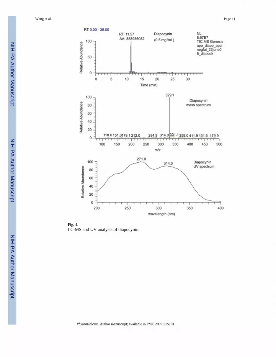
Fig. 4.LC-MS and UV analysis of diapocynin.
Wang et al. Page 11
Phytomedicine. Author manuscript; available in PMC 2009 June 01.
NIH
-PA Author Manuscript
NIH
-PA Author Manuscript
NIH
-PA Author Manuscript

Fig. 5.Representative chromatographs showing apocynin in plasma at different times after i.p.injection (5 mg/kg). Arrow points to apocynin peak.
Wang et al. Page 12
Phytomedicine. Author manuscript; available in PMC 2009 June 01.
NIH
-PA Author Manuscript
NIH
-PA Author Manuscript
NIH
-PA Author Manuscript

Fig. 6.Levels of apocynin in plasma with or without β-glucuronidase treatment.
Wang et al. Page 13
Phytomedicine. Author manuscript; available in PMC 2009 June 01.
NIH
-PA Author Manuscript
NIH
-PA Author Manuscript
NIH
-PA Author Manuscript

Fig. 7.Apocynin in plasma, liver and brain comparing with control and 30 min after i.p. injection(5 mg/kg). Samples were treated with β-glucoronidase prior to analysis by HPLC.
Wang et al. Page 14
Phytomedicine. Author manuscript; available in PMC 2009 June 01.
NIH
-PA Author Manuscript
NIH
-PA Author Manuscript
NIH
-PA Author Manuscript