bioavailability of phenols from a phenol-enriched olive oil
TRANSCRIPT

Bioavailability of phenols from a phenol-enriched olive oil
Manuel Suarez1, Rosa M. Valls2, Maria-Paz Romero1, Alba Macia1, Sara Fernandez2, Montse Giralt2,Rosa Sola2* and Maria-Jose Motilva1*1Food Technology Department, XaRTA-TPV, Escuela Tecnica Superior de Ingenierıa Agraria, University of Lleida, Av/Alcalde
Rovira Roure 191, 25198 Lleida, Spain2Lipid and Arteriosclerosis Research Unit (CIBERDEM), Facultat de Medicina i Ciencies de la Salut, St Joan de Reus
University Hospital, IISPV, Universitat Rovira i Virgili, C/Sant Llorenc 21, 43201 Reus, Spain
(Received 15 November 2010 – Revised 21 March 2011 – Accepted 22 March 2011 – First published online 21 June 2011)
Abstract
Phenolic compounds are one of the main reasons behind the healthy properties of virgin olive oil (VOO). However, their daily intake from
VOO is low compared with that obtained from other phenolic sources. Therefore, the intake of VOO enriched with its own phenolic com-
pounds could be of interest to increase the daily dose of these beneficial compounds. To evaluate the effectiveness of enrichment on their
bioavailability, the concentration of phenolic compounds and their metabolites in human plasma (0, 60, 120, 240 and 300 min) from thir-
teen healthy volunteers (seven men and six women, aged 25 and 69 years) was determined after the ingestion of a single dose (30 ml) of
either enriched virgin olive oil (EVOO) (961·17 mg/kg oil) or control VOO (288·89 mg/kg oil) in a cross-over study. Compared with VOO,
EVOO increased plasma concentration of the phenol metabolites, particularly hydroxytyrosol sulphate and vanillin sulphate (P,0·05).
After the consumption of VOO, the maximum concentration of these peaks was reached at 60 min, while EVOO shifted this maximum
to 120 min. Despite these differences, the wide variability of results indicates that the absorption and metabolism of olive oil phenols
are highly dependent on the individual.
Key words: Bioavailability: Human plasma: Phenol-enriched olive oil: Plasma phenol metabolites
Over the last decade, epidemiological studies have proven
that a Mediterranean diet, rich in vegetables, fruit and
legumes, is correlated with a low incidence of CHD and
cancer(1). Within this nutritional pattern, virgin olive oil
(VOO) plays an important role, being the main source of fat
in the diet in this area. The beneficial effects of VOO were
initially attributed to its fatty acid composition, rich in MUFA.
Nevertheless, further studies have highlighted the importance
of its phenolic composition, especially after the publication of
the results of the EUROLIVE project, which demonstrated that
the increase in the HDL-cholesterol level and the decrease
in the lipid oxidation damage after VOO consumption were
highly correlated with its phenolic content(2,3). Thus, phenolic
compounds from VOO (mainly phenolic alcohols, secoiridoid
derivatives, phenolic acids, lignans and flavonoids) have
been reported to have anti-oxidant(4), anti-inflammatory(5),
anti-atherogenic(6) and anti-carcinogenic properties(7).
The metabolic fate of phenolic compounds after ingestion
has been the subject of study by the scientific community to
find out the mechanisms through which they exert their
activity into the organism. It is known that polyphenols
suffer strong metabolism phase I and phase II, in which
they are hydrolysed (phase I) and later conjugated (phase
II) into their glucuronidated, methylated and sulphated
forms in order to be absorbed(8). In the case of VOO, studies
have focused on some specific compounds such as hydroxy-
tyrosol and tyrosol as they have been reported as the most
biologically active. Thus, Visioli et al.(9) were the first
researchers who identified these compounds in urine, both
in the conjugated and in their free forms. In addition, this
study showed that hydroxytyrosol and tyrosol are dose-
dependently absorbed in humans. Later, the same research
group reported the presence of homovanillic alcohol in
urine, a compound that is derived from hydroxytyrosol by
the action of the enzyme catechol-O-methyltransferase(10).
On the other hand, the study by Miro-Casas et al.(11) reported
the presence of conjugated forms of hydroxytyrosol in plasma.
The importance of these compounds has been reinforced
by the demonstration that they can aid in preventing
the oxidation of LDL(12). Recently, an association between
*Corresponding authors: Dr M.-J. Motilva, fax þ34 973 702596, email [email protected]; R. Sola, fax þ34 977759322, email [email protected]
Abbreviations: 3,4-DHPEA-EDA, dialdehydic form of elenolic acid linked to hydroxytyrosol; EVOO, enriched virgin olive oil; MOPET, 3-methoxy-4-
hydroxy-phenylethanol; UPLC, ultra performance liquid chromatography; VOO, virgin olive oil.
British Journal of Nutrition (2011), 106, 1691–1701 doi:10.1017/S0007114511002200q The Authors 2011
British
Journal
ofNutrition

hydroxytyrosol and LDL in plasma has been described, a fact
that could have important physiological implications(13). All
these studies demonstrated the importance of hydroxytyrosol
and tyrosol; however, little has been done about the other
components of the phenolic fraction of VOO, which have
also been shown to have high biological activity such as the
dialdehydic form of elenolic acid linked to hydroxytyrosol
(3,4-DHPEA-EDA)(14).
More recent studies have indicated that the beneficial effects
of the phenolic compounds from VOO are not only due to
their anti-oxidant and related activities, but they also have a
nutrigenomic effect, which means that their intake has a
direct impact on the human genome. Thus, a study carried
out by Konstantinidou et al.(15) showed that the phenols
from VOO have a significant role in the down-regulation of
proatherogenic genes such as IFN-g, ARHGAP15 and IL7R.
This reinforces the importance of including VOO in the regu-
lar diet (the Food and Drug Administration recommends a
daily intake of 23 g of olive oil).
Nevertheless, the amount of phenolic metabolites from
VOO detected in human plasma samples is highly variable
and depends on both extraction and analytical procedure.
Thus, Vissers et al.(16) reported that the bioavailability of
these compounds in ileostomed volunteers was up to 66 %
of the total ingested. Considering a mean intake of 50 g of
VOO/d, these authors estimated that the total amount of phe-
nolic compounds in the plasma samples could be at most
0·06mmol of hydroxytyrosol equivalents/l plasma. However,
some studies revealed that a higher concentration is required
to protect LDL from oxidation. For example, Leenen et al.(17)
concluded that at least 50–100mmol of phenolic compounds
from VOO/l plasma are required to have a protective effect
on LDL. Therefore, according to these results, enough phe-
nols are unlikely to be obtained from VOO to have a direct
protective effect on LDL. In addition, the concentration of
phenolic compounds in commercial VOO is widely depen-
dent on agronomical and technical factors(18). This decreases
the ingestion of phenols from VOO compared with the doses
ingested from other sources and makes it difficult to ensure
a regular dose of phenols through the daily intake of VOO.
For this reason, the enrichment of VOO with their own
phenols, thus giving a high and standardised content of
phenolic compounds, could be desirable in order to increase
the daily intake of phenols without the drawback of a higher
energy content.
The aim of the present study was to evaluate the effect of
the enrichment of VOO with their own phenolic compounds
in their bioavailability in human subjects. To fulfil this objec-
tive, the phenol metabolites were identified and quantified
in human plasma samples obtained after an acute ingestion
of 30 ml of a phenol-enriched VOO (EVOO). Plasma samples
were obtained at 0, 60, 120, 240 and 300 min in order to study
the pharmacokinetic behaviour of these metabolites. The
results were compared with those obtained after the ingestion
of the VOO (without phenol addition).
Experimental methods
Chemicals and reagents
Standard phenolic compounds were used to obtain calibration
curves to quantify the compounds in the olive oils and in the
plasma samples. Apigenin, luteolin, hydroxytyrosol, tyrosol
and p-coumaric acid were purchased from Extrasynthese
(Genay, France). Caffeic and homovanillic acids were pur-
chased from Fluka Company (Buchs, Switzerland). (þ)-Pinor-
esinol was acquired from Arbo Nova (Turku, Finland), and
catechol was from Sigma-Aldrich (Schnelldorf, Germany).
The secoiridoid derivatives 3,4-DHPEA-EDA, the dialdehydic
form of elenolic acid linked to tyrosol and the lignan acetox-
ypinoresinol are not available commercially, and were isolated
from virgin olive by semi-preparative HPLC, as described by
Artajo et al.(19). A stock solution of each standard compound
was dissolved in methanol, and all the solutions were stored
in a dark flask at 2408C.
Methanol (HPLC grade), acetonitrile (HPLC grade) and
acetic acid were provided by Scharlau Chemie (Barcelona,
Spain). Ortho-phosphoric acid (85 %) was purchased from
Panreac (Barcelona, Spain). Water was MilliQ quality (Millipore
Corporation, Bedford, MA, USA).
Virgin olive oil
The VOO used as control and as matrix for phenolic enrichment
was from the Siurana Protected Denomination of Origin (Cata-
lonia, Spain), which is produced exclusively from the Arbequina
cultivar. On the other hand, the samples of the olive cake used
to obtain the phenolic extract for phenol enrichment were
taken from a commercial olive oil mill from the olive-growing
area of Les Garrigues (Catalonia, Spain), which works by the
two-phase centrifugation system. These samples were taken at
the decanter outlet, and liquid N2 was immediately added to
avoid oxidative damage. The samples were stored at 2408C
until the extraction of the phenolic compounds.
Preparation of the phenol-enriched olive oil
The EVOO was prepared by the addition of an extract rich in
the main phenolic compounds of VOO. This extract was
obtained from the olive cake following the method described
by Suarez et al.(20) and basically included secoiridoid deriva-
tives (89·4 %), phenyl alcohols (3·5 %) and flavonoids (6·0 %).
To carry out phenolic enrichment of the oil, 7 mg of olive
cake phenolic extract/ml oil and 0·3 % (p/v) of lecithin (Emul-
pur; Cargill, Barcelona, Spain) were dissolved in ethanol–water
(50:50, v/v) and added to the oil using a Polytron (Kinematica,
Littau, Switzerland) until completely homogenised. Finally, the
EVOO was filtered through a paper filter Ahlstrom (Ahlstrom
S.A., Barcelona, Spain) and bottled in dark bottles. The VOO
used as control (without added phenolic compounds) was
also submitted to the process of mixing and filtering to
ensure equal conditions in all the olive oils under study. The
phenolic compounds of both the VOO and the EVOO were
extracted and characterised with the method described in our
previous study(21).
M. Suarez et al.1692
British
Journal
ofNutrition

Volunteers and experimental design
The present study was conducted according to the guidelines
laid down in the Declaration of Helsinki, and all procedures
involving human subjects were approved by the Ethics Com-
mittee of Clinical Research of Sant Joan University Hospital,
Reus, Spain (Trial Registration clinicaltrials.gov Identifier:
ISRCTN03450153). The subjects gave written informed con-
sent. A total of sixteen healthy volunteers (eight women and
eight men) were recruited to carry out the study. However,
at the moment of ingestion, three of the volunteers (two
women and one man) gave up the study, thus reducing the
final number of volunteers to thirteen. The volunteers, aged
between 25 and 69 years, were considered healthy according
to the results obtained after physical examination and routine
laboratory tests. The men had a mean weight of 78·3 (SD
13·0) kg and the women had a mean weight of 64·5
(SD 5·7) kg, while the BMI was 26·7 (SD 1·9) kg/m2 for men
and 24·9 (SD 1·5) kg/m2 for women.
A randomised, controlled, cross-over trial was designed
using VOO (without phenol addition) and EVOO. Thus, the
volunteers who consumed VOO in the first intervention
ingested the EVOO in the second intervention after a
2-week washout period (Fig. 1) and vice versa, volunteers
who consumed the EVOO in the first intervention ingested
the VOO in the second intervention. The participants were
instructed to follow a stabilisation diet with 10 % of SFA
during the week before the postprandial test and the day
before a polyphenol free diet was prescribed. Between the
tests, the volunteers followed the stabilisation diet. The rec-
ommendations were to avoid VOO, olives, fresh fruits or
juices, wine, chocolate, coffee, tea, soya, legumes and beer.
The participants were instructed to perform intense physical
exercise 3 d before the postprandial test.
Acute ingestion of the oil was done at 08.00 hours after an
overnight fast. The VOO was included in the study to enable
the comparison of the resulting data and to extract
conclusions.
Plasma samples
Plasma samples were obtained by venepuncture from volun-
teers after the ingestion of 30 ml of either VOO or EVOO.
The blood samples were collected under basal conditions
(after a 12 h overnight fasting period) and 60, 120, 240 and
300 min after the consumption of 30 ml of either enriched or
control VOO. To obtain plasma samples at every measuring
point, blood (50 ml) was collected into Vacutainere tubes
containing EDTA as an anti-coagulant. They were protected
from light with Al foil and centrifuged for 15 min at 1500 g
and 48C (H-103RS; Kokusan, Tokyo, Japan). After that, the
plasma was immediately separated from the cells and kept
at 2808 C until analysis.
The extraction of the phenolic compounds from the plasma
samples was carried out following the method developed and
validated in our previous study(22) using microelution plates
(Waters, Milford, MA, USA) packed with 2 mg of OASIS HLB
sorbent (Waters).
Analysis of phenolic compounds in olive oils andplasma samples
The analysis of the phenolic compounds from both oils (VOO
and EVOO) and plasma samples was carried out by ultra per-
formance liquid chromatography (UPLC) coupled to tandem
MS. The UPLC system consisted of an AcQuitye UPLC
equipped with a Waters binary pump system using an AcQuity
UPLCe BEH C18 column (1·7mm, 100 mm £ 2·1 mm inner
diameter). During the analysis, the column was kept at 308C
and the flow rate was 0·4 ml/min, using MilliQ water–acetic
acid (100:0·2, v/v) as solvent A and acetonitrile as solvent
B. The elution gradient was the one reported in our previous
study(21).
Full-scan mode MS and MS/MS, based on neutral loss
scan and product ion scan, were used to identify and quantify
the phenolic compounds and their metabolites from plasma
samples. These techniques are very effective in verifying
the structural information of the compounds when no com-
mercially standards are available. To start with, the analyses
were carried out in full-scan mode (from 80 to 800 m/z)
by applying different cone voltages from 20 to 60 V. The MS
spectrum obtained when low cone voltages were used gave
information about the precursor ion or the [M 2 H]2. On
the other hand, when high cone voltages were applied,
specific fragment ions were generated and the MS spectrum
gave information about their structure. The structural infor-
mation was also verified by using the product ion scan
and neutral loss scan in the MS/MS mode. In the product
ion scan experiments, the product ions are produced by
collision-activated dissociation of the selected precursor ion
in the collision cell. Neutral loss scans of 80 and 176 units
were used to characterise the sulphate and glucuronide forms,
respectively. The detection and quantification of the phenolic
compounds and their metabolites were then performed
based on their ion fragmentation in the MS/MS mode using
selected reaction monitoring (SRM) and double SRM.
Due to the lack of standards for these metabolites, they
were quantified with the calibration curves corresponding to
their phenolic precursors. Hydroxytyrosol and tyrosol metab-
olites were quantified using the calibration curves of hydroxy-
tyrosol and tyrosol, respectively; homovanillic acid, vanillic
acid and vanillin metabolites were quantified using the
Diet stabilisation
Wash-out
Wash-out
1st ingestion 2nd ingestion
2 weeks 2 weeks
Group 1
Group 2 Diet stabilisation
Fig. 1. Study design of the acute ingestion study of control and phenol-
enriched olive oil. , 30 ml of control olive oil; , 30 ml of phenol-enriched
olive oil.
Bioavailability of phenol-enriched olive oil 1693
British
Journal
ofNutrition

calibration curve of homovanillic acid; p-coumaric acid,
p-hydroxybenzoic acid and ferulic acid metabolites were
quantified by means of the p-coumaric acid calibration
curve. Apigenin metabolites were quantified with the apigenin
calibration curve.
Statistical analysis and pharmacokinetic parameters
Values are presented as means and standard deviations. Data
analysis was performed using Statgraphics plus v.5.1 software
(Manugistics, Inc., Rockville, MA, USA). The normality of the
data was evaluated by means of the values of the coefficients
of asymmetry and kurtosis. Significant differences among
the results from the two olive oils were evaluated by the
Kruskal–Wallis ANOVA (P,0·05).
On the other hand, kinetic parameters of the different
metabolites of the phenolic compounds were calculated by
means of pharmacokinetic (PK) functions (for Microsoft
Excel). This software allowed us to determine the maximum
concentration of the metabolites (Cmax) and their area under
the curve, which can be used to compare the effect of the
ingestion of the different oils (control or phenol-enriched).
Results and discussion
Characterisation of the virgin olive oils
The phenolic profiles of both VOO and EVOO were analysed
by UPLC-electrospray ionisation-MS/MS. As can be seen in
Table 1, the addition of the olive cake extract significantly
increased the concentration of all the phenolic compounds
in the oils. The 10·95-fold increase experimented by the
phenyl alcohol fraction, made up of hydroxytyrosol and tyro-
sol, was especially remarkable. These compounds are biologi-
cally active, so it is very interesting to have them at a higher
and optimal concentration in the olive oil available to be
absorbed by the organism after ingestion.
It is also important to highlight the increase observed in the
secoiridoid derivatives and the flavonoid group. The import-
ance of the secoiridoid derivatives is based on their chemical
structure. These compounds are precursors of phenyl alco-
hols, which, as was stated earlier, are very active compounds.
In addition, recent studies have demonstrated that some
secoiridoid derivatives, such as 3,4-DHPEA-EDA, are even
better at protecting erythrocytes from oxidative damages
than hydroxytyrosol(23). Therefore, the secoiridoid derivatives
are valuable compounds that could have a beneficial effect on
the organism. On the other hand, flavonoids are the most
widespread natural phenolic compounds whose important
activity is due to their characteristic triple aromatic ring chemi-
cal structure. However, their concentration in VOO is very low
compared with that obtained from other phenolic sources.
Thus increasing the amount of flavonoids in olive oils is of
interest to increase their intake from this source.
Therefore, the greater concentration of phenolic com-
pounds in the EVOO is expected to increase the level of
their corresponding metabolites in human plasma after the
consumption of the oils due to the higher amount available
to be absorbed by the organism.
Phenol metabolite identification and quantification inhuman plasma
Phenolic compounds and their corresponding metabolites
in the plasma samples after ingestion of either VOO or
EVOO were identified and quantified following the procedure
described by Suarez et al.(22). Table 2 shows the main chro-
matographic peaks that appeared in the chromatograms. On
the basis of their retention time, corresponding molecular
weight and ion transition, and the information obtained
from the literature, we proposed which phenolic compounds
and metabolites could match these peaks. Some of the com-
pounds had more than one chromatographic peak, appearing
at different retention times but maintaining the molecular
weight and the mass spectrometric fragments. This suggests
the existence of several isomerical forms of these compounds
in the plasma samples.
As expected, most of the metabolites identified in the
plasma were derived from the secoiridoids and phenyl
alcohol groups and appeared in their conjugated form,
especially sulphated and glucuronidated. This could suggest
that basically the phenolic compounds from olive oil follow
two different pathways: one mediated by the action of the
sulphotransferase enzymes and the other governed by the
glucurotransferases. This agrees with the results obtained
by other authors, namely De la Torre-Carbot et al.(24) who
identified hydroxytyrosol and tyrosol in their glucuroni-
dated and sulphated forms in human LDL after the intake of
Table 1. Phenolic composition of the control and phenol-enriched oliveoil determined by ultra performance liquid chromatography (UPLC)-electrospray ionisation (ESI)-MS/MS
(Mean values and standard deviations)
Control virginolive oil
(mg/kg oil)
Phenol-enrichedolive oil
(mg/kg oil)
Phenolic compounds Mean SD Mean SD
Hydroxytyrosol 0·37 0·06 6·64 0·64Tyrosol 1·03 0·09 8·70 2·00Total phenyl alcohols 1·40 15·3
Vanillic acid 0·37 0·02 3·94 0·18p-Coumaric acid 0·08 0·02 0·84 0·11Vanillin 0·16 0·01 1·44 0·15
Total phenolic acids 0·61 6·213,4-DHPEA-EDA 45·4 9·29 527·8 30·553,4-DHPEA-EA 15·1 0·97 26·2 5·583,4-DHPEA-AC 52·8 11·28 102·5 18·26p-HPEA-EDA 9·72 1·03 24·1 4·14
Total secoiridoid derivatives 123·1 680·6Pinoresinol 115·8 10·19 173·1 14·18Acetoxypinoresinol 46·3 5·91 78·8 9·39
Total lignans 162·1 251·9Luteolin 1·44 0·26 6·28 0·25Apigenin 0·27 0·01 0·80 0·04
Total flavonoids 1·71 7·08Total phenolic compounds 288·9 961·2
3,4-DHPEA-EDA, dialdehydic form of elenolic acid linked to hydroxytyrosol; 3,4-DHPEA-EA, oleuropein aglycone; 3,4-DHPEA-AC, 4-(acetoxyethyl)-1,2-dihidrox-ybenzene; p-HPEA-EDA, dialdehydic form of elenolic acid linked to tyrosol.
M. Suarez et al.1694
British
Journal
ofNutrition

VOO. Thus, these conjugated forms are good biomarkers of
phenolic compounds of VOO.
The chromatographic analysis of the plasma samples also
revealed the existence of other compounds (either conjugated
or in their free form), including phenolic acids (hydroxy-
benzoic, hydroxyphenylacetic, ferulic and caffeic acids and
their derivatives) and flavonoids (apigenin glucuronide).
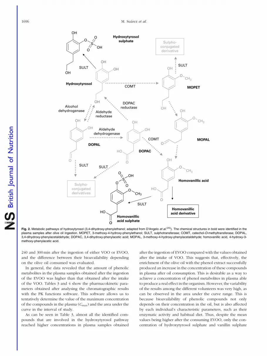
With regard to the metabolites derived from hydroxytyrosol,
the appearance of compounds structurally related to homo-
vanillic acid suggests the existence of a methylation process
in the organism, probably due to the activity of the enzyme
catechol-O-methyltransferase. D’Angelo et al.(25) proposed a
metabolic pathway for hydroxytyrosol in which the action of
this enzyme promoted the appearance of homovanillic acid
and its conjugated forms. In the case of the plasma samples
obtained after the ingestion of both VOO and EVOO,
we were able to identify the metabolites that appear at
the end of this chain of reactions: hydroxytyrosol sulphate,
homovanillic acid derivative and homovanillic acid sulphate.
Homovanillic acid derivative was a compound that presented
the same precursor and product ions as homovanillic acid
but eluted at a different retention time from the commercial
standard. The non-identification of free hydroxytyrosol and
the other intermediate forms may suggest that these com-
pounds are rapidly subjected to the activity of the enzymes,
originating in the sulphated or methyl-sulphated forms,
which are more likely to be absorbed by the organism. Fig. 2
shows the metabolic pathway of hydroxytyrosol proposed
by D’Angelo et al.(25) with some variations, indicating in
bold the metabolites identified in the plasma samples after
the ingestion of olive oil.
Similar to the metabolic pathway described in hydroxytyro-
sol, the analysis of the plasma samples revealed the existence
of some methylated metabolites that probably originated from
tyrosol. Thus, peaks 9, 13 and 18 given in Table 2 could be
formed from tyrosol, with an analogous chemical structure
to the one obtained as a result of the activity of catechol-O-
methyltransferase on hydroxytyrosol. Peak 9 is equivalent
to 3-methoxy-4-hydroxy-phenylethanol (MOPET) sulphate,
peak 13 is equivalent to the homovanillic sulphate and peak
18 was the result of the glucuronidation of the analogue struc-
ture of MOPET in tyrosol.
These results contrast with those obtained by Garcıa-
Villalba et al.(26) who did not identify methyl conjugate meta-
bolites from tyrosol in urine samples after the ingestion of
50 ml of VOO. They attributed this to the lack of activity
of catechol-O-methyltransferase on this compound due to
the need for an ortho-diphenolic structure of this enzyme.
The appearance of the methylated compounds in the plasma
samples in the present study could suggest the presence
of enzymatic activity able to methylate tyrosol. Thus, Fig. 3
shows the proposed metabolic pathways that tyrosol could
undergo in the organism, highlighting in bold the metabolites
identified in the plasma samples after the consumption of the
VOO. Based on the pathway described by D’Angelo et al.(25),
we hypothesised that tyrosol could also be affected by the
activity of alcohol dehydrogenase and aldehyde reductase,
although the metabolites produced by the activity of these
enzymes were not found. This could be due to the high
instability of these compounds and the possible preference
for methylation.
Pharmacokinetic parameters of phenol metabolites
The concentration of the phenolic compounds and their
metabolites in the plasma samples was monitored 0, 60, 120,
Table 2. Chromatographic parameters and proposed compound of the main chromatographic peaks observed in the plasmasamples after ingestion of the olive oils (control and phenol-enriched)
Peak Retention time (min) [M 2 H]2 Fragments MS/MS Proposed compound
1 3·62; 10·65 137 93 Hydroxybenzoic acid2 4·63, 6·52 151 107 Hydroxyphenylacetic acid3 6·55 151 136 Vanillin4 4·85 167 123 Vanillic acid5 5·17 179 135 Caffeic acid6 17·1 181 137 Homovanillic acid derivative7 10·14 197 153 3,4-DHPEA-EA derivative8 8·55; 19·15 217 137 Tyrosol sulphate9 5·06; 7·98; 12·45 230 150 4-Methoxy-phenylethanol sulphate10 9·32; 14·11 231 151, 136 Vanillin sulphate11 6·15 233 153, 123 Hydroxytyrosol sulphate12 6·67; 13·5; 21·75 243 163, 119 Coumaric acid sulphate13 11·77; 16·86; 19·3 245 165 4-Methoxy-phenylacetic acid sulphate14 10·05 247 167, 123 Vanillic acid sulphate15 19·15; 23·83 259 179, 135 Caffeic acid sulphate16 9·48 261 181 Homovanillic acid sulphate17 11·27 273 193, 134 Ferulic acid sulphate18 4·56; 6·74 327 150 4-Methoxy-phenylethanol glucuronide19 8·77; 18·97 327 151, 136 Vanillin glucuronide20 7·00 369 193, 134 Ferulic acid glucuronide21 10·57 373 197 3,4-DHPEA-EA derivative glucuronide22 15·15 445 269 Apigenin glucuronide23 10·69; 13·24 495 319, 153 3,4-DHPEA-EDA glucuronide
3,4-DHPEA-EA, oleuropein aglycone; 3,4-DHPEA-EDA, dialdehydic form of elenolic acid linked to hydroxytyrosol.
Bioavailability of phenol-enriched olive oil 1695
British
Journal
ofNutrition
240 and 300 min after the ingestion of either VOO or EVOO,
and the difference between their bioavailability depending
on the olive oil consumed was evaluated.
In general, the data revealed that the amount of phenolic
metabolites in the plasma samples obtained after the ingestion
of the EVOO was higher than that obtained after the intake
of the VOO. Tables 3 and 4 show the pharmacokinetic para-
meters obtained after analysing the chromatographic results
with the PK functions software. This software allows us to
tentatively determine the value of the maximum concentration
of the compounds in the plasma (Cmax) and the area under the
curve in the interval of study.
As can be seen in Table 3, almost all the identified com-
pounds that are involved in the hydroxytyrosol pathway
reached higher concentrations in plasma samples obtained
after the ingestion of EVOO compared with the values obtained
after the intake of VOO. This suggests that, effectively, the
enrichment of the olive oil with the phenol extract successfully
produced an increase in the concentration of these compounds
in plasma after oil consumption. This is desirable as a way to
achieve a concentration of phenol metabolites in plasma able
to produce a real effect in the organism. However, the variability
of the results among the different volunteers was very high, as
can be observed in the area under the curve range. This is
because bioavailability of phenolic compounds not only
depends on their concentration in the oil, but is also affected
by each individual’s characteristic parameters, such as their
enzymatic activity and habitual diet. Thus, despite the mean
values being higher after the consuming EVOO, only the con-
centration of hydroxytyrosol sulphate and vanillin sulphate
HydroxytyrosolMOPETCOMT
SULT
Hydroxytyrosol
sulphate
DOPAL
DOPAC
Aldehydedehydrogenase
DOPACreductase
Aldehydereductase
Alcoholdehydrogenase
COMT
Homovanillic acid
SULT
Homovanillic
acid sulphate
MOPAL
Homovanillic
acid derivative
OH
O
OH
S
O
OOH
OH
O
OH
CH3O
OH
O
OO
CH3
CH3
O
O
OH
O
O
HOCH3
O
O
O
HO
S
O
O
OH
O
OH
OH
O
HO
O
O
OH
OH
Sulpho-conjugatedderivatives
SULT SULT
OH
OH
OH
Sulpho-conjugatedderivative
SULT
Fig. 2. Metabolic pathways of hydroxytyrosol (3,4-dihydroxy-phenylethanol; adapted from D’Angelo et al.(25)). The chemical structures in bold were identified in the
plasma samples after olive oil ingestion. MOPET, 3-methoxy-4-hydroxy-phenylethanol; SULT, sulphotransferase; COMT, catechol-O-methyltransferase; DOPAL,
3,4-dihydroxy-phenylacetaldehyde; DOPAC, 3,4-dihydroxy-phenylacetic acid; MOPAL, 3-methoxy-4-hydroxy-phenylacetaldehyde; homovanillic acid, 4-hydroxy-3-
methoxy-phenylacetic acid.
M. Suarez et al.1696
British
Journal
ofNutrition

was statistically higher than in the VOO. This inter-individual
variability was also reported by other authors. D’Archivio
et al.(27) pointed out in their review about the bioavailability
of phenolic compounds in human subjects, suggesting that
one important factor to be considered in these studies is the
different genomic profile of the individuals. This influence
of the genomic profile in the absorption and effectiveness of
the polyphenols suggests that in the future further studies
should be carried out to evaluate the suitability of the introduc-
tion of a personalised factor in the design of functional foods.
On the other hand, although in all the cases an increase
in the concentration of the peaks for the tyrosol metabolites
was detected after consumption of both VOO and EVOO,
no significant differences between these oils were observed.
In addition, the mean values were very similar, suggesting
that the bioavailability of these compounds was not affected
by phenol enrichment. It is possible that complete absorption
of the phenols ingested with EVOO could be limited by the
membrane carriers. However, the intestinal mechanism of
the gastrointestinal absorption of polyphenols remains
poorly understood, and the membrane carriers that could be
involved in polyphenol absorption have not been identified.
The major components of the VOO phenolic fraction, the
secoiridoid derivatives (oleuropein and ligstroside aglycones),
are hydrolysed in the gastrointestinal tract(28), after which the
transport of the resulting polar phenols, tyrosol and hydroxy-
tyrosol, might occur via passive diffusion. In a previous study,
we have shown that there was extensive transport of tyrosol
and hydroxytyrosol and their conjugates to the basolateral
side using a Caco-2/TC7 model of the small intestine(29).
In general, apical loading of individual phenols resulted in
time-dependent efflux of different conjugates. However,
future studies are necessary to establish the optimal dose:bioa-
vailability ratio.
Tyrosol4-Methoxy-
phenylethanolMethylation
SULT
Tyrosol
sulphate
4-Hydroxy-
phenyl-
acetaldehyde
4-Hydroxy-
phenylacetic
acid
Aldehydedehydrogenase
Aldehydereductase
Alcoholdehydrogenase
4-Methoxy-
phenylacetic
acidSULT
4-Methoxy-
phenyl-
acetaledhyde
Sulpho-conjugatedderivatives
SULT
SULT
SULT
OH
OH
O
OH
S
O
O
OHO
OH
S
O
OOH
O
O
OH
CH3O
OH
4-Methoxy-
phenylethanol
sulphate
4-Methoxy-
phenylacetic
acid sulphate
4-Methoxy-
phenylethanol
glucuronide
O
O
HO
S
O
O OHO
O
O
HO
O
O
CH3
CH3
O
OO
OH
O
OH
OH
O
OH
OH
O
OH
Methylation
Fig. 3. Proposed metabolic pathways of tyrosol. The chemical structures in bold were identified in the plasma samples after olive oil ingestion. SULT,
sulphotransferase.
Bioavailability of phenol-enriched olive oil 1697
British
Journal
ofNutrition

Table 3. Pharmacokinetic parameters of the metabolites derived from the secoiridoids in plasma samples after the intake of either control or phenol-enriched olive oil (n 13)
(Mean values, standard deviations and ranges)
Phenolic compound Oil
Cmax (mmol/l)AUC
(mmol £ min/l)AUC range
(mmol £ min/l) PMean SD Mean SD
Hydroxytyrosol sulphate Control 0·53 0·30 103 65 50–238,0·05Enriched 0·86 0·24 159 49 93–287
Homovanillic acid derivative Control 0·53 0·29 132 59 80–246NSEnriched 0·34 0·10 80 20 63–102
Homovanillic acid sulphate Control 0·78 0·46 177 125 83–330NSEnriched 0·96 0·88 169 83 55–282
3,4-DHPEA-EDA glucuronide Control 0·24 0·21 58 59 26–127NSEnriched 0·20 0·18 54 49 26–111
3,4-DHPEA-EA derivative Control 0·36 0·27 75 62 5–235NSEnriched 0·60 0·59 98 78 7–245
3,4-DHPEA-EA derivative glucuronide Control 0·04 0·03 6 6 2–14NSEnriched 0·22 0·34 34 54 3–96
Tyrosol sulphate Control 1·16 1·07 228 189 59–601NSEnriched 0·95 0·64 240 169 73–538
4-Methoxy-phenylacetic acid sulphate Control 3·63 2·20 840 482 249–1891NSEnriched 2·38 2·05 565 519 46–2024
4-Methoxy-phenylethanol sulphate Control 2·85 1·74 617 378 119–1256NSEnriched 2·44 2·59 611 645 106–2054
4-Methoxy-phenylethanol glucuronide Control 0·03 0·01 6 5 2–9NSEnriched 0·03 0·01 6 5 3–9
Cmax, maximum concentration of the metabolites; AUC, area under the curve; 3,4-DHPEA-EDA, dialdehydic form of elenolic acid linked to hydroxytyrosol; 3,4-DHPEA-EA,oleuropein aglycone.
Table 4. Pharmacokinetic parameters of the metabolites derived from the phenolic acids and the lignans in plasma samples after the intake of eithercontrol or phenol-enriched olive oil (n 13)
(Mean values, standard deviations and ranges)
Cmax (mmol/l)AUC
(mmol £ min/l)AUC range
(mmol £ min/l)Phenolic compound Oil Mean SD Mean SD P
Apigenin glucuronide Control 0·09 0·05 16 7 8–35NSEnriched 0·08 0·03 16 9 8–36
Hydroxybenzoic acid Control 2·23 1·65 439 285 128–933NSEnriched 2·42 1·71 471 257 72–923
Hydroxyphenylacetic acid Control 4·95 2·63 1,135 723 185–2454NSEnriched 5·79 2·67 1,317 742 172–2507
Vanillic acid Control 6·70 2·22 1,254 512 824–1821NSEnriched 3·19 0·45 538 166 368–700
Vanillic acid sulphate Control 0·64 0·75 137 161 32–416NSEnriched 0·75 0·39 174 87 75–232
Caffeic acid Control 4·06 2·19 798 530 284–1885NSEnriched 4·98 3·09 959 621 355–2094
Caffeic acid sulphate Control 1·20 0·87 226 181 19–477 NSEnriched 2·58 4·98 357 583 32–1818
Coumaric acid sulphate Control 1·10 0·60 222 129 63–533NSEnriched 1·19 0·88 271 225 89–750
Vanillin Control 1·42 1·19 126 113 19–245NSEnriched 2·42 3·41 302 475 42–1014
Vanillin sulphate Control 12·96 10·78 2693 1970 718–6563,0·05Enriched 23·55 11·73 5161 2852 1711–10 800
Vanillin glucuronide Control 0·59 0·71 140 161 46–327NSEnriched 0·36 0·39 81 79 31–172
Ferulic acid derivative Control 0·27 0·03 70 6 49–131NSEnriched 0·23 0·05 51 12 39–71
Ferulic acid derivative sulphate Control 5·56 6·54 919 901 154–3245NSEnriched 6·74 2·90 1,257 687 454–2575
Ferulic acid glucuronide Control 0·53 0·51 119 106 19–214NSEnriched 0·77 0·73 189 166 23–413
Cmax, maximum concentration of the metabolites; AUC, area under the curve.
M. Suarez et al.1698
British
Journal
ofNutrition

Regarding the metabolites originating from phenolic acids,
it could be considered that the compounds that appeared in
the basal conditions are fermentation products, generated
through the effect of the human gut microbiota on the dietary
fibre and some phenolic compounds, such as flavonoids, in
the habitual diet of the volunteers(30). Once the VOO has
been ingested, the increase in the concentration of the pheno-
lic acid metabolites in the plasma over time could be partly
attributed to the absorption of this fraction included in the
olive oil. However, the enrichment of the olive oil with the
phenolic extract also increases the concentration of the flavo-
noid group, and it is possible that this could generate a further
increase in the concentration of the phenolic acid metabolites
once the human gut microbiota act on them. Therefore, in
addition to the direct effect on the content of the phenolic
acid metabolites observed in the plasma during the period
of study, it is possible that an indirect increase occurs through
the fermentation of the flavonoids in the colon. Nevertheless,
further studies would be needed to confirm this.
It is also remarkable that a certain shift in the time when the
maximum concentration of some of the identified metabolites
was reached was observed as a consequence of the phenol
enrichment of the oil. As an example, Fig. 4 shows the graphi-
cal evaluation of the concentration of some metabolites from
the different phenolic groups identified in the plasma after
the intake of both the VOO and the EVOO: hydroxytyrosol
sulphate (phenyl alcohol), oleuropein aglycone derivative glu-
curonide (secoiridoid), apigenin glucuronide (flavonoid) and
ferulic acid glucuronide (phenolic acid). After the consump-
tion of the VOO, the maximum concentration of these peaks
was reached at 60 min. However, consumption of the EVOO
shifted this maximum to 120 min. This could be due to
a higher concentration leading to a certain saturation of
the mechanisms involved in the absorption and transport of
these compounds in the human organism. Therefore, the
phenol-enriched oil needs more time to reach the maximum
concentration in the plasma.
In addition to this change in the time to reach the highest
concentration, there was a general quantitative increment in
the value of these metabolites in the plasma after the ingestion
of the EVOO compared with the one obtained after consump-
tion of the VOO. This is of great importance and points to
the suitability of phenol enrichment of the oil as a means of
increasing the amount of these desirable compounds in the
organism, which is necessary to produce a real effect on
the development of some types of cancers, such as colon
cancer(31).
Analysing the pharmacokinetic parameters of the different
groups of phenolic compounds under study, their different
pattern of absorption could be evaluated. To start with, the
time when the maximum concentration is reached suggests
that their absorption occurs approximately in the small intes-
tine after being subjected to the strong metabolism of the
organism. Thus, hydroxytyrosol sulphate (and by extension
the other phenyl alcohols metabolites) could come from the
high spectra of secoiridoid derivatives in the oil by hydrolysis
in the stomach (phase I metabolism) and later conjugation
by the action of the sulphotransferases (phase II). As was
reported in our previous study(28), after simulated in vitro
digestion of VOO, there was a significant increment in hydro-
xytyrosol in the stomach (rising by approximately 70 %) due
to the acidic conditions of the media (pH 2). This can be
related to the high proportion of phenyl alcohols conjugated
as their different metabolites that appeared in the plasma.
The absorption of phenolic acid metabolites seems to follow
the same pattern.
0·0
0·2
0·4
0·6
0·8
1·0
1·2
(a) (b)
(c) (d)
0 50 100 150 200 250 300
Hyd
roxy
tyro
sol s
ulp
hat
e(µ
mo
l/l)
Ap
igen
in g
lucu
ron
ide
(µm
ol/l
)
Ole
uro
pei
n a
gly
con
e d
eriv
ativ
eg
lucu
ron
ide
(µm
ol/l
)Fe
rulic
aci
d g
lucu
ron
ide
(µm
ol/l
)
Time (min)
0 50 100 150 200 250 300
Time (min)
0 50 100 150 200 250 300
Time (min)
0 50 100 150 200 250 300
Time (min)
0·0
0·1
0·2
0·3
0·4
0·5
0·6
0·00·20·40·60·81·01·21·41·6
0·00
0·02
0·04
0·06
0·08
0·10
0·12
Fig. 4. Pharmacokinetics of (a) hydroxytyrosol sulphate, (b) oleuropein aglycone derivative glucuronide, (c) apigenin glucuronide and (d) ferulic acid glucuronide in
human plasma after the intake of control ( ) and phenol-enriched olive oil ( ). Data are expressed as mmol phenolic metabolite per litre plasma.
Bioavailability of phenol-enriched olive oil 1699
British
Journal
ofNutrition

On the other hand, the appearance of oleuropein aglycone
derivative and its glucuronide form seems to suggest that
some of the secoiridoids in the oil are able to resist the
strong conditions of the digestion process and reach the
blood stream in a chemical form higher than hydroxytyrosol.
This is of great importance due to the healthy effects that
these compounds could exert(23).
The non-identification of any metabolic form derived from
lignans in the plasma could be due to their degradation by
the colonic flora of the gut and further transformation into
enterodiol and enterolactone. This process is believed to
take place in the period of 8–10 h after ingestion(32).
Concluding remarks
The enrichment of olive oil with an olive cake extract
increased the amount of phenolic compounds in the resulting
oil and had a direct influence on their bioavailability. The
in vivo study showed that the concentration of fourteen of
twenty-four compounds detected was higher in the plasma
samples from the EVOO than after ingestion of VOO.
Among these, two of them, hydroxytyrosol sulphate and vanil-
lin sulphate, were statistically significant in attending their
pharmacokinetic parameters, demonstrating the suitability of
enrichment. In general, a displacement of the time to reach
the maximum concentration is observed in the samples,
which indicates that more time is needed to absorb the
higher phenolic content. However, inter-individual variability
in the concentration of the plasma phenol metabolites
shows that it is difficult to show statistically significant differ-
ences between the VOO and the EVOO. This could be due
to the high number of variables that interfere in the absorption
of these compounds and suggests that the metabolism of phe-
nols is affected first by the individual. These preliminary
results suggest the need for a major interventional study
with a larger number of volunteers to confirm the effective-
ness of the enrichment of olive oil in increasing the plasma
concentration of the phenol metabolites.
Acknowledgements
The present study was supported by the Spanish Ministry
of Education and Science financing the projects AGL2005-
07881-C02-01/ALI and AGL2005-07881-C02-02/ALI; Health
Ministry (FIS; PI021307) and CIBERDEM; and the grant
received by Manuel Suarez (BES-2006-14136). We wish to
thank the company Moli dels Torm S.L. (Els Torms, Lleida,
Catalonia, Spain) for the supply of the olive cake samples to
obtain phenol extract. The authors declare that there is no
conflict of interest. M.-J. M. and R. S. had full access to all of
the data in the study, and took responsibility for the integrity
of the data and the accuracy of data analysis. M.-J. M. and
R. S conceived the study concept and design. M. S., A. M.
and M.-P. R. prepared and analysed the phenol-enriched
olive oil. R. M. V., S. F., M. G. and R. S. conducted the
human study. M. S. and A. M. had a role in the acquisition
of in vivo bioavailability data. M. S., R. M. V., M.-J. M., R. S.
and M. G. analysed and interpreted the data. M. S., M.-J. M.,
R. S. and R. M. V. drafted the manuscript. M.-J. M. and R. S.
participated in the critical revision of the manuscript for
important intellectual content. M. G. and S. F. provided admin-
istrative, technical and logistic support.
References
1. Knoops KTB, De Groot LCPGM, Kromhout D, et al. (2004)Mediterranean diet, lifestyle factors, and 10-year mortalityin elderly European men and women: the HALE project.JAMA 292, 1433–1439.
2. Covas MI (2007) Olive oil and cardiovascular system.Pharmacol Res 55, 175–186.
3. Covas MI (2008) Bioactive effects of olive oil phenolic com-pounds in humans: reduction of heart disease factors andoxidative damage. Inflammopharmacology 16, 216–218.
4. Obied HK, Bedgood DR Jr., Prenzler PD, et al. (2007) Bio-screening of Australian olive mill waste extracts: biophenolcontent, antioxidant, antimicrobial and molluscicidal activi-ties. Food Chem Toxicol 45, 1238–1248.
5. Lopez-Miranda J, Perez-Jimenez F, Ros E, et al. (2010) Oliveoil and health: summary of the II international conference onolive oil and health consensus report, Jaen and Cordoba(Spain) 2008. Nutr Metab Cardiovasc Dis 20, 284–294.
6. Visioli F & Galli C (2001) Antiatherogenic components ofolive oil. Curr Atheroscler Rep 3, 64–67.
7. Owen RW, Giacosa A, Hull WE, et al. (2000) Olive-oilconsumption and health: the possible role of antioxidants.Lancet Oncol 1, 107–112.
8. Manach C, Scalbert A, Morand C, et al. (2004) Polyphenols:food sources and bioavailability. Am J Clin Nutr 79,727–747.
9. Visioli F, Galli C, Bornet F, et al. (2000) Olive oil phenolicsare dose-dependently absorbed in humans. FEBS Lett 468,159–160.
10. Caruso D, Visioli F, Patelli R, et al. (2001) Urinary excretionof olive oil phenols and their metabolites in humans. Metab-olism 50, 1426–1428.
11. Miro-Casas E, Covas M-I, Farre M, et al. (2003) Hydroxytyro-sol disposition in humans. Clin Chem 49, 945–952.
12. Marrugat J, Covas MI, Fito M, et al. (2004) Effects of differingphenolic content in dietary olive oils on lipids and LDLoxidation – a randomized controlled trial. Eur J Nutr 43,140–147.
13. Gonzalez-Santiago M, Fonolla J & Lopez-Huertas E (2010)Human absorption of a supplement containing purifiedhydroxytyrosol, a natural antioxidant from olive oil, and evi-dence for its transient association with low-density lipopro-teins. Pharmacol Res 61, 364–370.
14. Paiva-Martins F, Fernandes J, Santos V, et al. (2010) Powerfulprotective role of 3,4-dihidroxyphenylethanol-elenolic aciddialdehyde against erythrocyte oxidative-induced hemolysis.J Agric Food Chem 58, 135–140.
15. Konstantinidou V, Covas M-I, Munoz-Aguayo D, et al. (2010)In vivo nutrigenomics effects of virgin olive oil polyphenolswithin the frame of the Mediterranean diet: a randomizedcontrolled trial. FASEB J 24, 2546–2557.
16. Vissers MN, Zock PL, Roodenburg AJ, et al. (2002) Olive oilphenols are absorbed in humans. J Nutr 132, 409–417.
17. Leenen R, Roodenburg AJ, Vissers MN, et al. (2002) Sup-plementation of plasma with olive oil phenols and extracts:influence on LDL oxidation. J Agric Food Chem 50,1290–1297.
M. Suarez et al.1700
British
Journal
ofNutrition

18. Esti M, Cinquanta L & La Notte E (1998) Phenolic com-pounds in different olive varieties. J Agric Food Chem 46,32–35.
19. Artajo LS, Romero MP, Morelloc JR, et al. (2006) Enrichmentof refined olive oil with phenolic compounds: evaluation oftheir antioxidant activity and their effect on the bitter index.J Agric Food Chem 54, 6079–6088.
20. Suarez M, Romero MP, Ramo T, et al. (2009) Methods forpreparing phenolic extracts from olive cake for potentialapplication as food antioxidants. J Agric Food Chem 57,1463–1472.
21. Suarez M, Macia A, Romero M-P, et al. (2008) Improvedliquid chromatography tandem mass spectrometry methodfor the determination of phenolic compounds in virginolive oil. J Chromatogr A 1214, 90–99.
22. Suarez M, Romero M-P, Macia A, et al. (2009) Improvedmethod for identifying and quantifying olive oil phenoliccompounds and their metabolites in human plasma bymicroelution solid-phase extraction plate and liquid chroma-tography–tandem mass spectrometry. J Chromatogr B 877,4097–4106.
23. Paiva-Martins F, Fernandes J, Rocha S, et al. (2009) Effectsof olive oil polyphenols on erythrocyte oxidative damage.Mol Nutr Food Res 53, 609–616.
24. De la Torre-Carbot K, Jauregui O, Castellote AI, et al.(2006) Rapid high-performance liquid chromatography–electrospray ionization tandem mass spectrometry methodfor qualitative and quantitative analysis of virgin olive oil
phenolic metabolites in human low-density lipoproteins.J Chromatogr A 1116, 69–75.
25. D’Angelo S, Manna C, Migliardi V, et al. (2001) Pharma-cokinetics and metabolism of hydroxytyrosol, a natural anti-oxidant from olive oil. Drug Metab Dispos 29, 1492–1498.
26. Garcıa-Villalba R, Carrasco-Pancorbo A, Nevedomskaya E,et al. (2010) Exploratory analysis of human urine byLC-ESI-TOF MS after high intake of olive oil: understandingthe metabolism of polyphenols. Anal Bioanal Chem 398,463–475.
27. D’Archivio M, Filesi C, Vari R, et al. (2010) Bioavailabilityof polyphenols: status and controversies. Int J Mol Sci 11,1321–1342.
28. Suarez M, Romero M-P & Motilva M-J (2010) Development ofa phenol-enriched olive oil with phenolic compounds fromolive cake. J Agric Food Chem 58, 10396–10403.
29. Soler A, Romero M-P, Macia A, et al. (2010) Digestion stab-ility and evaluation of the metabolism and transport ofolive oil phenols in the human small-intestinal epithelialCaco-2/TC7 cell line. Food Chem 119, 703–714.
30. Aura A-M (2008) Microbial metabolism of dietary phenoliccompounds in the colon. Phytochem Rev 7, 407–429.
31. Colomer R & Menendez JA (2006) Mediterranean diet, oliveoil and cancer. Clin Transl Oncol 8, 15–21.
32. Kuijsten A, Arts ICW, Vree TB, et al. (2005) Pharmacokineticsof enterolignans in healthy men and women consuming asingle dose of secoisolariciresinol diglucoside. J Nutr 135,795–801.
Bioavailability of phenol-enriched olive oil 1701
British
Journal
ofNutrition