conjugation of curcumin with pvp capped gold nanoparticles for improving bioavailability
TRANSCRIPT

This article appeared in a journal published by Elsevier. The attachedcopy is furnished to the author for internal non-commercial researchand education use, including for instruction at the authors institution
and sharing with colleagues.
Other uses, including reproduction and distribution, or selling orlicensing copies, or posting to personal, institutional or third party
websites are prohibited.
In most cases authors are permitted to post their version of thearticle (e.g. in Word or Tex form) to their personal website orinstitutional repository. Authors requiring further information
regarding Elsevier’s archiving and manuscript policies areencouraged to visit:
http://www.elsevier.com/copyright
Author's personal copy
Short communication
Conjugation of curcumin with PVP capped gold nanoparticles forimproving bioavailability
Rajesh K. Gangwar a, Vinayak A. Dhumale a, Dimple Kumari b, Umesh T. Nakate a, S.W. Gosavi c,Rishi B. Sharma d, S.N. Kale a,⁎, Suwarna Datar a,⁎a Department of Applied Physics, Defence Institute of Advanced Technology (DU), Girinagar, Pune 411025, Indiab Department of Applied Chemistry, Defence Institute of Advanced Technology (DU), Girinagar, Pune 411025, Indiac Department of Physics, University of Pune, Pune 411007, Indiad DOP, DRDO Bhawan, Rajaji Marg, New Delhi 110105, India
a b s t r a c ta r t i c l e i n f o
Article history:Received 22 May 2012Received in revised form 22 June 2012Accepted 14 July 2012Available online 20 July 2012
Keywords:CurcuminGold nanoparticlesBioavailability
Curcumin, a natural polyphenolic compound, has astounding therapeutic applications but lacks in bioavailabil-ity mainly due to its poor solubility in water. Polyvinyl pyrrolidone (PVP) which is a proven drug carrier hasbeen used to facilitate the conjugation of curcumin with gold nanoparticles and to improve the solubility ofcurcumin in water. In this conjugate diaryl heptanoid chromophore group of curcumin which is a much neededgroup in biomedical applications remains intact as observed from FTIR and UV–vis spectroscopy analysis. Thework shows good promise for such conjugates as therapeutic-cum-imaging materials in biomedical field.
© 2012 Elsevier B.V. All rights reserved.
1. Introduction
Curcumin, a bio-active polyphenol component of curcuma longa(Turmeric), also known as diferuloylmethane (C21H20O6), has be-come an intense topic of research due to its interesting biologicaland pharmacological applications [1–3]. It has anti-inflammatory,anti-oxidant, anti-carcinogenic, chemo-preventive, anti-angeogenic,anti-diabetic, anti-viral and antibacterial properties [4,5]. In spite of itswide property range, this polyphenolic compound is less documentedfor its actual applications, mainly due to its poor bioavailability.Curcumin remains to be less popular as a drug since the basic necessityof any drug to be delivered is its ease in suspension in bio-media (eitheraqueous solutions or PBS solutions). Curcumin's hydrophobic nature isone of the main reasons for this poor water-solubility/suspensioncapacity. This leads to its poor activity, low absorption, high rate ofmetabolism within the living system and rapid elimination from thesystem [6]. There have been attempts to functionalize the molecule toimprove its bioavailability by conjugating it with a hydrophilic mole-cule. However, this process hampers the therapeutically-active groupof the molecule, thereby killing the basic purpose of the conjugation.
Numerous approaches have been reported in this context to explorethe potent applications of curcumin in very recent times [7–10].Nanoparticles [11,12], liposomes [13], micelles [14] and phospholipidcomplexes [15] are being used to improve the bioavailability of curcumin.
Bhawana et al. and Wu et al. have prepared curcumin nanoparticles [8]and nanogels [16] for antimicrobial and photo-thermal therapyrespectively. Dhule et al. have explored curcumin-loaded γ-cyclodextrinliposomal nanoparticles as delivery vehicles for osteosarcoma [17].Ha et al. have synthesized poly(lactide)-vitamin E TPGS (PLA-TPGS)copolymer to formulate a curcumin nanocarrier [18] and Bisht et al.have reported on polymeric nanoparticle-encapsulated curcumin [19]for human cancer therapy.
Recently, S. Manju et al. have reported synthesis of water solublegold nanoparticles in curcumin-polymer conjugate and studied it forblood compatibility and targeted drug delivery onto cancer cells [20].They have conjugated curcumin with hyaluronic acid (HA) and thensynthesized gold nanoparticles within the solution by reducing thechloroauric acid. In this and many other documented protocols, it hasbeen observed that though the bio-availability improves upon conjuga-tion, the main therapeutic group of this molecule gets engaged in theconjugation thereby making itself less-available for its therapeuticactivity to any biological system [21]. Therefore, there is a need for anextensive research in this regard which would not only improve thebioavailability of the molecule but also keep the therapeutic activityhigh and impart multi-functional properties to the conjugated system.
On the other side, gold nanoparticles have been immensely ex-plored due to their interesting optical, electrical, physical and chemicalproperties, which find useful technological applications in the field ofopto-electronics [22], chemical and biological sensors [23] and biomed-icines [24]. These have been projected to be one of the safest(non-toxic) candidates for DNA-conjugation [25], bio-detection [26],drug-delivery, gene therapy and many other applications [27,28].
Materials Science and Engineering C 32 (2012) 2659–2663
⁎ Corresponding authors. Tel.: +91 20 2430 4091; fax: +91 20 2438 9572.E-mail addresses: [email protected] (S. Datar), [email protected]
(S.N. Kale).
0928-4931/$ – see front matter © 2012 Elsevier B.V. All rights reserved.doi:10.1016/j.msec.2012.07.022
Contents lists available at SciVerse ScienceDirect
Materials Science and Engineering C
j ourna l homepage: www.e lsev ie r .com/ locate /msec

Author's personal copy
These nanoparticles have also been documented to be good forbio-imaging purposes owing to their interesting surface-plasmon reso-nance property.
In this context, through this manuscript we report a neat protocolwhich describes the formation of curcumin conjugated with goldnanoparticles using PVP which has been used as drug carrier forvarious drugs in several reports [29]. This protocol envisages goodbio-availability of the curcumin molecule, keeping its therapeuticactivity intact. Gold nanoparticles show excellent plasmon resonancecharacteristic, exhibiting its potential in applications of imaging.
2. Experimental
2.1. Synthesis
Chloroauric acid (HAuCl4) has been used as a metal precursor andtri-sodium citrate (Na3C6H5O7) as reducing agent for the synthesis ofgold nanoparticles [30]. 200 ml of 1 mM HAuCl4 was boiled andstirred under the reflux condition for 30 min. 20 ml of 38.8 mMaqueous Na3C6H5O7 was added directly into the boiled solution. Thecolor of the solution changed from pale yellow to deep red within7–10 min after the addition. Further the reaction was continued foradditional ~20 min after which 20 mg of polyvinyl pyrrolidone(PVP, M.W.=40,000) in 30 ml of water was added to the above solu-tion and stirred for the next 45 min. The solution was cooled at roomtemperature. For conjugation of curcumin, 50 mg of crystallinecurcumin was dissolved in 25 ml acetone and this solution wasadded to the 100 ml of PVP capped gold nanoparticles under stirring.Further this mixture was stirred for 3 h at 60 °C and then cooleddown to room temperature. The Au–curcumin solution was thencentrifuged at 4000 rpm to remove unattached curcumin. This wasdone three times to ensure that no free curcumin molecules are leftin the final conjugate. The final solution was used for spectroscopicstudy and TEM analysis. Part of this solution was evaporated undervacuum using a rotary evaporator and then the remnant wascentrifuged to obtain the powder, which was also used for analysis.
2.2. Characterization
The synthesized gold nanoparticles and Au–curcumin samples werecharacterized by UV–vis Spectroscopy (Ocean Optics, HR4000), FourierTransform Infrared Spectroscopy (FTIR, Perkin Elmer), Thermo-gravimetric Analyser (TGA, Perkin Elmer STA 6000) and TransmissionElectron Microscopy (TEM, Philips CM 200 and FEI-Tecnai G2 20).For TEM characterization highly diluted specimens were prepared anddilution level was kept constant for all samples. Toxicity analysis wasdone using MTT assay as described elsewhere [31].
3. Results and discussion
Fig. 1 shows the FTIR spectra of (i) PVP (ii) curcumin (iii) PVPfunctionalized gold NPs and (iv) PVP functionalized gold nanoparticleswith curcumin. Here spectra (i) and (ii) show the typical signatures ofboth PVP and curcumin. These signatures may be recognized as C_O(1660 cm−1) and C\N (1290 cm−1) in PVP. Signatures of curcuminare free O\H group (3514 cm−1), C_O and C_C (enol) (1450–1630 cm−1), C\H (methyl) (2845 cm−1), C\H (aryl) (3015 cm−1)and C\O\C (1000–1300 cm−1) typically attributed to symmetricand asymmetric configurations of C\O\C chains [11]. Spectrum (iii)illustrates the functionalization of citrate reduced gold NPs with PVPin the form of shift of C_O stretching from 1660 cm−1 to 1628 cm−1.This may be attributed to the formation of intermolecular hydrogenbonding. Spectrum (iv) shows the conjugation of curcumin with PVPfunctionalized gold NPs. This figure reveals signatures of both PVP func-tionalized gold NPs and curcumin molecule. Curcumin molecule mayattach to the PVP functionalized goldNPswith intermolecular hydrogen
bonding to enolic hydroxyl group which results in shift of O\Hstretching from 3514 cm−1 to 3435 cm−1 whereas the basic diarylheptanoid group,which is the chromophore group of curcumin remainsintact [32]. This is also supported by UV–vis spectroscopy explainedbelow.
Fig. 2 shows the UV–vis spectra of (i) curcumin in PVP (ii) PVPfunctionalized gold nanoparticles and (iii) PVP functionalized goldnanoparticles with curcumin in aqueous media. UV–vis spectrum ofcurcumin in PVP shows an absorption peak at ~415 nm which is thesignature of basic diaryl heptanoid chromophore group of curcumin[11]. The spectrum of PVP functionalized gold nanoparticles(ii) shows absorption peak at ~520 nm, which is a characteristic peakof gold nanoparticles arising due to Surface Plasmon Resonance (SPR)[33]. The UV–vis spectra of PVP functionalized gold nanoparticleswith curcumin (iii) clearly show two distinct peaks at ~418 nm and~525 nm which confirms that the characteristic peaks for the compo-nents in the Au–C complex are intact. The red-shift in the absorptionpeaks for curcumin (~3 nm) and gold NPs (~5 nm) can be attributedto the conjugation of PVP functionalized gold NPs with curcumin.Thus, the UV–vis data with the support of FTIR results clearly
Fig. 1. FTIR spectra of (i) PVP (ii) curcumin (iii) PVP functionalized gold nanoparticlesand (iv) PVP functionalized gold nanoparticles with curcumin.
Fig. 2. UV–vis spectra of (i) curcumin in PVP (ii) PVP functionalized gold nanoparticlesand (iii) PVP functionalized gold nanoparticles with curcumin. Inset shows the colorimet-ric response for each sample (i.e. i, ii and iii).
2660 R.K. Gangwar et al. / Materials Science and Engineering C 32 (2012) 2659–2663
Author's personal copy
demonstrate that curcumin retains its diaryl heptanoid chromophoregroup which is much needed in biomedical applications. The suspen-sion stability of these conjugates was checked using aqueous solutionsof a) only curcumin in PVP b) gold:PVP and c) gold:PVP:curcumin. Theinset of Fig. 2(b) indicates that the conjugates show excellentaqueous-stability, imparted due to the chemistry involved.
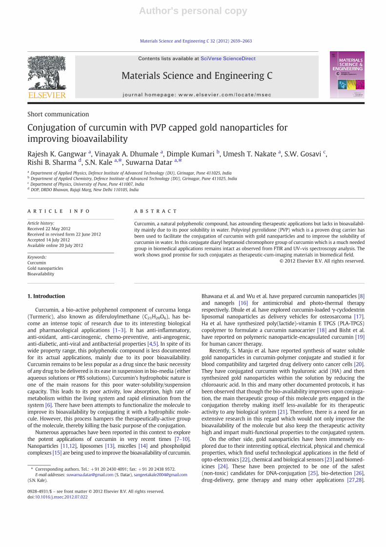
Fig. 3 shows the schematic for probable conjugation of curcuminmolecule with PVP functionalized gold NPs. Citrate reduced goldnanoparticles are capped by PVP as shown in the schematic. This
conjugation takes place by intermolecular hydrogen bonding asshown in the figure which is also supported by the shift of C_Ostretching from 1660 cm−1 to 1628 cm−1 in the FTIR spectra of citratereduced PVP functionalized gold nanoparticles. Curcumin also gets at-tached to the PVP functionalized gold NPs as shown in the figure bythe intermolecular hydrogen bonding to enolic hydroxyl group whichresults in shift of O\H stretching from 3514 cm−1 to 3435 cm−1.
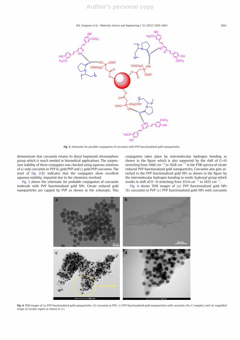
Fig. 4 shows TEM images of (a) PVP functionalized gold NPs(b) curcumin in PVP (c) PVP functionalized gold NPs with curcumin
Fig. 3. Schematic for possible conjugation of curcumin with PVP functionalized gold nanoparticles.
Fig. 4. TEM images of (a) PVP functionalized gold nanoparticles, (b) curcumin in PVP, (c) PVP functionalized gold nanoparticles with curcumin (Au–C complex) and (d) magnifiedimage of circular region as shown in (c).
2661R.K. Gangwar et al. / Materials Science and Engineering C 32 (2012) 2659–2663
Author's personal copy
and (d) magnified image of circular region as shown in (c). It is ob-served from Fig. 4(a) that PVP functionalized gold NPs are sphericalin shape having diameter of 13±2 nm. Fig. 4(b) shows the dispersionof curcumin and PVP (2:1) in aqueous medium for reference. PVPfunctionalized gold NPs with curcumin (Au–C complex) is shown inFig. 4(c). As observed in the figure, gold NPs (black cores) aresurrounded by PVP–curcumin conjugate as a shell. This is supportedby the image of PVP–curcumin conjugation (b) as well as the FTIR re-sults showing the hydrogen bonding between the PVP and curcumin.Magnified image of circular region in Fig. 4(c) is shown in Fig. 4(d),reveals that the PVP–curcumin completely surrounds the goldnanoparticles. Here PVP provides the required hydrophilicity to thecurcumin molecule making them bioavailable.
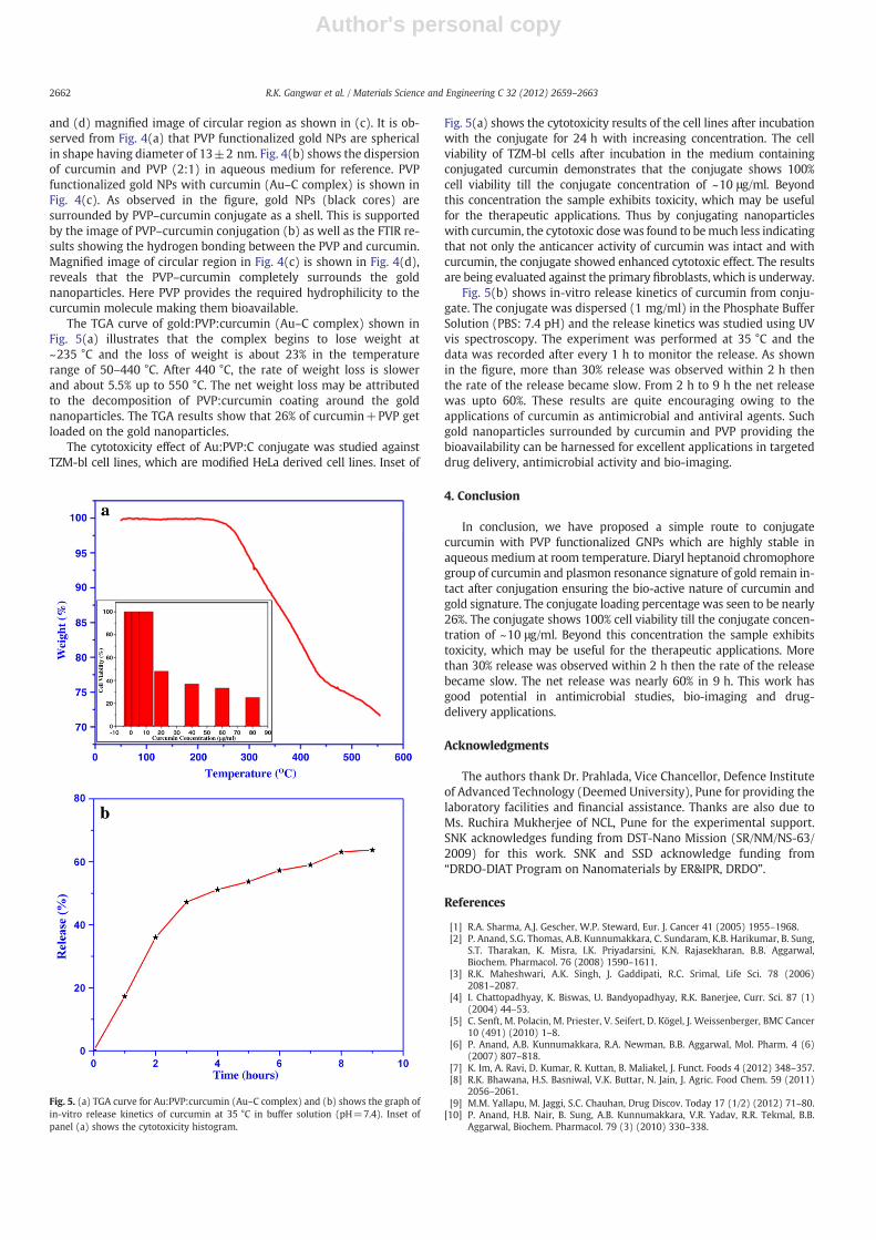
The TGA curve of gold:PVP:curcumin (Au–C complex) shown inFig. 5(a) illustrates that the complex begins to lose weight at~235 °C and the loss of weight is about 23% in the temperaturerange of 50–440 °C. After 440 °C, the rate of weight loss is slowerand about 5.5% up to 550 °C. The net weight loss may be attributedto the decomposition of PVP:curcumin coating around the goldnanoparticles. The TGA results show that 26% of curcumin+PVP getloaded on the gold nanoparticles.
The cytotoxicity effect of Au:PVP:C conjugate was studied againstTZM-bl cell lines, which are modified HeLa derived cell lines. Inset of
Fig. 5(a) shows the cytotoxicity results of the cell lines after incubationwith the conjugate for 24 h with increasing concentration. The cellviability of TZM-bl cells after incubation in the medium containingconjugated curcumin demonstrates that the conjugate shows 100%cell viability till the conjugate concentration of ~10 μg/ml. Beyondthis concentration the sample exhibits toxicity, which may be usefulfor the therapeutic applications. Thus by conjugating nanoparticleswith curcumin, the cytotoxic dose was found to bemuch less indicatingthat not only the anticancer activity of curcumin was intact and withcurcumin, the conjugate showed enhanced cytotoxic effect. The resultsare being evaluated against the primary fibroblasts, which is underway.
Fig. 5(b) shows in-vitro release kinetics of curcumin from conju-gate. The conjugate was dispersed (1 mg/ml) in the Phosphate BufferSolution (PBS: 7.4 pH) and the release kinetics was studied using UVvis spectroscopy. The experiment was performed at 35 °C and thedata was recorded after every 1 h to monitor the release. As shownin the figure, more than 30% release was observed within 2 h thenthe rate of the release became slow. From 2 h to 9 h the net releasewas upto 60%. These results are quite encouraging owing to theapplications of curcumin as antimicrobial and antiviral agents. Suchgold nanoparticles surrounded by curcumin and PVP providing thebioavailability can be harnessed for excellent applications in targeteddrug delivery, antimicrobial activity and bio-imaging.
4. Conclusion
In conclusion, we have proposed a simple route to conjugatecurcumin with PVP functionalized GNPs which are highly stable inaqueous medium at room temperature. Diaryl heptanoid chromophoregroup of curcumin and plasmon resonance signature of gold remain in-tact after conjugation ensuring the bio-active nature of curcumin andgold signature. The conjugate loading percentage was seen to be nearly26%. The conjugate shows 100% cell viability till the conjugate concen-tration of ~10 μg/ml. Beyond this concentration the sample exhibitstoxicity, which may be useful for the therapeutic applications. Morethan 30% release was observed within 2 h then the rate of the releasebecame slow. The net release was nearly 60% in 9 h. This work hasgood potential in antimicrobial studies, bio-imaging and drug-delivery applications.
Acknowledgments
The authors thank Dr. Prahlada, Vice Chancellor, Defence Instituteof Advanced Technology (Deemed University), Pune for providing thelaboratory facilities and financial assistance. Thanks are also due toMs. Ruchira Mukherjee of NCL, Pune for the experimental support.SNK acknowledges funding from DST-Nano Mission (SR/NM/NS-63/2009) for this work. SNK and SSD acknowledge funding from“DRDO-DIAT Program on Nanomaterials by ER&IPR, DRDO”.
References
[1] R.A. Sharma, A.J. Gescher, W.P. Steward, Eur. J. Cancer 41 (2005) 1955–1968.[2] P. Anand, S.G. Thomas, A.B. Kunnumakkara, C. Sundaram, K.B. Harikumar, B. Sung,
S.T. Tharakan, K. Misra, I.K. Priyadarsini, K.N. Rajasekharan, B.B. Aggarwal,Biochem. Pharmacol. 76 (2008) 1590–1611.
[3] R.K. Maheshwari, A.K. Singh, J. Gaddipati, R.C. Srimal, Life Sci. 78 (2006)2081–2087.
[4] I. Chattopadhyay, K. Biswas, U. Bandyopadhyay, R.K. Banerjee, Curr. Sci. 87 (1)(2004) 44–53.
[5] C. Senft, M. Polacin, M. Priester, V. Seifert, D. Kögel, J. Weissenberger, BMC Cancer10 (491) (2010) 1–8.
[6] P. Anand, A.B. Kunnumakkara, R.A. Newman, B.B. Aggarwal, Mol. Pharm. 4 (6)(2007) 807–818.
[7] K. Im, A. Ravi, D. Kumar, R. Kuttan, B. Maliakel, J. Funct. Foods 4 (2012) 348–357.[8] R.K. Bhawana, H.S. Basniwal, V.K. Buttar, N. Jain, J. Agric. Food Chem. 59 (2011)
2056–2061.[9] M.M. Yallapu, M. Jaggi, S.C. Chauhan, Drug Discov. Today 17 (1/2) (2012) 71–80.
[10] P. Anand, H.B. Nair, B. Sung, A.B. Kunnumakkara, V.R. Yadav, R.R. Tekmal, B.B.Aggarwal, Biochem. Pharmacol. 79 (3) (2010) 330–338.
Fig. 5. (a) TGA curve for Au:PVP:curcumin (Au–C complex) and (b) shows the graph ofin-vitro release kinetics of curcumin at 35 °C in buffer solution (pH=7.4). Inset ofpanel (a) shows the cytotoxicity histogram.
2662 R.K. Gangwar et al. / Materials Science and Engineering C 32 (2012) 2659–2663

Author's personal copy
[11] S. Hatamie, M. Nouri, S.K. Karandikar, A. Kulkarni, S.D. Dhole, D.M. Phase, S.N.Kale, Mater. Sci. Eng., C 32 (2012) 92–97.
[12] M. Kakran, N.G. Sahoo, J. Nanopart. Res. 14 (757) (2012) 1–11.[13] S. Mourtas, M. Canovi, C. Zona, D. Aurilia, A. Niarakis, B.L. Ferla, M. Salmona, F.
Nicotra, M. Gobbi, S.G. Antimisiaris, Biomaterials 32 (2011) 1635–1645.[14] Z. Song, R. Feng, M. Sun, C. Guo, Y. Gao, L. Li, G. Zhai, J. Colloid Interface Sci. 354
(2011) 116–123.[15] C.C. Lin, H.Y. Lin, H.C. Chen, M.W. Yu, M.H. Lee, Food Chem. 116 (2009) 923–928.[16] W. Wu, J. Shen, P. Banerjee, S. Zhou, Biomaterials 32 (2011) 598–609.[17] S.S. Dhule, P. Penfornis, T. Frazier, R. Walker, J. Feldman, G. Tan, J. He, A. Alb, V.
John, R. Pochampally, Nanomed. Nanotechnol. Biol. Med. 8 (4) (2012) 440–451.[18] P.T. Ha, T.M.N. Tran, H.D. Pham, Q.H. Nguyen, X.P. Nguyen, Adv. Nat. Sci.: Nanosci.
Nanotechnol. 1 (2010) 015012.[19] S. Bisht, G. Feldmann, S. Soni, R. Ravi, C. Karikar, Amarnath Maitra, Anirban
Maitra, J. Nanobiotechnol. 5 (3) (2007) 1–18.[20] S. Manju, K. Sreenivasan, J. Colloid Interface Sci. 368 (2012) 144–151.[21] S. Daniel, J.L. Limson, A. Dairam, G.M. Watkins, S. Daya, J. Inorg. Biochem. 98 (2)
(2004) 266–275.[22] A.N. Shipway, E. Katz, I. Willner, Chemphyschem 1 (2000) 18–52.
[23] G.R. Souza, D.R. Christianson, F.I. Staquicini, M.G. Ozawa, E.Y. Snyder, R.L. Sidman,J.H. Miller, W. Arap, R. Pasqualini, PNAS 103 (5) (2006) 1215–1220.
[24] M.C. Daniel, D. Astruc, Chem. Rev. 104 (2004) 293–346.[25] Y. Wen, C.K. McLaughlin, P.K. Lo, H. Yang, H.F. Sleiman, Bioconjugate Chem. 21
(2010) 1413–1416.[26] N. Uehara, K. Ookubo, T. Shimizu, Langmuir 26 (9) (2010) 6818–6825.[27] M. Hu, J. Chen, Z.Y. Li, L. Au, G.V. Hartland, X. Li, M. Marquez, Y. Xia, Chem. Soc.
Rev. 35 (2006) 1084–1094.[28] E.C. Dreaden, A.M. Alkilany, X. Huang, C.J. Murphy, M.A. El-Sayed, Chem. Soc. Rev.
41 (2012) 2740–2779.[29] G. Oster, E.H. Immergut, J. Am. Chem. Soc. 76 (5) (1954) 1393–1396.[30] K.C. Grabar, R.G. Freeman, M.B. Hommer, M.J. Natan, Anal. Chem. 67 (4) (1995)
735–743.[31] K.D. Wani, R. Kitture, A. Ahmed, A.S. Choudhari, S.J. Koppikar, S.N. Kale, R.
Kaul-Ghanekar, J. Bionanosci. 5 (1) (2011) 59–65.[32] T. Masuda, K. Hidaka, A. Shinohara, T. Maekawa, Y. Takeda, H. Yamaguchi, J. Agric.
Food Chem. 47 (1) (1999) 71–77.[33] S. Link, M.A. El-Sayed, J. Phys. Chem. B 103 (21) (1999) 4212–4217.
2663R.K. Gangwar et al. / Materials Science and Engineering C 32 (2012) 2659–2663