oxalic acid capped iron oxide nanorods as a sensing platform
TRANSCRIPT
Chemico-Biological Interactions 238 (2015) 129–137
Contents lists available at ScienceDirect
Chemico-Biological Interactions
journal homepage: www.elsevier .com/locate /chembioint
Oxalic acid capped iron oxide nanorods as a sensing platform
http://dx.doi.org/10.1016/j.cbi.2015.05.0200009-2797/� 2015 Elsevier Ireland Ltd. All rights reserved.
⇑ Corresponding authors at: Special Centre for Nanosciences, Jawaharlal NehruUniversity, New Delhi 110067, India (P.R. Solanki and H.B. Bohidar). Tel.: +91 1126704740, +91 11 26704699.
E-mail addresses: [email protected] (H.B. Bohidar), [email protected](P.R. Solanki).
Anshu Sharma a,b, Dinesh Baral b, H.B. Bohidar a,b,⇑, Pratima R. Solanki a,⇑a Special Centre for Nanosciences, Jawaharlal Nehru University, New Delhi 110067, Indiab School of Physical Science, Jawaharlal Nehru University, New Delhi 110067, India
a r t i c l e i n f o
Article history:Received 9 March 2015Received in revised form 12 May 2015Accepted 29 May 2015Available online 3 June 2015
Keywords:Oxalic acidMonoclonal antibodiesBiosensorIron oxide nanoparticlesElectrochemical studies
a b s t r a c t
A label free impedimetric immunosensor has been fabricated using protein bovine serum albumin (BSA)and monoclonal antibodies against Vibrio cholerae (Ab) functionalized oxalic acid (OA) capped iron oxide(Fe3O4) nanorods for V. cholerae detection. The structural and morphological studies of Fe3O4 andOA-Fe3O4, were characterized by X-ray diffraction (XRD), transmission electron microscopy (TEM),Fourier transform infrared (FTIR) spectroscopy and dynamic light scattering (DLS) techniques. The aver-age crystalline size of Fe3O4, OA-Fe3O4 nanorods were obtained as about 29 ± 1 and 39 ± 1 nm, respec-tively. The hydrodynamic radius of nanorods is found as 116 nm (OA-Fe3O4) and 77 nm (Fe3O4) by DLSmeasurement. Cytotoxicity of Fe3O4 and OA-Fe3O4 nanorods has been investigated in the presence ofhuman epithelial kidney (HEK) cell line 293 using MTT assay. The cell viability and proliferation studiesreveal that the OA-Fe3O4 nanorods facilitate cell growth. The results of electrochemical response studiesof the fabricated BSA/Ab/OA-Fe2O3/ITO immunosensor exhibits good linearity in the range of 12.5–500 ng mL�1 with low detection limit of 0.5 ng mL�1, sensitivity 0.1 O ng�1ml�1 cm�2 and reproducibilitymore than 11 times.
� 2015 Elsevier Ireland Ltd. All rights reserved.
1. Introduction
Recently, investigators are utilizing several types of nanostruc-tured materials including iron oxides (mostly maghemite, c-Fe2O3
and a-Fe2O3 or magnetite, Fe3O4), among which magnetite is a verypromising candidate. These iron oxide nanomaterials exhibit wideapplications in various fields including electronic devices, drugdelivery, ferrofluid technology, contrast agents hyperthermia treat-ment, cell separation, enzymatic assays biosensors, etc. [1–9] dueto their peculiar physical and electronics properties such as smallsize, super-magnetic, low toxicity, biocompatibility and stabilityunder physiological conditions. Although, these materials are moresusceptible towards oxidation and their tendency to agglomeratedue to availability of strong dipole–dipole interaction between par-ticles which can be prevented by functionalization with othermaterials [10]. The proper functionalization of nanomaterial withdesired solvent is important to prevent aggregation and obtainthermodynamically stable nanoparticles. Thus, this nanomaterialcan be functionalized with different materials including chitosan,
carbon, polymers, surfactants, bio-molecules, silica, metals, smallpolar molecules including citric acid (CA) etc. [2,11,12] which pro-vide improved stability and biocompatibility to nanoparticles.Moreover, the functionalized nanoparticles with carbon and silicacapped a Fe3O4 and c-Fe2O3 NPs control the phase transition ofFe3O4 nanorods at the time of electrophoretic film deposition andprovide improved the electrochemical properties [2].
It has been observed that nanomaterial functionalized with car-boxylic acid terminal group exhibit great importance because car-boxyl group not only render the particles more water dispersiblebut also provides a site for further surface modification that canbe utilized for wide applications including biomedical [13–17]. Inthis context, carboxylic group containing small molecules like oxa-lic acid (OA; C2H2O2, OA) and citric acid (CA; C6H8O7) found moresuitable for functionalization of Fe3O4 nanorods. Because, OA andCA can be adsorbed onto the surface of the nanomaterial by coor-dinating via one or two of the carboxylate functionalities, leavingat least one carboxylic acid group exposed, making the nanomate-rial surface hydrophilic, preventing particle agglomeration, andproviding other functional groups to be used for further surfacemodification including oligonucleotides and antibodies or proteinsvia covalent bonding [18]. It has been observed that most of studiesreported in literature are based on CA coated iron oxide nanomate-rial for their biomedical applications including in contrast agent,drug delivery, targeted cellular imaging, hyperthermia and
130 A. Sharma et al. / Chemico-Biological Interactions 238 (2015) 129–137
biodetection [10,11,19]. De Sousa has reported that CA coatednanoparticles (NPs) are stable in water and suitable for energy dis-sipation under an external ac magnetic field in the rf range which isappropriate for magnetic hyperthermia therapy [20]. Cheraghipouret al., reported that CA capped NPs exhibit remarkable heatingeffect during the application of a magnetic field, which make themattractive for magnetic fluid hyperthermia applications [21]. NPscoated with both oleic acid and pluronic loaded with high dosesof hydrophobic drugs has been used for drug delivery [22].Oxalic acid (OA), has gained importance in green chemistry organicoxalic acid is essential for human body. The OA has been utilized asa reductant for silver nanoparticles synthesis and stabilizing bycetyltrimethyl-ammonium bromide (CTAB). Thus there is a widescope to explore the studies on OA functionalized Fe3O4 for electro-chemical biosensing applications.
In this work, we have fabricated in situ capped OA-Fe3O4 nanor-ods for immunosensor application. A thin film of OA-Fe3O4 nanor-ods was prepared using electrophoretic deposition technique ontoIndium-tin-oxide (ITO) coated glass substrate and utilized for func-tionalization with monoclonal antibodies specific towards theVibrio cholerae and bovine serum albumin (BSA) for V. choleraedetection using impedimetric spectroscopic technique. V. choleraeis known to be highly pathogenic which creates a serious healthproblem like diarrhea and acidosis in human. Thus, developmentof point of care devices has recently gained increasing attentionin diverse fields including clinical diagnosis, environmental moni-toring and food safety etc. [23,24]. Few immunosensors have beenreported based on nanostructured materials for V. cholerae detec-tion [25–28]. To the best of our knowledge, this is first time reporton OA capped synthesized Fe3O4 nanorods and even for fabricationof immunosensor for pathogen (V. cholerae) detection. These 1Dnanomaterials (OA-Fe3O4) nanorods provide direct attachment ofantibodies via electrostatic interactions that may preserve thestructural integrity of the antibodies resulted in higher sensitivityas compared to CA capped Fe3O4 nanoparticles [19]. Moreover,the cytotoxicity effect of Fe3O4 nanorods is still unexplored forapplication related to point-of-care devices.
2. Experimental section
2.1. Material and methods
FeCl3 (Fe3+); FeCl2 (Fe2+) and NaOH was purchased from FisherScientific Ltd. Oxalic acid (OA; C2H2O4) was purchased fromSDFCL, Mumbai-30. All these chemicals were used to prepare solu-tions in deionized water without further purification.Indium-tin-oxide (ITO) coated glass plates were obtained fromBalzers, UK, (Baltracom 247 ITO, 1.1 mm thick) with a sheet resis-tance and transmittance of 25 X sq�1 and 90%, respectively.Monoclonal antibodies specific to V. cholerae (Ab-Vc) and bovineserum albumin (BSA) have been obtained from M/s GenetixBiotech Asia Pvt. Ltd, India. All other chemicals were of analyticalgrade and used without further purification. Deionized water fromOrgano Biotech Laboratories, India, was used to prepare thesolutions.
2.2. Preparation of OA capped Iron oxide nanorods
The magnetite nanorods were prepared using co-precipitationtechnique as reported in literatures [29]. We have used 0.2 M(25 mL) of FeCl3 and 0.1 M (25 mL) of FeCl2 and dissolved inde-ionized water with stirring, the proposed reaction is give below(1). The capping was done by maintaining the 1:1 ratio of resultingsolution and the capping agent (OA) of 0.5 gm (10 mL�1) wasadded drop wise followed by continuous stirring at 80 �C. Then
NaOH solution of 2 M (100 mL) was added drop wise at 90 �C withvigorously stirring for 30 min as reported by [30]. The precipitatingagent is NaOH solution (basic medium). Thus obtained dark brownprecipitation is taken for magnetic decantation and washed severaltimes with de-ionized water until the pH 7 was noted. Here baremagnetite nanorods were prepared without the functionalization,means the capping agent (oxalic acid; OA) was not dissolved inthe salt solution. The same procedure is followed to obtain the baremagnetite particle.
The principle reaction for the co-precipitation method is givenby:
Fe2þ þ 2Fe3þ þ 8OH� ! Fe3O4 þ 4H2O ð1Þ
2.3. Cell proliferation study
The biocompatibility test of bare Fe3O4 and OA-Fe3O4 has beenmonitored on cells line survival/proliferation (human epithelialkidney cell line HEK 293) was studied using Methyl ThiazolTetrazolium (MTT) assay. HEK 293 is used as test cell line. TheHEK 293 cells are procured from NCC, Pune, India. The cells aremaintained in completed growth medium [10% FBS containingDulbecco’s Modified Eagle’s Medium (DMEM) medium] at 37 �Cunder humidified 5% CO2 environment. For the assay, cells wereplated at about 6000/well in 96-well tissue-culture plate and wereallowed to attach for 48 h. Next day, the medium on cells wasreplaced with MTT containing DMEM medium. The cells were keptfor 6 h at 37 �C to allow reduction of MTT dye to formazan crystalsby living cells. Cells in each wells were solubilized in 200 lL ofdimethyl sulfoxide (DMSO), followed by absorbance measurementat 570 nm. The% change in proliferation was calculated withrespect to control cells that were not exposed to NPs (control).All experiments were done in triplicates.
2.4. Preparation of OA-Fe3O4/ITO electrodes by electrophoreticdeposition (EPD)
The thin film of OA-Fe3O4 was deposited on onto the ITO surfaceusing electrophoretic deposition (EPD) technique using a DC bat-tery (BioRad, model 200/2.0 as the power supply). 0.25 mg of OAcaped Fe3O4 (OA-NPs) was dispersed in 10 mL acetonitrile solutionand ultra-sonicated for 30 min. To increase the deposition rate ofnanorods onto the ITO surface Mg ions were added, which enhancethe positive charge on the material surface and help in transporta-tion of OA-Fe3O4 towards the cathode surface [26,31]. A platinumfoil (1 cm � 2 cm) was used as an anode and a hydrolyzedITO-coated glass plate as a cathode. The two electrodes, placed par-allel to each other with a separation of 1 cm, were dipped in capedFe3O4, NPs colloidal suspension. We have optimized the film depo-sition onto an ITO plate (0.25 cm2) at different voltages rangingfrom 30 to 60 V for 40 s and then removed from the suspension fol-lowed by washing with de-ionized water to remove any unboundparticles.
2.5. Biofunctionalization of OA-Fe3O4/ITO electrodes with antibodies(Ab) and BSA
The OA-Fe3O4/ITO electrodes were functionalized with mono-clonal anti-Vibrio cholera antibodies (Ab). The stock solution ofmonoclonal antibodies specific towards V. cholerae (Ab;1 ng mL�1) was freshly prepared in phosphate buffer (50 mM, pH7.0) and antigen (V. cholerae, Vc) aliquot in different working con-centrations (1–500 ng mL�1). 10 lL of Ab (1 ng mL�1) solution wasuniformly loaded onto OA-Fe3O4/ITO electrode surface via physicalabsorption and kept undisturbed overnight in a humid chamber.

A. Sharma et al. / Chemico-Biological Interactions 238 (2015) 129–137 131
The positive charge of OA-Fe3O4 NPs was electrostatically boundwith the Fc region of antibodies (Ab). Then, this immunoelectrodewas immersed in 50 mM phosphate buffer saline (PBS) (pH 7.0,0.9% KCl) to removed unbound antibodies from the surface.Finally, Ab/OA-Fe3O4/ITO immunoelectrode was treated with10 lL (1 mg mL�1) bovine serum albumin (BSA, 98%) solutionfor about 4 h to block the nonspecific site onto electrode surface.This BSA/Ab/OA-Fe3O4/ITO immunoelectrode was stored at 4 �Cwhen not in use. Schematic 1 shows the step wise fabricationof OA-Fe3O4/ITO electrode and their modification with antibodies(Ab) and BSA for the detection of Vc concentration (seeScheme 1).
2.6. Characterizations
The structure, shape and crystallite size of synthesized Fe3O4
and OA-Fe3O4 nanorods were investigated by X-ray diffractometer(XRD, Rigaku D/Max 2200 diffractometer with CuKa radiation atk = 1.5406 Å). Scanning electron microscope with dispersive X-rayspectroscopy (SEM, Zeiss, EVO, 40), high-resolution transmissionelectron microscope (HRTEM) studies were carried out using aJEOL JEM-2200 FS (Japan) instrument operating at a voltage of200 kV. Samples for TEM analysis were prepared by spreading adrop of dillute dispersion of as-prepared products on amorphouscarbon-coated copper grids and then dried in air at room temper-ature. Fourier transform infrared spectroscopy (FT-IR) spectra ofBSA/Ab/OA-Fe3O4/ITO immunoelectrodes were recorded on aVarian 7000 FT. Dynamic light scattering (DLS) measurementswere performed at a scattering angle of h = 90� and laser wave-length of He/Ne laser of k = 632.8 nm on RINA NetzwerkRNA-technologies. The probe length scale is defined by the inverseof the modulus of the scattering wave vector q where the wavevector q = (4pn/k) sin(h/2), the medium refractive index is n, theexcitation wavelength is k (=632.8 nm) and h is the scatteringangle. Further details on dynamic light scattering can be obtainedfrom Berne and Pecora [32]. The instrument was used in the polar-ized mode to determine the particle size. In all experiments, thedifference between the measured and calculated baseline was
Scheme 1. The preparation of BSA/Ab
not allowed to go beyond ±0.1%. The data that showed excessivebaseline difference were rejected. Analysis of the measured corre-lation function yields the translational diffusion coefficient of thescattering moiety.
The diffusion coefficient is related to corresponding effectivehydrodynamic radius through the Stoke–Einstein relation
Rh ¼kBT
6pgDð2Þ
where, the solvent viscosity is g, kB is the Boltzmann constant, and Tis the absolute temperature.
The zeta-potential measurements were performed on an elec-trophoresis instrument (model ZC-2000, Microtec, Japan). Thesolutions were diluted to know the surface charge of streamingparticles. In the case of the interacting solutions, if one uses thezeta potential (Z) as an approximation of the surface potential uof a uniformly charged sphere, the theory gives
Z � u ¼ 4pðr=ejÞ ð3Þ
where r is the surface charge density of the particle, and e andj are the dielectric constant and Debye–Huckel parameter ofthe solution, respectively. It has been shown to a very goodapproximation that the surface potential can be determinedfrom the potential existing at the hydrodynamic slip plane,which is called the zeta potential. The relationship betweenthe mobility (l) and the zeta potential is Z = 4p(lg/e). Next,l can be written as l = r/gj, where g is the viscosity of thesolution [33,34].
Electrochemical studies [cyclic voltammetry (CV) and impe-dance] were conducted on an Autolab Potentiostat/Galvanostat(Eco Chemie, Netherlands) using a three-electrode cell using ITOas working electrode, platinum (Pt) foil as the auxiliary electrodeand Ag/AgCl as reference electrode in phosphate buffer saline(PBS, pH7.0, 0.9% KCl) containing 3.3 mM [Fe(CN)6]3�/4� as redoxprobe.
/OA-Fe3O4/ITO immunoelectrode.

20 25 30 35 40 45 50 55 60 65 700
100
200
300
(440)
(511)
(422)
(400)
(311)
(220)
(b)
Inte
nsity
/a.u
.
2θ /degree
(a)
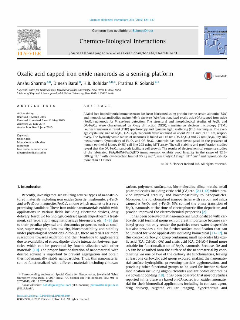
Fig. 1. X-ray diffraction pattern of (a) Fe3O4 and (b) OA-Fe3O4 nanorods.
132 A. Sharma et al. / Chemico-Biological Interactions 238 (2015) 129–137
3. Results and discussion
3.1. Characterization of OA-Fe3O4 nanocrystals
Fig. 1 shows the XRD studies of the Fe3O4, OA-Fe3O4 revealedthe purity and crystallinity of the synthesized NPs with the cubicstructure [space group: Fd-3 m] known for the Fe3O4 crystal(JCPDS Card No. 32-0483) [33,34]. It has been observed that theXRD pattern of bare and caped magnetite nanorods showed num-bers of Braggs reflections that may be indexed on the basis of theface cantered cubic structure of NPs diffraction peaks correspond-ing to the planes (220), (311), (400), (422), (511) and (440) at 2hvalues of 30.18�, 35.6�, 43.66�, 54.18�, 57.77� and 63.45�, respec-tively, consistent with the standard XRD data for the Fe3O4 phase[Fig. 1(a and b)]. The average crystalline size of Fe3O4, OA-Fe3O4
nanorods were estimated using Debye–Scherrer formula, and thevalue obtained was about 25 ± 1and 32 ± 1 nm, respectively. Noadditional peaks were observed indicating the formation of pureand single phase without any impurities that remain from theun-reacted precursors. Moreover, it has been noticed that thepeaks are broad and sharp that is an indication of the formationof very fine particles in the nanoscale range. The calculated interd-spacing of crystalline structure determined for the reflected peak(311) from Bragg’s law is 0.132 nm.
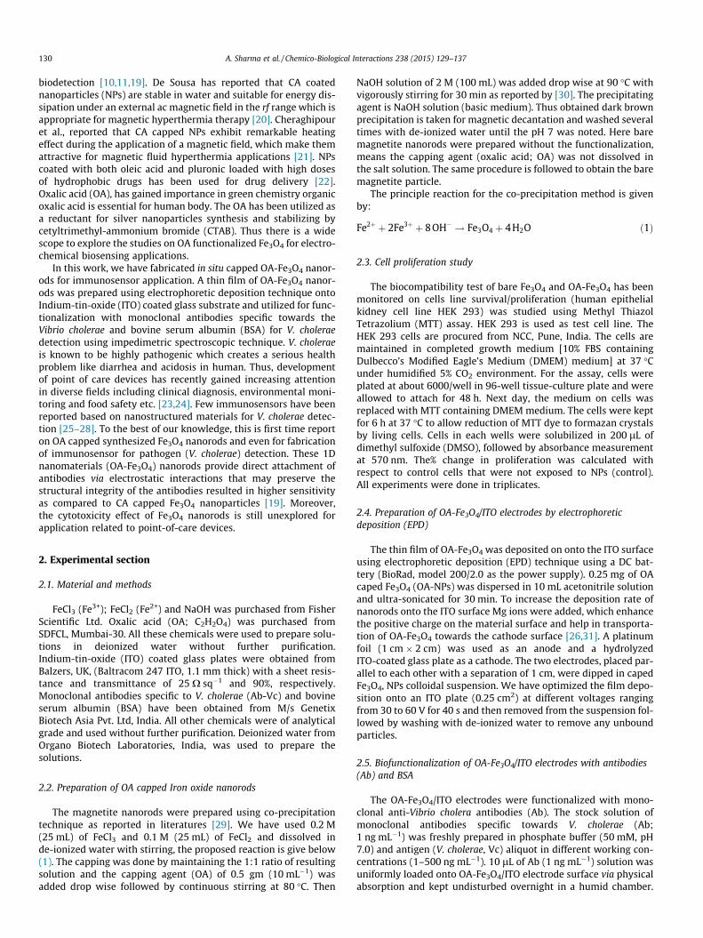
Fig. 2 depicts transmission electron microscopy (TEM) images ofOA-Fe3O4 magnetite products. The well-suspended OA-Fe3O4 hasbeen prepared in water and drop casted onto a carbon coated cop-per grid for the TEM micrograph. Image (a) shows the TEM ofOA-Fe3O4 magnetite nanorods which reveal that good dispersity
20 nm
(a) (
Fig. 2. (a) TEM images and (b) HR
of OA-Fe3O4 nanorods with sizes of about 30 ± 1 nm and resultsare accordance with XRD measurement. The coating of thesenanorods is clearly visible in both images. The correspondingatomic scale image of the OA-Fe3O4 exhibits the well-organizedlattice planes of Fe3O4 (image b).
3.2. Scanning electron microscope (SEM)
Fig. 3 shows the he surface morphology of (a) OA-Fe3O4/ITOelectrode and (b) BSA/Ab/OA-Fe3O4/ITO immunoelectrode hasbeen confirmed by SEM images. Image (a) shows rod shape mor-phology which is uniform deposition of OA-Fe3O4 onto the ITO sur-face. After the immobilization of Ab and BSA onto OA-Fe3O4
electrode surface, the morphology of electrode has been changedand formed the globular shape morphology due to presence ofmacromolecular size of antibodies and BSA that confirm the pres-ence of biomolecules (Ab, BSA) onto OA-Fe3O4/ITO electrode sur-face (image b).
3.3. Dynamic light scattering (DLS) study
The bare magnetite and OA capped NPs have been characterizedusing dynamic light scattering (DLS). It has been found that thehydrodynamic radius (RH) of OA-Fe3O4 as 115.9 nm and Fe3O4 as77.4 nm (data not shown). These results indicate that bare Fe3O4
is less hydrophilic as compared to OA capped Fe3O4 nanorodsresults indicated that Fe3O4 has been modified with OA. Table 1shows particle size of bare Fe3O4 NPs and OA-Fe3O4 nanorodsobtained from different techniques.
3.4. Fourier transforms infrared spectroscopy (FTIR) Study
Surface modification of OA-Fe3O4/ITO immunoelectrode afterimmobilization of Ab and BSA was confirmed by changing in IRspectra (Fig. 4). OA-Fe3O4/ITO, Ab/OA-Fe3O4/ITO andBSA/Ab/OA-Fe3O4/ITO immunoelectrode have been characterizedby using FTIR. The IR spectrum shows main absorptions peak at3400, 1500, 1090 and 591 cm�1. The sharp peak in the spectrumaround 591 cm�1 was ascribed to vibrations of Fe–O an iron oxideskeleton in almost all the samples (curve a), while the otherabsorptions should be assigned to organic species. The broad bandaround 3400 cm�1 was assigned to stretching vibrations of N–Hbonds and adsorbed water molecules, the bands between 1385and 1610 cm�1were assigned to bending vibrations of N–H (amideI) and C–H bonds, and the peak at about 1090 cm�1 arises from thestretching vibrations of C–N bonds [33,34]. Compared with theuncoated magnetite nanorods, the larger intensity of the OH band(3400–3500 cm�1) suggested that there was a reaction of the car-boxylic acid group of acids with the surface hydroxyls of Fe3O4
1 nm
b)
TEM of OA-Fe3O4 nanorods.

Fig. 3. SEM images shows (a) OA-Fe3O4/ITO electrode and (b) BSA/Ab/OA-Fe3O4/ITO immunoelectrode.
Table 1Particle size of nanorods obtained from different techniques.
Nanorods Nanorods sizefrom XRD (nm)
Average nanorodssize from TEM (nm)
Hydrodynamicradius from DLS(nm)
Fe3O4 29 24 77 (lesshydrophobic)
OA capedFe3O4
39 24 116
A. Sharma et al. / Chemico-Biological Interactions 238 (2015) 129–137 133
nanorods. The signature IR peaks appear at 1036 cm�1, 1374 cm�1
(amide II) are responsible for carboxylate (COO�) stretching (curveb). The broad hump formed in the region of 2994–3590 cm�1isassigned to O–H stretching of carboxylic acid group. The presenceof these peaks is evidence of the formation of oxalic acid cappedFe3O4 nanorods. However, the Fe–O vibration band at 500 cm�1
becomes broader and shifts towards higher wavenumber at994 cm�1 may be due to the incorporation of BSA (curve d). Thepeaks seen at 1650 cm�1 and 1535 cm�1 indicating the presenceof an amide band (I, II) and intensity increased at around1250 cm�1 assigned to amide (III) revealing the immobilizationof BSA onto the Ab/OA-Fe3O4/ITO electrode.
3.5. Zeta potential study
An imaginary surface (plane of shear) is used to represent theeffective location of the solid–liquid interface, where the liquid
500 1000 1500 2000 2500 3000 3500 4000
(d)
(c)
(b)
Tran
smitt
ance
(%)
Wavenumber (cm-1)
(a)
Fig. 4. FTIR spectra of (a) Fe3O4; (b) OA-Fe3O4/ITO electrode; (c) Ab/OA-Fe3O4/ITO;(d) BSA/Ab/OA-Fe3O4/ITO immunoelectrode.
velocity is zero. The equilibrium electric potential at the shearplane is called the zeta potential. The isoelectric point refers tothe pH value at which zeta potential is zero. The surface chargepotential of Fe3O4 in water could be explained by surface hydroxylgroups (Fe–OH). The presence of the hydroxyl group on the synthe-sized Fe3O4 nanorods surfaces was confirmed by infrared measure-ment as described. The zeta potential measurement gives thepositive surface charge value of the bare Fe3O4 and OA-Fe3O4
nanorods. The value of zeta potential was found as 12.2, 8.4 mVfor Fe3O4 and OA-Fe3O4, respectively (data not shown). The posi-tive surface charge is expected due to the formation of Fe–OH2
+ inan acidic environment. When OA was introduced into magnetitecolloid solution, acid molecules preferred to attach to the surfaceof the nanorods because carboxylic group of OA have a high affinityfor metallic oxides. In this system, the Fe–OH bond at the surface ofFe3O4 reacted with the carboxylic group of the acid molecule via anacid–base reaction, giving Fe–O–C species with the elimination ofH2O [12].
3.6. Electrochemical studies
Cyclic voltammetric (CV) has been used to investigate the elec-trochemical behavior of Fe3O4/ITO and OA-Fe3O4/ITO electrodesprepared at different voltages (30–50 V) in phosphate buffer(50 mM, pH 6.0, 0.9% KCl) containing 3.3 mM [Fe(CN)]6
3�/4�at scanrate of 20 mV/s (Fig. 5). We have optimized the film fabrication atdifferent voltage and observed that the maximum oxidation andreduction magnitude of current of the electrolyte inthree-electrode system obtained at 30 V as shown in inset(Fig. 5). It may be due to deposition of small size nanorodsattracted faster and deposited uniform thin film that enhanced fastelectron transfer from electrolyte to electrode surface. While athigher voltage (40–50 V) large size nanorods deposited onto ITOsurface to formed thick film and less uniformity resulting in slowcharge transfer. So, all the electrodes have been fabricated at30 V for further electrochemical studies.
The electrochemical response of (a) Fe3O4/ITO; (b)OA-Fe3O4/ITO; (c) Ab/OA-Fe3O4/ITO; (d) BSA/Ab/OA-Fe3O4/ITOimmunoelectrode have been observed in phosphate buffer saline(50 mM, pH 6.0, 0.9% KCl) containing [Fe(CN)6]3�/4� at scan rateof 20 mV/s (Fig. 5). It has been observed that the magnitude of oxi-dation current enhanced of OA-Fe3O4/ITO electrode (curve b) ascompared to bare Fe3O4/ITO (curve a) as shown in Fig. 5. It maybe due to OA capped Fe3O4 nanorods containing negative chargedue to availability of carboxyl group facilitate fast transfer of elec-tron originate from the negatively charged [Fe(CN)6]3�/4� mediumleading to enhanced heterogeneous electron transfer. Moreover,the enhancement of electrochemical signal due to self oxidation

-0.1 0.0 0.1 0.2 0.3 0.4-100
-50
0
50
100
30 35 40 45 50
110
120
130
140
150
160
170
180
Cur
rent
(μA
)
Voltage (V)
(d)(c)
(b)(a)
Cur
rent
(μA
)
Potential (V)
Fig. 5. CV of (a) Fe3O4/ITO; (b) OA-Fe3O4/ITO; (c) Ab/OA-Fe3O4/ITO; (d) BSA/Ab/OA-Fe3O4/ITO at scan rate of 20 mV/s and inset shows the current of OA-Fe3O4/ITOelectrode as a function of deposition voltage.
-0.1 0.0 0.1 0.2 0.3 0.4-200
-150
-100
-50
0
50
100
150
200
250
3 4 5 6 7 8 9 10
0.05
0.10
0.15
0.20
0.25
0.30
Pote
ntia
l (V)
scan rate (mV/s)1/2
3 4 5 6 7 8 9 10 11
-150
-100
-50
0
50
100
150
200
250
scan rate (mV/s)1/2
Cur
rent
(μA
)
10 mV/s
Cur
rent
(μA
)
Potential (V)
100 mV/s
-0.1 0.0 0.1 0.2 0.3 0.4
-150
-100
-50
0
50
100
150
200
250
3 4 5 6 7 8 9 10-200
-150
-100
-50
0
50
100
150
200
scan rate (mV/s)1/2C
urre
nt (μ
A)
10 mV/s
Cur
rent
( μA
)
Potential (V)
100 mV/s
(A)
(B)
Fig. 6A and B. Shows CV of OA-Fe3O4/ITO and BSA/Ab/OA-Fe3O4/ITO immunoelec-trode as a function of scan rate from 10 to 100 mV/s. Inset shows the graph ofcurrent with
ffiffiffiffiffiffiffiffiffiffiffiffiffiffiffiffiffiffiffiffiffi
scan ratep
.
134 A. Sharma et al. / Chemico-Biological Interactions 238 (2015) 129–137
of OA to carbon dioxide as mentioned in Eq. (4) [35]. However, themagnitude of current is increased and slightly shifted towardslower potential after the immobilization of antibodies ontoOA-Fe3O4/ITO electrode resulting in reduced tunneling distancefor diffusion of electrons from the bulk solution leading toenhanced current. It is due to the availability of gibbosities onOA-Fe3O4/ITO electrode surface facilitates the shorten distanceand enhanced the magnitude of current due to improved heteroge-neous electron transfer. These results indicate the significantincrease in the heterogeneous electron transfer rate results dueto strong interaction of antibodies with OA-Fe3O4/ITO electrode.However, the magnitude of current is not much changed butslightly shifted towards higher potential after incorporation ofBSA onto Ab/OA-Fe3O4/ITO electrode (curve d).
HCOO—COOH! 2CO2 þ 2Hþ þ 2e� ð4Þ
Fig. 6(A and B) shows the CV of OA-Fe3O4/ITO (A) andBSA/Ab/OA-Fe3O4 (B) immunosensor as a function of scan ratevarying from 10 to 100 mV/s. It was observed that in both bioelec-trodes the magnitude of both anodic (Ipa) and cathodic (Ipc) peakcurrents increased linearly with square root of the scan rate(v1/2), suggesting that electrochemical reaction was adiffusion-controlled process.
The anodic (Epa) and cathodic (Epc) peak potentials and potentialpeak shifts (DEp = Epa � Epc) of OA-Fe3O4/ITO (A) andBSA/Ab/OA-Fe3O4/ITO (B) immunoelectrode was observed to exhi-bit a linear relationship (linear regression coefficient 0.98) with thescan rate indicating facile charge transfer kinetics in the scan raterange from 10 to 100 mV s�1 in PBS containing 3.3 mM[Fe(CN)6]3�/4�. These results revealed that immunoelectrode pro-vide sufficient accessibility to electrons between the electrolyteand the electrode. The diffusion co-efficient value of the redox spe-cies from the electrolyte to OA-Fe3O4/ITO andBSA/Ab/OA-Fe3O4/ITO immunoelectrode were calculated usingthe Randles–Sevcik [36] Eq. (5) as shown below. The obtained val-ues have been mentioned in Table 1.
Ip ¼ ð2:69� 105Þn3=2A D1=2Cv1=2 ð5Þ
where Ip is the peak current of the BSA/Ab/OA-Fe3O4/ITO immuno-electrode (Ipa anodic and Ipc cathodic), n is the number of electronsinvolved or electron stoichiometry, A is the surface area of the elec-trode (0.25 cm2), D is the diffusion co-efficient, C is the
concentration of redox species {3.3 mM [Fe(CN)6]3/4} and v is thescan rate (20 mV s�1).
The electroactive surface area (Ae) of the OA-Fe3O4/ITO andBSA/Ab/OA-Fe3O4/ITO immunoelectrode was determined fromthe calculated diffusion co-efficient and the Randles–Sevcik equa-tion [36].
Ae ¼ S=ð2:99� 105Þn3=2CD1=2 ð6Þ
where S is the slope of the straight line obtained from the graph of Ip
versus scan rate1/2. The Ae value of the OA-Fe3O4/ITO (A) andBSA/Ab/OA-Fe3O4/ITO immunoelectrode has been found to be 0.62and 0.53 mm2, respectively.
The surface concentration of ionic species of these electrodeswas estimated using Brown–Anson model [36].
Ip ¼n2F2I�AV
4RTð7Þ
where n is the number of electrons transferred which is 1 in thiscase, F is Faraday constant (96485.34 C mol�1), A is surface area ofthe electrode (0.25 cm2), R is gas constant, I* is surface concentra-tion of ionic species of immunoelectrodes (mol/cm2), T is 298 K,and Ip/V is the slope of calibration plot (scan rate value). The surfaceconcentration of OA-Fe3O4/ITO and BSA/Ab/OA-Fe3O4/ITO immuno-electrode was found to be 1.9 � 10�8 and 1.99 � 10�8 M/cm2,

0 2 4 6 8 10 12
0
2
4
6
8
10
(c)
(a)
(d)
Z" (k
Ω)
Z' (kΩ)
(b)
Fig. 6C. Shows Nyquist plot of (a) Fe3O4/ITO; (b) OA-Fe3O4/ITO; (c) Ab/OA-Fe3O4/ITO and (d) BSA/Ab/OA-Fe3O4/ITO immunoelectrode at zero potential in PBScontaining 3.3 mM [Fe(CN)6]3�/4�.
5
10
15
12.5 ng/ml
500 ng/ml
WECE
Z" (k
Ω)
20
22
24
26
28
CT v
alue
s (k
Ω)
A. Sharma et al. / Chemico-Biological Interactions 238 (2015) 129–137 135
respectively. The results indicate that OA-Fe3O4/ITO electrode pro-vides increased electroactive surface area for loading of antibodies(Ab). However, after the immobilization of Ab, the surface concen-tration changed confirms the presence of Ab and BSAontoOA-Fe3O4/ITO electrodes surface with multilayer coverage.
The value of the heterogeneous electron transfer rate constant(Ks) obtained for OA-Fe3O4/ITO and BSA/Ab/OA-Fe3O4/ITO immu-noelectrode was estimated to be 0.11 and 0.12 s�1 according tothe model of Laviron as in eq. (8) [37].
Ks ¼ mnFv=RT ð8Þ
where m is the peak-to-peak separation (0.14 and 0.15 V, respec-tively), The highest value of Ks was found in case of OA-Fe3O4/ITOelectrode as compared to BSA/Ab/OA-Fe3O4/ITO immunoelectrode,Table 2 shows the electrochemical parameters: anodic peak current(Ipa), cathodic peak current (Ipc), charge transfer rate constant (Ks),diffusion coefficient (D), electroactive surface area (Ae), average sur-face concentration (I*) of ionic species obtained for respectiveelectrodes.
3.7. Electrochemical impedance spectroscopy study
Electrochemical impedance spectroscopy (EIS) is an effectivetool for studying interfacial properties of surface modified elec-trodes. In the EIS, the semicircular diameter of the EIS spectra givesa value of the charge transfer resistance (RCT) that reveals electrontransfer kinetics of the redox probe at the electrode interface.Moreover, RCT depends on dielectric characteristics of the elec-trode/electrolyte interface. The EIS studies has been investigatedin PBS containing 3.3 mM [Fe(CN)6]3�/4� in the frequency range0.01–105 Hz. The semicircular part corresponds to the electrontransfer limited process; its diameter is equal to the electron trans-fer resistance (RCT) which controls the electron transfer behavior ofthe redox probe at the electrode interface. Fig. 6(C) shows theNyquist plot of the impedance of respective modified electrodes(a) Fe3O4/ITO; (b) OA-Fe3O4/ITO; (c) Ab/OA-Fe3O4/ITO and (d)BSA/Ab/OA-Fe3O4/ITO immunoelectrodes. It has been observedthat RCT for Fe3O4/ITO electrode (curve a) is smaller than that ofOA/Fe3O4/ITO electrode (curve b). This suggests that Fe3O4/ITOelectrode improved conductivity of the electrode and that the pos-itively charged which facilitate diffusion of the negatively charged[Fe(CN)6]3�/4� ions towards the electrode surface. After the immo-bilization of Ab onto the OA-Fe3O4/ITO electrode, the RCT valuedecreases, revealing that non-binding sites on Ab facilitate electrontransfer between the OA-Fe3O4/ITO electrode and electrolyte.Further, RCT decreases after the immobilization of BSA onto theAb/OA-Fe3O4/ITO, revealing that BSA blocks non-specific sites ontoAb/OA-Fe3O4/ITO that perhaps perturb electron communicationbetween the BSA/Ab/OA-Fe3O4/ITO immunoelectrode and ITOsurface.
3.8. Electrochemical response studies
The EIS response studies of the BSA/Ab/OA-Fe3O4/ITO immuno-electrode were conducted as a function of V. cholerae concentration
Table 2Shows the electrochemical parameters: anodic peak current (Ipa), cathodic peakcurrent (Ipc), charge transfer rate constant (Ks), diffusion coefficient (D), electroactivesurface area (Ae), average surface concentration (I⁄) for respective electrodes.
Electrodes Ipa
(lA)Ipc
(lA)Ks
(s�1)D (cm2 s�1) Ae
(mm2)I⁄
(mol cm�2)
Fe3O4/ITO 65.4 �46.2 0.14 4.34 � 10�12 – 1.39 � 10�8
OA-Fe3O4/ITO
89.6 �76.7 0.11 8.15 � 10�12 0.62 1.9 � 10�8
BSA/Ab/OA-Fe3O4/ITO
93.7 �94.96 0.12 8.9 � 10�12 0.53 1.99 � 10�8
(12.5–400 ng/mL) in PBS containing [Fe(CN)6]3�/4� as shown inFig. 7. The BSA/Ab/OA-Fe3O4/ITO immunoelectrode were treatedwith 30 lL of V. cholerae concentration and then incubated forten minutes at room temperature (25 �C). It was found that theRCT value decreases with increasing concentrations of antigen(Vibrio cholera). It may be due to antibody-antigen complex forma-tion which enhanced the charge transfer rate resulting in decreaseRCT value and increased sensitivity (inset Fig. 7). The detectionrange found as 12.5–500 ngmL�1 with low detection limit of0.5 ng mL�1calculated by using the 3rb/m criteria, where m isslope of the calibration graph and rb is the standard deviation ofthe blank signal. The sensitivity of the BSA/Ab/OA-Fe3O4/ITO calcu-lated by the slope of the linear regression curve is0.1 O ng �1mL�1 cm�2, which is higher than the previous reportedCA-Fe3O4 nanoparticles. The electrochemical response studies ofbare Fe3O4 (uncapped) based BSA/Ab/Fe3O4/ITO immunoelectrodehave been conducted as a function of V. cholerae concentration atsimilar condition [Data not shown]. It has been observed that the
0 5 10 15 20 25 300
Z' (kΩ)
0 100 200 300 400 500
14
16
18R
Concentration (ng/ml)
Fig. 7. Shows electrochemical response studies of BSA/Ab/OA-Fe3O4/ITO as afunction of antigen concentration of 12.5–400 ng/mL and inset shows the respec-tive RCT values at zero potential in PBS containing 5 mM [Fe(CN)6]3�/4� (inset (A):Randles equivalent circuit, where Rs: solution resistance, RCT: charge transferresistance, Zw: Warburg impedance, and Cdl: double layer capacitance).

Table 3Shows a comparative biosensing characteristics of BSA/Ab/OA-Fe3O4/ITO immunoelectrode along with reported in literature.
Electrode/immobilization/transducer
Transducer Biomolecules Detection range Low detectionrange
Sensitivity Refs.
BSA/Ab/CA-Fe3O4/ITO Electrochemical impedancespectroscopy
Antibodies 12.5–500 ng ml�1 0.32 ng ml�1 0.03 O/ng ml�1cm�2 [19]
Nickel oxide nanowires/ITO Electrochemical impedancespectroscopy
Antibodies 37–350 ng ml�1 0.553 ng ml�1 11.12 O/ng ml�1cm�2 [25]
RGO-TiO2/ITO Electrochemical impedancespectroscopy
Antibodies 10–450 ng/mL 0.15 ng mL�1 21.8 � 10�3 lF/ng mL�1/cm2
[26]
Lipid layer Chemiluminescence HRP/gangliosideGM1
1 pgmL�1–1 ngmL�1
0.8 pgmL�1 – [27]
Gold-coated AFMmicrocantilevers
Dynamic force microscopy Antibodies 1 � 103–1 � 107
CFU/ml.1 � 103 CFU/ml, �146.5 pg/Hz [28]
BSA/Ab/OA-Fe3O4/ITO Electrochemical impedancespectroscopy
Antibodies 12.5–500 ng ml�1 0.5 ng ml�1 0.1 O/ng ml�1cm�2 Presentwork
% o
f cel
l via
bilit
y
Nanoparticles concentration (µg/mL)
Fe3O4 Oxalic acid capped Control
Fig. 8. Effect of Fe3O4 and OA-Fe3O4 Nanorods on the proliferation of HEK 293 cells.
136 A. Sharma et al. / Chemico-Biological Interactions 238 (2015) 129–137
RCT value does not change significantly as compared to OA cappedFe3O4 nanorods based [BSA/Ab/OA-Fe3O4/ITO] immunoelectrode.These results indicate that the OA-Fe3O4 nanorods[BSA/Ab/OA-Fe3O4/ITO] immunoelectrode shows higher interac-tion of antibodies with antigens and exhibits improved biosensingproperties. The stability of BSA/Ab/OA-Fe3O4/ITO immunoelectrodeobtained 50 days (80%) and reproducibility more than 11 times.Table 3 shows the comparison of biosensing properties ofBSA/Ab/OA-Fe3O4/ITO immunoelectrode along with reported inliterature.
3.9. Effect of cytotoxicity
Fig. 8 shows the relative percentage proliferation of HEK 293cells in the presence of Fe3O4 and OA-Fe3O4 nanorods, respectively;in the concentration range 0–100 lg mL�1. The MTT assay resultsindicated that viability of HEK 293 is not affected in the presenceof Fe3O4 and OA-Fe3O4 nanorods, resulted normal growth of cells.These results suggest that nanorods are biocompatible and donot have toxic affect on human cells in vivo studies. However,the OA-Fe3O4 nanorods exhibit higher biocompatibility overFe3O4 indicated that OA-Fe3O4 nanorods facilitate cell growth.Similar results reported with Hela cell lines [38].
4. Conclusions
Biocompatible magnetite nanorods prepared byco-precipitation method and stabilized by coating with oxalic acid(OA). We have successfully functionalized Fe3O4 nanorods with OAthat confirmed by TEM, FTIR and DLS. These NPs has been depos-ited using electrophoretic deposition technique onto ITO surfacefor immobilization of monoclonal antibodies against V. cholerae
and BSA for V. cholerae detection using impedimetric techniques.The results of these studies obtained as linearity 12.5–500 ng mL�1 with low detection limit of 0.5 ngmL�1, sensitivity0.1 O ng�1 mL�1 cm�2 and reproducibility more than 11 times.This type of electrode has the potential to be of general use fordesigning new electrochemical immunosensors for detection ofother clinically important antigens.
Conflicts of Interest
There is no any conflicts of interest.
Transparency Document
The Transparency document associated with this article can befound in the online version.
Acknowledgements
AS and DB acknowledges University Grants Commission,Government of India for a research fellowship. This work was sup-ported by a Grant received from Department of Science andTechnology, Government of India (Grant no. SB/PC/S1/110/2012and SR/NM/NS-1144/2013 (G)). Authors are thankful to theAdvanced Instrument Research Facility of the University for pro-viding access to FTIR and SEM instruments.
References
[1] P.R. Solanki, A. Kaushik, V.V. Agrawal, B.D. Malhotra, Nanostructured metaloxide-based biosensors, NPG Asia Mater. 3 (1) (2011) 17–24.
[2] R. Sharma, V.V. Agrawal, A.K. Srivastava, Govind, L. Nain, M. Imran, S.R. Kabi,R.K. Sinha, B.D. Malhotra, Phase control of nanostructured iron oxide forapplication to biosensor, J. Mater. Chem. B 1 (2013) 464–474.
[3] E. Amstad, M. Textor, E. Reimhult, Stabilization and functionalization of ironoxide nanoparticles for biomedical applications, Nanoscale 3 (2011) 2819–2843.
[4] Y.F. Rao, W. Chen, X.G. Liang, Y.Z. Huang, J. Miao, L. Liu, Y. Lou, X.G. Zhang, B.Wang, R.K. Tang, Z. Chen, X.Y. Lu, Epirubicin-loaded superparamagnetic iron-oxide nanoparticles for transdermal delivery: cancer therapy by circumventingthe skin barrier, Small 11 (2) (2015) 239–247.
[5] G.K. Kouassi, J. Irudayaraj, G. McCarty, Examination of cholesterol oxidaseattachment to magnetic nanoparticles, J. Nanobiotechnol. 3 (1) (2005) 1–9.
[6] T.R. Pisanic, J.D. Blackwell, V.I. Shubayev, R.R. Finones, S. Jin, Nanotoxicity ofiron oxide nanoparticle internalization in growing neurons, Biomaterials 28(16) (2007) 2572–2581.
[7] Y. Wang, J. Dostalek, W. Knoll, Magnetic nanoparticle-enhanced biosensorbased on grating-coupled surface plasmon resonance, Anal. Chem. 83 (16)(2011) 6202–6207.
[8] A. Kaushik, R. Khan, P.R. Solanki, P. Pandey, J. Alam, S. Ahmad, B.D. Malhotra,Iron oxide nanoparticles–chitosan composite based glucose biosensor, Biosens.Bioelectron. 24 (2008) 676–683.

A. Sharma et al. / Chemico-Biological Interactions 238 (2015) 129–137 137
[9] A. Kaushik, P. R. Solanki, K. Kaneto, C. G. Kim, Sharif Ahmad, Bansi D. Malhotra,Nanostructured iron oxide platform for impedimetric cholesterol detection,Electroanalysis, 22 (10) (2010) 1045 – 1055.
[10] G. Zhao, J.J. Feng, Q.L. Zhang, S.P. Li, H.Y. Chen, Syntheisis and characterizationof prussian blue modified magnetite nanoparticles and its application to theelectrocatalytic reduction of H2O2, Chem. Mater. 17 (2005) 3154–3159.
[11] A.K. Gupta, R.R. Naregalkar, V.D. Vaidya, M. Gupta, Recent advances on surfaceengineering of magnetic iron oxide nanoparticles and their biomedicalapplications, Nanomedicine (2007) 223–239.
[12] S. Yu, G.M. Chowa, Carboxyl group (–CO2H) functionalized ferrimagnetic ironoxide nanoparticles for potential bio-applications, J. Mater. Chem. 14 (2004)2781–2786.
[13] S. Nigam, K. Barick, D. Bahadur, Development of citrate-stabilized Fe3O4
nanoparticles: conjugation and release of doxorubicin for therapeuticapplications, J. Magn. Magn. Mater. 323 (2011) 237–243.
[14] C. Boyer, M.R. Whittaker, V. Bulmus, J. Liu, T.P. Davis, The design and utility ofpolymer-stabilized iron-oxide nanoparticles for nanomedicine applications,NPG Asia Materials 2 (2010) 23–30.
[15] E. Munnier, S. Cohen-Jonathan, C. Linassier, L. Douziech-Eyrolles, H. Marchais,M. Souce, K. Herve, P. Dubois, I. Chourpa, Novel method of doxorubicin–SPIONreversible association for magnetic drug targeting, Int. J. Pharm. 363 (2008)170–176.
[16] L. Li, K. Mak, C. Leung, K. Chan, W. Chan, W. Zhong, P. Pong, Synthesis andcharacterization of self-Assembled monolayer and bilayer carboxyl-groupfunctionalized magnetic nanoparticles, IEEE Trans. Magn. 48 (2012) 3299–3302.
[17] M.A. Daniele, M.L. Shaughnessy, R. Roeder, A. Childress, Y.P. Bandera, S.Foulger, Magnetic nano clusters exhibiting protein-activated near-infraredfluorescence, ACS Nano 7 (2013) 203–213.
[18] C. Liu, P.M. Huang, Atomic force microscopy and surface characteristics of ironoxides formed in citrate solutions, Soil Sci. Soc. Am. J. 63 (1999) 65–72.
[19] A. Sharma, D. Baral, K. Rawat, P.R. Solanki, H.B. Bohidar, Biocompatible cappediron oxide nanoparticles for Vibrio cholerae detection, Nanotechnology 26(2015) 175302 (10pp).
[20] M.E. De Sousa, M.B.F.V. Raap, P.C. Rivas, P.M. Zelis, P. Girardin, G.A. Pasquevich,J.L. Alessandrini, D. Muraca, F.H. Ssnchez, Stability and relaxation mechanismsof citric acid coated magnetite nanoparticles for magnetic hyperthermia, J.Phys. Chem. C 117 (10) (2013) 5436–5445.
[21] E. Cheraghipour, S. Javadpour, A.R. Mehdizadeh, Citrate cappedsuperparamagnetic iron oxide nanoparticles used for hyperthermia therapy,J. Biomed. Sci. Eng. 5 (2012) 715–719.
[22] I. Palchetti, M. Mascini, Electroanalytical biosensors and their potential forfood pathogen and toxin detection, Anal. Bioanal. Chem. 391 (2) (2008) 455–471.
[23] O. Lazcka, C.F.J. Del, M.F. Xavier, Pathogen detection: a perspective oftraditional methods and biosensors, Biosens. Bioelectron. 22 (2007) 1205–1217.
[24] S. Viswanathan, W. Li-chen, M.R. Huang, H.J. Annie, Electrochemicalimmunosensor for cholera toxin using liposomes and poly(3,4-ethylenedioxythiophene)-coated carbon nanotubes, Anal. Chem. 78 (4)(2006) 1115–1121.
[25] P.R. Solanki, M.A. Ali, V.V. Agrawal, A.K. Srivastava, R.K. Kotnala, B.D. Malhotra,Highly sensitive biofunctionalized nickel oxide nanowires for nanobiosensingapplications, RSC Adv. 3 (2013) 16060–16067.
[26] P.R. Solanki, S. Srivastava, M.A. Ali, R.K. Srivastava, A. Srivastava, B. Malhotra,RSC Adv. 4 (104) (2014) 60386–60396.
[27] U. Sungkanak, A. Sappat, A. Wisitsoraat, C. Promptmas, A. Tuantranont,Biosens. Bioelectron. 26 (2) (2010) 784–789.
[28] H. Chen, Y. Zheng, J.H. Jiang, H.L. Wu, G.L. Shen, R.Q. Yu, Biosens. Bioelectron.24 (4) (2008) 684–689.
[29] Y.S. Kang, S. Risbud, J.F. Rabolt, P. Stroeve, Synthesis and characterization ofnanometer-size Fe3O4 and c-Fe2O3particles, Chem. Mater. 8 (1996) 2209–2211.
[30] M. Racuciu, D.E. Creang, A. Airinei, Citric-acid-coated magnetite nanoparticlesfor biological applications, Eur. Phys. J. E 21 (2) (2006) 117–121.
[31] R. Shinar, J. Shinar: Organic electronics in sensors and biotechnology.Electrophoretically deposited polymers for organic electronics, chapter by C.Dhand and B.D. Malhotra (McGraw-Hill Professional, 2009), AccessEngineering.
[32] B.J. Berne, R. Pecora, Dynamic light scattering with applications to chemistrybiology and physics, Wiley-Interscience, New York, USA, 1976.
[33] H. Ohshima, Electrophoresis of soft particles, Adv. Colloid Interface Sci. 62(1995) 189–235.
[34] Y. Tian, B. Yu, X. Li, K. Li, Facile solvothermal synthesis of monodisperse Fe3O4
nanocrystals with precise size control of one nanometre as potential MRIcontrast agents, J. Mater. Chem. 21 (2011) 2476–2481.
[35] L.C. Rockombeny, J.P. Feraud, B. Queffelec, D. Ode, T. Tzedakis, Electrochemicaloxidation of oxalic acid and hydrazinium nitrate on platinum in nitric acidmedia, Electrochim. Acta 66 (2012) 230–238.
[36] A.J. Bard, L.R. Faulkner, A digital simulation model for electrochromicprocesses at WO3 electrodes, John Wiley & Sons, New York, 1980.
[37] J. Singh, A. Roychoudhury, M. Srivastava, P.R. Solanki, D.W. Lee, S.H. Lee, B.D.Malhotra, A highly efficient rare earth metal oxide nanorods based platformfor aflatoxin detection, J. Mater. Chem. B 1 (2013) 4493–4503.
[38] S. Nigam, K.C. Barick, D. Bahadur, Development of citrate-stabilized Fe3O4
nanoparticles: conjugation and release of doxorubicin for therapeuticapplications, J. Magnetism Magnet. Mater. 323 (2011) 237–243.