between semi-permeable film, antiseptics and sugar paste
TRANSCRIPT
J. Exp. Path. (I990) 71, I55-I70
A controlled model of moist wound healing: comparisonbetween semi-permeable film, antiseptics and sugar paste
Helen G. Archer,* Sheila Barnett,t Sarah Irving,t K.R. Middletont andD.V. Seal§
*Infection Control, $Department of Pharmacy and § Department of Microbiology, Northwick Park Hospital,Harrow and t Department of Research in Plastic Surgery, Mount Vernon Hospital, Northwood,
Middlesex HA6 2RN, UK
Received for publication 29 March I 989Accepted for publication I4 September I 989
Summary. An established wound model in the pig has been modified using a Stomahesive ringto enable study of the effects of fluids used in wound care. Full thickness wounds (up to 9 mmdeep) were treated with the substances under test. Each application was held in place with aStomahesive flange, the inner part of which had been excised as far as the hard plastic ring. Alldressings were then covered with OpSite which allowed gaseous exchange whilst retainingtreatment fluids and secretions. Wounds were treated immediately and at 2 and 4 days. Theexperiment was terminated after 7 days and the whole wound, with dressing, was excised forhistological examination. The wounds covered with OpSite alone and those treated with sugarpaste under Opsite were found to be infilled with granulation tissue over which epidermalmigration was taking place. Those wounds which had been packed with gauze, to which hadbeen added one of the following: chlorhexidine gluconate 0.2%, Irgasan 0.2%, povidone iodineo.8% or EUSOL half-strength, showed delayed healing in that less infilling had taken place overthe same time period. This delay could be attributed to the nature of the chemicals used and/orthe influence of gauze packing. This delay in the healing of wounds treated with chemicalagents was least with EUSOL half-strength and greatest with chlorhexidine. No toxic effectswere observed with sugar paste which may be preferable to antiseptics for the management ofdirty or infected wounds.
Keywords: wounds, healing, model, sugar, antiseptics, dressings, pH
Many new types of dressings such as hydro- Winter and Scales (i963) first showed in acolloids and hydrogels have been introduced pig model that keeping wounds moistin the last few years claiming to provide allowed more rapid epidermal migration atoptimal conditions for wound healing (Kick- the same level as the undamaged epidermishofen et al. I986) but most have not been and without shrinkage. This has been inves-subjected to rigorous controlled trials in tigated further, in superficial wounds in pigs,animals or humans. Winter (I962) and when re-epithelialization was increased
Correspondence: Dr K.R. Middleton, Department of Pharmacy, Northwick Park Hospital, WatfordRoad, Harrow HAI 3UJ, UK.
'55
H.G. Archer et al.beneath both oxygen-permeable and imper-meable occlusive dressings (Alvarez et al.I 983). This principle has been developed, forexample with a semi-permeable plastic filmOpSite, although excess exudate tends toaccumulate under it. Its clinical use has beenrecently reviewed (Alper I986).
Sugar paste has been used for over 4 yearsat Northwick Park Hospital and at othercentres in the UK. The thin paste is of similarconsistency to thin honey and can beinstilled via a syringe and Kwill (fine plastictubing) into abscess cavities with small open-ings. Traditionally such wounds are packedwith gauze soaked in EUSOL, a treatmentwhich is often painful for the patient andmay be harmful to granulating tissue. Thicksugar paste is suitable for packing into largeopen wounds such as pressure sore cavities.Both pastes have excellent antimicrobialactivity in vitro and have provided effectivetreatment for infected and malodorouswounds (Middleton & Seal I 985, I 989).Wound healing and orientation of colla-
gen fibres have been extensively studied inthe pig (Winter I962; Laufer et al. I974)which is the most suitable animal to studyfull thickness wound healing because theepidermis, dermis and subcutaneous fatresembles the human (Hartwell 1955;Winter I966). We have adapted Winter'smodel to study moist wound healing, withsealed chambers placed over each woundand have compared the effects on healing ofour Northwick Park sugar paste (Middleton& Seal I 98 5), various antiseptics and OpSitealone in a controlled trial. A similar study,comparing calcium alginate fabric withOpSite, has been conducted by Barnett andVarley (i 987) but sealed chambers were notused and the OpSite was applied directly toskin.
Materials
Halothane was purchased from ICI, Maccles-field, Cheshire; chlorhexidine gluconate0.015% w/v and cetrimide 0.15% w/v solu-tion (Tisept) and Tubinette from Seton
Healthcare Group, Oldham; Stomahesiveflanges from Squibb Surgicare Ltd, Moreton,Wirral; OpSite from Smith & Nephew Medi-cal Ltd, Hull; povidone iodine I0% solutionfrom Napp Laboratories Ltd, Cambridge;EUSOL from Macarthys Medical Ltd, Rom-ford. Chlorhexidine gluconate 0.2% w/v wasprepared in the pharmacy at Northwick Parkby diluting Hibitane Gluconate 20% solution(ICI, Macclesfield, Cheshire) with water andautoclaving at I2I°C for I5 min. IrgasanDP300 and its diluent (propylene glycol 5%w/v and Empicol LQ33T 33% w/v-monoethanolaminelaurylsulphate, Surfa-chem Ltd) were a gift from Hough Hoseason& Co Ltd, Manchester. Sugar paste wasmanufactured in the pharmacy at North-wick Park Hospital to the following formula:
Castor sugar I200 g(fine granular sucrose)Icing sugar-additive free i8oo g(powdered sucrose)Polyethylene glycol 400 686 mlHydrogen peroxide 30% igmlFinal concentration ofhydrogen peroxidewas O.I5% v/w.
Methods
Animals and anaesthesia
Trials were carried out using three o0-I2week female pigs (gilts), weighing approxi-mately 27 kg (6o lb). They were housed inindividual pens for the whole of the trialincluding an acclimatization period of 2weeks prior to the first operations. Food waswithheld on the morning of operation andanaesthesia was induced without excitingthe animal with 4% Halothane, nitrous oxide4 1/min and oxygen 3 1/min deliveredthrough a conical nose mask. The animalwas rendered unconscious in about 4 min.Anaesthesia was maintained with oxygen (31/min), nitrous oxide 4 1/min) and 2.0%Halothane until the end ofthe operation. Thepig was then carried back to the pen andmonitored until regaining consciousness.
I5 6
Effect of dressings on moist wound healingOperative procedure for production of woundsThe hair on the area to be operated on wasclipped and the skin washed with chlorhexi-dine gluconate 0.015%/cetrimide 0.15%solution, shaved and dried. This ensured thatthe dressings adhered.
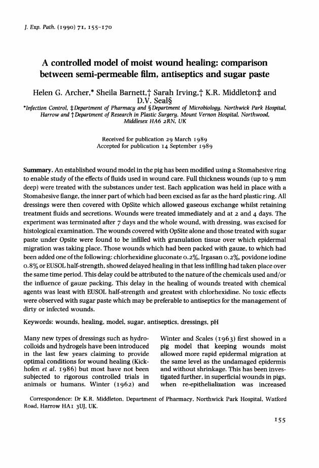
Fig. i. Arrangement of wounds for each pig.
Standard partial thickness wounds weremade by placing a Perspex guide (windowsize 2 5 mm square) on the skin so that two ofthe sides were parallel to the direction of hairgrowth. Shallow incisions were made todelineate the margins of the wound and thewound guide removed. The blade of a scalpelwas used to dissect the epidermis and papill-ary layer of the dermis over the whole woundarea to a depth of approximately 9 mm. Sixwounds were made on each side of theanimal (Fig. i). A Stomahesive flange was
placed over each wound site and the innercircle of Stomahesive cut out as far as thehard plastic ring. This yielded an area justsufficient to expose the wound underneath.With six wounds per flank, arranged as tworows of three, the application of the Stoma-hesive flange was such that there was com-plete occlusion of normal skin between andaround each wound (Fig. 2). Tests werearranged so that each vertical pair ofwoundscontained the same dressing, which ensuredthat mixing of products did not occur; pastexperience had shown that any leakagetended to track from the top wound down-wards. After each dressing had been applied,the site was covered with OpSite self-adhe-sive, water-vapour-permeable, polyurethaneplastic film. A cotton body stocking of Tubi-nette was put around the pig to help reducedirect rubbing of the dressings in the pen.
Application of wound dressings
Six wounds on one side of the first pig weredressed with OpSite, as controls of moistwound healing without gauze. Six woundson the other side were dressed with North-wick Park sugar paste.
For the second and third pigs, four woundswere each dressed with OpSite (controls),Irgasan DP300 (0.2% w/v in diluent), chlor-hexidine gluconate (0.2% W/V), povidineiodine o.8% (freshly prepared from a io%commercial preparation with o.9% saline)and EUSOL half-strength (freshly preparedby diluting the full strength preparation witho.9% saline). Two wounds were each dressedwith sugar paste and Irgasan diluent.
In order to observe healing in the absenceof foreign bodies, OpSite control wounds didnot contain gauze. Thick sugar paste wasapplied by rolling it into a ball and packingthe wound. All other wounds were packedwith five layers of gauze which had been cutto the size of the wound. Five ml of eachantiseptic solution were applied to the gauzein the wound with a syringe.
I57
H.G. Archer et al.
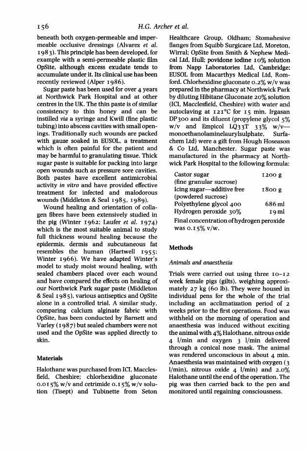
Fig. 2. Right flank of pig 3 showing six wound sites after being covered for 48 h with Stomahesivedressings and OpSite plastic film. 3 And 6, chlorhexidine gluconate 0.2% w/v with gauze dressing;2 and 5, OpSite control without gauze dressing; i and 4, Irgasan diluent with gauze dressing.
Wound care
Wounds were inspected 48 and 96 h follow-ing the initial procedure. Fluid exudate fromeach wound was removed as described belowand the OpSite was cut around the Stomahe-sive flange (the hard plastic ring provided anideal surface for this to be performed). Thewounds were re-dressed working from thetop to bottom of a pair so that the same testcompounds were dealt with sequentially.Wounds containing gauze were replenishedby injecting 2 ml of each antiseptic into thegauze taking care not to disturb it. Woundscontaining sugar were repacked with paste.OpSite and Tubinette body stocking wereapplied as described above.
Sampling from wound site
The wound exudate was retained within theStomahesive flange underneath the OpSiteand was removed with a sterile syringe and
needle for evaluation of antimicrobial acti-vity and measurement of pH. The exudatecollected was stored at 40C. pH was deter-mined immediately using a pH meter (OrionResearch Model 7oIA).
Termination of experiment and sampling forhistological examination
Seven days after the initial operation eachpig was anaesthetized as described pre-viously and wound exudate collected. TheStomahesive dressing was removed from oneside of the pig at a time. Gauze was notremoved prior to biopsy to ensure that tissuewas not disturbed. Tissue was dissected to adepth of I2-15 mm and normal skin wasremoved from around the wound.
Histology
All biopsy specimens were placed immedia-tely in io% formal saline and trimmed to size
I5 8
Effect of dressings on moist wound healingafter 24 h fixation. Photographs were takenof all prepared specimens prior to processing.The tissue was embedded using routine waximpregnation and serial sections cut at IOgum on a rotary microtome. Sections werestained with Ehrlich's haematoxylin andeosin and Gram's stains.
Analysis of antimicrobial activity of woundexudates
A laboratory isolate of a commensal Staphy-lococcus epidermidis was inoculated intonutrient broth and incubated overnight at3 7TC. This was flooded onto a 20 cm squareplate of nutrient agar which was allowed todry. Sixteen wells were made in the agarusing a cork borer (diameter 7.5 mm). Eachwell was filled with a different wound exu-date. The plate was incubated for i 8 h at3 70C after which zones of inhibition weremeasured with calipers.
Bacteriological analysis of wound exudates
Approximately 30 ul of exudate were spreadon both blood and McConkey agar plates.These were incubated aerobically at 3 70C forI8 h after which any colonies present werecounted and identified by standard methods.
Results
A typical appearance of the pig modelwounds enclosed by Stomahesive rings andOpSite is shown in Fig. 2. Fluid exudate canbe clearly seen contained within each woundenclosure. None of the wounds appearedclinically infected.
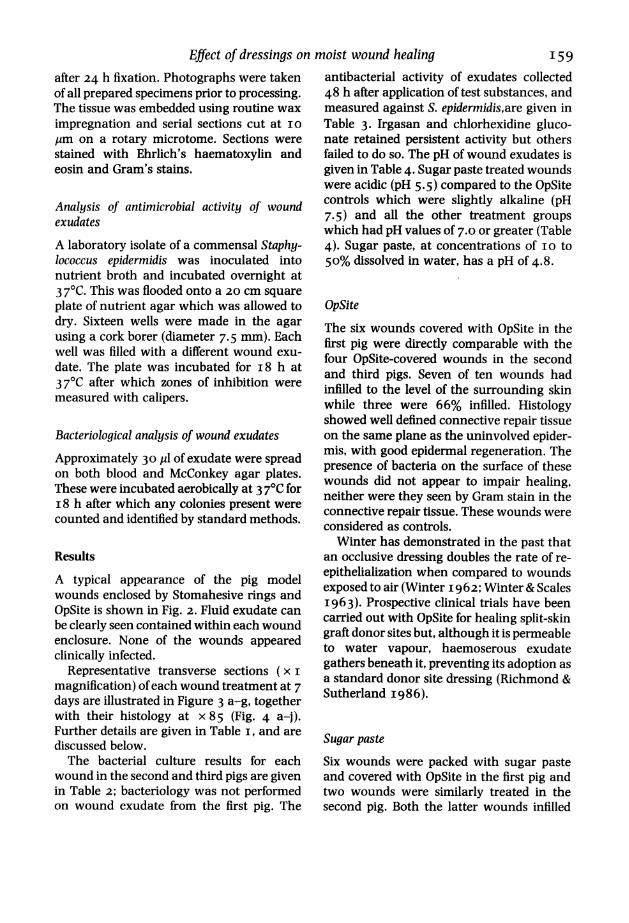
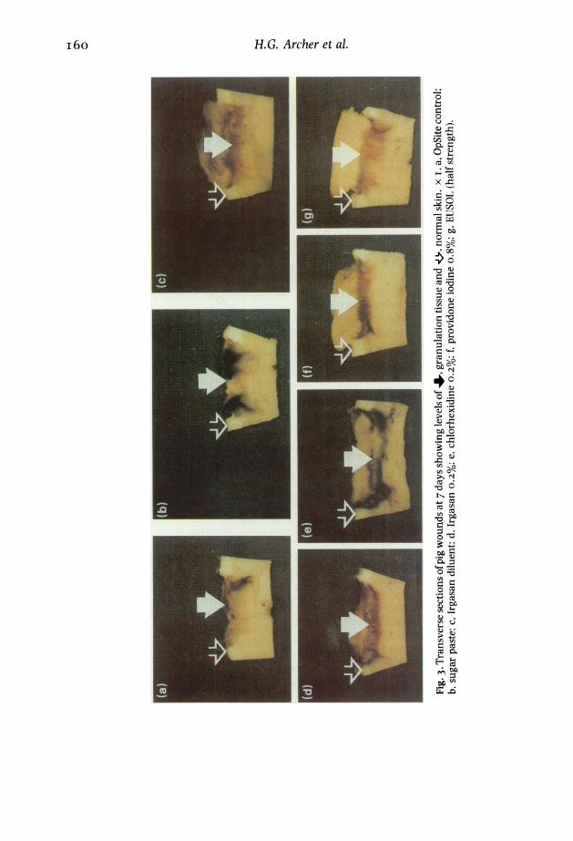
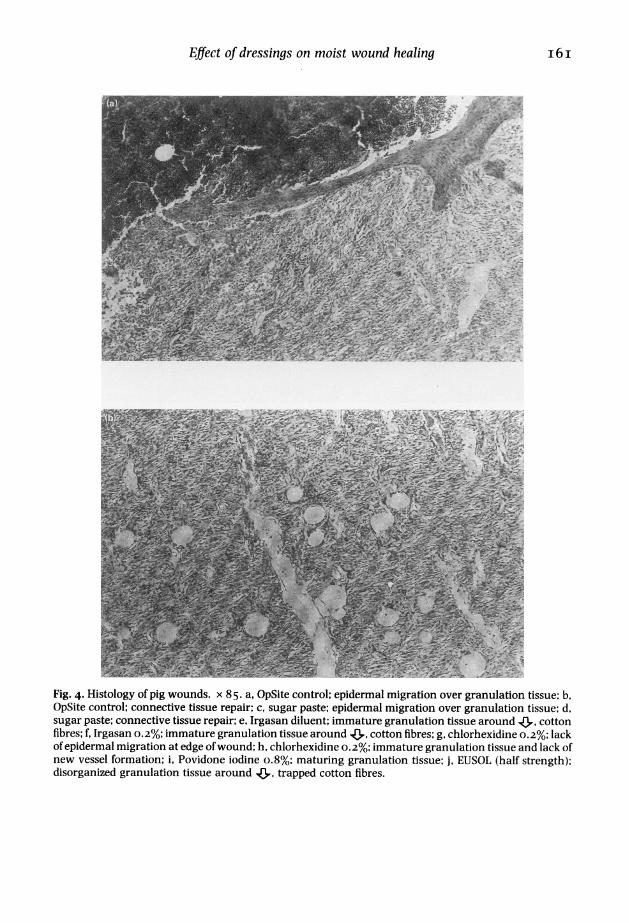
Representative transverse sections (x imagnification) of each wound treatment at 7days are illustrated in Figure 3 a-g, togetherwith their histology at x 85 (Fig. 4 a-j).Further details are given in Table i, and arediscussed below.The bacterial culture results for each
wound in the second and third pigs are givenin Table 2; bacteriology was not performedon wound exudate from the first pig. The
antibacterial activity of exudates collected48 h after application of test substances, andmeasured against S. epidermidis,are given inTable 3. Irgasan and chlorhexidine gluco-nate retained persistent activity but othersfailed to do so. The pH of wound exudates isgiven in Table 4. Sugar paste treated woundswere acidic (pH 5.5) compared to the OpSitecontrols which were slightly alkaline (pH7.5) and all the other treatment groupswhich had pH values of 7.0 or greater (Table4). Sugar paste, at concentrations of io to50% dissolved in water, has a pH of 4.8.
OpSite
The six wounds covered with OpSite in thefirst pig were directly comparable with thefour OpSite-covered wounds in the secondand third pigs. Seven of ten wounds hadinfilled to the level of the surrounding skinwhile three were 66% infilled. Histologyshowed well defined connective repair tissueon the same plane as the uninvolved epider-mis, with good epidermal regeneration. Thepresence of bacteria on the surface of thesewounds did not appear to impair healing,neither were they seen by Gram stain in theconnective repair tissue. These wounds wereconsidered as controls.
Winter has demonstrated in the past thatan occlusive dressing doubles the rate of re-epithelialization when compared to woundsexposed to air (Winter I962; Winter& ScalesI963). Prospective clinical trials have beencarried out with OpSite for healing split-skingraft donor sites but, although it is permeableto water vapour, haemoserous exudategathers beneath it, preventing its adoption asa standard donor site dressing (Richmond &Sutherland I986).
Sugar pasteSix wounds were packed with sugar pasteand covered with OpSite in the first pig andtwo wounds were similarly treated in thesecond pig. Both the latter wounds infilled
I59
H.G. Archer et al.
.-4
O X
0*
CZ
0~
0>
-~0
m
*;
N,2O
cu c
E cO
to
i6o
Effect of dressings on moist wound healing
2N W OFAM M yoF S ___E2 `-~ ; ; g- 1lI7-00
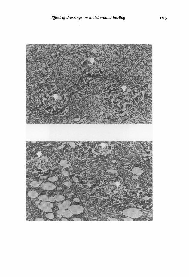
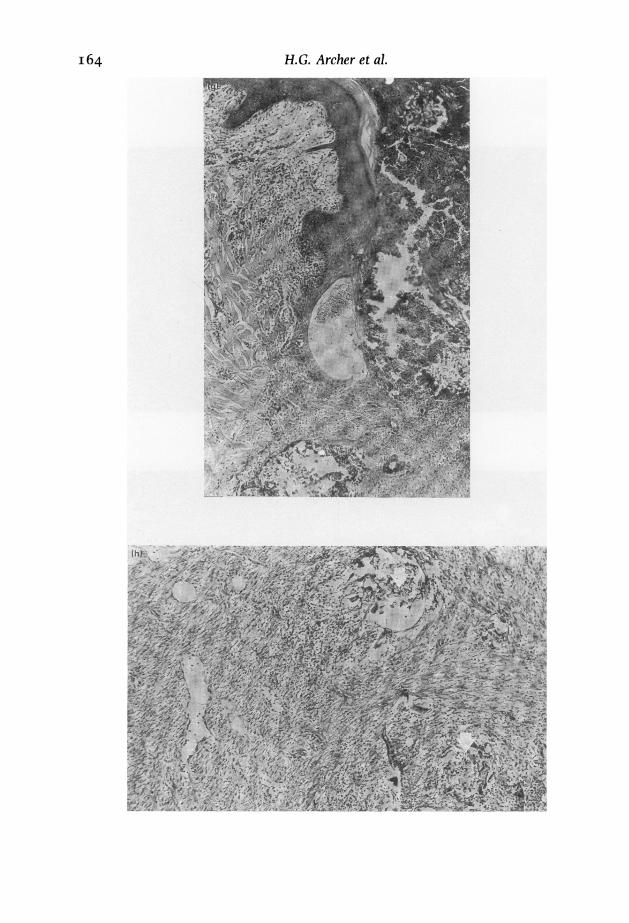
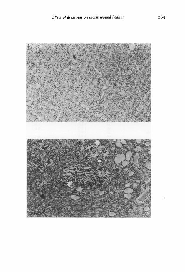
Fig. 4. Histology of pig wounds. x 85. a, OpSite control; epidermal migration over granulation tissue; b,OpSite control; connective tissue repair; c, sugar paste; epidermal migration over granulation tissue; d,sugar paste; connective tissue repair; e, Irgasan diluent; immature granulation tissue around 43, cottonfibres; f, Irgasan 0.2%; immature granulation tissue around &, cotton fibres; g, chlorhexidine 0.2%; lackofepidermal migration at edge ofwound; h, chlorhexidine 0.2%; immature granulation tissue and lack ofnew vessel formation; i, Povidone iodine o.8%; maturing granulation tissue; j, EUSOL (half strength);disorganized granulation tissue around 4,, trapped cotton fibres.
i62 H.G. Archer et al.
Effect of dressings on moist wound healing I63
i64 H.G. Archer et al.
EJfect of dressings on moist wound healing I65
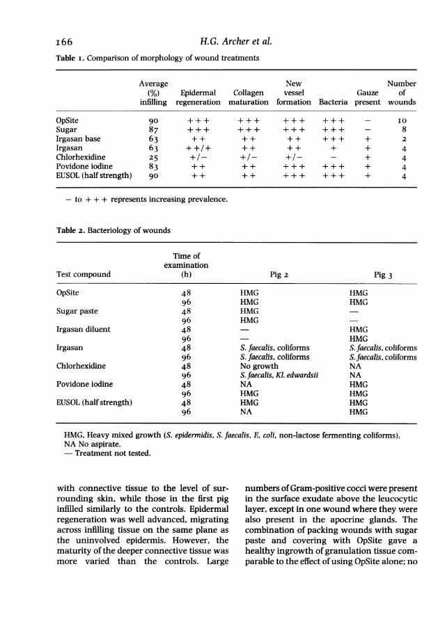
Table i. Comparison of morphology of wound treatments
Average New Number(%) Epidermal Collagen vessel Gauze of
infilling regeneration maturation formation Bacteria present wounds
OpSite go +++ +++ +++ +++ - IOSugar 87 +++ +++ +++ +++ - 8Irgasan base 63 + + + + + + + + + + 2Irgasan 63 + +/+ + + + + + + 4Chlorhexidine 25 +/- +/- +/- - + 4Povidone iodine 83 + + + + ++ + +++ + 4EUSOL (half strength) go + + ++ ++ + +++ + 4
- to + + + represents increasing prevalence.
Table 2. Bacteriology of wounds
Time ofexamination
Test compound (h) Pig 2 Pig 3
OpSite 48 HMG HMG96 HMG HMG
Sugar paste 48 HMG96 HMG
Irgasan diluent 48 - HMG96 HMG
Irgasan 48 S. faecalis, coliforms S. faecalis, coliforms96 S. faecalis, coliforms S. faecalis, coliforms
Chlorhexidine 48 No growth NA96 S.faecalis, Ki. edwardsii NA
Povidone iodine 48 NA HMG96 HMG HMG
EUSOL (half strength) 48 HMG HMG96 NA HMG
HMG, Heavy mixed growth (S. epidermidis, S. faecalis, E, coli, non-lactose fermenting coliforms),NA No aspirate.- Treatment not tested.
with connective tissue to the level of sur-rounding skin, while those in the first piginfilled similarly to the controls. Epidermalregeneration was well advanced, migratingacross infilling tissue on the same plane asthe uninvolved epidermis. However, thematurity of the deeper connective tissue wasmore varied than the controls. Large
numbers ofGram-positive cocci were presentin the surface exudate above the leucocyticlayer, except in one wound where they werealso present in the apocrine glands. Thecombination of packing wounds with sugarpaste and covering with OpSite gave ahealthy ingrowth of granulation tissue com-parable to the effect of using OpSite alone; no
I66 H.G. Archer et a].
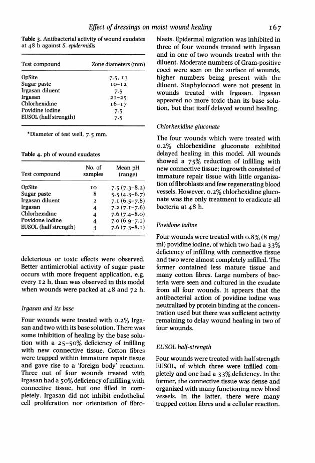
Effect of dressings on moist wound healingTable 3. Antibacterial activity of wound exudatesat 48 h against S. epidermidis
Test compound Zone diameters (mm)
OpSite 7.5, 13Sugar paste IO-I2Irgasan diluent 7.5Irgasan 2I-25Chlorhexidine i6-I7Povidine iodine 7.5EUSOL (half strength) 7.5
* Diameter of test well, 7.5 mm.
Table 4. ph of wound exudates
No. of Mean pHTest compound samples (range)
OpSite IO 7.5 (7.3-8.2)Sugar paste 8 5.5 (4.3-6.7)Irgasan diluent 2 7. I (6.5-7.8)Irgasan 4 7.2 (7.1-7.6)Chlorhexidine 4 7.6 (7.4-8.o)Povidone iodine 4 7.0 (6*9-7-1)EUSOL (half strength) 3 7.6 (7.3-8.i)
deleterious or toxic effects were observed.Better antimicrobial activity of sugar pasteoccurs with more frequent application, e.g.every 12 h, than was observed in this modelwhen wounds were packed at 48 and 72 h.
Irgasan and its base
Four wounds were treated with 0.2% Irga-san and two with its base solution. There wassome inhibition of healing by the base solu-tion with a 25-50% deficiency of infillingwith new connective tissue. Cotton fibreswere trapped within immature repair tissueand gave rise to a 'foreign body' reaction.Three out of four wounds treated withIrgasan had a 5o% deficiency of infilling withconnective tissue, but one filled in com-
pletely. Irgasan did not inhibit endothelialcell proliferation nor orientation of fibro-
blasts. Epidermal migration was inhibited inthree of four wounds treated with Irgasanand in one of two wounds treated with thediluent. Moderate numbers of Gram-positivecocci were seen on the surface of wounds,higher numbers being present with thediluent. Staphylococci were not present inwounds treated with Irgasan. Irgasanappeared no more toxic than its base solu-tion, but that itself delayed wound healing.
Chlorhexidine gluconateThe four wounds which were treated with0.2% chlorhexidine gluconate exhibiteddelayed healing in this model. All woundsshowed a 75% reduction of infilling withnew connective tissue; ingrowth consisted ofimmature repair tissue with little organiza-tion of fibroblasts and few regenerating bloodvessels. However, 0.2% chlorhexidine gluco-nate was the only treatment to eradicate allbacteria at 48 h.
Povidone iodine
Four wounds were treated with o.8% (8 mg/ml) povidine iodine, of which two had a 33%deficiency of infilling with connective tissueand two were almost completely infilled. Theformer contained less mature tissue andmany cotton fibres. Large numbers of bac-teria were seen and cultured in the exudatefrom all four wounds. It appears that theantibacterial action of povidine iodine wasneutralized by protein binding at the concen-tration used but there was sufficient activityremaining to delay wound healing in two offour wounds.
EUSOL half-strength
Four wounds were treated with half strengthEUSOL, of which three were infilled com-pletely and one had a 33% deficiency. In theformer, the connective tissue was dense andorganized with many functioning new bloodvessels. In the latter, there were manytrapped cotton fibres and a cellular reaction.
I67
H.G. Archer et al.
Epidermal migration had occurred in all fourwounds. Gram-positive cocci were seen inlarge numbers in three out of four woundsbut had not penetrated the granulationtissue.
DiscussionThe use of Stomahesive rings to enclosewound treatments and prevent cross-over
was satisfactory. The rings were easy to use
and stayed firmly adherent to the skin for 7days despite new hair growth; they providedadequate wound protection. Their use inconjunction with OpSite gave a 'woundchamber' enabling the collection of exudateand easy observation. Unfortunately, therings may have produced an artefact becausethe adhesive became softened by body heatand a small amount of gum leaked into allwounds. The principal component of theadhesive is karaya gum, which has beenseparately studied for wound healing proper-ties (Lowthian & Barnett I985). This effectwas, however, common to all wounds anddid not seem to alter reproducibility ofresults. All wounds other than those treatedwith chlorhexidine gluconate were colonizedwith bacteria, mostly Escherichia coli, Strepto-coccus faecalis and S. epidermidis.
It was appreciated that the use of differentmedicaments on one animal could lead topractical difficulties in this trial. The design ofthe experiment was to minimize these prob-lems given that there were insufficient fundsto increase the number of pigs used. Allhistological assessments were made 'blind',the treatments being coded throughout theexperiment. The results indicated that thedesign was satisfactory since a differencebetween treatments has been shown and theresults obtained for OpSite controls andsugar paste from different animals were
similar. With only four replicate woundsavailable for each antiseptic treatment, andthe use of one concentration which couldonly be applied on a 48-h dressing changebasis, the limitations of the experiment mustbe considered together with our quantitativeassessment on healing at 7 days.
This controlled study was set up to com-pare packing ofwounds with sugar paste andwith various antiseptics held in position ongauze. In each case, bundles of gauze fibreswere incorporated into new connective tis-sue and caused a 'foreign body' reaction.This delayed and disrupted the natural heal-ing process as studied under OpSite film. Ifthe gauze had been changed, as in clinicalpractice, most of the new connective tissuewould have been removed with it. Suchadhesion has been studied in a controlledmodel of partial thickness wounds in the pig(Varley & Barnett I986). No such adhesiveeffect occurred with wounds packed withsugar paste, which can be directly comparedwithin this model, and new connective tissuewas laid down without disruption and delay.A similar quantity of connective tissue wasproduced within the experimental sugarwound as when it was covered with OpSitefilm alone. All antiseptic treatments showedsome delay of healing.
Chlorhexidine caused the greatest delay ofwound healing in this model, and hadsimilar effects in a study using guinea-pigsexposed to 0.5% chlorhexidine gluconatesolution (Niedner & Schopf I 986). Irrigationof gingival wounds in rats with 0.5% chlor-hexidine retarded healing whereas o. I% and0.2% were comparable with saline (Kallen-berger 19 79). In a model of corneal abrasionin rabbits, irrigation with concentrations of2.0 and 4.0% chlorhexidine significantlyslowed the healing rate (re-epithelialization)but concentrations of i.o% did not (BowesHamill et al. I984). The manufacturer'srecommended concentration for wound irri-gation with chlorhexidine gluconate is0.05%; this level was shown not to delaywound healing in rats in conditions wheni.o% chloramine significantly delayed colla-gen production (Brennan et al. I986). Simi-larly, in an artificially infected wound modelin guinea-pigs, 0.05% chlorhexidine gluco-nate irrigation totally prevented sepsis anddid not delay wound healing (Platt & Buck-nall I984).
In this study we noted impairment of
I68
Effect of dressings on moist wound healing I69wound healing by a solution containing 8mg/ml povidone iodine. Connolly and Gil-more (1979) reported that 75 jug/ml povi-done iodine fully inhibited (and io jug/mlpartially) the chemotactic movement ofpoly-morphonuclear leucocytes, essential in theearly stages of wound healing. It may beexpected that io% (ioo mg/ml) povidoneiodine, the strength recommended for use inclinical practice, would have a serious dele-terious effect if applied frequently.Brennan and Leaper (I985) perfused the
rabbit ear chamber with full-strength EUSOLand noted a stagnation of blood flow in finecapillaries within seconds. Cessation ofbloodflow followed within minutes and they noteda subsequent delay in the repair process. Apilot animal study (unpublished data) hasbeen performed in which ribbon gauzesoaked in half-strength EUSOL and paraffinwas packed into full thickness wounds in thepig and covered with Gamgee and Microporetape; after 5 days, examination by histologyshowed little infilling of the wound cavity byconnective repair tissue. The results of thepresent study, which showed minimaldelayed healing with half-strength EUSOL,may have been different had more frequentpacking been possible which would havemaintained higher levels of free chlorine.We have found that wounds healed satis-
factorily without the need for sterility. Col-onization of their surface with bacteria didnot impair the formation of collagen tissuenor epidermal migration. The situation in aninfected wound or abscess cavity, where pusis present, is different, or when there iscellulitis around the wound. In these circum-stances, wound healing can be delayed bythe infective process and it may be moreimportant to inhibit bacterial growth even ifcollagen formation is delayed.
There is a growing body of evidence tosuggest that a low pH on the surface of awound will encourage the healing process.In a double blind study, Kaufman et al.(I985) applied buffered solutions to experi-mental second-degree burns in guinea-pigsand noted significantly increased re-epithe-
lialization in wounds treated with a pH 3.5solution compared with wounds treated withneutral and alkaline solutions. Leveen et al.(I973) also demonstrated increased rates ofhealing at acid pH values. Our observationsof low pH values in sugar paste treatedwounds are intriguing but without furtherstudies are of unknown significance.
This type of controlled study of moistwound healing, using the pig as opposed toother animal species where the dermis isdifferent (Gangjee et al. I985), is thoughtsuitable for studying other products to ascer-tain whether they are equivalent, better orworse than OpSite film alone, which can beconsidered as the standard control treat-ment.The findings of this study clearly demon-
strate that our sugar paste does not impairthe wound healing process as shown bychlorhexidine gluconate 0.2% and Irgasan0.2%. There is currently a move away fromthe regular treatment of wounds with anti-septics and our data supports the notion thatsuch treatments can be harmful and possiblycounter productive. Sugar paste has nowbeen used on many patients with infectedand malodorous wounds (Middleton & SealI985, I989) with excellent results and thedata presented here indicates that sugarpaste may be the treatment of choice forwounds that are traditionally treated withantiseptics.
AcknowledgementsWe are very grateful for advice and assis-tance from Professor J.T. Scales, OBE, Honor-ary Director, Department of Research inPlastic Surgery, Mount Vernon Hospital, toGary Batchelor and Nicolas Jones for caringfor the animals and administering the anaes-thetic, and to Mrs Angela Matthews fortyping the manuscript.
ReferencesALPER J.C. (I986) Recent advances in moistwound healing. South. Med. J. 79, I398-I404.
I 70 H.G. Archer et al.ALVAREZ O.M., MERTZ P.M. & EAGLSTEIN W.H.
(I983) The effect of occlusive dressings oncollagen synthesis and re-epithelialisation insuperficial wounds. J. Surg. Res. 35, 142-148.
BARNETT S.E. & VARLEY S.J. (I987) The effects ofcalcium alginate on wound healing. Ann. R.Coll. Surg. Engl. 69, I53-155.
BoWEs HAMILL M., OSATO M.S. & WILHELMUS K.R.(I984) Experimental evaluation of chlorhexi-dine gluconate for ocular antisepsis. Anti-microb. Agents Chemother. 26, 793-796.
BRENNAN S.S., FOSTER M.E. & LEAPER D.J. (I986)Antiseptic toxicity in wounds healing bysecondary intention. J. Hosp. Inf. 8, 263-267.
BRENNAN S.S. & LEAPER D.J. (I985) The effect ofantiseptics on the healing wound: a study usingthe rabbit ear chamber. Br. J. Surg. 72, 780-782.
CONNOLLY J.C. & GILMORE O.J.A. (I 9 79) A study ofthe effect of povidone-iodine on polymorpho-nuclear leucocyte chemotaxis. Br. J. Exp. Path.6o, 662-666.
GANGJEE T., COLAIzzo R. & VON REcUM A.F. (I985)Species-related differences in percutaneouswound healing. Ann. Biomed. Engineer. 13,45 I-467.
HARTWELL S.W. (I955) A practical approach tothe consideration of human wounds and theirrepair. In The Mechanism of Healing of HumanWounds. Eds M.E. DeBakey & R.G. Spurling.Charles C. Thomas, Springfield, Illinois. pp. 2-6.
KALLENBERGER A. (I 9 79) Experimental study ofthe histocompatibility of disinfectant solutions.Aktuelle Probi. Chir. Orthoped. I2, 87-96.
KAUFMAN T., EICHENLAUB E.H., ANGEL M.F., LEVINM., FUTRELL J.W. (I985) Topical acidificationpromotes healing of experimental deep partialthickness skin burns: a randomized double-blind preliminary study. Burns 12, 84-90.
KICKHOFEN B., WOKALEK H., SCHEEL D. & RUH H.(I986) Chemical and physical properties of ahydrogel wound dressing. Biomaterials 7, 67-72.
LAUFER M., ASHKENAZI C., KATZ D. & WOLMAN M.(I974) Orientation of collagen in wound heal-ing. Br. I. Exp. Path. 55, 233-236.
LEVEEN H.H., FALK G., BOREK B., DIAZ C., LYNFIELDY., WYNKOOP B.J., MABUNDA G.A., RuBRIcus J.L.& CHRISTOUDIAs G.C. (I973) Chemical acidifica-tion of wounds-an adjuvant to healing and theunfavorable action of alkalinity and ammonia.Ann. Surg. 178, 745-53.
LOWTHIAN P. & BARNETT S. (I985) Sterculia forwound healing. Lancet ii, ii86.
MIDDLETON K. & SEAL D.V. (I985) Sugar as an aidto wound healing. Pharm. J. 235, 75 7-758.
MIDDLETON K. & SEAL D.V. (I989) Development ofa semi-synthetic sugar paste for promotinghealing of infected wounds. In Pathogenesis ofWound and Biomaterial-Associated Infections. EdsI. Eliasson & T. Wadstrom. London: Springer-Verlag (in press).
NIEDNER R. & SCHOPF E. (I986) Inhibition ofwound healing by antiseptics. Br. J. Dermatol.115 (SUPPI. 31I), 4I-44.
PLATT J. & BUCKNALL R.A. (I 984) An experimen-tal evaluation of antiseptic wound irrigation. J.Hosp. Inf. 5, i8i-i88.
RICHMOND J.D. & SUTHERLAND A.B. (1986) A newapproach to the problems encountered withOpSite as a donor site dressing: systemic etham-sylate. Br. J. Plast. Surg. 39, 5i6-5i8.
VARLEY S.J. & BARNETT S.E. (I986) A study ofwound dressing adhesion. Clin. Mat. i, 3 7-5 7.
WINTER G.D. (I962) Formation of the scab andthe rate of epithelialisation of superficialwounds in the skin and the young domestic pig.Nature 193, 293-294.
WINTER G.D. (I966) A study ofwound healing inthe domestic pig. PhD Thesis, University ofLondon.
WINTER G.D. & SCALES J.T. (I963) Effect of airdrying and dressings on the surface of a wound.Nature 197, 91-92.