association study and expression analysis of porcine esr1 as a candidate gene for boar fertility and...
TRANSCRIPT
GA
Ac
AMCa
b
a
ARRAA
KmISES
1
ih
k((((
0d
ARTICLE IN PRESS ModelNIREP-4420; No. of Pages 11
Animal Reproduction Science xxx (2011) xxx– xxx
Contents lists available at SciVerse ScienceDirect
Animal Reproduction Science
journal homepage: www.elsevier.com/locate/anireprosci
ssociation study and expression analysis of porcine ESR1 as aandidate gene for boar fertility and sperm quality
sep Gunawana, Kanokwan Kaewmalaa, Muhammad Jasim Uddina,ehmet Ulas Cinara, Dawit Tesfayea, Chirawath Phatsaraa,b, Ernst Tholena,
hristian Loofta, Karl Schellandera,∗
Institute of Animal Science, Animal Breeding and Husbandry Group, University of Bonn, 53115 Bonn, GermanyDepartment of Animal and Aquatic Sciences, Faculty of Agriculture, Chiang Mai University, 50200 Chiang Mai, Thailand
r t i c l e i n f o
rticle history:eceived 13 May 2011eceived in revised form 12 August 2011ccepted 24 August 2011vailable online xxx
eywords:RNA
mmunofluorescencepermatogenesispididymispermatozoa
a b s t r a c t
Male fertility is impaired through the lack of ESR1 (Estrogen Receptor 1) but little is knownabout the ESR1 roles in boar spermatogenesis and fertility. Therefore, this research wasaimed at investigating the association with sperm quality and boar fertility traits in a totalof 300 boars both from purebred Pietrain and Pietrain × Hampshire crosses. A SNP in codingregion of ESR1 g.672C > T in exon 1 was associated with sperm motility (P < 0.05) and plasmadroplet rate (P < 0.01) while the polymorphism in non-coding region of ESR1 g.35756T > C ininton 1 was associated with non-return rate (P < 0.05). Furthermore, to analyse the mRNAand protein expression of ESR1 in boar reproductive tissues, a total of six boars were dividedinto two groups [Group I (G-I) and Group II (G-II)], where G-I had relatively better spermquality. ESR1 expression was higher in tissues collected from G-I boars than those of col-lected from G-II boars, and the difference in mRNA expression was significant (P < 0.01)in head of epididymis. The ESR1 protein expression results from western blot coincidedwith the results of qRT-PCR. The ESR1 protein localization observed a strong staining in the
cytoplasm of Sertoli cell in the testis, in the epithelial cells in head and tail of epididymis,in smooth muscle in tail of epididymis, and in the post acrosomal region and tail of thespermatozoa. These results will improve the understanding of the functions of the ESR1 inspermatogenesis within the reproductive tract and will shed light on ESR1 as a candidatein the selection of boar with good sperm quality and fertility.. Introduction
Please cite this article in press as: Gunawan, A., et al., Associacandidate gene for boar fertility and sperm quality. Anim. Repro
Despite being the ‘female’ hormone, estrogen is presentn low concentrations in male blood and in extraordinarilyigh concentration in semen (Ganjam and Amann, 1976).
∗ Corresponding author. Tel.: +49 228 732240; fax: +49 228 732284.E-mail addresses: [email protected] (A. Gunawan),
[email protected] (K. Kaewmala), [email protected]. Uddin), [email protected] (M.U. Cinar), [email protected]. Tesfaye), [email protected] (C. Phatsara), [email protected]. Tholen), [email protected] (C. Looft), [email protected]. Schellander).
378-4320/$ – see front matter © 2011 Elsevier B.V. All rights reserved.oi:10.1016/j.anireprosci.2011.08.008
© 2011 Elsevier B.V. All rights reserved.
The hormone estrogen is mediated through the nuclearestrogen receptor which functions as ligand-dependanttranscription factor. The increasing interest in the roleof estrogen in the male reproductive tract is mainly dueto the demonstration that male fertility is impaired inmice lacking estrogen receptor 1 (ESR1) (Eddy et al.,1996). The evidence for ESR1 in different parts of themale reproductive tract suggests a possible physiologi-cal role in spermatogenesis and sperm maturation. The
tion study and expression analysis of porcine ESR1 as ad. Sci. (2011), doi:10.1016/j.anireprosci.2011.08.008
ESR1 knockout mice have provided evidence for a signif-icant and crucial role of estrogens in maintaining normalspermatogenesis (Eddy et al., 1996). ESR1 is involved inthe reabsorption of luminal fluid during the transit of
ING Model
roductio
30 s at 56 ◦C. After checking the PCR products in 1.5% (w/v)agarose gels, genotyping was done following the restric-
ARTICLEANIREP-4420; No. of Pages 11
2 A. Gunawan et al. / Animal Rep
spermatozoa from the testis to the head of the epididymiswhich is important for their survival and maturation dur-ing epididymal storage (Couse and Korach, 1999). Theabsence of ESR1 leads to reduced epididymal sperm con-tent, reduced sperm motility and fertilizing ability (Eddyet al., 1996). Several studies have shown a significant asso-ciation of ESR1 with semen quality or fertility in humans(Suzuki et al., 2002; Guarducci et al., 2006; Lazaros et al.,2010; Safarinejad et al., 2010). Guarducci et al. (2006)reported a significant association of ESR1 polymorphismwith lower sperm count in men. Suzuki et al. (2002)reported two silent polymorphisms in ESR1 being associ-ated with azoospermia or severe oligozoospermia in men.To our knowledge, only one study was devoted to anal-yse the association between ESR1 polymorphism with littersize in sows but this failed to detect any statistically sig-nificant association (Munoz et al., 2007). The ESR1 mRNAwas reported to be highly expressed in the epididymis,testis, pituitary, uterus, kidney and adrenal gland in rats(Kuiper et al., 1996). ESR1 was localized in head, bodyand tail of epididymis in the bonnet monkey (Shayu et al.,2004), cat (Nie et al., 2002) and horse (Hejmej et al., 2005;Parlevliet et al., 2006). In pigs, ESR1 is located on SSC1p24-25, which was evidenced as the QTL region for total spermper ejaculate and close to the QTL for sperm motility(Xing et al., 2008). Therefore, ESR1 could be a functional aswell as a positional candidate gene for male reproductionin pigs. But no study has yet been devoted to unravel-ling its association with boar sperm quality and fertilitytraits.
In spermatogenesis, beside the reproductive tissuestestis and epididymis, some other accessory glands andtissues are also involved. Spermatogenesis is the complexprocess by which immature germ cells undergo division,differentiation and meiosis to give rise to haploid elon-gated spermatids (O’Donnell et al., 2001). When germcell development is complete, the mature spermatids arereleased from the Sertoli cells into the tubule lumen andproceed through the excurrent duct system, known as therete testis, until they enter the epididymis via the effer-ent ducts. During passage through the epididymis, thespermatids undergo a series of biochemical changes tobecome the motile and mature spermatozoa capable of fer-tilization (O’Donnell et al., 2001). Failure in any of theseevents leads to disturbances of male fertility. The roleof ESR1 in boar spermatogenesis within the reproductivetract is poorly understood. For the better understandingof ESR1 roles in spermatogenesis in pigs, the expressionof ESR1 at different parts of reproductive tract, includ-ing non-reproductive tissues, are important. Therefore,the aims of this study were to investigate the associa-tion of ESR1 polymorphism with sperm quality and boarfertility traits as well as to highlight the roles of ESR1in boar spermatogenesis within the reproductive tractby mRNA and protein expression and immunoreactiveESR1 localization. This work might be helpful to get fur-ther insight into the roles of ESR1 in spermatogenesisin the boar. The association of ESR1 polymorphism withboar sperm quality and fertility will support the candi-
Please cite this article in press as: Gunawan, A., et al., Associacandidate gene for boar fertility and sperm quality. Anim. Repro
dacy of this gene to be used in selection of breedingboars.
PRESSn Science xxx (2011) xxx– xxx
2. Materials and methods
2.1. Phenotypes
Samples and phenotypes from 200 purebred Pietrain(PI) and 100 Pietrain × Hampshire crossbred (PI × HA)boars were used for association analysis in this study.These animals were used for AI in commercial pig herdsin North-Western Germany. Details of the populations andphenotypes were described previously by Wimmers et al.(2005), Lin et al (2006) and Kaewmala et al. (2011). Inbrief, sperm samples from more than 39,000 ejaculateswere repeatedly collected from these boars. Whole ejacu-lates were obtained from purebred Pietrain and crossbredPietrain × Hampshire boars age between two and five yearswith an average age of 3.5 years. Sperm quality traitsincluded sperm concentration (SCON [×108 ml]), semenvolume per ejaculate (VOL [ml]), sperm motility (MOT [%]),plasma droplets rate (PDR [%]) and abnormal spermato-zoa rate (ASR [%]) and were obtained from each ejaculatewith light microscopy according to the guidelines of theWorld Health Organization. Semen was collected by thevinyl glove hand method twice per week. For each boar,the repeated measurements of sperm quality traits wereavailable. Fertility data (non-return rate data [NRR] at 42days after insemination [%] and number of piglet born alive[NBA] per litter) of each boar were available as the devia-tion from the population means within sow breed, parity ofsow, farm and season classes as described earlier by Lin et al(2006) and Kaewmala et al. (2011). The general descriptionof sperm quality traits and boar fertility traits are shown inTable 1.
2.2. Polymorphism study
Two single nucleotide polymorphisms detected byMunoz et al. (2007) were used in this study: a cyto-sine (C) transversion to a thymine at g.672C > T in exon1 and a thymine (T) to a cytosine (C) transversion atg.35756T > C in intron 1 of ESR1. For PCR amplification,the forward (5′-gttcaaatccctggttgcat-3′) and reverse (5′-ctaggcgtctccccagattag-3′) primers were designed from theexon 1 and the forward (5′-gacagcttccctgcagattc-3′) andreverse (5′-ttcatcatgcccacttcgta-3′) primers were designedfrom the intron 1 of porcine ESR1 genomic sequence(GenBank accession No. ENSSSCG00000004083) using thePrimer3 tool (Rozen and Skaletsky, 2000). Polymerasechain reactions (PCR) were performed in a 20 �l volumecontaining 100 ng of porcine genomic DNA, 1 × PCR buffer(with 1.5 134 mM MgCl2), 0.25 mM of each dNTP, 5 pmol ofeach primer and 0.1 U of Taq DNA polymerase (GeneCraft).The PCR reaction was performed under the following con-ditions: initial denaturation at 95 ◦C for 5 min followed by35 cycles of 30 s at 95 ◦C, 30 s at 52 ◦C, 30 s at 72 ◦C andfinal elongation of 10 min at 72 ◦C for the polymorphismin exon 1. The PCR conditions were the same for the poly-morphism in intron 1 except the annealing temperature of
tion study and expression analysis of porcine ESR1 as ad. Sci. (2011), doi:10.1016/j.anireprosci.2011.08.008
tion fragment length polymorphisms (RFLPs) analysis. Thedigestion of restriction enzyme was done using BstNI and
ARTICLE IN PRESSG Model
ANIREP-4420; No. of Pages 11
A. Gunawan et al. / Animal Reproduction Science xxx (2011) xxx– xxx 3
Table 1Means, standard deviation (S.D.), sample size, ranges of traits in Pietrain and Pietrain × Hampshire.
Population Traits Sample size Mean S.D. Minimum Maximum
Pietrain (PI) SCON (108/ml) 29,161 3.03 0.94 1 6VOL (ml) 30,772 237.03 57.32 25 522MOT (%) 30,118 84.72 4.37 65 95PDR (%) 30,239 5.41 3.33 0 15ASR (%) 30,528 6.53 4.18 0 20NRR42 (%)a 200 0.28 7.06 −24.07 17.68NBA (per litter)a 200 0.02 0.57 −1.69 1.37
Pietrain × Hampshire (PIHA) SCON (108/ml) 9123 2.95 0.97 1 6VOL (ml) 9673 297.50 81.62 56 546MOT (%) 9395 85.46 4.03 70 95PDR (%) 9409 5.76 3.14 0 15ASR (%) 9543 4.95 4.00 0 20NRR42 (%)a 100 0.97 4.18 −12.23 13.79NBA (per litter)a 100 0.05 0.52 −2.97 0.83
PI and PIHA SCON (108/ml) 38,284 3.00 0.95 1 6VOL (ml) 40,445 256.87 75.54 25 546MOT (%) 39,513 85.01 4.37 65 95PDR (%) 39,648 5.51 3.28 0 15ASR (%) 40,071 5.99 4.26 0 20
a 00
d breed.
ATtg3a
2
wSa
y
wPfisfffienε
2
tm
y
wt
mRNA from all 6 boars was pooled together according tothe tissues. On the other hand, the differential mRNA andprotein expression study in different reproductive tissuesfrom two divergent groups of animals was performed by
Table 2Means, standard deviation (S.D.), number of boars and ranges of traitsselected for mRNA and protein expression study.
Traits Selectedanimals(n = 6)
G-I(n = 3)
G-II(n = 3)
NRR42 (%) 30NBA (per litter)a 30
a Fertility (NRR42, NBA) corrected with factors: parity, farm, season an
luI for g.672C > T and g.35756T > C, respectively (BioLabs).he digestion was carried out in 10 �l of reaction mix-ure of each sample and incubated overnight at 37 ◦C for.35756T > C and 65 ◦C for g.672C > T. Detection of RFLPs of00 boars was carried out by electrophoresis in 3% (w/v)garose gels.
.3. Statistical analysis for sperm quality traits
The association of ESR1 with boar sperm quality traitsas analysed by variance analysis (PROC MIXED) using the
AS software package (ver. 9.2; SAS Institute Inc., Cary, USA)s described earlier by Kaewmala et al. (2011).
ijklm = � + breedi + seasonj + genotypek + agel
+ejaculationm + εijklm [Model 1]
here yijklm are the sperm quality traits (SCON, VOL, MOT,DR, ASR); � is the overall population mean; breedi is thexed effect of the i-th breed (i = PI; PIHA, PI and PIHA);easonj is the fixed effect of the j-th season (j = 1 through 8;our seasons per year, in total eight seasons within 2 yearsrom January 2000 to December 2001); genotypek is thexed effect of the k-th genotype (k = 1, 2 and 3); agel is theffect of boar age (covariable); ejaculationm is the perma-ent environmental effect of the m-th boar (random) andijklm is the residual error.
.4. Statistical analysis for fertility traits
The association analysis between ESR1 and the fertilityraits was carried out using the following generalized linear
odel (PROC GLM) in SAS (Kaewmala et al., 2011).
Please cite this article in press as: Gunawan, A., et al., Associacandidate gene for boar fertility and sperm quality. Anim. Repro
ijkl = � + breedi + genotypej + yeark + εijkl [Model 2]
here yijk is the boar fertility traits (NRR and NBA); � ishe overall population mean; breedi is the fixed effect of
0.41 6.23 −24.07 17.670.01 0.55 −2.97 1.37
the i-th breed (i = PI, PIHA, PI and PIHA); genotypej is thefixed effect of the j-th genotype (j = 1, 2 and 3); yeark is thefixed effect of the k-th boar year of birth (k = 1 through 3:boar born before 1996, in 1996–97 and in 1998–99), εijk isthe residual error.
A chi-square (�2) test was conducted to test the popula-tions for Hardy–Weinberg equilibrium. Least square meanvalues for the ESR1 genotypes were compared by t-test andP-values were adjusted by the Tukey–Kramer correction.
2.5. Selection of animals for mRNA and proteinexpression
The reproductive (testis, head, body and tail of epi-didymis, vas deferens, bulbourethral gland, vesicularglands and prostate gland), non-reproductive (brain, mus-cle and liver) tissues from 6 breeding boars with divergentphenotypes were collected from the AI station (SuisAG,Sempach, Switzerland) for mRNA and protein study. Fordifferential expression study between reproductive andnon-reproductive tissues by semi-quantitative PCR study,
tion study and expression analysis of porcine ESR1 as ad. Sci. (2011), doi:10.1016/j.anireprosci.2011.08.008
Mean S.D. Mean S.D. Mean S.D.
SCON (106/ml) 262.32 87.97 335.94 50.78 188.70 22.54SVOL (ml) 215.24 34.93 185.07 16.33 245.40 7.42SMOT (%) 76.59 3.71 79.03 1.89 74.14 3.60
ING Model
roductio
ARTICLEANIREP-4420; No. of Pages 11
4 A. Gunawan et al. / Animal Rep
semi-quantitative PCR, qRT-PCR and western blot, respec-tively. For these purposes, the 6 boars were divided intotwo groups: group I (G-I) and group II (G-II), each groupcontaining 3 boars on the basis of SCON, SVOL and SMOT(Table 2). The SCON (average sperm concentration) washighly negatively (r = −0.8) correlated with SVOL (aver-age semen volume), whereas SCON was highly positively(r = 0.7) correlated with SMOT (average sperm motility).Moreover, SVOL was highly negatively (r = −0.8) correlatedwith SMOT. For the animals of G-I the SCON was higherthan the mean (262.32 × 10⁶ ml), SMOT was higher thanthe mean (76.59%) and SVOL was lower than the mean(215.24 ml/ejaculation) of respective parameters. For theanimals of G-II these parameters were vice versa. The sig-nificant difference between the two groups was calculatedusing proc t-test in SAS. There were differences for SCON(P < 0.05) and for SVOL (P < 0.01) between G-I and G-II,whereas for the SMOT the difference was not significant(P = 0.12).
2.6. Semi-quantitative PCR
Total RNA was isolated using TRI reagent (Sigma) fromdifferent reproductive and non-reproductive tissues ofbreeding boars mentioned above (Section 2.5). RNA waspurified using RNeasy Mini Kit (Qiagen) according tothe manufacturer’s instructions. Total RNA was treatedusing on-column RNase-Free DNase set (Promega) andquantified sphectrophotometrically (Nano Drop, ND8000).Furthermore, RNA integrity was checked by 2% agarose gelelectrophoresis. First-strand cDNA were synthesized fromindividual RNA using Superscript II enzyme (Invitrogen).
cDNA amplification was performed by using specificforward and reverse primers (forward: 5′-agggagagg-agtttgtgtg-3′ and reverse: 5′-tctccagcagcaggtcatag-3′)derived from porcine ESR1 sequence (GenBank accessionAF035775). Amplification was performed with an initialheating at 95 ◦C for 5 min followed by 35 cycles of 95 ◦C for45 s, annealing temperature at 60 ◦C for 1 min and 72 ◦C for1 min, on the PCR Thermal Cycler (BioRad). PCR productswere electrophoresed on a 1.5% agarose gel and visualizedupon staining with ethidium bromide. Amplification ofGlyceraldehyde 3-phosphate dehydrogenase (GAPDH)served as housekeeping gene.
2.7. Quantitative Real-Time PCR (qRT-PCR)
For qRT-PCR, total RNA was isolated using TRI reagent(Sigma) from different reproductive tissues of the twodivergent groups of animals (G-I and G-II) as previouslydescribed (Section 2.5). Total RNA isolation and cDNAsynthesis was described in the previous section (Section2.6). The same primer pair used in semi-quantitative PCRwas also used in qRT-PCR. Nine-fold serial dilution ofplasmid DNA was prepared and used as a template forthe generation of the standard curve. In each run, the96-well microtiter plate contained each cDNA sample, plas-
Please cite this article in press as: Gunawan, A., et al., Associacandidate gene for boar fertility and sperm quality. Anim. Repro
mid standards for the standard curves and a no-templatecontrol. To ensure repeatability of the experiments, eachsample was run in three replications. qRT-PCR was setup using 2 �l first-strand cDNA template, 7.6 �l deionized
PRESSn Science xxx (2011) xxx– xxx
H2O, 0.2 �M of upstream and downstream primers and10 �l 1× Power SYBR Green I master mix with ROX asreference dye (BioRad). The thermal cycling conditionswere 3 min at 94 ◦C followed by 40 cycles of 20 s at 94 ◦Cand 1 min at 60 ◦C. Experiments were performed usingthe ABI prism®7000 (Applied Biosystems) qRT-PCR system.An amplification-based threshold and adaptive baselinewere selected as algorithms. The housekeeping geneGAPDH (forward: 5′-acccagaagactgtggatgg-3′ and reverse:5′-acgcctgcttcaccaccttc-3′) derived from porcine sequence(GenBank accession No. AF017079) was used for the datanormalization. Final results were reported as the relativeabundance level after normalizing with mRNA expressionlevel of the housekeeping gene. Differences in ESR1 mRNAexpression were analysed with the simple t-test in SAS soft-ware (SAS Institute Inc., ver. 9.2). Values of P < 0.05 wereconsidered to indicate statistically significant differences.
2.8. Western blotting
The protein was extracted from different reproductivetissues (testis, head, body and tail of epididymis) from thetwo divergent groups of breeding boars as used in qRT-PCR.However, for western blot study, proteins from the three G-I boars were pooled together and proteins from the threeG-II boars were pooled together according to the tissues.The protein extracted from tissues was separated by SDS-PAGE (gradient 4–18%). Subsequently the proteins weretransferred onto a nitrocellulose membrane (AmershamBiosciences). After blocking in blocking buffer (20 mM TrispH 7.5, 150 mM NaCl, 0.05% Tween-20 and 1% Polyvinylpy-rolidone) at room temperature for 1 h, the membrane wasincubated with the anti-ESR1 antibody purified from rabbitpolyclonal antibody (Cat.nr. 543; Santa Cruz) in the block-ing medium (diluted 1:500) overnight at 4 ◦C. Non-specificbinding of antibody was washed off with six changes of0.1% PBST (10 min to time). The horseradish peroxidaseconjugated goat anti-rabbit IgG secondary antibody (Cat.nr.Sc2004; Santa Cruz) was used as the secondary antibody(diluted 1:5000). The membrane was incubated for 1 hat room temperature with secondary antibody, followedby washing with six changes of 0.1% PBST (10 min totime). The chemiluminesce was detected by using the ECLplus western blotting detection system (Amersham Bio-sciences) and was visualized by using Kodak BioMax XARfilm (Kodak). GAPDH was used as a loading control and fornormalization. The membrane was stripped by incubationin 2% SDS, 100 mM Tris–HCl, 0.1% �-mercaptoethanol for30 min at 60 ◦C and re-probed with GAPDH antibody (Cat.nr.Sc20357; Santa Cruz).
2.9. Protein localization by immunofluorescence
Due to the limitations of fresh samples from G-Iand G-II boars, we collected different fresh reproduc-tive tissues were collected from a healthy breeding boarafter slaughtering for protein localization by immunofluo-
tion study and expression analysis of porcine ESR1 as ad. Sci. (2011), doi:10.1016/j.anireprosci.2011.08.008
rosence. Immunofluorescence staining was performed on8 �m cryostat sections of snap frozen tissues. All sectionswere kept in −80 ◦C for further analysis. To block unspe-cific staining, sections were incubated for 30 min at room
ARTICLE IN PRESSG Model
ANIREP-4420; No. of Pages 11
A. Gunawan et al. / Animal Reproduction Science xxx (2011) xxx– xxx 5
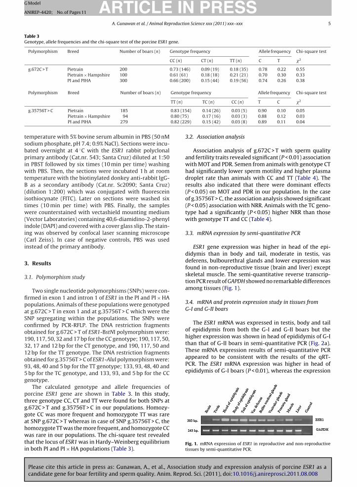
Table 3Genotype, allele frequencies and the chi-square test of the porcine ESR1 gene.
Polymorphism Breed Number of boars (n) Genotype frequency Allele frequency Chi-square test
CC (n) CT (n) TT (n) C T �2
g.672C > T Pietrain 200 0.73 (146) 0.09 (19) 0.18 (35) 0.78 0.22 0.55Pietrain × Hampshire 100 0.61 (61) 0.18 (18) 0.21 (21) 0.70 0.30 0.33PI and PIHA 300 0.66 (200) 0.15 (44) 0.19 (56) 0.74 0.26 0.38
Polymorphism Breed Number of boars (n) Genotype frequency Allele frequency Chi-square test
TT (n) TC (n) CC (n) T C �2
0.83 (150.80 (750.82 (22
tsbpiwtB(itw(ii(i
3
3
fipaSco131o95g
ptggahwti
These mRNA expression results of semi-quantitative PCRappeared to be consistent with the results of the qRT-PCR. The ESR1 mRNA expression was higher in head ofepididymis of G-I boars (P < 0.01), whereas the expression
g.35756T > C Pietrain 185
Pietrain × Hampshire 94
PI and PIHA 279
emperature with 5% bovine serum albumin in PBS (50 nModium phosphate, pH 7.4; 0.9% NaCl). Sections were incu-ated overnight at 4 ◦C with the ESR1 rabbit polyclonalrimary antibody (Cat.nr. 543; Santa Cruz) diluted at 1:50
n PBST followed by six times (10 min per time) washingith PBS. Then, the sections were incubated 1 h at room
emperature with the biotinylated donkey anti-rabbit IgG- as a secondary antibody (Cat.nr. Sc2090; Santa Cruz)dilution 1:200) which was conjugated with fluoresceinsothiocynate (FITC). Later on sections were washed siximes (10 min per time) with PBS. Finally, the samplesere counterstained with vectashield mounting medium
Vector Laboratories) containing 40,6-diamidino-2-phenylndole (DAPI) and covered with a cover glass slip. The stain-ng was observed by confocal laser scanning microscopeCarl Zeiss). In case of negative controls, PBS was usednstead of the primary antibody.
. Results
.1. Polymorphism study
Two single nucleotide polymorphisms (SNPs) were con-rmed in exon 1 and intron 1 of ESR1 in the PI and PI × HAopulations. Animals of these populations were genotypedt g.672C > T in exon 1 and at g.35756T > C which were theNP segregating within the populations. The SNPs wereonfirmed by PCR-RFLP. The DNA restriction fragmentsbtained for g.672C > T of ESR1-BstNI polymorphism were:90, 117, 50, 32 and 17 bp for the CC genotype; 190, 117, 50,2, 17 and 12 bp for the CT genotype, and 190, 117, 50 and2 bp for the TT genotype. The DNA restriction fragmentsbtained for g.35756T > C of ESR1-AluI polymorphism were:3, 48, 40 and 5 bp for the TT genotype; 133, 93, 48, 40 and
bp for the TC genotype, and 133, 93, and 5 bp for the CCenotype.
The calculated genotype and allele frequencies oforcine ESR1 gene are shown in Table 3. In this study,hree genotype CC, CT and TT were found for both SNPs at.672C > T and g.35756T > C in our populations. Homozy-ote CC was more frequent and homozygote TT was rare
Please cite this article in press as: Gunawan, A., et al., Associacandidate gene for boar fertility and sperm quality. Anim. Repro
t SNP g.672C > T whereas in case of SNP g.35756T > C, theomozygote TT was the more frequent, and homozygote CCas rare in our populations. The chi-square test revealed
hat the locus of ESR1 was in Hardy–Weinberg equilibriumn both PI and PI × HA populations (Table 3).
4) 0.14 (26) 0.03 (5) 0.90 0.10 0.05) 0.17 (16) 0.03 (3) 0.88 0.12 0.039) 0.15 (42) 0.03 (8) 0.89 0.11 0.04
3.2. Association analysis
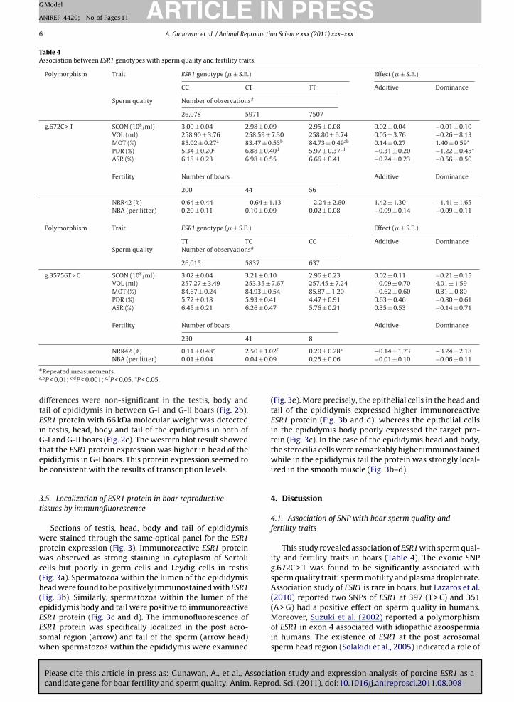
Association analysis of g.672C > T with sperm qualityand fertility traits revealed significant (P < 0.01) associationwith MOT and PDR. Semen from animals with genotype CThad significantly lower sperm motility and higher plasmadroplet rate than animals with CC and TT (Table 4). Theresults also indicated that there were dominant effects(P < 0.05) on MOT and PDR in our population. In the caseof g.35756T > C, the association analysis showed significant(P < 0.05) association with NRR. Animals with the TC geno-type had a significantly (P < 0.05) higher NRR than thosewith genotype TT and CC (Table 4).
3.3. mRNA expression by semi-quantitative PCR
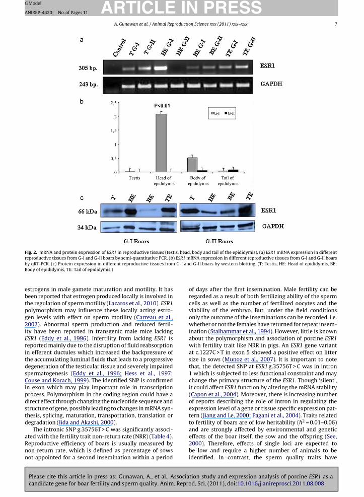
ESR1 gene expression was higher in head of the epi-didymis than in body and tail, moderate in testis, vasdeferens, bulbourethral glands and lower expression wasfound in non-reproductive tissue (brain and liver) exceptskeletal muscle. The semi-quantitative reverse transcrip-tion PCR result of GAPDH showed no remarkable differencesamong tissues (Fig. 1).
3.4. mRNA and protein expression study in tissues fromG-I and G-II boars
The ESR1 mRNA was expressed in testis, body and tailof epididymis from both the G-I and G-II boars but thehigher expression was shown in head of epididymis of G-Ithan that of G-II boars in semi-quantitative PCR (Fig. 2a).
tion study and expression analysis of porcine ESR1 as ad. Sci. (2011), doi:10.1016/j.anireprosci.2011.08.008
Fig. 1. mRNA expression of ESR1 in reproductive and non-reproductivetissues by semi-quantitative PCR.
ARTICLE IN PRESSG Model
ANIREP-4420; No. of Pages 11
6 A. Gunawan et al. / Animal Reproduction Science xxx (2011) xxx– xxx
Table 4Association between ESR1 genotypes with sperm quality and fertility traits.
Polymorphism Trait ESR1 genotype (� ± S.E.) Effect (� ± S.E.)
CC CT TT Additive Dominance
Sperm quality Number of observations#
26,078 5971 7507
g.672C > T SCON (108/ml) 3.00 ± 0.04 2.98 ± 0.09 2.95 ± 0.08 0.02 ± 0.04 −0.01 ± 0.10VOL (ml) 258.90 ± 3.76 258.59 ± 7.30 258.80 ± 6.74 0.05 ± 3.76 −0.26 ± 8.13MOT (%) 85.02 ± 0.27a 83.47 ± 0.53b 84.73 ± 0.49ab 0.14 ± 0.27 1.40 ± 0.59*PDR (%) 5.34 ± 0.20c 6.88 ± 0.40d 5.97 ± 0.37cd −0.31 ± 0.20 −1.22 ± 0.45*ASR (%) 6.18 ± 0.23 6.98 ± 0.55 6.66 ± 0.41 −0.24 ± 0.23 −0.56 ± 0.50
Fertility Number of boars Additive Dominance
200 44 56
NRR42 (%) 0.64 ± 0.44 −0.64 ± 1.13 −2.24 ± 2.60 1.42 ± 1.30 −1.41 ± 1.65NBA (per litter) 0.20 ± 0.11 0.10 ± 0.09 0.02 ± 0.08 −0.09 ± 0.14 −0.09 ± 0.11
Polymorphism Trait ESR1 genotype (� ± S.E.) Effect (� ± S.E.)
TT TC CC Additive DominanceSperm quality Number of observations#
26,015 5837 637
g.35756T > C SCON (108/ml) 3.02 ± 0.04 3.21 ± 0.10 2.96 ± 0.23 0.02 ± 0.11 −0.21 ± 0.15VOL (ml) 257.27 ± 3.49 253.35 ± 7.67 257.45 ± 7.24 −0.09 ± 0.70 4.01 ± 1.59MOT (%) 84.67 ± 0.24 84.93 ± 0.54 85.87 ± 1.20 −0.62 ± 0.60 0.31 ± 0.80PDR (%) 5.72 ± 0.18 5.93 ± 0.41 4.47 ± 0.91 0.63 ± 0.46 −0.80 ± 0.61ASR (%) 6.45 ± 0.21 6.26 ± 0.47 5.76 ± 0.21 0.35 ± 0.53 −0.14 ± 0.71
Fertility Number of boars Additive Dominance
230 41 8
NRR42 (%) 0.11 ± 0.48e 2.50 ± 1.02f 0.20 ± 0.28a −0.14 ± 1.73 −3.24 ± 2.18.04 ± 0.0
NBA (per litter) 0.01 ± 0.04 0#Repeated measurements.a,bP < 0.01; c,dP < 0.001; e,fP < 0.05. *P < 0.05.
differences were non-significant in the testis, body andtail of epididymis in between G-I and G-II boars (Fig. 2b).ESR1 protein with 66 kDa molecular weight was detectedin testis, head, body and tail of the epididymis in both ofG-I and G-II boars (Fig. 2c). The western blot result showedthat the ESR1 protein expression was higher in head of theepididymis in G-I boars. This protein expression seemed tobe consistent with the results of transcription levels.
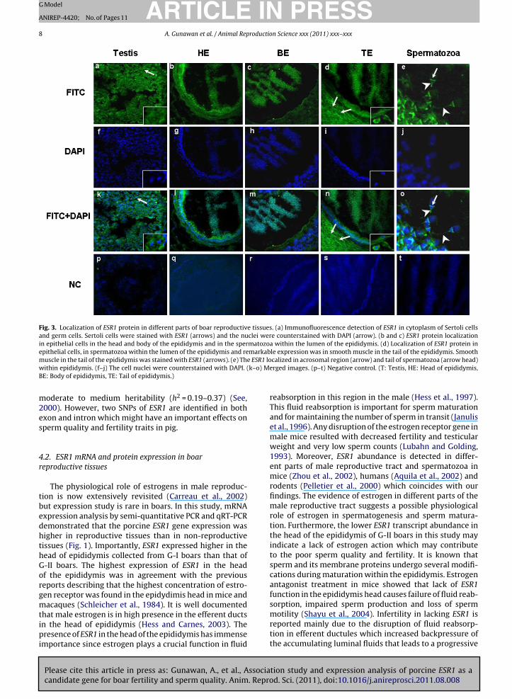
3.5. Localization of ESR1 protein in boar reproductivetissues by immunofluorescence
Sections of testis, head, body and tail of epididymiswere stained through the same optical panel for the ESR1protein expression (Fig. 3). Immunoreactive ESR1 proteinwas observed as strong staining in cytoplasm of Sertolicells but poorly in germ cells and Leydig cells in testis(Fig. 3a). Spermatozoa within the lumen of the epididymishead were found to be positively immunostained with ESR1(Fig. 3b). Similarly, spermatozoa within the lumen of theepididymis body and tail were positive to immunoreactive
Please cite this article in press as: Gunawan, A., et al., Associacandidate gene for boar fertility and sperm quality. Anim. Repro
ESR1 protein (Fig. 3c and d). The immunofluorescence ofESR1 protein was specifically localized in the post acro-somal region (arrow) and tail of the sperm (arrow head)when spermatozoa within the epididymis were examined
9 0.25 ± 0.06 −0.01 ± 0.10 −0.06 ± 0.11
(Fig. 3e). More precisely, the epithelial cells in the head andtail of the epididymis expressed higher immunoreactiveESR1 protein (Fig. 3b and d), whereas the epithelial cellsin the epididymis body poorly expressed the target pro-tein (Fig. 3c). In the case of the epididymis head and body,the sterocilia cells were remarkably higher immunostainedwhile in the epididymis tail the protein was strongly local-ized in the smooth muscle (Fig. 3b–d).
4. Discussion
4.1. Association of SNP with boar sperm quality andfertility traits
This study revealed association of ESR1 with sperm qual-ity and fertility traits in boars (Table 4). The exonic SNPg.672C > T was found to be significantly associated withsperm quality trait: sperm motility and plasma droplet rate.Association study of ESR1 is rare in boars, but Lazaros et al.(2010) reported two SNPs of ESR1 at 397 (T > C) and 351(A > G) had a positive effect on sperm quality in humans.
tion study and expression analysis of porcine ESR1 as ad. Sci. (2011), doi:10.1016/j.anireprosci.2011.08.008
Moreover, Suzuki et al. (2002) reported a polymorphismof ESR1 in exon 4 associated with idiopathic azoospermiain humans. The existence of ESR1 at the post acrosomalsperm head region (Solakidi et al., 2005) indicated a role of
ARTICLE IN PRESSG Model
ANIREP-4420; No. of Pages 11
A. Gunawan et al. / Animal Reproduction Science xxx (2011) xxx– xxx 7
Fig. 2. mRNA and protein expression of ESR1 in reproductive tissues (testis, head, body and tail of the epididymis). (a) ESR1 mRNA expression in differentreproductive tissues from G-I and G-II boars by semi-quantitative PCR. (b) ESR1 mRNA expression in different reproductive tissues from G-I and G-II boarsb G-I anB
ebtpg2iEritdsCipdstd
aRnn
y qRT-PCR. (c) Protein expression in different reproductive tissues fromody of epididymis, TE: Tail of epididymis.)
strogens in male gamete maturation and motility. It haseen reported that estrogen produced locally is involved inhe regulation of sperm motility (Lazaros et al., 2010). ESR1olymorphism may influence these locally acting estro-en levels with effect on sperm motility (Carreau et al.,002). Abnormal sperm production and reduced fertil-
ty have been reported in transgenic male mice lackingSR1 (Eddy et al., 1996). Infertility from lacking ESR1 iseported mainly due to the disruption of fluid reabsorptionn efferent ductules which increased the backpressure ofhe accumulating luminal fluids that leads to a progressiveegeneration of the testicular tissue and severely impairedpermatogenesis (Eddy et al., 1996; Hess et al., 1997;ouse and Korach, 1999). The identified SNP is confirmed
n exon which may play important role in transcriptionrocess. Polymorphism in the coding region could have airect effect through changing the nucleotide sequence andtructure of gene, possibly leading to changes in mRNA syn-hesis, splicing, maturation, transportation, translation oregradation (Iida and Akashi, 2000).
The intronic SNP g.35756T > C was significantly associ-
Please cite this article in press as: Gunawan, A., et al., Associacandidate gene for boar fertility and sperm quality. Anim. Repro
ted with the fertility trait non-return rate (NRR) (Table 4).eproductive efficiency of boars is usually measured byon-return rate, which is defined as percentage of sowsot appointed for a second insemination within a period
d G-II boars by western blotting. (T: Testis, HE: Head of epididymis, BE:
of days after the first insemination. Male fertility can beregarded as a result of both fertilizing ability of the spermcells as well as the number of fertilized oocytes and theviability of the embryo. But, under the field conditionsonly the outcome of the inseminations can be recorded, i.e.whether or not the females have returned for repeat insem-ination (Stalhammar et al., 1994). However, little is knownabout the polymorphism and association of porcine ESR1with fertility trait like NRR in pigs. An ESR1 gene variantat c.1227C > T in exon 5 showed a positive effect on littersize in sows (Munoz et al., 2007). It is important to notethat, the detected SNP at ESR1 g.35756T > C was in intron1 which is subjected to less functional constraint and maychange the primary structure of the ESR1. Though ‘silent’,it could affect ESR1 function by altering the mRNA stability(Capon et al., 2004). Moreover, there is increasing numberof reports describing the role of intron in regulating theexpression level of a gene or tissue specific expression pat-tern (Jiang and Le, 2000; Pagani et al., 2004). Traits relatedto fertility of boars are of low heritability (h2 = 0.01–0.06)and are strongly affected by environmental and genetic
tion study and expression analysis of porcine ESR1 as ad. Sci. (2011), doi:10.1016/j.anireprosci.2011.08.008
effects of the boar itself, the sow and the offspring (See,2000). Therefore, effects of single loci are expected tobe low and require a higher number of animals to beidentified. In contrast, the sperm quality traits have
ARTICLE IN PRESSG Model
ANIREP-4420; No. of Pages 11
8 A. Gunawan et al. / Animal Reproduction Science xxx (2011) xxx– xxx
Fig. 3. Localization of ESR1 protein in different parts of boar reproductive tissues. (a) Immunofluorescence detection of ESR1 in cytoplasm of Sertoli cellsand germ cells. Sertoli cells were stained with ESR1 (arrows) and the nuclei were counterstained with DAPI (arrow). (b and c) ESR1 protein localizationin epithelial cells in the head and body of the epididymis and in the spermatozoa within the lumen of the epididymis. (d) Localization of ESR1 protein inepithelial cells, in spermatozoa within the lumen of the epididymis and remarkable expression was in smooth muscle in the tail of the epididymis. Smooth
e ESR1 lok–o) M
muscle in the tail of the epididymis was stained with ESR1 (arrows). (e) Thwithin epididymis. (f–j) The cell nuclei were counterstained with DAPI. (BE: Body of epididymis, TE: Tail of epididymis.)
moderate to medium heritability (h2 = 0.19–0.37) (See,2000). However, two SNPs of ESR1 are identified in bothexon and intron which might have an important effects onsperm quality and fertility traits in pig.
4.2. ESR1 mRNA and protein expression in boarreproductive tissues
The physiological role of estrogens in male reproduc-tion is now extensively revisited (Carreau et al., 2002)but expression study is rare in boars. In this study, mRNAexpression analysis by semi-quantitative PCR and qRT-PCRdemonstrated that the porcine ESR1 gene expression washigher in reproductive tissues than in non-reproductivetissues (Fig. 1). Importantly, ESR1 expressed higher in thehead of epididymis collected from G-I boars than that ofG-II boars. The highest expression of ESR1 in the headof the epididymis was in agreement with the previousreports describing that the highest concentration of estro-gen receptor was found in the epidydimis head in mice andmacaques (Schleicher et al., 1984). It is well documented
Please cite this article in press as: Gunawan, A., et al., Associacandidate gene for boar fertility and sperm quality. Anim. Repro
that male estrogen is in high presence in the efferent ductsin the head of epididymis (Hess and Carnes, 2003). Thepresence of ESR1 in the head of the epididymis has immenseimportance since estrogen plays a crucial function in fluid
calized in acrosomal region (arrow) and tail of spermatozoa (arrow head)erged images. (p–t) Negative control. (T: Testis, HE: Head of epididymis,
reabsorption in this region in the male (Hess et al., 1997).This fluid reabsorption is important for sperm maturationand for maintaining the number of sperm in transit (Januliset al., 1996). Any disruption of the estrogen receptor gene inmale mice resulted with decreased fertility and testicularweight and very low sperm counts (Lubahn and Golding,1993). Moreover, ESR1 abundance is detected in differ-ent parts of male reproductive tract and spermatozoa inmice (Zhou et al., 2002), humans (Aquila et al., 2002) androdents (Pelletier et al., 2000) which coincides with ourfindings. The evidence of estrogen in different parts of themale reproductive tract suggests a possible physiologicalrole of estrogen in spermatogenesis and sperm matura-tion. Furthermore, the lower ESR1 transcript abundance inthe head of the epididymis of G-II boars in this study mayindicate a lack of estrogen action which may contributeto the poor sperm quality and fertility. It is known thatsperm and its membrane proteins undergo several modifi-cations during maturation within the epididymis. Estrogenantagonist treatment in mice showed that lack of ESR1function in the epididymis head causes failure of fluid reab-sorption, impaired sperm production and loss of sperm
tion study and expression analysis of porcine ESR1 as ad. Sci. (2011), doi:10.1016/j.anireprosci.2011.08.008
motility (Shayu et al., 2004). Infertility in lacking ESR1 isreported mainly due to the disruption of fluid reabsorp-tion in efferent ductules which increased backpressure ofthe accumulating luminal fluids that leads to a progressive
ING Model
A
roductio
dsaisi1
ealveawbhsieitee(bmaIio
4
pcwdcgiafi1p
e(stttrevrtzo
ARTICLENIREP-4420; No. of Pages 11
A. Gunawan et al. / Animal Rep
egeneration of the testicular tissue and severely impairedpermatogenesis (Eddy et al., 1996; Hess et al., 1997; Cousend Korach, 1999). Moreover, swelling and damage of sem-niferous tubules and the dilution of the essential proteinsecreted from epididymis also contributed to the infertil-ty (Eddy et al., 1996; Hess et al., 1997; Couse and Korach,999).
In this study, the protein expression analysis by west-rn blotting showed that ESR1 antibody recognized a bandt 66 kDa in all tissues. The band is similar to the molecu-ar weight of human (66 kDa) and mouse (66 kDa) ESR1 andery close to that of the stallion (65 kDa). Although, the lev-ls of protein in different tissues from G-I and G-II were nots distinguishable as mRNA, the ESR1 protein expressionas remarkably higher in the head of the epididymis of G-I
oars than that of G-II boars (Fig. 2b). The vas deferens in theead of the epididymis are the major sites for ESR1 expres-ion which regulates the testicular fluid reabsorption andncreases the sperm cell concentration as they enter thepididymis (Hess et al., 1997). Moreover, the epididymis isnvolved in different protein absorption as well as in pro-ein secretion (Syntin et al., 1999). The head and body of thepididymis are reported to secret higher amounts of differ-nt proteins in boars in comparison to other farm animalsSyntin et al., 1999). These protein secretions are regulatedy estrogen and the proteins are involved in the spermembrane remodelling, in the initiation of sperm motility,
nd especially in sperm-egg interaction (Pearl et al., 2007).t has been calculated that 50–90% of the total protein leav-ng the testis is absorbed by the efferent ducts in the headf the epididymis (Clulow et al., 1994).
.3. Protein localization of ESR1
ESR1 was found to be expressed strongly in the cyto-lasm of Sertoli cells, expressed on germ cells and Leydigells in testis (Fig. 3a). These results are in agreementith previous results localizing ESR1 in Sertoli and Ley-ig cells (Hess and Carnes, 2003) and in Leydig and germells in rat (Pelletier et al., 2000). Pelletier et al. (2000) sug-ested that in the Leydig cells the ESR1 might be involvedn the secretion and maturation of germ cells. Estrogenre reported to be involved in maintaining the Sertoli cellunction (O’Donnell et al., 2001) as well as in establish-ng Sertoli–germ cell adhesion (MacCalman and Blaschuk,994). However, the cellular distribution patterns of ESR1rotein between species could be different.
We localized ESR1 in the head, body and tail of thepididymis. When sperm are released from their ‘nurse’Sertoli) cells in the testis, they are transported in fluidecreted by the Sertoli cells to a collecting area, the reteestis. From there, the dilute suspension of sperm enterhe thin-walled efferent ducts in the epididymis head andhe epithelia of efferent ducts express enormous estrogeneceptors (Fisher et al., 1997). The sperm pass through thepididymis where they mature and progress toward theas deferens. Moreover, the ESR1 in the epididymis are
Please cite this article in press as: Gunawan, A., et al., Associacandidate gene for boar fertility and sperm quality. Anim. Repro
eported to modulate secretion of proteins such as oscilinhat promotes the maturity and viability of the spermato-oa (Mowa and Iwanaga, 2001). The most intense signalsf ESR1 are reported in epithelial cells in the head and tail
PRESSn Science xxx (2011) xxx– xxx 9
of the epididymis in rats and monkeys and suggested to beresponsible for semen concentration (Fisher et al., 1997;Hess et al., 1997). Our study identified a strong signal ofESR1 in the smooth muscle layer in the tail of the epididymiswhich is in accordance with a previous immunohistochem-ical study in human and rabbit which stated that estrogenis essential for transporting the spermatozoa by influenc-ing epididymal smooth muscle contractility as well as forejaculation with the help of oxytocin (Filippi et al., 2002).It is important to note that the expression of ESR1 in dif-ferent parts of the epididymis could vary among speciessince it has been localized in all parts of epididymis in therat (Pelletier et al., 2000), mouse (Zhou et al., 2002), bonnetmonkey (Shayu et al., 2004), horse (Parlevliet et al., 2006),cat (Nie et al., 2002) and dog (Nie et al., 2002).
We found that the ESR1 is remarkably localized insperm within the lumen of the epididymis. ESR1 was espe-cially immunolocalized in the post acrosomal region andtail of the sperm in this study (Fig. 3e). This result isin good agreement with previous studies which localizedimmunoreactive ESR1 in the post acrosomal region of thesperm head (Solakidi et al., 2005) and in the tailpiece of thesperm (Durkee et al., 1998) in humans. The post acroso-mal region of sperm is involved in the sperm–egg plasmamembrane fusion (Liu et al., 2008). The localization of ESR1in the post acrosomal region implies its involvement inthe fertilization process (Ramalho-Santos et al., 2002). It isimportant to note that there are other proteins identifiedin the post acrosomal region of sperm such as equatorinand oscilin which are important for successful fertilization(Montag et al., 1998; Solakidi et al., 2005). Our localizationof ESR1 in the tail of porcine sperm coincided with previousreports in humans (Durkee et al., 1998; Aquila et al., 2002).Moreover, Aquila et al. (2002) reported that the ESR1 mightbe involved in sperm survival and motility. Impaired motil-ity of sperm has been reported in mice lacking functionalESR1 in sperm (Eddy et al., 1996) and in ESR1 knockout mice(Hess and Carnes, 2003). Low fertilization rates have beenreported in case of immotile spermatozoa (Nijs et al., 1996).
5. Conclusion
Associations of ESR1 gene polymorphisms with boarsperm quality and fertility traits have been described forthe first time, providing evidence that ESR1 might be animportant candidate gene for boar sperm quality and fertil-ity. However, this study has to be validated in other animalpopulations in order to evaluate its potential in selectivebreeding. Finally, the ESR1 might play a role in spermatoge-nesis validated through association study and by profilingof mRNA and protein expression in non-reproductive andreproductive tissues including spermatozoa.
Acknowledgments
This research was financially supported by FBF(Förderverein Biotechnologieforschung e. V., Bonn,
tion study and expression analysis of porcine ESR1 as ad. Sci. (2011), doi:10.1016/j.anireprosci.2011.08.008
Germany). We thank Dr. A. Niggemeyer, Dr. S. Brüning,Dr. M. Friedrichs and Dr. A. Riesenbeck (GFS ArtificialInsemination Station: Die Genossenschaft zur Förderungder Schweinehaltung, Ascheberg, Germany) and Dr. A.
ING Model
roductio
ARTICLEANIREP-4420; No. of Pages 11
10 A. Gunawan et al. / Animal Rep
Hofer, Dr. H. Luther and Dr. K. Caspari (SuisAG ArtificialInsemination Station, Sempach, Switzerland) for theirsupport during the sample collection and the arrange-ments of the reproduction records. We are also thankfulto Prof. Dr. C. Knorr (Institute of Veterinary Medicine,Georg-August-University, Goettingen, Germany) for hisco-operation.
References
Aquila, S., Sisci, D., Gentile, M., Middea, E., Siciliano, L., Ando, S., 2002.Human-ejaculated spermatozoa contain active P450 aromatase. J.Clin. Endocrinol. Metab. 87, 85–90.
Capon, F., Allen, M.H., Ameen, M., Burden, A.D., Tillman, D., Barker, J.N.,Trembath, R.C., 2004. A synonymous SNP of the corneodesmosingene leads to increased mRNA stability and demonstrates associa-tion with psoriasis across diverse ethnic groups. Hum. Mol. Genet. 13,2361–2368.
Carreau, S., Bourguiba, S., Lambard, S., Galeraud-Denis, I., Genissel, C., Lev-allet, J., 2002. Reproductive system: aromatase and estrogens. Mol.Cel. Endocrinol. 193, 137–143.
Clulow, J., Jones, R., Hansen, L., 1994. Micropuncture and cannulation stud-ies of fluid composition and transport in the ductuli efferentes testisof the rat:comparisons with the homologous metanephric proximaltubule. Exp. Physiol. 79, 915–928.
Couse, J., Korach, K., 1999. Estrogen receptor null mice: what have welearned and where will they lead us? Endocr. Rev. 20, 358–417.
Durkee, T., Mueller, M., Zinaman, M., 1998. Identification of oestrogenreceptor protein and messenger ribonucleic acid in human spermato-zoa. Am. J. Obstet. Gynecol. 178, 1288–1295.
Eddy, E., Washburn, T., Bunch, D., Goulding, E., Gladen, B., Lubahn, D.,Korach, K., 1996. Targeted disruption of the estrogen receptor genein male mice causes alteration of spermatogenesis and infertility.Endocrinology 137, 4796–4805.
Filippi, S., Vannelli, G.B., Granchi, S., Luconi, M., Crescioli, C., Mancina,R., Natali, A., Brocchi, S., Vignozzi, L., Bencini, E., Noci, I., Ledda, F.,Forti, G., Maggi, M., 2002. Identification, localization and functionalactivity of oxytocin receptors in epididymis. Mol. Cel. Endocrinol. 193,89–100.
Fisher, J., Millar, M., Majdic, G., Saunders, P., Fraser, H., Sharpe, R., 1997.Immunolocalisation of oestrogen receptor 1 within the testis andexcurrent ducts of the rat and marmoset monkey from perinatal lifeto adulthood. J. Endocrinol. 153, 485–495.
Ganjam, V., Amann, R., 1976. Steroid content of fluids and spermentering and leaving the bovine epididymis, in epididymal tis-sue, and in accessory sex gland secretions. Endocrinology 99,1618–1630.
Guarducci, E., Nuti, Becherini, L., Rotondi, M., Balercia, G., Forti, G., Krausz,C., 2006. Estrogen receptor 2 promoter polymorphism: stronger estro-gen action is coupled with lower sperm count. Hum. Reprod. 21,994–1001.
Hejmej, A., Gorazd, M., Kosiniak-Kamysz, K., Wisniewska, B., Sadowska,J., Bilinska, B., 2005. Expression aromatase and androgen receptors inreproductive tissue of the stallion and a single cryptochid visualizedby means of immunohistochemistry. Domest. Anim. Endricinol. 29,534–547.
Hess, R., Carnes, K., 2003. The role of oestrogen in testis and male repro-ductive tract: a review and species comparison. Anim. Reprod. 1, 5–30.
Hess, R., Gist, D., Bunick, D., 1997. Estrogen receptor (alpha and beta)expression in the excurrent ducts of the adult male rat reproductivetract. J. Androl. 18, 602–611.
Iida, K., Akashi, H., 2000. A test of translational selection at ‘silent’ sites inthe human genome: base composition comparisons in alternativelyspliced genes. Gene 261, 93–102.
Janulis, L., Hess, R., Bunick, D., Nitta, H., Janssen, S., Asawa, Y., Bahr,J., 1996. Mouse epididimal sperm containt active P450 aromatasewhich decrease as sperm traverse the epididymis. J. Androl. 17,111–116.
Jiang, W., Le, B., 2000. Structure and expression of the human MEP1A geneencoding the alpha subunit of metalloendopeptidase meprin A. Arch.Biochem. Biophys. 379, 183–187.
Please cite this article in press as: Gunawan, A., et al., Associacandidate gene for boar fertility and sperm quality. Anim. Repro
Kaewmala, K., Uddin, M.J., Cinar, M.U., Grosse-Brinkhaus, C., Jonas, E.,Tesfaye, D., Phatsara, C., Tholen, E., Looft, C., Schellander, K., 2011.Association study and expression analysis of CD9 as candidategene for boar sperm quality and fertility traits. Anim. Reprod. Sci.,doi:10.1016/j.anireprosci.2011.02.017.
PRESSn Science xxx (2011) xxx– xxx
Kuiper, G., Enmark, E., Puelto-Huiko, M., Nilsson, S., Gustafsson, J., 1996.Cloning of a novel estrogen receptor expressed in rat prostate andovary. Proc. Natl. Acad. Sci. U.S.A. 93, 2925–2930.
Lazaros, L.A., Xita, N.V., Kaponis, A.I., Zikopoulos, K.A., Plachouras, N.I.,Georgiou, I.A., 2010. Estrogen receptor alpha and beta polymorphismsare associated with semen quality. J. Androl. 31, 291–298.
Lin, C.L., Tholen, E., Jennen, D., Ponsuksili, S., Schellander, K., Wimmers, K.,2006. Candidate gene markers for sperm quality and fertility of boar.Anim. Reprod. Sci. 92, 349–363.
Liu, G.Q., Zhu, J.J., Wang, Z.Y., Jiang, X.P., Dafalla, M.M., 2008. Analysis ofsperm storage ability using duration of fertility in hens. Br. Poult. Sci.49, 770–775.
Lubahn, D.B., Moyer, J.S., Golding, T.S., 1993. Alteration of reproductivefunction but not prenatal sexual development after insertional dis-ruption of the mouse estrogen receptor gene. Proc. Natl. Acad. Sci.U.S.A. 90, 11162–11166.
MacCalman, C.D., Blaschuk, O., 1994. Gonadal steroids regulate N-cadherinmRNA levels in the mouse testis. Endocr. Rev. 2, 157–163.
Montag, M., Parrington, J., Swann, K., Lai, F., Van der Ven, H., 1998. Pres-ence and localization of oscillin in human spermatozoa in relation tointegrity of the sperm membrane. FEBS Lett. 423, 357–361.
Mowa, C.N., Iwanaga, T., 2001. Expression of estrogen receptor-alpha and-beta mRNAs in the male reproductive system of the rat as revealedby in situ hybridization. J. Mol. Endocrinol. 26, 165–174.
Munoz, G., Ovio, C., Estell, J., Silio, L., Fernadez, A., Rodriguez, C., 2007.Association with litter size of new polymorphism on ESR1 andESR2 genes in a Chinese–European pig line. Genet. Sel. Evol. 39,195–206.
Nie, R., Zhou, Q., Jassin, E., Saunders, P., Hess, R., 2002. Differential expres-sion of oestrogen receptors alpha and beta in the reproductive tractof adult male dogs and cats. Biol. Reprod. 66, 1161–1168.
Nijs, M., Vanderzwalmen, P., Vandamme, G., 1996. Fertilizing ability ofimmotil spermatozoa after intracyctoplasmic sperm injection. Hum.Reprod. 11, 2180–2185.
O’Donnell, L., Robertson, K.M., Jones, M.E., Simpson, E.R., 2001. Estrogenand spermatogenesis. Endocr. Rev. 22, 289–318.
Pagani, R., Song, M., McEnery, M., Qin, N., Tsien, R.W., Toro, L., Stefani,E., Uchitel, O.D., 2004. Differential expression of alpha 1 and betasubunits of voltage dependent Ca2+ channel at the neuromuscularjunction of normal and P/Q Ca2+ channel knockout mouse. Neuro-science 123, 75–85.
Parlevliet, J., Pearl, C., Hess, M., Famula, T., Roser, J., 2006. Immunolocaliza-tion of estrogen and androgen receptors and steroid concentrationsin the stallion epididymis. Theriogenoloy 66, 755–765.
Pearl, C., Berger, T., Roser, J., 2007. Estrogen and androgen receptor expres-sion in relation to steroids concentrations in adult boar epididymis.Domest. Anim. Endocrinol. 33, 451–459.
Pelletier, G., Labrie, C., Labrie, F., 2000. Localization of oestrogen recep-tor alpha, oestrogen receptor beta and androgen receptors in the ratreproductive organs. J. Endocrinol. 165, 359–370.
Ramalho-Santos, J., Schatten, G., Moreno, R., 2002. Control membranefusion during spermiogenesis and the acrosome reaction. Biol. Reprod.67, 1043–1051.
Rozen, S., Skaletsky, H., 2000. Primer3 on the WWW for general users andfor biologist programmers. Methods Mol. Biol. 132, 365–386.
Safarinejad, M.R., Shafiei, N., Safarinejad, S., 2010. Association of polymor-phisms in the estrogen receptors alpha, and beta (ESR1, ESR2) withthe occurrence of male infertility and semen parameters. J. SteroidBiochem. Mol. Biol. 122, 193–203.
Schleicher, G., Drew, U., Stumpt, W., Sar, M., 1984. Differential distributionof dihydrosterone and estrodial binding site in the epididymis of themouse: an autoradiographic study. Histochemistry, 1139–1147.
See, M., 2000. Selection for AI stud traits. In: Proceedings of National SwineImprovement Federation, Nashville, Tennessee, USA, 7–8 December,p. 25.
Shayu, D., Chennakesava, C., Soundarajan, R., Rao, J., 2004. Effect ofICI 182780 on estrogen receptor expression, fluid absorption andsperm motility in the epididymis of the bonnet monkey. Reprod. Biol.Endocrinol. 3, 3–10.
Solakidi, S., Psarra, A., Nikolaropouluos, S., Sekeris, C., 2005. Estrogenreceptor alpha and beta and androgen receptor (AR) in human sperm:localization of Estrogen beta and AR in mitochondria of the midpiece.Hum. Reprod. 20, 3481–3487.
Stalhammar, E., Janson, L., Philipsson, J., 1994. Genetic studies on fertility
tion study and expression analysis of porcine ESR1 as ad. Sci. (2011), doi:10.1016/j.anireprosci.2011.08.008
in AI bulls. II. Environmental and genetic effects on non-return ratesof young bulls. Anim. Reprod. Sci. 34, 193–207.
Suzuki, Y., Sasagawa, I., Itoh, K., Ashida, J., Muroya, K., Ogata, T., 2002. Estro-gen receptor alpha gene polymorphism is associated with idiopathicazoospermia. Fertil. Steril. 78, 1341–1343.
ING Model
A
roductio
S
W
ARTICLENIREP-4420; No. of Pages 11
A. Gunawan et al. / Animal Rep
yntin, P., Dacheux, F., Druart, X., Gatti, J., Okamura, N., Dacheux, J.,1999. Characterization and identification of proteins secreted in
Please cite this article in press as: Gunawan, A., et al., Associacandidate gene for boar fertility and sperm quality. Anim. Repro
the various regions of the adult boar epididymis. Biol. Reprod. 55,956–974.
immers, K., Lin, C., Tholen, E., Jennen, D., Schellander, K., Ponsuksili, S.,2005. Polymorphisms in candidate genes as markers for sperm qualityand boar fertility. Anim. Genet. Sel. Evol. 36, 152–155.
PRESSn Science xxx (2011) xxx– xxx 11
Xing, Y., Ren, J., Ren, D., Guo, Y., Wu, Y., Yang, G., Mao, H., Brenig, B., Huang,L., 2008. A whole genome scanning for quantitative trait loci on traits
tion study and expression analysis of porcine ESR1 as ad. Sci. (2011), doi:10.1016/j.anireprosci.2011.08.008
related to sperm quality and ejaculation in pigs. Anim. Reprod. Sci.114, 210–2118.
Zhou, Q., Nie, R., Prins, G., Saunders, P., Katzenellenbogen, B., Hess, R., 2002.Localization of androgen and oestrogen receptors in adult male mousereproductive tract. J. Androl. 23, 870–881.