fertivision - indian fertility society
TRANSCRIPT

1Souvenir / Abstract Book
FERTIVISION 2019 6-8 December
15th Annual Congress of
Indian Fertility SocietyTh
eme:
Bey
ond
Tom
orro
w
Organised by
www.fertivision2019.com
The Leela Ambience Hotel, Gurugram New Delhi | India
Souvenir /Abstract Book

Souvenir / Abstract Book2
We welcome you all for Fertivision 2019, the 15th Annual Congress of Indian Fertility Society, scheduled to be held on 6th ,7th and 8th December 2019 at hotel, The Leela Ambience, Gurugram, New Delhi/NCR, India. The annual congress of Indian Fertility Society has most sought-after congress in the field of reproductive medicine in just one and a half decades of exciting scientific journey. Many delegates, not only from India but beyond India as well eagerly wait for this annual academic bonanza.
The organizing committee, has chosen “Beyond Tomorrow” as the theme for this year’s Fertivision.
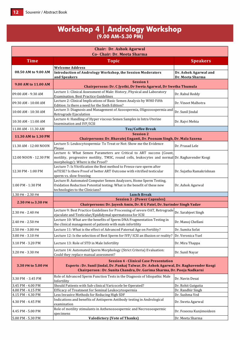
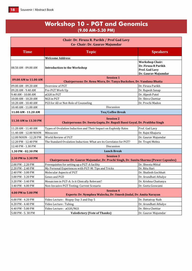
With the proposed theme in mind we have designed 10 interactive workshops on first day of the conference. These are namely – “IFFS Workshop on Do’s and Don’ts in Ovarian Stimulation”, “Reproductive Surgery”, “Ultrasonography/ Im-aging in Infertility Management”, “Andrology & Semenology”, “Ovum Pick up and Embryo Transfer (With simulators)”, “Cryobiology” ,“Total Quality Management”, “patient Counselling and Holistic medicine” , “Publish or Perish” and “PGT and Genomics”.
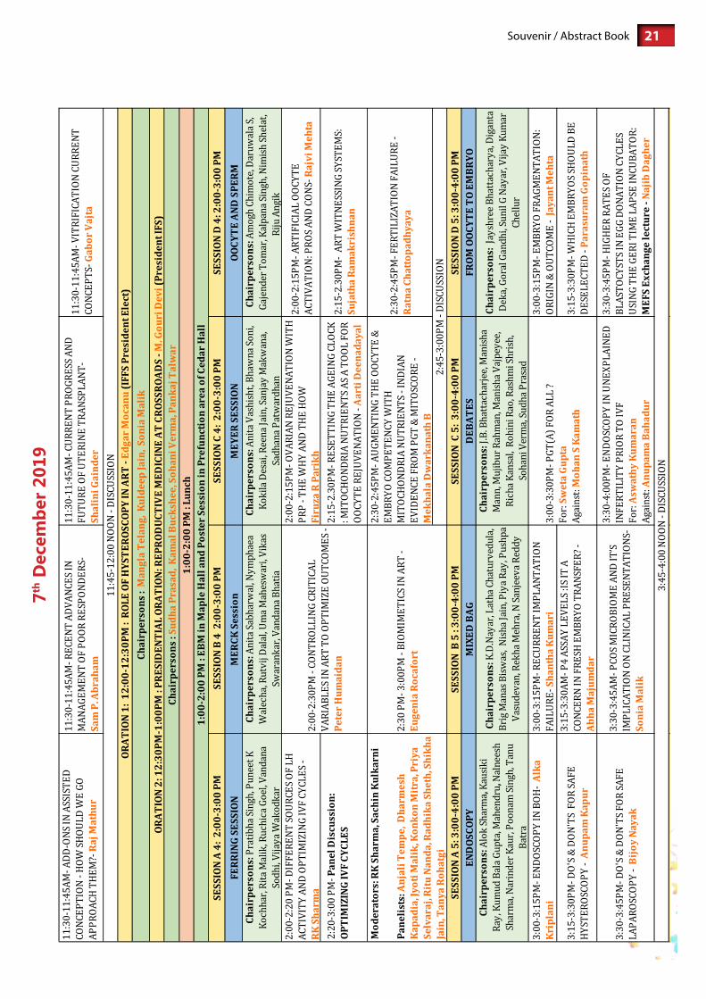
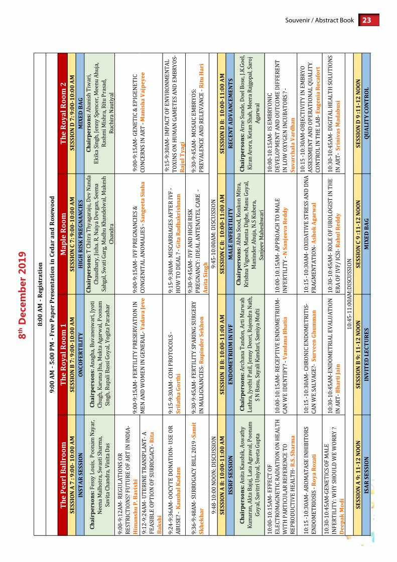
In the main congress on 7th and 8th December we have put together an exciting and interactive program.We have the combined expertise of an eminent group of around 20 internationally-renowned faculty and a large group of experienced Indian faculty to present the latest developments on every aspect of ART. It addresses the needs of practicing gynecol-ogists, reproductive endocrinologists, embryologists, residents, and fellows who wish to update their knowledge in this rapidly advancing field.
We have designed the conference in a way that it will promote extensive deliberations among speakers and participants with question periods, panels, and many opportunities for informal interaction. Care has been taken to ensure that the postgraduates and fellows get ample opportunities to interact and clear their doubts.
There is a long list of tourist attractions in Delhi(Heart of India) and Gurugram(Millennium City of India) From monu-ments ,temples , parks , museums, to sprawling malls - Delhi/NCR has so much in store that it won’t stop amusing you. For nature lovers , Sultanpur bird sanctuary , Sohna lake and Dumdama lake are nearby tourist attractions in Gurugram.
Looking forward to welcoming you all!

3Souvenir / Abstract Book

Souvenir / Abstract Book4
Dr. Mujibur Rehman (North East)M: 9435070660, E: [email protected]. Uma Shrivastava (Nepal)M: 977-9851074477, E: [email protected] Jayesh S.Amin ( Gujarat)M: 9824302671, E: [email protected]. Papa Dasari (Puducherry)M: 9442566883, E: [email protected]. JK Goel (UP West)M: 9458702304 E: [email protected]. Anupama Bahadur (Uttarakhand)M: 9810326959 , E: [email protected]. Roya Rozati (Telangana)E: [email protected]. Archana Kumari (Jharkhand)E: [email protected]. Syed Sajjad Hussain (Kashmir)M: 9419000077 E: [email protected]. Divyashree P.S (Karnataka)M: 9663351451 E: [email protected]. Firuza Parikh (Mumbai)M: 9812694923 E: [email protected]. Usha Prasad (Andra Pradesh)M: +91 91777 44546
Dr Gouri DeviPresident9810023111
Dr M KochharPatron
9810018277 / [email protected]
Dr Nalini MahajanPast President
Dr M TelangFounder President
9811030476/[email protected]
Dr Kuldeep JainPast President
9810018951, [email protected]
Dr Abha MajumdarPast President
Dr Sonia MalikPast President
Dr Surveen GhummanEditor
Dr Rashmi SharmaJt. Seretary
Dr KD NayarSr. Vice President
Dr Sohani VermaImmediate Past President
Dr Shweta MittalJt. editor9910303056
Dr Neena MalhotraTreasurer9891557707
malhotraneena@yahoo.
Dr Gita RadhakrishnanVice President
Dr Sudha PrasadPresident Elect
Dr Leena WadhwaWeb Editor
9910933447, 9818145296 [email protected]
Dr Ritu KhannaJt. Treasurer
Dr Pankaj TalwarSecretary General
Dr Ritu Jain9873183030 / [email protected]
EXECUTIVE MEMBERS
Dr Sweta Gupta8130140007
Dr Vandana Bhatia9891967417
Dr Renu Mishra9811147217
Dr Tanya Buckshee9910003731
Dr Rupali Bassi Goyal9818331760
Dr Nymphaea Walecha9873855738
CHAPTER SECRETARIESDr. Renu Makkar (UP)M: 9415002674 E: [email protected] Dr. (Mrs) Harinder Kaur Oberoi (Punjab)M: 9888030729 E: [email protected]. Neeru Thakral (Haryana)M: 9810569387E: [email protected] Dr. Sangeeta Sinha (Chattisgarh)M: 9752595605 E: [email protected] Dr. Dr Mamta Dighe (Maharashtra)M: 9881125250 E: [email protected]. Sangita Sharma (Rajasthan)M: 9549500137 E: [email protected]. Swati Verma (Greater Chandigarh)M: 9646004459 E: [email protected]
EXECUTIVE ADVISORSDr. Umesh JindalM: 9876130501, 0172-2703222E: [email protected]. S.N. BasuM: 9810119072 E: [email protected] Dr Sandeep TalwarM: 9810306455 E: [email protected] MEMBERSDr. Alka KriplaniM: 9810828717 E: [email protected]. Urvashi JhaM: 9811029310 / 9350550669E: [email protected]. R.K. SharmaM: 9810442301 E: [email protected]. Jayant MehtaE: [email protected]. Rama RajuM: 9849110004E: [email protected]
Dr. Shilpi Sud (Vidharba )M: 9923737304 E: [email protected]. Monica Singh (MP)M: 9200002833 E: [email protected] Dr. Surender Kumar (Jammu)M: 09419188392 E: [email protected]. Anita Singh (Bihar)M: 9334111925 E: [email protected]. Alok Sharma (Himachal)M: 9418477725 E: [email protected] Dr. Suparna Banerjee (West Bengal)M: 8697475255E: [email protected] K.U.Kunjumoideen (Kerala)T: 9895983376E: [email protected]. P.M. Gopinath (Tamil Nadu)M: 04426163884, 9840888878E: [email protected]
Dr Gaurav Majumdar9810794610
Team ifsGoverninG council

5Souvenir / Abstract Book
orGanisinGCOMMITTEE

Souvenir / Abstract Book6
inTernaTionalFACULTY

7Souvenir / Abstract Book
S.N. TITLE PAGE NO.
1 Workshops 8
2 Scientific Programme 19
3 Messages 26
4 Abstract Orations 35
5 Abstract of Talks in Main Scientific Programme 42
6 Abstract of Talks in Workshops 114
7 Free Communications : Oral/Poster Presentations 136 - 213
inDeX

Souvenir / Abstract Book8
Workshops6th DECEMbEr, 2019

9Souvenir / Abstract Book

Souvenir / Abstract Book10

11Souvenir / Abstract Book
Souvenir / Abstract Book12

13Souvenir / Abstract Book

Souvenir / Abstract Book14

15Souvenir / Abstract Book

Souvenir / Abstract Book16

17Souvenir / Abstract Book
Souvenir / Abstract Book18

19Souvenir / Abstract Book
scienTificPrOGrAMME

Souvenir / Abstract Book207th
Dec
embe
r 201
9
21Souvenir / Abstract Book7th
Dec
embe
r 201
9

Souvenir / Abstract Book227th
Dec
embe
r 201
9
23Souvenir / Abstract Book8th
Dec
embe
r 201
9

Souvenir / Abstract Book248th
Dec
embe
r 201
9

25Souvenir / Abstract Book8th
Dec
embe
r 201
9

Souvenir / Abstract Book26
messaGes

27Souvenir / Abstract Book
messaGe from ThePrESIDENT - IFS
Fertivision 2019, the 15th National annual conference is being held on 6th,7th& 8th December at Hotel, The Leela Ambience, Gurugram, New Delhi /NCR, India.
Fertivision is one of the most awaited annual academic events of Indian Fertility Society. This society in the last 2 years after the new governing council has taken over has added about 1000 members and now has over 2700 members and 9 chapters making a total of 27 chapters. It has conducted over 100 meetings all over India in the last one and half years.The society has taken active participation in IFFS world congress2019 at Shanghai,ESHRE at Vienna2019 and Middle eastern fertility societyat Cairo2019.
The conference program has been planned to deliver the recent advances in a most comprehensive manner in the field of infertility and Assisted Reproductive Technology (ART) befitting the theme of the conference “BEYOND TOMORROW”Keeping this in view, we have planned 10 very interactive pre-congress workshops and 2 full days of conference with latest topics in the form of orations, lectures, debates, panel discussions, by the eminent international and national faculties. We hope that this will not only enrich your current knowledge but also clear all doubts faced in day to day clinical practice.
We have tried to collect all the contents of the lectures and put it into the souvenir so that the delegates can at leisure read them. The editorial team under the able guidance of Dr. Rashmi Sharma has worked hard towards this.
Please enjoy the academic feast and warm hospitality of historical Delhi city and Millennial Gurugram City.
Wishing you all a very Happy New Year!
Dr. M. Gouri DeviPresidentIndian Fertility Society
Dr Gouri DeviDirectorRidge IVF &Gouri HospitalsDelhi
Souvenir / Abstract Book28
messaGe from TheSECrETArY GENErAL - IFS
Friends,
On behalf of IFS and organizing committee for the 15th NATIONAL ANNUAL CONFRENCE “FERTIVISION 2019”, it gives me great pleasure to extend you all a very warm welcome to Delhi, the home town of culture and heritage. Since the establishment of Indian Fertility Society in 2005, IFS has seen an unbelievable journey from 20 founder members to a current strength of more than 2700 members in 27 state chapters all over country with some international chapters as well.Fertivision has become the most awaited annual academic event in the field of infertility adorned with highly respected internationally and nationally renowned speakers who will share, discuss, debate and dissect significant new developments and scientific advancements in the field of ART.
We are having an overwhelming response with more than 1200 delegates attending the conference and a wide spectrum of international and national faculty who are going to contribute to the rich content of the conference. We express our grat-itude to all the international and national faculty for sharing and imparting knowledge to update the delegates on recent developments and practices in Reproductive Medicine, Embryology, Genetics, Andrology and many more subjects.
In the last 15 years of IFS, we are constantly making progress. We are having third batch of one-year fellowship training program in the form of “Diploma in clinical ART” and “Diploma in Clinical Embryology” in collaboration with Amity University under the UGC guidelines.
IFS has organized various focused meets all over the country especially reaching places and cities which were earlier un-touched like Nagaland in our Outreach initiative.
IFS is constantly bringing out very diligently made e Bulletins like IFS Conversations, Nexus, ARText, fertility focus, Cata-lyst and Fertility Synapses for knowledge dissemination. These have been hugely appreciated by one and all.
IFS is also publishing “Fertility Science and Research” peer reviewed journal with latest research and reviews contributed by experts from all over the world.
Wishing all delegates and faculty a very happy new year!
With warm Regards,
Dr. Pankaj TalwarSecretary GeneralIndian Fertility Society
prof (Dr) pankaj Talwar, VSMHOD, ART CentreManipal HospitalDwarka, Delhi

29Souvenir / Abstract Book
messaGe from TheCHIEF GUEST
It is a matter of immense pleasure that Indian Fertility Society is organizing 15thannual conference, Fertivision on 6th, 7th and 8th December 2019.
Parenthood is a very much desired and anticipated role for most human beings ,sonot being able to fulfil that role rep-resents a major crisis for most couples. Infertility continues to be a major worldwide problem affecting around 27 million couples in India alone. With the miraculous advancements in the field of reproductive medicine , now it has become pos-sible to treat many clinical situations which were earlier untreatable .
We realize that there is a huge unmet need for expert ART specialists, centres across the length and breadth of India. I hope this megaevent will be a great help in this regard.
I extend my warm greetings and felicitations to the organisers and participants . My best wishes for the success of event .
Wishing you a very pleasant and fruitful conference.
Dr Edgar Mocanu
Dr edgar mocanuPresident- Elect International Federation of Fertility Societies Immediate Past Chair FIGO REI committee Honorary senior lecturer in Reproductive Endocrinology

Souvenir / Abstract Book30
messaGe from TheGUEST OF HONOUr
I am delighted to write this message for the 15th Annual conference of Indian Fertility Society.
India is an overpopulated country, still the importance of treating infertile couples cannot be undermined. The agony that an infertile couple goes through is great especially since having a child and family is considered to be very important in social fabric of India. As India is advancing in ART rapidly, it is essential that there should be a law governing it. Surrogacy bill and ART bill are government’s priority to safeguard interest of patients and doctors as well .
I am very happy to see the great work being done by Indian Fertility Society in all aspects of infertility education, research and propagation of knowledge. I am sure the annual conference with discussions on most recent advancements in reproductive Medicine by eminent scientists across the globe will be of immense benefit to community and country at large.
Let us join our hands together to share knowledge and experience that will go a long way in helping to build a healthy, prosperous and developed India.
With Warm Regards,
Dr Mangla Telang
Dr mangla TelangFounder PresidentIndian Fertility Society

31Souvenir / Abstract Book
messaGe from TheSCIENTIFIC CHAIrPErSON
It is my immense pleasure to welcome you all to “FERTIVISION 2019”, the 15th Annual Conference of Indian Fertility Society.
Since the establishment of the Indian fertility society in 2005, it has steadily grown in stature and is making significant contributions to the cause of sharing and spreading knowledge about ART across Indian subcontinent. Now in 2019, it has become a remarkable academic society with membership exceeding 2700 members with 27 state chapters and affiliation to International Federation of Fertility Societies (IFFS) since 2007.
The society has contributed towards excellent CME programmes, Symposia and Workshops in both basics and advances of ART. High priority is accorded to activities that would result in clinical application of recent advances in the field of ART. IFS is now in third year of its one -year fellowship program for both reproductive endocrinologists and embryologist in collaboration with Amity University.
The theme of the conference this year is “Beyond Tomorrow”. I hope this conference will be a great help in educating and updating infertility specialists, embryologists, counsellors etc. for recent advances in the field of infertility management. The conference will act as a stimulant for promotion of research as well in this field.
We have the honor of hosting eminent speakers from all over the world with ample opportunity for interaction among delegates and expert faculty.
So, I welcome you all and hope that this conference will help you to update your current standards in clinical practice.
With Warm Regards,
Dr Sudha PrasadScientific Chairperson, FertivisionPresident Elect, Indian Fertility Society
Dr. sudha prasadScientific Chairperson, FertivisionPresident Elect, Indian Fertility SocietyDirector, Matritava Advanced IVF & Training Centre, Delhi

Souvenir / Abstract Book32
messaGe from TheCHAIrPErSON SOUVENIr COMMITTEE
Fertivision is one of the most awaited annual academic event of Indian fertility Society. We hope that this year’s theme of the conference “Beyond Tomorrow” would surely offer excellent opportunities for discussion, exchange of views and ideas on the subject. The deliberations of the conference will help the gynaecologists , embryologists, scientists, counsellors in providing a new vista of horizon in improving and updating their clinical and scientific calibre .
In order to fulfil our duty towards environment, we are trying to embrace digital, paper free means as far as possible through Mobile App, website and souvenir on CD rather than book and paper version.
There are 145 research papers being presented by young researchers and this souvenir contains a brief write up on all. It is a conglomeration of research and conclusions being presented at Fertivision 2019, both by stalwarts and young researchers.
I thank all the contributors for the timely submission of their abstracts. I am sincerely thankful to each and every member of my team for their invaluable help in preparation of this souvenir.
Hope to see you all at Fertivision 2019!
With Warm Regards,
Dr Rashmi Sharma Chairperson, Souvenir Committee Joint Secretary, Indian Fertility Society
Dr. rashmi sharmaJoint Secretary, Indian Fertility Society Director, Origyn Fertility and IVFNew Delhi
33Souvenir / Abstract Book
inDeX
S.N. TITLE AUTHOR PAGE
ORATIONS
1 Reproductive medicine at crossroads Dr Gouri Devi 36
2 The forgotten men - The reality and advances Prof Sudha Prasad 37
3 The Prevention of mitochondrial disease Dr Jane Stewart 41
INVITED LECTURES
1 Oxidative stress and DNA fragmentation Dr Ashok Agarwal 43
2 Microfluidics - Where are we? Arne Sunde 44
3 Endoscopy in unexplained infertiliity prior to IVF - The debate continues Dr Anapuma Bahadur 45
4 Endoscopy in unexplained infertiliity prior to IVF Dr Aswathy Kumaran 46
5 Effects of obesity on fertility and early pregnancy Dr Bharti Dhorey Patil 47
6 Mullerian anomaly and fertility outcome Ephia Yasmin 48
7 Objectivity in embryo assessment and operational quality contron in the Lab Eugenia Rocafort 49
8 Surgery in endometriosis limits and limitatins Dr Fessy Louis T 50
9 Day 3 versus Day 5 transfer Dr Geeta Goswamy 51
10 Poor Ovarian Response Dr Gita Khanna 53
11 Regulation or Restrictions? Future of ART in India Dr Himanshu P Bavishi 57
12 Impact of oral ovulogens on COS outcomes Dr J K Goel 58
13 Embryo Fragmentation origin and outcome Dr Jayant G Mehta 59
14 Toxicity in Labware Jenny Spencer 61
15 Adjuvants in POR what does the evidence say? Dr K D Nayar 62
16 Thyroid disorder and infertility Dr Karuna Jha 64
17 Oocyte donation - Use or abuse Dr Kaushal Kadam 65
18 Uterine Anamolies dignostic dilemmas Dr Lakshmi Chirumamilla 66
19 APLA and ART Dr Leena Wadhwa, Dr Jagriti Bhardwaj 67
20 Can Co-enzyme Q 10 Amitochondrial nutrient enhance oocyte and embryo quality Dr Mekhala Dwarkanath 69
21 Physiology of fertilization Dr Muthukumar K 71
22 Higher rates of blastocysts in egg donation cycles using the geri time lapse incubator De Najib Dagher 72
23 Three parent IVF: What are the concerns Dr Namita Kotia 73
24 Gene editing Dr Nathan Treff 74
25 Approach to the male with infertility Dr N Sanjeeva Reddy 74
26 IVF in women over forty Dr Neeru Thakral 76
27 Tubal disease before IVF:to treat or not Dr Nymphea Walecha 78
28 Rare sperm vitrification Dr Pankaj Talwar 79
29 Microfludics: Past, present & future Dr Paresh Makwana 80
30 Should yoga be a integeral part of IVF treatment Dr Poonam Nayar 81
31 Role of urologist in this era of ICSI Rahul Reddy G 82
32 Add-ons in assisted conception-how should we aproach them Dr Raj Mathur 83
33 Artificial Oocyte activation Dr Rajvi H Mehta 84
34 Diagnosis of genital tuberculosis Dr Rashmi Sharma 85
35 Fertilization Failure Dr Ratna Chattoapdhyay 87
36 Ovarian stimulation in PCOS Dr Ritu Jain 88
37 Newer molicules and advancements in management - role of letrozole Dr Roya Rozati 89
38 Redefining genital tuberculosis Dr Rupali Bassi 90
39 Relevance of sperm DNA fragmentation Dr Sayali Kandari 92

Souvenir / Abstract Book34
40 Current progress and future of uterine transplant Dr Shalini Gainder 94
41 Surrogacy bill 2019 - is it a game changer? Dr Samit Sekhar 96
42 PCOS: This circaian rhythm Dr Shishta Nadda Basu 98
43 Does perinatal outcomes outcomes matter in IVF - self vs donar Dr Shilpi Sud 99
44 Empty follicle sydrome Dr Sangita Sharma 100
45 Sonoendocrinology & monitoring ART Dr Sonal Panchal, Dr C B Nagori 102
46 PCOS microbiome and its implication on clinical presentations Dr Sonia Malik 103
47 Adenomyosis and ART Dr Sushma P Sinha 104
48 Surprises during Oocyte retrieval and embryo transfer Dr Shweta Mittal Gupta 107
49 Does perinatal outcome matter in IVF? Fresh vs Frozen embryos Dr Swati Verma 109
50 Managing complications of ovum pickup Dr Umesh Jindal 111
51 Receptive endometrium - can we identify? Dr Vandana Bhatia 112
52 Ultrasound examination Dr Varun Duggal 113
ABSTRACTS FOR WORKSHOPS 114 - 135
Understanding the peer review process Arne Sunde 115
Automated Computer Semen Analyzers, Home Sperm Testing, Oxidation Reduction Potential: What is the benefit of these new technologies to the Clinicians? Dr Ashok Agarwal 116
Genes and PGD Dr Arundhati Athalye 117
Lab Perspectives in OPU Dr Ethiraj Balaji Prasath 118
Testicular Sperms – Freezing, Post Thaw Processing and Outcomes Dr Ethiraj Balaji Prasath 119
No Sperms: What Next Dr Feseena Kunjimoideen 120
Overview of PGT: Dr Firuza Parikh 121
Laparoscopy in Female Genital Tuberculosis Dr J B Sharma 122
Vitrification and Neonatal Outcome Jenny Spencer 124
Contextualizing Infertility, ART and changing gender relations Dr Meerambika Mahapatro 125
Routine Psychosocial Care Dr Poonam Nayar 126
PGS for All or Not: Role of Counseling Dr Prochi Madon 127
Handling of hyperviscous semen samples for IUI/IVF/ICSI Dr Rajvi H Mehta 128
Yoga for the health of the infertility professionals: A practical exposition Dr Rajvi H Mehta 129
Efficacy of Treatment for Seminal Leukocytospermia Dr Randhir Singh 130
Pre-PGT Work Up Mr. Rupesh R. Sanap 132
Embryo Freezing- Selective or For All ? Dr Tanya Rohatgi 133
Meditation and its effects on Stress Management Dr Yash Shekhar 135

35Souvenir / Abstract Book
aBsTracTOrATIONS

Souvenir / Abstract Book36
Dr Gouri DeviPresident, IFSDirector, ridge IVFDelhi
reproductive medicineat crossroads
Nearly five million babies have been born worldwide as the result of assisted reproductive techniques (ART) since the birth of the first baby conceived using invitro fertilisation(IVF) techniques in 1978.Infertility is regarded as a health problem.(WHO: 2004.),though not so in every country.
Assisted reproductive technologies are advancing very fast since 1978. Starting from gamete and embryodonation, proceedures like ICSI,IMSI,PICSI have come into vogue trying to select the best sperm for ART.
For the selection of best embryo,Time lapse technology was introduced,wherein there was no need to change the media and the embryologist could at leisure decide which embryo is fast progressing and transfer a blastocyst.But then we know that Blastocyst transfer gives better results, so is Time lapse needed,as it costs the patient more?
Then to select the Euploid embryo, came PGT-A. With it came controversies too. According to Practice committee of ASRM, A lot of normal embryos are discarded which has the potential to grow given a chance. The debate is still on.
The result of ART is the live pregnancy rate.Whatever technologies we have introduced,the live pregnancy rate has remained about 30%.So the question arises do we really need to increase the cost to the patient?
Genome editing is another controversial procedure. CRISPER-CAS9 is the most versatile genomic engineering tool created in the history of molecular biology to date. It can be used in certain genetic disorders to prevent them in future generations But it like changing the genetic cell line and what happens to future generations is to be seen. A 2017 report of an animal study using an in vivo CRISPR/Cas9 system showed an unexpected number of off-target mutations, an important signal that further research is needed before in vivo gene editing techniques can be introduced into humans.
Age limit for ART is another controversy. Many elderly couple are opting for ART. It is the fundamental right of an individual to procreate. But is it ethically correct to produce children at 60 and 70 yrs of age?
To conclude, ART is a boon to many childless couples. Research and advancements comes at a cost. But our aim should be to increase the live birth rate with reasonable cost to the patient.
presiDenTial oraTion

37Souvenir / Abstract Book
presiDenT elecT oraTion
prof suDha prasaDPresident Elect, IFS
Dir Prof and IVF CoordinatorIVF & reproductive biology Centre
Department of Obstetrics and Gynecology Maulana Azad Medical College, New Delhi
The forgotten men-The reality & advances
Infertility is a complex situation which requires a thoughtful approach. Overall, infertility is on the rise with 1 in 6 couples wishing to conceive being diagnosed as infertile. The use of assisted reproductive technologies (ART) is therefore increasing at a rate of 5–10% per year, due to greater need [1].
Male factor infertility is the inability to cause pregnancy in a fertile female. If a man has a low sperm count and the woman’s eggs are diminished, achieving a pregnancy will require treatment for both. Ignoring the man’s compromised fertility and focussing all effort to make the female partner better fertile often fails miserably achieving pregnancy.
THE REALITY
No new advances in tackling male infertility
Despite half of infertility cases involving male factors, men have been largely neglected in terms of research, diagnosis, and treatment. Diagnostic methods for male infertility are based on outdated semen assessment methods that have remained essentially unchanged for the past 50 years. This is surprising given the advancement of molecular and cellular knowledge around sperm function [1,2]
Unfair burden on women
The primary intervention currently offered to infertile men is intracytoplasmic sperm injection (ICSI). Due to lack of advances in treatment options for Male factor women are often unfairly exposed to the trauma and complications of an ART cycle. Women are exposed to these risks even when they are fertile, since ICSI or IVF are the only options for their male partners.
Declining male fertility
Over the past 40 years, sperm counts worldwide have halved and sperm quality has declined alarmingly with 1 in 20 men currently facing reduced fertility. But while male fertility is declining, little to no research is being translated into meaningful clinical interventions. Literature evidence 2017 reports a significant decline in sperm counts between 1973 and 2011, based on studies showing 50–60% decline among men unselected by fertility from North America, Europe, Australia and New Zealand [3].
Age related decline in sperm quality
Research evidence shows plummeting sperm counts and declining sperm quality in men after the age of 40. Increased DNA damage and mutation rate in older men augment the risk of complex disease in offspring, such as schizophrenia, autism, and childhood cancer.
THE CURRENT SCENARIO IN MALE FERTILITY MANAGEMENT
Male infertility has a variety of causes, ranging from genetic mutations to lifestyle choices to medical illnesses or medications. Recent studies examining DNA fragmentation, capacitation, and advanced paternal age have shed light on previously unknown topics. The role of conventional male reproductive surgeries aimed at improving or addressing male factor infertility, such as varicocelectomy and testicular sperm extraction, have recently been studied in an attempt to expand their narrow indication [4] The initial evaluation for male factor infertility should include a PE performed by an examiner with appropriate training and expertise, a reproductive history, and at least one properly performed semen analyses [5].
General physical examination and medical history [5,6]
This includes clinical examination and eliciting medical history covering inherited conditions, chronic health problems,

Souvenir / Abstract Book38
illnesses, injuries or surgeries that could affect fertility. The evaluation covers sexual habits and development during puberty.A full evaluation by a urologist or other specialist in male reproduction should be carried out if the initial screening evaluation demonstrates an abnormal PE, an abnormal male reproductive or sexual history, or an abnormal semen analysis is found.
Further evaluation of the male partner should also be considered in couples with unexplained infertility and in couples in whom there is a treated female factor and persistent infertility.
Semen Analysis : Conventional semen analysis is commonly used to define semen quality and to predict only quantitative values. Semen samples should be tested twice after an abstinence period of 2–5 days. The current quality assessment tools of semen are unable to provide accuracy for predicting fertility status of a man. Therefore, lower reference limits for semen parameters have been modified several times (1987, 1992, 1999, 2010) in the WHO manual to increase the clinical value of these parameters for evaluating male fertility [6].
Standardized semen analyses depend on the descriptive analysis of sperm motility, morphology, and concentration, with a threshold level that must be surpassed to be considered a fertile spermatozoon. Nonetheless, these conventional parameters are not satisfactory for clinicians since 25% of infertility cases worldwide remain unexplained. Therefore, newer tests methods have been established to investigate sperm physiology and functions by monitoring characteristics such as motility, capacitation, the acrosome reaction, reactive oxygen species, sperm DNA damage, chromatin structure, zona pellucida binding, and sperm-oocyte fusion [1,2].
The future in semen analysis Recently, more advanced research methods have provided an opportunity to investigate new prediction techniques based on genomics, proteomics, transcriptomics, and metabolomics. Combination of the current omics and conventional semen analysis could provide new methods for exploring potential predictors of male fertility. Post-ejaculation Urinalysis : This help ruling out retrograde ejaculation.Scrotal Ultrasound : Uses high-frequency sound waves to help see if there is a varicocele or other problems in the testicles and supporting structures [2].
Hormone testing
Both ASRM and EAU ASRM do not recommend endocrine testing as a primary first line investigation. For example, the ASRM (2015a) suggest endocrine testing in men with abnormal semen parameters (particularly when the sperm concentration less (< 10 million/ml), impaired sexual function or clinical findings that suggest a specific endocrinopathy [5]
Genetic tests : When sperm concentration is extremely low, there could be a genetic cause. A blood test can reveal whether there are subtle changes in the Y chromosome — signs of a genetic abnormality. Testicular biopsy or aspiration- TESE, TESA, PESA, MicroTESE etc are various methods to identify and retrieve spermatozoa for ART in azoospermia patients The Cochrane analysis of existing data suggest here is insufficient evidence to recommend any specific sperm retrieval technique for azoospermic men undergoing ICSI. In the absence of evidence to support more invasive or more technically difficult methods, the review authors recommend the least invasive and simplest technique available [7].Transrectal ultrasound : A small, lubricated trans rectal transducer is inserted into the rectum to look for blockages of the ejaculatory ducts and seminal vesicles.
ManagementIn cases of male infertility, the female partner also is recommended to be checked. This can help to determine if she will require any specific treatments or if proceeding with assisted reproductive techniques is appropriate.
Treatment include-Surgery : Severe varicocele requires surgically corrected or an obstructed vas deferens repaired. Prior vasectomies can be reversed. In cases where no sperm are present in the ejaculate, sperm can often be retrieved directly from the testicles or epididymis using sperm-retrieval techniques.Treating infections: Antibiotic treatment might cure an infection of the reproductive tract but doesn’t always restore fertility.Treatments for sexual intercourse problems: Medication or counselling can help improve fertility in conditions such as erectile dysfunction or premature ejaculation.Hormone treatments and medications : Hormone replacement or medications help in cases where infertility is caused by high or low levels of certain hormones or problems with the way the body uses hormones.

39Souvenir / Abstract Book
Antioxidants for male infertility A 2018 meta-analysis including 26 studies reported a significant positive effect of antioxidant therapy on basic semen parameters, advanced sperm function, outcomes of assisted reproductive therapy, and live-birth rate. Vitamin E, vitamin C, carnitines, N-acetyl cysteine, co-enzyme Q10, zinc, selenium, folic acid and lycopene were most commonly used [8].
The 2019 Cochrane review concluded that oral supplementation with antioxidants is thought to improve sperm quality by reducing oxidative damage. Antioxidants are widely available and inexpensive when compared to other fertility treatments, however most antioxidants are uncontrolled by regulation and the evidence for their effectiveness is uncertain [9].
Assisted reproductive technology (ART) ART treatments involve obtaining sperm through normal ejaculation, surgical extraction or from donor individuals, depending on your specific case and wishes. The washed sperm are then inseminated intra uterine or used to perform in vitro fertilization or intracytoplasmic sperm injection. IVF gives better fertilisation results than ICSI in couples with male factor subfertility. Pregnancy rates found after IVF and ICSI are comparable for couple with non-male subfertility [10].
Methods to select/ screen best quality spermatozoa for treatmentNewer modalities to help find the quality and functionality of spermatozoa include test assessing DNS integrity of spermatozoa. Novel sperm selection techniques like annexin V–magnetic activated cell sorting (annexin V–MACS), zeta potential selection, electrophoretic systems for the rapid isolation of sperm exhibiting high levels of DNA integrity and hyaluronic acid binding techniques, have been recently described. Currently, the evidence is insufficient to recommend one specific method of sperm selection in the case of high sperm DNA fragmentation [11]. DNA fragmentation index value <30% can decrease fertility success in infertile couples by 1.6-fold [12]
Motile sperm organelle morphology examination (MSOME) has provided an opportunity for intensive selection of spermatozoa for ICSI. the inclusion of this method into ICSI led to a new technique termed IMSI.Another novel method of sperm selection based on the ability to bind with hyaluronic acid led to a new method termed PICSI or HA-ICSI.SpermSlow is used to decelerate the movement of spermatozoa to allow the selection of viable, mature, and non-fragmented DNA-containing spermatozoon for ICSI.The spermatozoa already screened out to be of higher quality by MSOME or Physiologic binding to hyaluronic acid may be further screened to rule out aneuploidy by either the hypo-osmotic sperm swelling test (HOST) or fluorescence in situ hybridization (FISH) testing.The 2019 Cochrane included eight randomised controlled trials with a total of 4147 women. The review concluded that sperm selected by hyaluronic acid binding may have little or no effect on live birth or clinical pregnancy but may reduce miscarriage. The effect of Zeta sperm selection on live birth, clinical pregnancy, and miscarriage was uncertain.
Many studies have related the centrifugation steps of the sorting process with sperm DNA damage (13, 14) that may have long-term effects on embryos’ viability (15). Microfluidics provides the opportunity to sort sperm cells in a faster, gentler way that more closely mimics the natural selection processes and avoids some of the most detrimental elements of current sperm sorting techniques. Microfluidic sperm sorting approaches can generally be sorted into three categories: (type 1) microfluidic devices that isolate only motile sperm; (type 2) microfluidic devices that isolate sperm cells without relying on sperm motility; (type 3) microfluidic devices for the observation and selection of individual sperm.
The effect of the other selection techniques on live birth, miscarriage, or pregnancy also remained uncertain hence more research is the need of the hour [16].
What is needed to Further the quality of male fertility management
To improve the treatment of male infertility, in-depth assays for the assessment of sperm quality are required that link with clinical outcomesResearch targeting lifestyle factors that can impact fertility are urgently needed.Incorporating sperm screens into primary care check-ups is advisable. Sperm tests should be performed at an early age, to inform men about their fertility potential and allow them to adopt lifestyle changes to abrogate a fertility crisis
To counter age related sperm quality deterioration consider “social sperm freezing”
Information about reproductive health and fertility must be responsibly and widely disseminated to boys and men beginning in school sex education programs and throughout their adult lives.
Optimise the male, reduce the fertility treatment burden on his female partner

Souvenir / Abstract Book40
Medical conditions affecting male infertility a variety of medical comorbid conditions have been found to affect semen parameters. The mechanism by which medical conditions may impact fertility includes effects on hormonal levels, impairment of sexual function (including ejaculatory function), or impairment of testicular function /spermatogenesis. By medically optimizing a man’s health, improvements in medical disease status can improve semen parameters, sexual function, and fertility potential [4]Obesity
obesity is associated with male infertility, likely because of hormonal changes secondary to excess adipose tissue. Promoting awareness to prevent and treat the disease of obesity is very important.
Male in fertility might be reflection suboptimal general health
studies suggest that male infertility may be an early sign of poor overall health. Not only may infertility be the presenting sign of an underlying medical condition, but men with abnormal semen parameters may be at a higher risk of malignancy.Other recent studies have touted the semen analysis as a barometer for overall men’s health, correlating decreasing semen parameters with increased male morbidity and mortality [4]
Research and more research
There is a need for large multi-centre studies to examine the predictive values in semen analysis to identify men likely to contribute to successful reproductive outcomes. Second, a fundamental problem with developing new therapies or diagnostic tests for male infertility is the limited understanding of the formation, maturation and physiological workings of the normal and dysfunctional spermatozoon. There is an urgent requirement to understand these cellular, molecular biochemical and genetic mechanism(s) in order to formulate appropriate diagnostic assays and rational therapy for the male [5].
It is time to promote a culture that puts as much emphasis on male as on female reproductive health. It is time to put the forgotten men centre stage in preconception education and in the development of better methods to diagnose and treat infertility.
References
1. Ravitsky, V., & Kimmins, S. (2019). The forgotten men: rising rates of male infertility urgently require new approaches for its prevention, diagnosis and treatment. Biology of reproduction.
2. Khatun A, Rahman MS, Pang MG. Clinical assessment of the male fertility. Obstet Gynecol Sci. 2018;61(2):179–191.3. Hagai Levine, Niels Jørgensen, Anderson Martino-Andrade, Jaime Mendiola, Dan Weksler-Derri, Irina Mindlis, Rachel Pinotti, Shanna H
Swan, Temporal trends in sperm count: a systematic review and meta-regression analysis, Human Reproduction Update, Volume 23, Issue 6, November-December 2017, Pages 646–659
4. Fainberg J, Kashanian JA. Recent advances in understanding and managing male infertility. F1000Res. 2019;8: F1000 Faculty Rev-670. Published 2019 May 16.
5. Christopher L R Barratt, Lars Björndahl, Christopher J De Jonge, Dolores J Lamb, Francisco Osorio Martini, Robert McLachlan, Robert D Oates, Sheryl van der Poel, Bianca St John, Mark Sigman, Rebecca Sokol, Herman Tournaye, the diagnosis of male infertility: an analysis of the evidence to support the development of global WHO guidance—challenges and future research opportunities, Human Reproduction Update, Volume 23, Issue 6, November-December 2017, Pages 660–680.
6. Infertility: An overview — A guide for patients. American Society for Reproductive Medicine. https://www.reproductivefacts.org/news-and-publications/patient-fact-sheets-and-booklets/documents/fact-sheets-and-info-booklets/infertility-an-overview-booklet/. Accessed April 4, 2019
7. Proctor M, Johnson N, van Peperstraten AM, Phillipson G. Techniques for surgical retrieval of sperm prior to intra-cytoplasmic sperm injection (ICSI) for azoospermia. Cochrane Database of Systematic Reviews 2008, Issue 2. Art. No.: CD002807. DOI: 10.1002/14651858.CD002807.pub3
8. Majzoub A, Agarwal A. Systematic review of antioxidant types and doses in male infertility: Benefits on semen parameters, advanced sperm function, assisted reproduction and live-birth rate. Arab J Urol. 2018; 16:113–124.
9. Smits RM, Mackenzie-Proctor R, Yazdani A, Stankiewicz MT, Jordan V, Showell MG. Antioxidants for male subfertility. Cochrane Database of Systematic Reviews 2019, Issue 3. Art.
10. van Rumste MME, Evers JLH, Farquhar C. Intra-cytoplasmic sperm injection versus conventional techniques for oocyte insemination during in vitro fertilisation in couples with non-male subfertility. Cochrane Database of Systematic Reviews 2003, Issue 2. Art. No.: CD001301. DOI: 10.1002/14651858.CD001301
11. Barak S, Baker HWG. Clinical Management of Male Infertility. [Updated 2016 Feb 5]. In: Feingold KR, Anawalt B, Boyce A, et al., editors. Endotext [Internet]. South Dartmouth (MA): MDText.com, Inc.; 2000.
12. Bungum M, Humaidan P, Axmon A, Spano M, Bungum L, Erenpreiss J, Giwercman A. Hum Reprod. 2007,22:174-9. 13. Rappa KL, Rodriguez HF, Hakkarainen GC, et al. Sperm processing for advanced reproductive technologies: Where are we today? Biotechnol
Adv 2016; 34:578-87. 14. Aitken RJ, De Iuliis GN, Finnie JM, et al. Analysis of the relationships between oxidative stress, DNA damage and sperm vitality in a patient
population: development of diagnostic criteria. Hum Reprod 2010; 25:2415-26. 15. Smith GD, Takayama S. Application of microfluidic technologies to human assisted reproduction. Mol Hum Reprod 2017; 23:257-68. 16. Lepine S, McDowell S, Searle LM, Kroon B, Glujovsky D, Yazdani A. Advanced sperm selection techniques for assisted reproduction. Cochrane
Database of Systematic Reviews 2019, Issue 7. Art. No.: CD010461. DOI: 10.1002/14651858.CD010461.pub3

41Souvenir / Abstract Book
oraTion on clinical reproDucTive meDicineDr Jane sTeWarT
Head of DepartmentNewcastle Fertility Centre (NFCL). UK
Associate Lecturer Newcastle UniversityNFCL Person responsible to HFEA
Chair british Fertility Society
The prevention of mitochondrial Disease
It is estimated that 1/250 births carries a pathogenic mitochondrial mutation. About 1/8000 women in the UK carry such a mutation and about 1/1000 adults are affected.
There is a wide range of disorders associated with such mutations and the severity of disease is dependent on the mutation, the load carried by the individual and other effectors which are largely unknown but may include environmental factors. There are no known effective treatments for mitochondrial disease.
Mitochondria are the so-called power packs of cells, responsible for energy production. The number of mitochondria in cells varies depending on cell function but ranges from 1-3000. Uniquely, these organelles contain their own DNA and mechanism for replication. mtDNA is made up of around 16,500 base pairs, 37 genes 13 of which code for proteins required for oxidative phosphorylation. Mutations commonly occur within the genome some idiosyncratic and of no pathological significance however mutations in critical regions can result in significant disease. Since mitochondria are critical to the function of all tissues, dysfunction may have a global effect and debilitating neurological and non-neurological disease results producing disorders described by phenotype; mitochondrial encephalopathy, lactic-acidosis and stroke-like episodes (MELAS), myoclonic epilepsy with ragged red fibres (MERRF), Leigh Syndrome and Leber hereditary optic neuropathy (LHON) are examples.
Sperm cells contain paternal mitochondria in the mid-piece however these are discarded and destroyed at fertilisation and play no part in the mitochondrial complement of an embryo nor offspring. An individual’s mitochondria are all maternally derived. Maternal mitochondria are distributed within oocytes generated when a fetus. There is great potential however for disproportionate distribution, therefore a woman who herself carries a low level of mutation (ie a small proportion of abnormal mitochondria) and therefore may be completely unaffected may have eggs with a range of mutational loads including very high levels resulting in significant disease in her offspring. Indeed a series of neonatal deaths or childhood mortality has been the presenting feature for many women culminating in the diagnosis for the first time in a family. The diagnosis has significant implications for the reproductive potential for all female members of that family. That risk of a “high-load” embryo resulting in a baby or child with devastating disease, or a child who develops disabling problems in young or later adulthood, is analogous to Russian roulette; there is no prediction for when it might hit. Up to now for some, for subsequent pregnancy the potential for antenatal screening and the potential for termination of a significantly affectedfetus has given a possible, if unpalatable choice. For women with high level loads or who are homoplasmic for their mutation there has been no reprieve.
In Newcastle upon Tyne, UK we have developed a programme based on assisted reproductive technologies to significantly reduce the load of abnormal mitochondria passed from mother to child, effectively reducing the risk of having an affected child.
We run a comprehensive patient pathway of risk assessment (mutation and load in woman), medical review (fitness for treatment and pregnancy) and treatment comprising the potential for pre-implantation diagnosis, mitochondrial donation or other recommendations as appropriate. The programme has been developed through research by Newcastle Fertility Centre research team (Newcastle Hospitals NHS Foundation Trust) in conjunction with Newcastle University; Wellcome Centre for Mitochondrial Research and collaboration with the clinical teams in both the clinical Mitochondrial Centre and the Fertility Centre for translation into clinical care. This has been possible through the support of NHS England commissioning and the Human Fertilisation and Embryology Authority (HFEA) regulation and licensing. The PGD service is one of few worldwide and the mitochondrial donation service for prevention of mitochondrial disease the only licensed service worldwide.
This programme is a game-changer for women at risk of having a child with mitochondrial disease allowing them the option to reduce or avoid such a potentially devastating reproductive outcome.
In my oration I will introduce mitochondrial disease, describe the development of the programme and explain the patient pathway to healthy pregnancy.

Souvenir / Abstract Book42
inviTeDLECTUrES

43Souvenir / Abstract Book
Dr ashok aGarWal
Professor of Surgery (Urology),Lerner College of Medicine & Case Western reserve UniversityDirector, Andrology Center & reproductive Tissue bankDirector, American Center for reproductive Medicine
Staff, Departments of Urology, Ob-Gyn, Immunolgy andAnatomic Pathology Cleveland Clinic Foundation
Cleveland, Ohio 44195, United States
Oxidative Stress and DNA Fragmentation
Despite advances in the field of male reproductive health, idiopathic male infertility remains a challenging condition to diagnose and manage. Semen analysis fails to predict the male fertility potential specifically in unexplained and idiopathic infertile conditions. After the fertilization, sperm DNA starts to transcribe actively at the 4‐cell stage, contributing to 50% of the embryonic genome. Therefore, the sperm DNA integrity in the ejaculated sperm is a critical factor for successful fertilization, embryo development, implantation, and pregnancy. Increasing evidence suggests that oxidative stress (OS) plays an independent role in the etiology of male infertility, with 30% to 80% of infertile men having elevated seminal reactive oxygen species levels. OS can negatively affect fertility via a number of pathways, including interference with capacitation and possible damage to sperm membrane and DNA, which may impair the sperm’s potential to fertilize an egg and develop into a healthy embryo. Adequate evaluation of male reproductive potential should therefore include an assessment of sperm OS and sperm DNA fragmentation (SDF) during male fertility evaluation. Patients diagnosed with varicocele, unexplained infertility, recurrent pregnancy loss, and recurrent failure of assisted reproductive techniques (ART) and those at risk of lifestyle/environmental exposures are recognized candidates for SDF testing. High rates of SDF have been demonstrated in the cauda epididymis and ejaculate when compared to testicular sperm, which indicates the major contributory role of post‐testicular damage in the origin of SDF. A large body of evidences suggests OS as the primary cause of post‐testicular sperm DNA damage. An imbalance between the production of ROS and scavenging ability of the antioxidant defense system results in a state of OS. The excessive ROS induces DNA damage either directly resulting in base oxidation, strand breaks, and chromatin crosslinks or indirectly via activation of sperm caspases and endonucleases. Augmented intrinsic production of ROS by immature spermatozoa that retains cytoplasmic droplets is the main cause of sperm DNA damage. Studies have demonstrated the strong association between high seminal levels of ROS and SDF as well as poor chromatin packaging in infertile men. On a therapeutic level, SDF can help in selecting patients for varicocelectomy, choosing the ART modality and intervention associated with highest pregnancy and live birth outcomes and monitoring treatment response in patients with lifestyle risk factors.

Souvenir / Abstract Book44
arne sundephD
Professor emeritus, Department ofClinical and Molecular Medicine,
Norwegian University of Science & Technology,Trondheim, Norway
Editor in Chief Human reproduction Update
Microfluidics – where are we?
Microfluidics deals with the control and manipulation of fluids in the µl to pl scale. Practical use currently varies from inkjet printer heads, to DNA-chips and so-called lab-on-chip technology. Transport of liquids in microfluidics system are very different from “macrofluidics” systems. In microfluidics, surface tension, capillary forces, energy dissipation and fluidic resistance dominate the system. Transport of liquids in micro-systems is characterised by a low Reynolds number i.e. the flow is predominantly laminar contrary to “macrofluidics systems where turbulent flow dominates.
The basic technology of microfluidics system is in rapid development and have been introduced in many diagnostic systems. Lab-on-chip systems have been devised that could perform chemical analysis, genetic analysis including DNA sequencing, immunological detection and cell sorting. Microfluidics systems may be integrated in closed systems that can be remotely controlled.
Microfluidics systems can be used for sperm sorting, oocyte and embryo culture, time-lapse, continuous flow replenishment of culture media or analysis of substances secreted by gametes of embryos and for cryopreservation of gametes and embryos. These systems are still experimental and most of the data available are from animal models. A major limitation for the further development of microfluidics systems in ART is that we still do not fully understand the requirements for optimal culture of gametes and embryos, and we have no consensus on the best system(s) for grading gamete and embryos quality.

45Souvenir / Abstract Book
Dr anupama Bahadur
Additional ProfessorDepartment of Obstetrics & Gynaecology
AIIMS, Rishikesh Uttarakhandrajlaxmi mundhraAssistant ProfessorDep. of Obs & Gyn, AIIMS, Rishikesh Uttarakhand
Endoscopy In Unexplained Infertility Prior To IVF-The Debate Continues
Traditionally, unexplained infertility remains a diagnosis of exclusion. A couple is labelled as unexplained infertility only when all standard recommended clinical investigations yield normal results. The standard investigation of an infertile couple includes semen analysis to detect male factor infertility, hysterosalpingogram (HSG) in order to evaluate the patency of fallopian tubes, and assessment of the ovulatory function by evaluating levels of follicle-stimulating hormone (FSH), luteinizing hormone (LH), Estradiol, and progesterone during the menstrual cycle. It is estimated that aetiologyof infertility fails to be identified in 30%–40% of infertile couples following standard infertility work up. Majority of cases with unexplained infertility seek assisted reproductive technologies.
However, there is a constant debate regarding the need to exclude endometrial and intraperitoneal abnormality by hystero-laparoscopy prior to establishing diagnosis of unexplained infertility. Worldwide, diagnostic laparoscopy is increasingly bypassed by IVF clinics in an effort to be cost-effective on one hand and on the other hand, to protect patients from possible hazards of surgical complications and general anaesthesia.Disadvantages of diagnostic laparoscopy include the need for general anaesthesia, patient’s anxiety and the possibility of adhesion formation. The rationale behind performing laparoscopy prior to IVF lies in detecting the underlying factor of infertility as the culprit of IVF overuse, in which case it could be corrected where deemed required but the invasive nature of diagnosis and treatment accounts for the hesitation in including it in the standard infertility investigation workup. Hence, this place is being reconsidered, especially in case of normal hysterosalpingogrophy (HSG), because of the advent of assisted reproductive technologies which are more efficient, and because of the improvement of medical imaging techniques which are more sensitive and specific. It is well established that the use of laparoscopy in women with decreased ovarian reserve or severe male factor infertility offers no added benefit as the main treatment will still remain IVF. The major concern and controversy lies in women with endometriosis, tubal adhesions, history of tubal sterilisation, and uterine fibroids distorting the uterine cavity which could have been benefitted with endoscopy. A Cochrane review in 2002 concluded that laparoscopic surgery in the treatment of minimal and mild endometriosis may improve pregnancy success rates, but that the relevant trials have some methodological problems and further research in this area is needed. Hence, till the time research focusses on its advantages as compared to the risks associated with its use, endoscopy in unexplained infertility should solely be based on the physician’s decision.
Souvenir / Abstract Book46
Dr aswathy kumaranEndoscopy in unexplained infertility prior to IVF
Unexplained infertility (UI) is infertility in which the cause of the fertility impairment cannot be detected by use of standard diagnostic measures like semen analysis, tests for ovulation and tubal patency. It remains a clinical and scientific challenge. UI does not mean there is no physical explanation for the infertility, but that is just, medical tests have not identified any specific problems. Possible aetiologies for UI may include tubal dysfunction, undetectable tubal disease, even minimal endometriosis, subclinical infections, hostile cervical mucus, subtle ovulatory dysfunction, luteal-phase defect, immunological variations, subtle endocrine variations, hyperprolactinemia, sperm dysfunction and antisperm antibodies some genetic, or psychological causes. The current fertility guidelines suggest IVF as the treatment of choice for UI after two years of expectant management.
Hystero laparoscopy is an integral step of the diagnostic work-up of any infertile couple. It is best to perform hysterolaparoscopic evaluation within 1 year of unexplained infertility. A thorough endoscopic evaluation gives clarity about missed fallopian tubal, ovarian or intrauterine causes, occult pelvic intrauterine infections. It allows us to explore the implantation site.
Laparoscopy can demonstrate previously undetected stage I or II endometriosis or periovarian or peri tubal adhesions in a substantial proportion of women. Laparoscopy many times might be able to explain the pathology that remained unfound till then. Mesosalpingeal pathology like paratubal cysts, lipo mesosalpinx, utereovescical adhesions, subtle tuba; pathology pelvic inflammation can all be revealed on a thorough laparoscopy by the trained reproductive medicine expert which quickly pave way to optimal fertility management. Sometimes a little adhesiolysis may allow the patient to conceive.
Hysteroscopy, especially office hysteroscopy, saves money, omits stress for the patient. It is an attractive tool to explore the endometrial cavity as well as to systematically examine the vagina, ectocervix, endocervical canal, endometrial cavity as well as the tubal ostia. Many tubal causes of infertility can be easily detected from the endometrial cavity like polyps, fine adhesions or occlusion. These helps explain or solve infertility, avoid IVF in some cases and make the IVF outcomes better in others. Hysteroscopy evaluates and ensures that the implantation area is optimal. Implantation site is located on the posterior endometrium at midline 10-15 mm from the fundus. Hysteroscopy can detect tiny lesions at the implantation site like fine adhesions, polyp or small septum. Implantation failure may be caused by abnormal cytokine expression by embryos and endometrium. As proved in many studies, endometrial injury would induce release of cytokines that may increase implantation.
Thus, dual endoscopy i.e. hysterolaparoscopic might explain a good number of cases with unexplained infertility. Endoscopy can lead way to management more suited to the couples need than IVF or make the IVF outcomes more favourable.

47Souvenir / Abstract Book
Dr Bharti Dhorepatil
DirectorSsmile Fertility Center, Shree Hospital, Pune
Effects of Obesity on Fertility and early Pregnancy
Globally, approximately 2.3 billion adults will be overweight and more than 700 million adults will be obese by 2015, as projected by WHO. Many low-income countries are affected by rising levels of obesity. India has the world’s second-largest population and its economy is growing rapidly with urbanization, industrialization, and changes in lifestyle—all of which predispose to obesity and other health-related conditions associated with it. Despite this, the overall prevalence of overweight adults in India is low and that of undernutrition is high, with obesity most common among women in urban and high-socioeconomic-status groups. National Family Health Surveys in India indicated an increase in obesity from 10.6% in 1998–1999 to 14.8% in 2005–2006. Obese women are at increased risk of pregnancy-related complications, including subfertility, early spontaneous abortion, preeclampsia, and gestational diabetes mellitus (GDM), and are more likely to require instrumental and/or cesarean delivery than are women of a normal weight.
Effects of obesity on Fertility and Early Pregnancy
FertilityObesity is associated with several reproductive disturbances. Body weight influences the timing of menarcheand the capacity to achieve pregnancy. Early reproductive dysfunction among obese women includes precocious menarche, irregular menstrual cycles, oligomenorrhea and amenorrhea, and chronic anovulation. A U-shaped relationship between body weight and fertility has been described.Excess body mass has an independent and deleterious effect on fertility,even after controlling for confounding factors such as maternal age. Among obese women, subfertility is often related to ovulatory dysfunction, likely because of the effect of obesity on many neuroendocrine and ovarian functions. Moreover, obesity creates a state of sex hormone imbalance that is not favour able for reproduction.The negative effect of obesity on fertility in general also influences the success of assisted reproductive technology. Although some studies report that clinical pregnancy and delivery rates after IVF or ICSI are not affected by obesity,the evidence in support of a negative effect on IVF and ICSI success rates is stronger. Obese women undergoing IVF require higher doses of exogenous gonadotropins to achieve superovulation and have fewer oocytes retrieved.24,25 There is also a direct relationship between BMI and the risk of miscarriage with a progressively increasing risk in overweight, obese, and very obese groups (adjusted OR 1.29, 1.71, and 2.19, respectively).One of the largest cohort studies examining the success rate of IVF determined that women with a BMI > 27 kg/m2 had significantly lower delivery rates (OR 0.67) than women with a BMI 20 kg/m2 to 27 kg/m2, and that a BMI > 27 kg/m2 reduced the chance of a live birth in the first IVF cycle by 33%.
Early PregnancyObesity has been identified as an independent risk factor for miscarriage in women receiving fertility treatments.However, reports on the risk of miscarriage in obese women who conceive naturally are scarce and contradic-tory. In a recent case-control study, the risks of early miscarriage (at 6–12 weeks of gestational age) and recurrent early miscarriage were significantly higher among obese women (OR 1.2 and 3.5, respectively). Further research is needed regarding the association between obesity and miscarriage in naturally conceived pregnancies.

Souvenir / Abstract Book48
ephia Yasmin mrcoG, mD
Consultant Gynaecologist with Sub-specialistAccreditation in reproductive Medicine & Surgery
University College London Hospital (UCLH) London
Müllerian anomaly and Fertility outcome
Assessing outcomes of Müllerian anomalies is fraught with problems because there was no consensus of the classification of Müllerian anomalies. After the initial AFS classification, the CONUTA classification also met with criticism. However with the advent of 3D scanning, the assessment of Müllerian anomalies have been easier. Anomalies range from absence of uterus (MRKH) to duplication of the Müllerian system. Reproductive outcome depends on the type of anomaly. Most non-obstructive anomalies when associated with normal functional uteruses do not cause infertility. The risk of miscarriage and preterm birth are higher. Obstructive anomalies carry the risk of adenomyosis and endometriosis.
Knowledge of Müllerian anomalies is important to counsel the patient, avoid unnecessary intervention whilst also considering timely intervention in certain types of obstructed anomalies. Randomised control trials are generally lacking as incidence of these anomalies are low. The talk will focus on the different types of Müllerian abnormalities, criteria for diagnosis, treatment and reproductive outcome.

49Souvenir / Abstract Book
eugènia rocafort
Senior Clinical EmbryologistDepartment of Assistedreproduction
Quironsalud Hospital and Teknon MedicalCenter (barcelona
Objectivity in Embryo Assessment and Operational Quality Control in the Lab
In ART, most of the processes and manipulation are performed by humans leading to a wide inter - and intra - operatorvariation. One of the most critical aspects is embryo selection by morpholgy assesment. The agreement is low among even experienced embryologists as to how to predict the viability of an individual embryo based up on its appearance at all embryo stages, including the blastocyst stage.
Ideally there quirements for the best embryo selection technique should include standardization, ease of assessment, objectivity, minimal harm to the embryo and a high correlation with pregnancy rates. Automated time-lapse imaging system shave the potential to meet these requirements. Many studies have demonstrated superior clinical outcomes from embryos developed in these specialized systems compared with standard incubators in combination with multi variable algorithms for selection. Nowadays, time-lapse Systems are also incorporating the published algorithms to facilitate IVFs labs to use them on a daily clinical practices and combine them with morphological information. Moreover, it seems likely that other screening tools used in tandem, such as PGT-A, may help to improve even more these outcomes.
Another critical factor that can be optimised by automated tools is the witness process. On average, embryologists manually double check identification of patients and their consumables forup to six movements per cycle. In some clinics, this amounts to almost 50,000 critical checks per year, that’s 50,000 chances of variable human error and 2,250 hours of productivity lost to witnessing interruptions.
Electronic Witnessing Systems enables the electronic traceability of the staff members performing each procedure and the time it was performed, and increases the efficiency of the whole process. Moreover, Automated witness Systems ensures safety, eficiency and standarization throughout all the clinical processes, giving precise information from every operator.
Thanks to that, operational quality control can be performed more accurately and combined with key performance indicators, all processes in the lab are monitored easily. Systems to monitor clinical and laboratorial performance have gained much importance. In fact, in many countries are stablishing quality control audits to ensure that SOPs (Standard Operation Procedures) are followed and good practices aplied equally for every patient. For this reason, external consulting is emerging for helping IVF labs to improve objectively clinicians and embryologists performance to achieve the best results.

Souvenir / Abstract Book50
Dr fessy louis T
Senior Consultant and Associate Professor,Department of reproductive Medicine and Surgery, AM-
rITA Fertility Centre, AIMS- Amrita Institute of Medical Sciences, Kochi, Kerala
Surgery in Endometriosis Limits and Limitations
Endometriosis occurs when the tissue that normally lines the inside of the uterus (endometrium) is found outside the uterus. Endometriosis may grow on the outside of uterus, ovaries, and tubes and even on bladder or intestines. This tissue can irritate structures that it touches, causing pain and adhesions on these organs. Although a definite causal relationship has not been confirmed, endometriosis is associated with infertility. Endometriosis is a common disease that occurs in 6 to 10% of reproductive-age women. Approximately 25 to 50% of infertile women have endometriosis, and 30 to 50% of women with endometriosis are infertile.Multiple mechanisms contribute to decreased fertility in these women.
The nature of the relationship between endometriosis and infertility remains controversial. The association between endometriosis and infertility is especially evident for advanced stages of the disease. There are many reasons why endometriosis might compromise fertility but they are basically connected with ovulatory abnormalities and distorted pelvic anatomy. Autoimmune disorders have also been implicated in the pathogenesis and possible association between endometriosis and periodontal disease.
The current management of endometriosis includes expectant, medical, surgical and combined therapies and the selection is based on the staging of the disease proposed by the American Fertility Society (AFS). The surgical management of endometriosis has largely been guided by patient symptoms, especially, complaints of dysmenorrhea, dyspareunia, dyschezia, and chronic pelvic pain. While the benefits of surgical management for improvement of endometriosis-related symptoms have been established, there is much debate about the utility of surgery in management of endometriosis-related infertility. Ovarian endometrial cysts are indications for reconstructive surgery. The extent of adhesions and fibrosis, rather than the size of the cyst, determine the surgical outcome.
The most widely used staging system of endometriosis is the revised American Fertility Society classification (r-AFS classification). The r-AFS classification is used to predict the recurrence potential of endometriosis after surgery. However, it has limited predictive ability for pregnancy after surgery. The endometriosis fertility index (EFI), proposed by Adamson and Pasta in 2010, is used to predict fecundity after endometriosis surgery. The variable used to create the EFI was the least function score. It is the sum of those scores determined intraoperatively after surgical intervention that describe the function of the tube, fimbria, and ovary on both sides.
The main visible features of the minimal and mild stages of endometriosis are peritoneal or ovarian endometriotic implants and filmy adhesions on the fallopian tubes or ovaries. The causal link between these lesions and infertility is much debated, also the value of resection or ablation of these lesions as a treatment for infertility. Operative laparoscopy for endometriosis consists of electrocautery or laser destruction of endometriotic implants and adhesiolysis.
Laparoscopic surgical removal of endometriosis is recognized as being effective in improving fertility in stage I and II endometriosis. RCTs have failed to demonstrate the benefit of excision over ablation, it is recommended to excise lesions where possible, especially deep endometriosis where pain is present. No RCTs have to date assessed whether surgery improves fertility in stage III and IV endometriosis and in deep endometriosis.Post-operative medical adjunct therapy may delay pregnancy at a time when fertility has been improved by surgery.
In endometriomas laparoscopic excision (cystectomy) whenever possible for endometriomas >4 cm in diameter improves fertility more than ablation (drainage and coagulation). The functional appearance of the fallopian tubes and ovariesat the end of the laparoscopic procedure appears to contribute to the chance of natural conception post-operatively.Much care needs to be taken in identification of tissue planes and careful dissection of the endometrioma to avoid removing normal ovarian tissue and thus impacting on ovarian reserve. In young women, for whom fertility is a consideration especially if bilateral endometrioma, surgical cystectomy must be not be preferred to ART if ovarian reserve is poor.
In patients with recurrent endometriosis two cycles of IVF might be more effective than repeat surgery. Pregnancy rate after repeat surgery is lower, approximately half that of after first surgery. But surgery should be considered for women with endometriosis-related infertility who continue to be symptomatic or have enlarging endometriomas, and women for whom IVF is declined.In conclusion, although surgical management for infertility due to endometriosis can improve pregnancy rates, the overall magnitude of effect is unknown. The endometriosis fertility index (EFI) is a simple, robust, and validated clinical tool that predicts pregnancy rates for patients after surgical staging of endometriosis. The EFI is very useful in developing treatment plans in infertile patients with endometriosis.
51Souvenir / Abstract Book
Dr. Geeta Goswami m.sc., ph. D
Scientific Director, ridge IVF Pvt. Ltd.
Day 3 VersusDay 5 Transfer
Transfer of embryos following in-vitro fertilization (IVF)is typically done either at the cleavage stage, or at the blastocyst stage. With advancements in media and embryo culture, extended culture of human embryos till day 5 has been promoted to increase the efficiency of IVF treatments by selecting the embryos with the best implantation potential and by reducing the number of embryos replaced, thereby reducing the risk of multiple pregnancy.
Although extending the embryo culture to day 5 has raised some concerns regarding safety and costs, there are some presumptive theoretical advantages to blastocyst embryo transfer. Day 5 transfer is more closer to a naturally occurring pregnancy where the embryo is thought to traverse the uteroDr. tubal junction late on day 3 or early on day 4, and so the timing of exposure of the embryo to the uterine environment at blastocyst stage is more appropriate (Oliviennes et al., 1994; Kaufmann et al., 1995).Second,byextendingthedurationofcultureforanadditional 2–3 days, activation of the embryonic genome on day 3 occurs and this, in turn, enables identification of those embryos capable of forming blastocysts in-vitrowith the highest implantation potential (Gardner et al, 1998). Therefore, blastocyst transfer may increase the pregnancy rate per embryo transferred, which is especially relevant in the context of singleembryotransferpoliciesintendedtoreducemultiple gestations.
Despite the above potential advantages, there are also some theoretical disadvantages associated with extended culture. First, it is likely that the in-vitro environment is inferior to that in-vivo, which may lead to some embryos failing to blastulate in culture that would have implanted successfully if transferred at the cleavage stage. Second, in-vitro culture beyond embryonic genomic activation couldpotentiallyharmtheembryo especially in terms of epigenetics. Monozygotic twinning, biased gender ratio and neonatal complications have been reported with blastocyst transfers (Kallen et al., 2010; Maheshwari et al., 2013; Dar et al., 2014).Ernstad et al., (2016) found no increased risk of birth defects in singletons born after blastocyst transfer. In fact, they found perinatal mortality and risk of placental complications were higher in the blastocyst group as compared to the cleavage-stage group. They recommended that theseobservations need further investigations. Moreso,severalstudies haveshownanincreasedincidenceoftransfercancellation and a lower number of embryos cryopreserved coupled with blastocyst-stage transfer.
Day 3 transfer provides adequate exposure of the embryo to the endometrium providing more time for crosstalk between the two, helping to enhance endometrial receptivity.Ovarian stimulation results in supra physiological levels of estrogen and progesterone that enhances endometrial development. The endometrium is advanced to such an extent that a Day 3 endometrium of a stimulated cycle may be equivalent to a day 5 endometrium of a natural cycle (Ubaldi et al., 1997; Nikas et al., 1999; Kolibianakis et al., 2002). Thus, day 3 transfer reduces the risk of missing the window of implantation. Alikani et al (2000) observed a 33% pregnancy rate when these embryos were transferred on day 3 but they failed to achieve pregnancies after Day 5 transfers.Thus, extended culture should be limited to only those patients whose embryos demonstrate optimal development during the first three days of culture.It has been shown that the number of 8-celled embryos on day 3 is the decisive factor for embryo transfer (Racowsky et al., 2000). The same study showed that embryos resulting from oocytes with cytoplasmic anomalies such as vacuoles and severe granulations also do better with day 3 embryo transfers compared to day 5.
Studies have shown conflicting results concerning the superiority of day 5 embryo transfer as compared with transfer on day 2 or day 3.A systematic review and meta-analysis by Martins et al., (2017) has shown that there was no difference in live birth/ongoing pregnancy, clinical pregnancy, miscarriage or cumulative pregnancy when comparing the transfer of blastocysts against the transfer of cleavage-stage embryos. There was moderate-quality evidence of a decrease in the number of women with surplus embryos after the blastocyst-stage embryo transfer.The Cochrane Review (2016) concluded that blastocyst transfer did not give superior outcome compared to cleavage stage transfer. According to ASRM and NICE guidelines, blastocyst transfers are associated with decreased number of embryos available for transfer and thus, there is an increased risk of failure to transfer and cycle cancellation.

Souvenir / Abstract Book52
A retrospective analysis of one year data of 527 cycles from our centre also showed no difference between day 3 and day 5 transfers (Fisher’s exact test gave p-value: 0.3187; 0.6714; 0.1142 for clinical pregnancy rate, implantation rate and ongoing pregnancy rate respectively which is statistically nonsignificant)(unpublished). There is no consensus as far as the ideal day of embryo transfer is concerned. Patient’swell-informed decision and clinical judgement based on quality of embryos, quantity of embryos, risk of multiples, past IVF cycle history should be taken into account while deciding the day of embryo transfer.
References:
1. Alikani M, Calderon G, Tomkin G et al. 2000 Cleavage anomalies in early human embryos and survival after prolonged culture. Human Reproduction 15, 2634–2643.
2. Dar S, Lazer T, Shah PS, Librach CL. Neonatal outcomes among singleton births after blastocyst versus cleavage stage embryo transfer: a systematic review and meta-analysis. Hum Reprod Update 2014; 20: 439–448. 53.
3. Gardner DK, Vella P, Lane M et al. 1998 Culture and transfer of human blastocysts increases implantation rates and reduces the need for multiple embryo transfers. Fertility and Sterility 69, 84–88.
4. Kallen B, Finnstrom O, Lindam A, Nilsson E, Nygren KG, Olausson PO. Blastocyst versus cleavage stage transfer in in vitro fertilization: differences in neonatal outcome? Fertil Steril 2010;94:1680-3.
5. Kolibianakis E, Bourgain C, Albano C et al. 2002 Effect of ovarian stimulation with recombinant follicle-stimulating hormone, gonadotropin releasing hormone antagonists, and human chorionic gonadotropin on endometrial maturation on the day of oocyte pick-up. Fertility and Sterility 78, 1025–1029.
6. Maheshwari A, Kalampokas T, Davidson J, Bhattacharya S. Obstetric and perinatal outcomes in singleton pregnancies resulting from the transfer of blastocyst-stageversuscleavage-stageembryosgeneratedthroughinvitrofertilization treatment: a systematic review and meta-analysis. Fertil Steril 2013.
7. Martins WP, Nastri CO, Rienzi L et al., 2017 Blastocyst vs cleavage-stage embryo transfer: systematic review and meta-analysis of reproductive outcomes. Ultrasound Obstet Gynecol, 49: 583–591
8. Nikas G, Develioglu OH, Toner JP et al. 1999 Endometrial pinopodes indicate a shift in the window of receptivity in IVF cycles. Human Reproduction 14, 787–792.
9. Olivennes F, Hazout A, Lelaidier C et al. 1994 Four indications for embryo transfer at the blastocyst stage. Human Reproduction 9, 2367–2373.
10. Racowsky C, Jackson KV, Cekleniak NA et al. 2000 The number of eight-cell embryos is a key determinant for selecting day 3 or day 5 transfer. Fertility and Sterility 73, 558–564.
11. Ubaldi F, Bourgain C, Tournaye H et al. 1997 Endometrial evaluation by aspiration biopsy on the day of oocyte retrieval in the embryo transfer cycles in patients with serum progesterone rise during the follicular phase. Fertility and Sterility 67, 521–526.

53Souvenir / Abstract Book
Dr Gita khannaScientific Director, ArT Unit, High risk Pregnancy Unit
& Endoscopy Surgery Unit, Ajanta Hospital & IVF centre LucknowPresident – Association of Private Gynaecologist(APGL), Lucknow
Vice President – LOGS, {FOGSI} LucknowFounder Secretory – Indian Fertility Society (IFS), U.P Chapter
Poor Ovarianresponse
Newer classification of Poor Responders – Has it made a difference?
The incidence of poor ovarian response in IVF
Poor Ovarian Response is a big ongoing challenge to reproductive endocrinologists and infertility specialists (REIs) world over.
Poor Responders : Why should you identify them?• Toindividualizestimulationprotocol• Todiagnosethemandstartearlytreatment.• Tocounselthemproperlyregardingprognosisandsuccessrates.Whentocancelorstoptreatmentforthemandwhen to counsel for Oocyte Donation
There were nearly 41 definitions given to POR in the past which led to wide heterogeneity in POR definitions. Heterogeneous definitions and study designs made it difficult to draw valid conclusions.The first structured definition was given by the Bologna Criteria in ESHRE 2011.
According to the Bologna ESHRE (2011) Criteria “poor ovarian responders” should be considered when patients have at least two out of the three features:
(i) Advanced maternal age (=>40 years) (ii) A previous POR (<=3 oocytes with standard stimulation)(iii) An abnormal ovarian reserve test (ORT) (AFC <5–7 follicles or AMH <0.5–1.1 ng/mL) 1+3 :expected POR
Two episodes of POR after maximal stimulation are sufficient to define a patient as poor responder in the absence of advanced maternal age or abnormal ORT
(Ferraretti et.al. ESHRE Consenses, HR 2011 )
Clinical Dilemma in Poor Responders by Bologna Criteria• Theyincludeheterogenoussubgroupsofpatients• Specificcharacteristicprofileofunexpectedpoorsuboptimalrespondersarenotincluded.• Ageandoocytenumberrelatedaneuploidy-RealOvarianQualitynotconsidered.
Ata B et al Reproductive Bio Medicine Online (2012)
To improve performance of Tailored therapies in such patients and to identify more homogenous population for further trials led to development of new classification that is POSEIDON Classification in 2016 ;Poor Response to low prognosis.
THE CONCEPT OF POSEIDON STRATIFICATION CAME INTO PICTURE IN 2016
[Patient Oriented Strategies Encompassing IndividualizeDOocyte Number]
POSEIDON WORKING GROUP composes of Reproductive Endocrinologists from 7 dfferent countries • CarloAlviggi(Italy),• ClausY.Andersen(Denmark),• KlausBuhler(Germany),

Souvenir / Abstract Book54
• AlessandroConforti(Italy),• GiuseppedePlacido(Italy),• SandroC.Esteves(Brazil)CarloAlviggi(Italy),• ClausY.Andersen(Denmark),• KlausBuhler(Germany),• AlessandroConforti(Italy),• GiuseppedePlacido(Italy),• SandroC.Esteves(Brazil),
POSEIDON CONCEPT• IT combined oocyte quality (Sensitivity, reduced mitochondrial activity) along with quantity for STRATIFICATION
of the “Low Prognosis” patients.
In POSEIDON Concept “Hypo-responders” were identified as a distinct category of “Low Prognosis” patients.
A word about HYPORESPONDERS:-
Who are Hyporesponders? • Young normo gonadotrophic women with normal ORTs who show suboptimal (<4-9oocytes) or unexpected Poor
response to Exogenous FSH.
Even when the Ovarian response is normal > 5 oocyte, they tend to show an increase in required cumulative FSH dose >2500-3000 IU and also increase in the stimulation length (Hyposensitivity to FSH & LH)
This hyporesponse could be because of decreased sensitivity of the oocytes to FSH stimulations and which in turn may be because of the genetic polymorphism.
SER- 680 GENOTYPE IS THE FACTOR WHICH CAUSES RESISTANCE TO FSH STIMULATIONGenetic conditions: like 45X mosaicism FMR1 (Fragile X) permutation carrierGenetic Polymorphisms (SNP) for FSH, LH ,E2& AMH receptors
New definition of low responders POSEIDON definition of low responders
1) Introduces two new categories of Impaired response a) “A suboptimal response” defined as the retrieval of four to nine oocytes, which is associated ,at any given age with a significantly lower live birth rate compared with normal responders i.e . those with 10-15 oocytes. b) “A hyporesponse”in which a higher dose of gonadotropins and more prolonged stimulation are required to obtain an adequate number of oocytes (more than three)
2) Combines “qualitative” and “quantitative” parameters namely a) The age of the patient and the expected aneuploidy rate b) Biomarkers and functional markers (i.e. AMH and AFC)
35 years of age represent the beginning of age related changes not only in oocytes quantity ,but also in their quality with an embryo euploidy rate that decreases by 2.4 percentage points for every year increase in female age and blastocyst euploidy rate that drops from 60% before 35 years to 30 %after 40 years and a subsequent decline in implantation potential.
The uniqueness in POSEIDON concept is the introduction of an intermediate marker or the end point of success in ART:• The ability to retrieve the number of MII oocytes needed to obtain at least one euploid blastocyst for transfer in each
patient. • Hence, transfer of euploid embryo maximizes IVF efficiency by offsetting the negative effect of age on implantation
and pregnancy.
What is the mean number of oocytes needed to optimize the likelihood of one euploidblastocyst ?

55Souvenir / Abstract Book
Pearson’s Correlation
Since we cannot treat apples and oranges alike so the 4 groups of POSEIDON were created.[Patient Oriented Strategies Encompassing IndividualizeDOocyte Number] 2016
Hypo responder (Unexpected Poor Response)
Hypo responder (Unexpected Poor Response)
GROUP1 (unexpected poor or Suboptimal ovarian responseafter standard ovarianstimulation)
Young patients <35 years,
Normal ORT(AFC ≥5; AMH ≥1.2 ng/ml) Subgroup 1a: <4oocytes Subgroup 1b: 4-9 oocytesretrieved
Poseidon Group, FertilSteril 2016
GROUP2 (unexpected poor or Suboptimal ovarian response after standard ovarian stimulation)
Older patients ≥35 years ,
Normal ORTs (AFC ≥5; AMH ≥1.2 ng/ml) Subgroup 2a: <4oocytes
Subgroup 2b: 4-9 oocytesretrieved
Poseidon Group, FertilSteril 2016

Souvenir / Abstract Book56
Low responders (Expected POR)
Low responders (Expected POR)
Conclusion
In conclusion the management of POR has been one of the most challenging aspects of ovarian stimulation for IVF. Attempts to diagnose and classify these POR cases led to development of BOLONGA criteria but further noticing its limitations, POSEIDON classification was introduced. But still it could not cover up all cases of poor responders. So the clinical research based on well designedrandomized trials should carry on in poor prognosis population .The POSEIDON concept contemplates clinical recommendations with a new pragmatic endpoint, the number of oocytes needed to obtain one euploid embryo for transfer in each patient. We see this novel initiative as an important working and counselling tool for ART specialist who handles the low prognosis patient. Meticulously designed studies of sufficient sample size with proper randomization , allocation concealment and masking should be employed in order to offer evidence of highest quality regarding the optimal management of POR.
GROUP 3 (Expected poor with Diminished ORTs )
Young patients (<35 years)
Abnormal ORTs (AFC <5; AMH <1.2 ng/ml)
Poseidon Group, FertilSteril 2016
GROUP4 (Expected very poor with Diminished ORTs )
Older patients (≥35 years)
Abnormal ORTs (AFC <5; AMH <1.2 ng/ml)
Poseidon Group, FertilSteril 2016
57Souvenir / Abstract Book
Dr himanshu p. Bavishiregulation or restrictions? Future of ArT in India
Infertility treatment in general and ART treatment in particular are at unique crossroads in India. The technology is developing very fast and ART is becoming more and more acceptable to the couples and helping millions of couples to realize their dreams of having a child.
However, because of the uncontrolled and extremely fast growth of the ART centres, there is always a fear of substandard medical care and unethical practices. To curb these and standardize the ART practices in India and make this beautiful and successful technology to benefit maximum number of needy couples, there needs to be regulations.
In India whenever there are regulations, there are problems. The regulations are normally formed by non-medical people and highly influenced by the ruling political party, NGOs and so many other stakeholders who do not understand the real science properly. That creates a real problem.
The regulations are formed in such a way that so many times the real needy couples do not get benefit of the technology. The processes are cumbersome and sometimes not acceptable to the couples, sometimes totally not at all practical. The provisions in the proposed ART bill and surrogacy bill are detrimental to the whole science.
All the gynec OBGY bodies like FOGSI, IFS, ISAR, INSTAR have represented so many times to Govt. Regarding making the regulations practical, but it is far from a reality. So many provisions in the bills are such that they kill the purpose of regulations itself. So much so that the penal provisions in the bills are such that doctors are no longer considered as treatment providers but “criminals” Practice in India has fast changed and so many non-medical persons, corporates, foreign IVF clinic chains, and even technology companies have entered into IVF and converted IVF practice into a business. That has created new problems in the field of ART.
Self-regulation by the clinics is also sometimes not followed properly and that also brings so much of discredit because of controversial work done by certain clinics for either popularity advantage or financial gains.
In short infertility and ART practice really needs to stabilize in our country and get ready to face the newer challenges.
In my presentation I will talk about various issues related to infertility and ART practices in India and how to overcome the issues and make ART practice, simple, safe, smart and successful!

Souvenir / Abstract Book58
Dr. J.k. GoelImpact of Oral Ovulogens on COS Outcomes’
Ovulatory dysfunction is one of the common reasons for infertility and accounts for about 40% of female infertility. Majority of ovulatory dysfunction belongs to WHO class II category. Oral ovulogens constitute the most utilized intervention. They have the advantage over the gonadotropins of being of lower cost, safer and easy to administer. They are also used to achieve superovulation during IUI in couples with unexplained infertility. During controlled ovarian hyperstimulation, they can be combined with gonadotropins for multiple oocyte retrieval for using in IVF protocols especially in poor responders.
Clomiphene citrate is the most common ovulogen being used. Ultrasound monitoring helps to identify the response to the particular dose. RCOG restricts the no of maximum CC cycles to 12 cycles due to potential threefold increase in risk of epithelial malignancy of the ovary. Routine administration of hCG as a trigger for follicular rupture during ovulation induction with clomiphene citrate has not been found to be beneficial. In unexplained infertility, use of CC should be avoided since it has no proven benefit, though it increases the risk of multiple pregnancy. CC resistance signifies a severe form of PCOS and treatment options include other ovulogens and in some cases Laparoscopic Ovarian Drilling. In CC failure, tubal evaluation is warranted to rule out tubal pathologies before moving to alternative options.
Another ovulogen being used is Letrozole, an Aromatase Inhibitor. It has advantage of monofollicular development and unlike clomiphene citrate, no peripheral antiestrogenic action. It is cleared from peripheral circulation within 48 hours as compared to clomiphene citrate which remains for 2 weeks. There were initial concerns about teratogenic effect of Letrozole, but subsequent studies have not shown increased congenital anomaly rate following the drug.
Certain adjuvants to be combined with oral ovulogen are dexamethasone, insulin sensitizer & bromocriptine with or without gonadotropins in controlled ovarian stimulation. Dexamethasone is being used in late onset congenital adrenal hyperplasia with normal DHEAS levels whereas metformin is useful in PCOS cases with impaired glucose tolerance or associated with obesity. Bromocriptine/ Cabergoline are useful in cases with mild hyperprolactinemia.
Oral ovulogens and adjuvants can be combined with gonadotropins in COH where we want to harvest/ retrieve multiple number of ova to reduce the dose and side effects of gonadotropins thus making the cycle more cost effective.
To conclude, a stepwise approach starting with oral ovulogens, then adjuncts and finally injectables or laparoscopic ovarian drilling would be the one to be followed, as it would be safer and more cost effective. Clomiphene, was and will be atleast in near future the drug of choice for ovulation induction due to its proven track record. It is of utmost importance to assess a given couple individually and plan for targated treatment rather than a blanket one. Further research might open more gates in this aspect of care with advent of safer and more efficacious alternatives including orally active gonadotropins.
59Souvenir / Abstract Book
Dr. Jayant G. mehta phD, Diprcpath
Embryo Fragmentation: Origin & Outcome
A majority of IVF embryos display some degree of fragmentation. Embryo fragmentation results when small cytoplasmic portions get enclosed by a cell membrane during cell divisions on oocyte activation. These blastomeric fragments are anucleated and occur during the cytokinetic phase of the cell cycle1.
Although, ‘time-lapse’ photography suggests that fragmentation is a dynamic process, frequently occurring as early as the first mitotic division (2-3), there are two definitive fragmentations, characterized as stable persistent fragments clearly detached from blastomeres and pseudo-fragmentation, characterized by a transient appearance during, or shortly after, cell cleavage, but not detected during later development and being reincorporated into cells (4-5). These observations suggest that fragmentation may be part of healthy embryo development. However, fragmentation leads to reduced cell volume and increased disorganization within the embryo (6-7).
The degree of fragmentation an essential biomarker for implantation potential is expressed as the percentage of the total cytoplasmic volume and is included in almost every embryo scoring system (8-10). Fragmentation <10% usually is reported as mild, between 10-20% as moderate and >25% as severe. Furthermore, moderate to severe fragmentation is often associated with blastomere multinucleation and chromosomal abnormalities, most notably mosaicism (11-14).
Abnormal distribution of Epithelial-cadherin, a cell adhesion protein (15), vitally crucial for compaction and blastulation is associated with abnormal microtubule and mitochondrial distribution(16), and may partly explain the association between severe fragmentation and reduced blastocyst formation (17-18). As a result, the cells numbers during differentiation are reduced limiting to trophectoderm in mild fragmentation or include the inner cell mass as well in severe fragmentations(18-19).
Conclusive evidence suggests that the oocyte cytoplasmic and nuclear competency contributes to fragmentation and embryo development which correlates with the amount of mtDNA (20-22). Studies have reported that in fragmented embryo, a considerably reduced number of mtDNA poorly distributed within the cytoplasm, results in reduced ATP availability and poor embryo development (23).
Various groups have considered, programmed cell death or apoptosis(24) as a possible contributor of fragmentation. Using annexin V staining, an early marker of apoptosis and terminal deoxynucleotidyl transferase-mediated X-deoxyuridine triphosphate nick end labelling (TUNNEL) staining, overexpression of Apoptotic gene Bcl-2, Bclx, Bax, Fas, and various caspases, pro-apoptotic genes Harakiri and Caspase-3 have been demonstrated (25-26), in some human embryos with substantial fragmentation. However, none of these studies is conclusive in suggesting that the fragmentation represents apoptosis, but it is more likely that apoptosis may occur selectively as a consequence of fragmentation (27).
The influence of maternal age, the role of different gonadotrophins and the length of the time administered for stimulation may indirectly cause fragmentation due to possibly poor oocyte competency. However, no human studies support this hypothesis.
Another hypothesis on the origin of fragmentation concerns telomere length. Telomeres are regions of repetitive DNA at the end of chromosomes that function in protecting chromosomes from damage. Measurements of Telomeres length germinal vesicle–stage oocytes failed to substantiate a possible contributory role of Telomeres in fragmentation (28).
In conclusion, the available evidence discussed suggests that the presence of significant numbers of fragments, particularly in conjunction with discrepancies in blastomere symmetry, substantially reduces embryo viability and negatively impacts clinical outcome. Furthermore, the oocyte competence may be the primary contributor to the origin of fragmentation in the absence of normal cytokinesis coinciding with the cytoskeletal disorder.
Souvenir / Abstract Book60
References:
1. Alikani M, Schimmel T, Willadsen SM. Cytoplasmic fragmentation in activated eggs occurs in the cytokinetic phase of the cell cycle, in lieu of normal cytokinesis, and in response to cytoskeletal disorder. Mol Hum Reprod 2005;11:335–44.
2. Mio Y, Maeda K. Time-lapse cinematography of dynamic changes occurring during in vitro development of human embryos. Am J Obstet Gynecol 2008;199:660.e1–5.
3. Van Blerkom J, Davis P, Alexander S. A microscopic and biochemical study of fragmentation phenotypes in stage-appropriate human embryos. Hum Reprod 2001;16:719–29.
4. Hardarson T, L€ofman C, Coull G, Sj€ogren A, Hamberger L, Edwards R. Internalization of cellular fragments in a human embryo: timelapse recordings. Reprod Biomed Online 2002;5:36–8.
5. Alikani M, Cohen J, Tomkin G, Garrisi GJ, Mack C, Scott RT. Human embryo fragmentation in vitro and its implications for pregnancy and implantation.Fertil Steril 1999;71:836–42.
6. Goyanes VJ, Ron-Corzo A, Costas E, Maneiro E. Morphometric categorization of the human oocyte and early conceptus. Hum Reprod 1990;5:613–8.
7. Hnida C, Ziebe S. Total cytoplasmic volume as biomarker of fragmentation in human embryos. J Assist Reprod Genet 2004;21:335–40.8. Keltz MD, Shorupski JC, Bradlye K, Stein D. Predictors of embryo fragmentation and outcome after fragment removal in in vitro
fertilization. Fertil Steril 2006;86:321-324.9. Cummins J, Breen T, Harrison K, Shaw J,Wilson L,Hennessey J. A formula for scoring human embryo growth rates in in vitro
fertilization: its value in predicting pregnancy and in comparison with visual estimates of embryo quality. J In Vitro Fert Embryo Transf 1986;3:284–95.
10. Puissant F, Van Rysselberge M, Barlow P, Deweze J, Leroy F. Embryo scoring as a prognostic tool in IVF treatment. Hum Reprod 1987;2:705–8.
11. Hardy K,Winston RML, Handyside AH. Binucleate blastomeres in preimplantation human embryos in vitro: failure of cytokinesis during early cleavage. J Reprod Fertil 1993;98:549–58.
12. Kligman I, Benadiva C, Alikani M, Munne S. The presence of multinucleated blastomeres in human embryos is correlated with chromosomal abnormalities.Hum Reprod 1996;11:1492–8.
13. Magli MC, Gianaroli L, Ferraretti AP, Lappi M, Ruberti A, Farfalli V. Embryo morphology and development are dependent on the chromosomal complement. Fertil Steril 2007;87:534–41.
14. Munne S, Chen S, Colls P, Garrisi J, Zheng X, Cekleniak N, et al. Maternal age, morphology, development and chromosome abnormalities in over 6000 cleavage-stage embryos. Reprod BioMed Online 2007;14:628–34.
15. Alikani M. Epithelial cadherin distribution in abnormal human pre-implantation embryos. Hum Reprod 2005;20:3369–75.16. Neganova IE, Sekirina GG, Eichenlaub-Ritter U. Surface-expressed E-cadherin, and mitochondrial and microtubule distribution
in rescue of mouse embryos from 2-cell block by aggregation. Mol Hum Reprod 2000;6:454–64.17. Eftekhari-Yazdi P, Valojerdi M, Ashtiani S, Eslaminejad M, Karimian L. Effect of fragment removal on blastocyst formation and
quality of human embryos. Reprod Biomed Online 2006;13:823–32.18. Stone BA, Greene J, Vargyas JM, Ringler GE, Marrs RP. Embryo fragmentation as a determinant of blastocyst development in vitro
and pregnancy outcomes following embryo transfer. Am J Obstet Gynecol 2005;192:2014–9.19. Sathananthan H, Gunasheela S, Menezes J. Critical evaluation of human blastocysts for assisted reproduction techniques and
embryonic stem cell biotechnology. Reprod Biomed Online 2003;7:219–2720. Antczak M, Van Blerkom J. Temporal and spatial aspects of fragmentation in early human embryos: possible effects on
developmental competence and association with the differential elimination of regulatory proteins from polarized domains. Hum Reprod 1999;14:429–47.
21. Ramalho-Santos J, Varum S, Amaral S, Mota PC, Sousa AP, Amaral A. Mitochondrial functionality in reproduction: from gonads and gametes to embryos and embryonic stem cells. Hum Reprod Update 2009;15:553–72.
22. Van Blerkom J. Mitochondria in human oogenesis and preimplantation embryogenesis: engines of metabolism, ionic regulation and developmental competence. Reproduction 2004;128:269–80.
23. Van Blerkom J, Davis P, Alexander S. Differential mitochondrial distribution in human pronuclear embryos leads to disproportionate inheritance between blastomeres: relationship to microtubular organization, ATP content and competence. Hum Reprod 2000;15:2621–33.
24. Jurisicova A, Varmuza S, Casper RF. Programmed cell death and human embryo fragmentation. Mol Hum Reprod 1996;2:93–8.25. Jurisicova A, Acton BM. Deadly decisions: the role of genes regulating programmed cell death in human preimplantation embryo
development. Reproduction 2004;128:281–91.26. Jurisicova A, Antenos M, Varmuza S, Tilly JL, Casper RF. Expression of apoptosis-related genes during human preimplantation
embryo development: potential roles for the Harakiri gene product and Caspase-3 in blastomere fragmentation. Mol Hum Reprod 2003;9:133–41.
27. Antczak M, Van Blerkom J. Temporal and spatial aspects of fragmentation in early human embryos: possible effects on developmental competence and association with the differential elimination of regulatory proteins from polarized domains. Hum Reprod 1999;14:429–47
28. Keefe DL, Franco S, Liu L, Trimarchi J, Cao B, Weitzen S, et al. Telomere length predicts embryo fragmentation after in vitro fertilization in women— toward a telomere theory of reproductive aging in women. Am J Obstet Gynecol 2005;192:1256–60.

61Souvenir / Abstract Book
Jenny spencerToxicity in labware
During the IVF process, gametes and embryos are subjected to multiple sources of potential stress. This stress is cumulative and can reduce an embryo’s viability, leading to lower clinical outcome rates. As embryologists, being able to minimise stress in the culture system is a significant factor in successful ART programme.
There are numeroussources of stress in the IVF laboratory; from light and air quality to media and labware. Even if there are multiple small sources of stress, the cumulative affect can cause a reduction in the viability of an embryo. It may not be a tangible, visual difference, it can be more subtle;being displayed in lower implantation rates.
In this presentation we look at tests that are performed to detect toxicity in IVF labware and in particular the mouse embryo assay (MEA). Although this test is a regulatory requirement in many countries, it is not a standardised test and this lack of standardisation can influence the accuracy and sensitivity of the test.Some of the important factors that should be considered when designing and utilising the MEA are described below.
Embryos from different mouse strains may be easier or more challenging to culture in the test environment. Whilst F1 hybrid mice are most commonly used, the CF-1 outbred strains can be used to test more critical raw materials prior to media production due to them being more challenging to culture. 1-cell embryos should be the preferred testing model over the 2-cell model as many of the critical processes occur during the first cellular division. Using a 2-cell model would not show the effect of toxicity at this early stage. The type of media used during the MEA also influences the sensitivity. The addition of protein can mask toxicity, so it is suggested that a simple, stripped down media should be used to maximise the sensitivity of the test. The method and type of exposure of the simplified media to the labware product should also mimic the exposure that would normally occur, e.g. for culture dishes the exposure could be for 5 or 6 days, for pipettes the exposure would be shorter. The endpoint of the test; the blastocyst assessment criteria is also important. Defining the timing of embryo scoring will standardise the test and having multiple scoring assessments throughout the culture will also give a more robust result. Finally, it is also important to not only assess the number of blastocysts but also the quality of those blastocysts by assessment of cell number. Cell number has been suggested to be the best correlation to viability that there is available and by staining the cells you an make an informed judgement on the blastocyst quality. In some cases, when testing some of thecritical components of culture media for example, the blastocyst is differentially stained to determine the cell numbers in the inner cell mass as an additional parameter to the test. The product must then pass all stages of this multiple scoring test to be passed and released for use in the IVF clinic.
By using products that have undergone rigorous, robust MEA testing you can be reassured that they are no exerting any additional stress to embryos during the culture period and can hopefully maximise embryo viability for your patients. It is important to note that not all MEA tests are the same and some are not rigorous and robust.

Souvenir / Abstract Book62
k.D. nayar mD, DGo,Dip. obst. (ireland), ficoG
Sr. Consultant & HODAkanksha IVF Centre, New Delhi
Adjuvants in POrWhat does the evidence say?
POR is one of the main challenges of modern Reproductive Medicine. It indicates a reduction in quantity and quality of oocytes in women of reproductive age group. Around 9 – 24% of infertile women are poor responders.
The definition of poor ovarian response (POR) and its management remains a controversial and complex clinical issue. Bologna criteria 2010 the POR population remained heterogeneous primarily because the criteria did not adequately take into consideration the age-related impact on oocyte quality, which obviously impacts success rates.
POSEIDON 2016 (Patient-Oriented Strategies Encompassing Individualized Oocyte Number) classification was developed, providing a more detailed classification to reduce the heterogeneity of the Bologna criteria.
The Cochrane review 2015 of poor responder interventions concluded that no particular treatment offered clear benefit, orcould be recommended.3 Few authors recommend increasing the dose of gonadotropins if hyporesponseis expected while others believe that increasing a dose beyond certain threshold doesnot improve pregnancy rates. Other strategies include micro dose flare protocol, luteal phase stimulation, dual stimulation and addition of certain adjuvants to a standard protocol. Several adjuvantshave been proposedto determine factors affecting follicular output rate and increased ovarian response but no strategy has been recommended so far. Adjuvants used for the treatment of POR patients, are as follows:
ANDROGENSAndrogens play a key role in steroidogenesis, acting as a substrate in the conversion of androgens to estrogens through aromatization. Hence, it is speculated that androgens may act by increasing the number of FSH receptors expressed in the granulosa cells, and could augment granulosa cell Anti- Mullerian hormone (AMH) production, thus stimulating early stages of follicular growth. Cochrane review in 2015 concluded that in women identified as poor respondersundergoing ART, pre-treatment with DHEA or testosterone may be associatedwith improved live birth rates though the overall quality of the evidence is moderate.4The pretreatment is advised for 3 months of oral DHEA. We also studied the effects of DHEA on poor responder population and found out that
clinical pregnancy rates increased upto 18% in patients> 40 years with3 months of DHEA in doses of 75 mg/day prior to IVF stimulation.
The12.5 mg testosterone gel 1% is used in from day 5of pre-stimulation cycle till day 2 of the next cycle. Akanksha IVF centre is currently doing a prospective randomized control trial to study the effects of 1% transdermal tesesterone gel(12.5mg) on IVF outcomes in patients with diminished ovarian reserve.
GROWTH HORMONE(GH)GH plays an essential role, is increasing intra ovarian production of insulin-like growth factor-1.Cochrane review 2015 on use of GH in ART cycles concluded that although the use of growth hormone in poor responders has been found to show a significant improvement in live birth rates, it is difficult to identify which sub-group of poor responders would benefit the most from adjuvant growth hormone. We also presented our research study at ASRM 2018 where in it was concluded that Growth hormone was more useful in young poor responders i.e.Poseidon group.3
RECOMBINANT LHSupplementing recombinant human FSH (r-hFSH) with recombinant human LH (r-hLH) during ART may have the beneficial effects owing to increased FSH receptor expression, improved follicular recruitment and a reduced rate of granulosa cell apoptosis.
ESPART2016 (Efficacy and Safety of Pergoveris in Assisted Reproductive Technology) trial was designed to investigate a fixed-ratio (2:1) combination of r-hFSH/r-hLHVz r-hFSH monotherapy showed that in the combination groupthere was a lower rate of total pregnancy outcome failure in patients, in addition to a higher live birth rate in patients with moderate and severe POR.7 The authors concluded that these findings are clinically relevant and require additional investigation.
A Systematic Meta-analysis published in Fertility Sterilty in 2018 assessed the role of recombinant human LH (r-hLH) supplementation in ovarian stimulation for ART in specific there subgroups of Patients.8

63Souvenir / Abstract Book
a) An unexpected hyporesponse to r- hFSH monotherapy,b) Women 36-39 years of agec) We also presented our research study at ASRM 2018 where in we concluded that addition of recombinant LH in antagonist protocol in poor responder(POSEIDON Group 3 & 4) patients significantly increased number of oocytes retrieved and embryos formed when compared to addition of Growth hormone but there was no statistically significant difference in Clinical Pregnancy Rates between the two groups.
OTHERSLuteal estradiol priming (estradiol valerate 4 mg from day 21 to day 2) may improve synchronization of the pool of follicles available to COH, thus resulting in more favorable response to COH
Use of aspirin and sildenafil before and/or during stimulation has been proposed although the existing evidence is insufficient.
Pretreatment with dexamethasone has shown to influence follicle development as dexamethasone is a substrate for the enzyme type 1, 11-hydroxysteroid dehydrogenase detected in luteinized granulosa cells and oocytes.
Pretreatment with oral contraceptive pills has been documented as an approach to improve response however, the recent update by ESHRE in 2019 has discouraged this approach due to reduced clinical pregnancy rates.
Corifollitropinalfa a new hybrid molecule with a prolonged half-life, supports the cohort of follicle receptive to stimulation for 7 days. A retrospective pilot study in young poor responder patients following a combination of corifollitropinalfa with HP-hMG in a GnRH antagonist protocol has shown promising results. However, availability of corifollitropinalfa is a limiting factor.
NEWER TECHNOLOGIESNewer technologies such as intra-ovarian injection of autologous activated platelet rich plasma has challanged the age old concept of available oocytes and ovarian ageing. Other technology include the use of bone marrowMesenchymal derived stem cells for ovarian regeneration to improve reproductive potential in poor responders and patients with premature ovarian failure. OVASERA, is a next generation regenerativstem cells e approach for the treatment of infertility due to poor quality and quantity of oocytes using combination of mobilized autologous mesenchymal stem cells progenitors (2ml)and growth factors 6-8x present in protein mixture in the plasma as well as alpha granules of concentrated autologous platelet 94% (1 ml) is injected in each ovary and after 60 days AMH levels may be increase by 60% when COS followed by oocyte retrieval may be achieved
Another such study aimed at ovarian rejuvenation is autologous mitochondrialtransplantation as ovarian aging leads to a decrease in the quantity and quality of oocytes and aged oocytes have a reduced number of mitochondria. Various strategies have been tested to increase the mitochondria quantity and thus improve the quality of oocytes used in in vitro fertilization.
The use of adjuvants is the desperate attempt at improving the reproductive outcomes however what works for one might not work for all therefore “one shoe fits all” approach cannot be used. With individualization and by keeping up with good quality studies we can achieve what we are longing for.
REFERENCES1. Ferraretti AP, LaMarca A, Fauser BCJM, Tarlatzis B, Nargund G, Gianaroli L, et al. ESHRE consensus on the definition of “poor response” to
ovarian stimulation for in vitro fertilization: the bologna criteria. Hum ReprodOxf Engl. 2011;26:1616–24.2. Humaidan P, Alviggi C, Fischer R, Esteves SC. The novel POSEIDON stratification of “low prognosis patients in assisted reproductive technology”
and its proposed marker of successful outcome. F1000Res. 2016;5:2911.3. Pandian Z, McTavish AR, Aucott L, Hamilton MP, Bhattacharya S. Intervention for poor responders to controlled ovarian hyperstimulation in
IVF. Cochrane Database Syst Rev I: CD004379.4. Nagels HE, Rishworth JR, Siristatidis CS, Kroon B. Androgens (dehydroepiandrosterone or testosterone) for women undergoing assisted
reproduction. Cochrane Database Syst Rev. 2015;26:CD009749.5. Bercaire L, Nogueira SM, Lima PC, Alves VR, Donadio N, Dzik A et al.ANDRO-IVF:a novel protocol for poor responders to IVF controlled
ovariannstimulation. JBRA Assist Reprod. 2018;22(1):52-5.6. Duffy JM, Ahmad G, Mohiyiddeen L, Nardo LG, Watson A. Growth hormone for in vitro fertilization. Cochrane Database Syst Rev.
2010;(1):CD000099.7. Humaidan P, Chin W, Rogoff D, D’Hooghe T, Longobardi S, Hubbard J et al.Efficacy and safety of follitropinalfa/lutropin alfa in ART: a randomized
controlled trial in poor ovarian responders. Hum Reprod. 2017;32(7):1537-8.8. AlviggiC,Conforti A , Esteves SC , Andersen CY , Bosch E , Bühler K et al. Recombinant luteinizing hormone supplementation inassisted
reproductive technology: a systematicreview.FertilSteril. 2018;109(4):644-64.9. Reynolds KA, Omurtag KR, Jimenez PT, Rhee JS, Tuuli MG, Jungheim ES. Cycle cancellation and pregnancy after luteal estradiol priming in
women defined as poor responders: a systematic review and meta-analysis. Hum Reprod. 2013;28(11):2981-9.10. Olgan S, HumaidanP.GnRH antagonist and letrozole co-treatment in diminished ovarian reserve patients: a proof-of-concept study. Reprod Biol.
2017;17(1):105-10.

Souvenir / Abstract Book64
Dr karuna JhaThyroid Disorders & Infertility
Thyroid disorders are more prevalent in women than men. India is a high prevalent country. Universal screening of all infertile & pregnant women is recommended. Morbidity is associated with abnormal levels on both ends of spectrum.
Hypothyroidism (Hashimoto Thyroditis) or hyperthyroidism (Garve’s disease) have found to be associated with anti thyroid antibodies. Most of these pathologies have auto immune etiology. They have proven deleterious effects on reproduction in male & female both.
Auto immune disorders like Anti Phospholipid Antibodies Syndrome, Type I diabetes, systemic lupus erythematosis (SLE) & premature ovarian failure are common in infertile women & anti thyroid antibodies titre are found to be elevated. Common thyroid disorders Hashimoto’s thyroiditis & Graves disease along with PCOS & Endometriosis have shown association with high level of anti-thyroid antibodies.
Infertile euthyroid women with raised anti-thyroid antibodies have poor pregnancy outcome, Lower fertilization rates, poor embryo quality & reduced pregnancy rate when compared to women without auto antibodies. Higher level of antibodies observed in ovarian follicular fluid with higher natural killer cell activity in the uterus in autoimmune thyroid disorders. The evidence suggests an improvement in fertilization & pregnancy outcomes after treatment with levothyroxin (LT4)
Prevalence:-Hypothyroidism- 3.1% of reproductive age women have hypothyroidism, most of them have irregular menstrual cycles, anovulation & resultant sub-fertility.Sub-clinical hypothyroidism affects 2-4% of reproductive age women presenting with normal thyroxin level with raised TSH (TSH> 4.5 µU/L)Mechanism- Hypothyroidism- sub clinical or overt, commoner in infertile couples.
1. Hypothyroidism inhibits pulsatile release of GnRH, preventing cyclical release of FSH & LH resulting into anovulation & subfertility.
2. Delayed sexual development in adolescents3. Women in reproductive age present with oligomenorrhoea, menorrhagia or amenorrhoea with hyperprolactinemia &
anovulation.4. Autoimmunity associated with thyroid disorders is also proposed to be associated with decreased ovarian reserve.5. Raised TSH is associated with depressed spermatogenesis.
Hyperthyroidism- Most women with hyperthyroidism are ovulatory despite having irregular menstruation. Their fertility is not hampered but leads to recurrent pregnancy loss (RPL). Quality of life is poor as they suffer from palpitation, anxiety, excessive sweating, heat intolerance, weight loss & excessive bowel movement. They present with tremor, exophthalmoses & thyromegaly.Men with Grave’s Disease present with hypogonadism unresponsive to Hcg.
Typical picture is of low gonadotropins, high estradiol, low free Testosterone, despite high total Testosterone levels.
Graves’ Disease- Commonest cause of hyperthyroidism. In the management of infertility, controlled ovarian stimulation causes rise in oestradiol level resulting in higher binding of T4 to TBG, temporarily making euthyroid women hypothyroid. Hence poor outcome of the management.
Correcting both sub-clinical & overt hypothyroidism improves implantation, pregnancy & live birth rates in ART.
Recommendation of American thyroid association- • Routine screening of all couples undergoing management of sub-fertility, recurrent pregnancy loss & those suffering from
irregular menstruation should be done.• S TSH & T4 should be checked to detect overt or sub-clinical hypothyroidism. If TSH is > 2.5 µU/L, TPO antibody level
should be measured.• If TSH level is >2.5 µU/L, patient needs thyroxin supplementation, followed by repeat testing after 6 weeks.• Thyroid function tests to be done either before or about 2 weeks after controlled ovarian stimulation.

65Souvenir / Abstract Book
Dr kaushal kadam
Medical DirectorCorion Fertility Clinic
Andheri, Mumbai
Oocyte Donation–Use or Abuse
With the first successful report of an IVF pregnancy achieved via donor oocytes in 1984,oocyte donation has greatly enhanced our ability to treat infertility. With this breakthrough, the applications of assisted reproductive technology (ART) were further expanded to include women unable to conceive with their own oocytes. Women unable to conceive, either due to premature cessation of ovarian function or repetitive IVF failures, are now able to use ART to successfully achieve pregnancies. Today, oocyte donation makes up an increasingly large percentage of all ART cycles worldwide.
However, with the involvement of a third party, oocyte donation also has a number of challenges of dealing with Legal, medical and Ethical issues. There are issues related to the welfare of advanced age women and their offspring as well as of donors. The method of procuring and screening donors and monetary compensation are sensitive matters. Guidelines or laws for eligibility, consents, confidentiality and maintenance of a database is equally challenging.
The reported successes of donor egg pregnancies for women in their 50s and 60s suggest that with the proper hormone priming, any woman with a uterus can achieve pregnancy. This has given rise to a new problem of more women in their advanced age opting for oocyte donation. There is a definite evidence of increased maternal morbidity and mortality related to advanced age. There is increased risk of diabetes, hypertension, pre-eclampsia, obesity abruption, etc. In addition, pre-existing comorbid medical illnesses also contribute to increase the risk. The social impact of capabilities of child-rearing and parental morbidity is also profound. There is a 15 per cent risk of losing parents before the age of 15 yr for children born to women above 45 yr. There is a possibility of legal conflicts related to inheritance issues.
Should there be an upper age limit of women to avail ART services?
Of the many challenges surrounding egg donation, the age limits set for recipients are likely to continue to be the most challenging.
Souvenir / Abstract Book66
Dr lakshmi chirumamilla
mD,mrcoG(uk),Dfsrh(uk)Clinical Director & Sr. Consultant Fertility Specialist
Nova IVI Fertility Clinics
Uterine anamolies Diagnostic dilemmas
Uterine anamolies are rare in general population. It is estimated that the incidence in general population is about 1-3% and 5-6% in infertile patients.
We can categorise them as fusion ( Uterus didelphys, Bicornuate uterus) abnormalities and absorption ( arcuate and septate uterus) abnormalities to make it simple.
Diagnosing these abnormalities is very crucial in the management of recurrent miscarriages and bad obstetric history.
Traditionally,transvaginal ultrasound and HSG are used in the diagnosis. Now 3D ultrasound and MRI has helped increase the specifity in diagnosing uterine anamolies. In case of any difficulty, Laproscopy combined with hysteroscopy is the gold standard test.

67Souvenir / Abstract Book
Dr leena Wadhwa
mD,DnB,mnams,ficoGProfessor & IVF Incharge
Department of Obstetrics and GynaecologyESI Hospital & PGIMSr, basai Darapur, New Delhi
co-authorDr. Jagriti bhardwajSenior resident
APLA and ArT
Antiphospholipid Syndrome (APS) is an autoimmune thrombophilic condition that is marked by the presence of antibodies that recognize and attack phospholipid-binding proteins, rather than phospholipid itself. The clinical manifestations of APS include vascular thrombosis and pregnancy complications especially recurrent spontaneous miscarriages and less frequently, maternal thrombosis.Antiphospholipid syndrome can be primary or secondary. Primary antiphospholipid syndrome occurs in the absence of any other related disease. Secondary antiphospholipid syndrome occurs with other autoimmune diseases, such as systemic lupus erythematosus (SLE) and Sjogren’s syndrome.(1)
APLA and infertility:APLAs act on three targets: endothelial cells, trophoblastic cells and preimplantation embryos.The effect on endothelial cells would result in blood clotting and decreased angiogenesis resulting in a clinical presentation of recurrent implantation failure or recurrent pregnancy loss. APLAs inhibits trophoblastic cell proliferation, differentiation and invasion resulting in implantation failure. A direct toxic effect on the preimplantation embryo would result in unexplained infertility or implantation failure after IVF and ET. All of these effects can be mediated through activation of complement.In a study done by Sauer et al. 9% of women with a history of unexplained infertility had more than one positive APLA compared with 1.5% of fertile negative control.(2)
Criteria for Diagnosis:Antiphospholipid syndrome can be diagnosed if at least one of the clinical and one of the laboratory criteria are met.(3)
Clinical criteria:1. Vascular thrombosis: one or more clinical episodes of arterial, venous or microvascular thrombosis occurring in any tissue
or organ (superficial venous thrombosis is not included).2. Pregnancy morbidity:• one or more unexplained deaths of a morphologically normal fetus at or beyond 10 weeks of gestation; OR• one or more premature births of a morphologically normal neonate before 34 weeks of gestation because of eclampsia,
severe pre-eclampsia or recognised features of placental insufficiency;• three or more unexplained consecutive spontaneous abortions before the 10th week of gestation, with maternal anatomical
or hormonal abnormalities and paternal and maternal chromosomal causes excluded
Laboratory criteria: Positive on two or more occasions at least 12weeks apart (and within 5 years) detected according to the guidelines of the International Society on Thrombosis and Haemostasis.1. Lupus anticoagulant (diluted Russell viper venom test or LA sensitive PTT)2. Anticardiolipin antibodies of IgG and/or IgM isotype in serum or plasma, present in medium or high titre (i.e. 40 GPLor
MPL, or 99th percentile), measured by a standardised ELISA.3. Anti- beta2-glycoprotein I antibody of IgG and/or IgM isotype in serum or plasma (in titre 99th percentile), measured by
standardised ELISA.
Notably, IgA isotypes, antiprothrombin antibodies, and antibodies directed against phosphatidylserine-prothrombin complex remained excluded from the criteria. Aretrospective study was done by Lacroix et al,in women presenting with at least two implantation failures after in vitro fertilization (IVF). In a population of 40 IVF patients, a total prevalence of 20% (8/40) of APLA was found, significantly different from that of the control population (100 healthy blood donors). Among the panels of APLA tested, aβ2GPI IgA antibodies were the most prevalent (62.5% 5/8), significantly higher in IVF patients (12.5%, 5/40) than in controls (1%, 1/100). they propose APLA assessment, in particular aβ2GPI IgA antibodies, in support of IVF treated women.(4)
Goncalves et al conducted a study on 102 patients with history of repeated IVF failures and recurrent pregnancy loss. They found 40.2% prevelance of positive anti phosphatidyl serine in patients with poor reproductive history.(5)
Management:The aims of management of APS in infertile women are:1. To ensure good counselling and planning for future pregnancies.2. To prevent thrombosis and other clinical manifestations of APS in the infertile women.Pre-ART planning in APLA positive women:1. Document and confirm antiphospholipid antibodies: lupus anticoagulant, aCL, anti-Beta 2GPI, anti-Ro and La antibodies
(even if there is no evidence of SLE: these antibodies are associated with a 2% risk of complete heart block in the fetus and

Souvenir / Abstract Book68
up to a 5% risk of neonatal lupus.2. assess renal function3. assess full blood count for presence of thrombocytopenia and/or anaemia4. postpone IVF if a thrombotic event has occurred within the last 6 months5. postpone if SLE has been active or hypertensive6. Assess individual additional risk factors such as obesity and maternal age and give a clear indication to the woman regarding
the degree of risk for both thrombosis and obstetric complications if she conceives.
Pharmacological management:Heparin: the use of heparin is recommended only in woman with classic APS undergoing IVF, while no consensus was obtained for heparin treatment in asymptomatic aPL-positive woman.(6)
Clinical situation Suggested management
Thrombophilia (confirmed laboratory abnormality) but no prior VTE
Surveillance or prophylactic LMWH(e.g. 40 mg enoxaparin or 5000 IU dalteparin daily) prior to controlled ovarian stimulation and continue throughout pregnancy ± graduated elastic compression stockings.
Previous episode(s) of VTE in women receiving long-term anticoagulants (e.g. with underlying thrombophilia)
Switch from oral anticoagulants to LMWH therapy (e.g. enoxaparin 0.5–1 mg/kg 12-hourly or dalteparin 50–100 IU/kg 12 hourly) prior to controlled ovarian stimulation and continue throughout pregnancy and fit graduated elastic compression stockings.
Single previous idiopathic VTE or single previous VTE with underlying thrombophilia and not on long-term anticoagulant therapy, or single previous VTE and additional current risk factor(s) (e.g. BMI ≥ 35)
Prophylactic doses of LMWH commenced in conjunction with controlled ovarian stimulation and continued throughout pregnancy ± graduated elastic compression stockings.
With antithrombin deficiency, intense LMWH therapy (e.g. enoxaparin 0.5–1 mg/kg 12-hourly or dalteparin 50–100 IU/kg 12-hourly)
Day of oocyte retrieval—refrain from administering LMWH for 12 h prior to oocyte retrieval if prophylactic dose and 24 h if on therapeutic dose. Both regimens can be restarted 3 h after oocyte retrieval.
Aspirin: Aspirin increases uterine blood flow. It has beneficial effect on early stages of implantation. Low dose aspirin (81-100 mg/d) should be started with attempted conception in the infertile women who is known case of APLA positive. However, nosignificant improvement is seen in cases of unexplained infertility. Pregnancy success rates of 79-100% with low-dose aspirin alone is seen in patients with APLA positive RPL.(7)
Glucocorticoids: Indicated in patients with secondry APS like SLE and not in patients with primary APLA syndrome.Hydroxycholroquine:The antimalarial drug,appears to reverse platelet activation and decrease the thrombogenic properties of aPL antibodies in mice. This agent also appears to decrease aCL levels in humans. No teratogenicity has been described with the use of hydroxychloroquine. It is recommended in patients with SLE.(8)
Newer anticoagulants: Factor Xa inhibitors (fondaparinux, hirudin and argatroban) and direct thrombin inhibitors(rivaroxaban, apixaban, betrixaban or dabigatran) are new anticoagulants.
Currently not recommended for use in infertility and pregnancy.
Refrences:1. H. J. A. Carp, C. Selmi, and Y. Shoenfeld, “The autoimmune bases of infertility and pregnancy loss,” Journal of Autoimmunity, vol.
38, no. 2-3, pp. J266–J274, 2012. 2. Ruben S, Roumen R, Rajasingam S. et al. Prevalence of antiphospholipid antibodies among women experiencing unexplained
infertility and recurrent implantation failure. Fertility and sterility. 93(7):2441-3.December 2009.3. Miyakis S, Lockshin MD, Atsumi T, et al. International consensus statement on an update of the classification criteria for definite
antiphospholipid syndrome (APS) J ThrombHaemost. 2006;4:295–306.4. M. Ninivaggi, H. Kelchtermans, T. Lindhout, and B. de Laat, “Conformation of beta2glycoprotein I and its effect on coagulation,”
Thrombosis Research, vol. 130, supplement 1, pp. S33–S36, 2012. 5. Goncalves. Oliveira, Nemeetal. Antiphospholoip antibodies and infertility: the importance of anti phosphatidylserine antibody.
Fertility and Sterelity.ASRM. 82(2):319-20. September 2004.6. Scott M, Ian A, Greer.Thepotential role of heparin in assisted conception.Human Reproduction Update, Volume 14, Issue 6,2008,
P 623–45.7. Carmona F, Font J, Azulay M, et al. Risk factors associated with fetal losses in treated antiphospholipid syndrome pregnancies: a
multivariate analysis. Am J ReprodImmunol. 2001;46:274–79.8. Espinola RG, Pierangeli SS, Gharavi AE, Harris EN. Hydroxychloroquine reverses platelet activation induced by human IgG
antiphospholipid antibodies. ThrombHaemost. 2002;87:518–22.
69Souvenir / Abstract Book
Dr mekhala DwarkanathCan Co-Enzyme Q 10 Amitochondrial Nutrient Enhance Oocyte and Embryo Quality?
BACKGROUND: Clinical and experimental studies suggest that decreased oocyte quality is the main factor culminating in the age-related deterioration of reproductive capacity. Although the molecular mechanisms underlying this decrease in egg quality remain poorly understood, altered mitochondrial function has been implicated. All of the complex processes the oocyte goes through prior to ovulation and fertilization require energy, which is derived mainly from ATP production via OXPHOS. Mitochondrial dysfunctions that inhibit OXPHOS and generate ROS lead to apotosis leading to diminished ovarian reserve. In human, CoQ10 concentrations decrease after 30 years of age in some tissues. The timing of the age‐related decline in CoQ10 availability seems to coincide with the decline in fertility and the increase in embryo aneuploidies.We hypothesize that dietary supplementation of mitochondrial nutrients Co- Q 10 may improve the availability of mitochondrial energy production for the maturing oocyte and developing embryo, thereby reducing the rate of aneuploidy and improving implantation.
RESEARCH QUESTIONCan Co-Enzyme Q10, a mitochondrial nutrient enhance Oocyte and Embryo quality in women with POR response in ART?OBJECTIVETo assess whether oral supplementation with CoQ10, a mitochondrial nutrient improves the oocyte yield and embryo quality in patients with poor ovarian reserve.METHODOLOGY•StudyDesign-Prospective,interventionalstudy•Setting–DoneatMilanntheFertilityCentre,Bengaluru•Participants–70•Duration–September2016toDate•TrialRegistrationnumber–028/1/17/08•ApprovedbycommitteeofInstitutionalEthics•Disclosures-NoneINCLUSION CRITERIA•Womenagedb/n25-45yearswithh/o2ormoreIVFfailuresonaccountofpoorqualityoocytesandorpoorqualityembryos (Grade 2/3)•Atleast1cycleofIVFshouldhavebeenAntagonistProtocol•BaselineFSH<15EXCLUSION CRITERIA•Malefactorinfertility•Casestreatedwithotheradjuvantsforpoorresponders•PCOSwithpoorresponse•Adenomyosis•SubmucousfibriodsandIntramuralfibroidsPRIMARY OUTCOME MEASURES•Oocytesretrieved•Matureoocytes•Fertilizationandcleavagerates•NoofGr1embryosandBlastocysts•PercentageofEuploidembryosandMitoscoresSECONDARY OUTCOME MEASURES•Clinicalpregnancy,ongoingpregnancyandLivebirthrates
70 patients with history of 2 or more IVF failure due to poor ovarian response and poor quality oocytes and embryos (Gr 2/3 embryos as per Istanbul Consensus) were recruited.
Informed written consents were obtained for willingness to participate in the study. All were given oral supplement of 300 mg of liposomal CoQ10 (same drug prescribed to all) for a minimum period of 3 months prior to subsequent IVF.The women underwent controlled ovarian stimulation with the antagonist protocol and a hCG trigger similar to their past
Souvenir / Abstract Book70
cycles. Ovum pick up was done 35 hrs after trigger. Number of oocytes retrieved, M2 oocytes, fertilization, cleavage, grade 1 cleavage embryos and blastocysts were documented. Embryo Biopsy was done on all Blast cysts on Day5/6 and sent for PGT- A by next generation sequencing and assignment of mitoscores. Subsequently Euploid blastocysts with rank 1 mitoscores were transferred in Down regulated Frozen embryo transfer cycles.
Data for the previous cycle was collected from medical records. Antral Follicle Count (AFC), Follicle stimulating hormone (FSH), and peak Estradiol (E2) were quantified by immunoassay. Number of oocytes retrieved, metaphase-2 oocytes, fertilization, cleavage, grade 1 cleavage embryos, blastocysts, and clinical outcomes were evaluated.The percentage of euploid embryos and Mitoscore on blastocysts were assessed by preimplantation genetic testing (PGT-A).
STATISTICAL ANALYSISThe data obtained on cycles, before and after the administration of CoQ10, was evaluated using the Wilcoxon signed-rank test, using SPSS version 16.0.Serum BHCG was performed 15 days after. Values of more than 500 IU /ml were considered positiveClinical pregnancy was defined as appearance of FHR on ultrasoundPregnancies that have completed 12 weeks or more have been defined as Ongoing pregnancyLive births were documentedOut of 70 subjects recruited 64(90%) continued the study.• In this subset of analysis 56 patients have been included (patients in whom ET is yet to be done and patients with
embryos requiring re biopsy/ noisy signal have been excluded) Pre-treatment with CoQ10 significantly (P<0.001) increased average • AFC (5.8 vs. 4.7), • peak E2 (1325 vs. 941pg/mL),• oocytes retrieved (5.2 vs 3.9) compared with the data from previous cycle.• Significantly (P<0.001), greater numbers of average M2 oocytes (4.5 vs. 2.9), fertilized embryos (4.2 vs. 2.5), cleaved
embryos (4.2 vs 2.4), total embryos (3.7 vs. 2.1), grade 1 embryos (2.9 vs. 1.3), and average blastocysts (2.4 vs. 0.09) were observed with CoQ10 Pre-treatment.
• On PGT-A an average of 45%, 52.7%, 47.6% and 43.1% blastocysts were euploid in the age groups of <30 years, 30-34 years, 35 to 39 years and &>40 years. Mitoscores obtained ranged from 9-29
• About 50% of cases, without grade 1 embryos, had euploid embryos with Mitoscore of 19.04 ± 6.98, after treatment• The mitroscores of aneuploid embryos ranges between 15.5-34.8, the average being 20.9. • 42 (75%), who had Euploid embryos underwent embryo transfer, resulting in Clinical pregnancy rate of 53%(30/56),
Ongoing pregnancy rate of 44%(25/54, all being more than 22 weeks currently) Live birth rate of 21.4% at this point of time(12/56).
• All ongoing pregnancies had a MS of less than 19.
CONCLUSIONPre-treatment with CoQ10 prior to IVF/Intracytoplasmic sperm injection improves oocyte retrieval rate, maturity rate, fertilization, cleavage rates, increases number of grade 1 blastocysts, and reduces the Mitoscore.
In our study it is noteworthy that post supplementation with Co-enzyme Q 10, we have obtained euploid embryos in a population who had no Grade 1 embryos in previous cycles.
However, further studies with large sample size, age-matched controls and follow-up with clinical pregnancy rate and live birth rate would be beneficial.
71Souvenir / Abstract Book
Dr muthukumar k
Senior Associate research Officer reproductive Medicine Unit
Christian Medical College, Vellore Tamil Nadu
Physiology of fertilization
The events associated with formation of the zygote in vivo is described in broader as physiology of fertilization. In outline, the event is marked by the migration of sperm towards fallopian tube followed by capacitation and hyperactivation that provides the ability of sperms to meet the ovum to further initiate the process of fertilization.
The other cascade of events includes sperms to penetrate through coronal cells and bind on zona followed by acrosomal reaction that initiates extrusion of cortical granules on one side to prevent further sperm entry and on other hand activates calcium influx periodically . This series of events triggered by second messenger inosital pathway helped by sperm protein phospholipase c. The destabilisation of sperm membrane releases sperm nuclei with further formation of pronucleus. The extrusion of second polar body of oocyte marking the transition of metaphase II oocyte arrest to reduction division turning haploid oocyte into diploid zygote.
The formation of male and female pronuclei occurs peripherally and further cytosolic wave brings both at the centre. The movement of organelles from periphery towards the centre is one of the important phenomena to ensure protein synthesis during cleavage. The occurrence of halo in the outer peripheral layer of oocyte depicts migration of organelles.
The most essential component of fertilization to occur is the oocyte activation associated with calcium oscillation at regular intervals. In vitro in intracytoplasmic sperm injection (ICSI) the acrosome reacted sperm deposition and the oocyte activating factors present in the nicked sperm essential to initiate calcium oscillation.
The sperm mitochondria and tail found to degrade once fertilization initiated in vivo. In vitro in ICSI the process of fertilization is initiated earlier than inseminated sperms around oocyte. Sperm and oocyte borne errors will result in fertilization failure or abnormal fertilization.
The sperm chromatin decondensation mediated by reduction of sulfide bonds in protamine is key component of formation of male pronucleus also essential to initiate fertilization. The zona pellucida glycoprotein ZP3,ZP2 essential for sperm binding and conversion of ZP2 in to soluble formmodifies zona to be impermeable for more sperm entering inside the oocyte.
In ART, the sperm borne errors like acrosomeless condition, apoptotic Vacoulated nucleus sperm and DNA fragmented sperms overcome by assisted oocyte activation and other means of sperm selection to achieve normal fertilization.
In summary, cascade of events initiates fertilization in vivo and in vitro and under pathological conditions any defective event will block the whole process of fertilization. Recent advances in ART laboratory techniques overcome the sperm borne error and ensure fertilization.

Souvenir / Abstract Book72
Dr najib Dagher
Middle East Fertility Society (MEFS)Clinica Tambre, 8 – 28002, Madrid – Spain
co-author 1: Dr. Laura Garcia De Miguel, co-author 2: Dr. Elena Santiago romero,co-author 3: Dr. Andreas Guijarro Ponce, co-author 4: Dr. Laura De La Fuente,co-author 5: Dr. Pedro Caballero Peregrin, co-author 6: rocio Nunez Calogne
Higher rates of blastocysts in egg donation cycles using the Geri time lapse incubator
IntroductionIncubators with a time-lapse system are gaining ground on traditional incubators, especially in cycles of blastocysts culture. The objective of this study is to analyze if the number of blastocysts of good quality is higher in egg donation cycles using the Geri incubator compared to the traditional incubators.
Blastocyst culture increases the success rate of IVF because of better embryo selection and better endometrial synchronization. An extended culture helps us distinguish the most viable embryos and a better selection of the good embryos with the maximum implantation potential during the IVF procedure. If we achieve a bigger number of blastocysts, we’ll have a better cumulative success rate by transferring the 1 best embryo and freezing the other blastocysts for further FET. The objective of the embryo selection is to maximize the success rates of IVF and minimize the multiple pregnancy rates and their consequences.
Benefits of Time-Lapse incubators: Culture stability, observing the appearance and duration of cell divisions, duration of cell cycles and precisely monitoring morphological parameters correlated with embryo development and implantation rate when compared to conventional incubators.
Embryos are safely incubated under stable culture conditions by minimizing the potential impact of changes in temperature or gas mixture concentration. They allow continuous observation of embryo development:, improving knowledge of embryo kinetics avoiding inaccurate findings from static morphological evaluations. Those incubators, especially when combined with a standard morphological evaluation, can improve embryo selection, a fundamental step towards the universal implementation of single embryo transfers
Material and methods : We analyzed 389 consecutive egg donation cycles with blastocyst culture from June 2016 to December 2017. In 156 cycles Geri time lapse incubator was used and in 233 a traditional incubator (minc benchtop). Cycles with partner and donor semen were included, excluding testicular biopsy and PGD. The use or non-use of Geri time lapse incubator was valued as the main independent variable.
As confusion variables: Age of the patient, REM, semen of the couple or the donor, number of cycles and number of embryos. As dependent variables, we analyzed the good quality blastocyst formation rate (number of blastocysts to transfer or vitrify per embryos on day 3) by T Student’s and with a multivariate linear regression model.
Results : Cycles with Geri showed a higher rate of useful blastocysts, since 48.27% of the embryos reached a useful blastocyst for the transfer or vitrification, which was only reached in 39.68% of the embryos developed without Geri. (Student T F: 2.307, p < 0.001). Appreciated in a multivariate regression model, the Geri culture was the only variable that showed an influence on the rate of useful blastocyst formation independent of the rest of the variables analyzed.
Conclusions : The use of Geri in egg donation cycles with blastocyst culture has a higher rate of good quality blastocysts available.
Biography: Dr. Najib Dagher started his medical studies in Bordeaux – France and obtained his MD degree from Odessa State Medical University where he also completed his residency in Obstetrics and Gynecology as well as REPRODUCTIVE ENDOCRINOLOGY - HUMAN REPRODUCTION - Infertility & IVF in 2006.
He obtained in July 2018 a University Diploma in “Infertility, Assisted Reproduction Technology (ART) & Endocrinology of the reproduction at Foch hospital IVF Center by the University of “Versailles Saint Quentin en Yvelines” in Paris – France for the year 2017 – 2018.
He is now a fertility specialist at “Clinica Tambre” in Madrid, Spain since February 2018, as well as at “IVF Lebanon” in Beirut since December 2013 and a member of MEFS.
73Souvenir / Abstract Book
Dr namita kotia
Director Aastha Fertility CareConsultant Ivf Specialist and
Senior Gynaecologist & ObstetricianHeart And General Hospital, Jaipur
Three Parent Ivf:What Are The Concerns?
Three-parent IVF’ or ‘mitochondrial replacement therapy’ (MRT) refers to a class of techniques (including pronuclear transfer, maternal spindle transfer and polar body transfer) with the same essential feature: embryos are brought to term containing the nuclear DNA of a couple undergoing IVF, but the mitochondrial DNA (mtDNA) of a third-party donor. There is uncertainty as to how regulators should respond to the use of three-parent IVF in humans. While the UK has taken steps towards explicit authorization of the procedure for use in clinical research , it remains effectively banned or heavily restricted in many parts of the world, including most of Europe, the USA, Australia, New Zealand and Singapore, among others .
Three parents IVF is indicated Mitochondrial Disorders. Mitochondrial diseases are a clinically heterogeneous group of disorders that arise as a result of dysfunction of the mitochondrial respiratory chain. They can be caused by mutation of genes encoded by either nuclear DNA or mitochondrial DNA (mtDNA).
Mitochondrial donation is a form of assisted reproductive technology. It involves replacing the faulty mitochondria in a mother’s egg with healthy mitochondria from the egg of a female donor.
There are two ways in which mitochondrial donation can be carried out. The first is maternal spindle transfer, which involves removing nuclear DNA - containing 99.9% of all DNA in a cell - from the donor egg. This means that only the part of the cell containing the healthy mitochondria remains. The healthy egg is then fertilized and placed in the mother’s uterus.
The second mitochondrial donation method is pronuclear transfer. This involves the mother’s egg and donor egg being fertilized first, so two embryos are created. The nuclear DNA is removed from the donor egg, and the nuclear DNA from the mother’s egg is transferred to the donor egg, which contains the healthy mitochondria.
The benefit of mitochondrial donation is clear: It will give families affected by serious mitochondrial disease a chance of having healthy children free of a devastating and often life-limiting disease. There are currently no means to treat devastating mitochondrial diseases, which can cause muscle wastage, loss of vision, stroke-like episodes and a premature death. Preventing inheritance, where possible, remains only option.
A major concern surrounding the legalization of mitochondrial donation is that it could make “designer babies” - embryos that are genetically engineered to have preferred characteristics. Concerns have also been raised about whether mitochondrial donation may influence a child’s personality and affect their mental health.
There are a number of published studies that indicate genetic variation in the mitochondrial DNA influences an individual’s personality. So swapping out the mitochondrial DNA from one person and replacing it with another will undoubtedly influence many different characteristics of an individual.
Appropriate regulation, ongoing research on risks among humans and other species, and commitment to abolishing stigma and discrimination relating to mitochondrial and other diseases should also be pursued.

Souvenir / Abstract Book74
Dr nathan TreffPhD, HCLD (ABB)
Co Founder, CSO andClinical Laboratory Director
Genomic Prediction Inc.
Gene Editing
With the introduction of PGT for polygenic disorders, and the potential for precise gene editing in the human embryo, IVF may be used to reduce the risk of and cure many diseases beyond infertility. This presentation will discuss existing data and the future work that is needed to bring the combination of IVF, gene editing, and genetic testing to clinical practice, including the potential application to complex diseases such as cancer, diabetes, and heart disease, as well as single gene disorders and aneuploidies.
Dr n sanjeeva reddy Professor and Head
Dept. of reproductive Medicine and SurgerySri ramachandra Institute of Higher Education and research(DU) Chennai
Approach to themale with infertility
Infertility in a couple is defined as the inability to achieve conception despite one year of frequent, unprotected intercourse. In approximately 35 percent of couples with infertility, a male factor is identified along with a female factor; in approximately 10 percent, a male factor is the only identifiable cause.
The causes of male infertility can be divided into four main areas:
• Endocrine and systemic disorders – 2 to 5 percent.• Primary testicular defects in spermatogenesis – 65 to 80 percent.• Sperm transport disorders – 5 percent.• Idiopathic male infertility – 10 to 20 percent.
DIAGNOSTIC APPROACH: The essential components of the evaluation of the infertile man include:• History• Physical examination• Semen analyses
Additional components of the evaluation include:• Endocrine testing• Imaging of accessory glands and ducts• Genetic tests
History and physical examination — The evaluation of an infertile man should begin with a detailed history that focuses on potential causes of infertility. The clinician should inquire about symptoms, prior illnesses, or surgical procedures.
The physical examination should include a general medical examination to determine overall health, obesity, and overt signs of endocrinopathies that are uncommon causes of male infertility. Because some infertile men have combined defects in testosterone and sperm production, the examination should also focus on findings suggestive of androgen deficiency.
The examination of the man should include the following components. Apart from general physical examination, examination of inguinal orifices to rule out Inguinal hernia, examination of the phallus, scrotum, testicular volume and consistency, absence of the vasa, epididymal thickening, and large varicoceles.
Semen analysis — Semen analysis is the key laboratory assessment of the male partner of an infertile couple. The semen sample should be collected after two to seven days ejaculatory abstinence. Because of the marked inherent variability of sperm concentrations in semen samples, at least two samples should be collected at least one week apart.
75Souvenir / Abstract Book
Reference limits — The WHO has published lower reference limits for semen analysis. The following parameters represent the generally accepted 5th percentile.• Volume – 1.5 mL (95% CI 1.4-1.7)• Sperm concentration – 15 million spermatozoa/mL (95% CI 12-16)• Total sperm number – 39 million spermatozoa per ejaculate (95% CI 33-46)• Morphology – 4 percent normal forms (95% CI 3-4), using “strict” Tygerberg method.• Vitality – 58 percent live (95% CI 55-63).• Progressive motility – 32 percent (95% CI 31-34).• Total (progressive and nonprogressive) motility – 40 percent (95% CI 38-42).
Additional evaluation — After the initial evaluation, men with infertility should undergo the following evaluation:Men with a normal semen analysis — Male partners in an infertile couple may have idiopathic male infertility. After complete evaluation of the female partner and treatment of reversible causes of female infertility, the couple should consider referral to a specialist in ART, such as in vitro fertilization (IVF).Men with an abnormal semen analysis — Most infertile men with abnormal semen analyses have abnormalities in sperm concentrations, morphology, and motility.
Normal sperm concentration, abnormal morphology and/or motility — Referral to a specialist in ART such as intracytoplasmic sperm injection (ICSI) might be useful.
Sperm concentration <10 million/mL — Serum total testosterone, FSH, LH measurements should be performed in these men.
Severe oligozoospermia or azoospermia — In addition to undergoing endocrine testing, men with severe oligozoospermia or azoospermia require genetic testing. Some men may require transrectal ultrasound for evaluation of obstructive azoospermia.
Endocrine testing — The following combinations of serum testosterone, LH, and FSH suggest the following diagnoses:• Low testosterone, and high FSH and LH – Primary hypergonadotropic hypogonadism. These men should have a
karyotype performed. • Normal testosterone and LH, and high FSH – Primary hypergonadotropic hypogonadism.• Low testosterone, but FSH and LH not elevated (normal or low) – Secondary hypogonadotropic hypogonadism. Serum
prolactin should be measured in men with a low serum testosterone concentration and normal to low serum LH. Some men may need additional evaluation for a sellar mass and secondary hypothyroidism and hypoadrenalism.
• High testosterone and LH, but normal FSH – Partial androgen resistance. • Normal testosterone, LH, and FSH – Further evaluation depends upon findings on semen analysis.
Men with normal endocrine testing who also have azoospermia should be evaluated for ejaculatory duct obstruction. • Low sperm count and very low LH in a man who is very muscular – Suspicious for androgen abuse.
Scrotal and transrectal ultrasound — If a patient has normal testicular volumes, palpable vasa deferentia, normal serum testosterone, FSH, and LH, and azoospermia, the likely diagnosis is obstructive azoospermia. Ejaculatory duct obstruction can be diagnosed by a scrotal or transrectal ultrasound showing dilated seminal vesicles.
Genetic tests — Depending upon the patient’s clinical presentation, genetic testing may include karyotyping,Y-chromosome microdeletions, or testing for cystic fibrosis. The introduction of ICSI has made it possible for men with severe oligozoospermia and azoospermia to father children, but counseling of patients needed regarding the genetic risks of transferring somatic and sex chromosome abnormalities to the offspring.
SUMMARY AND RECOMMENDATIONS The infertile couple should be evaluated together in an infertility center. • The female partner must be evaluated thoroughly before or concurrent with the male partner of an infertile couple. • Semen analysis is the fundamental investigation for the infertile man and directs the subsequent evaluation. • If routine semen analysis is abnormal, it should be repeated. If repeated semen analysis demonstrate a sperm concentration less than 15 million
spermatozoa/mL, then serum testosterone, serum follicle-stimulating hormone (FSH), and luteinizing hormone (LH) should be measured. • Absence of the vas deferens on physical examination suggest congenital absence of vas deferens. These patients should be tested for the cystic
fibrosis and genetic counseling is necessary before ICSI. • If seminal fluid pH and volume are low (<1.5 mL) with azoospermia, normal-sized testes, and normal serum testosterone, FSH, and LH
concentrations, retrograde ejaculation or ejaculatory duct obstruction is likely and a post ejaculatory urine sample analysis and trans rectal ultrasound imaging should be performed.
• Genetic assessment for Y-chromosomal disorders should be considered in men with normal hormone concentrations or isolated elevation of serum FSH and sperm concentrations <5 million/mL. If a Y-chromosome microdeletion or a chromosomal abnormality is found, genetic counseling is recommended before ICSI is undertaken.
• The evaluation of male infertility should focus on identifying treatable causes and factors that might affect the outcome of therapy or the health of offspring.

Souvenir / Abstract Book76
Dr neeru Thakral
Morpheus Thakral InternationalIVF Center, Gurugram
Secretary, IFS Haryana Chapter
IVF in WomanOver Forty
Introduction:There is increasing trend worldwide in woman to postpone pregnancy until later years of reproduction. Reasons for this could be building up of career, late marriage, increased divorce, and re-marriage.
Woman age is most significant prognostic factor for IVF success. Success declines with age but many women in their early 40 & mid 40s can still conceive with their own egg if they are given appropriate fertility treatment,although IVF success in woman in early 40, is half than that of patients less than 35. As per Human Fertilization and Embryology Authority (HFEA 2014), LBR per cycle in age 40-42 is 12.7%, in age 43-44 is 5% & in age 45 & above it is 1.5%. Woman 45 & above don’t benefit from IVF treatment using their own Oocytes.
Other than poor response to ovarian stimulation, woman over 40 have increased risk of miscarriage, PIH, IURG, Preterm labor, Placenta praevia& chromosomal defects in the offspring.
Ovarian Aging: -Age related infertility problems are because of decreased quality as well as quantity of eggs. First step in patients over 40 is to check ovarian reserve by AFC, AMH,FSH&E2, which will help us to know whether it would be worth attempting ovarian stimulation.
Management: -Before starting treatment all such patients should be properlycounseled regarding all age related risksduring pregnancy. Screening of other medical conditions like DM/Hypertension should also be done.
A. Oocyte cryopreservation: For woman who wants to postpone pregnancy for later age group, oocyte preservation at young age is now feasible and gold standard. Social freezing (egg freezing for non medical reason) is considered a reproductive insurance against age related infertility.
B. Stimulation Protocols:No consensus on what is best for patients over 40.No single treatment can be recommended over another, as the evidence for all of them is insufficient.Following methods of COS are generally used at present.
(1) Maximization of ovarian response:- Ovarian stimulation can only support the growth of follicles available during each ovarian cycle, but it cannot generate follicles ex-novo. In other words, it is worthless increasing the dose of gonadotropins beyond a maximal threshold, which has been set as 300-375 IU/day of FSH & 75-150 IU/ day of LH(HMG with FSH). With age there is decreased production of androgens and this causes decrease ovarian sensitivity & responses to exogenous FSH. LH promotes steroidogenesis&folliculogenesis in such patients.
(2) Mild Ovarian Stimulation Protocol:- Low doses of gonadotropins have been proposed in patients over the age the 40.The rationale is to prevent reduction in oocyte and embryo quality that isclaimed to happen withconventional stimulation protocol.
But in this protocol as less oocytes are retrieved so more cycles may be required to achieve pregnancy (i.e. longer the time-to-pregnancy) and there is increase in cycle cancellation. Oocytes/embryo accumulation & cryo-preservation after consecutive cycle gives better results.
To overcome these important issues, the number of oocytes retrieved must be maximized by fully exploiting each patients ovarian reserve. This is especially important due to high blastocyst aneuploidy ratein this age group.

77Souvenir / Abstract Book
(3) Personalized Tailoring COS:- The concept of tailoring ovarian stimulation protocols was therefore introduced. A proper dose of gonadotropinsshould be outlined to retrieve an ideal number of oocytes and produce a higher number of blastocysts according to patient-specific prognostic features.
Other regimes that are useful for improving fertility results in-patient over 40.(More evidence needed) •FlareupGnRHaprotocol •DualStimulation •HCGtriggeringatleadfolliclesizesof16mm •Dualtriggering •EstrogenPrimedAntagonistProtocol •Embryotransferonday2
(4) Use of pharmacological co –treatment: - Various drugs have been recommended as co- treatments to COS to improve IVF outcome in these patients like, Growth hormone, dehydroepiandrosterone(DHEA) or Transdermal testosterone.With DHEA there is increase in quality & quantity of eggs, increase in pregnancy rates and IVF success rates.Also miscarriage rate is decreased and there is lower incidence of chromosomal abnormalities in embryos.
C. Role of PGS: In patients over 40 undergoing IVF treatment-avoiding transfer of aneuploidy embryo is the main concern. PGS is main clinical strategy, which helps to give this benefit to these patients. However this procedure involves embryo biopsy & thus manipulation of embryo. Therefore, a quest for non-invasive approaches to conduct embryo selection started in IVF.
The most promising game changer among the proposed approaches is the screening of genomic-DNA from the spent culture medias well as cumulus after IVF (non-invasive PGT)
D. Oocyte Donation:Oocyte donation is main current option in woman of age 40 to 50, where there is clear depletion of ovarian reserve or patient with recurrent IVF failure. With OD higher pregnancy rates are achieved in this age group but there is lot of social, ethical & moral uncertainness.
Germline Engineering: - In patients over 40 undergoing IVF treatment, no therapy is available at present to restore the intrinsic gametes/embryo competence. To counteract age dependentfertility decline,varioustherapeutic approaches have been proposed to restore developmental competenceof aged oocyte namely.
Replacement of dysfunctional mitochondria (mitochondrial transfer), Cytoplasmic transfer from young donor oocyte into defective recipient oocyte,the spindle chromosomal complex transfer from mature oocyte of infertile womanto oocyteof young donor, chromosome therapy & pro nuclear therapy.But there are many scientific,medical,legal and ethical issues before Germline Engineering could be considered for clinic use.
Generation of Gametes in Vitro:- Generation of new gametes in vitro for such patients is motivating & is for academic research of future .Existence of OSCs(Oogonial Stem Cells) and their capability of creating new oocyte has been shown by various studies in last decade and future might be bright in this field but definitely more research required in this field.

Souvenir / Abstract Book78
Dr nymphea Walecha
Senior consultant Fortis ridge IVF,Shalimar bagh, Delhi
Tubal Disease before IVF : to treat or not
Tubal factor infertility was the initial rationale for the development of IVF . The clinical presentation of damaged fallopian tubes can vary depending on the degree and location of blockage and the method used to determine tubal damage.It has been estimated that ultrasound will detect only 34% of hydrosalpinges later detected by HSG . As experience with IVF grew most, but not all, retrospective studies found that women with hydrosalpinges, and especially those with large, ultrasound- visible hydrosalpinges had embryo implantation rates, clinical pregnancy rates and delivery rates that were 1/3 to 1/2 that found in women with tubal disease but no hydrosalpinges . Furthermore, the detrimental effects of hydrosalpinges were noted following both fresh and cryopreserved embryo transfers .
The presence of hydrosalpinges in women considering ART is an indication for surgical treatment.
Salpingectomy is recommended to optimise chances of successful implantation and pregnancy before IVF.
Laparoscopic tubal occlusion is an effective alternative to laparoscopic salpingectomy in improving IVF pregnancy rates in women with hydrosalpinges. Further research is required to assess the value of aspiration of hydrosalpinges prior to or during IVF procedures and also the value of tubal restorative surgery as an alternative (or as a preliminary) to IVF. Salpingostomy may have a role in a specific cohort of women who have reasonable chances of spontaneous conception.
Appropriate management of hydrosalpinx in a woman with reproductive potential needs to consider the risk profile, the woman’s desires and the local expertise of reproductive specialists.
79Souvenir / Abstract Book
Dr pankaj Talwar, vsm
Secretary GeneralIndian Fertility Society
rare SpermVitrification
Infertility is very common nowadays and male factor contributes to 23–25% of infertile cases. Azoospermia, defined as the absence of sperm in the ejaculate, is identified in ~10–15% of infertile males. However, in some cases, after an extended sperm search, there is the occasional presence of spermatozoa in the ejaculate of men diagnosed with azoospermia. In order to achieve paternity, men suffering from azoospermia are usually referred for intracytoplasmic sperm injection in which the presence of one spermatozoon per oocyte can achieve pregnancy. Sperm retrieval for the ICSI procedure can be extracted from semen ejaculate or by surgical testicular sperm extraction (TESE).
For men suffering from azoospermia or men with the occasional presence of spermatozoa after an extended search, cryopreservation of the retrieved spermatozoa, prior to the IVF cycle, may avoid the risk of cycle cancellation when no suitable spermatozoa for injection are detected on the day of oocyte retrieval. Moreover, cryopreserved, retrieved spermatozoa can be used for subsequent ICSI cycles. In our previous publication, we found that sperm can be retrieved in 78% of men suffering from azoospermia, following an extended search of ejaculated spermatozoa on the day of oocyte retrieval. This suggests that there are still 22% of cases where no spermatozoa can be found on the day of oocyte retrieval and a solution for these men should be found.
Efficient cryopreservation of a small number of retrieved spermatozoa from men suffering from non-obstructive azoospermia can also avoid repeated TESE surgeries in cases of failed treatment, thus reducing the risks entailed in this procedure, i.e. damage to the testes, epididymal fibrosis, testicular atrophy and degradation of spermatogenic function.
Various methods for cryopreservation of small numbers of human spermatozoa have been proposed. Cohen et al. in 1997 suggested an empty zona pellucida procedure. Just et al. developed spherical Volvox globator algae as a cryopreservation vehicle in 2004. Additional methods, such as agarose microspheres, straws, ICSI pipettes, cryoloops, and cell sleepers have also been tested.
A recent review which included 30 reports on all the previous methods and techniques for cryopreservation of individual or small numbers of human spermatozoa concluded that the ideal container or vessel/ platform that could be used universally has yet to be developed. Novel cryopreservation technology specifically designed to handle small numbers of spermatozoa needed to be further explored. The purpose of this study was to describe a new device and method for cryopreservation of a small number of spermatozoa, which optimizes fertility preservation. In order to validate efficacy of the sperm vitrification device, the recovery rate and motility percentage after thawing were first measured in a feasibility experiment. The clinical efficacy of the sperm vitrification device (Sperm VD) was then estimated in a prospective cohort study that also recorded ICSI outcomes in terms of fertilization rate, pregnancy rate and miscarriage rates.

Souvenir / Abstract Book80
Dr paresh makwana
Chief EmbryologistWINGS group of Hospitals
Ahmedabad-Gujarat
Microfludics: Past, Present & Future
Selecting healthy spermatozoa is requisite for intracytoplasmic sperm injection (ICSI) to achieve higher fertilization rates and to obtain higher quality embryos and live birth rates, which is the goal of in vitro fertilization (IVF). In ART (assisted reproductive technology), selection and sorting of motile sperm are routine processes. Some procedures are time consuming such as density gradient centrifugation and subsequent swim-up which may increase oxygen radical levels in spermatozoa [1]. Elevated oxidative stress in spermatozoa induces DNA base oxidation, increased DNA fragmentation, and eventually cell death [2].
Microfluidic sperm sorting (MFSS) chip devices is developedfor selecting motile sperm for use in ART to reduce treatment times and physical damage induced by centrifugation. Sperm are sorted on the basis of their ability to swim across the streamline into the medium stream, and hence only motile sperm are recovered.Combined with the selectionof spermatozoa with normal morphology, such methods allowselection of spermatozoa with reducedDNA injury and fragmentationrates and higher DNA integrity [3]. However, with MFSS, embryologistscan perform a one-step sorting protocol without centrifugation and can complete processing within 30 min. Reducing the treatment time and eliminating the centrifugation step minimizes the exposure of spermatozoa to concentrated reactive oxygen species and prevents DNA fragmentation (4). Schulte et al. (2007) previously reported that DNA fragmentation was significantly decreased in MFSS-treated spermatozoa. On the basis of these results, MFSS can be used in clinical semen-processing protocols for efficient intracytoplasmic sperm injection (ICSI) and IVF.
Microfluidic sperm selection is an Ideal method for patients which have problems with sperms. Patients with poor sperm motility and morphology benefit from this technology because the lmmotile sperm are sorted out. This method also decreases sperm DNA fragmentation and DNA damage which is responsible for failure of development of embryos. Studies have shown that DNA defects leads to lower blastocyst formation and implantation rates. This method can reduce the IVF failure.
1. Asghar W, Velasco V, Kingsley JL, Shoukat MS, Shafiee H,AnchanRM, et al. Selection of functional human spermwith higher DNA integrity and fewer reactive oxygen species. AdvHealthcMater. 2014;3(10):1671–9. https://doi.org/10.1002/adhm.201400058.
2. Aitken RJ, De Iuliis GN, Finnie JM, Hedges A, McLachlan RI. Analysis of the relationships between oxidative stress, DNA damage and sperm vitality in a patient population: development of diagnostic criteria. Hum Reprod. 2010;25(10):2415–26.
3. Teixeira DM, Barbosa MA, Ferriani RA, Navarro PA, Raine- Fenning N, Nastri CO, et al. Regular (ICSI) versus ultra-high magnification (IMSI) sperm selection for assisted reproduction. Cochrane Database Syst Rev.2013;7:Cd010167. https://doi.org/10.1002/14651858.CD010167.pub2.
4. Mortimer, D., 1991. Sperm preparation techniques and iatrogenicfailures of in-vitro fertilization. Hum. Reprod. 6, 173–176.
5. Schulte, R.T., Chung, Y.K., Ohl, D.A., Takayama, S., Smith, G.D., 2007. Microfluidic sperm sorting device provides a novel method for selecting motile sperm with higher DNA integrity. Fertil. Steril. 88, S76.

81Souvenir / Abstract Book
Dr poonam nayar
Consultant Clinical PsychologyAkanksha IVF Centre, Delhi
Should Yoga be an integral part of IVF treatment
We need to look at two systems i.e. ANCIENT YOGA and MODERN MEDICINE with its highly technical processes. The two systems have evolved in different cultures, separated in time and space. Hence, the conceptual frameworks are very different .The connections between Modern Objective Science and the Ancient Subjective System have developed on basis of STRESS RESEARCH .Stress is a non- specific biological response to perceived danger or challenge. It mobilises the organism for fight, flight and self preservation . There is a rapid, largely sub-cortical, preparation of the organism to respond to the challenge. Stress is an integral response involving many sub-systems, mostly involuntary or sub-cortical. However, the interconnectedness within the organism allows some parts of voluntary system to be used as stress modulators. Yoga gives a variety of techniques by which a person can learn to self regulate the stress response.
The asana or postural patterns involve static stretching and relaxing voluntary muscles. Conscious regulation of breathing or PRANAYAMA influence the emotions .The repetition or chanting produce a state of non critical awareness as it stops the discursive ruminations and thoughts which are so characteristic of psychological distress. All these mechanisms work on the cortical and sub-cortical biological stress system to produce the opposite of stress response or Relaxation Response. The stress response can be balanced by the relaxation response for restoring homeostasis. Without the Reciprocal Relaxation Response, the physiological deregulation sets in. Overall data of yoga research that emerges after decades of psychological and metabolic studies is of a discipline that succeeds brilliantly at smoothing the ups and downs of emotional life. It uses postures, breathing, and meditation practices to bring about these changes .There is research evidence that yoga results in faster regaining of homeostasis after stress.
Yoga can benefit infertility treatment by reducing stress. Unregulated stress can sabotage the infertility treatment .It can be a silent contributor not only to treatment failure ,but also effect the patients quality of life, mental and physical health. It is main cause of drop out from treatment despite good prognosis.
It is well known that Stress results in fluctuations in a large number of biological variables that are difficult to preview or control. To the clinician, being in control of a fertility treatment requires that the number of biological variables be as low as possible .Thus, from a technical point of view, the treatment cycle and its individual components will be under better control if the exogenous influences caused by chronic or acute stress are reduced or eliminated before the treatment cycle. The data suggests that Yoga can contribute to enhanced pregnancy rates in the IVF .It can provide an economical way to reduce stress. It can be given in groups.
Studies have shown that incorporating Yoga into infertility treatment can improve pregnancy rates, reduce psychological distress and enhance quality of life and well being
References 1. Frederiksen Y, Farver-Vestergaard I, Skovgard NG, Ingerslev HJ, Zachariae R. Efficacy of psychosocial interventions
for psychological and pregnancy outcomes in infertile women and men: A systematic review and meta-analysis. BMJ Open. 2015;5(1)
2. Yoga Can Improve Assisted Reproduction Technology Outcomes in Couples With Infertility. Available from: https://www.researchgate.net/publication/ Nov 24 2019
3. Lim SA, Cheong KJ. Regular yoga practice improves antioxidant status, immune function, and stress hormone releases in young healthy people: A randomized, double-blind, controlled pilot study. J Altern Complement Med. 2015;21(9):530-538
4. Oron G, Allnutt E, Lackman T, et al. A prospective study using hatha yoga for stress reduction among women waiting for IVF treatment. Reprod Biomed Online. 2015;30(5):542-548.

Souvenir / Abstract Book82
rahul reddy.Gms [gen], DnB[uro], fellowship in andrology
Microsurgical AndrologistAndrocare Andrology Institute
Hyderabad
role of Urologist in this Era of ICSI
In this era of ICSI where most of the evaluation is done by the gynecologist, unfortunately the male factor is being largely ignored and a good quality sperm is ofcourse a very important element in achieving good fertilty rate in ICSI procedure. But because of various reasons,the male partner evaluation is still in the phase of Tip of an Iceberg. We all might have to consider the need to review our approach towards a diligent Dual partner evaluation.
The role of an urologist in infertility stretches from treating simple and treatable causes like infections till diagnosing testicular failures, obstructive azoospermia and testicular sperm retrievals.
With the emphasis on the well known fact that high Sperm DNA Fragmentation and high ROS can affect the fertility outcome of ICSI, diagnosing and treating the underlying causes like Infections,varicocele etc.., wherever possible to improve the positive outcomes.
In cases where ICSI is planned only for male factor subfertlity, the role of an Urologist becomes irreplaceable especially in patients with Non Obstructive Azoospermia where advanced Sperm retrieval methods like Micro TESE are needed and also in downgrading the Procedure to IUI by improving Sperm concentration and Quality in feasible scenarios

83Souvenir / Abstract Book
Dr raj mathurmD frcoG
ADD-ONS in Assisted Conception - How should we approach them?
There is no consensus on what constitutes an ‘Add-on’ in assisted conception. A practical definition is as follows - additional therapies and techniques designed to improve the chance of a live birth from assisted conception, which are not strictly required for the treatment cycle and which lack strong supporting evidence. There is often an extra charge for these interventions. The UK regulator of assisted conception, the Human Fertilization and Embryology Authority lists 11 add-ons, but there are several more interventions that would meet the above definition in clinical practice.
It is important to understand what drives the use of these techniques. Often they are based on hypotheses arising from basic science research, such as the recognition that successful implantation depends on a complex interaction between the embryo and the maternal immune system, or the knowledge that an aneuploidy embryo is likely to either not implant or to miscarry. Researchers then attempt to translate these insights into clinical techniques that can be applied to patients, with the aim of helping them achieve their aim of a healthy baby, or at least to be able to answer their questions about why a particular treatment did not work. Equally, patients may demand treatments that they have heard about and find convincing. This situation produces both an inducement for clinics (as the add-on can attract more income) as well as a pressure (as patients may simply change providers to one who offers them the add-on they desire).
In these circumstances, doctors need to be aware of the factors at work, and mindful of their ethical responsibility. We should always be clear what the evidence shows, and base our advice on that. Equally, we should develop a relation of trust with our patients, so that they know that the doctor is always on their side. This does not mean that we should not use add-ons under any circumstances, and nor does it mean simply agreeing to all requests by patients. Rather it means working on the doctor-patient relationship and being open and clear to patients about what you, as a well-informed expert, would advise as the best course of action.
This can be made easier if we work collaboratively and develop national and international guidelines on best practice. Such guidance already exists for several of the Add-ons in clinical practice, allowing us to have an evidence-based approach to these. In addition, patient-facing information should be created that covers the important techniques and their pros and cons. The HFEA webpages are a good example of material created for patients to understand and use in making their treatment decisions.
There are two issues that are sometimes neglected when we consider patient and professional information about Add-ons. One is that if an intervention is not proven to beneficial, we may also not be able to say that it is proven to be harmless. The possibility of harm exists, and is uncertain, just as the possibility of benefit is uncertain. This is often missing, particularly from patient information about add-ons. A further issue is of the appropriate outcome measure– in general we talk about live birth, or healthy live birth, as the most relevant outcome for fertility treatments. However, in certain circumstances the most relevant outcome may be different - avoidance of miscarriage or time to pregnancy for instance (in the case of Pre-implantation Testing for Aneuploidy), or avoidance of late OHSS (in the case of freeze-all cycles). Understanding the relevant outcome from the point of view of the patient is an essential part of delivering good care as a fertility professional.

Souvenir / Abstract Book84
Dr rajvi h mehtaArtificial Oocyte Activation
The technique of ICSI involves two specific steps: the immobilization of the sperm, aspiration of the ooplasm, breaking the oolemma before the injection of the sperm into the oocyte. A “simple” injection of the sperm into the oocyte does not lead to fertilisation as the sperm is not merely a “courier” of the male genetic material. The sperm also delivers a sperm oocyte activating factor termed as phospholipase C which triggers oocyte activation. The process of oocyte activation involves exocytosis of the cortical granules, resumption of meiosis in the oocyte, pronuclear formation, translation of the oocyte mRNA and subsequent mitotic divisions for the formation of the embryos. This process of oocyte activation is triggered by the binding of phospholipase C on the specific molecules on the membrane vesicles within the oocyte. This phospholipase C hydrolyses the molecules on the vesicle membranes leading to the generation of inositol and diacyl glycerol. The inositol binds to its receptor on the endoplasmic reticulum which alters its configuration leading to the release of Ca ions from the endoplasmic reticulum. When Ca ion levels are low, the Ca release is stimulated and as the intracellular Ca levels rise their release from the vesicles is inhibited. This mechanism maintains the intracellular levels of Ca within the oocyte. Many such Ca oscillations occur within the oocyte following sperm entry – the first occurs within 1 to 2 minutes and the last after 5 to 6 hours. In humans, such Ca oscillations occur every 30 to 60 minutes. No fertilisation results in the absence of oocyte activation. Thus, failed fertilisation could be due to limitation in either the sperm or the oocyte.
Artificial oocyte activation can be used to overcome total fertilisation failure wherein one tries to mimic the Ca ion oscillations that happen naturally to trigger the downstream events leading to fertilisation and development of the embryo. Oocyte activation can be initiated artificially by electrical, chemical or mechanical means.
Application of an electric current on the oocyte, the electric field get the charged proteins to move towards the plasma membrane increasing their number of pores enhancing the entry of Ca into the oocytes. In these cases, there are no oscillations but just an increase in the Ca ions. However, there are very few reports of electical oocyte activation although a pregnancy has been reported with its use.
Chemical activation is the most widely used method of oocyte activation and the agent commonly is the Ca ionophore – Calcimycin A23187. These agents increase membrane permeability and facilitate Ca transport across the lipid membranes causing a Ca influx in the oocyte. These too do not create Ca oscillations but just a Ca rise within the oocyte. Their application is mainly in cases where one anticipates failure of oocyte activation and thereby fertilization failure. These cases include oligoteratoasthenospermia cases, globozoospermia, in vitro maturation of oocytes (IVM), unexplained female infertility.
The mechanical method of oocyte activation is a slight modification of the ICSI technique wherein the injection needle is placed in a slightly different positioning of the ICSO needle with the vigorous aspiration of the ooplasm.
The evidence of the use of oocyte activation in cases of precious total failed fertilisation are unclear. Some studies do report an improvement in the fertilisation and pregnancy rates with Oocyte activation followed by ICSI but there are other studies which do not report make the same observations. These variations could also be due to the techniques used conditions used for oocyte activation. Systematic reviews and meta analysis indicates insufficient evidence of improved outcome with oocyte activation.
The few children born following oocyte activation do not show any major abnormality. However, the numbers are too small to conclude that it is safe for routine use.
85Souvenir / Abstract Book
Dr rashmi sharmaDirector, Origyn Fertility and IVF
Joint secretary, Indian Fertility Society
Diagnosis of Genital Tuberculosis
The prevalence of GTB in India is reported to be 18% - 19% among infertile women. GTB is responsible for 1% of all gynecological admission in India and 17.4% in infertility clinics. Reportedly about 9% of all extra-pulmonary tuberculosis cases are genital tract TB. The fallopian tubes are affected in almost 90-100%cases, while endometrium is affected in 50-60% cases. In 20-30% cases ovaries are also affected. Cervix and vagina are rarely involved in genital TB.
Diagnosis is difficult and delayed since symptom appears when significant organ damage has already happened. The incidenceof genital TB varies not only with the prevalence of TB in the community but also with the Physician’s interest in searching of the disease. High index of suspicion, through clinical examination and appropriate investigation are important for correct diagnosis.
High Risk Factors- 20% give family historyof TB,30- 50% might have had some form of TB and ATT, Poor Socio-economic background, Drug use, HIV positiveandH/o chronic chest symptoms.Always suspect if dealing with Infertility for which no obvious cause can be found,chronic pelvic inflammatory disease refractory to standard antibiotic therapy, or adnexal disease with ascites.
Symptoms of genital TB - It is a chronic disease of low-grade symptomatology and very few specific complaints.
Systemicsymptoms - Weight loss, fatigue, low-grade fever
Specific symptoms – Infertility, menorrhagia, oligo / hypomenorrhoea, amenorrhoea, abdominal pain, dyspareunia, dysmenorrhoea.
Physical signs in genital tuberculosis – In some patients, there may not be any signs. In others patients may present withmild evening rise of temperature, abdominal mass, pelvic/ adnexal mass,“Doughy” feel of abdomen ,abdominal / pelvic/adnexal tenderness, ascites,excessive vaginal discharge,ulcer in the vulva, vagina, and cervixand at times enlarged uterus with pyometra.
Nonspecific investigations - Haematological examination shows tendency towards lymphocytosis, RaisedESR, Chest X-ray may show a picture of active or healed lesion.Microscopic hematuria, abacteriuric pyuria may suggest urinary tract involvement.
SPECIFIC TESTS FOR GENITAL TUBERCULOSIS- No single diagnostic test currently satisfies all the demands ofbeing “rapid”, “affordable”, and “easy”
USG - Various USG features of Genital Tuberculosis areloculated ascites, unilateral / bilateral adnexal masses containing scattered small calcification, thickenedmomentums,thickened peritoneum ,endometrial involvement, which might alert the clinician to suspect genital tract TB.
HSG - HSG is unreliable as a diagnostic tool & can cause exacerbation of the disease. It is contraindicatedin presence of adenexalmasses. HSG features suggestive of genital TB are rigid pipe-stem tubes, a clubbed ampula with retort-shaped hydrosalpingx, vascular or lymphatic intravasation of contrast ,small shrunken uterine cavity with filling defects, long and dilated cervical canal & dye in cervical crypts, bilateral cornual block, punctate opacification of crypts and diverticulae in lumen of tubes.
Endoscopy -There are no defined criteria in literature. Suggestive features on Endoscopy are tubercles on peritoneum or ovary, tubo-ovarian masses ,caseous nodules, encysted ascitisand Various grades of pelvic adhesions.
Histology - Typical “caseous granulomatous lesions” with giant epitheloid cells are highly suggestive of TB but are not diagnostic, as these also appear in fungal infections and sarcoidosis.

Souvenir / Abstract Book86
AFB smear - Stained by Z-N stain –conventionally. It gives a quick diagnosis but very poor sensitivity. AFB smear requires at least 104–106 bacilli/ml of tissue or fluid specimens to give a positive result. The detection rate is generally under 10%. Fluorescent staining / LED microscopy marginally improves the same.
Culture –AFB culture is considered to be the gold standard. It needs fewer organisms (10–100 bacilli/ml) fordetection.
Culture on conventional solid – L J medium – 6 weeks reporting time .
Liquid culture methods - 2 weeks reporting time. Various liquid culture methods are 1.BACTEC 460 (rapidradiometric culture system - Banned nowa days), 2.Bactec MGIT 960 (mycobacterium growth inhibitor tube), 3.Fluorimetric–(Fully automated, Rapid & sensitive), 4.BacT/Alert (Colorimetric, fully Automated, Rapid),5. Bact alert 3D (Automated liquid cultutre system)
Neither culture methods reduce the turnaround time below 12 days nor improve the rate of positivity beyond a limit.
Nucleic Acid Amplification Tests (NAAT) - Polymerase enzymes amplify specific DNA sequences, using Nucleic acid probes, using DNA extracted from MTB in the sample. It has the advantage of being rapid procedure (3 – 4 hours) with high sensitivity (1-10 bacilli).
Disadvantages of TB – PCR - very expensive,require specialist training &equipment, false positive resultsare common due to contaminants from lab and while collecting the sample.Inability to detect a difference between viable and nonviable organisms creates confusion between active disease vs already treated disease.
False-negativeresults can happen due to Inefficient extraction of the DNA due to low mycobacterial numbers or due to presence of PCR inhibitors.
Single tube- nested reverse transcription polymerase chain reaction (mRNA testing)This test detects only the live organisms in the clinical specimen. The major drawback is that it needs to be transported in ice to the laboratory within 2 h to prevent the degradation of RNA since the average half-life of bacterial messenger RNA is 3 min.
Gene-Xpert – Endorsed by WHO in 2010
It is a cartridge-based, semi-automated, DNA –PCR testing with simultaneous identification of a majority of the mutations that confer RIF resistance (MDR TB). The entire process is carried out in a closed automated system except for addition of the specimen into the cartridge, thus reducing contamination. The limitation of DNA-PCR tests regarding the detection of viable bacteria is not eliminated by the use of this method. Its use in extra pulmonary TB is still under evaluation.
Immunological tests for TBMantoux test (tuberculin sensitivity test TST)- in vivo testInterferon gama release assays (IGRAs)- in vitro test
IFN-g release assay (IGRA)Fresh heparinised whole blood from sensitised persons incubated with mixtures of synthetic peptides (two proteins present in M. tuberculosis) - ESAT-6 (early secretory antigenic target-6) andCFP-10 (culture filtrate protein-10).Lymphocytes in blood of TB patients recognize these mycobacterial antigens and start secreting interferon-γ (IFN-γ). Detection and subsequent quantification of IFN-γ is done by ELISA. These proteins are absent in BCG strains and from most NTMs (except M. kansasii, M. szulgaiand M. marinum) so it offers higher specificity than PPD (Mantoux test).
The WHO Strategic and Technical Advisory Group for Tuberculosis (STAG-TB) in 2010 Recommended to discourage the use of commercial IGRAs in low-income and middle-income countries (typically high-TB settings and/or high HIV-burden settings).
Conclusion- Infertility specialists should keep tuberculosis in mind while evaluating infertility. The diagnosis of genital TB remains an enigma. Early diagnostic laparoscopy may be an efficient tool to aid the evaluation. Bactec / BACT-ALERT 3D culture, DNA –PCR, Gene expert are newer useful technologies.A combination of tests must be carried out to diagnose GTB to avoid unnecessary treatment or miss out diagnosis

87Souvenir / Abstract Book
Dr ratna chattoapdhyayFertilization Failure
The most unwanted and hopeless situation in IVF cycles is the fertilization failure. It can be defined as the absence of two pronuclei and two polar bodies roughly within 16-20 hrs of ICSI or conventional insemination. Fertilization failure may be partial or complete. Partial or poor fertilization rate is defined as fertilization of <25% of oocytes following ICSI or conventional IVF. It may be an indicator of operator’s competence or gamete quality. According to Alpha Survey, the competence value is 5-20% and benchmark value is 0-15%. Complete fertilization failure is not very common. Competence value for complete fertilization failure is 2-15% and benchmark value is <1%. The three main factors responsible for complete fertilization failure are error in the structure and function of oocyte and sperm, and in the procedure involving both culture environment and embryologist. Care during ICSI, proper timing of ovulation trigger and ovum pick up and culture by experienced and caring embryologist under optimum culture condition can reduce failed fertilization rate or improve fertilization rate in the next cycle.

Souvenir / Abstract Book88
Dr. ritu JainOvarian Stimulationin PCOS
Polycystic ovary syndrome is prevalent as 8-13% in reproductive age group and 75% of these women have anovulatory infertility. Ovarian stimulation in PCOS is different because of high AMH, high LH, high Antral count and high FSH threshold, high follicular sensitivity to stimulation, high estradiol levels on stimulation and accelerated endometrial growth and premature leutinisation of follicle. During the course of stimulation initially follicle may be resistant but once FSH threshold is achieved, response is explosive and may result in Ovarian Hyperstimulation Syndrome. With exogenous gonadotrophins stimulation,medium and small size follicles also respond adding to estradiol levels and immature oocytes.The first step to normalize the follicular milieu and optimize body mass index (BMI) by changing to healthy life style through diet, yoga and exercise. In cases of insulin resistance and BMI >30kg/m2 , metformin has a role in reducing hyperinsulinemia, correcting metabolic disturbance and inducing ovulation. Amongst ovulogens letrozole is first line drug and scores over clomiphene citrate by inducing mono ovulation, good endometrial lining and better ovulation rate, pregnancy rate and live birth rate per patient. There is no difference between letrozole and clomiphene citrate for multiple pregnancy rate per patient and miscarriage rate per patient.
If there is clomiphene resistance then gonadotropins should be chosen for ovarian stimulation.Gonadotrophin therapy provides better cumulative pregnancy and live birth rates per cycle compared with the use of oral anti-oestrogens and/or no therapy in anovulatory women with PCOS. It is important to do intensive monitoring with ultrasound during gonadotrophin therapy due to risk of multiple pregnancy. The low-dose step-up protocol with exogenous FSH in securing single (fewer)dominantfollicleselectionisanalternativemethodtoavoidmultifollicular development. During late follicular development, LH is essential to achieve adequate ovarian steroidogenesis and develop the subsequent capacity of the follicle to ovulate and luteinize.
Laparoscopy surgery for ovarian drilling has limited indications, is invasive procedure and can lead to adhesion formation and has small risk of decreasing ovarian reserve.
Use of GnRH antagonist protocol with agonist triggerfor IVF, needs less dose and duration of gonadotrophins for ovarian stimulation and much lessincidence of moderate to severe OHSS withno statistically significant difference in clinical pregnancy, miscarriage or number of eggs retrieved. As per Cochrane review rFSH in down-regulation protocol, did not result inastatisticallysignificantdifferent livebirthrateorOHSSrate. Consideration of elective freeze should be discussed in all patients of PCOS undergoing IVF/ICSI cycle in agonist to decrease OHSS and in antagonist for better pregnancy outcome.Adjunct metformin 1000-2550 mg with agonist protocol reduces the incidence of OHSS while does not affect number of oocytes, live birth rate and miscarriage rate.
Obesity surgery can be considered after non-surgical treatment has failed with a BMI ≥40 kg/m2 and obesity surgery can be first line treatment with a BMI ≥50 kg/m2.Bariatric surgery improves weight loss and can decrease comorbidities associated with PCOS.
Due to large number of recruitable follicular pool which is initially resistant and then oversensitive, strict vigilance during stimulation is needed to avoid OHSS.

89Souvenir / Abstract Book
Dr. roya rozati
Medical Health and research Trustbanjara Hills rd Number 3, banjara Hills,
Hyderabad, Telangana 500034co-authorDr. Wajeeda Tabasum
Newer molecules & advancementsin Management - role of Letrozole
Aromatase inhibitors (AIs) are approved for “third -generation” and are a treatment option that usually is reserved for managing severe, intractable endometriosis associated pain in combination therapy with oral contraceptive pills, progestins, and GnRH analogues. They are Potent, specific and better tolerability than the former compounds. AIs all powerfully inhibit estrogen synthesis, they may be subdivided into steroidal and nonsteroidal inhibitors, which interact with the aromatase enzyme differently. Letrozole has greater potency than other AIs, as it produces near complete inhibition of aromatase in peripheral tissues and is associated with greater suppression of estrogen than is achieved with other AIs.
Letrozole, a third-generation aromatase inhibitor, has recently been successful for the induction of ovulation in women with infertility. Specific reversible nonsteroidal AIs, such as letrozole and anastrozole, have ovulation-inducing effects with a follicular dynamic not unlike those of natural cycles even after high doses.Letrozole is highly selective for aromatase and unlike first- and second-generation AIs does not significantly affect cortisol, aldosterone, or thyroxine. The selectivity of letrozole has been demonstrated in clinical studies in postmenopausal women.In postmenopausal women, letrozole achieves significantly greater plasma estrogen suppression of estrogen and greater inhibition of in vivo aromatization than anastrozole.
Letrozole improves fertility therapy with progestin add-back led to a 75% reduction of endometrioma volume and improved pain symptoms after 3 months of treatment. Alone Letrozole caused a statistically significant reduction in endometriomas with better reduction in endometriotic cysts.letrozole compared to GnRH is safe and in addition effective in reducing measurable endometriosis lesions and in reducing pain in patients with treatment of recurrent implantation failure.Endometriosis, a chronic and recurrent disease that requires a life-long management plan with the goal of maximizing the use of medical treatment and avoiding repeated surgical procedures. It represents a challenge to health-care providers and a burden on the health care system. Most widely available drugs for endometriosis aim to relieve symptoms and improve fertility. The prevalence rate of endometriosis is between 2% and 10% in the general population, 50% in the infertile population and more than 60% in patients with chronic pelvic pain. Endometriosis is an estrogen -dependent inflammatory disease. Several studies have reported a long delay in the diagnosis of endometriosis in various countries, which adds to the challenging nature of the disease. Unfortunately, many short and long-term side-effects are associated with the treatments. Currently, successful treatment of endometriosis-associated pain is based on suppressing estrogen production and inducing amenorrhea. This creates a relatively hypoestrogenic environment that inhibits ectopic endometrial growth and prevents disease progression. This treatment strategy, however, several limitations. endometriosis. Molecular mechanisms involved in Endometriosis is the endometriotic tissue exhibits very high levels of aromatase and inflammatory cytokines.
Letrozole is often recommended as promising therapeutic option for pain related endometriosis.Letrozole is an effective drug for treating anovulatory infertile patients with PCOS, though it cannot yet be considered the first-line approach for these patients since further studies are needed to validate its better efficacy/safety over CC in other settings and to clarify its role in well codified strategies and algorithms for ovulation induction. The clinical research available has been conducted only by a few groups, and letrozole use for ovulation induction in clinical practice is “off label” at the moment, which certainly carries with its medico-legal implications.
Souvenir / Abstract Book90
Dr. rupali Bassi
Senior Consultant, IVF & LaparoscopyIndraprastha Apollo Hospital, New Delhi
Assistant Editor, Fertility and Science researchFaculty, Fellowship in ArT, Amity University , Noida
redefining Genital Tuberculosis
Tuberculosis is amongst the top ten leading causes of death in 2018. In patients with HIV with tuberculosis, the mortality rates are the highest in the people.
Amongst the category of Extrapulmonary tuberculosis, genitourinary form constitutes a total of 9 % of total 27 % cases.Studies in India have shown an incidence of female genital tuberculosis ranges from 3 to 16 %. Indeed, a study among women with infertility registered for in vitro fertilization in north India reported the prevalence of genital TB in patients with tubal factor infertility as 48.5 per cent.
A survey by the Indian Council of Medical Research (ICMR) reported that prevalence of FGTB in India has increased from 19 per cent in 2011 to 30 per cent in 2015. A multicentric ICMR study team is working on developing a nationally applicable algorithm for diagnosis and management of FGTB.
The spectrum of tuberculosis varies in presentation from being completely innocuous to a florid disease effecting multiple organ systems at the same time. With increasing research there are newer terminologies and definitions being used to describe the various manifestations as well as outcomes of tuberculosis.
In the year 2013, Who published a manual to clearly define the various aspects of pulmonary as well extrapulmonary tuberculosis, during the disease state as well as post treatment.
The Index TB guidelines published in the year 2016, in India where Apex bodies like Indian Council of Medical Research, Ministry of health and family welfare and World Health Organization collaborated to form guidelines for management of tuberculosis. They have also described the various tests used to define positive forms of pulmonary as well as extrapulmonary tuberculosis.
Latent tuberculous infection is known to be prevalent in almost one thirds of world population.LTBI is defined as a state of persistent immune response to stimulation by Mycobacterium tuberculosis antigens with no evidence of clinically manifest active TB. Up to one third of the world’s population is estimated to be infected with Mycobacterium tuberculosis, and on average, 5–10% of those who are infected will develop active TB disease over their lifetime. The risk for active TB disease after infection depends on several factors, the most important being immunological status.
According to WHO, 2014 manual of definitions a presumptive case: A patient with symptoms and signs of EPTB who needs to be investigated. A presumptive case started on Anti Tubercular Therapy empirically, without microbiological testing, should also be considered a clinically diagnosed case (empirically treated)A bacteriologically confirmed case: A patient who has a microbiological diagnosis of EPTB, based on positive microscopy, culture or a validated PCR-based test. Clinically diagnosed case: A patient with negative microbiological tests for TB (microscopy, culture and validated PCR-based tests), but with strong clinical suspicion and other evidence of EPTB, such as compatible imaging findings, histological findings, ancillary diagnostic tests or response to antiTB treatment.A clinically diagnoseddiagnosed case subsequently found to be bacteriologically positive (before or after starting treatment) should be reclassified as bacteriologically confirmed.
Non-EPTB case: A patient who has been investigated for EPTB and has been diagnosed with a different condition, with no microbiological evidence of EPTB found. Presumptive relapse: A patient who was declared successfully treated at the end of ATT and now presents again with symptoms and signs of any form of TB.
Working Outcome definitions
Successfully treated: A TB patient who has clinical and radiological evidence of resolution of active TB at the end of ATT. It is recognized that some people have residual tissue damage that causes on-going symptoms or radiological change (sequelae) despite resolution of TB infection.

91Souvenir / Abstract Book
Completed treatment: A TB patient who completed treatment without clinical evidence of failure but with no record to show complete resolution by radiological or bacteriological evidence of persisting infection by the last month of treatment, either because tests were not done or because results are unavailable.
Presumptive treatment failure: A patient who has no satisfactory clinical or imaging response to treatment after completing 3–6 months ATT. At what point in the course of treatment clinicians should consider a patient to have presumptive treatment failure is uncertain, and is likely to vary between forms of EPTB. For example, in TB meningitis it may not be acceptable to wait longer than 3 months before taking action for presumptive treatment failure, whereas persisting with first-line treatment for up to 6 months may be more acceptable in lymph node TB. Further research is necessary to help inform clinical judgement on treatment endpoints.
Bacteriologically confirmed relapse: A patient with presumptive relapse who has microbiological evidence of persisting Mycobacterium tuberculosis infection on subsequent diagnostic sampling. Clinically diagnosed relapse: A patient with presumptive relapse who does not have microbiological evidence of persisting Mycobacterium tuberculosis infection on repeat diagnostic sampling, and has no evidence of another disease process.
A patient with presumptive relapse who is started on ATT empirically without repeat microbiological tests should also be considered a clinically diagnosed relapse (empirically treated). A clinically diagnosed relapse subsequently found to be bacteriologically positive (before or after starting treatment) should be reclassified as bacteriologically confirmed relapse. “Ancillary diagnostic tests” refer to organ system-specific tests such as pleural fluid adenosine deaminase activity (ADA) in pleural TB, or CSF biochemistry and differential cell count in TB meningitis.
Bacteriologically confirmed treatment failure: A patient with presumptive treatment failure who has microbiological evidence of persisting Mycobacterium Tuberculosis infection on repeat diagnostic sampling. Clinically diagnosed treatment failure: A presumptive treatment failure case who does not have microbiological evidence of persisting Mycobacterium Tuberculosis infection on repeat diagnostic sampling and has no evidence of another disease process, but has strong clinical suspicion of treatment failure and other evidence of active TB, such as imaging findings.
Patients with sequelae may have complete microbiological cure following ATT, but continue to have features of EPTB, sequelae can mimic the signs and symptoms of active TB infection, making the decision to stop treatment and declare the patient successfully treated difficult. Examples of sequelae include: small volume fibrotic lymph nodes following lymph node, neurological deficits following Tuberculous meningitis, intestinal strictures leading to abdominal pain and vomiting following gastrointestinal Tuberculosis deformity and back pain following spinal Tuberculosis. The clinician must balance the risks of possibly terminating treatment prematurely with the risks of continuing treatment with drugs that have well characterised adverse effects.
To conclude , there are areas where further research is needed to provide clinicians with better information and tools to guide their decision making. New diagnostic technologies may be helpful in future, but at present involvement of experienced specialists is suggested in cases where uncertainty exists.

Souvenir / Abstract Book92
Dr sayali kandari
Chief Scientific OfficerreproGeneX Diagnostics LLP
relevance of SpermDNA Fragmentation
Introduction -
The integrity of genetic material in the sperm is crucial for successful fertilisation and normal embryo development. Sperm DNA fragmentation is a term used to denote abnormal genetic material within the sperm, which in turn may lead to male subfertility, IVF failure and miscarriage. A conventional semen analysis done for sperm concentration, motility analysis and morphology assessment cannot assess the sperm at the molecular level and as result aid the detection of DNA fragmentation.
A number of sperm DNA fragmentation tests are available on the market, but the one used at ReproGeneX Diagnostics LLP, is the Sperm Chromatin Dispersion (SCD) test with semi automated counting and detection analysis and TUNEL DFI by Flow cytometry and Acridine Orange.
All these tests have rapid turn around times and clinical validity for cutoff value of impaired male infertility.. Our in-house Flow cytometers and ReproGeneX laboratories planned in atleast 50 locations across India will ensure seamless logistics and turnaround times to reflect Clinical efficacy.
Studies in the literature have shown that:• High sperm DNA fragmentation affects blastocyst development• Higher the DNA fragmentation levels, higher the chances of failed assisted conception treatment and miscarriage• Sperm DNA fragmentation is higher in subfertile men with abnormal sperm parameters• Men with normal sperm parameters are also found to have high sperm DNA fragmentation
Advantages of the sperm DNA fragmentation test• Provides a reliable analysis of sperm DNA integrity that may help to identify men who are at risk of subfertility• Provides information that helps in the clinical diagnosis, management and treatment of male fertility• Provides prognostic value in assessing the outcome of assisted conception treatment• High rates of sperm DNA fragmentation and pregnancy potential• Normal, healthy pregnancies do occur in couples where the male partner has high percentage of sperm with
fragmented DNA, although the chances are significantly reduced, as the percentage of sperm bearing low levels of DNA fragmentation is much lower
• Embryos derived from sperm with highly fragmented DNA have poor prognosis• DNA fragmentation could result in initiation of apoptosis (natural cellular death) and mutations resulting in blastocyst
arrest, miscarriage and abnormalities in the offspring• Sperms with high DNA fragmentation fertilising younger oocytes than older oocytes carry a better prognosis of
successful pregnancy, as they are much more efficient at DNA repair of defective sperm
Causes of Sperm DNA Fragmentation. In men, the major contributing factor for sperm DNA fragmentation is oxidative stress, which can be associated with one or more of the following:• Infection• Pyrexia• Elevated testicular temperature• Recreational drugs• Smoking• Alcohol• Stress• Diet• Environmental and occupational pollutants• Advanced chronological age
93Souvenir / Abstract Book
• Varicocoele• Indications for men who may benefit from the SpermComet test• Unexplained infertility• Arrested embryo development• Poor blastocyst development• Multiple failed IVF/ICSI treatments• Recurrent miscarriage• Advanced chronological age• Poor semen parameters• Exposure to harmful substances• Treatment of high sperm DNA fragmentation
It depends essentially on the cause. If the damage is caused by free radicals, a change in lifestyle and diet designed to protect against oxidative stress may help reduce the levels of DNA fragmentation.
Other treatment options include:
• Antibiotics in the co-existence of an infection• Life style changes – drugs, smoking and occupation• Diet – fresh foods, particularly those containing antioxidants and vitamin C & E• Varicocoele surgery• Targeted Antioxidants (refer to ESHRE 2019 PAPER Sperm DNA Fragmentation Improved outcomes post use of
combined Selenomethionine, Astaxanthin and Vitamin D supplementation (DFrag - Nutrisynapzz Therapeutix)• Testicular aspiration of sperm (DNA damage occurs at the post-testicular level, hence testicular sperm may have a
better DNA integrity than ejaculated sperm)• ICSI rather than IVF - microfluidics , MACs processing etc.
Initiatives to reduce the levels of fragmentation can be assessed by undertaking a second test three months later.
What is the SPERM TUNEL test?It is a second generation sperm DNA test. It detects the sperm with DNA damage and also tells how much DNA damage each sperm have. A TUNEL test result of 22% means that there is an average of 22% DNA damage in each sperm that was assessed. Sperm are stained with a fluorescent probe that interacts with the DNA molecule. The fluorescence signal changes when the DNA is fragmented, and this is monitored using a flow cytometer.
References 1. Sergey I. Moskovtsev, Irene Lecker, J. Brendan M. Mullen, Keith Jarvi, Jennifer Willis, John White & Kirk C. Lo (2009)
Cause-Specific Treatment in Patients with High Sperm DNA Damage Resulted in Significant DNA Improvement, Systems Biology in Reproductive Medicine, 55:2-3, 109-115, DOI: 10.1080/19396360902787944
2. Greco, Ermanno & Romano, Stefania & Iacobelli, Marcello & Ferrero, Simone & Baroni, Elena & Minasi, Maria & Ubaldi, Filippo & Rienzi, Laura & Tesarik, Jan. (2005). ICSI in Cases of Sperm DNA Damage: Beneficial Effect of Oral Antioxidant Treatment. Human reproduction (Oxford, England). 20. 2590-4. 10.1093/humrep/dei091.
3. Evensen Donald, Evaluation of sperm chromatin structure and DNA strand breaks is an important part of clinical male fertility assessment. Transl Androl Urol. 2017;6(Suppl 4):S495-S500.
4. Manesh Kumar Panner Selvam, Ashok Agarwal, A systematic review on sperm DNA fragmentation in male factor infertility: Laboratory assessment, Arab Journal of Urology, Volume 16, Issue 1,2018, Pages 65-76, ISSN 2090-598X
5. López G, Lafuente R, Checa MA, et al. Diagnostic value of sperm DNA fragmentation and sperm high-magnification for predicting outcome of assisted reproduction treatment. Asian J Androl 2013;15:790-4. 10.1038/aja.2013.81
6. McEvoy A, Roberts P, Yap K, et al. Development of a simplified method of human semen storage for the testing of sperm DNA fragmentation using the Halosperm G2 test kit. Fertil Steril 2014;102:981-8. 10.1016/j.fertnstert.2014.07.737

Souvenir / Abstract Book94
Dr shalini Gainder
Additional ProfessorDeptt of Obstetrics and Gynecology
PGIMEr, Chandigarh.
Current Progress andFuture of Uterine Transplant
Uterine transplant not being lifesaving is only considered life enhancing where a transplant is offered to women either born without it or having lost it, in a hope to achieve pregnancy and fulfill motherhood. With the possible option of getting a gestational surrogate who can carry the biological child of the woman not having her own uterus or uterine factor of infertility following in vitro fertilization, the current dilemma is whether it is worth the risk and effort involved in performing uterine transplant. However there are religious, cultural and legal barriers for getting a surrogate woman which increases the demand for uterine transplant.
The first reported human uterine transplant was carried out in Saudi Arabia in 2000. But was unsuccessful as it led to necrosis and loss of viability of the graft which required its removal after 3 months. The second Uterine transplant was performed by Ozkan et al in Turkey in 2013. Although the uterus remained viable but there was only report of biochemical pregnancy. Brännström et alinitiated the first clinical trial of multiple transplantations, involving nine women who received uteri from live donors. After 6 months, seven uteri remained viable with regular menses and they reported the first successful uterine transplant that resulted in a live birth in a Swedish woman in 2014 following a transplant from her mother. Media flooded with news from United Kingdom and United States of America reporting the first babies from uterine transplant.
India too reported its first successful laparoscopic uterine transplant in Pune performed by Shailesh Puntambekar in 2017. This was also followed by live birth in 2018. Recent publication appeared of a successful uterine transplant in a woman from a dead donor woman, this was followed by a pregnancy after seven months.
DonorsFor uterine transplantation either live or deceased organ donor is acceptable with proven fertility. World Health Organization recommends, organ donations from dead as this gives potential advantage of not risking live donors. Both live and dead donors have their advantages and disadvantages. When live donor is available the surgery can be planned and scheduled whereas in dead donors it becomes emergency and sometimes may not be feasible although the uterus can be removed with long vascular pedicles.
The surgery in the donor involves removal of the uterus with bilateral, long venous, and arterial vascular pedicles. It includes extensive dissection of the pelvic side walls, which includes dissection of the ureters from their passages over the iliac vessel bifurcations distally to their inlets into the bladder, and dissection of the uterine veins and uterine arteries from their origin after separation of the ureter.
Preparation of the recipient The most important aspect behind the success of the uterine transplant is immunosuppression to prevent rejection of the transplanted uterus which involves use of corticosteroids and immunosuppressants like mycophenolate mofetil thymoglobulin, anti-thymocyte globulin, tacrolimus and azathioprine.
The uterine transplantation is done by laparotomy and with recent publication of laparoscopic transplantation, the surgical procedure becomes less invasive. The surgery involves synchronizing the donor and recipient procedures to have minimal cold ischemia time. Macrovascular technique which used part of aorta, vena cava, common and internal iliac vessels coupled with uterine arterio venous tree enbloc with uterus for anastomosis was used by the Sweden study group. The transplantation also involves long surgical hours for the procedure to be accomplished.Pre and post operative thromboprophylaxis is essential component of surgery. Post-surgery there is regular monitoring to detect early signs of rejection which involves clinical examination, doppler studies, cervical cultures and biopsies. Early signs of rejection have been detected on cervical biopsies, and the episodes were effectively reversed by short courses of increased immunosuppression.
Pregnancy after Uterine transplantThe reported pregnancies were following in vitro fertilization and is imperative to continue immunosuppressive therapy throughout pregnancy. The three aspects of pregnancywhich need mention are the effect of pregnancy on graft and vice versa, the effect of immunosuppressants on fertility and pregnancy outcome. The most important aspect of uterine
95Souvenir / Abstract Book
transplant was its ability to function maintain strength and perform its function in response to growing foetus and the hormonal and metabolic functions. In 2014 Brannstrom and his team had reported the birth of first baby following uterine transplant in a woman who had a uterine transplant from her own mother. She started menstruating from day 43 following surgery and conceived following in vitro fertilization after 12 months of her transplant. She received immunosuppression with corticosteroids, tacrolimus and azathioprine. She developed pre-eclampsia and had her cesarean at 31 weeks to deliver a healthy baby boy.In the other four pregnancies of the Swedish trial, the recipients showed resolvable episodes of mild rejection and the children were born healthy.
The risks and complications of uterine transplantation
As a surgeon and physician, one needs to inform and counsel the woman that it is a complex procedure and involves major surgical procedure and associated complications, the other problems are graft rejection and sepsis associated. The use of immunosuppressants and the health implications need addressal and consent. The easier option is to have a child through surrogacy. The woman or the couple should give consent after understanding that even life risk is involved and an unpublished case of death in a transgender woman is reported. Graft rejection is reported in best of the centres. In the developing countries such surgical benefits should be weighed against the involved cost and probably involve ethical clearance and auditing to regulate performing uterine transplants in most optimal research settings.
It must be clearly understood that it is an experimental risky procedure and involves ethics and unlike the already performed successful cases in India needs strict clearance from the Indian counsel of Medical Research before undertaking a uterine transplant in our country. Although the success of the two cases was much celebrated in the medical world but it made a mockery of human research in India.
Since the pregnancy and live birth of the transplanted uterus is also reported now in the first case done in India we need to overlook the controversy which also died soon and consider this as an achievement for India.

Souvenir / Abstract Book96
Dr samit sekhar
Director Chief Embryologist.Kiran Infertility Centre Pvt Ltd.
Joint Secretary, INSTAr .
Surrogacy bill 2019 - Is It A “Game Changer”?
When the Surrogacy (Regulation) Bill, 2019 was introduced in the Rajya Sabha, The Hon’ble Minister for Health Shri. Dr.Harsh Vardhan termed it a ‘game-changer’. It was perhaps for the first time that any Govt. or Department aimed to regulate the much discussed topic “Surrogacy.”
What was the Need for the bill
As per the statement given in the Lok Sabha and Rajya Sabha the Hon’ble minister stated that several countries have banned commercial surrogacy. It is only legal in the state of California besides Russia and Ukraine.
According to the statement of the hon’ble Minister “A rough estimate says there are about 2,000-3000 surrogacy clinics running illegally in the country and a few thousand foreign couples resort to surrogacy practise within India and the whole issue is thoroughly unregulated.”
Some estimates even mentioned in parliament stated the surrogacy Industry in india to be 2 billion American dollars (study by CII) with over 3000 illegal clinics and these need to be regulated.
In comparison the Ayushman bharat Pradhan matriyojna has a budget of 6400 crores which is considerably lesser.
Another major reason sighted was “Exploitation” a highly thrown about word when it comes to Surrogacy and hence a bill was needed to prevent “ Exploitation.”
Contents of the Bill. Major clauses
1. The surrogacy bill 2019 proposed a complete ban on commercial surrogacy and allowed only “Ethical” surrogacy with a close relative. It prohibits any money to be paid except “the medical expenses incurred on surrogate mother and the insurance coverage for the surrogate mother”.
2. The Surrogacy (Regulation) Bill, 2019 also provides for constitution of surrogacy boards at national and state levels.
Why is the bill so contentious?
1. Usually when a comprehensive legislation is drafted a lot of ministries especially the law ministry’s views and opinions are sought in order to ensure that the piece of legislation sought to be tabled in parliament is compatible with the existing civil and criminal laws of the country. This is all the more important for a subject like surrogacy which has not only profound medical but also social and legal implications.
2. Most if not all medical treatments involve only a single party/patient but Surrogacy involves multiple parties like the commissioning/intending parent, Oocyte/sperm donors/ Surrogate mother and the baby/ies apart from ART banks, Medical practitioners, Lawyers etc.
In our country previous Governments have made numerous attempts to draft legislations related to ART and surrogacy and ICMR has been in the forefront of drafting these legislations but it has only now reached the threshold of being made into a law.

97Souvenir / Abstract Book
Current status
As you all may be aware that there were comprehensive discussions and debates regarding the bill in the upper house of parliament for almost 2 Days, something which is very rarely seen in our parliamentary system. We must give credit where it is due to some of the members of the upper house who cutting across party lines urged the minister to make several much needed amendments to the bill before getting it passed in the rajya sabha.
Even some ruling party members also pressed for major amendments to the bill.We must thank our hon’ble Minister of health who himself being a very accomplished Doctor took the well thought of decision to send the bill to the Select committee of parliament and did not pass a bill which still needs a lot of improvements especially if it has to be seen as a comprehensive, inclusive and non discriminatory piece of legislation which stands as a beacon for the rest of the world to follow.
Through this talk we shall discuss in detail about the need of a law related to surrogacy, its implications, discuss in detail various clauses in the bill and suggestions to further improve the bill.

Souvenir / Abstract Book98
Dr shishta nadda Basu
M.B.B.S (Gold Medallist), M.S., F.I.C.M.C.H., MRCOG (UK) F.R.C.O.G.(UK) Senior Director & Head,
Department Of Obstetrics & Gynaecology & Infertility,Max Super Specialty Hospital, Shalimar Bagh, Delhi
PCOS: The Circadian rhythm
Polycystic ovarian syndrome (PCOS) affects 10% -15% of women & is characterized by menstrual irregularities, anovulation and chronic hyper-androgenic changes. Women with PCOS are at increased risk of obesity, Insulin resistance, Type II Diabetes Mellitus and cardiovascular disease.
Researches have been ongoing to identify factors that may help in preventing these morbidities.
A circadian rhythm is a natural, internal process that regulates the sleep-wake cycle and repeats roughly every 24 hours.
Association between circadian rhythm dysfunction and PCOS has been found in recent studies. Circadian rhythm is regulated by the pacemaker located in the suprachiasmatic nucleus (SCN)of the hypothalamus. This affects the 24-hour pattern of physiology and behavior. Pineal gland synthesizes the hormone melatonin under regulation of Suprachiasmatic nucleus. Its levels are low in day time and rise gradually up to bedtime and then again fall after wake time.
Melatonin has cytoprotective role so is important in prevention and treatment of Metabolic Syndrome. Modern social habits such as exposure to light at night, erratic meal timings, irregular and insufficient sleep schedule and “24*7” lifestyle leads to low melatonin levels at night and has co-relation with obesity and suggests that impairment of cardiac rhythm is involved in --------of Metabolic Syndrome.
Sleep disturbances have been found to double in patients with PCOS. Circadian rhythm influences the maturation of LH releasing hormone secretion pattern at puberty and may be involved in the pathogenesis of PCOS.
Melatonin regulates ovarian steroidogenesis,folliculogenesis, & oocyte maturation. It protects follicular atresia and oxidative stress which may otherwise hamper ovulation.
Intra-follicular Melatonin comes down in PCOS which may lead to anovulatory cycles with poor oocyte quality.
In addition, Melatonin receptors may have genetic variations which has been found to be associated with risk of Insulin Resistance and developing PCOS.
Moreover, association between sleep disorders and menstrual irregularities has been found in non PCOS patients. Thus, focus on improved lifestyle may help to reduce the symptoms of PCOS and the long term morbidity. Melatonin may have role in treatment of PCOS to restore endocrine metabolic and reproductive functions.
Further research on sleep patterns and Circadian rhythm and its relation to PCOS is needed in adolescent girls to establish this causal association.
99Souvenir / Abstract Book
Dr shilpi sudDoes perinatal outcomes matter inIVF- Self vs Donor
Oocyte donation is a well-established method for the treatment of infertility in women. Oocyte donation was introduced in 1984, since than it has allowed women with ovarian insufficiency to become pregnant. As success rates following conventional IVF decline significantly after the age of 40 years, and viable pregnancies are infrequent beyond the age of 42 years. Oocyte donation permits dissociation of uterine and oocyte age. Oocyte donation is also offered to patients who repeatedly fail to conceive with standard IVF. Conception after oocyte donation is unique, because they have been achieved by an embryo which is immunologically different from the mother. This may be the cause of increased obstetrical and perinatal risk associated with these pregnancies.
Hypertensive disorders of pregnancy are one of the major causes of maternal morbidity and mortality leading to 10-15% of maternal deaths, especially in the developing world.The most common complication noted in pregnancies after donor oocyte IVF is pregnancy inducedhypertension, ranging from 16 to 40% of women. Some researchers have proposed that it is not maternal age but the allogenic fetus that may predispose women to maternal hypertensive disorders, fetal growth restriction (FGR), abnormalities in placentation and gestational diabetes mellitus.In India, with increasing availability and accessibility certainly more couples are availing the benefits of assisted reproductive techniques using oocyte donation for above conditions, it is important to address the obstetric and neonatal risk in comparison to conventional IVF.
In order to reduce the multiple delivery rates andthe risk of adverse maternal and fetal outcomes, the option of transferringa single high-quality embryo always ought to be favored. Another important suggestion for clinicians is a meticulous selectionof patients undergoing reproductive assistance with OD.Women should be screened accurately, in order to avoid highobstetrical risks. For example, in the presence of chronic diseasessuch as diabetes, it is important to reach an optimal glycemic controlbefore conception. In cases of anemia, we suggest to correct it also before the beginning of OD-IVF treatment. Regarding the presenceof risk factors for pre-eclampsia, counseling with the women shouldbe mandatory in order to advise about modifiable risk factors andclinicians should treat the patient with low-dose aspirin. The NICEGuideline on management of hypertensive disorders during pregnancyadvises use of low-dose aspirin for women with at least onehigh-risk factor for pre-eclampsia (chronic hypertension or kidney disease,diabetes, autoimmune disease, hypertension in previous pregnancy)or at least two moderate risk factors for pre-eclampsia(age ≥ 40 years, first pregnancy, multiple gestation, >10 yearsbetween pregnancies, BMI ≥ 35 kg/m2 at presentation, family historyof pre-eclampsia).
In conclusion, on one hand assisted reproductive technology using oocyte donation has enabled women at advanced age or with ovarian failure to achieve pregnancy while on the other hand conception after oocyte donation can subject them to a higher risk of maternal morbidity and mortality and this should be part of counselling the couple while they set out to donor oocyte IVF cycle. Obstetrician and Pediatrician need to be aware of the increased pregnancy risks, which should be managed appropriately during the pregnancy, delivery and puerperium period.
Souvenir / Abstract Book100
Dr sangita sharma
mD(oBGY), DnB, mnams, fnB (reprod.med.), DGes (Germany)Centre Head - Jaipur Fertility Centre, Jaipur
Secretary- IFS rajasthan Chapter
Empty FollicleSyndrome
Empty follicle syndrome (EFS) has been defined as a condition in which no oocytes are retrieved from mature ovarian follicles with apparently normal follicular development and estradiol levels, after COH for an assisted reproductive technology (ART) cycle, despite repeated aspiration and flushing.
Incidence: The reported incidence varies from 0.45% to 7% of the IVF cycles in different studies(1)and most of the cases are spo¬radic. (2)
Types of EFS:
This so-called “Empty Follicle Syndrome (EFS)” can be 1. False EFS- The false EFS is due to a fault in the administra¬tion of the final oocyte maturation trigger in terms of dose,
time, date or administration of the injection.(3)Other causes of false EFS can be incomplete aspiration of the follicles, or defects in the aspiration circuit like too low suction pressures, which might be missed during the procedure.
2. Genuine EFS (4) - The true or the genuine EFS is failure to retrieve oo¬cytes in spite of a correctly and timely administered trigger (hCG or GnRH agonist).This is associated with older age, prolonged infertility, poor ovarian reserve and a poor responsiveness to gonadotropins during ovarian stimulation (indicating dysfunctional folliculogenesis).(5)
Pathophysiology of Genuine EFS :
The pathophysiology of genuine EFS is still poorly un¬derstood, but probably there is a role of specific genetic factors. These patients are predisposed to abnormal fol¬liculogenesis and precocious atresia of the OCCs, as seen by the increased expression of some proapoptotic genes and a significant reduction in transcripts whose products are responsible for normal follicular growth.(6)Cases have been reported where after a second bolus of hCG, adequate number of OCCs were retrieved after 24 hours (i.e. much later to the initial hCG trigger), indicating partial and slow response of the follicle to hCG, thus requiring anextended time for complete follicular maturation. (7,8)Me¬niru and Craft (7)suggested that there could be a delayed detachment of OCC from the follicular wall following hCG injection.
The molecular mechanism(s) underlying the slow or insufficient follicular response to LH/hCG receptor stimu¬lus is not yet understood, but could involve the receptor sensitivity itself or the efficiency of postreceptor signal transducing pathways. (9)Still there are different kinds of patients who do not respond even to a second bolus of hCG, in whom mutations have been identified in the gene encoding for the LH/hCGreceptor.(10)
At times if the patient has been triggered earlier when the follicles are still smaller, the OCCs are immature with very few layers of granulosa cells. This makes identification difficult especially if the embryologist is not consciously “looking” for the OCCs. This is also the scenario in in vitro maturation (IVM) cycles wherein cell strainers or filters have to be used for faster OCC identification. (11)
Prevention &Management:
Good com¬munication between embryologist and the clinician dur¬ing oocyte retrieval is crucial. In case of failure to find any OCC in the initial few tubes with follicular fluid, the serum hCG levels or urinary hCG should be checked, although there is no consensus on the threshold of circulating hCG levels to define an adequate trigger. HCG values ranging from 5 IU/L to 160 IU/L after about 36 hours from the hCG administration have been reported as the concentration threshold by different groups.(12,13).Some authors have even reported checking hCG in the follicular fluid.(14)However, a positive test cannot exclude incomplete administration of the injection or any wrong timing of it. In case of a negative hCG test, the oocyte maturation trigger should be given and oocyte retrieval planned after 36 hours of it. There can be reduced in vivo biological activity of some batches of commercially available hCG, especially in cases of urinary products. (8)Thus, it is important to document any change in the batch entering the center.

101Souvenir / Abstract Book
GnRH Antagonist Cycles: Nowadays, with increasing use of antagonist protocol for COS and GnRH agonist trigger to minimize the inci¬dence of OHSS, testing for hCG holds no relevance in such cycles. In such cases with GnRH agonist trigger, serum luteinizing hormone (LH) and progesterone level testing have been proposed, but the exact thresholds are yet to be established. Some investigators have indicated an LH cir-culating level of 15 IU/L as the cut-off value.(15)
Recurrence of EFS:
Recurrence of EFS increases with age, 24% recurrence rate in 35–39 years age group and 57% for those over 40 years have been reported.(16)
Thus, the different strategies to prevent EFS in the sub¬sequent IVF cycles are: • changing the batch of hCG (17,18)• us¬ing recombinant hCG, (19)• changing the protocol to antago¬nist with a GnRH agonist trigger,20• administering a dual trigger with hCG and GnRH agonist (14)• increasing the inter¬val between the trigger and oocyte retrieval. (8)
1. Zegers-Hochschild F, Fernandez E, Mackenna A, et al. The empty follicle syndrome: a pharmaceutical industry syn¬drome. Hum Reprod. 1995;10:2262-5.
2. Peñarrubia J, Balasch J, Fábregues F, et al. Recurrent empty follicle syndrome successfully treated with recombinant HCG. Hum Reprod. 1999;14:1703-6.
3. Stevenson TL, Lashen H. Empty follicle syndrome: the real¬ity of a controversial syndrome, a systematic review. FertilSteril. 2008;90:691-8.
4. Quintans CJ, Donaldson MJ, Blanco LA, et al. Empty follicle syndrome due to human errors: its occurrence in an in-vi¬tro fertilization programme. Hum Reprod. 1998;13:2703-5.
5. Baum M, Machtinger R, Yerushalmi GM, et al. Recurrence of empty follicle syndrome with stimulated IVF cycles. Gynecol Endocrinol. 2012;28:293-5.
6. Inan MS, Al-Hassan S, Ozand P, et al. Transcriptional profiling of granulosa cells from a patient with recurrent empty follicle syndrome. Reprod Biomed Online. 2006;13:481-91.
7. Meniru GI, Craft IL. Evidence from a salvaged treatment cycle supports an aetiology for the empty follicle syndrome that is related to terminal follicular developmental events. Hum Reprod. 1997;12(11):2385-7.
8. Hassan HA, Saleh HA, Khalil O, et al. Double oocyte aspiration may be a solution for empty follicle syndrome: case report. FertilSteril. 1998;69:138-9.
9. Revelli A, Carosso A, Grassi G, et al. Empty follicle syndrome revisited: definition, incidence, aetiology, early diagnosis and treatment. Reprod Biomed Online. 2017;35:132-8.
10. Yariz KO, Walsh T, Uzak A, et al. Inherited mutation of the luteinizing hormone/choriogonadotropin receptor (LHCGR) in empty follicle syndrome. FertilSteril. 2011; 96:125-30.
11. Son WY, Tan SL. Laboratory and embryological aspects of hCG-primed in vitro maturation cycles for patients with polycystic ovaries. Hum Reprod Update. 2010;16:675-89.
12. Blazquez A, Guillen JJ, Colome C, et al. Empty follicle syndrome prevalence and management in oocyte donors. Hum Reprod. 2014;29:2221-7.
13. Madani T, Jahangiri N. Empty follicle syndrome: the pos¬sible cause of occurrence. Oman Med J. 2015;30:417-20.14. Deepika K, Rathore S, Garg N, et al. Empty follicle syn¬drome: successful pregnancy following dual trigger. J Hum
Reprod Sci. 2015;8:170-4. 15. Kummer NE, Feinn RS, Griffin DW, et al. Predicting suc¬cessful induction of oocyte maturation after gonadotropin-
releasing hormone agonist (GnRHa) trigger. Hum Reprod. 2013;28:152-9. 16. Zreik TG, Garcia-Velasco JA, Vergara TM, et al. Empty follicle syndrome: evidence for recurrence. Hum Reprod.
2000;15(5):999-1002. 17. Ndukwe G, Thornton S, Fishel S, et al. Curing empty follicle syndrome. Hum Reprod. 1997;12:21-3. 18. Practice Committee of Society for Assisted Reproductive Technology, Practice Committee of American Society for
Reproductive Medicine. The role of assisted hatching in in vitro fertilization: a review of the literature. A Committee opinion. FertilSteril. 2008;90(5 Suppl):S196-8.

Souvenir / Abstract Book102
Dr sonal panchal, mD
Dr c.B.nagori, mD.DGo.
Dr. Nagori’s Institute for Infertility and IVF, Ahmedabad
Sonoendocrinology and Monitoring Assisted reproduction technology
Reproductive function in a human being consists of continuous hormonal changes in the female. These hormonal changes are responsible for dynamic changes occurring in the ovarian and uterine morphology and vascularity. Ultrasound and doppler is a very useful modality for assessing these changes and therefore very effective for diagnosis of hormonal derangements and monitoring the assisted reproductive technolgy treated cycles. Amongst the common hormones that need to be assessed, Androgen can be correlated with high antral follicle count and high uterine artery resistance and AMH is also has a positive correlation with antral follicle count. FSH is reflected for follicular growth and LH by ovarian stromal echogenecity and vascularity and endometrial hyperechogenecity. Oestrogen adequacy can be judged by follicular and endometrial flow in preovulatory phase where as progesterone adequacy can be judged by corpus luteal and endometrial flow in mid luteal phase. Day to day hormonal assessment can therefore be replaced by ultrasound and doppler during ART.
Key words: Doppler, sono-endocrinal correlation, ovarian stromal vascularity.

103Souvenir / Abstract Book
Dr sonia malik
DGo, mD, ficoG,fiams Director & HOD,
Southend Fertility & IVF Palam Marg, Vasant Vihar, Delhi
PCOS Microbiome and It’s Implication on Clinical Presentations
Polycystic ovary syndrome (PCOS) is a widespread endocrine disease that affects 6% to 20% of women of reproductive age, is associated with high risk of infertility, and is characterised by obesity, and insulin resistance in majority of cases. Although genetic, neuroendocrine, and metabolic causes have been stated to lead to PCOS, the etiology of PCOS remains unclear. Recent studies in humans and rodent models have shown an association between changes in the gut microbiome and the metabolic and clinical parameters of PCOS. In addition, it has been proposed that dysbiosis of gut microbiota may be a potential pathogenetic factor in the development of PCOS. Studies suggest that insulin resistance, sex hormone concentrations, and obesity may affect the diversity and composition of gut microbiota in women with PCOS. With better understanding of the role of intestinal microbiota in PCOS, interventions including prebiotics, probiotics, and synbiotics can be considered as future treatment options.
In this context, intensive study of gut microbiota in various groups of PCOS and their treatment with probiotic, prebiotic, and synbiotic agents may serve as new diagnostic and treatment options for PCOS. In this presentation, it is aimed to explain the relationship between PCOS and gut microbiota with possible mechanisms and to examine the new treatment approaches that can be developed in this direction.
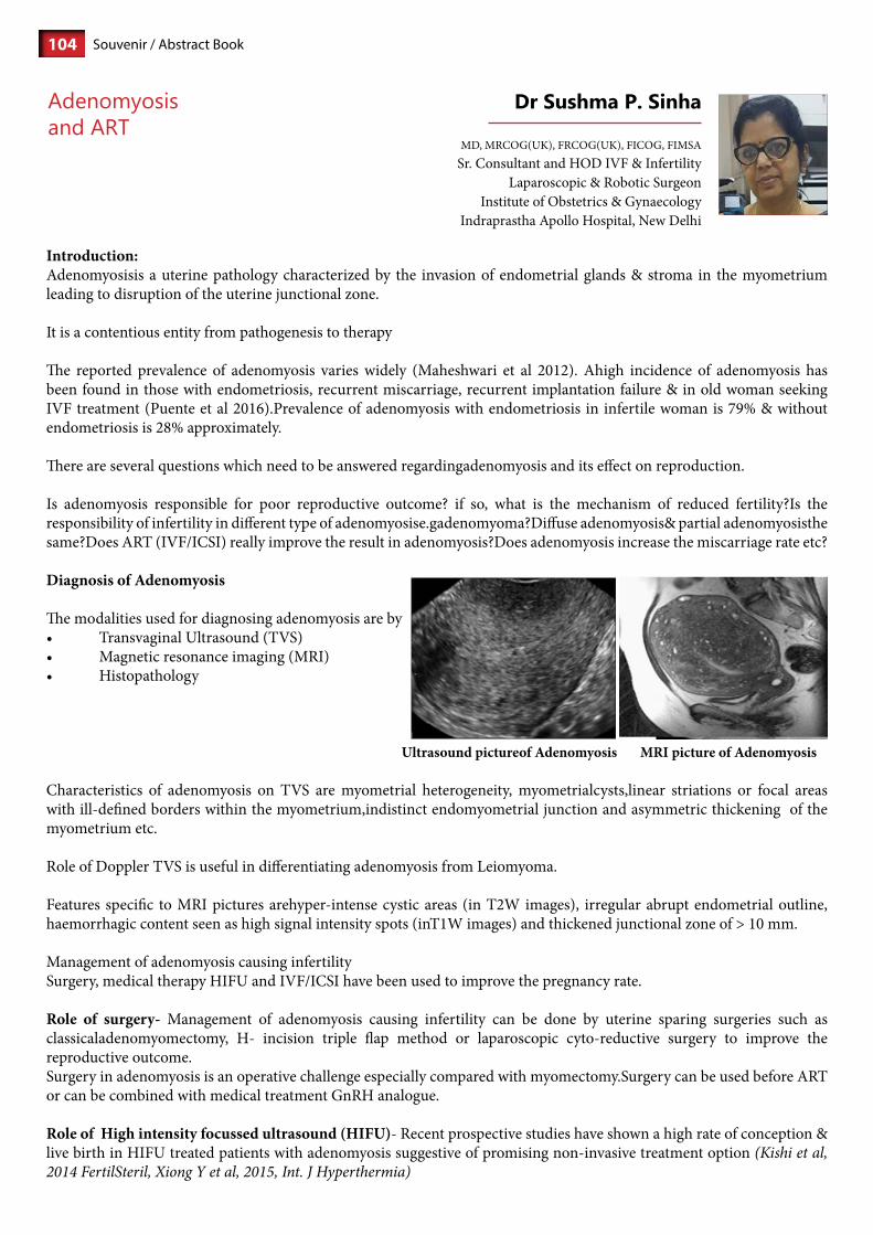
Souvenir / Abstract Book104
Dr sushma p. sinha
MD, MRCOG(UK), FRCOG(UK), FICOG, FIMSASr. Consultant and HOD IVF & Infertility
Laparoscopic & Robotic SurgeonInstitute of Obstetrics & Gynaecology
Indraprastha Apollo Hospital, New Delhi
Adenomyosisand ArT
Introduction:Adenomyosisis a uterine pathology characterized by the invasion of endometrial glands & stroma in the myometrium leading to disruption of the uterine junctional zone.
It is a contentious entity from pathogenesis to therapy
The reported prevalence of adenomyosis varies widely (Maheshwari et al 2012). Ahigh incidence of adenomyosis has been found in those with endometriosis, recurrent miscarriage, recurrent implantation failure & in old woman seeking IVF treatment (Puente et al 2016).Prevalence of adenomyosis with endometriosis in infertile woman is 79% & without endometriosis is 28% approximately.
There are several questions which need to be answered regardingadenomyosis and its effect on reproduction.
Is adenomyosis responsible for poor reproductive outcome? if so, what is the mechanism of reduced fertility?Is the responsibility of infertility in different type of adenomyosise.gadenomyoma?Diffuse adenomyosis& partial adenomyosisthe same?Does ART (IVF/ICSI) really improve the result in adenomyosis?Does adenomyosis increase the miscarriage rate etc?
Diagnosis of Adenomyosis
The modalities used for diagnosing adenomyosis are by• TransvaginalUltrasound(TVS)• Magneticresonanceimaging(MRI)• Histopathology
Ultrasound pictureof Adenomyosis MRI picture of Adenomyosis
Characteristics of adenomyosis on TVS are myometrial heterogeneity, myometrialcysts,linear striations or focal areas with ill-defined borders within the myometrium,indistinct endomyometrial junction and asymmetric thickening of the myometrium etc.
Role of Doppler TVS is useful in differentiating adenomyosis from Leiomyoma.
Features specific to MRI pictures arehyper-intense cystic areas (in T2W images), irregular abrupt endometrial outline, haemorrhagic content seen as high signal intensity spots (inT1W images) and thickened junctional zone of > 10 mm.
Management of adenomyosis causing infertilitySurgery, medical therapy HIFU and IVF/ICSI have been used to improve the pregnancy rate.
Role of surgery- Management of adenomyosis causing infertility can be done by uterine sparing surgeries such as classicaladenomyomectomy, H- incision triple flap method or laparoscopic cyto-reductive surgery to improve the reproductive outcome.Surgery in adenomyosis is an operative challenge especially compared with myomectomy.Surgery can be used before ART or can be combined with medical treatment GnRH analogue.
Role of High intensity focussed ultrasound (HIFU)- Recent prospective studies have shown a high rate of conception & live birth in HIFU treated patients with adenomyosis suggestive of promising non-invasive treatment option (Kishi et al, 2014 FertilSteril, Xiong Y et al, 2015, Int. J Hyperthermia)
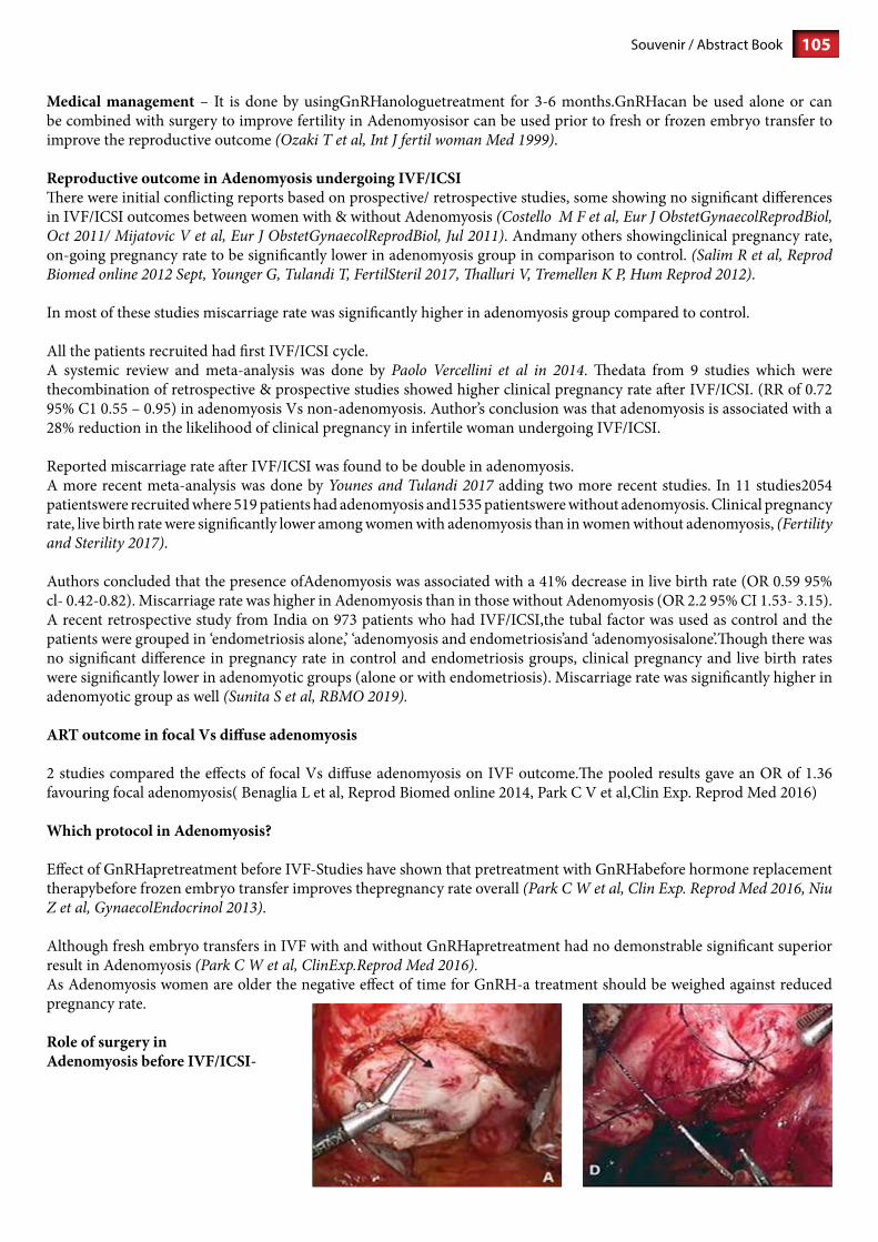
105Souvenir / Abstract Book
Medical management – It is done by usingGnRHanologuetreatment for 3-6 months.GnRHacan be used alone or can be combined with surgery to improve fertility in Adenomyosisor can be used prior to fresh or frozen embryo transfer to improve the reproductive outcome (Ozaki T et al, Int J fertil woman Med 1999).
Reproductive outcome in Adenomyosis undergoing IVF/ICSIThere were initial conflicting reports based on prospective/ retrospective studies, some showing no significant differences in IVF/ICSI outcomes between women with & without Adenomyosis (Costello M F et al, Eur J ObstetGynaecolReprodBiol, Oct 2011/ Mijatovic V et al, Eur J ObstetGynaecolReprodBiol, Jul 2011). Andmany others showingclinical pregnancy rate, on-going pregnancy rate to be significantly lower in adenomyosis group in comparison to control. (Salim R et al, Reprod Biomed online 2012 Sept, Younger G, Tulandi T, FertilSteril 2017, Thalluri V, Tremellen K P, Hum Reprod 2012).
In most of these studies miscarriage rate was significantly higher in adenomyosis group compared to control.
All the patients recruited had first IVF/ICSI cycle.A systemic review and meta-analysis was done by Paolo Vercellini et al in 2014. Thedata from 9 studies which were thecombination of retrospective & prospective studies showed higher clinical pregnancy rate after IVF/ICSI. (RR of 0.72 95% C1 0.55 – 0.95) in adenomyosis Vs non-adenomyosis. Author’s conclusion was that adenomyosis is associated with a 28% reduction in the likelihood of clinical pregnancy in infertile woman undergoing IVF/ICSI.
Reported miscarriage rate after IVF/ICSI was found to be double in adenomyosis.A more recent meta-analysis was done by Younes and Tulandi 2017 adding two more recent studies. In 11 studies2054 patientswere recruited where 519 patients had adenomyosis and1535 patientswere without adenomyosis. Clinical pregnancy rate, live birth rate were significantly lower among women with adenomyosis than in women without adenomyosis, (Fertility and Sterility 2017).
Authors concluded that the presence ofAdenomyosis was associated with a 41% decrease in live birth rate (OR 0.59 95% cl- 0.42-0.82). Miscarriage rate was higher in Adenomyosis than in those without Adenomyosis (OR 2.2 95% CI 1.53- 3.15).A recent retrospective study from India on 973 patients who had IVF/ICSI,the tubal factor was used as control and the patients were grouped in ‘endometriosis alone,’ ‘adenomyosis and endometriosis’and ‘adenomyosisalone’.Though there was no significant difference in pregnancy rate in control and endometriosis groups, clinical pregnancy and live birth rates were significantly lower in adenomyotic groups (alone or with endometriosis). Miscarriage rate was significantly higher in adenomyotic group as well (Sunita S et al, RBMO 2019).
ART outcome in focal Vs diffuse adenomyosis
2 studies compared the effects of focal Vs diffuse adenomyosis on IVF outcome.The pooled results gave an OR of 1.36 favouring focal adenomyosis( Benaglia L et al, Reprod Biomed online 2014, Park C V et al,Clin Exp. Reprod Med 2016)
Which protocol in Adenomyosis?
Effect of GnRHapretreatment before IVF-Studies have shown that pretreatment with GnRHabefore hormone replacement therapybefore frozen embryo transfer improves thepregnancy rate overall (Park C W et al, Clin Exp. Reprod Med 2016, Niu Z et al, GynaecolEndocrinol 2013).
Although fresh embryo transfers in IVF with and without GnRHapretreatment had no demonstrable significant superior result in Adenomyosis (Park C W et al, ClinExp.Reprod Med 2016).As Adenomyosis women are older the negative effect of time for GnRH-a treatment should be weighed against reduced pregnancy rate.
Role of surgery inAdenomyosis before IVF/ICSI-

Souvenir / Abstract Book106
There is no evidence of improved outcome of IVF/ICSI after cytoreductive surgery.
Hence, surgery is only recommended for symptomatic patients
Conclusion:
Uterine adenomyosis is a contentious entity from pathogenesis to therapy.Adenomyosis has a detrimental effect on IVF clinical outcome. It reduces pregnancy and live birth rates and increases the miscarriage rate.
Before embarking on medically assisted reproductive procedure, it is reasonable to suggest screening for adenomyosis by high resolution TVS and by MRI and the presence of adenomyosis should be discussed with infertile women who are considering IVF/ICSI.
As of now, it seems prudent to adopt GnRHa long protocols with immediate IVF/ICSI in women with adenomyosis and if the time permits to go for frozen embryo transfer afterGnRHa treatment before HRT. Further studies are needed.There is limited evidence for an improved outcome after surgery and surgery should only be an option for symptomatic women with repeated IVF/ICSI failures.
References:1. AbhaMaheshwari, SumanaGurunath, Farah Fatima, Siladitya Bhattacharya,“Adenomyosis and subfertility: a systematic review
of prevalence, diagnosis, treatment and fertility outcomes,Human Reproduction Update, Volume 18, Issue 4, July 2012, Pages 374–392.
2. J. M. Puente, A. Fabris, J. Patel, A. Patel, M. Cerrillo, “A. Requena, and J. A. Garcia-VelascoAdenomyosis in infertile women: prevalence and the role of 3D ultrasound as a marker of severity of the disease”,ReprodBiolEndocrinol. 2016; 14: 60. Published online 2016 Sep 20. doi: 10.1186/s12958-016-0185-6.
3. Kunz G, Beil D, Huppert P, Noe M, Kissler S, Leyendecker G. “Adenomyosis in endometriosis--prevalence and impact on fertility”. Evidence from magnetic resonance imaging.Hum Reprod. 2005 Aug;20(8):2309-16. Epub 2005 May 26.
4. Kishi Y, Yabuta M, Taniguchi F. “Who will benefit from uterus-sparing surgery in adenomyosis-associated subfertility”? FertilSteril. 2014 Sep;102(3):802-807.e1. doi: 10.1016/j.fertnstert.2014.05.028. Epub 2014 Jun 19.
5. Bin Xie, Cai Zhang, ChunyanXiong, Jia He, Guohua Huang &Lian Zhang, “High intensity focused ultrasound ablation for submucosal fibroids:International Journal of Hyperthermia Volume 31, 2015 - Issue 6 A comparison between type I and type II” | 27 Apr 2015, Pages 593-599 Published online: 05 Jun 2015
6. Ozaki T, Takahashi K, Okada M, Kurioka H, Miyazaki K “Live birth after conservative surgery for severe adenomyosis following magnetic resonance imaging and gonadotropin-releasing hormone agonist therapy”.Int J FertilWomens Med. 1999 Sep-Oct;44(5):260-4.
7. Costello MF, Lindsay K, McNally G. “The effect of adenomyosis on in vitro fertilisation and intra-cytoplasmic sperm injection treatment outcome.” Eur J ObstetGynecolReprod Biol. 2011 Oct;158(2):229-34.
8. Mijatovic V, Florijn E, Halim N, Schats R, Hompes P. “Adenomyosis has no adverse effects on IVF/ICSI outcomes in women with endometriosis treated with long-term pituitary down-regulation before IVF/ICSI.” Eur J ObstetGynecolReprod Biol. 2010 Jul; 151(1):62-5.
9. Salim R, Riris S, Saab W, Abramov B, Khadum I, Serhal P. “Adenomyosis reduces pregnancy rates in infertile women undergoing IVF.” Reprod Biomed Online. 2012 Sep;25(3):273-7.
10. Thalluri V, Tremellen KP“Ultrasound diagnosed adenomyosis has a negative impact on successful implantation following GnRH antagonist IVF treatment.” Hum Reprod. 2012 Dec; 27(12):3487-92.
11. Vercellini P, Consonni D, Dridi D, Bracco B, Frattaruolo MP, Somigliana E. “Uterine adenomyosis and in vitro fertilization outcome: a systematic review and meta-analysis.” Hum Reprod. 2014 May; 29 (5):964-77.
12. Younes G, Tulandi T. “Effects of adenomyosis on in vitro fertilization treatment outcomes: a meta-analysis.” FertilSteril. 2017 Sep; 108(3):483-490.
13. Sunita Sharma, Shikha Bathwal, Nupur Agarwal, Ratna Chattopadhyay, Indranil Saha, Baidyanath Chakravarty “Does presence of adenomyosis affect reproductive outcome in IVF cycles? A retrospective analysis of 973 patients” RBMO: volume 38, issue 1, 2019
14. Benaglia L, Cardellicchio L, Leonardi M, Faulisi S, Vercellini P, Paffoni A, Somigliana E, Fedele L. Asymptomatic adenomyosis and embryo implantation in IVF cycles. Reprod Biomed Online. 2014 Nov; 29(5):606-11.
15. Chan Woo Park, Min Hye Choi, Kwang Moon Yang, In Ok Song “Pregnancy rate in women with adenomyosis undergoing fresh or frozen embryo transfer cycles following gonadotropin-releasing hormone agonist treatment” ClinExpReprod Med 2016;43(3):169-173
16. Niu Z, Chen Q, Sun Y, Feng Y. “Long-term pituitary downregulation before frozen embryo transfer could improve pregnancy outcomes in women with adenomyosis. GynecolEndocrinol. 2013 Dec; 29(12):1026-30.
107Souvenir / Abstract Book
shweta mittal Gupta
Senior ConsultantCentre of IVF & Human reproduction
Sir Ganga ram Hospital
Surprises during oocyteretrieval and embryo transfer
Oocyte retrieval and embryo transfer are two important steps in an IVF cycle.Usually anticipated difficulty to access ovaries can be determined before hand by taking good hisrotry from patient and preliminary scans. The biggest surprise during oocyte retieval is in cases of spontaneous ovulation .These would more often be seen in poor responders or escape ovulation in antagonist cycles. Reports have shown successful pregnancies from aspirating remaining moderate and small size follicles and invitro maturation.Also in presence of immediate ovulation fluid from pouch of douglas can be aspirated and checked for oocytes. Secondly unsuccessful oocyte retrieval after apparently successful ovarian stimulation (also referred to as ‘empty follicle syndrome’) occurs in 1–7% of women undergoing assisted reproductive techniques. The aetiology is not clear, but probably multifactorial, and occurs in natural and stimulated cycles. In many cases, technical problems such as errors in human chorionic gonadotrophin (HCG) or GnRH agonist administration or defects in HCG batches can be identified, but this is not sufficient to account for all reported cases. The term empty follicle syndrome is inappropriate in cases in which such procedural factors can be identified. In many patients, however, unsuccessful oocyte retrieval appears to be due to an underlying ovarian dysfunction, and some may have a genuine empty follicle syndrome. Appropriate measures, such as monitoring of serum β-HCG, should be taken to minimize the risk of unsuccessful oocyte retrieval.
Inaccessible ovaries are usualy not in the POD but above the uterus When the ovary in not accessible (be it in the POD or elsewhere) and there is an increased risk of damage to internal organs or large vessels oocyte retrieval from the inaccessible ovary should not be performed. Oocyte retrieval should be performed on the accessible side. External abdominal manual pressure by an assistant often helps in making ovaries accessible,alternativelygrasping the cervix with a tenaculum and pulling the uterus is extremely helpful in difficult cases.
Rarely may have to pass through the uterus to reach the ovary
Even more rarely, when there is no other choice, you can reach the ovaries transabdominally. If, on pre-treatment scan, both ovaries are inaccessible transvaginally, the possibility of laparoscopic oocyte retrieval should be explored, although this may also not be possible due to the pelvic pathologyIt is important that inaccessible ovaries are identified prior to the start of the treatment so that the patient is well informed of possible problems before treatment starts.
Surprises during embryo transfercan be difficult cervical negotiation and retained embryos. For difficult cervical entry start with exaggerated lithotomy, Abduction of legs helps in better visualization, centralization of cervix with possibly straightening of uterocervical length. Malleable stylet introduced inside the outer embryo transfer catheter and bending it according to version of uterus helps in most of the case to negotiate even in an acute anteversion .If still difficulty is encountered, speculum is changed to sim’s speculum along with tenaculum to pull the cervix. Outer embryo transfer sheath along with stylet should be used to bend the cathere as per the direction of the uterus before uterine sounding.Rarely uterine sounding is attempted to identify direction of uterine cavity. After all the manuevers if still cervix cannot be negotiated then either embryo transfer is planned on same day under anesthesia or electively embryos are frozen. Decision for hysteroscopy is discussed and further frozen thawed embryo transfer is planned under general anesthesia directly. If embryologist declares that embryo/s are retained, then if the embryos are not damaged, decision for retransfer is taken. Fresh outer embryo transfer catheter is introduced by the clinician. Embryo is reloaded and handed over by embryologist for retransfer. After embryo transfer under ultrasound guidance, embryologist again checks for embryo retention.

Souvenir / Abstract Book108
Dr swati verma
Senior Consultant Jindal IVF Centre Chandigarh
Does Perinatal outcome matter in IVF? Fresh Vs Frozen embryos
Practice of frozen embryo transfer (FET) has been increased due to improved cryopreservation technologies. FET has also been widely adopted in the concern of patient safety to avoid OHSS and with the application of pre-implantation genetic testing. Some evidence suggest that FET may increase pregnancy rates and lead to more favorable perinatal outcomes . Routine clinical practice is gradually shifting toward ‘‘freeze-all’’ cycles or preferential FET over fresh ET.
Recently published literature document that pregnancies resulting from FETwere associated with lower relative risks of placenta previa, placental abruption, low birth weight, small for gestation age babies as compared to fresh ET. It was also observed that FET pregnancies were associated with increased risks of pregnancy-induced hypertension, postpartum hemorrhage, and large for gestational age babies compared with fresh ET. The risks of gestational diabetes mellitus, preterm premature rupture of membranes, and preterm birth (PTB) showed no differences between frozen and fresh ET.
Explanation of adverse perinatal & maternal outcomes is uncertain. It could be multifactoreal, related to procedure and method of cryofreezing or may be due to endometrial preparation. Altered genetic and epigenetic stabilities, altered gene expression, increased DNA fragmentation were observed in cryofrozen embryos compared with fresh embryos. Vitrified embryos show more significant effect than slow-frozen embryos. Increased abnormality in shape of spindles in during vitrification, affecting normal cell division could be one of the explanations.
Women conceived by FET after endometrial preparation with estrogen replacement were found to be associated with high risks of developing hypertensive disorders of pregnancy, placenta accrete, PPH, post term birth and macrosomia. While women with FET pregnancies after stimulated cycles or natural cycles were not at high risk for developing these adverse outcomes. Corpus luteum in natural or stimulated cycles seems to have a crucial role in maternal circulatory adaptation during pregnancy.
Although available data regarding FET provides reassurance for the safety of frozen thawed ET, there are some lingering concerns related to risk for large for GA babies, hypertensive disorders and PPH to mother. We should be cautious in planning of FET in the women of advanced age, chronic hypertension and other medical disorders. So it is advisable not to adopt “Elective freezing of all embryos” policy and FET should be performed when there is definite clinical indication.

109Souvenir / Abstract Book
Dr sweta Gupta
Clinical Director & Sr Consultant( reproductive Med. & IVF )
Medicover Fertility, DelhimBBs, mD (obs& Gynae, Delhi) mrcoG ( london, uk ), Dfsrh ( uk )
msc ( r&D , Bristol, uk ) fellowship in rm & arT ( london, uk )
PGT(A) FOr ALL?
Preimplantation genetic testing for aneuploidy (PGT-A) (formerly called preimplantation genetic screening [PGS]) is to identify embryos with de novo aneuploidy, in embryo(s) of couples presumed to be chromosomally normal, it also includes sub-chromosomal deletions and additions (duplications). This technique avoids transferring of these abnormal embryos, thereby reducing the risk of miscarriage and complications related to pregnancy failure and improving the probability of conceiving a viable pregnancy.
Chromosomal aneuploidies are seen in up to 70% of spontaneous miscarriages. It is one of the major causes of maternal age-related reduced fertility potential. Euploid embryo transfer results in superior clinical pregnancy and live birth rates, it reduces miscarriage risk independent of maternal age.
PGT-A helps to reduce the need of multiple IVF cycles and shorten time to viable pregnancy. PGT-A requires in-vitro fertilisation (IVF) as part of the process even though many of these couples have no known difficulties conceiving a pregnancy. The inconvenience, risks and expense of PGT sometimes limit the utilization of this technology. But for the best outcome, PGT-A should be integrated for all.
The current indications for PGT include repeated implantation failures, repeated pregnancy loss, advanced maternal and paternal age, male factor infertility, and genetic disorders in the parents including mosaicism of sex chromosomes, structural rearrangements, and monogenic genetic diseases. Scott et al. published a paper in 2013 showing the analysis of an RCT. The trial showed that with comprehensive chromosome screening(CCS) and fresh blastocyst transfer, sustained IR was significantly higher in the CCS group (66%) compared to control non-CCS group (48%). It also showed a higher delivery rate per cycle in CCS group (85%) compared to control non-CCS group (68%).
Meta-analysis of randomized controlled trials on preimplantation genetic screening with comprehensive chromosome screening versus routine care. ( Parikh et al. )Dahdouhet al. carried out a meta-analysis of RCTs and observational studies to see whether PGS with comprehensive chromosome screening (CCS) improved clinical IR and sustained IR (beyond 20 weeks) compared with routine care for embryo selection in IVF cycles. They concluded that PGS with the use of CCS technology increases clinical and sustained IRs, thus improving embryo selection particularly in patients with normal ovarian reserve
From the clinical point of view, the benefits attributed to invasive PGT-A in the selection of euploid embryos remain controversial, especially due to the lack of scientific proof of its effectiveness in increasing live birth rates in various clinical situations, such as patients with advanced age, repeated implantation failures or recurrent miscarriages. One of the most important criticism, difficulty in accurately assessing the presence of embryonic mosaicism creating significant levels of false positive results, and worse, causing a real possibility of discarding healthy embryos. This makes the clinical application of PGT-A as a risky approach (Munnéet al., 2017; Spinellaet al., 2018).
Another problem isneed to perform PGT-A by experienced embryologists, since otherwise the embryonic loss due to biopsy would be a frequent fact, something usually estimated below 10% but in some laboratories it may reach up to 30% of biopsied embryos (Munné, 2018). On the other hand, there are doubts about the future risks of invasive action of the usually 5-10 cell removed during biopsy for genetic diagnosis. Recently, (Xu et al.) described noninvasive chromosomal screening (NICS) by obtaining and sequencing free DNA dripped by embryos in the culture medium (without the need of embryo biopsy) creating a new non-aggressive and elegant perspective to preimplantation genetic diagnosis.
Large, prospective, well controlled studies are required for enhanced embryo selection by PGT A. At present, PGT A for all, as universal screening should be done after careful counselling to shorten time to successful life birth outcome.

Souvenir / Abstract Book110
References :
1. Scott RT Jr., Upham KM, Forman EJ, Hong KH, Scott KL, Taylor D, et al. Blastocyst biopsy with comprehensive chromosome screening and fresh embryo transfer significantly increases in vitro fertilization implantation and delivery rates: A randomized controlled trial. FertilSteril 2013;100:697-703
2. Parikh FR, Athalye AS, Naik NJ, Naik DJ, Sanap RR, Madon PF. Preimplantation genetic testing: Its evolution, where are we today?. J Hum Reprod Sci 2018;11:306-14
3. Dahdouh EM, Balayla J, García-Velasco JA. Comprehensive chromosome screening improves embryo selection: A meta-analysis. FertilSteril 2015;104:1503-12.
4. Franco JG Jr. New perspectives with the use of noninvasive chromosome screening (NICS) in ART. JBRA Assist Reprod. 2019 Oct 14;23(4):321-322. doi: 10.5935/1518-0557.20190071
5. Munné S, Blazek J, Large M, Martinez-Ortiz PA, Nisson H, Liu E, Tarozzi N, Borini A, Becker A, Zhang J, Maxwell S, Grifo J, Babariya D, Wells D, Fragouli E. Detailed investigation into the cytogenetic constitution and pregnancy outcome of replacing mosaic blastocysts detected with the use of high-resolution next-generation sequencing. FertilSteril. (e8) 2017;108:62–71. doi: 10.1016/j.fertnstert.2017.05.002.
6. Spinella F, Fiorentino F, Biricik A, Bono S, Ruberti A, Cotroneo E, Baldi M, Cursio E, Minasi MG, Greco E. Extent of chromosomal mosaicism influences the clinical outcome of in vitro fertilization treatments. FertilSteril. 2018;109:77–83. doi: 10.1016/j.fertnstert.2017.09.025.
7. Munné S. Status of preimplantation genetic testing and embryo selection. Reprod Biomed Online. 2018;37:393–396. doi: 10.1016/j.rbmo.2018.08.001.
8. Xu J, Fang R, Chen L, Chen D, Xiao JP, Yang W, Wang H, Song X, Ma T, Bo S, Shi C, Ren J, Huang L, Cai LY, Yao B, Xie XS, Lu S. Noninvasive chromosome screening of human embryos by genome sequencing of embryo culture medium for in vitro fertilization. Proc Natl Acad Sci U S A. 2016;113:11907–11912. doi: 10.1073/pnas.

111Souvenir / Abstract Book
Dr. umesh Jindal
Jindal IVF and Sant Memorial Nursing Home,Chandigarh
Managing Complicationsof Ovum Pick Up
Transvaginal ovum pick up is a safe and simple procedure. The technique is easy to learn and master. Complications can be related to ovarian stimulation which are ovarian hyperstimulation syndrome, ovarian torsion, and ectopic pregnancy. Complications may be related to oocytes e.g. empty follicle syndrome, poor quality etc. There are rare life threatening surgical complications which may require hospitalization or emergency surgery. The complication rate is less than 1%. Because of the rarity of the complications, the anticipation, prevention and management of such complications remain theoretical for most practioners (Levi-Setti 2018). The complications can be grouped as haemorrhagic, infection, trauma to adjoining structures or some rare complications.
Vaginal haemorrhage is the most commonly encountered complication of the above. Usually mild, self limiting and require only some pressure with gauze. Rarely bleeding may require vaginal packing or suturing of laceration in vaginal vault. Upto 200 ml of bleeding from ovary or vaginal vault may collect in pelvis without causing any discomfort or hemodynamic instability to the patient. Massive haemorrhage requiring surgical intervention is a serious but life threatening condition. Bleeding may develop immediately or after 24 hours because of slow leaks.
Infections of urinary tract also can occur after OPU. Incidence is quoted in the range of .3-.6 %. A more severe pelvic inflammatory disease or frank sepsis may also occur. Predisposing factors may be presence of latent infection, endometrioma, previous surgeries or occult gut injury. Conservative management with broad spectrum antibiotics is usually effective. Ovarian or pelvic abscess may develop and require surgical intervention.
Bladder many times is interposed between vaginal vault and uterus and gets pricked. A transient haematuria may occur which resolves on its own. Catheterization is sufficient for a more severe form of haematuria. One of the rare complications reported is bladder wall psedo- aneurysm leading to massive haemorrhage after few days. Uretric injuries have also been reported which may present as haematoma or urinomas in pelvis.
Complications decrease after experience. Everyone has to be aware of the possible complications and learn to prevent and treat these.

Souvenir / Abstract Book112
Dr. vandana Bhatia
Southend Fertility & IVFNew Delhi
receptive Endometrium – Can we identify ?
Endometrial receptivity (ER) plays a crucial role in successful implantation.Human endometrium is a dynamic tissue undergoing complex series of organized proliferative and secretory changes at different stages during the menstrual cycle in response to hormones, exhibiting a short period of receptivity known as “window of implantation” (WOI).
This restricted time period is the time when there is temporary unique collaboration of extremely large number of different factors which make the uterine environment conducive to blastocyst acceptance and implantation. Endometrial receptivity remains the last barrier in ART and as has been suggested in literature, two thirds of implantation failures are due to defective endometrial receptivity. The dynamic transition from a non receptive to a receptive endometrium remains a mystery and the identification of human implantation window still remains unsolved despite extensive research and advances in ART.
Extensive efforts have been made to understand and characterize a receptive endometrium starting from histological dating to current “omics technologies” which include- genomics (study of genes), epigenomics (study of epigenetic DNA modifications), transcriptomics (study of gene expression), proteomics (quantification of proteomics), metabolomics and lipidomics (composition and quantification of metabolites and lipids). Several histologic endometrial dating criteria, the most common being of Noyes have been commonly used but their accuracy, reproducibility and functional relevance has been questioned in various randomized studies and are now obsolete. Endometrial pinopods as identified on electron microscopy were considered as a marker for ER but since their presence was also demonstrated in post receptive endometrium, it precluded their use for the same.Cervical mucus has also been used to date ER by analyzing cytokines and growth factors produced by receptive endometrium and their transport to cervical mucus. However other studies failed to demonstrate the same and found no correlation between cytokine levels in cervicovaginal secretions and cytokine gene expression level in secretory endometrium . One or more panel of biochemical molecules during mid secretory phase predictive of endometrial receptivity have been proposed and these are integrins, selectins, cadherins, cytokines, anti-adhesion molecules, growth factors and immune markers . However none of these have found a place in clinical set up as they are all invasive and circumstantial.
Alternatively non invasive, easy, reliable methods like transvaginal ultrasonography to evaluate endometrial thickness , pattern, volume and blood flow can serve as a surrogate parameter but these do not seem to be an exact science. Currently transcriptomics, associated with WOI allows characterization of gene expression at the mRNA level , giving rise to “sample -specfic “molecular profile or its “Transcriptomic Signature” further permitting characterization of tissue function or disease phenotype. The transcriptome of the endometrium has been analysed in all phases of the menstrual cycle and there is a specific profile during the WOI which is affected or delayed by controlled ovarian stimulation and hormonal replacement therapy cycles.
Regardless of these studies, only two signatures became clinically validated tests – the ERA ( endometrial receptivity array ) and Win-Test ( Window Implantation Test). These were designed to personalize and shift the embryo transfer day according to the shift in WOI specially in cases of repeated implantation failures.. ERA is a customized array based on expression of 238 genes coupled to a computational predictor capable of diagnosing a functionally receptive endometrium where as the Win test is based on quantitative expression of 11 predictive genes of ER coupled with an algorithm for the identification of receptive status. Available data on fresh (stimulated cycles) and frozen ( natural /hormonal replacement cycles) show no difference in pregnancy or implantation rate. These findings suggest that the new biomarkers are highly conserved.But whatever said and done, despite 15 years of transcriptomic analysis on ER more studies are needed to optimize the selection of biomarkers of ER which influence pregnancy outcome.

113Souvenir / Abstract Book
Dr varun DuggalUltrasound examination
Ultrasound examination is becoming an increasingly important tool in infertility treatment. Ultrasound provides comprehensive assessment of the anatomy of female pelvis & pelvic pathologies. Besides routine 2D evaluation, 3D technology allows a multiplanar evaluation in planes not visible in routine 2D, most importantly the coronal plane of uterus. 4D creates a 3d view in real time or a live 3D scan.
Ultrasound can be used both in the diagnosis of infertility as well as follow up of treatment.Diagnosis of infertility: Ultrasound is used to look for potential causes of infertility in the uterus, ovaries and adnexae. It can be used in different phases of menstrual cycle to study the phasic changes in the female reproductive tract.
Examination of uterus: A routine 2D scan can measure the size of the uterus and vascularity (colour and power doppler). A 2D scan can evaluate and classify Mullerian anomalies (ESHRE classification), fibroid mapping and adenomyosis; however, a 3D scan offers more accurate diagnosis of these malformations, along with accurate localization & relationship of theses pathologies to the endometrium.
Endometrial thickness is assessed with 2D and morphology and focal lesions (polyp, fibroids, adhesions) are examined. With 3D the coronal plane can be obtained. We can measure the volume of endometrium and 3D circulation in endometrium and sub-endometrium. We can view these structures with a surface view, similar to the one used for hysteroscopy. With this information, we can easily explain the pathology to the physician and patients and easily plan the surgical procedures.
Examination of the tubes: With 2D US we can see the tubes in the pelvis only if there is dilatation, but sometimes it is difficult to distinguish them from the adnexal structures. With a 3D ultrasound and inversion mode, we can define the shape and continuity of the tube and we can view the tube from different angles to better ascertain the relationship with the ovary.
Examination of the ovary: With the 2D ultrasound the size of ovaries is measured and the morphology of ovaries is examined. Furthermore, position of the ovaries, mobility and tenderness with regard to their surroundings can be defined. With the 3D surface mode we can see the surface view of the inner layer of the follicle or the cyst. The volume of the liquid containing structures like follicles can be measured with VOCAL or sonoAVC (sono automated volume count). The number of the antral follicles strongly correlates with fertility potential markers such as FSH and AMH.
Examination of peritoneum: The position of pelvic organs along with tenderness, relative mobility and ascites can be defined. Also peritoneal nodules and endometriotic deposits can be better evaluated.
Examination of vagina: Endometriotic nodules can be excluded with ultrasound. A 3D ultrasound can define the exact position of the nodule.
Follow up of infertility treatment: Before ovarian stimulation, it is mandatory to exclude pelvic pathology that can influence the stimulation. Both natural and stimulated cycles are monitored with an ultrasound for follicular and endometrial growth. SonoAVC offers automatic volume count of all follicles. This information enables us to change the stimulation protocol and avoid OHSS more accurately comparing to the standard 2D technology.
IVF procedure use: Ovum pickup along with oocyte puncture is done with ultrasound-guided needle. Great vessels around the vagina and on the needle line can be avoided if colour doppler is used. With 3D surface mode, attempt can be made to identify good quality follicles.
Endometrial assessment for Embryo transfer (ET): Endometrial thickness is assessed with 2D and morphology is studied with search for focal lesions, which can interfere with implantation. Endometrial & sub-endometrial vascularity is assessed using colour and power doppler. The overall blood flow to the uterus is assessed by uterine artery evaluation which further can provide a hint for future development of a good uteroplacental circulation conducive to the growing pregnancy.
Using volume technology, we can measure the whole volume with VOCAL (virtual organ computer-aided analysis) and 3D circulation with the index (VI – vascular index, FI – flow index and VFI – vascular flow index) in the uterus.
During embryo transfer, angle between cervical canal and corpus uteri can be measured before the embryo transfer. The introduction of the ET catheter can be followed with transabdominal ultrasound. More exact location of the catheter can be visualized with 4D US.
It is important to learn the applications and use them in our daily practice. The recorded volumes can be examined at a later time. In any unclear pathology, the recorded volume can be used for a second opinion.

Souvenir / Abstract Book114
aBsTracTsFOr WOrKSHOPS

115Souvenir / Abstract Book
arne sundePhD
Professor emeritus, Department of Clinical &Molecular Medicine, Norwegian University ofScience and Technology, Trondheim, NorwayEditor in Chief Human Reproduction Update
Understanding the peer review process
In the Human reproduction journals, every manuscript submitted is screened by the Editorial Team according to the following criteria:
1. Scope; is the manuscript dealing with a subject that is within the scope of the journal?2. Publications ethics; is the manuscript under consideration elsewhere and is there any copyright issues concerning
illustrations/figures? Plagiarism is tested with a suitable software.3. The nature of the manuscript: original research, a review or an opinion? Every journal has a policy concerning which
type of manuscripts they accept. A review journal such as Human Reproduction Update (HRU) only accepts review papers that summarises already published information. HRU also has a proposal system. The authors must first submit a structured proposal (1-2 pages). If the proposal is accepted, the authors can submit a full manuscript. The other Human reproduction journals accept originals research as well as Opinions papers.
4. Is the manuscript structured and formatted according to the Instructions to Authors?5. Is the quality of the manuscript sufficient to be sent out for review?
The take home message here is: Read the “Instructions to Authors” carefully. Manuscripts that does not comply risk immediate rejection.
Manuscripts that are accepted for reviewwill be sent to an Associate Editor (AE) that has expert knowledge of the topics the manuscript deals with. The AE then selects reviewers according to several criteria:
The reviewer shall be a specialist on the topic of the manuscript.The reviewer must not have any conflict of interests concerning the topic or the authors.The reviewer has a track record of excellent and constructive reviews.
When the review reports are returned, the AE must judge the quality of the reviews and give a recommendation. Usually this falls in three categories:
Accept with minor revisions Major revision is required. Rejected (no appeal)
When revised manuscripts are returned, they will again go to the AE and the reviewers and may be accepted, accepted pending further revision or finally rejected.
The final judgement is then made by the Deputy Editor(s) and/or the editor in Chief.After final acceptance the manuscript goes to a final check by the production team before being published.

Souvenir / Abstract Book116
Dr. ashok agarwalProfessor of Surgery (Urology),Lerner College of Medicine and
Case Western reserve UniversityDirector, Andrology Center & reproductive Tissue bank
Director, American Center for reproductive MedicineStaff, Departments of Urology, Ob‐Gyn, Immunologyand Anatomic Pathology Cleveland Clinic Foundation
Cleveland, Ohio 44195, United States
Automated Computer Semen Analyzers, Home Sperm Testing, Oxidation reduction Potential: What is the benefit of these new technologies to the Clinicians?
Conventional semen analysis is an essential component of the male infertility workup, but requires laboratories to rigorously train and monitor technicians as well as regularly perform quality assurance assessments. There has been controversy regarding the accuracy and reliability of the routine manual semen analysis due to high degree of subjectivity due to human evaluation. The computer-based semen analysis provides a more standardized results withthe availability of various computer assisted semen analyzers.A novel, automated, artificial intelligence optical microscopic (AIOM)‐based technology, LensHooke™ X1 PRO analyzer (Bonraybio, Taiwan) showed high degree of correlation in the results of concentration, progressive motility and progressively motile sperm concentration compared to manual method, X1 PRO computer semen analyzer can be used as a reliable diagnostic tool for routine semen analysis.However, the method of producing a semen sample in a laboratory setting tends to be a stressful and difficult task for many men. This led to the development of novel, commercially available, and affordable at-home-based semen analysis screening tests.There are several home-based semen tests that have been approved for use by the US Food and Drug Administration (FDA). They allow men to perform and interpret the test in the comfort of their own home.
The YO Home Sperm Test (Medical Electronic Systems, Los Angweles, USA was the first commercially available smartphone-based semen testing device The YO device utilizes the smartphone camera to capture the light fluctuations caused by movement of sperm. The device determines the sperm concentration and motility to ultimately calculate motile sperm concentration. A double-blind trial comparing the YO Home Sperm Test and an automated laboratory analyzer (SQA-Vision) showed a good correlation coefficient. The YO exhibited an accuracy of 97.8% with a precision of 16%. YO Home Sperm Test allows users to accurately detect an abnormal sperm motile sperm count, a valuable parameter for screening fertility potential, in the convenience and privacy of their home environment. Wide usage of such home sperm testing may be of potential interest for clinicians/physicians to engage reluctant men in the fertility assessment process. Considering the challenges associated with semen analysis, the last several decades have seen the development of many advanced tests for sperm DNA fragmentation, acrosome reaction, and capacitation. While these new diagnostic tests have improved the scope of information available to clinicians, they are expensive, time-consuming, and require specialized training. The latest advance in laboratory diagnostics is the measurement of seminal oxidation-reduction potential (ORP). The measurement of ORP in an easy, reproducible manner using a new tool called the Male Infertility Oxidative Stress System (MiOXSYS) (Aytu Bioscience Inc, Englewood, CO) has demonstrated ORP’s potential as a feasible adjunct test to conventional semen analysis. Additionally, the measurement of ORP by this device has been shown to be predictive of both poor semen quality and male infertility. A cut-off value of 1.34 mV/106sperm/ml can differentiate the infertile men with abnormal semen parameters. Assessing ORP is a novel approach to both validating semen analysis results and identifying patients who may benefit from treatment of male oxidative stress infertility (MOSI). In conclusion, advancement in the new technologies can help the busy clinicians to address the infertility issues in men at an earlier stage and therefore greatly improve patient satisfaction, clinical efficiency, and management.

117Souvenir / Abstract Book
Dr arundhati athalyePh.D.
IVF Genetics, Jaslok Hospital
Genes and PGD
Genetic information carried by the egg and the sperm is unique, as they unite at fertilization. The progeny inherits genes from both the biological parents and these genes in turn express specific traits.
Periodically, along the chromosomes, a small patch of DNA containing specific instructions about how to make a particular protein occurs. Such a coded area of information is called a gene. Genes lie within the chromosomes.Each cell in the body contains an exact copy of all the many genes and, with the exception of identical twins, their combination is unique to the individual. Genetic Abnormality: A genetic disorder is a disease that is caused by an abnormality in an individual’s DNA. Most genes come in pairs, one from the egg and one from the sperm (father). When, the function of a gene is altered by a change (called a mutation) in the specific sequence, a genetic disease results. These mutations can be transmitted in families from generation to generation (inherited), or can be a new change in an individual (de novo). These mutations lead to either single gene or multiple gene disorders. Abnormalities can be as small as a single-base mutation in just one gene, or they can involve the addition or subtraction of entire chromosomes. Every year, an estimated 7.9 million infants (6% of worldwide births) are born with serious birth defects. Some congenital defects can be controlled and treated. An estimated 3.2 million of these children are disabled for life. Monogenic disorders: Monogenetic disorders are caused by a mutation in a single gene. The mutation may be present on one or both chromosomes (one chromosome inherited from each parent). Examples of monogenic disorders are: Thalassemia, Sickle cell disease, cystic fibrosis, polycystic kidney disease, Tay-Sachs disease. A dominant genetic disease is caused by a mutation in one copy of a gene. A recessive genetic disease is caused by a mutation in both copies of a gene. A carrier has one normal copy of the gene and one copy with a mutation. Sex-linked genetic diseases are caused by mutations on the X or Y chromosomes. Sex-linked diseases can be dominant (Rett syndrome) or recessive (Hemophilia) and affect males and females differently. The risk for a mother who is a carrier of a recessive sex linked disease to have a son with the condition is 50% whereas the daughter will again remain normal (50%) or be carrier (50%) as she would have one normal X chromosome from her father. In some genetic diseases, the abnormality in the gene keeps increasing in every subsequent generation and its expression is seen only when an affected child is born.e.g. Fragile X Syndrome Hence, if there is any history of having an affected child, it is advisable to undergoGenetic Testing.Preimplantation Genetic Testing (PGT, formerly known as Preimplantation Genetic Diagnosis or PGD):PGT is the earliest form of diagnosis of chromosomal and inherited single gene disorders on embryos prior to implantation. It is a powerful tool used during an ICSI cycle to detect embryos that are at risk of developing a serious genetic disease. PGT is performed on an embryo biopsy sample and identifies which embryos are not at an increased risk of developing genetic abnormality or disease. There are three types of PGT:
•Preimplantationgenetictestingformonogenic(single-gene)disorders(PGT-M)•Preimplantationgenetictestingforstructuralrearrangements(PGT-SR)•Preimplantationgenetictestingforaneuploidy(PGT-A)
Steps involved in PGT1. Diagnosis of the disorder in the couple undergoing PGT (Pre-PGT work up):
In this, both the partners should be first tested for the single gene disorder. It is always advised to carry out chromosomal analysis to rule out structural and numerical abnormalities, which may result in recurrent spontaneous abortions in future. Once the required pre-PGD work up is done, then the couple will be advised to undergo the IVF cycle.
2. IVF cycleIn this, the female partner is treated with hormonal stimulation in order to obtain adequate number of eggs as possible. The ICSI (intracytoplasmic sperm injection) procedure using single sperm per egg is carried out. This is incubated in the laboratory and kept in in the culture to allow fertilization and formation of the embryo.3. Embryo biopsyDay 5-6 blastocyst is selected for this procedure. An opening is created into the zona of the embryo on day 3 at 8 cell stage using a non-contact diode LASER beam (hatching). The blastocyst stage embryo is further incubated till the trophectoderm cells come out from the opening (herniation). Around 4-8 trophectoderm cells are separated from the embryo. All the above steps are carried out in the Embryology laboratory.
4. Analysis For PGD for single gene disorder, the biopsied cells are “tubed”. Whole genome amplification is carried out to increase the quantity of DNA externally. Then the amplified product is subjected to further testing for the single gene disorder. Also the parental genetic markers are matched to the embryonic markers.
After the analysis,embryos reported as normal are transferred back to the mother’s uterus to achieve a pregnancy which is genetically unaffected and euploid.

Souvenir / Abstract Book118
Dr ethiraj Balaji prasathHead, Chief Embryologist
Thomson Fertility Centre, Singapore
Lab Perspectives in OPU
Ovum pick up (OPU) or oocyte retrieval (OR) or egg retrieval (ER) is an important procedure in IVF treatment of an infertile couple. It is done around 35-36 post trigger after stimulating the ovaries with a suitable stimulation regime. Lab prepares for OPU a day before the procedure. Embryologist assesses expected number of follicles to be recovered from the follicular tracking scan. Preparation includes media and dishes to culture oocytes retrieved, process sperm and culture embryos resulting from fertilization either by IVF or ICSI.
A mature follicle is supposed to be around 16 mm in diameter on the day of trigger. Counting number of follicles reaching this size, Embryologist prepares number of dishes and tubes, quantity of media and related paper work. Dishes to hold oocyte with media are prepared. These dishes are equilibrated at 370C and 6% CO2 overnight. The handling medium, buffered culture medium, are equilibrated at 370C. These media are supplemented with protein. Flushing media (handling medium without protein) are also prepared and equilibrated at 370C overnight.
Patient’s identity must be verified with patient by Clinician and Embryologist before commencing OPU. A name label verified by all three and signed by patient is ideal. This lable may be stuck on lab worksheet.
OPU is done on heated stage of stereozoom microscope fitted inside a laminar flow hood or chambers with controlled temperature 370C and gas at 6% CO2. Temperature maintenance is critical as drop in temperature below 360C may disrupt metaphase spindle and may result in abnormalities. The aspiration of follicles should be done in tubes being warmed at 37C. These aspiration tubes should be passed to the heatblock in the lab. Passing from hand to hand is not advised. Also holding the tubes in hand while aspirating follicle is also not advised.
Oocyte cumulus complex (OCC) may be identified as a shining transparent cloudy mass in the aspirate. Checking aspirates for OCC may be done in a wide dish (60mm or 100 mm) on the heated stage. The OCC is picked up and rinsed with handling medium or culture medium and transferred to a collection dish with handling or culture medium. A controlled chamber with temperature and gas may be used for transferring to culture medium. Alternatively, collect until 4-6 OCC and transfer to culture medium in the CO2 incubator. Care must be taken to avoid exposure to room temperature for a long time.
The time of starting and ending OPU should be documented on a worksheet with names of Clinician and Embryologist doing it. The location of dishes (incubator number) should be noted on the worksheet. OCC grading is not practiced widely now since the advent of ICSI. Comments on appearance of OCC may be noted down as a guide to assess the maturity of oocytes. Embryologist must announce the number of OCC obtained periodically so that Clinician knows that OCC are retrieved. If no OCC is retrieved from first 2-3 aspirates, Embryologist must inform Clinician immediately so that further action such as ceasing retrieval to check whether patient has taken trigger.
OCC must be left undisturbed until they are needed for IVF insemination or denuding for ICSI. The incubators, equipment involved in OPU must be maintained periodically. The temperature and pH check must be carried out to ensure the culture condition is maintained throughout. A discussion may be held with patient and her partner on number of OCC collected and decisions such as doing IVF or ICSI or both.

119Souvenir / Abstract Book
Dr ethiraj Balaji prasathHead, Chief Embryologist
Thomson Fertility Centre, Singapore
Testicular Sperms – Freezing, Post Thaw Processing and Outcomes
Male factor, nowadays, is reported in 50% of the infertile couples. Of these men 10-15% are azoospermic. Obstruction of the male reproductive tract or microdeletion of AZFc region on Y chromosome may cause Obstructive azoospermia or non-obstructive azoospermia respectively. Sperm may be retrieved from testes of such men and these samples are poor in quality and quantity, hence cannot be used by conventional IVF or IUI. Such men were unable to father a child before the advent of ICSI, which has revolutionized treating men with severe male factor or azoospermia.
Sperm from testes may be retrieved by open biopsy or needle biopsy. The former is Testicular Sperm Extraction (TESE) where a piece of testicular tissue is biopsied. The piece of tissue is teased or minced and sperm may be extracted from the tissue after washing the tissue. These sperm may be used for ICSI immediately. Remaining unused sperm and tissues pieces may be frozen for future use so that the male patient does not need to undergo surgery again. Men may have to undergo repeated surgery if tissues collected from one surgery is used up for one cycle of ICSI. It is recommended to repeat testicular surgery not more than 4 times. Needle biopsy is done by inserting a needle in to testicular tissue without opening and not putting patient under general anesthesia. The inserted needle is moved and pulled out to get samples of seminiferous tubules. Sperms extracted this way may be used for ICSI immediately and excess may be frozen. Percutaneous Sperm aspiration (PESA) is similar to needle biopsy but in a larger scale. Bothe latter procedures are targeting tissues blindly.
Testicular tissue may be cryopreserved when excess tissue is available after used for ICSI or when TESE is done before ovarian stimulation of female partner is done or to preserve fertility in cases of pre-cancer treatment. Fertility preservation for pre-pubertal boys is still experimental.
Testicular tissue is generally frozen by mixing the minced and washed tissue with cryoprotectant in 1:0.7 ratio. Egg yolk citrate buffer was used to be standard cryoprotectant. Egg yolk is eliminated from the cryoprotectant for quite some time. The mixture is prepared with intermittent shaking the sample while adding cryoprotectant. The mixture is aliquoted in to prelabeled vials and suspended in vapor phase for 15 min and plunged in to liquid Nitrogen (LN2).
Thawing of frozen testicular tissue is done by warming frozen tissue to room temperature and wash it with media to remove cryoprotectant. Tissue may be minced again and added to ICSI dish to look for motile sperm. Testicular sperm may take some time to exhibit motility in the ICSI dish. Motility enhancers such as Theophylline may be used to identify and pick up motile sperm easily.
Outcomes such as fertilization rate, embryo quality and pregnancy rates were reported to be not affected by using testicular sperm. Although fresh testicular appear to perform better than frozen testicular sperm, the difference may not be significant. No additional risk has been reported when testicular sperm were used for ICSI. Testicular sperm were shown to be better than the ejaculated sperm in terms of DNA fragmentation, in crypto and oligo zoospermic men.
Vitrification is being standardized to cryopreserve testicular tissue and is promising particularly for pre pubertal testes where spermatogonia are well preserved.
Souvenir / Abstract Book120
Dr feseena kunjimoideen Consultant Embryology
ArMC IVF fertility centre
No Sperms:What Next
Complete absence of sperm from ejaculate is termed as Azoospermia.It affects 1% of male population and 10-15 % of infertile males.Azoospermia can be classified broadly as Obstructive and Non obstructive ;according to etiology as Pretesticular, and Post testicular;
The differential diagnosis between obstructive and non obstructive azoospermia is based on history, semen analysis findings, examination, hormonal assays,genetic evaluation and biopsy.Non obstructive type constitutes about 60% and can be either hypogonadotropichypogonadism (HH) which can be managed with specific hormone therapy or spermatogenicfailure ( SF) which is the most severe presentation of male infertility. HH can be congenital (Kallmans syndrome) or acquired (Pituitary tumours). SF can be congenital (Klinefelter syndrome) or acquired(varicocele,trauma,medications,radiations)These two types of NOA can be differentiated by hormone assays(FSH,TESTOSTERONE, PROLACTIN). Testicular Biopsy is the golden standard of diagnosis of NOA which usually has histological presentation of either hypo spermatogenesis,germ cell Maturation arrest,sertoli cell only ,tubular sclerosis or combined patterns.Karyotyping can reveal conditions like Klinefelter syndrome, sex chromosome and autosomal anomalies which present as mild to severe spermatogenic disorders.Micro deletions on the long arm of the Y chromosome of the subregions of Azoospermic factor (AZFa, b, c) is also a common finding in SF.Management is surgical retrieval of sperms by Testicular sperm extraction(TESE) or micro TESE followed by ICSI.
OA constitutes about 40% of azoospermia ,due toobstruction of male reproductive tract or due to ejaculatory dysfunction.Common presentation of OA is usually normal or low semen volume,normal or acidic pH, normal Hormonal values ,normal spermatogenesis and testicular size.cause can be either congenital (CBAVD,Youngs syndrome) or acquired (trauma,post surgical, infections). Depending upon the etiology and the presence or absence of female factor and age of the couples ,,the management can be either surgical correction of the obstruction or surgical retrieval of the sperms by Percutaneous sperm Aspiration (PESA) followed by ICSI.
Proper evaluation, counselling, and expertise in the field of ART has enabled the couples with even the most severe male factor to father their own progeny.

121Souvenir / Abstract Book
Dr firuza parikhDirector, Dept. of Assisted reproduction and Genetics,
FertilTree-Jaslok International Fertility Centre, Jaslok Hospital and research Centre, Mumbai, India
Overview of PGT:
Preimplantation Genetic Testing (PGT) is an early form of prenatal geneticdiagnosis where abnormal embryos are identified, thereby allowing transferof genetically normal embryos. This technology has become an integral part ofAssisted Reproductive Technology (ART) procedures.
Initial experiments withanimals as early as 1890 and those in the mid and later part of the last centurypaved the forward path of ART and PGT. After the establishment of IVF as a treatment for infertility, Preimplantation Genetic Testing (PGT) became a clinical reality in the 1980’s and was particularly stimulated by the discovery of the polymerase chain reaction (PCR) for the amplification of short fragments of DNA. For the first time, it seemed possible to detect mutations known to cause Genetic Disease by this technique.
The first experiments of PGT on humans were initiated by Dr. Alan Handyside’s group in 1989 in London. They reported pregnancies after human embryo biopsies and PGT for X-linked disorders in 1990 by the use of PCR technique.Handyside’s group with Wilton,Delhantyand Griffin introduced the world’sfirst PGT cases where sex chromosomes and their aneuploides were detected using thefluorescence in situ hybridization (FISH) technique. With this, many groups started using PGT technologyfor testing for aneuploidy and translocations by FISHand monogenic disorders by PCR. By 2001, Verlinskyet al. from Chicago reported the first successful PGTwith human leukocyte antigen matching for a sibwith Fanconianemia by Haplotype analysis.Thisled to the concept of “Savior Sib.” Using diseasefreeHLA-matched embryos for implantation, the previouslyaffected child could be cured using the transplantation ofcord stem cells and bone marrow stem cells of the unaffected baby.
The FISH technology was further improved using differentprobe mixtures for 5 to 12 chromosome pairs in multiplerounds. It was offered to women with advanced maternalage, with a history of recurrent abortions, implantationfailures as well as inherited Robertsonian or reciprocaltranslocations and inversions. The main limitation ofthe FISH technology was that only around 5–12 pairsof chromosomes could be tested for aneuploidy from atotal of 23 pairs of human chromosomes. Hence, furtherresearch was initiated for developing newer techniqueswhich could test all chromosomes for aneuploidies using asingle blastomere within 24-72 hours of the biopsy.
In 1999, two different groups, Wells et al. and Voullaireet al. demonstrated the use of Comparative GenomicHybridization (CGH) technology on human blastomeresto check for aneuploidies of all chromosomes.Wilton’s group, in 2001, successfully applied PGT byCGH in a 38years old female resulting in the birth of a healthyfemale child.In 2008, Well’s group published the technique of use of microarray and CGH for all chromosomes aneuploidy detection for PGT. Theyreported the first births after Preimplantation GeneticDiagnosis of structural chromosome abnormalities usingarray CGH (aCGH) in 2011.In 2013, Scott’s clinical trialshowed that the biopsy of cleavagestage embryossignificantly impaired implantation potential; however,trophectoderm biopsy of blastocyst did not have anynegative effect on implantation.Capalboet al. study showed that all day 5, day 6, or day 7blastocyst stage embryos should be tested by PGT to improve implantationoutcome.
In 2013, severalgroups showed the successful use of next generation sequencing (NGS) technology forPGT for aneuploies (PGT-A) and monogenic disorders (PGT-M). NGS has becomethe most popular method due to the shorter testingtime and costeffectiveness.Although there are several advantages ofthese new techniques for aneuploidy detection, due tothe limitation of their sensitivity, FISH is still used in rare cases fortelomeric translocations and inversions.
In 2010, Handyside’s group described theconcept of karyomapping for PGT-M. It is genomewide parentalhaplotyping using highdensity SNP genotyping.Here, a linkagebased diagnosis is carried out for asinglegene defect.
Recently, a new concept is being evaluated that a high mitochondrial DNA (mtDNA) copy number in euploid embryos is indicative of lowerembryo viability and implantation.As embryo biopsy is an invasive procedure, many non-invasive techniques likethe use of cell-free DNA from blastocoelfluid (BF), DNA isolatedfrom the spent culture medium, a combination of blastocyst cultureconditionedmedium (BCCM) and BF are being validated. All these approaches are stillunder research.

Souvenir / Abstract Book122
Dr J B sharmaProfessor
Dept. of Obstetrics & GynaecologyAIIMS, Ansari Nagar, New Delhi
Laparoscopy in Female Genital Tuberculosis
Female genital tuberculosis (FGTB) is a common disease which is caused by Mycobacterium tuberculosis (rarely M bovis or atypical mycobacteria). It is an important cause of significant morbidity, short and long term sequelae especially infertility whose incidence varies from 3-16% cases in India.
In 2017, an estimated 10.4 million people (3.5 million on [34%] women)developed TB (13% coinfected with HIV) and 1.3 million died from the disease. Nearly one third of the world population is infected with Mycobacterium tuberculosis (MTB) of whom only 10% are known to progress to clinical disease.
According to global TB report 2017, 3.5 million TB cases (34|%) occur in women in a year with 4, 80, 000 deaths amongst them. Alarmed by the high prevalence and mortality and morbidity of the disease, WHO declared TB a global emergency and launched the stop TB strategy as an evidence based approach to reduce the burden of TB. Revised National TB Control Program (RNTCP) of India has also achieved high success of 71% case deletion rate and 87% treatment success rate with a seven fold reduction in death rate (from 29%to 4%) in all cases of TB including female genital tuberculosis (FGTB) by using DOTS strategy and the whole of India is under DOTS cover now.
Classically, genital tuberculosis has been described as a disease of young women, with 80–90% of patients being first diagnosed between the ages 20 and 40 years especially in developing countries while in developed countries, the mean age is 40 years. It is usually secondary to pulmonary or extra pulmonary TB with infection reaching through hematogenous, lymphatic route or direct spread from abdominal TB.
Genital organs most frequently affected include fallopian tubes (95%-100%), endometrium (50-60%), ovaries (20-30%), cervix (5%), and rarely vulva and vagina (1%). It causes menstrual dysfunction and infertility through the damage of genital organs. High index of suspicion is needed.
Morgagni, the famous morbid anatomist was the first to describe signs of genital tuberculosis in a woman in 1744 by detecting the TB caseation of the fallopian tubes at autopsy in a 21 year old girl who died of tuberculous peritonitis6.
In 1943, Sutherland from UK observed tuberculous lesions in specimens of endometrial and menstrual disorders obtained during routine investigations arousing clinical interest of gynecologists towards female genital TB causing sterility in asymptomatic women3, 26
The diagnosis is made by from proper history taking of TB including family history, incontacts or past, thorough clinical examination. Endometrial sampling should be performed in all cases for detection of acid fast bacilli on microscopy or culture or on histopathological detection of epitheloid granuloma on biopsy. Polymerase chain reaction (PCR) may be false positive and alone is not sufficient to make the diagnosis with risk of over diagnosis and over treatment. The only WHO-recommended rapid diagnostic test for detection of TB and rifampicin resistance currently available is the Xpert MTB/RIF® assay. It has emerged out to be useful in detecting TB both in pulmonary and extra-pulmonary cases.
Use of radiological modalities like ultrasound, CT scan, MRI, PET scan is more in adnexal masses.Diagnostic hysteroscopy is useful in diagnosis of FGTB with features like pale endometrium, tubercles and intra-uterine adhesions. There may be a constricted cavity.
A laparoscopy and dye hydrotubation (lap and dye test) is the most reliable tool to diagnose genital TB especially for tubal, ovarian and peritoneal disease. It can be combined with Hysteroscopy for maximum information and can show morphological abnormalities of the fallopian tube directly(3). In the sub-acute stages, there may be congestion, edema and adhesion in pelvic organ with multiple fluid filled pockets. Miliary tubercles, white, yellow and opaque plaques over fallopian tubes and uterus can be seen. In chronic stages, there may be yellow small nodules on tubes (nodular salphingitis), unilateral and bilateral hydrosaplinx, pyosalpinx or caseosalpinx with various types of adhesions which may be localized to the pelvis or may spread to abdomen and in even liver area. Various other abnormalities could be granuloma, plaques, exudates, tubo-ovarain masses or pelvic congestion.(13).
Many findings of genital TB like shaggy looking uterus (fig 3), tubercles, adhesions, localized ascites, caseation, caseous nodules, Fitz Hugh Curtis Syndrome can be easily seen on laparoscopic findings. (14)

123Souvenir / Abstract Book
Various types of adhesions may be present in genital TB covering genital organs with or without omentum and intestines. There is very high prevalence (48%) of perihepatic adhesions (Fitz Hugh Curtis Syndrome) on laparoscopy in FGTB cases (12) (fig 1)
In a laparoscopic study on 85 women with FGTB, we observed tubercles on peritoneum (15.9% cases) (fig 2, tubo-ovarian masses (26%) (fig 7, 10), caseous nodules (7.2%) (fig 4), encysted ascites (8.7%), various grades of pelvic adhesions (65.8%) (fig 8, 9), hydrosalpinx (21.7%) (fig 6), pyosalpinx (2.9%), beaded tubes (10%) (fig 5), tobacco pouch appearance (2.9%) and inability to see tubes due to adhesions (14.2%). (13) Increased complications on laparoscopy for FGTB as compared to laparoscopy performed for non tuberculous patients (31% vs 4%) like inability to see pelvis (10.3% vs 1.3%), excessive bleeding (2.3% vs 0%), peritonitis (8% vs 1.8%) were also observed. (15)
The various laparoscopic findings are Fitz Hugh Curtis Syndrome with Hanging Gall Bladder (16), tubercles on omentum, shaggy looking areas on uterus, caseous nodules, beaded tubes, hydrosalpinx, tubo-ovarian mass, pelvic adhesions, omental adhesions, ascending colon adhesions, distended and distorted fallopian tubes showing blue python sign on dye testing.
Various clinical signs like Sharma’s Hanging Gall Bladder Sign (due to severe perihepatic adhesions, position of gall bladder changes and it hangs vertically showing hanging gall bladder sign), Sharma’s Ascending Colonic Adhesion (5×4cm large ascending colonic adhesion at junction of lower 2/3rd and upper 1/3rd of ascending colon, below the hepatic flexure, between ascending colon and anterior abdominal wall), Sharma’s Blue Python Sign (partial or complete blockage of tubes at cornual ends, multiple constrictions and dilatations of fallopian tubes and partial or complete blockage of fimbrial end of tubes. During chromotubation, tubes may be distended with alternate constriction and dilatation resembling blue python), Sharma’s Kissing Fallopian tube sign (sometimes caseous material may come out from the fimbrial end of one or both fallopian tubes and make an adhesion between the two fimbrial ends causing their fusion (kissing fallopian tube sign) can be seen during diagnostic laparoscopy.
Treatment is by giving daily therapy of rifampicin(R), isoniazid (H), pyrazinamide (Z) and ethambutol (E) for 2 months followed by daily 4 month therapy of rifampicin (R) and isoniazid (H). Alternatively 2 months intensive phase of RHZE can be daily followed by alternate day combination phase (RH) of 4 months. Three weekly dosing throughout therapy (RHZE thrice weekly for 2 months followed by RH thrice weekly for 4 months) can be given as directly observed treatment short course (DOTS). Treatment is for 8 months for relapse cases and for 18-24 months using second line drugs for drug resistant cases.
Treatment of chronic cases, drug resistant and multi drug resistant (MDR) FGTBis same as for pulmonary MDR with second line drugs Kanamycin, Ofloxacin, Ethionamide, Pyrazinamide, Ethambutol, Cycloserine (intensive phase) for 6 to 9 months and Ofloxacin, Ethambutol , Ethionamide and Cycloserine (continuation phase) is needed for long duration (18-24 months).
Surgery is rarely required only as drainage of abscesses. There is role of in vitro fertilization and embryo transfer in women whose fallopian tubes are damaged but endometrium is healthy. Surrogacy or adoption is needed for women whose endometrium is also damaged.
There has been a renewed interest in research in TB at global level. New and improved BCG vaccines are being developed. New drugs, effective against strains that are resistant to conventional drugs and requiring a shorter treatment regimen are being developed. By controlling TB, FGTB can also be kept at bay and treated early to prevent development of short term and long term sequelae of this menace.
REFERENCES1. Global TB Report 2019. Geneva: World Health Organisation (WHO); 2019 WHO/HTM/TB 2019.2. TB India. Revised National Tuberculosis Control Program (RNTCP). Annual Status Report. Central TB Division, Directorate
General of Health Services. NirmanBhawan, New Delhi, India: Ministry of Health and Family Welfare; 2019. http//www.tbcindia.nic.in.
3. Gupta N, Sharma JB, Mittal S, Singh N, Misra R, Kukreja M. Genital tuberculosis in Indian infertility patients. Int J GynecolObstet 2007;97:135-8.
4. Neonakis IK, Spandidos DA, PetinakiE.Female genital tuberculosis: a review. Scand J Infect Dis. 2011;43:564-72.5. Tripathy SN, Tripathy SN. Tuberculosis. Manula of Obstetrics and Gynaecologist. Ist Edition. JaypeeThe Health Sciences Publisher,
New Delhi 20156. Kumar S, Sharma JB. Female genital tuberculosis. In: Sharma SK, Mohan A.(eds), Tuberculosis, 3rd edition. Delhi: Jaypee,
2016;311-324.7. Sutherland AM in Broune AW and William LM Genital Tuberculosis: Recent Advances in Obstetrics and Gynecology, 1976;10:196-
203. 8. Sharma JB: Tuberculosis and Gynecological practice. In Studd J, Tan SL, Chervenak FA, (editors), Current Progress in Obstetrics
and Gynecology, Tree life Publication Mumbai 2012;18:304-327.

Souvenir / Abstract Book124
Jenny spencerVitrification andNeonatal Outcome
The most desirable outcome of any assisted conception treatment is the birth of a healthy infant. The focus of many clinics can be the initial, immediate results; the fertilisation rate, blastocysts formation or implantation for example. Sometimes there is a lack ofavailable clinical data to assess the neonatal outcomes from an ART cycle, and ultimately determine the success and safety of the treatment that has been performed. Without this valuable data we cannot accurately assess the treatment efficacy.
In the 21 years since the first human birth from vitrification was reported (Mukaidaet al.), the technique has grown to be one of the most used methods of cryopreservation and a routine procedure in most laboratories across the world. The rise in the use of vitrification is often attributed to the excellent survival and implantation rates, often making it comparable to the use of fresh oocytes or embryos.Freeze all cycles can be utilised to reduce the risk of ovarian hyperstimulation syndrome, for embryo banking and in PGT cycles. Cryopreservation of surplus embryos for sibling pregnancies gives the opportunity to increase the cumulative pregnancy ratefrom a single stimulation cycle and reduces the need for further costly stimulation cycles. Across the world there has also been a rise in the use of vitrification for social and donor oocyte freezing as well as for fertility preservation for medical reasons. Given the increased use of vitrification, it is imperative that neonatal outcome data is collected and analysed to ensure the safety of the procedure.
There are many parameters that are measured at the birth of an infant. Birthweight is measured immediately after birth and are usually categorised as normal (>2500g), low (<2500g), very low (<1500g) or high (>4000g). Gestational age is also reported in the literature with the normal gestation length being 40 weeks, however week 37 to 42 is considered term. Over 42 weeks is described as post-term whereas pre-term is categorised as <37 weeks, and very pre-term is <32 weeks. New-borns are also assessed and scored at 1 minute and 5 minutes after birth against the APGAR system. This system looks at Appearance, Pulse, Grimace, Activity and Respiration, with a score of 0, 1 or 2 given to each assessment and combined to give a score out of 10. Scores of ≥7 are considered good health. Lower scores will require immediate medical intervention. In the literature the ‘mean Apgar score’ or the ‘rate of <7’ is analysed. Any congenital abnormalities detected at birth are also reported and grouped as Major (requiring medical intervention) or Minor (limited consequences for the affected individual).
There are a low number of papers discussing the neonatal outcomes after vitrification but in this presentation we explore the outcomes of fresh versus vitrified oocytes (no differences), fresh versus frozen day 3 and day 5 embryos (higher birthweight and lower small-for-gestational age rate in the singletons in the vitrified group), day 3 fresh and slow frozen versus vitrified (higher birthweight in vitrified compared to other groups), day 5 fresh versus vitrified (no significant difference) and also look at open versus closed system vitrification (no significant differences).
The data that is available and described in this presentation is minimal and the authors recognise there are limitations to their studies. It is, however, encouraging that there have been no reported adverse outcomes to the use of vitrification when compared to the use of fresh oocytes and embryos. Some studies demonstrate that there is a favourable outcome of higher birthweight in the vitrified groups compared to the fresh groups. The authors do not make any suggestions on the reason for this trend. It is also encouraging that comparisons between closed and open system of vitrification show no differences in outcome. All authors acknowledge that more studies are required as well as long term follow-up studies of children born to confirm the ultimate safety of vitrification as a technique.
125Souvenir / Abstract Book
Dr meerambika mahapatroAssociate Professor
National Institute of Health & Family WelfareNew Delhi
Contextualizing Infertility, ArT and changing gender relations
Infertility, or the inability to conceive, remains a humiliation more than a problem of reproductive-aged couples worldwide with high infertility prevalence in some population, including South Asia. Although male infertility contributes to more than half of all cases of global childlessness, infertility remains a woman’s social burden. Fertility seems to be a behavior not needing any intervention; but in practice, it is influenced by the social values and norms in which cultural and traditional beliefs play a significant role. In Indian society fertility defines womanhood and motherhood, and infertility is stigmatized. In India, childless women face various physical and mental abuses which affect their health and well being. Women face a lot of pressures to produce a biological child, and go through all extent of treatments, including the traditional belief system, to have a child. This paper presents a holistic perspective on the social context of infertility, ART and changing gender relations. Further the paper will be dealing with five key areas such as i. societal perspective towards childless women, ii. men’s perspective of infertility, iii. Estimation of prevalence based on definition, iv. ART posing inequity and v. solution to overcome through gender intervention.

Souvenir / Abstract Book126
Dr poonam nayar
Consultant Clinical PsychologyAkanksha IVF Centre, Delhi
routinePsychosocial Care
Routine Psychosocial Care is patient-centered care that needs to be provided as part of routine services in the clinic. The growing awareness of high discontinuation rates in fertility treatment has created a need for psychosocial interventions that can be easily implemented by staff during the routine day-to-day delivery of treatment .It is an approach to care which is expected from all members of the medical team at all times. Patient-centered care aims to facilitate communication processes between staff and patients and aims to ensure that the people being treated are understood as individuals and not only as biological entities. Patient-centered care is desirable and may provide a good basic relationship from which the patient may then feel comfortable seeking support and/or initiating counseling.
It is consistently reported that for 80% of patients attending the IVF clinic, the psychosocial needs can be met routinely by the team .Only 20 % require referral for specialized or professional psychotherapeutic care. How can the treatment team identify and provide the psychosocial care? On the basis the evidence and the recommendations as given by Eshre 2017, the findings have been highlighted and presented.
The magnitude of psychological distress is highly variable and individual specific .It needs to be quantified right at the beginning of the treatment itself. On the basis of scores obtained on the psychological tests , the IVF team can identify high risk patients who require specialized counseling or psychotherapeutic care right at outset .Risk factors for high distress include low education , low socioeconomic status , previous history of treatment failures , history of psychiatric illness, marital discord , and poor coping skills . If not addressed, the escalating distress can cause a variety of negative reactions, such as anger , depression ,and even unjustified litigations against the clinician .Psychological distress has been major cause of dropout .It means premature treatment termination by the patient even though the prognosis is good.
The treatment process provoke clearly identified periods of high distress that increase the need for emotional support. For example, during phases of intensive assessment, waiting periods, failure to achieve pregnancy, decision conflicts with respect to treatment termination, and so on. If the team is aware of such points when stress is likely to peak, simple interventions to reduce stress can be incorporated into these specific points of the treatment process itself.
It is often the case that patients need support once treatment has ended. Many patients decide to terminate treatment without having fulfilled their goal of parenthood. Is the team to continue to provide care even when the couple is no longer patients at the clinic? The need for this is presented. Counselors can help patients make the transition to childlessness positively and can help patients elaborate a new and fulfilling life plan for themselves. Counseling services within clinics should be developed in such a way that couples continues to have access to professional help for issues which may arise and/or persist after treatment has ended.
An important aspect of psychosocial or patient-centered care and treatment is patient feedback .There are simple measures of patient centeredness which can help a clinic to assess their performance in administering psychosocial care .This feedback from the patient is a vital part of the improvement of service provision by the clinic .
References:• Guidelinesforcounselingininfertility.EshreSIGpsychologyandcounseling• ESHREguideline:routinepsychosocialcareininfertilityandmedicallyassistedreproductionGameiro-2015-
127Souvenir / Abstract Book
Dr prochi madonph.D.
Hon. Consultant Geneticist, FertilTree-Jaslok International Fertility Centre,
Jaslok Hospital and research Centre, Mumbai.
PGS for All or Not:role of Counseling
Preimplantation genetic screening (PGS) or comprehensive chromosomal screeningto check for aneuploidy of all chromosomesin embryos, has been in use for around 15 years to avoid transfer of chromosomally abnormal embryos. The term PGS hasbeen replaced by PGT-A or Preimplantation Genetic Testing for Aneuploidies. This has become a common procedure worldwidein IVF centres,as an option to improve the reproductive outcome of patients. A higher implantation rate and reduced number of pregnancy losses has been reported after transfer of only euploid (chromosomally normal)embryos. However,there is acontroversy as to whether PGT-A should be offered to all undergoing ICSI, or only to select groupssuch as those with advanced maternal age, repeated implantation failure, severe male infertility and those undergoing embryo biopsy for PGT-M (PGD for monogenic or single gene disorders).There is a concernby somethat the additional steps of trophectoderm biopsy and freeze-thaw involved may actually pose a risk outweighing the benefits and reducing the livebirth rate in cases of idiopathic repeated pregnancy loss.However, many believe that selecting chromosomally normal embryosfor transfer increases implantation and livebirth rates and reduces miscarriages, hencethe topic is still debatable.Since the first successful IVF birth in 1978 numerous advances have occurred including the upsurge of genomics, which has experienced rapid growth during the past two decades.Genetic counseling plays an important role in all cases.Infertility patients are particularly vulnerable to treatments and procedures that may impact the outcome of IVF. They have difficulty understanding the benefits and risks of elective interventions. Hence, nondirective genetic counseling should be offered prior to starting an IVF+PGT-A cycle. Couples should be givenbasic information on the benefits and risks of PGT, to enable them take an informed decision of using PGT-A as an adjuvant therapy within IVF treatment cycles. The possibility of not obtaining any normal embryos or of the pregnancy not going to full term should also be explained.
Counseling varies based on the specific history of each case. However, all couplesmust be madeaware that the additional testing for aneuploidiesis elective and remains controversial, by informing them of the risks and benefits. They should know that PGT-A identifies embryos with chromosomal abnormalities, which generally result in miscarriages.Hence only chromosomally normal embryos will be selected for transfer. Elective single euploid embryo transfer is usually preferred to avoid multiple gestations and give the couple another chance for a subsequent transfer later, from the cryopreserved embryos. Treatment decisions should be made by the doctor and couple collaboratively, based onthe best available evidence together with the values, beliefs, and preferences of the couple. Cost is another major factor. PGT helps to increase the reproductive potential and decrease the number of unsuccessful transfers. This infact decreases the ultimate cost and reduces emotional trauma.Couples should be informed that no screening process is absolutely accurate or has a perfect success rate, though the error rate associated with PGT remains low. They should be aware that PGT does not guarantee a successful pregnancy, nor does it always prevent miscarriage.However, the overall goal is to provide one healthy child with each pregnancy.
Couples should also be made aware of the rare possibility of confined placental mosaicism with two or more cell populations at different locations. As the inner cell mass is not touched to avoid harm to the fetus, the trophectoderm biopsy taken, may very rarely not represent the chromosomes of the fetus, but the abnormality could be confined to the placenta, or vice-versa. This has been demonstrated by reports of healthy births after the transfer of aneuploid or mosaic embryos, if normal ones are not available.Self-correction of mosaic embryos is known to occur by trisomy rescue. Rare cases of true fetal mosaicism with 50% monosomy and 50% trisomy of the same chromosome in the biopsy will not be detectable by DNA analysis, as the cells are lysed together for DNA extraction. Mosaicism may have an adverse effect on poor prognosis patients who have few embryos, as PGT-A may deprive them of their last pregnancy chances by not selecting embryos which were infact normal. Hence couples could take an informed decisionand consent to transfer certain mosaic embryos where severe abnormalities are not known to occur, if there are no normal embryos despite repeated cycles.Henceit is recommended that prenatal diagnosis should be carried out in all PGT cases.
Couples going through IVF have a very emotional time hence the more knowledge they can gain, the better prepared they can be.Knowing that the embryo theyare transferring ischromosomally normal makes the couple more relaxed and at ease, as it is one less point to worry about, with the uncertainty of IVF. If PGT-A is done when indicated in the first cycle, it can save time, money and trauma of miscarriages.The decision-making process is challenging. This places an increased importance on shared decision making between the IVF team and couples.

Souvenir / Abstract Book128
Dr rajvi h mehtaHandling of hyperviscous semen samples for IUI/IVF/ICSI
The viscosity of the semen plays a critical role facilitates the entry of spermatozoa into cervical mucus, maintains sperm swimming speed after mucus penetration, regulates the distribution of surface charges on the sperm membrane during the maturation process, prevents of the lipid peroxidation reaction, and maintains the chromatin integrity of spermatozoa Viscosity of the semen is determined by the secretions of the accessory sex glands.However, 7 to 29% of infertile men have hyperviscous semen which is an indication of the infection, inflammation or dysfunction of the accessory glands. In many cases, hyperviscosity of the semen persists despite treating the male and becomes a major hindrance even in the treatment of the couple with IUI/IVF or ICSI as it becomes difficult to ‘separate’ the sperms from the coagulum.
Hyperviscosity is known to effect the fertilisation rates as well as pregnancy outcomes in such patients. Mechanical, chemical and enzymatic methods have been used to retrieve sperms from such samples. These include gentle aspiration of the semen through a syringe; use of proteolytic enzymes such as chymotrypsin and recent reports describe the efficacy in the use of DNAseand n-acetyl cystine.
This presentation will focus on the different methods of handling hyperviscous semen and the pros and cons of each of these methods. However, as of now there is no single ideal method which can get rid of the hyperviscosity of these samples and improve outcomes of IUI/IVF/ICSI. Therefore, for a better comprehension of this phenomenon, the causes of hyperviscous semen, the various methods of treating individuals with hyerpviscous semen, apart from treating the samples, will also be discussed.

129Souvenir / Abstract Book
Dr rajvi h mehtaYoga for the health of the infertility professionals:A practical exposition
Every profession has its own stressors. These could be physical strain on the joints and muscles on the body or mental stress and pressures. Infertility professionals including embryologists too are subject to the same. The recent sudden demise of 3 embryologists has brought concern about this fact that while we work diligently and efficiently, one should not forget that our own health with determine the health of the embryos and babies! If we are unwell or not feeling up to the mark that it is bound to affect our own work. Therefore, it is important that infertility professionals should take care of their health.
The ancient Indian science of yoga is now gaining tremendous popularity as a subject that can be considered a preventive health science. Sage Patanjali, in his text written in the 2nd century BC states, ‘heyam duhkham anaghatam’ which means that the pains that have to come can and should be avoided. More recently, world renowned yogi, Yogacharya BKS Iyengar said, “Yoga cures what need not be endured and endure what cannot be cured.”
This subject of yoga consists of 8 limbs, yama, niyama, asana, pranayama, pratyahara, dharana, Dhyana and samadhi. The first two refer to the social and moral code of conduct, asana is the moulding the body in various positions, pranayama is extension and expansion of the life force [prana], dharana is concentration, dhayana is meditation while samadhi is a state where one becomes unified with the Supreme. Asana and pranayama are the “scientific” aspects of yoga with a proper system, techniques and effects; the remaining are experiential states resulting from the deeper practices of asana and pranayama.
There are various asanas of which nearly 100 can be practised on a regular basis. There are some where the body is in a standing position, some where the body is inverted, some are performed in a sitting position, some prone, some where the body is extended forward or backward and even twisted. All these bodily movements give access to the various muscles, tendons, ligaments and joints of the body. Not only the major but the minor muscles are worked on. Their effects penetrate even to the organs of the body optimising their functioning. While the body is being moulded with lot of precision and alignment, the mind learns to be very focussed and quiet. All these precise movements are naturally being coordinated by the brain and thus the individual also starts getting lot of clarity in thoughts and actions. The effects of these asanas on every individual would depend upon the level of practice of the individual. If done superficially then the effect is also superficial. This subject like any other requires working with an experienced teacher. The efficacy of the asana depends upon the accuracy with which is done. However, many of us can get overwhelmed by the yogic postures because the body is untrained or is heavy or not flexible. If such cases, how can there be accuracy when we cannot even do the posture?
Yoga is meant for one and all and has something for each of us. Guruji BKS Iyengar utilised household items such as belts, blankets, bolsters, benches and chairs to support the individuals so that they can perform these asanas with ease and accuracy and still gain benefit from it despite the limitations of the body. These items are termed as yoga props and even the not famous “yoga mat” was a discovery of BKS Iyengar.
How can these yoga techniques be used by infertility professionals - Those who spend long hours looking under the microscope or monitors sometimes do complain of neck pain and the long hours of sitting bring with it back pain. We are so engrossed in our work that we may not even realise it until the day is done! The eyes too are not spared and get strained.In this presentation, I will be demonstrating certain modifications of yoga poses that help relieve the back and neck pain. How these asanas can even be done in the hospital premises or even the embryology laboratory to release the muscles; how one can use the time between cases to refresh, retune our body and mind. The session would be an interactive one where the audience can interact and perform the asanas with the speaker and if they have difficulty then they can even be corrected and guided.
Souvenir / Abstract Book130
Efficacy of Treatment for Seminal Leukocytospermia
Male factors account for 25% to 50% of infertility cases, and infection in the genitourinary tract may play a contributing role in up to 15% of male infertility. Leukocytospermia is a well-known indicator of infection or inflammation in the male sex glands and the urogenital tract. Although great deal of effort has been expended to elucidate definite management strategies in infertile men with leukocytospermia, the gold standard of treatment remains unclear. Until recently, broad spectrum antibiotics and antioxidants have been used in the treatment of leukocytospermia for male infertility to eliminate infection and reduce reactive oxygen free radicals produced inside cellular mitochondria as a result of inflammation. The present review reveals that antibiotics might improve sperm parameters, the rate of resolution of leukocytospermia, the bacteriologic cure rate, and even the pregnancy rate, although some reports conflict. Antioxidants might also have clinical benefits for sperm function as shown by in vitro studies. However, the data are insufficient to conclude whether antibiotics and antioxidants for the treatment of infertile men with leukocytospermia are effective or not. Better designed investigations into leukocytospermia are needed.
---------------------------------------------------------------------------------------------------------------------------------------
The infectious process in the genitourinary tract may play a contributing role in the reproductive function and fertility in males. Infectious etiologies may be involved in up to 15% of male infertility cases. In articular, leukocytospermia is an abnormal laboratory finding defined by the World Health Organization as the presence of 1×10⁶ leukocytes/mL in human ejaculate and reflects the presence of genital tract infection. Studies have demonstrated that leukocytospermia has negative impacts on sperm function and integrity. Aziz et al reported a positive correlation between leukocytospermia and high sperm deformity index scores, acrosomal damage, mid- piece defects, and tail deformities. It is generally accepted that leukocytospermia may indicate infection or inflammation of the male sex glands and urogenital tract. Therefore, broad spectrum antibiotics have been routinely used in the treatment of leukocytospermia for male infertility until recently. In addition, antioxidants that can reduce reactive oxygen species (ROS) produced by semen leukocytes have been used in patients with leukocytospermia. However, there is no clear consensus on the effects of each treatment or on whether leukocytospermia needs to be treated or not. Furthermore, only 1 systematic review of the treatment of leukocytospermia is available, and it was published in 2003. Therefore, we reviewed the literature regarding the clinical significance and commonly used treatment options, such as antibiotics and antioxidants, of leukocytospermia for male infertility.
Although a great deal of effort has been put into elucidating the best treatment for leukocytospermia, a definite management strategy has not been clearly established. The current management focuses on the elimination of infection and protection from ROS produced inside of cellular mitochondria as a result of inflammation. Based on current best evidence, antibiotics and antioxidants have become the mainstream treatment for leukocytospermia.
In 2003, a meta-analysis of 12 studies showed that using broad spectrum antibiotics to treat patients with leukocytospermia might improve sperm concentration, motility, and morphology. However, these studies did not report the pregnancy rate or adverse events, which were the most important primary end points for the infertile patients from the included studies.Several in vitro studies have also investigated the efficacy of antioxidant therapy for the treatment of leukocytospermia. Various antioxidants, such as vitamin E, coenzyme Q10, and N-acetyl-L-cysteine, show significantly reduced ROS in human semen and the possibility of improving impaired sperm function. Direct counting of round cells in semen is highly in- accurate due to the presence of immature germ cells, which cannot be distinguished from leukocytes. Immuno- cytochemical staining is well-known to be the gold standard for diagnosis of leukocytospermia, but its high cost and the lack of standardization of immunocytochemical stainng are the main limiting factors in daily practice. Consequently, the World.
Health Organization recommends peroxidase staining as the best alternative for diagnosis, although it cannot identify non-peroxidase-rich leukocytes, such as lymphocytes. Interestingly, Barraud- Lange et al showed that leukocytospermia appears to be physiologic at moderate levels (106/mL) and did not alter the sperm’s fertilization ability or clinical pregnancy rates on assisted reproductive technology. In addition, a wide variety of antibiotics with different doses and treatment periods were used in the included trials. Moreover, animal studies showed that antibiotics negatively affected spermatogenesis or
Dr randhir singhMD, LLB
Director Bhopal Test Tube Baby CentreAssociate Prof. LN Medical College, Bhopal

131Souvenir / Abstract Book
sperm parameters by causing spermatogenic arrest in the germ cell not protected from antibiotics by the blood-testis barrier. Therefore, clinicians should bear in mind that an antibiotic’s proven effectiveness, even in patients with leukocytospermia, may have detrimental effects on male fertility. We performed this qualitative review because a meta-analysis analysis of studies with a high risk of bias and considerable heterogeneity might be seriously misleading. There are insufficient data to conclude whether antibiotics and anti- oxidants for the treatment of infertile men with leukocytospermia are effective or not. Further investigations into leukocytospermia are needed.
CONCLUSIONS
Is there a benefit from antibiotics or antioxidants for leukocytospermia in terms of human semen quality and fertility?All studies showed that antibiotics might improve sperm parameters, the rate of resolution of leuko-cytospermia, the bacteriologic cure rate, and even the pregnancy rate. Antioxidants had a tendency to improve sperm function in in vitro studies, but the data are insufficient to draw firm conclusions. An effort should be made to establish consistent recommendations for the use of antibiotics and antioxidants in infertile men with leukocytospermia.
Souvenir / Abstract Book132
There are three types of Preimplantation Genetic Testing (PGT procedure was formerly known as PGD or preimplantation genetic diagnosis).• Preimplantation genetic testing for monogenic (single-gene) disorders (PGT-M) – PGT-M is carried out to obtain embryos
which are genetically unaffected when one or both the biological parents are carrier(s) for a known genetic disorder. • Preimplantation genetic testing for structural rearrangements (PGT-SR) – PGT-SR is used to have embryos free from
unbalanced structural chromosomal rearrangement in a couple with a balanced chromosomal rearrangement. • Preimplantation genetic testing for aneuploidy (PGT-A) (formerly called preimplantation genetic screening [PGS])
– The goal of PGT-A is to identify embryos with de novo aneuploidy, including sub chromosomal deletions and duplications, where the couple is chromosomally normal. Transferring chromosomally normal or euploid embryos will reduce the risk of miscarriage, complications related to pregnancy failure, improve the probability of conceiving a viable pregnancy and birth of chromosomally abnormal child.
Prior to starting a cycle forPGT, it is mandatory to know the exact genetic mutation or chromosome abnormality to be checked at the time of PGT. Hence, it is very important that the couple with a history of genetic problem receives counseling from a Genetic counselor rather than just relying on a clinician. The family history should be taken for presence of any genetic disorder whether identified or unidentified.The family tree for at least 2-3 previous generations should be traced to decide the pattern of inheritance. There are various concerns:• History of spontaneous recurrent abortions• Termination of pregnancy due to abnormalities detected on sonography• IUGR, IUFD which showed presence of chromosomal abnormalities• Birth of a child with abnormalities• A mentally challenged person present in the family with a known genetic disorder• Presence of any of the biochemical, immunological, neurological, muscular, hematological, chromosomal etc. disorder
in the near or distant relatives in the extended family.
For PGT-M, the previous affected child should be investigated to determine the pathogenic gene mutation in case of a single gene disorder. Based on this, couple’s carrier status should be confirmed.In addition, karyotyping of the couple is important to check for any chromosomal rearrangement which though unlikely, may be lying undetected. In the rare event of the presence of a chromosomal rearrangement along with monogenic disorder, PGT will be needed for both.If the affected child is not alive and the mutation was not tested, genetic analysis of the couple by next generation sequencing (NGS) technology is recommended to detect the pathogenic mutation in the heterozygous form.
ForPGT-SR for chromosome rearrangement, the breakpoints involvedshould be determined accurately by Reflex FISH on metaphases. If the any breakpoint is in the telomeric region, PGT can only be carried out by FISH. Occasionally, an additional cryptic rearrangement may be detected during the pre-PGT work-up and this also should be checked during PGT. For breakpoints other than the telomeric region, both PGT-SR and PGT-Afor other chromosomes can be carried out by microarray (aCGH) or NGS.
For PGT, couple needs to undergo ICSItreatment, the pre-IVF work up is done. Many times when couples approach for PGT due to previous history of any genetic disorder, the age of the mother needs to be considered to offer combination of PGT-M and PGT-A. Male partner is checked for sperm count, motility, abnormal morphology, sperm DNA fragmentation, presence of high percentage of aneuploidy on sperm samples, Y chromosome microdeletions. The other blood parameters like hormones and sugar levels are also monitored before starting IVF cycle.
If there are any arrested embryos due to which the PGT will be opted for the next cycle, those embryos can be checked for presence of chromosomal aneuploidies to rule out a chromosomal cause for the embryonic arrest.With the availability of new genetic diagnostic techniques, the carrier screening testing can also be offered to couples who do not have any family history but who wish to undergo genetic screening prior to pregnancy. Sometimes, this work up may pick up a common disorder for which both the partners are carriers and this may help prevent an affected pregnancy in future.
Once, the genetic disorder is determined, the lab carrying out the genetic analysis needs to design the protocol for actual testing on embryo. For this reason, with the blood samples of the couples, additional blood samples of index person, other blood relations need to be provided.The protocol used for the diagnosis of disorders in embryo varies depending on the type of abnormality. The IVF cycle should not be started until the protocol is ready.
The Genetic counselor playsan important role in this case to decide which technique is suitable for the particular abnormality and to explain the inheritance of the disease. There should be a very good co-ordination between the IVF centre and the genetics laboratory.
mr. rupesh r. sanapSr. Technician, Genetics Lab) Dept. of Assisted Reproduction and Genetics,
FertilTree-Jaslok International Fertility Centre, Jaslok Hospital and Research Centre, Mumbai, India
Pre-PGTWork Up

133Souvenir / Abstract Book
Dr Tanya rohatgi
mD DnB mnams ficoG mrcoG (london)Consultant Training Program Obstetrics & Gynaecology
(Leeds Teaching Hospitals, NHS, UK)Principal Consultant IVF, Reproductive Medicine & Onco-Fertility
Max Super Specialty Hospitals, Saket, Panchsheel and Gurugram
Embryo Freezing- Selective or For All ?
Human implantation is a highly enigmatic and dynamic process that requires a perfect synchrony between a developing embryo at the blastocyst stage and a well differentiated receptive endometrium.
Recently concerns concerns have emerged regarding the potential adverse effects of controlled ovarian stimulation (COS) not only on the endometrium but also on obstetrics and perinatal outcomes in pregnancies after fresh embryo transfer ( ET).
In the freeze all strategy (FA) the entire batch of embryos is frozen to be transferred to the uterus in subsequent cycles in more physiologic environment, thus avoiding the relatively hostile supra-physiologic hormone levels observed during COS along with the risk of Ovarian Hyper stimulation Syndrome (OHSS).
• In 2017 Cochrane review on this topic included four studiescomparing a freeze-all strategy with a conventional IVF/ICSI strategy in a total of 1892 women undergoing assisted reproductive technology and found one strategy is not superior to the other in terms of cumulative live birth rates. Time to pregnancy was not reported, but it can be assumed to be shorter using a conventional Fresh IVF/ICSI strategy in the case of similar cumulative live birth rates, as embryo transfer is delayed in a freeze-all strategy. However, Low-quality evidence suggested that not performing a fresh transfer lowers the OHSS risk for women at risk of OHSS.
• Acharya et al in 2018 analysed over 82,935 cycles from the Society for Assisted Reproductive Technology registry. All first fresh autologous IVF cycles were analysed and compared to first FET cycles after a freeze-all first IVF stimulation. The cycles were subdivided into cohorts based upon the number of oocytes retrieved (OR): 1–5 (low), 6–14 (intermediate),and 15+ (high responders). High responders were found to have a higher CPR and LBR in the FET cycles compared with the fresh ET cycles (61.5 vs. 57.4%; 52.0 vs. 48.9%).In Intermediate responders, both CPR and LBR were higher after fresh ET compared with FET (49.6% vs. 44.2%; 41.2 vs. 35.3%). Similarly, in Low responders, CPR and LBR were higher after fresh compared with FET (33.2% vs. 15.9%; 25.9% vs. 11.5%).
• Maheshwari et al in 2018 did a meta-analysis to see if frozen ET was better for mothers and babies. They found Singleton babies conceived from FET were at increased risk (RR) of Hypertensive Disorders of Pregnancy (1.29; 95% CI 1.07–1.56) Large for Gestational Age (1.54; 95% CI 1.48–1.61) and High Birth Weight (1.85; 95% CI 1.46–2.33). There was No difference in the risk of APH/Congenital Anomalies/ NICU Admissions and Perinatal Mortality between the two groups. Thus recommending thatElective Freezing of all embryos should only be performed when there is a definite clinical indication or in the context of a clinical trial.
• Papanikolaou et al in 2019 did aprospective cohort observational study in 244 women who were divided into groups according to the numerical range or to the absolute number of vitrified blastocysts, respectively. They foundthat higher number of vitrified blastocysts was associated with higher Cumulative Live Birth Rate (CLBR) in women <40 years old- normal/high responders- following Freeze-all policy.
• In 2019 Roque et al reviewed the clinical utility of a freeze all strategy and found no clinical data to support this widespread use of the freeze-all strategy. Based on available trials, it seemed justified to implement the strategy in patients with risk of OHSS, hyper-responders and when performing PGT. Whereas all the other indications, such as implantation failure, high progesterone levels on the trigger day, advanced maternal age, and endometriosis, still lack the evidence to support routine use of the freeze-all policy.
• Moreover in the Indian scenario we have to address not only the costs of freezing but also the number of skilled embryologists for this policy along with the emotional aspects of delayed transfer in FA cycles.
Thus, although initial studies reported benefits of freeze all strategy however as data accumulated it is becoming clear that only in certain clinical situations it is beneficial and there is lack of evidence to support unselected use of this policy.

Souvenir / Abstract Book134
Dr Yash shekharMeditation and its effectson Stress Management
Each and everyone of us in this fast moving information age is feeling drained by ‘Stress’ and is looking for a way suitable to oneself for neutralizing this negative effect and make the mind and body full of positive energy and vitality.
However, to really solve this problem of Stress, we need to understand what it really is.Stress is a negative emotion generated by our “biased reaction” to certain situations, people, events or even based on our perception of situation.
It’s the way by which our whole personality makes an effort to withstand an external stimulus and keep our whole body operational and normal.
The events that provoke stress are called stressors, and they cover a whole range of situations - everything from outright physical danger to making an office presentation or taking a semester’s exam on your toughest subject.
The human body reacts to stress by activating the nervous system to generate enzymes to neutralize this effect. All of these physical changes prepare a person to react quickly and effectively to handle the pressure of the moment. However, the whole body system needs to ‘reset itself properly’ to attain a fully balanced and peaceful state and this is where Stress Management comes in picture.
Stress Managements starts with identifying the true sources of stress in your life, accepting their effect and looking for ways to neutralize the cause of stress. The initial purpose is to bring stress under control and later to neutralize it completely by working in a phased manner.
Meditation is one of the methods to achieve Stress Management easily and effectively.
MEDITATION:
Its Objective:To help a person in becoming self aware of the “still” state of mind through the use of breath or otherwise.
Still state is scientifically witnessed as alpha,theta or delta brain waves as measured by EEG(electroencephalograph), with frequency range of zero to twelve hertz.
By using breathing techniques and guided meditation, the participant will experience entering in deep peaceful meditation(or still state) which is felt like a state of “relaxed concentration”.
This helps in energizing a person’s mind and body to achieve a rejuvenated, relaxed peaceful state.
The art of being aware of self is called meditation. This self is witnessed best when ripples of thoughts are least.
By consistent practice of awareness, a person learns to enter deep states of meditation and turn brain waves to “slower” frequency ranges consciously.
The art of being aware depends on the ability to be a “conscious witness” to the external and internal environment. Being conscious ensures increase in the level of awareness in the individual, whereas being a witness is a must to ensure that no thought, emotion or external ripple is able to disturb the connection between the mind and the soul.
In the average individual, thoughts are like ripples created by the turbulent conditions of the environment. These conditions can reinforce emotions of love, hatred, anger, jealousy, greed, attachment, and/or ego which create cobwebs of additional thoughts creating further turbulence in the mind.

135Souvenir / Abstract Book
Through the practice of being a “conscious witness” of the breath, gradually all other thoughts except the awareness of the breath start dropping away, and a person starts noticing a greater union with their own breath.
Initially there are three elements in this process: 1) the observer of the breath; 2) the process of observing the breath; and 3) the breath itself. Gradually, as one starts uniting with the process, all the three elements become one and converge into the breath itself. In this state a person feels “connected” to the self, free of the entanglements of the world.
In this state, the person witnesses his or her own self; one becomes a “conscious witness” of each and every thought formation, as well as the conditions giving birth to such thought formations. In such a state, the person detaches from the thoughts or conditions creating the thoughts and hence becomes a “conscious witness” to existence itself. In such a deep peaceful state a person experiences upliftment in his or her consciousness to a deep state of relaxation, peace and bliss.
Meditation as a science doesn’t just focus on few hands-on methods. Rather, the system is framed in a way whereas practitioners first build their meditation foundation. This is constituted by the positive habits which help a person to stay in a meditative state and retain its positive effects for a longer time. Consequently the meditative state goes on to strengthen to a deeper level each of the Chakra centers of the body and the feeling of divine connection.
A suitable physical posture (Asan) and a suitable mental posture (Avastha) is a must to start a process of meditation. Hence before starting the process, one learns pre-processing steps which help the individual to enter the state of meditation effortlessly and make the whole experience peaceful, enjoyable and blissful.
Benefits of Meditation:
1)Strengthens immune system 2)Strengthens digestive system 3)Increases healing rate4)Makes a person, happy positive individual contributing positively to one’s environment
Regulates blood pressureRejuvenates the brainDecreases stress, anxiety, depression, insomnia, loneliness, addictions, pain, inflammation, risk of heart diseaseEnhances both analytical and emotional intelligence: Meditation enhances focus, clarity, wisdom, balance, perspective, creativity, energy level, and happiness (increase in brains cortical thickness in the areas related to introspection and attention; grey matter in areas related to memory and thought; and brain volume in areas related to emotion regulation, positive emotions and self-control)Increases (social and spiritual) connectedness, empathy, confidence, helpfulness and resilience in hard times.

Souvenir / Abstract Book136
free comunicaTionsOrAL PrESENTATIONS

137Souvenir / Abstract Book
1. Anti-mullerian hormone (AMH) as a marker of Ovarian Reserve in correlation with Antral Follicle Count (AFC): A prospective observational studyAnurag Vashista, Gopinathan K K, Soumya Nair, Sreehari ArunkumarC 4 C / 381, Pocket 14 Janak Puri
Aim: To assess the predictive value of serum AMH as marker of ovarian reserve in relation to AFC
Materials And Methods:This was a prospective observational study involving 200 couples, both with primary and secondary infertility registered at CIMAR, Edappal from January 2018 to December 2018 to assess AMH as marker of ovarian reserve in correlation with AFC.
All the patients had AMH evaluated at the booking visit and then AFC measured on day 2 of menstrual cycle by TVS.The compiled data was statistically analysed.
Observations:1. The mean age of patients enrolled was 30.51+/- 5.14 years.2. The mean duration of married life was 1.68+/- 0.87 years.3. The mean duration of infertility was 4.42+/- 3.59 years.4. A total of 68% patients had primary and 32% had secondary infertility.5. Female factor (32.5%) was most common factor while for 2.5% patients, no cause could be atributed.6. A total of 32% patients had AMH in range of 0.3-2.2 ng/dl (low fertility potential) while 14% had AMH values more than 6.8%
ng/dl (high responders). Around 29.5% of patients had satisfactory fertility (AMH 2.2-4.0 ng/ml) and 19% had optimal fertility potential (AMH 4.0-6.8 ng/dl). A total of 5.5% patients had very poor fertility (AMH less than 0.3 ng/ml).
7. A total of 54% patients were found to have 5-15 AFC (good reserve). Only 1% patients had poor ovarian reserve (AFC less than 5). Around 45% patients were detected to be hyper responders (AFC more than 15).
8. Among population with low fertility potential, maximum subjects had AFC in good ovarian reserve range (90.9% and 89.1%). In optimal fertility and high responder subjects, AMH values were found to be in sync and correlating with AFC. Overall relation between AMH and AFC was found to be significant (p < 0.001).
9. Among subjects with AMH <2.465 ng/dl, 69.1% had AFC <15. In optimal fertility group with AMH > 2.465 ng/dl, 92.2% had AFC> 15. The AMH values thus correlated well with AFC (p < 0.001).
10. The ROC curve showed that AMH performed well as marker of ovarian reserve (AUC: 0.874) with specificity of 70%.11. AMH was found as a good predictor of ovarian reserve with sensitivity of 92.2%, specificity of 69.1%, with good PPV of 70.9%.
However, NPV of AMH was low (47.8%). Overall accuracy of AMH was79.5%.12. The cut off value of AMH below which poor ovarian reserve could be predicted was 2.465 ng/dl.
Cocnclusion:AMH is a reliabel marker of ovarian reserve and thus counselling can be directed to the couple and management options can be based on AMH values. However, low NPV of AMH as per the study and being in contradiction to the literature need to be reassessed based on ovarian response to stimulation and oocyte yield.
2. Medical Management Opens a Very safe option for ectopic pregnancy (EP)Rakhi DebiDIMA 1/D-8, 158, Alupotti, Ghoramara, Boalia, Rajshahi
EP is an important issue in Gynaecology as because it causes significant maternal morbidity and occasional maternal mortality. EP can be managed surgically and medically. Through there is no standard protocol, however single dose protocol was found to be associated with concsidrably lower success rate as compared to the multidose protocol (88% versus 93%). My study evaluates the outcome of unruptured EP or tubal abortion with mild pelvic collection cases treated with single to 3 doses of Intramnscular methotrexate at a dose of 50mg/m2. 56 women were diagnosed by abdominal ultrasound and S.ΒHCG form January 2014 to December 2018. Repeat dose was given if there was <30% fall and less clinical improvement on 4th day.
On the 7th day again S.βHCG and USG of lower abdomen was done. 3rd does of Methotrexate was given in only a few cases. Antibiotics, Analgesics and tranexamic acids were given simultancously according to their clinical conditions. Then after one month follow up was given and followed till 6 months. All cases showed significant improvement except only in 3 cases who needed surgery due to significant hemoperitoneum on the day after 1st dose methotrcxate administration. Only 8 needed 1-2 units of blood transfusion. 23 cases became pregnant subsequently without any complications. I find Methotrexate intramuscular administration as a very good option for cectopic pregnancy without major side effects and also cost effective.
Souvenir / Abstract Book138
3. Effect of L-carnitine on TNF-Alpha Induced Apoptosis in Mice OocytesRanjana Rana, Dr. Mona SharmaF-73, Gumbad Wali Gali, Katwaria Sarai
Introduction : Normal oocyte physiology is integral to ovarian function and excessive rate of oocyte apoptosis leads to premature ovarian failure (POF). POF is a condition characterized by amenorrhea/oligomenorrhea, infertility due to cessation of ovarian function in women before the age of 40 years. Causes of POF are multifactorial and inflammatory oocyte aging has been recently reportedas one of them. The proinflammatory cytokine such as Tumour Necrosis Factor-Alpha (TNF-α) is one of the inducers of cellular apoptosis. TNF- α is a pleotropic cytokine whose roles range from promoting cell survival to initiating apoptosis or necrosis. Enhanced expression of TNF-α and its receptors are found in the oocytes of immunized mice with experimental immune ovarian failure and also been seen in inducing apoptosis in hen granulosa cells and neonatal rat ovary that suggests the role of TNF-α as a follicular cell death enhancer.
Carnitine is a quaternary amine which plays an essential role in the fatty acid metabolism by facilitating transport of activated fatty acids across the inner mitochondrial membrane. L-carnitine administration to sperm samples improves sperm motility as well as chromatin quality. L-carnitine has anti-apoptotic functions as well. The effect of L-carnitine on oocyte apoptosis rate has not been studied so far. Therefore, aim of this work was to study the effect of L-carnitine on TNF-α induced apoptosis in mice oocytes. Aims & Objectives:
Aim of studyTo study the effect of L-carnitine on TNF-α mediated apoptosis in mice oocytes Objectives1. To study TNF-α induced apoptotic changes in mice oocytes 1. To study the effects of L-carnitine on TNF-α induced apoptosis in mice oocytes Materials and methods:Swiss Albino Mice (6-8 weeks) were super ovulated using 5-10IU IP injection of PMSG and hCG. Gametes were isolated mechanically and treated with different doses of TNF-α (0.1ng/mL, 1ng/mL, 10ng/mL, 100ng/mL) to studymorphological and biochemical changes of apoptosis byTUNEL Assay. L-carnitine was added to oocyte culture medium in different doses (0.1mg/mL, 0.3mg/mL, 0.5mg/mL, 10mg/mL) along with TNF-α at 10ng/mL. Oocytes were assessed for apoptosis by TUNEL Assay. Both the experiments were repeated twice to confirm the results. Results:TNF-α induced apoptotic changesin mice oocytes at every concentrationbut the apoptotic rate of 10ng/mLwas the highest. L-carnitine at 0.1mg/mLgave the minimum apoptoticrateand hence the most effective to inhibit apoptosis induced by TNF-α in mice oocytes.
Conclusion:Results of present study validate the apoptotic changes induced by TNF-α and L-carnitine mediated rescue from these apoptotic changes. The results of present work suggest a possible role of L-carnitine as supplement in patients of POF where inflammatory pathology is indicated. But similar studies need to be evaluated in animal models of inflammatory POF. Further, mechanism by which L-carnitine inhibits the apoptotic action of TNF-α needs to be studied.
4. A case study of a couple having primary infertility with semen hyper-viscosity (SVH)Bhavin Prajapati, Bhavin prajapatic/o palash hospital, 14/c p nagar society, near Bhuyangdev cross roads, ghatlodiya, Ahmedabad
The estimated prevalence of semen Hyperviscosity (SHV) is to be between 12-29% and can lead to male factor infertility. The fluids secreted by the male accessory glands, which contain proteins essential to the coagulation and liquefaction of semen. Dysfunction of the prostate or seminal vesicles causes abnormal viscosity of seminal fluid. Infection and leucocytosis may also result in the development of SHV. Oxidative stress and biochemical and genetic factors can furthermore contribute to this condition. Hyperviscosity can impair normal sperm movement in the female reproductive tract, and can lead to decreased sperm count. SHV is treated with mucolytic enzymes, antibiotics and anti-inflammatory agents. In absence of all other factors responsible for SHV, showing only delayed Liquefaction Time during semen analysis can only be treated with Mucolytic during fertile period of females.
5. A case study of a couple having primary infertility with semen hyper-viscosity (SVH)Swati Yadav, Dr. Swati Agrawal, Dr. Manju Puri107 RMO A Lady Hardinge Medical College
Objective: To compare the uterine and tubal involvement in infertile women with and without extragenital tuberculosis and assess the accuracy of Mantoux, GeneXpert and liquid culture each alone and in combination in diagnosis of genital tuberculosis.

139Souvenir / Abstract Book
Method: Thirty infertile women with past history of extragenital tuberculosis enrolled as cases and thirty age matched infertile women without extragenital tuberculosis as controlsin this observational case control study at Lady Hardinge Medical College, New Delhi. Diagnostic laparohysteroscopy was performed in all cases and controls to look for any evidence of uterine and / or tubal damage. The peritoneal fluid was sent for GeneXpert and liquid culture for mycobacterium tuberculosis.
Result: Twenty-three (76.6%) and ten (33.3%) women had evidence of tubal and or uterine damage in cases and controls respectively. Of the 23 cases with evidence of tubal/ uterine damage, 6 had all three tests positive Mantoux, GeneXpert and culture, one had only GeneXpert positive, fifteen had only Mantoux positive, one had none of the tests positive. Of the 10 controls with evidence of tubal/ uterine damage, one had all three tests positive, three had only Mantoux positive and six had none of the tests positive. Six women with past history of extragenital TB had positive Mantoux test with but normal findings on laparohysteroscopy likely to be an old infection with no genital involvement. We observed that the sensitivity for GeneXpert and liquid culture had increased combining all the three tests namely Mantoux test, GeneXpert and Liquid culture positive but the sensitivity of Mantoux test (75.8%) stand alone was higher than the other tests combined (50%). However, specificity and positive predictive value (PPV) had markedly increased to 100% with the three tests combined positive.
Conclusion: Extragenital tuberculosis involves the genital tract in a significant number of women and cause infertility. Women with previous history of extragenital tuberculosis should have an early evaluation for tubal and uterine factors. Endoscopic evaluation is an important diagnostic tool but can neither confirm nor exclude genital TB. Hence its results should be combined with GeneXpert and liquid culture to attribute it to genital tuberculosis.
6. Comparable Reproductive outcome of ICSI for Couples with Donor oocyte cycle and Couples with Male factor infertility.Mohammed Sheril P P, Dr. Sujatha SureshP P house, kummanam, Elampara (post),.kannur, Kerala
Objective : The aim of this study was to compare Intra-Cytoplasmic Sperm Injection (ICSI) outcomes (fertilization rate, embryo quality, pregnancy rate,live birth rate and miscarriage rate) for couples with donor oocyte cycles and couples with male factor infertility. Methods : This is a retrospective chart review of 251 ICSI cycles performed between 2015 and 2018 (4 years) to compare ICSI outcomes in couples with donor oocyte cycles and those with male factor infertility. Infertile couples were divided into 6 groups: donor oocyte cycle in isolation (43 patients), mild male factor infertility either in isolation (25 patients) or combined (29 patients) with female factor infertility, severe male factor infertility either in isolation (24 patients) or combined (53 patients) with female factor infertility, female infertility in isolation (77 patients). Results : Although, fertilization rates were higher in the mild infertility group in isolation than in the oocyte donor group (P <0.05), the number of good-quality embryos were higher in the oocyte donor group than in the female infertility in isolation group and consequently live birth rates were higher in severe male factor with female infertility group and female infertility in isolation groups (P <0.05). The miscarriage rates were higher in the oocyte donor group and severe male factor with female infertility group than in the female infertility isolation group and mild male factor group (P<0.05). However, there was no significant difference in the pregnancy rates and clinical pregnancy rates between the donor oocyte group and others. Conclusions : This study demonstrates that the use of ICSI for donor oocyte cycle achieved similar reproductive outcomes as male infertility cases, which are usually referred to ICSI. Own oocytes gives good final reproductive outcome compare to donor oocytes with normal semen. Semen parameters have influence on pregnancy outcome while semen quality doesn’t directly affect ICSI results. Age is an important factor in male infertility. Sperm DNA integrity have a significant impact on reproductive outcomes and health of offspring. The selection of morphologically good spermatozoa is an important factor for ICSI outcome.
7. A Comparative Study Of Outcome of Intracytoplasmic Sperm Injection Using Surgically Retrieved Sperms And Ejaculated Sperms In Azoospermic And Severe Oligo-astheno-teratozoospermic PatientsSithara D, Dr. Parasuram GopinathCimar Fertility Centre, Tippu Sultan Road, Cheranallore, Thykkavu, Kochi, Kerala
Intra-cytoplasmic sperm injection (ICSI) is well established and provides patients with severely impaired sperm quality with an opportunity to father a child. Progress in the micro-manipulation, is bringing a new way in the treatment of severe male factor infertility to achieve acceptable rates of fertilization and pregnancy success. Over the past 2 decades, the availability of surgical sperm retrieval methods and introduction of ICSI in assisted reproduction have been the landmark achievements in the treatment of severe male-factor infertility. The various methods of sperm retrieval are: Percutaneous Epididymal Sperm Aspiration (PESA), Microsurgical Epididymal Sperm Aspiration (MESA), Testicular Sperm Aspiration (TESA), Testicular Sperm Extraction (TESE), Microsurgical Testicular Sperm Extraction (Micro-TESE). Other less frequently used methods like vasal sperm aspiration and seminal vesicle sperm aspiration guided by TRUS, are used infrequently. The aim and objectives of the study is to compare the outcomes of ICSI cycles using epididymal (PESA) or testicular (TESA and Micro-TESE) sperms in cases of azoospermia with those using ejaculated sperms in severe oligo-astheno-teratozoospermia (SOAT - <5 million/ml count). A retrospective data analysis was conducted on 350 ICSI treatment cycles
Souvenir / Abstract Book140
performed at CIMAR (Centre for Infertility Management and Assisted Reproduction), Cochin, Kerala, India; between January 2010 and December 2015. The selection of couples was based on husband’s semen analysis done at our laboratory as per WHO 2010 manual. All azoospermia and SOAT men were included in the study irrespective of the cause. Spermatozoa used for ICSI were either ejaculated or surgically extracted. It has been found that there are no significant differences in the reproductive outcomes between cycles using ejaculated and surgically retrieved sperms. The data shows that ICSI is an acceptable treatment option in azoospermic and SOAT males. Acceptable rates of fertilization, cleavage and pregnancy success can be attained with ICSI from patients with azoospermia, reaching levels comparable with those of patients using ejaculated spermatozoa for ICSI. Following are the conclusions obtained. Irrespective of the sperm retrieval technique (ejaculate or surgical), the clinical pregnancy rate is similar if there is no associated female factor contributing to infertility. Of the surgically retrieved sperms, epididymal sperms (PESA) have more fertilization rates (p <0.05) but comparable pregnancy and miscarriage rates as testicular sperm group. The cause of azoospermia (Obstructive Azoospermia or Non-Obstructive Azoospermia) does not affect the outcome of ICSI. Age of the male does not affect the overall outcome of ICSI in cases of SOAT and azoospermia. It can be inferred from our study that the method of sperm retrieval, the source of sperm, the cause of azoospermia do not have a significant effect on the outcome of ICSI in cases done solely for male factor infertility. Once a reasonably good sperm is obtained in these cases, whether ejaculated or epididymal or testicular in origin, the outcome will finally depend on the prognosis of any other coexisting infertility factors.
8.A randomised control trial to evaluate the efficacy of Low Dose Ovarian Stimulation and Conventional Ovarian Stimulation in POR PatientsArvind Vaid, Kshitiz murdia 2989/1c/1, lane no:14, ranjit nagar colony
Background: Previous trials have shown that neither conventional IVF nor natural cycle IVF is an effective treatment option for poor ovarian responders. However, none of the trials has examined the efficacy of accumulating embryos with serial minimal stimulation cycles, vitrifying the resulting embryos and transferring them in a remote cycle . Women with poor ovarian reserves, who commonly do not respond to conventional stimulation protocols, are left with few options when planning a family. The current study was undertaken to evaluate the efficacy of serial minimal stimulation in vitro fertilization (msIVF) cycles with vitrification of embryos for treatment of poor ovarian responders (PORs) as compared to conventional IVF protocols.
Materials and Methods: This is a retrospective data analysis of PORs from June 2017 to November 2018. A total of 222 patients were included in the study. Ninety-seven patients underwent serial minimal stimulation cycles with vitrification and embryo banking (LOW DOSE Group) and 125 patients underwent conventional controlled ovarian stimulation for IVF. The patients identified as PORs based on the Bologna criteria were included in the analysis. In the IVF Lite group, embryos were vitrified using Cryotec vitrfication protocol on Day 3. Once six embryos were banked with us, a frozen embryo transfer was planned. A maximum of 3 embryos were transferred. Main outcome measure was the clinical pregnancy rate defined as positive fetal heartbeat at 12 weeks of pregnancy.
Results: There was no significant difference in the number of metaphase II (MII) oocytes retrieved between the both groups. The difference in the number of gonadotropins units required to produce one MII oocyte between the two groups was statistically highly significant: 680.4 units for the IVF Lite group and 4956.2 units for the conventional IVF group. The IVF Lite group had a higher percentage of good grade embryos. In the IVF Lite group, each patient underwent an average of 2.96 cycles of embryo accumulation before planning a frozen embryo transfer. An average of 6.2 embryos were accumulated for each patient. The clinical pregnancy rate (CPR) per embryo transfer was higher in the IVF Lite group (27.81%) than the conventional IVF group (15.15%). The CPR per patient was much higher in the IVF Lite (48.45%) than the conventional IVF group (24.0%). Conclusion: The results obtained in the current study demonstrate that the IVF Lite protocol consisting of ms-IVF, ACCU-VIT and rET is a very successful approach in treating poor responders. Very favorable rates of pregnancy can be achieved with IVF Lite protocol.
Key Words: Embryo accumulation, embryo vitrification, in vitro fertilization Lite, minimal stimulation, poor ovarian responders, poor response
9. Fertility management of thin endometriumMeenu Vaish, Mansi VaishMeher Hospital(Vaish Nursing Home) Fertility Unit 42_Sewak Ashram Road Dehradun
Endomtrium factor has a great impact in infertility & implantation failure.Endometrial receptivity isa prerequisite forsuccessful embryo implantation & pregnancy.The period when endometrium becomes maximally receptivefor the attachment of human embryo in the midsecretary phase is termed the receptive endometrium or the window of implantation.Failure of endometrium to attain a receptive status is a important cause of infertility.
Transvaginal ultrasound assessment of the endometrium is noninvasive diagnostic test for endometrial thickness,echogenecity and uterine artery blood flow.Trilaminal endometrial pattern and thickness between 7-12mm are indirect evidence for receptive endometrium.Best marker of receptive endometrium is the successful implantation of blasdtocyst itself.

141Souvenir / Abstract Book
The two most common factors that may lead to thin endometrium are inflamation and iatrogenecity.
Inflammatory Causes - Acute or chronic infection can destruct basal layer of endometrium.Genital Koch,s is the most common cause of thin endometrium in India
Iatrogenic Causes- Repeated or vigourous curettage damages the basal layer of Endometrium &Indiscriminate use ofdrugs such as Clomephene citrate.
50 candidates with thin endometrium were studied over a period of six months.Common causes were asherman,s Syndrome.Genital kochs,Presence of faetal bones after previous miscarriage & vigorous curettage damage.
Management-Aspirin -Low molecular wt heparin -Luteal Phase Support -Esradiol Valerate -Granulocyte colony stimulating factor
Conclusion- Inflamatoty and iatrogenic factors are involved in the thinning of the endometriumEndometrium thickness on the day on the day of HCG trigger in fresh cycle is one of the important factors influencing IVF outcomesThin endometrium is associated with a lower clinical pregnancy and live birth rates,and with increased risk of ectopic pregnancy.Intrauterine instillation of G-CSF enhanced endometrial development and resulted in acceptable pregnancy rate.Instillation of G-CSF on the trigerring day showed better outcomes.
10. Myo-inositol Pre-treatment and IVF outcome in polycystic ovary syndrome patients undergoing All Freeze-FET IVF/ICSI Cycle.Tanvir Bakhshi, Sarabjeet Singh302,Defence colony Near bus stand
Objective: Myoinositol and D-chiro-inositol pre-treatment has shown to be effective in Improving endocrine and metabolic Milieu in PCOS patients. It has also shown to have a positive impact on IVF cycles in terms of need for a lesser dose of gonadotropins, lesser Oestradiol on final day, reduced OHSS, a better yield of good quality oocytes and embryos with improvement in pregnancy rates and miscarriage rates. The present study was aimed to evaluate the effect of pre-treatment of MI on IVF Outcomes such as pregnancy rate and miscarriage rates along with final day oestradiol and yield of better M2 oocytes and good quality embryos.
Design: Retrospective study
Materials & Methods: A total of 60 patientsundergoing IVF/ICSI-All Freeze-FET Cycles with diagnosed PCOS (Rotterdam criteria, 2003) were included in the study. The patients included in the study were matched with the potential factors which could have impacted the results (Both During stimulation cycle and Frozen embryo transfer cycle like Age, BMI, Day 2 LH/FSH, AMH, Number of Stimulation days, Husband semen analysis, Endometrial thickness in Frozen cycles, Number and quality of embryos transferred). The Clinical and embryology procedures were performed by the same persons with an experience of more than 2000 procedures. 30 patients (MI, group 1) were pre-treated with MI-DCI (40:1) for three months before starting the IVF Cycle which continued till Frozen embryo transfer was done whereas another 30 (Non-MI, Group 2) were offered folic acid only. Both groups were compared for IVF Outcome in terms of Gonadotropins dose, final day oestradiol, M2 oocyte yield, Oocyte quality (assessed by standard oocyte morphology assessment protocols), embryo grading with pregnancy and miscarriage rates after FET. Results: MI-DCI Pre-Treatment does not have any significant effect on M2 oocyte yield or oocyte quality. A reduced dosage of Gonadotropin was needed in MI treated group but it was not statistically significant. The final day oestradiol did not vary significantly in both groups. The Yield of Good quality embryos was similar in both the groups. During FET,Pregnancy rates among both the groups were not significantly different and so were the miscarriage rates which were similar in both the groups. Conclusion: Our finding suggests that pre-treatment with MI-DCI does not affect the pregnancy rates or miscarriage rates in ALL Freeze-FET Cycles in PCOS patients. The yield of better-quality oocyte and embryos were not significantly different in both the groups. There is minimal reduction in gonadotropin dosage along with final day oestradiol which does not seem to have any clinical significance especially in All Freeze-FET Cycles.
11. Role of hystersalphingogram in evaluation of female infertilityHuma Nishat, Kalpana Singh302,Defence colony Near bus stand47, Road No-3, Sector- 1, Haroon Nagar Colony, Phulwarisharief
Infertility is one of the commonest problems seen in outpatient department of gynecology. approximately fifteen percent of couples are affected by infertility, which is defined as the inability to conceive after twelve months of regular unprotected sexual intercourse.
Souvenir / Abstract Book142
Common causes of infertility include male factor ( 45%) , ovulation disorders ( 37%) and tubal damage ( 18%). Imaging plays a key role in the diagnostic evaluation of female infertility. Transvaginal ultrasound is a standard, first choice procedure.In an average percent of couples suffering from infertility is associated with tubular or uterine factors, only with few cases, the reason is unexplained.Hystersalphingogram (HSG) is one of the diagnostic patterns used for infertility. HSG is one of the initial diagnostic test used to ascess tubal patency as well as helpful in evaluating uterine cavity abnormalities. the objective of our study is to ascess the use and benefit of HSG in a tertiary care infertility centre.
12. Difficult ET: Does it impact our result?Monica Sachdeva, Rit ShuklaSwaroop Nagar Kanpur
Background : Embryo Transfer though an easy but a very crucial step in an IVF cycle.Any embryo transfer in which there is use of force or instrumentation is labelled as difficult embryo transfer.This retrospective study is all about inferring whether difficult embryo transfer impact our results?
Materials and Methods : This was a retrospective study involving 100 patients over a period of 12 months.An easy embryo transfer was defined as a transfer that ocurred without any force or instrumentation.Difficult embryo transfer was defined when force or any instrumentation used.
Results: Among 100 patients selected ,50 had undergone difficult embryo transfer and 50 had an easy embryo transfer.26% patients who had difficult ET successfully had clinical pregnancy and 80% of them had serum Beta hcg positive.Among those patients who had easy embryo transfer 28% had clinical pregnancy and 76% had serum beta hcg positive. No significant difference was found in both these categories.11 patients with difficult ET had blood on tip of inner catheter and only one of them had clinical pregnancy.This signifies that blood on tip of inner catheter had negative impact on pregnancy rates.
Conclusion: Results are comparable when we compare both difficult and easy embryo transfer that indicates quality of embryo and endometrium is superior to the process of embryo transfer.But with blood on tip of catheter is an indicator of decreased clinical pregnancy.
13. Cytokines in endometriosis associated infertility in reproductive age groupManisha Jhirwal, Sujata DeoDepartment Obstetrics & Gynaecology Aiims Jodhpur
Objective - Cytokines in endometriosis associated infertility in reproductive age group.
Method -This study was condected in department of obstetrics & gynecology, in tertiary care center of north India for period of one year. thirty seven women of reproductive age group with strong clinical suspicions and ultrasonography findings of endometriosis were recruited as casesand they underwent laparoscopy/ laparotomy. Twenty five women underwent laparoscopic tubal ligation were recruited as controls. Informed consent was taken. Detailed menstrual, gynecological, medical history taken. General, systemic, gynecological examination done. Hematological investigations, ultrasonography of whole abdomen & pelvis and serum cytokine were done. Laproscopy/ laparotomy done as per requirment. Standard managment of endometriosis was done as per hospital protocol.
Results- IL-8 had 70.3% senstivity and 80% specificity, so that it has good discriminant ability. TNF-@ has average discriminant ability with 62.2% senstivity and 56% specificity. So that, IL-8 & TNF@ can differentiate cases with or without endometriosis. By detecting these serum markers, we can diagnose endometriosis without undergoing laparoscopy or laparotomy.
Conclusion- The cytokines (IL-8,TNF@) canbe taken as non invasive tool for diagnosis of endometriosis.
14. The effect of laparoscopic ovarian cystectomy on ovarian reserve: A Prospective Clinical StudyChitra Thyagaraju, Sireesha Madeti Usha, Murali Subbaih, Nandeesha HanumathappaNo.5, TYPE V A Quarters JIPMER CAMPUS
Objective: The aim of this research was to determine the impact of laparoscopic ovarian cystectomy on ovarian reserve.
Materials and Methods: This was a prospective observational study carried out on 72 women, 25–40 years of age, who underwent laparoscopic ovarian cystectomy for symptomatic benign ovarian cysts in the Department of Obstetrics and Gynaecology at Jawaharlal Institute of Postgraduate medical Education and research (JIPMER), in Pondicherry, India.Bipolar electrocoagulation was used for haemostasis on the ovarian bed during surgery. Serum anti-Müllerian hormone (AMH) was measured 1 day prior to surgery (D0), on postoperative day 7 (D7), and on day 90 (D90), using an enzyme-linked immunosorbent assay. The data were analyzed using SPSS 21.Results: The mean age and BMI in the study was 31.17 ± 4.51 years and 25.97 ± 3.72 kg/m2.Twenty four (33%) women had presented with infertility, and 67% with dull abdominal pain. The mean baseline AMH value of the study population was 4.77 ± 1.32 ng/ml. On post-operative day 7, a reduction in the AMH value was seen with a mean value of 3.21 ± 1.54 ng/ml. On day 90, slight raise in the AMH

143Souvenir / Abstract Book
value was seen with a mean value of 3.64 ± 1.32 ng/ml. A significant fall in mean AMH levels was seen between D0 and D7 (38.78%; p < 0.001) and between D0 and D90 (46.73%, p < 0.001) in the study. Ovarian reserve was affected more with endometriotic cysts (54%) when compared to non-endometriotic cysts (27.5%) measured on day 7 postoperatively.
Conclusions: Laparoscopic ovarian cystectomy using bipolar coagulation causes a significant continuous decline in ovarian reserve as measured by serum AMH. So, patients with benign ovarian cysts have to be properly counselled and Medical management options to be considered before going for a surgical intervention especially for young and infertility patients and preventive measures had to be taken intra-operatively to prevent damage to the healthy ovarian follicles.
15. Redefining Role Of Hysterosalpingography By Using Disposable Hsg Cannula In Present Era - A Prospective Observational StudyRitika Gupta, Dr. Sanjay MakwanaA9/703, Tulip White, Sector - 69Aim: To evaluate role of disposable HSG cannula for reducing pain experienced during Hysterosalpingography procedure.
Methodology: This study is being conducted at VHFRC , jodhpur, using disposable HSG cannula in place of traditional metal cannula (Leech Wilkinson) for HSG procedure in 100 infertile women over a period of 2 year.
Results: Tubal factor is responsible for 30-40% cases of infertility. Hysterosalpingography (HSG) remains one of efficient, less invasive and less costly method for uterine cavity and tubal patency evaluation . Disadvantage with HSG being painful and discomfortable procedure, has significantly reduced the interest of both clinician and patient.
Our study is to assess the role of disposable HSG cannula to make procedure more comfortable and pain free for patient.
In this study, we have performed the procedure with disposable HSG cannula in 100 infertile women, and outcomes measured were significant reduction of pain (VAS( mean +- SD)= 11.2+-3.1), minimal or no use of analgesia before and after procedure, no need of holding cervix with tenaculum, no leakage of contrast media, better interpretation of uterine cavity due to minimal use of contrast media( 2-2.5 ml), less fluoroscopic time exposure , less incidence of spotting post procedure, making it very comfortable procedure.Conclusion :This study demonstrates that using disposable HSG cannula in place of traditional metal cannula(Leech Wilkinson ) for Hysterosalpingogaphy can make the procedure painfree and comfortable and helpul in regaining the importance and interest of this less invasive and inexpensive procedure for initial assessment of tubal factor without affecting its efficacy.
16. Comparative study of partogram in spontaneous and induced labour.Vacha Sardar,S-39,shanti Nagar,Khatipura road,opp NBC, Jaipur, Rajasthan 302006
Background: The process of labour is an enormous emotional and physiological accomplishment for the women and her family members.Inadequate care during labour results in threat to life of mother and fetus.In order to prevent complications during labour and for better outcome,continuous monitoring of labour provision of rapid care to deal with problem are most crucial for preventing adverse obstetric outcome related to childbirth.the use of partograph modified by the WHO significantly improves the maternal and neonatal outcome of Labour and is therefore recommended wordwide.
Methods:” Comparative study of partogram in induced and spontaneous labour” was a randomized controlled study.120 randomly selected pregnant females coming to obstetrics and gynecology labour room from 1july,2018 to 30 june,2019 at Zenana hospital,SMS Medical college,Jaipur.In this study two groups were classified.First group was ‘spontaneous Labour ‘, which had 60 patients and second group was ‘induced Labour’,which had 60patients.data management and analysis was done by using Microsoft excel and Epi-info software.The categorical variables were assessed using Pearson chi-square test and continuous variables were assessed using student T-test.the test was considered significant only if the p value come out to be less than 0.05.
Results: The duration of 1st stage ,latent phase as well as active phase,and duration of 2nd stage were reduced in induced group compared to spontaneous group.There were less NICU admission in induced group as compared to spontaneous group.Proportion of caesarean delivere in induced group were less than that of spontaneous group.
Conclusion: Partograph helped in real time monitoring of Labour that helped to take appropriate decision for maternal and fetal wellbeing.Induction of Labour results in Labour with adequate uterine contraction and progressive dilatation of the cervix,resulting in vaginal delivery of a variable pregnancy with minimal discomfort and risk to both mother and fetus.The partograph is the only tool used for interpreting management and is obligatory in all three level of maternal care services.The cartography forms the front graphic page of the Labour chart which has fetal condition, Labour progress and maternal condition.

Souvenir / Abstract Book144
17. Comparision Of Progestin Primed Stimulation Using Medroxyprogesterone Acetate V/S Gnrh Antagonist For Premature Lh Surge Supression In Controlled Ovarian Stimulation For Ivf-Icsi: A Prospective Randomised Study.Prakrutha Sreenath, Dr Meeta Sharma, Dr Gouri DeviC-1/E, First Floor, Ghumman Apartments, Green Park extension, green Park
BACKGROUND: The field of reproductive endocrinology and infertility is progressing at an astounding pace owing to the development of newer techniques, medications and strategies to treat infertile couples. In early IVF procedures, important concern was premature ovulation, which caused retrieving oocytes impossible despite careful and labor-intensive COS. Use of gonadotropin releasing hormone (GnRH) agonists and of GnRH antagonists made it possible to prevent premature ovulation due to LH surge and achieve oocyte retrieval. However, GnRH analogues have multiple disadvantages including cost, poor manageability with daily subcutaneous injections. This has prompted interest in medical alternatives. With advent of luteal phase stimulation demonstrating consistent LH suppression and no spontaneous LH surge, exogenous progesterone is being used in ovarian stimulation cycles for ovulation inhibition. Mandatorily with use of exogenous progestins the entire cohort of embryos need to be cryopreserved. OBJECTIVES:• To study the incidence of premature LH surge using exogenous progestin- medroxyprogesterone acetate in controlled ovarian
stimulation for IVF cycles.• To compare embryological and clinical outcome of MPA cycle with antagonist protocol. MATERIALS AND METHODS: A prospective randomized trial was conducted at Ridge IVF for a period of 9 months from Jan 2019 to September 2019. All patients with normo or anovulatory menstrual cycles with normal day-2 hormonal profile were included. Previous poor responders and patients with premature ovarian insufficiency were excluded. 50 patients were included in the study with random allocation of 25 in each of the following two groups: 1) MPA cycle protocol. 2) GnRH antagonist protocol.
In group 1, patients were started with medroxyprogesterone acetate 10 mg/day along with gonadotrophin stimulation from day-3 of cycle. Tab. MPA was continued until the trigger day. In group 2, patients were started with gonadotrophin stimulation on day-3 of cycle with fixed antagonist protocol.
Serial serum LH and serum E2 levels were tested every third day after day-6 of stimulation. Dual trigger was given to patients in both the groups and ovum retrieval was done 35 hours following trigger. ICSI was done in all patients and viable embryos were cryofrozen on day-3. Embryo transfer was done in consecutive cycles either by HRT or stimulated cycle, with endometrial preparation of >8mm. RESULTS: No premature LH surge was documented in MPA cycle group. Patients in both groups were comparable with respect to age, BMI, AFC and hormonal profile. Duration of stimulation and dosage of gonagdotrophins were similar in both groups. Number of oocytes retrieved (15.3 versus 13.4) and viable embryos (7.8 versus 6.3) were similar between the two groups. No significant difference was found in clinical pregnancy rates per transfer. CONCLUSION: The study showed that MPA had an advantage over the antagonist protocol in terms of cost effectiveness, easier, compliant- oral administration and easy access of the drug to the patient. Progestins effectively block the LH surge, doesn’t impact the number and quality of oocytes and embryos obtained, with greater flexibility in follicular monitoring. However MPA affects the endometrial receptivity and implantation thus requiring freeze all strategies.
18. Prophylactic cerclage in suspected cervical insufficiency and its outcomeShipra Srivastava, Dr. Prassan VijSd - 178, Tower Apartments Pitampura, New Delhi
INTRODUCTION: Cervical insufficiency is responsible for inevitable second trimester abortions and preterm deliveries. Considering the enormous cost of infertility treatment and risks and complications of repeated ovarian stimulation, prolongation of such high risk pregnancies till term with simple application of a prophylactic cerclage appears to be worthwhile. Even primigravidae with suspected cervical insufficiency may be benefitted with the application of prophylactic cerclage. This study aims to describe the outcomes of application of prophylactic cerclage in patients with suspected cervical insufficiency.
METHODS: This retrospective study was conducted at Department of Reproductive Medicine, St. Stephen’s Hospital, Delhi, India. Data used for study were extracted from medical records of 109 patients over a period of 5 years (January 2014 – December 2018) in whom prophylactic cerclage was applied. Information was extracted regarding patient’s age, parity, obstetric history, treatment for infertility, gestational age at the time of cerclage and at delivery, and mode of delivery. Suspicion of cervical insufficiency was based on previous obstetric performance, clinical findings and sonographic parameters. Out of 109 patients 32 were primigravidae. 21 patients were with twin gestation (including 13 primigravidae with twins). Maximum number (53 i.e. 48.6%) of cerclages were applied between 12 weeks 1 day and 14 weeks. Mc Donald’s stitch was applied in 99 patients whereas 10 patients were managed with modified Shirodkar’s stitch.RESULTS: Out of 102 patients (7 patients of the initial 109 patients were lost to follow up), 61 (59.8%) delivered at or after 37 completed weeks of pregnancy. 36 (31.37%) patients delivered preterm (between 32 weeks and 36 weeks 6 days). Only 5 (4.9%) patients aborted
145Souvenir / Abstract Book
before 28 weeks of gestation. 32 (31.37%) patients delivered vaginally, 19 (18.63%) at term and 8 (7.84%) preterm. 70 (68.62%) patients underwent LSCS (39 emergency LSCS vs 31 elective). Out of 21 patients with twin gestation 4 (19%) delivered at term, 15 (71.4%) delivered preterm (between 35 weeks and 36 weeks 6 days), one delivered at 33 weeks 1 day and one aborted at 21 weeks 6 days. The last patient developed PPROM, her CRP was elevated and on ultrasound fetal head was seen in the cervix. Hence cerclage was removed. Prophylactic cerclage was done in one patient with twin pregnancy (previous IVF conceived twin pregnancy lost at 20 weeks in the absence of a cerclage). This time Mc Donald’s stitch was applied at 13 weeks 1 day, at a cervical length of 3.2 cm on scan and 2 cm on clinical examination. She carried her pregnancy (FET conception) till 36 weeks 5 days and delivered vaginally.
CONCLUSION: The retrospective study done at St. Stephen’s Hospital proves that prophylactic cerclage can be applied even in suspected cervical insufficiency in primigravidae. We also conclude that if previous abortion/gestation was lost due to non cervical causes, a cerclage can be applied in the subsequent gestation depending on the findings of USG and clinical cervical examination. This was seen to improve pregnancy outcome in many cases in this study.
KEYWORDS: Prophylactic cerclage, primigravidae, twin gestation.
19. Knowledge, awareness and beliefs about fertility practices among sub-fertile women attending an infertility clinic: an observational studyNisha Malik, Pooja SinhaPt. B.D Sharma Post Graduate Institute of Medical Sciences, Rohtak
Introduction: The aim of the study is to determine the fertility knowledge and awareness among Indian women currently trying to conceive.
Methods: A prospective observational study was conducted among 60 sub-fertile women attending an infertility clinic between August 2019 to October 2019. The women were interviewed with a ten-item questionnaire which was framed after reviewing previous published studies on fertility awareness. Evaluation of fertility knowledge, beliefs and awareness was done.
Results: Primary infertility (66.6%) was the most common type of infertility in the study group. Most (66.6%) women belonged to age group between 20-30 years. About 60% women had an urban background and maximum (40%) fall into lower socioeconomic strata. Sixty percent women were not aware of fertile period during the menstrual cycle and 33.3% women didn’t know when to seek medical advice for fertility problem. Majority of women had the knowledge that husband too requires evaluation for subfertility and his smoking and drinking affects the fertility outcomes. About half of women (46.7%) did not know about fertility options if fallopian tubes were blocked. Majority (73.3%) of women had no knowledge regarding in-vitro fertilization.
Conclusion: Most Indian women had poor knowledge about fertility awareness and treatment options. Fertility counselling and education should be provided to all women during their first contact with health care professionals.
20. Effectiveness of Letrozole versus Clomiphene citrate as a first line drug for ovulation induction in sub fertile women with PCOS: A prospective studyTreasa Joseph, Dr. Mohan S. Kamath, Dr. Aleyamma T. K.Department of Reproductive Medicine Christian Medical college, Vellore Tamil Nadu
Introduction: Anovulatory dysfunction accounts for 40% of female infertility. Polycystic ovary syndrome (PCOS) is the most common endocrinopathy resulting in anovulatory infertility in young women. Clomiphene citrate (CC) is a long-standing, standard drug for ovulation induction and is still considered as first-line option in sub fertile PCOS women.
Resistance to CC, multi-follicular development and antiestrogenic effect on the endometrium are few of its demerits. However, in the desire to achieve better outcomes, effective alternative treatments were explored.
Letrozole is an orally-active aromatase inhibitor, with good potential for ovulation induction. A Cochrane systematic review of clinical trials comparing aromatase inhibitors versus CC concluded that there was low quality evidence that Letrozole appears to improve live birth and pregnancy rates compared to Clomiphene citrate. These results are therefore neither conclusive nor generalizable.Hence this trial is carried out to compare the effects of Letrozole as first-line ovulation induction drug in treatment-naïve sub-infertile PCOS women.
Aim: To determine the effectiveness of Letrozole as first line drug for ovulation induction in treatment naïve sub fertile women with PCOS.
Materials and methods: The current study included all treatment naive sub fertile women between age group 20-35 years with PCOS ( as diagnosed by Rotterdam criteria).

Souvenir / Abstract Book146
Exclusion Criteria: Previously treated with Clomiphene citrate/ Letrozole.
Chronic kidney disease/ liver disease or any other chronic illnesses.
Severe male factor and/ or bilateral tubal block
Study design: Prospective cohort study conducted at a tertiary level hospital.
Study Method: Prospective analysis of treatment naïve sub fertile PCOS women following ovulation induction with Letrozole (starting dose 2.5mg) over a period of one year (July 2018-June 2019) compared with, retrospective cohort of treatment naive sub fertile PCOS women following ovulation induction with Clomiphene citrate (starting dose 100mg) over a period of two years (February 2016-January 2018).
Primary Outcome: Ovulation rate.
Secondary outcome: Cumulative pregnancy rate following three cycles, Clinical pregnancy rate per cycle, Multiple pregnancy rate, Miscarriage rate.
The trial was registered under Clinical Trial Registry of India (CTRI/2018/07/014704)
RESULTS
A total of 140 women who received Clomiphene citrate were taken as a control group and 148 women were enrolled in the Letrozole group. Baseline parameters including age, duration and type of infertility, body mass index and type of infertility were comparable in both groups.
Letrozole had a higher ovulation rate compared to Clomiphene citrate, however it was not statically significant (84.4% vs. 77.2%; Relative risk(RR) 1.09, 95% CI 0.86-2.99;P 0.13).The cumulative pregnancy rate (38.5% vs. 22.0%;RR 1.74, 95% CI 1.28-3.81;P 0.004)and the clinical pregnancy rate per cycle(19.6% vs.12.4%;RR 1.58, 95% CI 1.05-2.84;P 0.03) were found to be significantly higher in the Letrozole group as compared to the Clomiphene group. However there was no significant difference between the two groups in terms of miscarriage rate (7.7% vs. 14.3%,RR 0.5, 95% CI 0.12 -2.17;P 0.35) and multiple pregnancy rate (5.8% vs. 14.3%,RR 0.40, 95% CI 0.07-1.77;P 0.21).
CONCLUSIONLetrozole is associated with similar ovulation rates and a significantly higher cumulative pregnancy rate compared to Clomiphene citrate in treatment naïve PCOS women.
21. Proximal Tubal Cannulation for Cornual Tubal Obstruction, still a safe and effective choice to women in the era of IVF- Single center study, South India.Hema Vaithianathan, Gopinath PMA3, Kavyam Apartments, Saraswathi Nagar, Thirumullaivoyal
OBJECTIVE: IVF remains the widely offered treatment option for women with tubal factor infertility in India. A decline in expertise for tubal factor management had led to the use of IVF as a treatment option. Despite high demand, IVF uptakes are low due to factors like religious beliefs, cost and success rates. This study is to assess the safety and effectiveness of the tactile, transcervical proximal tubal cannulation procedure for bilateral cornual tubal obstruction.
DESIGN: Retrospective chart review in university affiliated tertiary referral hospital.
MATERIALS AND METHODS:
We evaluated seventy-two women over a 2-year period, with bilateral proximal occlusion of the fallopian tubes confirmed by hysterosalpingography or laparoscopy who underwent proximal tubal cannulation. The procedure was done as an outpatient procedure and no patients required use of local anaesthesia. The patient was prepared as for hysterosalpingography. Flexible Embryo Transfer outer catheter sheath which had its proximal tip curved along with guide wire was used to cannulate the proximal tubes bilaterally. Tubal patency was confirmed by HSG at the same sitting. Outcome measures included were successful recanalization rate, procedure related complications, pregnancy outcome.
RESULTS: The mean age in the study population was 28±2.2 years. All 72 women included in the study had bilateral cornual obstruction. Tactile, transcervical tubal cannulation was attempted in 72 patients. Successful on table recanalization rate was 90% per patient (65/72). None had any pre or post procedural complications. Data on pregnancy outcome was available for only 23 cases. Of
147Souvenir / Abstract Book
these,7(30%) had spontaneous conceptions which includes 5 live births and 2 early miscarriages.
CONCLUSIONS: Tactile proximal tubal cannulation had effectively restored patency without complications. It may be offered as a safe and effective first choice in the management of proximal tubal obstruction in selected patients before assisted reproduction. The procedure has reasonable success rates and acceptable conception rates balancing safety and effectiveness. To consider this procedure as an option, there has to be an awareness and training. We believe that women with confirmed tubal obstruction should be referred to specialist units with availability of expertise.
22. PGS- A Miracle In Rif Or Still A Myth?? A Case Control StudySparsha Singla, Dr Ritu, Dr Sankalp Singh, Dr C.M. AshrafStudio Appartment No.12, Craft Hospital , Kodungallur, Kerala-680666
INTRODUCTION- The advancements in assisted reproductive technology (ART) in recent scientific history has allowed couples previously unable to conceive to achieve viable pregnancy, while at the same time opening a new window into detection of early miscarriages. ART is a boon for many struggling couples and keep on facing many challenges like recurrent implantation failure (RIF). Recurrent implantation failure (RIF) includes the cases in which women have had three failed in vitro fertilization (IVF) attempts with good quality embryos. Multiple failed cycles can leave couples frustrated and desperate for explanations. Although many modalities have been proposed to increase LBR in these patients, PGS is one of the most evolving strategy among all as many aneuploidies and translocations are one of the leading cause of RIF.
AIMS AND OBJECTIVES :- To study the role of PGS (PGT- A, PGT- SR) in RIF patients and to look for its role in reducing time to Live birth (TTLB) and time to pregnancy (TTP) and effect on clinical pregnancy rate and live birth rate.
Method- We are doing a case control study at CRAFT institute and research centre, Kodungallur, Kerala in RIF patients from January 2014- October 2019 by comparing TTP, TTLB,CPR and LBR in patients who have undergone PGS versus those who have not undergone the same.
Results:- Till date, we have found reduction in TTP and TTLB in those RIF patients who have undergone PGS compared to those who have not undergone PGS. But final results of the study are still awaited as study will be completed till end of October 2019
Conclusion:- PGS can be a boon for RIF patients by reducing the TTLB, TTP and thus can a pave a new path in management of RIF patients.
Keywords:- RIF, PGS, TTLB,TTP
23. To Evaluate Use Of Prp In Infertile WomenVatsal Thakral, Swati GargRoom No 215 Pg Hostel Mahatma Gandhi Medcal College
INTRODUCTION-PRP is a new promising regenerative therapeutic application which can offer therapeutic benefits without detrimental side effects as it is a direct product of own blood sample. PRP is highly rich in several growth factors hence it showcases many proliferative as well as anti-inflammatory effects while working on tissue repair. PRP is blood plasma prepared from fresh whole blood that has been enriched with platelets.
PRP has been employed in several fields of medicine like plastic surgery , maxillo facial surgery, dental surgery, orthopaedics, eye surgery and gynaecology. Recently clinical trials have demonstrated that PRP can have many beneficial effects in the field of infertility through its regenerative property on the endometrium. It is also being used in patients with poor ovarian reserve and asherman s syndrome.
Endometrium is one of the main factors in implantation and pregnancy. Pregnancy rate is increased with growing endometrial thickness. Recent studies have shed light on use of prp for increasing the endometrial thickness and vascularity. In several studies, the minimal endometrial thickness for embryo transfer was reported to be 7mm. PRP treatment could result in successful management of poor responders, patients with failed IVF attempts, poor oocyte yield and poor embryo quality.
METHODOLGY- Our study includes 50 infertile patients between 24 and 46 years who have undergone PRP application. Patients have been selected on inclusions of suboptimal endometrium, IVF failure, poor oocyte yield.
PRP was prepared from autologous blood using RegenACR KIT according to manufacturers instructions and subsequently 1cc of PRP was infused in the uterine cavity on the 11th day of menstrual cycle. If endometrial thickness failed to increase after 72 hours , PRP infusion was done 1-2 times in each cycle.

Souvenir / Abstract Book148
Result- Following PRP treatment, significantly successful endometrial expansion and increase in vascularity was observed in subjects and 30 out of these 50 patients achieved pregnancy.
CONCLUSION- Our study showcases that use of autologous PRP helps in treatment of women with suboptimal endometrium and vascularity for embryo transfer .
24. Does Global Warming Impact Sperm Motility? – A Retrospective AnalysisYogita Dogra, Neeta Singh, Neena Malhotra, Reeta Mahey, Sunesh KumarDM Fellow, Division of Reproductive Medicine, Dep. of Obstetrics and Gynecology, All India Institute of Medical Sciences, New Delhi
BACKGROUND: Various investigators have studied the increased testicular temperature as one of the primary cause of defective spermatogenesis. Even a single febrile episode of 39°C has been demonstrated to affect semen parameters and sperm DNA integrity for one spermatogenic cycle. The previous studies demonstrating seasonal variations in semen quality are conflicting, although none have compared the geographical temperature with sperm parameters. Recent observations of the rapid rise in global temperature have caught attention if it could be attributed to declining sperm density. The Intergovernmental Panel on Climate Change (IPCC) in its fifth assessment report briefed that warming of the global climate system is unequivocal, and this warming has accelerated since the 1950s.A recent study in a flour beetle model system has demonstrated the deleterious effect of experimental heatwaves on sperm production and viability. Therefore, this study was contemplated to analyze whether global warming is contributing to a decline in male fertility.DESIGN: Retrospective cohort
MATERIALS AND METHODS: Data was collected on 3632 semen analysis that was performed as a part of the basic evaluation of infertile couple attending the Andrology Laboratory of the Division of Reproductive Medicine at All India Institute of Medical Sciences, New Delhi between January 1, 2009 to December 31, 2018. Each record contained the age of the patient, date of sample collection, number of days of abstinence and semen analysis results (volume, sperm concentration, total motility, progressive motility, non-progressive motility and non-motile sperms). Azoospermic samples were excluded. The secondary data of mean temperature from January 1, 2009 to December 31, 2018 has been taken from the concerned Meteorological Department. The data was descriptively analyzed and correlated with the mean monthly temperature of the corresponding period using the Pearson correlation coefficient (SPSS Version 24).
RESULTS: A total of 3632samples were included. It was observed that mean progressive motility is declining gradually (31.2 ±13.1% in 2009 to 21.7 ±13.3% in 2018). The mean annual temperature of the country is on the rise during the last ten years (25.11 °C in 2009 to 26.79 °C in 2018). On correlating the means of progressive motile percentage with mean monthly temperature, the results were found to be statistically significant (p-value <0.05). When the seasonal analysis of semen samples was carried out separately, it was found that sperm concentration was significantly higher in autumn (43.17 ± 26.1, p <0.05). The significant decrease in progressive motility was noted from premonsoon (32.20 ± 14.5, p- 0.001) to autumn (29.3 ±14.3) with recovery towards winter.
CONCLUSION: The male fertility seems to wane as the temperature soars. The effect of global warming on spermatogenesis has not yet been demonstrated; this analysis, however, confirms the hypothesis of a positive correlation between temperature and semen parameters. The progressive motility was found to decrease with rising temperature.
24. Does agonist trigger eliminate OHSS completely?- A case Report of severe early onset OHSS with liver dysfunction in an IVF segmentation cycleYogita Dogra, Neeta SinghDM Fellow, Division of Reproductive Medicine, Dep. of Obs and Gyn, All India Institute of Medical Sciences, New Delhi
Severe early-onset OHSS with deranged liver function tests is an entity that cannot be eliminated wholly even after GnRH agonist trigger without any luteal hCG rescue in a GnRH antagonist protocolwith the freeze-all approach. The prevention of OHSS is based on its prediction. Strict vigilance for any signs suggesting the development of OHSS has to be kept in every case. OHSS cannot be abolished entirely by any method. A potential role exists for re-administering GnRH antagonist from the day of oocyte retrieval to eliminate OHSS as we head towards an OHSS-free clinic concept.
We describe a case of young PCOS patient with prior history of severe early-onset OHSS in her last IVF cycle in which she received antagonist protocol followed by blastocyst transfer. In view of her past history, she was planned for agonist trigger and freeze all approach during the present cycle. Despite segmentation of the cycle without any luteal rescue hCG, she developed early-onset severe OHSS with markedly deranged liver function tests for which she underwent ascitic tapping and remained hospitalised for eight days. Her symptoms improved with conservative management, and she was discharged satisfactorily. Letrozole based frozen embryo transfer was done after five months. One good quality blastocyst was transferred, and she conceived. Findings and management of the case will be discussed in detail.

149Souvenir / Abstract Book
25. Effect of Obesity on Oocyte and Embryo Quality in Women Undergoing In Vitro FertilizationAparna Vishwakiran, Kundavi ShankarVishwapriya, Plot No 33 & 34 , 2Nd Street, Sairam Avenue. Valasaravakkam , Chennai
Introduction : Obesity is a growing problem in many parts of the world. It is defined using BMI in kg/m2.Based on WHO standards a BMI of 18.5 0 24.9 is considered normal, 25 - 29.9 over weight and > 30 obese
Aim : To estimate the effect of Body mass Index on oocyte and embryo parameters and cycle outcomes in women undergoing IVF
Materials and Methods : We evaluated a retrospective cohort of 200 women undergoing IVF ,having transfer with fresh and frozen embryos in a tertiary care institution between 2018 to 2019.Main outcome measures included number of mature and normally fertilized oocytes, embryo morphology, estradiol levels on the day of trigger ,clinical pregnancy rate, spontaneous abortion, live birth rate.
Results : Compared to normal BMI , oocytes from Class II ( BMI 35- 39.9) and Class III obesity BMI (> 40) had fewer normally fertilized oocytes 8.4 compared to 6.7 and 6.1 respectively, P < 0.03 ,and lower estradiol levels 3427 pg/ml compared to 2948 and 2532 respectively adjusting for age and despite similar no of mature oocytes .
Conclusion : Obesity was associated with lower normally fertilized oocytes , lower estradiol levels, lower pregnancy and live birth rateskey words: obesity and oocyte quality, embryo quality with BMI
26. Predictive value of serum β-human chorionic gonadotrophin (β-HCG) and its serial rise for Pregnancy outcome in Assisted conceptions.Shravya Tallapureddy, Dr Divyashree P S, Dr Gautham Pranesh, Dr Kamini A RaoMilann, #1109, 24Th Main Road, J. P. Nagar 1St Phase, Bengaluru, Karnataka 560078
Aims and Objectives : Pregnancies achieved by in vitro fertilization (IVF) are at increased risk of adverse outcome. The main objective of this study was to evaluate the predictive value of β-human chorionic gonadotrophin (β-HCG) and its serial rise for pregnancy outcome in pregnancies from assisted reproduction.
Materials and methods :• A retrospective study was done at Milann from Jan 2016 to Jan 2019. The initial serum values of β-HCG on day 14 of embryo
transfer/ovulation and serial follow-up values were correlated with pregnancy outcomes. A normalised B hCG value was obtained as follows: (B hCG 2- B hCG 1)
time interval
• Analysis was done using SPSS software version 16 and Pearson coeffients were determined using a 2 tailed test.ROC curves were obtained for first BhCG and the normalised values with each pregnancy outcome.
Results : First B hCG correlates significantly with the Clinical pregnancy.First B hCG cut off value of 322 is a good predictor of clinical pregnancy with sensitivity of 84 % and specificity of 83.8%.Normalised B hCG cut off value of 5.82 is even better predictor of clinical pregnancy with sensitivity and specificity of 89%.This cut off value can be used as follows to obtain the expected BhCG
X= b1+ (5.82 x hrs) Where, X = expected BhCG b1= first BhCG hrs = no of hours between the two values.
Conclusion :Normalised Beta hCG correlates significantly with clinical pregnancy. Normalised Beta hCG is a better marker for prediction of clinical pregnancy.
27. SHEEHANS SYNDROME: Retrospective analysisSwet Nisha, Dr Kalpana SinghC/O- Anil Kumar, Parashuram Nagar Colony, Sandal Pur Road, Kumhrar
INTRODUCTION : Sheehan’s syndrome is also called Post partum hypopituitarism/ Post partum pituitary necrosis.Some degree of hypopituitarism is found in 1/3rd of severe PPH.
Home deliveries and poor obstetric care are among the main causes of severe PPH. Sheehan’s syndrome isunder diagnosed. No data is available.
Souvenir / Abstract Book150
AIMS and OBJECTIVES: To highlight the under diagnosed cases of Sheehan’s syndrome Materials and Methods: A retrospective analysis of Sheehan’s syndrome cases from 2010 to 2019 attending Reproductive Medicine OPD at IGIMS, Patna, Bihar. Total 17 cases were found. Their mean age of presentation was 38.31_+ 5.9 years and mean duration from delivery was 7.67_+ 3.76 years . Se FSH, Se LH, Se Estradiol, Se Thyroid profile, Se Cortisol and ACTH stimulated cortisol along with CT Scan were done. RESULTS:12 patients presented with failure to resume menses following delivery and 5 had oligomenorrhea following secondary amenorrhea. 13 patients presented with lactation failure. All the 17 patients had history of severe PPH and 11 had home deliveries. Hypothyroidism was found in 15 patients. Growth hormone deficiency and axillary and pubic hair loss were seen in 11 patients. 7 patients presented with features of hypocortisolism. All the patients were treated with hormone replacement therapy accordingly. CONCLUSION: Sheehan’s syndrome is a frequent cause of hypopituitarism in developing countries and usually present withSubtle clinical features. It is prone to be missed and delayed diagnosis is common.Severe PPH, failure to lactate and cessation of menses are important clues. With improved obstetric care we can prevent it. Sheehan’s syndrome is eminently treatable with gratifying response.
28. Role of DNA fragmentation index in IVF and ICSI cycles.Vandana Bansal, Dr Arpit Bansal, Swati MishraJeevan jyoti hospital (Arpit Test Tube Baby Centre)
Category: - Clinical Science Consent-e-learning:-I accept please publish my full presentation on the FERTIVISION elearning platform.Presentation preference: - Oral or Poster presentation.Vandana Bansal, Arpit Bansal, Swati Mishra.
AbstractTitle: - Role of DNA fragmentation index in IVF and ICSI cycles.What is already known
DNA damage is not recognizable in living sperms before insemination and has been associated with decrease in fertilization rate, poor grade embryo, pregnancy rate and miscarriage rate. Swim up method and density gradient method are ideal technique for semen preparation in which centrifugation of semen sample increased ROS which cause DNA damage in sperm. DFI provide reduced percentage sperm with DNA damage.
OBJECTIVE- To assess the benefits of using DNA fragmentation index male group in IVF and/or ICSI.
STUDY DESIGN:-A prospective clinical epidemiological study was done during the duration August 2018 to August 2019, recruiting infertile couples and performed selection of sperms from their male partners using DFI (n= 55) and centrifugation methods (n= 55).
SETTING: - Arpit Test Tube Baby Centre, Jeevan Jyoti Hospital.
PATIENT (s): -Normo-zoospermic male with high DNA fragmentation with the age of 20-35. World Health Organization recommendations were followed to detect semen quality.
INTERVENTION(s): - Sperm selection in Group A by DNA fragmentation index (DFI) and in Group B by density gradient centrifugation (DGC) in high DNA fragmentation male group.
OUTCOME MEASURE: - Fertilization rate, cleavage rate, Clinical pregnancy rate and Implantation rate.
RESULTS:-Female partners from infertile couples had their cycle characteristics like female age, length of stimulation, gonadotrophin dose, number of oocytes and number of transferred embryos, were comparable in Group A and Group B. Between the two groups, there was a significant increase observed in Fertilisation Rate (DFI=64%; DGC=39%; p=0.001), Clinical pregnancy rate (DFI=62%; DGC=40%; p=0.002) and in Implantation rate (DFI=35.8%; DGC=21.2%; p=0.001).
CONCLUSION:-Sperm selected by microfluidics sorting are associated with significant increase in fertilisation rate, clinical pregnancy rate and implantation rate.
Keywords:-DNA fragmentation index (DFI), Density gradient centrifugation (DGC), In Vitro Fertilisation (IVF), Intra cytoplasmic sperm injection (ICSI), Reactive oxygen species (ROS).

151Souvenir / Abstract Book
29. Barriers and facilitators to fertility preservation care: a meta-synthesisNavdeep Kaur Ghuman, Nikoletta Panagiotopoulou 104, Lotus Residency, Daspan House, Ratanada, Jodhpur
STUDY QUESTION: What are the barriers and facilitators to fertility preservation care in cancer patients?
SUMMARY ANSWER: Externally and internally driven barriers influence cancer patients’ and healthcare providers’ engagement in fertility preservation care. Efforts to improve access should, thus, focus on key factors within and beyond the health system.
WHAT IS KNOWN AREADY: Very few cancer patients of reproductive age undergo fertility preservation counselling and treatment despite policy recommendations.
STUDY DESIGN: A systematic review and meta-synthesis of qualitative and mixed-methods studies with a qualitative component concerning barriers, facilitators or other factors influencing cancer patients’, their families’ and healthcare providers’ behaviour to fertility preservation are reported.
PARTICIPANTS/ MATERIALS, SETTING, METHODS: We searched MEDLINE, EMBASE, PsycINFO, CINAHL as well as related articles, citations and reference lists. Studies were eligible if they included cancer patients or cancer survivors of reproductive age, their families and healthcare providers caring for such patients. Two authors independently screened for eligibility, extracted data and assessed the quality of included studies. Risk of bias and applicability concerns were investigated according to the Critical Appraisal Skills Program (CASP) for qualitative studies. Thematic framework and synthesis were used to analyse and synthesize the data.
MAIN RESULTS: Of the 545 citations identified, 37 studies were eligible. Seven major themes were identified: patients’; clinicians’; and institutional characteristics; fertility preservation characteristics; communication; medico-legal issues; and support systems. Complex convergences of barriers within and beyond the health system were seen to complicate and hinder engagement in fertility preservation care for both patients and healthcare providers. Qualitative data revealed barriers related to perceptions and communication in addition to traditional barriers related to health services limitations and funding.
LIMITATIONS, REASONS FOR CAUTION: This meta-synthesis was limited by lack of generalization and English-language restriction.
WIDER IMPLICATIONS OF THE FINDINGS: Healthcare providers and policy makers should work with cancer patients to improve access to fertility preservation care through policy, collaborative team development, education and enhanced communication.
STUDY FUNDING/ COMPETING INTEREST(S):This meta-synthesis received no specific grant from any funding agency in the public, commercial or not-for-profit sectors. There are no conflicts of interest to declare.
TRIAL REGISTRATION NUMBER: This review has been registered at PROSPERO: Registration number CRD42014014536
30. A Comparative study of Effects of Intrauterine Infusion of Autologous Platelet Rich Plasma with Intrauterine Infusion of Granulocyte Colony-Stimulating Factor on Endometrial Thickness & Clinical Pregnancy Rates in Frozen Embryo TransferPrabhleen Kaur, Dr.nympheae Walecha14 State Bank Colony Gt Karnal Road New Delhi 110009
Objective: To study the effect of intrauterine infusion of platelet rich plasma & granulocytes colony stimulating factor on thin endometrium in frozen embryo transfer cycles. PRIMARY OUTCOME: To compare endometrial thickness and vascularity &
SECONDARY OUTCOME: To compare clinical pregnancy rates in both the study groups. Design: Prospective clinical intervention study.
Materials and Methods : Women meeting the inclusion criteria were selected {Women of age between 22-40 years, Baseline FSH<12 mIU/ml on day 3 of cycle, AMH >1 ng/ml, Suboptimal Endometrium (<6mm) despite standard dose Estradiol Valerate, 12 mg/day on day 10 of cycle & Suboptimal endometrial vascularity on Power Doppler (<5 signals reaching zone 3& 4) on day 10.
From day 2 of menses Tab Estradiol Valerate was started, dose of 12 mg/day.Serial transvaginal ultrasound examinations were done and endometrial thickness & vascularity was measured using Power Doppler. On Day 10 of menses: Patients with endometrial thickness of less than or equal to 6 mm& Suboptimal endometrial vascularity on Power Doppler (<5 signals reaching zone 3&4) were selected &randomized into two groups; PRP and G-CSF group, total 50 Cases were studied: 25 cases in PRP & 25 cases in G-CSF group.
Results: There was no significant difference in both the groups based on mean age ,weight of patient, BMI, menstrual history and the cause of infertility, mean antral follicle count ,ante-mullerian hormone levels ,days of stimulation, dose of gonadotropin injection used, mean trigger day estradiol levels.
Souvenir / Abstract Book152
The mean endometrial thickness 72 hours after therapy (mm) of the patients in the PRP Group was 7.92 (±0.74) while that in the GCSF Group was 8.13 (±0.39). There was no significant difference between the two groups. But there was a significant increase in endometrial thickness after therapy in both groups.
The mean percent increase in endometrial thickness of therapy of the patients in the PRP Group was 39.48 (±14.51) while that in the G-CSF Group was 40.41 (±9.12). There was no significant difference between the two groups.
There was no significant difference between the two groups in terms of Vascularity on Day 10 of HRT & Vascularity after 72 hours of therapy. But, there was overall improvement in vascularity in both groups. However, the improvement in the G-CSF group was statistically significant. There was no significant difference between the two groups in terms of Clinical Pregnancy Rates and Implantation Rates.Conclusion: Thus results of our study conclude that newer modalities of treatment for thin endometrium like PRP and G-CSF are equally effective in improving endometrial thickness and vascularity in patients with thin endometrium in spite of high dose of hormone replacement therapy.But as the sample size of study was small, involving 50 patients, so further research in the form of large scale randomized controlled trial is needed, which would help to strengthen our observations and enable practitioners to use these newer modalities clinically to optimize the success rates of their frozen embryo transfer cycles.
31. OHSS free clinic : An Audit reportShefali Wadhwani, Dr. Umesh Jindal, Dr. Sanjeev MaheshwariHouse no. 1048, sector 37-B, Chandigarh
INTRODUCTION: A medical audit is a quality improvement process that seeks to improve patient care and outcomes through systematic review of care against explicit criteria and the implementation of change. A medical audit should be done in all Infertility centres routinely in order to take steps to make their clinics OHSS free.
There has been a rapid uptrend in the number of couples that need treatment of infertility with Assisted Reproductive Technology (ART) in recent years. While there is strong evidence that establishes safety and efficacy of ART, physicians should always be aware of the risk of ovarian hyperstimulation syndrome (OHSS) in patients undergoing controlled ovarian stimulation (COS). OHSS although rare and can sometimes be fatal, is an iatrogenic complication of COS.
AIM AND OBJECTIVE: 1. To improve the safety of IVF by reducing the incidence of moderate to severe OHSS. 2. To attain a benchmark level of OHSS free clinic.
METHODOLOGY: Hospital data for the first six months of the year 2019(T1) was analysed to find all cases who had risk of developing moderate or severe OHSS or had potential to develop OHSS. The criteria for suspicion of OHSS is presence of any of the following- 1. >15 follicles of Size > 17 mm on the day of trigger.2. More than 15 oocytes retrieved. 3. Clinical symptoms suggestive of OHSS or hospitalization.
Data was analysed and ways to decrease the risk was suggested to implement the guidelines issued by the ASRM (American Society of Reproductive Medicine) and ESHRE (European Society of Human Reproduction and Embryology). Results and recommendations were communicated in a non identifying and non-punitive manner to all concerned in a reporting format. Deficiencies and barriers at all the levels were identified and noted. Target for the next 6 months were set which were – 1. No case of clinical OHSS. 2. Reduction in ‘at risk’ cases by 50%. Audit for the next 3 months(T2) was made and compared with previous data.
RESULT: The total number of cases of ovum pickup was 200 (for T1) and 78 (T2). The total cases at risk of OHSS were 77/200 = 38.5%(T1) and 12/78=15.38%(T2). The total cases of clinical OHSS were 3/200 = 1.5%( 1 mild, 1 moderate and 1 severe, for T1) and 1/78=1.2%(T2)
RECOMMENDATIONS: Based on the recommendations for primary, secondary and tertiary prevention of OHSS-No HCG trigger was given to any patient having potential to develop OHSS. There was no clinical case of OHSS in these patients. There was one clinical case of unexpected OHSS in a long protocol where HCG was given.
CONCLUSION: Medical audit is a useful planned program, which objectively monitors and evaluates the clinical outcome of all practitioners. It identifies opportunity for improvements and provide mechanism through which action is taken to make and sustain those improvements. In context with infertility centres, it should be routinely carried out aiming to have OHSS free clinic.
153Souvenir / Abstract Book
31. Down-regulated ht fet versus non down-regulated ht fet ultrasound monitored cycle: a retrospective matched pair analysiskriti tiwari, Nymphaea Walecha, M. Gouri DeviC 27 Ground Floor, Vikram Nagar, Maulana Azad Medical College
Objective: To analyse the clinical outcomes of frozen-thawed embryo transfer (FET) with artificial preparation of the endometrium, using a combination of estrogen (E2) and progesterone (P4) with or without a gonadotropin-releasing hormone agonist (GnRHa). Materials and methods: This was a retrospective analysis done on 100 women (50 in each group) who underwent a FTET cycle from December 2018 to August 2019 at our institute. Inclusion criteria were women having undergone one or more cycles with the same protocol for all consecutive cycles (either with or without GnRH agonist). The use of donor oocytes was excluded. In GnRH-downregulated cycles (group A-53 cycles) Leuprodex depot 3.75 mg or inj. Decapeptyl 3.75 mg was given intramuscularly on day 2 of the menses and the patients were called after 3 weeks of injection and the artificial preparation of the endometrium was done using oral oestradiol valerate and oestradiol gel. Endometrial thickness was assessed by serial ultrasound scan and dose of estrogen was increased accordingly till the endometrial thickness of 7 mm or more was present, after which progesterone supplementation was started. In GnRH non downregulated group (group B-55 cycles), artificial priming was started on days 1–3 of the cycle. Cryopreserved day 3 or day 5 embryos were transferred 4 days and 6 days after progesterone initiation, respectively. No cross-over was allowed across different cycles. Results:There was no difference in both the groups in terms of baseline characteristics like mean age, BMI, duration , type and, cause of infertility. The number of embryos cryopreserved, thawed and, transferred and endometrial thickness on the day of beginning progesterone therapy was similar between the groups. There was difference of 7% in the rate of implantation (67% versus 60.0%), 13.6% in the clinical pregnancy rate (59.6% versus 46%), and13.7% in ongoing pregnancy rate (57.7% versus 44%) in group A versus group B respectively, although the difference was not statistically significant (p value >0.05). The abortion rate between the two groups was also not statistically significant; (3.6%) in group A versus (14%) in group B (p=0.49)and so is the biochemical pregnancy rate (3.8%) versus (8%) respectively. The cycle cancellation rate was higher in group B 9% as compared to 1.9% in group A but it was not statistically significant (p-0.11). Cycles were cancelled owing to thin endometrium despite estrogen supplementation for 14 days and there was presence of ovarian cyst at the start of cycle in two patients in Group B. There was one ectopic pregnancy seen in group B. Conclusion: The clinical outcomes were better in the hormone therapy FET with GnRHa group although, the difference did not resulted in statistically significantly improved IRs, CPRs and OPRs when compared with hormone therapy FET without GnRHa.
32. Role of gene xpert in detection of genital tuberculosis amongst infertile women:is the xpert enough?Kriti Tiwari, Sudha PrasadC 27 Ground Floor Vikram Nagar Maulana Azad Medical College
Objective: To evaluate the clinical utility of Gene Xpert compared with other available diagnostic modalities in prompt diagnosis of female genital tuberculosis causing infertility.
Study design: Prospective cross-sectional analytical study. Premenstrual endometrial biopsy specimens were collected from 176 infertile women of reproductive age group suspected of having genital tuberculosis. Samples were processed for AFB, culture, Histopathology , PCR and Gene Xpert. Patients detected positive on Gene Xpert and PCR were subjected to laparoscopy to look for affirmative findings of genital tuberculosis. The results were analysed using composite gold standard consisting of patients positive with culture, histopathology and laparoscopy.
Results: A total of 18 patients were found positive using Composite gold standard. Laparoscopy was positive in 15 patients whereas culture and histopathology was positive in 3 and 2 patients respectively. CB-NAAT was positive in 2 patients. None of them was detected rifampicin resistance. The sensitivity of CB-NAAT was 11.11% whereas specificity was 100%.
Conclusion: Since Genital tuberculosis is a paucibacillary disease; multiple diagnostic modalities are needed for diagnosis. CB-NAAT is a promising molecular diagnostic technique compared to conventional methods of diagnosis but further randomised studies are needed to support our study
33. Laparoscopy in management of ectopic pregnancy: Our five year experience.Parul Singh, Dr Smiti Nanda, Dr Daya Sirohiwal, Dr Pushpa Dahiya, Dr Savita Singhal321/19 Krishna Nagar, Opp Civil Hospital, Guhanna Stand
Retrospective analysis of ectopic pregnancy managed laparoscopically over 5 years at tertiary care centreAims and objectives: To analyse role of laparoscopy in management of ectopic pregnancy. Materials and methods: Present retrospective observational study was conducted in the department of obstetrics and gynaecology,

Souvenir / Abstract Book154
PGIMS, Rohtak on all laparoscopies conducted in cases of ectopic pregnancies in last 5 years.The following patient characteristics were recorded: age, parity, gestational age of ectopic pregnancy and quantitative beta- hCG level. The following outcome information was collected: operative findings, type of surgery performed and length of hospital stay.
Observations: Mean age was 26.63±4.73years. Mean gestational age on admission was 6.36±2.87weeks. Mean b HCG levels were 4454.7± 7949.8mIU/ml. Multiparous women counted for 76.2%. The most common presenting features were either pain (98%) and/or bleeding (98%). Maximam number of patients were having ampullary tubal ectopic gestation (74.5%). Out of 59 patients managed laparoscopically for ectopic pregnancy, 17 patients (29%) were managed with conservative surgery, i.e. salpingostomy. Salpingectomy was done in 38% of patients. Adhesionolysis was done in 7 patients. 3 patients were having ovarian ectopic pregnancy for which partial oopherectomy was done. 12 patients required conversion of laparoscopic surgery to laparotomy.
Discussion: Traditionally, laparotomy has been the most commonly performed procedure, but since last decade, the laparoscopic approach is emerging as the gold standard for direct visualization of ectopic gestation, providing definitive diagnosis with simultaneous management with salpingostomy or salpingectomy, depending upon the clinical scenario. Patients managed laparoscopically have lesser blood loss, lesser postoperative pain with less need for analgesics, less postoperative adhesions, and early recovery vs laparotomy.
34. To evaluate the effect of supplementation of growth hormone (GH) on endometrial response and implantation rate in frozen–thawed embryo transfer (FET).Rupali Khanna, Dr. M. Gouri Devi, Dr. Meeta SharmaD-71, first floor, Mansarovar garden
INTRODUCTION: Administration of growth hormone (GH) during ovarian stimulation has been shown to improve success rates of ivf cycles.GH beneficial effect on oocyte quality is shown in several studies, but GH effect on uterine receptivity is not clear. To assess it, we studied whether GH administration can improve the endometrial receptivity, implantation rate and chance of pregnancy in patients undergoing FET cycles. METHODS: A prospective study was conducted among 40 patients (aged 23-38 years) who underwent FET cycles at Ridge IVF, Delhi, between June 2019 and October 2019. Patients were divided into two groups: those in group A received hormone-replacement therapy (HRT) for endometrial preparation, those in group B received HRT plus simultaneous GH. GH 4 IU subcutaneously was started on the day of HRT (day 2/3) and continued till the endometrium reached a thickness of 8mm. RESULTS: A total of 40 patients were included in this study. Endometrial thickness was significantly higher in group B than in group A (p value < 0.05), whereas pulsatility index, resistance index, and peak systolic velocity/end diastolic velocity of the uterine arcuate artery were lower. The rates of clinical pregnancy, embryo implantation were comparatively higher in group B than in group A. CONCLUSION: Simultaneous administration of GH with HRT could improve clinical outcomes after FET by increasing endometrial blood perfusion.
35. Efficacy of embryo glue as transfer medium in IVF outcomes in patients with recurrent IVF failureNidhi Sharma, K. D. Nayar, Minal singhFlat 240 Second Floor ,Tower No 8 South City, Vip Road Zirakpur, Sas Nagar
Aim: To evaluate the role of Embryo Glue in improving implantation rate and clinical pregnancy rates in patients with recurrent IVF failure.
Design: A prospective randomized study conducted in Akanksha IVF centre, New Delhi
Method: Fifty patients with history of previous IVF failure were randomly allocated into two groups in which one group (embryo glue group; n=25) had embryos transferred into 50µL of embryo glue for 10 min prior to transfer inside uterine cavity. In the control group (n = 25), embryos were transferred to conventional blastocyst culture medium. Statistical analysis was performed using SPSS.
RESULTS: The clinical pregnancy rate was higher in the embryo glue group compared to the control group (42.8% vs. 28.5%). The difference, however, was not statistically significant (p value= 0.33). There was no difference in the rate of multiple pregnancy and implantation rate between the two groups.
CONCLUSION: Our results suggest that use of embryo glue as transfer medium does not significantly improve clinical pregnancy rates and further RCTs are required to establish it’s effectiveness.
KEY WORDS: Embryo transfer, hyaluronan, implantation, in‑vitro fertilization, pregnancy, recurrent implantation failure.
155Souvenir / Abstract Book
36. Impact of number of oocytes retrieved and cumulative pregnancy outcome in ART/ICSI cycles - A Retrospective analysisRajani C, Dr. SS Gayathri Devi, Prof.p M . GopinathE-13, Peach Block , Appaswamy Orchards
Objective : The aim of our study is to analyse the relation between number of oocytes retrieved during ART cycle and cumulative outcome of pregnancy in one complete cycle [fresh and frozen transfers included].
Design : A retrospective analysis of 81 women who started their first ART cycles and underwent fresh and frozen embryos during the period from January 2017 to December 2018.
Materials and Methods : From January 2017 to December 2018, 81 women were included after fulfilling the exclusion criteria who underwent assisted reproduction cycles were taken for study of pregnancy outcome in relation to the number of oocytes retrieved. The study was done in the SRM Institute for Medical Sciences, OG/Reproductive Medicine department. The protocol used for pituitary down regulation was antagonist protocol and stimulation of ovaries was done with recombinant FSH. Patients were divided into four groups depending on the number of oocytes retrieved: Group 1: 1-4 oocytes; Group 2: 5-10 oocytes; Group 3: 11-15 oocytes; Group 4 >15 oocytes.
Primary outcome is to assess the cumulative pregnancy rate in one complete ART/ICSI cycle in different range of oocytes retrieved. Secondary outcome was the number of cases with OHSS.
Results : The mean age of the patients was 31.33yrs.The cumulative pregnancy rate was 43.21%.The number of oocytes retrieved ranged from 1 to 20 with the mean of 8.72.More than 20 oocytes were not retrieved in any of the patients. Most pregnancies (76%) were in the group where 11-15 oocytes retrieved. The statistical analysis for groups were not analysed. Majority of the pregnancies occurred with grade I embryos.
No case of severe OHSS was encountered and 2 cases of moderate OHSS in the group where 11-15 oocytes retrieved. Conclusions The percentage of pregnancy rate increases with number of oocytes retrieved in ART/ICSI treatment cycles. The incidence of OHSS was negligible. Optimum stimulation is essential to achieve good pregnancy outcome.
37. Role of daily ejaculation for four consecutive days in improving sperm DNA fragmentation indexBalpreet Dhaliwal, Dr. Sonia MalikHouse No 84, Green Avenue, Opposite Rose Garden Bathinda
INTRODUCTION : Male factor accounts for almost 50% infertility cases. Gold standard test for determining male partner fertility is semen analysis measuring sperm concentration, motility and morphology. Semen analysis does not provide any information about the genetic make-up of the sperm, which is essential for normal embryo development. High sperm DNA fragmentation is also found in men with normal semen parameters.
Sperm with high DNA damage leads to nuclear instability in the embryo, resulting in developmental arrest, implantation failure and a higher miscarriage rate. It results in cell degeneration and mutations, leading to abnormalities in the offspring and increased susceptibility to childhood cancers.
European Society for Human Reproduction and Embryology (ESHRE 2017) guidelines recommend sperm DNA fragmentation testing for men whose partners experience recurrent pregnancy loss. In 25th annual meeting of ESHRE it was discussed that daily sex helps to reduce sperm DNA damage and improve fertility. The basis for this is that daily ejaculations reduce the exposure of sperms to reactive oxygen species in testicular ducts and epididymis; hence less DNA damage and improvement in DNA fragmentation index. AIM: To aim of the study is to evaluate the role of daily ejaculation for four consecutive days in improving the sperm DNA fragmentation index (DFI) MATERIALS AND METHODS: The presentstudy was conducted on fifty-six patients who were showing increased sperm DNA damage indicated by high DNA fragmentation index (DFI). DNA fragmentation index higher than or equal to 30% was considered abnormal. Patients were asked to ejaculate daily for four consecutive days. During this period, medication or any lifestyle modification was not advised to these patients. Repeat semen sample was collected on fifth day and DNA fragmentation index was calculated. RESULTS : Fifty-six patients showed increased DNA fragmentation index after sexual abstinence for 2 – 3 days. The mean age of the patients was 35.52 years (range 29 – 50 years). A statistically significant reduction in the DNA fragmentation index was noted in the fifth day sample as compared to the first sample. There was a difference of 22.03 points in the sperm DNA fragmentation index between two samples. (57.36 ± 17.54 vs 35.33 ± 21.62. P value <0.0001)

Souvenir / Abstract Book156
It was found that there was a decrease in the sperm count after daily ejaculations but the difference was not statistically significant (45.05 ± 31.47 vs 41.38 ± 33.72. P value = 0.07). No statistically significant difference was seen in the sperm motility (45.59 ± 15.47 vs 46.5 ± 14.33) and normal morphology (11.11 ± 13.17 vs 10.75 ± 12.94) CONCLUSION : It is concluded that daily ejaculations for four consecutive days by patients showing high sperm DNA damage can help in significantly reducing the DNA fragmentation index without compromising other parameters of semen analysis. KEYWORDS : Semen analysis, Sperm DNA fragmentation index, DFI
38. Effect of Progesterone elevation and high progesterone to mature oocyte ratio in antagonist cycles on ART outcomesJasmine Aggarwal, Dr. Divyashree P SH No 304, Roop Vihar, Aara Road
Impact of elevated late follicular phase progesterone levels negatively affects pregnancy rates due to asynchrony between embryo development and endometrium. It also affects oocyte and embryo quality.
Objective is to evaluate the association between blood progesterone level and progesterone to mature oocyte index on ART outcomes.Study design- Clinical data from 492 couples undergoing fresh embryo transfer at Milann Fertility Centre, Jayanagar, Bangalore from Jan 2015 to Sept 2019 were analysed. All patients underwent ovarian stimulation combining recombinant FSH/HMG and gonadotropin releasing hormone antagonist. Serum Progesterone levels were measured on the day on which ovulation was triggered. Thirty five hours after oocyte trigger and maturation, ovum pickup was done and later ICSI was performed. Fresh embryo transfer was planned either on Day3 or Day5 post ovumpick up. Serum progesterone to mature oocyte ratio calculated and correlated with IVF outcomes.
Exclusion criteria- Third party reproduction, Agonist cycles
Results- No significant association was found between progesterone to mature oocyte ratio and live birth and implantation rates.The main limitation of our study is that it is a retrospective study and also less sample size.
39. Role of embryo loading’s and embryo transfer catheters on pregnancy rateAbanish TiwariRZ-46 Street No,1 Durga Park Sect, 1 A Dwarka New Delhi
Over recent decades a major progress has been achieved in many aspect of assisted reproductive technology (ART).
Despite significant technological advances, IVF remains a complex process & final and most crucial procedure in IVF is embryo loading and embryo transfer.
Approximately 80 % of patients undergoing IVF treatment and reach the ET stage but pregnancy rates remains low.It has been well established that various factors, including embryo quality , endometrium receptivity as well as embryo loading ,embryo transfer technique can influence the implantation rate and overall success rate of IVF. Conclusion- Embryo transfers with soft catheter & small volume(20 microlitre) having 36% pregnancy rate while hard catheters and large volume of media having 17% prenancy rate.
40. Effect of Body Mass Index on In Vitro Fertilization cycle characteristics in Polycystic Ovarian Syndrome Sanjeev Behera, Sunita SharmaIRM, 36/A/3, HB Block, Salt Lake Sector-3
INTRODUCTION – Polycystic ovarian syndrome (PCOS) is one of the most frequently observed cause of treatable infertility. It is commonly encountered among young women and accounts for nearly 70% of cases involving anovulatory infertility.PCOS is a broad syndrome, with 2 distinct populations, lean and obese with different pathophysiologies.. Therefore, In an attempt to examine whether body mass index (BMI) may influence IVF cycle characteristics in polycystic ovary syndrome (PCOS) patients this study was undertaken. MATERIAL AND METHODS – A prospective analysis of 230 patients who underwent IVF-ET cycles in IRM,Kolkata from may 2018 to August 2019 was performed. All the women with PCOS younger than 35 years of age were included in the study. PCOS was diagnosed using the 2003 Rotterdam criteria. Women with associated disorders like endometriosis, thyroid disorders etc. and >35 years of age were excluded. Out of the 230 women, 73% of the women underwent IVF for male factor infertility, 19% had associated tubal factor and 8% had other causes like previous IVF failures. The women were divided into two groups based upon their BMI. Group

157Souvenir / Abstract Book
1(N=108) had women with BMI<= 25 kg/m2 and Group 2(N=122) had women with BMI>25 kg/m2. The IVF cycle characteristics like length of stimulation, total dose of gonadotrophins, serum estradiol levels and optimum follicles(>17mm) on the day of trigger, quantity of total and mature oocytes retrieved, the oocyte quality and the risk of OHSS were compared.
Data was summarised as Mean ± SE (standard error of the mean). Groups were compared by independent Student’s t test. Categorical groups were compared by chi-square (χ2) test. Analyses were performed on SPSS (window version 17.0) software. RESULTS- The present study compares IVF cycle characteristics in lean (BMI ≤ 25 kg/m2) and obese (BMI >25 kg/m2) PCOS women. Total 230, 108 lean and 122 obese age between 20-35 yrs were analysed. Comparing the mean IVF cycle characteristics of two groups, Student’s t test showed significantly different and higher total dose gonadotropin (10.8%) (2085.92 ± 44.61 vs. 2339.65 ± 59.81, t=3.33, p=0.001) and Oocyte score (35.7%) (0.61 ± 0.08 vs. 0.95 ± 0.12, t=2.28, p=0.023) in obese as compared to lean. The quality of oocyte was significantly poor in obese as compared to lean. In contrast, the mean optimum follicles on the day of trigger administration (11.8%) was found significantly higher in lean as compared to obese (20.16 ± 0.77 vs. 17.79 ± 0.70, t=2.28, p=0.023). Further, the frequency of presence of risk of OHSS was also found significantly higher (15.2%) in lean as compared to obese (39.8% vs. 24.6%, χ2=6.13, p=0.013). However, mean length of stimulation, peak estradiol on the day of trigger administration, total no. of oocytes retrieved and mature oocytes do not differ (p>0.05) between the two groups i.e. found to be statistically the same.
41. To study the Impact of Mucus and/or blood on embryo transfer catheter tip and IVF outcome.Nidhi Singh, Gita Khanna, Archana Shakya, Nija Rajbhandari, Rajshree Bagchi, Beetu LambaC-2001/42, Indira Nagar
BACKGROUND: There are many factors that contribute to success or failure of an IVF cycle. One of the most crucial steps is the accomplishment of a smooth embryo transfer . Despite the transfer of potentially viable embryos, most don’t implant and there has been little in the way of advancement to specifically address the impact and presence of mucus or blood on the embryo transfer catheter.
OBJECTIVE: The purpose of this study is to investigate the impact of the presence of blood or mucus on the tip of a ET catheter following an embryo transfer has on resulting clinical pregnancy outcomes IVF cycles.
MATERIALS AND METHOD: Data from a total of 30 embryo transferstaking place July 2019 to October 2019 in women aged between 23-38 years old were analyzed. Cleavage and blasotcyst transfers from both fresh and frozen cycles were included in the analysis. All embryo transfers used Soft catheters which were examined for the presence of blood or mucus following the transfer of the embryos to the uterus and were documented as BOC, MOC or clear.
RESULT: Statistical analysis has shown that the presence of mucus in outer catheter had no significant effect on the pregnancy outcome (P > 0.05).The presence of blood in outer catheter had lower clinical pregnancy rates.
42. Comparison of sperm DNA fragmentation selected according to microfluidic device and density gradient centrifugation (DGC) technique.Arti Gupta, Dr. Saba-E FirdausN-600, Aashiana, Kanpur road
Introduction: Sperm play a crucial role in the fertilization of oocytes, therefor diagnosis for a man suffering from infertility clinically focuses on examination of sperm quality, which is characterized by eight indices by the World Health Organization (WHO, 2010). Among these indices, sperm concentration and motility could be the most critical two indices relating to fertility and usually first being checked. Many technologies for the isolation and the collection of the high-motility spermatozoa from semen samples and removing any impurities that interfere with fertilization during IVF and ICSI have been developed over the past few decades, such as density gradient centrifugation and conventional swim-up procedure. However, several studies found that single or multiple centrifugation steps induce sperm DNA fragmentation (SDF) and the generation of reactive oxygen species (ROS) producing adverse consequences during the post implantation development of the embryo rather than before it. However, a microfluidic tool that facilitates the retrieval of spermatozoa with normal DNA integrity from ejaculated semen sample does not require centrifugation could improve ICSI outcome. In this study we investigated whether the frequency of SDF was affected by selection method during sperm processing.
Materials and Methods: Semen samples from twelve men with normal (n=4), oligozoospermia (n=4) and asthenozoospermia (n=4) were split into two groups and sorted using a microfluidic device and density gradient centrifugation technique. Subsequently, SDF were measured in prewash samples, DGC group and microfluidic sperm sorter (MFSS) group using Sperm chromatin dispersion (SCD) test. Data are shown as the mean±SD and analyzed statistically using Student’s t-tests. Paired tests were used for comparing data before and after sperm separation and non-paired tests were used for comparing the MFSS and DGC group. Microsoft excel and online available software’s were used for statistical analysis and p<0.05 was considered significant.
Souvenir / Abstract Book158
Results: Results showed that SDF rates were reduced from 21 ± 8.98% in prewash samples to 9 ± 2.41% in DGC group (paired t-test, p=0.0002) and 5 ± 1.28% in MFSS group (paired t-test, p=0.0001). A significantly lower percentage of sperm DNA fragmentation was detected with MFSS compared with the density gradient centrifugation (non-paired t-test, p=0.0001).
Conclusion: Sperm damage by centrifugation might lead to increased levels of ROS causing SDF that can affect the ICSI results. Sorting highly motile spermatozoa using microfluidic device results in less SDF than density gradient centrifugation.
43. Continuous Stimulation vs Conventional Stimulation for Embryo Pooling : to optimize IVF outcomes in Poor Responders.Divyashree P SSri kalabhariava krupa, no 334, 2nd main,chennamana kere achukattu, banashankari 3 rd stage
The choice of Controlled ovarian stimulation for poor responders can be challenging. The number as well as the quality of the oocytes retrieved are important factors to increase the cumulative live birth rate.
Aim :To evaluate the efficacy of Continuous stimulation protocol (Triple stimulation) over Embryo pooling in Poor responders.Materials and Methods : This was a prospective study of COS/IVF cycles in Poor Responders, who underwent either Continuous stimulation or Pooling cycles between Jan 2016 and Oct 2018 at Milann Fertility centre, Bangalore.
Continuous Stimulation: Follicular phase stimulation (FPS) was started with rec-FSH/HMG – dose and protocol individualized according to patient characteristics. Oocyte retrieval and ICSI was done. In cases where intermediary follicles were seen, Luteal phase stimulation and third continuous stimulation (FPS) was done. Fresh transfer or Frozen embryo transfer done at the end of three stimulations.
Pooling Cycles: In patients with no intermediary follicles, three follicular phase stimulations were done sequentially. Embryo transfer done at the end of three stimulations.
Results: A total of 40 cycles of continuous stimulation and 35 pooling cycles were analysed. A trend of retrieving more number of mature oocytes (MII) (P value – 0.08) and good quality embryos (8CGA) (P value – 0.06) was noted following continuous stimulation in comparison to pooling cycles.
When individual stimulations in each cycle (Continuous and pooling) were compared, significantly higher no of oocytes, MIIs and 8CGA embryos were obtained following second follicular stimulation in comparison to other cycles.There was no difference noted between follicular phase stimulation and luteal phase stimulation in terms of no of MIIs retrieved or quality of embryo obtained.Implantation rates were higher in continuous stimulation (42.8%) compared to pooling cycles (33.3%).
Conclusion :The evidence that multiple waves of follicle recruitment may arise during a single ovarian cycle in women opened important clinical implications for the treatment of poor responders. Triple continuous stimulation further increases the chances of retrieving more mature oocytes and good quality embryos.
44. Comparision Of Positive Ivf Outcomes Between Normal And Brown Coloured OocytesShreya NautiyalHouse No. 17, Lehna Singh Market Malka Ganj Chowk
INTRODUCTION: Oocyte morphology has a critical role in developmental of human preimplantation embryos in assisted reproductive treatments. The maturation of follicle takes place in four stages:
the primordial follicle, the primary follicle, secondary follicle and mature follicle, irrespective of natural cycle or stimulated cycle. Any problem in these steps can block development of follicle and cause morphological changes in the oocyte. During the process of oocyte maturation, the ZP ( zona pellucida) changes constantly. Usually the ZP appears to be pale, but in some cases the ZP appears to be brown or black. Oocytes with this brown colored ZP are called brown oocyte.They have a heterogeneous cytoplasm with dark clusters.
AIM: The objective of the study was to compare the IVF outcome of normal and brown coloured oocytes.
MATERIAL AND METHOD : A study was done on patients of RIDGE IVF PVT LTD. having brown oocytes from the year 2017 to 2019. Thirty three patients with brown egg (group 1 ) and thirty four patients as control were taken ( group 2). In group 1 fifteen out of thirty three were positive and in group 2 twenty two out of thirty four were positive. Control group was formed of patients having normal oocyte morphology. All the patients underwent a standard GnRH antagonist protocol. Age was taken as a criteria to further divide group one and group two ( ≤35 or >35)

159Souvenir / Abstract Book
RESULT: General comparison of patients in the brown oocyte group and the normal group was done and a significance difference was observed when comparison of patients above thirty five years of age was done in group one and group two. 14% patients having age greater than thirty five in group one were observed to be positive and 50% having age greater than thirty five were positive in group two.(p= 0.0018) but a non significant result was observed when patients below thirty five years of age were compared in group one and group two. ( p= 0.3942)
In group one 60 % patients which were less than thirty five years of age were positive and in group two 70% patients having age less than thirty five were positive . CONCLUSION: It was observed that a significance difference in positive IVF outcomes was there only when the patient age was beyond thirty five. Hencefertilization, embryo development, and successful pregnancy can be achieved from brown oocytes if the age is less than thirty five.
45. Assessment of semen parameters affected by age and its impact on male fertilityPayal Agrawal, Shilpi Sud607.Tulip, Garden Enclave, Ganesh Peth
Background: After the age of 40 proportion of sub-fertility and adverse pregnancy events are higher. Oogenesis decreases with age, while spermatogenesis continues in men.
Objective: Retrospectively assessing the impact of aging on semen parameters in male partners of infertile couples over the period of 2 years and to find out whether aging affects male fertility.
Materials and Methods: In this cross sectional study, the laboratory semen analysis records of 1021 male partners of infertile couples of a 2-year period from April 2017 to April 2019 were evaluated into 3 groups based on men age: Group 1: 20- 30yr (n= 157); group 2: 30-40yr (n=685); group 3: 40-50 yr (n=179). Evaluation of all semen parameters were done according to WHO standard criteria(2010).Results: The analysis of semen parameters showed negative correlation of total sperm count and motility with age. Total semen volume and morphology of the sperm was not significanctly affceted by the age. There was sigificant fall in the sperm count and motility after the age of 40 years.
Conclusion: To conclude, age has significant negative impact on total sperm count and motility, without significantly affecting the morpholgy and semen volume.
46. First trimester Uterocervical Angle as a screening tool in singleton pregnancy through Assisted Reproductive Technologies: A prospective studyMekhala Dwarakanath B, Sankara Guha, Prabha Nambiar, Madhuri VidhyashankarNo 237’A’, Ramaskanda, 4th main, Ganganagar, Bangalore
Background : According to global estimates, the incidence of Preterm Birth (PTB)is 9–11% in normal pregnancy and 7.8–16.1% in population who conceived through Assisted Reproductive Technologies (ART). Uterocervical angle (UCA) and cervical length (CL) are markers to predict preterm delivery during second trimester normal pregnancy.
Objective: To assess first trimester UCA as a screening tool in singleton pregnancy through ART.
Methodology: Total 70 women with singleton pregnancy achieved primarily by ART with no other medical co-morbidities with gestational age 12–13 weeks, UCA >110º and CL >3cm, confirmed from nuchal translucency (NT) scan were included for this prospective study between 2016 to 2019 at a private fertility hospital in Bangalore. Subjects with history of preterm labor in a previous pregnancy, multiple pregnancies, uterine cervical surgery and uterine fibroid were excluded. The AUCA is defined as an angle constructed by the measurement of the cervical canal and lower uterine segment. AUCA was measured for all subjects by the same Radiologist using the same machine.AUCA was classified in the following was in our study: 1) AUCA < 95 degrees- Low risk for PTB 2) AUCA between 95 degrees to 110 degrees – Moderate risk for PTB. Offered repeat assessment of AUCA at 16th week 3) AUCA > 110 degrees – considered as high risk for PTB and offered immediate Cervical encirclage.
All 70 patients in our study were having an AUCA >/= 110 degrees with the cervical length being more than 2.5 cms. All were offered with Cervical Cerclage (CC); however, who denied for CC, were continued with standard care with Vaginal Progesterone (VP) and were under supervision during pregnancy. Independent t-test, χ2 test, and ANOVA were performed using R software and P<0.05 was statistically significant for the analysis.
Results: The mean age, body mass index, UCA and CL were 32.23±5.89 years, 26.41±3.20 kg/m2, 122.12±6.86 degree and 3.41±0.21cm respectively. Out of 70, only 45 subjects who have crossed 34 weeks gestation or have delivered was included for analysis. Thirty-eight

Souvenir / Abstract Book160
subjects in the CC group crossed 34 weeks. and out of seven subjects continued with VP, three suffered pregnancy loss and three evidenced cervical funneling hence inevitably CC was done as a rescue procedure to prolong the pregnanc, after which VP group was stopped. The cause of infertility, reproductive technologies and adopted therapies were significantly associated with the pregnancy outcome (P<0.05).
Majority of the subjects (94.74%), who underwent CC completed gestation of 32 weeks and delivered, whereas the subject with VP, abortion rate was 100%. However, the subjects where rescue CC was performed, abortion rate was only 33.33% indicating that only VP without CC may not be the correct practice to prevent PTB or abortion.
Conclusion: Measurement ofUCA between 12–13 weeks of gestation can be a screening tool in predicting preterm delivery. CC can be beneficial for prolongation of gestation for the subjects with higher UCA in first trimester pregnancies achieved by ART.
Keywords: Cervical cerclage, Preterm birth, Vaginal progesterone, Invitro fertilization.
47. Ovarian Sensitivity Index- A Novel Marker of Ovarian Responsiveness in IVF CyclesCharu Jandial, Dr Sonia MalikF 50 Sarita Vihar
In IVF cycles, controlled ovarian stimulation is an integral step, which necessitates the careful administration of exogenous gonadotropins that in turn would translate into an adequate number of oocytes without the risk of OHSS. In COH, women respond differently to similar doses of exogenous gonadotropins. Various indices have been proposed to predict this ovarian response: FORT (Follicular output rate) and OSI (Ovarian sensitivity index).
FORT is the ratio of preovulatory follicle count (PFC) to AFC. FORT assesses the ovarian response irrespective of the dose of gonadotropins used and also does not consider the final number of oocytes retrieved. OSI measures the ovarian reserve in which oocytes retrieved and FSH dose is considered. This is particularly useful when different women are subjected to different stimulation protocols and doses which would have a confounding effect on the number of oocytes retrieved. OSI has been found to have a positive correlation with AMH and AFC, the markers which are currently considered the best available parameters to predict the ovarian responsiveness.
This retrospective analysis was carried out to validate the use of OSI as a measure of ovarian response during IVF cycles by correlating it with the other measures of ovarian response like, total dose of gonadotropins, duration of stimulation, serum peak E2 levels, oocytes retrieved and good quality embryos.
MATERIAL AND METHODS; In this retrospective analysis, we reviewed 256 women who underwent their first IVF cycle between July 2018 to September 2019. On day 2 of the period, a transvaginal scan was done using 6.5 MHZ vaginal probe, serum levels of FSH, LH, E2 and Progesterone was assessed followed by ovarian stimulation. All the patients received Antagonist protocol / Long protocol, the dose of FSH was decided on the basis of AFC and AMH. Monitoring was done with the help of TVS and E2levels. When 3 or more leading follicles reached 18 mm, appropriate trigger was given with 250 microgram Recombinant human chorionic gonadotropin. Oocyte retrieval was done 36 hours after the trigger and follicular fluid was examined for identification of oocytes. OSI was calculated as. OSI= NO of oocytes retrieved/ Total dose of Gonadotropins x 1000.
RESULTS; The study included 256 women. The mean age was 34 years .A correlation of OSI with age, AMH,AFC , Total dose of Gonadotropins ,duration of stimulation, peak E2 level and good quality embryos (Grade A &B) was determined. The same was also calculated between these parameters with the oocytes retrieved. A positive correlation was observed for AMH (r=0.66,p=0.000), AFC( r=0.77,p=0.000) and good quality embryos(r=0.55,p= 0.000). Correlation between OSI and other parameters of ovarian response was also observed. OSI showed a positive correlation with total dose of gonadotropins(r=0.52, p=0.000), peak serum E2 level and good quality embryos (r=0.55,p=0.000).
CONCLUSION: Our results confirmed the previous findings that OSI is strongly and positively correlated with AMH and AFC and can be used as a surrogate marker for AMH
48. To study the comparison of efficacy of letrozole versus clomiphene citrate for ovulation induction in infertile women with polycystic ovary syndrome in Indian population.Dr. KD Nayar, Dr. Minal Singh , Dr. Nikita Banerjee House Number 58 Pocket E1 Sector 7 Rohini
Background:The aim of the study was to study the comparison of efficacy of letrozole versus clomiphene citrate for ovulation induction in infertile women with polycystic ovary syndrome in Indian population.

161Souvenir / Abstract Book
Methods:This prospective trial included 30 infertile women with PCOS. Letrozole dose of 2.5 mg/day (n = 15) or a clomiphene citrate dose of 100 mg/day (n = 15) was given on day 3 to day 7 of the menstrual cycle. Follicular monitoring was started from day 7 and done until the mean diameter of the largest follicle reached 18 mm, then, 10,000IU of HCG was administered intramuscularly. Intra-uterine insemination was done in two sittings, 24 hours and 48 hours after HCG administration. All women received 400 mg micronized progesterone intra-vaginally daily for 15 days for luteal support. The chemical pregnancy testing was done by testing β-hCG assay, which should be ≥ 50mIU/ml. The occurrence of ovulation, number of mature follicles (≥18 mm diameter), serum E2 levels and endometrial thickness were measured on the day of hCG administration.
Results: The clinical profile including mean age, duration of infertility, BMI, baseline FSH, LH and E2 of patients belonging to both groups were comparable. The numbers of mature follicles were significantly higher in letrozole group. Serum E2 levels on the day of hCG were significantly lower in the letrozole group. Significant differences were found in endometrial thickness measured on the day of hCG in letrozole. The rate of ovulation was higher in letrozole group and it was marginally statistically significant. The rate of pregnancy was slightly greater in the letrozole group.
Conclusion:Letrozole appears to be superior to clomiphene citrate for ovulation induction in infertile women with PCOS.
49. Embryo Quality in Hyper RespondersAneesha Grover, Sonia Malik, Ved Prakash, Deepu GuptaSouthend Fertility and IVF, 2, Palam Marg, Vasant Vihar
Introduction: Hyper-response occurs when more than 15 oocytes are retrieved. It is frequently seen in PCO patients as well as in a subgroup of patients with unexpected good response to exogenous gonadotropins.
Connotations of hyper response on outcomes is debated. Embryo quality may be compromised due to the hyper-insulinemic milieu in PCO. High serum estradiol (E2) concentrations in hyper-responders as well as higher number of aneuploid embryos all may adversely effect ART cycle outcomes.
On the other hand studies suggest that more number of oocytes and thus more of euploid embryos become available, ultimately leading to higher cumulative pregnancy rates. A decrease in miscarriage rate has also been observed with increasing number of oocytes retrieved.
Lower doses in repeat cycles in hyper responders have shown favourable results.
This study was undertaken to understand whether embryo quality is affected in hyper responders. Aim: To evaluate the effect of hyper response to controlled ovarian stimulation on embryo quality. Methods:Single centre, retrospective observational study between September 2018 to September 2019. Two age matched subgroups 50 each of hyper responders and normal responders were analysed for embryo quality. Embryo grading was done according to the SART grading system. Correlations were sought between number of oocytes retrieved, gonadotropin dose and embryo grade. Results: The duration of stimulation was similar in both groups(10.38 vs 10.22), total amount of gonadotropins used was lower in hyper responders(2931.91+ 687.95 vs 3155.7 + 857, p-value= 0.16). Significantly more number of oocytes were obtained in hyper responders (17.62 + 3.12 vs 12.12 + 1.3, p-value= 0.000). The oocyte maturity index was similar in the two groups (86.9 + 1.99 vs 84.6 + 17.85). The number of embryos available on Day 3 was significantly more in hyper responders (12.8 vs 9.36, p-value=0.000013). The number of good quality Grade A embryos on Day 3 were also significantly more in the group of hyper responders (7.72 vs 5.42, p-value= 0.0029). However, no significant correlation could be found between the total dose of gonadotropins used, gonadotropin dose per day and oocyte maturity index with the percentage of grade A embryos available on Day 3. The percentage of grade A embryos formed in the two groups was similar (58.82 vs 57.4, p-value=0.78). Conclusion: Embryo quality is not adversely affected in hyper responders. The availability of higher number of embryos in this sub group will increase the cumulative pregnancy rate and the success rate of an ART cycle.
50. Correlation between DNA Fragmentation Index & ART OutcomeAnkita Goel, Divyashree P SHouse No. 89, 1st floor, Nanjudeshwara layout, 15th Main, 17th Cross, J P Nagar 5 th Phase
Introduction - Standard semen parameters provide a crude prediction of the male factor fertility potential. 15% infertile men have semen parameters within normal reference ranges. DFI is functional assessment of sperms. It is a measure of chromatin integrity damage induced by various causes.

Souvenir / Abstract Book162
Objectives- To evaluate the relation between DFI & ART outcome
Primary outcome: Live Birth Rate; Secondary outcome: Implantation rate, Clinical Pregnancy rate, Ongoing Pregnancy and Embryo Quality
Methodology- A retrospective observational study , data was collected from 2 years( 2017-2018), done at Milann Bangalore. DFI was measured using sperm chromatin dispersion test.
Inclusion criteria : Women aged < 35 years of age, Normal hormonal parameters, Male Subfertility ( except severe OATs ),RPL ( with APLA/ Karyotype normal)
Exclusion criteria : POR, Severe endometriosis, history of extensive uterine surgery or uterine malformations, apparent endometrial disease (submucous myoma, synechia, uterus septum) or hydrosalpinges detected by ultrasonography, severe OATS
Statistical Analysis- Analysis was done using ROC curve and Chi square test. Correlation of DFI with CPR, LBR, Implantation,embryo quality, fertilisation and cleavage rate was studied,
Conclusions - high DFI is associated with poor fertility outcome. Fertilization rate , cleavage rate and embryo quality are affected in case of high DFI.
51. Study of fetal heart beat-to-beat variability in response to prenatal music exposure: a randomized controlled trial.Reetu Hooda, Ankita Jaglan53/9J, Medical Campus, PGIMS, Rohtak
BACKGROUND-Music has numerous benefits on human emotions, behaviour and neurotransmitter systems. It has also shown to positively impact a pregnant women’s psychological state. Beat-to-beat variability reflects the interaction of the autonomic nervous system and the fetal heart, and is a measure of fetal well-being.
AIM- To study the effect of prenatal music exposure on the beat-to-beat variability of the fetus.
MATERIAL AND METHODS- 300 pregnant women (>=32 wks) attending the Outpatient Department of Obstetrics & Gynaecology of Pt. B. D. Sharma PGIMS, were randomized into three groups based on music intervention. Group A wherein music intervention was given to the mother, Group B music intervention to the fetus and Group C with no music intervention (Control). The music used was instrumental flute and was the same for all subjects. The fetal heart beat-to-beat variability was graphically recorded during an NST. In the first 10 minutes, a baseline fetal heart trace was obtained and in the next 10 minutes music intervention was given and fetal heart recorded. The beat-to-beat variability of the study groups was compared and statistically analyzed.
RESULTS- The baseline beat-to-beat variability (without intervention) was statistically similar between all the groups. A significant difference in the beat-to-beat variability was observed in both the music intervention groups A and B (p= 0.009 and p=0.024, respectively). Preterm fetuses showed significant changes in beat-to-beat variability in music intervention to mother (Group A), p= 0.038; whereas the term fetuses showed significant difference when music was imparted directly to the fetus (p=0.022)
CONCLUSION- Prenatal music exposure , both directly and indirectly, has influence on the fetal heart beat-to-beat variability. Music is a simple , non-invasive intervention which has the potential to benefit the fetus-in-utero and may have wider clinical applications.
52. Predictive value of endometrial blood flow on 2D Doppler ultrasound in terms of pregnancy outcomes in FET cycles of IVF.Meeta Airen, Dr Priya DahiyaA-3, type 5 , tower 16 , kidwai nagar east
OBJECTIVE:- Endometrium is one of the main factors for implantation and pregnancy. Successful implantation needs a synchronous relationship between good quality embryo and receptive endometrium. A good blood supply to endometrium is an essential requirement of good receptive endometrium. Purpose of our study is to determine effectiveness of assessing endometrium blood flow on 2D Doppler ultrasound for prediction of pregnancy rates in FET cycles of IVF
DESIGN: This is 6months randomized prospective study done at Seedling Maharaja Agrasen IVF Centre from March 2019 to August 2019. In this study we took 50 infertile patients undergoing IVF.

163Souvenir / Abstract Book
Inclusion Criteria•Ageb/w30-45yrs.•Frozenembryotransfercycles.•Patientwithnoobviousuterinepathologyonultrasound.•Day-5transfers.•Agradeembryos.•Sameprotocolforendometrialpreparation.•ET>7onday14,dayofstartingprogesterone.Exclusion criteria -. Age above 45. Existing Asherman’s syndrome, uterine Synaechie, polyp METHODS:Out of the infertile patients coming to seedling OPD, we selected 50 patients of age b/w 30-45yrs. On day 2 scan no obvious uterine pathology seen. Patients were stimulated by antagonist protocol. OPU done. Embryos frozen at Blastocyst stage. In FET cycle their endometrium were prepared with same protocol and ET were monitored by 2D Doppler ultrasound.Progesterone started and transfer of blastocyst done. All patients were given same post transfer medication. Patients were seen for the outcome.Primary Outcome- Positive serum Beta-HCG on day 16Secondary Outcome- Pregnancy continued till term.RESULT:-Our study shows that in patients with good blood flow in zone 4of endometrium on 2D Doppler ultrasound shows significantly higher clinical pregnancy rates and term pregnancy rates.CONCLUSION:-Assessment of endometrial blood flow by 2D Doppler ultrasound on the day of starting progesterone in FET cycles is a good predictor of assessing clinical pregnancy rates and full term baby rates.
53. Hatching blastocyst: a new milestone for embryo selection or just a fad.Tejaswi Nandan, Dr Himanshu RoyOm Niwas, Lal Kothi Compound, Rai Kashinath More, Civil Line, Gaya
INTRODUCTON :ART is now being commonly used for the purpose of child bearing in sub-fertile couple for various indications. Each clinic has their own protocols for selection, stimulation and other procedures employed in gamete manipulation. Off late, transfer of embryos developed to a blastocyst stage is one of the common procedures employed to enhance the pregnancy rates. On day 5 of culture, embryos are in blastocyst state and few of them can be in the stage of phenomena of hatching. Hatching is more commonly seen in day 6 or day 7 embryos. Hatching is escape of inner cell mass from a breach in zona pellucida. Hatching is also one of prerequisites for implantation of embryo in the endometrium. There is scarcity of studies which throw light on the effectiveness of hatching blastocyst on implantation.
AIM & OBJECTIVE :This study was done to compare the implantation rate of hatching blastocyt with non-hatching blastocyst transferred on day 5 of culture.
MATERIAL & METHOD : This is a retrospective study conducted at Srijan fertility clinic which is a tertiary care Centre. This study was conducted during Jan 18 to July 18. A total 78 women who underwent embryo donation with 5 day of endometrial preparation and embryos developed to 5th day of culture were selected.
Inclusion criteria: donor eggs from female of age 21 to 29 years with history of live birth were taken and frozen donor semen collected from male of 21 to 35 years with normal semen parameter were taken.
Down regulation of oocyte donor with GnRH agonist depot was done and controlled ovarian stimulation was done with urinary HMG 225 IU daily for 10 days after confirmation of HPO axis suppression. HCG trigger was given. Ovum pick up was done followed by ICSI using donor sperms prepared by swim up technique. Embryos were cultured by single step media and LASER HATCHING done on day 3 followed by embryo transfer on day 5 of pick up.
The transferred embryos were divided into two groups hatching blastocyst and non hatching. Out of 78 patients, in 56 cases 2 non hatched blastocyst were transferred, while in 22 women, 2 blastocysts with one hatching blastocyst were transferred.
Statistical analysis was performed using MEDCALC software. The mean were compared by T-test, with significance at p<0.05.
RESULT :Out of 22 women in which hatching blastocyst were transferred, 16 had positive β-HCG. In 56 women in whom non hatched blastocyst were transferred, 31were positive for β-HCG result. Thus, in hatched blastocyst pregnancy rate was 72.72% while in non-hatched it was 55.35%. Thus, there was 17.37 % higher pregnancy rate in the group in whom hatching blastocysts were transferred though this difference was however could not reach to statistically significant levels. There was no statistically significant difference between the age, endometrial thickness and PI of the two groups.

Souvenir / Abstract Book164
CONCLUSION: Our study demonstrate that extending blastocyst culture till hatching improves the implantation and pregnancy rate as compared with non hatching blastocyst culture.
54. Uterine NK cell activity in Recurrent Implantation Failure- Role of IntralipidsDeepika Krishna, Shital Gaidhani7, Kumara Park East, Kumara Krupa Road
STUDY HYPOTHESIS:â - Does uterine natural killer cell activity (uNKc) activity in Recurrent implantation failure (RIF) affect the clinical outcome?â - In the subset population of RIF with elevated uNKc levels, does Intralipidtherapy influence the clinical outcome?
STUDY DESIGN: Observational study conducted at a tertiary fertility care center- Milann - The fertility center, Bangalore. Study Population: RIF [defined as two IVF failures, after transfer of fresh/frozen embryos with at least two embryos of good quality; (n=232]. Duration- From September 2015 to August 2019. MATERIAL AND METHODS: Inclusion Criteria: All patients with Recurrent Implantation Failure (RIF); Normal hormonal reserve (FSH <8 IU/L and AMH1.5-3.5 ng/mL):Antagonist protocol ;Frozen embryos.
Exclusion criteria : Thrombophilias, Uterine cavity abnormalities; Contraindications to intralipid therapy
On day 2/3 of the menstrual cycle, serum FSH, AMH and transvaginal sonography (TVS) was performed. Informed written consent was obtained from all patients. Endometrial biopsy (EB) was performed on day LH+7 of the peri-implantation period after confirming LH surge (ovulation) using daily urine dipstick test and TVS. Multiple quadrant biopsy was taken using a pipelle, resulted adequate in all cases, albeit in one case. Approval was obtained from the Institutional Ethical Committee.
Flow cytometric analysis was based on at least 10,000 gated leukocytes (defined as CD45+events and low side scatter) using CellQuest software. NK cells were defined as CD56+CD3- when the mean fluorescence intensity was ≥ 100 above the background.Frozen embryo transfer (FET) cycles: A standard conventional protocol was followed for endometrial preparation, embryo transfer and luteal phase support.
Intralipid therapy (uNKc>30%): 20% slow intravenous infusion, started at a rate of 1-2 drops per minute.Only one embryo transfer cycle per subject was considered for the analysis. PRIMARY OUTCOME: Clinical pregnancy (CP) rate: number of cases with evidence of at least one gestational sac by TVS to the number of transfers.
SECONDARY OUTCOMES:Implantation rate; Miscarriage rate, Ongoing pregnancy rate STATISTICAL ANALYSIS:Chi square test, Kruskal Wallis test, Receiver Operating Characteristic (ROC) analysis. RESULTS: Highest CP rate seen in group with uNKc of 20-30%.Low CP rate seen in group with uNKc <10% and >30%.Implantation rate was highest in the group with uNKc of 20-30% Higher miscarriage rate: group with uNKc of <10% and >50% Derive an Ideal cut-off of uNKc for a successful CP? ROC curves help in defining a cut-off when there is a dichotomous outcome (either CP or no CP) against a continuous variable (% uNK cells).Lower cut-off: 14.3% (Sensitivity: 72.4%, Specificity: 71.7% )Upper cut-off: 30.65 % (Sensitivity >85%, Specificity: 63.4%).Prevalence of altered uNKc level in RIF :About 80% of RIF population had uNKc count outside the derived cut-off, with a majority(62%) above and a smaller proportion (18%) below the cut-off.In the subset population of RIF with elevated uNKc levels, does Intralipid therapy influence the clinical outcome? Intralipids improved CP in group with uNKc of 30-50%. CONCLUSIONS: Altered uNKc levels in RIF affects the clinical outcome. uNKc analysis serves to determine if failure in RIF is related to immunological dysfunction and identifies the subset population (elevated uNKc levels), who could benefit from targeted immuno-suppressive therapy

165Souvenir / Abstract Book

Souvenir / Abstract Book166
55. CORRELATION OF ENDOMETRIAL THICKNESS AND BLOOD FLOW WITH PREGNANCY RATES IN IN- VITRO FERTILIZATION.
Jai Kishan Goel, Dr. Ruchica Goel, Dr. Shashi Bala Arya, Dr.mridu Sinha, Dr. Anshikha AroraA-8 Doctors Residence, Srms IMS Bareilly Aim: To study correlation of endometrial thickness and blood flow with pregnancy rates in in-vitro fertilization.Objectives -1) To study endometrial thickness before embryo transfer. 2) To study blood flow to the endometrium before embryo transfer. 3) To correlate endometrial thickness and blood flow with pregnancy rate.
Methods: This study was prospective case study conducted on 25 patients attending infertility clinic undergoing IVF at SRMS IMS. Sixteen patients underwent fresh cycles and 9 underwent frozen thawed embryo. Fresh cycles were studied on the day of trigger and frozen cycles were studied on day of adding progesterone. Ultrasound examination is performed both transabdominally and transvaginally. The maximum thickness of the endometrium on both sides of the midline was measured and grouped on the basis of thickness. Endometrial vascularity was assessed by the color gate. The presence or absence of color in zone 3 of the endometrium is determined.
Patients were divided into two groups: successful outcome, defined as clinical pregnancy and failure of implantation, where no pregnancy was detected.
Results : The endometrial thickness was in the range of 7-9 mm in 60% of females and this group showed the pregnancy rate of 46.6% which is higher than group 3(ET-9-14mm) with pregnancy rate of 37.5%.The end diastolic blood flow of the uterine artery was present in 80% of the cases and pregnancy rate in this group was 45% against pregnancy rate of 25% in group with absent end diastolic flow.
Conclusion: This scoring can help to plan performing embryo transfers in only favourable uterine endometrium and endometrial vascularity and postpone or cancel cycles with poor endometrial thickness.
56. MATERNAL AND FETAL OUTCOMES IN ART PREGNANCY - EXPECTING THE UNEXPECTED
Shipra Gupta, Dr. Ruchica Goel , Dr. Jaikishan Goel , Dr. Shanti Sah Piyush Hospital and Child Care Centre, GT Road, Maholi. Distt: Sitapur INTRODUCTION:- With an increase in the incidence of infertility, more cases are being subjected to evaluation. Inspite of advancements in technology, clinicians have been facing more cases of adverse events and complications. So it has become even more pertinent to evaluate risks associated with ART’s like multiple pregnancies, OHSS, low birth weight, prematurity, small for gestational age.
CASE:- 25 years old, average built female with history of primary infertility for 6 years, k/c/o endometriosis with two sessions of laparoscopy and three failed sessions of IUI, taken up for IVF. Controlled Ovarian Stimulation (COS) was done with an antagonist protocol. Patient developed features of moderate OHSS like ascitis, respiratory distress on 8th day of transfer, when she was admitted and monitored by clinical and haematological parameters and managed conservatively.
In 5th week she was diagnosed as triplet pregnancy but underwent spontaneous reduction at 9th week to twin pregnancy, put on progesterone support and followed up during antenatal period. She presented in advance labour at 28 weeks POG and delivered extremely preterm twin babies. Both were kept in NICU for 1 month, developed retinopathy of prematurity and were referred to AIIMS, New Delhi where one was given medical management and other was given surgical management. Both babies are presently 1.5 years and are doing well.
CONCLUSION:- Assisted reproductive practices should be well monitored as potential for complications are more before and during pregnancies.
167Souvenir / Abstract Book
57. TO STUDY THE EFFECT OF INTRAUTERINE INSTILLATION OF G-CSF IN INFERTILE WOMEN WITH THIN ENDOMETRIUM
Kumari Shashi, Kalpana Singh Quarter No: 103, Sb Residency , Bailey Road Pillar No 70, Patna, Bihar To study the effect of intrauterine instillation of G-CSF in infertile women with thin endometrium
ABSTRACTBackground: Thin endometrium is a challenging problem in treatment of uterine cause of infertility. The normal thickness of endometrium is 7 to 14 mm in secretory phase and it is a prominent factor for successful pregnancy .Treatments with low dose aspirin, vaginal sildenafil and estrogen administration are found to be inefficient . Recent reports have suggested that intrauterine perfusion with G-CSF may be effective in women who are otherwise resistant to treatment. In the present study, we studied the impact of G-CSF infusion on thin endometrium and role in pregnancy outcome. ObjectiveTo study the effect of intrauterine instillation of G-CSF in infertile women with thin endometriumMaterials and Methods: It is a hospital based prospective study,done for 1 year (March 2018 - February 2019 )in department of reproductive medicine, I.G.I.M.S ,Patna. Fifty patients in IUI cycle , having thin endometrium (ET of ≤7 mm) on the day of trigger, received intra uterine instillation of G CSF ( 300 mcg/ml ) using IUI catheter. Endometrial thickness was reassessed after 36 hours by TVS , on day of IUI.Results: It was seen that ET improved in all the patients. Mean ET before infusion was 5.6840 ±0.44371 mm and 36 hours after infusion was 8.1000 ±0.40507 mm. Difference in endometrial thickness was 2.41± 0.29441(p value 0.000, Significant). UPT was positive in 16 % of patients. ConclusionG CSF intrauterine infusion has the potential to improve endometrial thickness and pregnancy outcome in IUI Cycle. Key words: G-CSF – Granulocyte colony stimulating hormone, ET – Endometrial thickness, IUI- Intrauterine insemination.TVS –Trans vaginal sonography , UPT –Urine pregnancy test
58. TO ASSESS THE ROLE OF HYSTEROSCOPY IN THE EVALUATION OF INFERTILE WOMEN WITH RECURRENT INTRAUTERINE INSEMINATION FAILURE.
Kalpana Singh, Kumari ShashiA-304,Sagar Apartment, Magistrate Colony Road ,Khajpura
Back groundAbnormal uterine findings are responsible for reproductive failure. These high percentages ofbenign abnormalities are thought to be associated with poorendometrial receptivity and necessitate evaluation of the uterinecavity.Hysteroscopy is the gold standard procedure for uterinecavity exploration through direct visualization in patients withrecurrent implantation failure.Objective:To assess the role of Hysteroscopy in the evaluation of infertile women with recurrent intrauterine insemination failure
Materials and methods:It is a hospital based prospective study,done for one year(2018-2019) in department of reproductive medicine, I.G.I.M.S ,Patna. Sixty patients with three or more IUI failurewere included .Hysteroscopy was done for intrauterine evaluationin follicular phase of menstrual and intrauterine pathology was recorded.
Results:In our study ,we found that out of 60 patient evaluated by hysteroscopy ,21.6 % patient had normal intrauterine finding but 35 % patient had intrauterine fibrosis,8.3% patients with intrauterine septa,21.6% patients were having adhesion, 5% patients with uterine anatomical abnormality, 6.6% patients with intrauterine polyp and 1.6% patient having intrauterine foreign body .Conclusion: Significant uterine pathology was proved in 78.3% infertile women. So Hysteroscopy has a role in evaluation of infertile women ,before they proceed to more aggressive treatment.
Key WordsIUI : Intrauterine insemination , RIF: Recurrent Implantation failure
Souvenir / Abstract Book168
59. EFFECT OF CERVICAL MUCUS REMOVAL BEFORE INTRAUTERINE INSEMINATION IN UNEXPLAINED INFERTILITY: A RANDOMIZED CONTROLLED TRIAL
Richa Vatsa, Dr Vanita Suri, Dr Shalini Ggainder, Dr Aashima AroraHouse No. 22, Ground floor, ICISA, 52A, Sector 62, Noida , UP 201307 A-8 Doctors Residence, Srms IMS Bareilly Aim: To study correlation of endometrial thickness and blood flow with pregnancy rates in in-vitro fertilization.Objectives -1) To study endometrial thickness before embryo transfer. 2) To study blood flow to the endometrium before embryo transfer. 3) To correlate endometrial thickness and blood flow with pregnancy rate.
Background: Intrauterine insemination (IUI) is known as a routine method for infertility treatment. Hostile cervical mucus has been suggested as a cause of unexplained infertility and removal of the same can improve outcome of IUI.
Aims: To detect effect of removing cervical mucus before performing IUI on pregnancy rate in patients with unexplained infertility.Settings and Design: It was randomized controlled trial, conducted in infertility division of Department of Obstetrics and Gynecology, XXXX.
Methods and Material: By computer generated block randomization in block size of 4 and 6, patients were randomly allocated at time of starting ovarian stimulation into mucus-removal from cervical canal (group A) or non-mucus-removal (Group B) groups before IUI, 40 in each group. Main outcome measure was clinical pregnancy rate.
Statistical analysis used: Statistical analysis was performed using STATA software. Descriptive statistics such as mean, standard deviation and range were calculated for continuous variables. To compare the frequency of occurrences of outcomes across categories, Chi-square/Fisher’s exact tests were used as appropriate.
Results: Baseline characteristics were similar inthe patients of two groups. IUI was not done on 4 patients due to hyperstimulation. Pregnancies per IUI cycle occurred in 7.8%(3/38) in cervical mucus cleaning (group A) and 21%(8/38) in control group (group B), (p=0.19).
Conclusion: There was trend towards harm of removing cervical mucus before IUI in patients of unexplained infertility though the difference was not statistically significant. Further studies with large sample size needs to be done on this intervention.
60. SCLEROTHERAPY IN THE MANAGEMENT OF ENDOMETRIOMAS UNDER ULTRA SOUND GUIDELINES
Yazhini Selvaraj 2/408, Pillaiyar Kovil Street ,Narayanapuram, Madurai-14
INTRODUCTION:Endometriosis is an estrogen –dependant disease resulting in substantial morbidity ,Severe pelvic pain affecting sexual life, demanding multiple surgeries and with impaired fertility.It is clinically defined as endometrium lying outside uterine cavity.Clinical manifestation include pelvic pain,Dysmenorrhea,Dyspareunia & Abdominal discomfort , Approximately 68 % of infertile women suffer from endometriosis .176 million women worldwide & 26 million women in India suffer from endometriosis. STUDY DESIGN , SIZE , DURATION :This prospective pilot study was first designed for a patient who had previous 3 surgeries, the last being just 3 months ago for recurrent endometrioma ( with low Anti-Mullerian hormone ) ,we did sclerotherapy & continued her fertility treatment in the same cycle .She conceived through IVF & that gave me much confidence, so I extended sclerotherapy for 59 patients 20-45 years of age from April 2016 till date ,with regular follow-ups. AIM:A treatment at affordable cost which resume fertility quickly for recurrent endometriomas.TO -Alleviate the pain &Restore good sexual health- Avoid repeated surgeries.- Preserve the ovarian tissue from damage & Restore fertility PARTICIPANTS /MATERIALS ,SETTING ,METHODS :A total of 59 participants (32 infertile , 27 parous ) .The inclusion criteria was age , patients with symptoms ,with no associated pathology (fibroids )
169Souvenir / Abstract Book
61, RANDOMISED CONTROLLED TRIAL: COMPARING EFFECTS OF METFORMIN V/S MYOINOSITOL V/S METFORMIN AND MYOINOSITOL ON OVARIAN FUNCTIONS AND METABOLIC FACTORS IN PCOS
Shaheen Anjum, Shaheen Anjum, Shikha Thakur, Sheelu Shafiq 2/2, Royal Apartment, Chakki wali Gali, Kelanagar
INTRODUCTION:Endometriosis is an estrogen –dependant disease resulting in substantial morbidity ,Severe pelvic pain affecting sexual life, demanding multiple surgeries and with impaired fertility.It is clinically defined as endometrium lying outside uterine cavity.Clinical manifestation include pelvic pain,Dysmenorrhea,Dyspareunia & Abdominal discomfort , Approximately 68 % of infertile women suffer from endometriosis .176 million women worldwide & 26 million women in India suffer from endometriosis. STUDY DESIGN , SIZE , DURATION :This prospective pilot study was first designed for a patient who had previous 3 surgeries, the last being just 3 months ago for recurrent endometrioma ( with low Anti-Mullerian hormone ) ,we did sclerotherapy & continued her fertility treatment in the same cycle .She conceived through IVF & that gave me much confidence, so I extended sclerotherapy for 59 patients 20-45 years of age from April 2016 till date ,with regular follow-ups. AIM:A treatment at affordable cost which resume fertility quickly for recurrent endometriomas.TO -Alleviate the pain &Restore good sexual health- Avoid repeated surgeries.- Preserve the ovarian tissue from damage & Restore fertility PARTICIPANTS /MATERIALS ,SETTING ,METHODS :A total of 59 participants (32 infertile , 27 parous ) .The inclusion criteria was age , patients with symptoms ,with no associated pathology (fibroids )
In sclerotherapy ,the endometriomas were manually aspirated with a needle of 17 gauge / 35 cm and using craft aspirator guided by Transvaginal ultrasonagraphy.An injection of Luprolide 1mg + 1cc of Cefeprazone sulbactum were injected and left behind after completion to prevent recurrence .In case of old endometriomas, aspiration was achieved by diluting it with normal saline MAIN RESULTS We are the first to use Leuprolide 1mg + 1CC of Cefeprazone Sulbactum as a newer sclerosing agent which showed impressive results as followsA. 100 % restoration in reproductive healthB. 80 % cysts regressed completelyC. Repeated aspiration was needed in only 4 patientsD. 5 Patients had recurrent small endometriotic cysts & were treated with dinogestE. 65 % patients conceived through ARTF. Sclerotheraphy for ovarian endometrioma appears to be a promising alternative to surgery because ,it prevents surgical and anesthetic risks and ovarian tissue damage which may result in lowering of Anti-mullerian hormone (AMH). By aspirating the endometriomas , the pressure exerted by the cyst on other follicles is relieved and newer follicles start developing leading to resumption of fertility treatment in the same cycle .Moreover it’s a minimally invasive, cheap ,cost effective(< than 1/10 of the surgery and drugs ) , It is an Out -Patient procedure with low- recurrence rate which is the best for developing and under developed countries. CONCLUSION :Endometriosis is an ever- challenging disease for reproductive age- group of women demanding individualization of treatment .Sclerotherapy, a minimally invasive procedure with less ovarian manipulation and low recurrence rate , is the best option for endometriosis in third – world countries . Sclerotherapy using newer sclerosing agents prevents repeated surgeries and is a boon to frustrated endometriotic patients.
Souvenir / Abstract Book170
62. STUDY OF AMH VERSUS AFC AS A PREDICTOR OF OVARIAN RESPONSE TO CONTROLLED OVARIAN HYPERSTIMULATION IN ART
Parvathy T, Dr Fessy Louis T Amrita Fertility Centre, MG Road , Thevara
Background: Ovarian reserve assessment in Assisted Reproductive techniques (ART) and the decision based on its value is a diagnostic dilemma. Among the tests available to assess ovarian reserve, the most commonly used tests include FSH, AFC and AMH.
Objectives: To assess whether AMH or AFC is a better predictor of ovarian response (as evidenced by folliculogenesis) and cycle outcome in ART and also to see the correlation between AMH and AFC.
Material and methods: This was a prospective comparative study conducted in the Department of Reproductive Medicine and Surgery at Amrita Fertility Centre, Kochi from July 2017 to September 2018. The study group comprised of 80 women recruited for ICSI. The number of follicles more than 12 mm on day of trigger was the primary outcome studied. After trigger and oocyte retrieval followed by ICSI the rest of the dependent parameters of COH were assessed including the pregnancy rate. Based on the AMH and AFC subgroups (low / normal/ high), the various parameters were compared. Results were analyzed using SPSS version 20.
Results: Eighty women who underwent COH and ICSI were followed up till 12 weeks of pregnancy. Patients divided into AMH and AFC low, normal and high groups. There was no difference between the AMH and AFC high (p value : 0.76), normal (p value : 0.99) and low (0.54) groups on comparing the follicle number. Other dependent variables of ovarian response and pregnancy rates were also not significantly different between the AMH and AFC subgroups. The correlation of AFC with AMH was statistically significant (r=0.44, p < 0.001). Conclusion: AMH and AFC have the same ability and clinical value in prediction of ovarian response and pregnancy outcome. Hence, AFC can be considered as a substitute for the expensive AMH estimation in predicting the stimulation outcome.Key words: Ante Mullerian Hormone, Antral Follicle Count, Controlled Ovarian Hyperstimulation, Follicle number, Pregnancypathology (fibroids )
63. ROLE OF SEQUENTIAL EMBRYO TRANSFER IN IMPROVING PREGNANCY OUTCOME
Nidhi Jha, Dr. Sonia Malik80/48 B, 2nd Floor Left Side Malviya Nagar
Aims and ObjectivesTo examine whether sequential transfer of embryos on day 3and on day 5 improves IVF/ET success rates in patients with previous IVF failures
MethodsA total of 80 women with previous IVF failures now undergoing IVF/ET cycles were treated with sequential transfer on day 3 and on day 5 in a frozen embryo transfer cycle. Matched patients, that had embryos transferred only once on day 5 served as controls. The main outcome measures were the implantation rate, clinical pregnancy rate, multiple pregnancy rate, and miscarriage rate.
ResultsBaseline and cycle characteristics were similar in the study group and controls. The Clinical pregnancy rate was significantly higher in the study group (19/40, 47.5% ) than in the control group (14/40 , 35 % ). A significantly lower miscarriage rate was observed in the study group (2/19 ,10.52%) than in the control group (2/14 ,14.28%) .In addition the Multiple pregnancy rates was significantly higher in study group (10/19 , 52.6 % ) than in the controls (4/14 ,28.57% ) .
ConclusionsPatients with previous IVF/ET failures, treated with the sequential transfer approach had significantly improved cycle success rates compared with regular day 5 embryo transfer protocol with a significant increase in multiple pregnancy rate.

171Souvenir / Abstract Book
64. WHICH FACTORS AFFECT THE SUCCESS RATE OF FROZEN-THAWED EMBRYO TRANSFER(FET) CYCLE : AN ANALYSIS OF 200 CONSECUTIVE FET CYCLES
Tejashri Shrotri, Neeti Tiwari , Shweta Mittal , Ruma Saatvik, Gaurav Mazumdar, Abha Mazumdar Center For Ivf & Human Reproduction, Sir Gangaram Hospital, New Delhi
Background: Frozen – thawed embryo transfer (FET) has now become an essential part of IVF/ICSI treatment. It is now generally accepted that paying attention to individual variations in transfer technique can have positive impact on success rate of IVF.Aims & Objectives : To study the clinical, technical and embryological factors that may influence pregnancy outcome in FET.
Study design: Prospective observational study
Material & Methods: All FET cycles with embryo frozen at blastocyst stage have been included in the study. Patients of i) age > 45 years, ii) untreated uterine factors, iii)untreated hydrosalpinx, iv) all donor oocyte/embryo cycles and v)previous two failed FETs were excluded. We have assessed the independent effect of various clinical, laboratory and technical variables on clinical pregnancy rate. All patients enrolled in the study were prospectively followed up. The outcome measures were- 1)A positive serum HCG test (> 50 U/L)conducted 14 days after FET.2)Documentation of clinical pregnancy by the presence of gestational sac(s) on TVS 2-3 weeks later.
Results & Conclusions: A total of 200 FET cycles were studied.β-HCG positivity was found in 53% cases. Clinical pregnancy rate was 46% in the study. Out of the various clinical, embryological and technical factors studied; female age < 37 years, good quality of transferred embryos and endometrial thickness ≥ 7mm have statistically significant favourable effect on clinical pregnancy rate following FET cycles(p value< 0.05).Also, difficulty in transfer is negatively correlated with clinical pregnancy. The other factors like BMI, basal FSH level, etiology of infertility, reason for freezing, rank of FET cycle, number of embryos transferred, type of endometrial preparation, route of administration of progesterone for luteal phase support, laminar flow during emryo transfer and traumatic transfers ; have not been found to be statistically significant in affecting clinical pregancy rate.
65. HYSTEROSCOPIC ENDOMETRIAL SCRATCHING IN RECURRENT IMPLANTATION FAILURE
Rajani C, P M GopinathE13, Peach Block, Appaswamy Orchards, Arcot Road, Vadapalani,Chennai :600026 Opp Avm Studios
AIM:To evaluate the role and effectiveness of Hysteroscopy and Endometrial Scratching in improving Implantation rates in IVF-ET cycles
METHODS, OBSERVATION AND RESULTS: 47 Infertile women over a period of 2 years with a history of >1 previous failed IVF-ET cycles and unsatisfactory endometrial growth prior to ET (ET <6mm) were counselled and taken up for Hysteroscopy and Endometrial scratching in the premenstrual phase. Of which there were 27 positive pregnancies. On discussing about the outcome, there were 14 live births, 4 ongoing pregnancies in 2nd and 3rd trimester of which one is a DCDA twin gestation,5 early pregnancy miscarriages, 1 anomalous fetus terminated at 13 weeks and 3 patients who were referred back to their primary consultant after successful IVF for further Antenatal Care.Irrespective of the outcome, all positive pregnancies occured in the transfer that was done during the next cycle following scratching. CONCLUSION :From this study, we infer that implantation rate increases significantly after endometrial scratching in patients with previous failed IVF cycles thus emphasising the fact that local injury and healing induces endometrial decidualisation, local inflammatory process and better synchronisation between endometrium and embryo.
Souvenir / Abstract Book172
66. ROLE OF PLACENTAL GROWTH FACTOR (PLGF) AS A PREDICTOR OF PREECLAMPSIA IN IVF PATIENTS
Rajani C, P M GopinathE13, Peach Block, Appaswamy Orchards, Arcot Road, Vadapalani,Chennai :600026 Opp Avm Studios
AIM:To evaluate the role and effectiveness of PLGF as a predictive marker of preeclampsia among IVF patients.
METHODS, OBSERVATION AND RESULTS: Effectiveness of PLGF as a marker of preeclampsia was studied in 200 pregnant women over a period of 2 years who underwent IVF-ET/FET cycles . Serum sample was taken to quantify PLGF around 20 weeks of gestation. Out of 200 women studied, 14 women had PLGF values less than 100. All the 14 women developed BP > 140/90 during subsequent visits and were started on T.Ecosprin 150mg.All of them showed high resistance in uterine artery doppler. 6 of them had severe preeclampsia with BP>160/100mmHg , started on T.Labetalol and were terminated at less than 36 weeks. 56 women had PLGF values between 100-200 and 39 of them developed high BP in subsequent weeks of pregnancy (70% of patients).All of them were started on T.Ecosprin 75mg. 2 of them developed severe preeclampsia and were terminated at less than 36 weeks. 72 women had PLGF between 200-500 and were kept under observation. 6 of them developed BP > 140/90mmHg.One of them required termination at 34 weeks. CONCLUSION :From this study, we infer that PLGF is an excellent marker in the prediction of preeclampsia among IVF patients.
67. A RARE CASE OF SCAR DEHISCENCE AT 33 WEEKS OF GESTATION FOLLOWING A PREVIOUS HYSTEROSCOPIC SEPTAL RESECTION
K. Mohana Pramila, Dr. R. Sathya Narayana, Dr. C. Uma Rani, Dr. Hemamalini VishvanathanNo: 524, M.T.H Road, Sri Narayana Medical Center, Ambattur
Objective: To describe a patient who underwent hysteroscopic resection of a uterine septum as a part of fertility work-up. The subsequent pregnancy caused uterine scar dehiscence.
Design: Case report.Setting:Obstetrical Department, Sri Narayana Medical Centre Ambattur, Chennai.Case Summary:A 28-year-old primigravida long period of infertility/ART Conception with a gestation of 32 weeks and 6 days was admitted with a complaint of epigastric pain on and off. Patient had hysteroscopic septal resection done 4 years back as a part of fertility work-up. The first and second trimesters were uneventful. Patient on regular AN follow up. At 32 weeks, she was admitted for epigastric pain. Patient Vitals Stable, Uterus 32 weeks size with focal tenderness at the fundus, CTG-Reactive. Ultrasound showed Dehiscence of myometrium with bulging membrane seen in the fundus measuring approx. 2.0 cm at the site of tenderness, normal myometrium not made out at the site. In view of scar dehiscence of previous septal resection emergency LSCS done. Uterus was ruptured along the fundus about 2cm at the line of attachment of the septum. Mother and the baby were saved.
Discussion: Uterine rupture after hysteroscopic septum resection is a rare complication, and its frequency is reported to be approximately 1-2.7%. We should not neglect even a minor complaint of the patient and when fetal distress occurs after previous uterine surgery, uterine rupture must be considered as a possible cause and appropriate treatment is necessary. The history and clinical examination remains the gold standard in diagnosis and management.Uterine scar dehiscence/ rupture during a pregnancy may occur following hysteroscopic metroplasty, even when no complications occur at surgery.
Conclusions: In this case report, we would like to highlight that physicians providing care for patients who have had hysteroscopic metroplasty should be aware of the potential for uterine rupture during pregnancy.Key Words: Infertility, uterine septum, hysteroscopic surgery, uterine scar dehiscene.

173Souvenir / Abstract Book
68. RHOG IS ESSENTIAL FOR THE ACQUISITION OF ENDOMETRIAL RECEPTIVITY FOR BLASTOCYST IMPLANTATION.
Rajesh Kumar Jha, Upendra Kumar Soni, Vineet Kumar MauryaEndocrinology Division, Life Science North 111B/101, Csir-Central Drug Research Institute B.s. 10/1, Sector-10, Jankipuram Extension, Sitapur Road
RHOGTPase is family of small G-proteins involved in various cell functions including cell cytoskeleton dynamics and cell cycle regulation. RHOG is one of member of RHOGTPase and shown to be involved in the ovarian follicular development (Ubba et al., 2017). Herein, we investigated the role of RHOG in the acquisition of endometrial receptivity during the blastocyst implantation process for the establishment of pregnancy. Using mouse model, we found the RHOG upregulation during peri-implantation phase of window of endometrial receptivity and the expression is receptivity dependent as confirmed by the delayed implantation model. The expression of RHOG was seen predominant in the implantation regions of endometrium. We confirmed the functional role of RHOG in endometrial receptivity by transient knockdown during pre-receptive stage and observed the effect on post-receptive stage and found the loss of embryo implantation sites in mouse model, but the recovered blastocyst remained same. This confirmed the essential role of RHOG in the acquisition of the endometrial receptivity. Further, we determined the association of RHOG role in context of the human endometrial epithelial cells using mouse blastocyst co-culture on monolayer of human endometrial epithelial cells. Interestingly, as expected, we found the poor attachment of the blastocyst on human endometrial epithelial cells when RhoG was transiently knockdown from human endometrial epithelial cells. Overall, in conclusion, we found that RHOG is essential to attain the endometrial epithelial cells receptivity to achieved the blastocyst implantation and indicate one of critical signaling mechanism for the modulation of endometrial receptivity.
69. UNDERSTANDING AND STRATEGY TO IMPROVE MENTAL HEALTH PROBLEMS AMONG INDIAN WOMEN WITH POLYCYSTIC OVARY SYNDROME (PCOS): A SYSTEMATIC NARRATIVE REVIEW OF STUDIES FROM INDIA
Shyam Sundar Kanagarajan, Dr. Sundarnag Ganjekar, Dr. Geetha DesaiRoom No 204, New Kabini Hostel Nimhans, Bangalore
Introduction:Polycystic Ovary Syndrome (PCOS) is a heterogeneous medical condition with reproductive, metabolic and psychological manifestations. In India, studies indicate a high incidence of PCOS in young women and it is an emerging public health concern. PCOS is associated with psychological problems – depression, anxiety, body dissatisfaction, eating disorders, diminished sexual satisfaction and lowered quality of life. Aims and objectives: To review the published literature on current understanding about mental health problems in women with Polycystic Ovary Syndrome in India. Materials and Methods:Systematic narrative literature review. Search strategy: using MeSH terms “(PCOS OR polycystic ovary syndrome OR PCOD OR polycystic ovary disease OR Stein-Leventhal syndrome) AND (Mental health OR anxiety OR depression OR bipolar disorder OR psychosis OR mental illness OR mental health problems) AND (India OR Indian women OR Indian girls)”, in PubMed, PsychINFO, Google scholar and Cochrane Library. Inclusion criteria: Studies on psychiatric morbidity of PCOS among Indian women, published in English language, between 2000 and 2019. Exclusion criteria: Case reports, studies not involving Indian women, studies measuring PCOS in women with mental illness, intervention studies without a measure of mental health.
Results:Anxiety spectrum disorders are more common in Indian women with PCOS, followed by depression. Past and family history of psychiatric illness, treatment for PCOS were not taken into account in any of the study. Infertility, associated with PCOS has high psychiatric comorbidity and poor QOL. Body image dissatisfaction, Sexual dysfunction are yet to be explored in India. Conclusion: Further systematic exploration on mental health problems in women with PCOS is suggested. Comprehensive management of PCOS should include underlying mental health comorbidities.

Souvenir / Abstract Book174
70. ROLE OF CIGARETTE SMOKING IN MALE FERTILITY
Ashish Kumar Dhirasaria, Dr Subrat Panda, Dr Ananya DasSushil Dhirasaria Subham Elite, Block A1 7B Gandhi Basti Tinali, Sarania Hills, Kamrup Metro
Objectives: Cigarette smoking is associated with subfertility in males and may lead to a decrease in sperm concentration, lower sperm motility, and a reduced percentage of morphologically normal sperms. The objective of the study is to find out the effect of smoking with semen parameters and the relationship between amount of cigarette smoking and semen parameters.
Methods: This is a hospital based cohort study conducted in the Infertility clinics of NEIGRIHMS from December 2018 for a period of 1year. 254 male partners were enrolled for the study. Routine semen analysis was carried out under light microscopy according to WHO guidelines.2010. Data was analysed to find out relation of smoking with semen parameters and the relationship between amount of cigarette smoking per day and semen parameters by using SPSS Software Version 21.
Results: In the one year period we had 140 cases of non smoker and 114 cases of smokers. On analysis of the findings of sperm count, smokers have relative risk of 2.45 of getting oligozoospermia (p value=0.0005). Smokers have relative risk of 1.5 of having total motility <50%. For progressive motility, smokers have relative risk of 2.01 of getting progressive motility<32%.
Conclusion: Smoking cessation should certainly be advised to any male smoker, especially if he is trying to conceive with his partner. Healthcare providers should facilitate smoking cessation by education, monitoring, and constant support.The data on smoking and male fertility reinforce the preferred preventive approach of discouraging smoking and eliminating exposure to tobacco smoke among both males and females in general, and males in particular, while trying to conceive.
71. AN UNUSUAL CASE OF SEPTATE UTERUS WITH DOUBLE CERVIX AND LONGITUDINAL VAGINAL SEPTUM SIMULATING UTERUS DIDELPHYS
Ashish Kumar Dhirasaria, Alka Vijay, Kshitiz MurdiaSushil Dhirasaria Subham Elite, Block A1 7B Gandhi Basti Tinali, Sarania Hills, Kamrup Metro
Rare mullerian anomalies not falling in any present classification are sometimes reported. A 27-yearâ€old woman came to our hospital with history of secondary infertility. She was found to have a longitudinal vaginal septum, cervical duplication and two endometrial cavities, separated by a complete septum. Laparoscopic examination revealed a relatively intact uterine fundus with both patent fallopian tubes. Hysteroscopic exam confirmed the presence of double vagina and double cervix, as well as complete uterine septum. It was a rare variant of complete septate uterus with double cervix, which could be successfully treated by hysteroscopic operation. Diagnosis and management of this unusual Müllerian anomaly are discussed in the context of a literature review.
72. PREGNANCY OUTCOMES WITH TRANSFER OF EMBRYOS WITH NORMAL, DELAYED AND MIXED BALSTULATION “A RETROSPECTIVE ANALYSIS.
INTRODUCTION:The development competence of an embryo is a multifactorial process. Embryos develop into blastocyst stage at day 5, however, some non-expanding embryos either in morula or cavitating morula stage are also found at day 5 but when cultured to day 6 for further expansion result in a blastocyst. Data suggest that dyssynchrony between day 6 blastocysts and endometrium may impair pregnancy rates in fresh transfer cycles but several studies indicate a contrast in data obtained from Frozen Embryo Transfers (FETs). To improve the outcome in cases of delayed blastulation, extending the culture to day 6 and freezing the embryos by means of vitrification provides the best chance for transferring a viable embryo as it achieves better endometrium-embryo synchronisation. Apart from that, the live rates are higher in the blastocyst, compared to transferring cleavage stage embryos.Study Question:Impact of transfer of embryos with normal blastulation (day 5), delayed blastulation (day 6) and mixed blastulation
(day 5 and day 6) in FET cycles.Aims & Objectives:To identify the pregnancy and implantation outcomes with the transfer of embryos with delayed blastulation, normal blastulation and transfer of embryos with mixed blastulation in FET cycles.Design: A retrospective analysis of clinical pregnancy rates and implantation rates in 388 frozen-blastocyst transfer (day 5, day 6 and mixed day 5 and 6) was performed in a tertiary care fertility centre. The data was collecting from the pre-existing maintained registry. Depending upon the vitrification of expanded transferred blastocyst, patients were divided into three groups; group I with fully developed blastocysts on day 5, group II with extended culture to day 6 and group III in which group I and II are mixed. Materials and Methods:The study included all the participants in which at least one embryo developed into the fully expanded blastocyst (Gardner stage III) stage and was vitrified either on day 5 or an extended culture to day 6. Embryo transfers were done

175Souvenir / Abstract Book
73. DIFFERENT TRIGGERS IN HYPER, NORMO AND POOR RESPONDERS, IN ANTAGONIST CYCLES- A COMPARATIVE STUDY.
Sowparnika S N#83, Ground floor, Terracon Residency, Kitthiganahalli, Hennagara Main road, Anekal taluk
INTRODUCTION: Follicular maturation is one of the crucial event, failure of which may result in either partial or complete lack of oocyte yield during ovum pick up. In the recent past there are many concepts blooming out with respect to triggers. HCG, a LH surrogate was traditionally used for oocyte maturation. Later, came the agonist trigger acting at the pituitary level which was mainly devised to prevent OHSS. The dual trigger, a new revolution in this field aided to performing fresh transfer with OHSS prevention. Co-trigger, a more physiological one. In this study we are comparing different triggers in different groups, to analyse which trigger is more efficacious in which group. OBJECTIVE:Comparison of different triggers in hyper, normal and poor responders.Primary outcome: number of oocyte retrieved, number of M2 retrieved, Oocyte Maturation Index (OMI).Secondary outcome: High quality embryo and total number of embryo.STUDY DESIGN: This a retrospective observational study done between January 2017 to September 2019 at MilannFertility centre, Bangalore.METHODOLOGY: COS was done with antagonist protocol, dosage of gonadotropin adjusted according to the patient characteristics. Depending on the peak E2 value, subjects were divided into Hyper responders (E2>3500), normoresponder (E2-500 to 3500), poor responders (E2<500). Different triggers were used. Oocyte pick up done between 34-35 hour post trigger. Number of oocytes retrieved, number of M2 ,OMI, high quality embryo and total embryo details were collected in different trigger groups and were analyzedSTATISTICAL ANALYSIS: Kruskal-Wallis test was applied to analyse the data. The significance was accepted for P value <0.05. RESULTS: Overall 824 antag cycles were studied, 180 were hyper responders, 518 normoresponders and 126 poor esponders. when the data was analysed as awhole. Agonist trigger showed better oocyte and M2 retrival along with high quality embryo with a p value of 0.0002. sub group analysis was done, where agonist trigger gave better ooyte , M2 retrival and high quality embryo when compared to hCG and other trigger. OHSS rate was 1.66% in hcg arm, 0.3% in dual trigger arm but no case reported in agonist trigger. in normo responders also agonist trigger had better oocyte and M2 retrieval compared to othrer group with a p value of 0.04. No case of OHSS noted. In poor responders, no difference noted in any parameter between trigger groups. Further the anlysis is carried out to find out ifthere is any correlation between peak E2/Peak LH in determining the success of Agonist trigger CONCLUSION: Agonist trigger yields better results in hyper and normo responders with low risk of OHSS. No difference in the outcome in poor responders, with usage of different triggers.References:1.Humaiden et al, GnRHagonist vshcgtrigger; fertster, 2005;196-2012. Mohammed Youssef et al,Cochranedatabase GnRHagonist vshcgtrigger; 2014.3.Dual trigger improves the live birth rate for normal responder in GnRHagonist cycles; Fertster, Nov 2013;1296-1300
on day 5 of progesterone with either blastocysts of day 5, day 6 or mixed day 5 and day 6. Statistical analysis was performed using Newman-Keuls Multiple Comparison Test.
Results and Conclusions: A total of 388 frozen blastocyst transfer were analysed out of which 257 had day 5, 107 had day 6, and 24 had mixed day 5 and day 6. All three groups have similar baseline characteristics. A total of 712 blastocysts were thawed, the implantation, clinical pregnancy and live birth rates were significantly higher (41.31%, 66.15% and 56.05% respectively) in day 5 when compared to blastocysts transferred on day 6 (36.49%, 44.85% and 33.64% respectively) P<0.05 and mixed day 5/6 (22.81%, 45.83% and 37.5% respectively) blastocyst transfers (P<0.05). However, there was no significant difference between day 6 and mixed day 5 and day 6 pregnancy outcomes (P>0.05). Mixed blastulation transfers were associated with low implantation rate. Overall, implantation and live birth rates were better in day 5 blastulation embryo transfers than among the three groups. The biochemical pregnancy rate was noted to be higher in day 6 blastocyst compared to day 5 blastocyst.
Souvenir / Abstract Book176
74. SHOULD LH BE DOWNREGULATED IN PCOS PRIOR TO ART STIMULATION?
Tuhina Goel, Sonia MalikD 123, 3Rd Floor, Sector 8, Dwarka, New Delhi
Aim: To determine the need of downregulating LH in PCOS prior to stimulation in ARTDesign: Reterospective cohort study(October 2016- March 2019)Setting: Southend Fertility and IVF, New Delhi
Materials and methods:72 PCOS patients were analysed.All patients included were those with PCOS according to Rottredam criteria, less than 40years and with LH hypersecretion(LH>10 or derranged FSH:LH>=1:2) and were undergoing IVF for the first time.Patients with male factor infertility/tubal factor and Grade III/IV endometriosis were excluded.Random allocation was done into two groups(N=36) was done.Both groups underwent COH with antagonist cycle.Group I patients underwent stimulation without prior downregulation.Only Group II patients underwent prior downregulation with either OCP or OCP followed by GnRH agonist.USG guided OPU was done under GA 34hrs after trigger.Decision to transfer or not to transfer was taken depending on risk of OHSS, estradiol levels and endometrial status.Luteal phase support was given with progesterone.Serum Beta HCG was done after 14 days of transfer.
Results:Both groups were similar in their demographic profile.The total number of oocytes retrieved were similar in both groups(p=0.9) while the number of MII oocytes were significantly more in the downregulated group(p<0.001).The total number of oocytes fertilized and the total number of embryos were significantly more in Group II(p<0.001; 0.003).There were no significant differences in fresh embryo transfer cycles although positive results were significantly more in frozen embryo transfer cycle in Group II(p=0.012)
Conclusion: It would be worthwile to downregulate LH before attempting IVF in certain subset of PCOS patients as inferred by our reterospective analysis.The sample size in this study was less and larger studies are needed to validate the need of downregulation.
75. TO STUDY THE EFFECT OF AUTOLOGOUS PLATELET RICH PLASMA INSTILLATION ON SUBOPTIMAL ENDOMETRIUM AND PREGNANCY OUTCOMES IN INFERTILE WOMEN UNDERGOING FROZEN EMBRYO TRANSFER CYCLE
Yshika Motwani, Dr Raj Kumar SharmaFlat no 403 Lal Jyoti Appartments sector 09 Rohini Delhi 110085
introduction- This study was done to evaluate the effectiveness of intrauterine instillation of autologous platelet rich plasma in the treatment of infertile women with recurrent implantation failure due to suboptimal endometrium.
Materials and Method: 20 infertile women undergoing IVF between 23 to 38 years of age with suboptimal endometrial receptivity defined as endometrial thickness of <7mm despite standard medical therapy, abnormal endometrial pattern, suboptimal endometrial vascularity and abnormal uterine artery doppler indices (PI>3, RI>0.8) were enrolled into the study. Females with more than or equal to 2 failed IVF cycles and repeated cycle cancellations due to thin refractory endometrium were one of the main inclusion criteria of the study. Patients with poor quality embryo, congenital or acquired abnormalities of uterus, parental genetic and chromosomal disorders, immunological disorders and uncontrolled endocrinological disorders were excluded from the study. These patients were treated with itrauterine instillation of autologous PRP 1-2 times from menstrual cycle day 10 of their FET cycle.After 3 days of last PRP, endometrial thickness, pattern , endometrial vascularity and uterine artery doppler indices in terms of PI, RI were noted. Once an optimal endometrial thickness of more than or equal to 7 mm was achieved , embryo transfer was done after 3 days of progesterone supplementation. Beta HCG was measured after 14 days of embryo transfer. Secondary outcomes were implantation rate, pregnancy rate and ongoing pregnancy rate.
Results : FET was performed in 17 patients. In 2 patients, cycle was cancelled due to suboptimal endometrium. Bleeding occured in 1 patient post PRP instillation. The mean pre PRP endometrial thickness (ET) was 5.84mm SD 0.46. Mean post PRP ET was 7.27mm SD 0.75. The average increament was 1.4mm, however this difference was not statistically significant. 13 patients displaying sparse to modest vascularity pre PRP had an excellent vascularity pattern post PRP. In 6 patients, the vascularity pattern improved to modest from sparse. Mean uterine artery PI, RI pre PRP was 2.82 SD 0.15 and 0.92 SD 0.06 respectively. Post PRP PI, RI of uterine artery was 2.35 SD 0.54 and 0.78 SD 0.15 respectively. No statistical significant difference in PI, RI of uterine artery was observed post PRP. Implantation rate and pregnancy rate was 13.3% and 30% respectively. Ongoing pregnancy rate was 25%. 1 patient was diagnosed with cervical pregnancy. She was managed with 2 doses of injectable methotrexate followed by suction and evacuation. Pregnancy rate was higher in women with distinct 5 line endometrium and multifocal endometrial vascularity in zone 3 to 4. In our study no intrauterine pregnancy occured with uterine artery PI>2.8 and RI> 0.8.
177Souvenir / Abstract Book
76. LAPAROSCOPIC MANAGEMENT OF CESAREAN SCAR INDUCED ISTHMOCELE FOR RESTORATION OF FERTILITY
Kusum Lata, Dr Alka Kriplani, Dr Akshita Panwar, Dr Seema SharmaDr Kusum lata Assistant Professor Deptt of Obstetrics Gynaecology Room no 3076 Third floor Main building AIIMS NEW DELHI
Study ObjectiveTo demonstrate the stepwise excision and repair of isthmocele laparoscopically Setting: Department of Minimal Invasive Gynaecology & Obstetrics, Paras Hospitals, Gurgaon, HaryanaCase Study33 yr old P1L1 with previous cesarean section had menorrhagia and intermenstrual spotting since 1 year with secondary infertility of 2 years. Her cesarean section was done 5 year back due to non progresss of labour. Ultrasound depicted a anechoic triangle defect in the myometrium. MRI showed Cesarean scar shows gapping with a markedly thinned out overlying myometrium and a cystic lesion 18.4 x 18.0 x 10.0 mm.
InterventionLaparoscopic isthmocele resection and repair ,myomectomy with adenomyomectomy with B/L endometriotic cystectomy was done.On Hysteroscopy a pouch like defect in left lateral wall at isthmus and previous scar was seen. Serpiginous glands were seen all over the endometrial cavity, b/l Ostia were normalOn Laparoscopic findings, Omentum adhered to anterior abdominal wall and over previous scar.Extensive adhesiolysis done,Uterus ~ 8 weeks adenomyotic with multiple fibroids with isthmocele defect,B/L tubes healthy.Right ovary had with multiple endometriotic cysts, 5x5cm and Left ovary with single endometriotic cyst ,3x3 cm.On Chromotubation, bilateral free spill seen. Bladder densely was adhered to previous scar, dissected off by sharp dissection and scar excision done and edges freshened. Uterus scar repaired with V-Loc 1-0 in 2 layers.3 Fibroids (type VII) 1x1.5 cm near right cornu, 1x2 cm on anterior wall 1x1 in posterior wall, adenomyoma (type VI), 3x3 cm in left postero-lateral wall seen. Myomectomy and adenomyomectomy was done along with b/l endometriotic cystectomy. Endometriotic spots in pouch of douglas and post wall of uterus were fulgurated.Postop period was uneventful and patient was discharged on day two of surgery.Measurements and Main ResultsToday, at 6 months she has not attempted pregnancy but is relieved of her intermenstrual spotting and chronic pelvic pain.
Conclusion : Laparoscopy with simultaneous hysteroscopy is a safe and definitive approach for the repair of an isthmocele in expert hands.video will also be presented in powerpoint.
77. EFFECT OF GROWTH HORMONE CO-TREATMENT IN POOR OVARIAN RESPONDERS TO IVF/ICSI
Manasvi Bommareddy, Srilatha GorthiRevive Clinics and Fertility Centre, Lingampally
Introduction: Ovarian response is key indicator of prognosis and outcome in an IVF cycle. Poor ovarian response (POR) to stimulation is increasingly reported in IVF cycles with varying incidence of 9 to 30% in different studies. In the recent past, few studies have shown that growth hormone (GH) may increase the oocyte yield in poor ovarian responders. Unlike in the West, where most of these studies have been conducted, it’s been noted that Indian women with POR are younger than their western counterparts.Aim: Our study aims to estimate the effects of growth hormone supplementation in POR with antagonist protocol in IVF/ICSI cycles in the Indian subcontinent.
Materials and methods: A cohort-controlled study was performed over duration of 5 years from 2014 to 2019 at a tertiary centre in a metropolitan city of India. POR was defined by the Bologna criteria and 72 women were recruited into the study. All these women were undergoing their second or third cycle of IVF/ICSI and taken to act as their own control. The study cycle was carried out within a year of their previous failed cycle. Natural cycles were excluded from this study. Institutional ethical approval was obtained. Women were co-treated with GH at a dose of 2mg/day (6U Zomacton, Ferring pharma) starting from day 1 of menstrual cycle in an antagonist protocol and the supplementation continued throughout the stimulation, up until the day of oocyte pick-up. ICSI was carried out in all if there was an M2 oocyte.
Conclusion : The use of autologous PRP improved implantation and pregnancy rates of patients with suboptimal endometrium. The ability of platelet rich plasma therapy to rejuvinate endometrial receptivity of damaged endometrium may have aspects other than increasing endometrial thickness. The underlying molecular mechanisms of this treatment needs to be assessed in future studies.reterospective analysis.The sample size in this study was less and larger studies are needed to validate the need of downregulation.

Souvenir / Abstract Book178
Main outcome measure: number of metaphase-2(M2) oocytes retrieved. Secondary outcomes: Fertilisation rate, pregnancy rates and clinical ongoing pregnancy.
Results: The average age of women in this study was 29years of age with age range of 23 to 39years. All of them were nulliparous. Their AMH value ranged from 0.05 to 1.1ng/ml with an average of 0.38. They had 1 to 5 antral follicles on day2 of the menstrual cycle.Of the 72 women, 33(45%) had 1-2 immature eggs (MI/GV) and 39 (54%) had no eggs in the previous cycle. In the study cycle, significantly higher number of women 60(83%) of 72women has successful ovum pickup with 50(69.4%) of them having at least one M2 oocyte (p<0.01). In addition, 44 of them had fertilized embryos with ICSI (88% fertilization rate) and 42 of them had successful embryo transfer. Pregnancy test was positive in 28(66% of embryo transfers) of them and 18(42.8% of total transferred) are ongoing pregnancies with fetal heart. Therefore, 18 out of 72 or 25% of couples with intention to treat had successful ongoing pregnancy.
Discussion and Conclusion: Although the study size was small, GH co-treatment was found to significantly increase the number of M2 oocytes and clinical outcomes thus may be improved. It can therefore be considered a worthwhile investment in women with previous POR. The cost of GH injections, lack of standardization of dose to be used and paucity of well designed studies however are limiting factors for its liberal use.
78. PSYCHOLOGICAL IMPACT OF MEN UNDERGOING IVF/ICSI TREATMENT DUE TO MALE FACTOR INFERTILITY
Swathi Ratna Veloor, Srilatha GorthiFlat no 403 Lal Jyoti Appartments sector 09 Rohini Delhi 110085
Introduction: Numerous studies have been done to evaluate the psychological effect infertility has on couples; however most of them focus on the women. The few studies that have been conducted on men have shown that there is a correlation between psychological state and poor semen analysis reports. In addition, very few Indian studies reported on male partner counseling for male factor fertility delay. In a country like India where both men and women bear the societal and familial pressures of having progeny, it’s important that we do not forget to include the other 50% of the equation in our counseling. Understanding the extent and varying degrees of psychological impact in men with infertility forms the basis of the first step towards better counseling of these men. Aim:To evaluate the extent of psychological impact in male infertility in Indian subcontinent.Materials and methods:
A cross sectional survey was carried out in an outpatient infertility clinic of 316 consecutive men amongst couples presenting for infertility treatment with IVF/ICSI for abnormal male factor. The men were presented with questionnaires for infertility-focused psychological evaluation, including the Beck Depression Inventory and Impact of Events Scale with impact on their intimate relationships, work etc. In addition, few questions were added such as family pressure to suit the Indian societal structure and free space was given to input their thoughts and feelings.
Main outcome measures: Scores of above psychological questionnaires.Results:The response rate was 80% (254/316). Male factor infertility was reported to have had a negative impact on the intimate partner relationship by 71% of men(182 out of 254). Satisfaction with medical care and clinic information was high and not influenced by the outcome of the treatment. Clinic-provided information and discussion with clinic staff were the most strongly preferred sources of information, and the counsellor and the treating doctor were the most valued sources of personal support. Very few men found support groups useful and less than half confided in friends and less than 10% with their other family members.
Conclusions:This study suggests that for male factor infertility affects their personal and intimate relationship negatively for a significant number of men. Men do not seek social support and they rely predominantly on clinic-provided information and support. This indicates urgent need for counselling especially for men in Indian clinics. In busy fertility clinics, all staff members working should be sensatized to the psychological needs of male partner and adequate time should be given to improve the outcome of treatment. Similar to the studies outside of Asia, psychologically informed clinical care with supportive staff is particularly important for men diagnosed as infertile.

179Souvenir / Abstract Book
79.CORRELATION OF PRE-TRIGGER SERUM LUTEINIZING HORMONE LEVEL WITH 12 HOUR POST TRIGGER LH AND OOCYTE MATURATION RATE IN GNRH AGONIST TRIGGER CYCLES.
Shree Abhinayaa Sundaravel, Prof.Dr.N.Sanjeeva Reddy, Dr.Monna Pandurangi, Dr.Radha Vembu76/13 A,Gandhi road Salem
BACKGROUND: The use of GnRH agonist for triggering final oocyte maturation and ovulation can reduce OHSS in high-risk patients. The lack of a widespread acceptance of GnRH agonist triggering is due to a small subset of patients who do not respond to the GnRH agonist injection with an adequate LH surge and result in a poor or no oocyte yield. Various studies aimed to identify the risk factors for sub optimal response to GnRH agonist trigger have suggested that patients with very low endogenous serum LH levels on the day of LH trigger are at increased risk for a suboptimal GnRH-agonist trigger response.AIM: To evaluate the correlation between Pre trigger Serum LH level and oocyte yield, oocyte maturation rate and fertilization rate in GnRH aonist trigger patients.
DESIGN: Retrospective cohort study.SETTING: Sri Ramachandra Medical College, Porur, Chennai73 patients who were treated with a flexible GnRH antagonist protocol in which final oocyte maturation was triggered with GnRH agonist. Patients were categorized into 2 groups. Group A:Pretrigger LH<=0.5mIU/mL, Group B:PretriggerLH > 0.5mIU/mLMAIN OUTCOME VARIABLES:1. Serum LH levels 12 hours after GnRH agonist trigger. Suboptimal response to GnRH-agonist trigger, is defined by a serum LH level <15mIU/mL on the morning after trigger.2. Oocyte maturation rates3. Fertilization rates RESULTS: Pre trigger LH was <0.5mIU/mL in 18 patients of the total 73 patients. Patients were divided intoGroup A:Pretrigger LH<=0.5mIU/mL, Group B:PretriggerLH > 0.5mIU/mL. Baseline Characteristics were comparable between the two groups. There was statistically significant difference in mean 12 hour Post trigger LH value(30.55 vs 43.24, P=0.022 ) between the two groups. But on sub group analysis of 12 hour post trigger LH, more patients had Serum LH levels was <15mIU/mL in Group A (22.2% ) compared to (16.5%) in Group B, but the difference was not statistically significant(P=0.655). Mean number of oocytes retrieved were (26.4 vs 22.71, P=0.136). There was no statistically significant difference in Oocyte maturation rate (75.7 vs 69.1 , P = 0.15) and fertilization rates (15.8 vs 12.6 , P = 0.09) between the two groups. We did not find any significant correlation between pre-trigger LH levels and the proportion of mature oocytes ( r = 0.107, P = 0.376). There was also no significant correlation between post-trigger LH levels and the proportion of mature oocytes (r = 0.077, P = 0.542). CONCLUSION: Our study shows that in patients with pre trigger LH <= 0.5, GnRH agonist Trigger does not affect the 12 hour Post trigger LH value, number of oocytes retrieved, oocyte maturation rates and fertilization rates.
80. PREGNANCY OUTCOME IN FET AS COMPARED TO FRESH EMBRYO TRANSFER
Alekhya Reddy, Sunitha Illinani 76/13 A,Gandhi road Salem
BackgroundThee use of art is increasing in india & as more of the centres are preferring segmental ivf. embryos are frozen often thus resulting in increase rate of fet. hence we wanted to compare atenantal, perinatal and neonatal outcome in fet & fresh embryo transfer & whether vitrification & thawing of embryos has any impact on the pregnancy & neonatal outcome.objectives
The aim of the study was to evaluate pregnancy and neonatal out come of fresh embryo transfers as compared to fet.methods
A restrospective comparative study was carried out from december 2015 to october2018 at surya fertility centre. in this study 85 patients using fresh embryo transfer and 86 women under went fet were investigated regarding live births , ectopic, abortions as primary outcome. once conceived, antenatal complications, gestation age at the time of delivery, birth weight, neonatal diagnosis were evaluated as secondary outcome

Souvenir / Abstract Book180
Inclusion criteria1. antagonist cycles2. age group 25 to 40 yearsexclusion criteria1. long protocol2. women beyond 40 yRS.
81. COMPARATIVE STUDY OF SPERM DNA FRAGMENTATION BY TWO DIFFERENT METHODS.
Alekhya Reddy, SUNITHA ILLINANI201, Jyothi homes Srinagar Colony
Objective: To compare two different techniques, in house halo test technique outsourcedtechnique tunel assay for the analysis of sperm dna fragmentation Design:Prospective comparative study of 50 men from couple undergoing ivf-icsi at surya fertility centre, hyderabad, telangana, india between (7/3/19 to 4/7/19) Materials & methods:Each of 50 samples were analysed for ph, concentration, motility sperm dna fragmentation by halo & tunel methods.Cut off value of halo test was (<30) & tunel test was (<15)
Along with these the age, medical status, ill habits like smoking, alcohol was also taken into consideration.
82. DOES STEP DOWN OF GONADOTROPIN DOSE IN CONTROLLED OVARIAN STIMULATION LEAD TO A LOWER RATE OF FOLLICULAR PHASE PROGESTERONE ELEVATION COMPARED TO SUSTAINED HIGH DOSE GONADOTROPIN STIMULATION.
Monalisa Singh, Dr. Ruma Satwik96/second floor, B7 extension, Safdarjung enclave.
Objective: To determine whether type of stimulation: step down or sustained high dose stimulation influences terminal progesterone (P) to estradiol (E) ratio (P:E) in controlled ovarian stimulation (COS) cycles, across various ranges of ovarian reserves.
Background: Premature progesterone elevation (PE) prior to the administration of trigger in COS cycles has been associated with poorer IVFET outcomes. Since FSH is the primary agent driving preovulatory PE in large follicles, it has been speculated that a step down stimulation protocol wherein gonadotropin dose is lowered towards the end of stimulation may prevent PE.Design : Prospective cohort study. A total of 552 COS cycles performed between 01/01/2019 and 08/02/2019 were divided into two groups, Group A: where gonadotropin dose was stepped down in late follicular phase and Group B: where same or a higher dose was continued.
Measured outcome : Serum E and P levels on the day of trigger were noted. The primary outcome variable, P:E was calculated as progesterone(ng/mL)x1000/estradiol(pg/mL). The effect of stimulation protocol on P:E was evaluated in the entire group as well as in the group of women with AMH below the average value and finally the group of women falling below the 25th centile of AMH values.
Results : 451 cycles remained for analysis after excluding the cycles where P levels were not available. 54 out of the total 451 (11.7%) had PE indicated by P:E >1. Of the total 451 cycles, 79 patients received a step down protocol and were classified as group A. Remaining 372 made group B. Both groups A and B were similar in terms of age, BMI and duration of infertility. 1 out of 79 cycles in group A (1.26%), and 53 cycles out of the 372 in group B (14%) had PE (p=0.0004). Average AMH of the entire cohort was 20.7 pm/L. 278 women had AMH below this average value. Among these women 32 belonged to group A and 246 to group B. None group A and 46 (18.7%) in group B had PE, showing that risk of PE was higher in women with less than average AMH compared to the entire cohort. The 25th centile of AMH values of the cohort was 9.66 pm/l. Of the 105 women having AMH ≤ 9.66 pm/l, 9 belonged to group A and 96 to group B. Women who had PE in groups A and B were 0/9 and 24/96 (25%), respectively. Thus the highest incidence of PE was seen in the group of those women who had AMH levels below the lowest quartile and had received sustained high dose gonadotropin stimulation (11.7% in the entire cohort vs 25% in this group, p= 0.002). The results demonstrate that the risk of PE increases as ovarian reserve decreases and is further aggravated by sustained high dose gonadotropin stimulation.Conclusion : Sustained high dose gonadotropin stimulation compared to a step down protocol is associated with higher rate of PE, especially in women with lower AMH.
181Souvenir / Abstract Book
83. COMPARING BLASTOCYST FORMATION RATES AND EMBRYO DEVELOPMENT IN TWO DIFFERENT TYPES OF INCUBATORS
Prateek Makwana, Dr. Sanjay Makwana, Rahul Sen, Dr. Renu Makwana, Tarika Sen, Liyanka Singhvi32-A nehru park, D- road, sardarpura
COMPARING BLASTOCYST FORMATION RATES AND EMBRYO DEVELOPMENTIN TWO DIFFERENT TYPES OF INCUBATORSDr. Prateek Makwana, Dr. Sanjay Makwana, dr. Renu Makwana, Rahul Sen, Tarika Sen, LIyanka Singhvi Objective-To evaluate and compare rates of blastocyst formation and embryo development arrest in time-lapse versus bench-top incubators. Method- A total number of 203 embryos were considered for evaluation.103 embryos were cultured uninterrupted in the time-lapse incubator and 100 embryos were cultured in bench-top incubator and evaluated on days 1, 2 ,3, 4, 5. Blastocyst formation rate, development of embryos to day 3 and embryo developmental arrest were evaluated and results compared. Results- A total number of 100 embryos were cultured in bench-top incubator and 103embryos were cultured in a time-lapse incubator.Results were evaluated and noted:Blastocyst formation rate- 46% in TL incubator compared to 45% in BT incubator.Development of embryos to day 3- 64% in TL incubator to 62% in BT incubator. Conclusion- The findings suggest that there is not a significant difference between the development of embryos to cleavage stage and formation of blastocyts in either Benchtop or Timelapse incubator.
84. GROWTH HORMONE SUPPLEMENTATION IN POOR RESPONDERS IN IVF/ICSI CYCLES
Deepthi Venkatesh, Dr Nirmala , Dr Varsha Samson RoyNo.30,seethi,5th main, Anjaneyanagar, BSK 3rd stage
Poor responders to ovarian stimulation( 9- 24%) represent a significant challenge in ART.numerous interventions have been proposed for management of poor ovarian response (POR).This study examines whether addition of GH can improve probability of pregnancy in poor responders undergoing ovarian stimulation.
Methods-This was a retrospective single center case control study from July 2016 to August 2019.69 poor responders according to Bologna criteria were selected and allotted into GroupA(36) with GH and Group B(33)without GH.Antagonist protocol was followed in all patients.Group A received 4 IU of GH along with usual protocol from start of gonadotrophins till day of trigger with rHCG,group B received usual protocol.
Results- Statistical analysis was done with chi square test and independent T test. A p value of <0.05 was considered significant.A non significant increase in pregnancy rate ( 36.6% vs 24.2% p value-.096) along with an increase in MatureII oocytes with a decrease in total dose of gonadotrophins was seen in Group A ( GH Group )
Conclusion- Use of Growth Hormone co-treatment in ovarian stimulation in poor responders shows significant decrease in total dose of gonadotrophins but no significant difference in pregnancy or live birth rates.
Souvenir / Abstract Book182
85. CORRELATION OF LATERAL PLACENTAL LOCATION WITH DEVELOPMENT OF PREECLAMPSIA
Priyanka Bansal, Anjali Gupta, Jyotsna Sen. Savita Rani SinghalSenior Girls Hostel, Pgims Campus, Rohtak
ABSTRACTBackground: Preeclampsia is a complex clinical syndrome which involves multiple organ systems and remains the principle cause of maternal and perinatal morbidity and mortality. Preeclampsia is a disease of trophoblastic tissue. Placental abnormality is one of the initial event in patients who are destined to develop pregnancy induced hypertension subsequently.Objective:To evaluate the association of laterally located placenta on ultrasound with development of preeclampsia.
Methods: This prospective observational study was conducted on 200 antenatal women with singleton pregnancy at 18-24 weeks of gestation who attended antenatal clinic of Obstetrics and Gynaecology. Detailed antenatal transabdominal ultrasound along with placental location was done between 18-24 weeks of gestation in women who fitted into inclusion criteria.
Results: Out of 200 antenatal women, 84 had lateral placenta while 116 had central placenta. Out of these 84 women who had lateral placenta, 55 women (65.5%) developed preeclampsia and out of 116 (58%) women who had central placenta, 28 women (24.1%) developed preeclampsia.
Conclusion: From the above study, we concluded that women with laterally located placenta by ultrasound at 18-24 weeks of gestation have greater risk of developing preeclampsia.
Keywords: Placenta, Lateral placenta, Central placenta, Ultrasound, Preeclampsia
86. ASSESSMENT OF RISK FACTORS FOR SURGICAL SITE INFECTION (SSI) FOLLOWING CAESAREAN SECTION
Aditi Sangwan, Vani Malhotra,House No. - 204, Sector-45
Background: Assessment of surgical site infection is an important factor to determine the functioning of the health care system. The incidence of SSI in low and middle-income countries is 11% and 2% in high income countries. Aim and Objectives: To estimate the incidence of surgical site infection among caesarean section cases and to determine the risk factors associated with surgical site infection and comparison with patients having healthy wounds.
Material and Methods: One thousand pregnant women who underwent caesarean section were divided into two groups: Group 1 (Cases): Those who had SSI within 30 days of caesarean section and Group 2 (Controls): Those who didn’t have SSI. Results: Mean age of group I was 25.35±4.40 and 21.12±3.60 years in group II (p >0.05). Hypertension found to be the most common in both the groups i.e. 5(12.5%) and 39(4.06%). Mean gestational age of group I cases was 38.07±1.88 weeks and in group II, it was 38.17±2.06 weeks (p >0.05). A total of 37(82.5%) women in group I and 931(96.98%) women in group II underwent emergency caesarean section (p <0.05). In group I, mean duration of surgery was 1.0±0.13 hrs and 1.02±0.21 hrs in group II (p <0.05). Maximum number of patients i.e. 22(55%) had wound discharge between 4-7 days followed by 11(27.5%) between 8-10 days. Mean wound discharge was 7.32±3.45 days in group I. Majority of women, i.e. 27(67%) found to be sterile in the present study followed by 7(17.5%) women were found to have staphylococcus aureus. A total of 23(71.88%) women required resuturing between 11-20 days followed by 8 (25%) women between 21-30 days.
Conclusion: Present study suggested that the risk of developing SSI after caesarean section is multi-factorial and found to be influenced by emergency surgery, PROM, pre-operative anaemia, multiple vaginal examinations, interrupted skin suturing, raised BMI, nulliparity, emergency caesarean, duration of surgery. This study showed that post discharge surveillance is feasible and is important in determining the true burden of SSIs

183Souvenir / Abstract Book
87. SIGNIFICANCE OF MORPHOLOGICAL GRADING OF EMBRYO IN PREDICTING CLINICAL OUTCOMES IN SINGLE BLASTOCYST TRANSFER
Sonal Sehgal, Dr Gaurav Majumdar, Tanvi ChincholkarMehrotra Hospital, Moradabad road, Kashipur -244713, Uttarakhand
Background: Single blastocyst transfer has been found to achieve higher implantation rates and live birth rates as compared to cleavage stage embryo transfer. In order to achieve the highest pregnancy rates, while keeping the incidence of multiple pregnancies at minimum in IVF, embryo selection parameters should be reliable to enable selection of best viable embryo from the patient’s cohort of embryos. Published literature supports the conventional morphological embryo grading system in selection of such top quality embryos for transfer.
Objective:The present study aims to evaluate the conventional embryo grading parameters - Blastocyst expansion grade, Trophectoderm grade and Inner Cell Mass (ICM) grade, in predicting clinical outcomes in IVF.
Design: This is aretrospective cohort study in which total of 1023 single frozen blastocyst transfers (FBTs) were analyzed from a period of June 2015 to June 2019. Effect of blastocyst morphological parameters like degree of expansion, TE grade and ICM grade on clinical outcomes was observed.Measured Outcomes: Implantation rate and Pregnancy loss rate
Result: Univariate analysis showed that the implantation rates and pregnancy loss rates are not strictly dependant on a single parameter.Multivariate dependence on a combination of parameters was demonstrated with the highest implantation rate for blastocyst expansion=5, ICM=A, and TE=A embryos. The pregnancy loss rate did not show any variability among the analysed parameters. Logistic regression based multivariate analysis of data concluded that Expansion grade ,TE grade and ICM grade have higher coefficient of determination in predicting Implantation rate with 55% accuracy as compared to Pregnancy loss rate where the accuracy was just 33%.
Conclusion: The prediction of Implantation rate can be related better with morphological grading of embryo. However, the prediction of Pregnancy loss rate is not governed by the embryo grading parameters.
88. COMPARISON OF CLINICAL OUTCOMES FOR AUTOLOGOUS FROZEN-THAWED SINGLE BLASTOCYST TRANSFERS ON DAY 5 AND DAY 6
Tanvi Chincholkar, Dr Gaurav Majumdar, Dr. Sonal Sehgal154 MIG E-7 Arera Colony
Background: Since there are conflicting reports in literature regarding clinical outcomes associated with frozen embryo transfers (FET) of day 5 and day 6 embryos, this study seeks to examine the sustained implantation potential of day 5 and day 6 blastocysts.Objective: To compare the clinical outcomes for autologous frozen thawed single blastocyst transfers on day 5 and day 6
Design: This was a retrospective cohort based study in which a total of 961 autologous, frozen thawed single blastocyst transfers on day 5 and day 6 of the cycle, in women aged 38 years or less with no uterine or endometrial factors, were analysed for clinical outcomes. The implantation rate (IR) and pregnancy loss rate (PLR) was compared between day 5 and day 6 frozen thawed single blastocyst transferred performed between January 2013 to June 2019.
Results: The overall implantation rate and pregnancy loss rate was found to be comparable between day 5 and day 6 FETs (IR: 55.3% vs 57.3%, p=0.582; PLR: 23.9% vs 26.3%, p= 0.549). On the basis of embryo quality, IR was comparable between the two groups for good, fair and poor quality embryo. No difference in PLR between good and fair day 5 and day 6 embryos was observed. However, the PLR for poor quality day 6 embryos was much higher than the day 5 embryos (44.4% vs 10.0%).
Conclusion: The implantation rates and pregnancy loss rates for autologous frozen-thawed single blastocyst transfers on day 5 and day 6 are equivalent. However, a trend towards higher pregnancy loss was observed in FET of poor quality day 6 embryos compared to poor quality day 5 embryos.

Souvenir / Abstract Book184
89. TO EVALUATE THE ASSOCIATION OF FSH RECEPTOR GENE POLYMORPHISMS WITH OVARIAN RESPONSE IN INDIAN WOMEN UNDERGOING IVF/ICSI
Reeta Mahey, Monica Gupta, Reema Dada, Neena Malhotra, Neeta Singh, Neerja BhatlaRoom no.-3082,3rd floor,Teaching Block,Gynae Office,AIIMS Ansari Nagar-110029
BackgroundOvarian response to follicle stimulating hormone (FSH) during controlled ovarian hyperstimulation (COH) in in-vitro fertilization (IVF) is highly variable.Single nucleotide polymorphisms (SNPs) in the FSH receptor (FSHR) gene has been linked to unpredictable poor or hyper response during COH in normo-responders. Studies conducted in various ethnic groups have revealed contradictory findings and very few studies have been conducted in Indian women. The present study was planned to add to existing literature regarding association of FSHR gene polymorphisms with ovarian response and pregnancy outcome during IVF treatment in Indian women.
MethodsA prospective cohort study was conducted among 100 normo-responder sub-fertile women undergoing IVF at a tertiary care centre and 100 fertile controls between October 2017 to March 2019. FSHR genotyping at position 680 was performed in all patients and also in fertile controls. Polymorphisms at Ser680Asp were reported as homozygous AA and heterozygous AG, and, when no polymorphism was detected it was reported as homozygous GG. All the sub-fertile patients underwent COH with recombinant FSH and ovarian response parameters like baseline hormonal profile, antral follicle count (AFC), number of pre-ovulatory follicles, serum estradiol (E2) level on day of human chorionic gonadotropin (hCG) trigger, total FSH dose required, number of oocytes retrieved were recorded. Association of FSHR polymorphism with the ovarian response parameters was analysed. The association of pregnancy with gene polymorphism in patients undergoing embryo transfer was also measured.
ResultsIn sub-fertile women the most common polymorphism at position 680 of FSHR gene was heterozygous AG genotype in 44%, followed by homozygous AA genotype in 35% and the least common was homozygous GG in 21% patients. There was no significant difference in ovarian response parameters like duration of stimulation, dose of gonadotropins, follicle number, serum E2 levels on day of trigger and number of retrieved oocytes in between the three genotypes. However in comparison to other genotypes, patients having AA genotype required higher dose of gonadotropins, had lesser follicle number and lower estradiol levels on day of ovulation trigger. More number of patients with AA (45%) and AG (33%) genotype demonstrated poor ovarian response in comparison to GG (22%) genotype. In comparison to the sub-fertile women, the distribution of polymorphism in fertile control women was significantly different with homozygous AA being most common in 58% followed by AG in 22% and GG in 20%.
Discussion Studies have documented different distribution of FSH receptor polymorphism among infertile women of different ethinic groups. Some authors have reported GG to be the cause of poor IVF response while Indian studies have documented AA being more common among women having poor response in IVF.
ConclusionFSHR gene polymorphism may be associated with response to COH during IVF. Unexpected poor response in normo-responders is a management dilemma for the physicians and a psychological and financial burden for the patients. If a definite association of gene polymorphisms with ovarian response can be established personalised medical treatment can be performed with improved outcome.
90.DOES MYOMECTOMY IN INTRAMURAL FIBROID NOT DISTORTING ENDOMETRIUM IMPROVE THE FET OUTCOME
Jeetendra Behera, K.u KunjimoideenArmc Aegis Hospital
Does Myomectomy in intramural fibroid not distorting the Endometrium improve the FET outcome.
ABSTRACT: BACKGROUND: The effect of uterine leiomyoma on fertility is subject to continuous debate. IVF provides a unique opportunity to examine the effect of leiomyoma on embryonic implantation rate. The influence of fibroids on fertility is poorly understood. Submucosal and intramural fibroids that distort the endometrial cavity have been associated with decreased pregnancy rates (PRs) following IVF treatment. However, there is uncertainty about the effect of intramural fibroids that do not distort the endometrial cavity on IVF outcomes.
185Souvenir / Abstract Book
91. TO COMPARE IVF PARAMETERS OF STANDARD PROTOCOL AND A LUTEAL PHASE ESTRADIOL(E2) PROTOCOL IN ANTAGONIST OVARIAN STIMULATION IN PATIENTS WITH DECREASE OVARIAN RESERVE.
Neha SinghHouse No.1653, Park Road,Civil Lines -2 Golghar
Objective: To compare IVF parameters of Standard protocol and a Luteal phase Estradiol(E2) protocol in antagonist ovarian stimulation in patients with decrease ovarian reserve(AMH<2).
Design: Prospective cohort analysis.
Setting: Private INDIRA In-vitro fertilization (IVF) center,GORAKHPUR.
Patient(s): 100 patients with decreased ovarian reserve undergoing ovarian stimulation by antagonist protocol.
Intervention(s): Estradiol valerate (E2) is administered in mid luteal phase(day 21 of the previous cycle ) in a dose of 2 mg twice daily till next menses in one group(N=50) and no hormonal pretreatment in other group(N=50).Main Outcome Measure(s):Total number and percentage of M2 Oocytes retrieved,fertilization rate, GRADE 1 Blastocysts formed on day 5 ,Clinical Pregnancy rate.
Result(s): Average number of oocyte retrieved was more in conventional protocol (12.1±1.4 vs 10.6±1.8) but percentage of M2 oocytes (70.5%vs64.2%) and good quality blastocyst (58.7% vs 52.2%) was significantly higher in luteal estradiol group(p<0.05). Total dose of gonadotropins(2875±788.2 vs 2431.2±669.3)and serum estrogen on trigger day (4537±974 vs3519±896) was more in luteal estradiol group. . Pregnancy rate though was more in luteal group but not significantly different.
Conclusion(s): In patients with decreased ovarian reserve undergoing ovarian stimulation in IVF adding luteal estradiol leads to an improved ovarian stimulation with increased percentage of good quality oocytes and embryo with improved pregnancy rate.
Key Words: Poor ovarian reserve, antagonist-protocol, Poor responders, IVF, luteal estrogen,blastocyst, pregnancy outcome
OBJECTIVES: To investigate whether myomectomy in intramural non distorting the endometrium improve the FET outcomes.DESIGN: Retrospective cohort study.
SETTING: IVF - centre
PATIENTS: Total number of patients cases 47 cases. (group I - Myomectomy group). 50 cases (non-myomectomy group II).
INTERVENTIONS (S): Transvaginal ultrasound, controlled ovarian Hyperstimulation, IVF - ICSI, Myomectomy, frozen Embryo transfer strict matching criteria.
MAIN OUTCOMES MEASURES (S): Clinical pregnancy rates miscarriage and delivery rates.
RESULTS: The number of clinical pregnancy in women with and without myomectomy was 32 (24%) and 22 (19%) respectively. (P = 0.53). The adjusted odds ratio (OR) for pregnancy in affected women was 1.38 (95 % of Confidence Interval (CI): 0.73 - 2.60).
CONCLUSION: Though significant statistical difference was not found in two groups in our study (P = 0.53). Slightly increased pregnancy

Souvenir / Abstract Book186
92. COMPARISON OF PREGNANCY OUTCOMES IN WOMEN UNDERGOING ICSI CYCLES FOLLOWING FRESH AND FROZEN SURGICALLY RETRIEVED SPERMS- A RETROSPECTIVE COHORT STUDY
Vasudha Gupta, Dr. Abha Majumdar, Dr. Shweta Mittal, Dr. Neeti Tiwari, Dr. Ruma SatwikF-438, 3rd floor, new rajinder nagar, delhi,110060
Objective- To investigate and compare clinical pregnancy outcomes in ICSI cycles with fresh and frozen sperm obtained by testicular sperm aspiration (TESA) or percutaneous epididymal sperm aspiration (PESA) in azoospermic males
Background- Use of frozen sperm obviates the requirement of repeated invasive procedures and having frozen sperm on standby allows for more effective treatment planning.However, it is not known clearly whether using fresh or frozen sperm is associated with better ICSI outcomes as few previous reports have suggested that cryopreservation reduces the fertilizing capacity of sperm. Therefore, the present study was planned.
Design- Retrospective cohort study. The records of 138 azoospermic patients who underwent surgical sperm retrival and first ICSI cycle between January 2017 to July 2019 were reviewed retrospectively. Fresh TESA (group A) was done in 72 patients, Frozen TESA (Group B) in 40 patients; Fresh PESA (Group C) in 8 patients, Frozen PESA (Group D) in 13 patients; Fresh TESE in 4 patients and Frozen TESE in 1 patient. The fertilization rate, implantation rate and clinical pregnancy rates were calculated and compared for each group with respect to the type of sperm retrieval used.Outcome measures- Fertilization rates, implantation rates and clinical pregnancy rates were calculated and compared between fresh and frozen surgical retrieval techniques.
Results- 72 men underwent fresh TESA and 40 underwent frozen TESA. Demographic data were comparable between group A and B (Age of husband and wife, Basal FSH of wife, Serum AMH and estradiol values). Number of eggs retrieved and 2 PN stage were also comparable. Fertilization rates were also comparable, being 78% in group A and 74% in group B (p= 0.64). Implantation rates were also comparable in both groups (p= 0.19). Clinical pregnancy rates were 31.5% and 38.02% in group A and B respectively which were not statistically different (p= 0.69).
Conclusion- Use of frozen – thawed sperms is a reliable and favourable method which provide similar pregnancy outcomes compared to fresh sperms precluding repeated invasive procedure.
93.CORRELATION BETWEEN UTERINE ARTERY PULSATILITY INDEX ANDPRESENCE OF HYSTEROSCOPIC FEATURES OF CHRONIC ENDOMETRITIS IN WOMEN UNDERGOING FROZEN EMBRYO TRANSFER.
Dr. Kavita Manchanda, Dr. Mekhala DwarkanathB-7/116, Third Floor, Sector-4, Rohini, New Delhi, 560038
AIMS AND OBJECTIVESThe present study aims to find out therelation between abnormal uterine artery doppler and presence of hysteroscopic features of chronic endometritis in women undergoing frozen embryo transfer.To evaluate the effect of treatment with oral antibiotic and vaginal probiotic on improvement in Doppler parameters.
STUDY DESIGN This is a prospective study conducted over a period of 1.5 years (Jan 2018 to June 2019). Women aged 26-44yrs undergoingendometrial preparation for frozen thawed embryo transfer are recruited in the study after screening based on inclusion and exclusion criteria. Women undergoing hysteroscopic evaluation of endometrium done for various indications like previous failed embryo transfer with good quality embryos, persistent thin endometrium, thick endometrium, polyp, poor subendometrial blood flow and high resistance flow on doppler evaluation of uterine arteries will be included. Features of chronic endometritis like presence of micropolyps, patchy endometritis, pale endometrium, fluffy endometrium and polypoidal endometrium will be noted. Women with any one of the feature of chronic endometritis will be treated with a course of oral doxycycline 100mg twice a day for 14days along-with vaginal probiotic for 8days after menstruation. Pulsatility index and resistance index will be evaluated before and after treatment in women with features of endometritis. Based on hysteroscopic evaluation and ultrasound assessment of uterine artery doppler women will be sub-divided in to groups-1) Hysteroscopic features of endometritis with high PI value2) Normal hysteroscopy with high PI value3) Abnormal hysteroscopy with normal PI value.Change in the Doppler parameters after treatment will be noted in subsequent endometrial preparation cycle with hormone replacement therapy.
OUTCOME MEASURES- 1) An assessment of relationship between persistently raised pulsatility index in endometrial preparation cycle and presence of features

187Souvenir / Abstract Book
95. DUAL TRIGGER VERSUS SINGLE TRIGGER IN POOR RESPONDER IN ANTAGONIST IVF / ICSI CYCLES
Pooja Uniyal, Dr Abha Majumdar, Dr Shweta Mittal Gupta, Dr Neeti Tiwari, Dr Ruma Satwik, Dr Gaurav MajumdarH-32 , House no. 9 Sector-3, Rohini
OBJECTIVE: To compare effect of dual trigger using decapeptyl along with recombinant human chorionic gonadotropin (rHCG) vs rHCG trigger alone on IVF outcomes in poor responders undergoing IVF / ICSI cycle with antagonist protocol.
STUDY DESIGN:Retrospective cohort study studyMATERIAL AND METHOD :A total of 1423 patients who underwent IVF/ ICSI cycles at our center from January 2018 to august 2019 were analysed. Applied inclusion criteria were (i) Poor ovarian response, defined as retrieval of less than 9 oocytes (ii) IVF/ICSI cycles with antagonist protocol. Women aged more than 38 years, or with evidence of bilateral hydrosalpix, fibroid or adenomyosis of >4 cm were excluded. 415 patients remained after applying above criteria. 92 of these who received dual trigger with Gnrh agonist (Decapeptyl 0.2 mg) and rhCG ( Ovitrelle 250 mcg) formed the study group and the rest 353 received single trigger with rhCG formed control group. Outcomes measured were oocyte retrival rate, oocyte maturity rate, fertilization rate, embryo utilization rate and implantation rate.
RESULT: Both study group and the control group were similar with regard to age, AMH, BMI and duration of infertility. The oocyte retrieval rate in the study group and the control group were similar (5.39 ± 2.35 vs 5.33± 2.47 , p = 0.5803), as were the oocyte maturity rate (0.746 ± 0.25 vs 0.788 ± 0.19, p=0.155), fertilization rate (0.74 ±0.24 vs 0.70 ± 0.25, p = 0.3132) embryo utilization rate (0.62 ± 0.32 vs 0.65 ± 0.32 , p = 0.436 ). The implantation rate was higher dual trigger group compared to single trigger group (0.30±0.40 vs 0.20±0.33,p=0.0718 ). However, the difference did not teach statistical significance, probably due to small number of subjects. CONCLUSION: Dual trigger appears to improve implantation rate marginally, possibly because of its beneficial effect on endometrial thickness and embryo quality . However, larger studies are required before conclusion can be drawn regarding the routine use of dual trigger in IVF cycles.
94. A REVIEW OF FOLLICULAR OUTPUT RATE(FORT) AS AN INDEPENDANT PREDICTOR OF FOLLICULAR RESPONSE TO EXOGENOUS GONADOTROPINS, NUMBER OF MATURE FERTILIZABLE OOCYTES & IVF-ET OUTCOME
LIPIKA M.
Follicular Output Rate(FORT) is a independant predictor of response of existing response to exogenous gonadotropins, number of mature,fertilizable oocytes,clinical pregnancy rate & IVF-ET outcomes. The management of low prognosis patients in ART represents one of the biggest challenges for reproductive endocrinology specialists. Different profiles and biologic characteristics have been identified among these patients. Indeed, while poor ovarian response can be seen in patients with impaired ovarian reserve, others, identified as hypo-responders, show unexpected poor or suboptimal response to controlled ovarian stimulation despite satisfying ovarian parameters. These hypo-responders are associated during FSH stimulation to slow initial responses in terms of estradiol levels and follicle growth, longer stimulations, and/or greater cumulative FSH doses. It appears that ovarian sensitivity to gonadotropins differs from a patient to another, and plays a determinant role on ovarian response to stimulation. So, evaluating ovarian sensitivity to FSH therefore appears as a key element to improve IVF success rates in these low prognosis patients and open new treatment perspectives. The present review aims to present Follicular Output Rate (FORT) as an efficient quantitative and qualitative marker of ovarian responsiveness to gonadotropins, discuss the underlying mechanisms of impaired sensitivity to FSH and the possible FORT implications for Poseidon criteria.
of chronic endometritis on hysteroscopy.2) Evaluation of improvement in PI value after treatment with oral antibiotic and vaginal probiotic in women with features of endometritis.3) Outcome of embryo transfer in subsequent cycle.
RESULT AND ANALYSISData was collected in a standardized format on MS Excel 2016. Statistical analysis was done using SPSS version 16. Data analysis was done by applying chi-square and Mann-Whitney U test. A p-value <0.05 was considered as significant.In our study, we found a significant correlation between persistently high PI value on Doppler evaluation of uterine arteries and presence of chronic endometritis on hysteroscopic evaluation. There was an improvement in the Doppler parameters after treating the patients with a course of oral antibiotics and vaginal probiotic.

Souvenir / Abstract Book188
96. EFFECT OF LASER ASSISTED HATCHING ON CLINICAL OUTCOME OF FROZEN EMBRYO TRANSFER CYCLES.
VANUSHA AVUDAITHANGAM, PROF SANJEEVA REDDY , DR RADHA VEMBUC 307,S AND P LIVING SPACE , AYANAMBAKKAM,CHENNAI 95.
BACKGROUND: Cryopreservation of embryos may lead to zona hardening that may compromise in vivo hatching and implantation following thawing and transfer. Assisted hatching (AH) has been advocated as a means of assisting the natural hatching process and enhancing implantation. Laser assisted hatching is one of the method which is used to breach the thick zonapellucida in order to enhance the embryo impantation.
AIM: To evaluate the effect of laser assisted hatching on clinical outcome of frozen embryo transfer cycles.
DESIGN: Retrospective cohort study.
SETTING: Department of Reproductive medicine and surgery, Sri Ramachandra institute of higher education and research ,Porur,Chennai.Out of 292 patients who underwent frozen embryo transfer between Jan 2017 to May 2019, LAH was done in 160 patients(Group 1) and LAH was not done in 132 patients(Group 2).The following outcome variables were compared between the 2 groups.
OUTCOME VARIABLES:1. Implantation rates2. Pregnancy rates3. Clinical pregnancy rates4. Miscarriage rates5. Multiple pregnancy rates
RESULTS:Both groups were comparable with respect to age, duration of infertility, type of infertility, cause of infertility, day of embryo transfer, number of embryos transferred per patient.The performance of LAH significantly increased implantation rates (25.5% vs 18.8%). Though there is an increase in pregnancy rates(56.9% vs 47.7%) ,clinical pregnancy rates(40.6% versus 37.1%) multiple pregnancy rates (24.2% vs 17.5 %)and miscarriage rates(17.6%vs11.1%) in patients under group 1 when compared to group 2, the difference was not statistically significant.
CONCLUSIONS:Our study revealed that LAH improves the pregnancy rates, clinical pregnancy rates and multiple pregnancy rates in women undergoing frozen–thawed embryo transfer though statistically not significant. But a significant increase in implantation rate was observed with patients in non LAH group.
97. EFFECT OF TRIGGER DAY PROGESTERONE VALUES ON OOCYTE
SIVARANJANI ARUN, SANJEEVA REDDY, RADHA VEMBU, MONNA PANDURANGINO - 3, S G DIVINITI APARTMENT, 7 TH STREET, NANGANALLUR, CHENNAI
OBJECTIVE: To determine if progesterone level (p4) on trigger day impacts oocyte maturity, fertilization and embryo quality.MATERIALS AND METHOD : A retrospective obserational study was conducted in the department of reproductive medicine and surgery at SRIHER, chennai. 330 women undergoing controlled ovarian hyperstimulation from 2016 - 2019 were included in the study. P4 on trigger day was analyzed and categorized into two groups, group I </= 1.5 ng/ml and group II > 1.5 ng/ml. Oocyte maturity, fertilization and embryo quality were assessed. Student’s t test/ chi square test/ Mann Whitney test were used for biovariable analysis to compare continuous and categorical variables.
RESULTS: Both groups were comparable with regard to age, BMI, ovarian reserve tests, type of infertility and cause of infertility. The total dose of rFSH used and the no.of days of stimulation was significantly higher in group II. The no. of oocytes retrieved were significantly higher in group II (p<0.002) but the no.of M II oocytes and no.of injected oocytes were higher in group I, p <0.001 and p <0.000 respectively. There was no statistical difference in oocyte maturity rate between the two groups (p - 0.490). There was no statistical difference in the embryo quality between the groups. 31.7 % (38.7 % Vs 15.8 %) underwent fresh transfer. In group I, 29.2 % had positive pregnancy whereas only 6.3 % had positive pregnancy in the group with P4 > 1.5, which is statistically significant.
CONCLUSION: There was no association between trigger day P4 and oocyte maturity, fertilization or embryo quality. There was a statistically significant higher pregnancy rate in the group with trigger day progesterone < or = 1.5 ng/ml.

189Souvenir / Abstract Book
98. OUTCOME OF HYSTEROSCOPIC ADHESIOLYSIS IN SEVERE ASHERMAN’S SYNDROME IN INFERTILITY PATIENTS BEFORE IVF
VISHANG PATEL37 LAXMINARAYAN SOCIETY , MAHAVIRNGAR
STUDY OBJECTIVE: To evaluate outcome of Hysteroscopic Adhesiolysis in Severe Asherman’s syndrome in infertility patients before IVFStudy Retrospective from April 2014 -February 2019 METHODS: 33 patients. 9% primary amenorrhoea ,12% secondary amenorrhea & 60% had oligomenorrhoea ,6% were menopausal while 13 % had normal menstruation.Pre op evaluation & TVS done. Hysteroscopic Adhesiolysis done with scissor,Cu-T inserted post-operatively.Post-op TVUSG for creation of proper uterine cavity.
Tab evadiol 8 mg 25 days. Post-op 25th day patient was subjected to TVUSG for ET measurement, If ET > 6mm patients were subjected for IVF & if ET< 6mm ,Patients were subjected to removal of Cu-T & EB was sent for TB-Culture Sensitivity.
RESULTS: At end of 1 month menstruation restored in 90%.36 % patient were subjected for IVF out of which 25 % conceived. Persistent thin ET observed in 54 % patients among them 6% patients were found positive for TB &AKT started.
99. COLOUR DOPPLER INDICES OF ENDOMETRIAL AND FOLLICULAR BLOOD FLOW AS A PREDICTOR OF PREGNANCY IN INTRAUTERINE INSEMINATION (IUI) CYCLES
Leena Wadhwa, Sanjana Wadhwa72, Raja Garden, New Delhi, 110015
Introduction. To increase the chance of pregnancy in assisted reproductive cycles better synchronization between ovarian follicle and endometrium is required .Doppler assessment of perifollicular perfusion along with endometrial and subendometrial blood flow at the time of trigger is useful marker for optimal evaluation of follicle and endometrium with the former having direct relationship with follicular oxygenation & maturation.
Aims & Objective: To evaluate the colour doppler indices of follicular ,endometrial and subendometrial blood flow to predict pregnancy in IUI cycles.
Material & Method: Prospective observational study of 35 infertile women undergoing IUI cycle. On the day of trigger, colour doppler indices study of follicular & endometrial blood flow was assessed by transvaginal ultrasound (3-5 MHZ) .During ultrasound scanning of each ovary,perifollicular blood flow of dominant follicle was graded Grade1 <25% Grade 2 >26-50% , Grade 3- 51-75% and Grade 4- 76-100% blood flow visible.The uterine artery PI, RI and S/D ratio was calculated and endometrial blood flow was studied Zone A- no endometrial blood flow detected ,Zone B - Sub endometrial blood flow detected ,Zone C- Both endometrial and subendometrial blood flow detected.
Result:The patients (n=35) were divided into three groups according to pregnancy outcome:1- non pregnant (n=31); 2-intrauterine pregnancy with live fetus (n=3);3- others(ectopic pregnancy)n=1. Intrauterine pregnancy with live fetus was much higher in Zone C endometrial blood flow (3/11,27.2%) as compared to Zone A (0/9,0%) and Zone B (1/15,6.6%) and the clinical pregnancy rate was higher in grade 3 (2/6, 33.33%)perifollicular flow than grade 2 (2/10, 20% ) and there was no positive result found in grade 1 (0/16) and grade 4 (0/3) flow . In group 1( non Pregnant) Uterine artery PI 2.5+_0.8 ,RI = 0.9 and S/D ratio 8.2+_2.6, group 2 (pregnancy with intrauterine fetus (n=3) PI 2+_0.5 ,RI 0.8 +_0.1 & S/D = 6.2+_1.1 and group 3 (n=1)PI=3 ,RI=1 S/D 7.1+_1.4.The present study shows significant positive correlation between pregnancy outcome and the endometrial (P Value= 0.12) & perifollicular blood flow (P Value=0.1106) measured by colour doppler indices in IUI Cycles.The infertile patients with uterine artery PI <3 and RI Value < 1 had higher clinical pregnancy rates .
Conclusion: Doppler analysis of perifollicular and endometrial blood flow is simple and effective tool to improve clinical pregnancy rate in IUI cyclesCTRI No: REF/2019/03/024562.

Souvenir / Abstract Book190
100. AFC AS A MARKER OF OVARIAN RESERVE IN SUBFERTILE FEMALES AND ITS CORRELATION WITH AMH & FSH
Nivedita Chawla, Rehana Najam, Astha Lalwani, Arjit AgarwalRoom no. 216, Block A PG Medical Girls Hostel TMU Campus, Moradabad
Aim: To study AFC as a marker of ovarian reserve in subfertile females and its correlation with AMH & FSH.Objectives:1.Correlation of sonographic parameters(AFC & TOV) with age in subfertile females in the reproductive age group.2.Correlation of hormonal parameters(AMH & FSH) with age in subfertile females in the reproductive age group.3.Establishing correlation between AMH & AFC in subfertile females.4.Establishing correlation between AMH & FSH in subfertile females.Methodology: A prospective study was conducted in the Department of Obstetrics & Gynaecology and Radiology in which 50 females with primary or secondary subfertility between 20 to 45 years were included. Antral follicle count of all patients was done on Day 3 of cycle and Total Ovarian Volume was measured.Serum AMH and FSH on Day 3 of cycle of patients studied.
Results:AMH decreases as the age increases. FSH values were higher in women above the age of 40 years and lower in less than 40 years. AFC reduces with increasing age. TOV reduces with increasing age. Fertility outcomes of the patients were studied.
Conclusion:Our study focussed on the role of antral follicular count as an important ovarian reserve predictor. Both AFC and AMH were complimentary to each other. AFC was the earliest marker to depict decline in ovarian reserve. TOV measurement is also complimentary to AFC in planning the IVF cycle.
101. TITLE: EVALUATION OF THE IMPACT OF DURATION OF OVARIAN STIMULATION ON EMBRYO QUALITY IN ASSISTED REPRODUCTIVE CYCLES
Divya Prasad, Dr Sudha Prasad, Dr Yogesh Kumar, Dr Saumya PrasadMatritava Advanced Ivf And Training Centre, 29, Paschimi Marg, Sector B1, Vasant Vihar, New Delhi.
AIM: An assessment of the significance of follicular phase length and changes in gonadotropin requirements in relation to the response of women undergoing IVF/ICSI -ET Cycles
Methodology: A total of 30 women were enrolled for the study from July to November 2019 at Matritava Advanced IVF & Training Centre,Vasant Vihar,New Delhi. Relevant investigations were carried out and these women underwent ovarian stimulation as per standard antagonist protocol.Subsequently ,gonadotropin dose were adjusted as per their response in terms of developing follicle(s) and IVF/ICSI procedure were performed. Embryo(s) were transferred on day 2/3/5 depending upon individual’s cycle.Women were divided into 4 gropus (Group A : 08 days of stimulation ,Group B : 09 days of stimulation, Group C : 10 days of stimulation and Group D :11 days of stimulation. Comparison was also made by categorizing women into three groups according to the starting daily doses of gonadotropins (150-225IU, 300-375IU and 450IU), so as to examine whether the starting dose of gonadotropins influenced the assisted reproductive treatment outcomes. All groups were compared and evaluated for demographic profile and other cycle characteristics. Student T-test, Mann Whitney U test and Chi-Square test were applied as appropiate and statistical significance was calculated at p <0.05.
Results: The mean age of the women in the study group was 33.4+0.4 years. The peak estradiol concentration was 1832+34 pg/ml, 1934+74 pg/ml , 1876+36 pg/ml and 1793+86 pg/ml in the women stimulated for 08 days , 09 days , 10 days and 11 days respectively, which was not statistically significant . No statistical significant difference in the age ,FSH, LH ,progesterone and peak estradiol levels were observed among these four groups on comparison . However , when comparisons were made according to the starting daily doses of gonadotropins (150-225IU, 300-375 IU and 450IU), there were significant differences in the outcome among these groups.It was found that women who required 450IU doses per day were older , achieved lower peak estradiol levels , resulted in fewer oocyte yield and poor embryos. Also, the higher starting dose of gonadotropin was related to lower pregnancy outcome.Conclusion(s): The length of the ovarian stimulation has little impact on the quality of embryo and pregnancy outcome. In this context, the starting dose of gonadotropin was found significantly related with the outcome. Women with advanced age yield lesser number of oocytes , poor quality embryos and lower success rate even after requiring longer duration of stimulation with higher doses of gonadotropins, which was related to the advanced age and the associated decreased ovarian reserve.Keywords: Ovarian stimulation, gonadotropin dosage , In -vitro fertilization, pregnancy outcome.to AFC in planning the IVF cycle.

191Souvenir / Abstract Book
102. CHALLENGES IN THE MANAGEMENT OF SCAR PREGNANCY
Anjali Gupta, Dr Savita Singhal1157/13, Opposite Model School Delhi Road,Rohtak
Challenges in the management of scar pregnancyCaesarean scar pregnancy(CSP) is an ectopic pregnancy implanted in the myometrium at the site of previous scar. It is the rare kind of ectopic pregnancy. Its incidence is 1:1800 to 1:2,226 (0.05-0.04%) of all pregnancies. The increasing rate of caesarean sections and wider use of transvaginal sonography has brought into light CSP that was not so frequent in the past. The mechanism of scar pregnancy may be due to invasion of myometrium through a microscopic tract between the scar and endometrial canal. Such tract can develop from the trauma due to curettage, myomectomy, manual removal of placenta, hysteroscopy and IVF-ET. It can be a potential threat to the life of the patient as it is associated with severe complications such as uterine rupture, uncontrollable bleeding which may lead to hysterectomy. So, early and accurate diagnosis is important to avoid complications and preserve fertility.
The patient may be asymptomatic or present with painless vaginal bleeding and or pain abdomen. As symptoms and signs are non-specific of CSP, endovaginal USG and colour flow Doppler are essential for early detection. It is frequently misdiagnosed as spontaneous abortion or cervical pregnancy.
The aim of the presentation is to analyze the effective treatment methods for CSP.
The cases of CSP encountered in the department of Obst & Gynae will be discussed. These were managed with different treatment modalities.
The management of CSP poses a challenge to the obstetrician because of its rarity. There is no optimum treatment. Treatment modalities can be either medical or surgical and or combined. Decision on treatment options is dictated by gestational age, HCG levels, presence of foetal cardiac activity, site of implantation and the facilities available.
103. COMPARATIVE STUDY OF DAY 3 VS DAY 5 QUARTER LASER ASSISTED HATCHING IN FROZEN EMBRYOS TRANSFER.
Sadiya Draboo, Dr. K.d Nayar, Gaurav KantA2/152, House No 152, Block A2, Janakpuri West, Near Mata Chanan Devi Hospital. 110058
Aim and Objectives of study: •Tochecktheefficacyofquarterlaserassistedhatchinginfrozenembryostransfer(FET)cycles.•Tocomparethereproductiveoutcomeofday3vsday5quarterlaserassistedhatching.Primary outcome:Clinical pregnancy rate•Clinicalpregnancyrate:No.ofcaseswithevidenceofatleastonegsacpernooftransfers.Secondary outcome: Miscarriage rate•Miscarriagerate:No.ofclinicalpregnancylosesincludingectopicpregnancybefore20weeksofgestationpernoofclinicalpregnancies. Study population:From January 2019 to September 2019, this study included 181 patients who met the following inclusion criteria: (i) female age ≤38 yrs with BMI (20-38), (ii) patients with previous one or more implantation failure, (iii) frozen embryo transfers, (iv) with good ovarian reserve and normo-responders(AMH ≈2-6), AFC (10-15), (v) endometrial thickness ≥7mm, (vi) patients with no endometrium pathologies like polyps, submucosal fibroids. All 81 patients received quarter laser assisted hatching (39 received QLAH on day 3, while 42 received QLAH on day 5), rest 100 patients did not undergo assisted laser hatching (58 = day 3, 42 = day 5). The exclusion criteria were (i) patients receiving first ART cycle (ii) poor responders and low ovarian reserve. Patients were categorized randomly into treatment group or control group in both (day 3and day 5) cases, by the use of sealed envelopes and a computer-generated list. QLAH was done using OCTAX NaviLase Laser Shot system which is controlled by a multi-purpose imaging software called OCTAX EyeWare.Statistical Analysis:Statistical differences between the two groups was analysed by chi-squared test as appropriate, using SPSS program for Windows, version 17.0(SPSS, Chicago, Illinois). A p-value < 0.05 was considered significant.Result:Among patients with day 3 FET, the clinical pregnancy rate was significantly higher in patients receiving QLAH (51.28%) as compared to controls (29.3%, p value = 0.02), In patients with day 5 FET, the clinical pregnancy rate was significantly higher in patients receiving QLAH (57.14%) as compared to control receiving no laser hatching (23.8%, p value = 0.027). The difference between clinical pregnancy rate in day 3vs day 5 was not significant (51.28% vs 57.14%, p value = 0.66), among patients receiving QLAH. Miscarriage rate was similar in cases and control groups in day 3 FET (35% and 23.5%, p value = 0.44) as well as in day 5 (41.6% and 40%, p value = 0.94)
Souvenir / Abstract Book192
Conclusion:QLAH improves clinical pregnancy rate in patients receiving Day 3 as well as Day 5 frozen embryo transfers, while the miscarriage rate remains similar irrespective of laser assisted hatching being done or not.
104. SELF INSEMINATION: A SIMPLE AND INEXPENSIVE TECHNIQUE FOR THE MANAGEMENT OF SUBFERTILITY IN COUPLES WITH DIFFICULTIES IN INTERCOURSE.
Prachi benara, Kiran AroraArtemis Hospitals
Self insemination: A Simple and inexpensive technique for the management of subfertility in couples withdifficulties in intercourse.Case series
Introduction Non-consummation of marriage due to difficulty in intercourse is an obvious cause of infertility. Difficulty in intercourse may either be due to erectile dysfunction (ED) or vaginismus. These are not uncommon problems and in fact up to 8 – 17 % consultations in an andro-gynecological clinic are for unconsummated marriages. Couples with these issues suffer on two accounts- first, they feel guilty of not being able to fulfil their own and their partner’s sexual needs and second, they feel frustrated and depressed on not being able to procreate. Anxiety and stress are common factors causing vaginismus and ED. Apart from that, Diabetes Mellitus, some neurogenic disorders and drugs also may cause ED. The treatment modalities for management of vaginismus and ED include psychotherapy, medications and surgical interventions. However, there are potential side effects of medications/surgical procedures and their success rates are low. Failure of these interventions can cause more stress and anxiety leading to further aggravation of their symptoms. While assisted reproductive techniques may not help in sexual fulfilment, they can certainly help in achieving a conception which might reduce the couple’s stress, and in some cases relieve the psychogenic ED and vaginismus. Various techniques like self-insemination, IUI, IVF are used for helping in achieving conception. Of these, self- insemination is the most underused technique and there is very limited reporting in literature on outcomes of self-insemination. We report our series of 28 such cases of infertility treated with self- insemination
Methods This is a retrospective study of all infertile patients in the last 5 years who had reported ED/ vaginismus on detailed sexual history taking and had chosen self-insemination as their treatment modality. They were given kits for the procedure which included a clean wide mouthed bottle for semen collection and a one ml syringe. The male partner was asked to produce a sample after self-manipulation on fertile days and handover to the female partner. The woman was taught to draw the semen sample into a one ml syringe and squirt the sample in the vagina. Checking tubal patency or semen parameters was a not a prerequisite before attempting self-insemination. The couples were encouraged to try at least 4-6 cycles of self-insemination before trying other modalities like IUI/IVF
Results We studied the outcomes of 28 couples who opted for self-insemination in our clinic. There were 21 cases of ED and 7 cases of vaginismus. Out of these 28 couples who attempted self-insemination, 15 couples (54%) achieved a conception. Out of the remaining 13, 4 couples had coexisting tubal or male factor issues and two patients reported difficulty in performing the procedure.
Conclusion The success rate of self-insemination in our series is very encouraging (54%). We recommend that self-insemination be discussed with every couple with ED/ vaginismus and used as a first line of treatment. It is the simplest, least invasive and the cheapest method of achieving conception in such patients.

193Souvenir / Abstract Book
105. ROLE OF MEASURING SERUM PROGESTERONE DAY BEFORE EMBRYO TRANSFER IN FET CYCLES
Punyashree Ranganna, Dr Mekhala Dwaraknath, Dr Gautham, Dr Arveen Vohra, Dr Kamini Rao #153,19Th Cross , 7Th C,Main, 3Rd Block , 4Th Stage, Basveshwaranagar Bangalore
AIMS AND OBJECTIVES:The success of FET mainly depends upon effective endometrial preparation and leuteal support. The need for commencing the progesterone is leutenization and pinopode formation. Progesterone thereby synchronizes the uterus with the embryo age. After the endometrial preparation protocol, The ideal cut off value for progesterone is not currently available ,Also there is little data on the optimal range of progesterone on the day of FET.
STUDY DESIGN:This is a Prospective Observational study in view of determining a cut off value for progesterone for FETcycles. It was done a day before FET in our study due to logistic conviniences. Infertile patients undergoing FET at our center between jan 2017 to October 2019 were recruited in our study.Women having uterine factor as cause of infertility were excluded.Endometrial preparation was initiated with oral estradiol valerate ( progynova )on cycle day2-3 after transvaginal ultrasound in HRT cycles. In natural cycles and modified natural cycles oral ovulogens were used initially, likewise endometrial growth was monitored with transvaginal ultrasound. Natural micronized progesterone administered vaginally at dose of 400mg per vaginally once a day for 3 or 5 days before Embryo transfer (depending on the cleavage stage of embryo) and Injection Aquasusten 25mg intramuscular was also given for the same duration for all the women undergoing the FET . Cases with poor quality embryo on thawing were included initially but excluded for analysis. FET was done only if endometrial thickness> 8mm and <12mm with a trilaminar appearance with good sub endometrial vascularity between Zone 2-Zone 3 and uterine artery Doppler- PI being <3.Serum progesterone levels were measured using Roche Cobas E411 machine. Following our instituitional protocol of standardization of procedure, ET was performed with ultrasound guidance.Standard leuteal support was given to all patients upto 12 weeks of pregnancy and were followed up till delivery.
OUTCOME:The main outcome was ongoing pregnancy rate ,at least one viable fetus beyond gestational week 12 by ultrasound .Secondary outcomes being implantation rate, biochemical pregnancy rate and miscarriage rate.
RESULTS AND ANALYSIS:Data was collected in standard format on MS Excel sheet 2010, Statistical analysis was done using SPSS soft ware. P values were calculated and a p value less than 0.05 was considered significant.In our study we found a significant correlation between serum progesterone values and success of FET in terms of implantation rate and ongoing pregnancy rate.
106. EVALUATION OF THE EFFECTIVENESS OF AUTOLOGOUS BLOOD CELL DERIVATIVE -ABCDTM FOR IMPROVING ENDOMETRIAL THICKNESS AND RECEPTIVITY OF INFERTILE WOMEN UNDERGOING IVF
Vasanthi PalanivelSeragen Biotherapeutics Private Limited, WFF4, Bangalore Bioinnovation Centre, Helix Biotech Park Electronics City Phase 1
OBJECTIVE: To evaluate of the effectiveness of Autologous Blood Cell Derivative -ABCDTM for improving endometrial thickness and receptivity of infertile women with thin endometrium and recurrent implantation failure undergoing IVF-ICSI/FET at multiple IVF centres in Tamil Nadu and Karnataka.
DESIGN:This is a prospective interventional study. About 100 women in the age group of 25-45 years from January 2019 to September 2019 were included because of failure of endometrial growth, defined as endometrium less than 6 mm, more than 2 failed FET, with negative hysteroscopic screening for endometrial pathology, and with negative bacteriologic screening, before present and all previous treatment failed to get pregnant with multiple immune therapy regimens like intra-lipid infusions, granulocyte colony stimulating factor infusions, steroid therapy, endometrial scratching etc were selected to undergo ABCDTM treatment.
MATERIALS AND METHODS:After obtaining informedconsentthe subjects were treated with intrauterine infusion of ABCDTM 3 times (Day7, Day 12 and 2 days before ET) from menstrual cycle day 7 and ET was performed 2 days after the final ABCDTM infusion. 90 patients underwent FET, and 10 patients were lost to follow up. Intrauterine infusion of 0.8 ml of ABCDTM was infused into uterine cavity in addition to standard HRT protocols. On the day of embryo transfer (ET) the endometrial thickness was found to be >7mm with a trilaminar pattern in all the patients and subsequently ET was performed. Clinical pregnancy was determined by positive serum β-HCG, 2weeks after ET and presence of fetal heart beat in trans-vaginal ultrasound 5weeks after ET.

Souvenir / Abstract Book194
RESULTS:Out of 90 women, 52 became positive (57.7%) and 38 women have a clinical ongoing pregnancy (42.2%) and 6 women miscarried before 10 wks and 8 women had biochemical pregnancy. The average increase in the EMT was 1.6 mm compared with the EMT of their previous cycle. There were no adverse effects reported by the patients who were treated with autologous PRP.
CONCLUSIONS:It is demonstrated that the use of autologous platelet and growth factor based therapeutics improved the implantation, pregnancy, and live birth rates (LBR) of the patients with refractory thin endometrium and implantation failure. As the diagnosis of implantation failure is always tentative, direct clinical efficacy of ABCDTM in this indication is difficult to assess. There are studies highlighting the need for anti-inflammatory environment for successfulimplantation. ABCDTM is standardised to contain6-8 folds higher amounts of growth factors than peripheral blood majorly implantation friendly anti-inflammatory cytokines and regenerative growth factors. It has also been shown to be safe, reproducible, and effective in mimicking the natural processes of tissue repair and regeneration. While the findings establish that improved thickness of endometrium and restoration of endometrial receptivity and provide evidence for future randomised controlled trials with large sample size in this field, the molecular basis of the treatment needs to be revealed in future studies.and ongoing pregnancy rate.
107. ROUTE OF ADMINISTRATION OF PROGESTERONE DEPENDING UPON THE VALUE OF SERUM PROGESTERONE ON THE DAY OF EMBRYO TRANSFER
Arushi Chaudhary Sethi, Dr Sonia Malik Malik, Dr Sandeep TalwarHouse No 1 ,Pocket F 3, Sector 11, Rohini, Delhi 110085
Observation study:Route of administration of progesterone depending upon the value of serum progesterone on the day of embryo transfer.
Abstract:Introduction and BACKGROUND:Progesterone (P4) is required for successful embryonic implantation into the endometrium and maintenance of the pregnancy in natural cycles, fresh in vitro fertilization cycles, and frozen embryo transfer (FET) cycles Women undergoing frozen transfer cycles, specifically, are unable to provide adequate endogenous P4 and require progesterone supplementation to initiate and maintain the secretory endometrium and pregnancy. The optimal route of progesterone (P4) administration in embryo transfer (FET) cycles remains to be determined
METHOD:A study was done on all non-donor FET cycles that were performed between JULY 2019 and OCT 2019 at Southend Fertility. Patients were given oral estradiol in a stepwise manner to a max of 12 mg by day 14 of their menstrual cycle. On day 14, estradiol and progesterone values were checked to ensure adequate levels of estradiol and to ensure the patient had not ovulated. Endometrial thickness was also evaluated with transvaginal sonography on day 14. When endometrial thickness reached 7 mm or greater and was trilaminar in appearance, patients were started on tab duphaston 10 mg twice a day and vaginal micronized progesterone 400mcg twice a day.. Serum progesterone was done on the day of embryo transfer. Patients were divided in two groups based on serum progesterone values. Patients with progesterone <20ng/ml were given injection Gestone 100mg im daily and tab duphaston 10 mg twice a day for 14 days. Patients with progesterone levels >20ng/ml were continued on vaginal administration of 400mcg of micronized progesterone twice a day and oral duphaston 10mg twice for 14 days. Serum Bhcg was done after 14 days. The identification of fetal heart beat by transvaginal ultrasound defined a clinical pregnancy. Statistical analysis was carried out using the Student’s t-test and Chi square. A p-value of <0.05 was considered to be significant.
RESULT:A total of 63 patients underwent frozen embryo transfer in non donor cycle. Amongst the 63 patients, 18 patients had embryo transfer on day 3( average 3 embryos), 31 patients had embryo transfer on day 5( average 1 blastocyst), 14 patients had dual embryo transfer( 2embryos ). Clinical pregnancy rates were (36.95%) in group A( P4<20ng/ml) and were (41.7%)in group B(P4>20ng/ml).
CONCLUSION: The numbers of women undergoing frozen embryo transfer has increased for many reasons including both elective and medically indicated oocyte and embryo preservation. Currently, there are three different routes of administration of exogenous P4; vaginal, intramuscular, and oral.Excellent clinical pregnancy rates were achieved using either of the two luteal support protocols for a FET cycle. We propose that maintenance of levels of progesterone >20 ng/ml prior to implantation will maximize .implantation and ongoing pregnancy rates for FET. And timely intervention with injectable progesterone in patients with progesterone <20ng/ml rescues the luteal phase and gives comparable pregnancy rates.

195Souvenir / Abstract Book
108. IMPACT OF AGE , BMI AND LIFESTYLE ON SEMEN PARAMETERS AND SPERM KINEMATICS
Sumi MariaMilann Fertility Centre
Objective: T o assess the imapact of aging,bmi and lifestyle on routine semen parameters and sperm kinematics assessed by sperm quality analyser (SQA)according to WHO guidelines
Design:Prospective cross sectional study.
Setting: Milann Fertility Centre, Banagalore
Patients: 1500 men who attended the clinic from Septmber 2017 to September 2019
Interventions: None
Main outcome measures: Correlation between age and routine semen parameters(semen volume,sperm concentration and count, motility,vitality,morphology) and SQA parameters like functional sperm concentration, velocity and sperm motility index.
Results: A negative correlation was found between age and routine sperm parameters and a negative correlation between bmi and sperm motility index.
109. ROLE OF INTRAUTERINE PRP ON CLINICAL PREGNANCY RATE IN PATIENTS WITH RECURRENT IMPLANTATION FAILURE.
Nihar Bhoi, K K Gopinathan, Soumya Nair, Revty Murugappa, ShreehariCimar,Edappal
Role of intrauterine PRP on clinical pregnancy rate in patients with recurrent implantation failure.Dr B Nihar, Dr k.k Gopinathan
Aim & objective:To study the clinical pregnancy rate after intrauterine instillation of autologous platelet-rich plasma(PRP) in patients with repeated implantation failure(RIF).
Background:Successful embryo implantation requires good quality embryo but also needs a receptive endometrium . PRP, as a portion of plasma fraction from autologous blood, contains a significant amount of growth factors including VEGF, TGF, PDGF and EGF, which can promote tissue regeneration and endometrial receptivity
Material and methods:It was an open leveled randomized controlled trial, done at CIMAR ,Edappal , the infertility wing of Edappal hospital Pvt. Ltd, Edappal, Kerala. Hundred women with more than 2 failed cryo-transfers were selected. They were randomly assigned to PRP group or placebo group (normal saline) . Either prepared PRP or NS was supplied to the clinician in a preloaded IUI catheter from laboratory. Under all possible aseptic precaution the sample was infused into the endometrial cavity .Any difficulties encountered were noted down. All the participants received similar medication for luteal support. They were followed up with beta hcg after 15 days and TVS after 6 weeks of ET.
Result: There were 56 participants in group 1 and 44 in group 2 . Age, weight ,height, SES were comparable in both the groups. Clinical pregnancy rate was 43% in prp group (24 out of 56) and 45% in control group (20 out of 44).Embryo transfer could not be done in 5 patients due to poor embryos.Conclusions: PRP shows no significant improvement in implantation rate (clinical pregnancy rate) as compared to placebo.

Souvenir / Abstract Book196
110. IS AMH AN AGE INDEPENDENT MARKER FOR OOCYTE QUALITY IN ART?
Shruthi A G, Dr Gopinathan K KRajathadri, Kushavanakunnu, Ajanur P.o Near Shashirekha Hospital Kanhangad, Kasargod Dist
Aims Objective: the aim of this study is to compare the oocyte quality , number of embryos and the ICSI outcomes in different age groups in a population having low AMH Background: Anti mullerian hormone(AMH) produced by granulosa cells surrounding the central follicles, provides a direct marker of quantity. It is considered a marker for the ovarian reserve. Circulating AMH concentrations progressively decline with advancing age. Age related decline in female infertility and increase in risk of miscarriage can be attributed to an increase in proportion of abnormal oocytes in an aging and shrinking follicular pool. It is known that a relationship always exists between AMH levels and antral follicle count, oocyte retrieval count, pregnancy rates and birth rates.This prospective observational study was conducted at a tertiary referral IVF centre, between January 2019 and November 2019, 136 infertility patients with AMH <2.2ng/ml and age both <35 years and >35yrs were stimulated with long agonist protocol with HMG stimulation and HCG trigger and were evaluated for number of MII oocytes retrieved, number of embryos and clinical pregnancy rate after ICSI
111. ROLE OF MTHFR SNP POLYMORPHISMS IN IDIOPATHIC MALE INFERTILITY
SABNAM PARVIN, Pranab paladhi, Saurav Dutta, Samudra Pal, Dr Ratna Chattopadhyay, Prof B N Chakravarty67, DR SURESH SARKAR ROAD
Aims Objective: the aim of this study is to compare the oocyte quality , number of embryos and the ICSI outcomes in different age groups in a population having low AMH Introduction: Idiopathic male infertility is of great concern nowadays as it encompasses 50% of the male infertility cases. Genetic alterations like that in folate metabolic regulators have been thought to have a correlation with such cases. During the process of spermatogenesis, distinctive and substantial genetic modifications transpire to bring about deviating genetic profiles in spermatozoa.Objective: In the era of assisted reproductive technologies (ARTs), polymorphism of MTHFR and MTR genes represents a possible explanation of increased frequency of syndromes related to idiopathic male infertility. Several reports confirmed that male sub-fertility and folate pathway genes are related, but their actual role in male infertility is yet to be addressed. Our objective was to see the corelation between idiopathic male infertility and MTHFR gene polymorphisms and the effect of folic acid supplementation on these group of patients.
Materials and methods: PCR-RFLP method were performed among 102 case samples (with Azoospermia or Oligozoospermia) and 58 control samples to detect SNPs and also performed Sanger sequencing for identification of novel anomalies. The results of Sanger sequencing were properly evaluated and the correlations between the known SNPs and risk of male infertility was tested by Fisher’s exact test. Odd ratios (ORs) with their respective confidence intervals 95% (CIs) were calculated where P <0.05 was considered to be statistically significant. The effect of folic acid supplementation on the case samples in terms of sperm parameters were also evaluated.
Result: We found MTHFR A1298C gene polymorphism among 59 samples (case) in contrast to 12 samples from control group (P value – 0.0129, OD ratio – 4.776, 95% CI 1.462-15.606). On other hand, 29 cases exhibited MTR A2756G polymorphism in contrast to 11 samples from control group (P value – 1.0, OD ratio – 1.120, 95% CI 0.3302-3.799). For MTHFR C677T polymorphism, all case and control samples exhibited none but CC genotype. Folic acid supplementation for 3 months showed no improvements in any of the azoospermic patients. In the severe oligospermic patients with the wild type variants, the semen parameters improved, though not significantly and not to normal parameters according to the WHO criteria, but no such improvement was in the MTHFR polymorphism groups.
Conclusion: The result suggests that the MTHFR A1298C polymorphism may be at a risk of idiopathic male infertility in West Bengal population and folic acid supplementation does’nt seem to improve the semen parameters in this group. We are looking forward to reproduce the same results in larger sample population and also to consider the pros and cons of the genetic determinants of it.

197Souvenir / Abstract Book
free comunicaTionsPOSTEr PrESENTATIONS
Souvenir / Abstract Book198
1. NONPUERPERAL LACTATION INDUCTION
Rashmi Vohra, Monna Pandurangi Md OBG, N Sanjeeva ReddySriher Chennai
Introduction: Breastfeeding and developing mother-infant relationship is a big challenge in surrogate pregnancy. Breastfeeding gives immunological, metabolic and psychological benefits to the child. Lactation induction can be the way to circumvent this problem. Though protocols have been put forth but still they have not come into practice We report a case of successful lactation induction in a surrogate pregnancy.Case Details: A 31 year old female married for 9 years, presented with three previous missed abortions with stage 4 endometriosis and severe adenomyosis and multiple fibromyomas recurrent implantation failure. Two frozen embryos transferred to surrogate host which resulted in a singleton pregnancy. When surrogate host reached 26 weeks of gestation, lactation induction for genetic mother was planned. Low dose COPs once daily were given continuously along with tablet Domperidone 10mg three times a day which was increased to 20 mg three times a dayrapidly over one week, and continued till surrogate reached 34 weeks. After 34 weeks, COPs stopped, domperidone continued in same dose. After withdrawal bleeding, breast pumping started for 5-7 minutes every 3 hours. Pumping started atleast once at night too when surrogate host reached 36 weeks. By 38 weeks she was able to express 4-5 spoons of milk. Her serum prolactin levels on the day of arrival of baby were 90 ng/ml. Baby was put to breast of commissioning mother immediately after delivery who was able to lactate her baby well. CONCLUSION: Lactation induction is a process, it requires patience of mother and treating physician. In the era, where more couples are embracing surrogacy, lactation induction can serve as helpful practice for providing benefits of breastfeeding to baby as well as promoting materenal-infant bonding.Cocnclusion:AMH is a reliabel marker of ovarian reserve and thus counselling can be directed to the couple and management options can be based on AMH values. However, low NPV of AMH as per the study and being in contradiction to the literature need to be reassessed based on ovarian response to stimulation and oocyte yield.
2. Comparative Study of Fresh Embryo Transfer versus Frozen Embryo Transfer in IVF/ICSI on The Basis of Patient Age
Vikram Singh Bhati Dr Vikram Singh Bhati (Embryologist) Morpheus- Sridhar International IVF Centre, 2nd Floor, Choithram Clinic, 48, Annapurna Road, Near Annapurna Temple, Indore – 452009.
Abstract Objective: -To evaluate the available evidence to assess if cryopreservation of all embryos and subsequent frozen embryo transfer (FET) results compared with fresh embryo transfer.
Design: Systematic review and pregnancy outcome ratio.
Setup: Morpheus- Sridhar International IVF Centre, Indore, M.P. lab for reproductive care.
Patient(s): Infertility patient(s).
Intervention(s):We included randomized clinical trials comparing outcomes of IVF cycles between fresh and frozen embryo transfers for almost 422 patients.Main Outcome Measure(s):The outcomes of interest were ongoing pregnancy rate, clinical pregnancy rate.
Result(s):We included three trials accounting for 422 cycles in women aged 20–45 years. Data analysis showed that FET resulted in significantly higher ongoing pregnancy rates and clinical pregnancy.
Conclusion(s):Our results of 422 patients showed that frozen embryo transfer can give better pregnancy outcome in 31-45 age group patient. Because by FET we can maintain time gap between the endometrium and embryos transfer. And we can overcome the adverse effects of controlled ovarian hyper-stimulation (COH) on ER during ART cycles.
Key Words:Fresh embryo transfer, frozen embryo transfer, endometrial receptivity, pregnancy outcome

199Souvenir / Abstract Book
3. COUGH SYRUPS TO TREAT MALE INFERTILITY WITH HYPER-VISCOUS SEMEN
Bhavin prajapati, Bhavin prajapati c/o palash hospital, 14/c p nagar society, near Bhuyangdev cross roads, ghatlodiya, Ahmedabad
Introduction: The estimated prevalence of semen Hyperviscosity (SHV) is to be between 12-29% and can lead to male factor infertility. The fluids secreted by the male accessory glands, which contain proteins essential to the coagulation and liquefaction of semen. Dysfunction of the prostate or seminal vesicles causes abnormal viscosity of seminal fluid. Infection and leucocytosis may also result in the development of SHV. Oxidative stress and biochemical and genetic factors can furthermore contribute to this condition. Hyperviscosity can impair normal sperm movement in the female reproductive tract, and can lead to decreased sperm count. SHV is treated with mucolytic enzymes, antibiotics and anti-inflammatory agents. In absence of all other factors responsible for SHV, showing only delayed Liquefaction Time during semen analysis can only be treated with Mucolytic during fertile period of females.
4. IMPACT OF LIFESTYLE FACTORS ON SEMEN QUALITY
Deepa Sekhar, Dr. Renu Makker, Dr. Girish Chandra MakkerMakker Medical Centre, 3-C, Tilak Marg, near National P.G. College, Hazratganj.
Introduction: The role of unhealthy lifestyles, such as smoking and alcohol consumption on general health is universally recognized, but their effects on male fertility are less known. It has been observed that tobacco leads to increase in reactive oxygen species (ROS) levels, leading to abnormal semen parameters. Similarly, alcohol consumption also affects semen quality by reducing blood testosterone levels. Sperm DNA fragmentation and morphology are considered crucial indicators in evaluating semen quality. It has significant impacts on fertilization, and embryonic development in spontaneous as well as ART pregnancies. The objective of this retrospective study was to investigate if the lifestyle factors affect semen quality and sperm DNA integrity in North Indian population.
Materials & methods: This study was conducted on 114 male patients who came for infertility treatment (cases=57; controls=57). Patients with addiction (smoking, tobacco chewing and alcohol) were treated as cases and without addiction as control groups. The cases were into sub-divided into three addiction groups: Smokers, Tobacco chewers and alcoholics. Student’s T-test was performed to evaluate the statistical significance between groups and graphically represented by bar and scatter plots. To determine the overall effects of the habits we combined the three addiction sub groups and compared them with the controls. Semen analysis was performed according to the WHO 2010 criteria evaluating the microscopic and macroscopic examination of the sample. DNA fragmentation was done using a commercially available kit (Sperm Chroma Kit, Cryolab International) and the slide was prepared following the manufacturer’s instructions.
Results & Conclusion: Our analysis showed a possible relation between tobacco and alcohol with male infertility. Subgroup analysis of addiction groups predicted abnormal sperm parameters in comparison to no addiction groups. Abnormal sperm morphology correlated with high DNA fragmentation, low motility and concentration. Also, the association of sperm DNA fragmentation with motility suggested that, with a decrease in sperm DNA integrity, the motility of sperm decreases. However, in a handful of men with addiction, the semen parameters were normal while in some, these parameters reverted back to normal after withdrawal from addiction. Even though our results did not show any significant differences between men with addiction and without, discontinuation of all these habits should be suggested as little is known about the true effects of these habits in fertility outcomes. To get a clear view point on how the tobacco and alcohol intake affects normal semen parameters, more studies have to be conducted with considerable sample size in the same population.
5. A RARE CASEOF SCAR DEHISCENCEAT 33 WEEKSOF GESTATION FOLLOWING A PREVIOUS HYSTEROSCOPIC SEPTAL RESECTION
Mohana Pramila, Sathya Narayana, Uma Rani, Hemamalini VishwanathanNo 524, Mth Road, Ambattur, Chennai - 600053
A 28-year old primigravida long period of infertility/IVF Conception with a gestation of 32 weeks and 6 days was admitted with a complaint of epigastric pain on and off. On examination vitals stable, Uterus size corresponds 32 weeks, relaxed, cephalic presentation, FH 148/minute, Focal tenderness in fundus of the uterus, no bleeding/ discharge p/v. CTG - Reactive. A revisit of the patient’s past history was made - patient underwent DHL in 2016 as a part of fertility work-up, diagnosed as septate uterus. Hysteroscopic septal resection was done. Patient underwent IUI 5 times which failed. Patient underwent ICSI/FET. The first and second trimesters were uneventful. Iron/FA/Cal was taken regularly. Patient on regular AN follow up. At 32 weeks, she was admitted for epigastric pain. Differential diagnosis – normal( as vitals stable/CTG reactive), scar dehiscence due to previous septal resection - ? over diagnosis, acute cholecystitis, acute gastritis. To our surprise ultrasound showed Dehiscence of myometrium with bulging membrane seen in the fundus measuring approx. 2.0 cm at the site of tenderness, normal myometrium not made out at the site. In view of primi/IVF conception/scar dehiscence of previous septal resection patient prepared for emergency LSCS. Patient delivered alive preterm Boy baby of 2.25kg,

Souvenir / Abstract Book200
fundal dehiscence 2cm - same sutured, mother and baby discharged in stable condition. In this case report, we would like to highlight the approach to proper history, clinical examination, relevant investigations act swiftly to save the mother and the baby. We should not neglect even a minor complaint of a patient. Proper clinical examination is always the gold standard in making a diagnosis. Even a septal resection done 2 years back can result in scar dehiscence. Review of incidence of scar dehiscence, in various uterine surgeries, prediction and management.
6. STUBBORN OVARIES: A REAL CHALLENGE
Aayushi KaushalJk hospital, palm city, attewali, near mata sundri school fatehgarh sahib. distt FATEHGARH SAHIB
Background: With infertility being strong feature of PCOS, our 90-95% of anovulatory women seeking treatment for infertility have PCOS. Struggling with the anovulatory ovaries is a major challenge for the treating physician. Though theoretically our ranges of options are varied from life style modifications to pharmacological drugs and surgery, but to really face those stubborn ovaries in a patient with infertility is a tough task. Keeping in mind the depth of the problem it is important to study the effective management.
Case Presentation: We present a case of a lean PCOS patient, BMI of 23, presented to us in infertility OPD with infertility of 4 years along with irregular menstrual cycle. After a series of evaluation every possible pharmacological modalities were tried, with cycles with stimulation from maximal doses of clomiphene citrate ,letrozole, gonadotropins. This patient also underwent laproscopic ovarian drlliing. But none of modality helped in a getting a dominant follicle out of polycystic ovaries. Finally suppression from GnRH agonists and OC pills and continuous 25 day stimulation with increasing dose of gonadotropins succeeded her in getting dominant follicles and this patient conceived in same cycle and had a successful pregnancy with live and healthy 3 years baby now. Despite trying too hard with searching over all literatures it took 3 years for us to ovulate her ovaries.
Conclusion: Though this not a standard modality of treatment in PCOD patients, but our case needs to be highlighted in such platform so that we can find best management for ovulation induction in PCOS.
7. PREDICTIVE VALUE OF THE OVARIAN SENSITIVITY INDEX (OSI) FOR OVARIAN RESPONSE TO GONADOTROPIN STIMULATION AND EMBRYO QUALITY IN GNRH ANTAGONIST CYCLES
Hema Vaithianathan, Gopinath P M, Gayathri Devi S SA3, Kavyam Apartments, Saraswathi Nagar, Thirumullaivoyal.
Objective: Predicting the ovarian response to stimulation is fundamental for individualising controlled ovarian stimulation. The Ovarian Sensitivity Index has been shown to be a valid and reliable estimation model in ART cycles. This study is to assess the ability of Ovarian Sensitivity Index (OSI) in predicting the ovarian response patterns to stimulation and embryo quality in antagonist cycles.
Design: Retrospective cohort study in a university-affiliated private center.Materials & Methods: A total of 130 women enrolled for ART-ICSI (IntraCytoplasmic Sperm Injection) with antagonist protocol at our centre during a one year period were included. Demographic and clinical characteristics of women were obtained from medical records. Ovarian Sensitivity Index was calculated by dividing the total administered r-FSH dose by the number of oocytes retrieved at oocyte pickup. The main outcome measures was the distribution of the OSI values, the relationship of OSI to ovarian response patterns and embryo quality.
Results: The mean age of the study population was 27.3±3.2 years. The mean AMH value was 2.4±1.3ng/mL and mean AFC of 9.6± 2.3.The total gonadotropin dosage received by high, normal and poor responders were 1327±245IU, 1473±270IU and 1783±540IU respectively. The statistical analysis demonstrated a significant positive correlation between the OSI and the total number of oocytes, MII oocytes collected and that of the good quality day 3 embryos (P=0.001). The ROC curve analysis of Age, AMH and OSI were performed for the retrieval of <4 oocytes, ≥ 4 MII oocytes and ≥15 oocytes. The predictive power for OSI to predict ovarian response to stimulation was superior to that of Age, AMH and AFC (P=0.001).
Conclusion: The ratio between the number of oocytes retrieved and the total dose of FSH, as calculated by the index (OSI), improved the prediction of ovarian response and it takes into account the degree of stimulation. It could be used as a means to predict poor, normal, and high response patterns in stimulation by antagonist protocols.

201Souvenir / Abstract Book
8. DOES AGONIST TRIGGER ELIMINATE OHSS COMPLETELY?- A CASE REPORT OF SEVERE EARLY ONSET OHSS WITH LIVER DYSFUNCTION IN AN IVF SEGMENTATION CYCLE
Yogita Dogra, Neeta SinghDM Fellow, Division of Reproductive Medicine, Department of Obstetrics and Gynecology, All India Institute of Medical Sciences, New Delhi
Severe early-onset OHSS with deranged liver function tests is an entity that cannot be eliminated wholly even after GnRH agonist trigger without any luteal hCG rescue in a GnRH antagonist protocolwith the freeze-all approach. The prevention of OHSS is based on its prediction. Strict vigilance for any signs suggesting the development of OHSS has to be kept in every case. OHSS cannot be abolished entirely by any method. A potential role exists for re-administering GnRH antagonist from the day of oocyte retrieval to eliminate OHSS as we head towards an OHSS-free clinic concept.
We describe a case of young PCOS patient with prior history of severe early-onset OHSS in her last IVF cycle in which she received antagonist protocol followed by blastocyst transfer. In view of her past history, she was planned for agonist trigger and freeze all approach during the present cycle. Despite segmentation of the cycle without any luteal rescue hCG, she developed early-onset severe OHSS with markedly deranged liver function tests for which she underwent ascitic tapping and remained hospitalised for eight days. Her symptoms improved with conservative management, and she was discharged satisfactorily. Letrozole based frozen embryo transfer was done after five months. One good quality blastocyst was transferred, and she conceived. Findings and management of the case will be discussed in detail.
9. INCIDENTAL DIAGNOSIS OF RUDIMENTARY HORN IN A CASE OF INFERTILITY
Jyotsna Yadav, Dr. Meenakshi Chauhan17 GF, H11 Street Sec 82
Uterine anomalies result from the failure of complete fusion of the Müllerian ducts during embryogenesis. A unicornuate uterus with a rudimentary horn is the rarest anomaly and results from the failure of one of the Müllerian ducts to develop completely and an incomplete fusion with the contralateral side. Its prevalence is estimated to be 1 in 4020 in the general population. In 84% of those with the condition a rudimentary contralateral horn is present and in the majority of cases it is non-communicating. The anomaly is commonly associated with adverse pregnancy outcomes and the main concern is rupture of the horn containing an ectopic pregnancy, leading to life-threatening haemoperitoneum.
We present the case of a nulliparous (P0E1), 32 yrs old female who presented to PGIMS OPD with complaint of inability to concieve. She was married for 4 and ½ years. There was a history of Right tubal ectopic pregnancy 3 years back which was medically managed by Inj. Methotrexate. Her menstrual cycles were regular.
On examination- P/S: Cervix vagina healthy, No discharge P/V: Uterus normal size, anteverted, B/l fornices free.On investigating for secondary infertility- USG - Uterus normal, No adnexal mass seen bilaterally; HSA- normal study; Endometrial biopsy- Proliferative phase, No AFB detected.HSG - Suggestive of Right cornual block, uterine cavity well outlined with contrast and of normal shape and slightly small in size. Left tube visualized till fimbrial end. No peritoneal spill present.
Patient was taken up for diagnostic hysterolaparoscopy and chromopertubation.
On hysteroscopy - Uterine cavity was normal, Left tubal ostia seen, Right side cornual end was not seen.
On laparoscopy - Right non communicating rudimentary horn was seen which was excised and right salpingectomy was done. On left side flimsy adhesions present, same removed. On chromopertubation, spill was present on the left side.
Patient conceived spontaneously, at present she is 12+4weeks pregnant.
Thus, the removal of incidentally diagnosed rudimentary horn prevented an inadvertent event endangering the life of the patient and improved the ultimate outcome.

Souvenir / Abstract Book202
10. COMBINED USE OF CALCIUM IONOPHORE ACTIVATION(AOA) AND MICROFLUDICIS SPERM SORTER: BOON FOR MALE INFERTILITY
Madhumitha Balasundaram, Madhumitha Balasundaram, Dr.Mirthubashini Govindarajan, Dr.Ramya Jayaram, Divya RWomens Center by Motherhood, Chairman Raju Nagar, Coimbatore
Combined use of Calcium ionophore activation(AOA) and Microfludicis sperm sorter : Boon for male infertilityResearch has data that shows the benefit of either microfluidics sperm sorter or Artifical Oocyte Activation (AOA ) with calcium ionophore for treating male infertility. Our study was designed to use combination of both microfludics sperm sorter and calcium ionophore activation of oocytes to improve fertilization rate,implantation rate and live birth rate.Our results shows improvement in sperm quality parameters,including motility,morphology,DNA intergrity,apotopsis and maturity by microfluidics sperm sorter method and usage of calcium ionophore activation overcomes fertilisation problems in.
The mutal activation of both spermatozoa & oocye is subsequently achieved by microfluidics sperm sorter and calcium ionophore activation.Gametes are in mieotic & metabolic quiescence , requires reciprocal signal to undergo functional changes which leads to competance for fertilisation.Endoplasmic reticulam release calcium elevation in oocyte with 5 minutes after sperm binding to oolemma.Thus our results shows improved fertilization rate,implantation rate and live birth rate with no abnormalites by using combination of both microfludicis sperm sorter and caliucm ionophore activation to overcome male infertility problems.
11. GM-CSF SUPPLEMENTED CULTURE MEDIUM BOON FOR PATIENTS WITH RECURRENT IMPLANTATION FAILURE
Madhumitha Balasundaram, Madhumitha Balasundaram, Dr.Mirthubashini Govindarajan, Dr.Ramya JayaramWomens Center Motherhood, Chairman Raju Nagar, Coimbatore
GM-CSF supplemented culture medium boon for patients with Recurrent Implantation FailureGranulocyte Macrophage - Colony Stimulating Factor (GM -CSF) is a cytokine/growth factor produced by epithelial cell that exerts embryotrophic effects during implantation and subsequent development.We aimed to evaluate the effect of embryo cell culture medium with inclusion of GM-CSF . In this study we analysed to explore the effect of GM-CSF supplemented medium and benefical effect for patients with Repeated Implantation Failure,Repeated IVF failures and unexplained infertility.GM-CSF significantly improves the pregnancy rate, implantation competence and improves endometrial receptivity .GM-CSF may have a role in development of embryo as it traverses the reproductive tract in vivo & suggest that the addition of this GM-CSF supplemented medium improves the yield of implantation rate,pregnancy rate and increases clinical pregnancy rate .
13. A RARE CASE OF HYDATID CYST IN AN INFERTILE PATIENT
Arushi Chaudhary, Daksh Sethihouse no 1 ,pocket f 3, sector 11, rohini, delhi 110085
ABSTRACTAuthors (in sequence) : Arushi Sethi, Daksh Sethi.Presenting author: Arushi Sethi Background: We present a case of laparoscopic resection of hydatid cyst of ovary in a patient of infertility. Materials and Methods: A 29 year old female presented with primary infertility of 5 years duration. On ultrasound pelvis, the patient was found to have a right sided multi loculated ovarian mass of size 5.3*4.8*4cm . MRI abdomen and pelvis revelaed a similar sized mass in the right adnexa suggestive of hydatid cyst. Eosinophilia 21% was reported on haemogram.CA 125: 85. Rest all infertility related and pre operative investigations were normal. Thus patient was planned for diagnostic laparoscopy and proceed.The patient was placed in lithiotomy position. One supra umbilical camera trocar, three accessory trocars were placed. Preoperative albendazole was given for 1 week. Intraoperative dexamethosone was given to prevent anaphylactic reaction. After ahdesinolysis the mass was visualised. Dissection and cystectomy prformed within endobag and care taken to avoid spillage of contents. The specimen was retreived and hemostasis ascertained. Lavage with hypertonic saline done.The patient made an uncomplicated postoperative recovery and was discharged from hospital on the 2nd post op day. Histological examination showed cystic lesions with a cellular membrane and the scolices in the daughter cyst. Result: There was no intraoperative or postoperative complication. The operative time was 110 minutes, and estimated blood loss was 75 mL. The intraoperative hemodynamic instability was managed well by anaethesists. The postoperative hospital stay was 2days.
203Souvenir / Abstract Book
14. FOLLICULAR FLUSHING VS DIRECT ASPIRATION IN POOR OVARIAN RESPONDERS
Manasvi BommareddyVilla 67, Mayfair villas, Osman Nagar road Tellapur
Introduction:Poor ovarian response (POR) is a limiting factor in a significant number of IVF cycles. Various strategies have been tried to improve the outcomes in such women and improving oocyte yield during retrieval is be one of them. We already know though meta-analyses that flushing may not make a difference to the number of eggs in a normal responder.
Aim of the study:We conducted this study to evaluate the impact of follicular flushing on the number of oocytes retrieved, its maturity and fertilization rate, and pregnancy rate in poor ovarian responders (POR).
Materials and methods:A cohort-controlled study was performed with the patients acting as their own controls from the first cycle. All these women were undergoing their second or third cycle. with direct aspiration and use of single lumen aspiration needle (Cook’s, Intermedics) used in their previous cycle. The study cycle was carried out within a year of their previous failed cycle.In our study, POR, defined as having 3 or less follicles with at least one follicle having maximum diameters >17 mm on the day of human chorionic gonadotrophin administration for their IVF. Their AMH value was<1.1ng/ml. Natural cycles were excluded from this current study. There were 176 patients matching Bologna criteria identified over a period of 5years from 2014 to 2019 in a tertiary centre in a metropolitan city of India that carries out around 30 IVF cycles a month. Institutional ethical approval was obtained.We used, a double-lumen needle was used (Cook’s- Intermedics) for follicular flushing and oocyte retrieval was done with follicular fluid examined for an oocyte. If no oocyte was identified, follicular flushing was repeated until an oocyte was retrieved or up to a maximum of 4 times by flushing with 1-2 ml in each follicle.Main outcome measure- Number of metaphase -2 oocytes retrieved.Secondary outcomes: Fertilization rate, pregnancy test positive and clinical ongoing pregnancy.
Results: Statistical analysis was carried out in IBM-SPSS software. The mean age of the group was 30+/-5years with a range of 23 to 41years. A total of 456 (mean = 2.5± 1.3) follicles were aspirated and 271 oocytes recovered by follicular flushing. Overall pregnancy rate (positive serum HCG) result was 46% with ongoing clinical pregnancy rate of 36%. This was significantly better than the first cycle. Positive association was observed between pregnancy rate and the number of oocytes retrieved, the number of MII oocytes, and the number of embryos transferred. Conclusion:Follicular flushing although time consuming and marginally expensive, may increase the oocyte yield and pregnancy rates as compared with direct follicle aspiration in poor ovarian responders. This can be considered as suitable and standard method in those with POR especially in India as we seem to have younger cohort of women with poor ovarian response compared with the western studies.
15. MATERNAL AND FETAL OUTCOMES IN ART PREGNANCY - EXPECTING THE UNEXPECTED
Shipra Gupta, Dr. Ruchica Goel, Dr. Jaikishan Goel, Dr. Shanti SahPiyush Hospital and Child Care Centre, GT Road, Maholi. Distt: Sitapur
INTRODUCTION: With an increase in the incidence of infertility, more cases are being subjected to evaluation. Inspite of advancements in technology, clinicians have been facing more cases of adverse events and complications. So it has become even more pertinent to evaluate risks associated with ART’s like multiple pregnancies, OHSS, low birth weight, prematurity, small for gestational age.
CASE: 25 years old, average built female with history of primary infertility for 6 years, k/c/o endometriosis with two sessions of laparoscopy and three failed sessions of IUI, taken up for IVF. Controlled Ovarian Stimulation (COS) was done with an antagonist protocol. Patient developed features of moderate OHSS like ascitis, respiratory distress on 8th day of transfer, when she was admitted and monitored by clinical and haematological parameters and managed conservatively.
Conclusion:Hydatid cyst is a parasitic infection which spreads to humans by contagion as a result of close contact with dogs and sheeps. We report a case of primary and solitary ovarian hydatid cyst, as no other cyst in the body was detected. Pelvic hydatid cyst symptomatology is mostly nonspecific and doubtful. A high level of doubt or a preoperative diagnosis of Echinococcus cyst makes it possible to avoid an intraoperative iatrogenic rupture, and when available, to administer previously an Albendazole-based therapy in order to decrease the risk of dissemination that can lead to recurrences .
Souvenir / Abstract Book204
In 5th week she was diagnosed as triplet pregnancy but underwent spontaneous reduction at 9th week to twin pregnancy, put on progesterone support and followed up during antenatal period. She presented in advance labour at 28 weeks POG and delivered extremely preterm twin babies. Both were kept in NICU for 1 month, developed retinopathy of prematurity and were referred to AIIMS, New Delhi where one was given medical management and other was given surgical management. Both babies are presently 1.5 years and are doing well.
CONCLUSION: Assisted reproductive practices should be well monitored as potential for complications are more before and during pregnancies.
17. DHEAS - IS IT A MIRACLE DRUG ?
Sonal Garg, Prof Paapa DasariC1 flat, No. 8 , Essar apartments, Ragavendra nagar, Thattanchavadi, Puducherry
A 36 years old lady married for 9 years approached us on 8.10.2018 for fertility advice after undergoing > 20 cycles of Ovulation induction with oral ovulogens and 3 cycles of failed IUI. Male factor was normal. She was diagnosed as hypothyroid and was on 50 micrograms of Eltroxin since one year. Clinically there was no abnormality and on transvaginal USG there was tenderness with vaginal probe uterus and bilateral ovaries were structurally normal and she was empirically treated for chronic PID. Her Day 2 FSH was 11.6IU/L and AMH was 0.84 ng/dl. She underwent diagnostic hysterolaparoscopy in December 2018. She was counselled for ART and adviced to take tab. DHEAS 75 mg daily for 3 months and come for ICSI after 3 months, She spontaneously conceived . LMP 5.5.2019. On 1.7.2019 USG showed Pregnancy of 8 weeks.Now her Pregnancy is ongoing at 22 weeks.
18. UNDERSTANDING AND STRATEGY TO IMPROVE MENTAL HEALTH PROBLEMS AMONG INDIAN WOMEN WITH POLYCYSTIC OVARY SYNDROME (PCOS): A SYSTEMATIC NARRATIVE REVIEW OF STUDIES FROM INDIA.
Shyam Sundar Kanagarajan, Dr. Sundarnag Ganjekar , Dr. Geetha Desai ROOM NO 204, NEW KABINI HOSTEL NIMHANS, BANGALORE
Introduction: Polycystic Ovary Syndrome (PCOS) is a heterogeneous medical condition with reproductive, metabolic and psychological manifestations. In India, studies indicate a high incidence of PCOS in young women and it is an emerging public health concern. PCOS is associated with psychological problems – depression, anxiety, body dissatisfaction, eating disorders, diminished sexual satisfaction and lowered quality of life. Aims and objectives: To review the published literature on current understanding about mental health problems in women with Polycystic Ovary Syndrome in India. Materials and Methods:Systematic narrative literature review. Search strategy: using MeSH terms “(PCOS OR polycystic ovary syndrome OR PCOD OR polycystic ovary disease OR Stein-Leventhal syndrome) AND (Mental health OR anxiety OR depression OR bipolar disorder OR psychosis OR mental illness OR mental health problems) AND (India OR Indian women OR Indian girls)”, in PubMed, PsychINFO, Google scholar and Cochrane Library. Inclusion criteria: Studies on psychiatric morbidity of PCOS among Indian women, published in English language, between 2000 and 2019. Exclusion criteria: Case reports, studies not involving Indian women, studies measuring PCOS in women with mental illness, intervention studies without a measure of mental health.Results:Anxiety spectrum disorders are more common in Indian women with PCOS, followed by depression. Past and family history of psychiatric illness, treatment for PCOS were not taken into account in any of the study. Infertility, associated with PCOS has high psychiatric comorbidity and poor QOL. Body image dissatisfaction, Sexual dysfunction are yet to be explored in India. Conclusion: Further systematic exploration on mental health problems in women with PCOS is suggested. Comprehensive management of PCOS should include underlying mental health comorbidities.
205Souvenir / Abstract Book
19. AN UNUSUAL CASE OF SEPTATE UTERUS WITH DOUBLE CERVIX AND LONGITUDINAL VAGINAL SEPTUM SIMULATING UTERUS DIDELPHYS
Ashish Kumar Dhirasaria, Alka Vijay, Kshitiz MurdiaSUSHIL DHIRASARIA SUBHAM ELITE, BLOCK A1 7B GANDHI BASTI TINALI, SARANIA HILLS, KAMRUP METRO
Rare mullerian anomalies not falling in any present classification are sometimes reported. A 27-yearâ€old woman came to our hospital with history of secondary infertility. She was found to have a longitudinal vaginal septum, cervical duplication and two endometrial cavities, separated by a complete septum. Laparoscopic examination revealed a relatively intact uterine fundus with both patent fallopian tubes. Hysteroscopic exam confirmed the presence of double vagina and double cervix, as well as complete uterine septum. It was a rare variant of complete septate uterus with double cervix, which could be successfully treated by hysteroscopic operation. Diagnosis and management of this unusual Müllerian anomaly are discussed in the context of a literature review.
20. PREGNANCY OUTCOME IN FET AS COMPARED TO FRESH EMBRYO TRANSFER
Alekhya Reddy, Sunitha Illinani201 , Jyothi homes Srinagar Colony
Background:The use of art is increasing in india & as more of the centres are preferring segmental ivf. Embryos are frozen often thus resulting in increase rate of fet. Hence we wanted to compare atenantal, perinatal and neonatal outcome in fet & fresh embryo transfer & whether vitrification & thawing of embryos has any impact on the pregnancy & neonatal outcome.
Objectives:The aim of the study was to evaluate pregnancy and neonatal out come of fresh embryo transfers as compared to fet.
Methods:A restrospective comparative study was carried out from december 2015 to october2018 at surya fertility centre. In this study 85 patients using fresh embryo transfer and 86 women under went fet were investigated regarding live births , ectopic, abortions as primary outcome. Once conceived, antenatal complications, gestation age at the time of delivery, birth weight, neonatal diagnosis were evaluated as secondary outcome Inclusion criteria1. Antagonist cycles2. Age group 25 to 40 yearsExclusion criteria1. Long protocol2. Women beyond 40 yrs.
21. CASE REPORT OF LARGE HYDROSALPINX IN A TERTIARY CARE CENTRE.
Vartika Shriniwas Kesharwani, Dr. Suparna Banerjee 2c-406, Avidipta, 401, BARAKHOLA, E.M bypass, mukundapur, opposite medica superspecialty hospital
• Ahydrosalpinxisablocked,dilated,fluidfilledfallopiantube.In10-30%patientsundergoingIVF-EThydrosalpinxispresent. Any distal occlusion can lead to hydrosalpinx regardless of cause. Bilateral hydrosalpinx is most commonly associated with pelvic.• Case-A29yearmarriednulliparouswomencamewithhistoryofendometriosisfortreatmentofprimaryinfertilityinour hospital. She had undergone two times laparotomy and once left adenectomy for endometriosis 2 years ago. Investigation showed female factor infertility with only one ovary with poor ovarian reserve on ultrasound. There was no male factor infertility. So IVF with ovum donation cycle was advised which failed. After failure of 1st IVF cycle, patient took 8 months gap. Further investigation done again showed right large hydrosalpinx resembling ovarian cyst of around 6 cm in size on ultrasound, which was reconfirmed by HSG. Decision for laparoscopic right salpingectomy was taken. But due to presence of extensive adhesion during laparoscopy, only right proximal tubal occlusion (cornual resection) has done. Repeat HSG done showed no connectivity with large hydrosalpinx.2nd IVf with ovum donation done–failed. 3rd Ivf with ovum donation cycle resulted in biochemical pregnancy. So again laparoscopy done for right hydrosalpinx mass which was extending up to the umbilicus and can be easily confused with distended bowel even during laparoscopy. Deroofing of hydrosalpinx done. After 4th IVF with ovum donation cycle, patient conceived, but had miscarriage at 20 weeks with excessive blood loss during miscarriage.
Souvenir / Abstract Book206
• Conclusion- Proper investigation is needed to diagnose hydrosalpinx before starting IVF cycle. As patients withhydrosalpinx rarely experience any symptoms until they are attempting to conceive, though some do report pelvic pain.• Differentmodalitiesfordiagnosisofhydrosalpinxareultrasound,HSG,MRIandlaparoscopy.Sensitivityofultrasoundfor diagnosis of hydrosalpinx is only 34%. whereas HSG has Sensitivity of 85% and specificity 83% as compared to laparoscopy.• Treatmentofhydrosalpinx• 1)Laparoscopicsalpingectomy-Completeremovaloftube• 2)ProximaltubalocclusionbyLaproscopy• 3)Hysteroscopictubalocclusion(iflaparoscopyiscontraindicated)• canbedonea)Usingmicroinsertdevice-ESSUR• b)hysteroscopictubalelectrocoagulation• 4)Transvaginalaspirationofhydrosalpinx(ifnosurgicalinterventionisperformedpriortoIVF• 5)Salpingostomy
22. EFFICACY OF MACS IN ICSI FOR MALE FACTOR INFERTILITY
Srividhyalakshmi Venkatabaskaran, Dr. Sunil G Nayar, Dr. Feseena Seethi, Dr. Kunjimoideen KinattingalARMC IVF Fertility Centre, BMT centre, Puthiyara, Kozhikode
AIM:The aim of this study was to evaluate the efficiency of using Magnetic Assisted Cell sorter (MACS) as one of the sperm preparation method for ICSI in patients with male factor infertility and patients with a history of failed Assisted Reproductive Technology (ART) cycle.
METHOD:A total number of 136 patients recruited in the study were divided into two groups, the study group (n=68) and the control group (n=68). These two groups were further sub-grouped into first time Intra Cytoplasmic Sperm Injection (ICSI) patients (n=48) and repeaters (n=20; patients who have undergone failed ART cycle). Parameters such as fertilization rate, embryo quality and pregnancy rate were compared between the groups and between the sub-groups to statistically analyze the efficacy of MACS. All statistical analysis were performed using Gradpad Prism 8.0
RESULT:Although there was no significant difference observed in the fertilization rate between the study and the control groups (75.07% versus 74.33%), the embryo quality (G1 - 91.18% versus 63.23%) and the pregnancy rate (58.82% versus 41.17%) were higher and statistically significant with P value was less than 0.05 (0.0001 and 0.0396 respectively) in study group. The parameters analyzed were not statistically significant among the sub-groups though the percentage variation was high due to small sample size.
CONCLUSION:The use of MACS in sperm preparation for ICSI in couples with male factor infertility and failed ART cycle is definitely a more efficient method and results in better embryo quality and higher pregnancy rate. However, further studies are required to support the potential of MACS in ART.
23. SUCCENTURIATE PLACENTA IN IVF PREGNANCY: AN EMBRYOGENIC FLAW? A CASE REPORT & REVIEW OF LITERATURE.
Reetu Hooda, Nisha Malik , Daya Sirohiwal 53/9J, Medical Campus, PGIMS, Rohtak
BACKGROUNDSuccenturiate placenta is an abnormal placentation wherein an accessory lobe is connected to the main placenta with blood vessels. It is associated with high maternal and fetal complications viz. postpartum hemorrhage, retained placenta, vasa previa, and infection. The incidence in singleton pregnancies is 0.6%–1%, with a significantly higher frequency in pregnancies conceived after in vitro fertilization (p<0.01). This has been attributed to the frailty in the process of embryonal implantation and orientation in IVF. 1,2 CASE
The management of a 27 year old G2P0A1with a pregnancy resulting from in vitro fertilization and with a medical history of Rheumatic heart disease is presented. Her antenatal period was closely supervised with the help of a cardiologist. She underwent an elective cesarean section at 36 weeks of gestation in view of oligohydramnios and a non-reassuring fetal heart status. A 2.7 kg healthy baby was delivered with a good apgar score despite presence of meconium stained liquor. Intraoperatively, the placenta was found to

207Souvenir / Abstract Book
24. SURGICAL MANAGEMENT OF SCAR ECTOPIC PREGNANCY - A CASE REPORT.
Reetu Hooda, Roopa MalikHOUSE NO 1/9 J MEDICAL CAMPUSBACKGROUND
Title: Surgical management of scar ectopic pregnancy- a case report.Case report: A 33 year old female with history of previous two cesarean births presented in emergency department of PGIMS,Rohtak with complains of pain abdomen at 11 weeks of gestation. The diagnosis of CSP(Cesarean Scar Pregnancy) was made by ultrasonography and beta hcg levels. The patient was managed by laparotomy and removal of fetus with placenta from the scar site.site. The uterus was closed in layers. The patient was then discharged after uneventful hospital stay and followed up.
Discussion Cessarean scar pregnancy is an important complication of pregnancy that if missed can lead to catastrophic abdominal hemmorhage.Later on it has been postulated to progress on to placenta accreta. Therefore timely diagnosis and management ia important. In early gestation it can be manged by medical method such as Methotraxate, given systemically or intralesional, suction and evacuation and mechanical methods like compression by intrauterine folley’s bulb. but at advanced gestation like in our case laparotomy and excision of scar site becomes imperable.
25. TO EVALUATE THE UTILITY OF ANTI-MULLERIAN HARMONE IN PREDICTING PREGNANCY OUTCOMES WITH INTRAUTERINE INSEMINATION (IUI) AND COMPARE IT TO OTHER MARKERS OF QUANTITATIVE OVARIAN RESERVE.
Deepthi Damodara, Dr Divyashree HOUSE NO: 1236, 25TH MAIN , 9TH BLOCK, JAYANAGAR, BANGALORE, KARNATAKA
Introduction: Serum concentrations of the anti-Müllerian hormone (AMH) have been considered a valuable predictive marker of ovarian reserve. Circulating AMH is produced by granulosa cells of pre-antral and small antral follicles, and it plays an important role in folliculogenesis.Serum AMH levels reflect the size of the resting ovarian primordial follicle pool. The clearest data on the clinical utility of AMH are related to IVF outcomes. However, there are conflicting reports on the role of AMH level in predicting pregnancy after intrauterine insemination (IUI) cycles. Aims and objective: To evaluate the utility of Anti-mullerian harmone in predicting pregnancy outcomes with intrauterine insemination (IUI) and compare it to other markers of quantitative ovarian reserve. Design: Observational studyMaterials and methods: Sample size/intervention:550 Gn /IUI cycles at milann fertility center from january 2017 to may 2019 underwent stimulation with OI/Gn . All patients are submitted upto 3 cycles of IUI Outcome measures: Primary outcome is the ability of AMH levels to predict clinical pregnancy at first attempt and the cumulative clinical pregnancy probability of upto 3 IUI cycles. Secondary outcomes were live birth rate , abortion rate, ectopic pregnancy and relation of AMH, FSH, LH, BMI, Age, and parity with each other and outcome of IUIResults:The area under the Receiver operating curves (ROC) in predicting clinical pregnancy for AMH at first attempt was 0.53 and cumulative clinical pregnancy was 0.76. AMH levels were positively correlated with clinical pregnancy at first attempt and cumulative clinical pregnancy rate, but negatively correlated with patients age and FSH levels. Patients FSH and LH levels were negatively correlated with cumulative clinical pregnancy rate. Conclusion:AMH levels seem to have a positive correlation and patients age and LH levels had a negative correlation with the outcome of IUI and COS with OI/Gn . AMH concentration was significantly higher and LH was significantly lower in patients with a clinical pregnancy after 3 cycles of IUI treatment compared with those who did notg achieve pregnancy.
be succenturiate with the accessory lobe being morbidly adherent to the uterine wall. It was a partial placenta accreta , which was manually removed and an intrauterine Foley-balloon tamponade placed to tackle the ensuing postpartum hemorrhage. The patient was monitored under close supervision post-operatively in the Intensive Care Unit. The uterine tamponade was removed after 24 hours. The hospital stay was uneventful thereafter.
CONCLUSIONIVF pregnancies should undergo strict vigilance for early prenatal detection of abnormal placentation, Timely recognition can help formulate better management plans to minimize the dreadful consequences.

Souvenir / Abstract Book208
26. HETEROTROPHIC PREGNANCY: A DIAGNOSTIC DILEMMA
Prachi Benara, Kiran Arora Department of Reproductive Medicine Artemis Hospital
Heterotrophic pregnancy: a diagnostic dilemmaHeterotrophic pregnancy is a rare form of ectopic pregnancy. It is defined as coexistence of intrauterine and extra uterine gestation. Its incidence is approximately 1 in 30,000 spontaneous pregnancies. Occurrence of heterotrophic pregnancies is on the rise due to increasing incidence of pelvic inflammatory diseases (which in turn causes tubal factor subfertility) and increasing use of drugs for super ovulation and assisted reproductive techniques. In vitro fertilization has led to 2.5 to 5-fold increase in the risk of heterotrophic pregnancy. The diagnosis of heterotrophic pregnancy is often delayed, endangering the life of both mother and fetus.Case report
A32 years old lady, trying for a pregnancy for 3 years, underwent IVF for unexplained primary subfertility. She was stimulated with gonadotropins and 15 oocytes were retrieved after 12 days of stimulation. Two grade A blastocysts were transferred on day 5. Serum beta hcg was tested 9 days after embryo transfer (as per the clinic’s protocol) and was reported to be 187mIU/ml. Repeat value after 48hrs was 380 mIU/ml.
Transvaginal ultrasound was done for site, number, viability and age of pregnancy after 2 weeks. It showed a viable intrauterine pregnancy of 6 weeks gestation with moderately enlarged ovaries (with multiple corpora lutei) and some free fluid. Two days after the ultrasound, she presented to the emergency department with complaints of severe right sided lower abdominal pain. On examination, she was afebrile, tachycardic (P-116/min) and the BP was 100/70 mmHg. There was tenderness in the rt iliac fossa but no bleeding PV. A differential diagnosis of acute appendicitis/ ovarian torsion/ ruptured ovarian cyst/ tubal abortion/ ruptured ectopic was made. Ultrasound showed an intrauterine pregnancy and moderate amount of free fluid. A full blood count revealed a normal TLC but an Hb of 8.7 gm%. Prior to IVF it had been 10gm%. The pain did not settle with usual measures and a decision was taken for laparoscopy. Intraoperatively there was pelvic hemoperitoneum, with right sided ruptured tubal pregnancy. Left sided tube appeared normal and rt salpingectomy was done. Her postoperative period was unremarkable, and she delivered a healthy female baby at 38 weeks’ gestation DiscussionHeterotrophic pregnancy presents a diagnostic dilemma. Enlarged ovaries with free fluid in the pelvis add to the difficulty and delays in diagnosis. A high index of suspicion is necessary for diagnosis. Minimally invasive surgery is treatment of choice in stable patients. However, around one third of such pregnancies are hemodynamically unstable at presentation and require laparotomy. Use of methotrexate and potassium chloride has a potential to cause harm to the ongoing intrauterine pregnancy and are best avoided.Conclusion
Heterotopic pregnancy is no longer a rare entity with increasing use of drugs for superovulation ART. Laparoscopic surgery for the tubal ectopic with the continuation of the intrauterine pregnancy is the treatment of choice.
27. STUDY OF LIFESTYLE FACTORS LEADING TO MALE SUB FERTILITY - AN OBSERVATIONAL STUDY
Swati Jain, Dr Rehana NajamRoom no. 104 A Block PG Girls hostel , Teerthanker mahaveer medical college & research centre , NH24 delhi road
INTRODUCTION: Nowadays infertility is a widespread problem and reason can be either male or female & male contribution is involved in 20-70% of affected couples. Male lifestyle factors such as alcohol consumption, tobacco smoking and chewing are some of the most common causes of male infertility. And the diagnosis of male infertility is relies largely on conventional semen analysis.AIMS AND OBJECTIVESThe aim of our study is to assess the effect of smoking and chewing tobacco and alcohol consumption on the sperm functionality and quality. MATERIALS AND METHODS: : This prospective study was conducted at TMMC&RC , during the period of September 2018 to September 2019.A total of 242 cases, aged between 18 to 50 years presented in opd/ipd during the study period were included for semen analysis and asked to answer a validated questionnaire regarding their lifestyle and habits. RESULTS:Of 242 samples, 145 (59.91%) cases were between the ages of 18 and 30 years. The percentages of oligozoospermia, normozoospermia and azoospermia were 21.48%, 71.90% and 6.61%, respectively. Out of 52 (21.48%) sample of oligozoospermia, 31 (59.61%) were alcoholic, 29 (55.76%) were tobacco smokers and 3 (5.76%) were tobacco chewers. A total of 84(34.71%) out of 242 samples showed reduced sperm motility, of which 56 (66.66%) patients consumed alcohol, 49 (58.33%) & 25 (29.76%) were addicted to tobacco smoking and chewing, respectively. CONCLUSION:The study, thus points out to the drastic negative influence of the lifestyle habits on male fertility. Therefore, before the attempt to conceive, it is advisable to modify lifestyle, as it is an important medical and social problem in the world as regards 15% couples are infertile of which 40% are due to male infertility factor. In our present study, we have discussed the agenda of how controlling their own fertility potential can make a significant difference.
209Souvenir / Abstract Book
28. TESTICULAR SPERM V/S EJACULATE SPERM IN PATIENTS WITH PREVIOUS FET FAILURE AND THEIR ICSI OUTCOME
Jeetendra Behera, K.U KunjimoideenPerinthalmanna
Does Myomectomy in intramural fibroid not distorting the Endometrium improve the FET outcome. ABSTRACT: BACKGROUND: -The effect of uterine leiomyoma on fertility is subject to continuous debate. IVF provides a unique opportunity to examine the effect of leiomyoma on embryonic implantation rate. The influence of fibroids on fertility is poorly understood. Submucosal and intramural fibroids that distort the endometrial cavity have been associated with decreased pregnancy rates (PRs) following IVF treatment. However, there is uncertainty about the effect of intramural fibroids that do not distort the endometrial cavity on IVF outcomes.
OBJECTIVES: - To investigate whether myomectomy in intramural non distorting the endometrium improve the FET outcomes.
DESIGN: -Retrospective cohort study.
SETTING: -IVF - centre
PATIENTS: - Total number of patients cases 47 cases. (group I - Myomectomy group). 50 cases (non-myomectomy group II).
INTERVENTIONS (S):-Transvaginal ultrasound, controlled ovarian Hyperstimulation, IVF - ICSI, Myomectomy, frozen Embryo transfer strict matching criteria.
MAIN OUTCOMES MEASURES (S): -Clinical pregnancy rates miscarriage and delivery rates.
RESULTS: - The number of clinical pregnancy in women with and without myomectomy was 32 (24%) and 22 (19%) respectively. (P = 0.53). The adjusted odds ratio (OR) for pregnancy in affected women was 1.38 (95 % of Confidence Interval (CI): 0.73 - 2.60). CONCLUSION: - Though significant statistical difference was not found in two groups in our study (P = 0.53). Slightly increased pregnancy rates were seen in myomectomy group compared to non - myomectomy group. More RCT’s required to confirm this finding.
29. GM- CSF MEDIA -A BOON TO REPEATED IMPLANTATION FAILURE AND UTERINE ABNORMALITIES
Madhumitha Balasundaram, Madhumitha Balasundaram, Dr.Mirudhubhasini Govindarajan , Dr. Ramya Jayaraman Womens center by motherhood
Abstract:Granulocyte macrophage colony stimulating factor(GM-CSF) plays a crucial role during embryo implantation & subsequent development.Here we aimed to evaluate the effect of commercial single step culture media and GM-CSF medium with pregnancy rates.We took retrospective observational study to correlate the outcomes from two different media on repeated implantation failure and uterine factors(endometriosis & adenomyosis).Our results show that pregnancy rate increased with usage of GM-CSF medium.
Introduction:Culture media & culture conditions are crucial for pregnancy outcome.Preimplantation embryo ,invading placental trophoblast cells & abundant of leukocytes controlling maternal immune tolerance are subjected to GM-CSF regulation.GM-CSF deficiency in pregnancy adversely impacts on foetal & placental development for pregnancy in human fertility.Addition of GM-CSF medium significantly increases the pregnancy rate.
Advanced GM-CSF medium has been used for recurrent implantation failure & uterine factors such as endometriosis & adenomyosis.In this retrospective cohort study,we aimed to explore GM-CSF medium in pregnancy outcomes.Methods & Materials:We compared clinical outcomes of 25 patients with conventional media and 25 patients with advanced GM-CSF media .All the embryo was cultured previously with commercial single step medium and cryopreserved on day-3.The frozen embryo transfer was done in the following month upon patient’s endometrial preparation,the embryos were thawed on day-3 & embryo transfer was done on day 3 using GM-CSF media ,in the rest of the cases the day 3 embryos were cultured until day 5 in GM-CSF culture medium and embryo transfer was done .GM CSF was used as embryo transfer medium in both cases(either day 3 or day 5 transfer).
Results:

Souvenir / Abstract Book210
This retrospective analysis involved 25 patients with following inclusion of both repeated implantation failure and uterine factor(Endometriosis & adenomyosis) or at least one factor was included for group 1 conventional single step medium and group 2 GM-CSF media .The results suggest that clinical pregnancy rate was higher in group 2 with 60% using GM-CSF media,while pregnancy rate was 42 % in group 1 with 42%.Thus our study shows that GM -CSF media does have a pivotal role in improving the results of RIF(repeated implantation failure),adenomyosis & Endometriosis cases.
30. TO EVALUATE CB-NAAT AS A DIAGNOSTIC TOOL FOR GENITAL TUBERCULOSIS IN INFERTILE WOMEN€�
Shivansh Jaiswal, Dr Leena Wadhwa8/163 3rd floor Ramesh nagar
TO EVALUATE CB-NAAT AS A DIAGNOSTIC TOOL FOR GENITAL TUBERCULOSIS IN INFERTILE WOMEN” ABSTRACT:INTRODUCTION: Female genital tuberculosis[FGTB] , one of the form of Extra-Pulmonary Tuberculosis, usually occurring due to secondary infection , is a major cause of infertility in women. Early diagnosis of female genital tuberculosis is very important for timely treatment and prevention of widespread sequelae of FGTB including infertility among women. There is a need for a diagnostic modality for FGTB that is rapid, reliable and give us the information of drug susceptibility. AIM & OBJECTIVE: Correlation of CB-NAAT with the clinical findings, histopathological, radiological and endoscopic findings in diagnosing female genital tuberculosis in infertile women. MATERIAL & METHOD: Cross sectional Analytical study on 101 infertile women presenting with infertility in our tertiary centre were passed through detailed clinical history and regular clinical workup along with a set of investigations that are done for making a diagnosis of genital tuberculosis. The results of CB-NAAT and histopathological, radiological and endoscopic findings were correlated in diagnosing female genital tuberculosis in infertile women. RESULT: The results obtained from CB-NAAT were put in a 2x2 table and the data was analysed to obtain the sensitivity, specificity, positive predictive value, negative predictive value, and likelihood ratio of the CB-NAAT with the gold standard, i.e., culture as the reference standard. The Chi-square test were used to identify the statistical significance between diagnostic tests, with a p-value <0.05 being considered as significant.CBNAAT was positive in 4 subjects out of which histopathological and endoscopic findings were true for 3 patients. CONCLUSION: CB-NAAT is an effective tool in diagnosing genital tuberculosis in comparison to older modalities but the diagnosis of genital tuberculosis is to be considered only in concordance with histopathological and endoscopic findings.

211Souvenir / Abstract Book
31. DONOR INSEMINATION
Vartika shriniwas kesharwani2c_406, Avidipta ,401, Barakhola, E.m bypass
Donor insemination-•Itisthedeliberateintroductionofsperm,obtainedfromsomeoneotherthenpartner,intofemalereproductivetract for the purpose of achieving a pregnancy through in vivo fertilization by means other than sexual intercourse’’.
Indications of Donor insemination•Azoospermia•Severeoligozoospermia•H/ohereditarydiseaseinfamily•Rhisoimmunization•CouplewhocannotaffordIVF/ICSI•PersistentIVF/ICSIfailurewithhusbandsperm•UnexplainedrecurrentpregnancylossCouples who are serodiscordantfor sexually transmissible viral infectionFor a single women or same sex female coupleTypes of donor-•Anonymousdonor:wherethechild/receivingcouplewillnevergettoknowtheidentityofthedonor.•Non-anonymousdonor:termedasknowndonor,openidentityoridentityreleasedonor,•Privateor“directed”donations:privatelyanddirectlyfromafriend,familymember,byadvertising,orthroughabroker.
where do we get donors?•Aspermbank,semenbankorcryobankisafacilityorenterprisethatcollectsandstoreshumanspermfromspermdonorsforusebywomen who need donor-provided sperm to achieve pregnancy.ICMR recommends •Semenbankmaybesetupby-eitheranARTclinicoranyothersuitableindependentorganization.•IfsetupbyanARTclinicitmustoperateasaseparateidentity.Only the assisted reproductive technology banks registered under this Actshall be authorisedto advertise for, procuring or providing semen •Semenshouldbecryopreservedforaquarantineperiodof6months.•Afterwhichthedonorwillbere-testedfortheSTIs.Thisistoensurenonewinfectionshavebeenacquiredorhavedevelopedduringthe period of donation.•Providingtheresultisnegative,thespermsamplescanbereleasedfromquarantineandusedintreatmentsstorage- ART banks should store the donor semenHighest possible standards should be followedNo donor gamete shall be stored for a period of more than 5 yearsAll storage/ cryopreservation must be with written consent•Thebankshallkeeparecordofallsemenreceived,storedandsupplied,anddetailsoftheuseofthesemenofeachdonor.Thisrecordwill be liable to be reviewed by the accreditation authority
32. INDUCTION OF OVULATION USING LETROZOLE IN STAIR STEP PROTOCOL, IN PATIENTS WITH POLYCYSTIC OVARIAN SYNDROME
Sampurna Roy, Dr Kiran Arora, Dr Prachi BenaraToday’s Blossoms 2 House No. -82, 2nd Floor Sector -51 Gurgaon PCOS is the commonest cause of anovulatory infertility. ACOG recommends letrozole as first line therapy for ovulation induction (OI) in women with PCOS.
Protocols using letrozole include the traditional sequential regime, stair - step regime, extended letrozole regime or step -up regime.Traditionally, letrozole is commenced at a dose of 2.5 mg on Day 2/3 for 5 days. The response is judged by follicular monitoring between 10th and 14th day and if there is no dominant follicle, a withdrawal bleed is induced and a higher dose of 5 mg is used in the next cycle, going up to a maximum dose of 7.5 mg /day in the subsequent cycle, if there is still no response seen.In stair-step protocol ,letrozole is commenced in a dose of 2.5 mg for 5 days , but if there is no response by day 12 , a higher dose of 5 mg is prescribed in the same cycle without inducing a withdrawal bleed,going upto a maximum dose of 7.5 mg/day.This approach can potentially reduce time to ovulation,without adverse effects like multiple pregnancy and hyperstimulation syndrome. The aim of the study is to assess ovulation rate and pregnancy rate with letrozole in a stair- step protocol in women with anovulatory PCOD.

Souvenir / Abstract Book212
Method: This was prospective study from Jan 2019 to Oct 2019 in which all women diagnosed to have anovulation due to PCOS (prolonged cycles of more than 42 days or fewer than 8 periods in a year) were prescribed letrozole in the stair step protocol.The couple were advised to try naturally once a dominant follicle was seen and then to do a pregnancy test 2 weeks after ovulation.If ovulation was observed but there was no pregnancy ,letrozole was re-prescribed in the subsequent cycle in the dose that caused ovulation.The couples were followed upto 4 cycles of OI.The objectives of the study were to study ovulation rates,clinical pregnancy rate,average time to ovulation and cumulative pregnancy rate per patient.
Results: OI was done by letrozole in stair step protocol in 24 patients . Interestingly,ovulation was successfully achieved in all 24 cases,with 14 patients (58%) ovulating on 2.5 mg,8 patients (33%)ovulating with the stepped -up dose of 5 mg and 2 patients ovulating on 7.5 mg.Ovulation occurred within 14 days in 15 patients, between 15th-20th day in 5 patients ,and between 20th -25th day in 4 patients. Out of 24 patients,16 (66%) patients reported a positive pregnancy test (10 women on 2.5 mg ,5 on 5 mg and 1 on 7.5 mg of letrozole). However,only 10 of these 16 women have an ongoing pregnancy.
Conclusion: Letrozole in stair-step protocol appears to be very successful in inducing ovulation. Resistance with letrozole was not seen with this regime.It also seems to achieve a high pregnancy rate (66%) . However, ongoing pregnancy rate is less than desirable (62%). Since the number of patients recruited was very small, more studies are needed to arrive on a definite conclusion.
33. COMPARING THE EFFECT OF GNRH AGONIST VERSUS HUMAN CHORIONIC GONADOTROPIN (HCG) TRIGGER ON CLINICAL PREGNANCY RATE IN INTRAUTERINE INSEMINATION WITH ORAL OVULOGENS: RETROSPECTIVE COMPARATIVE STUDY
Imlesh Meena, Rashmi Sharma, Poonam Sharma Flat No .61 Type 5 Quarters Opposite Nbcc Palace Lodhi Complex . Objective – To compare the effect of GnRH agonist Versus Human chorionic Gonadotropin (HCG) Trigger on clinical pregnancy rate in patients undergoing Intrauterine Insemination with oral Ovulogens.
Study design – Retrospective comparative study.
Material and methods– A total on 295 patients treated for primary or secondary infertility between the period of January 2019 to October 2019 were included in retrospective comparative study. 127 patients were those who were given 0.2 mg triptorelin as trigger. Rest 168 patients were those who were given 10000IU HCG as trigger. For all the patients included in the study oral ovulation induction drugs were given followed by follicular monitoring and when dominant follicle reached >= 18 mm trigger was given. IUI was performed after 36 -44 hours. Luteal phase support was given with oral Dydrogesterone (10mg) twice daily. Clinical Pregnancy rate was calculated.Results – Clinical pregnancy rate in group A (GnRHa trigger) and group B (HCG) was 14.1% and 11.9% respectively which was not found to be clinically significant.
Conclusions – GnRH agonist as trigger may be utilised in IUI cycle with equal effectiveness as HCG trigger.PCOD.
34. CAESAREAN MYOMECTOMY - ALTERNATIVE SOLUTION TO AVOID LAPAROSCOPY FOR SECONDARY INFERTILITY
Harmanpreet kaur, Preet Kamal, Madhu NagpalAnand hospital chatiwind gate ObjectiveMyomas, most common benign uterine tumours with the prevalence of 2-5% in pregnancy. They are associated with fetal malpresentation, IUGR, obstructed labour, retained placenta and red degeneration. They contribute to major cause for secondary infertility. Cesarean myomectomy was considered to be life threatening but presently, it is considered to be a safe procedure in selected patients. We conducted a prospective study with an aim to determine the safety and feasibility of cesarean myomectomy in selected patients according to set criteria.
Materials and MethodsThis prospective study was conducted at tertiary care centre, Sri Guru Ram Das Institute of Medical Sciences and Research, Amritsar from February2015 to January2017. All the women included in the study fulfilled the following criteria: 1)Diagnosed cases of fibroid uterus during pregnancy by antenatal ultrasound or detected for the first-time during surgery 2)No history of Antepartum Hemorrhage 3) No history of caesarean section 4) who wished to have future children

213Souvenir / Abstract Book
35. OUTCOMES OF FRESH AND FROZEN EMBRYO TRANSFER: A RETROSPECTIVE, COMPARATIVE STUDY
Sunitha IlinaniSurya Fertility Clinic , Plot No. 53, UBI Colony, Banjara Hills Rd Number 3, Behind Masjid, Hyderabad, Telangana 500034 Background:Segmenal invitro fertilization (IVF) is preferred in artificial reproductive techniques. Embryos are frozen often for increased rate of success.
Aim and Objectives: Evaluate the outcome of fresh embryo transfers (FrET) compared to frozen embryo transfer (FET) was the aim. Primary objective was to evaluate and compare the outcomes (antenantal, perinatal and neonatal) of FrET and FET; outcome was evaluated in terms of live births, ectopic pregnancy, and abortions. Secondary objective was to assess the impact of vitrification & thawing of embryos on the pregnancy & its outcome.
Methods:This restrospective comparative study conducted from December 2015 to October 2018. Data of patients in whom fresh embryo transfer and FET were performed were analysed. Patients aged 25-40 years, who received antagonist cycle protocol were considered, while those aged >40 years and received long protocol were excluded. Descriptive statistics was used for analysis
Results:Of 179 women included in the study, 95 (53.07%) underwent FET. Mean age±SDwas 31.09 years±4.36. Embryos were transferred on days 3 (n=76), 4 (n=40) and 5 (n=63); In FrET 68 embryos were transferred on day 3 compared to eight in FET group. Number of embryos transferred ranged from 1-4; two and three embryos were transferred in 98 (54.75%) and 63 (35.2%), respectively. Success at first attempt was seen in 138 (77.10%). Scan at 6-8 weeks, NT scan, TIFFA Scan, and growth scan were normal in 151 (85.36%) and all delivered live birth.
Antenatal complications were noted in 68 (37.99%); pre-eclampsia (n=20/17, 11.17%), gestational diabetes mellitus (n=17, 9.5%) and 31 (17.32%) were hypothyroids. Of 151 live births, 42 (24.81%) were pre-term deliveries, mean birth weight was 2.86 Kg (range 1.0Kg – 4.5Kg; 3-4Kg – 27, ≥4 Kg - 02). Lower Segment Caesarean Section (LSCS) was the mode of delivery in 149 (83.24%). Of 179 patients, 90 had complications (either antenatal, or perinatal or neonatal) of which 62 belonged to FET group; one had ventricular septal defect (FET group). One neonae had genetic defect in FrET group. Lower Apgar score was seen in FET group.There were 27 abortions (FET-21); one each who received fresh embryo transfer had ectopic pregnancy and polyhydromneos.Invitro fertilization failed in one patient in FrET group.
Conclusion: Abortions are higher with FET. Pre-term delivery is common in both approach. More neonates born to those in FET group had Lower Apgar scores (3-5). Complications were more with FET.
Results:25 women met this criteria, Out of 25 women fibroids in 18 women were diagnosed in antenatal sonography and in 7 women they were incidentally detected during caesarean. All fibroids removed were located on anterior wall of uterus except in one patient where a large fibroid located in posterior uterine wall, 21(52.5%) fibroids were subserous, 17(42.5%) were intramural and only 2 (5%) were submucous. 62.5% of fibroids were seen in body of uterus, only 9% were present in fundus of uterus and 15% were present in lower uterine segment. No intraoperative complications were reported in any of the patients.
Conclusion:Cesarean myomectomy was earlier avoided because of risk of haemorrhage and hysterectomy during cesarean section. Though myomectomy during caesarean section is not mandatary but with correct selection of patients, it can still be undertaken especially in those patients who want to have future children . This will not only avoid delay in treatment and will also avoid another surgical intervention.

Souvenir / Abstract Book214