abstracts_isgcon 2018_final.pdf - indian society of
TRANSCRIPT
1
59th Annual Conference of Indian
Society of Gastroenterology
ISGCON 2018, KOCHI
Abstract Book
2
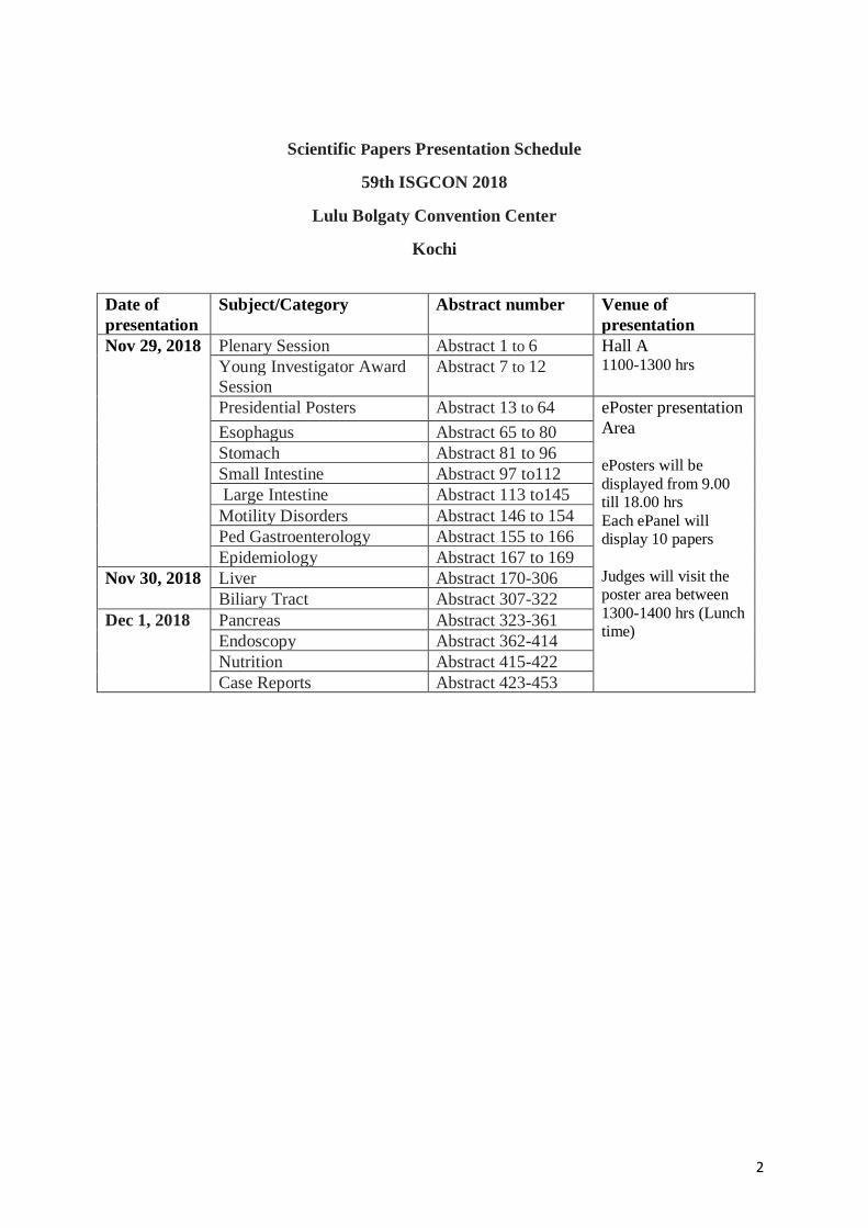
Scientific Papers Presentation Schedule
59th ISGCON 2018
Lulu Bolgaty Convention Center
Kochi
Date of
presentation
Subject/Category Abstract number Venue of
presentation
Nov 29, 2018 Plenary Session Abstract 1 to 6 Hall A
1100-1300 hrs Young Investigator Award
Session
Abstract 7 to 12
Presidential Posters Abstract 13 to 64 ePoster presentation
Area
ePosters will be
displayed from 9.00
till 18.00 hrs
Each ePanel will
display 10 papers
Judges will visit the
poster area between
1300-1400 hrs (Lunch
time)
Esophagus Abstract 65 to 80
Stomach Abstract 81 to 96
Small Intestine Abstract 97 to112
Large Intestine Abstract 113 to145
Motility Disorders Abstract 146 to 154
Ped Gastroenterology Abstract 155 to 166
Epidemiology Abstract 167 to 169
Nov 30, 2018 Liver Abstract 170-306
Biliary Tract Abstract 307-322
Dec 1, 2018 Pancreas Abstract 323-361
Endoscopy Abstract 362-414
Nutrition Abstract 415-422
Case Reports Abstract 423-453
3
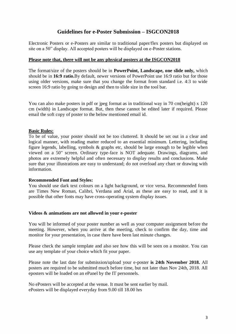
Guidelines for e-Poster Submission – ISGCON2018
Electronic Posters or e-Posters are similar to traditional paper/flex posters but displayed on
site on a 50” display. All accepted posters will be displayed on e-Poster stations.
Please note that, there will not be any physical posters at the ISGCON2018
The format/size of the posters should be in PowerPoint, Landscape, one slide only, which
should be in 16:9 ratio.By default, newer versions of PowerPoint use 16:9 ratio but for those
using older versions, make sure that you change the format from standard i.e. 4:3 to wide
screen 16:9 ratio by going to design and then to slide size in the tool bar.
You can also make posters in pdf or jpeg format as in traditional way in 70 cm(height) x 120
cm (width) in Landscape format. But, then these cannot be edited later if required. Please
email the soft copy of poster to the below mentioned email id.
Basic Rules:
To be of value, your poster should not be too cluttered. It should be set out in a clear and
logical manner, with reading matter reduced to an essential minimum. Lettering, including
figure legends, labelling, symbols & graphs etc, should be large enough to be legible when
viewed on a 50" screen. Ordinary type-face is NOT adequate. Drawings, diagrams, and
photos are extremely helpful and often necessary to display results and conclusions. Make
sure that your illustrations are easy to understand; do not overload any chart or drawing with
information.
Recommended Font and Styles:
You should use dark text colours on a light background, or vice versa. Recommended fonts
are Times New Roman, Calibri, Verdana and Arial, as these are easy to read, and it is
possible that other fonts may have cross-operating system display issues.
Videos & animations are not allowed in your e-poster
You will be informed of your poster number as well as your computer assignment before the
meeting. However, when you arrive at the meeting, check to confirm the day, time and
monitor for your presentation, in case there have been last minute changes.
Please check the sample template and also see how this will be seen on a monitor. You can
use any template of your choice which fit your paper.
Please note the last date for submission/upload your e-poster is 24th November 2018. All
posters are required to be submitted much before time, but not later than Nov 24th, 2018. All
eposters will be loaded on an ePanel by the IT personnels.
No ePosters will be accepted at the venue. It must be sent earlier by mail.
ePosters will be displayed everyday from 9.00 till 18.00 hrs
4
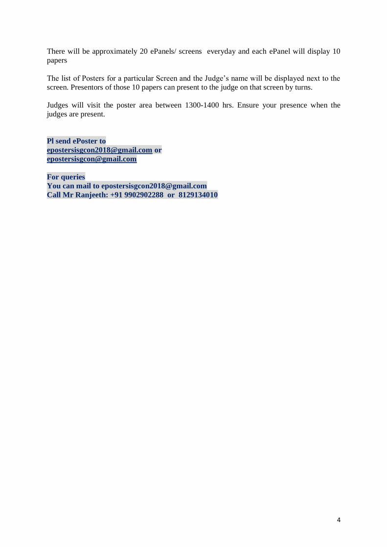
There will be approximately 20 ePanels/ screens everyday and each ePanel will display 10
papers
The list of Posters for a particular Screen and the Judge’s name will be displayed next to the
screen. Presentors of those 10 papers can present to the judge on that screen by turns.
Judges will visit the poster area between 1300-1400 hrs. Ensure your presence when the
judges are present.
Pl send ePoster to
For queries
You can mail to [email protected]
Call Mr Ranjeeth: +91 9902902288 or 8129134010
5
Contents
Plenary Session …
Young Investigator Award Session …
Presidential Posters …
Esophagus …
Stomach …
Small Intestine …
Large Intestine …
Motility Disorders …
Pediatric Gastroenterology …
Epidemiology …
Liver …
Biliary Tract …
Pancreas …
Endoscopy …
Nutrition …
Case Reports …
Author Index …
The Abstracts printed in this issue are as submitted by the Indian Society of
Gastroenterology. In no way, the Honorary Editor-in-Chief, Editorial Board Members, or
the printer/publishers are responsible for the results/findings and content of these Abstracts.
ABSTRACTS
Indian - Society of Gastroenterology
Plenary Session
001
Outcome of management protocol to reduce von Willebrand factor (vWF) in acute
hepatic dysfunction: Hepatotoxicity due to yellow phosphorus (rat killer) poisoning as a
prototype
Debasis Sardar, Nitty Mathews, Joy Mammen, S C Nair, Shibu Jacob, Mousumi Sen,
Vijayalekshmi, K A Balasubramanian, K Subramani, Lovely Thomas, K P P Abhilash,
Shankar Jhanwar, Ashish Goel, Uday Zachariah, Elwyn Eias, C E Eapen
Correspondence: C E Eapen – ([email protected])
Gastroenterology and Hepatology, Christian Medical College, Vellore 632 004, India
6
Introduction: High vWF levels may predispose to platelet microthrombi and multi-organ
failure in phosphorus poisoning induced hepatotoxicity. We report outcome of vWF lowering
therapies: N–acetyl cysteine (NAC), fresh frozen plasma (FFP) infusions and plasma
exchange in these patients.
Method: In this retrospective analysis of prospectively collected data, patients were classified
to have uncomplicated acute hepatitis/UAH (deranged LFT, INR ≤1.5), acute liver
injury/ALI (deranged LFT, INR >1.5) and acute liver failure/ALF (ALI, hepatic
encephalopathy). ALF patients were advised liver transplantation, those not opting for
transplantation underwent plasma exchange and NAC infusion; ALI patients received
NAC±FFP infusions (plasma exchange, if worsening); UAH patients had oral NAC. Normal
plasma vWF antigen levels are 50% to 150%.
Results: Seventeen patients with hepatotoxicity due to phosphorus poisoning (December
2017 to July 2018), at presentation had UAH (one 19 year old male), ALI (13 patients, age 22
(15-35) years, median (range); 6 males)) or ALF (3 patients, age 25 (7-25) years; 1 male).
Baseline MELD scores were 11, 24 (12-38) and 36 (32-37); platelet counts were 1.59, 2
(1.05–3.51), and 1.63 (0.9–2.92), and plasma vWF antigen levels were 153%, 392 (146-
513)% and 448 (414- 555)% in UAH, ALI and ALF patients respectively. All 13 ALI
patients received NAC, 7 had FFP infusion, 3 had plasma exchange (2 (1-2) sessions). All 3
ALF patients had NAC and plasma exchange (5 (1-6) sessions). 11/13 (85%) ALI patients
survived, 2 progressed to ALF and died. Of 3 ALF patients, 2 (67%) survived. No patient
underwent liver transplantation. Of 5 patients meeting criteria for emergency liver
transplantation, 3 (60%) survived.
Conclusion: vWF lowering treatment in phosphorus poisoning induced hepatotoxicity
resulted in transplant free survival rates of 100% (UAH), 85% (ALI) and 67% (ALF).
002
Optimizing infliximab therapy using a Dashboard approach – An Indian experience
Mihika B Dave,1 Alpa Dherai,1 Devendra Desai,2 Diane Mould,3 Tester Ashavaid,1
Correspondence: Devendra Desai- ([email protected]) 1Department of Biochemistrty, 2Division of Gastroenterology, P D Hinduja Hospital, Veer
Savarkar Marg, Mahim, Mumbai 400 016, India, and 3Projections Research Inc.,
Phoenixville, Pennsylvania, USA
Introduction: Infliximab (IFX) pharmacokinetics is influenced by several variables. A few
patients fail to respond (primary non-responders) while there are others who form antibodies
and loose response with time (secondary non-responders). The complex multifactorial
relationship between covariates and IFX distribution limits a direct correlation between dose
and drug level. Recently, Bayesian systems in dashboard formats have been proposed for use
in clinical practice. We aimed to assess the accuracy and efficacy of the iDose dashboard
system.
Methods: The IFX levels estimated in our laboratory in inflammatory bowel disease (IBD)
patients as a part of clinical service (n=20) were compared with that forecasted by the iDose
dashboard software. Patient’s clinical history, demographic details, laboratory findings such
as albumin and CRP were entered in the software and the predicted IFX level was compared
7
with the estimated level. In addition, dashboard guided dosing strategy was prescribed in 3
patients and the clinical outcome was followed.
Results: Of the 20 patients, concordance in estimated and predicted IFX level was seen in 16
patients. iDose guided dosing was clinically useful to achieve target therapeutic response in
all three patients. The dosing interval was increased from 4 weeks to 7 weeks in two patients
and, in the third patient, the dosing was optimized with multiple drug estimations and iDose
prediction.
Conclusion: The iDose dashboard forecast for IFX level and dosing regimen was in
concurrence with the estimated IFX level. This approach optimized the infliximab therapy by
individualized IFX dosing and duration. Ithas the potential to substantially reduce the cost
003
Time-dependent associations of acute pancreatitis with gut microbial dysbiosis and
altered intestinal permeability: Lessons learnt from experimental model
Aparna Jakkampudi, Ratnakar Reddy, Balakumar Reddy, D Nageshwar Reddy, C
Subramanyam, Rupjyoti Talukdar
Correspondence: Rupjyoti Talukdar- ([email protected])
Asian Healthcare Foundation/Wellcome DBT Labs, Asian Institute of Gastroenterology, 6-3-
661, Somajiguda, Hyderabad 500 082, India
Introduction: Intestinal barrier dysfunction plays an important role in acute pancreatitis (AP).
In this study, we evaluated the dynamic association of AP with intestinal microbial dysbiosis
and gut barrier permeability.
Methods: AP was induced in C57Bl/6J mice with intra-peritoneal injections of cerulein (50
µg/kg/hour for 10 hrs). V3-V4 region of metagenomic DNA isolated from fecal pellets at
baseline, 12, 24, 48 and 72 hrs were sequenced. Pancreas and intestines were harvested for
histological examination and expression (mRNA, protein) studies. Circulating and tissue
cytokines were quantified by FACS. Alterations in the intestinal permeability was identified
by performing IHC and qRT-PCR studies for ZO-1, ZO-2, occludin. Intestinal epithelial cell
apoptosis and proliferation were identified and quantified by performing IHC for caspase-3
and Ki-67.
Results: Successful induction of AP was confirmed by elevated trypsin activity and
inflammatory changes in pancreatic tissue. Circulating IL-6, TNF-α, and IL-10 were
significantly elevated at 12 hrs of AP induction. Principal component analyses and
hierarchical clustering analyses of metagenomic data confirmed gut bacterial dysbiosis in
mice with AP. Relative abundance of phylum Bacteroidetes and genus Bacteroides were
significantly elevated at 48 hrs of AP induction. Pancreatic and ileal tissue homogenates
showed increased IL-6, TNF-α concentration at 12 and 24 hrs. qRT-PCR and IHC of
intestinal tissue showed progressive time-dependent down-regulation of ZO-1, ZO-2 and
occludin expression. Intestinal epithelial cell apoptosis was significantly high at 24hrs and
normalized by 72 hrs. Ki-67 expression correlated negatively with caspase-3 expression at 72
hrs after AP induction.
Conclusion: Data suggest that AP associated intestinal inflammation could result in time
dependent altered microbial dysbiosis and intestinal permeability.
8
004
Normative values of sarcopenia in the Indian population
Sandeep Sidhu,1 Kavita Saggar,2 Omesh Goyal,1 Harsh Kishore,1 Samarth Singh Sidhu,3
Correspondence: Sandeep Sidhu-([email protected]) 1Departments of Gastroenterology, and 2Radiodiagnosis, Dayanand Medical College and
Hospital, Udham Singh Nagar, Civil Lines, Ludhiana 141 001, India, and 3Charles
Univiersity, Prague
Introduction: Sarcopenia is characterized by the loss of skeletal muscle mass, strength and
performance. It is of great prognostic importance in patients with liver cirrhosis (LC). The
study was design to provide normal values (computerized tomography skeletal muscle index
(CTSI), handgrip strength (HGS), gait velocity, chair stand) for measuring sarcopenia in
Indian population.
Methods: CTSI (cm2/m2), HGS (Kg), gait velocity (m/min), chair stand, of 3087 non-
cirrhotic patients who underwent abdominal computed tomography (CT) for acute abdomen
were analyzed in this study. The cross-sectional area of skeletal muscles was measured at the
level of the third lumbar vertebra on CT (using Tomovision slice Omatic 5.0 software).
Results: Three thousand eighty-seven number of patients, 1003 (32%) females and 2084
(67%) males who underwent abdominal CT were enrolled in this study. Mean CTSI in female
was 41.25±4.42 vs. 44.33±6.56 in male (p<0.0001). Mean of HGS in female was 25.19±7.57
vs. 35.14±8.56 in male (p<0.0001), mean of gait velocity in female was 1.76±2.38 vs.
1.86±2.22 in male (p=0.2524) and mean of chair stand in female was 10.38±4.42 vs.
13.86±2.56 in male (p<0.0001).
Conclusions: This is the largest global data provide normative values of all sarcopenia
parameters for adults based on gender. This shall enable future studies on Sarcopenia in
cirrhotic patients.
005
Utility of neutrophil CD64 and in distinguishing bacterial infection from disease flare in
severe alcoholic hepatitis
Gaurav Pande,1 Alok Kumar,2 Harshit Singh,3 Saurabh Chaturvedi,3 Ravi Mishra,3
Prabhaker Mishra,4 Arun Bhadoria,2 Sameer Mohindra,5 Durga Prasanna Misra,3 Vivek
Anand Saraswat,2 Vikas Agarwal3
Correspondence: Vikas Agarwal-([email protected])
Gastroenterology and Hepatology1, Departments of Gastroenterology2, Clinical
Immunology3, Stastestics4, and Clinical Gastroenterology5, Sanjay Gandhi Postgraduate
Institute of Medical Sciences, Rae Bariely Road, Lucknow 226 014, India
Background: Bacterial and opportunistic infections are a major cause of morbidity and
mortality in patients with severe alcoholic hepatitis (SAH). Steroids are used in treatment of
severe alcoholic hepatitis (MDF >32: MELD>20) and may exacerbate sepsis so it is vital to
differentiate infection vs. inflammation. Fc receptor (FcγR1 or CD64) expression on
neutrophils and soluble TREM-1 (Triggering Receptor Expressed on Monocytes) are
potential biomarkers of bacterial infections.
9
Aim: To Assess the clinical usefulness of quantitative CD64 measurement on neutrophils
measurements in differentiating bacterial infection from onflammation in patients of SAH.
Methods: Patients with bacterial infection (n=58), active disease (n=70), and healthy controls
(n=20) were included. Neutrophil CD64 expression using flow cytometry and procalcitonin
C-reactive protein levels were studied.
Result: The percentage of neutrophils with CD64 expression and their mean fluorescence
intensity in patients with infection 76.2 (56.9-86.5)%, 1431 (229-1828) were significantly
(p<0.05) higher as compared to those without infection (16[12.6-23.1])%, 853 (20-968) and
controls (7.05[1.4-9.5]%, 99.5 [54.7-140.7]).The sensitivity and specificity of CD64
expression on neutrophils to diagnose bacterial infection (using a cut-off value of 27%) was
94% and 78%, respectively. The sensitivity and specificity of procalcitonin was 83% and
72% respectively at cut-off of 2.91 ng/mL.
Conclusion: Quantitative measurement of CD64 on neutrophils can distinguish between
systemic infection and Inflammation.
006
Predictive models of the outcome of medical management of acute severe ulcerative
colitis using principal component analysis and artificial neural network
Uday C Ghoshal, Ankur Gupta, Sushmita Rai, Akshay Kulkarni, Shikha Agnihotry
Correspondence: Uday C Ghoshal- ([email protected])
Department of Gastroenterology, Sanjay Gandhi Postgraduate Institute of Medical Sciences,
Raebareli Road, Lucknow 226 014, India
Background: About 15% patients with acute severe ulcerative colitis (UC) fail to respond to
medical treatment and may require colectomy. An early prediction response may help the
treating team and the patients and their family to prepare for alternative treatment options.
Methods: Data of 263 patients (mean age 37.0±14.0-y, 176, 77% male) with acute severe UC
admitted during a 12-year period were used to study predictors of response using univariate
analysis, multivariate linear principal component analysis (PCA), and non-linear artificial
neural network (ANN).
Results: Of 263 patients, 231 (87.8%) responded to the initial medical treatment that included
oral prednisolone (n=14, 5.3%), intravenous (IV) hydrocortisone (n=238, 90.5%), IV
cyclosporine (n=9, 3.4%), and inflixmab (n=2, 0.7%), and 28 (10.6%) did not respond and
the remaining 4 (1.5%) patients died. Non-responding patients had to stay longer in the
hospital and died more often. On univariate analysis, the presence of complications, the need
for use of cyclosporin, lower Hb, platelets, albumin, serum potassium, and higher C-reactive
protein were predictors of non-response. Hb and albumin were strong predictive factors both
on PCA and ANN. Though the non-linear modeling using ANN had very predictive accuracy
for the response, its accuracy for predicting non-response was lower.
Conclusion: It is possible to predict the response to medical treatment in patients with UC
using linear and non-linear modeling technique. Serum albumin and Hb are strong predictive
factors.
Young Investigator Award Session
10
007
Quantitative proteomic analysis identifies dysregulated platelets in severe alcoholic
hepatitis
Adil Bhat, Sukanta Das, Ashish Kumar Vyas, Sudrishti Chaudhary, Gaurav Yadav, Renu
Goel, Meenu Bajpai, Rakhi Maiwall, Jaswinder Singh Maras, Shiv K Sarin
Correspondence: Shiv Kumar Sarin- ([email protected])
Department of Molecular and Cellular Medicine, Institute of Liver and Biliary Sciences, D 1,
Vasant Kunj, New Delhi 110 070, India
Background and Aims: Severe alcoholic hepatitis (SAH) is linked to thrombocytopenia and
platelet activation. However, the phenotype of platelets, proteins transported and associated
function in SAH patients is not known.
Method: To explore this quantitative proteomic profiling of SAH platelets was compared to
healthy platelets, which led to the characterization platelet phenotype (GO, KEGG, blood
transcription modules-BTMs) and identification of key dysregulated pathways and were
validated in a separate cohort of (n=40) SAH patients. Platelets functions such as activation,
aggregation, intracellular calcium levels, vesicular and granule secretion were correlated to
severity of SAH patients.
Results: In this pilot study a total of 1235 platelet proteins were identified; of them 202 were
up-regulated and 321 down-regulated (FC >±1.5, p<0.01) in SAH vs. HC. Proteins linked to
platelet activation, complement regulation and lipid transportation were up-regulated
(log2FC>2, p<0.001), whereas proteins linked to platelet hemostasis, coagulation, alpha and
dense-granules and vesicular transport were down-regulated (log2FC<-2, p<0.001) in SAH
platelets. On validation, platelets from SAH patients documented higher expression of
activation markers (PAC-1, CD62P), intracellular calcium levels and aggregation marker
(Gp2b/3a protein) compared to HC (p<0.05). Genes linked to platelet and complement
activation were up-regulated and directly correlated (r >0.3, p<0.05) with the severity.
Further genes linked to alpha and dense-granules, coagulation and hemostasis were
downregulated and inversely correlated (r>-0.3, p<0.05) to the severity of SAH patients.
Interestingly the mRNA and proteins expression levels of vesicular and granular secretions
(SNAP-23, VAMP-3, Rab-27b, Syntaxin-11 and Munc13-4) were significantly
downregulated (p<0.05) and inversely correlated (r>-0.3, p<0.05) with the severity.
Conclusion: Platelet in SAH patients are activated, however, they show decreased granular
content and impaired vesicular and granular secretion, which may contribute to vascular
injury in such patients.
008
Generation of secondary humanized livers through intra-omental transplantation of
bioengineered livers for the management of end-stage liver diseases
Sandeep K Vishwakarma
Correspondence: Sandeep K Vishwakarma-([email protected])
Department of Dr. Habeebullah Life Sciences, Deccan College of Medical Sciences,
Kanchanbagh, Hyderabad 500 058, India
11
Introduction: End-stage liver diseases (ESLD) represent a major, neglected global public
health crisis which requires urgent actions towards finding proper cure. Orthotopic liver
transplantation has been only definitive treatment modality for ESLD. However, shortage of
donor organs, timely unavailability, post-surgery related complications and financial burden
to the patients limits the number of patients receiving transplants. Hence alternative choices
are highly desirable to overcome the limitations of current modalities. In present study we
have explored a new concept of generating secondary humanized livers through intra-omental
transplantation in rats which can be used for the management of ESLD.
Methods: Decellularization of whole rat liver was performed using gradients of detergents
within 20 h through hepatic artery perfusion at defined flow rate. Decellularized liver
scaffolds (DLS) were characterized extensively for preservation of ECM, vasculature and
organ architecture. The humanization of DLS was performed post-sterilization using human
hepatic progenitor cells (hHPCs) under controlled ex vivo condition. The structural and
functional response of engineered humanized livers was identified to explore its use in drug
evaluation and further transplanted in rat omentum. The structural and functional
characteristics of transplanted grafts were identified after day 3 and day 30.
Results: An efficient decellularization protocol was developed to generate completely DLS
within 20h of continuous perfusion through hepatic artery while retaining intact ECM
proteins, liver vasculature and architecture. The engraftment efficiency of cells within the
DLS was found to be >98%. Functional analysis of repopulated liver cells was identified by
quantification of albumin and urea. Retrieved humanized liver grafts didn’t show visible
fibrotic reactions at the site of transplantation. Grafts were found to be well embedded in the
peritoneal cavity with neo-vascularisation without immunological rejection.
Conclusion: The present study demonstrates generation of secondary humanized livers with
neo-vascularization during intra-omental transplantation of bioengineered livers in rats.
009
Pentazocine, a kappa-opioid agonist, is better than diclofenac for analgesia in acute
pancreatitis: A randomized controlled trial
Soumya Jagannath Mahapatra, Saransh Jain, Sawan Bopanna, Swatantra Gupta, Anjan
Trikha, Preet Mohinder Singh, V Sreenivas, Shalimar, Pramod Kumar Garg
Correspondence: Pramod Kumar Garg- ([email protected])
Department of Gastroenterology, All India Institute of Medical Sciences, New Delhi 110 029,
India
Introduction: The ideal analgesic is not known for patients with acute pancreatitis (AP).
Concerns have been raised about serious adverse effects of opioid analgesics increasing the
severity of AP. We hypothesized that non-steroidal anti-inflammatory drugs (NSAIDs) might
be better analgesics due to their anti-inflammatory effect. Our objective was to compare
pentazocine, an opioid, and diclofenac, an NSAID for adequate analgesia in patients with AP.
Methods: In a double-blind randomized controlled trial, patients with AP were randomized to
either intravenous Diclofenac 75 mg or Pentazocine 30 mg. Fentanyl was given as a rescue
analgesic through a patient-controlled analgesia pump. Primary outcome was pain relief
measured objectively by the dose of fentanyl required as rescue analgesic, pain-free period,
and numbers of good and bad demands of fentanyl. Secondary outcome was adverse events.
12
Results: Fifty patients were randomized: 24 to pentazocine group and 26 to diclofenac group.
Baseline characteristics were comparable between the groups. Pentazocine was found to be
better than diclofenac in terms of significantly lower dose of rescue analgesic (fentanyl)
required [126 μg (range 0-960 μg) vs. 225.5 μg (range 0-810 μg); p=0.028], and longer pain-
free period (31.1±8.2 hours vs. 27.9±6.6 hours, p=0.047). The number of good and bad
demands was lower in the pentazocine compared with diclofenac group (11.5 [range 0-92] vs.
16 [range 0-85], p=0.098) although not statistically significant. Adverse events were similar
between the groups.
Conclusion: Pentazocine, a kappa opioid receptor agonist, was significantly better than
diclofenac for pain relief in AP.
Trial registration number: CTRI/2016/09/007326
010
Thromboelastography guided blood product transfusion in patients with chronic liver
disease undergoing invasive liver-related procedures: A randomized controlled trial
Sudheer Kumar Vuyyuru, Shalimar, Gyan Ranjan Rout, Ashish Chauhan, Shivanand
Gamanagatti, Baibaswata Nayak, Saurabh Kedia
Correspondence: Shalimar-([email protected])
Department of Gastroenterology, All India Institute of Medical Sciences, Ansari Nagar, New
Delhi 110 029, India
Background: We aimed to assess the use of thromboelastography (TEG) directed blood
product transfusion in cirrhotic patients undergoing invasive liver-related procedures
compared to standard of care (SOC) correction of coagulopathy.
Methods: In this open label, randomized control trial, cirrhotic patients with coagulopathy,
undergoing invasive liver-related procedures, were randomized- to either TEG-guided blood
product transfusion or SOC- from November 2017 till April 2018. The primary outcome was
difference in the amount of fresh frozen plasma (FFP) and platelet units transfused between
the two groups. Secondary outcome measures were procedure related bleeding complications
within five days.
Results: Of the 30 patients, 15 were randomized to the TEG group and 15 to the SOC.
Procedures performed were percutaneous liver biopsy (n=21), sphincterotomy with stone
removal (n=1), transjugular intrahepatic portosystemic shunt (TIPS) (n=1), pigtail drainage of
empyema (n=1), percutaneous acetic acid injection (PAI) for hepatocellular carcinoma
(HCC) (n=2) and transarterial chemoembolization (TACE) (n=2), pigtail drainage of
intraperitoneal collections (n=1) and hepatic vein angioplasty (n=1). There were no
differences in baseline demographic profile and types of invasive liver-related procedures
between the two groups. All 15 subjects in the SOC group received blood product
transfusions, vs. 5 in the TEG group (100% vs. 33.3%; p<0.001). In the SOC group, 9 (60%)
received platelet transfusions, 6 (40%) received FFP and no patient received both FFP and
platelets. In the TEG group, 2 (13.3%) received FFP (p=0.215 vs. SOC), 3 (20%) received
platelets (p=0.128 vs. SOC) and no patient received both FFP and platelet. None of the
patients in either group developed procedure related bleeding complications immediately post
procedure or till 5 days of follow up.
13
Conclusion: TEG-guided blood product transfusion strategy was associated with reduced
blood product transfusion without increased risk of bleeding in cirrhotic patients undergoing
invasive liver-related procedures.
011
Hepatitis B virus-infected pregnant females with higher circulating HBsAg levels
showed impaired immune imprint in their newborns at birth
Ashish Kumar Vyas, Pooja Negi, Sharda Patra,* Jaswinder Singh Maras, Shiv Kumar Sarin,
Nirupma Trehanpati
Correspondence: Nirupma Trehanpati-([email protected])
Department of Molecular and Cellular Medicine, Institute of Liver and Biliary Sciences, D 1,
Vasant Kunj, New Delhi 110 070, India, and *Obstetrics and Gynecology, Lady Harding
Medical College, C-604, Shaheed Bhagat Singh Road, Diz Area, Connaught Place, New
Delhi 110 001, India
Background: Vertical transmission of hepatitis B virus (HBV) from infected mother to the
new-born often results in viral persistence. To understand mechanisms of materno-fetal
HBV-transmission and viral persistence, we studied maternal immunity and peripheral blood
mononuclear cell (PBMC) transcriptome in mothers and new-borns.
Methods: Fifty HBsAg positive mothers and babies were included; 22 with the transmission
(Gr. I, T, n=22) and 28 with no transmission (Gr. II, NT, n=28) to the new-borns, and
compared with healthy mother-baby pairs (Gr. III, n=10). PBMCs were analyzed for HBV-
specific DCs, T cells, T follicular helper (TFh), B cells and functional immune responses,
cytokine levels as well as transcriptome signatures to identify immune gene expression
correlates for protective immunity.
Results: Group II mothers had lower HBsAg levels (3.82 x10³ vs. 1.493x10⁴, p<0.0001)
with greater HBV specific responses of DCs, T cells, TFh and B cell than Gr. I mothers. The
percentage frequencies of TFh cells had lower (11.06±1.71 vs. 18.05±1.74, p=0.02) in Gr. I
mothers was accompanied with reduced IL21 (356.1±50.17 vs. 493.6±56.70, p=0.04) levels.
TFh frequencies and IL21 levels inversely correlated with HBV DNA levels. The cut-off
level of 9.5% and 8.93% from the receiver operating curve predicted the involvement of TFh
and B cells in HBV transmission. Transcriptome signatures revealed that maternal gene
imprints were reflected in their new-borns. Further genes related to DCs, TFH and B cells
and were increased in Gr. II. HBsAg+ve new-borns showed a boost in cellular and humoral
responses after vaccination.
Conclusion: In HBV infected mothers, low serum IL-21 levels, decreased TFh and plasma B
cell frequencies are associated with vertical transmission of HBV to new-born. These features
are indicative of low protective immunity.
012
Gut microbiome diversity in acute severe colitis is distinct from mild to moderate
ulcerative colitis
Saurabh Kedia1, Tarini Shankar Ghosh2, Bhabatosh Das2, Saransh Jain1, Vipin Gupta1,
Sawan Bopanna1, Dawesh P Yadav1, Sandeep Goyal1, Govind Makharia1, Simon Travis3,
Vineet Ahuja1
14
Correspondence: Vineet Ahuja-([email protected]) 1Department of Gastroenterology and Human Nutrition, All India Institute of Medical
Sciences, New Delhi 110 029, India, 2Molecular Genetics Laboratory, Centre for Human
Microbial Ecology, Translational Health Science and Technology Institute, NCR Biotech
Science Cluster, Faridabad 121 001, India, and 3John Radcliffe Hospital, Translational
Gastroenterology Unit, Headley Way, Oxford, UK
Introduction: Although the gut microbiome of patients with ulcerative colitis (UC) has been
characterized, there has been no study of gut microbial diversity in patients with acute severe
colitis (ASC). The present study compared the gut microbiome of patients with UC, ASC and
healthy controls (HC).
Methods: Patients with mild to moderate UC (n=23), ASC (n=21) and healthy controls
(n=24) were recruited prospectively. A metagenomics approach was used to explore gut
microbial diversity and genetic repertoires. Ulcerative colitis was diagnosed using ECCO
guidelines and ASC was diagnosed using Truelove and Witts’ criteria.
Results: Genus level diversity (Simpson diversity measure) was significantly lower in ASC
than in mild-moderately active UC (p<0.05), or HC (p<0.001). The gut microbiome in ASC
was highly unstable, as characterized by high intra-cohort variation (analyzed using J-
divergence measure) which was significantly greater than in UC or HC. On principal
coordinate analysis, the microbiome of HC and UC were similar, with the ASC cohort being
distinct from both. On quantitative evaluation of these differences (random forest classifiers),
both ASC vs. HC and UC comparisons revealed excellent classification accuracy, with >90%
patients being correctly classified. Statistical comparison of the ranked abundances identified
four distinct clusters of genera (G1A, G1B, G2A, G2B), with specific trends in their
abundance patterns across the three groups: the G1A/G1B clusters had the least, whereas
G2A/G2B had the highest abundance in the ASC cohort. Interestingly, several known health-
associated bacteria (Faecalibacterium, Prevotella and Roseburia) exhibited different
oligotypes, with distinct oligotypes segregating into health and disease states (ASC).
Conclusions: Gut microbial diversity is lower in ASC than in mild-moderate UC or healthy
controls. Gut microbiome composition is increasingly unstable in ASC, with a distinct
abundance of specific genera varying between healthy controls and ASC. Mild-moderate UC
lies within the spectrum.
Presidential Posters
013
Non-surgical management of gastroduodenal tuberculosis: Nine-year experience from a
tertiary referral center
Ashok Dalal, Amarender Singh Puri, Puja Sakuja
Correspondence: Amarender Singh Puri- [email protected]
Department of Gastroenterology, G B Pant Institute of Postgraduate Medical Education and
Research, 1, J L N Marg, New Delhi 110 002, India
Introduction: Gastroduodenal tuberculosis (GDTB) is an uncommon disease. Surgery has
been standard of care both for diagnosis and management of GDTB. The aim of study was to
evaluate the efficacy of non-surgical management of GDT using a combination of anti-
tuberculous therapy (ATT) along with endoscopic dilatation of the tuberculous stricture.
15
Methods: Patients suspected to have gastroduodenal TB were evaluated: clinical, endoscopic,
radiological and histopathological data were recorded. Patients in whom a definite diagnosis
of tuberculosis could not be confirmed on mucosal biopsies underwent endoscopic mucosal
resection (EMR). Patients were treated with ATT, endoscopic dilatation was done if
indicated. Patients were followed up to evaluate clinical, radiological and endoscopic
response.
Results: Over a 9-year period from 2009-2017 at GIPMER, 52 patients (mean age 28.5 yrs)
were diagnosed with GDTB. Commonest presenting symptoms were vomiting (n=51, 98%)
and weight loss (n=52,100%). The most common anatomical site of involvement was D1- D2
junction (n=22, 42%). Histopathological diagnosis could be made in 43 (82.6%) patients; 36
(69%) on mucosal biopsies and in 7 of the 10 patients (70%) who underwent snare
biopsy/EMR. Endoscopic dilatation was done in 37 (71%) patients and median dilatation
sessions were 2. Failure of endotherapy occurred in 4 patients (7.6%). All responders had
complete amelioration of symptoms after 4-6 weeks of combination therapy. The mean
period of follow up was 31 months and none of the patients reported any recurrence of
symptoms.
Conclusion: Combination of ATT and endoscopic dilatation has a high success rate in
management of GDT and should be considered the standard of care.
014
FeGdO3 bimetallic nanoparticles-blood interaction: Implication for the development of
magnetic resonance imaging based functional imaging
Avinash Bardia
Correspondence: Avinash Bardia-([email protected])
Department of Dr. Habeebullah Life Sciences, Deccan College of Medical Sciences,
Kanchanbagh, Hyderabad 500 058, India
Introduction: Development of high contrast molecular imaging devices has gained
tremendous potential in biomedical applications. Nanotechnology-based strategies have been
evolved as one of the most promising field to design more appropriate and effective
nanoparticles for such applications. However, biocompatibility of developed nanoparticles
with human blood components is of pivotal importance for their clinical applications. In this
study, we investigated the biological effects and stability of FeGdO3 nanoparticles during
interactions with human blood and serum components.
Methods: Human blood and platelet rich plasma samples were treated with 1μg/mL, 2 μg/mL,
4 μg/mL and 8 μg/mL concentration of FeGdO3 nanoparticles for 30 minutes, 60 minutes,
120 minutes, 240 minutes, 24 hours and 72 hours in vitro. Moreover, peripheral blood
mononuclear cells were also treated with similar concentrations of nanoparticles to
investigate the effect of these nanoparticles in time dependent manner. Following to
biocompatibility assessment of FeGdO3 nanoparticles, the dual-MRI contrast property was
evaluated using 1.5T Magnetome using different concentrations mixed with human blood.
Results: Complete blood picture showed no adverse effect of tested concentrations of
nanoparticles on human blood components such as platelet and RBC counts and levels of
hemoglobin and HCT. Furthermore, serum stability of nanoparticles showed no significant
16
effect at different concentrations of serum during interaction. Enhanced dual-MRI contrast
was observed for FeGdO3 nanoparticles in human blood.
Conclusion: The use of FeGdO3 showed high stability and biocompatibility with human
blood components with increased T1 and T2 MRI-contrast which could be utilized for the
future development of functional imaging in human applications.
015
Admission serum urea is a better predictor of mortality in patients with acute on
chronic liver failure and acute kidney injury
Shivaram Prasad Singh, Chitta Ranjan Khatua, Bhaskar Thakur*, Debakanta Mishra,
Subhendu Panigrahi, Subhasis Pradhan, Saroj Kanta Sahu, Rakesh Kumar Barik, Prasanta
Kumar Parida
Correspondence: Berhampur Khatua-([email protected])
Department of Gastroenterology, S C B Medical College, Cuttack 753 007, India, and *Kalinga Institute of Medical Sciences (KIMS), KIIT University, Bhubaneshwar 751 024,
Odisha
Introduction: Occurrence of acute kidney injury (AKI) in acute-on-chronic liver failure
(ACLF) patients negatively impacts their survival. Only elevated serum creatinine is used to
assess AKI and survival in ACLF. There is scant data on impact of serum urea on outcome.
We performed a prospective study to evaluate impact of serum urea on survival in ACLF
patients with AKI.
Methods: A prospective study was conducted in ACLF patients with AKI, hospitalized in
Gastroenterology Department of SCB Medical College in India from October 2016 to March
2018. Demographic, clinical and laboratory parameters were recorded, and outcome
compared in patients with respect to admission serum urea level.
Results: Consecutive decompensated cirrhosis (DC) patients (n=439) were screened for
ACLF, as per Asian Pacific Association for the Study of the Liver (APASL) criteria. One
hundred and thirteen (25.7%) of them had ACLF. Out of 113 ACLF cases, 78 (69%) had AKI
as per AKIN criteria. Alcohol was both the commonest cause of CLD (74.3%), as well as the
commonest precipitant (61%) of ACLF. The discrimination ability between survivors and
deceased was similar for serum urea (AUROC 28 days; 0.792 [0.694-0.889], 90 days; 0.838
[0.745-0.931], 95% CI) and serum creatinine (AUROC 28 days; 0.770 [0.666-0.874], 90
days; 0.794 [0.683-0.904]; 95% CI) in patients with ACLF and AKI. However, on
multivariate analysis, admission serum urea (not serum creatinine) was an independent
predictor of mortality in ACLF with AKI patients at 28 days; p=0.002, AHR 1.014 (1.005-
1.023) and 90 days; p=.001, AHR 1.016 (1.007-1.025).
Conclusions: About one fourth of decompensated cirrhotic patients had ACLF and more than
two thirds of them were associated with AKI. Admission serum urea was found to be a better
predictor of mortality than serum creatinine in ACLF patients with AKI. Hence serum
creatinine may be replaced by serum urea, as a better predictor of mortality in ACLF patients
with AKI.
016
17
Long-term outcome of patients with Budd-Chiari syndrome treated with
anticoagulation alone: A single center experience
Dhiraj Agrawal, Deepak Gupta, Rohit Nathani, Amit Bhondve,* Shobna Bhatia, Akash
Shukla
Correspondence: Akash Shukla- ([email protected])
Department of Gastroenterology, Seth G S Medical College and KEM Hospital, Mumbai 400
012, India, and *Department of Preventive and Social Medicine, Seth G S Medical College
and KEM Hospital, Mumbai 400 012, India
Background: Long-term outcome of patients with Budd-Chiari syndrome (BCS) treated with
anticoagulants alone is unknown.
Methods: Consecutive patients (n=138, mean [SD] age 29.3 [12.9] years; 66 men) with BCS,
treated with oral anticoagulation alone, with minimum follow up of 12 months were
included. Response was graded as complete response (CR), partial response (PR) and non-
response (NR). During follow up, loss of response (LoR) or maintenance of response (MoR)
were recorded. The association of baseline, clinical and biochemical parameters with
different responses was evaluated.
Results: 76/138 patients (55.1%) had CR, 26 (18.8%) had PR and 36 (26.1%) had NR. None
of the patients who had PR or NR at one year had CR later. At a median follow up of 40 (12–
174) months, LoR was more common in PR group than in CR group (12 [46.2%] vs. 18
(23.7%), p0.03]. On multivariate analysis, absence of ascites at presentation (p=0.027), low
baseline bilirubin (p=0.035), low INR (p=0.045), high AST (p=0.049), low ALP (p=0.001)
and low CTP score (p=0.048) were associated with PR or CR. On logistic regression analysis,
presence of ascites (OR 0.303, 95% CI-0.098-0.931) at baseline was associated with LoR.
Absence of ascites, jaundice or GI bleed and low CTP score (p=0.01[OR -0.67, 95% CI 0.31
– 0.84]) on presentation were associated with MoR. One hundred and thirty-three bleeding
episodes (23 major, four deaths) occurred in 55 (39.85%) patients (0.289 episodes per person
year). Mortality was higher in NR group (28 [77.8%]) than CR [15 (19.7%], p=0.001) and PR
(8 [30.8%] p=0.001) groups.
Conclusion: Patients with initial CR have better survival than non-responders. One third
patients lose response on follow up. Presence of ascites at baseline is associated with LoR.
017
Alveolar echinococcosis of liver: A series of ten cases
G N Yattoo,1 Gulzar Ahmad,1 Zubaida Rasool,2 G M Gulzar,1 Jaswinder Singh Sodhi,1
Naseer A Choh,1 Zaffar Iqbal Kawoosa,1 Mushtaq Ahmad Laway,1 Suresh Gorka1
Correspondence: Gulzar Dar- ([email protected]) 1Departments of Gastroenterology, and 2Pathology, Sher-I-Kashmir Institute of Medical
Sciences, Jammu and Kashmir 190 011, India
Introduction: Alveolar echinococcosis (AE) is a chronic, serious and sometimes lethal
parasitic infection, caused by Echinococcus multilocularis (EM). Epidemiologically most
commonly found in the northern hemisphere. AE has been reported to occur in India with an
18
annual incidence of one case per year. Until now there have been no cases of AE reported
from this part of world to best of our knowledge. Aims and objectives: To evaluate and study
the clinical profile of consecutive patients of AE diagnosed at a tertiary care centre.
Methods: All patients referred with space occupying lesions of liver with suspicion of
neoplastic lesions (Hepatocellular carcinoma, Intrahepatic Cholangiocarcinoma, Atypical
hemangioma etc.) were evaluated over past three years. All consecutive case of AH
diagnosed were enrolled in this study. All baseline investigations, hydatid serology, imaging
(USG, CT, MRI) and image guided biopsy of lesions were done. ERCP was done where
indicated.
Observations: A total of ten cases of AE liver were enrolled in the study with mean age of
39±11 years (range of 18-55 years). Males and females were equal. All patients were from
rural background. Most of patients presented with pain upper abdomen 7 (70%) and rest
presented with combination of jaundice and itching 3 (30%). Hydatid serology was positive
in 7(70%) cases while as 3(30%) had either negative or equivocal results. Most of patients 8
(80%) had right lobe involvement while as 2(20%) had left lobe. The size of lesions ranged
from 3.5 to 15 cms. Histology (gold standard) was confirmatory in all cases. The lesions were
Kodama type-1 (2, 20%), Type-2 (4, 40%), Type-3 (3, 30%) and Type-5 (1, 10%) and PNM
stage-I 2 (20%), II 4 (40%), III 3 (30%) and IV 1 (10%). Three patients required endotherapy
in addition to benzimidazole therapy because of biliary tract involvement. On follow up of
two years 70 % had either complete.
018
Endoscopic management of postcholesystectomy benign biliary stricture: if you can
cross it you can cure it! 25 year’s experience at a tertiary care center in northern India
Hemanta Kumar Nayak,1 Vivek Anand Saraswat,2 Samir Mohindra,2 Gaurav Pande,2 Piyush
Ranjan,3
Correspondence: Vivek Anand Saraswat-([email protected]) 1Advanced Endoscopy and 2Department of Gastroenterology, Sanjay Gandhi Postgraduate
Institute of Medical Sciences, Lucknow 226 014, India, and 3Sir Gangaram Hospital,
Rajinder Nagar, New Delhi 110 060, India
Introduction: Endoscopic management of post-cholecystectomy benign biliary stricture with
multiple plastic stents is routinely practised in many centers. However, data from India is
scarce. Our aim was to assess efficacy and outcome of endoscopic management of these
patients.
Methods: A retrospective study was done in a referral center from January 2004 till May
2018. Seventy-three patients were included in the study. During each ERCP, increasing
number of stents were place, stent exchange was done every 3 monthly for a period of 12
months and all the stents were removed at the end of therapy. Subsequently, patient was
followed up with ultrasonography of abdomen and liver function tests every three monthly,
ERCP was repeated if symptomatic or abnormal liver function parameter.
Results: Of 73 patients, technical success of endoscopic biliary cannulation achieved in 70
(96%). Three patients with failed biliary cannulation underwent hepaticojejunostomy. Due to
inadequate biliary drainage and unwilling for repeated endotherapy, six patients, underwent
19
hepaticojejunostomy. Seven patients were lost to follow up after initiation of endotherapy.
Two patients died, one due to myocardial infarction and another due to underlying chronic
kidney disease. Four patients found to have malignant stricture, underwent palliative biliary
stenting. Fifty-one patients completed therapy. Median age of patients was 39 years (range
23-86), mostly females (72%). Median number of procedures, maxium number of stents per
session, duration of therapy were 4.2 (2-7), 4 (2-8), 13 months (6-26) respectively. The mean
and median duration of follow up after completion of therapy was 41 and 33 months
respectively (3-137). One patient had stricture recurrence, underwent hepaticojejunostomy.
Conclusions: Endoscopic therapy is feasible in post cholecystectomy BBS with technical
success of 96%. Recurrence of stricture is rare and at a median follow up of 3 years, bile duct
patency is maintained in 98%.
019
A proteomic approach to identify dysregulated lipid transporter proteins which could
predict the severity and outcome of patients with acute-on-chronic liver failure
Jaswinder Singh Maras, Sukanta Das, Adil Bhat, Dhananjay Kumar, Chaggan Bihari, Rakhi
Mahiwal, Shiv Kumar Sarin
Correspondence: Shiv Kumar Sarin- ([email protected])
Department of Molecular and Cellular Medicine, Institute of Liver and Biliary Sciences, D 1,
Vasant Kunj, New Delhi 110 070, India
Acute-on-chronic liver failure (ACLF) is a serious ailment with high mortality. It is not
known whether altered levels of various lipid transport related proteins could predict the
severity of ACLF.
Aim: To identify candidate biomarkers using proteomics, to assess the severity and prognosis
in ACLF patients.
Methods: Quantitative proteomics profiling was performed using high resolution mass
spectrometry. Validation of protein expression profiling were performed in; Gr. 1. Alcoholic
ACLF (n=40), Gr. 2. Alcoholic cirrhosis (n=20) and Gr. 3. Healthy controls (n=20).
Results: Three hundred and five differentially regulated proteins were identified of which 120
were more than 2-fold differentially regulated. Proteins involved in transport and
metabolismof lipids were significantly reduced in Gr. 1 (Pon1, ApoA1, ApoA2, and ApoC3;
>2 folds). PON 1 was significantly reduced in Gr.1 (25 ug/mL), vs. Gr.2 (45 ug/mL), Gr.3
(140 ug/mL) (p<0.0001). Levels of other lipid transporters (Apo A1, Apo A2, Apo C1 Apo
C3, Apo B, and Apo E) were significantly reduced in Gr.1 vs. Gr.2, Gr.3 (p<0.05). Levels of
PON1 and Apo B were significantly reduced in the non-survivors compared to survivor in
Gr.1 (p<0.05). Ratios of PON1/ApoA1, Apo A2, and ApoC1 were severely deranged in non-
survivors in Gr.1 (p< 0.05). The level of PON1 and the ratio of PON1/A1, A2, C1 correlated
inversely with the MELD, SOFA and CTP scores (p<0.05). Moreover, level of Pon1 and the
ratio of PON1/Apo A1, A2 and C1 showed a direct correlation with survival in ACLF
patients (p< 0.03, r2>0.3).
Conclusions: In the ACLF patients, circulating Pon1 level and the ratio of PON1/Apo A1,
A2, and C1 were significantly reduced in the non-survivors compared to survivors. These
20
lipid transporter proteins could serve as biomarkers for assessing the outcome of patients with
ACLF.
020
Hyperglycemia in acromegaly–How much is contributed by incretins secreted by
intestinal K and L cells
Kim Vaiphei,1 Shobhit Bhansali,2 Pinaki Dutta,2 K K Mukherjee,3 R Kochhar,4 S K Sinha,4
Naresh Sachdeva,2 A V Kurpad,5 Kishor Bhat,5 S Mudaliar,6 Anil Bhansali,2 Vikram Singh
Shekhawat,2
Correspondence: Kim Vaiphei-([email protected]) 1Departments of Histopahtology, 2Endocrinology, 3Neurosurgery, 4Gastroenterology, and 5Physiology, Postgraduate Institute of Medical Education and Research, Chandigarh 160 012,
India, and 6Department of Medicine, University of California, San Diego, La Jolla,
California, USA
Incretins are hormones secreted into the gastrointestinal tract upon food ingestion. They
promote insulin secretion by acting on pancreatic b cells in a blood-glucose-dependent
manner. Two incretins, glucose-dependent insulinotropic polypeptide (GIP) and glucose
dependent glucagon-like peptide (GLP)-1, have been identified. GLP-1 receptor agonists
(GLP-1RAs) can reduce blood glucose levels and have been reported to have several
extrapancreatic effects. All enteroendocrine cells (EECs) sense nutrients present in intestinal
lumen. Whereas the molecular mechanisms by which specific nutrients are detected, and their
impact on the secretion of different peptides, is still poorly understood. EECs express various
types of receptors including for sweet, bitter and other taste. These properties are also true for
K and L cells; K cells are predominantly found in duodenum whereas L cells are located in
ileum and colon. However, a recently described population of K/L cells secretes both GIP
and GLP-1. Their effects have been best demonstrated when glucose is orally ingested,
compared to when the same amount of glucose is administered intravenously and GLP-1
being more potent than GIP.
Acromegaly is almost invariably caused by a growth hormone (GH) secreting pituitary
adenoma with metabolic complications of disorders of glucose and lipid metabolism. The
metabolic actions of GH are mainly diabetogenic as it is a potent antagonist of insulin.
Prevalence of glucose intolerance in acromegaly ranges between 19% to 56% and up to 20%
may have diabetes mellitus (DM). GH is known to induce glucose intolerance resulting in
insulin resistance (IR). Earlier studies have shown that insulin sensitivity (IS) is reduced to a
similar extent in acromegalic patients with and without glucose intolerance.
Incretin hormones are known to strongly influence glucose-insulin homeostasis and little is
known about their effects on carbohydrate metabolism in acromegaly.
021
To compare the tissue diagnostic yield of solid lesion biopsies based on the
histopathological analysis of endoscopic ultrasound guided fine needle aspiration (EUS
–FNA) samples produced by the 19G Procore needle, Standard 19G needle and 22G
Procore needle
Mahesh Kumar Gupta, Rajesh Puri, Randhir Sud
Correspondence: Mahesh Kumar Gupta- ([email protected])
21
Department of Gastroenterology, Medanta -The Medicity, CH Baktawar Singh Road, Sector
38, Gurugram 122 001, India
Introduction: Endoscopic ultrasound guided fine needle aspiration (EUS–FNA) is a sensitive
method for detecting intestinal and extraintestinal mass lesions. Inflammation causes cellular
changes undistinguishable from neoplasia solely based on cytological evaluation, because
tissue architecture and cell morphology are essential for accurate pathological assessment.
Various EUS-guided techniques are available with variable success and complication rates.
Methods: Single centre, prospective, randomized study. All patients above 18 years of age,
having intestinal and extraintestinal solid mass lesions, were randomized to EUS-FNA and
tissue diagnostic yield were compared based on the histopathological analysis of samples
produced by the 19GP, 19GS and 22GP needles. Patients with INR >1.5, platelets <50000
were excluded.
Results: Total 215 patients were evaluated and EUS-FNA was technically feasible in 210
(97.67%) cases. Three needle passes were made in every case. There was no significant
difference between these three groups with regard to the age (p-0.676), gender (p-0.856),
location (p-0.998), echogenicity (p-0.123), size (p-0.735 and 0.374) of the lesions and
presence of calcification (p-0.093) or necrosis (p-0.729). Sample suitable for pathological
evaluation were obtained in 90.5% cases with a tissue core in 45.7% cases. 28.1% lesions
were malignant, 62.4% were benign and 9.5% remained undiagnosed. The histopathological
diagnoses were possible in 87.1%, 90.0% and 94.3% cases respectively with 22GP, 19GP and
19GS needles (p-value-0.350). Presence of blood clot in order of 19GP (70.00%) >22GP
(50.00%) >19GS (42. 8%), (p-value 0.003). There were no post procedure complications
noted in any group.
Conclusion: EUS-guided biopsy with these three needles was feasible and safe. The
diagnostic yield of these three needles for providing adequate sample for histopathological
analysis was clinically significant but not Statistical significant. Procore needles did not offer
the extra possibility of obtaining a core sample for histopathological analysis in this study but
there is high possibility of presence of blood clots.
022
Prevalence and predictors of irritable bowel syndrome (ROME IV, Rome III, and Asian
Criteria) among medical students in a Government Medical College in South India
Manoj Yadav, Krishnadas Devadas, K V Neeraj, Jose Mathew, Aniruddha Pratap Singh
Correspondence: Manoj Yadav- ([email protected])
Department of Medical Gastroenterology, Trivandrum Medical College, Ulloor-Akkulam
Road, Chalakkuzhi, Thiruvananthapuram 695 011, India
Introduction: Irritable bowel syndrome (IBS) is the most common functional gastrointestinal
disorder. The prevalence rates of IBS in the United States using Rome III vs. Rome IV
criteria are 10.8% vs. 6.1%, respectively. The prevalence of IBS in various Asian countries
(ROME III) is 5%-10%. There has been the unavailability of study on the epidemiology of
irritable bowel syndrome (IBS) based on ROME IV among medical students in India.
22
Aims: 1. To estimate the prevalence and predictors of irritable bowel syndrome by ROME
IV, ROME III, and Asian criteria, 2. The measure of agreement between Rome IV, ROME
III, and Asian criteria.
Methods: It was a cross-sectional questionnaire-based study done on 552 medical students
(138 students per batch x 4 batches) who gave their consent for the study were included.
Filled up questionnaires were collected, and the Chi-square test was applied.
Results: IBS prevalence and severity distribution according to ROME IV, ROME III, and
Asian criteria is depicted in Fig. 1. Among IBS subtypes 43.9%, 29.8%, 18.1% and 9.4%
were mixed, constipation predominant, diarrhea predominant and unclassified. Prevalence
increases with increasing age and higher MBBS batch using Rome III (p=0.012, 0.027) and
Asian criteria (p=0.002, 0.017), but not with Rome IV (p=0.23, 0.40). IBS was found to have
an association with mode of delivery, physical activity, BMI, coffee, dairy products,
carbonated beverages, sleep duration, analgesic and antibiotics intake (Table 1). Cohen's
kappa coefficient (κ) were 0.699, 0.367 and 0.213, between Rome III and Asian, Rome IV
and Rome III, and Rome IV and Asian criteria.
Conclusions: 1. Prevalence of IBS according to ROME IV, ROME III, and Asian criteria
were 5.8%, 19%, 30.4% respectively. 2. The measure of agreement was maximum between
Rome III and Asian and minimum between ROME IV and Asian criteria.
023
Risk factors predicting nosocomial, health care associated and community-acquired
infection in spontaneous bacterial peritonitis and survival outcome
Uday Sanglodkar, Mayank Jain, Jayanthi Venkataraman
Correspondence: Mayank Jain- ([email protected])
Department of Gastroenterology, Gleneagles Global Health City, 439, Cheran Nagar,
Perumbakkam, Chennai 600 100, India
Background: The clinical presentation and outcome of spontaneous bacterial peritonitis
(SBP) based on the acquisition site is not clearly known.
Aim: To study prevalence, differences and survival outcome following nosocomial (N-SBP),
community acquired (C-SBP) and healthcare associated (H-SBP) SBP infection in
decompensated cirrhotic patients.
Methods: This prospective observational study included confirmed cases of cirrhosis with
ascites requiring paracentesis, age >18 years, either gender, any etiology and Child-Turcotte-
Pugh (CTP) stage, with or without cirrhosis related complication. Patient data included age,
gender, co-morbidity, model for end-stage liver disease (MELD) score, CTP score, cirrhosis
related complications, details of previous hospitalization, ascitic tapping and antibiotics
instituted. SBP was diagnosed as ascitic fluid polymorphonuclear leukocyte count greater
than 250/mm3 (0.25 × 10⁹/L) and/or culture positivity for a single organism.
Statistics: Chi-square test, Mann-Whitney U test, analysis of variance (ANOVA). Survival
plot was plotted for various variables using log rank test. A p value <0.05 was statistically
significant.
23
Results: Six hundred and ten cases fulfilled the criteria for inclusion. One hundred and
twenty-two (20%) patients had SBP: C-SBP in 37 (30.3%), N-SBP in 19 (16.5%) and H-
SBP in 66 (54.5%). Majority were men (106; 86%) with median age of 51.5 (27-78) years. A
significantly higher percentage of C-SBP belonged to CTP class B. Thirty-two and 7 patients
respectively were blood and ascitic fluid culture positive. There were fewer ascitic fluid
cultures positive in N-SBP and H-SBP compared to blood culture. Significant N-SBP were
blood culture positive (p<0.02). The most common isolates were E coli followed by
Klebsiella. Survival plot analysis at 3 months showed the worst survival for N-SBP (p
0.0009).
Conclusions: Prevalence of SBP in our study was 20%, majority with H-SBP belonging to
CTP C. Patients with N-SBP had significant bacteremia with high mortality.
024
Systemic inflammatory response predict organ failure and mortality in alcoholic
hepatitis
V Neeraj, D Krishnadas
Correspondence-D Krishnadas-([email protected])
Department of Medical Gastroenterology, Government Medical
College, Trivandrum 695 011, India
Introduction: Severe alcoholic hepatitis (AH) can lead to systemic inflammatory response
syndrome (SIRS) due to cytokine overplay which could predispose to multiple organ failure
(MOF) and death.
Aims: To estimate the predictive accuracy of Day1 SIRS in determining MOF in patients
with severe AH. To estimate the predictive accuracy of Day1 SIRS in determining the 30
day mortality in patients with severe AH.
Methods: Day 1 SIRS score in all consecutive patients with severe AH (DF ≥32) admitted in
the wards of Medical Gastroenterology over a period of 1 year from the date of ethical
committee clearance was taken and its relation with complications and mortality was studied
and followed up for 1 m to look for morbidity and mortality.
Results: A total of 133 patients with severe AH were taken out of which 94 had D1 SIRS 50
patients had 30 d mortality, 47 patients developed renal failure, 54 patients had MOF. Out of
47 patients who had RF 43 had D1 SIRS (91%). Out of 54 patients with MOF 50 (100%) had
D1 SIRS. Out of 50 patients with 30d mortality, 50 (100%) had D1 SIRS. Higher D1 scores
of SIRS were found to have higher sensitivity and specificity in predicting both the
development of RF, MOF and 30d mortality.
Conclusions: D1 SIRS can be used to predict the development of renal failure and MOF in
patients with severe AH with very high sensitivity. Therefore, D1 SIRS can be used as a
screening tool to stratify patients with severe AH and facilitate early referral, ICU
admissions, thereby reducing adverse outcomes
025
24
Demographics and perioperative outcome of colorectal cancer in Kerala - Analysis of
ASGK colorectal cancer surgical registry
Prasad Krishnan,1 O V Sudheer,2 Prakash Kurumboor,3 Ramesh Hariharan,4 Sylesh Aikot,5
Rajesh Nambiar,6 Ramesh Rajan,7 B Venugopal,8 M L Arun Kumar,9 Joshy John,10
Correspondence: Prasad Krishnan-([email protected]) 1Department of Surgical Gastroenterology, Sree Narayana Institute of Medical Sciences,
Chalakka, P.O. North Kuthiyathodu 683 594. Ernakulam Dist. India, 2Amrita Institute of
Medical Sciences, Peeliyadu Road, Ponekkara, Edappally, Ernakulam, Kochi 682 041, India, 3Aster Medcity, Kuttisahib Road, Cheranalloor, South Chittoor, Kochi 682 027, India, 4Lakeshore Hospital, Nettor. Kochi 682 040, India, 5Baby Memorial Hospital,
Arayidathupalam Junction, Arayidathupalam, Kozhikode 673 004, India, 6MIMS, Mini
Bypass Road, Govindapuram, Kozhikode 673 016, India, 7Trivandrum Medical College,
Ulloor-Akkulam Road, Chalakkuzhi, Thiruvananthapuram 695 011, India, 8Kerala Institute
of Medical Sciences (KIMS), 1, Anayara Road, Anayara, Thiruvananthapuram 695 029,
India, 9Sree Gokulam Medical College and Research Center, Aalamthara-Bhoothamadakki
Road, Venjaramoodu 695 607, India, and 10Holycross Hospital, Hollycross Hospital Road,
Kottiyam, Kollam 691 571, India
Colorectal cancer (CRC)is the third most common cancer in males and second most common
in females.
Aim: To develop a colorectal cancer surgical registry and to analyse the data of colorectal
cancer operated by the Association of Surgical Gastroenterology Kerala (ASGK)members
Methodology: A colorectal surgical registry format was developed by the ASGK. Members
of ASGK were registered. This data base consists of demographics, symptomatology,
investigatory reports, operative, perioperative details, neoadjuvant and adjuvant treatment.
Follow up data of 1 year, 3 year and 5 years included in the Registry. The centers were
requested to enter the colorectal cancer cases from January 2016.
Results: The period of study was from January 2016 to March 2018. Fifteen centers in Kerala
participated in the study. The number of cases taken up for our analysis was 1018. Total
number of male patients were 621 (61%) and female patients 395 (39%). The median age
group in male patients was 63 and female patients was 64. The most common presentation
include bleeding per rectum (58.8%) and abdominal pain (23.3%). Rectum was the most
common site 408 (40%). Stage II- 258 (25.3%) and stage III- 423 (42.2%) patients were the
common stages reported.10.9% had metastatic disease. Low anterior resection was the most
common procedure performed in 262 (25.3%) patients. Median duration of surgery was 240
minutes. Complications reported in 149 patients (14.6%). Most common type of
differentiation was moderately differentiated adenocarcinoma 78.8%. Median number of
lymphnodes harvested was 14.5. Follow up data is awaited.
Conclusion: State wide colorectal surgical registry is feasible. Median age group Male-63
and female-64 and rectum is the common location. 10.9% presented with metastatic disease.
This registry would identify high volume centers and for future multivariate analysis.
Attempts should be made by other state chapter in order get a National level data.
026
25
Therapeutic plasma-exchange improves systemic inflammation and survival in patients
with acute-on- chronic liver failure
Rakhi Maiwall
Correspondence: Rakhi Maiwall-([email protected])
Departments of Hepatology, and Biostatistics2, Institute of Liver and Biliary Sciences, D 1,
Vasant Kunj, New Delhi 110 070, India
Systemic inflammation (SIRS) and infection form the pathogenetic hallmark of acute-on-
chronic liver failure (ACLF). High-volume plasma-exchange (PE) has been shown to
improve survival in patients with acute liver failure by combating SIRS, but there is paucity
of data in patients with ACLF. We evaluated the role of artificial liver support systems
(ALS), plasma-exchange (PE) and liver dialysis (FPSA) as compared to standard medical
treatment in improving SIRS, development of multiorgan failure and survival (SMT) in a
large multicentric-multinational cohort of ACLF patients.
Methods: Prospectively collected data from AARC data base was analysed. Matching by
propensity risk score (PRS) was done. Competing risk Cox regression analysis was done to
identify event specific i.e. multiorgan failure related death.
Results: ACLF patients (n=1866, mean age 44.3±12.3 yrs, 93% males, 65% alcoholics)
received either ALS (n=162); (PE [n=131], FPSA (n=31]) or continued with SMT (n=1704).
Patients treated with ALS had a significantly lower MELD (p=0.009), CTP (p<0.001) and
SOFA scores (p<0.001) which was no more evident in the PRS-matched cohort (p>0.05)
(n=208, [ALS-119; PE-94, LD-25)], SMT-89). ALS was associated with significantly higher
resolution of SIRS ([OR 3.53, 1.95-6.49 and 9.23,3.42-24.8]) and development of new-onset
SIRS ([or 4.38, 1.1-17.5 and 1.2, 1.04-1.29]) and MOF ([HR 6.5, 4.8-8.7 and 7.1,4.5-11.1]) in
pre-match and PRS-matched cohorts respectively. At 1-month, 656 (35%) died of which 233
(35.5%) died of multiorgan failure. Treatment with PE ([HR 0.11, 0.04-0.27]), (HR-0.02,
0.002-0.15) significantly resulted in lower liver failure related death. Further, on subgroup
analysis PE was associated with a significant survival benefit as compared to FPSA in pre-
match (HR 3.4, 1.4-8.1) and PRS-matched (HR 3.9, 1.3-12.3) cohorts.
Conclusion: ALS treatment in patients with ACLF improves systemic inflammation, lowers
development of multiorgan failure and results in improved survival. Plasma-exchange has a
significant survival benefit over FPSA and should be the therapy of choice in these patients.
027
High Cytomegalovirus DNA load in mucosal biopsies predicts steroid failure as well as
colectomy in acute severe ulcerative colitis
Saransh Jain,1 Divya Namdeo,2 Saurabh Kedia,1 Pabitra Sahu,1 Prasenjit Das,4 Peush
Sahni,3 Nihar Ranjan Dash,3 Sujoy Pal,3 Govind Makharia,1 Lalit Dar,2 Vineet Ahuja,1
Correspondence: Vineet Ahuja-([email protected])
Departments of 1Gastroenterology, 2Microbiology, 3Gastrointestinal Surgery and 4Anatomy,
All India Institute of Medical Sciences, New Delhi 110 029, India.
Introduction: Cytomegalovirus (CMV) reactivation may be responsible for steroid refractory
acute severe colitis (ASC), which requires rescue therapy in form of surgery or advanced
26
immunosuppression. The optimum technique for diagnosing CMV colitis in this setting
remains unclear. We investigated the role of CMV quantitative PCR for diagnosing CMV
colitis and for predicting of steroid-failure in ASC.
Methods: Consecutive patients with ASC satisfying Truelove and Witts’ criteria, hospitalized
at a single centre from May 2016 to November 2017, were included. The primary outcome
measure was steroid-failure defined as colectomy and/or rescue therapy with ciclosporin or
infliximab during admission. Oxford criteria, ulcerative colitis index of severity (UCEIS) at
day 1 and fecal calprotectin (FCP) at day 3 were used to predict steroid response.
Immunohistochemistry (IHC) and quantitative PCR for CMV was done on mucosal biopsies
and the results were compared between steroid responders and non-responders.
Results: Of 37 patients (mean age: 35±12 years, 70% males), 14 (38%) failed IV
corticosteroids and 8 (25%) required surgery. Although IHC for CMV was not different
between steroid failures and responders (29% vs. 17%, p=0.40), patients with steroid failure
had a significantly higher median level of mucosal CMV DNA [7840(0-2700000) vs. 112(0-
34459) copies/mg, p=0.03]. Significantly greater number of patients with steroid failure had
CMV DNA count >1000 copies/mg (71% vs. 26%, p=0.007). CMV DNA count >1000
copies/mg (Odds ratio 6.5 (95% confidence interval 1.3-33, p=0.03)) and positive oxford
criterion on day 3 of iv corticosteroids (OR 6 [95% CI 1.2-30, p=0.03]) were independent
predictors of steroid-failure and need for rescue therapy/colectomy.
Conclusions: CMV DNA quantification in mucosal biopsy can detect CMV colitis and
predict steroid failure in acute severe colitis with reasonable accuracy.
028
Sarcina ventriculi of gastrointestinal tract: A clinicopathologic characteristics of eleven
cases
Ritesh Prajapati, Subhash Nandwani, Mayank Kabrawala, Rajiv Mehta, Pankaj Desai,
Chintan Patel, Parika Kalra, Nisharg Patel
Correspondence: Ritesh Prajapati- ([email protected])
Department of Gastroenterology, Surat Institute of Digestive Sciences, Vijay Nagar Gate No-
3, Besides Nirman Bhavan, Majura Gate, Ring Road, Surat 395 002, India
Sarcina ventriculi, a gram-positive coccus, is occasionally found in gastric biopsies.
Although Sarcina had been described more than 150 years ago, little is known about its
pathogenicity in humans. We, report clinicopathologic characteristics of 11 patients
with Sarcina in gastric or duodenal biopsies. All eleven patients had presence of Sarcina
ventriculi on the luminal mucosal surface of epithelium with mucosal injury. Of these eleven
patients, ten patients had gastric outlet obstruction (GOO). Of these ten patients with GOO, 5
had antral narrowing and 5 had duodenal obstruction. Three patients had malignant GOO (2-
carcinoma stomach, 1- carcinoma duodenum), 5 had ulcers with edematous narrowing (1- H
pylori, 1- eosinophilic gastritis) and 2 had duodenal stricture. All patients had etiology other
than sarcina for GOO. Of the 3 patients that had follow up endoscopy on resolution of
symptoms, all patients had gastric residue on endoscopy. One patient had recurrence of
symptoms with persistence of sarcina on biopsy at 3 months. Symptoms improved at 6
months and no evidence of sarcina on biopsy at 6 months. Thus, our findings suggest gastric
27
outlet obstruction can be considered as a predisposing factor for Sarcina infection. Sarcina
infection may not be the etiology for GOO but may complicate recovery and may lead to life-
threatening complications. Therefore, the clinician and pathologist must be aware of such
microorganisms. Moreover, if pathologist identified it in biopsies, it must be documented in
the final report as the findings may have therapeutic consideration and warrant further
investigation.
029
Foxp3, TGF-ß and PDCD1 promoter hypomethylation impairs effector cells in non-
seroconverters after hepatitis B reactivation
Mojahidul Isam, Ashish Kumar Vyas, Tanvi Agrawal, Ankur Jindal, Gayatri Ramakrishna,
Shiv Kumar Sarin, Nirupma Trehanpati
Correspondence: Nirupma Trehanpati- ([email protected])
Department of Molecular and Cellular Medicine, Institute of Liver and Biliary Sciences, D 1,
Vasant Kunj, New Delhi 110 070, India
Background and Aim: Hepatitis B surface antigen (HBsAg) seroconversion sometimes occurs
after reactivation, probably due to immune reconstitution. Termination of tolerance is decided
by suppressive and inhibitory markers. We evaluated the methylation status of Foxp3, TGF-β
and PD1 inhibitors in immune restoration in HBVr patients.
Methods: Patients with HBVr, HBeAg+ve, ALT >5XULN and high HBV DNA (104-8)
(HBVr, Gr-A, n=40) were followed till 48 wks. Patients who seroconverted (HBV DNA and
HBsAg–ve, anti-HBs >10 IU/mL) (Gr-B) or remained as HBsAg+ve (Gr-C) were studied.
HBeAg+ve, HBV DNA+ve CHB patients (Gr-D, n=18) served as control. Promoter
methylation of Foxp3, TGF-β, and PDCD1 gene in PBMCs was analysed using bisulfite
sequencing at baseline between Gr-B and C. Immune reconstitution of CD4, CD8 T cells,
Tregs, and B cells and HBV specific functionality was assessed by flowcytometric analysis.
Results: During follow up of 48 wk, 28 (70%) patients lost HBV DNA but remained
HBsAg+ve (Gr C), and 8 (20%) lost HBV DNA and seroconverted (Gr B). Healthy subjects
showed high methylation frequency of Foxp3 (100%), TGF-β (100%) and PDCD1 (76.47%).
Gr-B methylation profile was comparable to heathy subjects. Gr-C had reduced methylation
frequency for Foxp3 (91.63%±3.30, p=0.0196), TGF-β (92.73±2.46%, p=0.0041) and
PDCD1 (14.70±1.76%, p=0.0041) in comparison to Gr B. Immune status of Gr-B revealed
higher CD4+(p=0.0240), CD8+(p=0.0088), B cells (p=0.0002) counts, lower expression of
inhibitory PD1(p=0.0007, p=0.0105) and TGF-β (p=0.0012, p=0.0140) and increased
expression of activation marker CD38 (p<0.0001, p<0.0107) on CD+/CD8+ Cells at baseline
than Gr-C. No difference in CD4 Tregs in two groups, but significant decrease in CD8 Tregs
(CD8+CD25+, p=<0.05, CD8+CD25+CD127lo/-Foxp3+, p=<0.05) in Gr-B vs. Gr-C was
observed. At 24 wks, we observed similar profile at baseline, except increased expression of
PD1 on CD4+/CD8+CD127LOCD25+FoxP3+Tregs (p=0.0003, p=0.0002) in Gr-B
compared to Gr-C.
Conclusions: Hypermethylation of Foxp3, PDCD1 and TGF-β promoter at baseline
determines the seroconversion with increased effector T cells and decreased suppressive
Tregs in HBVr patients.
28
030
Relationship between serum vitamin D levels and disease severity in chronic
pancreatitis patients
S Archana, B N Girish, L Saraswathy, G Rajesh
Correspondence: G Rajesh- ([email protected])
Department of Gastroenterology, Amrita Institute of Medical Sciences, Kochi
Background: Chronic pancreatitis (CP) may lead to deficiency of fat-soluble vitamins such as
vitamin D. CP is more prevalent in India especially in Kerala state; however, there is limited
data on vitamin D levels in this population.
Aim: The aim of the present study was to assess the vitamin D status and its predictors in CP
patients and relation with pancreatic exocrine insufficiency (PEI).
Methods: Vitamin D status, serum calcium, protein, albumin, globulin and fecal elastase were
measured in twenty-two CP patients and thirty control subjects. Anthropometric
measurements and clinical details were recorded.
Results: The mean age of the subjects was 43.9±15.58 years (range 19-65 years). Vitamin D
level was significantly lower in CP patients as compared to healthy controls. Vitamin D
status was significantly lower in Cambridge-grade III vs. II (9.89±4.5 vs 22.04±3.13 ng/mL,
p=0.001) Cambridge-grade II vs. I (22.04±3.13 vs. 27.1±1.12 ng/mL, p=0.06), Cambridge-
grade I vs. III (3.13 vs. 9.89±4.5 ng/mL, p= 0.01). Vitamin D levels positively correlated with
serum calcium level (r=0.522, p=0.02). Fecal elastase1 directly correlated with serum
calcium (r=0.558, p= 0.02). No significant difference in vitamin D and serum calcium was
observed between smokers vs. non-smoker, diabetics vs. non-diabetics or alcoholic vs. non-
alcoholic CP patients.
Conclusion: Vitamin D deficiency appeared to correlate with severity of CP and PEI. Fecal
elastase1 value of 112.5 µg/g appeared to predict vitamin D deficiency in CP.
031
Predictors of unplanned hospitalization or death and utility of screening with clinical
frailty scale in outpatients with cirrhosis
Aniruddha Pratap Haripal Singh, K Sandesh, Krishnadas Devadas, Gaurav Kapur, Rathan
Cyriac Joseph, Varun Tadkalkar
Correspondence: Aniruddha Pratap Singh-([email protected])
Department of Medical Gastroenterology, Government Medical College, Ulloor-Akkulam
Road, Chalakkuzhi, Thiruvananthapuram 695 011, India
Introduction: Predictive factors determining which outpatients with cirrhosis are at highest
risk for unplanned admissions are lacking. We aimed to study factors and utility of clinical
frailty scale (CFS), in outpatients with cirrhosis in predicting hospitalization or/and death at 3
months.
Methods: Prospective observational single-centre study. Unplanned hospitalization was
defined as any admission to acute care hospital that was not planned. The outcome was
defined as unplanned hospitalization or/and death at 1 and 3 months from study entry.
29
Results: One hundred and twenty-four patients were included, 42 % were frail, 84.6% frail
patients as compared to 15.3% non-frail patients had unplanned hospitalization at 3 months.
Nine frail patients died in comparison to 1 in the non-Frail group at 3 months. Propranolol
more than 20 mg with p<0.07, OR 7.16 (CI 1.73-29.6), Frail patients with p<0.01, OR:5.38
(CI: 1.3-21.6), presence of AKI with p<0.006, OR:17.24 (CI:2.2-133.7), CRP >2 mg/dL with
p<0.001, OR:11.23 (CI:2.6-47) were found to independently predict unplanned
hospitalization/death at 3 months. We also observed that CRP with a cut-off of 2 mg/dL with
the sensitivity of 77.5% and specificity of 92.42 % with AUC of 0.88 predicted unplanned
hospitalization/death at 3 months.
Conclusions: Patients on propranolol more than 20 mg, Frailty, acute kidney injury and
elevated CRP predicts unplanned hospitalization or/and death at 3 months. Clinical frailty
scale as a rapid 1-minute screen can be utilized in outpatients with cirrhosis for stratifying
patients at risk of adverse outcomes.
032
Role of mitochondrial therapy in repair, rejuvenation in liver injury model and its
bioenergetics
Avinash Bardia
Correspondence: Avinash Bardia – ([email protected])
Department of Dr. Habeebullah Life Sciences, Deccan College of Medical Sciences,
Kanchanbagh, Hyderabad 500 058, India
Introduction: Intracellular communication has shown lot of importance in various clinical
conditions in repair of injured cells. There has been burgeoning interest in intercellular
organelle transfer such as mitochondria and bio-energizing the repair and regeneration.
Mitochondria are responsible for more than 90% of a cell’s energy production via ATP.
Mitochondria transfer helps in repair of injured cells by communicating the healthy cells. In
the present study invitro and invivo work was carried out to bio-energies injured liver cells
using mitochondrial therapy.
Methods: Cryopreserved human hepatic cells (HHCs) were exposed with heat shock at 42o C
to create injury in cells. Then heat stressed HHCs were cocultured with umbilical cord bold
derived mesenchymal stem cells (MSCs). MTT, LDH, urea productions, expression of
albumin, CK18, Miro1 and CX43 genes were analyzed after heat stress and coculture.
Mitochondria were isolated from MSCs and transplanted along with MSCs into CCl4 induced
liver stressed mice model. Histopathology, biochemical parameters, MDA, ROS, SOD and
ATP level were measured after mitotherapy.
Results: Viability and proliferation of HHCs in coculture were significantly (p=0.001)
increased. The expression of albumin, CK18, Miro1 and CX43 were also significantly
increased after the coculture. CCl4 induces mice shown the inflammatory cells after the 4
week which was gradually decrease after the mitotherapy. The level of MDA (p=0.01) and
ROS (p=0.03) decreased in mice model after the mitotherapy and the level of SOD (p=0.03)
and ATP (p=0.001) were significantly increased after therapy.
Conclusion: Study has demonstrated that intracellular mitochondrial transfer increases the
ATP production to bio-energizing the injured liver cells and this can be potentially serve as
30
an effective remedy for alleviation of such diseases. This new area of bio-energizing injured
cell to repair and regeneration using intracellular mitochondria transfer has paved the new
strategy for the treatment of liver injury.
033
Bone marrow mesenchymal stem cells (BMSCS) from decompensated cirrhotics are not
fit for therapeutic uses
Dhananjay Kumar, Smiriti Subham, Deepanshu, Nidhi Nautiyal, Rekha, Anupma Prasar,
Anurag Agrawal, Lokesh Anad, Sujata Mohanti, Chhagan Bihari, Shiv Sarin
Correspondence: Shiv Sarin- ([email protected])
Department Molecular and Cellular Medicine, Institute of Liver and Biliary Sciences, D-1,
Vasant Kunj, New Delhi 110 070, India
Autologous Bone marrow mesenchymal stem cells (BMSCS) are often used for hepatic
regeneration in patients with advanced cirrhosis. Use of autologous BMMSCs in chronic
liver disease (CLD) has shown negligible to mild improvement in Child-Turcotte-Pugh
(CTP) scores.
Aim of this study to investigate the effect of chronic liver damage on tissue repair and
regenerative function of BM-MSCs in decompensated-liver-disease (DLD).
Methods: MSC were isolated from BM of DLD patients (n=10) and regenerative function
were compared with same age/sex matched healthy BM-MSCs (n=8). MSC characteristic
were conformed as per the criteria of ISCT. In-vitro functional efficacy of cells was accessed
by studying their potential to suppress the proliferation of cytotoxic T-cells, macrophage
polarization, induction of angiogenesis and secretome profile. In-vivo therapeutic potential
were analyzed in animal model of APAP induced ALF. Global-transcript profiles of cells
were done by NGS. Metabolic profiles of BM plasma were done by NMR. Bioenergetics
were done by analyzing the oxygen-consumption-rates and extracellular-acidification-rate
using the Seahorse technology. Stamens property by CFU-F assay and senescence by SA-
βGal assay. Mitochondrial and total ROS measured by mito-SOX and DHR123.
Results: dBM-MSCs fulfilled the minimal criteria for MSC; plastic-adherence, surface
marker, inducible-osteo/adepogenesis and specific surface expression patterns. In
compression to hMSCs, dMSCs showed defect in immunomodulatory functions, angiogenic
support and paracrine support. While infusion of hMSC significantly improve the 48 hour
survival after APAP induced liver injury dMSCs failed, suggesting the loss of therapeutic
potential of dMSCs. NGS analysis showed impeachment of genes associated to insulin-
resistance in dMSC. DMSCs were significantly defective in glucose-uptake and are bio
energetically quiescent. NMR data showed increased glucose in DLD BM-plasma in
comparison to control. This might be responsible for insulin-resistance and loss of function in
dMSCs. Further analysis showed the loss of self-renewal capacity replicative senescence in
dMSCs.
Conclusion: BM-MSC in DLD are defective in their regenerative functions due to premature
ageing and senescence and are not good cells for MSCs therapy.
31
034
Impact of caffeine, sugar and other ingredients on beverage preference among 2nd
graders
Eesha Nachnani
Correspondence: Eesha Nachnani- ([email protected])
University School of Nashville, 93 Berry Hill Drive, Hendersonville, Tennessee, United
States
Introduction: Childhood obesity is a significant public health concern and beverages and soft
drinks have shown to one of the contributors of the same. Sugar and caffeine are known to be
addictive and we wanted to examine the beverage preference among 2nd graders depending
on ingredients in beverages. The aim of our study was to identify predictors of beverage
preference among 2nd graders.
Methods: A survey was conducted in one class of 2nd graders. They were told to rank various
beverages on a scale of 1 to 5 with 1 being don’t like it a lot and 5 being like it very much.
Commonly used beverages were used for the study including soft drinks, juices and milk
available commonly in the school cafeteria.
Results: Caffeine content had a positive correlation with beverage preference. There was no
significant differences in calories, fats, sodium or carbohydrates on beverage preference.
Milk had the highest sodium levels compared to other drinks. Color did not have much
impact among choosing beverages.
Discussion: The presence of caffeine may impact beverage preference among second graders
and may have an addictive potential. Other constituents like sodium, calories, sugar were not
significant in impacting beverage preference. Larger randomized studies will be needed to
further determine the effect on beverage preference and childhood obesity.
035
A paradigm shift in the management of bile duct strictures complicating living donor
liver transplantation
B Harshavardhan Rao, Anoop K Koshy, Thiba Salam, S Krishnapriya, Nirmal K Prabhu,
Surendran Sudhindran, Rama P Venu
Correspondence: B Harshavardhan Rao –([email protected])
Department of Gastroenterology, Amrita Institute of Medical Sciences, Ponekkara, AIMS
PO, Kochi 682 041, India
Aim: Validation of new metrics to identify functionally significant obstruction (FSO),
in order to better define biliary strictures complicating living donor liver transplantation
(LDLT).
32
Methods: All LDLT recipients who presented with biochemical alterations of cholestasis
were studied. Novel metrics (MRCP ductal ratio [MDR], ERCP ductal ratio [EDR] and
Delayed contrast drainage [DCD]) that can potentially signify a FSO were computed after a
multidisciplinary meeting with transplant surgeons, radiologists and the therapeutic
endoscopy team. Association between these metrics and endotherapy response was analyzed
along with patient demographics, intraoperative variables (cold ischemia time, blood
transfusions, biliary anastomosis) and perioperative complications (hepatic artery thrombosis
[HAT], bile leak). Favorable response to endotherapy was defined as symptomatic relief
with ≥80% reduction in total bilirubin/alkaline phosphatase.
Results: A total of 83 LDLT recipients (19.2%), presented with altered liver function tests; of
which, 39 patients were diagnosed to have biliary strictures and treated with endotherapy as
per institution protocol. Favorable response was seen in 18/39 patients (46.2%). On
univariate analysis HAT, multiple biliary anastomoses, graft-to-recipient weight ratio
(GRWR), MDR, EDR and DCD had a significant bearing on the endoscopic outcome
(p value ≤0.05). On multivariate analysis, only MDR ≥1.15 was an independent predictor of
favorable response to endotherapy (OR-48[95% CI: 7.096-324.71]).
Conclusion: A paradigm shift in the approach to management of biliary strictures
complicating LDLT is proposed whereby a multidimensional definition as FSO can help in
reliable patient selection for endotherapy and improve patient outcome as a whole.
036
Clinical profile and outcome of infants with neonatal cholestasis including biliary
atresia
Himali Meshram, Ira Shah
Correspondence: Himali Meshram-([email protected])
Department of Pediatric GI Medicine, Bai Jerbai Wadia Hospital for Children, Acharya
Donde Marg, Parel, Mumbai 400 012, India
Aim: To determine clinical profile and outcome of neonatal cholestasis (NC).
Methods: One hundred and thirty infants with NC were referred from January 2014-March
2018 were included. Clinical profile of NC and its outcome was analyzed.
Results: Mean age at presentation was 2.82+1.8 months. Male:female ratio was 92:38. Mean
birth weight was 2.44+0.58 kg. Onset of jaundice was at 0.46+0.77 months of life. Etiology
of NC was biliary atresia (BA) in 54 (41.86%), neonatal hepatitis (NH) in 47 (36.43%),
sepsis induced cholestasis in 9 (6.97%), progressive familial intrahepatic cholestasis (PFIC)
in 8 (6.2%), choledochal cyst in 3 (2.3%), bile duct paucity in 3 (2.3%), inspissated bile in 2
(1.55%), hemangioendothelioma in 2 (1.55%), and BA with choledochal cyst in 1 (0.77%).
Fifty-one (39.23%) patients recovered, 37 (28.46%) patients developed chronic
liver disease (CLD), 32 (24.6%) were lost to follow up, 6 (4.6%) are newly diagnosed and 4
(3.07%) died. On follow up, 60 (46.15%) patients were jaundice free, 45 (34.61%) children
developed portal hypertension, 19 (14.6%) had ascites and 51 (39.23%) had growth failure.
Mean age of presentation in BA was 3.3 months and that in NH was 2.47 months (p=0.018).
Twenty-two (38.88%) females had BA while 10 (17.24%) females had NH. Mean birth
weight of BA and NH patients was 2.58 and 2.37 respectively (p=0.036). Forty-six (85.18%)
with BA had clay stools as compared to 22 (37.9%) patients with NH (p=0.00001). Mean
33
GGTP levels in BA and NH group were 434.1 IU/L and 225.03 IU/L respectively (p=0.001).
Other laboratory parameters were similar in both the groups. On follow up of BA patients, 10
(18.5%) recovered, 27 (50%) developed CLD, 12 (22.22%) were lost to follow up and 3
(5.5%) are following up. Among the NH patients, 29 (50%) recovered, 10 (17.24%)
developed CLD, 14 (24.13%) lost to follow up and 3 (5.17%) are follow
ing up (p=0.0018).
Conclusions: BA and NH are common causes of NC. Patients with BA have normal birth
weight, female gender, clay stools and higher GGTP. Patients with BA develop CLD whereas
most patients with NH recover.
037
Acute kidney injury in cirrhosis: L-KDIGO (liver-kidney disease in global outcomes)
criteria-based evaluation of outcome
Jose Mathew, Krishnadas Devadas, K Sandesh, Manoj Yadav, V Neeraj, Rathan Cyriac
Correspondence: Jose Mathew- ([email protected])
Department of Medical Gastroenterology, Government Medical College, Trivandrum 695
011, India
Introduction: Acute kidney injury (AKI) is associated with increased mortality in cirrhosis.
Kidney disease in global outcomes (KDIGO) criteria is used for AKI evaluation in general
population. KDIGO criteria's urine output criteria to define and stage AKI is not suitable for
cirrhotic patients. The division of Stage 1 of KDIGO to 1A and 1B provides a better
stratification in cirrhotics.
Methods: We did a prospective study in cirrhotic patients. We modified the KDIGO criteria
by deletion of urine output criteria and dividing the Stage 1 to 1A and 1B based on creatinine
cut-off of 1.5. We called the modification as L-KDIGO crtiteria and used it for this study.
Those patients who had AKI were staged to Stage 1A, 1B, 2 or 3. Patients were followed up
until death, liver transplantation, or for 90 days. The primary outcome was 90-day survival.
Secondary outcomes were progression and resolution of AKI in relationship with survival.
Results: We collected data from 492 consecutive patients with cirrhosis. Data of 162 patients
who had AKI was analyzed. Ninety day survival was 85% in Stage 1A, 57.1% in Stage 1B,
51.4% in Stage 2 and 41.2% in Stage 3. Progression of AKI was 12.5 % in Stage 1A, 32.9%
in Stage 1B and 37.1% in Stage 2. Resolution of AKI was 92.5% in Stage 1A, 61.4% in 1B,
48.6% in 2 and 17.6% in 3. Delta creatine (difference between peak creatine and baseline
creatinine) was significant in all stages: Fig.1.
Conclusion: The study showed that the L-KDIGO can be used for evaluation of AKI in
cirrhosis. As the Stage of AKI increases, the survival decreases There is a significant
difference between Stage 1A and 1B with reference to progression, resolution and survival.
The concept of Delta creatinine was found to be significant but needs further studies in that
area.
038
HMOX 1 gene 19 G>C polymorphism – A prognostic genetic marker for gastric cancer
34
Krishnaveni Devulapalli,1 P Shravan Kumar,2 Pratibha Nallari,3 A Venkateshwari,1
Correspondence: Krishnaveni Devulapalli- ([email protected]) 1Department of Cell Biology, Institute of Genetics and Hospital for Genetic Diseases,
Osmania University, Hyderabad 500 007, India, 2Department of Gastroenterology, Gandhi
Hospital, Secunderabad 500 003, India, 3Department of Genetics, Osmania University,
Hyderabad 500 007, India
Introduction: Gastric cancer (GC) is a multifactorial disorder mediated by genetic and
environmental risk factors. Single nucleotide polymorphisms are an important category of
DNA mutations play role in disease etiology. The present study is aimed to evaluate the role
of a novel polymorphism 19 GC polymorphism in exon-1 of Heme oxygenase (HMOX-
1) gene in the etiology of GC.
Methods: Genomic DNA was extracted from a total of 100 endoscopically and
histopathologically confirmed GC cases and 100 healthy controls for the present retrospective
case-control study. Genotyping of 19 GC polymorphism was carried out by amplification
refractory mutation system polymerase chain reaction (ARMS-PCR) method. Statistical
analyses were performed by SNPstats online software program for the evaluation of results.
Results: Risk factor profile of the GC patients showed that advanced age i.e. ≥50 years, male
gender, consumption of non-vegetarian diet, addiction to smoking or alcohol, consanguinity,
and H. pylori infection were the significant epidemiological risk factors (p<0.05). The
statistical analyses revealed 3.35 fold risk of GC for subjects with C/C genotype and 2.11
fold risk for those carrying C allele indicating possible implication of C/C genotype and C
allele in disease phenotype. In cross-classification analysis the C/C genotype in association
with male gender, alcoholism, smoking, H. pylori infection and parental consanguinity
disclosed 5.23, 9.15, 12.8, 3.8 and 3.9 folds enhanced risk of GC, respectively.
Conclusion: The present study is first of its kind in the world to investigate the role of
HMOX1 exon1 polymorphism in the etiology of GC. The C allele and C/C genotype of the
19 GC polymorphism of HMOX 1 gene is significantly associated with enhanced risk of GC,
suggesting it as a candidate genetic/molecular marker. Hence, the current study highlights the
role of HMOX1 exon1 polymorphism as a potential prognostic marker for GC.
039
Bottom perils and dyssynergic defecation - The chicken–egg situation!
Mayank Jain, Rajiv Baijal*, Melpakkam Srinivas, Jayanthi Venkataraman
Correspondence: Mayank Jain- ([email protected])
Institute of Gastrointestinal and Hepatobiliary Sciences, Gleneagles Global Health City,
Chennai 600 100, India, and *Pushpavati Singhanaia Hospital, Press Enclave Marg, Phase II,
Sheikh Sarai, New Delhi 110 017, India
Background: The causative factors for hemorrhoids, anal fissure and solitary rectal ulcer
syndrome (SRUS) are poorly understood.
Aim: To identify occurrence of fecal evacuation disorders in patients with these conditions
using ARM.
35
Methods: Baseline demographic details and symptoms pertaining to bowel movements were
noted. Limited colonoscopy details were recorded to rule out mechanical obstruction,
hemorrhoids, fissure in ano and SRUS. The patients were divided into two groups–Group I
(those with fissure, hemorrhoids or solitary rectal ulcer) and Group II (normal study). ARM
parameters of resting anal pressure, squeeze pressure, dyssynergic defecation and abnormal
balloon expulsion were compared between the two groups. Sub-analysis was done for ARM
metric differences between those with hemorrhoids, chronic fissure and SRUS. Appropriate
statistical tests were used. A p value of <0.05 was considered significant.
Results: There were more men in Gp I (87%; p value 0.01) with a higher resting anal pressure
(80 vs. 69 mmHg, p value 0.03). Functional evacuation disorders (p<0.0001), dyssynergic
defecation (77.2% vs. 46.8%, p<0.0001) and abnormal balloon expulsion (66.7% vs. 20.3%,
p<0.0001) were significantly higher in Gp I. Functional evacuation disorders, dyssynergic
defecation and abnormal balloon expulsion were significantly more common in patients with
fissure and SRUS compared to those with hemorrhoids (p value 0.028).
Conclusion: Functional evacuation disorders are frequently noted in patients with
hemorrhoids, fissure and SRUS.
040
von Willebrand factor (vWF)-pheresis: A possible explanation how plasma exchange is
beneficial in liver failure
K Subramani,1 Binila Chacko,1 Shankar Jhanwar,2 Ashish Goel,2 Uday Zachariah,2 Elwyn
Elias,2 C E Eapen,2 Prashanth B Gandhi,2 Nitty Mathews,3 Joy Mammen,3 Sukesh Nair,3
Shibu Jacob,4 Vijayalekshmi,5 K A Balasubramanian,5
Correspondence: C E Eapen- ([email protected]) 1Departments of Critical Care, 2Hepatology, 3Transfusion Medicine, 4Nephrology, and 5Wellcome Trust Laboratory, Christian Medical College Hospital, Vellore 632 004, India
Introduction: How PLEX improves survival in acute liver failure(ALF) and acute-on-chronic
liver failure (ACLF) is not known. Plasma vWF levels predict in-hospital survival in ACLF
patients. We explored if vWFpheresis could explain how PLEX is beneficial in these patients.
Methods: Prospectively collected database of ALF/ACLF patients who had PLEX (October
2017-June 2018) was retrospectively analyzed. Serial plasma vWF antigen levels (normal:
50–150 u/dL) and MELD scores were analyzed in patients with ≥1 month follow up after
discharge from hospital. vWF levels in PLEX filtrate were analyzed.
Results: Five ALF and 4 ACLF patients, 32 (16-59) years, median (range) old, 7 males, had
follow up ≥4 weeks after PLEX. Duration of hospital stay was 20 (7-35) days and later
follow up was 70 (30-194) days. MELD scores at admission, at discharge and at last follow
up reduced (27 [21-40]; 19 [17-30]; 10 [6-21] respectively), while the low platetet counts
tended to normalize. Markedly raised plasma vWF levels reduced from 705 (300–1048) u/dL
at admission to 418 (151–681) u/dL at discharge (after PLEX) and 262 (96-710) u/dL at last
follow up. PLEX treatment reduced plasma vWF levels (median estimated daily decrease in
plasma vWF level was 13% during PLEX treatment compared to 1% after discharge from
hospital, no further PLEX given). In PLEX filtrate tested in 7 patients, vWF levels were
249.7 (24 – 496.5) u/dL.
36
Conclusion: Reduction in MELD scores and in plasma vWF levels were seen in ALF and
ACLF patients with PLEX. Plasma vWF reduction was higher while patients were on PLEX
compared to later follow up. vWF was demonstrated in PLEX filtrate. This, data
suggests vWF pheresis as a possible mechanism to explain why PLEX is beneficial in ALF
and ACLF patients.
041
Senescent hepatocytes in decompensated liver show reduced mitochondrial unfolded
protein response (UPRMT) and its key player, CLPP, attenuates senescence in vitro
Bijoya Sen,1 Archana Rastogi,1 Rhisita Nath,1 S M Shasthry,1 Viniyendra Pamecha,1 Sonika
Pandey,2 Kapuganti J Gupta,2 Shiv K Sarin,1 Nirupma Trehanpati,1 Gayatri Ramakrishna1
Correspondence: Gayatri Ramakrishna- ([email protected]) 1Department of Molecular and Cellular Medicine, Institute of Liver and Biliary Sciences, D1-
Vasant Kunj, New Delhi 110 070, India, and 2National Institute of Plant Genome Research,
Jawaharlal Nehru University Campus, Aruna Asaf Ali Marg, New Delhi 110 067, India
Introduction: Non-dividing hepatocytes in end-stage liver disease indicates permanent growth
arrest similar to senescence. Dysfunctional mitochondria are hallmark features of both
cirrhotic liver and senescent cells. In lower organisms, mitochondrial unfolded protein
response (UPRMT) helps in revival of mitochondrial function which is unexplored in liver
disease.
Aim: of this study was to understand the role of UPRMT and its key player, CLPP, in
hepatocyte senescence and cirrhosis progression.
Methods: Doxorubicin was used to induce senescence in hepatoma cells. Mitochondrial
dysfunction was monitored by transmission electron microscopy, polarization status,
biogenesis, ROS level and respiration rate. Explants/biopsies from normal, fibrosis (stage 1-
2), compensated cirrhosis (stage 5-6) and decompensated cirrhosis (stage 5-6) without any
known etiology were examined for presence of senescent cells (n=15/group). Changes in
expression of UPRMT genes were evaluated in vitro and in tissue samples. CLPP was cloned,
overexpressed and its effect on mitochondrial functions and senescence were examined.
Results: Premature senescence was confirmed by SA-β-galactosidase staining, loss of Lamin
B1 and Ki67 with increase in p21, p53 and γH2AX in both in vitro and cirrhotic liver.
Senescent cells accumulated during progression from compensated (no varices/ascites) to
decompensated (varices+ascites) state. Senescent hepatocytes in culture and in cirrhotic liver
showed defective mitochondria with loss of cristae and mtDNA. Senescent hepatocytes
showed decline in protein levels of key UPRMT players viz., Hsp60, Hsp10 and CLPP.
Compensated cirrhotic liver exhibited strong UPRMT response, which was downregulated >2
fold in decompensated state. Of all the UPRMT genes, CLPP showed maximal increase (15
fold) in compensated cirrhosis (p<0.05), and when overexpressed it mitigated doxorubicin
induced senescent phenotype by inhibiting mitochondrial ROS and altering oxygen
consumption rate.
37
Conclusion: Our results show a compromised UPRMT during hepatocyte senescence in
decompensated cirrhosis. Restoring CLPP levels appears to be a promising strategy for
preventing hepatocyte senescence and possibly end-stage liver disease.
042
Elastography of pancreas in patients with idiopathic early onset recurrent acute
pancreatitis
S Rajesh, Sudipta Dhar Chowdhury
Correspondence: Sudipta Dhar Chowdhury-([email protected])
Department of Gastroenterology, Christian Medical College, Vellore 632 004, India
Background and Aims: Diagnosis of chronic pancreatitis (CP) depends on identification of
morphological features by imaging. However, Imaging techniques are suboptimal in
detecting early CP. Proportion of patients of idiopathic recurrent acute pancreatitis (IRAP)
are known to progress to CP. Acoustic radiation force impulse imaging (ARFI) technique by
measuring velocity of shear waves through the target organ/ROI quantifies the degree of
fibrosis. The aim of this study was to use ARFI to assess pancreatic fibrosis and hence
diagnose early chronic pancreatitis, in patients with IRAP and compare it with a group of
controls.
Methods: A prospective study was done between June 2016 – March 2018. Patients with
IRAP who did not fulfil EUS criteria for chronic pancreatitis were included. Controls
included patients with no history of pancreatic disorder in whom EUS was done for an
alternate indication. History, physical examination and relevant laboratory investigations
were recorded. Elastography using ARFI was done by single radiologist who was blinded to
cases and controls.
Results: A total of 31 patients and 31 controls were included. Mean age of patients in IRAP
was 24.8 (SD) years, disease duration was 3.45 (SD) years, mean number of episodes 4.9
(SD). Using ARFI there was a significant difference in the shear wave velocity between
patients (1.27±0.50 m/s) and controls (1.00±0.17 m/s) (p-0.001). ARFI scores also showed
statistically significant positive correlation with number of pain episodes and negatively
correlation with BMI.
Conclusion: The present study shows that despite EUS not showing evidence of chronic
pancreatitis, patients with IRAP have evidence of fibrosis of the pancreas. Further long-term
follow up studies are necessary to assess the outcome of these patients with fibrosis.
043
A unique prognostic panel of miRNAs in combination with viral load for the assessment
of disease progression, therapeutic response and relapse in HCV patients
Aleem Ahmed Khan
Correspondence: Aleem Ahmed Khan- ([email protected])
Department of Dr. Habeebullah Life Sciences, Centre for Liver Research and Diagnostics,
Deccan College of Medical Sciences, Owaisi Hospital and Research Centre, Kanchanbagh,
Hyderabad 500 058, India
38
Introduction: Micro RNAs (miRNAs) are one of the most promising emerging diagnostic and
therapeutic targets in determining the hepatitis C Virus (HCV) pathogenesis and treatment
response. Identification of HCV infected subjects at early stages, represents an urgent need
due to continuous increasing prevalence and mortality and further to improve long-term
prognosis of these individuals. Hence, the present study was undertaken to identify the role of
miRNAs panel containing miR-21, miR-122 and miR-181a to identify their viral kinetics
during HCV infection and treatment response in patients infected with HCV genotype 1 and
genotype 3.
Methods: A total of 25 healthy controls and 184 HCV patients (treatment with PEG-IFNα2a,
RBV, sofosbuvir, daclatasvir and ledipasvir) were enrolled in the present study. Viral RNA
was extracted from serum followed by quantification of HCV viral load at different time
points before and after treatment. Expression of selected miRNA’s (miR-21, miR-122 and
miR-181a) were assessed by RT-qPCR and correlated with viral load and liver function test
(LFT).
Results: The expression level of miRNA-21 was significantly high in HCC patients compare
to control subjects. The miRNA-21 expression level was significant decrease with increasing
the treatment duration and was almost negligible at the end of treatment for chronic HCV.
miRNA-122 showed significantly high expression level of (>2.0-fold, p<0.001) in chronic
patients as compared to the healthy subjects. After treatment with all the regimens the
expression level of miRNA-122 was gradually decreased in HCV infected subjects. Whereas
the expression levels of miRNA-181a got significantly enhanced with increasing the
treatment duration (p<0.001).
Conclusion: The panel of miRNAs can be used to identify the HCV related disease conditions
such as acute, chronic, cirrhosis and HCC. miRNA-181a could be a good disease progressive
marker in combination with miRNA-122 and miRNA-21 further to segregate different
category of HCV.
044
Efficacy of oral psyllium versus placebo in pediatric irritable bowel syndrome: A
double blind randomized control trial
V R Jagadeesh Menon, B R Thapa, Sadhna B Lal, S V Rana*
Correspondence: Sadhna B Lal- ([email protected])
Division of Pediatric Gastroenterology, and *Division of Clinical Biochemistry, Department
of Gastroenterology, Postgraduate Institute of Medical Education and Research, Chandigarh
160 012, India
Introduction: Irritable bowel syndrome (IBS) defined as per the ROME 4 criteria is being
increasingly recognised in children. In contrast to adults, there is a paucity of IBS therapeutic
trials in pediatric age group.
Methods: This study was conducted at the Division of Pediatric Gastroenterology in a tertiary
care center in North India from June 2016 to May 2017. Eighty-one subjects aged 4 to 18
years, diagnosed with IBS as per the ROME IV criteria were randomized and double blinded
across two groups, receiving Psyllium and placebo (maltodextrin). They were further
classified into three subgroups, D (diarrhea predominant), C (constipation predominant) or M
(mixed pattern) IBS. Efficacy in each drug limb was analyzed after 4 weeks by comparing the
39
IBS severity scoring scale (IBS SSS) before and after therapy. Remission was defined with
IBS SSS <75. Categorical data was compared with Chi-square test and paired categorical
variable was compared with Mc Nemer’s test.
Results: Among the 81 subjects the mean age was 9.8 years. The baseline demography and
IBS SSS was comparable in the psyllium group (n=43) and placebo group (n=38), IBS SSS
being 275 and 260 respectively. Subtypes of IBS had a comparable distribution in the 2
groups. Forty-one in psyllium limb and 31 in placebo limb completed the trial. After 4 weeks
of therapy, the median IQR IBS SSS was much lower in psyllium limb i.e. 75 (42.5-140)
compared to placebo limb which was 225 (185-270) (p=<0.001). Forty-four percent in
psyllium limb had remission compared with 9.7% in placebo (p<0.001). This improvement
was irrespective of the subtype.
Conclusion: Psyllium husk is highly effective for the therapy of Pediatric IBS irrespective of
the subtypes, when compared with placebo.
045
Chronic liver disease induces senescence in mesenchymal stem cells
Sarita,1 P Kumaraswamy,1 Sinam Ibatombi Singh,1 Archana Rastogi,1 Nisha Vats,1 Parul
Gupta,2 Neharika Jain,3 Dhiraj Kumar,3 Senthil Kumar Venugopal,2 Shiv Kumar Sarin,1
Viniyendra Pamecha,1 Madhusudana Girija Sanal,1
Correspondence: Madhusudana Girija Sanal- ([email protected]) 1Department of Molecular and Cellular Medicine, Institute of Liver and Biliary Sciences, D
1, Vasant Kunj, New Delhi 110 070, India, 2South Asian University, Akbar Bhawan, Satya
Marg, Chanakyapuri, New Delhi 110 021, India, and 3International Centre for Genetic
Engineering and Biotechnology, Aruna Asaf Ali Marg, Jawaharlal Nehru University, New
Delhi 110 067, India
Introduction: Adipose tissue is rich in mesenchymal stem cells compared to bone marrow.
Adipose tissue derived mesenchymal stem cell (ADMSC) are characterized by
immunosuppressive and regenerative properties, hence considered for autologous
transplantation in liver disease. Chronic liver disease (CLD) causes derangement of the bone
marrow MSC and loss of hematopoietic stem cells contributing to immunological
dysfunctions and reduced regenerative potential. It is possible that CLD induces oxidative
stress and senescence in various cell types in human body-the same may be true for MSC
from various compartments. The goal of the study is to compare the proliferative and
differentiation potential of ADMSC of CLD compared to healthy individuals.
Methods: Adipose tissues from cirrhotic patients and their healthy donors undergoing living
donor liver transplantation was collected. ADMSC were isolated, characterized and
differentiated to osteoblasts, adipocytes, and chondrocytes following the established protocol.
Proliferative potential was assessed by serial passages. We performed H&E staining of
adipose tissue, immunofluorescence/IHC for the relevant CD markers and β-galactosidase
staining for senescence in ADMSC. Mitochondrial oxidative stress was assessed by live
imaging of cells with a free radical sensor dye.
40
Results: Fifteen consenting donor and recipient ADMSC’s were analyzed. Increased
inflammatory infiltrate was noted in recipient adipose tissue compared to the healthy donor.
ADMSC from both showed characteristic plastic adherence and there was no difference in
CD markers. Donor ADMSC showed subjectively better differentiation capacity to
osteoblasts, adipocytes and chondrocytes compared to the recipients. Recipient ADMSC
proliferated faster initially in culture and then slows down and undergoes senescence faster
compared to the donors and had higher mitochondrial ROS levels.
Conclusion: Adipose tissue is affected in CLD and ADMSC from CLD patients attain
senescence faster. Further studies are required before concluding on the regenerative potential
and the suitability of autologous ADMSC transplantation in CLD patients.
046
Rigid sigmoidoscopy examination, an investigation down but not out-A five year single
centre experience of 9418 patients
Sandeep V Nair, T M Ramachandran, Prajob Geevarghese Prasad, Shine J Pakalomattom,
N Sunil Kumar
Correspondence: T M Ramachandran- ([email protected])
Department of Gastroenterology, Government Medical College, Medical College Road,
Kozhikode 673 008, India
Introduction: Rigid sigmoidoscopy (RS) in the present era of flexible scopes is falling out of
favor. We aim to determine the utility of RS for diagnosis of rectosigmoidal pathologies in
the outpatient setting with emphasis on neoplastic lesions, and to determine yearly trends in
various pathologies involving the rectum.
Methods: Retrospective study using the RS records and histopathology reports of five years
(July 2013 to June 2018) done in the Department of Gastroenterology at Medical College,
Calicut.
Results: During the study period 9418 RS examinations were done and a total of 6921
abnormalities were picked up, giving a diagnostic yield of 73.5%. Most common indication
was bleeding per rectum (51%), followed by constipation (29%) and to assess endoscopic
activity in inflammatory bowel disease ulcerative colitis (IBD UC) (9%). The most common
lesion found was hemorrhoids 39.8% followed by proctitis 13.7%, neoplasms 9.7%, polyps
6.7%, and solitary rectal ulcers 2.4% and others 1.2% while 26.5% studies were normal.
Histopathology reports showed 7.7% were biopsy proven for malignancy, 5.8% were
adenoma, 12.2% were IBD UC, 2.2% were SRUS, 2.7% were normal and 1.4% were
inconclusive. Of the 4812 patients with complaints of bleeding per rectum 4739 (98.5%) had
a diagnosis after RS, of which hemorrhoids (72.7%) was the most common cause followed by
proctitis (14.2%), neoplasm (9%) and others (4.1%). The sensitivity, specificity, positive
predictive value, negative predictive value of RS in detecting neoplasia is 98.2%, 96.8%,
66.1%, 99.9% respectively. RS was found to be effective for assessing activity in IBD UC.
Five yearly trends in RS findings show a significant (p<0.05) increase in the incidence of
IBD UC, neoplasia and hemorrhoids.
Conclusion: RS is a simple and effective tool for diagnosing various rectosigmoid
pathologies. The incidence of IBD UC, neoplasia rectum and hemorrhoids is increasing. RS
41
can be used as an effective screening test for rectosigmoid pathologies especially neoplasia
and IBD UC.
[081]
047
Prevalence of cerebral edema and other structural changes in acute-on-chronic liver
failure
Shekhar Singh Jadaun,1 Shalimar,1 K S Madhusudan,2 Raju Sharma,2 Gyan Ranjan,1
Gyanranjan Rout,1 Saurabh Kedia,1 Vishwajeet Singh3
Correspondence: Shekhar Jadaun- ([email protected]) 1Departments of Gastroenterology, 2Radiology, and 3Biostatistics, All India Institute of
Medical Sciences, Ansari Nagar, New Delhi 110 029, India
Introduction: Acute-on-chronic liver failure (ACLF) is associated with high short-term
mortality. There is paucity of data on prevalence of cerebral edema and other structural
changes of brain in ACLF patients. Present prospective study was aimed to study the
prevalence of cerebral edema and other changes on MR imaging of brain in patients with
ACLF.
Methods: In this prospective study from July 2016 to December 2017, we included 41
ACLF, 21 acute decompensation (AD) and 13 compensated cirrhosis patients. Study patients
underwent magnetic resonance imaging (MRI) brain and findings were evaluated.
Results: In ACLF patients T2W diffuse white matter hyperintensities (WMHs) were seen in
17 (41.4%), T2W focal WMHs in 7 (17%), T1W basal ganglia hyperintensities in 20
(48.7%), cerebral microbleeds (CMBs) in 6 (14.6%) and 2 (4.8%) patients had cerebral
edema. In AD patients T2W diffuse WMHs seen in 3 (23%), T2W focal WMHs in 3 (23 %),
T1W basal ganglia hyperintensities in 9 (69.2%). while no patients had cerebral edema or
CMBs. In compensated cirrhosis T2W diffuse WMHs seen in 7 (33.3%), T2W focal WMHs
in 5 (23.8 %), T1W basal ganglia hyperintensities in 15 (71.4%), 2 (11.8%) patients had
CMBs. MR spectroscopy was done in total 32 patients, but changes were not significant
among ACLF, AD and CLD patients. Thirty and 90 days survival for patients with diffuse
T2W WMHs was significantly lesser than patients without T2W WMHs (p=0.007).
Conclusion: Cerebral edema is uncommon in ACLF patients. T2 weighted diffuse white
matter hyperintensities may have prognostic value in ACLF patients.
048
Biofeedback therapy for patients with dyssynergic defecation: Preliminary experience
from a tertiary centre in North India
Shrihari Anil Anikhindi
Correspondence: Shrihari Anil Anikhindi- ([email protected])
Institute of Liver, Gastroenterology and Pancreaticobiliary Sciences, Sir Gangaram Hospital,
Rajinder Nagar, New Delhi 110 060, India
42
Background: Dyssynergic defecation (DD) is the most common cause of functional
constipation. Biofeedback therapy (BFT) is an established modality of treatment for patients
having DD. Despite its documented success, it has been scarcely reported in Indian studies.
We report our experience of BFT in patients with DD.
Methods: Consecutive patients who underwent BFT for DD in past 6 months in our
gastrointestinal motility laboratory were included in study. All patients underwent baseline
anorectal manometry (ARM) with a 16-channel water perfused high resolution manometry
system. For those with DD, BFT was done using manometry-based technique on
weekly/twice weekly schedule, each session lasting 20-30 minutes. Maximum 4 sessions
done before result analysis. Baseline and post BFT symptom diary and manometry
parameters were evaluated.
Results: Thirty patients (median age 30 years [16-74]) were included in study. Median
symptom duration was 2 years (range 1-20); with 43% patients on >=2 laxatives. Type I DD
was most common (83%). A median of 3 BFT sessions was done; 53% completed all 4
sessions. There was a significant improvement in all stool related symptoms post BFT.
Significant fall in anal sphincter pressures was seen (pre vs. post BFT, 101[29-142] vs.
65[28-162] mmHg); although rise in rectal pressures was not achieved post BFT (pre vs. post
BFT, 52[20-158] mmHg vs. 51[30-130]) mmHg. Defecation index (DI) was significantly
better post BFT (0.89 vs. 0.58, p= 0.006). Younger patients showed more likelihood to
improve DI by >50% (28 vs. 42, p=0.03).
Conclusion: BFT yields excellent results in patients with DD. Younger patients especially
have better outcomes. Availability and expertise of BFT needs to increase in India to cater for
the large population of patients with DD.
049
Tacrolimus as rescue therapy for steroid dependent/steroid refractory ulcerative colitis:
Experience from tertiary referral centre
Sukrit Sud, Amarender Singh Puri, Sanjeev Sachdeva, Anil Agarwal*
Correspondence: Amarender Singh Puri- ([email protected])
Departments of Gastroenterology, and *GI Surgery, G B Pant Institute of Postgraduate
Medical Education and Research, 1, J L N Marg, New Delhi 110 002, India
Introduction: Approximately 20% to 40% patients of severe UC are either steroid resistant
(SRUC) or steroid dependent (SDUC). Tacrolimus is an oral and relatively cheap drug with
minimal adverse events.
Methods: Fifty-two UC patients diagnosed as SDUC/SRUC were started on Tacrolimus 0.05-
0.1 mg/kg. Clinical Mayo score (CMS) and UCEIS were recorded prior to starting
Tacrolimus and after 8 weeks. 5-ASA and immunomodulators were continued if the patients
were already on these drugs. Clinical response at 8 weeks was defined as CMS decrease by at
least 3 points. Clinical remission was defined as CMS ≤2 and combined remission as CMS≤2
with UCEIS <3.
Results: The mean age of 52 patients (29 males) was 35.1±12.8 years. Extent of disease was
E3 in 37 (71%) patients. Thirty-one were SDUC and 21 were SRUC. Seven failed treatment
within 8 weeks and 4 were subjected to surgery and 3 patients were switched to Infliximab.
43
Three patients either discontinued Tacrolimus or were lost on follow up. Forty-two patients
continued Tacrolimus for 8 weeks. Mean CMS and UCEIS prior to starting Tacrolimus were
6±1.1 and 4.8±1.1 respectively. At 8 weeks median CMS and UCEIS were 2.6±1.7 and
2.7±1.3 respectively. Twenty-nine (56%) patients responded while 25 (48%) had CMS ≤2
and 18 out of 35 (35%) had CMS ≤2 and UCEIS <3 suggesting that they had achieved
clinical and endoscopic remission. Ten patients showed partial/no response at 8 weeks. There
was a significant fall in both CMS and UCEIS at 8 weeks of Tacrolimus therapy (paired t-test
p<0.001) in both SDUC and SRUC subgroups.
Conclusion: Tacrolimus was effective in inducing a clinical response in 56% of patients with
SDUC and SRUC. In view of its low cost and safety profile it may be considered as first line
therapy for SDUC/SRUC.
050
AASLD or EASL dose recommendation of diuretics is not applicable for Indians with
decompensated cirrhosis
Uday Sanglodkar, Mayank Jain, Jayanthi Venkataraman
Correspondence: Uday sanglodkar- ([email protected])
GI and HBP Research Sciences, Gleneagles Global Health City, Chennai 600 100, India
Aim: To determine common diuretics used alone or in combination for management of
decompensated cirrhosis; the adverse effects with the administered regimens and factors that
predict the need for paracentesis while on diuretics.
Methods: A prospective cross sectional study done at outpatient clinic of Gleneagles Global
Health City, Chennai. Indian patients with known liver cirrhosis, irrespective of
etiology, belonging to Child-Turcotte-Pugh (CTP) B or C, with mild to moderate ascites and
no dyselectrolemia and on diuretics at the time of study were included in the study. The
primary outcome of the study was the need for paracentesis. The type and dose of diuretic at
this instance was noted. Chi square test, Mann Whitney U test, Fischer’s exact test and odds
ratio were calculated. A p value <0.05 was considered statistically significant.
Results: A total of 200 patients, predominantly from southern Indian states and 84% males
formed the study group. The median model for end-stage liver disease (MELD) was 18 and
70% of cases required paracentesis within 3 months. Single diuretic agent was used in 18% of
cases while combination of two diuretics was used in 82%. Spironolactone was the
commonest single agent used (26, 72.2%). Combination of furosemide and spironolactone
was the commonest diuretic combination used (89.6%, 147 cases). One hundred and thirty
patients (65%) reported adverse effects to diuretics. Renal dysfunction and electrolyte
disturbances were the commonest adverse effects. Adverse effects to diuretic use and
frequency of large volume paracentesis was significantly commoner in patients taking
furosemide 40 mg and spironolactone 100 mg per day.
Conclusions: Indian patients seldom tolerate high doses of diuretics and 65% of them develop
diuretic related adverse effects. There is an urgent need formulate guidelines for optimal
management of ascites in decompensated cirrhosis in Indian setting.
051
44
Role of endoscopic ultrasound guided coiling and cynoacrylate injection in cirrhotic
patients with large gastric varices
Vikas Singla
Correspondence: Vikas Singla- ([email protected])
Department of Gastroenterology and Hepatology, Sir Ganga Ram Hospital, Rajinder Nagar,
New Delhi 110 060, India
Background: Cynoacrylate injection of large fundal varices is associated with limited efficacy
and embolic complications.
Objective: To report the outcome of single session obliteration with endoscopic
ultrasound (EUS) guided coiling followed by cynoacrylate injection in large fundal varices.
Setting: Tertiary care center, at New Delhi, India.
Design: Prospective cohort study
Patient population: Cirrhotic patients with large fundal varices (> 2cms).
Intervention: EUS guided coil deployment in fundal varices followed by Cynoacrylate
injection. Maximum 6 coils were deployed with EUS-FNA needle, which was followed by N
Butyl Cynoacrylate injection at multiple sites to achieve complete obliteration, which was
confirmed with probing and absence of flow on the doppler study
Outcome: Hemostasis, rebleed rate, embolic complication, and all cause mortality during
follow up.
Results: Thirty-eight patients underwent intervention during the study period, 36 had recent
bleed. Mean varix size was 2.6±0.36 cms, mean number of coils required were 2.08±2, (1-6),
mean amount of glue required was 1.97±2 mL (1-6). Thirty-six patients had complete
obliteration of varices, one patient had persistent bleed. Mean follow up time was 189±129
days (1-508 days). Four more patients had rebleed after discharge, 2 patients died because of
bleed, 2 more deaths occurred due to liver failure. No embolic complication was suspected.
Conclusion: EUS guided coiling followed by Cynoacrylate injection is safe and effective
modality for large fundal varices.
052
Paracentesis induced circulatory dysfunction with modest paracentasis is reduced by
albumin in acute-on-chronic liver failure: A randomized controlled trial
(NCT02467348)
Vinod Arora, Rakhi Maiwall, Rehmat Ali, Guresh Kumar, Sherin Sarah Thomas, Priyanka
Jain, Shiv Kumar Sarin
Correspondence: Shiv Kumar Sarin- ([email protected])
Departments of Hepatology, Pathology, and Research, Institute of Liver and Biliary Sciences,
D 1, Vasant Kunj, New Delhi 110 070, India
45
Background and Aims: Paracentesis induced circulatory dysfunction (PICD) is diagnosed by
increase in plasma renin activity (PRA) (50% from baseline or 4 ng/ml/hour) on day 6 post
large volume paracentesis (5 L). Patients with acute-on-chronic liver failure (ACLF) have
greater hemodynamic derangements than decompensated cirrhosis. There is lack of data on
the incidence, diagnosis and management of PICD in ACLF. We hypothesized that there is an
increased predisposition to circulatory dysfunction even with modest volume paracentesis.
Methods: Eighty consecutive patients of ACLF, undergoing paracentesis of 5 L were
randomized to receive albumin (8 g/L of ascitic fluid, Gr A, n=40) or no albumin (Gr. B,
n=40). Heart rate (HR), systolic BP (SBP), diastolic BP (DBP) were monitored for 6 days.
PRA were analysed at baseline, day 3 and 6. Hyponatremia was defined as Na; 130 Meq/l or
5 mEq decrease, AKI as ICA-AKI criteria; HE; grade 2 as per West Haven criteria.
Results: Baseline characteristics were comparable in the two groups, including mean volume
of ascitic tap (4.14; 0.26; 4.16; 0.22L, p=0.80), baseline PRA, 23.2; 8.24; 20.7; 7.03
ng/ml/hour, p=0.11), baseline HVPG(20.52; 3.63; 18.68; 3.06 mmHg, p=0.14); systemic
vascular resistance (SVR, 1375 (870, 2240); 1195 (719, 3116) dynes; sec; cm-5, p=0.77).
PICD was more frequent in Gr B than A (70% vs. 30%; p=0.001); along with high incidence
of HE (50% vs. 27.5%, p= .04), hyponatremia (67.5% vs. 22.5%, p;0.001), acute kidney
injury (62.5% vs. 30%, p=0.001). On multivariate analysis, albumin infusion (OR 0.068, 95%
CI .011-0.43, p=0.005), Baseline DBP (OR 0.73, 95% CI=057-.093, p=.012), MELD (OR
1.13, 95% CI=1.06-.12, p=.01) were predictor for PICD. PICD (HR 2.6, 95% CI 1.17-7.47,
p=0.02) was predictor for mortality. PICD was associated with a higher mortality rate (26/40
[72.2%] vs. 10/40 [27.8%];0.001).
Conclusions: Albumin infusion decreases the incidence of PICD and mortality; is
recommended as a plasma expander for modest volume tap in ACLF patients.
053
Role of serial follow up to assess treatment response in suspected ileocolonic
tuberculosis (ICTB) and early identification of Crohn’s disease
N Sunil Kumar, Varghese Thomas,* T M Ramachandran
Correspondence: Varghese Thomas-([email protected])
Department of Gastroenterology, Government Medical College, Medical College Road,
Kozhikode 673 008, India, and *Department of Gastroenterology, Malabar Medical College,
Chottanikkara Mulanthuruthy Road, Pallithazhu, Kochi 682 314, India
Introduction: There is a close resemblance in clinical, radiological, endoscopic and
histological features of Crohn’s disease (CD) and ileocolonic TB (ICTB); thus differential
diagnosis of these two conditions remains a major challenge to clinicians. The aim of the
present study was to evaluate the colonoscopic features of suspected ICTB and prospectively
evaluate the clinical and colonoscopic response after 2 and 6 months of ATT so as to identify
predictors that would help to differentiate these 2 conditions at the time of diagnosis.
Methodology: This is a prospective study conducted in department of Gastroenterology,
Calicut over a period of 2 years from August 2016 to July 2018. All patients with clinical
symptoms and colonoscopic feature of ICTB were included in the study. Patients were
followed up with ESR, CRP, protein, albumin and colonoscopy at 2 and 6 months for
objective evidence of improvement.
46
Results: There were a total of 75 cases of ICTB patients over a period of 2 years. Out of 75
cases, 59 were successfully treated at end of 6 months with ATT. Sixteen cases did not show
improvement and was reconsidered as Crohn’s disease. Mean duration of symptoms was
higher in Crohn’s disease group (132 vs. 80.4 days). Between these two groups, CRP
reduction by 50% and increase in protein at 2 months were significantly seen in ICTB group.
Colonoscopic findings of cecal pseudopolyps and ileal ulcer were seen commonly in CD
group. Albumin at 2 months was lower in CD group (3.05 vs. 3.4 g/dL). At end of 2 months
of ATT, 40 patients in ICTB group showed colonoscopic healing whereas only 2 patients
showed healing in CD group.
Conclusion: Sixteen (21.3%) Crohn’s disease patients were initially treated as ICTB due to
diagnostic dilemma. Colonoscopic healing, serum protein and CRP help in early diagnosis
and differentiation with Crohn’s disease.
054
Impact of oral dysbiosis and periodontal disease in patients with chronic liver disease: A
pilot study
N Sunil Kumar, T M Ramachandran
Correspondence: T M Ramachandran- ([email protected])
Department of Gastroenterology, Medical College, Calicut 673 008, India
Introduction: Periodontal disease is considered as a source of subclinical and persistent cause
of infections in cirrhosis. This may act as an independent risk factor for adverse outcomes
and infections in them. This study was conducted to determine the outcome of liver disease,
especially infections and encephalopathy in patients with periodontal disease.
Methodology: This study was conducted in department of Gastroenterology in coordination
with periodontology, Medical College, Calicut over a period of 5 months (ongoing study). All
decompensated cirrhotic patients admitted were evaluated for oral hygiene and periodontal
status. These patients were followed up for 12 months and etiology for frequent admissions
including infections was compared with oral hygiene status by objective indices. Study
patients were blinded between gastroenterologist and periodontist and data were collected
separately.
Results: A total of 52 cirrhosis patients were enrolled during this period. Oral hygiene index
(OHI) of and amp; 3 (poor OHI) was seen in 36 (69.2%) and OHI of and and ample; 3 (good
OHI) was seen in 30.7%. Moderate to severe periodontits (clinical attachment loss and amp;
4) was seen in 33 (63.4%) patients. Poor OHI was significantly associated with frequent
admissions (2.0 and amp; plusmn; 1.3 vs. 1.25 and mp; plusmn;1.2) and duration of CLD (2.7
and amp; plusmn; 2.6 vs. 1.1 and amp; plusmn; 1.3 years). Encephalopathy (17, 47.2%),
bleed (15, 41.7%), UTI (12, 33.3%) and SBP (19.4%) were common causes for admissions in
these patients. Admissions secondary to hepatic encephalopathy and GI bleed were
significantly more in poor OHI group. Although cellulitis and SBP were more commonly
seen in poor OHI group, they were statistically not significant. There was no difference in
MELD score, ESR or ascitic fluid protein/albumin among these two groups.
47
Conclusion: Prevalence of periodontitis and poor oral hygiene was significantly higher in
cirrhotic patients. Frequent admissions due to encephalopathy and infections (SBP, cellulitis)
were more in patients with poor oral hygiene. Thus, frequent oral assessment and treatment
may reduce incidence of infections, encephalopathy and consequently, admissions in cirrhotic
patients.
055
Endoscopic management of gastric varices: Endoscopic treatment compared to
endoscopic ultrasound guided coil and glue placement in severe liver disease
Kapil Jamwal, Manoj K Sharma, Barjesh C Sharma, Shiv K Sarin
Correspondence: Kapil Jamwal-([email protected])
Department of Gastroenterology, Institute of Liver and Biliary Sciences, D 1, Vasant Kunj,
New Delhi 110 070, India
Introduction: Gastric varices (GV) are present in 15-25% of cirrhotics with GOV1 and the
primary gastric varices being the most common. GV can also be associated with isolated
splenic vein thrombosis, seen commonly in local inflammatory conditions such as
pancreatitis, pancreatic malignancy or in systemic conditions such as myeloproliferateive
disorders. The incidence of gastric variceal bleeding is low (10% to 20%) but the rebleeding
rate is higher up to 30% with high mortality. Endo ultrasound guided GV treatment is
upcoming and associated with minimal complications and rebleeding. We did a retrospective
comparison of endoscopic glue injection with EUS guided coil and glue injection of GV.
Methods: Retrospective collection of data from June 2016 till December 2017 at ILBS and
DNSH. New Delhi. Equal number of patients (20 in each arm) were recruited in both arms
with follow up of at least 6 months post treatment. The inclusion criteria were GOV 2 and
IGV 1, recent bleed (within 6 weeks), size of GV >10 mm (large GV) and high MELD
(MELD >18). The exclusion criteria were age <18 years, failure of a valid consent, advanced
hepatocellular cancer and portal vein thrombosis.
Results: A total of forty patients were studied. The age, sex, CTP, MELD Na score and other
clinical parameters were comparable in both the arms. There were a few complications in
each arm with two patients had respiratory discomfort post procedure, but no embolization
was found on chest imaging and were managed conservatively. The median number of
procedures and overall cost was significantly less in EUS group with near obturation of the
GV.
Conclusion: EUS guided placement of coil and glue is equivalent in efficacy but is cheaper
and requires less number of treatment sessions as compared to endoscopic treatment in
patients with gastric varices and advanced liver disease.
056
Increased 28-day survival with continuous slow infusion of furosemide, albumin with
Terlipressin (SAFI [T]) in patients with ACLF and acute kidney injury - Hepatorenal
syndrome (AKI -HRS)
Gaurav Pande, Alok Kumar, Samir Mohindra, Shreya P Butala, Arun Singh Bhadauria,
Vivek Saraswat
Correspondence: Alok Kumar- ([email protected])
48
Department of Gastroenterology, Sanjay Gandhi Post graduate institute of medical sciences,
Raebareli Road, Lucknow 226 014, India
Background and Aims: Pathophysiology of acute-on-chronic liver failure (ACLF) involves
dysbalanced systemic inflammation, worsened hyperdynamic circulation leading to renal
dysfunctions. A combination of continuous, slow infusion of furosemide, albumin, with
terlipressin administered according to a response-guided protocol in patients with ACLF and
acute kidney injury (AKI), causing pathophysiological changes may impact survival.
Methods: Ninety-seven patients with ACLF and acute kidney injury-hepatorenal syndrome
(AKI -HRS) were included. Eighty-one patients in Arm 1 with serum creatinine > 2, were
managed with SAFI(T) and another group of 16 patient’s serum creatinine >2 in arm 2, were
managed with standard medical treatment. Both the arms were comparable in baseline CTP,
MELD, CLIF-C-ACLF scores and serum creatinine values. Patients were diagnosed with
HRS according to standard guidelines. Arm 1 were given furosemide infusion at 2 mg/hr,
albumin infusion at 2 gm/hr (20-40gm/d) and terlipressin infusion at 4 mg/24 hrs. If 24-hr
urine Na persisted below 80 meq/day, response-guided increase in terlipressin (1 mg/12 hrly)
was done (ma x 6 mg/24 hours) after correcting anemia (≥8g/dL) and excluding cardiac
conduction anomalies. Graded increase of furosemide infusion was done at a rate of 1 mg/12
hrs, if Una <80 mEq/day, with aggressive potassium supplementation as needed.
Results: Both the arms were comparable in terms of CTP, MELD, CLIF-SOFA and CLIF-C-
ACLF score as well as in terms of serum creatinine, TLC, bilirubin and INR. In arm 1, serum
creatinine decreased from 2.8± 0.76 to 1.8± 1.6 with the corresponding increase in urine
output and 24 hr urine Na from 28.9± 21 mmol/day to 127±101. In arm 2, serum creatinine
decreased from 3.3±1.3 to 2.4±1.8. Overall survival in arm 1 was much higher than in arm 2
(74.4% vs. 50%) (p=0.01).
Conclusion: Slow continuous infusion of furosemide, albumin and terlipressin (SAFI(T) lead
to better survival of patients with ACLF and HRS, probably due to better reversal of
underlying pathophysiology.
057
Endoscopy on a human cadaver- A feasibility study as training tool
Avinash Balekuduru
Correspondence: Avinash Balekuduru- ([email protected])
Department of Gastroenterology, M S Ramaiah Hospitals, B E L M S Ramaiah Nagar, RIT
Post, M S Ramaiah Nagar, Mathikere, Bengaluru 560 054, India
Background: Simulation device and porcine modelsare increasingly being used for training in
gastrointestinal endoscopy. However reports on the use of human cadaver for training in
diagnostic or therapeutic endoscopy is limited.
Method: Human cadavers were preserved at our center in a customized non formalin based
solution which retains organoleptic properties (preserves the colour, feel, inflation of gut).
We studied the feasibility of using these cadavers for training in endoscopy. Endoscopy was
49
performed using Pentax/EP 2940 with a light source processor Pentax/EPM 3500.
Participants performed endoscopy and submucosal injection on cadaever as well as simulator.
Before and after simulator and cadaver training, attendees completed a questionnaire
onintubation, manoeuvring esophagus, stomach and duodenum for diagnostic endoscopy and
scope positioning, needle out, submucosal injection and elevation of mucosa and needle in.
The steps of ESD- marking, pre-cut and submucosal dissection were attempted on the
stomach of human cadaver.
Results: Ten participants with very little prior experience of endoscopy felt the cadaver based
training more beneficial in obtaining the sub mucosal plane and positioning the needle for
four quadrant injection as compared to the endoscopic simulator (ES).The attendees felt that
while ES has the advantage of providing feedback for the procedure, training on cadaver gave
more realistic tactile experience and feel of the elasticity of the gut wall. Overall, diagnostic
endoscopy was comparable in both cadaver and simulator except for difficulty in intubation
in the former due to supine cadaver position. The steps of ESD were done only in the cadaver
with limited success.
Conclusion: This study shows the feasibility of using human cadaver for simulation-based
training programs in gastrointestinal endoscopy.
058
Steroids for abdominal tuberculosis: Systematic review and meta-analysis
Hariom Soni, Balaji L Bellam, K Raghavendra Rao, Harshal S Mandavdhare, Harjeet Singh,
Usha Dutta, Vishal Sharma
Correspondence: Vishal Sharma- ([email protected])
Department of Gastroenterology, Postgraduate Institute of Medical Education and Research,
Chandigarh 160 012, India
Background: Steroids are used for tubercular pericarditis and meningitis but their role in
abdominal tuberculosis is unclear.
Aim: To evaluate effect of steroids as an adjunctive therapy for abdominal tuberculosis in
reducing comprehensive clinical outcomes (symptomatic strictures, intestinal obstruction and
surgery.
Methods: We searched the Pubmed, EMBASE, Scopus, CINAHL, Web of science and
CENTRAL from inception to 25th June 2018 using the search terms “abdominal
tuberculosis” OR “intestinal tuberculosis” OR “peritoneal tuberculosis” OR “tuberculous
peritonitis” AND steroids OR methylprednisolone OR prednisolone. Bibliography of
potential articles was also searched. We excluded articles in languages other than English,
case reports, reviews and unrelated papers. Two authors independently extracted data from all
eligible studies. The primary outcome was a comprehensive clinical outcome as described
above.
Results: Of total 673 records found in search, three studies on peritoneal tuberculosis were
included in meta-analysis. These papers were deemed to be of poor quality (one quasi-
randomized study and two retrospective cohort studies). The outcomes were analyzed using
random-effects models. Meta-analyses showed adjunctive steroids with ATT is more
effective than ATT alone in tuberculous peritonitis patients for the prevention of composite
50
end-point (RR 0.15 [0.04, 0.62], p=0.008). The use of steroids also appeared to have a benefit
in prevention of symptomatic stricture and need for surgery.
Conclusion: The data on use of steroids for abdominal tuberculosis is limited to peritoneal
tuberculosis. Although steroids seem to have some benefit in patients of tubercular
peritonitis, the poor quality of studies limits the generalisation of the findings.
059
Left ventricular diastolic dysfunction is associated with renal dysfunction, poor survival
and low health-related quality of life
Madhumita Premkumar,1 Devaraja Devurgowda,1 Tanmay Vyas,1 S M Shasthry,1
JelenSingh Khumuckham,2 Ritu Goyal,3 Sherin Sarah Thomas,3 Guresh Kumar3
Correspondence: Madhumita Premkumar- ([email protected]) 1Departments of Hepatology, 2Cardiology, and 3Biochemistry, Postgraduate Institute of
Medical Education and Research, Chandigarh 160 012, India
Background: The presence of left ventricular diastolic dysfunction (LVDD) in patients with
cirrhosis leads to a restriction of activities and a poor health-related quality of life (HRQoL),
which should be taken into consideration when treating them for liver and cardiac
complications.
Aims: The prevalence, complications and predictors of HRQoL and survival in cirrhotic
patients with LVDD were studied.
Methods: We report a prospective cohort study of 145 consecutive cirrhotic patients with
LVDD who were evaluated for cardiac functional status at enrolment and followed up for
hepatic complications, cardiac events, outcome and HRQoL using the Minnesota Living With
Heart Failure Questionnaire (MLHFQ) over a period of 2 years.
Results: In total, 145 patients were included (mean age 61 years, 59% male). Nineteen
patients died (median duration of follow up 20 months). The mean Child-Pugh (CTP) and
MELDNa scores were 8.9 and 16.3. The parameters that were significant for predicting
mortality were CTP, MELD, and MELDNa, HVPG, and LV diastolic function (e' and E/e'
ratio). The E/e’ ratio (8.7±3.3 in survivors vs. 9.1±2.3 in non-survivors) predicted outcome.
The predictors of poor HRQOL were the CTPscore ≥9.8 (OR 4.6; 95% CI 2.3-9.1, p=0.041),
MELD score ≥15.7 (OR 6.6; 95% CI 3.3-12.1, p=0.033 ), refractory ascites (OR 2.6; 95% CI
1.3-6.1, p=0.050), and E/e’ ratio ≥7.6 (OR 3.6; 95% CI 1.8-7.1, p=0.036). The E/e’ ratio ≥7.6
predicted poor HRQoL independently of the presence of ascites and albumin level. The
presence of Class II/ III (p=0.046) symptoms of heart failure and MLHFQ ≥45 (p=0.042)
were predictors of mortality at 24 months.
Conclusions: The grade of LVDD correlates with liver function, clinical events, risk of renal
dysfunction and HRQoL. Evaluation of novel therapies which target symptomatic
improvement in LVDD, should be done with suitable outcome measures, including HRQoL
assessment.
060
Mucosa associated candida in ulcerative colitis: Prevalence and relationship to disease
severity
51
Jimil Shah,1 Usha Dutta,1 Shivaprakash Rudramurthy,2 Arunaloke Chakrabarti,2 Pankaj
Sharma,1 Priyanka Popli,1 Srinivasan Radhika,3 Ashim Das,4 S K Sinha,1 Vishal Sharma,1
Narendra Dhaka,1 Harshal Madhavdhare,1 Rakesh Kochhar1
Correspondence: Jimil Shah- ([email protected]) 1Departments of Gastroenterology, 2Mycology, 3 Cytology, and 4Pathology, Postgraduate
Institute of Medical Education and Research, Chandigarh 160 012, India
Introduction: Role of fungal dysbiosis in pathogenesis and severity of ulcerative colitis (UC)
is not known. We planned to determine the relationship of presence, nature and quantify
mucosa associated Candida (MAC) with the disease severity in patients with UC.
Methods: In a prospective study of UC (n=96) and non-UC controls (n=20), clinical,
endoscopic, histological and serological assessment was performed for disease severity.
MAC was considered to be present if mucosal biopsy culture grew Candida. Mucosal brush
cytology and brush culture was also performed. Candida species identification was done by
MALDI. Serum β-D-Glucan was measured by Fungitell assay. Seven UC patients with
evidence of Candida were treated with oral Fluconazole and re-evaluated after 14 days. Data
is analyzed using SPSS and p<0.05 was considered to be significant.
Results: Cases and controls were similar in age and gender. Cases more often had
MAC:biopsy culture [33% vs. 5%; p=0.011], brush cytology [30% vs. 5%; p=0.019]; brush
culture [36.5% vs. 10%; p=0.021]. Cases had higher colony counts (≥103 CFU/mL)
compared to controls: [36% vs. 5%; p=0.007]. Cases had higher non-C. albicans species
compared to controls (25%. vs. 0%; p=0.029). Median β-D-Glucan values were higher in
cases compared to controls (103.2 pg/mL vs. 66.5 pg/mL; p=0.011). Cases with MAC had
higher median UCDAI, CRP, fecal calprotectin and histological activity compared to those
without MAC. Patients with severe disease more often had confluent growth of Candida as
compared to patients with moderate or mild disease (50% vs. 7.4% vs. 3%; p=0.009). Post-
therapy all patients (n=7) showed significant reduction in UCDAI score (p=0.017),
histological score and fecal calprotectin values.
Conclusion: Patients with UC more often have evidence of MAC, higher Candida colony
count, higher non-C. albicans species and increased β-D-Glucan levels as compared to
controls. Disease severity is associated with presence of MAC and higher β-D-Glucan levels.
061
Anorectal manometry in functional constipation and IBS-constipation
Mayank Jain1, Rajiv Baijal2, Melpakkam Srinivas1, Jayanthi Venkataraman1
Correspondence: Mayank Jain-( [email protected]) 1Department of Gastroenterology, Gleneagles Global Health City, 439, Medavakkam Road,
Perumbakkam, Cheran Nagar, Chennai 600 100, India, and 2Pushpavati Singhanaia Hospital,
Press Enclave Marg, Phase II, Sheikh Sarai, New Delhi 110 017, India
Background: Chronic constipation is classified clinically into FC and IBS-C based on Rome
IV criteria. It is not known if there are differences in anorectal manometry in these two
clinical entities.
52
Aim of the study: To determine differences in anorectal manometry parameters in patients
with FC and IBS-C based on clinical presentation and gender.
Methods: This is a multicentre retrospective study. Baseline demographic details and
symptoms pertaining to bowel movements were obtained from the records prior to ARM.
Baseline investigations included fasting blood sugar with HbA1C, TSH and serum calcium.
The Rome IV definitions for FC and IBS-C were used to classify the patients. We excluded
patients below 18 years, fecal incontinence, posthemorrhoidectomy or fissurectomy. The data
was analyzed using appropriate statistical tests. A p value <0.05 was considered significant.
Results: Out of 628 patients evaluated, 430 (322 males, 74.8%) fulfilled the selection criteria.
317 (73%) patients had FC. Amongst men, none of the parameters were significantly
different between FC and IBS-C. However, women with FC had significantly higher
complaints of an incomplete evacuation (p<0.0002) with significant difference in balloon
expulsion in IBS-C (p <0.0008). On comparing men and women with FC, it was noted that
digital evacuation was significantly more common amongst women (29.5% versus16.7%,
p<0.01). The median basal pressures (p<0.01), squeeze pressures (p<0.0001) and markers of
dyssynergia were higher in men. In the IBS-C patients, symptom of incomplete evacuation
was significant amongst men (p<0.005), with ARM confirming a significant proportion of
dyssynergic defecation (p value 0.049) in these patients.
Conclusion: Gender wise differences anorectal manometry and symptoms are present in
patients with FC and IBS-C.
062
ISG Dyspepsia Task Force: Prevailing clinical practices in management of dyspepsia in
India
U Dutta, S Shukla, A Dutta*, J Venkataramanan#, U Ghoshal!, M K Goenka@, V Ahuja^, P
Abraham^^, G Makharia^, S Bhatia**
Correspondence: U Dutta-([email protected])
Department of Gastroenterology, Postgraduate Institute of Medical Education and Research,
Chandigarh 160 012, India, *Department of Gastroenterology, Christian Medical College and
Hospital, Vellore 632 004, India, #Institute of Gastrointestinal and Hepatobiliary Sciences,
Gleneagles Global Health City, Chennai 600 100, India, !Department of Gastroenterology,
Sanjay Gandhi Postgraduate Institute of Medical Sciences, Lucknow 226 014, India, @Institute of Gastrosciences, Apollo Gleneagles Hospitals, Kolkata, 58 Canal Circular Road,
Kolkata 700 054, India, ^Department of Gastroenterology and Human Nutrition, All India
Institute of Medical Sciences, New Delhi 110 029, India, ^^Division of Gastroenterology, P D
Hinduja Hospital and MRC, Mahim, Mumbai 400 016, India, and **Department of
Gastroenterology, Seth G S Medical College, KEM Hospital, Mumbai 400 012 India
Background: Dyspepsia is extremely common in clinical practice. Recently international
guidelines have been published for management of dyspepsia and H. pylori management.
However, no standard practice guidelines exist in India. We conducted a nationwide survey
to understand the prevailing practices in the management of dyspepsia in India.
Methods: We prepared a 19-point questionnaire using Delphi method after involving
dyspepsia experts in the country. A nationwide online survey of 834 practicing
gastrointestinal (GI) physicians was conducted in this regard.
53
Results: The respondents included GI physicians from both government and private sectors.
Over 75% were qualified DM gastroenterologists. Dyspepsia was a common GI disorder and
most GI physicians saw minimum 25 patients with dyspepsia per week. The common
symptoms were combination of post prandial fullness (76%); epigastric discomfort (73%);
bloating (69%); epigastric burning (50%) and epigastric pain (46%). Most opined that about
10% to 50% patients with dyspepsia had overlapped with irritable bowel syndrome (IBS) like
symptoms and 10% to 50% had gastroesophageal reflux disease (GERD). About 50%
patients are subjected to endoscopy by most physicians. 75% opined that up to 10% to 25%
have some endoscopic findings such as peptic ulcer disease or gastrointestinal cancers.
Testing for H. pylori was not performed by most of the respondents if upper gastrointestinal
endoscopy (UGIE) was “normal”. When performed, it was done using rapid urease test
(RUT) (85%) on antral biopsies (70%). The triple therapy (OAC) of 14 day duration was
implemented by 85% respondents. However, most (88%) did not confirm eradication of H.
pylori after treatment completion.
Conclusion: Dyspepsia is a common in India. The present management practices do not
confirm to any practice guidelines. There is an urgent need to formulate practice guidelines in
India.
063
Normative Chicago Classification metrics in sitting posture using 16-channel water
perfused high resolution esophageal manometry (HREM) system and postural
variations in healthy Indian volunteers
M Srinivas, P Bawane, Mayank Jain, V Jayanthi
Correspondence: M Srinivas-([email protected])
Department of Gastroenterology, Gleneagles Global Health City, 439, Medavakkam Road,
Perumbakkam, Cheran Nagar, Chennai 600 100, India
Background: We recently reported normal values in supine posture for Chicago classification
(CC) metrics using 16-channel water perfused system in healthy Indian volunteers. Although
postural variations in high resolution esophageal manometry (HREM) are recognized, normal
values for CC metrics in sitting posture using the above system are not reported.
Aim: Evaluate the presence of postural variations in CC metrics (supine versus sitting) in
healthy volunteers and determine normal values for sitting posture, if postural differences are
significant.
Methods: HREM tracings with ten 5 mL water swallows in sitting were analyzed using Trace
1.3.3 software and compared with supine data of same volunteers. Statistics: median, range,
5, 10, 75 and 95 percentiles. Normal value percentiles defined as: 95th (IRP), 10th–100th
(DCI) and minimum (DL). Wilcoxon test and Kappa concordance test as appropriate.
Results: Swallows in sitting posture had higher median IRP (6.7 vs. 6.1); lower mean DL (6.3
vs. 6.8) and DCI (1224 vs. 1456) compared to supine. Normal cut-offs for sitting (vs supine):
IRP 13.9 (13), DCI 115–4500 (350–4500), DL (4.5s for both), peristaltic break size (5 cm for
both). DCI < 30 (< 70) was defined absent contractility; Jackhammer DCI was similar to
supine (>4500). Moderate concordance in CC diagnoses on applying respective postural cut-
offs (k= 0.47).
54
Conclusions: Sitting posture normative values revealed higher IRP and lower DCI limits for
hypoperistaltic states. All other metrics were similar to supine. Moderate diagnostic postural
concordance suggests need for evaluation in both postures using respective normative cut-
offs, subject to validation in patient cohorts.
064
Intra-peritoneal transplantation of bioengineered humanized liver grafts supports
failing liver in acute condition
Md. Aejaz Habeeb, Sandeep Kumar Vishwakarma, Avinash Bardia, Chandrakala
Lakkireddy, Nagarapu Raju, Safwaan Habeeb, Aleem Ahmed Khan
Correspondence: Md. Aejaz Habeeb-([email protected] )
Dr Habibullah Life Sciences, Centre for Liver Research and Diagnostics, Deccan College of
Medical Sciences, Kanchanbagh, Hyderabad 500 058, India
Introduction: Acute liver failure (ALF) is one of the most devastating fatal conditions which
has posed potential challenge to the clinicians and researchers for identifying permanent cure.
Currently liver transplantation is the only curative option, but is limited due to non-
availability of quality donor organs. Repopulation of decellularized xenogeneic organs is one
of the emerging technologies for development of humanized organs for regenerative
applications.
Methods: Male Wister rats were housed at 24°C±2°C temperature, 55%±5% humidity and at
12h dark light cycle. Decellularized rat liver scaffold (DLS) recellularized with 10 X 106
EpCAM+ve enriched human hepatic progenitor cells (hHPCs). MTT cell assay was carried
out to estimate the repopulation efficiency of cells within the DLS. ALF model was
developed by injecting D-GalN intra-peritoneally. Rats were received recellularized liver and
compared with only hepatocyte transplant and control group. Clinical and histopathological
parameters were performed before and after transplantation.
Results: Complete liver Decellularization through hepatic artery perfusion within 20 h. Cells
were infused into the DL during static culture provided better repopulation efficiency
(>80%). Abundant secretion of albumin and urea was found in 2D culture system. D-Gal N
induced ALF model development was identified using histological analysis of liver tissues.
After intraperitoneal transplantation of humanized liver grafts at 16-24 hours post-DGal
injection, all the animals recovered immediately and represented >80% survival after 30 days.
Retrieved humanized liver grafts at post-transplantation didn’t show fibrotic reactions.
Prothrombin time showed fastest recovery, blood ammonia level was also reduced and liver
function parameter
revealed faster improvement in serum bilirubin and albumin levels in group which received
recellularized liver.
Conclusion: The results of the present study provides a proof of concept in pre-clinical ALF
model for the applicability of bioengineered humanized liver grafts in management of failing
liver conditions.
55
ESOPHAGUS
065
Prevalence and predisposing factors to recurrence following endoscopic dilation of
esophageal webs-A retrospective study
Pazhanivel Mohan
Correspondence: Pazhanivel Mohan- ([email protected])
Department of Medical Gastroenterology, Jawaharlal Institute of Postgraduate Medical
Education and Research, Dhanvantri Nagar, Gorimedu, JIPMER Campus, Puducherry 605
006, India
Introduction: The severity of dysphagia and failure of improvement in symptoms with iron
therapy necessitates endoscopic dilation of esophageal webs in Plummer-Vinson syndrome
(PVS). There is little information on recurrence of web after endoscopic dilation. The
objective of present study was to analyze the prevalence and predisposing factors for
recurrence of web after endoscopic dilation.
Methods: All case records of patients who underwent endoscopic dilation and follow up for
esophageal webs between the years 2011 and 2016 were included. Demographic details and
information regarding duration of symptoms, associated diseases, presence of PVS, type of
dilator used, diameter of dilation, compliance to iron therapy and findings on oral cavity
examination were collected.
Results: During the study period, 72 patients were diagnosed with esophageal web. Mean age
was 40.57±10.68 years with a majority (76.4%) having dysphagia for more than one year.
There was a female preponderance (83.3%) with a female to male ratio of 5:1. Most (80.6%)
of them had PVS. A significantly higher proportion of men (54.5%) had associated oral
lichen planus than women (15.2%) (p=0.011). The recurrence of esophageal web after
dilation was 23.6%. On univariate analysis of risk factors, diameter of initial dilation <13 mm
(OR 5.75, p=0.004), poor compliance with iron therapy (OR 6.95, p=0.009) and marked
oropharyngeal pigmentation (p<0.0001) were found to be significantly associated with
recurrence. However, on multivariate analysis, none of the risk factors could predict
recurrence. Conversely, factors not found to be significantly associated with recurrence were
age, gender, duration of dysphagia, type of dilator and associated oral lichen planus.
Conclusion: In conclusion, nearly one in four patients had recurrence of web following
endoscopic dilation. The present study also showed that compliance with iron therapy and
appropriate initial diameter of dilation could probably prevent recurrence.
066
Transnasal Savary-Gilliard dilation of the esophagus: Experience from our Centre
Sunil Chacko Verghese, R Murali, Kani Shaik Mohamed, A R Venkateswaran, T Rajkumar
Solomon, M Malarvizhi, Ratnakar Kini
Correspondence: Sunil Chacko Verghese- ([email protected])
Institute of Medical Gastroenterology, Rajiv Ghandhi Goverment General Hospital, Madras
Medical College, Near Park Town Station, Park Town, Chennai 600 003, India
56
Introduction: Traditionally, non-surgical treatment of esophageal strictures has been
performed with transorally introduced dilators under endoscopic or fluoroscopic guidance.
Benign pharyngoesophageal strictures (PES) may affect 3% to 7% of patients submitted to
head and neck as well as esophageal cancer therapy making such dilatation technically
challenging. The objective was to describe the safety, efficacy and our experience with
fluoroscopically guided Savary-Gilliard (SG) dilations of esophageal strictures.
Methods: This study was carried out at Madras Medical College on consecutive patients with
dysphagia from September 2017 to March 2018. Dilation was performed according to
standard clinical practice with or without local anesthesia. Dilation was performed with
Savary-Gilliard polyvinyl dilators (Wilson-Cook Medical) over a spring-tip stainless steel
guidewire that was advanced transnasal under fluoroscopy to the stomach. Depending on the
tightness of the stricture, dilator was passed during each session, usually according to the rule
of three.
Results: Fifty-one transnasal esophageal SG dilatations were performed in 35 patients. The
mean age of the cohort was 56 years (17-68 years). Twenty patients were male (57%).
Pharyngeal and cervical esophageal region was the most frequent dilation site (83%),
followed by mid and lower esophagus (17%). Indications included post-radiotherapy (post-
RT) strictures (37%), cricopharyngeal web (22%), stenosis of surgical anastomosis (20%),
corrosive (11%) and peptic stricture (3%). One procedure (2.8%) was aborted due to
laryngospasm or gagging. There were no clinically significant complications.
Conclusion: Transnasal esophageal SG dilation can be performed in unsedated patients with a
very low complication rate and in technically challenging cases of transoral route. It was well
tolerated by 97% of patients. This technique, formerly done only through endoscopy
guidance, allows for office-based esophageal dilation in our practice.
067
Efficacy of intralesional Triamcinolone injections for benign refractory esophageal
strictures post acid ingestion
Sonny Bherwani, Kaushal Vyas, Sushil Narang, Nilesh Panda
Correspondence: Sonny Bherwani- ([email protected])
Department of Gastroenterology, V S Hospital, 605, Paldi Road, Madalpur Gam, Paldi,
Ahmedabad 380 006, India
Introduction: Refractory benign strictures of the esophagus after acid ingestion can present a
significant clinical challenge and may require repeated attempts at dilatation. Intralesional
triamcinolone injections have been used in a limited number of studies to aid in the dilatation
of benign, refractory esophageal strictures. The purpose of this study was to assess if steroid
injection reduces the need for serial endoscopic dilatations and aids in maintaining
oesophageal patency.
Methodology: We included all patients who presented with history of acid ingestion and
were refractory to endoscopic bougie dilation with Savary-Gilliard dilator. Triamcinolone (40
mg/mL, 1 mL diluted 1:1 with saline solution) was injected via sclerotherapy needle in a four
quadrantic manner in aliquots of 0.5 mL each into the proximal end of the stricture and also
into the stricture itself after dilatation every 2 weeks for 2 months. The patients were
57
followed up for 1 year. Number of dilations needed before and after steroid injection and
dysphagia score was noted for each patient.
Results: Twenty-eight patients were enrolled for the study. Mean age of the patients were
22.74 years and 16 were females. Mean number of dilations needed before steroid injection
were significantly more than after steroid injection (p<.0001). Mean maximum esophageal
dilation achieved before injection was 10.6+0.8 mm, which increased significantly to
14.6+0.9 mm (p < 0.01). Mean dysphagia score also improved significantly from 2.64±0.52
to 0.81±0.35 before and after steroid injection respectively (p<0.001). The periodic dilatation
index defined as number of dilatations per month also decreased significantly from 1.84±0.8
before injection to 0.6±0.2 after injection (p<0.001).
Conclusion: This study demonstrates the efficacy of triamcinolone intralesional steroids in
reducing the requirement for repeated dilatations in refractory esophageal strictures after acid
ingestion.
068
Esophageal cancer: Tertiary center experience
Thinakar Mani Balusamy, A R Venkateswaran, Rajkumar Solomon, M Malarvizhi, Bharat
Narasimhan,* Murali
Correspondence: Thinakar Mani Balusamy- ([email protected])
Institute of Medical Gastroenterology, Rajiv Ghandhi Goverment General Hospital,
Tamilnadu Dr.M G R Medical University, Near Park Town Station, Park Town, Chennai 600
003, India, and *Manipal University, K M C, Mangalore,India
Introduction: Esophageal cancer is one of the leading cause of gastrointestinal cancer related
deaths worldwide. It has rapid development and fatal progression in most cases. This study
was conducted to describe clinical, endoscopic, pathological appearance and outcomes
following surgical and endoscopic management of esophageal cancer in tertiary care hospital
at south India.
Methods: Total one hundred patients with esophageal cancer treated at a tertiary referral
center at Madras Medical College has been analyzed retrospectively for various
presentations. Treatment modalities and outcomes.
Results: Among one hundred patients, only twenty cases have been managed surgically by
transhiatal or transthoracic esophagectomy for curative indent. Remaining cases because of
its late presentation managed by non-operative measures, which includes palliative
chemoradiotherapy, esophageal self-expanding metal stents for tracheoesophageal fistula and
progressive dysphagia. Few cases managed with parenteral nutrition, nasogastric or feeding
jejunostomy. Most of patients were younger than fifty years of age.
Conclusion: Even though esophageal cancer one of leading cause of gastrointestinal cancer
related deaths in India. Many patients present late to hospitals, only for palliative care
because of its rapid progression and aggressive nature. In this present study lot of younger
patients presented with esophageal cancer at third and fourth decades of life, we may have to
seriously study the etiopathogenic reason behinds the younger age of onset of esophageal
58
cancer. Like colonic cancer screening in western countries, Here we may have to implements
prophylactic screening for esophageal cancer after forty years.
069
Profile of esophageal strictures and response to dilatation- A tertiary care experience
K A Jinsha, R Murali, A R Venkateswaran, T Rajkumar Solomon, M Malarvizhi
Correspondence: T Rajkumar Solomon-([email protected])
Department of Medical Gastroenterology, Madras Medical College, The Tamil Nadu Dr. M
G R Medical University, Near Park Town Station, Park Town, Chennai 600 003, India
Introduction: Benign esophageal strictures can be caused by variety of injuries such as
esophageal reflux disease, corrosive ingestion, radiation for cancer of head, neck and chest,
eosinophilic esophagitis, surgery for cancer esophagus and post sclerotherapy. This study is
to identify the prevalence of different types of esophageal strictures, their response to
dilatation, and management of refractory esophageal strictures.
Methods: Patients who underwent dilatation in Department of Gastroenterology, from
January 2015 to December 2017 for esophageal strictures were included in the study.
Results: Out of 156 patients, 87 (55.76%) were males and 69 (44.23%) were females.
Seventeen years was the youngest age and oldest was 75 years old. Average age was
43.26±4.2 yrs. Corrosive stricture was observed in 71 patients (45.51%), peptic stricture in 33
patients (21.15%), post-radiation stricture in 29 patients (18.58%), anastomotic stricture in 21
patients (13.46%) and post EST stricture in 2 (1.2%) patients. In corrosive injury, mid and
upper esophagus (68.2%) was the common site of stricture formation. Radiation stricture was
more observed in mid esophagus (58.64%), peptic stricture and post EST stricture were
common in lower 1/3rd of esophagus. Multiple esophageal strictures were observed in 4
(5.63%) patients in corrosive group. Refractory strictures were also highest in corrosive
injury (9 patients), followed by radiation stricture (4 patients) and peptic stricture (2 patients).
Average number of dilatations needed was 3.24 in corrosive stricture, 1.68 in peptic stricture,
and 2.49 in radiation stricture. Four patients with refractory strictures received steroid
injection with dilatation, and one patient underwent SEMS placement.
Conclusion: Corrosive strictures are the most common cause of esophageal strictures in our
study than peptic strictures, and they are more refractory to dilatations. Dilatations combined
with steroid injections, and self-expanding metal stents (SEMS) can be considered as
effective treatment in refractory esophageal strictures.
070
Prevalence of esophageal candidiasis in patients of chronic liver disease with portal
hypertension
Surbhi Jain, Atul Sachdev, Deepak Bansal
Correspondence: Surbhi Jain- ([email protected])
Department of General Medicine, Government Medical College and Hospital, Chandi Path,
32B, Sector 32, Chandigarh 160 047, India
Introduction: Esophageal candidiasis (EC) is a common opportunistic infection in
immunocompromised patients especially HIV. Chronic liver disease (CLD) is associated with
59
immune dysfunction which is caused by acquired alteration of both innate and acquired
immunity. Thus, CLD being an immunocompromised state leads to higher predisposition to
develop EC. The data from the Western countries elucidate CLD as an independent risk
factor for EC. In this study, we have retrospectively analyzed the prevalence of EC in patients
of CLD with portal hypertension (PHTN).
Methods: We retrospectively reviewed the medical records of 650 consecutive patients of
CLD with PHTN who underwent upper GI endoscopy at GMCH, Chandigarh during period
of September 2017 to June 2018. All patients who were positive for HIV were excluded
from the study. The diagnosis of EC was primarily based on endoscopic findings and KOH
preparations. Prevalence of EC in patients of CLD with PHTN was determined. We also did
risk analysis for variables like age, sex, alcohol consumption, presence of co-morbidities and
drugs: herbal medication or corticosteroids use. Factors which were independently associated
with EC were determined.
Results: The prevalence of EC in patients of CLD with PHTN was found to be 4.0%
(26/650). The mean age was higher in those diagnosed with esophageal candidiasis
(54.52±11.09 years, total mean age=48.13±13.3 years). We also observed use of herbal
medication in the patient under study, but its association was found insignificant (p-
value=.0048; p<.01). But no significant association was found with gender, alcohol
consumption, DM, CAD, CKD or PPIs use.
Conclusion: This study identified prevalence of OC in CLD with PHTN in Indian scenario as
4%. It was consistent with prevalence range of 0.3% to 7.3% in non-HIV-infected patients
observed in previous studies.
071
A method to measure the pH of different parts of the upper gastrointestinal tract using
a pH catheter via the biopsy channel of the endoscope
Aakash Sethi,* Ashish Sethi, Pankaj Jain, Devang Shah, Humanshu Patel
Correspondence: Ashish Sethi- ([email protected]) *Government Medical College, Baroda, India, and Internatinal Gastro Institute, Atlantis
Mall, G.F./3, Atlantis Ln, Ellora Park, Shubhanpura, Vadodara 390 023, India
Introduction: The pH of the gastric acid is measured by passing a pH catheter through the
nose. This is a discomforting and prolonged process. Many of these patients usually undergo
an upper gastrointestinal (GI) endoscopy also. This current study aims to demonstrate a way
of measuring static pH of the upper gastrointestinal tract by passing a catheter through the
biopsy channel of upper GI endoscope during the procedure of upper GI endoscopy.
Method: We measured the pH of the patients who came for routine diagnostic upper GI
endoscopy for upper abdominal symptoms at the International Gastro Institute, Vadodara,
Gujarat. We excluded those patients who had taken any acid release inhibiting medications in
last one week and those having comorbid illnesses like cirrhosis of liver or malignancy.
Routine diagnostic upper GI endoscopy was done using a Fujinon 600 series endoscope. The
pH catheter was calibrated and then passed through biopsy channel of the upper GI
endoscope. The pH of 3 parts of the upper GI was measured during the endoscopy. These
include-lower esophagus, stomach and first part of duodenum. After measuring the pH the
60
catheter tip was tested in vitro by putting it into acidic and basic solutions to reconfirm the
correctness of pH reading.
Results: The pH measurements were done using the above described process in 355 patients
over a period starting October 2017 till date. We analyzed the data in first consecutive 30
patients.
Conclusion: We present an accurate and simple method of measuring pH of upper GI tract.
The pH measurements are easily reproducible and will have many therapeutic and research
applications.
072
Biopsy negative malignant esophageal stricture-Exploring the unexplored
Amol Sonyabapu Dahale, Siddharth Srivastava, Ujjwal Sonika, Ashok Dalal, Puja Sakhuja,
Aditi Goyal, Sanjeev Sachdeva, Amarender S Puri
Correspondence: Siddharth Srivastava-([email protected])
Department of Gastroenterology, G B Pant Institute of Post graduate Medical Education and
Research, 1, J L N Marg, New Delhi 110 002, India
Introduction: Endoscopic biopsy is gold standard for diagnosis of ulcero-proliferative lesion
in esophagus. Few cases present with concentric stricture without apparent mucosal
involvement resulting in negative biopsy. Endoscopic ultrasound (EUS) as diagnostic
modality in biopsy negative malignant stricture has been rarely used. We aimed to use EUS
in diagnosis of biopsy negative malignant stricture.
Methods: We analyzed data from January 2017 to July 2018 of biopsy negative highly
suspicious malignant stricture. All adult patients with at least two negative biopsies and
having smooth overlying mucosa on endoscopy were included in study. Clinical,
epidemiological, endoscopic, imaging and EUS findings were noted and analyzed.
Results: Seventeen patients were underwent EUS for suspicion of malignant esophageal
stricture. Seven cases excluded as they were sub mucosal tumor causing luminal compromise
in which EUS is standard procedure. Ten patients had linitus type esophageal involvement
causing tight stricture. Male:Female ratio was 7:3. Computed tomography scan showed
esophageal wall thickening in 6 (median:24 mm, range:16-38 mm), while esophageal mass in
4. EUS showed loss of normal five layered wall structure of esophagus in all patients and
additionaly mass was seen in 4 patients. FNAC revealed squamous cell carcinoma (n=4),
adenocarcinoma (n=3), poorly differentiated carcinoma (n=2) and neuroendocrine carcinoma
(n=1). There were no complications related to EUS.
Conclusion: EUS is effective and safe modality for diagnosis of biopsy negative suspected
malignant esophageal stricture.
073
An unusual presentation of dysphagia-A case report of esophageal duplication cyst.
Need for high index of suspicion and discretion against use of EUS-FNA
P Deepak Ganga
Correspondence: P Deepak Ganga- ([email protected])
61
Department of Gastroenterology, St. John's Medical College and
Hospital, Sarjapur Main Road, Bengaluru 560 034, India
Introduction: Gastrointestinal (GI) duplication cysts are rare. Esophageal duplication cyst
accounts for 10% to 15% of all congenital duplication cysts involving the GI tract. Children
(80%) are more commonly affected and rare in adults but symptomatic. 50% to 70% of
foregut duplication cysts are enterogenous while 7% to 15% of them are bronchogenic.
Diagnostic modalities are CT, MRI and EUS.
Case: A middle aged gentleman presented with history of dysphagia both to solid and liquids
significant weight loss was evaluated elsewhere. CECT abdomen chest showed well
defined extraluminal mass (4.7 x 5.6 x 6.5 cm) abutting right lateral wall of the thoracic
esophagus in retrocardiac segment abutting posterior wall of right atrium with differentials of
lymphomaleiomyoma offered. EUS showed 7x 6 cms variegated heteroechoic
right paracardiac mass, abutting left atrium suggestive of pleuropericardial cyst/leiomyoma.
FNA was suggestive of cystic lesion. Patient was offered surgery.
In our centre EUS was repeated and showed a cystic mass seen in the posterior-lateral surface
measuring 5 x 4.5 x 3.7 cm. Both hypoechoic and isoechoic content with air in the mass
causing artifacts, wrapped by esophageal muscularis.
EGD showed ulcerated surface with thick margins and pus/white discharge extruding from
the ulcerated base. Few openings leading from ulcer surface seen. Pus/discharge was
aspirated and sent for cytology, TC, DC and culture which confirmed infection. The
diagnoses was esophageal duplication cyst- infected post FNA. He needed a morbid surgery
but was managed with antibiotics and recovered.
Conclusion: Even expert endosonographers can mistake a duplication cyst for a more
ominous lesion. EUS-guided FNA of duplication cysts should be avoided if
the endosonographer have a high index of suspicion that the lesion in question really is a
duplication cyst. If FNAC is performed may lead to severe infection of the cyst requiring a
morbid surgery. If symptomatic, generally treated with surgical enucleation performed
laproscopically.
074
Symptom and manometric profile of patients with motility disorders from a tertiary
care centre in North Kerala
Sunu Kurien
Correspondence- Sunu Kurien-([email protected])
Department of Gastroenterology, Calicut Medical College, Medical
College Road, Kozhikode 673 008, India
Introduction: Data regarding motility disorders in South India is scarce and this study aimed
to analyse the symptom and manometric profile of patients with suspected esophageal
manometric disorders based on Chicago Classification v 3.0.
62
Methods: This was a retrospective analysis of patients who underwent high resolution
manometry (HRM) for predominant complaints of dysphagia, non-cardiac chest pain,
regurgitation and retrosternal burning sensation, at our centre from January 2016 to April
2018. HRM was performed after an overnight fast with 16 channel water perfusion system
and patients were classified using Chicago Classificationv3.0. Continuous variables were
analyzed using X2 tests. P<0.05 was considered statistically significant.
Results: Ninety-five patients were studied which included 61 females (64.2%) and 34 males
(35.8%). Mean age was 46.25 (range 12 to 74). Manometric findings included achalasia in 43
patients (45.26%), normal findings in 30 (31.57%), ineffective esophageal motility (IEM) in
13 (13.6%), absent contractility in 3 (3.15%) distal esophageal spasm (DES) in 4 patients
(4.21%) and EGJ outflow obstruction and fragmented peristalsis in 1 person each. Achalasia
subtypes were type I in 4 (9.3%) Type II in 36 (83.7%) and type III in 3 (6.9%) patients.
Duration of dysphagia at presentation was longer in those with achalasia (39.72 months) than
in either those with normal manometry (15.2 m) or ineffective esophageal motility (14.02 m)
(p<0.05). LES pressures were elevated in 34/43 achalasia patients (79.06%) and there was no
significant difference with regard to subtype of achalasia. This study included 5 patients with
scleroderma and findings were absent contractility in 3, ineffective motility in 1, and
fragmented peristalsis in 1.
Conclusions: Dysphagia was the most common presenting symptom seen followed by
regurgitation. Using Chicago Classification v3, achalasia was the most common disorder
diagnosed. In spite of significant symptoms, manometry findings were normal in 30 patients.
075
An interesting case of mediastinal abscess managed endosocpically
Krishnagopal Bhandari, Ajay Shinde
Correspondence: Krishnagopal Bhandari- ([email protected])
Department of Gatsroenetrology, Global Hospital Lakdi ka pool,
Opposite to Dwarka Hotel, Hyderabad 500 004, India
Introduction: An interesting case of mediastinal abscess managed endoscopically.
Discussion: Twenty-two-years-old lady had history of corrosive injury at age 4. Thereafter
she was having complaints of cough on and off and was managed conservatively. She
developed complaints of dysphagia and cough with swelling of neck and hence underwent
UGI scopy with suspicion of foreign body. UGI revealed stricture in esophagus at multiple
levels and foreign body impaction in cricopharyx region which could not be removed
endoscopically and hence removed with help of rigid endoscope. She had fistulous opening
and pus discharge from from it. CECT chest revealed large mediastinal abscess. She
underwent fully covered SEMS placement in esophagus and thereafter showed significant
improvement in her symptoms with resolution of abscess. After 3 weeks on follow up stent
removal was done and she is asymptomatic thereafter.
Conclusion: We present a rare case of mediastinal abscess following corrosive injury which
otherwise would have needed surgical management being managed with minimal invasion
with endoscopic stent placement.
63
076
Clinical outcome of pneumatic dilatation in patients with achalasia cardia: A tertiary
centre experience
P B Sriram, Malar Vizhi, A R venkateswaran, T Rajkumar Solomon, M Murali
Correspondence: P B Sriram-([email protected])
Department of Medical Gastroenterology, Madras Medical College, Near Park Town Station,
Park Town, Chennai 600 003, India
Introduction: Pneumatic balloon dilation is one of the effective methods for treating patients
with achalasia cardia. This study was conducted to assess immediate and long-term outcome
of pneumatic dilatation (PD) in these patients.
Methods: Eight-teen achalasia cardia patients, who underwent PD in our center from august
2017 to July 2018, were studied. Data from these 18 patients who underwent dilatation
procedure were analyzed for clinical improvement in symptoms over this period as per
Eckardt score.
Results: A total of 18 patients underwent PD, among which three lost to follow up. Of the 15
patients, 5 were male (30%) and 10 were females (70%). Mean age was 36.54 (19-54) years.
Median symptom duration before first dilatation was 18 months. Major symptoms at
presentation were dysphagia (n=15, 100%), regurgitation (n=13, 86.6%), chest tightness (n
11, 73.3%), and weight loss (n=7, 46.6%). 14 (93.3%) patients had immediate clinical
improvement after 1 dilatation, of which 13 (86.6%) patients did not require any further
treatment. Mean Eckardt score was 6.82 (4–11) at the time of first dilatation which improved
to 0.66 during follow up. One patient required second dilatation (5 months after the first
procedure).
Conclusion: PD is a relatively safe and effective long-term therapy for achalasia cardia
patients and has a good long-term clinical outcome.
077
Yield of endoscopy in evaluation of dysphagia
Awanish Tewari, T Rajkumar Solomon
Correspondence: T Rajkumar Solomon-([email protected])
Department of Medical Gastroenterology, Madras Medical College, Near Park Town Station,
Park Town, Chennai 600 003, India
Introduction: Endoscopy is widely used for workup of dysphagia. In this study, endoscopic
findings of dyspahagia and it’s utility as initial diagnostic test was analyzed.
Methods: Patients with complaint of dysphagia were evaluated with by endoscopy from
November 2017 to June 2018. Patients who had undergone prior esophageal evaluation, or
who had a history of prior upper GI pathology were excluded.
Results: A total of 320 patients were analyzed (172 male, 148 female, mean age 46.5 years).
Normal finding was present in 45% (144) patients. Abnormal findings were present in 55%
(176) patients. Amongst abnormal findings, ulceroproliferative growth was seen in 16.25%
64
(52) patients while in others stricture (malignant or benign) or 13.75% (44), esophagitis
6.25% (20), esophageal dilatation 1.5% (5), other findings (55) were seen. Male gender, older
age (>45 years), significant weight loss and odynophagia were associated more with
abnormal endoscopic findings.
Conclusion: Esophagogastroduodenoscopy (EGD) is an effective and rationale tool for initial
evaluation of patients presenting with dysphagia. Early EGD should be considered , mostly,
in male patients aged more than 45 years with associated complaints of significant weight
loss and odynophagia.
078
A rare case of dysphagia- Multiple lymphomatous polyposis
Kishan Nunsavata
Correspondence: Kishan Nunsavata- ([email protected])
Department of Gastroenterology, Osmania Medical College, 5-1-876, Turrebaz Khan Road,
Troop Bazaar, Koti, Hyderabad 500 095, India
Introduction: Dysphagia is an alarming symptom that requires systematic and prompt
evaluation. Various benign and malignant diseases may cause dysphagia. Esophageal
lymphoma is a very rare cause of dysphagia. Stomach and small intestine are more common
sites for gastrointestinal lymphoma. Mantel cell lymphoma can involve any region of the
gastrointestinal tract, but esophageal involvement is extremely rare. Three case reports have
been published till date on esophageal involvement of mantel cell lymphoma. We present a
case of 60-year-old male with esophageal mantel cell lymphoma.
Case report: A sixty-year-old male patient presented with difficulty in swallowing for two
months associated significant unintentional weight loss. He had no history of any significant
past medical ailments. General and systemic physical examination was unremarkable except
for enlarged sub-mental, sub-mandibular and bilateral inguinal lymph nodes.
Lab investigations showed normal blood picture with few reactive lymphocytes. Abdominal
ultrasound showed stomach wall thickening with multiple enlarged lymph nodes. Upper
gastrointestinal endoscopy showed multiple sub mucosal polypoidal lesions involving
esophagus, stomach, and duodenum with thicken gastric folds noted in fundus and body.
Biopsies from stomach and esophagus on histopathology and IHC revealed Mantel cell
lymphoma. Contrast CT scan of the abdomen showed stomach wall thickening and short
segment thickening of cecum and ascending colon with multiple lymphadenopathy.
Colonoscopy revealed multiple sub mucosal polypoidal lesions involving all segment of the
colon. Biopsies from the colon were reported as mantel cell lymphoma. Bone marrow
aspiration biopsy showed Mantel cell lymphoma. After 6 cycles of RCHOP regimen, review
endoscopy showed disappearances of all the lesions. He is planned for autologous bone
marrow transplantation. Esophageal involvement of mantel cell lymphoma is rare cause of
dysphagia. We present a case of Mantle cell lymphoma with esophageal involvement and
multiple lymphomatous polyposis secondary to systemic involvement.
079
Profile of benign esophageal stricture dilatation-Our experience from a tertiary care
centre in South India
65
Jeby Jacob, S Kavitha, K Premkumar, A Aravind
Correspondence: Jeby Jacob-([email protected])
Department of Digestive Health and Diseases, Government Kilpauk Medical College,
Chennai 600 010, India
Introduction: Benign esophageal stricture of various etiologies constitute a major portion of
patients presenting with dysphagia. Dilatation of stricture using bougie dilators is an effective
endoscopic modality in providing relief to the patients. In this study, we aim to analyse the
etiological and endoscopic profile of benign esophageal stricture dilatations done in our
department.
Methods: We retrospectively analyzed data of patients who had undergone esophageal
stricture dilatation using Savary-Gillard bougie dilator at our centre over a period of 1 year
from August 2017 to July2018.
Results: A total of 108 patients of benign esophageal stricture had undergone dilatation, out
of which 32 were male and 76 were female. The mean age of patients was 49.2 years. Most
common etiology was post cricoid web (51.9%), followed by postradiotherapy stricture
(23.1%), corrosive stricture (17.6%), peptic stricture (5.6%) and anastomotic stricture (1.8%).
The most common etiology was post cricoid web in both the genders, even though the M:F
ratio in post cricoid web was 1:3.3. Seventeen patients had complex strictures, which
included both long segment and multiple stricture. Complex strictures were most commonly
seen with corrosive injury (11 out of 17). The average diameter of dilatation achieved in post
cricoid web (13.3 mm) was higher compared to that of post radiotherapy stricture (12.1mm)
and corrosive stricture (12.4 mm). The average number of dilatations undergone over 1 year
period were also more in postradiotherapy and corrosive strictures.
Conclusion: Post cricoid web is the major cause of benign esophageal stricture and has better
response to dilatation. Complex strictures are more commonly found in corrosive injury and
are more refractory to dilatation. The incidence of peptic strictures have come down probably
with the increasing use of proton pump inhibitors.
080
A study of clinical profile and endoscopic findings in patients with dysphagia in our
centre
Darshak Shah, K Premkumar, S Kavitha
Correspondence: K Premkumar- [email protected]
Department of Medical Gastroenterology, Kilpauk Medical College, 822, Poonamallee High
Road, Kilpauk, Chennai 600 010, India
Introduction: Dysphagia (defined as difficulty in swallowing), is the most common symptom
of esophageal disorders. It is an alarming symptom which needs detailed evaluation and
prompt treatment. Endoscopy is the most valuable tool in evaluation of dysphagia.
66
Aim: Aim was to study the clinical profile and endoscopic findings in patients presenting
with dysphagia as a primary complaint in our centre from June 2017 to July 2018.
Method: It was a retrospective study of the patients who presented with dysphagia as primary
complaint in our centre from July 2017 to June 2018.
Inclusion Criteria: All patients with new onset dysphagia were included in study.
Exclusion Criteria: Patients with neurological dysphagia were excluded from study.
Results: Total number of 284 patients (163 male and 121 females) were included and index
endoscopy was performed. Common findings at endoscopy were Ca cricopharynx and Ca
esophagus (39.4%), PC web and Schatzki ring (27.5%), post radiation stricture (11.2%).
Other less common causes were, corrosive stricture (8.1%), peptic stricture (6.3%), achalasia
(2.4%) and Eosinophilic Esophagitis(1.7%). In 9 (3.1%) patient endoscopy were normal. Ca
cricopharynx, Ca esophagus and post radiation strictures were more common in patients > 40
years of age whereas PC web, corrosive strictures and achalasia were more common in
patients with < 40 years of age. Smoking and Alcohol consumption were strongly associated
with Ca cricopharynx and Ca esophagus, whereas anemia was more common in patient with
PC web. Achalasia and corrosive stricture were more common in females and carcinoma and
peptic stricture were more common males.
Conclusion: Ca cricopharyx and Ca esophagus remain most common causes, followed by PC
Web and Schatzki ring and post radiation strictures. Endoscopy was useful in arriving at
diagnosis in 97% of cases. Most patients (73.6%) present with Grade III or Grade IV
dysphagia.
STOMACH
081
Case report a rare case of gastric tuberculosis presented as left lobe liver abscess
Bhavesh Bhut, Akshay Kulkarni, Uday C Ghoshal, Abhai Verma
Correspondence: Uday C Ghoshal-([email protected])
Department of Medical Gastroenterology, Sanjay Gandhi Postgraduate Institute of Medical
Sciences, Lucknow 226 014, India
67
Introduction: Tuberculosis (TB) is a life-threatening disease which can affect virtually any
organ system. India has the world’s highest tuberculosis case burden (around 25% of the
world TB cases). Primary involvement of the stomach is rare (0.4%-2%).
Case presentation: A 30-year-old male, presented with epigastric pain, high grade fever and
significant weight loss. On examination epigastric lump (8 X 7 cm) felt, investigation
revealed anemia, polymorphonuclear leukocytosis. Patient was treated with intravenous
antibiotics based on ultrasound finding of Left lobe liver abscess, fever persisted in spite of
course of antibiotics. CT Scan showed asymmetrical thickening in body and fundus of
stomach, hepatic SOL. Endoscopy showed diffuse nodular lesions in gastric body, pus
extruded from nodule. Gastric biopsy showed chronic gastritis. Xpert MTB/RIF Assay for
tuberculosis was negative twice. PET-CT showed metabolically active tumor of stomach with
liver infiltration. Liver SOL biopsy inconclusive. Repeat gastric biopsy was done and sent for
Xpert MTB/RIF Assay which turned out to positive/sensitive to rifampicin. The patient was
prescribed four drug ATT (HRZE), after 6 months of ATT now patient is afebrile, follow up
CT show satisfactory improvement in lesion.
Discussion: Endoscopic biopsy is occasionally successful in diagnosis. Due to inaccurate
clinical diagnoses, most patients end up requiring surgical intervention, only after which is
gastric tuberculosis diagnosed. Regarding the present case, we were able to demonstrate
neither AFB bacilli, nor granulomas, but we did obtain a positive PCR test from the
endoscopic biopsy. In the case of extrapulmonary specimens, the sensitivities of smear (51%)
and culture (53%), though comparable, were found to be low in comparison with that of the
Xpert test (81%).
Conclusion: This case highlights varied presentation of gastric tuberculosis and importance
of Xpert MTB/RIF assay in diagnosis of TB which was not picked up by biopsy/culture,
which particularly obviating the necessity of operation.
082
A rare case of collagenous gastritis due to a rarer cause was detected and treated at our
institute
Pankaj Desai, Mayank Kabrawala, Subhash Nandwani, Rajiv Mehta, Chintan Patel, Parika
Kalra, Ritesh Prajapati, Nisarg Patel
Correspondence: Pankaj Desai-([email protected])
Department of Endoscopy, Surat Institute of Digestive Sciences, Vijay Nagar Gate No-3,
Besides Nirman Bhavan, Majura Gate, Ring Road, Surat 395 002, India
Collagenous gastritis is a rare disease. The disease was first reported in 1989, only 60 cases
have been documented in the English literature [1].
Case report: A 54-year-old female presented with recurrent post prandial vomiting and 6 kg
weight loss in last 6 months. She had a recent onset diarrhea for 2 days. She has diabetes
melitus since 2003 and hypertension since 2007. She was treated with tab. Olmesartan 40 mg
once daily since 2016. Her physical examination and investigations were unremarkable. Her
symptoms were not relieved with conservative treatment. Her UGI scopy revealed atrophic
mucosa with nodularity in body region. NBI showed tubular micromucosal pattern. Her
colonoscopy was normal. HPE report was suggestive of thick collagen deposition (>10
micron) entrapping the subepithelial vessels, inflamed mucosa and epithelial injury.
68
Discussion: Endoscopic findings in CG have been characterized by fine nodularity
surrounded by depressed, atrophic mucosa [1]. Pathologically, dense collagen depositions are
found in the intervening atrophied mucosa [1, 2]. Kawamura et al. [3] reported that a tubular
gastric micromucosal pattern under M-NBI is representative of infiltration of inflammatory
cells, while an obscure surface structure with irregular, narrowed, and coiled subepithelial
capillaries is compatible with mucosal atrophy and intestinal metaplasia.
Conclusion: Our case suggests that the co-existence of these two distinctive NBI finding with
history of olmesartan use may be diagnostic of CG.
083
Improvement of photodynamic diagnosis of gastric pre-cancer in laboratorial animals
using polymer photosensitizers’ carriers
Aleksandr Khorovodov,1 Ilana Agranovich,1 Anastasia Shitenkova,1 Nikita Novolokin,2
Ekaterina Borisova,3 Ivan Angelov,4 Vanya Mantareva,4 Alexander Yakimansky,5 Tamara
Meleshko,5 Oxana Semyachkina-Glushkovskaya1
Correspondence: Aleksandr Khorovodov- ([email protected]) 1Saratov State University, 2Saratov State Medical University, 3Institute of Electronics,
Bulgarian Academy of Science, 4Institute of Organic Chemistry with Centre of
Phytochemistry, Bulgarian Academy of Sciences, 5Institute of Macromolecular Compounds,
Russian Academy of Sciences.
The stomach cancer is a leading cause of cancer-related deaths in the world. The
photodynamic effects leading in fluorescent gastroscopy is promising optical technique for
non-invasive tumor diagnosis. However, this method is less effective for diagnosis of pre-
cancer in gastrointestinal tract that requests improvement of photodiagnostic technique.
In our study, we presented an effective improvement of fluorescent gastroscopy by
application of new conjugate between photosensitizer such as zinc phthalocyanine (ZnPc)
with newly synthesized polymer brushes (copolyimide with regularly grafted side chains of
polymethacrylic acid) (PB) as drug carriers in comparison with the standard photodynamic
diagnosis by using of phthalocyanine solely.
For formation of adenocarcinoma in rats n=200, we used our original model using combined
effects of stress overpopulation for 9 months +nitrosamine diet (toluidine 2 g/kg in food and
water with nitrites 2 g/l). For photodynamic diagnosis of gastric tumor, we used physiological
solutions of ZnPc, ZnPc-PB conjugates (1 mg/kg, I.V.) and 5-ALA/PpIX (20 mg/kg, per os)
histological method was performed to analyze the changes in the gastric tissues.
The comparative analysis of two photosensitizers combined with histological analysis of
gastric tissues showed that fluorescent signal (FS) from 5-ALA/PpIX was higher than from
ZnPc in rats with tumor. However, using of ZnPc solely was ineffective for observation of
pre-cancer changes in stomach. Only 5-ALA/PpIX expressed enough strong FS from
precancerous gastric lesions. To improve diagnostic effectiveness of ZnPc, we applied PB
conjugates that increased the photosensitizer penetration in the cells. The FS from conjugate
from precancerous gastric area was similar by value with the 5-ALA / PpIX fluorescence.
Thus, using PB as drug carriers, we have achieved a significant improvement in the
diagnostic ZnPc potential, demonstrating a significant increase in FS from precancerous
69
mucosal areas. This work was supported by Grant of Russian Science Foundation # 18-
15-00139
084
Spectrum of gastric cancer – Study from a tertiary care hospitals in India
Neeraj Nagaich
Correspondence: Neeraj Nagaich- ([email protected])
Department of Gastroenterology, Fortis Hospital, Jawahar Lal Nehru Marg, Malviya Nagar,
Jaipur 302 017, India
Background: Gastric cancer in India shows marked regional variation both in incidence and
presentation. Highest incidence is seen in the north eastern states of India. Its also common in
other parts of India. However, data on various clinicopathological characteristics of gastric
cancer patients in different region is limited. The aim of this study was to find out the
epidemiological, pathological and clinical profile of gastric cancer patients presenting to
above mentioned centre during study period.
Methods: Three hundred and fifty cases of gastric cancer patients were included in this
multicenter study. Patient characteristics were obtained using pretested questionnaire. All
relevant investigations like endoscopy, biopsy reports, HPE and operative notes wherever
available were recorded.
Results: Average age of the patients was 41.36 years. There were 290 (79%) males and 60
(21%) females. Fifty-one percent of the patients came from rural areas while the rest (49%)
resided in urban areas. Weight loss was the commonest symptom followed by dyspepsia and
abdominal pain. Majority of them were smokers (81%). Antrum was the commonest site of
involvement and most common type of lesion was ulceroproliferative type (60%). Most of
the gastric cancers were poorly differentiated adenocarcinomas. Indeterminate type was the
commonest histological subtype. Eleven percent of patients deemed operable on preoperative
workup had advanced unresectable cancers.
Conclusions: Majority of the gastric cancer patients appear to be male, smoker and mostly
with a rural background. The average age at diagnosis was 41 years. Commonest symptoms
were weight loss and dyspepsia. A high percentage of the patients were smokers with a long
duration of smoking. About half of the patients had poorly differentiated cancers and
indeterminate histology. A significant number of cases presented late with advanced disease.
There is a need for urgent screening protocol to identify patients at earlier stages of the
disease.
085
Concomitant versus sequential therapy for the treatment of Helicobacter pylori
infection: A randomized trial
Shachish Doctor, Pavan Dhoble, Tarun Gupta, Devendra Desai, Philip Abraham, Anand
Joshi
Correspondence: Shachish Doctor- ([email protected])
70
Department of Gastroenterology, P D Hinduja Hospital and Medical Research Centre, Veer
Savarkar Marg, Mahim, Mumbai 400 016, India
Introduction Prevalence of H pylori varies from 50% to 83% in India [1]. H pylori eradication
should be offered in symptomatic patient with H pylori infection [2]. Dual resistance rates
from 26% to 57% in India influence the eradication rates [3].
Aim: To compare the eradication rates of the concomitant and sequential H pylori therapy in
real life situation.
Methods: As a part of ongoing study, newly diagnosed H pylori infection, using the urea
breath test or rapid urease test, were randomized to receive a 10-day concomitant or 10-day
sequential therapy. Eradication was assessed by C14-urea breath test 4 weeks after therapy.
Intention to treat (ITT) and per protocol (PP) analysis of the eradication rates was performed.
Secondary end points included patient compliance and adverse events across the groups.
Results: This interim analysis included 54 patients. Eradication rates were higher in
concomitant therapy 32/33 (96.97%) than sequential therapy 17/21 (80.96%) but did not
reach statistical significance (p=0.06). Compliance rates did not differ (32/33 [96.97%] for
concomitant therapy and 20/21 [95.23%] for sequential therapy, p=1.0]). There were no
major adverse events that led to the discontinuation of therapy in either group.
Conclusions: There was a trend for higher eradication rates with Concomitant therapy as
compared to sequential therapy. Compliance and safety profile were comparable.
086
Isolated collagenous gastritis in an adult-Association with H pylori infection
Abhishek Mufkalwar, Hilal Ahmed, D Viswanath Reddy, Y Rami Reddy, Santosh
Narayankar, Dipen Agarwal, G R Srinivas Rao
Correspondence: G R Srinivas Rao-([email protected])
Department of Gastroenterology, Yashoda Hospital, Alexander Road, Kummari Guda,
Shivaji Nagar, Secunderabad 500 003, India
Introduction: Collagenous gastritis is a rare disorder characterized by abdominal pain and
severe anemia. In adults it is associated with collagenous colitis. Isolated collagenous gastritis
is confined to pediatric population. We present a rare case of isolated collagenous gastritis in
an adult female associated with H pylori gastritis and clinical response to anti H pylori
treatment.
Methods: A 35-years-female complained of post prandial abdominal distension, vomiting and
weight loss of 6 kg in 4 months. Evaluation revealed iron deficiency anemia. Vitamin B12
was normal. Celiac profile and ANA were negative. Endoscopy revealed antral narrowing
with mucosal abnormality. Deep pre-pyloric ulcer was noted and RUT was positive. Mucosal
biopsy showed inflammatory markers in lamina propria. H pylori was seen. Suggestive of H
pylori gastritis. Anti-H pylori treatment given. As the symptoms persisted, repeat endoscopy
after 1 month showed gastric mucosal nodularity with depressed areas. Pyloric channel
narrowing. Repeat biopsy revealed attenuated gastric epithelium with active inflammation.
Subepithelial collagen plate was thickened. Suggesting, collagenous gastritis. H pylori was
71
seen. Colonoscopy and biopsy were normal. Endoscopy after 3 months revealed smooth
mucosa with pre-pyloric scarring, reduced thickness of collagen band and no H pylori.
Results: Patient responded well to 2nd line anti H pylori treatment, vomiting decreased,
weight and anemia improved.
Conclusion: Collagenous gastritis is diagnostically challenging disease and its exact etiology
remains unclear. This is a rare case of isolated collagenous gastritis in adults with H pylori
association. This case responded well to H pylori eradication, from which we can assume H
pylori as a probable cause for collagenous gastritis. Study of a larger number of patients will
help to establish diagnostic criteria and therapeutic strategies.
087
Proton pump inhibitor prescription in a tertiary hospital- An audit
H Irshad Ali, Gayathri Gopalakrishnan, Rangarajan Kasturi, H K Nandish, R Kiran,
Raghunath Babu, Sudipto Saha, R Anitha, Rajeshwari
Correspondence: H Irshad Ali-([email protected])
Department of Gastroenterology, Mazumdar Shaw Medical Center, Narayana Health City,
258/A, Hosur Road, Anekal Taluk, Bommasandra Industrial Area, Bengaluru 560 099, India
Background: Proton pump inhibitor (PPI) are commonly prescribed for various clinical
conditions including upper gastrointestinal (UGI) bleed from peptic ulcers, as stress
prophylaxis, reflux esophagitis, abdominal pain etc. While appropriate in some conditions, it
has also resulted in widespread inappropriate PPI use. Recent published data suggest adverse
effects of short-term and long-term PPI therapy cautioning indiscriminate prescription.
Aims: To audit appropriateness of PPI prescription in adult non-ICU and ICU patients in a
tertiary care centre.
Methods: A prospective cross-sectional audit was carried out at Mazumdar Shaw Medical
Center which does not include the NH cardiac centre, after obtaining consent from
empanelled doctors. For a period of one week, all hospitalized patients on PPI, H2RA and
antacids, aged 18 and above, were identified and indications considered appropriate for the
use of PPI based on current literature and guidelines were noted. Details of the primary
treating departments, co-morbidities, use of aspirin and NSAID’s were also noted.
Results: A total of 280 patients were surveyed, of which 119 (42.5%) patients were on acid
suppressant therapy. One hundred and one (84.8%) patients were on PPI, 10 (8.4%) on
H2RA, 6 (5.04%) on multiple drugs and rest 2 (1.6%) on antacids. Majority of patients
(66.12%) received PPI in intravenous form. Inappropriate use was noted in 83 patients
(69.74%) and it was significantly higher in ICU (55.26%) than in non-ICU (41.84%) patients.
Appropriate use was noted in patients with GI bleed, on NSAIDS and endoscopic evidence of
acid peptic disease. Inappropriate prescribing was more common amongst male patients,
surgical admissions and non-upper GI bleeders
Conclusion: Indiscriminate and inappropriate PPI use was noted to be common in both ICU
and non-ICU patients. With increasing literature on potential harmful side effects of PPI use
72
over short and long-term, it is necessary to educate practising physicians and surgeons to
prescribe PPI judiciously.
088
Accuracy of serology for the diagnosis of Helicobacter pylori infection- A comparison of
two kits
Hage Ambing, Mari Basa
Correspondence: Hage Ambing- ([email protected])
Department of Medicine, Tomo Riba Institute of Health and Medical Sciences, National
Highway 52A, Old Assembly Complex, Naharlagun 791 110, India
Aims: To determine the accuracy of two commercially available kits for the serological
diagnosis of Helicobacter pylori infection, and hence to reduce endoscopy workload.
Methods: We conducted a study on 937 patients with an aim to compare 2 serology H pylori
kits with the gold standard i.e. biopsy findings. The samples were collected from the patients
who attended OPD from January 2011 to April 2017. Serology kits used in the study were H
pylori Ag ELISA (Standard Diagnostic USA) and on-site H pylori Ag rapid test (CTK
Biotech, USA). Histological examination of gastroduodenal biopsy specimens was
considered as gold standard.
Results: While biopsy is the current gold standard for diagnosis of H pylori infection of the
gastrointestinal tract, other non-invasive methods are rapidly catching up. Biopsy findings of
937 patients in our study revealed 561 as H pylori positive and 376 as negative. From the
results obtained from our study, the sensitivity, specificity, positive predictive value (PPV)
and negative predictive value (NPV) of SD kit were 15.82%, 95.50%, 73.53% and 58.95%
respectively; and of CTK kit were 36.48%, 75.45%, 84.48% and 24.48%. This is in sharp
contrast to the 8 kit comparison by Wilcox et al. which reports sensitivity, specificity, PPV
and NPV averaging at 96.37%, 91.62%, 91.87% and 99.25% respectively. The wide
differences in values indicate the need for a better serology diagnostic kit. If the aim of non-
invasive diagnostic techniques is to someday replace the existing invasive gold standard
investigations, a better collated result with the biopsy findings is highly desirable. Till that
time biopsy is the only irrefutable test in our armamentarium.
Conclusion: A substantial disagreement was found between serology and histopathology
results Laboratory-based serologic testing using ELISA technology to detect IgG antibodies
is inexpensive, non-invasive and convenient method to detect the H pylori infection in
primary care setting.
089
Rare case of gastric amyloidosis
Soma Ramakrishna, B N Raghu, Rajkumar P Wadhwa
Correspondence: Ramakrishna Soma- ([email protected])
Department of Gastroenterology, BGS Apollo Hospital, Adhichunchanagiri Road,
Kuvempunagar, Mysuru 570 023, India
73
Introduction: Gastrointestinal amyloidosis (GIA), a protein deposition disorder, represents a
complex common pathway that encompasses multiple etiologies and presentations. It
represents a significant diagnostic and treatment challenge. We report a rare such case with
gastric and hepatic involvement presenting with GI bleed.
Case Report: A 68-year-old male admitted with complaints of loss of appetite and weight loss
of 10 kg in 6 months. He was hypertensive on medications. Basic blood investigations were
within normal limits. USG abdomen - hepatosplenomegaly, CECT abdomen - coarse
echotexture of liver, splenomegaly, multiple anterior abdominal wall collaterals and
periportal lymphadenopathy. Upper GI endoscopy - thickened gastric mucosa, superficial
ulcers, and active diffuse oozing of blood wherever the scope touches the mucosa (extreme
friability), small duodenal submucosal nodule also noted. Gastric and duodenal nodule biopsy
was suggestive of amyloidosis, Congo red staining was positive for classic green
birefringence under polarized light. Serum protein electrophoresis and bone marrow
aspiration were negative for myeloma. He was treated conservatively.
Discussion: The disease results from the deposition of insoluble extracellular protein
fragments that have been rendered resistant to digestion. GIA can be acquired or genetic, and
most commonly results from chronic inflammatory disorders (AA), hematologic malignancy
(AL), and ESRD (Beta-2). Within GI tract, amyloid deposition occurs in the muscularis
mucosae, within close proximity to vasculature, nerves, and nerve plexuses. This deposition
increases the frailty of blood vessels, hinders intrinsic peristalsis and decreases the
compliance of the gut wall. Treatment is based on addressing the underlying etiology for
elevated amyloid precursors and most often involves chemotherapy or biologic therapy.
Prognosis depends on subtype. AL subtype typically shows worse prognosis than AA
subtype, and those with GI involvement typically have worse outcomes.
Conclusion: GIA is a rare entity, diagnosis depends on clinical acumen, high index of
suspicion and histopathology
090
Upper gastrointestinal involvement in inflammatory bowel disease patients of North
Kerala
Sandeep V Nair, T M Ramachandran
Correspondence: T M Ramachandran- ([email protected])
Department of Gastroenterology, Government Medical College Calicut, Kozhikode 673 017,
India
Introduction: The incidence of Crohn’s disease (CD) and ulcerative colitis (UC) is increasing
in India and particularly in Kerala. The clinical correlation between upper gastrointestinal
(GI) manifestations and disease process in IBD is under reported. This cross-sectional study
evaluated the prevalence of macroscopic and microscopic upper GI lesions in IBD patients
with no upper GI symptoms in a tertiary care hospital of North Kerala.
Methodology: Consecutive asymptomatic IBD patients were recruited for the study with
upper GI endoscopy and biopsies between January 2018 and July 2018 in Department of
Gastroenterology, Medical College, Calicut.
74
Results: One hundred and fifteen IBD patients (44 CD, 71 UC; 58.3% male 41.7% female;
mean age 40±15 years) were included. Endoscopic gastroduodenal involvement of CD was
29.5% and in UC 42.2% (p=0.242) but histology was significantly abnormal in CD than UC
(63.6% vs. 45.1%, p = 0.05). H pylori (Hp) positivity was similar in both (9% vs. 7%
p=0.65). Common endoscopic finding in CD patients was gastritis (20.4%), duodenal
erosions (9.1%) and aphthae (4.5%) while in UC it was gastric ulcerations and erosions
(29.5%), erythema (18.4%) and duodenal erosions (7.1%). In CD 31.8% and 29.6% of UC
with normal endoscopic findings had abnormal histopathology. Patients with higher disease
activity CDAI >150, Truelove-Witts score moderate or more had abnormal histopathology
even in the presence of normal endoscopy. Common histopathology findings in CD included
Hp negative chronic active gastritis (34.1%), duodenitis (31.8%), granuloma (6.8%) while in
UC, Hp negative chronic active gastritis (43.7%), duodenitis (16.9%) and gastric basal mixed
infiltrates (11.3%). Endoscopic findings were less common in Azathioprine (AZA) and
aminosalicylic acid users (p=0.05) while histopathology was better in AZA users (p=0.03).
Conclusions: Upper GI manifestations are seen commonly in both CD and UC but are lesser
in patients on immunomodulators. Histopatholologic changes in upper GI is seen even with
normal endoscopy findings and more in patients with severe disease.
091
Assessment of gastric mucosal pattern by narrow band imaging endoscopy for the
diagnosis of Helicobacter pylori infection
S V Shabeer Ahamed, Shiran Shetty, C Ganesh Pai, Satish Nayak
Correspondence: Shiran Shetty- ([email protected])
Department of Gastroenterology and Hepatology, Kasturba Medical
College, Manipal University, Manipal 576 104, India
Background: Diagnosis of Helicobacter pylori infection (HPI) by conventional white light
endoscopy requires multiple biopsies. Narrow band imaging (NBI) helps in enhanced
visualization of mucosal microscopic structure and capillaries in the superficial mucosal
layer. This study was done to find out the correlation of NBI microvascular findings of the
stomach with HPI.
Methods: Consecutive patients with dyspeptic symptoms who underwent endoscopic
examination from October 2017–July 2018 were enrolled. After conventional endoscopy,
NBI was used to examine the stomach and the predominant vascular pattern was studied.
Visualized mucosa was classified into one of the 3 patterns. Type 1-slightly enlarged, round
pits with unclear or irregular subepithelial capillary networks. Type 2: obviously enlarged,
oval or prolonged pits with increased density of irregular vessels. Type 3: well-demarcated,
oval or tubulovillous pits with clearly visible coiled or wavy vessels of subepithelial capillary
networks. Biopsy done from the predominant pattern was evaluated for HPI.
Results: Eighty patients were enrolled. 8/35 patients with type 1 pattern, 15/25 patients with
type 2 pattern and 16/20 patients with type 3 pattern were positive for HPI. Overall
prevalence of HPI was 48%. Sensitivity, specificity, PPV and NPV of type 1 pattern for
predicting HPI were 20%, 34%, 22% and 31% respectively. Corresponding results for type 2
pattern were 38%, 75%, 60% and 56% and for type 3 pattern it was 41%, 90%, 80% and 60%
respectively.
75
Conclusion: Type 3 NBI pattern has high specificity and PPV than other patterns in detecting
HPI. Site specific biopsies targeted from type 3 pattern of gastric mucosa in NBI may
increase the diagnostic yield in suspected HPI.
092
A case of gastric mucormycosis in an immunocompetent individual
Santosh M Narayankar, Viswanath Reddy Donapati, G R Srinivas Rao, Y Rami Reddy
Correspondence: Viswanath Reddy Donapati- ([email protected])
Department of Gastroenterology, Yashoda Hospital, Alexander Road, Kummari Guda,
Shivaji Nagar, Secunderabad 500 003, India
Introduction: Mucor mycosis is an uncommon opportunistic fungal infection usually seen in
immunocompromised patients which is associated with significant morbidity and mortality. It
is an angioinvasive fungal infection which can present with various forms-rhinocerebral,
pulmonary, cutaneous, gastrointestinal, renal or disseminated infection. Primary
gastrointestinal infection is an uncommon clinical presentation with a prevalence of only 7%
of reported cases. The stomach is the most commonly affected organ followed by the colon
and ileum in alimentary zygomycosis.
Methods: We hereby describe a case of 49-year-old female with no comorbidities, presented
with history of road traffic accident and traumatic brain injury. She required neurosurgical
intervention and was having a residual deficit. She was referred for PEG tube placement in
view of her poor neurological status. Endoscopy revealed a large gastric ulcer with necrotic
base involving the fundus. Biopsy was suggestive of gastric mucormycosis. CECT abdomen
revealed partial thickening of gastric wall. Patient was started on liposomal Amphoterecin-B.
Follow up endoscopy showed resolution of the ulcer.
Results and Conclusion: Gastric mucormycosis is a rare disorder usually affecting the
immunocompromised patients with a very high morbidity and mortality. Here we report an
immunocompetent individual with incidentally detected gastric mucormyosis which was
managed medically with Liposomal Amphoterecin-B.
093
How rapid is the rapid urease test?
Avnish Seth, Varun Gupta, Gursimran Kaur, Rinkesh Kumar Bansal
Correspondence: Avnish Seth- ([email protected])
Gastroenterology and Hepato-Biliary Sciences, Fortis Memorial Research Institute,
Gurugram 122 002, India
Introduction: Reagent strip of commercially available rapid urease test (RUT) is widely used
for diagnosis of Helicobacter pylori in endoscopy suites with reported sensitivity and
specificity above 90%. While the recommendation is to look for color change for 24 hours,
most centers record the result before patient departs after endoscopy.
Methods: Five hundred consecutive patients undergoing upper GI endoscopy were studied
prospectively. Two gastric biopsy samples obtained with multibite forceps (Boston
Scientific) from mid-antrum and mid-corpus were placed in Pylo Dry TM kit (Halifax
Research Laboratory, Kolkata). The kit was observed prospectively for change of color at 0-
76
15 mins (patient in the endoscopy room), 16-60 mins (patient in the recovery
room/department), 1-8 hours (end of the day) and 16-24 hours (following morning). Positive
test was defined as definite change in color as recorded by at least 2 observers. Earliest time
to change of color was noted. Patients undergoing endoscopy on Saturday were excluded as
color change could not be observed on Sunday.
Results: Median age of patients was 42 (range 15-80) years and 60% were male. RUT was
positive in 183/500 (37%) patients. The test was positive in 92 (50%) in first 15 minutes, 29
(16%) in 16-60 minutes, 19 (10%) at 1 to 8 hours and 43 (24%) at 16 to 24 hours. If the kit
was observed till departure of patient from the department, only 121 (66%) of patients
positive for RUT would have been reported.
Conclusion: RUT should be read for 24 hours before the kit is discarded. One-third of
positive tests will be missed if results are read with-in one hour of the test.
094
Detection of Helicobacter pylori in proton pump inhibitor era
Manas Kumar Panigrahi,1 Pavithra Ayyanar,2 Susama Patra,2 Subash Chandra Samal,1
Correspondence: Subash Chandra Samal- ([email protected]) 1Departments of Gastroenterology, and 2Pathology, All India Institute of Medical Sciences,
Bhubaneswar 751 003, India
Introduction: Proton pump inhibitors (PPI) are among one of the most abused medication.
Histology is a reliable diagnostic test for H pylori infection, but its performance status is
affected by PPI overuse. We have tried to assess its impact on the gastric mucosal biopsy and
suggest some measures to improve the outcome of the various diagnostic tests.
Methods: Fifty consecutive dyspeptic patients requiring endoscopy who had been on oral PPI
therapy for more than 3 months were included in the study prospectively. Endoscopic
mucosal biopsies were taken from the antrum and body of the stomach separately and the
tissue sections were stained with H&E, Giemsa and immunohistochemistry for H pylori
antigen. Rapid urease test (RUT) was also performed on tissues obtained from antrum and
antrum plus corpus. Readings were taken at 5 minutes, 4 hours and 24 hours.
Results: Out of 50 patients, 36 were male and 14 were female. The mean age was 44+16.82
years. RUT was positive from the antral mucosa in (19/50) 38%, (24/50) 47% and (26/50)
52% cases at 5 minutes, 4 hours and 24 hours reading respectively. When the antrum and
corpus were combined, the corresponding values were (25/50) 50%, (33/50) 65% and (41/50)
82% respectively. Histopathologically H pylori was positive in (26/50) 52% of PPI exposed
dyspeptic patients when both antrum and corpus were included, whereas the detection rate
was (34/50) 67.6% with the IHC technique. Even though the difference is not statistically
significant (p value=0.17) because of small sample size, it shows an upward trend.
Conclusion: It is desirable to include both antrum and corpus mucosal tissue for RUT and
histopathology in PPI exposed patients. IHC as an ancillary technique improves H pylori
detection rate and RUT reading at 5 hours is as good as IHC results.
095
Prevalence of Helicobacter pylori infection in cirrhotic patients
77
B Harriprasad
Correspondence: B Harriprasad- ([email protected])
Department of Medical Gastroenterology, Chennai
Introduction: H pylori infection is one of the leading cause of peptic lesions in cirrhotic
patients. Peptic lesions result in upper GI bleed apart from variceal bleed in cirrhotic patients.
Oxygen free radicals have a role in gastric inflammation and are abnormal in chronic liver
disease patients.
Aim: To evaluate the prevalence of H pylori infection in cirrhotic patients.
Method: Sixty cirrhotic patients were included in the study and during endoscopy, antral
biopsy was done and rapid urease test was performed.
Results: Twenty percent was positive for RUT and 80% was negative. Sixty percent had
esophageal varices, 10 % had gastric varices, 80 % had portal gastropathy, 20 % had
duodenal ulcer (10 % was RUT positive) 12% had antral erosions (6% was RUT positive).
Conclusion: Majority of patient tested negative for RUT in cirrhotics, but those with
associated peptic ulcer disease, H pylori positivity was seen higher in them.
096
Real world efficacy and tolerability of acotiamide, in relieving meal-related symptoms
of functional dyspepsia in diabetic and non-diabetic patients
Varsha Narayanan, Shailesh Pallewar, Amit Bhargava
Correspondence: Varsha Narayanan- ([email protected])
Department of Medical Research, Lupin Research Ltd, Laxmi Towers, BKC, Mumbai 400
051, India
Background: Functional dyspepsia (FD) is a prevalent condition imposing health care burden,
impaired quality of life, and a therapeutic challenge due to non-specific symptoms. FD
symptoms and delayed gastric emptying are often seen in and further worsen the quality of
life in diabetic patients. Acotiamide is a first-in-class gastro-kinetic drug, enhancing
acetylcholine release and representing an appropriate therapeutic option to fill the need gap in
specific, and effective FD drugs. Though evidence of its efficacy and tolerance are available
through randomized clinical trials, real world data from its in-clinic use is lacking.
Methodology: Three hundred and fourteen patients with meal-related FD symptoms, visiting
63 gastroenterology clinics across India, received Acotiamide 100 mg TID for 4 weeks and
were evaluated for patient’s perception of improvement in presenting symptoms, and
tolerance to treatment.
Results: Treatment response rate (complete relief or significant improvement) for the
symptoms of post-prandial fullness, upper abdominal bloating and early satiety was achieved
by 79.2%, 74.4%, and 77.1% patients respectively. (p<0.001 for all vs. no/slight
improvement). A sub-analysis of 71 diabetic patients showed no significant difference in
symptomatic relief between diabetic and non-diabetic patients (response rate for symptoms of
post-prandial fullness, upper abdominal bloating and early satiety in diabetic vs. non-diabetic
patients was 76.3% vs. 82.3%; p=0.81, 82.63% vs. 76%; p=0.25, and 76.2% vs. 83.2%;
78
p=0.36, respectively). Significantly more number of patients achieved complete relief when
treated for >28 days or 14-28 days than when treated for less than 2 weeks (p<0.05). Mild,
transient adverse events were reported in 6% cases with only 2 patients discontinuing
treatment.
Conclusion: This real-world study suggests that use of Acotiamide was associated with
improvement of meal-related FD symptoms with in both diabetics and non-diabetic Indian
patients with good safety profile.
SMALL INTESTINE
097
Endoscopic management of early foregut non-ampullary neuroendocrine tumors
Amol Bapaye, Sravan Kumar Korrapati, Harshal Gadhikar, Rajendra Pujari, Suhas Date,
Mahesh Mahadik, Pankaj Nemade, Jay Bapaye,*
Correspondence: Amol Bapaye- ([email protected])
Shivanand Desai Center for Digestive Disorders, Deenanath Mangeshkar Hospital and
Research Center, Pune 411 004, India, and *Shrimati Kashibai Navale Medical College, 1,
49, Off, Mumbai Pune Bypass Road, Narhe, Pune 411 041, India
Background and Aim: Foregut neuroendocrine tumors (NETs) account for 10% to 30% of all
gastrointestinal NET’s. Although rare, incidence increasing over last 50 years. Standard
treatment is surgery. We report our single center experience of endoscopic resection (ER) for
early foregut NET.
Aims and Methods: Prospectively maintained endoscopic database of patients undergoing ER
for NET’s over 5 years (2013-2017). Pre-procedure radial endosonography (EUS), serum
chromogranin A levels in all. Exclusion criteria – e/o extraintestinal disease on EUS
/CT/PET-CT. ER techniques implemented– EMR (band-EMR), ESD or EFTR. Closure after
EFTR– TTS clips, OTS clips or endosuture. Histology–WHO grading G1, G2, G3. Follow up
at 4-6 weeks (EGD); 3, 6, 12-months (EGD and/or imaging).
Results: N (lesions)=26 in n (patients)=24. Location–Stomach–2, duodenal bulb–17,
descending duodenum–7. Males=18. Mean age=59 years. Layer of origin (EUS)–M/MM=11
(42.3%), SM=12 (46.2%), MP=3 (11.5%). Elevated chromogranin A=21 (80.7%). Mean
CSA = 20.3mm (5–50). Enbloc ER in all. Mean procedure time=98.6min (20–135). EMR=7
(27%); ESD=16 (61.5%); EFTR=3 (11.5%). EFTR defect closure–omental patch+TTS
clips=1; OTS clip=2. Histology–G1=22 (84.6%); G2=4 (15.4%); clear margins in all.
Adverse events – intra-procedural=nil, delayed–2 (7.7 %) (hemorrhage–1, perforation–1;
endotherapy in both). No mortality. Follow up–median 12 months (3-72). New lesion
detected at 12 weeks in one patient–enbloc ESD. 17 patients (65.4%) completed 12 months
follow up.
Conclusions: ER for foregut NET is safe and effective in carefully selected patients.
098
Faster small bowel transit time leads to poorer diagnostic yield in patients undergoing
capsule endoscopy study
79
Shrihari Anil Anikhindi, Ashish Kumar, Shivam Khare, Anil Arora
Correspondence: Ashish Kumar-([email protected])
Institute of Liver, Gastroenterology and Pancreaticobiliary Sciences, Sir Gangaram Hospital,
Rajinder Nagar, New Delhi 110 060, India
Background: There is scarce data on the effect of small bowel transit time (SBTT) on
diagnostic yield during video capsule endoscopy (VCE). Our study aim was to assess the
effect of SBTT on the likelihood of detecting intestinal pathology during VCE.
Methods: We retrospectively reviewed data on VCE studies performed at Sir Ganga Ram
Hospital, New Delhi, India from January 2015 to January 2018. The indications for VCE and
its diagnostic yield were recorded. Age, gender, study indication, and SBTT were studied as
candidate factors affecting diagnostic yield. All patients received similar bowel preparation
with polyethylene glycol, and none of the patients received any prokinetics. Patients who did
not achieve completion were considered to have SBTT >9 hours (battery life).
Results: Total 438 patients were assessed; 2 patients were excluded as their capsules
remained retained in the stomach. The completion rate up to cecum was 83% (364/436). The
mean SBTT in patients who had complete examination was 283 (±143) minutes. The
indications were: obscure gastrointestinal bleed 52%, pain abdomen 25%, suspected small
bowel inflammatory bowel disease 12%, chronic diarrhea 7%, and miscellaneous 4%.
Diagnostic yield was 53% (233/436) and depended on indication; being highest for chronic
diarrhea group (62%) and lowest for miscellaneous group (20%). Apart from indication only
other factor that influenced diagnostic yield was SBTT: patients with SBTT <300 minutes
were less likely to have diagnostic yield compared to patients with SBTT ≥300 minutes (47%
vs. 60%, p=0.009).
Conclusion: Shorter SBTT during VCE (<300 minutes) is associated with a poorer diagnostic
yield. This may be due to negative effect on image quality due to faster small bowel transit.
Thus, use of prokinetic agents during VCE might adversely impact the diagnostic yield and
should be discouraged.
099
Non-specific ileitis/colitis remain the commonest cause of ileo-cecal ulcerations
Manish Chinia, Zubin Sharma, Pawan Rawal, Rajesh Kumar Patel, Avinash Sudan, Shabir
Ahmad Parry, Walid Ullah Adil, Hamid Wali, Sonam Gupta, Ritika Sharma
Correspondence: Zubin Sharma- ([email protected])
Department of Gastroenterology, Artemis Hospital, Sector 51, Gurugram 122 001, India
Aim: Clinical presentation, causes and investigative assessment of patient with ileo-cecal
ulcerations- A prospective study.
Methods: All symptomatic patients who underwent colonoscopy and were found to have
ulcerations in ileo-cecal region were enrolled in this study. The biopsy was taken and their
clinical feature, laboratory, imaging and outcome were recorded.
80
Result: We had 60 patients with ulcerations in the ileocecal region and were included in the
study. Their mean age was 44.6 years and 71.6 % were males. The predominant presentation
was pain abdomen (88.33%), vomiting (50%) and diarrhea (33.33%). In CT abdomen,
normal study 13 (21.66%), 18 (30%) terminal ileum thickening, 32 (53.33%) short segment,
13 (21.66%) long segment, 22 (36.66%) mesenteric lymph node. In colonoscopy, the
distribution of ulcers was as follows: isolated ileum 48.33% (29/60), both ileum plus cecum
35% (21/60) and isolated cecum 16.67% (10/60). The ulcers were multiple in 36 (60%), 33
(55%) ulcer size <5 mm, 27 (45%) ulcer size >5 mm. Out of 60 patient Crohn’s disease 7
(11.66%), tuberculosis 4 (6.66%), amebic ulcers 4 (6.66%), typhoid 2 (3.33%), eosinophilic
enteritis 2 (3.33%), pseudomembranous colitis 1 (1.66%), malignancy 2 (3.33%), H/O
NSAIDS 2 (3.33%), Un specific infection 4 (6.66%) and nonspecific ulcers 32 (53.33%).
Conclusions: Nonspecific ileal ulcerations remain as the commonest cause of ileocecal
ulcerations. Short segment ileal thickening is the most common finding on CT. Isolated ileal
ulceration is the most common colonoscopic finding. Based on clinical presentation and
investigations, the etiology of ulcer was divided into infective cause 17 (28.33%) and
noninfective cause 43 (71.66%). Infective causes comprised of tuberculosis, amebic ulcers
and typhoid ulcers. There is need of further studies to characterize and differentiate between
various kinds of ulcers.
100
Food induced toxemia causing significant gastrointestinal health issues
Rajneesh Rajan, Harikrishnan Suresh, Tony Jose, Anish Kumar
Correspondence: Rajneesh Rajan-([email protected])
Aster MIMS, Calicut, Kerala
Aim: To assess the incidence, morbidity and complications following adulterated food
consumption.
Methods: During the mean period of 2013-2018, Patients who got admitted at department of
gastroenterology, Aster MIMS, Calicut, Kerala, India with gastrointestinal complaints
following food consumption from outside home/marriage function/any other function was
evaluated and arrived at a diagnosis of probable food induced gastrointestinal symptoms were
enrolled. Other diseases which can cause similar issues including chronic pancreatitis, IBD,
bowel malignancy, spontaneous bacterial peritonitis and postoperative abdomen were
excluded.
Results: Total 90 patients got admitted with GI complaints. Most common symptom was
abdominal pain, followed by diarrhea, vomiting and constipation. Majority of affected
belonged to age group 26—35 (21%) and above 60 yrs (18%). Males were commonly
affected than females (73% vs. 27%). Majority had a duration of symptoms less than 1 week
(98%). Thirty-two percent had leukocytosis and 27% had mildly elevated creatinine. Sixty-
seven percent had elevated CRP and mean CRP elevation was 55.6%. Small intestinal
involvement was more common. Forty-six percent had underlying diseases including DM,
malignancy, CLD, CKD, asthma, rheumatologic issues. Four patients (0.04%) were having
features of multiorgan failure. There were no deaths. Underlying diseases (like malignancy,
CCP, colonic diverticuli, intestinal lymphoma, NET etc.) could be diagnosed after being
presented with food toxemia.
81
Conclusions: Food induced toxemia presenting as partial intestinal obstruction is on the rise
in North Kerala region. Due to unhealthy eating habits and adulteration, people have to suffer
regarding health, Money and loss of work days. CRP is an effective marker for detecting
intestinal injury. Acute kidney injury and small intestinal thickening were more often
reversible. >90% of the symptoms resolved over a period of 7 days. The only advantage of
food toxaemia is early rather accidental detection of underlying illnesses.
101
Endoscopic retrograde cholangiopancreatrography complicating visceral artery
aneurysm presenting as massive gastrointestinal bleed- A case report
Abhinav Gupta
Correspondence: Abhinav Gupta- ([email protected])
Department of Gastroenterology, R N Tagore Hospital, Kolkata, India
Introduction: VAAs are rare clinical entities, with a reported incidence of 0.01% to 0.2%
though an incidence of 10% has been reported among chronic pancreatitis patients.
Gastroduodenal artery (GDA) aneurysm is among the rarest VAAs; accounting only for 1.5%
of the total, second in its rarity only to inferior mesenteric artery aneurysm. Despite its rarity,
it represents an important subcategory of VAAs on account of having up to 75% risk of
rupture; with gastrointestinal bleeding being the presenting feature in up to 52% of cases.
Case Summary: A 67-years-old male patient known case of systemic hypertension on regular
treatment came to emergency with the history of upper abdominal pain and passage of fresh
blood per rectum. On admission he was hemodynamically unstable. After stabilization,
esophagogastroduodenoscopy was done which did not reveal source of any active bleeding
and subsequent colonoscopy was done which showed presence of fresh blood in the colon
and terminal ileum suggesting bleeding from the small intestine. Again endoscopy was
planned as the patient has history of recent ERCP, a bleeding source was identified on side
viewing endoscope, on which fresh blood noted coming out from ampulla. So, computed
tomogramangiography of abdomen done which showed well defined pseudoaneurysm (2 x 2
cm diameter) typically seen at the ampullary site and is arising from the branch of
gastroduodenal artery proper.
As CT angiography revealed GDA aneurysm, so transcatheter embolization of the
gastroduodenal artery was performed and two coils were placed in the minor branches of
GDA proper. Post procedure patient hemoglobin started to stabilize and the patient was
discharged home.
102
Ileal changes in patients with microscopic colitis
Siddharth Shukla, R Kochhar, S V Rana, K K Prasad, N Dhaka, Sarthak Malik, Neha Berry,
Megha Sharma, Balaji L Bellam, Saroj Kant Sinha
Correspondence: Saroj Kant Sinha- ([email protected])
Department of Gastroenterology, Postgraduate Institute of Medical Education and Research,
Chandigarh 160 012, India
82
Background and Aims: Microscopic colitis (MC) is an overlooked cause of chronic diarrhea.
Strict restriction of disease to colon is still unclear and may have bearing on treatment
regimes. We therefore evaluated terminal ileum using narrow band imaging (NBI), high
definition white light endoscopy (HDWLE) with eventual histopathological findings on
biopsy.
Methods: Fifty-three adults with suspected MC were recruited and routine tests done. All
underwent colonoscopy with ileal intubation if possible. Segmental colonic biopsies were
obtained with additional biopsies from terminal ileum using HDWLE and NBI. They were
analysed by expert gastrointestinal histopathologist.
Results: MC was established in 43 patients. HDWLE findings of ileum did not reveal any
abnormality. On NBI, intravillous capillary network was regular unbranched with semi-
circular pattern in 41 (95.4%) of patients of MC and in all controls. It was sparse and
irregular in 2 (4.7%) cases. Peyer’s patch domes were indistinct in 5 (9.4%) or normal in 38
(88.4%) cases and normal in all controls (p=0.570). Peyer’s patch vessels were regular and
unbranched in 38 (88.4%) cases and in all controls. They were sparse and irregular in 5 cases
(11.6%) (p=0.570). Histopathology of terminal ileum revealed normal villi in 39 (90.6%)
cases, 4 had partial villous atrophy. The crypt villous ratio was grouped as 1:1.5, 1:3, 1:4 and
1:5 in 2 (4.6), 26 (60.4), 11 (25.5%) and 4 (9.3) respectively. Lymphoplasmacytic infiltrate
was grouped as mild, moderate and severe, and observed in 10 (23.2%), 2 (4.6%) and 1
(2.3%) patient respectively.
Conclusion: We reported ileal NBI findings in MC for the first time, with up to 12% showing
abnormalities. Histologically too, up to 23% showed abnormalities in ileal biopsies. Ileal
pathology may contribute to certain symptoms in MC and therefore need to be addressed.
103
Primary gastroduodenal tuberculosis in immunocompetent adult presenting as chronic
small bowel diarrhea- A rare presentation of rare entity
Correspondence: ([email protected])
Introduction: Gastroduodenal tuberculosis is rare type of abdominal tuberculosis even in
highly endemic region like India. It classically presents in immunocompromised patients as
pain in abdomen, fever and weight loss mimicking malignancy. Here we present a case report
of gastroduodenal tuberculosis in an immunocompetent adult presenting as chronic small
bowel diarrhea.
Case:- 26-year-old male, no known comorbidities and belonging to higher socioeconomic
strata presented with insidious onset small bowel type non-bloody diarrhea for 10 months
duration -and low grade intermittent fever with chills for 4 months. He also complained
periumbilical colicky mild intensity pain in abdomen. He had about 15 kg of weight loss in
past 6 months.
His clinical examination was unremarkable. His hemogram, electrolytes, thyroid panel, stool
analysis was normal. HIV serology was non-reactive. Ultrasound of abdomen was s/o few
small non-necrotic surrounding lymph nodes. CT abdomen was s/o multiple sub-centimetric
mesenteric lymph nodes. CT chest was normal. Colonoscopy and ileosocopy was normal.
Ante grade single balloon enterosocpy was suggestive of multiple apthous ulcers in antrum,
83
mucosal edema, erythema and small superficial ulcerations on pyloric channel, multiple small
ulcers in duodenal bulb. Rest of duodenum and jejunum was normal.
Duodenal biopsy showed blunting of villi, presence of cryptitis, crypt abscesses, moderate
lympoplasmacytic and eosinophilic infiltrate in lamina propria, mild crypt distortion and few
ill formed granuloma and giant cells. Antral biopsy shows epithelioid cell granuloma with
langhans gaint cells and moderate lympoplasmacytic and eosinophilic infiltrate in lamina
propria. Stain for AFB is negative. His T cell IFN assay for TB was positive, p ANCA, c
ANCA and ASCA was negative. Patient received 9 months standard ATT and is
asymptomatic on follow up.
104
Spectrum of gastric outlet obstruction in north west India
Deepak Sharma, Sandeep Kumar, Sudhir Maharshi, Bharat Sapra, S S Sharma, Sandeep
Nijhawan
Correspondence: Deepak Sharma-([email protected])
Department of Gastroenterology, Sawai Man Singh Medical College, J L N Marg, Jaipur 302
004, India
Introduction: Intrinsic or extrinsic obstruction of the pyloric channel or duodenum either by
benign or malignant diseases leads to gastric outlet obstruction (GOO). Malignancy became
the most common cause of GOO in developed countries. However, literature suggests that the
benign etiologies continue to be the major cause of GOO in the developing world.
Methods: An observational study of patients with GOO from December 2016 to June 2018, at
Department of Gastroenterology, SMS Hospital, Jaipur, Rajasthan.
Results: A total of 105 (68 male/ 37 female) patients with GOO diagnosed. Patients with
benign and malignant etiologies were 59/105 (56%) and 46/105 (44%), respectively. In male
patients benign and malignant etiologies were 47/68 (69%) and 21/68 (31%), respectively. In
female patients benign and malignant etiologies were 12/37 (32%) and 25/37 (68%),
respectively. In benign cases, NSAID and opioids abuse 24/59 (41%); peptic ulcer disease
14/59 (24%); corrosive injury 10/59 (17%); chronic pancreatitis 7/59 (12%); acute pancratitis
3/59 (5%) and tuberculosis 1/59 (1.6%) were present. In malignant cases gastric carcinoma
16/46 (35%); carcinoma gallbladder 15/46 (33%); periampullary carcinoma 5/46 (11%);
duodenal adenocarcinoma 4/46 (9%) were present. Other causes of malignant etiologies
include GI lymphoma (2/46); duodenal neuroendocrine tumor (1/46); cholangiocarcinoma
(1/46); carcinoma head of pancreas (1/46) and retroperitoneal sarcoma (1/46).
Conclusion: Overall benign etiologies are still most common cause of GOO in north west
India, with most common causes are NSAID and opioids abuse and peptic ulcer disease. In
males and females most common causes are benign and malignant, respectively.
105
Eosinophilic enteritis – An under recognized disease
Dinesh Kini, T M Amruthesh, Sachin S Shetty
Correspondence: Dinesh Kini- ([email protected])
84
Institute of Digestive and Hepatobiliary Sciences, Sakra World Hospital, SY No.52/2 and
52/3, Devarabeesanahalli, Varthur Hobli, Bengaluru 560 103, India
Introduction: Eosinophilic gastrointestinal disorders may involve esophagus, stomach, small
and large intestine presenting with pleomorphic manifestations. Eosinophilic enteritis (EE) is
characterized by eosinohilic infiltration of small intestine without any secondary cause.
Diagnosis is confirmed by histopathological examination and this entity responds
dramatically to short course of steroids.
Methods: We retrospectively analyzed patients diagnosed with EE in our hospital between
February 2014 and February 2017. We included patients with confirmed EE (histopathology
showing >30 eosinophils/high power field). We analyzed their symptoms, lab data,
endoscopic findings, imaging, biopsy reports, response to therapy and relapse after
completion of therapy. Biopsy specimen were obtained mainly from the duodenum by using
upper gastrointestinal endoscopy. If duodenal biopsy was unyielding and/or involvement of
another site was obvious in CT scan, then targeted biopsy was performed.
Results: Over three years, 25 cases were considered but three were excluded (one each of
hypereosinophilic syndrome, hookworm and round worm infestation). Remaining 22 patients
were included in the study. EE was more common in males (M: F=14:8) with the mean age
of 33.8 years. Pain abdomen was the most common symptom (86.4%), intestinal obstruction
and ascites were seen in one patient each. History of allergy was present in 27.3% of patients.
In contrary to other studies, peripheral eosinophilia was seen in 95.5% of our patients.
Eighteen patients responded to steroids without relapse. One non-responder needed
Azathioprine and another patient with multiple relapses is on Mycophenolate Mofetil.
Conclusion: EE is not uncommon in India. It should be considered in patients presenting with
short duration pain abdomen with negative workup for peptic ulcer, pancreatitis and biliary
problems. It has excellent response to oral corticosteroids.
106
Diagnostic role of double-balloon enteroscopy for small bowel disease
Prashanth B Gandhi, Itish Patnaik, Shankar Zanwar, Rajeeb Jaleel, Deepu David, Reuben
Thomas Kurien, Sudipta Dhar Chowdhury, A J Joseph, Ebby George Simon, Amit Kumar
Dutta
Correspondence: Amit Kumar Dutta-([email protected])
Department of Gastroenterology and Hepatology, Christian Medical College Hospital,
Vellore 632 004, India
Introduction: Double-balloon enteroscopy (DBE) provides both diagnostic and therapeutic
access to the small bowel mucosa. This study aims to detect the diagnostic yield of this
modality in a series of patients evaluated for various small bowel disease indications.
Methods: Over a period of one year (2017-2018 June), patients who underwent DBE for
various diagnostic indications were included. The clinical details, indications, diagnosis and
complications were recorded on a standard proforma. The data obtained was analysed and p
value <0.05 was considered significant.
85
Results: Over the study period, diagnostic DBE was done for 53 patients: mean age 42+/- 17
years with 81% males. The various indications have been shown in Table for whom diagnosis
was reached using DBE. The most common indications were abdomen pain 17 (32%)
followed by small bowel obstruction 16 (30%). About 34 (64%) patients had anal approach
with median distance reached being 80 cms (5-150) from IC valve and about 18 (34%) had
oral approach with median distance reached being 125 cms (25-200) from pylorus (p
value<0.05). The lesions were detected in 66% with most common finding being ulcers 19
(54%) followed by strictures 5 (14%). The overall diagnostic yield of DBE was 53%. The
combined yield in patients with abdomen pain and small bowel obstruction was significantly
more (63.6%) when compared with remaining patients (35%) (p value=0.04, Chi-square test).
The most common etiology diagnosed was Crohn’s disease in 22 (42%). There were no
complications reported during the study period.
Conclusion: Double balloon enteroscopy is a relative safe modality with good diagnostic
yield in small bowel diseases in patients with abdomen pain and small bowel obstruction.
107
Effectiveness of Biosimilar Adalimumab in patients with small intestinal Crohn’s
disease diagnosed by double balloon enteroscopy and histology
Karmabir Chakravartty,* Ujjwal Kumar Bhakat**
Correspondence: Karmabir Chakravartty- ([email protected]) *Poddar Institute of Gastrointestinal and Liver Disease, B.P. Poddar Hospital and Medical
Research Limited, 71/1, Humayun Kabir Sarani, Block–G, New Alipore, Kolkata 700 053,
India and **Apollo Gleneagles Hospital, 58, Canal Circular Road, Kadapara, Phool Bagan,
Kankurgachi, Kolkata 700 054, India
Introduction: About 33% of Crohn’s disease (CD) patients have small bowel involvement
beyond the reach of upper GI endoscopy and colonoscopy [1]. CD is known to be more
severe when the small bowel is involved [2]. In Indian patients, enteroscopic biopsy and
histology for establishing the initial diagnosis of CD is helpful in view of prevalent infections
and infestations (eg: TB, Compylobactor etc.). Indian studies of biosimilar Adalimumab in
CD, limited to small intestine, diagnosed by DBE are lacking.
Method: Total 18 patients (13 male and 5 female) with small intestinal CD, who were non-
responders to mesalazine therapy, were enrolled. The mean age was 43.4 years. Diagnosis of
small intestinal CD was confirmed by clinical, radiological parameter, double balloon
enteroscopy (DBE), and histology. Patients were enrolled to receive subcutaneous injections
of biosimilar adalimumab as per the dosing schedule (40 mg x 4-s/c at Day-1, 40 mg x 2-s/c
after 2 weeks and thereafter 40 mg s/c fortnightly). Median duration was 6 months. Seven out
of 18 patients agreed for repeat DBE after 6 months.
Result: Baseline mean CDAI was 369.9 and after 24 weeks 146.4, statistically significant
(p<0.0001). All 18 patients were in clinical remission and the 7 patients who agreed for
repeat DBE after 6 months showed reduction in enteroscopic score 3. Mean enteroscopy
score reduction of 45.83% was observed in these 7 patients. No significant side effect was
noted during the study.
86
Conclusion: Biosimilar adalimumab is effective and safe in patients with small intestinal
Crohn’s disease. More studies with long term follow up are required.
108
Role of hydrogen breath test in patients with functional dyspepsia
Monali Dnyaneshwarao Nistane
Correspondence: Monali Dnyaneshwarao Nistane- ([email protected])
Department of Medical Gastroenterology, Ground Floor, 601 Building, Stanley Medical
College, Chennai 600 003, India
Introduction: Small intestinal bacterial overgrowth (SIBO), defined as excessive bacteria in
the small intestine remains a poorly understood disease. Initially thought to occur in only a
small number of patients it is now apparent that this disorder is more prevalent than
previously thought. Patients with SIBO vary in presentation, from being only mildly
symptomatic to suffering from chronic diarrhea, weight loss, and malabsorption. A number of
diagnostic tests are currently available, although the optimal treatment regimen remains
elusive.
Aim: To study incidence of SIBO in patients with functional dyspepsia without alarming
signs and symptoms.
Methods: We studied 21 patients with history of bloating, abdominal pain, watery stools.
These patients were asked to be fasting for 14 hours and asked to avoid milk, bread, potato,
fibre, beans, legumes, nuts, soy and large meal 24 hours before the test. With the help of
gastroglutolyzer basal hydrogen was recorded. 75 grams of glucose in 250 mL water was
given as a test substrate to the patient and 5 breath sample were collected after every 15
minutes. H2 >20 ppm compared to basal sample within 90 minutes of substrate ingestion was
considered to be positive.
Results: Among 21 patients, 8 (38.1%) patients were male and 13 (61.9%) were female. Ten
patients (47.6%) were positive for this test. Maximum (i. e. 28.5%) number of patients were
in the age group of 51 to 60 years. Nineteen percent of the patients were diabetics. All
positive patients were given a course of rifaximin and ciprofloxacin for 10 days and breath
test was repeated and was found to be negative in 70% of patients.
Conclusion: Since 70% of the patients responded to the treatment, it is essential to do
hydrogen breath test and treat the patients.
109
A meta-analysis on small intestinal bacterial overgrowth in patients with different sub-
types of irritable bowel syndrome
Abhimanyu Nehra, Uday C Ghoshal
Correspondence: Uday C Ghoshal- ([email protected])
Department of Gastroenterology, Sanjay Gandhi Postgraduate Institute of Medical Sciences,
Lucknow 226 014, India
87
Background: Enteric microbiota is increasingly being recognized as an important factor in
the pathogenesis of irritable bowel syndrome (IBS). The reported prevalence of small
intestinal bacterial overgrowth (SIBO) in subjects with IBS is highly variable and there is no
consensus on the role of SIBO in different subtypes of IBS, and indications and methods of
testing.
Methods: A comprehensive literature search was performed for studies applying tests for
SIBO in subjects with IBS. After applying prospectively decided exclusion criteria, the
eligible papers were examined using a meta-analysis approach for the prevalence of SIBO in
subjects with IBS using different tests. The odds ratios (OR) of SIBO among subjects with
IBS as compared to healthy controls were calculated.
Results: Of 49 studies (23, 18, 5, and 3 using lactulose and glucose hydrogen breath tests
(LHBT and GHBT), jejunal aspirate culture, and more than one tests, respectively) meeting
the inclusion criteria, 36.7% (95% CI 24.2-44.6) had a positive test for SIBO. Patients with
IBS were 2.6 (95% CI 1.3-6.9) and 8.3 (95% CI 3.0-5.9) times more likely to have a positive
test for SIBO as compared to healthy controls using GHBT and jejunal aspirate culture,
respectively. Patients with diarrhea-predominant IBS were more likely to have positive
GHBT as compared to other subtypes.
Conclusions: Patients with IBS were more likely to have SIBO as compared to healthy
subjects using GHBT and jejunal aspirate and culture but not using LHBT. Patients with
diarrhea-predominant IBS more often have SIBO.
110
Role of low-grade inflammation in the pathogenesis of diarrhea-predominant irritable
bowel syndrome (IBS) and IBS-like symptoms in patients with ulcerative colitis in
remission: An evidence for inflamed gut in the pathogenesis of irritable bowel syndrome
Ujjala Ghoshal, Mohammad Saquib, Bhavin M Bera,* Uday C Ghoshal*
Correspondence: Ujjala Ghoshal- ([email protected])
Departments of Microbiology, and *Gastroenterology, Sanjay Gandhi Postgraduate Institute
of Medical Sciences, Lucknow 226 014, India
Introduction: Recent evidence suggests that low-grade inflammation and altered intestinal
permeability may play role in pathogenesis of irritable bowel syndrome (IBS). Patients
with ulcerative colitis in remission (UC-R) often report IBS-like symptoms. We studied the
role of inflammation in the pathogenesis of diarrhea-predominant IBS (IBS-D) and IBS like
symptoms in patients with UC-R.
Methods: Thirty patients with IBS-D, 28 with UC-R and 25 age- and gender-matched healthy
controls (HC) were included in the study. Fecal calprotectin (FC), gut permeability test
(GPT), colonic biopsy and histopathology were done (last two inpatients only).
Results: Patients with IBS-D were comparable to UC-R and controls in gender (male 23/30
[76.6%] vs. 17/28 [60.75] vs. 19/25 [76%]; p=ns) though the controls were younger than
those with IBS-C and UC-R (34-y, 19-63 vs. 35.5-y, 17-68 vs. 28-y, 23-60; p=0.02). FC
values were comparable among patients with IBS-D and UC-R and were higher than in HC
(29.1 mg/kg, 2.5-302.5 vs. 26.2 mg/kg, 2.5-231.2 vs. 5 mg/g, 1.2-22.5; p<0.00001). Lactulose
88
mannitol ratio was comparably abnormal among patients with IBS-D and UC-R but was
normal among HC. Among IBS-D, 66% had minimal to moderate lamina propria (LP)
inflammation and 20% had minimal crypt architecture distortion (CID). Among UC-R, 96%
had mild LP inflammation, 75% had CID, 39% had LP edema and 25% had evidence of
mildly active colitis. Lymphoid aggregates were seen in 18.3% of patients with IBS-D and
43% with UC-R.
Conclusion: Patients with IBS-D have a similar degree of low-grade mucosal inflammation in
patients with UC-R as compared to HC.
111
Prevalence of ultrashort segment celiac disease among north Indian patients with celiac
disease
Pranab Kumar Maity, S K Sinha, K K Prasad, N Dhaka, A Bhatia, Neha Berry, Gaurav
Muktesh
Correspondence: S K Sinha-([email protected])
Post Graduate Institute of Medical Education and Research, Sector 12, Chandigarh 160 012,
India
Introduction: Ultrashort segment celiac disease (USCD) is an emerging concept where first
part of duodenum is only site of villous atrophy. In Western countries it comprises of 10% of
diagnosed celiac disease (CD). USCD reported in younger with milder than CCD. Prevalence
of USCD in India is unknown. This study aimed to define the prevalence of USCD and
compare the profile of USCD and CCD.
Methods: We performed a single-centered, cross-sectional, observational study where
patients of malabsorption or serologic suspicion of CD were enrolled from January 2017 to
June 2018. After taking history, examination, we performed hemogram, iron-profile, IgA tTG
etc. Esophagogastroduodenoscopy was done in all patients for evaluating first and second
part of the duodenum with white-light and narrow band imaging. Biopsies were obtained
from first and second part of duodenum in separate containers. An histopathologist reported
all the biopsies. Clinical data from patients identified as USCD, based on biopsy analysis,
were compared with patients of conventional celiac disease (CCD).
Results: Total 143 patients were included in this study and 110 patients were diagnosed to
have CD. Among them, 15 cases (13.6%) were diagnosed as USCD. The mean age, height,
weight, BMI and gender ratio of USCD patients was not significantly different from CCD.
Chronic diarrhea was the presenting symptoms among 64 (67.3%) patients of CCD group and
5 (33.3%) patients of the USCD group. This difference was significant (p=0.013). The family
history, dermatitis herpetiformis and autoimmune disease like T1 diabetes mellitus, thyroid
disorder were seen in similar frequency in both CCD and USCD group. Patients with CCD
were found to have abdominal pain, anemia and weight loss more frequently as compared to
patients with USCD, though these did not reach significance level.
Conclusion: USCD constituted about 13.6% of total celiac disease cases diagnosed at out
center. Patients with USCD were less likely to have classic presentation with diarrhea.
112
89
Prospective validation of CD4+ CD25+ FOXP3+ T-regulatory cells as an immunological
marker to differentiate intestinal tuberculosis from Crohn’s disease
Ritika Rampal, Md. Nahidul Wari, Deepak Madhu, Saurabh Kedia, Vipin Gupta, Saransh
Jain, Sawan Bopanna, Dawesh P Yadav, Sandeep Goyal, Amit Kumar Singh, Govind
Makharia, Vineet Ahuja
Correspondence: Vineet Ahuja- ([email protected])
Department of Gastroenterology, All India Institute of Medical Sciences, New Delhi 110 029,
India
Introduction: Crohn’s disease (CD) and intestinal tuberculosis (ITB) are difficult to
differentiate. We have previously documented high diagnostic accuracy of peripheral blood
frequency of CD4+ CD25+ FOXP3+ T-regulatory cells (Tregs) in differentiating CD and ITB.
Present study was designed to validate the results prospectively.
Methods: Seventy treatment naïve patients of CD (n=23) and ITB (n=47) (diagnosed by
standard criteria) were recruited prospectively from October 2016 till May 2017. Patients
with history of ant-tubercular therapy in the past were excluded. The frequency of Treg cells
in peripheral blood as determined by flow cytometry and was compared between CD and
ITB.
Results: Like the previous study, frequency of Treg cells in peripheral blood was significantly
increased in ITB as compared to CD (40.9 [IQR: 33 – 50] vs. 24.9 [IQR: 14.4 – 29.6],
p<0.001). There was good diagnostic accuracy of Treg cell frequency in differentiating CD
and ITB with an area under the curve (AUC) on ROC curve of 0.77 (0.65 – 0.9). The cut-off
obtained on the ROC curve, to differentiate CD and ITB in the present study (32%) was like
the previous study (32.5%) with a sensitivity, specificity, positive and negative predictive
value of 81%, 83%, 90%, 65% respectively for the diagnosis of ITB.
Conclusions: CD4+ CD25+ FOXP3+ T-regulatory cells frequency in peripheral blood has a
good diagnostic accuracy and can be used as a simple test in differentiating CD and ITB.
LARGE INTESTINE
113
Evolution of ulcerative colitis to microscopic colitis: Is it chance association or spectrum
of inflammatory bowel disease?
K K Prasad, S K Sinha, R Kochhar
Correspondence: Kaushal Kishor Prasad- ([email protected])
Division of GE Enzymology, and Department of Superspeciality of Gastroenterology,
Postgraduate Institute of Medical Education and Research, Chandigarh 160 012, India
Introduction: Microscopic colitis is considered as chronic inflammatory bowel disorder of
unknown etiology and lymphocytic colitis or collagenous colitis could present as a continuum
of ulcerative colitis. The aim was to study ulcerative colitis patients subsequently evolving
into microscopic colitis.
Methods: This prospective study included 53 eligible adult ulcerative colitis patients. Thirty-
five patients had active disease at baseline and were followed up for 8 weeks, where as 18
90
patients were in remission and only baseline evaluation was done in them including
colonoscopy with concurrent biopsy.
Results: Most patients had relapsing remitting disease (85.7% in active disease and 83.3% in
remission group). Active disease group had longer disease duration than remission group.
Progression to microscopic colitis-like histologic change seen in 17/53 (32.1%). Of which
88.2% associated with collagenous colitis and 11.8% with lymphocytic colitis. Five in active
disease group had collagenous colitis-like change at baseline biopsy, 3/5 (60%) showed
regression of thickness of collagen band on follow up biopsy.
Conclusions: Microscopic colitis-like histologic changes in ulcerative colitis are common.
Ulcerative colitis subsequently evolving into collagenous colitis or lymphocytic colitis
suggesting microscopic colitis could be a part of the spectrum of inflammatory bowel disease.
Prognostic implication of this histological transformation to microscopic colitis in chronic
ulcerative colitis patients is still unknown and has to be further evaluated in prospective
studies. Until then it might be prudent to consider microscopic colitis as a part of the natural
history of inflammatory bowel disease, at least in some cases.
114
Phantom rectal syndrome after abdominoperineal resection: A unit based retrospective
study
Theekshana Pathirana
Correspondence: Theekshana Pathirana- ([email protected])
Department of Surgery, Colombo North Teaching Hospital, 516/2 Pannipitiya Road,
Battaramulla, Colombo 800, Western, Sri Lanka
Background: Phantom rectal syndrome (PRS) is a rare and a poorly understood area where
patients with a background of abdominoperineal resection (APR)/similar dissection develop
symptoms arising from the resected bowel. Symptoms are either painful or non-painful. This
descriptive study attempts to explore the prevalence and the burden of these symptoms.
Methods: This descriptive study was conducted at Colombo North Teaching Hospital
amongst patients who underwent APR. Interviewer based questionnaire was administered to a
total of 30 surviving patients within past 5 years.
Results: Out of the 30 patients, 33% had sensation arising from the resected distal bowel.
And out of those with symptoms, 80% of patients had painless symptoms and only 20% of
patients had intermittent painful rectal symptoms. All patients who had such symptoms had
undergone the surgery at an age less than 55 years Laparoscopic surgery had 35% risk of PRS
compared to pure open procedure which is 30%. 80% patients are staged II B. Only 20% of
patients with symptoms have seeked emergency medical attention and symptoms objectively
halved with regular analgesics. Reassurance at clinic level was received by all patients and
the perturbations to activities of daily living was minimal afterwards.
Conclusion: Phantom rectal syndrome is a common but poorly addressed complication of
perineal surgery in APR. And the likelihood increases with younger age group, perirectal
involvement. We feel a thorough explanation of the possibility of phantom rectal symptoms
preoperatively and postoperatively is required and will improve symptoms.
115
Gastrointestinal Histoplasmosis: A cases from North India
91
Vishal Sharma
Correspondence: Vishal Sharma-([email protected])
Department of Gastroenterology, Postgraduate Institute of Medical Education and Research,
Chandigarh 160 012, India
Background: Gastrointestinal histoplasmosis (GIH) is an uncommon disease with myriad of
manifestations. We report a series of cases of GIH including two of esophageal involvement
from a tertiary center in North India.
Methods: Retrospective analysis of patients with primary symptomatic GIH admitted in
tertiary care center in last 1 year was done. Data regarding mode of presentation, clinical and
imaging findings, endoscopic and histological findings were retrieved. Risk factors for
primary GIH were assessed.
Results: Six patients with symptomatic GIH were identified. Mean age of patients was 43;
plusmn;13.08 years. Half of the patients were male. The clinical features included abdominal
pain (83%), weight loss (83%), fever (66%), diarrhea (33%) and dysphagia (33%). None of
the patient presented with intestinal obstruction. Colonic was involved in 67% of patients and
33% of patients had esophageal involvement. The diagnosis was established by using
histology Periodic Acid Schiff (PAS) staining multiple intracellular as well as extracellular
small organism with peripheral halo were identified. Risk factors for GIH were identified and
were HIV with low CD4 counts (143 and 125 cells/mm3) in two patients, primary
gastrointestinal lymphoma, disseminated tuberculosis with severe malnutrition, isolated low
CD4 counts (16 cells/mm3) and diabetes mellitus. All patients were given amphotericin B
followed by itraconazole. All patients responded to treatment and in 4 cases mucosal healing
was also documented.
Conclusion: GIH is not uncommon even in countries endemic with tuberculosis like India.
Early diagnosis and differentiation from tuberculosis and Crohn’s disease will help in
institution of prompt treatment which is associated with good outcome.
116
Endoscopic management of colonic intussusception by endoscopic submucosal
dissection- Case series
Amol Bapaye, Sravan Kumar Korrapati, Rajendra Pujari, Mahesh Mahadik, Suhas Date,
Pankaj Nemade, Jay Bapaye, *
Correspondence: Amol Bapaye- ([email protected])
Shivanand Desai Center for Digestive Disorders, Deenanath Mangeshkar Hospital and
Research Center, Pune 411 004, India, and *Shrimati Kashibai Navale Medical College, 1,
49, Off, Mumbai Pune Bypass Road, Narhe, Pune 411 041, India
Background: Intussusception is telescoping of one bowel segment into another.
Intussusception is rare in adults. Colonic involvement represents up to 50% of cases. Due to
the high rates of malignancy and recurrence in adult intussusception, it has traditionally been
considered an indication for surgery. We report our single center experience in management
of adult colonic intussusception by endoscopic submucosal dissection (ESD).
Aims and Methods: Patients with colonic intussusception managed by ESD were identified
from prospectively kept ESD database over an 7 year period (2010-2017). All patients
underwent pre-ESD colonoscopy with reduction of intussusception by hydrostatic reduction
92
method and relevant imaging (EUS/CT) to assess respectability and layer of origin. ESD was
performed using standard colonoscope/gastroscope with distal transparent hood, insulated tip
(IT) and dual knives, CO2 insufflation. Submucosal cushion was created using a modified
gelatin solution stained with methylene blue injected through a sclerotherapy needle. General
anaesthesia was used.
Results: Seven patients underwent endoscopic reduction of intussusception followed by
ESD. Mean age 63.6 years, males-5. All patients presented with recurrent abdominal colic
and abdominal distension over a period of 2 months. Three (42.8%) of the lesions were in
the ascending colon, 3 (42.8%) in sigmoid colon, 1 (14.4%) in transverse colon. Enbloc
resection was achieved in 6 (85.7%) patients. Mean procedure time was 77.5±30.6 minutes.
Median lesion diameter was 3.75 cm. Intraprocedural bleeding was encountered in 1 (14.2%)
patient requiring surgery. Mean hospital stay was 3.1±1.4 days. Histopathology showed
lipoma (100%) in all cases. Follow up colonoscopy at 8 weeks revealed complete mucosal
healing.
Conclusion: Endoscopic reduction of colo-colic intussusception followed by resection of
lesions by ESD is safe and feasible in carefully selected patients.
117
Yield of p53 expression in colon cancer and its relationship with survival
Tushar Sankalecha, Sudhir J Gupta,Amol Samarth, Nitin Gaikwad, Harit Kothari, Sonal
Gattewar
Correspondence: Tushar Hiralal Sankalecha-([email protected])
Department of Gastroenterology, Government Medical College and Super Specialty Hospital
(SSH), Nagpur 440 009, India
Introduction: Colon cancer is one of the most aggressive types of cancer worldwide. Tumor
suppressor gene p53 mutation is implicated in many gastrointestinal malignancies including.
The immunohistochemical protein expression of mutant p53 has been proposed as a potential
tool to evaluate the biological behavior of colon cancer. Majority of studies suggest the poor
prognostic significance of Mp53 expression in colon cancer, however, several studies show
conflicting results. Hence this study was formulated to know the survival of patients with
Mp53 expression.
Methods: It is a prospective analytical study for duration August 2014 to August 2016,
consist of 64 consecutive patients of colon cancer diagnosed by colonoscopy and
histopathology. These biopsy specimens were treated immuno-histochemically and
expression of Mp53 gene is analyzed by immunoreactive score (IRS score). These findings
compared with clinicopathological parameters like age, gender, location,
appearance, histological grades TNM and clinical stages. All patients received treatment and
kept under follow up for 1 yr to assess the survival.
Results: Study included 39 (60.94%) male and 25 (39.06%) female patients (M:F ratio
1.56:1). Mp53 expression analyzed by IRS score showed low expression (score ≤6) in 21
patients (32.81%) and high expression (>6) in 43 patients (67.19%). Level of Mp53
expression increased significantly with distal location, increasing histological grades of colon
cancer and TNM stage (p=<0.001). Survival at 1 year in Mp53 high expression group is
93
63.86% (SE=.0471, CI=0.75-0.97) and in Mp53 low expression group is 90.43% (SE=0.06,
CI=0.53-0.78) with statistically significant difference in these groups (p=0.0001).
Conclusion: Expression of Mp53 in colon cancer showed positive correlation with distal
location, increasing histological grade, and TNM stages. High Mp53 expression group has
significantly low survival than low Mp53 expression group, suggesting that
immunohistochemical analysis of Mp53 is simple and effective modality to determine the
prognosis and survival in colon cancer
118
Colonic thickening on CT scan: Our experience on further evaluation
Awanish Tewari, T Rajkumar Solomon
Correspondence: T Rajkumar Solomon- ([email protected])
Department of Medical Gastroenterology, Madras Medical College, Chennai 600 003, India
Background: Colonic thickening is a commonly reported finding on diagnostic abdominal
pelvic computed tomography (CT) in patients with history of lower gastrointestinal
symptoms. The significance of this nonspecific finding is not clear.
Aim: To establish clinicopathological diagnosis in colonic thickening on CT, in tertiary care
hospital of southern India.
Methods: All patients with colonic thickening on CT were prospectively evaluated with
colonoscopy, biopsy and other relevant investigations. This is a prospective study done from
November 2017 to June 2018. The primary objective was to identify pathological diagnosis
in colonic thickening.
Results: A total of 90 patients met the inclusion criteria of our study. Of those, 85% had
various identifiable pathologies on colonoscopy. Only 15% had normal colonoscopic
findings. Inflammatory bowel disease (IBD) and infectious colitis were the most common
causes of colonic thickening. A report of ‘‘skip lesions’’ on the CT (5%) was always
associated with IBD. ‘‘Pancolitis’’ reported on the CT (11%) was associated with endoscopic
findings of IBD in 45% of cases, infection in 35% of cases, and normal findings in 20% of
cases. The report of ‘‘stranding’’(36%) on CT in the presence of colonic thickening was
associated with many non-neoplastic endoscopic pathologic processes, including IBD (29%),
infectious colitis (26%), and ischemia (15%), but also was associated with normal endoscopic
findings in 26% of the cases. ‘‘Lymphadenopathy’’ was reported in 37% of the CTs and was
associated with infectious colitis (30%), IBD (38%), or neoplastic processes (15%) but also
normal endoscopic findings in 15%.
Conclusion: Symptomatic patients who are found to have nonspecific colonic thickening on
CT should undergo definitive endoscopic investigation because the majority will have
significant gastroenterological disease, and only a minority will have a normal colonoscopy.
119
Oro-cecal transit time and lactose intolerance in patients with microscopic colitis
94
Siddharth Shukla, R Kochhar, S V Rana, K K Prasad, N Dhaka, Sarthak Malik, Neha Berry,
Megha Sharma, Balaji L Bellam, Saroj Kant Sinha
Correspondence: Saroj Kant Sinha- ([email protected])
Department of Gastroenterology, Post Graduate Institute of Medical Education and Research,
Sector 12, Chandigarh 160 012, India
Background and Aims: Microscopic colitis (MC) remains an underreported cause of chronic
diarrhea. Histopatholgy is the established gold standard with variable yield. It is imperative to
look for oro-cecal transit time (OCTT) and concomitant lactose intolerance in these cases to
understand the complex symptoms. Hydrogen (H2) breath tests (H2BT) is frequently used for
determining prevalence of small intestinal bacterial overgrowth (SIBO), lactose tolerance,
and oro-cecal transit time (OCTT) and was used for this study.
Methods: Forty-three patients of MC [mean age - 45.83 (plusmn;15.92] and 10 controls were
studied. Among these, 37 (86%) patients with microscopic colitis; 9 (90%) controls
underwent breath tests. SIBO was diagnosed with glucose H2BT. For confirmation, rise;12
ppm over fasting value in H2 concentration within 2 hours of glucose ingestion was accepted.
Lactulose H2BT was done to calculate OCTT. Time taken for rise in breath hydrogen by;12
ppm over baseline value in two consecutive readings was considered as OCTT. For lactose
breath test, 15 minutes samples were taken up to 4 hours. A; 20 ppm rise over fasting value in
H2 concentration in two consecutive readings was considered suggestive of intolerance.
Results: Four (9.5%) patients of SIBO in Glucose H2BT were enrolled in controls. Mean (+
SD) OCTT in cases of MC vs. controls was 130.38 plusmn; 47.95 mins and 97.14 plusmn;
48.55 mins (p= 0.109) respectively. Thus OCTT was prolonged in 73% cases of MC vs. 43%
controls. Also, in the MC group, 9 (28.1%) patients were lactose intolerant while 3 (42.9%)
controls were intolerant (p= 0.654). Although controls were more frequently lactose
intolerant but a significant 28% MC patients were also intolerant possibly aggravating
symptoms.
Conclusion: Lactose intolerance has lesser prevalence in cases of microscopic colitis. These
patients were also detected to have paradoxically prolonged OCTT, excluding role of small
bowel in its diarrheal symptoms.
120
Influence of inflammatory bowel disease on quality of life: Results of a questionnaire-
based survey from Western India
Amey Dilip Sonavane,1 Naveen S Totla,2 Devendra C Desai,3 Philip
Abraham,3 Anil M Pal,1 Pratibha Pagar,1 Deepak N Amarapurkar1
Correspondence: Amey Dilip Sonavane-([email protected]) 1Department of Gastroenterology and Hepatology, Bombay Hospital and Institute of Medical
Sciences, MumbI 400 020, India, 2Jagjivanram Western Railway Hospital, M M Marg, RBI
Staff Colony, Mumbai Central, Mumbai 400 008, India and 3P D Hinduja Hospital and
Medical Research Centre, Mumbai 400 016, India
Introduction: The IMPACT questionnaire developed by the European Federation of Crohn's
and ulcerative colitis associations (EFFCA) covers a holistic view of inflammatory bowel
95
disease (IBD). We sought to obtain an Indian perspective of the effect of IBD on patients'
lives using the IMPACT questionnaire.
Methods: Both hospitalized and outdoor patients consecutively attending three multispecialty
hospitals in Mumbai from 2014-2016 were administered the 52-question IMPACT
questionnaire.
Results: Two hundred and sixteen IBD patients (120 men, 96 women; age 41.78±15.34 years)
solved the questionnaire. Out of the 216 IBD patients, 69% had UC; 30% had CD and 1%
had indeterminate colitis. 43% of the respondents had to wait for more than six months for a
final diagnosis. 90.74% had adequate access to a specialist gastroenterologist. 63.88% of the
respondents were satisfied with the current IBD treatment. Awareness about availability of
other health care workers (nurse trained in IBD, counsellor, psychologist) was poor. 61.57%
of respondents felt that the attending specialist/gastroenterologist should have raised more
fundamental questions during consultations. More than 70% of the respondents were at least
sometimes worried about their next aggravation during periods of remission. 40.3% of
respondents felt stressed or under pressure due to absence from work as a result of sickness
related to IBD. 33.33% of the respondents felt that IBD negatively affected their professional
development. One-thirds of the respondents felt that IBD stopped them from achieving
intimate relationships, caused an interruption in maintaining intimate relationships and
stopped them from entering into a new relationship. 51.4% of the respondents were worried
about the availability of toilets. 94% of the respondents were not connected by any way to
any of the support organizations for IBD.
Conclusion: IBD significantly affects the physical, personal, social and emotional quality of
life. Our data highlights unmet needs of Indian patients suffering with IBD.
121
Mitochondrial respiratory chain complex III genes expression: As a new molecular
biomarker of human ulcerative colitis
Avinash Bardia
Correspondence: Avinash Bardia-([email protected])
Dr. Habeebullah Life Sciences, Centre for Liver Research and Diagnostics, Deccan College
of Medical Sciences, Kanchanbagh, Hyderabad 500 058, India
Introduction: Ulcerative colitis (UC) represents a group of idiopathic disorders characterized
by chronic or recurring inflammation of the gastrointestinal tract. Several factors contribute
to a destruction of the intestinal epithelial barrier, increased gut permeability, and an influx of
immune cells. Given that most cellular functions as well as maintenance of the epithelial
barrier is energy-dependent, it is logical to assume that mitochondrial dysfunction may play a
key role in both the onset and recurrence of disease. The aim of this study was to determine
whether UQCRB can be used molecular marker for ulcerative colitis.
Methods: Blood and tissue samples were obtained from 50 UC patients and 50 healthy
volunteers. Total RNA was extracted immediately and followed by cDNA construction from
blood and tissue samples. The correlation between the expression of three genes (UQCRB,
UQCRFS1 and MT-CYB) in the mitochondrial respiratory chain complex III and
clinicopathological features was determined.
96
Results: Compared to non-tumor tissues, UQCRB gene expression was upregulated in UC
patients (p=0.01). Expression of these genes in blood samples was also shown significant
difference in patient and controls (p=0.001). All the genes were positively correlated with
disease severity (p=0.04). UQCRB overexpression was correlated with specific UC
clinicopathological features, indicating clinical significance as a prognostic predictor in UC.
Conclusion: Our results provide novel insights into the critical role of UQCRB in regulating
UC, supporting mitochondria complex III genes as a novel candidate marker for the
development of prognostic tool UC patients.
122
Toxic megacolon: Still a major cause of morbidity and mortality in ulcerative colitis in
India
Amarender Singh Puri, Anil Aggarwal, S S Saluja, Sanjeev Sachdeva, Ashok Dalal, R K
Saran
Correspondence: Sanjeev Sachdeva-([email protected])
Department of Gastroenterology, GB Pant Hospital, 1, J L N Marg, New Delhi 110 002, India
Introduction: Toxic megacolon (TMC) characterized by a transverse colon diameter of 6 cm
in the setting of severe disease is one of the most dreaded complications of ulcerative colitis
(UC). Earlier reports suggested that it occurred in 1.6% to 6 % of all patients with ulcerative
colitis and in 9.5% to 20% of patients with acute severe UC. Most data on TMC has come
from Europe and North America. Asian data on frequency and outcome of TMC is sparse.
The aim of the current study was to determine the frequency and outcome of TMC in a
developing country (India) which has a significant burden of UC.
Methods: All patients diagnosed to have TMC over a 4 year period between March 2015 and
February 2018 at GB Pant hospital, New Delhi were prospectively enrolled in the study.
Results: TMC was documented in 20 (11 females) of the 515 patients (3.8%) with UC during
the study period. The mean age of the pts was 36.4 yrs; the mean duration of the UC at the
time of admission was 28 months. All patients except one had pancolitis with a mean Clinical
Mayo Score of 7.5. The mean values of Hb, TLC and serum albumin were 8.7, 12,830 and
1.92 respectively. Mean colonic diameter of the cohort was 7.3 cm. A fatal outcome occurred
in 9 (45%) patients. Sub-total colectomy was performed in 11 patients with a fatal outcome in
6 (54%) patients. Two important predictors of mortality were colonic perforation and serum
albumin levels. Age less than 40 yrs and first episode were not associated with a higher risk
of death but this could be a Type II error due to the small sample size.
Conclusion: Surgery is the mainstay of management of UC. The high postoperative mortality
rate in our study is attributed to the presence of colonic perforations.
123
Screening for latent tuberculosis infection in patients of inflammatory bowel disease
prior to immunosuppressive therapy
Deepak Sharma, Virender Chauhan, Anubhav Jain, Gaurav Gupta, Sandeep Nijhawan
Correspondence: Deepak Sharma-([email protected])
97
Department of Gastroenterology, Sawai Man Singh Medical College, J L N Marg, Jaipur 302
004, India
Background: To study the prevalence of latent tuberculosis infection (LTBI) in patients of
inflammatory bowel disease (IBD), prior to immunosuppressive therapy.
Methods: Forty-one IBD (UC=100%) patients with active disease planned for
immunosuppressive therapy, were screened for LTBI using Quantiferon TB Gold (QFT- G).
Results: Seven (3 male/4 female) out of 41 (20 male/21 female) patients were positive for
QFT-G (17.07%). All 7 had normal chest X-rays. 2/4 female and 1/3 male patients had
history of treatment for tuberculosis.
Conclusion: Immunosuppressive therapy increases the risk of tuberculosis. QFT-G is
unaffected by BCG vaccine and non-TB mycobacteria, making it a superior method for LTBI
screening. IBD patients should be screened for LTBI, before starting immunosuppressive or
Biological therapy to prevent activation of LTBI with poor outcome.
124
Profile of lower gastrointestinal bleed in a tertiary care centre in coastal eastern region
in India
Girish Kumar Pati, Manoj Kumar Sahu, Ayaskanta Singh, Manas Kumar Behera, Jimmy
Narayan, Kanishka Uthansingh
Correspondence: Girish Kumar Pati-([email protected])
Department of Gastroenterology, IMS and SUM Hospital, Bhubaneswar 751 003, India
Introduction: Lower gastrointestinal bleed (LGIB) is usually defined as bleeding from the
hind gut. There is limited published report on the profile of LGIB. Therefore, this study was
conducted with an aim to analyze the profile of LGIB in this part of Coastal Eastern region.
Methods: In this open labelled observational cohort study, 70 consecutive cases with LGIB
attending the gastroenterology department of IMS and SUM Hospital, Bhubaneswar from
November 2017 to April 2018 were included and prospectively evaluated.
Results: Male outnumbered female in our study (Male:Female-3.37:1). Mean age of
presentation was 45.37±17.26 years. Most of the cases (70%) were middle classed. All the
cases presented with overt LGIB followed by abdominal pain in 67% cases. 51.42% and
41.42% cases had frank hematochezia and maroon colored stool passage respectively.
24.28%, 17.14%, 11.42%, and 8.57% cases had constipation, anorexia, fatigability and
melena respectively. NSAID and aspirin usage was documented in 17.14% and 8.57% cases
respectively. Abdominal tenderness was found in only 14.28% cases. Most of the cases
(35.71%) had internal hemorrhoid. 14.28%, 11.42%, 7.14%, 4.28%, 4.28%, 2.85%, and
2.85%, cases had inflammatory bowel disease (IBD), rectal polyp, fissure in ano, colonic
polyp, solitary rectal ulcer syndrome (SRUS), anorectal growth and normal colonoscopic
finding respectively. Out of total 10 cases of IBD, only 10% case had Crohn’s disease. Only
5.71% cases required blood transfusion for survival during index admission.
Conclusion: Most cases of LGIB in our region had internal hemorrhoid and blood transfusion
was rarely required for survival.
98
125
The utility of bowel ultrasound in diagnosing disease activity in Crohn’s disease
K R Pradeep, N Kayal Vizhi, Amit G Yelsangikar, Anupama Nagar, Naresh Bhat
Correspondence: K R Pradeep-([email protected])
Department of Gastroenterology, Liver DIseases and Clinical Nutrition, Aster CMI Hospital,
No. 43/2, New Airport Road, NH.7, Hebbal, Sahakara Nagar, Bengaluru 560 092, India
Objectives: This study assessed the utility of bowel ultrasound (USG) in assessing Crohn’s
disease activity in correlation with the simple-endoscopic score-CD (SES-CD) and Harvey-
Bradshaw Index (HBI).
Methods: Patients with Crohn’s disease who underwent colonoscopy for assessment of
disease activity also underwent a USG within a 2-week period without change in treatment.
Colonoscopic disease activity was assessed by the SES-CD, SES-CD score of 3 and higher
was defined as endoscopically active. Clinical disease activity was assessed by the HBI, and a
HBI of 5 or more was defined as active disease. USG parameters assessed include bowel
wall thickness (BWT), loss of bowel wall stratification (BWS), Doppler activity. Doppler
activity was evaluated semi-quantitatively by the Limberg score.
Results: Thirty-five patients were enrolled in the study, 12 patients (34%) were in endoscopic
remission, 7 patients (20%) had mild endoscopic activity, 6 patients (17%) had moderate
endoscopic activity and 10 patients (29%) had severe endoscopic activity as per the SES-CD
score. As per the HBI, eighteen (51%) patients were in remission, 6 (17%) had mild, 8 (23%)
had moderate and 3 (9%) had severe disease. Sixteen (46%) patients had only small
intestinal involvement, 13 (37%) ileocolonic and 6 (17%) colonic only. Median BWT was
greater in patients with active disease when compared to those in endoscopic remission (6
mm vs. 2.45 mm, p<0.01). BWT correlated with SES CD (r=0.455, p 0.007) and HBI
(r=0.506, p 0.002). BWS correlated with SES-CD (r=0.432, p 0.011) and HBI (r=0.483, p
0.003), Doppler correlated with SES-CD (r=0.494, p0.003) and HBI (r=0.656, p 0.001).
Conclusion: USG is a useful modality in assessing disease activity in Crohn’s disease. Bowel
wall thickness, loss of stratification and Doppler activity in the bowel wall correlate with
endoscopic and clinical disease activity.
126
Bowel ultrasound is accurate in assessing disease extent and disease activity in
ulcerative colitis
K R Pradeep, Amit G Yelsangikar, N Kayal Vizhi, Anupama Nagar, Naresh Bhat
Correspondence: K R Pradeep-([email protected])
Department of Gastroenterology, Liver DIseases and Clinical Nutrition, Aster CMI Hospital,
No. 43/2, New Airport Road, NH.7, Hebbal, Sahakara Nagar, Bengaluru 560 092, India
Background: Colonoscopy is curr ently the standard of care for the evaluation of disease
activity in ulcerative colitis (UC). bowel ultrasound (USG) is an easy, cheap, non-invasive
tool and can be used to assess disease activity in UC patients.
99
Methods: Patients who underwent colonoscopy for assessment of disease activity underwent
USG within 2 weeks. Endoscopic activity was graded with the Mayo Endoscopic Score
(MES); clinical disease activity was assessed using the Total Mayo Score (TMS). Colonic
wall thickness (CWT), loss of bowel wall stratification (WS), Doppler activity (DA) were
assessed. DA was evaluated semi-quantitatively by the Limberg score.
Results: Seventeen patients were included in the study, 10/17 (59%) had left-sided colitis,
41% (7/17) had pancolitis. MES of 0, 1, 2, 3 was seen in 1, 3, 8 and 5 patients respectively.
As per TMS, 8 (47%) patients had severe disease, 8 (47%) had mild to moderate disease and
one patient (6%) was in remission. USG accurately assessed the disease extent in 16 (94%)
patients. Median CWT was lower in patients with MES 0-1 when compared to MES 2-3
(3mm vs. 4.1 mm, p 0.01). CWT correlated with MES (r=0.763, p 0.000) and with TMS
(r=0.748, p 0.001). DA correlated with MES (r = 0.806, p 0.001) and TMS (r = 0.789,
p0.0001). Loss of bowel wall stratification correlated with MES (r=0.551, p 0.022) and TMS
(r=0.505, p 0.039). In patients in whom the bowel wall stratification was preserved, the MES
was 1 point lower than in whom it was lost.
Conclusion: USG is accurate in assessing disease extent and disease activity in UC and
correlates with the Mayo score. CWT and DA and wall doppler activity are accurate in
predicting MES of 2 or 3.
127
Development of a new ultrasound based score for assessing disease activity in ulcerative
colitis: Preliminary results
K R Pradeep, N Kayal Vizhi, Amit G Yelsangikar, Anupama Nagar, Naresh Bhat
Correspondence: K R Pradeep-([email protected])
Department of Gastroenterology, Liver DIseases and Clinical Nutrition, Aster CMI Hospital,
No. 43/2, New Airport Road, NH.7, Hebbal, Sahakara Nagar, Bengaluru 560 092, India
Background: The aim of this study was to develop a new bowel ultrasound (USG) based
score for assessing disease activity in ulcerative colitis and to assess its correlation with Mayo
Endoscopic score (MES).
Methods: Patients who underwent colonoscopy for assessment of disease activity also
underwent USG within 2 weeks. Endoscopic activity was graded with MES; clinical disease
activity was assessed using the Total Mayo score (TMS). All assessments were performed
for the rectum, sigmoid, descending, transverse, and ascending colon and caecum. A novel
score based on colonic wall thickness (CWT), loss of bowel wall stratification, Doppler
activity (D) was calculated for each segment and correlated to the MES.
Results: A total of 102 colonic segments were analyzed. Median CWT was lower in patients
with MES 0-1 when compared to MES 2-3 (3 mm vs. 4.1 mm, p 0.01). The ultrasound score
(USS), Table 1, was calculated for each colonic segment and correlated to the MES for that
particular segment. USS correlation to MES for each segment was as follows: caecum (r=
0.95, p 0.0001); ascending colon (r =0.9, p 0.001), transverse colon (r = 0.955, p 0.0001),
descending colon (r =0.845, p 0.001), sigmoid colon (r=0.816, p= 0.0001), rectum (r=0.761,
p 0.001). The USS for sigmoid colon correlated with the overall MES (r= 1, p 0.0001), and
Total Mayo score (r = 0.918, p 0.0001).
100
Conclusion: Preliminary results shows that the novel ultrasound score has excellent
correlation to MES. The USS for sigmoid colon correlates with overall endoscopic activity
and the Total Mayo score.
128
Non-alcoholic fatty liver disease in inflammatory bowel disease– A neglected entity
Virender Chauhan, Vishnu Aggarwal, Amit Tanwar, Gaurav Gupta, Sandeep Nijhawan
Correspondence: Virender Chauhan-([email protected])
Department of Medical Gastroenterology, Sawai Man Singh Medical College, J L N Marg,
Jaipur 302 004, India
Background: Patients with inflammatory bowel disease (IBD) are at high risk for non-
alcoholic fatty liver disease (NAFLD). We employed non-invasive methods to study
incidence of NAFLD and assessment of fibrosis.
Methods: This was a observational study of IBD patients without known liver disease
followed at outpatient department, SMS Hospital, Jaipur. Data about demography, disease
extent, duration and treatment were collected. NAFLD was defined as fatty liver on
ultrasonography and absence of alcohol intake. 2D Shear Wave (2D-SWE) Elastography was
done in patients with fatty liver using Toshiba Aplio 500 Ultrasound Machine. Advanced
liver fibrosis was diagnosed by ³F2 Fibrosis (Mean stiffness >7 Kpa) on Elastography. All the
patients were screened for diabetes mellitus, hypertension, dyslipidaemia, metabolic
syndrome using ATP III criteria.
Results: Fifty patients with mean age (35.7±12 years) of IBD (UC=46, CD=4) with mean
duration of disease (4.4±9 years) were included in the study. Mean BMI was 18.6 kg/m².
Only one patient had diabetes mellitus. No patient fulfilled criteria of metabolic syndrome.
On ultrasound, 10 cases (25%) had hepatomegaly and 5 cases (12.5%) had fatty liver. On 2D-
SWE elastography of these 5 patients, mean elasticity was (18.65±10 Kpa) with all patients
showing significant fibrosis (F2=2, F3=2, F4=1, patients respectively).
Conclusion: NAFLD is a frequent comorbidity in patients with IBD. These patients can also
develop advanced liver fibrosis. Every effort should be made to achieve remission, as higher
disease activity and duration is linked to development of NAFLD and advanced fibrosis,
despite the absence of conventional risk factors.
129
Occult hepatitis B infection in patients of inflammatory bowel disease undergoing
immunosuppressive therapy: An overlooked entity
Amit Tanwar, Virender Chauhan, Kamlesh Sharma, Gaurav Gupta, Sandeep Nijhawan
Correspondence: Amit Tanwar- ([email protected])
Department of Medical Gastroenterology, Sawai Man Singh Medical College, J L N Marg,
Jaipur 302 004, India
Background: To study the prevalence of occult hepatitis B virus infection (OBI) in patients of
inflammatory bowel disease (IBD), screened before immunosuppressive therapy.
101
Methods: Forty IBD (UC=100%) with active disease (Mayo score >7) planned for
immunosuppressive therapy patients were screened for OBI using serum hepatitis B surface
antigen (HBsAg) and hepatitis B core total antibody (anti-HBcAb) using Elisa methods.
Results: Five out of 40 patients were positive for anti-HBc total antibody (12.5%). All the
patients were HbsAg negative. HBV DNA was planned for further evaluation.
Conclusion: Anti-HBc total positivity may be a good surrogate marker for screening for
occult hepatitis B infection in IBD patients, who are HbsAg negative. This may serve as cost
effective strategy for screening in resource limited settings. IBD patients should be screened
for OBI, before starting immunosuppressive or biological therapy to prevent reactivation of
hepatitis B infection with poor outcome.
130
Bloody diarrhea in patients with amebic colitis is related to the site of lesion in the colon
Mayank Agarwal, Mayank Agarwal
Correspondence: Mayank Agarwal-([email protected])
Department of Gastroenterology, G B Pant Institute of Post Graduate Medical Education and
Research, 1, Jawaharlal Nehru Marg, New Delhi 110 002, India
Introduction: Entameba histolytica is found globally and most of its morbidity and mortality
occurs in the Indian subcontinent, Africa, Central and South America. Primary organ of
involvement is large bowel. In a study from Bangladesh of 85 adults and children dysentery
was present in 58% of patients. The aim of the present study was to evaluate if the pattern of
distribution of colonic lesions in invasive amebiasis was related to the clinical presentation.
Materials: The study was conducted at department of Gastroenterology, GIPMER, New
Delhi from November 2016 till July 2018. Patients who underwent colonoscopy during the
study period for suspected amebiasis were prospectively evaluated regarding the clinical
presentation and distribution of lesions. Patients where biopsy showed evidence of
trophozoites of E histolytica were included.
Statistics: Fischer’s exact test was used to analyse the difference between discrete variables in
the two groups.
Results: A total of 27 cases were included. The median age was 50 years (9-82) and 66.7%
were males. Fever, abdominal pain and bloody diarrhea were present in 10 (37%), 19 (70.4%)
and 11 (40.4%) respectively. Isolated cecal involvement was seen in 19 (70%) of cases
whereas 8 (30%) patients had lesions of left sided colon with or without involvement of the
cecum. Only 4 of 19 patients with isolated cecal involvement had bloody diarrhea as
compared to 7 of 8 patients with left sided disease. Association of bloody diarrhea with left
sided lesions was found to be significant (p=0.002).
Conclusion: Bloody diarrhea was seen in only 40% of patients. Cecum was the most common
site of involvement, but bleeding was uncommon with cecal lesions. Most patients with
bloody diarrhea had lesions in left colon or rectum.
131
102
Clostridium difficile superinfection in patients with ulcerative colitis in India: A poorly
recognized pathogen associated with disease flare
Sukrit Singh Sethi, A S Puri, S Sachdeva, A Dalal
Correspondence: Sukrit Singh Sethi- ([email protected])
Department of Gastroenterology, Govind Ballabh Pant Institute of Postgraduate Medical
Education and Research, 1, Jawaharlal Nehru Marg, New Delhi 110 002, India
Introduction: Clostridium difficile infection (CDI) is a well-recognized cause for disease flare
of ulcerative colitis (UC) patients in the West. CDI accounted for 3% of hospitalizations of
IBD patients in the West. Indian data in this subset of patients with UC is scant. The aim of
the current study was to assess the prevalence of CDI in UC patients admitted to hospital with
a disease flare.
Methods: All patients of UC who were diagnosed with a severe flare warranting admission
from September 2017 to July 2018 were included in the study. Diagnosis of CDI was based
on dual positivity on ELISA based stool test for the presence of GDH and toxin A or B.
Patients were analyzed for demographic data, immunosuppressant therapy and colonoscopic
evidence of pseudomembranes against a background of UC changes. CDI was classified as
severe if there was associated renal dysfunction or TLC >15000.
Results: During the study period 43 patients with UC were admitted with disease flare. Eight
of the 43 patients (19%) tested positive for both GDH and toxin A or B. Partial CMS and
UCEIS (day 1) were 6/9 and 5/8 respectively. Of the 8 patients with CDI, 12.5% each had
steroid dependent and steroid refractory disease. Exposure to corticosteroids and azathioprine
was documented in 8 patients and 4 patients respectively. Pseudomembranes were not
documented in any patient. The putative offending antibiotics were: ATT 2 (25%),
Quinolones 2 (25%) and Clindamycin in (1) 12.5%. More than one third of the patients did
not have a prior antibiotic exposure. CDI was graded severe in 25%. All patients were treated
with Vancomycin and Metronidazole for 2 weeks. Clinical recovery was documented in all 8
patients.
Conclusion: CDI was associated with disease flare in nearly one-fifth of the patients admitted
for flare of disease.
132
Cytomegalovirus colitis in apparently healthy immuno-competent non-inflammatory
bowel disease patients – A tertiary center experience
Bimal Kumar Sahu, Amol S Dahale
Correspondence: Amol S Dahale- ([email protected])
Department of Gastroenterology, Govind Ballabh Pant Institute of Postgraduate Medical
Education and Research, 1, Jawaharlal Nehru Marg, New Delhi 110 002, India
Introduction: Cytomegalovirus (CMV) infection either primary or reactivated seen
commonly in immunocompromised persons but is a rare entity in immunocompetent patients.
Only few case series have been published till date. This study was planned to evaluate the
clinical profile of 5 immunocompetent apparently healthy patients with CMV colitis in a
single tertiary referral center.
103
Methods: Retrospective data analysis of 5 immunocompetent patients diagnosed with CMV
colitis based on clinical, colonoscopic and histological features between August 2017 to July
2018 in GIPMER, New Delhi.
Results: Five patients who are non-IBD and tested negative for HIV are diagnosed to have
CMV colitis on the basis of histopathology reports (both IHC and cytopathy on HE stain).
Out of 5 cases 4 are female. The median age of presentation was 58 yrs (range: 45-64 yrs).
Duration of symptoms range from 3 weeks to 8 weeks with a median of 4 weeks. All 5
patients had pain abdomen, 4 had bloody diarrhea, 1 presented with non-bloody diarrhea, 1
patient had fever at presentation. Only 1 patient was anemic and 1 patient was febrile at
presentation. TLC was raised in only one patient. Two patients had underlying diabetes that
were well controlled (HbA1C <6.5). CECT abdomen shows involvement of right colon
(ascending colon and cecum) in 4 patients and involvement of left colon (anal canal and
rectum) in 1 patient. Colonoscopy revealed ulcerations in colon in all patients. Two patients
had deep, large, discrete, transverse ulcers in transverse colon, ascending colon and cecum; 2
had superficial ulcers in rectum and sigmoid colon; 1 had pan-colonic ulcerations. Out of 5
patients 3 were in recovery phase, so not treated (self limiting). Two patients were
symptomatic at presentation and are treated with IV ganciclovir. Both shows clinical
improvement.
Conclusion: The most common presentation of CMV colitis in immunocompetent apparently
healthy non- inflammatory bowel disease patients is pain abdomen and bloody diarrhea.
133
Narrow band imaging (NBI) findings in microscopic colitis and utility of NBI guidance
on yield of colonic biopsies for its diagnosis
Siddharth Shukla, R Kochhar, S V Rana, K K Prasad, N Dhaka, Sarthak Malik, Neha Berry,
Megha Sharma, Saroj Kant Sinha
Correspondence: Saroj Kant Sinha-([email protected])
Department of Gastroenterology, Post Graduate Institute of Medical Education and Research,
Chandigarh 160 012, India
Background and Aims: Microscopic colitis (MC) is overlooked as cause of chronic diarrhea.
Histopathology is gold standard for diagnosis but colonic biopsy has a variable yield in view
of normal endoscopy. This study evaluated ileocolonic mucosa in suspected MC cases with
narrow band imaging (NBI), described its findings and outcome of targeted biopsy.
Methods: Fifty-three adults with suspected MC were recruited after excluding malignancy,
celiac disease, small intestinal bacterial overgrowth and inflammatory bowel disease. Routine
blood tests, necessary imaging, stool analysis were done. All underwent colonoscopy with
ileal intubation if possible. HDWLE (high definition white light examination) and NBI
finding were recorded. Routine biopsies on white light and targeted biopsies on NBI were
taken and analysed by an expert gastrointestinal histopathologist as per statements of the
European Microscopic Colitis Group 2012.
Results: Forty-three patients were confirmed to have MC (mean age-45.83 [±15.92], males–
27). The WLE revealed normal mucosa in all patients. NBI showed type 1 pit pattern and
regular vascular pattern in all patients with MC. Mucosal pattern was honey comb type in all.
Focal areas of abnormal vascularity with focally obscure pit pattern was noted more
104
frequently in cases than controls (81% vs. 12.5% [p=0.052]). Histologically 25 (58.1%) had
collagenous colitis (CC), 14 (32.5%) had lymphocytic colitis (LC) and 4 (9.4%) had mixed
picture. The yield of WLE and NBI targeted biopsies were not different (p>0.05).
Conclusion: Colonoscopic NBI findings in MC revealed a hitherto unreported and distinct
focal areas of abnormal vascularity with focally obscure pit pattern. Also, there was no
significant difference in yield of NBI vs. WLE biopsies.
134
Clinical profile and management of left colonic and anorectal strictures –A single centre
study
Correspondence: ([email protected])
Introduction: There has been an apparent increase in the incidence of lower gastrointestinal
(GI) strictures in the recent years. These lower GI strictures cause significant morbidity in the
general population. Endoscopic treatment has emerged as an effective alternative to surgery
in the management of lower GI strictures.
Aim: Study of etiology, clinical profile and management of left colonic and anorectal
strictures.
Methods: Thirty patients between the age group of 30 to 70 years were included in the study.
A retrospective case note review was carried out from June 2015 to July 2018. All confirmed
cases of left colonic and anorectal stricture by barium enema, CECT abdomen and
colonoscopy were included in the study. Data collected included patient demographics,
clinical symptoms and management.
Results: Out of 30 patients,16 (53%) were males and 14 (47%) were females. Majority of the
patients were in the age group of 50 to 70 yrs (80%) in both groups. Twenty-four (80%)
patients had anorectal stricture and 6 (20%) had left colonic stricture. The most common
etiology identified was postsurgical anastomotic strictures (53%) followed by post radiation
induced stricture (26%), Crohn’s disease (13%) and post hemorrhoid surgery (6%). In
patients with anorectal stricture, most common symptom was strenuous painful bowel
movements (80%) and with left colonic stricture most commom symptom was abdominal
pain. Out of 30, 24 (80%) patients underwent balloon dilatation. Among 24 patients,12 (50%)
patients had 1 dilataion, 8 (33%) patients had 2 dilatations, 3 (12%) patients had 3 dilatation
and 1 patient had 4 dilatations. Two patients underwent surgery due to post dilatation
complications.
Conclusion: Study shows that left colonic and anorectal stricture was more common in the
older age group and most common etiology was postsurgical anastomotic stricture.
Endoscopic balloon dilatation is an effective management of treating lower GI stricture.
135
Prevalence of Vitamin-D deficiency in ulcerative colitis and its correlation with
severity of disease activity
105
S B Malipatil, R Balamuali, K Premkumar
Correspondence: S B Malipatil-([email protected])
Department of Digestive Health and Diseases, Government Peripheral Hospital, Kilpauk
Medical College, Chennai 500 102, India
Introduction: Ulcerative colitis is a chronic inflammatory disease of the colon of unclear
etiology with an increasing incidence in India. Immunomodulatory effects of vitamin-D has
been linked to autoimmune disease such as ulcerative colitis and deficiency of vitamin-D has
been found to correlate with severity and activity of disease. In our study we evaluated the
prevalence of vitamin-D deficiency in mild, moderate, and severe ulcerative colitis.
Aim: To study the prevalence of vitamin-D level and deficiency in ulcerative colitis patient at
our centre.
Methods: Retrospective data of 70 patients with ulcerative colitis diagnosed by clinical,
endoscopic and histopathologic evaluation from 2015-2017 at our centre were included in the
study and they are classified into mild, moderate, severe by Truelove and Witts criteria.
Serum vitamin-D levels were measured in all patients and 70 healthy controls. Patients with
mean age of 22-55 years and no comorbid illness such as diabetes mellitus, systemic
hypertension, chronic kidney disease were included in the study. Vitamin-D levels were
defined as sufficient (>30 ng/mL), insufficient (20-30 ng/mL), deficient (<20 ng/mL1).
Results: Seventy patients (male=43, female=27) with mean age of 40 years and average
symptom duration of 6 months prior to our hospital visit were reassessed during follow up at
our hospital. They were classified into mild UC group n=23 (32.8%), moderate UC group
n=31 (44.28%) and severe UC group n=16 (22.85%). Among study group 18 (78.26%)
patients in mild UC, 26 (83.87%) patients in moderate UC and 14 (87.5%) patients in severe
UC group were vitamin-D deficient. Among 70 healthy controls serum vitamin-D levels were
deficient in 18 (25.71%) patients and insufficient in 22 (31.42%) patients.
Conclusions: We conclude that immunomodulatory property of serum vitamin-D might be a
contributory factor to disease activity of ulcerative colitis, however other factors such as
infections, poor compliance to treatment and steroid resistance should also be considered.
136
Differentiation of Crohn’s disease from intestinal tuberculosis and ulcerative colitis: A
single tertiary centre experience in Nepal
Rabin Hamal, Rahul Pathak, Brindeswori Kafle ,Tshering W Sherpa, Pawan Parajuli, Prem
K Khadga, Sashi Sharma
Correspondence: Rabin Hamal-([email protected])
Department of Gastroenterology, Tribhuvan University
Teaching Hospital Institute of Medicine, Maharajgunj,
Kathmandu, Nepal.
Introduction: Differentiating intestinal luminal tuberculosis from Crohn’s disease is an
important clinical challenge of considerable therapeutic significance. Likewise differentiating
ulcerative colitis from Crohn’s disease with colonic or ileocolonic involvement is difficult.
106
The aim of this study was to investigate the clinical, endoscopic, radiologic and histological
features that will help to differentiate Crohn’s disease from intestinal luminal tuberculosis as
well as from ulcerative colitis.
Methods: A total of 52 patients diagnosed with Crohn’s disease, intestinal luminal TB and
ulcerative colitis who were admitted under the Gastroenterology Department from July 2017
to July 2018 were included in this retrospective study. Clinical, endoscopic, radiologic,
histopathologic and microbiologic features as well as response to treatment of these patients
were studied in detail.
Results: Among 52 patients, intestinal TB was diagnosed in 44.2% patients, ulcerative colitis
in 40.4% patients and Crohn’s disease in 15.4% patients. There was a higher incidence of
fever, night sweats, lung involvement and ascites in intestinal TB whereas diarrhea, perianal
disease, hematochezia and extraintestinal were predictive for Crohn’s disease. Similarly, on
colonoscopy involvement of IC valve, patulous IC valve and transverse ulcers favored, a
diagnosis of intestinal TB in contrast to Crohn’s disease where longitudinal ulcers, aphthous
ulcers, cobblestone appearance and rectal involvement were seen. Similarly, the diagnosis of
ulcerative colitis was favored by rectal involvement and contiguous involvement whereas
patients with Crohn’s colitis had significantly more deep ulcers, cobblestoning, skip areas and
ileal involvement.
Conclusion: Crohn’s disease must be differentiated from intestinal luminal TB and ulcerative
colitis before treatment. According to our study, a combination of clinical, endoscopic,
serologic, radiologic, histopathologic and microbiologic features can be utilized in order to
reliably predict and distinguish Crohn’s disease from intestinal luminal TB and from
ulcerative colitis. In complicated cases deep enteroscopy and surgery may be needed before a
confident diagnosis is reached.
137
Pathogenic role of IL-15 in intestinal inflammation
A Molina-Kautzman, M Mayhue, J-F Beaulieu, C Asselin, S Ramanathan
Correspondence: Sheela Ramanathan- ([email protected])
Department of Anatomy and Cell Biology, FMSS, Université de Sherbrooke, Quebec,
Canada
Itroduction: Crohn's disease and ulcerative colitis, the main forms of inflammatory bowel
disease (IBD), are characterized by chronic inflammation of the intestinal mucosa and
frequent development of various intestinal and extraintestinal complications. The immune
system is a major effector of this tissue damage caused by the action of multiple cell types
and soluble mediators. Some of these soluble mediators such as TNFα, IL-17 and IL-22 are
immunotherapeutic targets in IBD. Interleukin 15 (IL-15) is a pro-inflammatory cytokine that
belongs to the IL-2 family of cytokines. IL-15R is heterotrimeric and is formed of the ligand
binding IL-15Rα, the β chain shared with IL-2 and the common γ chain (γc). IL-15 is
involved in the maintenance and activation of different subgroups of cytotoxic lymphocytes
and macrophages in the intestinal epithelium. Expression of IL-15 is increased in IBD in
humans and in murine models of intestinal inflammation. We hypothesize that IL-15
contributes to the pathogenesis of intestinal inflammation.
107
Objectives: 1) Identify the signals that induce the expression of IL-15 in human intestinal
epithelial cells. 2) Determine the therapeutic efficacy of the anti-IL-15Rb antibody in DSS
induced colitis.
Methods: Gene expression for pro-inflammatory cytokine production by human primary
epithelial cells in response to TLR-ligands and inflammatory cytokines were analyzed. The
effect of neutralizing IL-15 signaling on the development of colitis following administration
of DSS was analyzed.
Results and Conclusion: IFNg was a potent stimulator of inflammatory cytokines including
IL-15 in the cell lines. In mice treated with antibody targeting the IL-15 receptor showed
decreased crypt abscesses, crypt erosion and lymphocyte infiltration following DSS
treatment. These observations suggest that IL-15 may be a therapeutic target in diseases
associated with intestinal inflammation.
138
Colonoscopic findings in patients presenting with diarrhea predminant irritable bowel
syndrome: Prospective study in a tertiary care hospital
Sanjay Agrawal
Correspondence: Sanjay Agrawal-([email protected])
Department of Gastroenterology, Institute of Medical Sciences, Banaras Hindu University,
Varanasi 221 005, India
Introduction: Irritable bowel syndrome (IBS) is a very common condition affecting 10% to
20% of the population. The clinical picture of diarrhea-predominant IBS (IBS-D) resembles
other chronic diarrheic conditions. The aim of this study was to evaluate colonoscopic finding
in IBS-D patients when only using a symptom-based approach for the diagnosis of IBS.
Method: Out of 100 diarrheas patients, 54 participants diagnosed with IBS by the Rome IV
criteria. All the participants underwent clinical examination, laboratory tests and
ileocolonoscopy including mucosa biopsies for histological examination.
Results: Majority of patients belong to age group 40-60 yr. Males constitute majority (male to
female ratio 1.3:1). Duration of symptoms is 24±6 months. Colonoscopic finding are normal
in 28, apthous ulceration in 12, mild colitis in 8 and melanosis coli in 6 patients respectively.
Further histological analysis revealed one case of microscopic and one case of collagenous
colitis in patient with normal colonoscopy findings. Twenty patients showed non- specific
colitis on histology.
Conclusion: Patients presenting with chronic diarrhea colonoscopy with biopsy should be
performed to rule out other pathologies before diagnosing IBS. Our study also showed
melanosis coli in few patients emphasizing the use of over the counter laxative use in
patients.
139
Etiology of lower gastrointestinal bleed in a tertiary care centre
108
K Sunil, R Balamurali, A Aravind
Correspondence: K Sunil-([email protected])
Department of Digestive Health and Diseases, Government Peripheral Hospital, Kilpauk
Medical College and Hospital, Chennai 600 018, India
Introduction: Lower gastrointestinal (GI) bleed is defined as bleeding that occurs from lower
GI tract, distal to the ligament of Treitz. The annual incidence of LGI bleeding is
approximately 20 cases/100,000 population. It is associated with significant rebleeding and
mortality. Hence the study was conducted to determine the frequency of different causes of
rectal bleeding in patients at Department of Digestive health and diseases, Chennai.
Methods: This is a 1 year cross sectional study, data is collected from colonoscopy registry of
MGE Department from August 2017 to July 2018. Total no of cases who admitted with lower
GI bleed were 241. Out of which, growth was noted in 71 cases, hemorrhoids in 64 cases, 25
patients had IBD, and 15 patients had polyps. Descriptive statistics were used to calculate
mean±SD of numerical data, e.g. age. Nominal data like gender and colonoscopic findings
were analyzed by their frequencies and percentages.
Results: A total of 241 patients (134 males and 107 females) with mean age 38.6 +/- 9.2 years
were part of the study. Colonoscopy showed abnormal findings in 232 (96.2%). The
commonest diagnosis was growth, which was found in 71 (29.4%) patients (rectosigmoid 50
(70.4%). It was followed by hemorrhoids in 64 (26.5%) patients, 25 (10.3%) patients had
IBD, and 15 (6.2%) patients had polyps. Other less frequent findings were non-specific
inflammation, radiation proctitis, SRUS and diverticula.
Conclusion: Growth was the leading cause of bleeding per rectum in this study, followed by
hemorrhoids and IBD, which is in contrast to western literature while infrequent findings of
SRUS and diverticula indicate that these are uncommon in our centre. Hence in any case
presenting with bleeding PR, an high index of suspicion for malignancy should be kept and
colonoscopy +biopsy should be performed.
140
When two systems meet, it gets complicated
T P Antony, Jayaraj, Sojan George, M Ragunath, Cyrin Annie Cyriac
Correspondence: Cyrin Annie Cyriac-([email protected])
Department of General Medicine, Amala Institute of Medical Sciences, Amalanagar PO,
Thrissur 680 555, India
Introduction: Colovescical fistula is an abnormal connection between any part of colon with
the urinary bladder. Most common cause is carcinoma. The typical symptoms are fecaluria
and pneumaturia.
Case: A 68-year-old lady with repeated hospital visits and admission for recurrent urinary
tract infection came with complaints of fever, dysuria and anorexia. She was investigated
extensively for a cause of the recurrent urinary tract infection and a CT-KUB was done which
showed a colovesical fistula with features of carcinoma and metastasis.
Results: She was taken up for open laprotomy. The sigmoid colon appeared inflammed with
no features of carcinoma. Biopsy taken was suggestive of diverticulitis and carcinomatous
109
changes. A final diagnosis of sigmoid diverticulitis with colovescical fistula was made. On
retrospectively asking, she gave history of fecal smell of urine and pneumaturia.
Conclusion: The most common cause of colovesical fistula in our country is carcinoma.
Sigmoid diverticulitis is very rare cause of colovesical fistula although is common in western
countries. This is a very rare presentation in our country and only few case reports are
available.
141
Changes in microbiome after fecal microbiota transplantation affected by irritable
bowel syndrome
Subramanya Rao
Correspondence: Subramanya Rao-([email protected])
Department of Medicine, The University of Hong Kong, Hong Kong
The microbiome plays a crucial role in maintaining homeostasis in the human gut. The fecal
microbial transplant (FMT) is the process of transferring microbial communities from a
healthy donor to a recipient; consequently, it is now widely investigated for its ability to
improve various health issues that are associated with gastrointestinal diseases. In this study,
we investigated changes in gut microbiota following FMT in a patient with irritable bowel
syndrome (IBS). We describe changes in the composition of the fecal microbiome from a
patient recipient before and after he underwent fecal microbiota transplantation (FMT) for
IBS condition. There was the marked loss of bacterial diversity with reduced bacterial
phylum belonging to Firmicutes before FMT; this was corrected after post-FMT, furthermore
notably 13 more donor bacterial species were engrafted after the post-FMT which belongs to
the phylum, actinobacteria, bacteroidetes and firmicutes. The dynamic changes in the
bacterial microbiome after post-FMT in the form of restoration of the microbiome highlights
changes in community dynamics reflecting changes in the host gut environment.
142
Thromboembolic complications in patients with inflammatory bowel disease predict
poor prognosis
S Yadukrishna, Vineet Ahuja, Saurabh Kedia, Saransh Jain, Govind Makharia, Varun Teja,
Sudheer Kumar, Pabitra Sahu
Correspondence: Vineet Ahuja- ([email protected])
All India Institute of Medical Sciences, New Delhi 110 029, India
Background: Inflammatory bowel disease (IBD) is associated with increased risk of
thromboembolic (TE) complications. However, the information on exact burden of TE in
IBD and its predictors are lacking, especially from developing countries, where IBD is on the
rise. The present study aimed to find out the prevalence and predictors of TE in patients with
IBD and their prognosis.
Methods: This case control study included 32 patients with IBD (ulcerative colitis [UC,
n=23]; Crohn’s disease [CD] n=8, IBD unclassified n=1) with history of TE, selected out
110
from a cohort of 3597 patients with IBD (UC n=2752, CD n=845) under follow up from
2004-2018. Details on demographics, extra intestinal manifestations (EIMs), patient’s status
at the time of TE, type and outcome of TE including recurrence, treatment details before and
after TE, and disease course before and after TE were collected and compared with IBD
patients without history of TE in the ratio of 1:4.
Results: The prevalence of TE in patients of IBD was 0.9% (UC-0.8%, CD-0.95%). Among
patients with TE (mean age, 39.5+14.7 years, 47% males), median duration from disease
onset to TE was 19 (IQR-5-45) months, 37% had other EIMs, 75% had severe disease at time
of TE, 70% had steroid dependent or steroid refractory disease, and 2 patients (6%) died
because of disease related complications. Lower limb was the commonest site for TE (72%),
16% had pulmonary TE, and 19% had involvement of multiple sites. More patients with UC
and TE had pancolitis (80% vs. 40%, p<0.001), higher prevalence of other EIMs (45% vs.
27%, p=0.15), severe disease(75% vs. 10%, p<0.001), steroid dependent/refractory disease
(68% vs. 15%, p<0.001), aggressive disease course (38% vs. 12%, p=0.004), H/O surgery
(19% vs. 5%, p=0.027), and H/O acute severe colitis (43% vs. 9%, p<0.001).
Conclusion: Approximately 1% patients with IBD develop thromboembolism relatively early
during their disease course, and TE is a marker of severe disease and higher disease related
complication.
143
National survey of physicians on awareness of inflammatory bowel disease in India
Nagesh Kamat, Saurabh Kedia, K K Aggarwal,* Govind Makharia, Vineet Ahuja
Correspondence: Vineet Ahuja- ([email protected])
All India Institute of Medical Sciences, New Delhi 110 029, India, and *Indian Medical
Association, Delhi, India
Background: Although inflammatory bowel disease (IBD) is common in India and disease
burden continues to grow, little is known regarding physician’s awareness about IBD.
Methods: We identified and electronically surveyed a random sample of 100,000 practicing
physicians throughout India over a four month period (March 2018–June 2018). Physicians
were questioned (sixteen questions) electronically on various aspects related to their daily
encounter of patients with IBD and their responses were analyzed.
Results: A total of 332 physicians (median age 47.5 years, 82.2% males) responded. Most
physicians (93.7%) had been in practice for 3 or more years in urban area (72.9%) and were
into hospital based (41.6%) or individual (37%) practice. About 60% physicians evaluated ≤1
patient/month with IBD, 32% evaluated >1/month, while 7% saw >1/patient per week. Most
of their patients were from middle (48.5%) or lower (38.6%) socioeconomic status, and
maximum had urban background (48.2%). More than two-thirds felt that IBD is common in
India, ~60% felt that UC is the commonest subtype, and 25% believed that disease burden of
IBD in India is one of the highest in World. 85% believed that it’s difficult to differentiate
between CD and ITB. Almost all were aware (98.5%) that IBD needs long-term treatment
and that UC can progress to colorectal cancer (92.2%).
111
Conclusion: There exists minimal knowledge gap on the awareness of IBD throughout the
spectrum of health care providers in India. This gap can potentially be remedied with a
targeted IBD knowledge and appropriate awareness program.
144
Correlation of patient reported outcome measure with clinical disease activity and fecal
calprotectin in patients with ulcerative colitis
Nagesh Kamat, Saurabh Kedia, Vikas Sachdev, Saransh Jain, Pabitro Sahu, Govind
Makharia, Vineet Ahuja
Correspondence: Vineet Ahuja-([email protected])
All India Institute of Medical Sciences, New Delhi 110 029, India
Background: Patient reported outcome measures (PROM) have been developed to evaluate
patients’ perspective of disease control in inflammatory bowel disease (IBD). Measures of
clinical disease activity (simple clinical colitis activity index [SCCAI]) have shown moderate
correlation with fecal calprotectin (FCP), a marker of mucosal healing. However, no study
has correlated FCP with PROM. Present study aimed to correlate FCP with SCCAI and
PROM, and evaluate the role of PROM in predicting clinical remission and mucosal healing.
Methods: This prospective study included consecutive patients with ulcerative colitis of any
disease extent/severity under follow up at All India Institute of Medical Sciences, New Delhi
India from June 2018 – July 2018. A detailed evaluation was done for demographics, disease
duration, extent, and activity (SCCAI), FCP and IBD control questionnaire. IBD control-8,
IBD control visual analogue scale (IBD-VAS) and SCCAI were correlated with FCP. Clinical
remission was defined as SCCAI <3, mucosal healing was defined as FCP <150 mg/kg of
stool.
Results: Of 57 patients (mean age: 37.5±12.1 years, 58% males, median disease duration 5
[3-9] years, 15.8% proctitis, 45.6% left sided colitis, 38.6% pancolitis), 32 were in clinical
remission and 28 had mucosal healing. There was a significant correlation between FCP and
IBD control-8 (0.57, p<0.001), IBD-VAS (0.46, p<0.001), and SCCAI (0.68, p<0.001) and
between SCCAI and IBD control-8 (0.65, p<0.001) and IBD-VAS (0.64, p<0.001). IBD
control-8 had a moderate diagnostic accuracy to identify patients in clinical remission (area
under curve: 0.82 [0.69–0.84]) and mucosal healing (AUC:0.86 [0.77–0.96]), with a score of
13 having a sensitivity and specificity of 69% and 84%; 72% and 82% to identify patients in
clinical remission and mucosal-healing respectively.
Conclusion: IBD control-8, and IBD-VAS correlates with markers of disease activity and
mucosal healing and has a reasonable diagnostic accuracy to identify clinical remission and
mucosal-healing.
145
Effectiveness and safety of Adalimumab biosimilar in inflammatory bowel disease: A
multi-center study
Nagesh Kamat1, Saurabh Kedia1, Uday C Ghoshal2, Govind Makharia1, Ajit Sood3, Vandana
Midha3, Varun Gupta4, Gourdas Choudhuri4, Vineet Ahuja1
Correspondence: Vineet Ahuja-([email protected])
112
1Departments of Gastroenterology, All India Institute of Medical Sciences, New Delhi 110
029, India, 2Department of Gastroenterology, Sanjay Gandhi Postgraduate Institute of
Medical Sciences, Lucknow 226 014, India, 3Dayanand Medical College and Hospital,
Udham Singh Nagar, Civil Lines, Ludhiana 141 001, India, and 4Fortis Memorial Research
Institute, Sector-44, Gurugram 122 002, India
Background: Adalimumab (ADA) has emerged as a useful drug for treating patients with
inflammatory bowel disease (IBD), not responding to conventional therapy. There is limited
data on effectiveness and safety of ADA biosimilar (Exemptia) in patients with IBD.
Methods Patients with inflammatory bowel disease (IBD) who received at least 1 dose of
ADA biosimilar from October 2015 to February 2018 were retrospectively included in this
multicenter data analysis. Its effectiveness in inducing and maintaining clinical remission at
8, 26 and 52 weeks for Crohn’s disease (CD) and ulcerative colitis (UC) and safety profile of
drug was studied.
Results: Seventy patients (49 UC; 21 CD) with a median age of 39 (range 13-73) years, male
predominance (64.3%), and median (IQR) disease duration of 72 (33-104) months were
included. ADA biosimilar was effective in inducing remission (at 8 weeks) in 46.9% and
52.4% patients with CD and UC, respectively, of which 32.7% and 33.3% patients (3/4th of
remitters) maintained remission over 1 year, respectively. Twenty (28.6%) patients
experienced adverse events, 8 (11.4%) were serious out of which three had developed
tuberculosis.
Conclusions: ADA biosimilar in usual clinical practice is safe and effective in inducing and
maintaining remission in Indian patients with IBD. Steroid-free clinical remission was
observed in one-third of UC and CD cases at one year of therapy.
MOTILITY DISORDERS
146
Association of adrenergic receptor beta 2 (ADRB2) gene polymorphism with
gastrointestinal motility in type 2 diabetes mellitus patients
Aastha Malik1, Satyavati Rana1, Rajesh Kumar Morya1, Sanjay Kumar Bhadada2
Correspondence: Satyavati Rana-([email protected]) 1Departments of Super Specialty of Gastroenterology, and 2Endocrinology, Post Graduate
Institute of Medical Education and Research, Chandigarh 160 012, India
Introduction: Common gastrointestinal (GI) symptoms among T2DM patients include
constipation and diarrhea. However, pathogenesis of these symptoms in T2DM remains to be
poorly understood. Thus, aim of the study was to evaluate association of adrenergic receptor
beta 2 (ADRB2) gene polymorphism on GI motility leading to diarrhea and constipation.
Methods: Three hundred T2DM patients attending Diabetic Clinic at PGIMER, Chandigarh
and 200 age and sex matched healthy controls were enrolled. Non-invasive lactulose
hydrogen breath test was used for measurement of gut motility i.e. oro-cecal transit time
(OCTT). 5 mL blood was taken from subjects. Buffy coat was used for isolation of DNA.
ADRB2 gene polymorphism was performed by PCR followed by restriction enzyme
digestion.
113
Results: GI symptoms in T2DM patients were constipation (55.4%) and diarrhea (14.3%). It
was observed that significantly (p<0.01) more number of T2DM patients with diarrhea had
GG genotype (28.7%). OCTT (72.4±2.6 minutes) was significantly rapid in T2DM patients
with diarrhea who had GG genotype as compared to AG (82.7±3.5 minutes) or AA (99.3±4.8
minutes) genotype. Although, number of T2DM patients with constipation in AA genotype of
ADRB2 were more than AG or GG genotype but data was not significant. Also, OCTT
though showed delaying trend in AA genotype (159.8±5.1 minutes) as compared to AG
(154.2±4.7 minutes) or GG (147.2±3.3 minutes) genotype but data was not significant.
Conclusion: It can be concluded that patients with GG genotype had fast motility and more
number of patients with diarrhea had GG genotype. Thus, patients with GG genotype are
more prone of developing diarrhea through effect of rapid gut motility.
147
Post infectious irritable bowel syndrome risk score: Validation in an Indian population
Shivaram Prasad Singh, Debakanta Mishra, Prasanta Kumar Parida, Preetam Nath*,
Subhendu Panigrahi, Chitta Ranjan Khatua, Kaibalya Ranjan Dash, Sambit Kumar Behera,
Suryakanta Parida
Correspondence: Debakanta Mishra- ([email protected])
Department of Gastroenterology, S C B Medical College, Cuttack 753 007, India, and *Department of Gastroenterology, Kalinga Institute of Medical Sciences, Campus No:5, KIIT
Road, Patia, Bhubaneswar 751 024, India
Introduction: A large population of India is at risk of developing post infectious irritable
bowel syndrome (PI-IBS) due to the high incidence of acute gastroenteritis. A risk score
developed by Thabane et al. can be useful to predict patients at higher risk of developing PI-
IBS. The aim of this study was to validate the PI-IBS risk score in an Indian population.
Method: Patients of acute infectious diarrhea admitted to a tertiary care hospital of Odisha
from September 2014 to October 2016 were assessed about various risk factors associated
with PI-IBS. Follow up surveys with IBS Questionnaire were done after 6 months and PI-IBS
was diagnosed according to ROME III criteria. Comparison of the baseline characteristics
and PI-IBS score was done between patients who developed PI-IBS after 6 months and who
didn’t develop. Area under the receiver operating curve was used to determine the
discriminatory ability of the PI-IBS risk score.
Results: Out of 136 patients of acute gastroenteritis included in the study, 35 patients (25.7%)
developed PI-IBS at 6 months. Females and younger patients more often developed PI-IBS at
6 months (57.1% vs. 38.6%; p value=0.04 and 30.37±11.67 vs. 47.12±17.06; p value<0.001)
respectively. The risk score was significantly higher in patients who developed PI-IBS
(72.4±14.48 vs. 31.56±20.4; p value <0.001). The area under the ROC curve was 0.92 for our
study population (95% CI - 0.878 to 0.977). A score of more than equal to 50 had a
sensitivity of 91.4% and specificity of 84.2% to predict PI-IBS at 6 months.
Conclusions: A PI-IBS risk score of 50 or more is a highly sensitive predictor for PI-IBS
development following acute gastroenteritis. It is easy to use and cost effective to identify
patients at higher risk of developing PI-IBS so that early counselling and intervention can be
instituted.
114
148
Evaluation of esophageal motility disorders by high resolution manometry – Experience
at a tertiary care centre in central Kerala
T Ashish, N Premaletha, K Sunilkumar, Prijith Abraham Thomas, Deepak Thomas, S
Unnikrishnan, Deepak Madhu
Correspondence: T Ashish-([email protected])
Department of Medical Gastroenterology, Government Medical College, Gandhi Nagar,
Kottayam 686 008, India
Introduction: Esophageal motility disorders are often underdiagnosed in busy outpatient
clinics due to late presentation and lack of awareness. With the advent of high resolution
manometry, now it has become easier to diagnose and classify motility abnormalities of
esophagus especially achalasia cardia. Our aim was to learn about the clinical and
manometric profile of patients with esophageal motor dysphagia.
Methods: All patients who had clinically suspected esophageal dysmotility were included in
the study after written informed consent. Initial evaluation was made using upper GI
endoscopy and barium swallow to exclude patients with structural disorders of esophagus.
The patients then underwent high resolution manometry (HRM). Chicago classification was
used for classifying motility disorders. Clinical profile and manometric data obtained were
entered in Microsoft Excel sheet and analysed using IBM SPSS v16.0.
Results: Out of 70 patients enrolled in our study, 25 patients had achalasia cardia (type I-7, II-
15, III-3), 6 patients had EGJ (esophagogastric junction) outflow obstruction, 6 had
distal esophageal spasm and 10 had ineffective esophageal motility. One patient had jack
hammer esophagus, 1 had absent contractility and another had fragmented peristalsis. Twenty
patients had normal study.
Conclusion: High resolution manometry is an effective tool for early diagnosis and
classification of esophageal motility disorders especially achalasia cardia.
149
Effect of Biofeed back therapy on anorectal manometry methods in patients with fecal
evacuation disorders
Prudhvi Krishna Chandolu, L Venkatakrishnan, Siddarth Dharamsi, S Mukundan, R K
Kartikayan, P Bibin, A Vishnu
Correspondence: L Venkatakrishnan- ([email protected])
Department of Medical Gastroenterology, PSG Institute of Medical Sciences and Research
Center, Off Avanashi Road, Peelamedu, Coimbatore 641 004, India
Introduction: Biofeedback therapy is an effective therapy in patients with fecal evacuation
disorder (FED), a common cause of chronic constipation (CC). We present our data on
anorectal physiological parameters before and after biofeedback therapy collected in last 2
years.
115
Methods: Consecutive patients with fecal evacuation disorder ED diagnosed by
abnormalities in at least two of the three tests (defecography, anorectal manometry and
balloon expulsion test [BET]) undergoing biofeedback (two sessions per day, 30 min each,
for 2 weeks) during a 2-year period were analyzed. Clinical evaluation, anorectal manometry
(ARM), and BET were performed at the beginning and after biofeedback.
Results: Fecal evacuation disorder was detected in 36 patients during course of working up
for CC. There were 24 males and 12 females. 34/36 patients (98%) had incomplete
evacuation. 32/36 (94 %) had straining. 28/36 (80 %) had features of functional outlet
obstruction. Nearly 50 % patients reported digital manual evacuation. The age range is from
24-67 years. 25 % of patients had abnormalities in all 3 parameters. Following biofeedback
therapy for 3 weeks. Basal sphincter pressure and median anal pressure reduced and
intrarectal pressure increased in 24/36 patients. Dysynergia on ARM and BET was corrected
in 24/36 patients (66 %). At 1 month and 3 months follow up 20/36 (60%) patients reported
significant improvement symptomatically.
Conclusions: Biofeedback not only improves symptoms but also anorectal physiological
parameters in patients with FED.
150
Anorectal manometery in the evaluation of constipation - Single center experience from
a tertiary care center
Ankur Jain, Shine Sadasivan, Harshwardhan Rao
Correspondence: Ankur Jain- ([email protected])
Department of Gastroenterology, Amrita Institute of Medical Sciences, Kochi 682 041, India
Introduction: Anorectal manometery (ARM) is used for the evaluation of defecatory diseases
and constipation. Aim of the study was to evaluate the prevalence of various ano-rectal
diseases and establish the ARM findings in the patients who were referred for constipation
and/or symptoms of defecatory diseases.
Methods: This was a retrospective study where all patients who were referred for evaluation
with ARM from January 2014 to December 2017 were included into the study. Relevant
demographic data of the patients including age, gender, comorbidities and symptoms were
recorded into pre-designed performas. ARM reports were reviewed and all metrics including
basal anal canal and squeeze pressure, sensory functions, cough reflex and RAIR were
considered in final analysis.
Results: A total of 171 patients were included with a mean age of 47 years and a
Male:Female ratio of 1.5. Diabetic patients accounted for 26 (15%) while 29 (16.9%) were
hypertensive and 8 (4.6%) patients had a past history of stroke. The most common symptom
was constipation 130 (76%) patients amongst which 27 patients (20%) had dyssynergic
defecation and 7 (5%) patients also had a sensory defect. Among the pediatric patients (n=7),
3 (1.7%) patients had an absent RAIR. Only 28 (16.3%) patients were referred for the
evaluation of evacuatory disorders of which, 16 (57.14%%) patients had weak external anal
sphincter and 9 (32.14) patients also had sensory defect. All the patients with dyssynergic
defecation (n=30) were treated with Bio-feedback sessions and 27/30 (90%) patients had a
good clinical response on subsequent follow up (median duration of follow up 6 months).
Rectal sensory disorder was seen in 35 (20.4%) patients of which 5 (3%) were normal on re-
116
evaluation and 21 (60%) patients had weak sphincter. No procedure related complications
were noted in this study.
Conclusion: ARM is safe and useful in the evaluation of constipation and anorectal disorders.
Dyssynergic defecation is the most common diagnosis with a good response to biofeedback
therapy.
151
Role of 24 hours Ph metry study in evaluation of patients with refractory
gastroesophageal reflux disease
Ayaskanta Singh, Jimmy Narayan, Girish Kumar Pati, Manoj Kumar Sahu, Manas Kumar
Behera
Correspondence: Ayaskanta Singh- ([email protected])
Department of Gastroenterology, Institute of Medical Sciences and SUM Hospital, K8
Kalinga Nagar, Shampur, Bhubaneswar 751 003, India
Introduction: Proton pump inhibitors (PPIs) are the preferred medications in the management
of gastro esophageal reflux disease (GERD). Ambulatory 24 hours pH monitoring is often
recommended in patients experiencing symptoms of GERD despite on PPI therapy. Recent
studies show that pH studies are predictably normal in this setting. The objective of the study
was to describe 24 hours pH findings in patients having persistent symptoms despite PPI
therapy.
Methods: A prospective analysis was done in IMS and SUM Hospital; a tertiary care hospital
at Bhubaneswar for a duration of 6 months from October 2017-March 2018. Those patients
who are clinically suspected of having GERD despite being on single dose of PPI therapy for
at least 4 weeks were included in the study. pH data was analyzed for 24 hours and were done
for total; supine and upright periods. Abnormal reflux parameters were defined by applying
three criteria: DeMeester score greater than 14.72; pH less than 4.0 more than 5.5% of the
total time, more than 6.3% of the time upright, or more than 3% for the time supine position
and pH less than 4 for more than 1.6% of the total time.
Results: A total of 32 patients were included in the study. All these patients were referred by
gastroenterologists for evaluation of GERD. Twenty-four patients were males while 8 were
female patients. The mean age of the patients was 34. De Meester score was greater than
14.72 in eighteen (57%) patients. If the second or third criteria were applied; it was abnormal
in twenty-two (69%) patients.
Conclusion: In a patient population assessed by a specialist as clinically suspected GERD
that is not responding to PPI therapy; a substantial number of patients will have abnormal
esophageal pH results. Thus, pH testing is important in the evaluation of refractory GERD.
152
Evaluation of anorectal manometry findings in patients of IBS-C and functional
constipation
117
Saransh Singh
Corespondence: Saransh Singh-([email protected])
Department of Gastroenterology, Pushpawati Singhania Hospital and Research Institte, Press
Enclave Marg, Phase II, Sheikh Sarai, New Delhi 110 017, India
Background and Aims: Dyssynergic defecation is often under recognized in constipation
patients which is often amenable to biofeedback therapy. The purpose of the study was to
determine the anorectal manometric profile in patients of chronic constipation and to find
whether there is any difference in the manometric findings in the patients of IBS-C and FC.
Methods: Twenty patients each of functional constipation and IBS-C (total of 40) were
included in the study during a 12 month period for prospective observational comparative
study and each patient underwent anorectal manometry and pressure recordings like basal
pressure, squeeze pressure, RAIR, balloon expulsion test were recorded. Quantitative
variables were compared using independent t test/Mann-Whitney Test (when the data sets
were not normally distributed) between the two groups whereas Qualitative variables were
correlated using Chi-square test/Fisher’s Exact test.
Results: Significant number of dyssynergia was seen in both the groups; FC-45%, IBS-C-
35%; with no statistically significant difference between the groups (p value-0.519). Basal
pressure (72.5±16.13 mmHg vs. 66.45±10.72 mmHg, p value-0.172), squeeze pressure
(116.4±30.1 mmHg vs. 111.3±32.13 mmHg, p value 0.608), bearing down (45% vs. 35%, p
value 0.519) and balloon expulsion test (45% vs. 30%, p value 0.327) were not significantly
different in FC and IBS-C patients.
Conclusion: The study concluded that a significant number of dyssynergia exists in patients
of chronic constipation and that there is no statistically significant difference in the anorectal
manometric profile of IBS-C and FC.
153
Nutrient drink test and single photon emission tomography for evaluation of gastric
accommodation in functional dyspepsia
Harish Darak, Vinit Kahalekar, Pratik Tibdewal, Leena Sawant, Rahul Azeez, Shwetal
Pawar, Shobna Bhatia
Correspondence: Shobna Bhatia-([email protected])
Department of Gastroenterology, Seth G S Medical College and KEM Hospital, Parel,
Mumbai 400 012, India
Background and Aim: Impaired gastric accommodation is a major pathophysiological
mechanism in functional dyspepsia [FD]. Nutrient drink test [NDT] is a non-invasive test
which can be used to assess gastric accommodation. We compared the findings of NDT and
single photon emission tomography [SPECT] in healthy volunteers and patients with FD.
Methods: Twenty-seven patients (13 men) with PDS diagnosed using Rome IV criteria, and
20 healthy volunteers (14 men) underwent NDT using Ensure (0.94 kcal/mL) at a rate of 30
mL/min. The maximum tolerated volume (MTV) was recorded. After the drink, symptoms
118
were rated using visual analogue scales every thirty minutes for 2 hours. SPECT was done
before and immediately after NDT to assess gastric volume and change in volumes were
recorded.
Results: All patients (100%) had postprandial fullness and bloating at baseline. Eighty-five
percent had burping, 77.8% had epigastric pain and 74% had nausea. Early satiety was
present in 66.7%. The MTV (mean±SEM) for NDT (637.77±45.9 ml vs. 660 ± 51.18, p =
0.75), fasting SPECT volume (166.48±14.7 vs. 161.3±13.14 ml, p=0.8) and post-NDT
SPECT volume (650.51±33.76 vs. 685.85±34.78, p=0.47) were similar in patients and
controls. The MTV correlated with post NDT gastric volumes on SPECT (r=0.57,
p=0.00029). Early satiety at baseline negatively correlated with MTV (p=0.009), and post
NDT gastric volumes on SPECT (r = -0.46, p=0.04). Other symptoms of FD did not correlate
with MTV or SPECT.
Conclusion NDT is simple and inexpensive and can be used to assess gastric accommodation
instead of SPECT. Symptoms of FD do not correlate with MTV of nutrient drink. Patients
with severe early satiety drink less nutrient drink as compared to those with mild or no
symptoms.
154
A randomized study comparing the efficacy of Polyethylene Glycol 3350 versus
Prucalopride in the management of chronic constipation
Kranthi Kumar
Correspondence: Kranthi Kumar-([email protected])
Department of Medical Gastroenterology, Government General Hospital, Kurnool Medical
College, Budhwarpet Road, Budhawarapeta, Kurnool 518 002, India
Introduction: PEG 3350 and prucalopride are distinct pharmacological agents used for the
relief of chronic constipation. PEG 3350 is an established first-line osmotic laxative, widely
available for the treatment of all forms of constipation in adults and children. PEG 3350 acts
by increasing stool volume through increased hydration, triggering colon motility to improve
transit of softened stools and defecation mechanics. PEG 3350 is more efficacious, but with
comparable tolerability to laxatives such as lactulose and ispaghula husk. Unlike PEG 3350,
prucalopride is a selective serotonin (5-HT 4) receptor agonist. Though majority studies over
prucalopride role in chronic constipation were done in females, there are few randomized
control studies including both genders shown equal efficacy in males. Prucalopride stimulates
the enteric nervous system to enhance the peristaltic reflex, stimulating gut motility and
accelerating colonic transit
Aim of study: The Aim is to compare the efficacy and impact on quality of life of
Polyethylene glycol 3350 vs. prucalopride in patients with chronic constipation.
Methods: In this single-centre, randomized, double-blind study, patients with chronic
constipation diagnosed as per ROME IV criteria will be given either a 17-34 gm of PEG 3350
or 1–2 mg prucalopride daily for 28 days. The primary endpoint assessed will be the
proportion of patients having 3 spontaneous complete bowel movements during the last
treatment week.
119
Results: Non-inferiority of PEG 3350+E to prucalopride was demonstrated in the per-
protocol population (66.67% vs. 56.52%), and in the modified intent-to-treat population.
Statistically significant differences in favor of PEG 3350+E were observed for most
secondary variables (bowel movements, stool weight, consistency, time to next SCBM,
patient perception of straining and completeness of defecation).
Conclusion: PEG 3350+E was at least as effective as and generally better tolerated than
prucalopride as a treatment for chronic constipation in this study population.
PEDIATRIC GASTROENTEROLOGY
155
Peroral endoscopic myotomy for achalasia in children-Single center experience
Amol Bapaye, Sravan Kumar Korrapati, Rajendra Pujari, Vishnu Biradar, Pankaj Nemade,
Mahesh Mahadik, Jay Bapaye*
Correspondence: Amol Bapaye- ([email protected])
Shivanand Desai Center for Digestive Disorders, Deenanath Mangeshkar Hospital and
Research Center, Pune 411 004, India and *Shrimati Kashibai Navale Medical College, 1, 49,
Off, Mumbai Pune Bypass Road, Narhe, Pune 411 041, India
Background: Per oral endoscopic myotomy (POEM) is an emerging treatment modality for
achalasia cardia (AC). Although efficacy of POEM in adult AC is well established, its role in
pediatric AC is less clear. Technical parameters for POEM differ in pediatric patients as
compared to adults. There is limited data about POEM for pediatric AC. This single center
study reports outcomes of POEM for pediatric AC.
Methods: AC patients (<18 years) undergoing POEM identified from a prospectively
maintained database. Pre-POEM evaluation – EGD, barium swallow and high-resolution
manometry (HRM) in all. Pre-POEM Eckardt score calculated in all. All patients received
posterior POEM. Follow up – EGD and Eckardt scores at 6-weeks, 6 and 12-months. Clinical
success defined as post-POEM Eckardt score <3.
Results: N=15, median age=15 years (3-18), males=10. Mean duration of symptoms=21.9
months (6-54). Previous endoscopic balloon dilatation (EBD) in 3 (20%). Median pre-POEM
BMI=14.9 kg/m2. All patients had Type II AC. Mean pre POEM Eckardt score=7.0±1.7 and
IRP=41.82±14.2 mm Hg respectively. Median length of myotomy=8 cm (6-11). Mean
procedure time = 85.3±31min. Adverse events–mucosotomy–1 (6.7%)–clip closure, post-
POEM pleural effusion–1 (6.7%), responded to conservative treatment. Mean hospital stay=
4.4±2.5 days. Median follow-up=79 weeks (6-208). Twelve (80%) patients completed 12
months follow up. Mean post-POEM Eckardt score=1.0±1.1; significant improvement
compared to pre-POEM (p=0.0001). Post-POEM GERD–nil. Mean percent weight
gain=13.4± 8.2% of pre-POEM weight.
Conclusion: POEM is safe and demonstrates excellent therapeutic response for pediatric AC,
with superior post-POEM weight gain and low incidence of post-POEM GERD. The results
appear sustained at one-year.
120
156
Abdominal tuberculosis in children: Clinical profile, diagnostic methods and treatment
outcome from a tertiary care center in North India
Jagadeesh Menon, Vybhav Venkatesh, Aradhana Aneja, Aditi Kumar, Sadhna B Lal
Correspondence: Sadhna B Lal-([email protected])
Division of Pediatric Gastroenterology, Department of Gastroenterology, Postgraduate
Institute of Medical Education and Research, Chandigarh 160 012, India
Introduction: Abdominal tuberculosis (ATB), is not well described in children, with very few
case series reported till now.
Methods: We retrospectively analyzed clinical presentations, methods of diagnosis and
therapeutic response of abdominal tuberculosis over 10 years.
Results: One hundred and fifty-eight children, mean age 10.89 years were diagnosed from
2006 to 2016. <2 years 2%, 2-4 years 6%, 5-8 years 21.5%, 9-12 years 51% and 13-18 years
7.5%. Anorexia and pain abdomen 83%, fever 82%, loss of weight 68%, vomiting 48%,
abdominal distension 37%, diarrhea 25%, SAIO 16.5%, and GI bleed 4.5% were the
presenting symptoms. Pallor 76%, generalized LNE 20%, doughy abdomen 33%, guarding
20%, hepatomegaly 36%, splenomegaly 18% and ascites 23% were elicited clinically.
Among the types of ATB, luminal 60%, (ileum 77%, colon 42%, ileocecal valve 35%,
jejunum 30% and duodenum 2%). Lymph nodal 71.5%; peritoneal 39%: ascitic 37% and
plastic 2%; omental 23%; granulomatous hepatitis 12% and splenic granuloma in 7% were
the other sites of involvement. AFB was isolated in 24 % and histopathology was suggestive
in 30%, rest being suggested by imaging and response to ATT. Gene expert was positive in
9% of 23 samples. Eighty-five percent were cured without relapse during follow up of
minimum 1 year post ATT completion. Six percent children had ATT induced hepatitis.
Conclusion: 1) Almost 25% of ATB in children occurs below 8 yrs 2) Lymph nodal followed
by luminal involvement are the most common sites involved 3) Illeum is the most frequent
site of luminal involvement; the ileocecal valve may be spared in 40% of these cases. 4)
Response to therapy remains the corner stone of diagnosis, with microbial/histologic proof
being found in less than a third of the cases. 5) Prognosis for pediatric abdominal tuberculosis
is excellent.
157
Pediatric prevalence of Clostridium difficile infection in a tertiary care hospital
Chetana Vaishnavi
Correspondence: Chetana Vaishnavi- ([email protected])
Departments of Gastroenterology, Postgraduate Institute of Medical Education and Research,
Chandigarh 160 012, India
Introduction: Clostridium difficile is the etiological agent of health care-associated infections.
Children less than two years are generally carriers. Recently, C. difficile is being considered
as a gastrointestinal pathogen in pediatric patients. We carried out a retrospective,
investigation on different age group pediatric patients (i) to evaluate the prevalence of CDI
121
confirmed with stool toxin analysis in pediatric patients of different age groups (ii) to look for
the association of CDI with antibiotic and clinical symptoms and (iii) to investigate the
available follow up data.
Methods: The patient population investigated for CDI was categorized into Infant Group (0-2
years), early childhood group (<2-12 years) and teenage group (<12-19 years). Clinical and
demographic information were retrieved from laboratory records.
Results: Of 1033 patients (0-19 years; M:F=667:366) male gender was significant
(p<0.0001). Statistical significance (p<0.0001) was observed between the three age groups
(Infant Group, n=241; early childhood group, n=424; teenage group, n=368). The major
underlying ailments were gastrointestinal symptoms (31.9%) and malignancies (24.2%). C.
difficile toxin (CDT) was positive in 22.07%, and was significant (p=0.000) in all the
groups. Clinical symptoms were bloody diarrhea (9.87%), watery diarrhea (57.31%), fever
(53.05%) and abdominal pain (34.56%). The frequency of diarrhea was significant
(p>0.0001). Antibiotic use with clinical symptoms showed significance with watery
diarrhea (p=0.000) and fever (p=0.000). Abdominal pain was found to be significant
(p=0.007) when correlated with CDT positivity. CDI was positive in a total of 46 (27%)
patients on first follow up (n=170). When variables of patients in the repeat follow up
(n=47) were compared with their primary admission data and that of first follow up,
significant difference was seen in several variables particularly in antibiotic use, CDT status
and presence of mucus.
Conclusions: CDI is commonly present in hospitalized pediatric patients, but the final
diagnosis should be based on accompanying clinical symptoms.
158
Outcomes of CRE balloon dilatation in post tracheoesophageal fistula anastomotic
strictures. Sparing the agony of the knife
Atul Jha, P K Sharma, Rahul Jain, P Pallavi, M Sethia, K Sherry
Correspondence: Atul Jha-([email protected])
Department of Gastroenterology, Army Hospital (RR), New Delhi 110 010, India
Introduction: Tracheoesophageal fistula (TEF) is a devastating congenital defect of the new
born child. Definite therapy of the defect is a surgery to correct the developemental anomaly.
A significant number of patients post surgery develop anastomotic stricture which limits the
clinical benefit of the intervention to the child. Balloon dilatation of this stricture is a
procedure which may alleviate the symptoms. We studied the incidence of esophageal
strictures post TEF surgery and the response of the residual anastomotic strictures to balloon
dilatation.
Methods: All patients with TEF were enrolled and managed as per standard treatment
guidelines (primary anastomosis or diversion followed by anastomosis). Follow up was
focussed for symptoms of dysphagia and post prandial vomiting. All patients with an
anastomotic stricture were addressed with CRE balloon dilation till resolution of the stricture.
Subsequent to the achievement of target diameter (12 mm) the patients were followed up for
a period of 12 months for recurrence of symptoms.
122
Results: A total of 37 cases (22 males and 15 females) of TEF were operated upon over a
period of 4 yrs. Thirty-one patients were managed with primary anastomosis and the rest
underwent diversion on and subsequent anastomosis. Subsequent follow up showed a
development of anastomotic strictures in 14 patients (37.8%) with 8 males and 6 females. All
the patients were taken up for CRE dilatation. A target diameter of 12 mm was achieved in all
cases. Average age was 19.3 months (2 months - 72 months). The average dilatation required
was 4.4 per child. There was no recurrence after achieving the target diameter. There were no
incidence of any adverse effects.
Conclusion: CRE dilatation of post op anastomotic stricture is a safe and effective modality
of therapy in children of anastomotic stricture in Post TEF surgery.
159
Cyclical vomiting syndrome in children: Presentation and outcome
Jayendra Seetharaman, Surender Kumar Yachha, Moinak Sen Sarma, Anshu Srivastava,
Ujjal Poddar
Correspondence: Surender Kumar Yachha-([email protected])
Department of Pediatric Gastroenterology, Sanjay Gandhi Postgraduate Institute of Medical
Sciences, Lucknow 226 014, India
Objective: Cycling vomiting syndrome (CVS) is an important cause of recurrent vomiting in
children. We aimed to analyze the clinical profile and outcome of prophylactic therapy in
children with CVS.
Methods: Children diagnosed as CVS by the Rome III criteria were analyzed for their
clinical presentation and outcome (2007-17). Prophylactic therapy was
cyproheptadine±propranolol ≤5 years age and amitriptyline±propranolol >5 years age.
Combination therapy was considered in those with non-response to monotherapy. Adjuvant
therapy (valproate, carnitine, co-enzyme Q) was added in refractory cases.
Results: Of 737 referrals for recurrent vomiting, 34 (4.6%) were CVS. Age of symptom-
onset was 76 (6-208) months. Number of episodes before starting prophylaxis, duration of
each episode and symptom-free intervals were 5.7 (3-20), 2.5 (1-6.5) days and 3.27 (0.3-12)
months respectively. Aura was associated in 50% (abdominal pain, 41%). Family history of
migraine was noticed in 8/34 (23%) of children (17% mothers). All children were started on
prophylaxis. Amitriptyline prophylaxis was started in 72%. Twenty-one percent of children
required combination therapy and 5% required adjuvant therapy. Of the 28 children who are
on regular follow up (median-24 months (2-72), 19 (67%) children reported at least one break
through symptom. Duration between breakthrough episodes after prophylaxis was 10.9 (0.3-
48) months. Sixty-three percent and 50% children at 2 years and 5 years respectively were
symptom free.
Conclusion: Overall outcome of CVS is favorable. Amitriptyline prophylaxis is effective in
majority with prolonged gap between break through episodes. Combination and adjuvant
therapies are required in a small proportion.
160
Childhood traumatic pancreatitis: Natural history and outcome
123
Aathira Ravindranath, Anshu Srivastava, Surender Kumar Yachha, Ujjal Poddar, Moinak
Sen Sarma, Vivek A Saraswat,* Samir Mohindra, Rajanikant R Yadav,** Sheo Kumar**
Correspondence: Anshu Srivastava-([email protected])
Departments of Pediatric Gastroenterology, *Gastroenterology, and **Radiology, Sanjay
Gandhi Postgraduate Institute of Medical Sciences, Lucknow 226 014, India
Introduction: Trauma is an important cause of pancreatitis in children and there is paucity of
literature on management and natural history.
Methods: Children with pancreatic trauma in last 8 years were analyzed for presentation,
management and outcome. Trauma grading was done according to American Association for
Surgery of Trauma.
Results: Thirty-six children (age 144[13–194] months) presented after 30 (3-210) days of
abdominal trauma due to bicycle handle (18), blunt object (9), fall from height (5) and motor
vehicle accident (4). Fifteen cases each had Grade 3 and 4 injury, 4 had grade 5 and grading
was not available in 2 cases. Four had injury to other organs (liver [3], kidney [2]). Most
children presented with abdominal pain (n-26) followed by ascites (n-12). Examination
revealed palpable lump in 15, ascites in 12, pleural effusion in 9, epigastric tenderness in 5
and anasarca in 3 cases. Antibiotics, nasojejunal feeds, total parenteral nutrition and
octreotide were given in 21, 17, 5 and 13 cases respectively. Drainage of ascites was done in
13 (surgical - 1, percutaneous – 12) and collection in 22 (percutaneous -18, endoscopic – 3,
surgical -1). ERP was successfully done in 11/12 cases (stenting [4], sphincterotomy [6],
nasopancreatic drain [1]). Cumulative hospital stay was 21.5 (3 to 91) days. Two children
died (sepsis, multiorgan failure), 2 were lost and 32 were in follow up (11.5 [1 to 96]
months). 14/32 developed chronic pancreatitis (CP) (11 parenchymal atrophy, 11 main
pancreatic duct dilatation, 7 strictures, 1 calcification, 3 exocrine insufficiency and 2
prediabetes) of which 7 were symptomatic with pain. Longer interval between injury and
presentation (73.6 vs. 36.5 days, p=0.03) predicted development of CP.
Conclusion: Multidisciplinary team approach is essential for managing pancreatic trauma.
Majority (95%) improve, but 40% develop CP of which only half are symptomatic with pain.
Delay in therapy post-trauma increases risk of developing CP.
161
Primary gastrointestinal lymphoma in children–Changing patterns and spectrum
Aathira Ravindranath, Moinak Sen Sarma, Anshu Srivastava, Surender Kumar Yachha,
Ujjal Poddar, Richa Lal, Anshul Gupta*
Correspondence: Anshu Srivastava-([email protected])
Departments of Pediatric Gastroenterology, and *Hematology, Sanjay Gandhi Postgraduate
Institute of Medical Sciences, Lucknow 226 014, India
Introduction: Lymphoma is a rare neoplasm of gastrointestinal (GI) tract. Primary GI
lymphoma in children is different from adults and there is paucity of literature on the same.
Methods: Children diagnosed with GI lymphoma as per Dawson’s criteria (predominant
bowel lesion±involvement of corresponding draining lymph node without hematopoietic or
reticuloendothelial system involvement) in last 17 years were analyzed. Treatment included
chemotherapy±surgical debulking of tumor.
124
Results: Eighteen children (15 boys) were diagnosed with primary GI lymphoma. The
median ages at presentation and duration of symptoms were 168 (36-228) and 3 (1-24)
months respectively. Presenting features were pain abdomen in 14 (78%), lump in 8 (45%),
vomiting in 9 (50%), bleeding per rectum in 4 (22%), jaundice in 3 (17%) and altered bowel
habits in 6 (33%) cases. Eighty-three percent (n-15) cases had B symptoms. Six (33%)
presented with subacute intestinal obstruction, of which 4 had intussusception. On imaging,
tumor location was in duodenum in 4 (22%), jejunum in 2 (11%), ileum in 2 (11%),
Ileaocecal region in 4 (22%), and colon in 6 (33%) cases. Colonoscopy and
gastroduodenoscopy were done in 5 cases each and showed ulceroproliferative (n-5) and
nodular masses (n-5). Yield of endoscopic biopsy was 90% (9/10) and percutaneous biopsy
was 80% (4/5). Diagnosis was established from surgical specimen in 5 cases (resection- 4 and
biopsy-1). Histopathology showed Burkitt’s lymphoma in 14 (78%), diffuse large B cell
lymphoma (DLBL) in 3(17%) and anaplastic large B cell lymphoma in 1 (5%). Surgery was
done as primary therapy for obstruction in 5 (28%) cases and for post chemotherapy stricture
in 2 (11%) cases. 13/18 (72%) were followed up for 22 (3-108) months; 9 completed
chemotherapy (7–asymptomatic, 2 relapsed), 2 are on chemotherapy and 2 died (febrile
neutropenia).
Conclusion: In children, GI lymphoma involves colon commonly followed by ileocecum and
duodenum with predominant Burkitt’s histology. Upfront surgery was required in 1/3rd for
obstructive symptoms and 2/3rd have good outcome.
162
Microvillous inclusion disease with tufting enteropathy in two infants with intractable
diarrhea
Amandeep Kaur, Rohan Malik, Prasenjit Das*
Correspondence: Amandeep Kaur- ([email protected])
Department of Pediatrics, Division of Pediatric Gastroenterology, and *Pathology, All India
Institute of Medical Sciences, New Delhi 110 029, India
The heterogeneous group of protracted diarrheas starting in the first months of life are
traditionally grouped under “intractable diarrheas of infancy” and include many different
diseases. Tufting enteropathy (TE) and microvillus inclusion disease (MID) form a group
associated with defects in enterocytes. We report two cases who presented with intractable
diarrhea and who surprisingly had histopathology findings of both tufting enteropathy and
microvillus inclusion disease. First case is a 6 month old baby, born at term, with birth weight
of 3 kg, presented with loose stools starting in the first week of life. On examination he had
failure to thrive, generalized anasarca with mild ascites and pallor. Laboratory tests showed
elevated TLC with thrombocytopenia and hypoalbuminemia. Multiple stool tests revealed
presence of 40-50 pus cells as well as occult blood. HIV serology, primary
immunodeficiency work up, thyroid function was normal. On gastroscopy there was mild
scalloping in the duodenum and colonoscopy showed scattered areas of erythema and
ulceration, with intervening areas normal. The second case, a 25 day old male baby presented
to us with large volume watery diarrhea starting at day 10 of life. There was consanguinity in
the parents and a previous male sibling had died at 14 months of age due to chronic diarrhea
and malnutrition. Multiple stool examinations, thyroid function, HIV serology and primary
immunodeficiency work up were normal. A gastroscopy and sigmoidoscopy were
macroscopically normal. Both cases required prolonged parenteral nutrition, while the former
125
is alive on elemental formula feeds the latter succumbed to severe sepsis. The histopathology
in both cases showed presence of tufts along the mucosal surface, as well as multiple
intraepithelial vesicles with microvilli with disarray of surface microvilli along with multiple
desmosomes and CD 10 positivity, suggesting a diagnosis of both microvillous inclusion
disease as well as tufting enteropathy.
163
Small bowel bacterial overgrowth syndrome in children: Prospective longitudinal
evaluation by jejunal aspirate cultures and hydrogen breath tests
Moinak Sen Sarma, Surender Kumar Yachha, Richa Misra, Anshu Srivastava, Kashi Nath
Prasad
Correspondence: Surender Kumar Yachha-([email protected])
Department of Pediatric Gastroenterology, Sanjay Gandhi Postgraduate Institute of Medical
Sciences, Lucknow 226 014, India
Introduction and Aims: Small bowel bacterial overgrowth syndrome (SBOS) is an
underdiagnosed cause of small bowel diarrhea in children. Gold standard jejunal aspirate
cultures (JAC) have been used in limited studies. We prospectively assessed a) prevalence of
pediatric SBOS, b) specific microbiological profile in JAC and correlation with hydrogen
breath tests (HBT), c) response to antibiotics and outcome after stopping the therapy.
Methodology: Children with chronic diarrhea >3 months duration of undetermined etiology
or without anatomical obstruction were recruited. They serially underwent glucose (day 1)
and lactulose (day 2) HBT and JAC by a special jejunoscopy apparatus (day 3). Positive JAC
patients received two phases (primary: day 1-14; secondary: day 28-42) of sensitive oral
antibiotics with a wash out phase (day 14-28). SBOS was defined as: a) JAC count of
≥100,000 CFU/mL, b) positive HBT (glucose and/or lactulose) and c) response (clinical and
breath test) within 2 weeks of completion of second antibiotic therapy. Fulfilment of all three
and first two criteria was labelled as confirmed SBOS and possible SBOS respectively. Cases
were followed up for 6 months.
Results: Confirmed (31%, n=16) and possible (8%, n=4) SBOS was diagnosed in 52
symptomatic children (Table 1). In SBOS group, JAC
yielded Pseudomonas (n=10), Escherichia coli (n=9) and Klebsiella
(n=6), Streptococcus (n=5) and Acinetobacter (n=4). Against JAC, sensitivity/specificity of
glucose (74/96%) and lactulose HBT (46/73%) was seen. Confirmed SBOS showed
multibacterial JAC (65%), dual breath test positivity (35%), clinical response to primary
(67%) and secondary (100%) antibiotic therapies. Co-trimoxazole was used in primary and
secondary antibiotic failure (n=4 possible SBOS; Klebsiella). Symptomatic recurrence (31%
confirmed SBOS) was seen in 3 (2-6) mo follow up and responded (clinical and breath test)
to primary antibiotic sensitivity.
Conclusions: SBOS (39%) is worth a search in undiagnosed chronic diarrhea and is a
treatable entity. One-third may have relapse.
164
Endoscopic ultrasonography in children: Pivotal role in diagnostic utility, clinical
impact and safety
126
Moinak Sen Sarma, Surender Kumar Yachha, Praveer Rai, Anshu Srivastava, Ujjal Poddar
Correspondence: Surender Kumar Yachha-([email protected])
Department of Pediatric Gastroenterology, Sanjay Gandhi Postgraduate Institute of Medical
Sciences, Lucknow 226 014, India
Introduction and Aims: In the current era of minimally invasive procedures, there is limited
data on the role of endoscopic ultrasonography (EUS) in children. We aimed to evaluate the
clinical utility and safety of EUS in children.
Methods: EUS records of children (January 2017-July 2018) from a prospective database
were analyzed for their indications, sedation, findings, yield and change in decision making.
Results: Fifty children (35 boys) aged 12.8 (4-17) years underwent 53 EUS (linear±radial)
procedures under midazolam and ketamine±propofol sedation with 100% procedural
completion and no major complications. Diagnostic (n=49) and therapeutic (n=4) were
performed over 20 (15-40) and 45 (40-70) minutes respectively. EUS was performed for
diagnostic dilemma in 49% (n=26) and better lesion delineation in 79% (n=42). Indications
were pancreatobiliary (n=37; 73%), lymph nodal (n=7; 13%), luminal (n=4), hepatic (n=2)
and miscellaneous (n=3). Findings on EUS aided or changed decision-making management
in 73% (n=39). A new lesion or diagnosis yielded in 37% (18/49), 83% of which were missed
on previous radiological imaging. Overall agreement of EUS versus radiological finding for
the main indication was seen in 45%. Compared to EUS, radiological imaging had diagnostic
accuracy of 47% for debris in peripancreatic cysts and walled-off necrosis. Positive yield of
EUS-guided fine needle aspiration cytology was in 6/8 (n=5 tuberculosis from periportal and
mediastinal lymph nodes, n=1 neuroendocrine biliary neoplasm). Therapeutic procedures
were EUS-aided cystogastrostomy (n=3) and cystoduodenostomy (n=1).
Conclusions: EUS should be considered as a primary diagnostic and therapeutic tool in
children. EUS yields a new diagnosis in more than one third of cases that may be missed on
radiological imaging.
165
Role of splenic stiffness measurement by shear wave elastography for non-invasive
diagnosis of portal hypertension in children
Aditi Kumar, Aradhana Aneja, Vybhav Venkatesh, Jagadeesh Meneon, Anmol Bhatia,* Sadhna B Lal
Correspondence: Sadhna B Lal- ([email protected]) *Department of Radiology, and Division of Pediatric Gastroenterology, Postgraduate Institute
of Medical Education and Research, Chandigarh 160 012, India
Introduction: Splenic stiffness (SSM) by shear wave elastography (SWE) has been used for
non-invasive diagnosis of portal hypertension and varices in adults, but there is scanty
literature in children
Methodology: Fifty consecutive children less than 14 years old, with a diagnosis of portal
hypertension (PHTN) were assessed. USG Doppler, SSM and liver stiffness measurement
(LSM) were performed by experienced radiologist and compared with healthy controls.
127
Esophagogastroduodenoscopy (EGD) was taken as reference standard, and compared with
SSM and LSM values.
Results: Of 70 cases of PHTN screened, 51 were included in the study with 93% success rate
for acquisition of SSM. Of the 51, 49% cases were prehepatic, 41% were hepatic and 7.8%
were post hepatic type of PHTN. Thirty-nine percent of total cases were EHPVO and 51%
(26/51) of all PHTN cases had bled. There were 62% males; mean age was 6.7 years. EGD
revealed varices in 94%; grade I, II and III being 13.7%, 35% and 45%. LSM and SSM
values of cases (10.31 kPa and 10.26 kPa) were significantly higher compared with healthy
controls (5.62 kPa, p 0.002). Further, SSM of non EHPVO group was significant compared to
controls (11.10 kPa, p 0.01). There was negative correlation of LSM values and grade of
varices, as high-risk varices were associated mainly with EHPVO.
Conclusion: SWE is a useful method for non-invasive assessment of PHTN in children, and
may indicate the presence of varices. Further validation with larger group of patients is
required.
166
A single centre appraisal of the etiological spectrum of chronic liver disease in children
from Kerala
Prasanth Sobhan
Correspondence: Prasanth Sobhan-([email protected])
Department of Gastroenterology, Government Medical College, Ulloor-Akkulam Road,
Chalakkuzhi, Thiruvananthapuram 695 011, India
Introduction: Chronic liver disease (CLD) constitutes around 30% of referral to pediatric
gastroenterology (PGE) OP from various centres in India. Our centre is the only hospital
under public sector in Kerala, with a dedicated PGE services. We undertook this study to
determine the etiological spectrum of CLD in children from Kerala.
Methods: All children (age <12 years) diagnosed with CLD (based on clinical, biochemical
[LFT] and ultrasound abdomen +/- liver biopsy +/- transient elastography) in our PGE clinic
from February 2014 to July 2018 were enrolled in this descriptive study. Etiology directed
investigations including genetic testing and upper GI endoscopy were done wherever
indicated. The clinical manifestations and laboratory findings of each type of disease were
compared using chi-square or Fisher’s exact test for proportions and analysis of variance
(ANOVA – 1 way) for continuous data.
Results: One hundred and thirty-five children were diagnosed with CLD. On analysis-1.
Chronic viral hepatitis (B and C) – (n=15 [11.1%]); 2. Metabolic liver diseases - Wilson
disease (n=21 [15.6%]); Glycogen storage diseases (n= 7); Tyrosinemia (n=2 and PFIC
[n=1]). 3. Autoimmune liver diseases – (n= 12 [8.9%]) 4. Vascular diseases (EHPVO and
Budd-Chiari syndrome) – (n=7 [5.2%]) 5. NAFLD – (n= 22 [16.9%]) 6.Neonatal cholestasis
syndromes (n=36 [26.7%]) – Biliary atresia (n=27) and neonatal hepatitis (n=9) 7. Alagille
syndrome (n=2), sclerosing cholangitis (n=3) and congenital hepatic fibrosis (n=3) 8.
Cryptogenic (n=4 (2.9%).
Conclusions: Biliary atresia (<5 years) and NAFLD (>5 years) constituted the single most
common etiology for CLD in children. Identification of NAFLD as a major etiologic factor
for CLD among older children is an important observation from public health perspective
considering the burden of life style diseases in Kerala.
128
EPIDEMIOLOGY
167
Clinical profile of inflammatory bowel disease in Kashmir valley
G N Yattoo, Gulzar Ahmad Dar, G M Gulzar, Jaswinder Singh Sodhi, Zaffar Kawoosa,
Mushtaq Ahmad Laway, Suresh Gorka, Sourabh Kaushik, Neeraj Dhar
Correspondence: Gulzar Dar-([email protected])
Department of Gastroenterology, Sher-I-Kashmir Institute of Medical Sciences, Soura,
Srinagar 190 011, India
Introduction: Inflammatory bowel disease (IBD) is common worldwide. IBD is a chronic
inflammatory disease of unknown etiology. Last few decades has shown increase in IBD
cases worldwide, including India. There is no such data from Kashmir valley to best of our
knowledge. SKIMS is the only referral tertiary care institute in J&K and we get patients from
both Jammu and Kashmir province.
Methods: We conducted a retrospective study over a period of 4 years from 2014 to 2017.
Results: Over a period of 4 year, from 2014 till 2017, we have seen 268 patients of
inflammatory bowel disease (IBD). Out of this 98 (36.56%) were female and 170 (63.43%)
were male. Two hundred and forty-seven (92.16%) patients were of ulcerative colitis (UC),
10 (3.73%) patients of Crohn’s disease (CD) and 11 (4.10%) were unclassified. Out of 247
patients, 81 (32.79%) patients had involvement of only rectum i.e. proctitis (E1), 106
(42.91%) patients had left sided colitis (E2) and 57 (23.07%) patients had extensive disease
(E3). Out of 247 patients 96 (38.86%) had mild in severity (Mayo S1), 84 (34.0%) had
moderate (Mayo S2) and 84 (34.0%) had severe disease (Mayo S3). Among Crohn’s disease,
we had only 10 patients, majority i.e. 8 (80.0%) patients was in the age group between 16-40
years, 2 (20.0%) patients >40 years of age and none below the age of 16 years. Nine (90.0%)
patients were having non-stricturing type and only 1 (10.0%) had stricturing type. Out of 10
patients, 7 (70.0%) had involvement of ileocolonic region, 2 (20.0%) colon and in 1 (10.0%)
only was involved ileum.
Conclusions: IBD is not uncommon in Kashmir valley. We need prospective community-
based studies in future to see the exact incidence and prevalence of IBD in Kashmir.
168
Impact of awareness on hepatitis B vaccination, a hospital based study in coastal Odisha
Manoj Kumar Sahuhu, Jimmy Narayan
Correspondence: Manoj Sahu- ([email protected])
Department of Gastroenterology, IMS and SUM Hospital, Bhubaneswar 751 003, India
Background/Aim: Hepatitis B infection is the dominant cause of chronic hepatitis, cirrhosis
of liver and hepatocellular carcinoma in our country. With the declaration of the World health
Assembly, “Elimination of viral hepatitis by 2030, it is high time we should seriously
129
concentrate on raising awareness, preventing transmission, scaling up screening, vaccination
and treating the infected people. The aim of the study was to see the impact of awareness on
HBV vaccination.
Method: IMS and SUM Hospital is a tertiary care teaching hospital, we have the facility of
free screening and vaccination of hepatitis B on all working days. This is a prospective study
on people coming to the hospital for screening and vaccination against hepatitis B. Two
dedicated staff speak to people on this infection, and the ways to prevent it. Pictures and
posters are displayed in the vaccination area. After consent, screening and vaccination done.
Finally, a leaflet on hepatitis B infection in local language is handed over to them.
Results: Between January 2017 to July 2018, 7246 people attended the clinic, 1315 opted for
screening. One hundred and forty-five (11%) found to be HBsAg positive by the screening
test. They were mostly (73%) in the age group 20-40 years. 7101 people were administered
the first dose of vaccination. This study was aimed to see the impact of awareness on HBV
vaccination. We found, 67% (4729) people completed the third dose of vaccination, 33%
discontinued the vaccination schedule, mostly after first dose.
Conclusion: With the aim of creating awareness on the first visit, we are able to achieve 67%
complete vaccination against hepatitis B. But with more awareness, health education, and
involving media, we might able to achieve the expected >90% mark.
169
PRucalOpride practice MappIng SurvEy (PROMISE)
Dhiren Pranami, Nilanj Dave, Ramandeep Sharma, Alok Chaturvedi
Correspondence: Dhiren Pranami – ([email protected])
Department of Medical Services, Intas Pharmaceuticals Limited
Introduction: Chronic idiopathic constipation (CIC) and constipation-predominant irritable
bowel syndrome (IBS-C) put significant healthcare burden on society that adversely impacts
the quality of life of the general population. In PRucalOpride practice MappIng SurvEy
(PROMISE), we aimed to understand and map the current clinical practice for diagnosis and
management of constipation.
Methods: This was a cross-sectional and non-interventional survey that employed a
structured paper-based questionnaire. This questionnaire was developed by the team of
subject experts and reviewed by the practicing gastroenterologists. The objective of the
questionnaire was to map the clinical practice and perceptions of Indian clinician who are
treating patients with chronic constipation.
Results: Constipation dominates the gastroenterology focused clinical practice with a 30% to
40% share of total number of out-patients. Investigation based management of constipation is
critical at the tertiary (gastroenterology) level to rule out the secondary causes. In addition to
hemogram, thyroid profile, abdominal ultrasound and colonoscopy are often used laboratory
investigations. Bulk-forming agents, osmotic laxatives and prokinetic agents are the
preferred first, second and third therapeutic options, respectively. Approximately 20% to 40%
of patients with constipation fail on conventional laxative therapy and stand to get significant
benefit from prokinetic drugs like prucalopride which has been rated as efficacious and safe
by responding clinicians.
130
Conclusion: Many patients don’t respond to conventional laxative therapy despite the
availability of multiple laxative agents. The newer oral agent such as prucalopride is going to
have a definite place for managing difficult to treat constipation.
LIVER
170
Cirrhotic cardiomyopathy-Short-term survival after liver transplantation
Ravikumar Bokarvadia, Mayank Jain, Jayanthi Venkataraman
Correspondence: Mayank Jain- ([email protected])
Institute of Gastrointestinal and Hepatobiliary Sciences, Gleneagles Global Health City,
Chennai 600 100, India
Aims and Objectives: (1) To study the cardiovascular functional reserve in patients with liver
cirrhosis. (2) To determine the prevalence of cirrhotic cardiomyopathy in cirrhotic patients at
our centre. (3) To study the prognostic implication of cirrhotic cardiomyopathy on survival at
3 months post liver transplantation.
Method: This prospective study was conducted from July 2016 to July 2017 at Gleneagles
Global Hospital and Health City, Chennai. Patients, who were diagnosed as cirrhosis of liver
based on clinical, biochemical, imaging and endoscopic findings, were included in study.
After informed consent, details of demography. co-morbidity and cirrhosis related
complication were noted. Cardiac evaluation included ECG, 2D echocardiography,
dobutamine stress test and coronary angiography whenever indicated. Cirrhotic
cardiomyopathy was diagnosed based on the standard criteria. Patients who underwent liver
transplantation were followed up in the post-transplant period up to 3 months.
Statistics: Mann-Whitney U test, chi-Square test, Kaplan-Meier survival plot. P value of
<0.05 was considered significant.
Results: Five hundred and eighty-six adult patients with cirrhosis of liver underwent cardiac
evaluation. One hundred and ninety-eight (33.8%) patients were diagnosed with cirrhotic
cardiomyopathy. One hundred and eighty-nine (80.3%) were males and alcohol was most
common etiology (43, 21.7%). Patients of cirrhotic cardiomyopathy were significantly older
(p<0.00001). QTc prolongation, diastolic dysfunction and systolic dysfunction on stress were
more common in patients with cirrhotic cardiomyopathy. Post liver transplantation, there was
improvement in diastolic dysfunction in a third of patients (12; 29.3%). There was no
statistically significant difference in survival among patients with or without cirrhotic
cardiomyopathy (p 0.23) at 3 months.
Conclusion: Prevalence of cirrhotic cardiomyopathy is 33.8%. It is commoner in older
patients but is independent of etiology of cirrhosis, co-morbid states and severity of liver
disease. It does not affect short-term survival in patients undergoing liver transplantation.
171
131
Two dimestional speckle tracking echocardiographic (2–STE) abnormalities in cirrhosis
and their correlation with severity of cirrhosis using model for end-stage liver
disease score
Sandeep Jindal, S Babu Kumar, T Pugazhendhi, Yogesh Garg
Correspondence: Sandeep Jindal-([email protected])
Department of Medical Gastroenterology, Chettinad Hospital and Research Institute,
Kelambakkam, Kanchipuram Dist., Chennai 603 103, India
Introduction: Non-invasive cardiovascular imaging modalities like 2D echocardiographic and
two-dimensional speckle tracking echocardiographic (2-STE) play a major role in assessing
cardiomyopathy in patients with cirrhosis. We aimed to review the non-invasive imaging
technologies currently used for assessing systolic and diastolic function in cirrhosis.
Methods: A total of 50 subjects, were enrolled with cirrhosis of liver after excluding any
known cardiac illness and other etiologies causing cardiomyopathy. Patients were subjected
to sonographic evaluation to establish the diagnosis of cirrhosis of liver. We divided the
subjects in two group according to their model for end-stage liver disease (MELD) score
(Group A >10.5 MELD and Group B <10.5 MELD). Cardiac assessment was performed non-
invasively using 2D-STE and the strain (S) and systolic strain rate (SRS) values belonging to
the circumferential (C), and longitudinal (L) functions of the LV were measured.
Results: A very high prevalence of diastolic dysfunction was found among the study group.
Significant statistical correlation in longitudinal strain ( -19.47±1.20, -21.85±1.1, p value
<.001) and strain rate (-1.2±0.18, .1.26±0.13, p value <0.001) was observed. There is also
significant correlation between circumferential strain ( -24.5±1.93, -.25.0±2.43, p value
<0.001), however there is no correlation found in circumferential strain rate (-1.40±0.3, -
1.42±0.22, p value .412) (p-value 0.0451).
Conclusion: Cirrhotic patients have a high prevalence of diastolic dysfunction. In the absence
of any known cardiac causes it should be attributed to cirrhosis itself. 2-STE can be routinely
done in cirrhotic patients to detect cardiac dysfunction.
172
Etiological spectrum, clinical profile and prognostic factors in hepatocellular carcinoma
patients from middle India
Vipin Goyal
Correspondence: Vipin Goyal- ([email protected])
Department of Gastroenterology, Sri Aurobindo Medical College and Post Graduate Institute,
Indore 453 555, India
Introduction: Hepatocellular carcinoma (HCC) is commonest primary liver malignancy. Risk
factors include various viruses, non-alcoholic steatohepatosis, etc. Our study is conducted to
look for the various factors leading to HCC and factors affecting survival.

132
Methods: Two-year prospective study conducted on 50 histologically proven HCC cases.
Detailed history, examination and laboratory tests were performed. The statistical analysis of
the data was performed.
Results: The mean age was 47 10.7 yrs. Presenting complaints included loss of appetite in
74%, weight loss in 70%, abdominal pain in 58%, hemetemesis in 36%, abdominal distension
in 32%, jaundice in 22%, hepatic encephalopathy in 12% and skin lesion–pityriasis rotunda
in 2% cases. The risk factors were hepatitis B virus (HBV) in 62%, hepatitis C virus (HCV)
in 16%, non-alcoholic steatohepatitis (NASH) in 16%, HBV and alcohol in 4% and no risk
factor in 2% patients. Sixty percent of patients had underlying cirrhosis while 40% were non-
cirrhotics. Lesion was single in 60% while multiple in 40%. HCC most commonly involved
right lobe, accounting for 52%. Patients having ascites had significant greater mortality at 1
year (p=0.025). Portal vein thrombosis (PVT) increased the mortality at 1 month (p=0.002)
and 1 year (p=0.001) follow up. Higher short-term and long-term mortality with increasing
Okuda stage at 1 month (0.05) and at 1 year (p=0.001) was observed.
Conclusion: Most patients belonged to 5th decade. Hepatitis B was the commonest etiology.
Decreased appetite was the commonest complaint. Most had single lesion and involved right
lobe. Ascites, PVT and Okuda staging have prognostic significance.
173
Insulin resistance or genetic polymorphism, the hidden secret of non-alcoholic fatty
liver disease
Md. Saiful Islam, Md. Shahinul Alam, S K M Nazmul Hasan
Correspondence: Md. Saiful Islam- ([email protected])
Department of Hepatology, Block-D, Bangabandhu Sheikh Mujib Medical University,
Shahbag, Dhaka 1000, Bangladesh
Introduction: Insulin resistance is considered as the major contributor for the development of
non-alcoholic fatty liver disease (NAFLD), but it is evident that genetic polymorphism also
contributes for the development of necroinflammation and subsequent progression of disease.
This study was aimed to compare the effect of insulin resistance and genetic polymorphism
on the development of NAFLD.
Methods: Forty-seven subjects with NAFLD and thirty-four controls were selected for the
study after ultrasonography of eighty one consecutive apparently healthy subjects.
Anthropometric profile (body mass index [BMI], waist circumference [WC] etc.) lipid
profile, hepatic aminotransferases, fasting blood glucose (FBG), were recorded and value of
homeostasis model assessment of insulin resistance (HOMA-IR) was analyzed. PNPLA3
genotyping was also done. To see the association between insulin resistance, and genetic
polymorphism with NAFLD was evaluated by multivariate logistic regression analysis after
adjusting for potential confounding variables. Risk analysis was performed by calculating the
odds ratio (OR) and the 95% confidence interval (CI).
Results: It was found that 62.5% individual having fatty liver had only insulin resistance
without genetic polymorphism and 51.61% individual having fatty liver had genetic
polymorphism without insulin resistance. Regarding PNPLA3 polymorphism 80% of the
133
fatty liver group and 55% of the non-fatty liver group showed polymorphism. A subject with
PNPLA3 polymorphism had 3.33 (95% CI 1.235% to 8.998%) times increase in odds having
NAFLD. A subject with HOMA-IR >1.6 had 4.375 (95% CI 1.525% to 12.55%) times
increase in odds having NAFLD.
Conclusion: Insulin resistance seems to be more responsible for the development of NAFLD
than genetic polymorphism. Individuals having both factors are mostly vulnerable.
174
Efficacy of directly acting antivirals in treatment of chronic hepatitis C
Gautam Ray, Chandan Kumar
Correspondence: Gautam Ray-([email protected])
Department of Medicine, B R Singh Hospital, Sealdah, Kolkata 700 014, India
Introduction: Directly acting antivirals (DAA) have replaced interferon in treatment of
hepatitis C. More Indian data is needed to optimise its use in India.
Methods: In a prospective, open label study 45 chronic hepatitis C patients (mean age 49.6
years, 31 males, Child class A=22, B=10, C=13, peginterferon+ribavirin failure 12) were
administered Sofosbuvir (400 mg)+Daclatasvir (60 mg) daily for genotype 3 (60% cases) and
Sofosbuvir (400 mg)+Ledepasvir (90 mg) daily for genotype 1 (35.6% cases), 4 and 6 (2.2%
case each) for 3 months except cirrhotics who were treated for 6 months. Hepatitis C virus
(HCV) RNA was assessed at baseline, 3, 6 months and 6 months after completion of therapy
to assess sustained virologic response (SVR).
Results: Twenty-one (46.7%) had chronic kidney disease and were on dialysis. Compensated
cirrhosis (including all peginterferon+ribavirin failure cases) was present in 18 (40%).
Genotype 3 and 1 occurred in 13 (62%) and 8 (38%) chronic kidney disease (CKD) cases vs.
11 (61%) and 7 (39%) cirrhotic cases respectively. Overall response at end of therapy as well
as SVR was 100%. All patients had negative HCV RNA after 3 months of therapy (including
all CKD cases) except 4 cirrhotics in whom viral load was much reduced. Child class
improved in all B and C cases. No serious adverse effects were observed except reversible
rashes in 2 patients.
Conclusion: Three months DAA therapy (without ribavirin) is highly effective in Indian
hepatitis C patients even in CKD and cirrhotics with minimal side effects.
175
Small-intestinal bacterial overgrowth in cirrhosis and its relation to the severity of liver
disease
Lavkush Tiwari
Correspondence: Lavkush Tiwari-([email protected])
Department Medical Gastroenterology, MMC, Tamil Nadu Dr. M.G.R. Medical University,
69, Anna Salai, Little Mount, Guindy, Chennai 600 032, India
134
Background: Small intestinal bacterial overgrowth (SIBO) is known to be present in patients
with cirrhosis, predisposing to various complications.
Aim: To determine the frequency of SIBO in cirrhosis and correlate with severity of
cirrhosis.
Methods: Prospective randomized study was done from April 2017 to June 2018 in
Department of Medical Gastroenterology, Madras Medical College, Government General
Hospital, Chennai. Small intestinal bacterial overgrowth was determined by glucose–
hydrogen breath test (GHBT). A basal breath-hydrogen >20 ppm or a rise by 12 ppm above
baseline following glucose administration was taken as positive test. Prevalence of SIBO in
cirrhosis was compared with healthy controls and correlated with severity of cirrhosis.
Exclusion criteria: a) Hepatic encephalopathy grades-III to IV. b) Patients with
gastrointestinal bleed in last 1 month. c) Sepsis in previous 4 weeks. d) Significant pulmonary
disease. e) Patients on immunosuppressive or narcotics.
Results: Of the 100 cirrhosis, 20 were excluded from study due to above mentioned criteria.
Forty-five (56.2%) had SIBO, compared to 1 (5%) control (p=0.010). The prevalence of
SIBO increased with severity of cirrhosis (Child–Pugh A 20%, B 52% and C 73%; p=0.013).
On multivariate analysis, SIBO was independently associated with serum bilirubin and
ascites. The best cut-off of serum bilirubin was 2.5 mg⁄ dL (AUROC 0.77 [95% CI 0.64–
0.90]) predicting SIBO with sensitivity 65%, specificity 81%, positive predictive value 77%,
negative predictive value 71% and accuracy 74%. Patients having combination of ascites and
serum bilirubin 2.5 mg⁄ dL had 82% chance, while patients having neither had only 10%
chance of having SIBO.
Conclusions: Small intestinal bacterial overgrowth was prevalent in more than half of
cirrhosis. Its frequency increased with increase in severity of cirrhosis. Ascites and raised
serum bilirubin reliably predicted presence of SIBO.
176
Study of non-invasive assessment of esophageal varices in liver cirrhosis patients by
measuring spleen stiffness using fibroscan
C Vinay, Vinay Reddy
Correspondence: Vinay Reddy – ([email protected])
Department Medical Gastroenterology, Stanley Medical College, 1, Old Jail Road, Chennai,
600 001, India
Introduction: Many non-invasive surrogate markers for pH or for the presence or grade of
esophageal varices (EV) were studied, but only aspartate aminotransferase (AST) to platelets
ratio index and platelets count to spleen diameter ratio, have been demonstrated to be
partially correlated with the presence of EV.
Splenomegaly along with splenic congestion secondary to splenic hyperdyanmic circulation
is seen in cirrhotic patients. These changes can be quantified by elastography. The aim of this
135
study was to investigate whether spleen stiffness, assessed by TE, useful tool for grading
chronic liver diseases and to compare its performance in predicting the presence and size of
esophageal varices in liver cirrhosis patients.
Methodology: One hundred and twenty-six patients full filling inclusion and exclusion
criterion were taken into the study. All patients underwent transient elastography of liver and
spleen for the assessment of liver stiffness (LSM) and spleen stiffness (SSM). Cirrhotic
patients underwent esophagogastroduodenoscopy (EGD) to assess esophageal varices.
Results: Spleen stiffness showed higher values in liver cirrhosis patients as compared with
controls: 54.2 kpa vs. 17.8 KPa (p<0.0001). In liver cirrhosis, spleen stiffness was
significantly higher in patients with varices compared with those without (72 vs. 46
KPa, p <0.0001) 46 KPa being the best cut-off value. liver stiffness was also found to be
higher in cirrhotic patients with varices when compared to patients without varices (38 vs.
26). Using both liver and spleen stiffness measurement we can correctly predicted the
presence of esophageal varices.
Conclusion: Spleen stiffness can be assessed using transient elastography, its value is high
in cirrhotic patients. In liver cirrhosis patients spleen stiffness can predict the presence, but
not the grade of esophageal varices. Esophageal varices better predicted when both spleen
and liver stiffness measurements are used.
177
Carotid atherosclerosis in patients with non-alcoholic fatty liver disease
Shubha Immaneni, Rajkumar Solomon, A R Venkateswaran, M Malarvizhi, R Muarli
Correspondence: Shubha Immaneni-([email protected])
Institute of Medical Gastroenterology, Rajiv Gandhi Government General hospital, Madras
Medical College, Poonamallee High Road, Park Town, Chennai 600 003, India
Introduction: Non-alcoholic fatty liver disease (NAFLD) shares many features of metabolic
syndrome and its presence could signify a substantial cardiovascular risk.
Aim: This study is an attempt to investigate the association of NAFLD with carotid intima-
media thickness and plaque as surrogate measures of increased cardiovascular risk.
Methods: Carotid atherosclerosis and cardiovascular risk factors were assessed in 38 patients
with an ultrasound diagnosis of primary NAFLD and 34 age and sex matched controls
attending the MGE OPD, MMC and RGGGHS, Chennai from February 2017 to January
2018 were prospectively evaluated. Anthropometric factors-waist circumference, blood
pressure, fasting serum samples were analyzed for glucose, triglyceride, cholesterol and its
fractions, alanine and aspartate transaminases, gammaglutamyl transferase and C reactive
protein. Liver ultrasonographic scanning was used for assessing fatty liver, carotid
atherosclerosis was assessed by B-mode ultrasonography of common carotid artery and
internal carotid artery and the relation between the two was observed.
Results: The metabolic syndrome and its traits, including elevated C-reactive protein, were
significantly (p<0.005) more frequent in NAFLD patients than in control subjects. Patients
with NAFLD showed more carotid atherosclerosis than controls, with mean intima-media
thickness (IMT) of 0.85±0.30 mm and 0.48±0.17 mm (p<0.0001) and plaque prevalence of
136
62% and 28% (p=0.020), respectively. The level of carotid IMT was more in cases than in
controls which was statistically significant.
Conclusion: Patients with NAFLD show a cluster of risk factors of the metabolic syndrome
and advanced carotid atherosclerosis. NAFLD appears to be a feature of the metabolic
syndrome, and its detection on abdominal ultrasound should alert to the existence of an
increased cardiovascular risk.
178
Budd-Chiari syndrome-spectrum of presentation and obstructive pattern – A tertiary
care experience
Tejaswini Dande, M Malarvizhi, A R Venkateswaran, T Rajkumar Solomon, R Murali
Correspondence: Tejaswini Dande- ([email protected])
Department Medical Gastroenterology, Madras Medical College,
Near Park Town Station, Park Town, Chennai 600 003,
India
Introduction: The aim is to evaluate the patterns of presentation and level of obstruction in
patients presenting with Budd-Chiari syndrome (BCS) in our centre.
Methods: Twelve consecutive cases of BCS were prospectively evaluated. All the patients
underwent routine investigations including complete blood count, liver and kidney function
tests, HBsAg, anti-HC, portal, hepatic and inferior vena cava doppler.
Results: Mean age of presentation 38±10.4 yrs. 66.67% (8) were males, 33.33% (4) were
females. Mean duration of symptoms was 42±11.6 months (range 1- 236 months). Hepatic
vein thrombosis (HVT) was present in 9 cases (75%), Inferior vena cava (IVC) thrombosis
was present in 1 case (8.33%), both IVC and HV thrombosis were present in 2
cases (16.67%). Two cases (16.67%) presented acutely, 7 cases had subacute asymptomatic
presentation (58.33%), 2 cases presented with compensated chronic liver disease (16.67%), 1
case presented with decompensated chronic liver disease (8.33%). All the cases were tested
for hypercoagulable states. One patient (8.33%) had APLA syndrome, 1
patient (8.33%) had Myelodysplastic syndrome with JAK2 mutation, 2 patients had increased
homocysteine levels (16.67%), 2 cases had protein C deficiency (16.67%), 1
patient (8.33%) had increased homocysteine levels and APLA. In 5 cases (41.67%) no cause
was found after extensive work up. One case had spontaneous recanalization of hepatic vein,
IVC balloon angioplasty was done in 1 patient, HV stenting was done in 1 patient, 1 patient
was referred for transplant, rest of the 8 patients were only managed with anticoagulants.
Patients with chronic liver disease were put on diuretics, beta blockers.
Conclusion: In our series, HVT was the predominant cause of BCS. Non-surgical
management was successful in most cases.
179
Prevalence of hepatic fibrosis in patients with diabetes mellitus
Bhagat Singh
Correspondence: Bhagat Singh- ([email protected])
137
Department of Gastroenterology, Post Graduate Institute of Medical Sciences, Medical Road,
Rohtak 124 001, India
Introduction: Recent studies have shown a significant association between diabetes mellitus
with fatty liver and hepatic fibrosis, and AMP; NBSP; but the prevalence of fibrosis and
cirrhosis in diabetes mellitus is poorly known. The study was undertaken to determine the
prevalence of hepatic fibrosis in patients with diabetes mellitus irrespective of and AMP;
NBSP; the fatty liver.
Methods: The study was conducted in the specialty of Gastroenterology. SKIMS. Three
hundred and seventy-four diabetic patients having diabetes of more than 5 years duration
with age more than 19 years, irrespective of type of diabetes and complications associated
with diabetes were assessed and AMP; NBSP; for the presence of fibrosis and cirrhosis.
Fibrosis was assessed non-invasively and AMP; NBSP; by fibroscan using and AMP; NBSP;
standard M probe. Severe fibrosis was considered if the score was 7 kpa or more and a score
of 14 kpa or more indicated cirrhosis. Patients with fibrosis and cirrhosis were evaluated
further to rule out any other cause for fibrosis and cirrhosis.
Results: One hundred and nine (29.14%) had normal fibroscan (and AMP; LT; 7 KPA),117
(31.28 %) had severe fibrosis and 148 (39.57 %) had cirrhosis which was statistically
significant. 77/148 (52.03%) and 14/148 (9.46 %) had features of portal hypertension in the
form of varises and potal hypertensive gastropathy respectively. High fibroscan was strongly
associated with age, duration of diabetes, complication of diabetes and glycemic control.
BMI, ALT and triglyceride levels were not associated with fibrosis.
Conclusion and NBSP: There is a significant correlation of diabetes with fibrosis and further
studies are required to establish the role of Diabetes as an etiological factor for cirrhosis
including liver biopsies.
180
A case control study to compare the occurrence of non-alcoholic fatty liver disease
(NAFLD) between parent of NAFLD patients and non-NAFLD individuals
Shubham Jain
Correspondence: Shubham Jain- ([email protected])
Department of Gastroenterology, B Y L Nair Hospital, Mumbai 400 008, India
Introduction: Prevalence of non-alcoholic fatty liver disease (NAFLD) is increasing
worldwide. The etiology is believed to be multi-factorial with a substantial genetic
component. However, the heritability of NAFLD is undetermined. This study was aimed to
compare occurrence of NAFLD in parents of NAFLD patient.
Aims: To compare the occurrence of NAFLD between parents of NAFLD0 patients and non-
NAFLD individuals.
Methods: An observational case control ongoing study in a tertiary care centre. Total 80 (40
parents of NAFLD patients as cases and 40 parents of non-NAFLD individuals as control
138
having BMI >23) were studied. All were analyzed with anthropometry, SGOT/SGPT, USG
abdomen and fibroscan. Data was compared using descriptive statistics.
Results: Mean SGPT in parents of case group (37.65±22.42) was statistically significant
(p=0.013) as compared to control group (26.5±14.48). Fibroscan liver stiffness among case
group (7.52±3.74) was significant p=0.042, as compared to control group (6.07±2.38). Waist-
hip-ratio in mothers was found significant p<0.0001 with values of 0.88±0.03 and 0.81±0.03
in case and control group respectively. Fatty liver was detected in 60% parents (24 of 40) of
NAFLD patients and 20% parents (8 of 40) of non-NAFLD group which was statistically
significant (p<0.03). However, we didn’t find significant correlation between laboratory
parameters of these two groups of fatty liver. Serum glucose concentration, total cholesterol
and triglyceride level were not found statistically significant.
Conclusion: Family members of NAFLD patients should be considered at high risk for
NAFLD. These data suggest that familial factors are a major determinant of NAFLD. Studies
examining the complex relations between genes and environment in the development and
progression of NAFLD are warranted.
181
To evaluate the role of diffusion magnetic resonance imaging in differential diagnosis of
focal liver lesions
P Nair Sujit, Vikrant Patil, Debnath Prasanta, Parmeshwar Junare, Nikhil Sonthalia, Samit
Jain, Pravin Rathi
Correspondence: P Nair Sujit – ([email protected])
Department of Gastroenterology, Topiwala National Medical College, B Y L Nair Hospital,
Mumbai 400 008, India
Background: Magnetic resonance imaging (MRI) is currently considered to be the most
accurate non-invasive method in the evaluation of liver lesions. Diffusion weighted imaging
(DWI) is another mechanism for developing image contrast and relies on changes in the
diffusion properties of water molecules in tissues. It uses a pulse sequence (T2-weighted spin
echo sequence) and 2 strong motion probing gradients on either side of the 180º refocusing
pulse, known as the Stejskal-Tanner sequence.
Methods: The main sources of data for the study are patients referred to the tertiary care
hospital. Fifty patients referred to the department of Radiodiagnosis MRI department
clinically suspected of focal liver lesions were studied. Recommended sequence used was
spin-echo (SE) T1WI, fast SET2WI in axial and coronal plane, gradient echo (GE) and
diffusion weighted imaging (DWI) was performed in axial plane with by using two B values
(b=0 s/mm2 and b=600 s/mm2) before contrast study.
Results: DWI was associated with significantly higher detection rate of all lesions when
compared to T2WI (p<0.001) (92.89% vs. 80.47%). By using 2 sample proportion test i.e.
significant difference between proportion of detection T2WI and DWI for all benign and
malignant lesions (61.42% vs. 51.4%) (Table). There was significant difference for detection
rate with DWI between right and left lobe (97.20% vs. 86.56%). Benign lesions had higher
139
ADC, among them simple cysts had highest value. Malignant group had lowest ADC value,
among them metastasis had the lowest value 0.79±0.15x10⁻³mm2/s. Total 13 lesions were
missed on DWI including 1 HCC, 6 mets, 6 simple cysts and 41 lesions were missed on T2W
imaging including 3 HCC, 36 mets, and 2 simple cysts.
Conclusion: Diffusion-weighted (DW) MR imaging can be used for liver lesion detection and
characterization, with better results compared with T2-weighted imaging.
182
Precipitating factors and outcomes of hepatic encephalopathy in liver cirrhosis: Our
center experience
Sachin Dhande
Correspondence: Sachin Dhande- ([email protected])
Department of Gastroenterology, Department of Digestive Health and Diseases, Kilpaulk
Medical College, 822, Poonamallee High Road, Kilpauk, Chennai 600 010, India
Aims and Objectives To determine precipitants of hepatic encephalopathy (HE) and their
impact on hospital stay and mortality.
Study Design: Cross-sectional, analytical study.
Place and Duration of Study Department of Digestive Health and Diseases (DDHD), Kilpauk
Medical College Chennai, from January 2018 to April 2018.
Methods Consecutive patients admitted with different grades of HE were evaluated between
January 2018 and April 2018. The precipitants of HE were correlated with the different
grades of HE; length of hospital stay and mortality. Chi-square test was used for statistical
analysis with significance at p<0.05.
Results Of the 104 patients 80 (76.92%) were males and 24 (23.08%) were females.
Predominant cause of cirrhosis was alcohol in study population. Seventy-nine (75.96%)
patients were in Child-Turcotte-Pugh (CTP) class C. 17% patients had grade 1 HE while 44%
had grade 2, 29% had grade 3 and 10% had grades 4 encephalopathy. Precipitating factors
were UGI bleed in 50 (48%) patients, constipation in 25 (24%) patients, infections including
SBP, enteritis, UTI and pneumonia in 10 (9.6%) patients. Fifteen (14.42%) patients had 2 or
more precipitating factors and 4 (3.84%) patients had no precipitating factors. Mean hospital
stay was 5+/- 2 days in patients with single precipitating factor and 7+/- 3 days in patients
with multiple precipitating factors. Total number of deaths were 4 out of which 3 had
multiple precipitating factors and 1 was a case of UGI bleed. Ninety patients were discharged
with no encephalopathy, 10 with grade I encephalopathy.
Conclusion Patients presenting with 2 or more precipitating factors and advanced grade of
HE had a prolonged hospital stay and increased mortality rate. Upper GI bleeding was the
most common precipitating factor at our center
140
183
Slow albumin furosemide infusion with or without terlipressin and noradrenalin
(SAFIT NA) improves 28 days survival in acute-on-chronic liver failure (ACLF) than
standard medical therapy
Arun Singh Bhadauria, Shreya P Butala, Manjunath, Alok Kumar, Gaurav Pandey, V A
Saraswat
Correspondence: Arun Singh Bhadauria-([email protected])
Department of Gastroenterology, Sanjay Gandhi Post Graduate Institute of Medical Sciences,
Raebareli Road, Lucknow 226 014, India
Introduction: In liver cirrhosis increased intestinal permeability (IP), bacterial translocation
(BT), small intestinal bacterial overgrowth (SIBO) and gut dysbiosis known to occur. Marked
increase in IP and BT may occur due to a surge in portal hypertension in acute-on-chronic
liver failure (ACLF). Control of large ascites in patients with ACLF using slow albumin-
furosemide infusion with or without terlipressin and noradrenalin (SAFIT NA) may
decongest the bowel and reduce the increased IP, which may improve 28 days survival.
Methods: ACLF of according to CLIF-SOFA and MELD ≥35 were included. Consecutive
patients with CVP ≥10 cmH2O received CMT (Arm I) using slow infusion of furosemide (2
mg/ hour), albumin (2 gm/hour; 20-40 g/d); (SAFI), NAC and oral probiotics in addition to
SMT for complications of ACLF. Furosemide was stepped up gradually based on urinary
sodium (UNa+) excretion. If UNa+ still remained <85 mmol/d, terlipressin infusion
[SAFI(T)] was started at 4mg/24 hrs, (max. up to 8 mg/24 hr). Treatment was continued till
the patient was clinically dry. Similar patients during the same period in other unit of the
department received SMT; (Arm II) for ACLF as per guidelines.
Results: Three hundred and six patients with etiologies: alcohol (50 %) cryptogenic (22.5%),
HCV (12%), HBV (6.5%), NASH (5.5%) and autoimmune (3.5%) included. Mean age, CTP,
MELD, CLIF_SOFA and urinary sodium (UNa) at baseline in ARM 1 (SAFIT NA) were
comparable to ARM II. While at day 5 of therapy UNa in ARM 1 and 2 were 137.58±99.43
and 103.40±103.40 respectively, p value 0.02. Overall survival on 28 days follow up was
significantly higher those who receive SAFIT NA (24.52±0.14) than those who receive SMT
(21.57±0.43), p value <0.01.
Conclusion: SAFIT NA therapy improve the survival in ACLF patients than those who
receive SMT, along with rapid mobilization of ascites.
184
Screening the role of TLR2 signaling mediated immunomodulation as common route of
NAFLD pathogenesis in diabetic and non-diabetic, non-obese subjects: A pilot study
from Assam
Snigdha Jyoti Das,1 Niharika Thakuria,1 Natasha Kashyap,1 Tarun K Basumatary,1 Anjan
Kumar Saikia,2 Sujoy Bose,3 Purabi Deka Bose1
Correspondence: Purabi Deka Bose- ([email protected]) 1Department of Molecular Biology and Biotechnology, Cotton University, Panbazar,
Guwahati, Assam, India, 2GNRC, Super Market, GNRC Road, Dispur, Rukmini Gaon,
141
Guwahati, Assam 781 005, India and 3Gauhati University, Jalukbari, Guwahati, Assam,
India
Non-alcoholic fatty liver disease (NAFLD) in lean subjects suggests a phenotypic and
probably with pathophysiology distinctiveness; the molecular mechanism(s) associated being
poorly documented. NAFLD is also associated with diabetes mellitus-type II (DM2), and
therefore may have a common immunological link of disease genesis. We aimed to delineate
the probable role of TLR2 signalling as a common mediator in the pathogenesis of NAFLD
via differential immune regulation in lean subjects with or without DM2.
Methodology: Blood samples were collected from clinically characterized lean NAFLD
(n=26), DM2 linked NAFLD cases (n=21) and healthy controls (n=35) with clinical details
and informed consent. All mRNA and protein based expression profiles were studied by
realtime-PCR and ELISA respectively. Statistical analysis was performed by SPSS.
Results: A distinct upregulation of Th1 cytokines TNF-α, IL-12 and IFN-gamma, and a
downregulation of Th2 cytokine IL-10 was observed in both the case cohorts compared to
controls. Increased serum endotoxin levels were observed in cases (highest in DM2-NAFLD
cases) compared to controls, and was associated with increased LFT profile markers,
significantly with GGTP levels. The sTLR2 protein expression, associated with regulated
immune responses, was significantly downregulated in both the case groups compared to
controls. The cellular TLR2 mRNA expression was increased in DM2-NAFLD cases
(3.13±1.97folds), and more than 12 folds (12.41±6.57folds) in non-etiological NAFLD cases
compared to controls. Increased cellular-TLR2 expression correlated positively with Th1
cytokine expression at mRNA level. The mRNA expression of my D88 was upregulated by
2.76±1.93 folds in NAFLD cases compared to controls; and correlated positively with both
cellular-TLR2 and TNFα expression.
Conclusions: The data suggests a my d88 dependent and independent mechanism of NAFLD
development in non-etiological and DM2 patients respectively, greatly underlining the
importance of cellular-TLR2 as the common ‘molecular switch’ of Th1 biased
immunomodulation mediated NAFLD pathogenesis under increased gut endotoxemia
conditions, therefore underlining its prognostic and therapeutic significance.
185
Safety of sofosbuvir in patients with chronic kidney disease patients
Rajneesh Rajan, Harikrishnan Suresh, Tony Jose, Anish Kumar
Correspondence: Rajneesh Rajan- ([email protected])
Department of Gastroenterology, Aster MIMS, Calicut 673 016, India
Background and Aims: We evaluated the safety and efficacy of Sofosbuvir in CHC patients
with CKD.
Methods: During last 5 years, around 68 CHC patients visited the Department of
Gastroenterology, Aster MIMS, Calicut. Eleven patients lost to follow up and 6 patients were
terminally ill. Twenty-four CHC patients with CKD with eGFR less than 30 mL/min/1.73 m2
were included in the study. All patients with genotype 3 were treated with full dose
142
Sofosbuvir [400 mg) on the day of hemodialysis plus full-dose Daclatasvir (60 mg) given
daily for either 12 or 24 weeks. All patients with Genotype 1 were treated with fixed dose
combination of Sofobuvir + Ledipasvir on the day of hemodialysis. The efficacy was
assessed by the sustained virological response (SVR12) with negative HCV RNA 12 weeks
after the end of treatment (ETR).
Results: The median HCV RNA level in 24 patients was 6,66,109 copies/mL with 15 (62.5%)
patients having HCV genotype 1a, followed by genotype 3 in 9 (37.5%) patients,
respectively. All patients with underlying cirrhosis/fibroscan score and gt; 15 kpa and
hepatocellular carcinoma were excluded 22 (91.6%) patients achieved ETR, and 24 (100%)
patients attained SVR12. All patients tolerated the DAAs well with none of the patients
reporting any serious adverse events. Minor side effects noted were nausea, insomnia and
headache in 2 (8.3%) patients each.
Conclusion: Sofosbuvir plus full-dose Daclatasvir or Ledipasvir are safe and effective in
treating CHC in patients with CKD with eGFR less than 30 mL/min/1.73 m2.
186
To compare the KAM score with MELD and MELD-Na in predicting 3-month
mortality after hospital discharge in patients with liver cirrhosis
Vaishali Sharma, Rajiv Baijal, Nripen Saikia, R K Tandon
Correspondence: Vaishali Sharma- ([email protected])
Department of Gastroenterology, Pushpawati Singhania Hospital and
Research Institute, Press Enclave Marg, Phase II Sheikh Sarai, New
Delhi 110 017, India
Introduction: Liver cirrhosis has a high morbidity and mortality. Currently, the mortality
predictions are done using model for end-stage liver disease (MELD) score and MELD-Na,
both of which have multiple drawbacks. The aim of this study was to compare MELD and
MELD-Na with KAM that incorporates Karnofsky Performance Status (KPS), age and
MELD score. It is used to predict 3-month mortality and can also be used to select and
prioritize patients for liver transplantation.
Methods: Seventy-two patients of liver cirrhosis were listed over a period of 13 months for a
prospective observational cohort study. The quantitative variables obtained were compared
using unpaired t-test and ANOVA. Receiver operating characteristic curve was used to find
out area under curve of KPS, age and MELD score. Diagnostic tests were used to find out
sensitivity, specificity, negative and positive predictive values.
Results: Out of 72 patients, 30-day mortality was 16.66% and 90-day mortality was 23.61%.
In predicting 30-day mortality, KAM score had the highest AUC (=0.681497) while MELD
and MELD-Na were not statistically significant. However, when predicting 90-day mortality,
MELD-Na had the AUC=0.680283 with p-value of 0.0285 followed by MELD score having
AUC=0.648128 having p-value of 0.0663. KAM score was not found to be statistically
significant.
143
Conclusion: The KAM model was better at predicting 30-day mortality but was unreliable in
predicting 90-day mortality. MELD-Na was more accurate than MELD in predicting 90-day
mortality. The addition of Na+ to MELD improved its prognostic accuracy.
187
Predictors of mortality in severe alcoholic hepatitis at 180 days
Antony Kolothumveettil
Correspondence: Antony Kolothumveettil- ([email protected])
Department of Medical Gastroenterology, VPS Lakeshore Hospital and Research Centre, NH
47 Byepass, Nettoor, Maradu, Ernakulam 682 040, India
Correspondence: Antony Kolothumveettil- [email protected]
Introduction: Severe alcoholic hepatitis (SAH) is associated with significant mortality. We
wish to analyze mortality predictors at day 180, which is considered the window period for
liver transplant in alcoholics with cirrhosis
Methods: Retrospective–prospective comparison of survivors and non-survivors in SAH
patients on Pentoxifylline /G-CSF (2012-2017), SAH was defined as patients with alcohol
consumption in last 3 weeks, AST, ALT, AST 2 ULN and Maddrey’s discriminant function
(MDF) ≥32. Patients who received Pentoxifylline±GMCSF for <8 days, aged <18 or >65 y
and patients who received corticosteroids or hepatotoxic drugs or had viral hepatitis for last 6
months were excluded. Parameters on day of admission and day 5-9 were used. Primary
outcome was 180-day mortality.
Results: Forty patients with SAH were included. Thirty-eight were males and 2 females. The
mean age was 44.25±10.53 yrs. On univariate data analysis, alkaline phosphatase (p=0.040),
prothrombin time at day1 (p=0.032), INR day 1 (p=0.021) and INR day-7(p=0.027), presence
of SBP at hospitalization (p=0.004) and hepatic encephalopathy during hospitalization
(p=0.031) were significantly associated with mortality. The 30-day mortality was 3/40 (7.5%)
and 180-day mortality was 13/40 (32.5%). On multivariate analysis presence of SBP
(p=0.027) and INR day-7 (p=0.023) was significantly associated with poor survival. The
survival at 180-days could be predicted by the INR-SBP score given by the formula 7.834-
3129 x (INR D7) – 1.995 x (SBP). On ROC analysis the AUC for SBP was 0.70 (p=0.036)
and INR Day7 was 0.82 (p=0.001). The ROC analysis of INR-SBP score showed statistically
significant value.
Conclusion: Ascites at day-7 was not associated with increased mortality at 180-days,
although presence of spontaneous bacterial peritonitis at hospitalisation and INR day-7 was
associated with increased mortality.
188
Comparison of acute on chronic liver failure patients defined by APASL and EASL
definitions: A prospective study
144
Debakanta Mishra, Prasanta Kumar Parida, Chitta Ranjan Khatua, Subhendu Panigrahi,
Rakesh Kumar Barik, Saroj Kanta Sahu, Subhasish Pradhan, Shivaram Prasad Singh
Correspondence: Debakanta Mishra- ([email protected])
Department of Gastroenterology, S C B Medical College, Cuttack 753 007, India
Introduction: Currently, acute-on-chronic liver failure (ACLF) has been defined differently
by Asia–Pacific Association for the Study of the Liver (APASL) and European Association
for the Study of Liver (EASL-Chronic Liver Failure (EASL-CLIF) consortium. This study
aimed to compare characteristics of patients defined by these disparate diagnostic criteria for
ACLF.
Methods: This is a prospective observational study. Consecutive patients of decompensated
chronic liver disease admitted to a Gastroenterology department of a tertiary care hospital of
Odisha from December 2017 to June 2018 were screened for the presence of ACLF either
according to APASL or EASL criteria. The patients satisfying either or both of these criteria
were included in the study and followed up. The patient’s demographics, baseline laboratory
parameters and mortality rate were compared using appropriate statistical tests.
Results: A total of 109 patients were included in the study. Majority were males (85.3%) with
mean age of 45.26±10.96 years and alcohol being the etiology in 81(74.3%) patients. Out of
109 patients, 72 (66.1%) patients had ACLF according to APASL definition, 59 (54.1%)
patients had ACLF according to EASL definition and 22 (20.2%) had ACLF according to
both. In APASL group, recent alcohol intake was the commonest (58.3%) acute precipitant
whereas in EASL group bacterial infection (52.5%) was the commonest acute precipitant.
Twenty-eight day mortality was significantly higher in patients satisfying both definitions
(63.6%) than either APASL (16%) or EASL group (24.3%); p<0.001. Overall, serum
creatinine (OR 1.99, 95% CI 1.11 -3.54; p=0.01) and MELD score (OR 1.16, 95% CI 1.07 -
1.26; p<0.001) were independent predictors of 28 day mortality.
Conclusion: Alcohol was the commonest cause of chronic liver disease in ACLF; alcohol
intake and bacterial infection were the commonest precipitant in APASL and EASL group
respectively. The patients satisfying both definitions had high 28 day mortality and warrant
early intervention.
189
Autoimmune hepatitis in elderly: A single center experience
Shubham Jain
Correspondence: Shubham Jain – ([email protected])
Department of Gastroenterology, T N M C and B Y L Nair Hospital, Mumbai 400 008, India
Introduction: Autoimmune hepatitis (AIH) has been seen in west with only handful of series
available in literature. Recent epidemiological studies have shown increase in prevalence of
AIH among elderly, but it has been rarely reported from India. The aim was to study the
characteristics of AIH in elderly Indian population.
145
Methods: Patients with diagnosis of AIH based on the revised international autoimmune
hepatitis group scoring criteria were recruited from 2011 till 2017 for this study. Patients
were defined elderly as more than or equal to 60 years and young as <60 years of age.
Clinical, serological, histological characteristics and treatment outcome with follow up till 12
months were analyzed and compared between both the group
Results: Out of 150 patients, 30 were elder. Females were predominant in younger group
(90%) as well as in elder patients (70%). Total bilirubin and alanine aminotransferases level
were significantly higher in younger patients as compared to elderly group (p<0.05). Chronic
presentation including cirrhosis was more common in elderly patients (76% vs. 38%,
p=0.01). On histology presence of interface hepatitis, lymphoplasmacytic infiltrate, Rosette
formation was similar but cirrhosis was significantly more in elderly group as compared to
younger group. Prevalence of conventional autoimmune marker was similar in both the
groups. Complete response to treatment at the end of 12 months were similar in both the
groups (70% vs. 81%, p=0.6). However incomplete response to treatment at one year was
seen in younger. Due to increased prevalence of co-morbidities including severe osteoporosis
appropriate immunosuppressant could not be started in 10% of elderly patients.
Conclusion: AIH is an important differential diagnosis among elderly Indian population.
They present with chronic hepatitis and cirrhosis more commonly than younger patients.
When given appropriate immunosuppressant they have similar outcome as compared to
younger population.
190
Seroprevalence of hepatitis C virus antibody in the Indian population: A systematic
review and meta-analysis
Amit Goel,1 Nicole Seguy,2 Rakesh Aggarwal1
Correspondence: Amit Goel-([email protected]) 1Department of Gastroenterology, Sanjay Gandhi Postgraduate Institute of Medical Sciences,
Lucknow 226 014, India, and 2World Health Organization India Country Office, Rooms 533-
537, 'A' Wing, Nirman Bhawan, Maulana Azad Road, New Delhi 110 011, India
Introduction: Burden of hepatitis C in India is not known. We conducted a systematic review
of the available data on anti-HCV seroprevalence in the Indian population.
Methods: We searched several publication databases for English language papers that
reported data on anti-HCV seroprevalence from India, and also identified other unpublished
sources of such data. Data on groups likely to represent seroprevalence in general population
and in selected high-risk groups were extracted and subjected to meta-analysis.
Results: Of the 3995 published papers and 94 additional data sources identified, 327 were
selected; these provided 414 anti-HCV seroprevalence data points. Pooled anti-HCV
seroprevalence rates in community-based studies, blood donors and pregnant women were
0.85% (95% CI: 0.00%-3.98%), 0.44% (0.40-0.49) and 0.88% (0.21-1.90), respectively.
Among groups considered at high risk of HCV, pooled anti-HCV seroprevalence rates were
as follows: people living with HIV (40 studies from 17 states: 3.51% [2.43-4.76]), persons on
maintenance hemodialysis (37, 13, 19.23% [13.52-25.65]), people who inject drugs (46, 14;
146
44.71% [37.50-52.03]), multi-transfused persons (38, 12; 24.06% [20.00-28.36]), persons
with sexually-transmitted diseases (7, 5; 4.10% [0.98-9.04]) and those with high-risk sex
behavior (6, 5; 4.06% [1.79-7.10]).
Conclusions: Community-based data on HCV seroprevalence in India were limited. Large
amount of data on blood donors and pregnant women were found, with pooled anti-HCV
seroprevalence rates of 0.44% and 0.88%, respectively. Among high-risk groups, anti-HCV
prevalence was higher among persons living with HIV, those with sexually-transmitted
diseases or high-risk sex behavior, injectable drug abusers, and those receiving hemodialysis
or frequent transfusions.
191
Usefulness of a novel score in deciding response to direct acting anti-virals in
decompensated cirrhosis due to HCV in Indian population
Prasanta Debnath
Correspondence: Prasanta Debnath –([email protected])
Department of Gastroenterology, Topiwala National Medical College and B Y L Nair
Hospital, Mumbai Central, Mumbai 400 008, India
Introduction: Decompensated cirrhosis due to hepatitis C virus (HCV) is a difficult to treat
population. At present, there is no gold standard predictors of response to direct acting anti-
virals (DAA). We did this study to look usefulness of newly devised novel score known as
“BE3A score” to predict response t treatment following DAA therapy.
Methodology: De-compensated HCV cirrhosis patients were recruited. Independent variables
for each- age, sex, BMI, response to therapy (SVR12), presence of ascites, encephalopathy,
ALT, LFT, INR, GFR, MELD score were noted.
Primary Study Endpoint: Proportion of patients achieving a clinically meaningful treatment
benefit following DAA therapy (down-staging from CPT B → A or C → A). BE3A score
calculated by summation of 1 point for - BMI <23.5, no encephalopathy, no ascites, ALT >60
IU/L, albumin >3.5 g/L. Both pre-treatment and post-treatment MELD, CPT and BE3A score
were calculated for all patients. The relationship between pre-treatment BE3A score and
change in MELD and CTP score was studied.
Results: Total 35 patients of de-compensated HCV cirrhosis patients recruited, with females
54%, median age 54 years. Thirty patients were CPT class B whereas 5 patients of CPT class
C. Patients with MELD score 6-11, 12-14 and ≥15 were 22, 6 and 7 respectively in pre-
treatment period. Calculated BE3A score was 1 in 2 in 48.5%, 3 in 28.5% and 4 in 2.8%.
Post-treatment 21 patients (60%) turned into CTP class A, which was significantly more with
higher BE3A score (p<0.05).
Conclusion: Patients with higher BE3A score is more likely to improve, i.e. achieve CTP A
status post DAA therapy. Thus, BE3A score can predict response to DAA therapy in
decompensated HCV cirrhosis.
147
192
Acute liver failure: Etiology, outcome and complications in a tertiary care centre in
north India
Meetali Singh, Ajay Kumar
Correspondence: Meetali Singh-([email protected])
Department of General Medicine, King George's Medical University, Chowk, Lucknow 226
003, India
Introduction: Acute liver failure (ALF) is a multiorgan failure syndrome with dramatic
clinical features and often a fatal outcome. It is defined by severe acute liver injury along
with hepatic encephalopathy and coagulopathy, within 6 months and usually <6 weeks,
without any previous liver disease. The disease has high mortality rate and complications are
quite common. This study aims to study the etiology, outcome, mortality and complications
in ALF.
Methods: A cross sectional study conducted over a year involving 100 cases was performed
which were diagnosed as ALF on basis of liver function tests, PT and INR and clinical
examination. The clinical outcome, mortality rate and etiological factors were then assessed.
Descriptive statistics were used to analyze the data.
Results: A total of 100 patients were included in the study of which 63 were males and 37
females. Out of 63 males, 25 were discharged and 38 expired. Out of 36 females, 21 females
were discharged and 15 expired. Among 36 females, 9 females were pregnant. Major cause
among pregnant females mortality was hepatitis E virus. Mortality in our study was 53%.
Overall the most common cause was viral followed by drug induced failure followed by no
cause found followed by autoimmune hepatitis. Among viral etiologies hepatitis B was
overall predominant cause followed by hepatitis E. Various factors like grade of hepatic
encephalopathy, level of serum bilirubin, lactic acidosis in arterial blood gas, derangement of
coagulation profile, derangement of renal function determines outcome of the patient.
Complications were present in all patients. The
major complication which needed urgent attention was hemodynamic instability followed by
deranged coagulation profile followed by raised intracranial pressure.
Conclusion: ALF is a disease with significant mortality with viral etiologies as major cause.
Management includes prompt diagnosis, aggressive approach and appropriate treatment of
complications which increases chance of recovery.
193
A study of correlation between transient elastography fibrosis grading and NAFLD
score with serum ferritin levels in predicting the severity of disease in patients of
NAFLD: A correlative study
Umesh Tripathi, Ajay Kumar
Correspondence: Umesh Tripathi – ([email protected])
Department of General Medicine, King George's Medical University, Chowk, Lucknow 226
003, India
148
Non-alcoholic fatty liver disease (NAFLD) exists as a spectrum, from simple steatosis
without any evidence of cell injury which tends to be stable over time, to steatohepatitis
which may progress to fibrosis and then cirrhosis [1] with an alcohol intake of less than 20
g/day for males and 10gm/day for females [2], [3]. Various non-invasive biomarkers have
been identified to diagnose NAFLD, but biopsy still remains the gold standard. There is a
need to identify an easily available, inexpensive biomarker which can easily predict the
severity of NAFLD, which can serve as replacement for liver biopsy.
Method: The study conducted in medicine department King George’s Medical University, on
41 cases, who visited to us with fatty liver of grade (II-III) on USG whole abdomen, SGOT
(>40), SGPT (>40), raised serum triglycerides, raised serum cholesterol, with normal c-
reactive protein, normal ESR, viral hepatitis ruled out by ELISA, autoimmune markers of
liver diseases negative, with no clinical history suggestive of Wilson’s or hemochromatosis,
no evidence of any chronic diseases like CRF, no evidence of any hematemesis in the past or
present, of all the patients 15 were diabetic and 31 were hypertensive. The cases fasting
serum sample was sent for serum ferritin and serum iron evaluation, with transferrin
saturation assessment. Fibroscan was done of all the patients, and NAFLD fibrosis score was
calculated using http://www.nafldscore.com (using parameters of age, BMI ,diabetic status
AST:ALT ratio, platelets, albumin.
Results and Conclusion: The mean serum ferritin levels that came in patients of NAFLD is
268.86, mean Fibroscan kpa levels were 15.13, 73.5% of the patients were of NAFLD
fibrosis score grade 4, fibroscan showed fibrosis at serum ferritin cut off of 119 ng/mL, co-
efficient of correlation between serum ferritin and serum fibroscan is 0.677.
194
Temporal profile of HEV RNA concentration in blood and stool in patients with acute
hepatitis E
Amit Goel,1 H J Vijay,1 K V Padam Prakash,2 Harshita Katiyar,1 Vishwajeet Yadav,1 Rakesh
Aggarwal,1
Correspondence: Rakesh Aggarwal- ([email protected]) 1Department of Gastroenterology, Sanjay Gandhi Postgraduate Institute of Medical Sciences,
Lucknow 226 014, India, and 2Command Hospital, Lucknow 226 002, India
Introduction: Acute hepatitis E is a self-limiting illness caused by infection with hepatitis E
virus (HEV). The duration of viremia and fecal viral shedding during this illness is short.
However, no data are available on the temporal profile of viral titer in the blood and feces.
Methods: Serial serum and stool specimens were collected on alternate days from patients
with acute hepatitis E (ALT >5-fold ULN; IgM anti-HEV positive) presenting within 7 days
of onset of symptoms. HEV RNA titer was measured in these specimens using a real-time
amplification assay.
Results: In all, 17 subjects (median age: 25 [range 19-61] years; all male; duration of illness
at enrolment: 5 [3-7] days; maximum serum bilirubin 10.3 [5.9-43.4] mg/dL; ALT 45.4 [11-
149
116] fold ULN; AST 25.4 [2.2-114] fold ULN) provided 113 serum specimens and 71 stool
specimens). The profiles of titer of viremia and fecal viral excretion in these patients are
shown in the figure. The level of viremia as well as viral excretion declined rapidly within the
initial few days after enrolment. HEV RNA began to disappear in both serum and stool
beginning at around day 14 of illness and had disappeared by day 21 of illness in all the
subjects.
Conclusion: The level of viremia and fecal excretion of HEV parallel each other, decline
rapidly after the onset of illness and disappear by day 21 of illness. This information should
be helpful in better understanding the dynamics of HEV infectiousness and transmission.
195
Clinical profile of portal vein thrombosis- A single center study
Rughwani Hardik, Nistane Monali, M S Revathy
Correspondence: Hardik Rughwani- ([email protected])
Department of Medical Gastroenterology, Dr MGR University, Stanley Medical College,
Chennai 600 001, India
Introduction: Portal vein thrombosis generally refers to a complete or partial obstruction of
portal venous blood flow due to presence of a thrombus in the lumen of the vein or due to
malignant obstruction of portal vein. Correct management by adopting adequate diagnostic
and therapeutic measures is paramount. Hence this study was done with the aim to study the
incidence, prevalence, etiology of portal vein thrombosis (PVT) patients and correlation with
the severity of liver disease.
Methods: We conducted an observational cross sectional study of patients of PVT in
outpatient department as well as hospitalized patients in the Medical Gastroenterology of our
hospital (n=70). All patients with PVT on USG abdomen/PV Doppler/CT abdomen were
identified. BAVENO VI classification (2015) was used to classify patients with cirrhotic
PVT.
Results: Among the 70 patients we had, 51 (72.8%) were males and 19 (27.1%) were
females. Acute/recent presentation was in 8/56 patients. Cirrhosis (n=48), hepatocellular
carcinoma (n=7) were major liver related causes. Acute severe pancreatitis (n=8) were major
non-liver causes. Among patients with cirrhosis major causes were alcohol (n=36), hep B
(n=8), hep C (n=4). Mean MELD score was 17. Two patients presented as ACLF. One
patient has undergone liver transplant. Majority of cirrhotic patients were in CPS B (79.1%)
and CPS A (10.4%). Majority of PVT were in portal vein main trunk (Type 1) and were non-
occlusive.
Conclusions: The occurrence of PVT correlates with the severity of liver disease with the
incidence being high in patients with cirrhosis. Patients awaiting liver transplantation need to
be treated in selected cases.
196
Study of quality of life in caregivers of patients with hepatic cirrhosis
150
Tanmoy Maji, Mukul P Agarwal, Rajnish Avasthi
Correspondence: Tanmoy Maji-([email protected])
Department of Medicine, University College of Medical Sciences and GTB Hospital,
Tahirpur Road, GTB Enclave, Dilshad Garden, New Delhi 110 095, India
Introduction: Cirrhosis is a chronic disease affecting not only patients but also their
caregivers. Most studies have focused on the patient while the caregivers’ health has not
received much attention.
Methods: Cross sectional study of 40 caregivers (CG) of cirrhosis patients and 40 age and
sex matched controls. Quality of life (QOL) was assessed by WHOQOL-BREF, caregiver
burden by Zarit Burden Interview (ZBI), anxiety and depression by Hospital Anxiety and
Depression Scale (HADS). Unpaired t test, one-way ANOVA and Pearson correlation
coefficient were used as statistical tools.
Results: Thirteen males and 27 females formed the CG group with mean age 38.38±13.01
years. Majority (55%) were wives, 50% had other caregivers along with primary caregiver.
Mean scores of physical, psychological, social and environmental domains of WHOQOL-
Bref were significantly lower in cases compared to controls- 46.85±11.99 vs. 81.8±7.1
(p<0.001), 44.30±14.47 vs. 82.28±6.27(p<0.001), 44.05±19.05 vs. 80.7±6.59 (p<0.001) and
43.85±13.30 vs. 86.75±6.42 (p<0.001) respectively. Female caregivers had significantly low
scores in physical, social and environmental domains. 15%, 70%, 15% had mild to moderate,
moderate to severe and severe burden respectively on ZBI with significant inverse
relationship between physical, social and environmental domains of WHOQOL-Bref and
caregiver burden. CGs with low anxiety and depression scores had better quality of life
(QOL). The factors not significantly influencing QOL were age, residence, duration of
caregiving, socioeconomic status, number of previous hospitalizations, Child-Pugh and
MELD scores, and presence of hepatic encephalopathy in the patients.
Conclusion: Caregivers of liver cirrhosis patients had low quality of life scores in all domains
of WHOQOL-Bref. Caregivers who had high caregiver burden had low QOL scores and
those who had low anxiety or depression scores had better QOL scores. The study
emphasized the importance of the health and wellbeing of the caregivers; this is also likely to
affect the care provided to and outcome of the patients.
197
Acute liver failure: Beyond King’s College criteria
Sunil Raviraj Kothakota, Madhu Sasidharan, Harish Kareem, Ajith K Nair
Correspondence: Sunil Raviraj Kothakota- ([email protected])
Department of Gastroenterology, Kerala Institute of Medical Sciences, 1, Anayara Road,
Anayara, Thiruvananthapuram 695 029, India
Background: High ammonia level which encounters commonly in acute liver failure (ALF),
is the main culprit for major complications including cerebral edema. This study was
conducted to check the importance of ammonia level along with other factors.
151
Methods: This was a retrospective observational study. Patients presented with ALF and
qualifying King’s College criteria for liver transplant, from July 2017 to June 2018 were
considered. Those who underwent transplant were excluded. Those who were not willing for
transplant were included. The level of ammonia, INR, bilirubin, lactate, creatinine and
presence or absence of grade 3 or 4 encephalopathy and cerebral oedema were compared
between survivors and non-survivors. All data was analysed using the statistical software
SPSS, version 22.0. Comparison of variables between the two groups were done by Mann-
Whitney U test.
Results: Among 17 patients with ALF, two patients underwent liver transplant. In remaining
15 patients 5 patients survived and 10 were died. Both groups had encephalopathy. Ammonia
level (micromole/l) was significantly high in non-survivors than in survivors (182.37±92.66
vs. 92.6±24.2: p 0.03). INR level also significant (10.14±2.7 sec vs. 3.90 ±.86 sec). Lactic
acid, bilirubin and creatinine level was not significant.
Conclusion: Despite knowing about the key role of ammonia in ALF, no much importance is
giving for decision making criteria for transplant surgery. Our study observed that the high
ammonia level is associated with more mortality. Better care with ammonia lowering
therapies may improve survival in ALF patients. Hence it is the time to rethink about the
factors predicting mortality in ALF including ammonia.
198
CLIF-C ACLF score <65 predicts better survival with aggressive therapy (SIFA[T])
than standard medical therapy
Shreya Butala, Gaurav Pandey, Arun Singh Bhadauria, Alok Kumar, Manjunath Hatti,
Samir Mohindra, V. A. Saraswat
Correspondence: Shreya Butala- ([email protected])
Department of Gastroenterology, Sanjay Gandhi Postgraduate Institute of Medical Sciences,
Lucknow 226 014, India
Introduction: CLIF-C ACLF sore 35-65 identifies the sub-group of ACLF patients with good
response to intervention. Prospective case-control study to assess prognosis of ACLF patients
with ascites, treated with slow continuous albumin, furosemide±terlipressin (SIFA± T)
infusion or standard medical therapy (SMT).
Method: ACLF patients were stratified according to CLIF-C ACLF score 35-65 and >65.
Consecutive patients in each group received CMT (Arm I) using slow infusion of furosemide
(2 mg/hour), albumin (2 gm/hr, 20-40g/d) (SIFA), in addition to SMT for complications of
ACLF. Furosemide was stepped up by 2 mg/h every 12 hours for 48 hours based on urinary
sodium (UNa+) excretion. If UNa+ was <85 mmol/d, terlipressin infusion [SIFA (T)] was
started at 4 mg/24 hrs, (max- 8 mg/ 24hr). Treatment was continued till the patient was
clinically dry and with UNa+ >85 mmol/L. Patients in Arm II received SMT for ACLF and
its complications as per guidelines.
Results: Three hundred and twenty patients, overall etiology: alcohol (45.5%) cryptogenic
(25%), HCV (12.2%), HBV (8%), NASH (5.3%) and autoimmune (4%); Baseline
152
characteristics of Arm I (200) vs Arm II (120 patients): Age 43.5±11.5 vs. 43.7±12.4 years,
CTP 11.2±3.2 vs. 11.7±1.8, MELD 30.1±7.9 vs. 29.8±7.5, CLIF-SOFA- 10.1±1.56 vs.
10.2± 2.1, number of organ failure 2±1.8 vs. 1.8± 1.4, serum creatinine 1.6±0.9 vs. 1.4 ±1.1
mg/dL and CLIF-C-ACLF - 50.1±8.9 vs 52.1±8.8 (p>0.05) were not significantly different.
Twenty-eight day survival in Arm I was 80% vs. 50% in Arm II in patients with CLIF-C
ACLF score 35-65 (p<0.01). CLIFC-C-ACLF score >65 had >80% mortality in both arms
(p>0.05).
Conclusion: CLIF-C ACLF score ≤65 offers a window of opportunity for aggressive therapy
in these extremely sick patients having beneficial impact on hemodynamics, intestinal
permeability and survival.
199
Use of self-therapeutic bi-metallic magnetic nanoparticles for identifying real-time
cellular and molecular dynamics of cancer cells
Sandeep K Vishwakarma
Correspondence: Sandeep K Vishwakarma- ([email protected])
Department of Dr. Habeebullah Life Sciences, Deccan College of Medical Sciences,
Kanchanbagh, Hyderabad 500 058, India
Introduction: Since last decades various kinds of nanoparticles have been functionalized to
improve their biomedical applications. However, the biological effect of un-modified/non-
functionalized bi-metallic magnetic nanoparticles remains under investigated. Herein we
demonstrate a multifaceted non-functionalized bi-metallic inorganic Gd-SPIO nanoparticle
which passes dual high MRI contrast and can be quantified inside cancer cells using unique
technology.
Methods: We used flow cytometry-based side scatter analysis to determine the nanoparticle
load in liver cancer cells with different time points. The results of the present study
demonstrate that Gd-SPIO nanoparticles have potential to enter inside the cancer cells in time
dependent manner which can be quantified using side scatter analysis in flow cytometry.
Cellular interaction with MNPs was determined with SEM analysis whereas MNPs
internalization was confirmed by TEM analysis. Further the uptake of MNPs by cells was
identified by prussian blue staining of cells at each time point of incubation with MNPs.
SYBR green-based qRT-qPCR was performed to evaluate the changes in gene expression and
miRNA expression levels pre and post-nanoparticles exposure.
Results: The flow cytomtery-based analysis represents a real-time quantitative measure of
MNPs interaction with cancer cells and its integration with imaging techniques represents a
multi-variant platform to monitor nanoparticles uptake and intracellular processing in real-
time at single-cell level. T1 and T2-weighted dual-MRI contrast property of Gd-SPIO
nanoparticles was also identified using 1.5T Magnetome. The study also demonstrated that
Gd-SPIO nanoparticles have potential to induce cancer cell death by production of reactive
oxygen species and apoptotic events. Furthermore, Gd-SPIO nanoparticles also enhance the
expression levels of miRNA-199a and miRNA-181a-7p which results in decreased levels of
cancer markers such as C-met, TGF-β and hURP.
153
Conclusion: The present study paves a way for future investigation of un-modified inorganic
nanoparticles to purport enhanced payload in combination with potential anti-tumor
drugs/molecules in cancer cells.
200
Effective killing of drug-resistant cancer cells using hyperthermic bi-metallic redox
nanoprobe
Sandeep K Vishwakarma
Correspondence: Sandeep K Vishwakarma- ([email protected])
Department of Dr. Habeebullah Life Sciences, Deccan College of Medical Sciences,
Kanchanbagh, Hyderabad 500 058, India
Introduction: Drug resistance in cancer is a major delimiting factor for effective treatment.
Despite availability of several kinds of therapeutic molecules, consistent increase in drug
resistance has been observed in various types of cancers. Despite promises of novel cancer
therapeutic approaches, several crucial challenges have remained to be overcome for
successful clinical translation of available agents. Hence, the present study was aimed to
investigate the therapeutic efficacy of bi-metallic gadolinium super-paramagnetic iron oxide
nanoformulation of ascorbic acid (AA-GdSPIO) in synergism with hyperthermia on ascorbic
acid-resistant cancer cells.
Methods: Unique nanoformulation of AA with Gd-SPIO was achieved by a simple procedure
of establishing different ratios. Extensive chemical characterization of the generated Redox-
nanoprobe was evaluated by UV-vis spectra, XRD, SEM, TEM and DLS studies. Effect of
Redox-nanoprobe was evaluated using different concentrations and time points on normal
and AA-resistant cancer cells alone and in combination with hyperthermia. Dark field and
MRI-contrast imaging was performed to determine the loading of redox-nanoprobe in AA-
resistant cancer cells. RT-qPCR analysis was performed to determine the quantitative reversal
of resistance mechanisms post-exposure to Redox-nanoprobe.
Results: UV-vis spectra of redox nanoprobe showed two individual peaks of AA and Gd-
SPIO after mixing. The zeta potential showed negatively charged nature of Gd-SPIO
nanoparticles, indicating the presence of particle size in the nanoregime. Bi-metallic redox
nanoprobe internalized inside the AA-R cancer cells which was identified by dark-field and
MRI-contrast imaging of cells in time dependent manner. Post-cellular internalization, the
redox nanoprobe was found to generate the highest level of ROS with hyperthermia. The
synergetic effect of redox nanoprobe with hyperthermia showed significant decrease in
ABCB1 expression as compared to AA-R cells.
Conclusion: The present study describes an innovative, highly selective and efficient
approach for real-time imaging and treatment of drug-resistant cancer cells using redox
nanoprobe in combination with hyperthermia.
201
Reversal of drug-resistance mechanisms in liver cancer cells using low-dose ultra-small
size gold nanoparticle-tagged anti-cancer drugs
154
Sandeep K Vishwakarma
Correspondence: Sandeep K Vishwakarma- ([email protected])
Department of Dr. Habeebullah Life Sciences, Deccan College of Medical Sciences,
Kanchanbagh, Hyderabad 500 058, India
Introduction: Existing treatment for liver cancers largely fail due to the chemo résistance
properties of cancer cells. High doses of drugs lead to systemic toxicity resulting in a
multitude of unwanted adverse reactions due to their lack of availability at tumor site, poor
tumor intake of drugs and rapid elimination. In present study, we investigated the effect of
tagged anti-tumor drugs with ultra-small gold nanoparticles to investigate their effect on
drug-resistant liver cancer cells.
Methods: We developed a stable colloidal suspension of sorafenib-gold nanoconjugate (SF-
GNP) of <10 nm size in aqueous medium for reverting the cancer drug resistance in SF-
resistant liver cancer cells in a 3D ex-vivo model system. Quantitative estimation of the
amount of SF binds on GNP was performed by HPLC. SF-resistant tumor cell colonies were
developed in soft-agar by consistent exposure to IC50 dose of SF up to seven passages.
Changes in colony number and cell death were identified post-nanoconjugate exposure to SF-
resistant colonies and compared with the untreated groups.
Results: In vivo biocompatibility assay of SF-GNPs showed absence of systemic
toxicological effects including hematological, biochemical and histological parameters.
However, SF-GNP nanoconjugates significantly reduced (>80%) the percentage cell survival
and the size and number of SF resistant solid tumor colonies of liver cancer cells in 3D model
system. The exposure of SF-GNP nanoconjugate to SF resistant liver cancer cell colonies also
provided evidence for anti-proliferative effect and reversal of drug resistance by elucidating
the molecular regulatory mechanisms of extracellular matrix factor, tumor growth factor,
hURP and drug transporter.
Conclusion: This particular strategy of using SF-GNP conjugate in treating SF resistant HCC
cells may augment the possibility of SF and related drugs to reduce the load of drug
resistance with lower dose and reduced adverse events in advanced cancer by evading the
drug efflux mechanisms.
202
Engineering biomimetic humanized three-dimensional liver system for drug evaluation
and development of extra-corporeal organ support system
Sandeep K Vishwakarma
Correspondence: Sandeep K Vishwakarma-([email protected])
Department of Dr. Habeebullah Life Sciences, Deccan College of Medical Sciences,
Kanchanbagh, Hyderabad 500 058, India
Introduction: Liver is the central organ for absorption, distribution, metabolism, excretion and
toxicity (ADMET) of pharmacological drugs and molecules. Available in vitro and in vivo
preclinical models deals with several limitations including xenogeneic barrier, lack of natural
humanized liver architecture and functional responses. Bioengineered humanized livers
developed in present study can overcome on such limitations. This humanized liver model
155
system provides better platform which could be used more efficiently to screen the ADMET
of several pipeline drugs and other pharmacological molecules. Furthermore, this system
could pave a way towards development of extra-corporeal liver support devices for removing
toxicity in acute liver failure.
Methods: Bioengineered humanized livers were developed in this study using human hepatic
stem cells repopulation within the acellularized liver scaffolds which mimics with the natural
organ anatomy and physiology. Six cytochrome P-450 probes were used to enable efficient
identification of drug metabolism in bioengineered humanized livers. The drug metabolism
study in bioengineered livers was evaluated to identify the ADMET.
Results: The bioengineered humanized livers showed cellular and molecular characteristics of
human livers. The bioengineered liver showed three-dimensional natural architecture with
intact vasculature and extra-cellular matrix. Human hepatic cells were engrafted similar to the
human liver. Drug metabolism studies provided a suitable platform alternative to available
ex-vivo and in vivo models for identifying cellular and molecular dynamics of
pharmacological drugs.
Conclusion: The present study paves a way towards the development of suitable humanized
preclinical model systems for pharmacological testing. This approach may reduce the cost
and time duration of preclinical drug testing and further overcomes on the anatomical and
physiological variations in xenogeneic systems. This natural bioartificial liver can also be
used as extra-corporeal support system of failing liver of patients waiting for liver
transplantation.
203
The expression of circulating tumor cells and epithelial–mesenchymal transition genes
in peripheral blood samples before and after transarterial chemoembolization in
hepatocellular carcinoma patients
Avinash Bardia
Correspondence: Avinash Bardia-([email protected])
Department of Dr. Habeebullah Life Sciences, Deccan College of Medical Sciences,
Kanchanbagh, Hyderabad 500 058, India
Introduction: Transarterial chemoembolization (TACE) is the current standard of care for
patients with intermediate-stage hepatocellular carcinoma (HCC) and relatively preserved
liver function. Recent progress in gene expression profiling has shed more light on the
prevalence of epithelial–mesenchymal transition (EMT) in human cancer. To determine
factors associated with mortality after the first TACE procedure.
Methods: Total of 5 patients underwent TACE as treatment for hepatocellular carcinoma.
Clinical and biochemical parameters before TACE, and response after TACE, were
evaluated. Circulating tumor cells, EMT markers were also evaluated by quantitative reverse
transcription-PCR (qRT-PCR).
Results: Most of the biomarkers cytokeratin 19, cytokeratin 18, Epi-CAM were found to be
overexpressed in patients with HCC before TACE. The expression of all the biomarkers were
gradually decreases after TACE procedures. The expression level of all the EMT markers
156
such as vimentin snail, slug and twist also decrease after the TACE procedure. All the
Biomarkers were confirmed as significant predictors.
Conclusion: These data suggest that further investigations are needed on the role of HCC
biology in influencing responses to TACE. Prototypical characteristics of these markers have
recently been proposed: ideally, they should be cost-effective, reproducible, and easy to
evaluate also on pre-treatment specimens, such as liver biopsy or liquid biopsy specimens.
204
Transjugular liver biopsy in children and adolescents
Prabir Maji, R Malik, S Sharma
Correspondence: Prabir Maji-([email protected])
Department of Pediatrics, All India Institute of Medical Sciences, New Delhi 110 029, India
Objective: Transjugular liver biopsy (TJLB) is widely used in adult patients when
percutaneous liver biopsy is contraindicated. Our aim was to evaluate the safety, efficacy and
utility of transjugular liver biopsy in pediatric patients who had significant coagulopathy or
ascites precluding liver biopsy.
Methods: Eighteen children and adolescents (11 male) aged 8 to 18 years from January 2016
to March 2018 underwent the procedure (TJLB) under general anesthesia (14) or intravenous
sedation (4). Standard percutaneous liver biopsy was contraindicated because of significantly
elevated PT/INR (?15.5/1.5) in 14 (77.8%) patients and thrombocytopenia (?60000/dl) in 10
(55.5 %) patients. Among 18 children, 7 (38.8%) patients had actually severe derangement in
both INR and platelet count. Clinically significant ascites was also present in 8 (44.5 %)
patients. Initial provisional diagnoses before TJLB were comprised of autoimmune hepatitis
(AIH) (6), Wilson’s disease (2), portal hypertension (PHTN) under evaluation (2),
cryptogenic chronic liver disease (CLD) with hypersplenism (3), hepatitis B related cirrhosis
(2) high SAAG ascites of unknown etiology (1), acute on chronic liver failure (ACLF) (1)
and aplastic anemia with acute liver failure (1).
Results: Adequate biopsy samples for a definitive diagnosis were obtained in 17/18 patients
(technical success rate 94.4%). No major or minor complications were seen. A new
diagnosis was established in 7 (38.8%) cases (2 AIH, CHF, chronic Budd-Chiari syndrome,
congestive heaptopathy, non-cirrhotic portal fibrosis, and normal liver). Another 9 (50%)
cases revealed the histopathology of initial suspected etiology (6 AIH, 2 Wilson’s, 1 chronic
hepatitis B) and helped us to start definitive treatment accordingly. In one case, admitted with
acute on chronic liver disease, definitive diagnosis could not be reached due to acute hepatitis
A virus induced cytopathic changes in the background of chronic hepatitis.
205
Autoimmune hepatitis in children: Experience from a tertiary care centre
Prabir Maji, Rohan Malik
Correspondence: Prabir Maji- ([email protected])
157
Department of Pediatrics, All India Institute of Medical Sciences, New Delhi 110 029, India
Objective: The aim of this study was to evaluate the clinical, laboratory, histopathological
characteristics and the response to immunosuppression in children and adolescents with
autoimmune hepatitis (AIH).
Methods: We retrospectively reviewed the data of 24 children and adolescents with AIH,
diagnosed from 2013 to 2018, in the of Pediatric Gastroenterology Hepatology clinic,
AIIMS, New Delhi.
Results: A total of 24 children (age range from 1.3 to 18 years) were studied. 15 (62.5%)
were females. According to simplified AIH scoring system, 14 (58.3%) cases were diagnosed
as definite AIH and rest were probable AIH. The predominant mode of presentation was
chronic hepatitis (75%) followed by acute liver failure (12.5%), acute hepatitis without liver
failure (4.2%) and others (8.3%) like organomegaly, asymptomatic transaminitis.
Classification of the patient population was grouped as AIH I (6), AIH II (8) and seronegative
(10). The most notable clinical features were jaundice with dark urine (79.2%) and
abdominal distension due to organomegaly (58.3%) while the most relevant laboratory
parameters were the aminotransferases (87.5%) and high IgG (83.3%). Other autoimmune
diseases were present in 21.7% cases and in 33.3% first degree relatives. In the
histopathology the most important findings were the interface hepatitis (95.2%), moderate to
severe portal inflammation (80.1%) and definite or incomplete cirrhosis (79.2%). During the
standard treatment with steroid and azathioprine, 20 patients achieved complete remission
with 2 cases showing treatment related side effects (glaucoma, growth failure). Two patients
with ALF died during the waiting period of liver transplantation. Relapses were noted in
22.7% cases on tapering steroid doses.
Conclusions: AIH should be kept in mind in the differential diagnosis of both acute and
chronic liver diseases in the children and treatment with combination of corticosteroids and
azathioprine is an effective treatment option with few side effects.
206
The correlation of poor oral hygiene with spontaneous bacterial peritonitis in cirrhotic
patients with ascites
Miya Jose, Sojan K George, Robert P Panackal, Anoob K John
Correspondence: Miya Jose-([email protected])
Department of Gastroentrology, Amala Institute of Medical Sciences, Amalanagar PO,
Thrissur 680 555, India
Introduction: Spontaneous bacterial peritonitis (SBP) is a frequent complication of cirrhosis
resulting from factors like prolonged bacteremia secondary to compromised host defences,
intrahepatic shunting of colonic blood, and defective ascitic fluid bactericidal activity.
Studies have shown that bacteremia is more common in patients with poor oral hygiene. To
our knowledge there are no published studies looking at the correlation between SBP and
poor oral hygiene. This study was conducted to assess whether SBP is more common in
cirrhotic patients with ascites, having poor oral hygiene.
158
Methods: A cross-sectional study of forty-three cirrhotic patients with ascites admitted in
Gastroenterology department was conducted between November 2017 and July 2018.
Patients were divided into 2 groups (good and poor oral hygiene) using the simplified oral
hygiene index (OHI-S). Paracentesis was done and a neutrophil count of and gt;250/cmm was
considered as diagnostic of SBP. The collected data was analysed using the SPSS software
and p value was calculated using the chi-square test.
Results: Of the 43 patients studied, 17 patients had poor oral hygiene. Twelve (70.6%) of
these 17 patients had SBP, while 2 among the 26 patients (7.7%) with good oral hygiene had
SBP. The result was statistically significant (p and lt; 0.0001). Although higher MELD
scores and CHILD scores was found in patients with poor oral hygiene, the results were not
statistically significant (59% vs. 40%).
Conclusion: SBP was more common in cirrhotic patients with ascites having poor oral
hygiene. Further prospective studies are needed to determine whether improving oral hygiene
can reduce the incidence of SBP and improve outcomes.
207
Quantification and functional analysis of dendritic cells (DCs) in peripheral blood of
hepatitis C infected patients with disease severity
Raju Nagarapu
Correspondence: Raju Nagarapu- ([email protected])
Department of Dr. Habeebullah Life Sciences, Centre for Liver Research and Diagnostics,
Deccan College of Medical Sciences, Owaisi Hospital and Research Centre, Kanchanbagh,
Hyderabad 500 058, India
Introduction: An estimated 3% of the world’s population is infected with hepatitis C virus
(HCV). With low spontaneous clearance rates by the host immune system, approximately
85% of infected patients have increased risk for developing liver cirrhosis and hepatocellular
carcinoma. The mechanisms of HCV persistence remain elusive. Myeloid dendritic cells
(MDCs) play an important role in the initiation of adaptive antiviral immune response by
acquiring and processing antigens for specific T cells. In addition, While plasmacytoid
dendritic cells (PDCs) participate in antiviral responses by producing large amounts of IFN-
a/b. Present study was designed to rule out the role of MDCs and PDCs in HCV infected
patients.
Methods: Total of 50 patients (anti-HCV positive with no viral load (n=10), acute HCV
(n=10), chronic HCV (n=10), cirrhosis with HCV (n=10) and HCV related HCC (n=10)) and
10 age and gender matched control were enrolled in the present study. HCV load was
quantified from serum samples by use of real-time polymerase chain reaction (PCR). For
DCs analysis, PBMCs were isolated by use of density-gradient separation using Ficoll
Hypaque (Pharmacia) and analyzed by using flowcytometry with DCs specific antibodies.
Results: Hepatitis C infected patients showed a lower absolute cell count percentage of
peripheral blood mDCs when compared to healthy controls, while only antibody positive
HCV patients did not display any statistically significant differences with healthy individuals,
159
thereby suggesting the lack of enrolment in peripheral blood of this dendritic cell subset
during the HCV infection.
Conclusion: We speculate that during hepatitis C, the virus might prevent the activation of
circulating myeloid dendritic cells (ILT3/CD11c+) conditioning the natural history of the
hepatitis. Thus, an early assessment of mDCs using our gate strategy could give useful
suggestions about the possible progression of infection, thereby proposing the early
enrolment of the patients in treatment.
208
Mitochondrial DNA quantification and its correlation with clinical features in HCV
patients
Raju Nagarapu
Correspondence: Raju Nagarapu- ([email protected])
Department of Dr. Habeebullah Life Sciences, Centre for Liver Research and Diagnostics,
Deccan College of Medical Sciences, Owaisi Hospital and Research Centre, Kanchanbagh,
Hyderabad 500 058, India
Introduction: Hepatitis C virus (HCV) is a major cause of chronic liver disease, with about
170 million people infected worldwide. HCV can lead to a spectrum of diseases including,
fibrosis, cirrhosis and hepatocellular carcinoma. There is no effective vaccine available and
current therapies show limited efficacy in HCV infected patients. Mitochondrial abnormality
is reported in individuals with HCV infection, but the associated hosts’ mitochondrial genetic
factors remain obscure. We hypothesized that mitochondria may affect host susceptibility to
HCV infection. In this study, we aimed to detect the association between various stages of
HCV infection and mitochondrial DNA quantification.
Methods: A total of 100 individuals with different stages of HCV infection, 50 who had never
been infected by HCV (healthy controls, HC) and 50 with a trace of HCV infection
(10=acute; 25=chronic; 10=cirrhosis and 5=HCC) were analyzed for mtDNA quantification
along with viral load. Liver function test (LFT) and high-density lipoprotein cholesterol
(HDL-C) were also correlated with mtDNA quantification.
Results: The results suggested that mtDNA copy numbers significantly decreased in HCV
patients with disease progression than in control samples. Further analysis indicated that level
of high-density lipoprotein cholesterol (HDL-C) was negatively correlated with mtDNA copy
numbers in total HCV patients. Intriguingly, liver function test (ALT, AST and ALP) also
showed negative correlation with mtDNA copy numbers in HCV patients with disease
progression.
Conclusion: Totally, we identified that the mtDNA copy numbers significantly decreased in
HCV patients. mtDNA copy numbers showed negative correlation with LFT and HDL-C
level of HCV patients.
209
Role of miRNAs in hepatitis B virus infected patients before and after anti-viral therapy
160
Shaik Iqbal Ahmed
Correspondence: Shaik Iqbal Ahmed-([email protected])
Department of Gastroenterology and Hepatology, Centre for Liver Research and Diagnostics,
Owaisi Hospital and Research Centre, Hyderabad 500 058, India
Introduction: Hepatitis B, a major global health problem is a potentially life-threatening liver
infection caused by the hepatitis B virus (HBV). It can cause chronic infection and puts
people at high risk of death from cirrhosis and liver cancer. There is still limited access to
diagnosis and treatment of hepatitis B in many resource-constrained settings, and many
people are diagnosed only when they already have advanced liver disease. Recent evidences
show that miR-122 decreases HBV replication through the inhibitory effect of p53 on HBV
transcription, and consequently it acts as a tumor-suppressor through both a decrease in HBV
replication and by directly targeting several key pathways. Hence we intend to investigate the
role of key miRNAs including miRNA-122 in molecular pathogenesis of HBV infection to
investigate therapeutic implications of miRNAs on the underlying host-HBV interaction and
disease pathogenesis.
Methods: A total of 100 subjects, 50 healthy controls and 50 hepatitis B virus (HBV) infected
subjects before and after 3 months, 6 months, 9 months and 12 months treatment with
Tenofovir disoproxil fumarate 300 mg were enrolled in the study. HBV DNA was extracted
and quantified by RT-qPCR. miRNA-122 expression analysis was done by RT-qPCR and
correlated with HBV DNA quantification and liver function test (LFT).
Results: Differential expression profile was observed during HBV infection compared to age
and gender matched healthy controls. miRNA-122 was found significantly decreased during
HBV infection. Analysis of relative fold change revealed significant down regulation of
miRNA-122 expression in HBV DNA+ve patients compared to HBV DNA-ve patients. After
treatment it was getting up regulated expression levels were observed compared with before
treatment.
Conclusion: miR-122 can be useful for therapeutic target along with HBV DNA viral load for
chronic hepatitis B infection.
210
Prevalence of diabetes mellitus in newly detected hepatitis C patients
Nisar A Shah, Rupakshi Pathania
Correspondence: Nisar A Shah- ([email protected])
Department of Gastroenterology, Government Medical College, Jammu and Kashmir,
Bemina, Srinagar 190 010, India
Hepatitis C, in addition to liver disease, also causes extrahepatic manifestations in almost
35% of patients resulting in spectrum of autoimmune conditions. The prevalence of type 2
diabetes has been reported to be more. There is a compelling reason to believe this, because
studies have shown an increased prevalence of HCV in type 2 DM. The study was conceived
to find out the prevalence of Type 2 DM in HCV positive patients and its relation with
genotype.
161
Methods: The study was a hospital-based case control study, conducted in the Department of
Gastroenterology, Government Medical College Srinagar. All hepatitis C patients were
enrolled and investigated as per the study design. Seventy cases and equally matched controls
were included. Cases were thoroughly evaluated, and controls underwent GTT, HbA1c,
serum insulin levels, HOMA-IR. Analysis was done using Open Epi 3.01, p value of <0.05
was considered significant.
Results: Amongst cases, the mean age was 45.97±11.982 years; 67.14% had Genotype 3 and
32.85% Genotype 1. Cases and controls were compared on the basis of fasting blood sugars
in three different ranges (≤100 mg%, 101-125 mg% and ≥ 126 mg%) and a 2 hour GTT at
three different levels (<140 mg%, 140-199 mg% and ≥200 mg%). Other parameters of
comparison included HbA1C, serum insulin levels, HOMA-IR, BMI, LFT and lipids.
Amongst all these only the difference in level of HOMA-IR (<2.5 in 45.71% of cases and
65.71% of controls) was statistically significant (p=0.0063). On comparing the diabetogenic
potential of two genotypes on the basis of FBS and a 2-hour GTT, genotype 3 was found to
be more diabetogenic.
Conclusion: Hepatitis C causes 2.1-fold increase in the prevalence of Type 2 diabetes in
patients without evidence of chronicity or complications with increased predisposition to
Genotype 3 over Genotype 1.
211
A study on outcome of hepatitis C patients in a community with predominant genotype
3 with standard of care treatment before and after advent of direct acting antivirals in
Kashmir valley a retrospective cum prospective study
Nisar A Shah
Correspondence: Nisar A Shah- ([email protected])
Department of Gastroenterology, Government Medical College, Jammu and Kashmir,
Bemina, Srinagar 190 010, India
Introduction: The treatment of hepatitis C has evolved by leaps and bounds in last two
decades to almost a perfection at present. The aim of our study was to study and compare the
response to different regimen during and after the Interferon era.
Methods: The current study was a hospital based prospective cum retrospective study. For
retrospective data, 200 hepatitis ‘C’ patients (Genotype 3) from 2011 to 2014 were analyzed
with respect to patients' demography, quantitative hepatitis C RNA before treatment, during
treatment and after treatment. For prospective study, 290 patients with hepatitis C genotype 3
were studied for two years (June 2015 to June 2017). They were enrolled both from IPD and
OPD. These patients were detected HCV positive during blood donation, preoperative
evaluation, evaluation of liver function tests etc.
Results: For retrospective group, SVR24 was achieved in 90.96% (171/188) patients on Peg-
IFN alpha 2a plus Ribavirin (weight based) combination for a period of 24 weeks. For
prospective group, SVR12 was achieved in 94.57% (157/166) patients on Sofosbuvir plus
Ribavirin combination for a period of 12 weeks and 98% (98/100) patients on Sofosbuvir
162
plus Daclatasvir combination for a period of 12 weeks among non-cirrhotics while as among
cirrhotics SVR24 was achieved in 83.33% (20/24) patients on Sofosbuvir plus Ribvirin plus
Daclatasvir combination for a period of 24 weeks.
Conclusion: Overall in retrospective group an SVR24 was 90.96% and in prospective group
SVR12 was achieved in 94.57% on Sofosbuvir plus Ribavirin combination for 12 weeks,
98% on Sofosbuvir plus Daclatasvir for 12 weeks and in cirrhotic group, 83.33% achieved an
SVR12 on Sofosbuvir plus Ribvirin plus Daclatasvir combination for 24 weeks.
212
Validation and modification of Baveno criteria to rule out high-risk varices (HRV) in
patients with compensated cirrhosis
Varun Tadkalkar, Rajendra Gunjal, C J Rathan, Krishnadas Devadas
Correspondence: Varun Tadkalkar-([email protected])
Department of Medical Gastroenterology, Government Medical College,
Thiruvananthapuram 695 005, India
Background and Aims: Baveno VI defines patients with compensated cirrhosis in whom
endoscopy can be avoided as those with liver stiffness (LSM) by transient elastography (TE)
<20 kPa and platelet count >150,000/mm3. The aim was to validate Baveno criteria to rule
out HRV and to determine whether alternate parameters not including TE would be more
accurate in ruling out HRV.
Method: Cross-sectional study evaluating patients with LSM>10 kPa who had endoscopy
within 6 months of TE evaluation. Factors like hemoglobin, platelet, LFT, RFT, INR,
etiology, LSM and MELD score were compared between the groups who had HRV and who
did not have HRV.
Results: Study included 375 patients who underwent (TE) and esophagogastroduodenoscopy
(EGD) from September 2016 to August 2017. Most common etiology was HBV followed by
HCV, NASH and ethanol. Out of 266 patients satisfying Baveno criteria to rule out HRV,
262 patients did not have HRV. Sensitivity, specificity, positive predictive value (PPV) and
negative predictive value (NPV) was 96 %, 90 %, 74% and 99 % respectively. Platelet count,
Bilirubin, MELD and LSM significantly differed between patients who had HRV and those
who did not have HRV. An alternative strategy excluding TE and including platelet count
and MELD score was also used. No patients with MELD score 6 showed HRV irrespective of
platelet count (sensitivity and NPV=100%). One out of 233 patients with MELD score 8 with
was platelet >150000/mm3 had HRV. By using MELD 6 or MELD <8 and platelet
>150000/mm3 criteria, 251 (67%) endoscopies could have been circumvented. While using
Baveno criteria, 262 (70%) endoscopies could have been circumvented.
Conclusion: This study validates Baveno VI criteria defining patients in which screening
endoscopy can be safely avoided. MELD 6 or MELD <8 and platelet >150000/mm3 can be
used when TE is not available.
213
Use of transient elastography in newly diagnosed cases of chronic hepatitis B
163
Deepak Thomas, N Premaletha, K Sunilkumar, Prijith Abraham Thomas, T Ashish, S
Unnikrishnan
Correspondence: Deepak Thomas-([email protected])
Department of Medical Gastroenterology, Government Medical college, Gandhi Nagar,
Kottayam 686 008, India
Introduction: It is often difficult to decide on starting antiviral therapy in many newly
diagnosed cases of chronic hepatitis B. Guidelines recommend treatment mainly based on the
liver enzyme values and liver biopsy is not possible in many cases because of its
invasiveness. In this study we assessed liver fibrosis non-invasively using transient
elastography among patients with a high viral load.
Method: One hundred consecutive newly diagnosed cases of chronic hepatitis B after
excluding patients with cirrhosis and other risk factors such as ethanol use in toxic dose,
metabolic syndrome etc. were enrolled into study. All patients were evaluated with LFT,
ultrasound scan of liver and hepatitis B viral load. Transient elastography was performed in
all patients with a high viral load of >2000 IU/mL. Data was entered in Microsoft Excel and
analysed using IBM-SPS v 16.0.
Results: Out of 100 cases, 33 patients had a high viral load. Among these 33 cases 26 cases
(79%) were having evidence of advanced fibrosis in transient elastography while only 11
cases (33%) were having an ALT level >2 times ULN.
Conclusion: In our study, among patients with high HBV viral load we observed that a
significant proportion of patients were having advanced fibrosis despite having a near normal
LFT. It will be useful to assess liver fibrosis non-invasively to decide on treatment strategies
among patients with chronic hepatitis B.
214
A study for rapid bedside diagnosis of spontaneous bacteri peritonitis using leukocyte
esterase reagent strip (LERS) test
Harshad Khairnar, Shamsher Chouhan, Meghraj Ingle, Vikas Pandey, Mamata Amonkar,
Prateik Poddar, Ruchir Patel, Swapnil Walke, Akash Shukla
Correspondence: Akash Shukla-([email protected])
Department of Gastroenterology, Lokmanya Tilak Muncipal Medical College and General
Hospital, Sion, Mumbai 400 022, India
Introduction: Spontaneous bacterial peritonitis (SBP) is a potentially life-threatening
complication of liver cirrhosis with ascites. The in-hospital mortality is also very high (30%
to 50%). For diagnosis of SBP, use of a reagent strip may be an useful approach. In various
studies it has shown high sensitivity, between 85% and 100%, and high specificity between
98% and 100% and NBSP; The principle is based on leukocyte esterase activity. A pyrrole
esterified with an amino acid is used as the substrate; hydrolysis of the ester releases the
pyrrole which in turn reacts with diazonium salt yielding a violet or purple azo dye in
relevant pad of the strip.
164
Methods: Patients were enrolled from March 2017 to September 2017 in the study. The study
was conducted at the Department of Gastroenterology, Lokmanya Tilak Medical College and
AMP; General Hospital, Sion, Mumbai.
Inclusion Criteria and NBSP: Liver cirrhosis patients with ascites, who are hospitalized were
included in this study.
Exclusion Criteria: 1. Ascites secondary to other etiologies (i.e. noncirrhotic cause of ascites).
2. Cirrhosis with ascites of tubercular etiology. 3. Those who have received antibiotics
within last 2 week. 4. On antibiotic prophylaxis for SBP or patients with prior history of
SBP.
Results: We had analyzed 64 patients in this study. and NBSP; at cut-off of 2+; sensitivity to
diagnose SBP was 100%; specificity being 94%; PPV being 57% and NPV being 94%.
Similarly, at the cut off level of 3+; sensitivity decreased down to 76%; specificity increased
to 100%; PPV of 100 % and NPV of 93.75 %. Overall accuracy at 2 + and 3 + was
respectively 94.5 % and 93.75 %.
Conclusion: LERS strip test of ascitic fluid is a very sensitive and specific method for
diagnosis of SBP. It is rapid, simple and cheap. Positive test calls for antibiotics at earliest.
215
Correlation between portal hypertensive gastropathy with etiologies of decompensated
chronic liver disease
M A Ishwar, Murali
Correspondence: Ishwar Amalazari-([email protected])
Department of Medical Gastroenterology, Madras Medical College, Chennai 600 003, India
Introduction: Pathophysiology of PHTG not well established till date. PHTG prevalence
varies widely in DCLD patients from 36% to 67% among different etiology and 7% to 18%
in non-cirrhotic PHTN. Most of studies clearly showed no correlation between PHTG and
portal hypertension. Now hypothesis for PHTG development were local and systemic
inflammatory factors due to underlying CLD, etiology of CLD and also duration of CLD play
the major role.
Aim: We found out presence of PHTG across the common etiologies of DCLD including
cryptogenic origin and correlation between PHTG and etiology of DCLD.
Method: We done retrospective cohort study, collected data from endoscopy registry of
MGE Department from June 2018 to June 2016. We were included total 400 patients of
DCLD (CTP class B/C) with established etiology and also cryptogenic who underwent
endoscopy. In 400 patient 130 patient were ethanol, 70 patients were HBV, 60 patients were
HCV, 80 patients were cryptogenic, 40 patients were NAFLD, 10 Wilson's, 6 autoimmune
and 4 patients were secondary billiary cirrhosis related. PHTG in each group of patient
calculated in percentage then association between etiology of DCLD and PHTG estimated by
HR (hazard ratio) and CI using cox regression analysis in multivariate model and considered
significant association when p value <0.05.
165
Results: PHTG were present in 76.9% (100/130), 64% (45/70), 70% (42/60), 32.5% (26/80),
70% (28/40), 40% (4/10), 66.6% (4/6) and 25% (1/4) of ethanol, HBV, HCV, cryptogenic,
NAFLD, Wilson's, autoimmune hepatitis, and secondary biliary cirrhosis related DCLD
patients respectively. Cox regression analysis using multivariate model shows ethanol, HBV,
HCV, NAFLD, autoimmune etiology of DCLD significantly associated with PHTG.
Conclusion: Persistent moderate to severe inflammatory activity over long period in DCLD
patients and also underlying etiology like ethanol, HBV, HCV, NAFLD, autoimmune activity
were independent risk factors for development of PHTG.
216
Clinical profile of non-alcoholic fatty liver disease (NAFLD) and noninvasive analysis of
NAFLD fibrosis score among type 2 diabetic patients in a rural setting
Piyush P Marudwar, D Ramcharan Reddy, Mohammed Juned Khan, C G Sridhar, C
Palanivelu
Correspondence: Ramcharan Reddy- ([email protected])
Department of Medical Gastroenterology, Gem Hospital and Research Institute, 45, Pankaja
Mill Road Coimbatore 641 045, India
Introduction: Type 2 diabetes mellitus is a common medical condition even in rural settings
in our country, screening for non-alcoholic fatty liver disease (NAFLD) among diabetic
population is largely ignored due to logistics, we use non-invasive methods for early
detection of patients with cost effective screening solutions.
Objective of Study: To study the prevalence of NAFLD based on ultrasound and study its
clinical profile in type 2 diabetic patients in a rural setting, apply simple non-invasive scoring
system (NAFLD fibrosis score). To correlate the NAFLD Fibrosis score (indeterminate and
high risk) in patients with fatty liver (ultrasound) with the liver stiffness measured by
transient elastography (Fibroscan).
Conclusion: There is high prevalence of NAFLD in patients with T2DM (63.86%) based on
abdominal ultrasound examination. Female sex, advanced age, obesity, visceral fat (higher
waist circumference) and metabolic syndrome in patients with T2DM poses significant risk
of development of NAFLD. The common clinical presentation of NAFLD in T2DM was
abdominal pain followed by fatigue and these features are strongly associated with NAFLD.
Also, lipids in the form of hypertriglyceridemia, lower HDL, and liver derangement in terms
of elevated AST, ALT and deranged albumin levels significantly associated with high risk of
NAFLD in patients with T2DM. NFS is a simple non-invasive scoring system which helps in
identifying and screening at risk patients at a relatively lower cost. High prevalence of
diabetes and increased cost burden of health care costs in rural areas, a simple non-invasive
scoring system is the need of the hour.
217
Changes in hepatic fibrosis in chronic hepatitis C patients treated with direct-acting
antivirals: Single centre experience
166
Rajendra Gunjal, Varun Tadkalkar, Biji Benny, K Sandesh, Krishnadas Devadas
Correspondence: Rajendra Gunjal- ([email protected])
Department of Medical Gastroenterology, Government Medical College, Ulloor-Akkulam
Road, Chalakkuzhi, Thiruvananthapuram 695 011, India
Introduction: Treatment of chronic hepatitis C (CHC) with direct acting antiviral drugs
(DAA) results in sustained viral response (SVR) in more than 90% of treated individuals
with improvement in liver function and liver fibrosis. We assessed changes in liver fibrosis
by transient elastography (TE) and fibrosis-4 (FIB-4) score and AST-platelet ratio (APRI) in
CHC patients treated with DAA.
Methods: Between April 2016 and April 2018, 110 consecutive CHC patients treated with
DAAs were included. TE, FIB4, APRI score were performed before treatment and after 12
weeks of completion of therapy, the time of assessing sustained virological response
(SVR12). Changes in scores were analysed by paired t test.
Results: The median age of the patients was 51 years (range: 28–70 yrs), 46.3% were males,
57% had compensated cirrhosis, 79% were treatment naïve, 92.7% achieved SVR12. Most
common genotype was type 3 (62%) followed by genotype 4 (20%). There was improvement
in fibrosis scores as TE, FIB4, APRI improved from baseline values of 18Kpa, 2.85, 0.92 to
13.4KPa, 1.9, 0.54 at 12 weeks post completion of treatment. No relationship was found
between improvements in liver fibrosis and baseline HCV-RNA, HCV genotype, DAA
treatment modality and ribavirin use. Patients with advanced fibrosis (F3-F4, TE >9.5KPa)
had significant improvement in fibrosis scores compared to those without advanced fibrosis
(F0-F2, TE<9.5) as shown in Table. Patients who achieved SVR12 have shown improvement
in liver enzymes with mean SGOT and SGPT improved from baseline 76.26 IU/mL and
81.53 IU/mL to 34.42 IU/mL 31.49 IU/mL respectively at SVR12. Non-SVR12 patients had
shown increment in liver enzymes as well as fibrosis scores.
Conclusion: Most patients with SVR12 using DAA therapy experienced significant
reduction in liver fibrosis particularly those with advanced fibrosis as assessed using TE,
FIB4 and APRI
218
Clinical profile and risk stratification of alleged poisoning with rodenticides–A single
center experience
Ankur Jain, Shine Sadasivan, Harshvardhan Rao
Correspondence: Ankur Jain-([email protected])
Department of Gastroenterology, Amrita Institute of Medical Sciences, Peeliyadu Road,
Ponekkara, Edappally, Kochi 682 041, India
Introduction: Poisoning with rodenticides is common in India and most commonly causes
acute liver failure. However, the spectrum of clinical profile and prognostic factors have not
been clearly studied. The aim of this study was to elucidate the clinical profile of patients
presenting alleged poisoning with rodenticides and identify prognostic factors of outcome.
167
Methods: This was a retrospective study of all patients with proven poisoning with a
rodenticide from January 2006 to November 2018. All relevant demographics including age,
gender and comorbidities were recorded in a pre-designed performas. Patients were then
studied for course in the hospital and outcomes with in-hospital mortality and/or liver
transplantation performed on an emergency basis during the course of admission. Prognostic
factors were then analyzed using SPSS v20.
Results: A total of 82 patients were included into the study with a mean age of 24.71 and a
male to female ratio of 1.15. The majority of patients consumed the rodenticide with an intent
to self-harm (67.1%). The most common rodenticide found was zinc phosphide (61%). Liver
transplantation was performed in 17 (20.7%). Overall mortality was 14 (17.1%). On
univariate analysis, younger age, female gender, accidental consumption, renal failure,
hemoglobin <10 gm/dL, total WBC counts <4000 cells/dL and platelet count <100000
cells/dL were found to be significant predictors of poor outcome (p value <0.05). On
multivariate analysis, accidental consumption, renal failure and hemoglobin <10 gm/dL were
found to be independent predictors of poor outcome.
Conclusion: Poisoning with a rodenticide is common in India and has a poor prognosis.
Younger age, female gender, history of accidental consumption and pancytopenia at
presentation can portend a poor prognosis and help in risk stratification in these patients.
219
A rare liver mass
Sajith Sebastian
Correspondence: Sajith Sebastian-([email protected])
Department of Medical Gastroenterology, Medical Trust Hospital, Mahatma Gandhi Road,
Pallimukku, Kochi 682 016, India
Introduction: Angiosarcoma is an relatively rare tumor of mesenchymal origin affecting the
liver, representing between 0.1% to 2% of all primary tumors of the liver. Angiosarcoma of
liver affects mainly 2-3 times common in men presenting in their sixth or seventh decade of
life and carries a high mortality
Presentation of Case: A 42-year-old gentleman presented with Fever for 2 months duration
which was intermittent, low grade associated with evening rise of temperature. It was
associated with dry cough and weight loss -3 kg in 2 months. Past history is only suggestive
systemic hypertension. He received treatment and preliminary investigations from
abroad before coming here, reports were in normal limits. He received 2 course of oral
antibiotics. For the past one week he noticed right upper quadrant pain which aggravated on
deep breathing and coughing, fever became continuous between 101-103 F and anorexia.
Systemic examination was normal expect for mildly tender hepatomegaly 4 cm below right
costal margin. Contrast CT abdomen suggestive of large liver lesion with hyper vascular
borders, and treated with a possibility of liver abscess was enertained based of clinical
scinario. Biopsy was inconclusive expect few vascular elements. Tumor markers were
normal. Despite adequate treatments his symptoms persisted and worsened, which prompted
up to relook revise the imaging, biopsy and IHC was certain to confirm a liver angiosarcoma.
He underwent treatment but did succumbed to it, following a cytopenia related sepsis
following chemotherapy.
168
Conclusion: Angiosarcoma of liver are rare neoplasm, diagnosis is sometimes a difficult
dilemma and carries a high mortality. Best treatment outcome is by hepatectomy, followed by
adjuvant therapy. Liver transplantation has a dismal prognosis.
220
Transition from well differentiated to poorly differentiated hepatocellular carcinoma: A
role of Wnt and Hedgehog signaling pathways
Manoj Kumar Sahu,1 Ratna Kumari,2
Correspondence: Manoj Sahu- ([email protected]) 1Department of Gastroenterology, IMS and SUM Hospital, K8 Kalinga Nagar, Shampur,
Bhubaneswar 751 003, India, and 2KIIT School of Biotechnology, Campus XI, Patia,
Bhubaneswar 751 024, India
Introduction: Hepatocellular carcinoma (HCC) is one of the leading causes of cancer related
mortality and is particularly refractory to the available chemotherapeutic drugs. It is a
heterogeneous tumor and is associated with multiple signaling pathway alterations including
Wnt and Hh signaling pathways. Despite the advances in HCC treatment it has poor
prognosis and the 5 year survival rate is less than 20% across all stages. In most cases HCC is
diagnosed at late stages and is associated with diverse histopathological characteristics. The
range of cellular differentiation in HCC extends from well-differentiated to poorly-
differentiated types and contains varied morphological subtypes. The widespread molecular
nature of HCC across different grades is not known.
Method: In the present study we investigated the change in expression level of Wnt and Hh
pathway molecules across different grades/stages of HCC. N-Nitrosodiethylamine (DEN)
induced HCC model in male Wistar rats were used to study the expression level of Wnt and
Hh pathway molecules at different stages of hepatocarcinogenesis. At the same time the
findings of pre-clinical models were substantiated with the HCC patients' biospecimen data.
Results: Our results demonstrated that there is shift in histopathological characteristics
coincident with the changes in the expression level of Wnt and Hh pathway molecules.
Higher expression of Wnt pathway molecules were associated with well differentiated
histological pattern while lower expression of the same molecules were associated with
poorly differentiated histological pattern.
Conclusion: Simple microscopic examination is not enough to differentiate between different
grades of HCC. A significant difference in the molecular signature of each histopathological
grade during hepatocarinogenesis would open a window for grade specific treatment option.
Thus, our study provides a notion for the potential utility of targeting Wnt and Hh pathway
genes differentially/distinctly expressed in lower/higher stage/grade of HCC.
221
Non-invasive indices for prediction of high-risk esophageal varices in patients with
compensated advanced chronic liver disease
169
Rathan Cyriac, N Sandeep, Aniruddha Singh, Varun Tadkalkar, Gaurav Kapur, Rajendra
Gunjal, Krishnadas Devadas
Correspondence: Rathan Cyriac- ([email protected])
Department of Medical Gastroenterology, Government Medical College, Ulloor-Akkulam
Road, Chalakkuzhi, Thiruvananthapuram 695 011, India
Introduction: With the increasing use of transient elastography, patients with compensated
advanced chronic liver disease (cACLD) are on a rise, which in turn leads to the overburden
of endoscopy units. Hence, simple, non-invasive tests are needed to identify patients at risk of
having esophageal varices prior to endoscopy. This study focuses on the performance of non-
invasive indices like spleen-stiffness, liver stiffness-spleen diameter-to-platelet ratio score
(LSPS), liver stiffness (LS) and Platelet to spleen ratio (PSR) in predicting high-risk
esophageal varices in patients with cACLD.
Methods: Two hundred and two consecutive patients with cACLD, defined by liver-stiffness
measurement ≥10 kPa were included in the study. Those with history of decompensation,
portal vein thrombosis, hepatocellular carcinoma or current β-blocker therapy were excluded.
All patients underwent ultrasound-abdomen and upper gastrointestinal (GI) endoscopy.
Spleen-stiffness was measured with 2D-shear-wave elastography. LSPS and PSR were
calculated based on established formulae. High-risk varices were defined as-large varices
(diameter >5 mm) or small varices with RCS. Receiver-operator-curves were drawn for all
indices. AUC, sensitivity, specificity, positive predictive value, negative predictive value and
accuracy were assessed.
Results: Spleen-stiffness cut-off value of 62.8 kPa yielded a sensitivity of 75 %, specificity of
93 %, PPV of 60 %, NPV of 97 %, and accuracy of 90%. Below the cut-off score of 62.8 kPa
there was <5% pooled risk of missing high-risk varices. The LSPS cut-off value of 2.4
provided a sensitivity of 74 %, specificity of 92 %, PPV of 56 %, NPV of 96 %, and accuracy
of 89 %. Below the cut-off score of 2.4 there was <5% pooled risk of missing high-risk
varices.
Conclusion: Spleen-stiffness had the best discrimination for high-risk varices followed by
LSPS. These tests could spare most of the screening endoscopies with a very low risk of
missing high-risk varices.
222
Occult thyroid function abnormalities in patients with liver cirrhosis
Kamlesh Kumar Sharma
Correspondence: Kamlesh Kumar Sharma- ([email protected])
Sawai Man Singh Medical College, J L N Marg, Jaipur 302 004, India
Introduction: Liver is involved in the synthesis of carrier proteins and metabolism of various
hormones. Cirrhosis is closely associated with various abnormalities in endocrinal axes. We
studied the presence of occult thyroid function abnormalities in liver cirrhosis.
170
Methods: We evaluated 25 patients of cirrhosis and 25 healthy controls in this cross-sectional
observational study. After complete clinical and laboratory tests, we analysed all the data
with appropriate statistical tests.
Results: In study group (20 males, 5 females) had a mean age of 48.4±11.2 years. Alcohol
was most common cause of cirrhosis in 17 patients (68 %), followed by viral causes (HBV,
HBC) in 5 patients (20%), others in 3 patients (12 %). According to Child class status; 6
patients were in class A, followed by 11 and 8 patients in class B and C respectively. Thyroid
dysfunctions were present in 11 patients; out of which 4 patients had sub-clinical
hypothyroidism. Primary hypothyroidism and sick euthyroidism was seen in 3 patients and 4
patients respectively. Out of these 11 patients having thyroid dysfunction, 8 patients were
from Child class B and C. Abnormalities of thyroid function in study group were statistically
significant as compared to control group (p< 0.001).
Conclusion: Occult thyroid dysfunction abnormalities are an important part of clinical
spectrum of cirrhosis of liver and usually remain undetected. Thyroid dysfunction often
correlates with liver disease severity. Management of these thyroid dysfunction leads to
improvement of overall prognosis.
223
Efficacy of enhanced liver fibrosis score and transient elastography for assessment of
significant fibrosis in chronic hepatitis C
Manish Kumar, Amarender Puri, Ajay Kumar, Amol Dahale, Siddharth Srivastava, Sanjeev
Sachdeva, Ashok Dalal, Pooja Sakhuja
Correspondence: Amarender Puri-([email protected])
Department of Gastroenterology, G B Pant Institute of Post Graduate Medical Education and
Research, New Delhi 110 002, India
Introduction: Chronic hepatitis C (CHC) patients with Genotype 3 infection having advanced
fibrosis or cirrhosis need a longer duration of therapy and sustained virological response rate
is lower with currently available directly acting antiviral therapy. Enhanced liver fibrosis
(ELF) score and transient electrography (TE) are non-invasive methods for ruling out
cirrhosis, have not been compared in Indian CHC patients. The aim of the study is to
compare ELF and TE for ruling out significant fibrosis or cirrhosis in CHC patients.
Methods: Consecutive CHC patients who attended OPD at GIPMER from September 2016 to
May 2018 were included in the study. Patients with decompensated cirrhosis, BMI ≥30
kg/m2 or additional etiology for liver disease were excluded. The liver biopsy was used as a
gold standard; The METAVIR scoring system was used for grading fibrosis in liver biopsy.
The TE by using fibroscan 430 mini and ELF were done on the same day in fasting state.
Results: Total of 48 patients were included, mean age was 35.02 years. The All TE, ELF
score calculation and liver biopsy was done in all patients. Median ELF score and TE score
for patients were 9.46 and 5.95 kPa respectively. The area under the receiver operator
characteristic curve (AUROC) of ELF was 0.619, with the sensitivity of 74% and specificity
of 42% for significant fibrosis and AUROC for TE, was 0.860, with sensitivity and
specificity 79% and 86% respectively using liver biopsy as a gold standard for diagnosing
significant fibrosis
171
Conclusions: TE, as compared to ELF, is a better non-invasive method for diagnosing
significant fibrosis in CHC patients.
224
Characteristics of hepatocellular carcinoma with different etiologies of underlying
chronic liver diseases
Srimanta Kumar Sahu,1 Yogesh Kumar Chawla,2 Radha Krishan Dhiman,2 Virendra Singh,2
Ajay Duseja,2 Sunil Taneja,2 Naveen Kalra,3 S Radhika,4
Correspondence: Srimanta Kumar Sahu- ([email protected]) 1Department of Internal Medicine, PGIMER (RML), Ram Manohar Lohia Hospital, Type III,
President's Estate, New Delhi 110 001, India, 2Departments of Hepatology, and 3Radiodiagnosis, Postgraduate Institute of Medical Education and Research, Chandigarh 160
012, India and 4Department of Cytology and Gynaec. Pathology
Correspondence: Srimanta Kumar Sahu- [email protected]
Introduction: The study analyzes the differences in clinico-radiological profile of hepato-
cellular carcinoma (HCC) in different etiologies of liver disease.
Methods: A retrospective study design, analyzed 353 patients with HCC in four groups,
according to the etiology of liver disease. HCC due to chronic HCV or HBV infections were
called HCC-viral; HCC due to chronic HCV or HBV infections and alcoholic liver disease
(ALD) together as HCC-Viral–ALD; HCC in ALD alone as HCC-ALD and HCC in NASH
and cryptogenic cirrhosis as HCC-NASH/CC.
Results: The etiology wise break-up of HCC patients were as follows, HCC-viral (38.2%),
HCC-viral-ALD (24.0%); HCC-ALD (18.9%) and HCC-NASH/CC (18.7%). The mean age
of patients was higher in HCC NASH/CC than HCC-viral (62.94±9.901 vs. 58.60±9.827;
p=0.018) and HCC-viral-ALD (62.94±9.901 vs. 56.87±9.154; p=0.001). More patients in
HCC-NASH/CC than HCC-viral had Child A cirrhosis (68.2% vs. 45.9%; p=0.024) and
fewer Child C cirrhosis (4.6% vs. 8.2%; p=0.032). Decompensation was less frequent in
HCC-NASH/CC than HCC-ALD (37.9% vs. 64.2%; p=0.002) and HCC-viral (37.9% vs.
53.3%; p=0.039). Compared to HCC-viral, multifocal HCC were more common in HCC-
viral-ALD (48.1% vs. 68.2%; p=0.003) and HCC-ALD (48.1% vs. 64.2%; p=0.022). More
patients in HCC-NASH/CC had tumour size >10 cm compared to HCC-viral (36.4% vs.
14.8%; p=0.001), HCC-viral-ALD (36.4% vs. 18.8%; p=0.016), and HCC-ALD (36.4% vs.
17.9%; p=0.001). HCC-viral had less frequent macro-vascular invasion (28.9% vs. 46.3%; p
0.012) and extrahepatic spread (20.7% vs. 32.8%; p 0.046) than HCC-ALD. Compared to
HCC-viral more patients in HCC-ALD (65.9% vs. 82.1%, p=0.012) and HCC-viral-ALD
(65.9% vs. 80.0%, p=0.017) were outside Milan’s criteria for liver transplantation.
Conclusion: HCC-NASH/CC presents at older age with larger tumors and relatively
preserved liver function compared to HCC of other etiology. Multifocal tumors and advanced
liver disease are frequent with HCC-ALD than HCC-viral. These differences can impact
eligibility for potential curative therapy and overall prognosis.
172
225
Hepatitis B virus reactivation in patients with rheumatological diseases undergoing
conventional and biological disease modifying anti-rheumatic drugs
Atul Kumar Sood, Khushvinder Sherry, Mohit Sethia, Atul Kumar Jha
Correspondence: Atul kumar Sood- ([email protected])
Army Hospital R and R, New Delhi, India
Introduction: Reactivation of hepatitis B virus is well known complication in patients
receiving disease modifying anti-rheumatic drugs (DMARDs), mainly biologicals for
rheumatological diseases. This study was conducted to assess the degree of risk and
prophylaxis for hepatitis B reactivation in patients receiving DMARDs for rheumatological
diseases.
Methods: Study was prospective study which was carried out in Army hospital R and R, New
Delhi and included 50 patients with rheumatological disorders with underlying occult
hepatitis B and requiring conventional and biological DMARDs. A detailed clinical,
biochemical, radiological evaluation of patients with HBsAg negative and total anti-Hbc
positive who were followed up for a period of 1 year was done after ethical clearance.
Statistical analysis using W2 or Fisher’s exact test.
Results: Out of 50 patients 30 received biologicals alone (Group 1), 10 received conventional
agents including MTX, SSZ, HCQ (Group 2) and 10 received a combination of both (Group
3). Reactivation developed in 10 patients in group 1 not receiving antiviral prophylaxis
(p<0.005) out of which 5 were started on antivirals prophylactically and discontinued. Rest of
the 20 patients received antivirals and had no reactivation. One patient out of 10 in group 2
had reactivation and none of them was on antiviral prophylaxis. Two out of 10 patients in
group 3 had reactivation in group 3 and all of them discontinued antiviral prophylaxis
(p<0.005).
Conclusion: Hepatitis B reactivation is significant in patients receiving biological DMARDs
for rheumatological diseases and antiviral prophylaxis reduces the chances of reactivation in
such groups.
226
Hepatitis B reactivation in patients with malignancy undergoing chemotherapy
Mohit Sethia, Atul Sood, Rahul Jain, Khusvnider Sherry
Correspondence: Mohit Sethia- ([email protected])
Department of Gastroenterology, Army Hospital, Research and Referral,
New Delhi, India
Introduction: Reactivation of hepatitis B virus is well known complication in patients
receiving chemotherapy, mainly biologicals rituximab, anthracyclines and platinum based
chemotherapy for oncological diseases. This study was conducted to assess the degree of risk
173
and prophylaxis for hepatitis B reactivation in patients receiving chemotherapy for
malignant diseases
Methods: Study was prospective study which was carried out in Army hospital R and R, New
Delhi and included 50 patients with oncological diseases with underlying occult hepatitis B
and requiring chemotherapy. A detailed clinical, biochemical, radiological evaluation of
patients with HBsAg negative and total anti-Hbc positive who were followed up for a period
of 1 year was done after ethical clearance. Statistical analysis using W2 or Fisher’s exact test.
Results: Out of 50 patients 10 received rituximab alone (Group 1), 20 received anthracyclines
based chemotherapy (Group 2) 20 received platinum based chemotherapy (Group 3).
Reactivation developed in 3 patients in group 1 not receiving antiviral prophylaxis (p<0.005)
out of which 1 were started on antivirals prophylactically and discontinued. Rest of the
7 patients received antivirals and had no reactivation. Four patients out of 20 in group 2 had
reactivation and none of them was on antiviral prophylaxis. Four out of 20 patients in group 3
had reactivation in group 3 and all of them discontinued antiviral prophylaxis (p<0.005).
Conclusion: Hepatitis B reactivation is significant in patients receiving chemotherapy and
antiviral prophylaxis reduces the chances of reactivation in such groups.
227
Sofosbuvir and velpatasvir for chronic HCV genotype 3 infection compared to
Sofosbivir and Daclatasvir
Neehar M Shanavas, Krishnadas Devadas
Correspondence: Neehar M Shanavas- ([email protected])
Department of Medical Gastroenterology, Government Medical College, Ulloor-Akkulam
Road, Chalakkuzhi, Thiruvananthapuram 695 011, India
Introduction: This study was done to compare the 2 treatment modalities currently available
for chronic HCV genotype 3 infections.
Aim was to study the SVR rates and adverse effect profile among south Indian people treated
with Sofosbuvir/Daclatasvir and Sofosbuvir/Velpatasvir.
Methods: This was a single-center, prospective, observational study performed at a tertiary
care centre in south India from April 2015-February 2018. Patients included were those
having chronic HCV infection of genotype 3. All selected patients were followed from the
beginning of treatment to 12 weeks after the end of treatment. Follow up included history and
examination for any adverse drugs effects, blood analyses and clinical interviews (baseline
and weeks 2, 4, and 12) during treatment and 12 weeks post treatment.
Results: Of the 124 patients who were treated for chronic HCV infection with genotype 3
virus 66 received Sofosbuvir with Daclatasvir while 58 received Sofosbuvir with Velpatasvir.
Among the 66 patients treated with Sofosbuvir/Daclatasvir 60 patients (90.9) attained SVR.
While in the 58 patients treated with Sofosbuvir/Velpatasvir 56 patients (96.5) attained
SVR. The significance of SVR on advanced fibrosis and cirrhosis (F3 and F4), previous
treatment experienced were analyzed by chi-square test and were found not significant.
174
Fatigue was the most common adverse event (AE), which developed in 45.45 % (n=30)
patients treated with Sofosbuvir/Daclatasvir and in 31.03% (n=18) patients treated with
Sofosbuvir/Velpatasvir. AE were more in those with cirrhosis compared to non-cirrhotics.
Conclusions: The sustained virologic response with treatment was high in patients treated
with Sofosbuvir/Velpatasvir compared to Sofosbuvir/Daclatasvir. Patients in the
Sofosbuvir/Velpatasvir group had lower adverse effects occurrence. Except one patient who
developed severe sarcopenia other AE were few and mild and none result in discontinuation
of treatment.
228
Elasticity characterization of malignant and benign liver lesions by shear wave
elastography
K Anoop
Correspondence: K Anoop-([email protected])
Department of Medical Gastroenterology, Government Medical College, Ulloor-Akkulam
Road, Chalakkuzhi, Thiruvananthapuram 695 011, India
Introduction: Supersonic shear wave elastography is a novel technology that allows
evaluation of soft tissue stiffness. The aim of the study was to determine the elasticity
characteristics of focal liver lesions by shear wave elastography.
Methods: This was a retrospective study including all patients with FLL admitted in our ward
over a period of 6 months. Diagnosis of FLL was obtained by typical imaging findings. For
elasticity characterization of FLLs, median of 5 consecutive stiffness measurements was used
as a representative value for each lesion. Background liver stiffness was measured at 3 cm
from the lesion periphery and lesion-parenchyma stiffness ratio was calculated.
Results: Sixty-eight patients, 37 males and 31 females, were included in the study. Thirty-
one patients had cirrhosis and rest of the patients had normal background liver parenchyma.
There were 33 malignant lesions including HCC with cirrhosis (23), IHCC (2) (out of which
one patient had cirrhosis), De novo HCC (1), metastasis (7), and 35 benign lesions including
hemangioma (15), FNH (4), abscess (5) and complex cyst (11). Mean stiffness of malignant
lesions was 69.5 kPa, and that of benign lesions was 24.2 kPa. Mean stiffness of HCC (53.2
kPa) was lower than that of metastasis (62.4 kPa) and was higher than benign FLLs (24.2
kPa). Mean stiffness ratio of HCC (4.1) was lower than that of other malignancies. This may
be due to the fact that HCCs were relatively softer especially in background of cirrhosis,
when compared with other malignancies. According to this study FNH had highest stiffness
among benign liver lesions (FNH-10.1, hemangioma - 9.3, complex cyst - 8.4, abscess - 9.1
kPa).
Conclusions: SWE could be a useful method for the differentiation of benign and malignant
FLLs. Stiffness value was better than stiffness ratio in characterizing FLLs.
229
A very rare complication of sorafenib in the treatment of hepatocellular carcinoma
175
Jijo Varghese, Krishnadas Devadas, K Sandesh, N Sandeep, Neehar Shanavas, Biji Benny,
Nibin Nahaz, K S Prasanth
Correspondence: K Sandesh-([email protected])
Department of Medical Gastroenterology, Government Medical College, Ulloor - Akkulam
Road, Chalakkuzhi, Thiruvananthapuram 695 011, India
52/M known case of hepatocellular carcinoma started on sorafenib one week following the
initiation of sorafenib presented with abdominal pain and reduced urine output. Biochemical
parameters suggestive of acute kidney injury (AKI). Patient was taken for dialysis. His
clinical condition deteriorated during the hospitalization and biochemical parameters showed
rising blood phosphorus, potassium and uric acid and hypocalcemia. At this point of time
diagnosis of tumorlysis syndrome was made. Patient was treated with adequate hydration,
allopurinol and hemodialysis. His clinical condition deteriorated over the course of
hospitalization and died of sepsis 10 days following hospitalization.
Conclusion: Tumorlysis syndrome following initiation of sorafenib in hepatocellular
carcinoma is very rare. Less than five case reports regarding the same has been published till
date. Hepatocellular carcinoma patient following sorafenib initiation if presenting with AKI
we also have to think in lines of tumorlysis syndrome.
230
Circulating damage associated molecular patterns and ammonia levels modulate
immune dysfunction in acute liver failure
Tanvi Agrawal, Rakhi Maiwall, Apurva Pande, Rakesh Kumar Jagdish, Meenu Bajpai, V
Rajan, Shiv Kumar Sarin, Nirupma Trehanpati
Correspondence: Nirupma Trehanpati-([email protected])
Department Molecular and Cellular Medicine, Institute of Liver and Biliary Sciences, D-1,
Vasant Kunj, New Delhi 110 070, India
Introduction: Acute liver failure (ALF) is characterized by rapid and progressive loss of
hepatic function and high plasma ammonia is shown to be associated with increased
mortality. Damage associated molecular patterns (DAMPs) (HMGB1, HSPs and IL-33) have
been shown to modulate systemic immune responses. We investigated role of circulating
DAMPs and ammonia in modulation of immune cells in ALF.
Methods: Plasma was collected at day 0 and 3 to determine levels of DAMPs (HMGB1, heat
shock protein 60/70, S100A8/9, SAA and IL-33) using ELISA. Phenotypic analysis of
immune cells was done. Age matched 15 healthy controls (HC) were included.
Results: ALF patients (n=25, age 28+9 yr, 48% males, 75% viral etiology) had MELD
32±10, SOFA 13±3.9, J to HE duration 4.4+3.5 days, HE grade III-IV, 82% with cerebral
edema, 50% meeting King’s college hospital (KCH) criteria, 40% with suspected sepsis and
ammonia levels 286.5 μg/dL (123-761). Ammonia had positive correlation with INR and
MELD score (p<0.01) while DAMPs had negative correlation. HMGB1 (p=0.0428), HSPs
176
and S100A9 significantly correlated with ALT levels while HMGB1 correlated with AST
(p<0.05). DAMPs were significantly increased in non-survivors at day 3. In ALF, at day 0,
immune cells showed significantly (p<0.05) reduced activation (CD16-neutrophils, HLADR-
monocytes and NKG2D-NK cells), migration (CXCR1 and CXCR2-neutrophils) and
increased adhesion (CD66B for neutrophils) markers. HMGB1 and HSP60 positively
correlated with CD66B expression on neutrophils while ammonia levels negatively correlated
with CXCR2 on neutrophils and HLADR expression on monocytes (p<0.05). At day 3, IL-33
showed positive correlation with NKT % and NKG2D expression (p<0.01) while HSPs and
S100A8/9 had negative correlation with NK cells (p<0.05).
Conclusion: Ammonia and DAMPs differentially modulate immune cells. Higher ammonia
reduces neutrophil activation, migration capabilities and monocyte HLA DR expression.
DAMPs specifically affect the NK and NKT cells however, the mechanism is not known.
231
Effect of sofosbuvir and daclatasvir on lipid profile, glycemic control and quality of life
index in chronic hepatitis C, genotype 3 patients
Siddharth Srivastava, Ayush Jain, Shalini Chawla, Bhupender Kalra
Correspondence: Siddharth Srivastava- ([email protected])
Department of Pharmacology, Delhi University, New Delhi, India
Background: The management of hepatitis C has progressed from Interferon based therapy to
oral direct antiviral therapy. Deranged lipid levels (total cholesterol, triglyceride) after
treatment with interferon-based therapy is well known. There is paucity of data on changes in
lipid profile, glycemic parameters, alteration in quality of life with the newer regimen. This
study was designed to assess the changes in lipid profile, glycemic parameters, quality of life
in chronic hepatitis C patients with genotype 3 after treatment with Sofosbuvir and
Daclatasvir.
Methodology: Study was a single centre, prospective study, conducted at tertiary care
hospital from January 2017 to December 2017. Fifty patients receiving oral Sofosbuvir (400
mg) and Daclatasvir (60 mg) once daily for a period of 12 weeks for chronic hepatitis C,
genotype 3, were recruited.
Results: Total cholesterol levels (166.90 to 192.38 mg/dL, CI 18.38 to 32.58, p value
<0.0001) and low density cholesterol (LDL) levels (100.88 to 121.56, CI 14.02 to 27.34, p
value <0.0001) were significantly elevated after the treatment. A significant decrease in
median levels of HbA1c (glycated hemoglobin) was observed (5.57 to 5.41, CI -0.25 to -0.06,
p value <0.002). Quality of life markedly improved in all domains i.e. physical,
physiological, social relationships and environment according to world health organization
quality of life instruments (WHOQOL-BREF) questionnaire. Treatment was found to be
effective with sustained virological response (SVR) achieved in 94% patients.
Conclusion: Our study reports a substantial increment in total cholesterol, LDL cholesterol
with Sofosbuvir and Daclatasvir treatment regimen, predisposing patients to coronary artery
disease. Improvement in quality of life and SVR of 94% suggests adequate control of
hepatitis C infection with minimal adverse effects.
177
232
Prevalence of organ failures and its association to mortality in acute on chronic liver
failures
Raju Kumar, V K Dixit, S K Shukla, Sanjay Agarwal, M H Usmani, D P Yadav
Correspondence: Raju Kumar- ([email protected])
Department of Gastroenterology, Institute of Medical Sciences, Banaras Hindu University,
Varanasi 221 005, India
Introduction: Acute-on-chronic liver failure (ACLF) is a newly recognized clinical syndrome
characterized by pre-existing chronic liver disease or cirrhosis associating with multiple
organ failures and high 28-day mortality (50% to 90%).Our objective was to obtain
prevalence of organ failures and its impact on mortality in these patients.
Methods: Patients admitted to the department of Gastroenterology of our institute fulfilling
the definition of ACLF based on the Asia-Pacific Association for Study of Liver Disease
(APASL) consensus were included. Complete history and medical evaluation to assess the
etiology of underlying liver cirrhosis and to identify the acute precipitating insult of
worsening liver function was done. Data was prospectively recorded, and individual clinical
and laboratory parameters were assessed to identify predictors of 28 days mortality.
Results: Forty-five out of 140 patients screened for ACLF were analyzed in the study.
Median age was 42 years and 63% were males. Alcohol was the primary cause of cirrhosis in
62%. Infections and active alcoholism were the main precipitating acute insult in 57% and
34% patients respectively. More than one acute insult was seen in 30.5% patients 28 days in
hospital mortality was 37.77% and was highest in patients with sepsis (67.8%). Highest
mortality was seen with cerebral (94%) and lung as organ failure and mortality increased as
the number of organ failure worsened.
Conclusion: ACLF carries high short-term mortality and early intervention by liver
transplantation should be considered in patients who shows high risk of mortality. Patients
with cerebral or lung failure or more than 2 organ failure have most worse outcome.
233
Liver abscess in cirrhosis: A retrospective study
Gaurav Garg, V K Dixit, S K Shukla, S K Singh
Correspondence: Gaurav Garg- ([email protected])
Department of Gastroenterology, Institute of Medical Sciences, Banaras Hindu University,
Varanasi 221 005, India
Introduction: Liver cirrhosis is often associated with severe immunodeficiency, malnutrition,
and risk of mortality. Chronic alcohol intake is an important cause of liver cirrhosis and also
a risk factor for amebic and/or pyogenic liver abscess.
Methods: The aim of our study was to detect any association between alcoholic liver cirrhosis
and liver abscess. For which we included 116 patients (outdoor/indoor) of liver abscess in our
178
department from June 2017 to May 2018. All patients were evaluated with biochemical,
imaging, esophagogastroduodenoscopy (EGD) test and other relevant investigations.
Results: Out of 116 patients 22 (18.96%) had underlying liver cirrhosis. The mean age of
patients was 46±12 years. Eight (36.36%) patients had the single liver abscess, while
multiple liver abscess was present in 14 (63.63%). Serum antibodies to E histolytica was
present in 9 (40.90%) patients. Of 22 patients with associated liver cirrhosis, 12 had CTP
class-A, 8 had CTP class-B, and 2 had CTP class-C cirrhosis with 1 patients complicated
with variceal hemorrhage during his course of illness of etiology of liver cirrhosis among 22
patients 19 had alcoholic cirrhosis while 3 had HBV cirrhosis with concomitant alcohol
intake.
Conclusion: Chronic alcohol intake with associated malnutrition is a risk factor for liver
cirrhosis and liver abscess. In our study, we find that the risk of both amebic and the
pyogenic liver abscess had increased in patients with underlying cirrhosis (alcoholic/HBV
related).
234
Spectrum, clinical profile of spontaneous bacterial peritonitis and factors affecting
outcome in these patients in terms of mortality
B M Raghu,1 Mathew Philip,2 G N Ramesh,3
Correspondence: Raghu Mallappa- ([email protected])
1Department of Gastroenterology, BGS Apollo, 2PVS Memorial Hospital, and 3Astermedcity
Hospital
Introduction: Spontaneous bacterial infection of ascitic fluid is the most common infection in
patients with cirrhosis of liver. In-hospital mortality of patients with SBP is high. The
spectrum, clinical profile and various clinical parameters affect the prognosis of these
individuals. This study looks into these parameters in these patients.
Methodology: Study design -Prospective observational cohort study. Time frame - One year
(November 2014-November 2015). All patients admitted in PVS Memorial Hospital, Kaloor,
Kochi diagnosed to have SBP in cirrhosis of liver. Data collected was based on clinical
history, physical examination, laboratory parameters and the course in the hospital until
improvement, discharge or death.
Results: About 250 patients were enrolled in the study. Study population had a mean age of
56 years. Males predominated (83.2%). The survival rate in our study population was 86.4%.
Multi drug resistant organism (19.2%), suboptimal ascitic fluid improvement, high child
score and higher MELD score were independent predictors of mortality. SIRS (22.8%),
higher creatinine level (mean of 1.48), higher INR (2.23 vs. 1.5) and lower sodium levels
(122 vs. 129) was associated with higher mortality.
Conclusion: Gram negative organisms were the predominant group of organisms causing
spontaneous ascitic fluid infections out of which Escherichia coli was the most common
organism. Infection with multidrug resistant organisms and suboptimal response to initial
179
antibiotic treatment indicated poor prognosis. Features of SIRS and renal dysfunction at
admissions were the two parameters which showed highest association with mortality. Higher
Child-Pugh-Turcot score, hyponatremia, higher bilirubin level were independent predictors of
mortality.
235
A rare case of acute hepatitis in South India: Case report
Nibin Nahaz, Krishnadas Devadas, K Sandesh, Jijo Varghese, V Neeraj, Anju Krishna,
Manoj Yadav, Jose Mathew
Correspondence: Nibin Nahaz –([email protected])
Department of Medical Gastroenterology, Government Medical College, Ulloor - Akkulam
Road, Chalakkuzhi, Thiruvananthapuram 695 011, India
Sixty-five-year-old male referred from local Hospital for acute hepatitis B started on
Tenofovir two weeks back. Without HBV DNA quantification due to worsening LFT. HBV
DNA quantification and serology studies were consistent with acute hepatitis B with IgM
HBc positive, IgG HbC negative, HBeAg positive and HBV DNA of 57000000. Bilirubin
remained high with SGOT, SGPT showing dual spikes. Liver biopsy showed acute hepatitis.
With a failure to response a second antiviral Entecavir was started. There was a clinical and
biochemical response after one week, he was re-admitted with Ascites and INR 1.6. Hepatits
D RNA PCR results came as 160000 copies IgM HDV positive and IgG HDV negative. HBV
DNA was repeated after total 5 weeks of NA showing decrease to 2 logs. No specific therapy
was started for hepatitis D owing to the decomposition. After supportive care for ALF he
improved clinically and biochemically. A diagnosis of hepatitis B and hepatitis D co-
infection was made depending on serological features of IgM HBc and IgM HDV positivity
and natural history corresponding to co infection. Patient is currently on follow up.
Conclusion: HDV is a defective virus needs hepatitis B virus for survival. They can cause
disease as a part of co-infection or superinfection with co-infection being rare and super-
infection being severe and chronic. Hepatitis D virus prevalence vary geographically with
India being a low prevalent country mainly due to contribution from Northern parts of India.
There has been only single case series report of hepatitis D infection from South India, A
study of prevalence of hepatitis D in hepatitis B patients undergoing dialysis in a tertiary Care
Centre from Chennai. No cases has been reported from Kerala. So in cases of un-resolving
Hep B a co infection must also be considered in South India though it is a rare entity.
236
Endoscopic ultrasound-guided caudate lobe abscess drainage- A case series
Viswanath Reddy Donapati, G R Srinivas Rao
Correspondence: Viswanath Reddy Donapati-([email protected])
Department of Gastroenterology, Yashoda Hospital, Alexander Road, Kummari Guda,
Shivaji Nagar, Secunderabad 500 003, India
Introduction: Percutaneous drainage of the liver abscess may be difficult in caudate lobe
abscess due to the anatomical position. Percutaneous drainage is also associated with
180
significant pain and few complications. Surgical management is associated with significant
morbidity. An alternative option is endoscopic ultrasound guided aspiration or drainage
through stomach wall in view of anatomic proximity to caudate lobe.
Methods: Four consecutive patients who presented with symptomatic caudate lobe liver
abscess were selected from 2014 till 2017. In all the four patients, percutaneous drainage was
considered difficult by Radiologist. In them endosonography guided aspiration/drainage was
done through the gastric wall under sedation.
Results: All the four patients had a successful clinical outcome following EUS guided
abscess drainage.
Conclusion: EUS guided aspiration or drainage of liver abscesses is feasible and a safe option
in patients where percutaneous drainage may be difficult.
237
Early antiviral treatment (Tenofovir) leads to emergence of functional T cells with
decline in hepatitis B viral load and prevent vertical transmission in HBsAg positive
pregnant females
Ashish Kumar Vyas, Pooja Negi, Sharda Patra, Shiv Kumar Sarin, Nirupama Trehanpati
Correspondence: Nirupama Trehanpati-([email protected])
Department of Molecular and Cellular Medicine, Institute of Liver and Biliary Sciences, D 1,
Vasant Kunj, New Delhi 110 070, India
Background: Chronic HBV being an immune mediated disease, there is little data of HBV
flares leading to reduction in viral load and immune reconstitution with or without anti-viral
therapy during kinetics of pregnancy.
Methods: Twenty-four HBsAg+ pregnant-females treated with 300 mg/day tenofovir from
first trimester (T, ~12 wks; Gr.I) and 11 HBsAg positive non treated pregnant (NT, Gr. II)
were analyzed for changes in serological (ALT/AST), virological (HBV DNA, HBeAg, anti-
HBe and quant HBsAg) and HBV specific immune correlates of HBV flares throughout the
pregnancy. Healthy pregnant females (H, n=6) were also included in the analysis as controls.
Results: We studied the early effects of anti-viral treatment in the second trimester, 3rd
trimester, followed through at delivery and 3 month post-partum. Mann–Whitney and
univariate analysis revealed decreased expression of exhaustion marker PD1 on CD4+ and
CD8+T cells and its clinical correlation in Gr.1compared to Gr. II. Gr. I females also showed
higher HBV specific IFN-γ, IL-17A and decreased IL-10 and TGF-β secreting CD4 and CD8
T cell compared to Gr. II. Along with increased HBV specific T cells, antibody secreting
plasma B cells were also increased in Gr. I than Gr. II. In the subsequent 3rd trimester, at
delivery and in post-partum, in addition to increased HBV specific effector T cells, Tregs
cells and inhibitory immune parameters declined and effective HBV specific pro-
inflammatory responses were increased in Gr. I compared to Gr. II. Spearman correlation
showed significant negative correlation of ALT, CD4+1 / CD8+ IFN-γ+ T cells with HBV
DNA levels. Although, there was continues decline in HBV DNA, HBsAg levels throughout
181
the pregnancy in Gr. I, significant reduction in HBsAg was observed at delivery and HBV
DNA led to few HBsAg+ve babies at birth.
Conclusion: Early start of anti-viral treatment in HBsAg positive pregnant females could
prevent viral flare and HBV vertical transmission.
238
Study of clinical profile of and treatment response of Wilson disease in a tertiary care
center
Jacob Joseph
Correspondence: Jacob Joseph-([email protected])
Department of Medical Gastroenterology, Stanley Medical College, Government Stanley
Chennai 600 001, India
Introduction: Wilson disease is caused by a defect in the ATP7B gene that results in impaired
biliary copper excretion leading to accumulation of copper in several organs, commonly in
liver and brain, causing neurologic dysfunction with chronic liver disease.
Methods: The study was conducted in the Department of Medical Gastroenterology, Govt.
Stanley Medical College, Chennai, from October 2017 July 2018. Sixteen patients diagnosed
to have Wilson disease were taken into the study. Patients were analysed based on their
clinical features at the time of presentation and they were followed up for clinical response to
treatment.
Results: Age distribution was 11–61 years with majority in the age group between 15–30
years. Majority of the patients (68.7%) presented with hepatic manifestations. Twenty-five
percent patients had neuropsychiatric manifestation. Two patients (13%) had both
neurological and hepatic manifestations, both of them presented with tremor. Three patients
were asymptomatic and was diagnosed on family screening. Nine patients (56%) were born
out of consanguineous marriage. Kayser–Fleischer (KF) ring was found in 56% of patients.
All patients with neurological features, 2 out of the 3 asymptomatic patients and 3 patients
with only hepatic features had KF ring.Ten patients were treated with penicillamine, 2
patients with zinc and 4 patients were treated with zinc and penicilamine. Nine patients noted
improvement with chelation therapy in the form of improved LFT and decreased 24-hour
urine copper. Two patients with DCLD underwent liver transplantation and did well in post
liver transplant follow up. Fifty-six percent had stable disease and 25% deteriorated, now on
liver transplantation work up.
Conclusion: Family screening is important for early diagnosis. Hepatic manifestations were
found to be more common. Penicillamine and zinc therapy can effectively treat Wilson
disease with hepatic symptoms. Liver transplantation remains life saving for those with
fulminant and end-stage disease.
182
239
A unusual complication of amebic liver abscess- Hepatogastric fistula
Correspondence: ([email protected])
Introduction: Amebic liver abscess is a common tropical disease. The incidence of amebic
liver abscess varies between 3% to 9 % of amebiasis. The major complications are
pleuropulmaonary, intrapleural and intraperitoneal rupture, jaundice and ascites etc. 2. Only
few cases of heaptogastric fistula is described in literature.
Case report: Sixty-year-year-old male, known DM-2 for 5 years well controlled on OHA
presented with high grade fever for 7 days and RUQ pain for 5 days. He denies any history of
alcohol addiction. On clinical examination he was febrile, pale and had tachycardia. He had
mild tender hepatomegaly. His hemogram revealed severe anemia and leukocytosis (Hb-6.9
g/dL, TLC-27,700, P-85 %), prerenal acute kidney injury (AKI) (creatinine 3.21 mg/dL),
normal bilirubin, mildly raised transaminases (SGOT-64, SGPT-58), reversal of albumin
/globulin ratio. His ultrasound abdomen was 430 mL centrally liquefied left lobe liver
abscess (segment 2/3). His amebic serology was reactive. He was started with intravenous
metronidazole and supportive treatment. He became afebrile in 72 hrs and parameters of
SIRS improved. Hs leukocytosis decreased. Follow up ultrasound examination showed 360
mL centrally liquefied abscess in segment 2/3. On day 9 patient developed hematochezia.
Upper GI endoscopy was s/o thickened gastric folds in fundus with? fistulous communication
with seeping of ancovy sauce like fluid. Colonoscopy was s/o large ulcer in transvers colon.
CT abdomen was s/o left lobe liver abscess with fistulous communication with stomach. He
was managed conservatively with IV metronidazole, IV antibiotics and supportive treatment.
His serial ultrasound examination showed resolution of abscess and an upper GI endoscopy
after 4 weeks showed closing of fistula tract.
240
Albumin bilirubin index: A new simple score to predict short term mortality in
alcoholic acute-on-chronic liver failure
Shamshersingh Chauhan
Correspondence: Shamshersingh Chauhan- ([email protected])
Department of Gastroenterology, Lokmanya Tilak Muncipal Medical College and Hospital,
Mumbai 400 022, India
Background and Aims: Alcohol is a major cause of acute-on-chronic liver failure (ACLF).
There is a need for a simple prognostic score to predict 90-day mortality and liver transplant
prioritization. We evaluated the albumin bilirubin index (ALBI), previously validated in
hepatitis B ACLF as a predictor of 90-day mortality in alcoholic ACLF.
Methods: This is an interim analysis. Consecutive patients with alcoholic ACLF were
included. Following scores were calculated on day 1 of admission- model for end-stage liver
disease (MELD), MELD-Na, Chronic Liver Failure Consortium Acute on chronic liver
failure [CLIF-C ACLF] and ALBI. Patients were followed until death or 90 days.
183
Results: Thirty-nine patients (44.90+7.81 yrs; 37 males) were included. Median MELD,
MELD-Na, CLIF C ACLF and ALBI for patients who died was 34, 35, 100, −0.43
respectively as compared to 24, 29, 93, −0.71 in who survived (p<0.001 for all scores). The
AUROC for MELD, MELD-Na, CLIF-C ACLF, ALBI was 0.891, 0.896, 0.982, 0.982
respectively; however, this was not significant (p -0.198). The ALBI score at −0.59 showed
96.2% sensitivity and 92.6% specificity in predicting 90-day mortality. The ALBI according
to Grade of ACLF was as follows: Grade 0 was −0.68 + 0.23, Grade1 was −0.64 + 0.2, and
Grade 2 ACLF was −0.36+0.21 and Grade 3 ACLF was −0.35 + 0.18.
Conclusions: The ALBI was thus regarded as a sensitive and specific score to predict 90-day
mortality in this interim analysis in patients with alcoholic ACLF.
241
Thalidomide for the treatment of gastric antral vascular ectasia: A case series
Chandra Shekar Patil, Rajkumar Solomon, Venkateswaran
Correspondence: Rajkumar Solomon- ([email protected])
Department of Medical Gastroenterology, Madras Medical College, Chennai 600 003, India
Background and Aims: Gastric antral vascular ectasia (GAVE) is an uncommon but
important cause of chronic gastrointestinal bleeding. It is often associated with systemic
diseases such as autoimmune diseases, liver cirrhosis, chronic renal insufficiency and
cardiovascular disease. Patients with recurrent bleeding from GAVE are a challenge to treat.
We investigated the efficacy and safety of thalidomide for refractory bleeding from GAVE in
this pilot study.
Methods: Patients with GAVE bleeding and requiring ongoing transfusion were eligible for
this open nonrandomized study. Thalidomide was started with 100 mg/day and continued for
6 months. Adverse events, hemoglobin, blood chemistry, and blood transfusion were
monitored during the treatment period.
Results: The study was conducted from January 2017 to June 2018 at Madras Medical
College. Six patients were recruited for this study. Among the 6 patients with GAVE, 3 had
chronic parenchymal liver disease, 2 had systemic sclerosis and in 1 patient, we could not
find the cause in spite of extensive evaluation and he had angioectasia all over the small
bowel revealed by capsule endoscopy. All patients were started on 100 mg/day of
Thalodomide. Two patients discontinued thalidomide within 8 weeks, because of skin rash in
1 patient and peripheral neuropathy in another patient. All side-effects resolved when
Thalidomide was discontinued. These two patients required the same volume of blood
transfusions per month as pre-study. In contrast, the four patients who continued 100 mg/day
of thalidomide for 6 months did not require any transfusions during the 6 months of
medication.
Conclusion: Thalidomide should be considered as a therapeutic option in patients who are
resistant to conventional therapy, but it should be monitored for of its side-effects.
242
184
Screening of non-alcoholic fatty liver disease using new screening biomarker
triglyceride and glucose index (Tyg) index
Pradev Inavolu
Correspondence: Pradev Inavolu – ([email protected])
Postgraduate Departmet of Gastroenterology, Osmania General Hospital, Afzalgunj Road,
Afzal Gunj, Hyderabad 500 001, India
Background: Triglyceride and glucose index (TyG) is being proposed as a marker of insulin
resistance. We wanted to compare the ability of TyG, as compared with the predictive value
of alanine aminotransferase (ALT), to identify individuals at risk for non-alcoholic fatty liver
disease (NAFLD). In a Chinese study, TyG threshold of 8.5 was found to be highly sensitive
for detecting NAFLD subjects and was found to be suitable as a diagnostic criterion for
NAFLD in adults.
Methods: A cross-sectional study is being conducted in Department of Gastroenterology,
Osmania Government Hospital, Hyderabad in 100 people aged above 20 years from 1st
January 2018 to 15th November 2018. NAFLD is being diagnosed by ultrasonography.
Results: Till date we have collected data for 78 patients. Physical examination,
anthropometry and BMI was collected. ALT, FPG, uric acid (UA), and serum lipids was
collected and analyzed for the biochemical measurements. Till date the data collected and
analyzed is showing the mean age as 51.5 (±13.8) years and mean BMI 26.3 (±4.8) kg/m2.
The median values of TyG index and ALT was found to be significantly elevated in subjects
with NAFLD in contrast to those without the disease (both p<0.05).These results suggest that
TyG is superior than ALT in association with NAFLD risk.
Conclusions: TyG is effective in screening people at risk for NAFLD.
243
Evaluation of ascitic fluid lactoferrin levels in patients with liver cirrhosis
Pradev Inavolu
Correspondence: Pradev Inavolu – ([email protected])
Postgraduate Departmet of Gastroenterology, Osmania General Hospital, Afzalgunj Road,
Afzal Gunj, Hyderabad 500 001, India
Background: Lactoferrin is a iron-binding protein. Lactoferrin is found in various bodily
secretions. Although elevated levels of lactoferrin provide a biomarker for inflammatory
bowel diseases and colorectal cancer, the clinical significance of these elevated levels in
ascitic fluid of patients with ascites caused by liver cirrhosis is limited.
Very few studies have evaluated the clinical usefulness of ascitic fluid lactoferrin level in
patients with liver cirrhosis. Parsi et al. assessed the utility of ascitic fluid lactoferrin level for
the diagnosis of spontaneous bacterial peritonitis (SBP) in patients with cirrhosis. However,
few studies have evaluated the findings of Parsi et al. to clarify the clinical usefulness of
ascitic fluid lactoferrin level for the diagnosis of SBP in patients with liver cirrhosis. We aim
to study the usefulness of ascitic fluid Lactoferrin levels in patients of liver cirrhosis.
185
Methods: A cross-sectional study of 100 subjects, both male and female with ascites due to
cirrhosis of liver presenting to the Department of Medical Gastroenterology in Osmania
General Hospital from October 2017 to October 2018.
Results: Till date we have collected data from 86 patients. Ascitic fluid lactoferrin levels are
measured using ELISA kits along with basic laboratory work up and routine ascitic fluid
analysis. The median ascitic fluid lactoferrin levels were significantly higher in patients with
SBP than in those without SBP.
Conclusions: Ascitic fluid lactoferrin level can be a useful diagnostic tool to identify SBP in
patients with ascites caused by cirrhosis.
244
Efficacy and safety of directly acting antivirals (DAAs) in hepatitis C virus (HCV)
infected patients in Indian scenario-3 year observational study from a tertiary care
centre in South India
Simna Lubina, Krishnadas Devadas, B K Bincy, Atul Hareendran, Tharun Tom Oommen,
Jose Mathew, A Shanid, Rajendra Gunjal
Correspondence: Simna Lubina-([email protected])
Department of Medical Gastroenterology, Medical College, Ulloor-Akkulam Road,
Chalakkuzhi, Thiruvananthapuram 695 011, India
Introduction: Hepatitis C virus (HCV) infection is a major cause of liver disease affecting 1.6
% of population. Indian data regarding safety and efficacy of directly acting antiviral (DAA)
therapy for HCV infection is limited, hence the importance of this study. The primary
objective was proportion of patients who achieved sustained virological response 12 weeks
after cessation of treatment (SVR12). Secondary end point was adverse events due to DAAs.
Methodology: This was a three year, single-centre, observational study performed at a tertiary
hospital in South India from April 2015-April 2018. All selected patients were categorized
into 3 groups based on genotypes (1, 3, 4) and followed up with history and examination for
any adverse drugs effects, blood analyses, clinical interviews at baseline and weeks 2, 4, and
12 and 12 weeks post treatment along with liver stiffness measurement using fibroscan pre
and post-treatment. Those with genotype 3 received (Sofosbuvir+daclatasvir or sofosbuvir+
velpatasvir ) +/- ribavirin and those with genotype 1 and 4 received (sofosbuvir+ledipasvir or
sofosbuvir+velpatasvir) +/- ribavirin according to EASL guidelines and availability.
Results: Of the 210 patients, 46 were in genotype 1, 123 in genotype 3 and 41 in genotype 4.
Overall rate of SVR12 was 91.8 % (n=192/210). Among 46 patients with genotype 1,SVR 12
was 100% (n=46/46) and among 123 patients with genotype 3, SVR 12 was 88.6%
(n=109/123). 90.5% patients from genotype 4 (n=37/41) attained SVR12. Fatigue was the
most common adverse event, 45.2% (n=95). The significance of SVR on overall advanced
fibrosis and cirrhosis (F3 and F4) were analysed by chi-square test and were found not
significant (p> 0.5).
186
Conclusion: The efficacy and safety of DAAs in HCV infected population in Indian scenario
is comparable to Western standards. However low rates of SVR12 in genotype 3, the
commonest genotype in India, needs to be addressed further.
245
Management of chronic hepatitis B resistance in a tertiary care centre in India: A real-
life experience
Amar Deep,1 Ajay Kumar,2 Suchit Swaroop,3
Correspondence: Amar Deep- ([email protected]) 1Department of Zoology, University of Lucknow, Lucknow University Main Building,
University Road, Babuganj, Hasanganj, Lucknow 226 007, India, 2King George's Medical
University, King George's Medical University, Chowk, Lucknow 226 003, India, and 3University of Lucknow, Lucknow University Main Building, University Road, Babuganj,
Hasanganj, Lucknow 226 007, India
Background: The emergence of antiviral resistance has been an important challenge in the
treatment of chronic hepatitis B (CHB) for years, until the era of nucleoside/nucleotide
analogues (NA) with a high genetic barrier to resistance. Current guidelines suggest that
entecavir and tenofovir, which are the most potent drugs, should be used as first-line NAs to
prevent resistance in the long term. High serum hepatitis B virus (HBV) DNA levels are an
independent risk factor for disease progression to cirrhosis and hepatocellular carcinoma
(HCC) in patients with CHB. The primary goal of antiviral therapy for CHB is to prevent
liver disease progression.
Method: An observational study from January 2014 to December 2017 of patients referred to
the out patients clinic of gastro unit of Medicine Department of KGMU, Lucknow, for
treatment of CHB. Assessment of demographic, laboratory data and clinical presentation at
the time of referral. Biochemical and virological tests were performed at the baseline, at 3-
month intervals in the first year, and every 6 months thereafter.
Treatment and end-points: All NA-naïve patients with CHB received Tenofovir (TNV)
monotherapy. The primary end-point of this study was the proportion of patients achieving a
complete virological response (CVR), which was defined as an undetectable HBV DNA level
(<20 IU/mL) during the follow up period.
Result: We have accessed total 537 HBV infected patients, in which we have evaluated
HBV-DNA level to see whether to start antiviral therapy or not. In which 250 patients were
found whose HBV-DNA level was >2000 IU/mL, were started TNV. All the patients respond
well except 6 patients in which we don’t achieve the CVR after 3 years of TNV antiviral
therapy. In non-responder patients, we started entecavir 0.5 mg.
Conclusion: Tenofovir resistance is negligible in the treatment of hepatitis B, even after long-
term of treatment.
187
246
Management of accidental exposure to HBV and HCV in health care workers: An
experience at tertiary care hospital
Nayana Joshi, Ajit Kumar, B Sukanya, Y Raghavendra, Rahul Sainani, Vineet Chaudhary, N
Ravichandra, Bhushan Choupade
Correspondence: Nayana Joshi- ([email protected])
Department of Medical Gastroenterology, Nizam's Institute of Medical Sciences, Punjagutta,
Hyderabad 500 082, India
Introduction: Health care workers (HCW) have a high risk for occupational exposure to
hepatitis B virus (HBV), HCV and human immunodeficiency virus (HIV). HBV is the most
infectious but is preventable by safe and effective vaccination. The present retrospective
study was aimed to know the prevalence of accidental exposure to these viruses and its
management amongst the HCW.
Study Design: Present study was retrospective analysis of all HCW who voluntarily reported
injuries by needle prick, sharps and splashes on cut skin and mucous membranes by
blood/body fluids mainly to HBV and HCV. A detailed history of HBV vaccination, number
of doses received was noted. HBsAg, anti-HBs and anti-HCV was performed in all and LFT
wherever needed.
Results: The study included 41 HCW (21M/20 F) who reported during the period of 2012-
2018. There were 20 doctors, 12 nurses, 7 laboratory personnel and 2 workers in the age
group 18-45 yrs. Needle prick with known HBsAg positive during various procedures was
the commonest (34/41) followed by splash (5/41) and spill over of blood or body fluids
(2/41). HCV exposure was reported by 2HCW who had normal LFT and HCV seronegativity
at baseline. Vaccination against HBV was reported in 33/41(80.4%) and anti-HBs was
positive in 30/33 (90.9%). Anti-HBs was positive in 5/8 (62.5%) non-vaccinated subjects.
Anti-HBs titres were in the range of <10, 10-50, 51-100 and >100 mIU/mL in 9.1%,
27.2%,18.1% and 45.4% respectively. A booster dose of HBV vaccine was administered in
all with titers < 50 mIU/mL. HBIG was administered in all anti HBs negative HCW followed
by active immunization. At 6 months follow up none of them developed chronic HBV or
HCV infection.
Conclusion: 1) Immediate analysis of anti-HBsAg titre helps in avoiding HBIG. 2) HBIG
administration required only in 18%. 3) Single booster dose is sufficient in HBV exposure in
majority. 4) HCV is less efficiently transmitted hence can be managed by follow up LFTs
and HCV RNA.
247
The relationship of serum vitamin A (Retinol) levels with disease characteristics in
patients of liver cirrhosis
Hardik K Parmar, Mohd Talha Noor, Vishal Goyal, Bhagwan Singh Thakur
Correspondence: Mohd Talha Noor- ([email protected])
Department of Gastroenterology, Sri Aurobindo Medical College, and PGI,
SAIMS Campus, Indore-Ujjain State Highway, Near MR-10 Crossing, Sanwer Road,
Indore 453 555, India
188
Introduction: Vitamin A is stored in hepatic stellate cells which are also involved in
pathogenesis of liver cirrhosis. But there is scarcity of data on whether vitamin A levels are
associated with severity of cirrhosis and can be used as a prognostic marker in cirrhosis
patients.
Aim: To study the relationship of serum vitamin A (retinol) levels with disease
characteristics in patients of cirrhosis and to compare it with age and sex matched control
group.
Method: Case control analytical study of forty-five consecutive patients of cirrhosis of liver
with age and sex matched control group of forty-five healthy volunteers from February 2017
to September 2018.
Results: Serum vitamin A levels were significantly (p<0.05) lower among patients with
cirrhosis (28.96±9.63 µg/dL) compared to age and sex matched control group (62.36±42.19
µg/dL). Patients with 6-month survival had significantly higher serum vitamin A levels as
compared to those could not survive (30.15±9.21 µg/dL vs. 19.4±8.08 µg/dL, p=0.01). There
was a trend towards decreasing serum vitamin A levels with advancing CHILD PUGH class
A to C. But it was not statistically significant (p=0.16).
Conclusion: Serum vitamin A deficiency in patients with cirrhosis was associated with
increased mortality.
248
Combined hepatocelluar cholangiocarcinoma: A diagnostic conundrum
Shashi Bala Paul, Ekta Dhamija, Neeti Nadda, Pabitra Sahu, Prasenjit Das, Shalimar,
Subrat K Acharya
Correspondence: Shashi Bala Paul- ([email protected])
Department of Radiodiagnosis, Dr B.R.A. IRCH, All India Institute of Medical Sciences,
New Delhi 110 029, India
Introduction: Primary liver tumor with combined histology of hepatocellular carcinoma and
cholangiocarcinoma (cHCC-CC) is a rare entity with an overall incidence varying from 0.4%
to 4.7%. Lack of specific treatment guidelines, poor response to the various surgical and
palliative therapeutic options often result in dismal outcome. Familiarity with the clinical and
imaging features for differentiating from either Hepatocellular carcinoma (HCC) or
cholangiocarcinoma (CC) is important. Variable atypical findings depending upon the
dominant component (cholangiocellular or hepatocellular) should raise an index of suspicion
and final confirmation by histopathology. We present the clinical and imaging profile of
cHCC-CC patients along with review of literature.
Method: Case records containing prospectively collected data of the patients reporting to our
hospital were reviewed retrospectively. Data of histopathologically proven cHCC-CC
189
patients with complete records were included. Demographic details, clinical features and
imaging characteristics on multiphasic CT or MRI of the Liver were analyzed.
Results: Seven patients of mean age of 43.5+20.7yrs with male female ratio of 4:3 were
evaluated. Abdominal pain (71 .4%) and anorexia (42.8%) were the commonest complaints.
Three patients has hepatitis B related chronic liver disease and raised serum alpha-fetoprotein
(>1000 ng/mL), one was hepatitis C positive. Four patients had single hepatic mass and three
had multiple; the size varied from 3.3-18cms. The masses showed variable enhancement
which included arterial phase hyper-enhancement, delayed washout or centripetal fill-in and
with/without capsule enhancement. Pattern of enhancement of different masses was also
different within the same patient. Biliary dilatation and lymph node enlargement was noted in
2 and 1 patient each. Only three patients could be given treatment (TACE [2] and oral
sorafenib [1]).
Conclusion: Combined HCC is a complex and challenging entity with dismal prognosis and
thus differentiation is important. Atypical imaging findings should raise the suspicion and
biopsy should be recommended for final confirmation
249
A study on empiric response to cefotaxime for treatment of spontaneous bacterial
peritonitis
Hardik Rughwani
Correspondence: Hardik Rughwani-([email protected])
Department of Medical Gastroenterology, Stanley Medical College, Chennai 600 001, India
Introduction: Spontaneous bacterial peritonitis (SBP) is defined as an infection of ascites in
the absence of a contiguous source of infection. SBP occurs in 10% to 30% patients with
cirrhosis and ascites with in-hospital mortality rates from 20% to 30%. The diagnosis is by
presence of an absolute PMN cells >250 cells/mm3. This study was done to assess the
response of cefotaxime on SBP as reports of resistance have emerged.
Methods: We conducted an interventional study involving 36 patients from June 2017 to June
2018. All patients with decompensated cirrhosis with ascites, having ascitic fluid PMN cells
>250 cells/ mm3, with or without prior history of SBP, with or without symptoms of SBP,
were studied and treated empirically with cefotaxime. Ascitic fluid was repeated at 48 hrs.
and response was assessed by 25% decrease in PMN cells.
Results: Our 36 patients of SBP were 27 males and 9 females, among them 33% were Child
score B and 66% were Child score C, with most common presentation being abdominal pain.
The mean age was 42.5 years. Most common cause of cirrhosis was ethanol related (n=20).
Mean MELD score was 17.5. However only n=25 (69%) patients achieved satisfactory
response with at least 25% reduction of their PMN count. The remaining 11 patients (31%)
were shifted to alternative antibiotics. The most common organism responsible was E coli.
Conclusion: As switch to another antibiotic was necessary to achieve resolution of infection
in 31% of our patients, 3rd generation cephalosporins are no more the best first empiric
choice of SBP. So, in patients with high MELD and child C cirrhosis we should plan to start
190
higher antibiotic as empiric treatment. Also, there is an urgent need for clinical trials on
alternative antibiotic for first line empiric therapy for SBP.
250
Management of hepatocellular carcinoma associated with hepatic venous outflow tract
obstruction (HVOTO-HCC): A challenging task for an Interventional Radiologist
Shashi Bala Paul, Ekta Dhamija, Pabitra Sahu, Shivanand Gamangatti
Correspondence: Shashi Bala Paul- ([email protected])
Department of Radiodiagnosis, All India Institute of Medical Sciences, New Delhi 110 029,
India
Introduction: 1n hepatic venous outflow tract obstruction (HVOTO-HCC) is rare entity which
has recently come into limelight due to the availability of interventional treatment options for
HVOTO resulting in prolonged survival. Treatment is challenging as both HVOTO as well as
HCC needs to be treated for effective outcome. We highlight the management and outcome
of these cases undertaken at our centre.
Methods: HVOTO-HCC patients presenting to our hospital were evaluated. Detailed clinical,
biochemical and imaging examination was done. Ultrasound Doppler, multiphase CT
liver/dynamic MRI/venography were undertaken. HVOTO treatment was based on the site
and type of obstruction. Staging and treatment of HCC was based on the Barcelona Clinic
liver cancer Staging (BCLC). Treatment response and survival was studied.
Results: HVOTO-HCC (n=15) were included. For HVOTO, 8 patients received therapy-
transjugular intrahepatic portosystemic shunt (TIPS) 3, IVC angioplasty 2, IVC stenting 2
and HV stenting in 1 patient. Complete or partial response was seen in 5 patients while 3 did
not respond to therapy. Ten HCC patients were treated (trans-arterial chemoembolization
[TACE] in 8, trans-arterial chemotherapy in 2 and TACE followed by percutaneous acetic
acid ablation in one patient). Local tumor response could be assessed in 9 patients. Complete
response was seen in 5 patients and partial response in 4 patients. Six patients are alive while
4 have died. The mean survival period was 20.5 months.
Conclusion: Treatment of HVOTO-HCC is challenging and requires great skill and expertise.
It is mandatory to treat both HVOTO and HCC for an effective outcome.
251
Differential expression of hepatic CYP2R1 in patients with cirrhosis
Noor Chaman, Indu Grover, Sumaira Rashid, Namrata Singh, Deepak Gunjan, Anoop
Saraya
Correspondence: Anoop Saraya- ([email protected])
Departments of Gastroenterology and Human Nutrition, All India Institute of Medical
Sciences, New Delhi 110 029, India
191
Introduction: Hypovitaminosis D is common in patients with cirrhosis, and it is one of the
implicated factors in hepatic osteodystrophy. The liver is an important intermediary site of
vitamin D metabolism. Metabolism of vitamin D in the liver employs D-25-hydroxylase
enzyme for the conversion of vitamin D into 25-hydroxyvitamin D (calcidiol). This enzyme
is encoded by the CYP2R1 gene. This study aims at analyzing CYP2R1 expression in liver
biopsy tissue of patients with cirrhosis and diseased controls.
Methodology: The study was done on liver biopsy samples from 16 cirrhotic and 12 diseased
controls (post-mortem liver biopsies of Crohn’s disease, severe acute pancreatitis, acute liver
failure, chronic pancreatitis and liver abscess patients). Total RNA was extracted from the
biopsies by using commercial kits (Qiagen, RNA isolation kit). Concomitantly cDNA was
obtained by reverse transcription by using high fidelity 1st strand cDNA synthesis kit
(Agilent). The mRNA expression of CYP2R1 was estimated using the CYP2R1 specific
primer by real-time PCR. Fold change was calculated using 2-ΔΔct. Appropriate statistical
tests were used. A p-value of <0.05 was considered significant.
Results: Among cirrhotic patients, 12 were males and 4 were females and their mean age
was 44.0±13, 13 years. Out of 12 diseased controls, 5 were males and 7 were females with
mean age of 38 ±12.8 years and measured serum bilirubin (mg%) in patients with cirrhosis
was 17.3 (0.8-32) as compared to disease control was 1.3 (0.2-19.7). The hepatic CYP2R1
gene expression was significantly decreased (2.8 fold) in cirrhotic patients as compared to its
expression in diseased controls (p<0.05).
Conclusion: Serum bilirubin level is significantly high with the down-regulated CYP2R1
gene in cirrhotic patients as compared to disease control, which explains hypovitaminosis D
in these patients.
252
Surviving fulminant hepatitis B without liver transplant
Ajith Thomas, Shankar Zanwar, Runal John Steve, Priya Abraham, Divya Deodhar, Shibu
Jacob, K Subramani, Binila Chacko, Anand Sharma, Uday George Zachariah, Ashish Goel,
Elwyn Elias, C E Eapen
Correspondence: C E Eapen- ([email protected])
Department of Gastroenterology, Christian Medical College, Vellore 632 004, India
Introduction: We hypothesised that the overwhelming immune response against hepatitis B
virus (HBV) causing acute liver failure (ALF) maybe countered by immunosuppression.
Methods: We report 3 HBV ALF patients (September 2017 - August 2018) (23 (20 – 23)
years old, median (range); 1 male) (jaundice to encephalopathy interval: 1 (1-2) day) who
survived with high dose steroids and Tenofovir. Both females were pregnant – primi (2nd
month gestation); 2nd gravida, (2nd month gestation). Two patients also had plasma
exchange, none opted for liver transplantation.
Results: At presentation, serum total bilirubin : 16 (13-28) mg%, SGPT 1996 (1693-3568)
IU/mL, INR 5.94 (3.6-8), plasma vWF antigen 327 (192-640) %, MELD 39 (31-39), SOFA
score 8 (8-9) and hepatic encephalopathy grade 3 (3-4). Two patients needed mechanical
ventilation. Two patients were HBsAg positive. All 3 patients were IgM core antibody
positive, E antigen negative, had HBV DNA 20000 (2000 - 7 lakhs) IU/mL and seronegative
192
for hepatitis A, C, E and HIV. None had prior history of hepatitis B. Possible routes of HBV
acquisition: sexual (1 patient); ear-piercing (1 patient). Tenofovir was started from day 1 of
hospital stay; methyl prednisolone infusions (250 mg for 1 day, 500 mg for next 2 days)
given 1–5 days later; then, prednisolone (40 mg/day) - tapered over 14 days. In 1 patient,
methyl prednisolone was stopped after 2 days (gram negative bacteremia). Co-trimoxazole
was given as PCP prophylaxis. All patients improved, were well 6 (2-11) months later and
became HBsAg negative. One lady delivered a healthy baby, one is in 4th month gestation.
Complications of high dose steroids: gram negative bacteremia (2 patients), 1 also had
Candidemia.
Conclusion: We report 3 HBV ALF patients (hyperacute presentation) who survived without
liver transplantation with Tenofovir and immunosuppression.
253
To study cardiac manifestations in chronic liver disease
Ajay K Jain, Nilesh Godhani, Shohini Sircar
Correspondence: Ajay K Jain-([email protected])
Department of Gastroenterology, Choithram Hospital and Reasearch Center, 14, Manik Bagh
Road, Indore 452 014, India
Introduction: The systemic circulation in patients with cirrhosis is hyperdynamic with an
increased cardiac output, heart rate and a reduced systemic vascular resistance as the most
pronounced alterations. With this background, objective of this study is to assess the cardiac
functions in patients with chronic liver disease and study the prevalence of cirrhotic
cardiomyopathy in these patients.
Methodology: A descriptive study conducted with 35 patients with diagnosed case of liver
cirrhosis. Participants were enrolled after excluding any known cardiac illness and known
causes of cardiomyopathy. Patients were subjected to biochemical and sonographic
evaluation to confirm cirrhosis of liver. Cardiac assessment was performed non-invasively
using ECG and transthoracic echocardiography. Results were interpreted using Chi-
square/Fisher’s exact test. A p value 0.05 was considered to be significant.
Results: Among 35 participants, mean age was- 53.37 years (min-32, max-81) with 74% male
and 26% female. Ascites (60%) and anorexia [60%] were most common presenting
symptoms. 48% participants were in category of B as per Child-Pugh score. Mean MELD
score was 27.6 (min-7, max-40). In cardiac assessment, 54% participants presented with Qtc
prolongation in ECG and 71% and 29% presented with diastolic dysfunction grade I and
grade II respectively. Mean ejection fraction was 70.57 (min-44, max-82).Cardiac
abnormalities did not correlate with the severity of liver dysfunction.
Conclusion: There is high prevalence of cardiac dysfunction amongst cirrhosis of liver.
Diastolic dysfunction which is seen in all patients of cirrhosis suggest that all patients with
cirrhosis of liver must be screened for cardiac dysfunction. This may help in planning fluid
therapy especially in patients have hepatorenal syndrome.
193
254
Prevalence of non-alcoholic fatty liver disease in patients of coronary artery disease and
its association with coronary artery disease severity
Ajay K Jain, Swapnil Pendharkar, Shohini Sircar
Correspondence: Ajay K Jain-([email protected])
Department of Gastroenterology, Choithram Hospital and Reasearch Center, 14, Manik Bagh
Road, Indore 452 014, India
Background: It was believed till few decades back that target organs for metabolic syndrome
are heart and brain with increased prevalence of cardiovascular and cerebrovascular
accidents. Now liver is concerned as another very important target organ of metabolic
syndrome with non-alcoholic fatty liver disease (NAFLD) being most common liver disease
worldwide. Despite high prevalence of NAFLD, the most common cause of mortality
amongst metabolic syndrome remains coronary artery disease (CAD).
Objectives: This study aims to evaluate the prevalence of NAFLD in CAD patients and
relation of fatty liver disease with the severity of coronary artery disease.
Methods: This study was a cross-sectional descriptive-analysis research that included 83
patients who were proved CAD on angiography (>50% stenosis). The severity of CAD was
assessed by the number of vessels involved (vessel score: vd). An ultrasound was performed
for all patients and intensity of fatty liver involvement was graded from 0 (absence of
steatosis) to 3 (severe steatosis). Results were interpreted using Chi- square test. A p value
<0.05 was considered to be significant.
Results: 43.3% of the CAD patients had NAFLD on ultrasonography. Those patients who
had CAD along with NAFLD had significantly higher body mass index (p<0.05). Moreover,
coronary angiographic data indicated that the presence of NAFLD significantly correlated
with the CAD severity score as so: 25% of people of SVD, 52% of people with 2vd and 73%
of people with 3vd had fatty liver that was statistically significant (df=4; p<0.01).
Conclusion: This study showed a high prevalence of NAFLD in patients with documented
CAD. More severe the CAD, higher the chances of NAFLD. Based on this small study we
confer that all patients with NAFLD need screening for CAD with higher chances of CAD
amongst severe fatty liver disease.
255
Acute viral hepatitis A/E with severe acute hemolysis: Case series
Apurva Shah, Shravan Bohra
Correspondence: Apurva Shah- ([email protected])
Department of Gastroenterology, Apollo Hospitals International Limited, Plot No.1A
Sarkhej-Gandhinagar Highway Bhat, G.I.D.C. Estate, Gandhinagar 382 424, India
194
Acute viral hepatitis A or E in general is self-limited but in rare cases it may cause severe
hemolysis, acute liver failure, renal failure and/or cholestasis. We report two cases of acute
viral hepatitis A/E complicated by acute severe hemolysis. A 18-year-male presented with
progressive jaundice since 7 days. Investigations revealed leukocytosis, very high total
bilirubin–53 mg/dL, SGPT–5130 U/dL, PT- INR 1.42, high creatinine 3.64 mg/dL, high
ammonia, IgM HAV reactive. Evaluation showed severe hemolysis (very high indirect
billirubin, LDH, reticulocyte count). He was treated with intravenous N-acetyl cystein,
steroids (IV methylprednisolone in tapering doses followed by oral), plasmapheresis (5
sessions), hemodialysis and multiple blood transfusions for severe hemolysis, acute liver and
renal failure with anuria. There was complete resolution of jaundice, hemolysis after eight
weeks of onset of illness. Renal failure resolved after three months. Second case- A 30 year
male presented with progressive jaundice of 2 months duration with weight loss,
breathlessness. Investigations revealed IgM HEV reactive, total billirubin 23.43mg/dL,
hemoglobin 5.9g/dL and hemolysis (high retic count 5.2%, direct coomb test positive) with
other causes of hemolysis like autoimmune, infections other than hepatitis A/E and glucose-
6-phosphate dehydrogenase (G6PD) deficiency ruled out. He was treated with oral steroids
for HEV related immune mediated hemolytic anemia and blood transfusions. After 3 weeks
of treatment there was no evidence of hemolysis and jaundice resolving. A prompt
diagnosis and treatment of acute hepatitis A/E associated complications is essential, as
uncommon presentation may delay diagnosis leading to permanent morbidity and high
mortality in fulminant cases. We demonstrated rare cases of severe hemolysis related to
hepatitis A/E in absence of other causes of hemolysis and efficacy of steroid, plasmapheresis
in acute hemolysis and renal failure related to acute viral hepatitis.
256
The spontaneous symptomatic acute exacerbation of chronic hepatitis B - clinical profile
and outcomes
Ajay Kumar, Gaurav Padia, Siddharth Srivasvta, Shivakumar Varkanhalli, Ashok Dalal,
Amol Dahale
Correspondence: Ajay Kumar- ([email protected])
Department of Gastroenterology, G B Pant Institute of Post Graduate Medical Education and
Research, 1, Jawaharlal Nehru Marg, New Delhi 110 002, India
Introduction: Spontaneous severe symptomatic reactivation of chronic hepatitis B (CHB-AE)
has high mortality especially after the onset of decompensation. The aim was to study the
outcome with early anti-viral therapy (AVT) in CHB-AE.
Methods: The consecutive adult patients having CHB-AE seen in Out-Patient department or
admitted at Department of Gastroenterology, G.I.P.M.E.R., Delhi were enrolled in study. The
severity of CHB-AE was classified at first presentation on basis of total bilirubin and
prolongation of prothrombin time as mild (Bil 2-5 mg/dL, PT <3 secs), moderate (Bil >5-15
mg/dL, PT <3 secs and severe (Bil >15 mg/dL or PT >3 secs or decompensation). The AVT
was started in moderate and severe disease at the time of presentation and patients were
followed up till 3 months or death.
195
Results: Total of 53 patients with confirmed CHB-AE and followed till end point were finally
included in study. The mean age was 39.8±14.4 yrs and 38 (72%) were males. In moderate
CHB-AE group we had (n=21 (39.6%) patients, non-cirrhotic n=18, cirrhotic n=03) and
median duration of symptoms was 25 days (21-28 days), S. Bil 9.3 mg/dL (7.9-10.9 mg/dL),
ALT 1161 IU/mL (608-1395 IU/mL), S. Alb 3.9 gm/dL (3.6-4.4 gm/dL) and HBV
DNA 33910 IU/mL (7916-1900000 IU/mL) respectively. Severe CHB-AE group had (n=29
(54.7%) patients, non-cirrhotic n=18, cirrhotic n=11) and median duration of symptoms was
28 days (21-42 days), S. Bil 19.32 mg/dL (15.4–23.1 mg/dL), ALT 1088 IU/mL (311-1440
IU/mL), S. Alb 3.5 gm/dL (3.1-4.2 gm/dL) and HBV DNA was 174931 IU/mL (7600 –
66760 IU/mL). The AVT in mod CHB AE was (Entecavir n=14, Tenofovir n=7) and in
severe CHB-AE was (Entecavir n=24, Tenofovir n=05). There were 4 (7.5%) deaths in CHB-
AE, all death were in severe CHB-AE.
Conclusion: The initiation of AVT at time of presentation in symptomatic moderate and
severe CHB-AE reduces mortality.
257
A case of combined hepatocellular and cholangiocarcinoma in a patient with cirrhotic
liver presenting as a index variceal bleed
Swanand Redekar
Correspondence: Swanand Redekar- ([email protected])
Department of Medical Gastroenterology, GEM Hospital
and Research Centre, 45, Pankaja Mills Road, Palaniappa
Nagar, Ramnathpuram, Coimbatore 641 045, India
Introduction: Combined hepatocellular cholangiocarcinoma (CHC) is uncommon form of
hepatic neoplasm creating diagnostic dilemma. Hereby we are presenting a case of CHC.
Method: We had a case of 65-year-old female, k/c/o DM II on OHA, came with index
esophageal variceal bleed (EVBL done). There was no h/o weight loss/ LOA/ethanol intake.
HBV/HCV- neg. As a workup of CLD she underwent (USG abdomen) which showed-coarse
liver echotexture with irregular surface. Splenomegaly, ascites +. Hypoechoic mass (4 x 3.9
cm) in left lobe of liver. To know further details of lesion she underwent CECT (abdomen)
which showed –irregular hypodense lesion with thin rim of peripheral enhancement in
segment II of liver – 5.5 X 3.9 cm size. ? Cholangicarcinoma was considered. No IHBR
dilated. Sr AFP level was elevated.
Result: To confirm diagnosis liver Bx done which favored adenocarcinoma. However,
immunohistochemistry showed overlapping features of both poorly differentiated HCC and
poorly differentiated adenocarcinoma (cholangioca. favored). (Glypican 3, CK 7 positive,
Heppar 1 neg).
Conclusion: Case of combined hepatocellular – cholangicarcinoma in pt. With liver cirrhosis
with index bleed.
196
258
Utility of liver dry weight copper and liver biopsy staining for copper in diagnosing
hepatic Wilson disease
B Vijayalekshmi,1 Thomas Alex,2 Deepak Burad,2 Banumathi Ramakrishna,2 Anup
Ramachandran,1 K A Balasubramaniam,1 Uday Zachariah,3 K G Sajith,3 Ashish Goel,3 C E
Eapen,3
Correspondence: C E Eapen- ([email protected]) 1Wellcome Trust Research Laboratory, Division of GI Sciences, Christian Medical College,
Vellore 632 004, India, 2Departments of Pathology, and 3Hepatology, Christian Medical
College, Vellore 632 004, India
Introduction: Of additional tests used in diagnosing hepatic Wilson’s disease (WD), liver
biopsy staining for copper is much more widely available than liver dry weight Copper
(LDW Cu) estimation. We compared the clinical utility of these 2 tests to diagnose WD.
Methods: We retrospectively analyzed LDW Cu (by atomic absorption spectroscopy) and
liver copper staining (rhodanine) in diagnosing hepatic WD. Semi-quantitative score was
based on type of granules, zonal distribution and extent of staining (each scored 0-3; total
score 0-9). Serum ceruloplasmin (ferroxidase) activity was assayed (normal >200 U/L). WD
diagnostic scoring was based on EASL-WD guidelines (2012).
Results: Of 141 patients with LDW Cu available, 18 were diagnosed to have WD (M: 11;
age: 23 years, 9-52; median, range). In these 18 patients, liver biopsy core weight was 0.84,
0.34-15.2 mg; LDW Cu: 306.3, 5.9-2365.6 µg/g; liver copper stain score: 4.5, 0-7. Nine of
the 18 patients fulfilled EASL-WD diagnostic criteria (score ≥4; low serum ceruloplasmin:6,
KF ring:7; 24 hr. urinary copper >50 µg: 18) prior to liver biopsy. Of these 9 patients, LDW
Cu (225.03, 27.5-2365.6 µg/g) was raised in 7 patients (> 55 µg/g: 3; >250 µg/g: 4) and 5
showed Copper granules (Cu score: 4, 0-9). One patient had no copper on histology nor
raised LDW Cu. Nine patients did not fulfil EASL-WD diagnostic criteria (i.e. score ≥4)
prior to biopsy (score 3: 5 patients; score 2: 4 patients). Of these 9 patients, LDW copper
(387.5, 5.9-1844.5 µg/g) was raised in 7 patients (> 55 µg/g: 2; > 250 µg/g: 5) and 6 showed
Copper granules (Cu score: 5, 0-6). Only 1 patient (score: 3; probable WD) did not have
copper on either histology or LDW.
Conclusion: Staining for copper granules on liver biopsy is as helpful as LDW Cu estimation
in confirming hepatic WD in difficult to diagnose situations.
259
Plasma exchange therapy in liver failure: Femoral port insertion may be preferable
Lalji Patel,1 Shibu Jacob,2 Nitty Mathews,3 Joy Mammen,3 Sukesh Nair,3 Vijayalekshmi,4 K A
Balasubramanian,4 K Subramani,5 Binila Chacko,5 Shankar Jhanwar,1 Ashish Goel,1 Uday
Zachariah,1 Elwyn Elias,1 C E Eapen1
Correspondence: C E Eapen- ([email protected])
197
1Departments of Gastroenterology and Hepatology, 2Nephrology, 3Transfusion Medicine, 4Wellcome Trust Laboratory, and 5Critical Care Medicine, Christian Medical College,
Vellore 632 004, India
Introduction: Optimal site of IV access for plasma exchange (PLEX) in liver failure patients
is unclear. Significant coagulopathy in these patients makes jugular venous access prone to
bleeding; while femoral venous access may have higher risk of sepsis.
Methods: Prospectively collected database of liver failure patients who underwent PLEX
between July 2016 to July 2018 was retrospectively analyzed. The choice of IV central access
for PLEX was based on clinician’s preference (i.e. either femoral / jugular). The IV access
was secured with USG guidance, after adequate blood product transfusions. All patients
underwent low volume PLEX (30-50 ml/kg/session). The number of sessions were decided
on a case-to-case basis. The patients were monitored during and after PLEX sessions and
complications were noted.
Results: Forty-five patients (M:27; age:29 (7-66) years; ALF-23; SAHF-8; ACLF-14) were
included. Median 2 (1-10) PLEX sessions per patient were done. All patients had
coagulopathy (PT:30.8 [11.9-120] seconds, and platelet count: 1.29 [0.15-6.94] lakhs/
cu.mm), prior to port insertion and required multiple units of blood product transfusion. Ten
patients had jugular (ALF:1; SAHF:5 ACLF:4) and 35 had femoral (ALF:22; SAHF:3;
ACLF:10) central IV access. Significant port-site bleeding was noted more commonly in
jugular access (3/10 [30%]) as compared to femoral access (2/35 (6%); p-value:0.065). One
patient in jugular access group required packed red cell transfusion and further PLEX
sessions were discontinued in 2 patients (1 in each group) due to port site bleed. Blood stream
infection (Gram negative bacilli), possibly secondary to port insertion was noted in one
patient each from jugular and femoral access groups (p-value:0.4).
Conclusions: In liver failure patients undergoing PLEX, significant port-site bleeding was
less common with femoral access as compared to jugular access; whereas the risk of blood
stream infection was similar.
260
A rare association of isolated agranulocytosis with hepatitis B reactivation
Kunal Das
Correspondence: Kunal Das-([email protected])
Department of Gastroenterology and Hepatology, Max Super Speciality Hospital, W-3, Near
Radisson Blu Hotel, Sector 1, Vaishali, Ghaziabad 201 012, India
Introduction: Hepatitis B infection has been reported as cause of thrombocytopenia but not
isolated neutropenia. Many other infectious agents have been implicated in causing
neutropenia. Among hepatotropic viruses, hepatitis C has been reported to cause neutropenia.
We present a case of isolated agranulocytosis caused by reactivation of hepatitis B.
Case Report: A 50-year-old woman presented with history of jaundice for 2 months and high-
grade fever for 4 days. Physical examination was unremarkable except for icterus. On
investigation, CBC showed Hb-11.8 gm/dL, TLC-1200cells/µl with absolute neutrophil count
198
of 24 cells/µl, platelet count-2.9 lakhs/µl. Peripheral smear did not reveal any abnormal cell
lines. LFT was SGOT-417 IU/L, SGPT-304 IU/L, alkaline phosphatase-115IU/L, GGT-
51U/L, T. bilirubin-29.2 mg/dL, D. bilirubin-25.1mg/dL. USG-abdomen showed mild
hepatomegaly with normal CBD and no IHBRD. Viral markers showed HBsAg was positive,
serology for HAV, HEV, HCV, HIV were negative. HBeAg was negative, anti-HBc–total
and IgM were both positive. HBV-DNA was 66420 IU/mL. Fever persisted for 10 days in
spite of antibiotics. Blood and urine cultures were sterile twice. No evidence of infection by
brucella, salmonella, rickettsia was found. Autoimmune profile (ANA, anti-dsDNA, Coomb’s
test) was negative. HRCT chest revealed B/L lower lobe atelectasis with no consolidation.
Patient persisted to have severe neutropenia. Bone marrow biopsy showed normal erythroid
and megakaryocte series with marked leucopenia and neutropenia. Patient was treated with
supportive care (antibiotics, antifungals, nursed in isolated room) and Inj. G-CSF 300 µg
subcutaneous was given for 5 days. Patient was also started on Tenofovir alafenamide 25 mg
OD. Patient improved after 2 weeks, was afebrile and discharged with normal leukocyte
count and improving LFT. On follow up 7 days after discharge her leukocyte count was 4800
cells/µl.
Conclusion: Viral hepatitis B may be associated with isolated neutropenia as was found in
this case. Hepatitis B infection should also be kept in mind in patients with neutropenia.
261
FIB – 4 index better correlates with significant fibrosis than newly described FIB – 5 in
patients with non-alcoholic fatty liver disease
Kailash Marotrao Kolhe, Shamsher Chouhan, Meghraj Ingle, Vikas Pandey, Mamata
Amonkar, Alisha Chaubal, Ruchir Patel, Swapnil Walke, Akash Shukla
Correspondence: Akash Shukla- ([email protected])
Department of Gastroenterology, Lokmanya Tilak Muncipal Medical College and General
Hospital, Sion, Mumbai 400 022, India
Introduction: A simple non-invasive marker for non-alcoholic fatty liver disease (NAFLD)
remains elusive. We evaluated a recently described non-invasive marker FIB; 5 and
compared with FIB.
Methods: Consecutive patients with biopsy proven NAFLD patients were included in the
study. Their FIB and FIB; 5 scores were calculated from laboratory parameters at the time of
biopsy. Patients were divided into two groups; group A (Metavir score of F0-F1; non-
significant fibrosis) and group B (Metavir score of F2-F4; significant fibrosis). AUROC of
these scores were calculated and compared.
Results: One hundred patients were included in the analysis (group A 62, group B 38). Their
mean age was 44.31; 13.68 years for group A and 41.26; 11.51 for group B. Group A
included 29 males (46.8%) and 33 females (53.2%). While in group B; there were 13 males
(34.2%) and 25 females (65.8%). Median FIB; 4 score of group A was higher than that of
group B (2.71; 4.64 vs. 1.16; 2.84; p value 0.06). Median FIB; 5 score for group A and group
B was (04.67; 05.69 vs. -02.02; 14.44; p value 0.007). FIB; 4 value above 1.45 had
199
sensitivity, specificity, PPV and NPV of 47.4%, 90.30%, 75% and 73.7% respectively for
predicting significant fibrosis. FIB; 5 value above 7.505 had sensitivity, specificity, PPV and
NPV of 18.4%, 53.20%, 19.4% and 51.6 % respectively for predicting significant fibrosis.
While FIB; 5 value above 0 had sensitivity, specificity, PPV and NPV of 57.1%, 83.9%, 61.5
% and 81.2 % respectively for predicting significant fibrosis. AUROC for FIB; 4 was 0.771
(p=0.0012) and that of FIB; 5 was 0.329 (p=0.159).
Conclusion: FIB 5; predicts significant fibrosis better than FIB 4.
262
Isolated tubercular liver abscess in a patient without immunodeficiency: A rare case
report
Sujata Devi
Correspondence: Sujata Devi – ([email protected])
Department of Internal Medicine, All India Institute of Medical Sciences, AIIMS Road,
Sijua, Patrapada, Bhubaneswar 751 019, India
Introduction: Isolated tubercular liver abscess without any other organ involvement is
an extremely rare presentation of tuberculosis. It is usually associated with pulmonary and
gastrointestinal tuberculosis in immunocompromised patients.
Case report: We report a case of 23-year-old man with fever and weight loss for 3 months.
On investigation, liver function test was normal with a high value of ESR. USG and CECT of
the abdomen were done and two abscesses of size 44 mm x 37 mm and 27 mm x 22 mm were
detected in the liver. CT guided FNAC was done and the histopathology report confirmed as
a tubercular abscess. The patient was treated successfully with anti-tubercular therapy for 6
months.
Conclusion: Although tubercular liver abscess is very rare, it should be considered in
differential diagnosis of prolonged fever with liver abscess so that early intervention will
prevent morbidity and mortality of the patients.
263
Addition of simvastatin to carvedilol improves hemodynamic response in non-bleeder
cirrhotics- A 1-year follow up analysis of a randomized controlled trial (NCT02465645)
Rajan Vijayraghavan, Ankur Jindal, Guresh Kumar, S K Sarin
Correspondence: S K Sarin- ([email protected])
Departments of Hepatology, Institute of Liver and Biliary Sciences, D 1, Vasant Kunj, New
Delhi 110 070, India
Introduction: Hepatic venous pressure gradient (HVPG) is the best surrogate marker for
predicting treatment response to beta-blockers in cirrhosis. We analyzed the hemodynamic
effect and decompensation events of long-term continuation of simvastatin with carvedilol.
200
Methods: Non-variceal bleed cirrhotics were prospectively randomized to Carvedilol (Gp.A,
n=110) or Carvedilol plus Simvastatin (Gr.B, n=110). Effect of drugs in HVPG reduction
(reduction of ≥20% or< 12 mmHg) was measured at 3-months. High risk varices and HVPG
non-responder patients underwent endoscopic variceal ligation (EVL) at 3-months and the
treatment was continued in others. Decompensation event was defined as new episode of
variceal bleed, new onset hepatic encephalopathy, worsening of ascites and jaundice. Patients
without decompensation events underwent repeat HVPG measurement at 1-year.
Results: Baseline parameters were comparable. Mean Child-Turcotte-Pugh (CTP) score in
Gr. A was 7.29±1.94 and in Gr. B was 7.66±1.83 (p=0.28). Overall, 121 (55%) patients
underwent HVPG at 3 months with 72 (59.5%) responders and 104 (85.9%) continued
treatment for 1-year. Seventeen (14.1%) non-responders underwent primary EVL. The mean
reduction in HVPG was significantly greater in Gr. B (n=24) than A (n=22) at 1-year, (-5.45
vs. -3.00 mmHg, p=0.007). Total decompensation events at 1-year in the two groups were
comparable (Gr. A=24.5% and Gr. B=21.6%, p=0.82). Patients who were responders at 3-
months (n=72) showed lesser decompensations than non-responders (18.6% and 34.4%,
p=0.068). The 6-month and 1-year complication free survival was 98% and 81% in
responders and 71.9% and 65.2% in non-responders (p=0.003). On univariate analysis, age,
model for end-stage liver disease (MELD), CTP score and response at 3-months were
significantly associated with decompensation events but on logistic regression analysis,
baseline CTP score (CTP>8, sensitivity=70% and specificity=74%) was significantly
associated with risk of decompensation in non-responders at 1-year (OR-1.69, 95% CI-1.26-
2.28, p=0.001).
Conclusion: Patients with cirrhosis and esophageal varices and achieving HVPG response at
3 months show significantly decreased overall risk of hepatic decompensation at 1 year.
Long-term simvastatin therapy helps in achieving greater degree of portal pressure reduction
on longer treatment duration.
264
Incidence of accidental exposures to HBV and HCV in health care workers
Nayana Joshi, Ajit Kumar,*B Sukanya, Y Raghavendra, Rahul Sainani, M Eshwar, Vineet
Chaudhary, N Ravichandra, Bhushan Chopade, A Sumaswi
Correspondence: Nayana Joshi-([email protected])
Department of Medical Gastroenterology, Nizam's Institute of Medical Sciences, Punjagutta,
Hyderabad 500 082, India, and *Senior Gastroenterologist, Krishna Institute of Medical
Sciences, A-5/3, E Mazla Colony, Hyderabad 500 014, India
Introduction: Accidental exposure by needle stick and other injuries is a recognized source of
exposure to blood borne pathogens like hepatitis B (HBV) hepatitis C (HCV) and human
immunodeficiency viruses (HIV) to health care workers (HCWs). Many of accidental injuries
remain unreported thus prevent HCWs from receiving post exposure prophylaxis (PEP). The
present observational study was aimed to know the incidence of needle prick and other
exposures to HBV and HCV in HCWs.
Methods: This observational prospective study was conducted in a tertiary care hospital. A
questionnaire was distributed to HCWs in various departments which included information
201
on demographic data, accidental exposure to blood/blood products and other body fluids,
needle prick, splash of infective material in the past one year. Immediate post exposure
measures taken, serological testing, vaccination status and PEP received if any was noted.
Results: The study included 281 HCWs in the age group 20-56. Accidental exposure was
reported in 69/281 (24%) including 24 doctors, 42 nursing staff (28 trainees) and 3
technicians. Commonest mode of accidental injury reported was contact with; blood in 35/69
(50.7%).This was followed by splash in 17 (24.6%), needle prick and sharp injury in 16
(23.1%) each. The incidence of accidental exposure was highest in young HCWs (82%). Use
of protective devices at time of injury was noted in 32/69 (46.3%), cleaning the injury site
with soap and disinfectant was frequently used first aid measure (66.6%) by HCWs. Most of
the injuries occurred in surgical units, acute care; emergency areas. The accidental injury
was reported to the concerned authorities only in 19%. HBV vaccination was recorded in
54/69 cases, anti Hbs status was known only in 7 cases. HBIG was administered in 6 cases.
Conclusion: 1) Accidental exposure to blood/blood products is common especially in young
HCWs (75%). 2) The awareness of reporting to concerned authorities is very low. 3)
Awareness and amp; education about preventive measures for HBV and amp; HCV is very
low.
265
Comparison of fibroscan values in chronic hepatitis B patients before and after six
months of treatment with tenofovir and to compare the HBV DNA levels in them
Umalakshmi Premnath, Vaishnavi Priyaa, G Ramkumar
Correspondence: Umalakshmi Premnath-([email protected])
Kilpauk Medical College, Chennai 600 010, India
Aim: To compare the fibroscan values of chronic hepatitis B patients before and after 6
months of treatment with Tenofovir and it's correlation with HBV DNA
levels.
Method: A cross sectional study was conducted in 53 patients with chronic hepatitis B who
were to be started on Tenofovir from June 2017 to June 2018 and fibroscan values after six
months of Tenofovir was compared with previous values. There were 22 males and 30
females. Out of these, for 2 males and 2 females, Tenofovir was started for age being more
than 40 years, and for one female it was started for a family history of hepatocellular
carcinoma. For the remaining cases Tenofovir was started for those in immuneactive phase.
The sensitivity of fibroscan compared to liver biopsy from previous studies were found to be
26% for F1(6KPA), 84.6% for F2, 96% (F3), and 86% for F4 and hence in this study
fibroscan was used as a surrogate marker for fibrosis.
Results: There was no significant decrease in fibroscan values in patient before and after 6
months of treatment with tenofovir. Increase in fibroscan by 0.3 or more in 15 (p<0.01), 7
males and 8 females. However, the HBVDNA levels reduced in all with chronic B. The
reduction in DNA was more in patients for whom Tenofovir was started in immunoactive
phase.
202
Conclusion: There were no significant decrease in fibroscan values in patients who were
treated with Tenofovir for chronic hepatitis B, prior to and after 6 months of treatment.
266
Overexpression of augmenter of liver regeneration modulates cell proliferation in
hepatic cells via miRNA-26a and Akt/Cyclin D1 pathway
Parul Gupta, Naushad Khan, Senthil Kumar Venugopal
Correspondence: Parul Gupta-([email protected])
Faculty of Life Sciences and Biotechnology, South Asian University, Akbar Bhawan, Satya
Marg, Chanakyapuri, New Delhi 110 021, India
Background: Liver is a unique organ of the body due to its property of regenerating post-
injury or partial hepatectomy. However, the mechanisms regulating this process are not
clearly understood. Augmenter of liver regeneration (ALR) is a protein known to augment the
process of liver regeneration. miRNA-26a was shown to modulate cell proliferation in several
cells by targeting PTEN. Hence, we hypothesized that the positive regulation of cell
proliferation by ALR might be through
modulating miRNA-26a/PTEN pathway in hepatic cells.
Methods: Huh7 cells and HepG 2 cells were used for all the experiments. MiRNA-26a was
overexpressed in these cells using siPORT NeoFX transfection reagent. After 72 hours of
transfection, the cells were collected for protein or RNA analysis. The total cellular protein
was used for Western blots for the detection of PTEN, GSK3b, b-catenin, Cyclin-D1, Akt, p-
Akt. b-actin was used as an internal control in all the Western blot results. Real-time PCR
was done to estimate miRNA-26a expression and
it was normalized with 5S rRNA.
Results Overexpression of ALR caused a 4-fold increase in miRNA-26a expression in Huh-7
cells and a 66% decrease in PTEN expression. This caused activation of proliferation
pathway by up-regulation of cyclin D1 by 9-fold. miRNA-26a overexpression resulted in a
decrease in the expression of PTEN (target protein for miRNA-26a) by 40% (n=3, p<0.05)
but ALR expression was not altered significantly. Over expression of miRNA-26a resulted in
an up-regulation of p-akt expression (2-fold over control)
and decrease in GSK3b protein expression, resulting in an increase of both b-catenin and
cyclin D1 (3-fold increase compared to control) protein levels.
Conclusion: These data demonstrate that ALR upregulates miRNA-26a which in turn down-
regulates PTEN, to activate p-akt/cyclin D1 axis of cell proliferation.
267
203
Can fibroscan predict the severity of vitamin D deficiency in patients of non-cholestatic
chronic liver disease
Ravi Anand, A Aravind, K Premkumar, K Caroline Selvi, G Ramkumar, R Balamurali,
Vaishanavi Priya, S Kavitha, A Anand
Correspondence: A Aravind- ([email protected])
Department of Digestive Health and Diseases, Government Peripheral Hospital, Kilpauk
Medical College and Hospital, Chennai 600 018, India
Introduction: Liver is an important organ involved in vitamin D metabolism. Vitamin D
deficiency [25-hydroxyvitamin D[25[OH]D) is quite prevalent in patients with chronic liver
disease (CLD). Liver fibrosis plays important role in prognosis of CLD patients.
Aim: To determine the prevalence, extent and amount of disturbance in serum calcium,
parathyroid hormone (PTH) and vitamin D in CLD patients and evaluate the correlation
between 25(OH)D levels and liver fibrosis as assessed by fibroscan in patients with CLD.
Methods: It is a prospective, cross sectional observational study. Eighty patients (55 males
and 25 females; mean age 47.06±12.53 years) data was collected with confirmed diagnosis of
non-cholestatic CLD between November 2017 and April 2018 and severity was graded with
Child-Turcotte-Pugh (CTP) and model for end-stage liver disease (MELD) score. Serum
levels of 25-hydroxyvitamin D(25[OH]D), PTH, PT/INR, calcium, phosphorus, LFT were
done. Fibroscan was done in all patients.
Results: Total patients with CTP A (n=26), CTP B (n=30), and CTP C (n=24). Causes of
cirrhosis was alcohol (n=30), hepatitis B (n=20), hepatitis C (n=4), NASH (n=9),
autoimmune (n=2), Wilson (n=3), cirrhosis secondary to EHPVO (n=6) and NCPF (n=6).
Serum 25(OH)D levels were inadequate in 78 out of 80 patients. Vitamin D deficiency (<20
ng/mL) was found in 60 patients and vitamin D insufficiency (20-30 ng/mL) found in 18
patients. Only 2 patients had adequate serum vitamin D levels (>30 ng/mL). Secondary
hyperparathyroidism (serum PTH >65 pg/mL) was seen only in 5 patients. Patients with CTP
score >9 had significantly lower serum 25(OH)D compared to CTP score <7 (10.33±2.85
ng/mL vs. 19.02±7.41ng/mL; p<0.001). Significant correlation was found between high
Fibroscan value (>27 kpa) and lower serum 25(OH)D (<20 ng/mL) levels (p<0.001).
Fibroscan was predictive of coagulopathy, hyperbilirubinimea, hypoalbunemia and high
MELD score.
Conclusion: Lower vitamin D levels are very common in chronic liver disease and showed
significant correlation with severity of CLD. Fibroscan correlated positively with severity of
vitamin D deficiency.
268
Hepatitis B virus (HBV)-induced augmenter of liver regeneration (ALR) modulates cell
cytotoxicity via suppressing reactive oxygen species in hepatic cells
Parul Gupta,1 Aakansha Kadam,1 Baibaswata Nayak,2 Senthil Kumar Venugopal,1
Correspondence: Parul Gupta-([email protected])
204
1Faculty of Life Sciences and Biotechnology, South Asian University, Akbar Bhawan, Satya
Marg, Chanakyapuri, New Delhi 110 021, India, and 2Department of Gastroenterology, All
India Institute of Medical Sciences, New Delhi 110 029, India
Background: Hepatitis B virus (HBV) infection is one of the leading causes of world-wide
deaths. Although HBV is shown to induce reactive oxygen species (ROS), apoptosis was not
induced in HBV-infected cells. Augmenter of liver regeneration (ALR) is known to induce
hepatocyte survival. We hypothesized that HBV could inhibit apoptosis by increasing
expression of ALR, which in turn might reduce intracellular ROS through a negative
feedback regulation.
Methods: The intracellular ROS levels were measured in HepG2 and HepG2.2.1.5 (HepG2
cells expressing HBV) cells or in HepG2.2.1.5 cells transfected with ALR expression plasmid
by fluorescence microscopy using H2DCFDA. Expression of Nrf-2 and ALR were
determined using Western blots. For HBsAg ELISA, spent media was collected from ALR
expressing HepG2.2.1.5 cells. ALR-plasmid or ALR-siRNA transfected HepG2.2.1.5 cells
were used for Western blotting of Caspase-3.
Results: HepG2.2.1.5 cells showed significantly higher ROS levels compared to HepG2 cells
(20% vs. 3%). There was an increase of 7.3-fold in Nrf-2 expression and 2.14-fold in ALR
expression in HepG2.2.1.5 cells compared to HepG2 cells. Addition of H2O2 to HepG2.2.1.5
cells decreased cell survival by 11% and ROS scavenging by NAc caused 10% increase in
cell survival. H2O2 treatment of HepG2.2.1.5 cells increased Nrf-2 expression by 8.62-fold
and ALR expression by 7.42-fold. N-acetyl cysteine (NAc) treatment resulted in 37%
reduction in Nrf-2 expression and 60% reduction in ALR expression in HepG2.2.1.5 cells.
ALR caused 10% decrease in ROS in HepG2.2.1.5 cells. NAc treated cells showed only 5%
ROS positive HepG2.2.1.5 cells. HBsAg levels were increased by 32% in HepG2.2.1.5 cells
treated with ALR siRNA (p<0.05, n=3). ALR effected apoptosis by decreasing cleaved
caspase-3 by 45% and increasing cell proliferation by 11%.
Conclusion: In this study we showed that HBV-induced ALR, at least in part, might be
responsible for down regulating intracellular ROS levels, thereby inhibiting the cytotoxicity.
This indirectly could help the HBV virus survival in hepatic cells.
269
Jeevan Rekha- A successful community based approach for hepatitis C
Parveen Malhotra
Correspondence: Parveen Malhotra- ([email protected])
Department of Medical Gastroenterology, Post Graduate Institute of Medical Sciences,
128/19, Civil Hospital Road, Rohtak 124 001, India
Introduction: The prevalence of chronic hepatitis C virus (HCV) infection has been estimated
at between 1.2% and 1.7% in the adult global population. In India, the prevalence rate is
estimated to be around 1%. The main reasons for delay in treatment in developed countries is
high cost and long waitlist but situation is totally reverse in developing country like India
where total cost of treatment is exceptionally low and there is no waiting list.
205
Methods: The oral antiviral drugs combination became available in India in December 2015.
The Haryana government came up with Jeevan Rekha project and patients belonging to
below poverty line and Schedule caste and tribes were treated free and General category
patients were treated at highly subsidized rate of 80 Dollars for whole course. Thus, many
patients came on treatment who were not able to afford treatment in past. Later on free
treatment was started to everybody in Haryana. Total ten thousand patients have been treated
under this project with oral antiviral as per scientific protocols.
Results: The Haryana government bears a burden of 80 Dollars/patient for whole course of
treatment and tests for 12 weeks whereas in United States of America for 12 weeks treatment,
cost is 84,000 Dollars. Up till now around ten thousand patients have been treated with SVR
of more than 97%, mainly due to predominantly non-cirrhotic group, good compliance, less
side effects, easy accessibility and affordability, no waiting list.
Conclusions: HCV treatment has focused on the costs of therapy in developed countries but
in developing country like India, Haryana state has developed a model named Jeevan Rekha,
whereby good policies has been made thus passing benefit to needy patients.
270
Comparison of eGFR using creatinine and cystatin C in patients with cirrhosis of liver
with normal serum creatinine - To detect early renal impairment
Srividya Manjunath, G Ramkumar, Vaishnavi Priya
Correspondence: G Ramkumar- ([email protected])
Department of Medical Gastroenterology, Department of
Digestive Health and Diseases, Government Kilpauk
Medical College, Chennai 600 010, India
Introduction: Functional renal failure is often seen in cirrhosis of liver, especially in advanced
liver disease. Precise evaluation of renal function is crucial. Serum creatinine-widely used for
assessment of renal function as it is easily accessible and inexpensive. But several reports
have challenged the role of the assay in estimation of GFR especially in cirrhotic patients.
Reference methods of estimation of GFR are not routinely feasible in clinical practice.
Hence, several biomarkers have been evaluated for estimation of GFR in this population.
Cystatin C is a cysteine protease inhibitor, unaffected by age, gender, race, muscle mass
hence used as a biomarker for AKI.
Aims: To compare and correlate eGFR in patients with cirrhosis of liver using serum
creatinine and serum cystatin C based on MDRD, CKD equations, in cirrhotic patients with
normal serum creatinine.
Methods: Forty patients with diagnosed cirrhosis of liver with normal serum creatinine were
included in the study. Patients with serum creatinine >1.2 mg/dl, sepsis, HCC,
hypothyroidism, CKD were excluded. Measurement of serum creatinine (modified Jaffe) and
serum cystatin C (nephelometry) was done. eGFR in all patients were calculated using
MDRD formula and CKD EPI formula for eGFR (CKD EPI creat, CKD EPI cys C, CKD–
EPI creat-cys C) were calculated.
Results: A total of 40 patients were included- male 90% and female 10%. The number of
patients in CTP A, B and C were 7.5%, 40% and 52.5% respectively. Mean cystatin C was
206
found to be high in 52% patients and in patients with CTP C cirrhosis (1.01+/- 0.42). Mean
eGFR using MDRD (98.7+/-44), using CKD EPI cystatin (96.3+/- 47). AUROC analysis
showed larger area under curve in the eGFR using CKD EPI cystatin C formula (1) as
compared with eGFR using MDRD(0.56), EPI cystatin creatinine (0.93).
Conclusion: Cystatin C may be used as a biomarker for early detection of renal impairment in
patients with cirrhosis of liver.
271
Hepatocellular carcinoma presenting as dysphagia
Muppa Girish
Correspondence: Muppa Girish- ([email protected])
Department of Gastroenterology and Hepatology, Andhra Medical College, Jagadamba
Junction, Visakhapatnam 530 002, India
Introduction: Hepatocellular carcinoma (HCC) is the most common malignancy of liver
presenting in patients with chronic hepatitis B or hepatitis C infection or underlying chronic
liver disease. It is usually seen in 6th decade of life. HCC rarely infiltrates the
Gastrointestinal (GI) tract with a reported incidence of 0.5-2% presenting as hematemesis and
malena. 2. Here we present a case of dysphagia due to hepatocellular carcinoma infiltrating
the gastroesophageal (GE) junction.
Case Report: A 72-year-old male with presented with one month history of progressive
dysphagia for solids and liquids, dull aching pain in the right hypochondrium and epigastrium
with associated weight loss of 10 kgs in one month and loss of appetite. No significant past
history.
On General examination he was lethargic and cachectic (BMI: 17 Kg/m2). Systemic
examination of his abdomen revealed hard, irregular liver mass palpable 5 cms below the
right costal margin, there was no splenomegaly or stigmata of chronic liver disease. Upper GI
endoscopy done showing ulcerated friable growth at the level of GE junction for which
biopsy was done. Contrast enhanced CT abdomen was done for staging of GE junction
growth which interestingly showed two heterogeneously enhancing masses in arterial phase
with wash out in venous and delayed phase one in the right lobe and other in the left lobe.
His alpha-fetoprotein (AFP) was 1,81,500 ng/mL. Patient followed up with GE junction
growth biopsy report which showed pleomorphic hepatocytes with vesicular nucleus and
prominent nucleoli suggestive of hepatocellular carcinoma. There was no involvement of
portal vein. His Child-Pugh score is 5, performance status ECOG 3 and BCLC stage D.
Conclusion: HCC presenting as dysphagia due to infiltrating the gastroesophageal junction
is a rare presentation.
272
Low volume plasma exchange (PLEX) improves survival in severe alcoholic hepatitis
patients – A case control study
207
Amit Singh Barpha,1 Prashanth B Gandhi,2 Shibu Jacob,3 Nitty Mathews,4 Joy Mammen,5
Sukesh Nair,4 Vijayalekshmi,5 K A Balasubramanian,5 Shankar Zanwar,2 Ashish Goel,1 C E
Eapen,1
Correspondence: C E Eapen- ([email protected]) 1Departments of Hepatology, 2Gastroenterology, 3Nephrology, 4Transfusion Medicne, and 5Wellcome Trust Laboratory and GI Sciences, Christian Medical College, Vellore 632 004,
India
Introduction: Severe alcoholic hepatitis (discriminant function, DF>32) has high short-term
mortality. We report improved survival with low volume plasma exchange (PLEX) in severe
alcoholic hepatitis (AH) patients.
Methods: Severe AH (DF>32) patients who underwent PLEX during May–June 2018 (cases)
and severity matched historic controls treated with standard medical therapy were recruited
for this study. In addition to low-volume PLEX (performed daily or on alternate days with
fresh frozen plasma as replacement), oral Prednisolone (20 mg/day tapered over 40 days), N-
Acetylcysteine and zinc were used in cases. Plasma von-Willebrand factor (vWF) antigen
levels (normal range: 50-150 U/dL) were assessed. Primary outcome studied was survival at
discharge from hospital.
Results: Five cases (age: 37, 29-49 years; median, range) and 32 controls (age: 38, 30-63
years) were studied. All study patients had acute-on-chronic liver failure as per APASL
criteria. Baseline DF, MELD score and plasma vWF levels were similar in cases (87.2, 42-
430; 42, 23-53 and 785 U/dL, 300-1048 U/dL respectively) as in controls (72.5, 34-188; 29.5,
17-49 and 779 U/dL, 212-1347 U/dL). A median of 5 (1-8, range) PLEX sessions were done
per case with 1.4 (1.4 - 1.6) liters plasma exchanged in each session. No PLEX related
serious complications were noted. Hospital stay in cases (18, 10-35 days) was longer than
controls (6.5, 2-12 days). Composite poor in-patient outcome (death/ discharged in terminal
state) was noted in 18 (56%) controls but in none of the cases (p value: 0.10). On medium
term follow up, two PLEX patients died (49 and 66 days).
Conclusion: In this preliminary report, low volume PLEX along with low dose steroids
appears to be safe and tends to improve short-term in-hospital outcome in severe alcoholic
hepatitis.
273
A rare case of primary hepatic neuroendocrine tumor
Pradev Inavolu
Correspondence: Pradev Inavolu- ([email protected])
Department of Gastroenterology, Osmania General Hospital, Afzalgunj Road, Afzal Gunj,
Hyderabad 500 012, India
Introduction: Primary hepatic neuroendocrine tumors (PHNETs) are extremely rare.
Edmondson et al. described the first case in 1958. There are about 150 cases reported till date
208
in the literature. We here report 65-year-old female with primary hepatic neuroendocrine
tumor.
Case Report: A 65-year-old female admitted with complaints of abdominal pain and low-
grade fever since one month. She had no history of jaundice, flushing, diarrhea or vomiting.
She had a past medical history of appendectomy and hysterectomy done more than 20 years
ago. On physical examination, she had hepatomegaly up to 10 cm below right coastal margin.
On Laboratory investigation, liver enzymes slightly raised. Viral serology was negative.
CECT abdomen was suggestive of hepatomegaly with multiple focal ill-defined iso- to hypo-
dense lesions noted in both lobes of liver. Serum alfa-fetoprotein (2.53 ng/mL) and CEA
(1.65 ng/mL) were normal. UGI Endoscopy showed features of portal hypertension with
esophageal varices and portal hypertensive gastropathy. Colonoscopy done to look for
primary source was normal.
Liver biopsy revealed well-differentiated neuroendocrine tumor. Serum chromogranin A
levels were found to be high normal. Synaptophysin was found to be diffusely positive on
IHC. Ki 67 index was 4% (intermediate grade). Other markers like CK-7, CK-20 and Hep-
PAR-1 were all found negative. Ga68DOTATOC scan showed primary hepatic
neuroendocrine tumor with gross hepatomegaly with multiple small hypodense lesions in
both lobes.
Despite that the liver represents the common site for metastases from other gastrointestinal
neuroendocrine carcinomas or other tumors, primary hepatic neuroendocrine tumors are rare,
but can occur. The diagnosis of PHNET is a medical challenge, as radiological appearances
on ultrasound, CT scan and MRI can mimic other pathologies. We report a rare case of a
PHNET in an adult female presenting with abdominal pain and hepatomegaly.
274
Long-term outcome of patients with decompensated autoimmune liver diseases treated
with steroids and azathioprine
Prateek Padole, Piyush Ranjan, Mandhir Kumar, Munish Sachdeva
Correspondence: Piyush Ranjan- ([email protected])
Department of Gastroenterology, Sir Ganga Ram Hospital, Rajinder Nagar, New Delhi 110
060, India
Introduction: There is little information on the outcome of treatment of decompensated
autoimmune liver disease (DAILD) with immunosuppressive therapy. Liver transplantation
may be the only curative option as these patients are at higher risk of adverse effects. We
report our experience of outcome of treatment of DAILD with steroids and azathioprine.
Methods: Patients with DAILD admitted in Sir Ganga Ram Hospital since January 2011 were
enrolled. DAILD was diagnosed by simplified score for autoimmune liver diseases.
Comprehensive serological markers and TJLB was done in all patients. Patients with HCC
and those not consenting were excluded. All patients received prednisolone (30 mg/day
tapered to 10 mg/day) with or without azathioprine (50 mg/day). On follow up, response was
defined as reduction in CTP score of 2 or more.
209
Results: A total of 24 patients (13 females), age 51.25 (21 - 73 years) with median CTP score
9 (range,7-12) were included. After a median follow up of 31.5 months (5 - 95 months),
sixteen patients had response. Eleven patients improved from Child B to A, three from Child
C to B and two from Child C to A at the last follow up. Fourteen patients had resolution of
ascites and three had regression of varices. Five patients died of progressive liver failure, two
were lost to follow up and one had incomplete response. Side effects of steroids (diabetes and
sepsis) was seen in eight patients each, gastrointestinal ulcer bleed was seen in one. However,
steroids could be restarted in all. Twenty patients received Azathioprine and eight had
cytopenia requiring stoppage of drug.
Conclusion: There was a transplant free survival in 16/24 (66.6%) of patients over a median
follow up of 31.5 months. Sepsis, diabetes and cytopenia are common however drugs could
be restarted or modified in all patients.
275
Study of association between hypoxia, diastolic dysfunction and severity of liver
cirrhosis
S B Malipatil, Caroline Selvi, Anand
Correspondence: Caroline selvi-([email protected])
Department of Digestive Health and Diseases (DDHD), Government Kilpauk Medical
College, Chennai 600 010, India
Introduction: The presence of cirrhotic cardiomyopathy, which includes a left ventricular
diastolic dysfunction (DD) and hypoxia as an earliest sign in hepatopulmonary syndrome
(HPS) seems to deteriorate the course of liver cirrhosis and the prognosis. The prevalence of
DD and hypoxia due to development of HPS in cirrhotic patients averages about 45% to 50%
and 4& to 47% respectively.
Methods: Fifty cirrhotic patients from January 2017 - March 2018 were considered for the
study after excluding known case of cardiovascular disease, respiratory disease, renal disease
or any major systemic disease. Severity of cirrhosis was evaluated by Child-Pugh score. All
patients underwent echocardiogram and oxygen saturation documented by portable pulse
oximetry in supine, sitting, standing position, pulmonary function tests done by spirometry
and chest X-ray was taken.
Results: Forty (80%) patients were male and 10 (20%) were female. The mean age of
patients was 52.78±15.2 years n=20 (40%), n=18 (36%) and n=12 (24%) of patients were in
Child-class A, B, and C, respectively. Of the 50 patients n=18 (36%), n=12 (24%), n=14
(28%) were having mild, moderate, severe diastolic dysfunction respectively with n=06
(12%) being normal LV function. There was a significant correlation between diastolic
dysfunction, disease duration and severity of cirrhosis. There was also significant relation
between hypoxia in child class B, Class C with moderate and severe diastolic dysfunction and
severity of liver cirrhosis.
Conclusion: Hypoxia and presence of diastolic dysfunction will significantly correlates with
severity and duration of cirrhosis.
210
276
Do the new International Club for Ascites criteria make a difference in the evaluation
and management of acute kidney injury in patients with acute-on-chronic liver failure
and decompensated chronic liver disease? A prospective study
Alok Kumar, Gaurav Pandey, Samir Mohindra, Manjunath, Arun Singh Bhadauria, Shreya
P Butala, Vivek Saraswat
Correspondence: Vivek Saraswat- ([email protected])
Departments of Gastroenterology, Sanjay Gandhi Postgraduate Institute of Medical Sciences,
Lucknow 226 014, India
Background and Aims: New International Club for Ascites (ICA) diagnostic criteria are
expected to help in early identification and management of acute kidney injury (AKI). The
present study aimed to determine the impact of the new criteria on the frequency, type and
response to therapy of AKI in patients with acute-on-chronic liver failure (ACLF) and
decompensated chronic liver disease (DCLD) with ascites.
Methods: Consecutive patients admitted with ACLF (APASL criteria) and decompensated
cirrhosis with AKI (ICA-AKI definitions) were recruited and analyzed.
Results: AKI was detected in 51 of 106 patients (48%) (44 [86.3%] men, mean age
46.4±11.6 years, mean CTP 11.3±1.8 while MELD score was 27.2±7) with ACLF (n=25) or
DCLD (n=26) admitted between July 1st 2017 and June 30th 2018. Clinical profile and
spectrum of AKI in patients with ACLF and DCLD is given in Table 1. AKI was found to
be pre-renal in 22 patients (43%), HRS in 23 (45%), ATN in 3 (6%) and CKD in 3 (6%).
Serum creatinine at admission was 2.24±1.39. It returned to baseline in 35 patients (69%) and
failed in 16 (31%). In HRS patients, 13 (57%) showed complete while 10 (43%) showed
partial response, terlipressin needed to be continued for 7 days or more in 13 patients
(56.5%). Major identifiable precipitating event was infection (21%) with SBP being most
common (73%). Baseline urine Na was 28.3±8.2 mmol/day, which improved to 156.5±36.2
mmol/day at day 5. Mean renal artery resistive indices were 0.73 while systemic vascular
resistance (SVR) were 1347±121 in pre-renal patients, 1588±146 in ATN patients and
964±164 dynes/sec/cm5 in patients with HRS. HD was required in 6 (11.7%). Overall 44
patients survived (86%) the acute event.
Conclusion: New ICA-AKI definitions led to increased percentage of patients diagnosed
with AKI with pre-renal and HRS being the most common forms. Systemic vascular
resistance (SVR) is lower in HRS, reflecting severe systemic vasodilatation and can be used
to differentiate it from other forms of AKI.
277
Prevalence of non-alcoholic fatty liver disease among prospective liver donors,
comparison of ultrasonography abdomen, CT, MR spectroscopy and liver biopsy for
diagnosis of fatty liver in prospective donors
Muzaffer Shawl
211
Correspondence: Muzaffer Shawl-([email protected])
Department of Gastroenterology and Hepatology, Aian Institute of Medical Sciences, Badkal
Flyover Road, Sector 21 A, Faridabad 121 002, India
Introduction: Non-alcoholic fatty liver disease (NAFLD) is the hepatic pandemic of the
twenty first century. The increasing prevalence of NAFLD in the general population
translates directly into an increasing prevalence of NAFLD in both, potential deceased and
live liver donors. Graft steatosis, in turn, affects both, the quality and the quantity of donor
livers available for transplantation. The development of primary dysfunction, early allograft
dysfunction, poor overall graft survival, and other complications have been reported in
recipients of steatotic grafts in liver transplantation.
Methods: A total of 124 prospective liver donors over a period from June 2014 to March
2016 from Apollo Hospital, Delhi were included in this study. The demographics, findings on
general physical and systemic examination, anthropometric measures results of blood
investigations done as part of donor work up, radiological investigations and liver biopsy
findings were noted.
Results: Out of 124 study participants 73 (59%) were male and 51 (49%) were female. Mean
age of participants was 31.71 years. Prevalence of fatty liver was 23% on USG abdomen,
31% On CT liver attenuation index (LAI), 49% on MRS. High BMI (>25 kg/m2), increased
WC and WHR were significantly associated with presence of fatty liver. USG abdomen in
comparison to CT LAI had sensitivity 55%, specificity 91%, PPV 72%, NPV 82% and
accuracy of 80% in diagnosing fatty liver. Similar values for CT LAI in comparison to MRS
were 57%, 95%, 92%, 70% and 77% respectively and for USG abdomen in comparison to
MRS the values were 43%, 95%, 96%, 63% , 69%. For MRS vs. liver biopsy similar values
were 100%, 33%, 92%, 100% and 92% respectively.
Conclusion: NAFLD epidemic is directly translating into higher prevalence of fatty liver
among prospective liver donors. MRS is emerging as a useful test to quantify hepatic fat,
comparable to liver biopsy.
278
Validation study of non-alcoholic fatty liver disease (NAFLD) fibrosis score (NFS) for
non-invasive diagnosis of fibrosis in NAFLD in South Indian population
Charles Panackel,1 Joe Francis,2 S Rommel,1 M Noushif,1 Mathew Jacob,1 Jeffey George,2
Ismail Siyad,2 G N Ramesh,2
Correspondence: Charles Panackel- ([email protected]) 1Department Integrated Liver Care, Aster Integrated Liver Care, Kuttisahib Road, South
Kothad Cheranallur, Kochi 682 027, India, and 2Aster Gastroenterology
Background: The incidence of liver cirrhosis is significantly high in South Indian population.
The rising incidence of non-alcoholic fatty liver disease (NAFLD) is likely partially
responsible for these figures. Liver biopsy is not a practical diagnostic option in all patients
212
suspected to have NAFLD. Hence validation of non-invasive markers of fibrosis is important
in this population with a high prevalence of NAFLD.
Aim: To evaluate the diagnostic accuracy of some NAFLD fibrosis score (NFS) in South
Indian population with NAFLD.
Methods: Biopsy-proven NAFLD patients were recruited from our tertiary care centre. Study
period was from January 2016 to July 2018. NASH CRN fibrosis staging system was used to
stage fibrosis. NAFLD fibrosis score was calculated using Age, BMI, IFG/DM, AST, ALT,
platelet count, serum albumin. The sensitivity, specificity, positive predictive value, negative
predictive value, and area under the receiver-operating characteristic curve (AUROC) for
predicting advanced fibrosis using NFS were calculated. Results A total of 99 liver biopsies
met the criteria for NAFLD during the study period. The mean age was 48.8 (21-83). 60.6%
were males, 74.7% had a BMI>25, DM was seen in 50.5%. The severity of score was
classified as none or mild fibrosis (stage 1, 2) 50.50% and advanced fibrosis (stage 3, 4)
49.5%. The high proportion of advanced fibrosis could be because of selection bias. A NFS
score >0.676 had a diagnostic accuracy of 93.9% in detecting advanced fibrosis. Using NFS
score >0.676 as a cut off we could avoid biopsy in 56.6% of NAFLD patients.
Conclusion: NAFLD fibrosis scores (NFS) can reliably diagnose advanced fibrosis in a high
proportion of patients with NAFLD, allowing liver biopsy to be used in a more directed
manner.
279
Pediatric liver transplant across the blood group- An option for early transplant
Krishnagopal Bhandari, Ajay Shinde
Correspondence: Krishnagopal Bhandari-([email protected])
Department of Gatsroenetrology, Global Hospital,
Lakdi ka Pool, Hyderabad 500 004, India
Introduction: The initial experience with ABO incompatible (ABOi) orthotopic liver
transplantations (OLTs) was dismal. In the current study, we investigated whether ABOi
pediatric OLTs could achieve acceptable patient outcomes. The option for ABOi
transplantation is vital because critically ill children have limited access to donor liver
allografts. We present 3 cases of children less than 1 year who underwent ABOi liver
transplant successfully at our center.
Results: Two male child and 1 female child underwent ABO incompatible LRLT in our
center in last 6 months. At the time of operation average age of Child was 11 months, average
weight was 7.3 kgs while average PELD was 15.5. Average GRWR was 4.2. First child had
blood group O+ve and received B+ ve donor liver and had anti B titres of 1:2, second child
had blood group O+ve and received donor liver from her mother who was A2+ve. She had
anti-A titres of 1:256 while the third child was O+ve and received liver from A1+ve mother
and had anti-A titres of 1:16. First two child had ICU stay of 4 day and were discharged on
POD 20 in stable condition while third child had bowel perforation and underwent small
bowel resection on POD9. He required re exploration and closure of two small perforation on
213
POD 13 and was discharged in stable condition on POD 30. None of child required steroid
pulse or immunoglobulins and are doing well till date.
Conclusion: ABOi LRLT seems to be safe without an escalation of immunosuppression and
should be considered as an additional option to timely facilitate the transplantation.
280
Fibroscan in assesment of tenofovir response to chronic hepatitis B
Nibin Nahaz, Krishnadas Devadas, K Sandesh, Tharun Tom Oommen, Neehar Shanavas,
Jijo Varghese, Biji Benny, Anoop Varghese, Rathan Cyriac
Correspondence: Nibin Nahaz- ([email protected])
Department of Medical Gastroenterology, Medical College, Ulloor - Akkulam Road,
Chalakkuzhi, Thiruvananthapuram 695 011, India
Background and Aims: Tenofovir disoproxil fumerate (TDF) is the most potent drug for
CHB. There is a lack of data on Tenofovir efficacy, compliance and response on fibrosis in
Indian population. The aim of the study is to determine the response of tenofovir in
progression of fibrosis assesed by Fibroscan.
Method: This is an institution based observational study, in a tertiary care hospital in
Trivandrum, Kerala. Subjects who were started on Tenofovir for various indications, for at
least 6 months in the past 2.5 years and whose pre-treatment Fibroscan and relevant
investigations are available were enrolled. Patients were called for repeat fibroscan and
investigations. Compliance is assessed separately by interview with both patient and
bystander.
Results: Total 52 patients were enrolled in the study. Ten patients (19.5%) were not properly
compliant to tenofovir. Average duration of treatment was 1.4 years. Indications for treatment
were persistently raised ALT in 46%, USG showing coarse liver in 29%, biopsy proven
fibrosis in 10%. Viral load decreased in 42 (81%) while remained static or increased in 10
(19%). Lack of compliance increased the risk of increase in viral load by 11.66 times
(p=0.001). The pre-treatment TE average was 12.2 when compared to post treatment average
of 11.8 which was not statistically significant. TE value decreased in 27 while increased or
remained same in 25. Pre-treatment higher AST (avg 65 vs. 45) and ALT (avg 85 vs. 61)
determined the possibility of reduction in TE score (p=0.048). Corresponding METAVIR
fibrosis score decreased in 13 (25%) and remained static in 36 (69%) and progressed in 3
(5.7%).
Conclusion: Tenofovir therapy significantly decreases the viral load, but the compliance with
medication is an issue to be addressed. Tenofovir does not significantly reduce TE value even
though it prevents progression of fibrosis.
281
Prevalence, risk factors and prognostic significance of sarcopenia in liver cirrhosis in
Indian population
214
Charles Panackel,1 Snija Balan,2 S Rommel,1 M Noushif,1 Mathew Jacob,1 Jeffey George,2
Ismail Siyad,2 Brijesh Ray,3 G N Ramesh,2
Correspondence: Charles Panackel- ([email protected]) 1Integrated Liver Care, Aster Integrated Liver Care, Kuttisahib Road, South Kothad
Cheranallur, Kochi 682 027, India, 2Aster Gastroenterology, and 3Aster Interventional
Radiology
Introduction and Background: Sarcopenia (loss of muscle mass) is common in cirrhosis and
is associated with poor outcomes. Prevalence, prognostic significance and diagnostic criteria
for sarcopenia in cirrhotic population in Indian subcontinent is not established.
Aim: To estimate the prevalence, risk factors and prognostic significance for sarcopenia in
patients with liver cirrhosis evaluated in a tertiary care centre in South India.
Methods: A cross sectional study conducted from January 2018 to July 2018. All adult
cirrhotic patients who had a CT abdomen were included. Patients with extrahepatic
malignancy, malabsorption syndromes, Coexisting tuberculosis and CKD stage 4 and 5 were
excluded. Clinical, anthropometric and biochemical data was collected for all patients. Psoas
muscle area (PMA) was measured at L3 level in a plain CT scan of abdomen and skeletal
muscle index calculated.
Result: A total of 55 patients met the inclusion criteria. The male to female ratio was 47:8.
The mean age distribution was 48.4 yrs (34-66). Sarcopenia was seen in 56.4% patients with
liver cirrhosis. Prevalence of sarcopenia was similar in both sex groups. Serum albumin and
serum creatinine was not statistically different in patient with and without sarcopenia.
Prevalence of sarcopenia did not differ with CTP class (Child A-52.17%, Child B-64.7%,
Child C-53.3%), MELD score (MELD <20- 53.4% vs. MELD >20 – 61.6%) or presence of
HCC. Sarcopenia was more common in diabetic patients with cirrhosis (62.5% vs. 37.5%).
There was also a significantly higher incidence of SBP in patients with cirrhosis and
sarcopenia (16.1% vs. 8.3%).
Conclusion: In this single centre study, incidence of Sarcopenia in patients with liver
cirrhosis was 56.4%. Presence of diabetes mellitus may be a cofactor for sarcopenia in liver
cirrhosis. Incidence of spontaneous bacterial peritonitis is increased in patients with liver
cirrhosis and sarcopenia.
282
Case series of acute liver failure patients managed conservatively in a transplant centre
Ajay Sheshrao Shinde, Krishnagopal Bhandari
Correspondence: Krishnagopal Bhandari-([email protected])
Department of Gatsroenetrology, Global Hospital, Lakdi Ka Pul, Hyderabad 500 004, India
Introduction: We present 7 cases of acute liver failure (ALF) fulfilling King’s College criteria
for liver transplantation managed conservatively without liver transplant.
Methods: Observational study in last 6 months and review of relevant literature.
215
Results: Median age of our study population was 30 out of which 5 were female and 2 were
male. Two patients had ALF related to rodenticide paste (yellow phosphorous) ingestion, 2
due to paracetamol poisoning, 1 related to HEV, 1 due to HEV in pregnancy with DILI, while
1 had ALF due to HAV. Median INR at presentation to our centre was 7.2. Median bilirubin
was 13. Median ammonia was 109. All of them had normal serum creatinine. Median MELD
score was 31. Median ICU stay was 4 days. Two patients had grade III encephalopathy and
required mechanical ventilation for airway protection. Patient with HEV in pregnancy with
DILI had spontaneous abortion during hospital course and required CVVHF. All patients
received intravenous N-acetylcysteine, 100 mg/kg for 3 days, glutathione therapy for 5 days
and Inj. Vitamin K. All patients received antibiotics according to antibiotic policy after
sending surveillance cultures. None of them was given osmotic therapy and fresh frozen
plasma for coagulopathy correction. Median hospital stay was 8 days. All patients were
discharged in clinically stable condition.
Conclusion: ALF is associated with high mortality without liver transplant however can be
managed conservatively without liver transplant in selected group of patient. Good prognostic
indicator being MELD <33, arterial ammonia <124, normal serum creatinine, early
hospitalization and initiation of antioxidant therapy. Osmotic therapy is not always necessary.
coagulopathy should not be corrected with plasma unless there is any bleeding or need of
invasive procedure. CVVHF can be used as bridge for recovery if needed.
283
Comparison of non-invasive screening tools with Fibroscan in patients with non-
alcoholic fatty liver disease
Atul Hareendran, Krishnadas Devadas, S Srijaya, Tharun Tom Oommen, Singh Aniruddha,
Simna Lubina, V Neeraj
Correspondence: Atul Hareendran- ([email protected])
Department of Medical Gastroenterology, Medical College, Ulloor-Akkulam Road,
Chalakkuzhi, Thiruvananthapuram 695 011, India
Background: Fibroscan, even though non-invasive, is a costly and not routinely available
modality in assessing fibrosis.
Aim of our study was to determine the efficacy of non-alcoholic fatty liver disease (NAFLD)
fibrosis score (NFS), aspartate transaminase to platelet ratio index (APRI) and fibrosis – 4
(FIB4) vs. Fibroscan in assessing fibrosis in NAFLD
Methods: This is a single center study carried out in NAFLD patients attending a tertiary care
center who were diagnosed using ultra sound abdomen showing fatty liver. Baseline
parameters of liver function test, blood routine, anthropometry and Fibroscan were taken for
these patients. APRI, FIB 4, NFS were calculated for these patients and area under the cure
(AUC) were calculated.
Result: Of the 689 patients who were screened, the cut-off of significant fibrosis with
Fibroscan was taken as 10 Kpa . Standard cut off for APRI, FIB-4, NFS score were taken. 2,
3.25, 0.676 respectively. AUC, sensitivity, specificity and positive predictive value and
216
negative predictive values for APRI, FIB 4 and NFS respectively were 0.759, 0.11, 0.98,
0.74, 0.68 (APRI), 0.773, 0.92, 0.16, 0.35, 0.81 (FIB-4) and 0.808, 0.16, 0.98, 0.85, 0.70
(NFS).
Conclusion: APRI and NFS score were found to be more specific while FIB 4 was found to
be more specific in the study population in ruling in significant fibrosis.
284
Post mortem liver biopsy: Utility in diagnosing the etiology of liver diseases
Ujjwal Sonika, Kaushik Mazumdar
Correspondence: Ujjwal Sonika- ([email protected])
Department of Gastroenterology, G B Pant Institute of Post graduate Medical Education and
Research, 1, J L N Marg, New Delhi 110 002, India
Introduction: Liver diseases are the most common cause of mortality in the gastrointestinal
wards and intensive care units. Clinicians are unable to identify the cause of liver disease in a
proportion of such patients. We aim to look at the spectrum of liver diseases leading to
mortality by analyzing the post-mortem liver biopsies.
Methods: The study was conducted at a tertiary care centre in North India. All the patients
who were admitted with a diagnosis of liver disease which could be either cirrhosis, acute on
chronic liver failure (ACLF) or acute liver failure (ALF) and who succumbed to their illness
underwent post-mortem liver biopsy. The liver biopsies were studied by two experienced
pathologists for description and assignment of a possible etiology. All the post-mortem liver
biopsies which were done during the period from January 2018 to June 2018 were included
for this analysis.
Results: A total of 53 patients underwent liver biopsy during the study period. The diagnosis
were as follows: ACLF in 12 (22.6%) patients, cholestatic disease in 8 (15.1%) patients,
cirrhosis in 7 (13.2 %) patients, steatohepatitis in 6 (11.3 %) patients and sepsis related
changes in 5 (9.4 %) patients. Three patients (5.7 %) each had ALF, granulomatous hepatitis,
Budd-Chiari syndrome and tumor infiltration. Two patients (3.8 %) did not have any specific
changes and 1 (1.9 %) patient had primary sclerosing cholangitis. Overall, a definite
diagnosis could be made in 51 (96.2 %) patients out of the 53 who underwent liver biopsy.
Conclusion: Post-mortem liver biopsy could identify the diagnosis in the majority of patients
who die due to liver disease.
285
Improved long-term survival in patients with severe alcoholic hepatitis acute-on-chronic
liver failure (ACLF) grade III with SIFA (T)
Manjunath Durgappa, Arun Singh, Alok Kumar, Shreya P Butala, Arun Bhaduria, Gaurav
Pandey, Samir Mohindra, V A Saraswat
217
Correspondence: Gaurav Pandey-([email protected])
Department of Gastroenterology, Sanjay Gandhi Postgraduate Institute of Medical Sciences,
Lucknow 226 014, India
Background: Severe alcoholic hepatitis (SAH), acute-on-chronic liver failure (ACLF) grade
III is a subset of patients with a very high short-term mortality. Pathophysiology of ACLF is
dominated by severe systemic inflammation and aggravated hyperdynamic circulation
originating from the gut and leading to multi organ dysfunction. Using a treatment approach
that address major pathophysiologic derangements may improve survival.
Aim: To study the efficacy of slow infusion of furosemide, albumin with or without
terlipressin (SIFA [T]) in patients with SAH, ACLF grade III and compare it with a similar
cohort treated with standard medical therapy (SMT).
Methods: SAH (mDF >32), ACLF grade III according to EASL. Consecutive patients
of severe SAH ACLF grade III received SIFA (T) (Arm I) using slow infusion of furosemide
(2 mg/hour), albumin (2 gm/hour; 20-40 g/d; (SIFA) in addition to SMT for complications of
ACLF. Furosemide was stepped up by 2 mg/h every 12 hours for 48 hours based on urinary
sodium (UNa+) excretion. If UNa+ still remained <85 mmol/d, terlipressin infusion (SIFA
[T]) was started at 4mg/24 hrs, (max. up to 8 mg/24 hr).Treatment was continued till the
patient was clinically dry and with UNa+ >85 mmol/L.
Result: Fifty-one patients with SAH ACLF grade III were included from July 2016 to July
2018. Twenty-seven received SIFA (T) and 24 received SMT (Arm II). Baseline parameters
were similar. Significant improvement (p<0.05) was seen in urine sodium (27.6±21 to
202±106 mmol/24hrs), and creatinine (2.3±1. 67. vs. 1. 63.± 0. 9). after treatment in group I
while there was no change in group II (serum creatinine [2.4±1.7 to 1. 99.±1.7]; UO (525±85
ml/day to 810±150 ml/d]). 28-day, 180 days, and 1 year survival in Arm I was 62%, 52%,
37% vs. 46%, 29%, (20% in Arm II (p<0.05).
Conclusion: SIFA (T) in severe alcoholic hepatitis ACLF grade III may improve survival.
286
Asssessment of autonomic dysfunction in patients with cirrhosis of liver: A simplified
electro-hemodynamic approach
Khalid Javid, C G Sridgar
Correspondence: Khalid Javid- ([email protected])
Department of Medical Gastroenterology, GEM Hospital, 45, Pankaja Mills Road,
Palaniappa Nagar, Sowripalayam Pirivu, Ramanathapuram, Coimbatore 641 045, India
Introduction: Cirrhotic patients with autonomic dysfunction are often asymptomatic, non-
specific, challenging to treat and with potential risk of cardiac arrhythmias and increased
mortality.
Methods: Thirty patients with cirrhosis of liver irrespective of the CTP class were enrolled
with controls (n=30). Subjects who were on drugs known to cause autonomic disturbance
were excluded. For parasympathetic nervous system, heart rate variation with respiration
(E/I), Valsalva ratio, lying standing ratio (L/S) and for sympathetic nervous system, blood
pressure variation to standing/postural and sustained grip were documented.
218
Results: In our study the etiology of chronic liver disease was alcohol in 56.61 % patients,
HBV in 3.33%, HCV in 23.33% and cryptogenic/NAFLD in 16.67%. There were 5 patients
in CTP class A, 10 in CTP class B and 15 in CTP class C. Autonomic neuropathy (AN) was
present in total of 22 out of 30 (73.33%) of the cases. CTP A had no evidence of AN. In CTP
B 5 (50%) had early parasympathetic damage and 2 (20%) had definite parasympathetic
damage. In CTP C, 4 (26.7%) patients had definite parasympathetic damage and 11 (73.3 %)
had sympathetic damage. The mean E/I ratio, Valsalva ratio, L/S ratio were 1.17, 1.20 and
1.13 respectively in cirrhotic group irrespective of CTP class (p<0.001). Among
parasympathetic tests heart rate response to deep breathing (E/I) ratio was the most frequent
observed abnormal test (18, 60%) and among the sympathetic tests rise in BP to sustained
grip was the most frequent observed abnormal test (14, 46.71%).
Conclusion: AN was present in total of 22 out of 30 (73.33%) of the cases. CTP A had no
evidence of AN and AN was significantly related to the severity of CLD. Evidence-based
treatment options for autonomic symptoms in CLD are lacking and further studies are needed
to extrapolate data.
287
Correlation of muscle mass and strength with fibrosis asessed by Fibroscan, in patients
with non-alcoholic fatty liver disease
Tharun Tom Oommen, Krishnadas Devadas, Atul Hareendran, Simna Lubina, Manoj
Yadav, K Sandesh, Varun Tadkalkar
Correspondence: Tharun Tom Oommen- ([email protected])
Department of Medical Gastroenterology, Ulloor - Akkulam Road, Chalakkuzhi,
Thiruvananthapuram 695 011, India
These factors have been postulated as mechanism involved in the pathogenisis of non-
alcoholic fatty liver disease (NAFLD). Muscle mass and strength are important predictors of
NAFLD.
Aim: of the present study is to compare the muscle mass and strength with transient
elastography and to determine whether muscle mass and strength can be used as a marker of
degree of fibrosis in patients with NAFLD.
Methods: Six hundred and eighty-nine patients attending OPD of Medical Gastroenterology,
Medical College, Trivandrum with USG evidence of fatty liver were enrolled. All patients
were subjected to anthropometric measurements and Fibroscan after informed consent. Liver
stiffness measurement were compared with MAMC and Handgrip using Chi-square analysis.
Result: TEE inversely correlated with handgrip of dominant hand ( CHI- 4.75, p=0.018),
hand grip of non-dominant hand (CHI-8.61, p=0.002) and mid-arm muscle circumference
(CHI- 15.6, p= 0.001).
Conclusion: With an increase in muscle power and mass, there was significant reduction in
fibrosis. Muscle mass and power can be used to determine the degree of fibrosis in patients
with NAFLD.
219
288
To compare the efficacy of number connection tests and psychometric hepatic
encephalopathy score (PHES) for diagnosis of minimal hepatic encephalopathy
Sachin K Dhande, G Ramkumar, C Vaishnavipriya
Correspondence: G Ramkumar- ([email protected])
Gastroenterology, Department of Digestive Health and Diseases, Government Peripheral
Hospital, Kilpauk Medical College and Hospital, Chennai 600 018, India
Background and Aim: To compare the efficacy of number connection tests and psychometric
hepatic encephalopathy score (PHES) for the diagnosis of minimal hepatic encephalopathy
(MHE) in patients of chronic liver disease (CLD). PHES is an internationally accepted gold
standard test for diagnosis of MHE. The sensitivity of PHES is 96% and specificity is almost
100% in the diagnosis of MHE but it is complex to perform.
Methods: We enrolled 100 patients of CLD with more than 12 years of education and
enrolled them into both arms 1. PHES–fifty patients and 2. NCTs (A and B) -fifty patient. All
patients with overt HE were excluded using West-Heaven Criteria. We calculated the PHES
score by the online PHES calculator. The cut-off of 30 seconds was used for NCT A and 60
seconds for NCT B. We compared both groups to check the efficacy as compared to each
other.
Results: Of the 100 patients 70 were males. Alcohol was the cause of cirrhosis in 80 patients;
Child-Turcotte-Pugh (CTP) class C was present in 54 patients, class B in 46 patients. All 100
patients completed the tests. Out of 50 patients 16 patients showed MHE in PHES arm. In
case of NCT-A and B arm 14 patients showed MHE positive result. All 16 patients showing
MHE in PHES arm and all 14 patients from NCT arm were CTP class C. The p value was
<0.05 so the hypothesis was true and there was no significant difference between the two
arms.
Conclusion: Combination of NCT A and B in the diagnosis of MHE is comparable with
PHES. NCT is simpler to perform and produces similar results as PHES.
289
Assessment of clinical and metabolic profile of lean non-alcoholic fatty liver disease
Arun Singh
220
Correspondence: Arun Singh-([email protected])
Spectrum of non-alcoholic fatty liver disease (NAFLD) ranges from non-alcoholic fatty
liver (NAFL) to cirrhosis, commonly it is seen in association with obesity and DM. However
recent studies have shown that it is also seen in lean and non-diabetic population. So we tried
to assess clinical and metabolic profile of lean NAFLD. Lean NAFLD is defined as NAFLD
with BMI less than 23.
Our aims are: 1. To determine the frequency of lean NAFLD among NAFLD 2. To evaluate
the clinical and metabolic profile of patients with lean NAFLD in comparison to overweight
or obese NAFLD This is hospital based Cross sectional observational study.
Results: Total 90 patients recruited till now, among them 16 (18%) are lean, 8 (9%) are
overweight and 66 (73%) obese. However, in all we could not get data, so we compared
between lean (15) and overweight and obese (70) patients, both groups are similar in age
group. Lean NAFLD have lesser degree of fasting insulin as compare to overweight and
obese NAFLD, but not statistically significant, however TG level is significantly more in lean
NAFLD as compare to overweight and obese NAFLD (p=0.0001). Metabolic syndrome is
seen in 26 among overweight and obese NAFLD, where as in 2 among lean NAFLD. Insulin
resistance is significantly more in overweight and obese group (p=0.01). Transaminases are
significantly higher (p<0.01) in overweight and obese group as compared to lean NAFLD
group.
Conclusion: Lean NAFLD is not a rare form of NAFLD, dyslipidemia is most common
associated factor for lean NAFLD. Transaminases are significantly higher in overweight and
obese group.
290
Study of acute kidney injury in chronic liver disease patients
Correspondence: ([email protected])
Introduction: Renal dysfunctions amongst end stage liver disease (ESLD) patients is common
with impact on outcome. If diagnosed early, outcome may be good in majority, There-fore
we aimed to study the prevalence and causes of acute kidney injury (AKI) amongst all
patients with ESLD requiring admission and its outcome.
Methods: All consecutive patients who presented at liver clinic from 1st October 2015 to 30th
September 2016 with ESLD requiring admission formed the study group. All patients were
evaluated for etiology of ESLD, presence of AKI and its causes. Treatment outcome was
measured among AKI patients.
Results: Total 116 patients were recruited as per study protocol. Symptoms at presentation
were abdominal distension 16 (44%), fever 10 (27%), abdominal pain 8 (22.2%), jaundice 9
(25%), decreased appetite 10 (27.7%), decreased urine output 13 (36.1), loose stools 9 (25%),
GI bleed 4 (11.1%). Out of 116, 36 patients (31.03%) had AKI. Etiology of AKI was -HRS in
19 patients (52.7%%), prerenal failure in 6 patients (16.6%), sepsis in 10 patients (27%) and
1 patient had CKD. Of these 36 patients with AKI ,10 patients (27.7%) had CTP-B and 26
had (72.3%) CTP-C. Out of 36 patients with ESLD with AKI, 8 patients (22.2%) expired in
221
hospital and additional 3 patients left against medical advice in view of poor prognosis
suggesting an overall mortality of 30.5 %, while 25 patients were discharged. All 8 patients
who expired in hospital had HRS. Overall out of 22 patients with HRS, 8 patients (36.36%)
expired in same admission.
Conclusion 1. Every third patient of ESLD requiring admission has AKI. 2. Among ESLD
patients with AKI, HRS has worst prognosis with more than one third dying in same
admission. 3. Sepsis remains the most important identifiable cause of AKI amongst ESLD.
291
Answering the donor graft shortage-successful outcome of ABO incompatible Pediatric
living related liver transplants
Neelam Mohan, Amrit Dhungel, Anish J Paul, Deepak Goyal, Veena Ragunathan, Maninder
Dhaliwal, Veena Ragunathan, Maninder Dhaliwal, A S Soin
Correspondence: Neelam Mohan- ([email protected])
Pediatric Gastroenterology, Hepatology and Liver Transplantation, Medanta The Medicity,
CH Baktawar Singh Road, Sector 38, Gurugram 122 001, India
Background and Aims: ABO-incompatible (ABOi) liver transplantation is usually
contraindicated because of risk of antibody-mediated humoral rejection of graft. Ours is a
busy living related liver transplant (LDLT) center. We describe 7 successful cases of patients
who had LDLT from ABOi donors.
Methods: Study period-July 2010 to April 2018. ABOi LDLT patients <18 years age.
Protocol consisted of rituximab 2 weeks prior (>3 years) and plasmapheresis before LDLT.
Target anti-ABO titres pre-transplant was less than 1:16. Plasmapheresis to aim anti-ABO
titers below 1:32 was planned up to 4 weeks post-op. Mycophenolate was started one week
prior to transplant. No child was splenectomized and no local graft infusion used. Standard
triple immune suppression (steroid, mycophenolate mofetil and tacrolimus) was used
postoperatively.
Results: Out of 200 Pediatric LDLT patients, 7 were ABO incompatible (ABOi). Indications-
biliary atresia- 4; PFIC–2, chronic rejection 1(re-transplant). Median age 33 months (7-91);
median PELD score 24 (19-42). Mean graft-to-recipient weight ratio 1.79. Initial range of
isoagglutinin IgM and IgG titers were 1:32–1:256 and 1:64–1:256 respectively in 6 patients
on whom median 4 (range 2–6) cycles of plasmapheresis done. One patient had titre 1:1024
where immune adsorption technique was used in view of 3 failed plasmapheresis along with
Rituximab. Postoperative IVIG was used in 2 patients. No rejection, bacterial or fungal
infections noted. Postoperative complications included portal vein thrombosis (one-
successfully re-explored) and CMV infection (one). Mean hospital stay was 19 days (18-23
range). Patient and graft survival was 100% in recipients at a mean follow up of 24 months
(range 1-59 months).
Conclusions: ABO-incompatible LDLT can be safely performed in children with excellent
outcome. Preoperative reduction of antibody titres <1:32 by plasmapheresis is essential for
successful LT. Immuno-adsorption (glycosorb) technique is more effective than routine
cascade plasmapheresis in patients with very high antibody titres.
222
292
Percutaneous catheter drainage is highly efficient and safe procedure in liver abscess:
Single centre experience
Kartik Goyal, Naveen Kumar, Narendra Bhargava, Sunil Dadhich
Correspondence: Sunil Dadhich- ([email protected])
Department of Gastroenterology, Dr. S N Medical College, Residency Road, Near Sriram
Excellency Hotel, Opposite Petrol Pump, Sector-D, Shastri Nagar, Jodhpur 342 003, India
Background and Objectives: Percutaneous catheter drainage (PCD) is used as rescue
modality along with antimicrobial therapy in patients with liver abscesses not responding to
medical therapy. Numerous complications have been defined such as biliary communications,
subcapsular hematoma, spillage into peritoneal cavity. We did a retrospective analysis to see
the adverse events and treatment response in our group of patients.
Methodology: Based upon the medical records, total 158 patients have been admitted in our
department for liver abscess management from March 2017 to June 2018. Among them 56
patients were selected for analysis based upon the strict inclusion criteria (refractory to
medical treatment [nonresponse to I.V. antimicrobial therapy for 4 days] and impending
rupture). Epidemiological profile, predisposing factors, etiology, supporting relevant
investigations, their subsequent outcome and procedure related complications were analysed.
Results: Total 56 patients were included having M: F ratio (2.5:1), mean age (46±18) and
poor socioeconomic status (80% patients). Predisposing factors was present in twenty (eight
alcoholics and twelve diabetics) of them. Most of the patients (66.2%) had amoebic liver
abscess. Solitary abscess (71.42%) and right lobe abscess (85.7%) were predominate in the
analysed data. Indication of PCD was impending rupture (7.14%) rest were treatment
refractory. Clinical recovery was seen in 1.28±0.70 days (mean) and complete disappearance
of liquefied content in 4.64±2.03 days (mean). Procedure related complications were seen in
3 patients (two had external biliary fistula through abscess cavity and one had peritoneal
contamination). Endoscopic biliary stenting was done in both the patients and peritoneal
catheter drainage was done in third patient. Follow up data (ultrasound study at 1 month)
showed residual abscess (<55.2 mL) in six patients (n=56) without any symptoms.
Conclusion: PCD is highly efficient and very safe procedure in patients with liver abscess
which are either treatment refractory or with impending rupture.
293
Spectrum, manifestation and management of hepato-pulmonary syndrome in 200
consecutive living related pediatric liver transplantation
Neelam Mohan, Veena Raghunathan, Maninder Dhaliwal, Deepak Goyal, A S Soin
Correspondence: Neelam Mohan- ([email protected])
223
Pediatric Gastroenterology, Hepatology and Liver Transplantation, Medanta The Medicity,
CH Baktawar Singh Road, Sector 38, Gurugram 122 001, India
Background: Liver transplantation (LT) is challenging in hepatopulmonary syndrome (HPS).
Preoperative pO2 <50 mmHg and MAA scan showing >20% shunting are believed to predict
mortality post-LT. There is scarcity of data on LT in severe HPS with occasional reports of
use of nitric oxide.
Aim: To study profile, impact and outcome of HPS in pediatric-LT. To study use of inhaled
nitric oxide (iNO) for refractory hypoxemia post-LT.
Methods: Retrospective analysis. Age <18 years. Study period: 2010-mid 2017. HPS severity
graded by 2 methods: pO2 room air (mild: >80 mmHg, moderate: 60-80 mmHg, severe: 50-
60 mmHg, very severe <50 mmHg) and CE-ECHO (some shunt: few bubbles, significant
shunt: dense opacification). iNO used for refractory postoperative hypoxemia as per
institutionally developed protocol.
Results: Twenty-three of 200 had HPS. Mean age: 6.5 years. By oxygenation criteria, 30.8
had severe/very severe HPS. By CE-ECHO 12(52.2%) had significant and 11(47.8%) had
some shunt. MAA scan shunt-fraction was 21.1-73%. Patients with very severe HPS (n=6)
had higher LOS (p=0.031) and duration of oxygen requirement (p=0.001) compared to rest
HPS patients. Four of these 6 patients had-pO2 <45 mmHg: this appeared to-be predictor of
stormy postoperative course. One-out-of these 4 died-on 7th postoperative-day due-to
intracranial-hemorrhage (ICH). Remaining 3 had refractory postoperative hypoxemia,
prolonged-LOS and hospital stay. iNO was used in all 3 cases successfully (mean duration of
iNO:26.3days). Incidence of hepatic artery thrombosis in HPS-group was 17.3% compared to
3.1% in non-HPS group (3.1%; p=0.786). PV-thrombosis occurred in 1 (4.3%) in HPS group.
ICH occurred in 2, both had pO2 <45 mmHg. One died on POD7 and other recovered with
intact neurological outcome. One-year survival of HPS-group was 95.6% which was similar
to non HPS group (94.4%, p=0.88).
Conclusion: Pediatric LT in HPS, especially if severe is challenging. This is the largest
series of LT in HPS children published in literature. With good supportive care, outcomes of
HPS and non-HPS patients are comparable. pO2 <45 mm Hg, predicts a difficult
postoperative course, rather than mortality. iNO is a successful strategy to overcome
refractory postoperative hypoxemia.
294
Comparison of Health-related quality of life (HRQOL) in treatment success versus
treatment failure group in HCV patients post directly acting antivirals
Mridula Sharma,1 Rahul P Talele,2
Correspondence: Rahul P Talele- ([email protected]) 1Meerut College, Near Commissioner Office, Western Kutchery Road, Meerut 250 003,
India, and 2Department of Gastroenterology, Jaswant Rai Speciality Hospital, Near Sports
Stadium, Saket, Meerut 250 001, India
224
Introduction and Aims: Health-related quality of life (HRQOL) in HCV infected patients is
poor and it can be attributed to various hepatic and extrahepatic manifestations resulting due
to viral replication. Sustained virological response (SVR) results in improvement in HRQOL.
However, in treatment failure cases there is no improvement in HRQOL. The present study
was aimed to compare HRQOL in treatment failure versus treatment success cases post
directly acting antivirals (DAA) therapy.
Methods: Four hundred and sixty recently diagnosed chronic hepatitis C patients without
any evidence of cirrhosis who received DAAs for 12 weeks were analyzed retrospectively.
HRQOL was measured using the SF-36 questionnaire before initiation and after completion
of therapy. Patients were classified in treatment success (SVR achieved, n=400) and
treatment failure (SVR not achieved, n= 60).
Results: Both groups had similar comparable HRQOL in all 8 domains measured by SF-36
questionnaire before initiation of therapy. Post-therapy, treatment failure group showed a
significantly poor quality of life in all 8 domains.
Conclusion: Improvement in HRQOL in HCV cases is related to the treatment outcome.
Elimination of virus as measured by SVR is associated with significant improvements in
HRQOL.
295
Efficacy and safety of saroglitazar in diabetic as well as non-diabetic non-alcoholic fatty
liver disease patients: 6 months follow up
Naveen Kumar, Kartik Goyal, Sunil Dadhich
Correspondence: Sunil Dadhich- ([email protected])
Department of Gastroenterology, Dr. S N Medical College, Residency Road, Near Sriram
Excellency Hotel, Opposite Petrol Pump, Sector-D, Shastri Nagar, Jodhpur 342 003, India
Background: Non-alcoholic fatty liver disease (NAFLD) is one of the most common liver
diseases in the world. Until today, there is no established or approved treatment for NAFLD.
Efficacy and tolerability of Saroglitazar, a dual peroxisome proliferator-activated receptors
(PPAR) α/γ agonist, is being analyzed for treatment of NAFLD in diabetic as well as non-
diabetic subjects in this study.
Methods: This is a single centre, single arm, prospective, open label study of Saroglitazar.
Obese patients having dyslipidemia with or without type 2 diabetes who had sonographic
evidence (fibroscan) of NAFLD were enrolled. Patients with other concomitant liver disease
were excluded from the study. All the patients received Saroglitazar 4 mg/day. Patients with
diabetes were on standard oral antidiabetic therapy. The changes in laboratory parameters
from baseline to 6 months follow up, were statistically evaluated.
Results: Ten Table 1 Comparison of HRQOL in treatment success vs treatment failure
groupspatients with average age of 39.7 years with mean BMI of 28.8 kg/m2 were included
in the study. In this study, 70% patients were male and 30% were female. Eighty percent of
subjects were non-diabetic and 20% were diabetic. The baseline aspartate aminotransferase
(AST), alanine aminotransferase (ALT) and triglycerides (TG) values were 81.5 IU/dL, 81.6
IU/dL and 343.3 mg/dL, respectively. There was no serious adverse event observed and
225
Saroglitazar was well tolerated by all the patients. The mean reduction at 6 months follow up
in the patients compared to baseline in the parameters like AST (50.31%), ALT (48.77%) and
TG (59.71%) was highly significant (p<0.001).
Conclusion: Saroglitazar has shown promising results in both diabetic and non-diabetic
NAFLD patients in terms of reduction in both liver enzymes and triglycerides at 6 months
follow up in this study.
296
Bile cast nephropathy in pediatric liver diseases - Does it exist?
Ritambhra Nada,1 Suvradeep Mitra,1 Ajay Duseja,2 Kirti Gupta,1 B R Thapa,3 Ashim Das,1
R K Vasishta1
Correspondence: Ritambhra Nada- ([email protected])
1Departments of Histopathology, 2Hepatology, and 3Pediatric Gastroenterology, Postgraduate
Institute of Medical Education and Research, Chandigarh 160 012, India
Introduction: Pediatric liver diseases differ from those of adults in term of etiology and
outcome. Aim of the present study was to analyze the kidneys of pediatric patients with acute
liver failure (ALF), acute-on-chronic liver failure (ACLF) and chronic liver failure (CLF) for
evidence of bile related injury and bile cast nephropathy.
Methods: In a retrospective medical autopsy study, morphological analysis of kidneys was
done after taking informed consent from the first relatives. Renal tubules were looked for
presence of bile cast nephropathy and tubulopathic changes without bile cast nephropathy.
Assessment was also done for evidence for acute tubular necrosis and osmotic nephropathy.
Results: Of 14 patients (males 9, mean age 6.7 years [6 mo–16 years]) 3 had died of ALF, 8
ACLF and 3 CLF. Serum creatinine > 0.8 mg/dL was present in 3 (100%) patients with ALF,
2 (25%) with ACLF and none with CLF. Mean bilirubin was 23.2, 14 and 5 mg/dL in three
groups of patients. Etiology of ALF was DILI in two and Wilson’s disease in one patient.
Chronic liver disease in ACLF were Indian childhood cirrhosis in 3 (37.5%), cryptogenic 2
(25%), and galactosemia, Wilson’s disease and autoimmune hepatitis in one (12.5%) each.
Acute insults in ACLF were infections in two (25%) and unknown in other patients. Etiology
of cirrhosis in CLF was cryptogenic in 2 (66.6%) and Budd-Chiari syndrome in 1 (33.3%)
patient. Renal morphology revealed bile staining of proximal renal tubular cells suggestive of
tubulopathic changes without bile cast nephropathy in 4 (50%) patients with ACLF and 2
(66.6%) with ALF. Changes of osmotic nephropathy were present in 3 (100%) patients with
ALF.
Conclusion: Bile cast nephropathy is not seen in pediatric patients with ALF, ACLF and
CLF. Patients with ALF and ACLF show tubulopathic changes without bile cast nephropathy.
297
A case report of essential thrombocythemia in a female patient with EHPVO who
underwent splenectomy misdiagnosed initially as reactive thrombocytosis
226
Kranthi Kumar
Correspondence: Kranthi Kumar- ([email protected])
Department of Medical Gastroenterology, Government General Hospital, Kurnool Medical
College, Bharath Petroleum, Near, Bhudawarapet, Kisan Ghat Road, Kurnool 518 002, India
Introduction: Essential thrombocythemia (ET) is a myeloproliferative disorder and
characterized by an increased platelet count, megakaryocytic hyperplasia, and a hemorrhagic
or thrombotic tendency. ET usually occurs with bimodal peaks of between ages 50 and 70 yr
and a separate peak among young females. A Janus kinase 2 (JAK2) enzyme mutation,
JAK2V617F is present in about 50% of patients. The platelet count can be 1,000,000/mm3
but may be as low as 450,000/mm3. Asymptomatic patients require no therapy. Aspirin is
usually effective for microvascular events (ocular migraine, erythromelalgia and transient
ischemic attacks). Some patients with extreme thrombocytosis require more aggressive
treatment to control the platelet count; and such measures include hydroxyurea, anagrelide,
interferon alfa-2b, or plateletpheresis.
Case report: The patient is 50-year-old female, who is a known case of extrahepatic portal
vein obstruction (EHPVO) with bleeding fundal varices, spleenomegaly with
hyperspleenism. As indicated she underwent spleenectomy with devascularization. Seven
days after surgery her platelet count raised to 3,00,000/mmm3 in routine investigations,
followed by progressive increase up to 8 lacks after 1 month. Initially it was labelled as
reactive thrombocytosis postsplenectomy. As the thrombocytosis persisted for 2 month and
progressed further to 12 lacks, work up for myeloproliferative disorder is done with
JAK2V617F mutation which is positive and bone marrow biopsy which has given the picture
of essential thrombocythemia. Patient was treated with Asprin 75 mg once a day and
Hydroxyurea 500 mg once a day for 4 weeks following which platelet count decreased to 5
lacks, then to 3 lacks after 6 weeks and then to 2.1 lacks after 8 weeks of treatment. Now
patient is on maintenance with aspirin and hydroxyurea and patient did not have any features
of thromosis or bleeding during this course
298
Prevalence and risk factors of non-alcoholic fatty liver disease in patients mono-infected
with human immunodeficiency virus
Prayas Badhala,1 Ajay Duseja,2 Aman Sharma,1 Sunil Taneja,2 Sunil Arora,3 Yogesh
Chawla,2
Correspondence: Ajay Duseja- ([email protected]) 1Departments of Internal Medicine, 2Hepatology, and 3Immunopathology, Post Graduate
Institute of Medical Education and Research, Chandigarh 160 012, India
Introduction: Diseases unrelated to human immunodeficiency virus (HIV) are becoming more
frequent in people living with HIV-AIDS.
Aim of this study was to analyse the prevalence and risk factors of non-alcoholic-fatty liver
disease (NAFLD) in HIV mono-infected patients.
227
Methods: Consecutive adult patients with HIV (stage I-III as per WHO) without significant
alcohol intake were included after an informed consent. Study had approval of Institute’s
Ethics Committee. Patients co-infected with HBV or HCV, other liver disease, any
malignancy and HIV stage IV were excluded. Assessment was done with ultrasound
abdomen (USG), controlled attenuation parameter (CAP), serum ALT, non-invasive fibrosis
markers and liver stiffness measurement (LSM) on Fibroscan. Host metabolic and HIV
related risk factors were studied.
Results: Of 187 patients screened over one year, 100 patients (males 65, mean age 36.1±10.3
yrs) meeting criteria were included. Prevalence of NAFLD on USG and CAP was 63% and
60% respectively. Severity of hepatic steatosis on CAP was S1 (215 to ≤251 dB/m) in 26
(43.3%), S2 (252 to296 dB/m) in 23 (38.3%) and S3 (>296 dB/m) in 11 (18.3%) patients.
Fourteen (23.3%) patients had abnormal ALT (>40 IU/L) and significant hepatic fibrosis
(LSM >7 kPa) was in 7 (11.6%) of 60 patients with hepatic steatosis. Hepatic steatosis
patients had significantly higher overweight (14 [23.3%] vs. 6 [15%], p=0.003), obesity (19
[31.6%] vs. 3 [7.5%], p=0.003), central obesity (14[23.3%] vs. 0%, p=0.001) and metabolic
syndrome (8 [13.3%] vs. 1 [2.5%], p=0.003) in comparison to without hepatic
steatosis. There was no significant difference in respect to other metabolic risk
factors, duration of HIV infection, duration and type of ART, CD4 count and stage of HIV-
AIDS.
Conclusion: NAFLD is common in patients mono-infected with HIV; majority of them have
mild disease without significant necro-inflammation and hepatic fibrosis. Absence of HIV
related factors and low prevalence of metabolic risk factors point towards an unknown
mechanism of NAFLD.
299
A case control study of post-endoscopic variceal ligation bleeding ulcer (PEBU) in
severe liver disease
Kapil Jamwal, Rakhi Maiwall, Manoj K Sharma, Guresh Kumar, Shiv K Sarin
Correspondence: Kapil Jamwal- ([email protected])
Department of Gastroenterology, Institute of Liver and Biliary Sciences, D-1, Vasant Kunj,
New Delhi 110 070, India
Introduction: The post-endoscopic variceal ligation (EVL) bleeding ulcer is associated with
varied outcomes. The management of post EVL ulcer bleeding is currently based on the local
technical expertise and the underlying liver disease status.
Methods: Retrospective analysis of a prospectively collected data of post EVL ulcer bleed in
a tertiary care liver disease center of India. The primary outcome being the five day survival
and secondary outcome being six weeks survival.
Results: Over the study period (January 2014 to March 2017) 3854 sessions of EVL bands
took place, 127 patients (3.3 %) presented with post EVL ulcer bleed (hematemesis or
melena). We classified post EVL ulcers based on their endoscopic appearance into Type A-
ulcer with active spurting, Type B- ulcer with ooze, Type C- ulcer base with visible vessel or
clot and Type D– clean or pigmented base. There were 23 (18%), 33 (26%), 46 (36.2%) and
228
25 (19.8%) patients in Type A, Type B, Type C and Type D respectively. Forty-two patients
died and the distribution was 17 (40.5%), 13 (31%), 8 (19%) and 4 (9.5%) in Type A, B, C
and D respectively. On univariate analysis there was no correlation to HVPG, TIPS
procedure, size of varices, number of bands applied. The MELD score and the esophageal
mucosal appearance (neovascularisation and scarring) had a positive and negative correlation
to the outcome. After adjusting for MELD the mortality was best predicted by type A ulcer
with a p value of 0.024, OR (8.95) and CI (1.34-59.72).
Conclusions: Post EVL ulcer bleeding incidence was 3.3% in our large cohort of patients
who underwent EVL for esophageal varices. Stratifying these patients into various types
based upon new classification helps in predicting outcomes independent of MELD score, but
it requires further validation.
300
To find out prevalence of minimal hepatic encephalopathy in patients with cirrhosis of
liver who came for regular follow up without any neurological symptoms
Correspondence: ([email protected])
Background: Minimal hepatic encephalopathy (MHE) is suggestive of earliest stage of
hepatic encephalopathy (HE). MHE diagnosis requires specially designed psychometric tests
and is associated with increased risk of developing overt HE and mortality. However, MHE is
frequently missed during regular visits to OPD as majority of patients and their care takers do
not recognise subtle symptoms. Hence we aimed this study to find out prevalence of MHE in
patients who came to hospital for regular check-up and also to find out factors contributing to
MHE.
Methods: Consecutive patients with liver cirrhosis between age group of 18-65 years
presented to Liver Clinic from 1/11/2015 to 30/12/2016 and fulfilled inclusion and
exclusion criteria were enrolled after obtaining informed consent. MHE was diagnosed by
calculating PHES score (score of <-4 was considered as MHE). All patients with MHE were
treated with rifaximine and lactulose. Patients were evaluated at end of 1 month for MHE
and further followed up to 6 months for development of overt HE.
Results: A total of 102 patients males 79.4%, female 20.6% with a mean age of 49.77 years
were enrolled. Prevalence of MHE was found in 34.31%. Overt HE at 6 months follow up
was seen in 73.33% in MHE patients and 18.86% without MHE. 60% patients who had MHE
in CTP-B class, developed overt HE on follow up over 6 months while 76.47% patients who
had MHE in CTP-C class developed overt HE on follow up. Patients with CTP-B and without
MHE, 19.23% developed overt HE while in CTP-C class without MHE, 27.27% developed
overt HE on follow up.
Conclusions: 1. There is high prevalence (34.31%) of MHE in cirrhotic patients. 2. Presence
of MHE predicts overt HE on follow up in majority (73.3%).3. Higher MELD score (>15)
and poor liver function (CTP >6) predicts higher chances of MHE
301
229
Degree of hepatic and systemic hemodynamic alterations predicts development of acute
kidney injury and mortality in patients with cirrhosis - A prospective cohort study
Rakhi Maiwall
Correspondence: Rakhi Maiwall-([email protected])
Departments of Hepatology, and Biostatistics2, Institute of Liver and Biliary Sciences, D 1,
Vasant Kunj, New Delhi 110 070, India
Background and Aim: There is limited data in cirrhotics on the correlation of altered portal,
systemic and pulmonary hemodynamics and development, progression and severity of acute
kidney injury (AKI) and the impact of hemodynamic response to beta-blockers (BB).
Methods: Consecutive cohort of cirrhotics (n=3493) without AKI, who underwent a
hemodynamic assessment at baseline as part of clinical evaluation, were prospectively
followed for the development/ resolution of AKI.
Results: Cirrhotics aged 50±12 years, 79% male, mean hepatic venous pressure gradient
(HVPG) 15.6±6.1 mmHg were followed for a median of 369 (61-790) days. Patients who
developed AKI than those who did not, were significantly more vasodilated; higher median
cardiac index (4.3[3.2-5.1] vs. 3.3 [2.9-3.9] L/min/m2; p<0.001]), lower systemic vascular
resistance index (SVRI) (869 [1324-2232] vs. 2094 [1570-2689] dynes/cm/m2; p<0.001]),
pulmonary vascular resistance index (89 [53-141] vs. 9.4 [58-147] dynes/cm/m2; p=0.001]),
higher HVPG (17 [13-21] vs. 15 [11-19]; p=0.001]), lower eGFR (6138-96) vs. 87 (67-114)
ml/min/m2; p<0.001), higher MELD (17.2±9.1 vs. 12.6±5.6; p<0.001) and CTP scores
(9.5±1.7 vs. 8±1.9; p<0.001). Higher HVPG (3.1), lower SVRI (0.23, 0.1-0.5) and eGFR
(0.08,0.05-0.16) and higher CTP score (1.5,1.4-1.6) predicted AKI development. Of the 1344
patients who received BB (Carvedilol-79%, propranolol 21%, second HVPG in 781), 43.4%
were responders (>20% reduction or ≤12 mmHg) showing greater resolution (p<0.001) and
lower degree of AKI (p<0.001). High HVPG (HR 4.2) and MELD score (HR 1.1)
independently predicted mortality. A significant increase (p<0.05) was noted for both AKI
development (HR) (1 vs. 1.4 vs 1.8 vs. 3.4) and mortality (1 vs. 1.4 vs. 1.9 vs. 2.7) on
stratification of patients based on HVPG (<12 vs. 12.1-16 vs. 16.1-22 vs. >22 mm of Hg).
Conclusions: High portal pressure, severe systemic and pulmonary vasodilatation, lower
renal-reserve and severity of liver disease determine the risk of AKI development and
mortality in cirrhosis. BB therapy, especially in responders, reduces degree of AKI and helps
in its resolution. Stratifying patients with high portal pressure for additional pharmacologic
intervention could enable prevention of AKI.
302
Ruptured hepatocellular carcinoma- A study from tertiary care centre
Akshay Deshpande
Correspondence:AkshayDeshpande-
Department of Medical Gastroenterology, PVS Memorial Hospital,
230
Banerji Road, Kaloor, Ernakulam 682 017, India
Introduction: Hepatocellular carcinoma (HCC) is third most common cause of cancer related
deaths worldwide. Eighty percent of patients with HCC have chronic liver disease (CLD).Ten
percent of deaths in patients with HCC are due to rupture. Transarterial embolization (TAE)
and surgical resection serve as management options for ruptured HCC.
Methodology: Retrospective study of 14 patients from October 2014 to April 2018.
Diagnosis of HCC rupture was based on symptoms coupled with findings of hemorrhagic
ascites and CT abdominal angiogram. Patients suspected with HCC rupture were taken up for
emergency TAE. Post embolization angiogram was done to confirm hemostasis. Patients
were followed up over a period of 3month.
Results: Study included 14 male patients, of which 13 had CLD. One patient who didn’t have
CLD was HBsAg positive. Mean age was 64.8 yrs. Etiology of CLD was ETOH in 61.6%
and NASH in 38.4%. Most patients were in CHILD B (71.4%). Average MELD score was
17.2. Number of patients in BCLC stage A, B, C and D were 21.4%, 42.8%, 35.7% and 0%
respectively. Most common symptoms were pain abdomen (78.6%) and abdomen distension
(71.4%). Two patients presented with shock. Fifty percent of patients had single lesion and
rest had multiple lesion. Size of lesion was <3 cm in 35.7% of patients and >3cm in 64.2%.
Most lesions were localized to right lobe (71.4%). 57.1% of lesions were of exophytic
morphology. Mean ICU stay was 3.8 days after procedure. TAE with glue embolization was
done in 11 (78.6%) patients, 3 (21.4%) patients underwent coiling. All patients achieved
hemostasis. Thee months survival was 50%.
Conclusions: In our study most common presentations were pain abdomen and abdominal
distension. Incidence of rupture was more in lesions greater than 3cm and confined to right
lobe. Exophytic lesions were commonly associated with rupture. TAE is effective in
achieving hemostasis with 3month survival of 50%.
Limitations: Small number of subjects and retrospective study
303
Cysteinylation of albumin leads to reduced antioxidant activity in Non-alcoholic fatty
liver disease patients
Abhishak C Gupta
Correspondence: Abhishak C Gupta- ([email protected])
Department of Research, Institute of Liver and Biliary Sciences, D-1, Vasant Kunj, New
Delhi 110 070, India
Oxidative stress is postulated to play an important role in liver disease progression. The
degree of oxidized cysteine (Cys) 34 in human serum albumin (HSA) is correlated with
oxidative stress related to pathological conditions and modulates its physiological functions.
We analyzed purified plasma albumin from 46 biopsy-proven NAFLD patients and 21
matched healthy blood donors. The albumin modifications were analyzed by liquid
chromatography coupled with electrospray ionization time-of-flight mass spectrometer (ESI-
TOF/MS). Relative % abundance of unmodified (intact) and modified isoforms of albumin
231
were compared between NAFLD and controls. In-vitro ROS generation and antioxidant
activity was measured by mean fluorescence (MFI) of Dihydrorhodamine (DHR) by flow
cytometry in presence of purified albumin of controls and NALFD patients. Three most
prominent isoforms of albumin were observed in the deconvoluted ESI spectrum with
molecular masses of 66,438±2.8, 66,559±4.8 and 66,603±6 Da in controls and NAFLD
patients represents intact, cysteinylated and glycated isoforms of albumin respectively.
Unmodified albumin was the predominant peak with 100% relative abundance in healthy
with calculated theoretical mass (66,438 Da, 542aa). In contrast, the relative abundance of
modified form with addition of +119Da (cysteinylation) of albumin was predominant (100%)
in NAFLD. Cysteinylated isoform of albumin (cys-Alb) was significantly higher in NAFLD
patients than controls (100% vs. 52% [p<0.01]). Circular dichroism (CD) spectrum showed
clear structural alterations in purified albumin from NAFLD patients as compared to controls.
Further, albumin antioxidant activity was measured by removal of ROS productions in-vitro.
Significant differences were observed in mean fluorescence intensity of DHR in presence of
purified albumin from controls and patients (51.5±5.8% vs. 60.3±13.8%, p<0.001) showed
reduced antioxidant activity of albumin in NAFLD patients. Our results clearly showed that
sustained oxidative stress and reduced antioxidant activity is reflected by high levels of
cysteinylated albumin in NAFLD patients.
304
Study of cardiac dysfunction in cirrhotics
Gursimran Singh, P Ganesh
Correspondence: P Ganesh- ([email protected])
Sri Ramachandra Medical College and Research Institute, Chennai 600 116, India
Introduction: Liver cirrhosis is associated with a wide range of cardiovascular abnormalities.
The prevalence of cirrhotic cardiomyopathy remains unknown at present. Features include
structural, histological, electrophysiological, systolic and diastolic dysfunction. Due to
limited number of human studies, management of cirrhotic cardiomyopathy remains
empirical and treatment is mainly supportive. The aim of this study was to study cardiac
abnormalities in specific reference to corrected QT interval in Cirrhotic patients.
Methods: It is a prospective, observational study. Patients included were recruited from the
Department of Medical Gastroenterology, Sri Ramachandra Medical College and Research
Institute. Patients above 18 years with cirrhosis due to any etiology were enrolled from
September 2016 to August 2018. Investigations included hemogram, LFT, RFT, serum
electrolytes, prothrombin time, INR, ascitic fluid analysis, viral markers, electrocardiography
and echocardiography. QTc interval >440 msec and E/A ratio less than 1 were considered
diagnostic of cirrhotic cardiomyopathy. Patients were followed up to 6 months from date of
presentation. Statistical analysis was done using SPSS software. Univariate and multivariate
analysis were done with Chi-square test or Fischer’s exact test. P value of <0.05 was found to
be significant.
Results: Child-Turcotte-Pugh score (CTP), ascites, patient outcome, MELD score and male
gender significantly influenced cirrhotic cardiomyopathy, QTc interval and E/A ratio. Higher
QTc interval significantly reduced the E/A ratio. Ejection fraction was less than 60% and
presence of esophageal varices was seen with decrease in E/A ratio (p<0.05).
Conclusion: Severity of cirrhosis correlates with the presence of cirrhotic cardiomyopathy
and is associated with poor outcome. Ascites, males, and elevated levels of serum bilirubin,
232
serum albumin and INR are significant features in cases with cirrhotic cardiomyopathy. It is
very important to examine heart to most of cirrhotic hepatic patients for early detection or
any ECG changes.
305
Unusual presentation of autoimmune hepatitis with systemic lupus
erythematosus
Suraj Agrawal, Atul Sachdev, Deepak Bansal, Saurabh Gaba
Correspondence- Suraj Agrawal- [email protected]
Government Medical College and Hospital, Chandi Path, 32B, Sector 32,
Chandigarh 160 047, India
Background: Diagnosis of autoimmune hepatitis is based on combination
of biochemical, immunological, and histological features. The presentation
can be identical to many other causes of liver disease, hence they need to
be excluded. We are presenting a case of a 57-year-old female who
presented with fatigue and abdominal distension. An usual finding of
hepatomegaly with liver span of 23 cm posed a diagnostic dilemma.
A diagnois of autoimmune hepatitis in background of systemic lupus
erythematosus (SLE) was established and confirmed by the response to
treatment.
Introduction: The patient had history of type 2 diabetes mellitus,
hypothyroidism and glomerulonephritis (biopsy not done due to solitary
kidney) 10 year back. She presented with decreased appetite and fatigue of
2 months duration along with shortness of breath and abdominal
distension. On evaluation, she was found to have pleural effusion and
ascites, deranged liver functions and high titer of ANA and anti-
dsDNA however, other markers associated with autoimmune hepatitis
were negative. The viral markers and screen for hemochromatosis and
Wilson’s disease were negative. there was no radiological or biochemical
evidence of tuberculosis. Liver biopsy demonstrated inflammation.
Immunosuppressive therapy in the form of prednisolone and azathioprine
was started resulting in normalization of liver function and improvement
in symptoms.
Conclusion: The diagnosis of autoimmune hepatitis coexisting with SLE
was confirmed. The episode of glomerulonephritis occurred 10 year back
was probably related to same disease and had resolved within a coarse of
oral steroids only. This was important component of history since
it prompted search for an autoimmune etiology which could be missed due
to absence of typical features of lupus such as rash and polyarthralgia.
306
233
Bacteriological profile and antibiogram of ascitic fluid in cirrhotics in Government
General Hospital, Kurnool, Andhra Pradesh
Durga Reddy Pothuganti, Shankara Sharma
Correspondence: Durga Reddy Pothuganti- [email protected]
Department of Medical Gastroenterology, Kurnool Medical College, Kurnool 518 002, India
Introduction Spontaneous bacterial peritonitis (SBP) in cirrhotic patients is associated with
high mortality if treated inappropriately. This study was undertaken to address the
microbiological profile and sensitivity patterns to guide the SBP treatment which is a
growing concern with injudicious use of antibiotics.
Methods This is a prospective observational study in which all cirrhotic patients with ascites
admitted to the Department of Gastroenterology, Kurnool General Hospital, Andhra Pradesh
from January 2017 to June 2018 were included. Ascitic fluid was collected aseptically at bed
side for cell count in EDTA vacutainer and for culture in Bactec culture bottles.
Antimicrobial sensitivity testing done in culture positive cases by disc diffusion method.
Suspected cases of SBP were started on cefotaxime. Statistical analysis was done using
Micro Soft excel.
Results Out of 120 cases of ascitic fluid samples analyzed 38 cases (31.6%) were having SBP
with PMN count >250 cells /mm3. Among these, 16 cases (42.1%) were culture positive. E.
coli in 9 cases (56.2 %), klebsiella in 3 cases (18.7%), psudomonas in 2 cases (12.5 %) and
proteus, Staphylococcus species in one case each (6.2%). SBP required change of empirical
antibiotic in 4 cases (25%) after culture results. 78 % cases of E. coli, 66% of klebsiella, and
all proteus, staphylococcus species grown are sensitive to third generation cephalosporins,
ciprofloxacin, and amikacin. one case (50%) of pseudomonas which was resistant to
ciprofloxacin, cefotaxime and Two cases (22%) of E.coli, one case (33%) of klebsiella
which were ESBL strains treated with imipenam. 33% of E. coli cases were resistant to
ciprofloxacin. All cases were sensitive to carbapenams group of antibiotics. Six cases
(15.7%) of SBP were asymptomatic at presentation.
Conclusion About one third of cirrhotic patients with ascites were diagnosed to have SBP.
There is a rising trend of resistant organisms to empirical antibiotic cefotaxime with one
quarter of SBP patients requiring change of antibiotics. Hence routine ascitic fluid analysis
for SBP including culture and sensitivity will guide further appropriate antibiotic therapy.
BILIARY TRACT
307
Bile duct injury after laparoscopic cholecystectomy: New classification and Novel
approach for the management in emergency situations
Pankaj Desai, Mayank Kabrawala, Subhash Nandwani, Rajiv Mehta, Chintan Patel, Parika
Kalra, Ritesh Prajapati, Nisarg Patel
234
Correspondence: Pankaj Dessai-([email protected])
Department of Endoscopy, Surat Institute of Digestive Sciences, Vijay Nagar Gate No-3,
Besides Nirman Bhavan, Majura Gate, Ring Road, Surat 395 002, India
Methods: From October 2010 to May 2018, we managed a total 207 patients of mean age 51
(range 30-74) years with cholecystectomy induced bile duct injury. Intraoperative cases
included. Twenty-two injuries recognized at operation. Rest transferred -7th postoperative
day average.
Our proposed new classification: Type I: Cystic stump blow out. Type II: Partial CBD injury.
Type III: a. Segment loss of CBD <2 cm. b. CBD/CHD >2 cm extending up to hilum. c. Hilar
injury with separate sectoral ducts. T d. Any of above with ligation of right hepatic artery.
Type IV: Isolated sectoral duct injury. All Type 1 injury- ERC, papillotomy and stenting. Of
119- 108 had drains, 11 had bilioma with pigtail.
Type II and III - All patients operated, lavage done, CBD explored, complete diversion of
bile done. Feeding jejunostomy in only initial 16 patients. Diversion was done using plastic
stents used in ERC. Routinely 2 stents were placed-one in each right and left duct. Then
planned hepaticojejunostomy was done after three months on average. ERC was done prior to
HJ to assess the stricture and patients segregated to surgery or multiple biliary stents.
Average time for hepaticojejunostomy -day 62. Out of 86 type II and III - 47 (54.6 %)
required HJ, 39 (45.3%) managed with ERC and multiple stenting. One patient lost to follow
up. Mortality-2. Reasons- sepsis, multiorgan failure and one with liver failure due to clipped
portal vein.
Conclusions: Type I CBD injuries- Respond well to ERC and stenting. Type II onwards
injuries- Always drain first, lavage, CBD exploration and placement of stents in both ducts in
emergency. Follow up with ERC and multiple stents is strictures not too tight or
hepaticojejunostomy if tight strictures after three months on an average.
308
Spectrum of common bile duct strictures-A tertiary centre experience
P B Sriram, Malar Vizhi
Correspondence: P B Sriram-([email protected])
Department of Medical Gastroenterology, Madras Medical College, Near Park Town Station,
Park Town, Chennai 600 003, India
Introduction: Bile duct stricture is a challenging clinical condition that requires a coordinated
multidisciplinary approach involving gastroenterologists, radiologists, and surgical
specialists. Bile duct strictures can cause life threatening complications such as ascending
cholangitis, liver abscess and septicemia. Early intervention can help the clinicians to treat
accurately and thereby improving the quality of life and survival rate of these patients.
Methods: This retrospective study was done in RGGGH- MMC, Chennai through 2 years
from May 2016 to May 2018. All patients with obstructive jaundice subjected to ERCP were
included.
235
Results: Total number of patients were 256 patients. Male and female were 60% (153/256)
and 40% (103/256) respectively. Out of 256 patients, malignant stricture was found in 72.6%
(186/256) and benign strictures in 27.3% (70/256). Out of 186 malignant strictures, the most
common malignancy was pancreatic cancer (27.95%), followed by Hilar cholangiocarcinoma
(26.88%), distal cholangiocarcinoma (23.12%), gallbladder malignancy (13.97%) and
ampullary malignancy (8.06%). One hundred and forty patients were non-operable and all
underwent ERCP and biliary stenting with double pigtail plastic stent. Remaining 24.73%
patients underwent definite surgical procedure with preoperative ERCP and biliary stenting.
Out of 70 benign strictures, the most common cause was chronic calcific pancreatitis
(54.5%), post cholecystectomy with iatrogenic bile duct injury (21.4%), choledocholithiasis
(11.4%), EHPVO with portal biliopathy (8.5%), AIDS cholangiopathy (2.8%), primary
sclerosing cholangitis (1.4%). All these patients underwent ERCP and Biliary stenting with
double pigtail stent.
Conclusion: Obstructive jaundice was decreased in majority of patients who underwent
palliative stenting. Few malignant stricture patients were in operable stage and were
subjected to surgery. Remaining of them was put on self-expanding metallic biliary stenting.
All benign stricture cases were managed with double pigtail plastic stenting which showed
significant improvement in bilirubin levels. Early diagnosis and intervention is needed to
provide definitive therapy in more number of patients there by improving the quality of life
and survival rates in these patients.
309
Bile culture and antibiotic sensitivity in patients with biliary obstruction - A
retrospective study from a tertiary hospital in south India
Pazhanivel Mohan
Correspondence: Pazhanivel Mohan-([email protected])
Department of Gastroenterology, Jawaharlal Institute of Postgraduate Medical Education and
Research, Dhanvantri Nagar, Gorimedu, Puducherry 605 006, India
Introduction: Bactibilia may result in biliary infection following obstruction to biliary system.
The aim of the study was to analyze the frequency of bile culture positivity, type of flora and
its antibiotic sensitivity in biliary obstruction.
Methods: It was a retrospective record-based study between January 2016 and December
2017 including patients with biliary obstruction, who had their bile aspirated during
endoscopic retrograde cholangiopancreatography (ERCP). Demographic characteristics
including age, gender, comorbidities, and details regarding indications for ERCP, presence of
cholangitis, complete blood count, renal and liver function, bile culture and sensitivity were
recorded.
Results: A total of 127 patients were included (mean age: 53.6+14.3 years with male
preponderance [59%]). Malignancy accounted for about half of ERCP (56.7%). Bile culture
was positive in 59 (46.5%) patients. Escherichia coli was the most common organism isolated
in 38 (65.5%) followed by Klebsiella in 9 (15.5%) and enterobacter in five patients (8.6%).
Other organisms grown in culture were: pseudomonas, enterococcus, staphylococcus, and
236
acinetobacter. Acute cholangitis was noted in 67 patients (52.8%). Malignant biliary
obstruction more often presented with cholangitis (59.7%) than benign causes (43.6%), the
difference however did not attain statistical significance (p=0.0768). Bile culture positivity
was observed frequently in those with cholangitis (68.7%) and was statistically significant
(p<0.0001). However, such a difference was not observed in different etiologies of biliary
obstruction. Most of the organisms were sensitive to amikacin (86.4%) or gentamycin
(74.6%). Nearly-two third of them were resistant to ciprofloxacin (64.4%).
Conclusion: The frequency of bile culture positivity in biliary obstruction was 46.5%.
Escherichia coli was the most common organism isolated. The etiology of biliary obstruction
did neither influence the frequency nor the type of biliary colonization. Aminoglycosides
would be a reasonable empirical antibiotic of choice in the setting of cholangitis in our
region.
310
A study of role of therapeutic endoscopic retrograde cholangiopancreatography in
choledocholithiasis
Ravi Thanage
Correspondence: Ravi Thanage- ([email protected])
Department of Gastroenterology, Topiwala National Medical College and BYL Nair
Charitable Hospital, Dr Anandrao Nair Marg, Mumbai Central, Mumbai 400 008, India
Introduction: Endoscopic retrograde cholangiopancreatography (ERCP) used to be a
diagnostic and therapeutic modality for choledocholithiasis. With the advent of MRCP which
is non-invasive, the diagnosis of choledocholithiasis has become much easier, but still ERCP
is a modality that can not only diagnose but also treat it in single procedure, thus providing
diagnostic and therapeutic advantage.
Aims: To study the role of therapeutic ERCP in choledocholithiasis and to study the outcome
of patients with choledocholiathiasis.
Methods: Total 50 patients admitted to tertiary care hospital were taken up for the study from
January 2017 to October 2017. Age more than 18 years and investigations suggesting
choledocholithiasis were included in study. Preoperatively patients were evaluated with
ultrasound abdomen and/or CECT abdomen, were subjected to ERCP procedure.
Postoperatively patients were assessed, and complications were documented.
Results: Choledocholithasis was seen in 31 to 70 years with M:F ratio (1:1.2).
Symptomatology was pain in abdomen (96 %) followed by jaundice (80 %). Bilirubin was
normal in 7 (14%) of the patients. ERCP was successful in diagnosing all cases of CBD
stones. Successful complete clearance of CBD with balloon was achieved in 76%, clearance
achieved in 9 (18%) cases after repeat ERCP and 3 (6%) were referred for surgery. <12 mm
calculus had 100 % retrieval rate. 12-18 mm calculus had 80% success rate. One patient with
>18 mm calculus was referred for surgery. Procedure related complication noted in 12 %
cases with maximum rate of pancreatitis in 8 % cases.
Conclusion: ERCP with balloon extraction is a good endoscopic therapy for
choledocholithisis till 18 mm calculus size up to 94 % cases in resource poor settings.
237
Limitations of study Cholangioscopy, laser and mechanical lithotripsy were not available.
311
A study of factors contributing to plastic stent occlusion in benign and malignant biliary
obstruction
Vineet Vijay Chaudhari, B Sukanya
Correspondence: Vineet Vijay-chaudhari-([email protected])
Department of Medical Gastroenterology, Nizam’s Institute of Medical Sciences, Punjagutta,
Hyderabad 500 082, India
Introduction: When plastic stents are chosen to maintain bile duct patency in benign and
malignant biliary obstruction, stent occlusion with consequent bile stasis and cholangitis
constitutes one of the major late complications. In addition to microbial colonization and
duodenal reflux of food constituents (e.g. fibers), several other factors like stent size, duration
of stent stay, etiology of biliary disease and use of UDCA have been suggested to be involved
in the occlusion of these endoprostheses.
Method: This observational cross-sectional study, conducted from January 2017 to January
2018, included 50 patients who required stent removal. Stent removal was decided on a
scheduled basis up to 3 months after placement or if clinical signs and symptoms of stent
clogging such as cholangitis, recurrent jaundice or biliary colic with deranged liver function
tests developed. Stents which migrated, or which required removal within 10 days or after 6
months were excluded from study.
Results: Male:Female ratio was 1.17:1 in this study. Thirty-nine had benign disease,
commonest being choledocholithiasis (26/39), 11 had malignant disease, commonest being
carcinoma head of pancreas (4/11). 15/50 presented with cholangitis (8/11 in malignant vs.
3/32 in benign). The mean stent patency in benign diseases was 94.71±56.57 days vs.
75.63±30.57 days in malignant disease (p=0.422). The mean duration of stent patency in
patients with UDCA intake) was higher than without UDCA intake (125.38 vs. 78.97 days)
(p<0.05). Polymicrobial growth was more common in patients with longer stent stay.
Conclusion: Malignant diseases were more likely to present with cholangitis and clinical
features of stent blockage. Use of UDCA significantly prolonged the duration of stent stay.
Polymicrobial growth was more common in patients with longer duration of stent stay. There
is no difference in microbial spectrum in blocked and patents stents.
312
Spectrum of micro-organisms isolated from plastic biliary stents and their resistance
pattern in patients with benign and malignant biliary obstruction
Vineet Vijay Chaudhari, B Sukanya
Correspondence: Vineet Vijay Chaudhari-([email protected])
238
Department of Medical Gastroenterology, Nizam’s Institute of Medical Sciences, Punjagutta,
Hyderabad 500 082, India
Introduction: When plastic stents are chosen to maintain bile duct patency in benign and
malignant biliary obstruction, stent occlusion with consequent bile stasis and cholangitis
constitutes the major late complication. The elimination of the anti-microbial barrier of
Sphincter of Oddi and low pressure in common bile duct due to endoscopic sphincterotomy
and endoprosthesis insertion leads to duodenal reflux, allowing bacterial colonization and
biofilm formation, resulting in stent occlusion.
Method: This observational cross-sectional study, conducted from Jannuary 2017 to January
2018, included 50 patients who required stent removal. Stent removal was decided on a
scheduled basis up to 3 months after placement or if clinical signs and symptoms of stent
clogging such as cholangitis, recurrent jaundice or biliary colic with deranged liver function
tests developed. Stents which migrated, or which required removal within 10 days or after 6
months were excluded from study. Stent washings were sent for culture immediately.
Results: Male:Female ratio was 1.17:1 in this study. 15/50 had cholangitis. Thirty-nine had
benign disease, commonest being choledocholithiasis (26/39). Eleven had malignant disease,
commonest being carcinoma head of pancreas (4/11). E coli (70%), P mirabilis (26%) and K
pneumonia (20%) were commonest organisms isolated. Polymicrobial growth was more
common in patients with longer stent stay (113 vs. 84 days). 4/15 cholangitis patients had
identical microorganisms isolated from blood culture with same antibiotic sensitivity pattern.
All isolated organisms were highly resistant to Fluoroquinolones. Many of them were
sensitive to Aminoglycosides, Cotrimoxazole and Cephalosporins.
Conclusion: E coli, P mirabilis, K pneumoniae and Enterobacter were common organisms
isolated; hence, antimicrobial therapy should be directed against them in cholangitis.
Polymicrobial growth is more common in patients with longer stent stay. Prevalence of
ESBLs is on the rise. Aminoglycosides and cotrimoxazole should be considered due to
resurgence of sensitivity to these antibiotics.
313
Liver hydatid cyst in urban referral centre – When do we need biliary stenting?
Pankaj Desai, Mayank Kabrawala, Subhash Nandwani, Rajiv Mehta, Chintan Patel, Parika
Kalra, Ritehs Prajapati, Nisarg Patel, Keyur Bhatt, Dhaval Mangukiya
Correspondence: Pankaj Desai- ([email protected])
Department of Endoscopy, Surat Institute of Digestive Sciences, Vijay Nagar Gate No-3,
Besides Nirman Bhavan, Majura Gate, Ring Road, Surat 395 002, India
Background: Hydatidosis, a zoonotic infection very common in developing countries. Mostly
single organ involvement. Liver represents (50% to 70%) and lungs (10% to 30%). Rupture
into the biliary tree is the commonest complication (up to 25% of cases) and can be
associated with biliary obstruction by daughter cysts.
Methods: Data from October 2010 to March 2018 for liver cysts analyzed. Total liver cysts –
67- operated, for hydatid cysts: 50 management- cystectomy- non-radical alternative. Cyst
239
de-roofing and cyst content evacuation without removal of the priciest, plus or minus
omentoplasty and endoscopic retrograde cholangiography (ERC). Preoperatively-
albendazole to all. Not given - patients with cholangitis due to internal rupture of cyst
contents. None of the patient in our series was of type CE 1 (WHO classification).
Results: Biliary communication preoperative-27 (54%), ERC-18 (66.7%) postoperatively.
Cholangitis and jaundice preoperatively-4, ERC biliary communication on table-12 (24%).
Distance from hilum: ERC group 1.66 cm vs. 4.35 cm non-ERC Size of cyst - (10.79 cm vs.
9 cm), alkaline phosphatase (103.88 vs. 62.13, normal- up to 80), bilirubin (1.54 vs. 0.75) -
statistically insignificant. ERC group - hospital stay - 9.22 vs. 3.2 days – non-ERC combined
liver spleen hydatid cyst- 1, mortality 1 – average follow up - 3 months to 8 yrs. Fluid
collection – 1- PCD.
Conclusions: Cystectomy - a non-radical alternative of hydatid disease is practical and
feasible approach. Preoperative ERC if raised bilirubin, alkaline phosphatase and cholangitis.
Post Op ERC: biliary communication during surgery, Bile leak to the hilum – An important
predictor for the need of postoperative ERC in our series. However larger studies are needed
to reciprocate the same findings. Morbidities higher- internal rupture cases.
314
Microbiology of bile in extrahepatic biliary obstruction: Are malignant obstructions
sterile?
Atul Jha, A K Sood, Manish Manrai, Rahul Jain, Prerna Pallavi, Sandeep Thareja
Correspondence: Atul Jha- ([email protected])
Department of Gastroenterology, Army Hospital (RR), New Delhi 110 010, India
Introduction: It has been traditionally believed that malignant biliary obstructions are sterile.
We cultured the bile of 163 continuous ERCPs to compare the culture positivity of biliary
obstructions.
Methods: We collected and analyzed the bile culture for patients undergoing ERCP at our
center. The patients were categorized into benign and malignant subgroups based on their
etiology of biliary obstruction. Bile culture was done using the bile collected during the
ERCP keep aseptic precautions. Culture positivity was compared amongst patients of biliary
and malignant obstruction on the basis of the spectrum of organisms and their antibiotics
sensitivity.
Results: A total of 163 patients underwent ERCP during the study period. Benign obstruction
was present in 115 patients (70.6%) and 48 patients (29.4%) had malignant obstruction. The
common etiologies of obstruction were choledocholithiasis (n=70), post liver transplant
anastomotic strictures (n=21) and carcinoma gallbladder (n=29). Bile culture was
done during the ERCP keeping aseptic precautions. Of all the patients, 63.8% (n=104) had a
positive biliary culture with mono and poly-microbial growth pattern noted in 77.9% and
22.1% respectively. Culture positivity was more common in the benign group as compared to
the malignant group (69.6% vs. 50.0%: p= 0.02). The bacteriology of the bile was similar
amongst the benign and malignant groups. E coli was the predominant bacteria in total,
benign and malignant groups (65.4%, 67.5% and 58.3% respectively) followed by Klebsiella
240
in the total and benign group (24.0% and 28.8%) and Enterococcus and Pseudomonas in the
malignant group (12.5% each). Aciinetobacter was seen in a small percentage of cases.
Conclusion: Malignant biliary obstruction has a lesser incidence of growth and the bacterial
spectrum was different. Although E coli was the commonest organism in both groups,
Enterococcus and pseudomonas were common in the malignant subgroup which may have
therapeutic implications.
315
Intraoperative endoscopic retrograde cholangiography in patients with gallstone disease
– Our experience
Dinesh Kini, Sadiq Sikora, T M Amruthesh, Sachin S Shetty, K S Prasanna
Correspondence: Sachin S Shetty- ([email protected])
Institute of Digestive and Hepatobiliary Sciences, Sakra World Hospital, SY No.52/2 and
52/3, Devarabeesanahalli, Varthur Hobli, Bengaluru 560 103, India
Introduction: Intraoperative endoscopic retrograde cholangiography (IO-ERC) is an
underutilized approach which has multiple advantages. In patients with necrotic gallbladder
(GB), intermediate risk for common bile duct (CBD) stone with contradictory radiological
and clinical findings where intraoperative cholangiogram revealed CBD stones, we present
data of intraoperative ERCs done in our hospital over last four years.
Methods: This was a retrospective study from March 2014 till June 2018. Patients who
underwent ERC in operation theatre (OT) for gallstone diseases were included in the study.
ERC was done in OT after completion of laparoscopic cholecystectomy mostly in supine
position with the side viewing endoscope for cannulating the papilla. We evaluated
indications, technical success and complications of intraoperative ERC.
Results: Fourteen patients were included in the study. The indications were necrotic GB with
difficult stump closure (n=7), dilated CBD detected intraoperatively (n=3), two patients with
CBD and GB stones chose to have laparoscopic cholecystectomy and ERC under single
anaesthesia, and one each for Mirizzi syndrome and failed preoperative ERC. Technical
success was achieved in 100%, with one patient requiring a rendezvous procedure. In all
except one patient, ERCP was done in supine position. None of the patients had any
complications.
Conclusion: IO-ERC has advantages of combining two procedures under a single anesthesia
with good success rate and without increased rate of complications. In patients with
gangrenous/necrotic GB, IO-ERC is an excellent approach to minimize postoperative
morbidity. It may be best indicated as an alternative to surgical CBD exploration in those
with failed preoperative ERC.
316
Bacterial spectrum and antibiotic sensitivity pattern in bile cultures from endoscopic
retrograde cholangiography patients
241
Vishal Goyal, Mohd Talha Noor, Bhagwan Singh Thakur, Nishat Gorie
Correspondence: Vishal Goyal- ([email protected])
Department of Gastroenterology, Sri Aurobindo Medical College, SAIMS Campus, Indore-
Ujjain State Highway, Near MR-10 Crossing, Sanwer Road,
Indore, and PGI, Indore, India
Introduction: Temporal shifts have been known to occur in antibiotic sensitivity patterns of
organisms causing cholangitis. This study was conducted to study the common
microorganisms cultured from bile during endoscopic retrograde cholangiopancreatography
(ERCP) and their local sensitivity pattern.
Methods: This was a prospective observational study conducted between January 2016 and
November 2017. Patients undergoing ERCP were included in the study. Bile aspirated
aseptically during ERCP was cultured, bacteria were identified, and antimicrobial
susceptibility testing was performed.
Results: One hundred patients (48% males, mean age 53.53±14.65 years) were included.
Sixty-six patients had growth in bile culture, out of which 9 patients had dual growth (total of
75 microbial growths). The maximum growths amongst all microorganisms were of
Escherichia coli (40.9%) and Pseudomonas aeruginosa (40.9%). With regard to bacterobilia,
there was no significant difference between patients with cholangitis and without cholangitis
(61.36% vs. 69.64%, p=0.288), patients who had underwent previous ERCP with stenting
and those who had not undergone the same previously (60% vs. 67%, p=0.301), patients who
were empirically administered antibiotics before intervention and not administered (67.92%
vs. 63.83%, p=0.599). Growth rates were significantly higher in patients with non-malignant
causes of biliary obstruction versus those with malignant causes (70.76% vs.
57.14%, p=0.03). Polymixins had the highest sensitivity to cultured bacteria followed by
aminoglycosides and Imipenem.
Conclusion: Gram negative bacteria like Escherichia coli and Pseudomonas aeruginosa were
the most common isolates from bile. Empirical antibiotic therapy in patients with cholangitis
should be based on local sensitivity patterns.
317
Predicting failure during endoscopic retrograde cholangiopancreatography: Is there a
better way? – A single centre study
Ragavendra Suresh, K Premkumar, R Balamurali
Correspondence: Ragavendra Suresh- ([email protected])
Department of Medical Gastroenterology, Tamil Nadu Dr. M.G.R.
Medical University, Kilpauk Medical College, 822, Poonamallee
High Road, Kilpauk, Chennai 600 010, India
Introduction: Endoscopic retrograde cholangiopancreatography (ERCP) is frequently used for
the treatment of hepatic, biliary tract, and pancreatic disorders and in the diagnosis of certain
selected patients. However, failure during cannulation necessitates other interventions.
Aims and Objectives: The aim of this study was to establish parameters that can be used to
predict failure during ERCP.
242
Methods: A total of 783 ERCP procedures performed on 722 patients, between January 2016
and May 2018, were retrospectively evaluated and analysed.
Results: Cannulation was possible in 68.4% patients. For each one-year increase in age, the
cannulation failure rate increased by 1.31-fold (p=0.03). A history of previous hepatic biliary
tract surgery caused the cannulation failure rate to increase by 0.67-fold (p<0.001). A tumor
infiltrating the ampulla, malignant gastric outlet obstruction, and benign gastric outlet
obstruction due to peptic ulcer disease increased the failure rate by 92 -, 48-, and 3.47-fold,
respectively (p<0.001)
Conclusion: Patient gender and duodenal diverticula do not influence the success of
cannulation during ERCP. Advanced age, benign or malignant obstruction of the
gastrointestinal system, and duodenal ulcers decrease the cannulation success rate.
Endoscopists who had at least 5 years of experience had better cannulation rates (statistically
significant differences were detected among them).
318
Profile of hilar biliary stricture at a tertiary care hospital in North India
M H Usmani, V K Dixit, S K Shukla, D P Yadav, S K Agrawal, R Kumar
Correspondence: Manzar Usmani-([email protected])
Department of Gastroenterology, Institute of Medical Sciences, Banaras Hindu University,
Aurobindo Colony, Banaras Hindu University Campus, Varanasi 221 005, India
Background and Objective: Biliary strictures at hepatic hilum are not uncommon. Studies
about hilar biliary stricture (HBS) are infrequent from India. We retrospectively assessed the
profile of HBS in patients attending a tertiary care centre of north India.
Methods: During a period from July 2017 to July 2018, consecutive patients
referred/presenting for ERCP in the department of gastroenterology, IMS, BHU were studied.
A total of 236 patients were found to have HBS. Clinical examination and other relevant
laboratory investigations were done and tumor markers were sought when required. The
strictures and their nature were diagnosed on MRI/MRCP and CECT. ERCP aided in
diagnosis albeit it was used primarily for therapeutic purposes. FNAC/Brush cytology were
used as ancillary investigation (when indicated).
Results: Of the 236 patients, 114 (48.3%) were males and 122 (51.7%) were females. Mean
age was 56±8.5 years. The most common presentations were fatigability (100%), progressive
jaundice (96%), pruritus (78%) and pain abdomen (57%). Malignancy accounted for majority
of cases of HBS (n=182;77.1%) and benign biliary strictures (BBF) accounted for the
remaining (n=54;22.9%). Carcinoma gallbladder (CaGB) was the most frequent cause of
HBS (n=148; 62.7%), and of these 4.6% (n=11) were postcholecystectomy recurrences. Hilar
cholangiocarcinoma accounted for 14.4% (n=34) cases. Postcholecystectomy strictures were
the second most common cause of HBS (n=43; 18.2%). Hydatid cyst enucleation and
splenectomy (involving the left hepatic duct) accounted for 2.5% (n=6) and 0.5% (n=1) cases
respectively. Mirrizi’s syndrome and dominant stricture in PSC involving left hepatic ducts
were found in two patients (0.84%) each.
Conclusions: CaGB, post-surgical BBS and hilar cholangiocarcinoma remain the commoner
cause of HBS. Rarely splenectomy may also complicate into HBS. Physicians need appraisal.
243
319
Microbial analysis of gallstones and gallbladder tissue by MALDI-TOF in patients with
symptomatic gallstones
Dimple Kalsi, Priyanka Popli, Pallab Ray, L Kaman, D Dahiya, A Behera, Pankaj Sharma,
H Mandavdhare, Vishal Sharma, Usha Dutta
Correspondence: Usha Dutta-([email protected])
Department of Gastroenterology, Postgraduate Institute of Medical Education and Research,
Chandigarh 160 012, India
Introduction: Mucosa-associated bacteria (MAB) may play a role in the pathogenesis of
chronic cholecystitis and GBC in patients with GS. Prevalence and nature of MAB a high
incidence area for GBC is not known. We determine the prevalence and nature of bacteria on
the gallbladder (GB) mucosal surface and on the gallstone (GS) surface among patients
undergoing cholecystectomy.
Methods A prospective study was performed among patients undergoing cholecystectomy
(94-symptomatic GS, 2 each of Whipple surgery, pediatric cholecystectomy; pediatric
choledochol cyst) from April, 2017-April, 2018. Clinical and demographic profile was
recorded. Immediately after surgery 1 GS and 1 cm 2 GB tissue was inoculated in two
separate Robertson cooked meat (RCM) media; cultured on blood; MacConkey agar plates.
The nature of bacterial species was identified using MALDI-TOF.
Results: The mean age of the study subjects was;13.67; and 81% were females. Of the 94
cases with GS 69 (73%) of GB mucosal samples were culture +ve and 38 (40%) of GS were
culture +ve. Both (GB tissue; GS) were culture positive in 35 (37%) of the patients while
both were culture negative in 16 (17%). Similar organisms were found in 17 (18%) and
different were found in 14 (15%). Among the patients who were culture positive (n=69) in
GB tissue 43 (62%) were Gm-ve and 26 (38%) were Gm+ve. A similar profile was seen in
GS. The commonest Gm-ve were E. coli, P. aeruginosa, P. moselli, K. pneumoniae; Gm+ve
were M. luteus. S. hominis, S. hemolyticus, E. faecium, C. striatum in GS and GB tissue.
While the GB sample obtained from Whipple surgery; pediatric surgery were found sterile.
Conclusion: There is high prevalence of asymptomatic bacterial infection in the GB mucosal
surface; the GS surface among adult patients with symptomatic GS undergoing routine
cholecystectomy. The predominant organisms were Gm-ve in nature.
320
Endoscopic management of biliary strictures – 10 years’ experience
Sriram Srikakulapu, Uma Devi Malladi, P Shravan Kumar, Sai Krishna Katepally, Gopal
Krishna, Keerthi Reddy, Srujan Dasyam, Sridevi Sunkara, Anirudh Kovvali, M N S V Murali
Krishna
Correspondence: Sri Ram Srikakulapu-([email protected])
244
Department of Gastroenterology, Gandhi Medical College, Musheerabad, Secunderabad 500
003, India
Introduction: It is important for gastroenterologists to be familiar with causes of biliary
strictures and correlate with patient's history, symptoms, and laboratory values to formulate
an accurate differential diagnosis and management strategy. In our study we aim to
find common causes and anatomy of biliary strictures.
Methods: In this retrospective study, 506 patients with obstructive jaundice and biliary
obstruction admitted in department of gastroenterology, Gandhi Hospital, who underwent
ERCP, from March 2008-March 2018 were included. Data regarding causes of strictures and
ERCP findings were analyzed.
Results: Of 506 patients, 216 (42.7%) were women and 290 (57.3%) were men with mean
age 51 years (14-84). Benign etiology was found in 179 (35.3%), malignant 305 (60.2%) and
indeterminate 22 (4.3%) cases. Malignant causes being periampullary growth 134 (44%),
cholangiocarcinoma 112 (36.7%), carcinoma pancreas 40 (13%), gallbladder carcinoma 14
(4.5%) and liver metastasis 3 (1%) with metastasis from colon, breast and mandibular cancer.
Benign causes include chronic pancreatitis 65 (36.3%), benign distal CBD strictures 51
(28.4%), postcholecystectomy injury 26 (14.5%), PSC 8 (4.4%), portal biliopathy 4 (2.2%),
mid CBD stricture 4 (2.2%), recurrent cholangitis 6 (3.3%), post traumatic 2(1%), mirizzi
syndrome 2 (1%), pancreatic divisum 1 and tuberculosis 1. Site of obstruction was Distal
CBD 355 (70%), proximal CBD 32 (6.3%), Mid CBD 30 (5.6%), CHD 68 (13.4%), hepatic
ducts 43 (8.4%) and hilar 31 (6.1%). Choledocholithiasis was present in 27 (8.8%) of
malignant strictures. Biliary drainage with stent placement was done in 414 (82%), SEMS
was placed in 6 and rest of patients were referred for surgery or PTBD.
Conclusion: Malignant biliary strictures are more common than benign strictures. Distal CBD
is the most common site of biliary strictures and Mid CBD strictures are least common.
Chronic pancreatitis is most common benign cause and periampullary growth in malignant
strictures. Biliary obstructions can be managed by stent placement in most of the patients
with minimal complications. SEMS can be used for palliative management of non resectable
malignant strictures.
321
Early experience with endobiliary radiofrequency ablation (Endo-RFA) in unresectable
malignant hilar biliary obstruction
Vikek A Saraswat, Hemant Nayak, Samir Mohindra, Gaurav Pandey, Shreya P Butala, Arun
Singh Bhadauria
Correspondence: Arun Singh Bhadauria-([email protected])
Department of Medical Gastroenterology, Sanjay Gandhi Postgraduate Institute of Medical
Sciences, Rae Bareli Road, Lucknow 226 014, India
Introduction: Sustained relief of biliary obstruction in patients suffering from malignant
biliary obstruction improves quality of life and may prolong survival. Endoscopically applied
radiofrequency ablation (RFA) induces coagulative necrosis of tissue within malignant biliary
245
strictures, retarding tumor growth, which can significantly prolong stent patency and may
improve patient survival.
Methods: Patients with unresectable malignant extrahepatic hilar biliary obstruction who
needed palliation for biliary tract obstruction were included. All patients underwent complete
baseline evaluation including assessment of performance status and TNM staging. RF energy
was applied to stricture using the HabibTM-Endo-RFA 8F catheter (8-10W*120 sec), which
delivers RF energy over a length of 2.5 cm. The stricture was treated at one or two levels
followed by placement of plastic stent(s). Cholangiography with stricture evaluation was
done at 2 weeks after index procedure with stent exchange and repeat endo-RFA if stricture
persisted.
Results: Ten patients (mean age 51.5±10.9; 3 males, 7 females) of hilar block with
cholangiocarcinoma (CC, 6) and gallbladder cancer (GBC, 4) were included (Bismuth-
Corlette type II in 5, type III in 3 and type IV in 2). Total ERC sessions were 24, with mean
of 2.4 (1-8), while total endo-RFA sessions were 14, with mean of 1.4 (1-4). Median duration
of follow up is 7.1 months. During follow up, 6 (GBC-4, CC-2) out of 10 patients died.
Median survival with GBC was 6 months (range 2-8 months) while with CC was 8.6 months
(range 2-15 months). Four patients are still alive, with mean follow up duration of 8.1 months
(range 6-12 months). Most common adverse event was abdominal pain (9 out of 10 patients).
Complications included cholangitis in 2 and minor hemobilia in 1 patient and were managed
conservatively. All 4 surviving patient have cholangiocarcinoma.
Conclusion: Endo-RFA is safe and may improve survival in patients with hilar malignant
extrahepatic biliary obstruction, particularly in those with cholangiocarcinoma.
322
Prevalence of gallbladder dysmotility among patients with gallstones
Priyanka Popli, Dimple Kalsi, Pankaj Gupta, Harish Reddy, Pankaj Sharma, L Kaman, D
Dahiya, S S Rana, Balaji L Bellam, Usha Dutta
Correspondence: Usha Dutta-([email protected])
Department of Gastroenterology, Postgraduate Institute of Medical Education and Research,
Chandigarh 160 012, India
Introduction: Gallbladder (GB) stasis is an important cofactor in the pathogenesis of
gallstones (GS) and gallbladder cancer. The prevalence and predictors of GB dysmotility
among patients with GS in a high incidence area for GBC like North India is not known. We
studied relationship of gallbladder dysmotility to symptom profile and number of gallstones.
Methods: Patients with GS were prospectively evaluated for clinical and demographic profile
from 2015 to 2018. Ultrasonography assessment for number of stones and to exclude
associated malignancy/polyp was done. Gallbladder ejection fraction (GBEF) was assessed
using ultrasound after ingestion of standard fatty meal at 30 minutes and 60 minutes. GBEF
of ≤40% was considered as evidence for GB stasis. Relationship of GB stasis and GBEF to
stone status and biliary symptom status was assessed.

246
Results: Patients with GS (n=148) were studied (mean age 43.06±1.77; 70% F). Ultrasound
showed multiple GS in 120 (81%) cases. Those with symptomatic gallstones were 121 (82%)
while rests were asymptomatic. It was found that 91 patients (61%) had static GB. Peak
GBEF was more often attained at 60 minutes rather than that at 30 minutes. Among those
with symptomatic GS, higher biliary symptom score was associated with lower GBEF at 30
minutes (r= -0.257; p=0.003). Prevalence of GB stasis was similar among symptomatic
(61%) and asymptomatic GS (61%). Patients with multiple GS had lower GBEF as compared
to those with single stone at 3 0 min (29.67[IQR-37.08] vs. 34.16 [IQR-20.42]; p=0.37) as
well as 60min (30.39 [27.37] vs. 33.73 [35.87]; p=0.038). Patients with multiple GS more
often had static GB compared to those with single GS (77/120 [64%] vs. 14/28 [50%];
p=0.198).
Conclusions: Patients with GS have high prevalence of GB stasis irrespective of whether they
are symptomatic or asymptomatic. Those with symptomatic gallstone, multiple gallstones and
those with higher symptom scores had lower GBEF.
PANCREAS
323
Correlation between sulfur amino acids and oxidative stress in chronic pancreatitis
patients
B N Girish, G Rajesh
Correspondence: G Rajesh-([email protected])
Department of Gastroenterology, Amrita Institute of Medical Sciences, Peeliyadu Road,
Ponekkara, Edappally, Ernakulam, Kochi 682 041, India
Background: Sulfur amino acids (SAAs) being an important antioxidant could affect
pancreatic function. In this study, we investigated the relationship between blood SAAs and
antioxidant levels.
Methods: One hundred and seventy-five chronic pancreatitis (CP) patients and 113 healthy
normal controls were prospectively studied. Disease characteristics and imaging features
were recorded. Plasma SAAs were estimated using HPLC. Erythrocyte reduced glutathione,
glutathione peroxidase, superoxide dismutase, plasma vitamin C, erythrocyte thiobarbituric
acid reactive substance (TBARS), urinary inorganic sulphate and creatinine were estimated
by spectrophotometry.
Results: Multivariate regression analysis shows plasma methionine and erythrocyte TBARS
were inversely correlated whereas plasma cysteine and erythrocyte glutathione levels were
directly correlated. Plasma cysteine and vitamin C levels were significantly lower whereas
TBARS was higher in CP patients with atrophy as compared to patients without atrophy. A
significant positive correlation was observed between urinary inorganic sulfate/creatinine
ratio and BMI.
247
Conclusion: Deficiency of SAAs was associated with increased oxidative stress in CP
patients. Possible benefit of supplementation of SAAs needs to be elucidated.
324
A retrospective analysis to assess the importance of doing a biliary sphincterotomy as a
method to increase and simplify cannulation success rate of the main pancreatic duct
Pankaj Desai
Correspondence: Pankaj Desai- ([email protected])
Department of Endoscopy, Surat Institute of Digestive Sciences, Vijaynagar, Gate No. 3,
Majura Gate Road, Surat 395 007, India
Methods: We studied our data of all cases (1206) for pancreatic endoscopic retrograde
cholangiopancreatography (ERCP) from October 2008 to May 2018 in our tertiary care
centre. All cases were done by a single operator. All cases where main pancreatic duct
(MPD) could not be cannulated in three attempts, or MPD not cannulated directly in 10
minutes or common bile duct (CBD) cannulated first were studied. When direct MPD
cannulation failed we attempted to cannulate the CBD first or when CBD was first cannulated
during an MPD cannulation, we did a biliary sphincterotomy wide enough to separate the
biliary and pancreatic orifices and then cannulated the MPD with a cannula and a glide wire.
Findings: Number of ERP: 1206, successful direct MPD cannulation: 982 (81.4%), difficult
cannulation: 224 (18.6%), CBD cannulated first: 199 out of 224 (88.9%), biliary
sphincterotomy done: 199 (100%), successful MPD cannulation after biliary sphincterotomy:
185 (92.9%), failed MPD cannulation after biliary sphincterotomy: 14 (7.03%), pancreas
divisum found: 10 out of 14 failed cannulations (71.4%), failed MPD cannulation overall: 25
out of 1206 (2.07%).
Conclusion: If direct MPD cannulation is difficult, cannulating the CBD first and doing a
biliary sphincterotomy improves the MPD cannulation success rate significantly. Increase
success from 81.4% to 92.9%. Of those cases with failed MPD cannulation after biliary
sphincterotomy, 71.4% cases had pancreas divisum which was not detected on magnetic
resonance cholangiopancreatography (MRCP) or prior imaging. Those who could not be
cannulated at all were subjected with either endoscopic ultrasound (EUS)-guided drainage or
surgery.
325
Pancreaticopleural fistula knife versus scope : A tertiary center experience
Thinakar Mani Balusamy
Correspondence: Thinakar Mani Balusamy-([email protected])
Institute of Medical Gastroenterology, Madras Medical College and Rajiv Gandhi
Government General Hospital, Chennai 600 003, India
248
Pancreaticopleural fistula is a rare complication of acute, recurrent, and chronic pancreatitis.
Pancreaticopleural fistula has been recognized as a clinical entity since case reports were
published in late 1960s. Since that time, pancreaticopleural fistulae and pancreatic ascites
have been termed as internal pancreatic fistulae which share common pathogenesis which
includes the disruption of main pancreatic duct, resulting in leakage of pancreatic fluid. This
rare entity may be seen in patients with acute and chronic pancreatitis or may follow
traumatic and surgical disruption of the pancreatic duct.
It is characterized by massive pleural effusion and has a tendency to recur following pleural
tapping, ICD insertion in pleural space. While conservative management with pancreatic duct
stenting and inhibition of pancreatic secretion with octreotide may achieve closure of fistula
in most cases. Surgery rarely require in Persistent symptomatic cases of chronic Fistula with
underlying damaged pancreatic duct or parenchyma.
Methods: Ten patients with pancreaticopleural fistula (PPF) treated at a tertiary referral
center at Madras Medical College has been analyzed for various presentation, treatment
modalities and outcomes.
Results: Among ten patients with PPF five patients had right sided pleural effusion. Three
had left side effusion, two patient had bilateral effusion. Five patients managed conservative
measures, five patients treated by endoscopic retrograde cholangiopancreatography (ERCP).
All patients treated successfully without surgery.
Conclusion: ERCP and stent placement have revolutionized the concept of nonsurgical
management in PPF. The principal aim of the treatment with ERCP is to achieve drainage of
ducts and to reduce intra ductal pressure, which will reduce morbidity and mortality in
comparison with surgery.
326
Natural course of asymptomatic walled off pancreatic necrosis: A long-term prospective
follow up study
Ravi Sharma, Surinder S Rana, Rajesh Gupta*
Correspondence: Surinder S Rana- ([email protected])
Departments of Gastroenterology, and Surgery,* Postgraduate Institute of Medical Education
and Research, Chandigarh 160 012, India
Background: There is paucity of data on the natural course of necrosis (WON) as well
consequences of conservative management of these patients.
Objective: To study the natural course as well as clinical outcome of conservative
management in patients with asymptomatic WON.
Methods: Retrospective analysis of prospectively maintained data base of patients with acute
necrotising pancreatitis (ANP) seen over last 3 years was done to identify patients with
asymptomatic WON who presented to us 4 to 6 weeks after an acute episode and were on
regular follow up.
Results: Forty-three patients (37 M; mean age: 38.2±10.4 years) with asymptomatic WON
were studied. Nine (4.7%) patients had multiple WON and the size of WON ranged from 5 to

249
16 cms (mean 8.2±2.2 cms). The WON were located in head, body and tail in 5 (11%), 34
(79%) and 4 (10%) patients respectively. Thirty of 43 patients (70%) patients did not
experience any complications during the expectant management period ranging from 3 weeks
to 32 months with 13 (30%) patients having resolution of WON within a mean period of
6.2±3.4 months. Of remaining 17 patients with uneventful course during follow up, the
WON decreased in size in 11 patients and increased in size in 6 patients. Thirteen of 43
patients (30%) became symptomatic or developed complication within a mean follow up of
3.2±1.3 months. The complications observed were refractory pain (n=7), infection (n=4),
spontaneous rupture into gastrointestinal tract in 5 patients (stomach in 3, duodenum in 1 and
colon in 1 patient respectively) and bleeding from splenic artery pseudoaneursym in 1 patient.
None of the patients succumbed to the illness.
Conclusions: Majority of patients with asymptomatic WON have an uneventful clinical
course with it spontaneously resolving in one third patients within 6 months. However, one
third of patients will develop complications that require interventional treatment for
management.
327
Clinical profile of management of symptomatic pseudocyst by endotherapy, surgery and
radiological interventions – A tertiary care experience
G N Gireesh, M Malarvizhi, A R Venkateswaran, T Rajkumar Solomon, R Murali
Correspondence: G N Gireesh- ([email protected])
Institute of Medical Gastroenterology, Madras Medical College, Chennai 600 003, India
Introduction: Pancreatic pseudocysts are fluid collections in the pancreatic tissue or adjacent
pancreatic space. Surgery is favored, however; less invasive methods like endotherapy and
radiological interventions have comparable success rates.
Methods: Patients diagnosed with pancreatic pseudocyst of duration more than 4 weeks
between January 2016 and June 2018 based on the revised Atlanta Classification were
included in the study. Of the total of 115 cases (male 92 and female 23), 42% underwent
endotherapy, 19% underwent surgery and 36% were treated by radiological interventions.
The main symptoms for which pseudocyst was drained include persisting abdominal pain
(72%), jaundice (22%) and intestinal obstruction (6%). In endoscopic therapy, PD stenting
was done in 77% of patients. In 23% of patients, stenting could not be done due to failed PD
cannulation.
Results: Fifty percent patients had pseudocysts in HOP, 14% in tail of pancreas, 8% in body
and tail, and 19% in body of pancreas. Of the cases treated by endoscopic approach, 61%
were pseudocyst associated with chronic pancreatitis, 11% acute pancreatitis, 5% blunt
trauma, 9% RAP and 5% pancreas divisum cases. With surgical approach, 80% were chronic
pancreatitis, 12% acute pancreatitis and 8% due to blunt trauma. In radiological interventions,
64% were infected pancreatic pseudocysts and 35% were large symptomatic pseudocysts.
Eighty-four percent of surgical cases underwent cystogastrostomy, 4% cystoduodenostomy
and 12% distal pancreatectomy with splenectomy. Symptom improvement with endoscopy
was comparable with surgical interventions and least with radiological drainage. The
250
complication rate and mean hospital stay were least with endoscopic drainage. Surgery
required the least reintervention
Conclusion: Only symptomatic pseudocyst needs to be treated. Endoscopic approach is less
invasive and associated with less hospital stay and complications. Radiological drainage is
especially useful in infected pancreatic pseudocyst but carries a complication of
pancreatocutaneous fistula.
328
Hyperamylasemia and lipasemia in diabetic ketoacidosis
Ambika Mohanty, S Patro, U N Panda
Correspondence: Ambika Mohanty- ([email protected])
Department of Internal Medicine, Kalinga Institute of Medical Sciences, KIIT University,
Campus No:5, KIIT Road, Patia, Bhubaneswar 751 024, India
Introduction: Serum amylase and lipase estimations are standard tests to diagnose acute
pancreatitis (AP). However, there are rare conditions which may lead to significant elevation
of these two parameters. We here report a case of diabetic ketoacidosis (DKA) with
hyperamylasemia and lipasemia without AP.
Case report: A 18-years-old girl was hospitalized with pain abdomen, vomiting and loose
stools of one day duration. She had persistent mild diffuse abdominal pain without any
radiation, aggravating or relieving factors. She had several bouts of vomiting bringing out
food particles without any blood. On examination at the time of admission, she was
hemodynamically stable. Per abdominal examination revealed mild epigastric guarding.
Investigations revealed neutrophlic leukocytosis (TLC–44,800/cmm, N 93%). Urine
examination revealed glycosuria, ketonuria (++++). Random blood glucose was 723 mg/dL,
HbA1c 5.8%. Her ABG at the time of admission revealed pH 7.1 and serum bicarbonate was
8.9 mmol/L. CRP was 22 mg/L. USS abdomen. Serum amylase was 401 U/L, lipase was 413
U/L. Repeat serum amylase and lipase next day was 283mg/dL and 946 mg/dL respectively.
A CECT abdomen revealed a normal pancreas.
Discussion: Serum amylase and lipase are used for diagnosis of AP. Other causes of
hyperamylasemia and lipasemia are rare. The source of elevated enzymes in DKA without
AP remains unclear. Search for alternative causes of hyperlipasemia is essential because such
levels may be erroneously interpreted as indicative of AP. In conclusion, significant but
nonspecific elevations of amylase and lipase can be seen in DKA. Hence, when DKA is
diagnosed, raised amylase/lipase should not be assumed to be due to the DKA only and
imaging studies for AP should be undertaken.
329
Spectrum of pancreatico-biliary malignancy in South India–A tertiary centre
experience
Dharshan Channaiah, A R Venkateswaran. T Rajkumar Solomon, M Malarvizhi, R Murali
Correspondence: A R Venkateswaran-([email protected])
251
Department of Medical Gastroenterology, Madras Medical College, Chennai 600 003, India
Introduction: Early detection of pancreatico-biliary malignancy can help clinicians to treat
accurately and thus will improve quality of life of patient and particularly survival rates. The
present study aimed at knowing the spectrum of pancreatico-biliary malignancy and
treatment of them in a tertiary centre in south India.
Methods Consecutive patients of pancreatico-biliary malignancy admitted to the Institute of
Medical Gastroenterology, Madras Medical College between November 2016 to May 2018
were included in the study.
Results: Total number of patients were 186 with M:F ratio of 1.3:1 and mean age of
presentation was 61.73 years. Out of 186 patients, most common malignancy
was pancreatic cancer (27.95%), hilar cholangiocarcinoma (26.88%),
distal cholangiocarcinoma (23.12%), gallbladder malignancy (13.97%)
and ampullary malignancy (8.06%). Definitive surgery was done in 24.73% (46/186)
of patients. Preoperative ERCP and biliary stenting with plastic stent was done in 60.87%
(28/46) of them. Among 140 non-operable patients, ERCP and biliary stenting with a double
pig tail plastic stent was successful in 91.43% (128/140).Remaining 8.57% (12/140)
underwent PTBD. Obstructive jaundice decreased in majority of them and 17.19% (22/128)
underwent repeat ERCP and biliary stenting. Palliation with uncovered self-expanding metal
stent (SEMS) was successfully done in 19 of overall nonoperable patients.
Conclusion: Majority of patients with pancreatico-biliary malignancy were over the age of
55 years and received palliative stenting . Quality of life significantly improved
after stenting and reduction of jaundice. Minority of patients who were in operable stage were
subjected to surgery. Pancreatic malignancy was the most common malignancy followed
by hilarcholangiocarcinoma. Awareness and early diagnosis is needed to provide definitive
therapy in more number of patients
330
Acute pancreatitis and pregnancy: A 10 year single center experience
Deepu David, Sudipta Dhar Chowdhury
Correspondence: Sudipta Dhar Chowdhury-([email protected])
Department of Gastroenterology, Christian Medical College Hospital, Vellore 632 004, India
Introduction: Acute pancreatitis is a rare event during pregnancy. This study examines the
epidemiology and treatment outcomes of pregnant patients with acute pancreatitis.
Methods: In this retrospective study from a tertiary hospital in South India, we analyzed the
epidemiology, course of hospital stay and clinical outcome of all pregnant patients who got
admitted with a diagnosis of acute pancreatitis during the time period from May 2006 – June
2016.
Results: During the 10 year study period, 14 pregnant patients were admitted with acute
pancreatitis. One patient had two episodes during the same pregnancy and two other patients
presented with pancreatitis in two consecutive pregnancies. The mean age of patients was
252
27+/- 4 years. Eleven out of 14 patients were primigravida. Out of 17 pancreatitis episodes
studied, 1 (6%), 5 (29%), 11 (65 %) episodes occurred in the 1st, 2nd and 3rd trimesters,
respectively. 11 (64 %) were mild, 3 (18%) moderately severe and 3 (18%) severe
pancreatitis. Cause of pancreatitis was determined to be due to biliary cause in 4 (29%)
patients, hypercalcemia in 3 (21%), and idiopathic in 7 patients (50%). Five patients were lost
to follow up after discharge in well condition. There was no maternal mortality. Of nine
deliveries, four were normal term deliveries, three pre-term and two were still-born. Two
patients underwent LSCS. Two patients developed pseudocysts which were conservatively
managed and one developed distal bile duct rupture which warranted surgical management.
Out of four biliary pancreatitis, only one underwent ERCP and stone clearance. Three (21%)
patients out of 14 had underlying chronic pancreatitis and four (28%) patients had recurrent
acute pancreatitis.
Conclusion: The incidence of pancreatitis in pregnancy is extremely low. It was most
commonly seen in primigravida during the third trimester. Although there was no maternal
mortality, fetal complications, in terms of intrauterine death and preterm birth, are causes for
concern.
331
Comparison of various prognostic scores in assessing severity and prognosis of acute
pancreatitis
N S Ravichandra, Raghavendra Yarlagadda
Correspondence: Raghavendra Yarlagadda –([email protected])
Department of Medical Gastroentrology, Nizam’s Institute of
Medical Sciences, Punjagutta, Hyderabad 500 082, India
Introduction: For assessing the severity of acute pancreatitis, a number of multi-factorial
scoring systems have been developed, some of which include Ranson’s score, APACHE II
score, MCTSI. The present study is intended to compare the predictive value of these
prognostic scoring systems in assessing severity and prognosis of AP.
Methods: This is a single-center prospective observational study, conducted in patients
admitted with acute pancreatitis in Medical Gastroenterology Department, Nizam’s Institute
of Medical Sciences, Hyderabad. Prognostic scores like Ranson’s score, APACHE-II, mCTSI
scores were calculated on admission based on various clinical, laboratory parameters and
radiological features.
Results: A total of 61 patients were enrolled, of which 15, 24 and 22 had mild, moderately
severe and severe pancreatitis respectively; 9 patients with severe pancreatitis died during the
index admission. This study had predominant male patients (70.5%). Mean age was 35.64±
10.1 years; alcohol was the most common etiology (63.94%). AUROC for APACHE II score,
Ranson’ score, MCTSI, CRP in predicting the severity were 0.948, 0.939, 0.841, 0.791
respectively. Sensitivity and specificity for prediction of severity for APACHE II, Ranson’s,
MCTSI and CRP were 86.36% and 82.05%, 90.9% and 79.48%, 90.9% and 64.10%, 63.63%
and 74.3% respectively. AUROC for APACHE II score (cut-off 12), Ranson score (cut-off
5), MCTSI (cut-off 8), CRP ( cut-off 28 mg/L) in predicting the mortality were 0.929, 0.899,
0.804, 0.786 respectively. Sensitivity and specificity in prediction of mortality for APACHE
253
II score (88.9% and 78.84%), Ranson score (88.9% and 61.5%), MCTSI (100% and 61.29%),
CRP (66.67% and 69.23%).
Conclusion: APACHE II and Ranson’s scores have comparable predictive value and are
superior AUROC to MCTSI and CRP with regard to prediction of severity and mortality.
332
Prospective study of D-dimer, antithrombin III and routine coagulation parameters in
predicting severity and mortality of acute pancreatitis
N S Ravichandra, Y Raghavendra, Ajit Kumar
Correspondence: Y Raghavendra- ([email protected])
Department of Medical Gastroentrology, Nizam’s Institute of Medical Sciences, Punjagutta,
Hyderabad 500 082, India
Introduction: One of the pathophysiological aspects of acute pancreatitis (AP) is
derangements in coagulation profile. We intended to study the predictive value of coagulation
parameters, natural anticoagulants in AP and compare them with established severity scores.
Methods: It is a single-center, prospective analytical study of patients admitted with acute
pancreatitis between January 2017 and January 2018 at Nizam's Institute of Medical
Sciences, Hyderabad. Platelet count, PT, APTT, TT, fibrinogen, anti-thrombin -III, protein
C, D-dimer were measured on admission and 72 hours later; prognostic scores were
calculated at admission.
Results: Sixty-one patients were enrolled, 15 had mild, 24 had moderately severe and 22
patients had severe pancreatitis. Mean PT, fibrinogen, D- dimer (on day 0, 3) were
significantly higher whereas platelet count, protein C, ATIII (day 0, 3) were significantly
lower among severe acute pancreatitis (SAP) and non-survivors. In predicting severity,
protein C, AT III on day 3 were superior (AUROC 0.886, 0.877), followed by ATIII on day 0
(0.843). On comparison with other severity scores, protein C, ATIII on day 3 had superior
AUROC, higher predictive value than CRP (AUROC 0.791), mCTSI (AUROC 0.841); and
were comparable to Ranson’s score (AUROC 0.939) and APACHE II (AUROC 0.948). In
predicting mortality, D-dimer on day 0 had superior AUROC (0.881), higher predictive value
as compared to the other coagulation parameters. It was comparable to Ranson score
(AUROC 0.899) and APACHE II score (AUROC 0.929) in assessing severity.
Conclusion: Protein C and AT III on day 3 at cut-off ≤72% and ≤82% were better predictors
of severity. D-dimer at cut off ≥10 µg/mL at admission was better predictor of mortality.
333
Clinical profile of non-alcoholic chronic pancreatitis at tertiary care hospital in South
India
Bhushan Chopade
254
Correspondence: Bhushan Chopade- ([email protected])
Department of Medical Gastroentrology, Nizam’s Institute of Medical Sciences, Punjagutta,
Hyderabad 500 082, India
Introduction: Chronic pancreatitis is widely prevalent in India. Prevalence of chronic
pancreatitis in Southern India is 114-200 cases per 100,000 population. Alcohol is the most
common cause of chronic pancreatitis worldwide. Still in a large proportion of patients with
chronic pancreatitis no etiology can be identified. We studied clinical profile of non-alcoholic
chronic pancreatitis patients admitted to a tertiary care centre in South India.
Methods This is a retrospective observational study. Between July 2017 to June 2018, 82
patients of chronic pancreatitis were admitted to Department of Gastroenterology in Nizam’s
Institute of Medical Sciences, Hyderabad. Data was collected from institute database and
analyzed.
Results: Twenty-six of 82 (32%) patients with chronic pancreatitis were non-alcoholic.
Male: Female ratio was 1.16:1. Median age was 35 years (17 to 77 years) as compared to 42
years (22 to 80 years) in alcoholic. Median duration of symptoms was 19.5 months (3 days to
12 months). Pain was most common symptom present in 23 patients (88.46%). Diabetes was
present in 7 patients (27%) as compared to 15% in alcoholic chronic pancreatitis. Exocrine
pancreatic dysfunction was uncommon, steatorrhea being present in only 3 patients (11%).
Hyperparathyroidism and hypertriglyceridemia were found in 1 patient each. Pancreas
divisum was found in 2 patients and annular pancreas in 1 patient. Pancreatic calcification
was most common radiological finding noted in 65% (n=17) of patients. Atrophy, dilated PD,
PD calculi and pseudocyst was noted in 58%, 61%, 23% and 23 % respectively.
Conclusion: Non-alcoholic chronic pancreatitis occurs with approximately equal frequency
in both sexes, occurring at younger age than alcoholic. Steatorrhea is uncommon. Diabetes is
more common as compared to alcoholic chronic pancreatitis. Calcification, atrophy and
dilated PD are common radiological findings.
334
Which approach is better for pancreatic necrosectomy in WON– Comparing our own
data
Pankaj Desai
Correspondence: Pankaj Desai – ([email protected])
Department of Endoscopy, Surat Institute of Digestive Sciences, Vijay Nagar Gate No-3,
Besides Nirman Bhavan, Opp. Gandhi College, Majura Gate, Ring Road, Surat 395 002,
India
Background: Comparison of data of management of WON over last 8 years with different
modalities.
Methods: Patients with acute pancreatitis between October 2010 to March 2018: 1980
Necrotizing pancreatitis: 608 (30.7%); Walled of necrosis: 283 (46.5%); procedures : open
surgery, endoscopic transgastric: minimal access necrosectomy (MAN): sessions open
surgery: 1 in 46 of 53 (86.8%), repeat: 7 of 48 (14.5%). Sessions MAN: 58 of 61 (95.1%),
255
sessions endoscopy: average 4 time : session 1 - 25 min ( plastic stents ), 15 min (metal
stents). Session 2 and 3 - 20 min. Total follow up 8 years.
Results: Bleeding: 3 – endoscopy; 1- open surgery; 1 – MAN; endoscopic : surgery; rest 4:
angioembolization; mortality : 1; wound infections- 44 vs. 9 - open and MAN; intestinal
fistulae 4 vs. 1 - open and MAN. Pulmonary complications: 26, 8 and 3 - open, MAN and
endoscopy ICU stay- MAN 3.57 vs. 8.14 open vs. 2.04 endoscopy. After ICU stay:11.33 vs.
12.7 vs. 11.8 - open vs. MAN vs. endoscopy mortality- (0.6%) endoscopy vs. 3 (5.35%) in
MAN vs. 12 (25%) open. Incisional hernia: 8 vs. 3-open vs. MAN timing of endoscopy: 45 th
day (32–75), MAN -34 th day (24 to 50 ) and open -33rd (18 to 40).
Conclusions: Later the necrosectomy better outcomes. Endoscopic necrosectomy – up to 50%
necrosis better outcomes. MAN >50% necrosis. Fistula-open >MAN > endoscoopy
bleeding: open > endoscopy endoscopy: not early sepsis: open and MAN. Pulmonary
complications, ICU and mortality: more in open and MAN mortality in endoscopy: 1. sepsis.
335
Pancreatico pleural fistula-Case series in a tertiary center
Venkatesh Pabbisetti
Correspondence: Venkatesh Pabbisetti-([email protected])
Department of Gastroenterology, Gandhi Hospital, Secunderabad 500 003, India
Introduction: Pancreaticopleural-fistula (PPF) is a rare complication of severe acute
pancreatitis, where-in pancreatic-secretions drain into pleural cavity. It occurs after disruption
of posterior-wall of pancreatic-duct. It accounts for <1% of pleural effusions. Prevalence is
estimated to be 0.4% in patients with pancreatitis, there is an increasing incidence of PPF. We
aim to report presentation and various therapeutic modalities in management.
Methods: A total of 14 cases admitted with PPF during 2016-2018 period at Gandhi Hospital,
a tertiary referral centre in Hyderabad were included in study. All patients with pancreatic
type of pain and dyspnea were evaluated clinically/chest X-ray for pleural effusion. PPF is
diagnosed on the basis of pleural fluid amylase >1000IU/L. Serum-amylase, USG-abdomen,
CECT abdomen were done. All patients were treated with somatostatin-analogues,
thoracocentesis initially. Unresponsive patients were treated with endotherapy [ERCP]. ICD-
drainage and surgical management were considered in patients who failed conservative and
endoscopic treatment.
Results: Of 14 patients, males:13 [92%], females:1 [7%]. Mean age was 33.9 yr [16-50 yr
age]. Main etiological factor found to be ethanol-12 [85%], connective tissue disorder-1
[7%], pancreatic divisum-1 [7%]. Mean serum amylase was 675 IU/L (89-1650 IU/L),
Pleural fluid amylase being 25559 IU/L, lowest being 895 IU/L. CECT-abdomen
demonstrated fistulous communication with pleural-cavity in 3 (21%), calcifications noted in
6 (42%), pseudocyst in 7 (50%), left pleural-effusion in 12 (85%), right pleural effusion in 2
(15%), ascites in 3 (21%). ERCP was successful in 9 (64%), demonstrated duct leak in 7
(78%) (Head:4 [44%], Body:1 [11%], Tail:3 [33%]). No leak demonstrated in 2 (22%).
Stricture noted in 4 (44%) (Head:3 [33%], Body:1 [11%]). MPD appeared irregular in 4
(44%), normal in 2 (22%), dilated in 1 (10%). PD-cannulation cannot be achieved in 4 (28%)
in view of papillary stenosis. Distal-CBD stricture noted in 1 (11%). PD-stenting was done in
256
6 (66%).Thoracocentesis alone sufficient in 4 (28%), ICD-drainage done in 8 (56%). Surgical
management was done for 3 who did not respond to conservative and endoscopic treatment.
Five (35%) patients responded to conservative management with octreotide, NJ,
Thoracocentesis. All patients followed weekly for 3 months and monthly for 6 months.
Conclusion: PPF is not uncommon. Alcohol found to be the main etiological factor (85%).
Pleural fluid-amylase found to be diagnostic. Conservative management treated 35%, ERCP
with stenting in 42% and surgery in 22%. Early endotherapy with PD-stenting+/-
sphincterotomy is an effective option in combination with medical management in closure of
fistula. We encountered no mortality in-our study.
336
Factors affecting outcome after percutaneous catheter drainage of fluid collection in
patients with acute pancreatitis
Balaji L Bellam, Narendra Dhaka, Saroj Kant Sinha, Gaurav Muktesh, Pankaj Gupta,
Phulen Sarma, Vikas Gupta, Thakur Deen Yadav, Rakesh Kochhar
Correspondence :Rakesh Kochhar-([email protected])
Department of Gastroenterology, Postgraduate Institute of Medical Education and Research,
Chandigarh 160 012, India
Introduction: Percutaneous catheter drainage is effective initial step of step-up approach for
management of acute pancreatitis (AP). The objective of this study was to identify factors
associated with successful outcome after PCD.
Methods: This was a prospective observational study between July 2016 - November 2017. A
total of 101 consecutive AP were recruited. Step up approach of management was followed,
initially all patients received medical management and subsequently 51 patients requiring
PCD were enrolled in the study. We evaluated the association between success of PCD
drainage (i.e. survival without necrosectomy) and various factors. Baseline parameters
(demography, CTSI, aetiology, % necrosis, APACHE II, CRP and IAP, morphologic
characteristics on computed tomography), characteristics of collection before PCD (nature of
collection, volume, site, solid component), PCD parameters (initial size, maximum size,
number and duration of drainage) and factors after PCD insertion (fall in CRP, fall in IAP,
reduction in volume of collection).
Results: Out of 101 patients of AP, 51patients required PCD drainage. The success rate of
PCD in our study was 66.66 % (34/51) and 4 patients required additional surgical
necrosectomy after PCD. Overall mortality rate in our study was 29.4 % (15/51, including 2
deaths after necrosectomy). PCD alone improved organ failure in 72.54% patients. Of all the
parameters evaluated, initial PCD size (p=0.011) and >50% volume reduction in collection
after PCD insertion (p=0.000) were positive predictors of PCD success. Total volume of
collection more than 750 CC before PCD was probably a negative predictor of PCD outcome
(p=0.05).
Conclusion: Larger size of the first PCD and more than 50% reduction of collection after
PCD were positive predictors of PCD success. Total volume of collection more than 750 CC
before PCD is probably a negative predictor of PCD outcome.
257
337
An observation on the use of point of care ultrasound (POCUS) to guide fluid
resuscitation in acute pancreatitis patients within first 72 hours of onset of pain
Virender Chauhan, Deepak Sharma, Vandana Tyagi,* Shivadatta Padhi, Ganesh Seth,**
Sudhir Maharshi, Gaurav Gupta, Sandeep Nijhawan
Correspondence: Shivadatta Padhi- ([email protected])
Departments of Medical Gastroenterology, *Anesthesiology and Critical Care Medicine, and **Department of Cardiology, Sawai Man Singh Medical College, J L N Marg, Jaipur 302
004, India
Background: Intravenous fluid resuscitation plays a critical role in the management of early
acute pancreatitis. Central venous pressure (CVP) measurement as a guide to estimation of
intravascular volume is the standard of care however, point of care ultrasound (POCUS) is an
emerging non-invasive alternative for intravascular volume estimation. Inferior vena cava
(IVC) diameter, collapsibility index (CI) in spontaneously breathing patients and distensibilty
index (DI) in mechanically ventilated patients of acute pancreatitis can be measured along
with the CVP and mean arterial pressure (MAP) in order to guide fluid therapy. We aim to
determine if CVP correlated with distensibility index of IVC in mechanically ventilated
patients and collapsibility index in spontaneously breathing patients undergoing POCUS
guided fluid resuscitation.
Methods: Prospective observational study comprising of 33 patient’s acute pancreatitis were
evaluated between January 2016 to September 2017 and POCUS directed fluid resuscitation
was carried out with regular monitoring of IVC parameters, CVP, MAP and various clinical
and biochemical parameters on a 6th hourly basis.
Results: After adequate POCUS directed fluid resuscitation there was a significant increase
in CVP (p=0.0002) and MAP (p=0.0004) in spontaneously breathing patients and MAP (p=
0.0001) and CVP (p=0.0118) in mechanically ventilated patients. There was a negative
correlation between CI, MAP, CVP (R= -0.8, p=0.04) in spontaneously breathing patients,
however there was no correlation between DI, MAP, and CVP in mechanically ventilated
patients (R= - 0.252, p=0.06).
Conclusion: Collapsibility index and IVC diameters are novel non-invasive methods to guide
fluid therapy in spontaneously breathing patients of acute pancreatitis, however in
mechanically ventilated patients CVP and distensibility indices and IVC diameter showed no
correlation, making it unreliable as an accurate marker of intravascular fluid status in
mechanically ventilated patients of acute pancreatitis.
338
Acute necrotizing pancreatitis as the first presentation of undiagnosed primary
hyperparathyroidism
Surbhi Jain
258
Corespondence: Surbhi Jain- ([email protected])
Department of General Medicine, Government Medical College and Hospital, Jan Aushadhi
Store, 32B, Sector 32, Chandigarh 160 047, India
Introduction: Primary hyperparathyroidism is a recognised, but rare cause of acute
pancreatitis. The pathophysiology of hypercalcemia-induced acute pancreatitis is not well
known, but when this combination occurs, pancreatitis is likely to be severe and the degree of
hypercalcemia may play an important role in this association. Therefore, the cause of
hypercalcemia should be identified early. Surgical resection of the parathyroid adenoma is
the ultimate therapy.
Case Report: We report two cases of acute pancreatitis caused by hypercalcemia due to pHPT
that was not known previously. The cause of hyperparathyroidism was a benign parathyroid
adenoma. We highlight the drawbacks in delaying the diagnosis of primary
hyperparathyroidism in patients with acute pancreatitis as the sole clinical presentation.
Conclusion: The role of primary hyperparathyroidism as a causative factor is underestimated
when managing patients with acute pancreatitis, and frequently the underlying disease
remains undiagnosed for a long time. Proper early diagnosis and management prevent
unnecessary morbidity.
339
Clinical profile of pancreatitis in a tertiary care center in coastal Eastern Region of
India
Girish Kumar Pati, Manoj Kumar Sahu, Jimmy Narayan, Manas Kumar Behera, Ayaskant
Singh, Kanishka Uthansingh
Correspondence: Girish Kumar Pati- ([email protected])
Department of Gastroenterology, Institute of Medical Sciences and SUM Hospital, K8
Kalinga Nagar, Shampur, Bhubaneswar, Odisha 751 003, India
Introduction: The incidence of pancreatitis is gradually increasing due to faulty life style.
Profile of pancreatitis usually differs from region to region. Therefore, this study was
conducted with an aim to unravel the profile of pancreatitis in this part of Coastal Eastern
region.
Methods: In this open labelled observational cohort study, 88 consecutive admitted cases of
pancreatitis in the Gastroenterology Department of IMS and SUM hospital, Bhubaneswar
from November 2017 to April 2018 were included and prospectively evaluated.
Results: Male outnumbered female in our study (Male: Female-3.8:1). Mean age of
presentation was 39.03±14.65 years. Most of the cases (73.86%) were middle classed.
57.95% and 42.04% cases had acute and chronic pancreatitis respectively. None of the
chronic pancreatitis case had exocrine or, endocrine insufficiency. 42.04% and 30.68% cases
were alcoholic and habitual smoker respectively. Most (62.5%) of the cases had regular
outdoor duties. All the cases presented with mild to moderate abdominal pain followed by
259
vomiting in 76.13% cases. Abdominal pain was mostly localized to epigastric region in
52.27% cases followed by periumbilical distribution in 37.5% cases. Typical back radiation
of abdominal pain was present in 76.13% cases. Abdominal tenderness could be elicited in
88.63% cases. 27.27%, 23.86%, 19.31%, and 12.5% cases had anorexia, fatigability, fever
and jaundice respectively.
Conclusion: Mostly the middle classed communities having predominant outdoor duties
suffer from pancreatitis in our region and rarely manifest with exocrine or, endocrine
insufficiency.
340
Analysis of expression of neutrophil CD 64 and monocyte HLA-DR as a predictive
marker of sepsis during severe acute pancreatitis (SAP) and its correlation with
existing SAP and sepsis markers
Manoj Kumar, Nidhi Sharma, Gaurav Kumar Gupta, Ankit Gupta, Shashank J Wanjari, Amit
Tanwar, Vishnu Kumar Agarwal, Kapil Dhingra, Sandeep Nijhawan
Correspondence: Manoj Kumar- ([email protected])
Department of Gastroenterology, Sawai Man Singh Medical College, J L N Marg, Jaipur 302
004, India
Introduction: Major cause of death in severe acute pancreatitis (SAP) is organ failure and
sepsis which occurs in 20% patients. Advances in flow cytometry have provided
opportunities for identifying such patients earlier. Hence this study was planned to evaluate
the role of mHLA DR, nCD64 , severity index (SI) as an early marker for severe acute
pancreatitis and sepsis and compare with existing markers
Methods: A total of 52 consecutive patients of acute pancreatitis were enrolled. All patients
were subjected to investigations as per the study protocol like CBC, CT abdomen, serum
procalcitonin, CRP. Severity index on Day 1 and CT severity index (CTSI) was calculated on
day 4 of presentation. Determination of cell surface markers was done using flow cytometry
for monocytic HLA–DR and neutrophillic CD64.
Severity index was calculated as =MFI of nCD64 x 100, MFI of mHLA-DR, All patients
were followed up for 30 days to assess the disease course.
Results: Out of 52 patients 15 patients (28.8%) had sepsis over the follow up period. Out of
these, 4 patients expired. Age, mHLA DR and severty index significantly associated with
early assessment of sepsis while procalcitonin, CTSI, nCD64 did not predict sepsis.
Conclusion: Improved methods for early identification of severity in acute pancreatitis are
needed. HLA–DR and severity index may predict severity on day one itself which may be
helpful in modulating the treatment strategy and predicting outcome.
341
Study on success of naso-jejunal tube feeds in patients with acute pancreatitis at tertiary
care centre
260
Anirudh Kovvali, P Shravan Kumar, M Uma Devi, Sai Krishna, S Sreedevi, P Venkatesh, D
Srujan, Murali Krishna, S Sri Ram
Correspondence: P Shravan Kumar- ([email protected])
Department of Gastroenterology, Gandhi Medical College Hospital, Hyderabad 500 003,
India
Introduction: Naso-Jejunal tube (NJ) feeds are used as a treatment modality in both acute and
acute on chronic pancreatitis. It is given to those patients presenting with complications of
pancreatitis. In this study we evaluate the effect of NJ tube feeds in such patients.
Methods: Thirty patients of acute pancreatitis admitted in Department of Gastroenterology,
Gandhi Hospital, between 2017 October and 2018 June, who received NJ tube feeds were
analyzed. The response in these patients was assessed and those who responded were
compared to those who required further surgical interventions.
Results: Thirteen patients had acute pancreatitis. Of them, 5 out of 8 patients (62.5 %) with
acute fluid collection and only 1 out of 5 patients (20 %) with acute necrotizing collection
responded to NJ tube feeds. The remaining 3 patients of acute fluid collection (37.5%) and 4
patients of acute necrotizing collection (80%) failed to respond requiring either, lateral
Pancreatico-Jejunostomy and percutaneous drainage or surgical necrosectomy respectively.
Among 17 patients with Acute on chronic pancreatitis, 15 had pseudocyst with or without
ascites, of which 13 (83 %) responded to NJ tube feeds.1 patient presented with pancreatic
ascites and required ERCP with PD stenting. Three patients presented with pancreatico-
pleural fistula, of which only 1 (33.3 %) responded to NJ tube feeds,1 required ERCP with
PD stenting and another required lateral Pancreatico-Jejunostomy as definitive treatment.
Conclusions: NJ tube feeding is most effective in patients with acute on chronic pancreatitis
with response seen in 14 of 17 patients (82.3 %). It can also be tried in patients with
pancreatico-pleural fistula (response rate 33.3 %). In acute severe pancreatitis the response
was reduced to 6 of 13 patients (46.1 %) with 7 (53.9%) requiring surgical management.
Response was lower in acute necrotizing pancreatitis (20 %) emphasizing the need for early
surgical debridement. Hence endotherapy with NJ tube feeds is effective in reducing
mortality and morbidity in most cases of acute pancreatitis, decreasing the need for more
aggressive surgical interventions.
342
Comparison of severity and outcome of acute pancreatitis in patients of biliary and
alcoholic etiologies
Raju Kumar, V K Dixit, S K Shukla, Sanjay Agarwal, M H Usmani, D P Yadav
Correspondence: Raju Kumar- ([email protected])
Department of Gastroenterology, Institute of Medical Sciences, Banaras Hindu University,
Varanasi 221 005, India
Introduction: Acute pancreatitis (AP) is a relatively common disease with incidence of 5–80
per 100,000 members of the population. Gallstones and alcohol account for 70% to 80 % of
all AP etiologies. Identification and differentiation of etiologies can affect the specific
261
therapeutic strategies, and eliminating causes of AP prevents further aggravation and
recurrence.
Methods: Patients of acute pancreatitis admitted to the department of gastroenterology of our
institute were included. We compared the severity of AP by revised Atlanta classification in,
local complications, severity scores, computed tomography severity index (CTSI),
radiographic, and laboratory data between alcoholic and biliary AP. We also evaluated the
length of hospital stay and in-hospital mortality in each group.
Results: Out of 53 patients of acute pancreatitis 23 were alcoholic and 30 had gallstones. In
laboratory parameter ALT levels were significantly (p<0.001) higher in biliary (245.2
±202.6) cause than alcoholic (105.6 ±99.4) while CRP levels were significantly high in
alcoholics then biliary. Incidence of pseudocyst formation was significantly higher in the
alcoholic than in the biliary. Among prognostic scoring systems, only CTSI showed
significant difference (p<0.001). Severe AP with organ failure persisting beyond 48 h was
observed in 4 patients (17.3%) in the alcohol group and one patient (3.3 %) in the biliary
group (p=0.04).
Conclusion: More severe forms of AP and local complication, such as pseudocyst formation,
are associated with alcoholic AP compared with biliary AP.
343
A prospective randomized clinical study to compare efficacy of intravenous ringer
lactate or diclofenac suppository or combination of both in prevention of post
endoscopic retrograde cholangiopancretography (ERCP) pancreatitits
Ravi Thanage
Correspondence: Ravi Thanage- ([email protected])
Department of Gastroenterology T N M C and B Y L Nair Hospital, Mumbai 400 008, India
Background: Acute pancreatitis is a major adverse event post-endoscopic retrograde
cholangiopancretography (ERCP), resulting in significant medical cost and patient morbidity.
The incidence of post-ERCP pancreatitis (PEP) ranges from 1% to 10% for low-risk
individuals to 25% to 30% without prophylaxis for individuals with high-risk factors. The use
of an infusion of Ringer’s lactate (RL) has shown a benefit in the prevention of acute
pancreatitis. In this study we compared effect of rectal diclofenac suppository, RL and
combination of both in prevention of PEP.
Aims: To study the effect of intravenous ringer lactate or Diclofenac suppository to decrease
incidence of PEP. To assess effectiveness of combination of both modalities to decrease
incidence of PEP.
Methods: Patient fulfilling major high risk criteria (past history of PEP). Pancreatic
sphincterotomy, pre-cut sphincterotomy, difficult cannulation (>8 attempts or >10 min for
cannulation), sphincter of Oddi dysfunction, pneumatic dilatation of intact sphincter). Minor
high risk criteria (female with age <50 years, normal bilirubin levels, dye injection in
pancrease, pancretic brush cytology, >3 times PD cannulation) for PEP during ERCP was
given either RL (1 Litre) or diclofenac (100 mg) suppository or combination of both (RL+
Diclofenac) immediate post procedure. Prophylaxis allotment was done with computer
262
generated random number table. Patients were observed for next 24 hours for development of
post ERCP pancreatitis.
Results: Out of 60 patients enrolled in study (20 per group), all patients had at least 1 high
risk criterion for PEP, and 56% had >1. PEP occurred in 2 patients (10%) in RL+ Diclofenac
group, 2 patients (10%) in diclofenac group and 3 (15%) patients in RL group. No statistical
difference was found between 3 groups.
Conclusion: RL or diclofenac or combination of both are equally effective in prevention of
PEP.
Limitations of study On-going study.
344
Utility of neutrophil-lymphocyte ratio as an early predictor of severity of acute
pancreatitis
Hasim Ahamed, Rama P Venu, Krishna Tejaswi, Harshavardhan B Rao, Anoop K Koshy,
Shine Sadasivan, Vinod Kumar Ankarath, Anil Kokkat, G Rajesh
Correspondence: Rama P Venu-([email protected])
Department of Gastroenterology, Amrita Institute of Medical Sciences, Peeliyadu Road,
Ponekkara, Edappally, Ernakulam, Kochi 682 041, India
Introduction: Inflammation-based metrics like neutrophil-lymphocyte ratio (NLR) and
platelet to lymphocyte ratio (PLR) have been used to predict disease severity in various
diseases including inflammatory states, cardiovascular disease and malignancy. Acute
pancreatitis (AP) has a wide clinical spectrum which ranges from mild disease to a severe
systemic inflammatory response with multiple organ failure associated with a poor outcome.
This study aims to study the utility of NLR as an early predictor of disease severity that can
in turn improve treatment outcome.
Methods: This was a retrospective study of patients with AP who were admitted in Amrita
Institute of Medical Sciences between January 2016 to July 2017. Relevant demographic data
including age, gender, delayed presentation to the hospital, comorbidities and laboratory
reports were recorded in pre-designed performas. AP was classified as mild, moderate and
severe pancreatitis as per modified Atlanta classification for AP (2012). NLR was then
compared between the three groups along with other known indicators of prognosis in AP.
Multivariate analysis was then carried out using SPSS v20.
Results: A total 95 patients were included with a mean age of 46.6±16.6 years and a male
preponderance (69/95 patients, 72.1%). A total of 29 patients (30.5%), 31 patients (32.6%),
35 patients (36.8%) had mild, moderate and severe pancreatitis respectively. On univariate
analysis, a delayed presentation to the hospital, serum urea, creatinine, C-reactive protein and
mean NLR was found to be significantly associated with severe pancreatitis irrespective of
the etiology of AP. On multivariate analysis, mean NLR and serum urea were found to be
significant predictors of severity of AP (p value <0.001). A cut-off of 7.41 for mean NLR
could predict severe pancreatitis with a sensitivity of 80% and specificity of 77%.
263
Conclusions: NLR >7.41 at admission can reliably predict severe pancreatitis and can
potentially be used to anticipate early ICU admission and prognostication of acute
pancreatitis.
345
Endoscopic management of mediastinal pseudocysts-our experience at Gandhi Hospital
Srujan Kumar Dasyam, P Shravan Kumar, M Uma Devi, P Venkatesh, S Sreedevi, Anirudh,
Sri Ram, Murali Krishna, Sai Krishna
Correspondence: Shravan Kumar Porika – ([email protected])
Department of Gastroenterology, Gandhi Hospital, Secunderabad 500 003, India
Introduction: Mediastinal pseudocyst and mediastinal extension of pancreatic psuedocyst are
rare complications of acute and chronic pancreatitis and pancreatic trauma. Treatment options
include conservative management with surgery reserved for refractory or progressive disease.
Endoscopic medialities of management include transpapillary pancreatic stent placement and
EUS guided transmural internal drainage. We are reporting following two cases of
mediastinal pseudocysts managed endoscopically over last 6 months.
Case-I: A 33-year-male, chronic alcohol user presented with pain abdomen and vomiting
with history of alcohol intake prior to onset of pain and past history of chronic pancreatitis.
His S. amlayse was 191 U/L and S. lipase was 100 U/L. On imaging CT scan showed PD4
mm, intraductal calculi and 12.3 x 4.6 x 3.9 cm hypodense collection anterior to descending
aorta. Patient was managed by EUS guided internal drainage of pseudocyst 35 cm from
incisors just above GE junction and placement of 4 Frx 6 cm DPT stent. Post procedure
complete resolution of cyst was noted and patient discharged in stable condition.
Case –II: A 43-year-old male, chronic alcohol user presented with pain abdomen and
breathlessness at admission on evaluation L sided massive pleural effusion was noted. His
serum amylase was 128U/L and pleural fluid amylase 895U/L.
Imaging revealed on CT scan, enlarged pancreas head, body with intraductal and
parenchymal calcifications. 6.8 X 3.5 X 18 cm pseudocyst in tai of pancreas with P.D
communication and mediastinal extension was noted.
ERCP revealed a dilated main pancreatic duct with ductal leak in tail, along with ductal and
parenchymal calcifications. Massive pleural effusion was addressed with ICD tube
placement. Tail leak with mediastinal extension was managed successfully by pancreatic duct
stenting through transpapillary route using 5 frx 7 cm single pigtall PD stent.
Conclusion: Endoscopic management of mediastinal pseudocyst can be associated with lower
morbidity, compared to traditional surgical intervention.
346
Role of neutrophil-lymphocyte ratio in the prognostication of patients with pancreatic
adenocarcinoma
264
Hasim Ahamed, Arjun Prakash, Harshavardhan Rao, Anil Kokkat, Anoop K Koshy, Shine
Sadasivan, G Rajesh, Vinod Kumar Ankarat, Rama P Venu
Correspondence: Rama P Venu-([email protected])
Department of Gastroenterology, Amrita Institute of Medical Sciences, Peeliyadu Road,
Ponekkara, Edappally, Ernakulam, Kochi 682 041, India
Introduction: Neutrophil lymphocyte ratio (NLR) is an inflammation-based metric that has
been shown to have prognostic value, negatively affecting both overall and progression free
survival in colorectal cancer, non-small cell lung cancer, cholangiocarcinoma and
hepatocellular carcinoma. Although NLR in PDAC has been studied before, they are mainly
in surgical patients. Moreover, there is no data from India regarding the role of NLR in the
prognostication of PDAC.
Methods: This was a retrospective study from Amrita Institute of Medical Sciences where all
patients with a diagnosis of PDAC from January 2014 to December 2017 were included. The
relevant demographics of the patients including age, gender, comorbidities and mode of
presentation were recorded in pre-designed performas. The stage of disease, cancer related
complications (gastrointestinal bleed, biliary strictures, gastric outlet obstruction), metastasis
and treatment offered (surgery with adjuvant chemotherapy, palliative therapy) were also
considered in the final analysis. Mortality at 3 months, 6 months and 1 year; along with
survival benefit using Kaplan-Meir curves were computed.
Results: A total of 68 patients were included with a mean age of 68.44±10.9 years. In
univariate analysis, older age, presence of vascular metastasis, patients who underwent
Whipple’s surgery and those who completed adjuvant chemotherapy had a significant
association with outcomes (p value <0.05). Mean NLR was found to be significantly higher
in patients who had 3 months, 6 months and 1 year mortality. On multivariate analysis, NLR
was found to independently correlate with poor survival irrespective of age, stage of the
disease and treatment modality. (Hazard ratio–69, CI 95% 6.9-702). Patients with NLR
>6.17 were found to have a significantly poor survival on Kaplan-Meir curves as compared to
patients with NLR <6.17 (p value<0.001).
Conclusion: NLR (>6.17) at admission is a significant predictor of poor outcome in patients
with PDAC irrespective of stage of disease or treatment modality
347
Autoimmune pancreatitis presenting as a pancreatic head mass in a 5 year old boy
Amandeep Kaur, Rohan Malik, Poonam Sherwani,* Manisha Jana*
Correspondence: Amandeep Kaur-([email protected])
Departments of Pediatrics, Division of Pediatric Gastroenterology, and *Radiodiagnosis, All
India Institute of Medical Sciences, New Delhi 110 029, India
Autoimmune pancreatitis (AIP) refers to unique forms of steroid-responsive chronic
pancreatitis with two subtypes as described in adults. It is a rare, newly recognized disease
and there are only few reports of pediatric patients. A 5-year-old boy presented with 2 months
265
history of epigastric abdominal pain, fever and weight loss. Blood analysis revealed bilirubin
1.4 mg/dL, aspartate aminotransferase 163 U/L (normal 5-40 U/L), alanine aminotransferase
80 U/L (normal 5-45 U/L), with amylase 120 U/L (normal 20-100 U/L). Other fever work up
including malarial smears was negative and blood culture sterile. Serum IgG4 was normal at
1.12 g/L (normal 0.013-1.446 g/L). On imaging, USG revealed presence of large ill defined
mass of 41 x 38.4 mm involving pancreatic head, no internal vascularity, and dilated
pancreatic duct measuring 2.8 mm. MR abdomen revealed an ill-defined T2 heterogeneously
hyperintense mass (43 x 43 x 43 mm) showing heterogeneous enhancement and diffusion
restriction, arising from head and uncinate process of pancreas causing intrahepatic biliary
radical, common biliary and pancreatic duct dilatation. To rule out malignancy an USG
guided biopsy from mass was done which demonstrated extensive fibrosis with moderate to
dense inflammation with few plasma cells. However, storiform pattern of arrangement and
phlebitis was not seen and stain for IgG4 was negative. He was started on oral prednisolone
at 2 mg/kg given for 2 weeks followed by tapering doses, and repeat MR scan after 4 weeks
revealed more than 50 percent reduction in size of mass. On 3 months follow up he is on 5
mg prednisolone and 50 mg azathioprine and is asymptomatic.
348
The etiology clinical profile and treatment outcome of pancreatitis in coastal Eastern
India–A single center experience
Bijay Misra, Prakash Chandra Dalai, Kedarnath Panda
Correspondence: Bijay Misra- ([email protected])
Department of Gastroenterology, Institute of Gastro and Kidney Care, Bhubaneswar 751 015,
India
Introduction: Pancreatitis is a common disease with wide clinical variation and its incidence
is increasing. The present study was aimed to assess the etiology clinical profile and their
treatment outcome of pancreatitis.
Methods: Consecutive patients attending the hospital during last 1 year with diagnosis of
pancreatitis as per clinical, biochemical and radiologic criteria were included in the study.
Result: The total numbers of patient were 113 with M: F, 4:1.The mean age of the study
population was 48.01±13.17. Majority were in age group of 30-50 years. Out of the total 39%
were acute 11% were recurrent acute and 50% were chronic pancreatitis. Of acute
pancreatitis (21/44) 47.72% were biliary, (17/44) 38.63% were alcoholic (2/44) 4.5% had
hypertriglyceredemia, (3/44) 6.87% were groove pancreatitis and (1/44) 2.2% were post
ERCP pancreatitis. Ten patients of acute pancreatitis (22.72%) had severe acute pancreatitis
with CT severity index of 8 to 10, of them 9% had AKI, 16% had pseudocyst,11% had
ascites, needs ICU admission. Two patients need debridement, 1 patient died and rest
recovered. Out of 56 chronic pancreatitis (22/56) 39.2% were alcoholic, (30/56) 53.57% were
idiopathic. Of them 24/56 (42.85%) had endocrine, 7/56 (12.5%) had exocrine dysfunction
5/56 (8.9%) had mesenteric vein thrombosis and (4/56) 7.14% had pancreatic malignancy.
Patients with acute pancreatitis mostly managed conservatively, ERCP done in (12/ 17) of
biliary pancreatitis, 2 patients need surgical debridement 1 need endoscopic debridement, one
patient died. Patients with chronic pancreatitis also managed conservatively except 10
266
patients who need ERCP for obstructive jaundice. Three patients need surgery for chronic
pain.
Conclusion: The incidence of pancreatitis increasing in our part. Alcohol being the most
common etiology followed by idiopathic and biliary in the present study. Majority of patients
improved conservatively, and few needs endoscopic and surgical intervention.
349
Spectrum of acute pancreatitis: Experience of a tertiary care hospital Telangana
Hardik Gupta, P Sharat, Ramakrishna, N Mathur, Ajit Kumar, Sethu Babu
Correspondence: Hardik Gupta- ([email protected])
Department of Gastroenterology, KIMS Hospitals, 1-8-31/1, Minister Road, Krishna Nagar
Colony, Begumpet, Secunderabad 500 003, India
Objectives: To evaluate the frequency, age, gender, etiology, mortality and morbidity of
patients suffering from acute pancreatitis (AP) and management issues in a tertiary referral
centre.
Methods: Data analysis of patients with diagnosis of acute pancreatitis admitted to the tertiary
care centre in the KIMS hospital, Telangana between March 2017 and April 2018.
Results: Total 204 patients enrolled in this study: Male 143 (70%) and female 61 (30%).
Majority of male (47.5%) in the age group of 40-60 years and females (49.2%) are >60 years.
Most common etiology alcohol (45%) followed by gallstones disease (30%). Total patients of
acute severe pancreatitis 74 (36.27%) and death 11. Overall total death 18/204. In severe
pancreatitis 22 cases had very high CRP (>25).17 severe pancreatitis developed pseudocyst
and 4 require percutaneous drainage, 4 require surgery and 3 require EUS guided
cystogastrostomy. Majority of patients received antibiotics during their stay in ICU
empirically or for confirmed infections. 43/204 of AP develop AKI and 33 are moderate to
severe pancreatitis. AKI and ARDS most commonly encountered complications. On
admission to ICU of severe cases, the median APACHE II score 17, the pancreatitis outcome
prediction (POP) score 8, and the median CTSI 5. Median lengths of stay in ICU of severe
pancreatitis were 12 days. Only 3 out of 74 patients of severe pancreatitis require NJ feed and
rest manage with enteral feeding.
Conclusion: Acute pancreatitis occurred more in males than females. Alcoholic pancreatitis
more common in males, whereas biliary pancreatitis more common in females. APACHE
II and POP score are useful prognostic scoring system for predicting the severity of AP
within first 24 hours. We observed that improved outcomes when enteral nutrition was started
within 48 h in severe pancreatitis. Organ failure more in 1st week and local complications of
necrosis and pancreatic fluid collections, including infection more common in the late phase.
Mortality and morbidity in this study was low in comparison to other studies.
350
Unusual causes of acute pancreatitis
267
Anando Sengupta, Sushil Kumar Sharma, Kunal Das, Vibhu V Mittal, Vibhu V Mittal,
Premashis Kar
Correspondence: Premashis Kar- ([email protected])
Department of Gastroenterology and Hepatology, Max Super Speciality Hospital, W-3, Near
Radisson Blu Hotel, Sector 1, Vaishali, Ghaziabad 201 012, India
Introduction: Common predisposing factors for acute pancreatitis (AP) include alcohol,
gallstones, ERCP, drugs and trauma. Metabolic causes of AP like hypercalcemia,
hypertriglyceridemia and diabetes mellitus are infrequent.
Methods: We present our experience of acute pancreatitis cases admitted in the
Gastroenterology department over one year (August, 2017 to July, 2018) at a tertiary care
hospital in Delhi NCR and report few unusual causes of pancreatitis which were observed.
Results: A total of 77 cases of AP were admitted comprising 6.5% of total admissions (1173)
in the Gastroenterology Department. Alcohol was the most common etiology (28.5%)
followed by gallstones (27.2%), idiopathic (27.2%) and ERCP (5%). Other causes included
pancreas divisum in 3 patients, hypercalcemia due to primary hyperparathyroidism in 2
patients, drug induced in 2 patients (ACE inhibitor, DPP-4 inhibitor), hypertriglyceridemia,
trauma and viral infection in one patient each. Two female patients with primary
hyperparathyroidism presented with severe hypercalcemia resulting in AP. Hypercalcemia in
both patients was managed initially with bisphosphonates, saline diuresis and calcitonin
injections. One patient had severe necrotizing pancreatitis and had Zolendronic
acid/pancreatitis induced severe hypocalcemia leading to cardiac arrest. She had a stormy
course during hospitalization wherein she was resuscitated after cardiac arrest and had
prolonged stay in hospital due to sepsis and hypoxic brain injury. The second patient had
Mild AP with Acute on CKD. Both patients subsequently underwent parathyroidectomy
before discharge.
A 25-year-old male patient with family history of hypertriglyceridemia presented with mild
AP and was found to have increased triglycerides (3917 mg/dL). He was managed
conservatively, and fibrates were started.
Conclusion: Apart from typical causes like alcohol and gallstones, metabolic causes should
be looked for in cases of acute pancreatitis. Metabolic causes may lead to recurrent acute
pancreatitis unless there is prompt identification and definitive treatment of underlying
cause.
351
Serial lung ultrasound in the assessment of volume status and disease severity in acute
pancreatitis
Bhimaray Katageri, Surinder S Rana, Anmol Bhatia, Ravi Sharma, Rajesh Gupta
Correspondence: Surinder S Rana- [email protected]
Department of Gastroenterology, Postgraduate Institute of Medical Education and Research,
Chandigarh 160 012, India
268
Introduction: Pressure-based parameters like central venous pressure have shortcomings for
assessing volume status in acute pancreatitis (AP). Transthoracic lung ultrasonography (LUS)
has been reported as a non-invasive method for diagnosing alveolar-interstitial syndrome
(AIS) in critically ill patients. There is insufficient data on role of LUS in AP.
Objective: To prospectively study the serial LUS findings in AP, and correlate them with the
amount of intravenous fluid administered and the clinical outcomes.
Methods: Adult patients of predicted severe AP who presented within 5 days of pain onset
from January 2016 to June 2017 underwent LUS at baseline (LUS1), 24 hours (LUS2) and 48
hours (LUS3). Number of B-lines in anterolateral chest was recorded. B-line score was
calculated for-all quadrants (BLS1), all quadrants except lower lateral quadrants (BLS2) and
only upper quadrants (BLS3). % change in BLS from LUS1-2 (Delta1-BLS) and LUS1-3
(Delta2-BLS) were calculated. They were followed up till clinical recovery or death.
Results: 17/78 eligible patients (11 male, age-mean 36±14 years, range 18-68) underwent
LUS. The etiology of AP was: alcohol (n=7), gallstones (n=6) and others (n=4). All had acute
lung injury at presentation. Eight patients required radiological ± surgical intervention. Five
patients expired. Mean BLS1 on LUS1-3 was 2.8±2.8, 1.8±2.4 and 1.7±2.5, respectively.
Patients with moderate lung injury (vs. mild lung injury) had numerically higher but
insignificant BLS on LUS1 (BLS1, BLS2, and BLS3-3.8±4.0, 3.6±4.1 and 2.1±1.3 vs.
2.1±1.9, 1.8±1.7 and 1.3±1.4, respectively). Patients who required intervention and who
expired had positive Delta2-BLS (vs. no intervention and alive group had negative Delta2-
BLS). Delta2-BLS had significant positive correlation with net fluid balance between LUS1-
3 (Delta2-BLS1, Delta2-BLS2, and Delta2-BLS3, r=0.61, p=0.010; r=0.593, p=0.015, and
r=0.597, p=0.014, respectively).
Conclusions: LUS appears to provide important prognostic information in AP. Serial LUS
may help in assessment of volume status during fluid resuscitation and predicting severity of
lung injury.
352
Study of etiology and clinical profile of patients with recurrent acute pancreatitis
J C Vij, S Mazumder, Divya Singh, Yogesh Batra, Amrish Sahney, Ashish Garg, Jagdish
Kaswan, Aniket Saoji, Vishav Singh Dadwal
Correspondence: J C Vij-([email protected])
Department of Gastroenterology Dr B L Kapur Superspeciality Hospital, Pusa Road, Delhi
110 005, India
Introduction: Recurrent acute pancreatitis is often seen in clinical practice but very few
studies have been done on this entity from India. Early diagnosis and etiology-based therapy
is the key to optimum patient outcome and prevention of progression to chronic pancreatitis.
We aimed to study the etiology and clinical profile of patients with recurrent acute
pancreatitis.
Methods: All patients diagnosed with recurrent acute pancreatitis between 2016-2018 were
included in the study after exclusion of chronic pancreatitis. Diagnostic tests were grouped
into phase one (Biochemistry, USG and CT scan) and phase two (MRCP, EUS, IGG4).
269
Comparison was done between idiopathic group and other major groups with known
etiology.
Results: Total 57 patients were diagnosed with recurrent acute pancreatitis during the study
period. Most common etiological factor was alcohol (35%) followed by biliary (28%). No
etiology could be identified in almost 30 % of the patients. Severe disease was seen in 32%
of patients. High BMI and smoking history were the factors associated with severe disease
(p<0.001). Complications were seen in 38 % of the patients with mortality of 8.5%. On
comparison of idiopathic group with alcohol and biliary group, alcoholic group had more
severe disease decade whereas idiopathic group and biliary group had milder disease.
Conclusion: Alcohol was the most common etiology and was associated with severe disease
most commonly. Bilary disease was the second most common etiology. No etiology could be
identified in 29.8 % patients even after advanced investigation.
353
D-dimer as a marker of severity in acute pancreatitis- A comparative study
Vivek Sagar Pallepagu
Correspondence: Vivek Sagar Pallepagu- ([email protected])
Department of Gastroenterology, Osmania Medical College, 5-1-876, Turrebaz Khan Road,
Troop Bazaar, Koti, Hyderabad 500 095, India
Background: Acute pancreatitis (AP), is an acute inflammatory condition. Severe acute
pancreatitis (SAP) a significant problem accounts for >90% of the mortality attributed to AP.
Early identification of patients at risk for complications within 24 hours, the so called golden
hours or therapeutic window is an important step in improving outcome. Current methods of
risk stratification have limitations. They require collection of a large number of parameters
and 48 hours to complete, missing a potentially valuable early therapeutic window. There is a
need for an ideal prognostic marker that can help to predict severity, clinical course and
possible outcome. The aim of this study was to assess the value of the plasma D-DIMER as
an early prognostic marker in assessment of severity, complications and outcome in AP.
Methods: Fifty patients with AP were admitted and D–Dimer were determined. Bedside
index for severity in acute pancreatitis criteria (BISAP) and Ranson’s score was used to
assess disease severity and mortality. Receiver operator characteristic curves plotted and cut-
off value was determined for the variable of interest.
Results: D-dimer has the best predictive value for SAP and for mortality with areas under the
curve of 0.957 (95% CI) and 0.987 (95% CI) respectively with 1146.5 as optimal cut-off.
Conclusions: D–dimer serves as a simple and accurate predictor of severity and mortality in
AP with high sensitivity and specificity. Measurement of D-dimer on the admission to the
hospital is an accurate method for identifying the patients who will develop organ failure in
the further course of AP, although this observation requires confirmation.
354
270
To compare the clinical outcome in patients of infected versus sterile pancreatic necrosis
managed with percutaneous catheter drainage
Anupam Singh
Correspondence: Anupam Singh- ([email protected])
Department of Gastroenterology, Postgraduate Institute of Medical Education and Research,
Chandigarh 160 012, India
Objective: Studies have shown variable mortality in infected acute pancreatitis (AP). This
study was done to know the outcome of patients with infected versus sterile
necrosis managed with percutaneous drainage (PCD).
Methods: From January 2017 to May 2018, 130 patients of acute pancreatitis (AP) were
screened and 53 included in study. Twenty-four patients had confirmed infected pancreatic
necrosis (IPN) based on air in collection or FNAC Gram stain/culture positivity, and 29
patients had sterile pancreatic necrosis. Two groups were compared for baseline
characteristics and severity scores, CT severity index, persistent organ failure (OF), hospital
and ICU stay, PCD characteristics, indications and complications, post-PCD collection
reduction, surgery requirement and mortality rates.
Results: Among 24 patients of IPN, 13 had gas in collection and 11 had Gram stain/culture
positive. Most common organisms isolated were E.coli followed by Klebsiella pneumonia.
Both groups were comparable for baseline clinical characteristics, APACHE II, SIRS, CT
severity index, hospital and ICU stay. Baseline serum procalcitonin was significantly high in
IPN (3.46 vs. 0.59 ng/mL). Overall OFs were similar in both groups, but persistent
cardiovascular failure was common in sterile group (24.1% vs. 0, p value 0.01) and
mechanical ventilator requirement was common in infected group then sterile (69.23% vs.
21.05%, p value 0.026). PCD up-gradation and post-PCD organ failure improvement was
more in sterile group (p value 0.04). Average number mean cumulative catheter size, total
duration of PCD and complications were same among two groups. Surgery was done in 4
patients of IPN, in 1 patient of sterile necrosis (p value 0.1). Mortality was higher in IPN
(37.5% vs. 13.8%, p value 0.048).
Conclusion: IPN has higher mortality then sterile necrosis in patients managed with PCD.
IPN can be suspected with high initial serum procalcitonin.
355
The prevalence of vascular complications in patients with chronic pancreatitis
Abhinav Anand, Deepak Gunjan, Srikanth Gopi, Kanav Kaushal, Shekhar Poudel, Sanchit
Sharma, Namrata Singh, Anoop Saraya
Correspondence: Anoop Saraya- ([email protected])
Department of Gastroenterology and Human Nutrition Unit, All India Institute of Medical
Sciences, New Delhi 110 029, India
271
Introduction: Vascular complications such as splenic or portal vein thrombosis and
pseudoaneurysm are not uncommon in patients with chronic pancreatitis (CP). The aim of
this study is to determine the prevalence of vascular complications in CP.
Methods: A retrospective analysis of the prospectively maintained database of patients with
CP presenting to pancreas clinic from 2000 to 2018 was performed. A total of 1287 patients
with CP were followed up. The presence of vascular thrombosis was identified by
ultrasonography or contrast enhanced CT of abdomen. The presence of pseudoaneurysm was
identified using CT angiography or digital subtraction angiography.
Results: One hundred and fifty-two (152/1287, 11.8%) patients with CP had vascular
complications, isolated venous thrombosis in 120, pseudoaneurysm in 15 and both in 17
patients. Amongst 137 patients with venous thrombosis, involvement of splenic vein, portal
vein and both were seen in 82 (59.9%), 29 (21.1%) and 26 (19%) patients respectively. Only
42 (30.6%) patients with vascular thrombosis had presence of varices on endoscopy, of which
18 (13.1%) patients had gastrointestinal bleed and were managed endoscopically. Thirty-two
patients had pseudoaneurysm, most commonly in the splenic artery (75%), gastroduodenal
artery (18.8%), left gastric artery (3.1%) and 1 patient had pseudoaneurysm in pseudocyst
wall. Gastrointestinal bleed was the presenting manifestation in 19 (59.3%) patients with
pseudoaneurysm. Amongst 32 patients, 18 (56.2%) underwent angiographic coil or glue
embolization, 2 patients underwent surgery, 1 patient underwent endoscopic ultrasound
guided glue injection, 2 patients had thrombosed pseudoaneurysm and the follow up data was
not available for 9 patients.
Conclusion: This is probably the largest series evaluating vascular complications in CP.
Venous thrombosis is more common than pseudoaneurysm in patients with CP. Despite the
presence of venous thrombosis, varices and variceal bleed are infrequent in patients with CP.
Patients with pseudoaneurysms have greater bleeding risk which needs to be tackled more
aggressively.
356
Changing clinical spectrum of chronic pancreatitis in Northwestern India
Vasudha Goel, Mayank Ameta, Ashok Jhajharia, Prachis Asdhir, Rupesh Pokharna, Sandeep
Nihawan
Correspondence: Sandeep Nihawan-([email protected])
Department of Gastroenterology, Sawai Man Singh Medical College and Hospital, J L N
Marg, Jaipur 302 004, India
Introduction: Prevalence and etiology of chronic pancreatitis varies widely in different
geographical regions. The current study was done to study the etiology and clinical spectrum
of chronic pancreatitis in north western India.
Methods: This is a cross sectional study conducted at SMS Hospital Jaipur, Rajasthan over a
period of two years from August 2016 till August 2018. Diagnosis of chronic pancreatitis was
made on basis of clinical, biochemical, radiologic investigations. Statistical analysis was
performed using SPSS v21.
272
Results: The study included 402 subjects diagnosed as chronic pancreatitis, which included
384 (95.5%) male subjects and 18 (4.5%) female subjects. The median age of the study
population was 40 years. The predominant cause of chronic pancreatitis was attributed to
ethanol use in 228 (56.7%) subjects. The second most common etiology was idiopathic
pancreatitis in 150 (37.3%) subjects. Other etiologies included pancreas divisum in 8 (2%),
biliary in 6 (1.5%), hyperparathyroidism in 4 (1%), post-traumatic in 4(1%), celiac in 2
(0.5%). Pain was the predominant presenting complaint requiring analgesics in 372 (92.5%)
patients. Exocrine insufficiency in form of passage of greasy/foul smelling stools, night
blindness was present in 102 (25.4%) subjects. Diabetes mellitus was present in 116 (28.9%)
subjects. The concomitant presence of stigmata suggestive of chronic liver disease was
present in 28 (6.97%) subjects. The mean BMI of the study population was 20.54 kg/m2.
Sixty (14.9%) had a BMI <18.5 kg/m2. Eight (1.9%) had BMI ≥25 kg/m2. Calcifications
were present in plain abdominal X-ray in 80 (19.9%) subjects. Pseudocyst was the most
common complication observed in 106 (26.4%) patients. WON was observed in 26 (6.5%)
patients. Vascular complications like pseudoaneurysm and portal vein thrombosis was seen in
28 (6.9%) and 44 (10.9%) respectively. Malignancy was detected in 8 (1.99%). Endotherapy
was done in 174 (43.3%) with symptomatic relief in 136 (78%) patients.
Conclusions: The current study shows changing clinical profile and alcoholic chronic
pancreatitis as the predominant subtype in north western India.
357
Incidence of post endoscopic retrograde cholangiopancreatography (ERCP)
pancreatitis (PEP) in our center
Sravan Thumati, S Kavitha
Correspondence: Sravan Thumati – ([email protected])
Department of Medical Gastroenterology, Kilpauk Medical College and Hospital, Chennai
600 018, India
Introduction: Development of pancreatitis is a serious complication of endoscopic retrograde
cholangiopancreatography (ERCP) procedure and it results in significant morbidity,
occasional mortality. The chance of occurrence of this complication varies from 3 to 15
percent of cases in various studies worldwide. About 5 percent of post ERCP pancreatitis will
follow a severe course, requiring prolonged hospitalization.
Aims and Objectives: To study the incidence of post ERCP pancreatitis in patients who
underwent ERCP for management of obstructive jaundice. To identify the possible risk
factors for PEP. To study the course and management of PEP.
Methods: Total 384 patients were included . The duration of study is from January 2017 to
July 2018. All the patients were subjected to either CT scan abdomen or MRCP before
subjecting for therapeutic ERCP for various etiological conditions of obstructive jaundice.
After the ERCP, these patients were followed for any development of pancreatitis. Diagnosis
of PEP was made according to standard Atlanta classification, which mandates the presence
of 2 of the three following features. a. Pain abdomen typical of acute pancreatitis b. 3–fold
elevation of amylase/lipase c. Evidence of pancreatic inflammation on abdominal imaging.
273
These patients were further classified into mild moderately severe and severe basing on
revised Atlanta classification.
Results: Mean age of the patients was 54 years of which males were 256 (67%) and females
were 128 (33%). Most common indication for ERCP was choledocolithiasis 212 (46.9%)
followed by CBD stricture 54 (11.9%) malignant CBD stricture 35 (7.7%)
cholangiocarcinoma 47 (10.3%) pariampullary growth 32 (7.07), pancreatic malignancy 41
(9.09%). PEP developed in 12 (3.125%) patients. Nine patients had mild pancreatitis 2
patients had moderately severe pancreatitis, one patient developed severe pancreatitis. All the
patients were managed conservatively, and no mortality was reported in our centre.
358
Assessment of quality of life in patients with chronic pancreatitis
Namrata Singh, Deepak Gunjan, Anoop Saraya
Correspondence: Anoop Saraya- ([email protected])
Department of Gastroenterology and Human Nutrition Unit, All India Institute of Medical
Sciences, New Delhi 110 029, India
Introduction: Chronic pancreatitis (CP) may negatively influence the quality of life (QOL) of
patients. QOL becomes an important outcome as an indicator of effective treatment. So, we
evaluated the quality of life in patients with CP.
Methods: European Organization for Research and Treatment of Cancer (EORTC) Quality of
Life Questionnaire (Core questions 30) and Pancreatic Modification (28 questions) were used
to assess QOL in patients with CP. The results of the questionnaire give scores (during the
past week) for global health status (GHS) ranging from 1(very poor) to 7 (excellent) but for
the five functional and symptom scale ranges from 1 (not at all) to 4 (very much). For
nutritional status, body mass index (BMI) was used.
Results: A total of 107 diagnosed patients of CP (mean age 31.4± 10.9 yrs; males, 82; alcohol
aetiology, 24; diabetes mellitus, 31; steatorrhea, 19; mean duration of symptoms, 3.91±4.9
years; mean BMI 19.5± 3.6 Kg/m2) were included in the study. The GHS score was 4.5±1.6
and the perceptions of functional (physical, role, emotional, cognitive and social) scales
ranged from 1.14±0.34 to 1.52±0.6. The pancreatic pain scale was 1.4±0.5. There was no
difference in all the domains of QOL between males vs. females or diabetics versus non-
diabetics. There was negative weak correlation between BMI and fatigue (r=-0.320,
p=0.001).
Conclusion: Contemporary quality of life assessment show that GHS was between averages
to fair in patients with CP. Diabetes mellitus did not have an adverse effect on QOL and a
better nutritional status translates into less fatigue in patients of CP.
359
274
Vascular changes in acute pancreatitis: A CT based study
Suman Kochhar, Sanya Vermani, Rekha Gupta, Pritam Singh,* Atul Sachdeva,*
Correspondence: Suman Kochhar-([email protected])
Departments of Radiodiagnosis, and *Medicine, Government Medical College and Hospital,
Jan Aushadhi Store, 32B, Sector 32, Chandigarh, 160 047, India
Aim: The present study was aimed at defining vascular changes in acute pancreatitis (AP)
using contrast enhanced CT angiography (CTA).
Methods: In a prospective observational study 50 consecutive patients of AP underwent a
contrast enhanced computed tomography (CECT) scan when CT angiography (CTA) was
done including arterial, venous, portal venous and delayed phases. Presence and site of
necrosis and location of fluid collections were noted. At CTA arterial and venous changes
were classified as compression, thrombosis, aneurysm and hemorrhage. Arteries and veins
studied were those in and around pancreas. Correlation was made between vascular changes
and etiology of AP, presence of necrosis and fluid collections.
Results: Of the 50 patients (30 males, age 18-77 years, etiology- gallstones: 24, alcohol: 16,
others: 10), 41 (82%) had acute necrotizing pancreatitis (ANP) and 9 (18%) had acute
interstitial pancreatitis (AIP). On CECT 7 patients had pancreatic fluid collections, 33 had
extra-pancreatic collections and 6 had both. On CTA 24 patients (48%) had vascular
changes, 10 only venous, 3 only arterial and 11 both. Fourteen (28%) patients had arterial
changes in splenic and pancreatoduodenal artery (6 each), hepatic and left gastric artery (5
each) and others (8). Twenty-one (42%) patients showed venous changes in splenic vein (13),
superior mesenteric vein (10) and portal vein (4). Ten patients had venous compression and
21 had partial/complete thrombosis. Vascular changes occurred exclusively in ANP, with
only 1/9 of AIP developing venous thrombosis. Vascular changes were significantly (p<0.05)
more in alcoholic AP than gallstone AP (68.1% vs. 50%) and in those with fluid collections
than those without it (57.1% vs. 12.5%).
Conclusion: Vascular changes occur frequently in AP with venous changes in 42% patients
and arterial in 28%. Patients with ANP, alcohol etiology and fluid collections have the
vascular changes more often.
360
Pancreatic cystic neoplasms- Our experience at a tertiary centre
N S V M Krishna M
Correspondence: N S V M Krishna M- ([email protected])
Department of Gastroenterology, Gandhi Medical College, Walker Town, MIGH Colony,
Padmarao Nagar, Secunderabad 500 003, India
Introduction: Pancreatic cystic neoplasms (PCN) are rare comprising <10% of pancreatic
neoplasms. But their occurrence has dramatically increased over last few decades due to
275
widespread availability of cross sectional imaging, endosonogram, endoscopic tissue retrieval
and HPE
Methods: All patients of PCN attending department of gastroenterology from January 2009 to
December 2017 were analyzed retrospectively. Clinical features are analyzed along with
imaging findings of CT, MRI, EUS along with FNA cytology, CEA, CA19-9 and cyst fluid
amylase if available.
Results: We report a total of 26 cases of suspected PCNs. Of the 26, 11 cases (42.3%)
females and 15 cases (57.7%) males with M:F ratio 3:2. Mean age of presentation was 56+/-7
years, ranging from 24 to 76 years. Around 40% are incidental detected, in the rest 60%, 27%
had pain, 20% had mass lesion and other symptoms in the rest. Most frequent location was
head (57%). Mean size of the lesion was 35 mm ranging from 11 mm to 94 mm. Benign
lesions were 55% and malignant were 32% and around 13% are indeterminate due to lack of
histology and equivocal imaging findings and advised close follow up and monitoring. Of the
benign lesions 10 cases are mucinous cystadenoma (38.46%), 3 are serous cystadenomas
(11.63%) both accounting for 51.97%. Of the malignant cases, 8 are mucinous
cystadenocarcinomas (32.76%) and 3 cases serous cystadenocarcinomas (3.84%) accounting
to 36.59%. Four cases were indeterminate (15.38%). EUS done in 100% and FNA results
were obtained in 19 cases (73.03%); we haven’t encountered any significant complications in
any of them. About 65.38% cases were referred for surgical management.
Conclusion: PCNs are common in middle to elderly age group with M:F ratio in our data 3:2.
Indeterminate results were found in significant number of cases (15.38%). Considerable
proportion of cases were seen in asymptomatic individuals while abdominal pain and mass
are the most common symptoms. Most common benign lesion is mucinous cystadenoma and
mucinous cystadenocarcinoma is most common malignant lesion.
361
A study of clinical presentation and endoscopic management of biliary tract obstruction
in chronic pancreatitis at tertiary care centre
Sreedevi Sunkara
Correspondence: Sreedevi Sunkara- ([email protected])
Department of Gastroenterology, Gandhi Hospital, Secunderabad 500 003, India
Background and Aims: Biliary tract strictures are a common complication in patients with
advanced chronic pancreatitis. Intrapancreatic stricture of the common bile duct may be
either a fibrotic inflammatory restriction or compression by a pseudocyst. The aim of present
study is to study the clinical presentation, type of stricture, efficacy of endoscopic
management of CBD Stricture in chronic pancreatitis.
Methods: A total of 21 cases of chronic pancreatitis with obstructive jaundice admitted in the
Department of Gastroenterology, Gandhi Hospital, Secunderabad were studied from January
2017 to February 2018. A detailed history, clinically examination, all relevant investigations,
endoscopic management and follow up up to 6 months were analyzed.
Results: Out of 21 cases studied,15 (71.4%) were males and 6 (28.57%) were females. All
were between age of 28 to 80 yrs. Eleven (52.4%) cases had significant alcoholic history, 6
276
(28.6%) had both alcoholic and smoking history. Mean duration of pancreatic symptoms
before the diagnosis of CBD stricture is 5.5 yrs (2 months to 15 yrs). Steatorrhea present in 5
(23.8%) cases, diabetes in 3 (14.3%) cases, cholangitis in 6 (28.57%) cases, portal
hypertension in 3 (14.3%) cases. Out of 3 cases presented with portal hyper tension 2 cases
had splenic vein thrombosis 1 had portal vein thrombosis. On ERCP according to Caroli and
Nora classification Type I stricture present in 13 (62%) cases, Type II in 2 (9.5%) cases,
Type III in 0%,Type IV in 2 (9.5%) cases, Type V in 4 (19%) cases. Stones above the
stricture present in 2 (9.5%) cases. ERCP was successful in 19 (90.47%) cases, 2 (9.5%)
cases had failed cannulation. Two (9.5%) cases had pancreatic pseudocyst, 5 (23.8%) cases
had pancreatic head carcinoma. All cases were managed with sphincterotomy and biliary
plastic stents, in 2 cases pseudocyst drainage was done. LFT improved in all cases over a
mean duration of 4 weeks. All cases were followed up for 6 months. Repeated stenting was
done in only 3 (14.3%) cases within 5 months duration. Three cases lost follow up. Two
patients died.
Conclusion: Type I is most common in CBD stricture in chronic pancreatitis followed by type
V. CBD stricture is more common in males. Associated exocrine and endocrine
deficiencies are low. Endoscopic management of CBD stricture is highly successful.
ENDOSCOPY
362
Placement of atrial septal defect closure device for trachea esophageal fistula in a child :
Leanings from the experience
Chirag Shah
Correspondence: Chirag Shah-([email protected])
Noble Institute of Gastroenterology, 408,409 Sankalp Square, 2, Netaji Road, Ellis Bridge,
Ahmedabad 380 006, India
Objective: Atrial septal defect (ASD) closure device has been tried previously for the closure
of TOF.1, 2 In this patient we decided to do closure of TOF with ASD closure device till he
grows to adulthood as he had 3 failed surgeries and was very high risk for 4th surgery due to
repeated infections and nutrition issues.
Methods: 3D mapping of trachea esophageal tree was done with the CT scan. The defect was
oval in shape with wider mouth in tracheal side then esophageal side. Defect was 3 cm below
vocal cords and upper esophageal sphincter. A ASD closure device with waist thickness of 4
mm was chosen. As there was no pusher and sheath available to accommodate the device it
was decided to do as a hybrid procedure both cardiologist and endoscopist doing it under
endoscopic and fluoroscopic guidance. As device’s distal flange is wider and defect was
wider on tracheal side, we placed wider end on tracheal side over a guidewire. 20 mmm 16
mm and 12 mm devices were tried. Due to previously undetected tracheomalacia 12 mm
device was accommodated but was leading to significant luminal compromise which was
confirmed on simultaneous bronchoscopy. Lumen failed to expand consistently due to
tracheomalacia despite dilatation with various diameter balloon. Hence after discussion with
relatives the device was not released.
Results: We had a case where despite technical success we could not release the ASD closure
device for tracheoesophageal fistula as there was previous undetected tracheomalacia which
277
was not picked up on CT. Child went well home next day with a plan of possible surgery at a
later age.
Conclusions: ASD devices can potentially be used for tracheoesophageal fistula as either a
bridge to surgery or for palliation of malignancy. Pre-procedure detection of tracheomalacia
is important to document before attempting the procedure.
363
Correction of gastric outlet obstruction due to misplaced stent during transduodenal
endoscopic ultrasound guided drainage of gallbladder
Chirag Shah
Correspondence: Chirag Shah-([email protected])
Noble Institute of Gastroenterology, 408,409 Sankalp Square, 2, Netaji Road, Ellis Bridge,
Ahmedabad 380 006, India
Objective: Endoscopic ultrasound (EUS) guided gallbladder drainage is treatment modality
for patients with acalculous cholecystitis and advanced malignancy. A 60-year-old lady was
suffering from type 4 cholangiocarcinoma, already had bilateral SEMS in place for
obstructive jaundice. She had history of multiple abdominal surgery and had peritoneal
spread of malignancy. Hence EUS guided GB drainage was done for palliation of symptoms.
Methods: Under general anesthesia procedure was done with proper consent with therapeutic
Pentax EUS scope. Gallbladder was grossly distended with thickened wall. She had mild
neurological dysfunction also due to sepsis (Tokyo grade 3). Gallbladder was punctured with
19 G EUS needle and tract was dilated with 6F cystotome. A 16 mm x 20 mm FCSEMS
(Honto stent, Mitra Medical) was placed. During the last steps scope accidentally fell off the
duodenal cap and the stent was deployed at the pylorus. Attempt was made to reposition stent
with help of rat tooth forceps and snare but failed. Attempt to push with upper GI scope failed
due to larger diameter of stent. Hence stent was gradually pushed with side viewing
endoscope. After pushing, mouth of the stent was abutting the medial wall of duodenum. A
NJ-tube was placed to anchor the mouth which gradually pushed the stent laterally in next 2
days. Patient was started orally after 2 days and went home on day 3.
Results: EUS guided gallbladder drainage is done for palliation in cases of cholecystitis for
palliation. We had a case of gastric outlet obstruction due to misplaced stent during GB
drainage. Complication was managed by gradually pushing the stent carefully into duodenum
and placement of NJ-tube.
Conclusions: For accidental misplacement of GB drainage stent at pylorus, pushing the stent
with SVE for repositioning followed by anchoring with NJ may be attempted.
364
Endoscopic ultrasound imaging and fine needle aspiration of adrenal gland lesion: A
tertiary care experience
Vijyant Tak, Bharat Sapra, Sudhir Maharshi
Correspondence: Sudhir Maharshi-([email protected])
278
Department of Gastroenterology, Sawai Man Singh Medical College, J L N Marg, Jaipur
302 004, India
Aims and Objectives: The objectives of this study were to share our experience in performing
endoscopic ultrasound-guided fine needle aspiration (EUS-FNA) to determine the nature of
left adrenal lesions in a collective cohort of patient who underwent EUS with or without
FNA.
Methods: During a period of one year from December 2016 to January 2017, data on
consecutive patients who underwent linear EUS with or without EUS-FNA were
prospectively collected in gastroenterology department of SMS Hospital, Jaipur. Patient data
collected those who came for EUS FNA for other indication but found incidentally adrenal
mass.
Results: In total, 14 EUS-FNA specimens (from 13 males and 1 female) were obtained from
adrenal glands. The mean patient age was 55 years (range, 27–88 years). Adequate cellularity
was in all 14 samples. Six of 14 samples (42.85%) were positive for carcinoma, four out of
six were CA lung and one was malignant metastasis of CA esophagus and one metastatic
adenocarcinoma with undetected primary lesion. All samples diagnosed as metastatic adrenal
mass were confirmed on subsequent follow up except one who had not found any primary
lesion. Seven out of fourteen were found to have tubercular histology. Five of these seven
samples had tubercular involvement from the lung, and two samples had direct tubercular
involvement of adrenal gland. In one out of fourteen patients, FNA of adrenal mass noted
chronic inflammatory cells. No complications were reported during and after EUS-FNA of
adrenal glands.
Conclusions: Tubercular involvement of adrenal is common. For confirming the diagnosis of
metastatic carcinoma to the adrenal glands linear EUS-FNA biopsy is a highly diagnostic and
safe technique. During EUS imaging, screening of adrenal gland should be done in every
suspected mass lesion in lung and mediastinum.
365
Endoscopic palliation in advanced gastrointestinal malignancies, gastroenterology
SKIMS experience
G N Yattoo, Gulzar Ahmad Dar, G M Gulzar, Jaswinder Singh Sodhi, Zaffar Kawoosa,
Mushtaq Ahmad Laway, Suresh Gorka, Sourabh Kaushik, Neeraj Dhar
Correspondence: Gulzar Ahmad Dar-([email protected])
Department of Gastroenterology, Sher-i-Kashmir Institute of Medical
Sciences, Soura, Srinagar. Jammu and Kashmir 190 011, India
Background: Approximately 40% to 50% of patients with gastrointestinal (GI) tumors are not
candidates for curative surgery because of locally advanced tumors or metastatic disease. In
the course of the disease most of these patients will require palliative treatment because of
obstruction, fistula, jaundice, itching, pain or hemorrhage. Various endoscopic and surgical
procedures can be offered to these patients. However, the potential risk of each treatment
must be balanced carefully with the expected benefits and the prognosis of the individual
patient. Consequently, endoscopic have been increasingly used for palliation in recent years.
279
Methods: We share an overview on endoscopic palliation of GI tumors on the basis of our
own experience. In this study, we highlight specific GI malignancies (i.e. esophageal,
gastric, pancreatic, biliary and colon) and how the application of minimally invasive
techniques has benefitted patients. We retrospectively screened the records of patients with
advanced GI malignancies taken for palliative stenting in our department from 2012 to 2017.
Results: A total of 560 patients were included in the study. Two hundred and fifty (44%)
females and 310 (56%) were males. Most of patients 266 (48%) were in the age group 40-60
years. Three hundred and sixty-nine (66%) were from rural background. Four hundred and
thirty-four (77%) deployed stents were metallic (SEMS) rest were plastic. Esophageal
carcinoma, hilar cholangiocarcinoma, distal cholangiocarinoma, gallbladder, pancreas, antral
and colonic constituted 169 (30%), 114 (21%), 109 (19.5%), 72 (13%), 75 (13.5%), 22 (4%),
and 2 (0.4%) respectively. Chest pain (22, 12%), tumor ingrowth (15, 9%), cough (13, 8%),
stent migration (3, 1.5%) were experienced in esophageal group. Cholangitis (24, 6.5%),
pancreatitis (26, 7%), tumor in growth (19, 5%) and stent migration (11, 3%) were
experienced in biliary group.
Conclusions: In advanced GI malignancies endoscopic palliation is an effective and
minimally invasive modality of treatment.
366
To assess the anesthetic approach in per oral endoscopic myotomy
Pankaj Desai, Mayank Kabrawala, Subhash Nandwani, Rajiv Mehta, Chintan Patel, Parika
Kalra, Ritesh Prajapati, Nisarg Patel
Correspondence: Pankaj Desai- ([email protected])
Department of Endoscopy, Surat Institute of Digestive Sciences, Vijay Nagar Gate No-3,
Besides Nirman Bhavan, Opp. Gandhi College, Majura Gate, Ring Road, Surat 395 002,
India
Aim: To assess the anaesthetic approach in Per Oral Endoscopic Myotomy ( POEM)
Methods: Preoperative measures: Patients-nil orally 12 hours prior. Third generation
antibiotics and IV fluids started.
Investigations: CBC, creatinine, PT, HIV, HBsAg, electrolytes, ECG, chest radiograph and
2D echocardiography. Asthmatic patients–nebulized. Induction and intraoperative: General
anesthesia-endotracheal intubation and positive pressure ventilation. Ready for Selliks
manoeuvre and suctioning oral cavity. Anesthesia-Rapid sequence method. Sedation and
induction - (Glycopyrrolate 0.2 mg; Midazolam 0.02 mg/kg; Propofol-1-2 mg/kg; Fentanyl
100 mcg; Succinylcholine 1-2 mg/kg). Atracurium (0.5 mg/kg) for neuromuscular block
maintenance by N2O+O2+Sevoflurane (0.5-2 minimum alveolar concentration).
Nitroglycerine infusion and selective beta-blocker – BP maintainance. Mechanical ventilation
- pressure control mode. Screen - NIBP, ECG, pulse oximetry and ETCO2. ETCO2- 35-45
mm Hg – adjust respiratory rate and minute ventilation. Peak airway pressure-Potential
indicator -elevated intra-abdominal pressure. Below 35 cm of H2O. Tidal volume-Low
ventilator rate high.
280
Observations: Total: 225; Fluid in oral cavity: 30 (13.3%); Aspiration: 3 (1.3%). Antibiotics
and supportive management. Significant rise in blood pressure: 41 (18.2%). Directly
proportional to CO2 pressures. More initial patients. Improved with very low flow CO2.
High airway pressures: 18 (8%). Between 20 -30 cms of H2O. Airway pressures >40 cms of
H2O - capno thorax. Hyperventilated and controlled. Symptomatic capno peritoneum/thorax:
4 (1.8%). Aspirated -18 G Needle ICD - 1. Insignificant capno peritoneum: 115 (51.1%).
Noted on 24 hours Gastrograffin study. No management needed. Postoperative pain: 158
(70.2%). Tramadol and Paracetamol - immediate and 8 hourly for 24 hours.
Conclusions: For induction - Initial ventilation before intubation – never. Prepare for
managing aspiration. Aggressive BP monitoring. Use NTG and beta blockers. Low tidal
volume and high ventilatory rate - eliminate ET CO2. Observe ET CO2 and CO2 related
complications clinically. Immediate management. Analgesics important postop.
367
Endoscopic ultrasound guided dilatation of strictured bilio-enteric anastomoses
Amol Bapaye, Sravan Kumar Korrapati, Mahesh Mahadik, Pankaj Nemade, Jay Bapaye*
Correspondence: Amol Bapaye- ([email protected])
Shivanand Desai Center for Digestive Disorders, Deenanath Mangeshkar Hospital and
Research Center, Pune 411 004, India, and *Shrimati Kashibai Navale Medical College, 1,
49, Off, Mumbai Pune Bypass Road, Narhe, Pune 411 041, India
Background: Treatment of strictured bilio-enteric anastomosis (BEA) may be required in 3%
to 5% patients. Percutaneous, endoscopic (overtube assisted enteroscopy [OAE] or
endoscopic ultrasound [EUS] guided) or surgical approaches are described. Percutaneous
and surgical approaches have high morbidity whereas OAE guided endoscopic approach has
poor success rates. This study reports results of patients treated by EUS guided dilatation of
BEA strictures.
Methods: Retrospective analysis of prospectively maintained database of postoperative BEA
strictures treated by EUS guided approach from 2012 – 2017. All patients underwent EUS
guided dilatation / stent placement via transgastric approach. After EUS guided puncture
using 19G needle, cholangiogram obtained to demonstrate stricture. Hydrophilic guidewire
negotiated across stricture and stricture balloon dilated in one/two sessions. SEMS
additionally placed for malignant strictures.
Results: N=9; mean age 54.11 years (32-73); 7 males. Reason for BEA–cholecystectomy
related injury–5, post Whipple’s–4. Presentation–obstructive jaundice–9; recurrent
cholangitis–3. Only stricture dilatation–5, additional SEMS–4 (confirmed malignancy
recurrence). Bougie, balloon or cystotome used for dilatation. All SEMS–single session
therapy. Stricture dilatation–1 session in 1 patient, 2 sessions in 3 and 4 sessions in 1.
Transhepatic naso-biliary drain between dilatation sessions in 2. Outcomes–technical and
clinical success–100%; bilirubin improved from 5.1 (0.5-13.98) (pre) to 3.2 (0.7-10.5) (post);
Sr. ALP–938.2 (251-2368) (pre) to 663.7 (122-2192) (post). Mean hospital stay 9.5 days (6-
14). Adverse events–2 (sepsis–1; biliary leak with lesser sac collection-1, drained
endoscopically); 30-day mortality–1 (11%) (sepsis). At 1-month follow up, liver parameters
improved in all. Subsequent follow up–repeat dilatation in one after 14 months.
281
Conclusions: EUS guided antegrade dilatation/stenting of post-BEA strictures is safe and
effective. Technique requires comparison with other modalities.
368
Endoscopic snare papillectomy for ampullary tumors - Single center experience
Amol Bapaye, Sravan Kumar Korrapati, Rajendra Pujari, Mahesh Mahadik, Pankaj
Nemade, Jay Bapaye*
Correspondence: Amol Bapaye- ([email protected])
Shivanand Desai Center for Digestive Disorders, Deenanath Mangeshkar Hospital and
Research Center, Pune 411 004, India, and *Shrimati Kashibai Navale Medical College, 1,
49, Off, Mumbai Pune Bypass Road, Narhe, Pune 411 041, India
Background: Ampullary tumors may be adenoma, adenoca or others. Whipple’s procedure is
recommended treatment. Endoscopic snare papillectomy (ESP) is minimally invasive option.
Methods: Patients with localized ampullary tumors treated by ESP during 10-year period
(2007-2017) identified from prospectively maintained database. All underwent pre-ESP
endoscopic ultrasonography (EUS) to confirm localized disease. Exclusions–tumor
infiltration into pancreas, duodenal wall, distal bile duct. Endoscopic and EUS appearance
adequate to plan ESP; pre ESP histology not mandatory. Enbloc ESP using diathermy snare
followed by biliary and pancreatic stent placement; removed at 4 weeks with biopsies to look
for residual tumor. Patients with final histology–adenocarcinoma counselled for close follow
up or surgical resection. Those with benign histology followed up. Follow up at 3, 6, 12, 18,
24 months, yearly thereafter.
Results: N=63. Mean age:59.2 years (33-72) adenoma (AA), 66.8 years (46-88)-
adenocarcinoma (AC). Males–36. Mean size 20.3 mm (5–52). Adverse events: 6–post ESP
bleed–3 (2–endotherapy, 1–angio-embolization), biliary stenosis–2 (ERCP stenting), fatal
pancreatitis after biopsy–1. Final histology: AC-29, AA-27, neuroendocrine tumor (NET)–3,
chronic inflammation–4. Base involved in 6 –surgery. Follow up available-49, mean
duration– 22.4 months (1–88). Recurrence in 5–2/13 in AC (surgery–1, palliative stenting–1)
and 3/25 in AA. After correcting values for patients lost to follow up, 11/22, AC and 20/25
AA disease free during follow up. All recurrences around one-year (10.5-14 months). 35/62
patients (56%) remained disease free during follow up.
Conclusions: Results in this carefully selected cohort suggest that ESP may be curative for
>50% patients with ampullary tumors. Curative resection may be achieved for 4/5 adenomas
and every other localized adenocarcinoma.
369
A prospective analysis of all EUS guided biopsies having granuloma to identify a
definitive cause
Pankaj Desai, Mayank Kabrawla, Subhash Nandwani, Rajiv Mehta, Chintan Patel, Parika
Kalra, Ritesh Prajapati, Nisarg Patel
Correspondence: Pankaj Desai- ([email protected])
282
Department of Endoscopy, Surat Institute of Digestive Sciences, Vijay Nagar Gate No-3,
Besides Nirman Bhavan, Opp. Gandhi College, Majura Gate, Ring Road, Surat 395 002,
India
Aim: A prospective analysis of all EUS guided biopsies having granuloma to identify a
definitive cause. Do we need expensive tests when we find granulomas in Indian
subcontinent or are we justified in giving an anti-tubercular treatment to them?
Method: All mediastinal and abdominal lymphadenopathy from September 2016 to May
2018. Six passes using 22G Aquire needle. First two - Cytology, Next Two - Core biopsy
collected in saline for Gene Xpert and SOS AFB Culture Last Two - Core biopsy for HPE
and SOS Immunohistochemistry. Inclusion citeria: Cases with granuloma on cytology -
subjected to Gene Xpert and AFB Culture. Exclusion: Cytology showing lymphoma or
malignancy.
Findings: No of cases of lymphadenopathy: EUS guided FNA from September 2016 to May
2018–54; Granulomatous lymphadenitis–40 (74.1%); Non granulomatous–14 (25.9%);Gene
Xpert : 38/4 – 95.1%%; TB culture : 35/40–87.5%; Positive for gene Xpert or TB culture or
both:24/40–60%. Out of 24 positives Gene Xpert positive:14/22 (63.6%). Not Done-1
Culture positive:14/21 (66.7%). Not done-2
Xpert negative but positive on culture: 8 cases (33.3%)
TB culture - correlated with presence of caseation in smears and/or biopsies
- 14/24 positive cases (58.3%); Positive on Xpert or culture with non-caseating granuloma on
cytology and histology – 10 (41.6%); Granulomatous lymphadenitis - negative on culture and
Xpert evaluated clinically – 16; Raised ACE levels-1 (6.25 %)–Sarcoidosis
Empirical ATT–15 (93.8 % ); Responded to ATT–15 (100%).
Conclusions: Granulomatous lymphadenitis is major cause lymphadenopathy in India.
Definitive diagnosis of tuberculosis- only 60% all tests put together 93.8% cases with TB not
confirmed-responded to ATT. Sarcoidosis–rare ATT trial–imperative to get a final diagnosis.
But justified to give ATT when granuloma seen in lymphnodes. Culture is essential to avoid
drug resistance. Expensive tests can be avoided in poor patients.
370
Per oral endoscopic myotomy–“penetrating vessels” simple method to identify
gastroesophageal junction
Reuben Thomas Kurien, Sudipta Dhar Chowdhury, Amit Kumar Dutta, Deepu David,
Rajeeb Jaleel, Ebby George Simon, A J Joseph
Correspondence: Sudipta Dhar Chowdhury-([email protected])
Department of Gastroenterology, Christian Medical College, Vellore 632 004, India
283
Background and Aim: Per oral endoscopic myotomy (PEOM) is an emerging technique for
management of patients with achalasia cardia. POEM involves creation of a sub mucosal
tunnel and performing a distal myotomy. One of the challenges in this procedure is the
identification of the gastroesophageal junction and determining the tunnel length. Penetrating
vessels of the left gastric artery are present at the level of GEJ. The present study was done
with an aim to evaluate penetrating vessels as a reliable marker of GEJ.
Methods: All the patients who underwent POEM procedure between September 2016 to
February 2018 were included in this study. In our institution the esophageal submucosal
tunnel and myotomy is done preferentially in posterior wall of esophagus or in the 5 ’clock
position. Once the penetrating vessels were identified the tunnel was extended for 3 cm more
or till a second set of “penetrating vessels” noted. The scale on the endoscope was used to
measure the length of the myotomy.
Results: Of the patient 51 who underwent myotomy during this time in the institution
“penetrating vessels” were identified in 49 (96.1%) the patients. Eight (15.7%) patients had
bleeding during the procedure 4 (7.8%) had the bleeding at the gastroesophageal junction.
Conclusion: The penetrating vessels at GEJ are a simple and reliable method to identify the
GEJ while doing POEM using the posterior approach. The vessels also pose a challenge as a
narrow dissection space at GEJ increases risk of bleeding from these vessels.
371
High-frequency miniprobes and 3-dimensional EUS for evaluation of oesophageal
strictures of unknown etiology
Surinder S Rana, Ravi Sharma, Rajesh Gupta*
Correspondence: Surinder S Rana- ([email protected])
Departments of Gastroenterology, and *Surgery, Postgraduate Institute of Medical Education
and Research, Chandigarh 160 012, India
Background: Patients presenting with dysphagia with computed tomography (CT)
demonstrating thickened esophageal wall and endoscopy revealing stricture with normal
mucosa and endoscopic biopsies being negative for malignancy are a difficult diagnostic
dilemma.
Objective: To prospectively evaluate the utility of high frequency mini-probe and 3-D EUS
for evaluation of esophageal strictures of unknown etiology.
Methods: Over a period of 3 years, 19 patients (14 males; mean age:58.911.6years) with
dysphagia and computed tomography (CT) demonstrating thickened esophageal wall were
studied. A final diagnosis was based on definitive cytopathology, surgical pathology or
clinical follow up for more than 6 months.
Results: The etiology of esophageal stricture was benign in 10 patients (peptic:2,
tubercular:2, drug induced:2, idiopathic:1, post-pancreatitis:1, post-surgical:1 and IgG4
related sclerosing esophagitis: 1) and malignant in 9 patients (squamous cell carcinoma: 5
and adenocarcinoma 4). The esophageal stricture was located in upper, mid and lower
esophagus in 1, 9 and 9 patients respectively. The high-frequency 3-dimensional (3D) EUS
284
could be successfully performed over a guide wire using catheter probe without doing
dilatation in all patients. The esophageal wall stratification was lost in all patients with
malignant stricture but was lost in 3/10 (30%) patients with benign strictures (p=0.03). The
mean esophageal wall thickness was 11.7 mm and this was significantly greater than that in
benign strictures (7.1 mm; p=0.0005). The loss of wall stratification on EUS had a sensitivity,
specificity, NPV and PPV of 100%, 70%, 100% and 75% respectively for diagnosis of
malignancy. Also, wall thickness of ≥9 mm had a sensitivity, specificity, and accuracy of
78%, 80%, and 79% respectively for diagnosis of malignancy.
Conclusion: High frequency mini-probe provides important diagnostic information in
esophageal strictures of unknown etiology with thicker esophageal walls and loss of wall
stratification being more common in malignant strictures.
372
Endoscopic ultrasound guided treatment of external pancreatic fistulae with
disconnected pancreatic duct
Surinder S Rana, Ravi Sharma, Rajesh Gupta,* Roshan Agarwala
Correspondence: Surinder S Rana- ([email protected])
Departments of Gastroenterology, and *Surgery, Postgraduate Institute of Medical Education
and Research, Chandigarh 160 012, India
Background: External pancreatic fistulae (EPF) developing in the setting of disconnected
pancreatic duct syndrome (DPDS) are associated with significant morbidity and are a
therapeutic challenge.
Objective: To describe the safety and efficacy of an endoscopic ultrasound (EUS) guided
drainage technique for resolving EPF in the setting of DPDS.
Methods: Over a period of 4 years, 8 patients (6 males; mean age:35.5 years) with EPF and
DPDS were studied. These patients underwent EUS guided management of refractory EPF.
Results: All 8 patients had acute necrotising pancreatitis (ANP) in the past and EPF
developed post PCD (n=6) or post-surgical necrosectomy (n=2). The etiology of ANP was
alcohol in 6 and gallstone diseases in 2 patients. All patients had refractory EPF with daily
output of >100 ml/day with mean duration of EPF being 21.8 weeks. All patients had been
given subcutaneous octreotide for at least 2 weeks. All patients underwent successful EUS
guided internalisation of the EPF by EUS guided transmural puncture of the upstream
pancreatic duct (n=2), fistula tract (n=1), or the collection created by slight withdrawal of the
percutaneous drain (PCD) (n=3) and/or injection of distilled water through the PCD (n=2).
All punctures were done through the stomach and following stents were placed: 7Fr 5 cm and
10 Fr 3 cm double pigtail stent in 4 and 2 patients respectively and 5 Fr 9 cm straight stent in
2 patients. Following, transmural stent placement the EPF healed in all the patients within 2
weeks and there has been no recurrence in any of these 8 successfully treated patients over a
follow up period of 23.812.8 weeks.
Conclusion: The management of refractory EPFs in the setting of DPDS is challenging and
difficult. EUS guided transmural drainage is a safe and effective minimally invasive
treatment modality for treating these complex EPF’s
285
373
Clinical profile of patients with corrosive poisoning: Single tertiary referral centre
experience
P Arun, A R Venketeshwaran, T Rajkumar Solomon, M Malavizhi, R Murali
Correspondence: P Arun- ([email protected])
Department of Gastroenterology, M G R University, Chennai
Introduction: Corrosive poisoning is one of the most challenging clinical scenarios with
potentially life-threatening presentations and long-term morbidity. Acid by means of
coagulation necrosis affects stomach mainly. Alkali produces liquifactive necrosis mainly in
esophagus. The objective is to study the clinical profile, morbidity and various treatment
modalities used in patients presenting with corrosive poisoning.
Methods: All patients undergoing endoscopy for corrosive poisoning from July 2016-2018
were included in the study. Grades of corrosive injury were analyzed with modified Zargar et
al. grading. Upper GI scopy was done after 6 hours of exposure up to 5 days of ingestion and
repeat scopy done after 6 weeks of ingestion. Patients with hemodynamic compromise were
not taken up for scopy.
Results: As in developing countries, acid poisoning was common when compared with alkali
poisoning. Two hundred and forty corrosive poisoning were there, out of which 140 were
acid. Most common age group was 2nd and 3rd decade. Alarmingly 10 pregnant patients
were there in the lot. Suicidal ingestions were having grade 2a, 2b, 3a, 3b. Oropharyngeal
injury didn’t correlate with the grade of injury. Eighty percent of patients with grade 2b and
above injuries developed complex strictures. In esophagus, strictures were equally distributed
among upper, middle and lower part. In stomach, pylorus and antral strictures were there in 8
patients. Refractory and recurrent strictures contributed to 28 and 8 percent respectively.
Coloplasty was done in 10 cases, feeding jejunostomy in 13, gastrectomy in 5, and
gastrojejunostomy in 5 of corrosive poisonings.
Conclusion: Grades above 2b injury lead to stricture formation. Unlike other studies,
esophageal stricture was uniformly distributed among upper, middle and lower part. Stomach
stricture was mainly localized to antrum and pylorus.
374
Endoscopic ampullectomy - A single centre experience
Tony Jose, Harikrishnan Suresh, Rajneesh Rajan, Anish Kumar
Correspondence: Rajneesh Rajan- ([email protected])
Aster MIMS Hospital, Mini Bypass Road, Govindapuram, Kozhikode 673 016, India
Aim: To analyze the results of endoscopic ampullectomies done in our department.
Methods: A retrospective analysis of case records of ampullectomies done during the period
of April 2013 to March 2018 were carried out.
286
Results: A total of 12 case records were found. Male: Female ratio was 7:5. Mean age was 42
yrs. Seven cases were detected incidentally during routine endoscopy. Three cases had
recurrent pancreatitis and two cases had obstructive jaundice. Preampullectomy histology
was adenoma with mild to moderate dysplasia. Contrast-enhanced computed tomography
(CECT) abdomen and endoscopic ultrasound was done in all cases. Snare polypectomy was
done in all cases. Pancreatic duct stenting was carried out in 10 cases after ampullectomy.
Complete removal was possible in 10 cases. Residual adenomas of two cases were removed
on follow up with routine biopsy. None of the cases had evidence of malignancy on
histological examination. One case had mild pancreatitis. There was no significant bleeding
or perforation. Cases were followed up for a period of 3 months to 5 yr period. (Mean
duration-28 months). There was no recurrence.
Conclusion: Endoscopic ampullectomy is an effective method of nonsurgical management of
ampullary adenoma.
375
Spectrum of endoscopic findings in patients with Helicobacter pylori infection
A Sumaswi, B Sukanya, N S Ravichandra
Correspondence: A Sumaswi- ([email protected])
Department of Medical Gastroenterology, Nizam Institute of Medical Sciences, Punjagutta,
Hyderabad 500 082, India
Introduction: Helicobacter pylori infection has myriad presentations such as normal mucosa,
gastritis, gastric ulcers, duodenal ulcers, atrophic gastritis, gastric carcinomas and lymphoma.
The present study is undertaken to assess the spectrum of endoscopic manifestations in
patients tested positive for H pylori.
Methods: The present retrospective study was done in a tertiary care centre between January
2018 and June 2018. Patients who underwent upper gastrointestinal endoscopy for dyspeptic
symptoms and tested positive for Helicobacter pylori (rapid urease test/histopathology) were
analyzed.
Results: A total of 213 patients were tested positive for Helicobacter pylori. Antral erosions
was the commonest finding in 58 (27.23%), followed by duodenal ulcers in 48 (22.53%),
antral ulcers in 32 (15.02%), fundal gastritis in 23 (10.8%), pangastritis in 20 (9.38%),
atrophic gastritis in 18 (8.45%), normal study in 12 (5.63%), mucosal nodularity in 2 (0.94%)
patients. Both antral and duodenal ulcers were seen in 10 (4.69%) and erosions with ulcers
were seen in 20 (9.39%) patients.
Conclusion: Antral erosions is the most common endoscopic finding in patients with
Helicobacter pylori infection. In cases of high clinical suspicion, testing for Helicobacter
pylori should be tested even if mucosa appears normal.
376
Endoscopic ultrasound-guided fine needle aspiration (EUS-FNA) in mediastinal
lymphadenopathy
287
Parmeshwar Junare, Samit Jain, Nikhil Sonthalia, Vinay Pawar, Sujit Nair, Prashanta
Debnath, Shubham Jain, Ravi Thanage, Pravin Rathi
Correspondence: Parmeshwar Junare- ([email protected])
Department of Gastroenterology, Topiwala National Medical College, and B Y L Nair
Hospital, Mumbai 400 008, India
Background: EUS-guided fine needle aspiration (EUS-FNA) has significantly expanded the
diagnostic capability of GI EUS. EUS-FNA technology is also helpful in the diagnosis of
non-GI disorders. The recognition of EUS-guided fine needle aspiration as accurate and safe
has provided a new technique for evaluation of mediastinal masses.
Methods: Twenty-four patients (10 men, 14 women; mean age 33 years, range 13-75 years)
with mediastinal lymphadenopathy of unknown etiology underwent EUS-FNA at Department
of Gastroenterology TNMC Mumbai from period of July 2107 to July 2018. Presenting
symptoms included following: dysphagia 5; dry cough 6; fever 5; hemoptysis 1; abdominal
distension 1; chest pain 2; weight loss 2; dyspnea 2. Mediastinum was evaluated with linear
echoendoscope (EUM-20, Olympus America, Inc., Melville, N.Y.). EUS-FNA was
performed with adjustable length needle Wilson-Cook 22-gauge pro-core needle. EUS-FNA
aspirates were sent for cytopathological evaluation.
Results: Average lymph node size was 3.8 cm (maximum size 8 cm and minimum size 2.1
cm). In 20 of 24 patients hypoechoic heterogenous conglomerated lesions were seen while in
3 patients it were discrete lesions while in 1 patient it was predominantly cystic lesion. Final
FNA findings were granulomatous lymphadenitis suggestive of tuberculosis in 15/24 patients
(62.5%), malignant cells in 3/24 patients, Reactive lymphadenitis in 1/24 patients (4.1%),
lymphoma 1/24 (4.1%) patients, bronchogenic cyst 1/24 patients (4.1%), leiomyoma 1/24
patients (4.1%), inconclusive in 2/24 patients (8.2%). EUS-FNA established a tissue
diagnosis in 22 of 24 patients, with success rate of 92%. Patient tolerance of procedure was
excellent and three shots of insertion of needle into the lesion were made without any
complications.
Conclusion: Accurate tissue diagnosis of mediastinal lymphadenopathy is possible with EUS-
FNA obviating the need for CT guided biopsy/laparoscopy/thoracoscopy/or mediastinoscopy
377
Prospective evaluation of outcomes of endoscopic ultrasound-guided biliary drainage
Bade Yogesh, Amit Maydeo, Prabha Sawant, Ankit Dalal, Gaurav Patil, Vijay Anand, Chaiti
Gandhi
Correspondence: Yogesh Bade-([email protected])
Baldota Institute of Digestive Sciences, Global Hospitals,35, Dr. E Borges Road. Opp
Shirodkar High School, Parel, Mumbai 400 012, India
288
There is a paucity of literature of EUS guided biliary drainage in India, hence this prospective
observational study was done.
Aims: A prospective evaluation of outcomes (success rate and complication rate) of patients
undergoing endoscopic ultrasound guided biliary drainage (EUS-BD).
Methods: A prospective observational study of EUS guided biliary drainage carried out from
June 2017 to May 2018 at a tertiary care hospital. Patients undergoing EUS-BD due to failed
ERCP and more than 18 years of age were included in study. Patients who refused to
participate were excluded. EUS was performed using a therapeutic linear array
echoendoscope under general anesthesia. EUS-guided rendezvous technique (EUS-RV) or
EUS guided Antegrade Technique attempted in patients with an endoscopically accessible
ampulla followed by the transluminal technique for a failed rendezvous attempt. As for
transluminal biliary drainage, EUS-guided hepaticogastrostomy (EUS-HGS) was performed
in patients with proximal biliary obstruction, surgically altered anatomy, or duodenal
obstruction, while EUS-guided choledochoduodenostomy (EUS-CDS) was reserved for
patients with middle or distal biliary obstruction. Prophylactic broad-spectrum antibiotics was
administered intravenously to all patients prior to the procedure and after the procedure.
Results: Total 25 patients included in this study. Male patients-16, female patients-9. EUS
guided HGS-2 patients, EUS RV-9 patients, EUS guided antegrade-12 patients, CDS- none,
procedure failed in 2 patients who underwent PTBD. Success rate–92%. The mean
procedure time-was 39.92 min (95% CI 37.82 to 42.02), plastic stents in 5 patients and
metallic stents in 18 patients. Complications - cholangitis-2 patients, bleeding-1 patient,
balloon temponade stopped the bleeding and cholangitis responded to antibiotics. Most
common indication was Ca pancreas.
Conclusion: EUS guided biliary drainage is an effective method with success rate of 92%
and complication rate of 12 %. Complications were treated effectively.
378
Precut papillotomy for biliary access reduces the risk of post-ERCP pancreatitis: A
randomized study in coastal Odisha
Chitta Ranjan Khatua, Debakanta Mishra, Subhendu Panigrahi, Chitta Ranjan Panda,
Shivaram Prasad Singh, Haribhakti Seba Das
Correspondence: Subhendu Panigrahi- ([email protected])
Department of Gastroenterology, S C B Medical College, Cuttack 753 007, India
Introduction: Endoscopic retrograde cholangiopancreatography (ERCP) is a widely used
procedure for the diagnosis and treatment of pancreatobiliary disease with post-ERCP
pancreatitis (PEP) is the most common complication. Pre-cut papillotomy increases the
technical success rate of biliary cannulation thereby reduces the rate of repeated attempts of
cannulation. The primary aim of the study was to assess the incidence of PEP in pre-cut
papilotomy for biliary access compared with the standard cannulation technique without pre-
cut.
289
Methods: This prospective randomized study was performed at the Department of
Gastroenterology, S C B Medical College and Hospital, Cuttack. From January 2017 to June
2018, patients were prospectively randomized into two groups: cannulation with pre-cut
papillotomy (Group I) and cannulation with standard technique without pre-cut (Group II).
PEP was defined as the onset of upper abdominal pain associated with an elevation in serum
pancreatic enzymes of at least three times the normal level at more than 24 hours after the
procedure.
Results: Out of 320 patients, 57.15% were males and 42.85% were females with mean age
44.37±17.395 years. Group I and Group II had 158 and 162 patients respectively. The
successful cannulation rates were 98.9% and 76.5% respectively (p=0.0002). PEP developed
in 8 of the 158 patients (5.06%) in Group I and 19 of the 162 (11.73%) in Group II (odds
ratio [OR] 0.35; 95% confidence interval [CI] 0.16-0.78). Pancreatitis rate was lower with
pre-cut papillotomy than with standard technique without pre-cut (p<0.05).
Conclusion: Pre-cut papillotomy is an effective technique and significantly reduces the
incidence of PEP as compared to standard cannulation technique.
379
Comparison of diagnostic yield of single needle pass and multiple needle passes in
endoscopic ultrasound guided fine needle aspiration cytology (EUS-FNA) in solid
lesions and lymph nodes
Amit Tanwar, Ankit Gupta, Vijayant Tak, Hemendra Bhardwaj, Gaurav Gupta, Sandeep
Nijhawan
Correspondence: Ankit Gupta- ([email protected])
Department of Medical Gastroenterology, Sawai Man Singh Medical College, J L N Marg,
Jaipur 302 004, India
Background: Endoscopic ultrasound guided fine needle aspiration (EUS-FNA) is an
indispensable tool for acquisition of tissue in patients with gastrointestinal lesions and lymph-
nodes. Increasing the number of needle passes is considered to increase the diagnostic yield.
However, it increases the bloodiness of aspirate, compromising the diagnostic yield and
predisposing the patient to complications. We aimed to compare the diagnostic yield of EUS-
FNA by single needle pass versus multiple needle passes in solid gastrointestinal lesions and
lymph nodes.
Methods: Patients who underwent EUS-FNA at our centre for solid gastrointestinal lesions
and lymph nodes between January to March 2018 were included. Patients were randomized
to single or multiple needle passes by random allocation. The indication, clinical,
investigation and the final diagnosis details were recorded on a uniform structured data form.
Results: Thirty-three patients have been included in the study. Twelve patients had
mediastinal or mesenteric lymph nodes and 21 patients had other solid lesions. Pancreatic
(n=8) and lung (n=6) masses were the most common solid lesions. Gastric lesions,
gallbladder and splenic masses accounted for rest of the cases. Fourteen patients underwent
single needle pass and 19 patients underwent multiple needle passes. Overall diagnostic yield
in single needle pass (71%) and multiple needle pass (78%) was not different significantly.

290
Among patients who underwent single needle pass, diagnostic yield was better in lymph
nodes (87%) than other solid lesions (50%). Among patients who underwent multiple needle
passes, diagnostic yield was equal in lymph nodes and other solid lesions.
Conclusion: Diagnostic yield of single vs. multiple needle passes in EUS-FNA were not
different. Single needle pass may be better for lymph nodes than other solid lesions. If these
promising data are confirmed by other investigators, consideration should be given to
incorporating only single needle pass into routine clinical practice thus decreasing the risk of
bleeding and other complications.
380
Fully covered self expanding metallic stents (FC-SEMS) for benign gastric outlet
obstruction (GOO): An optimistic therapeutic advance
Vallurupalli Krishna Priya, A S Puri, Ajay Kumar, Ashok Dalal, Amol Sonyabapu Dahale, K
Arivarasan, Alok Kumar Mantri, Mayank Agarwal, Manish Gupta
Correspondence: Sanjeev Sachdeva-([email protected])
Department of Gastroenterology, G B Pant Hospital, New Delhi 110 002, India
Introduction: First line therapy for benign gastric outlet obstruction (GOO) has evolved from
surgical management to endoscopic balloon dilatation (EBD) which though effective but
needs multiple sessions. There is no study on fully covered self-expanding metallic stents
(FC-SEMS). We aimed to evaluate efficacy and safety of a newly designed FC-SEMS for
treating benign GOO.
Methods: We prospectively enrolled treatment naive adult patients with benign GOO.
Evaluation included clinical assessment; barium meal UGI, CECT abdomen, UGI endoscopy,
EUS (in selected cases). Niti-S fully covered pyloroduodenal SEMS was deployed
endoscopically. Technical success, clinical success, end of treatment response (ETR) (at 4
weeks) and sustained treatment response (STR) (at weeks 4 and 8 post SEMS removal) were
noted.
Results: Twenty-three patients were treated with FC-SEMS. 65.2% were males. Median age
was 30 (19-65) years. Etiology included peptic (14[60.9%]), tuberculosis (6[26.1%]) and
corrosive (3[13%]). Site of stricture was proximal duodenum in 16 (69.6%) and pylorus in 7
(30.4%). Median stricture length was 2 (1.5-4) cm. Technical success was achieved in all.
Clinical response rates at days 3 and 7 were 78.3% and 91.3%, respectively. Only
complication was stent migration (5 patients), two of whom had continued improvement of
symptoms while rest 3 opted for surgery. ETR was achieved in 20 (86.9%) patients.
Predictors of SEMS migration were stricture caliber (more than 1mm), stricture length (less
than 2 cm) and stent length (more than 6 cm). Median weight gain was 3[1-11]kg at end of 4
weeks. STR-4 and STR-8 were both 90%. Patients with tubercular strictures had 100% ETR
and STR.
Conclusion: Essence of this first prospective study on FC-SEMS in benign GOO is high and
rapid clinical response rates. Impressive ETR and remarkable STR with low rates of
complications make FC-SMS a truly promising therapeutic tool worthy of future RCTs.
291
381
Replacing the percutaneous endoscopic gastrostomy with a low profile gastrostomy
tube: is it always straightforward?
Ankur Gupta
Correspondence: Ankur Gupta- ([email protected])
Department of Gastroenterology, Sahara Hospital, Viraj Khand Gomti Nagar, Gomti Nagar,
Lucknow, India
Introduction: Percutaneous endoscopic gastrostomy (PEG) is a common procedure employed
for patients with swallowing disorders with a functioning gastrointestinal tract. Replacement
of PEG with a conventional PEG tube by ‘pull technique’ is considered relatively
straightforward. Low profile or button PEG is more acceptable to some patients and obviates
the need of endoscopy for its replacement; it is less explored in terms of its success and
outcomes.
Methods: Records of all the patients, who underwent PEG replacement with a low profile
PEG for past three years were reviewed. Complications and other relevant details were
recorded. Patients in whom conventional PEG was placed were excluded.
Results: Twenty-four low profile PEG tubes were placed in 17 patients; (male 13 [76.5%];
age median [range] 67 [25-85] years). Two patients developed complications in form of
perforation peritonitis and skin induration. None of the patients had fatal complication.
Conclusion: Although PEG exchange with low profile PEG is considered safe, it has potential
complications occasionally life threatening, utmost care is required to recognize and treat
them early.
382
Efficacy and safety of EUS-FNA in the diagnosis of isolated abdominal tubercular
lymphadenitis
Manoj Kumar Sahu, Girish Kumar Pati
Correspondence: Manoj Kumar Sahu- ([email protected])
Department of Gastroenterology, Institute of Medical Sciences and SUM Hospital, K8
Kalinga Nagar, Shampur, Bhubaneswar 751 003, India
Introduction and Aim: The diagnosis of intra-abdominal lymphadenopathy is difficult, when
the primary lesion has not been identified. Many times ultrasonography and CT are difficult
and used to have low yield of tissue for cytological examination. We aimed to evaluate the
efficacy and safety of EUS-FNA in patients with enlarged intra-abdominal lymph nodes of
unknown etiology.
292
Methods: A retrospective analysis of 8 patients with isolated abdominal lymphadenpopathy,
where US/CT were documented to be difficult and risky, underwent EUS- FNA for diagnosis
of the primary pathology. The different locations of the enlarged lymph nodes were peri-
gastric, celiac axis, peri-pancreatic and peri-portal. 22 G needle used for the FNA and the
median number of passes was 3. The procedures were performed without the presence of
onsite Pathologist. All the samples were adequate for cytological examination. The diagnosis
was granulomatous lymphadenitis in all, two samples were positive for acid fast bacillus.
None of the patient had any complication related to the procedure.
Conclusion: EUS-FNA was found to be highly accurate and safe in diagnosing patients with
isolated intra-abdominal lymphadenopathy.
383
Impact of endoscopic ultrasound: Case series of first one year at a tertiary care hospital
Kunal Das, Sushil Kumar Sharma, Anando Sengupta, Rishabh Kothari, Ronak Ajmera,
Vibhu Vibhas Mittal, Premashish Kar
Correspondence: Kunal Das-([email protected])
Department of Gastroenterology and Hepatology, Max Super Speciality Hospital, W-3, Near
Radisson Blu Hotel, Sector 1, Vaishali, Ghaziabad 201 012, India
Background: Endoscopic ultrasound (EUS) is important for evaluation of benign and
malignant gastrointestinal disorders. EUS can provide imaging as-well-as tissue sampling for
diagnosis. EUS is considered better than MRCP for some conditions like idiopathic
pancreatitis and biliary stones. We report 5 interesting cases showing impact of EUS in
clinical practice.
Method: One hundred and twenty-eight patients underwent EUS between March-2017 till
June-2018 at Max Super Specialty Hospital, Vaishali.
Case Reports
Case 1: 22-year-female presented with recurrent severe abdominal pain. S amylase and
lipase were normal. LFT showed >10x raised aminotransferases. USG-abdomen revealed GB
stone with dilated CBD. MRCP revealed no CBD stone. EUS showed dilated CBD with
stone. ERCP confirmed CBD stone.
Case 2: 55-year-old woman diagnosed carcinoma-cervix, underwent total-abdominal-
hysterectomy and later radiotherapy. Follow up PET-CT at 1-yr detected a large mediastinal
lymph-node. EUS–FNA lymph-node revealed AFB positive granulomas.
Case-3: 12-year-male with h/o fever, weight-loss and loss of appetite. CT-abdomen showed
multiple abdominal lymph-nodes. EUS–FNA revealed AFB positive necrotizing
granulomatous lymph-node.
Case-4: 41-year-old male, with history of acute necrotizing pancreatitis with acute fluid
collection in lesser sac three-weeks back. Present CECT (abdomen) shows large symptomatic
pseudocyst in body and tail of pancreas. EUS-guided-cystogastrostomy drainage was done.
293
Case-5: 73-year-old female with progressive mechanical dysphagia and weight loss for six
months. UGI endoscopy revealed external compression in lower esophagus. CT-scan showed
a large paraesophageal mass. EUS–FNA revealed AFB positive granulomatous lesion.
Conclusion: EUS is indicated for bilio-pancreatic diseases and EUS-FNA/FNB is useful for
diagnosing of mediastinal and abdominal lymph-nodes. EUS has big impact on clinical
practice.
384
Pattern and characteristics of lymphadenopathy in inflammatory bowel disease patients
Mahesh Gupta
Correspondence: Mahesh Gupta- ([email protected])
Department of Gastroenterology, Gurgaon, India
Introduction: In inflammatory bowel disease (IBD), there are growing evidence suggests an
increased risk of lymphadenopathy (LAD) specially associated with use of
immunosuppressant (e.g. azathioprine) and tumor necrosis factor-α blockers. Some
population-based studies suggest that IBD itself does not increase the risk of
lymphadenopathy but relative risk of lymphoma increases with thiopurines treatment. Data
related to the pattern and characteristics of lymphadenopathy in patients with IBD are still
scarce.
Methods: Single centre, prospective, observational study. All patients with diagnosed with
IBD (old and new both) irrespective of ongoing treatment, above 18 years of age, having
significant lymphadenopathy (>1 cm) on any imaging like CAT/MRI are subjected to EUS-
FNA using Olympus GF-UCT180 linear echoendoscope with 19G standard needle for
acquisition of sample. Patients with INR>1.5, platelets <50000 were excluded. SPSS
software Version 24.0 was used for statistical analysis. P-value <0.05 was considered
statistically significant.
Result: The lymph nodes characteristics were evaluated prospectively in 25 patients with IBD
(10 UC [active 2 and 8 inactive]) 15 Crohn`s (10 active, 5 inactive). There are 20 males and
mean age is 45 years (range 20-60 years). When all 25 cases were analyzed, 2 lesions (8%)
were considered as malignant, 10 lesions (40%) as benign and 13 (52%) remained
undiagnosed (inflammatory). There is no significant difference in lymph nodes characteristics
including locations, dimension and attenuation between active or inactive disease and
between UC and Crohn's (p>0.05).
Conclusions: Inflammation may result in the development of lymphadenopathy in IBD but
physician should be aware of other possibilities to facilitate prompt diagnosis and treatment.
385
294
Prevalence of neuroendocrine tumors (NET’s) in patients undergoing elective
polypectomies in a tertiary referral hospital in the state of Telangana, Department of
Gastroenterology and Hepatology, Continental Hospitals, Hyderabad
Asha Subbalakshmi Musunuri, Abdul Wadood Ahmed
Correspondence: Asha Subbalakshmi Musunuri-([email protected])
Department of Gastroenterology and Hepatology, Continental Hospitals, Plot No. 3, Road
Number 2, Financial District, Nanakram Guda, Hyderabad 500 035, India
Introduction: Gastrointestinal tract is the commonest site for neuroendocrine tumors which
manifest mainly as carcinoids. They are rare neoplasms that are derived from neuroendocrine
cells interspersed within the gastrointestinal system and throughout the body.
Objective: To study the Prevalence of neuroendocrine tumors in patients undergoing elective
polypectomies in a tertiary referral hospital in the state of Telangana, Department of
Gastroenterology and Hepatology, Continental Hospitals, Hyderabad.
Methods: This prospective study between June 2017 and July 2018 included patients who
underwent polypectomies with Histopathological examination for polyps in the GI tract
diagnosed on a previous endoscopy/colonoscopy. Diagnosis of neuroendocrine tumors was
made on histopathological examination and confirmed with immunohistochemistry marker
studies with chromogranin-A, synaptophysin, NSE, CD- 56 and Ki-67.
Results: A Total of 1280 upper GI endoscopies and 824 colonoscopies were performed. 425
polypectomies were performed and sent for HPE of which 24% (n=102) were positive for
neuroendocrine tumor. The commonest site was stomach 36 (35.3%), followed by duodenum
24 (23.5%), rectum 16 (15.6%), cecum 10 (9.8%), left colon 8 (7.8%), right colon 8 (7.8%).
Only 1 (0.98%) patient presented with carcinoid syndrome. Immunohistochemistry marker
studies with chromogranin-A, synaptophysin, NSE, CD- 56 and Ki-67were done for all
patients for confirmation with at least 1 marker positive for each patient.
Conclusion: 1. There are limited studies analyzing the prevalence of NETs in GI polyps,
hence our study highlights this importance. 2. Prevalence of neuroendocrine tumors in polyps
in our study was 24%. 3. NETs were detected in even small polyps measuring 2 x 2 mm.
386
Prevalence of Helicobacter pylori infection in patients undergoing upper gastrointestinal
endoscopy in a tertiary referral hospital in the state of Telangana, Department of
Gastroenterology and Hepatology, Continental Hospitals, Hyderabad
Asha Subbalakshmi Musunuri, Abdul Wadood Ahmed, Darasani Khyathi
Correspondence: Asha Subbalakshmi Musunuri-([email protected])
Department of Gastroenterology and Hepatology, Continental Hospitals, Plot No. 3, Road
Number 2, Financial District, Nanakram Guda, Hyderabad 500 035, India
Introduction: Helicobacter pylori is a common cause of dyspeptic symptoms in the Indian
Subcontinent.
295
Objective: To study the prevalence of Helicobacter pylori associated gastritis in patients
undergoing UGI endoscopy for dyspeptic symptoms in a tertiary referral hospital in the state
of Telangana, Department of Gastroenterology and Hepatology, Continental Hospitals,
Hyderabad.
Methods: This prospective study between June 2017 and July 2018 included patients
presenting with symptoms of dyspepsia and were subjected to upper gastrointestinal
endoscopy using Olympus Evis Exera-III, CV-190 series endoscope and investigated for
Helicobacter pylori infection through rapid urease test of the biopsy specimen. Diagnosis of
H pylori infection was made if test results were positive.
Results: A total of 1280 upper GI endoscopies were performed. Male: female ratio was
1.05:1. Age range-3 to 85, mean age: 35 years. 72.7% patients were Indians (n=930) and
27.3 % (n=350) were international patients mainly from Middle East and Africa. H pylori
infection was diagnosed by RUT (rapid urease test) in 93.1% (1192/1280) of the patients
screened. The most commonly recognized presenting symptoms were pain abdomen: 993
(77.6%), abdominal bloating: 1125 (87.9%), belchings: 865 (67.5%), abdominal burning
sensation: 798 (62.3%), reflux of food: 685 (53.5%), nausea: 376 (29.4%), vomiting: 53
(4.1%), GI bleed: 12 (0.9%).
Conclusion: 1. The prevalence of Helicobacter pylori infection in our study is 93.1% which
is significantly high, even in the urban population which can be attributed to their lifestyle. 2.
Studies done previously in the Indian subcontinent show high rates of Helicobacter pylori
positivity at around 88% to 92%.
387
Outcome of acute upper gastrointestinal bleed in patients taking antiplatelet
Prajob Prasad Geevarghese, Varghese Thomas,* T M Ramachandran
Correspondence: Varghese Thomas-([email protected])
Department of Gastroenterology, Government Medical College, Medical College Road,
Kozhikode 673 008, India, and *Malabar Medical College, Chottanikkara Mulanthuruthy
Road, Pallithazhu, Kochi 682 314, India
Background: The use of antiplatelet, even at a low dose for secondary prevention of
cardiovascular events, is associated with a small but significant increase in the risk of upper
gastrointestinal bleeding (UGIB). Only a few studies have reported on the outcomes in
patients with CAD presenting with acute UGIB.
Aims: To assess the outcome of upper GI bleed in patients taking antiplatelets and assess the
problems associated with upper GI endoscopy in CAD patients.
Method: The present study is a cross sectional study including all the patients taking
antiplatelet and presenting with upper GI bleed.
Results: There were a total of 67 patients of which 51 (75%) were male and 16 (25%) were
female patients. Sixty-nine percent (44) and 31% patients were taking single and dual
antiplatelet respectively. Most common antiplatelet that was being used was aspirin (49%),
followed by combination of clopidogrel and aspirin (32%). Most common endoscopic finding
296
patients was erosive mucosal disease (45%). Peptic ulcer was seen in 21 patients (31%). All
the patients with peptic ulcer had Forrest III grade of ulcer on endoscopy. Clinically
significant proportion of patients with peptic ulcer were on aspirin whereas there was no
correlation between peptic ulcer and patients taking clopidogrel or combination. Twenty-
three pecent patient had tachycardia and there were no immediate cardiac complications.
Patients with peptic ulcer had a Blatchford score more than 9 whereas those with a score of
less than 9 had erosive mucosal disease.
Conclusion: Patients on long-term antiplatelets can have peptic ulcer. Patients on antiplatelet
with a Blatchoford score of less than 9 can be observed, only to intervene if there is any
clinical deterioration. Diagnostic and therapeutic endoscopy is a relatively safe procedure in
patients with coronary artery disease.
388
Endoscopic treatment of upper gastrointestinal bleeding using hemoseal spray
Avnish Seth, Rinkesh Kumar Bansal, Varun Gupta
Correspondence: Avnish Seth-([email protected])
Gastroentrology and Hepato-Biliary Sciences, Fortis Memorial Research Institute, Sector -
44, Opposite HUDA City Centre, Gurugram 122 002, India
Introduction: USFDA recently approved use of Hemospray for management of GI bleeding.
We report our experience with Haemoseal Spray (HS, Shaili Endoscopy) for treatment of
upper GI bleeding (UGIB). HS is collagen powder that adheres to bleeding surfaces and
promotes platelet aggregation.
Methods: Retrospective analysis of patients receiving HS during endoscopy in patients with
UGIB from January 2013 to June 2018. Patients with UGIB from diffuse/multiple/large
lesions not amenable to conventional endotherapy received 5 cc HS spray with manual pump
through 2.5 mm catheter. Short bursts of powder were applied until cessation of bleeding and
site was observed for 5 minutes. APC/Injection/Clip application was used as required for
tackling focal bleeding points. Primary end-point was clinical success defined as control of
bleeding over 24 hours. Secondary end-points were recurrent bleeding within 7 days, in-
hospital mortality and complications secondary to HS.
Results: Thirty-eight patients received HS. The median age was 57 (range 5 to 87) years with
27 male and 11 females. In 24 patients HS was used as monotherapy while it was combined
with APC/Injection/Clip application in 14. All patients were kept NPO and received
intravenous PPI for 24 hours following the procedure. Indications were esophageal/gastric
ulcers or erosions in 22, gastric malignancy in 10, portal hypertensive gastropathy/GAVE in
4 and radiation gastropathy in 2. There were 32 (84%) in-patients, 5 of whom were admitted
to ICU. Clinical success was achieved in 32/38 (84%). All 6 non-responders had
coagulopathy related to chemotherapy/BMT. Recurrent bleeding within 7 days was observed
in 4 patients (gastric malignancy 2, radiation gastropathy 2). In-hospital mortality was seen in
8/38 (21%) and 2/38 (4.8%) were related to active GI bleeding. There was no therapy-related
complication.
297
Conclusion: HS may be an effective and safe tool in the endoscopic management of UGIB
due to diffuse/multiple/large lesions.
389
Endoscopic treatment of lower gastroIntestinal bleeding using Hemoseal spray
Avnish Seth, Rinkesh Kumar Bansal, Varun Gupta
Correspondence: Avnish Seth-([email protected])
Gastroentrology and Hepato-Biliary Sciences, Fortis Memorial Research Institute, Sector -
44, Opposite HUDA City Centre, Gurugram 122 002, India
Background: USFDA has recently approved use of Collagen spray for management of GI
bleeding. We report our experience with Haemoseal Spray (HS, Shaili Endoscopy) for
endoscopic treatment of lower GI bleeding (LGIB). HS is a collagen powder that adheres to
bleeding surfaces and promotes platelet aggregation.
Methods: Retrospective analysis of patients receiving HS during colonoscopy in patients
with LGIB from January 2013 to June 2018. Patients with LGIB from diffuse/multiple/large
lesions not amenable to conventional endotherapy received 5 cc HS spray with manual pump
through 2.5 mm catheter. Short bursts of powder were applied until cessation of bleeding and
site was observed for a further 5 minutes. APC/Injection/Clip application was used as
required for tackling focal bleeding points. Primary end-point was clinical success defined as
control of bleeding over 24 hours. Secondary end-points were recurrent bleeding within 7
days, in-hospital mortality and complications secondary to HS.
Results: Twenty patients received treatment with HS. The mean age was 60 (range 19 to 80)
and there were 11 males and 9 females. In 11 patients, HS was used as monotherapy while it
was combined with APC/Clip application in 9 patients. Indications were benign ulcers in 11,
radiation colitis in 5 and malignancy in 4 patients. 14/20 patients were in-patient, including 3
in ICU. Clinical success was achieved in 17/20 (85%). Recurrent bleeding within 7 days was
observed in 2 patients (malignancy 1; stercoral rectal ulcer 1). In-hospital mortality was seen
in 1(5%) patient and there was no therapy-related complication.
Conclusions: HS may be an effective and safe tool in the endoscopic management of LGIB
due to diffuse/multiple/large lesions.
390
Endoscopic air insufflation induced gastric barotrauma: A report of three patients
Avnish Seth, Rinkesh Kumar Bansal, Varun Gupta
Correspondence: Avnish Seth-([email protected])
Gastroentrology and Hepato-Biliary Sciences, Fortis Memorial Research Institute, Sector -
44, Opposite HUDA City Centre, Gurugram 122 002, India
298
Background: We report 3 patients with insufflation induced gastric barotrauma (IGB) during
UGI endoscopy (UGIE) for PEG.
Methods: Records of patients undergoing UGIE over five years (April 2013 to March 2018)
were reviewed. All PEG procedures were performed by ‘Pull technique’ under conscious
sedation by using Olympus GIF H-180 with Olympus Processor EVIS Exera II CV-180 with
air setting ‘high’.
Results: IGB occurred in 3/204 (1.4%) patients undergoing PEG and in none of 14,158 other
UGIE procedures. All patients were male with median age 76 (Range 68 to 80) years. Two
patients were on Aspirin 75 mg, discontinued one week earlier and two patients were on
Apixaban 5-10 mg per day, discontinued 3 days prior to procedure. Platelet count was 1.37 to
2.11 x 10⁵/cu mm and INR 1.1 to 1.24. There was no hiatus hernia and stomach was normal
in all the three patients. Fresh blood was noted in the stomach at median of 275 seconds
(range 130 to 340) seconds after commencement of endoscopy. At retroflexion, multiple
linear and serpiginous mucosal breaks of 1-3 cm with ooze were noted in fundus and lesser
curvature of stomach. PEG was abandoned, and stomach deflated with endoscopic suction.
There was no hemodynamic compromise. Patients were kept nil by mouth for 24 hours and
IV Pantoprazole was started. There was no subsequent hematemesis, melena or drop in
hemoglobin. One week later, repeat UGIE in two patients revealed multiple healing linear
ulcers of 1 to 3 cm in the fundus and lesser curvature and PEG was performed with cautious
inflation.
Conclusion: Prolonged air insufflation during therapeutic UGIE may lead to IGB in proximal
stomach resulting in self-limiting bleeding.
391
Upper gastrointestinal injury in patients of corrosive ingestion in a tertiary care centre
in south India
Deven Gosavi, M S Revathy, Chitra, Manimaran, Sathya, Muthukumaran
Correspondence: Deven Gosavi-([email protected])
Department of Gastroenterology, Stanley Medical College and Hospital, No. 1, Old Jail
Road, Old Washermanpet, Chennai 600 001, India
Introduction: Corrosives are a group of chemicals that have the capacity to cause tissue injury
on contact by a chemical reaction. They most commonly affect the gastrointestinal tract
(GIT), respiratory system and eyes. The estimated prevalence of corrosive poisoning is 2.5%
to 5% while the morbidity is above 50% and the mortality is 13%. Eighty percent of
corrosive poisoning occurs in children below five years. But, adult exposure has more
morbidity and mortality due to significant volume of exposure and possible co-ingestion.
Aim: The aim of this study is to collect information about the quantity consumed and to
correlate this with the extent and severity of upper GI injury.
Methodology: Inclusion criteria: All patients admitted with history of corrosive ingestion
who are hemodynamically stable and fit to undergo upper GI endoscopy. Exclusion criteria:
Patients not fit to undergo upper GI endoscopy. Procedure: Patients fulfilling inclusion
299
criteria will undergo upper GI endoscopy using a standard gastroscope and the severity of
mucosal injury will be graded using the Zargar’s classification of corrosive injuries.
Results: Out of the 20 patients enrolled 14 were females (70%) and 6 were males (30%).
Average age of the patients was 26.6 years (range 16-62 years). 6/20 were Ala poisonings, 5
were phenol poisonings, 2 patients had consumed acid, 3 patients had consumed toilet
cleaning agents, 2 patients had consumed liquid detergents. Average quantity consumed was
60.5 ml. 6/20 had grade 0 injury, 9/20 had grade 1 injury, 3 had grade 2 injury and 2 had
grade 3 injury. All except 2 had consumed in diluted forms with a suicidal intent. Both pts
who had consumed concentrated forms of corrosive had severe (grade 3) injury. There was a
positive correlation between the grade of the injury and the quantity of corrosive consumed
with a Pearson correlation coefficient of 0.832.
392
A prospective study to correlate between a basic pre-endoscopic scoring system (T-
score) and need for doing urgent endoscopy in upper gastrointestinal bleeding patients
Rajdeep B More, Ashwini Kumar Setya, Vivek Raj, Abhishek Deo, Suneel Chakravarty,
Nivedita Pandey
Correspondence: Rajdeep B More-([email protected])
Department of Gastroenterology, Max Superspeciality Hospital, Saket, 1, 2 Press Enclave
Road, Mandir Marg, Saket, New Delhi 110 017, India
Introduction: Upper gastrointestinal bleeding (UGIB) is defined as bleeding from the upper
gastrointestinal tract i.e. proximal to the ligament of Trietz. It is one of the most significant
causes of morbidity in healthcare settings and has a huge expenditure on healthcare resources.
Timely intervention and rapid risk assessment is of paramount importance in patients with
upper gastrointestinal bleeding. A clinically sensible and well validated clinical prediction
score for upper gastrointestinal hemorrhage has potential to safely reduce the burden of
hospitalization and reduce health care cost. Hence, this study was done in order up to attempt
to validate a simplified pre-endoscopic score, called the T-score, for clinical evaluation prior
to endoscopy to predict the severity of bleeding and the need for urgent endoscopic
intervention depending on risk stratification.
Methods: A prospective observational study of 111 patients who presented with an upper
gastrointestinal bleeding was done over a period of one year. All the patients underwent
upper gastrointestinal endoscopy within 12 hours of presentation with upper GI bleed.
Results: The endoscopic stigmata of recent hemorrhage were seen more commonly in
patients with T1 as compared to T2 and T3and this difference was statistically significant
(p<0.001). The T-score was similar in accuracy to the Glasgow Blatchford score with respect
to occurrence of endoscopic stigmata of recent hemorrhage (AUC 0.744 vs. AUC 0.747,
respectively), rebleeding (AUC 0.863 vs. AUC 0.829, respectively), and 30-day mortality
(AUC 0.904 vs. AUC 0.919, respectively).
Conclusions: T-score is a simplified pre-endoscopic score which is effective in stratifying
patients presenting with upper gastrointestinal bleeding into high risk, intermediate and low
risk.
300
393
Preliminary data on bariatric endoscopy - Endoscopic sleeve gastroplasty for the
treatment of obesity
Chaiti Gandhi, Prabha Sawant, Gaurav Patil,* Ankit Dalal,* Yogesh Bade,* Amit Maydeo
Correspondence: Amit Maydeo- ([email protected])
Baldota Institute of Digestive Sciences, *Global Hospitals, 35, Dr. E Borges Road. Opp
Shirodkar High School, Parel, Mumbai 400 012, India
Introduction: Obesity is a growing pandemic across the world. sleeve gastroplasty (ESG), a
minimally invasive bariatric procedure is a novel therapeutic endoscopic restrictive procedure
to facilitate weight loss. We hereby report the effectiveness and safety of the ESG technique
as a treatment modality for obese subjects.
Methods: A prospective study was performed in 11 patients (2 males, 9 females) who were
subjected to ESG using the OverStitch (Apollo Endosurgery, Inc., Austin, Texas, US) device.
Patients with BMI >30kg/m2 who adequately understood and committed themselves to
undergo multidisciplinary follow up for obesity for at least one year were included in the
study. Mean age was 40 years (range 24 yr-47 yr) and mean BMI was 42 kg/m2 (range 34
kg/m2-49.6 kg/m2). Patients were evaluated for absolute weight loss, total body weight loss
(TBWL), excess body weight loss (EBWL) at 1,2 and 3 months.
Result: A median of 5 plications (range 4-6) used to provide a tubular restriction to the gastric
cavity. Mean procedure time was 2.5 hr (1.5–4.5 hr). No major complications developed, and
the patients were discharged on postoperative day 2 (+2 days). The mean absolute weight loss
was 8.7 kg (6.8 kg-11 kg), 10.46 kg (8 kg-13.1 kg) and 11.2 kg (9.1 kg-13.3 kg) at 1month, 2
months and 3 months and Mean % TBWL was 8.6% (6.3%-10.5%), 10% (6.5%-13.3%) and
10.8% (9%-11.36%) at 1, 2 and 3 months respectively. While the EBWL was 18% (13%-
28%), 21% (13.5%-34.16%) and 29% (17%-41%) at 1, 2 and 3 months respectively. Mild
abdominal pain (40%) was the most common side effect, treated effectively with analgesics.
Stitch loosening was noted in 1 patient who underwent resuturing in the 3rd month and
continued to loose weight.
Conclusion: ESG combined with dietary and behavioral modifications is safe, effective,
reproducible procedure for management of obesity. Long term follow up is required to assess
sustained effect of this procedure in treatment of obesity.
394
Association between alarm signs and symptoms of patients with chronic dyspepsia and
endoscopy findings
Shine J Pakalomattom, T M Ramachanran
Correspondence: Shine J Pakalomattom- ([email protected])
Department of Gastroenterology, Medical College, Medical College Road, Kozhikode 673
008, India
301
Introduction: Dyspeptic symptoms are common endoscopy is recommended for dyspeptic
patients over the age of 45, or those with certain “alarm” symptoms. We have evaluated the
effectiveness of age and alarm symptoms for predicting endoscopic findings.
Aims: To assess the association between age, alarm signs and symptoms of dyspepsia and
endoscopic findings.
Methods: A cross-sectional study of 250 patients who present with alarm symptoms and signs
of dyspepsia and dyspeptic patients with age >45 years attending Department of
Gastroenterology, MCH, Calicut in the period of 12 months duration
Results: Patients with age >45 with new onset dyspepsia were 63 (25.2%) and those with at
least one alarm symptom were 187 (74.8%). Main symptoms of dyspepsia were epigastric
pain (100%), postprandial and early satiety (37.2%), anorexia (48.8%), anemia (48.8 %),
weight loss (42.8%) and persistent vomiting (36%). Out of 250 patients, those with
esophagogastroduodenoscopy (EGD) findings were 183. Patients with functional dyspepsia
were 60 (24%). Common EGD findings were carcinoma stomach (18.4%), antral gastritis
(12.4%), gastric ulcer (12.4%), duodenal ulcer (10.4%). Patients with age >45 with new onset
dyspepsia (no alarm symptoms) were 63 (25.2%). Among them twenty three (35.5%) patients
had endoscopic findings, the rest (63.5%) were functional dyspepsia. Out of 250 patients,
patients with at least one alarm sign and symptom were 187 (85.6 %). Among them, patients
with any endoscopic finding were 160 (85.6%). Hemetemesis, dysphagia had PPV and
specificity of 100%, while persistent vomiting and melena had PPV and specificity of >90%.
Almost all alarm symptoms and signs of dyspepsia had high specificity (>80%) for
diagnosing gastric malignancy except for vomiting and loss of weight. Anorexia had high
positive predictive value and specificity (>80%).
Conclusion: Alarm symptoms like hemetemesis, dysphagia, persistent vomiting and
melena has high PPV and specificity. Only anorexia has been found to have a high positive
predictive value in predicting gastric malignancy. A risk-prediction model using
combinations of these alarm symptoms may increase the accuracy in predicting organic
dyspepsia.
395
Prevalence of Helicobacter pylori infection among patients with portal hypertension
Shatdal Chaudhary, Altban Rahi
Correspondence: Shatdal Chaudhary-([email protected])
Department of Internal Medicine, Universal College of Medical Sciences, Bhairahawa, Nepal
Introduction: Helicobacter pylori (H. pylori) is a microaerophile Gram-negative bacillus. H.
pylori lives mainly on the surface of epithelial cells of mucous membranes of the pre-pyloric
part of the stomach. This study was done to find out the prevalence of H. pylori infection in
patients with portal hypertension at Universal College of Medical Sciences Teaching
Hospital, a tertiary care center in Lumbini zone of Nepal.
302
Methods: It was a hospital based cross sectional observational study. All the consecutive
patients presented with portal hypertension who fulfilled the inclusion criteria were included
in the study. The study period was from 1st September 2015 to 31st August 2016.
Results: During the study period, 71 patients fulfilled the inclusion criteria of the study. All
the data were collected as per predesigned proforma and analyzed statistically. The majority
of the patients 21 (29.6%) were between 51-60 years age group. There were more males
(78.9%) then females (21.1%). The mean age of study population was 50.85 years with
standard deviation of ±12.47. Among the study patients, rapid urease test was positive in 50
(70.4%).The histopathological examination findings showed chronic superficial gastritis
without activity and negative for H. pylori in 38 (53.52%), chronic superficial gastritis
without activity and positive for H. pylori in 3 (4.23%), chronic active gastritis and negative
for H. pylori in 13 (18.3%), chronic active gastritis and positive for H. pylori in 15 (15%),
normal gastric mucosa in 1 (1.41%) and presence of dilated blood vessels suggestive of portal
hypertensive gastropathy in 1 (1.41%).
Conclusion: The prevalence of H. pylori infection was seen in 50 (70.4%) of the patients.
396
Endoscopic ultrasound guided cystogastrostomy without fluoroscopy
Y Raghavendra
Correspondence: Y Raghavendra-([email protected])
Department of Medical Gastroenterology, Nizam’s Institute of Medical Sciences, Punjagutta,
Hyderabad 500 082, India
Introduction: Endoscopic ultrasound (EUS) guided cystogastrostomy has emerged as the
preferred modality of treatment due to high clinical success and better safety profile. It is
usually done under fluoroscopic guidance which ensures safety of procedure even though it
carries risk of radiation exposure. However, with expertise and in selected patients’ procedure
can be done without fluoroscopy. The present study aims to assess the technical, clinical
success and safety of EUS guided cystogastrostomy without fluoroscopic guidance.
Methods: This was a retrospective study. In a six-month period, EUS guided
cystogastrostomy patients who underwent procedure without fluoroscopy were
retrospectively reviewed.
Results: Out of 30 cystogastrostomy patients, 6 underwent EUS guided cystogastrostomy
without fluoroscopy. Reason was due to non-availability of fluoroscopy and the need for
procedure. Mean age was 33 yrs (range 14-42). Five were male, 1 was female. Etiology was
alcohol related pancreatitis (4), traumatic (1) and idiopathic pancreatitis (1). Symptoms were
fever/sepsis in 4 and pain abdomen in 2 patients. Four were pseudocysts and 2 were WON
(debris 30% to 50%). All were located within access from stomach fundus/proximal body
(near body, tail or in lesser sac). On imaging, all had definite wall, ovoid shape. Mean
duration of pseudocyst/WON was 8 wks (range 4-12). Pseudocyst/WON size ranged from 4-
12 cm (mean 8 cm). All underwent cystogastrostomy in standard manner - 19G needle
puncture –guidewire placement into cyst cavity-tract dilatation with balloon–second
guidewire through dilator. Two (8 Fr) stents were placed in pseudocysts and each WON had
303
1 stent (8 Fr) and NBD (7 Fr). Duration of procedure was 45-90 min (mean 65.25 min). There
were no serious complications. Symptomatic response was seen in all patients. Four
pseudocysts had complete and 2 WON had partial resolution on follow up imaging at 1-2
wks. None needed reintervention.
Conclusions: EUS guided cystogastrostomy can be done in selected group (>4 wks, body/tail
or lesser sac, ovoid shape) without fluoroscopy with good clinical, technical success and
safety profile.
397
A rare case of diaphragmatic disease of cecum
Pradev Inavolu
Correspondence: Pradev Inavolu- ([email protected])
Department of Gastroenterology, Osmania General Hospital, Afzalgunj Road, Afzal Gunj,
Hyderabad 500 012, India
Introduction: Gastrointestinal diaphragm-like stricture, also called diaphragm disease, is a
relatively rare. Raymond et al. (1983) and Sheers et al. (1989) first described colonic
diaphragmatic disease. They have been observed to be associated with long-term use of non-
steroidal anti-inflammatory agent. About less then fifty cases diaphragmatic disease of colon
have published till date. We report a case of twenty-five-year-old female with cecal
diaphragm.
Case report: Twenty-five-year old female presented with complaints of chronic intermittent
abdominal pain since two years. She noticed cramping pain in right lower quadrant, which
usually increased after a meal. She had been using oral diclofenac since last six months. No
history of altered bowel habit or fever. Physical examination revealed thin built women with
pallor. Abdominal examination and rest of the examination was unremarkable.
Lab investigations showed iron deficiency anemia with hemoglobin 7.6 gm%. Plain
radiography and ultrasonography showed no abnormalities. Contrast CT scan of abdomen
revealed mildly dilated terminal ileum. Colonoscopy showed thin pliable web like stricture
was noted at the cecum. Biopsy of the same showed mild non-specific inflammatory
infiltrate, granulation tissue and sub mucosal fibrosis without granulomas, cryptitis or crypt
abscess. CT enterography done to rule out small bowel disease was normal.
Diaphragm disease of the colon is a rare condition associated with long-term use of NSAIDs.
The exact pathogenesis of NSAID-induced DD remains obscure. This case demonstrates a
rare complication of non-steroidal anti-inflammatory agent therapy that should be considered
in the differential diagnosis of unexplained abdominal pai
398
Esophageal varices in pregnant women – Our experience with endoscopic band ligation
Anirudh Kovvali, P Shravan Kumar, M Uma Devi, Sai Krishna, Murali Krishna, S Sri Ram,
P Venkatesh, D Srujan, Sri Devi
Correspondence: P Shravan Kumar- ([email protected])
304
Department of Gastroenterology, Gandhi Hospital, Secunderabad 500 003, India
Introduction: In pregnancies with portal hypertension, 30% to 50% suffer from portal
hypertension associated complications, mainly variceal bleed and hepatic failure. The main
stay of treatment for variceal bleed remains endoscopic variceal ligation (EVL). Pregnant
patients at risk for variceal bleed should receive primary prophylaxis, with either endoscopic
variceal ligation or Beta-blockers. In this study we evaluate endoscopic band ligation of
esophageal varices in pregnant women with portal hypertension and the safety of
prophylactic endoscopic band ligation (EBL).
Methods: An open labelled study was carried out which included, 32 pregnant women with
portal hypertension and esophageal varices, who underwent endoscopic variceal ligation,
between October 2017 to June 2018 in Department of Gastroenterology, Gandhi Hospital,
Secunderabad.
Results: Out of 32 patients, the cause of esophageal varices secondary to portal hypertension
is cirrhosis in 11 (37.5%) (post viral-1, auto immune-3, alcohol-1, no cause – 6), EHPVO in
13 (40.6%), NCPF in 5 (15.6%), portal vein thrombosis in 1 and Budd-Chiari syndrome in 1.
Of these Primigravida were 22 (68.7%). Seven (21.8%) cases had Grade 4 and 21 (65.6%)
had Grade 3 esophageal varices. Twenty (62.5%) cases underwent EBL in second trimester
and 12 in (37.5%) third trimester. Thirteen had few minor complications which responded
well to Sucralfate. Eighteen (56.25%) cases had anemia and 16 (50%) had thrombocytopenia.
Pre-eclampsia developed in 4 cases. Out of 32 cases of esophageal varices with portal
hypertension, 3 had hematemesis, 1 had jaundice and 2 had ascites. Ten (31%) cases
delivered vaginally, 13 (40%) underwent LSCS and 9 (28.1%) cases had assisted delivery.
None of them developed SBP or hepatic encephalopathy. Maternal mortality in present study
is 1 (6.2%) in COL group and not related to esophageal variceal bleed.
Conclusion: Endoscopic band ligation of esophageal varices in pregnant women in second
and third trimester is safe with few complications and prophylactic EBL along with Beta
blockers in-turn prevents variceal bleed and its fatal consequences during third trimester and
labor.
399
The study of use of self expanding metal stents in gastrointestinal disorders in coastal
Eastern India–A single center experience
Bijay Misra, Prakash Chandra Dalai, Kedarnath Panda, Brundaban Nahak, Manoranjan
Mohapatra
Correspondence: Prakash Chandra Dalai- ([email protected])
Departments of Radiology, and Gastroenterology, Institute of Gastro and Kidney Care
Bhubaneswar 751 015, India
Background: Endoscopic stenting plays an indispensable role in the management of
malignant and benign conditions as well as in all segments of the gastrointestinal tract. In the
present study we analyze the profile of patients in whom self-expanding metal and plastic
stents are used.
305
Methods: Consecutive patients attending the hospital with inoperable malignancy of the
gastrointestinal (GI) tract and for GI leaks and fistulas for which self-expanding stents
placement were needed were included in the study. The study period was 2 years.
Result: The total numbers of patient undergone self expanding metal stents (SEMS)
placement were 108. The mean age of the study population was 64.01±13.17, with age range
of 26-98 years and median age of 63 years. Male to female ratio was 3:2. Majority cases
(91%) had malignant etiology; only 5 patients had benign etiology. The most common
etiology was carcinoma gallbladder (43.85%) followed by cholangiocarcinoma (10.52%),
carcinoma head of pancreas in (10.52%) esophageal carcinoma in (10.52%), gastric
carcinoma with outlet obstruction in (7%), duodenal malignancy in (7%) cases and colonic
malignancy in one cases. Out of benign etiology fully covered SEMS were placed in
refractory biliary stricture in two cases, prolong bile leak in one case, corrosive esophageal
stricture in one case and tubercular tracheoesophageal fistula in one case. The median
duration of survival was 6 months. Fully covered SEMS were placed in seven patients (6
biliary and one malignant GOO), due to tumor in growth after 6 months of initial uncovered
SEMS placement.
Conclusion: SEMS placement is both a safe and effective technique for inoperable
gastrointestinal malignancies and GI leaks, strictures and fistula. Pancreatobiliary
malignancies were the most common indication for SEMS placement in our part.
400
Intragastric balloon for treatment of obesity: Effective and simple but caution is
warranted
Shivam Khare, Nishant Verma, Naresh Bansal, Anil Arora, Piyush Ranjan
Correspondence: Piyush Ranjan- ([email protected])
Institute of Liver, Gastroenterology, and Pancreatico-Biliary Sciences, Ganga Ram Institute
for Postgraduate Medical Education and Research (GRIPMER), Sir Ganga Ram Hospital,
New Delhi 110 060, India
Introduction: Obesity is a rapidly emerging health problem. Among treatment options, dietary
modification and exercise are difficult to sustain and bariatric surgery is an irreversible
intervention. Intragastric balloon (IGB) is a simple and reversible option. Our aim was to
assess the tolerability, safety and efficacy of the IGB in obese patients. We included all
consenting obese patients (BMI 30 kg/m2) who underwent IGB placement after a trial of
exercise and dietary modification in last 2 years. IGB was filled with 500 mL solution of
saline and methylene blue. And increased up to 700 mL if there was no weight loss by 4
weeks. Patients were followed up regularly up to IGB removal.
Results: Thirteen patients (6 males, median age 38 years [32-67 years]) underwent IGB
placement. Average indwelling time was 8.4 months (6 to 12 months). Premature removal
was done in 3 patients (aspiration pneumonitis in 1 (67 M, diabetes) and intolerability in 2
patients). One patient developed perforation (managed conservatively) after a year of IGB
(liver cirrhosis, weight loss 12 kg). There was no significant weight loss in 2 patients. In
remaining 8 patients, pre and post IGB median weight was 93 kg (74-96 kg) and 78 kg (67-84
306
kg), (p 0.05) and median BMI was 32 (30-38.3) and 28 (25-33), (p 0.05). Percent BMI
reduction was 12.5% (p 0.05).
Discussion: Conclusion IGB is safe, well tolerated and effective for short-term weight loss
(62%, 8/13). Failure to loss weight may be seen in patients with intolerance leading to
premature removal (n=2) and in those who are non-compliant with diet and exercise (n=2).
Potential complications IGB are aspiration (n=1, premature removal) and perforation which
may be seen in patient with co-morbidities (n=1). Perforation occurred in a cirrhotic and he
was managed endoscopically.
Conclusion: IGB is an simple and effective for weigh loss in obese patients. However
counselling and rigorous patient selection may help in minimizing complications and
improving results.
401
Etiology and outcome of patients with upper gastrointestinal bleeding-Our centre
experience
A Anand,1 Caroline Selvi,1 A Aravind,2 Premkumar,2 Ramkumar,2 Balamurali,2
Vaishnavipriya,2 Vaishnavipriya,2 Kavitha,2 R Anand,2
Correspondence: Caroline Selvi-([email protected]) 1 Department of Medical Gastroenterology; Government Royapettah Hospital, 2Kilpauk
Medical College, Chennai 600 010, India
Introduction: Upper gastrointestinal bleeding (UGIB) is a common medical emergency
associated with significant morbidity and mortality. The presentation of bleeding depends on
the amount and location of bleed. The primary diagnostic test for evaluation of UGIB is
endoscopy. The aim was to study clinical, endoscopic profile, and associated mortality in
patients presenting with UGIB.
Methods: Prospective study conducted from January 2018 to June 2018. It included 120
patients presenting with manifestations of upper gastrointestinal bleed. The clinical and
endoscopic profile was analyzed and mortality pattern was studied. Rockall scoring system
was used to assess their prognosis.
Results: The mean age of patients was 45±15.19. At presentation 75 patients had both
hematemesis and melena, 27 patients had only melena and 18 patients had only hematemesis.
Shock was detected in 18.7% and severe anemia found in 32.4%. The most common cause of
UGIB was portal hypertension related (esophageal and gastric varices) seen in 57.5% of
patients, Mallory-Weiss tear was seen in 18.2% patients, gastric erosions in 12.8% patients,
peptic ulcer disease was seen in 8.7% cases, gastric malignancy in 2.8% patients. Mortality
rates were higher with Rockall score >6.
Conclusions: In our study, variceal bleed was the most common cause of UGIB. Factors
associated with increased mortality in our study were: hypotension, underlying co-
morbidities, prolonged INR, elevated serum creatinine level, re-bleeding during the hospital
stay and hemoglobin of <10 g%. Early risk stratification and appropriate management helps
to reduce morbidity and mortality.
307
402
Endoscopic profile of upper gastrointestinal foreign bodies in a tertiary care centre
R Anand, A Aravind, R Balamurali, K Premkumar
Correspondence: A. Aravind-([email protected])
Department of Medical Gastroenterology, Government Kilpauk Medical College, Chennai
600 010, India
Introduction: Foreign body ingestion is a common clinical problem seen in medical practice.
It needs prompt evaluation and appropriate management. The aim of this study is to report the
endoscopic profile and outcome of patients reporting with foreign bodies in the
gastrointestinal tracts using upper GI endoscopy.
Methods: In this single centre, retrospective study, upper GI endoscopic records of all
patients from January 2015 to June 2018 were collected and reviewed regarding presence of
foreign bodies. The demographic profile, nature of FB ingested, location of the foreign body
and success of endoscopic therapy were analyzed.
Results: A total number of 42 patients with foreign body in the upper gastrointestinal tract
were studied. Out of which 12 were males, 16 females and 14 patients were in pediatric
group. Coins (16), fish bone (8) and food bolus (10) constituted the main type of foreign
bodies. The rest were ring, iron nail, aluminum foil, dentures and tablets. All the 14 children
with foreign body ingestion had swallowed coin. The site of impaction and type of the
foreign body is summarized in the following Table.
Of the 42 cases, 39 foreign bodies was successfully removed with endoscopy. Out of the
remaining, one case of foreign body esophagus (denture) had to be removed surgically by
cervical esophagectomy. Other 2 cases of cricopharyngeal fish bone impaction were removed
by rigid endoscope. All the cases of food bolus impaction were associated with mechanical
obstruction of esophagus. None of the patients had serious complications.
Conclusions: Prompt and timely evaluation of a patient with history of foreign body ingestion
with an upper GI endoscopy is highly effective in terms of removal of the foreign body and
limiting the complications.
403
Endoscopic management of inoperable carcinoma of head of pancreas with obstructive
jaundice and duodenal stenosis by endoscopic ultrasound guided
choledocoduodenostomy and endoscopic enteral stenting
Ajay Sheshrao Shinde, Rakesh Kumar Adi
Correspondence: Ajay Sheshrao Shinde- ([email protected])
Department of Gatsroenetrology, Global Hospital, Lakdi ka Pool, Hyderabad 500 004, India
Introduction: We present 3 cases of inoperable carcinoma of head of pancreas presenting with
features of obstructive jaundice; duodenal obstruction managed endoscopically.
Methods: Case series and review of relevant literature.
308
Discussion: Advanced carcinoma of head of pancreas can cause both biliary and duodenal
obstruction. We report 3 cases of inoperable head of pancreas carcinoma with duodenal
obstruction and symptoms of obstructive jaundice. Each of the patient underwent successful
simultaneous EUS guided celiac plexus neurolysis followed by choledocoduodenostomy for
biliary obstruction and endoscopic enteral SEMS (self-expanding metal stenting) for
duodenal stenosis in single session. All three patients are symptomatically better thereafter.
Conclusion: EUS guided CDS followed by endoscopic enteral stenting (SEMS) represents a
single session palliative option for biliary drainage and relieving duodenal obstruction
respectively in advanced carcinoma of head of pancreas.
404
Diagnostic yield of endoscopic ultrasound guided fine needle aspiration of tubercular
lymphadenitis enhanced by combining cytopathology and Gene X-pert MTB/RIF:
Initial experience
Hemanta Kumar Nayak,1 Vivek Anand Saraswat,2 Samir Mohindra,2 Gaurav Pande,2
Correspondence: Samir Mohindra- ([email protected]) 1Advanced Endoscopy, and 2Department of Sanjay Gandhi Postgraduate Institute of Medical
Sciences, Lucknow 226 014, India
Introduction: GeneXpert MTB/RIF is a fully-automated diagnostic molecular test which
simultaneously detects tuberculosis (TB) and rifampicin (RIF) drug resistance. The purpose
of this study is to evaluate the diagnostic yield of combining gene Xpert MTB/RIF and
cytology test in tubercular lymphadenitis, where lymphnode tissue obtained by endoscopic
ultrasound guided fine needle aspiration (FNAC).
Methodology: This was a retrospective analysis of prospectively maintained hospital records
of patients with mediastinal and intraabdominal lymphadenopathy who underwent
endoscopic ultrasound (EUS)-guided fine needle aspiration (FNA) for a period from March
2017 to May 2018 conducted at a referral tertiary care centre of Northen India and was
diagnosed as tuberculosis. The results of endosonographic characteristics (heterogeneous
echotexture and coagulation necrosis sign) AFB microscopy, cytology, AFB microscopy
Xpert MTB/RIF assay, conventional culture of the lymph nodes were documented.
Result: Of 30 patients, 17 (57%) were female. Mean (±SD) and median (Inter-quartile range)
age of the patients were 36.04 (±12.52) and 33.5 (25-45) years with range of 21-66 years.
Fever and weight loss, dysphagia and recurrent subacute intestinal obstruction were present
in 27 (90%), 2 (7%), 1 (3%) patients respectively at time of presentation. Highest sensitivity
was observed for Gene xpert (29, 97%) followed by cytology (23, 77%), AFB smears (11,
37%) and conventional culture (4, 13%). Positive tuberculosis cytology (grade I–III) and
gene xpert showed overall diagnostic sensitivity of 77% and 97% for tubercular
lymphadenitis respectively, which was statistically significant (p value of 0.04).
Conclusion: Gene Xpert MTB/RIF test is a useful tool for the detection of Mycobacterium
tuberculosis with high sensitivity on EUS guided fine needle aspirate of subcarinal and
intraabdominal lymphnodes with superior performance as compared to cytology and smear
microscopy.
309
405
Spectrum of upper gastrointestinal bleeding in cirrhosis, A single centre study in South
India
Sajith Sebastian
Correspondence: Sajith Sebastian–([email protected])
Department of Gastroenterology, Medical Trust Hospital, Mahatma Gandhi Road,
Pallimukku, Kochi 682 016, India
Background/Aims: The etiology of upper gastrointestinal bleed in cirrhotics (UGIB) is
variable in different geographical regions. Epidemiological data are helpful in knowing the
burden of the problem. This study was conducted to know the spectrum, mortality, morbidity,
and predictors of outcome in patients with cirrhosis presenting with acute UGIB.
Methods: We retrospectively analyzed the data of patients admitted to our hospital between
April 2017 and April 2018, with UGIB and cirrhosis and noted the clinical presentation,
etiology of bleed, and outcome.
Results: A total of 134 patients (112 [83.58 %]) male, 22 (16.41%) female (male: female
ratio: 5:1) of UGIB were included in the study. The mean age of the patients was 52.31±15.3
years (range-37-85 years). The most common etiology of UGIB in cirrhotics was variceal
related (83.21%) followed by erosive mucosal disease and peptic ulcer related 28 (28.81%).
Majority of patients were managed endoscopically. The mean duration of hospital stay was
6.6±5.79 days. Rebleeding was seen in 7 patients but none of them underwent surgery. In
hospital, mortality was 2.6%. Age ≥65 years (odds ratio [OR]: 9.5, 95% confidence interval
[CI]: 3.108-29.266), serum albumin 2 mg/dL (OR: 4.1, 95% CI: 1.068-8.591) were associated
with increased mortality.
Conclusions: Cirrhotics may present with non-variceal sources of UGI bleeding though
variceal bleeding is still the most common cause of UGIB. Rebleed rate, need for surgery,
and mortality due to UGIB are declining. Elderly age (>65), hypoalbuminemia (serum
albumin < 3 mg/dL) and renal dysfunction (serum creatinine >2 mg/dL) are important factors
associated with increased mortality.
406
Diagnostic efficacy of cytopathology in the diagnosis of gastrointestinal malignany in
comparison with the histopathology of biopsy specimens
Balaji Musunuri, Sandeep Gopal, B V Tantry, Suresh Shenoy, Rajendra Patel
Correspondence: Balaji Musunuri-([email protected])
Department of Gastroenterology, Kasturba Medical College, Manipal Academy of Higher
Education, Mangalore 575 001, India
Introduction: Gastrointestinal malignancy is diagnosed based on clinical, imaging and
endoscopic techniques; however histopathological sampling is necessary for confirmation of
310
diagnosis before definitive therapy. Cytology evaluation is widely accepted as a cost-
effective method that allows rapid interpretation and triaging of material.
Methods: This is a retrospective analytical study done in Kasturba Medical College,
Mangalore. Data regarding biopsy and cytology of specimens (crush and brush cytology) of
GI lumen collected in suspicion of malignancy during UGI scopy and colonoscopy
procedures conducted in Gastroenterology department from June 2015 to March 2017 were
collected. Analysis of data was done to evaluate the diagnostic efficacy of cytologic
techniques and comparison with concurrent biopsy results in the diagnosis of malignancy.
Results: Total of 202 samples (195 crush and 7 brushings) were procured during endoscopic
procedures for analysis of malignant cytology. The mean age of the patient group was
58.72±13.99 (IQR:52–68) yrs. Male:Female ratio was 1.33. Most common site of
procurement of sample was from Upper GI tract (62.56% of crush and all of brushings),
while rest were from LGI tract. Crush smears were most commonly taken from esophagus
followed by stomach, while all the brushings were taken from esophagus. Brushings were
positive in 28.6% of samples, while negative in 57.1% of samples and insufficient in 1case.
Brush cytology when compared with histopathology had sensitivity, specificity and accuracy
of 66.7%, 100% and 85% respectively. Crush cytology was positive in 68.2% which included
those which are suggestive and suspicious of malignancy. Crush cytology yield was
comparable to histopathology of biopsy specimen with sensitivity, specificity and accuracy
which were found to be 96.4%, 100% and 97.4%respectively.
Conclusion: Cytopathology of samples with crush smears and brushings had higher
specificity and accuracy in detection of malignancy and can be used as a rapid diagnostic tool
in view of faster reporting compared to histopathology. Limitations in cytopathology include
insufficient sampling and smears with atypical features.
407
Endoscopic management of gastrointesinal fistulas - Our experience at tertiary centre
N S V M Krishna M
Correspondence: N S V M Krishna M- ([email protected])
Department of Gastroenterology, Gandhi Hospital, Secunderabad 500 003, India
Introduction: Gastrointestinal (GI) fistula is one of the most complex and challenging
complications encountered after surgery, associated with extensive postsurgical morbidity.
Conservative treatment reported spontaneous closure rates of 15% to 71% with associated
risk of sepsis, electrolyte disturbance, malnutrition. Three cases of enterocutaneous fistulae
treated With Endoscopic modalities are reported.
Methods Case 1: Thirty-five-year-old male, operated for Pott’s spine with anterior fixation of
spine developed left moderate pleural effusion. Inter coastal drainage constituted food
particles. CECT chest showed esophageal pleural fistula. Patient was managed conservatively
on FJ feeding for 1 month later endoscopy revealed fistulous opening at 35 cm from incisors,
2 cm above GE junction. Fistulous tract demonstrated under fluoroscopy. Cyanoacrylate glue
was injected into the fistula. Case 2: 17-year-old female presented with intestinal obstruction
in 2010; underwent sigmoid colostomy with Hartman’s procedure. Subsequently colostomy
closure and anastamosis was done. Postoperatively developed feculent discharge from suture
311
site (left iliac fossa) which was managed conservatively; fistulous output decreased but small
quantity of discharge persisted for 7 yrs. Colonoscopy showed 2 fistulous openings; at 15
and18 cm from anal verge. Enterocutaneous fistula was demonstrated under flouroscopy.
Injection of cyanoacrylate Glue into opening was done. CASE 3:26-year-old male underwent
ileal resection for perforation and end to end anastamosis, presented with discharge from scar
of drain site of surgery for 1 month. Fistulous tract noted in right iliac fossa. 1 mL of glue
injected into fistula.
Results: Endoscopic management was done in one session, second fistula in case 2 required
two sessions. Thus, decreasing time and increasing closure rates.
Conclusion: GI fistula after surgery causes morbidity, sepsis and affects quality of life.
Endoscopic methods offers closure rates up to 90% with less complications. Endoscopic
cyanoacrylate glue injection reported safe, cost effective and less morbidity, with better
results after 2-3 sessions.
408
Cholangioscopy and laser lithotripsy as rescue treatment for impacted common bile
duct stone after failed balloon dilatation and mechanical lithotripsy
Vikas Singla
Correspondence: Vikas Singla- ([email protected])
Department of Gastroenterology and Hepatology, Sir Ganga Ram Hospital, Rajinder Nagar,
New Delhi 110 060, India
Aim: To report the outcome of cholangioscopy and laser lithotripsy for impacted common
bile duct (CBD) stone.
Setting: Tertiary care centre, at New Delhi, India.
Design: Prospective cohort study patient population: Patients with impacted CBD stone after
failure of mechanical lithotripsy. Impacted stone was defined as stone/lower CBD ratio >1,
and failure to grasp stone with mechanical lithotripter.
Intervention: Cholangioscopy with spyglass DS system, followed by laser lithotripsy and
clearance of CBD.
Outcome: Common bile duct clearance, complications, predictor of failure.
Results: Eighteen patients with impacted stone and failed mechanical lithotripsy underwent
cholangioscopy and laser lithotripsy. Mean age of the patients was (50.94±18.13 years), mean
stone size was (16.3±4.6mm), 6 patients had single stone (33%), 12 patients had multiple
stones. Complete clearance was achieved in 15/18 patients (83.33%). In patients with
intrahepatic extension of the stone, complete clearance could be achieved in only 25% (one
out of four) of patients, compared with 100% of patients with stones limited to CBD (14/14).
Two patients developed self-limiting cholangitis after the procedure.
Conclusion: Cholangioscopy with laser lithotripsy is effective and safe modality for the
clearance of impacted common bile duct stone. Extension in the hepatic duct is associated
with the partial clearance.
312
409
An audit of endoscopic post-esophagectomy stricture dilatations in a high volume
referral cancer center
Aditya Kale, Shaesta Mehta, Prachi Patil, Manish Dodmani
Correspondence: Prachi Patil-([email protected])
Digestive Diseases and Clinical Nutrition, Tata Memorial Center, Dr. E Borges Road, Parel,
Mumbai 400 012, India
Introduction: Anastomotic site strictures can develop after transhiatal (THE) and
transthoracic esophagectomies (TTE) performed for esophageal carcinoma. We performed a
retrospective review of a prospectively maintained endoscopy database to evaluate our
experience at dilatation of these strictures.
Methods: Forty-one patients underwent 115 stricture dilatation sessions from January 2013
till December 2017. Demographic data and treatment details (type of surgery and
anastomosis, neoadjuvant treatment, time to stricture, number of sessions of dilatation and
recurrence of stricture) were noted. Stricture dilatation was performed using Savary-
Gilliard® dilators (n=40) except one patient with a coloplasty where CRE dilators were used.
Results: Of 41 patients, four (9.8%) underwent THE while 37 (90.2%) underwent TTE.
Twenty-four (58.5 %) had a stapled anastomosis and 17 (41.5%) had a hand sewn
anastomosis. Four patients had a post-op anastomotic leak. Thirty-eight patients (87.8%)
received pre-op chemotherapy. Mean time for development of dysphagia after surgery was
4.5 months for THE and 5.7 months for TTE. Mean sessions required for stricture dilatation
was 2.8 (range 2-6) and all strictures could be dilated completely (till 14 mm). Stricture
recurrence developed in 13 (31.7%) patients at a mean duration of 7 months (1-36 months),
all of whom had undergone TTE. Complete dilatation was achieved in 92% (12/13) of these
patients. Mean sessions required for complete re-dilatation were 2.6 (1-5). There were no
dilatation related complications. Site of tumour, type of surgery and anastomosis, time to
stricture formation, number of sessions for initial dilatation, post-surgery leak or neoadjuvant
therapy were not significantly associated with stricture recurrence (p>0.05 for all).
Conclusion: Anastomotic strictures are uncommon after esophagectomy. These strictures can
be safely dilated using Savary-Gilliard® dilators. Up to a third may develop stricture
recurrence which can again be treated successfully with re-dilatation. Refractory strictures are
uncommon.
410
Assessment of utility of narrow band imaging for the diagnosis of ultrashort segment
celiac disease
Pranab Kumar Maity, S K Sinha, K K Prasad, N Dhaka, A Bhatia, Neha Berry, Gaurav
Muktesh, R Kochhar
Correspondence: S K Sinha-([email protected])
Department of Gastroenterology, Postgraduate Institute of Medical Education and Research,
Chandigarh 160 012, India
313
Introduction: Standard endoscopy (SE) is the routinely used procedure to visualize duodenal
mucosal abnormality in celiac disease (CD). But it has low sensitivity in detecting villous
abnormality. Endoscopy with NBI is helpful in targeting biopsy instead of taking randomy.
NBI has been utilized earlier to assess the villous morphology in CD.
Method: We performed a single centered, cross sectional, observational, study. Patients
having malabsorption or positive IgA tTG were included. Endoscopic-basement of both D1
and D2 was done with WL and NBI each. The biopsies were taken from D1 and D2 in
separate containers.
Results: Total 143 patients with suspected CD were included in this study and 110 patients
were diagnosed as CD, 15 patients were USCD on histology. We considered histopathology
as gold standard for detecting villous atrophy. Endoscopy with NBI detected villous atrophy
in 97cases (88.18%) in D1 out of 110 cases who were confirmed having villous atrophy in
D1 by histological examination. Endoscopy with NBI was able to detect villous atrophy in 82
cases (87.23%) out of 94 cases which were confirmed as having villous atrophy in D2 by
histological examination. In the first part of duodenum, endoscopy with NBI had sensitivity,
specificity, PPV, NPV and diagnostic accuracy of 88.18%, 90.91%, 97%, 69.77%, and
88.81% respectively. Similar procedure for second part of duodenum had Sensitivity,
specificity, PPV, NPV and diagnostic accuracy of 87.23%, 89.8%,94.25%, 78.5% and
88.11% respectively. Overall accuracy of NBI for the diagnosis of USCD was 88.54%.
Conclusion: Endoscopy with NBI can be considered as a reliable method to assess villous
atrophy in both first and second part of duodenum. Endoscopy with NBI should be
considered as a routine endoscopic procedure to detect villous abnormality of both D1 and
D2 in suspected CD.
411
Efficacy of endoscopic ultrasound guided drainage of pancreatic fluid collections:
Experience from tertiary care centre from Northern India
Shreya Butala, Samir Mohindra, Hemanta Nayak, Gaurav Pandey, V A Saraswat
Correspondence: Samir Mohindra- ([email protected])
Department of Gastroenterology, Sanjay Gandhi Postgraduate Institute of Medical Sciences,
Lucknow 226 014, India
Introduction: Symptomatic pancreatic fluid collections (PFCs) due to pancreatitis require
drainage. Currently minimally invasive endoscopic drainage of pancreatic pseudocyst (PPC)
and walled off pancreatic necrosis (WOPN), under EUS-guidance by Seldinger technique by
using either double-pigtail (DPT) plastic stents or fully covered short flanged metal stent
(NAGI) is the preferred mode of drainage. We evaluated the clinical outcomes, success rate,
and adverse events of EUS-guided drainage of pancreatic fluid collection with DPT and
NAGI in PPC.
Methods: Forty consecutive patients undergoing EUS- guided drainage of PPC were
included in the study. EUS evaluation of PPC for size, location, content (debris), wall
thickness and collaterals was done. Main outcome measures studied were technical success,
clinical efficacy and complications. EUS guided cystogastrostomy with placement of either
314
DPT stents or NAGI with/without ENCD placement was done. Stents were removed after
complete resolution of collection. Pancreatogram was done prior to removal of stents for
identifying PD leak.
Outcome: Forty patients (alcoholic-14, biliary- 17, idiopathic- 8, traumatic-1) were enrolled,
including 19 patients in NAGI group and 21 patients in DPT group. ENCD was placed in
7/19 patients in NAGI vs. 17/21 patients in DPT group. Technical success rate (successful
deployment of stent) was 100% in both groups. Complete resolution of collection was
achieved in 15/19 patients in NAGI vs. 18/21 in DPT group. Two patients each in both
groups required additional percutaneous drainage, 2 patients in NAGI group required surgical
necrosectomy while 1 patient in DPT group required NAGI placement. Stent migration was
seen in 1 patient in each group, Perforation with cystojejunal fistula occurred in 1 patient in
NAGI group. 1 patient died after surgical necrosectomy in NAGI group.
Conclusion: In PPC, EUS-guided drainage was technically successful in 100% cases.
Resolution rates were similar in both DPT plastic stents and metal stent groups.
412
Etiological profile of patients presenting with lower gastrointestinal bleeding: A single
center experience
Bijay Misra, Kedar Nath Panda, Prakash Chandra Dalai
Correspondence: Kedar Nath Panda-([email protected])
Department of Gastroenterology, Institute of Gastro and Kidney Care, Bhubaneswar 751 015,
India
Introduction: Lower gastrointestinal bleeding (LGIB) is one of the leading causes for hospital
admissions in gastroenterology wards all over the world with significant morbidly and
mortality.
Aim of the Study: To determine the various etiologies, of LGIB patients posted for
colonoscopic examination in our center.
Methods: Consecutive patients with complain of lower GI bleeding in endoscopy suite during
last one year were taken into study.
Result: Total number of patients presented with lower GI bleeding were 119, with M: F 1.8:1.
The mean age was 47.03±18.37 with age range of 3 to 95 years. Out of total, majority
(35/119) 29.41% had inflammatory bowel disease followed by colon carcinoma (21/119)
17.64%, large colonic polyp with bleeding (17/119) 14.28%, hemorrhoidal bleeding (11/119)
9.24%. Other causes includes intestinal tuberculosis, solitary rectal ulcer, colonic nonspecific
ulcers, bleeding from previous anastomotic site, and diverticular bleeding were (6/119)
5.04% each. Other less common causes includes were radiation proctitis, ischaemic colitis,
rectal prolapse and bleeding from anal fissure.
Conclusion: Incidence of lower GI bleeding is high in our population, most common cause
was inflammatory bowel disease followed by colon cancer.
413
EUS FNA for abdominal lesions: A retrospective analysis
315
Vishav Dev Singh Dadwal, Subhasish Mazumder, J C Vij, Ashish Garg, Amrish Sahney,
Seema*, Pawan Kirtane*, Yogesh Batra
Correspondence: Vishal Dadwal-([email protected])
Departments of Gastroenterology, and *Pathology, Dr B L Kapur Memorial Hospital, 5, Pusa
Road, Near Rajendra Place Metro Station, New Delhi 110 005, India
Introduction: EUS guided FNAC is a standard of care for diagnosing abdominal lesions.
Methods: EUS-FNA data was retrospectively analyzed from April 2014 to April 2018.
Results: EUS-FNA was performed on186 abdominal masses. Tissue could be obtained in 171
(91.9%) 133 lesions (71.5%) were found to be malignant. The commonest malignancy was
that of pancreas 85. Inflammatory cells (16) were the commonest benign lesion (8.6%)
followed by tuberculosis (3.22%) and abscesses 5 (3.6%). Out of 106 abdominal lymph nodes
studied, FNA was unsuccessful in only 2 (1.8%). Malignancy was the commonest diagnosis
and was seen in 43 (40.5%) patients tuberculosis was seen in 39 (36.7%), reactive lymph
nodes were seen in 21 (19.81%) and undefined granuloma was seen in 1 (0.94%).
Immunohistiochemistry helped in the diagnosis in 8 pancreatic tumors, 1 stomach mass, 2
liver tumors, 1 retroperitoneal mass for diagnosis.
Conclusion: EUS-FNA is a effective tool for the diagnosis of abdominal lesions.
414
Role of EUS/EBUS FNA in the diagnosis of mediastinal lesions: A retrospective analysis
Vishav Dev Singh Dadwal, Subhasish Mazumder, Sandeep Nayyar*, Pratibha Gogia*,
Sunny Kalra*, Ashish Garg, Amrish Sahney, Seema**, Pawan Kirtane**, Yogesh Batra
Correspondence: Vishal Dadwal-([email protected])
Departments of Gastroenterology and Hepatology, *Respiratory Medicine, and **Pathology,
Dr B L Kapur Memorial Hospital, 5, Pusa Road, Near Rajendra Place Metro Station, New
Delhi 110 005, India
Introduction: EUS and EBUS guided FNAC is a standard of care for diagnosing mediastinal
lesions.
Methods: We have retrospectively analyzed the data of EUS and EBUS guided FNAC of 4
years from April 2014 to April 2018.
Results: FNA was done in 28 mediastinal masses, inflammatory cells were seen in 4
(14.28%), benign lesions were seen in 3 (10.71%), inadequate material was seen in 5
(17.85%) and malignant lesions were seen in 16 (57.14%). FNA was performed in 296
mediastinal lymph nodes. Inadequate material was seen in 30 (10.13%) tuberculosis were
seen in 119 (40.20%). Reactive lymph nodes were seen in 87 (29.39%), malignant lesions
were seen in 45 (15.20%) undefined granulomas were seen in 4 (1.35%), fungal spores and
pseudo hyphae was seen in 1 (0.34%). IHC was applied in 3 mediastinal lymph nodes, 4
subcarinal nodes for diagnosis. Twenty-eight mediastinal masses were studied
(57.14%). inflammatory cells seen in 4 (14.28%).
Conclusion: EUS and EBUS FNAC is an effective tool for diagnosing mediastinal lesions.
NUTRITION
316
415
Assessment of percutaneous endoscopic gastrostomy site infection and it’s
microbiological profile
Harshal Gadhikar, Nisarg Patel, Sravankumar Korrapati, Sachin Palnitkar, Rajendra Pujari
Correspondence: Nisarg Patel- ([email protected])
Deenanath Mangeshkar Hospital and Research Center, Deenanath Mangeshkar Hospital and
Research Center, Pune 411 004, India
Introduction: Percutaneous endoscopic gastrostomy (PEG) is used to provide long term
enteral access in patients with dysphagia. PEG site infection remains the most common
complication following PEG tube placement. The aim of this study was to assess the
prevalence and the role of contributory factors in PEG site infection and outline the
microbiological profile.
Methods: A retrospective study of patients, who underwent PEG placement (20Fr/24Fr) from
April 2016 to March 2018 at a tertiary care center in Pune, were included. PEG Insertion was
performed using a standard sterile pull-through technique and rigid perioperative care
including broad spectrum antibiotics were given. Clinical and microbiological data along
with sensitivity pattern were collected from the hospital electronic medical records. PEG site
infection was defined as two of: peristomal erythema, induration, and purulent discharge. The
following risk factors were evaluated: age, sex, presence of diabetes and indication for PEG.
Results: PEG tube placement was performed in 244 patients (mean age 60.49±20.38;
M:F=180:64). The major indications were stroke (68) (27.86%), oropharyngioesophageal
malignancy (43) (17.62%) and parkinsonism (34) (13.93%). Peg exchange was done in 41
patients (16.8%). PEG site infection was found in 24 patients (9.83%) for 1 month follow up
after PEG placement. Only oropharyngioesophageal malignancy was found to be a significant
risk factor for PEG site infection (9/43 (20.93%) vs. 15/201 (7.46%) (p-0.007). Klebsiella
was the most common organism identified in culture report.
Conclusions: In our study, PEG site infection was seen in nearly 10% of patients.
oropharyngioesophageal malignancy was found to be a significant risk factor for PEG site
infection. Most common organism for PEG site infection in our institute was Klebsiella.
Educating patients on wound care practices and perioperative antibiotics especially in
patients with oropharyngioesophageal malignancy could play a significant role in prevention
of PEG site infection.
416
Percutaneous endoscopic gastrostomy: Retrospective analysis of a 3 year tertiary care
experience
Shubha Immaneni, Rajkumar Solomon, A R Venkateswaran, M Malarvizhi, R Muarli
Correspondence: Shubha Immaneni-([email protected])
Rajiv Gandhi Government General Hospital, Madras Medical College, Poonamallee High
Road, Park Town, Chennai 600 003, India
317
Introduction: Since its description in 1980, percutaneous endoscopic gastrostomy (PEG) has
become the modality of choice for providing enteral access to patients who require long-term
enteral nutrition. This study aimed to evaluate current indications associated with PEG
feeding.
Methods: We conducted a retrospective analysis of all patients who referred to our Institute
of Gastroenterology, Madras Medical College for PEG tube placement over a period of 3
years from August 2015 to July 2018. Medical records of 81 patients referred for PEG tube
placement were reviewed to assess indications, technical success and the need for repeat
procedures.
Results: A total of 81 patients (58 male and 23 female). Average age of patients - 55.05 +/-
6.16 years. The indications for enteral feeding tube placement were malignancy in 39.5%
(n=32), of which 87.5% (n=28) patients were suffering of head and neck cancer and 12.5%
(n=4) of other malignancy. Central nervous system disorders was the indication in 60.49 %
(n=49) of patients. Cerebrovascular accidents (CVA) accounted for 53.2% (n=26), head
injury for 32.6% (n=16) and cerebral palsy for 14.2% (n=7). Three patients experienced
major complications including hemorrhage, and perforation. There were no deaths related to
PEG procedure placement. Oral feeding was resumed in 23% (n=18) of the patients and the
tube was removed subsequently after 6 -12 months.
Conclusions: Percutaneous endoscopic gastrostomy is a save and minimally invasive
endoscopic procedure associated with a low morbidity (9.2%) rate, easy to follow up and to
replace when blockage occurs. Over a three-year period we noticed an increase in PEG
placement at our department.
417
Vitamin D deficiency in Crohn’s disease: Prevalence, risk factors and response to oral
supplementation
Vimal Dev
Correspondence: Vimal Dev- ([email protected])
Department of Clinical Gastroenterology and Hepatology, Christian Medical College, Vellore
632 004, India
Introduction: Vitamin D may have an effect on the incidence, severity and relapse rates in
Crohn's disease. Data on vitamin D absorption in Crohn's patients is limited.
Aims: To determine the prevalence, risk factors for vitamin D deficiency and assess the
change in vitamin D levels after 8 weeks of oral supplementation in Crohn's disease.
Methods: The study cohort included 159 consecutive Crohn’s disease patients who attended
the inflammatory bowel disease clinic of a tertiary South Indian hospital between November
2017 and May 2018. Seventy patients aged 15-70 years with baseline serum vitamin D <30
ng/ml who consented to participate and review after 2 months of treatment were included.
They were given 60,000 IU of vitamin D3 once weekly for 8 weeks. Data on vitamin D usage
and compliance was recorded by telephonic interview. At the end of 8 weeks, a repeat serum
vitamin D was done. Paired T test was applied to check whether the increase in vitamin D
after treatment was significant.
318
Results: Among 159 patients with Crohn's disease, 6 (3.8%) had severe deficiency (<5
ng/ml), 85 (53.5%) deficiency (5-20 ng/ml), 47 (29.6%) insufficiency (20-30 ng/ml) and 21
(13.2%) were normal (>30 ng/ml). Harvey Bradshaw score >7 was significantly associated
with vitamin D deficiency (p=0.031). Among 45 patients followed up after 8 weeks of
treatment, the mean vitamin D pre-treatment was 13+6.5 ng/ml and post treatment
30.58+11.1 ng/ml. Mean increase in vitamin D after treatment was 17.55+8.9 (p=0.00). There
was no significant difference in response to vitamin D therapy with age, sex, duration, extent
of involvement or severity of disease.
Conclusions: The prevalence of vitamin D deficiency was 57.3% and insufficiency was
29.6%. Vitamin D deficiency is more likely in severe disease. Oral supplementation of
vitamin D was adequate to correct deficiency regardless of type or severity of Crohn's
disease.
418
Normative data of hand grip strength in the general population and it’s correlation with
other anthropometric variables
Biji Benny, Krishnadas Devadas
Correspondence: Krishnadas Devadas-([email protected])
Department of Medical Gastroenterology, Government Medical College, Trivandrum 695
011, India
Background and Aims: There is lack of normative data of handgrip strength in Indian
population. Aim of this was to derive a normative data of hand grip strength across various
age.
Methods: 4005 healthy individuals accompanying patients to the medical gastroenterology
OPD in the age group 16 to 65 years who gave consent for the study were included in this
study. The hand grip strength and anthropometric variables including height, weight, BMI,
waist circumference, hip circumference, waist hip ratio (WHR), triceps skin fold thickness
(TSF) and mid arm circumference (MAC) and mid arm muscle circumference (MAMC) were
recorded.
Results: Study included 2549 males and 1456 females’ individuals. Mean hand grip for males
was 43.8 kg+12.73 for right hand and 38.7 kg+13.3 for left hand, MAMC was 24.1cm + 3.7,
TSF was 17 mm+7, BMI was 23.84 kg/m2+3.05, WHR was 0.93+0.61. Mean hand grip for
females was 30.8+10.6 for right hand and 26.4kg+10.44 for left hand, MAMC was 19.83cm+
3.74, TSF was 22.7 mm+7.16, BMI was 26.21 kg/m2+3.95, WHR was 0.92+0.0.075. In
general, grip strength peaked within the 35-45 age group for male and 25-35 age group for
female subjects.
Conclusion: Normative hand grip data was established on the basis of gender and age groups
at ten-year intervals between 16 and 65 years. Right hand grip was stronger than the left hand
at all ages. Men were stronger than women on all hand strength tests and at all ages. There
was positive correlation between hand grip and waist circumference, hip circumference,
MAC MAMC, height, weight, and inverse correlation between hand grip and age, TSF, BMI
and WHR.
319
419
Presence of visceral adiposity fans the flame of mortality amongst sarcopenic patients
with liver cirrhosis
Varsha Shasthry, Puja Bhatia, Harshita Tripathi, Jaya Benjamin, Guresh Kumar, Puneet
Puri, Yogendra Kumar Joshi, Shiv Kumar Sarin
Correspondence: Varsha Shasthry-([email protected])
Department of Clinical Nutrition and Hepatology, Institute of Liver and Biliary Sciences, D
1, Vasant Kunj, New Delhi 110 070, India
Background: Both sarcopenia and obesity are associated with high risk of complications and
mortality in patients with liver cirrhosis (LC), however the effect of concurrent sarcopenia
and obesity remains unclear. Hence, we aimed to study the effect of sarcopenic obesity on
mortality in patients with LC.
Methods: In this prospective study, complete data of 306 patients between 2015 to 2016 were
collected. Single slice CT images at L3 vertebrae were analyzed and expressed as skeletal
muscle index (SMI; skeletal muscle area/height2 in cm2/m2), subcutaneous adipose tissue
area (SAT in cm2) and visceral adipose tissue area (VAT in cm2). Obesity was defined based
on Asian Indian cut off, as BMI >25Kg/m2. Visceral obesity was defined by CT derived
VAT area >100cm2. Sarcopenia was defined according to previously published cut-off of
SMI (44.2 in males and 36.1 in females) at 1SD below the mean of healthy adults. Patients
having both sarcopenia and visceral obesity were identified as sarcopenic obese. Survival
analysis was done using Kaplan-Meier and Cox Proportional hazards regression method.
Results: Total of 306 patients with LC[M-62.4%; age-50.9±10.8; CTP-9.1±2.6; MELD-
16.3±7.9; BMI-26.3±5.0; etiology-alcohol-44.8% NASH-25.5%, others-29.7%; Child A:B:C-
20.6%: 34.6%:44.8%], were classified as obese (n-167; 54.7%) and non-obese (n-139;45.3%)
based on BMI. Visceral obesity was present in 54.9% (168/306). Sarcopenia was prevalent in
28% (86/306); 34% in males and 18.3% in females. One-year mortality rate was 32%
(98/306). Sarcopenic obesity was prevalent in 61.6% (53/86). In all, sarcopenics had
significantly increased risk of mortality compared to non sarcopenics (45.3% vs. 26.8%; OR-
2.3[1.35-3.8]; p=0.003) Fig1A. One-year mortality rate was comparable between obese
sarcopenics and nonobese sarcopenics as defined by BMI (45.8% vs. 45.2%; x2=0.003;
p=1.00 (Fig.1B), however the mortality risk was higher in sarcopenic obese (defined by
VAT) compared to sarcopenics alone (60.4% vs. 21.2%; OR-5.7 [2.1-15.4]; p<0.001)
(Fig.1C).
Conclusion: The risk of mortality doubles with sarcopenia. However, the presence of visceral
adiposity brings a six-fold, increase in the mortality amongst sarcopenic patients with liver
cirrhosis.
420
Exclusive enteral nutrition is effective in adults with Crohn’s disease: Experience from
a tertiary care hospital in India
Sanchit Sharma, Saurabh Kedia, Vineet Ahuja, Namrata Singh, Pabitra Sahu, Sudheer
Kumar, Govind Makharia, Saransh Jain
320
Correspondence: Sanchit Sharma-([email protected])
Department of Gastroenterology, All India Institute of Medical Sciences, New Delhi 110 029,
India
Background: Although exclusive enteral nutrition (EEN) is effective in inducing clinical
remission in children with Crohn’s disease (CD), its efficacy in adults remains unclear. We
present the efficacy of EEN in adult patients with CD with different phenotypes and severity.
Methods: This retrospective study included patients with CD who were given EEN for
induction of clinical response. The patients were under follow up at AIIMS, New Delhi, India
from October 2017 till July 2018. Details were extracted from prospectively maintained case
records for demographics, disease duration, disease phenotype and severity, treatment given,
duration of EEN, response to EEN and intolerance to EEN.
Results: Twenty-one patients (mean age:32±16 years, 76% males, 7 with fistulising disease
and 3 with perianal disease) were given EEN for a median duration of 4 (IQR, 2–4) weeks.
Concomitant treatment with immune-suppressants was given in 12 patients (10-steroids
alone, 2-steroids followed by infliximab, 1-infliximab alone). Mean CDAI improved
significantly at 4 weeks (295±58 vs. 212±63, p<0.01) and 8 weeks (295±58 vs. 199±43,
p<0.001) after EEN. Serum albumin and hemoglobin also improved significantly, starting at
2 weeks (hemoglobin-7.6±1.4 g/dL vs. 8.7±1.2 g/dL, p<0.01; albumin-2.4±0.5 g/dL vs.
2.9±0.5 g/dL, p<0.01) and persisting over 4 and 8 weeks. On subgroup analysis, patients
receiving EEN without concomitant immune-suppressants also showed significant
improvement in CDAI at 4 weeks (305±40 vs. 240±60, p<0.05) and serum albumin at 2
(2.5±0.5 g/dL vs. 3.1±0.5 g/dL p<0.05) and 4 weeks (3.3±0.5 g/dL, p<0.05). No significant
difference in EEN response was observed with respect to different disease behaviour. Only 1
patient had intolerance to EEN.
Conclusion: EEN is effective in adults with CD in inducing clinical response both as an
exclusive treatment and as an adjunct to other immunosuppressants across all disease
phenotypes.
421
Estimation of gluten content in unlabeled and labeled gluten-free food products and
commonly used food items consumed by celiac disease patients
Wajiha Mehtab,1 Vikas Sachdev,2 Anita Malhotra,3 Alka Singh,2 Namrata Singh,2 Govind K
Makharia2
Correspondence: Govind K. Makharia – ([email protected]) 1Department of Home Science, Lady Irwin College, University of Delhi, Sikandra Road, New
Delhi 110 001, India, 2Department of Gastroenterology and Human Nutrition, All India
Institute of Medical Sciences, New Delhi 110 029, India, and 3Department of Food
Technology, Lakshmibai College, University of Delhi, Ashok Vihar Phase III, New Delhi
110 052, India
Introduction: Gluten-free (GF) diet is the only reliable treatment for patients with celiac
disease (CeD). However, there is a paucity of data on the type of GF food available in India,
those consumed by Indian CeD patients and more importantly the extent of gluten
contamination therein. We evaluated the gluten content in labelled and unlabelled GF food
products currently available in Indian market.
321
Methods: A total of 251 processed and commercially available GF products (including both
naturally [n=99] and certified labelled GF products [n=152]) were randomly collected from
supermarkets of a metropolitan city (Delhi) of India. Those not available in stores, were
purchased from e-commerce sites. Gluten level in food was determined by Ridascreen
Gliadin sandwich R5-enzyme-linked immunosorbent assay (R-Biopharm AG, Germany). As
per Codex Alimentarius, European Commission Regulation and Food Safety and Standard
Authority of India, “gluten-free” labelled products must not contain >20 mg/kg of gluten.
Results: Of one hundred and fifty-two samples of labelled GF products, 19 samples (12.5%)
had gluten content >20 mg/kg (range: 25.68-713.74 mg/kg) and 133 items had gluten content
within permissible limits (range: 1.41-19.5 mg/kg). Among non-labelled natural GF foods,
five samples (5%) had gluten content >20 mg/kg (range: 23.2-168.87 mg/kg), while 94 items
had gluten content <20 mg/kg (range: 1.46-18.31 mg/kg). Contaminated products most
commonly belonged to cereal and its products like pasta/macaroni, cereal based snack foods
and pulse products. Contamination levels were negligible in companies producing only GF
foods.
Conclusion: A substantial number (12.5%) of labelled GF products available in India have
high gluten content. Patients with CeD should be aware about this fact and ingestion of
contaminated GF food products may be the reason for non/partial response in them.
422
Comparative analysis of cost and nutritional composition of gluten-free food with their
gluten containing counterparts
Wajiha Mehtab,1 Vikas Sachdev,2 Anita Malhotra,3Namrata Singh,2 Govind K Makharia2
Correspondence: Govind K. Makharia – ([email protected]) 1Department of Home Science, Lady Irwin College, University of Delhi, Sikandra Road, New
Delhi 110 001, India, 2Department of Gastroenterology and Human Nutrition, All India
Institute of Medical Sciences, New Delhi 110 029, India, and 3Department of Food
Technology, Lakshmibai College, University of Delhi, Ashok Vihar Phase III, New Delhi
110 052, India
Introduction: Gluten-free diet (GFD) is the cornerstone treatment for patients with celiac
disease (CeD). However, GFD has become a trend in general population as well. There is
paucity of analysis of nutritional content and cost of gluten free (GF) food-items compared
with their gluten containing counterparts.
Methods: Nutrient composition and cost of commonly consumed labelled GF (n=158) and
non-labelled naturally GF/gluten containing (n=170) food items, systematically obtained
from supermarkets of metropolitan city of India (Delhi), were compared. The nutritional
content per 100 g of product and per gram cost was taken for comparison under different
categories.
Result: GF pasta/macaroni products were found to have lower protein (7.2 g vs. 11.3 g) and
higher carbohydrates (78 g vs. 73 g) compared to their gluten containing counterparts.
Similarly, GF flour mixes had lower protein (9.3 g vs. 12 g) and substantially lower fiber (2.7
g vs. 13.8 g) than gluten containing flours. In case of GF biscuits, the carbohydrates were
322
lower (54.1 g vs. 69 g) and fats were higher (26.2 g vs. 19.7 g) including saturated fat (12.7 g
vs. 9.4 g) and trans fat (0.9 g vs. 0.02 g). The trans-fat content was >1.5 g in most of the GF
biscuits and cookies. GF cereal-based snack foods contained higher sodium (1293 mg vs.
658.5 mg) than their gluten containing counterparts. Overall, GF foods were markedly higher
in cost, ranging from 1.6 (biscuits and cookies) to 3.8 times (snack foods) higher, compared
to similar gluten containing counterparts.
Conclusion: Compared to their gluten containing counterparts, the gluten-free foods are
substantially costlier and less nutritious with respect to their protein, fiber and fat content,
particularly the saturated and trans-fat. Strategies need to be developed to lower the cost and
improve the nutritional profile of gluten-free food items.
CASE REPORTS
423
Gastrointestinal basiodiobolomycosis – A rare case
Rajeeb Jaleel
Correspondence: Rajeeb Jaleel- ([email protected])
Department of Gastroenterology, Christian Medical College, Vellore 634 004, India
Introduction: Basidiobolomycosis is a rare fungal infection caused by Basidiobolus ranarum,
a member of the subphylum Entomopthoromycotina. Visceral involvement by
basidiobolomycosis is rare but has been emerging fungal infection in the stomach, small
intestine, colon, and liver. Diagnosis of GIB is clinically difficult due to the fact that GIB
occurs in immunocompetent patients and so it may not be clinically suspected. We report a
patient presented with involving gastrointestinal tract and generalised lymphadenopathy who
has been diagnosed with disseminated basidiobolomycosis.
Methods: We report a case of basidiobolomycosis with gastrointestinal tract involvement and
intrabdominal lymphadenopathy.
Results: Twenty-seven-year-old housewife presented with history of: 1. Diffuse abdominal
pain for 3 months; 2. Postprandial vomiting for 3 months; 3. Significant loss of weight and
appetite. Previously treated twice with ATT for granulomatous inflammation on biopsy of
cervical lymphadenopathy. Local and systemic examination was unremarkable. CT abdomen:
Long segment circumferential wall thickening of the 2nd and 3rd parts of duodenum.
Gastroscopy - duodenal nodules, proabable infiltration of mucosa. Biopsy - granulomatous
duodenitis, eosinophilic microabscesses and dimorphic form with Splendore-Hoeppli
phenomenon favor Basidiobolomycosis. Colonoscopy – normal study. CT guided FNA from
paraortic nodal mass - extensive necrosis with broad fungal hyphae, consistent with
Entomophthoromycosis. Treatment: She was started on T. Iatraconazole for months. On
follow up after 2 months, she had significant improvement in the form generalied well being,
improved appetite, weight gain, reduction of abdominal pain and vomiting. On follow up she
was started on Syp potassium iodide.
Conclusion: GI Basidiobolomycosis presents diagnostic challenges due to lack of specific
features. GI Basidiobolomycosis should be considered as the differential diagnosis in
patients presenting with pain abdomen and intraabdominal lymphadenopathy after ruling out
323
common conditions. Clinical finding may mimic malignancy, TB, or IBD. Prognosis for this
disease is usually favorable,but can be fatal if not treated.
424
De novo gastric cancer following gastrojejunostomy
Paul T Joyes, P Padmanabhan, Rajan Mittal
Correspondence: Paul T Joyes- ([email protected])
Department of Medical Gastroenterology, Meenakshi Medical College, Enathur, Karrapettai
Post, Kanchipuram 631 552, India
Introduction: Gastric stump carcinoma (GSC) following gastric surgeries is well documented
in the Indian and western literature. De Novo gastric malignancy following
gastrojejunostomy 42 years ago for benign disease is quite rare and scarcely reported in
literature. The incidence of gastric malignancy ranges from 2% to 8% in the remnant
stomach.
Aim: To report an unusual case of De Novo gastric cancer occurring 42 years following
gastrojejunostomy for a benign disease.
Case Report: We present a 75-year-old male who underwent gastrojejunostomy for gastric
outlet obstruction 42 years ago presented with post prandial fullness and vomiting for past 3
months. No melena. On examination patient was anemic. Abdomen had a clean scar 5 cm
below the xiphisternum to umbilicus. No mass or VGP detected. Upper GI endoscopy
showed post GJ status with peristomal inflammation and ulceroprolifertive lesion in the
antrum. Biopsy from lesion proved to be singnet ring cell carcinoma.
Discussion: The incidence of GSC is more following gastric surgeries like Billroth II with
partial gastrectomy than Billroth I. The procedure gastrojejunostomy alone for benign disease
is quite rare and it has the last risk of developing GSC compared to Billroth II and Billroth I.
Latent period for development of malignancy in the anastomotic site or remnant stomach
varies from 15 to 20 years. De Novo gastric cancer can present as late as 36 years. The
pathogenesis of GSC has been implicated to duodenogastric reflux of bile, hypochlorhydria,
chronic inflammation, intestinal metaplasia and bacterial overgrowth. The pathogenesis of De
Novo gastric cancer following gastrojejunostomy is not well understood. H pylori eradication
in prevention of gastric carcinoma following gastric surgeries is debatable. Peroidic
endoscopic surveillance is recommended following gastric surgeries.
Conclusion: De novo gastric cancer is very rare following gastrojejunostomy alone after 42
years. Case reported for its rarity.
425
Rare case of pancreatitis
Amiya Ranjan, M S Revathy
Correspondence: Amiya Ranjan-([email protected])
324
Department of Medical Gastroenterology, Government Stanley Medical College, Chennai
600 001, India
Introduction: Antiphospholipid syndrome has been defined by the presence of
antiphospholipid antibodies or lupus anticoagulant in association with certain clinical events,
including recurrent arterial and venous thrombosis and recurrent fetal loss. Definite APS is
present if at least one of the clinical criteria and one of the laboratory criteria are met, with
the first measurement of the laboratory test performed at least 12 weeks from the clinical
manifestation. A. Clinical criteria; 1) vascularthrombosis- one or more clinical episode of
arterial, venous or small vessel thrombosis confirmed by imaging or histopathology. 2)
Pregnancy morbidity B. Laboratory criteria: 1) anticardiolipin antibody antibody of IgG and
or IgM medium or high titre, 2) lupus anticoagulant, 3) antiibeta 2 glycoprotein-1 antibody.
Methods: We report a case of a young male known case of left brachiocephalic, proximal
subclavian and SVC thrombosis, pulmonary thrombosis and pulmonary infarction,
presenting with abdominal distension since 1 month which was not associated with pedal
edema, jaundice, GI bleed and pain abdomen since 1 month – diffuse, non-radiating, not
aggravated with food. Past history of admission 3 months back for swelling in right side of
neck, cough, hemptysis, breathlessnes for 1 week. Nonalcoholic, nonsmoker, no specific drug
history, no significant family history.
Results: With above history and clinical presentation, on further evaluation he had
massive hemorrhagic pancreatic ascites and pseudocyst in the head of pancreas. His
thrombotic workup showed laboratory evidence for primary APS with lupus anticogulant and
beta 2 glycoprotein positivity and no clinical features or laboratory evidence of other
autoimmune diseases.
Conclusion: A high index of suspicion for Antiphospholipid syndrome should be kept in
patients with pancreatic ascites with previous thrombotic episodes. This case is being
reported due to the rarity of antiphospholipid syndrome presenting as pancreatitis.
426
A young man with progressive jaundice
Arindam Banerjee
Correspondence: Arindam Banerjee- ([email protected])
Department of Tropical Medicine, Calcutta School of Tropical Medicine, 108, Chittaranjan
Ave, Calcutta Medical College, College Square, Kolkata 700 073, India
Introduction: Acute viral hepatitis, especially hepatitis A, may sometimes present as
progressive hyperbiliribinemia due to prolonged cholestasis up to 9 months. On the other
hand hepatitis A, hepatitis E, cytomegalovirus and Epstein-Barr virus may activate clinically
inactive chronic liver disease resulting in progressive hyperbiliribinemia and hepatocellular
failure. In this report we present a rare case of acute hepatitis A with progressive severe
hyperbiliribinemia unmasking manifestation of autoimmune hepatitis (AIH) in a young man.
Methods and Results: Case Report A 21-year-old man presented with 3 months history of
progressive yellowish discoloration of eyes and urine along with generalized itching and 11/2
months history of fever with anorexia and weakness. He gave no history of melena or altered
sensorium. Clinical examination revealed pallor, icterus and hepatosplenomegaly. Anti-HAV
325
IgM was reactive. Ultrasound showed coarse echotexture of liver with mild ascites. We ruled
out other viral hepatitis as well as Wilson’s disease. Liver function tests (LFT) initially
showed markedly raised bilirubin and liver enzymes.
Liver enzymes decreased over 3 months, bilirubin level continued to rise up to 30 mg%.
Albumin level was normal, but globulin was gradually rising to 5.1 gm%. Serial LFTs
showed AST slightly higher than normal (Table1). Prothrombin time was 18 seconds.
Endoscopy showed 2 columns of grade I varix. As there was features of portal hypertension
coupled with hyperglobulinemia, autoimmune hepatitis profile was done. ANA and anti-
LKM1 antibodies were nonreactive whereas anti-smooth muscle antibody was reactive (1 in
80). Total IgG level was high 1733 mg% (n=620-1400). A probable diagnosis of AIH was
made according to simplified diagnostic criteria adapted from Hennes et al. Patient refused
liver biopsy. He was successfully treated with glucocorticoids.
Conclusion: Every episode of acute viral hepatitis should be evaluated for exclusion of acute
exacerbation of any occult chronic liver disease.
427
A case of combined liver and kidney transplantation for primary hyperoxaluria
K B Sreerekha, M Geetha
Correspondence: M Geetha –([email protected])
Department of General Pediatrics, Adolescent and Child Health, Aster Medcity, Kuttisahib
Road, Cheranalloor, South Chittoor, Kochi 682 027, India
Background: Primary hyperoxaluria type 1 (PH1) is a rare disorder that mainly affects the
kidneys. The main defect is overproduction of oxalates by liver resulting in build-up of
oxalate, which normally is filtered through the kidneys and excreted in the urine. The
deficiency is of the liver peroxisomal enzyme alanine: glyoxylate-aminotransferase (AGT),
which catalyzes the conversion of glyoxylate to glycine. When AGT activity is absent,
glyoxylate is converted to oxalate, which forms insoluble calcium oxalate crystals that
accumulate in the kidney and other organs leading to progressive renal involvement and
chronic renal disease.
Case Characteristics: A 4-month-old girl infant presented with features of chronic renal
disease. She was started on renal replacement therapy-peritoneal dialysis from infancy along
with management of hypertension and anemia. She was evaluated and diagnosed to have
primary hyperoxaluria Type 1(Genetically proven). She also had multiple complications like
refractory hypertension, obstructed umbilical hernia, recurrent peritoneal catheter block and
peritonitis. She was then managed with hemodialysis. She was advised combined liver and
kidney transplantation. However, in view of her extreme young age, low weight, and
anticipated technical difficulties due to size of the child this was initially considered
impossible. She finally underwent combined simultaneous live related liver and kidney
transplant with right nephrectomy ( native kidney) at 21 months of age with a weight of 7.6
kg and is currently doing well.
Conclusion: This is the youngest reported simultaneous combined liver-kidney
transplantation done to date in India in a child with a weight of 7.6 kg.
326
428
Levosulpride induced involuntary movements
Rony Thomas, George Chandy Matteethra
Correspondence: Rony Thomas-([email protected])
Department of Gastroenterology, Believers Church Medical College Hospital, St. Thomas
Nagar, Kuttapuzha, Thiruvalla 689 103, India
Levosulpiride is a substituted benzamide that is widely used in plethora of medical
conditions. It is used in dyspepsia, gastroparesis, burning mouth syndrome, cataplexy, acute
labrynthine dysfunction, glycemic control and premature ejaculation. Psychiatric illness such
as depressive disorders, somatoform disorder and schizophrenia recommend levosulpride use.
Levosulpride produces extrapyramidal symptoms as it blocks dopaminergic D2 receptor
activity. We report some involuntary movements encountered with use of levosulpride.
Reports: 1 Isolated tremors-45-year-male was prescribed pantoprazole+levosulpride for
dyspeptic symptoms. On the next day, he developed rest and postural tremor which was
mainly confined to his upper extremities. He completely recovered within 10 days of
stopping the drug.
2 Parkinsonism-70-year-female, was on levosulpride for nearly 5 months for upper GI
symptoms, presented with asymmetrical bradykinesia, rest tremor and rigidity since 1-month
duration. Tremor and bradykinesia improved on stopping the drug but rigidity persisted.
3 Worsening of preexisting Parkinsonism–72-year-male on antiparkinsonian medication
showed rapid worsening after starting levosulpride. Increasing the dose of levodopa showed
no benefit.
3 Orolingual dyskinesia-52-year-female on levosulpride since 5 days, presented with acute
onset of oro-lingual dyskinesia. She completely recovered in few days on stopping the drug.
Conclusion: We report these cases as levosulpride has to be prescribed carefully especially in
elderly age group. Possibility, of neurological complications have to be explained. Isolated
tremors, Parkinsonism, worsening of Parkinson disease, tardive dyskinesia, dystonia, truncal
akathisia have been reported in literature with levosulpride. Some of them, though not all are
completely reversible on stopping the drug.
429
Presenting an unusual case of a foreign body ingested orally getting impacted in the
colon
Pankaj Desai, Mayank Kabrawala, Subhash Nandwani, Rajiv Mehta, Chintan Patel, Parika
Kalra, Ritesh Prajapati, Nisarg Patel
Correspondence: Pankaj Desai-([email protected])
Department of Endoscopy, Surat Institute of Digestive Sciences, Vijay Nagar Gate No-3,
Besides Nirman Bhavan, Majura Gate, Ring Road, Surat 395 002, India
327
Fifty-four-year old male patient, no comorbidity presented with c/o of fresh bleeding per
rectum for 10 days. No abdominal pain, distention, vomiting, fever, diarrhea, urgency, fecal
incontinence. Clinically-Vitals stable, normal abdominal findings. PR examination - normal.
Routine investigations - Hb 12.5 gm/dL, creatinine 0.8 mg/dL, bilirubin 0.7 mg/dL, PT 12
sec, APTT 34 sec. Colonoscopy planned after split bowel preparation. There was an impacted
foreign body at recto-sigmoid junction 25 cms from anal verge. Scope not advanced further.
It was removed using rat tooth forceps after disengaging it from deeply into the muscle.
Utterly surprising it was a denture with sharp margins, measuring 8 cm ×7.5 cm! Complete
ileo colonoscopy did not reveal any gross abnormality except area of abnormal mucosa with
granulation tissue around the site of impaction. Biopsies-No evidence of malignancy. The
distal area revealed an area of visible muscularis s/o suspicious sealed perforation.
Patient gave past history of accidental ingestion of the denture 5 months back but did not
consult to medical practioner for the same!
CECT abdomen showed thickening at the sigmoid area, no mass, no through perforation or
diverticulum. Started normal diet after 24 hours and discharged with supportive treatment.
Follow up after 10 days - completely asymptomatic. Repeat colonoscopy planned after 1
month.
We present this rare case report as such a large, sharp foreign body which was likely to get
impacted in the small bowel or terminal ileum surprisingly got impacted at sigmoid colon
without any mass lesion or any other gross mucosal abnormality.
430
Recurrent gastrointestinal bleeding due to ileal varix
Nikhil Bhangale, Devendra Desai, Philip Abraham, Anand Joshi, Tarun Gupta, Adesh
Andhale, Pavan Dhoble, Shachish Doctor
Correspondence: Nikhil Bhangale- ([email protected])
Division of Gastroenterology, P D Hinduja Hospital, Veer Savarkar Marg, Mahim, Mumbai
400 016, India
Introduction: Ectopic varices result from portal hypertension, surgical procedures, venous
outflow anomalies or abdominal vascular thrombosis, and can be familial (1) in origin. They
account for 1% to 5% of GI bleeding episodes. They can occur anywhere along the GI tract,
abdominal wall or even the retroperitoneal space, common sites being the duodenum (17%)
and jejunum or ileum (17%).
Case Presentation: A 53-year-old obese man with history of significant alcohol intake
presented with recurrent melena. The hemoglobin was 7.6 g/dL. There was no history of
NSAID use. Prior episodes of melena in 2015 and 2016 were managed conservatively
with blood transfusions and were attributed to duodenal ulcer with H pylori infection, which
was eradicated. Colonoscopy 2 years ago had shown ileal varix but no intervention was
required then. This time, abdomen ultrasonography showed hepatosplenomegaly. Fibroscan
could not be done due to technical issues. Upper GI endoscopy showed small esophageal
varices without RCS and mild portal hypertensive gastropathy. Colonoscopy showed terminal
ileal varix with stigmata of recent bleed like red patch on it, for which endotherapy with
328
histoacryl glue was done. Subsequently, the melena resolved completely. Capsule
enteroscopy was done to rule out any other source of GI bleed which was normal. He was
discharged after IV iron replacement with no recurrence of GI bleed and hemoglobin of 7.8
on follow up at 1 month.
Conclusion: Ectopic varices should be considered in patients with obscure GI bleed,
especially in the presence of portal hypertension.
431
Late recurrence of colon cancer as isolated mediastinal lymphadenopathy after curative
surgery
G V S Raghunath
Correspondence: G V S Raghunath- ([email protected])
Department of Medical Gastroenterology, Bangalore
Introduction: Colorectal cancer is one of the most common causes of cancer-related death.
Tumor recurrences in distant organ after resection of colon cancer is frequently seen. Among
the patients with evidence of distant organ recurrence, the liver was the most common site,
followed by lung, bone, and brain in descending order. The first site of recurrence of colon
cancer is rarely the mediastinal lymph nodes. Solitary recurrence at this site without another
site of recurrence is extremely rare. Till 2017 only 7 cases of isolated mediastinal recurrence
of colorectal cancer have been reported. We are reporting a case of late recurrence of colon
cancer presenting as isolated mediastinal lymphandenopathy successfully treated with
resection.
Case report: A 74-year-old male, resident of Udupi with comorbidities of hypertension and
bronchial asthma, was diagnosed with adenocarcinoma of sigmoid colon in 2008 and was
successfully treated with sub-total colectomy and adjuvant chemotherapy. Following which
he was asymptomatic and doing well and was on regular follow up tests. PET CT done in
2017 revealed a para cardiac lymph node 3.1×2.9 cm (SUV 8.8) with no mural thickening or
focal hypermetabolic lesion at the anastomotic site or any other site in body. EUS done for
further evaluation showed a hypoechoic lesion with irregular border of size 2.5 x 2 cm in the
para-cardiac region. EUS guided FNAC was done and sent for cytological examination which
revealed clusters of atypical hyperchromatic cells with larger irregular nuclei - consistent
with metastatic carcinoma. As this was isolated lesion with no other regional or distant
involvement, resection was done. HPE of lesion showed atypical irregular glands in fibrous
stroma suggestive of moderately differentiated adenocarcinoma indicating late recurrence of
colon cancer.
Conclusion: Clinicians must be aware of mediastinum as a potential site for recurrence during
follow ups of patients with colorectal carcinoma.
432
A rare case of cholestasis in infancy
A Praveen Kumar Reddy, Ankit Vijay Agarwal, V Vamshi Krisna, Pradev Inavolu, N Kishan,
M Ramanna, B Ramesh Kumar
Correspondence: A Praveen Kumar Reddy- ([email protected])
329
Department of Medical Gastroenterology, Osmania Medical College, 5-1-876, Turrebaz
Khan Road, Troop Bazaar, Koti, Hyderabad 500 095, India
Progressive familial intrahepatic cholestasis (PFIC) is a heterogeneous group of rare, genetic
autosomal recessive disorders, resulting from defects in the mechanisms involved in bile
formation with typical clinical, biochemical and histological features. These patients typically
present with intrahepatic cholestasis in infancy or childhood. Progressive, familial
intrahepatic cholestasis (PFIC) is a heterogeneous group of rare, genetic autosomal recessive
disorders, resulting from defects in the mechanisms involved in bile formation with typical
clinical, biochemical and histological features. These patients typically present with
intrahepatic cholestasis in infancy or childhood.
We report a case of a 21-month-old female child borne out of 3rd degree consanguineous
wedding, presenting with acholic stools since birth along with pruritus since 6 months of age
and jaundice since 6 weeks, suggestive of cholestatic jaundice.
On investigations LFT showing elevated direct bilirubin and elevated ALP with normal GGT
and elevated serum bile acids. Other cholestatic disorders are ruled out. Liver biopsy
showing intracanalicular cholestasis with early cirrhosis. Genetic study showing positive for
ABCB11 EXON21 gene positive. Diagnosis of PFIC 2 was made. Patient on medical
management and listed for liver transplantation and surveillance for HCC.
433
Von Meyenberg complex: A rare case report in an infant
V Vamshi Krishna, A Praveen Kumar Reddy, I Pradev, N Kishan, Ramanna Macharla, B
Ramesh Kumar
Correspondence: Vamshi Vemula- ([email protected])
Department of Medical Gastroenterology, Osmania Medical College, 5-1-876, Turrebaz
Khan Road, Troop Bazaar, Koti, Hyderabad 500 095, India
Biliary hamartomas also known as Von Meyenberg complexes are found incidentally in less
than 4 percent of adults and are even less commonly seen in infants. They are considered to
be arising from bile ducts that fail to involute. Often they are asymptomatic and detected
incidentally. Biliary hamartomas are often multiple scattered throughout the liver. They are
usually less than 1.5 cm in diameter, when they are large called as giant cystic biliary
hamartomas. The importance of this condition lies in the fact that it has got high chances of
malignant transformation to cholangiocarcinoma and can be confused with hepatic metastasis
on imaging. Here we present a six months old male infant presented with fever, vomitings,
and upon investigating found to have bicytopenia and biliary hamartomas on imaging and we
also briefly review the literature of this condition.
434
Two rare cases of steatohepatitis in neutral lipid storage disorder-Chanarin-Dorfman
syndrome
V Vamshi Krishna, A Praveen Kumar Reddy, I Pradev, N Kishan, Ramanna Macharla, B
Ramesh Kumar
330
Correspondence: Vamshi Vemula- ([email protected])
Department of Medical Gastroenterology, Osmania Medical College, 5-1-876, Turrebaz
Khan Road, Troop Bazaar, Koti, Hyderabad 500 095, India
Chanarin-Dorfman syndrome a rare genetically determined autosomal recessive neutral lipid
storage disorder caused by mutations in either adipose triglyceride lipase (ATGL/PNPLA2)
on chromosome 11p15.5 or comparative gene identification-58 (ABHD5/CGI-58) on
chromosome 3p21,which are involved in lipid metabolism, characterized by lipid droplets in
cytoplasm of blood leukocytes (Jordans anamoly) and multiple tissues of body particularly
liver causing steatohepatitis which may rapidly progress to cirrhosis. In literature less than
60 cases have been reported. Here we present two cases of 4 year old and 6 year old male
children who are brothers presented with icthyosis and upon investigating found to have lipid
droplets in leukocytes (Jordans anamoly) on peripheral smear ,elevated liver enzymes, liver
biopsy showed macrovesicular steatohepatitis and genetic analysis showed mutations
of comparative gene identification-58 (ABHD5/CGI-58) on chromosome 3p21, we also
briefly review the clinical implications and management of this rare syndrome.
435
Unusual case of hemetemesis-Bleeding from vertebral artery
Cyril Alex
Correspondence: Cyril Alex- ([email protected])
Department of Gastroenterology, Medical Trust Hospital, Mahatma Gandhi Road,
Pallimukku, Kochi 682 016, India
Introduction: Here we present a rare case of hemetemesis due to bleeding from vertebral
artery due to a migrating foreign body which was managed with coil embolization of the
vessel.
Case report: A middle aged female was admitted with history of hemetemesis followed by
syncope. Her upper GI endoscopy showed a post cricoid ulcer with opposite wall showing a
penetrating wound with pus draining. She gave a history of forgotten accidental fish bone
ingestion 2 days back. CT imaging showed an anomalous hypoplastic left vertebral artery
arising directly from the arch of the aorta with a thrombus in situ and suspicious contrast leak
with surrounding early abscess formation around the vessel. The fish bone was found
dislodged and found lying in the stomach. She was taken up for coil embolization after
ensuring that right vertebral artery was patent and perfusing the vertebrobasilar system. Post-
embolization she had an uneventful course and was stable at follow up.
Conclusion: Vertebral artery injury due to foreign bodies are rare. A penetrating
fish bone causing vertebral artery injury which was successfully managed with coil
embolization of the vessel has not been previously described.
436
A rare case of celiac disease presented as enteropathy associated T-cell lymphoma
Bhavesh Bhut
Correspondence: Bhavesh Bhut – ([email protected])
Department of Medical Gastroenterology, Sanjay Gandhi Post Graduate Institute of Medical
Sciences, Lucknow 226 014, India
331
Introduction: Celiac disease is an autoimmune disorder in which a genetic predisposition and
the ingestion of wheat gluten triggers a deleterious immune response. Enteropathy associated
T cell lymphoma is a rare primary intestinal lymphoma. It is often, but not always associated
with celiac disease.
Case presentation: A 31-year-male presented with bloating, postprandial fullness, and
significant weight loss since 4 months. Family history of celiac disease in elder sister. He had
Bitemporal, supraclavicular hollowing (Fig.1). The patient had iron deficiency anemia
(hemoglobin: 8.9 g/dL). Endoscopy was normal (Fig. 2 A and B). Anti-tissue
transglutaminase antibody >100. Duodenal biopsy - subtotal villous atrophy (Fig. 3). Based
on this patient was started on gluten-free diet. After a month patient had overt upper GI bleed.
Ileocolonoscopy was normal (Fig. 4 A and B), hence single balloon enteroscopy planned
which revealed circumferantial short segment ulcerated stricture in mid jejunum (Fig. 5) on
histopathology and immunohistochemistry -T cell non-Hodgkin's lymphoma (Fig. 6 A and
B).The patient started on chemotherapy-CHOP (cyclophosphamide, doxorubicin, vincristine
and prednisolone) protocol and after 1 cycle of chemotherapy patient had intestinal
perforation and died due to sepsis/poor nutritional status.
Discussion: The patient was exposed to gluten during all his lifetime and it is widely
acknowledged that a gluten-free diet effectively prevents the development of EATL in
patients with overt CeD. In this clinical case there was no formal indication to institute a
gluten-free diet previous to the diagnosis of lymphoma, since the diagnosis of CeD was
simultaneous to that of EATL.
Conclusion: This may raise the question whether silent CeD in family members of affected
patient would benefits by early screening and initiation of gluten-free diet and preventing it’s
life threatening complication like EATL. This case draws our attention to clinically silent
celiac disease, which may present directly as intestinal lymphoma.
437
A rare case of disseminated histoplasmosis in a patient with Crohn disease on
immunosuppressive treatment
Bhavesh Bhut
Correspondence: Bhavesh Bhut-([email protected])
Department of Medical Gastroenterology, Sanjay Gandhi Post Graduate Institute of Medical
Sciences, Lucknow 226 014, India
A 45-year-male presented to us in December 2017 with fever, bloody diarrhea, weight loss,
fatigue, watery peri-anal discharge, and left the lower abdominal colicky pain of 4-months.
He reported having undergone left hemicolectomy followed by a standard 9-month treatment
with isoniazid, rifampicin, ethambutol, and pyrazinamide for subacute intestinal obstruction
and descending colon stricture 4 years back. The resected specimen revealed transmural
lymphocytic infiltration and non-caseating granuloma without acid-fast bacilli. Though he
remained well for the next 3-y, he experienced recurrent symptoms. A colonoscopy revealed
multiple rectosigmoid and colonic ulcers, biopsy revealed moderate mononuclear
inflammatory infiltrate in the lamina propria, ZN stain was negative. Based on this, a
diagnosis of Crohn disease was considered and oral prednisolone (50 mg/d) and azathioprine
332
(100 mg/d) were started. After a month of therapy, the patient presented with proximal
myopathy, bleeding per rectum, and peri-anal ulceration, fever, and cervical
lymphadenopathy. A fine needle aspiration and biopsy of the cervical lymph node revealed
histoplasmosis. Perianal ulcer biopsy also revealed histoplasmosis. Colonoscopy revealed
rectosigmoid ulceration with stricture, biopsy also revealed histoplasmosis. Serological test
for immunodeficiency virus was negative. A computerized tomographic scan showed
bilateral adrenal enlargement, and thickening of the ileocecal junction. With a diagnosis of
disseminated histoplasmosis in a patient with Crohn disease on immunosuppressive
treatment, prednisolone and azathioprine were withheld, treatment with liposomal
amphotericin B started, and continued for the next 21 days; his fever subsided, lymph node
regressed, perianal ulcer showed healthy granulation tissue. At this stage, itraconazole was
started.
Conclusion: In patients with inflammatory bowel disease including Crohn disease on
immunosuppressive treatment such as corticosteroids, azathioprine, and biologicals, though
opportunistic infections like tuberculosis, cytomegalovirus, coccidiomycosis have been
reported, 1, 2, 3 disseminated histoplasmosis has not been widely published in adult
population. 4 An early recognition and appropriate treatment may be life-saving for such
infection.
438
An unusual case of acute left upper quadrant pain at high altitude
Sanjay Krishan Dhar, B V Tantry, Sandeep Gopal, Suresh Shenoy, Anurag Shetty
Correspondence: Sanjay Krishan Dhar- ([email protected])
Department of Gastroenterology, Kasturba Medical College, Mangalore, Manipal Academy
of Higher Education, Manipal 576 104, India
Introduction: Sickle cell trait is often asymptomatic; anemia and painful crisis are rare. An
uncommon but highly distinctive symptom is pain less hematuria due to papillary necrosis.
Exposure to high altitude, extreme exercise and dehydration is widely known to be associated
with splenic infarction in sickle cell anemia. Here we present a case splenic infarction after
exposure to high altitude and was later diagnosed as case of sickle cell trait.
Clinical presentation: A 27-year-old south Indian sportsman with no comorbid conditions, no
illicit drug addiction and un remarkable past history was on a leisure trip to Ladakh, at a
height of 4000 meters above sea level. He developed sudden onset pain in epigastrium and
left hypochondrium associated with vomiting. Initial diagnosis of motion sickness was made
however due to persistence of left hypochondriac pain detailed evaluation was done with
USG abdomen, CECT abdomen and CT angiography were suggestive of splenic infraction.
On subjecting patient to hemoglobulin electrophoresis diagnosis of sickle cell trait leading to
splenic infarction was made. Patient was treated with I.V hydration and analgesics with
gradual improvement and had no further complications.
Conclusion: Spontaneous infarction of spleen can occur in patients of sickle cell trait on
exposure to high altitude or other conditions leading to hypoxia. Diagnosis of sickle cell trait
leading to splenic infarct should be considered in a patient with left upper quadrant pain
abdomen on ascent to high altitude. Further most of these patients achieve full recovery with
conservative management.
333
439
Severe symptomatic thrombocytopenia in acute hepatitis E - A case report
Dipen Agrawal, D Viswanath Reddy, Y Rami Reddy, Santosh Narayankar, Abhishek
Mufkalwar, G R Srinivas Rao
Correspondence: G R Srinivas Rao-([email protected])
Department of Medical Gastroenterology, Yashoda Hospital, Alexander Rd, Kummari Guda,
Shivaji Nagar, Secunderabad 500 003, India
Introduction: HEV related acute hepatitis is common in developing and underdeveloped
countries. It can be both sporadic or epidemic. Severe thrombocytopenia is rare during acute
HEV infection. We present a rare case of severe thrombocytopenia in an adult with acute
hepatitis E which was probably immune mediated.
Methods: A 60-year-old male presented with jaundice for 10 days, hematuria 5 days,
ecchymosis 2 days and epistaxis 1 day. He was non-alcoholic. Physical examination showed
jaundice and ecchymotic spots on both upper limbs. On investigation: hemoglobin-13 gm/dL,
Platelet count 0.02 lakh/microlitre.PT- 17.60, INR-1.36, creatinine 1.00, total bilirubin 12
mg/dL, direct bilirubin was 7 mg/dL, indirect bilirubin- 5.00 mg/dL, ALT 980 U/L, AST 878
U/L, alkaline phosphatase 114 U/L and IgM anti-HEV positive, other acute viral markers for
HAV, HBV, HCV were negative. Multiple platelet rich plasma (PRPs) were given, platelet
rose up to 0.10 lakh/microliter but again fell to 0.05 lakh/microlitre on next day, hence bone
marrow biopsy done which showed adequate number of megakaryocytes with normal
morphology, patient was treated with IV methylprednisolone 100 mg daily for 3 day. After 2
dose of IV steroids patient’s platelet count rose to 0.36 lakh/microliter, suggesting possible
immune mediated destruction of platelets.
Results: As the patient responded to Inj. methylprednisolone and bone marrow showed
normal megakaryocyte population, thrombocytopenia could possibly because of immune
mediated destruction of platelets.
Conclusion: Acute hepatitis E infection can rarely cause symptomatic severe
thrombocytopenia. Immune mediated destruction of platelets possibly is the cause.
Antiplatelet antibody have been frequently reported in this setting.
440
Dieulafoy's lesion in a patient with co-existing ALD, diabetes and hemophilia A- An
unusual case with unexpected challenges
Aashish Rathore, Arvind Sahni, Jai Dev Wig, Ritu Pankaj, Meenakshi Malhotra, Rahatdeep
Singh Brar, Shaleen Rana, Apra Kalra, Arun Sharma
Correspondende: Aashish Rathore-([email protected])
Department of Gastroenterology, Fortis Hospital in Mohali, Sector 62, Sahibzada Ajit Singh
Nagar Punjab 160 062, India
Dieulafoy's lesion is a large caliber submucosal artery which accounts for 1.5% of all upper
GI bleeds. A bleeding dieulafoy's evades endoscopic diagnosis in approximately 1/3rd of the
cases and carries a mortality of 9% to 13%. Our case presented with massive UGI bleed and
hemorrhagic shock. Endoscopy done twice revealed small non-bleeding esophageal varices,
with stomach full of blood, no duodenal ulcer was seen. Patient was taken up for emergency
334
laparotomy, gastric devascularization was done along with removal of 2 litres of blood clot.
An arterial spurter identified just below the GE junction was underrun with vicryl suture and
intraoperative liver biopsy was taken. Patient started improving, remained hemodynamically
stable and was extubated. On reviewing the history, a bleeding disorder was suspected as
patient gave history of receiving 8 units of blood for a minor injury. Subsequent coagulation
studies revealed improving INR but persistently elevated a PTT. Factor assays VIII and IX
were sent and confirmed the diagnosis of hemophilia A, liver biopsy was suggestive of
alcoholic steatohepatitis. On postoperative day 12 patient developed wound dehiscence which
was managed with wound manager and daily dressings. Two intra-abdominal collections and
a gastrocutaneous fistula were diagnosed on CECT abdomen. Postoperative course was
complicated by development of fungiemia, septicemia, shock, seizures and cortical blindness
which were appropriately managed. The patient finally was discharged after 73 days of
hospitalization and needed 23 units of O negative PRC's, 18 units of cryoprecipitates, 12
units of FFP's, 3 units of platelet apheresis and 17,500 IU of rFactor VIII. Vac dressings were
used for management of gastrocutaneous fistula which finally closed on 13th post discharge
day.
Conclusion A rare and challenging case of Dieulafoy's lesion with hemophilia A and co-
existing ALD with diabetes is presented.
441
Recurrent anemia and jejunal gastrointestinal stromal tumor: A mystery unfolded
Sonny Bherwani, Kaushal Vyas, Ankur Jain, Sushil Narang, Nilesh Pandav
Correspondence: Sonny Bherwani- ([email protected])
Department of Gastroenterology, V S Hospital, 605, Paldi Road, Madalpur Gam, Paldi,
Ahmedabad 380 006, India
Introduction: Gastrointestinal stromal tumors (GISTs) are rare form of soft tissue sarcoma of
the digestive tract. The incidence of gastrointestinal stromal tumors is very low. Jejunal
GISTs are extremely rare. GISTs comprise 0.2% of gastrointestinal tumors and only 0.04% of
small intestinal tumors. Jejunal GISTs are the rarest subtype. Only 10% to 30% progress to
malignancy.
Case report: A 45-year-old female referred for esophagogastroduodenoscopy and
colonoscopy for anemia under evaluation. She had a past history of repeated episodes of
anemia for which she got multiple blood transfusions in previous two years. She denied
history of melena and weight loss. Blood tests showed microcytic, hypochromic anemia. She
had a positive test for stool for occult blood three times. All endoscopies were normal,
failing to identify a bleeding source. Further investigation with capsule endoscopy exposed a
bleeding source in the form of multiple jejunal ulcers. CT shows well defined lobulated
exophytic heterogeneously enhancing soft tissue density lesion noted in pelvis in close
relation to small bowel wall.
Intraoperative findings were also suggestive of GIST. There was 12 x 8 cm mass seen in the
wall of jejunum with no extraluminal involvement and lymph nodes metastasis. The whole
mass excised and resection anastomosis of jejunum to jejunum done. Histopathological
appearance and immunohistochemical profile of the mass confirmed high-grade
gastrointestinal stromal tumor. Imatinib mesylate 400 mg once daily was given as adjuvant
335
chemotherapy, and patient is asymptomatic without any evidence of tumor recurrence after
12 months of postoperative follow up.
Conclusion: GIST can have multiple clinical pictures and unusual symptoms, such as anemia,
obscure gastrointestinal bleeding. This case report highlights the rarity of jejunal GISTs and
as extensive initial investigation yielded all false-negative results, indicates the difficulty in
diagnosing jejunal GIST resulting in progression to unusually large jejunal GIST .
442
A curious story of rare companions-coexistence of celiac disease and Crohn' s disease
K Anju Krishna, D Krishnadas, Nibin Nahaz, V Neeraj, Jijo Vargheese, Shanid, Gaurav
Kapur
Correspondence: K Anju Krishna- ([email protected])
Department of Medical Gastroenterology, Government Medical College, Trivandrum 695
011, India
Twenty-nine-year-old female patient was asymptomatic till 11years when she had perianal
abscess and fistula at the age of 11 years for which fistulectomy was done and it took 1½
years to heal. After which she was having intermittent episodes of small bowel diarrhea
which increases after food intake along with abdominal pain and bloating sensation. She has
increasing symptoms for last 7 yrs with increasing frequency and nocturnal episodes. The
symptoms were related to some food items like-chapati, bread etc. She also developed pedal
edema and abdominal distension for past 1 month. Symptoms were associated with failure to
gain weight, skin changes and secondary amenorrhea. On examination, she is short statured
and have features of protein energy malnutrition and signs of nutritional deficiencies.
Investigations revealed low ferritin anemia, low albumin level (1.7 g/dL), increased INR. Her
anti-TTG level was high. Esophagogastroduodenoscopy (EGD) showed scalloping of D2
folds with biopsy showed focal blunting of intestinal villi with increased intraepithelial
lymphocytes >30/100 intestinal epithelial cells suggestive of celiac disease. Colonoscopy was
normal. Upper GI endoscopic biopsy showed microgranulomas favoring Crohn's disease. MR
enterography showed mild diffuse bowel wall thickening noted involving proximal jejunal
loops. Hence, we arrived at a diagnosis of celiac disease coexisting with Crohn's disease after
discussion with the pathologist and considering history of fistula and MR enterography
findings. She was given a trial of gluten free diet and nutritional rehabilitation for
malnutrition and she showed initial significant improvement in general condition and is under
follow up.
Conclusion: Celiac disease and inflammatory bowel disease are chronic inflammatory
conditions of gastrointestinal tract. These immune mediated diseases may co-exist, but rare.
As per literature, around 1% to 3.2% of celiac disease patients have IBD. Increased
recognition of coexistent IBD and celiac disease can prompt clinicians to investigate for
concomitant disease.
443
An uncommon cause of abdominal wall abscess
K B Naveenkumar, M G Srinivas, D Venkateswara Raju
Correspondence: Naveen Kotapati-([email protected])
336
Department of Gastroenterology, Narayana Medical College Hospital, Chinthareddipalem,
Nellore 524 002, India
A 31-year-old male presented with complaints of fever and pain abdomen for 10 days. On
examination patient is found to have tenderness in lower abdomen and fullness in left iliac
fossa. USG abdomen showed a collection in left iliac fossa and a pseudocyst near tail of
pancreas (Retrospective history revealed h/o pain abdomen 6 months back, for which he took
local treatment). MRI abdomen revealed a collection in the abdominal wall that is in
communication with the pseudocyst near the tail of pancreas. Due to poor general condition,
patient is not taken up for surgery and a percutaneous pigtail catheter was inserted. Post
drainage, collection size decreased and so are the patient’s complaints.
Pseudocyts are described to be seen in various locations like mediastinum, retroperitoneal
space, omental bursa, perinephric space and even as renal cysts. But this type of infected
pseudocyst with abdominal wall extension is not described in the literature till yet.
444
Atypical presentation of a diffuse large B cell lymphoma
Sushant Kumar Sethi,1 Rajat Khandelwal,2
Correspondence: Sushant Kumar Sethi- ([email protected]) 1Department of Gastroenterology and Hepatology, and 2Gastroenterology, Apollo Hospitals,
Plot No. 251, Sainik School Road, Unit 15, Gajapati Nagar, Bhubaneswar 751 005, India
Background: Lymphomas are not uncommonly encountered as a causative diagnosis for
prolonged fever, weight loss and pain abdomen; but it requires a high degree of suspicion in
case of necrotic lymphadenopathy, with few features in support of Koch’s, to diagnose NHL.
We recently encountered a case of pain abdomen, melena and weight loss in a young adult
who already been evaluated by various gastroenterologists, with no diagnosis being made.
Case Report: Thirty-one-year-old male, presented with abdominal pain since 4 months,
fatigue, reduced appetite, significant weight loss and occasional dark stools. Examination was
normal except for pallor. CECT of abdomen showed ~20 cm segment circumferential wall
thickening in the patient’s mid and distal ileal loops, with necrotic areas within the thickened
wall and surrounding fat stranding. Multiple necrotic lymphnodes and omental/mesenteric
necrotic deposits were noted. Blood TB quantiferon was positive. Mantoux was negative.
Serum CEA was normal. Serum LDH was mildly elevated. Colonoscopy with ileoscopy
revealed nodular terminal ileal mucosa and biopsy showed florid cryptitis and focal mucosal
ulcerations, crypt branching and destruction. Intervening lamina propria was densely
infiltrated by lymphocytes, plasma cell and showed many lymphoid follicles. No dysplasia or
granuloma noted, and AFB stain was negative. CT-guided biopsy of necrotic omental nodule
revealed areas of necrosis, adjacent viable areas infiltrated with lymphocytes and histiocytes.
But no granuloma was identified, and AFB stain was negative. Evidence of increased cell
proliferation noted by pathologist prompted tissue immunohistochemistry which revealed
positivity for CD20, BCL2, C-MYC and MUM1, making a diagnosis of diffuse large B cell
lymphoma of activated B cell type. This case represents dilemmas in diagnosis between
abdominal TB, lymphoma and IBD. Diligent and intelligent investigations, along with tissue
diagnosis were the key points in clinching the case.
337
445
An ulceronodular lesion in stomach: A case report
Sudhanva Kinhal
Correspondence: Sudhanva Kinhal- ([email protected])
Department of Gastroenterology, Kurnool Medical College, Bharath Petroleum, Near,
Bhudawarapet, Kisan Ghat Road, Kurnool 518 002, India
A 21-year-old male non-smoker and non-alcoholic presented with complaints of fever with
chills and melena for 15 days. On further enquiry patient gave history of myalgia, bone pain
and early satiety. On examination sternal tenderness was elicited and there was massive
splenomegaly. blood investigation showed elevated WBC count with 82% blasts. Upper GI
endoscopy showed ulceronodular lesion in the a body of stomach. Bone marrow aspiration
and biopsy of ulceronodular lesion in stomach both were suggestive of large B cell
lymphoma/ Burkitt lymphoma. Flow cytometry was positive for CD 19, CD 20 and CD 38.
Confirmed to have B cell lymphoblastic leukemia with GI involvement. chemotherapy with
MCP 841 protocol was administered. Repeat bone marrow aspiration and upper GI
endoscopy were normal after 1 month of therapy.
Conclusion: GI involvement of hematological malignancy is seen in 15% to 20% of patients.
Early diagnosis and treatment help to achieve remission in 70% to 80% of patients.
446
Sepsis induced liver dysfunction – A case report
S K Singh, V K Dixit, S K Shukla, Gaurav Garg
Correspondence:S K Singh-([email protected])
Department of Gastroenterology and Hepatology, Institute of Medical Sciences, Banaras
Hindu University, Varanasi 221 005, India
A 28-years-old male with no previous comorbidities presented with febrile jaundice of 10
days duration and vomiting of 2 days duration. Clinical examination revealed normal
hemodynamic status, marked icterus and right upper quadrant tenderness. There was no
hepatosplenomegaly, ascites or encephalopathy. Investigations revealed anemia (Hb – 8.2
gm%), leukocytosis (TLC–28,100/cu.mm), neutrophilia (DLC – N76L16), thrombocytopenia
(Plt - 30,000/cu.mm), marked direct hyperbilirubinemia (Total bil–56 mg%, direct bil–41
mg%), transaminitis (AST/ALT – 112/90 IU/L), mildly raised ALP (140 IU/L),
hypoalbuminemia (S. Alb – 2.7 gm%), azotemia (Bl urea – 128 mg%, S. Creat - 1.6 mg%),
hyponatremia (S. Na – 128 mEq/L), hypokalemia (S. Pot – 2.8 mEq/L) and coagulopathy (PT
– 21 sec, INR – 1.8). HBsAg, anti-HCV, HIV, IgM anti-HAV, IgM anti-HEV and IgM anti-
HBc were negative. Malarial, leptospiral and dengue serology were negative. Peripheral
blood smear did not reveal any malarial parasites. Urine analysis revealed 15-20 WBCs/HPF.
However, urine culture did not show any growth. Ultrasound abdomen revealed normal liver,
IHBRs and CBD and contracted gallbladder. Upper GI endoscopy did not reveal any
esophageal or gastric varices or portal hypertensive gastropathy. He was managed with
antimicrobials to cover sepsis and UTI, ursodeoxycholic acid and other supportive measures
and started improving rapidly. His bilirubin decreased to 11.9 mg% during the period of 8
days of hospitalization and other parameters also improved. He was discharged on oral
hematinics and oral antibiotic and is under follow up on OPD basis.
338
447
Sepsis induced liver dysfunction – A case report
S K Singh, S K Shukla, V K Dixit, Gaurav Garg, Shivam Sachan
Correspondence:S K Singh-([email protected])
Department of Gastroenterology and Hepatology, Institute of Medical Sciences, Banaras
Hindu University, Varanasi 221 005, India
A 68-years-old male was admitted with fever and jaundice of 7 days duration and vomiting of
2 days duration. Clinical examination revealed stable hemodynamic status, marked icterus,
anemia (Hb-7.1 gm%), leukocytosis (TLC – 15,200/cu.mm), neutrophilia (DLC – N74L22),
marked direct hyperbilirubinemia (Total Bil – 32 mg%, Direct Bil – 28 mg%), transaminitis
(AST/ALT – 177/215 IU/L), mildly raised ALP (294 IU/L), hypoalbuminemia (S. Alb – 2.4
gm%), azotemia (Bl urea – 259 mg%, S. Creat - 4 mg%), hyponatremia (S. Na – 124 mEq/L)
and coagulopathy (PT – 23 sec, INR – 1.9). HbsAg, anti-HCV, HIV, IgM anti-HAV, IgM
anti-HEV and IgM anti-HBc were negative. Peripheral blood smear did not reveal any
malarial parasites. Malarial and leptospiral serology were negative. Urinalysis, urine culture,
blood culture and chest X-ray were normal. An ultrasound abdomen showed mild
hepatosplenomegaly with thickened GB wall with no evidence IHBR dilatation or CBD
dilatation. Upper GI endoscopy was normal. CECT abdomen showed mild bilateral pleural
effusion, pericecal fat standing, edematous ileocecal valve and left anterior lower abdominal
wall edema. Colonoscopy was normal. He was managed with antimicrobials to cover sepsis,
ursodeoxycholic acid and other supportive measures and started improving rapidly. His
bilirubin decreased to normal within 2 weeks of hospitalization and other parameters also
improved. He was discharged on oral hematinics and oral antibiotic and is under follow up on
OPD basis.
448
An unusual cause of liver abscess
Viswanath Reddy, G R Srinivas Rao, T L V D Prasad Babu
Correspondence: Viswanath Reddy Donapati- ([email protected])
Department of Gastroenterology, Yashoda Hospital, Alexander Road, Kummari Guda,
Shivaji Nagar, Secunderabad 500 003, India
Introduction: Liver abscess is an infectious space occupying lesion in liver parenchyma. It
could be pyogenic or amebic in origin. The most common source is biliary followed by
abdominal infection and hematogenous spread. We describe a patient with liver abscess
caused by a fish bone which penetrated into liver capsule through the gastric wall.
Methods: A 65-year-old male presented with history of fever and mild abdominal discomfort.
He was evaluated and was found to have a liver lesion on ultrasonography. Triphasic CECT
scan abdomen revealed a liver abscess in segment 4 with hyper dense linear material within.
On probing, there was history of fish bone ingestion a month earlier. Endoscopy did not
reveal any gastric lesion or breach in mucosa. He was given antibiotics and taken up for
surgery. Laparoscopic liver abscess drainage was done and foreign body- fish bone was noted
within the abscess. Patient improved clinically and was doing well on follow up.
339
Conclusion: Liver abscess due to fish bone penetrating liver capsule through stomach is very
rare. The transgastric penetration of foreign body should be kept in mind whenever there is
unusual cause of liver abscess with foreign body within.
449
Spontaneous rupture of bile duct due to choledocholitiasis- A case report with review of
literature
Amol Manmohanji Sathawane, Chinmay Bera, Sandip Pal
Correspondence: Amol Manmohanji Sathawane- ([email protected])
Department of Gastroenterology, Rabindranath Tagore International
Institute of Cardiac Sciences, Premises No: 1489, 124, Eastern
Metropolitan Bypass, Mukundapur, Kolkata 700 099, India
Introduction: Non-traumatic perforation of the extrahepatic biliary ductal system is a rare; in
adult and occur due to choledocholithiasis. The treatment of this rare condition should be
based on the individual’s clinical status. The rarity of this case is the atypical site of CBD
perforation and its occurrence at a relatively young age.
Case Summary: Fifty-one-year-old male presented with pain in abdomen. On examination
patient had icterus with tenderness in abdomen. CECT of abdomen was suggestive of
subphrenic biloma. EUS showed distal CBD stone. In HIDA scan there was biliary leak from
left hepatic duct. We managed patient successfully with external drainage of biloma and
endoscopic stone removal. Patient was discharged after 8 days of hospitalization in stable
condition.
Discussion: In spontaneous perforation of the bile duct the majority of cases are related to
choledocholithiasis. Other causes include pregnancy, acalculous cholecystitis, choledochal
cyst etc. Abdominal USG, and CT can aid to primary lesion and intra-abdominal collection.
Biliary leakage can be detected through nuclear medicine studies or via intraoperative
cholangiography. The role of surgical treatment in cases of spontaneous bile duct rupture is
unclear. The most preferred treatment modality is ERCP with stone extraction.
Conclusion: This case emphasizes on conservative surgery which is the mainstay of treatment
in the acute presentation.
450
Sarcina ventriculi as a cause of gastric outlet obstruction- A rare association
Tarun Varshney, Ajit Sood, Honey Gupta
Correspondence: Tarun Varshney- ([email protected])
Department of Gastroenterology, Dayanand Medical College and Hospital, Udham Singh
Nagar, Civil Lines, Ludhiana 141 001, India
Although gastric outlet obstruction is a common condition, etiology could not be determined
in a few cases by means of clinical features, radiological, and endoscopic examination,
resulting in substantial diagnostic dilemma. Sarcina ventriculi is a Gram-positive organism,
which has been reported to be found rarely, in the gastric specimens of patients with
gastroparesis. Only eight cases of Sarcina, isolated from gastric specimens have been
340
reported so far. Sarcina has been implicated in the development of gastric ulcers,
emphysematous gastritis and gastric perforation. A case is being described where a 47-year-
old female presented with anorexia, dyspepsia, vomiting, loss of appetite, and weight loss for
2 months.
Results: of routine laboratory tests were within normal limit except mild decrease in
hemoglobin. Abdominal computed tomography scan demonstrated a gastric outlet
obstruction. Upper gastrointestinal endoscopy depicted gastric outlet obstruction along with
duodenopathy. Sigmoidoscopy shows patchy colitis. Endoscopic biopsy revealed only
chronic gastritis with regenerative changes of epithelium. Exfoliative cytology of gastric
brushings revealed spores of candida along with colonies of Sarcina ventriculi. Report were
negative for malignant cells. Stomach being an uncommon site of infestation with sarcina
ventriculi diagnosis of such rare condition remains a diagnostic enigma. Finally, the patient
was treated with ciprofloxacin and metronidazole for 1 week, and a repeat endoscopy showed
improvement of erythema, along with clearance of Sarcina organisms. Review of reported
cases including ours suggests that Sarcina is more frequently an innocent bystander rather
than a pathogenic organism. However, given its association with life threatening illness in
two reported cases, it may be prudent to treat with antibiotics and anti-ulcer therapy, until
further understanding is achieved.
451
A rare case of disseminated abdominal hydatidosis
A Praveen Kumar Reddy, V Vamshi Krishna, I Pradev, N Kishan, M Ramanna, B Ramesh
Kumar
Correspondence: B Ramesh Kumar-([email protected])
Department of Medical Gastroenterology, Osmania Medical College, 5-1-876, Turrebaz
Khan Road, Troop Bazaar, Koti, Hyderabad 500 095, India
Hydatid disease (HD) also known as cystic echinococcosis is a zoonotic infection caused by
the cestode tapeworm Echinococcus granulosus and rarely by Echinococcus
multilocularis. In humans the HD commonly involves the liver (75%) and the lungs (15%).
The remaining (10% to 15%) of the cases includes the other regions of the body. Peritoneal
hydatidosis is a rare presentation reported in only 2% to 12% of all abdominal HD.
We report a case of 20-year-old male from remote area presented with abdominal distension
since 6 months with no pedal edema. No h/o breathlessness/cough/hemoptysis. On
investigations patient is anemic with mild eosinophilia. Liver function tests are normal. USG
abdomen: e/o multiple thin walled cystic lesions seen in liver, spleen, and abdomen. B/L
kidneys showing B/L HUN
CECT abdomen showing multiple hepatic hydatid, splenic hydatid and peritoneal
hydatidosis. Patient was started on albendazole preoperatively and planned for surgery.
452
Retroperitoneal tumor causing portal vein block led to gastric variceal bleed
Adeshkumar Andhale, Devendra Desai, Philip Abraham, Muzammil Shaikh, Anand Joshi,
Tarun Gupta, Nikhil Bhangale
Correspondence: Devendra Desai-([email protected])
341
Department of Gastroenterology, P D Hinduja National Hospital and Medical Research
Center, Mahim, Mumbai 400 016, India
Introduction: We report a patient who presented with recurrent UGI bleeding from gastric
varices due to infiltration of portal and superior mesenteric vein by retroperitoneal carcinoma.
Multiple attempts of endotherapy for gastric varices failed and BRTO was needed.
Fifty-three-year-old diabetic lady presented with abdominal pain radiating to back and an
episode of hematemesis in 2017 September. An upper gastrointestinal (UGI) endoscopy
revealed large esophageal varices for which band ligation was done. On further evaluation
CT abdomen showed retroperitoneal infiltrating mass encasing main PV and causing
narrowing of celiac trunk, SMV and CBD. Biopsy suggestive of retroperitoneal metastatic
adenocarcinoma. Patient was started on chemotherapy in December 2017. In December 2017,
patient had yellowish discolorations of eyes, suggest biliary obstruction for which she
underwent ERCP stenting. Patient had repeated episode of upper GI bleeding in spite of
multiple sessions of glue injections in gastric varices. An initial effort to do balloon
retrograde transvenous obliteration (BRTO) was abandoned. In June 2018, after 2 more
episodes of gastric variceal bleeding, attempt to perform BRTO via femoral route failed due
to failure to cannulate gastro-renal shunt. Repeat BRTO was performed via right internal
jugular vein and sclerosant was injected in gastric varices. Post-procedure patient did not
have hematemesis.
Discussion: Nineteen percent of patients with gastric varices require alternative management
inform of BRTO1. In our case retroperitoneal mass encasing portal vein led to gastric varices
which responded well to BRTO. Gastric varices due to portal vein block is extremely rare.
453
An uncommon cause of chronic diarrhea: an unusual way of diagnosis
Neil Palkhiwala, Satish Nayak, Shiran Shetty, Ganesh Pai
Correspondence: Neil Palkhiwala-([email protected])
Department of Gastroenterology and Hepatology, Kasturba Medical College, Manipal
University, Manipal 576 104, India
Introduction: Chronic diarrhea is a common clinical problem encountered in the
Gastroenterology office. Investigations can be extensive, but a thorough clinical history and
physical examination can provide valuable clues to appropriate diagnostic workup.
Case presentation: A 64-year-old woman presented with non-specific upper abdominal pain,
non-bloody diarrhea and 10% weight loss over 6 months. A history of peptic-ulcer surgery
came to light after noticing an upper abdominal scar during physical examination. Her BMI
was 12.8 kg/m2. Upper GI endoscopy (UGIE), done for the abdominal pain, showed a
gastrojejunostomy on the posterior wall with some feculent material draining from a
suspicious opening, surrounded by mucosal nodularity, at entry into the efferent loop.
Suspecting a gastrojejunocolic fistula (GJCF), barium enema was attempted, however since
patient could not hold the enema, the imaging failed. Even the colonoscopy did not show any
fistulous opening. Contrast-enhanced CT abdomen with an oral contrast also failed to
demonstrate any fistulous tract. Meanwhile, the biopsies that were taken from the suspicious
fistulous opening (during the UGIE) showed colonic mucosa with colonic glands. At a re-
342
look UGIE, the endoscope could gently be negotiated across the fistulous tract into the colon,
confirming GJCF. She has been advised corrective surgery after appropriate nutritional build-
up.
Conclusion: With a decrease in surgery for peptic ulcer disease, its complications, such as of
much GJCF, are becoming uncommon. However, GJCF from the previously popular surgery
may manifest as a late complication today and have high morbidity and mortality of left
untreated. Hence a high index of suspicion is necessary to make an early diagnosis for better
outcome.