association of randall plaque with collagen fibers and membrane vesicles
TRANSCRIPT
Association of Randall's Plaques with Collagen Fibers andMembrane Vesicles
Saeed R. Khan1, Douglas E. Rodriguez2, Laurie B. Gower2, and Manoj Monga3
1Department of Pathology Immunology and Laboratory Medicine, Center for the Study ofLithiasis, College of Medicine, University of Florida, Gainesville, Florida2Departmen of Materials Science and Engineering, College of Engineering, University of Florida,Gainesville, Florida3Stevan B. Streem Center for Endourology & Stone Disease, Glickman Urological & KidneyInstitute, The Cleveland Clinic, Cleveland, Ohio
AbstractBackground—Idiopathic calcium oxalate (CaOx) kidney stones develop by deposition of CaOxcrystals on Randall's plaques (RP). Mechanisms involved in RP formation are still unclear.
Objective—It is our hypotheses that RP formation is similar to vascular calcification involvingcomponents of extracellular matrix including membrane bound vesicles (MV) and collagen fibers.In order to verify our hypothesis we critically examined renal papillary tissue from stone patients.
Methods—4 mm cold-cup biopies of renal papillae were performed on fifteen idiopathic stonepatients undergoing PCNL. Tissue was immediately fixed and processed for analyses by variouslight and electron microscopic techniques.
Results and Limitations—Spherulitic CaP crystals, the hallmark of RP's, were seen in allsamples examined. They were seen in interstitium as well as laminated basement membrane oftubular epithelia. Large crystalline deposits comprised of dark elongated strands mixed withspherulites. Strands showed banded patterns similar to collagen. Crystal deposits were surroundedby collagen fibers and membrane bound vesicles. Energy dispersive x-ray microanalyses (EDX)and electron diffraction identified the crystals as hydroxyapatite.
The number of kidneys examined is small and urinary data was not available for all thepatients.
Conclusions—Results presented here show that crystals in the Randall's plaques are associatedwith both the collagen as well as MV. Collagen fibers appeared calcified and vesicles containedcrystals. We conclude that crystal deposition in renal papillae may have started with membranevesicle induced nucleation and grew by addition of crystals on the periphery within a collagenframework.
© 2011 American Urological Association. Published by Elsevier Inc. All rights reserved.
Contact: Saeed R. Khan, Department of Pathology, College of Medicine, University of Florida, Gainesville, Florida [email protected], Tel: 352-392-3574.
Publisher's Disclaimer: This is a PDF file of an unedited manuscript that has been accepted for publication. As a service to ourcustomers we are providing this early version of the manuscript. The manuscript will undergo copyediting, typesetting, and review ofthe resulting proof before it is published in its final citable form. Please note that during the production process errors may bediscovered which could affect the content, and all legal disclaimers that apply to the journal pertain.
NIH Public AccessAuthor ManuscriptJ Urol. Author manuscript; available in PMC 2013 April 15.
Published in final edited form as:J Urol. 2012 March ; 187(3): 1094–1100. doi:10.1016/j.juro.2011.10.125.
NIH
-PA Author Manuscript
NIH
-PA Author Manuscript
NIH
-PA Author Manuscript
KeywordsRandall's Plaque; Hydroxyapatite; Stone Formation; Ectopic Calcification; Biomineralization
IntroductionInvestigations of the human renal papillary autopsies and biopsies have provided evidencethat idiopathic stones are formed attached to renal papillary surfaces at sites of interstitialplaques of pre-formed calcium phosphate (CaP), the so-called Randall's plaques 1. It hasbeen proposed that Randall's plaques (RP) themselves start by the deposition of apatitic CaPcrystals in the basement membrane of the Loops of Henle 2, 3. Mechanisms involved in theformation and growth of RPs are poorly understood. In the absence of animal models of RPformation we are looking for mineralogical signatures 4 in the plaque itself using a variety ofultrastructural techniques, as has previously been done in cases of pathologicalmineralization in other organ systems such as bone and blood vessels 5. Our study wasperformed specifically to examine plaque growth in the renal interstitium. We hypothesizedthat RPs grow by addition of more crystals which are formed by heterogeneous nucleation.To determine the substrate which promotes crystallization of CaP and growth of RP, weexamined renal papillary tissue from idiopathic CaOx stone patients obtained at the time ofstone removal.
Materials and MethodsAfter IRB approval patients undergoing percutaneous nephrolithotomy (PCNL) formanagement of large intrarenal calculi were consented to participate in the study.Percutaneous renal access was obtained by the treating urologist at the time of PCNL. At thecompletion of stone removal, a papilla was selected for biopsy and intraoperative imagerecorded using a Karl Storz Tricam Endoscopic camera (Culver City, CA). The papilla wasaccessed utilizing a 24-F rigid Karl Storz Nephroscope or 15-F flexible Karl Storzcystonephroscope and 4 mm cold-cup biopsy obtained and tissue sample stored in 10%formalin fixation. All stones were analyzed (ARUP Labs, Salt Lake City UT). Two 24-hoururine metabolic evaluations were performed one-month following PCNL by Litholink Inc.(Chicago IL).
Twelve idiopathic calcium oxalate stone formers, as defined by stone composition of <50%calcium oxalate, no brushite component and >50% basic calcium phosphate or uric acidcomponent, were included in this study . Following fixation kidney tissue was processed forlight and electron microscopic analyses using standard techniques described in otherpublications 6, 7. All samples were examined by light and transmission electron microscopy(TEM). Six specimens were examined by SEM. Because of small specimen size, some werefirst examined by scanning electron microscopy (SEM) and then TEM and histology.Paraffin embedded sections were examined after hematoxylin and eosin (H&E) and periodicacid Schiff (PAS) staining. Specimens, which showed distinct calcium deposits wereadditionally stained for osteopontin (OPN) as previously described 8 as well as collagenusing standard Masson's trichrome protocol.
Crystal deposits were analyzed using energy dispersive x-ray microanalyses (EDX) inassociation with SEM 9. Five of the 15 specimens had large interstitial deposits whichallowed us to perform selected area electron diffraction. Electron diffraction micrographswere analyzed by using the three most prominent diffraction lines of aluminum to translatethe measured diameter of the diffraction rings in millimeters to the ASTM interplanar “d”spacing's in Angstroms. This camera constant was then used to translate the measured
Khan et al. Page 2
J Urol. Author manuscript; available in PMC 2013 April 15.
NIH
-PA Author Manuscript
NIH
-PA Author Manuscript
NIH
-PA Author Manuscript
diameters of the electron diffraction lines of the experimental samples to Angstroms forcomparison to the extensive library of diffraction standards assembled at CrystalIdentification Center and Molecular Structure Laboratory, Medical College of Wisconsin.
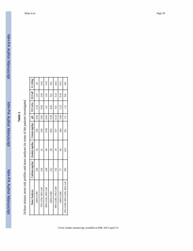
ResultsResults of stone and urine analyses are presented in Table 1
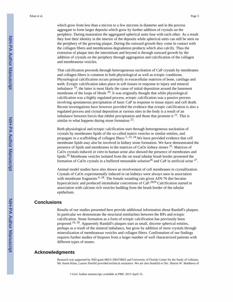
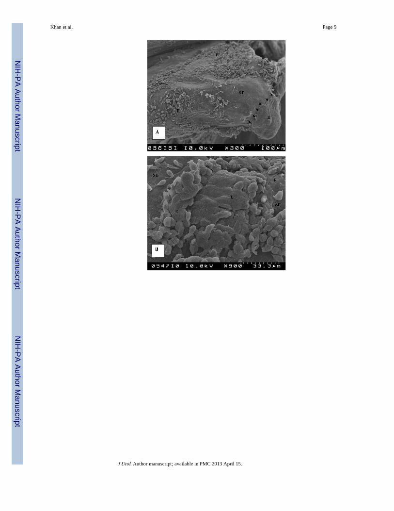
Intraoperative images of the papillae clearly showed RPs (Figure 1A) as well as attachedstones (Figure 1B). Once renal papillary tissues were fixed for microscopic analyses,plaques appeared as small bulges or protrusions on the papillary surface. A closerexamination using SEM showed these bulges were not completely covered with the surfaceepithelium (Figures 2 A, B). Part of the epithelial covering appeared to have sloughed offexposing the underlying structures. Loss of surface epithelium revealed a granular surfacewhich was followed by a fibrous layer (Figure 2 C). Crystal deposits or plaque were presentunderneath the fibrous layer (Figure 2 D). The outer surface of the plaque was composed of1-5 μm diameter spherical units, referred to as spherulites. These spherulites had a roughexternal surface giving an appearance of a pin cushion and were mixed with thin long fibersrunning between them (Figure 3 A). Some fibers appeared deeply embedded in the crystals.Fusion of the units and loss of their identity inside the plaque was apparent when plaqueswere fractured to examine the interior, which revealed mostly continuous concentric layers(Figure 3 B). However, higher magnification of fractured surfaces occasionally revealed thepresence of spherulites therein. EDX microanalyses of the spherulites showed the presenceof calcium and phosphorus only, indicating that the deposits were made of CaP (Figure 4 A).Calcium and phosphorus were also seen on EDX analysis of some fibers (Figure 4B)indicating that they also contained CaP deposits.
TEM analysis of the plaque showed basically two types of calcifications. All specimenscontained spherical deposits in the interstitium as well as in the laminated basementmembrane of the renal tubules (Figure 5 A). Some specimens had only few such depositsthat required sustained efforts to visualize them while others had a large number of sphericalcrystals both around the tubules as well as in the interstitium. The spherical units ranged insize from less than 0.5 μm to 2 μm across and showed a number of distinct internalconcentric laminations (Figure 5 B). Some sections clearly showed that concentriclaminations inside the spherulites contained radially arranged crystallites which could alsobe seen in grazing sections . The size of the spherical units and number of concentric layersseen in their cross section may depend upon the plane of sectioning 10. As a result, a 0.5 μmspherical unit may actually be bigger. Spherulites present in the basement membranegenerally appeared embedded in an amorphous matrix (Figure 5 A,B), sometime associatedwith membrane bound vesicles (Figure 5 A) while those in the interstitium were surroundedby both collagen fibers and cellular degradation products, including membrane boundvesicles. Small vesicles approximately 200 nm across were seen with dark contents (Figure6 A, B), which could be nucleating crystals. Grazing sections of the spherical units showedcrystals sticking out of their surface (Figure 6C).
Large calcifications were located in the interstitium and surrounded by collagen fibers withcharacteristic banding patterns, as well as membranous cellular degradation products and anamorphous matrix (Figures 7 A-D). The deposits were comprised of dark elongated strandsmixed with spherulites (Figure 7 B) and were apparently produced by their aggregation.Strands present on the periphery often showed banding patterns, highly correlated with thebanding pattern of the nearby collagen fibers (Figure 7 D). In some sections the periphery ofthe deposits appeared nodular (Figure 7A).
Khan et al. Page 3
J Urol. Author manuscript; available in PMC 2013 April 15.
NIH
-PA Author Manuscript
NIH
-PA Author Manuscript
NIH
-PA Author Manuscript
Higher magnifications showed the center of the deposits to be more crystalline withradiating needle shaped crystals while the periphery appeared less crystalline.
Electron diffraction analyses (Figure 8 A, B, C) revealed that all of them had somehydroxyapatite diffraction rings. There were less intense rings at the periphery (Figure 8 B,C) compared to those taken at the center (Figure 8 A) indicating that peripheralhydroxyapatite was less crystalline than the central one. In addition, crystals when presentwere smaller on the periphery in comparison with those present in center of the deposits.
DiscussionEver since Randall proposed his hypothesis that renal papillary subepithelial deposits act asthe initiating lesion of idiopathic renal calculi 1, a number of morphological studies of renalpapillae obtained from kidneys of stone formers as well as non stone formers have beenperformed. Some of the earliest electron microscopic studies were performed by Cooke andassociates 11, 12 and Haggitt and Pitcock 13. Haggit and Pitcock examined kidneys from 100randomly selected autopsies and performed electron microscopy on selected specimens.They found alizarin positive laminated spherules in the interstitium adjacent to the collectingducts. TEM showed laminated spherical bodies in close association with collagen fibers inthe interstitium as well as basement membrane of the collecting ducts. Cooke studied 62normal kidneys and found calcification in 43. He reported that calcification was invariablylocated in the basement membrane of the loops of Henle from where it extended into themedullary interstitium. Later electron microscopic examination of nephrectomy specimensfrom three normocalcaemic, normotensive patients localized the mineral deposits inassociation with collagen mainly around the loops of Henle. Some collecting ducts andblood vessels were also involved.
In recent years, Stoller and associates as well as Evan and associates have meticulouslyinvestigated Randall's plaques. Stoller et al. performed high resolution radiography 14 ofcadaveric kidneys and found that 57% of the kidneys had subepithelial Randall's plaqueswhich extended deep within the papillae and were intimately associated with collectingducts and vasa recta. von Kossa positive spherical CaP deposits were identified scattered inthe interstitium as well as around the collecting ducts and blood vessels. They proposed apathway for idiopathic stone formation involving the vascular system 15. Evan andassociates performed exhaustive morphological studies of renal papillae from stone patientswith a variety of causes16 and concluded that all idiopathic calcium stones develop attachedto the subepithelial Randall's plaques 17. They also confirmed the earlier observations ofCooke that Randall's plaques begin in the basement membrane of the loops of Henle.Osteopontin was identified in the crystal matrix interface while heavy chain of inter-alpha-inhibitor was localized in the crystal matrix itself. There was no evidence of cell injury,inflammation, interstitial fibrosis or intratubular crystal deposition in the renal biopsies fromidiopathic stone formers. They hypothesized that deposits migrate from the basementmembrane of the loops of Henle into the surrounding interstitium and become associatedwith type 1 collagen, fusing into a synctium in which islands of mineral appear to float in anorganic sea 16, 18.
Our studies show CaP crystals as spherical units with radially arranged crystals, thehallmark of apatite crystals particularly in kidney stones 9. They range in size from half amicron to a few microns and are found loosely scattered around the tubules and in theirbasement membranes and then extending into the interstitium. Large interstitial depositsshow both spherical units as well as long strands with distinct banding pattern suggestive ofcollagen fibers. Cellular degradation products including membrane bound vesicles wereassociated with the calcified deposits. Apparently CaP crystals start as small spherical units
Khan et al. Page 4
J Urol. Author manuscript; available in PMC 2013 April 15.
NIH
-PA Author Manuscript
NIH
-PA Author Manuscript
NIH
-PA Author Manuscript
which grow from less than a micron to a few microns in diameter and in the processaggregate to form larger deposits which grow by further addition of crystals on theperiphery. During maturation the aggregated spherical units fuse with each other. As a resultthey lose their identity in the interior of the deposits while spherical units can still be seen onthe periphery of the growing plaque. During the outward growth they come in contact withthe collagen fibers and membranous degradation products which also calcify. Thus theextension of plaque into the interstitium and beyond is through outward growth by theaddition of crystals on the periphery through aggregation and calcification of the collagenand membranous vesicles.
That calcification proceeds through heterogeneous nucleation of CaP crystals by membranesand collagen fibers is common in both physiological as well as ectopic conditions.Physiological calcification occurs primarily in extracellular matrices of bone, cartilage andteeth. Ectopic calcification takes place in soft tissues in response to injury and mineralimbalance 19, the latter is most likely the cause of initial deposition around the basementmembrane of the loops of Henle 20. It was originally thought that while physiologicalcalcification was a highly regulated process, ectopic calcification was a passive processinvolving spontaneous precipitation of basic CaP in response to tissue injury and cell death.Recent investigations have however provided the evidence that ectopic calcification is also aregulated process and crystal deposition at various sites in the body is a result of animbalance between forces that inhibit precipitation and those that promote it 21. This issimilar to what happens during stone formation 22.
Both physiological and ectopic calcification start through heterogeneous nucleation ofcrystals by membranes lipids of the so-called matrix vesicles or similar entities, andpropagate in a scaffolding of collagen fibers 5, 23, 24.We have provided evidence that cellmembrane lipids may also be involved in kidney stone formation. We have demonstrated thepresence of lipids and membranes in the matrices of CaOx kidney stones 25. Matrices ofCaOx crystals induced in vitro in human urine also showed the presence of membranes andlipids.26 Membrane vesicles isolated from the rat renal tubular brush border promoted theformation of CaOx crystals in a buffered metastable solution26 and CaP in artificial urine 27.
Animal model studies have also shown an involvement of cell membranes in crystallization.Crystals of CaOx experimentally induced in rat kidneys were always seen in associationwith membrane fragments 6, 28. The female weanling rats given AIN-76 diet becamehypercalciuric and produced intratubular concretions of CaP.2829 Calcification started inassociation with calcium rich vesicles budding from the brush border of the tubularepithelium.
ConclusionsResults of our studies presented here provide additional information about Randall's plaques.In particular we demonstrate the structural similarities between the RPs and ectopiccalcification. Stone formation as a form of ectopic calcification has previously beenproposed 19, 30. Apparently Randall's plaques start as small, discrete spherical entities,perhaps as a result of the mineral imbalance, but grow by addition of more crystals throughmineralization of membranous vesicles and collagen fibers. Confirmation of our findingsrequires further studies of biopsies from a larger number of well characterized patients withdifferent types of stones.
AcknowledgmentsResearch was supported by NIH grant #RO1-DK078602 and University of Florida Center for the Study of Lithiasis.Ms Anum Khan, Lauren Streifel provided technical assistance. We are also thankful to Drs. Sharon W. Matthews of
Khan et al. Page 5
J Urol. Author manuscript; available in PMC 2013 April 15.
NIH
-PA Author Manuscript
NIH
-PA Author Manuscript
NIH
-PA Author Manuscript
COM electron microscopy Core and Ms. Karen L. Kelly of University of Florida ICBR Electron Microscopy &BioImaging Lab for SEM and TEM analyses.
References1. Randall A. The Origin and Growth of Renal Calculi. Ann Surg. 1937; 105:1009. [PubMed:
17856988]
2. Coe FL, Evan AP, Worcester EM, et al. Three pathways for human kidney stone formation. UrolRes. 2010; 38:147. [PubMed: 20411383]
3. Evan AP, Lingeman JE, Coe FL, et al. Randall's plaque of patients with nephrolithiasis begins inbasement membranes of thin loops of Henle. J Clin Invest. 2003; 111:607. [PubMed: 12618515]
4. Gower LB, Amos FF, Khan SR. Mineralogical signatures of stone formation mechanisms. Urol Res.2010; 38:281. [PubMed: 20625894]
5. Murshed M, McKee MD. Molecular determinants of extracellular matrix mineralization in bone andblood vessels. Curr Opin Nephrol Hypertens. 19:359. [PubMed: 20489614]
6. Khan SR, Finlayson B, Hackett RL. Experimental calcium oxalate nephrolithiasis in the rat. Role ofthe renal papilla Am J Pathol. 1982; 107:59.
7. Khan SR, Finlayson B, Hackett R. Renal papillary changes in patient with calcium oxalate lithiasis.Urology. 1984; 23:194. [PubMed: 6695491]
8. Khan SR, Johnson JM, Peck AB, et al. Expression of osteopontin in rat kidneys: induction duringethylene glycol induced calcium oxalate nephrolithiasis. J Urol. 2002; 168:1173. [PubMed:12187263]
9. Khan SR, Hackett RL. Identification of urinary stone and sediment crystals by scanning electronmicroscopy and x-ray microanalysis. J Urol. 1986; 135:818. [PubMed: 3959214]
10. Ryall RL. The future of stone research: rummagings in the attic, Randall's plaque, nanobacteria,and lessons from phylogeny. Urol Res. 2008; 36:77. [PubMed: 18286270]
11. Weller RO, Nester B, Cooke SAR. Calcification in the human renal papilla: an electronmicroscope study. Journal of Pathology. 1971; 107:211. [PubMed: 5084933]
12. Cooke SAR. The site of calcification in the human renal papilla. British Journal of Surgery. 1970;57:890. [PubMed: 5487030]
13. Haggit RC, Pitcock JA. Renal medullary calcification: a light and electron microscopic study. TheJournal of Urology. 1971; 106:342. [PubMed: 4106437]
14. Stoller ML, Low RK, Shami GS, et al. High resolution radiography of cadaveric kidneys:unraveling the mystery of Randall's plaque formation. J Urol. 1996; 156:1263. [PubMed:8808850]
15. Stoller ML, Meng MV, Abrahams HM, et al. The primary stone event: a new hypothesis involvinga vascular etiology. J Urol. 2004; 171:1920. [PubMed: 15076312]
16. Coe FL, Evan AP, Lingeman JE, et al. Plaque and deposits in nine human stone diseases. Urol Res.2010; 38:239. [PubMed: 20625890]
17. Miller NL, Gillen DL, Williams JC Jr, et al. A formal test of the hypothesis that idiopathic calciumoxalate stones grow on Randall's plaque. BJU Int. 2009; 103:966. [PubMed: 19021625]
18. Evan AP, Lingeman JE, Coe FL, et al. Role of interstitial apatite plaque in the pathogenesis of thecommon calcium oxalate stone. Semin Nephrol. 2008; 28:111. [PubMed: 18359392]
19. Giachelli CM. Ectopic calcification: gathering hard facts about soft tissue mineralization. Am JPathol. 1999; 154:671. [PubMed: 10079244]
20. Halperin ML, Cheema Dhadli S, Kamel KS. Physiology of acid-base balance: links with kidneystone prevention. Semin Nephrol. 2006; 26:441. [PubMed: 17275581]
21. Giachelli CM. Inducers and inhibitors of biomineralization: lessons from pathological calcification.Orthod Craniofac Res. 2005; 8:229. [PubMed: 16238602]
22. Khan SR, Kok DJ. Modulators of urinary stone formation. Front Biosci. 2004; 9:1450. [PubMed:14977559]
23. Habibovic P, Bassett DC, Doillon CJ, et al. Collagen biomineralization in vivo by sustained releaseof inorganic phosphate ions. Adv Mater. 1858; 22
Khan et al. Page 6
J Urol. Author manuscript; available in PMC 2013 April 15.
NIH
-PA Author Manuscript
NIH
-PA Author Manuscript
NIH
-PA Author Manuscript
24. Golub EE. Biomineralization and matrix vesicles in biology and pathology. Semin Immunopathol.2010
25. Khan SR, Atmani F, Glenton P, et al. Lipids and membranes in the organic matrix of urinarycalcific crystals and stones. Calcif Tissue Int. 1996; 59:357. [PubMed: 8849402]
26. Khan SR, Maslamani SA, Atmani F, et al. Membranes and their constituents as promoters ofcalcium oxalate crystal formation in human urine. Calcif Tissue Int. 2000; 66:90. [PubMed:10652953]
27. Fasano JM, Khan SR. Intratubular crystallization of calcium oxalate in the presence of membranevesicles: an in vitro study. Kidney Int. 2001; 59:169. [PubMed: 11135069]
28. Khan SR. Nephrocalcinosis in animal models with and without stones. Urol Res. 2010; 38:429.[PubMed: 20658131]
29. Nguyen HT, Woodard JC. Intranephronic calculosis in rats: an ultrastructural study. Am J Pathol.1980; 100:39. [PubMed: 7395968]
30. Gambaro G, D'Angelo A, Fabris A, et al. Crystals, Randall's plaques and renal stones: do bone andatherosclerosis teach us something? J Nephrol. 2004; 17:774. [PubMed: 15593050]
Key of Definitions
RP Randall's Plaques
MV Membrane Bound Vesicles
CaOx Calcium Oxalate
EDX Energy Dispersive X-ray Microanalyses
CaP Calcium Phosphate
TEM Transmission Electron Microscopy
SEM Scanning Electron Microscopy
H&E Hematoxylin and Eosin
PAS Periodic Acid Schiff
OPN Psteopontin
Khan et al. Page 7
J Urol. Author manuscript; available in PMC 2013 April 15.
NIH
-PA Author Manuscript
NIH
-PA Author Manuscript
NIH
-PA Author Manuscript
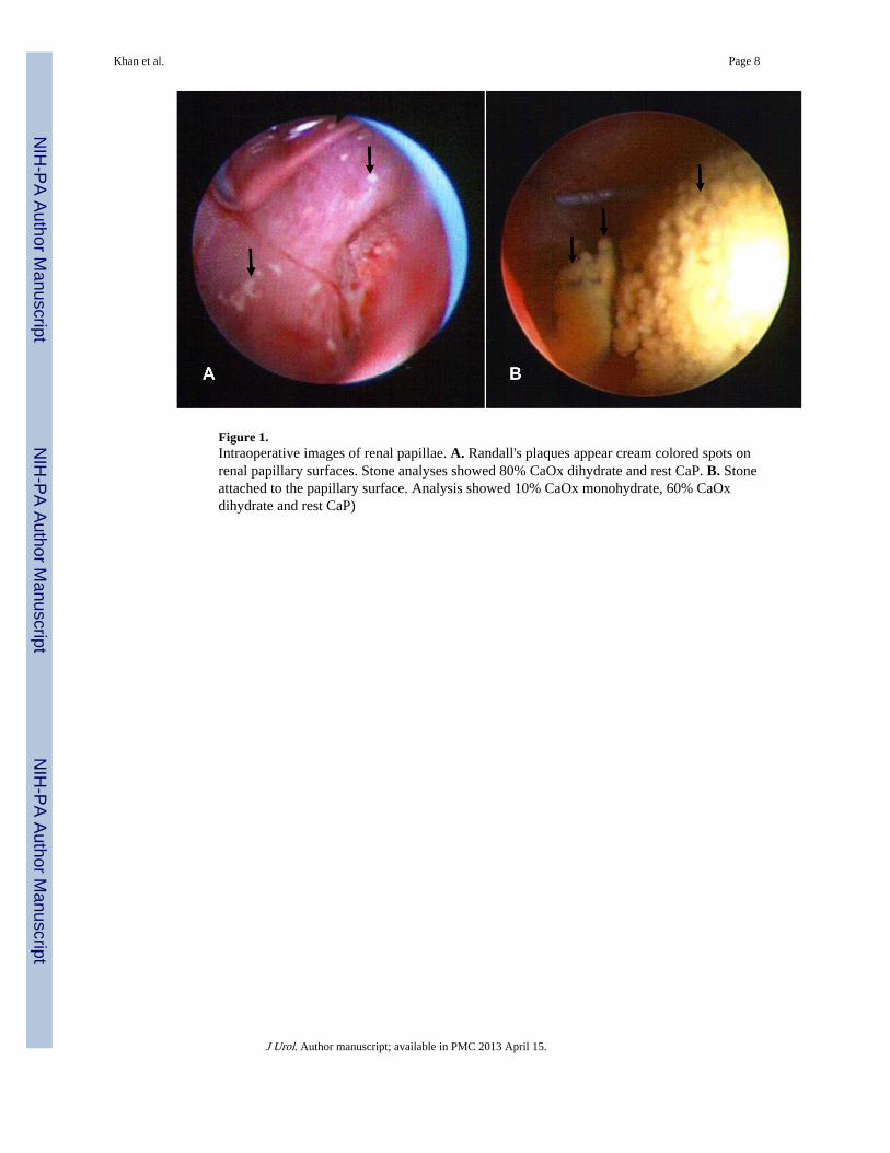
Figure 1.Intraoperative images of renal papillae. A. Randall's plaques appear cream colored spots onrenal papillary surfaces. Stone analyses showed 80% CaOx dihydrate and rest CaP. B. Stoneattached to the papillary surface. Analysis showed 10% CaOx monohydrate, 60% CaOxdihydrate and rest CaP)
Khan et al. Page 8
J Urol. Author manuscript; available in PMC 2013 April 15.
NIH
-PA Author Manuscript
NIH
-PA Author Manuscript
NIH
-PA Author Manuscript
Khan et al. Page 9
J Urol. Author manuscript; available in PMC 2013 April 15.
NIH
-PA Author Manuscript
NIH
-PA Author Manuscript
NIH
-PA Author Manuscript
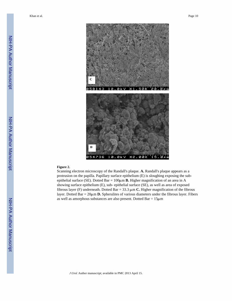
Figure 2.Scanning electron microscopy of the Randall's plaque. A. Randall's plaque appears as aprotrusion on the papilla. Papillary surface epithelium (E) is sloughing exposing the sub-epithelial surface (SE). Dotted Bar = 100μm B. Higher magnification of an area in Ashowing surface epithelium (E), sub- epithelial surface (SE), as well as area of exposedfibrous layer (F) underneath. Dotted Bar = 33.3 μm C. Higher magnification of the fibrouslayer. Dotted Bar = 20μm D. Spherulites of various diameters under the fibrous layer. Fibersas well as amorphous substances are also present. Dotted Bar = 15μm
Khan et al. Page 10
J Urol. Author manuscript; available in PMC 2013 April 15.
NIH
-PA Author Manuscript
NIH
-PA Author Manuscript
NIH
-PA Author Manuscript
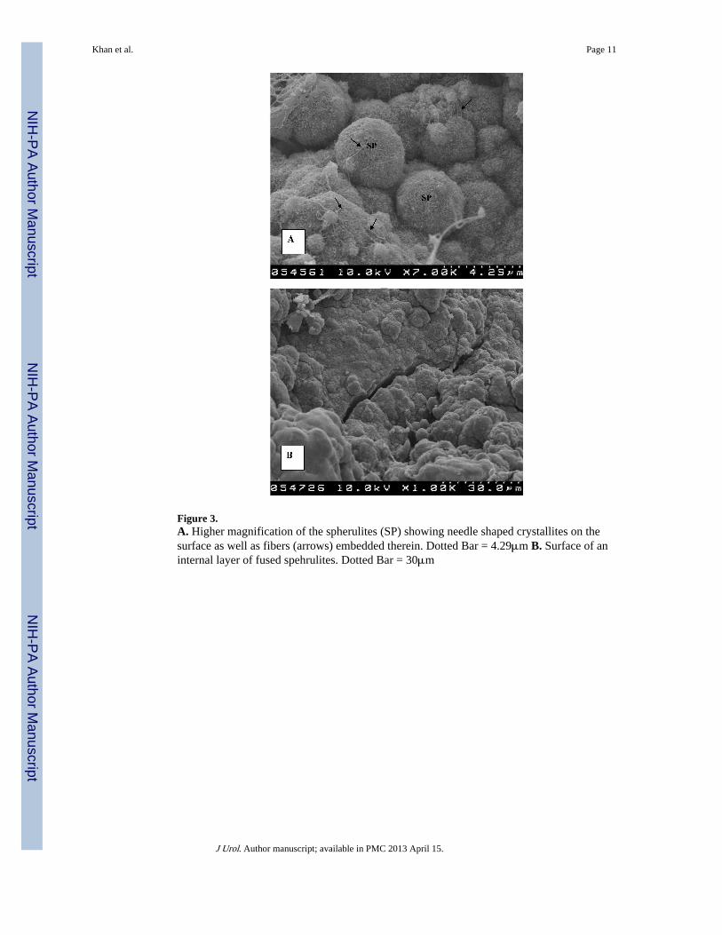
Figure 3.A. Higher magnification of the spherulites (SP) showing needle shaped crystallites on thesurface as well as fibers (arrows) embedded therein. Dotted Bar = 4.29μm B. Surface of aninternal layer of fused spehrulites. Dotted Bar = 30μm
Khan et al. Page 11
J Urol. Author manuscript; available in PMC 2013 April 15.
NIH
-PA Author Manuscript
NIH
-PA Author Manuscript
NIH
-PA Author Manuscript
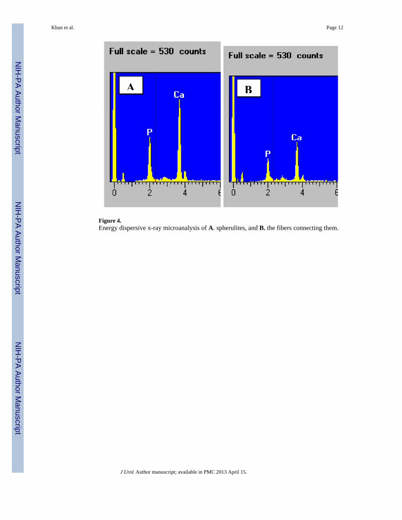
Figure 4.Energy dispersive x-ray microanalysis of A. spherulites, and B. the fibers connecting them.
Khan et al. Page 12
J Urol. Author manuscript; available in PMC 2013 April 15.
NIH
-PA Author Manuscript
NIH
-PA Author Manuscript
NIH
-PA Author Manuscript
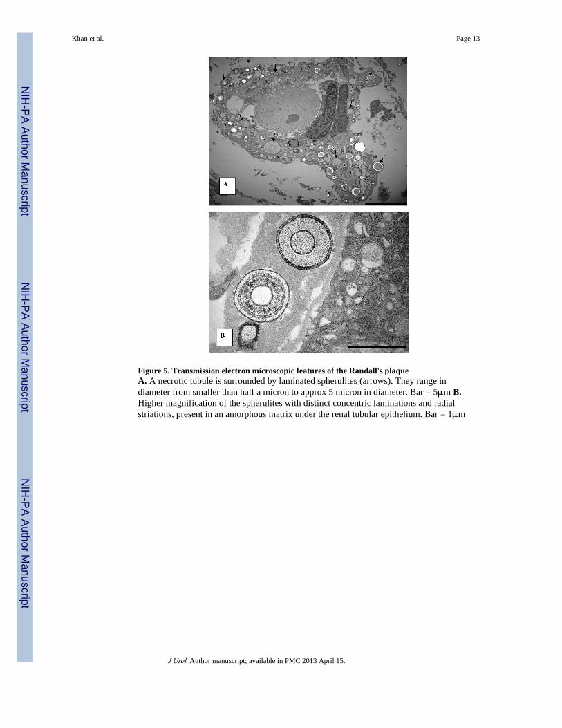
Figure 5. Transmission electron microscopic features of the Randall's plaqueA. A necrotic tubule is surrounded by laminated spherulites (arrows). They range indiameter from smaller than half a micron to approx 5 micron in diameter. Bar = 5μm B.Higher magnification of the spherulites with distinct concentric laminations and radialstriations, present in an amorphous matrix under the renal tubular epithelium. Bar = 1μm
Khan et al. Page 13
J Urol. Author manuscript; available in PMC 2013 April 15.
NIH
-PA Author Manuscript
NIH
-PA Author Manuscript
NIH
-PA Author Manuscript
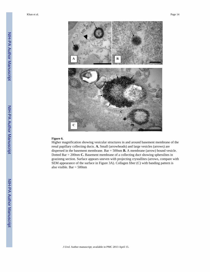
Figure 6.Higher magnification showing vesicular structures in and around basement membrane of therenal papillary collecting ducts. A. Small (arrowheads) and large vesicles (arrows) aredispersed in the basement membrane. Bar = 500nm B. A membrane (arrow) bound vesicle.Dotted Bar = 200nm C. Basement membrane of a collecting duct showing spherulites ingrazinmg section. Surface appears uneven with projecting crystallites (arrows, compare withSEM appearance of the surface in Figure 3A). Collagen fiber (C) with banding pattern isalso visible. Bar = 500nm
Khan et al. Page 14
J Urol. Author manuscript; available in PMC 2013 April 15.
NIH
-PA Author Manuscript
NIH
-PA Author Manuscript
NIH
-PA Author Manuscript
Khan et al. Page 15
J Urol. Author manuscript; available in PMC 2013 April 15.
NIH
-PA Author Manuscript
NIH
-PA Author Manuscript
NIH
-PA Author Manuscript
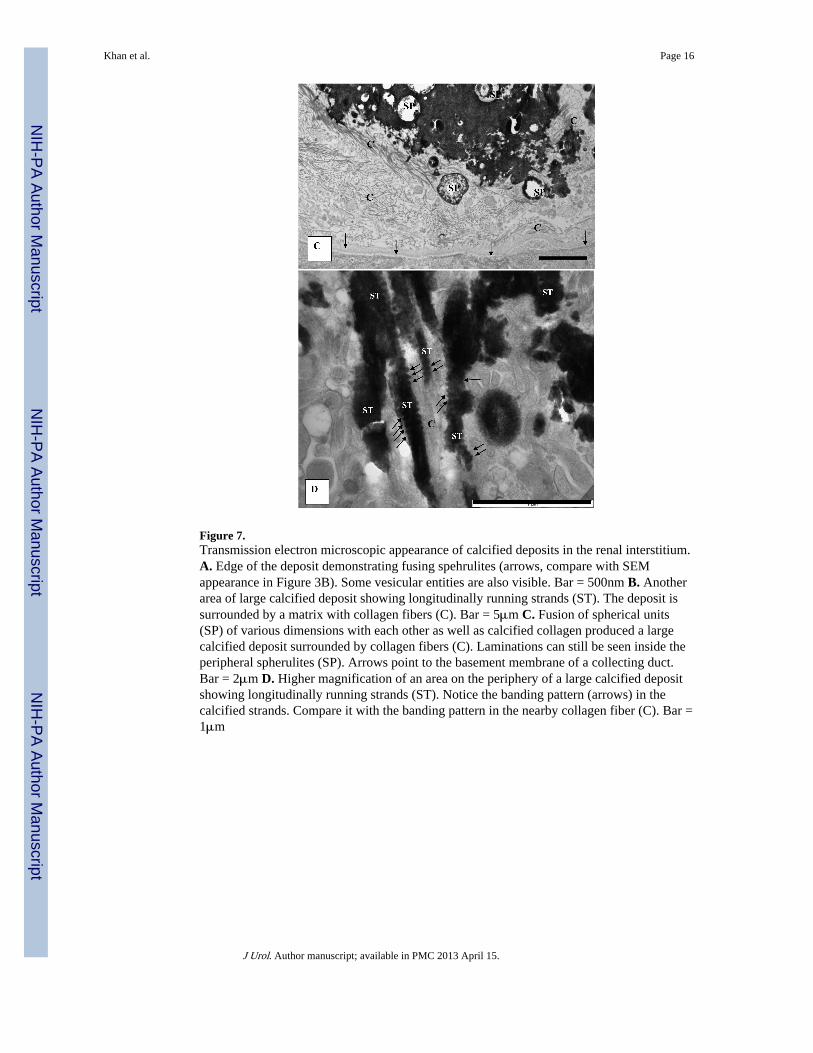
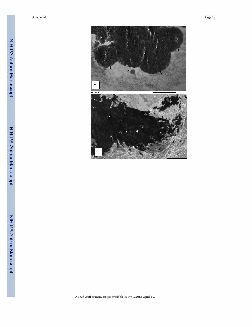
Figure 7.Transmission electron microscopic appearance of calcified deposits in the renal interstitium.A. Edge of the deposit demonstrating fusing spehrulites (arrows, compare with SEMappearance in Figure 3B). Some vesicular entities are also visible. Bar = 500nm B. Anotherarea of large calcified deposit showing longitudinally running strands (ST). The deposit issurrounded by a matrix with collagen fibers (C). Bar = 5μm C. Fusion of spherical units(SP) of various dimensions with each other as well as calcified collagen produced a largecalcified deposit surrounded by collagen fibers (C). Laminations can still be seen inside theperipheral spherulites (SP). Arrows point to the basement membrane of a collecting duct.Bar = 2μm D. Higher magnification of an area on the periphery of a large calcified depositshowing longitudinally running strands (ST). Notice the banding pattern (arrows) in thecalcified strands. Compare it with the banding pattern in the nearby collagen fiber (C). Bar =1μm
Khan et al. Page 16
J Urol. Author manuscript; available in PMC 2013 April 15.
NIH
-PA Author Manuscript
NIH
-PA Author Manuscript
NIH
-PA Author Manuscript
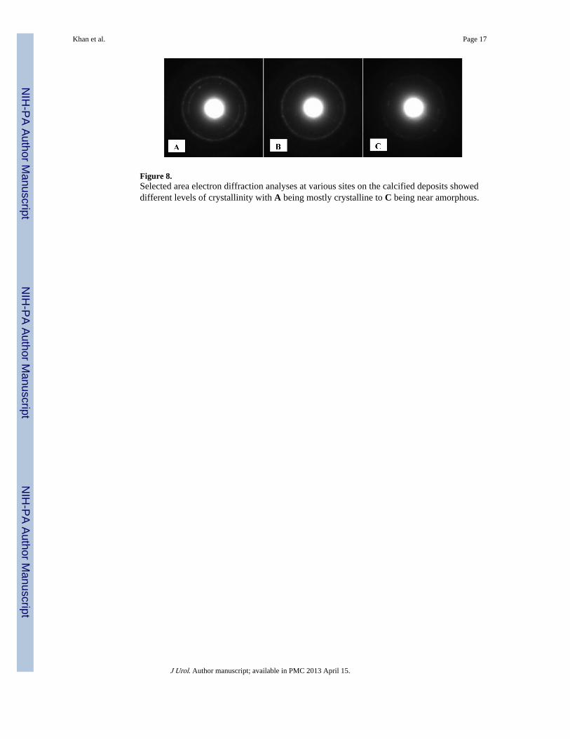
Figure 8.Selected area electron diffraction analyses at various sites on the calcified deposits showeddifferent levels of crystallinity with A being mostly crystalline to C being near amorphous.
Khan et al. Page 17
J Urol. Author manuscript; available in PMC 2013 April 15.
NIH
-PA Author Manuscript
NIH
-PA Author Manuscript
NIH
-PA Author Manuscript
NIH
-PA Author Manuscript
NIH
-PA Author Manuscript
NIH
-PA Author Manuscript
Khan et al. Page 18
Tabl
e 1
24-h
our
urin
ary
ston
e ri
sk p
rofi
les
and
ston
e an
alys
es f
or s
ome
of th
e pa
tient
s in
vest
igat
ed.
Ston
e A
naly
ses
Cal
cium
mg/
day
Oxa
late
mg/
day
Cit
rate
mg/
day
pHSS
CaO
xSS
CaP
Cr
24/k
g
100%
CO
M20
443
556
6.83
5.53
1.67
20
70%
CO
D, 3
0% C
aP22
422
334
5.83
9.82
2.02
14.9
80%
CO
D, 2
0% C
aP24
846
1136
6.63
6.4
1.81
15.9
100%
CO
M21
228
650
5.59
4.92
NA
NA
90%
CO
M, 1
0% C
OD
332
2955
76.
148.
03.
1224
.6
100%
CO
M77
4930
45.
965.
230.
4518
.3
10%
CO
M, 6
0% C
OD
, 30%
CaP
293
24.7
301
5.5
1.4
NA
NA
J Urol. Author manuscript; available in PMC 2013 April 15.