association of intercellular adhesion molecule‑1 gene polymorphism with coronary heart disease
TRANSCRIPT
“fendo-03-00179” — 2013/1/20 — 20:09 — page 1 — #1
REVIEW ARTICLEpublished: 22 January 2013
doi: 10.3389/fendo.2012.00179
Association of intercellular adhesion molecule 1 (ICAM1)with diabetes and diabetic nephropathyHarvest F. Gu1*, Jun Ma2, KarolinT. Gu3 and Kerstin Brismar1
1 M1:03 Rolf Luft Center for Diabetes and Endocrinology Research, Department of Molecular Medicine and Surgery, Karolinska Institutet,Karolinska University Hospital, Stockholm, Sweden
2 Department of Anesthesiology, Anzhen Hospital, Capital Medical University, Beijing, People’s Republic of China3 Viktor Rydberg Gymnasium Odenplan School, Stockholm, Sweden
Edited by:
Gabriel Virella, Universidade deLisboa, Portugal
Reviewed by:
Kay Waud, Eastern Virginia MedicalSchool, USAShinichi Oikawa, Nippon MedicalSchool, JapanMelissa Irene March, Beth IsraelDeaconess Medical Center, USA
*Correspondence:
Harvest F. Gu, M1:03 Rolf LuftCenter for Diabetes andEndocrinology Research, Departmentof Molecular Medicine and Surgery,Karolinska Institutet, KarolinskaUniversity Hospital, StockholmSE-17176, Sweden.e-mail: [email protected]
Diabetes and diabetic nephropathy are complex diseases affected by genetic andenvironmental factors. Identification of the susceptibility genes and investigation of theirroles may provide useful information for better understanding of the pathogenesis and fordeveloping novel therapeutic approaches. Intercellular adhesion molecule 1 (ICAM1) is acell surface glycoprotein expressed on endothelial cells and leukocytes in the immunesystem. The ICAM1 gene is located on chromosome 19p13 within the linkage regionof diabetes. In the recent years, accumulating reports have implicated that geneticpolymorphisms in the ICAM1 gene are associated with diabetes and diabetic nephropathy.Serum ICAM1 levels in diabetes patients and the icam1 gene expression in kidney tissuesof diabetic animals are increased compared to the controls. Therefore, ICAM1 may play arole in the development of diabetes and diabetic nephropathy. In this review, we presentgenomic structure, variation, and regulation of the ICAM1 gene, summarized genetic andbiological studies of this gene in diabetes and diabetic nephropathy and discussed about thepotential application using ICAM1 as a biomarker and target for prediction and treatmentof diabetes and diabetic nephropathy.
Keywords: intercellular adhesion molecule 1, diabetic nephropathy, end-stage renal disease, type 1 diabetes
mellitus, type 2 diabetes mellitus
INTRODUCTIONDiabetes mellitus is a group of metabolic diseases in which thepatients have high blood glucose levels. Its epidemic has becomea national and global crisis. Based upon the figures today, atleast 366 million people at the worldwide have diabetes. By theyear 2030, this number is expected to be double (Bonow andGheorghiade, 2004; Wild et al., 2004; Cornell and Dorsey, 2012;Lam and LeRoith, 2012). There are two major types of dia-betes. Type 1 diabetes (T1D), previously called juvenile diabetesor insulin-dependent diabetes, develops on the basis of autoim-mune destruction of pancreatic β-cells, which results in insulindeficiency. It mostly affects young people (<20 years old) butoccurs also in adults (Lightfoot et al., 2012). Type 2 diabetes(T2D) is the most common form of diabetes and accounts forapproximately 85–90% of all diabetic patients. In T2D, hyper-glycemia results from a combination of impaired insulin secretionand insulin resistance. When the pancreatic β-cells loose the abil-ity to compensate for insulin resistance in liver, skeletal muscle,and adipose tissues, hyperglycemia becomes manifest (Alberti and
Abbreviations: ACR, urinary albumin/creatinine ratio; AER, albumin excretionrate; CD54, cluster of differentiation 54; DN, diabetic nephropathy; ESRD, end-stagerenal disease; GFR, glomerular filtration rate; GoKinD, Genetics of Kidneys in Dia-betes; HDL, high-density lipoprotein; HWE, Hardy–Weinberg equilibrium; ICAM1,intercellular adhesion molecule 1; LD, linkage disequilibrium; LDL, low-densitylipoprotein; LFA, leukocyte adhesion protein; SNP, single-nucleotide polymor-phism; T1DM, type 1 diabetes mellitus; T2DM, type 2 diabetes mellitus; UTR,un-translation region.
Zimmet, 1998). Diabetes patients often develop macro- and/ormicro-vascular complications. Diabetic nephropathy (DN) is oneof serious complications and occurs in 30–40% of diabetic patients(Heerspink and de Zeeuw, 2011; Marshall, 2012). This diabeticcomplication is characterized by pathophysiological changes inglomerular hyperfiltration, renal hypertrophy, tubular functionand then progress to proteinuria and reduction of glomerularfiltration rate (GFR). The patients with DN exhibit persistent pro-teinuria, hypertension, declining renal function, and increasedpremature mortality largely as a result of cardiovascular disease.DN is the most common single cause of end-stage renal disease(ESRD). Once overt DN occurs, it progresses slowly or rapidly tothe most advanced stage of chronic kidney disease which needsdialysis or transplantation treatment (Marshall, 2004; Shields andMaxwell, 2010; Weil et al., 2010; Thomas and Groop, 2011). Thetreatment cost for diabetes patients has been increasing stagger-ing in the recent decades and becomes a further burden of thehealthcare system. Diabetes and DN are multi-factorial diseases,which are influenced by both genetic and environmental factors(Satko et al., 2005; Pitkäniemi et al., 2007; Ashcroft and Rorsman,2012; Gonzalez-Bulnes and Ovilo, 2012; Morahan, 2012). There-fore, identification of the susceptibility genes in development ofdiabetes and diabetic complications and investigation of their rolesare of importance to provide useful information for improvementof the prevention and medication programs.
Intercellular adhesion molecule 1 (ICAM1, OMIM: 147840)is a cell surface glycoprotein and expressed in endothelial cells
www.frontiersin.org January 2013 | Volume 3 | Article 179 | 1
“fendo-03-00179” — 2013/1/20 — 20:09 — page 2 — #2
Gu et al. ICAM1 in diabetes and nephropathy
and leukocytes in the immune system. This endothelial- andleukocyte-associated transmembrane protein has been known forits importance in stabilizing cell–cell interactions and facilitatingleukocyte endothelial transmigration. Recently, the accumulat-ing reports from genetic studies in diabetic patients with andwithout DN and from biological studies with diabetic animalmodels have implicated that ICAM1 may play a role in thepathogenesis of diabetes and DN. In this review, we will sum-marize the genetic and pathophysiological relevance of ICAM1and discuss about the possible role of ICAM1 in the develop-ment of diabetes and DN as well as the perspectives of the ICAM1research.
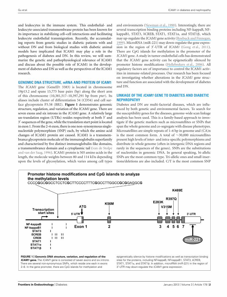
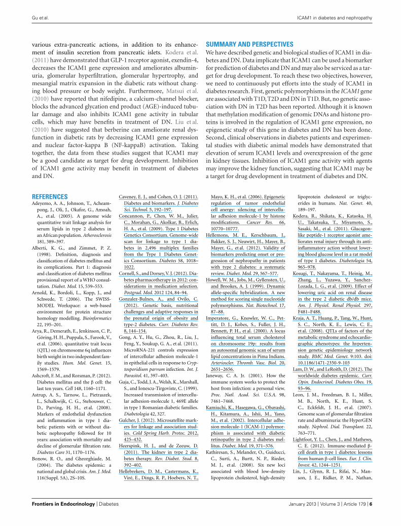
GENOMIC DNA STRUCTURE, mRNA AND PROTEIN OF ICAM1The ICAM1 gene (GeneID: 3383) is located in chromosome19p13.2 and spans 15,775 base pairs (bp) along the short armof this chromosome (10,381,517–10,397,291 bp from pter). Itsaliases include cluster of differentiation 54 (CD54) and cell sur-face glycoprotein P3.58 (BB2). Figure 1 demonstrates genomicstructure, regulation, and variation of the ICAM1 gene. There areseven exons and six introns in the ICAM1 gene. A relatively largeun-translation region (UTRs) resides respectively at both 5′ and3′-sequences of the gene, while the translation start point is locatedin exon 1. From the 2–6 exon, there is one non-synonymous single-nucleotide polymorphism (SNP) each, by which the amino acidchanges of ICAM1 protein are caused. ICAM1 is a transmem-brance glycoprotein molecule of the immunoglobulin superfamilyand characterized by five distinct immunoglobulin-like domains,a transmembrance domain and a cytoplasmic tail (van de Stolpeand van der Saag, 1996). ICAM1 protein is 505 amino acids in thelength, the molecule weights between 80 and 114 kDa dependingupon the levels of glycosylation, which varies among cell types
and environments (Newman et al., 1990). Interestingly, there areseveral transcription binding proteins including NF-kappaB, NF-kappaB1, STAT3, δCREB, STAT1, STAT1α, and STAT1β, whichmay up-regulate the ICAM1 gene activity (Roebuck and Finnegan,1999). MicroRNA (miR-221) may down-regulate the gene expres-sion in the region of 3′-UTR of ICAM1 (Gong et al., 2011).There are CpG islands for methylation in the promoter of theICAM1 gene. A study in tumor endothelial cells has demonstratedthat the ICAM1 gene activity can be epigenetically silenced bypromoter histone modifications (Hellebrekers et al., 2006). Allregulatory factors are of importance to control the ICAM1 activ-ities in immune-related processes. Our research has been focusedon investigating whether alterations in the ICAM1 gene struc-ture and function are associated with the development of diabetesand DN.
LINKAGE OF THE ICAM1 GENE TO DIABETES AND DIABETICNEPHROPATHYDiabetes and DN are multi-factorial diseases, which are influ-enced by both genetic and environmental factors. To search forthe susceptibility genes for the diseases, genome-wide scan linkageanalysis has been used. This is a family-based approach to inves-tigate if the genetic markers such as microsatellites or SNPs thatspan the whole genome and co-segregate with disease phenotypes.Microsatellites are simple repeats of 1–6 bp in genome and (CA)nis the most common form. A total of ∼30,000 microsatellitespresent high levels of inter- and intra-specific polymorphisms anddistribute in whole genome (often in intergenic DNA regions andrarely in the sequences of the genes). SNPs are the substitutionsof nucleotides in genomic DNA. In general speaking, bi-allelicSNPs are the most common type. Tri-allelic ones and small inser-tions/deletions are also included. C/T is the most common SNP
FIGURE 1 | Genomic DNA structure, variation, and regulation of the
ICAM1 gene. The ICAM1 gene is consisted of seven exons and six introns.There are several non-synonymous SNPs, which reside one each in exons2–6. In the gene promoter, there are CpG islands for methylation and
epigenetically silence by histone modifications as well as transcription bindingsites for the proteins, including NF-kappaB, NF-kappaB1, STAT3, δCREB,STAT1, STAT1α, and STAT1β. In addition, microRNA (miR-221) in the region of3′-UTR may down-regulate the ICAM1 gene expression.
Frontiers in Endocrinology | Diabetes January 2013 | Volume 3 | Article 179 | 2
“fendo-03-00179” — 2013/1/20 — 20:09 — page 3 — #3
Gu et al. ICAM1 in diabetes and nephropathy
in the human genome. SNPs reside in the coding region of thegenes are called cSNPs, which include non-synonymous SNPs(with amino acid changes) and synonymous SNPs (without aminoacid changes). Most of SNPs are located in non-coding regions ofthe genes, including promoter, intron, and UTRs. SNPs in thepromoter region may alter transcription binding site and therebyaffect the transcriptional activity of the gene. Moreover, a numberof SNPs are found in inter-genic sequences. Up to date, more than40 million SNPs are recorded in the public SNP databases, whichare freely available for research use. Using the genetic markers, thelinkage analysis allows us to identify the genome that is transmit-ted within families along with the disease phenotypes of interestat a genome-wide scale. Based on finding a statistical signal, theprobability of co-segregation of a disease with a chromosomallocus is given (Gulcher, 2012). In this approach of genome-widescan, highly polymorphic genetic markers distributed across thegenome are genotyped in large family pedigrees or in affected anddiscordant sibling pairs.
By using the approach of genome-wide scan and linkage anal-yses, several chromosomal regions including 19p13 have beenpredicted to link with diabetes and DN. Mein et al. (1998) have pre-viously conducted genome wide scan analysis in 93 affected sibpairfamilies and 263 multiplex families from the UK and indicated thatloci in chromosome 19p13 are linked to T1DM. Later on, the link-age of T1DM to chromosome 19p13 is replicated by the study with2658 affected sib-pairs in USA (Concannon et al., 2009). Interest-ingly, lipid-related traits such total cholesterol, triglycerides andlow-density lipoprotein (LDL) concentrations in T2DM are linkedto chromosome 19p13 and this finding has been replicated inCaucasians, African-American and Hispanic families (Imperatoreet al., 2000; Adeyemo et al., 2005; Malhotra and Wolford, 2005).Several interesting candidate genes, including insulin receptor,resistin and ICAM1, are involved in the region of chromosome19p13. The linkage of LDL concentrations to chromosome 19p13has been replicated in the study with 612 individuals from 28Amish families in USA (Pollin et al., 2004). Furthermore, Leonet al. (2007) have performed a genome-wide scan study in 1251African Americans (AA) and 1129 European Americans (EA)hypertensive siblings from the Hypertension Genetic Epidemiol-ogy Network study and indicated that loci in chromosome 19p arelinked with albumin to creatinine ratio (ACR) when both AA andEA subjects are combined in the analyses. Particularly, Kathire-san et al. (2008) have analyzed the genome-wide scan data fromthree studies including 8816 T2D subjects and found six new lociassociated with LDL, cholesterol, HDL, and triglycerides. One ofthe loci is located in chromosome 19p13. Previously, Arya et al.(2006) have conducted a genome-wide scan and linkage studyand suggested that loci in both chromosomal arms of 19p13.2and 19q13.4 may be linked to birth weight in T2D families ofboth Mexican Americans and EA. By the analyses of the com-bined metabolic syndrome and echocardiographic factors, Krajaet al. (2008) have found the linkage of blood pressure in T2Dpatients of AA and EA with the region chromosome 19p13. Tak-ing together with the information from above briefly describedgenome-wide scan studies, it is clear that there are the loci inchromosome 19p may confer the susceptibility risk to diabetesand DN.
ASSOCIATION OF THE ICAM1 GENETIC POLYMORPHISMSWITH DIABETES AND DIABETIC NEPHROPATHYSeveral research groups including ours have reported the geneticassociation studies of the ICAM1 gene in T1DM and DN. Gujaet al. (1999) reported that the transmission of the G allele of SNPK469E (A/G) is increased in Romanian T1DM families. One yearlater, Nishimura et al. (2000) replicated that this K469E poly-morphism is associated with adult-onset T1DM in a Japanesepopulation. However, the association of K469E polymorphismin the ICAM1 gene with T1DM was not found in Danish, Finnish,and British Caucasian (Nejentsev et al., 2000). Furthermore,Nejentsev et al. (2003) demonstrated that another synonymousSNP G241R in the ICAM1 gene was associated with T1DM. Allthese previous studies were designed for analysis of one or twoSNP(s). In order to ascertain whether the ICAM1 genetic poly-morphisms are associated with T1DM and DN, we conductedthe comprehensive genetic association studies. The studied SNPsincluding K469E and G241R were selected based upon the infor-mation of their position in the ICAM1 gene and their linkagedisequilibrium (LD) values in the HapMap. Our data from sin-gle marker association analyses indicated that K469E and anotherintronic polymorphism (rs281432) were significantly associatedwith T1DM in Swedish Caucasians (Ma et al., 2006). Interestingly,these two SNPs are located in intron 2 and exon 6, respectively.According to the data of pair-wise LD values for SNPs in theICAM1 gene, a relatively strong LD (/D′/ ≥ 0.7) existed to extendover the region between these two SNPs. Due to the large 5′- and3′- UTRs in exons 1 and 7, the LD block covers almost the wholecoding region of the gene. Further multiplex marker associationanalysis was done and the common haplotype C-A constructedby C allele from K469E and A allele from rs281432 was found tobe associated with T1D (Ma et al., 2006). Later on, we found thatK469E polymorphism in the ICAM1 is associated with DN in T1Dpatients of Americans of European descent and selected from theGenetic of Kidney Diseases in Diabetes (GoKinD) study (Muelleret al., 2006). However, no association of G241R in the ICAM1gene with T1DM and DN in Swedish and GoKinD populationswas found (Ma et al., 2008). In patients with T2D, the K469E poly-morphism in the ICAM1 gene was found to associate with plasmafibrinogen levels and diabetic retinopathy (Kamiuchi et al., 2002;Yokoyama et al., 2005; Liu et al., 2006; Petrovic et al., 2008; Vinitaet al., 2012). Up to date, there is, however, no report regarding theassociation of ICAM1 genetic polymorphism with DN in T2D.
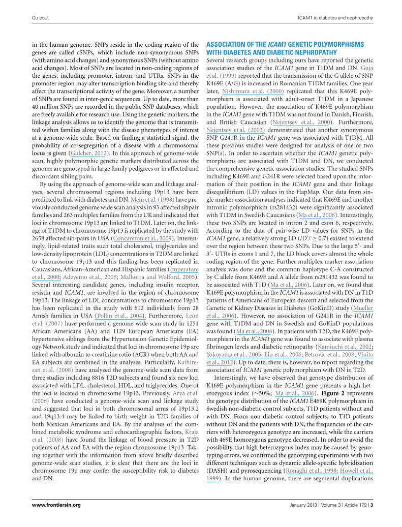
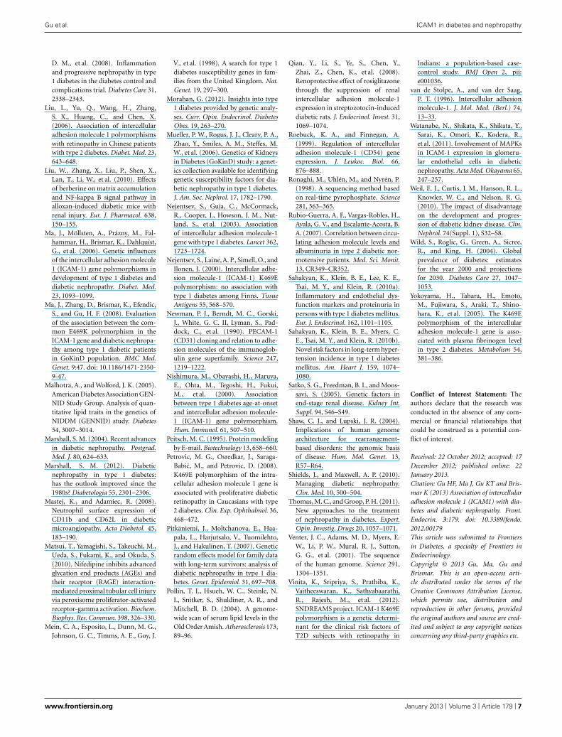
Interestingly, we have observed that genotype distribution ofK469E polymorphism in the ICAM1 gene presents a high het-erozygous index (∼50%; Ma et al., 2006). Figure 2 representsthe genotype distribution of the ICAM1 E469K polymorphism inSwedish non-diabetic control subjects, T1D patients without andwith DN. From non-diabetic control subjects, to T1D patientswithout DN and the patients with DN, the frequencies of the car-riers with heterozygous genotype are increased, while the carrierswith 469E homozygous genotype decreased. In order to avoid thepossibility that high heterozygous index may be caused by geno-typing errors, we confirmed the genotyping experiments with twodifferent techniques such as dynamic allele-specific hybridization(DASH) and pyrosequencing (Ronaghi et al., 1998; Howell et al.,1999). In the human genome, there are segmental duplications
www.frontiersin.org January 2013 | Volume 3 | Article 179 | 3
“fendo-03-00179” — 2013/1/20 — 20:09 — page 4 — #4
Gu et al. ICAM1 in diabetes and nephropathy
FIGURE 2 | Genotype distribution of the ICAM1 K469E
polymorphism. The genotype distribution of the ICAM1 K469Epolymorphism is represented from a genetic association study in Swedishpopulation (Ma et al., 2006). Three genotypes of the ICAM1 K469Epolymorphism are shown in as light gray color for K469K, gray for K469E,
and dark for E469E. Obviously, the heterozygous index is highcompared to the percentage of homozygous and increased fromthe group of non-diabetic control subjects, to type 1 diabetes (T1D)patients without diabetic nephropathy and the patients with diabeticnephropathy.
(duplicons) with >90% sequence similarity between the copies,which may cause specific allelic and genotypic diversities, suchas high heterozygous index in complex diseases (Venter et al.,2001; Shaw and Lupski, 2004). To ascertain whether K469E SNPis involved in a duplicon, we further performed a cloning andsequencing analysis and found that no duplication resides in thegene region (Ma et al., 2006). K469E is a non-synonymous SNP inexon 6 of the ICAM1 gene, which causes the amino acid changesof the ICAM1 protein. We have submitted ICAM1 amino acidsequences with K469 and 469E alleles respectively into SWISS-MODEL (Peitsch, 1995; Arnold et al., 2006) to understand thechanges of ICAM1 protein. There are 532 amino acids in theprotein sequence of ICAM1, K469 is wild-type and has 100% iden-tified homology. Compared to the DIMER image of wild ICAM1protein, however, the structure of ICAM with mutant 469E issignificantly changed. Although the modeling analysis implicatesthat the K469E polymorphism in the ICAM1 gene may have func-tional effect, further investigation with transfection of 469E alleleinto cells such human embryonic kidney (HEK) 293A or withicam1 knock-out mouse model is necessary in order to furtherunderstand the pathogenic mechanism.
POSSIBLE ROLE OF ICAM1 IN DEVELOPMENT OF DIABETESAND DIABETIC NEPHROPATHYIn general speaking, ICAM1 proteins act as ligands and the pri-mary receptors for ICAM1 are integrins, which mediate cell–cellinteractions and allow signal transduction. Specifically, ICAM1,unlike most integrin-binding proteins, does not contain an RGD(Arg-Gly-Asp) motif to promote integrin binding (van de Stolpe
and van der Saag, 1996), but is targeted to two integrins of theβ2 subunit family, i.e., leukocyte adhesion protein-1 (LFA-1) andMac-1 (integrin, alpha M; Janeway, 2001). Thus, based uponthe interaction with these two molecules, ICAM1 has a role fortwo important immune-related functions: T lymphocytes acti-vation and leukocyte–endothelial cell interaction. The role ofICAM1 in the development of diabetes and DN has not been fullyexplored. Recent studies, however, have provided the informationto predict that ICAM1 is involved in the pathogenesis of diabetesand DN (Sahakyan et al., 2010a,b).
Diabetic nephropathy is a progress disease, which is categorizedinto stages based upon urinary albumin excretion (UAE) values.The early phase, which can be reversed, is microalbuminuria. Thereduction of renal function begins with proteinuria. Clinical inves-tigation has demonstrated that soluble ICAM1 levels in storedblood samples from T1D patients are higher compared to non-diabetic control subjects. High ICAM1 levels in T1D patients areassociated with a relative risk of 1.67 (95 CI 0.96–2.92, P = 0.03)of developing incident sustained microalbuminuria after adjust-ment for baseline age, sex, duration of diabetes, and randomizedtreatment assignment (Lin et al., 2008). Furthermore, Astrup et al.(2008) have reported that soluble ICAM1 levels are associated withall-caused mortality and cardiovascular morbidity in T1D patientswith DN. The similar findings have been observed in T2D patients.Soluble ICAM1 levels are significantly correlated with albumin-uria in T2D patients (Rubio-Guerra et al., 2007). T2D patientswith diabetic micro-angiopathic complications have higher sol-uble ICAM1 levels in comparison with diabetic group withoutmicro-angiopathic complications and healthy control subjects
Frontiers in Endocrinology | Diabetes January 2013 | Volume 3 | Article 179 | 4
“fendo-03-00179” — 2013/1/20 — 20:09 — page 5 — #5
Gu et al. ICAM1 in diabetes and nephropathy
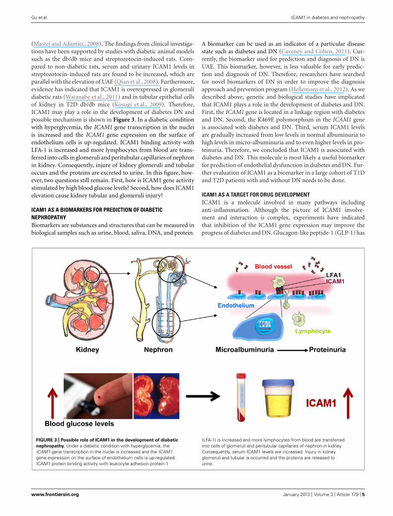
(Mastej and Adamiec, 2008). The findings from clinical investiga-tions have been supported by studies with diabetic animal modelssuch as the db/db mice and streptozotocin-induced rats. Com-pared to non-diabetic rats, serum and urinary ICAM1 levels instreptozotocin-induced rats are found to be increased, which areparallel with the elevation of UAE (Qian et al., 2008). Furthermore,evidence has indicated that ICAM1 is overexpressed in glomerulidiabetic rats (Watanabe et al., 2011) and in tubular epithelial cellsof kidney in T2D db/db mice (Kosugi et al., 2009). Therefore,ICAM1 may play a role in the development of diabetes DN andpossible mechanism is shown in Figure 3. In a diabetic conditionwith hyperglycemia, the ICAM1 gene transcription in the nucleiis increased and the ICAM1 gene expression on the surface ofendothelium cells is up-regulated. ICAM1 binding activity withLFA-1 is increased and more lymphocytes from blood are trans-ferred into cells in glomeruli and peritubular capillaries of nephronin kidney. Consequently, injure of kidney glomeruli and tubularoccurs and the proteins are excreted to urine. In this figure, how-ever, two questions still remain. First, how is ICAM1 gene activitystimulated by high blood glucose levels? Second, how does ICAM1elevation cause kidney tubular and glomeruli injury?
ICAM1 AS A BIOMARKERS FOR PREDICTION OF DIABETICNEPHROPATHYBiomarkers are substances and structures that can be measured inbiological samples such as urine, blood, saliva, DNA, and protein.
A biomarker can be used as an indicator of a particular diseasestate such as diabetes and DN (Caveney and Cohen, 2011). Cur-rently, the biomarker used for prediction and diagnosis of DN isUAE. This biomarker, however, is less valuable for early predic-tion and diagnosis of DN. Therefore, researchers have searchedfor novel biomarkers of DN in order to improve the diagnosisapproach and prevention program (Hellemons et al., 2012). As wedescribed above, genetic and biological studies have implicatedthat ICAM1 plays a role in the development of diabetes and DN.First, the ICAM1 gene is located in a linkage region with diabetesand DN. Second, the K469E polymorphism in the ICAM1 geneis associated with diabetes and DN. Third, serum ICAM1 levelsare gradually increased from low levels in normal albuminuria tohigh levels in micro-albuminuria and to even higher levels in pro-teinuria. Therefore, we concluded that ICAM1 is associated withdiabetes and DN. This molecule is most likely a useful biomarkerfor prediction of endothelial dysfunction in diabetes and DN. Fur-ther evaluation of ICAM1 as a biomarker in a large cohort of T1Dand T2D patients with and without DN needs to be done.
ICAM1 AS A TARGET FOR DRUG DEVELOPMENTICAM1 is a molecule involved in many pathways includinganti-inflammation. Although the picture of ICAM1 involve-ment and interaction is complex, experiments have indicatedthat inhibition of the ICAM1 gene expression may improve theprogress of diabetes and DN. Glucagon-like peptide-1 (GLP-1) has
FIGURE 3 | Possible role of ICAM1 in the development of diabetic
nephropathy. Under a diabetic condition with hyperglycemia, theICAM1 gene transcription in the nuclei is increased and the ICAM1gene expression on the surface of endothelium cells is up-regulated.ICAM1 protein binding activity with leukocyte adhesion protein-1
(LFA-1) is increased and more lymphocytes from blood are transferredinto cells of glomeruli and peritubular capillaries of nephron in kidney.Consequently, serum ICAM1 levels are increased. Injury in kidneyglomeruli and tubular is occurred and the proteins are released tourine.
www.frontiersin.org January 2013 | Volume 3 | Article 179 | 5
“fendo-03-00179” — 2013/1/20 — 20:09 — page 6 — #6
Gu et al. ICAM1 in diabetes and nephropathy
various extra-pancreatic actions, in addition to its enhance-ment of insulin secretion from pancreatic islets. Kodera et al.(2011) have demonstrated that GLP-1 receptor agonist, exendin-4,decreases the ICAM1 gene expression and ameliorates albumin-uria, glomerular hyperfiltration, glomerular hypertrophy, andmesangial matrix expansion in the diabetic rats without chang-ing blood pressure or body weight. Furthermore, Matsui et al.(2010) have reported that nifedipine, a calcium-channel blocker,blocks the advanced glycation end product (AGE)-induced tubu-lar damage and also inhibits ICAM1 gene activity in tubularcells, which may have benefits in treatment of DN. Liu et al.(2010) have suggested that berberine can ameliorate renal dys-function in diabetic rats by decreasing ICAM1 gene expressionand nuclear factor-kappa B (NF-kappaB) activation. Takingtogether, the data from these studies suggest that ICAM1 maybe a good candidate as target for drug development. Inhibitionof ICAM1 gene activity may benefit in treatment of diabetesand DN.
SUMMARY AND PERSPECTIVESWe have described genetic and biological studies of ICAM1 in dia-betes and DN. Data implicate that ICAM1 can be used a biomarkerfor prediction of diabetes and DN and may also be serviced as a tar-get for drug development. To reach these two objectives, however,we need to continuously put efforts into the study of ICAM1 indiabetes research. First, genetic polymorphisms in the ICAM1 geneare associated with T1D, T2D and DN in T1D. But, no genetic asso-ciation with DN in T2D has been reported. Although it is knownthat methylation modification of genomic DNAs and histone pro-teins is involved in the regulation of ICAM1 gene expression, noepigenetic study of this gene in diabetes and DN has been done.Second, clinical observations in diabetes patients and experimen-tal studies with diabetic animal models have demonstrated thatelevation of serum ICAM1 levels and overexpression of the genein kidney tissues. Inhibition of ICAM1 gene activity with agentsmay improve the kidney function, suggesting that ICAM1 may bea target for drug development in treatment of diabetes and DN.
REFERENCESAdeyemo, A. A., Johnson, T., Acheam-
pong, J., Oli, J., Okafor, G., Amoah,A., et al. (2005). A genome widequantitative trait linkage analysis forserum lipids in type 2 diabetes inan African population. Atherosclerosis181, 389–397.
Alberti, K. G., and Zimmet, P. Z.(1998). Definition, diagnosis andclassification of diabetes mellitus andits complications. Part 1: diagnosisand classification of diabetes mellitusprovisional report of a WHO consul-tation. Diabet. Med. 15, 539–553.
Arnold, K., Bordoli, L., Kopp, J., andSchwede, T. (2006). The SWISS-MODEL Workspace: a web-basedenvironment for protein structurehomology modelling. Bioinformatics22, 195–201.
Arya, R., Demerath, E., Jenkinson, C. P.,Göring, H. H., Puppala, S., Farook,V.,et al. (2006). quantitative trait locus(QTL) on chromosome 6q influencesbirth weight in two independent fam-ily studies. Hum. Mol. Genet. 15,1569–1579.
Ashcroft, F. M., and Rorsman, P. (2012).Diabetes mellitus and the β cell: thelast ten years. Cell 148, 1160–1171.
Astrup, A. S., Tarnow, L., Pietraszek,L., Schalkwijk, C. G., Stehouwer, C.D., Parving, H. H., et al. (2008).Markers of endothelial dysfunctionand inflammation in type 1 dia-betic patients with or without dia-betic nephropathy followed for 10years: association with mortality anddecline of glomerular filtration rate.Diabetes Care 31, 1170–1176.
Bonow, R. O., and Gheorghiade, M.(2004). The diabetes epidemic: anational and global crisis. Am. J. Med.116(Suppl. 5A), 2S–10S.
Caveney, E. J., and Cohen, O. J. (2011).Diabetes and biomarkers. J. DiabetesSci. Technol. 5, 192–197.
Concannon, P., Chen, W. M., Julier,C., Morahan, G., Akolkar, B., Erlich,H. A., et al. (2009). Type 1 DiabetesGenetics Consortium. Genome-widescan for linkage to type 1 dia-betes in 2,496 multiplex familiesfrom the Type 1 Diabetes Genet-ics Consortium. Diabetes 58, 1018–1022.
Cornell, S., and Dorsey,V. J. (2012). Dia-betes pharmacotherapy in 2012: con-siderations in medication selection.Postgrad. Med. 2012 124, 84–94.
Gonzalez-Bulnes, A., and Ovilo, C.(2012). Genetic basis, nutritionalchallenges and adaptive responses inthe prenatal origin of obesity andtype-2 diabetes. Curr. Diabetes Rev.8, 144–154.
Gong, A. Y., Hu, G., Zhou, R., Liu, J.,Feng, Y., Soukup, G. A., et al. (2011).MicroRNA-221 controls expressionof intercellular adhesion molecule-1in epithelial cells in response to Cryp-tosporidium parvum infection. Int. J.Parasitol. 41, 397–403.
Guja, C., Todd, J. A., Welsh, K., Marshall,S., and Ionescu-Tirgoviste, C. (1999).Increased transmission of intercellu-lar adhesion-molecule 1, 469E allelein type 1 Romanian diabetic families.Diabetologia 42, 327.
Gulcher, J. (2012). Microsatellite mark-ers for linkage and association stud-ies. Cold Spring Harb. Protoc. 2012,425–432.
Heerspink, H. J., and de Zeeuw, D.(2011). The kidney in type 2 dia-betes therapy. Rev. Diabet. Stud. 8,392–402.
Hellebrekers, D. M., Castermans, K.,Viré, E., Dings, R. P., Hoebers, N. T.,
Mayo, K. H., et al. (2006). Epigeneticregulation of tumor endothelialcell anergy: silencing of intercellu-lar adhesion molecule-1 by histonemodifications. Cancer Res. 66,10770–10777.
Hellemons, M. E., Kerschbaum, J.,Bakker, S. J., Neuwirt, H., Mayer, B.,Mayer, G., et al. (2012). Validity ofbiomarkers predicting onset or pro-gression of nephropathy in patientswith type 2 diabetes: a systematicreview. Diabet. Med. 29, 567–577.
Howell, W. M., Jobs, M., Gyllensten, U.,and Brookes, A. J. (1999). Dynamicallele-specific hybridization. A newmethod for scoring single nucleotidepolymorphisms. Nat. Biotechnol. 17,87–88.
Imperatore, G., Knowler, W. C., Pet-titt, D. J., Kobes, S., Fuller, J. H.,Bennett, P. H., et al. (2000). A locusinfluencing total serum cholesterolon chromosome 19p: results froman autosomal genomic scan of serumlipid concentrations in Pima Indians.Arterioscler. Thromb. Vasc. Biol. 20,2651–2656.
Janeway, C. A. Jr. (2001). How theimmune system works to protect thehost from infection: a personal view.Proc. Natl. Acad. Sci. U.S.A. 98,7461–7468.
Kamiuchi, K., Hasegawa, G., Obayashi,H., Kitamura, A., Ishii, M., Yano,M., et al. (2002). Intercellular adhe-sion molecule-1 (ICAM-1) polymor-phism is associated with diabeticretinopathy in type 2 diabetes mel-litus. Diabet. Med. 19, 371–376.
Kathiresan, S., Melander, O., Guiducci,C., Surti, A., Burtt, N. P., Rieder,M. J., et al. (2008). Six new lociassociated with blood low-densitylipoprotein cholesterol, high-density
lipoprotein cholesterol or triglyc-erides in humans. Nat. Genet. 40,189–197.
Kodera, R., Shikata, K., Kataoka, H.U., Takatsuka, T., Miyamoto, S.,Sasaki, M., et al. (2011). Glucagon-like peptide-1 receptor agonist ame-liorates renal injury through its anti-inflammatory action without lower-ing blood glucose level in a rat modelof type 1 diabetes. Diabetologia 54,965–978.
Kosugi, T., Nakayama, T., Heinig, M.,Zhang, L., Yuzawa, Y., Sanchez-Lozada, L. G., et al. (2009). Effect oflowering uric acid on renal diseasein the type 2 diabetic db/db mice.Am. J. Physiol. Renal Physiol. 297,F481–F488.
Kraja, A. T., Huang, P., Tang, W., Hunt,S. C., North, K. E., Lewis, C. E.,et al. (2008). QTLs of factors of themetabolic syndrome and echocardio-graphic phenotypes: the hyperten-sion genetic epidemiology networkstudy. BMC Med. Genet. 9:103. doi:10.1186/1471-2350-9-103
Lam, D. W., and LeRoith, D. (2012). Theworldwide diabetes epidemic. Curr.Opin. Endocrinol. Diabetes Obes. 19,93–96.
Leon, J. M., Freedman, B. I., Miller,M. B., North, K. E., Hunt, S.C., Eckfeldt, J. H., et al. (2007).Genome scan of glomerular filtrationrate and albuminuria: the HyperGENstudy. Nephrol. Dial. Transplant. 22,763–771.
Lightfoot, Y. L., Chen, J., and Mathews,C. E. (2012). Immune-mediated β-cell death in type 1 diabetes: lessonsfrom human β-cell lines. Eur. J. Clin.Invest. 42, 1244–1251.
Lin, J., Glynn, R. J., Rifai, N., Man-son, J. E., Ridker, P. M., Nathan,
Frontiers in Endocrinology | Diabetes January 2013 | Volume 3 | Article 179 | 6
“fendo-03-00179” — 2013/1/20 — 20:09 — page 7 — #7
Gu et al. ICAM1 in diabetes and nephropathy
D. M., et al. (2008). Inflammationand progressive nephropathy in type1 diabetes in the diabetes control andcomplications trial. Diabetes Care 31,2338–2343.
Liu, L., Yu, Q., Wang, H., Zhang,S. X., Huang, C., and Chen, X.(2006). Association of intercellularadhesion molecule 1 polymorphismswith retinopathy in Chinese patientswith type 2 diabetes. Diabet. Med. 23,643–648.
Liu, W., Zhang, X., Liu, P., Shen, X.,Lan, T., Li, W., et al. (2010). Effectsof berberine on matrix accumulationand NF-kappa B signal pathway inalloxan-induced diabetic mice withrenal injury. Eur. J. Pharmacol. 638,150–155.
Ma, J., Möllsten, A., Prázny, M., Fal-hammar, H., Brismar, K., Dahlquist,G., et al. (2006). Genetic influencesof the intercellular adhesion molecule1 (ICAM-1) gene polymorphisms indevelopment of type 1 diabetes anddiabetic nephropathy. Diabet. Med.23, 1093–1099.
Ma, J., Zhang, D., Brismar, K., Efendic,S., and Gu, H. F. (2008). Evaluationof the association between the com-mon E469K polymorphism in theICAM-1 gene and diabetic nephropa-thy among type 1 diabetic patientsin GoKinD population. BMC Med.Genet. 9:47. doi: 10.1186/1471-2350-9-47.
Malhotra, A., and Wolford, J. K. (2005).American Diabetes Association GEN-NID Study Group. Analysis of quan-titative lipid traits in the genetics ofNIDDM (GENNID) study. Diabetes54, 3007–3014.
Marshall, S. M. (2004). Recent advancesin diabetic nephropathy. Postgrad.Med. J. 80, 624–633.
Marshall, S. M. (2012). Diabeticnephropathy in type 1 diabetes:has the outlook improved since the1980s? Diabetologia 55, 2301–2306.
Mastej, K., and Adamiec, R. (2008).Neutrophil surface expression ofCD11b and CD62L in diabeticmicroangiopathy. Acta Diabetol. 45,183–190.
Matsui, T., Yamagishi, S., Takeuchi, M.,Ueda, S., Fukami, K., and Okuda, S.(2010). Nifedipine inhibits advancedglycation end products (AGEs) andtheir receptor (RAGE) interaction-mediated proximal tubular cell injuryvia peroxisome proliferator-activatedreceptor-gamma activation. Biochem.Biophys. Res. Commun. 398, 326–330.
Mein, C. A., Esposito, L., Dunn, M. G.,Johnson, G. C., Timms, A. E., Goy, J.
V., et al. (1998). A search for type 1diabetes susceptibility genes in fam-ilies from the United Kingdom. Nat.Genet. 19, 297–300.
Morahan, G. (2012). Insights into type1 diabetes provided by genetic analy-ses. Curr. Opin. Endocrinol. DiabetesObes. 19, 263–270.
Mueller, P. W., Rogus, J. J., Cleary, P. A.,Zhao, Y., Smiles, A. M., Steffes, M.W., et al. (2006). Genetics of Kidneysin Diabetes (GoKinD) study: a genet-ics collection available for identifyinggenetic susceptibility factors for dia-betic nephropathy in type 1 diabetes.J. Am. Soc. Nephrol. 17, 1782–1790.
Nejentsev, S., Guja, C., McCormack,R., Cooper, J., Howson, J. M., Nut-land, S., et al. (2003). Associationof intercellular adhesion molecule-1gene with type 1 diabetes. Lancet 362,1723–1724.
Nejentsev, S., Laine, A. P., Simell, O., andIlonen, J. (2000). Intercellular adhe-sion molecule-1 (ICAM-1) K469Epolymorphism: no association withtype 1 diabetes among Finns. TissueAntigens 55, 568–570.
Newman, P. J., Berndt, M. C., Gorski,J., White, G. C. II, Lyman, S., Pad-dock, C., et al. (1990). PECAM-1(CD31) cloning and relation to adhe-sion molecules of the immunoglob-ulin gene superfamily. Science 247,1219–1222.
Nishimura, M., Obayashi, H., Maruya,E., Ohta, M., Tegoshi, H., Fukui,M., et al. (2000). Associationbetween type 1 diabetes age-at-onsetand intercellular adhesion molecule-1 (ICAM-1) gene polymorphism.Hum. Immunol. 61, 507–510.
Peitsch, M. C. (1995). Protein modelingby E-mail. Biotechnology 13, 658–660.
Petrovic, M. G., Osredkar, J., Saraga-Babic, M., and Petrovic, D. (2008).K469E polymorphism of the intra-cellular adhesion molecule 1 gene isassociated with proliferative diabeticretinopathy in Caucasians with type2 diabetes. Clin. Exp. Ophthalmol. 36,468–472.
Pitkäniemi, J., Moltchanova, E., Haa-pala, L., Harjutsalo, V., Tuomilehto,J., and Hakulinen, T. (2007). Geneticrandom effects model for family datawith long-term survivors: analysis ofdiabetic nephropathy in type 1 dia-betes. Genet. Epidemiol. 31, 697–708.
Pollin, T. I., Hsueh, W. C., Steinle, N.I., Snitker, S., Shuldiner, A. R., andMitchell, B. D. (2004). A genome-wide scan of serum lipid levels in theOld Order Amish. Atherosclerosis 173,89–96.
Qian, Y., Li, S., Ye, S., Chen, Y.,Zhai, Z., Chen, K., et al. (2008).Renoprotective effect of rosiglitazonethrough the suppression of renalintercellular adhesion molecule-1expression in streptozotocin-induceddiabetic rats. J. Endocrinol. Invest. 31,1069–1074.
Roebuck, K. A., and Finnegan, A.(1999). Regulation of intercellularadhesion molecule-1 (CD54) geneexpression. J. Leukoc. Biol. 66,876–888.
Ronaghi, M., Uhlén, M., and Nyrén, P.(1998). A sequencing method basedon real-time pyrophosphate. Science281, 363–365.
Rubio-Guerra, A. F., Vargas-Robles, H.,Ayala, G. V., and Escalante-Acosta, B.A. (2007). Correlation between circu-lating adhesion molecule levels andalbuminuria in type 2 diabetic nor-motensive patients. Med. Sci. Monit.13, CR349–CR352.
Sahakyan, K., Klein, B. E., Lee, K. E.,Tsai, M. Y., and Klein, R. (2010a).Inflammatory and endothelial dys-function markers and proteinuria inpersons with type 1 diabetes mellitus.Eur. J. Endocrinol. 162, 1101–1105.
Sahakyan, K., Klein, B. E., Myers, C.E., Tsai, M. Y., and Klein, R. (2010b).Novel risk factors in long-term hyper-tension incidence in type 1 diabetesmellitus. Am. Heart J. 159, 1074–1080.
Satko, S. G., Freedman, B. I., and Moos-savi, S. (2005). Genetic factors inend-stage renal disease. Kidney Int.Suppl. 94, S46–S49.
Shaw, C. J., and Lupski, J. R. (2004).Implications of human genomearchitecture for rearrangement-based disorders: the genomic basisof disease. Hum. Mol. Genet. 13,R57–R64.
Shields, J., and Maxwell, A. P. (2010).Managing diabetic nephropathy.Clin. Med. 10, 500–504.
Thomas, M. C., and Groop, P. H. (2011).New approaches to the treatmentof nephropathy in diabetes. Expert.Opin. Investig. Drugs 20, 1057–1071.
Venter, J. C., Adams, M. D., Myers, E.W., Li, P. W., Mural, R. J., Sutton,G. G., et al. (2001). The sequenceof the human genome. Science 291,1304–1351.
Vinita, K., Sripriya, S., Prathiba, K.,Vaitheeswaran, K., Sathyabaarathi,R., Rajesh, M., et al. (2012).SNDREAMS project. ICAM-1 K469Epolymorphism is a genetic determi-nant for the clinical risk factors ofT2D subjects with retinopathy in
Indians: a population-based case-control study. BMJ Open 2, pii:e001036.
van de Stolpe, A., and van der Saag,P. T. (1996). Intercellular adhesionmolecule-1. J. Mol. Med. (Berl.) 74,13–33.
Watanabe, N., Shikata, K., Shikata, Y.,Sarai, K., Omori, K., Kodera, R.,et al. (2011). Involvement of MAPKsin ICAM-1 expression in glomeru-lar endothelial cells in diabeticnephropathy. Acta Med. Okayama 65,247–257.
Weil, E. J., Curtis, J. M., Hanson, R. L.,Knowler, W. C., and Nelson, R. G.(2010). The impact of disadvantageon the development and progres-sion of diabetic kidney disease. Clin.Nephrol. 74(Suppl. 1), S32–S8.
Wild, S., Roglic, G., Green, A., Sicree,R., and King, H. (2004). Globalprevalence of diabetes: estimatesfor the year 2000 and projectionsfor 2030. Diabetes Care 27, 1047–1053.
Yokoyama, H., Tahara, H., Emoto,M., Fujiwara, S., Araki, T., Shino-hara, K., et al. (2005). The K469Epolymorphism of the intercellularadhesion molecule-1 gene is asso-ciated with plasma fibrinogen levelin type 2 diabetes. Metabolism 54,381–386.
Conflict of Interest Statement: Theauthors declare that the research wasconducted in the absence of any com-mercial or financial relationships thatcould be construed as a potential con-flict of interest.
Received: 22 October 2012; accepted: 17December 2012; published online: 22January 2013.Citation: Gu HF, Ma J, Gu KT and Bris-mar K (2013) Association of intercellularadhesion molecule 1 (ICAM1) with dia-betes and diabetic nephropathy. Front.Endocrin. 3:179. doi: 10.3389/fendo.2012.00179This article was submitted to Frontiersin Diabetes, a specialty of Frontiers inEndocrinology.Copyright © 2013 Gu, Ma, Gu andBrismar. This is an open-access arti-cle distributed under the terms of theCreative Commons Attribution License,which permits use, distribution andreproduction in other forums, providedthe original authors and source are cred-ited and subject to any copyright noticesconcerning any third-party graphics etc.
www.frontiersin.org January 2013 | Volume 3 | Article 179 | 7