assessment of olfactory function
TRANSCRIPT

Taste and Smell

Advances in Oto-Rhino-LaryngologyVol. 63
Series Editor
W. Arnold Munich
Taste and Smell An Update
Basel · Freiburg · Paris · London · New York ·
Bangalore · Bangkok · Singapore · Tokyo · Sydney
Volume Editors
Thomas Hummel Dresden
Antje Welge-Lüssen Basel
33 figures, 1 in color, and 12 tables, 2006
Prof.Thomas Hummel PD Dr.Antje Welge-LüssenSmell & Taste Clinic Department of Otorhinolaryngology
Department of Otorhinolaryngology University Hospital Basel
University of Dresden Medical School Petersgraben 4
Fetscherstrasse 74 CH–4031 Basel (Switzerland)
DE–01307 Dresden (Germany)
Bibliographic Indices. This publication is listed in bibliographic services, including Current Contents® and
Index Medicus.
Disclaimer. The statements, options and data contained in this publication are solely those of the individ-
ual authors and contributors and not of the publisher and the editor(s). The appearance of advertisements in the
book is not a warranty, endorsement, or approval of the products or services advertised or of their effectiveness,
quality or safety. The publisher and the editor(s) disclaim responsibility for any injury to persons or property
resulting from any ideas, methods, instructions or products referred to in the content or advertisements.
Drug Dosage. The authors and the publisher have exerted every effort to ensure that drug selection and
dosage set forth in this text are in accord with current recommendations and practice at the time of publication.
However, in view of ongoing research, changes in government regulations, and the constant flow of information
relating to drug therapy and drug reactions, the reader is urged to check the package insert for each drug for
any change in indications and dosage and for added warnings and precautions. This is particularly important when
the recommended agent is a new and/or infrequently employed drug.
All rights reserved. No part of this publication may be translated into other languages, reproduced or
utilized in any form or by any means electronic or mechanical, including photocopying, recording, microcopying,
or by any information storage and retrieval system, without permission in writing from the publisher.
© Copyright 2006 by S. Karger AG, P.O. Box, CH–4009 Basel (Switzerland)
www.karger.com
Printed in Switzerland on acid-free paper by Reinhardt Druck, Basel
ISSN 0065–3071
ISBN-10: 3–8055–8123–8
ISBN-13: 978-3–8055–8123–3
Library of Congress Cataloging-in-Publication Data
Taste and smell : an update / editors, Thomas Hummel, Antje Welge-Lüssen.
p. ; cm. – (Advances in oto-rhino-laryngology ; v. 63)
Includes bibliographical references and index.
ISBN 3-8055-8123-8 (hard cover : alk. paper)
1. Taste. 2. Smell. 3. Sense organs. 4. Taste disorders. I. Hummel,
Thomas, 1959- II. Welge-Lüssen, Antje. III. Series.
[DNLM: 1. Smell–physiology. 2. Olfaction Disorders. 3.
Taste–physiology. 4. Taste Disorders. W1 AD701 v.63 2006 / WV 301 T2148
2006]
QP458.T37 2006
612.8�6–dc22
2006011577
V
Contents
VII PrefaceHummel, T. (Dresden); Welge-Lüessen, A. (Basel)
Smell
1 Nasal Anatomy and the Sense of SmellHornung, D.E. (Canton/Syracuse, N.Y.)
23 Transduction and CodingRawson, N.E.; Yee, K.K. (Philadelphia, Pa.)
44 Smell: Central Nervous ProcessingGottfried, J.A. (Chicago, Ill.)
70 Structure and Function of the Vomeronasal OrganWitt, M. (Dresden); Wozniak, W. (Poznan)
84 Assessment of Olfactory FunctionHummel, T. (Dresden); Welge-Lüessen, A. (Basel)
99 Posttraumatic Olfactory LossCostanzo, R.M. (Richmond, Va.); Miwa, T. (Kanazawa)
108 Chronic Rhinosinusitis and Olfactory DysfunctionRaviv, J.R.; Kern, R.C. (Chicago, Ill.)
125 Olfactory Disorders following Upper Respiratory Tract InfectionsWelge-Lüssen, A.; Wolfensberger, M. (Basel)
´´
133 Olfaction in Neurodegenerative DisorderHawkes, C. (Romford)
Taste
152 Human Taste: Peripheral Anatomy,Taste Transduction, and CodingBreslin, P.A.S.; Huang, L. (Philadelphia, Pa.)
191 Central Gustatory Processing in HumansSmall, D.M. (New Haven, Conn.)
221 Modern Psychophysics and the Assessment of Human Oral SensationSnyder, D.J. (New Haven, Conn./Gainesville, Fla.);
Prescott, J. (Cairns); Bartoshuk, L.M. (Gainesville, Fla.)
242 Postoperative/Posttraumatic Gustatory DysfunctionLandis, B.N.; Lacroix, J.-S. (Genève)
255 Neurological Causes of Taste DisordersHeckmann, J.G.; Lang, C.J.G. (Erlangen)
265 Toxic Effects on Gustatory FunctionReiter, E.R.; DiNardo, L.J.; Costanzo, R.M. (Richmond, Va.)
278 Burning Mouth SyndromeGrushka, M.; Ching, V. (Toronto); Epstein, J. (Chicago, Ill.)
288 Author Index
289 Subject Index
Contents VI
Preface
An intact sense of smell and taste allows us to recognize the chemical sig-
nals from our environment. By doing this, the chemical senses contribute signi-
ficantly to the quality of our lives. Despite this fact, and although chemosensory
disorders are frequent, they have been ‘neglected’ in clinical routine for many
years. This neglect may be partly explained by traditional difficulties in the diag-
nosis of chemosensory disorders and the common belief that chemosensory dis-
orders cannot be treated. Having said that, the clinical neglect of chemosensory
functions is in sharp contrast to the remarkable interest the chemical senses have
received over the last decade, culminating in the 2004 Nobel Prize awarded to
two researchers in the sense of smell.
This publication is meant for the clinician confronted with chemosensory
disorders. It is supposed to bridge the gap between clinical and basic research.
Above all, it is meant to provide an update in this area of research, presented by
most distinguished researchers in the field. We do hope that this book will be
used by many clinicians in order to improve counselling and treatment of
patients with chemosensory dysfunction. Above and beyond this, we would be
more than delighted if this book inspires clinical colleagues to perform basic
research on the chemical senses where many questions are still open.
Thomas Hummel Antje Welge-Lüssen
VII

Hummel T, Welge-Lüssen A (eds): Taste and Smell. An Update.
Adv Otorhinolaryngol. Basel, Karger, 2006, vol 63, pp 1–22
Nasal Anatomy and the Sense of Smell
David E. Hornung
St. Lawrence University, Canton, and Neuroscience and Physiology Department,
Upstate Medical University, Syracuse, N.Y., USA
AbstractAs a result of the relative sizes of the various compartments in the nasal cavity, the bulk
of the airflow is along the floor of the nasal cavity. The percent of airflow directed to the olfac-
tory region (the superior region of the nasal cavity) is about 10%. Structural changes in the
nasal cavity can alter airflow pathways and the characteristics of the airflow (e.g. laminar,
mixed or turbulent) within nasal compartments. The relationship between the olfactory response
and the stimulus is complex and may vary depending on the physiochemical properties of the
odor and the rate at which odorants are delivered to the olfactory receptors. Changes in nasal
airflow may impact the various olfactory functions (e.g. identification, differentiation) differ-
ently. When there is a nasal obstruction, a decline in olfactory ability may not simply be an
access problem, since nasal disease can affect olfactory processing at many levels.
Copyright © 2006 S. Karger AG, Basel
Nasal Anatomy
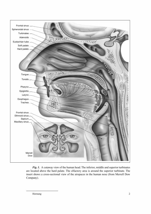
The internal anatomy of the nose (fig. 1) is divided into two halves along the
midline by a bony structure called the nasal septum. The outline of the lateral wall
is delineated by the curves of the inferior, middle and superior turbinates [1]. The
respiratory and olfactory epithelial cells lining the structures of the internal nose
have a very rich blood supply and are covered by a watery mucus that is continu-
ally flowing into the back of the throat [2–4]. By altering the blood flow in the
dense capillary beds servicing the structures of the internal nose, the size of the air-
space can be changed quickly and dramatically [5, 6]. Because these areas can
change shape so quickly, they are sometimes referred to as swell spaces. The struc-
tural changes in these spaces can alter both the airflow pathways through the nose
and the characteristics of the airflow (e.g. laminar, mixed or turbulent) within the
various compartments of the nasal cavity [6].
Smell
Hornung 2
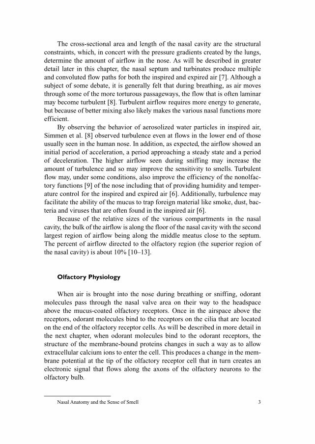
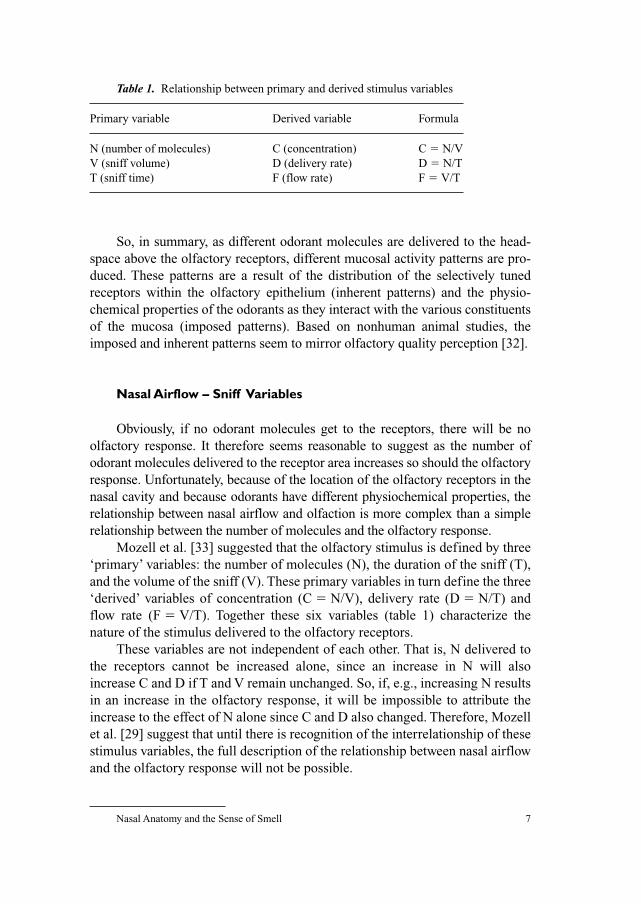
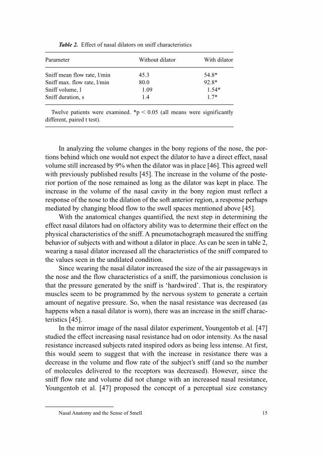
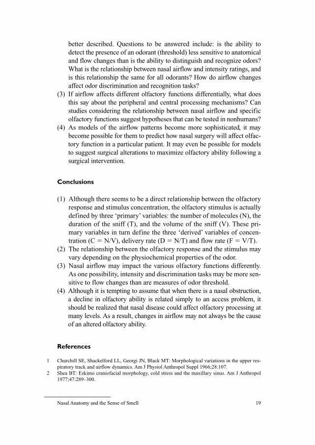
Fig. 1. A cutaway view of the human head. The inferior, middle and superior turbinates
are located above the hard palate. The olfactory area is around the superior turbinate. The
insert shows a cross-sectional view of the airspaces in the human nose (from Merrell Dow
Company).
Frontal sinus
Sphenoidal sinus
Turbinates
Adenoids
Eustachian tube
Soft palate
Hard palate
Tongue
Tonsils
Pharynx
Epiglottis
Larynx
Esophagus
Trachea
Frontal sinusEthmoid sinus
SeptumMaxillary sinus
MerrellDow
Nasal Anatomy and the Sense of Smell 3
The cross-sectional area and length of the nasal cavity are the structural
constraints, which, in concert with the pressure gradients created by the lungs,
determine the amount of airflow in the nose. As will be described in greater
detail later in this chapter, the nasal septum and turbinates produce multiple
and convoluted flow paths for both the inspired and expired air [7]. Although a
subject of some debate, it is generally felt that during breathing, as air moves
through some of the more torturous passageways, the flow that is often laminar
may become turbulent [8]. Turbulent airflow requires more energy to generate,
but because of better mixing also likely makes the various nasal functions more
efficient.
By observing the behavior of aerosolized water particles in inspired air,
Simmen et al. [8] observed turbulence even at flows in the lower end of those
usually seen in the human nose. In addition, as expected, the airflow showed an
initial period of acceleration, a period approaching a steady state and a period
of deceleration. The higher airflow seen during sniffing may increase the
amount of turbulence and so may improve the sensitivity to smells. Turbulent
flow may, under some conditions, also improve the efficiency of the nonolfac-
tory functions [9] of the nose including that of providing humidity and temper-
ature control for the inspired and expired air [6]. Additionally, turbulence may
facilitate the ability of the mucus to trap foreign material like smoke, dust, bac-
teria and viruses that are often found in the inspired air [6].
Because of the relative sizes of the various compartments in the nasal
cavity, the bulk of the airflow is along the floor of the nasal cavity with the second
largest region of airflow being along the middle meatus close to the septum.
The percent of airflow directed to the olfactory region (the superior region of
the nasal cavity) is about 10% [10–13].
Olfactory Physiology
When air is brought into the nose during breathing or sniffing, odorant
molecules pass through the nasal valve area on their way to the headspace
above the mucus-coated olfactory receptors. Once in the airspace above the
receptors, odorant molecules bind to the receptors on the cilia that are located
on the end of the olfactory receptor cells. As will be described in more detail in
the next chapter, when odorant molecules bind to the odorant receptors, the
structure of the membrane-bound proteins changes in such a way as to allow
extracellular calcium ions to enter the cell. This produces a change in the mem-
brane potential at the tip of the olfactory receptor cell that in turn creates an
electronic signal that flows along the axons of the olfactory neurons to the
olfactory bulb.
Hornung 4
Inherent Mucosal Activity Patterns
The olfactory bulb is composed of a number of different types of cells,
although the axons of the olfactory receptor cells first make contact with the
glomerular cells. Receptors that respond to some common chemical feature
(there are approximately 350 different cell types loosely arranged into zones)
send their signals to specific glomerular cells. Since most odorants stimulate
more than one type of receptor cell, each odorant produces a response pattern
across the glomerulus that is unique for that particular odorant. In other words,
the olfactory receptor sheet disassembles the odorant molecule into a unique
pattern of its functional groups. This disassembled pattern is maintained and
perhaps sharpened in the glomerular cell layer of the bulb and is then sent to
more central areas (like the primary olfactory cortex) where the patterns are
reassembled for processing [14, 15].
Since receptor cells of similar sensitivity are grouped in particular loca-
tions along the olfactory receptor sheet, different smells produce different pat-
terns of electrical activity in both the mucosa and in the olfactory bulb. These
patterns are perhaps one of the ways the brain can identify a particular smell.
The signal created by the specific tuning of olfactory receptor cells is called the
‘inherent’ mucosal activity pattern [16, 17].
Imposed Mucosal Activity Patterns
As described above, the olfactory receptors are located high in the nose
along the septum and in the region of the superior turbinate. Once odorant
molecules arrive at the headspace above the olfactory receptors, the molecules
distribute themselves along the long axis of the mucosal sheet in patterns
reflecting the physical and chemical properties of the odorant molecules them-
selves. For a chemical that is highly soluble in the mucus, most of the incom-
ing molecules will be trapped early in the flow path, producing a very uneven
distribution of odorant molecules along the long axis of the mucosal sheet
(fig. 2). On the other hand, for a chemical that is only slightly soluble in the
mucus, odorant molecules will be more evenly distributed along the flow path.
The specific mucosal odorant distribution pattern created by an odorant’s sol-
ubility may be another mechanism by which the central nervous system identi-
fies smells. These distribution patterns are called ‘imposed’ patterns [18–21].
There is electrophysiologic [22–24], radioisotopic [20, 25, 26], and gas
chromatographic evidence [27] to support the existence of imposed mucosal
activity patterns in a variety of nonhuman animal species. However, for obvious
Nasal Anatomy and the Sense of Smell 5
reasons, the techniques available to study mucosal distribution patters in non-
humans are not appropriate for use in people.
It should be emphasized that imposed and inherent patterns may not be
mutually exclusive. They may work in concert to allow humans and other ani-
mals to identify a very wide variety of smells. Because they are created by the
flow of air across the olfactory receptor sheet, imposed patterns may be more
susceptible than inherent patterns to changes in airflow [18, 21].
A Role for Mucosal Patterns in Olfactory Coding
Although a number of investigators have documented the existence of
imposed and inherent patterns using a variety of techniques in a number of
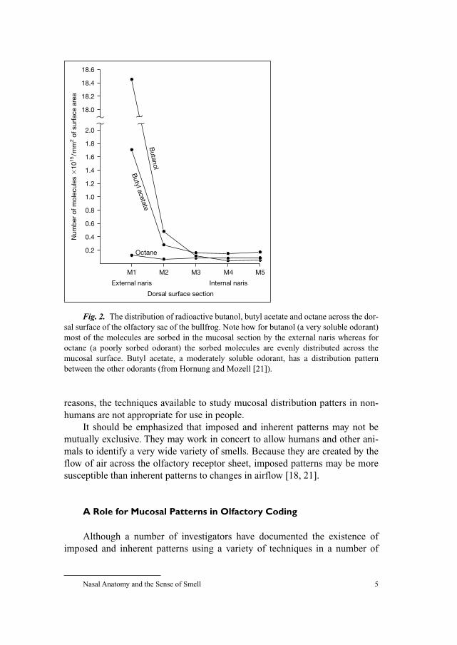
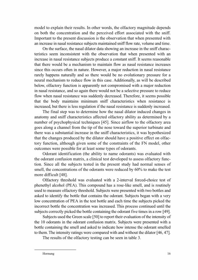
Fig. 2. The distribution of radioactive butanol, butyl acetate and octane across the dor-
sal surface of the olfactory sac of the bullfrog. Note how for butanol (a very soluble odorant)
most of the molecules are sorbed in the mucosal section by the external naris whereas for
octane (a poorly sorbed odorant) the sorbed molecules are evenly distributed across the
mucosal surface. Butyl acetate, a moderately soluble odorant, has a distribution pattern
between the other odorants (from Hornung and Mozell [21]).
18.6N
umb
er o
f mol
ecul
es �
1015
/mm
2 of s
urfa
ce a
rea
18.4
18.2
18.0
2.0
1.8
1.6
1.4
1.2
1.0
0.8
0.6
0.4
0.2
M5M4M3M2M1
Octane
Butanol
Butyl acetate
External naris Internal naris
Dorsal surface section
Hornung 6
different species, the role they play in olfactory coding remains a matter of
some discussion. To understand the difficulty in determining the role that these
patterns play in odor detection (and how the patterns are affected by changes in
nasal airflow), one needs to first appreciate the conundrum faced by investiga-
tors who are studying the sense of smell. It is relatively easy to ask humans if
they smell a particular chemical; at some level, it is even possible to ask them
how similar one chemical smells to another. However, it is usually not possible
in any systematic and/or permanent way to alter the imposed and inherent pat-
terns produced in the human nose. As a result, the experimental manipulations
appropriate in a hypothesis-based exploration of human olfaction are not gener-
ally possible. On the other hand, anatomical, biochemical and physiological
manipulations are possible in nonhuman animals, but it is very difficult to ask
the animals if they smelled anything and it is even more difficult to ask them
how similar one chemical is to another. To help solve this conundrum, the five-
odorant identification confusion matrix was developed to test the hypothesis
that inherent and imposed patterns, at least in nonhuman animals, have some
significance in olfactory processing [28].
In the five-odorant confusion matrix, after sampling a target smell, an animal
sampled five test ports to determine which port smelled most like the target. The
animal indicated his choice by pressing a bar in front of the appropriate port. This
technique not only made it possible to measure correct olfactory identification, but
by analyzing the off-diagonal response, also made it possible, at least indirectly, for
the animal to indicate how similar odors are to each other. The logic is if two odors
are often confused they must share some similar perceptual quality and odors not
often confused must be more perceptually dissimilar. A multidimensional scaling
analysis based on the pattern of errors allowed for a graphical representation of the
perceptual relationship among the odors used in the confusion matrix.
By comparing the animal confusion (multidimensional scaling) matrix
data with voltage-sensitive dye recordings from the mucosa and the olfactory
bulb, Youngentob et al. [29, 32] and Kent et al. [30, 31] were able to demon-
strate a relationship between the differential activity patterns and the perceptual
characteristics of the odors. That is, odors having similar mucosal activity pat-
terns are more often confused than odors producing very different mucosal
activity patterns. In other words, the mucosal activity patterns produced at the
mucosal level (a result of imposed and inherent patterns) seem to mirror the
psychophysical relationship seen among odors.
If the mucosal patterns play a role in olfactory perception, changing the
patterns should change olfactory perception. To test this hypothesis, Youngentob
et al. [32] changed the mucosal activity patterns using olfactory marker protein
gene depletion. As predicted, as the mucosal activity patterns changed so did
olfactory perception.
Nasal Anatomy and the Sense of Smell 7
So, in summary, as different odorant molecules are delivered to the head-
space above the olfactory receptors, different mucosal activity patterns are pro-
duced. These patterns are a result of the distribution of the selectively tuned
receptors within the olfactory epithelium (inherent patterns) and the physio-
chemical properties of the odorants as they interact with the various constituents
of the mucosa (imposed patterns). Based on nonhuman animal studies, the
imposed and inherent patterns seem to mirror olfactory quality perception [32].
Nasal Airflow – Sniff Variables
Obviously, if no odorant molecules get to the receptors, there will be no
olfactory response. It therefore seems reasonable to suggest as the number of
odorant molecules delivered to the receptor area increases so should the olfactory
response. Unfortunately, because of the location of the olfactory receptors in the
nasal cavity and because odorants have different physiochemical properties, the
relationship between nasal airflow and olfaction is more complex than a simple
relationship between the number of molecules and the olfactory response.
Mozell et al. [33] suggested that the olfactory stimulus is defined by three
‘primary’ variables: the number of molecules (N), the duration of the sniff (T),
and the volume of the sniff (V). These primary variables in turn define the three
‘derived’ variables of concentration (C � N/V), delivery rate (D � N/T) and
flow rate (F � V/T). Together these six variables (table 1) characterize the
nature of the stimulus delivered to the olfactory receptors.
These variables are not independent of each other. That is, N delivered to
the receptors cannot be increased alone, since an increase in N will also
increase C and D if T and V remain unchanged. So, if, e.g., increasing N results
in an increase in the olfactory response, it will be impossible to attribute the
increase to the effect of N alone since C and D also changed. Therefore, Mozell
et al. [29] suggest that until there is recognition of the interrelationship of these
stimulus variables, the full description of the relationship between nasal airflow
and the olfactory response will not be possible.
Table 1. Relationship between primary and derived stimulus variables
Primary variable Derived variable Formula
N (number of molecules) C (concentration) C � N/V
V (sniff volume) D (delivery rate) D � N/T
T (sniff time) F (flow rate) F � V/T
Hornung 8
Still there is a large body of evidence suggesting that the olfactory
response (R) is proportional to C such that:
R � Cx
This relationship suggests R is proportional to the log of C of the stimulus.
Since C is derived from the primary variables of N and V, this proportionality
can be written as
R � Nx/Vx
If R is defined only by C, increasing N and V by the same proportion
should result in no change in R. If, however, C is not the sole determiner of R,
any change in R seen when N and V are increased by the same proportion
would reflect the effect of these two primary variables independently of the
effect that they have through C. Likewise, the effects of the derived variables of
F and D could be studied in relation to the primary variables from which they
originate. This is the logic that drove the NVT study described below.
In an effort to parcel out the interrelationship between the six sniff vari-
ables, Mozell et al. [34] designed an experiment in which all combinations of
two levels of the three primary variables were presented to animals in which the
olfactory nerve response was recorded. The levels for each variable were picked
so that the levels of volume and time were in the natural range and the lower
level of a variable was exactly 50% of the higher level. For example, a lower
sniff time was picked from the lower range of what the animal normally pro-
duces. The higher sniff time was twice the lower duration but still in the ani-
mal’s ‘physiological’ range. The same selection procedure was used in picking
the two sniff volumes. The concentrations picked were in the moderate range
and the concentration of the larger stimulus was twice the concentration of the
smaller stimulus. This strategy resulted in 8 combinations of ‘sniffs’, which,
because of the relationships described above, also resulted in three combina-
tions of each of the derived variables. The odorant used in this study was octane,
a chemical that is poorly sorbed by the mucosa and produces an even imposed
pattern across the mucosal sheet. The species used was the bullfrog. From an
analysis of variance of the log of the olfactory response (the dependent vari-
able), a model using the three primary variables (NVT) was generated that best
accounted for the variability in the neural activity. The predicted error variance
for this model was 0.0523. This model was:
R � N0.35V�0.28T0.22
Stated in words, the model proposes that the magnitude of the olfactory
nerve response increases as the number of molecules and sniff time increase.
Further, the model proposes that the magnitude of the response increases as the
sniff volume decreases. This model did very well in explaining the variability in
the olfactory nerve response.
Nasal Anatomy and the Sense of Smell 9
However, because of the relationship between the primary and derived
variables, it was possible to test other models that included combinations of
the primary and derived variables. From this analysis, two additional models
emerged that were even better than the NVT model in explaining the variability
in the olfactory nerve response. These models were:
R � C0.31T0.22
R � F�0.25N0.35
In the NVT model, the three primary variables are independent of each
other, but the combined models make a different statement about the olfactory
nerve response. For example, in the CT model, the effects of the number of
molecules and volume are equal and opposite to each other, while the effect of
time is independent of the other two primary variables. Likewise, for the FN
model, the effects of volume and time are equal and opposite while the number
of molecules is independent of the other two. The other combined models
(there were 11) were all very poor in explaining the variability in the olfactory
nerve response. Of all the models tested, the FN was the best in explaining the
variability in the olfactory nerve response with a predicted error variance of
0.0517.
The negative coefficient for flow rate might be explained in terms of the
work of Stuiver [10] who, using a clear plastic nasal model and aluminum par-
ticles suspended in water, observed that as flow rate increased the percent of the
incoming airstream directed to the olfactory area decreased. However, the mod-
eling studies of Hahn et al. [11] and Keyhani et al. [12] and others have failed to
replicate the observation of Stuiver. Most of the modeling studies have sug-
gested that in the absence of a major change in nasal anatomy, the percent of
incoming air delivered to the receptors is reasonably constant through a wide
range of flow rates.
Rehn [35] used psychophysical techniques to study the effect of flow rate
on the human olfactory response by presenting subjects with fixed concentra-
tions of pyridine at different flow rates. Rehn increased flow rate in two ways,
increasing sniff volume while holding sniff duration constant and decreasing
sniff duration while holding sniff volume constant. The first strategy increased
flow rate, delivery rate and the number of molecules. The second strategy
increased flow rate and delivery rate while holding the number of molecules
constant. The two strategies yielded surprisingly similar results, and so Rehn
concluded that as flow rate increased so did the perceived intensity of the olfac-
tory response.
To reconcile the apparent discrepancy between the FN model and the
observations of Rehn, Mozell et al. [34] proposed that what appeared to be the
flow rate effect was dependent on the physiochemical properties of the odor-
ants. That is, they proposed for odorants only slightly mucosa soluble (like
octane used in the NVT study described above) that as flow rate increased the
response decreased because the likelihood of molecules interacting with the
mucosa was decreased. That is, as flow increased, a given molecule spent less
time in the headspace above the receptors and so it was less likely to be sorbed
by the mucus covering the receptors, decreasing the likelihood of an interaction
with the odorant molecule and the olfactory receptor. However, for mucosa-
soluble odorants like pyridine, as flow rate increased there were fewer molecules
sorbed by the nonolfactory tissue early in the flow path and so more molecules
would arrive at the olfactory receptor area. In addition, because of the higher
flow rate, more molecules were available further along the flow path of the
olfactory receptor sheet. As a result of both of these effects more molecules
would be available to stimulate the olfactory receptors and so the olfactory
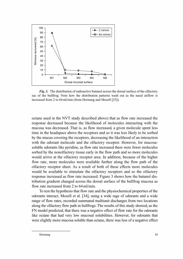
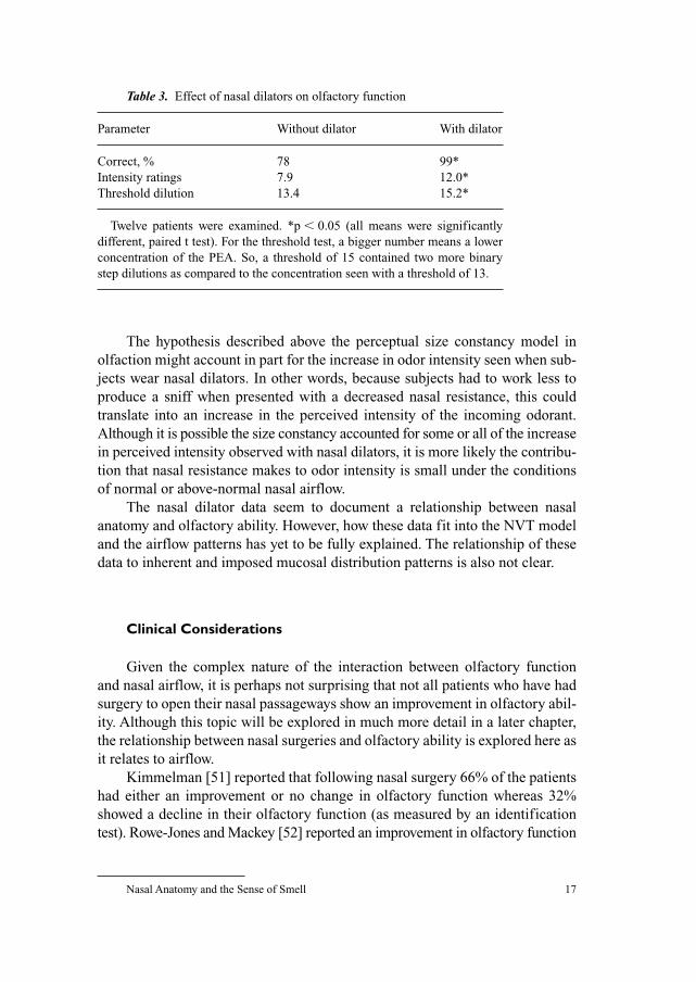
response increased as flow rate increased. Figure 3 shows how the butanol dis-
tribution gradient changed across the dorsal surface of the bullfrog mucosa as
flow rate increased from 2 to 64 ml/min.
To test the hypothesis that flow rate and the physiochemical properties of the
odorants interact, Mozell et al. [34], using a wide rage of odorants and a wide
range of flow rates, recorded summated multiunit discharges from two locations
along the olfactory flow path in bullfrogs. The results of this study showed, as the
FN model predicted, that there was a negative effect of flow rate for the odorants
like octane that had very low mucosal solubilities. However, for odorants that
were slightly more mucosa soluble than octane, there was less of a negative effect
Hornung 10
Fig. 3. The distribution of radioactive butanol across the dorsal surface of the olfactory
sac of the bullfrog. Note how the distribution patterns wash out as the nasal airflow is
increased from 2 to 64 ml/min (from Hornung and Mozell [25]).
0
10
20
30
40
50
60
70
80
90
100
M1 M2 M3 M4 M5
Dorsal mucosal surface
Stim
ulus
rec
over
ed (%
)
2 ml/min
64 ml/min
Nasal Anatomy and the Sense of Smell 11
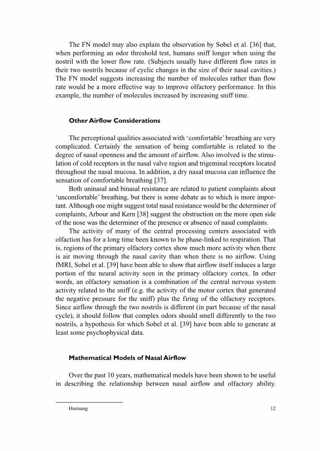
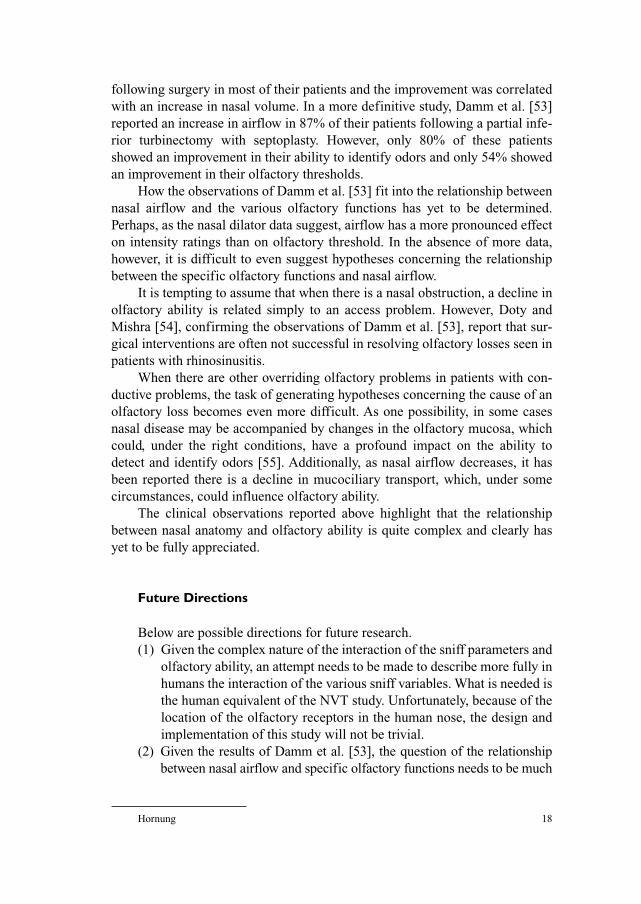
Fig. 5. Coronal MRI scan from the bony region of the nose behind which one would
expect a direct effect of a nasal dilator. Note the increase in the size of the air passageways
with the dilator present (b).
a b
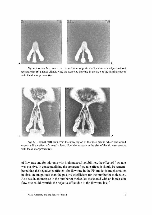
Fig. 4. Coronal MRI scan from the soft anterior portion of the nose in a subject without
(a) and with (b) a nasal dilator. Note the expected increase in the size of the nasal airspaces
with the dilator present (b).
a b
of flow rate and for odorants with high mucosal solubilities, the effect of flow rate
was positive. In conceptualizing the apparent flow rate effect, it should be remem-
bered that the negative coefficient for flow rate in the FN model is much smaller
in absolute magnitude than the positive coefficient for the number of molecules.
As a result, an increase in the number of molecules associated with an increase in
flow rate could override the negative effect due to the flow rate itself.
Hornung 12
The FN model may also explain the observation by Sobel et al. [36] that,
when performing an odor threshold test, humans sniff longer when using the
nostril with the lower flow rate. (Subjects usually have different flow rates in
their two nostrils because of cyclic changes in the size of their nasal cavities.)
The FN model suggests increasing the number of molecules rather than flow
rate would be a more effective way to improve olfactory performance. In this
example, the number of molecules increased by increasing sniff time.
Other Airflow Considerations
The perceptional qualities associated with ‘comfortable’ breathing are very
complicated. Certainly the sensation of being comfortable is related to the
degree of nasal openness and the amount of airflow. Also involved is the stimu-
lation of cold receptors in the nasal valve region and trigeminal receptors located
throughout the nasal mucosa. In addition, a dry nasal mucosa can influence the
sensation of comfortable breathing [37].
Both uninasal and binasal resistance are related to patient complaints about
‘uncomfortable’ breathing, but there is some debate as to which is more impor-
tant. Although one might suggest total nasal resistance would be the determiner of
complaints, Arbour and Kern [38] suggest the obstruction on the more open side
of the nose was the determiner of the presence or absence of nasal complaints.
The activity of many of the central processing centers associated with
olfaction has for a long time been known to be phase-linked to respiration. That
is, regions of the primary olfactory cortex show much more activity when there
is air moving through the nasal cavity than when there is no airflow. Using
fMRI, Sobel et al. [39] have been able to show that airflow itself induces a large
portion of the neural activity seen in the primary olfactory cortex. In other
words, an olfactory sensation is a combination of the central nervous system
activity related to the sniff (e.g. the activity of the motor cortex that generated
the negative pressure for the sniff) plus the firing of the olfactory receptors.
Since airflow through the two nostrils is different (in part because of the nasal
cycle), it should follow that complex odors should smell differently to the two
nostrils, a hypothesis for which Sobel et al. [39] have been able to generate at
least some psychophysical data.
Mathematical Models of Nasal Airflow
Over the past 10 years, mathematical models have been shown to be useful
in describing the relationship between nasal airflow and olfactory ability.
Nasal Anatomy and the Sense of Smell 13
Models were first used to describe the mass transport of odorant molecules
from the air to the olfactory receptors [11–13, 40]. The output of these models
predicted the flow rate, length and thickness of the olfactory mucosa and
air/mucosa partitioning of the odorant all were important in determining the
intensity of the olfactory response. These models further predicted given ‘an
adequate mucus surface area’, an increase in the flow rate should increase the
perceived intensity for odorants with high solubility in the mucus (as reported
in the study by Rehn [35] – see above) whereas for poorly sorbed odorants (like
those used by Mozell et al. [33] in the NVT study), an increase in flow rate
should decrease the perceived intensity. With a reduced surface sorption area,
like that seen in some individuals or with certain nasal diseases, increasing flow
rate will result in a decreased perceived intensity for all odors.
In general, flow path descriptions by the various modeling studies have
yielded similar results. That is, there seems to be a much higher velocity of flow
in the nasal valve region and the floor of the nasal cavity and a much lower
velocity in the olfactory area [13].
In another modeling approach, Zhao et al. [41], using computational fluid
dynamics techniques (CFD), predicted airflow and odorant transport in various
locations in the nose as the shape of the cavity itself changed. Numerical finite
volume methods were used to evaluate how flow to the olfactory area was
altered as the size of critical nasal areas like the olfactory slit and nasal valve
region were changed.
For example, Zhao et al. [41] modified the nasal valve area and modeled
the total nasal airflow rate and flow to the olfactory area. Although there were
only slight changes to the total nasal airflow rate, the changes to airflow through
the olfactory region were dramatic, with flow changing by 50% as the size of
the airway increased or decreased. Obviously these changes in flow to the olfac-
tory region were accompanied by corresponding changes in odorant uptake. As
one example, a small blockage in the nasal valve region (1.45% reduction in
local airway volume) resulted in an 18.7% decrease in nasal airflow and a
76.9% decrease in flow to the olfactory area. Since the nasal valve region seems
to be the source of most of the nasal resistance [42], the modeling results pro-
vided convincing evidence that the nasal valve area is a key in controlling air-
flow to the olfactory region. In other words, the models support the hypothesis
that small changes in critical nasal regions can have profound effects on flow to
the olfactory region.
CFD has also been recently combined with an experimental procedure to
further describe some of the factors that govern odorant mucosal deposition [43].
The fraction of odorant absorbed by the nasal mucosa was experimentally deter-
mined for a number of odorants by measuring the concentration that occurred
when an odorant was ‘blown’ into one nostril and exited the contralateral nostril.
Hornung 14
Subjects performed velopharyngeal closure during this procedure. The nasal
air/odorant airflows seen during the velopharyngeal closure were modeled using
CFD techniques in a model of the human nose [36] and the mucosal odorant
uptake was numerically calculated. The comparison between the numerical simu-
lations and the experimental results led to an estimation of the human mucosal
odorant solubility. The study suggested that the increase in diffusive resistance of
the mucosal layer over that of a thin layer of water seemed to be general and non-
odorant-specific. However, the mucus solubility was odorant specific and usually
followed the trend that odorants with lower water solubility were more soluble in
mucus. The ability of this model to predict odorant movement in the nasal cavity
was evaluated by comparison of the model output with known values of odorant
mucosal solubility. The results strongly suggest the physical processes involved in
olfaction are quite consistent across the various animal species and so it seems
reasonable to suggest the results obtained from the many animal studies have
some relevance to the understanding of the processes involved in olfactory per-
ception in humans. Combining CFD with the experimental measurements
described above provides a technique to measure, in humans, the air/mucosa par-
tition coefficient of any nontoxic odorant.
Nasal Dilators
Nasal dilators (Breathe Right), sometimes worn by athletes, are elastic
strips with imbedded plastic springs that fit over the bridge of the nose. Despite
their questionable usefulness in improving athletic performance, nasal dilators
have been useful as a tool with which to begin to describe the relationship
between nasal airflow and olfactory ability [44, 45]. The effect that nasal dila-
tors have on nasal resistance is considerable. For example, Lorion et al. [42]
using active posterior rhinometry estimated the presence of an internal nasal
dilator reduces nasal resistance by about 40%.
The first step in assessing how dilators affect olfactory function was to
determine exactly how dilators produced the dramatic reduction in nasal resis-
tance. Toward that end, MRI and CT were used to determine where and by how
much nasal dilators change the anatomy of the nose. In a recent study [46],
when subjects wore nasal dilators, the volume of the soft anterior portion of the
nose (defined as the nasal cavity from the tip of the nose to the beginning of the
turbinates) was increased by 23% compared to the undilated condition. This
result agreed well with previously published data [45] reporting dilators pro-
duced a 26% increase in the anterior nasal volume. Additionally, the recent
studies demonstrate that, at least for 4 h, the anterior nasal volume remained
elevated as long as the dilator was worn [46].
Nasal Anatomy and the Sense of Smell 15
In analyzing the volume changes in the bony regions of the nose, the por-
tions behind which one would not expect the dilator to have a direct effect, nasal
volume still increased by 9% when the dilator was in place [46]. This agreed well
with previously published results [45]. The increase in the volume of the poste-
rior portion of the nose remained as long as the dilator was kept in place. The
increase in the volume of the nasal cavity in the bony region must reflect a
response of the nose to the dilation of the soft anterior region, a response perhaps
mediated by changing blood flow to the swell spaces mentioned above [45].
With the anatomical changes quantified, the next step in determining the
effect nasal dilators had on olfactory ability was to determine their effect on the
physical characteristics of the sniff. A pneumotachograph measured the sniffing
behavior of subjects with and without a dilator in place. As can be seen in table 2,
wearing a nasal dilator increased all the characteristics of the sniff compared to
the values seen in the undilated condition.
Since wearing the nasal dilator increased the size of the air passageways in
the nose and the flow characteristics of a sniff, the parsimonious conclusion is
that the pressure generated by the sniff is ‘hardwired’. That is, the respiratory
muscles seem to be programmed by the nervous system to generate a certain
amount of negative pressure. So, when the nasal resistance was decreased (as
happens when a nasal dilator is worn), there was an increase in the sniff charac-
teristics [45].
In the mirror image of the nasal dilator experiment, Youngentob et al. [47]
studied the effect increasing nasal resistance had on odor intensity. As the nasal
resistance increased subjects rated inspired odors as being less intense. At first,
this would seem to suggest that with the increase in resistance there was a
decrease in the volume and flow rate of the subject’s sniff (and so the number
of molecules delivered to the receptors was decreased). However, since the
sniff flow rate and volume did not change with an increased nasal resistance,
Youngentob et al. [47] proposed the concept of a perceptual size constancy
Table 2. Effect of nasal dilators on sniff characteristics
Parameter Without dilator With dilator
Sniff mean flow rate, l/min 45.3 54.8*
Sniff max. flow rate, l/min 80.0 92.8*
Sniff volume, l 1.09 1.54*
Sniff duration, s 1.4 1.7*
Twelve patients were examined. *p � 0.05 (all means were significantly
different, paired t test).
Hornung 16
model to explain their results. In other words, the olfactory magnitude depends
on both the concentration and the perceived effort associated with the sniff.
Important to the present discussion is the observation that when presented with
an increase in nasal resistance subjects maintained sniff flow rate, volume and time.
On the surface, the nasal dilator data showing an increase in the sniff charac-
teristics seem inconsistent with the observation that when presented with an
increase in nasal resistance subjects produce a constant sniff. It seems reasonable
that there would be a mechanism to maintain flow as nasal resistance increases
since this occurs often in nature. However, a major reduction in nasal resistance
rarely happens naturally and so there would be no evolutionary pressure for a
neural mechanism to reduce flow in this case. Additionally, as will be described
below, olfactory function is apparently not compromised with a major reduction
in nasal resistance, and so again there would not be a selective pressure to reduce
flow when nasal resistance was suddenly decreased. Therefore, it seems possible
that the body maintains minimum sniff characteristics when resistance is
increased, but there is less regulation if the nasal resistance is suddenly increased.
The final step was to determine how the nasal dilator induced changes in
anatomy and sniff characteristics affected olfactory ability as determined by a
number of psychophysical techniques [45]. Since airflow to the olfactory area
goes along a channel from the tip of the nose toward the superior turbinate and
there was a substantial increase in the sniff characteristics, it was hypothesized
that the changes produced by the dilator should have a positive effect on olfac-
tory function, although given some of the constraints of the FN model, other
outcomes were possible for at least some types of odorants.
Odorant identification (the ability to name odorants) was evaluated with
the odorant confusion matrix, a clinical test developed to assess olfactory func-
tion. Since all the subjects tested in the present study had normal senses of
smell, the concentrations of the odorants were reduced by 60% to make the test
more difficult [48].
Olfactory threshold was evaluated with a 2-interval forced-choice test of
phenethyl alcohol (PEA). This compound has a rose-like smell, and is routinely
used to measure olfactory threshold. Subjects were presented with two bottles and
asked to identify the bottle that contains the odorant. Subjects began with a very
low concentration of PEA in the test bottle and each time the subjects picked the
incorrect bottle the concentration was increased. This process continued until the
subjects correctly picked the bottle containing the odorant five times in a row [49].
Subjects used the Green scale [50] to report their evaluation of the intensity of
the 10 odorants in the odorant confusion matrix. Subjects were presented with a
bottle containing the smell and asked to indicate how intense the odorant smelled
to them. The intensity ratings were compared with and without the dilator [46, 47].
The results of the olfactory testing can be seen in table 3.
Nasal Anatomy and the Sense of Smell 17
The hypothesis described above the perceptual size constancy model in
olfaction might account in part for the increase in odor intensity seen when sub-
jects wear nasal dilators. In other words, because subjects had to work less to
produce a sniff when presented with a decreased nasal resistance, this could
translate into an increase in the perceived intensity of the incoming odorant.
Although it is possible the size constancy accounted for some or all of the increase
in perceived intensity observed with nasal dilators, it is more likely the contribu-
tion that nasal resistance makes to odor intensity is small under the conditions
of normal or above-normal nasal airflow.
The nasal dilator data seem to document a relationship between nasal
anatomy and olfactory ability. However, how these data fit into the NVT model
and the airflow patterns has yet to be fully explained. The relationship of these
data to inherent and imposed mucosal distribution patterns is also not clear.
Clinical Considerations
Given the complex nature of the interaction between olfactory function
and nasal airflow, it is perhaps not surprising that not all patients who have had
surgery to open their nasal passageways show an improvement in olfactory abil-
ity. Although this topic will be explored in much more detail in a later chapter,
the relationship between nasal surgeries and olfactory ability is explored here as
it relates to airflow.
Kimmelman [51] reported that following nasal surgery 66% of the patients
had either an improvement or no change in olfactory function whereas 32%
showed a decline in their olfactory function (as measured by an identification
test). Rowe-Jones and Mackey [52] reported an improvement in olfactory function
Table 3. Effect of nasal dilators on olfactory function
Parameter Without dilator With dilator
Correct, % 78 99*
Intensity ratings 7.9 12.0*
Threshold dilution 13.4 15.2*
Twelve patients were examined. *p � 0.05 (all means were significantly
different, paired t test). For the threshold test, a bigger number means a lower
concentration of the PEA. So, a threshold of 15 contained two more binary
step dilutions as compared to the concentration seen with a threshold of 13.
Hornung 18
following surgery in most of their patients and the improvement was correlated
with an increase in nasal volume. In a more definitive study, Damm et al. [53]
reported an increase in airflow in 87% of their patients following a partial infe-
rior turbinectomy with septoplasty. However, only 80% of these patients
showed an improvement in their ability to identify odors and only 54% showed
an improvement in their olfactory thresholds.
How the observations of Damm et al. [53] fit into the relationship between
nasal airflow and the various olfactory functions has yet to be determined.
Perhaps, as the nasal dilator data suggest, airflow has a more pronounced effect
on intensity ratings than on olfactory threshold. In the absence of more data,
however, it is difficult to even suggest hypotheses concerning the relationship
between the specific olfactory functions and nasal airflow.
It is tempting to assume that when there is a nasal obstruction, a decline in
olfactory ability is related simply to an access problem. However, Doty and
Mishra [54], confirming the observations of Damm et al. [53], report that sur-
gical interventions are often not successful in resolving olfactory losses seen in
patients with rhinosinusitis.
When there are other overriding olfactory problems in patients with con-
ductive problems, the task of generating hypotheses concerning the cause of an
olfactory loss becomes even more difficult. As one possibility, in some cases
nasal disease may be accompanied by changes in the olfactory mucosa, which
could, under the right conditions, have a profound impact on the ability to
detect and identify odors [55]. Additionally, as nasal airflow decreases, it has
been reported there is a decline in mucociliary transport, which, under some
circumstances, could influence olfactory ability.
The clinical observations reported above highlight that the relationship
between nasal anatomy and olfactory ability is quite complex and clearly has
yet to be fully appreciated.
Future Directions
Below are possible directions for future research.
(1) Given the complex nature of the interaction of the sniff parameters and
olfactory ability, an attempt needs to be made to describe more fully in
humans the interaction of the various sniff variables. What is needed is
the human equivalent of the NVT study. Unfortunately, because of the
location of the olfactory receptors in the human nose, the design and
implementation of this study will not be trivial.
(2) Given the results of Damm et al. [53], the question of the relationship
between nasal airflow and specific olfactory functions needs to be much
Nasal Anatomy and the Sense of Smell 19
better described. Questions to be answered include: is the ability to
detect the presence of an odorant (threshold) less sensitive to anatomical
and flow changes than is the ability to distinguish and recognize odors?
What is the relationship between nasal airflow and intensity ratings, and
is this relationship the same for all odorants? How do airflow changes
affect odor discrimination and recognition tasks?
(3) If airflow affects different olfactory functions differentially, what does
this say about the peripheral and central processing mechanisms? Can
studies considering the relationship between nasal airflow and specific
olfactory functions suggest hypotheses that can be tested in nonhumans?
(4) As models of the airflow patterns become more sophisticated, it may
become possible for them to predict how nasal surgery will affect olfac-
tory function in a particular patient. It may even be possible for models
to suggest surgical alterations to maximize olfactory ability following a
surgical intervention.
Conclusions
(1) Although there seems to be a direct relationship between the olfactory
response and stimulus concentration, the olfactory stimulus is actually
defined by three ‘primary’ variables: the number of molecules (N), the
duration of the sniff (T), and the volume of the sniff (V). These pri-
mary variables in turn define the three ‘derived’ variables of concen-
tration (C � N/V), delivery rate (D � N/T) and flow rate (F � V/T).
(2) The relationship between the olfactory response and the stimulus may
vary depending on the physiochemical properties of the odor.
(3) Nasal airflow may impact the various olfactory functions differently.
As one possibility, intensity and discrimination tasks may be more sen-
sitive to flow changes than are measures of odor threshold.
(4) Although it is tempting to assume that when there is a nasal obstruction,
a decline in olfactory ability is related simply to an access problem, it
should be realized that nasal disease could affect olfactory processing at
many levels. As a result, changes in airflow may not always be the cause
of an altered olfactory ability.
References
1 Churchill SE, Shackelford LL, Georgi JN, Black MT: Morphological variations in the upper res-
piratory track and airflow dynamics. Am J Physiol Anthropol Suppl 1966;28:107.
2 Shea BT: Eskimo craniofacial morphology, cold stress and the maxillary sinus. Am J Anthropol
1977;47:289–300.
Hornung 20
3 Cole P: Modification in inspired air; in Procter DF, Anderson I (eds): The Nose: Upper Airway
Physiology and the Atmospheric Environment. Amsterdam, Elsevier Biomedical Press, 1982,
pp 351–375.
4 Barr GS, Tewary AK: Alteration of airflow and mucociliary transport in normal subjects.
J Laryngol Otol 1993;107:603–604.
5 Hornung DE: Smell; in Hoagstrom CW (ed): Magill’s Encyclopedia of Science: Animal Life.
Pasadena, Salem Press, 2002, pp 1514–1516.
6 DeWeese DD, Saunders WH: Textbook of Otolaryngology, ed 3. St Louis, Mosby, 1968.
7 Calhoun KH, House W, Hokanson JA, Quinn FB: Normal nasal airway resistance in noses of
different sizes and shapes. Otolaryngol Head Neck Surg 1990;103:605–609.
8 Simmen D, Scherrer JL, Moe K, Heinz B: A dynamic and direct visualization model for the study
of nasal airflow. Arch Otolaryngol Head Neck Surg 1999;125:1015–1021.
9 Wolpoff MH: Climatic influence on skeletal nasal aperture. Am J Physiol Anthropol 1968;29: 405–424.
10 Stuiver M: Biophysics of the sense of smell; thesis, Groningen, 1958.
11 Hahn I, Scherer PW, Mozell MM: A mass transport model of olfaction. J Theor Biol 1994;167:
115–128.
12 Keyhani K, Scherer PW, Mozell MM: A numerical model of nasal odorant transport for the analy-
sis of human olfaction. J Theor Biol 1997;186:279–301.
13 Kelly JT, Prasad AK, Wexler AS: Detailed flow patterns in the nasal cavity. J Appl Physiol
2000;89:323–337.
14 Luffingwell JC: Olfaction – a review. Online at http://www.leffingwell.com/olfaction.htm
15 Wilson DA: Synthetic coding of odorant mixtures in rat piriform cortex. Chem Senses 2002;
27:667.
16 Moulton DG: Spatial patterning response to odors in the peripheral olfactory system. Physiol Rev
1976;56:578–593.
17 Mozell MM, Sheehe PR, Hornung DE, Kent PK, Youngentob SL, Murphy SJ: Imposed and inher-
ent mucosal activity patterns: their composite representation of olfactory stimuli. J Gen Physiol
1987;90:625–650.
18 Mozell MM, Sheehe PR, Hornung DE, Kent PK, Youngentob SL, Murphy SJ: The composite rep-
resentation of olfactory stimuli by imposed and inherent mucosal activities patterns; in Miller I
(ed): The Lloyd M Beidler Symposium on Taste and Smell. Winston-Salem, Book Service
Associates, 1988, pp 143–158.
19 Hornung DE, Lansing RD, Mozell MM: The distribution of butanol molecules along the olfactory
mucosa of the bullfrog. Nature 1975;254:617–618.
20 Mozell MM, Sheehe PR, Hornung DE, Kent PK, Youngentob SL, Murphy SJ: Imposed and inher-
ent mucosal activity patterns: their composite representation of olfactory stimuli. J Gen Physiol
1987;90:625–650.
21 Hornung DE, Mozell MM: Smell – human physiology; in Rivlin RS, Meiselman RH (eds): Human
Taste and Smell: Measurements and Uses. New York, Macmillan Publishing, 1986, pp 19–38.
22 Mozell MM: Evidence for sorption as a mechanism of the olfactory analysis of odorants. Nature
1964;203:1181–1182.
23 Kent PF, Mozell MM, Murphy SJ, Hornung DE: The interaction of imposed and inherent olfactory
mucosal patterns and their composite representation in a mammalian species using voltage-
sensitive dyes. J Neurosci 1996;16:345–353.
24 Kent PF, Youngentob SL, Hornung DE: Mucosal activity patterns and odorant quality perception;
in Kurihara K, Suzuki N, Ogawa H (eds): Proceedings of the 11th International Symposium on
Olfaction and Taste. Tokyo, Springer, 1994, pp 205–206.
25 Hornung DE, Mozell MM: Factors influencing the differential sorption of odorant molecules
across the olfactory mucosa. J Gen Physiol 1977;69:343–361.
26 Hornung DE, Serio JA, Mozell MM: Olfactory mucosa/air partitioning of odorants; in van der
Storee H (ed): Olfaction and Taste VII. London, Information Retrieval, 1980, pp 167–170.
27 Mozell MM, Jagodowicz M: Chromatographic separation of odorants by the nose: retention times
measured across in vivo olfactory mucosa. Science 1973;181:1247–1249.
28 Youngentob SL, Markert LM, Mozell MM, Hornung DE: A method for establishing a five odorant
identification confusion matrix task in rats. Physiol Behav 1990;47:1053–1059.

Nasal Anatomy and the Sense of Smell 21
29 Youngentob SL, Kent PF, Sheehe PR, Schwob JE, Tzournaka E: Mucosal inherent activity patterns
in the rat: evidence from voltage-sensitive dyes. J Neurophysiol 1995;73:387–398.
30 Kent PF, Youngentob SL, Sheehe PR: Odorant-specific spatial patterns in mucosal activity predict
perceptual differences among odorants. J Neurophysiol 1995;74:1777–1781.
31 Kent PF, Mozell MM, Youngentob SL, Yurco P: Mucosal activity patterns as a basis for olfactory
discrimination: comparing behavior and optical recordings. Brain Res 2003;981:1–11.
32 Youngentob SL, Margolis FL, Youngentob LM: OMP gene deletion results in an alteration in
odorant quality perception. Behav Neurosci 2001;115:626–631.
33 Mozell MM, Sheehe PR, Swieck SW Jr, Kurtz DB, Hornung DE: A parametric study of the stim-
ulation variables affecting the magnitude of the olfactory nerve response. J Gen Physiol 1984;83:
233–267.
34 Mozell MM, Kent PF, Scherer PW, Hornung DE, Murphy SJ: Nasal airflow; in Getchell TV,
Doty RL, Bartoshuk LK, Snow JB Jr (eds): Smell and Taste in Health and Disease. New York,
Raven Press, 1991, pp 481–492.
35 Rehn T: Perceived odor intensity as a function of airflow through the nose. Sens Processes 1978;2:
198–205.
36 Sobel N, Khan RM, Hartley CA, Sullivan EV, Gabriele JD: Sniffing longer rather than stronger to
maintain olfactory detection threshold. Chem Senses 2000;25:1–8.
37 Pallanch JF, McCaffrey TV, Kern EB: Evaluation of nasal breathing function with objective airway
testing; in Cummings CW Jr, Frederickson JM, Harker LA, Richardson M, Schuller DE:
Otolaryngology, ed 3. St Louis, Mosby, 1998, pp 799–832.
38 Arbour P, Kern EB: Paradoxical nasal obstruction. Can J Otolaryngol 1975;4:333.
39 Sobel N, Prabhakaran V, Desmond JE, Glover GH, Sullivan EV, Goode RL, Gabrieli JDE:
Sniffing and smelling: separate subsystems in the human olfactory cortex. Nature 1998;392:
282–286.
40 Hahn I, Scherer PW, Mozell MM: Velocity profiles measured for airflow through a large-scale
model of the human nasal cavity. J Appl Physiol 1994;75:2273–2287.
41 Zhao K, Scherer PW, Hajiloo SA, Dalton P: Effect of anatomy on human nasal airflow and odor-
ant transport patterns: implications for olfaction. Chem Senses 2004;29:365–379.
42 Lorino AM, Lofaso F, Dahan E, Coste A, Harf A, Lorino H: Combined effects of a mechanical
nasal dilator and a topical decongestant on nasal airflow resistance. Chest 1999;115:1514–1518.
43 Kurtz DB, Zhao K, Hornung DE, Scherer P: Experimental and numerical determination of odor-
ant solubility in nasal and olfactory mucosa. Chem Senses 2004;29:763–773.
44 Hornung DE, Chin C, Kurtz DB, Kent PF, Mozell MM: Effect of nasal dilators on perceived odor
intensity. Chem Senses 1997;22:177–180.
45 Hornung DE, Smith DJ, Kurtz DB, White T, Leopold DA: Effect of nasal dilators on nasal struc-
tures, sniffing strategies, and olfactory ability. Rhinology 2001;39:84–87.
46 Lyng GD, Hornung DE, Leopold DA, Irwin SB, Vent J: The long-term effect of nasal dilators on
nasal anatomy and olfactory ability (abstracts). 25th Annu Meet Assoc Chemorecept Sci, Sarasota,
2003, p 89.
47 Youngentob SL, Stern NM, Mozell MM, Leopold DA, Hornung DE: Effect of airway resistance
on perceived odor intensity. Am J Otolaryngol 1986;7:187–193.
48 Kurtz DB, White TL, Sheehe PR, Hornung DE, Kent PF: Odorant confusion matrix: the influence
of patient history on patterns of odorant identification and misidentification in hyposmia. Physiol
Behav 2001;72:595–602.
49 Cain WS, Gent J, Catalanato FA, Goodspeed RB: Clinical evaluation of olfaction. Am J Otolaryngol
1983;4:252–256.
50 Green BG, Dalton P, Cowart B, Shaffer G, Rankin K, Higgins J: Evaluating the ‘labeled magni-
tude scale’ for measuring sensations of smell and taste. Chem Senses 1996;21:323–334.
51 Kimmelman CP: The risk to olfaction from nasal surgery. Laryngoscope 1994;104:981–988.
52 Rowe-Jones JM, Mackay IS: A prospective study of olfaction following endoscopic sinus surgery
with adjuvant medical treatment. Clin Otolaryngol 1997;22:377.
53 Damm M, Eckel HE, Jungehulsing M, Hummel T: Olfactory changes in threshold and
suprathreshold levels following septoplasty with partial inferior turbinectomy. Ann Otol Rhinol
Laryngol 2003;112:91–97.
Hornung 22
54 Doty RL, Mishra A: Olfaction and its alteration by nasal obstruction, rhinitis, and rhinosinusitis.
Laryngoscope 2001;111:409–423.
55 Kern RC: Chronic sinusitis and anosmia: pathologic changes in the olfactory mucosa. Laryngoscope
2000;110:1071–1077.
Prof. David E. Hornung
Dana Professor of Biology, St. Lawrence University
Canton, NY 13617 (USA)
E-Mail [email protected]
Hummel T, Welge-Lüssen A (eds): Taste and Smell. An Update.
Adv Otorhinolaryngol. Basel, Karger, 2006, vol 63, pp 23–43
Transduction and Coding
Nancy E. Rawson, Karen K. Yee
Monell Chemical Senses Center, Philadelphia, Pa., USA
AbstractOdor transduction and quality coding involves a cascade of events that occur at the level
of the olfactory epithelium and olfactory bulbs. Odorants bind to one or a few specific olfac-
tory receptors located in the cilia of olfactory neurons. These olfactory receptor proteins make
up the largest gene family discovered and are diverse between and within species. The change
of chemical signals to neural signals in the olfactory neurons involves G-coupled proteins and
the cascade of second messenger pathways that open ion channels to depolarize the cell and
trigger a series of action potentials carried along the receptor cell axon resulting in release of
glutamate at synapses with mitral cells within the olfactory bulb. These neural signals in the
olfactory bulb produce unique odor maps that play an important role in our ability to detect
and discriminate thousands of different odorants. The olfactory neurons are replaced through-
out life from a population of slowly dividing basal cells within the epithelium. Disease, infec-
tion, injury or aging can interfere with neuronal cell replacement as well as transduction and
coding processes, resulting in impairment and distortions of olfactory performance.
Copyright © 2006 S. Karger AG, Basel
This chapter will describe the cellular events occurring in the olfactory
epithelium (OE) and at the level of the olfactory bulb that are used to detect,
transduce and encode olfactory information. We will present current perspec-
tives on the cell biology of olfaction, emphasizing studies most relevant to
understanding normal and diseased olfactory function in humans.
Cellular Anatomy
The OE is a layered structure comprised of neuronal and nonneuronal cell
types that resides within the olfactory cleft and extends to varying degrees onto
the superior turbinate [1] and superior aspect of the medial turbinate [2]
[see chapter 1 by Hornung, this vol, pp 1–22]. The OE is typically well
Rawson/Yee 24
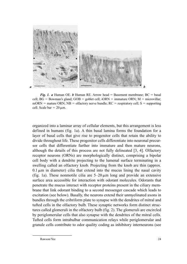
organized into a laminar array of cellular elements, but this arrangement is less
defined in humans (fig. 1a). A thin basal lamina forms the foundation for a
layer of basal cells that give rise to progenitor cells that retain the ability to
divide throughout life. These progenitor cells differentiate into neuronal precur-
sor cells that differentiate further into immature and then mature neurons,
although the details of this process are not fully delineated [3, 4]. Olfactory
receptor neurons (ORNs) are morphologically distinct, comprising a bipolar
cell body with a dendrite projecting to the lumenal surface terminating in a
swelling called an olfactory knob. Projecting from the knob are thin (approx.
0.1 �m in diameter) cilia that extend into the mucus lining the nasal cavity
(fig. 1a). These nonmotile cilia are 5–20 �m long and provide an extensive
surface area accessible for interaction with odorant molecules. Odorants that
penetrate the mucus interact with receptor proteins present in the ciliary mem-
brane that link odorant binding to a second messenger cascade which leads to
excitation (see below). Basally, the neurons extend their unmyelinated axons in
bundles through the cribriform plate to synapse with the dendrites of mitral and
tufted cells in the olfactory bulb. These synaptic networks form distinct struc-
tures called glomeruli in the olfactory bulb (fig. 2). The glomeruli are encircled
by periglomerular cells that also synapse with the dendrites of the mitral cells.
Tufted cells form intrabulbar communication relays while periglomerular and
granule cells contribute to odor quality coding as inhibitory interneurons (see
Fig. 1. a Human OE. b Human RE. Arrow head � Basement membrane; BC � basal
cell; BG � Bowman’s gland; GOB � goblet cell; iORN � immature ORN; M � microvillar;
mORN � mature ORN; NB � olfactory nerve bundle; RC � respiratory cell; S � supporting
cell. Scale bar � 20 �m.
a b
Transduction and Coding 25
below) [5]. The ORN axons are encircled by nonmyelinating ensheathing glial
cells that provide trophic support for axon outgrowth. These glial cells can be
purified and cultured and have recently been used to promote axon regeneration
in other regions, including the spinal cord [6]. Also present in the epithelium are
several types of nonneuronal cells, including sustentacular (supporting) cells
and microvillar cells which have nonmotile cilia or microvilli, respectively [7].
The functions of these cells are not clear, but are thought to include detoxifica-
tion and maintenance of ionic balance. The respiratory epithelium (RE) is
marked by cuboid respiratory epithelial cells with motile cilia, goblet cells and
basal cells (fig. 1b). RE is demarcated from the sensory areas by the presence of
a thicker basement membrane, regular cilia, frequent goblet cells and few nerve
bundles [8]. While RE and OE are clearly demarcated in most species studied,
in humans, OE may be interrupted by stretches of RE, particularly in the elderly
[9–11]. Nerve bundles, vascular components and Bowman’s glands reside in
the submucosal compartment, and the ducts from Bowman’s glands project
through the epithelium to secrete a specialized mucus that coats and protects
the epithelial surface. The mucus layer thickness ranges from 5 to 30 �m and is
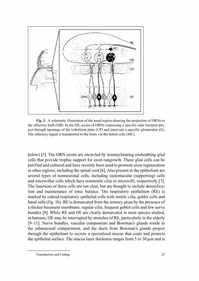
Fig. 2. A schematic illustration of the nasal region showing the projection of ORNs to
the olfactory bulb (OB). In the OE, axons of ORNs expressing a specific odor receptor pro-
ject through openings of the cribriform plate (CP) and innervate a specific glomerulus (G).
The olfactory signal is transported to the brain via the mitral cells (MC).
MC
GOB
CP
ORN OE
Rawson/Yee 26
comprised of water, electrolytes and a variety of mucopolysaccharides and pro-
teins. A family of odorant-binding proteins have been identified in the mucus
that bind distinct classes of hydrophobic odorants and apparently contribute to
odor transport [12–14]. These proteins may modulate the mucus concentration
of odorants to maintain a level optimal for ORN sensitivity. Mucus secretion is
influenced by adrenergic sympathetic fibers from the superior cervical ganglion
[15]. Catecholamines, including dopamine and norepinephrine are released
from the nerve terminals in the lamina propria, and catecholamines are also
released into the mucus in response to activation of the 5th cranial or trigeminal
nerve by irritants [16]. These catecholamines have been found to modulate odor
sensitivity via D2 dopamine receptors [17]. Thus, conditions that alter the func-
tion of these submucosal tissue components may exert direct and indirect
effects on the function of ORNs. A basic understanding of receptor cell func-
tion is important to understand the impact of conditions and treatments that may
result in olfactory dysfunction.
Odor Detection
Odorants interact with olfactory receptor (OR) proteins present in the cil-
iary membranes of mature ORNs. These receptor proteins represent the largest
gene family yet discovered, and also are among the most diverse both between
and within species (for a comprehensive database of ORs, see http://senselab.
med.yale.edu/senselab/). The ORs are members of the G-protein-coupled recep-
tor family as they are coupled to GTP-binding regulatory proteins (G-proteins).
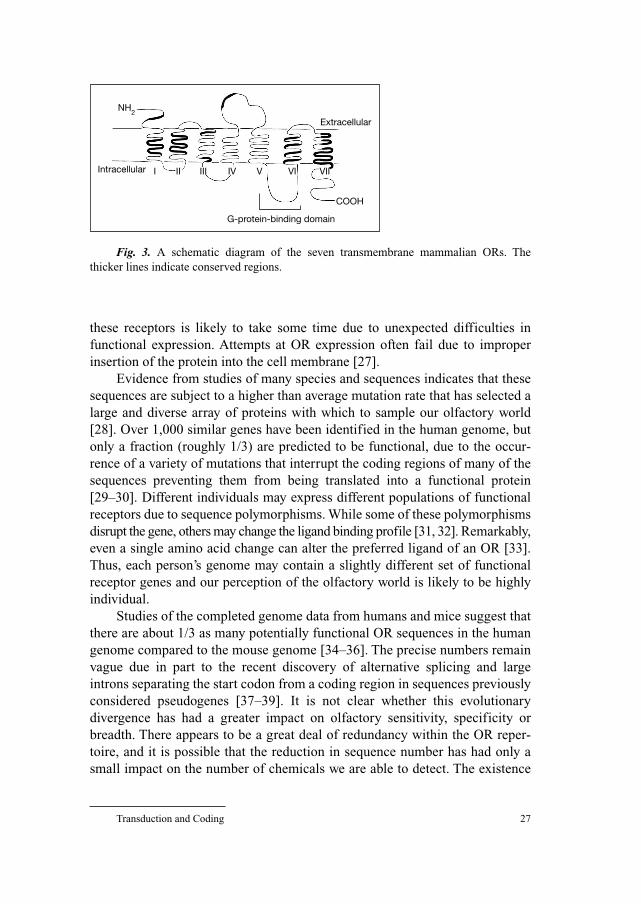
The OR proteins are typically 300–400 amino acids in length and, like other
G-protein-coupled receptors, traverse the membrane seven times. Their
sequences contain regions with an unusually high degree of sequence similarity
across all of the family members and other regions that are hypervariable
(fig. 3). The most highly conserved regions are involved in G-protein binding,
while regions that exhibit the most variability are likely to be involved in ligand
binding.
A few studies have determined the odor response profiles for specific
ORs. These studies insert the gene for that OR into a heterologous cell type
including a signaling cascade that allows receptor binding to be detected
[18–21]. Data from such studies suggest that each OR is narrowly tuned to bind
to a few odorants with particular structural features [22, 23]. Small differences
in the amino acid sequence can alter the binding affinity and studies of sequ-
ences with defined sequence mutations have identified several regions critical
to ligand binding (fig. 3) [24–26]. The vast majority of ORs remain ‘orphan’
receptors, in that their ligands are not yet known. The task of de-orphanizing
Transduction and Coding 27
these receptors is likely to take some time due to unexpected difficulties in
functional expression. Attempts at OR expression often fail due to improper
insertion of the protein into the cell membrane [27].
Evidence from studies of many species and sequences indicates that these
sequences are subject to a higher than average mutation rate that has selected a
large and diverse array of proteins with which to sample our olfactory world
[28]. Over 1,000 similar genes have been identified in the human genome, but
only a fraction (roughly 1/3) are predicted to be functional, due to the occur-
rence of a variety of mutations that interrupt the coding regions of many of the
sequences preventing them from being translated into a functional protein
[29–30]. Different individuals may express different populations of functional
receptors due to sequence polymorphisms. While some of these polymorphisms
disrupt the gene, others may change the ligand binding profile [31, 32]. Remarkably,
even a single amino acid change can alter the preferred ligand of an OR [33].
Thus, each person’s genome may contain a slightly different set of functional
receptor genes and our perception of the olfactory world is likely to be highly
individual.
Studies of the completed genome data from humans and mice suggest that
there are about 1/3 as many potentially functional OR sequences in the human
genome compared to the mouse genome [34–36]. The precise numbers remain
vague due in part to the recent discovery of alternative splicing and large
introns separating the start codon from a coding region in sequences previously
considered pseudogenes [37–39]. It is not clear whether this evolutionary
divergence has had a greater impact on olfactory sensitivity, specificity or
breadth. There appears to be a great deal of redundancy within the OR reper-
toire, and it is possible that the reduction in sequence number has had only a
small impact on the number of chemicals we are able to detect. The existence
Fig. 3. A schematic diagram of the seven transmembrane mammalian ORs. The
thicker lines indicate conserved regions.
Extracellular
COOH
G-protein-binding domain
Intracellular I II III IV V VI VII
NH2
Rawson/Yee 28
of specific anosmias suggests that mutation of specific ORs could lead to an
inability to detect particular odors, but none of these anosmias has yet been
linked to a particular OR mutation.
Currently available data suggest that each ORN expresses only a single
type of functional OR, and that only one allele of a given OR is expressed [34,
40]. In rodents, ORs of different classes are expressed in several zones across
the epithelium [41], but it is not known if similar zones exist in the human. The
molecular mechanisms that regulate how each mature ORN selects a particular
OR and how their expression pattern is maintained in the face of ongoing ORN
replacement remain to be explained. Research in these areas is leading to new
insights in our understanding of how the human genome operates, as well as
helping us to understand evolutionary processes and the diversity of olfactory
perception.
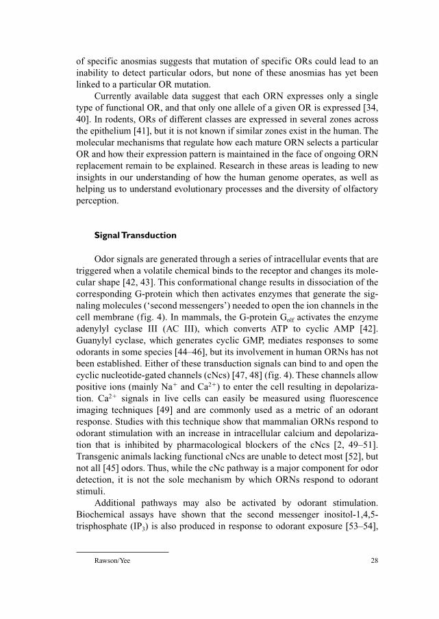
Signal Transduction
Odor signals are generated through a series of intracellular events that are
triggered when a volatile chemical binds to the receptor and changes its mole-
cular shape [42, 43]. This conformational change results in dissociation of the
corresponding G-protein which then activates enzymes that generate the sig-
naling molecules (‘second messengers’) needed to open the ion channels in the
cell membrane (fig. 4). In mammals, the G-protein Golf activates the enzyme
adenylyl cyclase III (AC III), which converts ATP to cyclic AMP [42].
Guanylyl cyclase, which generates cyclic GMP, mediates responses to some
odorants in some species [44–46], but its involvement in human ORNs has not
been established. Either of these transduction signals can bind to and open the
cyclic nucleotide-gated channels (cNcs) [47, 48] (fig. 4). These channels allow
positive ions (mainly Na� and Ca2�) to enter the cell resulting in depolariza-
tion. Ca2� signals in live cells can easily be measured using fluorescence
imaging techniques [49] and are commonly used as a metric of an odorant
response. Studies with this technique show that mammalian ORNs respond to
odorant stimulation with an increase in intracellular calcium and depolariza-
tion that is inhibited by pharmacological blockers of the cNcs [2, 49–51].
Transgenic animals lacking functional cNcs are unable to detect most [52], but
not all [45] odors. Thus, while the cNc pathway is a major component for odor
detection, it is not the sole mechanism by which ORNs respond to odorant
stimuli.
Additional pathways may also be activated by odorant stimulation.
Biochemical assays have shown that the second messenger inositol-1,4,5-
trisphosphate (IP3) is also produced in response to odorant exposure [53–54],
Transduction and Coding 29
and may contribute to the calcium response via opening of membrane channels
[55–59]. This pathway has been shown to modulate the sensitivity of the cNc
pathway in rodent ORNs [60], and indirect evidence suggests it may play a role
in mediating a component of the odorant-stimulated calcium response in human
ORNs [2, 61]. While the relative contributions of cyclic AMP and IP3-linked
pathways to odorant signaling remains unclear, several lines of evidence sup-
port the existence of multiple pathways for odorant-stimulated ORN activity
[45, 56, 60–64]. In most species, the calcium signal may be further transmit-
ted and amplified by voltage-gated calcium channels (VGCCs). Dopamine sup-
presses VGCC activity and ORN responsiveness [65–67] suggesting that the
activity of these channels influences the sensitivity of the ORN. In the axon
terminus, VGCCs are activated by the action potential and are necessary for
calcium-dependent neurotransmitter release.
The influx of positive ions through any of these mechanisms would be suf-
ficient to trigger an action potential, but under some conditions, additional com-
ponents may amplify the calcium signal. In many species, the calcium entering
via the cNc opens a chloride channel [68–70]. Unlike most cells, the chloride
level inside ORNs is higher than in the surrounding compartment, such that open-
ing this channel allows negative chloride ions to exit the cell, further amplifying
Fig. 4. A schematic illustration of the G-protein signal transduction cascade in the
ORN cilia. The binding of an odorant (O) to a G-coupled OR activates Golf and AC III, which
leads to an increase in cyclic AMP and the subsequent influx of cations (Na� and Ca2�) from
cNc. PKA and calcium cations are involved in feedback mechanisms. ATP � Adenosine
5�-triphosphate; C � cilia; CAM � calmodulin; PDE � phosphodiesterase.
C
ORN
OR
PKA
ATP cAMP
PDE
5�-AMP
CAM
Na� Ca2�
cNc
AC IIIGolf
Cellmembrane
� �
� � �
�

Rawson/Yee 30
the depolarizing effect [71–73]. However, the contribution of this channel in
human ORNs has not yet been demonstrated.
The second messengers elevated in response to odorant activation lead to
other cellular effects [74] (fig. 4). Calcium and cyclic AMP activate various
protein kinases (PKs), including PKA and calcium/calmodulin kinase II that are
involved in termination of the odorant signal [61, 75–80]. PKs phosphorylate,
thereby inactivating, ion channels and other components of the transduction
cascade to terminate the odorant signal resulting in short-term adaptation. In
catfish, a PKC-sensitive phosphorylation site is present in the ORs [81], and
this kinase has also been implicated in adaptation in rat and human ORNs,
although its target(s) have yet to be identified [61]. Calcium is removed from
the cell via the Na�/Ca2� exchanger [82] and other mechanisms not yet charac-
terized. These calcium homeostatic pathways have the potential to be influ-
enced by medications and molecules that are elevated in a variety of disease
states, either directly or indirectly (see below). As excess or chronic calcium
elevation is toxic, such an interaction could shorten ORN life span or impair the
function of mature neurons.
Membrane depolarization resulting from cation influx results in generation
of an action potential that is transmitted along the axon to trigger release of the
neurotransmitter glutamate at the synapses within the olfactory glomeruli (fig. 5).
Physiological studies in a variety of species show that, in addition to triggering
an increase in action potential firing, odorants may also suppress or inhibit neu-
ronal activity [83–85]. In some studies, odorants have been found to trigger a
decrease in intracellular calcium [2, 86] that could account for these inhibitory
responses, although data are limited. Studies in toad ORNs suggest these
inhibitory receptor potentials may be mediated by opening of Ca2�-activated
K� channels [62, 87] but similar studies have not been done with human ORNs.
The advantage of such a dual mechanism could be to improve the signal to
noise ratio. Suppressing the activity of ORNs expressing ORs with lower affin-
ity for that odorant could enhance the impact of the signal from ORNs express-
ing ORs whose affinity for the stimuli was highest.
Coding and Miscoding
Combinatorial ModelData acquired through a variety of methods point to a model for odor
coding that is based on unique patterns of mitral cell activity. Each odorant can
bind to multiple ORs, and each OR can bind multiple odorants. ORNs expressing
the same OR project to the same glomeruli in the olfactory bulb, and each
mitral cell projects to one glomerulus thereby serving as the central signal
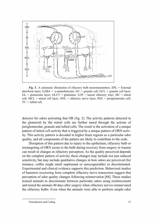
Transduction and Coding 31
detector for odors activating that OR (fig. 2). The activity patterns detected in
the glomeruli by the mitral cells are further tuned through the actions of
periglomerular, granule and tufted cells. The result is the activation of a unique
pattern of mitral cell activity that is triggered by a unique pattern of ORN activ-
ity. This activity pattern is decoded in higher brain regions as a particular odor
quality, and all components of the pattern are likely to contribute to the code.
Disruption of this pattern due to injury to the epithelium, olfactory bulb or
mistargeting of ORN axons to the bulb during recovery from surgery or trauma
can result in changes in olfactory perception. As the quality perceived depends
on the complete pattern of activity, these changes may include not just reduced
sensitivity, but may include qualitative changes in how odors are perceived (for
instance, coffee might smell unpleasant or unrecognizable) or discriminated.
Experimental and clinical evidence supports this prediction. Behavioral studies
of hamsters recovering from complete olfactory nerve transection suggest that
perception of odor quality changes following reinnervation [88]. These studies
trained animals to discriminate between particular odors using reinforcement
and tested the animals 40 days after surgery when olfactory nerves reinnervated
the olfactory bulbs. Even when the animals were able to perform simple odor
Fig. 5. A schematic illustration of olfactory bulb neurotransmitters. EPL � External
plexiform layer; GABA � �-aminobutyrate; GC � granule cell; GCL � granule cell layer;
GL � glomerular layer; GLUT � glutamine; LOT � lateral olfactory tract; MC � mitral
cell; MCL � mitral cell layer; ONL � olfactory nerve layer; PGC � periglomerular cell;
TC � tufted cell.
ONL
GL
EPL
MCL
GCL
PGC
TC
MC
GC
GABA
GABA
GLUT
LOT
�
�
�
Rawson/Yee 32
detection tasks, they were not able to discriminate previously learned odors.
Thus, following surgery or trauma in which hyposmia or anosmia results from
damage to the olfactory nerves, recovery may occur, but the same pattern of
glomerular activity may not be reestablished initially due to inaccurate targeting
of new axons to glomeruli in the olfactory bulb. With time, it is possible that
learning or axon targeting corrections occur and recreate the original patterns
or perceptions. In humans, olfactory abnormalities that often precede recovery
from anosmia after a head injury might be a result of this type of mistargeting
of regenerating axons [89–91; see also chapter 7 by Raviv and Kern, this vol, pp
108–124].
Coding of Mixture QualitiesThe vast majority of aromas we encounter in everyday life are actually
complex mixtures of tens, hundreds or even thousands of individual volatile
chemicals. Our understanding of how these complex stimuli are decoded into a
single ‘aroma’ quality is still very limited. Perfumers have long exploited the
ability of certain chemicals to enhance, suppress or alter the quality of other
odors to design fragrances. These interactions were traditionally discovered
through trial and error, but science has begun to reveal the basis for these phe-
nomena. Referring to the combinatorial model can suggest a basis for some of
these interactions.
As mentioned previously, each odor can interact with different affinities
with multiple ORs, which recognizes different chemical features (fig. 6A). A
high concentration of an odorant may activate the highest affinity or primary
receptor as well as lower affinity receptors, thus eliciting a different quality than
a lower concentration, which activates only the primary receptor (fig. 6B). Odor
suppression or masking may occur when an odorant blocks access to a receptor
without activating it, preventing detection of the odor that would be able to acti-
vate that receptor [92] (fig. 6C). These predictions are consistent with the
results of molecular and behavioral studies indicating that the quality of odor
mixtures may be represented either configurally (i.e. the mixture is qualitatively
different from the components) or elementally (the components are recogniz-
able) [93, 94]. These data showed that binary mixtures whose components acti-
vate overlapping sets of receptors are more likely to be perceived as configural,
while components can be discriminated when they activate distinct receptor
populations.
While the combinatorial model can provide some insight into the phenom-
ena of odor masking and concentration-dependent changes in odor quality,
other predictions of the model fail to account for observed phenomena. For
instance, the model predicts that a complex odor should activate more glomeruli
than a single compound. Predictions can be made about the number of glomeruli
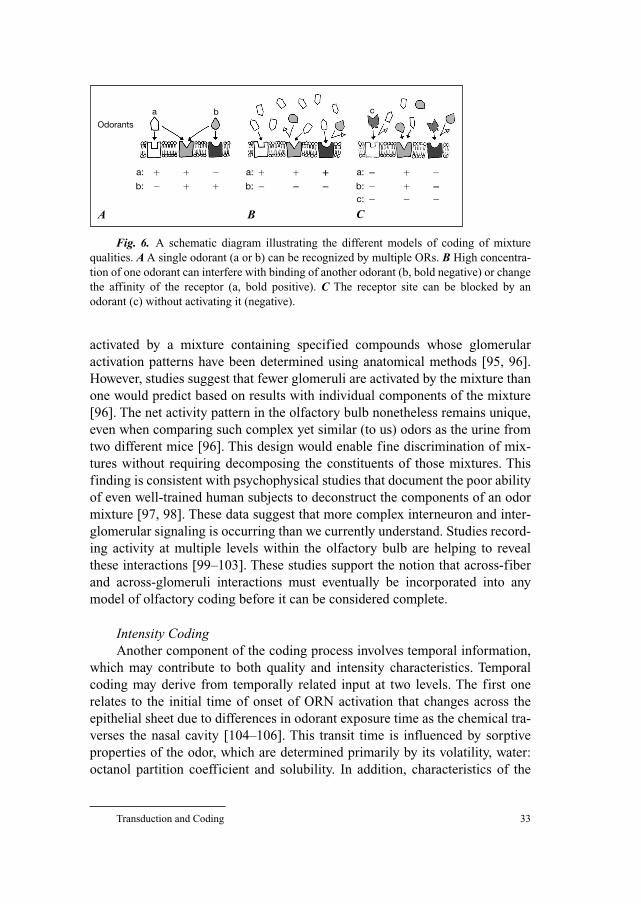
Transduction and Coding 33
activated by a mixture containing specified compounds whose glomerular
activation patterns have been determined using anatomical methods [95, 96].
However, studies suggest that fewer glomeruli are activated by the mixture than
one would predict based on results with individual components of the mixture
[96]. The net activity pattern in the olfactory bulb nonetheless remains unique,
even when comparing such complex yet similar (to us) odors as the urine from
two different mice [96]. This design would enable fine discrimination of mix-
tures without requiring decomposing the constituents of those mixtures. This
finding is consistent with psychophysical studies that document the poor ability
of even well-trained human subjects to deconstruct the components of an odor
mixture [97, 98]. These data suggest that more complex interneuron and inter-
glomerular signaling is occurring than we currently understand. Studies record-
ing activity at multiple levels within the olfactory bulb are helping to reveal
these interactions [99–103]. These studies support the notion that across-fiber
and across-glomeruli interactions must eventually be incorporated into any
model of olfactory coding before it can be considered complete.
Intensity CodingAnother component of the coding process involves temporal information,
which may contribute to both quality and intensity characteristics. Temporal
coding may derive from temporally related input at two levels. The first one
relates to the initial time of onset of ORN activation that changes across the
epithelial sheet due to differences in odorant exposure time as the chemical tra-
verses the nasal cavity [104–106]. This transit time is influenced by sorptive
properties of the odor, which are determined primarily by its volatility, water:
octanol partition coefficient and solubility. In addition, characteristics of the
Fig. 6. A schematic diagram illustrating the different models of coding of mixture
qualities. A A single odorant (a or b) can be recognized by multiple ORs. B High concentra-
tion of one odorant can interfere with binding of another odorant (b, bold negative) or change
the affinity of the receptor (a, bold positive). C The receptor site can be blocked by an
odorant (c) without activating it (negative).
Odorants
a: � � �
� � �b:
a: � � �
� � �b:
a: � � �
� � �b:� � �c:
a
A B C
b c
Rawson/Yee 34
olfactory organ, such as nasal structure, mucus composition and depth and air-
flow rate are critical factors influencing the temporal pattern of odor delivery to
the ORNs [107]. These latter characteristics can be dramatically influenced by
nasal sinus disease, injury or surgery or medications that alter hydration,
but these effects are difficult to predict precisely. Studies underway are model-
ing the impact of airflow and odorant sorptive properties on odor deposition
[105, 108] and will help to quantify and predict the impact of these factors on
olfaction.
The neural coding of intensity is dependent on temporal characteristics
built into the neural firing pattern. Aspects of odorant modulation of firing rates
may also contribute to quality coding, but this remains controversial [83, 84].
Nerve recordings indicate that ORNs may exhibit some baseline rate of action
potential firing. When stimulated, this baseline rate can increase or decrease
and the increase may involve bursts of activity or more tonic increases. The
neurophysiological mechanisms responsible for these activity patterns involve
the precisely regulated activity of voltage-gated sodium, potassium and calcium
channels as well as intermediate transduction elements. Many medications and
diseases can alter the functioning of these elements. The implication of this
biology is that a variety of conditions may alter olfactory performance more
subtly than simply by reducing sensitivity. For instance, changes could also
occur in perceived odor quality or rate of adaptation, and effects may be more
evident for some odors than others depending on their water solubility or
volatility. The clinician should be aware of such potential complex effects that
may have a significant impact on the patient’s quality of life and olfactory expe-
rience, but may be more difficult for the patient to describe and may not be
revealed by a simple test of odor sensitivity.
Pathology
A wide variety of conditions can lead to olfactory impairment. These con-
ditions may induce olfactory loss due to conductive changes such as congestion
or airflow blockage, and/or through effects on the neural and perineural processes
necessary for proper ORN function (‘sensorineural’ factors).
AgingSensory loss is a common age-related complaint, and may be due to
changes in the anatomy of the structure (e.g. loss of OR cells) or the environ-
ment surrounding the receptor cell (e.g. altered nasal mucus composition).
However, aging, as well as age-related diseases and medications may also alter
the distribution, density or function of specific receptor proteins, ion channels
Transduction and Coding 35
or signaling molecules that affect the ability of the ORN to function and signal
odorant information. For instance, age-related increases in calcium channel
density [109] and in the activity of several transduction elements including AC
[110], phospholipase C [111, 112], and PKC [113] have been reported, but have
not been specifically studied in the olfactory system. Changes such as these, if
occurring in the ORNs or olfactory bulb interneurons, could result in chronic
activation of calcium-dependent desensitization processes (see above). Studies
in our laboratory suggest that with age, individual ORNs become less selective
[2, 114], responding to a broader array of odors than do ORNs from younger
subjects. The cellular basis for this functional difference is not yet clear, but the
impact on perception would be to degrade the signal: noise level at the olfactory
bulb and impair odor discrimination. These kinds of neurophysiological
changes could also account for faster adaptation and delayed resensitization
reported among the elderly [115].
Impact of InflammationA variety of pathological conditions such as chronic sinusitis, viral infec-
tion, chemical exposure, or allergic rhinitis [116–119] result in inflammation in
the nasal cavity that can have acute and long-lasting effects on olfaction. In
addition to the acute impact of airway congestion due to injury, secretions and
tissue edema (‘conductive’ factors), a host of chemical signals related to the
inflammatory process can have short- and long-term consequences on olfactory
epithelial structure and function. At least a few of these inflammatory processes
have the potential to directly influence the function of the ORNs or other neural
components of the olfactory system.
When faced with injury or infection, a cascade of cellular and molecular
events is set into motion to remove damaged cells and stimulate repair. These
events may directly or indirectly influence odor detection and signal transduc-
tion. White blood cells migrate into the affected area and become activated in
response to chemicals released from the injured tissue. The infiltrating cells
release a host of pro- and anti-inflammatory chemical mediators including
cytokines, chemokines, leukotrienes and lymphokines that act through a com-
plex scenario of signaling pathways to modulate the expression of genes [120]
aimed at promoting tissue repair and recovery. Many of these mediators can
have effects on a wide array of cell types, including olfactory neurons and
related cells in the nasal mucosa, and studies have only begun to explore these
effects. For instance, interleukin-1� can stimulate process outgrowth in cul-
tured olfactory neurons [121], while tumor necrosis factor-� triggers apoptosis
in the OE [122, 123]. Inflammatory processes also result in elevated production
of two gaseous second messengers that may directly affect ORN function: nitric
oxide (NO) and carbon monoxide (CO). NO is produced by macrophages in
Rawson/Yee 36
response to activation and can quickly permeate cell membranes [124]. Nasal
and expired NO is elevated in chronic rhinosinusitis and has been explored as
an index of disease severity in allergic rhinitis and cystic fibrosis [125, 126].
CO is produced by heme oxygenase-1, which confers protection against oxida-
tive stress, and is induced by a variety of lymphokines, including interleukin-1
and tumor necrosis factor-� [127]. Both NO and CO have been shown to stim-
ulate cyclic GMP production in ORNs [46, 128], and to lead to activation of
identical types of electrical activity [129]. Thus, both gasses may lead to
chronic calcium influx in ORNs via the cNc and thus lead to desensitization or
long-term adaptation of the olfactory transduction pathway [130] as well as
shortening of ORN life span due to chronic calcium influx. Elevated levels of
these inflammatory mediators may linger beyond overt recovery of the nasal
epithelium and therapeutic approaches are needed that target the underlying
inflammatory process. The success of leukotriene inhibitors in treating asthma
has led to exploration of their use in polyposis and allergic and chronic nasal
sinus disease [131]. While some data are encouraging, a better understanding of
the inflammatory process in the nasal epithelium is needed to identify the
most relevant targets for successful drug therapy for the various pathological
conditions.
Medications and Olfactory TransductionA large number of medications have been reported to influence olfactory
function [132–134]. Some of these may impact nasal mucus secretion or blood
flow, while others may directly influence the neuronal signaling mechanisms.
Medications such as calcium channel blockers or dopaminergic drugs can
directly alter the functioning of the ORN or the interneurons in the olfactory
bulb. Dopamine D2 receptors are present on ORNs [135] and dopamine can
modulate activity of peripheral and central olfactory neurons [136–139]. The
excitatory neurotransmitter of the ORN is glutamate, and GABA is an inhibitory
neurotransmitter active at both primary and secondary synapses within the
olfactory bulb (fig. 5). In addition, receptors for many other neurotransmitters
and neuromodulators, including estrogen and insulin, are present in the olfac-
tory bulb [140, 141] and medications influencing their levels in the central
nervous system could alter olfactory performance. A variety of medications
include olfactory disturbances in their list of side effects (see Doty et al. [134]
and Koster et al. [135] for comprehensive reviews). Those listed include ACE
inhibitors, calcium channel blockers and a number of antiarrhythmics. These
lists are likely to underestimate the true number of medications with olfac-
tory consequences, and do not take into account the compounded effect of mul-
tiple medications taken concurrently. Both direct effects on transduction and
nerve function, as well as indirect effects due to interference with the epithelial
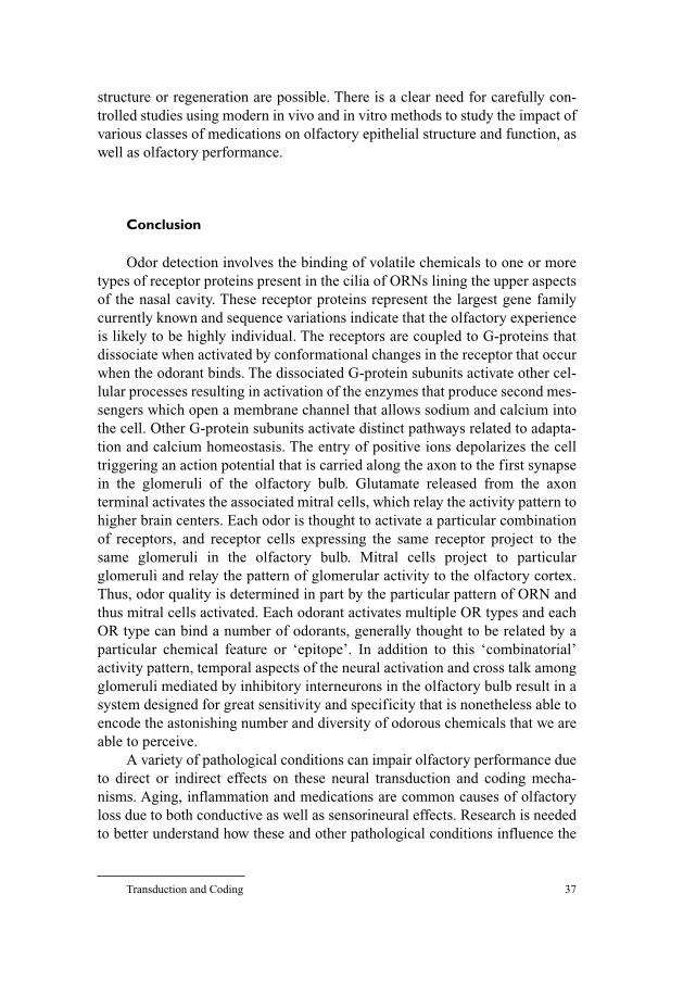
Transduction and Coding 37
structure or regeneration are possible. There is a clear need for carefully con-
trolled studies using modern in vivo and in vitro methods to study the impact of
various classes of medications on olfactory epithelial structure and function, as
well as olfactory performance.
Conclusion
Odor detection involves the binding of volatile chemicals to one or more
types of receptor proteins present in the cilia of ORNs lining the upper aspects
of the nasal cavity. These receptor proteins represent the largest gene family
currently known and sequence variations indicate that the olfactory experience
is likely to be highly individual. The receptors are coupled to G-proteins that
dissociate when activated by conformational changes in the receptor that occur
when the odorant binds. The dissociated G-protein subunits activate other cel-
lular processes resulting in activation of the enzymes that produce second mes-
sengers which open a membrane channel that allows sodium and calcium into
the cell. Other G-protein subunits activate distinct pathways related to adapta-
tion and calcium homeostasis. The entry of positive ions depolarizes the cell
triggering an action potential that is carried along the axon to the first synapse
in the glomeruli of the olfactory bulb. Glutamate released from the axon
terminal activates the associated mitral cells, which relay the activity pattern to
higher brain centers. Each odor is thought to activate a particular combination
of receptors, and receptor cells expressing the same receptor project to the
same glomeruli in the olfactory bulb. Mitral cells project to particular
glomeruli and relay the pattern of glomerular activity to the olfactory cortex.
Thus, odor quality is determined in part by the particular pattern of ORN and
thus mitral cells activated. Each odorant activates multiple OR types and each
OR type can bind a number of odorants, generally thought to be related by a
particular chemical feature or ‘epitope’. In addition to this ‘combinatorial’
activity pattern, temporal aspects of the neural activation and cross talk among
glomeruli mediated by inhibitory interneurons in the olfactory bulb result in a
system designed for great sensitivity and specificity that is nonetheless able to
encode the astonishing number and diversity of odorous chemicals that we are
able to perceive.
A variety of pathological conditions can impair olfactory performance due
to direct or indirect effects on these neural transduction and coding mecha-
nisms. Aging, inflammation and medications are common causes of olfactory
loss due to both conductive as well as sensorineural effects. Research is needed
to better understand how these and other pathological conditions influence the
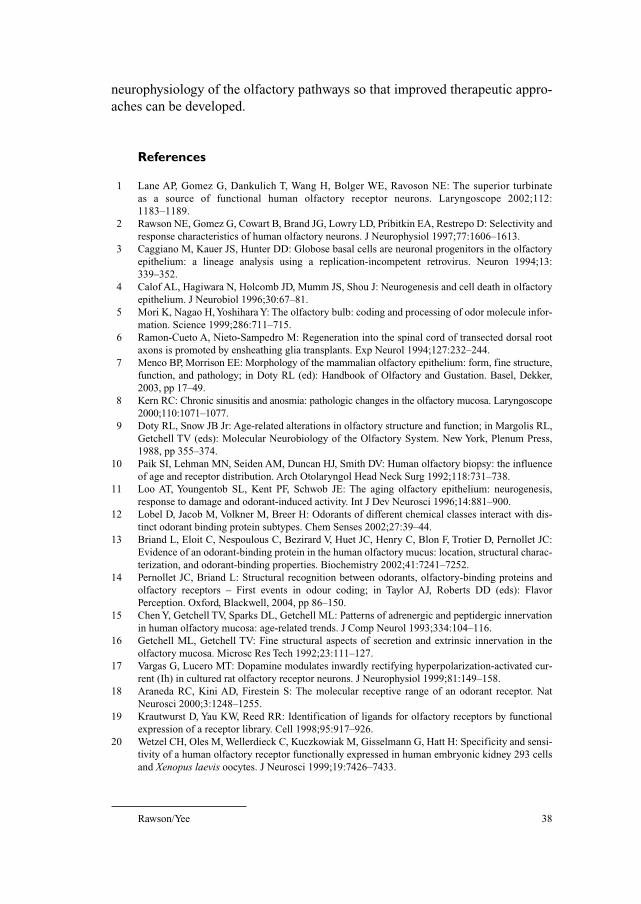
Rawson/Yee 38
neurophysiology of the olfactory pathways so that improved therapeutic appro-
aches can be developed.
References
1 Lane AP, Gomez G, Dankulich T, Wang H, Bolger WE, Ravoson NE: The superior turbinate
as a source of functional human olfactory receptor neurons. Laryngoscope 2002;112:
1183–1189.
2 Rawson NE, Gomez G, Cowart B, Brand JG, Lowry LD, Pribitkin EA, Restrepo D: Selectivity and
response characteristics of human olfactory neurons. J Neurophysiol 1997;77:1606–1613.
3 Caggiano M, Kauer JS, Hunter DD: Globose basal cells are neuronal progenitors in the olfactory
epithelium: a lineage analysis using a replication-incompetent retrovirus. Neuron 1994;13:
339–352.
4 Calof AL, Hagiwara N, Holcomb JD, Mumm JS, Shou J: Neurogenesis and cell death in olfactory
epithelium. J Neurobiol 1996;30:67–81.
5 Mori K, Nagao H, Yoshihara Y: The olfactory bulb: coding and processing of odor molecule infor-
mation. Science 1999;286:711–715.
6 Ramon-Cueto A, Nieto-Sampedro M: Regeneration into the spinal cord of transected dorsal root
axons is promoted by ensheathing glia transplants. Exp Neurol 1994;127:232–244.
7 Menco BP, Morrison EE: Morphology of the mammalian olfactory epithelium: form, fine structure,
function, and pathology; in Doty RL (ed): Handbook of Olfactory and Gustation. Basel, Dekker,
2003, pp 17–49.
8 Kern RC: Chronic sinusitis and anosmia: pathologic changes in the olfactory mucosa. Laryngoscope
2000;110:1071–1077.
9 Doty RL, Snow JB Jr: Age-related alterations in olfactory structure and function; in Margolis RL,
Getchell TV (eds): Molecular Neurobiology of the Olfactory System. New York, Plenum Press,
1988, pp 355–374.
10 Paik SI, Lehman MN, Seiden AM, Duncan HJ, Smith DV: Human olfactory biopsy: the influence
of age and receptor distribution. Arch Otolaryngol Head Neck Surg 1992;118:731–738.
11 Loo AT, Youngentob SL, Kent PF, Schwob JE: The aging olfactory epithelium: neurogenesis,
response to damage and odorant-induced activity. Int J Dev Neurosci 1996;14:881–900.
12 Lobel D, Jacob M, Volkner M, Breer H: Odorants of different chemical classes interact with dis-
tinct odorant binding protein subtypes. Chem Senses 2002;27:39–44.
13 Briand L, Eloit C, Nespoulous C, Bezirard V, Huet JC, Henry C, Blon F, Trotier D, Pernollet JC:
Evidence of an odorant-binding protein in the human olfactory mucus: location, structural charac-
terization, and odorant-binding properties. Biochemistry 2002;41:7241–7252.
14 Pernollet JC, Briand L: Structural recognition between odorants, olfactory-binding proteins and
olfactory receptors – First events in odour coding; in Taylor AJ, Roberts DD (eds): Flavor
Perception. Oxford, Blackwell, 2004, pp 86–150.
15 Chen Y, Getchell TV, Sparks DL, Getchell ML: Patterns of adrenergic and peptidergic innervation
in human olfactory mucosa: age-related trends. J Comp Neurol 1993;334:104–116.
16 Getchell ML, Getchell TV: Fine structural aspects of secretion and extrinsic innervation in the
olfactory mucosa. Microsc Res Tech 1992;23:111–127.
17 Vargas G, Lucero MT: Dopamine modulates inwardly rectifying hyperpolarization-activated cur-
rent (Ih) in cultured rat olfactory receptor neurons. J Neurophysiol 1999;81:149–158.
18 Araneda RC, Kini AD, Firestein S: The molecular receptive range of an odorant receptor. Nat
Neurosci 2000;3:1248–1255.
19 Krautwurst D, Yau KW, Reed RR: Identification of ligands for olfactory receptors by functional
expression of a receptor library. Cell 1998;95:917–926.
20 Wetzel CH, Oles M, Wellerdieck C, Kuczkowiak M, Gisselmann G, Hatt H: Specificity and sensi-
tivity of a human olfactory receptor functionally expressed in human embryonic kidney 293 cells
and Xenopus laevis oocytes. J Neurosci 1999;19:7426–7433.
Transduction and Coding 39
21 Touhara K: Functional cloning and reconstitution of vertebrate odorant receptors. Life Sci
2001;68:2199–2206.
22 Kajiya K, Inaki K, Tanaka M, Haga T, Kataoka H, Touhara K: Molecular bases of odor discrimina-
tion: reconstitution of olfactory receptors that recognize overlapping sets of odorants. J Neurosci
2001;21:6018–6025.
23 Touhara K: Functional cloning and reconstitution of vertebrate odorant receptors. Life Sci
2001;68:2199–2206.
24 Singer MS, Shepherd GM: Molecular modeling of ligand-receptor interactions in the OR5 olfac-
tory receptor. Neuroreport 1994;5:1297–1300.
25 Singer MS, Oliveira L, Vriend G, Shepherd GM: Potential ligand-binding residues in rat olfactory
receptors identified by correlated mutation analysis. Receptors Channels 1995;3:89–95.
26 Singer MS: Analysis of the molecular basis for octanal interactions in the expressed rat 17 olfac-
tory receptor. Chem Senses 2000;25:155–165.
27 McClintock TS, Landers TM, Gimelbrant AA, Fuller LZ, Jackson BA, Jayawickreme CK, Lerner
MR: Functional expression of olfactory-adrenergic receptor chimeras and intracellular retention
of heterologously expressed olfactory receptors. Brain Res Mol Brain Res 1997;48:270–278.
28 Dryer L: Evolution of odorant receptors. Bioessays 2000;22:803–810.
29 Sharon D, Glusman G, Pilpel Y, Khen M, Gruetzner F, Haaf T, Lancet D: Primate evolution of an
olfactory receptor cluster: diversification by gene conversion and recent emergence of pseudo-
genes. Genomics 1999;61:24–36.
30 Fuchs T, Glusman G, Horn-Saban S, Lancet D, Pilpel Y: The human olfactory subgenome: from
sequence to structure and evolution. Hum Genet 2001;108:1–13.
31 Sharon D, Gilad Y, Glusman G, Khen M, Lancet D, Kalush F: Identification and characterization
of coding single-nucleotide polymorphisms within a human olfactory receptor gene cluster. Gene
2000;260:87–94.
32 Gilad Y, Segre D, Skorecki K, Nachman MW, Lancet D, Sharon D: Dichotomy of single-
nucleotide polymorphism haplotypes in olfactory receptor genes and pseudogenes. Nat Genet
2000;26:221–224.
33 Gaillard I, Rouquier S, Chavanieu A, Mollard P, Giorgi D: Amino-acid changes acquired during
evolution by olfactory receptor 912–93 modify the specificity of odorant recognition. Hum Mol
Genet 2004;13:771–780.
34 Mombaerts P: Odorant receptor genes in humans. Curr Opin Genet Dev 1999;9:315–320.
35 Malnic B, Godfrey PA, Buck LB: The human olfactory receptor gene family. Proc Natl Acad Sci
USA 2004;101:2584–2589.
36 Godfrey PA, Malnic B, Buck LB: The mouse olfactory receptor gene family. Proc Natl Acad Sci
USA 2004;101:2156–2161.
37 Sosinsky A, Glusman G, Lancet D: The genomic structure of human olfactory receptor genes.
Genomics 2000;70:49–61.
38 Linardopoulou E, Mefford HC, Nguyen O, Friedman C, van den EG, Farwell DG, Coltrera M,
Trask BJ: Transcriptional activity of multiple copies of a subtelomerically located olfactory recep-
tor gene that is polymorphic in number and location. Hum Mol Genet 2001;10:2373–2383.
39 Hoppe R, Weimer M, Beck A, Breer H, Strotmann J: Sequence analyses of the olfactory receptor
gene cluster mOR37 on mouse chromosome 4. Genomics 2000;66:284–295.
40 Chess A, Simon I, Cedar H, Axel R: Allelic inactivation regulates olfactory receptor gene expres-
sion. Cell 1994;78:823–834.
41 Strotmann J, Wanner I, Helfrich T, Beck A, Breer H: Rostro-caudal patterning of receptor-expressing
olfactory neurones in the rat nasal cavity. Cell Tissue Res 1994;278:11–20.
42 Schild D, Restrepo D: Transduction mechanisms in vertebrate olfactory receptor cells. Physiol
Rev 1998;78:429–466.
43 Shepherd GM: Discrimination of molecular signals by the olfactory receptor neuron. Neuron
1994;13:771–790.
44 Gibson AD, Garbers DL: Guanylyl cyclases as a family of putative odorant receptors. Annu Rev
Neurosci 2000;23:417–439.
45 Lin W, Arellano J, Slotnick B, Restrepo D: Odors detected by mice deficient in cyclic nucleotide-
gated channel subunit A2 stimulate the main olfactory system. J Neurosci 2004;24:3703–3710.
Rawson/Yee 40
46 Breer H, Klemm T, Boekhoff I: Nitric oxide mediated formation of cyclic GMP in the olfactory
system. Neuroreport 1992;3:1030–1032.
47 Kurahashi T: The response induced by intracellular cyclic AMP in isolated olfactory receptor cells
of the newt. J Physiol 1990;430:355–371.
48 Zufall F, Firestein S, Shepherd GM: Cyclic nucleotide-gated ion channels and sensory transduc-
tion in olfactory receptor neurons. Annu Rev Biophys Biomol Struct 1994;23:577–607.
49 Restrepo D, Okada Y, Teeter JH, Lowry LD, Cowart B: Human olfactory neurons respond to odor
stimuli with an increase in cytoplasmic Ca2�. Biophys J 1993;64:1961–1966.
50 Restrepo D, Okada Y, Teeter JH: Odorant-regulated Ca2� gradients in rat olfactory neurons. J Gen
Physiol 1993;102:907–924.
51 Tareilus E, Noe J, Breer H: Calcium signals in olfactory neurons. Biochim Biophys Acta
1995;1269:129–138.
52 Brunet LJ, Gold GH, Ngai J: General anosmia caused by a targeted disruption of the mouse olfac-
tory cyclic nucleotide-gated cation channel. Neuron 1996;17:681–693.
53 Boekhoff I, Tarelius E, Strotmann J, Breer H: Rapid activation of alternative second messenger
pathways in olfactory cilia from rats by different odorants. EMBO J 1990;9:2453–2458.
54 Breer H, Boekhoff I: Odorants of the same odor class activate different second messenger path-
ways. Chem Senses 1991;16:19–29.
55 Restrepo D, Teeter J, Honda E, Boyle A, Kalinoski DL, Marecek J, Prestwich GD: Evidence for an
InsP-gated channel protein in isolated rat olfactory cilia. Am J Physiol 1992;263:C667–C673.
56 Miyamoto T, Restrepo D, Cragoe J, Teeter J: IP3- and cAMP-induced responses in isolated olfac-
tory receptor neurons from the channel catfish. J Membr Biol 1992;127:173–183.
57 Honda E, Teeter JH, Restrepo D: InsP3-gated ion channels in rat olfactory cilia membrane. Brain
Res 1995;703:79–85.
58 Kaur R, Zhu XO, Moorhouse AJ, Barry PH: IP3-gated channels and their occurrence relative to
CNG channels in the soma and dendritic knob of rat olfactory receptor neurons. J Membr Biol
2001;181:91–105.
59 Okada Y, Fujiyama R, Miyamoto T, Sato T: Comparison of a Ca2�-gated conductance and a
second-messenger-gated conductance in rat olfactory neurons. J Exp Biol 2000;203:567–573.
60 Spehr M, Wetzel CH, Hatt H, Ache BW: 3-Phosphoinositides modulate cyclic nucleotide signal-
ing in olfactory receptor neurons: Neuron 2002;33:731–739.
61 Gomez G, Rawson NE, Cowart B, Lowry LD, Pribitkin EA, Restrepo D: Modulation of odor-
induced increases in [Ca2�]i by inhibitors of protein kinases A and C in rat and human olfactory
receptor neurons. Neuroscience 2000;98:181–189.
62 Morales B, Bacigalupo J: Chemical reception in vertebrate olfaction: evidence for multiple trans-
duction pathways. Biol Res 1996;29:333–341.
63 Schild D, Lischka FW, Restrepo D: InsP3 causes an increase in apical [Ca2�]i by activating two
distinct current components in vertebrate olfactory receptor cells. J Neurophysiol 1995;73:
862–866.
64 Lischka FW, Teeter JH, Restrepo D: Odorants suppress a voltage-activated K� conductance in rat
olfactory neurons. J Neurophysiol 1999;82:226–236.
65 Wetzel CH, Spehr M, Hatt H: Phosphorylation of voltage-gated ion channels in rat olfactory
receptor neurons. Eur J Neurosci 2001;14:1056–1064.
66 Hegg CC, Lucero MT: Dopamine reduces odor- and elevated-K�-induced calcium responses in
mouse olfactory receptor neurons in situ. J Neurophysiol 2004;91:1492–1499.
67 Okada Y, Miyamoto T, Toda K: Dopamine modulates a voltage-gated calcium channel in rat olfac-
tory receptor neurons. Brain Res 2003;968:248–255.
68 Reisert J, Bauer PJ, Yau KW, Frings S: The Ca-activated Cl channel and its control in rat olfactory
receptor neurons. J Gen Physiol 2003;122:349–363.
69 Restrepo D: The ins and outs of intracellular chloride in olfactory receptor neurons. Neuron
2005;45:481–482.
70 Kleene SJ, Gesteland RC: Calcium-activated chloride conductance in frog olfactory cilia.
J Neurosci 1991;11:3624–3629.
71 Kurahashi T, Yau KW: Co-existence of cationic and chloride components in odorant-induced cur-
rent of vertebrate olfactory receptor cells. Nature 1993;363:71–74.
Transduction and Coding 41
72 Kurahashi T, Yau KW: Olfactory transduction. Tale of an unusual chloride current. Curr Biol
1994;4:256–258.
73 Menini A: Calcium signalling and regulation in olfactory neurons. Curr Opin Neurobiol 1999;9:
419–426.
74 Boekhoff I, Kroner C, Breer H: Calcium controls second-messenger signalling in olfactory cilia.
Cell Signal 1996;8:167–171.
75 Conneelly PA, Sisk RB, Schulman H, Garrison JC: Evidence for the activation of the multifunc-
tional Ca2�/calmodulin-dependent protein kinase in response to hormones that increase intracellu-
lar Ca2�. J Biol Chem 1987;262:10154–10163.
76 Boekhoff I, Breer H: Termination of second messenger signaling in olfaction. Proc Natl Acad Sci
USA 1992;89:471–474.
77 Schleicher S, Boekhoff I, Arriza J, Lefkowitz RJ, Breer H: A beta-adrenergic receptor kinase-
like enzyme is involved in olfactory signal termination. Proc Natl Acad Sci USA 1993;90:
1420–1424.
78 Peppel K, Boekhoff I, McDonald P, Breer H, Caron MG, Lefkowitz RJ: G protein-coupled recep-
tor kinase 3 (GRK3) gene disruption leads to loss of odorant receptor desensitization. J Biol Chem
1997;272:25425–25428.
79 Wei J, Zhao AZ, Chan GC, Baker LP, Impey S, Beavo JA, Storm DR: Phosphorylation and inhibi-
tion of olfactory adenylyl cyclase by CaM kinase II in neurons: a mechanism for attenuation of
olfactory signals. Neuron 1998;21:495–504.
80 Leinders-Zufall T, Ma M, Zufall F: Impaired odor adaptation in olfactory receptor neurons after
inhibition of Ca2�/calmodulin kinase II. J Neurosci 1999;19:RC19.
81 Medler KF, Bruch RC: Protein kinase Cbeta and delta selectively phosphorylate odorant and
metabotropic glutamate receptors. Chem Senses 1999;24:295–299.
82 Noe J, Tareilus E, Boekhoff I, Breer H: Sodium/calcium exchanger in rat olfactory neurons.
Neurochem Int 1997;30:523–531.
83 Rospars JP, Lansky P, Duchamp-Viret P, Duchamp A: Spiking frequency versus odorant concen-
tration in olfactory receptor neurons. Biosystems 2000;58:133–141.
84 Duchamp-Viret P, Duchamp A, Chaput MA: Peripheral odor coding in the rat and frog: quality and
intensity specification. J Neurosci 2000;20:2383–2390.
85 Kang J, Caprio J: In vivo responses of single olfactory receptor neurons in the channel catfish,
Ictalurus punctatus. J Neurophysiol 1995;73:172–177.
86 Delay R, Restrepo D: Odorant responses of dual polarity are mediated by cAMP in mouse olfac-
tory sensory neurons. J Neurophysiol 2004;92:1312–1319.
87 Morales B, Labarca P, Bacigalupo J: A ciliary K� conductance sensitive to charibdotoxin underlies
inhibitory responses in toad olfactory receptor neurons. FEBS Lett 1995;359:41–44.
88 Yee KK, Costanzo RM: Changes in odor quality discrimination following recovery from olfactory
nerve transection. Chem Senses 1998;23:513–519.
89 Christensen MD, Holbrook EH, Costanzo RM, Schwob JE: Rhinotopy is disrupted during the
re-innervation of the olfactory bulb that follows transection of the olfactory nerve. Chem Senses
2001;26:359–369.
90 Costanzo RM: Rewiring the olfactory bulb: changes in odor maps following recovery from nerve
transection. Chem Senses 2000;25:199–205.
91 Kern RC, Quinn B, Rosseau G, Farbman AI: Post-traumatic olfactory dysfunction. Laryngoscope
2000;110:2106–2109.
92 Kurahashi T, Lowe G, Gold GH: Suppression of odorant responses by odorants in olfactory recep-
tor cells. Science 1994;265:118–120.
93 Araneda RC, Peterlin Z, Zhang X, Chesler A, Firestein S: A pharmacological profile of the alde-
hyde receptor repertoire in rat olfactory epithelium. J Physiol 2004;555:743–756.
94 Linster C, Cleland TA: Configurational and elemental odor mixture perception can arise from
local inhibition. J Comput Neurosci 2004;16:39–47.
95 Johnson BA, Leon M: Modular representations of odorants in the glomerular layer of the rat olfac-
tory bulb and the effects of stimulus concentration. J Comp Neurol 2000;422:496–509.
96 Schaefer ML, Young DA, Restrepo D: Olfactory fingerprints for major histocompatibility complex-
determined body odors. J Neurosci 2001;21:2481–2487.
Rawson/Yee 42
97 Jinks A, Laing DG: A limit in the processing of components in odour mixtures. Perception
1999;28:395–404.
98 Jinks A, Laing DG: The analysis of odor mixtures by humans: evidence for a configurational
process. Physiol Behav 2001;72:51–63.
99 Trombley PQ, Shepherd GM: Synaptic transmission and modulation in the olfactory bulb. Cur
Opin Neurobiol 1993;3:540–547.
100 Shen GY, Chen WR, Midtgaard J, Shepherd GM, Hines ML: Computational analysis of action
potential initiation in mitral cell soma and dendrites based on dual patch recordings. J Neurophysiol
1999;82:3006–3020.
101 Luo M, Katz LC: Response correlation maps of neurons in the mammalian olfactory bulb. Neuron
2001;32:1165–1179.
102 McQuiston AR, Katz LC: Electrophysiology of interneurons in the glomerular layer of the rat
olfactory bulb. J Neurophysiol 2001;86:1899–1907.
103 Belluscio L, Katz LC: Symmetry, stereotypy, and topography of odorant representations in mouse
olfactory bulbs. J Neurosci 2001;21:2113–2122.
104 Mozell MM, Jagodowicz M: Mechanisms underlying the analysis of odorant quality at the
level of the olfactory mucosa. 1. Spatiotemporal sorption patterns. Ann NY Acad Sci 1974;237:
76–90.
105 Hahn I, Scherer PW, Mozell MM: A mass transport model of olfaction. J Theor Biol
1994;167:115–128.
106 Keyhani K, Scherer PW, Mozell MM: A numerical model of nasal odorant transport for the analysis
of human olfaction. J Theor Biol 1997;186:279–301.
107 Scott-Johnson PE, Blakley D, Scott JW: Effects of air flow on rat electroolfactogram. Chem
Senses 2000;25:761–768.
108 Zhao K, Scherer PW, Hajiloo SA, Dalton P: Effect of anatomy on human nasal air flow and odor-
ant transport patterns: implications for olfaction. Chem Senses 2004;29:365–379.
109 Mattson MP, Rydel RE, Lieberburg I, Smith-Swintosky V: Altered calcium signaling and neuronal
injury: stroke and Alzheimer’s disease as examples. Ann NY Acad Sci 1993;679:1–21.
110 Yamamoto M, Ozawa H, Saito T, Frolich L, Riederer P, Takahata N: Reduced immunoreactivity of
adenylyl cyclase in dementia of the Alzheimer type. Neuroreport 1996;7:2965–2970.
111 Joseph JA, Kowatch MA, Makui T, Roth GS: Selective cross-activation/inhibition of second mes-
senger systems and the reduction of age-related deficits in the muscarinic control of dopamine
release from perifused rat striata. Brain Res 1990;537:40–48.
112 Iren N, Pint A, Stef F, Jerz V, Hann M: Increased responsiveness of the cerebral cortical phos-
phatidylinositol system to noradrenaline and carbachol in senescent rats. Neurosci Lett 1989;107:
195–199.
113 Busquets X, Ventayol P, Sastre M, Garcia-Sevilla JA: Age-dependent increases in protein kinase
C-alpha beta immunoreactivity and activity in the human brain: possible in vivo modulatory
effects on guanine nucleotide regulatory G(i) proteins. Brain Res 1996;710:28–34.
114 Rawson NE, Gomez G, Cowart B, Restrepo D: The use of olfactory receptor neurons (ORNs)
from biopsies to study changes in aging and neurodegenerative diseases. Ann NY Acad Sci USA
1998;855:701–707.
115 Stevens JC, Cain WS, Schiet FT, Oatly MW: Olfactory adaptation and recovery in old age.
Perception 1989;18:265–276.
116 Yamagishi M, Fujiwara M, Nakamura H: Olfactory mucosal findings and clinical course in
patients with olfactory disorders following upper respiratory viral infection. Rhinology
1994;32:113–118.
117 Yamagishi M, Nakano Y: A re-evaluation of the classification of olfactory epithelia in patients
with olfactory disorders. Eur Arch Otorhinolaryngol 1992;249:393–399.
118 Yamagishi M, Nakamura H, Suzuki S, Hasegawa S, Nakano Y: Immunohistochemical examina-
tion of olfactory mucosa in patients with olfactory disturbance. Ann Otol Rhinol Laryngol
1990;99:205–210.
119 Church JA, Bauer H, Bellanti JA, Satterly RA, Henkin RI: Hyposmia associated with atopy. Ann
Allergy 1978;40:105–109.
120 Baraniuk JN: Mechanisms of rhinitis. Allergy Asthma Proc 1998;19:343–347.
Transduction and Coding 43
121 Vawter MP, Basaric-Keys J, Li Y, Lester DS, Lebovics RS, Lesch KP, Kulaga H, Freed WJ,
Sunderland T, Wolozin B: Human olfactory neuroepithelial cells: tyrosine phosphorylation and
process extension are increased by the combination of IL-1beta, IL-6, NGF, and bFGF. Exp Neurol
1996;142:179–194.
122 Farbman AI, Ezeh PI: TGF-alpha and olfactory marker protein enhance mitosis in rat olfactory
epithelium in vivo. Neuroreport 2000;11:3655–3658.
123 Suzuki Y, Farbman AI: Tumor necrosis factor-alpha-induced apoptosis in olfactory epithelium in
vitro: possible roles of caspase 1 (ICE), caspase 2 (ICH-1), and caspase 3 (CPP32). Exp Neurol
2000;165:35–45.
124 Moncada S: Nitric oxide in the vasculature: physiology and pathophysiology. Ann NY Acad Sci
1997;811:60–69.
125 Lefevere L, Willems T, Lindberg S, Jorissen M: Nasal nitric oxide. Acta Otorhinolaryngol Belg
2000;54:271–280.
126 Gilain L, Bedu M, Jouaville L, Guichard C, Advenier D, Mom T, Laurent S, Caillaud D: Analysis
of nasal and exhaled nitric oxide concentration in nasal polyposis. Ann Otolaryngol Chir
Cervicofac 2002;119:234–242.
127 Terry CM, Clikeman JA, Hoidal JR, Callahan KS: Effect of tumor necrosis factor-alpha and
interleukin-1 alpha on heme oxygenase-1 expression in human endothelial cells. Am J Physiol
1998;274:H883–H891.
128 Ingi T, Chiang G, Ronnett GV: The regulation of heme turnover and carbon monoxide biosynthe-
sis in cultured primary rat olfactory receptor neurons. J Neurosci 1996;16:5621–5628.
129 Lischka FW, Schild D: Effects of nitric oxide upon olfactory receptor neurones in Xenopus laevis.Neuroreport 1993;4:582–584.
130 Zufall F, Leinders-Zufall T: Identification of a long-lasting form of odor adaptation that depends
on the carbon monoxide/cGMP second-messenger system. J Neurosci 1997;17:2703–2712.
131 Parnes SM: The role of leukotriene inhibitors in allergic rhinitis and paranasal sinusitis. Curr
Allergy Asthma Rep 2002;2:239–244.
132 Elsner RJ: Environment and medication use influence olfactory abilities of older adults. J Nutr
Health Aging 2001;5:5–10.
133 Ackerman BH, Kasbekar N: Disturbances of taste and smell induced by drugs. Pharmacotherapy
1997;17:482–496.
134 Doty RL, Philip S, Reddy K, Kerr KL: Influences of antihypertensive and antihyperlipidemic
drugs on the senses of taste and smell: a review. J Hypertens 2003;21:1805–1813.
135 Koster NL, Norman AB, Richtand NM, Nickell WT, Puche AC, Pixley SK, Shipley MT: Olfactory
receptor neurons express D2 dopamine receptors. J Comp Neurol 1999;411:666–673.
136 Hsia AY, Vincent JD, Lledo PM: Dopamine depresses synaptic inputs into the olfactory bulb.
J Neurophysiol 1999;82:1082–1085.
137 Brunig I, Sommer M, Hatt H, Bormann J: Dopamine receptor subtypes modulate olfactory bulb
gamma-aminobutyric acid type A receptors. Proc Natl Acad Sci USA 1999;96:2456–2460.
138 Zhang JJ, Okutani F, Yagi F, Inoue S, Kaba H: Facilitatory effect of ritanserin is mediated by
dopamine D1 receptors on olfactory learning in young rats. Dev Psychobiol 2000;37:246–252.
139 Berkowicz DA, Trombley PQ: Dopaminergic modulation at the olfactory nerve synapse. Brain
Res 2000;855:90–99.
140 Macrides F, Schoenfeld TA, Marchand JE, Clancy AN: Evidence for morphologically, neurochem-
ically and functionally heterogeneous classes of mitral and tufted cells in the olfactory bulb. Chem
Senses 1985;10:175–202.
141 Halasz N, Shepherd GM: Neurochemistry of the vertebrate olfactory bulb. Neuroscience 1983;10:
579–619.
Nancy E. Rawson, PhD
Monell Chemical Senses Center
3500 Market Street
Philadelphia, PA 19104–3308 (USA)
Tel. �1 215 898 0943, Fax �1 215 898 2084, E-Mail [email protected]
Hummel T, Welge-Lüssen A (eds): Taste and Smell. An Update.
Adv Otorhinolaryngol. Basel, Karger, 2006, vol 63, pp 44–69
Smell: Central Nervous Processing
Jay A. Gottfried
Northwestern University Feinberg School of Medicine, Cognitive Neurology and
Alzheimer’s Disease Center, Chicago, Ill., USA
AbstractThis chapter focuses on central olfactory processing in the human brain. As the psy-
chophysiology of human olfactory function is important for appreciating its underlying neuro-
physiology, the chapter will begin with a brief overview of what the human nose can do,
contesting notions that human olfaction is a second-rate system. It will be followed by an
anatomical survey of the principal recipients of olfactory bulb input, with some comments on the
unique organizing properties that distinguish olfaction from other sensory modalities. The final
section will cover the neural correlates of human olfactory function, including aspects of basic
chemosensory processing (odor detection, sniffing, intensity, valence) and higher-order olfactory
operations (learning, memory, crossmodal integration), with particular emphasis on functional
imaging data, though human lesion studies and intracranial recordings will also be discussed.
Copyright © 2006 S. Karger AG, Basel
Historically the clinical science of smell has received scant attention com-
pared to other sensory modalities. In neurology, the olfactory system goes virtu-
ally without mention, and textbooks are quick to point out that the first cranial
nerve is of little diagnostic utility. For many practitioners in otolaryngology, their
acquaintance with olfactory structures is limited to the visible surface of the
olfactory epithelial sheet, having no reason to tread beyond the cribriform plate.
Grinker’s Neurology [1], a classic textbook of its day (1943), introduced the topic
of human olfaction by emphasizing ‘the extremely rudimentary sense of smell
and the insignificant role it plays in man’s existence’ (p 337). Several decades
later, the conventional wisdom had barely changed: ‘From a clinical point of view
the importance of the olfactory system is slight, just as the sense of smell is of rel-
atively minor importance in the normal life of civilized man’ [2] (p 509).
This clinical state of affairs is echoed by an evolutionary snub. So the the-
ory goes, human olfaction became a subpar ‘microsmatic’ sense precisely at the
moment long ago when we first learned how to walk upright. By bringing our

Central Olfactory Processing 45
noses up off the odor-rich ground, our bipedal station deprived us of nature’s
best scents, and a phylogenetic regression ensued (discussed in Shepherd [3]).
The relatively smaller sizes of human olfactory structures, along with the
decline in functioning human olfactory receptor genes [4, 5], are cited as bio-
logical consequences of this evolutionary upheaval. In contrast, for ‘macros-
matic’ quadrupeds like dogs, cats, and rodents, the sense of smell remained a
biological imperative, governing a wide variety of behaviors important for sur-
vival, namely those related to food, sex, and threat. As such, evolutionary pres-
sure has ensured olfactory preeminence in these ground-dwelling species.
Part of the historical indifference to human olfaction has been due to the
technical challenges of working with odorous stimuli and of measuring brain
activity with sufficient spatiotemporal detail. However, with recent experimental
advances, many of these problems have become soluble, and a whole new set of
research questions, more ambitious and more sophisticated, can now be applied
to the olfactory domain. As a result, it has become evident that the olfactory sys-
tem is well-suited to addressing key neurobiological questions. Importantly, the
fact that many neurodegenerative disorders, including Alzheimer’s disease and
Parkinson’s disease, are typified in their early stages by olfactory deficits [6–8]
means that a scientific knowledge of the functional organization of central olfac-
tion in healthy subjects may enlighten our understanding of these conditions.
This chapter focuses on central olfactory processing in the human brain.
As the psychophysiology of human olfactory function is important for appreci-
ating its underlying neurophysiology, the chapter will begin with a brief and
biased overview of what the human nose can do, contesting notions that human
olfaction is a second-rate system. It will be followed by an anatomical survey of
the principal recipients of olfactory bulb input, with some comments on the
unique organizing properties that distinguish olfaction from other sensory
modalities. The final section will cover the neural correlates of human olfactory
function, including aspects of basic chemosensory processing (odor detection,
sniffing, intensity, valence) and higher-order olfactory operations (learning,
memory, crossmodal integration), with particular emphasis on functional imag-
ing data, though human lesion studies and intracranial recordings will also be
discussed.
What Can Your Nose Do?
Perception and Discrimination
Human olfactory perception is surprisingly good, and in certain instances
better than in species with their noses closer to the ground. A comparison of

Gottfried 46
olfactory detection thresholds across several mammalian species indicates that
for certain simple monomolecular compounds, human thresholds are lower
(more sensitive) than the corresponding thresholds in rats, as well as in nonhu-
man primates [9]. Notably, human detection thresholds for many odors are
commonly in the parts-per-billion range [10]. Moreover, a series of elegant psy-
chophysical studies by Laska and Seibt [9], Laska and Teubner [11], Laska et al.
[12] and Laska and Hubener [13] has shown that humans can readily discrimi-
nate between two different odors that differ by a single molecular component.
For example, humans have no problem distinguishing aliphatic aldehydes of a
4- vs. 5-carbon chain length, otherwise matched for intensity, and in the
absence of other physicochemical differences [11]. Finally, it is estimated that
humans can distinguish thousands of different smells, although such powers of
discrimination are generally not matched by verbal skills, and when a human
subject is asked to name an odor, performance tends to break down [14].
Nevertheless, this verbal limitation does not invalidate the idea that human
olfactory perception is highly sensitive and specific.
Behavioral Modulation
In the animal kingdom, behavior is inevitably shaped by the biological
salience of stimuli encountered in the environment. Odors can have a powerful
impact on behavioral and motivational states, by virtue of their associations
with threat (predators), food, and sexual gratification. Bombykol, a volatile
pheromone secreted by female silkworm moths, will drive potential mates
miles upwind in hot pursuit [15]. Despite an apparent phylogenetic decline,
there is evidence to suggest that odors are also capable of modulating human
behavior. Human infants discriminate their own mothers’ odors by postnatal
day 6 [16] and are more likely to initiate sucking specifically in response to
these stimuli by 6 weeks of age [17]. The use of perfume can affect social
interactions by influencing subjective impressions of its wearer by others [18],
and odor signals may be favored over visual cues in dictating food prefer-
ences [19]. Odor components in a woman’s axillary sweat can synchronize the
ovulatory cycles of other female subjects [20, 21], suggesting that neuro-
endocrine states are sensitive to olfactory cues. A related work indicates that
the major histocompatibility complex genotype determines an individual’s
body odor and body odor preference [22, 23], and moreover that a woman’s
preference of male body odors is defined by major histocompatibility complex
haplotype inheritance patterns [24], all of which may influence mating choice
in humans [25].
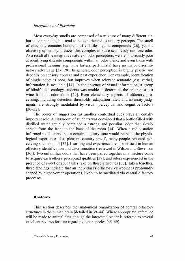
Central Olfactory Processing 47
Integration and Plasticity
Most everyday smells are composed of a mixture of many different air-
borne components, but tend to be experienced as unitary percepts. The smell
of chocolate contains hundreds of volatile organic compounds [26], yet the
olfactory system synthesizes this complex mixture seamlessly into one odor.
As a result of the integrative nature of odor perception, we are notoriously poor
at identifying discrete components within an odor blend, and even those with
professional training (e.g. wine tasters, perfumists) have no major discrimi-
natory advantage [27, 28]. In general, odor perception is highly plastic and
depends on sensory context and past experience. For example, identification
of single odors is poor, but improves when relevant semantic (e.g. verbal)
information is available [14]. In the absence of visual information, a group
of blindfolded enology students was unable to determine the color of a test
wine from its odor alone [29]. Even elementary aspects of olfactory pro-
cessing, including detection thresholds, adaptation rates, and intensity judg-
ments, are strongly modulated by visual, perceptual and cognitive factors
[30–33].
The power of suggestion (as another contextual cue) plays an equally
important role. A classroom of students was convinced that a bottle filled with
distilled water actually contained a ‘strong and peculiar’ odor that slowly
spread from the front to the back of the room [34]. When a radio station
informed its listeners that a certain auditory tone would recreate the physio-
logical experience of a ‘pleasant country smell’, many people reported per-
ceiving such an odor [35]. Learning and experience are also critical in human
olfactory identification and discrimination (reviewed in Wilson and Stevenson
[36]). Two unfamiliar odors that have been paired together in a mixture come
to acquire each other’s perceptual qualities [37], and odors experienced in the
presence of sweet or sour tastes take on those attributes [38]. Taken together,
these findings indicate that an individual’s olfactory viewpoint is profoundly
shaped by higher-order operations, likely to be mediated via central olfactory
processes.
Anatomy
This section describes the anatomical organization of central olfactory
structures in the human brain [detailed in 39–44]. Where appropriate, reference
will be made to animal data, though the interested reader is referred to several
excellent reviews for data regarding other species [45–49].
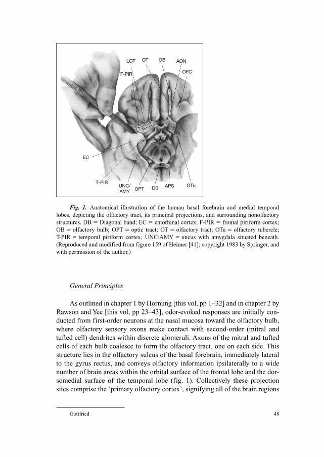
Gottfried 48
General Principles
As outlined in chapter 1 by Hornung [this vol, pp 1–32] and in chapter 2 by
Rawson and Yee [this vol, pp 23–43], odor-evoked responses are initially con-
ducted from first-order neurons at the nasal mucosa toward the olfactory bulb,
where olfactory sensory axons make contact with second-order (mitral and
tufted cell) dendrites within discrete glomeruli. Axons of the mitral and tufted
cells of each bulb coalesce to form the olfactory tract, one on each side. This
structure lies in the olfactory sulcus of the basal forebrain, immediately lateral
to the gyrus rectus, and conveys olfactory information ipsilaterally to a wide
number of brain areas within the orbital surface of the frontal lobe and the dor-
somedial surface of the temporal lobe (fig. 1). Collectively these projection
sites comprise the ‘primary olfactory cortex’, signifying all of the brain regions
F-PIR
LOT OT OB AON
OFC
EC
T-PIRUNC/AMY OPT DB APS OTu
Fig. 1. Anatomical illustration of the human basal forebrain and medial temporal
lobes, depicting the olfactory tract, its principal projections, and surrounding nonolfactory
structures. DB � Diagonal band; EC � entorhinal cortex; F-PIR � frontal piriform cortex;
OB � olfactory bulb; OPT � optic tract; OT � olfactory tract; OTu � olfactory tubercle;
T-PIR � temporal piriform cortex; UNC/AMY � uncus with amygdala situated beneath.
(Reproduced and modified from figure 159 of Heimer [41]; copyright 1983 by Springer, and
with permission of the author.)
Central Olfactory Processing 49
receiving direct bulbar input [50, 51]. Notably, the highly ordered chemotopic
organization in the olfactory bulb [see chapter 2 by Rawson and Yee, this vol,
pp 23–43] does not appear to be systematically maintained in central brain
regions. In rats, a given area of the olfactory bulb projects extensively through-
out the olfactory cortex, and a given area of the cortex receives projections from
widespread areas of the bulb [52]. On the other hand, a recent genetic tracer
study in rodents indicates that a given olfactory receptor subtype projects to dis-
crete neuronal clusters within the olfactory cortex, suggesting a certain topo-
graphical preservation [53].
As the olfactory tract courses posteriorly, collateral branches peel off and
synapse upon the anterior olfactory nucleus (AON), a collection of cell clusters
scattered along the caudal extent of the tract and the caudomedial orbital cortex.
While histologically variable in humans, this region appears to be a target of
pathology in both Parkinson’s disease [54] and multiple system atrophy [55]. In
animals, projections between one AON and its contralateral partner, via the
anterior commissure, provide the major route of olfactory information transfer
between hemispheres, though such contralateral pathways have not been docu-
mented in humans. Upon nearing the entry point to the brain (at the so-called
olfactory trigone), the olfactory tract is generally thought to divide into lateral,
intermediate, and medial olfactory striae. Although well-documented in ani-
mals, the intermediate and medial branches are extremely rudimentary in
humans and seldom evident histologically [39, 56]. Thus, the lateral olfactory
tract (LOT) apparently provides the only source of bulbar afferents to the
human brain [40].
The LOT deviates laterally around the rostral edge of the anterior perforated
substance (APS), and then makes a sharp bend caudally onto the medial tempo-
ral surface (uncus). Principal recipients include the piriform cortex, amygdala,
and rostral entorhinal cortex (fig. 1), all of which are substantially intercon-
nected via associational intracortical fiber systems (fig. 2). Another target of the
LOT input is the olfactory tubercle, which in animals forms a prominent bump,
but in humans is difficult to visualize. It is thought to be situated along the
posterior-most segment of the medial orbital cortex within the APS and may be
a derivative of the pallial striatum [57]. Other regions of the basal forebrain, such
as the taenia tecta, indusium griseum, anterior hippocampal continuation, and
the nucleus of the horizontal diagonal band, have been shown to receive direct
bulbar input in animal models [45, 58], but whether similar connections are pre-
served in humans is unknown.
Higher-order projections arising from each of these olfactory structures con-
verge on the orbital prefrontal cortex, agranular insula, other amygdala subnuclei,
thalamus, hypothalamus, basal ganglia, and hippocampus [47] (fig. 2). Together
this complex network of connections provides the basis for odor-guided regulation
Gottfried 50
of behavior, feeding, emotion, autonomic states, and memory. In addition, each
region of the primary olfactory cortex (apart from the olfactory tubercle) sends
dense feedback projections to the olfactory bulb [45], supplying numerous physio-
logical routes for central or ‘top-down’ modulation of olfactory information
processing as early as the second-order neuron in the olfactory hierarchy. What
follows below is a more detailed anatomical description regarding several key
regions involved in human olfaction.
Olfactory mucosa
Olfactorybulb
LOT
AON
Contralat.AON (?)
OFCalNSMD
BLAOFCMDHYP
PRHPC
VP, VSMDOFC
PIR PAC/ACo EC OTu
Fig. 2. Schematic diagram of the major olfactory pathways [based on refs. 39, 40,
45–48]. Regions in gray together represent the primary olfactory cortex. Projections between
the olfactory bulb and most areas of the primary olfactory cortex are bidirectional, with the
exception of the olfactory tubercle (OTu). Similarly, associational connections between the pri-
mary olfactory cortex subregions are reciprocal, apart from OTu. The downstream targets of the
primary olfactory cortex represent some of the major projection sites (bottom of figure), many
of which provide feedback to the primary olfactory cortex (not shown), but these connections
are not meant to be comprehensive or all-inclusive. While broadly illustrative of the human
olfactory system, this diagram is largely based on information obtained from animal models,
due to the scarcity of human data. ACo � Anterior cortical nucleus of the amygdala;
aINS � agranular insula; BLA � basolateral nucleus of the amygdala; EC � entorhinal
cortex; HPC � hippocampus; HYP � hypothalamus; MD � mediodorsal thalamus;
PAC � periamygdaloid cortex; PIR � piriform cortex; PR � perirhinal cortex; VP � ventral
pallidum; VS � ventral striatum.
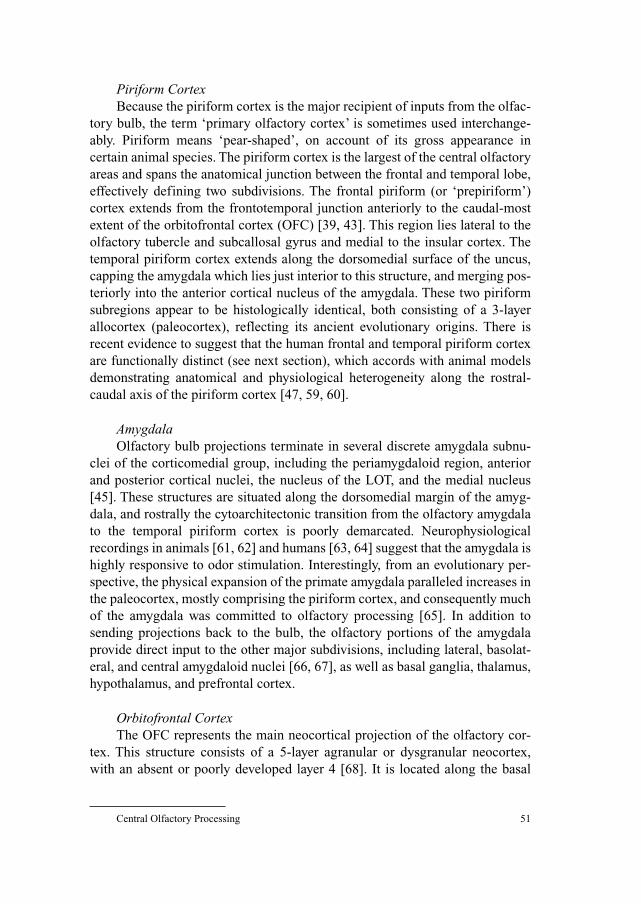
Central Olfactory Processing 51
Piriform CortexBecause the piriform cortex is the major recipient of inputs from the olfac-
tory bulb, the term ‘primary olfactory cortex’ is sometimes used interchange-
ably. Piriform means ‘pear-shaped’, on account of its gross appearance in
certain animal species. The piriform cortex is the largest of the central olfactory
areas and spans the anatomical junction between the frontal and temporal lobe,
effectively defining two subdivisions. The frontal piriform (or ‘prepiriform’)
cortex extends from the frontotemporal junction anteriorly to the caudal-most
extent of the orbitofrontal cortex (OFC) [39, 43]. This region lies lateral to the
olfactory tubercle and subcallosal gyrus and medial to the insular cortex. The
temporal piriform cortex extends along the dorsomedial surface of the uncus,
capping the amygdala which lies just interior to this structure, and merging pos-
teriorly into the anterior cortical nucleus of the amygdala. These two piriform
subregions appear to be histologically identical, both consisting of a 3-layer
allocortex (paleocortex), reflecting its ancient evolutionary origins. There is
recent evidence to suggest that the human frontal and temporal piriform cortex
are functionally distinct (see next section), which accords with animal models
demonstrating anatomical and physiological heterogeneity along the rostral-
caudal axis of the piriform cortex [47, 59, 60].
AmygdalaOlfactory bulb projections terminate in several discrete amygdala subnu-
clei of the corticomedial group, including the periamygdaloid region, anterior
and posterior cortical nuclei, the nucleus of the LOT, and the medial nucleus
[45]. These structures are situated along the dorsomedial margin of the amyg-
dala, and rostrally the cytoarchitectonic transition from the olfactory amygdala
to the temporal piriform cortex is poorly demarcated. Neurophysiological
recordings in animals [61, 62] and humans [63, 64] suggest that the amygdala is
highly responsive to odor stimulation. Interestingly, from an evolutionary per-
spective, the physical expansion of the primate amygdala paralleled increases in
the paleocortex, mostly comprising the piriform cortex, and consequently much
of the amygdala was committed to olfactory processing [65]. In addition to
sending projections back to the bulb, the olfactory portions of the amygdala
provide direct input to the other major subdivisions, including lateral, basolat-
eral, and central amygdaloid nuclei [66, 67], as well as basal ganglia, thalamus,
hypothalamus, and prefrontal cortex.
Orbitofrontal CortexThe OFC represents the main neocortical projection of the olfactory cor-
tex. This structure consists of a 5-layer agranular or dysgranular neocortex,
with an absent or poorly developed layer 4 [68]. It is located along the basal
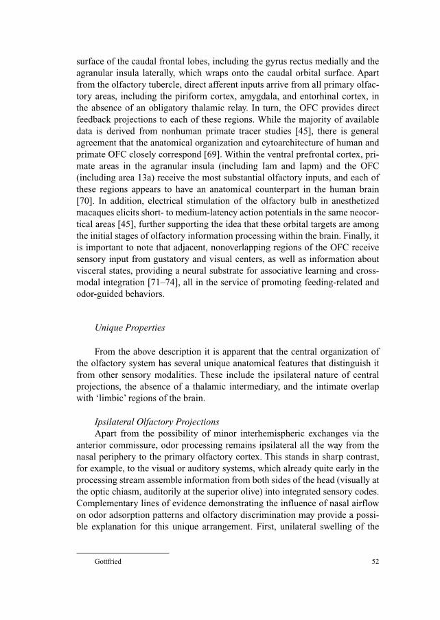
Gottfried 52
surface of the caudal frontal lobes, including the gyrus rectus medially and the
agranular insula laterally, which wraps onto the caudal orbital surface. Apart
from the olfactory tubercle, direct afferent inputs arrive from all primary olfac-
tory areas, including the piriform cortex, amygdala, and entorhinal cortex, in
the absence of an obligatory thalamic relay. In turn, the OFC provides direct
feedback projections to each of these regions. While the majority of available
data is derived from nonhuman primate tracer studies [45], there is general
agreement that the anatomical organization and cytoarchitecture of human and
primate OFC closely correspond [69]. Within the ventral prefrontal cortex, pri-
mate areas in the agranular insula (including Iam and Iapm) and the OFC
(including area 13a) receive the most substantial olfactory inputs, and each of
these regions appears to have an anatomical counterpart in the human brain
[70]. In addition, electrical stimulation of the olfactory bulb in anesthetized
macaques elicits short- to medium-latency action potentials in the same neocor-
tical areas [45], further supporting the idea that these orbital targets are among
the initial stages of olfactory information processing within the brain. Finally, it
is important to note that adjacent, nonoverlapping regions of the OFC receive
sensory input from gustatory and visual centers, as well as information about
visceral states, providing a neural substrate for associative learning and cross-
modal integration [71–74], all in the service of promoting feeding-related and
odor-guided behaviors.
Unique Properties
From the above description it is apparent that the central organization of
the olfactory system has several unique anatomical features that distinguish it
from other sensory modalities. These include the ipsilateral nature of central
projections, the absence of a thalamic intermediary, and the intimate overlap
with ‘limbic’ regions of the brain.
Ipsilateral Olfactory ProjectionsApart from the possibility of minor interhemispheric exchanges via the
anterior commissure, odor processing remains ipsilateral all the way from the
nasal periphery to the primary olfactory cortex. This stands in sharp contrast,
for example, to the visual or auditory systems, which already quite early in the
processing stream assemble information from both sides of the head (visually at
the optic chiasm, auditorily at the superior olive) into integrated sensory codes.
Complementary lines of evidence demonstrating the influence of nasal airflow
on odor adsorption patterns and olfactory discrimination may provide a possi-
ble explanation for this unique arrangement. First, unilateral swelling of the
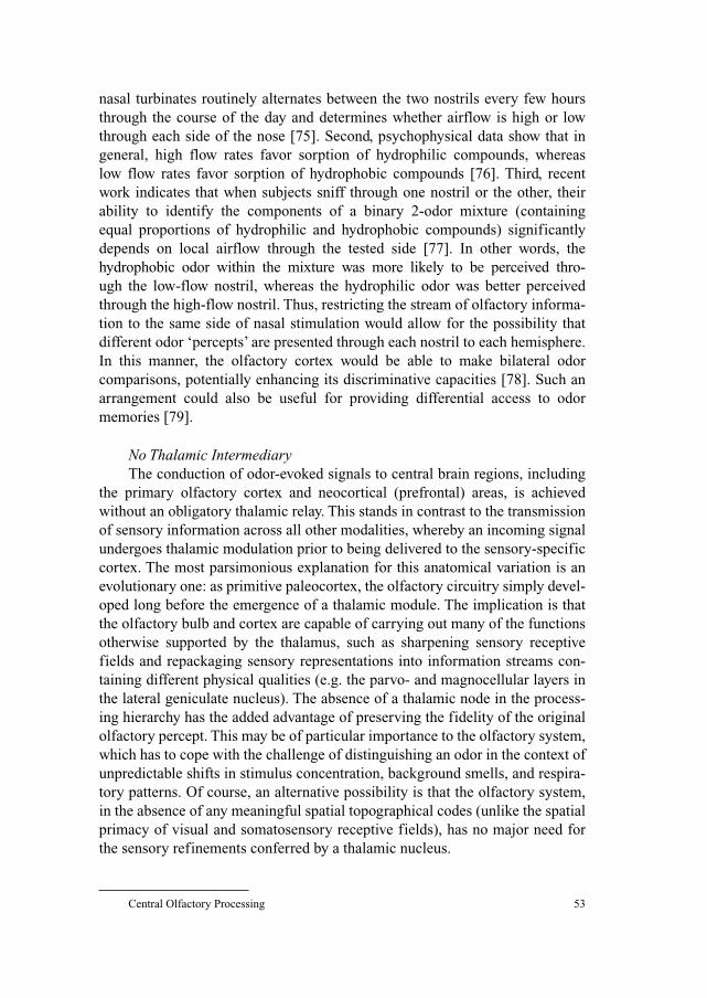
Central Olfactory Processing 53
nasal turbinates routinely alternates between the two nostrils every few hours
through the course of the day and determines whether airflow is high or low
through each side of the nose [75]. Second, psychophysical data show that in
general, high flow rates favor sorption of hydrophilic compounds, whereas
low flow rates favor sorption of hydrophobic compounds [76]. Third, recent
work indicates that when subjects sniff through one nostril or the other, their
ability to identify the components of a binary 2-odor mixture (containing
equal proportions of hydrophilic and hydrophobic compounds) significantly
depends on local airflow through the tested side [77]. In other words, the
hydrophobic odor within the mixture was more likely to be perceived thro-
ugh the low-flow nostril, whereas the hydrophilic odor was better perceived
through the high-flow nostril. Thus, restricting the stream of olfactory informa-
tion to the same side of nasal stimulation would allow for the possibility that
different odor ‘percepts’ are presented through each nostril to each hemisphere.
In this manner, the olfactory cortex would be able to make bilateral odor
comparisons, potentially enhancing its discriminative capacities [78]. Such an
arrangement could also be useful for providing differential access to odor
memories [79].
No Thalamic IntermediaryThe conduction of odor-evoked signals to central brain regions, including
the primary olfactory cortex and neocortical (prefrontal) areas, is achieved
without an obligatory thalamic relay. This stands in contrast to the transmission
of sensory information across all other modalities, whereby an incoming signal
undergoes thalamic modulation prior to being delivered to the sensory-specific
cortex. The most parsimonious explanation for this anatomical variation is an
evolutionary one: as primitive paleocortex, the olfactory circuitry simply devel-
oped long before the emergence of a thalamic module. The implication is that
the olfactory bulb and cortex are capable of carrying out many of the functions
otherwise supported by the thalamus, such as sharpening sensory receptive
fields and repackaging sensory representations into information streams con-
taining different physical qualities (e.g. the parvo- and magnocellular layers in
the lateral geniculate nucleus). The absence of a thalamic node in the process-
ing hierarchy has the added advantage of preserving the fidelity of the original
olfactory percept. This may be of particular importance to the olfactory system,
which has to cope with the challenge of distinguishing an odor in the context of
unpredictable shifts in stimulus concentration, background smells, and respira-
tory patterns. Of course, an alternative possibility is that the olfactory system,
in the absence of any meaningful spatial topographical codes (unlike the spatial
primacy of visual and somatosensory receptive fields), has no major need for
the sensory refinements conferred by a thalamic nucleus.
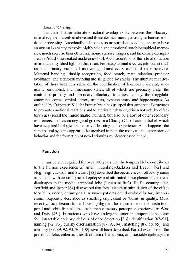
Gottfried 54
‘Limbic’ OverlapIt is clear that an intimate structural overlap exists between the olfactory-
related regions described above and those devoted more generally to human emo-
tional processing. Anecdotally this comes as no surprise, as odors appear to have
an unusual capacity to evoke highly vivid and emotional autobiographical memo-
ries, much more so than other mnemonic sensory triggers, and timelessly exempli-
fied in Proust’s tea-soaked madeleines [80]. A consideration of the role of olfaction
in animals may shed light on this issue. For many animal species, odorous stimuli
are the primary means of motivating almost every aspect of their behavior.
Maternal bonding, kinship recognition, food search, mate selection, predator
avoidance, and territorial marking are all guided by smells. The ultimate manifes-
tation of these behaviors relies on the coordination of hormonal, visceral, auto-
nomic, emotional, and mnemonic states, all of which are precisely under the
control of primary and secondary olfactory structures, namely, the amygdala,
entorhinal cortex, orbital cortex, striatum, hypothalamus, and hippocampus. As
outlined by Carpenter [81], the human brain has usurped this same set of structures
to promote emotional reactions and to motivate behavior, driven not only by olfac-
tory cues (recall the ‘microsmatic’ human), but also by a host of other secondary
reinforcers, such as money, good grades, or a Chicago Cubs baseball ticket, which
have acquired biological salience via learning and experience. As it happens, the
same neural systems appear to be involved in both the motivational expression of
behavior and the formation of novel stimulus-reinforcer associations.
Function
It has been recognized for over 100 years that the temporal lobe contributes
to the human experience of smell. Hughlings-Jackson and Beevor [82] and
Hughlings-Jackson and Stewart [83] described the occurrence of olfactory auras
in patients with certain types of epilepsy and attributed these phenomena to ictal
discharges in the medial temporal lobe (‘uncinate fits’). Half a century later,
Penfield and Jasper [84] discovered that focal electrical stimulation of the olfac-
tory bulb, uncus, or amygdala in awake patients could evoke olfactory impres-
sions, frequently described as smelling unpleasant or ‘burnt’ in quality. More
recently, focal lesion studies have highlighted the importance of the mediotem-
poral and orbitofrontal lobes to human olfactory perception (reviewed in West
and Doty [85]). In patients who have undergone anterior temporal lobectomy
for intractable epilepsy, deficits of odor detection [86], identification [87–91],
naming [92, 93], quality discrimination [87, 93, 94], matching [87, 88, 95], and
memory [88, 89, 92, 93, 96–100] have all been described. Partial excisions of the
prefrontal lobe, either as a result of tumor, hematoma, or intractable epilepsy, are
Central Olfactory Processing 55
also associated with impairments of odor identification [90], quality discrimina-
tion [94, 101], and memory [98]. However, the large size and spatial extent of
such lesions precludes careful anatomical delineation, particularly given the close
proximity of so many critical olfactory structures within the temporal lobe (fig. 1).
Thus, despite the abundance of clinical data implicating the ventral frontal and
temporal lobes in human olfaction, a more precise functional organization has
not been elucidated.
The advent of modern neuroimaging techniques, such as positron emission
tomography (PET) and functional magnetic resonance imaging (fMRI), has led
to important advances in our understanding of central olfactory function in the
normal human brain (for recent reviews, see Savic [102], Sobel et al. [103] and
Royet and Plailly [104]). While both PET and fMRI permit simultaneous data
acquisition from the whole brain at high spatial resolution, fMRI has several
important advantages: it is safe and noninvasive (not requiring the intravenous
injection of radioactive tracers), is easily repeated within subjects across differ-
ent sessions, and is ideal for complex experiments involving the delivery of
multiple different odors. It is important to note that the fMRI signal reflects
local activity-dependent changes in hemodynamic state (technically, blood oxy-
genation level-dependent contrast) [105–107], and therefore provides a surro-
gate marker of neural activity that is temporally constrained by the intrinsic
time lag of neurovascular coupling. Thus, the spatial benefits of fMRI (and
PET) come at a certain expense of temporal resolution, on the order of seconds.
Human neuroimaging research has begun to identify a network of brain
structures important for olfactory processing [108–119]. While many of these
studies identified odor-evoked neural responses within primary and secondary
areas of the olfactory cortex, one notable feature was the inconsistent activation
of the piriform cortex. This is now thought to reflect at least two factors. First,
conventional fMRI sequences are associated with signal loss (susceptibility
artifact) at air-tissue interfaces, reducing image quality in olfactory-specific
areas of the ventral temporal and basal frontal lobes [120]. Second, olfactory
habituation occurs with prolonged odor exposure in the rodent piriform cortex
[121], and analogous phenomena have been confirmed with fMRI in humans
[119, 122]. Because many previous olfactory neuroimaging studies used
blocked designs, with constant odor presentation over 30–60 s, habituation has
been an unavoidable confound. The recent introduction of event-related designs
to limit the duration of odor exposure [123–125], as well as the development of
specialized imaging protocols [126, 127] have helped improve fMRI signal
detection in the olfactory cortex.
In keeping with the earlier anatomical focus on the piriform cortex, amyg-
dala, and orbitofrontal cortex, the remaining section will provide a selective
survey of functional imaging data from these three regions. This decision is
Gottfried 56
motivated by the fact that: (1) there is a relative abundance of PET and fMRI
data for these structures, in comparison to other areas, and (2) regions like the
AON and olfactory tubercle are too small to be identified with confidence,
given the spatial limitations of fMRI. For a discussion of other olfactory struc-
tures, including areas not traditionally thought to be olfactory in function (e.g.
cerebellum), the reader is referred to Sobel et al. [103]. In addition, while corti-
cal lateralization of olfactory function is not considered here, the topic is cov-
ered in detail in a recent review [104]. Finally, other sections of the book are
specifically devoted to nasal trigeminal function [see chapter 10 by Breslin and
Huang, this vol, pp 152–190] and vomeronasal function [see chapter 11 by
Small, this vol, pp 191–220], which are therefore not treated here.
Piriform Cortex
Despite some initial difficulties in imaging the piriform cortex (as high-
lighted above), several early studies demonstrated that human piriform activity
could be evoked by smelling of an odor [108–110, 115], consistent with the
idea that this region participates in basic olfactory processing. One important
finding to emerge was that the piriform cortex responded not only to smells, but
to the act of sniffing itself, even in the absence of odor [115]. In this study,
sniffing of odorless air, as well as artificial sniffing induced by air puffs into the
nostrils, activated the piriform cortex, whereas partial physical occlusion of the
nostrils, or topical anesthesia to the nasal passages, reduced this activity (fig. 3).
These results indicate that sniff-induced piriform activity is not simply due to the
motor act of sniffing, but rather to the physical sensation of airflow across the
nasal mucosa, and are compatible with animal data suggesting that the sniff
may prime the piriform cortex for optimal reception of a smell [128, 129].
Until recently, it had been unclear whether the human piriform cortex bas-
ically served as a passive relay of olfactory information, or whether it was capa-
ble of more complex operations. The first hint that the human piriform cortex
was functionally complex, and moreover that fMRI techniques were capable of
resolving subregional piriform differences, came from a study on olfactory proc-
essing and odor valence [123], in which human subjects smelled three odors
that differed in valence (pleasant, neutral, unpleasant). The posterior (temporal)
piriform cortex was significantly activated bilaterally by each odor, independ-
ent of valence (fig. 4a, b), suggesting that this region mediates basic odor
perception. Such a role complements theories derived from animal models sug-
gesting that the piriform cortex is broadly tuned to odors [62, 72] and conforms to
recent neuroimaging studies demonstrating similar activation patterns in response
to low-level olfactory processes [108, 115, 118, 122]. In contrast, activations
Central Olfactory Processing 57
were also observed within the anterior (frontal) piriform cortex in response to
unpleasant and pleasant, but not neutral, odors (fig. 4c, d). One plausible hypoth-
esis is that the anterior piriform cortex is receptive to hedonic quality, especially
at extremes of odor valence. The identification of functional heterogeneity in
the human piriform cortex accords with animal models demonstrating ana-
tomical and physiological distinctions along its rostral-caudal axis [47, 59,
60] and suggests that this region likely transcends the role of mere sensory
intermediary.
The piriform cortex also appears to be involved in olfactory learning and
memory, again in keeping with the animal literature. In two PET studies, short- and
Sniffing
a
c
b
d
Top. anesthesia
Air puffs Nostril occluded
LR
Fig. 3. Sniffing activates the piriform cortex (a). By comparison, the sniff-induced
fMRI activity is reduced when topical anesthesia is applied to the nasal membranes (b) or
when airflow is blocked via nostril occlusion (d). Piriform activity is also present during arti-
ficial puffs of odorless air into the nose (c). L � Left; R � right. (Reproduced and modified
from figure 3 of Sobel et al. [115]; copyright 1998 by the Nature Publishing Group, and with
permission of the authors.)
Gottfried 58
long-term odor recognition memory was associated with enhanced piriform
cortex activity when compared to odorless baseline scans [99, 118]. More
recently, an fMRI study of visual-olfactory associative learning (Pavlovian con-
ditioning) between a neutral visual stimulus and a pleasant food odor showed
that the conditioned visual cue, in the absence of odor, elicited neural activity in
the piriform cortex [130]. In this same experiment, after subjects consumed a
food (corresponding to the odor) to satiety, the learning-evoked piriform
responses decreased in accordance with their current appetite state, lending cre-
dence to the idea that the piriform cortex is a site of learning-induced plasticity.
Other work has focused on episodic memory retrieval of olfactory context
[131]. In an initial study phase, subjects were given combinations of smells and
pictures and asked to imagine a link or association between the two stimuli. The
aim of this session was to encourage episodic memory encoding of odor-picture
a b
c d
y�0
y�10
I
IP
P
C
C
PPC
APC
Fig. 4. Functional heterogeneity in the piriform cortex. a The posterior piriform cortex
is mutually activated by pleasant, neutral, and unpleasant odors, characteristic of its role in
low-level olfactory processing. The area outlined by a rectangle is magnified in (b) to illus-
trate the anatomy more clearly. c In turn, the anterior piriform cortex is sensitive to unpleas-
ant and pleasant (but not neutral) odors, suggesting this region encodes information about
extremes of odor valence. The fMRI activation shown here was specifically evoked by
unpleasant odor, and is magnified in (d). APC � Anterior piriform cortex; C � caudate;
I � insula; P � putamen; PPC � posterior piriform cortex. The right side of the figure
matches the right side of the brain. (Reproduced and modified from figures 3 and 4 of
Gottfried et al. [123]; copyright 2002 by the Society for Neuroscience.)
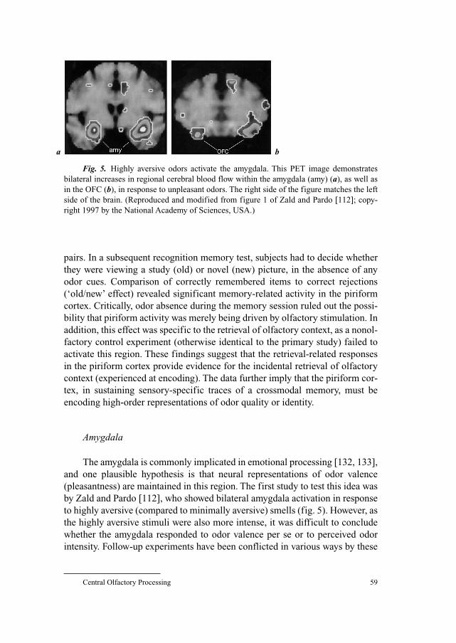
Central Olfactory Processing 59
pairs. In a subsequent recognition memory test, subjects had to decide whether
they were viewing a study (old) or novel (new) picture, in the absence of any
odor cues. Comparison of correctly remembered items to correct rejections
(‘old/new’ effect) revealed significant memory-related activity in the piriform
cortex. Critically, odor absence during the memory session ruled out the possi-
bility that piriform activity was merely being driven by olfactory stimulation. In
addition, this effect was specific to the retrieval of olfactory context, as a nonol-
factory control experiment (otherwise identical to the primary study) failed to
activate this region. These findings suggest that the retrieval-related responses
in the piriform cortex provide evidence for the incidental retrieval of olfactory
context (experienced at encoding). The data further imply that the piriform cor-
tex, in sustaining sensory-specific traces of a crossmodal memory, must be
encoding high-order representations of odor quality or identity.
Amygdala
The amygdala is commonly implicated in emotional processing [132, 133],
and one plausible hypothesis is that neural representations of odor valence
(pleasantness) are maintained in this region. The first study to test this idea was
by Zald and Pardo [112], who showed bilateral amygdala activation in response
to highly aversive (compared to minimally aversive) smells (fig. 5). However, as
the highly aversive stimuli were also more intense, it was difficult to conclude
whether the amygdala responded to odor valence per se or to perceived odor
intensity. Follow-up experiments have been conflicted in various ways by these
a b
Fig. 5. Highly aversive odors activate the amygdala. This PET image demonstrates
bilateral increases in regional cerebral blood flow within the amygdala (amy) (a), as well as
in the OFC (b), in response to unpleasant odors. The right side of the figure matches the left
side of the brain. (Reproduced and modified from figure 1 of Zald and Pardo [112]; copy-
right 1997 by the National Academy of Sciences, USA.)
Gottfried 60
intensity-valence confounds. One study showed greater amygdala activity for
unpleasant than pleasant odors, otherwise matched for intensity, suggesting a
valence-specific effect [134]. Another study suggested the amygdala was bas-
ically insensitive to valence, being similarly activated by pleasant, neutral, and
unpleasant odors [123], while a third group demonstrated correlations between
perceived intensity and neural activity in temporal structures adjacent to the
amygdala [135]. In an effort to resolve these issues, Anderson et al. [125] dis-
sociated intensity and valence within a single experiment, by presenting one
pleasant odor (citral: lemon smell) and one unpleasant odor (valeric acid:
sweaty sock smell), each at low and high intensity. In this manner, the amygdala
was significantly activated by intensity (high vs. low), but not valence (unpleas-
ant vs. pleasant, or vice versa), suggesting odor intensity coding in this region.
Even then, recent findings from our laboratory suggest this story is still
unfinished [135a]. Here, intensity and valence were again dissociated within
one experimental design, but with the inclusion of a neutrally valenced odor
condition, in order to estimate amygdala activity across a more comprehensive
valence spectrum. That is, high and low intensity versions of pleasant, neutral,
and unpleasant odors were delivered to subjects. We predicted that if the amyg-
dala responds to intensity irrespective of valence, then all three odor types
should elicit similar levels of activation. However, if the amygdala only
responds to intensity at the extremes of valence, then pleasant and unpleasant
odors, but not neutral odor, should elicit activity. Our findings are in keeping
with this latter prediction, suggesting that the interaction between intensity and
valence, reflecting the overall behavioral salience of an odor, is what matters
most to the amygdala.
As described above for the piriform cortex, the amygdala is also involved
in associative learning between visual stimuli and olfactory reinforcers [130,
136]. Recent data suggest this region is preferentially involved in the formation
of new associations, but does not maintain these representations over time
[136]. This latter mnemonic function may be reserved for regions such as the
OFC (see below). In addition, there is evidence to suggest a role for the amyg-
dala in the evocation of emotional odor memories [137]. Five subjects were
presented with one of four different stimulus conditions in the fMRI scanner:
odors (perfumes) that elicited a pleasant, personal memory; visual pictures of
those odors (perfume bottles); control odors; and control pictures. Comparison
of emotional odor to the other three conditions (or to the control odor alone)
was associated with parahippocampal activation extending into the amygdala.
While these findings do not permit a distinction between memory retrieval,
valence processing, and stimulus salience, they suggest that behaviorally rele-
vant odor cues may be a more potent activator of emotional circuitry than
nonolfactory stimuli. These functional imaging data are supported by lesion
Central Olfactory Processing 61
studies of patients with selective bilateral amygdala damage [100, 138], as well
as by stereotactic intracranial recordings [134], all of which further implicate
the amygdala in various tasks related specifically to olfactory memory.
Orbitofrontal Cortex
Human olfactory imaging has tentatively revealed dissociations of function
along two separate anatomical dimensions in the OFC. The first of these is a
caudal-rostral distinction. Odor-evoked neural activity in the caudal OFC is typi-
cally associated with low-level aspects of olfactory processing, such as passive
smelling and odor detection [108, 112, 123, 139], and probably represents the
initial neocortical projection site from the primary olfactory cortex. The location
of these activations roughly corresponds to the so-called ‘central-posterior
orbitofrontal cortex’ identified by Yarita et al. [140], who considered it a broadly
tuned area of the primate olfactory association cortex. According to Carmichael
et al. [45], the central-posterior orbitofrontal cortex is roughly homologous to
the orbital areas 13m, 13a, and Iam, the primary prefrontal locus of olfactory
input (see the Anatomy section above). By comparison, more rostral areas of the
OFC are engaged in higher-order olfactory computations, including associative
learning [124, 130, 136], working memory [141], and short- and long-term odor
recognition memory [99, 118]. This caudal-rostral division is bolstered by ani-
mal findings suggesting an anatomical hierarchy of orbitofrontal specialization,
whereby caudal regions (such as the OFC) converge on medial and anterior ter-
ritories to permit more complex information processing [69, 142].
The OFC also exhibits regional differences along a medial-lateral axis of
functional specialization. Numerous studies increasingly show that pleasant
odors evoke activity in the medial OFC and ventromedial prefrontal cortex,
whereas unpleasant odors evoke activity in the lateral OFC and adjacent infe-
rior prefrontal cortex [123, 125, 135]. Similar dissociations have been identi-
fied in visual-olfactory crossmodal paradigms of episodic memory [131] and
associative learning [136]. These valence-specific patterns have emerged in
other experiments spanning a variety of modalities. Thus, processing of tastes
[143], faces [144], and abstract monetary reinforcers [145] have all revealed
medial-lateral response differences in the OFC that vary according to the
degree of pleasantness. On neuroanatomical grounds, these orbital regions can
be regarded as distinct functional units with unique sets of cortical and subcor-
tical connections [142]. Notably, projections between the OFC and amygdala
are reciprocal [142]. It is thus plausible that differences in input patterns from
the amygdala, e.g., might contribute to the expression of positive and negative
value in medial and lateral orbital subdivisions, respectively.
Gottfried 62
As a major recipient of projections not only from the primary olfactory
cortex, but also from gustatory, visual, visceral, and thalamic centers, the OFC
is certain to participate in a wide variety of complex olfactory functions related
to multimodal integration, reward processing, and goal-directed learning and
behavior [see chapter 13 by Landis and Lacroix, this vol, pp 242–254, for an
extensive discussion on neural correlates of flavor and feeding]. The role of the
OFC as a site of sensory convergence has been documented across several dif-
ferent sensory combinations, including odor/taste [146–148] and odor/vision
[149]. Moreover, through manipulations of semantic correspondence between
odors and tastes [147], or between odors and pictures [149], OFC activity
increased with increasing subjective congruency ratings. These observations
underscore the general idea that prior learning and experience can profoundly
modulate the central processing of sensory information. Such mechanisms may
also help to resolve the inherent ambiguity in olfactory perception [149].
Interestingly, as discussed above, unimodal odor stimuli appear to be processed
in more caudal regions of the OFC than the corresponding bimodal stimulus
pairs [147, 149].
The human OFC also provides a substrate for the encoding of primary and
secondary (learned) value of olfactory stimuli. In a study of sensory-specific
satiety, subjects were delivered two different food odors, both before and after a
feeding session designed to decrease the pleasantness of one of the odors [117].
Following satiety, the neural activity in the OFC declined in parallel with the
decrease in food pleasantness ratings, suggesting that reward value of a food
odor is encoded and updated in this structure. Our own work consistently
demonstrates robust participation of the OFC in classical conditioning para-
digms of olfactory learning [124, 130, 136]. Specifically, after an arbitrary
visual picture is repetitively paired with either a pleasant or unpleasant odor,
presentation of the visual cue by itself elicits activation in the OFC, suggesting
that this region is involved in the establishment of picture-odor contingencies.
Moreover, when the current affective value of the olfactory reinforcer is either
decreased (via selective satiety) or increased (via odor inflation), cue-evoked
OFC responses are modulated in parallel with the behavioral manipulation. The
implication is that a predictive cue has direct access to central representations
of value in the OFC, and that these representations are flexibly updated accord-
ing to an individual’s motivational state.
Finally, it should be mentioned that a variety of explicit cognitive tasks influ-
ences responses in the OFC [104]. Intensity judgments [150], familiarity judg-
ments [116, 139], hedonicity judgments [139], and quality discrimination tasks
[118] are all associated with orbitofrontal activity, irrespective of specific percep-
tual features of the odors themselves (e.g. their intensity, familiarity, or pleasant-
ness). These activations are usually accompanied by regional responses in large
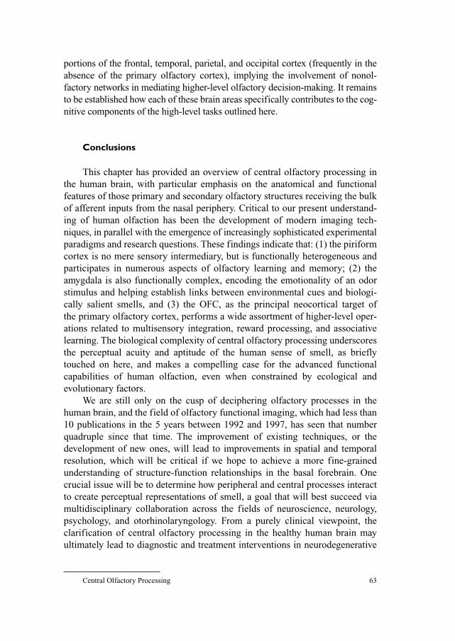
Central Olfactory Processing 63
portions of the frontal, temporal, parietal, and occipital cortex (frequently in the
absence of the primary olfactory cortex), implying the involvement of nonol-
factory networks in mediating higher-level olfactory decision-making. It remains
to be established how each of these brain areas specifically contributes to the cog-
nitive components of the high-level tasks outlined here.
Conclusions
This chapter has provided an overview of central olfactory processing in
the human brain, with particular emphasis on the anatomical and functional
features of those primary and secondary olfactory structures receiving the bulk
of afferent inputs from the nasal periphery. Critical to our present understand-
ing of human olfaction has been the development of modern imaging tech-
niques, in parallel with the emergence of increasingly sophisticated experimental
paradigms and research questions. These findings indicate that: (1) the piriform
cortex is no mere sensory intermediary, but is functionally heterogeneous and
participates in numerous aspects of olfactory learning and memory; (2) the
amygdala is also functionally complex, encoding the emotionality of an odor
stimulus and helping establish links between environmental cues and biologi-
cally salient smells, and (3) the OFC, as the principal neocortical target of
the primary olfactory cortex, performs a wide assortment of higher-level oper-
ations related to multisensory integration, reward processing, and associative
learning. The biological complexity of central olfactory processing underscores
the perceptual acuity and aptitude of the human sense of smell, as briefly
touched on here, and makes a compelling case for the advanced functional
capabilities of human olfaction, even when constrained by ecological and
evolutionary factors.
We are still only on the cusp of deciphering olfactory processes in the
human brain, and the field of olfactory functional imaging, which had less than
10 publications in the 5 years between 1992 and 1997, has seen that number
quadruple since that time. The improvement of existing techniques, or the
development of new ones, will lead to improvements in spatial and temporal
resolution, which will be critical if we hope to achieve a more fine-grained
understanding of structure-function relationships in the basal forebrain. One
crucial issue will be to determine how peripheral and central processes interact
to create perceptual representations of smell, a goal that will best succeed via
multidisciplinary collaboration across the fields of neuroscience, neurology,
psychology, and otorhinolaryngology. From a purely clinical viewpoint, the
clarification of central olfactory processing in the healthy human brain may
ultimately lead to diagnostic and treatment interventions in neurodegenerative
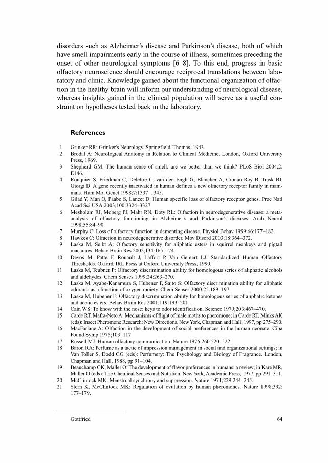
Gottfried 64
disorders such as Alzheimer’s disease and Parkinson’s disease, both of which
have smell impairments early in the course of illness, sometimes preceding the
onset of other neurological symptoms [6–8]. To this end, progress in basic
olfactory neuroscience should encourage reciprocal translations between labo-
ratory and clinic. Knowledge gained about the functional organization of olfac-
tion in the healthy brain will inform our understanding of neurological disease,
whereas insights gained in the clinical population will serve as a useful con-
straint on hypotheses tested back in the laboratory.
References
1 Grinker RR: Grinker’s Neurology. Springfield, Thomas, 1943.
2 Brodal A: Neurological Anatomy in Relation to Clinical Medicine. London, Oxford University
Press, 1969.
3 Shepherd GM: The human sense of smell: are we better than we think? PLoS Biol 2004;2:
E146.
4 Rouquier S, Friedman C, Delettre C, van den Engh G, Blancher A, Crouau-Roy B, Trask BJ,
Giorgi D: A gene recently inactivated in human defines a new olfactory receptor family in mam-
mals. Hum Mol Genet 1998;7:1337–1345.
5 Gilad Y, Man O, Paabo S, Lancet D: Human specific loss of olfactory receptor genes. Proc Natl
Acad Sci USA 2003;100:3324–3327.
6 Mesholam RI, Moberg PJ, Mahr RN, Doty RL: Olfaction in neurodegenerative disease: a meta-
analysis of olfactory functioning in Alzheimer’s and Parkinson’s diseases. Arch Neurol
1998;55:84–90.
7 Murphy C: Loss of olfactory function in dementing disease. Physiol Behav 1999;66:177–182.
8 Hawkes C: Olfaction in neurodegenerative disorder. Mov Disord 2003;18:364–372.
9 Laska M, Seibt A: Olfactory sensitivity for aliphatic esters in squirrel monkeys and pigtail
macaques. Behav Brain Res 2002;134:165–174.
10 Devos M, Patte F, Rouault J, Laffort P, Van Gemert LJ: Standardized Human Olfactory
Thresholds. Oxford, IRL Press at Oxford University Press, 1990.
11 Laska M, Teubner P: Olfactory discrimination ability for homologous series of aliphatic alcohols
and aldehydes. Chem Senses 1999;24:263–270.
12 Laska M, Ayabe-Kanamura S, Hubener F, Saito S: Olfactory discrimination ability for aliphatic
odorants as a function of oxygen moiety. Chem Senses 2000;25:189–197.
13 Laska M, Hubener F: Olfactory discrimination ability for homologous series of aliphatic ketones
and acetic esters. Behav Brain Res 2001;119:193–201.
14 Cain WS: To know with the nose: keys to odor identification. Science 1979;203:467–470.
15 Carde RT, Mafra-Neto A: Mechanisms of flight of male moths to pheromone; in Carde RT, Minks AK
(eds): Insect Pheromone Research: New Directions. New York, Chapman and Hall, 1997, pp 275–290.
16 MacFarlane A: Olfaction in the development of social preferences in the human neonate. Ciba
Found Symp 1975;103–117.
17 Russell MJ: Human olfactory communication. Nature 1976;260:520–522.
18 Baron RA: Perfume as a tactic of impression management in social and organizational settings; in
Van Toller S, Dodd GG (eds): Perfumery: The Psychology and Biology of Fragrance. London,
Chapman and Hall, 1988, pp 91–104.
19 Beauchamp GK, Maller O: The development of flavor preferences in humans: a review; in Kare MR,
Maller O (eds): The Chemical Senses and Nutrition. New York, Academic Press, 1977, pp 291–311.
20 McClintock MK: Menstrual synchrony and suppression. Nature 1971;229:244–245.
21 Stern K, McClintock MK: Regulation of ovulation by human pheromones. Nature 1998;392:
177–179.

Central Olfactory Processing 65
22 Wedekind C, Seebeck T, Bettens F, Paepke AJ: MHC-dependent mate preferences in humans. Proc
Biol Sci 1995;260:245–249.
23 Wedekind C, Furi S: Body odour preferences in men and women: do they aim for specific MHC
combinations or simply heterozygosity? Proc Biol Sci 1997;264:1471–1479.
24 Jacob S, McClintock MK, Zelano B, Ober C: Paternally inherited HLA alleles are associated with
women’s choice of male odor. Nat Genet 2002;30:175–179.
25 Ober C, Weitkamp LR, Cox N, Dytch H, Kostyu D, Elias S: HLA and mate choice in humans.
Am J Hum Genet 1997;61:497–504.
26 Counet C, Callemien D, Ouwerx C, Collin S: Use of gas chromatography-olfactometry to identify
key odorant compounds in dark chocolate. Comparison of samples before and after conching.
J Agric Food Chem 2002;50:2385–2391.
27 Laing DG, Francis GW: The capacity of humans to identify odors in mixtures. Physiol Behav
1989;46:809–814.
28 Livermore A, Laing DG: Influence of training and experience on the perception of multicompo-
nent odor mixtures. J Exp Psychol Hum Percept Perform 1996;22:267–277.
29 Morrot G, Brochet F, Dubourdieu D: The color of odors. Brain Lang 2001;79:309–320.
30 Zellner DA, Kautz MA: Color affects perceived odor intensity. J Exp Psychol Hum Percept
Perform 1990;16:391–397.
31 Dalton P: Odor perception and beliefs about risk. Chem Senses 1996;21:447–458.
32 Dalton P, Doolittle N, Nagata H, Breslin PA: The merging of the senses: integration of subthresh-
old taste and smell. Nat Neurosci 2000;3:431–432.
33 Distel H, Hudson R: Judgement of odor intensity is influenced by subjects’ knowledge of the odor
source. Chem Senses 2001;26:247–251.
34 Slosson EE: A lecture experiment in hallucinations. Psychol Rev 1899;6:407–408.
35 O’Mahony M: Smell illusions and suggestion: reports of smell contingent on tones played on tele-
vision and radio. Chem Senses Flav 1978;3:183–189.
36 Wilson DA, Stevenson RJ: The fundamental role of memory in olfactory perception. Trends
Neurosci 2003;26:243–247.
37 Stevenson RJ: Associative learning and odor quality perception: how sniffing an odor mixture can
alter the smell of its parts. Learn Motiv 2001;32:154–177.
38 Stevenson RJ: The acquisition of odour qualities. Q J Exp Psychol 2001;54:561–577.
39 Eslinger PJ, Damasio AR, Van Hoesen GW: Olfactory dysfunction in man: anatomical and behav-
ioral aspects. Brain Cogn 1982;1:259–285.
40 Price JL: Olfactory system; in Paxinos G (ed): The Human Nervous System. San Diego,
Academic Press, 1990, pp 979–998.
41 Heimer L: The Human Brain and Spinal Cord: Functional Neuroanatomy and Dissection Guide.
New York, Springer, 1983.
42 Nolte J: The Human Brain: An Introduction to Its Functional Anatomy. St Louis, Mosby, 2002,
pp 319–336.
43 Mai JK, Assheuer J, Paxinos G: Atlas of the Human Brain. San Diego, Academic Press, 1997.
44 Martin JH: Neuroanatomy: Text and Atlas. New York, McGraw-Hill, 2003, pp 207–226.
45 Carmichael ST, Clugnet MC, Price JL: Central olfactory connections in the macaque monkey.
J Comp Neurol 1994;346:403–434.
46 Shipley MT, Ennis M: Functional organization of olfactory system. J Neurobiol 1996;30:
123–176.
47 Haberly LB: Olfactory cortex; in Shepherd GM (ed): The Synaptic Organization of the Brain.
New York, Oxford University Press, 1998, pp 377–416.
48 Cleland TA, Linster C: Central olfactory structures; in Doty RL (ed): Handbook of Olfaction and
Gustation, ed 2. New York, Dekker, 2003, pp 165–180.
49 Wilson DA, Sullivan RM: Sensory physiology of central olfactory pathways; in Doty RL (ed):
Handbook of Olfaction and Gustation, ed 2. New York, Dekker, 2003, pp 181–201.
50 Price JL: An autoradiographic study of complementary laminar patterns of termination of afferent
fibers to the olfactory cortex. J Comp Neurol 1973;150:87–108.
51 de Olmos J, Hardy H, Heimer L: The afferent connections of the main and the accessory olfactory
bulb formations in the rat: an experimental HRP-study. J Comp Neurol 1978;181:213–244.
Gottfried 66
52 Haberly LB, Price JL: Association and commissural fiber systems of the olfactory cortex of the
rat. J Comp Neurol 1978;178:711–740.
53 Zou Z, Horowitz LF, Montmayeur JP, Snapper S, Buck LB: Genetic tracing reveals a stereotyped
sensory map in the olfactory cortex. Nature 2001;414:173–179.
54 Pearce RK, Hawkes CH, Daniel SE: The anterior olfactory nucleus in Parkinson’s disease. Mov
Disord 1995;10:283–287.
55 Kovacs T, Papp MI, Cairns NJ, Khan MN, Lantos PL: Olfactory bulb in multiple system atrophy.
Mov Disord 2003;18:938–942.
56 Montemurro DG, Bruni JE: The Human Brain in Dissection. New York, Oxford University Press, 1988.
57 Millhouse OE, Heimer L: Cell configurations in the olfactory tubercle of the rat. J Comp Neurol
1984;228:571–597.
58 Shipley MT, Adamek GD: The connections of the mouse olfactory bulb: a study using orthograde
and retrograde transport of wheat germ agglutinin conjugated to horseradish peroxidase. Brain
Res Bull 1984;12:669–688.
59 Litaudon P, Mouly AM, Sullivan R, Gervais R, Cattarelli M: Learning-induced changes in rat pir-
iform cortex activity mapped using multisite recording with voltage sensitive dye. Eur J Neurosci
1997;9:1593–1602.
60 Chabaud P, Ravel N, Wilson DA, Mouly AM, Vigouroux M, Farget V, Gervais R: Exposure to
behaviourally relevant odour reveals differential characteristics in rat central olfactory pathways as
studied through oscillatory activities. Chem Senses 2000;25:561–573.
61 Cain DP, Bindra D: Responses of amygdala single units to odors in the rat. Exp Neurol
1972;35:98–110.
62 Tanabe T, Iino M, Takagi SF: Discrimination of odors in olfactory bulb, pyriform-amygdaloid
areas, and orbitofrontal cortex of the monkey. J Neurophysiol 1975;38:1284–1296.
63 Hughes JR, Andy OJ: The human amygdala. 1. Electrophysiological responses to odorants.
Electroencephalogr Clin Neurophysiol 1979;46:428–443.
64 Hudry J, Ryvlin P, Royet JP, Mauguiere F: Odorants elicit evoked potentials in the human amyg-
dala. Cereb Cortex 2001;11:619–627.
65 Barton RA, Aggleton JP: Primate evolution and the amygdala; in Aggleton JP (ed):
The Amygdala: A Functional Analysis, ed 2. New York, Oxford University Press, 2000, pp
479–508.
66 Amaral DG, Price JL, Pitkänen A, Carmichael ST: Anatomical organization of the primate amyg-
daloid complex; in Aggleton JP (ed): The Amygdala, ed 1. New York, Wiley, 1992, pp 1–67.
67 Pitkänen A: Connectivity of the rat amygdaloid complex; in Aggleton JP (ed): The Amygdala:
A Functional Analysis, ed 2. Oxford, Oxford University Press, 2000, pp 31–116.
68 Carmichael ST, Price JL: Architectonic subdivision of the orbital and medial prefrontal cortex in
the macaque monkey. J Comp Neurol 1994;346:366–402.
69 Ongur D, Price JL: The organization of networks within the orbital and medial prefrontal cortex of
rats, monkeys and humans. Cereb Cortex 2000;10:206–219.
70 Ongur D, Ferry AT, Price JL: Architectonic subdivision of the human orbital and medial prefrontal
cortex. J Comp Neurol 2003;460:425–449.
71 Rolls ET, Baylis LL: Gustatory, olfactory, and visual convergence within the primate orbitofrontal
cortex. J Neurosci 1994;14:5437–5452.
72 Schoenbaum G, Eichenbaum H: Information coding in the rodent prefrontal cortex. 1. Single-neuron
activity in orbitofrontal cortex compared with that in pyriform cortex. J Neurophysiol 1995;74:
733–750.
73 Rolls ET, Critchley HD, Mason R, Wakeman EA: Orbitofrontal cortex neurons: role in olfactory
and visual association learning. J Neurophysiol 1996;75:1970–1981.
74 Schoenbaum G, Chiba AA, Gallagher M: Orbitofrontal cortex and basolateral amygdala encode
expected outcomes during learning. Nat Neurosci 1998;1:155–159.
75 Hasegawa M, Kern EB: The human nasal cycle. Mayo Clin Proc 1977;52:28–34.
76 Mozell MM, Kent PF, Murphy SJ: The effect of flow rate upon the magnitude of the olfactory
response differs for different odors. Chem Senses 1991;16:631–649.
77 Sobel N, Khan RM, Saltman A, Sullivan EV, Gabrieli JD: The world smells different to each nos-
tril. Nature 1999;402:35.
Central Olfactory Processing 67
78 Wilson DA, Sullivan RM: Respiratory airflow pattern at the rat’s snout and an hypothesis regard-
ing its role in olfaction. Physiol Behav 1999;66:41–44.
79 Kucharski D, Hall WG: New routes to early memories. Science 1987;238:786–788.
80 Proust M: Swann’s Way, 1913 (translated by CKS Moncrieff and T Kilmartin). New York, Modern
Library, 1956.
81 Carpenter RHS: Neurophysiology. London, Arnold Publishers, 2003.
82 Hughlings-Jackson J, Beevor CE: Case of tumour of the right temporo-sphenoidal lobe bearing on
the localization of the sense of smell and on the interpretation of a particular variety of epilepsy.
Brain 1890;12:346–357.
83 Hughlings-Jackson J, Stewart P: Epileptic attacks with a warning of a crude sensation of smell and
with the intellectual aura (dreamy state) in a patient who had symptoms pointing to gross organic
disease of the right temporo-sphenoidal lobe. Brain 1899;22:534–549.
84 Penfield W, Jasper H: Epilepsy and the Functional Anatomy of the Human Brain. Boston, Little
Brown & Co, 1954.
85 West SE, Doty RL: Influence of epilepsy and temporal lobe resection on olfactory function.
Epilepsia 1995;36:531–542.
86 Rausch R, Serafetinides EA: Specific alterations of olfactory function in humans with temporal
lobe lesions. Nature 1975;255:557–558.
87 Eichenbaum H, Morton TH, Potter H, Corkin S: Selective olfactory deficits in case HM. Brain
1983;106:459–472.
88 Eskenazi B, Cain WS, Novelly RA, Friend KB: Olfactory functioning in temporal lobectomy
patients. Neuropsychologia 1983;21:365–374.
89 Eskenazi B, Cain WS, Novelly RA, Mattson R: Odor perception in temporal lobe epilepsy patients
with and without temporal lobectomy. Neuropsychologia 1986;24:553–562.
90 Jones-Gotman M, Zatorre RJ: Olfactory identification deficits in patients with focal cerebral exci-
sion. Neuropsychologia 1988;26:387–400.
91 Jones-Gotman M, Zatorre RJ, Cendes F, Olivier A, Andermann F, McMackin D, Staunton H,
Siegel AM, Wieser HG: Contribution of medial versus lateral temporal-lobe structures to human
odour identification. Brain 1997;120:1845–1856.
92 Carroll B, Richardson JT, Thompson P: Olfactory information processing and temporal lobe
epilepsy. Brain Cogn 1993;22:230–243.
93 Martinez BA, Cain WS, de Wijk PA, Spencer DD, Novelly RA, Sass KJ: Olfactory functioning before
and after temporal lobe resection for intractable seizures. Neuropsychology 1993;7:351–363.
94 Zatorre RJ, Jones-Gotman M: Human olfactory discrimination after unilateral frontal or temporal
lobectomy. Brain 1991;114:71–84.
95 Abraham A, Mathai KV: The effect of right temporal lobe lesions on matching of smells.
Neuropsychologia 1983;21:277–281.
96 Henkin RI, Comiter H, Fedio P, O’Doherty D: Defects in taste and smell recognition following
temporal lobectomy. Trans Am Neurol Assoc 1977;102:146–150.
97 Rausch R: Cognitive strategies in patients with unilateral temporal lobe excisions.
Neuropsychologia 1977;15:385–395.
98 Jones-Gotman M, Zatorre RJ: Odor recognition memory in humans: role of right temporal and
orbitofrontal regions. Brain Cogn 1993;22:182–198.
99 Dade LA, Zatorre RJ, Jones-Gotman M: Olfactory learning: convergent findings from lesion and
brain imaging studies in humans. Brain 2002;125:86–101.
100 Buchanan TW, Tranel D, Adolphs R: A specific role for the human amygdala in olfactory memory.
Learn Mem 2003;10:319–325.
101 Potter H, Butters N: An assessment of olfactory deficits in patients with damage to prefrontal cor-
tex. Neuropsychologia 1980;18:621–628.
102 Savic I: Imaging of brain activation by odorants in humans. Curr Opin Neurobiol 2002;12: 455–461.
103 Sobel N, Johnson BN, Mainland J, Yousem DM: Functional neuroimaging of human olfaction; in
Doty RL (ed): Handbook of Olfaction and Gustation, ed 2. New York, Dekker, 2003, pp 251–273.
104 Royet JP, Plailly J: Lateralization of olfactory processes. Chem Senses 2004;29:731–745.
105 Ogawa S, Lee TM, Kay AR, Tank DW: Brain magnetic resonance imaging with contrast dependent
on blood oxygenation. Proc Natl Acad Sci USA 1990;87:9868–9872.
Gottfried 68
106 Kwong KK, Belliveau JW, Chesler DA, Goldberg IE, Weisskoff RM, Poncelet BP, Kennedy DN,
Hoppel BE, Cohen MS, Turner R, et al: Dynamic magnetic resonance imaging of human brain
activity during primary sensory stimulation. Proc Natl Acad Sci USA 1992;89:5675–5679.
107 Ogawa S, Tank DW, Menon R, Ellermann JM, Kim SG, Merkle H, Ugurbil K: Intrinsic signal
changes accompanying sensory stimulation: functional brain mapping with magnetic resonance
imaging. Proc Natl Acad Sci USA 1992;89:5951–5955.
108 Zatorre RJ, Jones-Gotman M, Evans AC, Meyer E: Functional localization and lateralization of
human olfactory cortex. Nature 1992;360:339–340.
109 Koizuka I, Yano H, Nagahara M, Mochizuki R, Seo R, Shimada K, Kubo T, Nogawa T: Functional
imaging of the human olfactory cortex by magnetic resonance imaging. ORL J Otorhinolaryngol
Relat Spec 1994;56:273–275.
110 Levy LM, Henkin RI, Hutter A, Lin CS, Martins D, Schellinger D: Functional MRI of human
olfaction. J Comput Assist Tomogr 1997;21:849–856.
111 Yousem DM, Williams SC, Howard RO, Andrew C, Simmons A, Allin M, Geckle RJ, Suskind D,
Bullmore ET, Brammer MJ, Doty RL: Functional MR imaging during odor stimulation: prelimi-
nary data. Radiology 1997;204:833–838.
112 Zald DH, Pardo JV: Emotion, olfaction, and the human amygdala: amygdala activation during
aversive olfactory stimulation. Proc Natl Acad Sci USA 1997;94:4119–4124.
113 Dade LA, Jones-Gotman M, Zatorre RJ, Evans AC: Human brain function during odor encoding
and recognition. A PET activation study. Ann NY Acad Sci 1998;855:572–574.
114 Fulbright RK, Skudlarski P, Lacadie CM, Warrenburg S, Bowers AA, Gore JC, Wexler BE:
Functional MR imaging of regional brain responses to pleasant and unpleasant odors. AJNR Am J
Neuroradiol 1998;19:1721–1726.
115 Sobel N, Prabhakaran V, Desmond JE, Glover GH, Goode RL, Sullivan EV, Gabrieli JD: Sniffing
and smelling: separate subsystems in the human olfactory cortex. Nature 1998;392:282–286.
116 Royet JP, Koenig O, Gregoire MC, Cinotti L, Lavenne F, Le Bars D, Costes N, Vigouroux M,
Farget V, Sicard G, Holley A, Mauguiere F, Comar D, Froment JC: Functional anatomy of percep-
tual and semantic processing for odors. J Cogn Neurosci 1999;11:94–109.
117 O’Doherty J, Rolls ET, Francis S, Bowtell R, McGlone F, Kobal G, Renner B, Ahne G: Sensory-
specific satiety-related olfactory activation of the human orbitofrontal cortex. Neuroreport
2000;11:893–897.
118 Savic I, Gulyas B, Larsson M, Roland P: Olfactory functions are mediated by parallel and hierar-
chical processing. Neuron 2000;26:735–745.
119 Sobel N, Prabhakaran V, Zhao Z, Desmond JE, Glover GH, Sullivan EV, Gabrieli JD: Time course
of odorant-induced activation in the human primary olfactory cortex. J Neurophysiol
2000;83:537–551.
120 Ojemann JG, Akbudak E, Snyder AZ, McKinstry RC, Raichle ME, Conturo TE: Anatomic local-
ization and quantitative analysis of gradient refocused echo-planar fMRI susceptibility artifacts.
Neuroimage 1997;6:156–167.
121 Wilson DA: Habituation of odor responses in the rat anterior piriform cortex. J Neurophysiol
1998;79:1425–1440.
122 Poellinger A, Thomas R, Lio P, Lee A, Makris N, Rosen BR, Kwong KK: Activation and habitua-
tion in olfaction – An fMRI study. Neuroimage 2001;13:547–560.
123 Gottfried JA, Deichmann R, Winston JS, Dolan RJ: Functional heterogeneity in human olfactory cor-
tex: an event-related functional magnetic resonance imaging study. J Neurosci 2002;22:10819–10828.
124 Gottfried JA, O’Doherty J, Dolan RJ: Appetitive and aversive olfactory learning in humans
studied using event-related functional magnetic resonance imaging. J Neurosci 2002;22:
10829–10837.
125 Anderson AK, Christoff K, Stappen I, Panitz D, Ghahremani DG, Glover G, Gabrieli JD, Sobel N:
Dissociated neural representations of intensity and valence in human olfaction. Nat Neurosci
2003;6:196–202.
126 Deichmann R, Gottfried JA, Hutton C, Turner R: Optimized EPI for fMRI studies of the
orbitofrontal cortex. Neuroimage 2003;19:430–441.
127 Wilson JL, Jezzard P: Utilization of an intra-oral diamagnetic passive shim in functional MRI of
the inferior frontal cortex. Magn Reson Med 2003;50:1089–1094.
Central Olfactory Processing 69
128 Adrian ED: Olfactory reactions in the brain of the hedgehog. J Physiol 1942;100:459–473.
129 Ueki S, Domino EF: Some evidence for a mechanical receptor in olfactory function.
J Neurophysiol 1961;24:12–25.
130 Gottfried JA, O’Doherty J, Dolan RJ: Encoding predictive reward value in human amydata and
orbitofrontal cortex. Science 2003;301:1104–1107.
131 Gottfried JA, Smith APR, Rugg MD, Dolan RJ: Remembrance of odors past: human olfactory cor-
tex in crossmodal recognition memory. Neuron 2004;42:687–695.
132 LeDoux JE: Emotion circuits in the brain. Annu Rev Neurosci 2000;23:155–184.
133 Dolan RJ: Emotion, cognition, and behavior. Science 2002;298:1191–1194.
134 Hudry J, Perrin F, Ryvlin P, Mauguiere F, Royet JP: Olfactory short-term memory and related
amygdala recordings in patients with temporal lobe epilepsy. Brain 2003;126:1851–1863.
135 Rolls ET, Kringelbach ML, De Araujo IE: Different representations of pleasant and unpleasant
odours in the human brain. Eur J Neurosci 2003;18:695–703.
135a Winston JS, Gottfried JA, Kilner JM, Dolan RJ: Integrated neural representations of odor intensity
and affective valence in human amydata. J Neurosci 2005;25:8903–8907.
136 Gottfried JA, Dolan RJ: Human orbitofrontal cortex mediates extinction learning while accessing
conditioned representations of value. Nat Neurosci 2004;7:1144–1152.
137 Herz RS, Eliassen J, Beland S, Souza T: Neuroimaging evidence for the emotional potency of
odor-evoked memory. Neuropsychologia 2004;42:371–378.
138 Markowitsch HJ, Calabrese P, Wurker M, Durwen HF, Kessler J, Babinsky R, Brechtelsbauer D,
Heuser L, Gehlen W: The amygdala’s contribution to memory – A study on two patients with
Urbach-Wiethe disease. Neuroreport 1994;5:1349–1352.
139 Royet JP, Hudry J, Zald DH, Godinot D, Gregoire MC, Lavenne F, Costes N, Holley A: Functional
neuroanatomy of different olfactory judgments. Neuroimage 2001;13:506–519.
140 Yarita H, Iino M, Tanabe T, Kogure S, Takagi SF: A transthalamic olfactory pathway to
orbitofrontal cortex in the monkey. J Neurophysiol 1980;43:69–85.
141 Dade LA, Zatorre RJ, Evans AC, Jones-Gotman M: Working memory in another dimension: func-
tional imaging of human olfactory working memory. Neuroimage 2001;14:650–660.
142 Carmichael ST, Price JL: Connectional networks within the orbital and medial prefrontal cortex of
macaque monkeys. J Comp Neurol 1996;371:179–207.
143 Small DM, Zatorre RJ, Dagher A, Evans AC, Jones-Gotman M: Changes in brain activity related
to eating chocolate: from pleasure to aversion. Brain 2001;124:1720–1733.
144 O’Doherty J, Winston JS, Critchley H, Perrett D, Burt DM, Dolan RJ: Beauty in a smile: the role
of medial orbitofrontal cortex in facial attractiveness. Neuropsychologia 2003;41:147–155.
145 O’Doherty J, Kringelbach ML, Rolls ET, Hornak J, Andrews C: Abstract reward and punishment
representations in the human orbitofrontal cortex. Nat Neurosci 2001;4:95–102.
146 Cerf-Ducastel B, Murphy C: fMRI activation in response to odorants orally delivered in aqueous
solutions. Chem Senses 2001;26:625–637.
147 De Araujo IE, Rolls ET, Kringelbach ML, McGlone F, Phillips N: Taste-olfactory convergence,
and the representation of the pleasantness of flavour, in the human brain. Eur J Neurosci
2003;18:2059–2068.
148 Small DM, Voss J, Mak YE, Simmons KB, Parrish T, Gitelman D: Experience-dependent neural
integration of taste and smell in the human brain. J Neurophysiol 2004;92:1892–1903.
149 Gottfried JA, Dolan RJ: The nose smells what the eye sees: crossmodal visual facilitation of
human olfactory perception. Neuron 2003;39:375–386.
150 Zatorre RJ, Jones-Gotman M, Rouby C: Neural mechanisms involved in odor pleasantness and
intensity judgments. Neuroreport 2000;11:2711–2716.
Jay A. Gottfried, MD, PhD
Northwestern University Feinberg School of Medicine
Cognitive Neurology and Alzheimer’s Disease Center
320 East Superior Street, Searle 11–453
Chicago, IL 60611 (USA)
Tel. �1 312 503 1834, Fax �1 312 908 8789, E-Mail [email protected]
Hummel T, Welge-Lüssen A (eds): Taste and Smell. An Update.
Adv Otorhinolaryngol. Basel, Karger, 2006, vol 63, pp 70–83
Structure and Function of theVomeronasal Organ
Martin Witta, Witold Wozniakb
aSmell and Taste Clinic, Department of Otorhinolaryngology, and
Department of Anatomy, University of Technology, Dresden, Germany; bDepartment of Anatomy, Medical University, Poznan, Poland
AbstractThe vomeronasal organ (VNO) is a complex of different structures that forward specific
chemical signals commonly called pheromones to the central nervous system. In some macros-
matic animals, e.g. rodents, the VNO consists of vomeronasal receptor neurons located in a
sensory epithelium of the vomeronasal duct, their afferent axons connecting the duct with the
accessory olfactory bulb, associated glands and ganglionic cells in the nasal septal mucosa. The
organ’s main task is to influence mating and social behavior. In humans, the VNO does not
exist, at least not in its complexity. Although developed in early fetal life, all structures except
the vomeronasal duct undergo regression. The orifice of this duct can be easily observed by
nasal endoscopy. Histochemically, it is lined with a remarkable pseudostratified epithelium, the
nature and significance of which are still unclear. Recent studies indicate that pheromone-like
compounds are most likely registered at the level of olfactory receptor cells, rendering the
chemical information system more independent of specific organ structures.
Copyright © 2006 S. Karger AG, Basel
In vertebrates, there are three different olfactory subsystems, which per-
ceive chemical stimuli from the external environment: the main olfactory sys-
tem [see chapter by Rawson et al., this vol, pp 23–43], the vomeronasal system
and the septal organ (organ of Masera). As a common feature, all of them
develop from the olfactory placode. However, while the embryonic origin is
almost constant in most species, there is considerable variation in the differenti-
ation and maintenance of these organs during adulthood.
This chapter will give a survey of development, differentiation, and preser-
vation of the vomeronasal organ (VNO) in humans. It will be preceded by some
short historical notes on its discovery and interpretations. Lastly, since only few
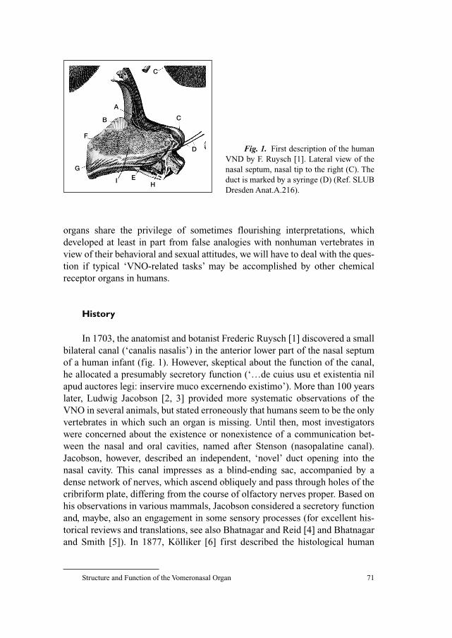
Structure and Function of the Vomeronasal Organ 71
organs share the privilege of sometimes flourishing interpretations, which
developed at least in part from false analogies with nonhuman vertebrates in
view of their behavioral and sexual attitudes, we will have to deal with the ques-
tion if typical ‘VNO-related tasks’ may be accomplished by other chemical
receptor organs in humans.
History
In 1703, the anatomist and botanist Frederic Ruysch [1] discovered a small
bilateral canal (‘canalis nasalis’) in the anterior lower part of the nasal septum
of a human infant (fig. 1). However, skeptical about the function of the canal,
he allocated a presumably secretory function (‘…de cuius usu et existentia nil
apud auctores legi: inservire muco excernendo existimo’). More than 100 years
later, Ludwig Jacobson [2, 3] provided more systematic observations of the
VNO in several animals, but stated erroneously that humans seem to be the only
vertebrates in which such an organ is missing. Until then, most investigators
were concerned about the existence or nonexistence of a communication bet-
ween the nasal and oral cavities, named after Stenson (nasopalatine canal).
Jacobson, however, described an independent, ‘novel’ duct opening into the
nasal cavity. This canal impresses as a blind-ending sac, accompanied by a
dense network of nerves, which ascend obliquely and pass through holes of the
cribriform plate, differing from the course of olfactory nerves proper. Based on
his observations in various mammals, Jacobson considered a secretory function
and, maybe, also an engagement in some sensory processes (for excellent his-
torical reviews and translations, see also Bhatnagar and Reid [4] and Bhatnagar
and Smith [5]). In 1877, Kölliker [6] first described the histological human
A
B
F
G
I EH
D
C
C
Fig. 1. First description of the human
VND by F. Ruysch [1]. Lateral view of the
nasal septum, nasal tip to the right (C). The
duct is marked by a syringe (D) (Ref. SLUB
Dresden Anat.A.216).

Witt/Wozniak 72´
VNN
S
S
L
V
Ca
*
*
*
*
*
Pa b
c d
P
OE
Fig. 2. VNO in the newborn rat (a, b) and in a 8-week-old human fetus (c, d). Frontal
section through the cartilage of the nasal septum (S). a The u-shaped VND outlined with a
rectangle is shown in detail in (b). OE � Olfactory epithelium; P � palate; VNN � fibers of
the vomeronasal nerve. Iron-hematoxylin stain. b The lumen (L) of the VND is lined with a
lateral nonsensory epithelium (left) belonging to the mushroom body that also contains the
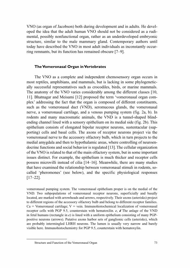
Structure and Function of the Vomeronasal Organ 73
VNO (as organ of Jacobson) both during development and in adults. He devel-
oped the idea that the adult human VNO should not be considered as a rudi-
mental, possibly nonfunctional organ, rather as an underdeveloped embryonic
structure, similar to the male mammary gland. Contemporary authors until
today have described the VNO in most adult individuals as inconstantly occur-
ring remnants, but its function has remained obscure [7–9].
The Vomeronasal Organ in Vertebrates
The VNO as a complete and independent chemosensory organ occurs in
most reptiles, amphibians, and mammals, but is lacking in some phylogenetic-
ally successful representatives such as crocodiles, birds, or marine mammals.
The anatomy of the VNO varies considerably among the different classes [10,
11]. Bhatnagar and Meisami [12] proposed the term ‘vomeronasal organ com-
plex’ addressing the fact that the organ is composed of different constituents,
such as the vomeronasal duct (VND), seromucous glands, the vomeronasal
nerve, a vomeronasal cartilage, and a venous pumping system (fig. 2a, b). In
rodents and many macrosmatic animals, the VND is a tunnel-shaped blind-
ending channel lined with a sensory epithelium on its medial side (fig. 2b). This
epithelium consists of elongated bipolar receptor neurons, sustentacular (sup-
porting) cells and basal cells. The axons of receptor neurons project via the
vomeronasal nerve to the accessory olfactory bulb, which in turn projects to the
medial amygdala and then to hypothalamic areas, where controlling of neuroen-
docrine functions and social behavior is regulated [13]. The cellular organization
of the VNO is related to that of the main olfactory system, but in some important
issues distinct. For example, the epithelium is much thicker and receptor cells
possess microvilli instead of cilia [14–16]. Meanwhile, there are many studies
that have examined the relationship between vomeronasal stimuli in rodents, so-
called ‘pheromones’ (see below), and the specific physiological responses
[17–22].
vomeronasal pumping system. The vomeronasal epithelium proper is on the medial of the
VND. Two subpopulations of vomeronasal receptor neurons, superficially and basally
located, are marked with arrowheads and arrows, respectively. Their axons (asterisks) project
to different regions of the accessory olfactory bulb and belong to different receptor families.
Ca � Vomeronasal cartilage; V � vein. Immunohistochemical localization of vomeronasal
receptor cells with PGP 9.5, counterstain with hematoxilin. c, d The anlage of the VND
in fetal humans (rectangle in c) is lined with a uniform epithelium consisting of many PGP-
positive neurons (arrows). Putative axons harbor sets of ganglionic cells (asterisks), which
are probably intermingled LHRH neurons. The lumen is usually very narrow and barely
visible here. Immunohistochemistry for PGP 9.5, counterstain with hematoxylin.
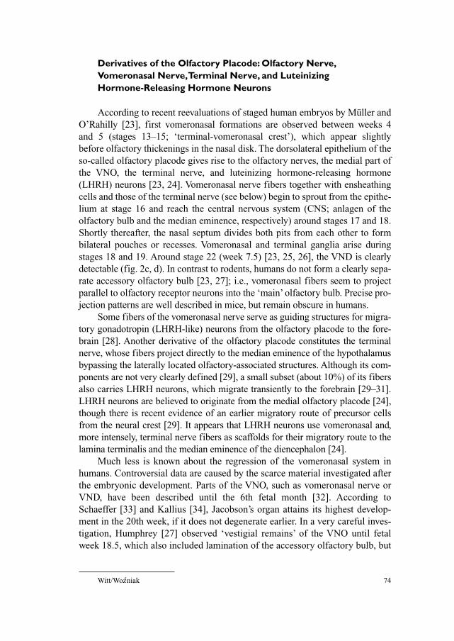
Witt/Wozniak 74´
Derivatives of the Olfactory Placode: Olfactory Nerve,Vomeronasal Nerve,Terminal Nerve, and Luteinizing Hormone-Releasing Hormone Neurons
According to recent reevaluations of staged human embryos by Müller and
O’Rahilly [23], first vomeronasal formations are observed between weeks 4
and 5 (stages 13–15; ‘terminal-vomeronasal crest’), which appear slightly
before olfactory thickenings in the nasal disk. The dorsolateral epithelium of the
so-called olfactory placode gives rise to the olfactory nerves, the medial part of
the VNO, the terminal nerve, and luteinizing hormone-releasing hormone
(LHRH) neurons [23, 24]. Vomeronasal nerve fibers together with ensheathing
cells and those of the terminal nerve (see below) begin to sprout from the epithe-
lium at stage 16 and reach the central nervous system (CNS; anlagen of the
olfactory bulb and the median eminence, respectively) around stages 17 and 18.
Shortly thereafter, the nasal septum divides both pits from each other to form
bilateral pouches or recesses. Vomeronasal and terminal ganglia arise during
stages 18 and 19. Around stage 22 (week 7.5) [23, 25, 26], the VND is clearly
detectable (fig. 2c, d). In contrast to rodents, humans do not form a clearly sepa-
rate accessory olfactory bulb [23, 27]; i.e., vomeronasal fibers seem to project
parallel to olfactory receptor neurons into the ‘main’ olfactory bulb. Precise pro-
jection patterns are well described in mice, but remain obscure in humans.
Some fibers of the vomeronasal nerve serve as guiding structures for migra-
tory gonadotropin (LHRH-like) neurons from the olfactory placode to the fore-
brain [28]. Another derivative of the olfactory placode constitutes the terminal
nerve, whose fibers project directly to the median eminence of the hypothalamus
bypassing the laterally located olfactory-associated structures. Although its com-
ponents are not very clearly defined [29], a small subset (about 10%) of its fibers
also carries LHRH neurons, which migrate transiently to the forebrain [29–31].
LHRH neurons are believed to originate from the medial olfactory placode [24],
though there is recent evidence of an earlier migratory route of precursor cells
from the neural crest [29]. It appears that LHRH neurons use vomeronasal and,
more intensely, terminal nerve fibers as scaffolds for their migratory route to the
lamina terminalis and the median eminence of the diencephalon [24].
Much less is known about the regression of the vomeronasal system in
humans. Controversial data are caused by the scarce material investigated after
the embryonic development. Parts of the VNO, such as vomeronasal nerve or
VND, have been described until the 6th fetal month [32]. According to
Schaeffer [33] and Kallius [34], Jacobson’s organ attains its highest develop-
ment in the 20th week, if it does not degenerate earlier. In a very careful inves-
tigation, Humphrey [27] observed ‘vestigial remains’ of the VNO until fetal
week 18.5, which also included lamination of the accessory olfactory bulb, but
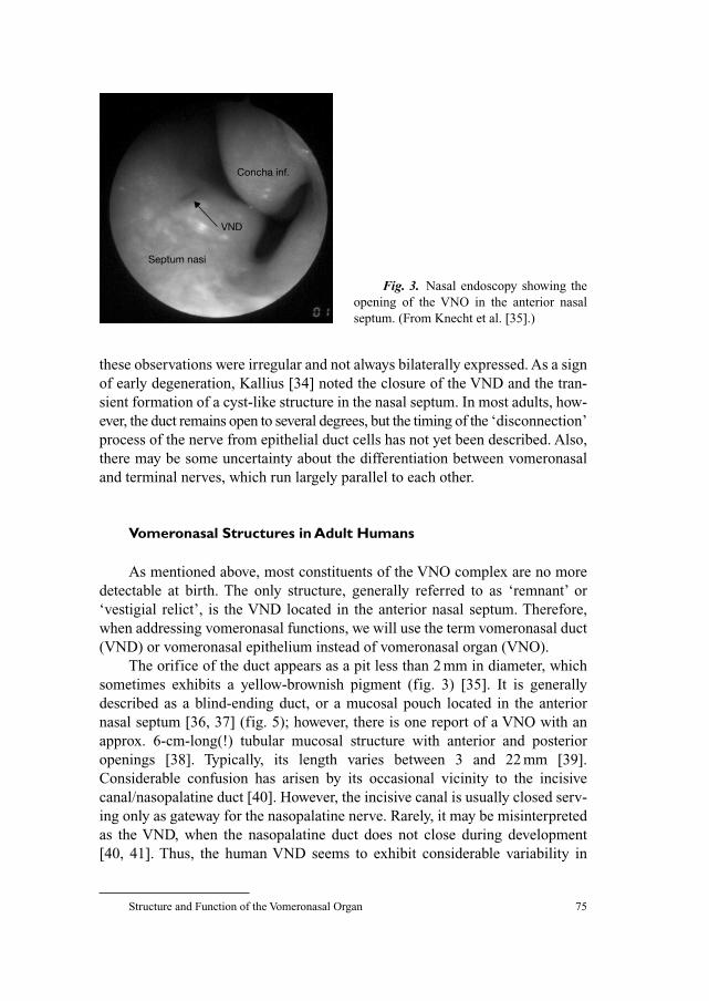
Structure and Function of the Vomeronasal Organ 75
these observations were irregular and not always bilaterally expressed. As a sign
of early degeneration, Kallius [34] noted the closure of the VND and the tran-
sient formation of a cyst-like structure in the nasal septum. In most adults, how-
ever, the duct remains open to several degrees, but the timing of the ‘disconnection’
process of the nerve from epithelial duct cells has not yet been described. Also,
there may be some uncertainty about the differentiation between vomeronasal
and terminal nerves, which run largely parallel to each other.
Vomeronasal Structures in Adult Humans
As mentioned above, most constituents of the VNO complex are no more
detectable at birth. The only structure, generally referred to as ‘remnant’ or
‘vestigial relict’, is the VND located in the anterior nasal septum. Therefore,
when addressing vomeronasal functions, we will use the term vomeronasal duct
(VND) or vomeronasal epithelium instead of vomeronasal organ (VNO).
The orifice of the duct appears as a pit less than 2 mm in diameter, which
sometimes exhibits a yellow-brownish pigment (fig. 3) [35]. It is generally
described as a blind-ending duct, or a mucosal pouch located in the anterior
nasal septum [36, 37] (fig. 5); however, there is one report of a VNO with an
approx. 6-cm-long(!) tubular mucosal structure with anterior and posterior
openings [38]. Typically, its length varies between 3 and 22 mm [39].
Considerable confusion has arisen by its occasional vicinity to the incisive
canal/nasopalatine duct [40]. However, the incisive canal is usually closed serv-
ing only as gateway for the nasopalatine nerve. Rarely, it may be misinterpreted
as the VND, when the nasopalatine duct does not close during development
[40, 41]. Thus, the human VND seems to exhibit considerable variability in
Concha inf.
Septum nasi
VND
Fig. 3. Nasal endoscopy showing the
opening of the VNO in the anterior nasal
septum. (From Knecht et al. [35].)
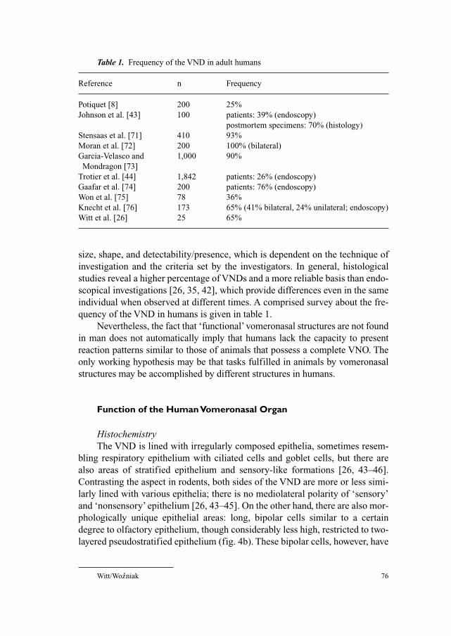
Witt/Wozniak 76´
size, shape, and detectability/presence, which is dependent on the technique of
investigation and the criteria set by the investigators. In general, histological
studies reveal a higher percentage of VNDs and a more reliable basis than endo-
scopical investigations [26, 35, 42], which provide differences even in the same
individual when observed at different times. A comprised survey about the fre-
quency of the VND in humans is given in table 1.
Nevertheless, the fact that ‘functional’ vomeronasal structures are not found
in man does not automatically imply that humans lack the capacity to present
reaction patterns similar to those of animals that possess a complete VNO. The
only working hypothesis may be that tasks fulfilled in animals by vomeronasal
structures may be accomplished by different structures in humans.
Function of the Human Vomeronasal Organ
HistochemistryThe VND is lined with irregularly composed epithelia, sometimes resem-
bling respiratory epithelium with ciliated cells and goblet cells, but there are
also areas of stratified epithelium and sensory-like formations [26, 43–46].
Contrasting the aspect in rodents, both sides of the VND are more or less simi-
larly lined with various epithelia; there is no mediolateral polarity of ‘sensory’
and ‘nonsensory’ epithelium [26, 43–45]. On the other hand, there are also mor-
phologically unique epithelial areas: long, bipolar cells similar to a certain
degree to olfactory epithelium, though considerably less high, restricted to two-
layered pseudostratified epithelium (fig. 4b). These bipolar cells, however, have
Table 1. Frequency of the VND in adult humans
Reference n Frequency
Potiquet [8] 200 25%
Johnson et al. [43] 100 patients: 39% (endoscopy)
postmortem specimens: 70% (histology)
Stensaas et al. [71] 410 93%
Moran et al. [72] 200 100% (bilateral)
Garcia-Velasco and 1,000 90%
Mondragon [73]
Trotier et al. [44] 1,842 patients: 26% (endoscopy)
Gaafar et al. [74] 200 patients: 76% (endoscopy)
Won et al. [75] 78 36%
Knecht et al. [76] 173 65% (41% bilateral, 24% unilateral; endoscopy)
Witt et al. [26] 25 65%

Structure and Function of the Vomeronasal Organ 77
no connection to nerve fibers, thus they do not seem to be able to transfer
potential information, if any, via the classical neuronal way to the CNS.
Moreover, bipolar, sensory-like cells of the vomeronasal epithelium are keratin-
positive, i.e., they provide intermediate filaments typical of epithelial cells, and
not of differentiated neuroepithelial cells, which do not possess keratin fila-
ments [26, 44]. Generally, the characteristic vomeronasal cells do not express
neuronal markers such as protein gene product 9.5 (PGP 9.5), olfactory marker
protein, or neuronal tubulin, though a very small subset of cells may contain
one of these proteins [26, 47]. Instead, there are some remarkable proteins,
which point to a specialized function in terms of cell signalling such as
caveolins, which were described in olfactory cells [48]. �1-Integrin also seem
to play a role, though a specific function of these proteins has not yet been
shown. Nevertheless, surrounding epithelia, e.g. of the nasal septum as well as
Olfactory bulb
Nasal cartilage
Nasal mucosa
Canalisincisivus
Vomeronasalduct
N. nasopalatinus
Fig. 4. Schematic drawing of the location of the VND. Paramedian-sagittal section
through the nasal cavity. The septal mucosa is partially split off to demonstrate the obliquely
ascending VND. Underneath runs the nasopalatine nerve which enters the oral cavity by tra-
versing through the incisive canal. There are no connections with the olfactory bulb.
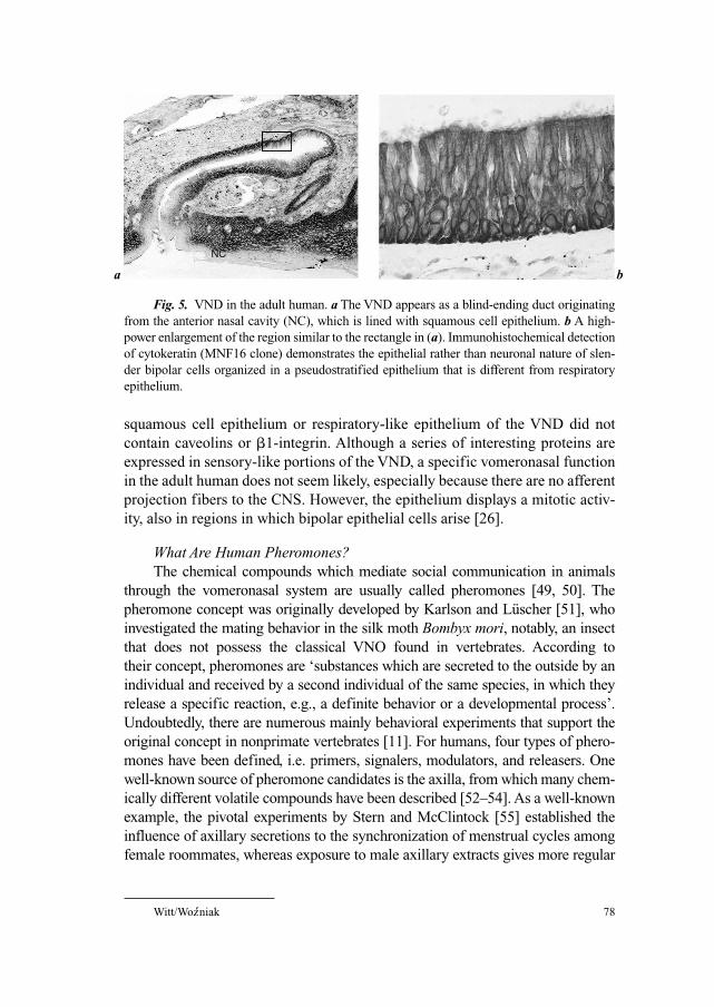
Witt/Wozniak 78´
squamous cell epithelium or respiratory-like epithelium of the VND did not
contain caveolins or �1-integrin. Although a series of interesting proteins are
expressed in sensory-like portions of the VND, a specific vomeronasal function
in the adult human does not seem likely, especially because there are no afferent
projection fibers to the CNS. However, the epithelium displays a mitotic activ-
ity, also in regions in which bipolar epithelial cells arise [26].
What Are Human Pheromones?The chemical compounds which mediate social communication in animals
through the vomeronasal system are usually called pheromones [49, 50]. The
pheromone concept was originally developed by Karlson and Lüscher [51], who
investigated the mating behavior in the silk moth Bombyx mori, notably, an insect
that does not possess the classical VNO found in vertebrates. According to
their concept, pheromones are ‘substances which are secreted to the outside by an
individual and received by a second individual of the same species, in which they
release a specific reaction, e.g., a definite behavior or a developmental process’.
Undoubtedly, there are numerous mainly behavioral experiments that support the
original concept in nonprimate vertebrates [11]. For humans, four types of phero-
mones have been defined, i.e. primers, signalers, modulators, and releasers. One
well-known source of pheromone candidates is the axilla, from which many chem-
ically different volatile compounds have been described [52–54]. As a well-known
example, the pivotal experiments by Stern and McClintock [55] established the
influence of axillary secretions to the synchronization of menstrual cycles among
female roommates, whereas exposure to male axillary extracts gives more regular
a
NC
b
Fig. 5. VND in the adult human. a The VND appears as a blind-ending duct originating
from the anterior nasal cavity (NC), which is lined with squamous cell epithelium. b A high-
power enlargement of the region similar to the rectangle in (a). Immunohistochemical detection
of cytokeratin (MNF16 clone) demonstrates the epithelial rather than neuronal nature of slen-
der bipolar cells organized in a pseudostratified epithelium that is different from respiratory
epithelium.
Structure and Function of the Vomeronasal Organ 79
menstrual cycles [56]. Hypothalamic activity can be measured after exposure of
subjects to sex-hormone-like substances such as androstadien-3-one (similar to
testosterone) or oestra-1,3,5(10),16-tetraen-3-ol, similar to estrogen [57].
Quantitatively, unsaturated acids such as (E)-3-methyl-2-hexenoic acid play a
greater role than previously thought, e.g., most often cited ‘human-related’,
androstenone, androstenol, and 4,16-androstadien-3-one [53, 58]. More recently,
Meredith [50] proposed to restrict the definition for pheromones by inclusion of
mutual benefit to sender and receiver. We will not discuss the particular chemistry
in detail (for excellent reviews, see Wysocki and Preti [59] and Halpern and
Martinez-Marcos [11]). Of some importance is the chemical similarity between
pheromone candidates in humans and those of other animals [59] and the relation-
ship of complex ‘odor prints’ with the immune system, e.g. the major histocom-
patibility complex. Axillary volatiles collected from T-shirts allowed individuals to
identify their own odor and that of closely related persons, e.g. spouses [60]. These
‘signaler pheromones’, represented by paternally inherited human lymphocyte
antigen alleles, may be responsible for mate choice [61]. In humans, the receptor
region for pheromone candidates such as androstenone does not seem to be the
VND, as own studies with experimentally covered VND entrance did not affect
olfactory function or androstenone sensitivity [62].
What and Where Are the Receptors for Human Pheromone Candidates?The observation that a series of particular compounds can exert specific
behavioral and physiological effects requires the presence and genetic expression
of specific receptors. The superfamily of thyroid hormones, approx. 300
vomeronasal receptors in mice, are structurally different from odorant receptors
[63], but they share the common 7-transmembrane domain structure. Most of the
genes encoding the vomeronasal receptors belong to at least two different families
(VR1 and VR2), but 95% of all V1R genes are pseudogenes, i.e. are nonfunctional
in man [64]; receptor proteins present in many animals [13, 65, 66] have not yet
been identified in the human vomeronasal epithelium. There is no intact human
V2R gene [67]. At least one of them is expressed at the mRNA level in the olfac-
tory mucosa [68]. On the other hand, several studies have established that not all
stimuli, which were believed to be VNO related, require a complete VNO [69, 70].
Also, major histocompatibility complex-related odor type recognition does not
seem to be a monopoly of the VNO [70]. Thus, the most likely binding sites for
human pheromone candidates are receptor cells within the olfactory epithelium.
Acknowledgements
The authors are grateful for the expert immunohistochemical work of Mrs. Anja Neisser
and the artwork of Mrs. I. Beck.
Witt/Wozniak 80´
References
1 Ruysch F: Thesaurus anatomicus tertius. Amstelaedami, Wolters, 1703, p 49.
2 Jacobson L: Description anatomique d’un organe observé dans les mammifères (reported by
M Cuvier; translated into English by Bhatnagar and Reid, 1996). Ann Mus Hist Nat (Paris) 1811;18:
412–424.
3 Jacobson L: Anatomisk beskrivelse over et nyt organ i huusdyrenes naese. Veter Salesk Skrift
1813;2:209–246.
4 Bhatnagar KP, Reid KH: The human vomeronasal organ. 1. Historical perspectives. A study of
Ruysch’s (1703) and Jacobson’s (1811) reports on the vomeronasal organ with comparative com-
ments and English translations. Biomed Res 1996;7:219–229.
5 Bhatnagar KP, Smith TD: The human vomeronasal organ. 5. An interpretation of its discovery by
Ruysch, Jacobson, or Kolliker, with an English translation of Kolliker (1877). Anat Rec 2003;270B: 4–15.
6 Kölliker A: Ueber die Jacobsonschen Organe des Menschen. Leipzig, Engelmann, 1877.
7 Merkel F: Jacobson’sches Organ und Papilla palatina beim Menschen. Festschrift zum fünf-
zigjährigen Doktorjubiläum des Herrn Geheimrat A v Kölliker. Anat Hefle, Wiesbaden I., 1892,
pp 213–232.
8 Potiquet A: Du canal de Jacobson. De la possibilité de le reconnaître sur le vivant et de son rôle
probable dans la pathogénie de certaines lésions de la cloison nasale. Rev Laryngol Otol Rhinol
1891;24:737–753.
9 Zuckerkandl E: Normale und pathologische Anatomie der Nasenhöhle und ihrer pneumatischen
Anhänge. Wien, Braumüller, 1893.
10 Garrosa M, Gayoso MJ, Esteban FJ: Prenatal development of the mammalian vomeronasal organ.
Microsc Res Tech 1998;41:456–470.
11 Halpern M, Martinez-Marcos A: Structure and function of the vomeronasal system: an update.
Prog Neurobiol 2003;70:245–318.
12 Bhatnagar KP, Meisami E: Vomeronasal organ in bats and primates: extremes of structural vari-
ability and its phylogenetic implications. Microsc Res Tech 1998;43:465–475.
13 Luo M, Katz LC: Encoding pheromonal signals in the mammalian vomeronasal system. Curr
Opin Neurobiol 2004;14:428–434.
14 Yoshida-Matsuoka J, Osada T, Mori Y, Ichikawa M: A developmental study using three antibodies
(VOBM1, VOBM2, and VOM2): immunocytochemical and electron microscopical analysis of the
luminal surface of the rat vomeronasal sensory epithelium. Anat Embryol (Berl) 1999;199:215–224.
15 Höfer D, Shin DW, Drenckhahn D: Identification of cytoskeletal markers for the different
microvilli and cell types of the rat vomeronasal sensory epithelium. J Neurocytol 2000;29:
147–156.
16 Menco BP, Carr VM, Ezeh PI, Liman ER, Yankova MP: Ultrastructural localization of G-proteins
and the channel protein TRP2 to microvilli of rat vomeronasal receptor cells. J Comp Neurol
2001;438:468–489.
17 Mombaerts P: Seven-transmembrane proteins as odorant and chemosensory receptors. Science
1999;286:707–711.
18 Takami S: Recent progress in the neurobiology of the vomeronasal organ. Microsc Res Tech
2002;58:228–250.
19 Zufall F, Kelliher KR, Leinders-Zufall T: Pheromone detection by mammalian vomeronasal neu-
rons. Microsc Res Tech 2002;58:251–260.
20 Zhang J, Webb DM: Evolutionary deterioration of the vomeronasal pheromone transduction path-
way in catarrhine primates. Proc Natl Acad Sci USA 2003;100:8337–8341.
21 Zhang J-X, Ni J, Ren X-J, Sun L, Zhang Z-B, Wang Z-W: Possible coding for recognition of sexes,
individuals and species in anal gland volatiles of Mustela eversmanni and M. sibirica. Chem
Senses 2003;28:381–388.
22 Restrepo D, Arellano J, Oliva A, Schaefer M, Lin W: Emerging views on the distinct but related
roles of the main and accessory olfactory systems in responsiveness to chemosensory signals in
mice. Horm Behav 2004;46:247–256.
23 Müller F, O’Rahilly R: Olfactory structures in staged human embryos. Cells Tissues Organs
2004;178:93–116.
Structure and Function of the Vomeronasal Organ 81
24 Schwanzel-Fukuda M: Origin and migration of luteinizing hormone-releasing hormone neurons
in mammals. Microsc Res Tech 1999;44:2–10.
25 Kreutzer EW, Jafek BW: The vomeronasal organ of Jacobson in the human embryo and fetus.
Otolaryngol Head Neck Surg 1980;88:119–123.
26 Witt M, Georgiewa B, Knecht M, Hummel T: On the chemosensory nature of the vomeronasal
epithelium in adult humans. Histochem Cell Biol 2002;117:493–509.
27 Humphrey T: The development of the olfactory and the accessory olfactory formations in human
embryos and fetuses. J Comp Neurol 1940;73:431–468.
28 Cummings DM, Brunjes PC: Migrating luteinizing hormone-releasing hormone (LHRH) neurons
and processes are associated with a substrate that expresses S100. Brain Res Dev Brain Res
1995;88:148–157.
29 von Bartheld CS: The terminal nerve and its relation with extrabulbar ‘olfactory’ projections:
lessons from lampreys and lungfishes. Microsc Res Tech 2004;65:13–24.
30 Kjaer I, Fischer Hansen B: The human vomeronasal organ: prenatal developmental stages and dis-
tribution of luteinizing hormone-releasing hormone. Eur J Oral Sci 1996;104:34–40.
31 Schwanzel-Fukuda M, Crossin KL, Pfaff DW, Bouloux PM, Hardelin JP, Petit C: Migration of
luteinizing hormone-releasing hormone (LHRH) neurons in early human embryos. J Comp
Neurol 1996;366:547–557.
32 McCotter RE: The connection of the vomero-nasal nerves with the accessory olfactory bulb in the
opossum and other mammals. Anat Rec 1912;6:299–318.
33 Schaeffer JP: The Nose, Parasinal Sinuses, Nasolacrimal Passageways, and Olfactory Organ in
Man. Philadelphia, Blakiston, 1920.
34 Kallius E: Geruchsorgan (organon olfactus). Bardeleben’s Handbuch der Anatomie des Menschen.
Jena, Fischer, 1905.
35 Knecht M, Kuhnau D, Hüttenbrink KB, Witt M, Hummel T: Frequency and localization of the
putative vomeronasal organ in humans in relation to age and gender. Laryngoscope 2001;111:
448–452.
36 Smith TD, Buttery TA, Bhatnagar KP, Burrows AM, Mooney MP, Siegel MI: Anatomical position
of the vomeronasal organ in postnatal humans. Ann Anat 2001;183:475–479.
37 Smith TD, Bhatnagar KP, Shimp KL, Kinzinger JH, Bonar CJ, Burrows AM, Mooney MP, Siegel
MI: Histological definition of the vomeronasal organ in humans and chimpanzees, with a compar-
ison to other primates. Anat Rec 2002;267:166–176.
38 Mangakis M: Ein Fall von Jacobson’schen Organen beim Erwachsenen. Anat Anz 1902;21:
106–109.
39 Anton W: Beiträge zur Kenntnis des Jacobson’schen Organes bei Erwachsenen. Z Heilk
1895;16:355–372.
40 Jacob S, Zelano B, Gungor A, Abbott D, Naclerio R, McClintock MK: Location and gross mor-
phology of the nasopalatine duct in human adults. Arch Otolaryngol Head Neck Surg 2000;126:
741–748.
41 Knecht M, Kittner T, Beleites T, Hüttenbrink KB, Hummel T, Witt M: Morphological and radio-
logic evaluation of the human nasopalatine duct. Ann Otol Rhinol Laryngol 2005;114:229–232.
42 Zbar RI, Zbar LI, Dudley C, Trott SA, Rohrich RJ, Moss RL: A classification schema for the
vomeronasal organ in humans. Plast Reconstr Surg 2000;105:1284–1288.
43 Johnson A, Josephson R, Hawke M: Clinical and histological evidence for the presence of the
vomeronasal (Jacobson’s) organ in adult humans. J Otolaryngol 1985;14:71–79.
44 Trotier D, Eloit C, Wassef M, Talmain G, Bensimon JL, Doving KB, Ferrand J: The vomeronasal
cavity in adult humans. Chem Senses 2000;25:369–380.
45 Bhatnagar KP, Smith TD: The human vomeronasal organ. 3. Postnatal development from infancy
to the ninth decade. J Anat 2001;199:289–302.
46 Bhatnagar KP, Smith TD, Winstead W: The human vomeronasal organ. 4. Incidence, topography,
endoscopy, and ultrastructure of the nasopalatine recess, nasopalatine fossa, and vomeronasal
organ. Am J Rhinol 2002;16:343–350.
47 Takami S, Getchell ML, Chen Y, Monti-Bloch L, Berliner DL, Stensaas LJ, Getchell TV:
Vomeronasal epithelial cells of the adult human express neuron-specific molecules. Neuroreport
1993;4:375–378.
Witt/Wozniak 82´
48 Schreiber S, Fleischer J, Breer H, Boekhoff I: A possible role for caveolin as a signaling organizer
in olfactory sensory membranes. J Biol Chem 2000;275:24115–24123.
49 Beauchamp GK, Doty RL, Moulton DG, Mugford RA: The pheromone concept in mammalian
chemical communication: a critique; in Doty RL (ed): Mammalian Olfaction, Reproductive
Processes, and Behavior. New York, Academic Press, 1976, pp 143–160.
50 Meredith M: Human vomeronasal organ function: a critical review of best and worst cases. Chem
Senses 2001;26:433–445.
51 Karlson P, Lüscher M: Pheromones: a new term for a class of biologically active substances.
Nature 1959;183:55–56.
52 Spielman AI, Zeng XN, Leyden JJ, Preti G: Proteinaceous precursors of human axillary odor: iso-
lation of two novel odor-binding proteins. Experientia 1995;51:40–47.
53 Zeng C, Spielman AI, Vowels BR, Leyden JJ, Biemann K, Preti G: A human axillary odorant is
carried by apolipoprotein D. Proc Natl Acad Sci USA 1996;93:6626–6630.
54 Natsch A, Gfeller H, Gygax P, Schmid J, Acuna G: A specific bacterial aminoacylase cleaves odor-
ant precursors secreted in the human axilla. J Biol Chem 2003;278:5718–5727.
55 Stern K, McClintock MK: Regulation of ovulation by human pheromones. Nature
1998;392:177–179.
56 Cutler WB, Preti G, Krieger A, Huggins GR, Garcia CR and Lawley HJ: Human axillary secre-
tions influence women’s menstrual cycles: the role of donor extract from men. Horm Behav
1986;20:463–473.
57 Savic I, Berglund H, Gulyas B, Roland P: Smelling of odorous sex hormone-like compounds
causes sex-differentiated hypothalamic activations in humans. Neuron 2001;31:661–668.
58 Gower DB, Ruparelia BA: Olfaction in humans with special reference to odorous 16-androstenes:
their occurrence, perception and possible social, psychological and sexual impact. J Endocrinol
1993;137:167–187.
59 Wysocki CJ, Preti G: Facts, fallacies, fears, and frustrations with human pheromones. Anat Rec
2004;281A:1201–1211.
60 Porter RH, Cernoch JM, Balogh RD: Odor signatures and kin recognition. Physiol Behav
1985;34:445–448.
61 Jacob S, McClintock MK, Zelano B, Ober C: Paternally inherited HLA alleles are associated with
women’s choice of male odor. Nat Genet 2002;30:175–179.
62 Knecht M, Lundstrom JN, Witt M, Hüttenbrink KB, Heilmann S, Hummel T: Assessment of olfac-
tory function and androstenone odor thresholds in humans with or without functional occlusion of
the vomeronasal duct. Behav Neurosci 2003;117:1135–1141.
63 Zhang X, Rodriguez I, Mombaerts P, Firestein S: Odorant and vomeronasal receptor genes in two
mouse genome assemblies. Genomics 2004;83:802–811.
64 Kouros-Mehr H, Pintchovski S, Melnyk J, Chen YJ, Friedman C, Trask B, Shizuya H:
Identification of non-functional human VNO receptor genes provides evidence for vestigiality of
the human VNO. Chem Senses 2001;26:1167–1174.
65 Rodriguez I, Feinstein P, Mombaerts P: Variable patterns of axonal projections of sensory neurons
in the mouse vomeronasal system. Cell 1999;97:199–208.
66 Matsuoka M, Yoshida-Matsuoka J, Iwasaki N, Norita M, Costanzo RM, Ichikawa M:
Immunocytochemical study of G(i)2alpha and G(o)alpha on the epithelium surface of the rat
vomeronasal organ. Chem Senses 2001;26:161–166.
67 Mombaerts P: Genes and ligands for odorant, vomeronasal and taste receptors. Nat Rev Neurosci
2004;5:263–278.
68 Giorgi D, Friedman C, Trask BJ, Rouquier S: Characterization of nonfunctional VIR-like
pheromone receptor sequences in human. Genome Res 2000;10:1979–1985.
69 Woodley SK, Cloe AL, Waters P, Baum MJ: Effects of vomeronasal organ removal on olfactory
sex discrimination and odor preferences of female ferrets. Chem Senses 2004;29:659–669.
70 Wysocki C, Yamazaki K, Curran M, Wysocki L, Beauchamp G: Mice (Mus musculus) lacking a
vomeronasal organ can discriminate MHC-determined odortypes. Horm Behav 2004;46:241–246.
71 Stensaas LJ, Lavker RM, Monti-Bloch L, Grosser BI, Berliner DL: Ultrastructure of the human
vomeronasal organ. J Steroid Biochem Mol Biol 1991;39:553–560.

Structure and Function of the Vomeronasal Organ 83
72 Moran DT, Jafek BW, Rowley JC: The vomeronasal (Jacobson’s) organ in man: ultrastructure and
frequency of occurrence. J Steroid Biochem Mol Biol 1991;39:545–552.
73 Garcia-Velasco J, Mondragon M: The incidence of the vomeronasal organ in 1,000 human sub-
jects and its possible clinical significance. J Steroid Biochem Mol Biol 1991;39:561–563.
74 Gaafar HA, Tantawy AA, Melis AA, Hennawy DM, Shehata HM: The vomeronasal (Jacobson’s)
organ in adult humans: frequency of occurrence and enzymatic study. Acta Otolaryngol (Stockh)
1998;118:409–412.
75 Won J, Mair EA, Bolger WE, Conran RM: The vomeronasal organ: an objective anatomic analysis
of its prevalence. Ear Nose Throat J 2000;79:600–605.
Martin Witt, MD, PhD
Smell and Taste Clinic
Dept. of Otorhinolaryngology, and
Department of Anatomy, University of Technology
Fetscherstrasse 74
DE–01307 Dresden (Germany)
Tel. �49 351 4586103, Fax �49 351 4586303, E-Mail [email protected]
Hummel T, Welge-Lüssen A (eds): Taste and Smell. An Update.
Adv Otorhinolaryngol. Basel, Karger, 2006, vol 63, pp 84–98
Assessment of Olfactory Function
Thomas Hummela, Antje Welge-Lüessenb
aSmell & Taste Clinic, Department of Otorhinolaryngology, University of Dresden
Medical School (‘Technische Universität Dresden’), Dresden, Germany; bDepartment
of Otorhinolaryngology, University of Basel, Basel, Switzerland
AbstractNumerous techniques are available for the investigation of chemosensory functions in
humans. They include psychophysical measures of chemosensory function, e.g. odor identi-
fication, odor discrimination, odor thresholds, odor memory, and retronasal perception of
odors. In order to assess changes related to the patients’ quality of life or effects of qualitative
olfactory dysfunction, questionnaires are being used. Measures relying to a lesser degree
on the subjects’ cooperation are e.g. chemosensory event-related potentials, odor-induced
changes of the EEG, the electroolfactogram, imaging techniques, or measures of respiration.
In a clinical context, however, psychophysical techniques are most frequently used, e.g. tests
for odor identification, and odor thresholds. Interpretation of results from these measures
is frequently supported by the assessment of chemosensory event-related potentials. Other
techniques await further standardization before they will become useful in a clinical context.
Copyright © 2006 S. Karger AG, Basel
Disturbances of the chemical senses are frequent. It is estimated that a
complete loss of the sense of smell is found in at least 1% of the US population
[1–3]. Recent epidemiological research indicates that at least 5% of the popula-
tion exhibit a significant loss of olfactory function rendering them functionally
anosmic [4, 5]. This appears to be largely due to aging as in individuals aged
53–97 years 24% were found to have impaired olfactory function [6]. Thus,
based on a survey, each year over 70,000 patients are treated in German ENT
clinics at least in part because of their olfactory loss [7].
Olfactory loss often appears to go unnoticed [8–10]. This seems to be
especially true in older people or patients with Alzheimer’s disease who are fre-
quently unaware of their olfactory deficit [6, 11]. Similar findings have been
reported in Parkinson’s disease [12], diabetes mellitus [13], laryngectomy [14],
or chronic renal failure [15]. While this highlights the limited attention the

Assessment of Olfactory Function 85
sense of smell receives in daily life [16], it also points towards the necessity to
measure olfactory function in order to obtain reliable information on the
patients’ olfactory abilities [17, 18].
During the last centuries, standardized tests of olfactory function have been
developed (e.g. The University of Pennsylvania Smell Identification Test, a
‘scratch and sniff’ odor identification test [19]; ‘sniffin’ sticks’, pen-like odor-
dispensing devices which include tests for odor identification, discrimination,
and thresholds [20, 21], or the Connecticut Chemosensory Clinical Research
Center Test, a combined odor identification and odor threshold test [22]). In
addition, methods are currently being developed to quantify qualitative olfactory
dysfunction, e.g. parosmia and phantosmia [23]. In terms of less biased mea-
sures of olfactory function, EEG-derived measures, such as the recording of
olfactory event-related potentials (ERPs), are available [24] plus a large array of
other techniques which are less well established in clinical routine.
Psychophysical Methods of Olfactory Testing
During psychophysical assessment of olfactory function, subjects/patients
are confronted with an odor and their response is monitored. The critical advan-
tage of this ‘low-tech’ approach is the speed of testing which allows psychophys-
ical tests to serve as quick screening tools for olfactory dysfunction [25–28].
While screening tests only differentiate between normal and pathologic
states, more extensive tests allow for the reliable discrimination between anos-
mic, hyposmic, and normosmic subjects, respectively. The ideal test should be
based on normative data acquired and validated on large samples of healthy
subjects and patients, respectively. This includes comparison of the results with
other validated tests and a good test-retest reliability. These requirements are
fulfilled only by a handful of olfactory tests [20, 21, 29–31]. The best-validated
olfactory tests include the University of Pennsylvania Smell Identification Test
[30], the Connecticut Chemosensory Clinical Research Center Test [29], and
the ‘sniffin’ sticks’ [20, 21].
Many tests are based on a forced-choice verbal identification of odors [20,
21, 29–33]. An odorant is presented at suprathreshold concentration and the sub-
ject has to identify the odor from a list of descriptors of odors (e.g. the subject gets
rose odor to smell, and is asked whether the perceived odor was ‘banana’, ‘anise’,
‘rose’, or ‘lilac’). This forced-choice procedure controls the subjects’ response
bias. Its strength lies in the fact that its concept is easily understood by both
patient and investigator. In addition, the simplicity of its administration is a clear
advantage over other olfactory tests. Odor identification tests based on forced
choice also potentially allow to detect malingerers since anosmic subjects will
Hummel/Welge-Lüessen 86
produce a few correct answers provided random selection of items. Accordingly,
if nonanosmic subjects willingly avoid selecting the correct items, they will have
a very low score below random probability, which may indicate malingering.
However, this method is insufficient for medicolegal investigations since well-
read or hyposmic malingerers may overcome these pitfalls.
Most tests are based on the identification of 10–40 odors – the more items
tested the more reliable the results. A major problem of odor identification is,
however, that it strongly taps onto the verbal abilities of the subject which, on
average, makes female subjects perform better than their male counterparts [34,
35]. In addition, odor identification tests have a strong cultural connotation as
not all odors are known equally well around the world. Tests used in North
America, for example, are composed of odors many of which are unfamiliar to
Europeans or Asians (e.g. root beer, or wintergreen). The odors tested should
therefore be adapted to the patients’ cultural background [36] in order to mini-
mize a familiarity bias in odor identification.
Other widely used test designs are threshold tests and tests of odor dis-
crimination – tests for odor memory are only rarely used in a clinical context
[37, 38]. In addition, tests for retronasal olfactory function are not in general
use [39] despite the fact that the orthonasal and retronasal olfactory function
can be dissociated in patients [40, 41].
The idea of threshold tests is to expose a subject repeatedly to ascending
and descending concentrations of the same odorant and to identify the least
detectable concentration for this individual odor [42]. Other techniques are
based on logistic regression [43, 44]. Discrimination tasks are frequently based
on a 3-alternative forced-choice technique. Two of the administered odors are
identical, one is different. The subjects’ task is to find out the different one. In
principal, tests for odor threshold/odor discrimination are nonverbal. In addi-
tion, they can be used repetitively – which is more difficult with odor identifi-
cation tests as subjects remember the answers they gave in previous tests of
odor identification.
It seems intuitive to assume that different olfactory tests address different
portions of olfactory processing, although there is no definitive proof for this
idea [45]. Generally, identification and discrimination tests are believed to
reflect central olfactory processing while thresholds are thought to reflect
peripheral aspects of olfactory function to a stronger degree. Accordingly, it has
been claimed by several authors [15, 46–51] that patients with diseases of the
central nervous processing of odorous information exhibit selective distur-
bances of discrimination and identification while threshold results (or other
measures of olfactory function, such as olfactory ERPs) may be normal.
Although this idea of a certain pattern pathognomonic for ‘central’ olfactory
dysfunction is highly attractive, the vast majority of studies have failed to
Assessment of Olfactory Function 87
confirm such typical pathology-associated patterns [52, 53]. One reliable and
recurrent test pattern in olfactory disturbance, however, may be a low threshold
and close to normal identification and discrimination in patients with sinunasal
disease [54].
Besides the solid body of literature and their clinical convenience, psy-
chophysical tests are limited insofar as they rely on the patients’ cooperation.
This becomes problematic in medicolegal investigations, or in subjects unable
to respond appropriately, e.g. demented, unconscientious, or inexperienced
patients.
Measures of Changes of Quality of Life Associated with Olfactory Loss
Loss of olfactory function affects the patients’ quality of life [10, 16]. This
impairment is especially severe in cases where patients develop qualitative
olfactory dysfunction such as parosmia and phantosmia [55, 56]. Such deficits
cannot be assessed with tests routinely administered to investigate quantitative
olfactory loss [57].
Several questionnaires are available to measure mood states or general
quality of life [58], e.g. the Beck Depression Inventory [59], ‘mood inventories’
[60], or the Short Form-36 Health Survey [61]. While these questionnaires
allow the quantification of changes in the patients’ quality of life in general,
only recently have questionnaires been introduced which specifically address
nasal dysfunction, e.g. the Sinonasal Outcome Test-16 [62]. Other question-
naires specifically address olfactory distortions, e.g. the Questionnaire for
Olfactory Dysfunction [23] or other scales [63–65]. They have been developed
in analogy to questionnaires used to quantify the degree of tinnitus [66]. Based
upon these questionnaires, it was found that patients with smell loss indicated
to be significantly impaired in areas of food, safety, personal hygiene, and addi-
tionally, in their sexual life [67, 68]. In addition, Varga et al. [69] presented a
questionnaire to assess the impact of chemosensory dysfunction on everyday
life which also includes utility-based or time trade-off scales, with a particular
focus on the value placed by patients on chemosensory function. Interestingly,
approximately half of the patients reported to be willing to spend more than
20% of their annual household income to successfully treat chemosensory dys-
function.
These developments seem to be of specific interest to studies on therapy
of olfactory dysfunctions as they allow the assessment of a dimension
related to olfactory loss which is not addressed by quantitative tests of olfac-
tory function.
Hummel/Welge-Lüessen 88
Chemosensory (Olfactory) Event-Related Potentials
In many clinics, chemosensory (olfactory) ERPs have become part of the
routine investigation of patients with olfactory loss (for a review, see Hummel
and Kobal [24]). According to the growing use of this technique, guidelines
have been published for both recording and reporting of chemosensory ERPs
[70, 71].
Chemosensory ERPs are derived from the EEG after intranasal chemical
stimulation. As revealed by means of magnetoencephalographic techniques, the
chemosensory ERPs’ cortical sources are found in the temporal and insular cor-
tices [72]. ERPs are recorded from the scalp; averaging is needed to increase
the signal-to-noise ratio [73–75], which requires repeated stimulation that, in
turn, makes the recording of olfactory ERPs a lengthy procedure.
Stimulators used to elicit chemosensory ERPs should allow for precise
control of stimulus concentration and duration of stimuli, the rise time of stimuli
should be in the range of 20 ms. Two major issues are of importance. (1) Chemical
stimulation should not lead to the concomitant activation of mechano- or
thermosensors of the nasal mucosa. Otherwise, the resulting ERPs will not reflect
specific responses to odors [73, 76, 77]. (2) Stimulants should be characterized
with regard to the degree to which they activate the trigeminal nerve [78–80].
Stimulants typically used to elicit chemosensory ERPs are vanillin, phenylethyl
alcohol, and H2S [73]; for relatively specific trigeminal stimulation, gaseous CO2
is frequently employed [81].
It has been established that chemosensory ERPs are useful in the detection
of malingering [82–85]. Typically, recordings are made after lateralized stimu-
lation with two olfactory stimuli (e.g. phenylethyl alcohol and H2S). In general,
the interstimulus interval used lies between 30 and 40 s [73], the stimulus dura-
tion is in the range of 200 ms [86]. In anosmic patients, only responses to
trigeminal stimulation can be elicited [82–84], and chemosensory ERPs dis-
criminate between normosmic and hyposmic states [87]. In addition, transient
changes of olfaction can be tracked by means of chemosensory ERPs [88–90].
The Human Electroolfactogram
Other than the cortically generated ERPs, electroolfactograms (EOGs)
allow the assessment of the peripheral input signal to the olfactory system
[73, 91, 92]. For recording, a tubular electrode is typically used (AgAgCl
electrode, insulating tubing with an inner diameter of 0.3 mm, filled with 1%
Ringer-agar, impedance �10 k� at 1 kHz [93]); an electrode placed contralat-
erally on the bridge of the nose serves as reference. Placement of the electrode’s
Assessment of Olfactory Function 89
tip on the mucosa is performed under endoscopical control [94]. After position-
ing, it is stabilized by means of adjustable clips on a frame similar to lensless
glasses [95]. Recordings are performed with a high-resistance amplifier (DC,
low pass 30 Hz, input resistance �10 M�).
Even under endoscopical control, EOGs cannot be recorded in all subjects
[96]. This may be due to the topographical distribution of specific olfactory
receptor neurons in combination with the relatively few number of odorants
used, or the presence of metaplasias of the olfactory epithelium the extent of
which may increase with the subjects’ age [97, 98].
While the EOG has been proven to be useful in basic research [93], its use
in the routine clinical diagnostic armamentarium of olfactory disorders is far
from being established. As noted above, this may partly be due to (a) the diffi-
culties associated with its recording, (b) the relatively poor reliability of the
recordings that are only successful in approximately 60–80% of the subjects,
which makes a negative result difficult to interpret, and (c) the strain imposed
on the patient since local anesthesia cannot be performed. Although recent
reports on ‘EOG’ recordings from the skin overlying the nose attracted much
interest [99] based on previous research by Getchell [100, 101], it seems highly
unlikely that the signal would be recordable from the cutaneous surface.
Contingent Negative Variation
The contingent negative variation (CNV) is a negative DC shift of the EEG
that occurs in expectation of a stimulus. Recording procedures start usually
with an initial warning stimulus (S1), which is followed by a second stimulus
(S2). Subjects are asked to respond to S2, e.g. by pushing a button. During the
interval between S1 and S2, the negativity slowly builds up, and breaks down
after occurrence of S2. This EEG-related component was first described for
visual and acoustic stimuli by Walter et al. [102]. Its first portion is related to S1
and is typically largest over frontal brain areas; the later part is related to the
preparation to respond to S2 and is typically largest over the motor cortex [103].
The CNV is largely governed by psychological variables [104–106].
CNV-based paradigms are applied in olfactory tests in patients.
Auffermann et al. [107] (see also Yamamoto [108]) used an odorant as S1. S2
was a tone that was only presented along with a certain odor. As the patients
expect the tone after having perceived this certain odor, the CNV develops.
Although this technique allows to test qualitative differences between odors, it
cannot be performed without the patient paying attention to the paradigm, and
even if subjects are cooperative, the CNV is not clearly present in all subjects. It
is therefore of limited use, e.g., for medicolegal investigations.
Hummel/Welge-Lüessen 90
EEG Changes in Response to Odors
In the 1950s, electroencephalographic changes were reported after presen-
tation of odorous stimuli [109]. As a rule, an arousal reaction was observed in
response to olfactory and trigeminal stimulation [110, 111]. Perbellini and
Scolari [112] investigated 50 patients by means of three stimulants, i.e. pyr-
idine, vanillin, and ‘rose’ odor. Their conclusion was that stimulus-induced
EEG changes would be particularly useful in malingering patients. However,
they also observed a number of cases where no arousal reaction could be
recorded, although the subjects reported an olfactory sensation. Thus, when
using this technique, only positive responses could be viewed as an unambigu-
ous result. In line with this finding, in an investigation on 24 subjects, no elec-
troencephalographic changes appeared when only weak odorous stimuli were
perceived [113].
Apart from research oriented towards the diagnosis of olfactory disorders,
EEG recordings have contributed to basic research, e.g. the study of subthresh-
old odor intensities where changes of the EEG have been observed even in the
presence of undetected odors [114]. Others reported that individual odors can
be discriminated by means of changes in the alpha-activity recorded at different
sites [115]. Thus, although recording and analysis of stimulus-related EEG
activity appear to be less complex than the recording of chemosensory ERP, it
seems to be premature to recommend the use of these measurements in a
medicolegal context.
Localization of Olfactory-Induced Activation in the Brain
Recent progress in the field of imaging opened the opportunity to study the
functional topography of the human olfactory system in detail [72, 116, 117].
There are three major techniques being used: positron emission tomography
[116, 118, 119], functional magnetic resonance imaging [120–122], and mag-
netic source imaging based on magnetoencephalography [123, 124]. While bio-
magnetic fields directly reflect neuronal activity, positron emission tomography
and functional magnetic resonance imaging reflect either changes in blood flow
or changes in metabolism which are epiphenomena of neuronal activity. Other
major differences between these techniques relate to their temporal and spatial
resolution [72]. All three techniques have been used extensively to perform
basic research, e.g. on olfactory-induced emotions, odor memory, mechanisms
of sniffing, or differences between orthonasal and retronasal olfactory percep-
tions [125], or age- and sex-related differences in terms of olfactory function
Assessment of Olfactory Function 91
[126]. However, in order to become relevant for routine clinical investigations
[127], these intriguing techniques await further standardization.
Other Measures of Olfactory Activation
Compared to EEG-related parameters, other measures of olfactory activa-
tion have never received the same degree of attention in a clinical context. For
example, respiratory changes in response to odorous stimulation have been
investigated in patients with olfactory loss [128–130]. Here, the perception of
an odorant is followed by changes in respiratory frequency and both amplitude
and pattern of the respiratory cycle. Investigations in respiratory changes fol-
lowing odorous stimulation have their place in research, e.g. in the investigation
of the perception of subthreshold olfactory stimuli [131, 132]. In fact, devices
for the clinical investigation of these respiratory changes are currently being
developed [133].
Since the 1920s, the psychogalvanic skin response has been thought to be
of use in the assessment of olfactory disorders [134–136]. In addition, pupillary
reflexes in response to olfactory stimuli have been investigated [137, 138], but
have not reached the level of routine clinical application. In addition, measure-
ment of odor-induced eye blinks has been investigated as a means for the
assessment of olfactory thresholds [139]. However, it appears that a protective
reflex like blinking is rather evoked by chemical irritants than by excitation of
the olfactory system. Yet other measures include changes of body posture fol-
lowing olfactory stimulation [140].
Methods Used for Assessment of Trigeminal Function
Most of the methods developed to quantify trigeminally mediated sensa-
tions such as stinging, burning, or tickling in humans are based on psychophys-
ical and electrophysiological techniques. Psychophysical approaches include
assessment of thresholds [141, 142], ratings of stimulus intensity of suprathresh-
old stimulus concentrations, or the assessment of the subject’s ability to localize
chemosensory stimuli [79, 143].
Electrophysiological measures appear to allow the assessment of sensory
functions in a more detailed fashion. That is, the negative mucosal potential
recorded from the surface of the respiratory epithelium is thought to reflect
functional aspects of trigeminal chemosensors (� nociceptors) [144]. The
ERPs recorded in response to trigeminal stimulants are of cortical origin [145]
and reflect different stages of the cognitive processing of trigeminal function
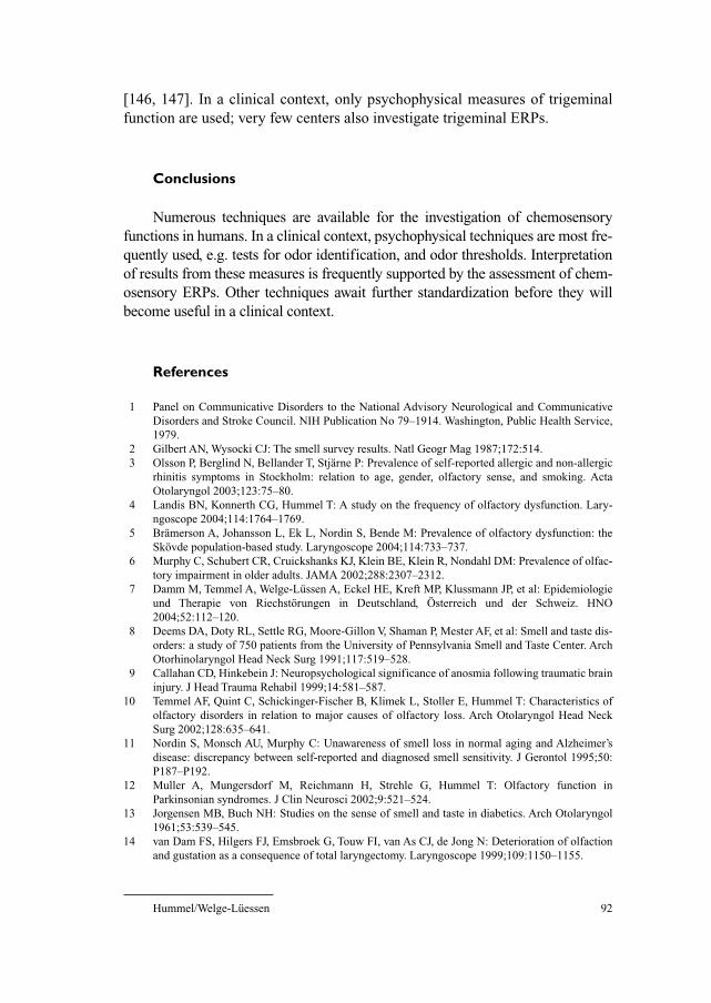
Hummel/Welge-Lüessen 92
[146, 147]. In a clinical context, only psychophysical measures of trigeminal
function are used; very few centers also investigate trigeminal ERPs.
Conclusions
Numerous techniques are available for the investigation of chemosensory
functions in humans. In a clinical context, psychophysical techniques are most fre-
quently used, e.g. tests for odor identification, and odor thresholds. Interpretation
of results from these measures is frequently supported by the assessment of chem-
osensory ERPs. Other techniques await further standardization before they will
become useful in a clinical context.
References
1 Panel on Communicative Disorders to the National Advisory Neurological and Communicative
Disorders and Stroke Council. NIH Publication No 79–1914. Washington, Public Health Service,
1979.
2 Gilbert AN, Wysocki CJ: The smell survey results. Natl Geogr Mag 1987;172:514.
3 Olsson P, Berglind N, Bellander T, Stjärne P: Prevalence of self-reported allergic and non-allergic
rhinitis symptoms in Stockholm: relation to age, gender, olfactory sense, and smoking. Acta
Otolaryngol 2003;123:75–80.
4 Landis BN, Konnerth CG, Hummel T: A study on the frequency of olfactory dysfunction. Lary-
ngoscope 2004;114:1764–1769.
5 Brämerson A, Johansson L, Ek L, Nordin S, Bende M: Prevalence of olfactory dysfunction: the
Skövde population-based study. Laryngoscope 2004;114:733–737.
6 Murphy C, Schubert CR, Cruickshanks KJ, Klein BE, Klein R, Nondahl DM: Prevalence of olfac-
tory impairment in older adults. JAMA 2002;288:2307–2312.
7 Damm M, Temmel A, Welge-Lüssen A, Eckel HE, Kreft MP, Klussmann JP, et al: Epidemiologie
und Therapie von Riechstörungen in Deutschland, Österreich und der Schweiz. HNO
2004;52:112–120.
8 Deems DA, Doty RL, Settle RG, Moore-Gillon V, Shaman P, Mester AF, et al: Smell and taste dis-
orders: a study of 750 patients from the University of Pennsylvania Smell and Taste Center. Arch
Otorhinolaryngol Head Neck Surg 1991;117:519–528.
9 Callahan CD, Hinkebein J: Neuropsychological significance of anosmia following traumatic brain
injury. J Head Trauma Rehabil 1999;14:581–587.
10 Temmel AF, Quint C, Schickinger-Fischer B, Klimek L, Stoller E, Hummel T: Characteristics of
olfactory disorders in relation to major causes of olfactory loss. Arch Otolaryngol Head Neck
Surg 2002;128:635–641.
11 Nordin S, Monsch AU, Murphy C: Unawareness of smell loss in normal aging and Alzheimer’s
disease: discrepancy between self-reported and diagnosed smell sensitivity. J Gerontol 1995;50:
P187–P192.
12 Muller A, Mungersdorf M, Reichmann H, Strehle G, Hummel T: Olfactory function in
Parkinsonian syndromes. J Clin Neurosci 2002;9:521–524.
13 Jorgensen MB, Buch NH: Studies on the sense of smell and taste in diabetics. Arch Otolaryngol
1961;53:539–545.
14 van Dam FS, Hilgers FJ, Emsbroek G, Touw FI, van As CJ, de Jong N: Deterioration of olfaction
and gustation as a consequence of total laryngectomy. Laryngoscope 1999;109:1150–1155.
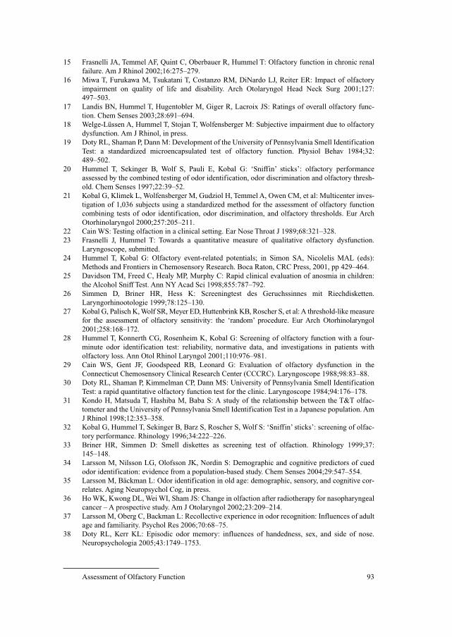
Assessment of Olfactory Function 93
15 Frasnelli JA, Temmel AF, Quint C, Oberbauer R, Hummel T: Olfactory function in chronic renal
failure. Am J Rhinol 2002;16:275–279.
16 Miwa T, Furukawa M, Tsukatani T, Costanzo RM, DiNardo LJ, Reiter ER: Impact of olfactory
impairment on quality of life and disability. Arch Otolaryngol Head Neck Surg 2001;127:
497–503.
17 Landis BN, Hummel T, Hugentobler M, Giger R, Lacroix JS: Ratings of overall olfactory func-
tion. Chem Senses 2003;28:691–694.
18 Welge-Lüssen A, Hummel T, Stojan T, Wolfensberger M: Subjective impairment due to olfactory
dysfunction. Am J Rhinol, in press.
19 Doty RL, Shaman P, Dann M: Development of the University of Pennsylvania Smell Identification
Test: a standardized microencapsulated test of olfactory function. Physiol Behav 1984;32:
489–502.
20 Hummel T, Sekinger B, Wolf S, Pauli E, Kobal G: ‘Sniffin’ sticks’: olfactory performance
assessed by the combined testing of odor identification, odor discrimination and olfactory thresh-
old. Chem Senses 1997;22:39–52.
21 Kobal G, Klimek L, Wolfensberger M, Gudziol H, Temmel A, Owen CM, et al: Multicenter inves-
tigation of 1,036 subjects using a standardized method for the assessment of olfactory function
combining tests of odor identification, odor discrimination, and olfactory thresholds. Eur Arch
Otorhinolaryngol 2000;257:205–211.
22 Cain WS: Testing olfaction in a clinical setting. Ear Nose Throat J 1989;68:321–328.
23 Frasnelli J, Hummel T: Towards a quantitative measure of qualitative olfactory dysfunction.
Laryngoscope, submitted.
24 Hummel T, Kobal G: Olfactory event-related potentials; in Simon SA, Nicolelis MAL (eds):
Methods and Frontiers in Chemosensory Research. Boca Raton, CRC Press, 2001, pp 429–464.
25 Davidson TM, Freed C, Healy MP, Murphy C: Rapid clinical evaluation of anosmia in children:
the Alcohol Sniff Test. Ann NY Acad Sci 1998;855:787–792.
26 Simmen D, Briner HR, Hess K: Screeningtest des Geruchssinnes mit Riechdisketten.
Laryngorhinootologie 1999;78:125–130.
27 Kobal G, Palisch K, Wolf SR, Meyer ED, Huttenbrink KB, Roscher S, et al: A threshold-like measure
for the assessment of olfactory sensitivity: the ‘random’ procedure. Eur Arch Otorhinolaryngol
2001;258:168–172.
28 Hummel T, Konnerth CG, Rosenheim K, Kobal G: Screening of olfactory function with a four-
minute odor identification test: reliability, normative data, and investigations in patients with
olfactory loss. Ann Otol Rhinol Laryngol 2001;110:976–981.
29 Cain WS, Gent JF, Goodspeed RB, Leonard G: Evaluation of olfactory dysfunction in the
Connecticut Chemosensory Clinical Research Center (CCCRC). Laryngoscope 1988;98:83–88.
30 Doty RL, Shaman P, Kimmelman CP, Dann MS: University of Pennsylvania Smell Identification
Test: a rapid quantitative olfactory function test for the clinic. Laryngoscope 1984;94:176–178.
31 Kondo H, Matsuda T, Hashiba M, Baba S: A study of the relationship between the T&T olfac-
tometer and the University of Pennsylvania Smell Identification Test in a Japanese population. Am
J Rhinol 1998;12:353–358.
32 Kobal G, Hummel T, Sekinger B, Barz S, Roscher S, Wolf S: ‘Sniffin’ sticks’: screening of olfac-
tory performance. Rhinology 1996;34:222–226.
33 Briner HR, Simmen D: Smell diskettes as screening test of olfaction. Rhinology 1999;37:
145–148.
34 Larsson M, Nilsson LG, Olofsson JK, Nordin S: Demographic and cognitive predictors of cued
odor identification: evidence from a population-based study. Chem Senses 2004;29:547–554.
35 Larsson M, Bäckman L: Odor identification in old age: demographic, sensory, and cognitive cor-
relates. Aging Neuropsychol Cog, in press.
36 Ho WK, Kwong DL, Wei WI, Sham JS: Change in olfaction after radiotherapy for nasopharyngeal
cancer – A prospective study. Am J Otolaryngol 2002;23:209–214.
37 Larsson M, Oberg C, Backman L: Recollective experience in odor recognition: Influences of adult
age and familiarity. Psychol Res 2006;70:68–75.
38 Doty RL, Kerr KL: Episodic odor memory: influences of handedness, sex, and side of nose.
Neuropsychologia 2005;43:1749–1753.

Hummel/Welge-Lüessen 94
39 Heilmann S, Strehle G, Rosenheim K, Damm M, Hummel T: Clinical assessment of retronasal
olfactory function. Arch Otorhinolaryngol Head Neck Surg 2002;128:414–418.
40 Landis BN, Giger R, Ricchetti A, Leuchter I, Hugentobler M, Hummel T, et al: Retronasal olfac-
tory function in nasal polyposis. Laryngoscope 2003;113:1993–1997.
41 Landis BN, Frasnelli J, Reden J, Lacroix JS, Hummel T: Differences between orthonasal and
retronasal olfactory functions in patients with loss of the sense of smell. Arch Otolaryngol Head
Neck Surg 2005;131:977–981.
42 Ehrenstein WH, Ehrenstein A: Psychophysical methods; in Windhorst U, Johansson H (eds):
Modern Techniques in Neuroscience Research. Berlin, Springer, 1999, pp 1211–1241.
43 Lotsch J, Lange C, Hummel T: A simple and reliable method for clinical assessment of odor
thresholds. Chem Senses 2004;29:11–317.
44 Linschoten MR, Harvey LO Jr, Eller PM, Jafek BW: Fast and accurate measurement of taste and
smell thresholds using a maximum-likelihood adaptive staircase procedure. Percept Psychophys
2001;63:1330–1347.
45 Doty RL, Smith R, McKeown DA, Raj J: Tests of human olfactory function: principle component
analysis suggests that most measure a common source of variance. Percept Psychophys
1994;56:701–707.
46 Hawkes CH, Shephard BC: Selective anosmia in Parkinson’s disease? Lancet 1993;341:435–436.
47 Koss E, Weiffenbach JM, Haxby JV, Friedland RP: Olfactory detection and recognition in
Alzheimer’s disease. Lancet 1987;i:622.
48 Koss E, Weiffenbach JM, Haxby JV, Friedland RP: Olfactory detection and identification perfor-
mance are dissociated in early Alzheimer’s disease. Neurology 1988;38:1228–1232.
49 Hornung DE, Kurtz DB, Bradshaw CB, Seipel DM, Kent PF, Blair DC, et al: The olfactory loss
that accompanies an HIV infection. Physiol Behav 1998;15:549–556.
50 Jones-Gotman M, Zatorre RJ: Olfactory identification deficits in patients with focal cerebral exci-
sion. Neuropsychologia 1988;26:387–400.
51 Peters JM, Hummel T, Kratzsch T, Lotsch J, Skarke C, Frolich L: Olfactory function in mild cog-
nitive impairment and Alzheimer’s disease: an investigation using psychophysical and electro-
physiological techniques. Am J Psychiatry 2003;160:1995–2002.
52 Mesholam RI, Moberg PJ, Mahr RN, Doty RL: Olfaction in neurodegenerative disease: a meta-
analysis of olfactory functioning in Alzheimer’s and Parkinson’s diseases. Arch Neurol
1998;55:84–90.
53 Daum RF, Sekinger B, Kobal G, Lang C: Riechprüfung mit ‘Sniffin’ Sticks’ zur klinischen
Diagnostik des Morbus Parkinson. Nervenarzt 2000;71:643–650.
54 Klimek L, Hummel T, Moll B, Kobal G, Mann WJ: Lateralized and bilateral olfactory function in
patients with chronic sinusitis compared with healthy control subjects. Laryngoscope
1998;108:111–114.
55 Leopold D: Distorted olfactory perception; in Doty RL (ed): Handbook of Olfaction and
Gustation. New York, Dekker, 1995, 441–454.
56 Frasnelli J, Landis BN, Heilmann S, Hauswald B, Huttenbrink KB, Lacroix JS, et al: Clinical pre-
sentation of qualitative olfactory dysfunction. Eur Arch Otorhinolaryngol 2003;11:11–13.
57 Nordin S, Murphy C, Davidson TM, Quinonez C, Jalowayski AA, Ellison DW: Prevalence and
assessment of qualitative olfactory dysfunction in different age groups. Laryngoscope 1996;106:
739–744.
58 Bullinger M: Assessing health-related quality of life in medicine. An overview over concepts,
methods and applications in international research. Restor Neurol Neurosci 2002;20:93–101.
59 Beck AT, Ward CM, Mendelson M, Mock JE, Erbaugh JK: An inventory for measuring depres-
sion. Arch Gen Psychiatry 1961;4:561–571.
60 von Zerssen D: Die Befindlichkeitsskala. Göttingen, Beltz Test, 1975.
61 Ware JE Jr: SF-36 health survey update. Spine 2000;25:3130–3139.
62 Anderson ER, Murphy MP, Weymuller EAJ: Clinimetric evaluation of the sinonasal outcome test-
16. Otolaryngol Head Neck Surg 1999;121:702–707.
63 de Jong N, Mulder I, de Graaf C, van Staveren WA: Impaired sensory functioning in elders: the
relation with its potential determinants and nutritional intake. J Gerontol A Biol Sci Med Sci
1999;54:B324–B331.
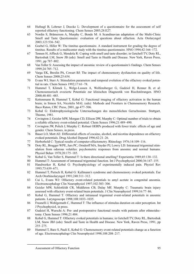
Assessment of Olfactory Function 95
64 Hufnagl B, Lehrner J, Deecke L: Development of a questionnaire for the assessment of self
reported olfactory functioning. Chem Senses 2003;28:E27.
65 Nordin S, Brämerson A, Murphy C, Bende M: A Scandinavian adaptation of the Multi-Clinic
Smell and Taste Questionnaire: evaluation of questions about olfaction. Acta Otolaryngol
2003;123:536–542.
66 Goebel G, Hiller W: The tinnitus questionnaire. A standard instrument for grading the degree of
tinnitus. Results of a multicenter study with the tinnitus questionnaire. HNO 1994;42:166–172.
67 Tennen H, Affleck G, Mendola R: Coping with smell and taste disorder; in Getchell TV, Doty RL,
Bartoshuk LM, Snow JB (eds): Smell and Taste in Health and Disease. New York, Raven Press,
1991, pp 787–802.
68 Van Toller S: Assessing the impact of anosmia: review of a questionnaire’s findings. Chem Senses
1999;24:705–712.
69 Varga EK, Breslin PA, Cowart BJ: The impact of chemosensory dysfunction on quality of life.
Chem Senses 2000;25:654.
70 Evans WJ, Starr A: Stimulation parameters and temporal evolution of the olfactory evoked poten-
tial in rats. Chem Senses 1992;17:61–78.
71 Hummel T, Klimek L, Welge-Lussen A, Wolfensberger G, Gudziol H, Renner B, et al:
Chemosensorisch evozierte Potentiale zur klinischen Diagnostik von Riechstörungen. HNO
2000;48:481–485.
72 Kettenmann B, Hummel T, Kobal G: Functional imaging of olfactory activation in the human
brain; in Simon SA, Nicolelis MAL (eds): Methods and Frontiers in Chemosensory Research.
Baco Raton, CRC Press, 2001, pp 477–506.
73 Kobal G: Elektrophysiologische Untersuchungen des menschlichen Geruchssinns. Stuttgart,
Thieme, 1981.
74 Covington J, Geisler MW, Morgan CD, Ellison DW, Murphy C: Optimal number of trials to obtain
a reliable olfactory event-related potential. Chem Senses 1996;21:489–490.
75 Covington JW, Polich J, Murphy C: Robust OERPs produced with fewer trials: effects of age and
gender. Chem Senses, in press.
76 Bauer LO, Mott AE: Differential effects of cocaine, alcohol, and nicotine dependence on olfactory
evoked potentials. Drug Alcohol Depend 1996;42:21–26.
77 Herberhold C: Typical results of computer-olfactometry. Rhinology 1976;14:109–116.
78 Doty RL, Brugger WPE, Jurs PC, Orndorff MA, Snyder PJ, Lowry LD: Intranasal trigeminal stim-
ulation from odorous volatiles: psychometric responses from anosmic and normal humans.
Physiol Behav 1978;20:175–185.
79 Kobal G, Van Toller S, Hummel T: Is there directional smelling? Experientia 1989;45:130–132.
80 Hummel T: Assessment of intranasal trigeminal function. Int J Psychophysiol 2000;36:147–155.
81 Handwerker H, Kobal G: Psychophysiology of experimentally induced pain. Physiol Rev
1993;73:639–671.
82 Hummel T, Pietsch H, Kobal G: Kallmann’s syndrome and chemosensory evoked potentials. Eur
Arch Otorhinolaryngol 1991;248:311–312.
83 Cui L, Evans WJ: Olfactory event-related potentials to amyl acetate in congenital anosmia.
Electroencephalogr Clin Neurophysiol 1997;102:303–306.
84 Geisler MW, Schlotfeldt CR, Middleton CB, Dulay MF, Murphy C: Traumatic brain injury
assessed with olfactory event-related brain potentials. J Clin Neurophysiol 1999;16:77–86.
85 Kobal G, Hummel T: Olfactory and intranasal trigeminal event-related potentials in anosmic
patients. Laryngoscope 1998;108:1033–1035.
86 Frasnelli J, Wohlgemuth C, Hummel T: The influence of stimulus duration on odor perception. Int
J Psychophysiol, in press.
87 Gudziol H, Wurschi A: Pre- and postoperative functional results with patients after ethmoidec-
tomy. Chem Senses 1996;21:484.
88 Kobal G, Hummel T: Olfactory evoked potentials in humans; in Getchell TV, Doty RL, Bartoshuk
LM, Snow JBJ (eds): Smell and Taste in Health and Disease. New York, Raven Press, 1991, pp
255–275.
89 Hummel T, Barz S, Pauli E, Kobal G: Chemosensory event-related potentials change as a function
of age. Electroencephalogr Clin Neurophysiol 1998;108:208–217.
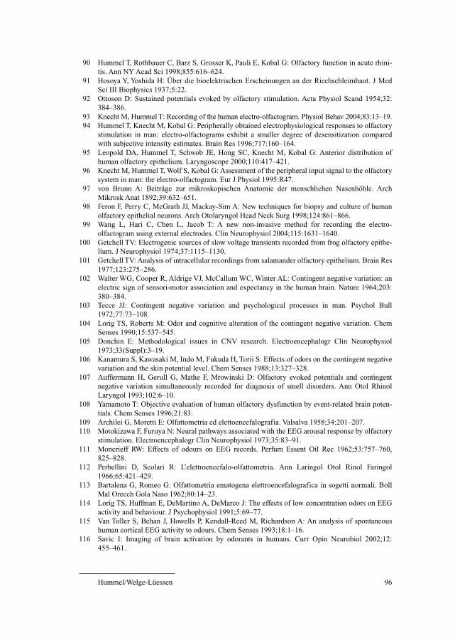
Hummel/Welge-Lüessen 96
90 Hummel T, Rothbauer C, Barz S, Grosser K, Pauli E, Kobal G: Olfactory function in acute rhini-
tis. Ann NY Acad Sci 1998;855:616–624.
91 Hosoya Y, Yoshida H: Über die bioelektrischen Erscheinungen an der Riechschleimhaut. J Med
Sci III Biophysics 1937;5:22.
92 Ottoson D: Sustained potentials evoked by olfactory stimulation. Acta Physiol Scand 1954;32:
384–386.
93 Knecht M, Hummel T: Recording of the human electro-olfactogram. Physiol Behav 2004;83:13–19.
94 Hummel T, Knecht M, Kobal G: Peripherally obtained electrophysiological responses to olfactory
stimulation in man: electro-olfactograms exhibit a smaller degree of desensitization compared
with subjective intensity estimates. Brain Res 1996;717:160–164.
95 Leopold DA, Hummel T, Schwob JE, Hong SC, Knecht M, Kobal G: Anterior distribution of
human olfactory epithelium. Laryngoscope 2000;110:417–421.
96 Knecht M, Hummel T, Wolf S, Kobal G: Assessment of the peripheral input signal to the olfactory
system in man: the electro-olfactogram. Eur J Physiol 1995:R47.
97 von Brunn A: Beiträge zur mikroskopischen Anatomie der menschlichen Nasenhöhle. Arch
Mikrosk Anat 1892;39:632–651.
98 Feron F, Perry C, McGrath JJ, Mackay-Sim A: New techniques for biopsy and culture of human
olfactory epithelial neurons. Arch Otolaryngol Head Neck Surg 1998;124:861–866.
99 Wang L, Hari C, Chen L, Jacob T: A new non-invasive method for recording the electro-
olfactogram using external electrodes. Clin Neurophysiol 2004;115:1631–1640.
100 Getchell TV: Electrogenic sources of slow voltage transients recorded from frog olfactory epithe-
lium. J Neurophysiol 1974;37:1115–1130.
101 Getchell TV: Analysis of intracellular recordings from salamander olfactory epithelium. Brain Res
1977;123:275–286.
102 Walter WG, Cooper R, Aldrige VJ, McCallum WC, Winter AL: Contingent negative variation: an
electric sign of sensori-motor association and expectancy in the human brain. Nature 1964;203:
380–384.
103 Tecce JJ: Contingent negative variation and psychological processes in man. Psychol Bull
1972;77:73–108.
104 Lorig TS, Roberts M: Odor and cognitive alteration of the contingent negative variation. Chem
Senses 1990;15:537–545.
105 Donchin E: Methodological issues in CNV research. Electroencephalogr Clin Neurophysiol
1973;33(Suppl):3–19.
106 Kanamura S, Kawasaki M, Indo M, Fukuda H, Torii S: Effects of odors on the contingent negative
variation and the skin potential level. Chem Senses 1988;13:327–328.
107 Auffermann H, Gerull G, Mathe F, Mrowinski D: Olfactory evoked potentials and contingent
negative variation simultaneously recorded for diagnosis of smell disorders. Ann Otol Rhinol
Laryngol 1993;102:6–10.
108 Yamamoto T: Objective evaluation of human olfactory dysfunction by event-related brain poten-
tials. Chem Senses 1996;21:83.
109 Archilei G, Moretti E: Olfattometria ed elettoencefalografia. Valsalva 1958;34:201–207.
110 Motokizawa F, Furuya N: Neural pathways associated with the EEG arousal response by olfactory
stimulation. Electroencephalogr Clin Neurophysiol 1973;35:83–91.
111 Moncrieff RW: Effects of odours on EEG records. Perfum Essent Oil Rec 1962;53:757–760,
825–828.
112 Perbellini D, Scolari R: L’elettroencefalo-olfattometria. Ann Laringol Otol Rinol Faringol
1966;65:421–429.
113 Bartalena G, Romeo G: Olfattometria ematogena elettroencefalografica in sogetti normali. Boll
Mal Orecch Gola Naso 1962;80:14–23.
114 Lorig TS, Huffman E, DeMartino A, DeMarco J: The effects of low concentration odors on EEG
activity and behaviour. J Psychophysiol 1991;5:69–77.
115 Van Toller S, Behan J, Howells P, Kendall-Reed M, Richardson A: An analysis of spontaneous
human cortical EEG activity to odours. Chem Senses 1993;18:1–16.
116 Savic I: Imaging of brain activation by odorants in humans. Curr Opin Neurobiol 2002;12:
455–461.
Assessment of Olfactory Function 97
117 Zald DH, Pardo JV: Functional neuroimaging of the olfactory system in humans. Int J
Psychophysiol 2000;36:165–181.
118 Small DM, Jones-Gotman M, Zatorre RJ, Petrides M, Evans AC: Flavor processing: more than the
sum of its parts. Neuroreport 1997;8:3913–3917.
119 Kareken DA, Sabri M, Radnovich AJ, Claus E, Foresman B, Hector D, et al: Olfactory system acti-
vation from sniffing: effects in piriform and orbitofrontal cortex. Neuroimage 2004;22:456–465.
120 Sobel N, Prabhakaran V, Zhao Z, Desmond JE, Glover GH, Sullivan EV, et al: Time course of
odorant-induced activation in the human primary olfactory cortex. J Neurophysiol 2000;83:
537–551.
121 Poellinger A, Thomas R, Lio P, Lee A, Makris N, Rosen BR, et al: Activation and habituation in
olfaction – An fMRI study. Neuroimage 2001;13:547–560.
122 Anderson AK, Christoff K, Stappen I, Panitz D, Ghahremani DG, Glover G, et al: Dissociated
neural representations of intensity and valence in human olfaction. Nat Neurosci 2003;6:196–202.
123 Kettenmann B, Hummel C, Stefan H, Kobal G: Magnetoencephalographical recordings: separa-
tion of cortical responses to different chemical stimulation in man. Funct Neurosci (EEG Suppl)
1996;46:287–290.
124 Ayabe-Kanamura S, Endo H, Kobayakawa T, Takeda T, Saito S: Measurement of olfactory evoked
magnetic fields by a 64-channel whole-head SQUID system. Chem Senses 1997;22:214–215.
125 Small DM, Gerber JC, Mak YE, Hummel T: Differential neural responses evoked by orthonasal
versus retronasal odorant perception in humans. Neuron 2005;47:593–605.
126 Yousem DM, Maldjian JA, Hummel T, Alsop DC, Geckle RJ, Kraut MA, et al: The effect of age on
odor-stimulated functional MR imaging. Am J Neuroradiol 1999;20:600–608.
127 Henkin RI, Levy LM, Lin CS: Taste and smell phantoms revealed by brain functional MRI
(fMRI). J Comput Assist Tomogr 2000;24:106–123.
128 Adema JM, Montserrat JM: Olfacto-rhinomanometry. Int Rhinol 1982;20:21–28.
129 Hoshino T, Usui N: Objective olfactometry by the method of recordings of respiratory resistances.
Jibiinkoka 1987;90:516–522.
130 Gudziol H, Gramowski KH: Respirations-Olfaktometrie – eine objektivierende Methode zur
quantitativen Bewertung einer Hyposmie. Laryngol Rhinol Otol 1987;66:570–572.
131 Kendall-Reed M, Walker JC: Human respiratory responses to odorants. Chem Senses 1996;
21:486.
132 Gudziol H, Wächter R: Gibt es olfaktorisch evozierte Atemänderungen? Laryngorhinootologie
2004;83:367–373.
133 Frank RA, Dulay MF, Niergarth KA, Gesteland RC: A comparison of the sniff magnitude test and
the University of Pennsylvania Smell Identification Test in children and nonnative English speak-
ers. Physiol Behav 2004;81:475–480.
134 Asaka H: The studies on the objective olfactory test by galvanic skin response. J Otorhinolaryngol
Soc Jap1965;68:100–112.
135 Auld JSM: Psycho-galvanic measurement of smell. J Inst Petroleum Technol 1923;9:389–391.
136 Borsanyi SJ, Blanchard DL, Baker FJ: Psychogalvanic skin response olfactometry. Ann Otol
1962;74:213–221.
137 Nishida H, Kumagami H, Jinnouchi H: Pupillary reaction following olfactory stimulation – Use in
objective olfactometry. Nippon Jibiinkoka Gakkai Kaiho 1973;76:1449–1458.
138 Sneppe R, Gonay P: Evaluation objective, quantitative et qualitative de l’olfaction. Electrodiagn
Ther 1973;10:5–17.
139 Ichihara M, Komatsu A, Ichihara F, Asaga H, Hirayoshi K: Test of smell based on the wink
response. Jibiinkoka 1967;39:947–953.
140 Delank KW: Subjektive und objektive Methoden zur Beurteilung der Riechfunktion. HNO
1998;46:182–190.
141 Cometto-Muniz JE, Cain WS: Trigeminal and olfactory sensitivity: comparison of modalities and
methods of measurement. Int Arch Occup Environ Health 1998;71:105–110.
142 Lötsch J, Ahne G, Kunder J, Kobal G, Hummel T: Factors affecting pain intensity in a pain model
based upon tonic intranasal stimulation in humans. Inflamm Res 1998;47:446–450.
143 Berg J, Hummel T, Huang G, Doty RL: Trigeminal impact of odorants assessed with lateralized
stimulation. Chem Senses 1998;23:587.
Hummel/Welge-Lüessen 98
144 Thürauf N, Friedel I, Hummel C, Kobal G: The mucosal potential elicited by noxious chemical
stimuli: is it a peripheral nociceptive event. Neurosci Lett 1991;128:297–300.
145 Hari R, Portin K, Kettenmann B, Jousmaki V, Kobal G: Right-hemisphere preponderance of
responses to painful CO2 stimulation of the human nasal mucosa. Pain 1997;72:145–151.
146 Livermore A, Hummel T, Kobal G: Chemosensory evoked potentials in the investigation of inter-
actions between the olfactory and the somatosensory (trigeminal) systems. Electroencephalogr
Clin Neurophysiol 1992;83:201–210.
147 Pause BM, Sojka B, Krauel K, Ferstl R: The nature of the late positive complex within the olfac-
tory event-related potential. Psychophysiology 1996;33:168–172.
Thomas Hummel, MD
Smell & Taste Clinic, Department of Otorhinolaryngology
University of Dresden Medical School (‘Technische Universität Dresden’)
Fetscherstrasse 74
DE–01307 Dresden (Germany)
Tel. �49 351 458 4189, Fax �49 351 458 4326
E-Mail [email protected]
Hummel T, Welge-Lüssen A (eds): Taste and Smell. An Update.
Adv Otorhinolaryngol. Basel, Karger, 2006, vol 63, pp 99–107
Posttraumatic Olfactory Loss
Richard M. Costanzoa, Takaki Miwab
aVirginia Commonwealth University, School of Medicine,
Richmond, Va., USA; bKanazawa University Graduate School of
Medical Sciences, Kanazawa, Japan
AbstractHead injury is the leading cause of posttraumatic anosmia. Complete or partial loss of
olfactory function may occur when the nasal passages are blocked, olfactory nerves are injured
or there are contusions or hemorrhages in olfactory centers of the brain. Evaluation of patients
with posttraumatic olfactory loss should include a physical examination by the otolaryngolo-
gist. Nasal endoscopy and radiological studies should be performed as well as olfactory func-
tion tests to determine the degree and type of olfactory impairment. Although treatment options
may be limited, physicians should provide information and counseling regarding the risks and
hazards associated with loss of olfactory function. For some individuals such as cooks, fire-
fighters, and research scientists, an assessment of vocational activities should be performed
prior to reentry into the workplace. Individuals with impaired olfactory function may be unable
to detect important warning signs such as gas leaks, volatile chemical fumes and fires and
therefore place themselves and coworkers at an increased risk for serious injury or death.
Copyright © 2006 S. Karger AG, Basel
Mechanisms of Injury
Head injury is one of the most common causes of posttraumatic olfactory
loss. Mechanisms leading to a partial or complete loss of olfactory function
include damage to the olfactory epithelium or nasal passages, injury to olfac-
tory nerve fibers, and contusions and hemorrhage lesions in olfactory areas of
the brain [1].
Damage to Nose or Nasal PassagesDirect injury, edema, mucosal hematoma, and scarring of the olfactory
epithelium all contribute to impairment of olfactory function. Trauma to the
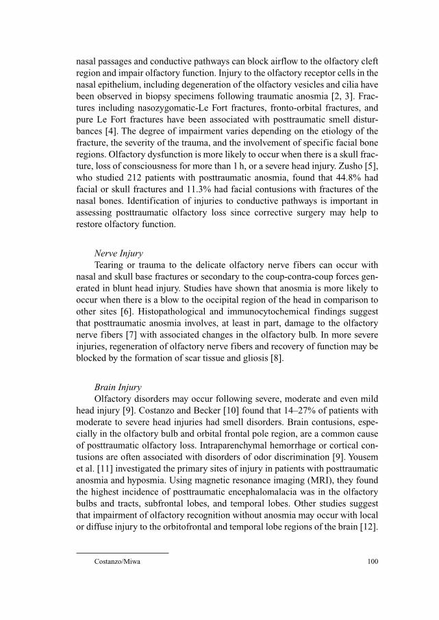
Costanzo/Miwa 100
nasal passages and conductive pathways can block airflow to the olfactory cleft
region and impair olfactory function. Injury to the olfactory receptor cells in the
nasal epithelium, including degeneration of the olfactory vesicles and cilia have
been observed in biopsy specimens following traumatic anosmia [2, 3]. Frac-
tures including nasozygomatic-Le Fort fractures, fronto-orbital fractures, and
pure Le Fort fractures have been associated with posttraumatic smell distur-
bances [4]. The degree of impairment varies depending on the etiology of the
fracture, the severity of the trauma, and the involvement of specific facial bone
regions. Olfactory dysfunction is more likely to occur when there is a skull frac-
ture, loss of consciousness for more than 1 h, or a severe head injury. Zusho [5],
who studied 212 patients with posttraumatic anosmia, found that 44.8% had
facial or skull fractures and 11.3% had facial contusions with fractures of the
nasal bones. Identification of injuries to conductive pathways is important in
assessing posttraumatic olfactory loss since corrective surgery may help to
restore olfactory function.
Nerve InjuryTearing or trauma to the delicate olfactory nerve fibers can occur with
nasal and skull base fractures or secondary to the coup-contra-coup forces gen-
erated in blunt head injury. Studies have shown that anosmia is more likely to
occur when there is a blow to the occipital region of the head in comparison to
other sites [6]. Histopathological and immunocytochemical findings suggest
that posttraumatic anosmia involves, at least in part, damage to the olfactory
nerve fibers [7] with associated changes in the olfactory bulb. In more severe
injuries, regeneration of olfactory nerve fibers and recovery of function may be
blocked by the formation of scar tissue and gliosis [8].
Brain InjuryOlfactory disorders may occur following severe, moderate and even mild
head injury [9]. Costanzo and Becker [10] found that 14–27% of patients with
moderate to severe head injuries had smell disorders. Brain contusions, espe-
cially in the olfactory bulb and orbital frontal pole region, are a common cause
of posttraumatic olfactory loss. Intraparenchymal hemorrhage or cortical con-
tusions are often associated with disorders of odor discrimination [9]. Yousem
et al. [11] investigated the primary sites of injury in patients with posttraumatic
anosmia and hyposmia. Using magnetic resonance imaging (MRI), they found
the highest incidence of posttraumatic encephalomalacia was in the olfactory
bulbs and tracts, subfrontal lobes, and temporal lobes. Other studies suggest
that impairment of olfactory recognition without anosmia may occur with local
or diffuse injury to the orbitofrontal and temporal lobe regions of the brain [12].
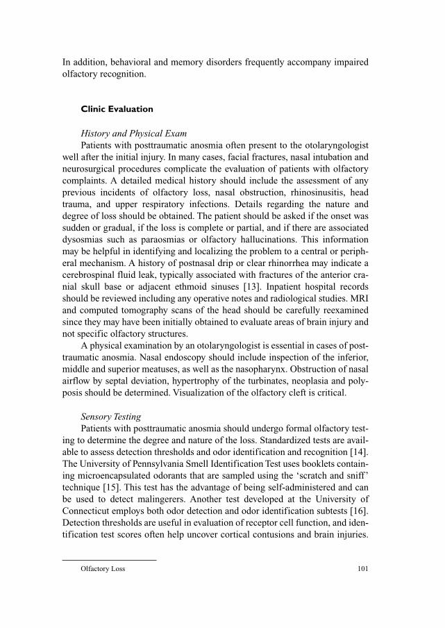
Olfactory Loss 101
In addition, behavioral and memory disorders frequently accompany impaired
olfactory recognition.
Clinic Evaluation
History and Physical ExamPatients with posttraumatic anosmia often present to the otolaryngologist
well after the initial injury. In many cases, facial fractures, nasal intubation and
neurosurgical procedures complicate the evaluation of patients with olfactory
complaints. A detailed medical history should include the assessment of any
previous incidents of olfactory loss, nasal obstruction, rhinosinusitis, head
trauma, and upper respiratory infections. Details regarding the nature and
degree of loss should be obtained. The patient should be asked if the onset was
sudden or gradual, if the loss is complete or partial, and if there are associated
dysosmias such as paraosmias or olfactory hallucinations. This information
may be helpful in identifying and localizing the problem to a central or periph-
eral mechanism. A history of postnasal drip or clear rhinorrhea may indicate a
cerebrospinal fluid leak, typically associated with fractures of the anterior cra-
nial skull base or adjacent ethmoid sinuses [13]. Inpatient hospital records
should be reviewed including any operative notes and radiological studies. MRI
and computed tomography scans of the head should be carefully reexamined
since they may have been initially obtained to evaluate areas of brain injury and
not specific olfactory structures.
A physical examination by an otolaryngologist is essential in cases of post-
traumatic anosmia. Nasal endoscopy should include inspection of the inferior,
middle and superior meatuses, as well as the nasopharynx. Obstruction of nasal
airflow by septal deviation, hypertrophy of the turbinates, neoplasia and poly-
posis should be determined. Visualization of the olfactory cleft is critical.
Sensory TestingPatients with posttraumatic anosmia should undergo formal olfactory test-
ing to determine the degree and nature of the loss. Standardized tests are avail-
able to assess detection thresholds and odor identification and recognition [14].
The University of Pennsylvania Smell Identification Test uses booklets contain-
ing microencapsulated odorants that are sampled using the ‘scratch and sniff’
technique [15]. This test has the advantage of being self-administered and can
be used to detect malingerers. Another test developed at the University of
Connecticut employs both odor detection and odor identification subtests [16].
Detection thresholds are useful in evaluation of receptor cell function, and iden-
tification test scores often help uncover cortical contusions and brain injuries.
Costanzo/Miwa 102
In Japan, olfactory testing is often performed using a graded series of odorants
presented on strips of blotting paper [17]. In Germany, ‘sniffing sticks’ are rou-
tinely used to test olfactory function [18] and olfactometers that deliver con-
trolled odor stimuli have made it possible to measure olfactory evoked
potentials [19]. A more comprehensive review of olfactory testing methods is
presented in a separate chapter in this volume [chapter by Hummel et al., this
vol, pp 84–98].
RadiologyAlthough plain-film radiographs may be of some diagnostic use in deter-
mining skull or facial fractures, high-resolution computed tomography and
MRI scans are preferred in diagnosing posttraumatic anosmia [11, 20]. Hematomas
and contusions are potential causes of posttraumatic anosmia and may be eval-
uated with MR images. When observed in the olfactory bulb and the orbito-
frontal pole region, they represent clinical signs commonly associated with
impaired olfactory function. Figure 1 shows the brain scan of a 42-year-old
woman who suffered impaired olfactory function after hitting the back of her
head. Olfactory function testing revealed that although she could detect some of
the test odors presented, there was a complete loss in her ability to recognize or
identify odors. A decrease in metabolism in the orbitofrontal cortex and medial
prefrontal cortex areas has been demonstrated in posttraumatic anosmia using
quantitative positron emission tomography scanning [21]. Functional MRI has
also been used successfully to visualize olfactory function in different brain
regions [22, 23].
a b
Fig. 1. Brain image (T2) of a 42-year-old woman who fell and hit the back of her head
while playing badminton. a Sagittal section showing the area of impact (arrow) and brain
injury in the orbitofrontal lobe region (arrowheads). b Coronal section showing areas of
increased intensity in the left orbitofrontal lobe (arrowheads).
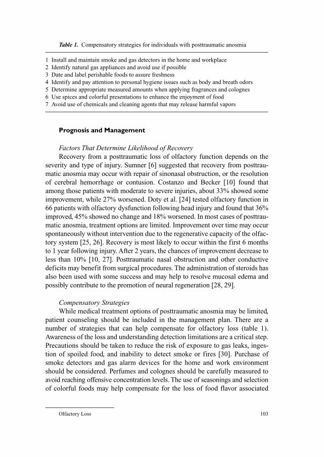
Olfactory Loss 103
Prognosis and Management
Factors That Determine Likelihood of RecoveryRecovery from a posttraumatic loss of olfactory function depends on the
severity and type of injury. Sumner [6] suggested that recovery from posttrau-
matic anosmia may occur with repair of sinonasal obstruction, or the resolution
of cerebral hemorrhage or contusion. Costanzo and Becker [10] found that
among those patients with moderate to severe injuries, about 33% showed some
improvement, while 27% worsened. Doty et al. [24] tested olfactory function in
66 patients with olfactory dysfunction following head injury and found that 36%
improved, 45% showed no change and 18% worsened. In most cases of posttrau-
matic anosmia, treatment options are limited. Improvement over time may occur
spontaneously without intervention due to the regenerative capacity of the olfac-
tory system [25, 26]. Recovery is most likely to occur within the first 6 months
to 1 year following injury. After 2 years, the chances of improvement decrease to
less than 10% [10, 27]. Posttraumatic nasal obstruction and other conductive
deficits may benefit from surgical procedures. The administration of steroids has
also been used with some success and may help to resolve mucosal edema and
possibly contribute to the promotion of neural regeneration [28, 29].
Compensatory StrategiesWhile medical treatment options of posttraumatic anosmia may be limited,
patient counseling should be included in the management plan. There are a
number of strategies that can help compensate for olfactory loss (table 1).
Awareness of the loss and understanding detection limitations are a critical step.
Precautions should be taken to reduce the risk of exposure to gas leaks, inges-
tion of spoiled food, and inability to detect smoke or fires [30]. Purchase of
smoke detectors and gas alarm devices for the home and work environment
should be considered. Perfumes and colognes should be carefully measured to
avoid reaching offensive concentration levels. The use of seasonings and selection
of colorful foods may help compensate for the loss of food flavor associated
Table 1. Compensatory strategies for individuals with posttraumatic anosmia
1 Install and maintain smoke and gas detectors in the home and workplace
2 Identify natural gas appliances and avoid use if possible
3 Date and label perishable foods to assure freshness
4 Identify and pay attention to personal hygiene issues such as body and breath odors
5 Determine appropriate measured amounts when applying fragrances and colognes
6 Use spices and colorful presentations to enhance the enjoyment of food
7 Avoid use of chemicals and cleaning agents that may release harmful vapors
Costanzo/Miwa 104
with olfactory impairment. Labeling and dating of refrigerated and frozen
foods will help to prevent ingestion of spoiled foods. Special instructions
should be given to patients with cardiovascular disease and hypertension so that
they do not compensate for loss of flavor by adding excessive amounts of salt to
their food.
Impact of Olfactory Loss
Risks and HazardsThe health and safety of patients with posttraumatic olfactory impairment
are at significant risk due to their inability to detect gas leaks, smoke, spoiled
foods and other olfactory-related warning signs that may be present in the home
and at the workplace [31]. This inability may be further complicated by trauma
to brain regions resulting in impaired cognitive and motor function. Instructions
and counseling are essential for this patient population. A commonly cited
activity of concern for patients with impaired olfactory function is the detection
of spoiled food [32]. The detection of gas leaks, smoke, eating and cooking
were also a concern of over half of the patients surveyed. Santos et al. [31] stud-
ied 445 patients and found that patients with impaired olfactory function are
more likely to experience an olfactory-related hazardous event than those with
normal olfactory function. Forty-five percent of these patients reported experi-
encing cooking-related incidents, 25% reported ingestion of spoiled food, and
23% the inability to detect a gas leak. Food hygiene is a major concern for those
with olfactory impairment. Perishable foods should be stored in a refrigerator
and eaten as soon as possible. Checking of expiration dates on food packages
and discarding out-of-date foods should be carefully adhered to. If in doubt
about the freshness of food, a family member or friend should be consulted
prior to serving. Gas and smoke detectors are essential safety devices for those
with impaired olfactory function. When possible, electric appliances should be
used instead of gas appliances. Cooking times and temperatures should be mon-
itored carefully to avoid burning food or igniting grease fires.
Quality of Life IssueThe patients who have olfactory impairment report significant changes in
quality of life [30]. Major concerns were bad breath and body odor, worrying
about the detection of gas leaks and smoke, and alterations in the taste of foods
and loss of appetite. Additional concerns included exposure to cleaning solu-
tions, pesticides and chemicals, and pet odors. Individuals with sustained olfac-
tory impairment washed clothes and cleaned their house more frequently than
those that had improvement in their olfactory function. Perfume and cologne
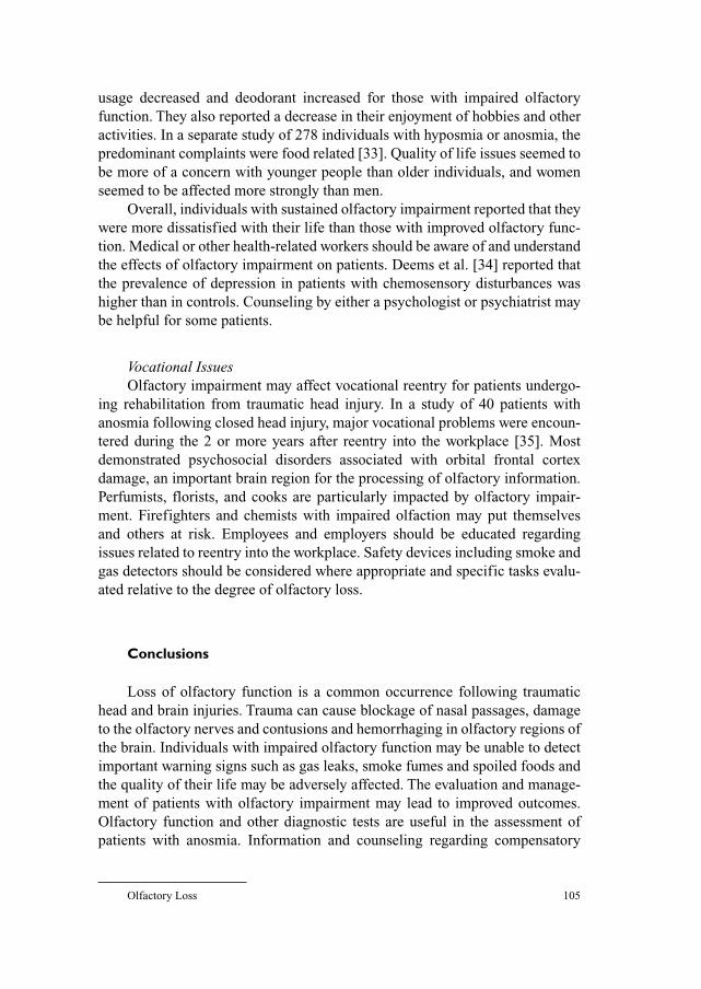
Olfactory Loss 105
usage decreased and deodorant increased for those with impaired olfactory
function. They also reported a decrease in their enjoyment of hobbies and other
activities. In a separate study of 278 individuals with hyposmia or anosmia, the
predominant complaints were food related [33]. Quality of life issues seemed to
be more of a concern with younger people than older individuals, and women
seemed to be affected more strongly than men.
Overall, individuals with sustained olfactory impairment reported that they
were more dissatisfied with their life than those with improved olfactory func-
tion. Medical or other health-related workers should be aware of and understand
the effects of olfactory impairment on patients. Deems et al. [34] reported that
the prevalence of depression in patients with chemosensory disturbances was
higher than in controls. Counseling by either a psychologist or psychiatrist may
be helpful for some patients.
Vocational IssuesOlfactory impairment may affect vocational reentry for patients undergo-
ing rehabilitation from traumatic head injury. In a study of 40 patients with
anosmia following closed head injury, major vocational problems were encoun-
tered during the 2 or more years after reentry into the workplace [35]. Most
demonstrated psychosocial disorders associated with orbital frontal cortex
damage, an important brain region for the processing of olfactory information.
Perfumists, florists, and cooks are particularly impacted by olfactory impair-
ment. Firefighters and chemists with impaired olfaction may put themselves
and others at risk. Employees and employers should be educated regarding
issues related to reentry into the workplace. Safety devices including smoke and
gas detectors should be considered where appropriate and specific tasks evalu-
ated relative to the degree of olfactory loss.
Conclusions
Loss of olfactory function is a common occurrence following traumatic
head and brain injuries. Trauma can cause blockage of nasal passages, damage
to the olfactory nerves and contusions and hemorrhaging in olfactory regions of
the brain. Individuals with impaired olfactory function may be unable to detect
important warning signs such as gas leaks, smoke fumes and spoiled foods and
the quality of their life may be adversely affected. The evaluation and manage-
ment of patients with olfactory impairment may lead to improved outcomes.
Olfactory function and other diagnostic tests are useful in the assessment of
patients with anosmia. Information and counseling regarding compensatory
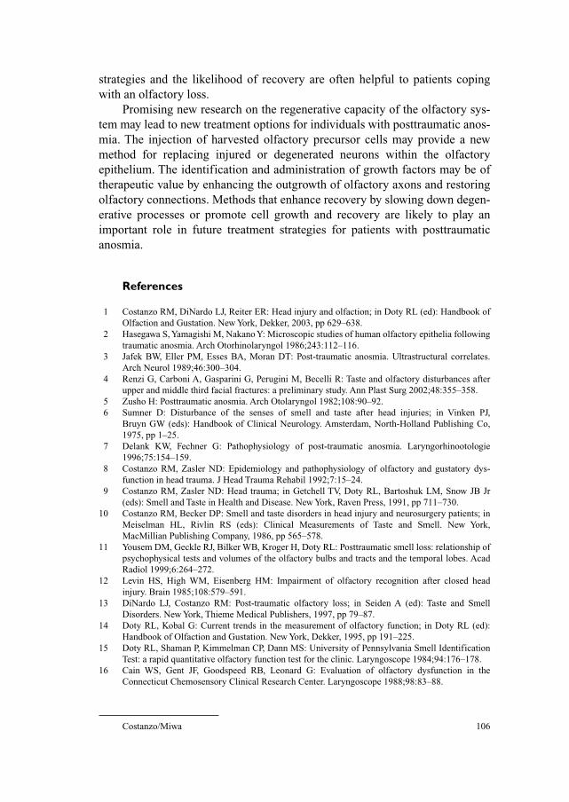
Costanzo/Miwa 106
strategies and the likelihood of recovery are often helpful to patients coping
with an olfactory loss.
Promising new research on the regenerative capacity of the olfactory sys-
tem may lead to new treatment options for individuals with posttraumatic anos-
mia. The injection of harvested olfactory precursor cells may provide a new
method for replacing injured or degenerated neurons within the olfactory
epithelium. The identification and administration of growth factors may be of
therapeutic value by enhancing the outgrowth of olfactory axons and restoring
olfactory connections. Methods that enhance recovery by slowing down degen-
erative processes or promote cell growth and recovery are likely to play an
important role in future treatment strategies for patients with posttraumatic
anosmia.
References
1 Costanzo RM, DiNardo LJ, Reiter ER: Head injury and olfaction; in Doty RL (ed): Handbook of
Olfaction and Gustation. New York, Dekker, 2003, pp 629–638.
2 Hasegawa S, Yamagishi M, Nakano Y: Microscopic studies of human olfactory epithelia following
traumatic anosmia. Arch Otorhinolaryngol 1986;243:112–116.
3 Jafek BW, Eller PM, Esses BA, Moran DT: Post-traumatic anosmia. Ultrastructural correlates.
Arch Neurol 1989;46:300–304.
4 Renzi G, Carboni A, Gasparini G, Perugini M, Becelli R: Taste and olfactory disturbances after
upper and middle third facial fractures: a preliminary study. Ann Plast Surg 2002;48:355–358.
5 Zusho H: Posttraumatic anosmia. Arch Otolaryngol 1982;108:90–92.
6 Sumner D: Disturbance of the senses of smell and taste after head injuries; in Vinken PJ,
Bruyn GW (eds): Handbook of Clinical Neurology. Amsterdam, North-Holland Publishing Co,
1975, pp 1–25.
7 Delank KW, Fechner G: Pathophysiology of post-traumatic anosmia. Laryngorhinootologie
1996;75:154–159.
8 Costanzo RM, Zasler ND: Epidemiology and pathophysiology of olfactory and gustatory dys-
function in head trauma. J Head Trauma Rehabil 1992;7:15–24.
9 Costanzo RM, Zasler ND: Head trauma; in Getchell TV, Doty RL, Bartoshuk LM, Snow JB Jr
(eds): Smell and Taste in Health and Disease. New York, Raven Press, 1991, pp 711–730.
10 Costanzo RM, Becker DP: Smell and taste disorders in head injury and neurosurgery patients; in
Meiselman HL, Rivlin RS (eds): Clinical Measurements of Taste and Smell. New York,
MacMillian Publishing Company, 1986, pp 565–578.
11 Yousem DM, Geckle RJ, Bilker WB, Kroger H, Doty RL: Posttraumatic smell loss: relationship of
psychophysical tests and volumes of the olfactory bulbs and tracts and the temporal lobes. Acad
Radiol 1999;6:264–272.
12 Levin HS, High WM, Eisenberg HM: Impairment of olfactory recognition after closed head
injury. Brain 1985;108:579–591.
13 DiNardo LJ, Costanzo RM: Post-traumatic olfactory loss; in Seiden A (ed): Taste and Smell
Disorders. New York, Thieme Medical Publishers, 1997, pp 79–87.
14 Doty RL, Kobal G: Current trends in the measurement of olfactory function; in Doty RL (ed):
Handbook of Olfaction and Gustation. New York, Dekker, 1995, pp 191–225.
15 Doty RL, Shaman P, Kimmelman CP, Dann MS: University of Pennsylvania Smell Identification
Test: a rapid quantitative olfactory function test for the clinic. Laryngoscope 1984;94:176–178.
16 Cain WS, Gent JF, Goodspeed RB, Leonard G: Evaluation of olfactory dysfunction in the
Connecticut Chemosensory Clinical Research Center. Laryngoscope 1988;98:83–88.
Olfactory Loss 107
17 Takagi SF: A standardized olfactometer in Japan. A review over ten years. Ann NY Acad Sci
1987;510:113–118.
18 Kobal G, Klimek L, Wolfensberger M, Gudziol H, Temmel A, Owen CM, Seeber H, Pauli E,
Hummel T: Multicenter investigation of 1,036 subjects using a standardized method for the
assessment of olfactory function combining tests of odor identification, odor discrimination, and
olfactory thresholds. Eur Arch Otorhinolaryngol 2000;257:205–211.
19 Hummel T, Kobal G: Differences in human evoked potentials related to olfactory or trigeminal
chemosensory activation. Electroencephalogr Clin Neurophysiol 1992;84:84–89.
20 Yousem DM, Geckle RJ, Bilker WB, McKeown DA, Doty RL: Posttraumatic olfactory dysfunc-
tion: MR and clinical evaluation. AJNR Am J Neuroradiol 1996;17:1171–1179.
21 Varney NR, Pinkston JB, Wu JC: Quantitative PET findings in patients with posttraumatic anos-
mia. J Head Trauma Rehabil 2001;16:253–259.
22 Levy LM, Henkin RI, Hutter A, Lin CS, Martins D, Schellinger D: Functional MRI of human
olfaction. J Comput Assist Tomogr 1997;21:849–856.
23 Smejkal V, Druga R, Tintera J: Olfactory activity in the human brain identified by fMRI. Bratisl
Lek Listy 2003;104:184–188.
24 Doty RL, Yousem DM, Pham LT, Kreshak AA, Geckle R, Lee WW: Olfactory dysfunction in
patients with head trauma. Arch Neurol 1997;54:1131–1140.
25 Graziadei PPC, Monti Graziadei GA: Neurogenesis and plasticity of the olfactory sensory neu-
rons. Ann NY Acad Sci 1985;457:127–142.
26 Schwob JE: Neural regeneration and the peripheral olfactory system. Anat Rec 2002;269:33–49.
27 Sumner D: Post-traumatic anosmia. Brain 1964;87:107–120.
28 Fujii M, Fukazawa K, Takayasu S, Sakagami M: Olfactory dysfunction in patients with head
trauma. Auris Nasus Larynx 2002;29:35–40.
29 Ikeda K, Sakurada T, Takasaka T, Okitsu T, Yoshida S: Anosmia following head trauma: prelimi-
nary study of steroid treatment. Tohoku J Exp Med 1995;177:343–351.
30 Reiter ER, Costanzo RM: The overlooked impact of olfactory loss: safety, quality of life and dis-
ability issues. Chem Senses 2003;6:1–4.
31 Santos DV, Reiter ER, DiNardo LJ, Costanzo RM: Hazardous events associated with impaired
olfactory function. Arch Otolaryngol Head Neck Surg 2004;130:317–319.
32 Miwa T, Furukawa M, Tsukatani T, Costanzo RM, DiNardo LJ, Reiter ER: Impact of olfactory
impairment on quality of life and disability. Arch Otolaryngol Head Neck Surg 2001;127:
497–503.
33 Temmel AF, Quint C, Schickinger-Fischer B, Klimek L, Stoller E, Hummel T: Characteristics of
olfactory disorders in relation to major causes of olfactory loss. Arch Otolaryngol Head Neck
Surg 2002;128:635–641.
34 Deems DA, Doty RL, Settle RG, Moore Gillon V, Shaman P, Mester AF, Kimmelman CP,
Brightman VJ, Snow JB Jr: Smell and taste disorders, a study of 750 patients from the University
of Pennsylvania Smell and Taste Center. Arch Otolaryngol Head Neck Surg 1991;117:519–528.
35 Varney NR: Prognostic significance of anosmia in patients with closed-head trauma. J Clin Exp
Neuropsychol 1988;10:250–254.
Richard M. Costanzo PhD
Professor of Physiology and Otolaryngology
School of Medicine, Box 980551, Virginia Commonwealth University
Richmond, VA 23298–0551 (USA)
Tel. �1 804 828 4774, Fax �1 804 828 7382
E-Mail [email protected]
Hummel T, Welge-Lüssen A (eds): Taste and Smell. An Update.
Adv Otorhinolaryngol. Basel, Karger, 2006, vol 63, pp 108–124
Chronic Rhinosinusitis and Olfactory Dysfunction
Joseph R. Raviv, Robert C. Kern
Department of Otolaryngology – Head and Neck Surgery, Northwestern University
Feinberg School of Medicine, Chicago, Ill., USA
AbstractChronic rhinosinusitis encompasses a group of disorders characterized by inflamma-
tion of the mucosa of the nose and paranasal sinuses of at least 12 weeks’ duration. In addi-
tion to nasal obstruction and discharge, chronic sinusitis is a common cause of olfactory
dysfunction. However, smell loss is often overlooked in the clinical setting of sinusitis, with
attention instead focused on the respiratory complaints of nasal obstruction, hypersecretion,
and facial pressure and pain. Olfactory dysfunction can result in problems including safety
concern, hygiene matters, appetite disorders, and changes in emotional and sexual behavior.
Although smell loss related to sinonasal disease is probably the most treatable form of olfac-
tory dysfunction, most studies show that improved olfactory sensation in this setting is usu-
ally transient and incomplete.
Copyright © 2006 S. Karger AG, Basel
Chronic rhinosinusitis (CRS) encompasses a group of disorders character-
ized by inflammation of the mucosa of the nose and paranasal sinuses of at least
12 consecutive weeks’ duration [1]. CRS is diagnosed using clinical criteria and
typically confirmed with CT scan findings of mucosal inflammation [1]. This
entity is often associated with the presence of allergic and nonallergic rhinitis,
as well as nasal polyposis. In addition to the more common patient complaints
of nasal obstruction and discharge, CRS is also a frequent cause of olfactory
dysfunction particularly in the setting of nasal polyposis, and is believed to
account for at least 25% of all cases of smell loss possibly affecting more than
10 million people [2–5]. However, olfaction is often overlooked in the clinical
setting of CRS, with attention instead focused on the respiratory complaints of
nasal obstruction, hypersecretion, and facial pressure and pain. Moreover, smell
loss related to sinonasal disease is probably the most treatable form of olfactory
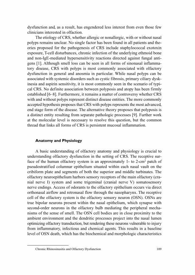
Chronic Rhinosinusitis and Olfactory Dysfunction 109
dysfunction and, as a result, has engendered less interest from even those few
clinicians interested in olfaction.
The etiology of CRS, whether allergic or nonallergic, with or without nasal
polyps remains unclear. No single factor has been found in all patients and the-
ories proposed for the pathogenesis of CRS include staphylococcal exotoxin
exposure, T-cell disturbances, chronic infection of the underlying ethmoid bone
and non-IgE-mediated hypersensitivity reactions directed against fungal anti-
gens [1]. Although smell loss can be seen in all forms of sinonasal inflamma-
tory disease, CRS with polyps is most commonly associated with olfactory
dysfunction in general and anosmia in particular. While nasal polyps can be
associated with systemic disorders such as cystic fibrosis, primary ciliary dysk-
inesia and aspirin sensitivity, it is most commonly seen in the scenario of typi-
cal CRS. No definite association between polyposis and atopy has been firmly
established [6–8]. Furthermore, it remains a matter of controversy whether CRS
with and without polyps represent distinct disease entities. The more commonly
accepted hypothesis proposes that CRS with polyps represents the most advanced,
end stage form of the disease. The alternative theory proposes that polyposis is
a distinct entity resulting from separate pathologic processes [9]. Further work
at the molecular level is necessary to resolve this question, but the common
thread that links all forms of CRS is persistent mucosal inflammation.
Anatomy and Physiology
A basic understanding of olfactory anatomy and physiology is crucial to
understanding olfactory dysfunction in the setting of CRS. The receptive sur-
face of the human olfactory system is an approximately 1- to 2-cm2 patch of
pseudostratified columnar epithelium situated within each nasal vault on the
cribiform plate and segments of both the superior and middle turbinates. The
olfactory neuroepithelium harbors sensory receptors of the main olfactory (cra-
nial nerve I) system and some trigeminal (cranial nerve V) somatosensory
nerve endings. Access of odorants to the olfactory epithelium occurs via direct
orthonasal airflow and retronasal flow through the nasopharynx. The receptive
cell of the olfactory system is the olfactory sensory neuron (OSN). OSNs are
true bipolar neurons present within the nasal epithelium, which synapse with
second-order neurons in the olfactory bulb mediating the peripheral mecha-
nisms of the sense of smell. The OSN cell bodies are in close proximity to the
ambient environment and the dendritic processes project into the nasal lumen
optimizing olfactory transduction, but rendering these neurons vulnerable to injury
from inflammatory, infectious and chemical agents. This results in a baseline
level of OSN death, which has the biochemical and morphologic characteristics
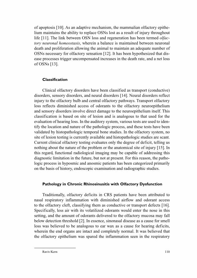
Raviv/Kern 110
of apoptosis [10]. As an adaptive mechanism, the mammalian olfactory epithe-
lium maintains the ability to replace OSNs lost as a result of injury throughout
life [11]. The link between OSN loss and regeneration has been termed olfac-tory neuronal homeostasis, wherein a balance is maintained between neuronal
death and proliferation allowing the animal to maintain an adequate number of
OSNs necessary for olfactory sensation [12]. It has been hypothesized that dis-
ease processes trigger uncompensated increases in the death rate, and a net loss
of OSNs [13].
Classification
Clinical olfactory disorders have been classified as transport (conductive)
disorders, sensory disorders, and neural disorders [14]. Neural disorders reflect
injury to the olfactory bulb and central olfactory pathways. Transport olfactory
loss reflects diminished access of odorants to the olfactory neuroepithelium
and sensory disorders involve direct damage to the neuroepithelium itself. This
classification is based on site of lesion and is analogous to that used for the
evaluation of hearing loss. In the auditory system, various tests are used to iden-
tify the location and nature of the pathologic process, and these tests have been
validated by histopathologic temporal bone studies. In the olfactory system, no
site of lesion testing is currently available and histopathologic studies are scant.
Current clinical olfactory testing evaluates only the degree of deficit, telling us
nothing about the nature of the problem or the anatomical site of injury [15]. In
this regard, functional radiological imaging may be capable of addressing this
diagnostic limitation in the future, but not at present. For this reason, the patho-
logic process in hyposmic and anosmic patients has been categorized primarily
on the basis of history, endoscopic examination and radiographic studies.
Pathology in Chronic Rhinosinusitis with Olfactory Dysfunction
Traditionally, olfactory deficits in CRS patients have been attributed to
nasal respiratory inflammation with diminished airflow and odorant access
to the olfactory cleft, classifying them as conductive or transport defects [16].
Specifically, less air with its volatilized odorants would enter the nose in this
setting, and the amount of odorants delivered to the olfactory mucosa may fall
below detection threshold [2]. In essence, sinonasal disease as a cause for smell
loss was believed to be analogous to ear wax as a cause for hearing deficits,
wherein the end organs are intact and completely normal. It was believed that
the olfactory epithelium was spared the inflammation seen in the respiratory
Chronic Rhinosinusitis and Olfactory Dysfunction 111
regions of the nose of CRS patients, apparently remaining intact and unaf-
fected. Lacking definitive data, the conductive hypothesis of CRS and smell
loss was supported by three main factors. First, clinical observations indicated
that olfactory deficits often rapidly improve following a burst of oral steroids
(see below). It was presumed that a rapid response to treatment was most con-
sistent with the reversal of a mechanical process, as any direct inflammatory
damage to the neuroepithelium would require a significant amount of time for
full recovery. However, the clinical observation of rapid olfactory recovery in
this setting was anecdotal and not based on clinical testing, so both the precise
time course and full extent of recovery are uncertain and probably highly vari-
able. Secondly, the olfactory mucosa was believed to be an ‘immunologi-
cally privileged’ site similar to the eye, incapable of mounting a normal immune
response to foreign proteins, in order to spare the neuroepithelium any associ-
ated inflammatory damage. Studies from the Getchell laboratory, however,
demonstrated that the olfactory mucosa at least in rodents possessed the requi-
site ‘synthetic and secretory capacity to respond with immune and non-immune
mechanisms to pathogenic challenge’ [17]. Furthermore, biopsies of the human
olfactory mucosa confirmed this assessment, demonstrating a marked inflam-
matory response in the olfactory epithelium in the setting of CRS, but the clin-
ical significance was unclear [18]. The third point in support of the ‘conductive
hypothesis’ was the demonstration by the Jafek laboratory (see below) of at
least some normal-appearing OSNs on electron microscopic studies of biopsies
obtained from patients with polyps and anosmia. Until relatively recently, the
conductive hypothesis of CRS and smell loss had remained unchallenged.
Historically, the first attempt to evaluate the mechanism of smell loss in
CRS was by Jafek et al. [19], who reported on 2 patients with nasal polyps and
anosmia. Both patients remained anosmic following surgery despite obvious
improvement in their nasal airways so they were started on oral steroid regimens.
Long-term correction of the anosmia was achieved in both patients through this
combination of corticosteroids and surgery. Biopsies of the olfactory mucosa at
the time of surgery in both patients showed at least some normal OSNs, sup-
porting the theory of mechanical obstruction as the cause for olfactory dys-
function. The need for steroids, however, caused Jafek et al. to speculate that,
rather than being only an obstructive phenomenon from polyps, anosmia at least
in part resulted from the direct effects of inflammatory processes on the olfactory
epithelium, the surface of the olfactory receptors, or the olfactory mucus bathing
the receptors.
Biopsies from the olfactory epithelium taken from patients with smell loss
after head trauma or acute viral insult have demonstrated obvious histological
abnormalities in the neuroepithelium [20, 21]. The demonstration of damage to
the olfactory mucosa in these entities confirms that the nature of the problem is,
Raviv/Kern 112
at least in part, sensory. Pathologic changes in the olfactory mucosa of patients
with CRS had not been documented, and these disorders were therefore classi-
fied as conductive disorders as mentioned above. In 2000, olfactory biopsies
were performed on 30 patients undergoing nasal surgery [16]. Nineteen of the
30 had actual olfactory mucosa on the biopsy sample. Nine of 19 patients demon-
strated normal olfactory mucosa and normal olfactory function. Ten patients
demonstrated pathologic changes in the olfactory mucosa with an influx of lym-
phocytes, macrophages, and eosinophils. Seven of these 10 patients had olfactory
deficits as confirmed by the University of Pennsylvania Smell Identification
Test (UPSIT). This was the first study emphasizing inflammatory changes in
the olfactory mucosa of patients with sinonasal disease, and indicated that the
pathological process present in the respiratory regions of the nose involved the
olfactory mucosa as well. In addition, grading of the intensity of the inflamma-
tory response within the olfactory mucosa was performed and generally showed
that severer inflammatory changes occurred in patients with decreased UPSIT
scores, further suggesting that pathology within the olfactory epithelium con-
tributed to hyposmia and anosmia in these patients.
Theoretically, inflammation within the olfactory neuroepithelium may trig-
ger smell loss by a variety of potential mechanisms. Mediators released by lym-
phocytes and macrophages are known to trigger hypersecretion in respiratory and
Bowman’s glands [22]. Olfactory mucus, produced by Bowman’s glands, is a
highly specialized substance vastly different from nasal respiratory mucus, serv-
ing a function analogous to cochlear endolymph [23, 24]. Hypersecretion would
likely alter the ion concentrations of olfactory mucus, affecting the microenviron-
ment of olfactory neurons and possibly the transduction process [25]. Moreover,
the presence of these inflammatory cells in the olfactory mucosa provides a direct
mechanism for the action of corticosteroids on anosmia. Type II corticosteroid
receptors are found in these inflammatory cells and activation with a systemic
glucocorticoid would rapidly suppress the local cytokine response [23, 25]. In
addition to secretory effects, these same cytokines and mediators may be toxic to
neurons [12, 26]. In particular, inflammatory mediators released by lymphocytes,
macrophages, and eosinophils may trigger caspase-3 activation in OSNs [27].
Caspase-3 is the primary executioner caspase in mammalian tissues, and detec-
tion of the active form within a cell designates it for apoptotic proteolysis. Recent
studies from our laboratory demonstrated that OSN death, demonstrated by
caspase-3 activation, appeared to be a significant component of olfactory dysfunc-
tion in chronic sinusitis. Olfactory tissue of patients with a normal sense of smell
despite CRS demonstrated increased caspase-3 activity when compared with nor-
mal human olfactory mucosa. Furthermore, olfactory tissue from patients with
CRS and smell loss demonstrated a severer inflammatory response and much
more extensive caspase-3 activity in both the olfactory epithelium as well as the
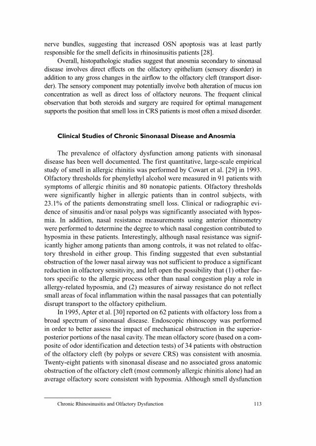
Chronic Rhinosinusitis and Olfactory Dysfunction 113
nerve bundles, suggesting that increased OSN apoptosis was at least partly
responsible for the smell deficits in rhinosinusitis patients [28].
Overall, histopathologic studies suggest that anosmia secondary to sinonasal
disease involves direct effects on the olfactory epithelium (sensory disorder) in
addition to any gross changes in the airflow to the olfactory cleft (transport disor-
der). The sensory component may potentially involve both alteration of mucus ion
concentration as well as direct loss of olfactory neurons. The frequent clinical
observation that both steroids and surgery are required for optimal management
supports the position that smell loss in CRS patients is most often a mixed disorder.
Clinical Studies of Chronic Sinonasal Disease and Anosmia
The prevalence of olfactory dysfunction among patients with sinonasal
disease has been well documented. The first quantitative, large-scale empirical
study of smell in allergic rhinitis was performed by Cowart et al. [29] in 1993.
Olfactory thresholds for phenylethyl alcohol were measured in 91 patients with
symptoms of allergic rhinitis and 80 nonatopic patients. Olfactory thresholds
were significantly higher in allergic patients than in control subjects, with
23.1% of the patients demonstrating smell loss. Clinical or radiographic evi-
dence of sinusitis and/or nasal polyps was significantly associated with hypos-
mia. In addition, nasal resistance measurements using anterior rhinometry
were performed to determine the degree to which nasal congestion contributed to
hyposmia in these patients. Interestingly, although nasal resistance was signif-
icantly higher among patients than among controls, it was not related to olfac-
tory threshold in either group. This finding suggested that even substantial
obstruction of the lower nasal airway was not sufficient to produce a significant
reduction in olfactory sensitivity, and left open the possibility that (1) other fac-
tors specific to the allergic process other than nasal congestion play a role in
allergy-related hyposmia, and (2) measures of airway resistance do not reflect
small areas of focal inflammation within the nasal passages that can potentially
disrupt transport to the olfactory epithelium.
In 1995, Apter et al. [30] reported on 62 patients with olfactory loss from a
broad spectrum of sinonasal disease. Endoscopic rhinoscopy was performed
in order to better assess the impact of mechanical obstruction in the superior-
posterior portions of the nasal cavity. The mean olfactory score (based on a com-
posite of odor identification and detection tests) of 34 patients with obstruction
of the olfactory cleft (by polyps or severe CRS) was consistent with anosmia.
Twenty-eight patients with sinonasal disease and no associated gross anatomic
obstruction of the olfactory cleft (most commonly allergic rhinitis alone) had an
average olfactory score consistent with hyposmia. Although smell dysfunction
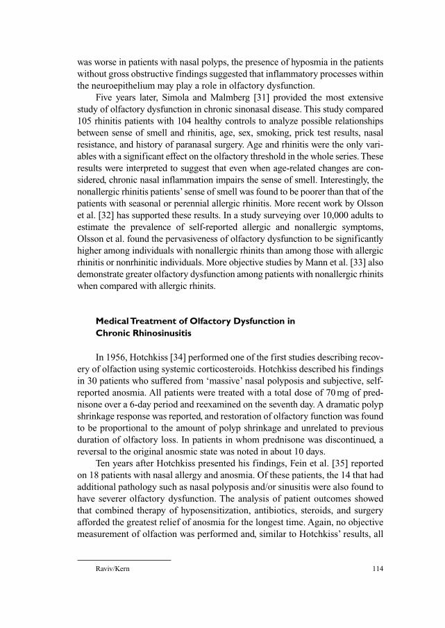
Raviv/Kern 114
was worse in patients with nasal polyps, the presence of hyposmia in the patients
without gross obstructive findings suggested that inflammatory processes within
the neuroepithelium may play a role in olfactory dysfunction.
Five years later, Simola and Malmberg [31] provided the most extensive
study of olfactory dysfunction in chronic sinonasal disease. This study compared
105 rhinitis patients with 104 healthy controls to analyze possible relationships
between sense of smell and rhinitis, age, sex, smoking, prick test results, nasal
resistance, and history of paranasal surgery. Age and rhinitis were the only vari-
ables with a significant effect on the olfactory threshold in the whole series. These
results were interpreted to suggest that even when age-related changes are con-
sidered, chronic nasal inflammation impairs the sense of smell. Interestingly, the
nonallergic rhinitis patients’ sense of smell was found to be poorer than that of the
patients with seasonal or perennial allergic rhinitis. More recent work by Olsson
et al. [32] has supported these results. In a study surveying over 10,000 adults to
estimate the prevalence of self-reported allergic and nonallergic symptoms,
Olsson et al. found the pervasiveness of olfactory dysfunction to be significantly
higher among individuals with nonallergic rhinits than among those with allergic
rhinitis or nonrhinitic individuals. More objective studies by Mann et al. [33] also
demonstrate greater olfactory dysfunction among patients with nonallergic rhinits
when compared with allergic rhinits.
Medical Treatment of Olfactory Dysfunction in Chronic Rhinosinusitis
In 1956, Hotchkiss [34] performed one of the first studies describing recov-
ery of olfaction using systemic corticosteroids. Hotchkiss described his findings
in 30 patients who suffered from ‘massive’ nasal polyposis and subjective, self-
reported anosmia. All patients were treated with a total dose of 70 mg of pred-
nisone over a 6-day period and reexamined on the seventh day. A dramatic polyp
shrinkage response was reported, and restoration of olfactory function was found
to be proportional to the amount of polyp shrinkage and unrelated to previous
duration of olfactory loss. In patients in whom prednisone was discontinued, a
reversal to the original anosmic state was noted in about 10 days.
Ten years after Hotchkiss presented his findings, Fein et al. [35] reported
on 18 patients with nasal allergy and anosmia. Of these patients, the 14 that had
additional pathology such as nasal polyposis and/or sinusitis were also found to
have severer olfactory dysfunction. The analysis of patient outcomes showed
that combined therapy of hyposensitization, antibiotics, steroids, and surgery
afforded the greatest relief of anosmia for the longest time. Again, no objective
measurement of olfaction was performed and, similar to Hotchkiss’ results, all
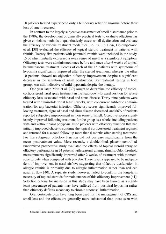
Chronic Rhinosinusitis and Olfactory Dysfunction 115
18 patients treated experienced only a temporary relief of anosmia before their
loss of smell recurred.
In contrast to the largely subjective assessment of smell disturbance prior to
the 1980s, the development of clinically practical tests to evaluate olfaction has
given clinicians methods to quantitatively assess smell dysfunction and evaluate
the efficacy of various treatment modalities [36, 37]. In 1996, Golding-Wood
et al. [38] evaluated the efficacy of topical steroid treatment in patients with
rhinitis. Twenty-five patients with perennial rhinitis were included in the study,
15 of which initially expressed a weak sense of smell as a significant symptom.
Olfactory tests were administered once before and once after 6 weeks of topical
betamethasone treatment. Scores of each of the 15 patients with symptoms of
hyposmia significantly improved after the steroid treatment, whereas the other
10 patients showed no objective olfactory improvement despite a significant
decrease in the sensation of nasal obstruction. Posttreatment testing in both
groups was still indicative of mild hyposmia despite the therapy.
One year later, Mott et al. [39] sought to determine the efficacy of topical
corticosteroid nasal spray treatment in the head-down-forward position for severe
olfactory loss associated with nasal and sinus disease. Thirty-nine patients were
treated with flunisolide for at least 8 weeks, with concurrent antibiotic adminis-
tration for any bacterial infection. Olfactory scores significantly improved fol-
lowing treatment, signs of nasal and sinus disease decreased, and 66% of patients
reported subjective improvement in their sense of smell. Objective scores signif-
icantly improved following treatment for the group as a whole, including patients
with and without nasal polyposis. Nine patients with olfactory function that had
initially improved chose to continue the topical corticosteroid treatment regimen
and returned for a second follow-up more than 6 months after starting treatment.
For this subgroup, olfactory function did not decrease significantly from the
mean posttreatment value. More recently, a double-blind, placebo-controlled,
randomized prospective study evaluated the effects of topical steroid spray on
olfactory performance in 24 patients with seasonal allergic rhinitis. Odor threshold
measurements significantly improved after 2 weeks of treatment with mometa-
sone furoate when compared with placebo. These results appeared to be indepen-
dent of improvement in nasal airflow, suggesting that olfactory dysfunction in
allergic rhinitis is primarily due to allergic inflammation rather than reduced
nasal airflow [40]. A separate study, however, failed to confirm the long-term
necessity of topical steroids for maintenance of this olfactory improvement [41].
Selection criteria for inclusion in this study may have been flawed, as a signif-
icant percentage of patients may have suffered from postviral hyposmia rather
than olfactory deficits secondary to chronic sinonasal inflammation.
Oral corticosteroids have long been used for the management of CRS and
smell loss and the effects are generally more substantial than those seen with

Raviv/Kern 116
topical analogues. In 1984, Goodspeed et al. [42] tested olfactory function in 20
anosmic and hyposmic patients before and after a 1-week course of systemic
steroids. Ten patients had sinonasal disease, 4 had olfactory loss after upper res-
piratory infection, and 6 were considered idiopathic. Only 6 patients responded,
all of who had nasal and sinus disease. In 1995, Ikeda et al. [43] documen-
ted olfactory function before and after systemic corticosteroid therapy in 12
patients with anosmia refractory to topical steroid treatment. Significant effi-
cacy was achieved with a short course of high-dose oral corticosteroids in non-
allergic sinus disease. On the other hand, anosmia induced by upper respiratory
infection failed to respond to systemic steroid treatment. The authors speculated
that the lack of improvement in patients after upper respiratory infection was
due to permanent damage to olfactory receptor cells, while the effectiveness
observed in patients with sinus disease was explained by improvement of
mucosal thickening in the area of the olfactory cleft, leading to increased access
of odorants to the olfactory epithelium. More recently, in 2001, Seiden and
Duncan [44] presented a retrospective study of consecutive patients presenting
with a primary complaint of olfactory loss. A ‘pulse’ dose of systemic steroids
was used to support the diagnosis of olfactory dysfunction in a subset of the
patients reviewed. Thirty-six patients received a tapering course of systemic
steroids, and 30 (83%) experienced an improvement in their sense of smell. In
contrast, only 13 of 52 patients (25%) who were given topical steroids noted any
improvement. In their discussion, the authors conclude that while long-term sys-
temic steroid treatment for anosmia may not be appropriate, a short course of
high-dose therapy can be helpful for diagnosing a reversible ‘conductive loss’.
Finally, recent studies have suggested a role for leukotriene receptor antago-
nist (LRA) therapy in the management of anosmia in CRS. In 2001, Wilson et al.
[45] reported on 32 patients with CRS who were treated with an LRA (mon-
telukast, 10 mg/day). Significant improvement in subjective scoring of sense of
smell was reported at a median follow-up duration of 14 weeks. More recently,
Otto et al. [46] presented their work supporting the role of LRAs in the medical
management of anosmia related to CRS. In their study, 12 patients were treated
with a combination of topical and systemic steroids, LRAs, and sinus surgery in
necessary cases. The average UPSIT score increased by 17.5 points with a min-
imum follow-up of 1 year.
Surgical Treatment of Olfactory Dysfunction in Chronic Rhinosinusitis
Overall, studies suggest that the degree of olfactory loss is usually associated
with the severity of sinonasal disease, with the greatest loss occurring in patients
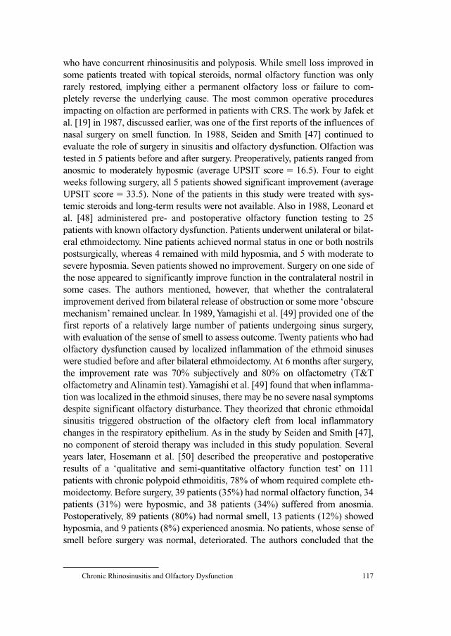
Chronic Rhinosinusitis and Olfactory Dysfunction 117
who have concurrent rhinosinusitis and polyposis. While smell loss improved in
some patients treated with topical steroids, normal olfactory function was only
rarely restored, implying either a permanent olfactory loss or failure to com-
pletely reverse the underlying cause. The most common operative procedures
impacting on olfaction are performed in patients with CRS. The work by Jafek et
al. [19] in 1987, discussed earlier, was one of the first reports of the influences of
nasal surgery on smell function. In 1988, Seiden and Smith [47] continued to
evaluate the role of surgery in sinusitis and olfactory dysfunction. Olfaction was
tested in 5 patients before and after surgery. Preoperatively, patients ranged from
anosmic to moderately hyposmic (average UPSIT score � 16.5). Four to eight
weeks following surgery, all 5 patients showed significant improvement (average
UPSIT score � 33.5). None of the patients in this study were treated with sys-
temic steroids and long-term results were not available. Also in 1988, Leonard et
al. [48] administered pre- and postoperative olfactory function testing to 25
patients with known olfactory dysfunction. Patients underwent unilateral or bilat-
eral ethmoidectomy. Nine patients achieved normal status in one or both nostrils
postsurgically, whereas 4 remained with mild hyposmia, and 5 with moderate to
severe hyposmia. Seven patients showed no improvement. Surgery on one side of
the nose appeared to significantly improve function in the contralateral nostril in
some cases. The authors mentioned, however, that whether the contralateral
improvement derived from bilateral release of obstruction or some more ‘obscure
mechanism’ remained unclear. In 1989, Yamagishi et al. [49] provided one of the
first reports of a relatively large number of patients undergoing sinus surgery,
with evaluation of the sense of smell to assess outcome. Twenty patients who had
olfactory dysfunction caused by localized inflammation of the ethmoid sinuses
were studied before and after bilateral ethmoidectomy. At 6 months after surgery,
the improvement rate was 70% subjectively and 80% on olfactometry (T&T
olfactometry and Alinamin test). Yamagishi et al. [49] found that when inflamma-
tion was localized in the ethmoid sinuses, there may be no severe nasal symptoms
despite significant olfactory disturbance. They theorized that chronic ethmoidal
sinusitis triggered obstruction of the olfactory cleft from local inflammatory
changes in the respiratory epithelium. As in the study by Seiden and Smith [47],
no component of steroid therapy was included in this study population. Several
years later, Hosemann et al. [50] described the preoperative and postoperative
results of a ‘qualitative and semi-quantitative olfactory function test’ on 111
patients with chronic polypoid ethmoiditis, 78% of whom required complete eth-
moidectomy. Before surgery, 39 patients (35%) had normal olfactory function, 34
patients (31%) were hyposmic, and 38 patients (34%) suffered from anosmia.
Postoperatively, 89 patients (80%) had normal smell, 13 patients (12%) showed
hyposmia, and 9 patients (8%) experienced anosmia. No patients, whose sense of
smell before surgery was normal, deteriorated. The authors concluded that the
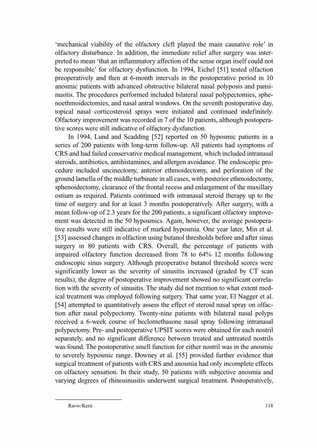
Raviv/Kern 118
‘mechanical viability of the olfactory cleft played the main causative role’ in
olfactory disturbance. In addition, the immediate relief after surgery was inter-
preted to mean ‘that an inflammatory affection of the sense organ itself could not
be responsible’ for olfactory dysfunction. In 1994, Eichel [51] tested olfaction
preoperatively and then at 6-month intervals in the postoperative period in 10
anosmic patients with advanced obstructive bilateral nasal polyposis and pansi-
nusitis. The procedures performed included bilateral nasal polypectomies, sphe-
noethmoidectomies, and nasal antral windows. On the seventh postoperative day,
topical nasal corticosteroid sprays were initiated and continued indefinitely.
Olfactory improvement was recorded in 7 of the 10 patients, although postopera-
tive scores were still indicative of olfactory dysfunction.
In 1994, Lund and Scadding [52] reported on 50 hyposmic patients in a
series of 200 patients with long-term follow-up. All patients had symptoms of
CRS and had failed conservative medical management, which included intranasal
steroids, antibiotics, antihistamines, and allergen avoidance. The endoscopic pro-
cedure included uncinectomy, anterior ethmoidectomy, and perforation of the
ground lamella of the middle turbinate in all cases, with posterior ethmoidectomy,
sphenoidectomy, clearance of the frontal recess and enlargement of the maxillary
ostium as required. Patients continued with intranasal steroid therapy up to the
time of surgery and for at least 3 months postoperatively. After surgery, with a
mean follow-up of 2.3 years for the 200 patients, a significant olfactory improve-
ment was detected in the 50 hyposmics. Again, however, the average postopera-
tive results were still indicative of marked hyposmia. One year later, Min et al.
[53] assessed changes in olfaction using butanol thresholds before and after sinus
surgery in 80 patients with CRS. Overall, the percentage of patients with
impaired olfactory function decreased from 78 to 64% 12 months following
endoscopic sinus surgery. Although preoperative butanol threshold scores were
significantly lower as the severity of sinusitis increased (graded by CT scan
results), the degree of postoperative improvement showed no significant correla-
tion with the severity of sinusitis. The study did not mention to what extent med-
ical treatment was employed following surgery. That same year, El Nagger et al.
[54] attempted to quantitatively assess the effect of steroid nasal spray on olfac-
tion after nasal polypectomy. Twenty-nine patients with bilateral nasal polyps
received a 6-week course of beclomethasone nasal spray following intranasal
polypectomy. Pre- and postoperative UPSIT scores were obtained for each nostril
separately, and no significant difference between treated and untreated nostrils
was found. The postoperative smell function for either nostril was in the anosmic
to severely hyposmic range. Downey et al. [55] provided further evidence that
surgical treatment of patients with CRS and anosmia had only incomplete effects
on olfactory sensation. In their study, 50 patients with subjective anosmia and
varying degrees of rhinosinusitis underwent surgical treatment. Postoperatively,
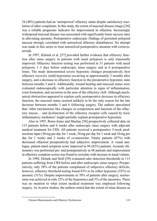
Chronic Rhinosinusitis and Olfactory Dysfunction 119
24 (48%) patients had an ‘unimproved’ olfactory status despite satisfactory reso-
lution of other complaints. In this study, the extent of mucosal disease (stage) [56]
was a reliable prognostic indicator for improvement in olfaction. Increasingly
widespread mucosal disease was associated with significantly lower success rates
in alleviating anosmia. Postoperative endoscopic findings of persistent polypoid
mucosa strongly correlated with unresolved olfactory disturbances. No attempt
was made in this series to treat unresolved postoperative anosmia with corticos-
teroids.
In 1997, Klimek et al. [57] provided further evidence that olfactory func-
tion after sinus surgery in patients with nasal polyposis is only transiently
improved. Olfactory function testing was performed in 31 patients with nasal
polyposis 1–3 days before endoscopic sinus surgery and at 6 postoperative
times. The study demonstrated severe hyposmic changes preoperatively, best
olfactory recovery (mild hyposmia) occurring at approximately 3 months after
surgery, and a decrease in olfactory function to the preoperative hyposmic state
between months 3 and 6. Additionally, wound healing and mucosal status were
evaluated endoscopically with particular attention to signs of inflammation,
crust formation, and secretion in the area of the olfactory cleft. Although mech-
anical obstruction appeared to explain early postoperative decrease in olfactory
function, the mucosal status seemed unlikely to be the only reason for the late
decrease between months 3 and 6 following surgery. The authors speculated
that ‘other mechanisms like changes in composition and function of the olfac-
tory mucus… and dysfunction of the olfactory receptor cells caused by toxic
inflammatory mediators’ might partially explain postoperative hyposmia.
Also in 1997, Rowe-Jones and Mackay [58] prospectively collected data on
115 patients before and 6 weeks after endoscopic sinus surgery with adjuvant
medical treatment for CRS. All patients received a postoperative 3-week pred-
nisolone taper (30 mg per day for 1 week, 20 mg per day for 1 week and 10 mg per
day for 1 week) and 2 weeks of co-amoxiclav. Ninety patients (87%) with
decreased olfaction preoperatively had subjective improvement. A visual ana-
logue, patient-rated symptom score improved in 94 (82%) patients. Acoustic rhi-
nometry was performed pre- and postoperatively in 96 patients and improvement
in olfactory symptom scores was found to correlate with increase in nasal volume.
In 1998, Delank and Stoll [59] evaluated odor detection thresholds in 115
patients suffering from CRS before and after endoscopic sinus surgery. Preope-
ratively, only 58% of the patients complained of subjective olfactory deficits,
however, olfactory threshold testing found 83% to be either hyposmic (52%) or
anosmic (31%). Despite improvements in 70% of patients after surgery, normo-
smia was achieved in only 25% of the hyposmics and 5% of the anosmics. There
was no mention to what extent medical treatment was employed following
surgery. As in prior studies, the authors noted that the extent of sinus disease as
Raviv/Kern 120
measured by the degree of nasal polyposis correlated with levels of preopera-
tive olfactory dysfunction, and that the rate of improvement following surgery
was generally lower than assumed.
Summary of Current Therapy
As described, a large number of clinical studies of variable quality have
sought to determine the efficacy of standard therapy in the management of the
olfactory component of sinonasal disease. Although improvement in olfaction is
often possible, it is frequently transient and incomplete. In addition to antibiotics
and surgery, both systemic and topical steroids are helpful in attempting to allevi-
ate olfactory dysfunction in this setting. While systemic steroids are usually more
effective than topically administered steroids, prescription of systemic steroids
over an extended period is usually unwarranted and places the patient at risk for
side effects including gastric ulceration, diabetes, and osteoporosis [60]. Instead,
it has been suggested that systemic steroids can be used as an effective diagnostic
tool to help determine if a patient has any functioning olfactory mucosa, at which
point therapy is continued with locally administered steroids. Repeated adminis-
tration of short courses of systemic steroids with a long enough interval between
courses to avoid untoward side effects may also be effective [61].
The mechanism of olfactory dysfunction in CRS remains controversial. As
mentioned above, some investigators believe that obstruction of the olfactory
cleft via polyps or edema is the sole significant cause of smell loss in this set-
ting. Furthermore, the often rapid response to treatment described with both
corticosteroids and surgery supports this hypothesis. This rapid response is
unlikely to result in normal olfactory sensation, however, and most reports of
‘immediate’ return are subjective or anecdotal. The histopathologic data, on the
other hand, support the concept that direct injury to the neuroepithelium is a
component of the problem in addition to any superimposed obstruction. The
common clinical observation of persistent smell loss despite adequate medical
or surgical treatment of other sinonasal complaints also supports this alternative
hypothesis. Overall, it is the authors’ opinion that the weight of current evi-
dence supports the theory that olfactory dysfunction in the setting of sinonasal
disease is a mixed problem, with varying degrees of conductive and sensory
losses in individual patients.
Future Therapy
The fundamental limitation of current therapy for CRS and smell loss is that
while surgery and corticosteroids can effectively treat the mechanical or obstructive
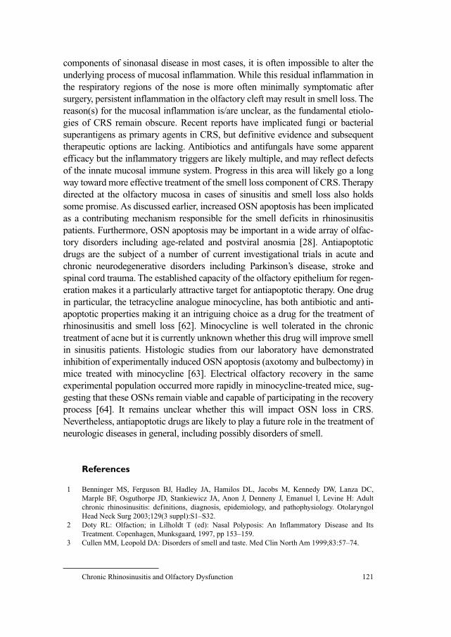
Chronic Rhinosinusitis and Olfactory Dysfunction 121
components of sinonasal disease in most cases, it is often impossible to alter the
underlying process of mucosal inflammation. While this residual inflammation in
the respiratory regions of the nose is more often minimally symptomatic after
surgery, persistent inflammation in the olfactory cleft may result in smell loss. The
reason(s) for the mucosal inflammation is/are unclear, as the fundamental etiolo-
gies of CRS remain obscure. Recent reports have implicated fungi or bacterial
superantigens as primary agents in CRS, but definitive evidence and subsequent
therapeutic options are lacking. Antibiotics and antifungals have some apparent
efficacy but the inflammatory triggers are likely multiple, and may reflect defects
of the innate mucosal immune system. Progress in this area will likely go a long
way toward more effective treatment of the smell loss component of CRS. Therapy
directed at the olfactory mucosa in cases of sinusitis and smell loss also holds
some promise. As discussed earlier, increased OSN apoptosis has been implicated
as a contributing mechanism responsible for the smell deficits in rhinosinusitis
patients. Furthermore, OSN apoptosis may be important in a wide array of olfac-
tory disorders including age-related and postviral anosmia [28]. Antiapoptotic
drugs are the subject of a number of current investigational trials in acute and
chronic neurodegenerative disorders including Parkinson’s disease, stroke and
spinal cord trauma. The established capacity of the olfactory epithelium for regen-
eration makes it a particularly attractive target for antiapoptotic therapy. One drug
in particular, the tetracycline analogue minocycline, has both antibiotic and anti-
apoptotic properties making it an intriguing choice as a drug for the treatment of
rhinosinusitis and smell loss [62]. Minocycline is well tolerated in the chronic
treatment of acne but it is currently unknown whether this drug will improve smell
in sinusitis patients. Histologic studies from our laboratory have demonstrated
inhibition of experimentally induced OSN apoptosis (axotomy and bulbectomy) in
mice treated with minocycline [63]. Electrical olfactory recovery in the same
experimental population occurred more rapidly in minocycline-treated mice, sug-
gesting that these OSNs remain viable and capable of participating in the recovery
process [64]. It remains unclear whether this will impact OSN loss in CRS.
Nevertheless, antiapoptotic drugs are likely to play a future role in the treatment of
neurologic diseases in general, including possibly disorders of smell.
References
1 Benninger MS, Ferguson BJ, Hadley JA, Hamilos DL, Jacobs M, Kennedy DW, Lanza DC,
Marple BF, Osguthorpe JD, Stankiewicz JA, Anon J, Denneny J, Emanuel I, Levine H: Adult
chronic rhinosinusitis: definitions, diagnosis, epidemiology, and pathophysiology. Otolaryngol
Head Neck Surg 2003;129(3 suppl):S1–S32.
2 Doty RL: Olfaction; in Lilholdt T (ed): Nasal Polyposis: An Inflammatory Disease and Its
Treatment. Copenhagen, Munksgaard, 1997, pp 153–159.
3 Cullen MM, Leopold DA: Disorders of smell and taste. Med Clin North Am 1999;83:57–74.
Raviv/Kern 122
4 Seiden AM, Duncan HJ: The diagnosis of a conductive olfactory loss. Laryngoscope 2001;111:
9–14.
5 Loury MC, Kennedy DW: Chronic sinusitis and nasal polyposis; in Getchell TV, Bartoshuk LM,
Doty RL, Snow J (eds): Smell and Taste in Health and Disease. New York, Raven Press, 1991, pp
711–730.
6 Caplin I, Haynes JT, Spahn J: Are nasal polyps an allergic phenomenon? Ann Allergy 1971;29:
631–634.
7 Patriarca G, Romano A, Schiavino D, Venuti A, Di Rienzo V, Fais G, Nucera E: ASA disease:
the clinical relationship of nasal polyposis to ASA intolerance. Arch Otorhinolaryngol 1986;243:
16–19.
8 Delaney JC: Aspirin idiosyncracy in patients admitted for nasal polypectomy. Clin Otolaryngol
1976;1:27–30.
9 Rudach C, Sachse F, Alberty J: Chronic rhinosinusitis – Need for further classification? Inflamm
Res 2004;53:111–117.
10 Magrassi L, Graziadei PP: Cell death in the olfactory epithelium. Anat Embryol (Berl) 1995;192:
77–87.
11 Farbman AI: Cell Biology of Olfaction. Developmental and Cell Biology Series No 27. New York,
Cambridge University Press, 1992.
12 Holcomb JD, Graham S, Calof AL: Neuronal homeostasis in mammalian olfactory epithelium:
a review. Am J Rhinol 1996;10:125–134.
13 Robinson AM, Conley DB, Shinners MJ, Kern RC: Apoptosis in the aging olfactory epithelium.
Laryngoscope 2002;112:1431–1435.
14 Snow JB: Causes of olfactory and gustatory disorders; in Getchell TV, Bartoshuk LM, Doty RL,
Snow J (eds): Smell and Taste in Health and Disease. New York, Raven Press, 1991, pp 445–449.
15 Doty RL, Shaman P, Kimmelman CP, Dann MS: University of Pennsylvania Smell Identification
Test: a rapid quantitative olfactory function test for the clinic. Laryngoscope 1984;94:176–178.
16 Baroody F, Naclerio R: Allergic rhinitis; in Getchell TV, Bartoshuk LM, Doty RL, Snow J (eds):
Smell and Taste in Health and Disease. New York, Raven Press, 1991, pp 731–740.
17 Mellert TK, Getchell ML, Sparks L, Getchell TV: Characterization of the immune barrier in
human olfactory mucosa. Otolaryngol Head Neck Surg 1992;106:181–188.
18 Kern RC: Chronic sinusitis and anosmia: pathologic changes in the olfactory mucosa.
Laryngoscope 2000;110:1071–1077.
19 Jafek BW, Moran DT, Eller PM, Rowley JC, Jafek TB: Steroid-dependent anosmia. Arch
Otolaryngol 1987;113:547–549.
20 Jafek BW, Hartman D, Eller P, Johnson EW, Strahan RC, Moran DT: Post-viral olfactory dysfunc-
tion. Am J Rhinol 1990;4:91–100.
21 Yamagashi M, Okazoe R, Ishizuka Y: Olfactory mucosa of patients with olfactory disturbance fol-
lowing head trauma. Ann Otol Rhinol Otolaryngol 1994;103:218–226.
22 Getchell M, Mellert T: Olfactory mucus secretion; in Getchell TV, Bartoshuk LM, Doty RL, Snow
J (eds): Smell and Taste in Health and Disease. New York, Raven Press, 1991, pp 445–449.
23 Robinson AM, Kern RC, Foster JD, Fong KJ, Pitovski DZ: Expression of glucocorticoid receptor
mRNA and protein in the olfactory mucosa. Laryngoscope 1999;109:383–388.
24 Joshi H, Getchell ML, Zielinski B, Getchell TV: Spectrophotometric determination of cation con-
centrations in olfactory mucus. Neurosci Lett 1987;82:321–326.
25 Kern RC, Foster JD, Pitovski DZ: Glucocorticoid (type II) receptors in the olfactory mucosa of the
guinea pig: RU 28362. Chem Senses 1997;22:313–319.
26 Nakashima T, Kimmelman C, Snow JB: Immunohistopathology of human olfactory epithelium,
nerve, and bulb. Laryngoscope 1985;95:391–398.
27 Ge Y, Tsukatani T, Nishimura T, Furukawa M, Miwa T: Cell death of olfactory receptor neurons in
a rat with nasosinusitis infected artificially with staphylococcus. Chem Senses 2002;27:521–527.
28 Kern RC, Conley DB, Haines GK 3rd, Robinson AM: Pathology of the olfactory mucosa: impli-
cations for the treatment of olfactory dysfunction. Laryngoscope 2004;114:279–285.
29 Cowart BJ, Flynn-Rodden K, McGeady SJ, Lowry LD: Hyposmia in allergic rhinitis. J Allergy
Clin Immunol 1993;91:747–751.

Chronic Rhinosinusitis and Olfactory Dysfunction 123
30 Apter AJ, Mott AE, Frank ME, Clive JM: Allergic rhinitis and olfactory loss. Ann Allergy Asthma
Immunol 1995;75:311–316.
31 Simola M, Malmberg H: Sense of smell in allergic and nonallergic rhinitis. Allergy 1998;53:
190–194.
32 Olsson P, Berglind N, Bellander T, Stjarne P: Prevalence of self-reported allergic and non-allergic
rhinits symptoms in Stockholm: relation to age, gender, olfactory sense, and smoking. Acta
Otolaryngol 2003;123:75–80.
33 Mann SS, Maini S, Nageswari KS, Mohan H, Handa A: Assessment of olfactory status in allergic
and non-allergic rhinitis patients. Indian J Physiol Pharmacol 2002;46:186–194.
34 Hotchkiss WT: Influence of prednisone on nasal polyposis with anosmia. Arch Otolaryngol
1956;64:478–479.
35 Fein B, Kamin P, Fein NS: The loss of smell in nasal allergy. Ann Allergy 1966;24:278–283.
36 Doty RL, Smith R, McKeown DA, Raj J: Development of the University of Pennsylvania Smell
Identification Test: a standardized microencapsulated test of olfactory function. Physiol Behav
1984;32:489–502.
37 Kobal G, Hummel T, Sekinger B, Barz S, Roscher S, Wolf S: ‘Sniffin’ sticks’: screening of olfac-
tory performance. Rhinology 1996;34:222–226.
38 Golding-Wood DG, Holmstrom M, Darby Y, Scadding GK, Lund VJ: The treatment of hyposmia
with intranasal steroids. J Laryngol Otol 1996;110:132–135.
39 Mott AE, Cain WS, Lafreniere D, Leonard G, Gent JF, Frank ME: Topical corticosteroid treatment
of anosmia associated with nasal and sinus disease. Arch Otolaryngol Head Neck Surg 1997;123:
367–372.
40 Stuck BA, Blum A, Hagner AE, Hummel T, Klimek L, Homann K: Mometasone furoate nasal
spray improves olfactory performance in seasonal allergic rhinitis. Allergy 2003;58:1195.
41 Blomqvist EH, Lundblad L, Bergstedt H, Stjarne P: Placebo-controlled, randomized, double-blind
study evaluating the efficacy of fluticasone proprionate nasal spray for the treatment of patients
with hyposmia/anosmia. Acta Otolaryngol 2003;123:862–868.
42 Goodspeed RB, Gent JF, Catalanotto FA, Cain WS, Zagraniski RT: Corticosteroids in olfactory
dysfunction; in Meiselma HL, Rivlin RS (eds): Clinical Measurement of Taste and Smell. New York,
Macmillan, 1986, pp 514–518.
43 Ikeda K, Sakurada T, Takasaka T: Efficacy of systemic corticosteroid treatment for anosmia with
nasal and paranasal sinus disease. Rhinology 1995;33:162–165.
44 Seiden AM, Duncan HJ: The diagnosis of a conductive olfactory loss. Laryngoscope 2001;111:
9–14.
45 Wilson A, White P, Gardiner Q, Nassif R, Lipworth B: Effects of leukotriene receptor antagonist
therapy in patients with chronic rhinosinusitis in a real life rhinology clinical setting. Am J Rhinol
2001;39:142–146.
46 Otto K, Lang E, DelGaudio J, Venkatraman G: Long-term medical management of anosmia
related to chronic sinusits. Am Rhinol Soc Meet, New York, 2004.
47 Seiden AM, Smith DV: Endoscopic intranasal surgery as an approach to restoring olfactory func-
tion. Chem Senses 1988;13:736.
48 Leonard G, Cain W, Clavet G: Surgical correction of olfactory disorders. Chem Senses
1988;13:708.
49 Yamagishi M, Hasegawa S, Suzuki S, Nakamura H, Nakano Y: Effects of surgical treatment of
olfactory disturbance caused by localized ethmoiditis. Clin Otolaryngol 1989;14:405–409.
50 Hosemann W, Goertzen W, Wohlleben R, Wolf S, Wigand ME: Olfaction after endoscopic
endonasal ethmoidectomy. Am J Rhinol 1993;7:11–15.
51 Eichel BS: Improvement of olfaction following pansinus surgery. Ear Nose Throat J 1994;73:
248–250.
52 Lund VJ, Scadding GK: Objective assessment of endoscopic sinus surgery in the management of
chronic rinosinusitis: an update. J Laryngol Otol 1995;109:941–944.
53 Min Y-G, Yun Y-S, Song BH, Cho YS, Lee KS: Recovery of nasal physiology after functional
endoscopic sinus surgery: olfaction and mucociliary transport. ORL J Otorhinolaryngol Relat
Spec 1995;57:264–268.

Raviv/Kern 124
54 el Naggar M, Kale S, Aldren C, Martin F: Effects of Beconase nasal spray on olfactory function
in post-nasal polypectomy patients: a prospective controlled trial. J Laryngol Otol 1995;109:
941–944.
55 Downey LL, Jacobs JB, Lebowitz RA: Anosmia and chronic sinus disease. Otolaryngol Head Neck
Surg 1996;115:24–28.
56 Kennedy D: Prognostic factors, outcomes and staging in ethmoid sinus surgery. Laryngoscope
1992;102(Suppl 51):1–18.
57 Klimek L, Moll B, Amedee RG, Mann WJ: Olfactory function after microscopic endonasal
surgery in patients with nasal polyps. Am J Rhinol 1997;11:251–255.
58 Rowe-Jones J, Mackay I: A prospective study of olfaction following endoscopic sinus surgery with
adjuvant medical treatment. Clin Otolaryngol 1997;22:377–381.
59 Delank KW, Stoll W: Olfactory function after functional endoscopic sinus surgery for chronic
sinusitis. Rhinology 1998;36:15–19.
60 Scott AE: Caution urged in treating ‘steroid-dependent anosmia’. Arch Otolaryngol Head Neck
Surg 1989:115;109–110.
61 Wolfensberger M, Hummel T: Anti-inflammatory and surgical therapy of olfactory disorders
related to sino-nasal disease. Chem Senses 2002:27;617–622.
62 Friedlander R: Apoptosis and caspases in neurodegenerative diseases. N Engl J Med 2003:348;
1365–1375.
63 Kern RC, Conley DB, Haines GK, Robinson AM: Treatment of olfactory dysfunction. 2. Studies
with minocycline. Laryngoscope 2004:114;2200–2204.
64 Kern RC, Raviv JR, Conley DB, Robinson AM: Recovery after olfactory axotomy in mice: phar-
macologic and genetic effects. 27th Annu Meet Assoc Chemorecept Sci, Sarasota, 2005.
Robert C. Kern, MD
Department of Otolaryngology – Head and Neck Surgery, Suite 15–200
657 N. St. Clair Street
Chicago, IL 60611 (USA)
Tel. �1 312 695 0805, Fax �1 312 695 7851, E-Mail [email protected]
Hummel T, Welge-Lüssen A (eds): Taste and Smell. An Update.
Adv Otorhinolaryngol. Basel, Karger, 2006, vol 63, pp 125–132
Olfactory Disorders following UpperRespiratory Tract Infections
Antje Welge-Lüssen, Markus Wolfensberger
Department of Otorhinolaryngology, University of Basel, Basel, Switzerland
AbstractPostviral olfactory disorders usually occur after an upper respiratory tract infection
(URTI) associated with a common cold or influenza. With a prevalence between 11 and 40%
they are among the common causes of olfactory disorders. Women are more often affected
than men and post-URTI disorders usually occur between the fourth and eighth decade of
life. The exact location of the damage in post-URTI is not yet known even though from biop-
sies a direct damage of the olfactory receptor cells is very likely. Nevertheless, central mech-
anisms cannot completely be ruled out. The diagnosis is made according to the history,
clinical examination and olfactory testing. Affected patients usually recall the acute URTI
and a close temporal connection should be present to establish the diagnosis. Spontaneous
recovery might occur within 2 years. So far, no effective therapy exists even though specific
olfactory training might be promising.
Copyright © 2006 S. Karger AG, Basel
Definition of Postviral Olfactory Disorders
Postviral olfactory disorders were described for the first time more than 20
years ago [1, 2]. The most frequent cause of postviral smell loss is an upper res-
piratory tract infection (URTI) typically associated with the common cold or
influenza. In such cases, there is a close temporal connection between the sub-
siding of the URTI and the development of olfactory disorders [3, 4].
Postviral olfactory disorders tend to be transient, but certain symptoms
may be irreversible resulting in permanent parosmia, phantosmia, hyposmia, or
anosmia [5]. Often, the respiratory infection is described by the affected patient
as ‘severe’ [3] or at least as severer than usual [6]. After subsiding of the URTI
symptoms, the olfactory disorder persists [7].
Welge-Lüssen/Wolfensberger 126
Although onset of the olfactory disorder is typically sudden, many patients
postpone medical consultation assuming that their sense of smell will return.
Nevertheless, patients tend to remember the causative URTI episode and ensu-
ing loss of smell, which greatly facilitates the diagnosis of postviral olfactory
disorder [6]. On the other hand, the correct diagnosis may be complicated by
the presence of rhinitis or sinusitis in addition to URTI, since these conditions
may also cause olfactory disorders. If the cause of an olfactory disorder is
URTI, there will be no persisting nasal symptoms other than the loss of smell.
Nevertheless, patients suffering from sinusitis may also have suffered a viral
episode causing their olfactory impairment. Therefore, careful nasal examina-
tion including endoscopy is mandatory for the correct diagnosis of postviral
olfactory disorder.
Epidemiology
Epidemiological data in the literature vary considerably. According to a
survey conducted by Damm et al. [8], approximately 11% of olfactory disor-
ders treated at German, Austrian, and Swiss university hospitals are caused by
URTI. In other studies, the prevalence of olfactory disorders caused by URTI is
stated to be as high as 20–40% [3, 9–11]. These discrepant data are thought to
be due to the variable patient populations studied. While the survey by Damm et
al. [8] focused on general ear, nose, and throat clinics, other studies were con-
ducted in specialized smell and taste centers that would be expected to see a dif-
ferent patient population. Women are more often affected than men [12–14],
and this disorder typically occurs between the fourth and eighth decade of life
[14, 15].
Pathogenesis
The exact location and nature of the damage in olfactory disorders caused
by URTI are not fully understood. Possible mechanisms are direct damage to
the olfactory epithelium or to central pathways leading to retrograde degenera-
tion [6]. Moreover, the virus itself may damage the olfactory receptor cells, or
the damage may result from immune responses to the virus. Although the
causative virus has not yet been identified, it is known that responsible viruses
give rise to the common cold and/or neural symptoms. Viruses suspected to
cause olfactory impairment are influenza virus, parainfluenza virus, respiratory
syncytial virus, coxsackievirus, adenovirus, poliovirus, enterovirus, and her-
pesvirus.

Olfactory Disorders following Upper Respiratory Tract Infections 127
Sugiura et al. [14] analyzed the monthly incidence of postviral olfactory
disorders and the monthly incidence of virus isolation. Based on the antibody
titers of affected patients, the authors suggested parainfluenza virus type 3 as
the virus responsible for olfactory disorders. However, since this virus is wide-
spread in the general public and often causes recurrent infections, it remains
speculative whether this is the only causative virus.
A retrospective study by Konstantinidis et al. [16] showed that the inci-
dence of olfactory disorders after URTI exhibits seasonal fluctuations, with the
months March and May showing the highest incidences. The first peak in
March appears to correlate with the peak occurrence of influenza, while the
second peak in May could be due to climate conditions, such as low humidity
and high temperatures, rendering the nasal epithelium particularly susceptible
towards certain viral infections.
Biopsies have shown that direct damage to the peripheral epithelium is the
most likely cause of olfactory disorders after URTI. Nevertheless, a central
mechanism cannot be ruled out as the olfactory epithelium is directly connected
with the brain thus providing a route for viruses to penetrate the brain [17]. In
animals, experimental intranasal infection with influenza A virus led to apopto-
sis of the olfactory epithelium but did not result in death, while injection of the
virus into the olfactory bulb led to a spreading of the anatomically connected
brain areas with subsequent death of all treated animals [18]. It was therefore
suggested that virus-induced apoptosis may be a protective response of the host
[19]. Moreover, certain viruses (e.g. herpes simplex virus type 1, corona mouse
hepatitis virus, rabies virus) affect central olfactory pathways after intranasal
inoculation [20, 21].
In a study in 9 patients with sporadic Creutzfeldt-Jakob disease, an infec-
tious prion protein was found after death in the olfactory cilia and olfactory
pathway, but not in the respiratory epithelium [22]. The authors proposed
that the olfactory pathway constitutes a route of infection and spreading of
prions. The same group identified a protease-resistant prion protein in an olfac-
tory biopsy taken only 45 days after disease onset, suggesting that involvement
of the olfactory epithelium is an early event in sporadic Creutzfeldt-Jakob
disease [23].
Histopathology
Based on histological findings from 17 patients with postviral olfactory
disorders, Jafek et al. [24] established that anosmic patients had very few cil-
iated olfactory receptor cells. Moreover, the dendrites of the few ciliated olfac-
tory receptor cells usually did not reach the epithelial surface. In contrast,
Welge-Lüssen/Wolfensberger 128
biopsies of hyposmic patients contained a larger number of olfactory receptors,
and some of the dendrites possessed sensory cilia while others did not. The
authors concluded that the peripheral receptor damage in postviral olfactory
disorders directly correlates with the degree of olfactory acuity.
However, Yamagishi et al. [25] were not able to correlate the number of
olfactory receptor cells in biopsies from 13 patients with postviral olfactory
disorders with the olfactory acuity measured by T&T olfactometry. Instead,
they found a correlation between subsequent olfactory improvement and the
number of olfactory receptor cells and intact nerve bundles. The authors sug-
gested that in patients with reversible olfactory impairment, cells were only par-
tially injured, e.g. showing damaged olfactory vesicles or olfactory cilia.
In later studies, the histological findings of a markedly disorganized epithe-
lium, very few dendrites often not reaching the surface, and frequent junctions
of the olfactory and respiratory epithelium were confirmed [26]. The authors
described the excessively patchy distribution of the olfactory epithelium as
‘checkerboard-like’.
Overall, replacement of the olfactory epithelium with the respiratory epith-
elium might take place, and olfactory receptors are reduced in number [27].
Clinical Examination
HistoryAffected patients tend to present at the clinic after their common cold has
disappeared. While impaired smelling ability is commonly experienced during
an acute cold [28, 29], postviral olfactory disorder typically persists after the
acute symptoms have disappeared. Patients present with either quantitative dis-
orders (hyposmia or anosmia) or qualitative disorders (phantosmia, parosmia).
The incidence of qualitative disorders ranges from 10% [13] to 50% [1], 65%
[30], and up to 70% [31]. Yamagishi et al. [25] observed dysosmia in 12.9% of
patients whose biopsies showed slightly or moderately impaired olfactory
mucosa, but not in those with completely destroyed olfactory mucosa. The
authors suggested that dysosmia can be attributed to impaired residual receptor
cells that are still connected to higher olfactory centers.
Patients usually recall the acute URTI, and a close temporal connection
between the infection and first manifestation of smell loss should be established
to diagnose postviral olfactory disorder [4]. When recording the medical his-
tory of the patient, it is important to elicit any concomitant medications, espe-
cially those taken to treat the cold, since a number of medications (including
antibiotics) may cause olfactory dysfunction themselves [32, 33]. Sometimes,
patients self-diagnose an olfactory disorder after URTI, but careful inquiry may
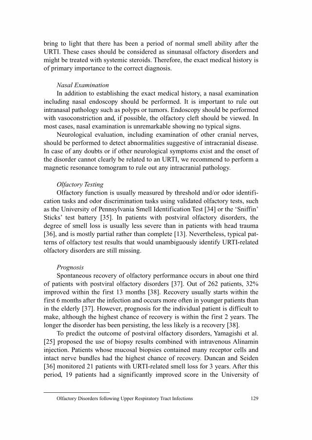
Olfactory Disorders following Upper Respiratory Tract Infections 129
bring to light that there has been a period of normal smell ability after the
URTI. These cases should be considered as sinunasal olfactory disorders and
might be treated with systemic steroids. Therefore, the exact medical history is
of primary importance to the correct diagnosis.
Nasal ExaminationIn addition to establishing the exact medical history, a nasal examination
including nasal endoscopy should be performed. It is important to rule out
intranasal pathology such as polyps or tumors. Endoscopy should be performed
with vasoconstriction and, if possible, the olfactory cleft should be viewed. In
most cases, nasal examination is unremarkable showing no typical signs.
Neurological evaluation, including examination of other cranial nerves,
should be performed to detect abnormalities suggestive of intracranial disease.
In case of any doubts or if other neurological symptoms exist and the onset of
the disorder cannot clearly be related to an URTI, we recommend to perform a
magnetic resonance tomogram to rule out any intracranial pathology.
Olfactory TestingOlfactory function is usually measured by threshold and/or odor identifi-
cation tasks and odor discrimination tasks using validated olfactory tests, such
as the University of Pennsylvania Smell Identification Test [34] or the ‘Sniffin’
Sticks’ test battery [35]. In patients with postviral olfactory disorders, the
degree of smell loss is usually less severe than in patients with head trauma
[36], and is mostly partial rather than complete [13]. Nevertheless, typical pat-
terns of olfactory test results that would unambiguously identify URTI-related
olfactory disorders are still missing.
PrognosisSpontaneous recovery of olfactory performance occurs in about one third
of patients with postviral olfactory disorders [37]. Out of 262 patients, 32%
improved within the first 13 months [38]. Recovery usually starts within the
first 6 months after the infection and occurs more often in younger patients than
in the elderly [37]. However, prognosis for the individual patient is difficult to
make, although the highest chance of recovery is within the first 2 years. The
longer the disorder has been persisting, the less likely is a recovery [38].
To predict the outcome of postviral olfactory disorders, Yamagishi et al.
[25] proposed the use of biopsy results combined with intravenous Alinamin
injection. Patients whose mucosal biopsies contained many receptor cells and
intact nerve bundles had the highest chance of recovery. Duncan and Seiden
[36] monitored 21 patients with URTI-related smell loss for 3 years. After this
period, 19 patients had a significantly improved score in the University of
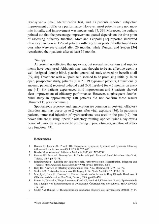
Welge-Lüssen/Wolfensberger 130
Pennsylvania Smell Identification Test, and 13 patients reported subjective
improvement of olfactory performance. However, most patients were not anos-
mic initially, and improvement was modest only [7, 36]. Moreover, the authors
pointed out that the percentage improvement quoted depends on the time point
of assessing olfactory function. Mott and Leopold [12] reported improved
olfactory function in 15% of patients suffering from postviral olfactory disor-
ders who were reevaluated after 26 months, while Duncan and Seiden [36]
reevaluated their patients after at least 36 months.
TherapyAt present, no effective therapy exists, but several medications and supple-
ments have been used. Although zinc was thought to be an effective agent, a
well-designed, double-blind, placebo-controlled study showed no benefit at all
[39, 40]. Treatment with �-lipoid acid seemed to be promising initially. In an
open, prospective study, patients (n � 23, 19 hyposmic patients, 4 functionally
anosmic patients) received �-lipoid acid (600 mg/day) for 4.5 months on aver-
age [41]. Six patients experienced mild improvement and 8 patients showed
clear improvement of olfactory performance. However, a subsequent double-
blind study in approximately 140 patients did not confirm these results
[Hummel T., pers. commun.].
Spontaneous recovery and regeneration are common in postviral olfactory
disorders and may occur up to 2 years after viral exposure [36]. In parosmic
patients, intranasal injection of hydrocortisone was used in the past [42], but
newer data are missing. Specific olfactory training, applied twice a day over a
period of 3 months, appears to be promising in promoting regeneration of olfac-
tory function [43].
References
1 Henkin RI, Larson AL, Powell RD: Hypogeusia, dysgeusia, hyposmia and dysosmia following
influenza-like infection. Ann Otol 1975;84:672–685.
2 Bendar M: Anosmie und Influenza. Med Klin 1930;48:1787–1789.
3 Duncan HJ: Postviral olfactory loss; in Seiden AM (ed): Taste and Smell Disorders. New York,
Thieme, 1997, pp 72–78.
4 Riechstörungen – Leitlinie zur Epidemiologie, Pathophysiologie, Klassifikation, Diagnose und
Therapie. http://www.uni-duesseldorf.de/AWMF/II/hno_II50.htm. 2004.
5 Doty RL: A review of olfactory dysfunction in man. Am J Otolaryngol 1979;1:57–79.
6 Seiden AM: Postviral olfactory loss. Otolaryngol Clin North Am 2004;37:1159–1166.
7 Murphy C, Doty RL, Duncan HJ: Clinical disorders of olfaction; in Doty RL (ed): Handbook of
Olfaction and Gustation. New York, Dekker, 2003, pp 461–478.
8 Damm M, Temmel A, Welge-Luessen A, Eckel HE, Kreft M-P, Klussmann JP, et al: Epidemiologie
und Therapie von Riechstörungen in Deutschland, Österreich und der Schweiz. HNO 2004;52:
112–120.
9 Seiden AM, Duncan HJ: The diagnosis of a conductive olfactory loss. Laryngoscope 2001;111:9–14.
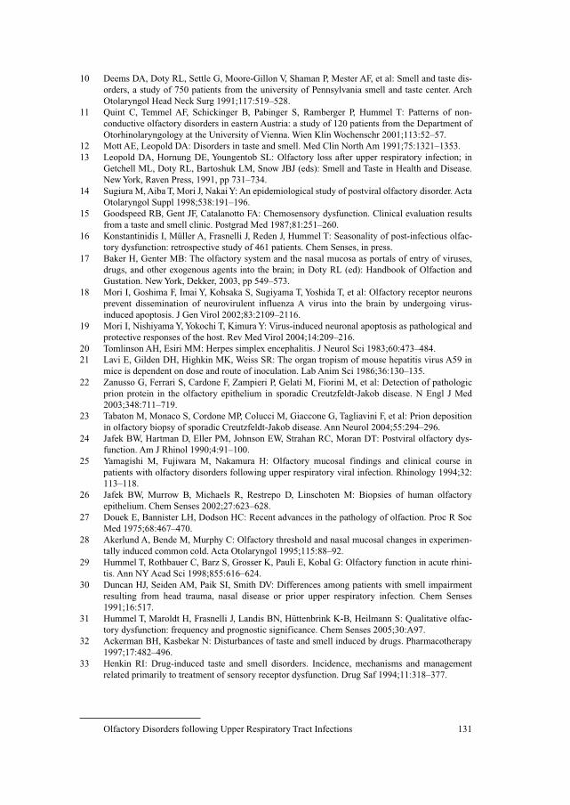
Olfactory Disorders following Upper Respiratory Tract Infections 131
10 Deems DA, Doty RL, Settle G, Moore-Gillon V, Shaman P, Mester AF, et al: Smell and taste dis-
orders, a study of 750 patients from the university of Pennsylvania smell and taste center. Arch
Otolaryngol Head Neck Surg 1991;117:519–528.
11 Quint C, Temmel AF, Schickinger B, Pabinger S, Ramberger P, Hummel T: Patterns of non-
conductive olfactory disorders in eastern Austria: a study of 120 patients from the Department of
Otorhinolaryngology at the University of Vienna. Wien Klin Wochenschr 2001;113:52–57.
12 Mott AE, Leopold DA: Disorders in taste and smell. Med Clin North Am 1991;75:1321–1353.
13 Leopold DA, Hornung DE, Youngentob SL: Olfactory loss after upper respiratory infection; in
Getchell ML, Doty RL, Bartoshuk LM, Snow JBJ (eds): Smell and Taste in Health and Disease.
New York, Raven Press, 1991, pp 731–734.
14 Sugiura M, Aiba T, Mori J, Nakai Y: An epidemiological study of postviral olfactory disorder. Acta
Otolaryngol Suppl 1998;538:191–196.
15 Goodspeed RB, Gent JF, Catalanotto FA: Chemosensory dysfunction. Clinical evaluation results
from a taste and smell clinic. Postgrad Med 1987;81:251–260.
16 Konstantinidis I, Müller A, Frasnelli J, Reden J, Hummel T: Seasonality of post-infectious olfac-
tory dysfunction: retrospective study of 461 patients. Chem Senses, in press.
17 Baker H, Genter MB: The olfactory system and the nasal mucosa as portals of entry of viruses,
drugs, and other exogenous agents into the brain; in Doty RL (ed): Handbook of Olfaction and
Gustation. New York, Dekker, 2003, pp 549–573.
18 Mori I, Goshima F, Imai Y, Kohsaka S, Sugiyama T, Yoshida T, et al: Olfactory receptor neurons
prevent dissemination of neurovirulent influenza A virus into the brain by undergoing virus-
induced apoptosis. J Gen Virol 2002;83:2109–2116.
19 Mori I, Nishiyama Y, Yokochi T, Kimura Y: Virus-induced neuronal apoptosis as pathological and
protective responses of the host. Rev Med Virol 2004;14:209–216.
20 Tomlinson AH, Esiri MM: Herpes simplex encephalitis. J Neurol Sci 1983;60:473–484.
21 Lavi E, Gilden DH, Highkin MK, Weiss SR: The organ tropism of mouse hepatitis virus A59 in
mice is dependent on dose and route of inoculation. Lab Anim Sci 1986;36:130–135.
22 Zanusso G, Ferrari S, Cardone F, Zampieri P, Gelati M, Fiorini M, et al: Detection of pathologic
prion protein in the olfactory epithelium in sporadic Creutzfeldt-Jakob disease. N Engl J Med
2003;348:711–719.
23 Tabaton M, Monaco S, Cordone MP, Colucci M, Giaccone G, Tagliavini F, et al: Prion deposition
in olfactory biopsy of sporadic Creutzfeldt-Jakob disease. Ann Neurol 2004;55:294–296.
24 Jafek BW, Hartman D, Eller PM, Johnson EW, Strahan RC, Moran DT: Postviral olfactory dys-
function. Am J Rhinol 1990;4:91–100.
25 Yamagishi M, Fujiwara M, Nakamura H: Olfactory mucosal findings and clinical course in
patients with olfactory disorders following upper respiratory viral infection. Rhinology 1994;32:
113–118.
26 Jafek BW, Murrow B, Michaels R, Restrepo D, Linschoten M: Biopsies of human olfactory
epithelium. Chem Senses 2002;27:623–628.
27 Douek E, Bannister LH, Dodson HC: Recent advances in the pathology of olfaction. Proc R Soc
Med 1975;68:467–470.
28 Akerlund A, Bende M, Murphy C: Olfactory threshold and nasal mucosal changes in experimen-
tally induced common cold. Acta Otolaryngol 1995;115:88–92.
29 Hummel T, Rothbauer C, Barz S, Grosser K, Pauli E, Kobal G: Olfactory function in acute rhini-
tis. Ann NY Acad Sci 1998;855:616–624.
30 Duncan HJ, Seiden AM, Paik SI, Smith DV: Differences among patients with smell impairment
resulting from head trauma, nasal disease or prior upper respiratory infection. Chem Senses
1991;16:517.
31 Hummel T, Maroldt H, Frasnelli J, Landis BN, Hüttenbrink K-B, Heilmann S: Qualitative olfac-
tory dysfunction: frequency and prognostic significance. Chem Senses 2005;30:A97.
32 Ackerman BH, Kasbekar N: Disturbances of taste and smell induced by drugs. Pharmacotherapy
1997;17:482–496.
33 Henkin RI: Drug-induced taste and smell disorders. Incidence, mechanisms and management
related primarily to treatment of sensory receptor dysfunction. Drug Saf 1994;11:318–377.
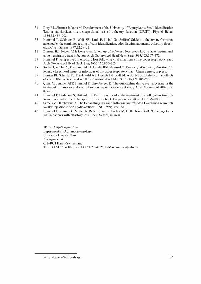
Welge-Lüssen/Wolfensberger 132
34 Doty RL, Shaman P, Dann M: Development of the University of Pennsylvania Smell Identification
Test: a standardized microencapsulated test of olfactory function (UPSIT). Physiol Behav
1984;32:489–502.
35 Hummel T, Sekinger B, Wolf SR, Pauli E, Kobal G: ‘Sniffin’ Sticks’: olfactory performance
assessed by the combined testing of odor identification, odor discrimination, and olfactory thresh-
olds. Chem Senses 1997;22:39–52.
36 Duncan HJ, Seiden AM: Long-term follow-up of olfactory loss secondary to head trauma and
upper respiratory tract infection. Arch Otolaryngol Head Neck Surg 1995;123:367–372.
37 Hummel T: Perspectives in olfactory loss following viral infections of the upper respiratory tract.
Arch Otolaryngol Head Neck Surg 2000;126:802–803.
38 Reden J, Müller A, Konstantinidis I, Landis BN, Hummel T: Recovery of olfactory function fol-
lowing closed head injury or infections of the upper respiratory tract. Chem Senses, in press.
39 Henkin RI, Schecter PJ, Friedewald WT, Demets DL, Raff M: A double blind study of the effects
of zinc sulfate on taste and smell dysfunction. Am J Med Sci 1976;272:285–299.
40 Quint C, Temmel AFP, Hummel T, Ehrenberger K: The quinoxaline derivative caroverine in the
treatment of sensorineural smell disorders: a proof-of-concept study. Acta Otolaryngol 2002;122:
877–881.
41 Hummel T, Heilmann S, Hüttenbrink K-B: Lipoid acid in the treatment of smell dysfunction fol-
lowing viral infection of the upper respiratory tract. Laryngoscope 2002;112:2076–2080.
42 Szmeja Z, Obrebowski A: Die Behandlung der nach Influenza auftretenden Kakosmien vermittels
lokaler Injektionen von Hydrokortison. HNO 1969;17:53–54.
43 Hummel T, Rissom K, Müller A, Reden J, Weidenbecher M, Hüttenbrink K-B: ‘Olfactory train-
ing’ in patients with olfactory loss. Chem Senses, in press.
PD Dr. Antje Welge-Lüssen
Department of Otorhinolaryngology
University Hospital Basel
Petersgraben 4
CH–4031 Basel (Switzerland)
Tel. �41 61 2654 109, Fax �41 61 2654 029, E-Mail [email protected]
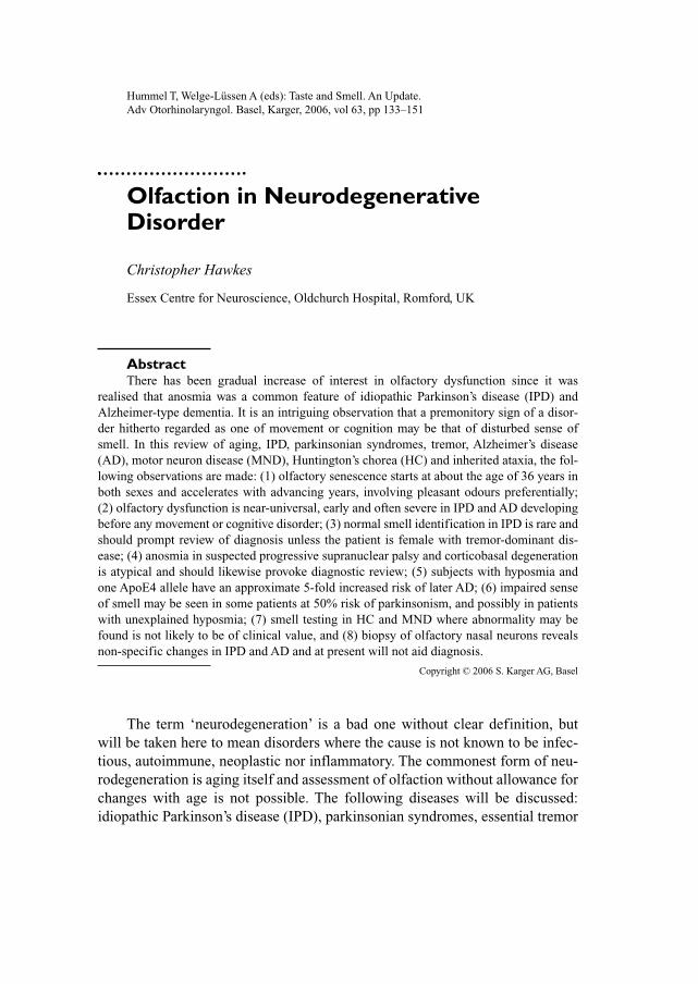
Hummel T, Welge-Lüssen A (eds): Taste and Smell. An Update.
Adv Otorhinolaryngol. Basel, Karger, 2006, vol 63, pp 133–151
Olfaction in NeurodegenerativeDisorder
Christopher Hawkes
Essex Centre for Neuroscience, Oldchurch Hospital, Romford, UK
AbstractThere has been gradual increase of interest in olfactory dysfunction since it was
realised that anosmia was a common feature of idiopathic Parkinson’s disease (IPD) and
Alzheimer-type dementia. It is an intriguing observation that a premonitory sign of a disor-
der hitherto regarded as one of movement or cognition may be that of disturbed sense of
smell. In this review of aging, IPD, parkinsonian syndromes, tremor, Alzheimer’s disease
(AD), motor neuron disease (MND), Huntington’s chorea (HC) and inherited ataxia, the fol-
lowing observations are made: (1) olfactory senescence starts at about the age of 36 years in
both sexes and accelerates with advancing years, involving pleasant odours preferentially;
(2) olfactory dysfunction is near-universal, early and often severe in IPD and AD developing
before any movement or cognitive disorder; (3) normal smell identification in IPD is rare and
should prompt review of diagnosis unless the patient is female with tremor-dominant dis-
ease; (4) anosmia in suspected progressive supranuclear palsy and corticobasal degeneration
is atypical and should likewise provoke diagnostic review; (5) subjects with hyposmia and
one ApoE4 allele have an approximate 5-fold increased risk of later AD; (6) impaired sense
of smell may be seen in some patients at 50% risk of parkinsonism, and possibly in patients
with unexplained hyposmia; (7) smell testing in HC and MND where abnormality may be
found is not likely to be of clinical value, and (8) biopsy of olfactory nasal neurons reveals
non-specific changes in IPD and AD and at present will not aid diagnosis.
Copyright © 2006 S. Karger AG, Basel
The term ‘neurodegeneration’ is a bad one without clear definition, but
will be taken here to mean disorders where the cause is not known to be infec-
tious, autoimmune, neoplastic nor inflammatory. The commonest form of neu-
rodegeneration is aging itself and assessment of olfaction without allowance for
changes with age is not possible. The following diseases will be discussed:
idiopathic Parkinson’s disease (IPD), parkinsonian syndromes, essential tremor
Hawkes 134
(ET), motor neuron disease (MND), Alzheimer’s disease (AD), Huntington’s
chorea (HC) and inherited ataxias.
Aging
It is essential to be aware of the effect of aging itself as it is recognised to
be a major variable affecting olfaction. We studied the effect of age on the
University of Pennsylvania Smell Identification Test (UPSIT) score in 211
healthy controls by multiple regression against gender, age and age squared [1]
and showed that identification ability begins a significant decline after the age
of 36 years and thereafter more steeply. Females scored 1–2 points higher at all
ages although they declined at exactly the same rate as their male counterparts.
Aging may not affect all varieties of odours in the same way. We therefore stud-
ied the effect of age on olfactory identification and its principle components –
pleasantness, irritation and intensity – in the same 211 controls using the 40
individual odours comprising the UPSIT. Older subjects scored less well than
younger on odours with high hedonic or low intensity/irritation rating. Although
intensity correlated better with age than pleasantness, the two dimensions pre-
dicted age discrepancy independently. In the over-50-year age group, we were
able to select 7 odours with moderately high hedonic properties and low inten-
sity score (chocolate, liquorice, grass, coconut, strawberry, rose and melon) and
suggest these odours are more resistant to the effects of age and may be appro-
priate for testing elderly subjects. At present, there is no geriatric smell kit avail-
able commercially, but such a product should be more discriminatory for testing
diseases of the elderly. This makes the assumption that degenerative diseases
are not themselves a simple acceleration of ‘normal’ aging.
Sniffing and the Role of the Cerebellum
It has been shown that sniffing enhances smell detection and apart from
redirection of airflow to the olfactory neuroepithelium, functional MRI studies
have shown that sniffing activates the pyriform and orbitofrontal cortices [2]. In
a meticulous study of IPD, Sobel et al. [3] showed that sniffing was impaired in
IPD and this caused slight reduction in the performance on identification and
detection threshold tests. This equates to a mean reduction of around 2–3 points
on the UPSIT-40 test (see below). Increasing sniff vigour improved olfactory
scores. Studies that have not allowed for this effect (which includes the majority)
may tend to slightly exaggerate the severity of any defect especially where the
disease is known to involve bulbar function (e.g. motor neurone disease).

Olfaction in Neurodegenerative Disease 135
To complicate matters further, it is suggested that the cerebellum is concerned
with smell identification [2] and if correct this has clear relevance to disorders
such as inherited ataxias and ET where the cerebellum is known to be abnormal.
Idiopathic Parkinson’s Disease
Impairment of the sense of smell in IPD was first documented in 1975 by
Ansari and Johnson [4]. Before that, many noted anecdotally that olfactory loss
may precede IPD by many years, but at present there is just one prospective
population-based study to be described below [5]. The majority of olfactory studies
in IPD have used clinical diagnostic criteria and none have correlated changes in
life with those diagnostic criteria found after death. This is of considerable rele-
vance as the diagnostic error rate of neurologists contrasted with autopsy diagno-
sis is 10–26% [6, 7]. Despite this, it is reasonable to propose that patients with
IPD have profound disorder of olfactory function [8, 9]. This observation is based
on pathological abnormality, psychophysical tests and evoked potential studies.
PathologyThe rhinencephalon has only recently been investigated systematically in
PD without dementia. Dystrophic neurites but no Lewy bodies were found in
two of three autopsy-derived olfactory epithelia of patients with IPD, but sev-
eral patients displayed accumulation of amyloid precursor protein fragments
which would not allow distinction from AD [10]. All three varieties of synuclein
(�, �, �) are expressed in olfactory neuroepithelium, particularly �-synuclein,
the hallmark of IPD. Unfortunately, the expression of �-synuclein was found to
be no different from Lewy body disease (LBD), AD, multiple system atrophy
(MSA) and seemingly healthy controls [11]. Nonetheless, it is possible that
those patients coincidentally found to have �-synuclein-containing neurites may
be in the preclinical stage of IPD and that the changes actually represent a
disease-specific finding.
In a preliminary study, we examined, blinded to clinical information, the
olfactory bulbs and tracts from formalin-fixed brains of 8 controls and 8 patients
with a clinical and pathological diagnosis of IPD taken from the UK Parkinson’s
Disease Brain Bank [12]. By inspecting the olfactory bulb and tract all 8 cases
were correctly diagnosed ‘probable PD’. Lewy bodies were most numerous in
the anterior olfactory nucleus but they were also found in mitral cells. The mor-
phology of Lewy bodies at this site resembled their cortical counterparts but
inclusions showing a classical trilaminar structure were rare. It was subse-
quently shown that loss of anterior olfactory neurons correlated with disease
duration [13].
Hawkes 136
Braak et al. [14] performed a detailed analysis of pathology in IPD in 125
cases by immunoreaction with �-synuclein, the protein specific to PD, which is
found in Lewy neurites and Lewy bodies. They demonstrated that the pathology
process advances in a predictable sequence, but the earliest changes, even
before the motor components appeared in life, were found in the dorsal motor
nuclei of the glossopharyngeal and vagus nerves, the olfactory bulb and associ-
ated anterior olfactory nucleus (stage I; fig. 1). This is a pivotal study as it
clearly identifies the dorsal medulla and olfactory bulb as starting points for
IPD. Given that at least 40% of substantia nigra cells have to die before there
are clinical symptoms [15], it is clear that the clinical motor manifestations of
IPD represent the terminal stage of a process that probably started several
decades previously. This point is borne out by the anecdotal clinical observation
that patients regularly report smell impairment 10–20 years before their first
motor symptoms. Involvement of central olfactory areas such as the entorhinal
cortex takes place much later in Braak stage 3 [14].
There have been few studies of the olfactory bulb beyond anatomical
description, but it is clear that there is considerable cell loss in the bulb. The
Fig. 1. The sequential development of PD starting in the olfactory bulb and dorsal
nuclear complex of cranial nerves IX and X (shown in black). The process then ascends the
brain stem to the cerebral cortex. Reproduced with permission from Braak et al. [14].
Olfaction in Neurodegenerative Disease 137
mitral cells seem to vanish in IPD [Braak, H., pers. commun.]. One report [16]
suggested that expression of tyrosine hydroxylase in the olfactory bulb is
increased 100-fold and that this might explain hyposmia of IPD. In the mouse
methylphenyltetrahydropyridine (MPTP) models of PD, there is a 4-fold
increase of dopamine neurogenesis in the olfactory bulb [17] that probably
relates to the migration of dopamine-secreting cells from the subventricular
zone – the so-called rostral migratory stream. This experiment suggests that
ongoing compensation is taking place in the adult brain and infers that only
when the process fails do the symptoms of the disease become apparent.
The studies of Braak et al. [14] make it clear that the first olfactory abnor-
malities are peripheral, that is in the olfactory bulb, which led these authors to
propose [18] that IPD starts in the stomach (Auerbach’s Plexus) and the agent
(virus or chemical) spread in a retrograde fashion up the motor vagal fibres into
the dorsal medulla. This theory, whilst appealing, does not take account of the
olfactory bulb changes.
Psychophysical TestsThe first case-control assessment [4] involved 22 patients with a clinical
diagnosis of IPD by detection threshold to amyl acetate. They found a correla-
tion between average olfactory threshold and more rapid disease progression.
There appeared to be no influence from medication (levodopa, anticholinergic
drugs) or smoking habit. A subsequent larger study of IPD [19] also used detec-
tion threshold tests to various concentrations of amyl acetate in 78 patients and
40 controls. Thresholds were reduced but no correlation was found with age, sex
or use of levodopa. Unlike the first study, there was no association with disease
duration. The next sizeable olfactory investigations [8, 20] using the UPSIT-40
showed that age-matched olfactory dysfunction did not relate to odour type; it
was independent of disease duration and did not correspond with motor func-
tion, tremor or cognition. They also demonstrated that the deficit was of the
same magnitude in both nostrils and not influenced by antiparkinsonian medica-
tion. Further evaluation in subtypes of presumed IPD showed that females with
mild disability and tremor-dominant disease had a significantly higher age-
matched UPSIT-40 score than males with moderate to severe disability and little
or no tremor; age at disease onset was not relevant [21]. A comparable survey [9]
was undertaken using the UPSIT-40 in 155 cognitively normal, depression-free
IPD patients aged 34–84 years and 156 age-matched controls. The age-matched
UPSIT scores for PD patients were dramatically lower than for controls. Only
19% (30/155) of the PD patients had a score within the level expected for 95%
age-matched healthy controls. There were 65 (42%) who were graded anosmic,
i.e. scoring less than 17. There was no correlation between disease duration and
UPSIT score (r � 0.074) [9]. Analysis of the 40 individual odours in the UPSIT
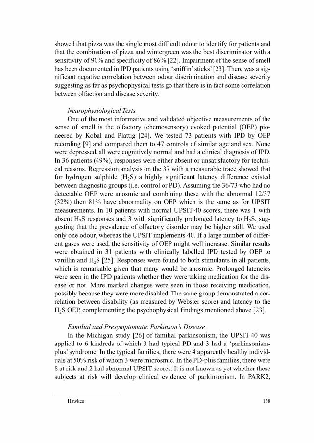
Hawkes 138
showed that pizza was the single most difficult odour to identify for patients and
that the combination of pizza and wintergreen was the best discriminator with a
sensitivity of 90% and specificity of 86% [22]. Impairment of the sense of smell
has been documented in IPD patients using ‘sniffin’ sticks’ [23]. There was a sig-
nificant negative correlation between odour discrimination and disease severity
suggesting as far as psychophysical tests go that there is in fact some correlation
between olfaction and disease severity.
Neurophysiological TestsOne of the most informative and validated objective measurements of the
sense of smell is the olfactory (chemosensory) evoked potential (OEP) pio-
neered by Kobal and Plattig [24]. We tested 73 patients with IPD by OEP
recording [9] and compared them to 47 controls of similar age and sex. None
were depressed, all were cognitively normal and had a clinical diagnosis of IPD.
In 36 patients (49%), responses were either absent or unsatisfactory for techni-
cal reasons. Regression analysis on the 37 with a measurable trace showed that
for hydrogen sulphide (H2S) a highly significant latency difference existed
between diagnostic groups (i.e. control or PD). Assuming the 36/73 who had no
detectable OEP were anosmic and combining these with the abnormal 12/37
(32%) then 81% have abnormality on OEP which is the same as for UPSIT
measurements. In 10 patients with normal UPSIT-40 scores, there was 1 with
absent H2S responses and 3 with significantly prolonged latency to H2S, sug-
gesting that the prevalence of olfactory disorder may be higher still. We used
only one odour, whereas the UPSIT implements 40. If a large number of differ-
ent gases were used, the sensitivity of OEP might well increase. Similar results
were obtained in 31 patients with clinically labelled IPD tested by OEP to
vanillin and H2S [25]. Responses were found to both stimulants in all patients,
which is remarkable given that many would be anosmic. Prolonged latencies
were seen in the IPD patients whether they were taking medication for the dis-
ease or not. More marked changes were seen in those receiving medication,
possibly because they were more disabled. The same group demonstrated a cor-
relation between disability (as measured by Webster score) and latency to the
H2S OEP, complementing the psychophysical findings mentioned above [23].
Familial and Presymptomatic Parkinson’s DiseaseIn the Michigan study [26] of familial parkinsonism, the UPSIT-40 was
applied to 6 kindreds of which 3 had typical PD and 3 had a ‘parkinsonism-
plus’ syndrome. In the typical families, there were 4 apparently healthy individ-
uals at 50% risk of whom 3 were microsmic. In the PD-plus families, there were
8 at risk and 2 had abnormal UPSIT scores. It is not known as yet whether these
subjects at risk will develop clinical evidence of parkinsonism. In PARK2,
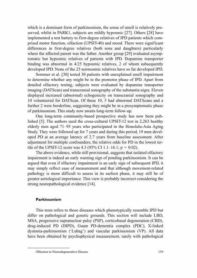
Olfaction in Neurodegenerative Disease 139
which is a dominant form of parkinsonism, the sense of smell is relatively pre-
served, whilst in PARK1, subjects are mildly hyposmic [27]. Others [28] have
implemented a test battery to first-degree relatives of IPD patients which com-
prised motor function, olfaction (UPSIT-40) and mood. There were significant
differences in first-degree relatives (both sons and daughters) particularly
where the affected parent was the father. Another group [29] evaluated asymp-
tomatic but hyposmic relatives of patients with IPD. Dopamine transporter
binding was abnormal in 4/25 hyposmic relatives, 2 of whom subsequently
developed IPD. None of the 23 normosmic relatives have so far developed IPD.
Sommer et al. [30] tested 30 patients with unexplained smell impairment
to determine whether any might be in the premotor phase of IPD. Apart from
detailed olfactory testing, subjects were evaluated by dopamine transporter
imaging (DATScan) and transcranial sonography of the substantia nigra. Eleven
displayed increased (abnormal) echogenicity on transcranial sonography and
10 volunteered for DATScan. Of these 10, 5 had abnormal DATScans and a
further 2 were borderline, suggesting they might be in a presymptomatic phase
of parkinsonism. This study now awaits long-term follow-up.
One long-term community-based prospective study has now been pub-
lished [5]. The authors used the cross-cultural UPSIT-12 test in 2,263 healthy
elderly men aged 71–95 years who participated in the Honolulu-Asia Aging
Study. They were followed up for 7 years and during this period, 19 men devel-
oped PD at an average latency of 2.7 years from baseline assessment. After
adjustment for multiple confounders, the relative odds for PD in the lowest ter-
tile of the UPSIT-12 score was 4.3 (95% CI 1.1–16.1; p � 0.02).
The above evidence, while still provisional, suggests that isolated olfactory
impairment is indeed an early warning sign of pending parkinsonism. It can be
argued that even if olfactory impairment is an early sign of subsequent IPD, it
may simply reflect ease of measurement and that although movement-related
pathology is more difficult to assess in its earliest phase, it may still be of
greater aetiological importance. This view is probably incorrect considering the
strong neuropathological evidence [14].
Parkinsonism
This term refers to those diseases which phenotypically resemble IPD but
differ on pathological and genetic grounds. This section will include LBD,
MSA, progressive supranuclear palsy (PSP), corticobasal degeneration (CBD),
drug-induced PD (DIPD), Guam PD-dementia complex (PDC), X-linked
dystonia-parkinsonism (‘Lubag’) and vascular parkinsonism (VP). All data
have been obtained by psychophysical measurement, rarely with pathological
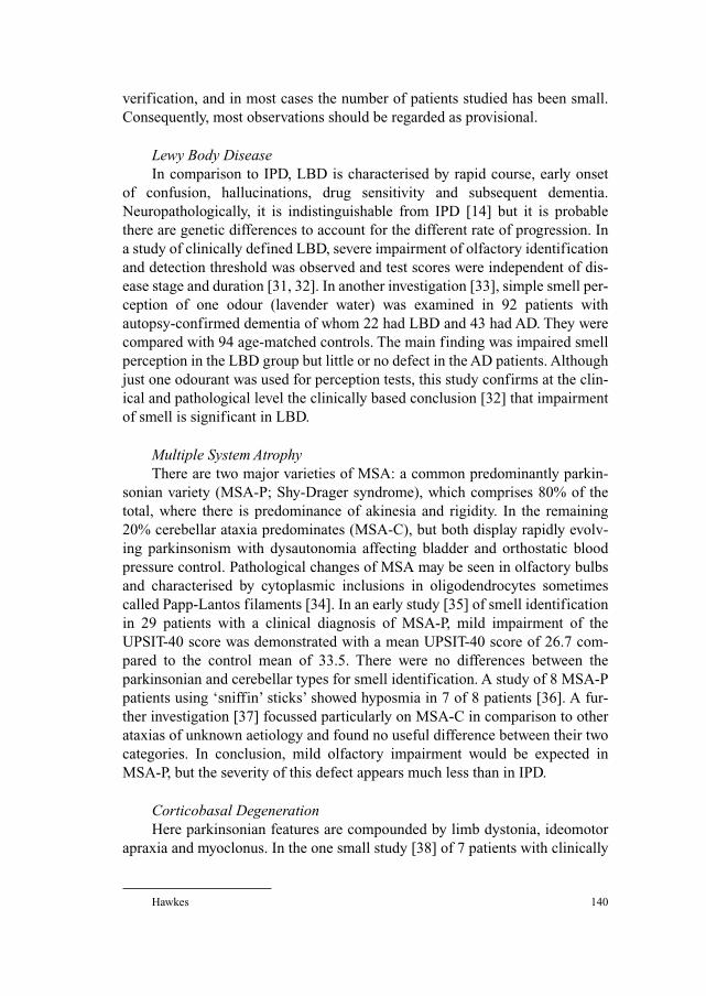
Hawkes 140
verification, and in most cases the number of patients studied has been small.
Consequently, most observations should be regarded as provisional.
Lewy Body DiseaseIn comparison to IPD, LBD is characterised by rapid course, early onset
of confusion, hallucinations, drug sensitivity and subsequent dementia.
Neuropathologically, it is indistinguishable from IPD [14] but it is probable
there are genetic differences to account for the different rate of progression. In
a study of clinically defined LBD, severe impairment of olfactory identification
and detection threshold was observed and test scores were independent of dis-
ease stage and duration [31, 32]. In another investigation [33], simple smell per-
ception of one odour (lavender water) was examined in 92 patients with
autopsy-confirmed dementia of whom 22 had LBD and 43 had AD. They were
compared with 94 age-matched controls. The main finding was impaired smell
perception in the LBD group but little or no defect in the AD patients. Although
just one odourant was used for perception tests, this study confirms at the clin-
ical and pathological level the clinically based conclusion [32] that impairment
of smell is significant in LBD.
Multiple System AtrophyThere are two major varieties of MSA: a common predominantly parkin-
sonian variety (MSA-P; Shy-Drager syndrome), which comprises 80% of the
total, where there is predominance of akinesia and rigidity. In the remaining
20% cerebellar ataxia predominates (MSA-C), but both display rapidly evolv-
ing parkinsonism with dysautonomia affecting bladder and orthostatic blood
pressure control. Pathological changes of MSA may be seen in olfactory bulbs
and characterised by cytoplasmic inclusions in oligodendrocytes sometimes
called Papp-Lantos filaments [34]. In an early study [35] of smell identification
in 29 patients with a clinical diagnosis of MSA-P, mild impairment of the
UPSIT-40 score was demonstrated with a mean UPSIT-40 score of 26.7 com-
pared to the control mean of 33.5. There were no differences between the
parkinsonian and cerebellar types for smell identification. A study of 8 MSA-P
patients using ‘sniffin’ sticks’ showed hyposmia in 7 of 8 patients [36]. A fur-
ther investigation [37] focussed particularly on MSA-C in comparison to other
ataxias of unknown aetiology and found no useful difference between their two
categories. In conclusion, mild olfactory impairment would be expected in
MSA-P, but the severity of this defect appears much less than in IPD.
Corticobasal DegenerationHere parkinsonian features are compounded by limb dystonia, ideomotor
apraxia and myoclonus. In the one small study [38] of 7 patients with clinically
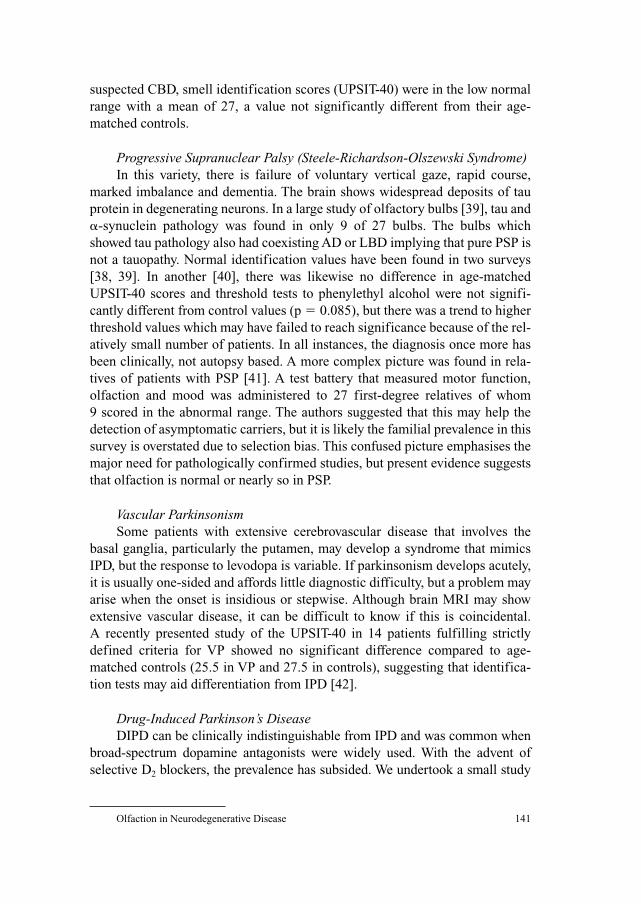
Olfaction in Neurodegenerative Disease 141
suspected CBD, smell identification scores (UPSIT-40) were in the low normal
range with a mean of 27, a value not significantly different from their age-
matched controls.
Progressive Supranuclear Palsy (Steele-Richardson-Olszewski Syndrome)In this variety, there is failure of voluntary vertical gaze, rapid course,
marked imbalance and dementia. The brain shows widespread deposits of tau
protein in degenerating neurons. In a large study of olfactory bulbs [39], tau and
�-synuclein pathology was found in only 9 of 27 bulbs. The bulbs which
showed tau pathology also had coexisting AD or LBD implying that pure PSP is
not a tauopathy. Normal identification values have been found in two surveys
[38, 39]. In another [40], there was likewise no difference in age-matched
UPSIT-40 scores and threshold tests to phenylethyl alcohol were not signifi-
cantly different from control values (p � 0.085), but there was a trend to higher
threshold values which may have failed to reach significance because of the rel-
atively small number of patients. In all instances, the diagnosis once more has
been clinically, not autopsy based. A more complex picture was found in rela-
tives of patients with PSP [41]. A test battery that measured motor function,
olfaction and mood was administered to 27 first-degree relatives of whom
9 scored in the abnormal range. The authors suggested that this may help the
detection of asymptomatic carriers, but it is likely the familial prevalence in this
survey is overstated due to selection bias. This confused picture emphasises the
major need for pathologically confirmed studies, but present evidence suggests
that olfaction is normal or nearly so in PSP.
Vascular ParkinsonismSome patients with extensive cerebrovascular disease that involves the
basal ganglia, particularly the putamen, may develop a syndrome that mimics
IPD, but the response to levodopa is variable. If parkinsonism develops acutely,
it is usually one-sided and affords little diagnostic difficulty, but a problem may
arise when the onset is insidious or stepwise. Although brain MRI may show
extensive vascular disease, it can be difficult to know if this is coincidental.
A recently presented study of the UPSIT-40 in 14 patients fulfilling strictly
defined criteria for VP showed no significant difference compared to age-
matched controls (25.5 in VP and 27.5 in controls), suggesting that identifica-
tion tests may aid differentiation from IPD [42].
Drug-Induced Parkinson’s DiseaseDIPD can be clinically indistinguishable from IPD and was common when
broad-spectrum dopamine antagonists were widely used. With the advent of
selective D2 blockers, the prevalence has subsided. We undertook a small study
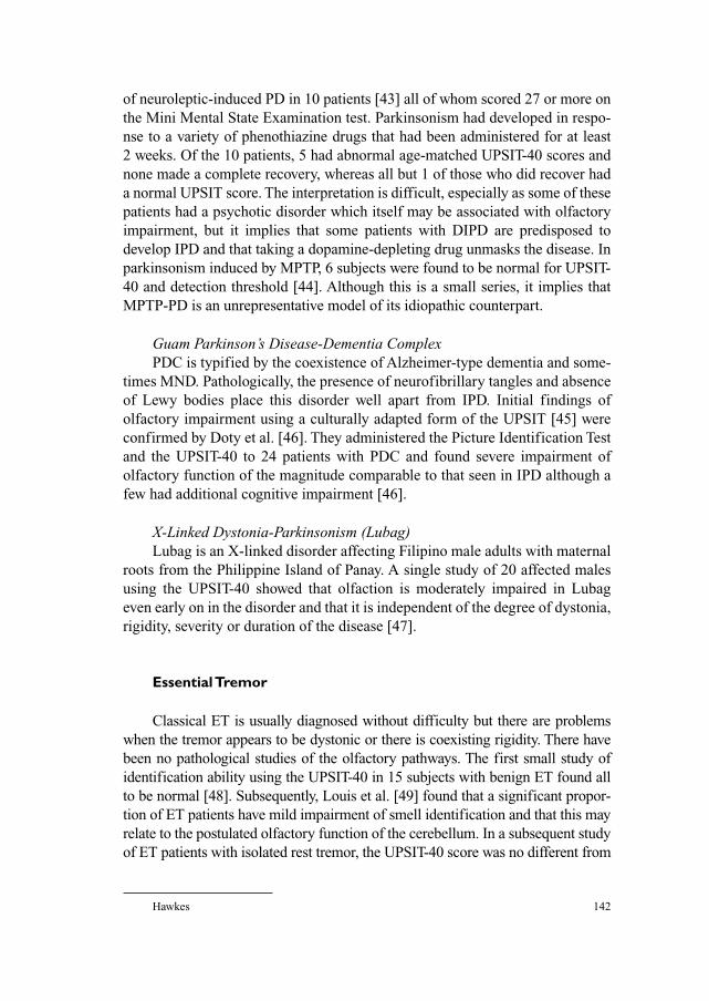
Hawkes 142
of neuroleptic-induced PD in 10 patients [43] all of whom scored 27 or more on
the Mini Mental State Examination test. Parkinsonism had developed in respo-
nse to a variety of phenothiazine drugs that had been administered for at least
2 weeks. Of the 10 patients, 5 had abnormal age-matched UPSIT-40 scores and
none made a complete recovery, whereas all but 1 of those who did recover had
a normal UPSIT score. The interpretation is difficult, especially as some of these
patients had a psychotic disorder which itself may be associated with olfactory
impairment, but it implies that some patients with DIPD are predisposed to
develop IPD and that taking a dopamine-depleting drug unmasks the disease. In
parkinsonism induced by MPTP, 6 subjects were found to be normal for UPSIT-
40 and detection threshold [44]. Although this is a small series, it implies that
MPTP-PD is an unrepresentative model of its idiopathic counterpart.
Guam Parkinson’s Disease-Dementia ComplexPDC is typified by the coexistence of Alzheimer-type dementia and some-
times MND. Pathologically, the presence of neurofibrillary tangles and absence
of Lewy bodies place this disorder well apart from IPD. Initial findings of
olfactory impairment using a culturally adapted form of the UPSIT [45] were
confirmed by Doty et al. [46]. They administered the Picture Identification Test
and the UPSIT-40 to 24 patients with PDC and found severe impairment of
olfactory function of the magnitude comparable to that seen in IPD although a
few had additional cognitive impairment [46].
X-Linked Dystonia-Parkinsonism (Lubag)Lubag is an X-linked disorder affecting Filipino male adults with maternal
roots from the Philippine Island of Panay. A single study of 20 affected males
using the UPSIT-40 showed that olfaction is moderately impaired in Lubag
even early on in the disorder and that it is independent of the degree of dystonia,
rigidity, severity or duration of the disease [47].
Essential Tremor
Classical ET is usually diagnosed without difficulty but there are problems
when the tremor appears to be dystonic or there is coexisting rigidity. There have
been no pathological studies of the olfactory pathways. The first small study of
identification ability using the UPSIT-40 in 15 subjects with benign ET found all
to be normal [48]. Subsequently, Louis et al. [49] found that a significant propor-
tion of ET patients have mild impairment of smell identification and that this may
relate to the postulated olfactory function of the cerebellum. In a subsequent study
of ET patients with isolated rest tremor, the UPSIT-40 score was no different from
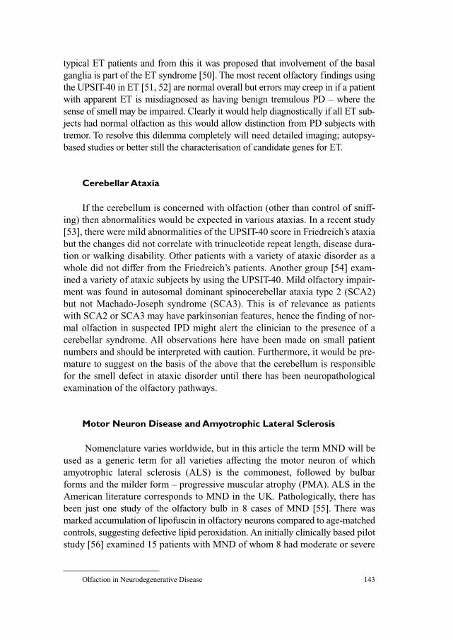
Olfaction in Neurodegenerative Disease 143
typical ET patients and from this it was proposed that involvement of the basal
ganglia is part of the ET syndrome [50]. The most recent olfactory findings using
the UPSIT-40 in ET [51, 52] are normal overall but errors may creep in if a patient
with apparent ET is misdiagnosed as having benign tremulous PD – where the
sense of smell may be impaired. Clearly it would help diagnostically if all ET sub-
jects had normal olfaction as this would allow distinction from PD subjects with
tremor. To resolve this dilemma completely will need detailed imaging; autopsy-
based studies or better still the characterisation of candidate genes for ET.
Cerebellar Ataxia
If the cerebellum is concerned with olfaction (other than control of sniff-
ing) then abnormalities would be expected in various ataxias. In a recent study
[53], there were mild abnormalities of the UPSIT-40 score in Friedreich’s ataxia
but the changes did not correlate with trinucleotide repeat length, disease dura-
tion or walking disability. Other patients with a variety of ataxic disorder as a
whole did not differ from the Friedreich’s patients. Another group [54] exam-
ined a variety of ataxic subjects by using the UPSIT-40. Mild olfactory impair-
ment was found in autosomal dominant spinocerebellar ataxia type 2 (SCA2)
but not Machado-Joseph syndrome (SCA3). This is of relevance as patients
with SCA2 or SCA3 may have parkinsonian features, hence the finding of nor-
mal olfaction in suspected IPD might alert the clinician to the presence of a
cerebellar syndrome. All observations here have been made on small patient
numbers and should be interpreted with caution. Furthermore, it would be pre-
mature to suggest on the basis of the above that the cerebellum is responsible
for the smell defect in ataxic disorder until there has been neuropathological
examination of the olfactory pathways.
Motor Neuron Disease and Amyotrophic Lateral Sclerosis
Nomenclature varies worldwide, but in this article the term MND will be
used as a generic term for all varieties affecting the motor neuron of which
amyotrophic lateral sclerosis (ALS) is the commonest, followed by bulbar
forms and the milder form – progressive muscular atrophy (PMA). ALS in the
American literature corresponds to MND in the UK. Pathologically, there has
been just one study of the olfactory bulb in 8 cases of MND [55]. There was
marked accumulation of lipofuscin in olfactory neurons compared to age-matched
controls, suggesting defective lipid peroxidation. An initially clinically based pilot
study [56] examined 15 patients with MND of whom 8 had moderate or severe
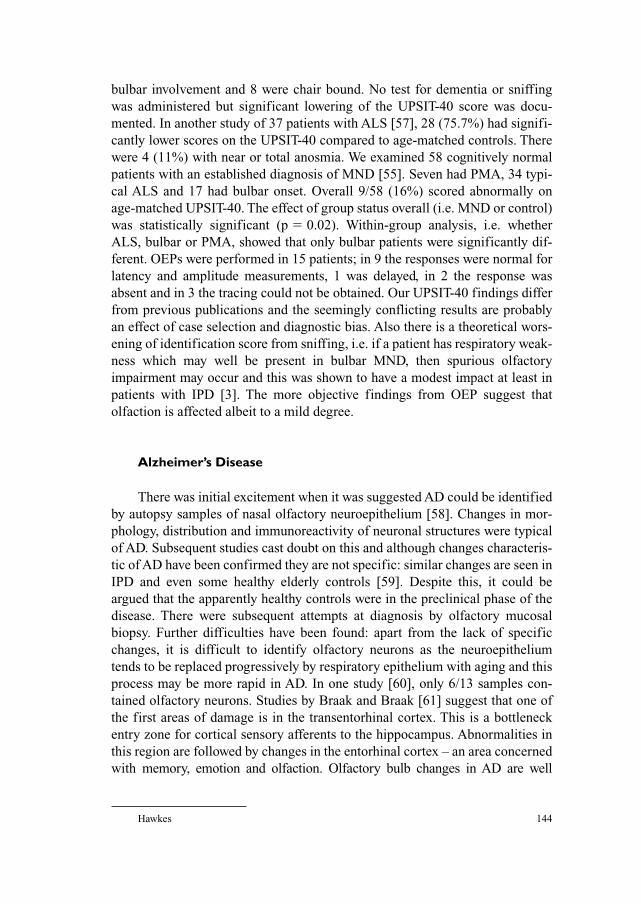
Hawkes 144
bulbar involvement and 8 were chair bound. No test for dementia or sniffing
was administered but significant lowering of the UPSIT-40 score was docu-
mented. In another study of 37 patients with ALS [57], 28 (75.7%) had signifi-
cantly lower scores on the UPSIT-40 compared to age-matched controls. There
were 4 (11%) with near or total anosmia. We examined 58 cognitively normal
patients with an established diagnosis of MND [55]. Seven had PMA, 34 typi-
cal ALS and 17 had bulbar onset. Overall 9/58 (16%) scored abnormally on
age-matched UPSIT-40. The effect of group status overall (i.e. MND or control)
was statistically significant (p � 0.02). Within-group analysis, i.e. whether
ALS, bulbar or PMA, showed that only bulbar patients were significantly dif-
ferent. OEPs were performed in 15 patients; in 9 the responses were normal for
latency and amplitude measurements, 1 was delayed, in 2 the response was
absent and in 3 the tracing could not be obtained. Our UPSIT-40 findings differ
from previous publications and the seemingly conflicting results are probably
an effect of case selection and diagnostic bias. Also there is a theoretical wors-
ening of identification score from sniffing, i.e. if a patient has respiratory weak-
ness which may well be present in bulbar MND, then spurious olfactory
impairment may occur and this was shown to have a modest impact at least in
patients with IPD [3]. The more objective findings from OEP suggest that
olfaction is affected albeit to a mild degree.
Alzheimer’s Disease
There was initial excitement when it was suggested AD could be identified
by autopsy samples of nasal olfactory neuroepithelium [58]. Changes in mor-
phology, distribution and immunoreactivity of neuronal structures were typical
of AD. Subsequent studies cast doubt on this and although changes characteris-
tic of AD have been confirmed they are not specific: similar changes are seen in
IPD and even some healthy elderly controls [59]. Despite this, it could be
argued that the apparently healthy controls were in the preclinical phase of the
disease. There were subsequent attempts at diagnosis by olfactory mucosal
biopsy. Further difficulties have been found: apart from the lack of specific
changes, it is difficult to identify olfactory neurons as the neuroepithelium
tends to be replaced progressively by respiratory epithelium with aging and this
process may be more rapid in AD. In one study [60], only 6/13 samples con-
tained olfactory neurons. Studies by Braak and Braak [61] suggest that one of
the first areas of damage is in the transentorhinal cortex. This is a bottleneck
entry zone for cortical sensory afferents to the hippocampus. Abnormalities in
this region are followed by changes in the entorhinal cortex – an area concerned
with memory, emotion and olfaction. Olfactory bulb changes in AD are well
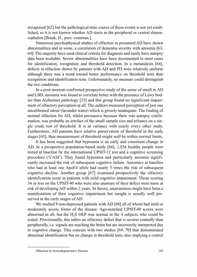
Olfaction in Neurodegenerative Disease 145
recognised [62] but the pathological time course of these events is not yet estab-
lished, so it is not known whether AD starts in the peripheral or central rhinen-
cephalon [Braak, H., pers. commun.].
Numerous psychophysical studies of olfaction in presumed AD have shown
abnormalities and in some, a correlation of dementia severity with anosmia [63,
64]. The majority have used clinical criteria for diagnosis and rarely have autopsy
data been available. Severe abnormalities have been documented in most cases
for identification, recognition and threshold detection. In a metanalysis [64],
defects in olfaction shown by patients with AD and PD were relatively uniform
although there was a trend toward better performance on threshold tests than
recognition and identification tests. Unfortunately, no measure could distinguish
the two conditions.
In a post-mortem confirmed prospective study of the sense of smell in AD
and LBD, anosmia was found to correlate better with the presence of Lewy bod-
ies than Alzheimer pathology [33] and this group found no significant impair-
ment of olfactory perception at all. The authors measured perception of just one
uncalibrated odour (lavender water) which is grossly inadequate. The finding of
normal olfaction for AD, whilst persuasive because there was autopsy confir-
mation, was probably an artefact of the small sample size and reliance on a sin-
gle crude test of threshold. It is at variance with nearly every other study.
Furthermore, AD patients have relative preservation of threshold in the early
stages [65], thus measurement of threshold might well be within normal limits.
It has been suggested that hyposmia is an early and consistent change in
AD. In a prospective population-based study [66], 1,836 healthy people were
tested at baseline by the international UPSIT-12 test and a cognitive screening
procedure (‘CASI’). They found hyposmia and particularly anosmia signifi-
cantly increased the risk of subsequent cognitive failure. Anosmics at baseline
who had at least one ApoE4 allele had nearly 5 times the risk of subsequent
cognitive decline. Another group [67] examined prospectively the olfactory
identification score in patients with mild cognitive impairment. Those scoring
34 or less on the UPSIT-40 who were also unaware of their defect were more at
risk of developing AD within 2 years. In theory, unawareness might have been a
manifestation of their cognitive impairment but insight is usually well pre-
served in the early stages of AD.
We studied 8 non-depressed patients with AD [68] all of whom had mild or
moderately severe forms of the disease. Age-matched UPSIT-40 scores were
abnormal in all, but the H2S OEP was normal in the 4 subjects who could be
tested. Provisionally, this infers an olfactory defect that is severer centrally than
peripherally, i.e. signals are reaching the brain but are incorrectly interpreted due
to cognitive change. This concurs with two studies [69, 70] that demonstrated
abnormal identification but no change in threshold tests, also implying a central
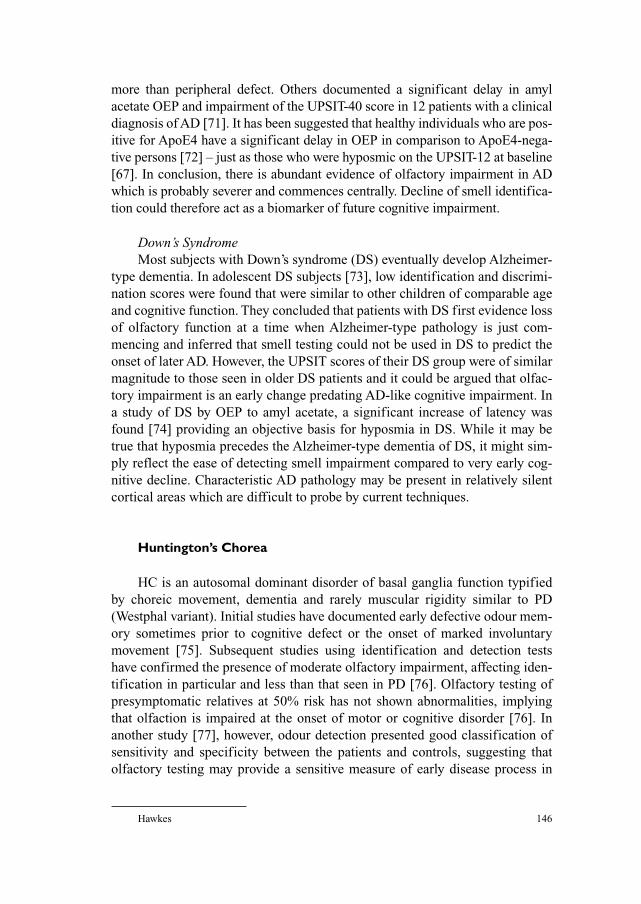
Hawkes 146
more than peripheral defect. Others documented a significant delay in amyl
acetate OEP and impairment of the UPSIT-40 score in 12 patients with a clinical
diagnosis of AD [71]. It has been suggested that healthy individuals who are pos-
itive for ApoE4 have a significant delay in OEP in comparison to ApoE4-nega-
tive persons [72] – just as those who were hyposmic on the UPSIT-12 at baseline
[67]. In conclusion, there is abundant evidence of olfactory impairment in AD
which is probably severer and commences centrally. Decline of smell identifica-
tion could therefore act as a biomarker of future cognitive impairment.
Down’s SyndromeMost subjects with Down’s syndrome (DS) eventually develop Alzheimer-
type dementia. In adolescent DS subjects [73], low identification and discrimi-
nation scores were found that were similar to other children of comparable age
and cognitive function. They concluded that patients with DS first evidence loss
of olfactory function at a time when Alzheimer-type pathology is just com-
mencing and inferred that smell testing could not be used in DS to predict the
onset of later AD. However, the UPSIT scores of their DS group were of similar
magnitude to those seen in older DS patients and it could be argued that olfac-
tory impairment is an early change predating AD-like cognitive impairment. In
a study of DS by OEP to amyl acetate, a significant increase of latency was
found [74] providing an objective basis for hyposmia in DS. While it may be
true that hyposmia precedes the Alzheimer-type dementia of DS, it might sim-
ply reflect the ease of detecting smell impairment compared to very early cog-
nitive decline. Characteristic AD pathology may be present in relatively silent
cortical areas which are difficult to probe by current techniques.
Huntington’s Chorea
HC is an autosomal dominant disorder of basal ganglia function typified
by choreic movement, dementia and rarely muscular rigidity similar to PD
(Westphal variant). Initial studies have documented early defective odour mem-
ory sometimes prior to cognitive defect or the onset of marked involuntary
movement [75]. Subsequent studies using identification and detection tests
have confirmed the presence of moderate olfactory impairment, affecting iden-
tification in particular and less than that seen in PD [76]. Olfactory testing of
presymptomatic relatives at 50% risk has not shown abnormalities, implying
that olfaction is impaired at the onset of motor or cognitive disorder [76]. In
another study [77], however, odour detection presented good classification of
sensitivity and specificity between the patients and controls, suggesting that
olfactory testing may provide a sensitive measure of early disease process in
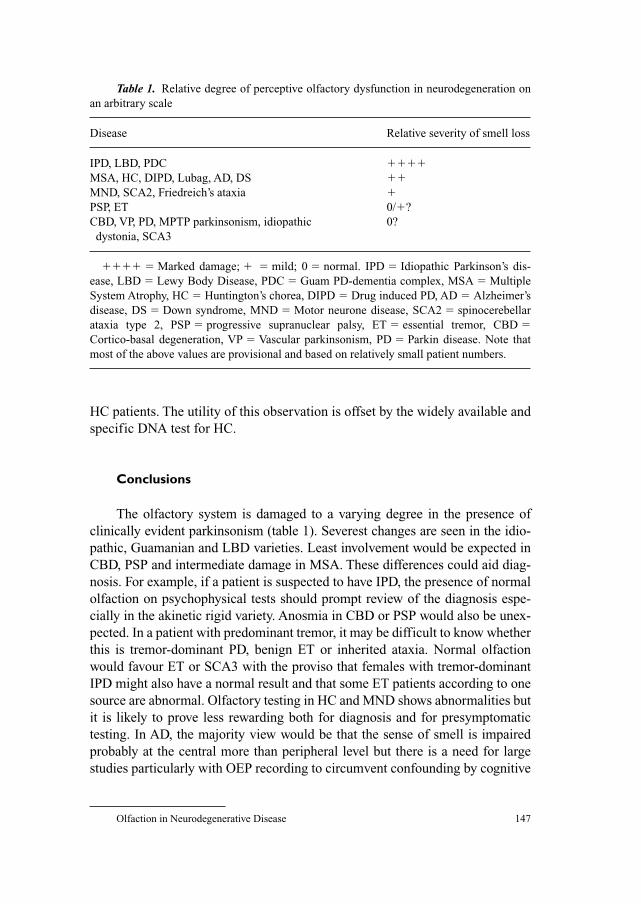
Olfaction in Neurodegenerative Disease 147
HC patients. The utility of this observation is offset by the widely available and
specific DNA test for HC.
Conclusions
The olfactory system is damaged to a varying degree in the presence of
clinically evident parkinsonism (table 1). Severest changes are seen in the idio-
pathic, Guamanian and LBD varieties. Least involvement would be expected in
CBD, PSP and intermediate damage in MSA. These differences could aid diag-
nosis. For example, if a patient is suspected to have IPD, the presence of normal
olfaction on psychophysical tests should prompt review of the diagnosis espe-
cially in the akinetic rigid variety. Anosmia in CBD or PSP would also be unex-
pected. In a patient with predominant tremor, it may be difficult to know whether
this is tremor-dominant PD, benign ET or inherited ataxia. Normal olfaction
would favour ET or SCA3 with the proviso that females with tremor-dominant
IPD might also have a normal result and that some ET patients according to one
source are abnormal. Olfactory testing in HC and MND shows abnormalities but
it is likely to prove less rewarding both for diagnosis and for presymptomatic
testing. In AD, the majority view would be that the sense of smell is impaired
probably at the central more than peripheral level but there is a need for large
studies particularly with OEP recording to circumvent confounding by cognitive
Table 1. Relative degree of perceptive olfactory dysfunction in neurodegeneration on
an arbitrary scale
Disease Relative severity of smell loss
IPD, LBD, PDC ����MSA, HC, DIPD, Lubag, AD, DS ��MND, SCA2, Friedreich’s ataxia �PSP, ET 0/�?
CBD, VP, PD, MPTP parkinsonism, idiopathic 0?
dystonia, SCA3
���� � Marked damage; � � mild; 0 � normal. IPD � Idiopathic Parkinson’s dis-
ease, LBD � Lewy Body Disease, PDC � Guam PD-dementia complex, MSA � Multiple
System Atrophy, HC � Huntington’s chorea, DIPD � Drug induced PD, AD � Alzheimer’s
disease, DS � Down syndrome, MND � Motor neurone disease, SCA2 � spinocerebellar
ataxia type 2, PSP � progressive supranuclear palsy, ET � essential tremor, CBD �Cortico-basal degeneration, VP � Vascular parkinsonism, PD � Parkin disease. Note that
most of the above values are provisional and based on relatively small patient numbers.
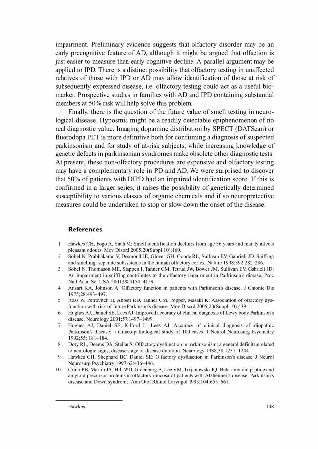
Hawkes 148
impairment. Preliminary evidence suggests that olfactory disorder may be an
early precognitive feature of AD, although it might be argued that olfaction is
just easier to measure than early cognitive decline. A parallel argument may be
applied to IPD. There is a distinct possibility that olfactory testing in unaffected
relatives of those with IPD or AD may allow identification of those at risk of
subsequently expressed disease, i.e. olfactory testing could act as a useful bio-
marker. Prospective studies in families with AD and IPD containing substantial
members at 50% risk will help solve this problem.
Finally, there is the question of the future value of smell testing in neuro-
logical disease. Hyposmia might be a readily detectable epiphenomenon of no
real diagnostic value. Imaging dopamine distribution by SPECT (DATScan) or
fluorodopa PET is more definitive both for confirming a diagnosis of suspected
parkinsonism and for study of at-risk subjects, while increasing knowledge of
genetic defects in parkinsonian syndromes make obsolete other diagnostic tests.
At present, these non-olfactory procedures are expensive and olfactory testing
may have a complementary role in PD and AD. We were surprised to discover
that 50% of patients with DIPD had an impaired identification score. If this is
confirmed in a larger series, it raises the possibility of genetically determined
susceptibility to various classes of organic chemicals and if so neuroprotective
measures could be undertaken to stop or slow down the onset of the disease.
References
1 Hawkes CH, Fogo A, Shah M: Smell identification declines from age 36 years and mainly affects
pleasant odours. Mov Disord 2005;20(Suppl 10):160.
2 Sobel N, Prabhakaran V, Desmond JE, Glover GH, Goode RL, Sullivan EV, Gabrieli JD: Sniffing
and smelling: separate subsystems in the human olfactory cortex. Nature 1998;392:282–286.
3 Sobel N, Thomason ME, Stappen I, Tanner CM, Tetrud JW, Bower JM, Sullivan EV, Gabrieli JD:
An impairment in sniffing contributes to the olfactory impairment in Parkinson’s disease. Proc
Natl Acad Sci USA 2001;98:4154–4159.
4 Ansari KA, Johnson A: Olfactory function in patients with Parkinson’s disease. J Chronic Dis
1975;28:493–497.
5 Ross W, Petrovitch H, Abbott RD, Tanner CM, Popper, Masaki K: Association of olfactory dys-
function with risk of future Parkinson’s disease. Mov Disord 2005;20(Suppl 10):439.
6 Hughes AJ, Daniel SE, Lees AJ: Improved accuracy of clinical diagnosis of Lewy body Parkinson’s
disease. Neurology 2001;57:1497–1499.
7 Hughes AJ, Daniel SE, Kilford L, Lees AJ: Accuracy of clinical diagnosis of idiopathic
Parkinson’s disease: a clinico-pathological study of 100 cases. J Neurol Neurosurg Psychiatry
1992;55: 181–184.
8 Doty RL, Deems DA, Stellar S: Olfactory dysfunction in parkinsonism: a general deficit unrelated
to neurologic signs, disease stage or disease duration. Neurology 1988;38:1237–1244.
9 Hawkes CH, Shephard BC, Daniel SE: Olfactory dysfunction in Parkinson’s disease. J Neurol
Neurosurg Psychiatry 1997;62:436–446.
10 Crino PB, Martin JA, Hill WD, Greenberg B, Lee VM, Trojanowski JQ: Beta-amyloid peptide and
amyloid precursor proteins in olfactory mucosa of patients with Alzheimer’s disease, Parkinson’s
disease and Down syndrome. Ann Otol Rhinol Laryngol 1995;104:655–661.

Olfaction in Neurodegenerative Disease 149
11 Duda JE, Shah U, Arnold SE, Lee VM, Trojanowski JQ: The expression of alpha-, beta-, and
gamma-synucleins in olfactory mucosa from patients with and without neurodegenerative dis-
eases. Exp Neurol 1999;160:515–522.
12 Daniel SE, Hawkes CH: Preliminary diagnosis of Parkinson’s disease using olfactory bulb pathol-
ogy. Lancet 1992;340:186.
13 Pearce RKB, Hawkes CH, Daniel SE: The anterior olfactory nucleus in Parkinson’s disease. Mov
Disord 1995;10:283–287.
14 Braak H, Del Tredici K, Rub U, de Vos RA, Jansen Steur EN, Braak E: Staging of brain pathology
related to sporadic Parkinson’s disease. Neurobiol Aging 2003;24:197–211.
15 Ross GW, Petrovitch H, Abbott RD, Nelson J, Markesbery W, Davis D, Hardman J, Launer L,
Masaki K, Tanner CM, White LR: Parkinsonian signs and substantia nigra neuron density in
descendents elders without PD. Ann Neurol 2004;56:532–539.
16 Huisman E, Uylings HB, Hoogland PV: A 100% increase of dopaminergic cells in the olfactory
bulb may explain hyposmia in Parkinson’s disease. Mov Disord 2004;19:687–692.
17 Yamada M, Onodera M, Mizuno Y, Mochizuki H: Neurogenesis in olfactory bulb identified by
retroviral labeling in normal and MPTP-treated adult mice. Neuroscience 2004;124:173–181.
18 Braak H, Rub U, Gai WP, Del Tredici K: Idiopathic Parkinson’s disease: possible routes by which
vulnerable neuronal types may be subject to neuroinvasion by an unknown pathogen. J Neural
Transm 2003;110:517–536.
19 Quinn NP, Rossor MN, Marsden CD: Olfactory threshold in Parkinson’s disease. J Neurol
Neurosurg Psychiatry 1987;50:88–89.
20 Doty RL, Stern MB, Pfeiffer C, Gollomp SM, Hurtig HI: Bilateral olfactory dysfunction in early
stage treated and untreated idiopathic Parkinson’s disease. J Neurol Neurosurg Psychiatry
1992;55:138–142.
21 Stern MB, Doty RL, Dotti M, et al: Olfactory function in Parkinson’s disease subtypes. Neurology
1994;44:266–268.
22 Hawkes CH, Shephard BC: Selective anosmia in Parkinson’s disease? Lancet 1993;341:
435–436.
23 Daum RF, Sekinger B, Kobal G, Lang CJ: Olfactory testing with ‘sniffin’ sticks’ for clinical diag-
nosis of Parkinson disease. Nervenarzt 2000;71:643–650.
24 Kobal G, Plattig KH: Objective olfactometry: methodological annotations for recording olfactory
EEG-responses from the awake human. EEG EMG Z Elektroenzephalogr Elektromyogr
Verwandte Geb 1978;9:135–145.
25 Barz S, Hummel T, Pauli E, Majer M, Lang CJ, Kobal G: Chemosensory event-related potentials
in response to trigeminal and olfactory stimulation in Parkinson’s disease. Neurology 1997;49:
1424–1431.
26 Markoupoulou K, Larsen KW, Wszolek EK, Denson MS, Lang AE, Pfeiffer RF, Wszolek ZK:
Olfactory dysfunction in familial parkinsonism. Neurology 1997;49:1262–1267.
27 Katzenschlager R, Lees AJ: Olfaction and Parkinson’s syndromes: its role in differential diagno-
sis. Curr Opin Neurol 2004;17:417–423.
28 Montgomery EB Jr, Baker KB, Lyons K, Koller WC: Abnormal performance on the PD test bat-
tery by asymptomatic first-degree relatives. Neurology 1999;52:757–762.
29 Berendse HW, Booij J, Francot CM, Bergmans PL, Hijman R, Stoof JC, Wolters EC: Subclinical
dopaminergic dysfunction in asymptomatic Parkinson’s disease patients’ relatives with a
decreased sense of smell. Ann Neurol 2001;50:34–41.
30 Sommer U, Hummel T, Cormann K, Mueller A, Frasnelli J, Kropp J, Reichmann H: Detection of
presymptomatic Parkinson’s disease: combining smell tests, transcranial sonography, and SPECT.
Mov Disord 2004;19:1196–1202.
31 Liberini P, Parola S, Spano PF, Antonini L: Olfaction in Parkinson’s disease: methods of assess-
ment and clinical relevance. J Neurol 2000;247:88–96.
32 Liberini P, Parola S, Spano PF, Antonini L: Olfactory dysfunction in dementia associated with
Lewy bodies. Parkinsonism Relat Disord 1999;5:30.
33 McShane RH, Nagy Z, Esiri MM, King E, Joachim C, Sullivan N, Smith AD: Anosmia in dementia
is associated with Lewy bodies rather than Alzheimer’s pathology. J Neurol Neurosurg Psychiatry
2001;70:739–743.

Hawkes 150
34 Kovacs T, Papp MI, Cairns NJ, Khan MN, Lantos PL: Olfactory bulb in multiple system atrophy.
Mov Disord 2003;18:938–942.
35 Wenning GK, Shephard B, Magalhaes M, Hawkes CH, Quinn NP: Olfactory function in multiple
system atrophy. Neurodegeneration 1993;2:169–171.
36 Muller A, Mungersdorf M, Reichmann H, Strehle G, Hummel T: Olfactory function in Parkinsonian
syndromes. J Clin Neurosci 2002;9:521–524.
37 Abele M, Riet A, Hummel T, Klockgether T, Wullner U: Olfactory dysfunction in cerebellar ataxia
and multiple system atrophy. J Neurol 2003;250:1453–1455.
38 Wenning GK, Shephard BC, Hawkes CH, Lees A, Quinn N: Olfactory function in progressive
supranuclear palsy and corticobasal degeneration. J Neurol Neurosurg Psychiatry 1995;57:
251–252.
39 Tsuboi Y, Wszolek ZK, Graff-Radford NR, Cookson N, Dickson DW: Tau pathology in the olfac-
tory bulb correlates with Braak stage, Lewy body pathology and apolipoprotein epsilon4.
Neuropathol Appl Neurobiol 2003;29:503–510.
40 Doty RL, Golbe LI, McKeown DA, Stern MB, Lehrach CM, Crawford D: Olfactory testing dif-
ferentiates between progressive supranuclear palsy and idiopathic Parkinson’s disease. Neurology
1993;43:962–965.
41 Baker KB, Montgomery EB Jr: Performance on the PD test battery by relatives of patients with
progressive supranuclear palsy. Neurology 2001;56:25–30.
42 Katzenschlager R, Zijlmans J, Evans A, Watt H, Lees AJ: Olfactory function distinguishes vascu-
lar parkinsonism from Parkinson’s disease. J Neurol Neurosurg Psychiatry 2004;75:1749–1752.
43 Hensiek AE, Bhatia K, Hawkes CH: Olfactory function in drug induced parkinsonism. J Neurol
2000;247(Suppl 3):P303.
44 Doty RL, Singh A, Tetrud J, Langston JW: Lack of major olfactory dysfunction in MPTP-induced
parkinsonism. Ann Neurol 1992;32:87–100.
45 Ahlskog JE, Waring SC, Petersen RC, Esteban-Santillan C, Craig UK, et al: Olfactory dysfunction
on Guamanian ALS, parkinsonism and dementia. Neurology 1988;51:1672–1677.
46 Doty RL, Perl DP, Steele JC, Chen KM, Pierce JD, Reyes P, Kurland LT: Odor identification
deficit of the parkinsonism-dementia complex of Guam: equivalence to that of Alzheimer’s dis-
ease and idiopathic Parkinson’s disease. Neurology 1991;41(5 suppl 2):77–81.
47 Evidente VG, Esteban RP, Hernandez JL, Natividad FF, Advincula J, Gwinn-Hardy K, Hardy J,
Singleton A, Singleton A: Smell testing is abnormal in ‘lubag’ or X-linked dystonia-parkinsonism:
a pilot study. Parkinsonism Relat Disord 2004;10:407–410.
48 Busenbark KL, Huber SJ, Greer G, Pahwa R, Koller WC: Olfactory function in essential tremor.
Neurology 1992;42:1631–1632.
49 Louis ED, Bromley SM, Jurewicz EC, Watner D: Olfactory dysfunction in essential tremor:
a deficit unrelated to disease duration or severity. Neurology 2002;59:1631–1633.
50 Louis ED, Jurewicz EC: Olfaction in essential tremor patients with and without isolated rest
tremor. Mov Disord 2003;18:1387–1389.
51 Shah M, Findley LJ, Muhammed N, Hawkes CH: Olfaction is normal in essential tremor and can
be used to distinguish it from Parkinson’s disease. Mov Disord 2005;20(Suppl 10):563.
52 Adler CH, Caviness JN, Sabbagh M, Hentz JG, Evidente VGH, Shill H: Olfactory testing in
Parkinson’s disease and other movement disorders: correlation with Parkinsonian severity. Mov
Disord 2005;20(Suppl 10):231.
53 Connelly T, Farmer JM, Lynch DR, Doty RL: Olfactory dysfunction in degenerative ataxias.
J Neurol Neurosurg Psychiatry 2003;74:1435–1437.
54 Fernandez-Ruiz J, Diaz R, Hall-Haro C, Vergara P, Fiorentini A, Nunez L, Drucker-Colin R,
Ochoa A, Yescas P, Rasmussen A, Alonso ME: Olfactory dysfunction in hereditary ataxia and
basal ganglia disorders. Neuroreport 2003;14:1339–1341.
55 Hawkes CH, Shephard BC, Geddes JF, Body GD, Martin JE: Olfactory disorder in motor neuron
disease. Exp Neurol 1998;50:248–253.
56 Elian M: Olfactory impairment in motor neuron disease: a pilot study. J Neurol Neurosurg Psychiatry
1991;54:927–928.
57 Sajjadian A, Doty RL, Gutnick D, Chirurgi RJ, Sivak M, Perl D: Olfactory dysfunction in amy-
otrophic lateral sclerosis. Neurodegeneration 1994;3:153–157.
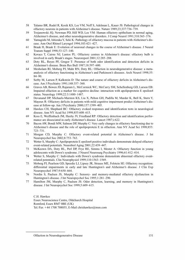
Olfaction in Neurodegenerative Disease 151
58 Talamo BR, Rudel R, Kosik KS, Lee VM, Neff S, Adelman L, Kauer JS: Pathological changes in
olfactory neurons in patients with Alzheimer’s disease. Nature 1989;23;337:736–739.
59 Trojanowski JQ, Newman PD, Hill WD, Lee VM: Human olfactory epithelium in normal aging,
Alzheimer’s disease, and other neurodegenerative disorders. J Comp Neurol 1991;310:365–376.
60 Yamagishi M, Ishizuka Y, Seki K: Pathology of olfactory mucosa in patients with Alzheimer’s dis-
ease. Ann Otol Rhinol Laryngol 1994;103:421–427.
61 Braak H, Braak E: Evolution of neuronal changes in the course of Alzheimer’s disease. J Neural
Transm Suppl 1998;53:127–140.
62 Kovacs T, Cairns NJ, Lantos PL: Olfactory centres in Alzheimer’s disease: olfactory bulb is
involved in early Braak’s stages. Neuroreport 2001;12:285–288.
63 Doty RL, Reyes PF, Gregor T: Presence of both odor identification and detection deficits in
Alzheimer’s disease. Brain Res Bull 1987;18:597–600.
64 Mesholam RI, Moberg PJ, Mahr RN, Doty RL: Olfaction in neurodegenerative disease: a meta-
analysis of olfactory functioning in Alzheimer’s and Parkinson’s diseases. Arch Neurol 1998;55:
84–90.
65 Serby M, Larson P, Kalkstein D: The nature and course of olfactory deficits in Alzheimer’s dis-
ease. Am J Psychiatry 1991;148:357–360.
66 Graves AB, Bowen JD, Rajaram L, McCormick WC, McCurry SM, Schellenberg GD, Larson EB:
Impaired olfaction as a marker for cognitive decline: interaction with apolipoprotein E epsilon4
status. Neurology 1999;22:3:1480–1487.
67 Devanand DP, Michaels-Marston KS, Liu X, Pelton GH, Padilla M, Marder K, Bell K, Stern Y,
Mayeux R: Olfactory deficits in patients with mild cognitive impairment predict Alzheimer’s dis-
ease at follow-up. Am J Psychiatry 2000;157:1399–405.
68 Hawkes CH, Shephard BC: Olfactory evoked responses and identification tests in neurological
disease. Ann NY Acad Sci 1998;855:608–615.
69 Koss E, Weiffenbach JM, Haxby JV, Friedland RP: Olfactory detection and identification perfor-
mance are dissociated in early Alzheimer’s disease. Lancet 1987;i:622.
70 Bacon AW, Bondi MW, Salmon DP, Murphy C: Very early changes in olfactory functioning due to
Alzheimer’s disease and the role of apolipoprotein E in olfaction. Ann NY Acad Sci 1998;855:
723–731.
71 Morgan CD, Murphy C: Olfactory event-related potential in Alzheimer’s disease. J Int
Neuropsychol Soc 2002;8:753–763.
72 Wetter S, Murphy C: Apolipoprotein E epsilon4 positive individuals demonstrate delayed olfactory
event-related potentials. Neurobiol Aging 2001;22:439–447.
73 McKeown DA, Doty RL, Perl DP, Frye RE, Simms I, Mester A: Olfactory function in young
adolescents with Down’s syndrome. J Neurol Neurosurg Psychiatry 1996;61:412–414.
74 Wetter S, Murphy C: Individuals with Down’s syndrome demonstrate abnormal olfactory event-
related potentials. Clin Neurophysiol 1999;110:1563–1569.
75 Moberg PJ, Pearlson GD, Speedie LJ, Lipsey JR, Strauss ME, Folstein SE: Olfactory recognition:
differential impairments in early and late Huntington’s and Alzheimer’s disease. J Clin Exp
Neuropsychol 1987;9:650–664.
76 Nordin S, Paulsen JS, Murphy C: Sensory- and memory-mediated olfactory dysfunction in
Huntington’s disease. J Int Neuropsychol Soc 1995;1:281–290.
77 Hamilton JM, Murphy C, Paulsen JS: Odor detection, learning, and memory in Huntington’s
disease. J Int Neuropsychol Soc 1999;5:609–615.
C.H. Hawkes
Essex Neuroscience Centre, Oldchurch Hospital
Romford, Essex RM7 0BE (UK)
Tel./Fax �44 1708 708055, E-Mail [email protected]
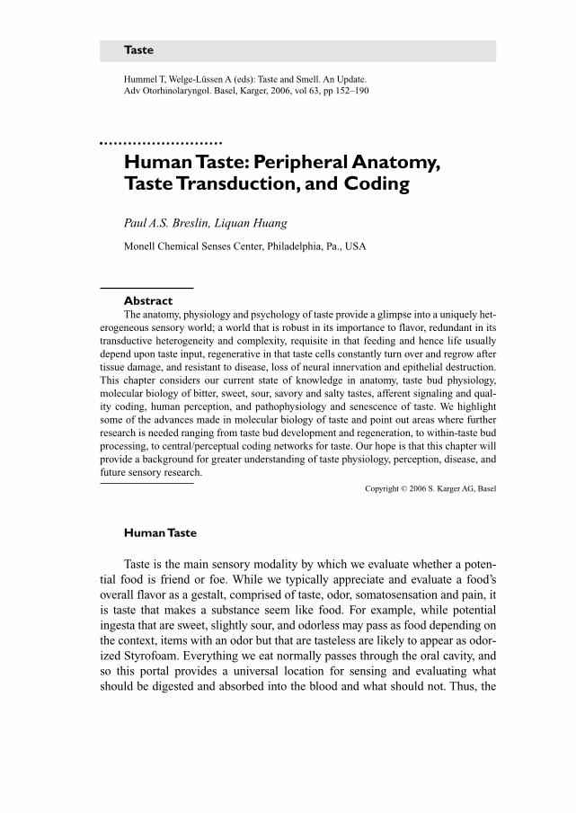
Hummel T, Welge-Lüssen A (eds): Taste and Smell. An Update.
Adv Otorhinolaryngol. Basel, Karger, 2006, vol 63, pp 152–190
Human Taste: Peripheral Anatomy,TasteTransduction, and Coding
Paul A.S. Breslin, Liquan Huang
Monell Chemical Senses Center, Philadelphia, Pa., USA
AbstractThe anatomy, physiology and psychology of taste provide a glimpse into a uniquely het-
erogeneous sensory world; a world that is robust in its importance to flavor, redundant in its
transductive heterogeneity and complexity, requisite in that feeding and hence life usually
depend upon taste input, regenerative in that taste cells constantly turn over and regrow after
tissue damage, and resistant to disease, loss of neural innervation and epithelial destruction.
This chapter considers our current state of knowledge in anatomy, taste bud physiology,
molecular biology of bitter, sweet, sour, savory and salty tastes, afferent signaling and qual-
ity coding, human perception, and pathophysiology and senescence of taste. We highlight
some of the advances made in molecular biology of taste and point out areas where further
research is needed ranging from taste bud development and regeneration, to within-taste bud
processing, to central/perceptual coding networks for taste. Our hope is that this chapter will
provide a background for greater understanding of taste physiology, perception, disease, and
future sensory research.
Copyright © 2006 S. Karger AG, Basel
Human Taste
Taste is the main sensory modality by which we evaluate whether a poten-
tial food is friend or foe. While we typically appreciate and evaluate a food’s
overall flavor as a gestalt, comprised of taste, odor, somatosensation and pain, it
is taste that makes a substance seem like food. For example, while potential
ingesta that are sweet, slightly sour, and odorless may pass as food depending on
the context, items with an odor but that are tasteless are likely to appear as odor-
ized Styrofoam. Everything we eat normally passes through the oral cavity, and
so this portal provides a universal location for sensing and evaluating what
should be digested and absorbed into the blood and what should not. Thus, the
Taste
Human Taste: Peripheral Anatomy, Taste Transduction, and Coding 153
ultimate final motor outputs that result from taste stimulation involve the swal-
lowing of food (acceptance) or the expectorating of food (rejection). If accept-
able, there are other taste-cued reflexes, which anticipate and facilitate digestion
of the forthcoming nutrients, involving both exocrine (e.g. gastric acid) and
endocrine (e.g. insulin) secretions. These early taste-triggered secretions, labeled
cephalic phase responses, are necessary for normal digestion and were the focus
of Pavlov’s famous digestion research [1, 2].
The acceptance of sweet tastes, signaling calories, and the rejection of
strongly bitter tastes, signaling toxins, are brain stem reflexes apparent in
humans prenatally [3, 4]. Our adult food preferences are built on top of these
reflexes, which can be modified by experience but never eliminated. Thus, it is
rare to find commercial foods that are strongly bitter, and among global markets
for foods with requisite bitterness, such as coffee and beer, the less bitter the
product the higher are global sales. Regardless of whether taste-guided behav-
iors are reflexive or are part of a more sophisticated developed appetite, the taste
system must detect what is present in the mouth and enable discrimination and
recognition of chemical components and levels there. These processes ultimately
lead to the recognition of food as familiar or, when novel, as safe. When taste is
severely disturbed, then feeding is almost always disturbed [5, 6].
Taste is arguably the only external sensory system required for life. It is
well known that people live in our societies without sight, hearing, or smell.
Even some somatosensory and proprioceptive deficits are overcome with visual
feedback. But people without taste often do not eat and without medical inter-
vention would die. This is most frequently seen in head and neck cancer
patients who receive radiotherapy [7]. These patients typically experience a
radiation-induced loss of taste that may be complete and always interferes with
eating [8, 9]. They commonly require the placement of a chronic nasogastric
tube for feeding, as they are an already nutritionally compromised population.
In addition to its requisite role in feeding, taste in other species plays a role in
detecting and identifying hydrocarbons that serve as a pheromonal social com-
munication signal such as in courting and mating [10]. While olfaction is
known to play a social communication role in humans, it is unknown whether
taste in humans plays a similar social role [11].
Taste sensations may be divided into several psychological attributes: qual-
ity, intensity, oral location, and timing. All of these attributes are, in turn,
rapidly evaluated and, within a given context, imbued with some degree of pos-
itive or negative hedonic value – yummy or yucky. The qualities of taste are the
basic subdivisions of the modality labeled sweet, sour, salty, bitter and savory
(or umami). Prototypical exemplars of these tastes are honey, lemon juice, table
salt, strong black coffee, and chicken broth (or more specifically free gluta-
mate). To evaluate the taste of these solutions without the influence of their

Breslin/Huang 154
intrinsic odors, simply taste them while pinching the nares of the nose closed.
Without airflow, there is no sense of olfaction, which is why congestion can
cause temporary loss of smell. This type of test is also clinically useful in
patients who complain of taste loss. It is presently an area of active research
whether identified fatty acid detection systems (fat perception) are part of the
taste sensory system and, if so, whether these transduction systems generate an
identifiable ‘fat taste’ quality [12–14]. One theory posits that fat receptors on
taste receptor cells may be gustatory modulators or perhaps help trigger cephalic
reflexes for fat metabolism.
The taste intensity is the magnitude of the qualitative sensations, such as
weakly salty or strongly bitter. The location is the perceived region of the oral
cavity from which a taste sensation arises. Taste unlike smell has a spatial
dimension that enables humans to localize specific qualities and even to manip-
ulate items in the mouth based on taste localization [15]. The timing of taste
refers to whether taste sensations arise quickly and whether they linger, i.e.
aftertastes. Timing and spatial cues are inherently used to evaluate stimuli and
are largely responsible for why artificial sweeteners rarely taste like sucrose;
most artificial sweeteners are localized more to the posterior oral cavity and
linger longer than sucrose does.
Peripheral Taste Anatomy
Humans have taste receptors in several fields within the oral cavity includ-
ing: all edges of the tongue and on the anterior dorsal surface of the tongue, on the
soft palate, and in the pharyngeal and the laryngeal regions of the throat (fig. 1).
Taste receptor cells predominantly reside within multicellular rosette clusters
labeled ‘taste buds’ [16, 17]. There is presently a discussion as to whether single
chemosensitive cells may also exist in humans; but there is evidence for solitary
taste receptor cells in the larynx of rodents [18]. The taste buds on the dorsal sur-
face and edges of the tongue occur in papillae: fungiform papillae pepper the
anterior two thirds of the tongue, several continuous foliate papillae (folds)
appear on the posterior lateral edges of the tongue, and circumvallate papillae
(towers with motes) appear in an arc of approximately nine papillae on the poste-
rior tongue just anterior to the lingual genu [17]. Other taste fields in the soft
palate and pharynx reside in the epithelium but not within papillae. A lingual
fungiform papilla can contain between 0 and 15 taste buds and averages approxi-
mately five taste buds [19]. Foliate and vallate papillae always contain taste buds
and typically have many more than do fungiform papillae, often dozens. The taste
buds live within the folds of foliate and in the ‘mote’ of the vallate papillae where
von Ebner’s glands secrete saliva and proteins into these recesses [20, 21].
Human Taste: Peripheral Anatomy, Taste Transduction, and Coding 155
The receptor cells within a taste bud are not neural cells. Rather, they are
specialized epithelial cells that share almost all of the same properties as neural
cells except that they lack an axon [17, 22]. The afferent taste information is
transmitted to neural fibers within the taste bud. The cell bodies of these taste
neural fibers occur within the sensory ganglia of cranial nerves (CN) VII, IX,
and X [6]. The fibers project into the central nervous system at the level of the
brain stem and synapse initially onto the nucleus of the solitary tract [23, 24].
From there, afferent information projects cortically via the thalamus to the pri-
mary opercular and insular taste cortex, as well as to the orbitofrontal cortex,
cingulate gyrus and to other multimodality integrative projection areas [25].
There is also a more ventral anterior pathway for taste signals that includes: the
Fig. 1. The human tongue contains three types of taste papillae. Vallate and foliate
papillae reside on the middle and sides of the posterior 1/3 of the tongue, respectively, and
contain hundreds of taste buds. Fungiform papillae are scattered in the anterior 2/3 of the
tongue, each harboring 0–15 taste buds. Taste buds are also located in the soft palate and
pharynx but are in the flat epithelium rather than in papillae in these locations.
Vallate
Foliate
Fungiform
Breslin/Huang 156
bed nucleus of the stria terminalis, hypothalamus, mammillary bodies, amyg-
dala, hippocampus and other areas of the limbic system [25].
The facial nerve (CN VII) has its cell bodies in the geniculate ganglion and
its taste information is carried by two nerve branches [6]. The chorda tympani
nerve, so named because it passes through the middle ear behind the tympa-
num, innervates the whole anterior two thirds of the tongue including all fungi-
form and the most anterior foliate papillae [6]. The greater superficial petrosal
nerve branch innervates the soft palate, which may contain as many taste buds
as does the anterior tongue [6]. The glossopharyngeal nerve (CN IX) has its cell
bodies in the petrosal ganglion and innervates most of the foliate and all of the
vallate papillae in the posterior tongue via the lingual-tonsillar nerve branch
[6]. The vagus nerve (CN X) has its cell bodies in the nodose ganglion and
innervates taste buds in the pharynx and larynx via the superior laryngeal nerve
branch [6]. A single axonal fiber of any of these nerves may innervate multiple
cells within a taste bud and may also innervate multiple buds. Thus, the primary
neural fibers are a potential site of taste signal integration. These fibers rarely
have branches that cross the midline of the tongue, leaving the left and right
sides of the tongue under independent peripheral control. Specific taste field
loss that impacts both sides of the tongue is usually associated with peripheral
damage to the receptor systems and in the tongue via physical or perhaps chem-
ical trauma. Peripheral neural damage or central neural involvement is usually
associated with asymmetrical effects. In addition to taste compound sensitivity,
most, but not all, of these peripheral afferent fibers are touch and thermally sen-
sitive in the anterior tongue and are additionally noxious stimulus sensitive in
the posterior tongue.
The Taste Bud
The taste bud is a microscopic ‘rosebud’-shaped structure that contains
between 60–120 cells [26–28] (fig. 1). The receptor cells involved in primary
taste signal transduction are in direct contact with the solutions of the oral cav-
ity via microvilli at the apical end of the cells [17]. The microvilli contact the
oral solutions via a small (approx. 20 �m) opening in the epithelium called the
taste pore that lies at the tip of each bud [29]. Chemical stimuli are restricted in
their flow past taste cells at the taste pore by tight junctions linking the cells in
contact with the pore; generally, only small ions may pass tight junctions
[29–31]. Adult human fungiform papillae contain approximately 4 or 5 taste
buds on average although many contain zero buds [32, 33]. The foliate papillae
contain several buds on either side of the epithelial walls that comprise each
foliate groove [17]. Similarly many taste buds line the papillary sidewall of the
Human Taste: Peripheral Anatomy, Taste Transduction, and Coding 157
‘mote’ around each vallate papillae [17]. In contrast, the vast majority of papil-
lae on the tongue are filiform, small conical-shaped papillae, which never con-
tain taste buds [34]. These seem to serve the function of making the lingual
surface mechanically rough, which facilitates food and beverage manipulation
and may also enhance lingual somatosensory function. The larger and less
dense fungiform papillae may be observed among the smaller more plentiful
filiform papillae by simple direct visual examination of the anterior dorsal sur-
face of the tongue.
There are four principal types of cells within a taste bud, but these may be
further subdivided histochemically. The cell types were historically labeled
dark, light, intermediate and basal cells based upon their electron-dense appear-
ance, shape, and position in an electron microscope image of a taste bud [28,
35]. The basal cells were small round cells at the base of the taste bud. The other
three cell types were elongated cells stretching from the basal to the apical end
of the taste bud and appearing dark, light, and intermediate [36, 37]. Today
these cell types are referred to as type I, II, and III cells, respectively [26–28,
38, 39]. Both type I and II cells possess microvilli with those of type II cells
shorter than those of type I [35, 40]. Most primary signal transduction compo-
nents such as receptors and effector enzymes are found only in type II cells
[41]. This observation has led some to conclude that type II cells are the canon-
ical taste receptor cell. However, most synapses with primary afferent axons are
on type III cells, many of which are identified as being serotonergic [42–44]. In
mice, synapses have also been occasionally identified in type I and II cells [27,
28, 44]. Rats, mice and rabbits appear to possess significant species differences
in taste bud organization. The precise configuration of human taste buds
remains to be determined [36, 37, 45].
An important question is how information passes from type II cells, where
the transduction elements reside, to type III cells, where most synapses with
primary afferents occur [44]. Bud cells can be electrically coupled via gap junc-
tions, but more likely they communicate with one another neurochemically.
Taste bud cells are known to possess serotonin and serotonin receptors, ATP,
ADP and their P2X and P2Y receptors, glutamate and its mGluR1, mGluR4
and ionotropic receptors, as well as nitric oxide (NO) synthase and NO, which
is a gas that freely enables cells to communicate with each other. These are all
possible candidates for cell-to-cell chemical communication within the taste
bud, so that primary receptor cells without neural synapses could communicate
with taste bud cells with neural synapses [46–50]. Since the taste bud contains
receptor cells specialized for different classes of chemicals, the problem arises
as to how to coordinate the cells within a bud of a similar chemical sensitivity.
This could be accomplished intragemmally pan-bud by the coordination of the
chemical identity of what is released with the appropriate receptors on target

Breslin/Huang 158
cells within the bud. For example, glutamate might be a bitter compound inter-
cellular signal within the bud, while serotonin might be a sweet compound
intercellular signaler [47].
While taste bud cells differ from olfactory receptor cells in that they are
not neurons, they are, however, similar to olfactory receptor cells in that they
have a short life span and are replaced continuously throughout the life of the
bud. The life of a taste bud cell is approximately 10 days [51]. Basal cells were
believed to be the sole progenitors of the elongated cells within the taste bud
and to give rise to the three elongate cell types, but this is not necessarily the
case [26, 52, 53]. The stem cells that give rise to taste bud cells reside outside
the bud, near its base in the stratum germinativum, and continuously migrate
into the bud to generate new cells [52, 53]. Moreover, the cell types are not all
different stages along a single cell’s life cycle. Rather, the stem cells that give
rise to different elongate bud cells are of different origins; that is, the elongate
cells seem to have different lineages [54]. The exact role that each bud cell
plays remains uncertain. For example, despite the fact that type I cells have
many long microvilli extending into the taste pore and may occasionally have
synapses with neurons in some species, it is unclear whether they are involved
directly in signal transduction. Some have hypothesized that they play a secre-
tory role for the taste pore and/or may even serve a glia-like function for the
bud [55].
Bitterness and the TAS2R Receptors
Insensitivity to bitter-tasting compounds was serendipitously discovered in
1931 when it was found that some people could not identify the taste of a com-
pound called phenylthiocarbamide (PTC) as strongly bitter, although the major-
ity of people could [56]. Subsequent studies demonstrated that humans displayed
this type of taster and nontaster bimodality in many structurally related com-
pounds containing the N–C�S chemical moiety. This trait was found to be her-
itable. Since then genetic studies have identified several loci on both human and
mouse chromosomes that control the sensitivity to several bitter compounds.
For example, the PTC loci on human chromosomes 5p15 7q31 control the
response to PTC and PROP [57, 58]; on the distal end of mouse chromosome 6,
loci SOA [59–61], RUA [62], CYX [63] and QUI [64] are tightly linked to the
response to sucrose octaacetate, raffinose undecaacetate, cycloheximide and
quinine, respectively. Recently, progress in human and mouse genome sequencing
projects has made it possible to identify a novel subfamily of G-protein-coupled
receptors (GPCRs) called type 2 taste receptors or T2Rs as bitter taste receptors
[65–67].
Human Taste: Peripheral Anatomy, Taste Transduction, and Coding 159
Several lines of evidence suggested that T2Rs are the receptors for bitter
substances. In situ hybridization demonstrated that these receptors are expressed
in a subset of taste bud cells. Interestingly, a taste bud cell may have many of these
T2Rs [66] and each one of these receptors could be specifically activated by
only a few bitter compounds. Therefore, while a T2R may only be able to rec-
ognize a few bitter substances with high specificity and sensitivity, a taste bud
cell with multiple T2Rs should detect a broad range of bitter and usually toxic
compounds with equally high sensitivity. One caveat here is that these cells may
not be able to respond differentially to one bitter substance over another. This
may explain why many different bitter compounds evoke similar bitterness per-
ception in humans.
Behavioral studies with taster and nontaster mouse strains indicated that
variations in the mouse T2R5 gene determine the animal’s response to a bitter
compound, cycloheximide. T2R5 is located in the CYX locus at the distal end
of mouse chromosome 6. Sequence analysis showed that all taster strains (e.g.
DBA/2) have the same mT2R5 nucleotide sequences while nontasters (e.g.
C57BL/6 and 129/Sv) have missense mutations and consequently amino acid
substitutions. A heterologously expressed mT2R5 receptor can be activated by
cycloheximide. But a much higher concentration of cycloheximide is required
to elicit a response with comparable amplitude via a receptor from a nontaster
mouse strain compared to a taster’s receptor [67].
Heterologous expression and ligand screening have so far determined bit-
ter compounds for a few receptors. In addition to the cycloheximide receptor
mouse T2R5, the rat counterpart, rT2R9, can be stimulated by cycloheximide
as well, while the human paralogue hT2R10 can detect the bitter compound
strychnine [68]. Human T2R4 and mouse mT2R8 respond to denatonium and
to a lesser extent PROP [67]. Human T2R16 can be stimulated by a group of
�-glucopyranosides including salicin, which is an extract from willow bark,
which has been used as an antipyretic and an analgesic in the treatment of
rheumatism for at least 3,500 years [69]. Human T2R43 and T2R44 have simi-
lar ligand profiles: both can be activated by aristolochic acid, nitrosaccharin
and at higher concentrations, the artificial sweeteners Na-saccharin and
acesulfame-K. But T2R44 can also be activated by denatonium and is more sen-
sitive to the artificial sweeteners than T2R43. This observation explains why
these sweeteners taste bitter at high concentrations [70, 71]. Surprisingly,
human T2R14 can be activated by a number of apparently structurally unrelated
chemicals, including picrotoxin and picrotin found in fishberry seeds, (–)-�-
thujone, a psychotropic component of absinthe from wormwood, and sodium
benzoate found in many plants and frequently used as an antimicrobial preserv-
ative [72]. Two of the most commonly used bitter compounds in human bitter
taste studies, PTC and its structurally similar proxy PROP, can be recognized

Breslin/Huang 160
via the human receptor T2R38 [73]. However, PROP may be able to activate
other T2Rs such as the aforementioned T2R4 receptor [73].
Identification of ligands for each bitter receptor is necessary for establishing
the entire bitter tasting profiles for humans. However, only a few of T2Rs have
been deorphanized. On the other hand, genomic and evolution analyses of T2R
repertoires have provided some insights into bitter taste ability of humans as a
species. The human T2R gene repertoire comprises 25 functional TAS2R genes
and 8–11 nonfunctional sequences that contain premature stop codons, referred to
as pseudogenes [68, 74–76]. In contrast, rodents have a larger T2R repertoire (43
sequences) and a lower proportion of pseudogenes (7 out of 43 or 16% vs. 31% in
humans) [75, 77]. The majority of both human and rodent TAS2R genes and
pseudogenes are clustered at two locations: human T2Rs reside on chromosomes 7
and 12, with only one gene on chromosome 5; most of mouse and rat TAS2R
sequences reside in two regions of one chromosome (chromosome 6 in mice; chro-
mosome 4 in rats). These two regions on the rodent chromosome are syntenic to
the corresponding human clusters on two separate chromosomes, indicating that
the overall arrangement of TAS2R genes was developed in a common ancestor
to the primate and rodent lineages (fig. 2). Genes within the same cluster share
higher sequence similarity than those between clusters, suggesting that these genes
were generated by duplication [78, 79]. Interspecific analysis of T2R sequences
showed that human and mouse T2R genes and pseudogenes can be categorized
into three groups. In group A, multiple human genes are orthologous to one mouse
gene, i.e., multiple-to-one orthology. Nine human genes and 6 pseudogenes can be
classified into this group. In group B, a single human gene is orthologous to mul-
tiple mouse genes, i.e., one-to-multiple orthology; three human genes fall into this
group. In group C, which consists of 10 human genes and 3 pseudogenes, one
human gene is orthologous to one mouse gene. This classification implicates that
during the mammalian radiation, humans and rodents underwent species-specific
gene duplication to adapt to their unique ecological niches, as shown in group A
and B T2R genes, respectively, while group C T2Rs seem essential to both humans
and rodents to detect some common bitter substances. This classification seems to
be supported by receptor expression and ligand screening data. For example,
Fig. 2. Orthology relationships between human and mouse T2R bitter receptors based
on the phylogenetic analyses of amino acid sequences (with permission from Go et al. [78]).
Human and mouse receptors are shown as solid and shaded, respectively. Pseudogenes are
indicated by asterisks (*). In group A, multiple human receptors are orthologous to
one mouse receptor (multiple-to-one orthology). In group B, one human receptor is ortholo-
gous to one mouse receptor (one-to-one orthology). In group C, one human receptor is
orthologous to multiple mouse receptors (one-to-multiple orthology). Note: a few human
T2Rs are uncategorized.
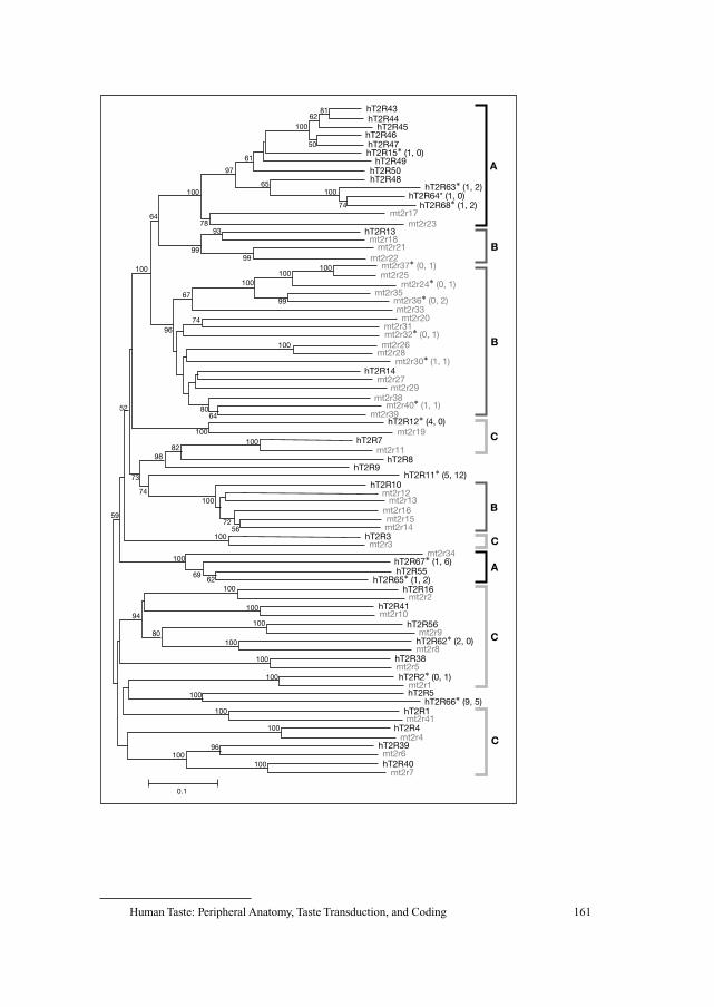
Human Taste: Peripheral Anatomy, Taste Transduction, and Coding 161
hT2R438162
100
100100
50
74
61
97
65
7893
99
6799
74
52 8064
8298
73
74
5972
56
69
94
80
96
0.1
62
96
99
64
hT2R44hT2R45
hT2R46hT2R47
hT2R49hT2R50hT2R48
hT2R13
hT2R15* (1, 0)
hT2R63* (1, 2)hT2R64* (1, 0)
hT2R68* (1, 2)
hT2R11* (5, 12)
hT2R67* (1, 6)
hT2R2* (0, 1)
hT2R62* (2, 0)
hT2R66* (9, 5)
hT2R65* (1, 2)
hT2R12* (4, 0)
mt2r17mt2r23
mt2r18mt2r21
mt2r22
mt2r25
mt2r35
mt2r33mt2r20
mt2r26mt2r28
mt2r27mt2r29
mt2r31
mt2r19
mt2r11
mt2r12mt2r13
mt2r16mt2r15mt2r14
mt2r3mt2r34
mt2r2
mt2r10
mt2r9
mt2r8
mt2r5
mt2r1
mt2r41
mt2r4
mt2r6
mt2r7
mt2r38
mt2r39
mt2r37* (0, 1)
mt2r24* (0, 1)
mt2r36* (0, 2)
mt2r32* (0, 1)
mt2r30* (1, 1)
mt2r40* (1, 1)
hT2R14
hT2R7
hT2R8
hT2R10
hT2R3
hT2R55
hT2R16
hT2R41
hT2R56
hT2R38
hT2R5
hT2R1
hT2R4
hT2R40
hT2R39
hT2R9
100100
100
100
A
B
B
C
C
A
C
C
B
100
100
100
100
100
100
100
100
100
100
100
100
100
100
100
100100
Breslin/Huang 162
hT2R14 is a group B receptor, which has 17 orthologs in mice. Human T2R14
could be an ancient and promiscuous receptor while the mouse has developed a
more sophisticated system to be able to more sensitively and specifically recognize
each of these compounds of this group. The same could be true for another group
B receptor, human T2R10, which has 5 orthologs in mice. One of the orthologs,
mT2R5, may have been more narrowly and specifically tuned for a structurally
related but different ligand. Human T2R43 and T2R44 belong to group A, sug-
gesting that they could be newly expanded, human species-specific receptors,
which have finely tuned, somewhat overlapping recognitions.
Another striking finding is that like in olfactory and pheromone systems,
humans have more T2R pseudogenes. However, no pseudogene is found in group
B, in which rodent genes expanded, suggesting that humans maintained a basic
but important ability to detect toxic substances that rodents more frequently
encounter. However, a large fraction of pseudogenes are present in group A, sug-
gesting that pseudogenization has rapidly been accumulated in the human-
specific repertoire of T2R genes, as well as in the group of common genes. This
notion is corroborated by the comparative analysis with the chimpanzee T2R
repertoire, in which 2 of these 11 human pseudogenes are not mutated and suppo-
sedly still functional in the chimp, indicating that since the time that human and
chimpanzee lineages split about 6 million years ago, humans lost 2 more bitter
receptors [79]. The deterioration of the bitter tasting ability in humans may reflect
the environmental and dietary changes during human evolution.
Substantial variation in human taste abilities has also been found among
different individuals [80–82]. Single nucleotide polymorphisms have been
found in many T2R genes of many individual people. The human population
exhibits unusually higher levels of allelic mutations in T2R genes than in other
genes. These polymorphisms may bestow on the individual the ability to sense
new compounds or ignore certain compounds such as the aforementioned
PTC/PROP. Emerging pseudogenes have also been discovered in a particular
group of people as well as in the worldwide population, suggesting that loss of
function in bitter taste is an ongoing event.
Like many other GPCRs, T2Rs have 7-transmembrane domains as well as
some conserved amino acid residues. In contrast to T1Rs (see below), T2Rs
have short N- and C-termini. Individual members of the T2R subfamily exhibit
a high degree of similarity with 30–70% of the amino acid sequence identical to
other family members. The most divergent segments are the extracellular loops,
and swapping of these loops between some T2Rs indicated that they participate
in the binding of structurally diverse bitter compounds and contribute to the ligand
specificity [70].
Comparison of human and rodent genomes reveals that humans have a
larger proportion of TAS2R pseudogenes than rodents, which is common for
Human Taste: Peripheral Anatomy, Taste Transduction, and Coding 163
other chemosensory gene families such as olfactory and pheromone receptor
genes [83, 84]. The exact roles of these pseudogenes are not known. This
pseudogenization may reflect the fact that humans have encountered fewer
toxic bitter compounds in their diet during their recent evolution.
Sweetness, Umaminess (Savoriness) and the TAS1R Receptors
Sweet and umami (or savory) tastes are thought to measure the carbohy-
drate and amino acid contents in food, hence its energy density. We have known
for decades that sweet taste is likely transduced by membrane-bound protein
receptors, since proteolytic (pronase) treatment of the tongue surface tran-
siently abolishes sweetness perception [85]. Further, gold- or radio-labeled sug-
ars and sweet-tasting peptides and proteins can bind to the membrane portion of
lingual epithelium or to the taste pore of taste buds [86, 87], where the pre-
sumed sweet receptors reside.
The discovery of sweet and umami receptors was also greatly facilitated by
mouse genetic studies and advances in genomic sequencing for model organ-
isms. Animal behavioral assays have reported that inbred mouse strains exhibit
a bimodal distribution of sensitivity to sweet compounds including saccharin,
i.e., a taster or nontaster trait. The determinant for this trait has been attributed
to a Sac locus on the distal end of mouse chromosome 4 that was named after
saccharin [59, 88–91]. Data mining of this locus and the syntenous region on
human chromosome 1 identified a novel GPCR, T1R3, which has the highest
amino acid sequence similarity to two previously isolated orphan receptors
from taste tissue, T1R1 and T1R2 [92–97]. Sequence analysis revealed that
mutations in this T1R3 gene of nontaster mouse strains are likely to be respon-
sible for their low sensitivity to saccharin and other sweeteners. Introduction of
the taster version of the T1R3 gene into the nontaster mice by backcrossing or
transgenic manipulation rescues the sweet taste deficit [92, 98], further con-
firming that T1R3 is responsible for Sac phenotypes.
T1R3, as well as T1R1 and T1R2, is classified based on sequence identity
as a member of the class C GPCR subfamily [99]. Other members of this sub-
family include calcium-sensing receptor, putative pheromone receptor V2Rs,
neurotransmitter receptors mGluRs and GABABRs. One of the hallmarks of
this subfamily is receptor dimerization. In situ hybridization has shown that
T1R1 and T1R2 are indeed co-localized with T1R3 to a subset of taste bud cells
while a few taste bud cells express T1R3 alone [95, 98]. Thus, three types of
T1R-expressing taste receptor cells exist: cells expressing both T1R1 � T1R3,
both T1R2 � T1R3, or T1R3 alone [95, 98]. Coexpression of T1R receptors in
heterologous systems has deorphanized all three of them [98, 100, 101]. Human
Breslin/Huang 164
T1R2/T1R3 heterodimers can be activated by all sweet-tasting compounds
tested at physiological concentrations, including sugars: fructose, glucose, lac-
tose sucrose and maltose; amino acids: glycine and D-tryptophan; sweet pro-
teins: monellin and thaumatin; synthetic sweeteners: acesulfame-K, aspartame,
cyclamate, dulcin, neotame and saccharin. Cells transfected with T1R3 alone
can also respond to some of these compounds at much higher concentrations,
presumably via T1R3 homodimers.
Interestingly, T1R3 can also form a functional heterodimeric receptor with
T1R1. The T1R1 and T1R3 dimer can be activated by many L-amino acids,
including monosodium glutamate (MSG) and L-aspartate. The activation can be
potentiated by 5�-ribonucleotides such as inosine monophosphate and guano-
sine monophosphate (GMP), which is a distinct feature of umami taste. These
data strongly suggest that T1R1/T1R3 heteromers are amino acid sensors and
function as an umami taste receptor.
Are there other receptors for sweet and umami tastes? Genetic studies indi-
cate that loci other than the Sac locus seem to be associated with the detection of
some sweet-tasting amino acids such as glycine [102]. T1R2 or T1R3 null mutant
mice exhibited residual preference for sugars [103, 104]. However, the double
knockout of both T1R2 and T1R3 eliminated responses to sweeteners [103], suggest-
ing that either T1R2 and T1R3 homomeric receptors or an unknown T1R2/T1R3
interacting moiety was responsible for the residual preference in the single-knock-
out animals. A truncated form of the metabotropic glutamate receptor taste-
mGluR4 has been proposed to be another umami receptor [105, 106]. However,
mice lacking this receptor showed enhanced glutamate detection [107], indicating
that this receptor may be playing other roles in taste receptor cells. Taste forms of
mGluR1 have also been identified in taste receptor cells [108, 109].
How can a single dimeric receptor recognize so many structurally diverse
sweet compounds, from simple glucose to glycine to large sweet-tasting pro-
teins? A distinct feature of class C GPCRs is that most of them have a large
extracellular amino terminus, which consists of two domains: a clam shell-like
‘Venus flytrap module’ (VFTM) and a cysteine-rich domain, followed by a hep-
tahelical transmembrane domain and an intracellular carboxyl-terminal domain
[99]. Molecular studies have elucidated multiple ligand binding sites in each
monomer of the T1R2/T1R3 receptor [110–113]. For example, aspartame and
neotame interact with the human VFTM domain of T1R2; brazzein and cycla-
mate bind to the human T1R3 cysteine-rich domain and extracellular loops 2 and
3 of the heptahelical transmembrane domain, respectively; lactisole, a potent
inhibitor of sweet taste, docks to the human T1R3 binding pocket formed by
transmembrane helices. One of the unexpected but understandable findings
is that lactisole can also suppress umami taste by interacting with the T1R3
moiety of the T1R1/T1R3 dimer [114].

Human Taste: Peripheral Anatomy, Taste Transduction, and Coding 165
All 3 T1R genes have been mapped onto human chromosome 1 [115].
They are organized in the order of T1R1, T1R2 and T1R3. However, T1R1 and
T1R2 genes are in the same transcriptional orientation, while T1R3 is in the
opposite orientation. All 3 T1R genes have a similar gene structure, consisting
of 6 coding exons. This genomic organization has been more or less preserved
during evolution. In mice, T1R genes are located on the syntenic region of
mouse chromosome 4, although the gene arrangement differs slightly; the T1R1
gene is in the middle, flanked by T1R2 and T1R3. Unlike other GPCRs, which
share �90% amino acid identity across mammals, human and rodent umami,
sweet, and bitter receptors display only about 70% sequence identity, suggest-
ing that taste receptors have evolutionarily tuned to their species-specific needs
and may have contributed to the formation of species-specific behaviors and
diets. Mutations and pseudogenization of T1Rs in humans have not been well
characterized yet. But in the cat, which has normal T1R1 and T1R3 receptors,
microdeletion and stop codons have been found in the T1R2 gene, resulting in
the lack of T1R2 expression and the functional T1R2/T1R3 sweet receptor
[116]. Pseudogenization of cat T1R2 may have been crucial to the cats’ indif-
ference toward sugars and their favoring meat or, more likely, their carnivory
may have relaxed pressures to maintain the T1R2 receptor; the direction of
the causal arrow is uncertain at this time. Curiously, some fish species possess
multiple copies of T1R2, which could generate variants of amino acid or carbo-
hydrate receptors [117]. In some inbred mouse strains, nonsynonymous sub-
stitutions in the T1R3 VFTM domain lead to low sensitivity to sweeteners. In
addition, rodents are not subject to lactisole inhibition of their sweet taste while
humans are. But mutations in other regions of T1R2 and T1R3 made rodents
and new world monkeys unable to taste a number of compounds that humans
and old world primates deem sweet, including monellin, brazzein, thaumatin,
neotame, aspartame, cyclamate and neohesperidin dihydrochalcone [118–123].
These species differences may arise due to relaxed selective pressure on
allosteric binding sites relative to the orthosteric sites on VFTMs that evolved to
bind canonical saccharides such as glucose, fructose, and sucrose, which virtu-
ally all omnivores and herbivores taste [114, 124–126]. Polymorphism studies on
these 3 human T1R genes might reveal individual differences in sensitivity to
sweet and umami substances, which could help illuminate individual prefer-
ences for flavors.
Salty and Sour Taste Receptors
Salt taste is elicited by Na� and other cations, which may enable humans
and animals to seek out mineral-rich food but avoid oversalty food to maintain
Breslin/Huang 166
ion-water homeostasis. The need to detect ions such as sodium arises from our
inability to store these ions in our bodies, unlike calories or calcium. Thus, all
terrestrial animals and fresh-water animals must ingest sodium regularly. Like
sour taste, saltiness is likely received by ion channels. Permeation of Na� and
other ions into taste bud cells depolarizes membrane potentials, triggering
influx of calcium and then the release of neurotransmitters. It has been postu-
lated for years that a sodium channel, epithelial sodium channel ENaC, is the
channel receptor for salty taste [127–130].
ENaC is a heterotetrameric protein, consisting of 2�-, 1�- and 1�-subunits
in vodents. Heterologously expressed ENaCs of all three subunits displayed sus-
ceptibility to topical amiloride inhibition, which was in agreement with behav-
ioral and nerve recording data from some rodent strains [127, 131–133]. This
was an important discovery as amiloride was believed to be specific to epithelial
sodium channels. However, amiloride does not inhibit much of human salt taste,
which is rather inhibited by another compound, chlorhexidine, suggesting that
the stoichiometry of human ENaCs may be different from rodents’ or a totally
different channel is responsible for human salt taste [134, 135]. Rodents also
possess a component of sodium taste that is not amiloride sensitive [136]. The
molecular basis for this salt taste aspect is unclear but the heat- and capsaicin-
sensitive TRP receptor VR1 has been implicated [137]. In addition, hormones
like aldosterone could change the expression levels of these channels, thus regu-
lating the sensitivity to salty stimuli and to suppression of these inhibitors [138].
At present, the transduction mechanism for human salt taste is unknown.
Sour taste is produced by acids, which play somewhat ambivalent roles
affectively. On the one hand, sour taste in some types of food appears to be
attractive to humans and animals such as oranges and grapefruits, or sour candy.
On the other hand, sourness from spoiled foods and unripe fruits evokes rejec-
tion response. Human psychophysical studies and animal nerve recording
showed that perceived sourness is proportional to the concentrations of protons,
i.e., pH in strong inorganic acids such as HCl, but only a low correlation was
observed between sour taste and pH in organic acids such as citric acid, indicat-
ing that anions in acidic stimuli also contribute to sour taste intensity [139, 140].
Sour taste is believed to be received by ion channels [55]. However, the
identity of these channel receptors has not been firmly established. One reason
is that proton is an active agent, can interact with and regulate virtually all ion
channels. Another confounding factor is that different animal species appear to
use different mechanisms [140].
Several ion channels have been hypothesized to transduce sourness, which
include: (1) direct blockage of the apical potassium channel by protons, thus
leading to depolarization of membrane potentials [141]; (2) passage of protons
through the ENaC [142]; (3) activation of acid-sensing ion channels (ASICs)
Human Taste: Peripheral Anatomy, Taste Transduction, and Coding 167
[143]; (4) activation of a 5-nitro-2-(3-phenylpropylamino)benzoic acid-sensitive
chloride channel, which could also contribute to changes in membrane potentials
and regulate the uptake of neurotransmitters into synaptic vesicles [144, 145];
(5) activation of hyperpolarization-activated and cyclic nucleotide-gated ion
channels [146]; (6) intracellular acidification [147]. In addition, there are GPCRs
that detect protons and could be involved in sour taste.
Much of the effort has been focused on ASICs, and several of them includ-
ing ASIC2 have been found in human taste buds [143, 148, 149]. In particular,
heterologously expressed ASIC2 showed features of acid-induced currents
resembling that observed in taste bud cells [149]. However, this channel is not
present in mouse taste bud cells and the knockout of this gene did not affect
sour taste in mutant mice, suggesting that ASIC2 is not a sour taste channel,
at least not in mice [150].
Preliminary studies with human subjects revealed a unimodal distribution
of sour sensitivity with a large variation in detection thresholds and some
patients exhibiting sour blindness [151]. More data are needed to understand
sour taste mechanisms and to find treatments of sour taste disorders.
Signal Transduction Cascades
Bitter stimulus (T2Rs), sweetener (T1R2/T1R3) and umami stimulus (T1R1/
T1R3, mGluR1, mGluR4) receptors are GPCRs. Activation of these receptors
triggers G-protein-mediated signaling cascades (fig. 3). Several G-protein
�-subunits have been identified in taste bud cells, including G�i2, G�i3, G�14,
G�15, G�q, G�s, �-gustducin and an �-transducin-like subunit, �-gustducin
[152–154]. �-Gustducin shares 80% amino acid identity with �-heteromeric,
and is selectively expressed in about 30% of taste bud cells. Nearly all T2Rs
have been localized to �-gustducin-expressing taste bud cells while additional
�-gustducin-expressing cells possess sweet or umami receptors [66, 97]. In the
heterologous expression system, �-gustducin or chimeric G-proteins containing
part of �-gustducin can relay the signals from activated T2Rs or heteromeric
T1Rs to downstream effectors, resulting in an increase in intracellular calcium
concentration [67, 110]. In vitro biochemical assays have demonstrated that
activated taste receptors can also interact with �-transducin [70, 155]. The
knockout of �-gustducin not only markedly reduces animals’ sensitivity to bit-
ter stimuli, but also moderately decreases sweet and umami tastes [156].
Double-knockout mice lacking both �-gustducin and �-transducin further
reduced umami sensitivity, suggesting that �-transducin is involved only in
umami taste [157]. From these data, it can be concluded that bitter signal trans-
duction is largely mediated by �-gustducin, which is also partially involved in
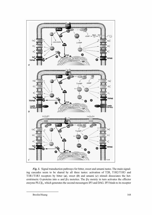
Breslin/Huang 168
Fig. 3. Signal transduction pathways for bitter, sweet and umami tastes. The main signal-
ing cascades seem to be shared by all three tastes: activation of T2R, T1R2/T1R3 and
T1R1/T1R3 receptors by bitter (a), sweet (b) and umami (c) stimuli dissociates the het-
erotrimeric G-proteins into � and �� moieties. The �� moiety in turn activates the effector
enzyme PLC�2, which generates the second messengers IP3 and DAG. IP3 binds to its receptor
a
b
c
Human Taste: Peripheral Anatomy, Taste Transduction, and Coding 169
sweet and umami transduction. Several G-protein ��-subunits have been iso-
lated from taste bud cells. Among them are G�3 and G�13, which are localized
to a subset of taste bud cells, including those expressing �-gustducin [158,
159]. Biochemical interaction indicated that G�3�13 is a likely �� partner for
�-gustducin [159]. Effector enzymes and other downstream components have
also been identified, which include phospholipase C�2 (PLC�2), the inositol
1,4,5-triphosphate (IP3) receptor IP3R3 and a transient receptor potential ion
channel, TRPM5 [160–163].
Therefore, activation of T2R bitter receptors stimulates the heterotrimeric
G-protein consisting of �-gustducin and G�3�13, which dissociates into �-moiety
and ��-moiety, generating bifurcate signaling pathways. �-Gustducin is believed
to activate phosphodiesterase and perhaps guanylyl cyclase as well, which reg-
ulate intracellular cAMP and cGMP levels, respectively, consequently the activ-
ity of protein kinase A and NO synthase [164]. Protein kinase A functionally
modulates an array of voltage-gated ion channels, changing the membrane
potentials. However, the G�3�13 branch appears to be the principal signaling
cascade. G�3�13 activates PLC�2, which hydrolyzes phosphatidylinositol-4,
5-biphosphate and produces two second messengers, diacylglycerol (DAG) and
IP3. DAG activates protein kinase C, which phosphorylates a number of intra-
cellular proteins including voltage-gated ion channels. IP3 binds to the IP3
receptor IP3R3, which releases calcium from intracellular stores. The increase
in free intracellular calcium opens a nonselective monovalent cation channel,
TRPM5 [165–167].
In addition to �-gustducin, other G-protein �-subunits such as �i2 and �s
are likely involved in sweet and umami signal transductions. Generation of
another second messenger, cAMP, has been reported in response to sweet stim-
uli [168]. cAMP is known to regulate protein kinase A activity, which in turn
phosphorylates other intracellular proteins. However, it is firmly established
that PLC�2 and TRPM5 are the two indispensable components in all three
GPCR-mediated bitter, sweet and umami taste transductions since knocking out
IP3R3, which releases Ca2� from intracellular stores. Increase in the free cytosolic Ca2� opens
a nonselective monovalent cation channel TRPM5. Influx of cations leads to depolarization of
the taste cell membrane potential. DAG activates protein kinase C (PKC), which phosphory-
lates and modulates other proteins including voltage-gated ion channels. In bitter receptor cells
(a), the G� subunit may activate phosphodiesterase (PDE) and/or guanylgl cyclase (GC), regu-
late cAMP and cGMP levels, thus activity of protein kinase A (PKA) and NO generation.
ER � endoplasmic reticulum. In sweet receptor cells (b), the G� subunit may regulate the
activity of adenylyl cyclase (AC) and cAMP level, which stimulates PKA. Leptin receptors
may play a role in sweet taste signal transduction. STAT3 � signal transducer and activator of
transcription protein 3. In umami receptor cells (c), truncated metatropic glutamate receptors
tmGluR1 and tmGluR4 may also contribute to umami sensation. Pi � phosphorylation.
Breslin/Huang 170
either of the two genes diminishes sensitivity to stimuli of all three taste modal-
ities [163]. Therefore, T1R- and T2R-mediated signaling cascades converge onto
a common activating event, that is, opening of TRPM5. It is still not known how
activation of TRPM5 on taste bud cells leads to generation of action potentials
on afferent nerve fibers. However, it is believed that influx of monovalent
cations through TRPM5 channels depolarizes taste bud cells. Interestingly,
depolarization of receptor cell membrane potential has also been postulated to
be the cellular response to sour and salty stimuli. Therefore, interactions of sapid
molecules of all five known taste modalities with GPCRs or channel receptors
eventually lead to the generation of receptor potential, which regulates the release
of neurotransmitters onto afferent gustatory nerve fibers and/or the release of
paracrine agents that act on and regulate the neurotransmitter release from adja-
cent taste bud cells [50].
Taste Bud Modulators
In a taste bud, cells are in close apposition with one another. This unique
organization may play an essential role in cell-to-cell communication. In amphib-
ians, gap junctions are present among taste bud cells, which allow the spread of
currents and small molecules to neighboring cells [169]. In mammals, a number
of bioactive agents and their receptors have been found on taste bud cells, which
include serotonin and its receptors, norepinephrine and its receptors, neuropep-
tide Y, cholecystokinin, vasoactive intestinal peptide and somatostatin and their
receptors, acetylcholine and its receptors, glutamate and its metabotropic and
ionotropic receptors, ATP and its P2Y receptors [46, 48, 49, 105, 170–175].
These signaling agents can be released from taste bud cells upon stimulation,
and act on the producing cells as autocrines or on adjacent cells as paracrines to
modulate these cells or to trigger neurotransmitter output onto the cranial nerve
fibers. Occurrence of receptors for the circulating regulatory hormones leptin
and aldosterone has also been reported [176, 177] (fig. 4). Activation of these
receptors is believed to change gene expression patterns such as ENaC subtypes,
or even induce cell proliferation and differentiation.
Physiological Responses of Taste Receptor Cells
Taste bud cells are specialized epithelial cells with some neuronal properties.
The excitability of taste bud cells and their voltage-dependent currents have been
well documented [55]. Action potentials in gustatory receptor cells were first
described in amphibians [178, 179], then in mammals, in response to passing cur-
rents or to taste stimuli such as sour, salt and sweet stimuli [142, 180–182]. In

Human Taste: Peripheral Anatomy, Taste Transduction, and Coding 171
Fig. 4. Signal transduction pathways for ionotropic sour and salty tastes. a A number
of mechanisms may exist for sour transduction, which includes the proton blockage of a K�
channel, proton permeation of ENACs, activation of ASICs, an NPPB-sensitive Cl channel,
or a hyperpolarization-activated and cyclic nucleotide-gated ion channel, and intracellular
acidification. HCN � Hyperpolarization-activated and cyclic nucleotide-gated ion channel.
b Na� and other cations permeate apical multimeric epithelial sodium channels (ENACs), or
pass through a tight junction and enter cells via basolateral ion channels, resulting in the
depolarization of receptor cell membrane potentials and consequently release of synaptic
transmitters. Intracellular ionic equilibrium is restored by Na�-K� pump and ion leak.
Aldosterone receptors may co-occur in these cells and activation of this receptor alters the
composition of ENAC subunits, thus the susceptibility to amiloride. MR � mineralocorti-
coid receptors.
a
b
rats, as many as 75% of taste bud cells can generate action potentials [183–186].
Two types of action potentials have been observed from taste bud cells [182, 187]:
(1) fast action potentials with shorter duration and larger inward and outward cur-
rents; (2) slow action potentials with longer duration and smaller inward and out-
ward currents [182, 184, 188]. Analysis of loose patch recording data from
Breslin/Huang 172
hamster taste buds using an artificial neural network suggested that action poten-
tials in taste buds may contribute to taste quality coding [189]. However, the exact
role of action potentials in taste signal transmission is still enigmatic since action
potentials are usually employed by neurons for long-distance propagation of elec-
trical signals and taste bud cells are short (approx. 100 �m in rodents). Moreover,
receptor potentials instead of action potentials may be sufficient to trigger signal
transmission. Active investigations are being carried out to solve this puzzle.
Various voltage-gated ion channels have been electrophysiologically descri-
bed from different taste bud cells, including tetrodotoxin-sensitive Na� channels,
tetraethylammonium-sensitive delayed rectifier K� channels, inward rectifier K�
channels, outward rectifier Cl– channels, and low-/high-voltage-activated Ca2�
channels [183–186]. The distribution of voltage-gated ion channels across taste
bud cells is heterogeneous. In rats, about 57% of taste bud cells possess voltage-
gated Na� channels but nearly all cells have voltage-gated K� channels. Multiple
voltage-gated Ca2� and Cl– conductances have also been reported from subsets of
taste bud cells. Developmentally, the complexity buildup of voltage-dependent
currents coincides with the maturation of taste bud cells, suggesting that voltage-
gated ion channels contribute to the taste bud cells’ function [190].
In addition to changes in electrical properties, two more responses of taste
bud cells to taste stimuli have also been monitored: changes in intracellular cal-
cium concentration and the release of bioactive agents from taste bud cells [42,
173]. Stimuli of all five taste modalities can induce an increase or decrease in
intracellular calcium concentrations in taste bud cells. Initial increase of cal-
cium in some cells may not require the influx of calcium from extracellular
sources. However, the presence of extracellular calcium augments the magni-
tude and prolongs the sustained response. Caution should be taken in interpret-
ing the calcium response data since this response is an intermediate step in the
taste signal integration process in a taste bud, which may not lead to the final
output of neurotransmitters in some cells, but rather synchronize and reset these
cells for following taste stimuli of similar or different modalities.
Biosensors, which have recently been applied to taste research, can detect
the release of paracrines and transmitters such as serotonin and ATP from taste
bud cells in response to taste stimuli [191]. Further studies have shown that ATP
appears to be a neurotransmitter taste buds use to convey the gustatory signal to
afferent nerve fibers [50].
Afferent Signal Transmission and Coding
How taste compounds are sensed and ultimately encoded qualitatively is a
complex process that involves integration by the system at almost every step of
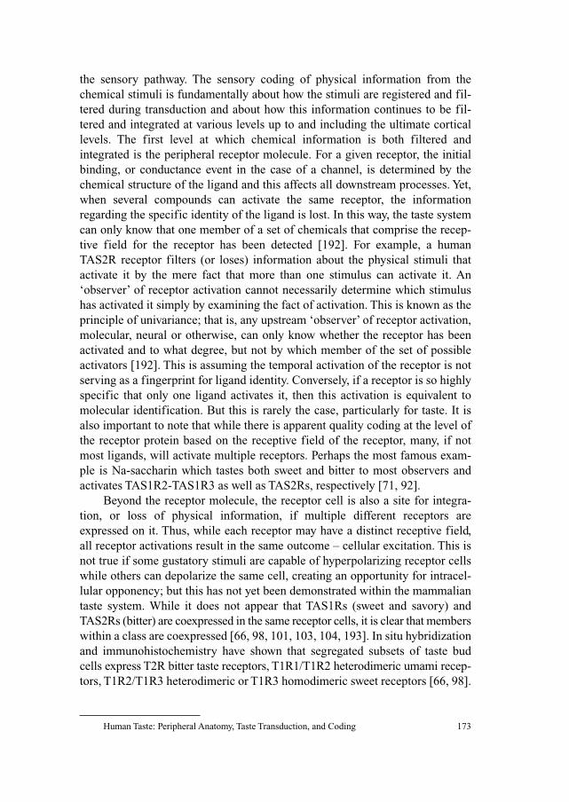
Human Taste: Peripheral Anatomy, Taste Transduction, and Coding 173
the sensory pathway. The sensory coding of physical information from the
chemical stimuli is fundamentally about how the stimuli are registered and fil-
tered during transduction and about how this information continues to be fil-
tered and integrated at various levels up to and including the ultimate cortical
levels. The first level at which chemical information is both filtered and
integrated is the peripheral receptor molecule. For a given receptor, the initial
binding, or conductance event in the case of a channel, is determined by the
chemical structure of the ligand and this affects all downstream processes. Yet,
when several compounds can activate the same receptor, the information
regarding the specific identity of the ligand is lost. In this way, the taste system
can only know that one member of a set of chemicals that comprise the recep-
tive field for the receptor has been detected [192]. For example, a human
TAS2R receptor filters (or loses) information about the physical stimuli that
activate it by the mere fact that more than one stimulus can activate it. An
‘observer’ of receptor activation cannot necessarily determine which stimulus
has activated it simply by examining the fact of activation. This is known as the
principle of univariance; that is, any upstream ‘observer’ of receptor activation,
molecular, neural or otherwise, can only know whether the receptor has been
activated and to what degree, but not by which member of the set of possible
activators [192]. This is assuming the temporal activation of the receptor is not
serving as a fingerprint for ligand identity. Conversely, if a receptor is so highly
specific that only one ligand activates it, then this activation is equivalent to
molecular identification. But this is rarely the case, particularly for taste. It is
also important to note that while there is apparent quality coding at the level of
the receptor protein based on the receptive field of the receptor, many, if not
most ligands, will activate multiple receptors. Perhaps the most famous exam-
ple is Na-saccharin which tastes both sweet and bitter to most observers and
activates TAS1R2-TAS1R3 as well as TAS2Rs, respectively [71, 92].
Beyond the receptor molecule, the receptor cell is also a site for integra-
tion, or loss of physical information, if multiple different receptors are
expressed on it. Thus, while each receptor may have a distinct receptive field,
all receptor activations result in the same outcome – cellular excitation. This is
not true if some gustatory stimuli are capable of hyperpolarizing receptor cells
while others can depolarize the same cell, creating an opportunity for intracel-
lular opponency; but this has not yet been demonstrated within the mammalian
taste system. While it does not appear that TAS1Rs (sweet and savory) and
TAS2Rs (bitter) are coexpressed in the same receptor cells, it is clear that members
within a class are coexpressed [66, 98, 101, 103, 104, 193]. In situ hybridization
and immunohistochemistry have shown that segregated subsets of taste bud
cells express T2R bitter taste receptors, T1R1/T1R2 heterodimeric umami recep-
tors, T1R2/T1R3 heterodimeric or T1R3 homodimeric sweet receptors [66, 98].
Breslin/Huang 174
Multiple TAS2Rs can be found within a single cell type, and while TAS1R3 is
coexpressed with TAS1R1 and TAS1R2 [67, 103, 163, 193], TAS1R1 and
TAS1R2 do not appear to be coexpressed nor is TAS1R3 always coexpressed
with another TAS1R. These data suggest that receptor cells in a taste bud tend to
specialize for certain receptor types, specifically for receptors that bind bitter,
sweet and umami substances.
In line with this hypothesis, the knockout of T1R1 gene expression only
diminished the animal’s sensitivity to umami compounds, and did not affect
sweet, salty and bitter detection. Likewise, the knockout of T1R2 only reduced
sensitivity to sweet stimuli, and did not affect other tastes [103, 104]. A transgenic
rescue experiment showed that expression of PLC�2 driven by a T2R promoter in
PLC�2 gene null mice only restored taste sensitivity to bitter compounds, but not
to sweet and umami compounds, demonstrating some independence of cells that
encode different taste qualities [163]. Additionally, mice expressing transgeni-
cally introduced human T1R2 receptors displayed humanized sweet taste prefer-
ences, and were able to detect substances that taste sweet to humans, but normally
not appetitive to rodents. Furthermore, mice transgenically expressing a human
T2R16 receptor for a bitter compound, phenyl-B-D-glucopyranoside, in all cells
that express sweetener receptors exhibited strong attraction to this bitter com-
pound [193]. These results demonstrate taste quality is encoded by the sets of
cells that are ‘typed’ by the receptors they express on their surface.
Taste receptor cells are typically not the bud cells which synapse onto
afferent neural fibers. Therefore, there must be cell-to-cell processing of affer-
ent signals within the taste bud prior to neural signaling. The role that this plays
in coding is not clear. Since there are cells within a taste bud that are sensitive
to a wide array of different taste stimuli, there must be an intragemmal chemical
coding system so that afferent information is not distorted or garbled during
bud processing. This could be accomplished if sets of receptor cells that collec-
tively represent a taste stimulus all communicated with the elongated cells that
possess neural synapses via a common transmitter, such as serotonin, neu-
ropeptide Y or glutamate. Once the encoding of stimuli has occurred within the
bud, the communication by these second- and third-order bud cells with the
intragemmal neural fibers does not need to be differentiated chemically. ATP
and its receptors P2X2 and P2X3 are now considered the dominant transmitter
for communication between taste bud cells and primary afferent neurons [50].
Thus, once the appropriate set of synapsing elongate bud cells are activated,
then all sets of cells within a bud need not be further differentiated when acti-
vating the afferent neuron; a single mode of synaptic communication suffices.
The neural taste fibers are, however, free to communicate with any cells
within the bud and with many buds. Each afferent neural taste fiber is richly
arborized in the periphery and contacts several taste buds within a small area
Human Taste: Peripheral Anatomy, Taste Transduction, and Coding 175
[194]. Also, several different ganglion neural cells may innervate a single taste
bud. This creates an opportunity for the coding to be either simplified or incr-
eased in its complexity depending on the mixture of bud cell types with which a
single afferent neuron synapses. In some species, such as Pan troglodytes (chim-
panzee), the receptive fields of peripheral afferent fibers appear to be organized
along qualitative dimensions similar to the organization of bud receptor cells.
How fibers are able to synapse with only those type III cells within a bud, across
several buds that are in chemical communication with type II cells of a particular
receptor makeup (e.g. TAS2R-expressing cells), is not known [120, 195–198]. On
the other hand, some species, such as rats, possess primary afferent taste fibers
with a receptive field that includes a qualitatively mixed variety of stimuli [17,
199, 200]. Taste afferent fibers and early taste processing areas are also sensitive
to thermal and tactile stimuli and nociception from posterior fields [201, 202].
Whether peripheral fibers are qualitatively refined in their receptive field or
whether they respond to a broad array of stimuli, the higher relay areas in the cen-
tral nervous system may converge and integrate inputs or refine mixed quality
signals depending on the region of the brain and species in question [203].
Therefore, it is should be clear that the question of how the sensory system codes
for taste quality and thus chemical stimulus class depends largely on where in the
system one is looking. How the system processes taste stimuli at a perceptual or
behavioral level is discussed below.
Human Psychophysics
Intensity JudgementsIn many sensory systems, the ability to detect changes in intensity is
described by a fixed function of the ratio of the magnitude of the change to the
starting concentration of the stimulus, in other words a percent increase or
decrease (a Weber fraction) [204]. For example, a subject may be able to just
noticeably detect an increase or a decrease of 10% in concentration regardless
of the starting concentration. Taste Weber fractions may be constant over sev-
eral orders of magnitude in concentration (above-threshold levels), but excep-
tions to this rule occur at very low and very high concentrations [205, 206].
The rate at which perceived intensity grows with concentration, the expo-
nent of Steven’s power function (I � kCn; where I is intensity and C is concen-
tration), helps to determine the input-output intensity function of the compound
under study [207]. One might surmise that the exponent for a compound’s inten-
sity power function bears some relation to the quality of the sensation. Within a
single quality, however, the exponents (n) for several compounds’ power func-
tions do not form natural groupings. That is, the exponents of sweeteners and
Breslin/Huang 176
the exponents of bitter compounds are intermingled and do not fall into two
neat clusters. Therefore, the rate at which intensity increases is a function of
peripheral events in the epithelium (for example individual molecule’s binding
kinetics) and not psychological events such as the qualitative categorization of
sweetness or sourness.
Umami (Savory) as a Distinct Perceptual QualityThe most typical methods of measuring taste quality involve either: (1) the
direct scaling of quality intensities by subjects using a variety of scaling tech-
niques and a variable number of scales presented per trial, or (2) the rating of
total intensity and the subsequent division of the total intensity into various per-
cent portions of salty, sweet, bitter, and sour. Occasionally, the quality umami is
included in these techniques.
Umami is the Japanese term to describe the ‘savory’ taste elicited by cer-
tain foods, including mushrooms (shiitake), seaweeds (sea tangle), fish
(bonito), and vegetables (tomato). The prototypical chemical elicitors of umami
are MSG mixed with 5�-ribonucleotides like inosine monophosphate or GMP.
All the foods described as umami are rich in these compounds. This quality of
taste is easily perceived by people of many different cultures. Therefore, there
must be cultural reasons why umami is not generally included with the standard
four taste qualities: salt, sour, bitter, and sweet. When offered MSG with 5�-ribonucleotides (especially in warm water), Americans or Europeans describe it
as brothy, soupy, meaty, and savory. While the term savory has not been
included in our parlance of qualitative taste descriptors, the term umami has
been accepted in Japan (at least among food scientists and industry). The term
is relatively new, however, first coined in 1908 by Ikeda for the taste of a broth
made of sea tangle and bonito fish [208].
AdaptationA fundamental characteristic of taste is the adaptation to background levels
of sapid compounds, with the exception of some acids and bitter-tasting com-
pounds. For example, saliva is usually tasteless as it rests in the mouth even
though it contains many ions and other potentially sapid chemicals. In psy-
chophysics, adaptation is defined as the decrement in intensity or sensitivity to a
compound under constant stimulation by this compound. Adaptation to a small
defined portion of the tongue tip is almost complete for a wide variety of taste
compounds. After complete or nearly complete adaptation to a low-intensity taste
stimulus, a subject will have to receive higher concentrations than the adapting
concentration to regain the sensation of the stimulus. Interestingly, concentrations
lower than the adapting concentration elicit tastes as well, though they are usually
of a different quality than the initial quality of the adapting stimulus.
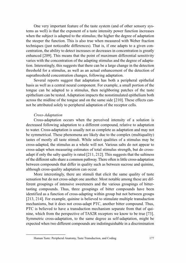
Human Taste: Peripheral Anatomy, Taste Transduction, and Coding 177
One very important feature of the taste system (and of other sensory sys-
tems as well) is that the exponent of a taste intensity power function increases
when the subject is adapted to the stimulus; the higher the degree of adaptation
the steeper the function. This is also true when measured with Weber fraction
techniques (just noticeable differences). That is, if one adapts to a given con-
centration, the ability to detect increases or decreases in concentration is greatly
enhanced [209]. This means that the point of maximum differential sensitivity
varies with the concentration of the adapting stimulus and the degree of adapta-
tion. Interestingly, this suggests that there can be a large change in the detection
threshold for a stimulus, as well as an actual enhancement of the detection of
suprathreshold concentration changes, following adaptation.
Several reports suggest that adaptation has both a peripheral epithelial
basis as well as a central neural component. For example, a small portion of the
tongue can be adapted to a stimulus, then neighboring patches of the taste
epithelium can be tested. Adaptation impacts the nonstimulated epithelium both
across the midline of the tongue and on the same side [210]. These effects can-
not be attributed solely to peripheral adaptation of the receptor cells.
Cross-AdaptationCross-adaptation occurs when the perceived intensity of a solution is
decreased following adaptation to a different compound, relative to adaptation
to water. Cross-adaptation is usually not as complete as adaptation and may not
be symmetrical. These phenomena are likely due to the complex (multiquality)
tastes of mostly all taste stimuli. While select qualities of a stimulus may be
cross-adapted, the stimulus as a whole will not. Various salts do not appear to
cross-adapt when measuring estimates of total stimulus strength, but do cross-
adapt if only the salty quality is rated [211, 212]. This suggests that the saltiness
of the different salts share a common pathway. There often is little cross-adaptation
between compounds that differ in quality such as between sucrose and quinine,
although cross-quality adaptation can occur.
More interestingly, there are stimuli that elicit the same quality of taste
sensation but do not cross-adapt one another. Most notable among these are dif-
ferent groupings of intensive sweeteners and the various groupings of bitter-
tasting compounds. Thus, three groupings of bitter compounds have been
identified as a function of cross-adapting within group but not between groups
[213, 214]. For example, quinine is believed to stimulate multiple transduction
mechanisms, but it does not cross-adapt PTC, another bitter compound. Thus,
PTC is believed to have a transduction mechanism separate from that of qui-
nine, which from the perspective of TAS2R receptors we know to be true [73].
Symmetric cross-adaptation, to the same degree as self-adaptation, might be
expected when two different compounds are indistinguishable in a discrimination
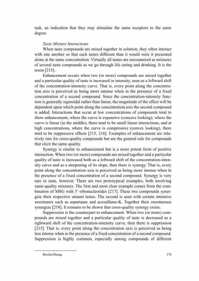
Breslin/Huang 178
task, an indication that they may stimulate the same receptors to the same
degree.
Taste Mixture InteractionsWhen taste compounds are mixed together in solution, they often interact
with one another so that each tastes different than it would were it presented
alone at the same concentration. Virtually all tastes are encountered as mixtures
of several taste compounds as we go through life eating and drinking. It is the
norm [215].
Enhancement occurs when two (or more) compounds are mixed together
and a particular quality of taste is increased in intensity, seen as a leftward shift
of the concentration-intensity curve. That is, every point along the concentra-
tion axis is perceived as being more intense when in the presence of a fixed
concentration of a second compound. Since the concentration-intensity func-
tion is generally sigmoidal rather than linear, the magnitude of the effect will be
dependent upon which point along the concentration axis the second compound
is added. Interactions that occur at low concentrations of compounds tend to
show enhancement, where the curve is expansive (concave looking); where the
curve is linear (in the middle), there tend to be small linear interactions, and at
high concentrations, where the curve is compressive (convex looking), there
tend to be suppressive effects [215, 216]. Examples of enhancement are rela-
tively rare for cross-quality compounds but are the general rule for compounds
that elicit the same quality.
Synergy is similar to enhancement but is a more potent form of positive
interaction. When two (or more) compounds are mixed together and a particular
quality of taste is increased both as a leftward shift of the concentration-inten-
sity curve and as a steepening of its slope, then there is synergy. That is, every
point along the concentration axis is perceived as being more intense when in
the presence of a fixed concentration of a second compound. Synergy is very
rare in taste, however. There are two prototypical examples, both involving
same-quality mixtures. The first and most clear example comes from the com-
bination of MSG with 5�-ribonucleotides [217]. These two compounds syner-
gize their respective umami tastes. The second is seen with certain intensive
sweeteners such as aspartame and acesulfame-K. Together their sweetnesses
synergize [218]. It remains to be shown that cross-quality synergy exists.
Suppression is the counterpart to enhancement. When two (or more) com-
pounds are mixed together and a particular quality of taste is decreased as a
rightward shift of the concentration-intensity curve, then there is suppression
[215]. That is, every point along the concentration axis is perceived as being
less intense when in the presence of a fixed concentration of a second compound.
Suppression is highly common, especially among compounds of different
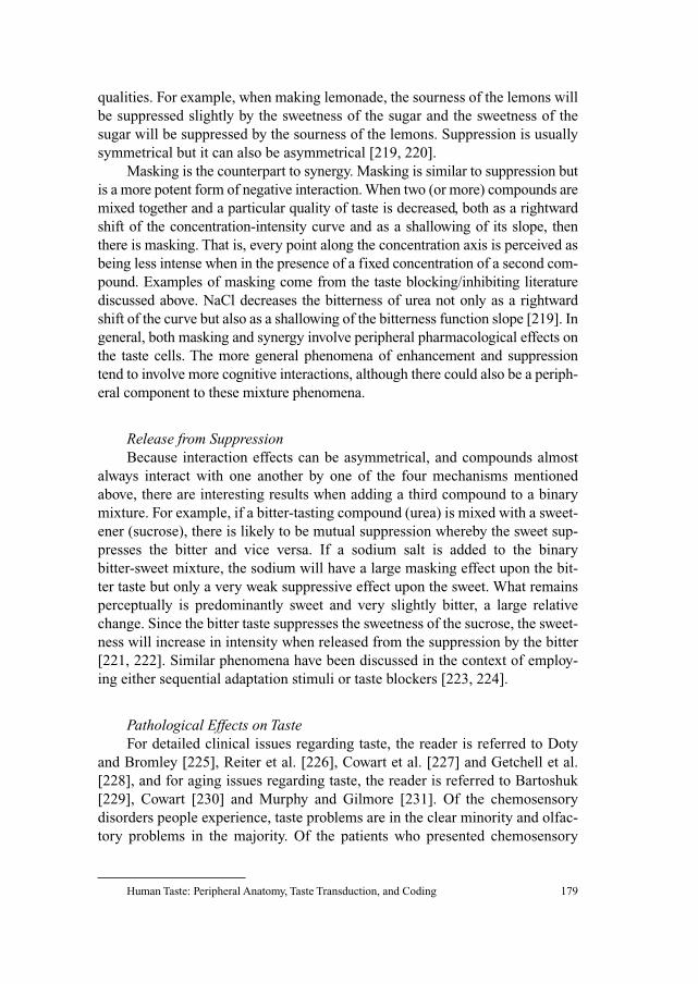
Human Taste: Peripheral Anatomy, Taste Transduction, and Coding 179
qualities. For example, when making lemonade, the sourness of the lemons will
be suppressed slightly by the sweetness of the sugar and the sweetness of the
sugar will be suppressed by the sourness of the lemons. Suppression is usually
symmetrical but it can also be asymmetrical [219, 220].
Masking is the counterpart to synergy. Masking is similar to suppression but
is a more potent form of negative interaction. When two (or more) compounds are
mixed together and a particular quality of taste is decreased, both as a rightward
shift of the concentration-intensity curve and as a shallowing of its slope, then
there is masking. That is, every point along the concentration axis is perceived as
being less intense when in the presence of a fixed concentration of a second com-
pound. Examples of masking come from the taste blocking/inhibiting literature
discussed above. NaCl decreases the bitterness of urea not only as a rightward
shift of the curve but also as a shallowing of the bitterness function slope [219]. In
general, both masking and synergy involve peripheral pharmacological effects on
the taste cells. The more general phenomena of enhancement and suppression
tend to involve more cognitive interactions, although there could also be a periph-
eral component to these mixture phenomena.
Release from SuppressionBecause interaction effects can be asymmetrical, and compounds almost
always interact with one another by one of the four mechanisms mentioned
above, there are interesting results when adding a third compound to a binary
mixture. For example, if a bitter-tasting compound (urea) is mixed with a sweet-
ener (sucrose), there is likely to be mutual suppression whereby the sweet sup-
presses the bitter and vice versa. If a sodium salt is added to the binary
bitter-sweet mixture, the sodium will have a large masking effect upon the bit-
ter taste but only a very weak suppressive effect upon the sweet. What remains
perceptually is predominantly sweet and very slightly bitter, a large relative
change. Since the bitter taste suppresses the sweetness of the sucrose, the sweet-
ness will increase in intensity when released from the suppression by the bitter
[221, 222]. Similar phenomena have been discussed in the context of employ-
ing either sequential adaptation stimuli or taste blockers [223, 224].
Pathological Effects on TasteFor detailed clinical issues regarding taste, the reader is referred to Doty
and Bromley [225], Reiter et al. [226], Cowart et al. [227] and Getchell et al.
[228], and for aging issues regarding taste, the reader is referred to Bartoshuk
[229], Cowart [230] and Murphy and Gilmore [231]. Of the chemosensory
disorders people experience, taste problems are in the clear minority and olfac-
tory problems in the majority. Of the patients who presented chemosensory
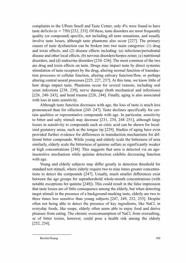
Breslin/Huang 180
complaints to the UPenn Smell and Taste Center, only 4% were found to have
taste deficits (n � 750) [232, 233]. Of these, taste disorders are most frequently
quality (or compound) specific, not including all taste sensations, and usually
involve taste losses, although taste phantoms also occur [227]. The primary
causes of taste dysfunction can be broken into two main categories: (1) drug
and toxin effects, and (2) disease effects including: (a) infections/periodontal
disease and other local effects, (b) nervous disorders/herpes zoster, (c) nutritional
disorders, and (d) endocrine disorders [234–236]. The most common of the two
are drug and toxin effects on taste. Drugs may impact taste by direct systemic
stimulation of taste receptors by the drug, altering normal function of transduc-
tion processes or cellular function, altering salivary function/flow, or perhaps
altering central neural processes [225, 227, 237]. At this time, we know little of
how drugs impact taste. Phantoms occur for several reasons, including oral
yeast infections [238, 239], nerve damage (both mechanical and infectious)
[226, 240–243], and head trauma [226, 244]. Finally, aging is also associated
with loss in taste sensitivity.
Although taste function decreases with age, the loss of taste is much less
pronounced than for olfaction [245–247]. Taste declines specifically for cer-
tain qualities or representative compounds with age. In particular, sensitivity
to bitter and salty stimuli may decrease [231, 234, 248–251], although large
losses in sensitivity to compounds such as citric acid can be shown for local-
ized gustatory areas, such as the tongue tip [229]. Studies of aging have even
provided further evidence for differences in transduction mechanisms for dif-
ferent bitter compounds. While young and elderly scale the bitterness of urea
similarly, elderly scale the bitterness of quinine sulfate as significantly weaker
at high concentrations [248]. This suggests that urea is detected via an age-
insensitive mechanism while quinine detection exhibits decreasing function
with age.
Young and elderly subjects may differ greatly in detection threshold for
standard test stimuli, where elderly require two to nine times greater concentra-
tions to detect the compounds [247]. Usually, much smaller differences exist
between the age groups for suprathreshold whole-mouth concentrations (with
notable exceptions for quinine [248]). This could result in the false impression
that taste losses are of little consequence among the elderly, but when detecting
target stimuli in the presence of a background masking taste, elderly are two to
three times less sensitive than young subjects [247, 249, 252, 253]. Despite
often not being able to detect the presence of key ingredients, like NaCl, in
everyday foods, like soups, elderly often seem able to enjoy food and derive
pleasure from eating. The chronic overconsumption of NaCl, from oversalting,
or of bitter toxins, however, could pose a health risk among the elderly
[252, 254].
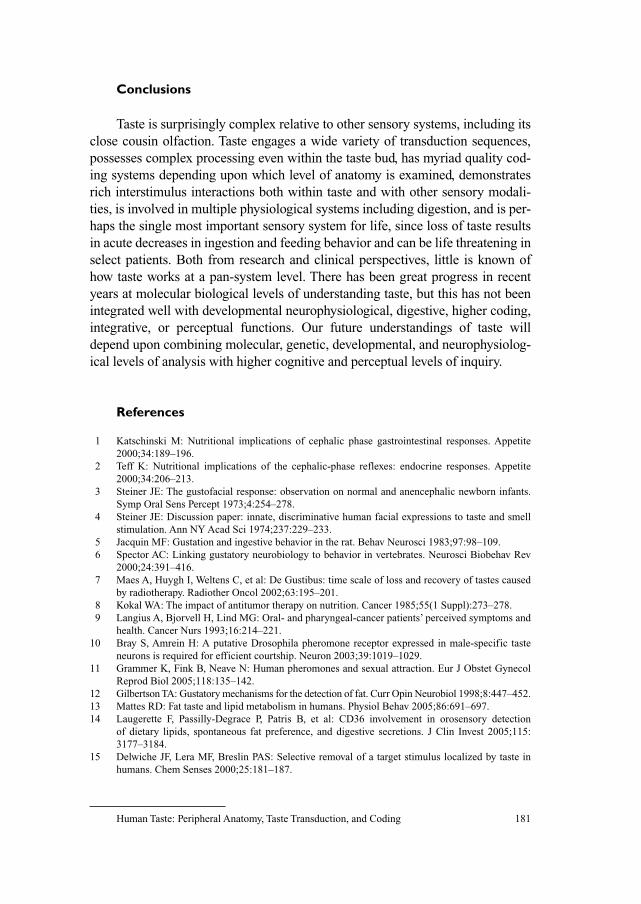
Human Taste: Peripheral Anatomy, Taste Transduction, and Coding 181
Conclusions
Taste is surprisingly complex relative to other sensory systems, including its
close cousin olfaction. Taste engages a wide variety of transduction sequences,
possesses complex processing even within the taste bud, has myriad quality cod-
ing systems depending upon which level of anatomy is examined, demonstrates
rich interstimulus interactions both within taste and with other sensory modali-
ties, is involved in multiple physiological systems including digestion, and is per-
haps the single most important sensory system for life, since loss of taste results
in acute decreases in ingestion and feeding behavior and can be life threatening in
select patients. Both from research and clinical perspectives, little is known of
how taste works at a pan-system level. There has been great progress in recent
years at molecular biological levels of understanding taste, but this has not been
integrated well with developmental neurophysiological, digestive, higher coding,
integrative, or perceptual functions. Our future understandings of taste will
depend upon combining molecular, genetic, developmental, and neurophysiolog-
ical levels of analysis with higher cognitive and perceptual levels of inquiry.
References
1 Katschinski M: Nutritional implications of cephalic phase gastrointestinal responses. Appetite
2000;34:189–196.
2 Teff K: Nutritional implications of the cephalic-phase reflexes: endocrine responses. Appetite
2000;34:206–213.
3 Steiner JE: The gustofacial response: observation on normal and anencephalic newborn infants.
Symp Oral Sens Percept 1973;4:254–278.
4 Steiner JE: Discussion paper: innate, discriminative human facial expressions to taste and smell
stimulation. Ann NY Acad Sci 1974;237:229–233.
5 Jacquin MF: Gustation and ingestive behavior in the rat. Behav Neurosci 1983;97:98–109.
6 Spector AC: Linking gustatory neurobiology to behavior in vertebrates. Neurosci Biobehav Rev
2000;24:391–416.
7 Maes A, Huygh I, Weltens C, et al: De Gustibus: time scale of loss and recovery of tastes caused
by radiotherapy. Radiother Oncol 2002;63:195–201.
8 Kokal WA: The impact of antitumor therapy on nutrition. Cancer 1985;55(1 Suppl):273–278.
9 Langius A, Bjorvell H, Lind MG: Oral- and pharyngeal-cancer patients’ perceived symptoms and
health. Cancer Nurs 1993;16:214–221.
10 Bray S, Amrein H: A putative Drosophila pheromone receptor expressed in male-specific taste
neurons is required for efficient courtship. Neuron 2003;39:1019–1029.
11 Grammer K, Fink B, Neave N: Human pheromones and sexual attraction. Eur J Obstet Gynecol
Reprod Biol 2005;118:135–142.
12 Gilbertson TA: Gustatory mechanisms for the detection of fat. Curr Opin Neurobiol 1998;8:447–452.
13 Mattes RD: Fat taste and lipid metabolism in humans. Physiol Behav 2005;86:691–697.
14 Laugerette F, Passilly-Degrace P, Patris B, et al: CD36 involvement in orosensory detection
of dietary lipids, spontaneous fat preference, and digestive secretions. J Clin Invest 2005;115:
3177–3184.
15 Delwiche JF, Lera MF, Breslin PAS: Selective removal of a target stimulus localized by taste in
humans. Chem Senses 2000;25:181–187.
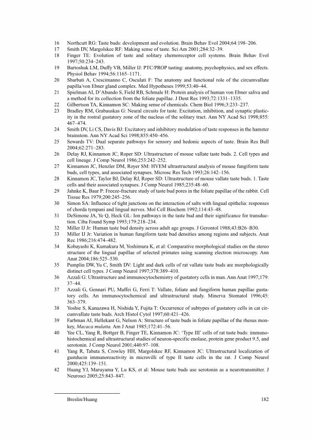
Breslin/Huang 182
16 Northcutt RG: Taste buds: development and evolution. Brain Behav Evol 2004;64:198–206.
17 Smith DV, Margolskee RF: Making sense of taste. Sci Am 2001;284:32–39.
18 Finger TE: Evolution of taste and solitary chemoreceptor cell systems. Brain Behav Evol
1997;50:234–243.
19 Bartoshuk LM, Duffy VB, Miller IJ: PTC/PROP tasting: anatomy, psychophysics, and sex effects.
Physiol Behav 1994;56:1165–1171.
20 Sbarbati A, Crescimanno C, Osculati F: The anatomy and functional role of the circumvallate
papilla/von Ebner gland complex. Med Hypotheses 1999;53:40–44.
21 Spielman AI, D’Abundo S, Field RB, Schmale H: Protein analysis of human von Ebner saliva and
a method for its collection from the foliate papillae. J Dent Res 1993;72:1331–1335.
22 Gilbertson TA, Kinnamon SC: Making sense of chemicals. Chem Biol 1996;3:233–237.
23 Bradley RM, Grabauskas G: Neural circuits for taste. Excitation, inhibition, and synaptic plastic-
ity in the rostral gustatory zone of the nucleus of the solitary tract. Ann NY Acad Sci 1998;855:
467–474.
24 Smith DV, Li CS, Davis BJ: Excitatory and inhibitory modulation of taste responses in the hamster
brainstem. Ann NY Acad Sci 1998;855:450–456.
25 Sewards TV: Dual separate pathways for sensory and hedonic aspects of taste. Brain Res Bull
2004;62:271–283.
26 Delay RJ, Kinnamon JC, Roper SD: Ultrastructure of mouse vallate taste buds. 2. Cell types and
cell lineage. J Comp Neurol 1986;253:242–252.
27 Kinnamon JC, Henzler DM, Royer SM: HVEM ultrastructural analysis of mouse fungiform taste
buds, cell types, and associated synapses. Microsc Res Tech 1993;26:142–156.
28 Kinnamon JC, Taylor BJ, Delay RJ, Roper SD: Ultrastructure of mouse vallate taste buds. 1. Taste
cells and their associated synapses. J Comp Neurol 1985;235:48–60.
29 Jahnke K, Baur P: Freeze-fracture study of taste bud pores in the foliate papillae of the rabbit. Cell
Tissue Res 1979;200:245–256.
30 Simon SA: Influence of tight junctions on the interaction of salts with lingual epithelia: responses
of chorda tympani and lingual nerves. Mol Cell Biochem 1992;114:43–48.
31 DeSimone JA, Ye Q, Heck GL: Ion pathways in the taste bud and their significance for transduc-
tion. Ciba Found Symp 1993;179:218–234.
32 Miller IJ Jr: Human taste bud density across adult age groups. J Gerontol 1988;43:B26–B30.
33 Miller IJ Jr: Variation in human fungiform taste bud densities among regions and subjects. Anat
Rec 1986;216:474–482.
34 Kobayashi K, Kumakura M, Yoshimura K, et al: Comparative morphological studies on the stereo
structure of the lingual papillae of selected primates using scanning electron microscopy. Ann
Anat 2004;186:525–530.
35 Pumplin DW, Yu C, Smith DV: Light and dark cells of rat vallate taste buds are morphologically
distinct cell types. J Comp Neurol 1997;378:389–410.
36 Azzali G: Ultrastructure and immunocytochemistry of gustatory cells in man. Ann Anat 1997;179:
37–44.
37 Azzali G, Gennari PU, Maffei G, Ferri T: Vallate, foliate and fungiform human papillae gusta-
tory cells. An immunocytochemical and ultrastructural study. Minerva Stomatol 1996;45:
363–379.
38 Yoshie S, Kanazawa H, Nishida Y, Fujita T: Occurrence of subtypes of gustatory cells in cat cir-
cumvallate taste buds. Arch Histol Cytol 1997;60:421–426.
39 Farbman AI, Hellekant G, Nelson A: Structure of taste buds in foliate papillae of the rhesus mon-
key, Macaca mulatta. Am J Anat 1985;172:41–56.
40 Yee CL, Yang R, Bottger B, Finger TE, Kinnamon JC: ‘Type III’ cells of rat taste buds: immuno-
histochemical and ultrastructural studies of neuron-specific enolase, protein gene product 9.5, and
serotonin. J Comp Neurol 2001;440:97–108.
41 Yang R, Tabata S, Crowley HH, Margolskee RF, Kinnamon JC: Ultrastructural localization of
gustducin immunoreactivity in microvilli of type II taste cells in the rat. J Comp Neurol
2000;425:139–151.
42 Huang YJ, Maruyama Y, Lu KS, et al: Mouse taste buds use serotonin as a neurotransmitter. J
Neurosci 2005;25:843–847.
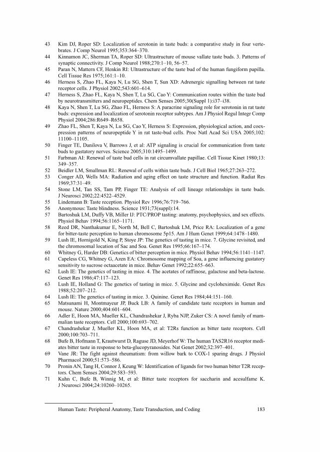
Human Taste: Peripheral Anatomy, Taste Transduction, and Coding 183
43 Kim DJ, Roper SD: Localization of serotonin in taste buds: a comparative study in four verte-
brates. J Comp Neurol 1995;353:364–370.
44 Kinnamon JC, Sherman TA, Roper SD: Ultrastructure of mouse vallate taste buds. 3. Patterns of
synaptic connectivity. J Comp Neurol 1988;270:1–10, 56–57.
45 Paran N, Mattern CF, Henkin RI: Ultrastructure of the taste bud of the human fungiform papilla.
Cell Tissue Res 1975;161:1–10.
46 Herness S, Zhao FL, Kaya N, Lu SG, Shen T, Sun XD: Adrenergic signalling between rat taste
receptor cells. J Physiol 2002;543:601–614.
47 Herness S, Zhao FL, Kaya N, Shen T, Lu SG, Cao Y: Communication routes within the taste bud
by neurotransmitters and neuropeptides. Chem Senses 2005;30(Suppl 1):i37–i38.
48 Kaya N, Shen T, Lu SG, Zhao FL, Herness S: A paracrine signaling role for serotonin in rat taste
buds: expression and localization of serotonin receptor subtypes. Am J Physiol Regul Integr Comp
Physiol 2004;286:R649–R658.
49 Zhao FL, Shen T, Kaya N, Lu SG, Cao Y, Herness S: Expression, physiological action, and coex-
pression patterns of neuropeptide Y in rat taste-bud cells. Proc Natl Acad Sci USA 2005;102:
11100–11105.
50 Finger TE, Danilova V, Barrows J, et al: ATP signaling is crucial for communication from taste
buds to gustatory nerves. Science 2005;310:1495–1499.
51 Farbman AI: Renewal of taste bud cells in rat circumvallate papillae. Cell Tissue Kinet 1980;13:
349–357.
52 Beidler LM, Smallman RL: Renewal of cells within taste buds. J Cell Biol 1965;27:263–272.
53 Conger AD, Wells MA: Radiation and aging effect on taste structure and function. Radiat Res
1969;37:31–49.
54 Stone LM, Tan SS, Tam PP, Finger TE: Analysis of cell lineage relationships in taste buds.
J Neurosci 2002;22:4522–4529.
55 Lindemann B: Taste reception. Physiol Rev 1996;76:719–766.
56 Anonymous: Taste blindness. Science 1931;73(suppl):14.
57 Bartoshuk LM, Duffy VB, Miller IJ: PTC/PROP tasting: anatomy, psychophysics, and sex effects.
Physiol Behav 1994;56:1165–1171.
58 Reed DR, Nanthakumar E, North M, Bell C, Bartoshuk LM, Price RA: Localization of a gene
for bitter-taste perception to human chromosome 5p15. Am J Hum Genet 1999;64:1478–1480.
59 Lush IE, Hornigold N, King P, Stoye JP: The genetics of tasting in mice. 7. Glycine revisited, and
the chromosomal location of Sac and Soa. Genet Res 1995;66:167–174.
60 Whitney G, Harder DB: Genetics of bitter perception in mice. Physiol Behav 1994;56:1141–1147.
61 Capeless CG, Whitney G, Azen EA: Chromosome mapping of Soa, a gene influencing gustatory
sensitivity to sucrose octaacetate in mice. Behav Genet 1992;22:655–663.
62 Lush IE: The genetics of tasting in mice. 4. The acetates of raffinose, galactose and beta-lactose.
Genet Res 1986;47:117–123.
63 Lush IE, Holland G: The genetics of tasting in mice. 5. Glycine and cycloheximide. Genet Res
1988;52:207–212.
64 Lush IE: The genetics of tasting in mice. 3. Quinine. Genet Res 1984;44:151–160.
65 Matsunami H, Montmayeur JP, Buck LB: A family of candidate taste receptors in human and
mouse. Nature 2000;404:601–604.
66 Adler E, Hoon MA, Mueller KL, Chandrashekar J, Ryba NJP, Zuker CS: A novel family of mam-
malian taste receptors. Cell 2000;100:693–702.
67 Chandrashekar J, Mueller KL, Hoon MA, et al: T2Rs function as bitter taste receptors. Cell
2000;100:703–711.
68 Bufe B, Hofmann T, Krautwurst D, Raguse JD, Meyerhof W: The human TAS2R16 receptor medi-
ates bitter taste in response to beta-glucopyranosides. Nat Genet 2002;32:397–401.
69 Vane JR: The fight against rheumatism: from willow bark to COX-1 sparing drugs. J Physiol
Pharmacol 2000;51:573–586.
70 Pronin AN, Tang H, Connor J, Keung W: Identification of ligands for two human bitter T2R recep-
tors. Chem Senses 2004;29:583–593.
71 Kuhn C, Bufe B, Winnig M, et al: Bitter taste receptors for saccharin and acesulfame K.
J Neurosci 2004;24:10260–10265.
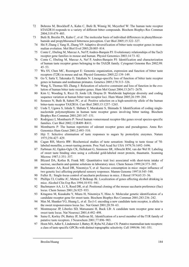
Breslin/Huang 184
72 Behrens M, Brockhoff A, Kuhn C, Bufe B, Winnig M, Meyerhof W: The human taste receptor
hTAS2R14 responds to a variety of different bitter compounds. Biochem Biophys Res Commun
2004;319:479–485.
73 Bufe B, Breslin PA, Kuhn C, et al: The molecular basis of individual differences in phenylthiocar-
bamide and propylthiouracil bitterness perception. Curr Biol 2005;15:322–327.
74 Shi P, Zhang J, Yang H, Zhang YP: Adaptive diversification of bitter taste receptor genes in mam-
malian evolution. Mol Biol Evol 2003;20:805–814.
75 Conte C, Ebeling M, Marcuz A, Nef P, Andres-Barquin PJ: Evolutionary relationships of the Tas2r
receptor gene families in mouse and human. Physiol Genomics 2003;14:73–82.
76 Conte C, Ebeling M, Marcuz A, Nef P, Andres-Barquin PJ: Identification and characterization
of human taste receptor genes belonging to the TAS2R family. Cytogenet Genome Res 2002;98:
45–53.
77 Wu SV, Chen MC, Rozengurt E: Genomic organization, expression and function of bitter taste
receptors (T2R) in mouse and rat. Physiol Genomics 2005;22:139–149.
78 Go Y, Satta Y, Takenaka O, Takahata N: Lineage-specific loss of function of bitter taste receptor
genes in humans and nonhuman primates. Genetics 2005;170:313–326.
79 Wang X, Thomas SD, Zhang J: Relaxation of selective constraint and loss of function in the evo-
lution of human bitter taste receptor genes. Hum Mol Genet 2004;13:2671–2678.
80 Kim U, Wooding S, Ricci D, Jorde LB, Drayna D: Worldwide haplotype diversity and coding
sequence variation at human bitter taste receptor loci. Hum Mutat 2005;26:199–204.
81 Soranzo N, Bufe B, Sabeti PC, et al: Positive selection on a high-sensitivity allele of the human
bitter-taste receptor TAS2R16. Curr Biol 2005;15:1257–1265.
82 Ueda T, Ugawa S, Ishida Y, Shibata Y, Murakami S, Shimada S: Identification of coding single-
nucleotide polymorphisms in human taste receptor genes involving bitter tasting. Biochem
Biophys Res Commun 2001;285:147–151.
83 Rodriguez I, Mombaerts P: Novel human vomeronasal receptor-like genes reveal species-specific
families. Curr Biol 2002;12:R409–R411.
84 Mombaerts P: The human repertoire of odorant receptor genes and pseudogenes. Annu Rev
Genomics Hum Genet 2001;2:493–510.
85 Hiji T: Selective elimination of taste responses to sugars by proteolytic enzymes. Nature
1975;256:427–429.
86 Cagan RH, Morris RW: Biochemical studies of taste sensation: binding to taste tissue of 3H-
labeled monellin, a sweet-tasting protein. Proc Natl Acad Sci USA 1979;76:1692–1696.
87 Farbman AI, Ogden-Ogle CK, Hellekant G, Simmons SR, Albrecht RM, van der Wel H: Labeling
of sweet taste binding sites using a colloidal gold-labeled sweet protein, thaumatin. Scanning
Microsc 1987;1:351–357.
88 Blizard DA, Kotlus B, Frank ME: Quantitative trait loci associated with short-term intake of
sucrose, saccharin and quinine solutions in laboratory mice. Chem Senses 1999;24:373–385.
89 Bachmanov AA, Reed DR, Ninomiya Y, et al: Sucrose consumption in mice: major influence of
two genetic loci affecting peripheral sensory responses. Mamm Genome 1997;8:545–548.
90 Fuller JL: Single-locus control of saccharin preference in mice. J Hered 1974;65:33–36.
91 Phillips TJ, Crabbe JC, Metten P, Belknap JK: Localization of genes affecting alcohol drinking in
mice. Alcohol Clin Exp Res 1994;18:931–941.
92 Bachmanov AA, Li X, Reed DR, et al: Positional cloning of the mouse saccharin preference (Sac)
locus. Chem Senses 2001;26:925–933.
93 Kitagawa M, Kusakabe Y, Miura H, Ninomiya Y, Hino A: Molecular genetic identification of a
candidate receptor gene for sweet taste. Biochem Biophys Res Commun 2001;283:236–242.
94 Max M, Shanker YG, Huang L, et al: Tas1r3, encoding a new candidate taste receptor, is allelic to
the sweet responsiveness locus Sac. Nat Genet 2001;28:58–63.
95 Montmayeur JP, Liberles SD, Matsunami H, Buck LB: A candidate taste receptor gene near a
sweet taste locus. Nat Neurosci 2001;4:492–498.
96 Sainz E, Korley JN, Battey JF, Sullivan SL: Identification of a novel member of the T1R family of
putative taste receptors. J Neurochem 2001;77:896–903.
97 Hoon MA, Adler E, Lindemeier J, Battey JF, Ryba NJ, Zuker CS: Putative mammalian taste receptors:
a class of taste-specific GPCRs with distinct topographic selectivity. Cell 1999;96: 541–551.
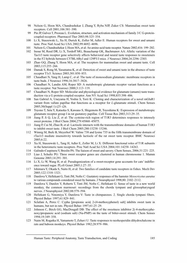
Human Taste: Peripheral Anatomy, Taste Transduction, and Coding 185
98 Nelson G, Hoon MA, Chandrashekar J, Zhang Y, Ryba NJP, Zuker CS: Mammalian sweet taste
receptors. Cell 2001;106:381–390.
99 Pin JP, Galvez T, Prezeau L: Evolution, structure, and activation mechanism of family 3/C G-protein-
coupled receptors. Pharmacol Ther 2003;98:325–354.
100 Li X, Staszewski L, Xu H, Durick K, Zoller M, Adler E: Human receptors for sweet and umami
taste. Proc Natl Acad Sci USA 2002;99:4692–4696.
101 Nelson G, Chandrashekar J, Hoon MA, et al: An amino-acid taste receptor. Nature 2002;416: 199–202.
102 Inoue M, Reed DR, Li X, Tordoff MG, Beauchamp GK, Bachmanov AA: Allelic variation of the
Tas1r3 taste receptor gene selectively affects behavioral and neural taste responses to sweeteners
in the F2 hybrids between C57BL/6ByJ and 129P3/J mice. J Neurosci 2004;24:2296–2303.
103 Zhao GQ, Zhang Y, Hoon MA, et al: The receptors for mammalian sweet and umami taste. Cell
2003;115:255–266.
104 Damak S, Rong M, Yasumatsu K, et al: Detection of sweet and umami taste in the absence of taste
receptor T1r3. Science 2003;301:850–853.
105 Chaudhari N, Yang H, Lamp C, et al: The taste of monosodium glutamate: membrane receptors in
taste buds. J Neurosci 1996;16:3817–3826.
106 Chaudhari N, Landin AM, Roper SD: A metabotropic glutamate receptor variant functions as a
taste receptor. Nat Neurosci 2000;3:113–119.
107 Chaudhari N, Roper SD: Molecular and physiological evidence for glutamate (umami) taste trans-
duction via a G protein-coupled receptor. Ann NY Acad Sci 1998;855:398–406.
108 San Gabriel A, Uneyama H, Yoshie S, Torii K: Cloning and characterization of a novel mGluR1
variant from vallate papillae that functions as a receptor for L-glutamate stimuli. Chem Senses
2005;30(Suppl 1):i25–i26.
109 Toyono T, Seta Y, Kataoka S, Kawano S, Shigemoto R, Toyoshima K: Expression of metabotropic
glutamate receptor group I in rat gustatory papillae. Cell Tissue Res 2003;313:29–35.
110 Jiang P, Ji Q, Liu Z, et al: The cysteine-rich region of T1R3 determines responses to intensely
sweet proteins. J Biol Chem 2004;279:45068–45075.
111 Jiang P, Cui M, Zhao B, et al: Lactisole interacts with the transmembrane domains of human T1R3
to inhibit sweet taste. J Biol Chem 2005;280:15238–15246.
112 Winnig M, Bufe B, Meyerhof W: Valine 738 and lysine 735 in the fifth transmembrane domain of
rTas1r3 mediate insensitivity towards lactisole of the rat sweet taste receptor. BMC Neurosci
2005;6:22.
113 Xu H, Staszewski L, Tang H, Adler E, Zoller M, Li X: Different functional roles of T1R subunits
in the heteromeric taste receptors. Proc Natl Acad Sci USA 2004;101:14258–14263.
114 Galindo-Cuspinera V, Breslin PA: The liaison of sweet and savory. Chem Senses, 2006;31:221–225.
115 Liao J, Schultz PG: Three sweet receptor genes are clustered in human chromosome 1. Mamm
Genome 2003;14:291–301.
116 Li X, Li W, Wang H, et al: Pseudogenization of a sweet-receptor gene accounts for cats’ indiffer-
ence toward sugar. PLoS Genet 2005;1:27–35.
117 Ishimaru Y, Okada S, Naito H, et al: Two families of candidate taste receptors in fishes. Mech Dev
2005;122:1310–1321.
118 Danilova V, Hellekant G, Tinti JM, Nofre C: Gustatory responses of the hamster Mesocricetus auratusto various compounds considered sweet by humans. J Neurophysiol 1998;80: 2102–2112.
119 Danilova V, Danilov Y, Roberts T, Tinti JM, Nofre C, Hellekant G: Sense of taste in a new world
monkey, the common marmoset: recordings from the chorda tympani and glossopharyngeal
nerves. J Neurophysiol 2002;88:579–594.
120 Hellekant G, Ninomiya Y, Danilova V: Taste in chimpanzees. 2. Single chorda tympani fibers.
Physiol Behav 1997;61:829–841.
121 Sclafani A, Perez C: Cypha [propionic acid, 2-(4-methoxyphenol) salt] inhibits sweet taste in
humans, but not in rats. Physiol Behav 1997;61:25–29.
122 Johnson C, Birch GG, MacDougall DB: The effect of the sweetness inhibitor 2(-4-methoxyphe-
noxy)propanoic acid (sodium salt) (Na-PMP) on the taste of bitter-sweet stimuli. Chem Senses
1994;19:349–358.
123 Naim M, Rogatka H, Yamamoto T, Zehavi U: Taste responses to neohesperidin dihydrochalcone in
rats and baboon monkeys. Physiol Behav 1982;28:979–986.
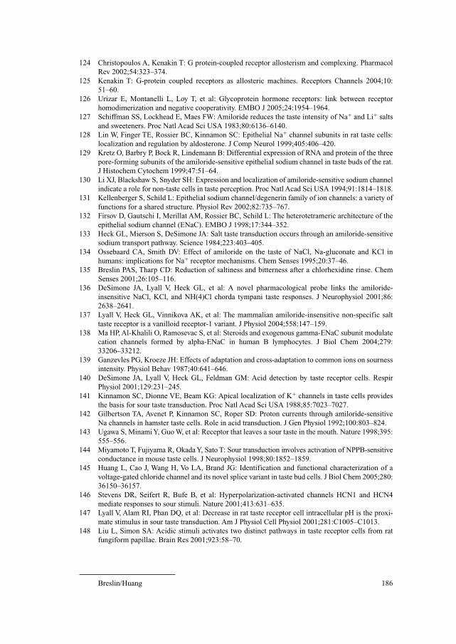
Breslin/Huang 186
124 Christopoulos A, Kenakin T: G protein-coupled receptor allosterism and complexing. Pharmacol
Rev 2002;54:323–374.
125 Kenakin T: G-protein coupled receptors as allosteric machines. Receptors Channels 2004;10:
51–60.
126 Urizar E, Montanelli L, Loy T, et al: Glycoprotein hormone receptors: link between receptor
homodimerization and negative cooperativity. EMBO J 2005;24:1954–1964.
127 Schiffman SS, Lockhead E, Maes FW: Amiloride reduces the taste intensity of Na� and Li� salts
and sweeteners. Proc Natl Acad Sci USA 1983;80:6136–6140.
128 Lin W, Finger TE, Rossier BC, Kinnamon SC: Epithelial Na� channel subunits in rat taste cells:
localization and regulation by aldosterone. J Comp Neurol 1999;405:406–420.
129 Kretz O, Barbry P, Bock R, Lindemann B: Differential expression of RNA and protein of the three
pore-forming subunits of the amiloride-sensitive epithelial sodium channel in taste buds of the rat.
J Histochem Cytochem 1999;47:51–64.
130 Li XJ, Blackshaw S, Snyder SH: Expression and localization of amiloride-sensitive sodium channel
indicate a role for non-taste cells in taste perception. Proc Natl Acad Sci USA 1994;91:1814–1818.
131 Kellenberger S, Schild L: Epithelial sodium channel/degenerin family of ion channels: a variety of
functions for a shared structure. Physiol Rev 2002;82:735–767.
132 Firsov D, Gautschi I, Merillat AM, Rossier BC, Schild L: The heterotetrameric architecture of the
epithelial sodium channel (ENaC). EMBO J 1998;17:344–352.
133 Heck GL, Mierson S, DeSimone JA: Salt taste transduction occurs through an amiloride-sensitive
sodium transport pathway. Science 1984;223:403–405.
134 Ossebaard CA, Smith DV: Effect of amiloride on the taste of NaCl, Na-gluconate and KCl in
humans: implications for Na� receptor mechanisms. Chem Senses 1995;20:37–46.
135 Breslin PAS, Tharp CD: Reduction of saltiness and bitterness after a chlorhexidine rinse. Chem
Senses 2001;26:105–116.
136 DeSimone JA, Lyall V, Heck GL, et al: A novel pharmacological probe links the amiloride-
insensitive NaCl, KCl, and NH(4)Cl chorda tympani taste responses. J Neurophysiol 2001;86:
2638–2641.
137 Lyall V, Heck GL, Vinnikova AK, et al: The mammalian amiloride-insensitive non-specific salt
taste receptor is a vanilloid receptor-1 variant. J Physiol 2004;558:147–159.
138 Ma HP, Al-Khalili O, Ramosevac S, et al: Steroids and exogenous gamma-ENaC subunit modulate
cation channels formed by alpha-ENaC in human B lymphocytes. J Biol Chem 2004;279:
33206–33212.
139 Ganzevles PG, Kroeze JH: Effects of adaptation and cross-adaptation to common ions on sourness
intensity. Physiol Behav 1987;40:641–646.
140 DeSimone JA, Lyall V, Heck GL, Feldman GM: Acid detection by taste receptor cells. Respir
Physiol 2001;129:231–245.
141 Kinnamon SC, Dionne VE, Beam KG: Apical localization of K� channels in taste cells provides
the basis for sour taste transduction. Proc Natl Acad Sci USA 1988;85:7023–7027.
142 Gilbertson TA, Avenet P, Kinnamon SC, Roper SD: Proton currents through amiloride-sensitive
Na channels in hamster taste cells. Role in acid transduction. J Gen Physiol 1992;100:803–824.
143 Ugawa S, Minami Y, Guo W, et al: Receptor that leaves a sour taste in the mouth. Nature 1998;395:
555–556.
144 Miyamoto T, Fujiyama R, Okada Y, Sato T: Sour transduction involves activation of NPPB-sensitive
conductance in mouse taste cells. J Neurophysiol 1998;80:1852–1859.
145 Huang L, Cao J, Wang H, Vo LA, Brand JG: Identification and functional characterization of a
voltage-gated chloride channel and its novel splice variant in taste bud cells. J Biol Chem 2005;280:
36150–36157.
146 Stevens DR, Seifert R, Bufe B, et al: Hyperpolarization-activated channels HCN1 and HCN4
mediate responses to sour stimuli. Nature 2001;413:631–635.
147 Lyall V, Alam RI, Phan DQ, et al: Decrease in rat taste receptor cell intracellular pH is the proxi-
mate stimulus in sour taste transduction. Am J Physiol Cell Physiol 2001;281:C1005–C1013.
148 Liu L, Simon SA: Acidic stimuli activates two distinct pathways in taste receptor cells from rat
fungiform papillae. Brain Res 2001;923:58–70.
Human Taste: Peripheral Anatomy, Taste Transduction, and Coding 187
149 Lin W, Ogura T, Kinnamon SC: Acid-activated cation currents in rat vallate taste receptor cells.
J Neurophysiol 2002;88:133–141.
150 Richter TA, Dvoryanchikov GA, Roper SD, Chaudhari N: Acid-sensing ion channel-2 is not
necessary for sour taste in mice. J Neurosci 2004;24:4088–4091.
151 Breslin PA: Human gustation; in Finger TE, Restrepo D, Silver WL (eds): The Neurobiology of
Taste and Smell. New York, Wiley-Liss, 2000, pp 423–461.
152 McLaughlin SK, McKinnon PJ, Margolskee RF: Gustducin is a taste-cell-specific G protein
closely related to the transducins. Nature 1992;357:563–569.
153 Ruiz-Avila L, McLaughlin SK, Wildman D, et al: Coupling of bitter receptor to phosphodiesterase
through transducin in taste receptor cells. Nature 1995;376:80–85.
154 Kusakabe Y, Yasuoka A, Asano-Miyoshi M, et al: Comprehensive study on G protein �-subunits in
taste bud cells, with special reference to the occurrence of G�i2 as a major G� species. Chem
Senses 2000;25:525–531.
155 Ming D, Ruiz-Avila L, Margolskee RF: Characterization and solubilization of bitter-responsive
receptors that couple to gustducin. Proc Natl Acad Sci USA 1998;95:8933–8938.
156 Wong GT, Gannon KS, Margolskee RF: Transduction of bitter and sweet taste by gustducin.
Nature 1996;381:796–800.
157 He W, Yasumatsu K, Varadarajan V, et al: Umami taste responses are mediated by �-transducin and
�-gustducin. J Neurosci 2004;24:7674–7680.
158 Rossler P, Boekhoff I, Tareilus E, Beck S, Breer H, Freitag J: G protein �� complexes in circum-
vallate taste cells involved in bitter transduction. Chem Senses 2000;25:413–421.
159 Huang L, Shanker YG, Dubauskaite J, et al: G�13 colocalizes with gustducin in taste receptor cells
and mediates IP3 responses to bitter denatonium. Nat Neurosci 1999;2:1055–1062.
160 Rossler P, Kroner C, Freitag J, Noe J, Breer H: Identification of a phospholipase C � subtype in rat
taste cells. Eur J Cell Biol 1998;77:253–261.
161 Clapp TR, Stone LM, Margolskee RF, Kinnamon SC: Immunocytochemical evidence for
co-expression of type III IP3 receptor with signaling components of bitter taste transduction.
BMC Neurosci 2001;2:6.
162 Perez CA, Huang L, Rong M, et al: A transient receptor potential channel expressed in taste recep-
tor cells. Nat Neurosci 2002;5:1169–1176.
163 Zhang Y, Hoon MA, Chandrashekar J, et al: Coding of sweet, bitter, and umami tastes: different
receptor cells sharing similar signaling pathways. Cell 2003;112:293–301.
164 Rosenzweig S, Yan W, Dasso M, Spielman AI: Possible novel mechanism for bitter taste mediated
through cGMP. J Neurophysiol 1999;81:1661–1665.
165 Prawitt D, Monteilh-Zoller MK, Brixel L, et al: TRPM5 is a transient Ca2�-activated cation
channel responding to rapid changes in [Ca2�]i. Proc Natl Acad Sci USA 2003;100:
15166–15171.
166 Liu D, Liman ER: Intracellular Ca2� and the phospholipid PIP2 regulate the taste transduction ion
channel TRPM5. Proc Natl Acad Sci USA 2003;100:15160–15165.
167 Hofmann T, Chubanov V, Gudermann T, Montell C: TRPM5 is a voltage-modulated and Ca2�-
activated monovalent selective cation channel. Curr Biol 2003;13:1153–1158.
168 Striem BJ, Pace U, Zehavi U, Naim M, Lancet D: Sweet tastants stimulate adenylate cyclase cou-
pled to GTP-binding protein in rat tongue membranes. Biochem J 1989;260:121–126.
169 Bigiani A, Roper SD: Identification of electrophysiologically distinct cell subpopulations in
Necturus taste buds. J Gen Physiol 1993;102:143–170.
170 Delay RJ, Kinnamon SC, Roper SD: Serotonin modulates voltage-dependent calcium current in
Necturus taste cells. J Neurophysiol 1997;77:2515–2524.
171 Herness S, Zhao FL, Lu SG, Kaya N, Shen T: Expression and physiological actions of cholecys-
tokinin in rat taste receptor cells. J Neurosci 2002;22:10018–10029.
172 Shen T, Kaya N, Zhao FL, Lu SG, Cao Y, Herness S: Co-expression patterns of the neuropeptides
vasoactive intestinal peptide and cholecystokinin with the transduction molecules �-gustducin and
T1R2 in rat taste receptor cells. Neuroscience 2005;130:229–238.
173 Ogura T: Acetylcholine increases intracellular Ca2� in taste cells via activation of muscarinic
receptors. J Neurophysiol 2002;87:2643–2649.

Breslin/Huang 188
174 Kataoka S, Toyono T, Seta Y, Ogura T, Toyoshima K: Expression of P2Y1 receptors in rat taste
buds. Histochem Cell Biol 2004;121:419–426.
175 Baryshnikov SG, Rogachevskaja OA, Kolesnikov SS: Calcium signaling mediated by P2Y recep-
tors in mouse taste cells. J Neurophysiol 2003;90:3283–3294.
176 Shigemura N, Miura H, Kusakabe Y, Hino A, Ninomiya Y: Expression of leptin receptor (Ob-R)
isoforms and signal transducers and activators of transcription (STATs) mRNAs in the mouse taste
buds. Arch Histol Cytol 2003;66:253–260.
177 Herness MS: Aldosterone increases the amiloride-sensitivity of the rat gustatory neural response
to NaCl. Comp Biochem Physiol Comp Physiol 1992;103:269–273.
178 Roper S: Regenerative impulses in taste cells. Science 1983;220:1311–1312.
179 Kashiwayanagi M, Miyake M, Kurihara K: Voltage-dependent Ca2� channel and Na� channel in
frog taste cells. Am J Physiol 1983;244:C82–C88.
180 Avenet P, Lindemann B: Noninvasive recording of receptor cell action potentials and sustained
currents from single taste buds maintained in the tongue: the response to mucosal NaCl and
amiloride. J Membr Biol 1991;124:33–41.
181 Cummings TA, Powell J, Kinnamon SC: Sweet taste transduction in hamster taste cells: evidence
for the role of cyclic nucleotides. J Neurophysiol 1993;70:2326–2336.
182 Chen Y, Sun XD, Herness S: Characteristics of action potentials and their underlying outward cur-
rents in rat taste receptor cells. J Neurophysiol 1996;75:820–831.
183 Chen Y, Herness MS: Electrophysiological actions of quinine on voltage-dependent currents in
dissociated rat taste cells. Pflugers Arch 1997;434:215–226.
184 Behe P, DeSimone JA, Avenet P, Lindemann B: Membrane currents in taste cells of the rat fungi-
form papilla. Evidence for two types of Ca currents and inhibition of K currents by saccharin. J
Gen Physiol 1990;96:1061–1084.
185 Herness MS, Sun XD: Voltage-dependent sodium currents recorded from dissociated rat taste
cells. J Membr Biol 1995;146:73–84.
186 Sun XD, Herness MS: Characterization of inwardly rectifying potassium currents from dissoci-
ated rat taste receptor cells. Am J Physiol 1996;271:C1221–C1232.
187 Noguchi T, Ikeda Y, Miyajima M, Yoshii K: Voltage-gated channels involved in taste responses and
characterizing taste bud cells in mouse soft palates. Brain Res 2003;982:241–259.
188 Medler KF, Margolskee RF, Kinnamon SC: Electrophysiological characterization of voltage-gated
currents in defined taste cell types of mice. J Neurosci 2003;23:2608–2617.
189 Varkevisser B, Peterson D, Ogura T, Kinnamon SC: Neural networks distinguish between taste
qualities based on receptor cell population responses. Chem Senses 2001;26:499–505.
190 Ghiaroni V, Fieni F, Pietra P, Bigiani A: Electrophysiological heterogeneity in a functional subset
of mouse taste cells during postnatal development. Chem Senses 2003;28:827–833.
191 Huang YJ, Maruyama Y, Lu KS, et al: Using biosensors to detect the release of serotonin from
taste buds during taste stimulation. Arch Ital Biol 2005;143:87–96.
192 Feigl H: The mental and the physical; in Feigl H, Scriven M, Maxwell G (eds): Concepts, Theories
and the Mind-Body Problem. Minneapolis, University of Minnesota Press, 1958, pp 370–497.
193 Mueller KL, Hoon MA, Erlenbach I, Chandrashekar J, Zuker CS, Ryba NJ: The receptors and
coding logic for bitter taste. Nature 2005;434:225–229.
194 Whitehead MC, Ganchrow JR, Ganchrow D, Yao B: Organization of geniculate and trigeminal
ganglion cells innervating single fungiform taste papillae: a study with tetramethylrhodamine dex-
tran amine labeling. Neuroscience 1999;93:931–941.
195 Hellekant G, Ninomiya Y: On the taste of umami in chimpanzee. Physiol Behav 1991;49:927–934.
196 Hellekant G, Ninomiya Y: Bitter taste in single chorda tympani taste fibers from chimpanzee.
Physiol Behav 1994;56:1185–1188.
197 Hellekant G, Ninomiya Y, Danilova V: Taste in chimpanzees. 3. Labeled-line coding in sweet taste.
Physiol Behav 1998;65:191–200.
198 Hellekant G, Ninomiya Y, DuBois GE, Danilova V, Roberts TW: Taste in chimpanzee. 1. The sum-
mated response to sweeteners and the effect of gymnemic acid. Physiol Behav 1996;60:469–479.
199 Smith DV, John SJ, Boughter JD: Neuronal cell types and taste quality coding. Physiol Behav
2000;69:77–85.
200 Smith DV, St John SJ: Neural coding of gustatory information. Curr Opin Neurobiol 1999;9: 427–435.
Human Taste: Peripheral Anatomy, Taste Transduction, and Coding 189
201 Robinson PP: The characteristics and regional distribution of afferent fibres in the chorda tympani
of the cat. J Physiol 1988;406:345–357.
202 Travers SP, Norgren R: Organization of orosensory responses in the nucleus of the solitary tract of
rat. J Neurophysiol 1995;73:2144–2162.
203 Smith-Swintosky VL, Plata-Salaman CR, Scott TR: Gustatory neural coding in the monkey cor-
tex: stimulus quality. J Neurophysiol 1991;66:1156–1165.
204 Stevens SS: Psychophysics: Introduction to Its Perceptual Neural and Social Prospects. New York,
Wiley, 1975.
205 Breslin PA, Beauchamp GK, Pugh EN Jr: Monogeusia for fructose, glucose, sucrose, and maltose.
Percept Psychophys 1996;58:327–341.
206 Holway AH, Hurvich LM: On the psychophysics of taste. J Exp Psychol 1938;23:191–198.
207 Stevens SS: Sensory scales of taste intensity. Percept Psychophys 1969;6:302–308.
208 Yamaguchi S: Fundamental properties of umami in human taste sensation; in Kawamura K, Kare
MR (eds): Umami: A Basic Taste. New York, Dekker, 1987, pp 41–73.
209 McBurney DH: Temporal properties of the human taste system. Sens Processes 1976;1:150–162.
210 Kroeze JH, Bartoshuk LM: Bitterness suppression as revealed by split-tongue taste stimulation in
humans. Physiol Behav 1985;35:779–783.
211 Smith DV, McBurney DH: Gustatory cross-adaptation: does a single mechanism code the salty
taste? J Exp Psychol 1969;80:101–105.
212 Smith DV, van der Klaauw NJ: The perception of saltiness is eliminated by NaCl adaptation:
implications for gustatory transduction and coding. Chem Senses 1995;20:545–557.
213 McBurney DH, Bartoshuk LM: Interactions between stimuli with different taste qualities. Physiol
Behav 1973;10:1101–1106.
214 Yokomukai Y, Cowart BJ, Beauchamp GK: Individual differences in sensitivity to bitter-tasting
substances. Chem Senses 1993;18:669–681.
215 Breslin PAS: Interactions among salty, sour and bitter compounds. Trends Food Sci Technol
1996;7:390–399.
216 Bartoshuk LM: Taste mixtures: is mixture suppression related to compression? Physiol Behav
1975;14:643–649.
217 Rivkin B, Bartoshuk LM: Taste synergism between monosodium glutamate and disodium 5�-guanylate. Physiol Behav 1980;24:1169–1172.
218 Lawless HT: Theoretical note: tests of synergy in sweetener mixtures. Chem Senses
1998;23:447–451.
219 Breslin PAS, Beauchamp GK: Suppression of bitterness by sodium: variation among bitter taste
stimuli. Chem Senses 1995;20:609–623.
220 Kamen JM, Pilgrim FJ, Gutman NJ, Kroll BJ: Interactions of suprathreshold taste stimuli. J Exp
Psychol 1961;62:348–356.
221 Breslin PAS, Beauchamp GK: Salt enhances flavour by suppressing bitterness. Nature 1997; 387:563.
222 Keast RS, Breslin PA: Bitterness suppression with zinc sulfate and na-cyclamate: a model of com-
bined peripheral and central neural approaches to flavor modification. Pharm Res 2005;22:
1970–1977.
223 Lawless H: Paradoxical adaptation to taste mixtures. Physiol Behav 1982;29:149–152.
224 Lawless HT: Evidence for neural inhibition in bittersweet taste mixtures. J Comp Physiol Psychol
1979;93:538–547.
225 Doty RL, Bromley SM: Effects of drugs on olfaction and taste. Otolaryngol Clin North Am
2004;37:1229–1254.
226 Reiter ER, DiNardo LJ, Costanzo RM: Effects of head injury on olfaction and taste. Otolaryngol
Clin North Am 2004;37:1167–1184.
227 Cowart BJ, Young IM, Feldman RS, Lowry LD: Clinical disorders of smell and taste; in Beauchamp
GK, Bartoshuk LM (eds): Tasting and Smelling. New York, Academic Press, 1997, pp 175–98.
228 Getchell TV, Doty RL, Bartoshuk LM, Snow JB Jr: Smell and Taste in Health and Disease. New
York, Raven Press, 1991.
229 Bartoshuk LM: Taste: robust across the age span? Ann NY Acad Sci 1989;561:65–75.
230 Cowart BJ: Development of taste perception in humans: sensitivity and preference throughout the
life span. Psychol Bull 1981;90:43–73.

Breslin/Huang 190
231 Murphy C, Gilmore MM: Quality-specific effects of aging on the human taste system. Percept
Psychophys 1989;45:121–128.
232 Deems DA, Doty RL, Settle RG, et al: Smell and taste disorders, a study of 750 patients from the
University of Pennsylvania Smell and Taste Center. Arch Otolaryngol Head Neck Surg 1991;117:
519–528.
233 Goodspeed RB, Gent JF, Catalanotto FA: Chemosensory dysfunction: clinical evaluation results
from a taste and smell clinic. Postgrad Med 1987;81:251–260.
234 Schiffman SS, Gatlin CA: Clinical physiology of taste and smell. Annu Rev Nutr 1993;13:405–436.
235 Wrobel BB, Leopold DA: Clinical assessment of patients with smell and taste disorders.
Otolaryngol Clin North Am 2004;37:1127–1142.
236 Wrobel BB, Leopold DA: Smell and taste disorders. Facial Plast Surg Clin North Am 2004;12:
459–468, vii.
237 Ackerman BH, Kasbekar N: Disturbances of taste and smell induced by drugs. Pharmacotherapy
1997;17:482–496.
238 Brightman VJ, Guggenheimer J: Changes in the oral microbial flora during treatment of recurrent
aphthous ulcers (abstract). J Dent Res 1968;47(suppl):126.
239 Osaki T, Ohshima M, Tomita Y, Matsugi N, Nomura Y: Clinical and physiological investigations in
patients with taste abnormality. J Oral Pathol Med 1996;25:38–43.
240 Blackburn CW, Bramley PA: Lingual nerve damage associated with the removal of lower third
molars. Br Dent J 1989;167:103–107.
241 Grant R, Miller S, Simpson D, Lamey PJ, Bone I: The effect of chorda tympani section on ipsilat-
eral and contralateral salivary secretion and taste in man. J Neurol Neurosurg Psychiatry
1989;52:1058–1062.
242 Kveton JF, Bartoshuk LM: The effect of unilateral chorda tympani damage on taste. Laryngoscope
1994;104:25–29.
243 Yanagisawa K, Bartoshuk LM, Catalanotto FA, Karrer TA, Kveton JF: Anesthesia of the chorda
tympani nerve and taste phantoms. Physiol Behav 1998;63:329–335.
244 Costanzo RM, Zassler ZD: Head trauma; in Getchell TV, Doty RL, Bartoshuk LM, Snow JB Jr
(eds): Smell and Taste in Health and Disease. New York, Raven Press, 1991, pp 711–730.
245 Cowart BJ: Relationships between taste and smell across the adult life span. Ann NY Acad Sci
1989;561:39–55.
246 Stevens JC, Bartoshuk LM, Cain WS: Chemical senses and aging: taste versus smell. Chem
Senses 1984;9:167–179.
247 Stevens JC, Cain WS: Changes in taste and flavor in aging. Crit Rev Food Sci Nutr 1993;33:27–37.
248 Cowart BJ, Yokomukai Y, Beauchamp GK: Bitter taste in aging: compound-specific decline in
sensitivity. Physiol Behav 1994;6:1237–1241.
249 Stevens JC: Detection of tastes in mixture with other tastes: issues of masking and aging. Chem
Senses 1996;21:211–221.
250 Weiffenbach JM, Baum BJ, Burghauser R: Taste thresholds: quality specific variation with human
aging. J Gerontol 1982;37:372–377.
251 Weiffenbach JM, Cowart BJ, Baum BJ: Taste intensity perception in aging. J Gerontol 1986;41:
460–468.
252 Stevens JC, Cain WS, Demarque A, Ruthruff AM: On the discrimination of missing ingredients:
aging and salt flavor. Appetite 1991;16:129–140.
253 Stevens JC, Traverzo A: Detection of a target taste in a complex masker. Chem Senses 1997;22:
529–534.
254 Stevens JC, Cruz LA, Hoffman JM, Patterson MQ: Taste sensitivity and aging: high incidence of
decline revealed by repeated threshold measures. Chem Senses 1995;20:451–459.
Paul A.S. Breslin
Monell Chemical Senses Center
3500 Market Street
Philadelphia, PA 19104 (USA)
Tel. �1 215 898 5021, Fax �1 215 898 2084, E-Mail [email protected]
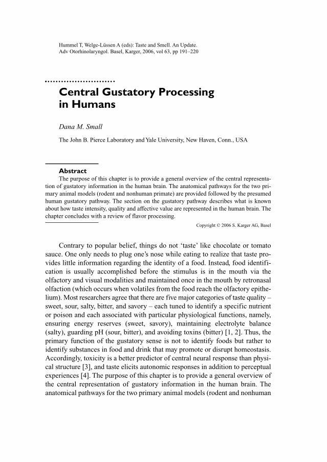
Hummel T, Welge-Lüssen A (eds): Taste and Smell. An Update.
Adv Otorhinolaryngol. Basel, Karger, 2006, vol 63, pp 191–220
Central Gustatory Processingin Humans
Dana M. Small
The John B. Pierce Laboratory and Yale University, New Haven, Conn., USA
AbstractThe purpose of this chapter is to provide a general overview of the central representa-
tion of gustatory information in the human brain. The anatomical pathways for the two pri-
mary animal models (rodent and nonhuman primate) are provided followed by the presumed
human gustatory pathway. The section on the gustatory pathway describes what is known
about how taste intensity, quality and affective value are represented in the human brain. The
chapter concludes with a review of flavor processing.
Copyright © 2006 S. Karger AG, Basel
Contrary to popular belief, things do not ‘taste’ like chocolate or tomato
sauce. One only needs to plug one’s nose while eating to realize that taste pro-
vides little information regarding the identity of a food. Instead, food identifi-
cation is usually accomplished before the stimulus is in the mouth via the
olfactory and visual modalities and maintained once in the mouth by retronasal
olfaction (which occurs when volatiles from the food reach the olfactory epithe-
lium). Most researchers agree that there are five major categories of taste quality –
sweet, sour, salty, bitter, and savory – each tuned to identify a specific nutrient
or poison and each associated with particular physiological functions, namely,
ensuring energy reserves (sweet, savory), maintaining electrolyte balance
(salty), guarding pH (sour, bitter), and avoiding toxins (bitter) [1, 2]. Thus, the
primary function of the gustatory sense is not to identify foods but rather to
identify substances in food and drink that may promote or disrupt homeostasis.
Accordingly, toxicity is a better predictor of central neural response than physi-
cal structure [3], and taste elicits autonomic responses in addition to perceptual
experiences [4]. The purpose of this chapter is to provide a general overview of
the central representation of gustatory information in the human brain. The
anatomical pathways for the two primary animal models (rodent and nonhuman
Small 192
primate) are provided followed by the presumed human gustatory pathway. The
following section on the gustatory pathway describes what is known about how
taste intensity, quality and affective value are represented in the human brain.
The chapter concludes with a review of flavor processing.
The Gustatory Pathway
RodentsIn rodents, taste information is carried through the cranial nerves VII
[chorda tympani (CT), and greater superficial petrosal branches], IX (lingual
branch) and X (superior laryngeal branch) and terminates in the rostral division
of the nucleus tractus solitarii (NTS) [5]. Many of the neurons that leave the
NTS travel to other brainstem nuclei to mediate reflexive and regulatory responses
related to feeding, such as salivation and the rejection reflex, leaving only a
small portion (roughly 20%) to ascend within the gustatory neuraxis [6]. From
the NTS, there are ipsilateral and bilateral [7] ascending fibers that synapse in
the gustatory parabrachial nucleus of the pons [8]. Two separate third-order
pathways arise from the pons: a dorsal sensory pathway [9], which ascends ipsi-
laterally to the gustatory cortex after synapsing in the gustatory nucleus of
the thalamus [parvocellular division of the ventroposterior medial nucleus
(VPMpc)] [10–12], and a ventral affective pathway [13], which projects to the
lateral hypothalamus, substantia innominata, central nucleus of the amygdala,
and bed nucleus of the stria terminalis in the ventral forebrain [14]. Thalamic
gustatory afferents terminate in two regions of the insular cortex [15], one
located within the dysgranular insular cortex and the other in the granular insu-
lar cortex. Both dorsal and ventral termination loci send back projections to the
brainstem taste nuclei [16–21]. Although projections are heaviest ipsilaterally,
at least some of them appear to be bilateral [18]. The rat insular cortex can be
roughly divided into an anterior gustatory region and a posterior visceral region
[22]. However, taste-responsive neurons are widely distributed, and there is a
clear overlap with the visceral representation [23].
Nonhuman PrimatesThe peripheral organization of the gustatory system is similar in rodents
and primates with taste information being carried through the cranial nerves
XII, IX, and X to the NTS [24]. Second-order gustatory fibers ascend ipsilater-
ally from the NTS towards the pons, but it is thought that instead of forming a
synapse in the parabrachial nucleus, as is the case in rats, taste fibers join the
central tegmental tract and project to the VPMpc [24, 25]. Thus, there is cur-
rently no evidence for a pontine taste relay in the nonhuman primate and no
Central Gustatory Processing 193
clear evidence of a separate ventral affective pathway. It is currently not known
if there are also contralateral afferent projections, as is the case in rodents. Scott
and Plata-Salaman [26] have suggested that the primary manifestation of these
anatomical interspecies differences relates to how homeostatic and hedonic fac-
tors influence sensory coding of taste. Since the goal of this chapter is to pro-
vide an understanding of sensory coding in the human, the primate will serve as
the primary model.
The equivalent of the dorsal stream projects ipsilaterally from the thalamus
to terminate in the anterior insula/frontal operculum (AI/FO) [27–29]. Using
titrated amino acid autoradiography, Pritchard et al. [27] traced the efferent proj-
ections of VPMpc. The primary efferent projection was located in the ipsilateral
AI/FO cortex adjacent to the superior limiting sulcus and extending rostrally
to the caudolateral orbitofrontal cortex (OFC). A second projection was also
found that terminated in areas 3a, 3b, 2, and 1 along the lateral margin of the
precentral gyrus. They noted that these two regions correspond to the two
regions Ogawa et al. [28] identified with electrical stimulation of the peripheral
gustatory nerves. Thus, as in the rat, there are multiple gustatory regions within
the insula/operculum. However, since AI/FO has few lingual terminations [28]
and is relatively more sensitive to stimulation of the taste nerve compared to the
lingual nerve [30], it has become known as the primary gustatory area or ‘area
G’ [31]. Cytoarchitectonically, AI/FO can be distinguished from its surrounding
cortex because layers II and IV are thicker and contain fine granule cells
[28, 32]. Electrophysiological studies [26, 33–38] and one positron emission
tomography (PET) [39] study have shown taste responses in the nonhuman pri-
mate AI/FO.
The caudal OFC receives direct projections from the insula and opercular
taste regions [40]. Baylis et al. [40] isolated taste-responsive neurons and injec-
ted horseradish peroxidase into this site. They found substantial labelling in the
frontal opercular taste area, anterior dorsal insula, extending into more ventral
regions of the insula, as well as in the amygdala, mediodorsal thalamus, rhinal
sulcus and substantia nigra. Since no labelling occurred in the VPMpc, Rolls et al.
[83] proposed that the caudal OFC region represented the secondary taste cor-
tex. However, it is clear that there are multiple taste-responsive regions in the
insula/operculum apart from the AI/FO that do not receive direct thalamic
projections and should therefore also be considered higher-order gustatory
areas [31].
Although not formally considered part of the gustatory system, the amyg-
dala has reciprocal connections with virtually every level of the gustatory path-
way [14, 41–45] and taste-responsive neurons are present within several
amygdaloid nuclei [46]. In the monkey, gustatory information reaches the
amygdala via direct pathways from cortical taste neurons within the insula and
Small 194
operculum [41, 47] and orbitofrontal taste area [45, 48, 49]. The amygdala also
sends projections back to the NTS [42] where it may exert an influence on taste
processing at this early level of the primate gustatory neuraxis.
HumansThe human gustatory pathway is assumed to be equivalent to the monkey
pathway and is depicted in figure 1. There is general agreement that brainstem
lesions caudal to the pons lead to decreased sensitivity on the ipsilateral side of
the tongue [50–54], supporting ipsilateral ascension of taste fibers. Gustatory
CLOF
VPM
NTSVII, IX, X
NTS
VPM
CLOFCLOF
AI/FOAI/FO
CLOF
VinsVins
FO
? PonsPons?
A
Fig. 1. The human gustatory pathway. Coronal sections on the left show the main path-
way. Slices in the right column provide additional views. The question mark in the midbrain
demarcates the region where a pontine relay would likely be if it did exist in the human. Most
evidence to date suggests that there is not a relay here. CLOF � Caudolateral OFC; dotted
lines � possible sites for bilateral projections; FO � other regions where activations are fre-
quently reported in response to stimulation with a taste; solid lines � known projections.
Vins, The areas outlined are estimates and are not meant to signify established anatomical
boundaries.
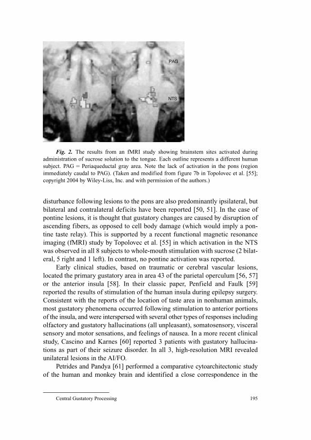
Central Gustatory Processing 195
disturbance following lesions to the pons are also predominantly ipsilateral, but
bilateral and contralateral deficits have been reported [50, 51]. In the case of
pontine lesions, it is thought that gustatory changes are caused by disruption of
ascending fibers, as opposed to cell body damage (which would imply a pon-
tine taste relay). This is supported by a recent functional magnetic resonance
imaging (fMRI) study by Topolovec et al. [55] in which activation in the NTS
was observed in all 8 subjects to whole-mouth stimulation with sucrose (2 bilat-
eral, 5 right and 1 left). In contrast, no pontine activation was reported.
Early clinical studies, based on traumatic or cerebral vascular lesions,
located the primary gustatory area in area 43 of the parietal operculum [56, 57]
or the anterior insula [58]. In their classic paper, Penfield and Faulk [59]
reported the results of stimulation of the human insula during epilepsy surgery.
Consistent with the reports of the location of taste area in nonhuman animals,
most gustatory phenomena occurred following stimulation to anterior portions
of the insula, and were interspersed with several other types of responses including
olfactory and gustatory hallucinations (all unpleasant), somatosensory, visceral
sensory and motor sensations, and feelings of nausea. In a more recent clinical
study, Cascino and Karnes [60] reported 3 patients with gustatory hallucina-
tions as part of their seizure disorder. In all 3, high-resolution MRI revealed
unilateral lesions in the AI/FO.
Petrides and Pandya [61] performed a comparative cytoarchitectonic study
of the human and monkey brain and identified a close correspondence in the
PAG
NTS
Fig. 2. The results from an fMRI study showing brainstem sites activated during
administration of sucrose solution to the tongue. Each outline represents a different human
subject. PAG � Periaqueductal gray area. Note the lack of activation in the pons (region
immediately caudal to PAG). (Taken and modified from figure 7b in Topolovec et al. [55];
copyright 2004 by Wiley-Liss, Inc. and with permission of the authors.)
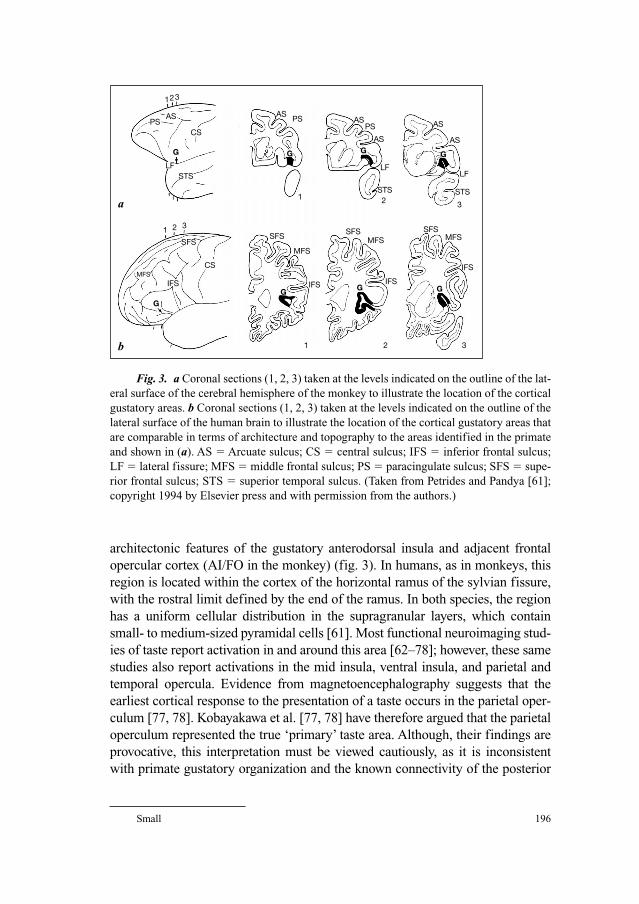
Small 196
architectonic features of the gustatory anterodorsal insula and adjacent frontal
opercular cortex (AI/FO in the monkey) (fig. 3). In humans, as in monkeys, this
region is located within the cortex of the horizontal ramus of the sylvian fissure,
with the rostral limit defined by the end of the ramus. In both species, the region
has a uniform cellular distribution in the supragranular layers, which contain
small- to medium-sized pyramidal cells [61]. Most functional neuroimaging stud-
ies of taste report activation in and around this area [62–78]; however, these same
studies also report activations in the mid insula, ventral insula, and parietal and
temporal opercula. Evidence from magnetoencephalography suggests that the
earliest cortical response to the presentation of a taste occurs in the parietal oper-
culum [77, 78]. Kobayakawa et al. [77, 78] have therefore argued that the parietal
operculum represented the true ‘primary’ taste area. Although, their findings are
provocative, this interpretation must be viewed cautiously, as it is inconsistent
with primate gustatory organization and the known connectivity of the posterior
1
PSAS
CS
ASAS AS
ASAS
LFLF
STS STS
PSPS
G G G
1
1
2
2
3
3
G
GG G G
LFSTS
a
23
1 2
MFS
MFSMFS MFS
SFSSFS SFS SFS
CS
IFS IFS IFS
IFS
3
b
Fig. 3. a Coronal sections (1, 2, 3) taken at the levels indicated on the outline of the lat-
eral surface of the cerebral hemisphere of the monkey to illustrate the location of the cortical
gustatory areas. b Coronal sections (1, 2, 3) taken at the levels indicated on the outline of the
lateral surface of the human brain to illustrate the location of the cortical gustatory areas that
are comparable in terms of architecture and topography to the areas identified in the primate
and shown in (a). AS � Arcuate sulcus; CS � central sulcus; IFS � inferior frontal sulcus;
LF � lateral fissure; MFS � middle frontal sulcus; PS � paracingulate sulcus; SFS � supe-
rior frontal sulcus; STS � superior temporal sulcus. (Taken from Petrides and Pandya [61];
copyright 1994 by Elsevier press and with permission from the authors.)
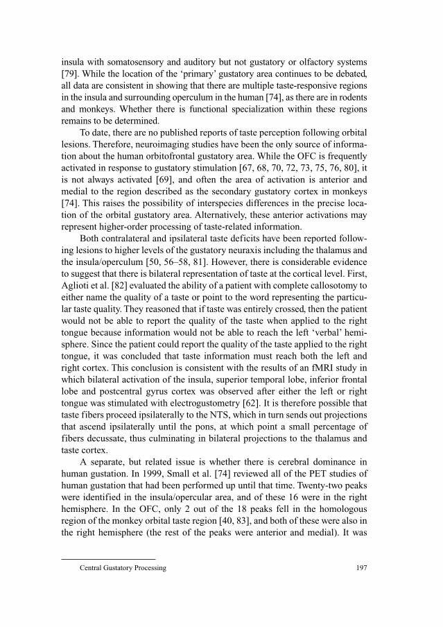
Central Gustatory Processing 197
insula with somatosensory and auditory but not gustatory or olfactory systems
[79]. While the location of the ‘primary’ gustatory area continues to be debated,
all data are consistent in showing that there are multiple taste-responsive regions
in the insula and surrounding operculum in the human [74], as there are in rodents
and monkeys. Whether there is functional specialization within these regions
remains to be determined.
To date, there are no published reports of taste perception following orbital
lesions. Therefore, neuroimaging studies have been the only source of informa-
tion about the human orbitofrontal gustatory area. While the OFC is frequently
activated in response to gustatory stimulation [67, 68, 70, 72, 73, 75, 76, 80], it
is not always activated [69], and often the area of activation is anterior and
medial to the region described as the secondary gustatory cortex in monkeys
[74]. This raises the possibility of interspecies differences in the precise loca-
tion of the orbital gustatory area. Alternatively, these anterior activations may
represent higher-order processing of taste-related information.
Both contralateral and ipsilateral taste deficits have been reported follow-
ing lesions to higher levels of the gustatory neuraxis including the thalamus and
the insula/operculum [50, 56–58, 81]. However, there is considerable evidence
to suggest that there is bilateral representation of taste at the cortical level. First,
Aglioti et al. [82] evaluated the ability of a patient with complete callosotomy to
either name the quality of a taste or point to the word representing the particu-
lar taste quality. They reasoned that if taste was entirely crossed, then the patient
would not be able to report the quality of the taste when applied to the right
tongue because information would not be able to reach the left ‘verbal’ hemi-
sphere. Since the patient could report the quality of the taste applied to the right
tongue, it was concluded that taste information must reach both the left and
right cortex. This conclusion is consistent with the results of an fMRI study in
which bilateral activation of the insula, superior temporal lobe, inferior frontal
lobe and postcentral gyrus cortex was observed after either the left or right
tongue was stimulated with electrogustometry [62]. It is therefore possible that
taste fibers proceed ipsilaterally to the NTS, which in turn sends out projections
that ascend ipsilaterally until the pons, at which point a small percentage of
fibers decussate, thus culminating in bilateral projections to the thalamus and
taste cortex.
A separate, but related issue is whether there is cerebral dominance in
human gustation. In 1999, Small et al. [74] reviewed all of the PET studies of
human gustation that had been performed up until that time. Twenty-two peaks
were identified in the insula/opercular area, and of these 16 were in the right
hemisphere. In the OFC, only 2 out of the 18 peaks fell in the homologous
region of the monkey orbital taste region [40, 83], and both of these were also in
the right hemisphere (the rest of the peaks were anterior and medial). It was
Small 198
therefore proposed that the right hemisphere was dominant for taste in human
gustation. Since this time, findings have been mixed. In accordance with Small
et al. [74], Barry et al. [62] reported that although activation in most regions of
the insula was bilateral following stimulation of taste by application of electro-
gustometry to either the right or left tongue, activation was consistently greater
in the right dorsal insular region. Additionally, several studies of taste percep-
tion following resection from the anterior temporal lobe have reported greater
changes if the resection is from the right hemisphere [80, 84, 85]. However,
most studies report bilateral activation of the insula taste regions [67, 70, 72, 75]
and still others report evidence for left hemisphere dominance [66, 86]. Various
factors have been proposed to influence lateralization including internal state
[86], handedness [66, 87], and affective value (discussed below). It is also pos-
sible that, as in olfaction, cognitive judgments influence laterality [88, 89].
Cognitive influences upon gustatory processing have yet to be addressed. Thus,
the possibility of cerebral dominance and/or lateralization of function in human
gustation remains unresolved.
Gustatory Physiology
The neural substrates of taste must code for perceived intensity, pleasant-
ness/unpleasantness (i.e. affective value) and quality. One difficulty in studying
the neural representation of these perceptual dimensions is that they are not
independent [see chapter 10 by Breslin and Huang, this vol, pp 152–190].
Instead pleasantness/unpleasantness may influence perceived intensity and
vice versa. Moreover, the nature of these interactions depends upon quality [1,
90–92]. For example, bitter is perceived as unpleasant at all concentrations,
whereas sweet generally becomes more pleasant as concentration increases and
then plateaus or decreases. These perceptual interactions are reflected in the
underlying physiology by overlapping representation. However, despite the
degree of interaction, dissociations have been found and the relative importance
of different regions to coding each dimension is beginning to be understood.
Detection and IntensityStimulus detection is the most fundamental perceptual process. Gustatory
detection thresholds measure the minimum amount of a substance that is
required to be present in a solution in order for a subject to detect its presence at
above-chance levels. In monkeys, bilateral lesions to the VPMpc result in a pro-
found and persistent elevation in rejection thresholds for quinine hydrochloride
[95], indicating that detection of taste relies on structures rostral to the brain-
stem taste regions. Pribram and Bagshaw [208] found that lesions produced in
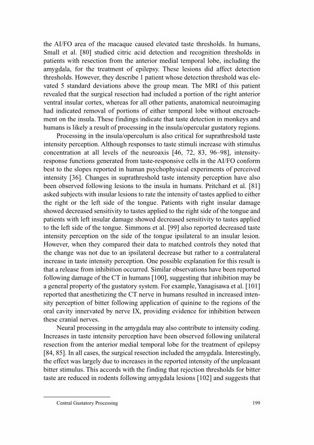
Central Gustatory Processing 199
the AI/FO area of the macaque caused elevated taste thresholds. In humans,
Small et al. [80] studied citric acid detection and recognition thresholds in
patients with resection from the anterior medial temporal lobe, including the
amygdala, for the treatment of epilepsy. These lesions did affect detection
thresholds. However, they describe 1 patient whose detection threshold was ele-
vated 5 standard deviations above the group mean. The MRI of this patient
revealed that the surgical resection had included a portion of the right anterior
ventral insular cortex, whereas for all other patients, anatomical neuroimaging
had indicated removal of portions of either temporal lobe without encroach-
ment on the insula. These findings indicate that taste detection in monkeys and
humans is likely a result of processing in the insula/opercular gustatory regions.
Processing in the insula/operculum is also critical for suprathreshold taste
intensity perception. Although responses to taste stimuli increase with stimulus
concentration at all levels of the neuroaxis [46, 72, 83, 96–98], intensity-
response functions generated from taste-responsive cells in the AI/FO conform
best to the slopes reported in human psychophysical experiments of perceived
intensity [36]. Changes in suprathreshold taste intensity perception have also
been observed following lesions to the insula in humans. Pritchard et al. [81]
asked subjects with insular lesions to rate the intensity of tastes applied to either
the right or the left side of the tongue. Patients with right insular damage
showed decreased sensitivity to tastes applied to the right side of the tongue and
patients with left insular damage showed decreased sensitivity to tastes applied
to the left side of the tongue. Simmons et al. [99] also reported decreased taste
intensity perception on the side of the tongue ipsilateral to an insular lesion.
However, when they compared their data to matched controls they noted that
the change was not due to an ipsilateral decrease but rather to a contralateral
increase in taste intensity perception. One possible explanation for this result is
that a release from inhibition occurred. Similar observations have been reported
following damage of the CT in humans [100], suggesting that inhibition may be
a general property of the gustatory system. For example, Yanagisawa et al. [101]
reported that anesthetizing the CT nerve in humans resulted in increased inten-
sity perception of bitter following application of quinine to the regions of the
oral cavity innervated by nerve IX, providing evidence for inhibition between
these cranial nerves.
Neural processing in the amygdala may also contribute to intensity coding.
Increases in taste intensity perception have been observed following unilateral
resection from the anterior medial temporal lobe for the treatment of epilepsy
[84, 85]. In all cases, the surgical resection included the amygdala. Interestingly,
the effect was largely due to increases in the reported intensity of the unpleasant
bitter stimulus. This accords with the finding that rejection thresholds for bitter
taste are reduced in rodents following amygdala lesions [102] and suggests that
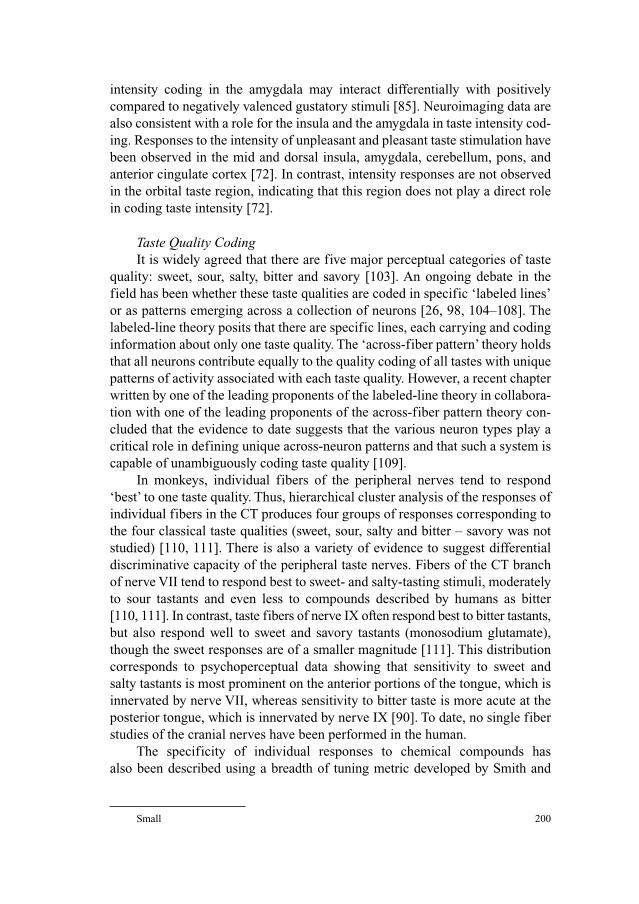
Small 200
intensity coding in the amygdala may interact differentially with positively
compared to negatively valenced gustatory stimuli [85]. Neuroimaging data are
also consistent with a role for the insula and the amygdala in taste intensity cod-
ing. Responses to the intensity of unpleasant and pleasant taste stimulation have
been observed in the mid and dorsal insula, amygdala, cerebellum, pons, and
anterior cingulate cortex [72]. In contrast, intensity responses are not observed
in the orbital taste region, indicating that this region does not play a direct role
in coding taste intensity [72].
Taste Quality CodingIt is widely agreed that there are five major perceptual categories of taste
quality: sweet, sour, salty, bitter and savory [103]. An ongoing debate in the
field has been whether these taste qualities are coded in specific ‘labeled lines’
or as patterns emerging across a collection of neurons [26, 98, 104–108]. The
labeled-line theory posits that there are specific lines, each carrying and coding
information about only one taste quality. The ‘across-fiber pattern’ theory holds
that all neurons contribute equally to the quality coding of all tastes with unique
patterns of activity associated with each taste quality. However, a recent chapter
written by one of the leading proponents of the labeled-line theory in collabora-
tion with one of the leading proponents of the across-fiber pattern theory con-
cluded that the evidence to date suggests that the various neuron types play a
critical role in defining unique across-neuron patterns and that such a system is
capable of unambiguously coding taste quality [109].
In monkeys, individual fibers of the peripheral nerves tend to respond
‘best’ to one taste quality. Thus, hierarchical cluster analysis of the responses of
individual fibers in the CT produces four groups of responses corresponding to
the four classical taste qualities (sweet, sour, salty and bitter – savory was not
studied) [110, 111]. There is also a variety of evidence to suggest differential
discriminative capacity of the peripheral taste nerves. Fibers of the CT branch
of nerve VII tend to respond best to sweet- and salty-tasting stimuli, moderately
to sour tastants and even less to compounds described by humans as bitter
[110, 111]. In contrast, taste fibers of nerve IX often respond best to bitter tastants,
but also respond well to sweet and savory tastants (monosodium glutamate),
though the sweet responses are of a smaller magnitude [111]. This distribution
corresponds to psychoperceptual data showing that sensitivity to sweet and
salty tastants is most prominent on the anterior portions of the tongue, which is
innervated by nerve VII, whereas sensitivity to bitter taste is more acute at the
posterior tongue, which is innervated by nerve IX [90]. To date, no single fiber
studies of the cranial nerves have been performed in the human.
The specificity of individual responses to chemical compounds has
also been described using a breadth of tuning metric developed by Smith and
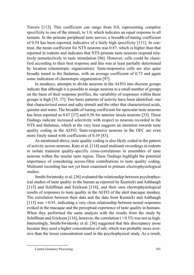
Central Gustatory Processing 201
Travers [113]. This coefficient can range from 0.0, representing complete
specificity to one of the stimuli, to 1.0, which indicates an equal response to all
tastants. In the primate peripheral taste nerves, a breadth-of-tuning coefficient
of 0.54 has been reported, indicative of a fairly high specificity [111]. In con-
trast, the mean coefficient for NTS neurons was 0.87, which is higher than that
reported in rodents and indicates that NTS primate taste neurons respond rela-
tively nonselectively to taste stimulation [96]. However, cells could be classi-
fied according to their best response and this was at least partially determined
by location (chemotopic organization). Taste-responsive cells are also quite
broadly tuned in the thalamus, with an average coefficient of 0.73 and again
some indication of chemotopic organization [97].
In monkeys, attempts to divide neurons in the AI/FO into discrete groups
indicate that although it is possible to assign neurons to a small number of groups
on the basis of their response profiles, the variability of responses within these
groups is high [35, 37]. Two basic patterns of activity have been identified: one
that characterized sweet and salty stimuli and the other that characterized acids,
quinine and water. The breadth-of-tuning coefficient for opercular taste neurons
has been reported as 0.67 [37] and 0.56 for anterior insula neurons [35]. These
findings indicate increased selectivity with respect to neurons recorded in the
NTS and thalamus, which at the very least suggests an intention towards taste
quality coding in the AI/FO. Taste-responsive neurons in the OFC are even
more finely tuned with coefficients of 0.39 [83].
As mentioned above, taste quality coding is also likely coded in the pattern
of activity across neurons. Katz et al. [114] used multiunit recordings in rodents
to isolate transient quality-specific cross-correlations in ensembles of taste
neurons within the insular taste region. These findings highlight the potential
importance of considering across-fiber contributions to taste quality coding.
Multiunit recording has not yet been examined in primate electrophysiological
studies.
Smith-Swintosky et al. [36] evaluated the relationship between psychophys-
ical studies of taste quality in the human as reported by Kuznicki and Ashbaugh
[115] and Schiffman and Erickson [116], and their own electrophysiological
results of responses to taste quality in the AI/FO of the alert macaque monkey.
The correlation between their data and the data from Kuznicki and Ashbaugh
[115] was �0.91, indicating a very close relationship between neural responses
evoked in the macaque and the perceptual experience of taste quality in humans.
When they performed the same analysis with the results from the study by
Schiffman and Erickson [116], however, the correlation (�0.53) was not as high.
Interestingly, Smith-Swintosky et al. [36] suggested that this discrepancy arose
because they used a higher concentration of salt, which was probably more aver-
sive than the lesser concentration used in the psychophysical study. As a result,

Small 202
the response they recorded to salty stimuli was more akin to the response pro-
files elicited by aversive stimuli such as quinine. In fact, the correlation between
the two data sets rose to �0.85 when they dropped the salty from the analysis.
These results suggest that the affective value, determined by concentration, may
influence taste quality coding in at least one region of the insula.
It is currently unknown if it is possible to isolate quality-specific responses
in humans, and if so, what anatomical structures contribute to taste quality cod-
ing. Deficits in identifying or recognizing suprathreshold taste quality have been
reported following anterior but not posterior insular lesions [81, 117], indicat-
ing that as in the monkey, this area is important in coding taste quality. Elevated
taste recognition thresholds have also been observed following unilateral resec-
tion from the anterior medial temporal lobe [80, 118], and a case of gustatory
agnosia has been described in patients with bilateral anterior medial temporal
lobe damage and unilateral insular atrophy [119]. Therefore, although taste
detection may be computed in the insula/operculuar region, recognition likely
involves integration of the gustatory code with motivational and hedonic net-
works related to feeding [80].
A separate issue is whether taste-responsive cells are organized in a spa-
tially determined fashion. Several studies have reported evidence for chemotopic
organization within the NTS of the rat [120], hamster [121] and monkey [96]. In
general, sweet and salty responses are more numerous in the rostral part of the
gustatory NTS, prompting Smith and Scott [109] to suggest that the distribution
reflects the differential sensitivity of the CT and IXth nerves because these
nerves terminate in a rostral to caudal order. Although there is some evidence for
chemotopy in the cortical gustatory areas [31, 37, 83, 122], results are inconsis-
tent. Scott et al. [37] recorded taste responses from neurons in the frontal oper-
culum of cynomolgus monkeys and found that sweet-best cells tended to be
distributed toward the anterior section, salty-best cells toward the posterior
extent and sour-best cells within an intermediate region. In subsequent studies,
no evidence for chemotopy was found [36, 123]. Furthermore, when all the data
were compiled, a plot of the location of each neuron in the gustatory cortex of
the macaque as a function of its most effective basic stimulus revealed no evi-
dence for chemotopic organization [26]. Nevertheless, Scott and Plata-Salaman
[26] note in their 1999 review that the probability was higher than chance that a
contiguous neuron would have the same sensitivity, indicating that taste sensitiv-
ity is not randomly distributed within the cortex. In the OFC, Rolls et al. [83]
found some evidence of spatial grouping of taste neurons; however, they too
noted that it was not clear whether the basis for this grouping was related to qual-
ity or some other attribute of the stimuli, such as affective valence.
The issue of chemotopy has not been investigated in humans. Spatially
segregated responses to different gustatory stimuli are frequently reported but
Central Gustatory Processing 203
interpreted to result from affective rather than qualitative differences in the
stimuli [70–72, 76, 124]. However, these studies confound taste pleasantness/
unpleasantness with quality by using different taste qualities to elicit the oppo-
site affective response. For example, Small et al. [72] reported that a caudal
region of the right OFC responds to sweet irrespective of intensity and not to
bitter stimuli, and a region of the left anterior OFC responds to bitter irrespec-
tive of intensity and not to sweet stimuli. Since pleasantness/unpleasantness is
confounded with quality, the result may be related to quality, valence or a com-
bination of these dimensions.
Affective ValueThe affective value of a taste stimulus can be influenced by a variety of
factors, including factors intrinsic to the stimulus (quality, intensity, physiolog-
ical significance) and factors related to the individual (preference, internal
state, experience). One intriguing aspect of gustatory hedonics is the possibility
that there are innate affective values associated with the different taste qualities
[125]. Steiner [126] observed that when a sweet or bitter taste is placed in the
mouth of a human infant, stereotyped facial expressions can be observed. He
described that a sweet or mildly salty taste elicits a mild rhythmic smacking,
slight protrusions of the tongue, and a relaxed expression accompanied some-
times by a slight upturn of the corners of the mouth. In contrast, a bitter, sour, or
very salty taste elicits a grimace, a turning away, a gape or gagging movement,
pushing out the offending taste, and a pushing away with the hands. This
‘innate’ response is adaptive for it saves us from the peril of having to learn that
bitter signifies poison, and can be thought of as the ‘intrinsic affective value’ of
the quality.
The presence of intrinsic affective values has led some investigators to pro-
pose that taste perceptions are primary reinforcers [127]. However, if this were
true then affective values should be immutable. They are not. In rodents, diet
has been shown to have a profound effect on gustatory organization [128–132],
which may in turn be related to preferences [133]. Likewise in humans, diet has
been shown to affect the pleasantness perception of taste qualities [134], and
the importance of being able to adapt taste preferences to the food available in
the environment has been emphasized [135]. The influence of diet upon taste
pleasantness perception and preference suggests that although the relationship
between the intrinsic affective value and quality is generally very stable, and
possibly present at birth, learning can and does occur. This in turn implies that
the intrinsic affective value and quality are separable at the neural level.
Genetic differences may also contribute to differences in preferences and
hence to the perceived pleasantness of a taste. Deviations in sweet preference
have been associated with higher risk for drug abuse [136], and obesity [137–139].
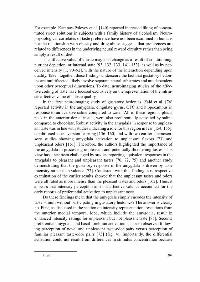
Small 204
For example, Kampov-Polevoy et al. [140] reported increased liking of concen-
trated sweet solutions in subjects with a family history of alcoholism. Neuro-
physiological correlates of taste preference have not been examined in humans
but the relationship with obesity and drug abuse suggests that preferences are
related to differences in the underlying neural reward circuitry rather than being
simply a result of diet.
The affective value of a taste may also change as a result of conditioning,
nutrient depletion, or internal state [93, 132, 133, 141–153], as well as by per-
ceived intensity [1, 90–92], with the nature of the interaction depending upon
quality. Taken together, these findings underscore the fact that gustatory hedon-
ics are multifaceted, likely involve separate neural substrates and are dependent
upon other perceptual dimensions. To date, neuroimaging studies of the affec-
tive coding of taste have focused exclusively on the representation of the intrin-
sic affective value of a taste quality.
In the first neuroimaging study of gustatory hedonics, Zald et al. [76]
reported activity in the amygdala, cingulate gyrus, OFC and hippocampus in
response to an aversive saline compared to water. All of these regions, plus a
peak in the anterior dorsal insula, were also preferentially activated by saline
compared to chocolate. Robust activity in the amygdala in response to unpleas-
ant taste was in line with studies indicating a role for this region in fear [154, 155],
conditioned taste aversion learning [156–160] and with two earlier chemosen-
sory studies showing amygdala activation to unpleasant flavors [73] and
unpleasant odors [161]. Therefore, the authors highlighted the importance of
the amygdala in processing unpleasant and potentially threatening tastes. This
view has since been challenged by studies reporting equivalent responses in the
amygdala to pleasant and unpleasant tastes [70, 72, 75] and another study
demonstrating that the gustatory response in the amygdala is driven by taste
intensity rather than valence [72]. Consistent with this finding, a retrospective
examination of the earlier results showed that the unpleasant tastes and odors
were all rated as more intense than the pleasant tastes and odors [162]. Thus, it
appears that intensity perception and not affective valence accounted for the
early reports of preferential activation to unpleasant taste.
Do these findings mean that the amygdala simply encodes the intensity of
taste stimuli without participating in gustatory hedonics? The answer is clearly
no. First, as discussed in the section on intensity representation, resections from
the anterior medial temporal lobe, which include the amygdala, result in
enhanced intensity ratings for unpleasant but not pleasant taste [85]. Second,
preferential amygdala and basal forebrain activation has been observed follow-
ing perception of novel and unpleasant taste-odor pairs versus perception of
familiar pleasant taste-odor pairs [73] (fig. 4). Importantly, the differential
activation could not result from differences in stimulus concentration because
Central Gustatory Processing 205
identical tastes and odors were used to generate familiar and unfamiliar pairs.
Sweet taste paired with strawberry odor and salty taste paired with soy sauce
odor created familiar and pleasant flavors whereas salty taste paired with straw-
berry odor and sweet taste paired with soy sauce odor created novel and some-
what unpleasant flavors. Thus, differential activation could not be related to the
concentration of the stimuli. However, it may be related to perceived intensity,
which tends to be greater for unpleasant tastes [92].
Taken together the findings indicate that the response in the amygdala
reflects an interaction between intensity, novelty and intrinsic affective value.
Small et al. [72] therefore proposed that ‘the amygdala is important in estab-
lishing the saliency of sensory stimuli, which is determined by the interacting
dimensions of intensity, valence, and perhaps novelty/familiarity’, and that ‘one
important function of this integrated coding may be in biasing processing in
favour of adaptive needs so that subjective experience can be released from
dependence upon the physical attributes of the stimulus’. If this were true then
lesions to the amygdala may bias the normal interaction between intensity and
pleasantness, resulting in enhanced aversion to already unpleasant tastes and a
subsequent increase in their perceived intensity. This notion is also consistent
with the psychophysical interactions noted above [see also chapter 10 by
Breslin and Huang, this vol, pp 152–190] and highlights the potential impor-
tance of the amygdala in supporting perceptual interactions that may serve to
promote perceptions that insure preservative responses rather than a veridical
A
BF
Fig. 4. Activation in the amygdala (A) and basal forebrain (BF) resulting from the
summed neural activity evoked during a 60-second PET scan of incongruent taste-odor stim-
ulation (strawberry with salty, soy sauce with sweet, grapefruit with bitter, coffee with sour)
compared to a 60-second PET scan of congruent taste-odor stimulation (strawberry with
sweet, soy sauce with salty, grapefruit with sour, coffee with bitter and tasteless with odor-
less). Tastants were presented on tongue shaped filter papers simultaneously with odors that
were presented on long-handed cotton wands waved under the nose. (Taken from Small et al.
[73]; copyright 1997 by Lippincott, Williams & Wilkins and with permission of the authors.)
Small 206
representation of the stimulus. Parallel findings and conclusions have been
reached for the role of the amygdala in representing pure olfactory stimuli [for
details, see chapter 3 by Gottfried, this vol, pp 44–69].
The affective value of a taste stimulus can also be modulated by changes in
internal state. In monkeys, single-cell recordings of taste-responsive neurons
in the amygdala show moderate attenuation of the gustatory response in some
neurons after feeding [163]. No study has yet examined the effect of satiety on
the neural response evoked by a pure taste stimulus in the human. However,
several studies of satiety have been conducted using food and drink [84, 86,
162, 164], and all fail to observe changes in the amygdala as a result of hunger
or satiety. The one exception to this comes from studies reporting changes in
the amygdala in obese but not lean subjects [165, 166]. These findings suggest
that the amygdala of healthy lean subjects is not critical in mediating changes
in the reward value of food being consumed. This result contrasts with the
known role of the amygdala in tracking changes in the reward value of visual
[167, 168] and olfactory stimuli [169, 170] caused by eating [see also chapter
3 by Gottfried, this vol, pp 44–69]; a discrepancy that may be related to differ-
ential responsiveness to sensory inputs linked to receipt versus prediction of
reward. In support of this notion, the amygdala has been shown to respond more
to the anticipation of a sweet taste compared to receipt of that sweet taste [71].
In contrast to the amygdala, valence-specific responses are consistently
observed in the OFC [70, 71, 75, 76] and these responses are not sensitive to
intensity [72]. They may be localized to the orbital taste area but also extend
well beyond this region, and they may be for received as well as for anticipated
tastes [71]. Primate OFC taste responses are also greatly attenuated by satiety
[171, 172], and in humans neuroimaging studies consistently show differential
responsiveness in the OFC to the food when hungry versus when full [86, 124,
164]. Consistent with these results, animals will only self-stimulate here when
hungry [173] and lesions to this area lead to gustatory anhedonia [174], indicat-
ing that this region is important in representing the intrinsic affective value as
well as transient shifts in the affective value associated with satiety.
There is a possibility that an asymmetry exists in gustatory processing in
the OFC, with pleasant tastes preferentially activating the right OFC. Figure 5 shows
peaks from pleasant or unpleasant taste stimulation color-coded and plotted
onto an axial anatomical section. Eight analyses of pleasant taste yielded 13 OFC
peaks, and of these 11 were in the right hemisphere and only 2 were in the left
hemisphere. Furthermore, the left-hemisphere peaks were from analyses that
produced stronger peaks in the right hemisphere. The situation is not as com-
pelling for unpleasant taste. From 8 analyses that yielded 12 peaks, 5 were in
the right and 7 in the left hemisphere. It is currently not known why pleasant
tastes should preferentially activate the right OFC.

Central Gustatory Processing 207
LeftRight
Num
ber
of p
eaks
12
10
8
6
4
2
0Pleasant Unpleasant
b
a
Pleasant taste
L R
Unpleasant taste
Fig. 5. a 25 activation foci from 8 analyses of a pleasant taste (either a pleasant taste
minus a neutral taste or a pleasant taste minus an unpleasant taste) and 8 analyses of an
unpleasant taste (either an unpleasant taste minus a neutral taste or an unpleasant taste minus
a pleasant taste) collected from 5 published studies [70–72, 75, 76] and plotted onto an
anatomical image. L � Left; R � right. To display peaks in a single plane, the average level
of z (superior to inferior � �14 in MNI coordinates) was calculated and the peaks were plot-
ted onto this plane according to their x (medial to lateral) and y (anterior to posterior) coor-
dinate values. b A bar graph showing the number of peaks (y axis) that fell in the left or right
OFC for the pleasant and unpleasant analyses.

Small 208
Valence-specific responses have also been observed in the anterior portions
of the insula [72], and there is some evidence that changes in the responsiveness
of this region to repeated exposures of gustatory experience is dependent upon the
evolution of the hedonic response [175]. Specifically, if the dislike of a stimulus
disappears, there is greater activity following repeated exposure. In contrast, if
the liking of a stimulus decreases then there is less activity following repeated
exposure. Finally, the insular region (anterior and posterior) is more responsive
to food when subjects are hungry compared to when they are full [86, 124, 164,
165, 176]; however, it is not known whether this effect is due to changes within
taste-responsive neurons or changes in overlapping neural circuits. Single-cell
recording studies in the primate indicate that taste-responsive neurons in the
insula/operculum are not modulated by satiety [34, 177].
Perception of pleasant and/or unpleasant taste or food also consistently acti-
vates regions of the brain outside the gustatory regions including the striatum, the
cingulate gyrus and the midbrain [70–72, 75, 76, 124, 164]. Interestingly, the
midbrain and ventral striatum are more likely to be recruited during the anticipa-
tion versus the receipt of a pleasant taste [71], whereas the dorsal striatum and
anterior cingulate region are more likely to be recruited during the experience of
a pleasant taste or food [70, 72, 124, 164].
Flavor
Although it is important to understand how pure taste perceptions are rep-
resented in the brain, it is a mistake to consider neurophysiological correlates of
taste only within this context. In everyday life, we rarely perceive taste without
concomitant experience of oral texture and retronasal olfaction. Unlike early
cortical representation of vision, audition and somatosensation, which are rep-
resented in the unimodal neocortex, the cortical representation of taste is in the
heteromodal paralimbic cortex where there is overlapping representation of
orthonasal olfaction [89, 178–181], retronasal olfaction [64, 182], oral move-
ment, oral somatosensation [63, 185] and texture perception [186]. This anat-
omy reflects the experience of taste, which is almost always accompanied by
simultaneous experience of odor and oral somatosensation in the context of
feeding. Thus, sensory integration is fundamental to gustation. While one does
not give a second thought to hearing without seeing or seeing without hearing,
only during a cold does one taste without smelling and this experience is gener-
ally described as strange, with food not ‘tasting’ right.
Neuroimaging studies of olfaction, gustation, and flavor are beginning to
isolate a network of regions that are likely responsible for taste/odor integration,
and hence flavor perception. Independent presentation of a tastant or an odorant
Central Gustatory Processing 209
produces overlapping activation in regions of the insula and operculum [63, 74,
89, 178, 182], the OFC [68, 70, 73, 74, 76, 89, 161, 170, 178, 179, 187], and the
anterior cingulate cortex [64, 72, 76, 89, 124, 161, 170, 181]. The insula, oper-
culum, OFC and anterior cingulate cortex are also sensitive to somatosensory
stimulation of the oral cavity [63, 185, 186]. Similarly, single-cell recording
studies in monkeys have identified taste- and smell-responsive cells in the
insula/operculum [26] and OFC [188, 189]. The presence of a unimodal repre-
sentation of taste, odor, and oral touch in the insula, frontal operculum and OFC
of the human and nonhuman primate suggests that these regions play a key role
in integrating the disparate sensory inputs that give rise to the perception of fla-
vors. Chemosensory responses in the monkey anterior cingulate cortex have yet
to be investigated, but the consistency of responses in this region to taste and oral
somatosensation in humans is highly suggestive of a role in flavor processing.
Rolls and Baylis [188], Rolls et al. [190, 191], and Verhagen et al. [192]
have performed a series of studies in which gustatory, olfactory, visual, and oral
somatosensory stimuli were presented to awake behaving monkeys and responses
were recorded from single-cell neurons located in the caudal OFC, extending
into the ventral insula. They identified unimodal taste, smell, visual, fat, and
texture cells that were interspersed with multimodal cells that responded to
independent stimulation of two or more modalities. In support of a role for
these cells in flavor processing, their best response was often to complex stim-
uli such as blackcurrant juice. Consistent with the findings in primates and the
neuroimaging results discussed above, De Araujo et al. [64] reported activation
in the frontal operculum, ventral insula/caudal OFC, amygdala, and anterior
cingulate cortex to unimodal stimulation with either a taste or an odor, and to
bimodal stimulation with a taste/odor mixture. A study by Small et al. [193],
using a similar design, also found activity in the frontal operculum, ventral
insula/caudal OFC and anterior cingulate cortex to a taste/odor mixture; but in
this study, the response was supra-additive (indicative of neural integration), in
that greater activity was observed when the subjects received a taste/odor mix-
ture compared to the summed neural activation evoked by independent stimula-
tion with the taste and the odor components. Thus, in monkeys and humans, it
appears that a major function of the core gustatory regions is the integration of
taste information with the other sensory constituents of flavor.
Whether taste and smell are integrated into a unitary flavor percept at the
perceptual and neural level is contingent upon several factors. These include,
but are not limited to, previous experience with taste-odor mixtures, spatial and
temporal proximity of the taste and odor and attentional allocation. Behavioral
studies of taste-odor integration show that odors can enhance perceived taste
intensity, but only if they have previously been experienced with that taste
[194–198]. For example, strawberry odor will enhance the perceived intensity
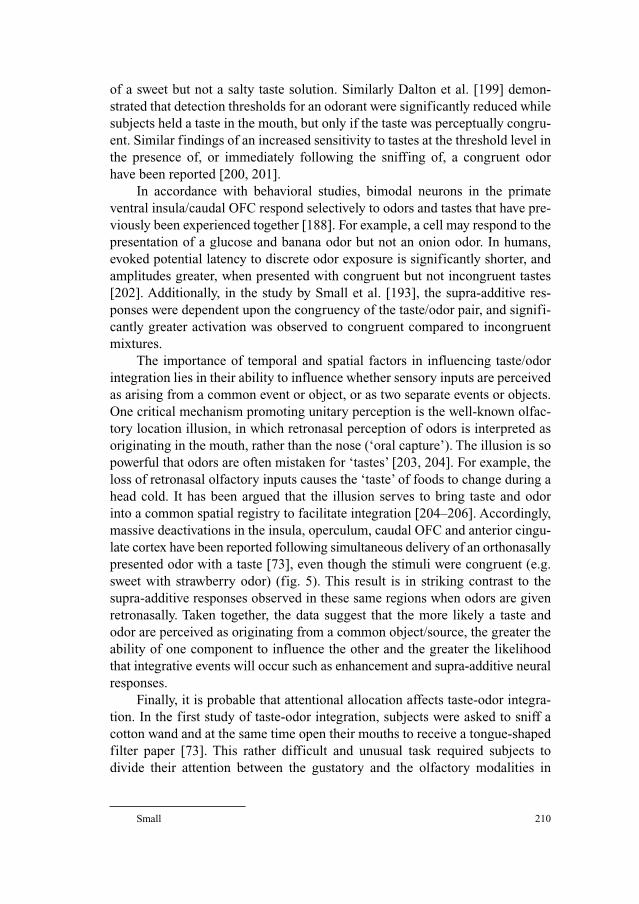
Small 210
of a sweet but not a salty taste solution. Similarly Dalton et al. [199] demon-
strated that detection thresholds for an odorant were significantly reduced while
subjects held a taste in the mouth, but only if the taste was perceptually congru-
ent. Similar findings of an increased sensitivity to tastes at the threshold level in
the presence of, or immediately following the sniffing of, a congruent odor
have been reported [200, 201].
In accordance with behavioral studies, bimodal neurons in the primate
ventral insula/caudal OFC respond selectively to odors and tastes that have pre-
viously been experienced together [188]. For example, a cell may respond to the
presentation of a glucose and banana odor but not an onion odor. In humans,
evoked potential latency to discrete odor exposure is significantly shorter, and
amplitudes greater, when presented with congruent but not incongruent tastes
[202]. Additionally, in the study by Small et al. [193], the supra-additive res-
ponses were dependent upon the congruency of the taste/odor pair, and signifi-
cantly greater activation was observed to congruent compared to incongruent
mixtures.
The importance of temporal and spatial factors in influencing taste/odor
integration lies in their ability to influence whether sensory inputs are perceived
as arising from a common event or object, or as two separate events or objects.
One critical mechanism promoting unitary perception is the well-known olfac-
tory location illusion, in which retronasal perception of odors is interpreted as
originating in the mouth, rather than the nose (‘oral capture’). The illusion is so
powerful that odors are often mistaken for ‘tastes’ [203, 204]. For example, the
loss of retronasal olfactory inputs causes the ‘taste’ of foods to change during a
head cold. It has been argued that the illusion serves to bring taste and odor
into a common spatial registry to facilitate integration [204–206]. Accordingly,
massive deactivations in the insula, operculum, caudal OFC and anterior cingu-
late cortex have been reported following simultaneous delivery of an orthonasally
presented odor with a taste [73], even though the stimuli were congruent (e.g.
sweet with strawberry odor) (fig. 5). This result is in striking contrast to the
supra-additive responses observed in these same regions when odors are given
retronasally. Taken together, the data suggest that the more likely a taste and
odor are perceived as originating from a common object/source, the greater the
ability of one component to influence the other and the greater the likelihood
that integrative events will occur such as enhancement and supra-additive neural
responses.
Finally, it is probable that attentional allocation affects taste-odor integra-
tion. In the first study of taste-odor integration, subjects were asked to sniff a
cotton wand and at the same time open their mouths to receive a tongue-shaped
filter paper [73]. This rather difficult and unusual task required subjects to
divide their attention between the gustatory and the olfactory modalities in
Central Gustatory Processing 211
order to comply with the experimental procedure. Comparison of brain activity
assayed when subjects only needed to focus on receiving the tongue-shaped fil-
ter papers or upon sniffing the cotton wands with brain activity assayed when
subjects were required to taste and sniff simultaneously revealed deactivations
in cortical chemosensory regions. These deactivations may have resulted from
the spatial disparity of the odor and taste or from the necessity of dividing atten-
tion between these modalities to accomplish the task. Consistent with this inter-
pretation, when subjects are asked to attend to the different qualities within a
flavor, enhanced intensity perception of a taste by a congruent odor is elimi-
nated [194, 196, 207]. These data suggest that the way in which attention is
allocated to the elements within a flavor stimulus may either compromise or
facilitate integrative processes.
Conclusion
In this chapter, data from animal and human neurophysiological and
anatomical studies were considered in an effort to understand where and how
taste perception is represented in the human brain. The human gustatory path-
way is presumed to be similar to the monkey pathway with a first-order synapse
in the NTS, a second-order synapse in the thalamus, and thalamic projections
terminating in several regions of the insula and overlying operculum. At least
some of these projections likely cross the midline, resulting in bilateral repre-
sentation at the cortical level. Projections from the AI/FO proceed to the caudal
OFC, which projects back to the insula/operculum, forward to more anterior
regions of the OFC, as well as to the amygdala. Gustatory information also reaches
the amygdala directly from the insula. The relative importance of each of these
regions to gustatory perception is beginning to be understood. Detection and
suprathreshold intensity perception likely rely upon processing in the insula/
operculum and affective representation upon processing in the OFC. The amyg-
dala is not involved in taste detection or in representing transient changes in the
affective value of taste but it is implicated in representing the intensity, quality
and perhaps overall saliency of gustatory stimuli. Although functional dissocia-
tions can be found, it is also likely that there is considerable interaction between
the neural representation of each of the sensory dimensions. The nature of these
interactions is only beginning to be understood. Furthermore, although functional
neuroimaging has permitted us to begin to understand central taste organiza-
tion, there are still fundamental gaps in our knowledge. We know that the gusta-
tory code is modulated by mechanisms promoting homeostasis but we do not
know at what level this first occurs or whether the interaction depends upon the
nature of the perturbation. We know that the insula is important in representing

Small 212
intensity and the OFC in representing the affective value, but we know virtually
nothing about taste quality coding in the human brain or how quality coding
interacts with intensity and affective value. We have hints of cerebral dominance
and asymmetrical processing but findings are often contradictory and those that
are not, are of unknown functional significance. We know nothing of the poten-
tial importance of top-down modulation and very little about brain correlates of
taste learning, imagery, conditioning or preference. In summary, important first
steps have been made towards understanding the representation of taste and fla-
vor in the human brain but there are still many more fundamental questions that
remain unanswered.
References
1 Bartoshuk LM: Taste, smell, and pleasure; in Bolles RC (ed): The Hedonics of Taste and Smell.
New Jersey, Erlbaum, 1991, pp 15–28.
2 Scott TR, Plata-Salaman CR: Coding of taste quality; in Getchel TV (ed): Smell and Taste in
Health and Disease. New York, Raven Press, 1991, pp 345–368.
3 Scott TR, Mark GP: The taste system encodes stimulus toxicity. Brain Res 1987;414:197–203.
4 Norgren R: Taste and the autonomic nervous system. Chem Senses 1985;10:143–161.
5 Torvik A: Afferent connections to the sensory trigeminal nuclei, the nucleus of the solitary tract
and adjacent structures: an experimental study in the rat. J Comp Neurol 1955;106:51–141.
6 Ogawa H, Kaisaku J: Physiological characteristics of the solitario-parabrachial relay neurons with
tongue afferent inputs in rats. Exp Brain Res 1982;48:362–368.
7 Williams JB, Murphy DM, Reynolds KE, Welch SJ, King MS: Demonstration of a bilateral pro-
jection from the rostral nucleus of the solitary tract to the medial parabrachial nucleus in rat. Brain
Res 1996;737:231–237.
8 Norgren R, Leonard CM: Taste pathways in rat brainstem. Science 1971;173:1136–1139.
9 Braun JJ, Lasiter PS, Kiefer SW: The gustatory neocortex of the rat. Physiol Psychol 1982;10:13–45.
10 Ganchrow D, Erickson RP: Thalamocortical relations in gustation. Brain Res 1972;36:298–305.
11 Kosar E, Grill HJ, Norgren R: Gustatory cortex in the rat. 2. Thalamocortical projections. Brain
Res 1986;379:342–352.
12 Norgren R, Leonard CM: Ascending central gustatory pathways. J Comp Neurol 1973;150:217–237.
13 Pfaffmann C, Norgren R, Grill HJ: Sensory affect and motivation. Ann NY Acad Sci 1977;290:
18–34.
14 Norgren R: Taste pathways to hypothalamus and amygdala. J Comp Neurol 1976;166:17–30.
15 Hayama T, Ogawa H: Afferent connections of granular and dysgranular insular cortices in adult
and rat pups. Neurosci Res Suppl 1991;14:S25.
16 Saper CB, Loewy AD, Swanson LW, Cowan WM: Direct hypothalamo-autonomic connections.
Brain Res 1976;117:305–312.
17 Saper CB, Loewy AD: Efferent connections of the parabrachial nucleus in the rat. Brain Res
1980;197:291–317.
18 Shipley MT, Sanders MS: Special senses are really special: evidence for a reciprocal, bilateral
pathway between insular cortex and nucleus parabrachialis. Brain Res Bull 1982;8:493–501.
19 Takeuchi Y, Hopkins DA: Light and electron microscopic demonstration of hypothalamic projec-
tions to the parabrachial nuclei in the cat. Neurosci Lett 1984;46:53–58.
20 Takeuchi Y, McLean JH, Hopkins DA: Reciprocal connections between the amygdala and
parabrachial nuclei: ultrastructural demonstration by degeneration and axonal transport of horse-
radish peroxidase in the cat. Brain Res 1982;239:583–588.
Central Gustatory Processing 213
21 van der Kooy D, Koda LY, McGinty JF, Gerfen CR, Bloom FE: The organization of projections
from the cortex, amygdala, and hypothalamus to the nucleus of the solitary tract in rat. J Comp
Neurol 1984;224:1–24.
22 Cechetto DF, Saper CB: Evidence for a viscerotopic sensory representation in the cortex and thal-
amus in the rat. J Comp Neurol 1987;262:27–45.
23 Hanamori T, Kunitake T, Kato K, Kannan H: Responses of neurons in the insular cortex to gusta-
tory, visceral, and nociceptive stimuli in rats. J Neurophysiol 1998;79:2535–2545.
24 Beckstead RM, Morse JR, Norgren R: The nucleus of the solitary tract in the monkey: projections
to the thalamus and brain stem nuclei. J Comp Neurol 1980;190:259–282.
25 Pritchard TC, Hamilton RB, Norgren R: Projections of the parabrachial nucleus in the old world
monkey. Exp Neurol 2000;165:101–117.
26 Scott TR, Plata-Salaman CR: Taste in the monkey cortex. Physiol Behav 1999;67:489–511.
27 Pritchard TC, Hamilton RB, Morse JR, Norgren R: Projections of thalamic gustatory and lingual
areas in the monkey, Macaca fascicularis. J Comp Neurol 1986;244:213–228.
28 Ogawa H, Ito S, Nomura T: Two distinct projection areas from tongue nerves in the frontal oper-
culum of macaque monkeys as revealed with evoked potential mapping. Neurosci Res 1985;2:
447–459.
29 Mufson EJ, Mesulam MM: Thalamic connections of the insula in the rhesus monkey and com-
ments on the paralimbic connectivity of the medial pulvinar nucleus. J Comp Neurol 1984;227:
109–120.
30 Benjamin RM, Burton H: Projection of taste nerve afferents to anterior opercular-insular cortex in
squirrel monkey (Saimiri sciureus). Brain Res 1968;7:221–231.
31 Ogawa H: Gustatory cortex of primates: anatomy and physiology. Neurosci Res 1994;20:1–13.
32 Sanides F: The architecture of the cortical taste nerve areas in squirrel monkey (Saimiri sciureus)
and their relationships to insular, sensorimotor and prefrontal regions. Brain Res 1968;8:97–124.
33 Rolls ET, Scott TR, Sienkiewicz ZJ, Yaxley S: The responsiveness of neurons in the frontal opercular
gustatory cortex of the macaque monkey is independent of hunger. J Neurophysiol 1988;397:1–12.
34 Yaxley S, Rolls ET, Sienkiewicz ZJ: The responsiveness of neurons in the insular gustatory cortex
of the macaque monkey is independent of hunger. Physiol Behav 1988;42:223–229.
35 Yaxley S, Rolls ET, Sienkiewicz ZJ: Gustatory responses of single neurons in the insula of the
macaque monkey. J Neurophysiol 1990;63:689–700.
36 Smith-Swintosky VL, Plata-Salaman CR, Scott TR: Gustatory neural coding in the monkey cor-
tex: stimulus quality. J Neurophysiol 1991;66:1156–1165.
37 Scott TR, Yaxley S, Sienkiewicz ZJ, Rolls ET: Gustatory responses in the frontal opercular cortex
of the alert cynomolgus monkey. J Neurophysiol 1986;56:876–890.
38 Scott TR, Plata-Salaman CR, Smith VL, Giza B: Gustatory neural coding in the monkey cortex:
stimulus intensity. J Neurophysiol 1991;65:76–86.
39 Kobayashi M, Sasabe T, Takeda M, et al: Functional anatomy of chemical senses in the alert mon-
key revealed by positron emission tomography. Eur J Neurosci 2002;16:975–980.
40 Baylis LL, Rolls ET, Baylis GC: Afferent connections of the caudolateral orbitofrontal cortex taste
area of the primate. Neuroscience 1995;64:801–812.
41 Mufson EJ, Mesulam MM, Pandya DN: Insular interconnections with the amygdala in the rhesus
monkey. Neuroscience 1981;6:1231–48.
42 Price JL, Amaral DG: An autoradiographic study of the projections of the central nucleus of the
monkey amygdala. J Neurosci 1981;1:1242–1259.
43 Turner BH, Mishkin M, Knapp M: Organization of the amygdalopetal projections from modality-
specific cortical association areas in the monkey. J Comp Neurol 1980;191:515–543.
44 Amaral DG, Price JL: Amygdalo-cortical projections in the monkey (Macaca fascicularis).J Comp Neurol 1984;230:465–496.
45 Carmichael ST, Price JL: Limbic connections of the orbital and medial prefrontal cortex in
macaque monkeys. J Comp Neurol 1995;363:615–641.
46 Scott TR, Karadi Z, Oomura Y, et al: Gustatory neural coding in the amygdala of the alert macaque
monkey. J Neurophysiol 1993;69:1810–1820.
47 Aggleton JP, Burton MJ, Passingham RE: Cortical and subcortical afferents to the amygdala of the
rhesus monkey (Macaca mulatta). Brain Res 1980;190:347–368.
Small 214
48 Carmichael ST, Clugnet MC, Price JL: Central olfactory connections in the macaque monkey.
J Comp Neurol 1994;346:403–434.
49 Carmichael ST, Price JL: Sensory and premotor connections of the orbital and medial prefrontal
cortex of macaque monkeys. J Comp Neurol 1995;363:642–664.
50 Onoda K, Ikeda M: Gustatory disturbance due to cerebrovascular disorder. Laryngoscope 1999;
109:123–128.
51 Uesaka Y, Nose H, Ida M, Takagi A: The pathway of gustatory fibers of the human ascends ipsi-
laterally in the pons. Neurology 1998;50:827–828.
52 Lee BC, Hwang SH, Rison R, Chang GY: Central pathway of taste: clinical and MRI study. Eur
Neurol 1998;39:200–203.
53 Nakajima Y, Utsumi H, Takahashi H: Ipsilateral disturbance of taste due to pontine haemorrhage.
J Neurol 1983;229:133–136.
54 Goto N, Yamamoto T, Kaneko M, Tomita H: Primary pontine hemorrhage and gustatory disturbance:
clinicoanatomic study. Stroke 1983;14:507–511.
55 Topolovec JC, Gati JS, Menon RS, Shoemaker JK, Cechetto DF: Human cardiovascular and gus-
tatory brainstem sites observed by functional magnetic resonance imaging. J Comp Neurol 2004;471:
446–461.
56 Bornstein WS: Cortical representation of taste in man and monkey. 1. Functional and anatomical
relations of taste, olfaction, and somatic sensibility. Yale J Biol Med 1940;12:719–736.
57 Bornstein WS: Cortical representation of taste in man and monkey. 2. The localization of the cor-
tical taste area in man and a method of measuring impairment of taste in man. Yale J Biol Med
1940;13:133–156.
58 Motta G: I centri corticali del gusto. Bull Sci Med 1959;131:480–493.
59 Penfield W, Faulk ME: The insula: further observations on its function. Brain 1955;78:445–470.
60 Cascino GD, Karnes WE: Gustatory and second sensory seizures associated with lesions in the
insular cortex seen on magnetic resonance imaging. J Epilepsy 1990;3:185–187.
61 Petrides M, Pandya D: Comparative architectonic analysis of the human and macaque frontal cor-
tex; in Boller F, Grafman J (eds): Handbook of Neuropsychology. Amsterdam, Elsevier, 1994,
pp 17–58.
62 Barry MA, Gatenby JC, Zeiger JD, Gore JC: Hemispheric dominance of cortical activity evoked
by focal electrogustatory stimuli. Chem Senses 2001;26:471–482.
63 Cerf-Ducastel B, van de Moortele PF, MacLeod P, Le Bihan D, Faurion A: Interaction of gustatory
and lingual somatosensory perceptions at the cortical level in the human: a functional magnetic
resonance imaging study. Chem Senses 2001;26:371–383.
64 de Araujo E, Rolls ET, Kringelbach ML, McGlone F, Phillips N: Taste-olfactory convergence, and
the representation of the pleasantness of flavour in the human brain. Eur J Neurosci 2003;18:
2059–2068.
65 Faurion A, Cerf B, Le Bihan D, Pillias AM: fMRI study of taste cortical areas in humans. Ann NY
Acad Sci 1998;855:535–545.
66 Faurion A, Cerf B, Van De Moortele PF, Lobel E, Mac Leod P, Le Bihan D: Human taste cortical
areas studied with functional magnetic resonance imaging: evidence of functional lateralization
related to handedness. Neurosci Lett 1999;277:189–192.
67 Frey S, Petrides M: Re-examination of the human taste region: a positron emission tomography
study. Eur J Neurosci 1999;11:2985–2988.
68 Francis S, Rolls ET, Bowtell R, et al: The representation of pleasant touch in the brain and its rela-
tionship with taste and olfactory areas. Neuroreport 1999;10:435–459.
69 Kinomura S, Kawashima R, Yamada K, et al: Functional anatomy of taste perception in the human
brain studied with positron emission tomography. Brain Res 1994;659:263–266.
70 O’Doherty J, Rolls ET, Francis S, Bowtell R, McGlone F: Representation of pleasant and aversive
taste in the human brain. J Neurophysiol 2001;85:1315–1321.
71 O’Doherty JP, Deichmann R, Critchley HD, Dolan RJ: Neural responses during anticipation of a
primary taste reward. Neuron 2002;33:815–826.
72 Small DM, Gregory MD, Mak YE, Gitelman DR, Mesulam MM, Parrish TB: Dissociation of
neural representation of intensity and affective valuation in human gustation. Neuron 2003;39:
701–711.
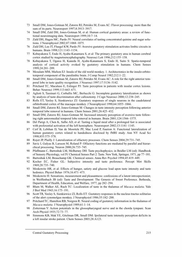
Central Gustatory Processing 215
73 Small DM, Jones-Gotman M, Zatorre RJ, Petrides M, Evans AC: Flavor processing: more than the
sum of its parts. Neuroreport 1997;8:3913–3917.
74 Small DM, Zald DH, Jones-Gotman M, et al: Human cortical gustatory areas: a review of func-
tional neuroimaging data. Neuroreport 1999;10:7–14.
75 Zald DH, Hagen MC, Pardo JV: Neural correlates of tasting concentrated quinine and sugar solu-
tions. J Neurophysiol 2002;87:1068–1075.
76 Zald DH, Lee JT, Fluegel KW, Pardo JV: Aversive gustatory stimulation activates limbic circuits in
humans. Brain 1998;121:1143–1154.
77 Kobayakawa T, Endo H, Ayabe-Kanamura S, et al: The primary gustatory area in human cerebral
cortex studied by magnetoencephalography. Neurosci Lett 1996;212:155–158.
78 Kobayakawa T, Ogawa H, Kaneda H, Ayabe-Kanamura S, Endo H, Saito S: Spatio-temporal
analysis of cortical activity evoked by gustatory stimulation in humans. Chem Senses
1999;24:201–209.
79 Mesulam MM, Mufson EJ: Insula of the old world monkey. 1. Architectonics in the insulo-orbito-
temporal component of the paralimbic brain. J Comp Neurol 1982;212:1–22.
80 Small DM, Jones-Gotman M, Zatorre RJ, Petrides M, Evans AC: A role for the right anterior tem-
poral lobe in taste quality recognition. J Neurosci 1997;17:5136–5142.
81 Pritchard TC, Macaluso E, Eslinger PJ: Taste perception in patients with insular cortex lesions.
Behav Neurosci 1999;113:663–671.
82 Aglioti S, Tassinari G, Corballis MC, Berlucchi G: Incomplete gustatory lateralization as shown
by analysis of taste discrimination after callosotomy. J Cogn Neurosci 2000;12:238–245.
83 Rolls ET, Yaxley S, Sienkiewicz ZJ: Gustatory responses of single neurons in the caudolateral
orbitofrontal cortex of the macaque monkey. J Neurophysiol 1990;64:1055–1066.
84 Small DM, Zatorre RJ, Jones-Gotman M: Changes in taste intensity perception following anterior
temporal lobe removal in humans. Chem Senses 2001;26:425–432.
85 Small DM, Zatorre RJ, Jones-Gotman M: Increased intensity perception of aversive taste follow-
ing right anteromedial temporal lobe removal in humans. Brain 2001;124:1566–1575.
86 Del Parigi A, Chen K, Salbe AD, et al: Tasting a liquid meal after a prolonged fast is associated
with preferential activation of the left hemisphere. Neuroreport 2002;13:1141–1145.
87 Cerf B, Lebihan D, Van de Moortele PF, Mac Leod P, Faurion A: Functional lateralization of
human gustatory cortex related to handedness disclosed by fMRI study. Ann NY Acad Sci
1998;855:575–578.
88 Royet JP, Plailly J: Lateralization of olfactory processes. Chem Senses 2004;29:731–745.
89 Savic I, Gulyas B, Larsson M, Roland P: Olfactory functions are mediated by parallel and hierar-
chical processing. Neuron 2000;26:735–745.
90 Pfaffmann C, Bartoshuk LM, McBurney DH: Taste psychophysics; in Beidler LM (ed): Handbook
of Sensory Physiology, vol IV. Chemical Senses Part 2: Taste. New York, Springer, 1971, pp 77–101.
91 Bartoshuk LM, Beauchamp GK: Chemical senses. Annu Rev Psychol 1994;45:419–449.
92 Kocher EC, Fisher GL: Subjective intensity and taste preference. Percept Mot Skills
1969;28:735–740.
93 Moskowitz HR, et al: Effects of hunger, satiety and glucose load upon taste intensity and taste
hedonics. Physiol Behav 1976;16:471–475.
94 Moskowitz H: Sensations, measurement and pleasantness: confessions of a latent introspectionist;
in Weiffenbach JB (ed): Taste and Development: The Genesis of Sweet Preference. Bethesda,
Department of Health, Education, and Welfare, 1977, pp 282–294.
95 Blum M, Walker AE, Ruch TC: Localization of taste in the thalamus of Macaca mulatta. Yale
J Biol Med 1943;16:175–191.
96 Scott TR, Yaxley S, Sienkiewicz ZJ, Rolls ET: Gustatory responses in the nucleus tractus solitarius
of the alert cynomolgus monkey. J Neurophysiol 1986;55:182–200.
97 Pritchard TC, Hamilton RB, Norgren R: Neural coding of gustatory information in the thalamus of
Macaca mulatta. J Neurophysiol 1989;61:1–14.
98 Zotterman Y: Action potentials in the glossopharyngeal nerve and in the chorda tympani. Scan
Arch Physiol 1935;72:73–77.
99 Simmons KB, Mak YE, Gitelman DR, Small DM: Ipsilateral taste intensity perception deficits in
a left insular stroke patient. Chem Senses 2003;28:A123.

Small 216
100 Kveton JF, Bartoshuk LM: The effect of unilateral chorda tympani damage on taste. Laryngoscope
1994;104:25–29.
101 Yanagisawa K, Bartoshuk LM, Catalanotto FA, Karrer TA, Kveton JF: Anesthesia of the chorda
tympani nerve and taste phantoms. Physiol Behav 1998;63:329–335.
102 Touzani K, Taghzouti K, Velley L: Increase of the aversive value of taste stimuli following ibotenic
acid lesion of the central amygdaloid nucleus in the rat. Behav Brain Res 1997;88:133–142.
103 Breslin PA: Human gustation; in Finger TE, Singer WL (eds): The Neurobiology of Taste and
Smell. San Diego, Wiley-Liss Inc, 2000, pp 423–461.
104 Smith DV, John SJ, Boughter JD: Neuronal cell types and taste quality coding. Physiol Behav
2000;69:77–85.
105 Smith DV, St John SJ: Neural coding of gustatory information. Curr Opin Neurobiol 1999;9:
427–435.
106 Scott TR, Giza BK: Theories of gustatory neural coding; in Doty RL (ed): Handbook of Olfaction
and Gustation. New York, Dekker, 1995, pp 573–603.
107 Frank ME: Neuron types, receptors, behavior, and taste quality. Physiol Behav 2000;69:53–62.
108 Pfaffmann C: The afferent code for sensory quality. Am J Psychol 1959;14:226–232.
109 Smith DV, Scott TR: Gustatory neural coding; in Doty RL (ed): Handbook of Olfaction and
Gustation. Philadelphia, Dekker, 2003, pp 731–758.
110 Sato M, Ogawa H, Yamashita S: Response properties of macaque monkey chorda tympani fibers.
J Gen Physiol 1975;66:781–810.
111 Hellekant G, Danilova V, Ninomiya Y: Primate sense of taste: behavioral and single chorda tympani
and glossopharyngeal nerve fiber recordings in the rhesus monkey, Macaca mulata. J Neurophysiol
1997;77:978–993.
112 Gordon G, Kitchell R, Strom L, Zotterman Y: The response pattern of taste fibers in the chorda
tympani of the monkey. Acta Physiol Scand 1959;46:119–132.
113 Smith DV, Travers JB: A metric for the breadth of tuning of gustatory neurons. Chem Senses
1979;4:215–229.
114 Katz DB, Simon SA, Nicolelis MA: Taste-specific neuronal ensembles in the gustatory cortex of
awake rats. J Neurosci 2002;22:1850–1857.
115 Kuznicki JT, Ashbaugh N: Taste quality differences within sweet and salty taste categories. Sens
Processes 1979;3:157–182.
116 Schiffman SS, Erickson RP: A psychophysical model for gustatory quality of sodium salts.
Physiol Behav 1971;7:617–633.
117 Cereda C, Ghika J, Maeder P, Bogousslavsky J: Strokes restricted to the insular cortex. Neurology
2002;59:1950–1955.
118 Henkin RI, Comiter H, Fedio P, O’Doherty D: Defects in taste and smell recognition following
temporal lobectomy. Trans Am Neurol Assoc 1977;102:146–150.
119 Small DM, Bernasconi N, Bernasconi A, Sziklas V, Jones-Gotman M: Gustatory agnosia.
Neurology 2005;64:311–317.
120 Halpern BP, Nelson LM: Bulbar gustatory responses to anterior and to posterior tongue stimula-
tion in the rat. Am J Physiol 1965;202:105–110.
121 Dickman JD, Smith DV: Topographic distribution of taste responsiveness in the hamster medulla.
Chem Senses 1989;14:231–247.
122 Sudakov K, Maclean PD, Reeves A, Marini R: Unit study of exteroceptive inputs to claustrocortex
in awake, sitting squirrel monkeys. Brain Res 1971;28:19–34.
123 Plata-Salaman CR, Smith-Swintosky VL, Scott TR: Gustatory neural coding in the monkey cor-
tex: mixtures. J Neurophysiol 1996;75:2369–2379.
124 Small DM, Zatorre RJ, Dagher A, Evans AC, Jones-Gotman M: Changes in brain activity related
to eating chocolate: from pleasure to aversion. Brain 2001;124:1720–1733.
125 Steiner JE, Glaser D, Hawilo ME, Berridge KC: Comparative expression of hedonic impact: affec-
tive reactions to taste by human infants and other primates. Neurosci Biobehav Rev 2001;25:53–74.
126 Steiner JE: Facial expressions of the neonate infant indicating the hedonics of food-related stimuli;
in Weiffenbach JM (ed): Taste and Development: The Genesis of Sweet Preference. Washington,
US Department of Health and Human Services, 1979.
127 Rolls ET: The Brain and Emotion. Oxford, Oxford University Press, 1999.
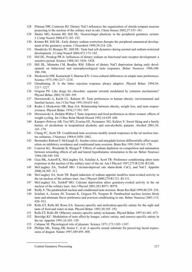
Central Gustatory Processing 217
128 Pittman DW, Contreras RJ: Dietary NaCl influences the organization of chorda tympani neurons
projecting to the nucleus of the solitary tract in rats. Chem Senses 2002;27:333–341.
129 Shuler MG, Krimm RF, Hill DL: Neuron/target plasticity in the peripheral gustatory system.
J. Comp Neurol 2004;472:183–192.
130 Krimm RF, Hill DL: Early dietary sodium restriction disrupts the peripheral anatomical develop-
ment of the gustatory system. J Neurobiol 1999;39:218–226.
131 Hendricks SJ, Brunjes PC, Hill DL: Taste bud cell dynamics during normal and sodium-restricted
development. J Comp Neurol 2004;472:173–182.
132 Hill DL, Przekop PR Jr: Influences of dietary sodium on functional taste receptor development: a
sensitive period. Science 1988;241:1826–1828.
133 Hill DL, Mistretta CM, Bradley RM: Effects of dietary NaCl deprivation during early devel-
opment on behavioral and neurophysiological taste responses. Behav Neurosci 1986;100:
390–398.
134 Moskowitz HW, Kumaraiah V, Sharma KN: Cross-cultural differences in simple taste preferences.
Science 1975;190:1217–1218.
135 Glendinning JI: Is the bitter rejection response always adaptive. Physiol Behav 1994;56:
1217–1227.
136 Grigson PS: Like drugs for chocolate: separate rewards modulated by common mechanisms?
Physiol Behav 2002;76:389–395.
137 Drewnowski A, Kurth CL, Rahaim JE: Taste preferences in human obesity: environmental and
familial factors. Am J Clin Nutr 1991;54:635–641.
138 Rodin J, Moskowitz HR, Bray GA: Relationship between obesity, weight loss, and taste respon-
siveness. Physiol Behav 1976;17:591–597.
139 Drewnowski A, Holden-Wiltse J: Taste responses and food preferences in obese women: effects of
weight cycling. Int J Obes Relat Metab Disord 1992;16:639–648.
140 Kampov-Polevoy AB, Tsoi MV, Zvartau EE, Nezmanov NG, Kalitov E: Sweet liking and a family
history of alcoholism in hospitalized alcoholic and non-alcoholic patients. Alcohol 2001;36:
165–170.
141 Chang FC, Scott TR: Conditioned taste aversions modify neural responses in the rat nucleus trac-
tus solitarius. J Neurosci 1984;4:1850–1862.
142 Bermudez-Rattoni F, McGaugh JL: Insular cortex and amygdala lesions differentially affect acqui-
sition on inhibitory avoidance and conditioned taste aversion. Brain Res 1991;549:165–170.
143 Conover KL, Woodside B, Shizgal P: Effects of sodium depletion on competition and summation
between rewarding effects of salt and lateral hypothalamic stimulation in the rat. Behav Neurosci
1994;108:549–558.
144 Giza BK, Ackroff K, McCaughey SA, Sclafani A, Scott TR: Preference conditioning alters taste
responses in the nucleus of the solitary tract of the rat. Am J Physiol 1997;273:R1230–R1240.
145 McCaughey SA, Tordoff MG: Calcium-deprived rats sham-drink CaCl2 and NaCl. Appetite
2000;34:305–311.
146 McCaughey SA, Scott TR: Rapid induction of sodium appetite modifies taste-evoked activity in
the rat nucleus of the solitary tract. Am J Physiol 2000;279:R1121–R1131.
147 McCaughey SA, Tordoff MG: Calcium deprivation alters gustatory-evoked activity in the rat
nucleus of the solitary tract. Am J Physiol 2001;281:R971–R978.
148 Reilly S: The parabrachial nucleus and conditioned taste aversion. Brain Res Bull 1999;48:239–254.
149 Sclafani A, Azzara AV, Touzani K, Grigson PS, Norgren R: Parabrachial nucleus lesions block
taste and attenuate flavor preference and aversion conditioning in rats. Behav Neurosci 2001;115:
920–933.
150 Rolls ET, Rolls BJ, Rowe EA: Sensory-specific and motivation-specific satiety for the sight and
taste of food and water in man. Physiol Behav 1983;30:185–192.
151 Rolls ET, Rolls JH: Olfactory sensory-specific satiety in humans. Physiol Behav 1997;61:461–473.
152 Berridge KC: Modulation of taste affect by hunger, caloric satiety, and sensory-specific satiety in
the rat. Appetite 1991;16:103–120.
153 Cabanac M: Physiological role of pleasure. Science 1971;173:1103–1107.
154 Phillips ML, Young AW, Senior C, et al: A specific neural substrate for perceiving facial expres-
sions of disgust. Nature 1997;389:495–498.
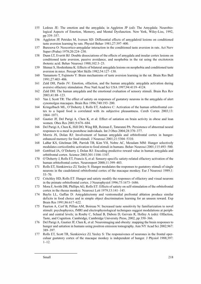
Small 218
155 Ledoux JE: The emotion and the amygdala; in Aggleton JP (ed): The Amygdala: Neurobio-
logical Aspects of Emotion, Memory, and Mental Dysfunction. New York, Wiley-Liss, 1992,
pp 339–357.
156 Aggleton JP, Petrides M, Iversen SD: Differential effects of amygdaloid lesions on conditioned
taste aversion learning by rats. Physiol Behav 1981;27:397–400.
157 Buresova O: Neocortico-amygdalar interaction in the conditioned taste aversion in rats. Act Nerv
Super (Praha) 1978;20:224–230.
158 Dunn LT, Everitt BJ: Double dissociations of the effects of amygdala and insular cortex lesions on
conditioned taste aversion, passive avoidance, and neophobia in the rat using the excitotoxin
ibotenic acid. Behav Neurosci 1988;102:3–23.
159 Shimai S, Hoshishima K: Effects of bilateral amygdala lesions on neophobia and conditioned taste
aversion in mice. Percept Mot Skills 1982;54:127–130.
160 Yamamoto T, Fujimoto Y: Brain mechanisms of taste aversion learning in the rat. Brain Res Bull
1991;27:403–406.
161 Zald DH, Pardo JV: Emotion, olfaction, and the human amygdala: amygdala activation during
aversive olfactory stimulation. Proc Natl Acad Sci USA 1997;94:4119–4124.
162 Zald DH: The human amygdala and the emotional evaluation of sensory stimuli. Brain Res Rev
2003;41:88–123.
163 Yan J, Scott TR: The effect of satiety on responses of gustatory neurons in the amygdala of alert
cynomolgus macaques. Brain Res 1996;740:193–200.
164 Kringelbach ML, O’Doherty J, Rolls ET, Andrews C: Activation of the human orbitofrontal cor-
tex to a liquid food is correlated with its subjective pleasantness. Cereb Cortex 2003;13:
1064–1071.
165 Gautier JF, Del Parigi A, Chen K, et al: Effect of satiation on brain activity in obese and lean
women. Obes Res 2001;9:676–684.
166 Del Parigi A, Chen K, Hill DO, Wing RR, Reiman E, Tataranni PA: Persistence of abnormal neural
responses to a meal in postobese individuals. Int J Obes 2004;28:370–377.
167 Morris JS, Dolan RJ: Involvement of human amygdala and orbitofrontal cortex in hunger-
enhanced memory for food stimuli. J Neurosci 2001;21:5304–5310.
168 LaBar KS, Gitelman DR, Parrish TB, Kim YH, Nobre AC, Mesulam MM: Hunger selectively
modulates corticolimbic activation to food stimuli in humans. Behav Neurosci 2001;115:493–500.
169 Gottfried JA, O’Doherty J, Dolan RJ: Encoding predictive reward value in human amygdala and
orbitfrontal cortex. Science 2003;301:1104–1107.
170 O’Doherty J, Rolls ET, Francis S, et al: Sensory-specific satiety-related olfactory activation of the
human orbitofrontal cortex. Neuroreport 2000;11:399–403.
171 Rolls ET, Sienkiewicz ZJ, Yaxley S: Hunger modulates the responses to gustatory stimuli of single
neurons in the caudolateral orbitofrontal cortex of the macaque monkey. Eur J Neurosci 1989;1:
53–70.
172 Critchley HD, Rolls ET: Hunger and satiety modify the responses of olfactory and visual neurons
in the primate orbitofrontal cortex. J Neurophysiol 1996;75:1673–1686.
173 Mora F, Avrith DB, Phillips AG, Rolls ET: Effects of satiety on self-stimulation of the orbitofrontal
cortex in the rhesus monkey. Neurosci Lett 1979;13:141–145.
174 Baylis LL, Gaffan D: Amygdalectomy and ventromedial prefrontal ablation produce similar
deficits in food choice and in simple object discrimination learning for an unseen reward. Exp
Brain Res 1991;86:617–622.
175 Faurion A, Cerf B, Pillias AM, Boireau N: Increased taste sensitivity by familiarization to novel
stimuli: psychophysics, fMRI and electrophysiological techniques suggest modulations at periph-
eral and central levels; in Rouby C, Schaal B, Dubois D, Gervais R, Holley A (eds): Olfaction,
Taste, and Cognition. Cambridge, Cambridge University Press, 2002, pp 350–366.
176 Del Parigi A, Gautier JF, Chen K, et al: Neuroimaging and obesity: mapping the brain responses to
hunger and satiation in humans using positron emission tomography. Ann NY Acad Sci 2002;967:
389–397.
177 Rolls ET, Scott TR, Sienkiewicz ZJ, Yaxley S: The responsivenes of neurones in the frontal oper-
culuar gustatory cortex of the macaque monkey is independent of hunger. J Physiol 1988;397:
1–12.
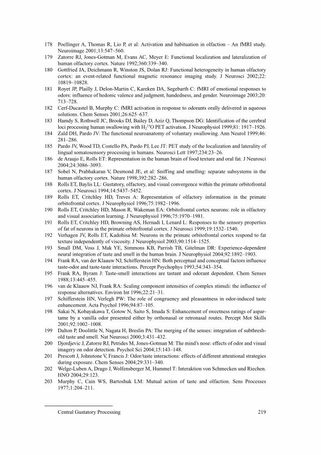
Central Gustatory Processing 219
178 Poellinger A, Thomas R, Lio P, et al: Activation and habituation in olfaction – An fMRI study.
Neuroimage 2001;13:547–560.
179 Zatorre RJ, Jones-Gotman M, Evans AC, Meyer E: Functional localization and lateralization of
human olfactory cortex. Nature 1992;360:339–340.
180 Gottfried JA, Deichmann R, Winston JS, Dolan RJ: Functional heterogeneity in human olfactory
cortex: an event-related functional magnetic resonance imaging study. J Neurosci 2002;22:
10819–10828.
181 Royet JP, Plailly J, Delon-Martin C, Kareken DA, Segebarth C: fMRI of emotional responses to
odors: influence of hedonic valence and judgment, handedness, and gender. Neuroimage 2003;20:
713–728.
182 Cerf-Ducastel B, Murphy C: fMRI activation in response to odorants orally delivered in aqueous
solutions. Chem Senses 2001;26:625–637.
183 Hamdy S, Rothwell JC, Brooks DJ, Bailey D, Aziz Q, Thompson DG: Identification of the cerebral
loci processing human swallowing with H215O PET activation. J Neurophysiol 1999;81: 1917–1926.
184 Zald DH, Pardo JV: The functional neuroanatomy of voluntary swallowing. Ann Neurol 1999;46:
281–286.
185 Pardo JV, Wood TD, Costello PA, Pardo PJ, Lee JT: PET study of the localization and laterality of
lingual somatosensory processing in humans. Neurosci Lett 1997;234:23–26.
186 de Araujo E, Rolls ET: Representation in the human brain of food texture and oral fat. J Neurosci
2004;24:3086–3093.
187 Sobel N, Prabhakaran V, Desmond JE, et al: Sniffing and smelling: separate subsystems in the
human olfactory cortex. Nature 1998;392:282–286.
188 Rolls ET, Baylis LL: Gustatory, olfactory, and visual convergence within the primate orbitofrontal
cortex. J Neurosci 1994;14:5437–5452.
189 Rolls ET, Critchley HD, Treves A: Representation of olfactory information in the primate
orbitofrontal cortex. J Neurophysiol 1996;75:1982–1996.
190 Rolls ET, Critchley HD, Mason R, Wakeman EA: Orbitofrontal cortex neurons: role in olfactory
and visual association learning. J Neurophysiol 1996;75:1970–1981.
191 Rolls ET, Critchley HD, Browning AS, Hernadi I, Lenard L: Responses to the sensory properties
of fat of neurons in the primate orbitofrontal cortex. J Neurosci 1999;19:1532–1540.
192 Verhagen JV, Rolls ET, Kadohisa M: Neurons in the primate orbitofrontal cortex respond to fat
texture independently of viscosity. J Neurophysiol 2003;90:1514–1525.
193 Small DM, Voss J, Mak YE, Simmons KB, Parrish TB, Gitelman DR: Experience-dependent
neural integration of taste and smell in the human brain. J Neurophysiol 2004;92:1892–1903.
194 Frank RA, van der Klaauw NJ, Schifferstein HN: Both perceptual and conceptual factors influence
taste-odor and taste-taste interactions. Percept Psychophys 1993;54:343–354.
195 Frank RA, Byram J: Taste-smell interactions are tastant and odorant dependent. Chem Senses
1988;13:445–455.
196 van de Klaauw NJ, Frank RA: Scaling component intensities of complex stimuli: the influence of
response alternatives. Environ Int 1996;22:21–31.
197 Schifferstein HN, Verlegh PW: The role of congruency and pleasantness in odor-induced taste
enhancement. Acta Psychol 1996;94:87–105.
198 Sakai N, Kobayakawa T, Gotow N, Saito S, Imada S: Enhancement of sweetness ratings of aspar-
tame by a vanilla odor presented either by orthonasal or retronasal routes. Percept Mot Skills
2001;92:1002–1008.
199 Dalton P, Doolittle N, Nagata H, Breslin PA: The merging of the senses: integration of subthresh-
old taste and smell. Nat Neurosci 2000;3:431–432.
200 Djordjevic J, Zatorre RJ, Petrides M, Jones-Gotman M: The mind’s nose: effects of odor and visual
imagery on odor detection. Psychol Sci 2004;15:143–148.
201 Prescott J, Johnstone V, Francis J: Odor/taste interactions: effects of different attentional strategies
during exposure. Chem Senses 2004;29:331–340.
202 Welge-Luben A, Drago J, Wolfensberger M, Hummel T: Interaktion von Schmecken und Riechen.
HNO 2004;29:123.
203 Murphy C, Cain WS, Bartoshuk LM: Mutual action of taste and olfaction. Sens Processes
1977;1:204–211.
Small 220
204 Rozin P: ‘Taste-smell confusions’ and the duality of the olfactory sense. Percept Psychophys
1982;31:397–401.
205 Robinson CJ, Burton H: Organization of somatosensory receptive fields in cortical areas 7b,
retroinsula, postauditory and granular insula of M. fascicularis. J Comp Neurol 1980;192:69–92.
206 Green BG: Studying taste as a cutaneous sense. Food Qual Pref 2002;14:99–109.
207 Clark CC, Lawless HT: Limiting response alternatives in time-intensity scaling: an examination of
the halo-dumping effect. Chem Senses 1994;19:583–594.
208 Pribram KH, Bagshaw M: Further analysis of the temporal lobe syndrome utilizing fronto-tempo-
ral ablations. J Comp Neurol 1953;2:347–375.
Dana M. Small, PhD
The John B. Pierce Laboratory
290 Congress Avenue
New Haven, CT 06519 (USA)
Tel. �1 203 401 6204, Fax �1 203 624 4950, E-Mail [email protected]
Hummel T, Welge-Lüssen A (eds): Taste and Smell. An Update.
Adv Otorhinolaryngol. Basel, Karger, 2006, vol 63, pp 221–241
Modern Psychophysics and theAssessment of Human Oral Sensation
Derek J. Snydera,b, John Prescottc, Linda M. Bartoshukb
aInterdepartmental Neuroscience Program, Yale University, New Haven, Conn.,bCenter for Taste and Smell, University of Florida, Gainesville, Fla., USA; cSchool of
Psychology, James Cook University, Cairns, Australia
AbstractPsychophysical measures attempt to capture and compare subjective experiences objec-
tively. In the chemical senses, these techniques have been instrumental in describing relation-
ships between oral sensation and health risk, but they are often used incorrectly to make group
comparisons. This chapter reviews contemporary methods of oral sensory assessment, with
particular emphasis on suprathreshold scaling. We believe that these scales presently offer the
most realistic picture of oral sensory function, but only when they are used correctly. Using
converging methods from psychophysics, anatomy, and genetics, we demonstrate valid uses of
modern chemosensory testing in clinical diagnosis and intervention.
Copyright © 2006 S. Karger AG, Basel
Psychophysical measures of experience have played a fundamental role in
our understanding of sensory and hedonic processes. In the chemical senses,
these measures have revealed the broad impact of oral sensation and dysfunc-
tion on health-related behaviors and overall quality of life [1]. Oral sensory dis-
turbances may be relatively benign, but sometimes they are profoundly life altering.
As such, chemosensory experience and its consequences represent an important
clinical concern.
Assessing this experience, however, is an extremely challenging task. By
definition, individual experience is subjective: we can describe our experiences
and track them over time, but we cannot directly share the experiences of another
person. Nevertheless, we use comparisons of real-world experience throughout
life to communicate what is acceptable (e.g. pleasure, comfort) and what is
not (e.g. disease, pain), so it is important to evaluate these experiences care-
fully. Recent advances in suprathreshold scaling capture sensory and affective
Snyder/Prescott/Bartoshuk 222
differences with improved accuracy, supporting the notion that perceptual expe-
riences can be measured and compared.
In this chapter, we address methodological issues regarding threshold and
suprathreshold measures of oral sensation. Using magnitude matching as an
example, we argue that suprathreshold intensity scales provide a more complete
picture of oral sensory function than do thresholds alone. Our efforts to identify
useful suprathreshold tools include an examination of labeled scales, which are
used (and misused) to compare experiences between individuals and groups. To
confirm our psychophysical results, we demonstrate the use of parallel methods
(e.g. multiple standards, genetic and anatomical tools). Finally, using techniques
appropriate for comparison, we show how spatial taste testing has advanced our
understanding of oral sensory function in health and disease.
Thresholds versus Intensity: How Should Oral Sensation Be Measured?
When we enjoy a meal, we can easily tell if the soup is too salty or the
cocktails watered down. These judgments demonstrate that intensity is continu-
ous (i.e. strong or weak variants of stimulus strength) and not binary (i.e. pres-
ent or absent). Some researchers believe that degrees of suprathreshold intensity
are immeasurable or at best ordinal [2], but we contend that suprathreshold
measures possess unique diagnostic and predictive capabilities.
Indirect Psychophysics: Threshold Procedures
Thresholds have been used for sensory evaluation ever since Fechner [3]
codified them almost 150 years ago. Although thresholds present technical
challenges, they are conceptually straightforward: the absolute threshold for a
stimulus is the lowest concentration at which its presence can be detected as
something, whether or not it is qualitatively discernible. The recognition thresh-old is the lowest concentration at which the quality of a stimulus (e.g. sweet,
painful) can be identified. Finally, the difference threshold is the smallest
increase in suprathreshold stimulus concentration that can be detected (i.e. the
‘just noticeable difference’).
Thresholds enjoy widespread use in research and clinical settings, mainly
because they produce values suitable for comparison. Because thresholds are
especially sensitive to sensory adaptation, subject fatigue, and criterion shift [4],
abbreviated methods have been developed that provide reliable threshold estimates
with fewer trials and minimal bias [5–7]. Even so, one of the most discouraging
Psychophysical Assessment of Human Oral Sensation 223
features of thresholds is the time required to measure them: an up-down, forced-
choice threshold procedure [7] takes approx. 20 min to administer, yielding only
the lower boundary of the taste function; suprathreshold procedures approxi-
mate the entire taste function in much less time. Generally, the decision to use
thresholds in lieu of suprathreshold measures is reasonable when there is strong
concordance between threshold and suprathreshold experiences. However, psy-
chophysical functions for taste stimuli show considerable variation that precludes
reasonable predictions of suprathreshold sensation from threshold values
alone [8].
As an alternative to chemical measures of taste sensitivity, electrogustom-
etry involves the application of weak anodal electric currents to specific regions
of the mouth [9]. Proponents of electrogustometry emphasize its convenience
[10]; it is portable, avoids the use of chemical solutions, permits regional stim-
ulation of taste bud fields, and provides values that can be compared across
individuals, time points, locations within the mouth, or treatment conditions.
Electric taste thresholds show high test-retest reliability and bilateral corre-
spondence [11], and normative data have been described for some groups [12].
Accordingly, electrogustometry has been used to identify sizable taste losses
associated with aging, denervation, and disease [11, 13, 14], but the following
two disadvantages limit its use in more specific clinical assessments of taste
function [15].
• Because saliva is mildly acidic and contains salts, electrogustometry typi-
cally evokes sour or salty taste sensations [16]. However, oral sensory alter-
ations are often quality specific, particularly affecting bitter taste [17, 18]. As
such, electrogustometry may fail to identify clinically relevant damage.
• Electrogustometric thresholds correlate well with regional [12, 19] but not
whole-mouth chemical taste thresholds [11]; suprathreshold functions for
electrical and chemical taste also show poor agreement [20]. Thus, as with
chemical taste thresholds (see above), electrogustometry cannot reflect real-
world taste experience accurately.
Direct Psychophysical Scaling of Suprathreshold Intensity
Thresholds provide only the lower limit of physical energy that can be per-
ceived (e.g. decibels of sound, molar concentration), but suprathreshold or
‘direct’ scaling methods measure perceived intensity across the full dynamic
range of sensation [21]. S.S. Stevens [22] introduced direct scaling methods with
ratio properties, the most popular of which is magnitude estimation. In this proce-
dure, subjects provide a number reflecting perceived stimulus intensity; they then
give a number twice as large to a stimulus that is twice as intense, a number half

Snyder/Prescott/Bartoshuk 224
as large to a stimulus half as intense, and so on. The size of the numbers is irrele-
vant; only the ratios among numbers carry meaning. As a result, magnitude esti-
mates describe only how perceived intensity varies with stimulus intensity withinan individual; they cannot reflect meaningful differences of absolute perceived
intensity between individuals or groups [23]. Because group comparisons are
such a basic element of scientific analysis, this limitation has not been fully
appreciated, but its consequences are severe. To illustrate, we now describe stud-
ies on individual differences in taste perception; these studies are especially note-
worthy in terms of their contributions to comparative suprathreshold scaling.
Genetic Variation in Oral Sensation: The Rise of Magnitude Matching
Taste Blindness: We Live in Different Oral Sensory Worlds
Discovered by the chemist A.L. Fox in 1931 [24], individuals differ signif-
icantly in their ability to taste thiourea compounds like phenylthiocarbamide
(PTC) and 6-n-propylthiouracil (PROP) [25]; most individuals perceive bitter-
ness (i.e. tasters), but others are ‘taste blind’ and perceive nothing (i.e. non-
tasters). Early reports suggested that taste blindness is a recessive trait inherited
through a single genetic locus [26], while other studies measured the proportion
of tasters by race, sex, and disease [27–29]. In the 1960s, behavioral experi-
ments showed that PTC/PROP threshold sensitivity influences food prefer-
ences, alcohol and tobacco use, and body weight [30].
Buoyed by the potential benefits of direct scaling, Bartoshuk sought to
compare the suprathreshold bitterness of PTC between nontasters and tasters.
However, this comparison presents a problem: magnitude estimates have relativemeaning when subjects are used as their own controls [31], but how can absolutebitterness rating be compared across groups? The answer to this question
involves measuring PTC bitterness relative to an unrelated standard. Although
magnitude estimates are often multiplied by a constant (i.e. ‘normalized’) in
order to obtain group functions [32], this procedure is qualitatively different.
• To average magnitude estimates while maintaining the ratios among them,
ratings must be brought into a common register so that one subject’s data are
not unduly weighted just because that subject used larger numbers [23].
With this type of normalization, the standard is arbitrary, so group functions
convey nothing about absolute perceived intensity.
• When the purpose of normalization is to permit group comparisons, the
standard is assumed to be equally intense (on average) to the groups being
compared. If this assumption holds, normalization yields valid across-group
differences in absolute perceived intensity for stimuli of interest: if ‘10’
denotes the intensity of a standard to nontasters and tasters, PTC ratings of
‘40’ for tasters and ‘20’ for nontasters reflect a twofold intensity difference.
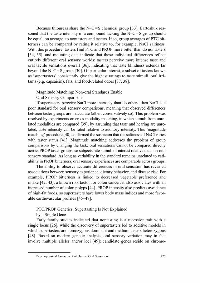
Psychophysical Assessment of Human Oral Sensation 225
Because thioureas share the N–C�S chemical group [33], Bartoshuk rea-
soned that the taste intensity of a compound lacking the N–C�S group should
be equal, on average, to nontasters and tasters. If so, group averages of PTC bit-
terness can be compared by rating it relative to, for example, NaCl saltiness.
With this procedure, tasters find PTC and PROP more bitter than do nontasters
[34, 35], and mounting data indicate that these individual differences reflect
entirely different oral sensory worlds: tasters perceive more intense taste and
oral tactile sensations overall [36], indicating that taste blindness extends far
beyond the N–C�S group [30]. Of particular interest, a subset of tasters known
as ‘supertasters’ consistently give the highest ratings to taste stimuli, oral irri-
tants (e.g. capsaicin), fats, and food-related odors [37, 38].
Magnitude Matching: Non-oral Standards Enable
Oral Sensory Comparisons
If supertasters perceive NaCl more intensely than do others, then NaCl is a
poor standard for oral sensory comparisons, meaning that observed differences
between taster groups are inaccurate (albeit conservatively so). This problem was
resolved by experiments on cross-modality matching, in which stimuli from unre-
lated modalities are compared [39]; by assuming that taste and hearing are unre-
lated, taste intensity can be rated relative to auditory intensity. This ‘magnitude
matching’ procedure [40] confirmed the suspicion that the saltiness of NaCl varies
with taster status [41]. Magnitude matching addresses the problem of group
comparisons by changing the task: oral sensations cannot be compared directly
across PROP taster groups, so subjects rate stimuli of interest relative to a non-oral
sensory standard. As long as variability in the standard remains unrelated to vari-
ability in PROP bitterness, oral sensory experiences are comparable across groups.
The ability to observe accurate differences in oral sensation has revealed
associations between sensory experience, dietary behavior, and disease risk. For
example, PROP bitterness is linked to decreased vegetable preference and
intake [42, 43], a known risk factor for colon cancer; it also associates with an
increased number of colon polyps [44]. PROP intensity also predicts avoidance
of high-fat foods, so supertasters have lower body mass indices and more favor-
able cardiovascular profiles [45–47].
PTC/PROP Genetics: Supertasting Is Not Explained
by a Single Gene
Early family studies indicated that nontasting is a recessive trait with a
single locus [26], while the discovery of supertasters led to additive models in
which supertasters are homozygous dominant and medium tasters heterozygous
[48]. Based on modern genetic analysis, oral sensory variation may in fact
involve multiple alleles and/or loci [49]: candidate genes reside on chromo-
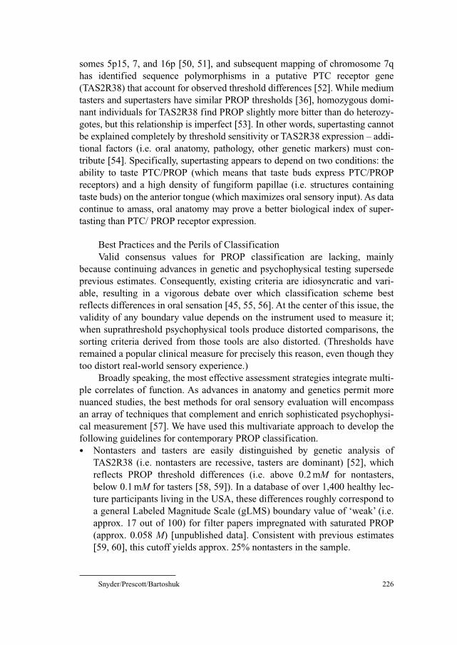
Snyder/Prescott/Bartoshuk 226
somes 5p15, 7, and 16p [50, 51], and subsequent mapping of chromosome 7q
has identified sequence polymorphisms in a putative PTC receptor gene
(TAS2R38) that account for observed threshold differences [52]. While medium
tasters and supertasters have similar PROP thresholds [36], homozygous domi-
nant individuals for TAS2R38 find PROP slightly more bitter than do heterozy-
gotes, but this relationship is imperfect [53]. In other words, supertasting cannot
be explained completely by threshold sensitivity or TAS2R38 expression – addi-
tional factors (i.e. oral anatomy, pathology, other genetic markers) must con-
tribute [54]. Specifically, supertasting appears to depend on two conditions: the
ability to taste PTC/PROP (which means that taste buds express PTC/PROP
receptors) and a high density of fungiform papillae (i.e. structures containing
taste buds) on the anterior tongue (which maximizes oral sensory input). As data
continue to amass, oral anatomy may prove a better biological index of super-
tasting than PTC/ PROP receptor expression.
Best Practices and the Perils of Classification
Valid consensus values for PROP classification are lacking, mainly
because continuing advances in genetic and psychophysical testing supersede
previous estimates. Consequently, existing criteria are idiosyncratic and vari-
able, resulting in a vigorous debate over which classification scheme best
reflects differences in oral sensation [45, 55, 56]. At the center of this issue, the
validity of any boundary value depends on the instrument used to measure it;
when suprathreshold psychophysical tools produce distorted comparisons, the
sorting criteria derived from those tools are also distorted. (Thresholds have
remained a popular clinical measure for precisely this reason, even though they
too distort real-world sensory experience.)
Broadly speaking, the most effective assessment strategies integrate multi-
ple correlates of function. As advances in anatomy and genetics permit more
nuanced studies, the best methods for oral sensory evaluation will encompass
an array of techniques that complement and enrich sophisticated psychophysi-
cal measurement [57]. We have used this multivariate approach to develop the
following guidelines for contemporary PROP classification.
• Nontasters and tasters are easily distinguished by genetic analysis of
TAS2R38 (i.e. nontasters are recessive, tasters are dominant) [52], which
reflects PROP threshold differences (i.e. above 0.2 mM for nontasters,
below 0.1 mM for tasters [58, 59]). In a database of over 1,400 healthy lec-
ture participants living in the USA, these differences roughly correspond to
a general Labeled Magnitude Scale (gLMS) boundary value of ‘weak’ (i.e.
approx. 17 out of 100) for filter papers impregnated with saturated PROP
(approx. 0.058 M) [unpublished data]. Consistent with previous estimates
[59, 60], this cutoff yields approx. 25% nontasters in the sample.
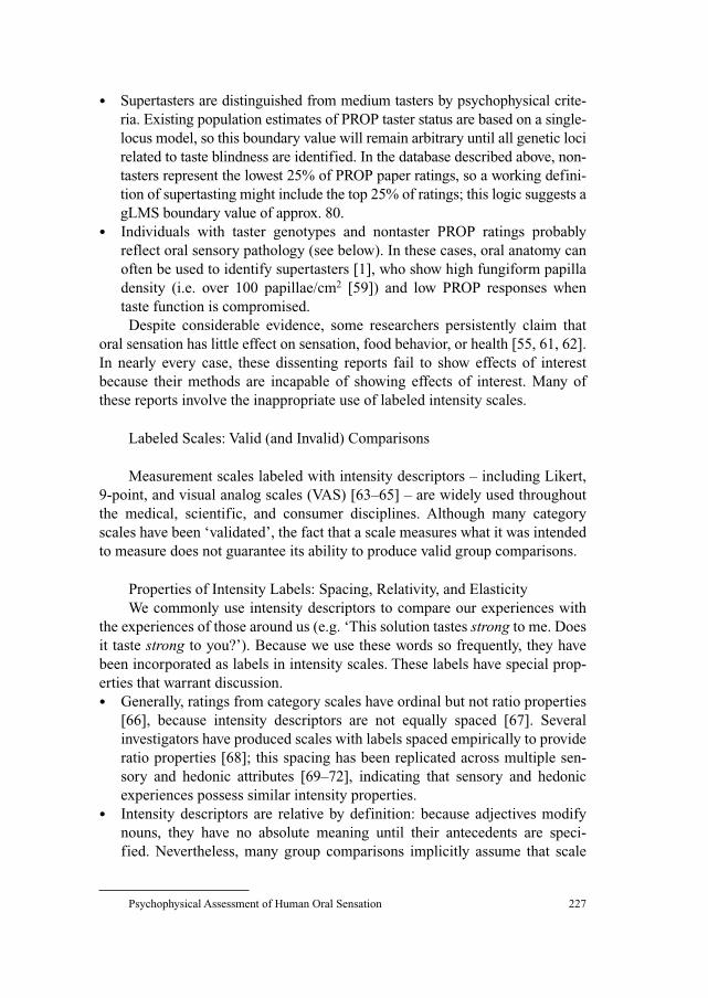
Psychophysical Assessment of Human Oral Sensation 227
• Supertasters are distinguished from medium tasters by psychophysical crite-
ria. Existing population estimates of PROP taster status are based on a single-
locus model, so this boundary value will remain arbitrary until all genetic loci
related to taste blindness are identified. In the database described above, non-
tasters represent the lowest 25% of PROP paper ratings, so a working defini-
tion of supertasting might include the top 25% of ratings; this logic suggests a
gLMS boundary value of approx. 80.
• Individuals with taster genotypes and nontaster PROP ratings probably
reflect oral sensory pathology (see below). In these cases, oral anatomy can
often be used to identify supertasters [1], who show high fungiform papilla
density (i.e. over 100 papillae/cm2 [59]) and low PROP responses when
taste function is compromised.
Despite considerable evidence, some researchers persistently claim that
oral sensation has little effect on sensation, food behavior, or health [55, 61, 62].
In nearly every case, these dissenting reports fail to show effects of interest
because their methods are incapable of showing effects of interest. Many of
these reports involve the inappropriate use of labeled intensity scales.
Labeled Scales: Valid (and Invalid) Comparisons
Measurement scales labeled with intensity descriptors – including Likert,
9-point, and visual analog scales (VAS) [63–65] – are widely used throughout
the medical, scientific, and consumer disciplines. Although many category
scales have been ‘validated’, the fact that a scale measures what it was intended
to measure does not guarantee its ability to produce valid group comparisons.
Properties of Intensity Labels: Spacing, Relativity, and Elasticity
We commonly use intensity descriptors to compare our experiences with
the experiences of those around us (e.g. ‘This solution tastes strong to me. Does
it taste strong to you?’). Because we use these words so frequently, they have
been incorporated as labels in intensity scales. These labels have special prop-
erties that warrant discussion.
• Generally, ratings from category scales have ordinal but not ratio properties
[66], because intensity descriptors are not equally spaced [67]. Several
investigators have produced scales with labels spaced empirically to provide
ratio properties [68]; this spacing has been replicated across multiple sen-
sory and hedonic attributes [69–72], indicating that sensory and hedonic
experiences possess similar intensity properties.
• Intensity descriptors are relative by definition: because adjectives modify
nouns, they have no absolute meaning until their antecedents are speci-
fied. Nevertheless, many group comparisons implicitly assume that scale
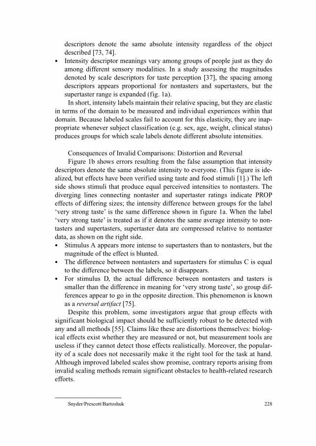
Snyder/Prescott/Bartoshuk 228
descriptors denote the same absolute intensity regardless of the object
described [73, 74].
• Intensity descriptor meanings vary among groups of people just as they do
among different sensory modalities. In a study assessing the magnitudes
denoted by scale descriptors for taste perception [37], the spacing among
descriptors appears proportional for nontasters and supertasters, but the
supertaster range is expanded (fig. 1a).
In short, intensity labels maintain their relative spacing, but they are elastic
in terms of the domain to be measured and individual experiences within that
domain. Because labeled scales fail to account for this elasticity, they are inap-
propriate whenever subject classification (e.g. sex, age, weight, clinical status)
produces groups for which scale labels denote different absolute intensities.
Consequences of Invalid Comparisons: Distortion and Reversal
Figure 1b shows errors resulting from the false assumption that intensity
descriptors denote the same absolute intensity to everyone. (This figure is ide-
alized, but effects have been verified using taste and food stimuli [1].) The left
side shows stimuli that produce equal perceived intensities to nontasters. The
diverging lines connecting nontaster and supertaster ratings indicate PROP
effects of differing sizes; the intensity difference between groups for the label
‘very strong taste’ is the same difference shown in figure 1a. When the label
‘very strong taste’ is treated as if it denotes the same average intensity to non-
tasters and supertasters, supertaster data are compressed relative to nontaster
data, as shown on the right side.
• Stimulus A appears more intense to supertasters than to nontasters, but the
magnitude of the effect is blunted.
• The difference between nontasters and supertasters for stimulus C is equal
to the difference between the labels, so it disappears.
• For stimulus D, the actual difference between nontasters and tasters is
smaller than the difference in meaning for ‘very strong taste’, so group dif-
ferences appear to go in the opposite direction. This phenomenon is known
as a reversal artifact [75].
Despite this problem, some investigators argue that group effects with
significant biological impact should be sufficiently robust to be detected with
any and all methods [55]. Claims like these are distortions themselves: biolog-
ical effects exist whether they are measured or not, but measurement tools are
useless if they cannot detect those effects realistically. Moreover, the popular-
ity of a scale does not necessarily make it the right tool for the task at hand.
Although improved labeled scales show promise, contrary reports arising from
invalid scaling methods remain significant obstacles to health-related research
efforts.
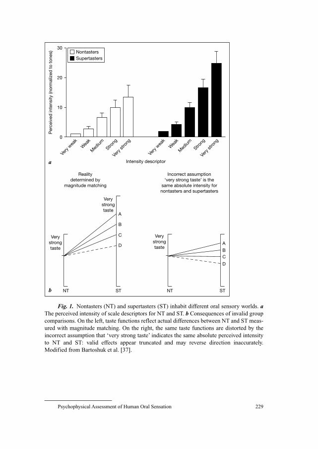
Psychophysical Assessment of Human Oral Sensation 229
30
20
10
0
a
Very
wea
kW
eak
Med
ium
Strong
Very
stro
ng
Very
wea
kW
eak
Med
ium
Strong
Very
stro
ng
Intensity descriptor
Realitydetermined by
magnitude matching
Incorrect assumption‘very strong taste’ is the
same absolute intensity fornontasters and supertasters
Per
ceiv
ed in
tens
ity (n
orm
aliz
ed t
o to
nes)
Verystrongtaste
Verystrongtaste
NT ST
A
B
C
D
Verystrongtaste
NT ST
A
BC
D
b
SupertastersNontasters
Fig. 1. Nontasters (NT) and supertasters (ST) inhabit different oral sensory worlds. aThe perceived intensity of scale descriptors for NT and ST. b Consequences of invalid group
comparisons. On the left, taste functions reflect actual differences between NT and ST meas-
ured with magnitude matching. On the right, the same taste functions are distorted by the
incorrect assumption that ‘very strong taste’ indicates the same absolute perceived intensity
to NT and ST: valid effects appear truncated and may reverse direction inaccurately.
Modified from Bartoshuk et al. [37].
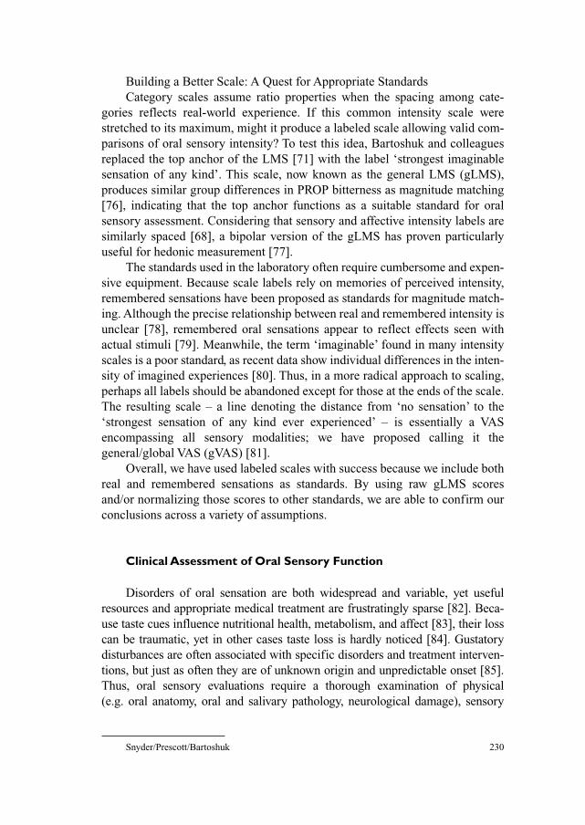
Snyder/Prescott/Bartoshuk 230
Building a Better Scale: A Quest for Appropriate Standards
Category scales assume ratio properties when the spacing among cate-
gories reflects real-world experience. If this common intensity scale were
stretched to its maximum, might it produce a labeled scale allowing valid com-
parisons of oral sensory intensity? To test this idea, Bartoshuk and colleagues
replaced the top anchor of the LMS [71] with the label ‘strongest imaginable
sensation of any kind’. This scale, now known as the general LMS (gLMS),
produces similar group differences in PROP bitterness as magnitude matching
[76], indicating that the top anchor functions as a suitable standard for oral
sensory assessment. Considering that sensory and affective intensity labels are
similarly spaced [68], a bipolar version of the gLMS has proven particularly
useful for hedonic measurement [77].
The standards used in the laboratory often require cumbersome and expen-
sive equipment. Because scale labels rely on memories of perceived intensity,
remembered sensations have been proposed as standards for magnitude match-
ing. Although the precise relationship between real and remembered intensity is
unclear [78], remembered oral sensations appear to reflect effects seen with
actual stimuli [79]. Meanwhile, the term ‘imaginable’ found in many intensity
scales is a poor standard, as recent data show individual differences in the inten-
sity of imagined experiences [80]. Thus, in a more radical approach to scaling,
perhaps all labels should be abandoned except for those at the ends of the scale.
The resulting scale – a line denoting the distance from ‘no sensation’ to the
‘strongest sensation of any kind ever experienced’ – is essentially a VAS
encompassing all sensory modalities; we have proposed calling it the
general/global VAS (gVAS) [81].
Overall, we have used labeled scales with success because we include both
real and remembered sensations as standards. By using raw gLMS scores
and/or normalizing those scores to other standards, we are able to confirm our
conclusions across a variety of assumptions.
Clinical Assessment of Oral Sensory Function
Disorders of oral sensation are both widespread and variable, yet useful
resources and appropriate medical treatment are frustratingly sparse [82]. Beca-
use taste cues influence nutritional health, metabolism, and affect [83], their loss
can be traumatic, yet in other cases taste loss is hardly noticed [84]. Gustatory
disturbances are often associated with specific disorders and treatment interven-
tions, but just as often they are of unknown origin and unpredictable onset [85].
Thus, oral sensory evaluations require a thorough examination of physical
(e.g. oral anatomy, oral and salivary pathology, neurological damage), sensory
Psychophysical Assessment of Human Oral Sensation 231
(e.g. taste, oral somatosensation, retronasal olfaction), and emotional aspects of
chemosensation (e.g. psychopathology, quality of life).
Several afferent nerves carry sensory information from the mouth, each
carrying a particular array of information from a particular area. The chorda
tympani (CT), a branch of the facial nerve (VII), carries taste information from
the anterior tongue; the lingual branch of the trigeminal nerve (V) carries pain,
tactile, and temperature information from the same region. The greater superfi-
cial petrosal nerve, another branch of nerve VII, carries taste cues from the
palate. Multimodal information (i.e. taste, touch, pain, temperature) is carried
from the posterior tongue by the glossopharyngeal nerve (IX) and from the
throat by the vagus (X) [86]. Taste and oral somatosensory cues combine cen-
trally with retronasal olfaction to produce the composite experience of flavor
[87]. This spatial distribution of input has led researchers to consider the clini-
cal relevance of localized oral sensory damage (see below), so modern proto-
cols for oral sensory evaluation typically include judgments of intensity and
quality for both regional and whole-mouth stimuli.
Whole-Mouth Oral Sensation
In whole-mouth gustatory testing, chemical stimuli are sampled and moved
throughout the mouth, stimulating all oral taste bud fields simultaneously; subjects
rinse with water prior to each stimulus. Laboratory tests of oral sensation involve
the presentation of chemical solutions at multiple concentrations spanning the
functional range of perception [41, 88], but most clinical tests have streamlined
this process to a single stimulus for each of the common taste qualities (i.e.
sucrose, NaCl, citric acid, quinine hydrochloride) [89]. In addition, multiple con-
centrations may be used to derive suprathreshold taste functions, and other oral
stimuli may be included to evaluate oral tactile sensation (e.g. capsaicin, alcohol)
or individual differences (e.g. PROP). Having discussed the disadvantages of
thresholds, we favor suprathreshold measurements involving magnitude matching
or the gLMS/gVAS with appropriate standards, so stimuli unrelated to oral sensa-
tion (e.g. sound, remembered sensations) should be incorporated.
Aqueous solutions are inconvenient for field and clinical use, so altern-
ative methods of stimulus delivery have been explored, including paper strips and
tablets [90–92]. With respect to whole-mouth testing, the most enduring example
of these methods is the use of PROP papers as a screening tool for taste blindness.
In early studies, PTC crystals were placed directly on the tongue [33] or delivered
on saturated filter papers [93]. Today, PROP papers are made by soaking labora-
tory-grade filter papers in a supersaturated PROP solution heated to just below
boiling. When dry, each paper contains approx. 1.6 mg PROP [94]; patients with
Snyder/Prescott/Bartoshuk 232
hyperthyroidism are prescribed 100–300 mg daily [95]. Variants of this method
have been described [96], but all share the common goal of introducing a small
amount of crystalline PROP to the tongue surface. Although PROP papers are
convenient and portable, their technical flaws warrant consideration.
• To produce a taste, the filter paper must be completely moistened with
saliva, which requires healthy salivary function and a sufficient period of
contact with the tongue [55].
• Some studies [35] report excessive false-positive and false-negative
responses to PTC/PROP filter papers. These response rates probably result
from minor testing variations that affect response bias.
• Filter paper testing shows only moderate concordance with threshold sensi-
tivity [97]. As described above, threshold and suprathreshold measures
almost always dissociate when proper scaling is used.
Despite these concerns, filter paper ratings and laboratory PROP assess-
ments show significant agreement and high test-retest reliability [94, 96, 98],
perhaps because the effective concentration of PROP is unimportant provided it
is high. Comparisons of PROP paper and solution bitterness suggest that the
concentration dissolved from paper into saliva approaches maximum solubility
[unpublished data]. Because functions of PROP bitterness for nontasters, medium
tasters, and supertasters diverge [36], the most efficient way to sort subjects is to
use the highest concentration possible, so papers made from saturated PROP
[94] may be preferable to those made from lower concentrations.
Oral Sensory Anatomy: Videomicroscopy of the Tongue
Multiple reports indicate that differences in taste bud density account for
human oral sensory variation [45, 59]. To explore this idea further, Miller and
Reedy [99] developed a method for visualizing the tongue in vivo. Human
tongues are coated with large, raised, circular structures (i.e. fungiform papillae)
that hold taste buds [100]; blue food coloring applied to the tongue surface fails
to stain these papillae, which subsequently appear as pink circles against a blue
background. Fungiform papillae can be counted with a magnifying glass and a
flashlight, while videomicroscopy allows resolution of pores at the apical tips of
taste buds. This method has revealed positive associations between PROP inten-
sity, fungiform papilla density, and taste bud density [59, 99]. Fungiform papil-
lae are dually innervated by CT and nerve V [101], which accounts for the
elevated taste and oral tactile sensations experienced by supertasters [45, 102].
Clinically, videomicroscopy is useful in confirming CT damage (see below),
which often appears as a discrepancy between high fungiform papilla density
and low taste sensation on the anterior tongue [1]. In addition, the association
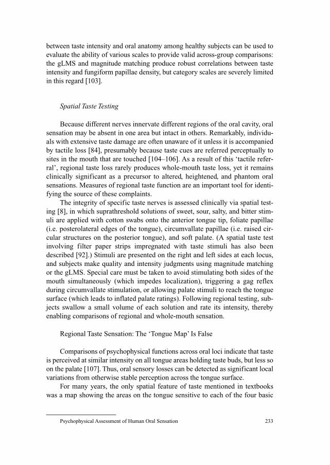
Psychophysical Assessment of Human Oral Sensation 233
between taste intensity and oral anatomy among healthy subjects can be used to
evaluate the ability of various scales to provide valid across-group comparisons:
the gLMS and magnitude matching produce robust correlations between taste
intensity and fungiform papillae density, but category scales are severely limited
in this regard [103].
Spatial Taste Testing
Because different nerves innervate different regions of the oral cavity, oral
sensation may be absent in one area but intact in others. Remarkably, individu-
als with extensive taste damage are often unaware of it unless it is accompanied
by tactile loss [84], presumably because taste cues are referred perceptually to
sites in the mouth that are touched [104–106]. As a result of this ‘tactile refer-
ral’, regional taste loss rarely produces whole-mouth taste loss, yet it remains
clinically significant as a precursor to altered, heightened, and phantom oral
sensations. Measures of regional taste function are an important tool for identi-
fying the source of these complaints.
The integrity of specific taste nerves is assessed clinically via spatial test-
ing [8], in which suprathreshold solutions of sweet, sour, salty, and bitter stim-
uli are applied with cotton swabs onto the anterior tongue tip, foliate papillae
(i.e. posterolateral edges of the tongue), circumvallate papillae (i.e. raised cir-
cular structures on the posterior tongue), and soft palate. (A spatial taste test
involving filter paper strips impregnated with taste stimuli has also been
described [92].) Stimuli are presented on the right and left sides at each locus,
and subjects make quality and intensity judgments using magnitude matching
or the gLMS. Special care must be taken to avoid stimulating both sides of the
mouth simultaneously (which impedes localization), triggering a gag reflex
during circumvallate stimulation, or allowing palate stimuli to reach the tongue
surface (which leads to inflated palate ratings). Following regional testing, sub-
jects swallow a small volume of each solution and rate its intensity, thereby
enabling comparisons of regional and whole-mouth sensation.
Regional Taste Sensation: The ‘Tongue Map’ Is False
Comparisons of psychophysical functions across oral loci indicate that taste
is perceived at similar intensity on all tongue areas holding taste buds, but less so
on the palate [107]. Thus, oral sensory losses can be detected as significant local
variations from otherwise stable perception across the tongue surface.
For many years, the only spatial feature of taste mentioned in textbooks
was a map showing the areas on the tongue sensitive to each of the four basic
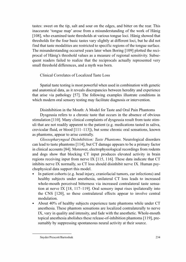
Snyder/Prescott/Bartoshuk 234
tastes: sweet on the tip, salt and sour on the edges, and bitter on the rear. This
inaccurate ‘tongue map’ arose from a misunderstanding of the work of Hänig
[108], who examined taste thresholds at various tongue loci. Hänig showed that
thresholds for the four basic tastes vary slightly at different loci, but he did not
find that taste modalities are restricted to specific regions of the tongue surface.
The misunderstanding occurred years later when Boring [109] plotted the reci-
procal of Hänig’s threshold values as a measure of regional sensitivity. Subse-
quent readers failed to realize that the reciprocals actually represented very
small threshold differences, and a myth was born.
Clinical Correlates of Localized Taste Loss
Spatial taste testing is most powerful when used in combination with genetic
and anatomical data, as it reveals discrepancies between heredity and experience
that arise via pathology [57]. The following examples illustrate conditions in
which modern oral sensory testing may facilitate diagnosis or intervention.
Disinhibition in the Mouth: A Model for Taste and Oral Pain Phantoms
Dysgeusia refers to a chronic taste that occurs in the absence of obvious
stimulation [110]. Many clinical complaints of dysgeusia result from taste stim-
uli that are not readily apparent to the patient (e.g. medications tasted in saliva,
crevicular fluid, or blood [111–113]), but some chronic oral sensations, known
as phantoms, appear to arise centrally.
Glossopharyngeal Disinhibition: Taste Phantoms. Neurological disorders
can lead to taste phantoms [114], but CT damage appears to be a primary factor
in clinical accounts [84]. Moreover, electrophysiological recordings from rodents
and dogs show that blocking CT input produces elevated activity in brain
regions receiving input from nerve IX [115, 116]. These data indicate that CT
inhibits nerve IX normally, so CT loss should disinhibit nerve IX. Human psy-
chophysical data support this model.
• In patient cohorts (e.g. head injury, craniofacial tumors, ear infections) and
healthy subjects under anesthesia, unilateral CT loss leads to increased
whole-mouth perceived bitterness via increased contralateral taste sensa-
tion at nerve IX [18, 117–119]. Oral sensory input rises ipsilaterally into
the CNS [120], so these contralateral effects appear to involve central
modulation.
• About 40% of healthy subjects experience taste phantoms while under CT
anesthesia. These phantom sensations are localized contralaterally to nerve
IX, vary in quality and intensity, and fade with the anesthetic. Whole-mouth
topical anesthesia abolishes these release-of-inhibition phantoms [119], pre-
sumably by suppressing spontaneous neural activity at their source.
Psychophysical Assessment of Human Oral Sensation 235
• In one report [121], a bitter taste phantom arose bilaterally at nerve IX fol-
lowing tonsillectomy. Spatial testing indicated complete nerve IX loss, yet
the phantom became more intense with whole-mouth topical anesthesia.
This nerve stimulation phantom was probably caused by surgical damage to
nerve IX and disinhibited further by CT anesthesia.
Trigeminal Disinhibition: Oral Pain Phantoms. CT taste input also appears
to inhibit cues from nerve V. This interaction may suppress oral pain during
intake, and it may facilitate tactile referral of taste information following local-
ized taste damage. Because supertasters have the most taste and trigeminal input,
CT damage may lead to adverse sensory consequences due to extreme disinhibi-
tion of nerve V: following unilateral CT anesthesia, supertasters show increased
ratings for the burn of capsaicin on the contralateral anterior tongue [122].
Oral pain phantoms are another serious consequence of nerve V disinhibi-
tion. The identification of burning mouth syndrome (BMS) as such a phantom
vividly illustrates the clinical relevance of modern oral sensory testing. BMS, a
condition most often found in postmenopausal women, is characterized by severe
oral pain in the absence of visible pathology [123]. BMS is often described as
psychogenic, but systematic psychophysical testing tells a different story. Most
BMS patients show significantly reduced bitterness for quinine on the anterior
tongue, consistent with CT damage [124]. Nearly 50% of BMS patients experi-
ence taste phantoms at nerve IX [17]; topical anesthesia usually intensifies BMS-
related taste and oral pain [125]. Finally, the peak intensity of BMS pain
correlates with fungiform papillae density, indicating that BMS is most prevalent
among supertasters. Taken together, these data strongly suggest that BMS is an
oral pain phantom generated by CT damage. Grushka et al. [126] have shown that
agonists to the inhibitory neurotransmitter �-aminobutyric acid suppress BMS
pain, presumably by restoring lost inhibition from absent taste cues.
Clinical Considerations
Laboratory and clinical data support the use of topical anesthesia in the
mouth to determine the locus of oral sensory dysfunction. However, interpre-
tations of topical anesthesia must be made carefully, as incomplete anesthesia will
impede differential diagnosis. In topical anesthesia, patients hold approx. 5 ml of
0.5% dyclone in the mouth for 60 s, rest for 60 s, rinse with water, and describe
any oral sensations experienced for the duration of the sensory block [121].
If a taste or oral pain complaint becomes more intense with oral anesthesia,
it does not arise from normal stimulation of oral sensory receptors. Certain
therapeutic agents promote venous taste and other dysgeusias, so medication
and supplement use should be reviewed. Another possibility is that the nerve
innervating the region of sensory disturbance has sustained physical damage. If
damage is peripheral to nerve cell bodies, the resulting neuroma may produce a

Snyder/Prescott/Bartoshuk 236
nerve stimulation phantom; topical anesthesia exacerbates nerve stimulation
phantoms via central disinhibition. Conclusions involving nerve damage should
be confirmed by further neurological examination.
When local anesthesia abolishes a taste or oral pain complaint, an actual
stimulus may be present in the mouth. To test for the presence of such a stimu-
lus, the patient should attempt to rinse it from the mouth; if the offending sen-
sation subsides at all, an actual stimulus should be considered. Alternatively,
nerve damage unrelated to the complaint may be disinhibiting input related to
it; topical anesthesia suppresses these central release-of-inhibition phantoms,
presumably by inhibiting spontaneous activity. Spatial testing should reveal
localized taste loss at a site distant from the phantom.
Conclusion
Human psychophysics is a powerful aspect of clinical and basic science
that offers a window onto neurobehavioral processes often inaccessible by other
means. As such, our goal in exploring psychophysical methodology is to craft
measurement tools that reflect individual differences accurately and allow
adaptive use in clinical, research, and other assessment settings. Conservative
approaches to this task emphasize threshold measures, but we have embraced
suprathreshold techniques in the hope of measuring biologically relevant sensa-
tions, and we have carefully evaluated these techniques in the process. Our
overall approach has been to refine existing methods continuously, incorporat-
ing real-world reference points in order to represent perception as faithfully as
possible. These refinements have posed a constant challenge to remain user
friendly; our use of sophisticated scaling tools in laboratory research shows that
untrained subjects learn to use them quickly and skillfully, and our clinical
research indicates that these tools are accessible to patients. Finally, our sys-
tematic use of techniques from psychophysics, anatomy, neurology, and genet-
ics has allowed us to explore complex relationships between oral sensation,
affect, behavior, and disease at multiple levels of analysis. In our view, these
effects confirm that our methods reflect highly predictive and highly compara-
ble aspects of sensory and hedonic experience.
Acknowledgements
This research is supported by a grant from the US National Institutes of Health (DC
00283) to L.M.B.. D.J.S. is supported by funding from the US National Science Foundation
and the Rose Marie Pangborn Sensory Science Fund.
Psychophysical Assessment of Human Oral Sensation 237
References
1 Bartoshuk LM, Duffy VB, Chapo AK, Fast K, Yiee JH, Hoffman HJ, Ko C-W, Snyder DJ: From
psychophysics to the clinic: missteps and advances. Food Qual Pref 2004;15:617–632.
2 Brindley GS: Introduction to sensory experiments; in Brindley GS (ed): Physiology of the Retina
and the Visual Pathway. London, Arnold, 1960, pp 144–150.
3 Fechner GT: Elements of Psychophysics (translated by Adler HE; edited by Howes DH, Boring
EG). New York, Holt, Rinehart and Winston, 1966 (original 1860).
4 Engen T: Psychophysics. 1. Discrimination and detection; in Kling JW, Riggs LA (eds): Wood-
worth and Schlosberg’s Experimental Psychology. New York, Holt, Rinehart and Winston, 1972,
vol 1: Sensation and Perception, pp 11–46.
5 Harris H, Kalmus H: The measurement of taste sensitivity to phenylthiourea (PTC). Ann Eugen
1949;15:24–31.
6 Henkin RI, Gill JR, Bartter FC: Studies on taste thresholds in normal man and in patients with
adrenal cortical insufficiency: the role of adrenal cortical steroids and of serum sodium concen-
tration. J Clin Invest 1963;42:727–735.
7 Wetherill GB, Levitt H: Sequential estimation of points on a psychometric function. Br J Math
Stat Psychol 1965;18:1–10.
8 Bartoshuk LM: Clinical evaluation of the sense of taste. Ear Nose Throat J 1989;68:331–337.
9 Mackenzie ICK: A simple method of testing taste. Lancet 1955;ii:377–378.
10 Frank ME, Smith DV: Electrogustometry: a simple way to test taste; in Getchell TV, Doty RL,
Bartohsuk LM, Snow JB (eds): Smell and Taste in Health and Disease. New York, Raven Press,
1991, pp 503–514.
11 Murphy C, Quiñonez C, Nordin S: Reliability and validity of electrogustometry and its application
to young and elderly persons. Chem Senses 1995;20:499–503.
12 Tomita H, Ikeda M, Okuda Y: Basis and practice of clinical taste examinations. Auris Nasus
Larynx 1986;13(Suppl I):S1–S15.
13 Grant R, Ferguson MM, Strang R, Turner JW, Bone I: Evoked taste thresholds in a normal popula-
tion and the application of electrogustometry to trigeminal nerve disease. J Neurol Neurosurg
Psychiatry 1987;50:12–21.
14 Le Floch JP, Le Lievre G, Verroust J, Phillippon C, Peynegre R, Perlemuter L: Factors related to
the electric taste threshold in type 1 diabetic patients. Diabet Med 1990;7:526–531.
15 Stillman JA, Morton RP, Hay KD, Ahmad Z, Goldsmith D: Electrogustometry: strengths, weak-
nesses, and clinical evidence of stimulus boundaries. Clin Otolaryngol 2003;28:406–410.
16 Bujas Z: Electrical taste; in Beidler LM (ed): Handbook of Sensory Physiology. Berlin, Springer,
1971, vol 4: Chemical Senses, part 2: Taste, pp 180–199.
17 Grushka M, Sessle BJ, Howley TP: Psychophysical evidence of taste dysfunction in burning
mouth syndrome. Chem Senses 1986;11:485–498.
18 Lehman CD, Bartoshuk LM, Catalanotto FC, Kveton JF, Lowlicht RA: The effect of anesthesia of
the chorda tympani nerve on taste perception in humans. Physiol Behav 1995;57:943–951.
19 Krarup B: Taste reactions of patients with Bell’s palsy. Acta Otolaryngol 1958;49:389–399.
20 Salata JA, Raj JM, Doty RL: Differential sensitivity of tongue areas and palate to chemical stimu-
lation: a suprathreshold cross-modal matching study. Chem Senses 1991;16:483–489.
21 Stevens SS: On the theory of scales of measurement. Science 1946;103:677–680.
22 Stevens SS: The direct estimation of sensory magnitudes: loudness. Am J Psychol 1956;69:1–25.
23 Marks LE: Sensory Processes: The New Psychophysics. New York, Academic Press, 1974.
24 Fox AL: Six in ten ‘tasteblind’ to bitter chemical. Science 1931;73:14a.
25 Barnicot NA, Harris H, Kalmus H: Taste thresholds of further eighteen compounds and their cor-
relation with PTC thresholds. Ann Eugen 1951;16:119–128.
26 Blakeslee AF: Genetics of sensory thresholds: taste for phenyl-thio-carbamide. Proc Natl Acad Sci
USA 1932;18:120–130.
27 Parr LW: Taste blindness and race. J Hered 1934;25:187–190.
28 Kalmus H, Farnsworth D: Impairment and recovery of taste following irradiation of the oropharynx.
J Laryngol Otol 1959;73:180–182.
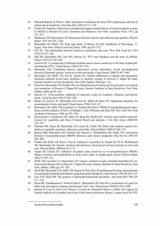
Snyder/Prescott/Bartoshuk 238
29 Whissell-Buechy D, Wills C: Male and female correlations for taster (PTC) phenotypes and rate of
adolescent development. Ann Hum Biol 1989;16:131–146.
30 Fischer R: Gustatory, behavioral, and pharmacological manifestations of chemoreception in man;
in Ohloff G, Thomas AF (eds): Gustation and Olfaction. New York, Academic Press, 1971, pp
187–237.
31 McBurney DH, Bartoshuk LM: Interactions between stimuli with different taste qualities. Physiol
Behav 1973;10:1101–1106.
32 Bartoshuk LM, Duffy VB: Taste and smell; in Masoro EJ (ed): Handbook of Physiology. 11.
Aging. New York, Oxford University Press, 1995, pp 363–375.
33 Fox AL: The relationship between chemical constitution and taste. Proc Natl Acad Sci USA
1932;18:115–120.
34 Hall MJ, Bartoshuk LM, Cain WS, Stevens JC: PTC taste blindness and the taste of caffeine.
Nature 1975;253:442–443.
35 Lawless HT: A comparison of different methods used to assess sensitivity to the taste of phenylth-
iocarbamide (PTC). Chem Senses 1980;5:247–256.
36 Bartoshuk LM: Comparing sensory experiences across individuals: recent psychophysical
advances illuminate genetic variation in taste perception. Chem Senses 2000;25:447–460.
37 Bartoshuk LM, Duffy VB, Fast K, Snyder DJ: Genetic differences in human oral perception:
advanced methods reveal basic problems in intensity scaling; in Prescott J, Tepper BJ (eds):
Genetic Variation in Taste Sensitivity. New York, Dekker, 2004, pp 1–42.
38 Prescott J, Bartoshuk LM, Prutkin JM: 6-n-Propylthiouracil tasting and the perception of nontaste
oral sensations; in Prescott J, Tepper BJ (eds): Genetic Variation in Taste Sensitivity. New York,
Dekker, 2004, pp 89–104.
39 Stevens JC: Cross-modality validation of subjective scales for loudness, vibration, and electric
shock. J Exp Psychol 1959;57:201–209.
40 Marks LE, Stevens JC, Bartoshuk LM, Gent JG, Rifkin B, Stone VK: Magnitude matching: the
measurement of taste and smell. Chem Senses 1988;13:63–87.
41 Bartoshuk LM, Duffy VB, Lucchina LA, Prutkin JM, Fast K: PROP (6-n-propylthiouracil) super-
tasters and the saltiness of NaCl; in Murphy C (ed): Olfaction and Taste XII. New York, New York
Academy of Sciences, 1998, pp 793–796.
42 Drewnowski A, Henderson SA, Hann CS, Berg WA, Ruffin MT: Genetic taste markers and pref-
erences for vegetables and fruit of female breast care patients. J Am Diet Assoc 2000;100:
191–197.
43 Dinehart ME, Hayes JE, Bartoshuk LM, Lanier SL, Duffy VB: Bitter taste markers explain vari-
ability in vegetable sweetness, bitterness, and intake. Physiol Behav 2006;87:304–313.
44 Basson MD, Bartoshuk LM, Dichello SZ, Panzini L, Weiffenbach JM, Duffy VB: Association
between 6-n-propylthiouracil (PROP) bitterness and colonic neoplasms. Dig Dis Sci 2005;50:
483–489.
45 Prutkin JM, Duffy VB, Etter L, Fast K, Gardner E, Lucchina LA, Snyder DJ, Tie K, Weiffenbach
JM, Bartoshuk LM: Genetic variation and inferences about perceived taste intensity in mice and
men. Physiol Behav 2000;69:161–173.
46 Tepper BJ, Ullrich NV: Influence of genetic taste sensitivity to 6-n-propylthiouracil (PROP),
dietary restraint, and disinhibition on body mass index in middle-aged women. Physiol Behav
2002;75:305–312.
47 Duffy VB, Lucchina LA, Bartoshuk LM: Genetic variation in taste: potential biomarker for car-
diovascular disease risk? in Prescott J, Tepper BJ (eds): Genetic Variation in Taste Sensitivity. New
York, Dekker, 2004, pp 195–228.
48 Reed DR, Bartoshuk LM, Duffy VB, Marino S, Price RA: Propylthiouracil tasting: determination
of underlying threshold distributions using maximum likelihood. Chem Senses 1995;20:529–533.
49 Guo S-W, Reed DR: The genetics of phenylthiocarbamide perception. Ann Hum Biol 2001;28:
111–142.
50 Reed DR, Nanthakumar E, North M, Bell C, Bartoshuk LM, Price RA: Localization of a gene for
bitter taste perception to human chromosome 5p15. Am J Hum Genet 1999;64:1478–1480.
51 Drayna D, Coon H, Kim U-K, Elsner T, Cromer K, Otterud B, Baird L, Peiffer AP, Leppert M:
Genetic analysis of a complex trait in the Utah Genetic Reference Project: a major locus for PTC
Psychophysical Assessment of Human Oral Sensation 239
taste ability on chromosome 7q and a secondary locus on chromosome 16p. Hum Genet
2003;112:567–572.
52 Kim U-K, Jorgenson E, Coon H, Leppert M, Risch N, Drayna D: Positional cloning of the human
quantitative trait locus underlying taste sensitivity to phenylthiocarbamide. Science 2003;299:
1221–1225.
53 Bartoshuk LM, Davidson AC, Kidd JR, Kidd KK, Speed WC, Pakstis AJ, Reed DR, Snyder DJ,
Duffy VB: Supertasting is not explained by the PTC/PROP gene. Chem Senses 2005;30:A87.
54 Bartoshuk LM, Duffy VB, Fast K, Green BG, Snyder DJ: Hormones, age, genes, and pathology:
how do we assess variation in sensation and preference? in Anderson GH, Blundell JE, Chiva MM
(eds): Food Selection: From Genes to Culture. Levallois-Parret, Danone Institute, 2002, pp
173–187.
55 Drewnowski A: Genetics of human taste perception; in Doty RL (ed): Handbook of Olfaction and
Gustation, ed 2. New York, Dekker, 2003, pp 847–860.
56 Rankin KM, Godinot N, Christensen CM, Tepper BJ, Kirkmeyer SV: Assessment of different
methods for 6-n-propylthiouracil status classification; in Prescott J, Tepper BJ (eds): Genetic
Variation in Taste Sensitivity. New York, Dekker, 2004, pp 63–88.
57 Duffy VB, Davidson AC, Kidd JR, Kidd KK, Speed WC, Pakstis AJ, Reed DR, Snyder DJ,
Bartoshuk LM: Bitter receptor gene (TAS2R38), 6-n-propylthiouracil (PROP) bitterness, and
alcohol intake. Alcohol Clin Exp Res 2004;28:1629–1637.
58 Bartoshuk LM: Bitter taste of saccharin: related to the genetic ability to taste the bitter substance
6-n-propylthiouracil (PROP). Science 1979;205:934–935.
59 Bartoshuk LM, Duffy VB, Miller IJ: PTC/PROP tasting: anatomy, psychophysics, and sex effects.
Physiol Behav 1994;56:1165–1171.
60 Harris H, Kalmus H: Chemical sensitivity in genetical differences of taste sensitivity. Ann Eugen
1949;15:32–45.
61 Kranzler HR, Skipsey K, Modesto-Lowe V: PROP taster status and parental history of alcohol
dependence. Drug Alcohol Depend 1998;52:109–113.
62 Mattes RD: 6-n-Propylthiouracil taster status: dietary modifier, marker, or misleader? in Prescott
J, Tepper BJ (eds): Genetic Variation in Taste Sensitivity. New York, Dekker, 2004, pp 229–250.
63 Likert R: A technique for the measurement of attitudes. Arch Psychol 1932;22:1–55.
64 Jones LV, Peryam DR, Thurstone LL: Development of a scale for measuring soldiers’ food prefer-
ences. Food Res 1955;20:512–520.
65 Aitken RCB: Measurement of feelings using visual analogue scales. Proc R Soc Med 1969;62:
989–993.
66 Stevens SS, Galanter EH: Ratio scales and category scales for a dozen perceptual continua. J Exp
Psychol 1957;54:377–411.
67 Lasagna L: The clinical measurement of pain. Ann NY Acad Sci 1960;86:28–37.
68 Moskowitz HR: Magnitude estimation: notes on what, how, when, and why to use it. J Food Qual
1977;1:195–228.
69 Gracely RH, McGrath P, Dubner R: Validity and sensitivity of ratio scales of sensory and affective
verbal pain descriptors: manipulation of affect by diazepam. Pain 1978;5:19–29.
70 Borg GAV: Psychophysical scaling with applications in physical work and the perception of exer-
tion. Scand J Work Environ Health 1990;16(Suppl 1):55–58.
71 Green BG, Shaffer GS, Gilmore MM: A semantically labeled magnitude scale of oral sensation
with apparent ratio properties. Chem Senses 1993;18:683–702.
72 Schutz HG, Cardello AV: A labeled affective magnitude (LAM) scale for assessing food
liking/disliking. J Sens Stud 2001;16:117–159.
73 Borg GAV: A category scale with ratio properties for intermodal and interindividual comparisons;
in Geissler HG, Petzold P (eds): Psychophysical Judgment and the Process of Perception. Berlin,
Deutscher Verlag der Wissenschaften, 1982, pp 25–34.
74 Teghtsoonian R: Range effects in psychophysical scaling and a revision of Stevens’ law. Am J
Psychol 1973;86:3–27.
75 Bartoshuk LM, Snyder DJ: Psychophysical measurement of human taste experience; in Stricker
EM, Woods SC (eds): Handbook of Behavioral Neurobiology. New York, Plenum Press, 2004, vol
14: Neurobiology of Food and Fluid Intake, pp 89–107.
Snyder/Prescott/Bartoshuk 240
76 Bartoshuk LM, Green BG, Hoffman HJ, Ko C-W, Lucchina LA, Snyder DJ, Weiffenbach JM:
Valid across-group comparisons with labeled scales: the gLMS versus magnitude matching.
Physiol Behav 2004;82:109–114.
77 Duffy VB, Peterson JM, Bartoshuk LM: Associations between taste genetics, oral sensation, and
alcohol intake. Physiol Behav 2004;82:435–445.
78 Algom D: Memory psychophysics: an examination of its perceptual and cognitive prospects;
in Algom D (ed): Psychophysical Approaches to Cognition. Amsterdam, Elsevier, 1992, pp
441–513.
79 Stevenson RJ, Prescott J: Judgments of chemosensory mixtures in memory. Acta Psychol (Amst)
1997;95:195–214.
80 Fast K, Green BG, Bartoshuk LM: Developing a scale to measure just about anything: compar-
isons across groups and individuals. Appetite 2002;39:75.
81 Bartoshuk LM, Fast K, Snyder DJ: Differences in our sensory worlds: invalid comparisons with
labeled scales. Curr Dir Psychol Sci 2005;14:122–125.
82 Deems DA, Doty RL, Settle RG, Moore-Gillon V, Shaman P, Mester AF, Kimmelman CP,
Brightman VJ, Snow JB: Smell and taste disorders: a study of 750 patients from the University of
Pennsylvania Smell and Taste Center. Arch Otolaryngol Head Neck Surg 1991;117:519–528.
83 Mattes RD, Cowart BJ: Dietary assessment of patients with chemosensory disorders. J Am Diet
Assoc 1994;94:50–56.
84 Bull TR: Taste and the chorda tympani. J Laryngol Otol 1965;79:479–493.
85 Bromley SM, Doty RL: Clinical disorders affecting taste: evaluation and management; in Doty
RL (ed): Handbook of Olfaction and Gustation, ed 2. New York, Dekker, 2003.
86 Pritchard TC, Norgren R: Gustatory system; in Paxinos G, Mai JK (eds): The Human Nervous
System, ed 2. Amsterdam, Elsevier, 2004, pp 1171–1196.
87 McBurney DH: Taste, smell, and flavor terminology: taking the confusion out of fusion; in
Meiselman HL, Rivlin RS (eds): Clinical Measurement of Taste and Smell. New York, Macmillan,
1986, pp 117–125.
88 Tepper BJ, Christensen CM, Cao J: Development of brief methods to classify individuals by PROP
taster status. Physiol Behav 2001;73:571–577.
89 Frank ME, Hettinger TP, Barry MA: Contemporary measurement of human gustatory function; in
Doty RL (ed): Handbook of Olfaction and Gustation, ed 2. New York, Dekker, 2003.
90 Hummel T, Erras A, Kobal G: A test for screening of taste function. Rhinology 1997;35:146–148.
91 Ahne G, Erras A, Hummel T, Kobal G: Assessment of gustatory function by means of tasting
tablets. Laryngoscope 2000;110:1396–1401.
92 Mueller C, Kallert S, Renner B, Stiassny K, Temmel AF, Hummel T, Kobal G: Quantitative assess-
ment of gustatory function in a clinical context using impregnated ‘taste strips’. Rhinology
2003;41:2–6.
93 Blakeslee AF, Fox AL: Our different taste worlds. J Hered 1932;23:97–107.
94 Bartoshuk LM, Duffy VB, Reed DR, Williams A: Supertasting, earaches, and head injury: genet-
ics and pathology alter our taste worlds. Neurosci Biobehav Rev 1996;20:79–87.
95 Cooper DS: Antithyroid drugs. N Engl J Med 2005;352:905.
96 Zhao L, Kirkmeyer SV, Tepper BJ: A paper screening test to assess genetic taste sensitivity to 6-n-
propylthiouracil. Physiol Behav 2003;78:625–633.
97 Hartmann G: Application of individual taste difference towards phenyl-thio-carbamide in genetic
investigations. Ann Eugen 1939;9:123–135.
98 Ly A, Drewnowski A: PROP (6-n-propylthiouracil) tasting and sensory responses to caffeine,
sucrose, neohesperidin dihydrochalcone, and chocolate. Chem Senses 2001;26:41–47.
99 Miller IJ, Reedy FE: Variations in human taste bud density and taste intensity perception. Physiol
Behav 1990;47:1213–1219.
100 Miller IJ: Variation in human fungiform taste bud densities among regions and subjects. Anat Rec
1986;216:474–482.
101 Gairns FW: Sensory endings other than taste buds in the human tongue. J Physiol 1953;121:
33P–34P.
102 Essick GK, Chopra A, Guest S, McGlone F: Lingual tactile acuity, taste perception, and the den-
sity and diameter of fungiform papillae in female subjects. Physiol Behav 2003;80:289–302.

Psychophysical Assessment of Human Oral Sensation 241
103 Snyder DJ, Fast K, Bartoshuk LM: Valid comparisons of suprathreshold sensations. J Conscious
Stud 2004;11:96–112.
104 Todrank J, Bartoshuk LM: A taste illusion: taste sensation localized by touch. Physiol Behav
1991;50:1027–1031.
105 Delwiche JF, Lera MF, Breslin PA: Selective removal of a target stimulus localized by taste in
humans. Chem Senses 2000;25:181–187.
106 Green BG: Studying taste as a cutaneous sense. Food Qual Pref 2002;14:99–109.
107 Bartoshuk LM: Clinical psychophysics of taste. Gerodontics 1988;4:249–255.
108 Hänig DP: Zur Psychophysik des Geschmackssinnes; PhD thesis, Leipzig, 1901.
109 Boring EG: Sensation and Perception in the History of Experimental Psychology. New York,
Appleton, 1942.
110 Snow JB, Doty RL, Bartoshuk LM, Getchell TV: Categorization of chemosensory disorders; in
Getchell TV, Doty RL, Bartoshuk LM, Snow JB (eds): Smell and Taste in Health and Disease.
New York, Raven Press, 1991, pp 445–447.
111 Bradley RM: Electrophysiological investigations of intravascular taste using perfused rat tongue.
Am J Physiol 1973;224:300–304.
112 Alfano M: The origin of gingival fluid. J Theor Biol 1974;47:127–136.
113 Fetting JH, Wilcox PM, Sheidler VR, Enterline JP, Donehower RC, Grochow LB: Tastes associ-
ated with parenteral chemotherapy for breast cancer. Cancer Treat Rep 1985;69:1249–1251.
114 Hausser-Hauw C, Bancaud J: Gustatory hallucinations in epileptic seizures. Brain 1987;110:
339–359.
115 Halpern BP, Nelson LM: Bulbar gustatory responses to anterior and to posterior tongue stimula-
tion in the rat. Am J Physiol 1965;209:105–110.
116 Ninomiya Y, Funakoshi M: Responsiveness of dog thalamic neurons to taste stimulation of various
tongue regions. Physiol Behav 1982;29:741–745.
117 Catalanotto FA, Bartoshuk LM, Östrum KM, Gent JF, Fast K: Effects of anesthesia of the facial
nerve on taste. Chem Senses 1993;18:461–470.
118 Kveton JF, Bartoshuk LM: The effect of unilateral chorda tympani damage on taste. Laryngoscope
1994;104:25–29.
119 Yanagisawa K, Bartoshuk LM, Catalanotto FA, Karrer TA, Kveton JF: Anesthesia of the chorda
tympani nerve and taste phantoms. Physiol Behav 1998;63:329–335.
120 Norgren R: Gustatory system; in Paxinos G (ed): The Human Nervous System. New York,
Academic Press, 1990, pp 845–861.
121 Bartoshuk LM, Kveton JF, Yanagisawa K, Catalanotto FA: Taste loss and taste phantoms: a role of
inhibition in taste; in Kurihara K, Suzuki N, Ogawa H (eds): Olfaction and Taste XI. Tokyo,
Springer, 1994, pp 557–560.
122 Tie K, Fast K, Kveton JF, Cohen ZD, Duffy VB, Green BG, Prutkin JM, Bartoshuk LM:
Anesthesia of chorda tympani nerve and effect on oral pain. Chem Senses 1999;24:609.
123 Grushka M, Sessle BJ: Burning mouth syndrome: a historical review. Clin J Pain 1987;2:245–252.
124 Grushka M, Bartoshuk LM: Burning mouth syndrome and oral dysesthesias. Can J Diagn
2000;17:99–109.
125 Ship JA, Grushka M, Lipton JA, Mott AE, Sessle BJ, Dionne RA: Burning mouth syndrome: an
update. J Am Dent Assoc 1995;126:842–853.
126 Grushka M, Epstein J, Mott A: An open-label, dose escalation pilot study of the effect of clon-
azepam in burning mouth syndrome. Oral Surg Oral Med Oral Path Oral Radiol Endod
1998;86:557–561.
Derek J. Snyder
Center for Smell and Taste
University of Florida, PO Box 100127
Gainesville, FL 32610 (USA)
Tel. �1 352 273 5794, Fax �1 352 273 5257, E-Mail [email protected]
Hummel T, Welge-Lüssen A (eds): Taste and Smell. An Update.
Adv Otorhinolaryngol. Basel, Karger, 2006, vol 63, pp 242–254
Postoperative/PosttraumaticGustatory Dysfunction
Basile Nicolas Landis, Jean-Silvain Lacroix
Unité de Rhinologie-Olfactologie, Service d’Oto-Rhinologie-Laryngologie et de
Chirurgie Cervico-Faciale, Hôpitaux Universitaires de Genève, Genève, Suisse
AbstractClinical taste testing in humans is far from being routinely performed in ear, nose and
throat (ENT) clinics. Consequently, most reports on posttraumatic and postoperative taste
disorders are case reports and mainly consist of qualitative (e.g. dysgeusia, metallic taste)
taste changes after either head injury or ENT surgery. Since quantitative taste deficiencies
(ageusia, hypogeusia) often go unnoticed by the patients, the real incidence of ageusia and
hypogeusia after head trauma and various surgical procedures remains largely unknown. This
lack of reliable clinical data is partly due to the lack of easy, reproducible and rapid clinical
taste testing devices. The present chapter tries to resume the current knowledge on postoper-
ative and posttraumatic taste disorders. Despite the sparse literature, the chapter focuses on
those ENT surgical procedures where at least some prospective and systematic studies on
gustatory dysfunction exist. Accordingly, taste disorders after middle ear surgery, tonsillec-
tomy and dental interventions are largely discussed.
Copyright © 2006 S. Karger AG, Basel
Postoperative Gustatory Dysfunction
History
The scientific debate on the pathways and nerves carrying the taste fibers
started about 200 years ago [1]. There has been a controversy for at least 50–70
years about which nerves are the main taste fibers [2, 3]. The glossopharyngeal,
trigeminal and facial nerve were all held for ‘the’ taste nerve [1]. A number of
authors such as Bernard [4], Alcock [5], and Bellingeri [7] pointed out that the
facial nerve and especially the chorda tympani could be involved in taste func-
tion. Others like Lewis and Dandy [3], Lussana [6] and Magendie [8] associated
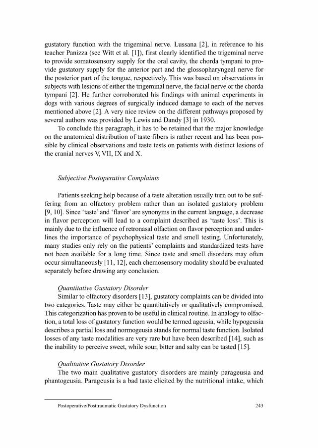
Postoperative/Posttraumatic Gustatory Dysfunction 243
gustatory function with the trigeminal nerve. Lussana [2], in reference to his
teacher Panizza (see Witt et al. [1]), first clearly identified the trigeminal nerve
to provide somatosensory supply for the oral cavity, the chorda tympani to pro-
vide gustatory supply for the anterior part and the glossopharyngeal nerve for
the posterior part of the tongue, respectively. This was based on observations in
subjects with lesions of either the trigeminal nerve, the facial nerve or the chorda
tympani [2]. He further corroborated his findings with animal experiments in
dogs with various degrees of surgically induced damage to each of the nerves
mentioned above [2]. A very nice review on the different pathways proposed by
several authors was provided by Lewis and Dandy [3] in 1930.
To conclude this paragraph, it has to be retained that the major knowledge
on the anatomical distribution of taste fibers is rather recent and has been pos-
sible by clinical observations and taste tests on patients with distinct lesions of
the cranial nerves V, VII, IX and X.
Subjective Postoperative Complaints
Patients seeking help because of a taste alteration usually turn out to be suf-
fering from an olfactory problem rather than an isolated gustatory problem
[9, 10]. Since ‘taste’ and ‘flavor’ are synonyms in the current language, a decrease
in flavor perception will lead to a complaint described as ‘taste loss’. This is
mainly due to the influence of retronasal olfaction on flavor perception and under-
lines the importance of psychophysical taste and smell testing. Unfortunately,
many studies only rely on the patients’ complaints and standardized tests have
not been available for a long time. Since taste and smell disorders may often
occur simultaneously [11, 12], each chemosensory modality should be evaluated
separately before drawing any conclusion.
Quantitative Gustatory DisorderSimilar to olfactory disorders [13], gustatory complaints can be divided into
two categories. Taste may either be quantitatively or qualitatively compromised.
This categorization has proven to be useful in clinical routine. In analogy to olfac-
tion, a total loss of gustatory function would be termed ageusia, while hypogeusia
describes a partial loss and normogeusia stands for normal taste function. Isolated
losses of any taste modalities are very rare but have been described [14], such as
the inability to perceive sweet, while sour, bitter and salty can be tasted [15].
Qualitative Gustatory DisorderThe two main qualitative gustatory disorders are mainly parageusia and
phantogeusia. Parageusia is a bad taste elicited by the nutritional intake, which
Landis/Lacroix 244
is otherwise absent [16]. Phantogeusia describes the presence of a permanent
intraoral bad taste [17, 18].
Causes
Middle Ear SurgeryThe facial nerve carries the gustatory innervation for the anterior two
thirds of the tongue and the palate [19]. The gustatory fibers run with the
chorda tympani which separates itself from the facial nerve within the temporal
bone. The chorda tympani also carries parasympathetic fibers for the main sali-
vary glands of the oral cavity [20]. The chorda tympani further travels unpro-
tected through the middle ear cavity. During middle ear surgery, the chorda is
freely exposed within the operative field. It is subject to considerable surgical
stress by stretch, injury or dryness, or it is directly sectioned in order to facili-
tate the surgical approach to the ossicles. Accordingly, following ear surgery,
lesions of the gustatory system may produce symptoms such as dysgeusia,
hypogeusia, ageusia or mouth dryness [3, 20–25]. Most of these postoperative
gustatory affections have been shown to be transitory. However, long-lasting
dysgeusia cases have been reported [26].
Systematic investigation of taste functions revealed little and often transi-
tory postoperative subjective complaints, but considerable alterations of mea-
surable ipsilateral taste sensitivity. Saito et al. [26] found the postoperative taste
alteration to correlate with the peroperative surgical stress. Although most
patients do not present long-term complaints, absent or incomplete taste recov-
ery has been observed in the majority of the investigated subjects. Especially
the group with a surgically severed chorda tympani exhibited the worst long-
term outcome. Attempts to readapt the sectioned ends of the chorda increase the
chances of recovery in taste function after surgery [27, 28]. Macroscopic reex-
amination of the chorda tympani in operated subjects revealed relatively high
rates of intact nerves [29]. However, microscopic analyses of such recovered
nerves have shown low numbers of intact and myelinated fibers, but mainly
fibrosis.
Considering the frequency of otologic surgery and especially the number of
bilaterally operated patients (i.e. for otosclerosis), very few complaints of ageu-
sia, hypogeusia and dry mouth are reported [20, 22]. One of the main reasons for
the low frequency of complaints appears to be related to the so-called ‘release of
inhibition’ phenomenon [30, 31]. Gustatory afferent inputs from cranial nerves
VII, IX, and X converge within the solitary nucleus of the medulla, whereas
input from the anterior or posterior portion of the tongue is topographically sep-
arated. When the chorda tympani is anesthetized, activity disappears in brain
Postoperative/Posttraumatic Gustatory Dysfunction 245
stem regions usually responding to stimulation of the anterior part of the tongue,
while the responsiveness to stimulation of the posterior parts of the tongue
increases. Thus, subjective reports of whole-mouth gustatory function may not
reflect regional taste function. Even lateralized ageusia may go unrecognized by
the patient. However, eating is a whole-mouth experience built of gustatory,
somatosensory and trigeminal inputs. Thus, it has been shown that lack of gusta-
tory function can be partly masked by touch [32]. Such ‘taste illusions’ might
also account for the poor subjective recognition of taste deficiencies. Beside the
injury due to the surgical procedure, there is some evidence that the chorda tym-
pani is already altered in subjects suffering from chronic inflammatory middle
ear diseases [33–36]. This is probably due to the aggressive behavior of some
middle ear inflammatory processes such as the cholesteatoma which is known to
erode adjacent structures in the middle ear [37].
In conclusion, patients undergoing middle ear surgery mostly exhibit altered
ipsilateral taste function. However, in the overwhelming majority of cases, this
alteration goes unnoticed by the patients and is transitory. Unfortunately, in some
patients, dysgeusia persists following middle ear surgery and the therapeutically
available options are limited. Although the mechanism of action is not clear, zinc
gluconate has recently been shown in a double-blind, placebo-controlled study to
improve taste function in idiopathic dysgeusia [38].
Tonsillectomy/Oropharyngeal SurgeryIn contrast to middle ear surgery, taste disorders after tonsillectomy have
been systematically investigated only by Tomita and Ohtuka [39]. However, dur-
ing the last 20 years, several reports have been available about this complication
[40, 41]. Compared to the very high frequency at which this surgery is performed,
taste complaints are extremely rare. After more than 3,500 tonsillectomies,
Tomita and Ohtuka [39] observed only 11 cases (0.3%) of taste problems reported
by the patients. These data mainly rely on subjective patient reports. Similar to the
gap between subjective and measurable taste disorders following middle ear
surgery, the ‘release of inhibition’ phenomenon probably also accounts for this
low occurrence of posttonsillectomy gustatory complaints. However, since eating
is a whole-mouth experience built out of gustatory, somatosensory and trigeminal
inputs, most quantitative localized taste disorders go unnoticed.
A prospective study measuring the taste acuity of the posterior third of the
tongue before and after tonsillectomy might unravel more psychophysical taste
disturbance than reported by patients. This assumption is mainly based on a
large anatomical study conducted by Ohtsuka et al. [42], who examined over
100 tonsillar beds with respect to their vicinity to the lingual branch of the glos-
sopharyngeal nerve (LBGN). Their study revealed that in approximately a quar-
ter of the cases, the LBGN traveled covered and separated from the tonsil by a
Landis/Lacroix 246
muscle layer over its whole course to the base of the tongue. In almost 50% of
the cases, the muscle lining of the tonsillar bed was discontinuous and only thin
muscle bundles covered the tonsillar capsule and the LBGN. Most interestingly,
in nearly 25% of cases, the LBGN was firmly adherent to the tonsillar capsule,
due to the complete absence of muscle lining between the tonsillar bed and the
LBGN. In these cases, and probably also in a similar percentage of patients
undergoing tonsillectomy, taste disturbance may occur on removal of the hyper-
trophic tonsillar capsule. It may be assumed that such taste alterations, in anal-
ogy to chorda tympani-related gustatory disorders, are also transitory. In the
few cases so far presented, no therapy could be offered [39–41].
Gustatory dysfunctions after all other kinds of oropharyngeal surgery have
been described [43–45]. Beside oncologic surgery (see below), it is mainly the
sleep apnea surgery which has been reported to alter chemosensory function
[46–51]. The most frequent interventions followed by gustatory complaints are
laser uvulopalatoplasty and uvulopalatopharyngoplasty. Although taste percep-
tion changes were recorded in all studies, psychophysical taste and smell testing
was performed in only one study [50]. No changes in gustatory or olfactory
function were reported by these authors. Since no taste buds have been
described on the uvula, this finding is not surprising [52]. The obviously pre-
sent changes in taste perception after sleep apnea surgery could be due to a
modification in the retronasal airflow pattern [53–56], modifying ‘flavor’ per-
ception which is widely attributed to ‘taste’. Thus, the reported changes rather
reflect olfactory changes. Another possibility is the lingual compression (see
below) during surgery which could temporarily alter peripheral taste nerve and
bud function. Further studies on patients undergoing sleep apnea surgery could
clarify these discrepancies between subjects’ reports and measured gustatory
function.
Oncologic Surgery and Radiation TherapyIn contrast to the previously mentioned surgical procedures, oncologic
surgery is far more devastating and taste disorders are frequently reported side
effects [57–60]. However, patients barely complain about chemosensory disorders
in view of the serious treatment and the severity of the disease. Resection of exten-
sive parts of the oral and cervical structures usually also involves loss of taste
fibers. This chapter is not dedicated to enumerate the various head and neck surgi-
cal procedures; however, we would like to focus on a special group of patients.
Laryngectomized subjects loose their whole flavor perception due to interruption
of the naso-laryngo-tracheal continuity. Since the negative pressure necessary for
nasal breathing can no longer be produced by laryngectomized patients, ortho- and
retronasal olfaction promptly disappears with laryngectomy [61, 62]. This leads
to a subjective loss of ‘taste’ in these patients, which can potentially influence
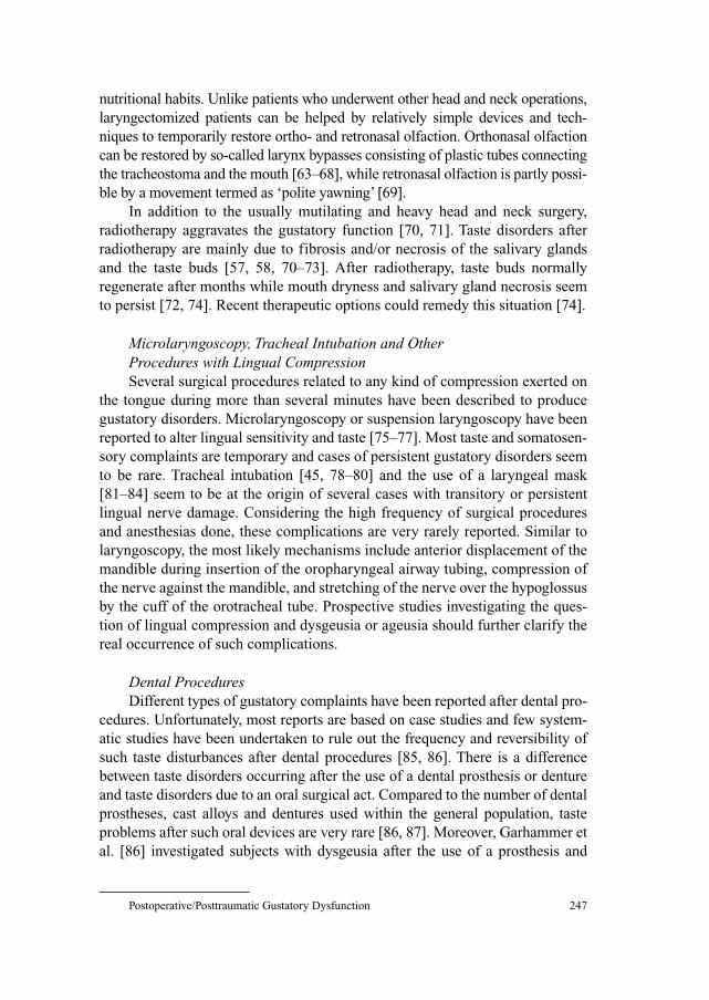
Postoperative/Posttraumatic Gustatory Dysfunction 247
nutritional habits. Unlike patients who underwent other head and neck operations,
laryngectomized patients can be helped by relatively simple devices and tech-
niques to temporarily restore ortho- and retronasal olfaction. Orthonasal olfaction
can be restored by so-called larynx bypasses consisting of plastic tubes connecting
the tracheostoma and the mouth [63–68], while retronasal olfaction is partly possi-
ble by a movement termed as ‘polite yawning’ [69].
In addition to the usually mutilating and heavy head and neck surgery,
radiotherapy aggravates the gustatory function [70, 71]. Taste disorders after
radiotherapy are mainly due to fibrosis and/or necrosis of the salivary glands
and the taste buds [57, 58, 70–73]. After radiotherapy, taste buds normally
regenerate after months while mouth dryness and salivary gland necrosis seem
to persist [72, 74]. Recent therapeutic options could remedy this situation [74].
Microlaryngoscopy, Tracheal Intubation and Other Procedures with Lingual CompressionSeveral surgical procedures related to any kind of compression exerted on
the tongue during more than several minutes have been described to produce
gustatory disorders. Microlaryngoscopy or suspension laryngoscopy have been
reported to alter lingual sensitivity and taste [75–77]. Most taste and somatosen-
sory complaints are temporary and cases of persistent gustatory disorders seem
to be rare. Tracheal intubation [45, 78–80] and the use of a laryngeal mask
[81–84] seem to be at the origin of several cases with transitory or persistent
lingual nerve damage. Considering the high frequency of surgical procedures
and anesthesias done, these complications are very rarely reported. Similar to
laryngoscopy, the most likely mechanisms include anterior displacement of the
mandible during insertion of the oropharyngeal airway tubing, compression of
the nerve against the mandible, and stretching of the nerve over the hypoglossus
by the cuff of the orotracheal tube. Prospective studies investigating the ques-
tion of lingual compression and dysgeusia or ageusia should further clarify the
real occurrence of such complications.
Dental ProceduresDifferent types of gustatory complaints have been reported after dental pro-
cedures. Unfortunately, most reports are based on case studies and few system-
atic studies have been undertaken to rule out the frequency and reversibility of
such taste disturbances after dental procedures [85, 86]. There is a difference
between taste disorders occurring after the use of a dental prosthesis or denture
and taste disorders due to an oral surgical act. Compared to the number of dental
prostheses, cast alloys and dentures used within the general population, taste
problems after such oral devices are very rare [86, 87]. Moreover, Garhammer et
al. [86] investigated subjects with dysgeusia after the use of a prosthesis and
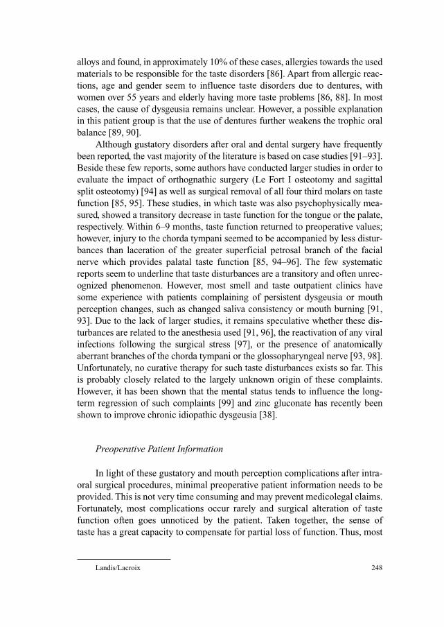
Landis/Lacroix 248
alloys and found, in approximately 10% of these cases, allergies towards the used
materials to be responsible for the taste disorders [86]. Apart from allergic reac-
tions, age and gender seem to influence taste disorders due to dentures, with
women over 55 years and elderly having more taste problems [86, 88]. In most
cases, the cause of dysgeusia remains unclear. However, a possible explanation
in this patient group is that the use of dentures further weakens the trophic oral
balance [89, 90].
Although gustatory disorders after oral and dental surgery have frequently
been reported, the vast majority of the literature is based on case studies [91–93].
Beside these few reports, some authors have conducted larger studies in order to
evaluate the impact of orthognathic surgery (Le Fort I osteotomy and sagittal
split osteotomy) [94] as well as surgical removal of all four third molars on taste
function [85, 95]. These studies, in which taste was also psychophysically mea-
sured, showed a transitory decrease in taste function for the tongue or the palate,
respectively. Within 6–9 months, taste function returned to preoperative values;
however, injury to the chorda tympani seemed to be accompanied by less distur-
bances than laceration of the greater superficial petrosal branch of the facial
nerve which provides palatal taste function [85, 94–96]. The few systematic
reports seem to underline that taste disturbances are a transitory and often unrec-
ognized phenomenon. However, most smell and taste outpatient clinics have
some experience with patients complaining of persistent dysgeusia or mouth
perception changes, such as changed saliva consistency or mouth burning [91,
93]. Due to the lack of larger studies, it remains speculative whether these dis-
turbances are related to the anesthesia used [91, 96], the reactivation of any viral
infections following the surgical stress [97], or the presence of anatomically
aberrant branches of the chorda tympani or the glossopharyngeal nerve [93, 98].
Unfortunately, no curative therapy for such taste disturbances exists so far. This
is probably closely related to the largely unknown origin of these complaints.
However, it has been shown that the mental status tends to influence the long-
term regression of such complaints [99] and zinc gluconate has recently been
shown to improve chronic idiopathic dysgeusia [38].
Preoperative Patient Information
In light of these gustatory and mouth perception complications after intra-
oral surgical procedures, minimal preoperative patient information needs to be
provided. This is not very time consuming and may prevent medicolegal claims.
Fortunately, most complications occur rarely and surgical alteration of taste
function often goes unnoticed by the patient. Taken together, the sense of
taste has a great capacity to compensate for partial loss of function. Thus, most
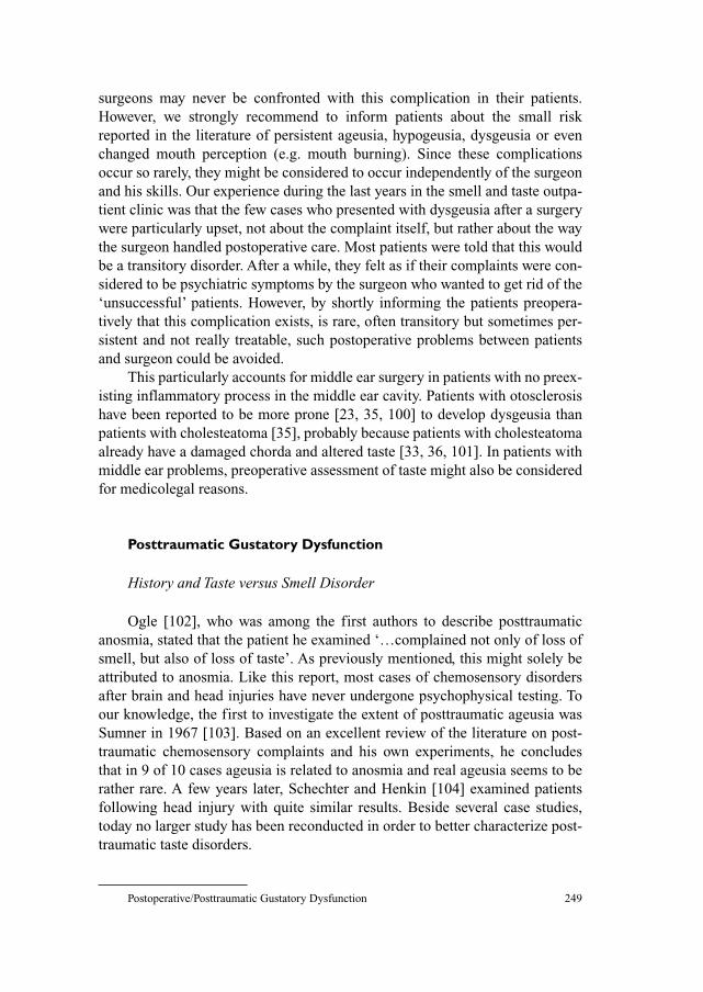
Postoperative/Posttraumatic Gustatory Dysfunction 249
surgeons may never be confronted with this complication in their patients.
However, we strongly recommend to inform patients about the small risk
reported in the literature of persistent ageusia, hypogeusia, dysgeusia or even
changed mouth perception (e.g. mouth burning). Since these complications
occur so rarely, they might be considered to occur independently of the surgeon
and his skills. Our experience during the last years in the smell and taste outpa-
tient clinic was that the few cases who presented with dysgeusia after a surgery
were particularly upset, not about the complaint itself, but rather about the way
the surgeon handled postoperative care. Most patients were told that this would
be a transitory disorder. After a while, they felt as if their complaints were con-
sidered to be psychiatric symptoms by the surgeon who wanted to get rid of the
‘unsuccessful’ patients. However, by shortly informing the patients preopera-
tively that this complication exists, is rare, often transitory but sometimes per-
sistent and not really treatable, such postoperative problems between patients
and surgeon could be avoided.
This particularly accounts for middle ear surgery in patients with no preex-
isting inflammatory process in the middle ear cavity. Patients with otosclerosis
have been reported to be more prone [23, 35, 100] to develop dysgeusia than
patients with cholesteatoma [35], probably because patients with cholesteatoma
already have a damaged chorda and altered taste [33, 36, 101]. In patients with
middle ear problems, preoperative assessment of taste might also be considered
for medicolegal reasons.
Posttraumatic Gustatory Dysfunction
History and Taste versus Smell Disorder
Ogle [102], who was among the first authors to describe posttraumatic
anosmia, stated that the patient he examined ‘…complained not only of loss of
smell, but also of loss of taste’. As previously mentioned, this might solely be
attributed to anosmia. Like this report, most cases of chemosensory disorders
after brain and head injuries have never undergone psychophysical testing. To
our knowledge, the first to investigate the extent of posttraumatic ageusia was
Sumner in 1967 [103]. Based on an excellent review of the literature on post-
traumatic chemosensory complaints and his own experiments, he concludes
that in 9 of 10 cases ageusia is related to anosmia and real ageusia seems to be
rather rare. A few years later, Schechter and Henkin [104] examined patients
following head injury with quite similar results. Beside several case studies,
today no larger study has been reconducted in order to better characterize post-
traumatic taste disorders.
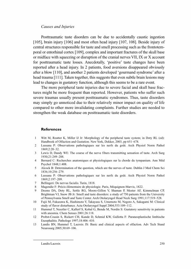
Landis/Lacroix 250
Causes and Injuries
Posttraumatic taste disorders can be due to accidentally caustic ingestion
[105], brain injury [106] and most often head injury [107, 108]. Beside injury of
central structures responsible for taste and smell processing such as the frontotem-
poral or entorhinal cortex [109], complex and important fractures of the skull base
or midface with squeezing or disruption of the cranial nerves VII, IX or X account
for posttraumatic taste losses. Anecdotally, ‘positive’ taste changes have been
reported after a head injury. In 2 patients, food aversions disappeared obviously
after a blow [110], and another 2 patients developed ‘gourmand syndrome’ after a
head trauma [111]. Taken together, this suggests that even subtle brain lesions may
lead to changes in gustatory function, although this seems to be a rare event.
The more peripheral taste injuries due to severe facial and skull base frac-
tures might be more frequent than reported. However, patients who suffer such
severe traumas usually present posttraumatic syndromes. Thus, taste disorders
may simply go unnoticed due to their relatively minor impact on quality of life
compared to other more invalidating complaints. Further studies are needed to
strengthen the weak database on posttraumatic taste disorders.
References
1 Witt M, Reutter K, Miller IJ Jr: Morphology of the peripheral taste system; in Doty RL (ed):
Handbook of Olfaction and Gustation. New York, Dekker, 2003, pp 651–678.
2 Lussana P: Observations pathologiques sur les nerfs du goût. Arch Physiol Norm Pathol
1869;2:20–32.
3 Lewis D, Dandy WE: The course of the nerve fibers transmitting sensation of taste. Arch Surg
1930;21:249–288.
4 Bernard C: Recherches anatomiques et physiologiques sur la chorde du tympanium. Ann Méd
Psychol 1848;1:408.
5 Alcock B: Determination of the question, which are the nerves of taste. Dublin J Med Chem Sci
1836;10:256–279.
6 Lussana P: Observations pathologiques sur les nerfs du goût. Arch Physiol Norm Pathol
1869;2:197–209.
7 Bellingeri: De nervus facialis. Turin, 1818.
8 Magendie F: Précis élémentaire de physiologie. Paris, Méquignon-Marvis, 1822.
9 Deems DA, Doty RL, Settle RG, Moore-Gillon V, Shaman P, Mester AF, Kimmelman CP,
Brightman VJ, Snow JB Jr: Smell and taste disorders: a study of 750 patients from the University
of Pennsylvania Smell and Taste Center. Arch Otolaryngol Head Neck Surg 1991;117:519–528.
10 Fujii M, Fukazawa K, Hashimoto Y, Takayasu S, Umemoto M, Negoro A, Sakagami M: Clinical
study of flavor disturbance. Acta Otolaryngol Suppl 2004;553:109–112.
11 Hummel T, Nesztler C, Kallerf S, Kobal G, Bende M, Nordin S: Gustatory sensitivity in patients
with anosmia. Chem Senses 2001;26:118.
12 Probst-Cousin S, Rickert CH, Kunde D, Schmid KW, Gullotta F: Paraneoplastische limbische
Enzephalitis. Pathologe 1997;18:406–410.
13 Landis BN, Hummel T, Lacroix JS: Basic and clinical aspects of olfaction. Adv Tech Stand
Neurosurg 2005;30:69–106.
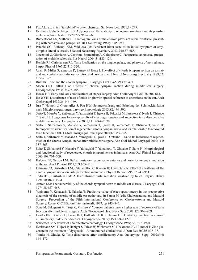
Postoperative/Posttraumatic Gustatory Dysfunction 251
14 Fox AL: Six in ten ‘tasteblind’ to bitter chemical. Sci News Lett 1931;19:249.
15 Henkin RI, Shallenberger RS: Aglycogeusia: the inability to recognize sweetness and its possible
molecular basis. Nature 1970;227:965–966.
16 Rutherfoord GS, Mathew B: Xanthogranuloma of the choroid plexus of lateral ventricle, present-
ing with parosmia and parageusia. Br J Neurosurg 1987;1:285–288.
17 Petzold GC, Einhaupl KM, Valdueza JM: Persistent bitter taste as an initial symptom of amy-
otrophic lateral sclerosis. J Neurol Neurosurg Psychiatry 2003;74:687–688.
18 Nocentini U, Giordano A, Castriota-Scanderbeg A, Caltagirone C: Parageusia: an unusual presen-
tation of multiple sclerosis. Eur Neurol 2004;51:123–124.
19 Henkin RI, Christiansen RL: Taste localization on the tongue, palate, and pharynx of normal man.
J Appl Physiol 1967;22:316–320.
20 Grant R, Miller S, Simpson D, Lamey PJ, Bone I: The effect of chorda tympani section on ipsilat-
eral and contralateral salivary secretion and taste in man. J Neurol Neurosurg Psychiatry 1989;52:
1058–1062.
21 Bull TR: Taste and the chorda tympani. J Laryngol Otol 1965;79:479–493.
22 Moon CNJ, Pullen EW: Effects of chorda tympani section during middle ear surgery.
Laryngoscope 1963;73:392–405.
23 House HP: Early and late complications of stapes surgery. Arch Otolaryngol 1963;78:606–613.
24 Ho WYH: Disturbance of taste of otitic origin with special reference to operations on the ear. Arch
Otolaryngol 1937;26:146–169.
25 Just T, Homoth J, Graumuller S, Pau HW: Schmeckstörung und Erholung der Schmeckfunktion
nach Mittelohroperationen. Laryngorhinootologie 2003;82:494–500.
26 Saito T, Manabe Y, Shibamori Y, Yamagishi T, Igawa H, Tokuriki M, Fukuoka Y, Noda I, Ohtsubo
T, Saito H: Long-term follow-up results of electrogustometry and subjective taste disorder after
middle ear surgery. Laryngoscope 2001;111:2064–2070.
27 Saito T, Shibamori Y, Manabe Y, Yamagishi T, Igawa H, Yamamoto T, Ohtsubo T, Saito H:
Intraoperative identification of regenerated chorda tympani nerve and its relationship to recovered
taste function. ORL J Otorhinolaryngol Relat Spec 2001;63:359–365.
28 Saito T, Shibamori Y, Manabe Y, Yamagishi T, Igawa H, Ohtsubo T, Saito H: Incidence of regener-
ation of the chorda tympani nerve after middle ear surgery. Ann Otol Rhinol Laryngol 2002;111:
357–363.
29 Saito T, Shibamori Y, Manabe Y, Yamagishi T, Yamamoto T, Ohtsubo T, Saito H: Morphological
and functional study of regenerated chorda tympani nerves in humans. Ann Otol Rhinol Laryngol
2000;109:703–709.
30 Halpern BP, Nelson LM: Bulbar gustatory responses to anterior and posterior tongue stimulation
in the rat. Am J Physiol 1965;209:105–110.
31 Lehman CD, Bartoshuk LM, Catalanotto FC, Kveton JF, Lowlicht RA: Effect of anesthesia of the
chorda tympani nerve on taste perception in humans. Physiol Behav 1995;57:943–951.
32 Todrank J, Bartoshuk LM: A taste illusion: taste sensation localized by touch. Physiol Behav
1991;50:1027–1031.
33 Arnold SM: The vulnerability of the chorda tympani nerve to middle ear disease. J Laryngol Otol
1974;88:457–466.
34 Yaginuma Y, Kobayashi T, Takaska T: Predictive value of electrogustrometry in the preoperative
diagnosis of the severity of middle ear pathology; in Sanna M (ed): Cholesteatoma and Mastoid
Surgery: Proceeding of the Fifth International Conference on Cholesteatoma and Mastoid
Surgery. Rome, CIC Edizioni Internazionali, 1997, pp 843–846.
35 Sone M, Sakagami M, Tsuji K, Mishiro Y: Younger patients have a higher rate of recovery of taste
function after middle ear surgery. Arch Otolaryngol Head Neck Surg 2001;127:967–969.
36 Landis BN, Beutner D, Frasnelli J, Huttenbrink KB, Hummel T: Gustatory function in chronic
inflammatory middle ear diseases. Laryngoscope 2005;115:1124–1127.
37 Schechter G: A review of cholesteatoma pathology. Laryngoscope 1969;79:1907–1920.
38 Heckmann SM, Hujoel P, Habiger S, Friess W, Wichmann M, Heckmann JG, Hummel T: Zinc glu-
conate in the treatment of dysgeusia – A randomized clinical trial. J Dent Res 2005;84:35–38.
39 Tomita H, Ohtuka K: Taste disturbance after tonsillectomy. Acta Otolaryngol Suppl 2002;546:
164–172.
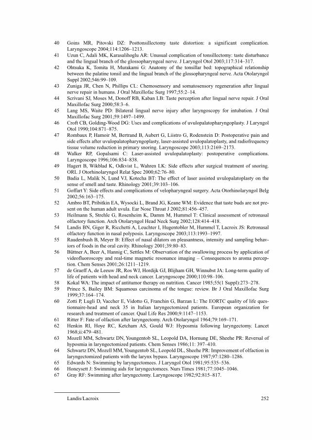
Landis/Lacroix 252
40 Goins MR, Pitovski DZ: Posttonsillectomy taste distortion: a significant complication.
Laryngoscope 2004;114:1206–1213.
41 Uzun C, Adali MK, Karasalihoglu AR: Unusual complication of tonsillectomy: taste disturbance
and the lingual branch of the glossopharyngeal nerve. J Laryngol Otol 2003;117:314–317.
42 Ohtsuka K, Tomita H, Murakami G: Anatomy of the tonsillar bed: topographical relationship
between the palatine tonsil and the lingual branch of the glossopharyngeal nerve. Acta Otolaryngol
Suppl 2002;546:99–109.
43 Zuniga JR, Chen N, Phillips CL: Chemosensory and somatosensory regeneration after lingual
nerve repair in humans. J Oral Maxillofac Surg 1997;55:2–14.
44 Scrivani SJ, Moses M, Donoff RB, Kaban LB: Taste perception after lingual nerve repair. J Oral
Maxillofac Surg 2000;58:3–6.
45 Lang MS, Waite PD: Bilateral lingual nerve injury after laryngoscopy for intubation. J Oral
Maxillofac Surg 2001;59:1497–1499.
46 Croft CB, Golding-Wood DG: Uses and complications of uvulopalatopharyngoplasty. J Laryngol
Otol 1990;104:871–875.
47 Rombaux P, Hamoir M, Bertrand B, Aubert G, Liistro G, Rodenstein D: Postoperative pain and
side effects after uvulopalatopharyngoplasty, laser-assisted uvulopalatoplasty, and radiofrequency
tissue volume reduction in primary snoring. Laryngoscope 2003;113:2169–2173.
48 Walker RP, Gopalsami C: Laser-assisted uvulopalatoplasty: postoperative complications.
Laryngoscope 1996;106:834–838.
49 Hagert B, Wikblad K, Odkvist L, Wahren LK: Side effects after surgical treatment of snoring.
ORL J Otorhinolaryngol Relat Spec 2000;62:76–80.
50 Badia L, Malik N, Lund VJ, Kotecha BT: The effect of laser assisted uvulopalatoplasty on the
sense of smell and taste. Rhinology 2001;39:103–106.
51 Goffart Y: Side effects and complications of velopharyngeal surgery. Acta Otorhinolaryngol Belg
2002;56:163–175.
52 Ambro BT, Pribitkin EA, Wysocki L, Brand JG, Keane WM: Evidence that taste buds are not pre-
sent on the human adult uvula. Ear Nose Throat J 2002;81:456–457.
53 Heilmann S, Strehle G, Rosenheim K, Damm M, Hummel T: Clinical assessment of retronasal
olfactory function. Arch Otolaryngol Head Neck Surg 2002;128:414–418.
54 Landis BN, Giger R, Ricchetti A, Leuchter I, Hugentobler M, Hummel T, Lacroix JS: Retronasal
olfactory function in nasal polyposis. Laryngoscope 2003;113:1993–1997.
55 Raudenbush B, Meyer B: Effect of nasal dilators on pleasantness, intensity and sampling behav-
iors of foods in the oral cavity. Rhinology 2001;39:80–83.
56 Büttner A, Beer A, Hannig C, Settles M: Observation of the swallowing process by application of
videofluoroscopy and real-time magnetic resonance imaging – Consequences to aroma percep-
tion. Chem Senses 2001;26:1211–1219.
57 de Graeff A, de Leeuw JR, Ros WJ, Hordijk GJ, Blijham GH, Winnubst JA: Long-term quality of
life of patients with head and neck cancer. Laryngoscope 2000;110:98–106.
58 Kokal WA: The impact of antitumor therapy on nutrition. Cancer 1985;55(1 Suppl):273–278.
59 Prince S, Bailey BM: Squamous carcinoma of the tongue: review. Br J Oral Maxillofac Surg
1999;37:164–174.
60 Zotti P, Lugli D, Vaccher E, Vidotto G, Franchin G, Barzan L: The EORTC quality of life ques-
tionnaire-head and neck 35 in Italian laryngectomized patients. European organization for
research and treatment of cancer. Qual Life Res 2000;9:1147–1153.
61 Ritter F: Fate of olfaction after laryngectomy. Arch Otolaryngol 1964;79:169–171.
62 Henkin RI, Hoye RC, Ketcham AS, Gould WJ: Hyposmia following laryngectomy. Lancet
1968;ii:479–481.
63 Mozell MM, Schwartz DN, Youngentob SL, Leopold DA, Hornung DE, Sheehe PR: Reversal of
hyposmia in laryngectomized patients. Chem Senses 1986;11: 397–410.
64 Schwartz DN, Mozell MM, Youngentob SL, Leopold DL, Sheehe PR: Improvement of olfaction in
laryngectomized patients with the larynx bypass. Laryngoscope 1987;97:1280–1286.
65 Edwards N: Swimming by laryngectomees. J Laryngol Otol 1981;95:535–536.
66 Honeysett J: Swimming aids for laryngectomees. Nurs Times 1981;77:1045–1046.
67 Gray RF: Swimming after laryngectomy. Laryngoscope 1982;92:815–817.
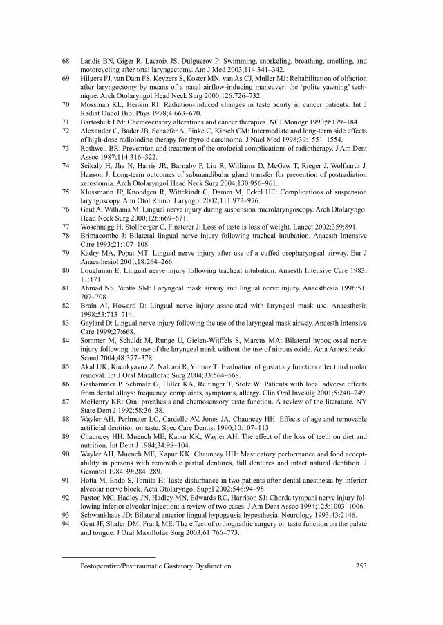
Postoperative/Posttraumatic Gustatory Dysfunction 253
68 Landis BN, Giger R, Lacroix JS, Dulguerov P: Swimming, snorkeling, breathing, smelling, and
motorcycling after total laryngectomy. Am J Med 2003;114:341–342.
69 Hilgers FJ, van Dam FS, Keyzers S, Koster MN, van As CJ, Muller MJ: Rehabilitation of olfaction
after laryngectomy by means of a nasal airflow-inducing maneuver: the ‘polite yawning’ tech-
nique. Arch Otolaryngol Head Neck Surg 2000;126:726–732.
70 Mossman KL, Henkin RI: Radiation-induced changes in taste acuity in cancer patients. Int J
Radiat Oncol Biol Phys 1978;4:663–670.
71 Bartoshuk LM: Chemosensory alterations and cancer therapies. NCI Monogr 1990;9:179–184.
72 Alexander C, Bader JB, Schaefer A, Finke C, Kirsch CM: Intermediate and long-term side effects
of high-dose radioiodine therapy for thyroid carcinoma. J Nucl Med 1998;39:1551–1554.
73 Rothwell BR: Prevention and treatment of the orofacial complications of radiotherapy. J Am Dent
Assoc 1987;114:316–322.
74 Seikaly H, Jha N, Harris JR, Barnaby P, Liu R, Williams D, McGaw T, Rieger J, Wolfaardt J,
Hanson J: Long-term outcomes of submandibular gland transfer for prevention of postradiation
xerostomia. Arch Otolaryngol Head Neck Surg 2004;130:956–961.
75 Klussmann JP, Knoedgen R, Wittekindt C, Damm M, Eckel HE: Complications of suspension
laryngoscopy. Ann Otol Rhinol Laryngol 2002;111:972–976.
76 Gaut A, Williams M: Lingual nerve injury during suspension microlaryngoscopy. Arch Otolaryngol
Head Neck Surg 2000;126:669–671.
77 Woschnagg H, Stollberger C, Finsterer J: Loss of taste is loss of weight. Lancet 2002;359:891.
78 Brimacombe J: Bilateral lingual nerve injury following tracheal intubation. Anaesth Intensive
Care 1993;21:107–108.
79 Kadry MA, Popat MT: Lingual nerve injury after use of a cuffed oropharyngeal airway. Eur J
Anaesthesiol 2001;18:264–266.
80 Loughman E: Lingual nerve injury following tracheal intubation. Anaesth Intensive Care 1983;
11:171.
81 Ahmad NS, Yentis SM: Laryngeal mask airway and lingual nerve injury. Anaesthesia 1996;51:
707–708.
82 Brain AI, Howard D: Lingual nerve injury associated with laryngeal mask use. Anaesthesia
1998;53:713–714.
83 Gaylard D: Lingual nerve injury following the use of the laryngeal mask airway. Anaesth Intensive
Care 1999;27:668.
84 Sommer M, Schuldt M, Runge U, Gielen-Wijffels S, Marcus MA: Bilateral hypoglossal nerve
injury following the use of the laryngeal mask without the use of nitrous oxide. Acta Anaesthesiol
Scand 2004;48:377–378.
85 Akal UK, Kucukyavuz Z, Nalcaci R, Yilmaz T: Evaluation of gustatory function after third molar
removal. Int J Oral Maxillofac Surg 2004;33:564–568.
86 Garhammer P, Schmalz G, Hiller KA, Reitinger T, Stolz W: Patients with local adverse effects
from dental alloys: frequency, complaints, symptoms, allergy. Clin Oral Investig 2001;5:240–249.
87 McHenry KR: Oral prosthesis and chemosensory taste function. A review of the literature. NY
State Dent J 1992;58:36–38.
88 Wayler AH, Perlmuter LC, Cardello AV, Jones JA, Chauncey HH: Effects of age and removable
artificial dentition on taste. Spec Care Dentist 1990;10:107–113.
89 Chauncey HH, Muench ME, Kapur KK, Wayler AH: The effect of the loss of teeth on diet and
nutrition. Int Dent J 1984;34:98–104.
90 Wayler AH, Muench ME, Kapur KK, Chauncey HH: Masticatory performance and food accept-
ability in persons with removable partial dentures, full dentures and intact natural dentition. J
Gerontol 1984;39:284–289.
91 Hotta M, Endo S, Tomita H: Taste disturbance in two patients after dental anesthesia by inferior
alveolar nerve block. Acta Otolaryngol Suppl 2002;546:94–98.
92 Paxton MC, Hadley JN, Hadley MN, Edwards RC, Harrison SJ: Chorda tympani nerve injury fol-
lowing inferior alveolar injection: a review of two cases. J Am Dent Assoc 1994;125:1003–1006.
93 Schwankhaus JD: Bilateral anterior lingual hypogeusia hypesthesia. Neurology 1993;43:2146.
94 Gent JF, Shafer DM, Frank ME: The effect of orthognathic surgery on taste function on the palate
and tongue. J Oral Maxillofac Surg 2003;61:766–773.
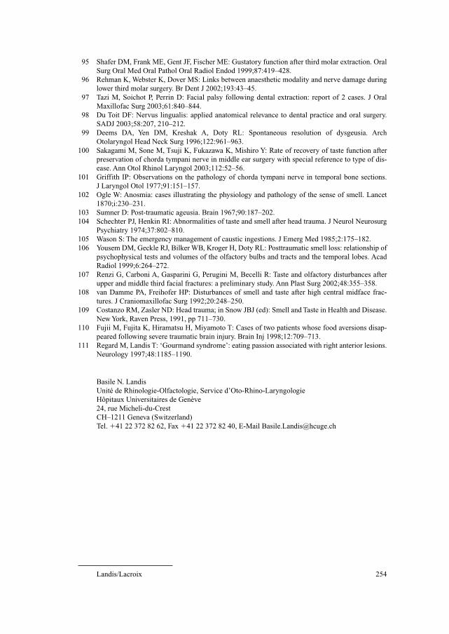
Landis/Lacroix 254
95 Shafer DM, Frank ME, Gent JF, Fischer ME: Gustatory function after third molar extraction. Oral
Surg Oral Med Oral Pathol Oral Radiol Endod 1999;87:419–428.
96 Rehman K, Webster K, Dover MS: Links between anaesthetic modality and nerve damage during
lower third molar surgery. Br Dent J 2002;193:43–45.
97 Tazi M, Soichot P, Perrin D: Facial palsy following dental extraction: report of 2 cases. J Oral
Maxillofac Surg 2003;61:840–844.
98 Du Toit DF: Nervus lingualis: applied anatomical relevance to dental practice and oral surgery.
SADJ 2003;58:207, 210–212.
99 Deems DA, Yen DM, Kreshak A, Doty RL: Spontaneous resolution of dysgeusia. Arch
Otolaryngol Head Neck Surg 1996;122:961–963.
100 Sakagami M, Sone M, Tsuji K, Fukazawa K, Mishiro Y: Rate of recovery of taste function after
preservation of chorda tympani nerve in middle ear surgery with special reference to type of dis-
ease. Ann Otol Rhinol Laryngol 2003;112:52–56.
101 Griffith IP: Observations on the pathology of chorda tympani nerve in temporal bone sections.
J Laryngol Otol 1977;91:151–157.
102 Ogle W: Anosmia: cases illustrating the physiology and pathology of the sense of smell. Lancet
1870;i:230–231.
103 Sumner D: Post-traumatic ageusia. Brain 1967;90:187–202.
104 Schechter PJ, Henkin RI: Abnormalities of taste and smell after head trauma. J Neurol Neurosurg
Psychiatry 1974;37:802–810.
105 Wason S: The emergency management of caustic ingestions. J Emerg Med 1985;2:175–182.
106 Yousem DM, Geckle RJ, Bilker WB, Kroger H, Doty RL: Posttraumatic smell loss: relationship of
psychophysical tests and volumes of the olfactory bulbs and tracts and the temporal lobes. Acad
Radiol 1999;6:264–272.
107 Renzi G, Carboni A, Gasparini G, Perugini M, Becelli R: Taste and olfactory disturbances after
upper and middle third facial fractures: a preliminary study. Ann Plast Surg 2002;48:355–358.
108 van Damme PA, Freihofer HP: Disturbances of smell and taste after high central midface frac-
tures. J Craniomaxillofac Surg 1992;20:248–250.
109 Costanzo RM, Zasler ND: Head trauma; in Snow JBJ (ed): Smell and Taste in Health and Disease.
New York, Raven Press, 1991, pp 711–730.
110 Fujii M, Fujita K, Hiramatsu H, Miyamoto T: Cases of two patients whose food aversions disap-
peared following severe traumatic brain injury. Brain Inj 1998;12:709–713.
111 Regard M, Landis T: ‘Gourmand syndrome’: eating passion associated with right anterior lesions.
Neurology 1997;48:1185–1190.
Basile N. Landis
Unité de Rhinologie-Olfactologie, Service d’Oto-Rhino-Laryngologie
Hôpitaux Universitaires de Genève
24, rue Micheli-du-Crest
CH–1211 Geneva (Switzerland)
Tel. �41 22 372 82 62, Fax �41 22 372 82 40, E-Mail [email protected]
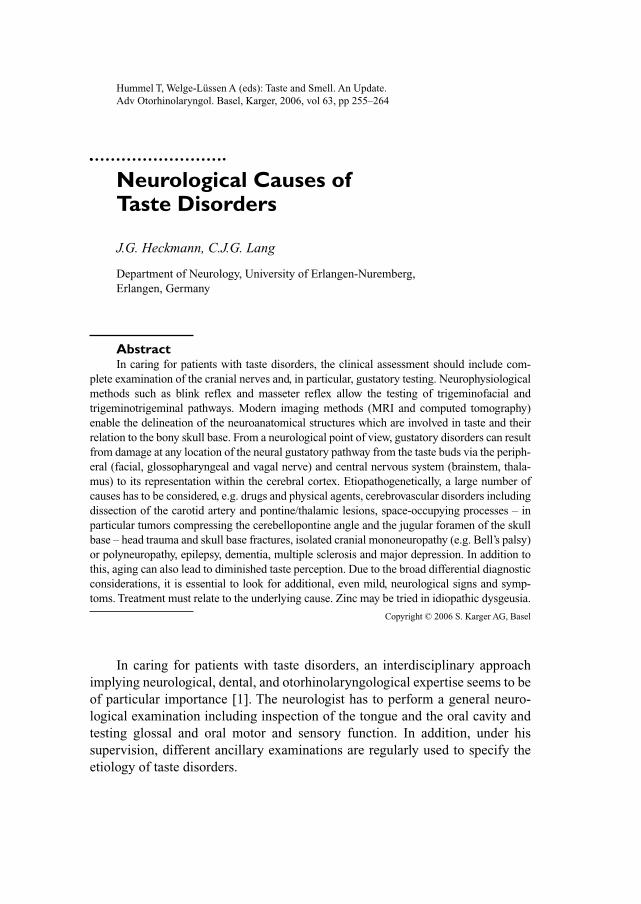
Hummel T, Welge-Lüssen A (eds): Taste and Smell. An Update.
Adv Otorhinolaryngol. Basel, Karger, 2006, vol 63, pp 255–264
Neurological Causes of Taste Disorders
J.G. Heckmann, C.J.G. Lang
Department of Neurology, University of Erlangen-Nuremberg,
Erlangen, Germany
AbstractIn caring for patients with taste disorders, the clinical assessment should include com-
plete examination of the cranial nerves and, in particular, gustatory testing. Neurophysiological
methods such as blink reflex and masseter reflex allow the testing of trigeminofacial and
trigeminotrigeminal pathways. Modern imaging methods (MRI and computed tomography)
enable the delineation of the neuroanatomical structures which are involved in taste and their
relation to the bony skull base. From a neurological point of view, gustatory disorders can result
from damage at any location of the neural gustatory pathway from the taste buds via the periph-
eral (facial, glossopharyngeal and vagal nerve) and central nervous system (brainstem, thala-
mus) to its representation within the cerebral cortex. Etiopathogenetically, a large number of
causes has to be considered, e.g. drugs and physical agents, cerebrovascular disorders including
dissection of the carotid artery and pontine/thalamic lesions, space-occupying processes – in
particular tumors compressing the cerebellopontine angle and the jugular foramen of the skull
base – head trauma and skull base fractures, isolated cranial mononeuropathy (e.g. Bell’s palsy)
or polyneuropathy, epilepsy, dementia, multiple sclerosis and major depression. In addition to
this, aging can also lead to diminished taste perception. Due to the broad differential diagnostic
considerations, it is essential to look for additional, even mild, neurological signs and symp-
toms. Treatment must relate to the underlying cause. Zinc may be tried in idiopathic dysgeusia.
Copyright © 2006 S. Karger AG, Basel
In caring for patients with taste disorders, an interdisciplinary approach
implying neurological, dental, and otorhinolaryngological expertise seems to be
of particular importance [1]. The neurologist has to perform a general neuro-
logical examination including inspection of the tongue and the oral cavity and
testing glossal and oral motor and sensory function. In addition, under his
supervision, different ancillary examinations are regularly used to specify the
etiology of taste disorders.

Heckmann/Lang 256
Electrophysiological tests can be applied to identify abnormalities in the
cranial nerve-brainstem pathways which is of importance in cases of trigeminal
neuropathy, multiple sclerosis, or pontine lesions. For example, the blink reflex
may be used to evaluate the integrity of the interaction between the trigeminal
nerve, pontine brainstem, and facial nerve [2]. The integrity of the trigemino-
brainstem-trigeminal pathway can be elaborated using the masseter reflex [3].
Imaging techniques are routinely used to demonstrate lesions in the taste
pathway. In particular, using special sequences, MRI allows the cranial nerves
to be imaged [4]. Furthermore, MRI applications provide significant informa-
tion to identify type and etiology of a lesion. Analysis of mucosal blood flow in
the oral cavity in combination with the assessment of autonomous, cardiocircu-
latory parameters appears to be useful in the diagnosis of autonomic disorders
in burning mouth syndrome or in patients with inborn autonomic disorder both
of which are associated with gustatory dysfunction [5, 6]. Microbiological cul-
tures are indicated when fungal or bacterial infections are suspected.
Analysis of saliva should be performed as it constitutes the environment of
taste receptors including both, transport of tastants to the receptor and protec-
tion of the taste receptor. Typical clinical investigations are sialometry and
sialochemistry. In combination with the clinical assessment, these techniques
provide information with regard to the individual salivary status [7].
The search for the cause of taste disorders should be guided by the question
whether they are due to damage to the peripheral or central nervous system or
whether they are caused by neurological disease with undetermined localization.
Peripheral Neurological Causes
Lesions of the peripheral nervous system can be confined to syndromes
affecting the facial and/or the glossopharyngeal nerve. Epidemiologically,
affections of the facial nerve are far more frequent than those of the glossopha-
ryngeal nerve with the most frequent disease being idiopathic Bell’s palsy.
While patients typically complain about insufficient closing of the eyelid and
numbness along the affected cheek, gustatory dysfunction can also be a lead-
ing, and sometimes even the earliest complaint. The etiopathogenesis of Bell’s
palsy is a matter of debate. Treatment with corticosteroids has been recom-
mended [8]. Other causes of facial nerve lesions have to be considered, e.g.
neuritis due to neuroborreliosis or zoster, or space-occupying processes in the
cerebellopontine angle such as meningioma or neurinoma [8, 9]. Other causes
include iatrogenic lesions, e.g. lengthy laryngoscopic manipulations [10].
Affections of the glossopharyngeal nerve not only lead to gustatory dys-
function but also to problems in swallowing (sore throat ipsilateral to the lesion)
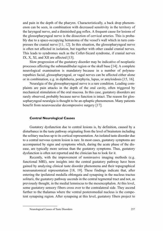
Neurological Causes of Taste Disorders 257
and pain in the depth of the pharynx. Characteristically, a back drop phenom-
enon can be seen, in combination with decreased sensitivity in the territory of
the laryngeal nerve, and a diminished gag reflex. A frequent cause for lesions of
the glossopharyngeal nerve is the dissection of cervical arteries. This is proba-
bly due to a space-occupying hematoma of the vessel’s wall which in turn com-
presses the cranial nerve [11, 12]. In this situation, the glossopharyngeal nerve
is often not affected in isolation, but together with other caudal cranial nerves.
This leads to syndromes such as the Collet-Sicard syndrome, if cranial nerves
IX, X, XI, and XII are affected [13].
Slow progression of the gustatory disorder may be indicative of neoplastic
processes affecting the submandibular region or the skull base [14]. A complete
neurological examination is mandatory because in a number of polyneu-
ropathies facial, glossopharyngeal, or vagal nerves can be affected either alone
or in combination, e.g. in diphtheria, porphyria, lupus, or amyloidosis [15, 16].
Neuralgia of the glossopharyngeal nerve is a rare condition. Leading com-
plaints are pain attacks in the depth of the oral cavity, often triggered by
mechanical stimulation of the oral mucosa. In this case, gustatory disorders are
rarely observed, probably because nerve function is intact. The reason for glos-
sopharyngeal neuralgia is thought to be an ephaptic phenomenon. Many patients
benefit from neurovascular decompressive surgery [17].
Central Neurological Causes
Gustatory dysfunction due to central lesions is, by definition, caused by a
disturbance in the taste pathway originating from the level of brainstem including
the solitary nucleus up to its cortical representation. An isolated taste disorder due
to a central nervous system lesion is rare. In most cases, gustatory symptoms are
accompanied by signs and symptoms which, during the acute phase of the dis-
ease, are typically more serious than the gustatory symptoms. Thus, gustatory
dysfunction is often not reported and the clinician has to look for it.
Recently, with the improvement of noninvasive imaging methods (e.g.
functional MRI), new insights into the central gustatory pathway have been
gained by analyzing clinical taste disorder phenomena and their topographical
neuroanatomical representation [18, 19]. These findings indicate that, after
entering the ipsilateral medulla oblongata and synapsing in the nucleus tractus
solitarii, the gustatory pathway ascends in the central tegmental tract and not, as
previously thought, in the medial lemniscus to the mesencephalon. At this level,
some gustatory sensory fibers cross over to the contralateral side. They ascend
further to the thalamus where the ventral posteromedial nucleus is the compe-
tent synapsing region. After synapsing at this level, gustatory fibers project to
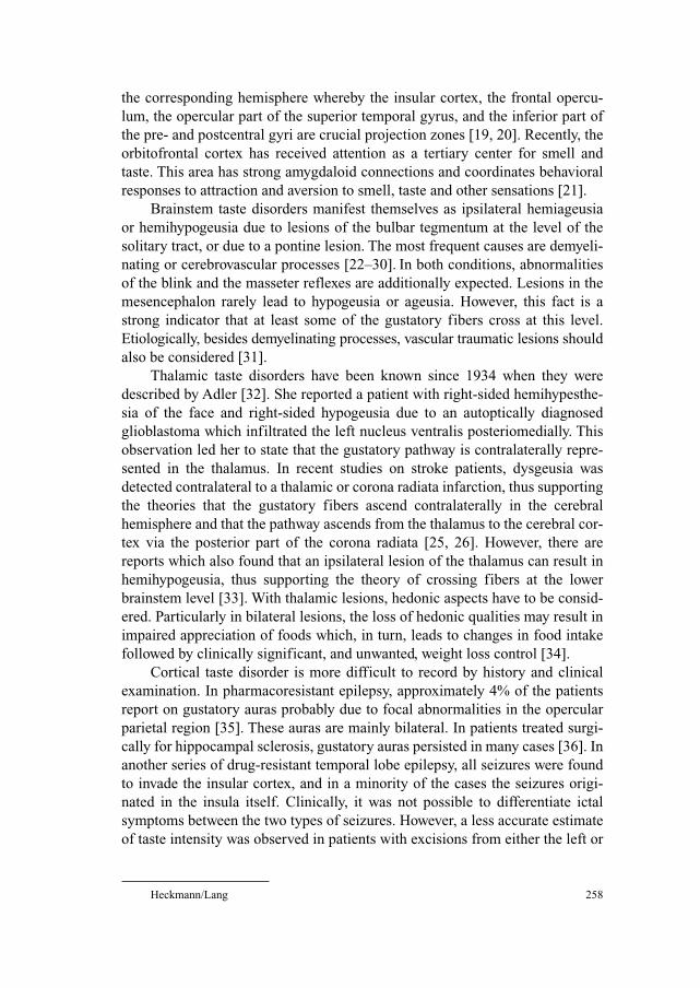
Heckmann/Lang 258
the corresponding hemisphere whereby the insular cortex, the frontal opercu-
lum, the opercular part of the superior temporal gyrus, and the inferior part of
the pre- and postcentral gyri are crucial projection zones [19, 20]. Recently, the
orbitofrontal cortex has received attention as a tertiary center for smell and
taste. This area has strong amygdaloid connections and coordinates behavioral
responses to attraction and aversion to smell, taste and other sensations [21].
Brainstem taste disorders manifest themselves as ipsilateral hemiageusia
or hemihypogeusia due to lesions of the bulbar tegmentum at the level of the
solitary tract, or due to a pontine lesion. The most frequent causes are demyeli-
nating or cerebrovascular processes [22–30]. In both conditions, abnormalities
of the blink and the masseter reflexes are additionally expected. Lesions in the
mesencephalon rarely lead to hypogeusia or ageusia. However, this fact is a
strong indicator that at least some of the gustatory fibers cross at this level.
Etiologically, besides demyelinating processes, vascular traumatic lesions should
also be considered [31].
Thalamic taste disorders have been known since 1934 when they were
described by Adler [32]. She reported a patient with right-sided hemihypesthe-
sia of the face and right-sided hypogeusia due to an autoptically diagnosed
glioblastoma which infiltrated the left nucleus ventralis posteriomedially. This
observation led her to state that the gustatory pathway is contralaterally repre-
sented in the thalamus. In recent studies on stroke patients, dysgeusia was
detected contralateral to a thalamic or corona radiata infarction, thus supporting
the theories that the gustatory fibers ascend contralaterally in the cerebral
hemisphere and that the pathway ascends from the thalamus to the cerebral cor-
tex via the posterior part of the corona radiata [25, 26]. However, there are
reports which also found that an ipsilateral lesion of the thalamus can result in
hemihypogeusia, thus supporting the theory of crossing fibers at the lower
brainstem level [33]. With thalamic lesions, hedonic aspects have to be consid-
ered. Particularly in bilateral lesions, the loss of hedonic qualities may result in
impaired appreciation of foods which, in turn, leads to changes in food intake
followed by clinically significant, and unwanted, weight loss control [34].
Cortical taste disorder is more difficult to record by history and clinical
examination. In pharmacoresistant epilepsy, approximately 4% of the patients
report on gustatory auras probably due to focal abnormalities in the opercular
parietal region [35]. These auras are mainly bilateral. In patients treated surgi-
cally for hippocampal sclerosis, gustatory auras persisted in many cases [36]. In
another series of drug-resistant temporal lobe epilepsy, all seizures were found
to invade the insular cortex, and in a minority of the cases the seizures origi-
nated in the insula itself. Clinically, it was not possible to differentiate ictal
symptoms between the two types of seizures. However, a less accurate estimate
of taste intensity was observed in patients with excisions from either the left or
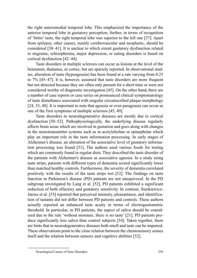
Neurological Causes of Taste Disorders 259
the right anteromedial temporal lobe. This emphasized the importance of the
anterior temporal lobe in gustatory perception; further, in terms of recognition
of ‘bitter’ taste, the right temporal lobe was superior to the left one [37]. Apart
from epilepsy, other causes, mainly cerebrovascular and neoplastic, should be
considered [38–41]. It is unclear to which extent gustatory dysfunction related
to migraine, schizophrenia, major depression, or eating disorders is based on
cortical dysfunction [42–44].
Taste disorders in multiple sclerosis can occur as lesions at the level of the
brainstem, thalamus, or cortex, but are sparsely reported. In observational stud-
ies, alteration of taste (hypoageusia) has been found at a rate varying from 0.25
to 7% [45–47]. It is, however, assumed that taste disorders are more frequent
but not detected because they are often only present for a short time or were not
considered worthy of diagnostic investigation [45]. On the other hand, there are
a number of case reports or case series on pronounced clinical symptomatology
of taste disturbance associated with singular circumscribed plaque morphology
[24, 33, 48]. It is important to note that ageusia or even parageusia can occur as
one of the first symptoms of multiple sclerosis [45, 49].
Taste disorders in neurodegenerative diseases are mostly due to cortical
dysfunction [50–52]. Pathophysiologically, the underlying disease regularly
affects brain areas which are involved in gustation and goes along with changes
in the neurotransmitter systems such as in acetylcholine or epinephrine which
play an important role in the taste information processing. In early stages of
Alzheimer’s disease, an alteration of the associative level of gustatory informa-
tion processing was found [51]. The authors used various foods for testing
which are commonly found in regular diets. They described the taste disorder of
the patients with Alzheimer’s disease as associative agnosia. In a study using
taste strips, patients with different types of dementia scored significantly lower
than matched healthy controls. Furthermore, the severity of dementia correlated
positively with the results of the taste strips test [52]. The findings on taste
function in Parkinson’s disease (PD) patients are not unequivocal. In the PD
subgroup investigated by Lang et al. [52], PD patients exhibited a significant
reduction of both olfactory and gustatory sensitivity. In contrast, Sienkiewicz-
Jarosz et al. [53] reported that perceived intensity, pleasantness, and identifica-
tion of tastants did not differ between PD patients and controls. These authors
actually reported an enhanced taste acuity in terms of electrogustometric
threshold. In particular, in PD patients, the aspect of saliva should be consid-
ered due to the rule ‘without moisture, there is no taste’ [21]. PD patients pro-
duce significantly less saliva than control subjects [54]. Taken together, there
are hints that in neurodegenerative diseases both smell and taste can be impaired.
These observations point to the close relation between the chemosensory senses
itself and the relation between sensory and cognitive abilities [52].
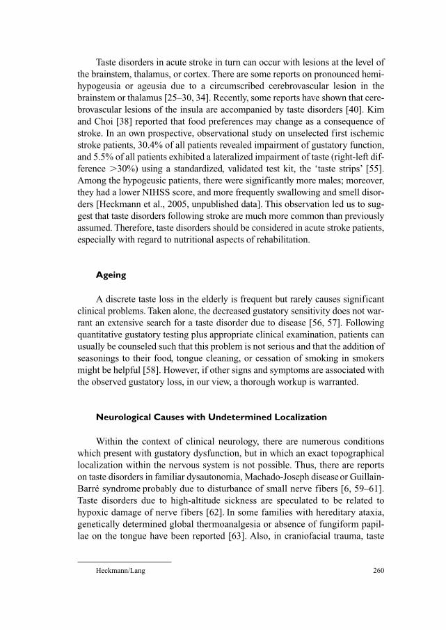
Heckmann/Lang 260
Taste disorders in acute stroke in turn can occur with lesions at the level of
the brainstem, thalamus, or cortex. There are some reports on pronounced hemi-
hypogeusia or ageusia due to a circumscribed cerebrovascular lesion in the
brainstem or thalamus [25–30, 34]. Recently, some reports have shown that cere-
brovascular lesions of the insula are accompanied by taste disorders [40]. Kim
and Choi [38] reported that food preferences may change as a consequence of
stroke. In an own prospective, observational study on unselected first ischemic
stroke patients, 30.4% of all patients revealed impairment of gustatory function,
and 5.5% of all patients exhibited a lateralized impairment of taste (right-left dif-
ference �30%) using a standardized, validated test kit, the ‘taste strips’ [55].
Among the hypogeusic patients, there were significantly more males; moreover,
they had a lower NIHSS score, and more frequently swallowing and smell disor-
ders [Heckmann et al., 2005, unpublished data]. This observation led us to sug-
gest that taste disorders following stroke are much more common than previously
assumed. Therefore, taste disorders should be considered in acute stroke patients,
especially with regard to nutritional aspects of rehabilitation.
Ageing
A discrete taste loss in the elderly is frequent but rarely causes significant
clinical problems. Taken alone, the decreased gustatory sensitivity does not war-
rant an extensive search for a taste disorder due to disease [56, 57]. Following
quantitative gustatory testing plus appropriate clinical examination, patients can
usually be counseled such that this problem is not serious and that the addition of
seasonings to their food, tongue cleaning, or cessation of smoking in smokers
might be helpful [58]. However, if other signs and symptoms are associated with
the observed gustatory loss, in our view, a thorough workup is warranted.
Neurological Causes with Undetermined Localization
Within the context of clinical neurology, there are numerous conditions
which present with gustatory dysfunction, but in which an exact topographical
localization within the nervous system is not possible. Thus, there are reports
on taste disorders in familiar dysautonomia, Machado-Joseph diseaseor Guillain-
Barré syndrome probably due to disturbance of small nerve fibers [6, 59–61].
Taste disorders due to high-altitude sickness are speculated to be related to
hypoxic damage of nerve fibers [62]. In some families with hereditary ataxia,
genetically determined global thermoanalgesia or absence of fungiform papil-
lae on the tongue have been reported [63]. Also, in craniofacial trauma, taste
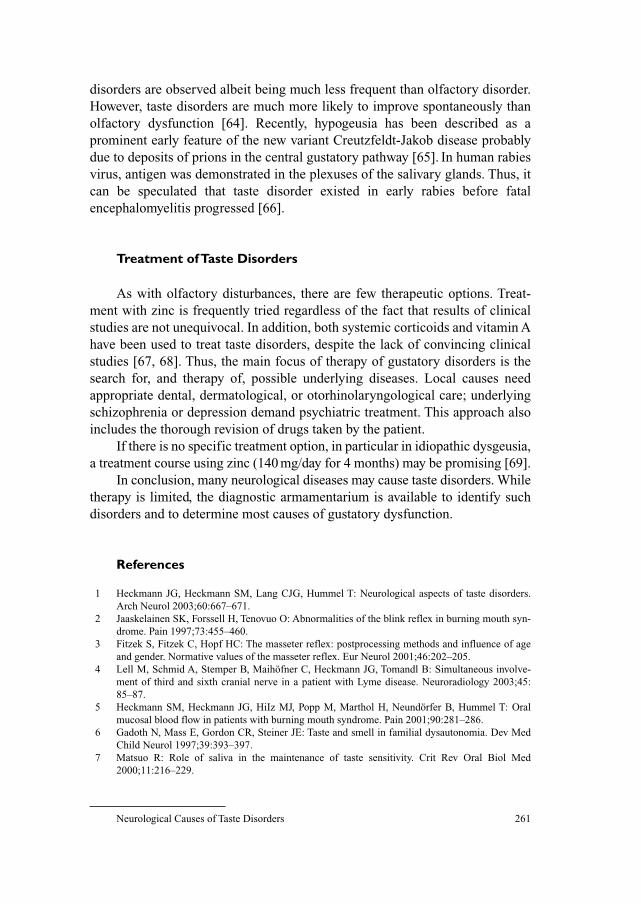
Neurological Causes of Taste Disorders 261
disorders are observed albeit being much less frequent than olfactory disorder.
However, taste disorders are much more likely to improve spontaneously than
olfactory dysfunction [64]. Recently, hypogeusia has been described as a
prominent early feature of the new variant Creutzfeldt-Jakob disease probably
due to deposits of prions in the central gustatory pathway [65]. In human rabies
virus, antigen was demonstrated in the plexuses of the salivary glands. Thus, it
can be speculated that taste disorder existed in early rabies before fatal
encephalomyelitis progressed [66].
Treatment of Taste Disorders
As with olfactory disturbances, there are few therapeutic options. Treat-
ment with zinc is frequently tried regardless of the fact that results of clinical
studies are not unequivocal. In addition, both systemic corticoids and vitamin A
have been used to treat taste disorders, despite the lack of convincing clinical
studies [67, 68]. Thus, the main focus of therapy of gustatory disorders is the
search for, and therapy of, possible underlying diseases. Local causes need
appropriate dental, dermatological, or otorhinolaryngological care; underlying
schizophrenia or depression demand psychiatric treatment. This approach also
includes the thorough revision of drugs taken by the patient.
If there is no specific treatment option, in particular in idiopathic dysgeusia,
a treatment course using zinc (140 mg/day for 4 months) may be promising [69].
In conclusion, many neurological diseases may cause taste disorders. While
therapy is limited, the diagnostic armamentarium is available to identify such
disorders and to determine most causes of gustatory dysfunction.
References
1 Heckmann JG, Heckmann SM, Lang CJG, Hummel T: Neurological aspects of taste disorders.
Arch Neurol 2003;60:667–671.
2 Jaaskelainen SK, Forssell H, Tenovuo O: Abnormalities of the blink reflex in burning mouth syn-
drome. Pain 1997;73:455–460.
3 Fitzek S, Fitzek C, Hopf HC: The masseter reflex: postprocessing methods and influence of age
and gender. Normative values of the masseter reflex. Eur Neurol 2001;46:202–205.
4 Lell M, Schmid A, Stemper B, Maihöfner C, Heckmann JG, Tomandl B: Simultaneous involve-
ment of third and sixth cranial nerve in a patient with Lyme disease. Neuroradiology 2003;45:
85–87.
5 Heckmann SM, Heckmann JG, HiIz MJ, Popp M, Marthol H, Neundörfer B, Hummel T: Oral
mucosal blood flow in patients with burning mouth syndrome. Pain 2001;90:281–286.
6 Gadoth N, Mass E, Gordon CR, Steiner JE: Taste and smell in familial dysautonomia. Dev Med
Child Neurol 1997;39:393–397.
7 Matsuo R: Role of saliva in the maintenance of taste sensitivity. Crit Rev Oral Biol Med
2000;11:216–229.
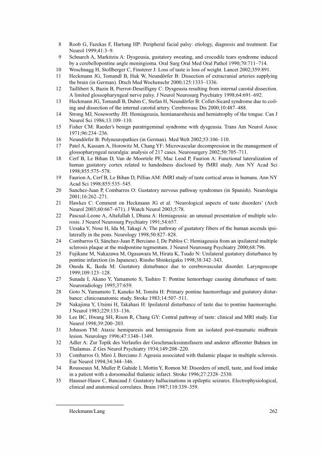
Heckmann/Lang 262
8 Roob G, Fazekas F, Hartung HP: Peripheral facial palsy: etiology, diagnosis and treatment. Eur
Neurol 1999;41:3–9.
9 Schnarch A, Markitziu A: Dysgeusia, gustatory sweating, and crocodile tears syndrome induced
by a cerebellopontine angle meningioma. Oral Surg Oral Med Oral Pathol 1990;70:711–714.
10 Woschnagg H, Stollberger C, Finsterer J: Loss of taste is loss of weight. Lancet 2002;359:891.
11 Heckmann JG, Tomandl B, Huk W, Neundörfer B: Dissection of extracranial arteries supplying
the brain (in German). Dtsch Med Wochenschr 2000;125:1333–1336.
12 Taillibert S, Bazin B, Pierrot-Deseilligny C: Dysgeusia resulting from internal carotid dissection.
A limited glossopharyngeal nerve palsy. J Neurol Neurosurg Psychiatry 1998;64:691–692.
13 Heckmann JG, Tomandl B, Duhm C, Stefan H, Neundörfer B: Collet-Sicard syndrome due to coil-
ing and dissection of the internal carotid artery. Cerebrovasc Dis 2000;10:487–488.
14 Strong MJ, Noseworthy JH: Hemiageusia, hemianaesthesia and hemiatrophy of the tongue. Can J
Neurol Sci 1986;13:109–110.
15 Fisher CM: Raeder’s benign paratrigeminal syndrome with dysgeusia. Trans Am Neurol Assoc
1971;96:234–236.
16 Neundörfer B: Polyneuropathies (in German). Med Welt 2002;53:106–110.
17 Patel A, Kassam A, Horowitz M, Chang YF: Microvascular decompression in the management of
glossopharyngeal neuralgia: analysis of 217 cases. Neurosurgery 2002;50:705–711.
18 Cerf B, Le Bihan D, Van de Moortele PF, Mac Leod P, Faurion A: Functional lateralization of
human gustatory cortex related to handedness disclosed by fMRI study. Ann NY Acad Sci
1998;855:575–578.
19 Faurion A, Cerf B, Le Bihan D, Pillias AM: fMRI study of taste cortical areas in humans. Ann NY
Acad Sci 1998;855:535–545.
20 Sanchez-Juan P, Combarros O: Gustatory nervous pathway syndromes (in Spanish). Neurologia
2001;16:262–271.
21 Hawkes C: Comment on Heckmann JG et al. ‘Neurological aspects of taste disorders’ (Arch
Neurol 2003;60:667–671). J Watch Neurol 2003;5:78.
22 Pascual-Leone A, Altafullah I, Dhuna A: Hemiageusia: an unusual presentation of multiple scle-
rosis. J Neurol Neurosurg Psychiatry 1991;54:657.
23 Uesaka Y, Nose H, Ida M, Takagi A: The pathway of gustatory fibers of the human ascends ipsi-
laterally in the pons. Neurology 1998;50:827–828.
24 Combarros O, Sánchez-Juan P, Berciano J, De Pablos C: Hemiageusia from an ipsilateral multiple
sclerosis plaque at the midpontine tegmentum. J Neurol Neurosurg Psychiatry 2000;68:796.
25 Fujikane M, Nakazawa M, Ogasawara M, Hirata K, Tsudo N: Unilateral gustatory disturbance by
pontine infarction (in Japanese). Rinsho Shinkeigaku 1998;38:342–343.
26 Onoda K, Ikeda M: Gustatory disturbance due to cerebrovascular disorder. Laryngoscope
1999;109:123–128.
27 Sunada I, Akano Y, Yamamoto S, Tashiro T: Pontine hemorrhage causing disturbance of taste.
Neuroradiology 1995;37:659.
28 Goto N, Yamamoto T, Kaneko M, Tomita H: Primary pontine haemorrhage and gustatory distur-
bance: clinicoanatomic study. Stroke 1983;14:507–511.
29 Nakajima Y, Utsimi H, Takahasi H: Ipsilateral disturbance of taste due to pontine haemorraghe.
J Neurol 1983;229:133–136.
30 Lee BC, Hwang SH, Rison R, Chang GY: Central pathway of taste: clinical and MRI study. Eur
Neurol 1998;39:200–203.
31 Johnson TM: Ataxic hemiparesis and hemiageusia from an isolated post-traumatic midbrain
lesion. Neurology 1996;47:1348–1349.
32 Adler A: Zur Topik des Verlaufes der Geschmackssinnsfasern und anderer afferenter Bahnen im
Thalamus. Z Ges Neurol Psychiatry 1934;149:208–220.
33 Combarros O, Miró J, Berciano J: Ageusia associated with thalamic plaque in multiple sclerosis.
Eur Neurol 1994;34:344–346.
34 Rousseaux M, Muller P, Gahide I, Mottin Y, Romon M: Disorders of smell, taste, and food intake
in a patient with a dorsomedial thalamic infarct. Stroke 1996;27:2328–2330.
35 Hausser-Hauw C, Bancaud J: Gustatory hallucinations in epileptic seizures. Electrophysiological,
clinical and anatomical correlates. Brain 1987;110:339–359.
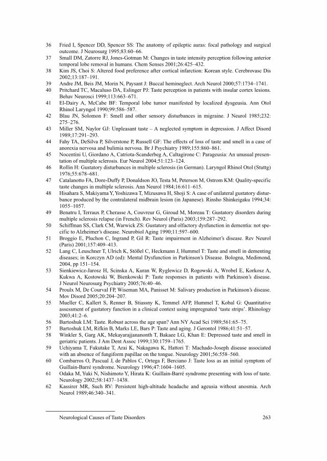
Neurological Causes of Taste Disorders 263
36 Fried I, Spencer DD, Spencer SS: The anatomy of epileptic auras: focal pathology and surgical
outcome. J Neurosurg 1995;83:60–66.
37 Small DM, Zatorre RJ, Jones-Gotman M: Changes in taste intensity perception following anterior
temporal lobe removal in humans. Chem Senses 2001;26:425–432.
38 Kim JS, Choi S: Altered food preference after cortical infarction: Korean style. Cerebrovasc Dis
2002;13:187–191.
39 Andre JM, Beis JM, Morin N, Paysant J: Buccal hemineglect. Arch Neurol 2000;57:1734–1741.
40 Pritchard TC, Macaluso DA, Eslinger PJ: Taste perception in patients with insular cortex lesions.
Behav Neurosci 1999;113:663–671.
41 El-Dairy A, McCabe BF: Temporal lobe tumor manifested by localized dysgeusia. Ann Otol
Rhinol Laryngol 1990;99:586–587.
42 Blau JN, Solomon F: Smell and other sensory disturbances in migraine. J Neurol 1985;232:
275–276.
43 Miller SM, Naylor GJ: Unpleasant taste – A neglected symptom in depression. J Affect Disord
1989;17:291–293.
44 Fahy TA, DeSilva P, Silverstone P, Russell GF: The effects of loss of taste and smell in a case of
anorexia nervosa and bulimia nervosa. Br J Psychiatry 1989;155:860–861.
45 Nocentini U, Giordano A, Catriota-Scanderbeg A, Caltagirone C: Parageusia: An unusual presen-
tation of multiple sclerosis. Eur Neurol 2004;51:123–124.
46 Rollin H: Gustatory disturbances in multiple sclerosis (in German). Laryngol Rhinol Otol (Stuttg)
1976;55:678–681.
47 Catalanotto FA, Dore-Duffy P, Donaldson JO, Testa M, Peterson M, Ostrom KM: Quality-specific
taste changes in multiple sclerosis. Ann Neurol 1984;16:611–615.
48 Hisahara S, Makiyama Y, Yoshizawa T, Mizusawa H, Shoji S: A case of unilateral gustatory distur-
bance produced by the contralateral midbrain lesion (in Japanese). Rinsho Shinkeigaku 1994;34:
1055–1057.
49 Benatru I, Terraux P, Cherasse A, Couvreur G, Giroud M, Moreau T: Gustatory disorders during
multiple sclerosis relapse (in French). Rev Neurol (Paris) 2003;159:287–292.
50 Schiffman SS, Clark CM, Warwick ZS: Gustatory and olfactory dysfunction in dementia: not spe-
cific to Alzheimer’s disease. Neurobiol Aging 1990;11:597–600.
51 Broggio E, Pluchon C, Ingrand P, Gil R: Taste impairment in Alzheimer’s disease. Rev Neurol
(Paris) 2001;157:409–413.
52 Lang C, Leuschner T, Ulrich K, Stößel C, Heckmann J, Hummel T: Taste and smell in dementing
diseases; in Korczyn AD (ed): Mental Dysfunction in Parkinson’s Disease. Bologna, Medimond,
2004, pp 151–154.
53 Sienkiewicz-Jarosz H, Scinska A, Kuran W, Ryglewicz D, Rogowski A, Wrobel E, Korkosz A,
Kukwa A, Kostowski W, Bienkowski P: Taste responses in patients with Parkinson’s disease.
J Neurol Neurosurg Psychiatry 2005;76:40–46.
54 Proulx M, De Courval FP, Wiseman MA, Panisset M: Salivary production in Parkinson’s disease.
Mov Disord 2005;20:204–207.
55 Mueller C, Kallert S, Renner B, Stiassny K, Temmel AFP, Hummel T, Kobal G: Quantitative
assessment of gustatory function in a clinical context using impregnated ‘taste strips’. Rhinology
2003;41:2–6.
56 Bartoshuk LM: Taste. Robust across the age span? Ann NY Acad Sci 1989;561:65–75.
57 Bartoshuk LM, Rifkin B, Marks LE, Bars P: Taste and aging. J Gerontol 1986;41:51–57.
58 Winkler S, Garg AK, Mekayarajjananonth T, Bakaee LG, Khan E: Depressed taste and smell in
geriatric patients. J Am Dent Assoc 1999;130:1759–1765.
59 Uchiyama T, Fukutake T, Arai K, Nakagawa K, Hattori T: Machado-Joseph disease associated
with an absence of fungiform papillae on the tongue. Neurology 2001;56:558–560.
60 Combarros O, Pascual J, de Pablos C, Ortega F, Berciano J: Taste loss as an initial symptom of
Guillain-Barré syndrome. Neurology 1996;47:1604–1605.
61 Odaka M, Yuki N, Nishimoto Y, Hirata K: Guillain-Barré syndrome presenting with loss of taste.
Neurology 2002;58:1437–1438.
62 Kassirer MR, Such RV: Persistent high-altitude headache and ageusia without anosmia. Arch
Neurol 1989;46:340–341.
Heckmann/Lang 264
63 Fukutake T, Kita K, Sakakibara R, Takagi K, Tokumaru Y, Kojima S, Hattori T, Hirayama K: Late-
onset hereditary ataxia with global thermoanalgesia and absence of fungiform papillae on the
tongue in a Japanese family. Brain 1996;119:1011–1021.
64 Wienke A, Walter O: Loss of the sense of taste and smell after head and brain injury (in German).
Laryngorhinootologie 2001;80:752.
65 Reuber M, Al-Din AS, Baborie A, Chakrabarty A: New variant Creutzfeldt-Jakob disease present-
ing with loss of taste and smell. J Neurol Neurosurg Psychiatry 2001;71:412–413.
66 Jackson AC, Ye H, Phelan CC, Ridaura-Sanz C, Zheng Q, Li Z, Wan X, Lopez-Corella E:
Extraneural organ involvement in human rabies. Lab Invest 1999;79:945–951.
67 Henkin RI, Schecter PJ, Friedewald WT, Demets DL, Raff M: A double-blind study of the effects
of zinc sulfate on taste and smell dysfunction. Am J Med Sci 1976;272:285–299.
68 Stoll AL, Oepen G: Zinc salts for the treatment of olfactory and gustatory symptoms in psychiatric
patients: a case series. J Clin Psychiatry 1994;55:309–311.
69 Heckmann SM, Hujoel P, Habiger S, Friess W, Wichmann M, Heckmann JG, Hummel T: Zinc
gluconate in the treatment of dysgeusia – A randomized clinical trial. J Dent Res 2005;84:35–38.
Josef G. Heckmann, MD
Department of Neurology, University of Erlangen-Nuremberg
Schwabachanlage 6
DE–91054 Erlangen (Germany)
Tel. �49 9131 853 3001, Fax �49 9131 853 4436
E-Mail [email protected]
Hummel T, Welge-Lüssen A (eds): Taste and Smell. An Update.
Adv Otorhinolaryngol. Basel, Karger, 2006, vol 63, pp 265–277
Toxic Effects on Gustatory Function
Evan R. Reitera, Laurence J. DiNardoa, Richard M. Costanzob
aDepartment of Otolaryngology – Head and Neck Surgery, bDepartment of Physiology
and Otolaryngology – Head and Neck Surgery, School of Medicine, Virginia
Commonwealth University, Richmond, Va., USA
AbstractA large number of substances and disease processes may impact the sense of taste. Toxic
substances may cause taste dysfunction from their effects on the gustatory system from the sali-
vary gland, to the taste bud, to the central neural pathways. A number of external toxins, includ-
ing industrial compounds, tobacco, and alcohol, may adversely affect taste, most commonly
through local effects in the oral cavity. Blood-borne toxins, such as medications and those pres-
ent in autoimmune and other systemic disorders (e.g. renal or liver failure), have access to all
parts of the gustatory system, and thus may exhibit varied effects on taste function. An under-
standing of these potential toxins and their impact on gustation will help physicians better rec-
ognize, and potentially limit the impact of such taste alterations on their patients.
Copyright © 2006 S. Karger AG, Basel
Mechanisms of Toxic Alterations of Gustation
A wide variety of toxins may affect gustatory function. Depending on the
nature of the toxin and its route of access into the body, either directly through the
oral cavity or via the bloodstream, changes in gustatory function may be caused
by a variety of mechanisms. The primary mechanisms include alterations in the
composition or quantity of saliva, changes in the oral mucus membranes, direct
effects on the taste buds, and modulation of peripheral or central neural pathways.
In the following discussion, we review examples of each of these effects.
Alteration of SalivaSaliva is a complex fluid that coats the surface of the oral cavity and pro-
vides the necessary ionic milieu for proper function of the taste receptor cells
[1]. Saliva is secreted at a baseline level by three paired major salivary glands,
Reiter/DiNardo/Costanzo 266
the parotid, submandibular, and sublingual glands, as well as by the widely dis-
tributed minor salivary glands. However, production may vary under control of
the autonomic nervous system. Interestingly, while saliva clearly influences
taste reception, taste perception may also influence salivary secretion through
reflex activation of the autonomic nervous system [2].
Saliva plays an important role in taste function [3]. Taste stimuli must dif-
fuse through the saliva layer in order to gain access to the taste receptor cells
[4]. Alterations in the amount and composition of saliva may result in changes
in taste perception. Since saliva contains sufficient concentrations of sodium,
potassium, bicarbonate and chloride to be detected by taste receptors, the taste
system must adapt to baseline levels of stimulation. When the constituents of
saliva are altered, changes in the response to taste stimuli occur [5]. Detection
thresholds for sodium and potassium change when there is an increase or
decrease in the saliva concentrations of these ions. Sourness and acid tastes may
be altered when the buffering capacity of saliva is compromised by changes in
bicarbonate levels. Salivary gland secretions may also decrease in specific
autoimmune diseases such as Sjögren’s syndrome [6]. In this case, autoimmune
destruction of salivary glandular tissue leads to marked reduction in salivary
flow, with resultant xerostomia. Patients with Sjögren’s syndrome often com-
plain of taste dysfunction most likely due to problems related to the solubiliza-
tion and delivery of taste molecules to the receptor cells.
Mucosal EffectsHealthy oral mucosa is essential for normal gustatory function. The mucus
membranes of the oral cavity are, however, directly exposed to the external
environment, and thus susceptible to toxic effects from a variety of sources,
both external and internal. Liquids and gases containing toxic agents that enter
the oral cavity may have a direct or indirect negative impact on taste function.
Some chemicals may burn the mucosa while others have toxic effects. Micro-
organisms, fungal growth and viral infections in the oral cavity can lead to
localized inflammatory response that can alter taste function [6]. Tobacco and
nicotine-containing products [7], or gastric contents from gastroesophageal reflux,
may cause inflammation and irritation of the mucosa, and alter the taste of foods.
Gastric acid in particular may produce a sour taste sensation typical of many
acids. Lastly, a variety of autoimmune disorders such as Behçet’s syndrome,
pemphigus vulgaris, systemic lupus erythematosus, and scleroderma may alter
the oral mucosa and thus taste function [8].
Taste Bud DysfunctionA number of toxic substances may lead to alterations in taste receptor acti-
vation, or may even reduce the number of taste receptors present. Several toxins
Toxic Effects on Taste 267
such as botulinum or tetrodotoxin found in the puffer fish can block normal
receptor cell function by blocking receptor sites or conductance through ion
channels. Chemotherapeutic agents that alter cell turnover also interfere with
the normal turnover of taste receptor cells [9]. The same may be true of other
toxins. Tobacco exposure has been shown to increase olfactory sensory neuron
death. Continued exposure might thus eventually overwhelm the regenerative
capacity of the epithelium, leading to reduction of receptors, and hyposmia
[10]. The same effect may occur with continued or repetitive exposure of taste
receptors to certain toxins.
Effects on Neural PathwaysToxins and infections that may have direct effects on the taste nerves
include botulism, herpes zoster, Bell’s palsy, Guillain-Barré syndrome and poli-
omyelitis [11, 12]. Agents that produce CNS infection and inflammation
such as is the case with herpes encephalitis and multiple sclerosis may also alter
taste due to the involvement of central taste pathways [13, 14]. Dysgeusia is
a common taste alteration arising from lesions in cortical taste structures
[15, 16].
External Toxins
MedicationsMedications can affect gustation through any of the mechanisms discussed
above. Virtually all medications can elicit a taste response of their own, which
can be perceived as unpleasant [17, 18]. Depending on the form of the medica-
tion (i.e. solution, tablet, or capsule), this is often through direct stimulation of
taste receptors upon dissolution of the medication in the saliva. However, as
some blood-borne agents can trigger gustatory sensations, termed intravascular
gustation, there is a possibility that some medications may reach taste receptors
through a more complex route [19]. Direct application of some medications to
the tongue can also acutely alter or reduce taste perceptions elicited by known
substances [17, 18]. For example, tricyclic antidepressants applied to the gerbil
tongue were shown to acutely reduce chorda tympani responses to a variety of
taste-stimulating solutions, including sodium chloride, quinine hydrochloride,
sucrose, and citric acid [18]. This effect resolved after complete clearance of the
drug. The authors state that this suggests a direct effect of these medications at
the level of the peripheral receptors. Another possible mechanism of altered
taste induced by medications is reduction of the regenerative capacity of taste
receptor cells. This in turn might lead to temporary depletion of receptor cells
and taste loss. Chemotherapeutic agents, which are well known to carry a high
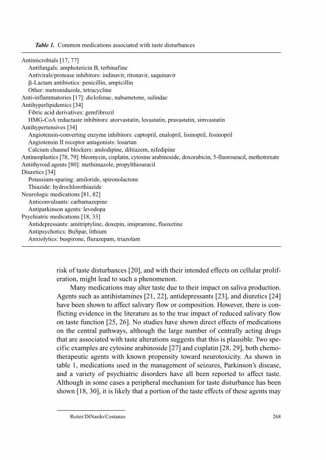
Reiter/DiNardo/Costanzo 268
risk of taste disturbances [20], and with their intended effects on cellular prolif-
eration, might lead to such a phenomenon.
Many medications may alter taste due to their impact on saliva production.
Agents such as antihistamines [21, 22], antidepressants [23], and diuretics [24]
have been shown to affect salivary flow or composition. However, there is con-
flicting evidence in the literature as to the true impact of reduced salivary flow
on taste function [25, 26]. No studies have shown direct effects of medications
on the central pathways, although the large number of centrally acting drugs
that are associated with taste alterations suggests that this is plausible. Two spe-
cific examples are cytosine arabinoside [27] and cisplatin [28, 29], both chemo-
therapeutic agents with known propensity toward neurotoxicity. As shown in
table 1, medications used in the management of seizures, Parkinson’s disease,
and a variety of psychiatric disorders have all been reported to affect taste.
Although in some cases a peripheral mechanism for taste disturbance has been
shown [18, 30], it is likely that a portion of the taste effects of these agents may
Table 1. Common medications associated with taste disturbances
Antimicrobials [17, 77]
Antifungals: amphotericin B, terbinafine
Antivirals/protease inhibitors: indinavir, ritonavir, saquinavir
�-Lactam antibiotics: penicillin, ampicillin
Other: metronidazole, tetracycline
Anti-inflammatories [17]: diclofenac, nabumetone, sulindac
Antihyperlipidemics [34]
Fibric acid derivatives: gemfibrozil
HMG-CoA reductaste inhibitors: atorvastatin, lovastatin, pravastatin, simvastatin
Antihypertensives [34]
Angiotensin-converting enzyme inhibitors: captopril, enalopril, lisinopril, fosinopril
Angiotensin II receptor antagonists: losartan
Calcium channel blockers: amlodipine, diltiazem, nifedipine
Antineoplastics [78, 79]: bleomycin, cisplatin, cytosine arabinoside, doxorubicin, 5-fluorouracil, methotrexate
Antithyroid agents [80]: methimazole, propylthiouracil
Diuretics [34]
Potassium-sparing: amiloride, spironolactone
Thiazide: hydrochlorothiazide
Neurologic medications [81, 82]
Anticonvulsants: carbamazepine
Antiparkinson agents: levodopa
Psychiatric medications [18, 33]
Antidepressants: amitriptyline, doxepin, imipramine, fluoxetine
Antipsychotics: BuSpar, lithium
Anxiolytics: buspirone, flurazepam, triazolam

Toxic Effects on Taste 269
be attributable to alteration of activity in central taste pathways. At the very
least, such agents may reduce the hedonic value of taste sensations, thus leading
to subjective taste complaints and alterations in appetite [31].
For most medications, quantitative data on the incidence of taste disturbance
are lacking. The Physicians’ Desk Reference is likely the most widely used refer-
ence for medication usage and adverse reactions [32]. Taste disturbances are
reported with descriptors including ‘altered taste’, ‘dysgeusia’, ‘ageusia’, ‘bad
taste’, or ‘metallic taste’. For some medications, data from controlled trials are
available showing specific incidence rates for taste disturbances. However, for
many others, the adverse reactions listed are derived from case reports or series in
the literature, or from voluntary reports from individual physicians to the publish-
ers. Even in cases of large controlled trials, the incidence of taste disturbance is
often derived from patient reports rather than objective testing. As such, the true
incidence of taste disturbances with usage of any given agent is difficult to deter-
mine. In addition, interactions between medications may also impact the likeli-
hood of taste disturbance in patients on multiple agents.
As a group, the angiotensin-converting enzyme inhibitors, which include
captopril, enalapril, fosinopril, and lisinopril, are probably the most common
group of agents leading to taste disturbance. This stems both from their efficacy
and thus widespread use in the management of hypertension and congestive
heart failure, and the relatively high rate of taste disturbances associated with
their usage, estimated from 1 to 8% [33]. While the mechanism of this effect is
uncertain, one possibility is chelation of zinc at the receptor level [34]. Taste
deficits reported from angiotensin-converting enzyme inhibitors include ageu-
sia, dysgeusia, and metallic taste. While in most cases normal taste returned
after cessation of the offending agent, rare reports exist of permanent dysgeusia
or even ageusia [33]. Another class of medications with both significant rates of
taste disturbance and widespread use are the HMG-CoA reductase inhibitors, or
‘statin’ antihyperlipidemic drugs, including atorvastatin, lovastatin, pravastatin,
and simvastatin [34]. Controlled trials have, however, reported rates of taste dis-
turbances less than 1% for these agents [32], and their mechanism of taste dis-
turbance remains unknown.
Foods, Chemicals, Tobacco, RadiationTaste disturbance secondary to external exposures has only recently begun
to be explored. Three sources of toxins have emerged: foods, chemicals and
radiation. Most toxic encounters are the result of human activity. Exposures that
damage taste function, therefore, are likely to increase as the human impact on
the environment grows.
Food containing toxins can possess an unpleasant taste. Occasionally, cir-
culating toxins cause a generalized taste disturbance. Scombroid poisoning is
Reiter/DiNardo/Costanzo 270
an example [35]. This form of ichthyosarcotoxism is caused by the ingestion of
affected dark-meat fish. In addition to parathesias, digestive tract symptoms,
and headache, an unusual taste sensation is often reported. The exact mecha-
nism is unknown and, although not a food allergy, patients respond to antihista-
mine therapy.
Taste alterations caused by cigarette smoking are thought to result from
exposure to toxic chemicals. Gromysz-Kalkowska et al. [36] studied the effects
of age, gender, and cigarette smoking on 471 randomly selected subjects. Taste
testing revealed that smoking had a small, diversified, but in most cases, statis-
tically insignificant effect on taste sensitivity.
Schiffman and Nagle [37] have reviewed the adverse effects of chemical
toxins on taste. Frequent contact with pesticides is reported to induce a sus-
tained metallic, bitter taste. Organophosphate-based pesticides, in particular,
have been linked to a garlic odor and taste [37]. Evidence exists for both central
and peripheral causes of pesticide-related taste disturbances. Organophosphates
demonstrate disruption of sensory nerve terminations on taste buds in the exper-
imental model but may also interfere with central neurotransmitters [38].
Metal workers have complained of metallic tastes as well, often coinciding
with the particular metal being used. Brass pipe fitters [39] and jewelry workers
suffering from lead poisoning [40], for example, frequently report a sweet
metallic taste. A possible direct mechanism of action has been proposed since
heavy metals are concentrated in saliva and their topical application to the
tongue can blunt taste perception [41]. Indeed, an increase in taste threshold has
been noted in chromium workers [42]. It is unknown if this effect is reversible.
Administration of the chelator D-penicillamine has relieved symptoms in lead-
contaminated silver workers [40].
The effects of solvents on taste are not well studied. In 1992, Hotz et al.
[43] reported subjective smell and/or taste disturbances in a cross-sectional
study of 264 workers exposed to organic solvents. The symptoms were mostly
transient and related to concentration peaks rather than duration of exposure
supporting an acute depressor effect.
Radiation exposure can occur from an ingested or external source. Radio-
active iodine (I131) is used in the treatment of differentiated thyroid cancers.
Ingested I131 concentrates in salivary glands as well as the thyroid gland.
Radiation sialadenitis with associated xerostomia, dental caries, facial nerve
involvement, secondary infection, and taste alterations have been reported [44].
The use of sialogogic agents is thought to decrease the transit time of radioac-
tive material in the salivary glands. However, their application and the pursuit
of adequate hydration have not yet been proven to be efficacious. External beam
head and neck radiation therapy often results in oral sequelae including mucositis,
xerostomia, dental caries, and taste loss. Taste loss is not entirely attributable to
Toxic Effects on Taste 271
xerostomia since mucositis and taste loss are usually reversible while hyposali-
vation persists [45]. Zheng et al. [46] prospectively evaluated 40 patients under-
going head and neck radiation therapy for taste disturbance. Four basic tastes
were measured using the whole-mouth method before, during, and after ther-
apy. Bitter taste was the most susceptible, while interference with sweet taste
depended on inclusion of the anterior tongue in the radiation field. Salivary
function was independent of taste disturbance, supporting the notion that radia-
tion therapy directly damages taste receptors. Fisher et al. [47] have recently
provided further support for these findings in a study that randomized radiation
therapy patients to receive pilocarpine 5 mg four times daily or placebo during
treatment. Although preservation of salivary function was statistically signifi-
cant in the pilocarpine arm and these patients reported less mouth pain, subjec-
tive difficulty with swallowing, hyposalivation, and taste were similar in both
groups. Targeted radiation therapy techniques, such as intensity-modulated radio-
therapy, that limit taste receptor and salivary gland exposure when feasible, show
promise in minimizing taste loss following treatment.
Internal Toxins
Liver FailurePatients with liver failure from various causes, including cirrhosis, hepati-
tis, primary biliary cirrhosis, and sclerosing cholangitis, have been shown to
have alterations in taste [48]. Taste disturbances tend to be more common than
olfactory dysfunction in patients with liver disease. Patients tend to report
reduced taste sensitivity, dysgeusia with taste aversions, as well as more taste
cravings as compared to controls [48]. Taste disturbances may affect food pref-
erences and thus contribute to the anorexia seen with liver failure, although no
consensus exists in the literature [48, 49]. The reversibility of these changes was
shown in patients undergoing successful liver transplantation [50]. However,
the authors note that complete recovery in all taste modalities did not occur, and
that many other factors other than liver function, such as medical regimen,
micronutrient deficiencies, patient nutritional status, and psychological state,
may have led to the improvements noted.
Several possible mechanisms exist for taste deficits in this population.
Effects of altered zinc metabolism have been equivocal. Although plasma zinc
levels were found to be low in cirrhotics, this did not correlate with the level of
taste acuity, nor did zinc supplementation seem to positively impact taste func-
tion [51]. Vitamin A supplementation was found to improve both gustatory and
olfactory acuity in patients with alcoholic cirrhosis and vitamin A deficiency,
although the mechanism of this effect was unclear [52].

Reiter/DiNardo/Costanzo 272
UremiaChronic renal failure has long been associated with disturbances of taste.
Such disturbances can be significant, contributing to altered food preferences
and reduced caloric intake [53], as well as reduced quality of life [54] in
patients with renal failure. Impairment of taste has been found to affect patients
with uremia, as well as patients on either hemodialysis or peritoneal dialysis
[55]. The nature of the disturbances reported include the presence of a foul
phantom taste, often termed the ‘uremic taste’, impaired taste recognition, and
elevated taste detection thresholds. The mechanism of these changes is unclear.
An immunohistochemical study comparing fungiform papillae taste buds in
uremic patients, renal transplant recipients, and healthy controls showed no dif-
ferences in taste bud architecture and innervation, although the patients with
chronic renal failure had fewer taste buds [56]. Patients with chronic renal fail-
ure on hemodialysis were shown to have reduced unstimulated salivary flow
rates, and increased salivary pH and buffer capacity compared with controls
[56]. Many studies have focused on the possible role of abnormal zinc metabo-
lism as a causative factor in dysgeusia in uremic patients [57]. Studies have
shown a correlation between plasma zinc levels and taste acuity [58], improve-
ment in taste following oral [59] or dialysate [60] zinc supplementation, and
improvement in taste acuity paralleling normalization of zinc metabolism fol-
lowing renal transplantation [61]. Zinc supplementation seemed to have similar
effects on nerve conduction velocity and taste acuity, suggesting that taste dys-
function in chronic renal failure might be another manifestation of the ‘uremic
neuropathy’ [57, 60]. One notable but rare mechanism of taste disturbance is
amyloid deposition in the tongue. Long-term dialysis is known to lead to amy-
loid deposition, usually in osteoarticular structures. However, this process may
also lead to formation of amyloid nodules in the tongue, as found by Matsuo et
al. [62] in about 8% of patients treated with dialysis for over 20 years. Over half
of these patients reported lingual dysfunction, including taste disturbances,
impaired tongue mobility, or articulation deficits. The effect of medications
used by patients with renal failure also cannot be overlooked, as many patients
with renal failure may require antihypertensive agents, including angiotensin-
converting enzyme inhibitors [34], and diuretics, such as hydrochlorothiazide
[63] or amiloride [64], all of which are known to affect gustation.
DiabetesThe prevalence of diabetes mellitus in the general population has been esti-
mated to be between 1 and 5%, a figure that may rise with the increasing preva-
lence of obesity and increasing life span seen in western populations. Diabetics
demonstrate a variety of gustatory and oral manifestations. Diabetics have increa-
sed electrical and chemical gustometric thresholds compared to controls [65].
Toxic Effects on Taste 273
In newly diagnosed diabetics, partial correction of hypogeusia was noted after
initiation of antihyperglycemic therapy [66]. Differing opinions exist as to the
association between gustatory deficits and neuropathy in diabetics, such that it
is unclear that hypogeusia in diabetics is simply another manifestation of diabetic
neuropathy [66]. The demonstration of reduced salivary flow rates suggests that
this may be another contributory factor to hypogeusia in diabetics [67].
Thyroid DiseaseBoth hypo- and hyperthyroid states have been associated with disturbances
of taste [68]. The spectrum of hypothyroid states ranges from asymptomatic
laboratory finding to myxedema coma. In one series of patients with hypothy-
roidism, almost half had complaints of altered sense of taste, and about 40%
had complaints of altered sense of smell [69]. Objective testing showed 83%
had decreased acuity for at least one taste (salty, sweet, bitter, sour), although
impaired detection or recognition of bitter taste was most common. Most com-
plaints and objective deficits were found to reverse following medical correc-
tion of hypothyroidism [69]. Decreased salty and bitter sensations have been
reported in hyperthyroid patients [70]. Although the mechanism for this obser-
vation is unclear, laboratory work has shown that thyroid hormones, such as
thyroxine (T4) or triiodothyronine (T3), may have a competitive inhibitory effect
on purified taste bud membrane adenosine 3�,5�-monophosphate phosphodi-
esterase activity [71].
In patients with differentiated thyroid cancer, radioiodine I131 administra-
tion is commonly used postoperatively to ablate residual thyroid tissue. As iodine
is concentrated and secreted in the salivary glands, such treatment can lead to
transient or even permanent xerostomia, sialadenitis, and taste disturbances.
Mendoza et al. [72] found taste disturbances in approximately 25% of patie-
nts receiving radioactive iodine therapy. While 21% experienced acute xerosto-
mia, 35% of patients receiving more than one radioiodine treatment reported
xerostomia.
Autoimmune DiseaseA number of diseases related to immune dysfunction have been reported
to cause disturbances in taste. Due to the systemic nature of these illnesses,
autoimmune processes can affect gustation through a number of different
mechanisms. One of these disorders is Sjögren’s syndrome, which is the second
most common autoimmune disease behind rheumatoid arthritis. This disease is
HLA-linked, and predominantly afflicts women in their third or fourth decades
of life. The disease is characterized by progressive destruction of exocrine
glands, including the salivary glands, which results from both lymphocytic
infiltration and immune complex deposition. While the classic eccrine gland

Reiter/DiNardo/Costanzo 274
involvement results in xerophthalmia and xerostomia with taste alterations, vir-
tually any organ system can be involved. Taste complaints seen with Sjögren’s
syndrome are primarily reduced sensitivity to tastes, which has been confir-
med by objective testing [73]. While sensitivity to tastes is generally impaired,
suprathreshold taste recognition is not. In addition, there appears to be a rather
poor correlation between salivary flow and degree of taste disturbance, suggest-
ing that a mechanism other than just reduced salivary flow may be present [73].
Some possibilities include altered oral bacterial flora, neuropathy [74], and
chronic oral mucosal changes such as lingual fissuring [75].
Amyloidosis is defined as the extracellular deposition of proteinaceous
material in various sites in the body. This may be localized or systemic, primary
(without coexisting disease) or secondary (arising in the setting of chronic
inflammatory disease or infection). Amyloid deposition has been noted in the
tongue, in addition to the buccal mucosa, palate, and floor of mouth [76].
Lingual involvement may cause taste disturbance, but more urgently can lead to
progressive airway compromise necessitating tracheotomy or lingual reductive
surgery.
Conclusion
A variety of toxins may alter the sense of taste. Alterations in gustation may
occur due to toxic effects on saliva production, the oral mucosa, taste receptor
cells, and neural pathways. Common toxins include external factors such as foods,
tobacco, chemicals, radiation, or medications, and blood-borne toxins as in liver
or renal failure, diabetes, or autoimmune disease. In many cases, further investi-
gation is needed to better understand the precise mechanisms of such toxic injuries
to the components of the gustatory system, which might lead to better preventive
or restorative therapies.
References
1 Bradley R, Beidler L: Saliva: its role in taste function; in Doty RL (ed): Handbook of Olfaction
and Gustation. New York, Dekker, 2003, pp 639–650.
2 Kim M, Chiego DJ Jr, Bradley RM: Morphology of parasympathetic neurons innervating rat lin-
gual salivary glands. Auton Neurosci 2004;111:27–36.
3 Bradley RM: Salivary secretion; in Getchell TV, Doty RL, Bartoshuk LM, Snow JB 2nd (eds):
Smell and Taste in Health and Disease. New York, Raven Press, 1991, pp 127–144.
4 De Simone JA, Heck GL: An analysis of the effects of stimulus transport and membrane charge on
the salt, acid and water-response of mammals. Chem Senses 1980;5:295–316.
5 Matsuo R, Yamamoto T: Effects of inorganic constituents of saliva on taste responses of the rat
chorda tympani nerve. Brain Res 1992;583:71–80.
Toxic Effects on Taste 275
6 Bromley S, Doty RL: Clinical disorders affecting taste: evaluation and management; in Doty RL
(ed): Handbook of Olfaction and Gustation. New York, Dekker, 2003, pp 935–957.
7 Reibel J: Tobacco and oral diseases. Update on the evidence, with recommendations. Med Princ
Pract 2003;12(Suppl 1):22–32.
8 Campbell SM, Montanaro A, Bardana EJ: Head and neck manifestations of autoimmune disease.
Am J Otolaryngol 1983;4:187–216.
9 Berteretche MV, Dalix AM, d’Ornano AM, Bellisle F, Khayat D, Faurion A: Decreased taste sensi-
tivity in cancer patients under chemotherapy. Support Care Cancer 2004;12:571–576.
10 Vent J, Robinson AM, Gentry-Nielsen MJ, Conley DB, Hallworth R, Leopold DA, et al: Pathology
of the olfactory epithelium: smoking and ethanol exposure. Laryngoscope 2004;114:1383–1388.
11 Odaka M, Yuki N, Nishimoto Y, Hirata K: Guillain-Barré syndrome presenting with loss of taste.
Neurology 2002;58:1437–1438.
12 Brand N: Taste response and poliomyelitis. Ann Hum Genet 1964;27:233–239.
13 Asnis DS, Micic L, Giaccio D: Ramsay Hunt syndrome presenting as a cranial polyneuropathy.
Cutis 1996;57:421–424.
14 Combarros O, Miro J, Berciano J: Ageusia associated with thalamic plaque in multiple sclerosis.
Eur Neurol 1994;34:344–346.
15 Finsterer J, Stollberger C, Kopsa W: Weight reduction due to stroke-induced dysgeusia. Eur
Neurol 2004;51:47–49.
16 Pritchard TC, Macaluso DA, Eslinger PJ: Taste perception in patients with insular cortex lesions.
Behav Neurosci 1999;113:663–671.
17 Schiffman SS, Zervakis J, Westall HL, Graham BG, Metz A, Bennett JL, et al: Effect of antimi-
crobial and anti-inflammatory medications on the sense of taste. Physiol Behav 2000;69:413–424.
18 Schiffman SS, Zervakis J, Suggs MS, Budd KC, Iuga L: Effect of tricyclic antidepressants on taste
responses in humans and gerbils. Pharmacol Biochem Behav 2000;65:599–609.
19 Maruniak JA, Silver WL, Moulton DG: Olfactory receptors respond to blood-borne odorants.
Brain Res 1983;265:312–316.
20 Wickham RS, Rehwaldt M, Kefer C, Shott S, Abbas K, Glynn-Tucker E, et al: Taste changes expe-
rienced by patients receiving chemotherapy. Oncol Nurs Forum 1999;26:697–706.
21 Van Ganse E, Kaufman L, Derde MP, Yernault JC, Delaunois L, Vincken W: Effects of antihista-
mines in adult asthma: a meta-analysis of clinical trials. Eur Respir J 1997;10:2216–2224.
22 Sreebny LM, Valdini A, Yu A: Xerostomia. 2. Relationship to nonoral symptoms, drugs, and dis-
eases. Oral Surg Oral Med Oral Pathol 1989;68:419–427.
23 von Knorring L, Mornstad H: Qualitative changes in saliva composition after short-term adminis-
tration of imipramine and zimelidine in healthy volunteers. Scand J Dent Res 1981;89:313–320.
24 Beal AM: The effect of transport-blocking drugs on secretion of fluid and electrolytes by the
mandibular gland of red kangaroos, Macropus rufus. Arch Oral Biol 1997;42:705–716.
25 Christensen CM, Navazesh M, Brightman VJ: Effects of pharmacologic reductions in salivary
flow on taste thresholds in man. Arch Oral Biol 1984;29:17–23.
26 Mattes RD, Christensen CM, Engelman K: Effects of hydrochlorothiazide and amiloride on salt
taste and excretion (intake). Am J Hypertens 1990;3:436–443.
27 Hoffman DL, Howard JR Jr, Sarma R, Riggs JE: Encephalopathy, myelopathy, optic neuropathy,
and anosmia associated with intravenous cytosine arabinoside. Clin Neuropharmacol 1993;16:
258–262.
28 Fanning J, Hilgers RD: High-dose cisplatin carboplatin chemotherapy in primary advanced epithe-
lial ovarian cancer. Gynecol Oncol 1993;51:182–186.
29 Wickham RS, Rehwaldt M, Kefer C, Shott S, Abbas K, Glynn-Tucker E, et al: Taste changes expe-
rienced by patients receiving chemotherapy. Oncol Nurs Forum 1999;26:697–706.
30 Schiffman SS, Graham BG, Suggs MS, Sattely-Miller EA: Effect of psychotropic drugs on taste
responses in young and elderly persons. Ann NY Acad Sci 1998;855:732–737.
31 Treit D, Berridge KC: A comparison of benzodiazepine, serotonin, and dopamine agents in the
taste-reactivity paradigm. Pharmacol Biochem Behav 1990;37:451–456.
32 Physicians’ Desk Reference, ed 58. Montvale, Thomson PDR, 2004.
33 Ackerman BH, Kasbekar N: Disturbances of taste and smell induced by drugs. Pharmacotherapy
1997;17:482–496.
Reiter/DiNardo/Costanzo 276
34 Doty RL, Philip S, Reddy K, Kerr KL: Influences of antihypertensive and antihyperlipidemic
drugs on the senses of taste and smell: a review. J Hypertens 2003;21:1805–1813.
35 Muller GJ, Lamprecht JH, Barnes JM, De Villiers RV, Honeth BR, Hoffman BA: Scombroid poi-
soning. Case series of 10 incidents involving 22 patients. S Afr Med J 1992;81:427–430.
36 Gromysz-Kalkowska K, Wojcik K, Szubartowska E, Unkiewicz-Winiarczyk A: Taste perception
of cigarette smokers. Ann Univ Mariae Curie Sklodowska 2002;57:143–154.
37 Schiffman SS, Nagle HT: Effect of environmental pollutants on taste and smell. Otolaryngol Head
Neck Surg 1992;106:693–700.
38 Vij S, Kanagasuntheram R: Effect of tri-O-cresyl phosphate (TOCP) poisoning on sensory nerve ter-
minations of slow loris (Nycticebus coucang coucang). Acta Neuropathol (Berl) 1972;20: 150–159.
39 Armstrong CW, Moore LW Jr, Hackler RL, Miller GB Jr, Stroube RB: An outbreak of metal fume
fever. Diagnostic use of urinary copper and zinc determinations. J Occup Med 1983;25:886–888.
40 Kachru DN, Tandon SK, Misra UK, Nag D: Occupational lead poisoning among silver jewelry
workers. Indian J Med Sci 1989;43:89–91.
41 Halpern BP: Environmental factors affecting chemoreceptors: an overview. Environ Health
Perspect 1982;44:101–105.
42 Seeber H, Fikentscher R: Taste disorders in chromium-exposed workers. Z Gesamte Hyg 1990;36:
33–34.
43 Hotz P, Tschopp A, Soderstrom D, Holtz J, Boillat MA, Gutzwiller F: Smell or taste disturbances,
neurological symptoms, and hydrocarbon exposure. Int Arch Occup Environ Health 1992;63:
525–530.
44 Mandel SJ, Mandel L: Radioactive iodine and the salivary glands. Thyroid 2003;13:265–271.
45 Vissink A, Jansma J, Spijkervet FK, Burlage FR, Coppes RP: Oral sequelae of head and neck
radiotherapy. Crit Rev Oral Biol Med 2003;14:199–212.
46 Zheng WK, Inokuchi A, Yamamoto T, Komiyama S: Taste dysfunction in irradiated patients with
head and neck cancer. Fukuoka Igaku Zasshi 2002;93:64–76.
47 Fisher J, Scott C, Scarantino CW, Leveque FG, White RL, Rotman M, et al: Phase III quality-of-
life study results: impact on patients’ quality of life to reducing xerostomia after radiotherapy for
head-and-neck cancer – RTOG 97–09. Int J Radiat Oncol Biol Phys 2003;56:832–836.
48 Deems RO, Friedman MI, Friedman LS, Munoz SJ, Maddrey WC: Chemosensory function, food
preferences and appetite in human liver disease. Appetite 1993;20:209–216.
49 Madden AM, Bradbury W, Morgan MY: Taste perception in cirrhosis: its relationship to circulat-
ing micronutrients and food preferences. Hepatology 1997;26:40–48.
50 Bloomfeld RS, Graham BG, Schiffman SS, Killenberg PG: Alterations of chemosensory function
in end-stage liver disease. Physiol Behav 1999;66:203–207.
51 Sturniolo GC, D’Inca R, Parisi G, Giacomazzi F, Montino MC, D’Odorico A, et al: Taste alter-
ations in liver cirrhosis: are they related to zinc deficiency? J Trace Elem Electrolytes Health Dis
1992;6:15–19.
52 Garrett-Laster M, Russell RM, Jacques PF: Impairment of taste and olfaction in patients with cir-
rhosis: the role of vitamin A. Hum Nutr Clin Nutr 1984;38:203–214.
53 Bellisle F, Dartois AM, Kleinknecht C, Broyer M: Perceptions of and preferences for sweet taste in
uremic children. J Am Diet Assoc 1990;90:951–954.
54 Rocco MV, Gassman JJ, Wang SR, Kaplan RM: Cross-sectional study of quality of life and symp-
toms in chronic renal disease patients: the Modification of Diet in Renal Disease Study. Am J
Kidney Dis 1997;29:888–896.
55 Fernstrom A, Hylander B, Rossner S: Taste acuity in patients with chronic renal failure. Clin
Nephrol 1996;45:169–174.
56 Astback J, Fernstrom A, Hylander B, Arvidson K, Johansson O: Taste buds and neuronal markers
in patients with chronic renal failure. Perit Dial Int 1999;19(Suppl 2):S315–S323.
57 Bolton CF: Peripheral neuropathies associated with chronic renal failure. Can J Neurol Sci
1980;7:89–96.
58 Burge JC, Schemmel RA, Park HS, Greene JA 3rd: Taste acuity and zinc status in chronic renal
disease. J Am Diet Assoc 1984;84:1203–1206, 1209.
59 Mahajan SK, Prasad AS, Rabbani P, Briggs WA, McDonald FD: Zinc deficiency: a reversible
complication of uremia. Am J Clin Nutr 1982;36:1177–1183.
Toxic Effects on Taste 277
60 Sprenger KB, Bundschu D, Lewis K, Spohn B, Schmitz J, Franz HE: Improvement of uremic neu-
ropathy and hypogeusia by dialysate zinc supplementation: a double-blind study. Kidney Int Suppl
1983;16:S315–S318.
61 Mahajan SK, Abraham J, Hessburg T, Prasad AS, Migdal SD, Abu-Hamdan DK, et al: Zinc metab-
olism and taste acuity in renal transplant recipients. Kidney Int Suppl 1983;16:S310–S314.
62 Matsuo K, Nakamoto M, Yasunaga C, Goya T, Sugimachi K: Dialysis-related amyloidosis of the
tongue in long-term hemodialysis patients. Kidney Int 1997;52:832–838.
63 Zervakis J, Graham BG, Schiffman SS: Taste effects of lingual application of cardiovascular med-
ications. Physiol Behav 2000;68:405–413.
64 Anand KK, Zuniga JR: Effect of amiloride on suprathreshold NaCl, LiCl, and KCl salt taste in
humans. Physiol Behav 1997;62:925–929.
65 Le Floch JP, Le Lievre G, Verroust J, Philippon C, Peynegre R, Perlemuter L: Factors related to the
electric taste threshold in type 1 diabetic patients. Diabet Med 1990;7:526–531.
66 Perros P, MacFarlane TW, Counsell C, Frier BM: Altered taste sensation in newly-diagnosed
NIDDM. Diabetes Care 1996;19:768–770.
67 Moore PA, Guggenheimer J, Etzel KR, Weyant RJ, Orchard T: Type 1 diabetes mellitus, xerostomia,
and salivary flow rates. Oral Surg Oral Med Oral Pathol Oral Radiol Endod 2001;92:281–291.
68 Pittman JA, Beschi RJ: Taste thresholds in hyper- and hypothyroidism. J Clin Endocrinol Metab
1967;27:895–896.
69 McConnell RJ, Menendez CE, Smith FR, Henkin RI, Rivlin RS: Defects of taste and smell in
patients with hypothyroidism. Am J Med 1975;59:354–364.
70 Bhatia S, Sircar SS, Ghorai BK: Taste disorder in hypo- and hyperthyroidism. Indian J Physiol
Pharmacol 1991;35:152–158.
71 Law JS, Henkin RI: Thyroid hormone inhibits purified taste bud membrane adenosine 3�,5�-monophosphate phosphodiesterase activity. Res Commun Chem Pathol Pharmacol 1984;43:449–462.
72 Mendoza A, Shaffer B, Karakla D, Mason ME, Elkins D, Goffman TE: Quality of life with well-
differentiated thyroid cancer: treatment toxicities and their reduction. Thyroid 2004;14:133–140.
73 Weiffenbach JM, Schwartz LK, Atkinson JC, Fox PC: Taste performance in Sjögren’s syndrome.
Physiol Behav 1995;57:89–96.
74 Urban PP, Keilmann A, Teichmann EM, Hopf HC: Sensory neuropathy of the trigeminal, glos-
sopharyngeal, and vagal nerves in Sjögren’s syndrome. J Neurol Sci 2001;186:59–63.
75 Soto-Rojas AE, Villa AR, Sifuentes-Osornio J, Alarcon-Segovia D, Kraus A: Oral manifestations
in patients with Sjögren’s syndrome. J Rheumatol 1998;25:906–910.
76 Stoopler ET, Sollecito TP, Chen SY: Amyloid deposition in the oral cavity: a retrospective study
and review of the literature. Oral Surg Oral Med Oral Pathol Oral Radiol Endod 2003;95:674–680.
77 Schiffman SS, Zervakis J, Heffron S, Heald AE: Effect of protease inhibitors on the sense of taste.
Nutrition 1999;15:767–772.
78 Wickham RS, Rehwaldt M, Kefer C, Shott S, Abbas K, Glynn-Tucker E, et al: Taste changes expe-
rienced by patients receiving chemotherapy. Oncol Nurs Forum 1999;26:697–706.
79 Bartoshuk LM: Chemosensory alterations and cancer therapies. NCI Monogr 1990;179–184.
80 Genter MB: Evaluation of olfactory and auditory system effects of the antihyperthyroid drug car-
bimazole in the Long-Evans rat. J Biochem Mol Toxicol 1998;12:305–314.
81 Barajas Garcia-Talavera F: Ageusia in a patient treated with carbamazepine. Neurologia 1988;3:
126–127.
82 Deguchi K, Furuta S, Imakiire T, Ohyama M: Case of ageusia from a variety of causes. J Laryngol
Otol 1996;110:598–601.
Evan R. Reiter, MD
Department of Otolaryngology – HNS, 1201 East Marshall Street
Virginia Commonwealth University, Box 980146
Richmond, VA 23298-0146 (USA)
Tel. �1 804 828 2766, Fax �1 804 828 3494, E-Mail [email protected]
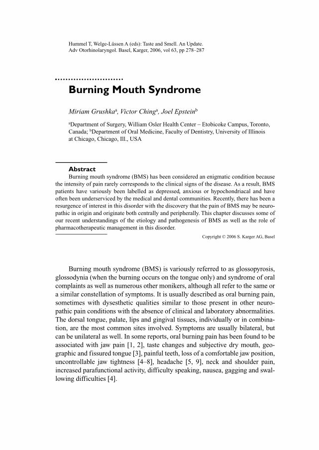
Hummel T, Welge-Lüssen A (eds): Taste and Smell. An Update.
Adv Otorhinolaryngol. Basel, Karger, 2006, vol 63, pp 278–287
Burning Mouth Syndrome
Miriam Grushkaa, Victor Chinga, Joel Epsteinb
aDepartment of Surgery, William Osler Health Center – Etobicoke Campus, Toronto,
Canada; bDepartment of Oral Medicine, Faculty of Dentistry, University of Illinois
at Chicago, Chicago, Ill., USA
AbstractBurning mouth syndrome (BMS) has been considered an enigmatic condition because
the intensity of pain rarely corresponds to the clinical signs of the disease. As a result, BMS
patients have variously been labelled as depressed, anxious or hypochondriacal and have
often been underserviced by the medical and dental communities. Recently, there has been a
resurgence of interest in this disorder with the discovery that the pain of BMS may be neuro-
pathic in origin and originate both centrally and peripherally. This chapter discusses some of
our recent understandings of the etiology and pathogenesis of BMS as well as the role of
pharmacotherapeutic management in this disorder.
Copyright © 2006 S. Karger AG, Basel
Burning mouth syndrome (BMS) is variously referred to as glossopyrosis,
glossodynia (when the burning occurs on the tongue only) and syndrome of oral
complaints as well as numerous other monikers, although all refer to the same or
a similar constellation of symptoms. It is usually described as oral burning pain,
sometimes with dysesthetic qualities similar to those present in other neuro-
pathic pain conditions with the absence of clinical and laboratory abnormalities.
The dorsal tongue, palate, lips and gingival tissues, individually or in combina-
tion, are the most common sites involved. Symptoms are usually bilateral, but
can be unilateral as well. In some reports, oral burning pain has been found to be
associated with jaw pain [1, 2], taste changes and subjective dry mouth, geo-
graphic and fissured tongue [3], painful teeth, loss of a comfortable jaw position,
uncontrollable jaw tightness [4–8], headache [5, 9], neck and shoulder pain,
increased parafunctional activity, difficulty speaking, nausea, gagging and swal-
lowing difficulties [4].
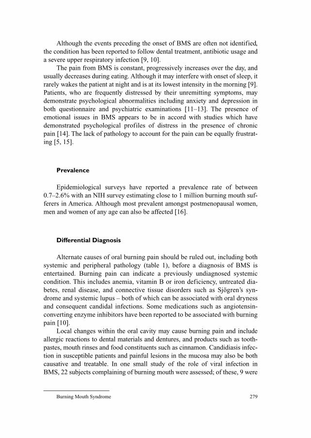
Burning Mouth Syndrome 279
Although the events preceding the onset of BMS are often not identified,
the condition has been reported to follow dental treatment, antibiotic usage and
a severe upper respiratory infection [9, 10].
The pain from BMS is constant, progressively increases over the day, and
usually decreases during eating. Although it may interfere with onset of sleep, it
rarely wakes the patient at night and is at its lowest intensity in the morning [9].
Patients, who are frequently distressed by their unremitting symptoms, may
demonstrate psychological abnormalities including anxiety and depression in
both questionnaire and psychiatric examinations [11–13]. The presence of
emotional issues in BMS appears to be in accord with studies which have
demonstrated psychological profiles of distress in the presence of chronic
pain [14]. The lack of pathology to account for the pain can be equally frustrat-
ing [5, 15].
Prevalence
Epidemiological surveys have reported a prevalence rate of between
0.7–2.6% with an NIH survey estimating close to 1 million burning mouth suf-
ferers in America. Although most prevalent amongst postmenopausal women,
men and women of any age can also be affected [16].
Differential Diagnosis
Alternate causes of oral burning pain should be ruled out, including both
systemic and peripheral pathology (table 1), before a diagnosis of BMS is
entertained. Burning pain can indicate a previously undiagnosed systemic
condition. This includes anemia, vitamin B or iron deficiency, untreated dia-
betes, renal disease, and connective tissue disorders such as Sjögren’s syn-
drome and systemic lupus – both of which can be associated with oral dryness
and consequent candidal infections. Some medications such as angiotensin-
converting enzyme inhibitors have been reported to be associated with burning
pain [10].
Local changes within the oral cavity may cause burning pain and include
allergic reactions to dental materials and dentures, and products such as tooth-
pastes, mouth rinses and food constituents such as cinnamon. Candidiasis infec-
tion in susceptible patients and painful lesions in the mucosa may also be both
causative and treatable. In one small study of the role of viral infection in
BMS, 22 subjects complaining of burning mouth were assessed; of these, 9 were
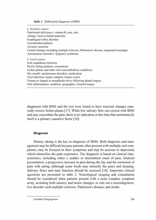
Grushka/Ching/Epstein 280
diagnosed with BMS and the rest were found to have mucosal changes espe-
cially erosive lichen planus [17]. While low salivary flow can coexist with BMS
and may exacerbate the pain, there is no indication at this time that xeristomia by
itself is a primary causative factor [10].
Diagnosis
History taking is the key to diagnosis of BMS. Both diagnosis and man-
agement may be difficult because patients often present with multiple oral com-
plaints, may be focused on their symptoms and may be anxious or depressed,
which intensifies the pain experience. The diagnosis is based on clinical char-
acteristics, including either a sudden or intermittent onset of pain, bilateral
presentation, a progressive increase in pain during the day and the remission of
pain with eating (although some foods may intensify the pain) and sleeping.
Salivary flows and taste function should be assessed [18]. Important clinical
questions are presented in table 2. Neurological imaging and consultation
should be considered when patients present with a more complex symptom
array, including both sensory and motor changes, to rule out a neurodegenera-
tive disorder such multiple sclerosis, Parkinson’s disease, and stroke.
Table 1. Differential diagnosis of BMS
a Systemic causesNutritional deficiency: vitamin B, iron, zinc
Allergy: food or dental materials
Esophageal reflux disorder
Uncontrolled diabetes
Acoustic neuroma
Central changes including multiple sclerosis, Parkinson’s disease, trigeminal neuralgia
Autoimmune disorders: Sjögren’s syndrome
b Local causesOral candidiasis infection
Poorly fitting dentures, restorations
Lichen planus and other oral vesiculobullous conditions
Dry mouth: autoimmune disorders, medication
Viral infection: herpes simplex, herpes zoster
Trauma to lingual or mandibular nerve following dental surgery
Oral inflammatory condition: geographic, fissured tongue
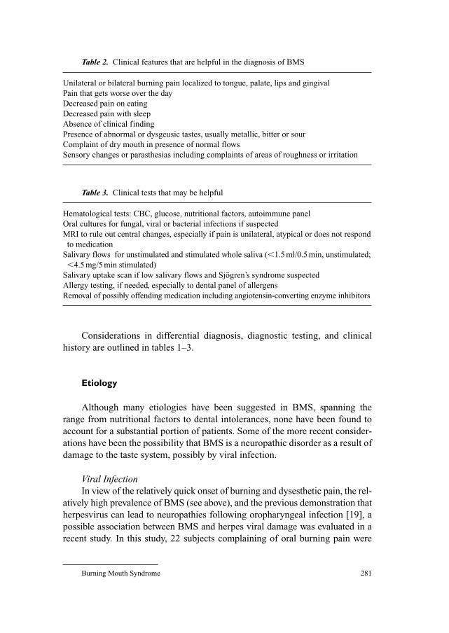
Burning Mouth Syndrome 281
Considerations in differential diagnosis, diagnostic testing, and clinical
history are outlined in tables 1–3.
Etiology
Although many etiologies have been suggested in BMS, spanning the
range from nutritional factors to dental intolerances, none have been found to
account for a substantial portion of patients. Some of the more recent consider-
ations have been the possibility that BMS is a neuropathic disorder as a result of
damage to the taste system, possibly by viral infection.
Viral InfectionIn view of the relatively quick onset of burning and dysesthetic pain, the rel-
atively high prevalence of BMS (see above), and the previous demonstration that
herpesvirus can lead to neuropathies following oropharyngeal infection [19], a
possible association between BMS and herpes viral damage was evaluated in a
recent study. In this study, 22 subjects complaining of oral burning pain were
Table 2. Clinical features that are helpful in the diagnosis of BMS
Unilateral or bilateral burning pain localized to tongue, palate, lips and gingival
Pain that gets worse over the day
Decreased pain on eating
Decreased pain with sleep
Absence of clinical finding
Presence of abnormal or dysgeusic tastes, usually metallic, bitter or sour
Complaint of dry mouth in presence of normal flows
Sensory changes or parasthesias including complaints of areas of roughness or irritation
Table 3. Clinical tests that may be helpful
Hematological tests: CBC, glucose, nutritional factors, autoimmune panel
Oral cultures for fungal, viral or bacterial infections if suspected
MRI to rule out central changes, especially if pain is unilateral, atypical or does not respond
to medication
Salivary flows for unstimulated and stimulated whole saliva (�1.5 ml/0.5 min, unstimulated;
�4.5 mg/5 min stimulated)
Salivary uptake scan if low salivary flows and Sjögren’s syndrome suspected
Allergy testing, if needed, especially to dental panel of allergens
Removal of possibly offending medication including angiotensin-converting enzyme inhibitors
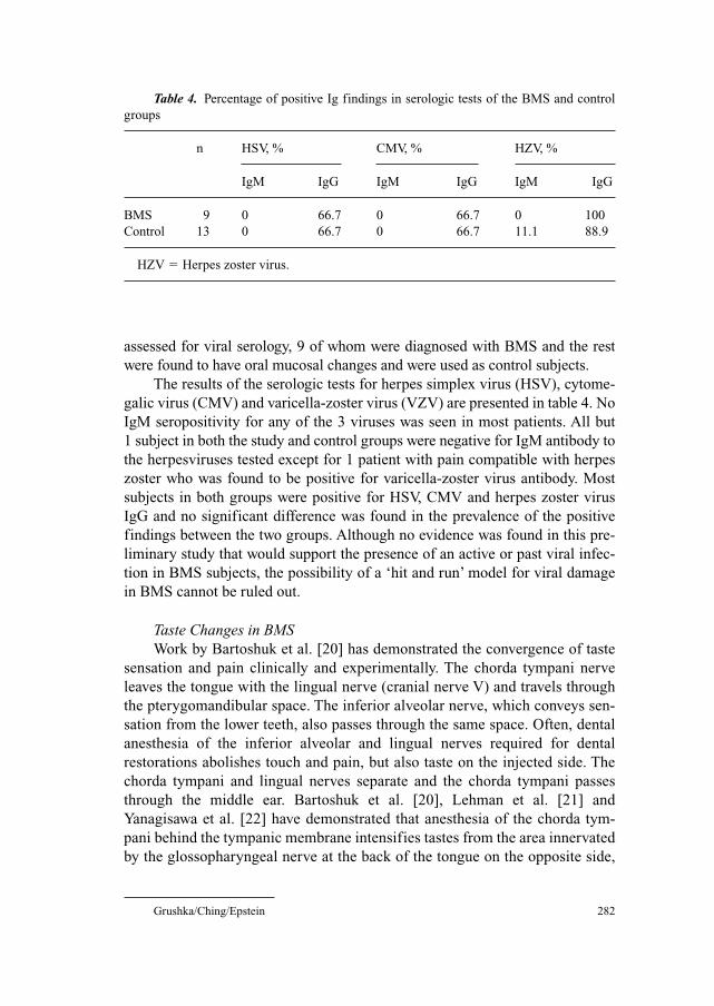
Grushka/Ching/Epstein 282
assessed for viral serology, 9 of whom were diagnosed with BMS and the rest
were found to have oral mucosal changes and were used as control subjects.
The results of the serologic tests for herpes simplex virus (HSV), cytome-
galic virus (CMV) and varicella-zoster virus (VZV) are presented in table 4. No
IgM seropositivity for any of the 3 viruses was seen in most patients. All but
1 subject in both the study and control groups were negative for IgM antibody to
the herpesviruses tested except for 1 patient with pain compatible with herpes
zoster who was found to be positive for varicella-zoster virus antibody. Most
subjects in both groups were positive for HSV, CMV and herpes zoster virus
IgG and no significant difference was found in the prevalence of the positive
findings between the two groups. Although no evidence was found in this pre-
liminary study that would support the presence of an active or past viral infec-
tion in BMS subjects, the possibility of a ‘hit and run’ model for viral damage
in BMS cannot be ruled out.
Taste Changes in BMSWork by Bartoshuk et al. [20] has demonstrated the convergence of taste
sensation and pain clinically and experimentally. The chorda tympani nerve
leaves the tongue with the lingual nerve (cranial nerve V) and travels through
the pterygomandibular space. The inferior alveolar nerve, which conveys sen-
sation from the lower teeth, also passes through the same space. Often, dental
anesthesia of the inferior alveolar and lingual nerves required for dental
restorations abolishes touch and pain, but also taste on the injected side. The
chorda tympani and lingual nerves separate and the chorda tympani passes
through the middle ear. Bartoshuk et al. [20], Lehman et al. [21] and
Yanagisawa et al. [22] have demonstrated that anesthesia of the chorda tym-
pani behind the tympanic membrane intensifies tastes from the area innervated
by the glossopharyngeal nerve at the back of the tongue on the opposite side,
Table 4. Percentage of positive Ig findings in serologic tests of the BMS and control
groups
n HSV, % CMV, % HZV, %
IgM IgG IgM IgG IgM IgG
BMS 9 0 66.7 0 66.7 0 100
Control 13 0 66.7 0 66.7 11.1 88.9
HZV � Herpes zoster virus.
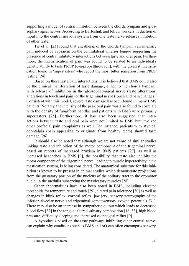
Burning Mouth Syndrome 283
supporting a model of central inhibition between the chorda tympani and glos-
sopharyngeal nerves. According to Bartoshuk and fellow workers, reduction of
input into the central nervous system from one taste nerve releases inhibition
of other taste.
Tie et al. [23] found that anesthesia of the chorda tympani can intensify
pain induced by capsaicin on the contralateral anterior tongue suggesting the
presence of central inhibitory interactions between taste and oral pain. Further-
more, the intensification of pain was found to be related to an individual’s
genetic ability to taste PROP (6-n-propylthiouracil), with the greatest intensifi-
cation found in ‘supertasters’ who report the most bitter sensation from PROP
testing [24].
Based on these taste/pain interactions, it is believed that BMS could also
be the clinical manifestation of taste damage, either to the chorda tympani,
with release of inhibition in the glossopharyngeal nerve (taste alterations,
alterations in touch and pain) or the trigeminal nerve (touch and pain changes).
Consistent with this model, severe taste damage has been found in many BMS
patients. Notably, the intensity of the peak oral pain was also found to correlate
with the density of fungiform papillae and patients with BMS were primarily
supertasters [25]. Furthermore, it has also been suggested that inter-
actions between taste and oral pain were not limited to BMS but involved
other orofacial pain complaints as well. For instance, patients with atypical
odontalgia (pain appearing to originate from healthy teeth) showed taste
damage [26].
It should also be noted that although we are not aware of similar studies
linking taste and inhibition of the motor component of the trigeminal nerve,
based on reports of increased bruxism in BMS patients [27], as well as
increased headaches in BMS [9], the possibility that taste also inhibits the
motor component of the trigeminal nerve, leading to muscle hyperactivity in the
mastication system, is being considered. The anatomical substrate for this inhi-
bition is known to be present in animal studies which demonstrate projections
from the gustatory portion of the nucleus of the solitary tract to the oromotor
nuclei in the medulla subserving the masticatory muscles [28].
Other abnormalities have also been noted in BMS, including elevated
thresholds for temperature and touch [29], altered pain tolerance [30] as well as
changes in blink reflex, corneal reflex, jaw jerk, sensory neurography of the
inferior alveolar nerve and trigeminal somatosensory evoked potentials [31].
There may also be an increase in sympathetic output which leads to decreased
blood flow [32] in the tongue, altered salivary composition [18, 33], high blood
pressure, difficulty sleeping and increased esophageal reflux [9].
A hypothesis based on the taste pathways inhibiting other cranial nerves
can explain why conditions such as BMS and AO can often encompass sensory,

Grushka/Ching/Epstein 284
motor and sympathetic abnormalities; why multiple orofacial phenomena are
linked together in these pain syndromes; why the onset of these problems can
occur suddenly; why there is almost always a lack of associated organic mucosal
and dental pathology, and why these conditions may respond to centrally acting
drugs, especially those affecting the GABAergic pathways [2, 34, 35] which are
involved in taste transmission and in neuroinhibition [28].
GABA is known to be an inhibitory neurotransmitter found in the taste sys-
tem [36–38] and may be a key target. According to Bartoshuk et al. [20], if taste
damage produces a sufficient loss of the inhibition normally exerted on central
structures mediating oral pain, then replacement of a GABA agonist such as
clonazepam might ameliorate the loss of inhibition and relieve the pain in BMS.
Interestingly, GABA agonists such as clonazepam [39] have also been
found to have value in the treatment of nausea, coughing and hiccups [40, 41]
and in taste disturbances when associated with BMS [34]. Thus, it is possible
that the inhibition produced by the taste system is important in controlling other
anatomy associated with eating.
Management
Therapy for BMS involves the use of centrally acting medications for neu-
ropathic pain, such as tricyclic antidepressants, benzodiazepines or gabapentin
[42]. Studies support the use of low-dose (0.25–0.75 mg) clonazepam or tri-
cyclic antidepressants (10–40 mg), including amitriptyline, desipramine, nor-
triptyline, imipramine and clomipramine. Clonazepam is a benzodiazepine
used either topically or systemically [1, 34, 35], which appears to have excellent
efficacy in the relief of the symptoms related to BMS. In view of only partial or
lack of response in some BMS patients taking these medications, other GABA
receptor-acting anticonvulsants have been used in combination with clon-
azepam with apparent success [43]. Topical medications, including clonidine
and capsaicin, may be considered for application to local sites. Systemic use
of capsaicin has also been suggested [44] as has �-lipoic acid with or without
psychotherapy [45].
Polypharmacy in Pain ManagementA recent retrospective study of low-dose anticonvulsant medications used
in combination for the management of BMS was carried out. Patients were pre-
scribed up to 0.5 mg clonazepam and asked to add as needed up to 1,200 mg of
gabapentin (up to 300 mg 4 times a day); 30 mg of baclofen (in 3 divided doses)
and then up to 200 mg of lamotrigine (in 2 divided doses) as needed and pain
scores were recorded on a modified adjectival/visual analogue scale. Of the
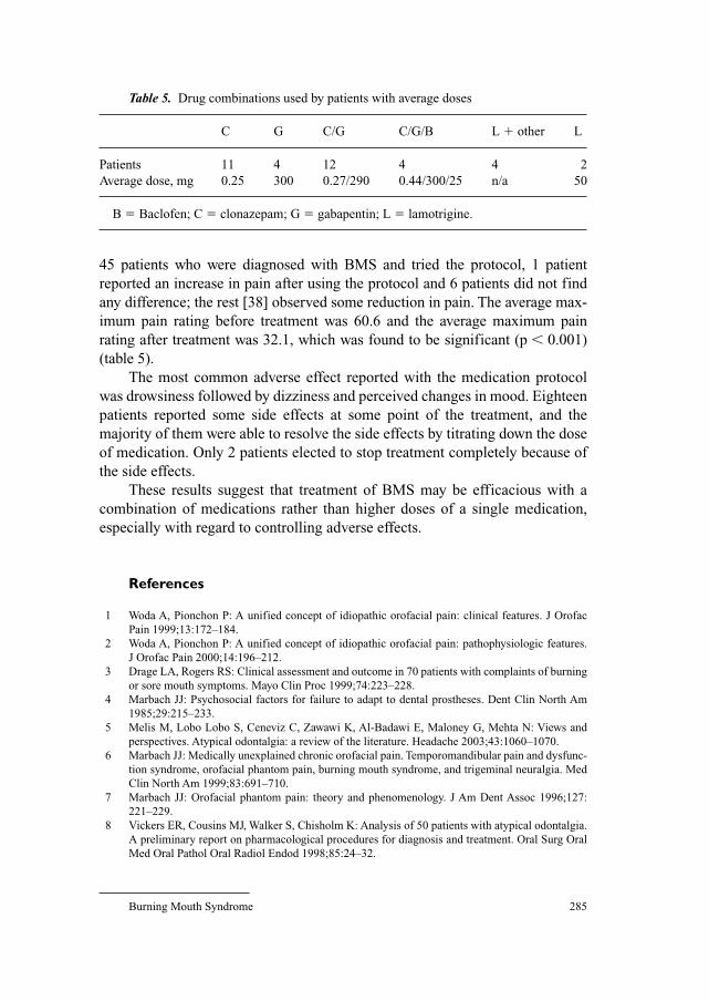
Burning Mouth Syndrome 285
45 patients who were diagnosed with BMS and tried the protocol, 1 patient
reported an increase in pain after using the protocol and 6 patients did not find
any difference; the rest [38] observed some reduction in pain. The average max-
imum pain rating before treatment was 60.6 and the average maximum pain
rating after treatment was 32.1, which was found to be significant (p � 0.001)
(table 5).
The most common adverse effect reported with the medication protocol
was drowsiness followed by dizziness and perceived changes in mood. Eighteen
patients reported some side effects at some point of the treatment, and the
majority of them were able to resolve the side effects by titrating down the dose
of medication. Only 2 patients elected to stop treatment completely because of
the side effects.
These results suggest that treatment of BMS may be efficacious with a
combination of medications rather than higher doses of a single medication,
especially with regard to controlling adverse effects.
References
1 Woda A, Pionchon P: A unified concept of idiopathic orofacial pain: clinical features. J Orofac
Pain 1999;13:172–184.
2 Woda A, Pionchon P: A unified concept of idiopathic orofacial pain: pathophysiologic features.
J Orofac Pain 2000;14:196–212.
3 Drage LA, Rogers RS: Clinical assessment and outcome in 70 patients with complaints of burning
or sore mouth symptoms. Mayo Clin Proc 1999;74:223–228.
4 Marbach JJ: Psychosocial factors for failure to adapt to dental prostheses. Dent Clin North Am
1985;29:215–233.
5 Melis M, Lobo Lobo S, Ceneviz C, Zawawi K, Al-Badawi E, Maloney G, Mehta N: Views and
perspectives. Atypical odontalgia: a review of the literature. Headache 2003;43:1060–1070.
6 Marbach JJ: Medically unexplained chronic orofacial pain. Temporomandibular pain and dysfunc-
tion syndrome, orofacial phantom pain, burning mouth syndrome, and trigeminal neuralgia. Med
Clin North Am 1999;83:691–710.
7 Marbach JJ: Orofacial phantom pain: theory and phenomenology. J Am Dent Assoc 1996;127:
221–229.
8 Vickers ER, Cousins MJ, Walker S, Chisholm K: Analysis of 50 patients with atypical odontalgia.
A preliminary report on pharmacological procedures for diagnosis and treatment. Oral Surg Oral
Med Oral Pathol Oral Radiol Endod 1998;85:24–32.
Table 5. Drug combinations used by patients with average doses
C G C/G C/G/B L � other L
Patients 11 4 12 4 4 2
Average dose, mg 0.25 300 0.27/290 0.44/300/25 n/a 50
B � Baclofen; C � clonazepam; G � gabapentin; L � lamotrigine.
Grushka/Ching/Epstein 286
9 Grushka M: Clinical features of burning mouth syndrome. Oral Surg Oral Med Oral Pathol
1987;63:30–36.
10 Grushka M, Epstein JB, Gorsky M: Burning mouth syndrome. Am Fam Physician 2002;65:615–620.
11 Soto Araya M, Rojas Alcayaga G, Esguep A: Association between psychological disorders and the
presence of oral lichen planus, burning mouth syndrome and recurrent aphthous stomatitis. Med
Oral 2004;9:1–7.
12 Nicholson M, Wilkinson G, Field E, Longman L, Fitzgerald B: A pilot study: stability of psychi-
atric diagnoses over 6 months in burning mouth syndrome. J Psychosom Res 2000;49:1–2.
13 Trikkas G, Nikolatou O, Samara C, Bazopoulou-Kyrkanidou E, Rabavilas AD, Christodoulou GN:
Glossodynia: personality characteristics and psychopathology. Psychother Psychosom 1996;65:
163–168.
14 Sternbach RA, Timmermans G: Personality changes associated with reduction of pain. Pain
1975;1:177–178.
15 Zakrewska JM: The burning mouth syndrome remains an enigma. Pain 1995;62:253–257.
16 Ship JA, Grushka M, Lipton JA, Mott AE, Sessle BJ, Dionne RA: Burning mouth syndrome: an
update. J Am Dent Assoc 1995;126:842–853.
17 Epstein JB, Grushka M, Gorsky M: Role of herpes simplex virus in BMS, submitted.
18 Nagler RM, Hershkovich O: Sialochemical and gustatory analysis in patients with oral sensory
complaints. J Pain 2004;5:56–63.
19 Hashizume K: Herpes zoster and post-herpetic neuralgia. Jap J Clin Med 2001;59:1738–1742.
20 Bartoshuk LM, Chapo A, Duffy VB, Gruhska M, Norgren R, Kveton JF, Pritchard TC, Snyder D:
Oral phantoms: evidence for central inhibition produced by taste. Chem Senses 2002;27:A52.
21 Lehman CD, Bartoshuk LM, Catalanotto FC, Kveton JF, Lowlicht RA: The effect of anesthesia of
the chorda tympani nerve on taste perception in humans. Physiol Behav 1995;57:943–951.
22 Yanagisawa K, Bartoshuk LM, Catalanotto FA, Karrer TA, Kveton JF: Anesthesia of the chorda
tympani nerve and taste phantoms. Physiol Behav 1998;63:329–335.
23 Tie K, Fast K, Kveton J, Cohen Z, Duffy VB, Green B, et al: Anesthesia of chorda tympani nerve
and effect on oral pain. Chem Senses 1999;24:609.
24 Bartoshuk LM, Duffy VB, Miller IJ: PTC/PROP tasting: anatomy, psychophysics, and sex effects.
Physiol Behav 1994;56:1165–1171.
25 Grushka M, Bartoshuk LM, Chapo AK, Duffy VB, Norgren R, Kveton J, Pritchard TC, Snyder DJ:
Oral pain: associated with damage to taste. J Pain 2000;145:P141.
26 Grushka M, Bartoshuk LM, Chapo AK, Duffy VB, Norgren R, Kveton J, Pritchard TC, Snyder DJ:
Oral pain: associated with damage to taste. Proc 10th World Congr Pain, San Diego, 2002.
27 Paterson AJ, Lamb AB, Clifford TJ, Lamey PJ: Burning mouth syndrome: the relationship between
the HAD scale and parafunctional habits. J Oral Pathol Med 1995;24:289–292.
28 King MS: Distribution of immunoreactive GABA and glutamate receptors in the gustatory portion
of the nucleus of the solitary tract in rat. Brain Res Bull 2003;60:241–254.
29 Forssell H, Jaaskelainen S, Tenovuo O: Sensory dysfunction in burning mouth syndrome. Pain
2000;99:41–44.
30 Grushka M, Sessle BJ, Howley TP: Psychophysical assessment of tactile, pain and thermal sen-
sory functions in burning mouth syndrome. Pain 1987;28:169.
31 Jaaskelainen SK: Clinical neurophysiology and quantitative testing in the investigation of orofa-
cial pain and sensory function. J Orofac Pain 2004;18:85–107.
32 Cekic-Arambasin A, Vidas I, Stipetic-Mravak M: Clinical oral test for the assessment of oral
symptoms of glossodynia and glossopyrosis. J Oral Rehabil 1990;17:495–502.
33 Chimenos-Kustner E, Marques-Soares MS: Burning mouth and saliva. Med Oral 2002;7:244–253.
34 Grushka M, Epstein J, Mott A: An open-label, dose escalation pilot study of the effect of clon-
azepam in burning mouth syndrome. Oral Surg Oral Med Oral Pathol Oral Radiol Endod
1998;86:557–561.
35 Gremeau-Richard C, Woda A, Navez ML, Atta N, Bouhassira D, Gagnieu MC, Laluque JF, Picard P,
Pioncon P, Tubert S: Topical clonazepam in stomatodynia: a randomized placebo-controlled study.
Pain 2004;108:51–54.
36 Davis BJ: GABA-like immunoreactivity in the gustatory zone of the nucleus of the solitary tract in
the hamster: light and electron microscopic studies. Brain Res Bull 1993;30:69–77.
Burning Mouth Syndrome 287
37 Smith DV, Li CS: GABA-mediated corticofugal inhibition of taste-responsive neurons in the
nucleus of the solitary tract. Brain Res 2000;858:408–415.
38 Wang L, Bradley RM: Influence of GABA on neurons of the gustatory zone of the rat nucleus of
the solitary tract. Brain Res 1993;616:144–153.
39 Shindo EF, Emoto M, Mohtai H, Hachisuga T, Kawarabayashi T, Shirakawa K: The prevention of
cancer chemotherapy-induced emesis with granisetron and clonazepam. Gan To Kagaku Ryoho
1995;22:233–237.
40 Dicpinigaitis PV, Grimm DR, Lesser M: Baclofen-induced cough suppression in cervical spinal
injury. Arch Phys Med Rehabil 2000;81:921–923.
41 Smith HS, Busracamwongs A: Management of hiccups in the palliative care population. Am J
Hosp Palliat Care 2003;20:149–154.
42 White TL, Kent PF, Kurtz DB, Emko P: Effectiveness of gabapentin for treatment of burning
mouth syndrome. Arch Otolaryngol Head Neck Surg 2004;130:786–788.
43 Ching V, Grushka M, Epstein JB: Clinical efficacy of titrated anticonvulsant analgesics on atypi-
cal odontalgia and burning mouth syndrome: retrospective study, in preparation.
44 Petruzzi M, Lauritano D, De Benedittis M, Baldoni M, Serpico R: Systemic capsaicin for burning
mouth syndrome: short-term results of a pilot study. J Oral Pathol Med 2004;33:111–114.
45 Femiano F, Gombos F, Scully C: Burning mouth syndrome: open trial of psychotherapy alone, med-
ication with alpha-lipoic acid (thioctic acid), and combination therapy. Med Oral 2004;9:8–13.
Further Reading
Bartoshuk LM, Grushka M, Duffy VB, Fast L, Lucchina L, Putkin J, et al: Burning mouth syndrome:
damage to CN VII and pain phantoms in CN V. Chem Senses 1999;24:609–613.
Grushka M, Bartoshuk LM: Burning mouth syndrome and oral dysesthesia: taste injury is a piece of the
puzzle. Can J Diagn 2000;17:99–109.
Grushka M, Epstein JB, Gorsky M: Burning mouth syndrome. Am Fam Physician 2002;65:615–620.
Grushka M, Epstein JB, Gorsky M: Burning mouth syndrome and other oral sensory disorders: a unify-
ing hypothesis. Pain Res Manag 2003;8:133–135.
Ship JA, Grushka M, Lipton JA, Mott AE, Sessle BJ, Dionne RA: Burning mouth syndrome: an update.
J Am Dent Assoc 1995;126:842–853.
Svensson P, Kaaber S: General health factors and denture function in patients with burning mouth syn-
drome and matched control subjects. J Oral Rehabil 1995;22:887–895.
Tammiala-Salonen T, Hiidenkari T, Parvinen T: Burning mouth in a Finnish adult population.
Community Dent Oral Epidemiol 1993;21:67–71.
Zakrewska JM: The burning mouth syndrome remains an enigma. Pain 1995;62:253–257.
Dr. Miriam Grushka
974 Eglinton W
Toronto, Ont. M6c 2C5 (Canada)
Tel. �1 416 787 2930, Fax �1 416 656 8328, E-Mail [email protected]
288
Bartoshuk, L.M. 221
Breslin, P.A.S. 152
Ching, V. 278
Costanzo, R.M. 99, 265
DiNardo, L.J. 265
Epstein, J. 278
Gottfried, J.A. 44
Grushka, M. 278
Hawkes, C. 133
Heckmann, J.G. 255
Hornung, D.E. 1
Huang, L. 152
Hummel, T. VII, 84
Kern, R.C. 108
Lacroix, J.-S. 242
Landis, B.N. 242
Lang, C.J.G. 255
Miwa, T. 99
Prescott, J. 221
Raviv, J.R. 108
Rawson, N.E. 23
Reiter, E.R. 265
Small, D.M. 191
Snyder, D.J. 221
Welge-Lüessen, A. VII, 84,
125
Witt, M. 70
Wolfensberger, M. 125
Wozniak, W. 70
Yee, K.K. 23
Author Index
289
Subject Index
Acid-sensing ion channels (ASICs),
sourness detection 166, 167
Aging
olfactory loss 34, 35, 134
taste loss 180, 260
Airflow, see Nose
Alzheimer’s disease (AD)
olfactory loss 144–148
taste disorders 259
Amygdala
central olfactory processing
anatomy 51
functional imaging 59–61
taste intensity coding 199, 200
taste preference role 204, 205
Amyloidosis, taste dysfunction 274
Amyotrophic lateral sclerosis (ALS),
olfactory loss 143, 144
Angiotensin-converting enzyme (ACE)
inhibitors, taste dysfunction 269
Anosmia
aging 34, 35
chronic rhinosinusitis, see Chronic
rhinosinusitis
classification 110
function testing, see specific tests
hazards 104
inflammation 35, 36
medication induction 36, 37
neurodegenerative disorders, see specific
diseases
posttraumatic, see Trauma
quality of life 87, 104, 105
treatment prospects 106
upper respiratory tract infection,
see Upper respiratory tract infection
vocational issues 105
Anterior insula/frontal operculum (AI/FO),
taste processing 193, 195, 196, 199, 201,
209
Anterior olfactory nucleus (AON), central
olfactory processing 49
Baclofen, burning mouth syndrome
management 284, 285
Bell’s palsy, taste loss 256
Breathe Right nasal strips, olfaction effects
14–17
Burning mouth syndrome (BMS)
clinical presentation 278, 279
diagnosis 280, 281
differential diagnosis 279, 280
etiology
taste/pain interactions 282–284
viruses 281, 282
pharmacotherapy 284, 285
prevalence 279
Calcium/calmodulin kinase II, olfactory
receptor signaling 30
Calcium channels, olfactory receptor
signaling 29, 30
Capsaicin, burning mouth syndrome
management 284

Cerebellar ataxia, olfactory loss 143
Chemosensory event-related potential
idiopathic Parkinson’s disease 138
olfactory testing 88
Chemotherapy, taste dysfunction 267, 268
Chorda tympani
burning mouth syndrome dysfunction 283
taste quality coding 200
Chronic rhinosinusitis (CRS)
anatomy and physiology 109, 110
anosmia
clinical studies 113, 114
management
corticosteroids 114–116
leukotriene receptor antagonists 116
minocycline 121
prospects 120, 121
surgery 116–120
pathology 110–113
definition 108
diagnosis 108
etiology and pathogenesis 109
Cingulate cortex, taste processing 209
Clonazepam, burning mouth syndrome
management 284
Clonidine, burning mouth syndrome
management 284
Computational fluid dynamics (CFD), nasal
airflow modeling 13, 14
Connecticut Chemosensory Clinical
Research Center Test, olfactory testing
85, 101
Contingent negative variation (CNV),
olfactory testing 89
Corticobasal degeneration (CBD), olfactory
loss 140, 141
Corticosteroids
chronic rhinosinusitis anosmia
management 114–116
taste disorder management 261
Creutzfeldt-Jakob disease, taste disorders
261
Cyclic AMP (cAMP)
olfactory receptor signaling 28, 29
taste bud receptor signaling 169
Cyclic GMP (cGMP), olfactory receptor
signaling 28
Cytomegalovirus (CMV), burning mouth
syndrome 282
Diabetes, taste dysfunction 272, 273
Down’s syndrome, olfactory loss 146
Drug-induced Parkinson’s disease,
olfactory loss 141, 142, 148
Electroencephalography, see Chemosensory
event-related potential; Electroolfactogram
Electrogustometry, oral sensation
measurement 223
Electroolfactogram (EOG)
contingent negative variation 89
odor response 90
olfactory testing 88, 89
ENaC, saltiness detection 165
Epilepsy, taste disorders 258, 259
Essential tremor (ET), olfactory loss 142,
143
Event-related potential (ERP)
chemosensory event-related potential 88
trigeminal function assessment 91, 92
Flow rate, olfactory response 7–12
Functional magnetic resonance imaging
(fMRI)
olfactory processing studies 55–58, 60,
90, 102
taste processing studies 195, 197, 257
GABA agonists, burning mouth syndrome
management 284
Gabapentin, burning mouth syndrome
management 284, 285
Glossopharyngeal nerve
burning mouth syndrome dysfunction 283
disinhibition and taste phantoms 234,
235
dysfunction and taste loss 256, 257
Glossopyrosis, see Burning mouth
syndrome
G-protein-coupled receptors (GPCRs)
olfactory receptor neurons and signaling
26, 28–30, 37
taste bud receptors and signaling 167,
169, 170
Subject Index 290
Guam Parkinson’s disease-dementia
complex, olfactory loss 142
Guillain-Barré syndrome, taste disorders
260
Gustation, see Taste
�-Gustducin, taste bud receptor signaling
167, 169
Herpes simplex virus (HSV), burning
mouth syndrome 282
Huntington’s disease, olfactory loss 146, 147
Idiopathic Parkinson’s disease (IPD)
olfactory loss
familial and presymptomatic disease
testing 138, 139
neurophysiological tests 138
pathology 135–137
psychophysical tests 137, 138
sniffing impairment 134, 135
taste disorders 259
Inflammation, olfactory loss mechanisms
35, 36
Inositol trisphosphate, olfactory receptor
signaling 28, 29
Lamotrigine, burning mouth syndrome
management 284, 285
Lateral olfactory tract (LOT), central
olfactory processing 49
Leukotriene receptor antagonists, chronic
rhinosinusitis anosmia management
116
Lewy body disease (LBD), olfactory loss
140
Limbic system, central olfactory processing
overlap 54
Lipoid acid
burning mouth syndrome management
284
upper respiratory tract infection olfactory
loss management 130
Liver failure, taste dysfunction 271
Lubag, olfactory loss 142
Machado-Joseph disease, taste disorders
260
Medications, olfactory loss mechanisms
36, 37
Minocycline, chronic rhinosinusitis
anosmia management 121
Multiple sclerosis, taste disorders
259
Multiple system atrophy (MSA), olfactory
loss 140
Nose
airflow
clinical considerations 17, 18
comfortable breathing 12
mathematical modeling 12–14
nasal dilator effects on olfaction
14–17
prospects for study 18, 19
sniff variables 7–12
anatomy 1–3
olfactory physiology 3
trauma 99, 100
Nucleus tractus solitarii (NTS), taste
processing 192, 194, 195, 197, 201,
202
Olfaction
behavioral modulation 46
central processing
anatomy and pathways 47–54
function studies 54–63
prospects for study 63, 64
detection thresholds and discrimination
45, 46
history of study 44, 45
integration and plasticity 47
loss, see Anosmia
nasal airflow
clinical considerations 17, 18
comfortable breathing 12
mathematical modeling 12–14
nasal dilator effects 14–17
prospects for study 18, 19
sniff variables 7–12
physiology 3
Olfactory epithelium
cellular anatomy 23–26
respiratory epithelium 25, 26
Subject Index 291
Olfactory receptor neuron (ORN)
coding
combinatorial model 30–32
intensity coding 33, 34
mixture qualities 32, 33
G-protein-coupled receptors and
signaling 26, 28–30, 37
morphology 24, 25
mucosal activity patterns
imposed mucosal activity patterns 4, 5
inherent mucosal activity patterns 4
olfactory coding role 5–7
odor response profiles 26–28
regeneration 109, 110, 121
trauma 100
Orbitofrontal cortex (OFC)
olfactory processing
anatomy 51, 52
functional imaging 61–63
taste processing 193, 196, 197, 201, 202,
206, 209, 210
Organophosphate pesticides, taste
dysfunction 270
Parkinson’s disease, see Drug-induced
Parkinson’s disease; Idiopathic
Parkinson’s disease; Vascular
parkinsonism
Pheromones
definition 77, 78
receptors 79
Phospholipase C (PLC), taste bud receptor
signaling 169, 170
Piriform cortex, central olfactory
processing
anatomy 51
functional imaging 56–59
Positron emission tomography (PET)
olfactory processing studies 55, 57, 58
taste processing studies 197, 204, 205
Primary olfactory cortex, components 48, 49
Progressive muscular atrophy (PMA),
olfactory loss 143, 144
Progressive supranuclear palsy (PSP),
olfactory loss 141
Protein kinase A (PKA), olfactory receptor
signaling 30
Psychophysics
idiopathic Parkinson’s disease and
olfactory loss testing 137, 138
oral sensation testing
adaptation 176, 177
cross-adaptation 177
direct psychophysical scaling of
suprathreshold intensity 223, 224
genetic variation
classification 226, 227
magnitude matching 225
phenylthiocarbamide/6-n-
propylthiouracil taste perception
224, 225
taste receptor genes 225, 226
indirect psychophysics and threshold
procedures 222
intensity
descriptor labels, spacing, relativity,
and elasticity 227, 228
distortion and reversal artifact 228
judgements 175, 176
scales and standards 230
oral sensory function testing
cranial nerve function 230, 231
local anesthesia studies 235, 236
localized taste loss and clinical
correlates 234, 235
regional taste testing 233, 234
spatial taste testing 233
videomicroscopy of tongue 232, 233
whole-mouth oral sensation testing
231, 232
pathology 179, 180
release from suppression 179
taste mixture interactions 178, 179
umami as distinct perceptual quality
176
Purinergic receptors, taste signaling 170, 174
Quality of life, assessment and impact in
olfaction loss 87, 104, 105
Radiation therapy, taste dysfunction 246,
247, 270, 271
Renal failure, taste dysfunction 272
Rhinosinusitis, see Chronic rhinosinusitis
Sinusitis, see Chronic rhinosinusitis
Subject Index 292
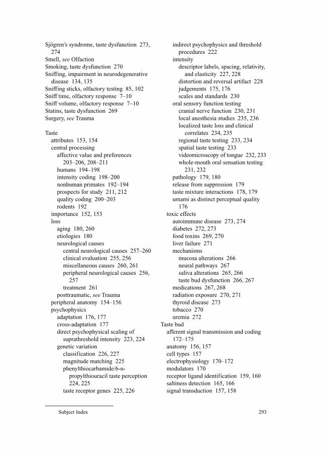
Sjögren’s syndrome, taste dysfunction 273,
274
Smell, see Olfaction
Smoking, taste dysfunction 270
Sniffing, impairment in neurodegenerative
disease 134, 135
Sniffing sticks, olfactory testing 85, 102
Sniff time, olfactory response 7–10
Sniff volume, olfactory response 7–10
Statins, taste dysfunction 269
Surgery, see Trauma
Taste
attributes 153, 154
central processing
affective value and preferences
203–206, 208–211
humans 194–198
intensity coding 198–200
nonhuman primates 192–194
prospects for study 211, 212
quality coding 200–203
rodents 192
importance 152, 153
loss
aging 180, 260
etiologies 180
neurological causes
central neurological causes 257–260
clinical evaluation 255, 256
miscellaneous causes 260, 261
peripheral neurological causes 256,
257
treatment 261
posttraumatic, see Trauma
peripheral anatomy 154–156
psychophysics
adaptation 176, 177
cross-adaptation 177
direct psychophysical scaling of
suprathreshold intensity 223, 224
genetic variation
classification 226, 227
magnitude matching 225
phenylthiocarbamide/6-n-
propylthiouracil taste perception
224, 225
taste receptor genes 225, 226
indirect psychophysics and threshold
procedures 222
intensity
descriptor labels, spacing, relativity,
and elasticity 227, 228
distortion and reversal artifact 228
judgements 175, 176
scales and standards 230
oral sensory function testing
cranial nerve function 230, 231
local anesthesia studies 235, 236
localized taste loss and clinical
correlates 234, 235
regional taste testing 233, 234
spatial taste testing 233
videomicroscopy of tongue 232, 233
whole-mouth oral sensation testing
231, 232
pathology 179, 180
release from suppression 179
taste mixture interactions 178, 179
umami as distinct perceptual quality
176
toxic effects
autoimmune disease 273, 274
diabetes 272, 273
food toxins 269, 270
liver failure 271
mechanisms
mucosa alterations 266
neural pathways 267
saliva alterations 265, 266
taste bud dysfunction 266, 267
medications 267, 268
radiation exposure 270, 271
thyroid disease 273
tobacco 270
uremia 272
Taste bud
afferent signal transmission and coding
172–175
anatomy 156, 157
cell types 157
electrophysiology 170–172
modulators 170
receptor ligand identification 159, 160
saltiness detection 165, 166
signal transduction 157, 158
Subject Index 293
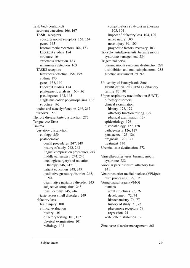
Taste bud (continued)
sourness detection 166, 167
TASR1 receptors
coexpression of receptors 163, 164
genes 165
heterodimeric receptors 164, 173
knockout studies 174
structure 164
sweetness detection 163
umaminess detection 163
TASR2 receptors
bitterness detection 158, 159
coding 173
genes 158, 160
knockout studies 174
phylogenetic analysis 160–162
pseudogenes 162, 163
single nucleotide polymorphisms 162
structure 162
toxins and taste dysfunction 266, 267
turnover 158
Thyroid disease, taste dysfunction 273
Tongue, see Taste
Trauma
gustatory dysfunction
etiology 250
postoperative
dental procedures 247, 248
history of study 242, 243
lingual compression procedures 247
middle ear surgery 244, 245
oncologic surgery and radiation
therapy 246, 247
patient education 248, 249
qualitative gustatory disorder 243,
244
quantitative gustatory disorder 243
subjective complaints 243
tonsillectomy 245, 246
taste versus smell disorders 249
olfactory loss
brain injury 100
clinical evaluation
history 101
olfactory testing 101, 102
physical examination 101
radiology 102
compensatory strategies in anosmia
103, 104
impact of olfactory loss 104, 105
nerve injury 100
nose injury 99, 100
prognostic factors, recovery 103
Tricyclic antidepressants, burning mouth
syndrome management 284
Trigeminal nerve
burning mouth syndrome dysfunction 283
disinhibition and oral pain phantoms 235
function assessment 91, 92
University of Pennsylvania Smell
Identification Test (UPSIT), olfactory
testing 85, 101
Upper respiratory tract infection (URTI),
olfactory disorders
clinical examination
history 128, 129
olfactory function testing 129
physical examination 129
epidemiology 126
histopathology 127, 128
pathogenesis 126, 127
persistence 125, 126
prognosis 129, 130
treatment 130
Uremia, taste dysfunction 272
Varicella-zoster virus, burning mouth
syndrome 282
Vascular parkinsonism, olfactory loss
141
Ventroposterior medial nucleus (VPMpc),
taste processing 192, 193
Vomeronasal organ (VMO)
humans
adult structures 75, 76
development 72, 74
histochemistry 76, 77
history of study 71, 72
pheromone receptors 79
regression 74
vertebrate distribution 72
Zinc, taste disorder management 261
Subject Index 294