arts and siah collaborate in a pathway for xiap degradation
TRANSCRIPT
Molecular Cell
Article
ARTS and Siah Collaboratein a Pathway for XIAP DegradationJason B. Garrison,1,2 Ricardo G. Correa,1 Motti Gerlic,1 Kenneth W. Yip,1,3 Andreas Krieg,1,4 Craig M. Tamble,1
Ranxin Shi,1 Kate Welsh,1 Srinivas Duggineni,1,5 Ziwei Huang,1,5 Keqin Ren,2 Chunying Du,2,* and John C. Reed1,*1Sanford-Burnham Medical Research Institute, La Jolla, CA 92037, USA2University of Cincinnati, Department of Cancer and Cell Biology, Cincinnati, OH 45267, USA3Present address: Department of Cell and Systems Biology, University of Toronto, Toronto, Ontario M5S 3G5, Canada4Present address: Department of General, Visceral and Pediatric Surgery, University Hospital Dusseldorf, 40225 Dusseldorf, Germany5Present address: SUNY Upstate Medical University, Syracuse, NY 13210, USA
*Correspondence: [email protected] (C.D.), [email protected] (J.C.R.)DOI 10.1016/j.molcel.2010.12.002
SUMMARY
ARTS (apoptosis-related protein in the TGF-bsignaling pathway) is a mitochondrial protein thatbinds XIAP (X-linked inhibitor of apoptosis protein)upon entering the cytosol, thus promoting cell death.Expression of ARTS is lost in some malignancies.Here, we show that ARTS binds to XIAP at BIR1,a domain distinct from the caspase-binding sites.Furthermore, ARTS interacts with the E3 ligaseSiah-1 (seven in absentia homolog 1) to induce ubiq-uitination and degradation of XIAP. Cells lackingeither Siah or ARTS contain higher steady-statelevels of XIAP. Thus, ARTS serves as an adaptor tobridge Siah-1 to XIAP, targeting it for destruction.
INTRODUCTION
Inhibitor of apoptosis proteins (IAPs) play pivotal roles in onco-
genesis by suppressing apoptosis induced by both intrinsic
and extrinsic cell death pathways (Salvesen and Duckett,
2002). Most IAPs bind caspases, intracellular proteases respon-
sible for apoptosis (Salvesen and Duckett, 2002; Eckelman et al.,
2006). While binding caspases, only X-linked inhibitor of
apoptosis protein (XIAP) has been shown to unequivocally inhibit
the activity of caspases with physiologically relevant potency,
owing to its ability to make contact with the caspases it targets
at two sites rather than a single site as found thus far for other
IAPs (Chai et al., 2001; Riedl et al., 2001; Shiozaki et al., 2003).
Consequently, XIAP may have special importance among IAP
family members as a suppressor of apoptosis.
All members of the human IAP family contain at least one ba-
culoviral IAP repeat (BIR) domain (a distinctive zinc-binding
protein fold). Several IAPs also contain a C-terminal RING
domain that binds ubiquitin-conjugating enzymes (E2s), endow-
ing them with E3 ubiquitin ligase activity (Srinivasula and
Ashwell, 2008). Ubiquitin modifications that IAPs induce on their
substrates vary, with K48-linked polyubiquitin chains targeting
for proteasomal degradation (Li et al., 2002; Varfolomeev et al.,
2007). In this regard, several IAPs appear to autoregulate their
M
expression through self-ubiquitination, maintaining relatively
low levels of these proteins in tissues.
Several endogenous antagonists of IAPs have been identified,
including SMAC, HtrA2/Omi, ARTS, and XAF-1. SMAC and
HtrA2/Omi are both targeted to mitochondria by an N-terminal
leader sequence that is removed by proteolysis upon their import
into these organelles (Shiozaki and Shi, 2004). SMAC and
HtrA2/Omi are released from the intermembrane space of mito-
chondria in response to apoptotic signals and bind via their
cleaved N termini to several IAPs. The interaction of SMAC and
Omi with IAPs requires the N-terminal Ala of the processedmito-
chondrial proteins, which binds grooves on the surfaces of BIR
domains, displacing caspases. Interestingly, SMAC binds with
high affinity to XIAP, cIAP1, and cIAP2 but stimulates the E3
ligase activity only of cIAP1 and cIAP2 (Creagh et al., 2004), sug-
gesting that XIAP ubiquitination may be regulated differently.
ARTS, a mitochondrial-localized protein, was shown to
promote apoptosis by binding XIAP (Gottfried et al., 2004).
Apoptotic stimuli cause the release of ARTS from mitochondria
allowing its binding to XIAP, resulting in caspase activation and
cell death (Gottfried et al., 2004; Larisch et al., 2000). ARTS
expression is lost in some cancers. For example, ARTS expres-
sion is lost in >70% of childhood acute lymphoblastic leukemia
(ALL) specimens during disease progression (Elhasid et al.,
2004). ARTS is also implicated in the progression of acute
myelogenous leukemia (AML), and leukemic cells lacking ARTS
are resistant to apoptotic stimuli (Elhasid et al., 2004). Further-
more, the region encoding ARTS is deleted in a number of solid
tumors (Kalikin et al., 2000). The loss of ARTS expression
provides evidence that ARTS may function as a tumor
suppressor (Yamamoto et al., 2004). We therefore investigated
the mechanism by which ARTS antagonizes IAPs, in hopes
that the resulting information might suggest strategies for
restoring or mimicking ARTS activity, thus promoting apoptosis
of cancer cells.
RESULTS
ARTS Reduces XIAP Protein LevelsXIAP contains three BIR domains, where BIR2 binds
downstream effector caspases (caspases-3 and -7) and BIR3
binds an upstream initiator caspase (caspase-9) (Reed, 2004).
olecular Cell 41, 107–116, January 7, 2011 ª2011 Elsevier Inc. 107
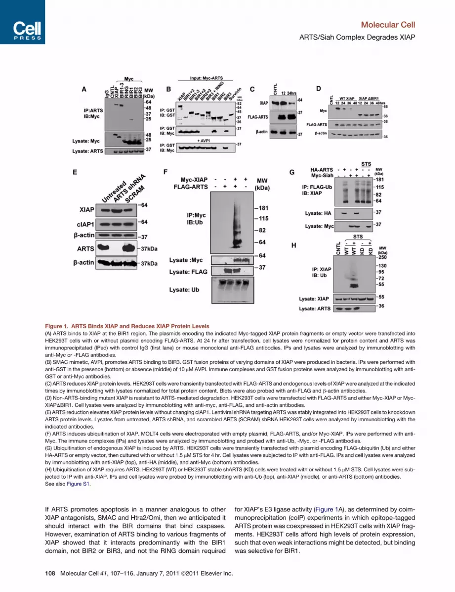
Figure 1. ARTS Binds XIAP and Reduces XIAP Protein Levels
(A) ARTS binds to XIAP at the BIR1 region. The plasmids encoding the indicated Myc-tagged XIAP protein fragments or empty vector were transfected into
HEK293T cells with or without plasmid encoding FLAG-ARTS. At 24 hr after transfection, cell lysates were normalized for protein content and ARTS was
immunoprecipitated (IPed) with control IgG (first lane) or mouse monoclonal anti-FLAG antibodies. IPs and lysates were analyzed by immunoblotting with
anti-Myc or -FLAG antibodies.
(B) SMAC mimetic, AVPI, promotes ARTS binding to BIR3. GST fusion proteins of varying domains of XIAP were produced in bacteria. IPs were performed with
anti-GST in the presence (bottom) or absence (middle) of 10 mM AVPI. Immune complexes and GST fusion proteins were analyzed by immunoblotting with anti-
GST or anti-Myc antibodies.
(C) ARTS reduces XIAP protein levels. HEK293T cells were transiently transfectedwith FLAG-ARTS and endogenous levels of XIAPwere analyzed at the indicated
times by immunoblotting with lysates normalized for total protein content. Blots were also probed with anti-FLAG and b-actin antibodies.
(D) Non-ARTS-binding mutant XIAP is resistant to ARTS-mediated degradation. HEK293T cells were transfected with FLAG-ARTS and either Myc-XIAP or Myc-
XIAPDBIR1. Cell lysates were analyzed by immunoblotting with anti-myc, anti-FLAG, and anti-actin antibodies.
(E) ARTS reduction elevates XIAP protein levels without changing cIAP1. Lentiviral shRNA targeting ARTSwas stably integrated into HEK293T cells to knockdown
ARTS protein levels. Lysates from untreated, ARTS shRNA, and scrambled ARTS (SCRAM) shRNA HEK293T cells were analyzed by immunoblotting with the
indicated antibodies.
(F) ARTS induces ubiquitination of XIAP. MOLT4 cells were electroporated with empty plasmid, FLAG-ARTS, and/or Myc-XIAP. IPs were performed with anti-
Myc. The immune complexes (IPs) and lysates were analyzed by immunoblotting and probed with anti-Ub, -Myc, or -FLAG antibodies.
(G) Ubiquitination of endogenous XIAP is induced by ARTS. HEK293T cells were transiently transfected with plasmid encoding FLAG-ubiquitin (Ub) and either
HA-ARTS or empty vector, then cultured with or without 1.5 mMSTS for 4 hr. Cell lysates were subjected to IP with anti-FLAG. IPs and cell lysates were analyzed
by immunoblotting with anti-XIAP (top), anti-HA (middle), and anti-Myc (bottom) antibodies.
(H) Ubiquitination of XIAP requires ARTS. HEK293T (WT) or HEK293T stable shARTS (KD) cells were treated with or without 1.5 mM STS. Cell lysates were sub-
jected to IP with anti-XIAP. IPs and cell lysates were probed by immunoblotting with anti-Ub (top), anti-XIAP (middle), or anti-ARTS (bottom) antibodies.
See also Figure S1.
Molecular Cell
ARTS/Siah Complex Degrades XIAP
If ARTS promotes apoptosis in a manner analogous to other
XIAP antagonists, SMAC and Htra2/Omi, then we anticipated it
should interact with the BIR domains that bind caspases.
However, examination of ARTS binding to various fragments of
XIAP showed that it interacts predominantly with the BIR1
domain, not BIR2 or BIR3, and not the RING domain required
108 Molecular Cell 41, 107–116, January 7, 2011 ª2011 Elsevier Inc.
for XIAP’s E3 ligase activity (Figure 1A), as determined by coim-
munoprecipitation (coIP) experiments in which epitope-tagged
ARTS protein was coexpressed in HEK293T cells with XIAP frag-
ments. HEK293T cells afford high levels of protein expression,
such that even weak interactions might be detected, but binding
was selective for BIR1.
Molecular Cell
ARTS/Siah Complex Degrades XIAP
Next, we performed in vitro protein binding studies by the
GST-pulldown method, employing a panel of GST fusion
proteins containing a variety of fragments of XIAP and incubating
them with lysates from HEK293T cells transfected with Myc-
ARTS. As in the coIP experiments, ARTS bound strongly to frag-
ments of XIAP containing the BIR1 domain, including the isolated
BIR1 domain (Figure 1B). We also detected a weaker interaction
of ARTS with the isolated BIR3 domain of XIAP by GST pulldown
(that was not observed by coIP), but not with other BIR3-contain-
ing fragments that included adjacent domains located on either
the N- or C-terminal sides of BIR3 (XIAP BIR2+BIR3 and XIAP
BIR3+UBA+RING) (Figure 1B). This observation suggests that
ARTS may also bind the BIR3 domain of XIAP in a conforma-
tion-dependent manner and is consistent with evidence that
some functions of IAPs may be controlled via intramolecular
interactions among domains found within these multifunctional
proteins (Varfolomeev et al., 2007; Mace et al., 2008).
N-terminal peptides from the IAP antagonists SMAC and Omi
bind BIR domains and displace caspases from XIAP (Shiozaki
and Shi, 2004). When the SMAC tetrapeptide alanine-valine-
proline-isoleucine (AVPI) (10 mM) was coincubated with reaction
mixtures containing ARTS and various fragment of XIAP, binding
to full-length XIAP, the isolated BIR1 domain, or the isolated
BIR3 domain was not inhibited, and in fact, binding was induced
between ARTS and additional BIR3-containing fragments of
XIAP (Figure 1B, bottom). In contrast, in separate reactions,
addition of 10 mM of this peptide inhibited binding of recombi-
nant full-length SMAC protein to full-length XIAP (Figure S1
available online) and restored caspase activity in reactions in-
hibited by BIR2 or BIR3 domains of XIAP (Wang et al., 2004).
We conclude from these experiments that (1) the mode of
binding of ARTS to XIAP differs from other mitochondrial antag-
onists of IAPs such as SMAC, (2) interaction of ARTS with some
regions of IAPs is likely to be conformation dependent, and (3)
ARTS interacts with domains within XIAP that are not involved
in caspase inhibition (e.g., BIR1).
The finding that ARTS prominently associates with the non-
caspase-binding BIR1 domain of XIAP suggested that its proa-
poptotic mechanism may involve more than competitive
displacement of caspases from XIAP. In this regard, we noticed
levels of XIAP protein were significantly reduced in cells in which
ARTSwasoverexpressedby transfection (Figure 1C). Amutant of
XIAP lacking the BIR1 domain, however, was resistant to ARTS-
mediated reductions in expression (Figure 1D), with stable XIAPD
BIR1 protein levels achieved by transfection-mediated overex-
pression of ARTS (which causes the ARTS protein to accumulate
in the cytosol as well as mitochondria). The BIR1 domain of
XIAP has been reported to mediate its dimerization (Lu et al.,
2007) However, binding of ARTS to XIAP does not appear to
depend on BIR1-mediated XIAP dimerization, as a BIR1 mutant
(V86A) reported to be defective in dimerization retained its ability
to bind ARTS (Figure S1). In contrast to overexpression of ARTS,
XIAP protein levels were elevated in cells in which endogenous
ARTS expression was reduced with a short hairpin RNA (shRNA)
vector (Figure 1E). Analysis of XIAP messenger RNA (mRNA)
levels did not reveal an effect of ARTS at the transcriptional level
(data not shown), suggesting that ARTSmodulates the stability of
the XIAP protein. Consistent with this hypothesis, experiments
M
with the chemical inhibitor MG132 showed that ARTS-induced
reductions in XIAP are dependent on the 26S proteasome (Fig-
ure S1). In contrast, a broad-spectrum inhibitor of caspases, be-
nozyl-Valinyl-Alaninyl-Aspartyl-fluoromethylketone (zVAD-fmk)
did not interferewith the ability of ARTS overexpression to induce
reductions in XIAP protein.
We next investigated whether ARTS modulates ubiquitination
of XIAP. When ARTS was overexpressed in the acute lympho-
cytic leukemia (ALL) cell line MOLT4, in which endogenous
ARTS gene expression is deficient (Figure S1), ubiquitination of
XIAP protein was clearly induced, as demonstrated by experi-
ments in which XIAP was immunoprecipitated from cells and
then analyzed by immunoblotting with anti-ubiquitin antibody
(Figure 1F). Overexpression of ARTS in HEK293T cells also
induced ubiquitination of XIAP (Figure 1G). Ubiquitination of
XIAP was also induced in HEK293T cells after treatment with
staurosporine (STS) (Figure 1G), a stimulus known to induce
mitochondrial outer membrane permeability (MOMP) and
release of ARTS from these organelles (Larisch et al., 2000),
but not in cells in which ARTS was stably knocked down with
a shRNA vector (Figure 1H).
ARTS Binds the E3 Ligase Siah-1We considered that ARTS might activate the intrinsic E3 ligase
activity of XIAP to induce its self-degradation. However, ARTS
overexpression also accelerated the rate of degradation of
XIAP(H467A), an E3 ligase-defective mutant of XIAP in which
the RING domain was disabled from binding E2s (Figure S2).
While performing control experiments in which we compared
the binding of ARTS to IAP familymemberswith other RING-con-
taining proteins by coIP, we noticed that the E3 ligase Siah-1
associated with ARTS (Figure 2A). In contrast, ARTS did not
bind to cIAP1 or cIAP2 (RING-containing IAP family members),
nor did it bind Survivin or Bcl-XL in these experiments. Interaction
of endogenous ARTS with endogenous Siah-1 (Figure 2B) and
endogenous XIAP with endogenous Siah-1 (Figure 2C) was also
detected by coIP, but only after treatment of cells with STS, sug-
gesting that MOMP is required. (Note that we previously demon-
strated that STS induces MOMP that is suppressed by Bcl-2/
Bcl-XL in the cell line used for these studies [Lin et al., 2004]).
These findings suggested that ARTS may operate as a molec-
ular bridge that targets Siah-1 onto XIAP. To explore this hypoth-
esis, we tested whether Siah-1 and XIAP form a complex as
detected by coIP and used shRNA gene silencing to ask whether
ARTS is required for Siah-1 to associate with XIAP. STS induced
formation of a complex containing both XIAP and Siah-1 when
endogenous ARTS was present but not when ARTS expression
was ablated by shRNA (Figure 2D).
To further evaluate the requirement of ARTS for joining XIAP
and Siah-1, we prepared mutants of ARTS that fail to bind
Siah-1 but retain the ability to bind XIAP. In this regard, the 3D
structure of Siah-1 has been determined (Polekhina et al.,
2002), and mutagenesis studies have revealed that most of the
known Siah substrates contain the consensus binding motif,
RPVAxVxPxxR, which is essential for interaction with a crevice
on the surface of Siah family proteins (House et al., 2003).
We compared the amino acid sequences of ARTS with other
known Siah-binding partners in an attempt to locate a sequence
olecular Cell 41, 107–116, January 7, 2011 ª2011 Elsevier Inc. 109
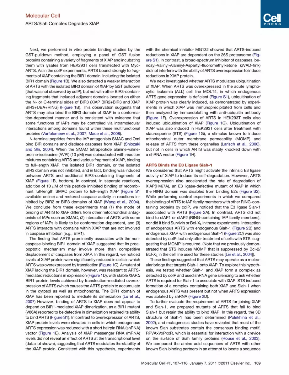
Figure 2. ARTS Binds the E3 Ligase Siah-1
(A) ARTS binds to Siah. HEK293T cells were co-
transfected with plasmids encoding Myc-ARTS
and FLAG-Bcl-XL, FLAG-Survivin, FLAG-cIAP1,
FLAG-cIAP2, FLAG-Siah-1, FLAG-XIAP, or empty
vector as indicated. After 24 hr, cell lysates were
normalized for protein content and ARTS was
IPed with control IgG (first lane) or anti-Myc anti-
body. Immune complexes (IPs) and lysates were
analyzed by immunoblotting and probed with the
indicated antibodies.
(B) The ARTS:Siah interaction is MOMP depen-
dent. HEK293T cells were treated with or without
1.5 mM STS for 4 hr. Cells were lysed, and IPs
were performed with control IgG or mouse mono-
clonal ARTS antibodies. IPs and lysates were
analyzed by immunoblotting and probed with
anti-Siah or anti-ARTS antibodies.
(C) Endogenous XIAP and Siah interact. HEK293T
cells were treated with or without 1.5 mM STS for
4 hr. Cell lysates were subjected to IP with anti-
Siah antibody. IPs and cell lysates were analyzed
by immunoblotting with anti-XIAP (top and
bottom) and anti-Siah (middle) antibodies.
(D) XIAP binds Siah-1 in an ARTS-dependent
manner. HEK293T control (CNTL), scrambled
ARTS (SCRAM) shRNA, and ARTS shRNA
(knock-down, KD) cells were transfected with
plasmids encoding Myc-XIAP and FLAG-Siah
and cultured with or without STS for 4 hr. Cell
lysates were normalized for protein content and
IPed with anti-Myc. IPs and lysates were analyzed
by immunoblotting using anti-FLAG, -Myc, or
-ARTS antibodies.
See also Figure S2.
Molecular Cell
ARTS/Siah Complex Degrades XIAP
in ARTS containing the consensus bindingmotif, finding a candi-
date Siah-binding motif at amino acids 37–48 (ASRPQV-
PEPRPQ) (Figure 3A).
Alanine substitution mutations were engineered within the
37–48 region to create four mutants: ASRAQAPAPAPQ R46A
(MUT1); R46A, E44A (MUT2); R46A, E44A, V42A (MUT3); and
R46A, E44A, V42A, P40A (MUT4). These mutant ARTS proteins
were then expressed as Myc-tagged proteins in HEK293T cells
and assessed for binding to FLAG-Siah-1 by coIP assay (Fig-
ure 3B). ARTS MUT1 and MUT2 retained their ability to bind
Siah-1, while ARTS MUT3 and MUT4 showed no interaction
with Siah-1 (Figure 3B). We then compared these mutant
versions of ARTS with the wild-type ARTS protein with respect
to their ability to promote formation of Siah-1/XIAP complexes.
In unstimulated cells where ARTS is associated with mitochon-
dria, overexpression of wild-type (WT) but not MUT4 ARTS
induced formation of a Siah-1/XIAP complex, as monitored by
coIP (Figure 3C). In STS-treated cells, where ARTS is released
frommitochondria (Larisch et al., 2000), Siah-1 and XIAP formed
a complex, which was increased in relative amounts by overex-
pression ofWTARTS but completely abrogated by expression of
MUT4 ARTS. We interpret this result as an indication that MUT4
ARTS competes with endogenous ARTS, thereby preventing
recruitment of Siah-1 onto XIAP.
To further corroborate that ARTS is a Siah binding partner,
we compared the ability of a synthetic ARTS peptide
110 Molecular Cell 41, 107–116, January 7, 2011 ª2011 Elsevier Inc.
(ASRPQVPEPRPQ) to compete with epitope-tagged-ARTS
protein for binding to Siah-1 in vitro, using coIP assays. For these
experiments, full-length Siah-1 was expressed in HEK293T cells
as a FLAG epitope-tagged protein together with Myc-tagged
ARTS, then cell lysates were prepared to which the ARTS
peptide was added. Incubation of cell lysates with the ARTS
peptide blocked Myc-ARTS protein binding to Siah-1 in
a concentration-dependent manner in vitro (Figures 3D and
3E). In contrast, addition of a negative control peptide to lysates
did not disrupt ARTS binding to Siah (Figure 3D).
Additionally, we generated the same ARTS peptide with a HIV
TAT sequence (HIV TAT-ASRPQVPEPRPQ), allowing for
membrane penetration of the peptide into live cells. Treatment
of cells with HIV TAT-ARTS peptide reduced interaction of
Siah-1 with ARTS protein in a concentration-dependent manner
(Figures 3D and 3F). In contrast, various HIV TAT control
peptides did not disrupt ARTS/Siah-1 binding either when added
to lysates or applied to cultured cells, including a control ARTS
peptide representing an alanine-substitution mutant analogous
to the engineered mutant ARTS protein (MUT4) that fails to
bind Siah-1 (Figure 3D).
ARTS and Siah Collaborate to Reduce XIAP ProteinLevelsWe hypothesized that the interaction of Siah-1 with ARTS might
provide a mechanism for recruiting the E3 ligase activity of
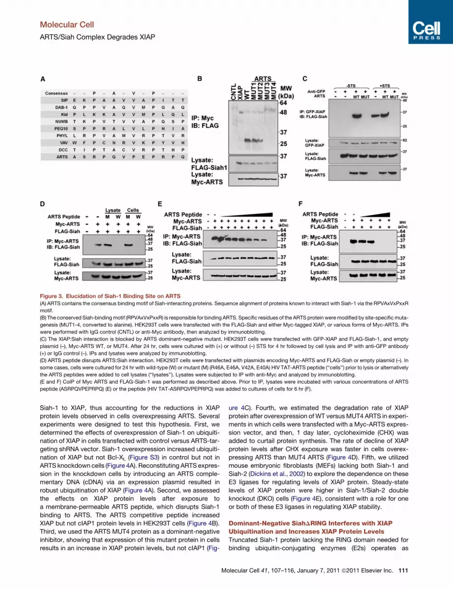
Figure 3. Elucidation of Siah-1 Binding Site on ARTS
(A) ARTS contains the consensus binding motif of Siah-interacting proteins. Sequence alignment of proteins known to interact with Siah-1 via the RPVAxVxPxxR
motif.
(B) The conserved Siah-bindingmotif (RPVAxVxPxxR) is responsible for binding ARTS. Specific residues of the ARTS protein weremodified by site-specificmuta-
genesis (MUT1-4, converted to alanine). HEK293T cells were transfected with the FLAG-Siah and either Myc-tagged XIAP, or various forms of Myc-ARTS. IPs
were performed with IgG control (CNTL) or anti-Myc antibody, then analyzed by immunoblotting.
(C) The XIAP:Siah interaction is blocked by ARTS dominant-negative mutant. HEK293T cells were transfected with GFP-XIAP and FLAG-Siah-1, and empty
plasmid (–), Myc-ARTS WT, or MUT4. After 24 hr, cells were cultured with (+) or without (–) STS for 4 hr followed by cell lysis and IP with anti-GFP antibody
(+) or IgG control (–). IPs and lysates were analyzed by immunoblotting.
(D) ARTS peptide disrupts ARTS:Siah interaction. HEK293T cells were transfected with plasmids encoding Myc-ARTS and FLAG-Siah or empty plasmid (–). In
some cases, cells were cultured for 24 hr with wild-type (W) or mutant (M) (R46A, E46A, V42A, E40A) HIV TAT-ARTS peptide (‘‘cells’’) prior to lysis or alternatively
the ARTS peptides were added to cell lysates (‘‘lysates’’). Lysates were subjected to IP with anti-Myc and analyzed by immunoblotting.
(E and F) CoIP of Myc ARTS and FLAG-Siah-1 was performed as described above. Prior to IP, lysates were incubated with various concentrations of ARTS
peptide (ASRPQVPEPRPQ) (E) or the peptide (HIV TAT-ASRPQVPEPRPQ) was added to cultures of cells for 6 hr (F).
Molecular Cell
ARTS/Siah Complex Degrades XIAP
Siah-1 to XIAP, thus accounting for the reductions in XIAP
protein levels observed in cells overexpressing ARTS. Several
experiments were designed to test this hypothesis. First, we
determined the effects of overexpression of Siah-1 on ubiquiti-
nation of XIAP in cells transfected with control versus ARTS-tar-
geting shRNA vector. Siah-1 overexpression increased ubiquiti-
nation of XIAP but not Bcl-XL (Figure S3) in control but not in
ARTS knockdown cells (Figure 4A). Reconstituting ARTS expres-
sion in the knockdown cells by introducing an ARTS comple-
mentary DNA (cDNA) via an expression plasmid resulted in
robust ubiquitination of XIAP (Figure 4A). Second, we assessed
the effects on XIAP protein levels after exposure to
a membrane-permeable ARTS peptide, which disrupts Siah-1
binding to ARTS. The ARTS competitive peptide increased
XIAP but not cIAP1 protein levels in HEK293T cells (Figure 4B).
Third, we used the ARTS MUT4 protein as a dominant-negative
inhibitor, showing that expression of this mutant protein in cells
results in an increase in XIAP protein levels, but not cIAP1 (Fig-
M
ure 4C). Fourth, we estimated the degradation rate of XIAP
protein after overexpression ofWT versusMUT4 ARTS in experi-
ments in which cells were transfected with a Myc-ARTS expres-
sion vector, and then, 1 day later, cycloheximide (CHX) was
added to curtail protein synthesis. The rate of decline of XIAP
protein levels after CHX exposure was faster in cells overex-
pressing ARTS than MUT4 ARTS (Figure 4D). Fifth, we utilized
mouse embryonic fibroblasts (MEFs) lacking both Siah-1 and
Siah-2 (Dickins et al., 2002) to explore the dependence on these
E3 ligases for regulating levels of XIAP protein. Steady-state
levels of XIAP protein were higher in Siah-1/Siah-2 double
knockout (DKO) cells (Figure 4E), consistent with a role for one
or both of these E3 ligases in regulating XIAP stability.
Dominant-Negative SiahDRING Interferes with XIAPUbiquitination and Increases XIAP Protein LevelsTruncated Siah-1 protein lacking the RING domain needed for
binding ubiquitin-conjugating enzymes (E2s) operates as
olecular Cell 41, 107–116, January 7, 2011 ª2011 Elsevier Inc. 111
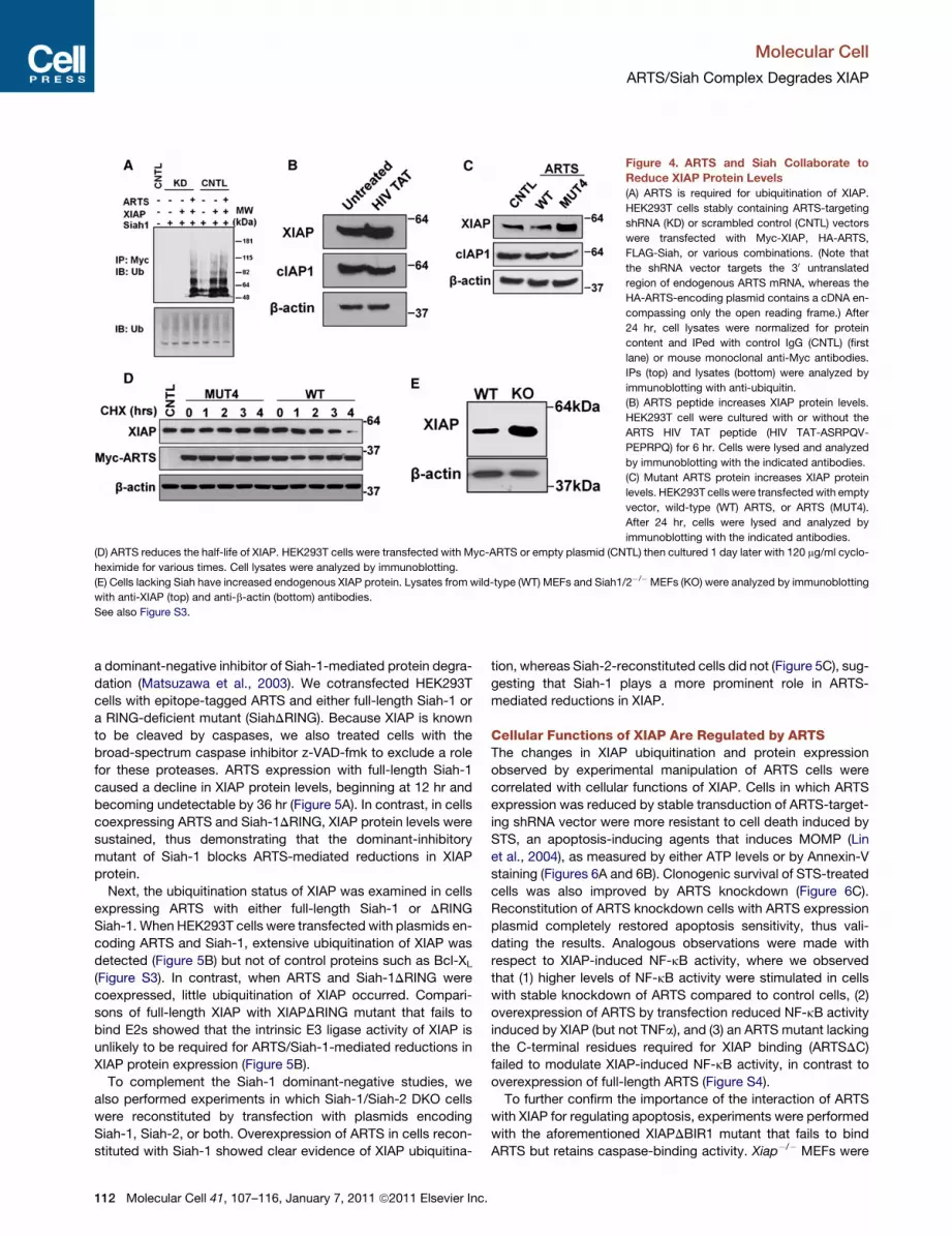
Figure 4. ARTS and Siah Collaborate to
Reduce XIAP Protein Levels
(A) ARTS is required for ubiquitination of XIAP.
HEK293T cells stably containing ARTS-targeting
shRNA (KD) or scrambled control (CNTL) vectors
were transfected with Myc-XIAP, HA-ARTS,
FLAG-Siah, or various combinations. (Note that
the shRNA vector targets the 30 untranslated
region of endogenous ARTS mRNA, whereas the
HA-ARTS-encoding plasmid contains a cDNA en-
compassing only the open reading frame.) After
24 hr, cell lysates were normalized for protein
content and IPed with control IgG (CNTL) (first
lane) or mouse monoclonal anti-Myc antibodies.
IPs (top) and lysates (bottom) were analyzed by
immunoblotting with anti-ubiquitin.
(B) ARTS peptide increases XIAP protein levels.
HEK293T cell were cultured with or without the
ARTS HIV TAT peptide (HIV TAT-ASRPQV-
PEPRPQ) for 6 hr. Cells were lysed and analyzed
by immunoblotting with the indicated antibodies.
(C) Mutant ARTS protein increases XIAP protein
levels. HEK293T cells were transfected with empty
vector, wild-type (WT) ARTS, or ARTS (MUT4).
After 24 hr, cells were lysed and analyzed by
immunoblotting with the indicated antibodies.
(D) ARTS reduces the half-life of XIAP. HEK293T cells were transfected with Myc-ARTS or empty plasmid (CNTL) then cultured 1 day later with 120 mg/ml cyclo-
heximide for various times. Cell lysates were analyzed by immunoblotting.
(E) Cells lacking Siah have increased endogenous XIAP protein. Lysates from wild-type (WT) MEFs and Siah1/2�/� MEFs (KO) were analyzed by immunoblotting
with anti-XIAP (top) and anti-b-actin (bottom) antibodies.
See also Figure S3.
Molecular Cell
ARTS/Siah Complex Degrades XIAP
a dominant-negative inhibitor of Siah-1-mediated protein degra-
dation (Matsuzawa et al., 2003). We cotransfected HEK293T
cells with epitope-tagged ARTS and either full-length Siah-1 or
a RING-deficient mutant (SiahDRING). Because XIAP is known
to be cleaved by caspases, we also treated cells with the
broad-spectrum caspase inhibitor z-VAD-fmk to exclude a role
for these proteases. ARTS expression with full-length Siah-1
caused a decline in XIAP protein levels, beginning at 12 hr and
becoming undetectable by 36 hr (Figure 5A). In contrast, in cells
coexpressing ARTS and Siah-1DRING, XIAP protein levels were
sustained, thus demonstrating that the dominant-inhibitory
mutant of Siah-1 blocks ARTS-mediated reductions in XIAP
protein.
Next, the ubiquitination status of XIAP was examined in cells
expressing ARTS with either full-length Siah-1 or DRING
Siah-1. When HEK293T cells were transfected with plasmids en-
coding ARTS and Siah-1, extensive ubiquitination of XIAP was
detected (Figure 5B) but not of control proteins such as Bcl-XL
(Figure S3). In contrast, when ARTS and Siah-1DRING were
coexpressed, little ubiquitination of XIAP occurred. Compari-
sons of full-length XIAP with XIAPDRING mutant that fails to
bind E2s showed that the intrinsic E3 ligase activity of XIAP is
unlikely to be required for ARTS/Siah-1-mediated reductions in
XIAP protein expression (Figure 5B).
To complement the Siah-1 dominant-negative studies, we
also performed experiments in which Siah-1/Siah-2 DKO cells
were reconstituted by transfection with plasmids encoding
Siah-1, Siah-2, or both. Overexpression of ARTS in cells recon-
stituted with Siah-1 showed clear evidence of XIAP ubiquitina-
112 Molecular Cell 41, 107–116, January 7, 2011 ª2011 Elsevier Inc.
tion, whereas Siah-2-reconstituted cells did not (Figure 5C), sug-
gesting that Siah-1 plays a more prominent role in ARTS-
mediated reductions in XIAP.
Cellular Functions of XIAP Are Regulated by ARTSThe changes in XIAP ubiquitination and protein expression
observed by experimental manipulation of ARTS cells were
correlated with cellular functions of XIAP. Cells in which ARTS
expression was reduced by stable transduction of ARTS-target-
ing shRNA vector were more resistant to cell death induced by
STS, an apoptosis-inducing agents that induces MOMP (Lin
et al., 2004), as measured by either ATP levels or by Annexin-V
staining (Figures 6A and 6B). Clonogenic survival of STS-treated
cells was also improved by ARTS knockdown (Figure 6C).
Reconstitution of ARTS knockdown cells with ARTS expression
plasmid completely restored apoptosis sensitivity, thus vali-
dating the results. Analogous observations were made with
respect to XIAP-induced NF-kB activity, where we observed
that (1) higher levels of NF-kB activity were stimulated in cells
with stable knockdown of ARTS compared to control cells, (2)
overexpression of ARTS by transfection reduced NF-kB activity
induced by XIAP (but not TNFa), and (3) an ARTS mutant lacking
the C-terminal residues required for XIAP binding (ARTSDC)
failed to modulate XIAP-induced NF-kB activity, in contrast to
overexpression of full-length ARTS (Figure S4).
To further confirm the importance of the interaction of ARTS
with XIAP for regulating apoptosis, experiments were performed
with the aforementioned XIAPDBIR1 mutant that fails to bind
ARTS but retains caspase-binding activity. Xiap�/� MEFs were
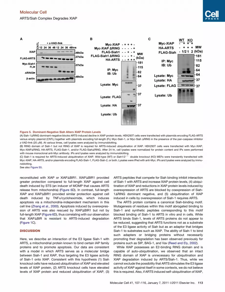
Figure 5. Dominant-Negative Siah Alters XIAP Protein Levels
(A) Siah-1DRING dominant-negative blocks ARTS-induced decline in XIAP protein levels. HEK293T cells were transfected with plasmids encoding FLAG-ARTS
versus empty plasmid (CNTL) together with plasmids encoding full-length (FL) Myc-Siah-1, or Myc-Siah DRING in the presence of the pan-caspase inhibitor
z-VAD-fmk (20 mM). At various times, cell lysates were analyzed by immunoblotting.
(B) RING domain of Siah-1 but not RING of XIAP is required for ARTS-induced ubiquitination of XIAP. HEK293T cells were transfected with Myc-XIAP,
Myc-XIAPDRING, HA-ARTS, FLAG-Siah-1, and/or FLAG-SiahDRING. After 24 hr, cell lysates were normalized for protein content and IPs were performed
with mouse monoclonal anti-Myc antibody. IPs and lysates were analyzed by immunoblotting.
(C) Siah-1 is required for ARTS-induced ubiquitination of XIAP. Wild-type (WT) or Siah1/2�/� double knockout (KO) MEFs were transiently transfected with
Myc-XIAP, HA-ARTS, and/or plasmids encoding FLAG-Siah-1, FLAG-Siah-2, or both. Lysates were IPed with anti-Myc. IPs and lysates were analyzed by immu-
noblotting.
See also Figure S5.
Molecular Cell
ARTS/Siah Complex Degrades XIAP
reconstituted with XIAP or XIAPDBIR1. XIAPDBIR1 provided
greater protection compared to full-length XIAP against cell
death induced by STS (an inducer of MOMP that causes ARTS
release from mitochondria) (Figure 6D). In contrast, full-length
XIAP and XIAPDBIR1 provided similar protection against cell
death induced by TNFa/cycloheximide, which induces
apoptosis via a mitochondria-independent mechanism in this
cell line (Zhang et al., 2000). Apoptosis induced by overexpres-
sion of ARTS was also rescued by XIAPDBIR1 but not by
full-length XIAP (Figure 6D), thus correlating with our observation
that XIAPDBIR is resistant to ARTS-induced degradation
(Figure 1C).
DISCUSSION
Here, we describe an interaction of the E3 ligase Siah-1 with
ARTS, a mitochondrial protein known to bind certain IAP family
proteins and to promote apoptosis. Our data are consistent
with a model in which ARTS serves as a molecular bridge
between Siah-1 and XIAP, thus targeting the E3 ligase activity
of Siah-1 onto XIAP. Consistent with this hypothesis (1) Siah
knockout cells have reduced ubiquitination of XIAP and elevated
levels of XIAP protein, (2) ARTS knockout cells have elevated
levels of XIAP protein and reduced ubiquitination of XIAP, (3)
M
ARTS peptides that compete for Siah binding inhibit interaction
of Siah-1 with ARTS and increase XIAP protein levels, (4) ubiqui-
tination of XIAP and reductions in XIAP protein levels induced by
overexpression of ARTS are blocked by coexpression of Siah-
1DRING dominant negative, and (5) ubiquitination of XIAP
induced in cells by overexpression of Siah-1 requires ARTS.
The ARTS protein contains a canonical Siah-binding motif.
Mutagenesis of residues within this motif abrogated binding to
Siah-1 and synthetic peptides corresponding to this motif
blocked binding of Siah-1 to ARTS in vitro and in cells. While
ARTS binds Siah-1, levels of ARTS proteins do not appear to
be reduced, suggesting that ARTS functions not as a substrate
of the E3 ligase activity of Siah but as an adaptor that bridges
Siah-1 to substrates such as XIAP. The ability of Siah-1 to bind
such adapters or bridging proteins without necessarily
promoting their degradation has been observed previously for
proteins such as SIP, BAG-1, and Vav (Reed and Ely, 2002).
While XIAP possesses an E2-binding RING domain and is
capable of auto-ubiquitination, we observed that an intact
RING domain of XIAP is unnecessary for ubiquitination and
XIAP degradation induced by ARTS/Siah-1. Thus, while we
cannot exclude the possibility that ARTS stimulates the E3 ligase
activity of XIAP against itself in some contexts, we do not believe
this is required. Also, if ARTS induced self-ubiquitination of XIAP,
olecular Cell 41, 107–116, January 7, 2011 ª2011 Elsevier Inc. 113

Figure 6. ARTS Regulates Cellular Functions of XIAP
(A) ARTS sensitizes cells to STS-induced cell death. Control untransfected (diamonds), stable scrambled shRNA (white squares), and stable ARTS shRNA (trian-
gles) HEK293T cells were transfected with empty plasmids or ARTS-encoding plasmid (black squares). After 24 hr, cells were treated with (STS 0–2.5 mM) for
24 hr, then cell viability was assessed by measuring ATP levels, expressing data as percent control relative to no STS (error bars represent mean ± SD; n = 3).
(B) Knockdown of ARTS protects cells against STS-induced apoptosis. Control untransfected, stable scrambled shRNA, and stable ARTS shRNA HEK293T cells
were transfected with empty plasmids or a plasmid encoding ARTS. After 24 hr, cells were treated with (STS 2.5 mM) for 24 hr, then cell death was assessed by
Annexin-V staining, expressing data as percentage Annexin V-positive cells (error bars represent mean ± SD; n = 3).
(C) Knockdown of ARTS increases clonogenic survival. Wild-type, stable scrambled shRNA, and stable ARTS shRNAHEK293T cells were transfectedwith empty
plasmids or ARTS. After 24 hr, cells were treatedwith 2.5 mMSTS for 1 day. Cells were thenwashed and 300 cells of each groupwere seeded into 6 cmdishes and
allowed to grow for 10 days. Colonies greater then 50 cells were then counted (error bars represent mean ± SD; n = 3).
(D) Murine (m) Xiap+/+ or Xiap�/�MEF cells were transiently transfected with various plasmids encoding full-length human (h) XIAP or XIAPDBIR1 deletion mutant.
After 24 hr, cells were cultured with either 2.5 mM STS or 20 ng/ml TNFa (10 mg/ml CHX) for an additional 24 hr, then Annexin-V staining was performed (percent
positive cells; error bars represent mean ± SD; n = 3).
See also Figure S4.
Molecular Cell
ARTS/Siah Complex Degrades XIAP
then we would not have expected to observe effects of various
experimental manipulations of Siah-1 on XIAP ubiquitination and
XIAP protein degradation. In contrast, cells in which either ARTS
or Siah expression was genetically ablated show elevated
steady-state levels of XIAP. The autoubiquitination sites of the
XIAP were previously localized to Lys 311, Lys322, and
Lys328 on the BIR3 domain (Shin et al., 2003). Mutating these
sites reduces XIAP ubiquitination but has little effect on the
ability of ectopically expressed XIAP to rescue cells from
apoptosis (Shin et al., 2003), suggesting that autoubiquitination
of XIAP may have a minimal effect on XIAP protein levels. We
also observed that these self-ubiquitination site mutants of
XIAP remain sensitive to ARTS-induced ubiquitination and
degradation (Figure S5), further arguing that ARTS/Siah-1
utilizes alternative sites in the XIAP protein to mediate ubiquitina-
tion. XIAP-deficient cells reconstituted with these autoubiquiti-
nation site mutants of XIAP were also equally sensitive to
cytotoxicity of ARTS overexpression as wild-type XIAP (data
not shown), suggesting ablation of these sites does not render
114 Molecular Cell 41, 107–116, January 7, 2011 ª2011 Elsevier Inc.
XIAP resistant to ARTS (unlike deletion of the ARTS-binding
BIR1 domain).
In our experiments, ARTS bound selectively to XIAP among
IAP family members tested, which included XIAP, cIAP1, cAP2,
and Survivin. Our domain mapping experiments demonstrated
that ARTS binds to BIR1 of XIAP reproducibly, including the iso-
lated BIR1 domain and several different fragments of XIAP that
contained the BIR1 domain, but not to XIAP lacking BIR1
(DBIR1) (Figure S5). However, ARTS also shows some tendency
to bind the isolated BIR3 domain, but not to some fragments of
XIAP that contained BIR3 along with other domains, unless
SMAC peptide was added. In this regard, domains within the
XIAP protein have been postulated to undergo intramolecular
interactions, controlling conformational states of XIAP that regu-
late its E3 ligase activity (Eckelman et al., 2006; Schile et al.,
2008). Thus, the BIR3 domain in XIAP may become accessible
to ARTS only in some conformational states. It remains to be
determined whether ARTS interacts with other members of the
IAP family in a conformationally restricted manner. Binding of
Molecular Cell
ARTS/Siah Complex Degrades XIAP
ARTS to XIAP does not appear to depend on BIR1-mediated
XIAP dimerization, as a BIR1mutant (V86A) reported to be defec-
tive in dimerization retained its ability to bind ARTS. Also of note,
various studies have provided evidence that the mode of ARTS
binding to XIAP is unrelated to the mechanism used by endoge-
nous antagonist SMAC, with ARTS depending on its C terminus
(versus SMAC requiring its proteolytically processed N terminus)
(Gottfried et al., 2004). Further arguing for a different mode of IAP
antagonism, we observed binding of ARTS to XIAP even in the
presence of SMAC N-terminal peptides that displace caspases
from the BIR2 and BIR3 domains of XIAP.
ARTS protein is reported to be predominantly localized to the
interior of mitochondria, becoming released after stimulation
with apoptosis-inducing agents that trigger MOMP (Larisch
et al., 2000). However, we observed changes in XIAP protein
levels even in normal culture conditions when (1) ARTS expres-
sion was knocked-down by shRNA vectors, (2) HIV TAT-ARTS
competitive peptide was introduced into cells, and (3) Siah-1
expression was ablated by homologous gene recombination.
These observations thus suggest that even in normal circum-
stances, ARTS and Siah-1 contribute to homeostasis of XIAP
protein levels. However, when MOMP was induced in cultured
cells that contain endogenous ARTS, greatly increased ubiquiti-
nation of XIAP was induced in a Siah-dependent manner. Thus,
circumstances known to induce ARTS release from mitochon-
dria clearly induce a burst of ubiquitination of XIAP. Conceivably,
ARTS plays both a baseline, tonic role in maintaining levels of
XIAP, while also allowing for rapid degradation of XIAP after
MOMP. Experimental overexpression of ARTS causes XIAP
ubiquitination and degradation because ARTS protein is
produced in excess amounts where its import into mitochondria
is saturated, resulting in accumulation of ARTS in the cytosol. It
remains to be clarified whether ARTS similarly targets XIAP for
Siah-1-dependent ubiquitination and degradation while passing
through the cytosol in route to the mitochondrial import
machinery versus other possible explanations.
In summary, we have identified a function for ARTS as an
adaptor for the E3 ligase Siah-1, showing that collaboration of
ARTS and Siah-1 controls XIAP protein levels. These findings
provide insights into the putative tumor suppressor role of
ARTS as pertains to apoptosis regulation, and also suggest
strategies for cytoprotective therapies where chemical mimics
of ARTS peptides that disrupt binding to Siah-1 might be em-
ployed to elevate endogenous XIAP levels for purposes of
preserving neurons or other types of vulnerable cells. Given the
recent implication of XIAP in signaling by NLR family proteins,
our findings may additionally have implications for innate immu-
nity (Bertrand et al., 2009; Krieg et al., 2009).
EXPERIMENTAL PROCEDURES
Additional methodological details are provided in the Supplemental Experi-
mental Procedures.
Cell Culture and Transfections
Cells were grown in either Dulbecco’s modified Eagle’s medium (HEK293T,
MEF) or RPMI (MOLT4) media supplemented with 10% fetal bovine serum,
L-glutamine, and penicillin/streptomycin. HEK293T andMEF cell transfections
were performed with Lipofectamine 2000 reagent (Invitrogen) in Opti-MEM-
M
reduced serum medium (Invitrogen). The Lipofectamine transfection mix was
replaced with fresh medium after 4 hr. MOLT4 cells were electroporated
with the Amaxa Biosystems (Amaxa, Gaithersburg, MD) Cell Line Nucleofector
Kit L, utilizing the reagents and according to the recommendations of the
manufacturer (program C-005).
Immunoprecipitations and Immunoblotting
Cells were lysed at 24 hr posttransfection in ice-cold immunoprecipitation
buffer (20 mM Tris-Cl [pH 7.5], 150 mM NaCl, 10% glycerol, 0.2% Nonidet
P-40, and a protease inhibitor mixture [Roche Applied Science, Indianapolis,
IN]). Lysis with immunoprecipitation buffer was complemented with sonication
(5 pulses) on ice. Lysates were cleared of cell debris by centrifugation at
16,000 3 g at 4�C for 30 min. The supernatant was saved and aliquots corre-
sponding to 20 mg and 250 mg total protein were used for SDS-PAGE/immuno-
blotting and immunoprecipitations, respectively. IPs were preformedwith anti-
Flag or anti-Myc antibodies conjugated to Sepharose beads (Sigma, Saint
Louis, MO; Santa Cruz, Santa Cruz, CA) and with gentle overnight agitation.
Beads were washed three times in lysis buffer, resuspended in 23 Laemmli
buffer, and boiled for 5 min to release bound proteins. Proteins were analyzed
by SDS-PAGE and immunoblotting after transfer to polyvinylidene difluoride
membranes (Osmonics, Minnetonka, MN). Antibodies used include anti-
XIAP, anti-cIAP1, anti-cIAP2, anti-His, and anti-GFP; anti-ubiquitin (Cell
Signaling Technologies Danvers, MA); anti-ARTS, anti-b-actin, and anti-
FLAG M2 (Sigma); anti-HA and anti-Myc (Roche); and anti-Siah1 (Santa Cruz
Biotechnology, Santa Cruz, CA). An enhanced chemiluminescence (ECL)
method (Pierce, Rockford, IL) was used for detection.
GST Pulldown
HEK293T cells were transfected with Myc-ARTS and lysed 24 hr later in ice
cold buffer (0.2% NP40, 1 mM EDTA, 135 mM NaCl, 20 mM Tris, 10%
Glycerol,1 mM DTT, 10 mM NaF, 20 mM Leupeptin, 2 mM PMSF, 2 mM
ortho-vanadate, protease inhibitor mixture [Roche]). Lysates were cleared of
cell debris by centrifugation at 16,000 3 g at 4�C for 30 min. GST-tagged
proteins were incubated with cell lysates overnight at 4�Cwith rotation. Beads
were washed three times in lysis buffer, resuspended in 23 Laemmli buffer,
and boiled for 5 min to release bound proteins. Proteins were analyzed by
SDS-PAGE and immunoblotting after transfer to polyvinylidene difluoride
membranes (Osmonics, Minnetonka, MN). Antibodies used include anti-Myc
and -GST.
Apoptosis Assays
For Annexin-V assays, HEK293T cells (13 106 cells) treated with 2.5 mMstaur-
osporine or TNFa (Invitrogen) at 20 ng/ml for 24 hr, with or without pretreat-
ment for 2 hr with 10 mg/ml cyclohexamide. Cells were double-stained with
fluorescein isothiocyanate-conjugated Annexin-V and propidium iodide (PI)
with a kit according to the manufacturer’s instructions (BioVision, Mountain
View, CA). The percentage of apoptotic (annexin-V positive + PI negative) cells
was determined by flow cytometric analysis (Becton Dickinson, San Jose, CA).
Clonogenic Assays
Wild-type, stable scrambled shRNA, and stable ARTS shRNA HEK293T cells
were transfected with empty plasmids or ARTS. At 24 hr after transfection,
cells were treated with (STS 2.5 mM). Cells were then split and 300 cells of
each group were seeded in 6 cm dishes and allowed to grow for 10 days. Colo-
nies greater then 50 cells were then counted.
SUPPLEMENTAL INFORMATION
Supplemental Information includesSupplemental Experimental Procedures and
five figures and can be found with this article online at doi:10.1016/j.molcel.
2010.12.002.
ACKNOWLEDGMENTS
We thank the National Institutes of Health (CA-069381 and CA-055164) and
Leukemia-Lymphoma Society of America for generous support and David
Bowtell for DKP cells.
olecular Cell 41, 107–116, January 7, 2011 ª2011 Elsevier Inc. 115

Molecular Cell
ARTS/Siah Complex Degrades XIAP
Received: February 11, 2010
Revised: August 12, 2010
Accepted: November 3, 2010
Published online: December 23, 2010
REFERENCES
Bertrand, M.J., Doiron, K., Labbe, K., Korneluk, R.G., Barker, P.A., and Saleh,
M. (2009). Cellular inhibitors of apoptosis cIAP1 and cIAP2 are required for
innate immunity signaling by the pattern recognition receptors NOD1 and
NOD2. Immunity 30, 789–801.
Chai, J., Shiozaki, E., Srinivasula, S.M., Wu, Q., Datta, P., Alnemri, E.S., Shi, Y.,
and Dataa, P. (2001). Structural basis of caspase-7 inhibition by XIAP. Cell 104,
769–780.
Creagh, E.M., Murphy, B.M., Duriez, P.J., Duckett, C.S., and Martin, S.J.
(2004). Smac/Diablo antagonizes ubiquitin ligase activity of inhibitor of
apoptosis proteins. J. Biol. Chem. 279, 26906–26914.
Dickins, R.A., Frew, I.J., House, C.M., O’Bryan, M.K., Holloway, A.J., Haviv, I.,
Traficante, N., de Kretser, D.M., and Bowtell, D.D. (2002). The ubiquitin ligase
component Siah1a is required for completion of meiosis I in male mice. Mol.
Cell. Biol. 22, 2294–2303.
Eckelman, B.P., Salvesen, G.S., and Scott, F.L. (2006). Human inhibitor of
apoptosis proteins: why XIAP is the black sheep of the family. EMBO Rep.
7, 988–994.
Elhasid, R., Sahar, D., Merling, A., Zivony, Y., Rotem, A., Ben-Arush, M., Izraeli,
S., Bercovich, D., and Larisch, S. (2004). Mitochondrial pro-apoptotic ARTS
protein is lost in the majority of acute lymphoblastic leukemia patients.
Oncogene 23, 5468–5475.
Gottfried, Y., Rotem, A., Lotan, R., Steller, H., and Larisch, S. (2004). The mito-
chondrial ARTS protein promotes apoptosis through targeting XIAP. EMBO J.
23, 1627–1635.
House, C.M., Frew, I.J., Huang, H.L., Wiche, G., Traficante, N., Nice, E.,
Catimel, B., and Bowtell, D.D. (2003). A binding motif for Siah ubiquitin ligase.
Proc. Natl. Acad. Sci. USA 100, 3101–3106.
Kalikin, L.M., Sims, H.L., and Petty, E.M. (2000). Genomic and expression
analyses of alternatively spliced transcripts of the MLL septin-like fusion
gene (MSF) that map to a 17q25 region of loss in breast and ovarian tumors.
Genomics 63, 165–172.
Krieg, A., Correa, R.G., Garrison, J.B., Le Negrate, G., Welsh, K., Huang, Z.,
Knoefel, W.T., and Reed, J.C. (2009). XIAP mediates NOD signaling via inter-
action with RIP2. Proc. Natl. Acad. Sci. USA 106, 14524–14529.
Larisch, S., Yi, Y., Lotan, R., Kerner, H., Eimerl, S., Tony Parks, W., Gottfried,
Y., Birkey Reffey, S., de Caestecker, M.P., Danielpour, D., et al. (2000). A novel
mitochondrial septin-like protein, ARTS, mediates apoptosis dependent on its
P-loop motif. Nat. Cell Biol. 2, 915–921.
Li, X., Yang, Y., and Ashwell, J.D. (2002). TNF-RII and c-IAP1 mediate ubiqui-
tination and degradation of TRAF2. Nature 416, 345–347.
Lin, B., Kolluri, S.K., Lin, F., Liu, W., Han, Y.H., Cao, X., Dawson, M.I., Reed,
J.C., and Zhang, X.K. (2004). Conversion of Bcl-2 from protector to killer by
interaction with nuclear orphan receptor Nur77/TR3. Cell 116, 527–540.
116 Molecular Cell 41, 107–116, January 7, 2011 ª2011 Elsevier Inc.
Lu, M., Lin, S.C., Huang, Y., Kang, Y.J., Rich, R., Lo, Y.C., Myszka, D., Han, J.,
and Wu, H. (2007). XIAP induces NF-kappaB activation via the BIR1/TAB1
interaction and BIR1 dimerization. Mol. Cell 26, 689–702.
Mace, P.D., Linke, K., Feltham, R., Schumacher, F.R., Smith, C.A., Vaux, D.L.,
Silke, J., and Day, C.L. (2008). Structures of the cIAP2 RING domain reveal
conformational changes associated with ubiquitin-conjugating enzyme (E2)
recruitment. J. Biol. Chem. 283, 31633–31640.
Matsuzawa, S., Li, C., Ni, C.-Z., Takayama, S., Reed, J.C., and Ely, K.R. (2003).
Structural analysis of Siah1 and its interactions with Siah-interacting protein
(SIP). J. Biol. Chem. 278, 1837–1840.
Polekhina, G., House, C.M., Traficante, N., Mackay, J.P., Relaix, F., Sassoon,
D.A., Parker, M.W., and Bowtell, D.D. (2002). Siah ubiquitin ligase is structur-
ally related to TRAF and modulates TNF-alpha signaling. Nat. Struct. Biol. 9,
68–75.
Reed, J.C. (2004). Apoptosis mechanisms: implications for cancer drug
discovery. Oncology (Huntingt.) 18 (13, Suppl 10), 11–20.
Reed, J.C., and Ely, K.R. (2002). Degrading liaisons: Siah structure revealed.
Nat. Struct. Biol. 9, 8–10.
Riedl, S.J., Renatus, M., Schwarzenbacher, R., Zhou, Q., Sun, C., Fesik, S.W.,
Liddington, R.C., and Salvesen, G.S. (2001). Structural basis for the inhibition
of caspase-3 by XIAP. Cell 104, 791–800.
Salvesen, G.S., and Duckett, C.S. (2002). IAP proteins: blocking the road to
death’s door. Nat. Rev. Mol. Cell Biol. 3, 401–410.
Schile, A.J., Garcıa-Fernandez, M., and Steller, H. (2008). Regulation of
apoptosis by XIAP ubiquitin-ligase activity. Genes Dev. 22, 2256–2266.
Shin, H., Okada, K., Wilkinson, J.C., Solomon, K.M., Duckett, C.S., Reed, J.C.,
and Salvesen, G.S. (2003). Identification of ubiquitination sites on the X-linked
inhibitor of apoptosis protein. Biochem. J. 373, 965–971.
Shiozaki, E.N., and Shi, Y. (2004). Caspases, IAPs and Smac/DIABLO: mech-
anisms from structural biology. Trends Biochem. Sci. 29, 486–494.
Shiozaki, E.N., Chai, J., Rigotti, D.J., Riedl, S.J., Li, P., Srinivasula, S.M.,
Alnemri, E.S., Fairman, R., and Shi, Y. (2003). Mechanism of XIAP-mediated
inhibition of caspase-9. Mol. Cell 11, 519–527.
Srinivasula, S.M., and Ashwell, J.D. (2008). IAPs: what’s in a name? Mol. Cell
30, 123–135.
Varfolomeev, E., Blankenship, J.W., Wayson, S.M., Fedorova, A.V., Kayagaki,
N., Garg, P., Zobel, K., Dynek, J.N., Elliott, L.O., Wallweber, H.J., et al. (2007).
IAP antagonists induce autoubiquitination of c-IAPs, NF-kappaB activation,
and TNFalpha-dependent apoptosis. Cell 131, 669–681.
Wang, Z., Cuddy, M., Samuel, T., Welsh, K., Schimmer, A., Hanaii, F.,
Houghten, R., Pinilla, C., and Reed, J.C. (2004). Cellular, biochemical, and
genetic analysis of mechanism of small molecule IAP inhibitors. J. Biol.
Chem. 279, 48168–48176.
Yamamoto, K., Abe, S., Nakagawa, Y., Suzuki, K., Hasegawa, M., Inoue, M.,
Kurata, M., Hirokawa, K., and Kitagawa, M. (2004). Expression of IAP family
proteins in myelodysplastic syndromes transforming to overt leukemia.
Leuk. Res. 28, 1203–1211.
Zhang, H., Huang, Q., Ke, N., Matsuyama, S., Hammock, B., Godzik, A., and
Reed, J.C. (2000). Drosophila pro-apoptotic Bcl-2/Bax homologue reveals
evolutionary conservation of cell death mechanisms. J. Biol. Chem. 275,
27303–27306.