cdc28clb5 (cdk-s) and cdc7-dbf4 (ddk) collaborate to initiate meiotic recombination in yeast
TRANSCRIPT
Cdc28–Clb5 (CDK-S) and Cdc7–Dbf4(DDK) collaborate to initiate meioticrecombination in yeastLihong Wan,1 Hengyao Niu,1 Bruce Futcher,2 Chao Zhang,3 Kevan M. Shokat,3 Simon J. Boulton,4
and Nancy M. Hollingsworth1,5
1Department of Biochemistry and Cell Biology, Stony Brook University, Stony Brook, New York 11794, USA; 2Departmentof Molecular Genetics and Microbiology, Stony Brook University, Stony Brook, New York 11794, USA; 3Department ofCellular and Molecular Pharmacology, University of California at San Francisco, San Francisco, California 94158, USA;4London Research Institute, Clare Hall Laboratories, South Mims, Herts EN6 3LD, United Kingdom
S-phase cyclin-dependent kinase Cdc28–Clb5 (CDK-S) and Dbf4-dependent kinase Cdc7–Dbf4 (DDK) arehighly conserved kinases well known for their roles in the initiation of DNA replication. CDK-S is alsoessential for initiation of meiotic recombination because it phosphorylates Ser30 of Mer2, a meiosis-specificdouble-strand break (DSB) protein. This work shows that the phosphorylation of Mer2 Ser30 by CDK-S primesMer2 for subsequent phosphorylation by DDK on Ser29, creating a negatively charged “patch” necessary forDSB formation. CDK-S and DDK phosphorylation of Mer2 S30 and S29 can be bypassed by phosphomimeticamino acids, but break formation under these conditions is still dependent on DDK and CDK-S activity.Coordination between premeiotic S and DSB formation may be achieved by using CDK-S and DDK to initiateboth processes. Many other proteins important for replication, recombination, repair, and chromosomesegregation contain combination DDK/CDK sites, raising the possibility that this is a common regulatorymechanism.
[Keywords: Meiotic recombination; Cdc7; double-strand breaks; CDK; phosphorylation]
Supplemental material is available at http://www.genesdev.org.
Received October 16, 2007; revised version accepted December 7, 2007.
Meiosis is a highly conserved, specialized form of celldivision that creates haploid gametes from diploid cellsby dividing the chromosome number in half. The reduc-tion in chromosome number is highly specific, such thatcells receive one copy of each chromosome, and is ac-complished by two rounds of chromosome segregationfollowing a single round of chromosome duplication.The first division, MI, is unique to meiosis. Pairs of ho-mologous sister chromatids segregate to opposite poles,in contrast to mitosis, where sister chromatids separate.Proper MI segregation requires that homologous chro-mosomes be physically connected by a combination ofcrossing over and sister chromatid cohesion (for review,see Petronczki et al. 2003). In the absence of interhomo-log crossovers, homologs segregate randomly at MI, pro-ducing chromosomally imbalanced gametes that are gen-erally incapable of producing viable offspring.
Recombination is initiated during meiosis by double-strand breaks (DSBs), catalyzed by a meiosis-specific, to-poisomerase-like protein called Spo11. spo11 mutants in
a variety of species, including budding and fission yeasts,worms, plants, fruit flies, and mice are defective inrecombination, illustrating the remarkable evolution-ary conservation of this recombination mechanism (Kee-ney 2001). DSBs occur preferentially at specific sites inthe genome called recombination “hot spots” (Petes2001). In budding yeast, numerous genes in additionto SPO11 are required for DSB formation. These includeMre11/Rad50/Xrs2, part of the MRX complex alsoutilized for recombination in vegetative cells, as wellas the meiosis-specific genes MER2/REC107, REC102,REC104, REC114, MEI4, and SKI8 (Pecina et al. 2002).Although recent work has described subcomplexes forsome of these proteins (e.g., Rec102/Rec104/Spo11/Ski8and Mer2/Mei4/Rec114), how these proteins work to-gether to initiate DSB formation is still not understood(Jiao et al. 2003; Arora et al. 2004; Li et al. 2006; Malekiet al. 2007).
Failure to repair meiotic DSBs prior to chromosomesegregation is disastrous for a cell. DSB formation andrepair are therefore highly regulated processes. Recom-bination between homologs, rather than sister chroma-tids, is promoted both by use of a meiosis-specific RecAortholog, Dmc1, and the suppression of sister chromatid
5Corresponding author.E-MAIL [email protected]; FAX (631) 632-8575.Article is online at http://www.genesdev.org/cgi/doi/10.1101/gad.1626408.
386 GENES & DEVELOPMENT 22:386–397 © 2008 by Cold Spring Harbor Laboratory Press ISSN 0890-9369/08; www.genesdev.org
Cold Spring Harbor Laboratory Press on August 18, 2016 - Published by genesdev.cshlp.orgDownloaded from
repair mediated by Mek1/Mre4, a meiosis-specific ki-nase (Bishop et al. 1992; Schwacha and Kleckner 1997;Wan et al. 2004). In addition, DSB formation is coordi-nated with other meiotic events, such that DSBs occurafter premeiotic DNA synthesis (Borde et al. 2000). Vari-ous checkpoints function during meiosis to preventchromosome segregation at MI in mutant situationswhere DNA replication is incomplete or recombinationintermediates fail to get processed (Hochwagen andAmon 2006). How replication and DSB formation arecoordinated in wild-type cells is not yet understood.However, recent results have indicated that the S-phasecyclin-dependent kinase Cdc28–Clb5,6 (CDK-S) may beinvolved (Henderson et al. 2006).
CDK-S is a highly conserved, essential protein kinaserequired for DNA replication in both mitotic and mei-otic cells (Stuart and Wittenberg 1998; Bell and Dutta2002; Benjamin et al. 2003). In the absence of CDK-Sactivity, neither premeiotic DNA synthesis nor meioticDSBs occur. Initially, this led to the suggestion thatDNA synthesis is a prerequisite for DSB formation(Borde et al. 2000; Smith et al. 2001; Benjamin et al.2003). Recently, however, Henderson et al. (2006)showed that CDK-S affects DSB formation directly, byphosphorylating Ser30 (S30) of Mer2. The mer2-S30Amutant fails to make DSBs, and consequently producesinviable spores. Mer2 physically interacts with Mei4 andRec114 and mer2-S30A disrupts these interactions, sug-gesting that the function of CDK-S phosphorylation ofMer2 is to promote the formation of larger protein com-plexes essential for making DSBs (Henderson et al. 2006;Li et al. 2006). The fact that CDK-S kinase activity is alsorequired for the initiation of premeiotic S raises the pos-sibility that these two processes may be linked by theaction of this kinase.
Cdc7 is another conserved, essential kinase requiredfor the initiation of DNA replication in mitotically di-viding cells (Sclafani 2000; Masai and Arai 2002). LikeCDK-S, Cdc7 kinase activity requires a catalytic subunit(Cdc7) that is present constitutively throughout the cellcycle, and a regulatory subunit (Dbf4), whose levels fluc-tuate (Nougarede et al. 2000). The Cdc7–Dbf4 complex isreferred to as DDK, for Dbf4-dependent kinase. Geneticstudies in budding yeast indicate that there is a sequen-tial order to kinase action for replication, with CDK-Spreceding DDK (Nougarede et al. 2000). It has been sug-gested that CDK-S phosphorylation of target proteinsmight prime subsequent DDK phosphorylation of thesame protein on adjacent residues; for instance, in vitrostudies show that DDK phosphorylation of Mcm2 is en-hanced by prior CDK phosphorylation (Cho et al. 2006;Montagnoli et al. 2006). However, whether coordinatedphosphorylation of replication proteins by CDK-S andDDK at specific sites is functionally relevant is not yetknown.
Inactivation of DDK during meiosis using temperaturesensitive alleles of CDC7 (cdc7ts) has no effect on pre-meiotic DNA synthesis, but it does prevent recombina-tion and causes a Prophase I arrest (Schild and Byers1978; Hollingsworth and Sclafani 1993). However, these
experiments cannot exclude a role for DDK in premei-otic S, because the Cdc7ts kinase may not have beencompletely inactivated at the restrictive temperature.Abolishing DDK function by repression of DBF4 tran-scription prior to entry into meiosis causes delays in pre-meiotic DNA replication, arguing for a role for DDK inthis process (Valentin et al. 2005). As an alternative ap-proach to inactivating DDK during meiosis, a condi-tional version of CDC7 (cdc7-as) was created by enlarg-ing the ATP-binding pocket of Cdc7, thereby creating amutant kinase that can be inactivated by addition of pu-rine analogs to the sporulation medium (Wan et al.2006). This chemical genetic approach was previouslysuccessful in revealing an essential role for CDC28 inpremeiotic DNA replication that was not detected usingcdc28ts mutants (Shuster and Byers 1989; Benjamin et al.2003). Similar to the DBF4 shutoff situation, cdc7-as dip-loids induced to undergo meiosis in the presence of ki-nase inhibitor did successfully synthesize DNA, butwith a delay (Wan et al. 2006). Like cdc7ts, inactivationof cdc7-as prevents recombination and causes a prophasearrest. The lack of recombination is due to a lack ofDSBs. When the kinase inhibitor is removed, DSBs rap-idly appear and cells proceed synchronously throughmeiosis, indicating that the cells are arrested immedi-ately prior to DSB formation (Wan et al. 2006). Here, weshow that a critical target of DDK for DSB formation isMer2, and that CDK-S and DDK function sequentially tophosphorylate Mer2 on the adjacent serines S30 and S29,allowing formation of meiotic DSBs.
Results
DDK activity is required for phosphorylationof the DSB protein Mer2
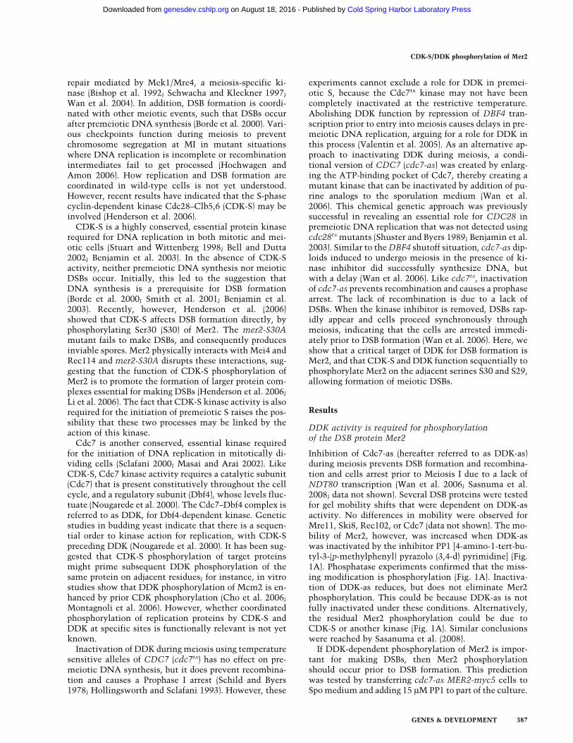
Inhibition of Cdc7-as (hereafter referred to as DDK-as)during meiosis prevents DSB formation and recombina-tion and cells arrest prior to Meiosis I due to a lack ofNDT80 transcription (Wan et al. 2006; Sasnuma et al.2008; data not shown). Several DSB proteins were testedfor gel mobility shifts that were dependent on DDK-asactivity. No differences in mobility were observed forMre11, Ski8, Rec102, or Cdc7 (data not shown). The mo-bility of Mer2, however, was increased when DDK-aswas inactivated by the inhibitor PP1 [4-amino-1-tert-bu-tyl-3-(p-methylphenyl) pyrazolo (3,4-d) pyrimidine] (Fig.1A). Phosphatase experiments confirmed that the miss-ing modification is phosphorylation (Fig. 1A). Inactiva-tion of DDK-as reduces, but does not eliminate Mer2phosphorylation. This could be because DDK-as is notfully inactivated under these conditions. Alternatively,the residual Mer2 phosphorylation could be due toCDK-S or another kinase (Fig. 1A). Similar conclusionswere reached by Sasanuma et al. (2008).
If DDK-dependent phosphorylation of Mer2 is impor-tant for making DSBs, then Mer2 phosphorylationshould occur prior to DSB formation. This predictionwas tested by transferring cdc7-as MER2-myc5 cells toSpo medium and adding 15 µM PP1 to part of the culture.
CDK-S/DDK phosphorylation of Mer2
GENES & DEVELOPMENT 387
Cold Spring Harbor Laboratory Press on August 18, 2016 - Published by genesdev.cshlp.orgDownloaded from
After 8 h in Spo medium, cells without PP1 exhibitedphosphorylated Mer2 and Hop1, as well as DSBs (Fig. 1B).In contrast, no DSBs, phospho-Hop1 or phospho-Mer2 were detected in cells incubated with PP1. At8 h, the PP1 was washed out and the cells were resus-pended in Spo medium and returned to the shaker (thistime is defined as 0 min after washout). Some DDK-asmolecules became active during the washout protocol(which takes ∼40 min), as an increase in phospho-Mer2was detectable at the 0 time point (Fig. 1B). Equalamounts of phosphorylated and unphosphorylated Mer2were present within 60 min after washout, while DSBswere not detected until 80 min post-washout. Phospho-Hop1 was observed 2 h after DSBs were first detected(Fig. 1B). The temporal order of these events is thereforeMer2 phosphorylation, then DSB formation, then Hop1phosphorylation.
Residues Ser29 and Ser30 of Mer2 are both requiredfor the full phosphorylation mobility shift
Given that Mer2 phosphorylation is dependent on DDK,the question is whether this phosphorylation is directand, if so, what sites are being phosphorylated. Recentanalysis of DDK site specificity has shown that DDK isan acid-directed, or phosphate-directed, protein kinase.In human Mcm2, DDK phosphorylation sites have beenmapped to Ser or Thr residues immediately followed bya negatively charged or phosphorylated amino acid (Choet al. 2006). Two groups independently found sites inhuman Mcm2 that are phosphorylated more efficientlyby DDK after the adjacent C-terminal residue was firstphosphorylated by CDK (Cho et al. 2006; Montagnoli etal. 2006). These sites both have the sequence TSSPGR,where the first (i.e., more N-terminal) serine is a DDKsite, and the second serine is a CDK site. Strikingly, analmost identical sequence, TSSPFR, occurs in Mer2,where the second (i.e., more C-terminal) serine is S30,the known site of phosphorylation by CDK-S. Therefore,phosphorylation of S30 by CDK-S could promote phos-phorylation of S29 by DDK. The preceding residue, T28,is also phosphorylatable. If DDK is truly a phosphate-directed kinase, then phosphorylation of S29 could leadto phosphorylation of T28, thus creating a highly acidicpatch of three phosphorylated residues in a row.
If S29 is phosphorylated by DDK, then the Mer2-S29A-myc5 protein should exhibit a change in protein mobil-ity similar to that seen in cdc7-as diploids in the pres-ence of PP1. To test this possibility diploids containingMER2-myc5, mer2-S30A-myc5, mer2-S29A-myc5, andmer2-S29A S30A-myc5 were incubated in Spo mediumfor 4 h and the proteins immunoprecipitated fromsoluble extracts using �-myc antibodies. Western blotanalysis indicates that Mer2 phosphorylation is de-creased in the mer2-S29A mutant (Fig. 1C). The fact thatMer2-S29A-myc5 and Mer2-S29A S30A-myc5 proteinmobilities are similar suggests that the bulk of the Mer2mobility shift is due to DDK phosphorylation. Further-more, the loss of this shift after inactivation of DDK-assuggests that S29 is a target for DDK phosphorylation invivo.
Meiotic CDK-S kinase activity is independent of DDK
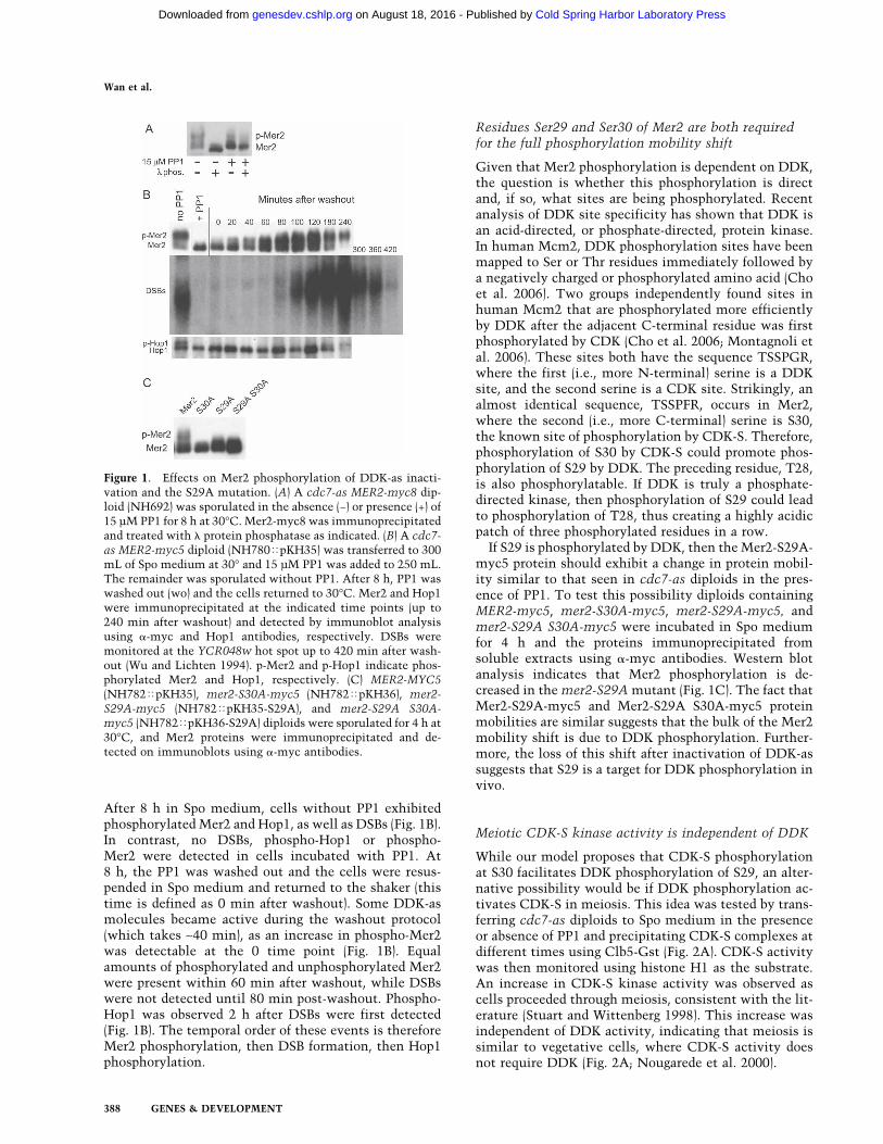
While our model proposes that CDK-S phosphorylationat S30 facilitates DDK phosphorylation of S29, an alter-native possibility would be if DDK phosphorylation ac-tivates CDK-S in meiosis. This idea was tested by trans-ferring cdc7-as diploids to Spo medium in the presenceor absence of PP1 and precipitating CDK-S complexes atdifferent times using Clb5-Gst (Fig. 2A). CDK-S activitywas then monitored using histone H1 as the substrate.An increase in CDK-S kinase activity was observed ascells proceeded through meiosis, consistent with the lit-erature (Stuart and Wittenberg 1998). This increase wasindependent of DDK activity, indicating that meiosis issimilar to vegetative cells, where CDK-S activity doesnot require DDK (Fig. 2A; Nougarede et al. 2000).
Figure 1. Effects on Mer2 phosphorylation of DDK-as inacti-vation and the S29A mutation. (A) A cdc7-as MER2-myc8 dip-loid (NH692) was sporulated in the absence (−) or presence (+) of15 µM PP1 for 8 h at 30°C. Mer2-myc8 was immunoprecipitatedand treated with � protein phosphatase as indicated. (B) A cdc7-as MER2-myc5 diploid (NH780�pKH35) was transferred to 300mL of Spo medium at 30° and 15 µM PP1 was added to 250 mL.The remainder was sporulated without PP1. After 8 h, PP1 waswashed out (wo) and the cells returned to 30°C. Mer2 and Hop1were immunoprecipitated at the indicated time points (up to240 min after washout) and detected by immunoblot analysisusing �-myc and Hop1 antibodies, respectively. DSBs weremonitored at the YCR048w hot spot up to 420 min after wash-out (Wu and Lichten 1994). p-Mer2 and p-Hop1 indicate phos-phorylated Mer2 and Hop1, respectively. (C) MER2-MYC5(NH782�pKH35), mer2-S30A-myc5 (NH782�pKH36), mer2-S29A-myc5 (NH782�pKH35-S29A), and mer2-S29A S30A-myc5 (NH782�pKH36-S29A) diploids were sporulated for 4 h at30°C, and Mer2 proteins were immunoprecipitated and de-tected on immunoblots using �-myc antibodies.
Wan et al.
388 GENES & DEVELOPMENT
Cold Spring Harbor Laboratory Press on August 18, 2016 - Published by genesdev.cshlp.orgDownloaded from
Phosphorylation of S29 in vitro is enhancedby S30 phosphorylation
The idea that phosphorylation of S30 promotes DDKphosphorylation of S29 was directly tested by immuno-precipitating Cdc7/7HA-Dbf4 (DDK-HA) from vegeta-tive yeast cells with �-HA antibodies for use in in vitroprotein kinase reactions with various peptides. As anegative control, a strain containing an untagged versionof Dbf4 was included. Biotinylated peptides were used assubstrates. Peptides had the structure Nterm-biotin-GALAX29X30PFRAAG-Cterm, where X29 and X30 repre-sent positions 29 and 30 in Mer2, respectively. The boldsequence indicates the Mer2 CDK-S/DDK site. T28 waschanged to alanine so that any phosphorylation thatcould potentially occur was limited to S29 or S30. Thewild-type sequence has SS in the X29 X30 sites. The nega-tive control peptide has AA at these positions. ASp andSSp peptides contain phospho-serine in the X30 position.After incubation with the immunoprecipitated kinaseand radioactively labeled ATP, the biotinylated peptideswere captured onto streptavidin membranes, washed,and counted. Each kinase reaction was then normalizedto the number of counts observed with the AA peptideincubated with an immunoprecipitate from the DDK-
HA strain. In two independent DDK-HA immunopre-cipites, a five- to ninefold increase in radioactivity wasobserved for the SSp peptide over the AA control (Fig.2B). This increase is dependent on both S29 and phospho-S30 (Fig. 2B). These results provide biochemical supportfor the idea that yeast DDK, like human DDK, has apreference for phosphorylating amino acids immediatelyupstream of a phosphorylated residue.
S29 and T28 are important for Mer2 function
Mutants were initially made in a tagged version ofMER2, MER2-myc5, which has almost but not quitewild-type levels of MER2 function (Materials and Meth-ods; Henderson et al. 2006). For simplicity, mer2 mu-tants are described by the amino acids present in themutant protein in the order 28–29–30; e.g., wild-typeMER2 is TSS. Substitution of S29 with alanine (mer2-TAS-myc5) produced ∼1% viable spores, equivalent tomer2� and mer2-TSA-myc (mutant residues in bold)(Fig. 3A), showing that S30 alone, the known target ofCDK-S phosphorylation, is not sufficient. A critical rolefor Mer2 S29 in meiosis has also been demonstrated bySasanuma et al. (2008). mer2-ASS-myc5 also exhibits re-duced spore viability, although the phenotype is not assevere as either TSA or TAS. In this case, changing T28to aspartic acid (DSS) restores spore viability to wild-typelevels (Fig. 3A). In combination with the biochemicalexperiments, these results are consistent with the ideathat CDK-S phosphorylates S30, and that this primes thephosphorylation of S29 by DDK. In some cases, this mayfurther prime the phosphorylation of T28 by DDK, cre-ating an acidic patch three residues long.
Substitution of S30 with aspartic acid (TSD) (mimick-ing phosphorylation) gave spore viability of ∼11%, indi-cating that the negative charge conferred by aspartic acidcan partially compensate for the lack of S30 phosphory-lation (Fig. 3A). Additional MER2 function can be re-stored to mer2-TSD-myc5 by introducing additional as-partic acid residues at S29 and/or T28. For example, DSDproduced 58% viable spores, a significantly higher num-ber than TSD (Fig. 3A). This improved spore viabilityrequires S29, presumably due to its phosphorylation, asDAD produced only 5.8% viable spores. Having S29flanked by negative charges may compensate for the ab-sence of a phosphate at S30 in promoting DDK phosphor-ylation at this site. Substituting aspartic acid at all threepositions resulted in 71.5% viable spores (Fig. 3A). Vi-ability is improved to wild-type levels if the DDD mu-tations are homozygous in an untagged allele of MER2(Fig. 3A, right). The high spore viability of mer2-DDDsuggests that CDK-S and DDK phosphorylation of Mer2can be bypassed under otherwise wild-type conditionswithout drastically affecting meiotic recombination.
Meiotic DSB formation requires a negative chargeat S29 and S30
Meiotic time-course analysis of DSBs at the YCR048whot spot shows that DSB formation is abolished by mer2-
Figure 2. DDK and CDK-S in vitro kinase assays. (A) CDK-Skinase activity during meiosis in the presence or absence ofactive DDK. cdc7-as CLB5-GST (NH760) and cdc7-as (NH452F)diploids were sporulated at 30°C in the presence or absence of15 µM PP1. CDK-S activity was monitored using histone H1 asthe substrate. The blot was probed with �-Gst antibodies todetect Clb5-Gst. The asterisk marks a cross-reacting band thatruns immediately below Clb5-Gst. (B) DDK activity: Solubleextracts from 334/pGO174 (7HA-DBF4) and 334 were used toimmunoprecipitate Cdc7/7HA-Dbf4 complexes with �-HA an-tibodies. Kinase assays were performed with these extracts us-ing various biotinylated peptides containing the indicatedamino acids corresponding to S29 and S30 of Mer2. Each reac-tion was normalized to the counts obtained for the reactionusing DDK-HA complexes and the AA peptide. Two indepen-dent DDK-HA immunoprecipitates were assayed.
CDK-S/DDK phosphorylation of Mer2
GENES & DEVELOPMENT 389
Cold Spring Harbor Laboratory Press on August 18, 2016 - Published by genesdev.cshlp.orgDownloaded from
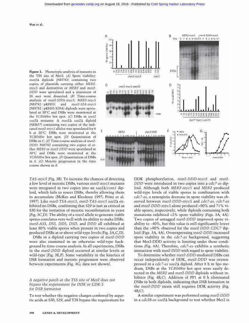
TAS-myc5 (Fig. 3B). To increase the chances of detectinga low level of meiotic DSBs, various mer2-myc5 mutantswere integrated in two copies into an sae2�/com1 dip-loid, which fails to resect DSBs, thereby allowing themto accumulate (McKee and Kleckner 1997; Prinz et al.1997). Like mer2-TSA-myc5, mer2-TAS-myc5 sae2� ex-hibited no DSBs, confirming that S29 is just as critical asS30 for the initiation of meiotic recombination in yeast(Fig. 3C,D). The ability of a mer2 allele to generate viablespores correlates very well with its ability to make DSBs.mer2-ASS, DSS, DDS, DSD, and DDD all exhibited atleast 80% viable spores when present in two copies andproduced DSBs at or above wild-type levels (Fig. 3A,C,D).
DSBs in a diploid carrying two copies of mer2-DDDwere also examined in an otherwise wild-type back-ground by time-course analysis. In all experiments, DSBsin the mer2-DDD diploid occurred at similar levels aswild-type (Fig. 3E,F). Some variability in the kinetics ofDSB formation and meiotic progression were observedbetween experiments (Fig. 3G; data not shown).
A negative patch at the TSS site of Mer2 does notbypass the requirement for DDK or CDK-Sfor DSB formation
To test whether the negative charges conferred by aspar-tic acids at S30, S29, and T28 bypass the requirement for
DDK phosphorylation, mer2-DDD-myc5 and mer2-DDD were introduced in two copies into a cdc7-as dip-loid. Although both MER2-myc5 and MER2 producedwild-type levels of viable spores in combination withcdc7-as, a synergistic decrease in spore viability was ob-served between mer2-DDD-myc5 and cdc7-as. cdc7-asand mer2-DDD-myc5 alone produced >90% and 71% vi-able spores, respectively, while diploids containing bothmutations exhibited <2% spore viability (Figs. 3A, 4A).Two copies of untagged mer2-DDD improved spore vi-ability to ∼40%, but this value is still significantly lowerthan the >90% observed for the mer2-DDD CDC7 dip-loid (Figs. 3A, 4A). Overexpressing mer2-DDD increasedspore viability in the cdc7-as background, suggestingthat Mer2-DDD activity is limiting under these condi-tions (Fig. 4A). Therefore, cdc7-as exhibits a syntheticinteraction with mer2-DDD with regard to spore viability.
To determine whether mer2-DDD-mediated DSBs canoccur independently of DDK, mer2-DDD was overex-pressed in a cdc7-as sae2� diploid. After 8 h in Spo me-dium, DSBs at the YCR048w hot spot were easily de-tected in the MER2 and mer2-DDD diploids without in-hibitor (Fig. 4B,C). Addition of PP1 at 0 h eliminatedDSBs in both diploids, indicating that DSB formation inthe mer2-DDD strain still requires DDK activity (Fig.4B,C).
A similar experiment was performed using mer2-DDDin a cdc28-as sae2� background to test whether Mer2 is
Figure 3. Phenotypic analysis of mutants inthe TSS site of Mer2. (A) Spore viability:mer2� diploids (NH782) containing twocopies of plasmids carrying either MER2-myc5 and derivatives or MER2 and mer2-DDD were sporulated and a minimum of26 asci were dissected. (B) Time-courseanalysis of mer2-S29A-myc5: MER2-myc5(NH782�pKH35) and mer2-S2A-myc5(NH782�pKH35-S29A) diploids were sporu-lated at 30°C and DSBs were monitored atthe YCR048w hot spot. (C) DSBs in mer2sae2� mutants: A mer2� sae2� diploid(NH837) containing two copies of the indi-cated mer2-myc5 alleles was sporulated for 8h at 30°C. DSBs were monitored at theYCR048w hot spot. (D) Quantitation ofDSBs in C. (E) Time-course analysis of mer2-DDD: NH782 containing two copies of ei-ther MER2 or mer2-DDD were sporulated at30°C and DSBs were monitored at theYCR048w hot spot. (F) Quantitation of DSBsin E. (G) Meiotic progression in the timecourse shown in E.
Wan et al.
390 GENES & DEVELOPMENT
Cold Spring Harbor Laboratory Press on August 18, 2016 - Published by genesdev.cshlp.orgDownloaded from
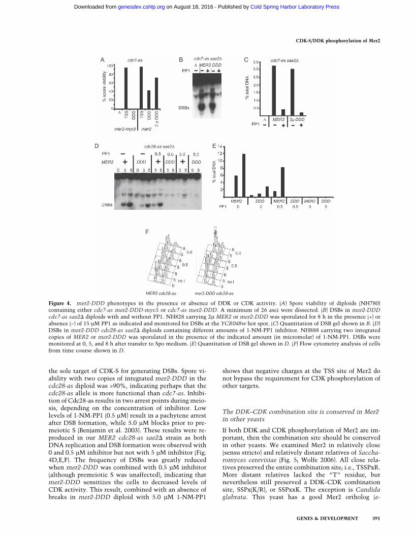
the sole target of CDK-S for generating DSBs. Spore vi-ability with two copies of integrated mer2-DDD in thecdc28-as diploid was >90%, indicating perhaps that thecdc28-as allele is more functional than cdc7-as. Inhibi-tion of Cdc28-as results in two arrest points during meio-sis, depending on the concentration of inhibitor. Lowlevels of 1-NM-PP1 (0.5 µM) result in a pachytene arrestafter DSB formation, while 5.0 µM blocks prior to pre-meiotic S (Benjamin et al. 2003). These results were re-produced in our MER2 cdc28-as sae2� strain as bothDNA replication and DSB formation were observed with0 and 0.5 µM inhibitor but not with 5 µM inhibitor (Fig.4D,E,F). The frequency of DSBs was greatly reducedwhen mer2-DDD was combined with 0.5 µM inhibitor(although premeiotic S was unaffected), indicating thatmer2-DDD sensitizes the cells to decreased levels ofCDK activity. This result, combined with an absence ofbreaks in mer2-DDD diploid with 5.0 µM 1-NM-PP1
shows that negative charges at the TSS site of Mer2 donot bypass the requirement for CDK phosphorylation ofother targets.
The DDK–CDK combination site is conserved in Mer2in other yeasts
If both DDK and CDK phosphorylation of Mer2 are im-portant, then the combination site should be conservedin other yeasts. We examined Mer2 in relatively close(sensu stricto) and relatively distant relatives of Saccha-romyces cerevisiae (Fig. 5; Wolfe 2006). All close rela-tives preserved the entire combination site; i.e., TSSPxR.More distant relatives lacked the “T” residue, butnevertheless still preserved a DDK–CDK combinationsite, SSPx(K/R), or SSPxxK. The exception is Candidaglabrata. This yeast has a good Mer2 ortholog (e-
Figure 4. mer2-DDD phenotypes in the presence or absence of DDK or CDK activity. (A) Spore viability of diploids (NH780)containing either cdc7-as mer2-DDD-myc5 or cdc7-as mer2-DDD. A minimum of 26 asci were dissected. (B) DSBs in mer2-DDDcdc7-as sae2� diploids with and without PP1. NH828 carrying 2µ MER2 or mer2-DDD was sporulated for 8 h in the presence (+) orabsence (−) of 15 µM PP1 as indicated and monitored for DSBs at the YCR048w hot spot. (C) Quantitation of DSB gel shown in B. (D)DSBs in mer2-DDD cdc28-as sae2� diploids containing different amounts of 1-NM-PP1 inhibitor. NH888 carrying two integratedcopies of MER2 or mer2-DDD was sporulated in the presence of the indicated amount (in micromolar) of 1-NM-PP1. DSBs weremonitored at 0, 5, and 8 h after transfer to Spo medium. (E) Quantitation of DSB gel shown in D. (F) Flow cytometry analysis of cellsfrom time course shown in D.
CDK-S/DDK phosphorylation of Mer2
GENES & DEVELOPMENT 391
Cold Spring Harbor Laboratory Press on August 18, 2016 - Published by genesdev.cshlp.orgDownloaded from
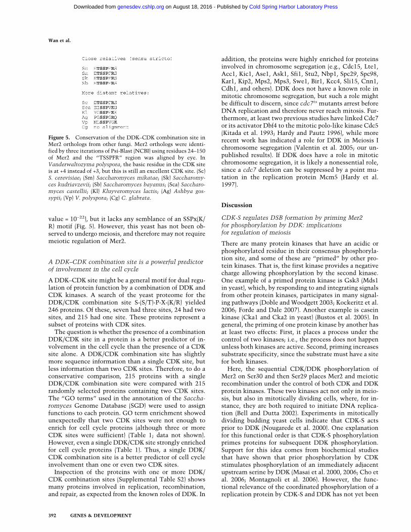
value = 10−22), but it lacks any semblance of an SSPx(K/R) motif (Fig. 5). However, this yeast has not been ob-served to undergo meiosis, and therefore may not requiremeiotic regulation of Mer2.
A DDK–CDK combination site is a powerful predictorof involvement in the cell cycle
A DDK–CDK site might be a general motif for dual regu-lation of protein function by a combination of DDK andCDK kinases. A search of the yeast proteome for theDDK/CDK combination site S-(S/T)-P-X-(K/R) yielded246 proteins. Of these, seven had three sites, 24 had twosites, and 215 had one site. These proteins represent asubset of proteins with CDK sites.
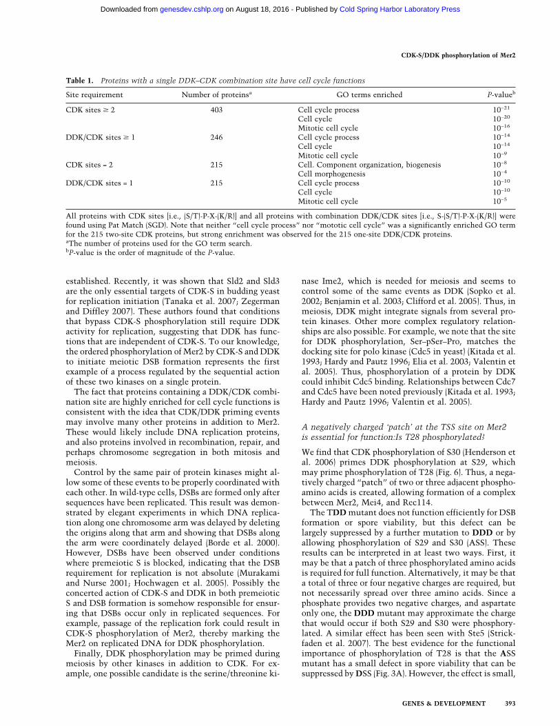
The question is whether the presence of a combinationDDK/CDK site in a protein is a better predictor of in-volvement in the cell cycle than the presence of a CDKsite alone. A DDK/CDK combination site has slightlymore sequence information than a single CDK site, butless information than two CDK sites. Therefore, to do aconservative comparison, 215 proteins with a singleDDK/CDK combination site were compared with 215randomly selected proteins containing two CDK sites.The “GO terms” used in the annotation of the Saccha-romyces Genome Database (SGD) were used to assignfunctions to each protein. GO term enrichment showedunexpectedly that two CDK sites were not enough toenrich for cell cycle proteins (although three or moreCDK sites were sufficient) (Table 1; data not shown).However, even a single DDK/CDK site strongly enrichedfor cell cycle proteins (Table 1). Thus, a single DDK/CDK combination site is a better predictor of cell cycleinvolvement than one or even two CDK sites.
Inspection of the proteins with one or more DDK/CDK combination sites (Supplemental Table S2) showsmany proteins involved in replication, recombination,and repair, as expected from the known roles of DDK. In
addition, the proteins were highly enriched for proteinsinvolved in chromosome segregation (e.g., Cdc15, Lte1,Acc1, Kic1, Ase1, Ask1, Sfi1, Stu2, Nbp1, Spc29, Spc98,Kar1, Kip2, Mps2, Mps3, Swe1, Bir1, Kcc4, Sli15, Cnn1,Cdh1, and others). DDK does not have a known role inmitotic chromosome segregation, but such a role mightbe difficult to discern, since cdc7ts mutants arrest beforeDNA replication and therefore never reach mitosis. Fur-thermore, at least two previous studies have linked Cdc7or its activator Dbf4 to the mitotic polo-like kinase Cdc5(Kitada et al. 1993; Hardy and Pautz 1996), while morerecent work has indicated a role for DDK in Meiosis Ichromosome segregation (Valentin et al. 2005; our un-published results). If DDK does have a role in mitoticchromosome segregation, it is likely a nonessential role,since a cdc7 deletion can be suppressed by a point mu-tation in the replication protein Mcm5 (Hardy et al.1997).
Discussion
CDK-S regulates DSB formation by priming Mer2for phosphorylation by DDK: implicationsfor regulation of meiosis
There are many protein kinases that have an acidic orphosphorylated residue in their consensus phosphoryla-tion site, and some of these are “primed” by other pro-tein kinases. That is, the first kinase provides a negativecharge allowing phosphorylation by the second kinase.One example of a primed protein kinase is Gsk3 (Mds1in yeast), which, by responding to and integrating signalsfrom other protein kinases, participates in many signal-ing pathways (Doble and Woodgett 2003; Kockeritz et al.2006; Forde and Dale 2007). Another example is caseinkinase (Cka1 and Cka2 in yeast) (Bustos et al. 2005). Ingeneral, the priming of one protein kinase by another hasat least two effects: First, it places a process under thecontrol of two kinases; i.e., the process does not happenunless both kinases are active. Second, priming increasessubstrate specificity, since the substrate must have a sitefor both kinases.
Here, the sequential CDK/DDK phosphorylation ofMer2 on Ser30 and then Ser29 places Mer2 and meioticrecombination under the control of both CDK and DDKprotein kinases. These two kinases act not only in meio-sis, but also in mitotically dividing cells, where, for in-stance, they are both required to initiate DNA replica-tion (Bell and Dutta 2002). Experiments in mitoticallydividing budding yeast cells indicate that CDK-S actsprior to DDK (Nougarede et al. 2000). One explanationfor this functional order is that CDK-S phosphorylationprimes proteins for subsequent DDK phosphorylation.Support for this idea comes from biochemical studiesthat have shown that prior phosphorylation by CDKstimulates phosphorylation of an immediately adjacentupstream serine by DDK (Masai et al. 2000, 2006; Cho etal. 2006; Montagnoli et al. 2006). However, the func-tional relevance of the coordinated phosphorylation of areplication protein by CDK-S and DDK has not yet been
Figure 5. Conservation of the DDK–CDK combination site inMer2 orthologs from other fungi. Mer2 orthologs were identi-fied by three iterations of Psi-Blast (NCBI) using residues 24–150of Mer2 and the “TSSPFR” region was aligned by eye. InVanderwaltozyma polyspora, the basic residue in the CDK siteis at +4 instead of +3, but this is still an excellent CDK site. (Sc)S. cerevisiae; (Sm) Saccharomyces mikatae; (Sk) Saccharomy-ces kudriavzevii; (Sb) Saccharomyces bayanus; (Sca) Saccharo-myces castellii; (Kl) Kluyveromyces lactis; (Ag) Ashbya gos-sypii; (Vp) V. polyspora; (Cg) C. glabrata.
Wan et al.
392 GENES & DEVELOPMENT
Cold Spring Harbor Laboratory Press on August 18, 2016 - Published by genesdev.cshlp.orgDownloaded from
established. Recently, it was shown that Sld2 and Sld3are the only essential targets of CDK-S in budding yeastfor replication initiation (Tanaka et al. 2007; Zegermanand Diffley 2007). These authors found that conditionsthat bypass CDK-S phosphorylation still require DDKactivity for replication, suggesting that DDK has func-tions that are independent of CDK-S. To our knowledge,the ordered phosphorylation of Mer2 by CDK-S and DDKto initiate meiotic DSB formation represents the firstexample of a process regulated by the sequential actionof these two kinases on a single protein.
The fact that proteins containing a DDK/CDK combi-nation site are highly enriched for cell cycle functions isconsistent with the idea that CDK/DDK priming eventsmay involve many other proteins in addition to Mer2.These would likely include DNA replication proteins,and also proteins involved in recombination, repair, andperhaps chromosome segregation in both mitosis andmeiosis.
Control by the same pair of protein kinases might al-low some of these events to be properly coordinated witheach other. In wild-type cells, DSBs are formed only aftersequences have been replicated. This result was demon-strated by elegant experiments in which DNA replica-tion along one chromosome arm was delayed by deletingthe origins along that arm and showing that DSBs alongthe arm were coordinately delayed (Borde et al. 2000).However, DSBs have been observed under conditionswhere premeiotic S is blocked, indicating that the DSBrequirement for replication is not absolute (Murakamiand Nurse 2001; Hochwagen et al. 2005). Possibly theconcerted action of CDK-S and DDK in both premeioticS and DSB formation is somehow responsible for ensur-ing that DSBs occur only in replicated sequences. Forexample, passage of the replication fork could result inCDK-S phosphorylation of Mer2, thereby marking theMer2 on replicated DNA for DDK phosphorylation.
Finally, DDK phosphorylation may be primed duringmeiosis by other kinases in addition to CDK. For ex-ample, one possible candidate is the serine/threonine ki-
nase Ime2, which is needed for meiosis and seems tocontrol some of the same events as DDK (Sopko et al.2002; Benjamin et al. 2003; Clifford et al. 2005). Thus, inmeiosis, DDK might integrate signals from several pro-tein kinases. Other more complex regulatory relation-ships are also possible. For example, we note that the sitefor DDK phosphorylation, Ser–pSer–Pro, matches thedocking site for polo kinase (Cdc5 in yeast) (Kitada et al.1993; Hardy and Pautz 1996; Elia et al. 2003; Valentin etal. 2005). Thus, phosphorylation of a protein by DDKcould inhibit Cdc5 binding. Relationships between Cdc7and Cdc5 have been noted previously (Kitada et al. 1993;Hardy and Pautz 1996; Valentin et al. 2005).
A negatively charged ‘patch’ at the TSS site on Mer2is essential for function:Is T28 phosphorylated?
We find that CDK phosphorylation of S30 (Henderson etal. 2006) primes DDK phosphorylation at S29, whichmay prime phosphorylation of T28 (Fig. 6). Thus, a nega-tively charged “patch” of two or three adjacent phospho-amino acids is created, allowing formation of a complexbetween Mer2, Mei4, and Rec114.
The TDD mutant does not function efficiently for DSBformation or spore viability, but this defect can belargely suppressed by a further mutation to DDD or byallowing phosphorylation of S29 and S30 (ASS). Theseresults can be interpreted in at least two ways. First, itmay be that a patch of three phosphorylated amino acidsis required for full function. Alternatively, it may be thata total of three or four negative charges are required, butnot necessarily spread over three amino acids. Since aphosphate provides two negative charges, and aspartateonly one, the DDD mutant may approximate the chargethat would occur if both S29 and S30 were phosphory-lated. A similar effect has been seen with Ste5 (Strick-faden et al. 2007). The best evidence for the functionalimportance of phosphorylation of T28 is that the ASSmutant has a small defect in spore viability that can besuppressed by DSS (Fig. 3A). However, the effect is small,
Table 1. Proteins with a single DDK–CDK combination site have cell cycle functions
Site requirement Number of proteinsa GO terms enriched P-valueb
CDK sites � 2 403 Cell cycle process 10−21
Cell cycle 10−20
Mitotic cell cycle 10−16
DDK/CDK sites � 1 246 Cell cycle process 10−14
Cell cycle 10−14
Mitotic cell cycle 10−9
CDK sites = 2 215 Cell. Component organization, biogenesis 10−8
Cell morphogenesis 10−4
DDK/CDK sites = 1 215 Cell cycle process 10−10
Cell cycle 10−10
Mitotic cell cycle 10−5
All proteins with CDK sites [i.e., (S/T)-P-X-(K/R)] and all proteins with combination DDK/CDK sites [i.e., S-(S/T)-P-X-(K/R)] werefound using Pat Match (SGD). Note that neither “cell cycle process” nor “mototic cell cycle” was a significantly enriched GO termfor the 215 two-site CDK proteins, but strong enrichment was observed for the 215 one-site DDK/CDK proteins.aThe number of proteins used for the GO term search.bP-value is the order of magnitude of the P-value.
CDK-S/DDK phosphorylation of Mer2
GENES & DEVELOPMENT 393
Cold Spring Harbor Laboratory Press on August 18, 2016 - Published by genesdev.cshlp.orgDownloaded from
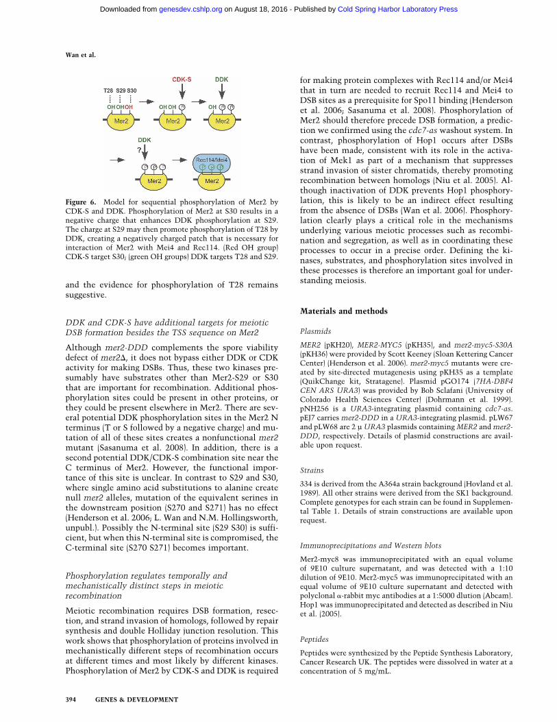
and the evidence for phosphorylation of T28 remainssuggestive.
DDK and CDK-S have additional targets for meioticDSB formation besides the TSS sequence on Mer2
Although mer2-DDD complements the spore viabilitydefect of mer2�, it does not bypass either DDK or CDKactivity for making DSBs. Thus, these two kinases pre-sumably have substrates other than Mer2-S29 or S30that are important for recombination. Additional phos-phorylation sites could be present in other proteins, orthey could be present elsewhere in Mer2. There are sev-eral potential DDK phosphorylation sites in the Mer2 Nterminus (T or S followed by a negative charge) and mu-tation of all of these sites creates a nonfunctional mer2mutant (Sasanuma et al. 2008). In addition, there is asecond potential DDK/CDK-S combination site near theC terminus of Mer2. However, the functional impor-tance of this site is unclear. In contrast to S29 and S30,where single amino acid substitutions to alanine createnull mer2 alleles, mutation of the equivalent serines inthe downstream position (S270 and S271) has no effect(Henderson et al. 2006; L. Wan and N.M. Hollingsworth,unpubl.). Possibly the N-terminal site (S29 S30) is suffi-cient, but when this N-terminal site is compromised, theC-terminal site (S270 S271) becomes important.
Phosphorylation regulates temporally andmechanistically distinct steps in meioticrecombination
Meiotic recombination requires DSB formation, resec-tion, and strand invasion of homologs, followed by repairsynthesis and double Holliday junction resolution. Thiswork shows that phosphorylation of proteins involved inmechanistically different steps of recombination occursat different times and most likely by different kinases.Phosphorylation of Mer2 by CDK-S and DDK is required
for making protein complexes with Rec114 and/or Mei4that in turn are needed to recruit Rec114 and Mei4 toDSB sites as a prerequisite for Spo11 binding (Hendersonet al. 2006; Sasanuma et al. 2008). Phosphorylation ofMer2 should therefore precede DSB formation, a predic-tion we confirmed using the cdc7-as washout system. Incontrast, phosphorylation of Hop1 occurs after DSBshave been made, consistent with its role in the activa-tion of Mek1 as part of a mechanism that suppressesstrand invasion of sister chromatids, thereby promotingrecombination between homologs (Niu et al. 2005). Al-though inactivation of DDK prevents Hop1 phosphory-lation, this is likely to be an indirect effect resultingfrom the absence of DSBs (Wan et al. 2006). Phosphory-lation clearly plays a critical role in the mechanismsunderlying various meiotic processes such as recombi-nation and segregation, as well as in coordinating theseprocesses to occur in a precise order. Defining the ki-nases, substrates, and phosphorylation sites involved inthese processes is therefore an important goal for under-standing meiosis.
Materials and methods
Plasmids
MER2 (pKH20), MER2-MYC5 (pKH35), and mer2-myc5-S30A(pKH36) were provided by Scott Keeney (Sloan Kettering CancerCenter) (Henderson et al. 2006). mer2-myc5 mutants were cre-ated by site-directed mutagenesis using pKH35 as a template(QuikChange kit, Stratagene). Plasmid pGO174 (7HA-DBF4CEN ARS URA3) was provided by Bob Sclafani (University ofColorado Health Sciences Center) (Dohrmann et al. 1999).pNH256 is a URA3-integrating plasmid containing cdc7-as.pEJ7 carries mer2-DDD in a URA3-integrating plasmid. pLW67and pLW68 are 2 µ URA3 plasmids containing MER2 and mer2-DDD, respectively. Details of plasmid constructions are avail-able upon request.
Strains
334 is derived from the A364a strain background (Hovland et al.1989). All other strains were derived from the SK1 background.Complete genotypes for each strain can be found in Supplemen-tal Table 1. Details of strain constructions are available uponrequest.
Immunoprecipitations and Western blots
Mer2-myc8 was immunoprecipitated with an equal volumeof 9E10 culture supernatant, and was detected with a 1:10dilution of 9E10. Mer2-myc5 was immunoprecipitated with anequal volume of 9E10 culture supernatant and detected withpolyclonal �-rabbit myc antibodies at a 1:5000 dlution (Abcam).Hop1 was immunoprecipitated and detected as described in Niuet al. (2005).
Peptides
Peptides were synthesized by the Peptide Synthesis Laboratory,Cancer Research UK. The peptides were dissolved in water at aconcentration of 5 mg/mL.
Figure 6. Model for sequential phosphorylation of Mer2 byCDK-S and DDK. Phosphorylation of Mer2 at S30 results in anegative charge that enhances DDK phosphorylation at S29.The charge at S29 may then promote phosphorylation of T28 byDDK, creating a negatively charged patch that is necessary forinteraction of Mer2 with Mei4 and Rec114. (Red OH group)CDK-S target S30; (green OH groups) DDK targets T28 and S29.
Wan et al.
394 GENES & DEVELOPMENT
Cold Spring Harbor Laboratory Press on August 18, 2016 - Published by genesdev.cshlp.orgDownloaded from
Inhibitor
Stocks of 10 mM PP1 were synthesized at the Universityof California at San Francisco as described in Bishop et al.(1999).
In vitro kinase assays
For the peptide labeling experiment, 700 mL of log-phase cul-tures were grown in SD-ura (∼2 × 10⁹ cells), washed with 10 mLof cold lysis buffer (50 mM HEPES at pH 7.5, 100 mM Na ac-etate, 10% glycerol, 5 mM EDTA, 2 mM benzamide, 10 mMNaF, 1 mM PMSF,), and resuspended in 0.3 mL of lysis buffer in2 mL screw-cap microfuge tubes (Sarstedt). Zirconia-silica beads(0.5 mm; Biospec) were added to a volume of 1.2 mL and thecells were lysed using a FastPrep instrument (Qbio-gene) withfour 20-sec pulses at speed 4. After bead beating, 10% NP-40was added to a final concentration of 0.5% (25 µL). The tube waspunctured on the bottom with a 30.5-gauge needle and placedinto a 15-mL conical tube, and the lysate was collected by cen-trifugation for 30 sec with a tabletop centrifuge. One-hundredmicroliters of lysis buffer were added to the beads and the tubewas centrifuged again to collect the wash, which was pooledwith the initial lysate. The extract was cleared by spinning at16,000 rpm in an Eppendorf microfuge for 10 min. To try toremove biotin from the extract, 200 µL of a 1:1 slurry of strep-tavidin beads (Active Motif) were first washed four times with1 mL of lysis buffer and then added to the soluble extract, whichwas incubated for 15 min on ice with vortexing every 2 min.The beads were removed by two 4-min spins at 16,000 rpm. HAantibody was prebound to beads by incubating 60 µL of ProteinG beads (GE Healthcare) pre-equilibrated in lysis buffer with 2.5µL of 12CA5 antibodies (in ascites fluid) for 15 min on ice. Theantibody–bead complexes were washed four times with 1 mL oflysis buffer, resuspended with 60 µL of lysis buffer, and added tothe soluble extract to immunoprecipitate 7HA-Dbf4. Thesamples were incubated for 1.5 h at 4°C on a rotating platform.The immunoprecipitates were washed four times with 1 mL oflysis buffer and once with 1 mL of kinase buffer (25 mM HEPESat pH 7.5, 100 mM Na acetate, 10 mM MgCl2, 10% glycerol, 0.1mM DTT, 0.1 mM PMSF, 5 µM ATP) and resuspended in 60 µLof kinase buffer. Kinase reactions contained 10-µL beads fromthe immunoprecipitate, 1 µL of peptide (1 mg/mL), and 1 µL of32P-�-ATP (5000 Ci/mmol), and were performed for 30 min atroom temperature. The reactions were stopped by addition 6 µLof 7.5 M guanidine HCl. Twelve microliters were spotted ontoa 1-mm2 piece of SAM2 Biotin Capture Membrane (Promega).After sitting at room temperature for 2 min, the membranesquares were washed in bulk with 100 mL of 2 M NaCl (1 × 30sec, followed by 3 × 2 min), 100 mL of 2 M NaCl plus 1% H3PO4
(4 × 2 min), and 100 mL of water (2 × 30 sec), and air dried. Thenumber of radioactive counts on each membrane was then as-sayed using a scintillation counter.
CDK-S activity was assayed by purifying Clb5-Gst from cdc7-as sporulating cells incubated with and without 15 µM PP1 asdescribed in Wan et al. (2004). Kinase assays were performedusing the precipitates as described in Neiman and Herskowitz(1994) with the addition to Histone H1 to a final concentrationof 200 ng/µL. The proteins were fractionated on a 15% SDSpolyacrylamide gel, transferred to a nitrocellulose filter, andexposed to film. The blot was then probed with a 1:5000 dilu-tion of �-Gst antibodies.
Sporulation, time courses, and PP1 washout
Cells were sporulated at a density of 3 × 10⁷ cells per milliliterat 30°C. PP1 was washed out after 8 h in Spo medium as de-scribed in Wan et al. (2006).
Acknowledgments
We are grateful to Scott Keeney and Aaron Neiman for helpfuldiscussions and comments on the manuscript. In addition, ScottKeeney provided plasmids and shared unpublished results.Thanks to Bob Sclafani and Neta Dean for plasmids and anti-bodies, respectively, and Adam Rosebrock for computer help.Emily Job provided excellent technical support. N.M.H. is sup-ported by NIH grant GM50717, S.J.B. is supported by CancerResearch UK, K.M.S. is supported by AI44009, and B.F. is sup-ported by NIH grant GM64813.
References
Arora, C., Kee, K., Maleki, S., and Keeney, S. 2004. Antiviralprotein Ski8 is a direct partner of Spo11 in meiotic DNAbreak formation, independent of its cytoplasmic role in RNAmetabolism. Mol. Cell 13: 549–559.
Bell, S.P. and Dutta, A. 2002. DNA replication in eukaryoticcells. Annu. Rev. Biochem. 71: 333–374.
Benjamin, K.R., Zhang, C., Shokat, K.M., and Herskowitz, I.2003. Control of landmark events in meiosis by the CDKCdc28 and the meiosis-specific kinase Ime2. Genes & Dev.17: 1524–1539.
Bishop, D.K., Park, D., Xu, L., and Kleckner, N. 1992. DMC1: Ameiosis-specific yeast homolog of E. coli recA required forrecombination, synaptonemal complex formation and cellcycle progression. Cell 69: 439–456.
Bishop, A.C., Kung, C.-Y., Shah, K., Witucki, L., Shokat, K.M.,and Liu, Y. 1999. Generation of monospecific nanomolartyrosine kinase inhibitors via a chemical genetic approach. J.Am. Chem. Soc. 121: 627–631.
Borde, V., Goldman, A.S., and Lichten, M. 2000. Direct couplingbetween meiotic DNA replication and recombination initia-tion. Science 290: 806–809.
Bustos, V.H., Marin, O., Meggio, F., Cesaro, L., Allende, C.C.,Allende, J.E., and Pinna, L.A. 2005. Generation of proteinCk1� mutants which discriminate between canonical andnon-canonical substrates. Biochem. J. 391: 417–424.
Cho, W.H., Lee, Y.J., Kong, S.I., Hurwitz, J., and Lee, J.K. 2006.CDC7 kinase phosphorylates serine residues adjacent toacidic amino acids in the minichromosome maintenance 2protein. Proc. Natl. Acad. Sci. 103: 11521–11526.
Clifford, D.M., Stark, K.E., Gardner, K.E., Hoffmann-Benning,S., and Brush, G.S. 2005. Mechanistic insight into the Cdc28-related protein kinase Ime2 through analysis of replicationprotein A phosphorylation. Cell Cycle 4: 1826–1833.
Doble, B.W. and Woodgett, J.R. 2003. GSK-3: Tricks of the tradefor a multi-tasking kinase. J. Cell Sci. 116: 1175–1186.
Dohrmann, P.R., Oshiro, G., Tecklenburg, M., and Sclafani,R.A. 1999. RAD53 regulates DBF4 independently of check-point function in Saccharomyces cerevisiae. Genetics 151:965–977.
Elia, A.E.H., Rellos, P., Haire, L.F., Chao, J.W., Ivins, F.J., Ho-epker, K., Mohammad, D., Cantely, L.C., Smerdon, S.J., andYaffe, M.B. 2003. The molecular basis for phosphodependentsubstrate targeting and regulation of Plks by the Polo-Boxdomain. Cell 115: 83–95.
Forde, J.E. and Dale, T.C. 2007. Glycogen synthase kinase 3: Akey regulator of cellular fate. Cell. Mol. Life Sci. 64: 1930–1944.
Hardy, C.F. and Pautz, A. 1996. A novel role for Cdc5p in DNAreplication. Mol. Cell. Biol. 16: 6774–6782.
Hardy, C.F., Dryga, O., Seematter, S., Pahl, P.M., and Sclafani,R.A. 1997. mcm5/cdc46-bob1 bypasses the requirement forthe S phase activator Cdc7p. Proc. Natl. Acad. Sci. 94: 3151–
CDK-S/DDK phosphorylation of Mer2
GENES & DEVELOPMENT 395
Cold Spring Harbor Laboratory Press on August 18, 2016 - Published by genesdev.cshlp.orgDownloaded from
3155.Henderson, K.A., Kee, K., Maleki, S., Santini, P., and Keeney, S.
2006. Cyclin-dependent kinase directly regulates initiationof meiotic recombination. Cell 125: 1321–1332.
Hochwagen, A. and Amon, A. 2006. Checking your breaks: Sur-veillance mechanisms of meiotic recombination. Curr. Biol.16: R217–R228. doi: 10.1016/j.cub.2006.03.009.
Hochwagen, A., Tham, W.H., Brar, G.A., and Amon, A. 2005.The FK506 binding protein Fpr3 counteracts protein phos-phatase 1 to maintain meiotic recombination checkpoint ac-tivity. Cell 122: 861–873.
Hollingsworth Jr., R.E. and Sclafani, R.A. 1993. Yeast pre-meiotic DNA replication utilizes mitotic origin ARS1independently of CDC7 function. Chromosoma 102: 415–420.
Hovland, P., Flick, J., Johnston, M., and Sclafani, R.A. 1989.Galactose as a gratuitous inducer of GAL gene expression inyeast growing on glucose. Gene 83: 57–64.
Jiao, K., Salem, L., and Malone, R. 2003. Support for a meioticrecombination initiation complex: Interactions amongRec102p, Rec104p and Spo11p. Mol. Cell. Biol. 23: 5928–5938.
Keeney, S. 2001. Mechanism and control of meiotic recombina-tion initiation. Curr. Top. Dev. Biol. 52: 1–53.
Kitada, K., Johnson, A.L., Johnston, L.H., and Sugino, A. 1993.A multicopy suppressor gene of the Saccharomyces cere-visiae G1 cell cycle mutant gene dbf4 encodes a proteinkinase and is identified as CDC5. Mol. Cell. Biol. 13: 4445–4457.
Kockeritz, L., Doble, B., Patel, S., and Woodgett, J.R. 2006. Gly-cogen synthase kinase-3—An overview of an over-achievingprotein kinase. Curr. Drug Targets 7: 1377–1388.
Li, J., Hooker, G.W., and Roeder, G.S. 2006. Saccharomycescerevisiae Mer2, Mei4 and Rec114 form a complex requiredfor meiotic double-strand break formation. Genetics 173:1969–1981.
Maleki, S., Neale, M.J., Arora, C., Henderson, K.A., and Keeney,S. 2007. Interactions between Mei4, Rec114, and other pro-teins required for meiotic DNA double-strand break forma-tion in Saccharomyces cerevisiae. Chromosoma 116: 471–486.
Masai, H. and Arai, K. 2002. Cdc7 kinase complex: A key regu-lator in the initiation of DNA replication. J. Cell. Physiol.190: 287–296.
Masai, H., Matsui, E., You, Z., Ishimi, Y., Tamai, K., and Arai,K. 2000. Human Cdc7-related kinase complex. In vitro phos-phorylation of MCM by concerted actions of Cdks and Cdc7and that of a criticial threonine residue of Cdc7 by Cdks. J.Biol. Chem. 275: 29042–29052.
Masai, H., Taniyama, C., Ogino, K., Matsui, E., Kakusho, N.,Matsumoto, S., Kim, J.M., Ishii, A., Tanaka, T., Kobayashi,T., et al. 2006. Phosphorylation of MCM4 by Cdc7 kinasefacilitates its interaction with Cdc45 on the chromatin. J.Biol. Chem. 281: 39249–39261.
McKee, A.H.Z. and Kleckner, N. 1997. A general method foridentifying recessive diploid-specific mutations in Sac-charomyces cerevisiae, its application to the isolation ofmutants blocked at intermediate stages of meiotic prophaseand characterization of a new gene SAE2. Genetics 146: 797–816.
Montagnoli, A., Valsasina, B., Brotherton, D., Troiani, S.,Rainoldi, S., Tenca, P., Molinari, A., and Santocanale,C. 2006. Identification of Mcm2 phosphorylation sitesby S-phase-regulating kinases. J. Biol. Chem. 281: 10281–10290.
Murakami, H. and Nurse, P. 2001. Regulation of premeiotic
S phase and recombination-related double-strand DNAbreaks during meiosis in fission yeast. Nat. Genet. 28: 290–293.
Neiman, A.M. and Herskowitz, I. 1994. Reconstitution of ayeast protein kinase cascade in vitro: Activation of the yeastMEK homolog STE7 by STE11. Proc. Natl. Acad. Sci. 91:3398–3402.
Niu, H., Wan, L., Baumgartner, B., Schaefer, D., Loidl, J., andHollingsworth, N.M. 2005. Partner choice during meiosis isregulated by Hop1-promoted dimerization of Mek1. Mol.Biol. Cell 16: 5804–5818.
Nougarede, R., Della Seta, F., Zarzov, P., and Schwob, E. 2000.Hierarchy of S-phase-promoting factors: Yeast Dbf4–Cdc7kinase requires prior S-phase cyclin-dependent kinase acti-vation. Mol. Cell. Biol. 20: 3795–3806.
Pecina, A., Smith, K.N., Mezard, C., Murakami, H., Otha, K.,and Nicolas, A. 2002. Target stimulation of meiotic recom-bination. Cell 111: 173–184.
Petes, T.D. 2001. Meiotic recombination hot spots and coldspots. Nat. Rev. Genet. 2: 360–369.
Petronczki, M., Siomos, M.F., and Nasmyth, K. 2003. Un me-nage a quatre: The molecular biology of chromosome segre-gation in meiosis. Cell 112: 423–440.
Prinz, S., Amon, A., and Klein, F. 1997. Isolation of COM1, anew gene required to complete meiotic double-strand in-duced recombination in Saccharomyces cerevisiae. Genetics146: 781–795.
Sasanuma, H., Hirota, K., Fukuda, T., Kakusho, N., Kugou, K.,Kawasaki, Y., Shibata, T., Masai, H., and Ohta, K. 2008.Cdc7-dependent phosphorylation of Mer2 facilitates initia-tion of yeast meiotic recombination. Genes & Dev. (thisissue), doi: 10.1101/gad.1626608.
Schild, D. and Byers, B. 1978. Meiotic effects of DNA-defectivecell division cycle mutations of Saccharomyces cerevisiae.Chromosoma 70: 109–130.
Schwacha, A. and Kleckner, N. 1997. Interhomolog bias duringmeiotic recombination: Meiotic functions promote a highlydifferentiated interhomolog-only pathway. Cell 90: 1123–1135.
Sclafani, R.A. 2000. Cdc7p–Dbf4p becomes famous in the cellcycle. J. Cell Sci. 113: 2111–2117.
Shuster, E.O. and Byers, B. 1989. Pachytene arrest and othermeiotic effects of the start mutations in Saccharomyces cer-evisiae. Genetics 123: 29–43.
Smith, K.N., Penkner, A., Ohta, K., Klein, F., and Nicolas, A.2001. B-type cyclins CLB5 and CLB6 control the initiation ofrecombination and synaptonemal complex formation inyeast meiosis. Curr. Biol. 11: 88–97.
Sopko, R., Raithatha, S., and Stuart, D. 2002. Phosphorylationand maximal activity of Saccharomyces cerevisiae meiosis-specific transcription factor Ndt80 is dependent on Ime2.Mol. Cell. Biol. 22: 7024–7040.
Strickfaden, S.C., Winters, M.J., Ben-Ari, G., Lamson, R.E.,Tyers, M., and Pryciak, P.M. 2007. A mechanism for cell-cycle regulation of MAP kinase signalling in a yeast differ-entiation pathway. Cell 128: 519–531.
Stuart, D. and Wittenberg, C. 1998. CLB5 and CLB6 are requiredfor premeiotic DNA replication and activation of the mei-otic S/M checkpoint. Genes & Dev. 12: 2698–2710.
Tanaka, S., Umemori, T., Hirai, K., Muramatsu, S., Kamimura,Y., and Araki, H. 2007. CDK-dependent phosphorylation ofSld2 and Sld3 initiates DNA replication in budding yeast.Nature 445: 323–332.
Valentin, G., Schwob, E., and Della Seta, F. 2005. Dual role ofthe CDC7-regulatory protein DBF4 during yeast meiosis. J.Biol. Chem. 281: 2828–2834.
Wan et al.
396 GENES & DEVELOPMENT
Cold Spring Harbor Laboratory Press on August 18, 2016 - Published by genesdev.cshlp.orgDownloaded from
Wan, L., de los Santos, T., Zhang, C., Shokat, K., and Holling-sworth, N.M. 2004. Mek1 kinase activity functions down-stream of RED1 in the regulation of meiotic DSB repair inbudding yeast. Mol. Biol. Cell 15: 11–23.
Wan, L., Zhang, C., Shokat, K.M., and Hollingsworth, N.M.2006. Chemical inactivation of Cdc7 kinase in budding yeastresults in a reversible arrest that allows efficient cell syn-chronization prior to meiotic recombination. Genetics 174:1767–1774.
Wolfe, K.H. 2006. Comparative genomics and genome evolu-tion in yeasts. Philos. Trans. R. Soc. Lond. B Biol. Sci. 361:403–412.
Wu, T.C. and Lichten, M. 1994. Meiosis-induced double-strandbreak sites determined by yeast chromatin structure. Sci-ence 263: 515–518.
Zegerman, P. and Diffley, J.F. 2007. Phosphorylation of Sld2 andSld3 by cyclin-dependent kinases promotes DNA replicationin budding yeast. Nature 445: 281–285.
CDK-S/DDK phosphorylation of Mer2
GENES & DEVELOPMENT 397
Cold Spring Harbor Laboratory Press on August 18, 2016 - Published by genesdev.cshlp.orgDownloaded from
10.1101/gad.1626408Access the most recent version at doi: 2008 22: 386-397 Genes Dev.
Lihong Wan, Hengyao Niu, Bruce Futcher, et al. meiotic recombination in yeast
Dbf4 (DDK) collaborate to initiate−Clb5 (CDK-S) and Cdc7−Cdc28
Material
Supplemental
http://genesdev.cshlp.org/content/suppl/2008/01/16/22.3.386.DC1.html
References
http://genesdev.cshlp.org/content/22/3/386.full.html#related-urls
Articles cited in:
http://genesdev.cshlp.org/content/22/3/386.full.html#ref-list-1This article cites 55 articles, 26 of which can be accessed free at:
Related Content
Genes Dev. February 1, 2008 22: 398-410Hiroyuki Sasanuma, Kouji Hirota, Tomoyuki Fukuda, et al.facilitates initiation of yeast meiotic recombination
Cdc7-dependent phosphorylation of Mer2 Genes Dev. February 1, 2008 22: 286-292Hajime Murakami and Scott KeeneyRegulating the formation of DNA double-strand breaks in meiosis
ServiceEmail Alerting
click here.right corner of the article orReceive free email alerts when new articles cite this article - sign up in the box at the top
http://genesdev.cshlp.org/subscriptionsgo to: Genes & Development To subscribe to
Copyright © 2008, Cold Spring Harbor Laboratory Press
Cold Spring Harbor Laboratory Press on August 18, 2016 - Published by genesdev.cshlp.orgDownloaded from