antineoplastic activity of povidone-iodine on different mesothelioma cell lines: results of in vitro...
TRANSCRIPT
Antineoplastic activity of povidone-iodine on differentmesothelioma cell lines: results of in vitro study†
Alfonso Fiorellia, Francesca Pentimallib, Vittorio D’Ursob, Domenico Di Marzob, Iris Maria Forteb,
Antonio Giordanob,c, Marina Di Domenicod, Marina Accardoe, Umberto Di Serioa and Mario Santinia,*
a Thoracic Surgery Unit, Second University of Naples, Naples, Italyb INT-CROM, ‘Pascale Foundation’ National Cancer Institute - Cancer Research Center, Mercogliano, Italyc Sbarro Institute for Cancer Research and Molecular Medicine, Center for Biotechnology, Temple University, Philadelphia, PA, USAd Department of Pathology, Second University of Naples, Naples, Italye Department of Morphopathology, Second University of Naples, Naples, Italy
* Corresponding author. Chirurgia Toracica, Seconda Università di Napoli, Piazza Miraglia 2, I-80138 Naples, Italy. Tel: +39-081-5665228; fax: +39-081-5665230;e-mail: [email protected] (M. Santini).
Received 12 June 2013; received in revised form 25 September 2013; accepted 30 September 2013
Abstract
OBJECTIVE: Povidone-iodine (PVP-I) or Betadine, owing to its antineoplastic activity, is also used as an adjuvant during intra-abdominal orintrathoracic surgery. However, the protocol of PVP-I administration has not been optimized to achieve the best antitumoural efficacy. Weaimed to determine the optimal concentration of PVP-I, the time of incubation and the mechanism of cell death by analysing the effect ofdifferent doses and time of administration of PVP-I on the cell viability of different mesothelioma cell lines.
METHODS: Four different cell lines (MET 5A/normal mesothelium; H2052/sarcomatoid mesothelioma; ISTMES2/epithelial mesothelioma;MSTO/biphasic mesothelioma) were incubated with increasing concentrations of diluted PVP-I (0.0001; 0.001; 0.01; 0.1; 1%) for 5, 10, 30,60 min and 24 h, respectively. Cell viability was determined using cell direct cytotoxicity assay and cell death was determined through flowcytometry assay analysis. The superoxide dismutase activity was assessed functionally through a specific inhibitor to evaluate the mechan-ism of cell death.
RESULTS: The antiproliferative effect of PVP-I varied largely among different cell lines in a dose- and time-dependent manner. At 0.1%concentration for 10 min of incubation, the percentage of viable cells was 0.5 ± 0.1; 0.8 ± 0.5 and 0% (P < 0.01) for MET5A, ISTMES2 andMSTO, respectively. Conversely, the same concentration did not significantly affect the H2052 cell line which was completely suppressedat a 1% concentration of PVP-I. Double staining of Annexin V and DNA showed that PVP-I induced cell death in all four cell lines via necro-sis depending on PVP-I concentration. However, H2052 was found to be more resistant than MSTO, ISTMES2 and MET 5A cells lines. Theactivity of superoxide dismutase was significantly inhibited in all cell lines.
CONCLUSIONS: Our results confirmed the anti-neoplastic activity of PVP-I especially on ISTMES2 and MSTO cell lines. With respectto chemotherapy pleural irrigation, washing with PVP-I is cost-effective and easy. If confirmed by larger studies, our findings suggest thatthe intrapleural irrigation with PVP-I (0.1% concentration for 10 min) in patients with epithelial or biphasic mesothelioma undergoingcytoreductive surgery might be applied in thoracic surgery practice to prevent neoplastic cell growth.
Keywords: Povidone-iodide • Betadine •Mesothelioma
INTRODUCTION
Malignant pleural mesothelioma (MPM) is a rare and aggressivethoracic malignancy associated with poor prognosis and predictedto increase in incidence in the next decades. Patients with MPMwho are not treated surgically survive at the most 12 months ifthey are offered the best palliative treatment [1]. A standard treat-ment protocol for MPM has not yet been developed, and in
particular, the role of surgery including extrapleural pneumonec-tomy (EPP) or radical decortication, as an alternative to EPP inunfit patients, still remains controversial.Despite attempting different multimodality therapies including
surgery, MPM recurs in most patients because the mostly diffusemalignant infiltration of the surrounding tissue strictly preventsa radical resection with histologically free resection margins.Thus, other treatments such as intrathoracic perfusion chemo-therapy alone or associated with hyperthermic perfusion havebeen applied after cytoreductive surgery with good results [2, 3].However, such techniques are not available in all hospitals, the
†Presented at the 21st European Conference on General Thoracic Surgery,Birmingham, UK, 26–29 May 2013.
© The Author 2014. Published by Oxford University Press on behalf of the European Association for Cardio-Thoracic Surgery. All rights reserved.
BASICSC
IENCE
European Journal of Cardio-Thoracic Surgery (2014) 1–8 ORIGINAL ARTICLEdoi:10.1093/ejcts/ezt534
European Journal of Cardio-Thoracic Surgery Advance Access published January 6, 2014
set-up is difficult and they may be associated with significantmorbidity.
Povidone-iodine (PVP-I) or Betadine, a combination of molecu-lar iodine and polyvinylpyrrolidone, has replaced the solution(tincture) of iodine widely used in clinical practice [4]. PVP-I hasbeen used in hand disinfection, skin preparations and antisepticirrigation for many years owing to the effective and wide anti-microbial activity [5]. In the last years, several studies have alsoaddressed its use as a sclerosing agent to promote pleurodesisin recurrent malignant pleural effusion or to prevent tumourseeding at the time of abdominal or thoracic surgery [6–8]. Yet, itsantitumour effects on breast cancer and MPM cells have alsobeen demonstrated in vitro [9, 10]. The aim of the present studywas to perform a comparative analysis of cell death induction byPVP-I in different cell lines of MPM focusing on dose and time ofincubation dependency and the mechanism of cell death.
MATERIALS ANDMETHODS
Study design
To investigate the effects on proliferation and cell death of dilutedPVP-I on MPM, four different cell lines (MET 5A/normal meso-thelium; H2052/sarcomatoid mesothelioma; ISTMES2/epithelialmesothelioma; MSTO/biphasic mesothelioma) were incubatedwith increasing doses of PVP-I diluted in fresh medium (0.0001;0.001; 0.01; 0.1; 1%) starting from a 10% PVP-I solution in distilledwater, for different times (5, 10, 30, 60 min and 24 h). Cytotoxicityassay was used to examine the proliferation; flow cytometry wasperformed to detect the apoptotic rate; superoxide dismutase(SOD) activity was assessed functionally through a specific inhibi-tor to evaluate the mechanism of cell death.
The aim was to establish the time of incubation and the con-centration of PVP-I that allowed a complete cell killing in order toevaluate its potential application in clinical practice.
Cell line and culture conditions
Four commercially available MPM cell lines were used in thepresent study: MET5A (normal mesothelium); H2052 (sarcomatoidmesothelioma); MSTO (biphasic mesothelioma), purchased fromthe American Type Culture Collection, and ISTMES2 (epithelialmesothelioma) from the ISTGE cell repository (www.iclc.it). All celllines were cultured as recommended by the providers in a hu-midified incubator at 37°C and 5% CO2.
Cell treatment with povidone-iodine and MTSassay
The cytotoxicity of PVP-I in all four cell lines was determined byMTS assay (CellTiter 96® Aqueous One Solution Cell ProliferationAssay, Promega). Compactly, MET5A/H2052/ISTMES2/MSTO(1.0 × 104 cells/well) were seeded in a 96-well plate with freshmedium and incubated overnight. MTS solution was added intoeach well at the indicated time points (5, 10, 30, 60 min, and for24 h) following treatment with five different concentrations ofPVP-I diluted in fresh medium (0.0001; 0.001; 0.01; 0.1; 1%). As a
control, cells were cultured in fresh medium added with themaximum amount of water used to vehicle the higher drug con-centration (1%). Cell viability was evaluated at different timepoints following the manufacturer’s instructions, through spectro-photometric reading at two different wavelengths (540 and 630nm). All experiments were repeated with at least three consecutivepassages.
Apoptosis analysis through flow cytometry
At the indicated incubation time, floating cells were collected to-gether with the supernatant and adherent cells were harvestedby trypsinization. Apoptosis was evaluated using Annexin V-FITCKit (Miltenyi Biotec, Inc., USA). Cells were analysed immediatelyusing a fluorescence activated cell sorting (FACS) flow cytometer(FACS Calibur; BD Biosciences, Heidelberg, Germany) for AnnexinV-FITC and Propidium iodide (PI) staining. For each measurement,20 000 cells were counted. Dot plots and histograms were ana-lysed by CellQuest Pro software (BD Biosciences). AnnexinV-positive cells were considered in the early stage of apoptosis(lower right quadrant); Annexin V- and PI-positive cells were con-sidered in the late stage of apoptosis or necrotic (upper rightquadrant). Annexin V- and PI-negative cells correspond to theviable cell fraction (lower left quadrant) [11].
Analysis of superoxide dismutase activity
To study one of the probable mechanisms of PVP-I-induced celldeath in tumour cells, the activity of SOD was detected with a col-orimetric method [12]. MET5A, ISTMES2, MSTO and H2052 celllines (1.5 × 10⁵ cells/ml) were seeded in a 6-well plate and incu-bated for 24 h. Cells were then treated with five different con-centrations of PVP-I (0.0001; 0.001; 0.01; 0.1; 1%) and incubatedfor 24 h. Next, cells were harvested and processed accordingto the instruction of the SOD kit (Nanjing Jiancheng Biotech-nology Institute SOD kit (detect total), Nanjing, China). The activ-ity of SOD was assessed by an ultraviolet spectrophotometer(SmartSpec 3000, Bio-Rad, Philadelphia, USA).
Statistical analysis
Data resulting from MTS and SOD activity experiments areexpressed as means ± standard deviation (SD) of three independ-ent experiments, each performed in triplicate. Statistically sig-nificant differences among the means of multiple groupswere evaluated by one-way repeated-measures ANOVA, withBonferroni post-test to compare pairs of data. The MedCalc statis-tical software (Version 12.3, Broekstraat 52; 9030 Mariakerke;Belgium) was used for the analysis. A P value of < 0.05 was consid-ered statistically significant.
RESULTS
Povidone-iodine effect on mesothelioma cellgrowth
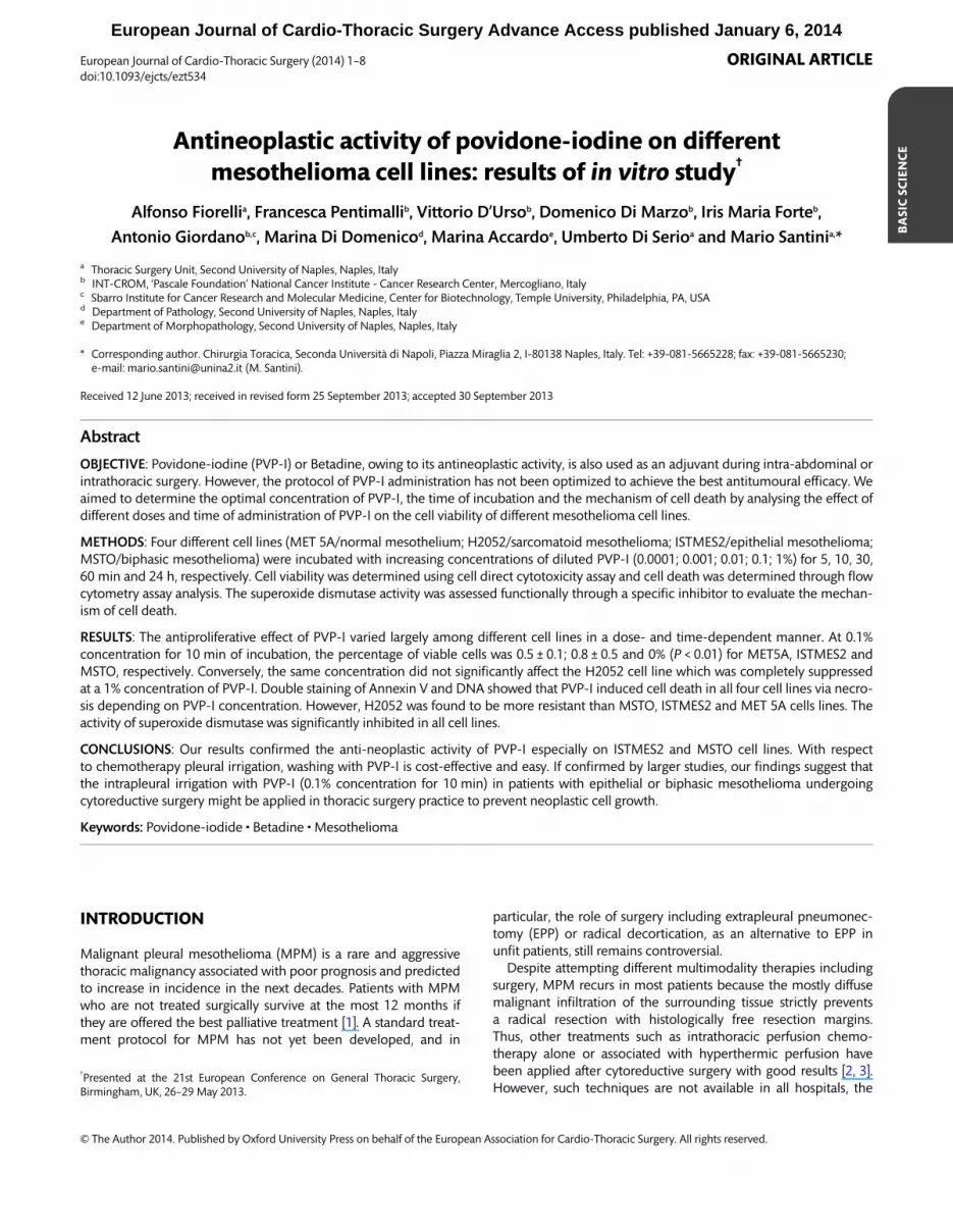
The effect of various incubations with different doses of PVP-Idoses on mesothelioma cell growth is represented in Fig. 1 (A for
A. Fiorelli et al. / European Journal of Cardio-Thoracic Surgery2
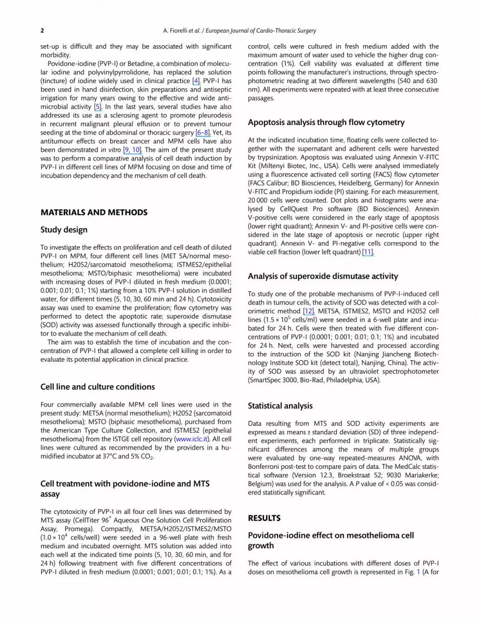
MET5A, B for ISTMES2, C for MSTO and D for H2052). MET5A,ISTMES2 and MSTO cell lines presented sensitivities similar to thatof PVP-I. The shortest time period necessary to observe a strongcytotoxic effect (cell viability <1%) on MET5A, ISTMES2 and MSTOcells was 10 min with a concentration of 0.1%. At a concentrationof 0.1% and after 10 min of incubation, the percentage of cell via-bility was significantly decreased with respect to that observedafter 5 min of incubation: 0.5 ± 0.1 vs 5.2 ± 0.8%, P < 0.05, respect-ively, for MET5A (Fig. 2A); 0.8 ± 0.5 vs 5.3 ± 0.5%, P < 0.05, respect-ively, for ISTMES2 (Fig. 2B); 0 vs 2.1 ± 0.1%, P < 0.05, respectively,for MSTO (Fig. 2C). Among all three different cell lines (MET5A,ISTMES2 and MSTO), no significant differences in cell viabilitywere found after longer incubation times at 0.1% concentration(Fig. 2D for MET5A, Fig. 2E for ISTMES2 and Fig. 2F for MSTO)and/or using a higher concentration.
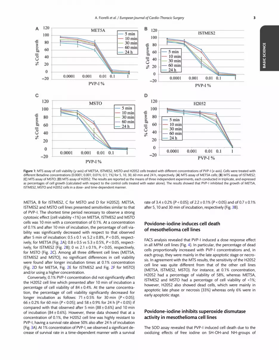
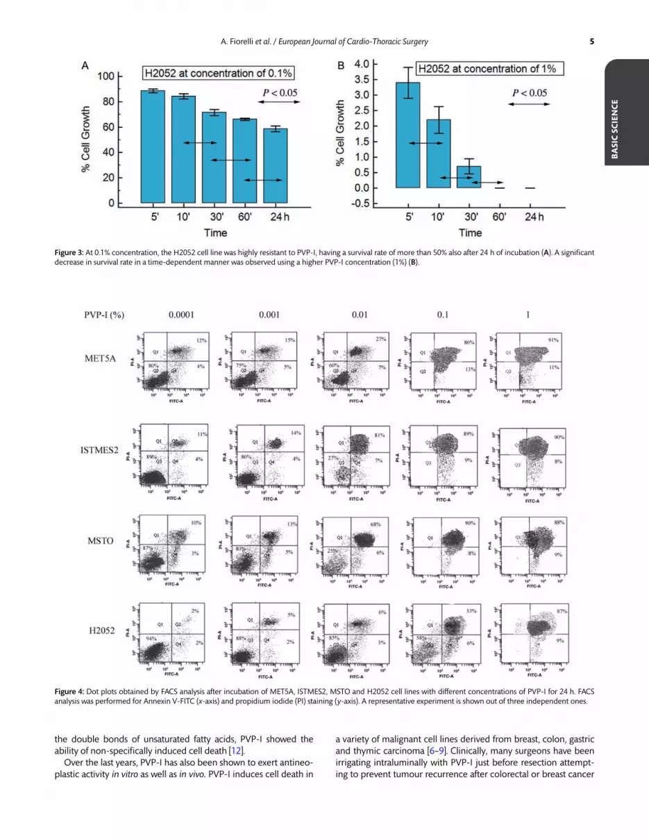
Conversely, 0.1% PVP-I concentration did not significantly affectthe H2052 cell line which presented after 10 min of incubation apercentage of cell viability of 84 ± 0.4%. At the same concentra-tion, the percentage of cell viability significantly decreased forlonger incubation as follows: 71 ± 0.5% for 30 min (P < 0.05);66 ± 0.2% for 60 min (P < 0.05); and 58 ± 0.9% for 24 h (P < 0.05) ifcompared with that observed after 5 min (88 ± 0.6%) and 10 minof incubation (84 ± 0.6%). However, these data showed that at aconcentration of 0.1%, the H2052 cell line was highly resistant toPVP-I, having a survival rate above 50% also after 24 h of incubation(Fig. 3A). At 1% concentration of PVP-I, we observed a significant de-crease of survival rate in a time-dependent manner with a survival
rate of 3.4 ± 0.2% (P < 0.05); of 2.2 ± 0.1% (P < 0.05) and of 0.7 ± 0.1%after 5, 10 and 30 min of incubation, respectively (Fig. 3B).
Povidone-iodine induces cell deathof mesothelioma cell lines
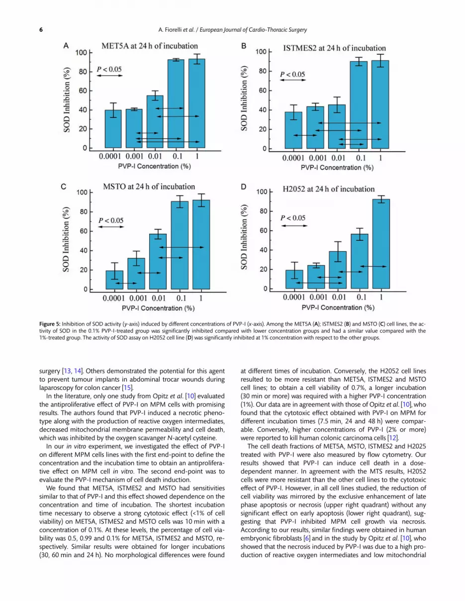
FACS analysis revealed that PVP-I induced a dose response effectin all MPM cell lines (Fig. 4). In particular, the percentage of deadcells proportionally increased with PVP-I concentrations and, ineach group, they were mainly in the late apoptotic stage or necro-sis. In agreement with the MTS results, the sensitivity of the H2052cell line was quite different from that of the other cell lines(MET5A, ISTMES2, MSTO). For instance, at 0.1% concentration,H2052 had a percentage of viability of 58%, whereas MET5A,ISTMES2 and MSTO had a percentage of cell viability of <1%;however, H2052 also showed dead cells, which were mainly inapoptotic late phase or necrosis (33%) whereas only 6% were inearly apoptotic stage.
Povidone-iodine inhibits superoxide dismutaseactivity in mesothelioma cell lines
The SOD assay revealed that PVP-I induced cell death due to theoxidizing effects of free iodine on SH-OH-and NH-groups of
Figure 1: MTS assay of cell viability (y-axis) of MET5A, ISTMES2, MSTO and H2052 cells treated with different concentrations of PVP-I (x-axis). Cells were treated withdifferent Betadine concentrations (0.0001; 0.001; 0.01%; 0.1; 1%) for 5, 10, 30, 60 min and 24 h, respectively. (A) MTS assay of MET5A cells; (B) MTS assay of ISTMES2;(C) MTS assay of MSTO; (D) MTS assay of H2052. The results are reported as the means of three independent experiments, each conducted in triplicate, and expressedas percentages of cell growth (calculated with respect to the control cells treated with water alone). The results showed that PVP-I inhibited the growth of MET5A,ISTMES2, MSTO and H2052 cells in a dose- and time-dependent manner.
BASICSC
IENCE
A. Fiorelli et al. / European Journal of Cardio-Thoracic Surgery 3
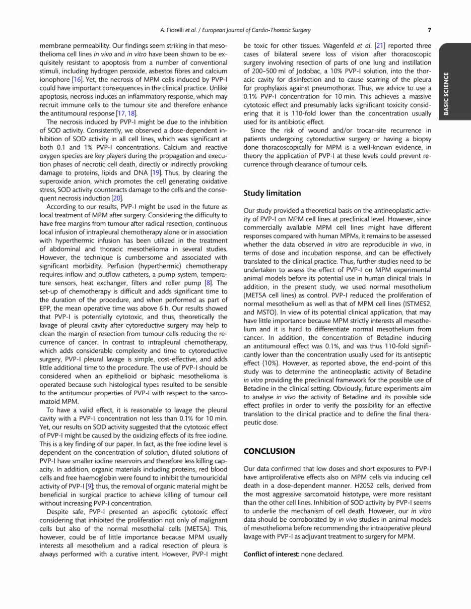
amino acids and on the double bonds of unsaturated fatty acids.Among all four cell lines, the activity of SOD was significantlyinhibited at a concentration of 0.1% with respect to lower concen-trations (P < 0.05). No significant difference was observed amongthe 0.1 and 1% groups for MET5A (Fig. 5A), ISTMES2 (Fig. 5B) andfor MSTO (Fig. 5C) cell lines; whereas a significant difference wasfound between 0.1 and 1% groups for H2052 cell lines (Fig. 5D).
DISCUSSION
PVP-I, a broad-spectrum microbicide against bacteria, virus, fungiand parasites in vitro, consists of elementary iodine bound to thecarrier poly(1-vinyl-2-pyrrolidone) that enhances the solubilityand provides a depot of iodine. Based on the oxidizing effects offree iodine on the NH-and SH-OH-groups of amino acids and on
Figure 2: After 10 min of incubation at a concentration of 0.1%, the percentage of cell viability was significantly decreased with respect to that observed using lowerconcentrations (0.0001; 0.001; 0.01%), whereas no significant difference was found for a higher concentration (1%) for MET5A (A); for ISTMES2 (B) and for MSTO (C).Among all three different cell lines, no significant differences were found after longer incubation times (D for MET5A, E for ISTMES2 and F for MSTO).
A. Fiorelli et al. / European Journal of Cardio-Thoracic Surgery4
the double bonds of unsaturated fatty acids, PVP-I showed theability of non-specifically induced cell death [12].
Over the last years, PVP-I has also been shown to exert antineo-plastic activity in vitro as well as in vivo. PVP-I induces cell death in
a variety of malignant cell lines derived from breast, colon, gastricand thymic carcinoma [6–9]. Clinically, many surgeons have beenirrigating intraluminally with PVP-I just before resection attempt-ing to prevent tumour recurrence after colorectal or breast cancer
Figure 4: Dot plots obtained by FACS analysis after incubation of MET5A, ISTMES2, MSTO and H2052 cell lines with different concentrations of PVP-I for 24 h. FACSanalysis was performed for Annexin V-FITC (x-axis) and propidium iodide (PI) staining (y-axis). A representative experiment is shown out of three independent ones.
Figure 3: At 0.1% concentration, the H2052 cell line was highly resistant to PVP-I, having a survival rate of more than 50% also after 24 h of incubation (A). A significantdecrease in survival rate in a time-dependent manner was observed using a higher PVP-I concentration (1%) (B).
BASICSC
IENCE
A. Fiorelli et al. / European Journal of Cardio-Thoracic Surgery 5
surgery [13, 14]. Others demonstrated the potential for this agentto prevent tumour implants in abdominal trocar wounds duringlaparoscopy for colon cancer [15].
In the literature, only one study from Opitz et al. [10] evaluatedthe antiproliferative effect of PVP-I on MPM cells with promisingresults. The authors found that PVP-I induced a necrotic pheno-type along with the production of reactive oxygen intermediates,decreased mitochondrial membrane permeability and cell death,which was inhibited by the oxygen scavanger N-acetyl cysteine.
In our in vitro experiment, we investigated the effect of PVP-Ion different MPM cells lines with the first end-point to define theconcentration and the incubation time to obtain an antiprolifera-tive effect on MPM cell in vitro. The second end-point was toevaluate the PVP-I mechanism of cell death induction.
We found that MET5A, ISTMES2 and MSTO had sensitivitiessimilar to that of PVP-I and this effect showed dependence on theconcentration and time of incubation. The shortest incubationtime necessary to observe a strong cytotoxic effect (<1% of cellviability) on MET5A, ISTMES2 and MSTO cells was 10 min with aconcentration of 0.1%. At these levels, the percentage of cell via-bility was 0.5, 0.99 and 0.1% for MET5A, ISTMES2 and MSTO, re-spectively. Similar results were obtained for longer incubations(30, 60 min and 24 h). No morphological differences were found
at different times of incubation. Conversely, the H2052 cell linesresulted to be more resistant than MET5A, ISTMES2 and MSTOcell lines; to obtain a cell viability of 0.7%, a longer incubation(30 min or more) was required with a higher PVP-I concentration(1%). Our data are in agreement with those of Opitz et al. [10], whofound that the cytotoxic effect obtained with PVP-I on MPM fordifferent incubation times (7.5 min, 24 and 48 h) were compar-able. Conversely, higher concentrations of PVP-I (2% or more)were reported to kill human colonic carcinoma cells [12].The cell death fractions of MET5A, MSTO, ISTMES2 and H2025
treated with PVP-I were also measured by flow cytometry. Ourresults showed that PVP-I can induce cell death in a dose-dependent manner. In agreement with the MTS results, H2052cells were more resistant than the other cell lines to the cytotoxiceffect of PVP-I. However, in all cell lines studied, the reduction ofcell viability was mirrored by the exclusive enhancement of latephase apoptosis or necrosis (upper right quadrant) without anysignificant effect on early apoptosis (lower right quadrant), sug-gesting that PVP-I inhibited MPM cell growth via necrosis.According to our results, similar findings were obtained in humanembryonic fibroblasts [6] and in the study by Opitz et al. [10], whoshowed that the necrosis induced by PVP-I was due to a high pro-duction of reactive oxygen intermediates and low mitochondrial
Figure 5: Inhibition of SOD activity (y-axis) induced by different concentrations of PVP-I (x-axis). Among the MET5A (A); ISTMES2 (B) and MSTO (C) cell lines, the ac-tivity of SOD in the 0.1% PVP-I-treated group was significantly inhibited compared with lower concentration groups and had a similar value compared with the1%-treated group. The activity of SOD assay on H2052 cell line (D) was significantly inhibited at 1% concentration with respect to the other groups.
A. Fiorelli et al. / European Journal of Cardio-Thoracic Surgery6
membrane permeability. Our findings seem striking in that meso-thelioma cell lines in vivo and in vitro have been shown to be ex-quisitely resistant to apoptosis from a number of conventionalstimuli, including hydrogen peroxide, asbestos fibres and calciumionophore [16]. Yet, the necrosis of MPM cells induced by PVP-Icould have important consequences in the clinical practice. Unlikeapoptosis, necrosis induces an inflammatory response, which mayrecruit immune cells to the tumour site and therefore enhancethe antitumoural response [17, 18].
The necrosis induced by PVP-I might be due to the inhibitionof SOD activity. Consistently, we observed a dose-dependent in-hibition of SOD activity in all cell lines, which was significant atboth 0.1 and 1% PVP-I concentrations. Calcium and reactiveoxygen species are key players during the propagation and execu-tion phases of necrotic cell death, directly or indirectly provokingdamage to proteins, lipids and DNA [19]. Thus, by clearing thesuperoxide anion, which promotes the cell generating oxidativestress, SOD activity counteracts damage to the cells and the conse-quent necrosis induction [20].
According to our results, PVP-I might be used in the future aslocal treatment of MPM after surgery. Considering the difficulty tohave free margins from tumour after radical resection, continuouslocal infusion of intrapleural chemotherapy alone or in associationwith hyperthermic infusion has been utilized in the treatmentof abdominal and thoracic mesothelioma in several studies.However, the technique is cumbersome and associated withsignificant morbidity. Perfusion (hyperthermic) chemotherapyrequires inflow and outflow catheters, a pump system, tempera-ture sensors, heat exchanger, filters and roller pump [8]. Theset-up of chemotherapy is difficult and adds significant time tothe duration of the procedure, and when performed as part ofEPP, the mean operative time was above 6 h. Our results showedthat PVP-I is potentially cytotoxic, and thus, theoretically thelavage of pleural cavity after cytoreductive surgery may help toclean the margin of resection from tumour cells reducing the re-currence of cancer. In contrast to intrapleural chemotherapy,which adds considerable complexity and time to cytoreductivesurgery, PVP-I pleural lavage is simple, cost-effective, and addslittle additional time to the procedure. The use of PVP-I should beconsidered when an epithelioid or biphasic mesothelioma isoperated because such histological types resulted to be sensibleto the antitumour properties of PVP-I with respect to the sarco-matoid MPM.
To have a valid effect, it is reasonable to lavage the pleuralcavity with a PVP-I concentration not less than 0.1% for 10 min.Yet, our results on SOD activity suggested that the cytotoxic effectof PVP-I might be caused by the oxidizing effects of its free iodine.This is a key finding of our paper. In fact, as the free iodine level isdependent on the concentration of solution, diluted solutions ofPVP-I have smaller iodine reservoirs and therefore less killing cap-acity. In addition, organic materials including proteins, red bloodcells and free haemoglobin were found to inhibit the tumouricidalactivity of PVP-I [9]; thus, the removal of organic material might bebeneficial in surgical practice to achieve killing of tumour cellwithout increasing PVP-I concentration.
Despite safe, PVP-I presented an aspecific cytotoxic effectconsidering that inhibited the proliferation not only of malignantcells but also of the normal mesothelial cells (MET5A). This,however, could be of little importance because MPM usuallyinterests all mesothelium and a radical resection of pleura isalways performed with a curative intent. However, PVP-I might
be toxic for other tissues. Wagenfeld et al. [21] reported threecases of bilateral severe loss of vision after thoracoscopicsurgery involving resection of parts of one lung and instillationof 200–500 ml of Jodobac, a 10% PVP-I solution, into the thor-acic cavity for disinfection and to cause scarring of the pleurafor prophylaxis against pneumothorax. Thus, we advice to use a0.1% PVP-I concentration for 10 min. This achieves a massivecytotoxic effect and presumably lacks significant toxicity consid-ering that it is 110-fold lower than the concentration usuallyused for its antibiotic effect.Since the risk of wound and/or trocar-site recurrence in
patients undergoing cytoreductive surgery or having a biopsydone thoracoscopically for MPM is a well-known evidence, intheory the application of PVP-I at these levels could prevent re-currence through clearance of tumour cells.
Study limitation
Our study provided a theoretical basis on the antineoplastic activ-ity of PVP-I on MPM cell lines at preclinical level. However, sincecommercially available MPM cell lines might have differentresponses compared with human MPMs, it remains to be assessedwhether the data observed in vitro are reproducible in vivo, interms of dose and incubation response, and can be effectivelytranslated to the clinical practice. Thus, further studies need to beundertaken to assess the effect of PVP-I on MPM experimentalanimal models before its potential use in human clinical trials. Inaddition, in the present study, we used normal mesothelium(MET5A cell lines) as control. PVP-I reduced the proliferation ofnormal mesothelium as well as that of MPM cell lines (ISTMES2,and MSTO). In view of its potential clinical application, that mayhave little importance because MPM strictly interests all mesothe-lium and it is hard to differentiate normal mesothelium fromcancer. In addition, the concentration of Betadine inducingan antitumoural effect was 0.1%, and was thus 110-fold signifi-cantly lower than the concentration usually used for its antisepticeffect (10%). However, as reported above, the end-point of thisstudy was to determine the antineoplastic activity of Betadinein vitro providing the preclinical framework for the possible use ofBetadine in the clinical setting. Obviously, future experiments aimto analyse in vivo the activity of Betadine and its possible sideeffect profiles in order to verify the possibility for an effectivetranslation to the clinical practice and to define the final thera-peutic dose.
CONCLUSION
Our data confirmed that low doses and short exposures to PVP-Ihave antiproliferative effects also on MPM cells via inducing celldeath in a dose-dependent manner. H2052 cells, derived fromthe most aggressive sarcomatoid histotype, were more resistantthan the other cell lines. Inhibition of SOD activity by PVP-I seemsto underlie the mechanism of cell death. However, our in vitrodata should be corroborated by in vivo studies in animal modelsof mesothelioma before recommending the intraoperative pleurallavage with PVP-I as adjuvant treatment to surgery for MPM.
Conflict of interest: none declared.
BASICSC
IENCE
A. Fiorelli et al. / European Journal of Cardio-Thoracic Surgery 7
REFERENCES
[1] Lindenmann J, Matzi V, Neuboeck N, Anegg U, Maier A, Smolle J et al.Multimodal therapy of malignant pleural mesothelioma: is the replace-ment of radical surgery imminent? Interact Cardiovasc Thorac Surg 2013;16:237–43.
[2] Ried M, Potzger T, Braune N, Neu R, Zausig Y, Schalke B et al.Cytoreductive surgery and hyperthermic intrathoracic chemotherapy per-fusion for malignant pleural tumours: perioperative management andclinical experience. Eur J Cardiothorac Surg 2013;43:801–7.
[3] Lee JD, Perez S, Wang HJ, Figlin RA, Holmes EC. Intrapleural chemotherapyfor patients with incompletely resected malignant mesothelioma: theUCLA experience. J Surg Oncol 1995;60:262–7.
[4] Durani P, Leaper D. Povidone-iodine: use in hand disinfection, skin prep-aration and antiseptic irrigation. Int Wound J 2008;5:376–87.
[5] Bryan JL, Cohran J, Larson EL. Hand washing: a ritual revisited. Crit CareNurs Clin North Am 1995;7:617–25.
[6] Wutzler P, Sauerbrei A, Klöcking R, Brögmann B, Reimer K. Virucidal activ-ity and cytotoxicity of the liposomal formulation of povidone-iodine.Antiviral Res 2002;54:89–97.
[7] Godazandeh G, Qasemi NH, Saghafi M, Mortazian M, Tayebi P.Pleurodesis with povidone-iodine, as an effective procedure in manage-ment of patients with malignant pleural effusion. J Thorac Dis 2013;5:141–4.
[8] Belcher E, Hardwick T, Lal R, Marshall S, Spicer J, Lang-Lazdunski L.Induction chemotherapy, cytoreductive surgery and intraoperative hyper-thermic pleural irrigation in patients with stage IVA thymoma. InteractCardiovasc Thorac Surg 2011;12:744–7.
[9] Basha G, Penninckx F, Yap P. Influence of blood components and faeceson the in vitro cancericidal activity of povidone-iodine. Br J Surg 1998;85:534–7.
[10] Opitz I, Sigrist B, Hillinger S, Lardinois D, Stahel R, Weder W et al.Taurolidine and povidone-iodine induce different types of cell death inmalignant pleural mesothelioma. Lung Cancer 2007;56:327–36.
[11] Chromik AM, Daigeler A, Bulut D, Flier A, May C, Harati K et al.Comparative analysis of cell death induction by Taurolidine in differentmalignant human cancer cell lines. J Exp Clin Cancer Res 2010;7;29:21.
[12] Sun P, Zhao JM, Luo ZC, Zhang P, Chen P, Zhang XL et al. Dilutedpovidone-iodine inhibits tumor growth through apoptosis-induction andsuppression of SOD activity. Oncol Rep 2012;27:383–8.
[13] Park KG, Chetty U, Scott W, Miller W. The activity of locally applied cyto-toxics to breast cancer cells in vitro. Ann R Coll Surg Engl 1991;73:96–9.
[14] Umpleby HC, Williamson RC. The efficacy of agents employed to preventanastomotic recurrence in colorectal carcinoma. Ann R Coll Surg Engl1984;66:192–4.
[15] Wu JS, Pfister SM, Ruiz MB, Connett JM, Fleshman JW. Local treatment ofabdominal wound reduces tumor implantation. J Surg Oncol 1998;69:9–13.
[16] Nici L, Monfils B, Calabresi P. The effects of taurolidine, a novel antineo-plastic agent, on human malignant mesothelioma. Clin Cancer Res 2004;10:7655–61.
[17] Grivennikov SI, Greten FR, Karin M. Immunity, inflammation, and cancer.Cell 2010;140:883–99.
[18] Fiorelli A, Ricciardi C, Pannone G, Santoro A, Bufo P, Santini M et al.Interplay between steroid receptors and neoplastic progression insarcoma tumors. J Cell Physiol 2011;226:2997–3003.
[19] Festjens N, Vanden Berghe T, Vandenabeele P. Necrosis, a well-orchestrated form of cell demise: signalling cascades, important mediators
and concomitant immune response. Biochim Biophys Acta 2006;1757:1371–87.
[20] Haddad JJ. Redox and oxidant-mediated regulation of apoptosis signalingpathways: immuno-pharmaco-redox conception of oxidative siege versuscell death commitment. Int Immunopharmacol 2004;4:475–93.
[21] Wagenfeld L, Zeitz O, Richard G. Visual loss after povidone-iodine pleur-odesis. N Engl J Med 2007;357:1264–5.
APPENDIX A. CONFERENCE DISCUSSION
Dr M. Hoda (Vienna, Austria): I just wondered if you looked at the mechanisticpart. Do you know how it works? How high is the toxicity? Did you look at thesignalling? Did you look at Western blot maybe, or are you planning to do thisto find out what the mode of action is, because it cannot only be necrotic orcytotoxic. It should have some sort of influence biologically.Dr Fiorelli: We did not perform Western blot in the laboratory. We used
FACS analysis to show if the cells are in the early stage of apoptosis or in necro-sis in the light of their position within quadrants.Dr Hoda: I just wonder if you can find out if it influences any signalling path-
ways, like the MAP kinase pathway or PI3 kinase pathway? There is recentlysome evidence that at least the PI3 kinase (because we published that actuallylast year) has a role, for example. So maybe there is other evidence for this,maybe cell signal pathways to look at the cell signalling.Dr Fiorelli: The only two SOD activity endpoints were cell viability and cell
death.Dr Hoda: Nice data anyway.Dr Z. Bilgi (Istanbul, Turkey): I have two questions. First of all, we have also
been using povidone-iodine in our clinic as pleural lavage after pleurectomy/decortication. Have you ever had the chance to observe this in suspended cellcultures? Do you have any idea how penetrant povidone-iodine is, forexample, how many cell-like layers?My second question is, while you are doing your viability assays, there is a
point, let’s say it is the LD 50 point, of the povidone-iodine, 50% of the cells arealive still in the culture: have you ever tried to propagate that population,like the affected but still not yet dead population, to see if there are anychanges in behaviour or changes in adherence or anything like that, somethingobservable?Dr Fiorelli: Regarding the penetration, as it is an in vitro study, we don’t have
a three-dimensional aspect; it is a planar model, and we know it is a limitationof this paper. So we were unable to evaluate the depth of penetration ofBetadine in the tissue.Dr Bilig: You didn’t use the molds. What about the propagation behaviour of
the cell culture, did you ever look at it?Dr Fiorelli: No, we didn’t.Dr T. Grodzki (Szczecin, Poland): I can add that it will probably work in the
clinical setting as well. As you may know, our centre is doing the so-calledWeder’s procedure in postpneumonectomy empyema. We fill the postpneu-monectomy cavity with Betadine mops for 48 hours twice, and after five yearswe observed, despite the healing process, the beneficial effect on the five-yearsurvival in propensity-matched groups. I suppose it is caused just by the im-munological response to chronic inflammation, but maybe povidone is add-itionally beneficial.Dr Fiorelli: We think that necrosis is important; it induces an inflammatory
response which may recruit immune cells to the site of the tumour and therebyenhance the anti-tumour response.
A. Fiorelli et al. / European Journal of Cardio-Thoracic Surgery8