anterior capsulotomy with a pulsed-electron avalanche knife
TRANSCRIPT

Anterior capsulotomy with a pulsed-electron avalanche knife
Daniel Palanker, PhD1, Hiroyuki Nomoto, MD, PhD1, Philip Huie, MA1, Alexander Vankov,PhD2, and David F. Chang, MD31 Department of Ophthalmology and Hansen Experimental Physics Laboratory, Stanford University,Stanford2 Peak Surgical, Inc, Palo Alto3 Private practice, Los Altos, California, USA
AbstractPURPOSE—To evaluate a new pulsed-electron avalanche knife (PEAK) design for creating acontinuous curvilinear capsulorhexis (CCC) and compare the CCC with a mechanical capsulorhexis.
SETTING—Department of Ophthalmology, Stanford University, Stanford, California, USA.
METHODS—In this study, CCCs were created in freshly enucleated bovine eyes and in rabbit eyesin vivo. The cutting velocity was adjusted by controlling the burst repetition rate, voltage amplitude,and burst duration. Tissue samples were fixed and processed for histology and scanning electronmicroscopy (SEM) immediately after surgery.
RESULTS—The study included 50 bovine eyes and 10 rabbit eyes. By adjusting the electrosurgicalwaveforms, gas-bubble formation was minimized to permit good surgical visualization. The optimumvoltage level was determined to be ±410 V with a burst duration of 20 μs. Burst repetition rate,continuously adjustable from 20 to 200 Hz with footpedal control, allowed the surgeon to vary linearcutting velocity up to 2.0 mm/second. Histology and SEM showed that the pulsed-electron avalancheknife produced sharp-edged capsule cutting without radial nicks or tears.
CONCLUSIONS—The pulsed-electron probe duplicated the surgical feel of a 25-gauge cystotomeand created a histologically smooth capsule cut.
The continuous curvilinear capsulorhexis (CCC) is one of the most important steps in cataractsurgery. A properly sized capsulorhexis enhances surgical safety, hydrodissection, corticalcleanup, and intraocular lens (IOL) centration and inhibits posterior capsule opacification.1–10 The introduction of newer refractive IOLs has increased the importance of consistentlyachieving a symmetrically round and properly sized capsulorhexis.
Corresponding author: Daniel Palanker, PhD, Stanford University, 452 Lomita Mall, Stanford, California 94305-4085, [email protected]. Palanker and Vankov hold patents to the pulsed electron avalanche knife technology, which are licensed to PEAK Surgical byStanford University. Drs. Palanker and Chang are consultants to Peak Surgical. Dr. Vankov is an employee of PEAK Surgical. Neitherof the other authors has a financial or proprietary interest in any material or method mentioned.Scott Taylor and Paul Davison, PEAK Surgical, Inc., help fabricate the capsulotomy probes. Roopa Dalal, Department of Ophthalmology,Stanford University, performed the histologic preparations and photography.Financial Disclosures: Drs. Palanker and Vankov hold patents to the pulsed electron avalanche knife technology, which are licensed toPEAK Surgical by Stanford University. Drs. Palanker and Chang are consultants to Peak Surgical. Dr. Vankov is an employee of PEAKSurgical. Neither of the other authors has a financial or proprietary interest in any material or method mentioned.Publisher's Disclaimer: This is a PDF file of an unedited manuscript that has been accepted for publication. As a service to our customerswe are providing this early version of the manuscript. The manuscript will undergo copyediting, typesetting, and review of the resultingproof before it is published in its final citable form. Please note that during the production process errors may be discovered which couldaffect the content, and all legal disclaimers that apply to the journal pertain.
NIH Public AccessAuthor ManuscriptJ Cataract Refract Surg. Author manuscript; available in PMC 2011 January 1.
Published in final edited form as:J Cataract Refract Surg. 2010 January ; 36(1): 127. doi:10.1016/j.jcrs.2009.07.046.
NIH
-PA Author Manuscript
NIH
-PA Author Manuscript
NIH
-PA Author Manuscript
Beginning resident surgeons usually rate the capsulorhexis as the most difficult step in cataractsurgery.11 However, even for experienced surgeons, achieving a properly sized capsulorhexisis more difficult in the presence of common surgical risk factors, including a small pupil, ashallow anterior chamber, weak zonules, pediatric eyes, and poor visibility or a poor red reflex.It is ironic, therefore, that the capsulorhexis is the rare step in cataract surgery that has not beenenhanced by technology. The maneuvers are still performed free hand, and the surgeon mustrely on visual clues, such as the diameter of the pupil and the cornea. A precise way to cut ortrace a capsulorhexis or to safely enlarge a small diameter capsulorhexis after IOL implantationwould be beneficial.
A pulsed-electron avalanche knife (PEAK-fc, Carl Zeiss Meditec) was initially designed forvitreoretinal surgery and has been successful in clinical testing.12,13 The system was developedfor “cold” and traction-free dissection of soft tissue14,15 We describe a modified system andprobe configuration that is optimized for creating CCCs and report the test results and histologicfindings in porcine and rabbit eyes.
MATERIALS AND METHODSThis study evaluated the PEAK anterior segment system (PEAK Surgical, Inc.) for creatingCCCs and compared the CCCs with those created mechanically. As opposed to conventionalradiofrequency electrosurgery, in which continuous waveforms are applied for tissue cutting,the system produces plasma-mediated discharges driven by radiofrequency bursts lastingseveral tenths of microseconds and are applied at a repetition rate of 10 to 1000 Hz.15,16
Because of the short duration of the radiofrequency bursts, heat diffusion into the surroundingtissue is limited to a distance of approximately 10 μm. This restricts thermal damage at theedge of the cut tissue to the cellular scale, in contrast to a much larger damage zone (typicallyhundreds of micrometers) produced by conventional electrosurgical devices. The duty cycleof the system’s waveform, defined as the ratio of the “power on” duration to the total applicationtime, typically does not exceed 1%. These low average power settings significantly reduce heatgeneration over that generated by conventional continuous radiofrequency instruments; thistechnology is referred to as cold cutting.
The cutting tip of the system’s capsulotomy probe is a protruding tungsten wire with a diameterof 75 μm (Figure 1). The shaft of the probe is identical to that of a 25-gauge needle (0.51 mmin diameter) and curved into a slight arc to accommodate the convexity of the lens capsule.The insulating material at the tip of the probe is transparent to allow visualization of the cuttingwire, which extends 250 to 300 μm from the insulator.
The cutting probe was used to create anterior CCCs in freshly enucleated bovine eyes, andafter initial optimization, in rabbit eyes in vivo. The corneal incisions were created using a 15-degree keratome (Alcon, Inc.), and the anterior chamber was kept inflated with sodiumhyaluronate 1.0% (Provisc).
Regarding the cutting parameters, the pulse repetition rate was adjusted continuously (range20 to 200 Hz) by the surgeon using a footpedal, the voltage amplitude was adjusted in the rangeof ±350 to ±450 V, and burst duration was 20 μs, or 40 cycles at a radiofrequency of 2 MHz.The cutting probe was slowly moved along the capsule in a circular manner with a velocity ofapproximately 1.0 mm/second to avoid tractional forces and minimize pressure on the lenscapsule. The cutting velocity was adjusted by controlling the pulse repetition rate with thefootpedal.
For light microscopy, specimens were fixed in 10% formaldehyde dehydrated with a gradedseries of ethanol and embedded in paraffin. Semithin sections (8 μm) were stained withhematoxylin–eosin. For scanning electron microscopy, treated eyes were fixed in 2.5%
Palanker et al. Page 2
J Cataract Refract Surg. Author manuscript; available in PMC 2011 January 1.
NIH
-PA Author Manuscript
NIH
-PA Author Manuscript
NIH
-PA Author Manuscript
glutaraldehyde in sodium cacodylate buffer (pH 7.4), postfixed in 1% osmium tetroxide,dehydrated in a series of methanols, and critical point dried. The samples were plasma coatedwith gold–palladium and evaluated with a scanning electron microscope.
RESULTSThe study included 50 bovine eyes and 10 rabbit eyes. During initial experimentation withporcine eyes, 2 parameters of the electrosurgical waveforms (voltage and burst repetition rate)were optimized for capsule cutting. After multiple experiments, an optimum voltage level of±410 V was determined. The optimum range of burst repetition rate was 20 to 200 Hz, with amaximum linear cutting velocity of approximately 2.0 mm/second. To allow surgeon controlof the cutting speed, the burst repetition rate was kept adjustable under footpedal control.
Figure 2, A, is a video frame showing a CCC nearing completion. Although the gas bubblesseen could not be eliminated, they were minimized by adjusting the electrosurgical waveformsuntil they no longer impaired the surgeon’s visualization. Depending on the cutting rate,capsulotomy completion took approximately 12 to 20 seconds in porcine eyes and up to 25seconds in rabbit eyes.
Figure 2, B, shows the typical histology of a porcine lens after CCC. The reduced osmolarityof the anterior chamber was likely responsible for the bulging of lens tissue through thecapsulotomy. Figure 3 shows a higher magnification histological comparison of anteriorporcine capsulotomy edges created by a forceps tear and edges after a probe cut. Bothtechniques resulted in a sharply cut edge to the capsule. After mechanical capsulorhexis, thecapsule was detached from the lens near the edge while after probe cutting, it was fully attachedto the lens cortex to the edge.
Figure 4 shows scanning electron microscopy (SEM) of the edge of a capsulorhexis that wasmechanically torn with a forceps. Figure 5 shows SEM of a probe capsulotomy. Both methodsproduced a sharp edge without radial nicks or tears. The cut face of the mechanically torncapsule edge was smoother on SEM than the probe-created edge because the cutting probe leftbehind residual microstructures.
DISCUSSIONCreating an anterior CCC has many intraoperative and postoperative advantages in cataractsurgery.1–10 A symmetric, properly sized capsulorhexis is even more important forimplantation of refractive IOLs, such as multifocal and accommodating models, for whichexcellent optic centration is critical. The effective axial lens position may also vary dependingon whether the capsulorhexis completely or incompletely overlaps the optic, thereby affectingaccuracy of the IOL power calculation. Due to their different designs, most accommodatingIOLs should not be implanted if the capsulorhexis is torn. For example, the Synchrony dual-optic accommodating IOL is a bag-filling lens designed to produce forward movement of theanterior optic during accommodative effort.17,18 This requires a completely overlappingcapsulorhexis to prevent partial prolapse of the anterior optic out of the bag.
Successfully creating a CCC is more difficult under challenging circumstances, such as poorcapsule visibility, a small pupil, a shallow anterior chamber, weak zonules, a thickened andfibrotic capsule, or an elastic pediatric anterior capsule. Particularly when it is difficult tocontrol the path of the flap, the ability to cut rather than tear the capsular margin would beadvantageous. Another challenge is rescuing the errant tear, which is typically due to one ofthe aforementioned risk factors. Again, the precise control of a cutting probe might allowsurgeons to better redirect the path of the tear. Finally, secondarily enlarging a smaller than
Palanker et al. Page 3
J Cataract Refract Surg. Author manuscript; available in PMC 2011 January 1.
NIH
-PA Author Manuscript
NIH
-PA Author Manuscript
NIH
-PA Author Manuscript

desired CCC would be advantageous once the IOL has been implanted. A precise and highlymaneuverable capsule-cutting instrument would facilitate this step.
Several other methods to allow surgeons to trace or draw a capsulorhexis have been tried.19–21 The high-frequency capsulotomy22 has been associated with a higher risk for capsule tearsand intraoperative complications than the conventional mechanical CCC.19,22 Histologicexamination of high-frequency capsulotomy cuts show thermal tissue damage that decreasesthe biomechanical stability of the anterior capsular edge.19,20,22 The vitrectorhexis, amechanized anterior capsulotomy technique combined with an IOL implantation in pediatriccataract surgery, was first described in the 1990s.23 In a retrospective analysis of 208 eyes,24
the incidence of radial tears was as high as 7.7%. This technique has not been used for adultanterior capsulotomy. The Fugo blade was introduced several years ago for resistance-freeincision of the anterior lens capsule.21,25 The instrument is bulkier and heavier than aconventional cystotome, which creates a substantial surgical learning curve. A higherpercentage of radial tears with this method were found in a clinical study comparing differentmethods of making a CCC.24 Although it is U.S. Food and Drug Administration approved,surgeons have not adopted the Fugo blade for creating the anterior capsulotomy to a significantdegree.
The original pulsed-electron avalanche knife system was used in a few human eyes to createa capsulotomy for cataract surgery26,27; however, the system’s retinal probe was not properlyconfigured for creating capsulotomy, and no histological evaluation of the cut capsule edgewas performed. In addition, the system’s power supply did not have continuous control of theburst repetition rate and the relatively low radio frequency (400 kHz) generated a noticeableamount of gas bubbles.26,27
We evaluated a modification of the pulsed-electron avalanche knife system; this new anteriorsegment system has an optimized probe shape, continuous footpedal control of cutting rate,and a higher radiofrequency (2 MHz) to reduce the amount of gas bubbles. The new probeconfiguration for anterior capsule cutting incorporates a lighter, more maneuverable handleand an intraocular design that emulates a bent 25-gauge cystotome or capsulotomy needle. Thismakes the tip easy to insert through a 1.0 mm paracentesis and easy to maneuver in the anteriorchamber.
Our study using the new pulsed-electron avalanche knife system showed that it consistentlycreated a histologically smooth continuous capsule edge without evidence of collateral thermaldamage or weakening nicks. In addition, the electrosurgical parameters were successfullyoptimized for cutting without excessive view-impairing bubbles.
In conclusion, the new pulsed-electron capsulotomy instrument has the potential to providecataract surgeons with a more precise and reproducible way to create a CCC. This mightimprove successful completion of this step in the face of surgical risk factors, such as weakzonules or poor visibility. Finally, better surgical control may improve proper capsulorhexissizing and centration, the consistent attainment of which will be a prerequisite for the nextgeneration of accommodating and refractive IOLs. The successful application and evaluationof CCC creation using the pulsed-electron probe in porcine cadaver eyes and in live rabbits,particularly given the difficulty of completing a rabbit anterior capsulorhexis, provides promisefor appropriately designed human clinical trials.
AcknowledgmentsFunded in part by the National Institutes of Health grant (NEI 2R0-EY12888) and by Stanford Photonics ResearchCenter, Stanford, California, USA.
Palanker et al. Page 4
J Cataract Refract Surg. Author manuscript; available in PMC 2011 January 1.
NIH
-PA Author Manuscript
NIH
-PA Author Manuscript
NIH
-PA Author Manuscript

References1. Neuhann T. Theorie und Operationstechnik der Kapsulorhexis. [Theory and surgical technic of
capsulorhexis]. Klin Monatsbl Augenheilkd 1987;190:542–545. [PubMed: 3626415]2. Gimbel HV, Neuhann T. Development, advantages, and methods of the continuous circular
capsulorhexis technique. J Cataract Refract Surg 1990;16:31–37. [PubMed: 2299571]3. Assia EI, Apple DJ, Tsai JC, Lim ES. The elastic properties of the lens capsule in capsulorhexis. Am
J Ophthalmol 1991;111:628–632. [PubMed: 2021174]4. Krag S, Thim K, Corydon L. Strength of the lens capsule during hydroexpression of the nucleus. J
Cataract Refract Surg 1993;19:205–208. [PubMed: 8487161]5. Peng Q, Apple DJ, Visessook N, Werner L, Pandey SK, Escobar-Gomez M, Schoderbek R, Guindi A.
Surgical prevention of posterior capsule opacification. Part 2: enhancement of cortical cleanup byfocusing on hydrodissection. J Cataract Refract Surg 2000;26:188–197. [PubMed: 10683786]
6. Wasserman D, Apple DJ, Castaneda VE, Tsai JC, Morgan RC, Assia EI. Anterior capsular tears andloop fixation of posterior chamber intraocular lenses. Ophthalmology 1991;98:425–431. [PubMed:2052295]
7. Ram J, Apple DJ, Peng Q, Visessook N, Auffarth GU, Schoderbek RJ Jr, Ready EL. Update on fixationof rigid and foldable posterior chamber intraocular lenses. Part I. Elimination of fixation-induceddecentration to achieve precise optical correction and visual rehabilitation. Ophthalmology1999;106:883–890. [PubMed: 10328385]
8. Hollick EJ, Spalton DJ, Meacock WR. The effect of capsulorhexis size on posterior capsularopacification: one-year results of a randomized prospective trial. Am J Ophthalmol 1999;128:271–279. [PubMed: 10511019]
9. Assia EI, Apple DJ, Tsai JC, Morgan RC. Mechanism of radial tear formation and extension afteranterior capsulectomy. Ophthalmology 1991;98:432–437. [PubMed: 2052296]
10. Aasuri MK, Kompella VB, Majji AB. Risk factors for and management of dropped nucleus duringphacoemulsification. J Cataract Refract Surg 2001;27:1428–1432. [PubMed: 11566527]
11. Dooley IJ, O’Brien PD. Subjective difficulty of each stage of phacoemulsification cataract surgeryperformed by basic surgical trainees. J Cataract Refract Surg 2006;32:604–608. [PubMed: 16698480]
12. Priglinger SG, Haritoglou C, Mueller A, Grueterich M, Strauss RW, Alge CS, Gandorfer A, PalankerD, Kampik A. Pulsed electron avalanche knife in vitreoretinal surgery. Retina 2005;25:889–896.[PubMed: 16205569]
13. Priglinger SG, Haritoglou C, Palanker DV, Alge CS, Gandorfer A, Kampik A. Pulsed electronavalanche knife (PEAK-fc) for dissection of retinal tissue. Arch Ophthalmol 2005;123:1412–1418.[PubMed: 16219733]
14. Palanker, DV.; Miller, JM.; Marmor, MF.; Sanislo, SR.; Huie, P.; Blumenkranz, MS. Pulsed electronavalanche knife (PEAK) for intraocular surgery; Invest Ophthalmol Vis Sci. 2001 [AccessedSeptember 24, 2009]. p. 2673-2678.Available at: http://www.iovs.org/cgi/reprint/42/11/2673
15. Palanker DV, Vankov A, Huie P. Electrosurgery with cellular precision. IEEE Trans Biomed Eng2008;55:838–841. [PubMed: 18270030]
16. Palanker, D.; Vankov, A.; Jayaraman, P. On mechanisms of interaction in electrosurgery; New J Phys.2008 [Accessed September 24, 2009]. p. 123022Available at:http://www.iop.org/EJ/article/1367-2630/10/12/123022/njp8_12_123022.pdf?request-id=3cfc83be-97ed-4254-a9d3-07f7758e5c94
17. McLeod SD, Vargas LG, Portney V, Ting A. Synchrony dual-optic accommodating intraocular lens.Part 1: optical and biomechanical principles and design considerations. J Cataract Refract Surg2007;33:37–46. [PubMed: 17189791]
18. Ossma IL, Galvis A, Vargas LG, Trager MJ, Vagefi MR, McLeod SD. Synchrony dual-opticaccommodating intraocular lens. Part 2: pilot clinical evaluation. J Cataract Refract Surg 2007;33:47–52. [PubMed: 17189792]
19. Morgan, JE.; Ellingham, RB.; Young, RD.; Trmal, GJ. The mechanical properties of the human lenscapsule following capsulorhexis or radiofrequency diathermy capsulotomy; Arch Ophthalmol. 1996[Accessed September 24, 2009]. p. 1110-1115.Available at:http://archopht.ama-assn.org/cgi/reprint/114/9/1110
Palanker et al. Page 5
J Cataract Refract Surg. Author manuscript; available in PMC 2011 January 1.
NIH
-PA Author Manuscript
NIH
-PA Author Manuscript
NIH
-PA Author Manuscript
20. Krag S, Thim K, Corydon L. Diathermic capsulotomy versus capsulorhexis: a biomechanical study.J Cataract Refract Surg 1997;23:86–90. [PubMed: 9100113]
21. Singh D. Use of the Fugo blade in complicated cases [letter]. J Cataract Refract Surg 2002;28:573–574. [PubMed: 11955880]
22. Kruger A, Amon M, Nepp J. Intraoperative and postoperative complications of high-frequencycapsulotomy and continuous curvilinear capsulorhexis. J Cataract Refract Surg 1997;23:429–432.[PubMed: 9159689]
23. Wilson ME, Bluestein EC, Wang X-H, Apple DJ. Comparison of mechanized anterior capsulectomyand manual continuous capsulorhexis in pediatric eyes. J Cataract Refract Surg 1994;20:602–606.[PubMed: 7837068]
24. Wilson, ME, Jr. Anterior lens capsule management in pediatric cataract surgery; Trans AmOphthalmol Soc. 2004 [Accessed September 24, 2009]. p. 391-422.Available at:http://www.pubmedcentral.nih.gov/picrender.fcgi?artid=1280111&blobtype=pdf
25. Izak AM, Werner L, Pandey SK, Apple DJ, Izak MGJ. Analysis of the capsule edge after Fugo plasmablade capsulotomy, continuous curvilinear capsulorhexis, and can-opener capsulotomy. J CataractRefract Surg 2004;30:2606–2611. [PubMed: 15617932]
26. Priglinger SG, Haritoglou C, Palanker D, Kook D, Grueterich M, Mueller A, Alge CS, Kampik A.Pulsed electron avalanche knife for capsulotomy in congenital and mature cataract. J Cataract RefractSurg 2006;32:1085–1088. [PubMed: 16857491]
27. Priglinger, SG.; Palanker, D.; Alge, CS.; Kreutzer, TC.; Haritoglou, C.; Grueterich, M.; Kampik, A.Pulsed electron avalanche knife: new technology for cataract surgery; Br J Ophthalmol. 2007[Accessed September 24, 2009]. p. 949-954.Available at:http://bjo.bmj.com/content/91/7/949.full.pdf
BiographyAuthor Photograph
Palanker et al. Page 6
J Cataract Refract Surg. Author manuscript; available in PMC 2011 January 1.
NIH
-PA Author Manuscript
NIH
-PA Author Manuscript
NIH
-PA Author Manuscript
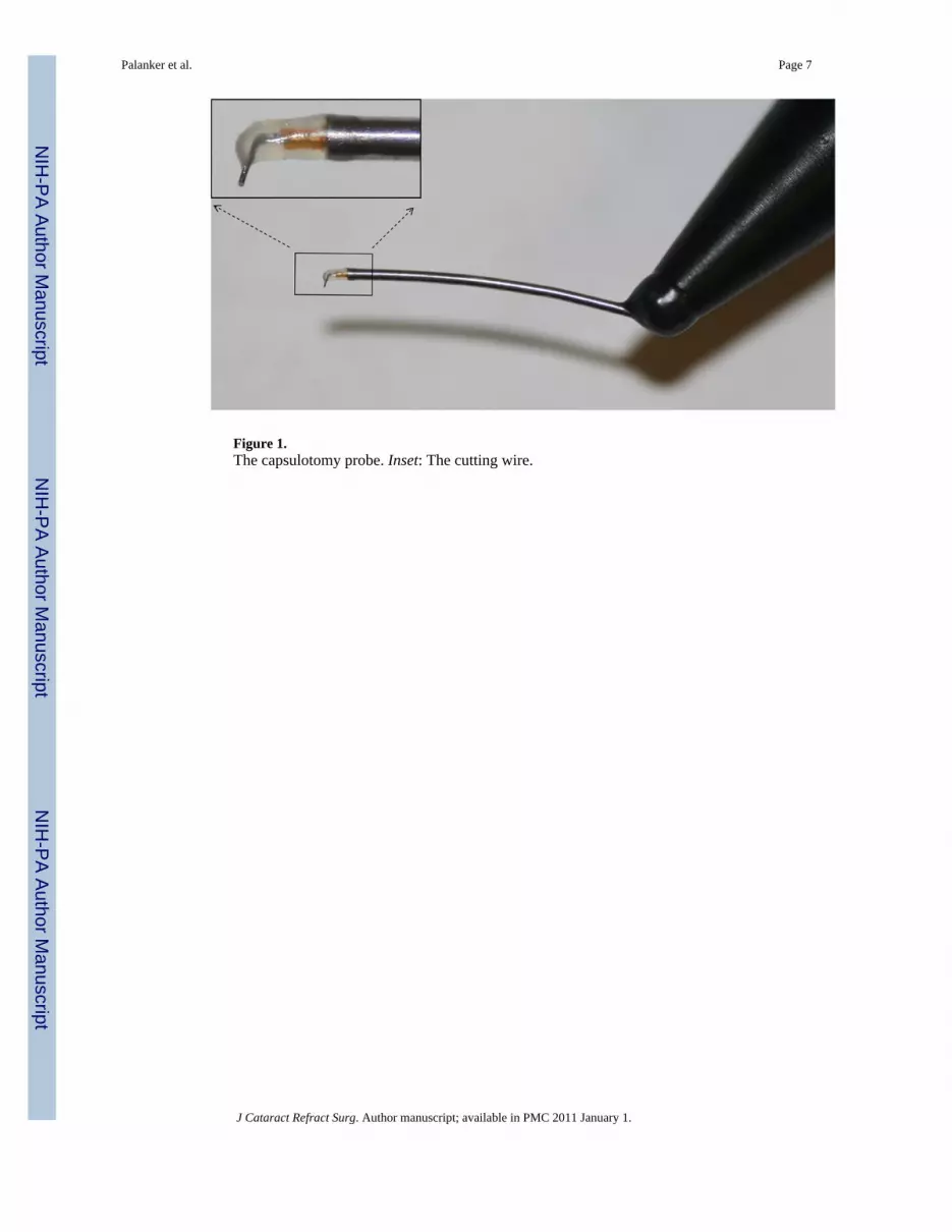
Figure 1.The capsulotomy probe. Inset: The cutting wire.
Palanker et al. Page 7
J Cataract Refract Surg. Author manuscript; available in PMC 2011 January 1.
NIH
-PA Author Manuscript
NIH
-PA Author Manuscript
NIH
-PA Author Manuscript
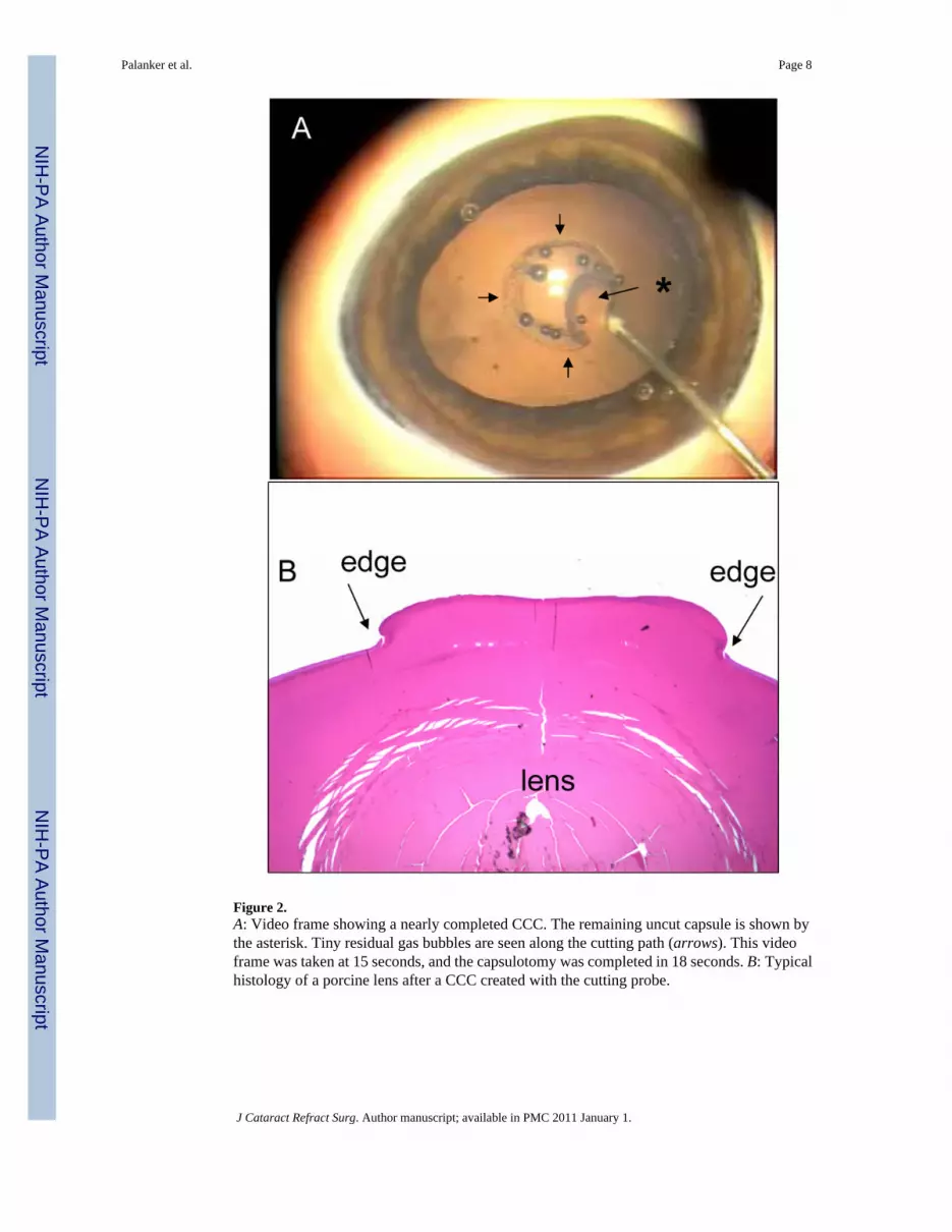
Figure 2.A: Video frame showing a nearly completed CCC. The remaining uncut capsule is shown bythe asterisk. Tiny residual gas bubbles are seen along the cutting path (arrows). This videoframe was taken at 15 seconds, and the capsulotomy was completed in 18 seconds. B: Typicalhistology of a porcine lens after a CCC created with the cutting probe.
Palanker et al. Page 8
J Cataract Refract Surg. Author manuscript; available in PMC 2011 January 1.
NIH
-PA Author Manuscript
NIH
-PA Author Manuscript
NIH
-PA Author Manuscript

Figure 3.Histologic sections of a porcine capsule after mechanical (A) and cutting probe (B) CCCcreation. The arrows show the surface of the cut edge.
Palanker et al. Page 9
J Cataract Refract Surg. Author manuscript; available in PMC 2011 January 1.
NIH
-PA Author Manuscript
NIH
-PA Author Manuscript
NIH
-PA Author Manuscript

Figure 4.Scanning electron micrographs of the porcine capsule after mechanical capsulorhexis. Thescale bar in the upper image is 500 μm and in the lower image, 50 μm (1 = iris; 2 = lens capsule;3 = lens cortex).
Palanker et al. Page 10
J Cataract Refract Surg. Author manuscript; available in PMC 2011 January 1.
NIH
-PA Author Manuscript
NIH
-PA Author Manuscript
NIH
-PA Author Manuscript

Figure 5.Scanning electron micrographs of the porcine capsule after a CCC created with the probe. Notethe very sharp and continuous edges, with some cellular-scale texture on the cut side surface.The scale bar in the upper image is 500 μm and in the lower image, 50 μm (1 = iris; 2 = lenscapsule; 3 = lens cortex).
Palanker et al. Page 11
J Cataract Refract Surg. Author manuscript; available in PMC 2011 January 1.
NIH
-PA Author Manuscript
NIH
-PA Author Manuscript
NIH
-PA Author Manuscript