gamma knife: treatment platform, qa - seaapm
TRANSCRIPT
2/27/2017
1
GAMMA KNIFE: TREATMENT PLATFORM, QA, AND TREATMENT UNCERTAINTY
Dan McDonald, MSAssistant Professor, Medical University of South Carolina
DISCLOSURES
No conflicts of interest to disclose
2/27/2017
2
OUTLINE
Calibration and commissioning Treatment prep and image acquisition
How do I determine imaging modality? What potential sources of error arise due to imaging?
Treatment planning and delivery QA Sources of uncertainty Gamma Knife ICON SAMS
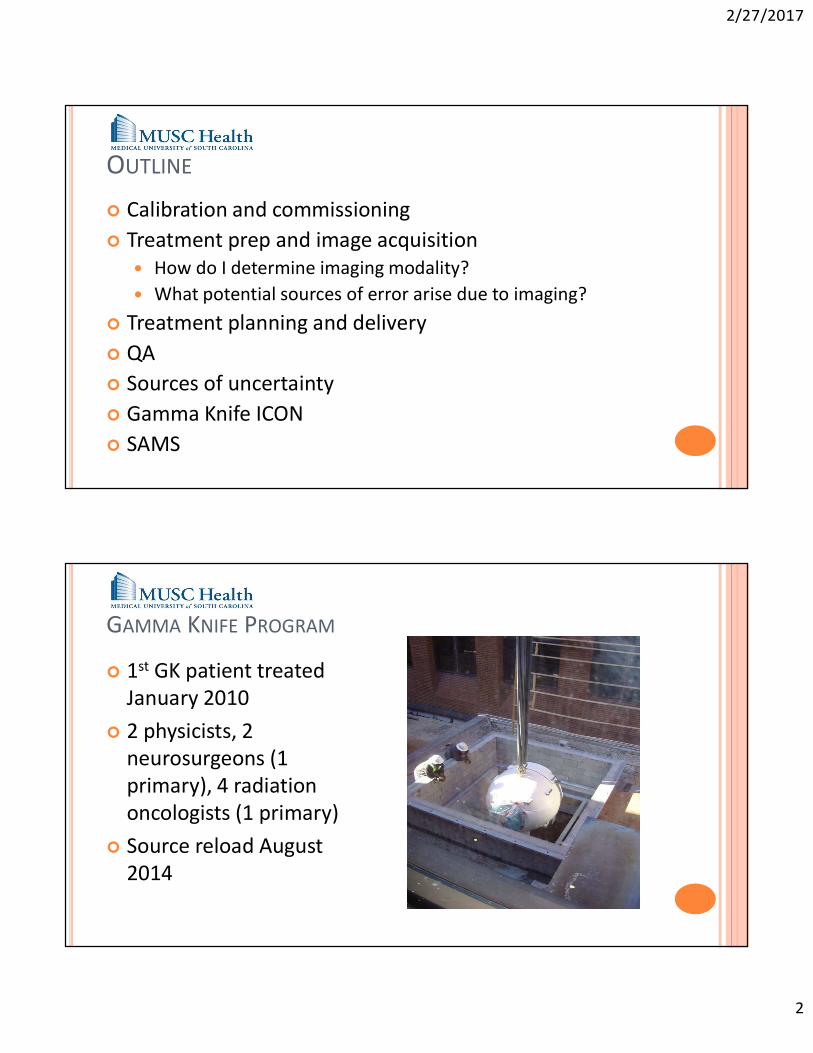
GAMMA KNIFE PROGRAM
1st GK patient treated January 2010
2 physicists, 2 neurosurgeons (1 primary), 4 radiation oncologists (1 primary)
Source reload August 2014
2/27/2017
3
GAMMA KNIFE PROGRAM
Over 1500 treatments provided to date
273 in 2016 Wide range of indications
Metastatic disease Surgical cavity Primary malignant brain tumor Acoustic neuroma Meningioma Trigeminal neuralgia AVM Glomus tumor Essential tremor
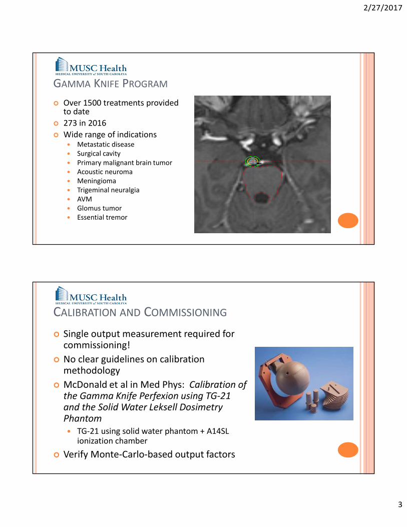
CALIBRATION AND COMMISSIONING
Single output measurement required for commissioning!
No clear guidelines on calibration methodology
McDonald et al in Med Phys: Calibration of the Gamma Knife Perfexion using TG-21 and the Solid Water Leksell Dosimetry Phantom TG-21 using solid water phantom + A14SL
ionization chamber
Verify Monte-Carlo-based output factors
2/27/2017
4
TREATMENT PREPARATION
Stereotactic frame-based system Neurosurgeon positions frame on morning of
treatment Patient receives:
MRI and/or CT and/or Angiogram (AVM only)
Each imaging modality has dedicated localizer At MUSC 90% of patients receive MRI only Typical sequences
T1 volume scan w/ gado (1mm slice) T2 Drive (0.7mm slice) Time-of-flight (0.7mm slice)
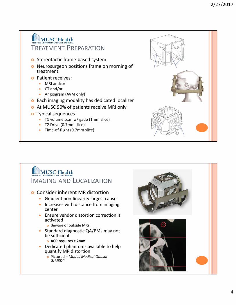
IMAGING AND LOCALIZATION
Consider inherent MR distortion Gradient non-linearity largest cause Increases with distance from imaging
center Ensure vendor distortion correction is
activated Beware of outside MRs
Standard diagnostic QA/PMs may not be sufficient ACR requires ± 2mm
Dedicated phantoms available to help quantify MR distortion Pictured – Modus Medical Quasar
Grid3D™
2/27/2017
5
IMAGING AND LOCALIZATION
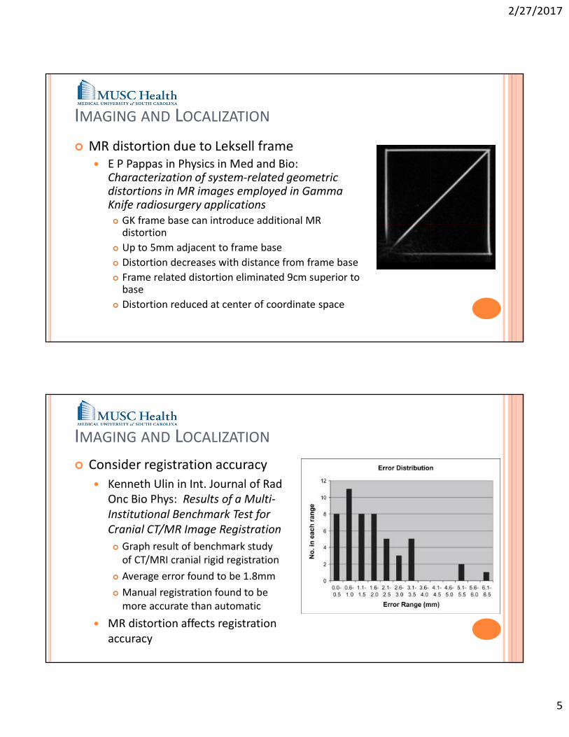
MR distortion due to Leksell frame E P Pappas in Physics in Med and Bio:
Characterization of system-related geometric distortions in MR images employed in Gamma Knife radiosurgery applications GK frame base can introduce additional MR
distortion Up to 5mm adjacent to frame base Distortion decreases with distance from frame base Frame related distortion eliminated 9cm superior to
base Distortion reduced at center of coordinate space
IMAGING AND LOCALIZATION
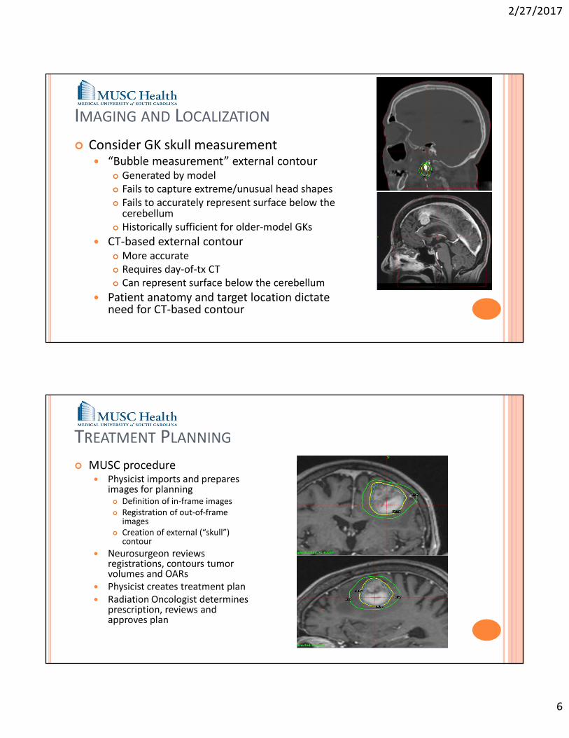
Consider registration accuracy Kenneth Ulin in Int. Journal of Rad
Onc Bio Phys: Results of a Multi-Institutional Benchmark Test for Cranial CT/MR Image Registration Graph result of benchmark study
of CT/MRI cranial rigid registration Average error found to be 1.8mm Manual registration found to be
more accurate than automatic
MR distortion affects registration accuracy
2/27/2017
6
IMAGING AND LOCALIZATION
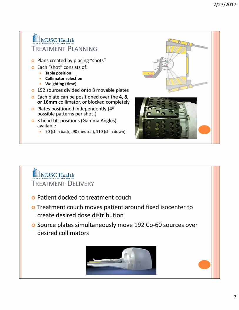
Consider GK skull measurement “Bubble measurement” external contour
Generated by model Fails to capture extreme/unusual head shapes Fails to accurately represent surface below the
cerebellum Historically sufficient for older-model GKs
CT-based external contour More accurate Requires day-of-tx CT Can represent surface below the cerebellum
Patient anatomy and target location dictate need for CT-based contour
TREATMENT PLANNING
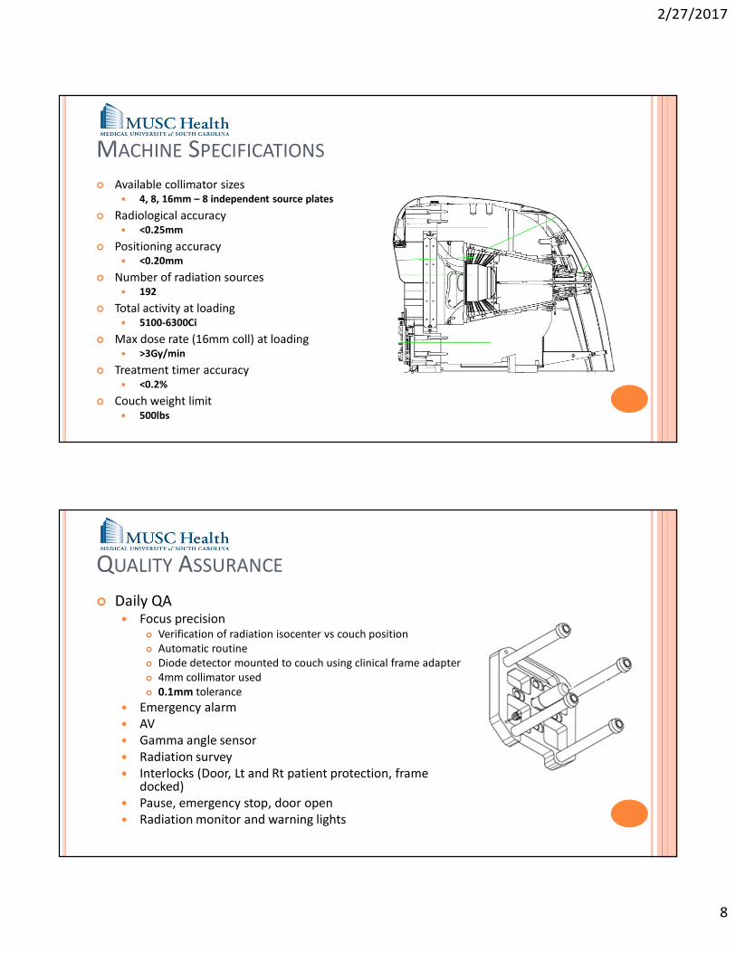
MUSC procedure Physicist imports and prepares
images for planning Definition of in-frame images Registration of out-of-frame
images Creation of external (“skull”)
contour Neurosurgeon reviews
registrations, contours tumor volumes and OARs
Physicist creates treatment plan Radiation Oncologist determines
prescription, reviews and approves plan
2/27/2017
7
TREATMENT PLANNING
Plans created by placing “shots” Each “shot” consists of:
Table position Collimator selection Weighting (time)
192 sources divided onto 8 movable plates Each plate can be positioned over the 4, 8,
or 16mm collimator, or blocked completely Plates positioned independently (48
possible patterns per shot!) 3 head tilt positions (Gamma Angles)
available 70 (chin back), 90 (neutral), 110 (chin down)
TREATMENT DELIVERY
Patient docked to treatment couch Treatment couch moves patient around fixed isocenter to
create desired dose distribution Source plates simultaneously move 192 Co-60 sources over
desired collimators
2/27/2017
8
MACHINE SPECIFICATIONS Available collimator sizes
4, 8, 16mm – 8 independent source plates
Radiological accuracy <0.25mm
Positioning accuracy <0.20mm
Number of radiation sources 192
Total activity at loading 5100-6300Ci
Max dose rate (16mm coll) at loading >3Gy/min
Treatment timer accuracy <0.2%
Couch weight limit 500lbs
QUALITY ASSURANCE
Daily QA Focus precision
Verification of radiation isocenter vs couch position Automatic routine Diode detector mounted to couch using clinical frame adapter 4mm collimator used 0.1mm tolerance
Emergency alarm AV Gamma angle sensor Radiation survey Interlocks (Door, Lt and Rt patient protection, frame
docked) Pause, emergency stop, door open Radiation monitor and warning lights
2/27/2017
9
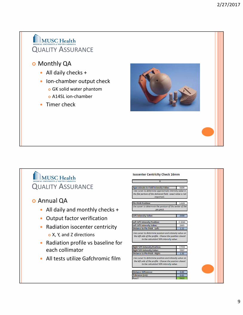
QUALITY ASSURANCE
Monthly QA All daily checks + Ion-chamber output check
GK solid water phantom A14SL ion-chamber
Timer check
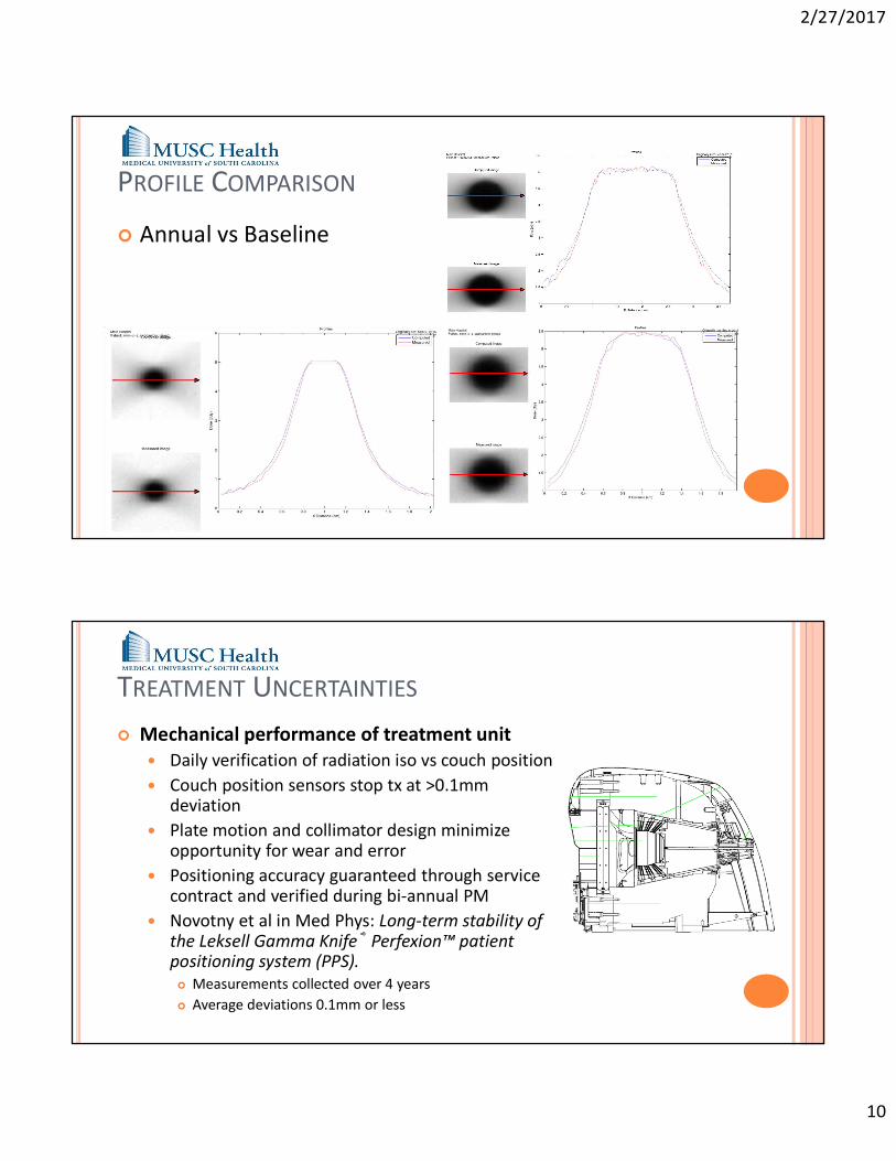
QUALITY ASSURANCE
Annual QA All daily and monthly checks + Output factor verification Radiation isocenter centricity
X, Y, and Z directions
Radiation profile vs baseline for each collimator
All tests utilize Gafchromic film
2/27/2017
10
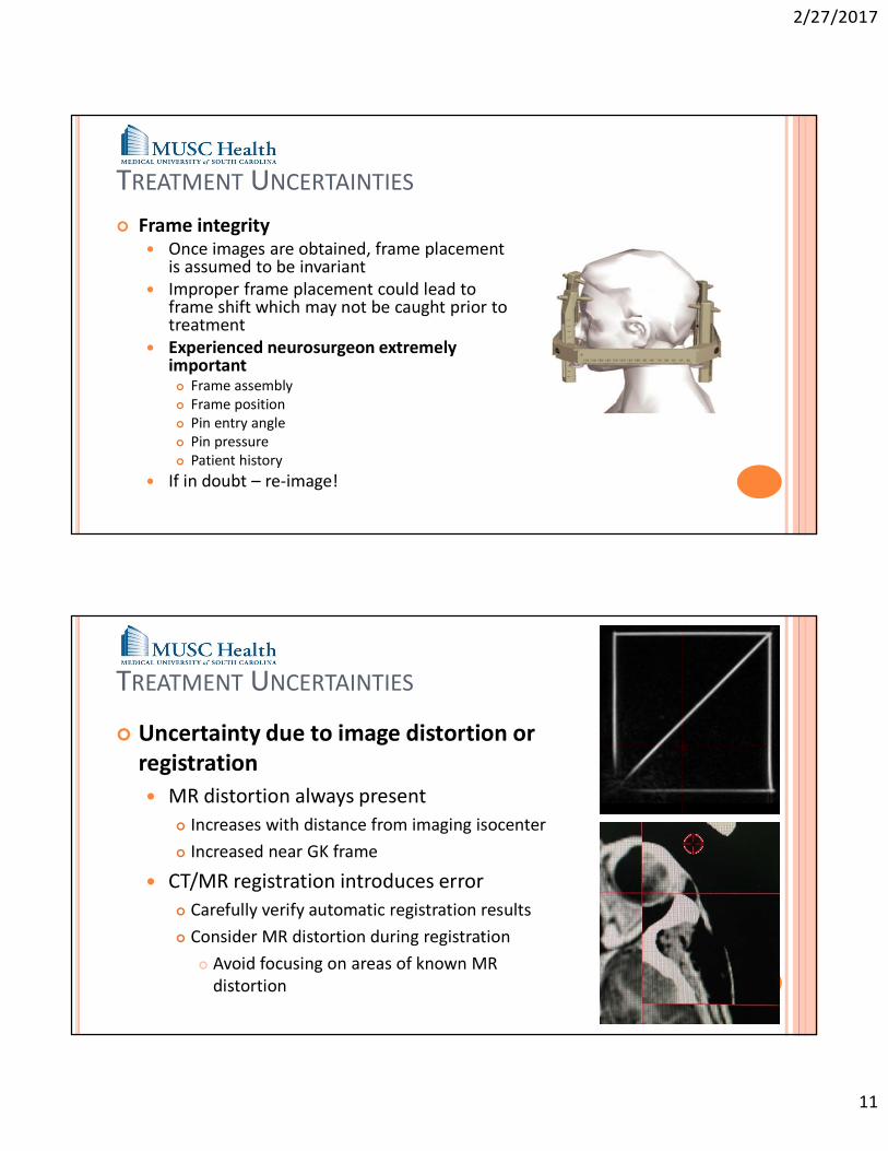
PROFILE COMPARISON
Annual vs Baseline
TREATMENT UNCERTAINTIES
Mechanical performance of treatment unit Daily verification of radiation iso vs couch position Couch position sensors stop tx at >0.1mm
deviation Plate motion and collimator design minimize
opportunity for wear and error Positioning accuracy guaranteed through service
contract and verified during bi-annual PM Novotny et al in Med Phys: Long-term stability of
the Leksell Gamma Knife® Perfexion™ patient positioning system (PPS). Measurements collected over 4 years Average deviations 0.1mm or less
2/27/2017
11
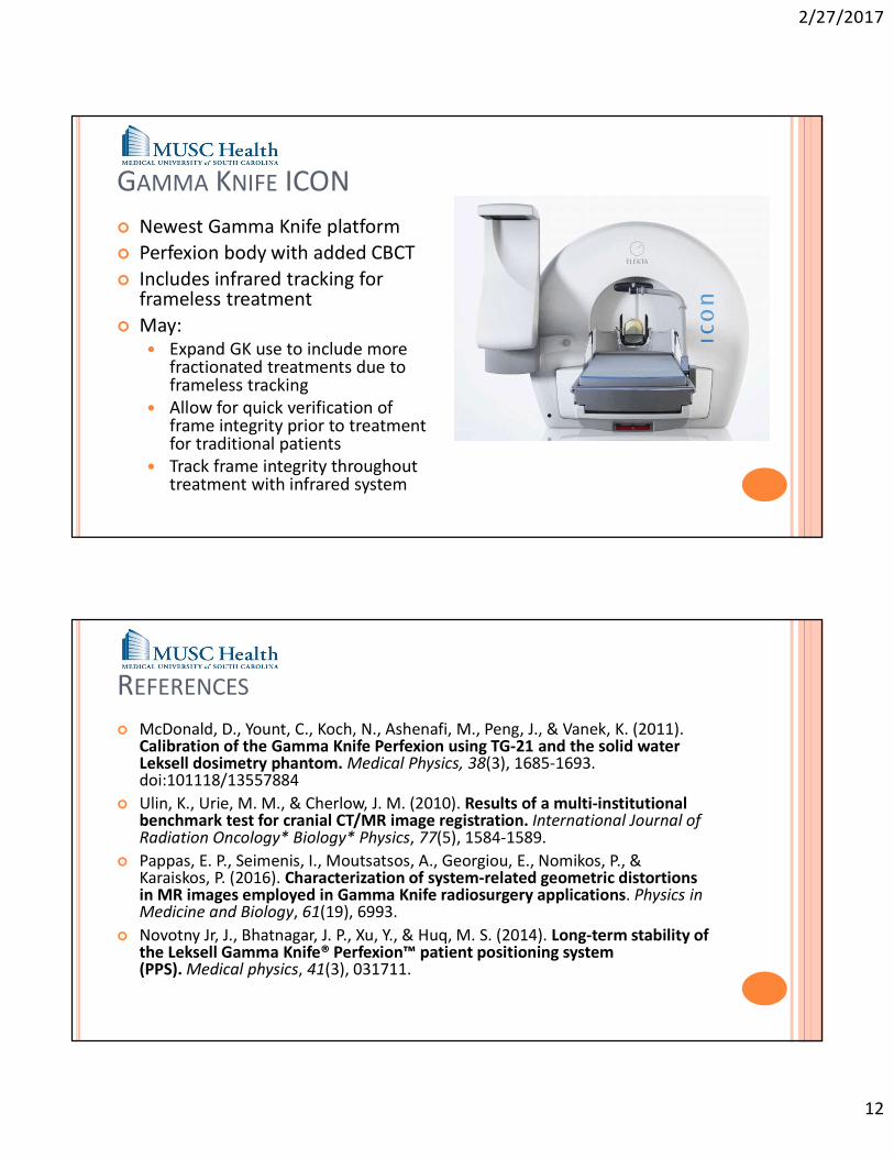
TREATMENT UNCERTAINTIES
Frame integrity Once images are obtained, frame placement
is assumed to be invariant Improper frame placement could lead to
frame shift which may not be caught prior to treatment
Experienced neurosurgeon extremely important Frame assembly Frame position Pin entry angle Pin pressure Patient history
If in doubt – re-image!
TREATMENT UNCERTAINTIES
Uncertainty due to image distortion or registration MR distortion always present
Increases with distance from imaging isocenter Increased near GK frame
CT/MR registration introduces error Carefully verify automatic registration results Consider MR distortion during registration
Avoid focusing on areas of known MR distortion
2/27/2017
12
GAMMA KNIFE ICON Newest Gamma Knife platform Perfexion body with added CBCT Includes infrared tracking for
frameless treatment May:
Expand GK use to include more fractionated treatments due to frameless tracking
Allow for quick verification of frame integrity prior to treatment for traditional patients
Track frame integrity throughout treatment with infrared system
REFERENCES
McDonald, D., Yount, C., Koch, N., Ashenafi, M., Peng, J., & Vanek, K. (2011). Calibration of the Gamma Knife Perfexion using TG-21 and the solid water Leksell dosimetry phantom. Medical Physics, 38(3), 1685-1693. doi:101118/13557884
Ulin, K., Urie, M. M., & Cherlow, J. M. (2010). Results of a multi-institutional benchmark test for cranial CT/MR image registration. International Journal of Radiation Oncology* Biology* Physics, 77(5), 1584-1589.
Pappas, E. P., Seimenis, I., Moutsatsos, A., Georgiou, E., Nomikos, P., & Karaiskos, P. (2016). Characterization of system-related geometric distortions in MR images employed in Gamma Knife radiosurgery applications. Physics in Medicine and Biology, 61(19), 6993.
Novotny Jr, J., Bhatnagar, J. P., Xu, Y., & Huq, M. S. (2014). Long-term stability of the Leksell Gamma Knife® Perfexion™ patient positioning system (PPS). Medical physics, 41(3), 031711.
2/27/2017
13
SAM QUESTION 1
Uncertainty during treatment on the Gamma Knife Perfexion is predominantly due to: Mechanical performance of the treatment machine MLC positioning error Frame integrity and planning image registrations and
distortions Machine output fluctuation
SAM QUESTION 1
Uncertainty during treatment on the Gamma Knife Perfexionis predominantly due to: Frame integrity and planning image registrations and distortions
References Ulin, K., Urie, M. M., & Cherlow, J. M. (2010). Results of a multi-institutional benchmark test for
cranial CT/MR image registration. International Journal of Radiation Oncology* Biology* Physics, 77(5), 1584-1589.
Pappas, E. P., Seimenis, I., Moutsatsos, A., Georgiou, E., Nomikos, P., & Karaiskos, P. (2016). Characterization of system-related geometric distortions in MR images employed in Gamma Knife radiosurgery applications. Physics in Medicine and Biology, 61(19), 6993.
Novotny Jr, J., Bhatnagar, J. P., Xu, Y., & Huq, M. S. (2014). Long-term stability of the LeksellGamma Knife® Perfexion™ patient positioning system (PPS). Medical physics, 41(3), 031711.
2/27/2017
14
SAM QUESTION 2
MR Distortion Due to the GK Frame Base is: Minimal compared to distortion already inherent in MR
imaging Significant adjacent to the frame base, decreasing as distance
from the frame base increases Corrected during the localization process Minimal compared to mechanical uncertainty of the
treatment unit
SAM QUESTION 2
MR Distortion Due to the GK Frame Base is: Significant adjacent to the frame base, decreasing as distance
from the frame base increases
References
Pappas, E. P., Seimenis, I., Moutsatsos, A., Georgiou, E., Nomikos, P., & Karaiskos, P. (2016). Characterization of system-related geometric distortions in MR images employed in Gamma Knife radiosurgery applications. Physics in Medicine and Biology, 61(19), 6993.