analytical protocols for the assessment of biological damage in historical documents
TRANSCRIPT

COALITION No. 19, January 2010
6ISSN 1579-8410 www.rtphc.csic.es/boletin.htm
ANALYTICAL PROTOCOLS FOR THE ASSESSMENT OF BIOLOGICAL DAMAGE IN HISTORICAL DOCUMENTS
Flavia Pinzari1, Mariasanta Montanari1, Astrid Michaelsen2 and Guadalupe Piñar3
1Istituto Centrale per il Restauro e la Conservazione del Patrimonio Archivistico e Librario, Laboratorio di Biologia, Via Milano 76, 00184 Rome, Italy. E-mail: [email protected] 2Department of Microbial Ecology, University of Vienna, Althanstrasse 14, 1090 Vienna, Austria 3Institute of Applied Microbiology, Department of Biotechnology, University of Natural Resources and Applied Life Sciences, Muthgasse 18, A-1190 Vienna, Austria. Abstract Biodeterioration of paper and parchment in ancient books and documents represents a cause of great concern for libraries and archives all over the world. The study of the mechanisms underlying the microbiological attack of historical materials has been widely practiced and still represents one of the main focuses of those institutions and laboratories that are involved in cultural heritage conservation. Many studies on the role of micro-organisms in the defacement of cultural heritage utilise standardised laboratory models to establish, under controlled conditions, which species of fungi or bacteria colonise a given substrata, and therefore do not raise the problem of working with objects of art that cannot be cut, sampled, or subjected to invasive analysis. The aim of this paper is the discussion of the procedures required for a conservative approach to the evaluation and description of the damage and organisms responsible for the biodeterioration of valuable books and works of art made from or supported on paper.
Introduction Microorganisms can be responsible for the destruction of cultural heritage, together with several environmental conditions, ageing and the chemical structure of substrate. There is a number of reviews giving a comprehensive picture of the role of microorganisms in the degradation of art objects, such as paintings, stone, wood, paper, masonry, leather, parchment, glass and metal (Bock and Sand 1993, Cappitelli and Sorlini 2005, Ciferri 1999, Griffin et al. 1991, Kowalik 1980, Montegut et al. 1991). However, when a biology laboratory is asked to supply a purely diagnostic explanation for a degradative phenomenon occurring in a precious art work, the challenge of obtaining
an informative sample from the object without damaging it becomes a real operative problem (Montanari et al. 2006). To this should be added the problem of the viability of micro-organisms and their culturability (Amann et al. 1995), which often makes the diagnosis a matter of sheer luck. In fact, as established in environments other than cultural heritage components, the microbial communities found are much more complex and bio-diverse than would appear according to classic culturing methods (Ward et al. 1990). Microbial investigations based on cultivation strategies cannot be regarded as being reliable in terms of reflecting the microbial diversity present in art works samples. When molecular biology techniques have been applied to cultural heritage environments, new spoiling taxa and unsuspected microbial consortia were found to be involved in the discolouration and biodeterioration of paintings and monuments (Piñar et al. 2001 a,b,c, 2002, 2003, Rölleke et al. 1996, 1998, Schabereiter-Gurtner et al. 2001 a,b,c, 2002 a,b, 2003). The routine work of describing and cataloguing which organisms are causing the specific damage on a given object of art suffers, on one hand, from a lack of standardised methodologies and informative techniques, and requires a set of theoretical instruments for operating at maximum levels and without error. On the other hand, it is somewhat dangerous to establish rules and define strict procedures because most of these can result in being too invasive or not informative when applied to a particularly valuable historical object. The degree of invasiveness in sampling an object of historical value depends on several factors like, for example, the size of the object, the presence or absence of detached fragments, the position of the damage with respect to the valuable parts of the object itself, the heterogeneity of the constituting materials, and the further work that restorers might have to carry out on it. In this paper we describe different sampling techniques and instruments that can be employed to obtain information on damage and damaging agents that can affect ancient papers. The aim is a discussion of the procedures required for a conservative approach to the
COALITION No. 19, January 2010
7ISSN 1579-8410 www.rtphc.csic.es/boletin.htm
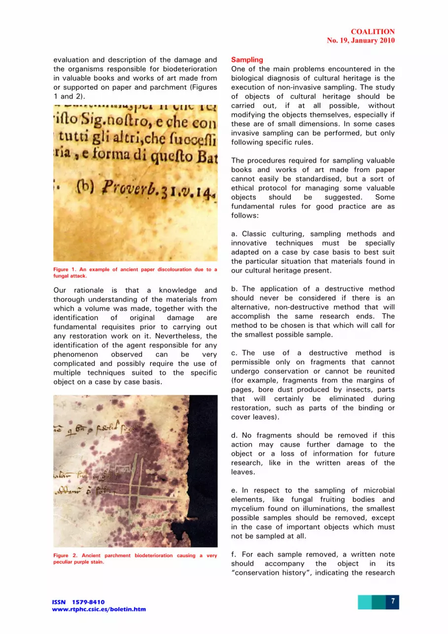
evaluation and description of the damage and the organisms responsible for biodeterioration in valuable books and works of art made from or supported on paper and parchment (Figures 1 and 2).
Figure 1. An example of ancient paper discolouration due to a fungal attack.
Our rationale is that a knowledge and thorough understanding of the materials from which a volume was made, together with the identification of original damage are fundamental requisites prior to carrying out any restoration work on it. Nevertheless, the identification of the agent responsible for any phenomenon observed can be very complicated and possibly require the use of multiple techniques suited to the specific object on a case by case basis.
Figure 2. Ancient parchment biodeterioration causing a very peculiar purple stain.
Sampling One of the main problems encountered in the biological diagnosis of cultural heritage is the execution of non-invasive sampling. The study of objects of cultural heritage should be carried out, if at all possible, without modifying the objects themselves, especially if these are of small dimensions. In some cases invasive sampling can be performed, but only following specific rules. The procedures required for sampling valuable books and works of art made from paper cannot easily be standardised, but a sort of ethical protocol for managing some valuable objects should be suggested. Some fundamental rules for good practice are as follows: a. Classic culturing, sampling methods and innovative techniques must be specially adapted on a case by case basis to best suit the particular situation that materials found in our cultural heritage present. b. The application of a destructive method should never be considered if there is an alternative, non-destructive method that will accomplish the same research ends. The method to be chosen is that which will call for the smallest possible sample. c. The use of a destructive method is permissible only on fragments that cannot undergo conservation or cannot be reunited (for example, fragments from the margins of pages, bore dust produced by insects, parts that will certainly be eliminated during restoration, such as parts of the binding or cover leaves). d. No fragments should be removed if this action may cause further damage to the object or a loss of information for future research, like in the written areas of the leaves. e. In respect to the sampling of microbial elements, like fungal fruiting bodies and mycelium found on illuminations, the smallest possible samples should be removed, except in the case of important objects which must not be sampled at all. f. For each sample removed, a written note should accompany the object in its “conservation history”, indicating the research

COALITION No. 19, January 2010
8ISSN 1579-8410 www.rtphc.csic.es/boletin.htm
establishment to which the sample was taken and analysed, the name of the laboratory, and the name of the person who executed the sampling and the analysis procedures. g. The sampling procedure should be accompanied by photographs of the object before and after the removal of material and a description of the method used for sampling, also stating the exact part of the object from which the sample was taken. Non invasive sampling Different types of swabs and adhesive tapes can be employed in sampling fungal or bacterial elements from paper (Figure 3). The choice of the most suitable technique should be based on the alteration to be diagnosed. For example, if there are dry and dusty fruiting bodies and free spores emerging from the substrate, a plastic swab might be electrically charged and therefore obtain a not representative sampling of the damaged area. A wooden-cotton swab is most suitable. The use of a cotton swab slightly moistened in a salt solution (i.e. Ringer or Czapek inorganic solution as described in Samson et al. 2001) can be useful for the recovery of spores and elements of microbial colonies from deteriorated paper or parchment fibres, but the moisture can damage the substrate by creating stains. Removable adhesive tape can be used to collect samples of fungal mycelium and sporulating structures from deteriorated paper (Samson et al. 2002). The choice of a tape that requires little effort to remove from surfaces is obligatory due to the fragility of paper and to the fact that a tape with a strong adhesive would strip too many cellulose fibres.
Figure 3. Direct sampling using cotton swabs. The fungal or bacterial structures taken with the swabs can be directly smeared on agar nutritive medium to be checked for viability.
Figure 4. Light microscopic examination of mounted slides carrying adhesive tape samples. Fungal structures that could tentatively be attributed to an Aureobasidium species, growing around a linen fibre (Olympus AX60 microscope).
Figure 5. A sampling procedure with the use of adhesive tape for the recover of fungal structures from paper.
The tape can be directly observed through a microscope after diaphanisation with cedar oil to check for the possible presence of fungal structures (Figure 4). To perform this type of sampling, a 3-4 cm strip of tape is looped back on itself, with the adhesive side facing outwards, and held between the thumb and index finger, or using the thumb and index of both hands as shown in Figure 5. The adhesive side is then pressed gently on the stain or the surface of paper or parchment. The aerial hyphae, conidiophora or fruiting structures, together with a few damaged fibres of the substrata cling to the sticky surface and can be gently pulled from the mat. This method may not be suitable for the study of very fragile materials (i.e. Japanese

COALITION No. 19, January 2010
9ISSN 1579-8410 www.rtphc.csic.es/boletin.htm
paper, or acidic depolymerised paper, felted paper, etc.) or damp materials. Invasive sampling Blades can be used to scratch the paper’s surface where there is evidence of the presence of fungal structures. Needles can be used to assist in the collection of powder obtained through the use of blades. Some fragments of paper (2-3 mm2 width) can, in some cases, be collected, possibly from the margins of the most degraded pages using a watchmaker’s tweezers. Paper samples should be taken only from those margins of the sheets and areas where invasive sampling is not endangering the value of the object itself (Figure 6).
Figure 6. A sampling procedure with the use of needles, performed under a safety cabinet, aimed at the removal of small parts of damaged paper.
Figure 7. A variable pressure SEM analysis (ZEISS EVO 50) of paper fragments sampled from a biodeteriorated document. No metallization was used. Paper fibres and fungal conidia can be observed.
When micro-invasive sampling is necessary, the use of such very small samples must be
optimised to obtain informative results. This is the case with SEM/EDS observation of small samples of damaged ancient paper. In some cases it is possible to make a diagnosis using this technique, especially when classic culturing methods yield no results (Figures 7 and 8). In the specific case of some filamentous fungi, the fruiting body shape, and the conidia size, shape and ornamentation can lead to identification at least at the genera level (Florian 2000, Samson and Pitt 2000). Paper powder or small fragments can be used for direct inoculation on to agar plates containing highly specific media, or used to perform more specific culturing methods.
Figure 8. A High Vacuum SEM (ZEISS EVO 50) micrograph showing bacteria growing on the surface of damaged parchment. The small sample, taken from the margin of a manuscript was sputtered with gold before SEM-observations.
Culturing methods Microbiological research carried out in the field of cultural heritage until today has mainly been based on classical culturing methods. Although these techniques are often helpful for demonstrating the importance of micro-organisms in some deterioration processes, most of them are not suitable for solving all the problems encountered and for an effective impact evaluation. Moreover, these studies can provide very little information on the true correlation between certain deterioration processes and micro-organisms, and often yield poor result on the efficiency of restoration treatments. The results obtainable can cover only those few organisms that can be cultivated, and it is now generally accepted that cultivation methods recover less than 1% of the total micro-organisms present in environmental samples (Amman et al. 1996, Ward et al. 1960). Nevertheless, cultivation techniques should be performed paying attention to the specific metabolic and physiological requirements of the species

COALITION No. 19, January 2010
10ISSN 1579-8410 www.rtphc.csic.es/boletin.htm
affecting the substrata that support library materials of cultural heritage value. Using the wrong medium can often led to unspecific results or to no results at all. Many of the fungal species that cause damage to paper are, for example, xerophiles, because they require a growth medium with a low water activity (aw) in order to germinate and produce fruiting bodies (useful for their identification) (Florian 2002, Samson and Pitt 2000). Moreover, a direct smearing of the cotton swabs on to solid agar medium very often doesn’t cause a colony to develop, because the spores and mycelium fragments picked up by the swab are often nutrient starved and dehydrated. Moistening the dry swab in water or in a nutritive or mineral broth for a few hours prior to agar inoculation can allow the reactivation of dormant spores. When fragments of deteriorated materials are available, a type of culture that is useful for discovering whether the microbial strain is a paper spoiler or just a contaminant can be performed. In this procedure, commonly referred to as “multi-point inoculum”, small pieces of paper are washed in sterile water and divided into 25 or more sub-samples that are inoculated directly on to solid nutritive agar distributed according to a geometric scheme (Figure 9).
Figure 9: Paper powder or small fragments can be used for direct inoculation on to agar plates according to a “multi-point inoculum” procedure. Pieces of paper are washed in sterile water and divided into 25 or more sub-samples that are inoculated directly on to solid nutritive agar distributed according to a geometric scheme.
The frame consists of a grid with 25 nodes spaced out equally. The development of the same fungal or microbial organism in most of the 25 points allows for a statistically significant attribution of the damage to it (Dix and Webster 1995) (Figure 10). When different species develop from the 25 inocula
it is not possible to place the isolated strains into a strict relationship with the damage observed. Following the isolation of a strain suspected of being the responsible for damage, the artificial reproduction of the observed damage on sample paper achieved by inoculating the isolated strain can represent a useful counterproof (Figure 11).
Figure 10. “Multi-point inoculum” procedure. The development of the same fungal or microbial organism in most of the 25 points allows for a statistically significant attribution of the damage to it.
Figure 11. The isolation from an ancient paper of a fungal species (Chaetomium globosum Kunze) and the counterproof of its “pathogenesis to paper” by the re-inoculation of the isolated strain on a paper sample.
Molecular methods In the last years, Molecular Biology is developing at a really fast pace. Many of the molecular techniques available can be readily applied to the study of cultural assets. Molecular techniques allow studying microorganisms from their DNA, RNA and proteins. DNA based techniques allow the identification of single bacterial species in sample material without the cultivation of the organisms (Amann et al. 1995). Most of the experiments carried out in this field so far are based on ribosomal sequences, which are used as phylogenetic markers (Woese 1987).

COALITION No. 19, January 2010
11ISSN 1579-8410 www.rtphc.csic.es/boletin.htm
The ribosomal sequences are present in all organisms and they contain variable and highly conserved regions which allow to distinguish between organisms on all phylogenetic levels. In addition, a lot of data exist in the databases (Maidak et al. 1999), which can be used to compare the DNA-sequences of unknown microorganisms and allow a phylogenetic identification. The extracted DNA can be used as a template to amplify ribosomal gene fragments with primers for universal sequences by PCR (Polymerase Chain Reaction). The PCR amplified fragments can then be cloned. The result of such strategy is a clone library, containing ribosomal sequences as inserts. By sequencing individual inserts and comparing the obtained sequences with sequences present in databases, it is possible to identify the phylogenetic position of the corresponding microorganism without their cultivation.
Figure 12. A gel showing the DGGE (ITS specific amplified sequences) of bacterial communities inhabiting ten samples from different biodeteriorated documents.
There are few works focusing on the investigation of the fungal flora responsible for the biodeterioration of paper materials by applying molecular techniques (Di Bonaventura
et al. 2003, Michaelsen et al. 2005, 2006, 2009, Pangallo 2009, Rakotonirainy 2007). In Michaelsen et al. (2005, 2006, 2009) culture-independent molecular methods were used to identify fungal communities on paper from different time periods and quality. As already applied for environmental fungal communities (Jackson et al. 1999, Jasalavich et al. 2000), nucleic-acid-based strategies targeting rRNA-encoding regions were selected for essays studying the community structure of fungi on biodeteriorated paper. Pure fungal strains were compared to infected material, according to DNA extraction and PCR conditions, as well as the setting of fingerprinting techniques as DGGE (Denaturing Gradient Gel Electrophoreses) analysis (Figure 12). Several extraction methods were tested. An ideal DNA extraction protocol adapted for paper/parchment samples should allow: a) to obtain good results with very small samples; b) to be able, by washing treatments previous to extraction, of distinguishing between the microorganisms responsible of the deterioration and the polluting spores coming from dust deposition (Michaelsen 2006). According to Michaelsen et al. (2006) the bead-beating homogenisation of samples showed best results in fungal DNA extraction from paper materials of different compositions and ages. It rendered a pure DNA able to be directly amplified without the need for any extra purification step. However, the problem still remains that no single method of cell lysis is appropriate for all fungi potentially colonising paper-made art objects. The PCR amplification of the ITS1 region, followed by fingerprinting discrimination of fungal sequences in DGGE analysis is a reproducible and powerful technique for the visualisation not only of viable fungi, but also of formerly active fungi, which could have had an important role in paper biodeterioration processes. Other molecular techniques have been applied to the study of the microflora inhabiting indoor artworks, like in Pangallo et al. (2009) where to type the bacterial and fungal isolates, 2 PCR methods, repetitive extragenic palindromes (REP) and random amplified microsatellite polymorphisms (RAMP) were used.

COALITION No. 19, January 2010
12ISSN 1579-8410 www.rtphc.csic.es/boletin.htm
In Rakotonirainy et al. (2007) the two Internal Transcribed Spacers and the 5.8 S gene (ITS1-5.8S-ITS2) from the nuclear ribosomal DNA were amplified, cloned and sequenced. Following a preliminary treatment with cellulase from Trichoderma reesei, DNA extractions were successfully achieved directly from paper samples. From 22 selected stained spots from a book dating from the 19th century, 8 extracts of genomic DNA were entirely analysed, which yielded 145 sequenced clones. Conclusions Prior to any restorative operation, books, like other objects of cultural heritage, should always undergo an evaluative study (from the chemical and biological point of view) of the damage they present. The sampling represents a very important moment and should never be improvised or performed with inadequate instruments. Although most of the information needed to detect biological damages could be obtained by means of the simple methods as some described in this study, new types of diagnostics could result in more exhaustive tests. Traditionally, microbiology research carried out in this field was mainly based on classical cultivation methods. Although these investigations were helpful to demonstrate the importance of microorganisms in deterioration processes, most of them were not suitable for sustained impact evaluation. The results obtained covered only those few organisms that could be cultivated. It is generally accepted that cultivation methods recover less than 1% of the total microorganisms present in environmental samples (Amann et al. 1995, Ward et al. 1990), and that only about 5% of fungal species can be described owing to culture limitations, misidentifications in culture collections, and unexplored habitats (Hawksworth and Rossman 1997). Besides the very new and fascinating results that can be obtained by molecular approaches, at the purely diagnostic level, there is still a need for unsophisticated and inexpensive biological methodologies that could result in exhaustive tests, being performed on very small samples, and able of defining the cause and degree of damage. Acknowledgements The molecular analysis included in this project and A. Michaelsen were financed by the
Austrian Science Fund (FWF) within the framework of project P17328-B12. G. Piñar was financed by the “Hertha-Firnberg-Nachwuchsstelle (T137)” from FWF. References Amann, R.I., Ludwig, W. and Schleifer, K-H. 1995. Phylogenetic
identification and in situ detection of individual microbial cells without cultivation. Microbiology Reviews 59: 143-169.
Bock, E. and Sand, W. 1993. The microbiology of masonry biodeterioration. Journal of Applied Bacteriology 74: 503-514.
Cappitelli, F. and Sorlini, C. 2005. From papyrus to compact disc: the microbial deterioration of documentary heritage. Critical Reviews in Microbiology 31: 1–10.
Ciferri, O. 1999. Microbial degradation of paintings. Applied and Environmental Microbiology 65: 879-885.
Di Bonaventura M.P., De Salle, R. and Bonacum, J. 2003. Tiffany’s drawings, fungal spots and phylogenetic trees, in C. Saiz-Jimenez (ed.) Molecular Biology and Cultural Heritage: 131-135. Lisse: Balkema.
Dix N.J. and Webster, J. 1995. Fungal ecology. Cambridge: Chapman & Hall.
Florian M.L.E. 2000 Fungal fox spots and others. In: O. Ciferri, P. Tiano, and G. Mastromei (eds.) Of Microbes and Art: 135-151. New York: Kluwer Academic.
Florian, M.L.E. 2002. Fungal facts. London: Archetype Publication.
Griffin, P.S., Indictor, N. and Koestler R.J. 1991. The biodeterioration of stone: a review of deterioration mechanisms, conservation case histories, and treatment. International Biodeterioration and Biodegradation 28: 187-207.
Hawksworth D.L. and Rossman, A.Y. 1997. Where are all the undescribed fungi? Phytopathology 87: 888-891.
Jackson C.J., Barton, B.C. and Evans, E.G. 1999. Species Identification and strain Differentiation of Dermatophyte Fungi by Analysis of Ribosomal-DNA Intergenic Spacer Regions. Journal of Clinical Microbiology 37(4): 931-936.
Jasalavich, C.A., Ostrofsky, A. and Jellison, J. 2000. Detection and Identification of Decay Fungi in Spruce Wood by Restriction Fragment Length Polymorphism Analysis of Amplified Genes Encoding rRNA. Applied and Environmental Microbiology 66(11): 4725-4734.
Kowalik, R. 1980. Microbiodeterioration of library materials. Part 2. Microbiodecomposition of basic organic library materials. Restaurator 4: 135-219.
Maidak, B.L., Cole, J.R., Parker Jr., C.T., Garrity, G.M., Larsen, N., Li, B., Lilburn, T.G., McCaughey, M.J., Olsen, G.J., Overbeek, R., Pramanik, S., Schmidt, T.M., Tiedje, J.M. and Woese, C.R. 1999. A new version of the RDP (Ribosomal Database Project). Nucleic Acids Research 27: 171-173.
Michaelsen A., Pinzari F., Ripka, K., Lubitz, W. And Pinar, G. 2005. Application of molecular techniques for identification of fungal communities colonising paper material. Proceedings 13th International Biodeterioration and Biodegradation Symposium (IBBS-13) Madrid 4-9 Sept. 2005
Michaelsen, A., Piñar, G., Montanari M., and Pinzari F. 2009. Biodeterioration and restoration of a 16th-century book using a combination of conventional and molecular techniques: a case-study. International Biodeterioration & Biodegradation 63: 161-168.
Michaelsen, A., Pinzari, F., Ripka, K., Lubitz, W. and Piñar, G. 2006. Application of molecular techniques for identification of fungal communities colonizing paper material. International Biodeterioration & Biodegradation 58: 133–141.
Montanari M., Valenti, P. and Pinzari, F. 2006. Conservative approach to the evaluation of biological damage on objects of art made from paper. In: G. Bonizzoni (ed.) Proceedings of the 2nd International ''Science, Technology and Cultural Heritage'' workshop organised by the Italian Association of Vacuum (AIV) 9-11 November 2005: 85-91. Catania: Fondazione Diocesana per i Beni Culturali, ARCA Edizioni.
Montegut, D., Indictor, N. and Koestler, R.J. 1991. Fungal deterioration of cellulosic textiles: a review. International Biodeterioration & Biodegradation 28: 209-226.
Pangallo D., Chovanová, K., Šimonovičová, A. and Ferianc, P. 2009. Investigation of microbial community isolated from indoor artworks and air environment: identification,

COALITION No. 19, January 2010
13ISSN 1579-8410 www.rtphc.csic.es/boletin.htm
biodegradative abilities, and DNA typing. Canadian Journal of Microbiology 55(3): 277–287.
Piñar, G., Gurtner, C., Ramos, C., Lubitz, W. and Rölleke, S. 2002. Identification of Archaea in deteriorated ancient wall paintings by DGGE and FISH analysis. In: E. Galan and F. Zezza (eds.) Protection and Conservation of the Cultural Heritage of the Mediterranean Cities. Lisse: Balkema.
Piñar, G., Gurtner, C., Lubitz, W. and Rölleke, S. 2001a. Identification of Archaea in objects of art by DGGE analysis and shot gun cloning. Methods on Enzymology 336: 356-366.
Piñar, G., Ramos, C., Rölleke, S., Schabereiter-Gurtner, C., Vybiral, D., Lubitz W. and Denner, E.B.M. 2001b. Detection of indigenous Halobacillus populations in damaged ancient wall paintings and building materials: molecular monitoring and cultivation. Applied and Environmental Microbiology 67: 4891-4895.
Piñar, G., Saiz-Jimenez, C., Schabereiter-Gurtner, C., Blanco-Varela, M.T., Lubitz, W. and Rölleke, S. 2001c. Archaeal communities in two disparate deteriorated ancient wall paintings: detection, identification and temporal monitoring by denaturing gradient gel electrophoresis. FEMS Microbiology Ecology 37: 45-54.
Piñar, G., Schabereiter-Gurtner, C., Lubitz, W. and Rölleke, S. 2003. Analysis of the microbial diversity present on the wall paintings of Castle of Herberstein by molecular techniques. In: C. Saiz-Jimenez, (ed). Molecular Biology and Cultural Heritage. Lisse: Balkema.
Rakotonirainy, M.S., Heude, E. and Lavédrine, B. 2007. Isolation and attempts of biomolecular characterization of fungal strains associated to foxing on a 19th century book. Journal of Cultural Heritage 8: 126–133.
Rölleke, S., Witte, A., Wanner, G. and Lubitz, W. 1998. Medieval wall painting-a habitat for archaea: identification of archaea by denaturing gradient gel electrophoresis (DGGE) of PCR-amplified gene fragments coding 16S rRNA in a medieval wall painting. International Biodeterioration & Biodegradation 41: 85-92.
Rölleke, S., C. Gurtner, U. Drewello, R. Drewello, W. Lubitz, and R. Weissmann. (1999). Analysis of bacterial communities on historical glass by denaturing gradient gel electrophoresis of PCR-amplified gene fragments coding for 16S rRNA. J. Microbiol. Meth. 36: 107-114.
Rölleke, S., Muyzer, G., Wawer, C., Wanner, G. and Lubitz, W. 1996. Identification of bacteria in a biodegraded wall painting by denaturing gradient gel electrophoresis of PCR-amplified gene fragments coding for 16S rRNA. Applied and Environmental Microbiology 62: 2059-2065.
Samson R.A., Hoekstra, E.S., Frisvad, J.C. and Filtenborg, O. (eds.) 2002. Introduction to food-and airborne fungi. Sixth Edition. Utrecht: Centraalbureau Voor Schimmelculture.
Samson, R.A. and Pitt, J.I. 2000. Integration of Modern Taxonomic Methods for Penicillium and Aspergillus Classification. Harwood Academic Publisher.
Schabereiter-Gurtner, C., Piñar, G., Lubitz, W. and Rölleke, S. 2001a. An advanced molecular strategy to identify bacterial communities on art objects. Journal of Microbiological Methods 45: 77-87.
Schabereiter-Gurtner, C., Piñar, G., Vybiral, D., Lubitz, W. and Rölleke, S. 2001b. Rubrobacter-related bacteria associated with rosy discoloration of masonry and lime wall paintings. Archives of Microbiology 176: 347-354.
Schabereiter-Gurtner, C., Piñar, G., Lubitz, W. and Rölleke, S. 2001c. Analysis of fungal communities on historical church window glass by denaturing gradient gel electrophoresis and phylogenetic 18S rDNA sequence analysis. Journal of Microbiological Methods 47: 345-354.
Schabereiter-Gurtner, C., C. Saiz-Jimenez, G. Piñar, W. Lubitz and S. Rölleke. (2002a). Phylogenetic 16S rRNA analysis reveals the presence of complex and partially unknown bacterial communities in Tito Bustillo cave, Spain, and on its Paleolithic paintings. Environmental Microbiology 4: 392-400.
Schabereiter-Gurtner, C., Saiz-Jimenez, C., Piñar, G., Lubitz, W. and Rölleke, S. 2002b. Altamira cave Paleolithic paintings harbour partly unknown bacterial communities. FEMS Microbiology Letters 211: 7-11.
Schabereiter-Gurtner, C., Piñar, G., Lubitz, W., Rölleke, S. and Saiz-Jimenez, C. 2003. Acidobacteria in Paleolithic painting caves. In: C.Saiz-Jimenez (ed.) Molecular Biology and Cultural Heritage. Lisse: Balkema.
Ward, D.M., Weller, R. and Bateson, M.M. 1990. 16S rRNA sequences reveal numerous uncultured microorganisms in a natural community. Nature 345: 63-65.
Woese, C.R. 1987. Bacterial evolution. Microbiology Reviews 51: 221-271.
THE CONSERVATION OF PAPAL BULLS FROM THE XVth-XVIth CENTURIES
V. Jurado1, M.P. Pastrana2, E. Porca1 and C. Saiz-Jimenez1 1Instituto de Recursos Naturales y Agrobiología de Sevilla, CSIC, Sevilla, Spain. 2 Departamento de Documento Gráfico. Centro de Conservación y Restauración de Bienes Culturales. Junta de Castilla y León, Spain. The Historical Heritage Foundation of Castile and León, in cooperation with the bishopric of Segovia and the town council of Cuéllar, carried out the restoration of the church of San Esteban and its properties in Cuéllar (Segovia, Spain) (Fundación del Patrimonio Histórico de Castilla y León 2007, 2009). The four sepulchres in the presbytery were restored in 2008. Among them was that of Doña Isabel de Zuazo, the wife of Don Martín López de Córdoba Hinestrosa, an alderman of the corporation of Cuéllar (Figure 1).
Figure 1. Sepulchres of Don Martín López de Córdoba Hinestrosa and Doña Isabel de Zuazo in the presbystery of the church of San Esteban.