procedures appendix protocols
TRANSCRIPT

Johnston County EMS System
STANDARDS & PRACTICE
Johnston County EMS System Clinical Policies, Procedures, Protocols, and References
2017 Edition
Table of Contents
Destination PlansProcedures AppendixProtocols Adult
Pediatric
Policies
Contact List

Johnston County EMS System Standards and Practice Document
Table of Contents Introduction and Foundations of Practice i Protocol Clarifications Letter v Contact List vii
POLICIES Air Transport Policy 1 Child Abuse Recognition Policy 2 Children with Special Healthcare Needs Policy 3 Criteria for Death or Withholding Resuscitation Policy 4 Deceased Persons Policy 5 Discontinuation of Prehospital Resuscitation Policy 6 Disposition Policy 7 DNR/ MOST Advanced Directives Policy 8 Documentation and Data Quality Policy 9 Documentation of Vital Signs Policy 10 Domestic Violence Policy 11 EMS Back In Service Time Policy 12 EMS Dispatch Center Time Policy 13 EMS Wheels Rolling Time Policy 14 Infant Abandonment Policy 15 Atypical Protocol Utilization/ Online Medical Direction Policy 16 Physician On Scene Policy 17 Poison Control Center Policy 18 Safe Transportation of Pediatric Patients Policy 19 Transport Policy 20 Patient Without A Protocol Policy 21 Palliative Care Policy 22 Emergency Medical Dispatch Policy 23 Quality Improvement Policy 24 Emergency Responder Rehabilitation Policy 25 Interfacility Transport Policy 26 Drawing Blood for Law Enforcement Policy 27 Spinal Precautions Policy 28 Non-Paramedic Transport of Patients Policy 29 Readiness for Response Policy 30 Law Enforcement Administration of Naloxone Policy 31 Second Incident Encountered During Response Policy 32 Transportation and Screening for Mental Health and Substance Abuse Patients Policy 33

Transport of Service Animals Policy 34 Weapons Encountered During Patient Care Policy 35 PROCEDURES 12-Lead ECG Procedure 1 Automated External Defibrillation/ AED Procedure 2 Manual Defibrillation Procedure 3 Dual Sequential External Defibrillation Procedure 3-A External Cardiac Pacing Procedure 4 Cardiopulmonary Resuscitation/ CPR Procedure 5 Cardioversion Procedure 6 Blind Insertion Airway Device Procedure 7 Nasotracheal Intubation Procedure 8 Endotracheal Intubation Procedure 9 Continuous Positive Airway Pressure/ CPAP Procedure 10 Cricothyrotomy/ Quick Trach Procedure 11 Endotracheal Tube Introducer/ ‘Gum Boogie’ Procedure 12 Tracheostomy Tube Change Procedure 13 Ventilator Operation Procedure 14 Foreign Body Airway Obstruction Procedure 15 Nebulizer Inhalation Therapy Procedure 16 Impedance Threshold Device/ Res-Q-POD Procedure 17 Chest Decompression Procedure 18 Pulse Oximetry Procedure 19 Advanced Airway Suctioning Procedure 20 Basic Airway Suctioning Procedure 21 Adult Assessment Procedure 22 Pain Assessment Procedure 23 Pediatric Assessment Procedure 24 Blood Glucose Analysis Procedure 25 Capnography Procedure 26 Cincinnati Prehospital Stroke Screen Procedure 27 Orthostatic Blood Pressure Procedure 28 Temperature Measurement Procedure 29 Paramedic Wellness Check Procedure 30 Decontamination Procedure 31 CNS Epidural Catheter Maintenance Procedure 32 CNS Intraventricular Catheter Maintenance Procedure 33 Gastric Tube Insertion Procedure 34 Intranasal Medication Administration Procedure 35 Intramuscular and Subcutaneous Medication Administration Procedure 36 Intravenous and Intraosseous Medication Administration Procedure 37 Spinal Care Precautions Procedure 38 Splinting Procedure 39 SBAR Reporting Procedure 40 Wound Care General Procedure 41

Wound Care Tourniquet Procedure 41A Wound Care Wound Packing Procedure 41B Wound Care Taser Probe Removal Procedure 41C Venous Blood Draw Procedure 42 Intraosseous Access/ EZ-IO Procedure 43 Extremity Vascular Access Procedure 44 External Jugular Access Procedure 45 Central Line Maintenance Procedure 46 Swan-Ganz Catheter Maintenance Procedure 47 Carbon Monoxide Monitoring Procedure 48 Alternative Destination for Mental Health and Substance Abuse Patients Procedure 49
Specialty Patient Notification Procedure 50 Physical Restraints Procedure 51 Childbirth Procedure 52
DESTINATION PLANS Burns Destination 1 Free-Standing Emergency Departments Destination 2 Pediatrics Destination 3 Post Cardiac Arrest Destination 4 STEMI Destination 5 Stroke Destination 6 Trauma Destination 7
TREATMENT PROTOCOLS Universal Patient Care UP 1 Patient Safety UP 1A Triage UP 2 Abdominal Pain, Vomiting, Diarrhea UP 3 Altered Mental Status UP 4 Back Pain UP 5 Behavioral UP 6 Dental UP 7 Emergencies Involving Indwelling Central Lines UP 8 Epistaxis UP 9 Fever / Infection UP 10 Pain Control UP 11 Police Custody UP 12 Seizure UP 13 Suspected Stroke UP 14 Suspected Sepsis UP 15 Syncope UP 16 Vascular Access UP 17 Adult Asystole / PEA AC 1

Bradycardia Pulse Present AC 2 Cardiac Arrest, Adult AC 3 Chest Pain: Cardiac & STEMI AC 4 CHF / Pulmonary Edema AC 5 Adult Tachycardia Narrow AC 6 Adult Tachycardia Wide AC 7 Ventricular Fibrillation / Pulseless Ventricular Tachycardia AC 8 Post Resuscitation AC 9 Target Temperature Management AC 10 Team Focused Resuscitation AC 11 On Scene Termination of Resuscitation AC 12 Emergencies Involving LVAD’s AC 13 Allergic Reaction / Anaphylaxis AM 1 Diabetic, Adult AM 2 Dialysis / Renal Failure AM 3 Hypertension AM 4 Hypotension / Shock AM 5 Adult Airway AR 1 Adult; Failed Airway AR 2 Adult COPD / Asthma Respiratory Distress AR 4 Pediatric Airway AR 5 Pediatric Failed Airway AR 6 Pediatric Asthma Respiratory Distress AR 7 Post Intubation / BIAD Management AR 8 Ventilator Emergencies AR 9 Tracheostomy Tube Emergencies AR 10 Childbirth / Labor AO 1 Newly Born AO 2 Obstetrical Emergencies AO 3 Pediatric Asystole / PEA PC 1 Pediatric Bradycardia; Poor Perfusion PC 2 Pediatric Pulmonary Edema / CHF PC 3 Pediatric Cardiac Arrest PC 4 Pediatric Tachycardia PC 5 Peds Ventricular Fibrillation/Pulseless Ventricular Tachycardia PC 6 Pediatric Post Resuscitation PC 7 Pediatric Allergic Reaction PM 1 Pediatric Diabetic PM 2 Pediatric Hypotension / Shock PM 3 Suspected Viral Hemorrhagic Fever Ebola SC 1 Scene Rehab General / Special Event / Mass Gathering SO 1 Scene Rehab Responder SO 2 Mass Patient Incident SO 3 Active Threat SO 4

Well Person Check SO 5 Crisis Response SO 5B Blast Injury TB 1 Chemical & Electrical Burn TB 2 Crush Syndrome TB 3 Extremity Trauma TB 4 Head Trauma TB 5 Multiple Trauma TB 6 Radiation Incident TB 7 Selective Spinal Restriction TB 8 Thermal Burn TB 9 Trauma Arrest TB 10 Bites and Envenomation TE 1 Carbon Monoxide and Cyanide Poisoning TE 2 Drowning TE 3 Hyperthermia TE 4 Hypothermia / Frostbite TE 5 Marine Envenomation / Injury TE 6 Overdose / Toxic Ingestion TE 7 Law Enforcement Officer Naloxone Administration TE 7A Weapons of Mass Destruction / Nerve Agent Protocol TE 8
APPENDIX Approved Medical Abbreviations Appendix A1 Do Not Resuscitate (DNR) Appendix A2 Medical Orders Scope of Practice (MOST) Appendix A3 Restraint Checklist Form Appendix A4 Patient Disposition Form Appendix A5
On Scene Physician Form Appendix A6 Cardiac Arrest Pre-ROSC Checklist Appendix A7 Cardiac Arrest Post-ROSC Checklist Appendix A8 NCCEP Standards for EMS Medications and Skills Use Appendix A9 Weight Conversion Chart Appendix B1 Pediatric Weight Based Chart Appendix B2 Adult Weight Based Chart Appendix B3 Drug Drip Charts Appendix B4 Common Patient Medications Appendix B5 Johnston County EMS System Drug Chart Appendix C1

i
Principles of Effective Pre-hospital Practice
Introduction
The following clinical operating guidelines describe the methods and modalities that we will use to evaluate and treat our patients. These protocols, coupled with the various policies and procedures, provide the basis and the infrastructure for evaluating and treating the majority of patient encounters. We likewise use our knowledge and experience but these protocols provide the framework within which we exercise our medical judgment.
Foundations of Practice
Definition of a Patient
Any person who requests or potentially needs medical evaluation and treatment is considered a patient. The patient-provider relationship may be established by telephone, radio, or personal contact. It is the provider’s duty to ensure that all potential patients at an incident are offered evaluation, treatment and transport if necessary. If a potential patient refuses evaluation a refusal form for that patient should be completed and signed by that patient.
Patient Rights
Every patient’s medical information is private. This confidentiality pertains not only to written information but also to verbal communication. Be sure that medical information is only transmitted to other providers who have a direct responsibility in the care of the patient.
Patients with intact mental capacity have the right to accept or refuse medical care. If a patient refuses medical care (even if that refusal might result in harmful consequences) we should always:
• Be polite• Offer transport without some (or all) of recommended treatments (documentcarefully)• Obtain refusal documentation• Clearly advise the patient of possible consequences of refusing care• Advise the patient to call back if they subsequently desire care and/or transport• Document all components of the patient encounter
Consent
The following situations require special attention:

ii
1. Minors
a) Patients under 18 usually cannot consent to medical treatment. The following groupsof can provide consent for treatment of an minor:
i. Mother or father.ii. Legal guardian.iii. An individual standing in loco parentis (stepparent, school administrator, etc.)iv. Leader of a group of children in possession of written permission from parents(school field trip or church trip, etc.).
b.) In the following situations no parental consent is required prior to starting treatment:
i. The parent, guardian, or person standing in loco parentis cannot be reachedand the minor needs to receive medical treatment.ii. The identity of the child is unknown and a delay in giving treatment wouldendanger the life of the child.iii. The effort to contact the child’s parents, guardian, or a person standing in locoparentis would result in a delay that would seriously worsen the condition of thechild.
c.) In North Carolina, under the following circumstances, a minor may consent to treatment without the knowledge of the parent:
i. Pregnancy.ii. Treatment of sexually transmitted diseases.iii. Alcohol or drug abuse.iv. Emotional disturbance.
2) Life-threatening situations without ability to communicate
a) A patient of any age who is unable to communicate because of an injury, accident,illness, or unconsciousness and is suffering from what reasonably appears to be a life-threatening injury or illness. This patient is treated on the principle of implied consent
b) The principle of implied consent presumes that if the individual with the illness orinjury were conscious and able to communicate, he or she would consent to emergencytreatment c) In these situations, patients may be transported without their consent. Lawenforcement, physical restraint, and/or chemical restraint may be required
3) Potentially life-threatening situation
a) Patients in this category generally fall into one of two groups: the alert patient whohas a concerning presentation and refuses treatment and/or transport (e.g., the patientwith chest pain and EKG changes) or the patient who may be intoxicated but does nothave what reasonably appears to be a life-threatening injury (e.g., the patient who hasconsumed alcohol with a small laceration). In these situations, the following stepsshould be taken:

iii
i. Determine orientation to person, place, and time. Document the results.ii. Determine what factor(s) is/are influencing the patient to refuse medical care.Resolve the ones in your power (e.g., patient does not want an IV – offertransport without an IV).iii. Attempt communication with spouse/significant other/other family members ifavailable.iv. If patient continues to refuse, consider making contact with a supervisor,medical control, or the medical director.v. If patient continues to refuse, clearly explain risks of refusal and have thepatient repeat these concerns back to you. Document your results in the patientcare report.vi. In a courteous manner, assure the patient they can call back for treatment andtransport at any time.
Automatic Notification for adverse outcomes
Any incident which potentially has an adverse or negative impact on the patient or the System must be immediately reported to their supervisor as soon as possible after the completion of the call. The supervisor will accordingly report this up the chain of command. Events that require this notification include:
• Cardiac and/or respiratory arrest occurring after administration of midazolam(Versed), morphine, or fentanyl.• Cardiac arrest occurring after administration of an antiarrhythmic agent in apreviously stable patient.• Any attempt (successful or unsuccessful) at needle and/or surgical airways• Incorrect medication administration with patient complication (excessiveamount, wrong dose, etc.).• System provider operating outside of scope of practice. The scope or practiceis defined not only by the state credential but by the provider’s level of approvedpractice within the System.• Unrecognized misplaced advanced airway device or other complication relatedto advanced airway management.• Unrecognized cardiac arrest.
Guidelines for the Use of Protocols
The medical protocols are divided into essentially three sections. The upper sections include history, signs and symptoms, and differential. The information in these boxed areas is meant as a guide to assist in obtaining pertinent patient information and to remind each of us to consider multiple potential causes for a patient complaint. It is not expected that every historical element or sign/symptom be recorded for every patient; it is, however, expected that those elements pertinent to your patient will be included in the patient evaluation.

iv
The center section describes the essentials of patient care which are presented in flow chart style. The Protocol Committee, the System Peer Review Committee, and the North Carolina College of Emergency Physicians have extensively reviewed the included elements. These represent the proven practices which are the foundation of the care we provide. Virtually every patient should receive the care suggested in this section. Certainly exceptions will exist; the rationale for any deviation should be clearly explained in the patient care report. It is anticipated that such exceptions will be rare, and providers are strongly encouraged to contact the medical director or on-line medical control prior to any deviations (so long as the patient’s condition is stable).
Finally, the pearls section at the bottom of the protocols provides suggestions for patient care based on experience and common medical knowledge. Not every patient will require every element under the pearls section. It is anticipated that this section will be used as a practical guide for the implementation of the essentials of patient care section.
In summary, these protocols describe the proven practices that are the foundation of our care. The additional information coupled with your clinical experience and education will allow us to provide pre-hospital patient care that is second-to-none.
Finally, the manner in which we carry ourselves is often as important as the care we provide. For many of our less critically ill or injured patients, the human interaction has more of a healing effect than any of our proven practices. Perhaps Dr. Ed Racht, the long-time Medical Director for Austin/Travis County in Texas, states this best:
“Being a professional has nothing to do with pay or rank or level of certification you hold. It is the goal that every member of our Practice, from basic provider to Medical Director, constantly strives to remain a comprehensive, clinically sophisticated, and compassionate EMS System”.
Our System provides a unique and evolving practice of medicine. I’m proud of our System and proud to be your medical director. Together we will all fulfill the mission of our system: “To provide excellent care and quality service.”
Sincerely,
Edwin L. Hartman, M.D. Medical Director Johnston County EMS

v
TO: All EMS providers in the Johnston EMS System
FROM: Edwin L. Hartman
RE: Protocols with Clarifications
As we continue to provide excellent care and quality service, we are adopting a new set of policies, procedures, and protocols. This protocol set is based largely on the North Carolina College of Emergency Physicians statewide protocol side, over which we had significant input. As with any protocol set that covers multiple jurisdictions, there are some elements of this set that will not apply to our system due to equipment, training, etc. The following components of the protocol set need clarification for our System:
1) In all protocols, the instructions state “Notify destination or contact medical control” issatisfied in our system by contacting the receiving hospital at the appropriate time. Wehave no restriction to contract medical control prior to administering any treatments insubsequent boxes. Obviously, if you need to contact medical control at any time forpatients with unusual presentations, high risk refusals, or other unusual circumstances,please continue so to do.
2) In all protocols where there is reference to end‐tidal CO2, respiratory rates, and O2saturations, the following applies:
a. If the patient is in extremis and there is difficulty in obtaining a pulse oximetry readingdue to poor/no perfusion, cool extremities, etc., an end tidal CO2 value greater than 20with a good waveform is a satisfactory substitute for the SpO2. As soon as possible, anSpO2 value should be obtained, but it is understood that certain patient conditionspreclude such a reading.
b. The respiratory rate should be guiding the EtCO2 only when the rate is too low for thepatient. In other words, for a patient with pulse and blood pressure with assistedventilations, the EtCO2 should be greater than 35. If it is not, one consideration is thepossibility of hyperventilation. In the prehospital setting, there is no reason to take arespiratory rate above 12 to address an EtCO2 except as directed in the Head Injuryprotocol for pending herniation.
3) On the intravenous access protocol, it is appropriate to continue attempts to obtainaccess in the patient who isunstable or potentially unstable past 3 attempts without contacting medical control.
4) The EMT‐B description for “blind insertion airway device” applies to EMT‐Bs who areassigned to transport ambulances and have completed the Training and Clinical Affairsin‐service on BIAD use or received this training in their original credentialing educationprogram. This also applies to EMT-B’s who are affiliated with fire departments who areproviding care at the EMT-B level and have completed the necessary training.

vi
Thank you for your continued commitment to the citizens of and visitors to Johnston County as we continue to fulfill the mission of our EMS system- “To provide excellent care and quality service”.
Sincerely,
Edwin L. Hartman, M.D. Medical Director Johnston County EMS

vii
Johnston County EMS System Contact Numbers
EMS Stations 50/210 Station 1 (919) 894-156550/210 Station 2 (919) 989-1909 (Cleveland FD Station 1)Benson Main (919) 894-8070Overshot (919) 894-1950Clayton Main (919) 553-5493Corinth Holders (919) 365-9922 (Corinth Holder FD Station 1)Four Oaks (919) 963-3935Princeton (919) 936-0409Micro (919) 284-2219 (Micro FD)Kenly (919) 284-4441 (Kenly FD)Selma Station 1 (919) 965-6761Selma Station 2 (919) 617-2021 (Pine Level FD)Smithfield (919) 934-7559Wilson’s Mills (919) 938-3473 (Wilson’s Mills FD Station 2)Thanksgiving (919) 965-4230 (Thanksgiving FD)
Local Hospitals Betsy Johnson Regional ED (910) 892-7161Central Harnett Health ED (910) 984-3050Durham VA Medical Center (919) 286-0411Duke University Medical Center ED (919) 681-4410Duke Health Raleigh ED (919) 954-3271Sampson Regional ED (910) 596-4299UNC Johnston Health-Smithfield ED (919) 938-7135UNC Johnston Health-Clayton ED (919) 585-8925UNC Rex Healthcare ED (919) 784-3037UNC Hospitals Chapel Hill ED (919) 966-4721UNC Hospitals Burn Center (984) 974-1600UNC Nash Healthcare ED (252) 962-8020WakeMed Raleigh ED (919) 350-5625WakeMed Children’s ED (919) 350-2883WakeMed Cary ED (919) 350-0818WakeMed Garner ED (919) 350-9105Wayne Memorial ED (919) 587-3917Wilson Memorial ED (252) 399-8101
Office of the Medical Director Dr. Edwin Hartman (919) 989-5050 (O) Medical Director (919) 464-6015 (C)

viii
Office of the Medical Director (continued)
Dr. Rodney McCaskill Assistant Medical Director Billy Langston Assistant Chief Training/ Clinical
Other Numbers Johnston Ambulance North State Medical Transport Poison Control Center Johnston County EMS Admin Office
(919) 989-5050 (O)(919) 741-0273 (C)(919) 989-5833 (O)(919) 369-0601 (C)
(800) 625-3500(877) 261-8911(800) 222-1222(919) 989-5050
Samaritan Ambulance ServciesMedEx Medical Transport
(844) 545-4343(855) 329-1003

This page was intentionally left blank.

Johnston County EMS System Standards and Practice Document
Policies

Policy:
Air transport can be utilized whenever patient care can be clinically improved by though the use of air medical evacuation. It should be kept in mind that due to the proximity of Johnston County to area trauma and burn centers, there is a very low likelihood of clinically significant time savings, and a higher likelihood of longer total transport times via air than by ground. Over-utilization also takes this resource away from areas more remote from specialty care centers.
Indications:
A medical evacuation helicopter may be considered when ALL of the following criteria are present:
1. Patient meets criteria for trauma and/or burn center evaluation, and...
2. Patient is entrapped/pinned and extrication time is expected to be greater than 40 minutes,and...
3. Estimated ground transport time is greater than 60 minutes, and...
4. Patient is not in traumatic cardiac arrest.
A medical evacuation helicopter may also be considered when any of the following is present:
1. A situation approved by the medical director or medical control physician.
2. Multiple Patient Incident (MPI) with ≥5 patients meeting with ‘red’ and/or ‘yellow’ criteriatriage criteria.
Procedure:
1. The Incident Commander or highest credentialed medical provider on scene will determinethat a helicopter may be needed for the patient. This request will be relayed to JC911 through the incident commander.
2. Upon notification JC911 will contact a medical air transport service for a scene transport.JC911 will determine which medical air transport is nearest and utilize this service. Information as to which air medical provider is responding, the location they are responding from, and their estimated ETA will be relayed to the incident commander to be relayed to EMS personnel on scene.
The minimum information that should be provided to the air medical transport service(s) includes:
3. A safe landing zone will be established and that location relayed to the air transport crew.
4. If the helicopter does not arrive prior to the extrication of the patient, the patientshould be placed in an ambulance and transport initiated to the nearest trauma/burn center.
Air Transport
Policy 1 (page 1 of 3)This protocol has been altered from the original NCCEP Protocol by the Johnston County EMS System Medical Director
a. Number of patientsb. Age of patient(s)c. Sex of patient(s)d. Estimated weight of patient(s)e. Mechanism of injury or complaint (MVC, burn, etc.)

5. Transport of a patient will not be delayed to use a helicopter. If the patient ispackaged and prepared for transport and the medical evacuation helicopter is not on the ground with a crew prepared to accept the patient, ground transport will be initiated to the proper destination.
6. The highest ranking EMS officer on scene may cancel a medical evacuationhelicopter at any time. This decision should always be relayed to JC911 through the Incident Commander. Utilization of the air transport resource is at the sole discretion of EMS.
7. If upon extrication the patient is determined to be conscious and alert, with stable vitalsigns and no apparent serious injuries, transport via ground ambulance will be initiated regardless of the presence of a helicopter on scene. Air transport will only be utilized when there is a clinical need as evidenced by the patient assessment.
8. An EMS ambulance may meet an air medical helicopter at the Johnston County Airport andtransfer care as long as the following conditions exist:
a. The air medical helicopter is physically at the Johnston County Airport at the time of therequest, and...
b. The aircrew is prepared to receive the patient from the EMS crew immediately upon theambulance’s arrival at the Johnston County Airport.
c. Under no circumstances shall a Johnston County EMS unit divert and wait at the airportfor the arrival of an air medical helicopter.
Radio Communication:
During the response of a helicopter to the scene, a radio channel should be assigned for communication between the aircraft and the on-scene paramedic. A radio report that includes the following information, at a minimum, should be provided to the air transport crew-
1. Age and sex of the patient
2. Level of consciousness and current vital signs, including blood pressure, heart rate,respiratory rate, and pulse oximetry (if available)
3. Physical assessment summary of significant findings to include injuries, lung sounds, etc.
4. Treatment(s) provided thus far, to include airway management, vascular access, fluidadministration, splinting, etc.
5. Estimated time of injury or incident (should be provided as a specific time and not “minutesor hours ago”).
6. Estimated time of extrication/ access (should be provided as a specific time and not“minutes or hours from now”)
7. Specific needs that can be provided by air crew, if any.
Air Transport
Policy 1 (page 2 of 3)This protocol has been altered from the original NCCEP Protocol by the Johnston County EMS System Medical Director

Pediatric M
edical Section P
rotocols
Times and rationale:
Any time air transport is utilized, the following times and information will be recorded in the patient call report/record. Request that the incident commander contact JC911 to have these intervals time stamped in the CAD record:
1. Time that air transport was requested
2. Provider who requested air transport and the clinical rationale
3. Time that air transport arrived on scene
4. Time that patient was extricated
5. Time that patient was turned over to the air transport crew
6. Time that air transport helicopter departed the scene.
These times must be recorded in the patient call report of the patient transported via air.
Destination record:
1. The destination hospital that the patient was transported to must be documented in thepatient care report / record.
Patient Follow-up:
1. For all patients transported by air medical resource, a follow-up report will be requested bythe Office of the Medical Director or designee, from the receiving medical facility that includes the patient’s course of treatment and disposition.
Air Transport
Policy 1 (page 3 of 3)This protocol has been altered from the original NCCEP Protocol by the Johnston County EMS System Medical Director

Policy:
Child abuse is the physical and mental injury, sexual abuse, negligent treatment, or maltreatment of a child under the age of 18 by a person who is responsible for the child’s welfare. The recognition of abuse and the proper reporting is a critical step to improving the safety of children and preventing child abuse.
Purpose:
Assessment of a child abuse case based upon the following principles:
Protect the life of the child from harm, as well as that of the EMS team from liability. Suspect that the child may be a victim of abuse, especially if the injury/illness is not
consistent with the reported history. Respect the privacy of the child and family. Collect as much evidence as possible, especially information.
Procedure:
1. With all children, assess for and document psychological characteristics of abuse, includingexcessively passivity, compliant or fearful behavior, excessive aggression, violent tendencies,excessive crying, fussy behavior, hyperactivity, or other behavioral disorders
2. With all children, assess for and document physical signs of abuse, including especially anyinjuries that are inconsistent with the reported mechanism of injury.
3. With all children, assess for and document signs and symptoms of neglect, includinginappropriate level of clothing for weather, inadequate hygiene, absence of attentivecaregiver(s), or physical signs of malnutrition.
4. Immediately report any suspicious findings to both the receiving hospital (if transported) andto agency responsible for Social Services in the county. After office hours, the child protectiveservices worker on call can be contacted by the EMS System’s 911 communications center.While law enforcement may also be notified, North Carolina law requires the EMS provider toreport the suspicion of abuse to DSS. EMS should not accuse or challenge the suspectedabuser. This is a legal requirement to report, not an accusation. In the event of a childfatality, law enforcement must also be notified.
5. Johnston County Department of Social Services contact numbers
Monday – Friday, 08:00 – 17:00 (919) 989-5300 After hours, weekends, holidays (919) 989-5000
Child Abuse Recognition and Reporting
Policy 2This policy has been altered from the original NCCEP Policy by the Johnston County EMS System Medical Director

Child with Special Health Care Needs (NC Kidbase)
Policy:
Medical technology, changes in the healthcare industry, and increased home health capabilities have created a special population of patients that interface with the EMS system. It is important for EMS to understand and provide quality care to children with special health care needs.
Purpose:
The purpose of this policy is to:
Provide quality patient care and EMS services to children with special health care needs. Understand the need to communicate with the parents and caregivers regarding healthcare
needs and devices that EMS may not have experience with. Promote, request, and use the “Kidbase” form, which catalogs the health care problems, needs,
and issues of each child with a special healthcare need.
Procedure:
1. Caregivers who call 911 to report an emergency involving a child with special health careneeds may report that the emergency involves a “Kidbase child” (if they are familiar with theNC Kidbase program) or may state that the situation involves a special needs child.
2. Responding EMS personnel should ask the caregiver of a special needs child for a copy of the“Kidbase Form”, which is the North Carolina terminology for the Emergency Information Form(EIF).
3. EMS personnel may choose to contact the child’s primary care physician for assistance withspecific conditions or devices or for advice regarding appropriate treatment and/or transport ofthe child in the specific situation.
4. Transportation of the child, if necessary, will be made to the hospital appropriate for thespecific condition of the child. In some cases this may involve bypassing the closest facilityfor a more distant yet more medically appropriate destination.
Policy 3This policy has been altered from the original NCCEP Policy by the Johnston County EMS System Medical Director

Criteria for Death / Withholding Resuscitation
Policy:
CPR and ALS treatment are to be withheld only if the patient is obviously dead or a valid North Carolina MOST and/or Do Not Resuscitate form (see separate policy) is present.
Purpose:
The purpose of this policy is to:
Provide EMS providers with guidance as to when it is appropriate to withhold resuscitative efforts Honor those who have obviously expired prior to EMS arrival.
Procedure:
1. If a patient is in complete cardiopulmonary arrest (clinically dead) and meets one or more ofthe criteria below, CPR and ALS therapy need not be initiated:
Body decomposition Rigor mortis Dependent lividity Blunt force trauma Injury not compatible with life (i.e., decapitation, burned beyond recognition, massive open or
penetrating trauma to the head or chest with obvious organ destruction) Extended downtime with Asystole on the ECG
2. If a bystander or first responder has initiated CPR or automated defibrillation prior to an EMSparamedic’s arrival and any of the above criteria (signs of obvious death) are present, theparamedic may discontinue CPR and ALS therapy. All other EMS personnel levels mustcommunicate with medical control prior to discontinuation of the resuscitative efforts.
3. If doubt exists, start resuscitation immediately. Once resuscitation is initiated, continueresuscitation efforts until either:
a) Resuscitation efforts meet the criteria for implementing the Discontinuation ofPrehospital Resuscitation Policy (see separate policy)
b) Patient care responsibilities are transferred to the destination hospital staff.
Policy 4This policy has been altered from the original NCCEP Policy by the Johnston County EMS System Medical Director

Standards Policy
Deceased Persons
Policy 5 (page 1 of 2)This policy has been altered from the original NCCEP Policy by the Johnston County EMS System Medical Director
Policy:
EMS will handle the disposition of deceased subjects in a uniform, professional, and timely manner, and provide the necessary family care to assist in their time of grief.
Purpose:
The purpose of this policy is to: Organize and provide for a timely disposition of any deceased subject Maintain respect for the deceased and family Allow EMS to return to service in a timely manner
Procedure:
1. Once death is determined, notify central and confirm the time is recorded.
2. If the death appears natural, unsuspicious and does not meet any criteria listed in #4 below, ourobjective is to contact the patient’s physician, physician assistant or nurse practitioner to review thedetails of the case and agree to sign the death certificate.
2a. If contact cannot be made with the patient’s physician or physician group, consider making contact with the on duty Johnston County Medical Examiner. Contact should be made by requesting Johnston County 911 Communications to have the on duty Medical Examiner contact you. Once the Medical Examiner agrees to work with the physician to obtain the signature, the body can be released from the scene.
2b. In the event that a Medical Examiner must be contacted because of the case meeting criteria in item #4 below, contact should be coordinated through the local law enforcement agency having jurisdiction.
2c. If all attempts to make contact with the Johnston County Medical Examiner are unsuccessful, the local law enforcement agency having jurisdiction should be contacted. This should only be performed once all other means of contacting the Johnston County Medical Examiner have been exhausted. Times of unsuccessful attempts of contact should be documented in the supervisor’s call report from the incident.
3. Once the physician agrees to sign the death certificate, transport arrangements should be madein coordination with the family wishes. This will usually involve contacting a local funeral home.
4. Under the circumstances below, law enforcement should be contacted. We will coordinate withlaw enforcement to make contact with the medical examiner: Suspected Homicide or Suicide Accidental Death Traumatic Death Disaster Violence Unnatural or suspicious circumstances

Deceased Persons
Policy 5 (page 2 of 2)This policy has been altered from the original NCCEP Policy by the Johnston County EMS System Medical Director
4. (continued): In police custody, jail or prison In state-operated mental health facilities Poisoning or suspicion of poisoning Public health hazard (contagious disease or epidemic) Deaths during surgical or anesthetic procedures Sudden unexpected deaths not reasonably related to known previous disease Deaths in which the patient does not have a personal physician Deaths in a hotel or motel Deaths in which the patient resides in another state
5. In situations listed in #4 notify the law enforcement agency with jurisdiction. The responsible LawEnforcement Officer (LEO) and agency will be notified by Johnston County 911 Communicationsand will be requested to respond.
6. In cases involving law enforcement and the medical examiner, contact to 911 for a transport unitto transport the body to the morgue will be handled by the LEO.
7. If the patient’s destination is the morgue, any intravenous and/or intraosseous access line(s) orairway devices placed by EMS should remain in place. If the patient is not transported to themorgue, any intravenous and/or intraosseous access line(s) or airway devices should be removed.
8. Document the situation, name of medical provider contacted, the agency providing transport ofthe deceased subject, and the destination in the PCR.
9. If you respond to a crime scene with obvious death, the LEO will allow one EMS provider toenter the scene to confirm death. Document specifics of the case including observations of thescene and the name of LEO(s) accompanying you into the scene in the PCR.
10. If the death/discontinuation occurs during transport, this policy applies. If any itemsin #4 above are involved contact should be made with the law enforcement agency havingjurisdiction where the call originated. The patient will then be transported to the Johnston Healthmorgue.
11. If the death/discontinuation occurs during transport, and no items in #4 are involved,coordinate with the family on any pre-arranged wishes (this may include transportation to a funeralhome). Follow #2 for obtaining a signature for the death certificate. If this is not possible, transportthe patient to the Johnston Health morgue. Make contact with the charge nurse and arrange anyneeded family viewing or family care.

Discontinuation of Prehospital Resuscitation
Policy:
Unsuccessful cardiopulmonary resuscitation (CPR) and other advanced life support (ALS) interventions may be discontinued prior to transport or arrival at the hospital when this procedure is followed.
Purpose:
The purpose of this policy is to:
Allow for discontinuation of prehospital resuscitation after the delivery of adequate and appropriate ALS therapy.
Procedure:
1. Discontinuation of CPR and ALS intervention may be implemented prior to contact with MedicalControl if ALL of the following criteria have been met:
Patient must be 18 years of age or older
Adequate CPR has been administered
Airway has been successfully managed with verification of device placement. Acceptable management techniques include orotracheal intubation, nasotracheal intubation, Blind Insertion Airway Device (BIAD) placement, or cricothyrotomy
IV or IO access has been achieved
No evidence or suspicion of any of the following:
-Drug/toxin overdose -Active internal bleeding-Hypothermia -Preceding trauma
Rhythm appropriate medications and defibrillation have been administered according to local EMS Protocols for a total of 3 cycles of drug therapy without return of spontaneous circulation (palpable pulse)
All EMS paramedic personnel involved in the patient’s care agree that discontinuation of the resuscitation is appropriate
2. If all of the above criteria are not met and discontinuation of prehospital resuscitation is desired,contact Medical Control.
3. The Deceased Persons Policy should be followed.
4. If a cardiac arrest occurs during transport and a discontinuation is implemented the following mustoccur:
Family care
Follow the Deceased Persons Policy (Policy 5) procedure #10 and #11.
Document all patient care and interactions with the patient’s family, personal physician, medical examiner, law enforcement, and medical control in the EMS patient care report (PCR).
Policy 6This policy has been altered from the original NCCEP Policy by the Johnston County EMS System Medical Director

Disposition (Patient Instructions)
Policy:
All patient encounters responded to by EMS will result in the accurate and timely completion of:
The Patient Care Report (PCR) for all patients transported by EMS
The Patient Disposition Form for all patients not transported by EMS
Purpose:
To provide for the documentation of:
The evaluation and care of the patientThe patient’s refusal of the evaluation, treatment, and/or transportationThe patient’s disposition instructionsThe patient’s EMS encounter to protect the local EMS system and its personnel from
undue risk and liability.
Procedure:
1. All patient encounters, which result in some component of an evaluation, must have a PatientCare Report (PCR) completed.
2. All patients who refuse any component of the evaluation or treatment, based on the complaint,must have a Disposition Form completed.
3. All patients who are NOT transported by EMS must have a Disposition (patient instruction)Form completed including the Patient Instruction Section.
4. A copy of the Patient Disposition Form should be maintained with the PCR.
5. Any call in which no patient presenting with a medical complaint or request is found, or whenall parties on a scene indicate that they did not call for EMS, may be listed as a “no patient found”in the PCR.
6. Any call in which a patient requests EMS for a specific intervention, i.e. lift assist, and does notrequest a medical treatment, nor present with a medical complaint, may be listed as an “assist” inthe PCR and no disposition form shall be required.
Policy 7This policy has been altered from the original NCCEP Policy by the Johnston County EMS System Medical Director

DNR, MOST, Advanced Directives
Policy 8 (page 1 of 2)This policy has been altered from the original NCCEP Policy by the Johnston County EMS System Medical Director
Policy:
Any patient presenting to any component of the EMS system with a completed North Carolina Do Not Resuscitate (DNR) form (yellow form) shall have the form honored and CPR and ALStherapy withheld in the event of cardiac arrest. The Medical Orders for Scope of Treatment(MOST) form shall be honored as directed below.
Purpose:
To honor the terminal wishes of the patient and to prevent the initiation of unwanted resuscitation.
Procedure:
1. When confronted with a cardiac arrest patient, the following conditions must be present in orderto honor the DNR request and withhold CPR and ALS therapy:
Original North Carolina DNR form (yellow form - not a copy) – or – DNR box is checked insection A of the MOST form (pink form – not a copy). (NOTE: If in a medical facility, see the“Deceased Persons” procedure for additional guidance regarding other methods ofdocumenting DNR status)
Form signed by physician, physician’s assistant, or nurse practitioner
2. A DNR request may be overridden by the request of the patient, the patient’s guardian, or thepatient’s on-scene physician.
3. When confronted with a seriously ill patient who is not in cardiac arrest and has a MOST form,the MOST form Section B shall be utilized as follows:
Full Scope of Treatment box is checked: Use all appropriate measures included in SystemProtocols to stabilize/resuscitate the patient
Limited Scope of Treatment box is checked: The maximum airway intervention is nonrebreather mask and airway suctioning. All appropriate IV medications may be utilized. Noelectrical therapies are to be provided.
Comfort Measures is checked: The maximum airway intervention is non-rebreather mask and airway suctioning. IV pain medications may be administered. Medical control may becontacted reference appropriate treatment.

DNR, MOST, Advanced Directives
Policy 8 (page 2 of 2)This policy has been altered from the original NCCEP Policy by the Johnston County EMS System Medical Director
4. In the case of a peri-arrest patient with a DNR but not a MOST form, make every effort tocontact the patient’s Healthcare Power of Attorney (HCPOA) if one exists, and/or the patient’sfamily to clarify the patient’s wishes regarding resuscitation. In general the “hierarchy” of decision-making for end-of-life issues, per NC Law (NCGS 90-322), is 1) Healthcare power of attorney; 2)Spouse; 3) A majority of reasonably available Adult Children and Parents; 5) Adult Siblings; 6)Adult Grandchildren; 7) Grandparents; 8) Adult who exhibited special care and concern for thepatient
5. If family members are present and ask that resuscitative efforts be withheld in the absence of anadvanced directive, determine their relationship to the patient and the patient’s history. If thepatient has an obvious life-limiting illness (terminal cancer, advanced neurological disease, etc.),resuscitative efforts may be withheld. If there is no obvious life-limiting illness, begin resuscitationbased on appropriate protocol(s) and contact medical control for further guidance.
6. Living wills or other documents indicating the patients desire to withhold CPR or other medicalcare may be honored only in consultation with the patient’s family.
7. For patients residing in a skilled nursing facility, a DNR order entered into the patient’s recordsis acceptable, however, a copy of this order must be provided to responding EMS personnel.

EMS Documentation and Data Quality
Policy:The complete EMS documentation associated with an EMS events service delivery and patient care shall be electronically recorded into a Patient Care Report (PCR) within 24 hours of the completion of the EMS event with an average EMS Data Score of 5 or less.
Definition:The EMS documentation of a Patient Care Report (PCR) is based on the appropriate and complete documentation of the EMS data elements as required and defined within the North Carolina College of Emergency Physician’s EMS Standards (www.NCCEP.org). Since each EMS event and/or patient scenario is unique, only the data elements relevant to that EMS event and/or patient scenario should be completed.
The EMS Data Score is calculated on each EMS PCR as it is electronically processed into the North Carolina PreHospital Medical Information System (PreMIS). Data Quality Scores are provided within PreMIS and EMS Toolkit Reports. The best possible score is a 0 (zero) and with each data quality error a point is added to the data quality score.
A complete Patient Care Report (PCR) must contain the following information (as it relates to each EMS event and/or patient):
Service delivery and Crew information regarding the EMS Agency’s responseDispatch information regarding the dispatch complaint, and EMD card numberPatient care provided prior to EMS arrivalPatient assessment as required by each specific complaint based protocolPast medical history, medications, allergies, and DNR/MOST statusTrauma and cardiac arrest information if relevant to the EMS event or patientAll times related to the eventAll procedures and their associated timeAll medications administered with their associated timeDisposition and/or transport informationCommunication with medical controlAppropriate signatures (written and/or electronic)Data collected by the cardiac monitor, i.e. ECG strips, vital signs, procedure markers,
etc., should be imported directly into the PCR.
Purpose:The purpose of this policy is to:
Promote timely and complete EMS documentation.Promote quality documentation that can be used to evaluate and improve EMS service
delivery, personnel performance, and patient care to the county’s citizens. Promote quality documentation that will decrease EMS legal and risk management
liability. Provide a means for continuous evaluation to assure policy compliance.
Policy 9 (Page 1 of 2)This policy has been altered from the original NCCEP Policy by the Johnston County EMS System Medical Director

EMS Documentation and Data Quality
Procedure:
The following procedures shall be implemented to assure policy compliance:
1. The EMS Patient Care Report (PCR) shall be completed as soon as possible after thecompletion of the call. Documentation must be completed prior to the end of theindividual provider’s shift.
a. After each patient encounter, all data from the cardiac monitor, if equipped to do so, will beuploaded and imported into the PCR.
2. Receiving facilities will access PCRs electronically after the completion of the report. Thisdoes not preclude the transporting crew of providing a turnover report at the time care of thepatient was transferred to the receiving facility staff.
3. The PCR must be completed in the PreMIS System or electronically submitted to the PreMISSystem within 24 hours of the EMS event or patient encounter’s completion. The EMS dataquality feedback provided at the time of the electronic submission into PreMIS should bereviewed and when possible any identified errors will be corrected within each PCR. EachPCR may be electronically resubmitted to PreMIS as many times as needed.
4. The EMS Data Quality Scores for the EMS System, EMS Agency, and individual EMSpersonnel will be reviewed regularly within the EMS System Peer Review Committee.
Policy 9 (Page 2 of 2)This policy has been altered from the original NCCEP Policy by the Johnston County EMS Medical Director

Documentation of Vital Signs
Policy:
Every patient encounter by EMS will be documented. Vital signs are a key component in the evaluation of any patient and a complete set of vital signs is to be documented for any patient who receives some assessment component.
Purpose:
To insure:
Evaluation of every patient’s volume and cardiovascular statusDocumentation of a complete set of vital signs
Procedure:
1. An initial complete set of vital signs includes:
Pulse rateSystolic AND diastolic blood pressureRespiratory ratePain / severity (when appropriate to patient complaint)GCS for Injured Patients
2. When no ALS treatment is provided, palpated blood pressures are acceptable for REPEATvital signs.
3. Based on patient condition and complaint, vital signs may also include:
Pulse Oximetry (SpO2)TemperatureEnd Tidal CO2 (EtCO2)Breath SoundsLevel of Response
4. If the patient refuses this evaluation, the patient’s mental status and the reason for refusal ofevaluation must be documented. A patient disposition form must also be completed.
5. Document situations that preclude the evaluation of a complete set of vital signs.
6. Record the time vital signs were obtained. Vital signs recorded with a capable cardiac monitorshould be imported directly into the PCR.
7. Any abnormal vital sign should be repeated and monitored closely.
8. Vital signs provided by other healthcare providers and first responders on the scene should berecorded in the PCR and listed as Prior To Arrival (PTA) of EMS.
Policy 10This policy has been altered from the original NCCEP Policy by the Johnston County EMS System Medical Director

Domestic Violence (Partner and/or Elder Abuse)Recognition and Reporting
Policy:
Domestic violence is physical, sexual, or psychological abuse and/or intimidation, which attempts to control another person in a current or former family, dating, or household relationship. The recognition, appropriate reporting, and referral of abuse is a critical step to improving patient safety, providing quality health care, and preventing further abuse.
Elder abuse is the physical and/or mental injury, sexual abuse, negligent treatment, or maltreatment of a senior citizen by another person. Abuse may be at the hand of a caregiver, spouse, neighbor, or adult child of the patient. The recognition of abuse and the proper reporting is a critical step to improve the health and wellbeing of senior citizens.
Purpose:
Assessment of an abuse case based upon the following principles:
Protect the patient from harm, as well as protecting the EMS team from harm and liability. Suspect that the patient may be a victim of abuse, especially if the injury/illness is not consistent with the reported history. Respect the privacy of the patient and family. Collect as much information and evidence as possible and preserve physical evidence.
Procedure:
1. Assess the/all patient(s) for any psychological characteristics of abuse, including excessive passivity, compliant or fearful behavior, excessive aggression, violent tendencies, excessive crying, behavioral disorders, substance abuse, medical non-compliance, or repeated EMSrequests. This is typically best done in private with the patient.
2. Assess the patient for any physical signs of abuse, especially any injuries that are inconsistentwith the reported mechanism of injury. Defensive injuries (e.g. to forearms), and injuriesduring pregnancy are also suggestive of abuse. Injuries in different stages of healing mayindicate repeated episodes of violence.
3. Assess all patients for signs and symptoms of neglect, including inappropriate level of clothingfor weather, inadequate hygiene, absence of attentive caregiver(s), or physical signs ofmalnutrition.
4. Immediately report any suspicious findings to both the receiving hospital (if transported). If anelder or disabled adult is involved, also contact the Department of Social Services (DSS) orequivalent in the county. After office hours, the adult social services worker on call can becontacted by the 911 communications center.
Contact number for DSS during normal office hours, Monday – Friday, non-holidays, 08:00 – 17:00 is (919) 989-5300. Contact number for DSS outside of normal office hours (nights, weekends, and holidays) is (919) 989-5000.
5. EMS personnel should attempt in private to provide the patient with the phone number of thelocal domestic violence program, or the National Hotline, 1-800-799-SAFE.
Policy 11This policy has been altered from the original NCCEP Policy by the Johnston County EMS System Medical Director

EMS Back in Service TimePolicy:
All EMS Units transporting a patient to a medical facility shall transfer the care of the patient and complete all required operational tasks to be back in service for the next potential EMS event within 30 minutes of arrival to the medical facility, 90% of the time.
Definition:The EMS Back in Service Time is defined as the time interval beginning with the time the transporting EMS Unit arrives at the medical facility destination and ending with the time the EMS Unit checks back in service and available for the next EMS event.
Purpose:
The purpose of this policy is to:
Assure that the care of each EMS patient transported to a medical facility is transferred to the medical facility staff in a timely manner.
Assure that the EMS unit is cleaned, disinfected, restocked, and available for the next EMS event in a timely manner.
Assure that an interim or complete EMS patient care report (PCR) is completed and left with the receiving medical facility documenting, at a minimum, the evaluation and care provided by EMS for that patient (It is acceptable to leave the PreMIS Preliminary Report or equivalent if the final PCR cannot be completed before leaving the facility).
Provide quality EMS service and patient care to the county’s citizens. Provide a means for continuous evaluation to assure policy compliance.
Procedure:
The following procedures shall be implemented to assure policy compliance:
1. The EMS Unit’s priority upon arrival at the medical facility will be to transfer the care of the patient to the appropriate medical facility staff as soon as possible. This includes STEMI patients transferred to the cath lab and Stroke patients to CT Scan, for which extended time may be warranted.
2. EMS personnel will provide a verbal patient report on to the receiving medical facility staff.
3. EMS personnel will provide an interim (PreMIS Preliminary Report or equivalent) or final Patient Care Report (PCR) to the receiving medical facility staff, prior to leaving the facility, that documents at a minimum the patient’s evaluation and care provided by EMS prior to arrival at the medical facility. A complete PCR should be completed as soon as possible but should not cause a delay in the EMS Back in Service Time.
4. The EMS Unit will be cleaned, disinfected, and restocked (if necessary) during the EMS Back in Service Time interval.
5. Any EMS Back in Service Time delay resulting in a prolonged EMS Back in Service Time will be documented as an addendum in the Patient Care Report (PCR) as an “EMS Turn-Around Delay” as required and defined in the North Carolina College of Emergency Physicians (NCCEP) EMS Dataset Standards Document.
6. All EMS Turn-Around Delays will be reviewed regularly within the EMS System Peer Review Committee.
Policy 12This policy has been altered from the original NCCEP Policy by the Johnston County EMS System Medical Director

EMS Dispatch Center Time
Policy:
The EMS Dispatch Center Time will be less than 90 seconds, 90% of the time, for all events identified and classified as an emergent (with lights and siren) response.
Definition:
The EMS Dispatch Center Time is defined as the time interval beginning with the time the initial 911 phone call rings at the 911 Communications Center requesting emergency medical services and ending with the dispatch time of the EMS Unit responding to the event.
Purpose:
The purpose of this policy is to:
Provide the safest and most appropriate level of response to all EMS events within the EMS System.
Provide a timely and reliable response for all EMS events within the EMS System. Provide quality EMS service and patient care to the county’s citizens. Provide a means for continuous evaluation to assure policy compliance.
Procedure:
The following procedures shall be implemented to assure policy compliance:
1. A public calls into the 911 Communications Center requesting emergency medical assistance will never be required to speak with more than two persons before a formal EMS Unit is dispatched.
2. In EMS Dispatch Centers where Emergency Medical Dispatch (EMD) has been implemented, EMS Units will be dispatched by EMD certified personnel in accordance with the standards developed by the Medical Director and the Emergency Medical Dispatch Protocols.
3. EMS Units will be dispatched emergent or non-emergent (no lights and sirens) by the 911 Call Center based on predetermined criteria. If First Responders are dispatched as a component of the EMS response, they should typically be dispatched hot (with lights and sirens).
4. Without question, exception, or hesitation, EMS Units will respond as dispatched. This includes both requests to respond on active calls and requests to “move-up” to cover areas of the System that have limited EMS resources available.
5. EMS Units may, at their discretion, request for a First Responder on Non-First Responder calls in situations where additional resources are required such as manpower, extreme response time of the EMS Unit, need for forcible entry, etc.
6. EMS Units dispatched with a non-emergent response, will not upgrade to an emergent response UNLESS:
Public Safety personnel on-scene requests an emergent response. Communications Center determines that the patient’s condition has changed, and requests the responding unit to upgrade to a emergent response.
Policy 13 (Page 1 of 2)This policy has been altered from the original NCCEP Policy by the Johnston County EMS System Medical Director

EMS Dispatch Center Time
7. An EMS Unit may divert from a current non-emergent call to a higher priority emergent call ONLY IF:
The EMS Unit can get to the higher priority call before it can reach the lower priority call. Examples of High Priority Calls: Chest Pain, Respiratory Distress, CVA, etc.
The diverting EMS Unit must notify the EMS Dispatch Center that they are diverting to the higher priority call.
The diverting EMS Unit ensures that the EMS Dispatch Center dispatches an EMS Unit to their original call.
Once a call has been diverted, the next EMS Unit dispatched must respond to the original call. A call cannot be diverted more than one (1) time.
8. Any EMS Dispatch Center Time delays resulting in a prolonged EMS Dispatch Center Time for emergent events will be documented in Patient Care Report (PCR) as an “EMS Dispatch Delay” as required and defined in the North Carolina College of Emergency Physicians (NCCEP) EMS Dataset Standards Document.
9. All EMS Dispatch Delays will be reviewed regularly within the EMS System Peer Review Committee.
Policy 13 (Page 2 of 2)This policy has been altered from the original NCCEP Policy by the Johnston County EMS System Medical Director

EMS Wheels Rolling (Turn-Out) Time
Policy:The EMS Wheels Rolling (Turn-out) Time will be less than 90 seconds, 90% of the time, for all events identified and classified as an emergent (with lights and siren) response.
Definition:The EMS Wheels Rolling (Turn-out) Time is defined as the time interval beginning with the time the EMS Dispatch Center notifies an EMS Unit to respond to a specific EMS event and ending with the time the EMS Unit is moving en route to the scene of the event.
Purpose:The purpose of this policy is to:
Provide a timely and reliable response for all EMS events within the EMS System. Provide quality EMS service and patient care to the county’s citizens. Provide a means for continuous evaluation to assure policy compliance.
Procedure:The following procedures shall be implemented to assure policy compliance:1. In EMS Dispatch Centers where Emergency Medical Dispatch (EMD) has been implemented, EMS units will be dispatched by EMD certified personnel in accordance with the standards developed by the Medical Director and the Emergency Medical Dispatch Protocols. 2. The EMS Unit Wheels Rolling (Turn-out) time will be less than 90 seconds from time of dispatch, 90% of the time. If a unit fails to check en route within 2:59 (mm:ss), the next available EMS unit will be dispatched.3. Without question, exception, or hesitation, EMS Units will respond as dispatched (hot or cold). This includes both requests to respond on active calls and requests to “move-up” to cover areas of the EMS system that have limited EMS resources available. 4. An EMS Unit may divert from a current cold (no lights and sirens) call to a higher priority hot (with lights and sirens) call ONLY IF:
The EMS Unit can get to the higher priority call before it can reach the lower priority call. Examples of High Priority Calls: Chest Pain, Respiratory Distress, CVA, etc.
The diverting EMS Unit must notify the EMS Dispatch Center that they are diverting to the higher priority call.
The diverting EMS Unit ensures that the EMS Dispatch Center dispatches an EMS Unit to their original call.
Once a call has been diverted, the next EMS unit dispatched must respond to the original call. A call cannot be diverted more than one (1) time.5. Any EMS Wheels Rolling (Turn-out) Time delay resulting in a prolonged EMS Response Time for emergent hot (with lights and sirens) events will be documented in Patient Care Report (PCR) as an “EMS Response Delay” as required and defined in the North Carolina College of Emergency Physicians (NCCEP) EMS Dataset Standards Document.6. All EMS Response Delays will be reviewed regularly within the EMS System Peer Review Committee.
Policy 14This policy has been altered from the original NCCEP Policy by the Johnston County EMS System Medical Director

Infant Abandonment
Policy:
The North Carolina Infant Homicide Prevention Act provides a mechanism for unwanted infants to be taken under temporary custody by a law enforcement officer, social services worker, healthcare provider, or EMS personnel if an infant is presented by the parent within 7 days of birth. Emergency Medical Services will accept and protect infants who are presented to EMS in this manner, until custody of the child can be released to the Department of Social Services.
“A law enforcement officer, a department of social services worker, a health care provider as defined in G.S. 90-21.11 at a hospital or local or district health department, or an emergency medical technician at a fire station shall, without a court order, take into temporary custody an infant under 7 days of age that is voluntarily delivered to the individual by the infant's parent who does not express an intent to return for the infant. An individual who takes an infant into temporary custody under this subsection shall perform any act necessary to protect the physical health and well-being of the infant and shall immediately notify the department of social services. Any individual who takes an infant into temporary custody under this subsection may inquire as to the parents' identities and as to any relevant medical history, but the parent is not required to provide this information.”
Purpose:
To provide:
Protection to infants that are placed into the custody of EMS under this law Protection to EMS systems and personnel when confronted with this issue
Procedure:
1. Initiate the Pediatric Assessment Procedure.2. Initiate Newly Born Protocol as appropriate.3. Initiate other treatment protocols as appropriate.4. Keep infant warm.5. Call local Department of Social Services or the county equivalent as soon as infant isstabilized.6. Transport infant to medical facility as per local protocol.7. Assure infant is secured in appropriate child restraint device for transport.8. Document protocols, procedures, and agency notifications in the PCR.
Policy 15This policy has been altered from the original NCCEP Policy by the Johnston County EMS System Medical Director

Atypical Protocol Utilization and Online Medical Direction
Purpose:
The purpose of this policy is to:
Provide excellent clinical care and quality service to the citizens of Johnston County. Give direction for providers who encounter complicated, unusual, and atypical patient
encounters. Establish an orderly method by which clinical issues can be rapidly addressed. This policy does not affect administrative issues related to employee/employer relationships (sick
outs, injuries, narcotic replacements, etc.)
Policy:
1. Clinical encounters requiring use of this protocol may be divided into two types:
a. those whose clinical situation is covered by existing protocol but who are presentingan operational/administrative challenge (e.g., patient refusals, non-intubated post-ROSC patients) and require non-medical control guidance, Atypical Protocol Utilization(APU), or
b. those whose clinical situation is not covered by existing protocol (e.g., modificationof drug dosage, termination of resuscitation not covered in current policy) and thusrequire medical control orders via on-line medical direction (OLMD).
2. Patients (b) requiring OLMD shall contact medical control via as described in steps 4 and 5below. The provider requesting OLMD must be at the scene with the patient.
3. The first call for (a) operational/administrative issues related to an individual patient or patientsshould be placed to the district supervisor on duty for the region. If possible, the call should beplaced directly to the “Medic xx” cell phone. If this is not practical, the district supervisor may becontacted on EMSCOMM and then coordinate the movement to an appropriate OPS channel withCentral.
4. If the request is for OLMD contact the Johnston County Medical Director (JCMD) or Johnston County Assistant Medical Director (JCAMD) via phone.
a. If the JCMD/ JCAMD is not available, request OLMD from a physician at the receiving hospital via radio or phone. Only physicians may provide medical direction. Other staff, including PAs and nurses, may not provide online medical direction. In all cases, contact should first be attempted with the JCMD and/or JCAMD.
5. In the patient care report (PCR) , the name of the individual (and unit number if applicable)providing OLMD and/or APU will be documented in the narrative section.
6. Additionally, the district supervisor for the region may take any calls in which immediatenotification of the JCMD is required when the JCMD cannot be reached.7. In all instances, until guidance is received from a district supervisor or JCMD, follow the Universal Patient Care Protocol.
Policy 16This policy has been altered from the original NCCEP Policy by the Johnston County EMS System Medical Director

Physician on Scene
Policy 17This policy has been altered from the original NCCEP Policy by the Johnston County EMS System Medical Director
Policy:
The medical direction of prehospital care at the scene of an emergency is the responsibility of those most appropriately trained in providing such care. All care should be provided within the rules and regulations of the state of North Carolina.
Purpose:
To identify a chain of command to allow field personnel to adequately care for the patient To assure the patient receives the maximum benefit from prehospital care To minimize the liability of the EMS system as well as the on-scene physician
Procedure:
1. When a non medical-control physician offers assistance to EMS or the patient is being attended by a physician with whom they do not have an ongoing patient relationship, EMS personnel must review the On-Scene Physician Form with the physician. All requisite documentation must be verified and the physician must be approved by on-line medical control.
2. When the patient is being attended by a physician with whom they have an ongoing patient relationship, EMS personnel may follow orders given by the physician if the orders conform to current EMS guidelines, and if the physician signs the PCR. Notify medical control at the earliest opportunity. Any deviation from local EMS protocols requires the physician to accompany the patient to the hospital.
3. EMS personnel may accept orders from the patient’s physician over the phone with the approval of medical control. The paramedic should obtain the specific order and the physician’s phone number for relay to medical control so that medical control can discuss any concerns with the physician directly.

Policy:
The North Carolina State Poison Center may be utilized by the 911 centers and the responding EMS services to obtain assistance with the pre-hospital triage and treatment of patients who have a potential or actual poisoning.
Purpose:
The purpose of this policy is to:
Improve the care of patients with poisonings, envenomations, and environmental/biochemical terrorism exposures in the pre-hospital setting.
Provide for the most timely and appropriate level of care to the patient, including the decision to transport or treat on the scene.
Integrate the State Poison Center into the pre-hospital response for hazardous materials and biochemical terrorism responses
Procedure:
1. The 911 call center will identify and if EMD capable, complete key questions for the Overdose/Poisoning, Animal Bites/Attacks, or Carbon Monoxide/Inhalation/HazMat emergency medical dispatch complaints and dispatch the appropriate EMS services and/or directly contact the State Poison Center for consultation.
2. If no immediate life threat or need for transport is identified, EMS personnel may conference the patient/caller with the Poison Center Specialist at the State Poison Center at 800-222-1222. If possible, dispatch personnel should remain on the line during conference evaluation.
3. The Poison Center Specialist at the State Poison Center will evaluate the exposure and makerecommendations regarding the need for on-site treatment and/or hospital transport in a timely manner. If dispatch personnel are not on-line, the Specialist will re-contact the 911center and communicate these recommendations.
4. If the patient is determined to need EMS transport, the poison center Specialist will contact thereceiving hospital and provide information regarding the poisoning, including treatment recommendations. EMS may contact medical control for further instructions or to discuss transport options.
5. If the patient is determined not to require EMS transport, personnel will give the phone number of the patient/caller to the Poison Center Specialist. The Specialist will initiate a minimum of one follow-up call to the patient/caller to determine the status of patient.
6. Minimal information that should be obtained from the patient for the state poison centerincludes:
Name and age of patient Substance(s) involvedTime of exposure Any treatment givenSigns and symptoms
7. Minimal information which should be provided to the State Poison Center for mass poisonings, including biochemical terrorism and HazMat, includes:
Substance(s) involved Time of exposure Signs and symptoms Any treatment given
Poison Control Center
Policy 18This policy has been altered from the original NCCEP Policy by the Johnston County EMS System Medical Director

Safe Transport ofPediatric Patients
Policy:
Without special considerations children are at risk of injury when transported by EMS. EMS must provide appropriate stabilization and protection to pediatric patients during EMS transport.
Purpose:
To provide:
Provide a safe method of transporting pediatric patients within an ambulance.Protect the EMS system and personnel from potential harm and liability associated with the transportation of pediatric patients.
Procedure:
1. Drive cautiously at safe speeds observing traffic laws.2. Tightly secure all monitoring devices and other equipment.3. Insure that all pediatric patient less than 40 lbs. are restrained with an approved child restraint device secured appropriately to the stretcher or captains chair.3. Insure that all EMS personnel use the available restraint systems during the transport.4. Transport adults and children who are not patients, properly restrained, in an alternate passenger vehicle, whenever possible. 5. Do not allow parents, caregivers, or other passengers to be unrestrained during transport.6. If at all possible, in all circumstances, a parent or caregiver should not be permitted to hold a pediatric patient during transport. Pediatric patients shall be transported in a safe manner and shall be restrained.
Policy 19This policy has been altered from the original NCCEP Policy by the Johnston County EMS System Medical Director

TransportPolicy:
All individuals served by the EMS system will be evaluated, treated, and furnished transportation (if indicated) in the most timely and appropriate manner for each individual situation.
Purpose:
To establish a uniform protocol for the transportation of sick and/or injured patients, and to provide:
Safe emergency EMS transport when needed.Appropriate medical stabilization and treatment at the scene when necessaryProtection of patients, EMS personnel, and citizens from undue risk when possible.
Procedure:
All sick or injured persons requesting transport should be transported without delay to an appropriate local emergency department of the patient’s preference. Generally, any hospital located within Johnston County, as well as any hospital located in a contiguous county, acceptable destination. The only exceptions to this rule are found below.
1. An “appropriate local emergency department” includes ALL JOHNSTON COUNTYEMERGENCY DEPARTMENTS (ED), as well as those located in contiguous counties and
the Veterans Administration Medical Center in Durham, NC (for eligible patients). If the unit availability status of the system is a concern, contact the appropriate district supervisor prior to initiating the patient-requested out-of-county transport. If a patient requests to be transported to a facility that is not located within Johnston County or a contiguous county, contact the district supervisor for guidance.
2. All non-specialty sick or injured persons requesting transport who do not express a preference will be transported without delay to the closest appropriate local hospital or ED.
3. Patients whose conditions are covered by a formal Destination Plan (Pediatric, Post-Resuscitation, STEMI, Stroke, Trauma, etc.) shall be transported in accordance with thosespecialty algorithms to the appropriate destination. All other patients should be transported per this policy.
4. In unusual circumstances, transport in other vehicles may be appropriate when directed under the authority of the Medical Director or Medical Director’s designee.
5. Transport decisions should take into strong consideration a patient’s pre-existinghealth care relationships. In general, patients should be taken to the hospital at whichthey have a pre-existing patient-provider relationship unless the patient expresslyrequests otherwise. For example, a patient who has had recent surgery who now has apossible surgical complication should return to the hospital at which the surgery wasperformed. If a patient has a Rex Healthcare cardiologist, for example, the patient shouldgenerally be taken to Rex Healthcare for possible cardiac problems. These situations are notnecessarily inconsistent with time-sensitive conditions and the Triage and DestinationPlans. Patients may choose their preferred destination specialty receiving hospital;providers should document discussion of possible risks and benefits associated withpossible longer transport times.
Policy 20 (page 1 of 2)This policy has been altered from the original NCCEP Policy by the Johnston County EMS System Medical Director

Transport
6. Specialty patients (i.e. LVAD, transplant, etc.) who are experiencing a condition related to their condition should be transported to the appropriate specialty facility when reasonably possible. Discuss any transport-related concerns with the district supervisor.
7. Scene times, while generally not a clinically significant factor in patient outcomes, should generally be kept to within certain guidelines for specific patient populations:
All patients meeting trauma notification criteria should be transported as soon as possible while striving to maintain a scene time of 10 minutes or less after patient contact.
All acute ST-Elevation Myocardial Infarction patients will be transported as soon as possible while striving to maintain a scene time of 15 minutes or less, with 12-lead ECG being acquired within first 5 minutes after patient contact.
All acute stroke patients will be transported as soon as possible while striving to maintain a scene time of 10 minutes or less after patient contact.
All other patients will be transported in the most efficient manner possible considering the medical condition. Advanced life support therapy should be provided at the scene if it would positively impact patient care. Unless otherwise noted, scene times should generally be limited to 20 minutes.
Variances to the above referenced scene times should be clearly documented in the patient call report.
9. While patients will, under normal circumstances, be transported to a hospital emergency department, unusual circumstances may indicate that transport to an alternate destination is in the best interests of the patient. These situations will be assessed on an individual basis and a system precedent will not be considered as having been established.
10. Exceptions to this policy may be issued on a case-by-case basis. These may include weather concerns or other situations as determined by the Johnston County EMS System Medical Director or his designee. These notifications will be made via radio, paging system, email, or other means as deemed appropriate.
Policy 20 (page 2 of 2)This policy has been altered from the original NCCEP Policy by the Johnston County EMS System Medical Director

Hospital Diversion
This Policy applies to all credential levels
Purpose: The Johnston County EMS System has adopted a Diversion Policy to be implemented when a hospital facility has reached or exceeded the capacity to care for patients and assure that all reasonable options to safely accommodate patients have been explored. Inasmuch as Johnston County EMS System ambulances transport patients to hospitals outside of Johnston County per Policy 20 Transport, it is important that we try and accommodate these situations as much as possible.
Policy: When a facility cannot safely provide appropriate care to ambulance patients, the hospital will redirect, to the extent possible, patients to other area hospitals. The most common status conditions for outside of Johnston County hospitals are listed below. Each hospital regulates their diversion policy and their requirements to meet diversion status may vary between hospitals. As hospitals go on diversion, they should contact the 911 center, who will then send that information out to our EMS units via CAD notification. If needed, To determine if a particular hospital is on a diversion status, contact the Johnston County Communications Center (preferably via radio) and request the diversion status of the hospital the patient is requesting to be transported to.
Statuses:
status is “business as usual” and normal transport procedures are in effect. All ambulance personnel should assume each hospital is in this condition until notified otherwise.
status may be initiated by a hospital when the emergency department is unable to safely and adequately care for an additional critical patient. Examples of critical patients include but are not limited to patients with: hemodynamic instability, respiratory distress, active chest pain, IV medication administration, altered mental status, or any patient in which ALS Protocols are being used. Under a YELLOW diversion, only BLS patients should continue to be transported to the hospital by EMS. Exceptions are listed on the next page.
status may be initiated by the emergency department when the department cannot safely accept and adequately care for any patients, except as listed on the next page.
The emergency department may initiate BLACK diversion when the department is unable to accept any patients. This condition assumes an internal disaster status within the facility and may not be used as a result of volume in the emergency department. If a facility has declared BLACK diversion, no patients may be transported under any circumstances.
Policy 20-A (page 1 of 2)This policy is unique to the Johnston County EMS System 2017
GREEN
YELLOW
RED
BLACK

Hospital Diversion
Exceptions:
Certain exceptions can be made when a facility declares YELLOW or RED diversion. Those include:
1. Respiratory arrests are transported to the closest hospital. 2. Obstetrical patients in active labor are transported to the hospital of their choice. 3. STEMI/ROSC patients are transported to the STEMI receiving/ PCI center of their choice. 4. Stroke/ CVA patients are transported to the stroke receiving facility of their choice. 5. Patients who meet trauma triage criteria are transported to the nearest trauma center. For those patients not transported via air, the nearest receiving facility is normally WakeMed Raleigh Campus.
Patient’s Right of Refusal:
After being informed that a specific hospital is on a diversionary status, the EMS crew should politely explain to the patient what the diversionary status means-
1. That the receiving facility of their choice is operating at or above a safe capacity,
2. That their care may be delayed by an unknown amount of time due to exhausted resources at the facility of their choice. Tell the patient “it may take hours for you to be seen.”
If the patient, after receiving this explanation, still insists upon transport to that facility, the EMS crew should obtain a refusal form from the patient with documentation of the hospitals diversion status and the patient’s signature on the refusal form. The EMS unit should then initiate transport to the requested facility. If the patient cannot sign, then the exchange between the EMS crew and the patient should be thoroughly documented in the PCR.
Additionally, the EMS crew should make every effort to contact the receiving facility as early as possible, and if needed, explain to the receiving facility staff that they are aware that the facility is on a diversionary status, the patient has been informed, and that they still insist upon transport to that facility.
During the exchange between the EMS crew and the patient (or patient’s family), EMS crew members are expected to maintain a high level of professionalism.
The patient’s refusal and insistence upon transport to the facility that is on a diversionary status should be thoroughly documented in the PCR.
Policy 20-A (page 2 of 2)This policy is unique to the Johnston County EMS System 2017

Patient Without a Protocol
Policy:
Anyone requesting EMS services will receive a professional evaluation, treatment, and transportation (if needed) in a systematic, orderly fashion regardless of the patient’s problem or condition.
Purpose:
To ensure the provision of appropriate medical care for every patient regardless of the patient’s problem or condition.
Procedure:
1. Treatment and medical direction for all patient encounters, which can be triaged into an EMS patient care protocol, is to be initiated by protocol.
2. When confronted with an emergency or situation that does not fit into an existing EMS patient care protocol, the patient should be treated by the Universal Patient Care Protocol and a Medical Control Physician should be contacted for further instructions.
Policy 21This policy has been altered from the original NCCEP Policy by the Johnston County EMS System Medical Director

Palliative Care
PolicyAny patient presenting to any component of the Johnston County EMS system with a completed North Carolina Do Not Resuscitate (DNR) Form (Yellow) or MOST Form (Pink) shall have the form honored in the event of a cardiac arrest. A living will (LW), advanced directive, or other legal document that identifies the patient’s desire to withhold CPR or ALS therapy may be honored with the approval of Medical Control. This should be done, when possible, in consultation with the patient’s family and personal physician.
PurposeTo honor the terminal wishes of a patient and to prevent the initiation of unwanted resuscitation.
ProcedureWhen confronted with a patient or situation involving a DNR, MOST Form, or LW, the following conditions must be present to in order to honor the form:
Original North Carolina DNR Form (Yellow form – not a copy) , NC MOST Form (Pink form – not a copy), or LW; Dates are current; Form signed by family physician; and Patient in cardiac arrest.
With all patients, perform adequate patient assessment.
This policy will only be activated when proper documentation of patient status and confirmation of patient identification is presented to the Paramedic(s) at the scene.
This policy does not apply to events of trauma to the patient unrelated to the patient’s terminal illness.
This policy does not apply to events of Mass Casualty Incidents (MCI) or disasters.
In the absence of a DNR Form or MOST Form and proper patient identification, the patient must be provided all treatment in accordance with all appropriate Johnston County EMS System patient care protocols.
The original DNR form or MOST form and any other advanced directives shall be transported with the patient and be delivered to the receiving staff.
A photocopy of the DNR form or MOST form and any other advanced directives shall be attached to the EMS copy of the patient care report.
Appropriate Care Vital signs, current chief complaint and medical history; Administer oxygen as appropriate; Administer appropriate over the counter medications; Suction the oropharynx and nares as necessary; Position/reposition patient for comfort; Control external bleeding by direct pressure, dressing, etc.; Splint and immobilize fractures as appropriate; Treat uncontrolled pain or discomfort by providing “Palliative“ care only; Allow patient, family or NON-EMS health care provider to administer patient’s own medications, as appropriate. Said individual shall not be required to accompany patient to hospital; Administer drugs in compliance with system treatment protocols for comfort measures; and/or Establish IV’s or initiate IV therapy.
Inappropriate Care Cardiopulmonary resuscitation (CPR) efforts. Airway adjuncts, especially tracheal intubation. Electrical therapy.
Policy 22This policy has been altered from the original NCCEP Policy by the Johnston County EMS System Medical Director

Emergency Medical Dispatch
Policy
Per the order of the Johnston County EMS System Medical Director, all dispatching of Emergency Medical Ambulances in Johnston County will be in accordance with the Medical Priority Dispatch System as published by Medical Priority Consultants, Inc., and approved by the International Academy of Emergency Dispatchers. All dispatchers will be trained in the use of the Medical Priority Dispatch System and will follow the protocol system without deviation. No person will give medical directions over the phone to a caller unless the North Carolina Office of Emergency Medical Services currently credentials them.
Priority Dispatching
For clarification the following definitions will apply-
EMS Agencies – Any of Johnston County’s Volunteer/Paid EMS Agencies.
JCEMS – A Johnston County EMS unit.
Medical Responder – A volunteer/ paid fire unit when an ambulance is not available or any organization (i.e. Antioch Fire Department) that has an agreement with Johnston County to respond to emergencies.
Non-emergent - No audible or visual warning devices will be used.
Emergent - Audible and visual warning devices will be used. Due regard for the safety of all other motorists will be observed at all times.
The following response configurations will be dispatched for the indicated response determinant:
Alpha “A” – EMS Agency non-emergent, No First Responder unless requested by EMS
Bravo “B” – EMS Agency emergent, First Responder emergent
Charlie “C” – EMS Agency emergent, First Responder emergent
Delta “D” – EMS Agency emergent, First Responder emergent
Echo “E” – EMS Agency emergent, First Responder emergent
Omega “O” – Any unit responding to an Omega determinant will respond non-emergent
Policy 23This policy has been altered from the original NCCEP Policy by the Johnston County EMS System Medical Director

Quality Improvement
Policy:
The purpose of these policies and guidelines are to review, evaluate, and improve the emergency response, care, and transportation of patients within Johnston County EMS System.
These Quality Improvement (QI) policies apply to all pre-hospital and interfacility transport medical care provided by EMS professionals practicing in Johnston County under the Johnston County EMS System Medical Director, and the Johnston County EMS System Plan. All EMS patient care reports and other related documentation, along with all undocumented response and care, in the Johnston County EMS System shall be included in this QI program. This includes, but is not limited to all the following:
Pre-hospital EMS First Responder programs.
Pre-hospital EMS response and care whether by ground, water, or air ambulance.
Interfacility/Specialty Care transport and care whether by ground, water, or air ambulance.
Telecommunication and dispatch of EMS units, including Emergency Medical Dispatch (EMD) programs.
Expanded scope of practice programs operating in non-traditional practice settings.
These policies and guidelines are applicable to all EMS response and care provided within the boundaries of Johnston County and any ground or air ambulance whose transport begins within those boundaries, with the following exception:
Inter-facility Critical Care Transport ambulances currently licensed by the North Carolina Office of EMS to hospitals outside of Johnston County, but only when requested by Johnston Health to transport in-hospital patients.
These policies and guidelines together comprise the complete "Medical Oversight" program of the Johnston County EMS System Medical Director and help meet the Johnston County EMS System Quality Management goal, as described in North Carolina EMS Rules.
QI review and evaluation shall include, but not be limited to:
Immediate review of EMS patient care reports and associated medical care by emergency department and receiving physicians,
Review of EMS patient care reports and non-documented EMS call aspects by the EMS agency’s supervisors and staff officers,
Review of voice recordings of all associated EMS radio frequencies and phone lines by the System Medical Director, Assistant to the Medical Director, EMS agency supervisors and staff officers,
Review of all voice recordings and documentation regarding EMD personnel by the System Medical Director, Assistant to the Medical Director, 911 Center Head and the Johnston County EMS System Head,
Review and reporting of statistical data from all available databases.
Presentation and discussion of random patient care reports, statistical data and other issues at the Peer Review Meetings.
Purpose
It is the purpose of all Johnston County EMS System Peer Review Committee and sub committees to serve and advise the Johnston County Medical Director regarding findings pertinent to the quality and appropriateness of EMS response, care and transportation within the Johnston County EMS System.
It is not the purpose of said committees to become involved in the internal and personnel affairs of agencies or employers. However, findings of protocol/policy violations may have bearing on the EMS personnel’s local credentials.
Each EMS agency shall adopt policies that direct the receipt and handling of clinical findings involving their personnel. In the absence of such policies, those procedures described below shall be applied.
Policy 24 (page 1 of 2)This policy has been altered from the original NCCEP Policy by the Johnston County EMS System Medical Director

Quality Improvement
Only the Chief, Training Officers or designated Supervisor of the affected EMS practitioners are permitted to approach the said EMS practitioner with Chart/Audit findings on an agency level. The Medical Director or representative may approach any provider in reference to clinical performance.
Chart/Audit findings on an agency level involving negative outcomes, protocol violation, or other clinical misadventures should be reported to the Chief, Training Officers or designated Supervisor, then immediately forwarded to the Johnston County EMS Office of the Medical Director Pending Medical Director review, findings are forwarded to the Critical Peer Review Committee as needed.
No discussion or presentation of damaging findings, regarded as individual employee or personnel matters, shall be made to anyone other than the employee’s supervisor or employer, except as stated below.
All information and documentation regarding EMS calls, response and care are considered confidential in nature, and shall not be shared outside the members of the review process.
Violations of the policy may result in disciplinary action up to and including removal of the EMS credential privileges and adverse legal action against those involved.
General Chart Audit
Patient care assessments by review of patient care reports for evaluation of individual care-providers and system trends.
Statistical data review for issues of patient care trends and evaluation of individual care-providers.
Evaluation of EMS equipment, medications, and interventions by review of patient care call reports.
ProceduresEach of the EMS agencies supervisors and/or training officers are to review 100% of the agencies EMS calls. Any case that results in a negative outcome, protocol violation, or other clinical misadventure should be forward immediately to the Johnston County EMS Office of the Medical Director.
MD ReviewThe Medical Director shall have the authority to suspend temporarily, pending due process review, any EMS personnel from further participation in the Johnston County EMS System when it is determined the activities, or medical care rendered by such personnel may be detrimental to the care of a patient, constitute unprofessional behavior, or result in non-compliance with credentialing requirements. 10 NCAC Sections 2803(c). Authority NCGS 143-508(b); 143-509(12). Due process review offers the case to the Critical Peer Review Committee.
Critical Peer Review CommitteeIf needed, a Critical Peer Review meeting will be arranged. The panel will consist of 2-4 peers that are currently functioning in the EMS system.
Violations of any policy/protocol may result in disciplinary action up to and including removal of EMS privileges at the local level and notification to the North Carolina Office of EMS.
Disciplinary Review BoardThe Disciplinary Review Committee (DRB) will exist to allow providers to appeal the decision of the Critical Peer Review Committee. The DRC will consist of the EMS System Manager, Johnston County Attorney, Johnston County HR representative and the Medical Director. This will help ensure due process for any provider in question.
Policy 24 (page 2 of 2)This policy has been altered from the original NCCEP Policy by the Johnston County EMS System Medical Director

Policy:
An EMS unit shall be dispatched to any emergency scene to serve as an evaluating (rehab) unit for the on-scene responders. A emergency scene includes any activity (e.g., working fire, rescue, extrication, etc.) during which emergency responders may / will be:
Working on the scene for more than an hour, particularly while wearing protective garments (turnout gear). In the presence of weather that will exceed 90 degrees F or be below 10 F. Expected to use two 20-minute or one 45-minute air bottle before the scene is under control.
Purpose:Emergency Responder On-scene Rehabilitation is a vital service that is provided on an emergency scene to provide emergency responders with immediate medical attention including rehydration, treatment for smoke inhalation, and the prevention of such life-threatening conditions as heatstroke and heart attack.Procedure:
The initial response shall be one ALS unit as directed by Emergency Medical Dispatch priority. It is acceptable for the responding EMS resource(s) to monitor the radio traffic of the first-in resource and cancel and/or downgrade response as appropriate. When available, a District Supervisor and/or a department/ administrative officer should be notified to respond and assist with rehab.
If any responder or other victim requires transport to a hospital, a second ALS unit will be dispatched to assume the rehab duties at the fire scene. Personnel on the scene will determine the level of response for the second unit (lights and sirens vs. no lights and sirens).
Transport of a responder or other patient will not be delayed while waiting for a second unit to arrive. If there is an urgent need for ALS personnel on the scene, the second unit should be dispatched with lights and sirens and the injured party transported without delay. It will be ideal to have a District Supervisor present to help with managing the scene.
It is important to note that rehab should be established at the request of the primary responding agency, as it is their responsibility to provide a rehab program for their responders. Our EMS System will gladly provide the EMS component of rehab, but can currently only provide those medical monitoring capabilities. It is the goal of our EMS system to work along side our county’s agencies to protect the health and safety of all responders.
If rehab is not requested by the on-scene responding agency(ies), the responding EMS crew should maintain a ready position, as determined by command, in order to better provide timely response to an injured responder/ citizen on the scene. When establishing this position, the following equipment is required to be present with the EMS crew-
Stretcher/ cot, primary bag, airway bag, monitor, and long spine board or scoop stretcher Portable radios, unit-assigned cell phone Assigned PPE (helmet, coat, pants, boots, gloves)
2012
Emergency Responder On-Scene Rehabilitation
Policy 25This policy has been altered from the original NCCEP Policy by the Johnston County EMS System Medical Director

Indication:
Transporting a patient from a medical facility to another medical facility that requires Advanced Life Support care during transport and the facility does not send a registered nurse to attend the patient.
Procedure:
1. The transporting paramedic may maintain any infusion approved by the North Carolina Medical Board for interfacility transport by an EMT-Paramedic provided:
a. The technician is familiar with the medication being infusedb. The medication is being regulated by an IV pump while enroute to the new medical facilityc. The patient has stable vital signs prior to departure from the facility.
2. The transporting paramedic should ensure that all appropriate documentation accompanies the patient.
3. While in transit to the new facility, all appropriate standing orders shall remain in place.
4. If the patient deteriorates, the transferring facility should be notified via radio or cellular phone.
5.If additional ALS orders are needed, the receiving facility should be contacted to issue those orders if the receiving hospital is inside of Johnston County. If the receiving facility is outside of Johnston County, the transferring facility should be contacted for ALS orders.
Interfacility Transfers
Policy 26This policy has been altered from the original NCCEP Policy by the Johnston County EMS System Medical Director

Policy:The Johnston County EMS System supports all efforts of the Law Enforcement community to get impaired drivers off the road. Please note that only EMT-Paramedics cleared in the Johnston County EMS System are allowed to draw blood for Law Enforcement Officers (LEO). At any point in the process, EMS personnel may have to either begin transportation or respond to additional emergencies based on the Paramedics discretion, no matter where in the process of the evidence collection they may be. In order for a Paramedic to draw blood for the officer, the subject must consent to have the procedure completed.Purpose:
To provide standard system policy for drawing blood as requested by Law Enforcement Officers in accordance with NCGS § 20-16.2, Implied consent to chemical analysis; mandatory revocation of license in event of refusal; right of driver to request analysis and NCGS § 20-139.1(d2), Procedures governing chemical analyses; admissibility; evidentiary provisions; controlled-drinking programs.
Procedure:1. Only Paramedics cleared in the Johnston County EMS System are allowed to draw blood for Law Enforcement Officers.2. EMS units should perform blood draws when requested by Law Enforcement Officers (LEOs) due to the suspicion of a subject Driving While Impaired (DWI).3. Transportation of a critical or “trauma” patient will not be delayed for a blood draw.4. EMS shall not draw blood from a deceased subject.5. The subject must be in custody and the LEO must present an appropriate Biological Blood Specimen Kit.6. The patient must consent to the blood draw. Uncooperative patients and inappropriate manpower will never be combined with sharp needles and bloodborne pathogens for the safety of the patient, EMS crew, and the LEO.Documentation:If called by an LEO only for a blood draw, the Run Type is listed as ‘911 Response (Emergency). The Disposition should be listed as ‘Assist’ and the Refusal Reason listed as ‘Other’. The requesting law enforcement agency should be listed in Additional Factors under Additional Agencies. The patient’s name and date of birth (DOB) should be obtained. The blood draw should be entered in the Flow Chart under IV Therapy as a Blood Draw and complete the information required. No refusal is required, unless additional assessment or treatment is performed or recommended.If asked to draw the blood of a pre-existing patient (ex: a patient you are treating at a motor vehicle crash), the report should be completed as any other based on the overall patient situation. The blood draw should be entered in the Flow Chart under IV Therapy as a Blood Draw.The Narrative page should accurately reflect the chief complaint of the patient, or if there was not a patient chief complaint, this may be the request from the officer. For Primary Impression list ‘Other’ and list as the Chief Complaint “Officer requests a blood draw”. The Narrative section allows you to record your narrative of the events on the call. Any difficulties that are realized with practice of this policy should be documented in a special report and reported to the appropriate supervisor due to the legal implications of this policy.
Drawing Blood for Law Enforcement
Policy 27This policy has been altered from the original NCCEP Policy by the Johnston County EMS Medical Director

2016
Spinal Precautions
Policy 28 (page 1 of 2)This protocol has been altered from the original NCCEP Protocol by the Johnston County EMS System Medical Director
Policy:
Johnston County EMS Providers will use the Spinal Precautions Policy to safely secure and move all patients with a possible spinal injury.
The goals of this policy are:
to avoid additional injury to the patient during movement and transportto maximize patient safetyto minimize patient discomfortto reduce provider injuries while caring for the patientsto minimize scene times by eliminating unnecessary interventions
Purpose:
The Spinal Precautions Policy is a two-step process. In the first step, the EMS Provider will consider if the patient should be included in the Selective Cervical Spine Clearance (SCSC) Protocol. If the Provider uses the SCSC Protocol and the patient meets criteria, then the provider will use the Spinal Precautions Procedure to achieve the aforementioned goals.
Procedure:
Johnston County EMS Providers will extract data from the scene size-up, the history of present illness, and the physical exam. The Provider will use this data to determine if the patient should be included in the SCSC Protocol.
Inclusion criteria includes, but is not limited to:
Mechanism consistent with potential for spinal injurySignificant falls (greater than 20 feet)Motor vehicle collisions with significant forceDirect trauma to the head, neck, or backComplaints or exam findings that may represent spinal injuryNeck or back pain or tendernessSensory or motor abnormalitiesHistory of loss of consciousness with current injuryAltered mental statusMulti-system trauma
If the patient has met inclusion criteria into the Selective Spinal Clearance Protocol, and the Provider has determined that the patient requires spinal precautions, then the Provider will utilize one of the four techniques found in the Spinal Precautions Procedure. All methods require inline stabilization of the head and cervical spine.
If the patient meets the “Spinal Precautions Not Required” criteria, then the Provider may secure and transport the patient without spinal precautions. These findings must be documented in the patient care report.

Spinal Precautions
Policy 28 (page 2 of 2)This protocol has been altered from the original NCCEP Protocol by the Johnston County EMS System Medical Director
Spinal Precaution Omission Criteria are:
Normal neurologic exam in the cooperative patientFully alert and orientedNormal sensory and motor examAbsence of midline neck or back tendernessAbsence of distracting injuriesAbsence of clinical intoxication that impairs decision making abilityNo communication barriers, i.e. due to language, intellect, intoxication, emotionalcondition, etc.
Pearls:
Long spine boards are for extrication and transfer. Once the patient is moved to the stretcher, the provider should use a lift-and-slide technique to lay the patient flat on the ambulance stretcher and off of the long spine board. The provider may elevate the back of the stretcher as needed for patient comfort.
An unresponsive, multi-system blunt trauma patient may remain on the long spine board or scoop stretcher until patient care is transferred to the Emergency Department staff. However, if this is undertaken, suitable blankets and rolled/folded towels should be utilized to pad the patient and fill all voids between the patient, straps, and long spine board/ scoop stretcher.
An unresponsive post-ROSC patient may remain on the long spine board or scoop stretcher until patient care is transferred to the Emergency Department staff.
A combative or seizing patient should not be forcefully strapped to a backboard, beyond what is required for his/her safety, as this can create higher forces and may cause increased injury. Sedation should be provided for these patients.
Do not routinely immobilize a patient with penetrating trauma such as gunshot or stab wounds. Emphasis should be on hemorrhage control, treatment of shock, airway management, and safe, rapid transport to the closest trauma center.
EMS personnel should eliminate the “standing take down” technique for patients who are ambulatory after an injury. These patients may have their cervical spine stabilized with a cervical collar, and then may be secured to the stretcher in a position of comfort.
All efforts should be made to secure the airway while maintaining in-line cervical spine immobilization. However, in the difficult airway patient, priority should be given to airway stabilization, and cervical spine motion may be needed to achieve this.
Presence of intoxicants (i.e. ‘alcohol on the breath’) does not constitute intoxication. Clinical judgment should be exercised to determine if a patient’s decision making capacity is compromised.

Non-Paramedic Transport of Patients
Policy: A Paramedic resource will be dispatched to every request for EMS service. For the purposes of this policy, “Paramedic” refers to a Johnston County EMS System credentialed Paramedic with no current restrictions on their clinical practice. At least one Paramedic will be on-board the ambulance during transport of all patients unless natural disaster or other exceptions as approved by policy or the Medical Director. The provider with the highest level of Johnston County EMS System Credential on scene shall conduct a detailed physical assessment and subjective interview with the patient to determine his or her chief complaint and level of distress. If this provider determines that the patient is stable and ALL patient care needs can be managed by a provider with a lower level credential, patient care may be transferred to a technician of lower certification for care while in en-route to the hospital. All personnel are encouraged to participate in patient care while on-scene, regardless of who “attends” with the patient while en-route to the hospital. The determination of who attends should be based upon the patient’s immediate treatment needs and any reasonably anticipated treatment needs while en-route to the hospital. The highest-credentialed provider on scene retains the right to make the decision to personally attend to any patient transported based on his or her impression of the patient’s clinical condition or needs. The paramedic performing the paramedic assessment must document the findings of that assessment. Other documentation may be completed by the transporting provider. As with all documentation, all providers are responsible for the content of the report.
The care of the following patients cannot be transferred to a lower level credential (i.e. to an EMT-I or EMT-B from an EMT-P) :
1. Any patient who requires or might reasonably require additional or ongoing medications, procedures and/or monitoring beyond the scope of practice of the lower credentialed provider. This includes any critically ill or unstable patient as advanced airway management may be required in any decompensating patient. EMT-Basic and EMT-Intermediate providers may be credentialed to perform some but not all airway management, and medications associated with airway management are limited to Paramedic scope of practice by the NC Medical Board.
2. Any patient for whom ALL EMS providers on scene do not agree can be safely transported without a Paramedic in attendance in the patient care compartment. As a general rule, if providers are questioning who should attend the patient, the paramedic should attend the patient.
3. Any patient suffering from chest pain of suspected cardiac origin, cardiac arrhythmia, moderate- to-severe respiratory distress, multiple trauma, or imminent childbirth.
4. Post-ictal seizure patients due to the possibility of a re-occurrence of a seizure.
5. Patients who have been medicated on the scene may only be transferred to a technician of lower credential whose formulary includes the medications that were administered, except that a patient who has received a single dose of pain medication (including opioids) and/or a single dose of ondansetron as the only medication outside of the receiving technician’s formulary may be transferred to a technician of lower credential if it is unlikely that repeat doses of medication will be indicated during transport.
Policy 29This policy has been altered from the original NCCEP Policy by the Johnston County EMS System Medical Director

Purpose:
To ensure the provision of a safe and well-organized EMS response by:
1. Selection and dispatch of the closest appropriate EMS resource(s) 2. Establishing the minimum amount of rest an EMS provider must have to promote health, safety in vehicle operations, and safety in patient care.
Policy:
1. During the scheduled work shift, all dispatch-eligible EMS system vehicles should remain in service in the CAD system except as detailed below:
a. When a response vehicle is so depleted of medical supplies (as detailed in the Patient Safety Protocol), or is encountering mechanical issues such that it cannot be reasonably expected to respond appropriately, the EMS technician(s) staffing the vehicle should verbalize and seek acknowledgement from the communications center that the vehicle is “out of service-equipment”.
b. For transport-capable vehicles, when at least one medically cleared Paramedic whose credential to practice is unrestricted in the Johnston County EMS system and one other locally credentialed EMS technician are no longer available to staff and safely affect an emergency response, the EMS technician(s) staffing the vehicle should verbalize and seek acknowledgement from the communications center that the vehicle is “out of service- personnel”. This includes the illness/injury of a technician, a technician exceeding 36 hours of emergency services work without 8 hours break from such work (see below), or recognition that the technician is otherwise unfit to effect a safe response. This also applies if necessary to respond to an appropriate order to exit service for a specific administrative task (“out of service-admin”).
c. In the following scenarios, units may mark out-of-service in addition to items mentioned in “b” above:
i. Unit assigned to patient care activity after scheduled shift change or to clear from an assigned coverage within 15 minutes prior to scheduled shift change: At the conclusion of the patient care activity, the unit may mark out of service in order to return to primary station for shift change.
ii. Unit staffed with at least one technician that has completed 36 hours of continuous emergency services work: The unit should check out of service and return to the primary station for shift change.
iii. Unit in the primary station at the time of scheduled shift change: If possible, personnel should remain at the primary station and be ready to respond. If personnel cannot remain past the scheduled shift change, this should be communicated with the appropriate supervisor and should verbalize and seek acknowledgement from the communications center that the vehicle is “out of service-personnel”.
Readiness for Response
Policy 30 (page 1 of 2)This policy has been altered from the original NCCEP Policy by the Johnston County EMS System Medical Director

d. When the unit is assigned to an incident by the communications center, including any response towhich an incident number is assigned, covering the period from notification to the time theambulance departs the hospital (transports) or leaves the incident scene (non-transports), it isexpected that the unit transmit and receive acknowledgement that their unit is in-service, availablefor dispatch at the time of departure from the incident scene, or upon return to their primary response district.
Readiness for Response
Policy 30 (page 2 of 2)This policy has been altered from the original NCCEP Policy by the Johnston County EMS System Medical Director

Law Enforcement OfficerAdministration of Naloxone
Purpose
To establish guidelines and regulations governing utilization of naloxone (Narcan) by Johnston County Law Enforcement Agencies. The objective is to treat and reduce the injury and fatality from opiate overdoses.
Policy
It is the policy of the Johnston County EMS System that all law enforcement officers are required to be initially trained in the use of naloxone by North Carolina Harm Reduction Coalition.
Training
Initial Training
All participating officers will receive initial training that will include, at minimum, a overview of the law that permits law enforcement use of naloxone, patient assessment (e.g., signs/symptoms of overdose), universal precautions, rescue breathing, seeking medical attention, and the use of intra-nasal and/or intra-muscular naloxone as detailed in the standing order. Upon completion of training, officers will have their training recorded with the Johnston County EMS System Office of Training and Clinical Affairs.
Continuing Education
Officers participating in the administration of naloxone will receive training refreshers during their yearly continuing education by Johnston County EMS System Medical Director or his designee.
Naloxone Deployment
Johnston County Law Enforcement Agencies will deploy naloxone kits as approved by the Johnston County EMS System. It is understood that each individual agency will have to deploy the naloxone kits in accordance with their resources, however, at least one on-duty officer will have to be equipped with a naloxone kit.
Naloxone Use
When deploying the naloxone kit officers will: (1) maintain universal precautions; (2) perform patient assessment; and (3) determine unresponsiveness, absence of breathing and/or pulselessness; and (4) update the dispatcher that the patient is in potential overdose state. The dispatcher will then update the responding EMS units. Officers shall follow the protocol as outlined in the naloxone training in accordance with the Johnston County EMS System Clinical Operating Guidelines.
Policy 31 (page 1 of 3)This policy is unique to the Johnston County EMS System

Law Enforcement OfficerAdministration of Naloxone
Maintenance/Replacement1. An inspection of the naloxone kit shall be the responsibility of the personnel assigned the equipment and will be done each shift.2. Missing or damaged naloxone kits will be reported to the Officer’s immediate supervisor without delay.3. Expired medication will be replaced and disposed of appropriately.4. Where any condition necessitates the naloxone kit shall be taken off line and be submitted for replacement to the department naloxone coordinator.5. Upon administering naloxone Documentation/Naloxone report: Officers administering the intranasal naloxone must submit a case report detailing the event. At a minimum the report must contain: 1. Dispatch information (OCA number, date, time, location, etc.) 2. Person(s) involved (patient, complainant, witnesses, etc.)3. Patient assessment findings; 4. Amount of naloxone administered; 5. Outcome of the naloxone administration 6. Any and all other care provided to the patient by the responding officer; 7. Documentation supporting whether or not the naloxone was deployed or administered; 8. Disposition of the patient; 9. Make EMS aware of naloxone administration and document the notification; 10. Document how the applicator/remaining naloxone was disposed of.
Criminal and Civil Immunity
1. Administration of Naloxone a. A person who receives an opioid antagonist (Naloxone) that was prescribed pursuant to NCGS 90-106.2 may administer same to another person if; i. the person has a good faith belief that the other person is experiencing a drug related overdose and; ii. the person exercises reasonable care in administering the drug to the other person. b. Evidence of the use of reasonable care in administering the drug shall include the receipt of basic instruction and information on how to administer the opioid antagonist.
c. The following individuals are immune from any civil or criminal liability for the administration of the drug pursuant to NCGS 90-106.2 i. Any practitioner who prescribes an opioid antagonist; ii. Any person who administers an opioid antagonist.
Policy 31 (page 2 of 3)This policy is unique to the Johnston County EMS System

Law Enforcement OfficerAdministration of Naloxone
2. Reporting Criminal Immunity a. Pursuant to NCGS 90-96.2, a person acting in good faith who seeks medical assistance for an individual experiencing a drug related overdose, or an individual experiencing a drug related overdose shall not be prosecuted for- i. misdemeanor possession of any controlled substance;
ii. felony possession of less than one gram of cocaine;
iii. felony possession of less than one gram of heroin;
iv. possession of drug paraphernalia, if the evidence for prosecution was obtained as a result of the drug related overdose and need for medical assistance. b. Pursuant to NCGS 18B-302.2, a person under the age of 21 shall not be prosecuted for the possession or consumption of alcoholic beverages if the possession or consumption was discovered solely because the person was seeking medical assistance on behalf of another person and; i. acted in good faith, upon a reasonable belief that he or she was the first to call for assistance;
ii. used his or her own name when contacting authorities;
iii. remained with the individual needing medical assistance until help arrived;
Policy 31 (page 3 of 3)This policy is unique to the Johnston County EMS System

Second Incident Encountered During Response
Purpose:
To provide guidance in the event that an EMS unit already assigned to an incident encountersa second incident that may or may not have been dispatched to responders.
Policy:
1. When an EMS unit is enroute to a dispatched call and comes upon another incident , the crewshould notify JC911 of the nature and location of the incident, make a determination of theseverity of the incident based upon a visual size up, assess the severity of the incident versus theoriginally dispatched call, and advise JC911 of which call you will be handling.
2. When an EMS unit is transporting a non-critical patient and comes upon another incident, thecrew should notify JC911 of the nature and location of the incident, make a determination of theseverity of the incident based upon a visual size up, and request necessary additional resources.
3. Should a patient at the scene of the secondary incident require advanced life support care, thedriver will initiate care while awaiting additional resources while the attendant remains with thetransported patient. If a delay in transport would be deleterious to the current patient , theambulance crew may continue to the hospital without awaiting the arrival of additional resources.
4. Should the patient not require advanced care, the patient may be left with medical firstresponders while awaiting the arrival of EMS, and the transport will continue.
5. When an EMS unit is transporting a critical patient and comes upon another incident, the crew should notify JC911 of the nature and location of the incident, make a determination of the severity of the incident based upon a visual size-up, request additional resources, inorm the citizens that you have a critical patient on board and that another ambulance is responding, and dontinue the emergent transport.
Policy 32This policy is unique to the Johnston County EMS System

Transport and Screening for Mental Healthand Substance Abuse Patients
Purpose: To establish criteria for EMS referral to approved Alternative Destinations (i.e. transport to a location other than the emergency department) in order to facilitate the most appropriate triage and care for persons with acute mental health or substance abuse concerns.
Procedure: Patients with a primary mental health or substance abuse complaint are eligible for consideration for alternative destination if the following criteria are met:
1) Patient has no acute medical or traumatic conditions. Patients with superficial abrasions may be evaluated at Mental Health Services (MHS) whereas any patient with on-going bleeding or wounds requiring repair should be referred to an emergency department for evaluation.
2) Pulse is less than 120 and SBP greater than 100.
3) Patients with a history of diabetes who have no evidence of ketoacidosis and a blood glucose <300 are appropriate for referral to MHS.
4) Blood alcohol concentration (BAC) is a determinant of which alternative destinations are an option for a patient. Patients with a primary substance abuse complaint with a BAC of less than or equal to 0.3 may be referred to MHS. Patients with a BAC greater than 0.3 are not eligible for alternative destination and must be referred to the Emergency Department .
5) Patient is either compliant with medications for chronic conditions or knows the name and dosage of medications for his or her chronic condition(s) and is willing to take these medications.
6) If a patient has taken medications outside of normal dose not requiring acute treatment, a CIT trained paramedic will discuss this with the poison center (800-222-1222). When calling, the CIT trained paramedic should be prepared with the information outlined in Policy 18.
7) Emergency department evaluation will be initiated if recommended by the poison center. If no emergency department evaluation is recommended and the patient meets all other criteria, the approved alternative destination should be notified of the poison center consultation, guidance received during consultation, and the case/ reference number as available.
In any circumstance, transport crews must communicate directly with either the on-duty/ on-scene Community Paramedic, whether in person, via phone, or via radio to ensure that proper notification of the alternative destination can be made by the Community Paramedic. The Community Paramedic must approve the patient for appropriate alternative destination in all cases. If a Community Paramedic believes a patient to be appropriate for alternative destination but the patient does not meet one or more criteria above, then acceptance of a referral on a case-by-case basis may be allowed from clinical personnel at the alternative destination.
Policy 33This policy is unique to the Johnston County EMS System

Transport of Service Animals
Purpose
In accordance with the Disability Rights Section of the United States Department of Justice (DOJ) Civil Rights Division, which is in charge of enforcing the Americans with Disabilities Act (ADA), a service dog should be allowed to ride in an ambulance with its handler when possible. The following information provides guidance in those situations. Although it should be rare that EMS encounters patients with service animals, we should generally allow a service dog to accompany the patient in the back of the ambulance. By allowing transport of the service animal, we will improve the emotional well-being of the patient resulting in lower anxiety and avoid legal risks associated with disability accommodation.
Procedure
1. The ADA defines a service animal as “any dog that is individually trained to do work or perform tasks for the benefit of an individual with a disability, including physical, sensory, psychiatric, intellectual, or other mental disability.”
a. A key note is that as of March 15, 2011, only dogs are recognized as service animals.
2. The ADA significantly limits the questions that EMS crews can ask to determine if a dog is a service animal. EMS crews may only ask:
a. Is the dog a service animal required because of a disability? b. What work or task has the dog been trained to perform?
3. It is not permissible for the EMS crew to ask for documentation, such as proof the animal has been certified or trained.
4. It is not necessary that the dog wear a vest, ID tag, or anything else identifying it as a service animal.
5. There are only three reasons a crew can refuse to transport a service animal:
a. If the service dog will “fundamentally alter the crew’s ability to provide lifesaving care; b. The dog is out of control and the handler does not take effective action to control it, or c. The dog isn’t housebroken.
6. Allergies and fear of dogs are not valid reasons for denying access or refusing service to patients with service animals.
7. The patient (handler) is required to maintain control of the dog at all times.
a. The dog must be harnessed, leashed or tethered, unless these devices interfere with the service animal’s work or the individual’s disability prevents using these devices. b. In the case of “7-a” the patient must maintain control of the dog through voice, signal, or other effective control.
8. When the patient is unconscious or in a condition requiring critical lifesaving treatment and the dog’s presence would compromise care or safety, it is appropriate to make other transport arrangements for the dog. However, those arrangements should be made to reunite the patient and animal.
Policy 34This policy is unique to the Johnston County EMS System

Weapons Encountered During Patient Care
Purpose
There is a possibility that providers will encounter persons armed with a firearm or other potentially dangerous weapon during the course of performing patient assessment, patient care, or ambulance transportation. Typically patients who have weapons as part of their concealed carry status mean no harm to us and will not make this a major issue during our interaction. This is intended to provide guidance, not specific instructions, to personnel in the event of such an encounter.
Procedure
1. A lawfully armed person will often tell an EMS provider about the presence of a firearm before the provider discovers it. If a provider becomes aware that a patient might be armed (e.g., the provider sees the firearm, holster clips, magazines or cartridges), the provider is encouraged to ask the patient if he or she is armed. A patient who acknowledges being armed before being moved to the ambulance should be instructed to leave the firearm at the residence or the patient’s place of business if the weapon can be secured at that location. If the patient refuses to do so, or the location doesn’t allow for the weapon to be properly secured, the provider should calmly evacuate the area and request law enforcement assistance. EMS providers should not knowingly place armed patients in to their ambulance.
2. Sick or injured law enforcement officers (LEO) may be transported while armed, provided that there is no indication of altered mental status or seizure activity. If the LEO - patient demonstrates altered mental status or seizure activity, another LEO should be asked to take custody of the sick/injured officer’s firearm until after discharge from the hospital.
3. EMS providers who encounter firearms not acknowledged by a patient must exercise sound judgment. Firearms found in a holster are best left in the holster in which they are found. The holster containing the firearm may be removed and secured within the controlled substances cabinet of the ambulance. Depending on patient condition and attitude, or lack of comfort with firearms, EMS providers may elect to stop and request law enforcement assistance wherever they are located, to continue to the hospital with a heightened level of awareness (keeping eyes on the patient’s hands and the firearm), or take other well-considered actions, including evacuation of themselves from the vicinity of the armed person. Note that EMS providers removing themselves from danger does not constitute “abandonment.” EMS providers should be aware that patients can hear one or both sides of hospital radio communications, and discussion of their firearm may cause agitation or hostility.
4. Upon arrival at the hospital, the firearm should be left in the ambulance, secured in the controlled substance cabinet and with the ambulance completely secured. Hospital (preferably) or city police, or an available deputy sheriff, should meet the EMS provider at the ambulance, be briefed on the situation, and be asked to take custody of the patient’s firearm.
Policy 35This policy is unique to the Johnston County EMS System

This page was intentionally left blank.

12-Lead ECGClinical Indications for 12-lead ECG:
Suspected cardiac patient or suspected stroke patientResuscitated cardiac arrest patientsSuspected tricyclic overdoseElectrical injuriesSyncope/ near syncope/ unconscious patientsUnexplained diaphoresisDizziness, faintness, weaknessUnexplained abdominal/ epigastric pain/ discomfortAny patient age ≥35 years with chest painPotential atypical cardiac presentations (female, diabetic, geriatric)
Procedure: 1. Assess patient and monitor cardiac status. 2. Administer oxygen as patient condition warrants. 3. If patient is unstable, definitive treatment is the priority. If patient is stable or stabilized after
treatment, perform a 12-lead ECG. 4. Prepare ECG monitor and connect patient cable with electrodes. 5. Enter the required patient information (patient name, etc.) into the 12-lead ECG device. 6. Expose chest and prep as necessary. Modesty of the patient should be respected. 7. Apply chest leads and extremity leads using the following landmarks:
RA -Right arm (may be placed on proximal arm, upper humerus)LA -Left arm (may be placed on proximal arm, upper humerus)RL -Right leg (may be placed on proximal leg, upper thigh)LL -Left leg (may be placed on proximal leg, upper thigh)V1 -4th intercostal space at right sternal borderV2 -4th intercostal space at left sternal borderV3 -Directly between V2 and V4V4 -5th intercostal space at midclavicular lineV5 -Level with V4 at left anterior axillary lineV6 -Level with V5 at left midaxillary line
8. Instruct patient to remain still. 9. Press the appropriate button to acquire the 12-lead ECG.10. If the monitor detects signal noise (such as patient motion or a disconnected electrode), the 12 Lead acquisition will be interrupted until the noise is removed.11. Once acquired, transmit the ECG data, if possible, to the appropriate hospital.12. Contact the receiving hospital to notify them that a 12-lead ECG has been sent.13. Monitor the patient while continuing with the treatment protocol.14. Document the procedure, time, and results within the patient care report (PCR) Flow Chart and Vital Signs.15. Each 12-lead ECG acquired must be uploaded to the PCR. If unable to download or digitally transfer data, print each 12-lead ECG, scan, and attach to PCR.16. Any time a 12-lead ECG is acquire, serial 12-lead ECG’s should be considered for comparison.17. Electrodes should not be ‘pre-loaded’ onto cables prior to a call. Electrodes should only be removed from their packaging and attached to the cables just prior to application.
Certification Requirements:Maintain knowledge of the indications, contraindications, technique, and possible complications of the procedure. Assessment of this knowledge may be accomplished via quality assurance mechanisms, classroom demonstrations, skills stations, or other mechanisms as deemed appropriate by the Johnston County EMS System.
Procedure 1 (page 1 of 2)This procedure has been altered from the original NCCEP Procedure by the Johnston County EMS System Medical Director
Basic
Advanced EMT
Paramedic
B B
A
P
A
P

12-Lead ECG
Procedure 1 (page 2 of 2)This procedure has been altered from the original NCCEP Procedure by the Johnston County EMS System Medical Director
STANDARD PLACEMENT OF LIMB LEADS FOR DIAGNOSTIC 12-LEAD ECG
Torso placement of limb leads should be reserved for monitoring (non-diagnostic) purposes only.

AutomatedExternal Defibrillation (AED)
Clinical Indications: Patients in cardiac arrest (pulseless, non-breathing). Age < 8 years, use Pediatric Pads if available.
Contraindication: Pediatric patients who are so small that the pads cannot be placed without touching one another.
Procedure:1. Upon arrival at the patient’s side initiate chest compressions and request additional resources. If multiple rescuers available, one rescuer should provide uninterrupted chest compressions while the AED is being prepared for use. If the arrest occurs while transporting a patient, stop the ambulance, pull over in a safe location, initiate chest compressions and the AED procedure, and request additional resources 2. Apply defibrillator pads per manufacturer recommendations. Use alternate placement when implanted devices (pacemakers, AICDs) occupy preferred pad positions. 3. Remove any medication patches on the chest and wipe off any residue. 4. If necessary, connect defibrillator leads: white to the anterior chest pad and the red to the posterior pad. 5. Activate AED for analysis of rhythm. 6. Stop CPR and clear the patient for rhythm analysis. Keep interruption in CPR as brief as possible. 7. If shock is advised, resume compressions while AED is charging. As soon as the defibrillator has charged assertively state “CLEAR” and quickly visualize that no one, including yourself, is in contact with the patient prior to defibrillation. Depress the “shock” button and, immediately after the shock is delivered, resume chest compressions. The sequence of defibrillation charges is preprogrammed for monophasic defibrillators. Biphasic defibrillators will determine the correct joules accordingly. 8. Begin CPR (chest compressions and ventilations) immediately after the delivery of the defibrillation. 9. After 2 minutes of CPR, analyze rhythm and defibrillate if indicated. Repeat this step every 2 minutes. 10. If “no shock advised” appears, perform CPR for two minutes and then reanalyze. 11. Upon arrival of ALS EMS personnel, provide an SBAR verbal report and provide assistance as needed. 12. Keep interruption of CPR compressions as brief as possible. Adequate CPR is a key to successful resuscitation. 13. Upon return of spontaneous circulation (ROSC) utilize the Post Resuscitation Protocol.14. AED use by first responders, other healthcare providers, or bystanders must be documented in the patient call report (PCR) Flow Chart as occuring prior to arrival (PTA) of EMS.
Certification Requirements:Maintain knowledge of the indications, contraindications, technique, and possible complications of the procedure. Assessment of this knowledge may be accomplished via quality assurance mechanisms, classroom demonstrations, skills stations, or other mechanisms as deemed appropriate by the Johnston County EMS System. Assessment should include direct observation at least once per certification cycle.
Advanced EMTEMT
Paramedic
B
A
P
B
A
P
EMR
Procedure 2This procedure has been altered from the original NCCEP Procedure by the Johnston County EMS System Medical Director

Dual SequentialExternal Defibrillation
Clinical Indications:
Any patient who has persisted in ventricular fibrillation/tachycardia, without even transient interruption of fibrillation, despite at least 4 external counter shocks.
At least one of the four previous shocks was delivered using different pads applied so as to produce a different current vector than the first set and all other indicated treatment modalities have been implemented.
Two system cleared paramedics have verified the persistence of the arrhythmia as persistent ventricular fibrillation without even transient interruption..
Procedure:
1. Ensure quality of CPR is not compromised during prolonged efforts.
2. Prepare the sites for attachment of an additional set of external defibrillation pads by drying thesites and minimizing interference of hair or other obstacles to good pad adhesion
3. Apply a new set of external defibrillation pads adjacent to, but not touching the pad setcurrently in use.
4. Assure that controls for the second cardiac monitor are accessible to Medical Branch.
5. Medical Branch will verify that the resuscitation protocol has been fully executed up to this point.
6. On rhythm check, Medical Branch will confirm the rhythm-
a. If a shockable rhythm is detected, CPR will resume immediately. Medical Branch will verify that both cardiac monitor/defibrillators are attached to the patient, that all pads are well
adhered, and direct the simultaneous charging of both attached cardiac monitors. Chest compressions will continue while the defibrillators are charging. When both monitors are charged to maximum energy, Medical Branch will announce “Stop CPR”, verify quickly that all persons are clear, and then both shock buttons as synchronously as possible. CPR will resume as soon as both shocks are delivered.
b. If a non-shockable rhythm is present care will resume according to the appropriate protocol.
7. Additional documentation is necessary in the PCR to clarify when a dual sequential external defibrillation is administered. This should be accomplished by the addition of a “General Comments’ entry in the Flow Chart of the PCR immediately following the defibrillation entry.
Certification Requirements:
Maintain knowledge of the indications, contraindications, technique, and possible complications of the procedure. Assessment of this knowledge may be accomplished via quality assurance mechanisms, classroom demonstrations, skills stations, or other mechanisms as deemed appropriate by the Johnston County EMS System.
ParamedicP P
Procedure 3AThis procedure has been altered from the original NCCEP Procedure by the Johnston County EMS System Medical Director

Manual Defibrillation
Clinical Indications:
Cardiac arrest with ventricular fibrillation or pulseless ventricular tachycardia
Procedure:
1. Ensure that Chest Compressions are adequate and interrupted only when absolutely necessary.
2. Clinically confirm the diagnosis of cardiac arrest and identify the need for defibrillation.
3. Apply defibrillation pads to the patient’s chest in the proper position
Initial: anterior-lateral position (AL) Alternate: anterior-posterior (AP)
For patients with implanted pacers/defibrillators, pads can be in AP or AL positions. The presence of implanted pacers/defibrillators should not delay defibrillation. Attempt to avoid placing paddles or pads directly above device.
4. Set the appropriate energy level
5. Charge the defibrillator to the selected energy level. Continue chest compressions while the defibrillator is charging.
6. Hold Compressions, assertively state “CLEAR”, and quickly visualize that no one, including yourself, is in contact with the patient.
7. Deliver the counter shock by depressing the shock button. The pause between the last compression before the shock and the first compression after the shock should be limited to as close to five seconds as possible.
8. Immediately resume chest compressions and ventilations for 2 minutes. After 2 minutes of CPR, analyze rhythm and check for pulse only if appropriate for rhythm.
9. Repeat the procedure every two minutes as indicated by patient response and ECG rhythm.
10. Keep interruption of CPR compressions as brief as possible. Adequate and effective CPR is a key to successful resuscitation.
11. In the event that the patient is not in a shockable rhythm, the charge should be ‘dumped’ following the procedure for the monitor/ defibrillator being used.
Certification Requirements:
Maintain knowledge of the indications, contraindications, technique, and possible complications of the procedure. Assessment of this knowledge may be accomplished via quality assurance mechanisms, classroom demonstrations, skills stations, or other mechanisms as deemed appropriate by the Johnston County EMS System. Assessment should include direct observation at least once per certification cycle.
ParamedicP P
Procedure 3This procedure has been altered from the original NCCEP Procedure by the Johnston County EMS System Medical Director

External Cardiac Pacing
Clinical Indications:
Patients with symptomatic bradycardia (less than 60 per minute) with signs and symptoms of inadequate cerebral or cardiac perfusion such as:
Chest PainHypotensionPulmonary EdemaAltered Mental Status, Confusion, etc.Ventricular Ectopy
Procedure:
1. Attach standard four/five-lead electrodes.
2. Apply defibrillation/pacing pads to chest and back: One pad to left mid chest next to sternumOne pad to mid left posterior chest next to spine.
3. Rotate selector switch to pacing option.
4. Adjust heart rate to 70 BPM for an adult and 100 BPM for a child. For Philips MRx monitor, after setting rate and output, press ‘start’.
5. Note pacer spikes on ECG screen.
6. If not already in place for airway management, place an EtCO2 sensor and monitor capnography waveforms and capnometry values. An increase in EtCO2 values will be an early indicator of mechanical capture.
7. Slowly increase output until capture of electrical rhythm on the monitor.
8. If unable to capture while at maximum current output, stop pacing immediately.
9. If capture observed on monitor, check for corresponding pulse and assess vital signs.
10. Consider the use of sedation or analgesia if patient is uncomfortable.
11. Document the dysrhythmia and the response to external pacing in the PCR; if unable to digitally transfer the data from the cardiac monitor into the PCR, pre- and post-pacing ECG strips should be printed, scanned, and attached to the PCR.
12. External pacing has been shown to provide no benefit in the setting of cardiac arrest and should not be used in the case of a pulseless patient.
Certification Requirements:
Maintain knowledge of the indications, contraindications, technique, and possible complications of the procedure. Assessment of this knowledge may be accomplished via quality assurance mechanisms, classroom demonstrations, skills stations, or other mechanisms as deemed appropriate by the Johnston County EMS System. Assessment should include direct observation at least once per certification cycle.
ParamedicP P
Procedure 4This procedure has been altered from the original NCCEP Procedure by the Johnston County EMS System Medical Director

CardiopulmonaryResuscitation (CPR)
Clinical Indications:
Basic life support for the patient in cardiac arrest
Procedure:
1. Within 10 seconds, assess the patient’s level of consciousness(shake and shout) and check pulse.
2. Check for carotid pulse in adults and older children, brachial pulse for infants. If no pulse or if you are unsure if there is a pulse, begin continuous chest compressions based on the chart below:
3. Go to Cardiac Arrest Procedure. Continue continuous chest compressions and begin ventilations in the age appropriate Cardiac Arrest Protocol.
4. Provide 8 - 12 breaths per minute with the BVM. Use EtCO2 to guide your ventilations as directed in the Cardiac Arrest Protocol. Ventilations should be timed with the Res-QPOD flashing lights.
5. Chest compressions should be provided in an uninterrupted manner. Only brief interruptions (≤5-10 seconds) should be undertaken for rhythm analysis and defibrillation. Compressors should be rotated at two (2) minute intervals. Full recoil must be allowed for each compression.6. Utilize feedback from monitor/defibrillators, Q-CPR, and metronome to guide compressions.
7. Document the time and procedure in the Patient Care Report (PCR) Flow Chart that CPR is initiated, to include restarting after a loss of ROSC. Documentation must include who performed CPR, to include fire department personnel and bystanders. Additionally, when CPR is terminated, either for a discontinuation of efforts or ROSC, this must be documented in the PC Flow Chart.
Certification Requirements:
Maintain knowledge of the indications, contraindications, technique, and possible complications of the procedure. Assessment of this knowledge may be accomplished via quality assurance mechanisms, classroom demonstrations, skills stations, or other mechanisms as deemed appropriate by the Johnston County EMS System.
Advanced EMTEMT
Paramedic
B
A
P
B
A
P
EMR
Age Location Depth Rate
Infant
Child
Adult
Over sternum, between nipples (inter-mammary line), 2-3 fingers
1/3 depth of anterior/posterior
depth
100 – 120 per minute
(metronomes set for 116 per minute)
Over sternum, just cephalad from
xyphoid process, heel of one hand
1/3 depth of anterior/posterior
depth
100 – 120 per minute
(metronomes set for 116 per minute)
Over sternum, just cephalad from
xyphoid process, hands with
interlocked fingers
≥2 inches
100 – 120 per minute
(metronomes set for 116 per minute)
Procedure 5This procedure has been altered from the original NCCEP Procedure by the Johnston County EMS System Medical Director

Cardioversion
Clinical Indications:
Unstable patient with a tachydysrhythmia (rapid atrial fibrillation, supraventricular tachycardia, ventricular tachycardia)
Patient is not pulseless (the pulseless patient requires unsynchronized cardioversion, i.e., defibrillation)
Procedure:
1. Ensure the patient is attached properly to a monitor/defibrillator capable of synchronized cardioversion.
2. Have all equipment prepared for unsynchronized cardioversion/defibrillation if the patient fails synchronized cardioversion and the condition worsens.
3. Consider the use of pain or sedating medications.
4. Set energy selection to the appropriate setting. First cardioversion is at 100J then max settings.
5. Set monitor/defibrillator to synchronized cardioversion mode.
6. Make certain all personnel are clear of patient.
7. Press and hold the shock button to cardiovert. Stay clear of the patient until you are certain the energy has been delivered. NOTE: It may take the monitor/defibrillator several cardiac cycles to “synchronize”, so there may a delay between activating the cardioversion and the actual delivery of energy.
8. Note patient response and perform immediate unsynchronized cardioversion/defibrillation if the patient’s rhythm has deteriorated into pulseless ventricular tachycardia/ventricular fibrillation, following the procedure for Defibrillation-Manual.
9. If the patient’s condition is unchanged, repeat steps 2 to 8 above, using escalating energy settings.
10. Repeat until maximum setting or until efforts succeed. Consider discussion with medical control if cardioversion is unsuccessful after 2 attempts.
11. Note procedure, response, and time in the patient care report (PCR) Flow Chart.
Certification Requirements:
Maintain knowledge of the indications, contraindications, technique, and possible complications of the procedure. Assessment of this knowledge may be accomplished via quality assurance mechanisms, classroom demonstrations, skills stations, or other mechanisms as deemed appropriate by the local EMS System. Assessment should include direct observation at least once per certification cycle, or other mechanisms as deemed appropriate by the Johnston County EMS System.
ParamedicP P
Procedure 6This procedure has been altered from the original NCCEP Procedure by the Johnston County EMS System Medical Director

Blind Insertion Airway Device (BIAD)i-Gel
Clinical Indications for Blind Insertion Airway Device (BIAD) Use: Cardiac arrest after assuring that continuous compressions, defibrillation, and BLS airway management has been completed (EMT, EMT-I, Paramedic) Inability to adequately ventilate a patient with a Bag Valve Mask or longer EMS transport distances require a more advanced airway. A situation in which it is more appropriate to utilize an i-gel airway due to patient access orexpected difficulty with intubation. Patient must be unconscious.
Contraindications:
Responsive patients with a gag reflex. Patients in whom caustic substance ingestion is suspected. Patients with known esophageal disease (varices, cirrhosis, alcoholism, etc.) Deforming facial trauma that prevents proper seating of airway. Patients under/over weight range for airway size usedSize Selection:
Select the appropriate size i-gel by assessing the patient’s anatomy/weight.
Pre-use Checks:
1. Inspect packaging and ensure it is not damaged prior to opening. 2. Inspect the device carefully to ensure that there are no foreign bodies or a bolus of lubricant obstructing the distal opening of the airway or gastric channel. 3. Inspect the inside of the bowl to ensure that the surfaces are smooth and intact. 4. Discard the device if the airway tube, body, or bowl appear abnormal or deformed.
Pre-insertion Preparation: 1. Always wear gloves. 2.Open the i-gel package, and on a flat surface remove the inner tray containing the airway support strap and packet of lubricant and place to one side. 3.In the final minute of pre-oxygenation, remove the i-gel open the packet of supplied lubricant and place a small bolus of the lubricant on the base of the inner side of the main shell of the packaging. 4. Grasp the i-gel along the integral bite block and lubricate the back sides and front of the cuff with a thin layer of lubricant. This process may be repeated if lubrication is not adequate, but after lubrication has been completed. Check that no BOLUS of lubricant remains in the bowl of the cuff or elsewhere on the device. Avoid touching the cuff of the device with your hands. 5. Place the i-gel back into the main shell of the packaging in preparation for insertion.
ParamedicP PAdvanced EMTA A
EMT B B
Procedure 7 (page 1 of 4)This procedure has been altered from the original NCCEP Procedure by the Johnston County EMS System Medical Director

Blind Insertion Airway Device (BIAD)i-Gel
WARNING: REMOVE DENTURES OR REMOVABLE PLATES FROM THE MOUTH BEFORE ATTEMPTING INSERTION OF THE DEVICE. DO NOT APPLY EXCESIVE FORCE DURING INSERTION.
IT IS NOT NECESSARY TO INSERT FINGERS OR THUMBS INTO THE PATIENT'S MOUTH DURING THE PROCESS OF INSERTING THE DEVICE.Placement of the device: 1. Preoxygenate the patient. Do not hyperventilate.
2. Grasp the lubricated i-gel firmly along the integral bite block. Position the device so that the i-gel cuff outlet is facing towards the chin of the patient. 3. The patient should be in the 'sniffing' position with head extended and neck flexed. The chin should be gently pressed down by an assistant before proceeding to insert the i-gel . 4. Introduce the leading soft tip into the mouth of the patient in a direction towards the hard palate. 5. Glide the device downwards and backwards along the hard palate with a continuous but gentle push until a definitive resistance is felt. 6. At this point the tip of the airway should be located into the upper esophageal opening and the cuff should be located against the laryngeal framework. The incisors should be resting on the integral bite- block. 7. i-gel should be secured either with the Thomas Select Tube Holder or with the airway support strap provided. In some patients, the circuference of the head may be too great for the Thomas device. 8. Apply the capnography sensor and begin monitoring the EtCO2 waveform and values. 9. Confirm proper position by auscultation, chest movement and verification of EtCO2 by capnography. 10. Insert appropriate size OG tube based on size selection chart and secure with tape. 11. Providers may continue to use approved patient transfer devices to assist in movement as needed. 12. Once placement is verified, place a cervical collar prior to patient movement to ensure stability. 13. Once the airway is secured, ensure that the BVM mask travels with the patient at all times in case of BIAD failure. 14. Ventilations should be controlled at 8-10 per minute. Do not hyperventilate. 15. An Airway Evaluation Form should be completed with any BIAD use. 17.Document i-gel size, time, and result in the Flow Chart of the patient care report (PCR). Additionally, document all devices/methods used to confirm device placement and positive or negative lung sounds before and after every patient movement.Certification Requirements: Maintain knowledge of the indications, contraindications, technique, and possible complications of the procedure. Assessment of this knowledge may be accomplished via quality assurance mechanisms,classroom demonstrations, skills stations, or other mechanisms as deemed appropriate by the JohnstonCounty EMS System. Assessment should include direct observation at least once per certification cycle.
Procedure 7 (page 2 of 4)This procedure has been altered from the original NCCEP Procedure by the Johnston County EMS System Medical Director

Blind Insertion Airway Device (BIAD) i-Gel
Procedure 7 (page 3 of 4)This procedure has been altered from the original NCCEP Procedure by the Johnston County EMS System Medical Director

Blind Insertion Airway Device (BIAD)i-Gel
Procedure 7 (page 4 of 4)This procedure has been altered from the original NCCEP Procedure by the Johnston County EMS System Medical Director
3 3
44
55

Blind Insertion Airway Device (BIAD)King LT
Clinical Indications for Blind Insertion Airway Device (BIAD) Use:
Inability to adequately ventilate a patient with a Bag Valve Mask or longer EMS transportdistances require a more advanced airway. A situation in which it is more appropriate to utilize a King airway due to patient access orexpected difficulty with intubation. Patient must be unconscious.
Procedure: 1. Preoxygenate the patient. Do not hyperventilate. 2. Select the appropriate tube size for the patient. 3. Lubricate the tube. 4. Grasp the patient’s tongue and jaw with your gloved hand and pull forward. 5. Gently insert the tube rotated laterally 45-90 degrees so that the blue orientation line is touching the corner of the mouth. Once the tip is at the base of the tongue, rotate the tube back to midline. Insert the airway until the base of the connector is in line with the teeth and gums. 6. Inflate the pilot balloon with 45-90 ml of air depending on the size of the device used. Utilize inflation chart. DO NOT OVER INFLATE. 7. Ventilate the patient while gently withdrawing the airway until the patient is easily ventilated. 8. Auscultate for breath sounds and sounds over the epigastrium and look for the chest to rise and fall. 9. The large pharyngeal balloon secures the device.10. Confirm tube placement using capnography and breath sounds. If unsure of placement, remove airway device and ventilate with BVM / OPA.11. Secure airway with commercial device or tape, then place a cervical collar.12. Once the airway is secured, ensure that the BVM mask travels with the patient at all times in case of BIAD failure.13. Airway should be monitored continuously through waveform capnography and pulse oximetry.14. Ventilations should be controlled at 8-12 per minute. Do not hyperventilate.15. An Airway Evaluation Form should be completed with any BIAD use.16. Document King LT size, time, result, and placement location by the centimeter (cm) mark either at the patient’s teeth or lips on the patient care report (PCR). Additionally, document all devices/methods used to confirm device placement and positive or negative lung sounds before and after every patient movement.
Certification Requirements:
Maintain knowledge of the indications, contraindications, technique, and possible complications of the procedure. Assessment of this knowledge may be accomplished via quality assurance mechanisms, classroom demonstrations, skills stations, or other mechanisms as deemed appropriate by the Johnston County EMS System. Assessment should include direct observation at least once per certification cycle.
ParamedicP PAdvanced EMTA A
EMT B B
Procedure 7This procedure has been altered from the original NCCEP Procedure by the Johnston County EMS System Medical Director

Nasotracheal Intubation
Clinical Indications:A spontaneously breathing patient in need of intubation (inadequate respiratory effort, evidence of hypoxia or carbon dioxide retention, or need for airway protection).Rigidity or clenched teeth prohibiting other airway procedures.Patient must be 12 years of age or older.
Procedure: 1. Premedicate the patient with nasal spray.
2. Select the largest and least obstructed nostril and insert a lubricated nasal airway to help dilate the nasal passage.
3. Preoxygenate the patient. Lubricate the tube. The use of a BAAM device is recommended.
4. Remove the nasal airway and gently insert the tube keeping the bevel of the tube toward the septum.
5. Continue to pass the tube listening for air movement and looking for to and fro vapor condensation in the tube. As the tube approaches the larynx, the air movement gets louder.
6. Gently and evenly advance the tube through the glottic opening on the inspiration. This facilitates passage of the tube and reduces the incidence of trauma to the vocal cords.
7. Upon entering the trachea, the tube may cause the patient to cough, buck, strain, or gag. Do not remove the tube! This is normal, but be prepared to control the cervical spine and the patient, and be alert for vomiting.
8. Auscultate for bilaterally equal breath sounds and absence of sounds of the epigastrium. Observe for symmetrical chest expansion. The 15mm adapter usually rests close to the nostril with proper positioning.
9. Inflate the cuff with 5-10 cc of air. 10. Confirm tube placement using EtCO2 monitoring. 11. Secure the tube. Place a cervical collar to help prevent dislodgement of the tube.
12. Reassess airway and breath sounds after transfer to the stretcher and during transport. These tubes are easily dislodged and require close monitoring and frequent reassessment. 13. It is required that the airway be monitored continuously through EtCO2 waveform capnography and pulse oximetry. 14. It is required that an Airway Evaluation Form be completed with all intubations. 15. Document ET tube size, time, result, and placement location by the centimeter (cm) mark at the patient’s nare on the patient care report (PCR) Flow Chart. Additionally, document all devices/methods used to confirm device placement and positive or negative lung sounds before and after every patient movement.
Certification Requirements:Maintain knowledge of the indications, contraindications, technique, and possible complications of the procedure. Assessment of this knowledge may be accomplished via quality assurance mechanisms, classroom demonstrations, skills stations, or other mechanisms as deemed appropriate by the Johnston County EMS System.
Procedure 8This procedure has been altered from the original NCCEP Procedure by the Johnston County EMS System Medical Director
ParamedicP PAdvanced EMTA A

Endotracheal Intubation
Clinical Indications: Inability to adequately ventilate a patient with a Bag Valve Mask or longer EMS transport distances may require a more advanced airway. An unconscious patient without a gag reflex who is apneic or is demonstrating inadequate respiratory effort.
Procedure: 1. Prepare, position, and oxygenate the patient with 100% oxygen.
2. Select proper ET tube (and stylette, if used), have suction ready.
3. Using laryngoscope, visualize vocal cords. (Use Sellick maneuver/BURP to assist you).
4. Limit each intubation attempt to 30 seconds with BVM between attempts.
5. Visualize tube passing through vocal cords.
6. Confirm and document tube placement using EtCO2 monitoring.
7. Inflate the cuff with 3-to10 cc of air; secure the tube to the patient’s face.
8. Auscultate for bilaterally equal breath sounds and absence of sounds over the epigastrium. If you are unsure of placement, remove tube and ventilate patient with bag-valve mask.
9. Consider using a Blind Insertion Airway Device if intubation efforts are unsuccessful. 10. After the tube is secure, place a cervical collar to help stabilize the patient’s head and neck to prevent dislodgement of the tube. 11. Document ETT size, time, result (success), and placement location by the centimeter marks either at the patient’s teeth or lips on/with the patient care report (PCR) Flow Chart. Document all devices and procedures used to confirm initial tube placement. Also document positive or negative breath sounds before and after each movement of the patient. 12. Consider placing an NG or OG tube to clear stomach contents after the airway is secured with an ET tube. 13. It is required that the airway be monitored continuously through EtCO2 waveform capnography and pulse oximetry. Obtain and record readings upon placement, after every patient movement, during transport, and upon arrival at the facility. If there is any doubt as to the placement of the ET tube, withdraw it immediately and begin BVM ventilations. Verify placement of the ETT after every patient move.14. It is required that an Airway Evaluation Form be completed with all intubations
Certification Requirements: Maintain knowledge of the indications, contraindications, technique, and possible complications of the procedure. Assessment of this knowledge may be accomplished via quality assurance mechanisms, classroom demonstrations, skills stations, or other mechanisms as deemed appropriate by the Johnston County EMS System. Assessment should include direct observation at least once per certification cycle.
ParamedicP PAEMTA A
Procedure 9This procedure has been altered from the original NCCEP Procedure by the Johnston County EMS System Medical Director

Video Laryngoscopy
Clinical Indications: Inability to adequately ventilate a patient with a Bag Valve Mask or Blind Insertion Airway Device (BIAD). An unconscious patient without a gag reflex who is apneic or is demonstrating inadequate respiratory effort. Procedure: 1. Prepare, position, and oxygenate the patient with 100% oxygen.
2. Evaluate the patient’s airway status (LEMON, MONS, RODS, SHORT).3. Prepare equipment while patient is being ventilated. Equipment includes video laryngoscope, alternative airway device, suction, gum elastic Bougie, etc.).4. Continue to pre-oxygenate patient during procedure. Apneic oxygenation may continue withhigh-flow oxygen via a nasal cannula during the entire procedure.5. Monitor oxygen saturation with pulse oximetry, waveform capnography with EtCO2, and heartrhythm with ECG.6. Use of Bougie device is preferred with both channeled and non-channeled blades.7. Select the blade type (open or channeled) and open packaging.8. Insert the display into the blade, listening for a ‘click’ and feel the display engage the blade.9. Attach the recording device to the video laryngoscope display.10. Turn the display ON; both the LED should illuminate GREEN and the video screen should display an image.11. SKIP THIS STEP IF USING THE STANDARD BLADE. If using the channeled blade, slide a lubricated endotracheal tube through the channel to lubricate it.12. Insert the video laryngoscope in to the patient’s mouth midline. With very large patients you may have to enter the patient’s mouth from the side while rotating to the midline. Watch for airway structures as you advance the laryngoscope. Place the tip of the blade into the vallecula, lifting upwards against the tissue. Always center the vocal cords in to the middle of the display screen. The provider who is placing the endotracheal tube should hold the device lower and closer to the blade, rather than high up on the handle in the manner of a traditional laryngoscope blade. Upon entry into the patient’s mouth, the blade should be held in a manner in which the paramedic’s thumb touches the patient’s lips. The provider placing the endotracheal tube should exercise caution against ‘rocking’ the handle against the patient’s teeth.13. Pass the Bougie device or the endotracheal tube through the vocal cords confirming placement with the display. A second paramedic should be utilized to also visualize the passage of the tube through the vocal cords and proper placement of the endotracheal tube.
ParamedicP P
Procedure 9A (page 1 of 2)This procedure has been altered from the original NCCEP Procedure by the Johnston County EMS System Medical Director
Advanced EMTA A

Video Laryngoscopy14a. STANDARD BLADE- Remove the video laryngoscope from the patient’s airway.14b. CHANNELED BLADE- “PEEL” the endotracheal tube from the channel before removing the video laryngoscope from the patient’s airway.15. Verify the endotracheal tube placement through waveform capnography (EtCO2), auscultation of stomach and lung fields, chest rise and fall, and pulse oximetry (SpO2).16. Secure the endotracheal tube with a commercial tube holding device (Thomas Tube Holder).17. Reassess the endotracheal tube placement after every patient move and upon arrival at the receiving facility.18. Consider placement of a gastric tube to evacuate stomach contents after the endotracheal tube has been secured.19. Documentation requirements- Endotracheal tube size, time, result (successful/ unsuccessful), and placement location by the centimeter marks either at the patient’s teeth or lips on/with the patient care report (PCR) Flow Chart. All devices and procedures used to confirm initial tube placement. Positive or negative breath sounds before and after each movement of the patient. EtCO2 values before and after endotracheal tube placement, as well as at regular intervals after endotracheal tube placement When available, all patient care data must be transferred and imported into the PCR. If data transfer is not available, ECG strips with EtCO2 data should be printed, scanned, and attached to the PCR.20. It is required that the airway be monitored continuously through EtCO2 waveform capnography and pulse oximetry. Obtain and record readings upon placement, after every patient movement, during transport, and upon arrival at the facility. 21. If there is any doubt as to the placement of the ET tube, withdraw it immediately and begin BVM ventilations.22. It is required that an Airway Evaluation Form be completed with all intubations.23. Ensure that the video display device is not disposed of- only the actual intubation blade.Special Note: Per Johnston County EMS System (JCEMSS) standards, endotracheal intubation is not a first line airway maneuver in the management of a cardiac arrest; utilization of basic airway maneuvers (BVM or BVM/OPA/NPA), or the placement of a BIAD, has yielded better results in discharge to home from the hospital. Endotracheal intubation, in the setting of cardiac arrest, should be considered only after other options have failed.Certification Requirements:
Maintain knowledge of the indications, contraindications, technique, and possible complications of the procedure. Assessment of this knowledge may be accomplished via quality assurance mechanisms, classroom demonstrations, skills stations, or other mechanisms as deemed appropriate by the Johnston County EMS System. Assessment should include direct observation at least once per certification cycle.
Procedure 9A (page 2 of 2)This procedure has been altered from the original NCCEP Procedure by the Johnston County EMS System Medical Director

Continuous Positive Airway Pressure (CPAP)
Clinical indications for Continuous Positive Airway Pressure (CPAP) use:
CPAP is indicated in patients for whom inadequate ventilation is suspected This could be as a result of pulmonary edema, pneumonia, COPD, asthma, etc. In asthmatic patients, continuous monitoring is required to reduce the risk of respiratory depression or arrest.
Clinical contraindications for Continuous Positive Airway Pressure (CPAP) use:
Decreased Mental Status.HypotensionFacial features or deformities that prevent an adequate mask seal.Excessive respiratory secretions.
Procedure:
1. Assemble the equipment and ensure adequate oxygen supply to ventilation device.2. Explain the procedure to the patient.3. Consider placement of a nasopharyngeal airway.4. Place a nasal-type EtCO2 sensor over the patient’s face prior to applying the CPAP facemask.5. Choose the appropriate PEEP setting- 5 – 10 cmH20 for Pulmonary Edema, Near Drowning, possible aspiration or pneumonia 5 cm H20 for COPD or Asthma.6. Place the delivery mask over the mouth and nose. Oxygen should be flowing through the device at this point.7. Secure the mask with provided straps until minimal air leak occurs, using the least amount of pressure to make a seal.8. Evaluate the response of the patient assessing breath sounds, oxygen saturation, and general appearance.9. Titrate oxygen levels to the patient’s response. Many patients respond to low FIO2 (30-50%).10. Encourage the patient to allow forced ventilation to occur. Observe closely for signs ofcomplications. The patient must be breathing for optimal use of the CPAP device.11. Monitor the patient’s condition for improvement, including respiratory rate, mental status, and SpO2. If the patient’s condition is improving continue to monitor. If the patient’s condition is not improving, adjust the PEEP setting. 11. Document time and response on patient care report (PCR) Flow Chart.
Certification Requirements:
Maintain knowledge of the indications, contraindications, technique, and possible complications of the procedure. Assessment of this knowledge may be accomplished via quality assurance mechanisms, classroom demonstrations, skills stations, or other mechanisms as deemed appropriate by the Johnston County EMS System.
Procedure 10This procedure has been altered from the original NCCEP Procedure by the Johnston County EMS System Medical Director
ParamedicP PAdvanced EMTA A
EMTB B

Cricothyrotomy (Quick Trach)
Clinical Indications:
Surgical Airway as indicated by the Failed Airway Protocol Management of an airway when standard airway procedures cannot be performed or have failed in a patient > 12 years old
Procedure:
1. Pre-oxygenate patient when possible
2. Assemble all available additional personnel
3. Locate cricothyroid membrane at the inferior portion of the thyroid cartilage (with head in neutral position, membrane is approximately 3 finger widths above the sternal notch).
4. Have assistant hold skin taunt over membrane and locate the midline.
5. Prep area with betadine if possible.
6. Hold the needle bevel up at a 90 degree angle, aimed inferiorly as you approach the skin.
7. Puncture the skin with the needle and continue with firm, steady pressure while aspirating for air with the syringe.
8. As soon as air is aspirated freely, stop advancing the needle/airway assembly.
9. Modify the angle to 60 degrees from the head and advance to level of the stopper.
10. Remove the stopper while holding the needle/airway assembly firmly in place. Do not advance the needle further. (NOTE: if the patient is obese and no air can be aspirated with the stopper in place, you may remove the stopper and continue advancing until air is aspirated. Be aware that without the stopper, risk of perforating the posterior aspect of the trachea is greatly increased.)
11. Hold the needle and syringe firmly and slide only the plastic cannula along the needle into the trachea until the flange rests on the neck. Carefully remove the needle and syringe.
12. Secure the cannula with the neck strap.
13. Apply the EtCO2 detector, then the connecting tube to the EtCO2 detector and connect the other end to the BVM.
14. Confirm placement with the use of breath sounds, pulse ox, and EtCO2.
15. Ensure 100% FiO2 to BVM via supplemental O2.
16. Document the time the procedure was performed, the indications, and the patient’s response in the patient care report (PCR) Flow Chart.
Certification Requirements:
Maintain knowledge of the indications, contraindications, technique, and possible complications of the procedure. Assessment of this knowledge may be accomplished via quality assurance mechanisms, classroom demonstrations, skills stations, or other mechanisms as deemed appropriate by the Johnston County EMS System.
ParamedicP P
Procedure 11This procedure has been altered from the original NCCEP Procedure by the Johnston County EMS System Medical Director

Cricothyrotomy (Surgical)
Clinical Indications: Surgical Airway as indicated by the Failed Airway Protocol Management of an airway when standard airway procedures cannot be performed or have failed in a patient > 12 years oldProcedure: 1. Pre-oxygenate patient when possible 2. Assemble all available additional personnel 3. Locate cricothyroid membrane at the inferior portion of the thyroid cartilage (with head in neutral position, membrane is approximately 3 finger widths above the sternal notch). 4. Prep area with betadine if possible. 5. Attach a 5-cc syringe to an 18G – 1½” needle. 6. Insert the needle (with syringe attached) perpendicularly through the cricothyroid membrane with the needle directed posteriorly. 7. During needle insertion, gentle aspiration should be applied to the syringe. Rapid aspiration of air into the syringe indicates successful entry into the trachea. Do not advanced the needle any further attach forceps and remove syringe. 8. With the needle remaining in place, make a 1" vertical incision through the skin and subcutaneous tissue above and below the needle using a scalpel. Using blunt dissection technique, expose the cricothyroid membrane. This is a bloody procedure. The needle should act as a guide to the cricothyroid membrane. 9. With the needle still in place, make a horizontal stabbing incision approx. ½” through the membrane on each side of the needle. Remove the needle. 10. Using a skin hook, tracheal hook, or gloved finger to maintain surgical opening, insert the bougie and place the cuffed tube into the trachea. (Cric tube from the kit or a #6 endotracheal tube is usually sufficient.) 11. Inflate the cuff with 5-10cc of air and ventilate the patient while manually stabilizing the tube. 12. All of the standard assessment techniques for insuring tube placement should be performed (auscultation, chest rise & fall, ETCO2, etc.). Esophageal bulb devices are not accurate with this procedure. 13. Secure the tube. 14. Apply ETCO2 and record continuous readings throughout the call including before and after patient handoff at the receiving facility. 15. Document ETT size, time, result, and placement location. Document all devices used to confirm initial tube placement and after each movement of the patient. 16. Consider placing an NG or OG tube to clear stomach contents after the airway is secured. 17. It is strongly recommended that the airway be monitored continuously through ETCO2 and SPO2. 18. It is strongly recommended that an Airway Evaluation Form be completed with all intubations.
Certification Requirements:
Maintain knowledge of the indications, contraindications, technique, and possible complications of the procedure. Assessment of this knowledge may be accomplished via quality assurance mechanisms, classroom demonstrations, skills stations, or other mechanisms as deemed appropriate by the Johnston County EMS System.
ParamedicP P
Procedure 11AThis procedure has been altered from the original NCCEP Procedure by the Johnston County EMS System Medical Director

Endotracheal TubeIntroducer (Bougie)
Clinical Indications:Patients meet clinical indications for oral intubation Initial intubation attempt(s) unsuccessful Predicted difficult intubation
Contraindications: Three attempts at orotracheal intubation (utilize failed airway protocol) Age less than eight (8) or endotracheal tube (ETT) size less than 6.5 mm
Procedure: 1. Prepare, position and oxygenate the patient with 100% oxygen; 2. Select proper ETT without stylet, test cuff, and prepare suction; 3. Lubricate the distal end and cuff of the ETT and the distal 1/2 of the Endotracheal Tube Introducer (Bougie) (note: failure to lubricate the Bougie and the ETT may result in being unable to pass the ETT); 4. Using laryngoscopic techniques, visualize the vocal cords if possible using Sellick’s/BURP as needed; 5. Introduce the Bougie with curved tip anteriorly and visualize the tip passing the vocal cords or above the arytenoids if the cords cannot be visualized; 6. Once inserted, gently advance the Bougie until you meet resistance or “hold-up” (if you do not meet resistance you have a probable esophageal intubation and insertion should be re-attempted or the failed airway protocol implemented as indicated); 7. Withdraw the Bougie ONLY to a depth sufficient to allow loading of the ETT while maintaining proximal control of the Bougie; 8. Gently advance the Bougie and loaded ETT until you have hold-up again, thereby assuring tracheal placement and minimizing the risk of accidental displacement of the Bougie; 9. While maintaining a firm grasp on the proximal Bougie, introduce the ETT over the Bougie passing the tube to its appropriate depth; 10. If you are unable to advance the ETT into the trachea and the Bougie and ETT are adequately lubricated, withdraw the ETT slightly and rotate the ETT 90 degrees COUNTER clockwise to turn the bevel of the ETT posteriorly. If this technique fails to facilitate passing of the ETT you may attempt direct laryngoscopy while advancing the ETT(this will require an assistant to maintain the position of the Bougie and, if so desired, advance the ETT); 11. Once the ETT is correctly placed, hold the ETT securely and remove the Bougie; 12. Confirm tracheal placement according to the intubation protocol, inflate the cuff with 3 to 10 cc of air, auscultate for equal breath sounds and reposition accordingly; 13. When final position is determined secure the ETT, reassess breath sounds, apply EtCO2 monitor, and record and monitor readings to assure continued tracheal intubation. 14. Document use of the Bougie in the Flow Chart of the PCR as a General Comment to indicate the time of the procedure with the results.
Certification Requirements:
Maintain knowledge of the indications, contraindications, technique, and possible complications of the procedure. Assessment of this knowledge may be accomplished via quality assurance mechanisms, classroom demonstrations, skills stations, or other mechanisms as deemed appropriate by the Johnston County EMS System. Assessment should include direct observation at least once per certification cycle.
ParamedicP PAdvanced EMTA A
Procedure 12This procedure has been altered from the original NCCEP Procedure by the Johnston County EMS System Medical Director

Tracheostomy Tube Change
Clinical Indications:
Presence of tracheostomy site.Urgent or emergent indication to change the tube, such as obstruction that will not clear with suction, dislodgement, or inability to oxygenate/ventilate the patient without other obvious explanation.
Procedure:
1. Have all airway equipment prepared for standard airway management, including equipment of orotracheal intubation and failed airway.
2. Have airway device (endotracheal tube or tracheostomy tube) of the same size as the tracheostomy tube currently in place as well as 0.5 size smaller available (e.g., if the patient has a #6.0 Shilley, then have a 6.0 and a 5.5 tube).
3. Lubricate the replacement tube(s) and check the cuff.
4. Remove the tracheostomy tube from mechanical ventilation devices and use a bag-valve apparatus to pre-oxygenate the patient as much as possible.
5. Once all equipment is in place, remove devices securing the tracheostomy tube, including sutures and/or supporting bandages.
6. If applicable, deflate the cuff on the tube. If unable to aspirate air with a syringe, cut the balloon off to allow the cuff to lose pressure.
7. Remove the tracheostomy tube.
8. Insert the replacement tube. Confirm placement via standard measures except for esophageal detection (which is ineffective for surgical airways).
9. If there is any difficultly placing the tube, re-attempt procedure with the smaller tube.
10. If difficulty is still encountered, use standard airway procedures such as oral bag-valve mask or endotracheal intubation (as per protocol). More difficulty with tube changing can be anticipated for tracheostomy sites that are immature – i.e., less than two weeks old. Great caution should be exercised in attempts to change immature tracheotomy sites.
11. Document procedure, confirmation, patient response, and any complications in the PCR
Certification Requirements:
Maintain knowledge of the indications, contraindications, technique, and possible complications of the procedure. Assessment of this knowledge may be accomplished via quality assurance mechanisms, classroom demonstrations, skills stations, or other mechanisms as deemed appropriate by the Johnston County EMS System. Assessment for this skill should include direct observation at least once per certification cycle.
ParamedicP PAdvanced EMTA A
Procedure 13This procedure has been altered from the original NCCEP Procedure by the Johnston County EMS System Medical Director

Ventilator Operation
Clinical Indications: Management of the ventilation of a patient during a prolonged or interfacility transport of an intubated patient.
Procedure: 1. Transporting personnel should review the operation of the ventilator with the treating personnel (physician, nurse, or respiratory therapy) in the referring facility prior to transport if possible. 2. All ventilator settings, including respiratory rate, FiO2, mode of ventilation, and tidal volumes should be recorded prior to initiating transport. Additionally, the recent trends in oxygen saturation experienced by the patient should be noted. 3. Prior to transport, specific orders regarding any anticipated changes to ventilator settings as well as causes for significant alarm should be reviewed with the referring medical personnel as well as medical control. 4. Once in the transporting unit, confirm adequate oxygen delivery to the ventilator. 5. Frequently assess breath sounds to assess for possible tube dislodgment during transfer. 6. Frequently assess the patient’s respiratory status, noting any decreases in oxygen saturation or changes in tidal volumes, peak pressures, etc. 7. Note any changes in ventilator settings or patient condition in the PCR. 8. Consider placing an NG or OG tube to clear stomach contents. 9. It is required that the airway be monitored continuously through EtCO2 waveform capnography and pulse oximetry. 10. If any significant change in patient condition, including vital signs or oxygen saturation or there is a concern regarding ventilator performance/alarms, remove the ventilator from the endotracheal tube and use a bag-valve mask with 100% O2. Contact medical control immediately. 11. If at all possible, if the patient being transported on a ventilator is being transferred from another ambulance, the personnel familiar with the ventilator should accompany the JCEMSS ambulance.
Certification Requirements: Maintain knowledge of the indications, contraindications, technique, and possible complications of the procedure. Assessment of this knowledge may be accomplished via quality assurance mechanisms, classroom demonstrations, skills stations, or other mechanisms as deemed appropriate by the Johnston County EMS System.
ParamedicP P
Procedure 14This procedure has been altered from the original NCCEP Procedure by the Johnston County EMS System Medical Director

Foreign Body Airway Obstruction
Clinical Indications:
Sudden onset of respiratory distress, often with coughing, wheezing, gagging, or stridor due to a foreign-body obstruction of the upper airway.
Procedure:
1. Assess the degree of foreign body obstructionDo not interfere with a mild obstruction allowing the patient to clear their airway by coughing.In severe foreign-body obstructions, the patient may not be able to make a sound. The victim may clutch his/her neck in the universal choking sign.
2. For an infant, deliver 5 back blows (slaps) followed by 5 chest thrusts repeatedly until the object is expelled or the victim becomes unresponsive.
3. For a child, perform a subdiaphragmatic abdominal thrust (Heimlich Maneuver) until the object is expelled or the victim becomes unresponsive.
4. For adults, a combination of maneuvers may be required.First, subdiaphragmatic abdominal thrusts (Heimlich Maneuver) should be used in rapid sequence until the obstruction is relieved.If abdominal thrusts are ineffective, chest thrusts should be used. Chest thrusts should be used primarily in morbidly obese patients and in the patients who are in the late stages of pregnancy
5. If the victim becomes unresponsive, begin CPR immediately but look in the mouth before administering any ventilations. If a foreign-body is visible, remove it.
6. Do not perform blind finger sweeps in the mouth and posterior pharynx. This may push the object farther into the airway.
7. In unresponsive patients, EMT-Intermediate and EMT-Paramedic level professionals should visualize the posterior pharynx with a laryngoscope to potentially identify and remove the foreign-body using Magil forceps.
8. Document the methods used and result of these procedures in the patient care report (PCR) as a General Comment in the Flow Chart to indicate the time of the procedure and results.
Certification Requirements:
Maintain knowledge of the indications, contraindications, technique, and possible complications of the procedure. Assessment of this knowledge may be accomplished via quality assurance mechanisms, classroom demonstrations, skills stations, or other mechanisms as deemed appropriate by the Johnston County EMS System.
Procedure 15This procedure has been altered from the original NCCEP Procedure by the Johnston County EMS System Medical Director
Advanced EMTEMT
Paramedic
B
A
P
B
A
P
EMR

Nebulizer Inhalation Therapy
Clinical Indications:
Patients experiencing bronchospasm.
Procedure:
1. Gather the necessary equipment.
2. Assemble the nebulizer kit and explain the procedure to the patient as you assemble the nebulizer. Utilize the nebulizer mask with the nebulizer as this ensures that the patient receives more of the medication, especially while talking/answering questions.
3. Instill the premixed drug (such as Albuterol or other approved drug) into the reservoir well of the nebulizer.
4. Apply a nasal cannula type EtCO2 sensor to the patient prior to applying the nebulizer mask.
5. Connect the nebulizer device to oxygen at 4 - 6 liters per minute or adequate flow to produce a steady, visible mist.
6. Instruct the patient to inhale normally through the mask of the nebulizer.
7. The treatment should last until the solution is depleted. Tapping the reservoir well near the end of the treatment will assist in utilizing all of the solution.
8. Monitor the patient for medication effects. This should include the patient’s assessment of his/her response to the treatment and reassessment of vital signs, ECG, SpO2, EtCO2, and breath sounds.
9. Document the treatment, dose, route, and any changes in patient’s condition in the patient care report (PCR) Flow Chart.
10. The administration of nebulized medications may also be accomplished during CPAP administration. During treatment utilizing portable oxygen sources, the nebulizer must be operated from an oxygen source separate from CPAP.
Certification Requirements:
Maintain knowledge of the indications, contraindications, technique, and possible complications of the procedure. Assessment of this knowledge may be accomplished via quality assurance mechanisms, classroom demonstrations, skills stations, or other mechanisms as deemed appropriate by the Johnston County EMS System.
ParamedicP PAdvanced EMTA A
EMT B B
Procedure 16This procedure has been altered from the original NCCEP Procedure by the Johnston County EMS System Medical Director

Impedance Threshold Device (ITD)(Res-Q-Pod®)
Clinical Indications:
The ITD should be utilized to assist with control of ventilatory rate and improve cardiac preload for patients who are receiving CPR.
It may be utilized with an endotracheal tube, BIAD or with a BVM.
Contraindications:
The ITD should not be utilized for patients who have spontaneous respirations. It should beremoved from the endotracheal tube/BIAD/BVM once spontaneous respirations have returned.
Procedure:
1. Ensure airway is adequate per airway/failed airway protocol.
2. Place the ITD between the EtCO2 detector and ET tube (for intubated patients), between theEtCO2 detector and BIAD (for patients being ventilated with a BIAD), or between the BVM and mask (for patients ventilated with the BVM with or without an OPA).
3. Move the switch to the “on” position so that the respiratory timing lights flash.
4. Provide a steady ventilation after each flash of the LED timing lights.
5. Perform chest compressions per the CPR procedure.
6. Once there is return of spontaneous circulation and the EtCO2 climbs above 40, remove theITD. Allow the EtCO2 value to control your respiratory rate (bag faster if EtCO2 >50, bag slower if EtCO2 <30). The ITD should also be removed if the patient has spontaneous respirations.
7. Upon removal of the ITD for a patient who regains spontaneous circulation but still must beventilated, maintain the ITD close by in case of re-arrest, as well as to continue to utilize therespiratory timing lights.
8. Carefully monitor the placement of the endotracheal tube after movement of the patient,placement of the ITD, and/or removal of the ITD.
9. Document the procedure and results in the Patient Care Report (PCR).
Certification Requirements:
Maintain knowledge of the indications, contraindications, technique, and possiblecomplications of the procedure. Assessment of this knowledge may be accomplished viaquality assurance mechanisms, classroom demonstrations, skills stations, or othermechanisms as deemed appropriate by the Johnston County EMS System.
Procedure 17 (page 1 of 2)This procedure has been altered from the original NCCEP Procedure by the Johnston County EMS System Medical Director
Advanced EMTEMT
Paramedic
B
A
P
B
A
P

Impedance Threshold Device (ITD)(Res-Q-Pod®)
Procedure 17 (page 2 of 2)This procedure has been altered from the original NCCEP Procedure by the Johnston County EMS System Medical Director
ITD with ETT
BVM
EtCO2
ITD
ETT
ITD with BIAD
ITD with BVM
BVM
EtCO2
BIAD
ITD
ITD
BVM
EtCO2
BVM

Chest Decompression
Clinical Indications:
Patients with hypotension (SBP <90), clinical signs of shock, and at least one of the following signs:
Jugular vein distention. Tracheal deviation away from the side of the injury (often a late sign). Absent or decreased breath sounds on the affected side. Hyper-resonance to percussion on the affected side. Increased resistance when ventilating a patient.
In patients with penetrating trauma to the chest or upper back, or gunshot wound to the neck or torso, who are in respiratory distress, a weak or absent radial pulse may be substituted for blood pressure measurement as above; signs of tension pneumothorax listed above may also be present.
Patients in traumatic arrest with chest or abdominal trauma for whom resuscitation is indicated. These patients may require bilateral chest decompression even in the absence of the signs above.
Procedure:1. Don personal protective equipment (gloves, eye protection, etc.). 2. Administer high flow oxygen. 3. Identify and prep the site:
Locate the second intercostal space in the mid-clavicular line on the same side as the pneumothorax. If unable to place anteriorly, lateral placement may be used at the fourth ICS mid-axillary line. Prepare the site with providone-iodine ointment or solution, or similar solution.
4. Pass the chest decompression needle/catheter through a finger cut from an exam glove prior to insertion. Secure the glove finger with tape or a rubber band. (Note – don’t waste much time preparing the flutter valve; if necessary control the air flow through the catheter hub with your gloved thumb or a saline lock.) 5. Insert the catheter (14 gauge x 3.25 inch for adults, 18 gauge x 1.25 inch for peds) into the skin over the third rib and direct it just over the top of the rib (superior border) into the interspace. 6. Advance the catheter through the parietal pleura until a “pop” is felt and air or blood exits under pressure through the catheter, then advance catheter only to chest wall. NOTE: Due to ambient noise, the escape of air through the catheter may not be heard. A 10cc syringe filled with 5cc of normal saline may be attached to the catheter, prior to insertion, and the escape of air from the chest cavity will cause air bubbles to be visible in the syringe.
7. Remove the needle, leaving the plastic catheter in place.
8. Secure the catheter hub to the chest wall with rolled / folded dressings and tape. 9. Document the procedure, time, indications, and the patient’s response to the procedure in the patient care report (PCR) Flow Chart.
Certification Requirements: Maintain knowledge of the indications, contraindications, technique, and possible complications of
the procedure. Assessment of this knowledge may be accomplished via quality assurance mechanisms, classroom demonstrations, skills stations, or other mechanisms as deemed appropriate by the Johnston County EMS System. Assessment should include direct observation once per certification cycle.
ParamedicP P
Procedure 18This procedure has been altered from the original NCCEP Procedure by the Johnston County EMS System Medical Director

Pulse OximetryClinical Indications:
Patients with suspected hypoxemia. Assessment of blood oxyhemoglobin
Procedure:
1. Apply probe to patient’s finger or any other digit as recommended by the device manufacturer.
2. Allow machine to register saturation level.
3. If this data is not transferred from the monitor into the PCR, record time and initial saturation percent in the Vital Signs. If at all possible as not to compromise patient care, document a ‘room air’ reading if possible in the patient care report (PCR). That this is a ‘room air’ reading needs to be clarified as a General Comment in the Flow Chart.
4. Verify pulse rate on machine with actual pulse of the patient.
5. Monitor critical patients continuously until arrival at the hospital. If recording a one-time reading, monitor patients for a few minutes as oxygen saturation can vary.
6. Document percent of oxygen saturation every time vital signs are recorded and in response to therapy to correct hypoxemia.
7. In general, normal saturation is 97-99%. If below 94%, suspect a respiratory compromise.
8. Use pulse oximetry as an added tool for patient evaluation. Treat the patient, not the data provided by the device.
9. The pulse oximeter reading should never be used to withhold oxygen from a patient in respiratory distress or when it is the standard of care to apply oxygen despite good pulse oximetry readings. Supplemental oxygen is not required if the oxyhemoglobin saturation is ≥94%, unless there are obvious signs of heart failure, dyspnea, or hypoxia.
10. Factors which may reduce the reliability of the pulse oximetry reading include:
a. Poor peripheral circulation (blood volume, hypotension, hypothermia) b. Excessive pulse oximeter sensor motion c. Fingernail polish (may be removed with acetone pad) d. Carbon monoxide bound to hemoglobin e. Irregular heart rhythms (atrial fibrillation, SVT, etc.) f. Jaundice g. Placement of BP cuff on same extremity as pulse ox probe.
Certification Requirements:
Maintain knowledge of the indications, contraindications, technique, and possible complications of the procedure. Assessment of this knowledge may be accomplished via quality assurance mechanisms, classroom demonstrations, skills stations, or other mechanisms as deemed appropriate by the Johnston County EMS System.
Procedure 19This procedure has been altered from the original NCCEP Procedure by the Johnston County EMS System Medical Director
Advanced EMTEMT
EMT- P
B
A
P
B
A
P
EMR

Advanced Airway Suctioning
Clinical Indications:
Obstruction of the airway (secondary to secretions, blood, or any other substance) in a patient currently being assisted by an airway adjunct such as a nasotracheal tube, endotracheal tube, King LT airway, tracheostomy tube, or a cricothyrotomy tube.
Procedure:
1. Ensure suction device is in proper working order.
2. Preoxygenate the patient as is possible.
3. Attach suction catheter to suction device, keeping sterile plastic covering over catheter.
4. Using the suprasternal notch and the end of the airway, measure the depth desired for the catheter (judgment must be used regarding the depth of suctioning with cricothyrotomy and tracheostomy tubes).
5. If applicable, remove ventilation devices from the airway.
6. With the thumb port of the catheter uncovered, insert the catheter through the airway device.
7. Once the desired depth (measured in #4 above) has been reached, occlude the thumb port and remove the suction catheter slowly.
8. A small amount of Normal Saline (10 ml) may be used if needed to loosen secretions for suctioning.
9. Reattach ventilation device (e.g., bag-valve mask) and ventilate the patient
10. Document time and result in the patient care report (PCR).
Certification Requirements:
Maintain knowledge of the indications, contraindications, technique, and possible complications of the procedure. Assessment of this knowledge may be accomplished via quality assurance mechanisms, classroom demonstrations, skills stations, or other mechanisms as deemed appropriate by the Johnston County EMS System.
EMT- PP PAdvanced EMTA A
Procedure 20This procedure has been altered from the original NCCEP Procedure by the Johnston County EMS System Medical Director

Basic Airway Suctioning
Clinical Indications:
Obstruction of the airway (secondary to secretions, blood, or any other substance) in a patient who cannot maintain or keep the airway clear.
Procedure:
1. Ensure suction device is in proper working order with suction tip in place.
2. Preoxygenate the patient as is possible.
3. Explain the procedure to the patient if they are coherent.
4. Examine the oropharynx and remove any potential foreign bodies or material which may occlude the airway if dislodged by the suction device.
5. If applicable, remove ventilation devices from the airway.
6. Use the suction device to remove any secretions, blood, or other substance.
7. The alert patient may assist with this procedure.
8. Reattach ventilation device (e.g., bag-valve mask) and ventilate or assist the patient
9. Record the time and result of the suctioning in the patient care report (PCR).
Certification Requirements:
Maintain knowledge of the indications, contraindications, technique, and possible complications of the procedure. Assessment of this knowledge may be accomplished via quality assurance mechanisms, classroom demonstrations, skills stations, or other mechanisms as deemed appropriate by the Johnston County EMS System.
ParamedicP PAdvanced EMTA A
EMT B B
Procedure 21This procedure has been altered from the original NCCEP Procedure by the Johnston County EMS System Medical Director

Adult Assessment
Clinical Indications:
Any patient requesting a medical evaluation that is too large to be measured with a Broselow-Luten Resuscitation Tape.
Procedure:
1. Scene size-up, including universal precautions, scene safety and environmental hazards assessment, need for additional resources, by-stander safety, and patient/caregiver interaction. Scene size-up is an ongoing process and EMS providers should maintain situational awareness.
2. Assess need for additional resources.
3. Initial assessment includes a general impression as well as the status of a patient’s airway,breathing, and circulation.
4. Assess mental status (e.g., AVPU) and disability (e.g., GCS).
5. Control major hemorrhage and assess overall priority of patient’s needs.
6. Perform a focused history and physical examination based on patient’s chief complaint.
7. Assess need for critical interventions.
8. Complete critical interventions and perform a complete secondary exam to include a baselineset of vital signs as directed by protocol.
9. Maintain an on-going assessment throughout transport; to include patient response/possiblecomplications of interventions, need for additional interventions, and assessment of evolvingpatient complaints/conditions.
10. Document all findings and information associated with the assessment, performedprocedures, and any administration of medications on the PCR. All procedures and medication administrations should be listed in the Flow Chart, along with clarifications, which would be listed as a General Comment.
Certification Requirements:
Maintain knowledge of the indications, contraindications, technique, and possible complications of the procedure. Assessment of this knowledge may be accomplished via quality assurance mechanisms, classroom demonstrations, skills stations, or other mechanisms as deemed appropriate by the Johnston County EMS System.
Procedure 22This procedure has been altered from the original NCCEP Procedure by the Johnston County EMS System Medical Director
Advanced EMTEMT
Paramedic
B
A
P
B
A
P
EMR

Pain Assessmentand Documentation
Clinical Indications: Any patient with pain.Definitions: Pain is an unpleasant sensory and emotional experience associated with actual or potential tissue damage. Pain is subjective and is based upon the patient’s perspective, i.e. ‘whatever the patient says it is’.Procedure:
1. Initial and ongoing assessment of pain intensity and character is accomplished through the patient’s self report.
2. Pain should be assessed and documented in the PCR during initial assessment, before starting pain control treatment, and with each set of vitals.
3. Pain should be assessed using the appropriate approved scale. 4. Three pain scales are available: the 0 – 10, the Wong - Baker "faces", and the FLACC.
0 – 10 Scale: the most familiar scale used by EMS for rating pain with patients. It is primarily for adults and is based on the patient being able to express their perception of the pain as related to numbers. Avoid coaching the patient; simply ask them to rate their pain on a scale from 0 to 10, where 0 is no pain at all and 10 is the worst pain ever. Wong – Baker “FACES” scale: this scale is primarily for use with pediatrics but may also be used with geriatrics or any patient with a language barrier. The faces correspond to numeric values from 0-10. This scale can be documented with the numeric value.
FLACC scale: this scale has been validated for measuring pain in children with mild to severe cognitive impairment and in pre-verbal children (including infants).
Certification Requirements:
Maintain knowledge of the indications, contraindications, technique, and possible complications of the procedure. Assessment of this knowledge may be accomplished via quality assurance mechanisms, classroom demonstrations, skills stations, or other mechanisms as deemed appropriate by the Johnston County EMS System.
From Hockenberry MJ, Wilson D, Winkelstein ML: Wong's Essentials of Pediatric Nursing, ed. 7, St. Louis, 2005, p. 1259. Used with permission. Copyright, Mosby.
Procedure 23This procedure has been altered from the original NCCEP Procedure by the Johnston County EMS System Medical Director
Advanced EMTEMT
Paramedic
BAP
BAP
EMR

Pediatric Assessment
Clinical Indications:
Any child that can be measured with the Broselow-Luten Resuscitation Tape or similar color coded measuring device.
Procedure:
1. Scene size-up, including universal precautions, scene safety and environmental hazards assessment, need for additional resources, by-stander safety, and patient/caregiver interaction. Scene size-up is an ongoing process and EMS providers should maintain situational awareness.
2. Assess patient using the pediatric triangle of ABCs:
Airway and appearance: speech/cry, muscle tone, inter-activeness, look/gaze, movement of extremities Work of breathing: absent or abnormal airway sounds, use of accessory muscles, nasal flaring, body positioning Circulation to skin: pallor, mottling, cyanosis
3. Initiate spinal precautions if suspicious of spinal injury
4. Establish responsiveness appropriate for age (AVPU, GCS, etc.)
5. Using the Broselow-Luten or similar, color coded tape, establish a weight-size for the patient.
6. Assess disability (pulse, motor function, sensory function, papillary reaction)
7. Perform a focused history and physical exam. Recall that pediatric patients easily experience hypothermia and thus should not be left uncovered any longer than necessary to perform an exam.
8. Record vital signs (BP > 3 years of age, cap refill < 3 years of age)
9. Include immunizations, allergies, medications, past medical history, last meal, and events leading up to injury or illness where appropriate.
10. Treat chief complaint as per protocol.
11. Drug dosing will be accomplished using the Johnston County EMS System Pediatric Drug Dosing Chart and NOT the Broselow-Luten tape drug dosages.
Certification Requirements:
Maintain knowledge of the indications, contraindications, technique, and possible complications of the procedure. Assessment of this knowledge may be accomplished via quality assurance mechanisms, classroom demonstrations, skills stations, or other mechanisms as deemed appropriate by the Johnston County EMS System.
Procedure 24This procedure has been altered from the original NCCEP Procedure by the Johnston County EMS System Medical Director
Advanced EMTEMT
Paramedic
B
A
P
B
A
P
EMR

Blood Glucose Analysis
Clinical Indications:
Patients with suspected hypoglycemia (diabetic emergencies, change in mental status, bizarre behavior, etc.).
Procedure:
1. Gather and prepare equipment.
2. Blood samples for performing glucose analysis can be obtained through a finger-stick.
3. Explain the procedure to the patient.
4. Cleanse and prep the site; ensure that the site is dry.
5. Obtain a venous blood sample by piercing the skin with a lancet.
6. Place correct amount of blood on the reagent strip.
7. Document the glucometer reading and treat the patient as indicated by the analysis, presentation, and protocol.
8. Repeat glucose analysis as indicated for reassessment after treatment and as per protocol.
9. Check for continued bleeding and apply an adhesive dressing over the test site.
10. Perform Quality Assurance on glucometers at least once every 7 days, if any clinically suspicious readings are noted, and/or as recommended by the manufacturer and document in the log.
11. Blood glucose readings have been shown to be inaccurate in cardiac arrest situations and blood glucose analysis should be deferred until after circulation has been restored.
Certification Requirements:
Maintain knowledge of the indications, contraindications, technique, and possible complications of the procedure. Assessment of this knowledge may be accomplished via quality assurance mechanisms, classroom demonstrations, skills stations, or other mechanisms as deemed appropriate by the Johnston County EMS System.
Procedure 25This procedure has been altered from the original NCCEP Procedure by the Johnston County EMS System Medical Director
Advanced EMTEMT
Paramedic
BAP
BAP
EMR

Capnography
Clinical Indications:
Capnography shall be utilized with all invasive airway procedures including endotracheal and/or nasotracheal intubation, cricothyrotomy, or Blind Insertion Airway Devices (BIAD).
Capnography should also be used with CPAP. Capnography should also be used on all patients experiencing respiratory distress, any patient treated under the cardiac, seizure, trauma, hypotension, and diabetic protocols, and any patient who has received opioids for pain management.
Precautions:
In low perfusion states, such as cardiac arrest, carbon dioxide (CO2) will not wash out through the lungs as it would normally and provides a low EtCO2 reading unless adequate CPR is being performed. A patient that has recently consumed carbonated beverages may cause a false positive reading if ventilation is attempted through a tube placed in the esophagus – this will also be short lived. EtCO2 should always be used in conjunction with other assessments for endotracheal tube placement, such as lung sounds, chest rise, absence of gastric sounds, tube fogging, and pulse oximetry.
Procedure:
1. Attach capnography sensor to the BIAD, endotracheal tube, or as a nasal type sensor.
2. Note EtCO2 level and waveform changes. These will be documented on each patient as listed in the Clinical Indications listed above.
3. The capnography sensor shall remain in place with the patient and be monitored throughout the prehospital care and transport.
4. Any loss of CO2 detection or waveform indicates an airway/perfusion problem and should be documented.
5. The capnogram should be monitored as procedures are performed to verify or correct the airway problem.
6. Document the procedure and results on/with the Patient Care Report (PCR) and the Airway Evaluation Form.
Certification Requirements:
Maintain knowledge of the indications, contraindications, technique, and possible complications of the procedure. Assessment of this knowledge may be accomplished via quality assurance mechanisms, classroom demonstrations, skills stations, or other mechanisms as deemed appropriate by the Johnston County EMS System.
Procedure 26This procedure has been altered from the original NCCEP Procedure by the Johnston County EMS System Medical Director
ParamedicP PAdvanced EMTA A
EMT B B

Cincinnati PrehospitalStroke Screen
Clinical Indications: Suspected Stroke Patient
Procedure:1. Assess and treat suspected stroke patients as per protocol.
2. The Cincinnati Stroke Screen should be completed for all suspected stroke patients.
3. Establish the “Last Known Well” (LKW) time for the patient. This will be presumed time of onset. If the patient woke up with signs/symptoms, then the time the patient went to sleep will be considered as the LKW time. LKW is expressed as a specific time, not a period of time, i.e. As “14:45”, not “45 minutes ago”. LKW must be documented in the PCR Flow Chart and in the specialty stroke page.
4. Perform the screen through physical exam: Look for facial droop by asking the patient to smile. Assess for decreased hand grip strength on one side. Have patient, while sitting upright or standing, extend both arms parallel to floor, close eyes and turn their palms upward. Assess for unilateral drift of arm.
5. One of these exam components must be positive to answer “yes” on the screening form.
6. Evaluate blood glucose level results7. If the “Last Known Well” time is less than 24 hours, blood glucose is between 60 and 400, and at least one of the physical exam elements is positive, follow the Johnston County EMS System Suspected Stroke Protocol and Destination Plan, alerting the receiving hospital of a possible stroke patient as early as possible.
8. All sections of the Cincinnati screen must be completed
9. The completed Stroke Screening procedure should be documented in the PCR.
Certification Requirements:Maintain knowledge of the indications, contraindications, technique, and possible complications of the procedure. Assessment of this knowledge may be accomplished via quality assurance mechanisms, classroom demonstrations, skills stations, or other mechanisms as deemed appropriate by the Johnston County EMS System.
Procedure 27This procedure has been altered from the original NCCEP Procedure by the Johnston County EMS System Medical Director
Advanced EMTEMT
Paramedic
B
A
P
B
A
P
EMR

Orthostatic BloodPressure Measurement
Clinical Indications:
Patient situations with suspected blood, fluid loss, or dehydration with no indication for spinal immobilization.Patients > 8 years of age, or patients larger than the Broselow-Luten tapeOrthostatic Vital Signs are not sensitive nor specific for volume loss/dehydration and may include syncope in some cases. Assessment of orthostatic vital signs are not routinely recommended.
Procedure:
1. Gather and prepare standard sphygmomanometer and stethoscope, or utilize an automatic sphygmomanometer.2. With the patient supine, obtain pulse and blood pressure. 3. Have the patient sit upright. 4. After 30 seconds, obtain blood pressure and pulse. 5. If the systolic blood pressure falls more than 30 mmHg or the pulse rises more than 20 bpm, the patient is considered to be orthostatic. 6. If a patient experiences dizziness upon sitting or is obviously dehydrated based on history or physical exam, formal orthostatic examination should be omitted and fluid resuscitation initiated. 7. Document the results and resultant treatments in the PCR.
Certification Requirements:
Maintain knowledge of the indications, contraindications, technique, and possible complications of the procedure. Assessment of this knowledge may be accomplished via quality assurance mechanisms, classroom demonstrations, skills stations, or other mechanisms as deemed appropriate by the Johnston County EMS System.
Advanced EMTEMT
Paramedic
B
A
P
B
A
P
EMR
Procedure 28This procedure has been altered from the original NCCEP Procedure by the Johnston County EMS System Medical Director

Temperature Measurement
Clinical Indications:
Monitoring body temperature in a patient with suspected infection, hypothermia, hyperthermia, or to assist in evaluating resuscitation efforts.
Procedure:
1. Multiple methods of temperature management are acceptable; refer to manufacturer’s instructions for these devices as necessary:
a. For adult patients that are conscious, cooperative, and in no respiratory distress, an oral temperature is preferred (step 2 below). b. For adult or pediatric patients being evaluated for a suspected infectious disease, utilization of the touchless temporal thermometer is indicated (step 3 below). c. Alternative methods: for infants or adults that do not meet the criteria above, a tympanic temperature may be performed (step 4 below). Rectal temperature measurement (step 5) is also acceptable, as is esophageal temperature probe in the setting of induced hypothermia; follow the Gastric Tube Insertion procedure (Paramedic Only) to place the esophageal probe.
2. To obtain an oral temperature, ensure the patient has no significant oral trauma and place the thermometer under the patient’s tongue with appropriate covering. Have the patient seal his or her mouth closed around thermometer. Leave the device in place until there is indication an accurate temperature has been recorded (per the “beep” or other indicator specific to the device).
3. To obtain a touchless temporal reading, point the device towards the patient’s temple with the device 2-3 inches from skin surface (temporal artery reading) in an enclosed area without wind. Pull the trigger and the unit will beep and give an immediate reading. Additional readings may be obtained after 15 seconds.
4. To obtain a tympanic (ear) temperature, ensure there is no ear trauma, cover the thermometer with an appropriate cover, place the device gently in the external auditory canal, press the button and the unit will beep within seconds and provide a reading.
5. To obtain a rectal temperature, ensure the patient has not suffered any rectal trauma by history and/or brief exam as appropriate for patient’s complaint. Cover the thermometer with an appropriate cover, apply lubricant, and insert into rectum no more than 1 to 2 cm beyond the external anal sphincter.
6. Record time, temperature, method (oral, tympanic, temporal, esophageal, rectal), and scale (C° or F°) in Patient Care Report (PCR).
Certification Requirements:
Maintain knowledge of the indications, contraindications, technique, and possible complications of the procedure. Assessment of this knowledge may be accomplished via quality assurance mechanisms, classroom demonstrations, skills stations, or other mechanisms as deemed appropriate by the Johnston County EMS System.
Advanced EMTEMT
Paramedic
BAP
BAP
EMR
Procedure 29This procedure has been altered from the original NCCEP Procedure by the Johnston County EMS System Medical Director

Paramedic Wellness Check
Indications:
When patient safety needs to be ensured for patients who are evaluated by paramedics for presumed non-urgent situations and not initiated by 911. This includes patients who are referred by other EMS providers, those identified by query of patient records, and those referred by external entities.
Contraindications:
Any patient for whom an emergency medical condition exists that would normally be treated under standards Johnston County EMS System protocols, policies, and procedures.
Procedure:
1. Ensure scene safety and at all times make Central aware of your location. When possible remain available for dispatch to calls.
2. Politely introduce yourself to the patient and family.
3. Determine the nature of the visit and record in electronic database (diabetes, CHF, falls, prevention, pediatric asthma, high-risk refusal follow up, or other).
4. For all patients, determine the name of the primary care physician.
5. Assist all patients with medication compliance. If pill minders or refills are needed, note this in the electronic database. It is appropriate to communicate these needs with the primary care physician when possible. Paramedics may not pick up or in other ways transport prescription medications without specific authorization from medical control.
6. If the patient is diabetic, ensure daily blood glucose logs are being maintained. Asymptomatic patients with more than two consecutive blood glucose measurements above 300 should contact their primary care physician within 24 hours. A phone call follow up by the paramedic to ensure glucose is not rising is appropriate. If the blood glucose is rising by more than 50 mg/dl and/or any reading is above 500, transport to the emergency department shall be recommended.
7. If the patient has CHF, ensure the patient has a scale and is performing weight checks. Asymptomatic patients with unexplained weight gain of more than 4 pounds should be referred to their primary care physician within 24 hours.
8. For patients with concerns over falls prevention, ensure there are no loose rugs, handrails are present on all steps and restroom facilities have available handrails and slip resistant surfaces in showers/bath tubs. If these items are needed, note this in the patient call report (PCR).
9. For pediatric asthma patients, assure medications are available. If smoking in home or potential pet allergens is identified, discuss this with the patient’s family and include this in your electronic patient care report.
Certification Requirements:
Maintain knowledge of the indications, contraindications, technique, and possible complications of the procedure. Assessment of this knowledge may be accomplished via quality assurance mechanisms, classroom demonstrations, skills stations, or other mechanisms as deemed appropriate by the Johnston County EMS System.
ParamedicP P
Procedure 30This procedure is unique to the Johnston County EMS System

Decontamination
Clinical Indications:
Any patient who may have been exposed to significant hazardousmaterials, including chemical, biological, or radiological weapons.
Procedure:
1. In coordination with HazMat and other Emergency Management personnel, establish hot, warm and cold zones of operation.
2. Ensure that personnel assigned to operate within each zone have proper personal protective equipment.
3. In coordination with other public safety personnel, assure each patient from the hot zone undergoes appropriate initial decontamination. This is specific to each incident; such decontamination may include:
Removal of patients from Hot Zone Simple removal of clothing Irrigation of eyes Passage through high-volume water bath (e.g., between two fire apparatus) for patients contaminated with liquids or certain solids. Patients exposed to gases, vapors, and powders often will not require this step as it may unnecessarily delay treatment and/or increase dermal absorption of the agent(s).
4. Initial triage of patients should occur after step #3. Immediate life threats should be addressed prior to technical decontamination.
5. Assist patients with technical decontamination (unless contraindicated based on #3 above). This may include removal of all clothing and gentle cleansing with soap and water. All body areas should be thoroughly cleansed, although overly harsh scrubbing which could break the skin should be avoided. Manage patients for hypothermia after decontamination.
6. Place triage identification on each patient. Match triage information with each patient’s personal belongings which were removed during technical decontamination. Preserve these personal affects for law enforcement.
7. Monitor all patients for environmental illness.
8. Treat and transport patients per applicable protocol.
Certification Requirements:
Maintain knowledge of the indications, contraindications, technique, and possible complications of the procedure. Assessment of this knowledge may be accomplished via quality assurance mechanisms, classroom demonstrations, skills stations, or other mechanisms as deemed appropriate by the Johnston County EMS System.
Advanced EMTEMT
Paramedic
B
A
P
B
A
P
EMR
Procedure 31This procedure has been altered from the original NCCEP Procedure by the Johnston County EMS System Medical Director

CNS EpiduralCatheter Maintenance
Clinical Indications:
Presence of an epidural catheter in a patient requiring transport Procedure:
1. Prior to transport, ensure catheter is secure and that transport personnel are familiar with medication(s) being delivered and devices used to control medication administration.2. No adjustments in catheter position are to be attempted.3. No adjustments in medication dosage or administration are to be attempted without direct approval from on-line medical control.4. Report any complications immediately to on-line medical control.5. Document the time and dose of any medication administration or rate adjustment in the patient care report (PCR) Flow Chart.
Certification Requirements:
Maintain knowledge of the indications, contraindications, technique, and possible complications of the procedure. Assessment of this knowledge may be accomplished via quality assurance mechanisms, classroom demonstrations, skills stations, or other mechanisms as deemed appropriate by the Johnston County EMS System.
ParamedicP P
Procedure 32This procedure has been altered from the original NCCEP Procedure by the Johnston County EMS System Medical Director

CNS VentricularCatheter Maintenance
Clinical Indications:
Transport of a patient with an intra-ventricular catheter in place Procedure:
1. Prior to transport, ensure the catheter is secure.2. Prior to transport, determine from the referring hospital/physician the desired patient position (e.g., supine, head of bed elevated 30 degrees, etc.).3. Prior to transport, determine the height at which the drain is to be maintained, given the patient position desired from #2 above (if applicable).4. Do not manipulate or move the drain.5. If the patient or height of the drain is altered, immediately correct based on the pre-determined configuration in step 2 and 3 above.6. Report any problems immediately to on-line medical control.7. Document the time and any adjustments or problems in the patient care report (PCR) Flow Chart.
Certification Requirements:
Maintain knowledge of the indications, contraindications, technique, and possible complications of the procedure. Assessment of this knowledge may be accomplished via quality assurance mechanisms, classroom demonstrations, skills stations, or other mechanisms as deemed appropriate by the Johnston County EMS System.
ParamedicP P
Procedure 33This procedure has been altered from the original NCCEP Procedure by the Johnston County EMS System Medical Director

Gastric Tube Insertion
Clinical Indications:
Gastric decompression in intubated patients. Do not administer charcoal via an NG tube.
Procedure:
1. Estimate insertion length by superimposing the tube over the body from the nose to the stomach.2. Flex the neck if not contraindicated to facilitate esophageal passage.3. Liberally lubricate the distal end of the tube and pass through the patient’s nostril along the floor of the nasal passage. Do not orient the tip upward into the turbinates. This increases the difficulty of the insertion and may cause bleeding.4. In the setting of an unconscious, intubated patient or a patient with facial trauma, oral insertion of the tube may be considered or preferred. 5. Continue to advance the tube gently until the appropriate distance is reached.6. Confirm placement by injecting 20cc of air and auscultate for the swish or bubbling of the air over the stomach. Additionally, aspirate gastric contents to confirm proper placement.7. Secure the tube.8. Decompress the stomach of air and food either by connecting the tube to suction or manually aspirating with the large catheter tip syringe.9. Document the procedure, time, and result (success) on/with the patient care report (PCR) as a General Comment.
Certification Requirements:
Maintain knowledge of the indications, contraindications, technique, and possible complications of the procedure. Assessment of this knowledge may be accomplished via quality assurance mechanisms, classroom demonstrations, skills stations, or other mechanisms as deemed appropriate by the Johnston County EMS System.
ParamedicP P
Procedure 34This procedure has been altered from the original NCCEP Procedure by the Johnston County EMS System Medical Director

Intranasal MedicationAdministration
Clinical Indications:
When medication administration is necessary and the medication must be given via the IN route or as an alternative route in selected medications.
Procedure:
1. Receive and confirm medication order or perform according to standing orders.
2. Verify that you have the proper medication, in the proper concentration, that the medication isin date, and that the medication is safe for IN administration.
3. Prepare equipment and medication, expelling excess air from the syringe. Attach the MAD device to the syringe.
4. Explain the procedure to the patient and reconfirm patient allergies.
5. Place the MAD device gently but firmly against the nasal opening.Injection volume should not exceed 1-2 cc.
6. Depress the syringe briskly to administer the medication.
7. Withdraw the syringe and MAD device and dispose of properly.
8. Monitor the patient for the desired therapeutic effects as well as any possible side effects.
9. Document the medication, dose, route, and time on/with the patient care report (PCR).
Certification Requirements:
Maintain knowledge of the indications, contraindications, technique, and possible complications of the procedure. Assessment of this knowledge may be accomplished via quality assurance mechanisms, classroom demonstrations, skills stations, or other mechanisms as deemed appropriate by the Johnston County EMS System.
ParamedicP PAdvanced EMTA A
Procedure 35This procedure has been altered from the original NCCEP Procedure by the Johnston County EMS System Medical Director
B EMT BEMR

Intramuscular and SubcutaneousMedication Administration
Clinical Indications:When medication administration is necessary and the medication must be given via the IM or SQ route.
Procedure:1. Receive and confirm medication order or perform according to standing orders.2. Verify that you have the proper medication, in the proper concentration, that the medication is in date, and that the medication is safe for IM/SQ administration.3. Prepare equipment and medication expelling air from the syringe.4. Explain the procedure to the patient and reconfirm patient allergies.5. The possible injection sites for intramuscular injections include the arm, buttock and thigh.Injection volume should not exceed 1 cc for the armInjection volume should not exceed 2 cc in the thigh or buttock.6. The thigh should be used for injections in pediatric patients and injection volume should not exceed 1 cc.7. Expose the selected area and cleanse the injection site with alcohol.8. If administering medication from a pre-filled syringe (e.g. naloxone)-
a. Attach a suitable needle to the syringe,b. Expel excess air from the syringe.
9. If administering medication that must be withdrawn from a vial (e.g. diphenhydramine)-a. Remove the vial’s protective cap and clean the withdrawal site with an alcohol prep,b. Attach a suitable needle onto the syringe,c. Draw the same volume of air into the syringe as is to be withdrawn from the vial,
d. Insert the needle/syringe into the top of the vial, inject the air, and withdraw the desired medication amount.
e. Expel excess air from the syringe.10. If administering medication that must be withdrawn from an ampule (e.g. epinephrine 1:1,000)-
a. Ensure that the medication is in the main body of the ampule,b. Grasp the tip of the ampule in a gauze pad and gently break the tip from the main body,c. Utilizing a filter/filtered needle, withdraw the desired medication into the syringe, d. Remove and properly dispose of the filter/filtered needle (without recapping the needle),e. Attach a suitable needle to the syringe.
11. Insert the needle into the skin with a smooth, steady motion SQ: 45-degree angle IM: 90-degree angle skin pinched skin flattened12. Aspirate for blood13. Inject the medication.14. Withdraw the needle/syringe quickly and dispose of properly without recapping.15. Apply pressure to the site and apply an adhesive dressing.16. Monitor the patient for the desired therapeutic effects as well as any possible side effects.17. Document the medication, dose, route, and time on/with the patient care report (PCR).
Certification Requirements:Maintain knowledge of the indications, contraindications, technique, and possible complications of the procedure. Assessment of this knowledge may be accomplished via quality assurance mechanisms, classroom demonstrations, skills stations, or other mechanisms as deemed appropriate by the Johnston County EMS System.
ParamedicP P
Advanced EMTA A
Procedure 36This procedure has been altered from the original NCCEP Procedure by the Johnston County EMS System Medical Director
B BEMT

Intravenous and IntraosseousMedication Administration
Clinical Indications:When medication administration is necessary and the medication must be given via the IV or IO route.
Procedure:1. Receive and confirm medication order or perform according to standing orders.2. Verify that you have the proper medication, in the proper concentration, that the medication is in date, and that the medication is safe for IV/IO administration.3. Prepare equipment and medication.4. Explain the procedure, if possible, to the patient and reconfirm patient allergies.5. If administering medication from a pre-filled syringe (e.g. epinephrine 1:10,000)-
a. Clean the injection port with an alcohol prep pad,b. Expel excess air from the syringe,c. Attach the syringe to the injection port,d. Depress the syringe to administer the medication into the IV/IO access,e. Detach the syringe from the access port and dispose of properly,f. Dispose of the syringe properly,g. Monitor the IV/IO site for infiltration into the surrounding tissue.
6. If administering medication that must be withdrawn from a vial (e.g. diphenhydramine)-a. Remove the protective cap and clean the withdrawal site with an alcohol prep,b. Draw the same volume of air into the syringe as is to be withdrawn from the vial,c. Insert the needle into the vial and depress the syringe to inject the air into the vial,d. Withdraw the amount of medication needed into the syringe,e. Remove the needle from the syringe and dispose of it properly without recapping,f. Attach the syringe to the needleless port and inject the medication,g. Dispose of the syringe properly,h. Monitor the IV/IO site for infiltration into the surrounding tissue.
7. If administering medication that must be withdrawn from an ampule (e.g. epinephrine 1:1,000)-a. Ensure that the medication is in the main body of the ampule,b. Grasp the tip of the ampule in a gauze pad and gently break the tip from the main body,c. Utilizing a filter/filtered needle, withdraw the desired medication into the syringe, d. Remove and properly dispose of the filter/filtered needle (without recapping the needle),e. Attach the syringe to the needleless port and inject the medication,g. Dispose of the syringe properly,h. Monitor the IV/IO site for infiltration into the surrounding tissue.
14. Monitor the patient for the desired therapeutic effects as well as any possible side effects.15. Document the medication, dose, route, and time on/with the patient care report (PCR) Flow Chart. Complications should be listed as a General Comment in the Flow Chart.
Certification Requirements:Maintain knowledge of the indications, contraindications, technique, and possible complications of the procedure. Assessment of this knowledge may be accomplished via quality assurance mechanisms, classroom demonstrations, skills stations, or other mechanisms as deemed appropriate by the Johnston County EMS System.
ParamedicP PAdvanced EMTA A
Procedure 37This procedure has been altered from the original NCCEP Procedure by the Johnston County EMS System Medical Director

Spinal Care Precautions
Procedure 38 (page 1 of 5)This protocol has been altered from the original NCCEP Protocol by the Johnston County EMS System Medical Director
Clinical Indications:
Need for spinal precautions as determined by protocol.
Long spine boards (LSB) have both risks and benefits for patients and have not been shown to improve outcomes. The best use of the LSB may be for extricating the unconscious patient or providing a firm surface for compressions. However, several devices may be appropriate for patient extrication and movement, including the scoop stretcher.
Utilization of the LSB should occur in consideration of the individual patient’s benefit vs. risk.
Patients who should receive spinal care precautions include: blunt trauma and distracting injury, intoxication, altered mental status, or neurologic complaint (e.g. numbness or weakness), andnon-ambulatory blunt trauma patients with spinal pain, tenderness, or spinal deformity.
Patients with penetrating trauma and no evidence of spinal injury do not require spinalimmobilization. Patients who are ambulatory at the scene of blunt trauma (.i.e. motor vehicle crash) in general do not require placement on a LSB, and may or may not require c-collar and spinal precautions.
Placement of a patient on a LSB, scoop stretcher, or other device does not indicate that the patient must remain on that device during transport, except in the case of an unresponsive blunt trauma patient and an unresponsive post-ROSC patient.
Whether or not a LSB is utilized, spinal precautions are STILL VERY IMPORTANT in patientsat risk for spinal injury. Adequate spinal precautions may be achieved by placement of a hardcervical collar and ensuring that the patient is secured tightly to the stretcher, ensuring minimalmovement and patient transfers, and manual in-line stabilization during any transfers.
Procedure:
After reviewing the scene size-up, the history of present illness, and the physical exam, the provider will choose the most appropriate method from the following four techniques:
A. Spinal Precautions using a Long Spine Board
1. Gather a backboard, straps, cervical collar appropriate for patient’s size, tape, and head blocks / rolls. In the setting of potential hypothermia, a blanket should be folded and secured to the long spine board via tape as to provide a barrier between the patient and the long spine board.
2. Explain the procedure to the patient.
3. Place the patient in an appropriate and properly sized cervical collar while maintaining in-line stabilization of the cervical spine. This stabilization, to be provided by a second provider, should not involve traction or tension but rather simply maintaining the head in a neutral, midline position while the first provider applied the collar.
4. Once the collar is secure, the second provider should still maintain his / her position to ensure stabilization.
EMT
AEMT
PARAMEDIC
B
A
P
B
A
P
EMR

Spinal Care Precautions
Procedure 38 (page 2 of 5)This protocol has been altered from the original NCCEP Protocol by the Johnston County EMS System Medical Director
5. Place the patient on a long spine board with the lift-and-slide or log-roll technique if the patient is supine or prone. For the patient in a vehicle or otherwise unable to be placed prone or supine, place him / her on a backboard by the safest method available that allows maintenance of in-line spinal stability while preventing excessive movement.
6. Stabilize the patient with appropriate straps and head blocks / rolls and tape. Once the head is secured to the backboard, the second provider may release manual in-line stabilization.
7. Some patients, often due to size or age, will not be able to be immobilized through in-line stabilization with a cervical collar. Never force a patient into a non-neutral position. Such situations may require a second provider, pillows, and / or folded blankets to maintain manual stabilization.
8. Upon placement on the cot, the providers should remove the long spine board from under the patient by an appropriate means. Maintain manual alignment of the head during this process. Head blocks / rolls should be replaced on each side of the patient’s head and secured with tape and appropriate straps placed to secure the patient to the stretcher. If appropriate in the provider’s judgment, the back of the stretcher may be adjusted for the patient’s comfort.
9. Document the procedure in the patient call report. Documentation should include an assessment of the patient’s peripheral neurological status before and after the procedure.
B. Spinal Precautions using a Scoop Stretcher
1. Gather a scoop stretcher, straps, cervical collar appropriate for patient’s size, tape, and head blocks / rolls. In the setting of potential hypothermia, a blanket should be folded and secured to each section of the scoop stretcher via tape as to provide a barrier between the patient and the scoop stretcher.
2. Explain the procedure to the patient
3. Place the patient in a properly sized cervical collar while maintaining in-line stabilization of the cervical spine. This stabilization, to be provided by a second provider, should not involve traction or tension but rather simply maintaining the head in a neutral, midline position while the first provider applied the collar.
4. Once the collar is secure, the second provider should still maintain his/her position to ensure stabilization.
5. Unlatch the two sides of the scoop stretcher, place them on either side of the patient, adjust them to the appropriate size, and then slide them together underneath the patient, making sure that all latches are secured.
6. Stabilize the patient with appropriate straps and head blocks / rolls and tape. Once the head is secured to the scoop stretcher, the second provider may release manual in-line stabilization
7. Some patients, often due to size or age, will not be able to be immobilized through in-line stabilization with a cervical collar. Never force a patient into a non-neutral position. Such situations may require a second provider, pillows, and / or folded blankets to maintain manual stabilization.

Spinal Care Precautions
Procedure 38 (page 3 of 5)This protocol has been altered from the original NCCEP Protocol by the Johnston County EMS System Medical Director
8. The scoop stretcher may be placed on the ambulance cot, and then unlatched and removed, allowing the patient to rest directly on the ambulance stretcher mattress.
9. Secure the patient to the ambulance cot with appropriate straps and head blocks / rolls placed on each side of the patient’s head in preparation for transport. If appropriate in the provider’s judgment, the back of the stretcher may be adjusted for the patient’s comfort.
10. Document the procedure in the patient call report. Documentation should include an assessment of the patient’s peripheral neurological status before and after the procedure.
C. Spinal Precautions using a Kendrick Extrication Device
1. Gather a KED and a cervical collar appropriate for patient’s size. Additionally, the curvature of the patient’s spine may result in a significant void between the patient’s upper spine, neck, and head, therefore sufficient padding may also be required. This maybe be fashioned from folded blankets and / or towels.
2. Explain the procedure to the patient.
3. Place the patient in a properly sized cervical collar while maintaining in-line stabilization of the cervical spine. This stabilization, to be provided by a second provider, should not involve traction or tension but rather simply maintaining the head in a neutral, midline position while the first provider applied the collar.
4. Once the collar is secure, the second provider should still maintain his/her position to ensure stabilization.
5. Apply and secure the KED. The patient may then be secured to a long spine board or scoop stretcher for transfer to the ambulance cot.
6. Secure the patient to the ambulance stretcher in preparation for transport. Upon placement on the ambulance cot, the providers should remove the KED and long spine board, first by releasing the KED and sliding it from under the patient towards the feet or head, and then remove the long spine board by an appropriate means. Maintain manual alignment of the head during this process. After the KED and long spine board have been removed, head rolls / blocks should be placed on each side of the patient’s head and secured with tape and appropriate straps placed to secure the patient to the stretcher. If appropriate in the provider’s judgment, the back of the stretcher may be adjusted for the patient’s comfort.
7. Some patients, often due to size or age, will not be able to be immobilized through in-line stabilization with a cervical collar. Never force a patient into a non-neutral position to immobilize him/her. Such situations may require a second provider to maintain manual stabilization.
8. Document the procedure in the patient call report. Documentation should include an assessment of the patient’s peripheral neurological status before and after the procedure.
D. Spinal Precautions using a Cervical Collar and Ambulance Stretcher
1. Gather a cervical collar appropriate for patient’s size.
2. Explain the procedure to the patient.

Spinal Care Precautions
Procedure 38 (page 4 of 5)This protocol has been altered from the original NCCEP Protocol by the Johnston County EMS System Medical Director
8. Document the procedure in the patient call report. Documentation should include an assessment of the patient’s peripheral neurological status before and after the procedure.
D. Spinal Precautions using a Cervical Collar and Ambulance Stretcher
1. Gather a cervical collar appropriate for patient’s size.
2. Explain the procedure to the patient.
3. Place the patient in a properly sized cervical collar.
4. Once the cervical collar is secure, the patient should self extricate to a standing position, move to an appropriate position with assistance (if needed), and be allowed to sit down on the ambulance cot and move into a position of comfort. Providers should / may assist in limiting movement of the spine during this process.
5. A patient who is permitted to ‘self-extricate’ with a cervical collar in place does not require the placement of head blocks / rolls if the patient is able to maintain his / her head in a neutral position. If in doubt, place head blocks / rolls on either side of the patient’s head and secure with tape. Secure the patient to the ambulance stretcher in preparation for transport. The back of the stretcher may be adjusted to the patient’s comfort.
6. Some patients, often due to size or age, will not be able to be immobilized through in-line stabilization with a cervical collar. Never force a patient into a non-neutral position to immobilize him/her. Such situations may require a second provider to maintain manual stabilization.
7. Document the time of the procedure in the patient care report.
Additional Considerations:
1. When removing a long spine board, scoop stretcher, or KED from under a patient after placement on the ambulance cot, manual stabilization of the patient’s head should be maintained. Also, support should also be provided for the patient’s torso and pelvis, as well as injured extremities, during this procedure.
2. When securing the patient to the ambulance cot straps should be placed so as to limit forward movement (while supine) of the patient in the event of a sudden deceleration during transport.
3. Option ‘D’ under this procedure is reserved for conscious and alert patients involved in low velocity/ damage motor vehicle crashes and, after a careful assessment, have no noted injuries.
4. It is very rare that a patient should require transport while secured to a long spine board or scoop stretcher. This will usually involve a unresponsive, blunt trauma patient or an unresponsive post-ROSC patient. In the event that transport is undertaken while the patient remains on a long spine board or scoop stretcher, suitable blankets and rolled/folded towels should be placed to pad the patient and fill all voids between the patient, straps, and long spine board/ scoop stretcher.
5. A patient should never be forcibly secured to a long spine board, scoop stretcher, or ambulance cot. In these instances, sedation should be considered.

Spinal Care Precautions
Procedure 38 (page 5 of 5)This protocol has been altered from the original NCCEP Protocol by the Johnston County EMS System Medical Director
6. Some patients, due to size, age, medical condition, or injury, will be able to be placed in the ‘standard’ supine position for spinal precautions. As an example, a patient experiencing kyphosis will not be able to be placed in a full supine position, rather, there will have to be ample padding placed (pillows, folded blankets, etc.).A pregnant patient in the third trimester (≥26 weeks) or a patient who is vomiting should be placed on her left side, when possible, with proper padding.
7. Athletes in full shoulder pads and helmet may be transported with helmet (with face mask removed) and pads in place.
8. Assess and record extremity neuro status and distal pulses pre- and post-procedure. If a deterioration is noted, remove any immobilization-type devices and reassess.
9. Document time of the procedure and results in the PCR. Upon placement of the patient on the ambulance cot and removal of the LSB (or other device) from under the patient, this procedure and the time must be documented in the narrative of the PCR.
Credential Requirements:
Maintain knowledge of the indications, contraindications, technique, and possible complications of the procedure. Assessment of this knowledge may be accomplished via quality assurance mechanisms, classroom demonstrations, skills stations, or other mechanisms as deemed appropriate by the Johnston County EMS System.

Splinting
Clinical Indications:
Immobilization of an extremity for transport, either due to suspected fracture, sprain, or injury. Immobilization of an extremity for transport to secure medically necessary devices such as intravenous catheters
Procedure:
1. Assess and document pulses, sensation, and motor function prior to placement of the splint. If no pulses are present and a fracture is suspected, consider reduction of the fracture prior to placement of the splint.2. Explain the procedure to the patient.3. Prior to the application of a splint, paramedic personnel should consider administration of pain control medications per protocol.4. Remove all clothing from the extremity.5. Select a site to secure the splint both proximal and distal to the area of suspected injury, or the area where the medical device will be placed.6. Do not secure the splint directly over the injury or device.7. Place the splint and secure with Velcro, straps, or bandage material (e.g., Kling, Kerlix, cloth bandage, etc.) depending on the splint manufacturer and design.8. Document pulses, sensation, and motor function after placement of the splint. If there has been a deterioration in any of these 3 parameters, remove the splint and reassess9. If a femur fracture is suspected and there is no evidence of pelvic fracture or instability, the following procedure may be followed for placement of a femoral traction splint:
Assess neurovascular function as in #1 above. Place the ankle device over the ankle. Place the proximal end of the traction splint on the posterior side of the affected extremity, being careful to avoid placing too much pressure on genitalia or open wounds. Make certain the splint extends proximal to the suspected fracture. If the splint will not extend in such a manner, reassess possible involvement of the pelvis Extend the distal end of the splint at least 6 inches beyond the foot. Attach the ankle device to the traction crank. Twist until moderate resistance is met. Reassess alignment, pulses, sensation, and motor function. If there has been deterioration in any of these 3 parameters, release traction and reassess.
8. Document the time, type of splint, and the pre and post assessment of pulse, sensation, and motor function in the patient care report (PCR).
Certification Requirements:
Maintain knowledge of the indications, contraindications, technique, and possible complications of the procedure. Assessment of this knowledge may be accomplished via quality assurance mechanisms, classroom demonstrations, skills stations, or other mechanisms as deemed appropriate by the Johnston County EMS System.
AEMTEMT
Paramedic
BAP
BAP
EMR
Procedure 39This procedure has been altered from the original NCCEP Procedure by the Johnston County EMS System Medical Director

SBAR Patient Report
Definition:
SBAR is an acronym for Situation, Background, Assessment, Recommendation. It is a communication model that can be used to facilitate prompt and appropriate communication and is common within the healthcare professions. The format of SBAR allows forshort and organized communications between healthcare professionals with a predictable flow of information.
Indications:
Any time that information is shared between JCEMSS personnel and other system personnel, ED staff, cath lab staff, etc.
Elements:
Situation- A concise statement of the problem that answers the questions “why EMS was called?”, which includes patient’s age, sex, chief complaint, nature of the call, and a description of the scene if relevant.
Background- This is the history of present illness (HPI) or information on the background of what happened to the patient, including nature of illness (NOI) or mechanism of injury (MOI). Provide more in-depth, precise information. Also include pertinent medical history, medications and allergies (specifically if a medication should be given). Convey only relevant information.
Assessment- Provide pertinent assessment findings, including a general impression of the patient, such as stable vs. unstable observations, vital signs, ECG results, pain level ratings, stroke scale, etc.
Recap/Rx (treatment)- Explain what treatment was given. Include the patient’s response to treatments rendered. Also indicate whether the treatments rendered were effective. This is the time to restate concerns about the patient and to respond to questions.
Certification Requirements:
Maintain knowledge of the indications, contraindications, technique, and possible complications of the procedure. Assessment of this knowledge may be accomplished via quality assurance mechanisms, classroom demonstrations, skills stations, or other mechanisms as deemed appropriate by the Johnston County EMS System. Assessment should include direct observation at least once per certification cycle.
AEMTEMT
Paramedic
B
A
P
B
A
P
EMR
Procedure 40This procedure is unique to the Johnston County EMS System

Tourniquet
Clinical Indications: Life threatening extremity hemorrhage that can not be controlled by other means. Serious or life threatening extremity hemorrhage and tactical considerations prevent the use of standard hemorrhage control techniques.
Contraindications: Non-extremity hemorrhage Proximal extremity location where tourniquet application is not practical
Procedure:
1. Place tourniquet proximal to wound
2. Tighten per manufacturer instructions until hemorrhage stops and/or distal pulses in affectedextremity disappear.
3. Secure tourniquet per manufacturer instructions
4. Note time of tourniquet application and communicate this to receiving care providers
5. Dress wounds per standard wound care protocol
6. If delayed or prolonged transport and tourniquet application time > 45 minutes, consider reattempting standard hemorrhage control techniques and removing tourniquet.
7. If one tourniquet is not sufficient or not functional to control hemorrhage consider the application of a second tourniquet proximal to the first.
8. Document the placement of the tourniquet(s) in the Flow Chart of the patient care report (PCR).
Certification Requirements: Maintain knowledge of the indications, contraindications, technique, and possible
complications of the procedure. Assessment of this knowledge may be accomplished via quality assurance mechanisms, classroom demonstrations, skills stations, or other mechanisms as deemed appropriate by the Johnston County EMS System.
Procedure 41-AThis procedure has been altered from the original NCCEP Procedure by the Johnston County EMS System Medical Director
AEMTEMT
Paramedic
B
A
P
B
A
P
EMR

Wound Packing
Clinical Indications:
Management of severe hemorrhage from deep lacerations or penetrating traumatic injuries where use of a tourniquet is contraindicated.
Procedure:
1. Use personal protective equipment, including gloves, gown, and mask as indicated.
2. If active bleeding, provide direct pressure to the site, utilizing fingertip pressure to the vessel if needed and possible. Do not rely on solely on a “compression” bandage to control bleeding- focused direct pressure is much more effective.
3. If determination is made to apply wound packing, designate a provider to maintain focused direct pressure while supplies are prepared.
4. Select sterile 4 ½” bulky gauze roll (commonly referred to as Kerlix).
5. Using a gloved hand, wrap the gauze roll around the provider’s forefinger 2-3 times, then push the gauze in to the wound. Withdraw the forefinger, then repeat the procedure until the wound is packed firmly with gauze. Any excess roll gauze should be placed on top of the wound with significant pressure to assist in controlling hemorrhage.
6. Maintain significant pressure on the site during transport. It may be necessary to designate a provider specifically for this task.
7. Monitor wounds and/or dressings throughout transport for bleeding. Even if bleeding has been controlled, further measures may become necessary.
8. Document the procedure in the Flow Chart of the patient care report (PCR) as a General Comment and provide specific information describing the procedure, additional steps undertaken, and if needed, the name of the provider assigned to maintain focused direct pressure.
Certification Requirements:
Maintain knowledge of the indications, contraindications, technique, and possible complications of the procedure. Assessment of this knowledge may be accomplished via quality assurance mechanisms, classroom demonstrations, skills stations, or other mechanisms as deemed appropriate by the Johnston County EMS System.
AEMTEMT
PARAMEDIC
E
A
P
E
A
P
EMR
Procedure 41-BThis procedure has been altered from the original NCCEP Procedure by the Johnston County EMS System Medical Director

Taser® Probe Removal
Clinical Indications:
Patient with uncomplicated conducted electrical weapon (Taser®) probes embedded subcutaneously in non-sensitive areas of skin. Taser probes are barbed metal projectiles that may embed themselves up to 13 mm into the skin.
Contraindications:
Patients with conducted electrical weapon (Taser®) probe penetration in vulnerable areas of body as mentioned below should be transported for further evaluation and probe removal Probes embedded in skin above level of clavicles, female breasts, or genitalia Suspicion that probe might be embedded in bone, blood vessel, or other sensitive structure.
Procedure:
Ensure wires are disconnected from weapon. Stabilize skin around probe using non-dominant hand. Grasp probe by metal body with pliers or hemostats using dominant hand. Remove probe in single quick motion. Wipe wound with antiseptic wipe and apply dressing.
Certification Requirements:
Maintain knowledge of the indications, contraindications, technique, and possible complications of the procedure. Assessment of this knowledge may be accomplished via quality assurance mechanisms, classroom demonstrations, skills stations, or other mechanisms as deemed appropriate by the Johnston County EMS System.
Procedure 41-CThis procedure has been altered from the original NCCEP Procedure by the Johnston County Medical Director
AEMTEMT
Paramedic
B
A
P
B
A
P
EMR

General Wound CareClinical Indications:
Protection and care for open wounds prior to and during transport.
Procedure:
1. Use personal protective equipment, including gloves, gown, and mask as indicated.
2. If active bleeding, elevate the affected area if possible and hold direct pressure. Do not rely on “compression” bandage to control bleeding. Direct pressure is much more effective.
3. Once bleeding is controlled, irrigate contaminated wounds with saline as appropriate (this may have to be avoided if bleeding was difficult to control). Consider analgesia per protocol prior to irrigation.
4. Cover wounds with sterile gauze/dressings. Check distal pulses, sensation, and motor function to ensure the bandage is not too tight.
5.Do not continue to add additional dressing in the event blood soaks through, as this creates a ‘wicking’ effect. Remove the original dressing and replace. Blood soaking through a dressing is indicative of the need for direct pressure or a tourniquet.
6. Monitor wounds and/or dressings throughout transport for bleeding.
7. Document the wound and assessment and care in the patient care report (PCR).
Certification Requirements:
Maintain knowledge of the indications, contraindications, technique, and possible complications of the procedure. Assessment of this knowledge may be accomplished via quality assurance mechanisms, classroom demonstrations, skills stations, or other mechanisms as deemed appropriate by the Johnston County EMS System.
AEMTEMT
Paramedic
B
A
P
B
A
P
EMR
Procedure 41This procedure has been altered from the original NCCEP Procedure by the Johnston County EMS System Medical Director

Venous Blood Draw
Clinical Indications:
Collection of a patient’s blood for laboratory analysis Blood draws may be requested from time to time by law enforcement as per state statute. Patient’s suspected of experiencing an acute stroke and for which a ‘Stroke Alert’ has been initiated
Procedure:
1. Utilize universal precautions as per OSHA. 2. Select vein and prep as usual. Have all supplies ready prior to initiating the IV stick. 3. Select appropriate blood-drawing devices, i.e. Vacutainer holder, adapter, lab tubes, etc. 4. Place a venous tourniquet and insert the IV needle-catheter device into the skin. Advance the catheter and leave the tourniquet in place for drawing blood. 5. Attach the vacutainer adapter and device to the catheter hub. Draw blood by pushing the lab tubes onto the needle inside the vacutainer- blood should flow easily into the lab tube. Allow to fill until flow ceases. Repeat as needed; once each tube is filled, rock gently end over end 8-10 times to ensure that the tube additive is well mixed with the blood in the tube. 6. Draw the appropriate type and number of tubes of blood for indicated lab testing (for stroke patients, blue-top tube first and then the purple-top tube). 7. Once blood drawing is complete, remove tourniquet, occlude vein, and insert IV tubing or saline lock onto the catheter hub and refer to the venous access procedure. 8. Assure that the blood samples are labeled with the correct patient information (if the tubes are not properly labeled, they may not be usable at the hospital!) Label with the patient’s name, along with the date and time the sample was collected, and the initials of the EMS provider that collected the blood. 9. Deliver the blood tubes to the appropriate individual at the emergency department. 10. Law enforcement blood draws will be accomplished utilizing the blood draw kit provided by law enforcement. 11. Record the procedure in the patient care report (PCR) in the Flow Chart as a Blood Draw. Immediately after in the Flow Chart, enter a General Comments notation to list whether this procedure was for a Stroke Alert, law enforcement blood draw, etc. IF for a law enforcement blood draw, enter the officers name and department in the General Comments notation also.
Certification Requirements:
Maintain knowledge of the indications, contraindications, technique, and possible complications of the procedure. Assessment of this knowledge may be accomplished via quality assurance mechanisms, classroom demonstrations, skills stations, or other mechanisms as deemed appropriate by the Johnston County EMS System.
ParamedicP PAEMTA A
Procedure 42This procedure has been altered from the original NCCEP Procedure by the Johnston County EMS Medical Director

Intraosseous Access (EZ-IO)
Clinical Indications:
Patients where rapid, regular IV access is unavailable with any of the following: Cardiac arrest. Multisystem trauma with severe hypovolemia and/or a significantly burned patient with no IV access. Severe dehydration with vascular collapse and/or loss of consciousness. Respiratory failure / Respiratory arrest. Any other immediately life-threatening, peri-arrest clinical condition in which IV access is unobtainable.
When in doubt, contact a senior medical authority (district supervisor, medical control) for advice.
Contraindications: Fracture proximal to proposed intraosseous site. History of Osteogenesis Imperfecta Current or prior infection at proposed intraosseous site. Previous intraosseous insertion or joint replacement at the selected site.
Procedure: 1. Don personal protective equipment (gloves, eye protection, etc.). 2. Identify anteromedial aspect of the proximal tibia (bony prominence below the knee cap). The insertion location will be 1-2 cm (2 finger widths) below this. If this site is not suitable, and patient >12 years of age, identify the anteriormedial aspect of the distal tibia (2 cm proximal to the medial malleolus). Proximal humerus is also an acceptable insertion site for patients > 40 Kg, lateral aspect of the humerus, 2 cm distal to the greater tuberosity. 3. Prep the selected site with an alcohol prep. 4. For manual pediatric devices, hold the intraosseous needle at a 60 to 90 degree angle, aimed away from the nearby joint and epiphyseal plate, twist the needle handle with a rotating grinding motion applying controlled downward force until a “pop” or “give” is felt indicating loss of resistance. Do not advance the needle any further. 5. For the EZ-IO intraosseous device, hold the intraosseous needle at a 60 to 90 degree angle, aimed away from the nearby joint and epiphyseal plate, power the driver until a “pop” or “give” is felt indicating loss of resistance. Do not advance the needle any further. Utilize the larger needle for the proximal humerus. The smallest needle is only intended for use in neonatal patients. 6. Remove the stylette and place in an approved sharps container. 7. Attach a syringe filled with at least 5 cc NS; aspirate bone marrow for manual devices only, to verify placement; then inject at least 5 cc of NS to clear the lumen of the needle. 8. Attach the IV line and adjust flow rate. A pressure bag may assist with achieving desired flows. 9. Stabilize and secure the needle with dressings and tape. 10. You may administer 10 to 20 mg (1 to 2 cc) of 2% Lidocaine in adult patients who are not allergic to lidocaine who experience infusion-related pain. This may be repeated prn to a maximum of 60 mg (6 cc) in adults. See appendix for lidocaine infusion manufacturer’s guidelines, including pediatric dosing. 11. Following the administration of any IO medications, flush the IO line with 10 cc of IV fluid. 12. Document the procedure, time, and result (success) on/with the patient care report (PCR).
Certification Requirements:Maintain knowledge of the indications, contraindications, technique, and possible complications of theprocedure. Assessment of this knowledge may be accomplished via quality assurance mechanisms,classroom demonstrations, skills stations, or other mechanisms as deemed appropriate by the Johnston County EMS System. Assessment should include direct observation at least once per certification cycle.
ParamedicP P
Procedure 43 (page 1 of 2)This procedure has been altered from the original NCCEP Procedure by the Johnston County EMS System Medical Director
AEMTA A

Intraosseous Access (EZ-IO)
Procedure 43 (page 2 of 2)This procedure has been altered from the original NCCEP Procedure by the Johnston County EMS System Medical Director
Proximal humerus site
Proximal tibial site
Distal tibial site

Extremity Vascular Access
Clinical Indications: Any patient where intravenous access is indicated (significant trauma, emergent or potentially emergent medical condition).
Procedure: 1. Saline locks may be used as an alternative to an IV tubing and IV fluid in every protocol at the discretion of the ALS professional. 2. Paramedics can use intraosseous access where threat to life exists as provided for in the Intraosseous Access procedure. 3. Use the largest catheter bore necessary based upon the patient’s condition and size of veins. 4. Fluid and setup choice is preferably: Lactated Ringers with a macro drip (10 gtt/cc) for burns Normal Saline with a macro drip (10 gtt/cc) for medical conditions, trauma or hypotension Normal Saline with a micro drip (60 gtt/cc) for medication infusions Dextrose 10% with a macro drip (10 gtt/cc) for diabetic/ hypoglycemia 5. Inspect the IV solution for expiration date, cloudiness, discoloration, leaks, or the presence of particles. 6. Connect IV tubing to the solution in a sterile manner. Fill the drip chamber half full and then flush the tubing bleeding all air bubbles from the line. 7. Place a tourniquet around the patient’s extremity to restrict venous flow only. 8. Select a vein and an appropriate gauge catheter for the vein and the patient’s condition. 9. Prep the skin with an antiseptic solution. 10. Insert the needle with the bevel up into the skin in a steady, deliberate motion until the bloody flashback is visualized in the catheter. 11. Advance the catheter into the vein. Never reinsert the needle through the catheter. Dispose of the needle into the proper container without recapping. 12. Draw blood samples when appropriate. 13. Remove the tourniquet and connect the IV tubing or saline lock. 14. Open the IV to assure free flow of the fluid and then adjust the flow rate as per protocol or as clinically indicated. Rates are preferably: Adult: KVO: 60 cc/hr (1 gtt/ 6 sec for a macro drip set) Pediatric: KVO: 30 cc/hr (1 gtt/ 12 sec for a macro drip set) If shock is present: Adult: 500 cc fluid boluses repeated as long as lungs are dry and BP < 90. Consider a second IV line. Pediatric: 20 cc/kg blouses repeated PRN for poor perfusion.2
15. Cover the site with a sterile dressing and secure the IV and tubing. 16. Label the IV with date and time, catheter gauge, and name/ID of the person starting the IV. 17. Document the procedure, time and result (success) on/with the patient care report (PCR).
Certification Requirements: Maintain knowledge of the indications, contraindications, technique, and possible complications of the procedure. Assessment of this knowledge may be accomplished via quality assurance mechanisms, classroom demonstrations, skills stations, or other mechanisms as deemed appropriate by the Johnston County EMS System.
ParamedicP PAEMTA A
Procedure 44This procedure has been altered from the original NCCEP Procedure by the Johnston County EMS Medical Director

External Jugular Access
Clinical Indications:
External jugular (EJ) vein cannulation is indicated in a critically ill patient > 8 years of age who requires intravenous access for fluid or medication administration and in whom an extremity vein is not obtainable. Consider IO access in addition to or instead of an EJ attempt. External jugular cannulation can be attempted initially in life threatening events where no obvious peripheral site is noted.
Procedure:
1. Place the patient in a supine head down position. This helps distend the vein and prevents air embolism.2. Turn the patient’s head toward the opposite side if no risk of cervical injury exists.3. Prep the site as per peripheral IV site.4. Align the catheter with the vein and aim toward the same side shoulder.5. “Tourniqueting” the vein lightly with one finger above the clavicle, puncture the vein midway between the angle of the jaw and the clavicle and cannulate the vein in the usual method.6. Attach the IV and secure the catheter avoiding circumferential dressing or taping.7. Document the procedure, time, and result (success) on/with the patient care report (PCR).
Certification Requirements:
Maintain knowledge of the indications, contraindications, technique, and possible complications of the procedure. Assessment of this knowledge may be accomplished via quality assurance mechanisms, classroom demonstrations, skills stations, or other mechanisms as deemed appropriate by the Johnston County EMS System.
ParamedicP PAEMTA A
Procedure 45This procedure has been altered from the original NCCEP Procedure by the Johnston County EMS Medical Director

Central Line Maintenance
Clinical Indications:
Transport of a patient with a central venous pressure line already in place Procedure:
1. Prior to transportation, ensure the line is secure.2. Medications and IV fluids may be administered through a central venous pressure line. Such infusions must be held while the central venous pressure is transduced to obtain a central venous pressure, but may be restarted afterwards.3. Do not manipulate the central venous catheter.4. If the central venous catheter becomes dysfunctional, does not allow drug administration, or becomes dislodged, contact medical control.5. Document the time of any pressure measurements, the pressure obtained, and any medication administration in the patient care report (PCR).
Certification Requirements:
Maintain knowledge of the indications, contraindications, technique, and possible complications of the procedure. Assessment of this knowledge may be accomplished via quality assurance mechanisms, classroom demonstrations, skills stations, or other mechanisms as deemed appropriate by the Johnston County EMS System.
ParamedicP P
Procedure 46This procedure has been altered from the original NCCEP Procedure by the Johnston County EMS Medical Director

Swan-GanzCatheter Maintenance
Clinical Indications:
Transport of a patient with a Swan-Ganz catheter that is in place prior to transport. Procedure:
1. Make certain catheter is secure prior to transport.2. Under the supervision of the nurse or physician caring for the patient, make certain thetransport personnel are aware of the depth at which the catheter is secured.3. UNDER NO CIRCUMSTANCES SHOULD TRANSPORT PERSONNEL ADVANCE THE SWAN-GANZ CATHETER.4. The sterile plastic sheath that surrounds the catheter should not be manipulated.5. The ports of the catheter may be used to continue administration of medications or IV fluids that were initiated prior to transport. These should be used as any other IV port with attention to sterile technique.6. If applicable, measurements from the catheter may be obtained during transport and used to guide care as per local protocols and medical control orders.7. If at anytime during the transport difficulties with the function of the Swan-Ganz catheter is noted, contact medical control.8. Document the time and any adjustments or problems associated with the catheter in the patient care report (PCR).
Certification Requirements:
Maintain knowledge of the indications, contraindications, technique, and possible complications of the procedure. Assessment of this knowledge may be accomplished via quality assurance mechanisms, classroom demonstrations, skills stations, or other mechanisms as deemed appropriate by the Johnston County EMS System.
ParamedicP P
Procedure 47This procedure has been altered from the original NCCEP Procedure by the Johnston County EMS Medical Director

Carbon MonoxideMonitoring
Clinical Indications:
Persons with suspected or known exposure to carbon monoxide or substance likely to produce methemoglobin. Patients exhibiting the following sign and/or symptoms in a setting that may be suggestive of a carbon monoxide environment- Dyspnea Headache Lethargy Nausea/ monitoring Hallucinations
Procedure:
1. Apply probe to patient’s middle finger or any other digit as recommended by the devicemanufacturer. Where the manufacturer provides a light shield it should be used. 2. Allow machine to register percent circulating carboxyhemoglobin or methemoglobin values 3. Record levels in patient care report or on the scene rehabilitation form. 4. Verify pulse rate on machine with actual pulse of the patient. 5. Monitor critical patients continuously until arrival at the hospital. If recording a one-time reading, monitor patients for a few minutes as oxygen saturation can vary. 6. Document percent of carboxyhemoglobin values every time vital signs are recorded during therapy for exposed patients. 7. Use the pulse oximetry feature of the device as an added tool for patient evaluation. Treat the patient based upon presentation, not just the data provided by the device. Utilize the relevant protocol for guidance. 8. The pulse oximeter reading should never be used to withhold oxygen from a patient in respiratory distress or when it is the standard of care to apply oxygen despite good pulse oximetry readings. 9. Factors which may reduce the reliability of the reading include: Poor peripheral circulation (blood volume, hypotension, hypothermia) Excessive external lighting, particularly strobe/flashing lights Excessive pulse oximeter sensor motion Fingernail polish (may be removed with acetone pad) Irregular heart rhythms (atrial fibrillation, SVT, etc.) Jaundice Placement of BP cuff on same extremity as pulse ox probe. 10. Patients should not be considered to have positive CO poisoning based on SpCO readings alone.
Certification Requirements:
Maintain knowledge of the indications, contraindications, technique, and possible complications of the procedure. Assessment of this knowledge may be accomplished via quality assurance mechanisms, classroom demonstrations, skills stations, or other mechanisms as deemed appropriate by the Johnston County EMS System.
Procedure 48This procedure has been altered from the original NCCEP Procedure by the Johnston County EMS System Medical Director
AEMTEMT
Paramedic
B
A
P
B
A
P
EMR

Crisis Response Alternative Care Site Transportation
Clinical Indications:Patient presents with primary substance abuse and/or mental health crisis, including suicidal ideations without an actual attempt.
Indicated when patient/guardian has consented to voluntary evaluation by a mental health professional and/or crisis intervention.
Clinical Contraindications:Non-consenting or violent patients
Involuntary commitment patients
Patients with an acute medical or traumatic condition. Patients with superficial abrasions may be evaluated at Crisis and Assessment Center whereas any patient with on-going bleeding or wounds requiring repair should be referred to an emergency department for evaluation.
Procedure:1. Evaluate the patient using protocol UP 6 Behavioral.
2. Perform an appropriate physical/clinical assessment and complete the MandatoryEvaluation Criteria in accordance with Protocol SO 5B Crisis Response.
3. Determine the appropriate destination facility in accordance with JCEMS Protocols, Policies, Procedures, and Destination Plans.
4. If you are a non CIT certified paramedic you should request a supervisor “reference crisis intervention.” If a CIT trained paramedic is closer they may respond at the discretion of the supervisor.
5. Maintain appropriate clinical contact, care, and monitoring of the patient (do not leave thepatient unattended).
6. Transport the patient to the determined destination, providing appropriate supportive care.
7. Documentation of the patient care report shall be completed upon appropriate transfer ofpatient care, despite destination.
Caveats:1. If the patient refuses transport to the most appropriate determined destination, the patient shallbe offered transport to the closest, most appropriate emergency department.
2. If the patient does not fit the criteria for transport to an alternative destination site, the patientshall be offered transport to the closest, most appropriate emergency department.
Certification Requirements:Maintain knowledge of the indications, contraindications, technique, and possiblecomplications of the procedure. Evaluation of these patients must be performed by a CIT certified paramedic. Assessment of this knowledge may be accomplished via quality assurance mechanisms, classroom demonstrations, skills stations, or other mechanisms as deemed appropriate by the Johnston County EMS System. Assessment should include direct observation at least once per certification cycle.
Procedure 49This procedure is unique to the Johnston County EMS System

2012
Specialty Patient Notification
Procedure 50This protocol has been altered from the original NCCEP Protocol by the Johnston County EMS System Medical Director
Clinical Indications:
Any patient who meets the criteria for transportation to a specialty receiving facility as outlined in the Destination Plans:
Post cardiac arrest resuscitationST- Elevated MI (STEMI)Stroke/CVATrauma
Procedure:1. Upon determination that the patient meets the criteria for a specialty receiving facility, contact that facility direct via VIPER radio or directly via phone to advise them of your patient.
NOTE: Past practice involved contacting the Johnston County Communications Center and they would provide the notification to the facility. However, the receiving facilities do not activate special capabilities until they receive direct notification from the transporting unit
2. Provide the following information as designated by nature of patient situation
Post cardiac arrest resuscitation – Age, sex, name, date of birth, current level of consciousness, time of arrest (given as time not xx minutes ago), and estimated time of arrival
ST-Elevated MI (STEMI) – Age, sex, name, date of birth, name of cardiologist, time of first EMS patient contact (given as time and not xx minutes ago), and estimated time of arrival.
Stroke / CVA – Age, sex current level of consciousness, last known well time – (given as time and not xx minutes ago), and estimated time of arrival.
Trauma – Age, sex, current level of consciousness, primary trauma criteria, and estimated time of arrival.
3. Provide any necessary updated via VIPER radio or phone directly to the receiving facility, This could include changes in patient status (e.g. re-arrest on a post-ROSC patient), change in ETA (e.g. extended extrication). Etc.
4. When approximately 6-10 minutes out from the receiving facility, contact with a patient updated and current ETA.
5. The trauma triage criteria in use by the Johnston County EMS System is the CDC Trauma Triage Criteria. The criteria are referenced in Destination Plan #7 Trauma.
Certification Requirements:
Maintain knowledge of the indication, contraindications, technique, and possible complications of the procedure. Assessment of this knowledge may be accomplished via quality assurance mechanisms, classroom demonstrations, skills stations, or other mechanisms as deemed appropriate by the Johnston County EMS System. Assessment should include direct observation at least once per certification cycle.
EMREMT
AEMTPARAMEDIC
MBAP
MBAP

Specialty Patient Notification
Procedure 50This protocol has been altered from the original NCCEP Protocol by the Johnston County EMS System Medical Director
JOHNSTON COUNTY EMS SYSTEM TRAUMA TRIAGE CRITERIA
ASSESS VITAL SIGNS AND LEVEL OF CONSCIOUSNESS: Glasgow Coma Scale <13 Systolic Blood pressure <90mm Hg Respiratory rate <10 or >29 breaths per minute (<20 in infant aged <1 year)
ASSESS ANATOMY OF INJURY: All penetrating injuries to head, neck, torso, and extremities proximal to elbow and knee Chest wall instability or deformity (e.g. flail chest Crush, degloved, mangled, or pulseless extremity Amputation proximal to wrist or ankle Pelvic fractures Open or depressed skull fracture Paralysis
ASSESS MECHANISM AND EVIDENCE OF HIGH‐ENERGY IMPACT: Falls Adults: >20 feet (one story is 10 feet) Children: >10 feet or 2‐3 times height of child High‐risk auto crash Intrusion, including roof: >12 inches occupant site; >18 inches passenger compartment Death in same passenger compartment Vehicle telemetry data consistent with high risk of injury Auto vs. pedestrian/bicyclist thrown, run over, or with significant (>20 mph) impact Motorcycle crash >20 mph
ASSESS SPECIAL PATIENT OR SYSTEM CONSIDERATIONS: Older adults Age ≥55 years Age ≥65 years with SBP <110mm Hg in setting of injury Ground level fall with s/s of head/serious injury Children Triaged to pediatric capable trauma center Anticoagulants and bleeding disorders Patients taking clopigrodel, Coumadin, heparin, ASA, or NSAID’s Hemophilia or clotting disorders Pregnancy >20 weeks EMS Provider judgement

Physical Restraints
Clinical Indications:
Any patient who may harm himself, herself, or others may be restrained to prevent injury to the patient or crew. This restraint must be in a humane manner and used only as a last resort. Other means to prevent injury to the patient or crew must be attempted first. These efforts could include reality orientation, distraction techniques, or less restrictive therapeutic means. Physical or chemical restraints should be a last resort technique.
Procedure:
1. If at all possible, attempt less restrictive means of managing the patient.2. Request law enforcement assistance and maintain situational awareness at all times.3. If physical restraints are required, ensure that there are sufficient personnel available to physically restrain the patient safely.4. Restrain the patient in a lateral or supine position. No devices such as backboards, splints, or other devices will be on top of the patient. The patient will never be restrained in the prone position.5. The patient must be under constant observation by the EMS crew at all times. This includes direct visualization of the patient as well as continuous cardiac, ETCO2, and SpO2 monitoring. Serial ECG’s will be obtained and uploaded into the PCR.6. The extremities that are restrained will have a circulation check at least every 15 minutes. The first of these checks should occur as soon after placement of the restraints as possible. This MUST be documented in the Flow Chart of the PCR as a General Comment.7. Documentation on/within the patient care report (PCR) should include the reason for the use of restraints (narrative), the type of restraints used (narrative and Flow Chart General Comment), and the time restraints (Flow Chart General Comment) were placed. Use of the Restraint Checklist is highly recommended. If used, scan and attach to the PCR.8. If the above actions are unsuccessful, or if the patient is resisting the restraints, consider administering medications per protocol. (Chemical restraint may be considered earlier.)9. If a patient is restrained by law enforcement personnel with handcuffs or other devices, EMS personnel can not remove them. A law enforcement office must accompany the patient to the hospital in the transporting EMS vehicle – NO EXCEPTIONS .
Certification Requirements:
Maintain knowledge of the indications, contraindications, technique, and possible complications of the procedure. Assessment of this knowledge may be accomplished via quality assurance mechanisms, classroom demonstrations, skills stations, or other mechanisms as deemed appropriate by the Johnston County EMS System Medical Director.
ParamedicP P
Advanced EMTA A
Procedure 51This procedure has been altered from the original NCCEP Procedure by the Johnston County EMS System Medical Director
B BEMT

ChildbirthClinical Indications:
Imminent delivery with crowning
Procedure:
1. Delivery should be controlled so as to allow a slow controlled delivery of the infant. This will prevent injury to the mother and infant.
2. Support the infant’s head as needed. 3. Check the umbilical cord surrounding the neck. If it is present, slip it over the head. If unable
to free the cord from the neck, double clamp the cord and cut between the clamps. 4. Suction the airway with a bulb syringe. 5. Grasping the head with hands over the ears, gently pull down to allow delivery of the anterior
shoulder. 6. Gently pull up on the head to allow delivery of the posterior shoulder. 7. Slowly deliver the remainder of the infant. 8. Clamp the cord 2 inches from the abdomen with 2 clamps and cut the cord between the
clamps. 9. Record APGAR scores at 1 and 5 minutes.10. Follow the Newly Born Protocol for further treatment.11. The placenta will deliver spontaneously, usually within 5 minutes of the infant. Do not force
the placenta to deliver.12. Massaging the uterus may facilitate delivery of the placenta and decrease bleeding by
facilitating uterine contractions.13. Continue safe transport to the hospital.
Certification Requirements:
Maintain knowledge of the indications, contraindications, technique, and possible complications of the procedure. Assessment of this knowledge may be accomplished via quality assurance mechanisms, classroom demonstrations, skills stations, or other mechanisms as deemed appropriate by the local EMS System.
ParamedicP PAEMTA AEMT B B
Procedure 52This procedure has been altered from the original NCCEP Procedure by the Johnston County EMS System Medical Director
EMR

Chest Pain Blood Draw
Purpose:The purpose of this procedure is to outline the process for drawing labs on chest pain patients by
Johnston County EMS providers in the field, further enhancing the care of chest pain patients.
Procedure: 1. The Johnston County EMS Provider will draw designated blood tubes on all chest pains patients that are transported to Johnston Health of Smithfield and Johnston Health of Clayton. 2. Once venous access has been established, the order to follow for blood collection with the tubes provider are as followed: blue, orange, and purple. 3. The EMS provider will label the tubes with the patient’s name, date of birth, date/time drawn, and the providers initials. This should also be documented in the PCR as well. 4. The EMS provider will hand the blood specimen to the receiving nurse on arrival to the Emergency Department, in the presence of the patient. 5. The EMS provider that drew the labs and the receiving nurse will compare the patients demographics together on the specimen samples and the patients armband to confirm these specimens are that of the patient. 6. The EMS provider will remain with the patient until the blood specimen is labeled and scanned by the Emergency Department nurse or the Emergency Department Technician. 7. Inadequately filled or incorrectly labeled tubes will not be utilized and will be disposed of immediately.
Certification Requirements:Maintain knowledge of the indications, contraindications, technique, and possible complications of the procedure. Assessment of this knowledge may be accomplished via quality assurance mechanisms, classroom demonstrations, skills stations, or other mechanisms as deemed appropriate by the Johnston County EMS System.
Procedure 53This procedure is unique to the to the Johnston County EMS System and has been approved by the Johnston County EMS System Medical Director
ParamedicP P
A AAdvanced EMT

This page was intentionally left blank.

Johnston County EMS System Standards and Practice Document
Destination Plans

BURNSE
MS
Triage and D
estination Plan
DESTINATION 1This document is unique to the Johnston County EMS System
2017
Burn Patient Any patient (regardless of age) with a significant injury
The purpose of this plan is to:
Rapidly identify burned patients who call 911 or present to EMS Minimize the time from injury to definitive care for critical burns Quickly identify life or limb threatening burns for EMS treatment and stabilization Rapidly identify the best hospital destination based on time of injury, severity of injury, and predicted transport time Early activation/notification to the hospital of a critically burned patient prior to patient arrival Minimize scene time to 10 minutes or less with a “load and go” approach Provide quality EMS service and patient care to the EMS System’s citizens Continuously evaluate the EMS System based on North Carolina’s EMS performance measures
Pearls and Definitions
All Burn patients must be triaged and transported using this plan. This plan is in effect 24/7/365. All Patient Care is based on the appropriate EMS Burn Protocol
≥5% TBSA 2nd/3rd degree burns
Transport to Closest Burn CenterUNC Hospitals/ Chapel Hill
Provide scene notification/activation
Unstable airway? = CONSIDER CLOSEST
FACILITY
Special Considerations?
Meets Trauma Center Criteria in addition to burns
Transport according to STEMI and/or stroke destination plan
Assessment of burn injuries
<5% TBSA 2nd/3rd degree burns
YES
Transport to Closest Appropriate Facility
YES
Transport to UNC Burn CenterNO
Transport to WakeMed/ RaleighYES
Meets Critical Medical Criteria in addition to burns, i.e.
STEMI, CVAYES
BURN NOTIFICATIONContact destination via VIPER radio for
“BURN NOTIFICATION”. Information provided includes-Age and sex of patient
Level of consciousness/ GCS TBSA and degree(s) of burns
Estimated Time of Arrival

FREE-STANDINGEMERGENCY DEPARTMENTS
EM
S T
riage and Destination P
lan
Destination 2This document is unique to the Johnston County EMS System
2017
Adult and pediatric patients may be transported to Free-Standing
Emergency Departments (FSED); exceptions are outlined in this plan
The purpose of this plan is to:
Transport patients to the closest appropriate receiving facility of their choice, unless otherwise indicated by their clinical condition.
Pearls and Definitions
A free-standing emergency department (FSED) is a full service emergency facility that is bound by EMTALA and affiliated with a local hospital system. Patients may be admitted directly to a hospital room from these facilities; the only difference between these facilities and a ”regular” ED is that the hospital beds are not necessarily at the same location as the emergency department. An urgent care center is NOT a FSED This protocol and the destinations have been approved by the Johnston County EMS System Medical Director.
Does the patient have any of the following:
1. Suspected active labor at >20 weeks gestation2. Open fracture3. Condition covered by another triage and destination plan (e.g. STEMI, stroke, trauma, burn, etc.)
Patient requests or requires transport to an emergency
department
In all cases, follow the Johnston County transport
policy and destination plans
Patient requests transport to an FSED
Transport the patient to the emergency department of his or her choice, including
any FSED
Politely inform the patient that his or her condition cannot be treated properly at an FSED; strongly suggest that the patient choose another appropriate facility.
Does the patient choose another appropriate facility?
Ask the patient to sign a refusal, transport the patient to the ED of his or her choice,
including the FSED, and NOTIFY THE FSED EARLY
Transport the patient to the appropriate facility of his or her
choice.
YES
YES
NO
YES
NO, the patient requests another local hospital
YES
NO

PediatricsE
MS
Triage and D
estination Plan
DESTINATION 3This document is unique to the Johnston County EMS System
2017
Pediatric Patient Any patient less than 16 years of age with a life-threatening illness (not trauma or burn)
Life Threatening Illness Decreased mental status (GCS <13) Non-responsive respiratory distress Airway management Non-responsive hypotension (shock) Status epilepticus Potential dangerous envenomation Life threatening ingestion/chemical exposure Children with special healthcare needs (and destination choice based on parental request)
The purpose of this plan is to: Rapidly identify pediatric patients who call 911 or present to EMS with a life-threatening illness Minimize the time from EMS contact to definitive care Quickly diagnose patients with pediatric life-threatening illness for EMS treatment and stabilization Rapidly identify the best hospital destination based on symptom onset time, vital signs, response to treatment, and predicted transport time Early activation/notification to the hospital prior to patient arrival Minimize scene time with a “load and go”: approach Provide excellent care and quality EMS service to the EMS system’s citizens Continuously evaluate the EMS system based on North Carolina’s EMS performance measures
Pearls and Definitions All Pediatric Patients with a life-threatening illness must be triaged and transported using this plan. Thisplan is in effect 24/7/365 All Patient Care is based on the appropriate Pediatric Protocol Pediatric Capable Hospital = a hospital with an emergency and pediatric intensive care capability including but not limited to: Emergency Department staffed 24 hours per day with board certified Emergency Physicians An inpatient Pediatric Intensive Care Unit (with a physician pediatric intensivist) Accepts all EMS patients regardless of bed availability Provides outcome and performance measure feedback to EMS including case review Community Hospital = a local hospital within the EMS System’s service area which provides emergency care but does not meetthe criteria of a Pediatric Capable Hospital Pediatric Specialty Care Transport Program = an air or ground based specialty care transport program that has specificpediatric training and equipment addressing the needs of a pediatric patient that can assume care of a pediatric patient from EMSor a Community Hospital and transport the patient to a Pediatric Capable Hospital.
Unstable Pediatric Life Threatening Illness?
Pediatric Trauma?
Pediatric Post-Cardiac Arrest?
Transport the patient to the emergency department of
his or her guardian’s choice
NO
NO
Pediatric Burns
NO
NO
Nearest Appropriate Emergency Department
WakeMed/ Raleigh
WakeMed/ RaleighPediatric ED
UNC Burn Center
YES
YES
YES
YES

POST CARDIAC ARRESTE
MS
Triage and D
estination Plan
DESTINATION 4This document is unique to the Johnston County EMS System
2017
Post Cardiac Arrest Patient Resuscitation was attempted by 911 responder(s)And/or
CPR performed prior to EMS arrival and pulses restored
The purpose of this plan is to:
Transport post cardiac arrest and post resuscitation patients to the appropirate receiving facility
Pearls and Definitions
All Post Cardiac Arrest patients must be triaged and transported using this plan. This plan is in effect 24/7/ 365. All Patient Care is based on the appropriate EMS Protocol This destination plan has been approved by the Johnston County EMS System Medical Director
Return of pulses?If criteria for discontinuation
met, cease efforts;Utilize Policy #5
Deceased Subjects
Traumatic Arrest?
NO
YES
Transport to Nearest Trauma Center-
WakeMed/ RaleighYES
Patient age <16 years
Transport to Nearest Pediatric Specialty Center-
WakeMed/ RaleighYES
Transport to Nearest hypothermia capable hospital
Primary is WakeMed/ Raleighunless patient family/guardian/
physician expresses preferenceRex Healthcare/ Raleigh
NO

Active symptoms of Cardiac Chest Pain and 12-lead ECG findings = STEMI
Early STEMI notification / activation of closest PCI capable hospital, WakeMed / Raleigh,unless patient expresses preference for
Rex Healthcare / Raleigh
STEMIE
MS
Triage and D
estination Plan
DESTINATION 5This document is unique to the Johnston County EMS System
2017
STEMI Patient(ST Elevation Myocardial Infarction)
* Cardiac symptoms greater than 15 minutes and less than 12 hours
AND* 12 lead ECG criteria of 1mm ST
elevation in 2 or more leadsand/or
LBBB not known to be old with signs/symptoms indicative of MI
(SEE BELOW or CHEST PAIN/STEMI PROTOCOL or CODE STEMI
Procedurefor CODE STEMI Criteria)
The purpose of this plan is to: Rapidly identify STEMI patients who call 911 or present to EMS Minimize the time from onset of STEMI symptoms to coronary reperfusion Quickly diagnose a STEMI by 12 lead ECG within 5 minutes of patient contact Rapidly identify the best hospital destination based on symptom onset time and predicted transport time Early activation/notification to the hospital prior to patient arrival Minimize scene time to 15 minutes or less (including a 12-lead ECG within first five minutes of patient contact) Provide quality EMS service and patient care to the Johnston County EMS System’s citizens Continuously evaluate the EMS System based on North Carolina’s STEMI EMS performance measures
Transport to hospital of patient’s choice
YESNO
Pearls and Definitions
All STEMI Patients must be triaged and transported using this plan. This plan is in effect 24/7/365 All Patient Care is based on the EMS Chest Pain and STEMI Protocol
Obtain the following information before your scene pre-alert: Patient’s age and gender, cardiologist, and preferred STEMI hospital.
As soon as possible, provide the following information to destination facility: Clinical presentation, history, symptoms that suggest this is an acute cardiac event, what are the 2 or more anatomically contiguous leads with 1 + mm ST elevation, (SEE CHEST PAIN PROTOCOL for STEMI localization tool), absence or presence of LBBB not known to be old?, absence or presence of LVH, absence or presence of profound tachycardia (heart rate >129), absence or presence of pacemaker activity, was the patient resuscitated from cardiac arrest but does not have obvious STEMI?
If patient has 1+ mm of ST elevation in two anatomically contiguous leads and none of the characteristics in red above, call a CODE STEMI to the hospital. If any of the characteristics in red above are present do NOT call “Code STEMI.” Instead, transmit the 12 lead for physician consultation; be sure to communicate the need for physician consult due to concern for possible STEMI, i.e. signs and symptoms of a cardiac event.
STEMI NOTIFICATIONContact destination via VIPER radio for
“STEMI NOTIFICATION”. Information provided includes-Name, age, and sex of patientName of cardiologist (if any)
Estimated Time of Arrival

STROKEE
MS
Triage and D
estination Plan
DESTINATION 6This document is unique to the Johnston County EMS System
2017
Stroke Patient Any patient with symptoms of an acute stroke as identified by the EMS Stroke Screen
Time of Symptom Onset
Defined as the last witnessed time the patient was symptom free (i.e. awakening with stroke symptoms would be defined as an onset time of the previous night when the patient was symptom free)
The purpose of this plan is to:
Rapidly identify acute stroke patients who call 911 or present to EMSMinimize the time from onset of stroke symptoms to definitive careQuickly diagnose a stroke using a validated EMS Stroke ScreenRapidly identify the best hospital destination based on symptom onset time, reperfusion checklist, and predicted transport timeEarly activation/notification to the hospital prior to patient arrivalMinimize scene time to 10 minutes or less Provide excellent patient care and quality EMS service and patient care to the EMS System’s citizensContinuously evaluate the EMS System based on North Carolina’s EMS performance measures
Pearls and Definitions
All Stroke patients must be triaged and transported using this plan. This plan is in effect 24/7/365. All Patient Care is based on the appropriate EMS Stroke Protocol
Early STROKE notification / activation of closest Stroke Center hospital,
WakeMed / Raleigh,unless patient expresses preference
for Duke Health Raleigh or
Rex Healthcare / Raleigh
Symptoms of Acute StrokePositive Stroke Screen
<5 hours onset?
Symptoms >5 hours onset?
Transport to Emergency Department of patient’s choice
STROKE NOTIFICATIONContact destination via VIPER radio for
“STROKE NOTIFICATION”. Information provided includes-Age and sex of patient
Level of consciousness/ GCS Last known well time
(provide as a ‘time’ not ‘hours ago’; if patient awakens with symptoms, ‘last know well’ is defined as previous night when patient
was symptom free)Estimated Time of Arrival
YES NO
YES

TRAUMAE
MS
Triage and D
estination Plan
DESTINATION 7This document is unique to the Johnston County EMS System
2017
Trauma Patient Any patient (regardless of age) with a significant injury
The purpose of this plan is to:
Rapidly identify injured patients who call 911 or present to EMS Minimize the time from injury to definitive care for critical injuries Quickly identify life or limb threatening injuries for EMS treatment and stabilization Rapidly identify the best hospital destination based on time of injury, severity of injury, and predicted transport time Early activation/notification to the hospital of a critically injured patient prior to patient arrival Minimize scene time to 10 minutes or less with a “load and go” approach Provide excellent patient care and quality EMS service to the EMS System’s citizens Continuously evaluate the EMS System based on North Carolina’s EMS performance measures
Pearls and Definitions All Trauma patients must be triaged and transported using this plan. This plan is in effect 24/7/365. All Patient Care is based on the appropriate EMS Trauma Protocol. When making the scene notification or communicating with the hospital, specify that the patient is a pediatric (12 y/o) or pregnant (OB) >20 weeks.
For TRAUMA CENTER CRITERIA, SEE PAGE 2 of this destination plan
Any Abnormal Vital Signs? Transport to Closest Trauma CenterWakeMed/ Raleigh
Provide scene notification/activationCritical Injury by Assessment?
Special Considerations?
Meets Trauma Center Criteria
Emergency Department of Patient’s Choice
YES
YES
Burns ONLY, without airway involvement of any other trauma?
>5% total burned surface areaUNC Burn CenterYES YES
NO
NO
NO
NO
Transport to Closest Trauma CenterWakeMed/ Raleigh
Provide scene notification/activation
YES
NO
TRAUMA NOTIFICATION:Contact destination via VIPER radio for “TRAUMA NOTIFICATION”. Information provided includes-
Age and sex of patientLevel of consciousness/ GCS
Primary applicable criteriaEstimated Time of Arrival
Unstable airway? = CONSIDER CLOSEST FACILITY

EM
S T
riage and Destination P
lan
DESTINATION 7This document is unique to the Johnston County EMS System
2016
TRAUMA
Abnormal Vital Signs?
Glasgow Coma Score <13 Systolic Blood Pressure <90 mm Hg Respiratory Rate <10 or >29 breaths per minute
or need for ventilatory support(<20 in infant aged <1 year)
Assess vital signs and level of consciousness
All penetrating injuries to head, neck, torso, and extremities proximal to elbow and kneeChest wall instability or deformity (e.g. flail chest)Two or more proximal long bone fracturesCrushed, degloved, mangled, or pulseless extremityAmputation proximal to wrist or anklePelvic fractures in setting of suspicious mechanismOpen or depressed skull fractureParalysis
Assess anatomy of injury
Falls: - Adults: >20 feet (one story is equal to 10 feet) - Children: >10 feet or two-three times the height of childHigh-risk auto crash: - Intrusion, including roof: >12 inches occupant site; >18 inches passenger compartment - Death in same passenger compartment - Vehicle telemetry data consistent with high risk of injuryAuto vs. pedestrian/bicyclist thrown, run over, or with significant (>20 mph) impactMotorcycle crash >20 mph
Assess mechanism of injury and evidence of high-energy impact
Older Adults: - Age ≥55 years - Age ≥65 with SBP <110 mm Hg in setting of injury - Ground level fall with s/s of head/serious injuryChildren: - Triaged to pediatric capable trauma centerAnticoagulants and bleeding disorders - Patients taking clopigrodel, coumadin, heparin, ASA, NSAIDS - Hemophilia or clotting disordersPregnancy >20 weeksEMS provider judgment
Assess special patient or system considerations
Potential traumatic injury?
Transport to trauma center
Transport to trauma center
Transport to trauma center
Transport to trauma center
YES
YES
YES
YES
1
2
3
4

This page was intentionally left blank.

Initial assessmentBLS maneuvers
Initiate oxygen if indicated
Consider Airborne, Contact, or Droplet Isolation if indicated
SceneSafe
Bring all necessary equipment to patientDemonstrate professionalism and courtesy
Mass assembly consider WMD
Utilize appropriate PPE
Call for help / additional resources
Stage until scene safe
Adult Assessment Procedure
Pediatric Assessment ProcedureUse Broselow-Luten tape
YES
NO
TraumaPatient
Medical Patient
No Significant MOI UnresponsiveSignificant MOI Responsive
Primary and Secondary
trauma assessment
Primary and Secondary trauma
assessment
Focused assessment on specific injury
Obtain SAMPLE
Primary and secondary
assessment
ObtainChief Complaint
Obtain history of present illness from available sources /
scene survey
Primary and Secondary assessment
Focused assessment on specific complaint
Obtain VS
Notify Destination or Contact Medical Control
Repeat assessment while preparing for transport
Continue on-going assessmentRepeat initial VS
Evaluate interventions / procedures
TransferPatient hand-off includes patient information, personal property and summary of care and response to carePatient does not
fit specific protocol
Required VS:Blood pressurePalpated pulse rateRespiratory ratePulse ox if available
If Indicated:Glucose 12 Lead ECGTemperaturePain scaleCO MonitoringEtCO2 Monitoring
Universal Patient CareU
niversal Protocol S
ection
UP 1This protocol has been altered from the original NCCEP Protocol by the Johnston County EMS Medical Director
Exit toAge Appropriate Protocol(s)
as indicated
Medical or Trauma
Mental Status ExamEvaluate MOI
Spinal Motion Restriction Procedure / Protocol TB 8as indicated

Universal P
rotocol Section
Scene Safety Evaluation: Identify potential hazards to rescuers, patient(s), and public. Identify number of patients and utilize triage protocol if indicated. Observe patient position and surroundings; maintain situational awareness.
General: All patient care must be appropriate to your level of training and documented in the PCR. The PCR / EMR narrativeshould be considered a story of the circumstances, events and care of the patient and should allow a reader to understand thecomplaint, the assessment, the treatment, why procedures were performed and why indicated procedures were not performedas well as ongoing assessments and response to treatment and interventions.
Adult Patient: An adult should be suspected of being acutely hypotensive when systolic blood pressure (SBP) is less than 90 mmHg. Diabetic patients and women may have atypical presentations of cardiac related problems such as MI. General weakness can be the symptom of a very serious underlying process. Beta blockers and other cardiac drugs may prevent a reflexive tachycardia in shock with low to normal pulse rates.
Geriatric Patient: Hip fractures and dislocations have high mortality. Altered mental status is not always dementia. Always checkBlood Sugar and assess signs of stroke, hypoxia, trauma, etc. with any alteration in a patient’s baseline mental status. Minor or moderate injury in the typical adult may be very serious in the elderly
Special note on oxygen administration and utilization: Oxygen is ubiquitous in prehospital patient care and probably overutilized. Oxygen is a pharmaceutical with indications, contraindications as well as untoward side effects. Recent researchdemonstrates a clear link with increased mortality when given in overdose (hyperoxia / hyperventiation) in cardiac arrest. Utilizeoxygen when indicated and not because it is available. A reasonable target oxygen saturation for most patients is 90‐99 %regardless of delivery device.
Pearls Recommended Exam: Minimal exam if not noted on the specific protocol is vital signs, mental status with GCS, and
physical exam that fully evaluates the patient’s complaint. Any patient contact which does not result in an EMS transport must have a completed disposition form. Vital signs should be obtained before, 10 minutes after, and at patient hand off with all pain medications. 2 complete vital sign acquisitions should occur at a minimum with a patient encounter. Patient Refusal
Patient refusal is a high risk situation. Encourage patient to accept transport to medical facility. Encourage patient to allow an assessment, including vital signs. Documentation of the event is very important including a mental status assessment describing the patient’s capacity to refuse care.Guide to Assessing capacity:C:Patient should be able to communicate a clear choice: This should remain stable over time. Inability to communicate a choice or an inability to express the choice consistently demonstrates incapacity.R: Relevant information is understood: Patient should be able to display a factual understanding of the illness, the options
and risks and benefits. A: Appreciation of the situation: Ability to communicate an understanding of the facts of the situation. They should be able to recognize the significance of the outcome potentially from their decision.M: Manipulation of information in a rational manner: Demonstrate a rational process to come to a decision. Should be able to describe the logic they are using to come to the decision, though you may not agree with decision.
Pediatric Patient General Considerations:A pediatric patient is defined by fitting a Length-based Resuscitation Tape, Age ≤ 15, weight ≤ 49 kg.Patients off the Broselow-Luten tape should have weight based medications until age ≥ 16 or weight ≥ 50 kg. Special needs children may require continued use of Pediatric based protocols regardless of age and weight.
Initial assessment should utilize the Pediatric Assessment Triangle which encompasses Appearance, Work of Breathing and Circulation to skin.The order of assessment may require alteration dependent on the developmental state of the pediatric patient.Generally the child or infant should not be separated from the caregiver unless absolutely necessary during assessment and treatment.
Timing of transport should be based on patient's clinical condition and the transport policy. Never hesitate to contact medical control for patient who refuses transport. Blood Pressure is defined as a Systolic / Diastolic reading. A palpated Systolic reading may be necessary at times. SAMPLE: Signs / Symptoms; Allergies; Medications; PMH; Last oral intake; Events leading to illness / injury
Universal Patient Care
UP 1This protocol has been altered from the original NCCEP Protocol by the Johnston County EMS Medical Director

If there is an equipment failure, observe the equipment failure policy and complete a Johnston County EMS System Special Report
If medication error, clinical misadventure, or other adverse patient outcome, contact medical director via policy below
Treat per appropriate protocol
Patient SafetyG
eneral Section P
rotocols
UP 1A This protocol has been altered from the original NCCEP Protocol by the Johnston County EMS System Medical Director 2017
Follow Universal Patient Care Protocol
Utilize the Broselow tape to estimate the weight of pediatric patients. For all patients (adult and pediatric) receiving Weight-Based Drug Administrations,
utilize the JCEMSS Weight based Drug Dose References supplied with these protocols (NOT the Broselow tape) to verify correct dose prior to administration
Document at least once per shift the presence of all equipment, medications, and supplies listed on the appropriate agency EMS unit daily check sheet
If supplies fall below required levels, restock at the nearest appropriate location. If dispatched to a call that may require depleted supplies, respond and call for
next nearest unit.
If massive depletion of supplies (e.g., post-cardiac arrest) and/or contamination, remain out-of-service until resupplied and clean/decontaminated
Medical Director Notification Policy: Utilize the Automatic Medical Director Notification Policy to determine when immediate notification of the Medical
Director by phone must occur. If no answer, contact JCEMS District Supervisor for assistance. For other adverse clinical outcomes, notify the Medical Director or Assistant Medical Director as soon as possible via
email and/or cell phone. The probability of utilization of disciplinary action is greatly diminished if a provider with a misadventure contacts the Medical Director directly.
If an error occurs without adverse patient outcome and/or a "near miss" occurs, complete the Johnston County EMS Special Report, and contact the Medical Director, JCEMS District Supervisor, and JCEMS Assistant Chief of Training and Clinical Affairs via email or cell phone. View Contact List.
Patient hand-off to the receiving facility/ unit follows the SBAR and includes patient information, personal property, summary of care and response to care
Transport patient per transport policy and appropriate destination plan

Universal P
rotocol Section
Triage
UP 2This protocol has been altered from the original NCCEP Protocol by the Johnston County EMS Medical Director
Arrive on scene-
Establish command and triageAddress scene hazards
Maintain situational awareness
Breathing? DECEASEDNO
Able to walk?
YES
DELAYEDYES
INTERMEDIATE
Move to the next patient
Move to next patient
TRIAGE TAKES NO MORE THAN 10-15 SECONDS PER PATIENT
RE‐ASSESS and RE‐TRIAGE
Move to next patientRequires standard ALS care?
NO
Requires lifesaving care?
YES
CRITICAL Move to next patientYES
NO

Universal P
rotocol Section
Pearls Triage is a subjective process All children who did not or cannot walk are to be triaged RED and moved off of the scene as soon as possible. Injured responders are to be triaged RED and moved off of the scene as soon as possible Pregnant patients are to triaged RED and moved off of the scene as soon as possible When approaching a multiple casualty incident where resources are limited:
Triage decisions must be made rapidly with less time to gather informationEmphasis shifts from ensuring the best possible outcome for an individual patient to ensuring the best possible outcome for the greatest number of patients.
Scene Size Up:1. Conduct a scene size up. Assure well being of responders. Determine or ensure scene safety before
entering. If there are several patients with the same complaints consider HazMat, WMC or CO poisoning.
2. Take Triage system kit.3. Determine number of patients. Communicate the number of patients and nature of the incident,
establish command and establish medical branch and triage supervisor if personnel available Triage is a continual process and should be repeated in each section as resources allow. Global sorting:
Call out to those involved in the incident to walk to a designated area and assess third. For those who cannot walk, have them wave / indicate a purposeful movement and assess them second. Those involved who are not moving or have an obvious life threat, assess first.
If your patient falls into the RED TAG category, stop, place RED TAG and move on to next patient. Attempt only to correct airway problems, treat uncontrolled bleeding, or administer an antidote before moving to next patient.
Treatment:Once casualties are triaged focus on treatment can begin. You may need to move patients to treatment areas. RED TAGs are moved / treated first followed by YELLOW TAGs. BLACK TAGs should remain in place. You may also indicate deceased patients by pulling their shirt / clothing over their head. As more help arrives then the triage / treatment process may proceed simultaneously.
Capillary refill can be altered by many factors including skin temperature. Age-appropriate heart rate may also be used in triage decisions.
SMART triage tag system is utilized in NC. Patients with injuries deemed to be incompatible with life may be designated as DECEASED. Current triage tags have designated areas for recording data, however this should be deferred until secondary or
tertiary triage.
Triage
UP 2This protocol has been altered from the original NCCEP Protocol by the Johnston County EMS Medical Director

Universal P
rotocol Section
History Age Time of last meal Last bowel movement/emesis Improvement or worsening
with food or activity Duration of problem Other sick contacts Past medical history Past surgical history Medications Menstrual history (pregnancy) Travel history Bloody emesis / diarrhea
Signs and Symptoms Pain Character of pain (constant,
intermittent, sharp, dull, etc.) Distention Constipation Diarrhea Anorexia RadiationAssociated symptoms:Fever, headache, blurred vision, weakness, malaise, myalgias, cough, headache, dysuria, mental status changes, rash
Differential CNS (increased pressure, headache, stroke,
CNS lesions, trauma or hemorrhage, vestibular) Myocardial infarction Drugs (NSAID's, antibiotics, narcotics,
chemotherapy) GI or Renal disorders Diabetic ketoacidosis OB-Gyn disease (ovarian cyst, PID, Pregnancy) Infections (pneumonia, influenza) Electrolyte abnormalities Food or toxin induced Medication or Substance abuse Psychological
Abdominal PainVomiting and Diarrhea
UP 3This protocol has been altered from the original NCCEP Protocol by the Johnston County EMS Medical Director
Notify Destination or Contact Medical Control
B
IV Procedure
Serious Signs / SymptomsHypotension, poor perfusion, shock
Normal Saline 500 mL BolusRepeat as needed
Titrate SPB ≥ 90 mmHgMaximum 2 L
Peds: 20 mL/kg IV / IORepeat as needed
Titrate toAge Appropriate
SBP ≥ 70 + 2 x AgeMaximum 60 mL/kg
IV / IO ProcedureConsider 2 Large Bore sites
P
Normal Saline IV TKOOr Saline Lock
NO
A
Odansetron 4 mgIV / IO / IM
Peds: 0.15 mg/kg IV / IV / IMPeds Maximum 4 mg
May repeat in 15 minutesIf no responsePromethazine
12.5 mg IV/ IO / IMMay repeat x 1 as needed
ConsiderBlood Glucose Analysis
Procedure
Pain Control Protocol UP 11
if indicated
Age Appropriate Diabetic Protocol AM 2 / PM 2
if indicated
12 Lead ECG Procedure
A
Cardiac Montior & ETCO2P
Age Appropriate CardiacProtocol(s)if indicated
AYES
Odansetron 4 mgIV / IO / IM
Peds: 0.15 mg/kg IV / IV / IMPeds Maximum 4 mg
May repeat in 15 minutes
Age Appropriate Hypotension / Shock Protocol AM 5 / PM 3
if indicated
P
Monitor and Reassess

UP 3This protocol has been altered from the original NCCEP Protocol by the Johnston County EMS Medical Director
Universal P
rotocol Section
Pearls Recommended Exam: Mental Status, Skin, HEENT, Neck, Heart, Lungs, Abdomen, Back, Extremities, Neuro Age specific blood pressure 0 – 28 days > 60 mmHg, 1 month - 1 year > 70 mmHg, 1 - 10 years > 70 + (2 x
age) mmHg and 11 years and older > 90 mmHg. Abdominal / back pain in women of childbearing age should be treated as pregnancy related until proven
otherwise. The diagnosis of abdominal aneurysm should be considered with abdominal pain, with or without back and /
or lower extremity pain or diminished pulses, especially in patients over 50 and / or patients with shock/ poor perfusion. Notify receiving facility early with suspected abdominal aneurysm.
Consider cardiac etiology in patients > 50, diabetics and / or women especially with upper abdominal complaints.
Repeat vital signs after each fluid bolus. Heart Rate: One of the first clinical signs of dehydration, almost always increased heart rate, tachycardia
increases as dehydration becomes more severe, very unlikely to be significantly dehydrated if heart rate is close to normal.
Promethazine (Phenergan) may cause sedative effects in pediatric patients and ages ≥ 60 and the debilitated, etc.) When giving promethazine IV dilute with 10 mL of normal saline and administer slowly in a large vein as it can also harm the veins.
Beware of vomiting only in children. Pyloric stenosis, bowel obstruction, and CNS processes (bleeding, tumors, or increased CSF pressures) all often present with vomiting.
Document the mental status and vital signs prior to administration of Promethazine (Phenergan). Isolated vomiting may be caused by pyloric stenosis, bowel obstruction, and CNS processes (bleeding, tumors, or
increased CSF pressures). Vomiting and diarrhea are common symptoms, but can be the symptoms of uncommon and serious pathology such
as stroke, carbon monoxide poisoning, acute MI, new onset diabetes, diabetic ketoacidosis (DKA), and organophosphate poisoning. Maintain a high index of suspicion.
Abdominal PainVomiting and Diarrhea

Universal P
rotocol Section
History Known diabetic, medic alert
tag Drugs, drug paraphernalia Report of illicit drug use or
toxic ingestion Past medical history Medications History of trauma Change in condition Changes in feeding or sleep
habits
Signs and Symptoms Decreased mental status or lethargy Change in baseline mental status Bizarre behavior Hypoglycemia (cool, diaphoretic
skin) Hyperglycemia (warm, dry skin; fruity
breath; Kussmaul respirations; signs of dehydration)
Irritability
Differential Head trauma CNS (stroke, tumor, seizure, infection) Cardiac (MI, CHF) Hypothermia Infection (CNS and other) Thyroid (hyper / hypo) Shock (septic, metabolic, traumatic) Diabetes (hyper / hypoglycemia) Toxicological or Ingestion Acidosis / Alkalosis Environmental exposure Pulmonary (Hypoxia) Electrolyte abnormality Psychiatric disorder
B
Age AppropriateAirway Protocol(s) AR 1, 2, 3, 5, 6
if indicated
IV / IO Procedure
Signs of shock / Poor perfusionTraumatic Injury
Exit to Age Appropriate
Hypotension / Shock Protocol AM 5 / PM 3Multiple Trauma Protocol TB 6
Head Injury Protocol TB 5
Exit toAge Appopriate
Appropriate Cardiac Protocol(s)
Signs of CVA Or SeizureExit to
Suspected Stroke Protocol UP 14Seizure Protocol UP 13
Signs of OD / Toxicology
Signs of Hypo / Hyperthermia
YES
Exit to Overdose / Toxic Exposure
Protocol TE 7YES
YES
Exit to Hypothermia Protocol TE 5Hyperthermia Protocol TE 4
YES
Arrhythmia / STEMI / CP
NO
NO
NO
NO
YES
NO
Blood Glucose Analysis Procedure
Altered Mental Status
UP 4This protocol has been altered from the original NCCEP Protocol by the Johnston County EMS Medical Director
12 Lead ECG Procedure
A
Notify Destination or Contact Medical Control
Fever / Sepsis
NOExit to
Fever Protocol UP 10Suspected Sepsis Protocol UP 15
YES
Age Appropriate DiabeticProtocol(s) AM 2 / PM 2
if indicated

Universal P
rotocol Section
Pearls Recommended Exam: Mental Status, HEENT, Skin, Heart, Lungs, Abdomen, Back, Extremities, Neuro. AMS may present as a sign of an environmental toxin or Haz-Mat exposure - protect personal safety. General:
The patient with AMS poses one of the most significant challenges. A careful assessment of the patient, the scene and the circumstances should be undertaken. Assume the patient has a life threatening cause of their AMS until proven otherwise.Pay careful attention to the head exam for signs of bruising or other injury.Information found at the scene must be communicated to the receiving facility.
Substance misuse:Patients ingesting substances can pose a great challenge. DO NOT assume recreational drug use and / or alcohol are the sole reasons for AMS.Misuse of alcohol may lead to hypoglycemia.More serious underlying medical and trauma conditions may be the cause.
Behavioral health: The behavioral health patient may present a great challenge in forming a differential. DO NOT assume AMS is the result solely of an underlying psychiatric etiology. Often an underlying medial or trauma condition precipitates a deterioration of a patients underlying disease.
Spinal Motion Restriction / Trauma:Only utilize spinal immobilization if the situation warrants. The patient with AMS may worsen with increased agitation when immobilized.
It is safer to assume hypoglycemia than hyperglycemia if doubt exists. Recheck blood glucose after Dextrose or Glucagon
Consider Restraints if necessary for patient's and/or personnel's protection per the restraint procedure.
Altered Mental Status
UP 4This protocol has been altered from the original NCCEP Protocol by the Johnston County EMS Medical Director

Universal P
rotocol Section
History Age Past medical history Past surgical history Medications Onset of pain / injury Previous back injury Traumatic mechanism Location of pain Fever Improvement or worsening with
activity
Signs and Symptoms Pain (paraspinous, spinous
process) Swelling Pain with range of motion Extremity weakness Extremity numbness Shooting pain into an extremity Bowel / bladder dysfunction
Differential Muscle spasm / strain Herniated disc with nerve compression Sciatica Spine fracture Kidney stone Pyelonephritis Aneurysm Pneumonia Spinal Epidural Abscess Metastatic Cancer AAA
Consider Cardiac Etiology12 Lead ECG Procedure
if indicatedB
Injury or Traumatic Mechanism
ShockHemodynamic Instability
Spinal Motion Restriction Procedure / Protocol TB 8
If indicated
YES
NO
YES
UP 5This protocol has been altered from the original NCCEP Protocol by the Johnston County EMS Medical Director
Back Pain
Cardiac Monitorif indicatedP
NO
Monitor and Reassess
Multiple Trauma Protocol TB 6if indicated
Age Appropriate Hypotension / Shock
Protocol(s) AM 5 / PM 3if indicated
Pain Control Protocol UP 11if indicated
Age Appropriate Airway Protocol(s) AR 1, 2, 3, 5, 6
if indicated
Age Appropriate Cardiac Protocol(s)
if indicated
Normal SalineBolus 500 mL IV
Titrate to SBP ≥ 902 L Maximum
Peds: 20 mL/kg IV / IOTitrate age appropriate
SBP ≥ 70 + 2 x AgeMaximum 60 mL/kg
A
Notify Destination or Contact Medical Control

Back PainU
niversal Protocol S
ection
UP 5This protocol has been altered from the original NCCEP Protocol by the Johnston County EMS Medical Director
Pearls Recommended Exam: Mental Status, Heart, Lungs, Abdomen, Neuro, Lower extremity perfusion Back pain is one of the most common complaints in medicine and effects more than 90 % of adults at some
point in their life. Back pain is also common in the pediatric population. Most often it is a benign process but in some circumstances can be life or limb threatening.
Consider pregnancy or ectopic pregnancy with abdominal or back pain in women of childbearing age. Consider abdominal aortic aneurysm with abdominal pain especially in patients over 50 and/or patients with
shock/ poor perfusion. Patients may have abdominal pain and / or lower extremity pain with diminished pulses. Notify receiving facility early with suspected abdominal aneurysm.
Consider cardiac etiology in patients > 50, diabetics and / or women especially with upper abdominal complaints or upper back pains.
Red Flags which may signal more serious process associated with back pain:Age > 50 or < 18Neurological deficit (leg weakness, urinary retention, or bowel incontinence)IV Drug useFeverHistory of cancer, either current or remoteNight time pain in pediatric patients
Cauda equina syndrome is where the terminal nerves of spinal cord are being compressed (Symptoms include):.
Saddle anesthesiaRecent onset of bladder and bowel dysfunction. (Urine retention and bowel incontinence)Severe or progressive neurological deficit in the lower extremity.Motor weakness of thigh muscles or foot drop
Back pain associated with infection:Fever / chills.IV Drug user (consider spinal epidural abscess)Recent bacterial infection like pneumonia.Immune suppression such as HIV or patients on chronic steroids like prednisone.Meningitis.
Spinal motion restriction in patients with underlying spinal deformity should be maintained in their functional position. Kidney stones typically present with an acute onset of flank pain which radiates around to the groin area.

Universal P
rotocol Section
History Situational crisis Psychiatric illness/medications Injury to self or threats to others Medic alert tag Substance abuse / overdose Diabetes
Signs and Symptoms Anxiety, agitation, confusion Affect change, hallucinations Delusional thoughts, bizarre
behavior Combative violent Expression of suicidal / homicidal
thoughts
Differential Altered Mental Status differential Alcohol Intoxication Toxin / Substance abuse Medication effect / overdose Withdrawal syndromes Depression Bipolar (manic-depressive) Schizophrenia Anxiety disorders
Call for help / additional resourcesStage until scene safe
Blood Glucose Analysis Procedureif indicated
Aggressive, Violent, Agitation
Threat to Self / othersSetting of Psychosis
NO
YES
Excited Delirium SyndromeParanoia, disorientation, hyper-
aggression, hallucination, tachycardia, increased strength, hyperthermia
IV / IO ProcedurePreferably 2 large bore
Normal Saline 1 L BolusThen 150 – 200 mL / hr
May repeat 500 mL Bolus as needed
Maximum 2 LPeds: 20 – 60 mL/kg IV / IO
Maximum 60 mL/kg
Midazolam 2.5 mg IV / IO / IN5 mg IMAge ≥ 65
1 – 2.5 mg IV / IO / IN2.5 mg IM
Peds: 0.1 – 0.2 mg/kg IV / IO / IM / IN
Repeat every 2-3 minutes as needed
P
Cardiac Monitor & ETCO2
External Cooling Measures
NO
Behavioral
UP 6This protocol has been altered from the original NCCEP Protocol by the Johnston County EMS Medical Director
Consider Restraint Physical Procedure
A
Evaluation and ScreeningMental Health and
Substance Use ProtocolCIT Paramedic Only
if available
P
Crisis Intervention Protocol
Monitor per restraint procedureif indicated
Consider Restraint Physical Procedure
Monitor and Reassess
Age ≥ 12Haloperidol 2 - 5 mg
IMAge ≥ 65
2.5 mg IMMay repeat every 5 as needed
Maximum 10 mgMidazolam 2.5 mg IV / IO / IN
5 mg IMAge ≥ 65
1 – 2.5 mg IV / IO / IN2.5 mg IM
Peds: 0.1 – 0.2 mg/kg IV / IO / IM / IN
Repeat every 2-3 minutes as needed
IV / IO Procedure
P
A
Monitor per restraint procedureif indicated
P
YES
Altered Mental Status Protocol UP 4Overdose / Toxic Ingestion Protocol TE 7
if indicated
Head Trauma Protocol TB 5Multiple Trauma Protocol TB 6
if indicated
Age Appropriate Diabetic Protocol AM 3 / PM 2
if indicated
Notify Destination or Contact Medical Control
Sodium Bicarbonate 50 mEq IV/IOsee pearls regarding
possible Wide ComplexTachycardia
Obtain and record patients temperature
Age ≥ 12 ConsiderHaloperidol 2 - 5 mg
IMAge ≥ 65
2.5 mg IMMay repeat every 5 as needed
Maximum 10 mg

Universal P
rotocol Section
How to de-escalate a crisis situationThe first impression is most critical when starting the de-escalation process. Just as you, the medical provider, are assessing the situation upon arrival, the patient is also assessing you. Your first impression can determine how the encounter proceeds. There are seven steps to the de-escalation process:
1. INTRODUCE YOUR SELF – start to create a relationship (make a good, genuine, first impression),
2. LISTEN – actively listen to the patient, relay back what you have heard, assess the situation as your listen, and encourage the patient to talk by asking open ended questions,
3. MINIMAL ENCOURAGERS – nod your head, say “OK” or “I see”, demonstrate to the patient that you are listening and paying attention early in the encounter, use positive body language,
4. REFLECTING / MIRRORING – repeat what has been said to you, usually the last 3-4 words. DO NOT INTERRUPT!
5. RE-STATING / PARAPHRASING – Demonstrate that you understand what is being said to you by calmly putting it into your own words, and refrain from using a mocking tone,
6. EMOTIONAL LABELING – Help the patient put their feelings into words,
7. “I” STATEMENTS – Begin your statements with “I”. Do not put the patient on the defensive.
If other steps have been successful, this stem sets the stage for a transition to a resolution.
Transition to the Three-Step Assertive Intervention:
1. Empathy statement – reinforce that the patient is being heard.2. Conflict Statement – address the situation at hand and present your view.3. Action Statement – a specific request from the provider to the patient.
Source: CIT presentation by SGT Hardin Brown, Cumberland County Sheriff’s Office, Cumberland County EMS
Reference: Compton, Michael T., Kotwicki, Raymond J. (2007), Responding to Individuals with Mental Illness, Sudbury, MA; Jones and Bartlett Publishers
Pearls Recommended Exam: Mental Status, Skin, Heart, Lungs, Neuro, Pupils, Abdominal, Neck, Temperature Crew / responders safety is the main priority. Any patient who is handcuffed or restrained by Law Enforcement and transported by EMS must be accompanied by law
enforcement in the ambulance. Consider Haldol for patients with history of psychosis or a benzodiazepine for patients with presumed substance abuse. Haldol is acceptable treatment in pediatric patients ≥ 12 years old. Safety and efficacy is not established in younger
ages. All patients who receive either physical or chemical restraint must be continuously observed by ALS personnel on
scene or immediately upon their arrival. Be sure to consider all possible medical/trauma causes for behavior (hypoglycemia, overdose, substance abuse, hypoxia, head
injury, etc.) Do not irritate the patient with a prolonged exam. Do not overlook the possibility of associated domestic violence, child, or geriatric abuse. Do not position or transport any restrained patient is such a way that could impact the patients respiratory or circulatory status. Excited Delirium Syndrome:
Medical emergency: Give 50meq sodium bicarbonate IV x 1 dose; if patient is in a wide complex tachycardia, repeat sodium bicarbonate dosing x 2-3 or until QRS narrows, and consider contacting medical control for persistent wide complex tachycardia. Combination of delirium, psychomotor agitation, anxiety, hallucinations, speech disturbances, disorientation, violent / bizarre behavior, insensitivity to pain, hyperthermia and increased strength. Potentially life-threatening and associated with use of physical control measures, including physical restraints and Tasers. Most commonly seen in male subjects with a history of serious mental illness and/or acute or chronic drug abuse, particularly stimulant drugs such as cocaine, crack cocaine, methamphetamine, amphetamines or similar agents. Alcohol withdrawal or head trauma may also contribute to the condition.
If patient is suspected of EDS suffers cardiac arrest, consider a fluid bolus and sodium bicarbonate early Extrapyramidal reactions:
Condition causing involuntary muscle movements or spasms typically of the face, neck and upper extremities. May present with contorted neck and trunk with difficult motor movements. Typically an adverse reaction to antipsychotic drugs like Haloperidol and may occur with your administration. When recognized give Diphenhydramine 50 mg IV / IO / IM / PO in adults or 1 mg/kg IV / IO / IM / PO in pediatrics.
Behavioral
UP 6This protocol has been altered from the original NCCEP Protocol by the Johnston County EMS Medical Director

Universal P
rotocol Section
History Age Past medical history Medications Onset of pain / injury Trauma with "knocked out" tooth Location of tooth Whole vs. partial tooth injury
Signs and Symptoms Bleeding Pain Fever Swelling Tooth missing or fractured
Pearls Recommended Exam: Mental Status, HEENT, Neck, Chest, Lungs, Neuro Significant soft tissue swelling to the face or oral cavity can represent a cellulitis or abscess. Scene and transport times should be minimized in complete tooth avulsions. Reimplantation is possible within 4
hours if the tooth is properly cared for. Occasionally cardiac chest pain can radiate to the jaw. All pain associated with teeth should be associated with a tooth which is tender to tapping or touch (or sensitivity to
cold or hot). Obtain history of anticoagulant use.
Differential Decay Infection Fracture Avulsion Abscess Facial cellulitis Impacted tooth (wisdom) TMJ syndrome Myocardial infarction
Dental or Jaw PainSuspicious for Cardiac
Dental Problems
UP 7This protocol has been altered from the original NCCEP Protocol by the Johnston County EMS Medical Director
12 Lead ECG Procedure
Cardiac Monitor & ETCO2
B
P
YES
NOAge Appropriate
Cardiac Protocol(s)
Monitor and Reassess
Place tooth in Milk / Normal Saline / Commercial Preparation
May rinse gross contaminationDo not rub or scrub tooth
Bleeding
Dental Avulsion
Control Bleeding with Direct PressureFashion gauze into a small square and place into socket with patient closing
teeth to exert pressure
YES
NO
YES
Significant Pain
Exit to Age Appropriate
Protocol(s)
NO
NO
YES Pain Protocol UP 11
Notify Destination or Contact Medical Control

Universal P
rotocol Section
History Central Venous Catheter Type
Tunneled Catheter(Broviac / Hickman)
PICC (peripherally inserted central catheter
Implanted catheter(Mediport / Hickman)
Occlusion of line Complete or partial dislodge Complete or partial disruption
Signs and Symptoms External catheter dislodgement Complete catheter dislodgement Damaged catheter Bleeding at catheter site Internal bleeding Blood clot Air embolus Erythema, warmth or drainage
about catheter site indicating infection
Pearls Always talk to family / caregivers as they have specific knowledge and skills. Use strict sterile technique when accessing / manipulating an indwelling catheter. Cardiac arrest: May access central catheter and utilize if functioning properly. Do not attempt to force catheter open if occlusion evident. Some infusions may be detrimental to stop. Ask family or caregiver if it is appropriate to stop or change infusion. Hyperalimentation infusions (IV nutrition): If stopped for any reason monitor for hypoglycemia.
Differential Fever Hemorrhage Reactions from home nutrient or medication Respiratory distress Shock
Circulation ProblemExit to
Age Appropriate Protocol(s)
Damage to catheter
Catheter completely or partially dislodged
Hemorrhage at catheter site Apply direct pressure around catheter
Suspect Air EmbolusTachypnea, Dyspnea,
Chest Pain
Ongoing infusion Continue infusionDo not exceed 20 mL/kg
YES
YES
NO
NO
YES
NO
YES
YES
NO
NO
AYES
Emergencies Involving Indwelling Central Lines
UP 8This protocol has been altered from the original NCCEP Protocol by the Johnston County EMS Medical Director
Place on left side in head down position
Clamp catheterA
Stop infusion if ongoing
Age Appropriate Airway Protocol(s) AR 1, 2, 3, 5, 6
as indicated
Clamp catheter proximal to disruption May use hemostat wrapped in gauzeA
Stop infusion if ongoing
Notify Destination or Contact Medical Control
Stop infusion if ongoingA
Apply direct pressure around catheter
NO

Universal P
rotocol Section
History Age Past medical history Medications (HTN, anticoagulants,
aspirin, NSAIDs) Previous episodes of epistaxis Trauma Duration of bleeding Quantity of bleeding
Signs and Symptoms Bleeding from nasal passage Pain Nausea Vomiting
Pearls Recommended Exam: Mental Status, HEENT, Heart, Lungs, Neuro Age specific hypotension (systolic pressures): 0 – 28 days < 60 mmHg, 1 month – 1 year < 70 mmHg, 1 year
– 10 years < 70 + ( 2 x age)mmHg, 11 years and greater < 90 mmHg. It is very difficult to quantify the amount of blood loss with epistaxis. Bleeding may also be occurring posteriorly. Evaluate for posterior blood loss by examining the posterior pharnyx. Anticoagulants include warfarin (Coumadin), Apixaban (Elequis), heparin, enoxaparin (Lovenox), dabigatran
(Pradaxa), rivaroxaban (Xarelto), and many over the counter headache relief powders. Anti-platelet agents like aspirin, clopidogrel (Plavix), aspirin/dipyridamole (Aggrenox), and ticlopidine (Ticlid) can
contribute to bleeding.
Differential Trauma Infection (viral URI or Sinusitis) Allergic rhinitis Lesions (polyps, ulcers) Hypertension
Significant or Multi-System Trauma Direct PressureYES
Active Bleeding
NO
NO
Leave Gauze in place if presentHead Tilt ForwardPosition of Comfort
Compress Nostrils with Direct PressureHead Tilt ForwardPosition of Comfort
Have Patient Blow NoseSuction Active Bleeding
Afrin 2 Sprays to NostrilFollowed by Direct PressureB
YES
IV / IO ProcedureIf indicatedA
Epistaxis
UP 9This protocol has been altered from the original NCCEP Protocol by the Johnston County EMS Medical Director
Appropriate Trauma Protocol(s) TB 5 / TB 6 / TB 8
Age Appropriate Hypotension / Shock Protocol AM 5 / PM 3
Notify Destination or Contact Medical Control
Monitor and Reassess
Monitor and Reassess

Universal P
rotocol Section
Temperature Measurement Procedureif available
Peds: Acetaminophen 15 mg / kg POAdult: Acetaminophen 1000 mg PO
IV / IO Procedure If indicated
Pearls Recommended Exam: Mental Status, Skin, HEENT, Neck, Heart, Lungs, Abdomen, Back, Extremities, Neuro Febrile seizures are more likely in children with a history of febrile seizures and with a rapid elevation in temperature. Patients with a history of liver failure should not receive acetaminophen. Droplet precautions include standard PPE plus a standard surgical mask for providers who accompany patients in the back of
the ambulance and a surgical mask or NRB O2 mask for the patient. This level of precaution should be utilized when influenza, meningitis, mumps, streptococcal pharyngitis, and other illnesses spread via large particle droplets are suspected. A patient with a potentially infectious rash should be treated with droplet precautions.
Airborne precautions include standard PPE plus utilization of a gown, change of gloves after every patient contact, and strict hand washing precautions. This level of precaution is utilized when multi-drug resistant organisms (e.g. MRSA), scabies, or zoster (shingles), or other illnesses spread by contact are suspected.
All-hazards precautions include standard PPE plus airborne precautions plus contact precautions. This level of precaution is utilized during the initial phases of an outbreak when the etiology of the infection is unknown or when the causative agent is found to be highly contagious (e.g. SARS).
Rehydration with fluids increases the patient’s ability to sweat and improves heat loss. All patients should have drug allergies documented prior to administering pain medications. Allergies to NSAIDs (non-steroidal anti-inflammatory medications) are a contraindication to Ibuprofen. Do not give to patients who
have renal disease or renal transplant. NSAIDs should not be used in the setting of environmental heat emergencies. Do not give aspirin to a child, age ≤ 15 years.
History Age Duration of fever Severity of fever Past medical history Medications Immunocompromised (transplant,
HIV, diabetes, cancer) Environmental exposure Last acetaminophen or ibuprofen
Signs and Symptoms Warm Flushed Sweaty Chills/RigorsAssociated Symptoms(Helpful to localize source) myalgias, cough, chest pain,
headache, dysuria, abdominal pain, mental status changes, rash
Differential Infections / Sepsis Cancer / Tumors / Lymphomas Medication or drug reaction Connective tissue disease
Arthritis Vasculitis
Hyperthyroidism Heat Stroke Meningitis
A
Contact, Droplet, and Airborne Precautions
Temperature ≥ 100.4° F(38° C)
B
YESNO
Exit toAge Appropriate
Protocol(s)as indicated
Fever / Infection Control
UP 10This protocol has been altered from the original NCCEP Protocol by the Johnston County EMS Medical Director
Exit toAge Appropriate
Protocol(s)as indicated

Pain ControlU
niversal Protocol S
ection
History Age Location Duration Severity (1 - 10) If child use Wong-Baker faces scale Past medical history Medications Drug allergies
Signs and Symptoms Severity (pain scale) Quality (sharp, dull, etc.) Radiation Relation to movement, respiration Increased with palpation of area
Differential Per the specific protocol Musculoskeletal Visceral (abdominal) Cardiac Pleural / Respiratory Neurogenic Renal (colic)
Assess Pain Severity
Use combination of Pain Scale, Circumstances, MOI, Injury or
Illness severity
Mild
Moderate to Severe
Enter from Protocol based on Specific Complaint
UP 11This protocol has been altered from the original NCCEP Protocol by the Johnston County EMS Medical Director
A
Acetaminophen 15 mg/kg(1000 mg typical adult)
Peds: 15 mg/kgMaximum 1000 mg
OrAspirin 324 to 650 mg PO
(≥ 16 only)
B
Consider IV Procedureif indicated
IV / IO ProcedureKetorolac 30 mg IV / IO
60 mg IMMaximum 60 mg
Peds: 0.5 mg/kg IV / IO / IMMaximum 30 mg
P
ADULT DOSINGMorphine 0.1 mg/kg IV / IO / IM initial dose
Max initial dose 10 mg; give SLOW over 2‐3 minutesAfter 10 minutes, may repeat 2 mg every 5 minutes
as needed until improvement.Maximum total dose 20 mg, Or
Fentanyl 1 mcg/kg IV / IO / IM / IN initial doseMax initial dose 100 mcg; give SLOW over 2‐3
minutesAfter 10 minutes, may repeat 25 mcg every 5 minutes
as needed until improvement.
Maximum total dose 200 mcg
A
Monitor and Reassess
Cardiac Monitor & ETCO2
Monitor and ReassessEvery 5 minutes following sedative
Notify Destination or Contact Medical Control
PEDS DOSINGMorphine 0.1 mg/kg IV / IO / IM
May repeat every 5 minutesMaximum single dose 5 mg
Maximum dose 10 mgOr
Fentanyl 1 mcg/kg IV / IO / IM / INMay repeat 0.5 mcg/kg every 5 minutes
Maximum initial dose 75 mcgMaximum total dose 150 mcg

UP 11This protocol has been altered from the original NCCEP Protocol by the Johnston County EMS Medical Director
Universal P
rotocol Section
Pain Control
Pearls Recommended Exam: Mental Status, Area of Pain, Neuro Pain severity (0-10) is a vital sign to be recorded before and after PO, IV, IO or IM medication delivery and at patient hand
off. Monitor BP closely as sedative and pain control agents may cause hypotension. Patients may display a wide variation of response to opioid pain medication (Morphine and Fentanyl, aka
“narcotics”). Consider the patient’s age, weight, clinical condition, other recent drugs or alcohol, and prior exposure to opiates when determining initial opioid dosing. Weight-based dosing may provide a standard means for dose calculation, but does NOT predict patient response. For example, minimal doses of opioids may cause respiratory depression in the elderly, opiate naïve, and possibly intoxicated patients
DO NOT administer opioids together with benzodiazepines; this combination results in a deeper level of anesthesia with a significant risk for airway and respiratory compromise.
DO NOT administer Ketorolac to patients who are pregnant. Any patient given a narcotic should have ETCO2 monitoring in place. Respiratory depression is common with narcotics
which can be easily monitored with ETCO2. Both arms of the treatment may be used in concert. For patients in Moderate pain for instance, you may use the
combination of an oral medication and parenteral if no contraindications are present. Pediatrics:
For children use Wong-Baker faces scale or the FLACC score (see Assessment Pain Procedure)Use Numeric (> 9 yrs), Wong-Baker faces (4-16yrs) or FLACC scale (0-7 yrs) as needed to assess pain
Vital signs should be obtained before, 5 minutes after, and at patient hand off with all pain medications. All patients who receive IM or IV medications must be observed 15 minutes for drug reaction in the event no transport occurs. Do not administer any PO medications for patients who may need surgical intervention such as open fractures or fracture
deformities, headaches, or abdominal pain. Ketorolac (Toradol) and Ibuprofen should not be used in patients with known renal disease or renal transplant, in
patients who have known drug allergies to NSAID's (non-steroidal anti-inflammatory medications), with active bleeding, headaches, abdominal pain, stomach ulcers or in patients who may need surgical intervention such as open fractures or fracture deformities.
Do not administer Acetaminophen to patients with a history of liver disease. Burn patients may required higher than usual opioid doses to titrate adequate pain control. Consider agency-specific anti-emetic(s) for nausea and/or vomiting.
Burn patients may require higher than usual opioid doses to effect adequate pain control. IF AN ADULT PATIENT HAS SUFFERED BURNS THAT REQUIRE TRANSPORT TO THE BURN CENTER, THE MAXIMUM
TOTAL DOSE OF FENTANYL is 300mcg AND THE MAXIMUM TOTAL DOSE OF MORPHINE IS 50mg.
For Pediatric Burn Patients contact Medical Control for maximum dosing.

Universal P
rotocol Section
History Traumatic Injury Drug Abuse Cardiac History History of Asthma Psychiatric History
Signs and Symptoms External signs of trauma Palpitations Shortness of breath Wheezing Altered Mental Status Intoxication/Substance Abuse
Differential Agitated Delirium Secondary to
Psychiatric Illness Agitated Delirium Secondary to
Substance Abuse Traumatic Injury Closed Head Injury Asthma Exacerbation Cardiac Dysrhythmia
Police Custody
UP 12This protocol has been altered from the original NCCEP Protocol by the Johnston County EMS Medical Director
YES
Wound Care-Taser Probe Removal Procedure
Evidence of Traumatic Injury or Medical Illness?
UtilizeAge Appropriate
Protocol(s)as indicated
YES
Use of Chemical Spray
NO
Excited Delerium Syndrome Exit toBehavioral Protocol UP 6
Wound Care Procedure(s)as indicated
Irrigate face and eyes copiouslyRemove contaminated clothingYES
Age Appropriate Respiratory Distress Protocol(s) AR 4, 7
if indicated
NO
Use of Conducted Electrical Weapon YES
Notify Destination or Contact Medical Control

UP 12This protocol has been altered from the original NCCEP Protocol by the Johnston County EMS Medical Director
Police CustodyU
niversal Protocol S
ection
Pearls Patient does not have to be in police custody or under arrest to utilize this protocol. Local EMS agencies should formulate a policy with local law enforcement agencies concerning patients
requiring EMS and Law Enforcement simultaneously. Agencies should work together to formulate a disposition in the best interest of the patient.
Patients restrained by law enforcement devices must be transported accompanied by a law enforcement officer in the patient compartment who is capable of removing the devices. However when rescuers have utilized restraints in accordance with Restraint Procedure, the law enforcement agent may follow behind the ambulance during transport.
All patients who receive either physical or chemical restraint must be continuously observed by ALS personnel on scene or immediately upon their arrival.
The responsibility for patient care rests with the highest authorized medical provider on scene per North Carolina law. If an asthmatic patient is exposed to pepper spray and released to law enforcement, all parties should be advised to
immediately contact EMS if wheezing/difficulty breathing occurs. All patients with decision-making capacity in police custody retain the right to participate in decision making regarding
their care and may request care or refuse care of EMS. If extremity / chemical / law enforcement restraints are applied, follow Restraint Procedure. Consider Haldol for patients with history of psychosis or a benzodiazepine for patients with presumed
substance abuse. Haldol is acceptable treatment in pediatric patients ≥ 12 years old. Safety and efficacy is not established in
younger ages. Excited Delirium Syndrome:
Medical emergency: Combination of delirium, psychomotor agitation, anxiety, hallucinations, speech disturbances, disorientation, violent / bizarre behavior, insensitivity to pain, hyperthermia and increased strength. Potentially life-threatening and associated with use of physical control measures, including physical restraints and Tasers. Most commonly seen in male subjects with a history of serious mental illness and/or acute or chronic drug abuse, particularly stimulant drugs such as cocaine, crack cocaine, methamphetamine, amphetamines or similar agents. Alcohol withdrawal or head trauma may also contribute to the condition.If patient suspected of EDS suffers cardiac arrest, consider a fluid bolus and sodium bicarbonate early.
Do not position or transport any restrained patient is such a way that could impact the patients respiratory or circulatory status.
Patients exposed to chemical spray, with or without history of respiratory disease, may develop respiratory complaints up to 20 minutes post exposure.

Universal P
rotocol Section
History Reported / witnessed seizure
activity Previous seizure history Medical alert tag information Seizure medications History of trauma History of diabetes History of pregnancy Time of seizure onset Document number of seizures Alcohol use, abuse or abrupt
cessation Fever
Signs and Symptoms Decreased mental status Sleepiness Incontinence Observed seizure activity Evidence of trauma Unconscious
Differential CNS (Head) trauma Tumor Metabolic, Hepatic, or Renal failure Hypoxia Electrolyte abnormality (Na, Ca, Mg) Drugs, Medications,
Non-compliance Infection / Fever Alcohol withdrawal Eclampsia Stroke Hyperthermia Hypoglycemia
Blood Glucose Analysis Procedure
Loosen any constrictive clothingProtect patient
IV / IO Procedureif indicated
A
Cardiac Monitor & ETCO2if indicated
P
Seizure
UP 13This protocol has been altered from the original NCCEP Protocol by the Johnston County EMS Medical Director
Altered Mental StatusProtocol UP 4if indicated
Behavioral Protocol UP 6if indicated
Monitor and Reassess
Notify Destination or Contact Medical Control
Active Seizure Activity
YES
Adult:If patient is actively seizing upon EMS
arrival administer Midazolam 5-10mg IM x1; do not wait to
obtain IV accessIf patient begins seizing after EMS arrival
and treatment is indicated, administer Midazolam 2.5mg IV/IM/IN/IO
Midazolam 2 - 5 mg IV / IO /IM /INMaximum 20mg
Peds:Midazolam 0.1 mg/kg IV / IOMidazolam 0.2 mg/kg IM / IN
Maximum 5 mg IM/IN initial dose orMaximum 2.5mg IV/IO initial dose
Repeat q5mins as needed until seizure stops or max total dose of 1 mg/kg
P
Age AppropriateAirway Protocol(s) AR 1, 2, 3, 5, 6
as indicated
Obstetrical Emergency Protocol AO 1 Childbirth Protocol AO 3
if indicated
NO

UP 13This protocol has been altered from the original NCCEP Protocol by the Johnston County EMS Medical Director
Universal P
rotocol Section
Seizure
Pearls Recommended Exam: Mental Status, HEENT, Heart, Lungs, Extremities, Neuro, Neck, Abdomen Items in Red Text are key performance measures used to evaluate protocol compliance and care Adult:
Midazolam 5 – 10 mg IM is effective in termination of seizures. Do not delay IM administration with difficult IV or IO access. IM Preferred over IO.
Pediatrics: Midazolam 0.2 mg/kg (Maximum 10 mg) IM is effective in termination of seizures. Do not delay IM administration with difficult IV or IO access. IM Preferred over IO.
Status epilepticus is defined as two or more successive seizures without a period of consciousness or recovery. This is a true emergency requiring rapid airway control, treatment, and transport.
Grand mal seizures (generalized) are associated with loss of consciousness, incontinence, and tongue trauma. Focal seizures affect only a part of the body and are not usually associated with a loss of consciousness, but can
propagate to generalized seizures with loss of consciousness. Be prepared for airway problems and continued seizures. Assess possibility of occult trauma and substance abuse. In an infant, a seizure may be the only evidence of a closed head injury. Be prepared to assist ventilations especially if diazepam or midazolam is used. For any seizure in a pregnant patient, follow the OB Emergencies Protocol. Midazolam is well absorbed when administered IM.

Universal P
rotocol Section
History Previous CVA, TIA's Previous cardiac / vascular surgery Associated diseases: diabetes,
hypertension, CAD Atrial fibrillation Medications (blood thinners) History of trauma Sickle Cell Disease Immune disorders Congenital heart defects Maternal infection / hypertension
Signs and Symptoms Altered mental status Weakness / Paralysis Blindness or other sensory loss Aphasia / Dysarthria Syncope Vertigo / Dizziness Vomiting Headache Seizures Respiratory pattern change Hypertension / hypotension
Differential See Altered Mental Status TIA (Transient ischemic attack) Seizure Todd’s Paralysis Hypoglycemia Stroke Thrombotic or Embolic (~85%) Hemorrhagic (~15%) Tumor Trauma Dialysis / Renal Failure
Time of Onset OrTime Last Seen Normal
< 24 HoursYES
NO
Suspected Stroke
UP 14This protocol has been altered from the original NCCEP Protocol by the Johnston County EMS Medical Director
Transport based on: STROKE
EMS Triage and Destination PlanKeep Scene Time to ≤ 10 MinutesImmediate Notification of Facility
Consistent with Acute Stroke
Exit to Appropriate Protocol(s)
as indicated
NO
Prehospital Stroke Screen
Blood Glucose Analysis Procedure
12 Lead ECG Procedure
A
Cardiac Monitor & ETCO2
IV / IO ProcedurePreferably 2 Sites
B
P
Venous Access Blood DrawProcedure
if applicable
Reperfusion Checklist Procedure
YES
Altered Mental Status Protocol UP 4
if indicated
Age Appropriate Cardiac Protocol(s)
if indicated
Notify Destination or Contact Medical Control
Head Trauma Protocol TB 5Multiple Trauma Protocol TB 6
if indicated
Seizure Protocol UP 13if indicated

UP 14This protocol has been altered from the original NCCEP Protocol by the Johnston County EMS Medical Director
Universal P
rotocol Section
Suspected StrokeSTROKE NOTIFICATION:
Contact the receiving facility via VIPER radio (primary) or JC911 via radio (secondary) to contact receiving facility for
“STROKE NOTIFICATION”. Information provided includes-
Age and sex of patient
Level of consciousness/ GCS
Last Known Well Time
(provide as a ‘time’ not ‘hours ago’; if patient awakens with symptoms, ‘last know well’ is defined as previous night when patient
was symptom free)
Estimated Time of Arrival
Provide updates from scene as needed. Provide update via radio or phone when approximately 10-15 minutes out.

UP 14This protocol has been altered from the original NCCEP Protocol by the Johnston County EMS Medical Director
Universal P
rotocol Section
Pearls Recommended Exam: Mental Status, HEENT, Heart, Lungs, Abdomen, Extremities, Neuro Items in Red Text are key performance measures used in the EMS Acute Stroke Care Toolkit. Acute Stroke care is evolving rapidly. Time of onset / last seen normal may be changed at any time
depending on the capabilities and resources of your hospital based on Stroke: EMS Triage and Destination Plan.
Time of Onset or Last Seen Normal: One of the most important items the pre-hospital provider can obtain, of which all treatment decisions
are based. Be very precise in gathering data to establish the time of onset and report as an actual time (i.e. 13:47
NOT “about 45 minutes ago.”) Without this information patient may not be able to receive thrombolytics at facility. Wake up stroke: Time that the patient was last witnessed to be neurologically at baseline.
You are often in the best position to determine the actual Time of Onset while you have family, friends or caretakers available. Often these sources of information may arrive well after you have delivered the patient to the hospital. Delays in decisions due to lack of information may prevent an eligible patient from receiving thrombolytics.
The Reperfusion Checklist should be completed for any suspected stroke patient. With a duration of symptoms of less than , scene times should be limited to ≤ 10 minutes, early notification / activation of receiving facility should be performed and transport times should be minimized.
If possible place 2 IV sites. Blood Draw:
Many systems utilize EMS venous blood samples. Follow your local policy and procedures. The differential listed on the Altered Mental Status Protocol should also be considered. Be alert for airway problems (swallowing difficulty, vomiting/aspiration). Hypoglycemia can present as a localized neurologic deficit, especially in the elderly. Document the Stroke Screen results in the PCR. Agencies may use validated pre-hospital stroke screen of choice. Pediatrics:
Strokes do occur in children, they are slightly more common in ages < 2, in boys, and in African-Americans.Newborn and infant symptoms consist of seizures, extreme sleepiness, and using only one side of the body.Children and teenagers symptoms may consist of severe headaches, vomiting, sleepiness, dizziness, and/or loss of balance or coordination.
Suspected Stroke
24 hrs
With the evolution of stroke care and advancements of mechanical thrombectomy, the receiving facility may elect to treat the patient outside of the normal stroke window. Due to these advancements, any patient with a last known well time < 24 hours should be transported to a facility that has an interventionalist 24/7/365. Advise the receiving facility of time of last known well to see if they want to activate a stroke alert.
If the symptoms are > 6 hours, consider a routine transport. If < 6 hours a SAFE transport may be necessary.
Any signs and symptoms > 24 hours, transport the patient to the facility of choice with the patient’s well being in consideration.

NO Universal P
rotocol Section
Temperature Measurement Procedureif available
History Duration and severity of fever Past medical history Medications / Recent antibiotics Immunocompromised (transplant,
HIV, diabetes, cancer) Indwelling medical device Last acetaminophen or ibuprofen Recent Hospital / healthcare facility Bedridden or immobile Elderly and very young – at risk Prosthetic device / indwelling device
Signs and Symptoms Warm Flushed Sweaty Chills / Rigors Delayed cap refill Mental status changesAssociated Symptoms(Helpful to localize source) myalgias, cough, chest pain,
headache, dysuria, abdominal pain, rash
Differential Infections: UTI, Pneumonia, skin/
wound Cancer / Tumors / Lymphomas Medication or drug reaction Connective tissue disease: Arthritis,
Vasculitis Hyperthyroidism Heat Stroke Meningitis Hypoglycemia/hypothermia MI / CVA
Consider: Contact, Droplet, and Airborne Precautions
YES
Suspected Sepsis
UP 15This protocol has been altered from the original NCCEP Protocol by the Johnston County EMS Medical Director
IV / IO Procedure If indicated
A
Sepsis ScreenPositive
Adult SIRS CriteriaTemperature
≥ 100.4° F (38° C) Or
≤ 96.8° F (36° C)AND
Any 1 of the following:HR > 90 RR > 20
EtCO < 25 mmHg
Adult qSOFA CriteriaSBP ≤ 100 mmHG
RR ≥ 22AMS or new mental status change
SEPSIS ALERTNotify Receiving Facility
Immediately
Fever / Infection Control Protocol UP 10if needed
Venous Access Blood Drawif applicable
A
Normal Saline 500 mL Bolus
ORLactated Ringers
500 mL BolusRepeat as needed
Titrate SPB ≥ 90 mmHgMAP > 65 mmHg
Maximum 2 L
Peds: 20 mL/kg IV / IORepeat to titrateAge Appropriate
SBP ≥ 70 + 2 x AgeMaximum 60 mL/kg
12 Lead ECG Procedure
Cardiac MonitorP
B
MAP(Mean Arterial Pressure)
SBP + 2(DBP)3
Monitor usually calculates this value on screen
Age AppropriateHypotension / Shock Protocol AM 5 / PM 3
Altered Mental Status Protocol UP 4if needed
Notify Destination or Contact Medical Control
Pediatrics SIRS Criteria Temperature Same as adult
AND Heart Rate
1 month – 1 year > 1802 – 5 years > 1406 – 12 years > 130
13 – 18 years > 120
Exit toAge Appropriate
Condition Appropriate Protocol(s)

Universal P
rotocol Section
Pearls Recommended Exam: Mental Status, Skin, HEENT, Neck, Heart, Lungs, Abdomen, Back, Extremities, Neuro Recommended Exam Pediatrics: In childhood, physical assessment reveals important clues for sepsis. Look for mental
status abnormalities such as anxiety, restlessness, agitation, irritability, confusion, or lethargy. Cardiovascular findings to look for include cool extremities, capillary refill >3 seconds, or mottled skin.
Sepsis is a life threatening condition where the body’s immune response to infection injures its own tissues and organs. Severe sepsis is a suspected infection and 2 or more SIRS criteria (or qSOFA) with organ dysfunction such as AMS or
hypotension. Septic shock is severe sepsis and poor perfusion unimproved after fluid bolus. Agencies administering antibiotics should inquire about drug allergies specific to antibiotics or family of antibiotics. Following each fluid bolus, assess for pulmonary edema. Consider administration of agency specific vasopressor. Supplemental oxygen should be given and titrated to oxygenation saturation ≥ 94%. EKG should be obtained with suspected sepsis, but should not delay care in order to obtain. Abnormally low temperatures increase mortality and found often in geriatric patients. Septic patients are especially susceptible to traumatic lung injury and ARDS. If artificial ventilation is necessary, avoid
ventilating with excessive tidal volumes. If CPAP is utilized, airway pressure should be limited to 5 cmH2O. Quantitative waveform capnography can be a reliable surrogate for lactate monitoring in detecting metabolic distress in sepsis
patients. EtCO2 < 25 mm Hg are associated with serum lactate levels > 4 mmol/L. Patients with a history of liver failure should not receive acetaminophen. Droplet precautions:
Include standard PPE plus a standard surgical mask for providers who accompany patients in the back of the ambulance and a surgical mask or NRB O2 mask for the patient.
This level of precaution should be utilized when influenza, meningitis, mumps, streptococcal pharyngitis, and other illnesses spread via large particle droplets are suspected.
A patient with a potentially infectious rash should be treated with droplet precautions. Airborne precautions:
Include standard PPE plus utilization of a gown, change of gloves after every patient contact, and strict hand washing precautions.
This level of precaution is utilized when multi-drug resistant organisms (e.g. MRSA), scabies, or zoster (shingles), or other illnesses spread by contact are suspected.
All-hazards precautions: Include standard PPE plus airborne precautions plus contact precautions. This level of precaution is utilized during the initial phases of an outbreak when the etiology of the infection is unknown or
when the causative agent is found to be highly contagious (e.g. SARS). All patients should have drug allergies documented prior to administering pain medications. Allergies to NSAIDs (non-steroidal anti-inflammatory medications) are a contraindication to Ibuprofen. Agency Medical Director may require contact of medical control prior to EMT / MR administering any medication. Sepsis Screen:
Agencies may use Adult / Pediatric Systemic Inflammatory Response Syndrome (SIRS) criteria or quickSOFA (qSOFA) criteria.
Receiving facility should be involved in determining Sepsis Screen utilized by EMS.
Suspected Sepsis
UP 15This protocol has been altered from the original NCCEP Protocol by the Johnston County EMS Medical Director

Universal P
rotocol Section
History Cardiac history, stroke, seizure Occult blood loss (GI, ectopic) Females: LMP, vaginal bleeding Fluid loss: nausea, vomiting,
diarrhea Past medical history Medications
Signs and Symptoms Loss of consciousness with recovery Lightheadedness, dizziness Palpitations, slow or rapid pulse Pulse irregularity Decreased blood pressure
Differential Vasovagal Orthostatic hypotension Cardiac syncope Micturition / Defecation syncope Psychiatric Stroke Hypoglycemia Seizure Shock (see Shock Protocol) Toxicological (Alcohol) Medication effect (hypertension) PE AAA
Syncope
UP 16This protocol has been altered from the original NCCEP Protocol by the Johnston County EMS Medical Director
Blood Glucose Analysis Procedure
I
12 Lead ECG Procedure
Cardiac Monitor & ETCO2
B
P
IV / IO Procedure
Age AppropriateAirway Protocol(s) AR 1, 2, 3, 5, 6
if indicated
Age Appropriate CardiacProtocol(s)if indicated
Age Appropriate Hypotension / Shock Protocol AM 5 / PM 3
if indicated
Altered Mental Status Protocol UP 4if indicated
Multiple Trauma Protocol TB 6Spinal Motion Restriction
Procedure / Protocol TB 8if indicated
Serious Signs / SymptomsHypotension, poor perfusion, shock
Normal Saline 500 mL BolusRepeat as needed
Titrate SPB ≥ 90 mmHgMaximum 2 L
Peds: 20 mL/kg IV / IORepeat as needed
Titrate to Age AppropriateSBP ≥ 70 + 2 x AgeMaximum 60 mL/kg
IV / IO ProcedureConsider 2 Large Bore sites
AYES
Notify Destination or Contact Medical Control
Exit toAge Appropriate
Condition Appropriate Protocol(s)
NO

UP 16This protocol has been altered from the original NCCEP Protocol by the Johnston County EMS Medical Director
Universal P
rotocol SectionSyncope
Pearls Recommended Exam: Mental Status, Skin, HEENT, Heart, Lungs, Abdomen, Back, Extremities, Neuro Syncope is both loss of consciousness and loss of postural tone. Symptoms preceding the event are
important in determining etiology. Syncope often is due to a benign process but can be an indication of serious underlying disease in both the
adult and pediatric patient. Often patients with syncope are found normal on EMS evaluation. In general patients experiencing syncope
require cardiac monitoring and emergency department evaluation. Differential should remain wide and include:
Cardiac arrhythmia Neurological problem Choking Pulmonary embolismHemorrhage Stroke Respiratory Hypo or HyperglycemiaGI Hemorrhage Seizure Sepsis
High-risk patients:Age ≥ 60 Syncope with exertionHistory of CHF Syncope with chest painAbnormal ECG Syncope with dyspnea
Age specific systolic blood pressure 0 – 28 days > 60 mmHg, 1 month - 1 year > 70 mmHg, 1 - 10 years > 70 + (2 x age) mmHg and 11 years and older > 90 mmHg.
Abdominal / back pain in women of childbearing age should be treated as pregnancy related until proven otherwise.
The diagnosis of abdominal aneurysm should be considered with abdominal pain, with or without back and / or lower extremity pain or diminished pulses, especially in patients over 50 and / or patients with shock/ poor perfusion. Notify receiving facility early with suspected abdominal aneurysm.
Consider cardiac etiology in patients > 50, diabetics and / or women especially with upper abdominal complaints.
Heart Rate: One of the first clinical signs of dehydration, almost always increased heart rate, tachycardia increases as dehydration becomes more severe, very unlikely to be significantly dehydrated if heart rate is close to normal.
Syncope with no preceding symptoms or event may be associated with arrhythmia. Assess for signs and symptoms of trauma if associated or questionable fall with syncope. Consider dysrhythmias, GI bleed, ectopic pregnancy, and seizure as possible causes of syncope. These patients should be transported. Patients who experience syncope associated with headache, neck pain, chest
pain, abdominal pain, back pain, dyspnea, or dyspnea on exertion need prompt medical evaluation. More than 25% of geriatric syncope is cardiac dysrhythmia based.
San Francisco Syncope Rule
Can be used to predict patients having a high-risk for serious outcome (defined as death, myocardial infarction, arrhythmia, pulmonary embolism, stroke, subarachnoid hemorrhage, significant hemorrhage, or return visit to
hospital).
History of CHFHematocrit <30% (usually not known to EMS providers)Any ECG abnormalityAny shortness of breathSystolic blood pressure <90 mm Hg on initial evaluation
Patients with one or more of the above findings should be evaluated in an emergency department.
For patients under the age of 30 with none of the above findings and no other concerning symptoms or pre-existing medical conditions, non-transport may be a reasonable consideration.
Abdominal Aortic Aneurysm (AAA)
Signs/ SymptomsPoor perfusion in lower extremitiesPulsus parydoxus (unequal pulses on left versus right)Different blood pressure measurements in each armAbdominal/ chest pain described “tearing” or “moving down the back”Pulsating abdominal mass

Universal P
rotocol Section
First circulatory access for cardiac arrest?
Vascular Access
UP 17This protocol has been altered from the original 2012 NCCEP Protocol by the Johnston County EMS System Medical Director
Universal Care Protocol
A
Intraosseous access(adult or peds device)At most suitable site
available
YES
Assess need for vascular access:Emergency or potentially emergent
medical or trauma condition
NO
A
Peripheral IV
External jugular IV (≥8 y/o) for life-threatening event
Intraosseous (adult or peds device) for life-trheatening event at most
suitable site available
B Monitor saline lock
A Monitor infusion
Notify Destination or Contact Medical Control
A
Peripheral IV
External jugular IV (≥8 y/o) for life-threatening event
Intraosseous (adult or peds device) for life-trheatening event at most
suitable site available
Unsuccessful x 3 attempts with any method
and patient has life threatening condition
Successful
Pearls In the setting of cardiac arrest, any preexisting dialysis shunt or external central venous catheter may be used. In patients who are hemodynamically unstable or in extremis, contact medical control prior to accessing dialysis shunts or external central venous catheters. Any working venous catheter already accessed prior to EMS arrival may be used for EMS IV fluids and medications. Intraosseous access should be obtained only with the appropriate adult or pediatric device (e.g. IO drill). Any prehospital fluids or medications approved for IV use may also be given through an intraosseous IV. External jugular access is only indicated for patients ≥ 8 years of age. All IV rates should be at KVO (minimal rate to keep vein open) unless administering fluid bolus. Use micro drip sets for all patients 6 years old or less. Upper extremity IV sites are preferable to lower extremity (LE) sites; LE IVs are discouraged in patients with vascular disease or diabetes. In post-mastectomy patients and patients with a working dialysis fistula, avoid IV attempts, blood draws, injections, and blood pressure measurements in the upper extremity on the affected side.

Adult C
ardiac Protocol S
ectionAdult Asystole / Pulseless Electrical Activity
AC 1This protocol has been altered from the original NCCEP Protocol by the Johnston County EMS System Medical Director
Cardiac Monitor
I
P
Criteria for Death / No ResuscitationReview DNR / MOST Form
Begin Continuous CPR Compressions Compress Chest ≥2 inches
Compress Chest at a rate of 100-120 / minUtilize metronome set at 116 if availableChange Compressors every 2 minutes
(Limit changes / rhythm checks ≤5 seconds)Place NRB at 15 LPM oxygen; Initiate BVM/OPA
Ventilations at no more than 10/min as personnel are available (≥3)
YES
NO
A
Consider Chest Decompression Procedure
Cardiac Arrest Protocol AC 3
IV / IO Procedure
Epinephrine 0.1mg/mL (1:10,000) 1 mg IV / IOEvery 5 minutes
Search for Reversible Causes
Reversible Causes
HypovolemiaHypoxiaHydrogen ion (acidosis)HypothermiaHypo / Hyperkalemia
Tension pneumothoraxTamponade; cardiacToxinsThrombosis; pulmonary (PE)Thrombosis; coronary (MI)
DecompositionRigor mortis
Dependent lividityBlunt force trauma
Injury incompatible with life
Extended downtime with asystole
Do not begin resuscitation
FollowDeceased Subjects
Policy
AED Procedure if available
Adult Rhythm Appropriate Protocol(s)as indicated
Normal Saline Bolus 500 mL IV / IOMay repeat as needed
Maximum 2 L
Notify Destination or Contact Medical Control
AT ANY TIME
Return ofSpontaneous Circulation
Go to Post Resuscitation
Protocol AC 9
Signs and Symptoms Pulseless Apneic No electrical activity on ECG No heart tones on auscultation
DifferentialSee Reversible Causes below
History SAMPLE Estimated downtime See Reversible Causes below DNR, MOST, or Living Will
On Scene Resuscitation / Termination of Resuscitation Protocol(s) AC 12
as indicated
Utilize this protocol with
Team Focused Resuscitation
Protocol
Medical Branch-
Utilize Pre-ROSC checklist
Airway Protocol(s)OPA/BVM, King Airway, iGel, ETCO2, ResQPod
Consider Early for PEA
1. Repeated NS boluses for possible hypovolemia3. Glucagon 4 mg IV/IO/IMfor suspected beta blocker or calcium channel blocker overdose4. Calcium chloride 1 g IV/IO for suspected hyperkalemia/ hypocalcemia5. Sodium bicarbonate 50 mEq IV/IO for possible overdose, hyperkalemia, renal failure 7. Consider dopamine drip8. Consider chest decompression

No medications are permitted to be administered through an ET tube Early administration of Epinephrine has been shown to have some benefit. If effective CPR is being performed, IO
placement with early administration of Epinephrine can be initiated. Potential association of PEA with hypoxia so early airway management with oxygenation early may provide benefit. PEA caused by sepsis or severe volume loss may benefit from higher volume of normal saline administration. Return of spontaneous circulation after asystole / PEA requires continued search for underlying cause of
cardiac arrest. Treatment of hypoxia and hypotension are important after resuscitation from asystole / PEA. Asystole is commonly an end-stage rhythm following prolonged VF or PEA with a poor prognosis. Sodium bicarbonate should be considered in the dialysis / renal patient, known hyperkalemia or tricyclic overdose
at 50 mEq total IV / IO. Potential protocols used during resuscitation include Overdose / Toxic Ingestion, Diabetic and Dialysis / Renal
Failure.
Adult Asystole / Pulseless Electrical ActivityA
dult Cardiac P
rotocol Section
AC 1This protocol has been altered from the original NCCEP Protocol by the Johnston County EMS System Medical Director
Pearls Efforts should be directed at high quality and continuous compressions with limited interruptions and early
defibrillation when indicated. Consider early IO placement if available and/or difficluty IV access is anticipated.
Team Focused Approach / Pit-Crew Approach recommended; assigning responders to predetermined tasks. DO NOT HYPERVENTILATE: Maintained controlled ventilations at rate of 10 per minute or as guided by
ETCO2, with continuous uninterrupted compressions. Administer controlled ventilations of 400-600 mL air volume. This can be accomplished by providing a half squeeze of the adult BVM or a full squeeze of a pediatric BVM. Do not interrupt compressions to place endotracheal tube. Consider BIAD first to limit interruptions.
Reassess and document BIAD and / or endotracheal tube placement and EtCO2 frequently, after every move, and at transfer of care.
IV / IO access and drug delivery is secondary to high-quality chest compressions and early defibrillation. Defibrillation: Follow manufacture's recommendations concerning defibrillation / cardioversion energy when
specified. End Tidal CO2 (EtCO2)
If EtCO2 is < 10 mmHg, improve chest compressions.If EtCO2 spikes, typically > 40 mmHg, consider Return of Spontaneous Circulation (ROSC)
Special Considerations Maternal Arrest - Treat mother per appropriate protocol with immediate notification to receiving hospital and rapid transport preferably to obstetrical center if available and proximate. Place mother supine and perform Manual Left Uterine Displacement moving uterus to the patient’s left side. IV/IO access preferably above diaphragm. Defibrillation is safe at all energy levels.Renal Dialysis / Renal Failure - Refer to Dialysis / Renal Failure protocol caveats when faced with dialysis / renal failure patient experiencing cardiac arrest. Opioid Overdose - Naloxone cannot be recommended in opioid-associated cardiac arrest. If suspected, attention to airway, oxygenation, and ventilation increase in importance. Naloxone is not associated with improved outcomes in cardiac arrest.Drowning / Suffocation / Asphyxiation / Hanging / Lightening Strike – Hypoxic associated cardiac arrest and prompt attention to airway and ventilation is priority followed by high-quality and continuous chest compressions and early defibrillation.
Transcutaneous Pacing:Pacing is NOT effective in cardiac arrest and pacing in cardiac arrest does NOT increase chance of survival
Success is based on proper planning and execution. Procedures require space and patient access. Make room to work.
Discussion with Medical Control can be a valuable tool in developing a differential diagnosis and identifying possible treatment options.

Adult C
ardiac Protocol S
ectionBradycardia; Pulse Present
AC 2This protocol has been altered from the original NCCEP Protocol by the Johnston County EMS System Medical Director
NO
Atropine 0.5 mg IV / IOMay repeat every 3 – 5 minutes
Maximum 3 mg
P
Normal Saline Fluid Bolus500 mL – 2 L NS IV / IO
(Unless Acute CHF)Maximum 2 L
If No Improvement Transcutaneous Pacing Procedure
(Consider earlier in 2nd or 3rd AVB)
If no response from atropine then,Epinephrine 2 - 10 mcg/min IV / IO
Titrate to SBP ≥ 90 mmHgOr
Dopamine 2 – 20 mcg/kg/min IV / IOTitrate to SBP ≥ 90 mmHg Consider Sedation
Midazolam 2 – 2.5 mgIV / IO / IM / IN
Maximum 10 mg
P
Exit to Appropriate Protocol(s)
Heart Rate < 60 / min and Symptomatic: Hypotension, Acute AMS, Ischemic Chest Pain,
Acute CHF, Seizures, Syncope, or Shock secondary to bradycardiaTypically HR < 50 / min
YES
12 Lead ECG ProcedureB
Cardiac Monitor & ETCO2 P
IV / IO ProcedureA
A
Search for Reversible Causes
Reversible Causes
HypovolemiaHypoxiaHydrogen ion (acidosis)HypothermiaHypo / Hyperkalemia
Tension pneumothoraxTamponade; cardiacToxinsThrombosis; pulmonary (PE)Thrombosis; coronary (MI)
Airway Protocol(s) AR 1, 2, 3if indicated
Respiratory Distress
Protocol AR 4if indicated
History Past medical history Medications
Beta-Blockers Calcium channel blockers Clonidine Digoxin
Pacemaker
Signs and Symptoms HR < 60/min with hypotension, acute
altered mental status, chest pain, acute CHF, seizures, syncope, or shock secondary to bradycardia
Chest pain Respiratory distress Hypotension or Shock Altered mental status Syncope
Differential Acute myocardial infarction Hypoxia / Hypothermia Pacemaker failure Sinus bradycardia Head injury (elevated ICP) or Stroke Spinal cord lesion Sick sinus syndrome AV blocks (1°, 2°, or 3°) Overdose
Notify Destination or Contact Medical Control
Chest Pain: Cardiac and STEMI Protocol AC 4if indicated
Suspected Beta-Blacker or Calcium Channel
Blocker
FollowOverdose/Toxic Ingestion
Protocol

AC 2This protocol has been altered from the original NCCEP Protocol by the Johnston County EMS System Medical Director
Adult C
ardiac Protocol S
ection
Pearls Recommended Exam: Mental Status, Neck, Heart, Lungs, Neuro, abdominal exam. Identifying signs and symptoms of poor perfusion caused by bradycardia are paramount. Rhythm should be interpreted in the context of symptoms and pharmacological treatment given only when
symptomatic, otherwise monitor and reassess. Consider hyperkalemia with wide complex, bizarre appearance of QRS complex, and bradycardia. Hypoxemia is a common cause of bradycardia. Ensure oxygenation and support respiratory effort. Atropine
Do NOT delay Transcutaneous Pacing to administer Atropine in bradycardia with poor perfusion. Caution in setting of acute MI. Elevated heart rate can worsen ischemia.Ineffective and potentially harmful in cardiac transplantation. May cause paradoxical bradycardia.
Transcutaneous Pacing Procedure (TCP)Utilize TCP early if no response to atropine. If time allows transport to specialty center because transcutaneous pacing is a temporizing measure. Transvenous / permanent pacemaker will probably be needed.Immediate TCP with high-degree AV block (2d or 3d degree) with no IV / IO access.
Consider treatable causes for bradycardia (Beta Blocker OD, Calcium Channel Blocker OD, etc.)
Bradycardia; Pulse Present

Adult C
ardiac Protocol S
ection
DecompositionRigor mortis
Dependent lividityBlunt force trauma
Injury incompatible with life
Extended downtime with asystole
Do not begin resuscitation
FollowDeceased Subjects
Policy
AT ANY TIME
Return ofSpontaneous Circulation
Go to Post Resuscitation
Protocol AC 9
Criteria for Death / No ResuscitationReview DNR / MOST Form
YES
NO
Cardiac Arrest; Adult
AC 3This protocol has been altered from the original NCCEP Protocol by the Johnston County EMS System Medical Director
Begin Continuous CPR Compressions Push Hard (≥ 2 inches)
Push Fast (100 - 120 / min)Utilize metronome set at 116 if availableChange Compressors every 2 minutes
(sooner if fatigued)(Limit changes / pulse checks ≤ 5 seconds)
Place NRB at 15 LPM oxygen; Initiate BVM/OPA Ventilations at no more than 10/min as personnel are
available (≥3)Monitor ETCO2 If available
AED Procedure if available
Cardiac MonitorP
Continue CPR2 Minutes
Shockable RhythmNONO
Airway Protocol(s) AR 1, 2, 3
Shockable RhythmYES
ALS Available YESNO
YES
Repeat and reassess
AED Procedure Asystole / PEAProtocol AC 1as indicated
VF / VTProtocol AC 8Tachycardia
Protocol AC 6, 7as indicated
Termination on SceneProtocol AC 12as indicated
Notify Destination or Contact Medical Control
Airway Protocol(s) AR 1, 2, 3
Airway Protocol(s) AR 1, 2, 3
Medical Branch-
Utilize Pre-ROSC checklist
Utilize this protocol with
Team Focused Resuscitation
Protocol

Adult C
ardiac Protocol S
ection
Successful resuscitation is based on proper planning and execution. Procedures require space and patient access. Make room to work. Utilize Team Focused Approach assigning responders to predetermined tasks and use the Pre-ROSC Cardiac Arrest Checklist and Medical Branch as personnel are available to do so.
Refer to Dialysis / Renal Failure protocol caveats when presented with dialysis / renal failure patient experiencing cardiac arrest
When possible continuously monitor ETCO2 readings as this will usually provided the first indication of ROSC.
Cardiac Arrest; Adult
AC 3This protocol has been altered from the original NCCEP Protocol by the Johnston County EMS System Medical Director
Pearls Team Focused Approach / Pit-Crew Approach recommended; assign responders to predetermined tasks.
Refer to optional protocol or development of local agency protocol. Efforts should be directed at high quality and continuous compressions with limited interruptions and early
defibrillation when indicated. DO NOT HYPERVENTILATE: Maintained controlled ventilations at rate of 10 per minute or as guided by
ETCO2, with continuous uninterrupted compressions. Administer controlled ventilations of 400-600 mL air volume. This can be accomplished by providing a half squeeze of the adult BVM or a full squeeze of a pediatric BVM. Do not interrupt compressions to place endotracheal tube. Consider BIAD first to limit interruptions.
Reassess and document BIAD and / or endotracheal tube placement and EtCO2 frequently, after every move, and at transfer of care.
IV / IO access and drug delivery is secondary to high-quality chest compressions and early defibrillation. Defibrillation: Follow manufacture's recommendations concerning defibrillation / cardioversion energy when
specified. End Tidal CO2 (EtCO2)
If EtCO2 is < 10 mmHg, improve chest compressions.If EtCO2 spikes, typically > 40 mmHg, consider Return of Spontaneous Circulation (ROSC)
Special Considerations Maternal Arrest - Treat mother per appropriate protocol with immediate notification to Medical Control and rapid transport preferably to obstetrical center if available and proximate. Place mother supine and perform Manual Left Uterine Displacement moving uterus to the patient’s left side. IV/IO access preferably above diaphragm. Defibrillation is safe at all energy levels.Renal Dialysis / Renal Failure - Refer to Dialysis / Renal Failure protocol caveats when faced with dialysis / renal failure patient experiencing cardiac arrest. Opioid Overdose - Naloxone cannot be recommended in opioid-associated cardiac arrest. If suspected, attention to airway, oxygenation, and ventilation increase in importance. Naloxone is not associated with improved outcomes in cardiac arrest.Drowning / Suffocation / Asphyxiation / Hanging / Lightning Strike – Hypoxic associated cardiac arrest and prompt attention to airway and ventilation is priority followed by high-quality and continuous chest compressions and early defibrillation.
Transcutaneous Pacing:Pacing is NOT effective in cardiac arrest and pacing in cardiac arrest does NOT increase chance of survival
Success is based on proper planning and execution. Procedures require space and patient access. Make room to work.
Discussion with Medical Control can be a valuable tool in developing a differential diagnosis and identifying possible treatment options.

Adult C
ardiac Protocol S
ection
Cardiac Monitor (12 lead ECG ≤5 minutes from initial patient contact)
Monitor ETCO2
12 Lead ECG Procedure
Aspirin 81 mg x 4 PO (chewed)Or 325 mg PO
History Age Medications (Viagra / sildenafil,
Levitra / vardenafil, Cialis / tadalafil) Past medical history (MI, Angina,
Diabetes, post menopausal) Allergies Recent physical exertion Palliation / Provocation Quality (crampy, constant, sharp,
dull, etc.) Region / Radiation / Referred Severity (1-10) Time (onset /duration / repetition)
Signs and Symptoms CP (pain, pressure, aching, vice-like
tightness) Location (substernal, epigastric, arm,
jaw, neck, shoulder) Radiation of pain Pale, diaphoresis Shortness of breath Nausea, vomiting, dizziness Time of Onset Women: More likely to have dyspnea, N/V, weakness, back or jaw
pain
Differential Trauma vs. Medical Angina vs. Myocardial infarction Pericarditis Pulmonary embolism Asthma / COPD Pneumothorax Aortic dissection or aneurysm GE reflux or Hiatal hernia Esophageal spasm Chest wall injury or pain Pleural pain Overdose: Cocaine or
Methamphetamine
P
B
Acute MI / STEMI(STEMI = 1 mm ST Segment
Elevation ≥ 2 Contiguous Leads)YES
Nitroglycerin 0.3 / 0.4 mg SublingualRepeat every 5 minutes x 3
if prescribed to patientand (BP ≥ 100)
Morphine 0.1 mg/kg IV / IORepeat every 5 minutes as neededMaximum initial and total 10 mg
OrFentanyl 1 mcg/kg IV / IO
Repeat 25 mcg every 20 minutes as needed
Maximum Initial 100 mcgMaximum 200 mcg
P
Nitroglycerin PasteSBP > 100 1 inchSBP >150 1.5 inchSBP > 200 2 inch
A
Chest Pain: Cardiac and STEMI
AC 4This protocol has been altered from the original NCCEP Protocol by the Johnston County EMS System Medical Director
IV / IO Procedure
NO
Nitroglycerin 0.3 / 0.4 mg SL Repeat every 5 minutes as needed
Transport based on: STEMI
EMS Triage and Destination PlanImmediate Notification of FacilityImmediate Transmission of ECG
Keep Scene Time to ≤ 10 Minutes from first patient contact
Hypotension / Shock Protocol AM 5if indicated
CHF / Pulmonary Edema Protocol AC 5if indicated
Notify Destination or Contact Medical Control
Routine oxygen administration is not indicated
for chest pain/STEMIUNLESS
Patient is hypoxic with SpO2 <94% and / or experiencing
respiratory distress
Oxygen administration should be titrated to maintain SpO2
≥94%
Hyperoxia should be avoided
Upon confirmation of STEMI-
Perform serial 12-lead ECG Q5 minutes
Apply therapy pads
Document first medical contact and share this
information when contacting the receiving facility. This
should be added to the PCR as well.
For patients that do not fit STEMI criteria, obtain the NSTEMI kit and draw labs for the receiving hospital

Adult C
ardiac Section P
rotocolsChest Pain and STEMI
Protocol 16 (page 2 of 3)This protocol has been altered from the original NCCEP Protocol by the Johnston County EMS System Medical Director 2016
Chest Pain: Cardiac and STEMI
AC 4This protocol has been altered from the original NCCEP Protocol by the Johnston County EMS System Medical Director
STEMI Culprit Vessel Localization Aid
* SEE 12-LEAD/CODE STEMI CATH LAB ACTIVATION PROCEDURE FOR CRITERIA TOCALL “CODE STEMI” AND ACTIVATE CATH LAB VS. SEND EKG FOR CONSULT.ST Elevation in 2 or more leads: II, III, aVF = Inferior wall MI (vessel likely RCA or LCx)ST Elevation in 2 or more leads: I, aVL, V5, V6 = Lateral wall MI (vessel likely LCx or LAD branch)ST Elevation in 2 or more leads: V1, V2, V3, V4 = Septal/Anterior wall MI (vessel likely LAD)**Look for ST DEPRESSION in reciprocal leads (opposite wall) to confirm diagnosis. In thisexample ekg there is ST Elevation in this INFERIOR wall MI, and ST Depression in the Antero-Septal leads.** Isolated ST elevation in aVR, with ST depression EVERYWHERE ELSE is concerning for apossible proximal LAD or Left Main lesion. Not STEMI criteria, but EKG should be sent for consultand ED notified early.
Be prepared to transport the patient from the ED to the cath lab Keep the patient on the EMS monitor until arriving at the assigned ED bed or the cath lab If the patient goes into cardiac arrest treat the patient even if inside the hospital
** Sgarbossa’s Criteria can be used to diagnose acute MI in patients with LBBB or in those with pacemakers

Adult C
ardiac Protocol S
ectionChest Pain: Cardiac and STEMI
AC 4This protocol has been altered from the original NCCEP Protocol by the Johnston County EMS System Medical Director
Pearls Recommended Exam: Mental Status, Skin, Neck, Lung, Heart, Abdomen, Back, Extremities, Neuro Items in Red Text are the key performance indicators for the EMS Acute Cardiac (STEMI) Care Toolkit Administration of oxygen is contraindicated for routine use in chest pain or STEMI except in the
presence of hypoxia (SpO2 <94%) or respiratory distress. Diabetic, geriatric, and female patients often have atypical presentations without specific pain or only
generalized complaints. Have a low threshold of suspicion to perform a 12-lead ECG in these patients. Avoid Nitroglycerin in any patient who has used Viagra (sildenafil) or Levitra (vardenafil) in the past 24
hours or Cialis (tadalafil) in the past 36 hours due to potential severe hypotension. STEMI (ST-Elevation Myocardial Infarction)
Positive Reperfusion Checklist should be transported to the appropriate facility based on STEMI EMS Triage and Destination Plan.
Consider placing 2 IV sites in the left arm: Many PTCI centers use the right radial vein for intervention.Place defibrillator pads on patient as a precaution.Consider Normal Saline or Lactated Ringers bolus of 250 – 500 mL as pre-cath hydration.
If CHF / Cardiogenic shock resulting from inferior MI (II, III, aVF), consider Right Sided ECG (V3 or V4). If ST elevation noted Nitroglycerin and / or opioids may cause hypotension requiring normal saline boluses.
If patient has taken nitroglycerin without relief, consider potency of the medication. Monitor for hypotension after administration of nitroglycerin and narcotics (Morphine or Fentanyl). Document the time of the 12-Lead ECG in the PCR as a Procedure along with the interpretation (Paramedic). EMT may administer Nitroglycerin to patients already prescribed medication. May give from EMS supply. Agency medical director may require Contact of Medical Control prior to administration. Monitor for hypotension after administration of nitroglycerin and narcotics. Nitroglycerin and opioids may be repeated per dosing guidelines.
SCENE NOTIFICATION
As soon as assessment reveals that patient meets STEMI criteria, contact via VIPER radio (primary) or JC911 via radio (secondary) to make a STEMI notification to
WakeMed/ Raleigh (or Rex Healthcare if preferred by patient):Patient’s name, age, and sex
Name of cardiologist (if any)
Time of first EMS-patient contact
Estimated time of arrival (ETA) to facility
Provide updates from scene as needed. Provide update via radio or phone when approximately 10-15 minutes out.

Adult C
ardiac Protocol S
ection
History Congestive heart failure Past medical history Medications (digoxin, Lasix,
Viagra / sildenafil, Levitra / vardenafil, Cialis / tadalafil)
Cardiac history --past myocardial infarction
Signs and Symptoms Respiratory distress,
bilateral rales Apprehension, orthopnea Jugular vein distention Pink, frothy sputum Peripheral edema, diaphoresis Hypotension, shock Chest pain
Differential Myocardial infarction Congestive heart failure Asthma Anaphylaxis Aspiration COPD Pleural effusion Pneumonia Pulmonary embolus Pericardial tamponade Toxic Exposure
12 Lead ECG Procedure
A
Cardiac Monitor
IV / IO Procedure
B
P
CHF / Pulmonary Edema
AC 5This protocol has been altered from the original NCCEP Protocol by the Johnston County EMS System Medical Director
Nitroglycerin 0.3 / 0.4 mg Sublingual
Repeat every 5 minutes x 3if prescribed to patient and
(BP >100)
Airway Protocol(s) AR 1, 2, 3
as indicated
Chest Pain and STEMIProtocol AC 4if indicated
Notify Destination or Contact Medical Control
P
MILDNormal Heart Rate
Elevated or Normal BP
CARDIOGENIC SHOCKTachycardia followed by
bradycardiaHypertension followed by
hypotension
MODERATE / SEVEREElevated Heart Rate
Elevated BP
Assess Symptom Severity
Nitroglycerin PasteSBP > 100 1 inch
SBP > 150 1.5 inchesSBP > 200 2 inches
Airway CPAP Procedure
Remove CPAP if in place
B
Improving NO
Nitroglycerin 0.3 / 0.4 mg SLRepeat every 5 minutes
ANitroglycerin 0.3 / 0.4 mg SL
Repeat every 5 minutes
A
YES
Enalapril 1.25 mg IVOnly if SBP > 110mmHg
B
Adult Hypotension / ShockProtocol
if indicated
Nitroglycerin PasteSBP > 100 1 inch
SBP > 150 1.5 inchesSBP > 200 2 inches
Dopamine2 – 20 mcg/kg/min IV / IO
Titrate to SBP ≥ 90P

Adult C
ardiac Protocol S
ection
Pearls Recommended Exam: Mental Status, Skin, Neck, Lung, Heart, Abdomen, Back, Extremities, Neuro Items in Red Text are key performance measures used to evaluate protocol compliance and care Furosemide and Opioids have NOT been shown to improve the outcomes of EMS patients with pulmonary
edema. Even though this historically has been a mainstay of EMS treatment, it is no longer routinely recommended.
Avoid Nitroglycerin in any patient who has used Viagra (sildenafil) or Levitra (vardenafil) in the past 24 hours or Cialis (tadalafil) in the past 36 hours due to potential severe hypotension.
Carefully monitor the level of consciousness, BP, and respiratory status with the above interventions. If CHF / Cardiogenic shock resulting from inferior MI (II, III, aVF), consider Right Sided ECG (V3 or V4). If ST
elevation noted Nitroglycerin and / or opioids may cause hypotension requiring normal saline boluses. If patient has taken nitroglycerin without relief, consider potency of the medication. If Nitro-paste is used, do not continue to use nitroglycerine SL Contraindications to opioids include severe COPD and respiratory distress. Monitor the patient closely. Consider myocardial infarction in all these patients. Diabetics, geriatric and female patients often have atypical pain,
or only generalized complaints. Allow the patient to be in their position of comfort to maximize their breathing effort. Document CPAP application using the CPAP procedure in the PCR. Document 12 Lead ECG using the 12 Lead
ECG procedure. Monitor EtCO2 during CPAP use, utilizing the nasal type sensor. EMT may administer Nitroglycerin to patients already prescribed medication. May give from EMS supply. Agency medical director may require Contact of Medical Control.
It is no longer recommended to administer Versed for sedation with respiratory compromise. Correction of hypoxia may alleviate the complications of an anxious patient. Utilize verbal calming measures and coaching with CPAP in place.
CHF / Pulmonary Edema
AC 5This protocol has been altered from the original NCCEP Protocol by the Johnston County EMS System Medical Director

Adult TachycardiaNarrow Complex (≤ 0.11 sec) REGULAR RHYTHM
Adult C
ardiac Protocol S
ection
AC 6This protocol has been altered from the original NCCEP Protocol by the Johnston County EMS System Medical Director
Unstable / Serious Signs and Symptoms HR Typically > 150
Hypotension, Acute AMS, Ischemic Chest Pain, Acute CHF, Seizures, Syncope, or Shock
secondary to tachycardia
Single lead ECG able to diagnose and treat arrhythmia
12 Lead ECG not necessary to diagnose and treat, but preferred when patient is
stable.
Diltiazem 20 mg IV / IOIf age ≥ 60 give 10 mg then repeat 10 mg in 5
minutesif SBP ≥ 100
If rate not controlled repeatbolus in 15 minutes
Diltiazem 25 mg IV / IOIf age ≥ 60 give 15 mg then repeat 10 mg in 5
minutes if SBP ≥ 100
P
Attempt Vagal Maneuvers Procedure
Adenosine 12 mg IV / IORapid push with flush
May repeat x 1 if needed
Narrow and Regular: 50 – 100J
Narrow and Irregular: 120 – 200J
May repeat and increase dose with subsequent cardioversion attempts
P
Cardioversion Procedure
Exit to Appropriate Protocol(s)
if rhythm converts
IV / IO ProcedureA
12 Lead ECG ProcedureB
Cardiac Monitor & ETCO2 Consider Sedation Prior to
Cardioversion
Midazolam 2 – 2.5 mg IV / IOMay repeat as needed
Maximum 10 mg
NO
YES
12 Lead ECG ProcedureB
RHYTHM CONVERTS
YES
Monitor and Reassess
Monitor and Reassess
Notify Destination or Contact Medical Control
PAmiodarone 150 mg IV/IO in 250 NS
Infuse over 10 minutesMay repeat if wide complex tachycardia recurs

Adult C
ardiac Protocol S
ection
AC 6This protocol has been altered from the original NCCEP Protocol by the Johnston County EMS System Medical Director
Adult Tachycardia Narrow Complex (≤ 0.11 sec) IRREGULAR RHYTHM
Unstable / Serious Signs and Symptoms HR Typically > 150
Hypotension, Acute AMS, Ischemic Chest Pain, Acute CHF, Seizures, Syncope, or Shock
secondary to tachycardia
Single lead ECG able to diagnose and treat arrhythmia
12 Lead ECG not necessary to diagnose and treat, but preferred when patient is
stable.
Diltiazem 20 mg IV / IOIf age ≥ 60 give 10 mg then repeat 10 mg in 5
minutesif SBP ≥ 100
If rate not controlled repeatbolus in 15 minutes
Diltiazem 25 mg IV / IOIf age ≥ 60 give 15 mg then repeat 10 mg in 5
minutes if SBP ≥ 100
Attempt Vagal Maneuvers Procedure
Narrow and Regular: 50 – 100J
Narrow and Irregular: 120 – 200J
May repeat and increase dose with subsequent cardioversion attempts
P
Cardioversion Procedure
Exit to Appropriate Protocol(s)
if rhythm converts
IV / IO ProcedureA
12 Lead ECG ProcedureB
Cardiac Monitor & ETCO2
P
Consider Sedation Prior to Cardioversion
Midazolam 2 – 2.5 mg IV / IOMay repeat as needed
Maximum 10 mg
NO
YES
12 Lead ECG ProcedureB
NO
RHYTHM CONVERTS YES
Monitor and Reassess
Monitor and Reassess
Notify Destination or Contact Medical Control

AC 6This protocol has been altered from the original NCCEP Protocol by the Johnston County EMS System Medical Director
Adult C
ardiac Protocol S
ectionAdult Tachycardia
Narrow Complex (≤ 0.11 sec)
Pearls Recommended Exam: Mental Status, Skin, Neck, Lung, Heart, Abdomen, Back, Extremities, Neuro Most important goal is to differentiate the type of tachycardia and if STABLE or UNSTABLE and SYMPTOMATIC. Rhythm should be interpreted in the context of symptoms. Unstable condition
Condition which acutely impairs vital organ function and cardiac arrest may be imminent.If at any point patient becomes unstable move to unstable arm in algorithm.
Symptomatic conditionArrhythmia is causing symptoms such as palpitations, lightheadedness, or dyspnea, but cardiac arrest is not imminent.Symptomatic tachycardia usually occurs at rates ≥ 150 beats per minute. Patients symptomatic with heart rates < 150 likely have impaired cardiac function such as CHF.
Serious Signs / Symptoms:Hypotension. Acutely altered mental status. Signs of shock / poor perfusion. Chest pain with evidence of ischemia (STEMI, T wave inversions or depressions.) Acute CHF.
Search for underlying cause of tachycardia such as fever, sepsis, dyspnea, etc. If patient has history or 12 Lead ECG reveals Wolfe Parkinson White (WPW), DO NOT administer a Calcium
Channel Blocker (e.g. Diltiazem) or Beta Blockers. Use caution with Adenosine and give only with defibrillator available.
Typical sinus tachycardia is in the range of 100 to (200 - patient’s age) beats per minute. Regular Narrow-Complex Tachycardias:
Vagal maneuvers and adenosine are preferred. Vagal maneuvers may convert up to 25 % of SVT.Adenosine should be pushed rapidly via proximal IV site followed by 20 mL Normal Saline rapid flush.Agencies using both calcium channel blockers and beta blockers should choose one primarily. Giving the agentssequentially requires Contact of Medical Control. This may lead to profound bradycardia / hypotension.
Irregular Tachycardias:First line agents for rate control are calcium channel blockers or beta blockers. Agencies using both calcium channel blockers and beta blockers should choose one primarily. Giving the agentssequentially requires Contact of Medical Control. This may lead to profound bradycardia / hypotension.Adenosine may not be effective in identifiable atrial fibrillation / flutter, yet is not harmful and may help identify rhythm.Amiodarone may be given in CHF, risk of rhythm conversion in patients with arrhythmia > 48 hours.
Synchronized Cardioversion:Recommended to treat UNSTABLE Atrial Fibrillation, Atrial Flutter and Monomorphic-Regular Tachycardia (VT.)
Monitor for hypotension after administration of Calcium Channel Blockers or Beta Blockers. Document all rhythm changes with monitor strips and obtain monitor strips with each therapeutic intervention.

Adult C
ardiac Protocol S
ection
P
Adult TachycardiaWide Complex (≥0.12 sec) REGULAR RHYTHM
AC 7This protocol has been altered from the original NCCEP Protocol by the Johnston County EMS System Medical Director
Single lead ECG able to diagnose and treat arrhythmia
12 Lead ECG not necessary to diagnose and treat, but preferred when patient is
stable.
12 Lead ECG ProcedureB
Cardiac MonitorP
Wide and Regular: 100J
Wide and Irregular: 200J (Not Synchronized)
May repeat and increase dose with subsequent cardioversion attempts
P
Cardioversion Procedure
IV / IO ProcedureConsider Sedation Prior to
Cardioversion
Midazolam 2 – 2.5 mg IV / IOMay repeat as needed
Maximum 10 mgREGULAR RHYTHM and
Monomorphic QRS Complex
Amiodarone 150 mg IV / IO in 250 NSInfuse over 10 minutes
May repeat if wide complex tachycardia recurs
OrLidocaine 1 mg / kg IV / IO.
May repeat ½ dose if wide complex tachycardia recurs
Consider consultation with medical control if patient is stable
YES
NO
Unstable / Serious Signs and Symptoms HR Typically > 150
Hypotension, Acute AMS, Ischemic Chest Pain, Acute CHF, Seizures, Syncope, or Shock
secondary to tachycardia
NO
YES
P
12 Lead ECG ProcedureBRHYTHM CONVERTS YES
Monitor and Reassess
Monitor and Reassess
Notify Destination or Contact Medical Control
NO
A

Adult TachycardiaWide Complex (≥0.12 sec) IRREGULAR RHYTHM
Adult C
ardiac Protocol S
ection
AC 7This protocol has been altered from the original NCCEP Protocol by the Johnston County EMS System Medical Director
P
Single lead ECG able to diagnose and treat arrhythmia
12 Lead ECG not necessary to diagnose and treat, but preferred when patient is
stable.
B
Cardiac MonitorP
A
Wide and Regular: 100J
Wide and Irregular: 200J (Not Synchronized)
May repeat and increase dose with subsequent cardioversion attempts
P
Cardioversion Procedure
IV / IO ProcedureConsider Sedation Prior to
Cardioversion
Midazolam 2 – 2.5 mg IV / IOMay repeat as needed
Maximum 10 mg
Amiodarone 150 mg IV / IO in 250 NSInfuse over 10 minutes
May repeat if wide complex tachycardia recurs
OrLidocaine 1 mg / kg IV / IO.
May repeat ½ dose if wide complex
tachycardia recurs
Consider consultation with medical control if patient is stable
Unstable / Serious Signs and Symptoms HR Typically > 150
Hypotension, Acute AMS, Ischemic Chest Pain, Acute CHF, Seizures, Syncope, or Shock
secondary to tachycardia
NO
YES
P
IRREGULAR RHYTHM and MONOMORPHIC QRS Complex
IRREGULAR RHYTHM and POLYMORPHIC QRS Complex
12 Lead ECG Procedure
Airway Protocol(s) AR 1, 2, 3
as indicated
Pulseless VF / VT Protocol AC 8Cardiac ArrestProtocol AC 3as indicated
12 Lead ECG ProcedureBRHYTHM CONVERTS YES
Monitor and Reassess
Monitor and ReassessNO
Notify Destination or Contact Medical Control

Adult TachycardiaWide Complex (≥0.12 sec)
Pearls Recommended Exam: Mental Status, Skin, Neck, Lung, Heart, Abdomen, Back, Extremities, Neuro If patient has history or 12 Lead ECG reveals Wolfe Parkinson White (WPW), DO NOT administer a Calcium
Channel Blocker (e.g., Diltiazem) or Beta Blockers. Adenosine may not be effective in identifiable atrial flutter/fibrillation, yet is not harmful. Monitor for hypotension after administration of Calcium Channel Blocker or Beta Blockers. Monitor for respiratory depression and hypotension associated with Midazolam. Continuous pulse oximetry is required for all SVT Patients. Document all rhythm changes with monitor strips and obtain monitor strips with each therapeutic intervention.
Adult C
ardiac Section
Pearls Recommended Exam: Mental Status, Skin, Neck, Lung, Heart, Abdomen, Back, Extremities, Neuro Most important goal is to differentiate the type of tachycardia and if STABLE or UNSTABLE and SYMPTOMATIC. Rhythm should be interpreted in the context of symptoms Unstable condition Condition which acutely impairs vital organ function and cardiac arrest may be imminent. If at any point patient becomes unstable move to unstable arm in algorithm. Symptomatic condition
Arrhythmia is causing symptoms such as palpitations, lightheadedness, or dyspnea, but cardiac arrest is not imminent.Symptomatic tachycardia usually occurs at rates ≥ 150 beats per minute. Patients symptomatic with heart rates < 150 likely have impaired cardiac function such as CHF.
Serious Signs / Symptoms:Hypotension. Acutely altered mental status. Signs of shock / poor perfusion. Chest pain with evidence of ischemia (STEMI, T wave inversions or depressions.) Acute congestive heart failure.
Search for underlying cause of tachycardia such as fever, sepsis, dyspnea, etc. If patient has history or 12 Lead ECG reveals Wolfe Parkinson White (WPW), DO NOT administer a Calcium
Channel Blocker (e.g., Diltiazem) or Beta Blockers. Use caution with Adenosine and give only with defibrillator available.
Search for underlying cause of tachycardia such as fever, sepsis, dyspnea, etc. Typical sinus tachycardia is in the range of 100 to (220 – patients age) beats per minute. Regular Wide-Complex Tachycardias:
Unstable condition: Immediate defibrillation if pulseless and begin CPR.
Stable condition: Typically VT or SVT with aberrancy. Adenosine may be given if regular and monomorphic and if defibrillator available.Verapamil contraindicated in wide-complex tachycardias.Agencies using Amiodarone, Procainamide and Lidocaine need choose one agent primarily. Giving multiple anti-arrhythmics requires contact of medical control.Atrial arrhythmias with WPW should be treated with Amiodarone or Procainamide
Irregular Tachycardias:Wide-complex, irregular tachycardia: Do not administer calcium channel, beta blockers, or adenosine as this may cause paradoxical increase in ventricular rate. This will usually require cardioversion. Contact medical control.
Polymorphic / Irregular Tachycardia:This situation is usually unstable and immediate defibrillation is warranted.When associated with prolonged QT this is likely Torsades de pointes: Give 2 gm of Magnesium Sulfate slow IV / IO.Without prolonged QT likely related to ischemia and Magnesium may not be helpful.
Document all rhythm changes with monitor strips and obtain monitor strips with each therapeutic intervention.AC 7
This protocol has been altered from the original NCCEP Protocol by the Johnston County EMS System Medical Director

Adult C
ardiac Protocol S
ectionVentricular Fibrillation
Pulseless Ventricular Tachycardia
AC 8This protocol has been altered from the original NCCEP Protocol by the Johnston County EMS System Medical Director
P
A
Amiodarone 300 mg IV / IOMay repeat if refractory
Amiodarone 150 mg IV / IO
RefractoryConsider Lidocaine 1.5 mg/kg IV / IO
May repeat if refractoryLidocaine ½ initial dose IV / IO
IV / IO Procedure
Cardiac Arrest Protocol AC 3
Resume continuous CPR compressionsdepth ≥ 2 inches at a rate of 100-120
Utilize metronome set for 116 if availableChange Compressors every 2 minutes
(Limit changes / pulse checks ≤ 5 seconds)
Epinephrine 0.1mg/mL (1:10,000) 1 mg IV / IOEvery 5 minutes
Resume Continuous Chest Compressions Compress Chest ≥ 2 inches at a rate of 100-110 / min
Utilize metronome set for 116 if availableChange Compressors every 2 minutes
(Limit changes / rhythm checks ≤ 5 seconds)If VF/Pulseless VT Refractory after 2 shocks
Continue aggressive CPR and give medications during compressions.
Change defibrillation vector at shock #4Pre-charge defibrillator prior to rhythm check.
Continue chest compressions to point where you are ready to defibrillate and resume compressions
immediately after shock.Consider Double Sequential External Defibrillation
at shock #5
Refractory after 4 Defibrillations AttemptsConsider Dual Sequential Defibrillation Procedure
if available
Reversible Causes
HypovolemiaHypoxiaHydrogen ion (acidosis)HypothermiaHypo / Hyperkalemia
Tension pneumothoraxTamponade; cardiacToxinsThrombosis; pulmonary (PE)Thrombosis; coronary (MI)
Search for Reversible Causes
Notify Destination or Contact Medical Control
AT ANY TIME
Return ofSpontaneous Circulation
Go to Post Resuscitation
Protocol AC 9
Defibrillate 200 JP
Airway Protocol(s)OPA/BVM, iGel, ETCO2, ResQPod
Utilize this protocol with
Team Focused Resuscitation
Protocol
Medical Branch-
Utilize Pre-ROSC checklist
Torsades de pointsLow Magnesium States
(Malnourished / alcoholic)Suspected Digitalis Toxicity
Defibrillate 200 JConsider
Magnesium Sulfate 2g IV / IO
P
Consider Intra-Arrest Targeted
Temperature Management Therapy
for refractory VF
If ROSC doesn’t occurConsider
Discontinuation of Resuscitation Policy

AC 8This protocol has been altered from the original NCCEP Protocol by the Johnston County EMS System Medical Director
Ventricular FibrillationPulseless Ventricular Tachycardia
Adult C
ardiac Protocol S
ection
It is important to minimize interruptions in chest compressions, especially prior to and immediately post shock. Compressions must continue while the defibrillator is charging.
Course ventricular fibrillation (VF) is most likely to convert to a perfusing rhythm. If a patient goes into ventricular fibrillation (VF) or pulseless ventricular tachycardia in front of
you, an immediate defibrillation is needed. Airway management should never be a priority with patients in VF/pulseless VT. Medications should never be administered through an ET tube. Resuscitation efforts should never be discontinued on a patient in VF. High quality CPR has been proven to increase chances of converting rhythms along with
defibrillation. Continuously monitor the quality of compressions and adjust as needed. If applicable, continuously monitor filtered rhythm for changes. Monitor ETCO2 and improve compressions. A low ETCO2 could be due to compression quality
and not ventilations. Adjust compressions and keep the ventilation rate at 10 breaths per minute. If BVM/OPA or BIAD is effective, continue its use. Studies show endotracheal intubation within
the first 15 minutes of a working code can increase mortality. Narcan has no role in cardiac arrest.
Pearls Recommended Exam: Mental Status, neuro, heart, and lung Team Focused Approach / Pit-Crew Approach recommended; assigning responders to predetermined tasks. Efforts should be directed at high quality and continuous compressions with limited interruptions and early
defibrillation when indicated. Consider early IO placement if available and / or difficult IV access anticipated. DO NOT HYPERVENTILATE: Maintained controlled ventilations at rate of 10 per minute or as guided by
ETCO2, with continuous uninterrupted compressions. Administer controlled ventilations of 400-600 mL air volume. This can be accomplished by providing a half squeeze of the adult BVM or a full squeeze of a pediatric BVM. Do not interrupt compressions to place endotracheal tube. Consider BIAD first to limit interruptions.
Do not interrupt compressions to place endotracheal tube. Consider BIAD first to limit interruptions. Consider Passive oxygenation during first 2 defibrillations, focus on high quality compressions and early
defibrillations versus obtaining an airway. Reassess and document BIAD and / or endotracheal tube placement and EtCO2 frequently, after every move, and
at transfer of care. ETCO2 must be utilized with ventilations. IV / IO access and drug delivery is secondary to high-quality chest compressions and early defibrillation. Defibrillation: Follow manufacture's recommendations concerning defibrillation / cardioversion energy when
specified. End Tidal CO2 (EtCO2) If EtCO2 is < 10 mmHg, improve chest compressions. If ETCO2 doesn’t increase, further evaluate and inquire about extended downtime. If EtCO2 spikes, typically > 40 mmHg, consider Return of Spontaneous Circulation (ROSC) Magnesium Sulfate is not routinely recommended during cardiac arrest, but may help with Torsades de
points, Low Magnesium States (Malnourished / alcoholic), and Suspected Digitalis Toxicity. Sodium bicarbonate should be considered in the dialysis / renal patient, known hyperkalemia, or suspected tricyclic
overdose at 50 mEq IV / IO.

Adult C
ardiac Protocol S
ectionPost Resuscitation
AC 9This protocol has been altered from the original NCCEP Protocol by the Johnston County EMS System Medical Director
Arrhythmias are common and usually self limiting after ROSC
If Arrhythmia Persists follow Rhythm Appropriate Protocol
Repeat Primary Assessment
IV / IO ProcedureA
Cardiac Monitor Utilize post-ROSC checklist
P
Optimize Ventilation and Oxygenation Maintain SpO2 ≥ 94 % ETCO2 ideally 35 – 45 mm Hg Respiratory Rate 10 / minute Remove Impedance Threshold Device DO NOT HYPERVENTILATE
B
Monitor Vital Signs / Reassess
B 12 Lead ECG Procedure
Post Intubation BIAD Management Protocol AR 8
Return of Spontaneous Circulation
Airway Protocol(s) AR 1, 2, 3, 4
as indicated
Hypotension / Shock Protocol AM 5as indicated
Chest Pain and STEMI Protocol AC 4if indicated
Appropriate Arrhythmia Protocol(s) AC 2, 6, 7
as indicated
Targeted Temperature ManagementProtocol AC 10
if available
Notify Destination or Contact Medical Control
Reversible Causes
HypovolemiaHypoxiaHydrogen ion (acidosis)HypothermiaHypo / Hyperkalemia
Tension pneumothoraxTamponade; cardiacToxinsThrombosis; pulmonary (PE)Thrombosis; coronary (MI)
10-minute pause prior to moving patientRepeat primary assessment
If patient re-arrests during transport at any point prior to
arrival at destination-
1. Stop ambulance in a safe manner and location
2. Notify JC911 of re-arrest and request additional resources
3. Work the arrest in place until ROSC or termination of efforts
AT ANY TIME
Patient re-arrests
Go to Appropriate
Protocol
Medical Branch-
Utilize Post-ROSC checklist
Post Cardiac Arrest Triage and
Destination Plan
Sustain MAP between 65‐80MAP = (SBP + 2(DBP))/3

Adult C
ardiac Protocol S
ectionPost Resuscitation
AC 9This protocol has been altered from the original NCCEP Protocol by the Johnston County EMS System Medical Director
Pearls Recommended Exam: Mental Status, Neck, Skin, Lungs, Heart, Abdomen, Extremities, Neuro Continue to search for potential cause of cardiac arrest during post-resuscitation care. Hyperventilation is a significant cause of hypotension and recurrence of cardiac arrest in the post
resuscitation phase and must be avoided at all costs. Titrate FiO2 to maintain SpO2 of ≥ 94%. Initial End tidal CO2 may be elevated immediately post-resuscitation, but will usually normalize. While goal
is 35 – 45 mmHg avoid hyperventilation to achieve. Most patients immediately post resuscitation will require ventilatory assistance. Titrate fluid resuscitation and vasopressor administration to maintain SBP of 90 – 100 mmHg or Mean
Arterial Pressure (MAP) of 65 – 80 mmHg. STEMI: Transport to a primary cardiac catheter facility per destination protocols with evidence of STEMI on 12
Lead ECG. Targeted Temperature Management: Maintain core temperature between 34ºC - 36°C (93.2ºF – 96.8ºF). Infusion of cold saline is NOT recommended in the prehospital setting. Consider transport to facility capable of managing the post-arrest patient including hypothermia therapy,
cardiology / cardiac catheterization, intensive care service, and neurology services. The condition of post-resuscitation patients fluctuates rapidly and continuously, and they require close monitoring.
Appropriate post-resuscitation management may best be planned in consultation with medical control. If the patient re-arrests at any point during transport prior to arrival at destination- Stop ambulance in a safe
manner and location, notify JC911 of re-arrest and request additional resources, and work the arrest in place until ROSC or termination of efforts

Adult C
ardiac Protocol S
ection
History Non-traumatic cardiac arrests
(drownings and hanging / asphyxiation are permissible in this protocol.)
All presenting rhythms are permissible in this protocol
Age 18 or greater
Signs and Symptoms Cardiac arrest Return of Spontaneous Circulation
post-cardiac arrest No neurological functioning
noted. (no purposeful movement)
Differential Continue to address specific differentials
associated with the arrhythmia
IV / IO Procedure
Perform Neurological Assessment
NO
NOB
A
Criteria for Induced HypothermiaInitial tympanic temperature
≥ 93.2 F (34C)
Advanced Airway (includes BIAD) in place with EtCO2 > 20 mmHg
YES
Expose and apply ice packs to axilla and groin areas
Hypotension / Shock Protocol AM 5as indicated
Reassess TympanicTemperature
Target: 93.2ºF – 96.8 °F ( Range 34ºC – 36º C)
Stop cooling measures
Until temperature increases
Reassess temperature every 10
minutes
Continue Post Resuscitation Care
< 93.2° F (34° C) ≥ 93.2° F (34° C)
Shivering noted
YES
Continue Cooling
Exit toPost
ResuscitationProtocol AC 9
NO
Exit toPost Resuscitation
Protocol AC 9
Return of SpontaneousCirculation ROSC
Airway Protocol(s) AR 1, 2, 3
as indicated
Target Temperature Management
AC 10This protocol has been altered from the original NCCEP Protocol by the Johnston County EMS System Medical Director
Notify Destination or Contact Medical Control
Fentanyl 50 – 75 mcg IV / IORepeat every 5 minutes as needed
Maximum 200 mcg
Etomidate 10 – 20 mg IV / IO
If shivering uncontrolled following Opioid and Benzodiazepine Administration Choose ONE
Vecuronium 10 mg IV / IO Pancuronium 0.1 mg/kg IV / IO
Rocuronium 1 mg/kg IV / IO
P
Cold Normal Saline bolus30 mL/kg IV / IO max 2L

Adult C
ardiac Protocol S
ection
Pearls Criteria for Targeted Temperature Mangement:
Return of spontaneous circulation not related to blunt / penetrating trauma or hemorrhage with ventricular fibrillation / tachycardia and non-shockable arrhythmias.Temperature greater than 93.2°F (34° C).Advanced airway (including BIAD) in place with no purposeful response to verbal commands.
Hyperventilation is a significant cause of hypotension and recurrence of cardiac arrest in the post resuscitation phase and must be avoided at all costs.
Titrate FiO2 to maintain SpO2 of ≥ 94%. Initial End tidal CO2 may be elevated immediately post-resuscitation, but will usually normalize. While goal
is 35 – 45 mmHg avoid hyperventilation to achieve. Most patients immediately post resuscitation will require ventilatory assistance. If no advanced airway in place obtained, cooling may only be initiated on order from medical control. Titrate fluid resuscitation and vasopressor administration to maintain SBP of 90 – 100 mmHg or Mean
Arterial Pressure (MAP) of 65 – 80 mmHg. STEMI
Transport to a primary cardiac catheter facility per destination protocols with evidence of STEMI on 12 Lead ECG.
Consider transport to facility capable of managing the post-arrest patient including hypothermia therapy, cardiac catherterization and intensive care service.
Utilization of this protocol mandates transport to facility capable of managing the post-arrest patient and continuation of induced hypothermia therapy.
Maintain patient modesty. Undergarments may remain in place during cooling. No studies to date demonstrate improved neurological outcomes with prehospital initiated cooling.
Target Temperature Management
AC 10This protocol has been altered from the original NCCEP Protocol by the Johnston County EMS System Medical Director

Adult C
ardiac Section P
rotocols
First Arriving Fire Department unitsProvide size-up as “working code”, “CPR in progress”, etc.
Establish and name command and verbalize via radio to JC911Initiate compressions
Initiate AED procedure If availablePlace NRB at 15 LMP oxygen; Initiate BVM/OPA Ventilations at no more than 10 / minute as personnel are available (≥3)
DecompositionRigor mortis
Dependent lividity
Injury incompatible with life, extended downtime with
asystole, or traumatic arrest with asystole
Do not begin resuscitation
FollowDeceased Subjects
Policy
AT ANY TIMEReturn of
Spontaneous Circulation
Go to Post Resuscitation
Protocol AC 9
Criteria for Death / No ResuscitationReview DNR / MOST Form
Team Focused Resuscitation
AC 11This protocol has been altered from the original NCCEP Protocol by the Johnston County EMS Director
Begin Continuous CPR Compressions Compress Chest ≥ 2 inches
Compress Chest at a rate of 100-120 / minChange Compressors every 2 minutes
(Limit changes / rhythm checks ≤ 5 seconds)
Medical Branch-
Run Pre-ROSC checklist
First Arriving EMS unitsIf not done, give size-up as “working code” or “CPR in progress”
If not done, establish and name COMMAND, verbalize via radio to JC911
Ensure / initiate continuous chest compressionsInitiate BVM / OPA / ITD / ETCO2 ventilations as adequate
personnel are available (≥3)Apply Defib Pads / Switch Pads if compatible
B
Provide updates via radio, i.e. “shock delivered”, “pulses present”, etc.
Establish Medical BranchInitiate manual defibrillation procedure
Utilize pre-ROSC checklistP
Second Arriving EMS unitsReceive report (SBAR) from Medical Branch
Place second set of defib padsEnsure continuous chest compressions
Ensure adequate airway BVM / ETCO2 / OPA / BIAD / ITD
10 / minute via BVM / ETCO2 / OPA / BIAD / ITD
B
Establish vascular access (IO preferred)Initiate medication therapy
YES
NO
Utilize this protocol with
Cardiac Arrest Protocol AC 3
A
First Arriving EMS Supervisor unitsReceive report (SBAR) from Medical BranchEnsure command established and named
Ensure adequate resources presentEnsure scene management (remain hands off as possible)
Ensure family care is taking place
P
Continue Cardiac Arrest ProtocolsUtilize pre- and post-ROSC checklists

Adult C
ardiac Section P
rotocols
During each shift communicate with you partner(s) about your role during a cardiac arrest. Have a pre-plan and assign tasks each shift.
Consider a debriefing after each cardiac arrest to include all personnel that was involved.
Pearls: Responding units should bring all necessary equipment to the patient’s side:
First arriving EMS unit: Primary medical bag, defibrillator, airway bag, portable suction unit Second arriving EMS unit: Primary medical bag, defibrillator, airway bag, back-up drug box, portable suction unit First arriving EMS supervisor: Hypothermic therapy supplies, defibrillator, primary medical bag
Compressor changes / rhythm checks (shock if indicated) should be limited to five seconds or less and the defiibrillator should be pre-charged prior to the actual pause. Additionally, these pauses should be spaced at least two (2) minutes apart as more frequent pauses reduce time on the chest. If available, continuously monitor filtered CPR and ETCO2 for rhythm changes.
Efforts should be directed at high quality and continuous compressions with limited interruptions and early defibrillation when indicated. Consider early IO placement if available and / or difficult IV access anticipated.
DO NOT HYPERVENTILATE: Maintained controlled ventilations at rate of 10 per minute or as guided by ETCO2, with continuous uninterrupted compressions. Administer controlled ventilations of 400-600 mL air volume. This can be accomplished by providing a half squeeze of the adult BVM or a full squeeze of a pediatric BVM. Do not interrupt compressions to place endotracheal tube. Consider BIAD first to limit interruptions.
When possible continuously monitor ETCO2 readings as this will usually provided the first indication of ROSC and verify quality of compressions. If ETCO2 is low, improve compression quality Do not interrupt compressions to place endotracheal tube. Consider BIAD first to limit interruptions.
Breathing / Airway management after second shock and / or 2 rounds of compressions (2 minutes each round.)
Successful resuscitation is based on proper planning and execution. Procedures require space and patient access. Make room to work. Utilize Team Focused Approach assigning responders to predetermined tasks and use the Pre-ROSC Cardiac Arrest Checklist and Medical Branch as personnel are available to do so. Allocate tasks as resources become available: First arriving fire department units should focus on continuous chest compressions and early AED; ventilations should be deferred until at least three personnel are on scene. Utilize passive oxygenation with a NRB if necessary to ensure
adequate compressions are ongoing. First arriving EMS unit should focus on continuous chest compressions, early manual defibrillation, and if three or more personnel are on scene, maintain controlled ventilations utilizing BVM / OPA / ITD / ETCO2. Consider passive
oxygenation with a NRB to ensure adequate compressions are ongoing. Second arriving EMS unit should establish vascular access (consider early IO) and BIAD / ITD / ETCO2 placement Utilize Team Focused Approach assigning responders to predetermined tasks and utilize the Pre- and Post-ROSC checklists as appropriate. Assign task as personnel are available to perform them. Consider possible CAUSE of arrest early: for example, resuscitated VF may be STEMI. Consider traditional ACLS “Hs and Ts” for PEA: Hypovolemia, Hypoxia, Hydrogen ions (acidosis), Hyperkalemia, Hypothermia, Toxins/Tricyclics, Tamponade, Tension pneumothorax, Thrombosis (MI), Thromboembolism (PE), Trauma When considering CAUSE, consider utilizing relevant protocols in conjunction: airway, all cardiac protocols, allergic reaction, dialysis/renal failure, overdose/ingestion, suspected stroke, environmental protocols, etc.
INCIDENT COMMANDER may be fire department personnel or EMS ALS personnel or supervisor. It is not necessary to transfer command if already established. Responsibilities include team leader until EMS arrival, scene / bystander management, and initial family briefing prior to EMS ALS arrival. MEDICAL BANCH may be EMS ALS personnel. It is not necessary to transfer Medical Branch automatically. Responsibilities include overall patient care, ensuring high-quality compressions, properly timed compressor changes, and recording the time of significant events, including rhythm changes, medication administration, return of spontaneous circulation (ROSC), etc. Upon obtaining ROSC or termination of efforts, notifiy JC911 of time and request an entry into the computer aided dispatch (CAD) notes.
Team Focused Resuscitation
AC 11 This protocol has been altered from the original NCCEP Protocol by the Johnston County EMS Director

Adult C
ardiac Section P
rotocolsTeam Focused Resuscitation
AC 11 This protocol has been altered from the original NCCEP Protocol by the Johnston County EMS Director
Successful resuscitation is based on proper planning and execution. Procedures require space and patient access. Make room to work.
F
As fire personnel arrive assign to BLS roles
including chest compressions, airway
management, scribe, etc.
1 2
3

Adult C
ardiac Section P
rotocolsTeam Focused Resuscitation
AC 11 This protocol has been altered from the original NCCEP Protocol by the Johnston County EMS Director 2016
1 2
Successful resuscitation is based on proper planning and execution. Procedures require space and patient access. Make room to work.
F E
As fire personnel arrive assign to BLS roles
including chest compressions, airway
management, scribe, etc.
As EMS personnel arrive assign to BLS and ALS roles including chest compressions, airway management, vascular
access, family care, etc., as needed.

Adult C
ardiac Section P
rotocolsTeam Focused Resuscitation
AC 11 This protocol has been altered from the original NCCEP Protocol by the Johnston County EMS Director 2016
3
4
1 2
F
As additional fire personnel arrive assign to BLS roles including chest
compressions, airway management, scribe, etc.
E
As EMS personnel arrive assign to BLS and ALS roles including chest compressions, airway management, vascular
access, family care, etc., as needed.

Adult C
ardiac Section P
rotocolsTeam Focused Resuscitation
AC 11 This protocol has been altered from the original NCCEP Protocol by the Johnston County EMS Director 2016
3 5
1 2
6 4
7

Adult C
ardiac Protocol S
ection
AC 12This protocol has been altered from the original NCCEP Protocol by the Johnston County EMS System Medical Director
On Scene Resuscitation Termination of CPR
AT ANY TIME
Return ofSpontaneous Circulation
Go to Post Resuscitation
Protocol AC 9
Exit toPediatric Cardiac Arrest Protocol(s) PC 1, 4, 6
YESAge ≤ 18
Downtime ≥ 15 minutes
AED / ECG MonitorInitial Rhythm AsystoleNo shock indicated
YES
Exit toCardiac Arrest
Protocol(s) AC 1, 3, 8
NO
FR / EMS BLS + ALS CPR> 20 minutes
NO
Do not begin resuscitation
FollowDeceased Subjects
Policy
YES
NO
YES
FR / EMS BLS + ALS CPR> 45 minutes
FR / EMS BLS + ALS> 30 minutes
EtCO2 ≥ 20
EtCO2 ≥ 20NO
EtCO2 ≥ 10 YES
YES
NO
NO
Contact Medical Control
TIME FROM BLS or ALS CPR
20 Minutes
45 - 60 Minutes
30 Minutes
Terminate CPR EffortFollow
Deceased SubjectsPolicy
ContinueCardiac Arrest
Protocol(s) AC 1, 3, 8
Terminate CPR EffortFollow
Deceased SubjectsPolicy
ContinueCardiac Arrest
Protocol(s) AC 1, 3, 8
Terminate CPR EffortFollow
Deceased SubjectsPolicy

Adult C
ardiac Protocol S
ectionOn Scene Resuscitation / Termination of CPR
Pearls. General approach:
1. Determine if a terminal disease is involved?2. Is there an advanced directive such as a DNR / MOST form?3. Did the patient express to your historian any desires regarding resuscitation and if so what measures?4. Remember a living will is not a DNR. Obtain a history while resuscitation efforts are ongoing. Determine the most legitimate person on scene as your
information source such as a spouse, child, or sibling or Durable Health Care Power of Attorney.
AC 12This protocol has been altered from the original NCCEP Protocol by the Johnston County EMS System Medical Director

Adult C
ardiac Protocols
History End-stage heart failure Patient has surgically-implanted
pump that assists the action of one or both ventricles
Patient may or may not be on a list for transplantation
Signs and Symptoms The flow through many of these devices
is not pulsatile, therefore THE PATIENT MAY NOT HAVE A PULSE AT BASELINE. For this reason SpO2 readings may also be inaccurate
Altered mental status may be the only indicator of a problem
Consider both VAD-related and non-VAD related problems
Differential Stroke Cardiac arrest Dysrhythmia different from patient’s
baseline Infection Bleeding (VAD patients are
anticoagulated) Dehydration Cardiac tamponade Device problem such as low battery or
disconnected drive line
Emergencies Involving Left Ventricular Assist Devices (LVAD)
AC 13This protocol has been altered from the original NCCEP Protocol by the Johnston County EMS System Medical Director
Signs or symptoms of possible device malfunction or failure
(hypoperfusion)
Determine type of device and assess any alarmsCALL VAD COORDINATOR and
DISCUSS PLAN WITH CAREGIVERSConsider changing device batteries and/or
reconnect cables
YES
Problem with circulation, perfusion, SYMPTOMATIC dysrhythmia not at
patient’s baseline, any other problems
NO
Exit to appropriate protocol(s)Treat as per usual guideline
1. Establish vascular access; consider fluid bolus 2. Cardiac monitor 3. Acquire 12-lead ECG 4. Treat symptomatic dysrhythmias 5. If indicated, place defib pads away from LVAD site and ICD
Measure pulse and blood pressure, If no pulse or
blood pressure, providers should use the device’s
HAND PUMP to maintain perfusion
Auscultate chest for whirring mechanical pump sound. Assess patient for hypoperfusion: Altered mental status, pallor, diaphoresis.
Pulses or blood pressure may not be detectable with this type of device
B
DO NOT PERFORM CHEST COMPRESSIONS
If no pump sound, no pulse or blood pressure, and/or signs of perfusion
B
Transport symptomatic medical LVAD patients to implanting center if local
Transport LVAD patient with unstable airway to nearest facility
Transport Level 1 and 2 trauma LVAD patients to nearest trauma facility.
Transport Level 3 and 4 trauma LVAD patients to implanting center, if local Stable patients
with non-LVAD related issues, or after consultation with LVAD coordinator, may be
transported to local ED
P
Check for GO bag (extra cables/batteries)Call LVAD coordinator
early
Call LVAD coordinator before initiating CPR.
Check for advanced directives.
Notify Destination or Contact Medical Control
LVAD CoordinatorDuke 919-684-8111Request LVAD emergency
contact to be paged
UNC 919-216-2095
Continuous Flow Device(most common)
Pulsatile Flow Device (less common)

Adult C
ardiac Protocols
Pearls Always talk to family/caregivers as they have specific knowledge and skills. Call the VAD Coordinator early
as per patient/family instructions or as listed on the device. They are available 24/7 and should be an integral part of the treatment plan.
Questions to ask: Does the patient have a DNR? Can the patient be cardioverted or defibrillated if needed? Can chest compressions be performed in case of pump failure?
Deciding when to initiate Chest Compressions is very difficult. Consider that chest compressions may cause death by exsanguination if the device becomes dislodged. However, if the pump has stopped the heart will not be able to maintain perfusion and the patient will likely die. Ideally, plan the decision in advance with a responsive patient and the VAD coordinator. If a VAD patient is unresponsive and pulseless with a non-functioning pump and has previously indicated a desire for resuscitative efforts, begin compressions. Contact the VAD coordinator and medical control.
Common complications in VAD patients include Stroke and TIA (incidence up to 25%), bleeding, dysrhythmia, and infection.
The cardiac monitor will reveal the underlying rhythm, this is not typically affected by the VAD device. VAD patients typically have low ejection fractions, hence why they received the VAD to aid in circulation and
increased afterload. VAD patients are preload dependent. Consider that a FLUID BOLUS can often reverse hypoperfusion. Transport patients with ALL device equipment including any instructions, hand pumps, backup batteries, primary and
secondary controllers, as well as any knowledgeable family members or caregivers.
Emergencies Involving Left Ventricular Assist Devices (LVAD)
AC 13 This protocol has been altered from the original NCCEP Protocol by the Johnston County EMS System Medical Director

Adult M
edical Protocol S
ection
History Onset and location Insect sting or bite Food allergy / exposure Medication allergy / exposure New clothing, soap, detergent Past history of reactions Past medical history Medication history
Signs and Symptoms Itching or hives Coughing / wheezing or respiratory
distress Chest or throat constriction Difficulty swallowing Hypotension or shock Edema N/V
Differential Urticaria (rash only) Anaphylaxis (systemic effect) Shock (vascular effect) Angioedema (drug induced) Aspiration / Airway obstruction Vasovagal event Asthma or COPD CHF
Assess Symptom Severity / Suspected Exposure to Allergen
Epinephrine 1 mg/mL (1:1000) 0.5 mg IMif available
SEVERE2 + Body Systems + hypotension
Or Isolated Hypotension
Allergic Reaction / Anaphylaxis
AM 1This protocol has been altered from the original NCCEP Protocol by the Johnston County EMS Medical Director
Diphenhydramine25 - 50 mg PO
See PearlsB
MILDSkin Only
MODERATE2 + Body Systems
Albuterol Nebulizer2.5 – 5 mg
Repeat as needed x 3if indicated
Ranitidine 50 mg PO
Methylprednisolone 125 mg IV / IO
P
Diphenhydramine25 - 50 mg IV / IM / IO
if not already given PO
A
IV / IO ProcedureAlbuterol Nebulizer
2.5 – 5 mg+/- Ipratropium 0.5 mgRepeat as needed x 3
if indicated
Epinephrine 1mg/mL0.3 – 0.5 mg IM
Repeat in 5 minutes if no improvement
Notify Destination or Contact Medical Control
Normal Saline Bolus500 mL IV / IO
Repeat as neededMaximum 2 Liters
Epinephrine 1 mg/mL (1:1000)
0.5 mg IMif available
Airway Protocol(s) AR 1-4if indicated
BAlbuterol 2.5 – 5 mg
NebulizerRepeat as needed x 3
if indicated
Diphenhydramine25 - 50 mg POB
Diphenhydramine25 - 50 mg
PO / IV / IM / IOA
Monitor and ReassessMonitor for Worsening Signs and Symptoms
B
IV Procedureif indicated
Ranitidine 50 mg PO
Hypotension / Shock Protocol AM 5if indicated
A
No improvement with IM Epinephrine Epinephrine 0.1 mg/mL (1:10,000) 0.5 mg IV / IO
Cardiac and ETCO2 Monitoring
Indicated for Moderate and Severe Reactions
EMRFor Moderate and
Severe allergic reaction, an EMR can
utilize an Epi Auto-injector.

Adult M
edical Protocol S
ection
Pearls Recommended Exam: Mental Status, Skin, Heart, Lungs, Abdominal Anaphylaxis is an acute and potentially lethal multisystem allergic reaction. Epinephrine administration:
Drug of choice and the FIRST drug that should be administered in acute anaphylaxis (Moderate / Severe Symptoms.) IM Epinephrine should be administered immediately before or during attempts at IV or IO access.Diphenhydramine and steroids have no proven utility in Moderate / Severe anaphylaxis and may be given only After Epinephrine. Diphenhydramine and steroids should NOT delay repeated Epinephrine administration. In Moderate and Severe anaphylaxis Diphenhydramine may decrease mental status. Oral Diphenhydramine should NOT be given to a patient with decreased mental status and / or a hypotensive patient as this may cause nausea and / or vomiting.
Anaphylaxis unresponsive to repeat doses of IM epinephrine may require IV epinephrine administration by IV push or epinephrine infusion. Contact Medical Control for appropriate dosing.
Symptom Severity Classification:Mild symptoms:
Flushing, hives, itching, erythema with normal blood pressure and perfusion.Moderate symptoms:
Flushing, hives, itching, erythema plus respiratory (wheezing, dyspnea, hypoxia) or gastrointestinal symptoms (nausea, vomiting, abdominal pain) with normal blood pressure and perfusion.
Severe symptoms: Flushing, hives, itching, erythema plus respiratory (wheezing, dyspnea, hypoxia) or gastrointestinal symptoms (nausea, vomiting, abdominal pain) with hypotension/poor perfusion or isolated hypotension.
Allergic reactions may occur with only respiratory and gastrointestinal symptoms and have no rash / skin involvement. Angioedema is seen in moderate to severe reactions and is swelling involving the face, lips or airway structures. This can also be
seen in patients taking blood pressure medications like Prinivil / Zestril (lisinopril)-typically end in -il. Hereditary Angioedema involves swelling of the face, lips, airway structures, extremities, and may cause moderate to severe
abdominal pain. Some patients are prescribed specific medications to aid in reversal of swelling. Paramedic may assist or administer this medication per patient / package instructions.
12 lead ECG and cardiac monitoring should NOT delay administration of epinephrine. EMR / EMT may administer Epinephrine IM and may administer from EMS supply. EMR / EMT may administer Epinephrine IM via AutoInjector or manual draw-up. EMT may administer diphenhydramine by oral route only and may administer from EMS supply. EMT may administer Albuterol if patient already prescribed and may administer from EMS supply. The shorter the onset from exposure to symptoms the more severe the reaction.
Patients who are ≥ 50 years of age, have a history of cardiac disease, take beta-blockers / digoxin, or patients who have heart rates ≥ 150; give one-half the dose of epinephrine 0.25mg of epi 1mg/mL (1:1,000) as epinephrine may precipitate cardiac ischemia.
Allergic Reaction / Anaphylaxis
AM 1This protocol has been altered from the original NCCEP Protocol by the Johnston County EMS Medical Director

Adult M
edical Protocol S
ection
History Past medical history Medications Recent blood glucose check Last meal
Signs and Symptoms Altered mental status Combative / irritable Diaphoresis Seizures Abdominal pain Nausea / vomiting Weakness Dehydration Deep / rapid breathing
Differential Alcohol / drug use Toxic ingestion Trauma; head injury Seizure CVA Altered baseline mental status
Blood Glucose ≤ 69 mg / dl and symptomatic
No venous accessGlucagon 1 – 2 mg IM
Repeat in 15 minutes if needed
Blood Glucose Analysis Procedure
12 Lead ECG Procedureif indicated
A
Cardiac Monitor
IV / IO Procedure
B
P
AM 2This protocol has been altered from the original NCCEP Protocol by the Johnston County EMS Medical Director
Diabetic; Adult
Notify Destination or Contact Medical Control
A
Monitor and ReassessEvery 5 minutes
Until Blood Glucose ≥ 80 mg / dl
Consider OralGlucose Solution
if available
If no improvement
D10W IV titrated to effectAdminister 10ml and
reassess blood glucose level after 5 minutes
Repeat as needed to attain blood glucose level
≥ 80 mg/dL
Normal Saline Bolus500 mL IV / IO
May repeat as neededThen infuse 150 mL / hr
A
Exit to Appropriate Protocol(s)
A
Blood Sugar≤ 69 mg / dl
Blood Sugar70 – 249 mg / dl
Blood Sugar ≥ 250 mg / dl
Blood Glucose Analysis Procedure
if condition changes
Consider OralSolution (Juices / Food)
Altered Mental Status Protocol UP 4if indicated
Seizure Protocol UP 13if indicated
Suspected Stroke Protocol AM 7if indicated
Hypotension / Shock Protocol AM 5if indicated

Adult M
edical Protocol S
ection
A patient may refuse transportation after being treated for hypoglycemia without medical control approval if the following criteria are met:
The patient is not taking oral diabetic medication(s), and
Blood sugar is ≥ 80 mg/dL, and
A competent adult is present with the patient, and
The patient has the ability to eat a meal now.
The patient has no complaints.
When encountering situations that are not straight forward or do not meet the requirements above continue to contact medical control via phone or radio (attempt contact with JCEMSS Medical Director first, then online medical control). Also, please be mindful of patients that you routinely treat and discontinue and their ultimate need for physician consultation.
When D10 is not available, D50 may be allowed if the JCEMS System Medical Director has released a directive to due so. D50 should be given 12.5mg IV / IO. If no improvement, repeat dosing of 12.5mg IV / IO.
Pearls Recommended exam: Mental Status, Skin, Respirations and effort, Neuro, Heart, Lung. Patients with prolonged hypoglycemia may not respond to glucagon. Do not administer oral glucose to patients that are not able to swallow or protect their airway. Quality control checks should be maintained per manufacturers recommendation for all glucometers. Patient’s refusing transport to medical facility after treatment of hypoglycemia:
Blood sugar must be ≥ 80, patient has ability to eat and availability of food with responders on scene. Patient must have known history of diabetes and not taking any oral diabetic agents. Patient returns to normal mental status and has a normal neurological exam with no new neurological deficits.Must demonstrate capacity to make informed health care decisions. See Universal Patient Care Protocol. Otherwise contact medical control.
Hypoglycemia with Oral Agents:Patient’s taking oral diabetic medications should be encouraged to allow transportation to a medical facility. They are at risk of recurrent hypoglycemia that can be delayed for hours and require close monitoring even after
normal blood glucose is established. Not all oral agents have prolonged action so Contact Medical Control for advice. Patient’s who meet criteria to refuse care should be instructed to contact their physician immediately and
consume a meal. Hypoglycemia with Insulin Agents:
Many forms of insulin now exist. Longer acting insulin places the patient at risk of recurrent hypoglycemia even after a normal blood glucose is established.
Not all insulin have prolonged action so Contact Medical Control for advice. Patient’s who meet criteria to refuse care should be instructed to contact their physician immediately and
consume a meal. Congestive Heart Failure patients who have Blood Glucose > 250:
Limit fluid boluses unless they have signs of volume depletion, dehydration, poor perfusion, hypotension, and / or shock.
Diabetic; Adult
AM 2This protocol has been altered from the original NCCEP Protocol by the Johnston County EMS Medical Director

Adult M
edical Protocol S
ection
History Past medical history Medications Recent blood glucose check Last meal
Signs and Symptoms Altered mental status Combative / irritable Diaphoresis Seizures Abdominal pain Nausea / vomiting Weakness Dehydration Deep / rapid breathing
Differential Alcohol / drug use Toxic ingestion Trauma; head injury Seizure CVA Altered baseline mental status
Blood Glucose ≤ 69 mg / dl and symptomatic
No venous accessGlucagon 1 – 2 mg IM
Repeat in 15 minutes if needed
Blood Glucose Analysis Procedure
12 Lead ECG Procedureif indicated
A
Cardiac Monitor
IV / IO Procedure
B
P
AM 2This protocol has been altered from the original NCCEP Protocol by the Johnston County EMS Medical Director
Diabetic; Adult
Notify Destination or Contact Medical Control
A
Monitor and ReassessEvery 5 minutes
Until Blood Glucose ≥ 80 mg / dl
Consider OralGlucose Solution
if available
If no improvement
D10W IV titrated to effectuntil blood glucose level
≥ 80 mg/dL
Normal Saline Bolus500 mL IV / IO
May repeat as neededThen infuse 150 mL / hr
A
Exit to Appropriate Protocol(s)
A
Blood Sugar≤ 69 mg / dl
Blood Sugar70 – 249 mg / dl
Blood Sugar ≥ 250 mg / dl
Blood Glucose Analysis Procedure
if condition changes
Consider OralSolution (Juices / Food)
Altered Mental Status Protocol UP 4if indicated
Seizure Protocol UP 13if indicated
Suspected Stroke Protocol AM 7if indicated
Hypotension / Shock Protocol AM 5if indicated

Adult M
edical Protocol S
ection
A patient may refuse transportation after being treated for hypoglycemia without medical control approval if the following criteria are met:
The patient is not taking oral diabetic medication(s), and
Blood sugar is ≥ 80 mg/dL, and
A competent adult is present with the patient, and
The patient has the ability to eat a meal now.
The patient has no complaints.
When encountering situations that are not straight forward or do not meet the requirements above continue to contact medical control via phone or radio (attempt contact with JCEMSS Medical Director first, then online medical control). Also, please be mindful of patients that you routinely treat and discontinue and their ultimate need for physician consultation.
When D10 is not available, D50 may be allowed if the JCEMS System Medical Director has released a directive to do so. D50 should be given 12.5mg IV / IO. If no improvement, repeat dosing of 12.5mg IV / IO.
Pearls Recommended exam: Mental Status, Skin, Respirations and effort, Neuro, Heart, Lung. Patients with prolonged hypoglycemia may not respond to glucagon. Do not administer oral glucose to patients that are not able to swallow or protect their airway. Quality control checks should be maintained per manufacturers recommendation for all glucometers. Patient’s refusing transport to medical facility after treatment of hypoglycemia:
Blood sugar must be ≥ 80, patient has ability to eat and availability of food with responders on scene. Patient must have known history of diabetes and not taking any oral diabetic agents. Patient returns to normal mental status and has a normal neurological exam with no new neurological deficits.Must demonstrate capacity to make informed health care decisions. See Universal Patient Care Protocol. Otherwise contact medical control.
Hypoglycemia with Oral Agents:Patient’s taking oral diabetic medications should be encouraged to allow transportation to a medical facility. They are at risk of recurrent hypoglycemia that can be delayed for hours and require close monitoring even after
normal blood glucose is established. Not all oral agents have prolonged action so Contact Medical Control for advice. Patient’s who meet criteria to refuse care should be instructed to contact their physician immediately and
consume a meal. Hypoglycemia with Insulin Agents:
Many forms of insulin now exist. Longer acting insulin places the patient at risk of recurrent hypoglycemia even after a normal blood glucose is established.
Not all insulin have prolonged action so Contact Medical Control for advice. Patient’s who meet criteria to refuse care should be instructed to contact their physician immediately and
consume a meal. Congestive Heart Failure patients who have Blood Glucose > 250:
Limit fluid boluses unless they have signs of volume depletion, dehydration, poor perfusion, hypotension, and / or shock.
Diabetic; Adult
AM 2This protocol has been altered from the original NCCEP Protocol by the Johnston County EMS Medical Director

Adult M
edical Protocol S
ection
History Peritoneal or Hemodialysis Anemia Catheter access noted Shunt access noted Hyperkalemia
Signs and Symptoms Hypotension Bleeding Fever Electrolyte imbalance Nausea and / or vomiting Altered Mental Status Seizure Arrhythmia
Differential Congestive heart failure Pericarditis Diabetic emergency Sepsis Cardiac tamponade
Shunt / FistulaBleeding
Cardiac Arrest
Normal Saline Bolus250 mL
Repeat as needed Maximum 1 L
If lungs remain clear
Exit toAppropriate Cardiac
Protocol(s)as indicated
YES
NOSodium Bicarbonate
50 mEq IV / IO
PYES
Sodium Bicarbonate50 mEq IV / IO
P
Systolic Blood Pressure < 90
NO
A
Peaked T WaveQRS ≥ 0.12 sec
Calcium Chloride 1 gm IV / IO
NO
Dialysis / Renal Failure
AM 3This protocol has been altered from the original NCCEP Protocol by the Johnston County EMS Medical Director
Notify Destination or Contact Medical Control
Calcium Chloride 1 gm IV / IO
Over 2 to 3 minutes
Hypotensive / Shock Protocol AM 5as indicated
Blood Glucose Analysis Procedure
12 Lead ECG Procedure
Cardiac Monitor & ETCO2P
B
IV / IO ProcedureA
Apply firm finger tip pressure to bleeding site
Apply dressing but avoid bulky dressing
Dressing must not compress fistula / shuntas this will cause clotting of the shunt
Diabetic Protocol AM 2as indicated
YES
NO
YES
CHF / Pulmonary Edema Protocol AC 5as indicated
NO
If direct pressure and dressing not effective andsignificant hemorrhage, apply tourniquet to
affected extremity far away from shunt/fistulaOnly in the event of life threatening
exsanguinating hemorrhage

Adult M
edical Protocol S
ection
Pearls Recommended exam: Mental status. Neurological. Lungs. Heart. Consider transport to medical facility capable of providing Dialysis treatment. Do not take Blood Pressure or start IV in extremity which has a shunt / fistula in place. Access of shunt indicated in the dead or near-dead patient only with no IV or IO access. If hemorrhage cannot be controlled with firm, uninterrupted direct pressure, application of tourniquet with
uncontrolled dialysis fistula bleeding is indicated. Hemodialysis:
Process which removes waste from the blood stream and occurs about three times each week.Some patients do perform hemodialysis at home.
Peritoneal dialysis:If patient complains of fever, abdominal pain, and / or back pain, bring the PD fluid bag, which has drained from
the abdomen, to the hospital. Complications of Dialysis Treatment:
Hypotension:Typically responds to small fluid bolus of 250 mL Normal Saline. May result in angina, AMS, seizure or
arrhythmia.Filtration and decreased blood levels of some medications like some seizure medications:Disequilibrium syndrome:
Shift of metabolic waste and electrolytes causing weakness, dizziness, nausea and / or vomiting and seizures.
Equipment malfunction:Air embolism.Bleeding.Electrolyte imbalance.Fever.
Fever:Consider sepsis in a dialysis patient with any catheter extending outside the body.
Always consider Hyperkalemia in all dialysis or renal failure patients. Sodium Bicarbonate and Calcium Chloride / Gluconate should not be mixed. Ideally give in separate lines. Renal dialysis patients have numerous medical problems typically. Hypertension and cardiac disease are prevalent.
AM 3This protocol has been altered from the original NCCEP Protocol by the Johnston County EMS Medical Director
Dialysis / Renal Failure

Adult M
edical Protocol S
ection
Signs and SymptomsOne of these Systolic BP 220 or greater Diastolic BP 120 or greater
AND at least one of these Headache Chest Pain Dyspnea Altered Mental Status Seizure
Pearls Recommended Exam: Mental Status, Skin, Neck, Lung, Heart, Abdomen, Back, Extremities, Neuro Elevated blood pressure is based on two to three sets of vital signs. Symptomatic hypertension is typically revealed through end organ dysfunction to the cardiac, CNS or renal systems. All symptomatic patients with hypertension should be transported with their head elevated at 30 degrees. Ensure appropriate size blood pressure cuff utilized for body habitus.
History Documented Hypertension Related diseases: Diabetes; CVA; Renal
Failure; Cardiac Problems Medications for Hypertension Compliance with Hypertensive
Medications Erectile Dysfunction medications Pregnancy
Differential Hypertensive encephalopathy Primary CNS Injury
Cushing’s Response with Bradycardia and Hypertension
Myocardial Infarction Aortic Dissection / Aneurysm Pre-eclampsia / Eclampsia
Hypertension is not uncommon especially in an emergency setting. Hypertension is usually transient and in response to stress and / or pain. A hypertensive emergency is based on blood pressure along with symptoms which suggest an organ is suffering
damage such as MI, CVA or renal failure. This is very difficult to determine in the pre-hospital setting in most cases. Aggressive treatment of hypertension can result in harm. Most patients, even with significant elevation in blood pressure, need
only supportive care. Specific complaints such as chest pain, dyspnea, pulmonary edema or altered mental status should be treated based on specific protocols and consultation with Medical Control.
Systolic BP 220 or greaterOr
Diastolic BP 120 or greaterBP taken on 2 occasions at least 5
minutes apartPain and Anxiety are addressed
YES
Exit toAppropriate Protocol(s)
NO
Hypertension
AM 4This protocol has been altered from the original NCCEP Protocol by the Johnston County EMS Medical Director
A
Obtain and Document BP Measurement in Both Arms
12 Lead ECG Procedure
Cardiac Monitor
B
P
IV / IO Procedure
CVA / AMS
Chest Pain
Exit toAppropriate
Protocol(s) AM 7 UP 4YES
NO
PregnancyExit to
Obstetrical EmergencyProtocol(s) AO 1 / AO 3
YES
NO
YESExit to
Chest Pain / STEMIProtocol AC 4
Dyspnea / CHFExit to
AppropriateProtocol(s) AC 5 / AR 4
NO
YES
Notify Destination or Contact Medical Control
NO

Adult M
edical Protocol S
ection
History Blood loss - vaginal or
gastrointestinal bleeding, AAA, ectopic
Fluid loss - vomiting, diarrhea, fever Infection Cardiac ischemia (MI, CHF) Medications Allergic reaction Pregnancy History of poor oral intake
Signs and Symptoms Restlessness, confusion Weakness, dizziness Weak, rapid pulse Pale, cool, clammy skin Delayed capillary refill Hypotension Coffee-ground emesis Tarry stools
Differential Ectopic pregnancy Dysrhythmias Pulmonary embolus Tension pneumothorax Medication effect / overdose Vasovagal Physiologic (pregnancy) Sepsis
Blood Glucose Analysis Procedure
A
12 Lead ECG Procedure
Cardiac Monitor & ETCO2
B
P
IV / IO Procedure
Airway Protocol(s)if indicated
History and Exam Suggest Type of Shock
Cardiogenic Distributive Obstructive
Normal SalineBolus 500 mL IVRepeat to effect
SBP > 902 L Maximum
A
Epinephrine 2 – 10 mcg/min IV / IOOR
Dopamine 5 – 20 mcg / kg/ min IV / IO
ORNorepinephrine 1 – 10 mcg/min
To effect SBP > 90
P
Hypotension / Shock
AM 5This protocol has been altered from the original NCCEP Protocol by the Johnston County EMS Medical Director
PChest Decompression
Procedureif indicated
Hypovolemic
Allergy Protocol AM 1if indicated
Suspected Sepsis Protocol UP 14
if indicated
Chest Pain: Cardiac and STEMI
ProtocolAppropriate Cardiac
Protocol(s)if indicated
Multiple Trauma Protocol TB 6if indicated
Diabetic Protocol AM 2if indicated
Notify Destination or Contact Medical Control

AM 5This protocol has been altered from the original NCCEP Protocol by the Johnston County EMS Medical Director
Adult M
edical Protocol S
ection
Hypotension / Shock
Pearls Recommended Exam: Mental Status, Skin, Heart, Lungs, Abdomen, Back, Extremities, Neuro Hypotension can be defined as a systolic blood pressure of less than 90. This is not always reliable and
should be interpreted in context and patients typical BP if known. Shock may be present with a normal blood pressure initially.
Shock often is present with normal vital signs and may develop insidiously. Tachycardia may be the only manifestation.
Consider all possible causes of shock and treat per appropriate protocol. For non-cardiac, non-trauma hypotension, consider Dopamine when hypotension unresponsive to fluid resuscitation. Hypovolemic Shock;
Hemorrhage, trauma, GI bleeding, ruptured aortic aneurysm or pregnancy-related bleeding. Cardiogenic Shock:
Heart failure: MI, Cardiomyopathy, Myocardial contusion, Ruptured ventrical / septum / valve / toxins. Distributive Shock:
SepsisAnaphylacticNeurogenic: Hallmark is warm, dry, pink skin with normal capillary refill time and typically alert.Toxins
Obstructive Shock:Pericardial tamponade. Pulmonary embolus. Tension pneumothorax.Signs may include hypotension with distended neck veins, tachycardia, unilateral decreased breath sounds ormuffled heart sounds.
Acute Adrenal Insufficiency or Congenital Adrenal Hyperplasia:Body cannot produce enough steroids (glucocorticoids / mineralocorticoids.) May have primary or secondary adrenal disease, congenital adrenal hyperplasia, or more commonly have stopped a steroid like prednisone. Injury or illness may precipitate. Usually hypotensive with nausea, vomiting, dehydration and / or abdominal pain. If suspected Paramedic should give Methylprednisolone 125 mg IM / IV / IO.
May administer prescribed steroid carried by patient IM / IV / IO. Patient may have Hydrocortisone (Cortef or Solu-Cortef). Dose: < 1y.o. give 25 mg, 1-12 y.o. give 50 mg, and > 12 y.o. give 100 mg or dose specified by patient’s physician.

Airw
ay Respiratory P
rotocol Section
Adult Airway
Basic Maneuvers First-open airway chin lift / jaw thrust-nasal or oral airway-Bag-valve mask (BVM)
NO
B
PAirway Cricothyrotomy
Surgical Procedureif indicated
Assess Respiratory Rate, Effort, Oxygenation
Is Airway / Breathing Adequate? YES
Spinal Motion Restriction Procedure / Protocol TB 8
if indicated
Altered Mental Status Protocol UP 4if indicated
Airway Obstructed YES
Breathing / OxygenationSupport needed?
YES
NO
Oral / Nasotracheal Intubation Procedure
NO
Monitor / Reassess Supplemental Oxygen
if indicated
Exit to appropriate protocol(s)
Supplemental oxygenGoal oxygen saturation ≥ 90%
Preferably ≥ 94%
Exit to Appropriate Protocol(s)
AR 1This protocol has been altered from the original NCCEP Protocol by the Johnston County EMS Medical Director
P
Unable to Ventilate and Oxygenate ≥ 90%
during or after one (1) or more unsuccessful
intubation attempts .and/or
Anatomy inconsistent with continued attempts.
and/orThree (3) unsuccessful
attempts by most experienced Paramedic/
AEMT.
Exit toAdult Failed Airway
Protocol AR 2
A
A
Airway Foreign Body Obstruction Procedure
DirectLaryngoscopy
Protocols AR 1, 2, and 3 should be utilized together (even if agency is not using
Drug Assisted Airway Protocol) as they contain
useful information for airway management.
Chest Decompression Procedure
if indicated
Respiratory Distress with a Tracheostomy Tube
Protocol AR 10if indicated
ConsiderAirway CPAP Procedure
Supplemental OxygenBVM
Maintain Oxygen Saturation ≥ 90 %
Preferably ≥ 94 %
Airway BIAD Procedureif indicated
Post-intubation / BIAD Management
Protocol AR 8if indicated
Notify Destination or Contact Medical Control

Airw
ay Respiratory P
rotocol Section
Adult Airway
Pearls See Pearls section of protocols AR 2 and 3.
For the purposes of this protocol a secure airway is when the patient is receiving appropriate oxygenation and ventilation.
If an effective airway is being maintained by BVM with continuous pulse oximetry values of ≥ 90%, it is acceptable to continue with basic airway measures.
An intubation attempt is defined as passing the laryngoscope blade past the teeth or ETT inserted into the nasal passage.
Capnometry or capnography is mandatory with all methods of intubation. Continuous capnography (EtCO2) is strongly recommended for the monitoring of all patients with a BIAD and mandatory with monitoring of an endotracheal tube.
Ventilatory rate should be 8-10 per minute to maintain a EtCO2 of 35-45. Avoid hyperventilation. Anticipating the Difficult Airway and Airway Assessment
Difficult BVM Ventilation (MOANS): Mask seal difficulty (hair, secretions, trauma); Obese, obstruction, OB – 2d and 3d trimesters; Age ≥ 55; No teeth; Stiff lungs or neck
Difficult Laryngoscopy (LEON): Look externally for anatomical problems; Evaluate 3-3-2 (Mouth opening should equal 3 of patients finger’s width, mentum area to neck should equal 3 of patient’s finger’s width, base of chin to thyroid
prominence should equal 2 of patients finger’s width); Obese, obstruction, OB – 2d and 3d trimesters; Neck mobility limited. Difficulty BIAD (RODS): Restricted mouth opening; Obese, obstruction, OB – 2d and 3d trimesters; Distorted or
disrupted airway; Stiff lungs or neckDifficulty Cricothyrotomy / Surgical Airway (SMART): Surgery scars; Mass or hematoma, Access or anatomical
problems; Radiation treatment to face, neck, or chest; Tumor.
It is strongly encouraged to complete an Airway Evaluation Form with any BIAD or Intubation procedure.
Nasotracheal intubation: Procedure requires spontaneous breathing and may require considerable time, exposing patient to critical desaturation. Contraindicated in combative, anatomical disrupted or distorted airways, increased ICP, severe facial trauma, basal skull fracture, and head injury. Orotracheal route is preferred.
Maintain spinal motion restriction for patients with suspected spinal injury. AEMT and Paramedics should consider using a BIAD if oral-tracheal intubation is unsuccessful.
During intubation attempts use External Laryngeal Manipulation to improve view of glottis.
Gastric tube placement should be considered in all intubated patients if available or time allows.
It is important to secure the endotracheal tube well to better maintain ETT placement. Manual stabilization of endotracheal tube should be used during all patient moves / transfers.
DOPE: Displaced tracheostomy tube / ETT, Obstructed tracheostomy tube / ETT, Pneumothorax and Equipment failure.
Trauma: Utilize in‐line cervical stabilization during advanced airway or BVM use. During airway placement the cervical collar front should be open or removed to facilitate translation of the mandible / mouth opening.
AR 1This protocol has been altered from the original NCCEP Protocol by the Johnston County EMS Medical Director
Mallampati
Utilize the ear to sternal notch position when attempting ETI.Picture from theairwayjedi.com

Airw
ay Respiratory P
rotocol Section
Adult, Failed Airway
Protocols AR 1, 2, and 3 should be utilized together
(even if agency is not using Drug Assisted
Airway as they contain useful information for
airway management.
AR 2This protocol has been altered from the original NCCEP Protocol by the Johnston County EMS Medical Director
Call for additional resources if available
BVM Adjunctive Airway NP / OP
Maintains Oxygen Saturation ≥ 90 %
Preferably ≥ 94 %
YES
NO
Failed Airway
Continue BVMSupplemental Oxygen
Exit to Appropriate Protocol(s)
Unable to Ventilate and Oxygenate ≥ 90% during or after one (1) or more unsuccessful intubation attempts.
and/orAnatomy inconsistent with continued attempts.
and/orThree (3) unsuccessful attempts by most experienced Paramedic/AEMT.
Each attempt should include change in approach or equipment
NO MORE THAN THREE (3) ATTEMPTS TOTAL
Airway Cricothyrotomy Surgical Procedure
P
Supplemental oxygenBVM with Airway Adjuncts
Maintain Oxygen Saturation ≥ 90 %
Preferably ≥ 94 %
AttemptAirway Blind Insertion Airway
Device ProcedureB
Airway Video Laryngoscopy Device Procedure
if availableA
Post-intubationBIAD Management
Protocol AR 8
Notify Destination or Contact Medical Control

Airw
ay Respiratory P
rotocol Section
A failed airway occurs when a provider begins a course of airway management by endotracheal intubation and identifies that intubation by that method will not succeed.
Conditions which define a Failed Airway:
1. Failure to maintain adequate oxygen saturation after 2 or more failed intubation attempts, OR
2. Three (3) failed attempts at intubation by the most experienced prehospital provider on scene in a patient who requires an advanced airway to prevent death, OR
3. Unable to maintain adequate oxygen saturation with BVM techniques and insufficient time to attempt alternative maneuvers.
It should be noted that a patient with a “failed airway” is one who is near death or dying, not stable or improving. Patients who cannot be intubated or who do not have an oxygen saturation greater than 90% do not necessarily have a failed airway. Many patients who cannot be intubated easily may be sustained by basic airway techniques and BVM, with stable if not optimal Oxygen Saturation, i.e. stable (not dropping) SpO2 values as expected based on pathophysiologic condition with otherwise reassuring vital signs (e.g. consistent pulse oximetry of 85% with otherwise normal or near-normal vitals in a post-drowning patient)
The most important way to avoid a failed airway is to identify patients with expected difficult airway, difficult BVM ventilation, difficult BIAD, difficult laryngoscopy and / or difficult cricothyrotomy. Please refer to AR 1, Adult Airway page 2 for information in how to identify the patient with potential difficult airway.
Position of patient: In the field, improper position of the patient and rescuer are responsible for many failed and difficult intubations. Often this is dictated by uncontrolled conditions present at the scene and we must adapt. However many times the rescuer does not optimize patient and rescuer position. The sniffing position or the head simply extended upon the neck are probably the best positions. The goal is to align the ear canal with the suprasternal notch in a straight line.
In the obese or late pregnant patient elevating the torso by placing blankets, pillows or towels will optimize the position. This can be facilitated by raising the head of the cot.
Use of cot in optimal patient / rescuer position: The cot can be elevated and lowered to facilitate intubation. With the patient on the cot raise until the patients nose is at the level of your umbilicus which will place you at the optimal position.
Trauma: Utilize in‐line cervical stabilization during intubation, BIAD or BVM use. During intubation or BIAD the cervical collar front should be open or removed to facilitate translation of the mandible / mouth opening.
Cricothyrotomy / Surgical Airway Procedure: Use in patients 12 years of age and greater only. Percutaneous transtracheal jet ventilation is used in younger patients if available. Relative contraindications include: Pre‐existing laryngeal or tracheal tumors, or infections or abscess overlying the cricoid area or hematoma or anatomical landmark destruction / injury.
Adult, Failed Airway
Pearls
For the purposes of this protocol a secure airway is when the patient is receiving appropriate oxygenation and ventilation. If an effective airway is being maintained by BVM with continuous pulse oximetry values of ≥ 90%, it is acceptable to continue with
basic airway measures. Anticipating the Difficult Airway and Airway Assessment
Difficult BVM Ventilation (MOANS): Mask seal difficulty (hair, secretions, trauma); Obese, obstruction, OB – 2d and 3d trimesters; Age ≥ 55; No teeth; Stiff lungs or neckDifficult Laryngoscopy (LEON): Look externally for anatomical problems; Evaluate 3-3-2 (Mouth opening should equal 3 of patients finger’s width, mental area to neck should equal 3 of patient’s finger’s width, base of chin to thyroid prominence should equal 2 of patients finger’s width); Obese, obstruction, OB – 2d and 3d trimesters; Neck mobility limited. Difficulty BIAD (RODS): Restricted mouth opening; Obese, obstruction, OB – 2d and 3d trimesters; Distorted or disrupted airway; Stiff lungs or neckDifficulty Cricothyrotomy / Surgical Airway (SMART): Surgery scars; Mass or hematoma, Access or anatomical problems; Radiation treatment to face, neck, or chest; Tumor.
If first intubation attempt fails, make an adjustment and then consider: Different laryngoscope blade / Video or other optical laryngoscopy devices Gum Elastic Bougie Different ETT size Change head positioning
AEMT and Paramedics should consider using a BIAD if oral-tracheal intubation is unsuccessful. During intubation attempts use External Laryngeal Manipulation to improve view of glottis. Continuous pulse oximetry should be utilized in all patients with inadequate respiratory function. Continuous EtCO2 should be applied to all patients with respiratory failure or to all patients with advanced airways. Notify Medical Control AS EARLY AS POSSIBLE concerning the patient's difficult / failed airway. DOPE: Displaced tracheostomy tube / ETT, Obstructed tracheostomy tube / ETT, Pneumothorax and Equipment failure.
AR 2This protocol has been altered from the original NCCEP Protocol by the Johnston County EMS Medical Director

Airw
ay Respiratory P
rotocol Section
History Asthma; COPD -- chronic bronchitis,
emphysema, congestive heart failure Home treatment (oxygen, nebulizer) Medications (theophylline, steroids,
inhalers) Toxic exposure, smoke inhalation
Signs and Symptoms Shortness of breath Pursed lip breathing Decreased ability to speak Increased respiratory rate and
effort Wheezing, rhonchi Use of accessory muscles Fever, cough Tachycardia
Differential Asthma Anaphylaxis Aspiration COPD (Emphysema, Bronchitis) Pleural effusion Pneumonia Pulmonary embolus Pneumothorax Cardiac (MI or CHF) Pericardial tamponade Hyperventilation Inhaled toxin (Carbon monoxide, etc.)
Following AssessmentWHEEZING STRIDOR
Albuterol Nebulizer 2.5 – 5 mgRepeat as needed x 3
Albuterol Nebulizer 2.5 – 5 mgRepeat as needed x 3
Epinephrine Nebulizer1 mg (1:1000) / 2 mL NS
May repeat x 1
Methylprednisolone125 mg IV / IO / IM
12 Lead ECG Procedure
A
Cardiac Monitor & ETCO2 Monitoring
IV/ IO Procedure
B
P
Adult COPD / AsthmaRespiratory Distress
AR 4This protocol has been altered from the original NCCEP Protocol by the Johnston County EMS Medical Director
B B
Airway CPAP Procedure
Magnesium Sulfate 2g IV / IOInfuse over 10 – 20 minutes
P
Adult Airway Protocol(s)as indicated
ConsiderEpinephrine 1 mg/mL
0.3 – 0.5 mg IM
Albuterol Nebulizer 2.5 – 5 mg
+/- Ipratropium 0.5 mgRepeat as needed x 3
if indicated
A
A
Adult Airway Protocol(s)as indicated
Adult Allergic Reaction / Anaphylaxis
Protocol AM 1as indicated
B
Notify Destination or Contact Medical Control

Airw
ay Respiratory P
rotocol Section
Mag Sulfate has been shown to improve condition and should be considered early in severe cases.
Patients who are ≥ 50 years of age, have a history of cardiac disease, take beta-blockers / digoxin or patient's who have heart rates ≥ 150 give one-half the dose of epinephrine (0.15 – 0.25 mg of 1 mg/mL) as epinephrine may precipitate cardiac ischemia.
Pearls Recommended Exam: Mental Status, HEENT, Skin, Neck, Heart, Lungs, Abdomen, Extremities, Neuro Items in Red Text are key performance measures used to evaluate protocol compliance and care. This protocol includes all patients with respiratory distress, COPD, Asthma, Reactive Airway Disease, or
Bronchospasm. Patients may also have wheezing and respiratory distress with viral upper respiratory tract infections and pneumonia.
Combination nebulizers containing albuterol and ipratropium: Patients may receive more than 3 nebulizer treatments, treatments should continue until improvement.Following 3 combination nebulizers, it is acceptable to continue albuterol solely with subsequent treatments as there is no proven benefit to continual use of ipratropium.
Epinephrine: If allergic reaction or anaphylaxis is suspected, give immediately and repeat until improvement. If allergic reaction is not suspected, administer with impending respiratory failure and no improvement. Consider Magnesium Sulfate with impending respiratory failure and no improvement. Pulse oximetry and ETCO2 should be monitored continuously if available. CPAP or Non-Invasive Positive Pressure Ventilation: May be used with COPD, Asthma, Allergic reactions, and CHF. Consider early in treatment course. Consider removal if SBP remains < 100 mmHg and not responding to other treatments. A silent chest in respiratory distress is a pre-respiratory arrest sign. EMT may administer Albuterol if patient already prescribed and may administer from EMS supply.
AR 4This protocol has been altered from the original NCCEP Protocol by the Johnston County EMS Medical Director
Adult COPD / AsthmaRespiratory Distress

Assess Respiratory Rate, Effort, Oxygenation
Is Airway / Breathing Adequate?
Supplemental oxygenGoal oxygen saturation ≥ 90%
Preferably ≥ 94%
Exit to Appropriate Protocol(s)
YES
NO
Airw
ay Respiratory P
rotocol Section
Pediatric Airway
AR 5This protocol has been altered from the original NCCEP Protocol by the Johnston County EMS Medical Director
Notify Destination or Contact Medical Control
Unable to Ventilate and Oxygenate ≥ 90% during
or after one (1) or more unsuccessful intubation
attempts .and/or
Anatomy inconsistent with continued attempts.
and/orThree (3) unsuccessful
attempts by most experienced Paramedic/AEMT.
Exit toPediatric Failed Airway
Protocol AR 6
Basic Maneuvers First-open airway chin lift / jaw thrust-nasal or oral airway-Bag-valve mask (BVM)
Spinal Motion Restriction Procedure / Protocol TB 8
if indicated
Consider AMS Protocol UP 4
Airway Obstructed
P
Breathing / OxygenationSupport needed
Monitor /Reassess Supplemental Oxygen if indicated
Exit to Appropriate Protocol(s)
Chest Decompression Procedureif indicated
P
B
Oral / TrachealIntubation Procedure
NO
YES
NO
Airway CricothyrotomyNeedle Procedure
See Pearls Section
A
A
Airway Foreign Body Obstruction Procedure
Direct Laryngoscopy
Supplemental OxygenBVM
Maintain Oxygen Saturation ≥ 90 %
Preferably ≥ 94 %
Airway BIAD Device Procedureif indicated
Respiratory Distress with a Tracheostomy Tube
Protocol UP 13if indicated
YES
Post-intubation / BIAD Management
Protocol AR 8if indicated

AR 5This protocol has been altered from the original NCCEP Protocol by the Johnston County EMS Medical Director
Pediatric AirwayA
irway R
espiratory Section
Pearls For this protocol, pediatric is defined as any patient which can be measured within the Broselow-Luten tape.
If an effective airway is being maintained by BVM with continuous pulse oximetry values of ≥ 90%, it is acceptable to continue with basic airway measures.
For the purposes of this protocol a secure airway is when the patient is receiving appropriate oxygenation and ventilation.
An intubation attempt is defined as passing the laryngoscope blade or endotracheal tube past the teeth or inserted into the nasal passage.
Capnometry (color) or capnography is mandatory with all methods of intubation. Document results.
Continuous capnography (EtCO2) is strongly recommended with BIAD or endotracheal tube use though this is not validated and may prove impossible in the neonatal population (verification by two (2) other means is recommended).
Ventilatory rate: 30 for Neonates, 25 for Toddlers, 20 for School Age, and for Adolescents the normal Adult rate of 8 - 10 per minute. Maintain a EtCO2 between 35 and 45 and avoid hyperventilation.
Ketamine may be used during airway management of patients who FIT on the Broselow-Luten Tape with a DIRECT, ONLINE MEDICAL ORDER, by the system MEDICAL DIRECTOR OR ASSISTANT MEDICAL DIRECTOR ONLY. Specific use in this population of patients must also be for use in individual agencies by the NC OEMS State Medical Director prior to use.
Agencies utilizing Ketamine must submit a local systems plan to State Medical Director detailing how the drug is used in your program.
Ketamine may be used with and without a paralytic agent in conjunction with either a OP, NP, BIAD or endotracheal tube.Ketamine may be used during the resuscitation of hypoxia or hypotension in conjunction with airway management.Ketamine may be used in the dangerously combative patient requiring airway management IM. IV / IO should be established as soon as possible. Ketamine may NOT be used for purposes of sedation only – it must be used only during airway management procedures.
It is strongly encouraged to complete an Airway Evaluation Form with any BIAD or Intubation procedure. AEMT and Paramedics should consider using a BIAD if oral-tracheal intubation is unsuccessful.
During intubation attempts use External Laryngeal Manipulation to improve view of glottis.
Gastric tube placement should be considered in all intubated patients.
It is important to secure the endotracheal tube well and consider c-collar (even in absence of trauma) to better maintain ETT placement. Manual stabilization of endotracheal tube should be used during all patient moves / transfers.
Airway Cricothyrotomy Needle Procedure:Indicated as a lifesaving / last resort procedure in pediatric patients ≤ 11 years of age.Very little evidence to support it’s use and safety.A variety of alternative pediatric airway devices now available make the use of this procedure rare.Agencies who utilize this procedure must develop a written procedure, establish a training program, maintain equipment and submit procedure and training plan to the State Medical Director / Regional EMS Office.
DOPE: Displaced tracheostomy tube / ETT, Obstructed tracheostomy tube / ETT, Pneumothorax and Equipment failure.

Airw
ay Respiratory P
rotocol Section
NO
BVM Adjunctive Airway NP / OP
Maintains Oxygen Saturation ≥ 90 %
Preferably ≥ 94 %
Failed Aiway
Continue BVMSupplemental Oxygen
Exit to Appropriate Protocol(s)
Call for additional resources if available
YES
Pediatric Failed Airway
AR 6This protocol has been altered from the original NCCEP Protocol by the Johnston County EMS Medical Director
Unable to Ventilate and Oxygenate ≥ 90% during or after one (1) or more unsuccessful intubation attempts.
and/orAnatomy inconsistent with continued attempts.
and/orThree (3) unsuccessful attempts by most experienced Paramedic / AEMT.
Each attempt should include change in approach or equipment
NO MORE THAN THREE (3) ATTEMPTS TOTAL
BIAD / Cricothyrotomy Successful
OrOxygenation / Ventilation
Adequate
Airway CricothyrotomyNeedle Procedure
See Pearls Section
YES
NO
P
Exit to Post-intubation /
BIAD Management Protocol AR 8
Supplemental oxygenBVM with Airway AdjuntsMaintain Oxygen Saturation
≥ 90 %Preferably ≥ 94 %
Attempt Airway Blind Insertion Airway
Device ProcedureB
Airway Video Laryngoscopy Device Procedure
if availableA
Notify Destination or Contact Medical Control

AR 6This protocol has been altered from the original NCCEP Protocol by the Johnston County EMS Medical Director
Airw
ay Respiratory P
rotocol Section
Pediatric Failed Airway
Pearls For this protocol, pediatric is defined as any patient which can be measured within a Length-based Resuscitation Tape.
If an effective airway is being maintained by BVM with continuous pulse oximetry values of ≥ 90%, it is acceptable to continue with basic airway measures instead of using a BIAD or Intubation.
For the purposes of this protocol a secure airway is when the patient is receiving appropriate oxygenation and ventilation.
An intubation attempt is defined as passing the laryngoscope blade or endotracheal tube past the teeth or inserted into the nasal passage.
Capnometry (color) or capnography is mandatory with all methods of intubation. Document results.
Continuous capnography (EtCO2) is strongly recommended with BIAD or endotracheal tube use though this is not validated and may prove impossible in the neonatal population (verification by two (2) other means is recommended).
Ventilatory rate: 30 for Neonates, 25 for Toddlers, 20 for School Age, and for Adolescents the normal Adult rate of 8 – 10 per minute. Maintain a EtCO2 between 35 and 45 and avoid hyperventilation.
It is strongly encouraged to complete an Airway Evaluation Form with any BIAD or Intubation procedure.
If first intubation attempt fails, make an adjustment and then try again: Different laryngoscope blade; Gum Elastic Bougie; Different ETT size; Change cricoid pressure; Apply BURP; Change head positioning
AEMT and Paramedics should consider using a BIAD if oral-tracheal intubation is unsuccessful.
During intubation attempts use External Laryngeal Manipulation to improve view of glottis.
Gastric tube placement should be considered in all intubated patients.
It is important to secure the endotracheal tube well and consider c-collar (even in absence of trauma) to better maintain ETT placement. Do not have the c-collar too tight to restrict venous blood return. Manual stabilization of endotracheal tube should be used during all patient moves / transfers.
Airway Cricothyrotomy Needle Procedure:Indicated as a lifesaving / last resort procedure in pediatric patients ≤ 11 years of age.Very little evidence to support it’s use and safety.A variety of alternative pediatric airway devices now available make the use of this procedure rare.Agencies who utilize this procedure must develop a written procedure, establish a training program, maintain equipment and submit procedure and training plan to the State Medical Director / Regional EMS Office.
DOPE: Displaced tracheostomy tube / ETT, Obstructed tracheostomy tube / ETT, Pneumothorax and Equipment failure.

Airw
ay Respiratory P
rotocol Section
History Time of onset Possibility of foreign body Past Medical History Medications Fever / Illness Sick Contacts History of trauma History / possibility of choking Ingestion / OD Congenital heart disease
Signs and Symptoms Wheezing / Stridor / Crackles / Rales Nasal Flaring / Retractions / Grunting Increased Heart Rate AMS Anxiety Attentiveness / Distractability Cyanosis Poor feeding JVD / Frothy Sputum Hypotension
Differential Asthma / Reactive Airway Disease Aspiration Foreign body Upper or lower airway infection Congenital heart disease OD / Toxic ingestion / CHF Anaphylaxis Trauma
Following AssessmentWHEEZING / Asthma STRIDOR / Croup
12 Lead ECG ProcedureB
Albuterol Nebulizer1.25 - 2.5 mg
B
Epinephrine Nebulizer 1 mg (1 mg/mL) in 2 mL NS
May repeat x 1A
Cardiac Monitor & ETCO2 MonitoringP
Pediatric Airway Protocol(s) 5 - 7as indicated
Pediatric AsthmaRespiratory Distress
AR 7This protocol has been altered from the original NCCEP Protocol by the Johnston County EMS Medical Director
IV / IO Procedureif indicated
A
Albuterol Nebulizer1.25 - 2.5 mg
Pediatric Airway Protocol(s) 5 - 7as indicated
PediatricReaction / Anaphylaxis
Protocol PM 1as indicated
Albuterol Nebulizer1.25 - 2.5 mg
+/- Ipratropium 0.5 mgRepeat as needed x 3
Methylprednisolone 2 mg / kg IV / IO / IM
Maximum 125 mg
A
P
No response: ConsiderEpinephrine 1 mg/mL
0.01 mg / kg IMMaximum 0.3 mg
B
Magnesium Sulfate 40 mg/kg IV / IOInfuse over 10 – 20 minutes
Notify Destination or Contact Medical Control

Airw
ay Respiratory P
rotocol Section
Pearls Recommended Exam: Mental Status, HEENT, Skin, Neck, Heart, Lungs, Abdomen, Extremities, Neuro Items in Red Text are key performance measures used to evaluate protocol compliance and care. Pulse oximetry should be monitored continuously in the patient with respiratory distress. This protocol includes all patients with respiratory distress, Asthma, Reactive Airway Disease, croup, or
Bronchospasm. Patients may also have wheezing and respiratory distress with viral upper respiratory tract infections and pneumonia.
Combination nebulizers containing albuterol and ipratropium: Patients may receive more than 3 nebulizer treatments, treatments should continue until improvement.Following 3 combination nebulizers, it is acceptable to continue albuterol solely with subsequent treatments as there is no proven benefit to continual use of ipratropium.
Epinephrine: If allergic reaction or anaphylaxis is suspected, give immediately and repeat until improvement. If allergic reaction is not suspected, administer with impending respiratory failure and no improvement. Consider Magnesium Sulfate with impending respiratory failure and no improvement. Albuterol dosing: ≤ 1 year of age 1.25 mg; 1 – 6 years 1.25 – 2.5 mg; 6 – 14 years 2.5 mg; ≥ 15 years 2.5 – 5
mg. Consider IV access when Pulse oximetry remains ≤ 92 % after first beta agonist treatment. Do not force a child into a position, allow them to assume position of comfort. They will protect their airway
by their body position. Bronchiolitis is a viral infection typically affecting infants which results in wheezing which may not respond to beta-
agonists. Consider Epinephrine nebulizer if patient < 18 months and not responding to initial beta-agonist treatment. Croup typically affects children < 2 years of age. It is viral, possible fever, gradual onset, no drooling is noted. Epiglottitis typically affects children > 2 years of age. It is bacterial, with fever, rapid onset, possible stridor, patient
wants to sit up to keep airway open, drooling is common. Airway manipulation may worsen the condition. In patients using levalbuterol (Xopenex) you may use Albuterol for the first treatment then use the patient’s supply for
repeat nebulizers or agency’s supply. EMT may administer Albuterol if patient already prescribed and may administer from EMS supply.
Pediatric AsthmaRespiratory Distress
AR 7This protocol has been altered from the original NCCEP Protocol by the Johnston County EMS Medical Director

Airw
ay Respiratory P
rotocol Section
Post-intubation / BIAD Management
B
P
YESNO
YES
Continue Airway AdjunctsMaintain SpO2 ≥ 90 %
Preferably ≥ 94 %EtCO2 35 – 45
Ventilate 8 – 10 breaths / minuteMay require faster rate
See Pearls
P
Protocols AR 1, 2, 3, 5, and 6 should be utilized together (even if agency is not using
Drug Assisted Airway Protocol) as they contain
useful information for airway management.
AR 8This protocol has been altered from the original NCCEP Protocol by the Johnston County EMS Medical Director
ETT or Blind Insertion Airway Device Successful Exit to
Appropriate Adult or Pediatric Airway
Protocol(s) 1 – 7
Transport Ventilator ProcedureIf available
Awakening or Moving after Intubation / BIAD Placement
Evidence of Anxiety / Agitation
IV / IO Procedure (preferably 2 sites)
12 Lead ECG Procedureas indicated
A
Cardiac Monitor & ETCO2
Midazolam 2 - 2.5 mg IV / IORepeat every 3 – 5 minutes as needed
Maximum 10 mgPediatric: 0.1 – 0.2 mg / kg IV / IO / IN
Consider long term paralyticVecuronium 10 mg (0.1 mg/kg) IV / IO
Pancuronium 0.1 mg/kg IV/IORocuronium 1 mg/kg IV/IO
If needed for patient movement.Not indicated in Pediatrics
Fentanyl 50 - 75 mcg IV / IORepeat every 5 minutes as needed
Maximum 300 mcgPediatric: 1 mcg / kg IV / IO / IN
May repeat 0.5 mcg / kg every 5 minutes as neededMaximum 2 mcg / kg
OrMorphine 4 mg IV / IO
Repeat 2 mg every 5 minutes as neededMaximum 10 mg
Pediatric: 0.1 mg / kg IV / IOMaximum single dose 5 mg
Maximum 10 mg
NO
Notify Destination or Contact Medical Control

Airw
ay Respiratory P
rotocol Section
Post-intubation / BIAD Management
Pearls Recommended Exam: Mental Status, HEENT, Heart, Lungs, Neuro Patients requiring advanced airways and ventilation commonly experience pain and anxiety. Unrelieved pain can lead to increased catecholamine release, ischemia, immunosuppression, and prolonged
hospitalization. Ventilated patients cannot communicate pain / anxiety and providers are poor at recognizing pain / anxiety. Vital signs such has tachycardia and / or hypertension can provide clues to inadequate sedation, however
they both are not always reliable indicators of patient’s lack of adequate sedation. Pain must be addressed first, before anxiety. Opioids are typically the first line agents before
benzodiazepines. Ventilator / Ventilation strategies will need to be tailored to individual patient presentations. Medical director
can indicate different strategies above. In general ventilation with BVM should cause chest rise. With mechanical ventilation a reasonable tidal volume
should be about 6 mL/kg and peak pressures should be < 30 cmH20. Continuous pulse oximetry and capnography should be maintained during transport for monitoring. Head of bed should be maintained at least 10 – 20 degrees of elevation when possible to decrease aspiration risk. With abrupt clinical deterioration, if mechanically ventilated, disconnect from ventilator to assess lung compliance.
Search for dislodged ETT or BIAD, obstruction in tubing or airway, pneumothorax, or ETT balloon leak. DOPE: Displaced tracheostomy tube / ETT, Obstructed tracheostomy tube / ETT, Pneumothorax and Equipment failure.
AR 8This protocol has been altered from the original NCCEP Protocol by the Johnston County EMS Medical Director

Airw
ay Respiratory P
rotocol Section
History Birth defect (tracheal atresia,
tracheomalacia, craniofacial abnormalities)
Surgical complications (damage to phrenic nerve)
Trauma (post-traumatic brain or spinal cord injury)
Medical condition (bronchopulmonary dysplasia, muscular dystrophy)
Signs and Symptoms Transport requiring maintenance
of a mechanical ventilator Power or equipment failure at
residence
Pearls Always talk to family / caregivers as they have specific knowledge and skills. If using the patient’s ventilator bring caregiver knowledgeable in ventilator operation during transport. Always use patient’s equipment if available and functioning properly. Continuous pulse oximetry and end tidal CO2 monitoring must be utilized during assessment and transport. Unable to correct ventilator problem: Remove patient from ventilator and manually ventilate using BVM. Take patient’s ventilator
to hospital even if not functioning properly. Typical alarms: Low Pressure / Apnea: Loose or disconnected circuit, leak in circuit or around tracheostomy site.
Low Power: Internal battery depleted.High Pressure: Plugged / obstructed airway or circuit.
DOPE: Displaced tracheostomy tube / ETT, Obstructed tracheostomy tube / ETT, Pneumothorax and Equipment failure.
Differential Disruption of oxygen source Dislodged or obstructed tracheostomy tube Detached or disrupted ventilator circuit Cardiac arrest Increased oxygen requirement / demand Ventilator failure
Correct cause
Problem with Airway, Ventilation or Oxygengation
Oxygenation saturation ≥ 90 % (Preferably ≥ 94 %)
(Ask Caregiver: What is baseline saturation for patient)
OrEtCO2 35 – 45 mmHg
Problem with Circulation /Other problems
YES
NO
NO
YES
Exit to Appropriate protocol(s)
YES
Transport on patients ventilator and maintain current settings
P
NO
Cause corrected
Remove patient from ventilator and manually ventilate with BVM
YES
NO
Ventilator Emergencies
AR 9This protocol has been altered from the original NCCEP Protocol by the Johnston County EMS Medical Director
Dislodged Tracheostomy Tube / ETT
Obstructed Tracheostomy Tube / ETT
Detached Oxygen SourceDetached Ventilator Circuit
YES
Tracheostomy TubeEmergencies
Protocol AR 10
YES
YES
NO
NO
NO
Notify Destination or Contact Medical Control

Airw
ay Respiratory P
rotocol Section
History Birth defect (tracheal atresia,
tracheomalacia, craniofacial abnormalities)
Surgical complications (accidental damage to phrenic nerve)
Trauma (post-traumatic brain or spinal cord injury)
Medical condition (bronchial or pulmonary dysplasia, muscular dystrophy)
Signs and Symptoms Nasal flaring Chest wall retractions (with or
without abnormal breath sounds) Attempts to cough Copious secretions noted coming
out of the tube Faint breath sounds on both sides
of chest despite significant respiratory effort
AMS Cyanosis
Pearls Always talk to family / caregivers as they have specific knowledge and skills. Important to ask if patient has undergone laryngectomy. This does not allow mouth/nasal ventilation by covering stoma. Use patients equipment if available and functioning properly. Estimate suction catheter size by doubling the inner tracheostomy tube diameter and rounding down. Suction depth: Ask family / caregiver. No more than 3 to 6 cm typically. Instill 2 – 3 mL of NS before suctioning. Do not suction more than 10 seconds each attempt and pre-oxygenate before and between attempts. DO NOT force suction catheter. If unable to pass, then tracheostomy tube should be changed. Always deflate tracheal tube cuff before removal. Continual pulse oximetry and EtCO2 monitoring if available. DOPE: Displaced tracheostomy tube / ETT, Obstructed tracheostomy tube / ETT, Pneumothorax and Equipment failure.
Differential Allergic reaction Asthma Aspiration Septicemia Foreign body Infection Congenital heart disease Medication or toxin Trauma
Trachesotomy Tube in place
Trachesotomy Tube available
NO
YES
NO
Remove ObturatorIf in place
YES
Tracheostomy TubeEmergencies
AR 10This protocol has been altered from the original NCCEP Protocol by the Johnston County EMS Medical Director
A
Advanced Suctioning Procedure
Tracheostomy TubeProcedure
Remove Inner CannulaOr
Remove Speaking ValveRemove Decannulation
plug
Assist Ventilations via Tracheostomy Tube / ETT
NO
YESYES
YES
A
Allow Caregiver to insert Tracheostomy
TubeOr
Place Trachesotomy Tube
OrAppropriately sized
ETT into stoma
A
Inner Cannula in place
(Double lumen)
Speaking Valve /Decannulation plug
in place
A
Continued Respiratory Distress
Age Appropriate Respiratory Distress Protocol
NO
Notify Destination or Contact Medical Control

Adult O
bstetrical Protocol S
ection
History Due date Time contractions started / how often Rupture of membranes Time / amount of any vaginal bleeding Sensation of fetal activity Past medical and delivery history Medications Gravida / Para Status High Risk pregnancy
Signs and Symptoms Spasmodic pain Vaginal discharge or bleeding Crowning or urge to push Meconium
Left lateral position
Inspect Perineum(No digital vaginal exam)
Differential Abnormal presentation
ButtockFootHand
Prolapsed cord Placenta previa Abruptio placenta
Abnormal Vaginal Bleeding / Hypertension / Hypotension
Obstetrical EmergencyProtocol AO 3as indicated
YES
NO
Childbirth / Labor
AO 1This protocol has been altered from the original NCCEP Protocol by the Johnston County EMS Medical Director
Crowning>36 Weeks Gestation
IV / IO Protocol
Childbirth Procedure
No Crowning
Monitor and Reassess
Priority symptoms:Crowning
<36 weeks gestationAbnormal presentationSevere vaginal bleeding
Multiple gestation
Expedite Safe Transport
A
Document frequency and duration
of contractions Prolapsed CordShoulder Dystocia Breech Birth
Hips ElevatedKnees to Chest
Insert fingers into vagina to relieve pressure on cord
Saline DressingOver cord
TransportUnless delivery
imminentEncourage Mother
to refrain from pushingSupport
Presenting PartsDo Not Pull
Delivery
Go toNewly Born
Protocol AO 2
Notify Receiving Facility Immediately
Place 2 fingers along nose and
push tissue from face
Monitor and Reassess
Notify Destination or Contact Medical Control
High Flow Oxygen to Mother
Unable to DeliverCreate air passage by supporting presenting
part of infant.
Place 2 fingers along side nose and push away
from faceTransport in Knee to Chest Position or Left
Lateral Position

AO 1This protocol has been altered from the original NCCEP Protocol by the Johnston County EMS Medical Director
Adult O
bstetrical Protocol S
ectionChildbirth / Labor
Pearls Recommended Exam (of Mother): Mental Status, Heart, Lungs, Abdomen, Neuro Record APGAR at 1 minute and 5 minutes after birth. After delivery, massaging the uterus (lower abdomen) will promote uterine contraction and help to control
post-partum bleeding. Notify JC911 and advise of time of birth. Have this entered in the CAD. Document all times (delivery, contraction
frequency, and length). Transport or Delivery?
Decision to transport versus remain and deliver is multifactorial and difficult. Generally it is preferable to transport. Factors that will impact decision include: number of previous deliveries; length of previous labors; frequency of contractions; urge to push; and presence of crowning.
Maternal positioning for labor:Supine with head flat or elevated per mother’s choice. Maintain flexion of both knees and hips. Elevated buttocks slightly with towel. If delivery not imminent, place mother in the left, lateral recumbent position with right side up about 10 – 20°.
Umbilical cord clamping and cutting:Place first clamp about 10 cm from infant’s abdomen and second clamp about 5 cm away from first clamp.
Multiple Births:Twins occur about 1/90 births. Typically manage the same as single gestation. If imminent delivery call for additional resources, if needed. Most twins deliver at about 34 weeks so lower birth weight and hypothermia are common. Twins may share a placenta so clamp and cut umbilical cord after first delivery. Notify receiving facility immediately.
If maternal seizures occur, refer to the Obstetrical Emergencies Protocol. Some perineal bleeding is normal with any childbirth. Large quantities of blood or free bleeding are abnormal.
Dry, suction, tactile stimulation, position
BVM ventilation
Chest compressions
Advanced airway
Medications
INVERTED PYRAMID OF NEONATAL RESUSCITATION

Adult O
bstetrical Protocol S
ection
History Due date and gestational age Multiple gestation (twins etc.) Meconium / Delivery difficulties Congenital disease Medications (maternal) Maternal risk factors such as
substance abuse or smoking
Signs and Symptoms Respiratory distress Peripheral cyanosis or mottling
(normal) Central cyanosis (abnormal) Altered level of responsiveness Bradycardia
Differential Airway failure
SecretionsRespiratory drive
Infection Maternal medication effect Hypovolemia, Hypoglycemia,
Hypothermia Congenital heart disease
Breathing or Crying?Term Gestation?
Good Muscle Tone?
Infant with mother if possibleProvide warmth / Dry infant
Maintain normal temperatureClear airway if necessary
Warm, Dry and StimulateClear airway if necessary
Monitor and Reassess
ALL YES
ANY NO
NO
Newly Born
AO 2This protocol has been altered from the original NCCEP Protocol by the Johnston County EMS Medical Director
Notify Destination or Contact Medical Control
Agonal breathing or Apnea?Heart Rate < 100 / min
Labored breathing or Persistent cyanosis BVM Ventilations
Pulse Oximetry Right Hand
Cardiac Monitor / 3 – Lead
Heart Rate < 100Check chest movement Change airway position Change BVM Technique
Heart Rate < 60
Epinephrine 0.1 mg/mL 0.01 mg /kg IV / IO
Every 3 to 5 minutes as needed
Normal Saline Bolus10 mL / kg IV / IO
May repeat x 1
P
YES
YES
YES
NO
Supplemental OxygenMaintain SpO2 ≥ 94 %
Maintain warmth
Chest Compressions
Pediatric Airway Protocol(s)
IV / IO Procedure
A
Position and clear airway if necessarySupplemental Oxygen
Pediatric Airway Protocol(s) AR 5, 6if no improvement
Monitor and Reassess
Most newborns requiring resuscitation will respond
to ventilations / BVM, compressions and / or
epinephrine.
If not responding consider hypovolemia,
pneumothorax and / or hypoglycemia (< 40.)

Adult O
bstetrical Protocol S
ection
Pearls Recommended Exam: Mental Status, Skin, HEENT, Neck, Chest, Heart, Abdomen, Extremities, Neuro Document 1 and 5 minute Apgars in PCR Most newborns requiring resuscitation respond to ventilations / BVM, compressions, and/or epinephrine. If
infant not responding consider hypovolemia, pneumothorax, and/or hypoglycemia (< 40 mg/dL). Term gestation, strong cry / breathing and with good muscle tone generally will need no resuscitation.
Routine suctioning is no longer recommended. Most important vital signs in the newly born are respirations / respiratory effort and heart rate. Maintain warmth of infant following delivery; cap, plastic wrap, thermal mattress, radiant heat. Meconium staining:
Infant born through meconium staining who is not vigorous: Positive pressure ventilation is recommended, direct endotracheal suctioning is no longer recommended.
Expected Pulse Oximetry readings immediately following birth:1 minute 60 – 65%2 minutes 65 – 70%3 minutes 70 – 75%4 minutes 75 – 80%5 minutes 80 – 85%
10 minutes 85 – 95% Heart rate is critical during the first few moments of life and is best assessed by 3-lead ECG. Pulse oximetry should be applied to the right upper arm, wrist, or palm. CPR in infants is 120 compressions/minute with a 3:1 compression to ventilation ratio. 2-thumbs encircling chest and
supporting the back is recommended. Limit interruptions of chest compressions. Maternal sedation or narcotics will sedate infant (Naloxone NO LONGER recommended-supportive care only). Notify JC911 and advise time of birth. Have this entered into the CAD. Document procedure and time of birth into
PCR. Document 1 and 5 minute APGAR Scores into PCR
Newly Born
AO 2This protocol has been altered from the original NCCEP Protocol by the Johnston County EMS Medical Director
Dry, suction, tactile stimulation, position
BVM ventilation
Chest compressions
Advanced airway
Medications
INVERTED PYRAMID OF NEONATAL RESUSCITATION

Adult O
bstetric Protocol S
ection
History Past medical history Hypertension meds Prenatal care Prior pregnancies / births Gravida / Para
Signs and Symptoms Vaginal bleeding Abdominal pain Seizures Hypertension Severe headache Visual changes Edema of hands and face
Differential Pre-eclampsia / Eclampsia Placenta previa Placenta abruptio Spontaneous abortion
Obstetrical Emergency
AO 3This protocol has been altered from the original NCCEP Protocol by the Johnston County EMS Medical Director
Notify Destination or Contact Medical Control
Left lateral recumbant position
Seizure Activity
Blood Glucose Analysis Procedure
IV / IO ProcedureA
YES
NO
NO
YES
Abdominal Pain Vomiting and Diarrhea
Protocol UP 3if indicated
Exit toAppropriateProtocol(s)
Cardiac MoniorP
Hypotension / ShockProtocol AM 5 / PM 3
if indicated
Diabetic Protocol AM 2 / PM 2
if indicated
Known or Suspected Pregnancy
Missed PeriodVaginal Bleeding Abdominal Pain
P
Monitor and Reassess
Childbirth / LaborProtocol AO 1if indicated
Magnesium Sulfate 4 g IV / IO drip over Over 2 – 3 minutes
For persistent seizure after 5‐10 min, may repeat Magnesium Sulfate 2g
Magnesium Sulfate is the priority for pregnant seizure. In addition, if patient
is seizing upon EMS arrival, give Midazolam 5mg IM x 1‐ do not wait for
IV/IO access to give Midazolam
If patient begins seizing with EMS, give Midazolam 2.5mg IV/IO/IM/IN in addition to Magnesium Sulfate
May repeat q3‐5mins for continued seizure activity to max 10mg

AO 3This protocol has been altered from the original NCCEP Protocol by the Johnston County EMS Medical Director
Adult O
bstetric Protocol S
ectionObstetrical Emergency
Pearls Recommended Exam: Mental Status, Abdomen, Heart, Lungs, Neuro Midazolam 5 – 10 mg IM is effective in termination of seizures. Do not delay IM administration with difficult
IV or IO access. Magnesium Sulfate should be administered as quickly as possible. May cause hypotension and decreased
respiratory drive, but typically in doses higher than 6 g. Any pregnant patient involved in a MVC should be seen immediately by a physician for evaluation. Greater
than 20 weeks generally require 4 to 6 hours of fetal monitoring. DO NOT suggest the patient needs an ultrasound.
Ectopic pregnancy:Implantation of fertilized egg outside the uterus, commonly in or on the fallopian tube. As fetus grows, rupture may occur. Vaginal bleeding may or may not be present. Many women with ectopic pregnancy do not know they are pregnant. Usually occurs within 5 to 10 weeks of implantation. Maintain high index of suspicion with women of childbearing age experiencing abdominal pain.
Preeclampsia:Occurs in about 6% of pregnancies. Defined by hypertension and protein in the urine. RUQ pain, epigastric pain, N/V, visual disturbances, headache, and hyperreflexia are common symptoms. In the setting of pregnancy, hypertension is defined as a BP greater than 140 systolic or greater than 90 diastolic, or a relative increase of 30 systolic and 20 diastolic from the patient's normal (pre-pregnancy) blood pressure.Risk factors: < 20 years of age, first pregnancy, multigestational pregnancy, gestational diabetes, obesity, personal or family history of gestational hypertension.
Eclampsia:Seizures occurring in the context of preeclampsia. Remember, women may not have been diagnosed with preeclampsia.
Maintain patient in a left lateral position, right side up 10 - 20° to minimize risk of supine hypotensive syndrome. Ask patient to quantify bleeding - number of pads used per hour.

History Events leading to arrest Estimated downtime SAMPLE Existence of terminal illness Airway obstruction Hypothermia Suspected abuse
Signs and Symptoms Pulseless Apneic No electrical activity on ECG No heart tones on auscultation
Pediatric C
ardiac Protocol S
ectionPediatric Asystole / PEA
PC 1This protocol has been altered from the original NCCEP Protocol by the Johnston County EMS Medical Director
Differential Respiratory failure Foreign body Infection (croup, epiglotitis) Congenital heart disease See Reversible Causes below
Notify Destination or Contact Medical Control
Cardiac Monitor & ETCO2
I
P
Criteria for Death / No ResuscitationReview DNR / MOST Form
YES
Pediatric Pulseless Arrest Protocol
Epinephrine 0.1 mg/mL (1:1000)0.01 mg/kg IV / IO every 5 minutes
IV / IO Procedure
A
Consider Chest Decompression-Needle Procedure
P
Blood Glucose Analysis Procedure
Normal Saline Bolus 20 mL/kg IV / IOMay repeat as needed Maximum 60 mL/kg
AT ANY TIME
Return ofSpontaneous Circulation
Go to Post Resuscitation
Protocol Reversible Causes
HypovolemiaHypoxiaHydrogen ion (acidosis)HypothermiaHypo / Hyperkalemia
Tension pneumothoraxTamponade; cardiacToxinsThrombosis; pulmonary (PE)Thrombosis; coronary (MI)
Search for Reversible Causes
Begin Continuous CPR Compressions Push Hard (1.5 inches Infant / 2 inches in Children)
(≥ 1/3 AP Diameter of Chest)Push Fast (100 - 120 / min)
Utilize the metronome at 116 a minuteChange Compressors every 2 minutes
(Limit changes / pulse checks ≤ 5 seconds)
Ventilate 1 breath every 6 seconds15:2 Compression:Ventilation if no Advanced Airway
NO
AED Procedure if available
DecompositionRigor mortis
Dependent lividityBlunt force trauma
Injury incompatible with life
Extended downtime with asystole
Do not begin resuscitation
FollowDeceased Subjects
Policy
Epinephrine 0.1 – 1 mcg / kg / minor
Dopamine 2 – 20 mcg /kg / min IV / IOSee Pearls
OrNorephinephrine 0.1 – 2 mcg / kg / minSee pearls for dosing recommendation
Medical Branch-
Utilize Pre-ROSC checklist
Utilize this protocol with
Team Focused Resuscitation Protocol

PC 1This protocol has been altered from the original NCCEP Protocol by the Johnston County EMS Medical Director
Pediatric C
ardiac Protocol S
ectionPediatric Asystole / PEA
Pearls Recommended Exam: Mental Status Beginning compressions first is recommended in pediatric patients during CPR. However, the majority of pediatric
arrests stem from a respiratory insult or hypoxic event. Compressions should be coupled with ventilations. When 1 provider is present, perform 30 compressions with 2 ventilations. When 2 providers are present, perform 15 compressions with 2 ventilations. Efforts should be directed at high quality and continuous compressions with limited interruptions and early defibrillation
when indicated. Compress ≥ 1/3 anterior-posterior diameter of chest in infants and in children 2 inches. Consider early IO placement if available and / or difficult IV access anticipated.
DO NOT HYPERVENTILATE: If advanced airway in place ventilate 6 breaths per minute with continuous, uninterrupted compressions.
Do not interrupt compressions to place endotracheal tube. Consider BIAD first to limit interruptions. High-Quality CPR:
Make sure chest compressions are being delivered at 100 – 120 / min.Make sure chest compressions are adequate depth for age and body habitus.Make sure you allow full chest recoil with each compression to provide maximum perfusion.Minimize all interruptions in chest compressions to < 5 seconds.
Do not hyperventilate, ventilate every 6 seconds only. Use AED or apply ECG monitor / defibrillator as soon as available. Airway is a more important intervention in pediatric arrests. This should be accomplished quickly with BVM or BIAD. Patient
survival is often dependent on proper ventilation and oxygenation / Airway Interventions. Success is based on proper planning and execution. Procedures require space and patient access. Make room to work.
Consider Team Focused Approach / Pit-Crew Approach assigning responders to predetermined tasks. Vasopressor agents:
Norepinephrine 0.1 – 2 mcg / kg / min IV / IO IF: Peds age ≥16 or ≥50 kg, contact medical control if the patient doesn’t meet this criteriaDopamine 2 – 20 mcg / kg / min IV / IOEpinephrine 0.1 – 1 mcg/kg/min IF: Peds age ≥16 or ≥50 kg, contact medical control if the patient doesn’t meet this criteriaDose Calculation: mL / hour = kg x dose(mcg / kg / min) x 60 (min / hr) / concentration (mcg / mL)
In order to be successful in pediatric arrests, a cause must be identified and corrected.

Pediatric C
ardiac Protocol S
ection
History Past medical history Foreign body exposure Respiratory distress or arrest Apnea Possible toxic or poison exposure Congenital disease Medication (maternal or infant)
Signs and Symptoms Decreased heart rate Delayed capillary refill or cyanosis Mottled, cool skin Hypotension or arrest Altered level of consciousness
Pediatric BradycardiaWith Poor Perfusion
PC 2This protocol has been altered from the original NCCEP Protocol by the Johnston County EMS Medical Director
Differential Respiratory failure, Foreign body,
Secretions, Infection (croup, epiglotitis) Hypovolemia (dehydration) Congenital heart disease Trauma Tension pneumothorax Hypothermia Toxin or medication Hypoglycemia Acidosis
P
BradycardiaCausing Hypotension / AMS
Poor Perfusion / ShockTypically HR < 50/min
Identify underlying cause
Cardiac Monitor & ETCO2
Blood Glucose Analysis Procedure
Exit to Appropriate Pediatric
Protocol(s)
Bradycardia / Heart Rate < 60 PersistsAND
Poor Perfusion / ShockCardiac arrest?
Exit toPediatric
Cardiac Arrest Protocol PC 1, 4, 6
Suspected Beta-Blocker or Calcium
Channel Blocker
Follow PediatricToxicology
Protocol
YES
NO
Pediatric Airway Protocol(s) AR 5, 6as indicated
Reversible Causes
HypovolemiaHypoxiaHydrogen ion (acidosis)HypothermiaHypo / Hyperkalemia
Tension pneumothoraxTamponade; cardiacToxinsThrombosis; pulmonary (PE)Thrombosis; coronary (MI)
A
P
IV / IO Procedure
Normal Saline Bolus20 ml / kg IV / IO
Repeat as needed x 3Maximum 60 mL / kg
Norepinephrine 0.1 – 2 mcg / kg / minor
Dopamine 2 – 20 mcg / kg/ min IV / IOTitrate to age appropriate
SBP ≥ 70 + 2 x Age
Atropine 0.02 mg / kg IV / IOMay repeat x 1
Minimum single dose 0.1 mgMaximum single dose 0.5 mg
ConsiderCardiac Pacing Procedure
Search for reversible causes
Notify Destination or Contact Medical Control
Epinephrine 0.1 mg/mL 0.01 mg / kg IV / IO
Repeat every 3 – 5 minutes

Pediatric BradycardiaWith Poor Perfusion
Pediatric C
ardiac Protocol S
ection
PC 2This protocol has been altered from the original NCCEP Protocol by the Johnston County EMS Medical Director
Pearls Recommended Exam: Mental Status, HEENT, Skin, Heart, Lungs, Abdomen, Back, Extremities, Neuro Use Length-based Resuscitation Tape for drug dosages if applicable. Ensure patent airway, breathing, and circulation as needed. Administer oxygen. Reassess if bradycardia persists after
adequate oxygenation and ventilation. Bradycardia with adequate pulses, perfusion, and respirations requires no emergency intervention. Monitor and
continue evaluation with reassessments. With HR < 60 / min and poor perfusion despite adequate ventilation and oxygenation, begin CPR immediately. Epinephrine is first drug choice for persistent, symptomatic bradycardia. Atropine is second choice, unless there is evidence of increased vagal tone or a primary AV conduction block, then give
Atropine first. Transcutaneous pacing:
Indicated if bradycardia is due to complete heart block or other AV blocks which are not responsive to oxygenation, ventilation, chest compressions, or medications. Indicated with known congenital or acquired heart disease. Transcutaneous pacing is not indicated for asystole or bradycardia due to post arrest hypoxic / ischemic myocardial insult or respiratory failure.Pediatric patients requiring external transcutaneous pacing require the use of pads appropriate for pediatric patients per the manufacturers guidelines.
Do not delay therapy when bradycardia is evident and no ECG monitor is available. Vasopressor agents:
Norepinephrine 0.1 – 2 mcg / kg / min IV / IO IF: Peds age ≥16 or ≥50 kg, contact medical control if the patient doesn’t meet this criteriaDopamine 2 – 20 mcg / kg / min IV / IODose Calculation: mL / hour = kg x dose(mcg / kg / min) x 60 (min / hr) / concentration (mcg / mL)
The majority of pediatric arrests are due to airway problems. Most maternal medications pass through breast milk to the infant so maintain high-index of suspicion for OD-toxins. Hypoglycemia, severe dehydration and narcotic effects may produce bradycardia. Many other agents a child ingests can cause
bradycardia, often is a single dose.

Pediatric C
ardiac Protocol S
ection
History Congenital Heart Disease Chronic Lung Disease Congestive heart failure Past medical history
Signs/Symptoms Infant: Respiratory distress, poor
feeding, lethargy, weight gain, +/- cyanosis
Child/Adolescent: Respiratory distress, bilateral rales, apprehension, orthopnea, jugular vein distention (rare), pink, frothy sputum, peripheral edema, diaphoresis, chest pain
Hypotension, shock
Pearls Recommended exam: Mental status, Respiratory, Cardiac, Skin, Neuro Contact Medical Control early in the care of the pediatric cardiac patient. Most children with CHF have a congenital heart defect, obtain a precise past medical history. Congenital heart disease varies by age:
< 1 month: Tetralogy of Fallot, Transposition of the great arteries, Coarctation of the aorta.2 – 6 months: Ventricular septal defects (VSD), Atrioseptal defects (ASD).Any age: Myocarditis, Pericarditis, SVT, heart blocks.
Treatment of Congestive Heart Failure / Pulmonary edema may vary depending on the underlying cause and may include the following with consultation by Medical Control:
Morphine Sulfate: 0.1 mg/kg IV / IO. Max single dose 5mg/doseFentanyl: 1 mcg/kg IV / IO. Max single dose 50 mcg.Nitroglycerin: Dose determined after consultation of Medical Control. Norepinephrine 0.1 – 2 mcg / kg / min IF: Peds age ≥16 or ≥50 kg, contact medical control if the patient doesn’t meet this criteria
Dopamine 2 – 20 mcg/kg IV / IO. Titrate to age specific systolic blood pressure. Systolic BP ≥ 70 + (2 x Age) Do not assume all wheezing is pulmonary, especially in a cardiac child: avoid albuterol unless strong history of recurrent
wheezing secondary to pulmonary etiology (discuss with Medical Control)
Differential Congestive heart failure Asthma Anaphylaxis Aspiration Pleural effusion Pneumonia Pulmonary embolus Pericardial tamponade Toxic Exposure
Position child with head of bed in up-position (25-40°)Flexing hips with support under knees so that they are bent 90°
Transport to a Pediatric Specialty Centerif available
Airway PatentVentilations adequateOxygenation adequate
Allergic ReactionAnaphylaxis
NO
YES
YES
History / Signs / Symptoms consistent with respiratory
distress with crackles / rales / known CHF
NO
Pediatric AirwayProtocol(s) 5, 6, 7
as indicated
PediatricAllergic Reaction
Anaphylaxis Protocol PM 1
12 Lead ECG ProcedureB
A
Cardiac Monitor & ETCO2
IV / IO Procedureif indicated
P
Pulse Oximetry
Notify Destination or Contact Medical Control
Pediatric Pulmonary Edema / CHF
PC 3This protocol has been altered from the original NCCEP Protocol by the Johnston County EMS Medical Director

Pediatric C
ardiac Protocol S
ection
History Time of arrest Medical history Medications Possibility of foreign body Hypothermia
Signs and Symptoms Unresponsive Cardiac arrest
Differential Respiratory failure: Foreign body, Secretions,
Infection (croup, epiglotitis) Hypovolemia (dehydration) Congenital heart disease Trauma Tension pneumothorax, cardiac tamponade,
pulmonary embolism Hypothermia Toxin or medication Electrolyte abnormalities (Glucose, K) Acidosis
Defibrillation Automatedif available
Do not begin resuscitation
FollowDeceased Subjects
Policy
AT ANY TIME
Return ofSpontaneous Circulation
Go to Post Resuscitation
Protocol
YES
Criteria for Death / No ResuscitationReview DNR / MOST Form
ALS Available
YES
NO
Newly Born / ≤ 31 days oldExit to
Newly Born Protocol AO 2
Pediatric Airway Protocol(s) 5, 6
Defibrillation Automated
Continue CPR5 Cycles / 2 MinutesRepeat and reassess
NO Shockable Rhythm Shockable RhythmNO YES
NO
YES
NO
≥ 16 years old
NO
Exit to Adult Cardiac Arrest
Protocol AC 3
Pediatric Cardiac Arrest
PC 4This protocol has been altered from the original NCCEP Protocol by the Johnston County EMS Medical Director
YES
Begin Continuous CPR Compressions Push Hard (1.5 inches Infant / 2 inches in Children)
(≥ 1/3 AP Diameter of Chest)Push Fast (100 - 120 / min)
Use metronome set at 116 when availableChange Compressors every 2 minutes
(Limit changes / pulse checks ≤ 5 seconds)
Ventilate 1 breath every 6 seconds15:2 Compression:Ventilation if no Advanced Airway
Pediatric Airway Protocol(s) 5, 6
Pediatric Asystole / PEA
Protocol PC 1
Pediatric VF / VT Protocol PC 6
Pediatric Tachycardia
Protocol PC 5
Pediatric Airway Protocol(s) 5, 6
Notify Destination or Contact Medical Control
YES
Medical Branch-
Utilize Pre-ROSC checklist
Utilize this protocol with
Team Focused Resuscitation Protocol

Pediatric C
ardiac Section
Pearls Recommended Exam: Mental Status, Heart, Lung Abdomen, Extremities, Skin. Beginning compressions first is recommended in pediatric patients during CPR. However, the majority of
pediatric arrests stem from a respiratory insult or hypoxic event. Compressions should be coupled with ventilations.
When 1 provider is present, perform 30 compressions with 2 ventilations. When 2 providers are present, perform 15 compressions with 2 ventilations. Efforts should be directed at high quality and continuous compressions with limited interruptions and early
defibrillation when indicated. Compress ≥ 1/3 anterior-posterior diameter of chest, in infants 1.5 inches and in children 2 inches. Consider early IO placement if available and / or difficult IV access anticipated.
DO NOT HYPERVENTILATE: If advanced airway in place ventilate 8 – 10 breaths per minute with continuous, uninterrupted compressions.
Do not interrupt compressions to place endotracheal tube. Consider BIAD first to limit interruptions. Defibrillation: First defibrillation is 2 J/kg, second defibrillation is 4 J/kg, subsequent shocks ≥ 4 J/kg
(Maximum 10 J/kg or adult dose) End Tidal CO2 (EtCO2)
If EtCO2 is < 10 mmHg, improve chest compressions.If EtCO2 spikes, typically > 40 mmHg, consider Return of Spontaneous Circulation (ROSC)
Special Considerations Maternal Arrest - Treat mother per appropriate protocol with immediate notification to Medical Control and rapid transport preferably to obstetrical center if available and proximate. Place mother supine and perform Manual Left Uterine Displacement moving uterus to the patient’s left side. IV/IO access preferably above diaphragm. Defibrillation is safe at all energy levels.Renal Dialysis / Renal Failure - Refer to Dialysis / Renal Failure protocol caveats when faced with dialysis / renal failure patient experiencing cardiac arrest. Opioid Overdose - Naloxone cannot be recommended in opioid-associated cardiac arrest. If suspected, attention to airway, oxygenation, and ventilation increase in importance. Naloxone is not associated with improved outcomes in cardiac arrest.Drowning / Suffocation / Asphyxiation / Hanging / Lightning Strike – Hypoxic associated cardiac arrest and prompt attention to airway and ventilation is priority followed by high-quality and continuous chest compressions and early defibrillation.
Transcutaneous Pacing:Pacing is NOT effective in cardiac arrest and pacing in cardiac arrest does NOT increase chance of survival
Success is based on proper planning and execution. Procedures require space and patient access. Make room to work. Consider Team Focused Approach / Pit-Crew Approach assigning responders to predetermined tasks.
In order to be successful in pediatric arrests, a cause must be identified and corrected.
Pediatric Cardiac Arrest
PC 4This protocol has been altered from the original NCCEP Protocol by the Johnston County EMS Medical Director

Pediatric C
ardiac Protocol S
ection
History Past medical history Medications or Toxic Ingestion
(Aminophylline, Diet pills, Thyroid supplements,Decongestants, Digoxin)
Drugs (nicotine, cocaine) Congenital Heart Disease Respiratory Distress Syncope or Near Syncope
Signs and Symptoms Heart Rate: Child > 180/bpm
Infant > 220/bpm Pale or Cyanosis Diaphoresis Tachypnea Vomiting Hypotension Altered Level of Consciousness Pulmonary Congestion Syncope
Differential Heart disease (Congenital) Hypo / Hyperthermia Hypovolemia or Anemia Electrolyte imbalance Anxiety / Pain / Emotional stress Fever / Infection / Sepsis Hypoxia, Hypoglycemia Medication / Toxin / Drugs (see HX) Pulmonary embolus Trauma, Tension Pneumothorax
QRS ≥ 0.09 seconds
Probable Sinus Tachycardia
Identify and Treat Underlying Cause
Exit to AppropriateProtocol(s)
Probable SVT
Adenosine 0.1 mg / kg IV / IO
Maximum 6 mgMay repeatAdenosine
0.2 mg / kg IV / IOMaximum 12 mg
Only if rhythm is regular and QRS are monomorphic
Vagal Maneuvers
P
NO
Notify Destination or Contact Medical Control
YES
P
Pediatric Tachycardia
PC 5This protocol has been altered from the original NCCEP Protocol by the Johnston County EMS Medical Director
Unstable / Serious Signs and SymptomsHR Typically > 180 ChildHR Typically > 220 Infant
12 Lead ECG ProcedureB
Cardiac Monitor & ETCO2
IV / IO ProcedureA
Cardioversion Procedure
P
0.5 – 1 J / kgRepeat and increase to 2 J / Kg
May increase to 4 J / KgOr adult maximum
Consider sedationDo NOT delay cardioversionMidazolam 0.1 – 0.2 mg / kg
IV / IO / IN May repeat if needed
Maximum Single Dose 2 mg Maximum Total Dose 5 mg
YES
P
NO
Single lead ECG able to diagnose and treat
arrhythmia
12 Lead ECG not necessary to diagnose and treat, but preferred when patient is stable.
Rhythm Converts 12 Lead ECG Procedure
B
AT ANY TIME
Pulseless
Go to Pediatric Pulseless
Arrest Protocol
YES
NO
Possible VT
UnstableHypotension / AMS / Poor perfusion
Adenosine 0.1 mg / kg IV / IO
Maximum 6 mgMay repeatAdenosine
0.2 mg / kg IV / IOMaximum 12 mg
Torsades de pointesMagnesium Sulfate40 mg / kg IV / IOOver 10 minutesCardiac arrest: Over 2 minutes
P

Pediatric C
ardiac Section
Pearls Recommended Exam: Mental Status, Skin, Neck, Lung, Heart, Abdomen, Back, Extremities, Neuro Serious Signs and Symptoms:
Respiratory distress / failure.Signs of shock / poor perfusion with or without hypotension.AMSSudden collapse with rapid, weak pulse
Narrow Complex Tachycardia (≤ 0.09 seconds):Sinus tachycardia: P waves present. Variable R-R waves. Infants usually < 220 beats / minute. Children usually
< 180 beats / minute.SVT: > 90 % of children with SVT will have a narrow QRS (≤0.09 seconds.) P waves absent or abnormal. R-R
waves not variable. Usually abrupt onset. Infants usually > 220 beats / minute. Children usually > 180 beats / minute.
Atrial Flutter / Fibrillation Wide Complex Tachycardia (≥ 0.09 seconds):
SVT with aberrancy.VT: Uncommon in children. Rates may vary from near normal to > 200 / minute. Most children with VT have
underlying heart disease / cardiac surgery / long QT syndrome / cardiomyopathy.Amiodarone 5 mg / kg over 20 – 60 minutes is recommended agents. Consultation with Medical Control is
advised when these agents are considered. Torsades de Pointes / Polymorphic (multiple shaped) Tachycardia:
Rate is typically 150 to 250 beats / minute.Associated with long QT syndrome, hypomagnesaemia, hypokalemia, many cardiac drugs.May quickly deteriorate to VT.Administer Magnesium Sulfate 40 mg / kg IV / IO over 10 minutes. Cardiac arrest given over 2 minutes.
Vagal Maneuvers:Breath holding. Blowing a glove into a balloon. Have child blow out “birthday candles” or through an obstructed straw. Infants: May put a bag of ice water over the upper half of the face careful not to occlude the airway.
Separating the child from the caregiver may worsen the child's clinical condition. Pediatric defibrillation pads should be used in children < 10 kg or Broselow-Luten color Purple if available. Monitor for respiratory depression and hypotension associated if Midazolam is used. Continuous pulse oximetry and ETCO2 is required for all SVT Patients if available. Document all rhythm changes with monitor strips and obtain monitor strips with each therapeutic intervention. Generally, the maximum sinus tachycardia rate is 220 – the patient’s age in years.
Pediatric Tachycardia
PC 5This protocol has been altered from the original NCCEP Protocol by the Johnston County EMS Medical Director

Pediatric C
ardiac Protocol S
ection
History Events leading to arrest Estimated downtime Past medical history Medications Existence of terminal illness Airway obstruction Hypothermia
Signs and SymptomsUnresponsiveCardiac Arrest
Differential Respiratory failure / Airway obstruction Hyper / hypokalemia, Hypovolemia Hypothermia, Hypoglycemia, Acidosis Tension pneumothorax, Tamponade Toxin or medication Thrombosis: Coronary / Pulmonary Embolism Congenital heart disease
AIV / IO Procedure
Pediatric PulselessArrest Protocol
Defibrillation Manual Procedure 2 J / Kg P
Amiodarone 5 mg / kg IV / IO (single dose Maximum 300 mg)
May repeat x 2 to a Maximum of 15 mg / kg.
Defibrillation Manual Procedure 4 J / KgSubsequent shocks ≥ 4 J / kg
Maximum 10 J / kg or adult dose P
AT ANY TIME
Return ofSpontaneous Circulation
Go to Post Resuscitation
Protocol
Pediatric Ventricular FibrillationPulseless Ventricular Tachycardia
PC 6This protocol has been altered from the original NCCEP Protocol by the Johnston County EMS Medical Director
If Rhythm RefractoryContinue CPR and give Agency specific Anti-
arrhythmic(s). Continue CPR up to point where you are ready to defibrillate with device charged.
Repeat pattern during resuscitation.
Persistent VF / VTOr
Torsades de Points
Magnesium Sulfate40 mg/kg IV / IO over
1 – 2 minutesMay repeat
every 5 minutes Maximum 2 g
Begin Continuous CPR Compressions Push Hard (1.5 inches Infant / 2 inches in Children)
(≥ 1/3 AP Diameter of Chest)(Push Fast (100 - 120 / min)
Utilize metronome at 116 if availableChange Compressors every 2 minutes
(Limit changes / pulse checks ≤ 5 seconds)
Ventilate 1 breath every 6 seconds15:2 Compression:Ventilation if no Advanced Airway
Defibrillation Automatedif available
Epinephrine 0.1 mg/mL 0.01 mg/kg IV / IO Maximum 1mg
Every 5 minutes
ConsiderDefibrillation Dual Sequential Manual Procedure
if available and rhythm refractory
Notify Destination or Contact Medical Control
Medical Branch-
Utilize Pre-ROSC checklist
Lidocaine 1 mg/kg IV / IOMaximum 100 mgRepeat 0.5 mg/kg
Maximum 3 mg/kg total
Torsades de points
Magnesium Sulfate40 mg/kg IV / IO
May repeat every 5 minutesMaximum 2 g
P
Utilize this protocol with
Team Focused Resuscitation
Protocol

PC 6This protocol has been altered from the original NCCEP Protocol by the Johnston County EMS Medical Director
Pediatric C
ardiac Protocol S
ectionPediatric Ventricular Fibrillation
Pulseless Ventricular Tachycardia
It is important to minimize interruptions in chest compressions, especially prior to and immediately post shock. Compressions must continue while the defibrillator is charging.
Course ventricular fibrillation (VF) is most likely to convert to a perfusing rhythm.
If a patient goes into ventricular fibrillation (VF) or pulseless ventricular tachycardia in front of you an immediate shock is needed
Medications should never be administered through an ET tube Resuscitation efforts should never be discontinued on a patient in VF
Pearls Recommended Exam: Mental Status, Heart, Lungs, Abdomen, Extremities, Skin. Beginning compressions first is recommended in pediatric patients during CPR. However, the majority of pediatric
arrests stem from a respiratory insult or hypoxic event. Compressions should be coupled with ventilations. When 1 provider is present, perform 30 compressions with 2 ventilations. When 2 providers are present, perform 15 compressions with 2 ventilations. Efforts should be directed at high quality and continuous compressions with limited interruptions and early defibrillation
when indicated. Compress ≥ 1/3 anterior-posterior diameter of chest, in infants 1.5 inches and in children 2 inches. Consider early IO placement if available and / or difficult IV access anticipated.
DO NOT HYPERVENTILATE: If advanced airway in place ventilate 8 – 10 breaths per minute with continuous, uninterrupted compressions.
Do not interrupt compressions to place endotracheal tube. Consider BIAD first to limit interruptions. Defibrillation: First defibrillation is 2 J/kg, second defibrillation is 4 J/kg, subsequent shocks ≥ 4 J/kg (Maximum 10 J/kg
or adult dose) End Tidal CO2 (EtCO2)
If EtCO2 is < 10 mmHg, improve chest compressions.If EtCO2 spikes, typically > 40 mmHg, consider Return of Spontaneous Circulation (ROSC)
Antiarrhythmic agents:Amiodarone 5 mg / kg IV / IO (single dose Maximum 300 mg). May repeat x 2 to a Maximum of 15 mg / kg. Lidocaine 1 mg / kg IV / IO.
Magnesium Sulfate 40 mg / kg IV / IO over 10 – 20 minutes. In Torsades de pointes give over 1 – 2 minutes. Maximum 2 g.
Success is based on proper planning and execution. Procedures require space and patient access. Make room to work. Consider Team Focused Approach / Pit-Crew Approach assigning responders to predetermined tasks. Refer to optional protocol.
In order to be successful in pediatric arrests, a cause must be identified and corrected.

Pediatric C
ardiac Protocol S
ection
Antiarrhythmic Medication Given
During ArrestYES
Continue AntiarrhythmicUtilized
Refer to Appropriate Pediatric Arrhythmia Protocol
P
Arrhythmias are common and usually self limiting
after ROSC
If Arrhythmia Persists follow Rhythm
Appropriate Protocol
History Respiratory arrest Cardiac arrest
Signs/Symptoms Return of pulse
Differential Continue to address specific differentials
associated with the original dysrhythmia
Pediatric Post Resuscitation
PC 7This protocol has been altered from the original NCCEP Protocol by the Johnston County EMS Medical Director
NO
Post-intubation / BIAD Management
Protocol AR 8
IV / IO ProcedureA
Cardiac Monitor & ETCO2 P
Optimize Ventilation and Oxygenation Maintain SpO2 ≥ 90% Preferably SpO2 ≥ 94% Advanced airway if indicated ETCO2 ideally 35 – 45 mm Hg Respiratory Rate 8 – 10 Remove Impedence Threshold DeviceDO NOT HYPERVENTILATE
B
Monitor Vital Signs / Reassess
B 12 Lead ECG Procedure
Blood Glucose Analysis Procedure
Pediatric Diabetic Protocol PM 2if indicated
Pediatric Hypotension / Shock Protocol PM 3if indicated
Pediatric Airway Protocol(s) AR 5 - 7as needed
Pediatric Bradycardia Protocol PC 2if indicated
Pediatric Tachycardia Protocol PC 5if indicated
Notify Destination or Contact Medical Control
HypotensionAge Based
0 – 28 Days< 60 mmHg
1 Month to 1 Year< 70 mmHg
1 to 10 Years< 70 + ( 2 x age) mmHg
11 Years and older< 90 + ( 2 x age) mmHg
Medical Branch-Utilize post-ROSC
checklist

PC 7This protocol has been altered from the original NCCEP Protocol by the Johnston County EMS Medical Director
Pediatric C
ardiac Protocol S
ection
Pearls Recommended Exam: Mental Status, Neck, Skin, Lungs, Heart, Abdomen, Extremities, Neuro Goals of care are to preserve neurologic function, prevent secondary organ damage, treat the underlying cause of
illness, and optimize prehospital care. Frequent reassessment is necessary. Hyperventilation is a significant cause of hypotension / recurrence of cardiac arrest in post resuscitation phase and
must be avoided. Target oxygenation to ≥ 94 %. 100 % FiO2 is not necessary, titrate oxygen accordingly. EtCO2 should be continually monitored with advanced airway in place. Administer resuscitation fluids and vasopressor agents to maintain SBP at targets listed on page 1. This table
represents minimal SBP targets. Targeted Temperature Management is recommended in pediatrics, but prehospital use is not associated with improved
outcomes. Transport to facility capable of intensive pediatric care. Antiarrhythmic agents:
Amiodarone 5 mg / kg IV / IO (single dose Maximum 300 mg). May repeat x 2 to a Maximum of 15 mg / kg. Lidocaine 1 mg / kg IV / IO if needed repeat 0.5 mg / kg bolus. Magnesium Sulfate 40 mg / kg IV / IO over 10 – 20 minutes. In Torsades de pointes give over 1 – 2 minutes.
Maximum 2 g. Vasopressor agents:
Norepinephrine 0.1 – 2 mcg / kg / min IV / IO IF: Peds age ≥16 or ≥50 kg, contact medical control if the patient doesn’t meet this criteriaDopamine 2 – 20 mcg / kg / min IV / IODose Calculation: mL / hour = kg x dose(mcg / kg / min) x 60 (min / hr) / concentration (mcg / mL)
If pediatric weight is known, use in drug and fluid calculations. Use actual body weight for calculating initial medication dosages. If unknown then use a body length tape system and refer to pediatric drug dosing chart.
Appropriate post-resuscitation management may best be planned in consultation with medical control.
Pediatric Post Resuscitation

Pediatric M
edical Protocol S
ection
History Onset and location Insect sting or bite Food allergy / exposure Medication allergy / exposure New clothing, soap, detergent Past medical history / reactions Medication history
Signs and Symptoms Itching or hives Coughing / wheezing or respiratory
distress Chest or throat constriction Difficulty swallowing Hypotension or shock Edema
Assess Symptom Severity
Differential Urticaria (rash only) Anaphylaxis (systemic effect) Shock (vascular effect) Angioedema (drug induced) Aspiration / Airway obstruction Vasovagal event Asthma / COPD / CHF
Airway Pediatric Protocol(s) if indicated
Pediatric Allergic Reaction
PM 1This protocol has been altered from the original NCCEP Protocol by the Johnston County EMS Medical Director
MILDSkin Only
MODERATE2+ Body Systems
SEVERE2 + Body Systems + hypotension
Or Isolated Hypotension
B
Diphenhydramine1 mg / kg mg POB
Monitor and ReassessMonitor for Worsening Signs and Symptoms
B
Diphenhydramine1 mg / kg IV / IM / IO / PO
A
IV ProcedureIf indicated
Epinephrine 1 mg/mL 0.01 mg / kg IM
Maximum 0.3 mgRepeat in 5 minutes if no
improvement
Albuterol Nebulizer2.5 – 5 mg
Repeat as needed x 3if indicated
Epinephrine 1 mg/mL 0.01 mg / kg IMMaximum 0.3 mg
Repeat in 5 minutes if no improvement
A
Diphenhydramine1 mg / kg PO
See Pearls
Methylprednisolone2 mg/kg IV
Maximum 125 mgP
Albuterol Nebulizer 2.5 – 5 mg
+/- Ipratropium 0.5 mgRepeat as needed x 3
if indicated
Epinephrine 1 mg/mL 0.01 mg / kg IM
Maximum 0.3 mgRepeat in 5 minutes if no
improvement
IV / IO Procedure
B
Diphenhydramine1 mg / kg IV / IM / IO / POif not already given PO
Albuterol Nebulizer2.5 – 5 mg
Repeat as needed x 3if indicated Pediatric Hypotension / Shock
Protocol PM 3 if indicated
Notify Destination or Contact Medical Control
A
Cardiac & ETCO2 Monitoring
Indicated for Moderate and Severe Reactions

Pediatric M
edical Protocol S
ection
Pearls Recommended Exam: Mental Status, Skin, Heart, Lungs Anaphylaxis is an acute and potentially lethal multisystem allergic reaction. Epinephrine administration:
Drug of choice and the FIRST drug that should be administered in acute anaphylaxis (Moderate / Severe Symptoms.) IM Epinephrine should be administered immediately before or during attempts at IV or IO access.Diphenhydramine and steroids have no proven utility in Moderate / Severe anaphylaxis and may be given only After Epinephrine. Diphenhydramine and steroids should NOT delay repeated Epinephrine administration. In Moderate and Severe anaphylaxis Diphenhydramine may decrease mental status. Oral Diphenhydramine should NOT be given to a patient with decreased mental status and / or a hypotensive patient as this may cause nausea and / or vomiting. Consider child’s age and ability to swallow pills.
Anaphylaxis unresponsive to repeat doses of IM epinephrine may require IV epinephrine administration by IV push or epinephrine infusion. Contact Medical Control for appropriate dosing.
Symptom Severity Classification:Mild symptoms:
Flushing, hives, itching, erythema with normal blood pressure and perfusion.Moderate symptoms:
Flushing, hives, itching, erythema plus respiratory (wheezing, dyspnea, hypoxia) or gastrointestinal symptoms (nausea, vomiting, abdominal pain) with normal blood pressure and perfusion.
Severe symptoms: Flushing, hives, itching, erythema plus respiratory (wheezing, dyspnea, hypoxia) or gastrointestinal symptoms (nausea, vomiting, abdominal pain) with hypotension and poor perfusion.
Allergic reactions may occur with only respiratory and gastrointestinal symptoms and have no rash / skin involvement. Angioedema is seen in moderate to severe reactions and is swelling involving the face, lips or airway structures. This
can also be seen in patients taking blood pressure medications like Prinivil / Zestril (lisinopril)-typically end in -il. Fluids and Medication titrated to maintain a SBP >70 + (age in years x 2) mmHg. EMR / EMT may administer Epinephrine IM and may administer from EMS supply. Agency Medical Director may require
contact of medical control prior to EMR / EMT administering any medication. EMT may administer diphenhydramine by oral route only and may administer from EMS supply. Agency Medical Director
may require contact of medical control prior to EMT / EMR administering any medication. EMT may administer Albuterol if patient already prescribed and may administer from EMS supply. Agency Medical
Director may require contact of medical control prior to EMT / EMR administering any medication. Patients with moderate and severe reactions should receive a 12 lead ECG and should be continually monitored, but this
should NOT delay administration of epinephrine. The shorter the onset from exposure to symptoms the more severe the reaction.
To increase patient safety, Use an autoinjector if available to deliver epinephrine. For pediatric patients, either the 0.15mg dose (“epi-pen jr”) or 0.3mg dose (“epi-pen”) may be used. Either may be repeated for severe symptoms that have not improved or are worsening 5 minutes after the first dose.
Pediatric Allergic Reaction
PM 1This protocol has been altered from the original NCCEP Protocol by the Johnston County EMS Medical Director

Pediatric M
edical Protocol S
ection
History Past medical history Medications Recent blood glucose check Last meal
Signs and Symptoms Altered mental status Combative / irritable Diaphoresis Seizures Abdominal pain Nausea / vomiting Weakness Dehydration Deep / rapid breathing
Differential Alcohol / drug use Toxic ingestion Trauma; head injury Seizure CVA Altered baseline mental status.
Blood glucose ≤ 69 mg/dl Symptomatic with NO IV / IO
Access: Awake, alert and able to tolerate oral agent:
Give oral glucose solution. If unable to tolerate oral: Glucagon
0.1 mg/kg IM (Maximum 1 mg) Repeat every 15 minutes as needed to keep Blood glucose > 60 mg / dl.
Pediatric Diabetic
PM 2This protocol has been altered from the original NCCEP Protocol by the Johnston County EMS Medical Director
A
Blood Glucose Analysis Procedure
12 Lead ECG Procedureif indicated
A
Cardiac Monitor
IV / IO Procedure
B
P
Altered Mental Status Protocol UP 4if indicated
Seizure Protocol UP 13if indicated
Hypotension / Shock Protocol AM 5if indicatedConsider Oral
Glucose Solutionif available
Notify Destination or Contact Medical Control
Oral Glucose½ to 1 Tube
If age appropriate
0 Days to 2 Years:D10 5 mL/kg IV / IO Repeat as
needed
> 2 Years:D10W IV titrated to effect
until blood glucose level ≥ 80
Normal Saline Bolus 10 - 20 mL/kg IV / IO
Maximum 20 mL/kg
A
Blood Sugar≤ 69 mg / dl
Consider OralSolution (Juices / Food)
Monitor and ReassessEvery 5 minutes
Until Blood Glucose ≥ 80 mg / dl
Exit to Appropriate Protocol(s)
Blood Sugar70 – 249 mg / dl
Blood Glucose Analysis Procedure
if condition changes A
Blood Sugar ≥ 250 mg / dl

Pediatric M
edical Protocol S
ection
Pearls Recommended Exam: Mental Status, HEENT, Skin, Heart, Lungs, Abdomen, Back, Extremities, Neuro Patients with prolonged hypoglycemia my not respond to glucagon. Do not administer oral glucose to patients that are not able to swallow or protect their airway. Quality control checks should be maintained per manufacturers recommendation for all glucometers. Patient’s refusing transport to medical facility after treatment of hypoglycemia:
Adult caregiver must be present with pediatric patient. Blood sugar must be ≥ 80, patient has ability to eat and availability of food with responders on scene. Patient must have known history of diabetes and not taking any oral diabetic agents. Patient returns to normal mental status and has a normal neurological exam with no new neurological deficits.Must demonstrate capacity to make informed health care decisions. See Universal Patient Care Protocol UP-1. Otherwise contact medical control.
Hypoglycemia with Oral Agents:Patients taking oral diabetic medications should be strongly encouraged to allow transportation to a medical facility. They are at risk of recurrent hypoglycemia that can be delayed for hours and require close monitoring even after normal blood glucose is established. Not all oral agents have prolonged action so Contact Medical Control for advice. Patients who meet criteria to refuse care should be instructed to contact their physician immediately and consume a meal.
Hypoglycemia with Insulin Agents:Many forms of insulin now exist. Longer acting insulin places the patient at risk of recurrent hypoglycemia even after a normal blood glucose is established. Not all insulins have prolonged action so Contact Medical Control for advice. Patients who meet criteria to refuse care should be instructed to contact their physician immediately and consume a meal.
Pediatric Diabetic
PM 2This protocol has been altered from the original NCCEP Protocol by the Johnston County EMS Medical Director

Pediatric M
edical Protocol S
ection
History Blood loss Fluid loss Vomiting Diarrhea Fever Infection
Signs and Symptoms Restlessness, confusion, weakness Dizziness Tachycardia Hypotension (Late sign) Pale, cool, clammy skin Delayed capillary refill Dark-tarry stools
Differential Shock
HypovolemicCardiogenicSepticNeurogenicAnaphylactic
Trauma Infection Dehydration Congenital heart disease Medication or Toxin
Blood Glucose Analysis Procedure
A
Cardiac Monitor & ETCO2P
IV / IO Procedure
Pediatric Airway Protocol(s)if indicated
History and Exam Suggest Type of Shock
Pediatric Hypotension / Shock
PM 3This protocol has been altered from the original NCCEP Protocol by the Johnston County EMS Medical Director
Cardiogenic Hypovolemic Distributive Obstructive
Normal Saline Bolus5 – 10 mL / kg IV / IO
Titrate to age appropriate
SBP ≥ 70 + 2 x AgeMaximum 10 mL / kg
A
Pediatric Allergy Protocol PM 1 if indicted
Normal Saline Bolus20 mL / kg IV / IO
Titrate to age appropriateSBP ≥ 70 + 2 x Age
Maximum 60 mL / kg
A
Chest Pain: Cardiac and STEMI
Protocol AC 4Appropriate Pediatric
Arrhythmia Protocol(s)if indicated
Chest Decompression-Needle Procedure
if indicatedP
Suspected Sepsis Protocol UP 15 if indicted
P
Diabetic Protocol PM 2if indicated
Multiple Trauma Protocol TB 6 if indicted
Notify Destination or Contact Medical Control
Caution with excess fluids in cardiogenic shock; consider presence of pulmonary edema; utilize
pressors early:Norepinephrine 0.1 – 2 mcg / kg / min IV/IO
See pearls for norepi dosingDopamine 2-20 mcg/kg/min IV/IO
to maintain SBP ≥70 + 2 x age

PM 3This protocol has been altered from the original NCCEP Protocol by the Johnston County EMS Medical Director
Pediatric M
edical Protocol S
ectionHypotension / Shock
Pearls Recommended Exam: Mental Status, Skin, Heart, Lungs, Abdomen, Back, Extremities, Neuro Lowest blood systolic pressure by age: < 31 days: > 60 mmHg. 31 days to 1 year: > 70 mmHg. Greater than 1 year: 70 + 2 x age in years. Consider all possible causes of shock and treat per appropriate protocol. Majority of decompensation in pediatrics is
airway related. Decreasing heart rate and hypotension occur late in children and are signs of imminent cardiac arrest. Shock may be present with a normal blood pressure initially. Shock often is present with normal vital signs and may develop insidiously. Tachycardia may be the only manifestation. Consider all possible causes of shock and treat per appropriate protocol. Hypovolemic Shock;
Hemorrhage, trauma, GI bleeding, ruptured aortic aneurysm or pregnancy-related bleeding. Cardiogenic Shock:
Heart failure: MI, Cardiomyopathy, Myocardial contusion, Ruptured ventrical / septum / valve / toxins. Distributive Shock:
SepticAnaphylacticNeurogenic: Hallmark is warm, dry, pink skin with normal capillary refill time and typically alert.Toxic
Obstructive Shock:Pericardial tamponade. Pulmonary embolus. Tension pneumothorax.Signs may include hypotension with distended neck veins, tachycardia, unilateral decreased breath sounds or muffled heart sounds.
Acute Adrenal Insufficiency or Congenital Adrenal Hyperplasia:Body cannot produce enough steroids (glucocorticoids / mineralocorticoids.) May have primary or secondary adrenal disease, congenital adrenal hyperplasia, or more commonly have stopped a steroid like prednisone. Injury or illness may precipitate. Usually hypotensive with nausea, vomiting, dehydration and / or abdominal pain. If suspected Paramedic should give Methylprednisolone 125 mg IM / IV / IO. Use steroid agent specific to your drug list. May administer prescribed steroid carried by patient IM / IV / IO. Patient may have Hydrocortisone (Cortef or Solu-Cortef). Dose: < 1y.o. give 25 mg, 1-12 y.o. give 50 mg, and > 12 y.o. give 100 mg or dose specified by patient’s physician.
For Trauma related shock, consider LR or NS 60ml/kg prior to use of vasopressor as this is a hypovolemic problem. Attempt to identify possible source of hemorrhaging and control it if possible.
Other types of shock may need pressors instead of fluid as it may be a loss of sympathetic tone. Norepinephrine 0.1 – 2- mcg/kg/min IF: Peds age ≥16 or ≥50 kg, contact medical control if the patient doesn’t meet
this criteria

Special C
ircumstances S
ectionSuspected Viral Hemorrhagic Fever
Ebola
EMS Immediate Concern 1. Traveler from area with known VHR (Ebola) with or without symptoms 2. Traveler from Sierra Leone, Guinea, or Liberia within past 21 days
AND Fever, Headache Joint and Muscle aches Weakness, Fatigue
Vomiting and/or Diarrhea Abdominal Pain AnorexiaBleeding
Evolving Protocol:Protocol subject to change at any time dependent on changing outbreak locations.
Monitor for protocol updates.
Viral Hemorrhagic Fevers:Ebola is one of many.
EMS Personal Protective Equipment
NO Routine Aerosol Generating Procedures
NO
NO
YES
SC 1This protocol has been altered from the original NCCEP Protocol by the local EMS Medical Director
Revised01/01/2017
EMS Dispatch Center
1. Use Emerging Infectious Disease (EID) Surveillance Tool with the following chief complaints:Typical Flu-Like Symptoms
and/or Unexpected Bleeding (not trauma or isolated nose bleed related)
2. Use EID Card (or equivalent) with the following protocols (or equivalent)EMD 6 Breathing ProblemEMD 10 Chest PainEMD 18 HeadacheEMD 21 Hemorrhage (medical)EMD 26 Sick Person
3. Ask the following:In the past 21 days have you been to Africa or been exposed to someone who has?If YES:Do you have a fever?
DO NOT DISPATCH FIRST RESPONDERS
Dispatch EMS Unit onlyDiscretely notify EMS
Supervisor or command staff
EMSDo not rely solely on EMD personnel to identify a potential viral hemorrhagic fever patient – constrained by time and caller information
Obtain a travel history / exposure history and assess for clinical signs and symptoms
YESRefer to page 2
Place surgical mask on patientUse Non-rebreather mask if Oxygen Needed
Donning and Doffing Guidelines
Avoid aerosol generating procedures unless medically necessary
NIPPV / Nebulizer therapy / Intubation / BIAD / Suctioning
No Routine IV or IO Lines
Avoid routine IV or IO access unless medically necessaryIf IV / IO necessary:
Stop vehicle to lessen exposure risk
Exit toAppropriateProtocol(s)
EMS Personnel / Equipment / Transport Unit Requires
DecontaminationRefer to Page 3
Notify Destination as soon and as discretely as possibleDO NOT ENTER facility with patient until instructed
Follow entry directions from hospital staff

SC 1This protocol has been altered from the original NCCEP Protocol by the local EMS Medical Director
Special C
ircumstances S
ectionSuspected Viral Hemorrhagic Fever
Ebola
Pearls Transmission to another individual is the greatest after a patient develops fever. Once there is fever, the viral load in the
bodily fluids appears to be very high and thus a heightened level of PPE is required. Patient contact precautions are the most important consideration. Incubation period 2-21 days Ebola must be taken seriously; however using your training, protocols, procedures and proper Personal
Protective Equipment (PPE), patients can be cared for safely. When an infection does occur in humans, the virus can be spread in several ways to others. The virus is spread through direct contact (through broken skin or mucous membranes) with a sick person's blood or body fluids (urine, saliva, feces, vomit, and semen) objects (such as needles) that have been contaminated with infected body fluids. Limit the use of needles and other sharps as much as possible. All needles and sharps should be handled with extreme care and disposed in puncture-proof, sealed containers. Safety devices must be employed immediately after use. Ebola Information: For a complete review of Ebola go to: http://www.cdc.gov/vhf/ebola/index.html http://www.cdc.gov/vhf/ebola/hcp/interim-guidance-emergency-medical-services-systems-911-public-safety-
answering- points-management-patients-known-suspected-united-states.html
PARTICULAR ATTENTION MUST BE PAID TO PROTECTING MUCOUS MEMBRANES OF THE EYES, NOSE, and MOUTH FROM SPLASHES OF INFECTIOUS MATERIAL OR SELF INOCULATION FROM SOILED PPE / GLOVES. THERE SHOULD BE NO EXPOSED SKIN DONNING PPE: BEFORE you enter the patient area. Recommended PPEPAPR: A PAPR with a full face shield, helmet, or headpiece. Any reusable helmet or headpiece must be covered with a single-use (disposable) hood that extends to the shoulders and fully covers the neck and is compatible with the selected PAPR.
N95 Respirator: Single-use (disposable) N95 respirator in combination with single-use (disposable) surgical hood extending to shoulders and single-use (disposable) full face shield. If N95 respirators are used instead of PAPRs, careful observation is required to ensure healthcare workers are not inadvertently touching their faces under the face shield during patient care.
Single-use (disposable) fluid-resistant or impermeable gown that extends to at least mid-calf or coverall without integrated hood. Coveralls with or without integrated socks are acceptable.
Single-use (disposable) nitrile examination gloves with extended cuffs. Two pairs of gloves should be worn. At a minimum, outer gloves should have extended cuffs.
Single-use (disposable), fluid-resistant or impermeable boot covers that extend to at least mid-calf or single-use (disposable) shoe covers. Boot and shoe covers should allow for ease of movement and not present a slip hazard to the worker.
Single-use (disposable) fluid-resistant or impermeable shoe covers are acceptable only if they will be used in combination with a coverall with integrated socks.
Single-use (disposable), fluid-resistant or impermeable apron that covers the torso to the level of the mid-calf should be used if Ebola patients have vomiting or diarrhea. An apron provides additional protection against exposure of the front of the body to body fluids or excrement. If a PAPR will be worn, consider selecting an apron that ties behind the neck to facilitate easier removal during the doffing procedure
DOFFING PPE: OUTSIDE OF PPE IS CONTAMINATED! DO NOT TOUCH1) PPE must be carefully removed without contaminating one’s eyes, mucous membranes, or clothing with potentially infectious materials. Use great care while doffing your PPE so as not to contaminate yourself (e.g. Do not remove your N-95 facemask or eye protection BEFORE you remove your gown). There should be a dedicated monitor to observe donning and doffing of PPE. It is very easy for personnel to contaminate themselves when doffing. A dedicated monitor should observe doffing to insure it is done correctly. Follow CDC guidance on doffing. 2) PPE must be double bagged and placed into a regulated medical waste container and disposed of in an appropriate location. 3) Appropriate PPE must be worn while decontaminating / disinfecting EMS equipment or unit. 3) Re-useable PPE should be cleaned and disinfected according to the manufacturer's reprocessing instructions.
Hand Hygiene should be performed by washing with soap and water with hand friction for a minimum of 20 seconds. Alcohol-based hand rubs may be used if soap and water are not available.
EVEN IF AN ALCOHOL-BASED HAND RUB IS USED, WASH HANDS WITH SOAP AND WATER AS SOON AS FEASIBLE. THE USE OF GLOVES IS NOT A SUBSTITUTE FOR HAND WASHING WITH SOAP & WATERFor any provider exposure or contamination contact occupational health. If the patient is being transported via stretcher then a disposable sheet can be placed over them.
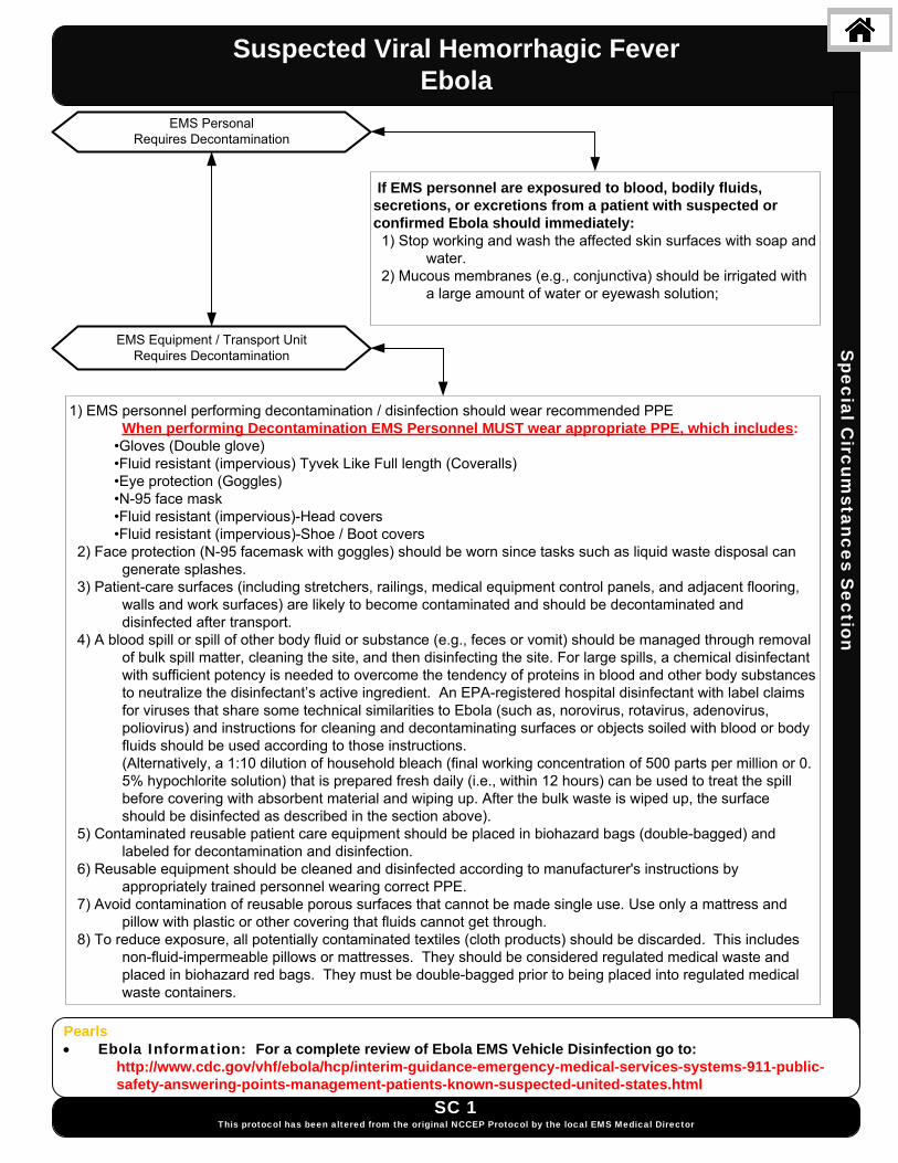
Suspected Viral Hemorrhagic FeverEbola
Special C
ircumstances S
ection
SC 1This protocol has been altered from the original NCCEP Protocol by the local EMS Medical Director
EMS Personal Requires Decontamination
EMS Equipment / Transport Unit Requires Decontamination
If EMS personnel are exposured to blood, bodily fluids, secretions, or excretions from a patient with suspected or confirmed Ebola should immediately: 1) Stop working and wash the affected skin surfaces with soap and
water. 2) Mucous membranes (e.g., conjunctiva) should be irrigated with
a large amount of water or eyewash solution;
1) EMS personnel performing decontamination / disinfection should wear recommended PPEWhen performing Decontamination EMS Personnel MUST wear appropriate PPE, which includes:
•Gloves (Double glove) •Fluid resistant (impervious) Tyvek Like Full length (Coveralls) •Eye protection (Goggles) •N-95 face mask •Fluid resistant (impervious)-Head covers •Fluid resistant (impervious)-Shoe / Boot covers 2) Face protection (N-95 facemask with goggles) should be worn since tasks such as liquid waste disposal can generate splashes. 3) Patient-care surfaces (including stretchers, railings, medical equipment control panels, and adjacent flooring,
walls and work surfaces) are likely to become contaminated and should be decontaminated and disinfected after transport.
4) A blood spill or spill of other body fluid or substance (e.g., feces or vomit) should be managed through removal of bulk spill matter, cleaning the site, and then disinfecting the site. For large spills, a chemical disinfectant with sufficient potency is needed to overcome the tendency of proteins in blood and other body substances to neutralize the disinfectant’s active ingredient. An EPA-registered hospital disinfectant with label claims for viruses that share some technical similarities to Ebola (such as, norovirus, rotavirus, adenovirus, poliovirus) and instructions for cleaning and decontaminating surfaces or objects soiled with blood or body fluids should be used according to those instructions.
(Alternatively, a 1:10 dilution of household bleach (final working concentration of 500 parts per million or 0. 5% hypochlorite solution) that is prepared fresh daily (i.e., within 12 hours) can be used to treat the spill before covering with absorbent material and wiping up. After the bulk waste is wiped up, the surface should be disinfected as described in the section above).
5) Contaminated reusable patient care equipment should be placed in biohazard bags (double-bagged) and labeled for decontamination and disinfection.
6) Reusable equipment should be cleaned and disinfected according to manufacturer's instructions by appropriately trained personnel wearing correct PPE.
7) Avoid contamination of reusable porous surfaces that cannot be made single use. Use only a mattress and pillow with plastic or other covering that fluids cannot get through.
8) To reduce exposure, all potentially contaminated textiles (cloth products) should be discarded. This includes non-fluid-impermeable pillows or mattresses. They should be considered regulated medical waste and
placed in biohazard red bags. They must be double-bagged prior to being placed into regulated medical waste containers.
Pearls Ebola Information: For a complete review of Ebola EMS Vehicle Disinfection go to: http://www.cdc.gov/vhf/ebola/hcp/interim-guidance-emergency-medical-services-systems-911-public-
safety-answering-points-management-patients-known-suspected-united-states.html

Suspected Viral Hemorrhagic FeverEbolaS
pecial Circum
stances Section
SC 1This protocol has been altered from the original NCCEP Protocol by the local EMS Medical Director
Decedent Known or suspected carrier of HVF / Ebola Requires Transportation
Only personnel trained in handling infected human remains, and wearing full PPE, should touch, or move any Ebola-infected remains.
Handling human remains should be kept to a minimum.
Donning / Doffing PPE
PPE should be in place BEFORE contact with the body1) Prior to contact with body, postmortem care
personnel must wear PPE consisting of: surgical scrub suit, surgical cap, impervious Tyvex-Coveralls, eye protection (e.g., face shield, goggles), facemask, shoe covers, and double surgical gloves.
2) Additional PPE (leg coverings,) might be required in certain situations (e.g., copious amounts of blood, vomit, feces, or other body fluids that can contaminate the environment).
PPE should be removed immediately after and discarded as regulated medical waste.
1) Use caution when removing PPE as to avoid contaminating the wearer.
2) Hand hygiene (washing your hands thoroughly with soap and water or an alcohol based hand rub) should be performed immediately following the removal of PPE. If hands are visibly soiled, use soap and water.
Preparation of Body Prior to Transport
1) At the site of death, the body should be wrapped in a plastic shroud. Wrapping of the body should be done in a way that prevents contamination of the outside of the shroud.
2) Change your gown or gloves if they become heavily contaminated with blood or body fluids.3) Leave any intravenous lines or endotracheal tubes that may be present in place. 4) Avoid washing or cleaning the body. 5) After wrapping, the body should be immediately placed in a leak-proof plastic bag not less than 150 μm
thick and zippered closed The bagged body should then be placed in another leak-proof plastic bag not less than 150 μm thick and zippered closed before being transported to the morgue.
Surface Decontamination
1) Prior to transport to the morgue, perform surface decontamination of the corpse-containing body bags by removing visible soil on outer bag surfaces with EPA-registered disinfectants which can kill a wide range of viruses.
2) Follow the product’s label instructions. Once the visible soil has been removed, reapply the disinfectant to the entire bag surface and allow to air dry.
3) Following the removal of the body, the patient room should be cleaned and disinfected. 4) Reusable equipment should be cleaned and disinfected according to standard procedures.
Transportation of VHV / Ebola Remains
PPE is required for individuals driving or riding in a vehicle carrying human remains. DO NOT handle the remains of a suspected / confirmed case of Ebola The remains must be safely contained in a body bag where the outer surface of the body bag has been disinfected prior to the transport.
Pearls Ebola Information: For a complete review of Handling Remains of Ebola Infected Patients go to: http://www.cdc.gov/vhf/ebola/hcp/guidance-safe-handling-human-remains-ebola-patients-us-hospitals-mortuaries.html

Special O
perations Section
Scene RehabilitationGeneral / Special Event / Mass Gathering
Initial Process1. Personnel logged into General Rehabilitation Section2. VS Assessed / Recorded (If HR > 110 then obtain Temp)
Carbon Monoxide monitoring if indicated3. Personnel assessed for signs / symptoms4. Remove PPE, Body Armor, Haz-Mat Suits, Turnout Gear,
Other equipment as indicated
Exit toAppropriate
ProtocolYES
Heator
Cold stressActive Cooling Measures
Forearm immersion, cool shirts, cool mist fans etc.
Rest 10 – 20 Minutes
Active Warming MeasuresDry responder, place in warm areaHot packs to axilla and / or groin
Rest 10 – 20 minutes
YES YES
Rehydration Techniques12 – 32 oz Oral Fluid over 20 minutes
Oral Rehydration may occur along with Active Cooling Measures
May administer up to 2L of NS
Rehydration Techniques12 – 32 oz Oral Fluid over 20 minutesOral Rehydration may occur along with
Active Warming Measures
Reassess responder after 20 Minutes in General Rehabilitation Section
Reassess VS
NO
NO
Injury / Illness / Complaint should be treated using appropriate treatment
protocol beyond need for oral or IV hydration.
SO 1This protocol has been altered from the original NCCEP Protocol by the Johnston County EMS Medical Director
Significant InjuryCardiac Complaint: Signs / Symptoms
Respiratory Complaint: Serious Signs / SymptomsRespiratory Rate < 8 or > 40Systolic Blood Pressure ≤ 80
HEAT STRESS COLD STRESS
HR ≥ 110
Temp ≥ 100.6
Extend Rehabilitation Time Until VS
Improve
YES YES
HR ≥ 110
Extend Rehabilitation Time Until VS
Improve
Discharge patient from General Rehabilitation Section
Temp ≥ 100.6
YES
NO NO
NONO
YES
VITAL SIGN CAVEATSBlood Pressure:
Prone to inaccuracy on scenes; must be interpreted in the proper context.
Individuals at special events may have elevated blood pressured due to
physical exertion and is not typically pathologic.
Individuals with SBP ≥160 or DBP ≥100 may need extended rehabilitation. However this does not necessarily prevent them from returning to the
event.
Temperature:Individuals may have increased
temperature during rehabilitation.

SO 1This protocol has been altered from the original NCCEP Protocol by the Johnston County EMS Medical Director
Special O
perations Section
Scene Rehabilitation: General / Special Event / Mass Gathering
Pearls This protocol should be utilized for evaluating patrons of certain special events that may or may not otherwise meet the
definition of a patient When a person presents with a medical complaint, or has persistent abnormal vital signs, they become a patient and an
individual PCR must be completed.. Rehabilitation goals:
Relief from climatic conditions.Rest, recovery, and hydration prior to incident, during, and following incident.Active and / or passive cooling or warming as needed for incident type and climate conditions.
May be utilized with adult responders on fire, law enforcement, rescue, EMS and training scenes. People taking anti-histamines, blood pressure medication, diuretics or stimulants are at increased risk for cold and heat
stress. Serious signs / symptoms:
Chest pain, dizziness, dyspnea, weakness, nausea, or headache.Symptoms of heat stress (cramps) or cold stress.Changes in gait, speech, or behavior.Altered Mental Status.Abnormal Vital Signs per agency SOP or Policy / Procedure.
Regarding documentation under this protocol, individuals who are evaluated only at the rehabilitation center require a narrative-based patient log entry under one PCR for all of these individuals. However, if a patient receives ALS care more than over-the-counter medications and/or is transported to an emergency department, the patient requires a separate run number and full PCR like any other patient.
Establish rehab location such that it provides shelter, privacy and freedom from smoke or other hazards. For approved gatherings, other patient contact requirements may be determined at approval of event coverage.

Special O
perations Section
Scene Rehabilitation: Responder
Pearls This protocol is optional and given only as an example. Agencies may and are encouraged to develop their own. Rehabilitation officer has full authority in deciding when responders may return to duty. Utilized when responder is not appropriate for General Rehabilitation Protocol. May be utilized with adult responders on fire, law enforcement, rescue, EMS and training scenes. Responders taking anti-histamines, blood pressure medication, diuretics or stimulants are at increased risk for cold and
heat stress. Rehabilitation Section is an integral function within the Incident Management System. Establish section such that it provides shelter, privacy and freedom from smoke or other hazards. If available, use the RAD-57 device in rehab to determine carbon monoxide levels of responders. May treat with fluids and D/C without Medical Control contact if only the ALS care provided is IV fluids up to two
liters.
Initial Process 1. Personnel logged into Responder Rehabilitation
Section 2. VS Assessed and Recorded / Orthostatic Vital Signs 3. Pulse oximetry and SPCO (if available) 4. Personnel assessed for signs / symptoms
Remove:PPEBody ArmorChemical SuitsSCBATurnout GearOther equipment as indicated
Continue:Heat and Cold Stresstreatment techniquesfrom General Rehab
Section
Discharge Responder from General
Rehabilitation Section
Reports for Reassignment
20 Minute Rest Period
Pulse Rate > 85 % NFPAAge Predicted Maximum
Normal Saline Bolus500 mL IV / IOMaximum 2 L
Titrate to HR ≤ 100An
SBP ≥ 100 mmHg
A
Systolic BP ≥ 160Or
Diastolic BP ≥ 100
Respirations < 8 or > 40
Pulse oximetry < 90 %SPCO > 10 %
Temperature ≥ 100.6
YES
NO
NO
NO
NO
NO
Mandatory Rest PeriodRehydration is Most
ImportantRe-evaluate in 10 minutes
No improvement after 30 minutes
of additional rehabilitation
YES
YES
YES
YES
Injury / Illness / Complaint should be treated using appropriate treatment
protocol beyond need for oral or IV hydration.
Use in conjunction with General Rehabilitation
Protocol
SO 2This protocol has been altered from the original NCCEP Protocol by the Johnston County EMS Medical Director
NFPA Age Predicted 85 %Maximum Heart Rate
20 - 2526 - 3031 - 3536 - 4041 - 4546 - 5051 -5555 - 6061 - 65
170165160155152148140136132
Notify Destination or Contact Medical Control

Special O
perations Protocols
Multiple Patient Incident
SO 3This protocol has been altered from the original NCCEP Protocol by the Johnston County EMS System Medical Director
Pearls In absence of guidance from JC911, consider utilizing the following communications assignments: 1. MEDICAL BRANCH on separate OPS channel 2. Transportation/Destination Coordination on separate OPS channel ICS Job Cards and ICS Job Vests should be utilized by all personnel involved in an MPI If blast injury with more than five (5) patients, patients with SBP <90 mm Hg and/or obvious external trauma to four (4) or more body areas should go to a Level I Trauma Center (WakeMed/Raleigh). Others may be considered for community hospital transport. Multiple patients may be transported in the same EMS unit if needed during a catastrophic mass cassualty. When
possible, patients of similar acuity should be transported in the same unit to assist with appropriate transport destination.
Differential Blast response MPI penetrating trauma MPI blunt trauma/MVC
Signs and Symptoms SLUDGEM for chemical exposure Respiratory distress for narcotic
exposure Nausea/vomiting for radiation
History Number of patients Cause of incident Chemical, biological, or radiological
contamination Secondary devices
Scene safety; Maintain situational awareness
If blast response, utilize respiratory protection and full turnout gear until
advised to remove
Provide scene size-up on assigned TAC channel; activate appropriate ALARM
level
If not already accomplished, establish COMMAND and TRIAGE (utilize ICS job
cards)
Move all ambulatory (GREEN) patients to a safe area in COLD ZONE
Move non-ambulatory patients to transportation area as rapidly as
possible. Establish treatment areas only if there are insufficient transport
resources available for rapid evacuation
Consider public transportation to
alternative receiving facility for ambulatory
patients
Triage Protocolas needed
Appropriate Treatment Protocols
as needed and resources allow

Special O
perations Section
Evacuate wounded and facilitate transport
Active Threat
SO 4This protocol has been altered from the original NCCEP Protocol by the Johnston County EMS System Medical Director
Active Threat Incident
Respond with law enforcement as a rescue task force and develop a casualty collection point until scene
secure.
PearlsMake notification to JC911 and all local hospitals as soon as possible with the projected number of patients.Level 1 trauma centers can manage up to 5 penetrating trauma patients per hour. If more than 5 penetrating trauma patients, consider transporting to multiple trauma centers. If more than 30 patients require Level One trauma care, then transport of trauma patients to non-trauma center local hospitals may be required.The penetrating trauma bag or “active threat bag” is the only equipment initially required. If a casualty collection point is established and additional equipment is required, a cache of needed supplies should be requested.Please refer to the MCI protocol for proper radio communication and channel assignments. Task cards and job vests should be utilized by all personnel involved in an MPIIf blast injury with more than 5 patients, patients with SBP <90 and/or obvious external trauma to 4 or more body surface areas should go to Level 1 trauma center. Others may be considered for a community hospital transport.Multiple patients may be transported in the same EMS unit if needed. When possible, patients with similar acuity should be transported in the same unit to assist with appropriate transport destination.
Sufficient transport units on scene?
Establish treatment areas until sufficient transport resources are available
NO
Rapidly transport all injuries to appropriate facility.
1-5 trauma patients – nearest trauma center>5 transport to multiple trauma centers
YES
Move all non-penetrating, ambulatory patients to a safe area in cold zone. Consider public
transportation to alternative receiving facility for ambulatory patients
Threat detained / threat neutralized and scene immediately safe
YES
NO

Special O
perations Protocols
Pearls This protocol applies to ALL responders Patients who are denying more severe symptoms may initially present for a “routine check”. Please confirm with the
patient at least twice that they have no medical complaints. All persons who request service are considered patients and shall have a PCR completed.
For these patients, the PCR may be brief but must include vital signs and documentation of the lack of a medical complaint. Additionally, patients with a potential mechanism for trauma should have a trauma exam completed.
Should a patient refuse evaluation and/or decline further evaluation once begun, document as much as you can. Even patients who refuse vital signs can be observed and RR assessed. The PCR narrative is key in these and all cases, and must accurately and thoroughly describe the patient encounter.
Any person that a responder comes into contact with that has a medical complaint or concern for themselves or others immediately with them should be evaluated under this protocol.
History Patient requests “blood
pressure check” EMS responds to “assist invalid” Someone else called 911;
patient did not request Other situation in which patient
does not have a medical complaint or obvious injury
Signs and Symptoms Assess for medical complaint For patients with hypertension,
particularly check for chest pain, shortness of breath, and/or neurologic changes
For assist invalid calls, particularly check for syncope, trauma from fall, or inability to ambulate
Differential Hypertensive urgency Hypertensive emergency Syncope Cardiac ischemia Cardiac dysrhythmia Fracture Head trauma
Well Person Check
SO 5This protocol is unique to the Johnston County EMS System
Universal Care Protocol
Patient has medical complaint or obvious injury/trauma?
Go to appropriate protocol and recommend transport
Obtain vital signs: HR, RR, BP, SpO2, FSBG, ETCO2
HR >110 or <50SBP >200 or <85
DBP >120RR >24 or <6SpO2 <94%
FSBG <60 or >400ETCO2 ≥30 or ≤50
Temp ≥96.8ºF or ≤100.4ºF
Re-confirm that the patient has no medical complaint.Provide patient with VS results and have them
contact their doctor to report results
Recommend transport for evaluation. Have patient sign refusal if transport declined.
Advise patient to call 9-1-1 if they develop any symptoms. Complete PCR and document all elements of this protocol.
YES
NO
YES
NO High risk refusal?Contact Medical Control

Special R
esponse Protocols
History Situational crisis Psychiatric illness / medications Injury to self or threats to others Medic alert Tag Substance abuse / overdose Diabetes
Signs and Symptoms Anxiety, agitation, confusion Affect change, hallucinations Delusional thoughts, bizarre
behavior Expression of suicidal / homicidal
thoughts
Differential Altered mental status Alcohol intoxication Toxin / substance abuse Withdrawal syndromes Depression Bipolar (manic-depressive) Schizophrenia Anxiety disorders
Crisis Response
SO 5BThis protocol is unique to the Johnston County EMS System
This protocol should only be utilized after redirection from the Behavioral Protocol and the patient / guardian has consented to Crisis Intervention
Universal Care Protocol
Scene Safe?
Call for help / additional resources.
Stage until the scene is safe and
maintain situational awareness
No
Mandatory Evaluation Criteria LOC – Alert / oriented to person place time and event BAC <0.3 HR < 120 SBP > 100 mm hg BSFS > 70 or <300 Temperature <100.4ºF or >96.0ºF No acute Trauma Non violent Able to ambulate or operate wheelchair No seizure within the past 24 hours
No
Patient does not qualify for crisis intervention. Consider
appropriate protocol and transportation to the appropriate
emergency department.
If you are not a CIT certified paramedic, request a supervisor response “reference crisis
intervention.”
Utilize de-escalation techniques and maintain situational awareness
Consider on-site intervention, calling mobile crisis, or contacting a mental health
provider for appointment
CIT P
Monday – Friday 08:00 – 17:00 and not a holiday
CIT P
Consider transporting to the appropriate emergency departmentP
Contact Johnston County Crisis and Assessment Center (919) 989-5500 . Be prepared to give vital
sings, pertinent PMH, and ETA
CIT P
NoYes

Traum
a and Burn P
rotocol Section
Blast Injury / IncidentHistory Type of exposure (heat, gas,
chemical) Inhalation injury Time of Injury Past medical history /
Medications Other trauma Loss of Consciousness Tetanus/Immunization status
Signs and Symptoms Burns, pain, swelling Dizziness Loss of consciousness Hypotension/shock Airway compromise/distress could
be indicated by hoarseness/wheezing / Hypotension
Differential Superficial (1st Degree) red - painful (Don’t
include in TBSA) Partial Thickness (2nd Degree) blistering Full Thickness (3rd Degree) painless/charred
or leathery skin Thermal injury Chemical – Electrical injury Radiation injury Blast injury
TB 1This protocol has been altered from the original NCCEP Protocol by the Johnston County EMS Medical Director
Nature of Device: Agent / Amount. Industrial Explosion. Terrorist Incident. Improvised Explosive Device.Method of Delivery: Incendiary / ExplosiveNature of Environment: Open / Closed.Distance from Device: Intervening protective barrier. Other environmental hazards, Evaluate for: Blunt Trauma / Crush Injury / Compartment Syndrome / Traumatic Brain Injury / Concussion / Tympanic Membrane
Rupture / Abdominal hemorrhage or Evisceration, Blast Lung Injury and Penetrating Trauma.
Scene Safety / Quantify and Triage Patients / Load and Go with Assessment / Treatment Enroute
Blast Lung Injury
Multiple Trauma Protocol TB 6if indicated
Age AppropriateAirway Protocol(s) AR 1, 2, 3, 5, 6
as indicated
Cardiac Monitor & ETCO2 if indicated
IV / IO Procedureif indicatedA
P
Pain Control Protocol UP 11if indicated
Safe Transport to appropriate destination using Trauma and Burn:
EMS Triage and Destination Plan
NO
Age AppropriateAirway Protocol(s) AR 4, 7
as indicatedYES
Thermal Burn Protocol TB 9Chemical and Electrical Burn Protocol TB 2
if indicated
Crush Injury Protocol TB 3if indicated
Triage Protocol UP 2as indicated
Radiation Incident Protocol TB 7if indicated
Accidental / Intentional Explosions(See Pearls)
Notify Destination or Contact Medical Control
Decontamination Procedureif indicated

TB 1This protocol has been altered from the original NCCEP Protocol by the Johnston County EMS Medical Director
Blast Injury / Incident Traum
a and Burn S
ection
Pearls Types of Blast Injury:
Primary Blast Injury: From pressure wave. Secondary Blast Injury: Impaled objects. Debris which becomes missiles / shrapnel.Tertiary Blast Injury: Patient falling or being thrown / pinned by debris.Most Common Cause of Death: Secondary Blast Injuries.
Triage of Blast Injury patients:Blast Injury Patients with Burn Injuries Must be Triaged using the Thermal / Chemical / Electrical Burn Destination Guidelines for Critical / Serious / Minor Trauma and BurnsPatients may be hard of hearing due to tympanic membrane rupture.
Care of Blast Injury Patients: Patients may suffer multi-system injuries including blunt and penetrating trauma, shrapnel, barotrauma, burns, and toxic
chemical exposure.Consider airway burns which should prompt early and aggressive airway management.Cover open chest wounds with semi-occlusive dressing. Use Lactated Ringers (if available) for all Critical or Serious Burns.Minimize IV fluids resuscitation in patients with no sign of shock or poor perfusion.
Blast Lung Injury:Blast Lung Injury is characterized by respiratory difficulty and hypoxia. Can occur (rarely) in patients without external
thoracic trauma. More likely in enclosed space or in close proximity to explosion.Symptoms: Dyspnea, hemoptysis, cough, chest pain, wheezing and hemodynamic instability.Signs: Apnea, tachypnea, hypopnea, hypoxia, cyanosis and diminished breath sounds.Air embolism should be considered and patient transported prone and in slight left-lateral decubitus position.Blast Lung Injury patients may require early intubation but positive pressure ventilation may exacerbate the injury, avoid
hyperventilation. Air transport may worsen lung injury as well and close observation is mandated. Tension pneumothorax may occur
requiring chest decompression. Be judicious with fluids as volume overload may worsen lung injury. Accidental Explosions or Intentional Explosions:
All explosions or blasts should be considered intentional until determined otherwise.Attempt to determine source of the blast to include any potential threat for aerosolization of hazardous materials.Evaluate scene safety to include the source of the blast that may continue to spill explosive liquids or gases. Consider structural collapse / Environmental hazards / Fire. Conditions that led to the initial explosion may be returning and lead to a second explosion. Greatest concern is potential threat for a secondary device. Patients who can, typically will attempt to move as far away from the explosive source as they safely can.Evaluate surroundings for suspicious items; unattended back packs or packages, or unattended vehicles.If patient is unconscious or there is(are) fatality(fatalities) and you are evaluating patient(s) for signs of life:
Before moving note if there are wires coming from the patient(s), or it appears the patient(s) is(are) lying on a package/pack, or bulky item, do not move the patient(s), quickly back away and immediately notify a law enforcement officer.
If there are no indications the patient is connected to a triggering mechanism for a secondary device, expeditiously remove the patient(s) from the scene and begin transport to the hospital.
Protect the airway and cervical spine, however, beyond the primary survey, care and a more detailed assessment should be deferred until the patient is in the ambulance.
If there are signs the patient was carrying the source of the blast, notify law enforcement immediately and most likely, a law enforcement officer will accompany your patient to the hospital.
SCENE NOTIFICATIONAs soon as assessment reveals that patient meets trauma triage criteria, contact receiving facility via
VIPER radio to make a trauma notification to WakeMed/ Raleigh:
Patient’s age, sex, and GCSPrimary trauma triage criteria
Estimated time of arrival (ETA) to facility
Provide updates from scene as needed and update via radio or phone when approximately 10-15 minutes out.

Chemical and Electrical BurnHistory Type of exposure (heat, gas,
chemical) Inhalation injury Time of Injury Past medical history /
Medications Other trauma Loss of Consciousness Tetanus/Immunization status
Signs and Symptoms Burns, pain, swelling Dizziness Loss of consciousness Hypotension/shock Airway compromise/distress could
be indicated by hoarseness/wheezing / Hypotension
Traum
a and Burn P
rotocol Section
Differential Superficial (1st Degree) red - painful (Don’t
include in TBSA) Partial Thickness (2nd Degree) blistering Full Thickness (3rd Degree) painless/charred
or leathery skin Thermal injury Chemical – Electrical injury Radiation injury Blast injury
TB 2This protocol has been altered from the original NCCEP Protocol by the Johnston County EMS Medical Director
Assure Chemical Source is NOT Hazardous to Responders.Assure Electrical Source is NO longer in contact with patient before touching patient.
Eye InvolvementIrrigate Involved Eye(s) with Normal Saline
for 15 – 30 minutesMay repeat as neededChemical Exposure / Burn
Flush Contact Area with Normal Saline for 15 minutes
Identify Contact Points
Thermal Burn Protocol TB 9
Decontamination Procedureif indicated
Age Appropriate Cardiac Protocol(s)if indicated
Assess Burn / Concomitant Injury Severity
>15% TBSA 2nd/3rd Degree BurnBurns with Multiple TraumaBurns with definitive airway
compromise(When reasonably accessible,
transport to a Burn Center)Critical Burn
5-15% TBSA 2nd/3rd Degree BurnSuspected inhalation injury or requiring
intubation for airway stabilizationHypotension or GCS 13 or Less(When reasonably accessible,
transport to a Burn Center)Serious Burn
< 5% TBSA 2nd/3rd Degree BurnNo inhalation injury, Not Intubated,
NormotensiveGCS 14 or Greater
Minor Burn
Age Appropriate Airway Protocol(s) AR 1, 2, 3, 4, 5, 6, 7
if indicated
Safe Transport to appropriate destination using Trauma and Burn:
EMS Triage and Destination Plan
Notify Destination or Contact Medical Control
Prehospital Fluid Resuscitation for Burns
≤5 y/o: 125mL LR / hour 6‐13 y/o: 250mL LR / hour ≥14 y/o: 500mL LR / hour
Utilize the Dial‐A‐Flow extension to administer
accurate fluids
Refer to Thermal Burn Protocol TB 9
for the Modified Parkland Formula in
regards to Interfacility Transport Fluid Administration

TB 2This protocol has been altered from the original NCCEP Protocol by the Johnston County EMS Medical Director
Traum
a and Burn P
rotocol Section
Chemical and Electrical Burn
Pearls Recommended Exam: Mental Status, HEENT, Neck, Heart, Lungs, Abdomen, Extremities, Back, and Neuro Green, Yellow and Red In burn severity do not apply to Triage systems. Refer to Rule of Nines: Remember the extent of the obvious external burn from an electrical source does
not always reflect more extensive internal damage not seen. Chemical Burns:
Refer to Decontamination Procedure.Normal Saline or Sterile Water is preferred, however if not available, do not delay irrigation and use tap water. Other water sources may be used based on availability. Flush the area as soon as possible with the cleanest readily available water or saline solution using copious
amounts of fluids. If it is a dry chemical, gently brush it off as it may react violently with water. Electrical Burns:
DO NOT contact patient until you are certain the source of the electrical shock is disconnected.Attempt to locate contact points (generally there will be two or more.) A point where the patient contacted the
source and a point(s) where the patient is grounded. Sites will generally be full thickness. Do not refer to as entry and exit sites or wounds.Cardiac Monitor: Anticipate ventricular or atrial irregularity including VT, VF, atrial fibrillation and / or heart
blocks.Attempt to identify the nature of the electrical source (AC / DC), the amount of voltage and the amperage the
patient may have been exposed to during the electrical shock.
Rule of Nines Seldom do you find a complete isolated
body part that is injured as described in the Rule of Nines.
More likely, it will be portions of one area, portions of another, and an approximation will be needed.
For the purpose of determining the extent of serious injury, differentiate the area with minimal or 1st degree burn from those of partial (2nd) or full (3rd) thickness burns.
For the purpose of determining Total Body Surface Area (TBSA) of burn, include only Partial and Full Thickness burns. Report the observation of other superficial (1st degree) burns but do not include those burns in your TBSA estimate.
Some texts will refer to 4th 5th and 6th degree burns. There is significant debate regarding the actual value of identifying a burn injury beyond that of the superficial, partial and full thickness burn at least at the level of emergent and primary care. For our work, all are included in Full Thickness burns.
Other burn classifications in general include: 4th referring to a burn that
destroys the dermis and involves muscle tissue.
5th referring to a burn that destroys dermis, penetrates muscle tissue, and involves tissue around the bone.
6th referring to a burn that destroys dermis, destroys muscle tissue, and penetrates or destroys bone tissue.
Estimate spotty areas of burn by using the size of the patient’s palm as 1 %

Traum
a and Burn P
rotocol Section
History Entrapped and crushed under
heavy load > 30 minutes Extremity / body crushed Building collapse, trench
collapse, industrial accident, pinned under heavy equipment
Signs and Symptoms Hypotension Hypothermia Abnormal ECG findings Pain Anxiety
Differential Entrapment without crush syndrome Vascular injury with perfusion deficit Compartment syndrome Altered mental status
A
P
Exit to Age Appropriate Cardiac Arrest
Protocol AC 3 / PC 4Arrhythmia Protocol(s)
if indicated
Sodium Bicarbonate50 mEq IV / IO
Pediatric: 1 mEq / kg IV / IO
Calcium Chloride 1 g IV / IOPediatric: 20 mg / kg IV / IO
Over 2- 3 minutes
Crush Syndrome Trauma
TB 3This protocol has been altered from the original NCCEP Protocol by the Johnston County EMS Medical Director
Normal Saline Infusion1 Liter per hour IV / IO
Pediatric:3 x maintenance fluid rate
P
Abnormal ECGPeaked T Waves
QRS ≥ 0.12 secondsQT ≥ 0.46 seconds
Loss of P waveOr
Hemodynamically UnstableOr
Asystole / PEA / VF / VT
ConsiderMidazolam 0.5 – 2 mg IV / IO
Midazolam 1 – 2 mg INMaximum 5 mg
Pediatric: 0.1 – 0.2 mg / kg IV / IO / IN
Pediatric Maximum 2 mgOver 2 – 3 minutes as needed
Notify Destination or Contact Medical Control
Safe Transport to appropriate destination using Trauma and Burn:
EMS Triage and Destination Plan
Decrease Normal Saline Infusion500 mL per hour IV / IO
Pediatric:Maintenance fluid rate
Entrapped < 2 hours
Entrapped > 2 hours A
Age AppropriateAirway
Protocol(s) AR 1, 2, 3, 4, 5, 6, 7as indicated
IV / IO Procedure
12 Lead ECG Procedure
Cardiac Monitor & ETCO2
B
P
A
Pain Control Protocol UP 11as indicated
Multiple Trauma Protocol TB 6if indicated
Thermal Burn Protocol TB 9Chemical and Electrical Burn
Protocol TB 2if indicated
Albuterol Nebulizer2.5 – 5 mg
May repeat x 3
ConsiderAge Appropriate Multiple
Trauma ProtocolHypothermia / Hyperthermia
Protocol(s)as indicated

TB 3This protocol has been altered from the original NCCEP Protocol by the Johnston County EMS Medical Director
Traum
a and Burn P
rotocol Section
Crush Syndrome Trauma
Pearls Recommended exam: Mental Status, Musculoskeletal, Neuro Scene safety is of paramount importance as typical scenes pose hazards to rescuers. Call for appropriate
resources. Lowest systolic blood pressure by age: < 31 days: > 60 mmHg. 31 days to 1 year: > 70 mmHg. Greater than 1 year: 70 + 2 x age in years. Pediatric IV Fluid maintenance rate: 4 mL per first 10 kg of weight + 2 mL per second 10 kg of weight + 1 mL
for every additional kg in weight. Crush syndrome typically manifests after 2 – 4 hours of crush injury, but may present in < 1 hour. Fluid resuscitation:
If access to patient and initiation of IV fluids occurs after 2 hours, give 2 liters of IV fluids in adults and 20 mL/kg of IV fluids in pediatrics and then begin > 2 hour dosing regimen.
Consider all possible causes of shock and treat per appropriate protocol. Majority of decompensation in pediatrics is airway related.
Decreasing heart rate and hypotension occur late in children and are signs of imminent cardiac arrest. Shock may be present with a normal blood pressure initially. Shock often is present with normal vital signs and may develop insidiously. Tachycardia may be the only
manifestation. Consider all possible causes of shock and treat per appropriate protocol. Patients may become hypothermic even in warm environments. Hyperkalemia from crush syndrome can produce ECG changes described in protocol, but may also be a bizarre,
wide complex rhythm. Wide complex rhythms should also be treated using the VF/Pulseless VT Protocol.
Pediatric Fluid Maintenance rate example25kg pediatric(4x10) mL + (2x10) mL + (5x1) mL =
60 mL/hr or 1mL/min
SCENE NOTIFICATIONAs soon as assessment reveals that patient meets trauma triage criteria, contact receiving facility via
VIPER radio to make a trauma notification to WakeMed/ Raleigh:
Patient’s age, sex, and GCSPrimary trauma triage criteria
Estimated time of arrival (ETA) to facility
Provide updates from scene as needed and update via radio or phone when approximately 10-15 minutes out.
ECG changes related to hyperkalemia are:Mild hyperkalemia (6-7 mmol/l) – peaked T waves.Moderate hyperkalemia (7 – 8 mmol/l) – flattened P wave, prolonged PR interval, depression of ST segment, peaked T wave.Severe hyperkalemia (8 – 9 mmol/l) – atrial standstill, prolonged QRS duration, further peaking T waves.Life-threatening hyperkalemia (>9 mmol/l) – sine wave pattern.

Traum
a and Burn P
rotocol Section
History Type of injury Mechanism: crush / penetrating /
amputation Time of injury Open vs. closed wound / fracture Wound contamination Medical history Medications
Signs and Symptoms Pain, swelling Deformity Altered sensation / motor function Diminished pulse / capillary refill Decreased extremity temperature
Differential Abrasion Contusion Laceration Sprain Dislocation Fracture Amputation
Clean amputated part, Wrap part in sterile dressing soaked in normal saline
and place in air tight container.
Place container on ice if available.
Amputation and / or Open Fracture YES
Extremity Trauma
TB 4This protocol has been altered from the original NCCEP Protocol by the Johnston County EMS Medical Director
Monitor and Reassess
Wound careControl Hemorrhage with Direct Pressure
Splinting as indicated
Pain Protocol UP 11if indicated
Wound Care - Tourniquet Procedureif indicated
IV / IO Procedureif indicated
Wound Care – Wound Packing If indicated
A
Crush Syndrome Protocol TB 3as indicated
Age Appropriate Airway Protocol(s) AR 1, 2, 3, 4, 5, 6, 7
if indicated
Multiple Trauma Protocol TB 6if indicated
Age Appropriate Hypotension / ShockProtocol AM 5 / PM 3
if indicated
Notify Destination or Contact Medical Control
NO
Suspected blunt trauma?Usual equipment is taken to
patient’s side
Suspected penetrating trauma?Active Threat Bag should be
included with usual equipment.
Safe Transport to appropriate destination using
Trauma:EMS Triage and Destination Plan
Scene time should be limited≤10 minutes
Provide Early Trauma Notification

TB 4This protocol has been altered from the original NCCEP Protocol by the Johnston County EMS Medical Director
Traum
a and Burn P
rotocol Section
Extremity Trauma
Pearls Recommended Exam: Mental Status, Extremity, Neuro Peripheral neurovascular status is important In amputations, time is critical. Transport and notify medical control immediately, so that the appropriate destination
can be determined. Hip dislocations and knee and elbow fracture / dislocations have a high incidence of vascular compromise. Urgently transport any injury with vascular compromise. Blood loss may be concealed or not apparent with extremity injuries. Lacerations must be evaluated for repair within 6 hours from the time of injury. Multiple casualty incident: Tourniquet Procedure may be considered first instead of direct pressure. The decision to transport a patient to a trauma center utilizing lights and sirens transport should be based upon the
clinical picture presented and reasonable expectation of potential changes. Routine transport of stable patients utilizing lights and sirens transport, especially those that meet trauma criteria by mechanism of injury or non-physiological criteria, is discouraged.
SCENE NOTIFICATIONAs soon as assessment reveals that patient meets trauma triage criteria, contact receiving facility via
VIPER radio to make a trauma notification to WakeMed/ Raleigh:
Patient’s age, sex, and GCS
Primary trauma triage criteria
Estimated time of arrival (ETA) to facility
Provide updates from scene as needed and update via radio or phone when approximately 10-15 minutes out.

Traum
a and Burn P
rotocol Section
History Time of injury Mechanism (blunt vs. penetrating) Loss of consciousness Bleeding Past medical history Medications Evidence for multi-trauma
Signs and Symptoms Pain, swelling, bleeding Altered mental status Unconscious Respiratory distress / failure Vomiting Major traumatic mechanism of injury Seizure
Differential Skull fracture Brain injury (Concussion, Contusion,
Hemorrhage or Laceration) Epidural hematoma Subdural hematoma Subarachnoid hemorrhage Spinal injury Abuse
Altered Mental Status Protocol UP 4
if indicated
DO NOT ROUTINELYHYPERVENTILATE
Ventilate 8-10 breaths per minute to maintain EtCO2 35-45
mmHg
OR
Evidence ofBrain Herniation:
Unilateral or Bilateral Dilation of Pupils / Posturing
Hyperventilate to maintainEtCO2 30 – 35 mmHg
See Pearls
Head Trauma
TB 5This protocol has been altered from the original NCCEP Protocol by the Johnston County EMS Medical Director
Supplemental oxygenMaintain SpO2 ≥ 90%
Preferably ≥ 94%
Prevent Oxygen desaturation events < 90%Maintain EtCO235 – 45 mmHg
Cardiac Monitor & ETCO2
Age AppropriateAirway Protocol(s) AR 1, 2, 3, 5, 6
if indicated
Obtain and Record GCS
IV / IO Procedureif indicated
A
B
Blood Glucose Analysis Procedure
Safe Transport to appropriate destination using Trauma and Burn:
EMS Triage and Destination Plan
Notify Destination or Contact Medical Control
P
Monitor and Reassess
Spinal Motion Restriction Procedure / Protocol TB 8
if indicated
Multiple Trauma Protocol TB 6if indicated
Seizure Protocol UP 13if indicated
Age AppropriateHypotension / Shock Protocol AM 5 / PM 3
if indicated
Pain Control Protocol UP 11if indicated

TB 5This protocol has been altered from the original NCCEP Protocol by the Johnston County EMS Medical Director
Traum
a and Burn P
rotocol Section
Pearls Recommended Exam: Mental Status, HEENT, Heart, Lungs, Abdomen, Extremities, Back, Neuro GCS is a key performance measure used in the EMS Acute Trauma Care Toolkit. A single episode of hypoxia and / or hypotension can significantly increase morbidity and mortality with
head injury. Hyperventilation in head injury:
Hyperventilation lowers CO2 and causes vasoconstriction leading to increased intracranial pressure (ICP) and should not be done routinely.
Use in patient with evidence of herniation (blown pupil, decorticate / decerebrate posturing, bradycardia, decreasing GCS).
If hyperventilation is needed, ventilate at 14 – 18 / minute to maintain EtCO2 between 30 - 35 mmHg. Short term option only used for severe head injury typically GCS ≤ 8 or unresponsive.
Do not place in Trendelenburg position as this may increase ICP and worsen blood pressure. Poorly fitted cervical collars may also increase ICP when applied too tightly. In areas with short transport times, Drug Assisted Airway protocol is not recommended for patients who are
spontaneously breathing and who have oxygen saturations of ≥ 90% with supplemental oxygen including BIAD / BVM.
Hypotension:Limit IV fluids unless patient is hypotensive.Increased intracranial pressure (ICP) may cause hypertension and bradycardia (Cushing's Response).Usually indicates injury or shock unrelated to the head injury and should be aggressively treated.Fluid resuscitation should be titrated to maintain at least a systolic BP of > 70 + 2 x the age in years.Lowest systolic blood pressure by age: < 31 days: > 60 mmHg. 31 days to 1 year: > 70 mmHg. Greater than 1 year: 70 + 2 x age in years.
An important item to monitor and document is a change in the level of consciousness by serial examination. Consider Restraints if necessary for patient’s and/or personnel’s protection per the Restraint Procedure. Concussions:
Traumatic brain injuries involving any of a number of symptoms including confusion, LOC, vomiting, or headache.
Any prolonged confusion or mental status abnormality which does not return to normal within 15 minutes or any documented loss of consciousness should be evaluated by a physician ASAP.
EMS Providers should not make return-to-play decisions when evaluating an athlete with suspected concussion. This is outside the scope of practice.
Head Trauma
SCENE NOTIFICATIONAs soon as assessment reveals that patient meets trauma triage criteria, contact receiving facility via
VIPER radio to make a trauma notification to WakeMed/ Raleigh:
Patient’s age, sex, and GCS
Primary trauma triage criteria
Estimated time of arrival (ETA) to facility
Provide updates from scene as needed and update via radio or phone when approximately 10-15 minutes out.

Traum
a and Burn P
rotocol Section
History Time and mechanism of injury Damage to structure or vehicle Location in structure or vehicle Others injured or dead Speed and details of MVC Restraints / protective equipment Past medical history Medications
Signs and Symptoms Pain, swelling Deformity, lesions, bleeding Altered mental status or
unconscious Hypotension or shock Arrest
Differential (Life threatening) Chest: Tension pneumothorax
Flail chestPericardial tamponadeOpen chest woundHemothorax
Intra-abdominal bleeding Pelvis / Femur fracture Spine fracture / Cord injury Head injury (see Head Trauma) Extremity fracture / Dislocation HEENT (Airway obstruction) Hypothermia
A IV / IO Procedure
VS / Perfusion / GCS
Cardiac Monitor & ETCO2P
Multiple Trauma
TB 6This protocol has been altered from the original NCCEP Protocol by the Johnston County EMS Medical Director
Age AppropriateAirway Protocol(s) AR 1, 2, 3, 5, 6
if indicated
Safe Transport to appropriate destination using Trauma and Burn:
EMS Triage and Destination PlanLimit Scene Time ≤ 10 minutes
Provide Early Scene Notification
Head Injury Protocol TB 5if indicated
Chest Decompression-Needle Procedureif indicated
AbnormalNormal
Monitor and Reassess
Repeat Assessment Adult Procedure
Monitor and Reassess
Obtain and Record GCS
Control External Hemorrhage Consider Pelvic Binding
Splint Suspected Fractures
P
Spinal Motion Restriction Procedure / Protocol TB 8
if indicated
Age Appropriate Hypotension / Shock Protocol AM 5 / PM 3
if indicated
Notify Destination or Contact Medical Control
Altered Mental Status Protocol UP 4
if indicated
Pain Control Protocol UP 11if indicated
Routine spinal precautions in the setting of patients
with penetrating trauma is not recommended in the
absence of signs / symptoms of spinal cord
injury

TB 6This protocol has been altered from the original NCCEP Protocol by the Johnston County EMS Medical Director
Traum
a and Burn P
rotocol Section
Multiple Trauma
JOHNSTON COUNTY EMS SYSTEM TRAUMA TRIAGE CRITERIA
ASSESS VITAL SIGNS AND LEVEL OF CONSCIOUSNESS: Glasgow Coma Scale <13 Systolic Blood pressure <90mm Hg Respiratory rate <10 or >29 breaths per minute (<20 in infant aged <1 year)
ASSESS ANATOMY OF INJURY: All penetrating injuries to head, neck, torso, and extremities proximal to elbow and knee Chest wall instability or deformity (e.g. flail chest Crush, degloved, mangled, or pulseless extremity Amputation proximal to wrist or ankle Pelvic fractures Open or depressed skull fracture Paralysis
ASSESS MECHANISM AND EVIDENCE OF HIGH‐ENERGY IMPACT: Falls Adults: >20 feet (one story is 10 feet) Children: >10 feet or 2‐3 times height of child High‐risk auto crash Intrusion, including roof: >12 inches occupant site; >18 inches passenger compartment Death in same passenger compartment Vehicle telemetry data consistent with high risk of injury Auto vs. pedestrian/bicyclist thrown, run over, or with significant (>20 mph) impact Motorcycle crash >20 mph
ASSESS SPECIAL PATIENT OR SYSTEM CONSIDERATIONS: Older adults Age ≥55 years Age ≥65 years with SBP <110mm Hg in setting of injury Ground level fall with s/s of head/serious injury Children Triaged to pediatric capable trauma center Anticoagulants and bleeding disorders Patients taking clopigrodel, Coumadin, heparin, ASA, or NSAID’s Hemophilia or clotting disorders Pregnancy >20 weeks EMS Provider judgement
SCENE NOTIFICATIONAs soon as assessment reveals that patient meets trauma triage criteria, contact receiving facility via
VIPER radio to make a trauma notification to WakeMed/ Raleigh:
Patient’s age, sex, and GCS
Primary trauma triage criteria
Estimated time of arrival (ETA) to facility
Provide updates from scene as needed and update via radio or phone when approximately 10-15 minutes out.

TB 6This protocol has been altered from the original NCCEP Protocol by the Johnston County EMS Medical Director
Traum
a and Burn P
rotocol Section
Pearls Recommended Exam: Mental Status, Skin, HEENT, Heart, Lung, Abdomen, Extremities, Back, Neuro Items in Red Text are key performance measures used in the EMS Acute Trauma Care Toolkit Transport Destination is chosen based on the EMS System Trauma Plan with EMS pre-arrival notification. Scene times should not be delayed for procedures. These should be performed en route when possible.
Rapid transport of the unstable trauma patient to the appropriate facility is the goal. Control external hemorrhage and prevent hypothermia by keeping patient warm. Consider Chest Decompression with signs of shock and injury to torso and evidence of tension
pneumothorax. Trauma Triad of Death:
Metabolic acidosis / Coagulopathy / HypothermiaAppropriate resuscitation measures and keeping patient warm regardless of ambient temperature helps to
mitigate metabolic acidosis, coagulopathy, and hypothermia. Bag valve mask is an acceptable method of managing the airway if pulse oximetry can be maintained ≥ 90% Trauma in Pregnancy:
Providing optimal care for the mother = optimal care for the fetus. After 20 weeks gestation (fundus at or above umbilicus) transport patient on left side with 10 – 20° of elevation.
Pediatric Trauma:Age specific blood pressure 0 – 28 days > 60 mmHg, 1 month - 1 year > 70 mmHg, 1 - 10 years > 70 +
(2 x age)mmHg and 11 years and older > 90 mmHg. Geriatric Trauma:
Evaluate with a high index of suspicion. Often occult injuries are more difficult to recognize and patients can decompensate unexpectedly with little
warning.Risk of death with trauma increases after age 55.SBP < 110 may represent shock / poor perfusion in patients over age 65. Low impact mechanisms, such as ground level falls might result in severe injury especially in age over 65.
Severe bleeding from an extremity not rapidly controlled with direct pressure may necessitate the application of a tourniquet.
Maintain high-index of suspicion for domestic violence or abuse, pediatric non-accidental trauma, or geriatric abuse.
Multiple Trauma Preload the stretcher with an emergency thermal blanket prior to extricating a trauma patient.
This can be a beneficial tool in combating the hypothermia arm of the trauma triad of death.

Traum
a and Burn P
rotocol Section
Radiation IncidentHistory Type of exposure (heat, gas,
chemical) Inhalation injury Time of Injury Past medical history /
Medications Other trauma Loss of Consciousness Tetanus/Immunization status
Signs and Symptoms Burns, pain, swelling Dizziness Loss of consciousness Hypotension/shock Airway compromise/distress could
be indicated by hoarseness/wheezing / Hypotension
Differential Superficial (1st Degree) red - painful (Don’t
include in TBSA) Partial Thickness (2nd Degree) blistering Full Thickness (3rd Degree) painless/charred
or leathery skin Thermal injury Chemical – Electrical injury Radiation injury Blast injury
TB 7This protocol has been altered from the original NCCEP Protocol by the Johnston County EMS Medical Director
Scene Safety / Quantify and Triage Patients / Load and Go with Assessment / Treatment Enroute
Collateral Injury: Most all injuries immediately seen will be a result of collateral injury, such as heat from the blast, trauma from concussion, treat collateral injury based on typical care for the type of injury displayed.·
Qualify: Determine exposure type; external irradiation, external contamination with radioactive material, internal contamination with radioactive material.·
Quantify: Determine exposure (generally measured in Grays/Gy). Information may be available from those on site who have monitoring equipment, do not delay transport to acquire this information.
Assess Burn / Concomitant Injury Severity
>15% TBSA 2nd/3rd Degree BurnBurns with Multiple TraumaBurns with definitive airway
compromise(When reasonably accessible,
transport to a Burn Center)
5-15% TBSA 2nd/3rd Degree BurnSuspected inhalation injury or requiring
intubation for airway stabilizationHypotension or GCS 13 or Less(When reasonably accessible,
transport to a Burn Center)
< 5% TBSA 2nd/3rd Degree BurnNo inhalation injury, Not Intubated,
NormotensiveGCS 14 or Greater
Minor Burn Serious Burn Critical Burn
Eye InvolvementIrrigate Involved Eye(s) with Normal Saline
for 15 – 30 minutesMay repeat as needed
Flush Contact Area with Normal Saline for 15 minutes
Thermal Burn Protocol TB 9if indicated
Decontamination Procedureif indicated
Age Appropriate Cardiac Protocol(s)as indicated
Age Appropriate Airway Protocol(s) AR 1, 2, 3, 4, 5, 6, 7
if indicated
Safe Transport to appropriate destination using Trauma and Burn:
EMS Triage and Destination PlanNotify Destination or
Contact Medical Control
Prehospital Fluid Resuscitation for Burns
≤5 y/o: 125mL LR / hour 6‐13 y/o: 250mL LR / hour ≥14 y/o: 500mL LR / hour
Utilize the Dial‐A‐Flowextension to administer
accurate fluids

Pearls Dealing with a patient with a radiation exposure can be a frightening experience. Do not ignore the ABC’s, a dead but
decontaminated patient is not a good outcome. Refer to the Decontamination Procedure for more information. Normal Saline or Sterile Water is preferred, however if not available, do not delay irrigation using tap water. Other water
sources may be used based on availability. Flush the area as soon as possible with the cleanest readily available water orsaline solution using copious amounts of fluids.
Three methods of exposure:External irradiationExternal contaminationInternal contamination
Two classes of radiation:Ionizing radiation (greater energy) is the most dangerous and is generally in one of three states: Alpha Particles, Beta
Particles and Gamma Rays. Non-ionizing (lower energy) examples include microwaves, radios, lasers and visible light.
Radiation burns with early presentation are unlikely, it is more likely this is a combination event with either thermal or chemicalburn being presented as well as a radiation exposure. Where the burn is from a radiation source, it indicates the patient hasbeen exposed to a significant source, (> 250 rem).
Patients experiencing radiation poisoning are not contagious. Cross contamination is only a threat with external and internalcontamination.
Typical ionizing radiation sources in the civilian setting include soil density probes used with roadway builders and medicaluses such as x-ray sources as well as radiation therapy. Sources used in the production of nuclear energy and spent fuel arerarely exposure threats as is military sources used in weaponry. Nevertheless, these sources are generally highly radioactiveand in the unlikely event they are the source, consequences could be significant and the patient’s outcome could be grave.
The three primary methods of protection from radiation sources:Limiting time of exposureDistance fromShielding from the source
Dirty bombs ingredients generally include previously used radioactive material and combined with a conventional explosivedevice to spread and distribute the contaminated material.
Refer to Decontamination Procedure / WMD / Nerve Agent Protocol for dirty contamination events. If there is a time lag between the time of exposure and the encounter with EMS, key clinical symptom evaluation includes:
Nausea/ Vomiting, hypothermia/hyperthermia, diarrhea, neurological/cognitive deficits, headache and hypotension. This event may require an activation of the National Radiation Injury Treatment Network, RITN. UNC Hospitals, Wake Forest-
Baptist and Duke are the NC hospitals, with burns managed at UNC and Wake Forest.
2012
Traum
a and Burn P
rotocol Section
Radiation Incident
TB 7This protocol has been altered from the original NCCEP Protocol by the Johnston County EMS Medical Director

Traum
a and Burn P
rotocol Section
Neuro Exam: Any focal deficit?
Spinal Exam: Point tenderness over the spinous process(es) or
pain to ROM?
Distracting Injury: Any painful injury that might distract the patient
from the pain of a c-spine injury?
Intoxication: Any evidence?Includes Drugs and / or Alcohol
Pearls Recommended Exam: Mental Status, Skin, Neck, Heart, Lungs, Abdomen, Back, Extremities, Neuro Patients meeting all the above criteria do not require spinal motion restriction. However, patients who fail one or more
criteria above require spinal motion restriction, but does NOT require use of the long spine board for immobilization. Long spine boards are NOT considered standard of care in most cases of potential spinal injury. Spinal motion
restriction with cervical collar and securing patient to cot, while padding all void areas is appropriate. True spinal immobilization is not possible. Spine protection and spinal motion restriction do not equal long spine board. Spinal motion restriction is always utilized in at-risk patients. These include cervical collar, securing to stretcher,
minimizing movement / transfers and maintenance of in-line spine stabilization during any necessary movement / transfers. This includes the elderly or others with body or spine habitus preventing them from lying flat.
Consider spinal motion restriction in patients with arthritis, cancer, dialysis, underlying spine or bone disease. Range of motion (ROM) is tested by touching chin to chest (look down), extending neck (look up), and turning head from side to
side (shoulder to shoulder) without posterior cervical mid-line pain. ROM should NOT be assessed if patient has midline spinal tenderness. Patient's range of motion should not be assisted.
Immobilization on a long spine board is not necessary where:Penetrating trauma to the head, neck or torso with no signs / symptoms of spinal injury.
Concerning mechanisms that may result in spinal column injury:Fall from ≥ 3 feet and/or ≥ 5 stairs or stepsMVC ≥ 30 mph, rollover, and/or ejectionMotorcycle, bicycle, other mobile device, or pedestrian-vehicle crashDiving or axial load to spineElectric shock
Spinal Motion RestrictionNot Required
NO
NO
NO
NO
NO
ApplyCervical Immobilization Device
Restrict Spinal Movement
Follow Spinal Precautions Procedure
AMBULATORYBring stretcher to patient, assist patient onto
stretcher with minimal spinal movement, and then secure patient to stretcher
NONAMBULATORYUse Long Spine Board (OR any of the multiple equivalent devices) to TRANSFER patient to
stretcher with minimal spinal movement, remove the device, then secure to stretcher.
May use multiple providers to transfer patient to stretcher using in-line spinal techniques such as
log roll / straddle slide to maintain spinal precautions without a device, then secure to
stretcher
Alertness: Alteration in mental status?
Significant mechanism of injury?
High-energy events such as ejection, high falls, and abrupt deceleration crashes
YES
YES
YES
YES
NO
Exit to Appropriate protocol(s)
Selective Spinal Motion Restriction
TB 8This protocol has been altered from the original NCCEP Protocol by the Johnston County EMS Medical Director
YES

Traum
a and Burn P
rotocol Section
History Type of exposure (heat, gas,
chemical) Inhalation injury Time of Injury Past medical history and
Medications Other trauma Loss of Consciousness Tetanus/Immunization status
Signs and Symptoms Burns, pain, swelling Dizziness Loss of consciousness Hypotension/shock Airway compromise/
distress could be indicated by hoarseness/wheezing
Differential Superficial (1st Degree) red - painful (Don’t include in
TBSA) Partial Thickness (2nd Degree) blistering Full Thickness (3rd Degree) painless/charred or leathery
skin Thermal injury Chemical – Electrical injury Radiation injury Blast injury
>15% TBSA 2nd/3rd Degree BurnBurns with Multiple TraumaBurns with definitive airway
compromise(When reasonably accessible,
transport to a Burn Center)Critical Burn
5-15% TBSA 2nd/3rd Degree BurnSuspected inhalation injury or requiring
intubation for airway stabilizationHypotension or GCS 13 or Less(When reasonably accessible,
transport to a Burn Center)Serious Burn
< 5% TBSA 2nd/3rd Degree BurnNo inhalation injury, Not Intubated,
NormotensiveGCS 14 or Greater
Minor Burn
1. Lactated Ringers preferred over Normal Saline. Use if available, if not change over once available.
Assess Burn / Concomitant Injury Severity
Thermal Burn
TB 9This protocol has been altered from the original NCCEP Protocol by the Johnston County EMS Medical Director
Safe Transport to appropriate destination using Trauma and Burn:
EMS Triage and Destination Plan
Remove Rings, Bracelets / Constricting Items
Dry Clean Sheet or Dressings
Multiple Trauma Protocol TB 6if indicated
Airway Protocol(s) AR 1, 2, 3, 5, 6as indicated
Prehospital Fluid Resuscitation for Burns ≤5 y/o: 125mL LR / hour 6‐13 y/o: 250mL LR / hour ≥14 y/o: 500mL LR / hour
Utilize the Dial‐A‐Flow extension to administer accurate fluids
Pain Control Protocol UP 11if indicated
IV / IO ProcedureConsider 2 IV sites if greater than 15 % TBSA
A
Monitor and Reassess
Carbon Monoxide / Cyanide Protocol TE 2if indicated
Notify Destination or Contact Medical Control
The Modified Parkland Formula should be
utilized if transferring a patient from another facility. Follow age
based fluid resuscitation in the prehospital setting
Prehospital Fluid Resuscitation for Burns
≤5 y/o: 125mL LR / hour 6‐13 y/o: 250mL LR / hour ≥14 y/o: 500mL LR / hour
Utilize the Dial‐A‐Flow extension to administer
accurate fluids

Traum
a and Burn P
rotocol Section
Pearls Recommended Exam: Mental Status, HEENT, Neck, Heart, Lungs, Abdomen, Extremities, Back, and Neuro Green, Yellow and Red In burn severity do not apply to the Start / JumpStart Triage System. Critical or Serious Burns:
> 5-15% total body surface area (TBSA) 2nd or 3rd degree burns, or 3rd degree burns > 5% TBSA for any age group, orcircumferential burns of extremities, orelectrical or lightning injuries, or suspicion of abuse or neglect, orinhalation injury, orchemical burns, orburns of face, hands, perineum, or feet
Require direct transport to a Burn Center. Local facility should be utilized only if distance to Burn Center is excessive or critical interventions such as airway management are not available in the field.
Burn patients are trauma patients, evaluate for multisystem trauma. Assure whatever has caused the burn is no longer contacting the injury. (Stop the burning process!) Early intubation is required when the patient experiences significant inhalation injuries. Circumferential burns to extremities are dangerous due to potential vascular compromise secondary to soft tissue swelling. Burn patients are prone to hypothermia - never apply ice or cool the burn, must maintain normal body temperature. Evaluate the possibility of geriatric abuse with burn injuries in the elderly. Never administer IM pain injections to a burn patient.
Rule of Nines
Seldom do you find a complete isolated body part that is injured as described in the Rule of Nines.
More likely, it will be portions of one area, portions of another, and an approximation will be needed.
For the purpose of determining the extent of serious injury, differentiate the area with minimal or 1st degree burn from those of partial (2nd) or full (3rd) thickness burns.
For the purpose of determining Total Body Surface Area (TBSA) of burn, include only Partial and Full Thickness burns. Report the observation of other superficial (1st degree) burns but do not include those burns in your TBSA estimate.
Some texts will refer to 4th 5th and 6th degree burns. There is significant debate regarding the actual value of identifying a burn injury beyond that of the superficial, partial and full thickness burn at least at the level of emergent and primary care. For our work, all are included in Full Thickness burns.
Other burn classifications in general include: 4th referring to a burn that destroys the dermis
and involves muscle tissue. 5th referring to a burn that destroys dermis,
penetrates muscle tissue, and involves tissue around the bone.
6th referring to a burn that destroys dermis, destroys muscle tissue, and penetrates or destroys bone tissue.
Estimate spotty areas of burn by using the size of the patient’s palm as 1 %
Thermal Burn
TB 9This protocol has been altered from the original NCCEP Protocol by the Johnston County EMS Medical Director

Traum
a and Burn P
rotocol Section
Thermal Burn
TB 9This protocol has been altered from the original NCCEP Protocol by the Johnston County EMS Medical Director
The Modified Parkland FormulaResearch indicates that resuscitation based on using 4mL / kg x %TBSA
burn has commonly caused excessive edema and over fluid administration
Adult Thermal and Chemical Burns
2 mL LR x Patients Body Weight in Kg x % 2º and 3º burns ½ is given in the first 8 hours.
Ex 2 x 100kg x 20% = 4,000mL LR½ in 8 hours is 2,000mL LR in first 8 hours
2,000mL LR / 8 = 250mL / hr
Peds ≤13 years old Thermal and Chemical Burns
3 mL LR x Patients Body Weight in Kg x % 2º and 3º burns ½ is given in the first 8 hours.
High Voltage Burns for All Ages
4 mL LR x Patients Body Weight in Kg x % 2º and 3º burns ½ is given in the first 8 hours.

Traum
a and Burn P
rotocol Section
TB 10This protocol has been altered from the original NCCEP Protocol by the Johnston County EMS Medical Director
Traumatic Arrest
YES DecompositionRigor mortis
Dependent lividityBlunt force trauma
Injury incompatible with life
Downtime ≥ 15 minutes with asystole
DNR / MOST Form
Do not begin resuscitation
FollowDeceased Subjects
Policy
Criteria for Death / No ResuscitationReview DNR / MOST Form
NO
YES
Blunt Arrest
Apnea
YES
NO
NO
Do not begin resuscitation
FollowDeceased Subjects
Policy
YES
YES
YES
Penetrating Arrest
Pulseless
Asystole or PEA < 40
YES
YES
YES
NO
Apnea
Pulseless
Asystole or PEA < 40
Pupil Reflexes
SpontaneousBody Movement
YES
NO
NO
NO
YES
NO
Do not begin resuscitation
FollowDeceased Subjects
Policy
NO
Safe Transport to appropriate
destination using Trauma and Burn:
EMS Triage and Destination PlanLimit Scene Time ≤ 10 minutes
Provide Early Notification
Control External Hemorrhage
Splint Suspected Fractures
Consider Pelvic Binding
Chest Decompression-Needle Procedure
if indicated
Airway Protocol(s)
AR 1, 2, 3, 5, 6if indicated
Spinal Motion Restriction Procedure
Protocol TB 8If indicated
A IV / IO Procedure
P
Cardiopulmonary Resuscitation Procedure
Medical BranchUtilize Pre-ROSC Checklist
If there is a lack of a suggestive mechanism of injury, the arrest may be
Medical in nature.
Utilize appropriate protocol for presenting
rhythm.
Consider termination of efforts if:
No ROSC after ≥15 minutes resuscitation attempt
No ROSC and transport time to trauma center ≥15 minutes
ROSC No
Contact Medical Control
Questions

Traum
a and Burn P
rotocol Section
Traumatic Arrest
Pearls. Recommended Exam: Mental Status, Skin, HEENT, Heart, Lung, Abdomen, Extremities, Back, Neuro Withholding resuscitative efforts with blunt and penetrating trauma victims who meet criteria is appropriate. Organized rhythms for the purposes of this protocol include Ventricular Tachycardia, Ventricular Fibrillation and PEA. Wide, bizarre rhythms such as Idioventricular and severely brachycardic rhythms < 40 BPM are not organized rhythms. First arriving EMS personnel should make the assessment concerning agonal respirations, pulselessness, asystole or
PEA < 40, pupillary reflexes and spontaneous body movements. Efforts should be directed at high quality and continuous compressions with limited interruptions and early defibrillation
when indicated. Consider early IO placement if available and difficult IV anticipated. DO NOT HYPERVENTILATE: If no advanced airway (BIAD, ETT) compressions to ventilations are 30:2. If advanced
airway in place ventilate 8 – 10 breaths per minute. ALS procedures should optimally be performed during safe transport. Time considerations:
From the time cardiac arrest is identified, if CPR is performed ≥ 15 minutes with no ROSC consider termination of resuscitation.From the time cardiac arrest is identified, if transport time to closest Trauma Center is > 15 minutes consider termination of resuscitation.
Lightning strike, drowning or in situations causing hypothermia resuscitation should be initiated. Where multiple lightning strike victims are found used Reverse Triage: Begin CPR where apneic / pulseless Agencies utilizing Targeted Temperature Management Protocol should not cool the trauma patient, but rather make
every effort to maintain warmth.
TB 10This protocol has been altered from the original NCCEP Protocol by the Johnston County EMS Medical Director
Epinephrine administration in a traumatic arrest has shown to have no benefit.
Consider rapid fluid administration in the case of traumatic arrest up to 2L.
Identify correctible causes of arrest.
Consider early bilateral chest decompression.

Toxin-E
nvironmental P
rotocol Section
History Type of bite / sting Description / photo for identification Time, location, size of bite / sting Previous reaction to bite / sting Domestic vs. Wild Tetanus and Rabies risk Immunocompromised patient
Signs and Symptoms Rash, skin break, wound Pain, soft tissue swelling, redness Blood oozing from the bite wound Evidence of infection Shortness of breath, wheezing Allergic reaction, hives, itching Hypotension or shock
Differential Animal bite Human bite Snake bite (poisonous) Spider bite (poisonous) Insect sting / bite (bee, wasp, ant, tick) Infection risk Rabies risk Tetanus risk
Contact and Document
contactwith Animal
Control Officer
Identification of Animal
Mammal Bite
General Wound Care Procedure
IV / IO Procedureif indicated
If NeededCarolinas Poison Control
1-800-222-1222
Transport
YES
NO
Bites and Envenomations
TE 1This protocol has been altered from the original NCCEP Protocol by the Johnston County EMS Medical Director
Call for help / additional resources
Stage until scene safe
Extremity Trauma Protocol TB 4if indicated
Snake BiteSpider Bite / Bee or Wasp Sting
Notify Destination or Contact Medical Control
Immobilize Injury
Keep bite at level of heartif able
Remove any constricting clothing / bands / jewelry
Muscle SpasmMidazolam 2 – 2.5 mg IV / IO
Over 2 to 3 minutesMaximum 10 mg
Pediatrics:Midazolam 0.1 – 0.2 mg / kg
IV / IO / IM / INOver 2 to 3 minutesMaximum 5 mg IM
Apply Ice Packs
P
Keep bite at level of heartif able
Remove any constricting clothing / bands
Mark Margin of Swelling / Redness and Time
DO NOT apply ICE
A
Age AppropriateHypotension / Shock Protocol AM 5 / PM 3
if indicated
Age AppropriateAllergic Reaction/ Anaphylaxis
Protocol AM 1 / PM 1if indicated
Pain Control Protocol UP 11
if indicated
Age Appropriate Trauma Protocol(s) TB 4, 5, 6
if indicated
Monitor and Reassess

Toxin-E
nvironmental P
rotocol Section
Pearls Recommended Exam: Mental Status, Skin, Extremities (Location of injury), and a complete Neck, Lung, Heart,
Abdomen, Back, and Neuro exam if systemic effects are noted Immunocompromised patients are at an increased risk for infection: diabetes, chemotherapy, transplant
patients. Consider contacting the North Carolina Poison Control Center for guidance (1-800-222-1222). Do not put responders in danger attempting to capture and animal or insect for identification purposes. Evidence of infection: swelling, redness, drainage, fever, red streaks proximal to wound. Human bites:
Human bites have higher infection rates than animal bites due to normal mouth bacteria. Dog / Cat / Carnivore bites:
Carnivore bites are much more likely to become infected and all have risk of Rabies exposure.Cat bites may progress to infection rapidly due to a specific bacteria (Pasteurella multicoda).
Snake bites:Poisonous snakes in this area are generally of the pit viper family: rattlesnake and copperhead.Coral snake bites are rare: Very little pain but very toxic. "Red on yellow - kill a fellow, red on black - venom lack." Amount of envenomation is variable, generally worse with larger snakes and early in spring.
Spider bites:Black Widow spider bites tend to be minimally painful, but over a few hours, muscular pain and severe abdominal pain may develop (spider is black with red hourglass on belly).Brown Recluse spider bites are minimally painful to painless. Little reaction is noted initially but tissue necrosis at the site of the bite develops over the next few days (brown spider with fiddle shape on back).
Bites and Envenomations
TE 1This protocol has been altered from the original NCCEP Protocol by the Johnston County EMS Medical Director
Eastern Diamondback Rattlesnake Eastern Coral Snake
Copperhead Water Moccasin
Black Widow Spider Brown Recluse Spider

Toxin-E
nvironmental P
rotocol Section
History Smoke inhalation Ingestion of cyanide Eating large quantity of fruit pits Industrial exposure Trauma Reason: Suicide, criminal,
accidental Past Medical History Time / Duration of exposure
Signs and Symptoms AMS Malaise, weakness, flu like illness Dyspnea GI Symptoms; N/V; cramping Dizziness Seizures Syncope Reddened skin Chest pain
Differential Diabetic related Infection MI Anaphylaxis Renal failure / dialysis problem Head injury / trauma Co-ingestant or exposures
B
Immediately Remove from Exposure
High Flow Oxygen
12 Lead ECG Procedure
Cardiac Monitor / CO MonitorP
Appropriate Airway Protocol(s) 1 - 7as indicated
Blood Glucose Analysis Procedure
IV/ IO ProcedureA
Continue CareContinue High Flow Oxygen
Monitor and Reasses
NO
High Suspicionof Cyanide YES
Carbon Monoxide / Cyanide
TE 2 This protocol has been altered from the original NCCEP Protocol by the Johnston County EMS Medical Director
Notify Destination or Contact Medical Control
Altered Mental Status Protocol UP 4if indicated
Age Appropriate Diabetic
Protocol AM 2 / PM 2if indicated
Age Appropriate Multiple Trauma Protocol TB 6
Head Injury TB 5if indicated
Age Appropriate Hypotension / ShockProtocol AM 5 / PM 3
if indicated
Appropriate Airway, Cardiac,
SeizureProtocol
SCENE SAFETY IS A PRIORITY

Toxin-E
nvironmental P
rotocol Section
Pearls Recommended exam: Neuro, Skin, Heart, Lungs, Abdomen, Extremities Scene safety is priority. Consider CO and Cyanide with any product of combustion Normal environmental CO level does not exclude CO poisoning. Symptoms present with lower CO levels in pregnancy, children and the elderly. Continue high flow oxygen regardless of pulse ox readings. Fetal hemoglobin has a greater attraction for CO than maternal hemoglobin. Females who are known to be
or possibly pregnant should be advised that EMS-measured SpCO levels reflect the adult’s level, and thatfetal COHb levels may be higher. Recommend hospital evaluation for any CO exposed pregnant person.
The absence (or low detected levels of) of COHb is not a reliable predictor of firefighter or victim exposure to othertoxic byproducts of fire
In obtunded fire victims, consider Cyanide ingestion. The differential list for CO Toxicity is extensive. Attempt to evaluate other correctable causes when possible
Carbon Monoxide / Cyanide
TE 2 This protocol has been altered from the original NCCEP Protocol by the Johnston County EMS Medical Director
Use of RAD-57 device should be utilized under this protocol.

Toxic-E
nvironmental P
rotocol Section
Pearls Recommended Exam: Respiratory, Mental status, Trauma Survey, Skin, Neuro Drowning is the process of experiencing respiratory impairment (any respiratory symptom) from submersion /
immersion in a liquid. Begin with BVM ventilations, if patient does not tolerate then apply appropriate mode of supplemental oxygen. Ensure scene safety. Drowning is a leading cause of death among would-be rescuers. When feasible, only appropriately trained and certified rescuers should remove patients from areas of danger. Regardless of water temperature – resuscitate all patients with known submersion time of ≤ 25 minutes. Regardless of water temperature – If submersion time ≥ 1 hour consider moving to recovery phase instead of rescue. Foam is usually present in airway and may be copious, DO NOT waste time attempting to suction. Ventilate with BVM
through foam (suction water and vomit only when present.) Cardiac arrest in drowning is caused by hypoxia, airway and ventilation are equally important to high-quality CPR. Encourage transport of all symptomatic patients (cough, foam, dyspnea, abnormal lung sounds, hypoxia) due to
potential worsening over the next 6 hours. Predicting prognosis in prehospital setting is difficult and does not correlate with mental status. Unless obvious death, transport. Hypothermia is often associated with drowning and submersion injuries even with warm ambient conditions. Drowning patient typically has <1 – 3 mL/kg of water in lungs (does not require suction) Primary treatment is reversal of hypoxia. Spinal motion restriction is usually unnecessary. When indicated it should not interrupt ventilation, oxygenation and / or CPR.
History Submersion in water regardless of
depth Possible history of trauma Slammed into shore wave break Duration of submersion / immersion Temperature of water or possibility
of hypothermia
Signs and Symptoms Unresponsive Mental status changes Decreased or absent vital signs Foaming / Vomiting Coughing, Wheezing, Rales,
Rhonchi, Stridor Apnea
Differential Trauma Pre-existing medical problem
HypoglycemiaCardiac Dysrhythmia
Pressure injury (SCUBA diving)BarotraumaDecompression sickness
Post-immersion syndrome
P
A
A
Spinal Motions Restriction Procedure / Protocol TB 8
if indicated
Awake and Alert Awake but with AMS Unresponsive
Remove wet clothingDry / Warm Patient
Monitor and Reassess
Encourage transport and evaluation even if
asymptomatic or with minimal symptoms
Asymptomatic drowning patients (refusing transport) should be instructed to seek medical care/call 911 if they
develop any symptoms within 6 hours
IV / IO Procedureif indicated
Cardiac Monitor & ETCO2
Pulse
Remove wet clothingDry / Warm PatientIV / IO Procedure
NO
Exit to Age Appropriate Cardiac Arrest
Protocol(s) AC 3 / PC 4Airway
Protocol(s) AR 1 - 7Spinal Motion Restriction
Protocol TB 8as indicated
YES
Altered Mental Status Protocol UP 4as indicated
Age Appropriate Airway Protocol(s) 1-7as indicated
Drowning
TE 3This protocol has been altered from the original NCCEP Protocol by the Johnston County EMS Medical Director
Supplemental Oxygenas tolerated
5 Breaths via BVM / mouth-to-mouthas tolerated
Spinal Motion Restriction Procedure / Protocol TB 8if indicated
5 Breaths via BVM / mouth-to-mouth
as tolerated
Dive Accident / BarotraumaDivers Alert Network
1-919-684-9111
Cardiac Monitor & ETCO2as indicated
P
Notify Destination or Contact Medical Control
Supplemental Oxygenas tolerated
Age AppropriateAirway Protocol(s) AR 1 - 7
as indicated
Consider CPAP for Respiratory Distress

Toxic-E
nvironmental P
rotocol Section
History Age, very young and old Exposure to increased temperatures
and / or humidity Past medical history / Medications Time and duration of exposure Poor PO intake, extreme exertion Fatigue and / or muscle cramping
Signs and Symptoms Altered mental status / coma Hot, dry or sweaty skin Hypotension or shock Seizures Nausea
Differential Fever (Infection) Dehydration Medications Hyperthyroidism (Thyroid Storm) Delirium tremens (DT's) Heat cramps, exhaustion, stroke CNS lesions or tumors
HEAT CRAMPSNormal to elevated body temperature
Warm, moist skinWeakness, Muscle cramping
HEAT STROKEFever, usually > 104°F (40°C)
Hot, dry skinHypotension, AMS / Coma
HEAT EXHAUSTIONElevated body temperature
Cool, moist skinWeakness, Anxious, Tachypnea
Hyperthermia
TE 4This protocol has been altered from the original NCCEP Protocol by the Johnston County EMS Medical Director
Remove tight clothing
PO Fluids as tolerated
Monitor and Reassess
Remove from heat source to cool environment
Cooling measures
Assess Symptom Severity
Blood Glucose Analysis Procedure
Temperature MeasurementProcedureif available
Temperature Measurement should NOT delay treatment of
hyperthermiaAge Appropriate
Diabetic Protocol AM 2 / PM 2as indicated
Notify Destination or Contact Medical Control
Monitor and Reassess
12 Lead ECG Procedure
Cardiac Monitor & ETCO2
IV / IO Procedure
B
Age AppropriateAirway Protocol(s) AR 1 - 7
as indicated
A
P
Altered Mental Status Protocol UP 4as indicated
Active cooling measuresTarget Temp < 102.5° F (39°C)
Normal Saline Bolus500 mL IV / IO
Repeat to effect SBP > 90Maximum 2 L
PED: Bolus 20 mL/kg IV / IORepeat to effect Age appropriate
SBP ≥ 70 + 2 x AgeMaximum 60 mL/kg
A
Age AppropriateHypotension / Shock Protocol AM 5 / PM 3
as indicatedSeizure Activity
Go to Seizure Protocol

TE 4This protocol has been altered from the original NCCEP Protocol by the Johnston County EMS Medical Director
Toxic-E
nvironmental P
rotocol Section
Pearls Recommended Exam: Mental Status, Skin, HEENT, Heart, Lungs, Neuro Extremes of age are more prone to heat emergencies (i.e. young and old). Obtain and document patient temperature if
able. Predisposed by use of: tricyclic antidepressants, phenothiazines, anticholinergic medications, and alcohol. Cocaine, Amphetamines, and Salicylates may elevate body temperatures. Intense shivering may occur as patient is cooled. Heat Cramps:
Consists of benign muscle cramping secondary to dehydration and is not associated with an elevated temperature. Heat Exhaustion:
Consists of dehydration, salt depletion, dizziness, fever, mental status changes, headache, cramping, nausea and vomiting. Vital signs usually consist of tachycardia, hypotension, and an elevated temperature.
Heat Stroke: Consists of dehydration, tachycardia, hypotension, temperature ≥ 104°F (40°C), and an altered mental status.Sweating generally disappears as body temperature rises above 104°F (40°C). The young and elderly are more prone to be dry with no sweating. Exertional Heat Stroke:In exertional heat stroke (athletes, hard labor), the patient may have sweated profusely and be wet on exam. Rapid cooling takes precedence over transport as early cooling decreases morbidity and mortality.If available, immerse in an ice water bath for 5 – 10 minutes. Monitor rectal temperature and remove patient when
temperature reaches 102.5°F (39°C). Your goal is to decrease rectal temperature below 104°F (40°C) with target of 102.5°F (39°C) within 30 minutes. Stirring the water aids in cooling.
Other methods include cold wet towels below and above the body or spraying cold water over body continuously. Neuroleptic Malignant Syndrome (NMS):
Neuroleptic Malignant Syndrome is a hyperthermic emergency which is not related to heat exposure. It occurs after taking neuroleptic antipsychotic medications. This is a rare but often lethal syndrome characterized by muscular rigidity, AMS, tachycardia and hyperthermia.Drugs Associated with Neuroleptic Malignant Syndrome:Prochlorperazine (Compazine), promethazine (Phenergan), clozapine (Clozaril), and risperidone (Risperdal) metoclopramide (Reglan), amoxapine (Ascendin), and lithium.Management of NMS:Supportive care with attention to hypotension and volume depletion. Use benzodiazepines such as diazepam or midazolam for seizures and / or muscular rigidity.
Hyperthermia

Toxic-E
nvironmental P
rotocol Section
Hypothermia / Frostbite
History Age, very young and old Exposure to decreased temperatures
but may occur in normal temperatures Past medical history / Medications Drug use: Alcohol, barbituates Infections / Sepsis Length of exposure / Wetness / Wind
chill
Signs and Symptoms Altered mental status / coma Cold, clammy Shivering Extremity pain or sensory
abnormality Bradycardia Hypotension or shock
Differential Sepsis Environmental exposure Hypothyroidism Hypoglycemia CNS dysfunction
StrokeHead injurySpinal cord injury
Hypothermia / Frostbite
TE 5This protocol has been altered from the original NCCEP Protocol by the Johnston County EMS Medical Director
Pulse
Exit to Age Appropriate
Cardiac Protocol(s)See Pearls
NO
Monitor and Reassess
Remove wet clothingDry / Warm Patient
Passive warming measures
Blood Glucose Analysis Procedure
IV / IO Procedure
Cardiac Monitor & ETCO2P
Active warming measures
12 Lead ECG ProcedureB
Age AppropriateAirway Protocol(s) AR 1 - 7
as indicated
Altered Mental Status Protocol UP 4as indicated
YES
Normal Saline Bolus500 mL IV / IO
Repeat to titrate SBP > 90 mmHgMaximum 2 L
Pediatric: 20 mL / kg IV / IORepeat to titrate Age Appropriate
SBP ≥ 70 + 2 x AgeMaximum 60 mL / kg
Age Appropriate Hypotension/ Shock Protocol AM 5 / PM 3
Multiple Trauma Protocol TB 6as indicated
A
Notify Destination or Contact Medical Control
General Wound Care
DO NOT allow refreezing
DO NOT Rub Skin to warmDO NOT Massage
Skin to warm
Awake with / without AMS Unresponsive
Hypothermia / Frost Bite
Localized Cold Injury
Systemic Hypothermia
Age AppropriateDiabetic Protocol AM 2 / PM 2
as indicated
Temperature MeasurementProcedureif available
Temperature Measurement should NOT delay treatment of
hypothermia

TE 5This protocol has been altered from the original NCCEP Protocol by the Johnston County EMS Medical Director
Toxic-E
nvironmental P
rotocol Section
Pearls Recommended Exam: Mental Status, Heart, Lungs, Abdomen, Extremities, Neuro NO PATIENT IS DEAD UNTIL WARM AND DEAD (Body temperature ≥ 93.2° F, 32° C.) Many thermometers do not register temperature below 93.2° F. Hypothermia categories:
Mild 90 – 95° F ( 32 – 35° C)Moderate 82 – 90° F ( 28 – 32° C)Severe < 82° F ( < 28° C)
Mechanisms of hypothermia:Radiation: Heat loss to surrounding objects via infrared energy ( 60% of most heat loss.)Convection: Direct transfer of heat to the surrounding air.Conduction: Direct transfer of heat to direct contact with cooler objects (important in submersion.)Evaporation: Vaporization of water from sweat or other body water losses.
Contributing factors of hypothermia: Extremes of age, malnutrition, alcohol or other drug use. If the temperature is unable to be measured, treat the patient based on the suspected temperature. CPR:
Severe hypothermia may cause cardiac instability and rough handling of the patient theoretically can cause ventricular fibrillation. This has not been demonstrated or confirmed by current evidence. Airway and CPR techniques should not be with-held due to this concern.Intubation can cause ventricular fibrillation so it should be done gently by most experienced person.Below 86°F (30° C) antiarrhythmics may not work and if given should be given at increased intervals. Contact medical control for direction. Epinephrine / Vasopressin can be administered. Below 86° F (30°C) pacing should not utilized.Consider withholding CPR if patient has organized rhythm or has other signs of life. Contact Medical Control.If the patient is below 86° F (30° C) then defibrillate 1 time if defibrillation is required. Deferring further attempts until more warming occurs is controversial. Contact medical control for direction. Hypothermia may produce severe bradycardia so take at least 60 seconds to palpate a pulse.
Active Warming:Remove from cold environment and to warm environment protected from wind and wet conditions.Remove wet clothing and provide warm blankets / warming blankets.Hot packs can be activated and placed in the armpit and groin area if available. Care should be taken not to place the packs
directly against the patient's skin.
Hypothermia / Frostbite

Toxic-E
nvironmental P
rotocol Section
History Type of bite / sting Identification of organism Previous reaction to marine
organism Immunocompromised Household pet
Signs and Symptoms Intense localized pain Increased oral secretions Nausea / vomiting Abdominal cramping Allergic reaction / anaphylaxis
Differential Jellyfish sting Sea Urchin sting Sting ray barb Coral sting Swimmers itch Cone Shell sting Fish bite Lion Fish sting
General Wound Care Procedure
IV / IO Procedureif indicated
If NeededCarolinas Poison Control
1-800-222-1222
Marine Envenomations / Injury
TE 6This protocol has been altered from the original NCCEP Protocol by the Johnston County EMS Medical Director
A
Age Appropriate Allergy / Anaphylaxis Protocol AM 1 / PM 1
if indicated
Age Appropriate Hypotension / Shock Protocol AM 5 / PM 3
if indicated
Pain Control Protocol UP 11
if indicated
Cardiac Monitor & ETCO2if indicated
Drowning Protocol TE 3if indicated
Identification of Sea Creature
Jelly FishAnemone
Man-O-War
Sting RayLion Fish
Urchin / StarfishLarge Organism
Immobilize injury
Extremity Trauma Protocol TB 4if indicated
Multiple Trauma Protocol TB 6if indicated
Immobilize injury
Remove Barb or SpineIf large Barb in thorax or abdomen stabilize object
Immerse in Hot Water110 – 114°F (43 - 46°C)
if available
Immobilize injury
Lift away tentaclesDo Not brush or rub
Apply Vinegar RinseIf available
Otherwise wash with clean seawaterDO NOT
use fresh water or ice
P
Monitor and Reassess
Notify Destination or Contact Medical Control

Toxic-E
nvironmental S
ectionMarine Envenomations / Injury
TE 6This protocol has been altered from the original NCCEP Protocol by the Johnston County EMS Medical Director
Pearls Ensure your safety: Avoid the organism or fragments of the organism as they may impart further sting / injury. Priority is removal of the patient from the water to prevent drowning. Coral:
Coral is covered by various living organisms which are easily dislodged from the structure. Victim may swim into coral causing small cuts and abrasions and the coral may enter to cuts causing little if any symptoms
initially. The next 24 – 48 hours may reveal an inflammatory reaction with swelling, redness, itching, tenderness and ulceration. Treatment is flushing with large amounts of fresh water or soapy water then repeating
Jelly Fish / Anemone / Man-O-War:Wash the area with fresh seawater to remove tentacles and nematocysts. Do not apply fresh water or ice as this will cause nematocysts firing as well.Recent evidence does not demonstrate a clear choice of any solution that neutralizes nematocysts. Vinegar (immersion for 30 seconds), 50:50 mixture of Baking Soda and Seawater, and even meat tenderizer may have
similar effects.Immersion in warm water for 20 minutes, 110 – 114°F (43 - 46°C), has recently been shown to be effective in pain control. Shaving cream may be useful in removing the tentacles and nematocysts with a sharp edge (card). Stimulation of the nematocysts by pressure or rubbing cause the nematocyst to fire even if detached from the jellyfish.Lift away tentacles as scrapping or rubbing will cause nematocysts firing.Typically symptoms are immediate stinging sensation on contact, intensity increases over 10 minutes. Redness and itching usually occur. Papules, vesicles and pustules may be noted and ulcers may form on the skin. Increased oral secretions and gastrointestinal cramping, nausea, pain or vomiting may occur. Muscle spasm, respiratory and cardiovascular collapse may follow.
Lionfish:In North Carolina this would typically occur in the home as they are often kept as pets in saltwater aquariums. Remove any obvious protruding spines and irrigate area with copious amounts of saline. The venom is heat labile so immersion in hot water, 110 – 114 degrees for 30 to 90 minutes is the treatment of choice but do
not delay transport if indicated. Stingrays:
Typical injury is swimmer stepping on ray and muscular tail drives 1 – 4 barbs into victim.Venom released when barb is broken.Typical symptoms are immediate pain which increases over 1 – 2 hours. Bleeding may be profuse due to deep puncture wound. Nausea, vomiting, diarrhea, muscle cramping and increased urination and salivation may occur. Seizures, hypotension and respiratory or cardiovascular collapse may occur. Irrigate wound with saline. Extract the spine or barb unless in the abdomen or thorax, contact medical control for advise. Immersion in hot water if available for 30 to 90 minutes but do not delay transport.
Patients can suffer cardiovascular collapse from both the venom and / or anaphylaxis even in seemingly minor envenomations. Sea creature stings and bites impart moderate to severe pain. Arrest the envenomation by inactivation of the venom as appropriate. Ensure good wound care, immobilization and pain control.

Toxin-E
nvironmental P
rotocol Section
History Ingestion or suspected ingestion of a
potentially toxic substance Substance ingested, route, quantity Time of ingestion Reason (suicidal, accidental, criminal) Available medications in home Past medical history, medications
Signs and Symptoms Mental status changes Hypotension / hypertension Decreased respiratory rate Tachycardia, dysrhythmias Seizures S.L.U.D.G.E. D.U.M.B.B.E.L.S
Differential Tricyclic antidepressants (TCAs) Acetaminophen (Tylenol) Aspirin Depressants Stimulants Anticholinergic Cardiac medications Solvents, Alcohols, Cleaning agents Insecticides (organophosphates)
Overdose / Toxic Ingestion
TE 7This protocol has been altered from the original NCCEP Protocol by the Johnston County EMS Medical Director
Following ALS Assessment and consultation with Poison Control
If patient is deemed suitable for home monitoring and telephone follow-up:
It is appropriate for patient to remain at home with close telephone follow-up
provided by Poison Control.
Disposition per local agency policy
See Page 2 For Specific Treatment Options
Poison Control Advises Need for Transport / Hospital
EvaluationNeed for EMS Intervention
Normal Mental ExamNormal Neurological Exam
Stable ConditionNo other medical concerns
Inadequate Respirations / Oxygenation / Ventilation
YES
NO Age Appropriate Airway
Protocol(s)as indicated
Age Appropriate Diabetic Protocol AM 2 / PM 2
Behavioral Protocol UP 6AMS Protocol UP 4
as indicated
Age Appropriate Hypotension / Shock Protocol AM 5 / PM 3
NO
NO
ContactCarolinas Poison Control
1-800-222-1222
Alerted Mental Status
Poor PerfusionShock / Hypotension YES
YES
Naloxone 0.4 – 2 mg INPeds: 0.1 mg/kg IN
Naloxone 0.4 – 2 mgPeds: 0.1 mg/kg
IV / IO / IM / IN / ETTTitrate to adequate ventilation and oxygenationNOT GIVEN TO RESTORE CONCIOUSNESSPatients with ineffective respirations can be
successfully managed with a BVM alone when adequate manpower is available
A
YES
IV / IO Procedure
12 Lead ECG Procedure
Cardiac Monitor & ETCO2P
B
A
YES
NO
NO
Follow Transport / Hospital Evaluation ARM

Beta Blocker or Calcium Channel Blocker
QRS ≥ 0.12 sec or Peds: 0.09 secSodium Bicarbonate
50 mEq IV / IOPeds: 1 mEq/kg IV / IO
Repeat 50mEq every 5 minutes until QRS Narrows to < 0.12sec
P
Overdose / Toxic IngestionOverdose / Toxic IngestionT
oxin-Environm
ental Protocol S
ection
TE 7This protocol has been altered from the original NCCEP Protocol by the Johnston County EMS Medical Director
AcetaminophenTylenol®
APAP Containing Product
AspirinSalicylate Containing Product
If Time of Ingestion ≤ 1 HourActivated Charcoal 1 gm/kg PO
If available
ConsiderCardiac External Pacing Procedure for Severe Cases
P Calcium Chloride 1 g IV / IOPediatric: 20 mg / kg IV / IO
Over 2- 3 minutes
Glucagon 2 – 4 mg IV / IO / IMPeds: 0.1 mg/kg IV / IO / IM
May repeat in 15 minutes as neededA
YES
NO
YES
YES
NO
NO
Exit toCarbon Monoxide / Cyanide
Protocol TE 2
TricyclicAntidepressant
OrganophosphateWMD / Nerve Agent Exposure
Cyanide /Carbon Monoxide
YES
Exit toWMD / Nerve Agent
Protocol TE 8YES
YES
NO
NO
If Time of Ingestion ≤ 1 HourActivated Charcoal 1 gm/kg PO
if available
Notify Destination or Contact Medical Control
Symptomatic Care as needed
Monitor and Reassess
B
BIf Time of Ingestion ≤ 1 Hour
Activated Charcoal 1 gm/kg POIf available
Age Appropriate Hypotension / ShockProtocol AM 5 / PM 3
if indicated
B

Toxin-E
nvironmental S
ection
Pearls Recommended Exam: Mental Status, Skin, HEENT, Heart, Lungs, Abdomen, Extremities, Neuro Opioids and opiates may require higher doses of Naloxone to improve respiration, in certain circumstances up to 10 mg.
Time of Ingestion:1. Most important aspect is the TIME OF INGESTION and the substance and amount ingested and any co-ingestants.2. Every effort should be made to elicit this information before leaving the scene.
Charcoal Administration:The American Academy of Clinical Toxicology DOES NOT recommend the routine use of charcoal in poisonings.1. Consider Charcoal within the FIRST HOUR after ingestion. If a potentially life threatening substance is ingested or
extended release agent(s) are involved and ≥ one hour from ingestion contact medical control or Poison Center for direction.
2. Charcoal in general should only be given to a patient who is alert and awake such that they can self-administerthe medication.
Do not rely on patient history of ingestion, especially in suicide attempts. Make sure patient is still not carrying othermedications or has any weapons.
Pediatric: Age specific blood pressure 0 – 28 days > 60 mmHg, 1 month - 1 year > 70 mmHg, 1 - 10 years > 70 + (2 x age)mmHg
and 11 years and older > 90 mmHg. Maintenance IV Rate: By weight of child: First 10 kg = 4 mL, Second 10 kg = 2 mL, Additional kg = 1 mL. (Example:
36 kg child: First 10 kg = 40 mL, Second 10 kg = 20 mL, 16 kg remaining at 1 mL each. Total is 76 mL / hour) Bring bottles, contents, emesis to ED. S.L.U.D.G.E: Salivation, Lacrimation, Urination, Defecation, GI distress, Emesis D.U.M.B.B.E.L.S: Diarrhea, Urination, Miosis, Bradycardia, Bronchorrhea, Emesis, Lacrimation, Salivation. Tricyclic: 4 major areas of toxicity: seizures, dysrhythmias, hypotension, decreased mental status or coma;
rapid progression from alert mental status to death. Acetaminophen: initially normal or nausea/vomiting. If not detected and treated, causes irreversible liver failure Aspirin: Early signs consist of abdominal pain and vomiting. Tachypnea and altered mental status may occur later. Renal
dysfunction, liver failure, and or cerebral edema among other things can take place later. Depressants: decreased HR, decreased BP, decreased temperature, decreased respirations, non-specific pupils Stimulants: increased HR, increased BP, increased temperature, dilated pupils, seizures Anticholinergic: increased HR, increased temperature, dilated pupils, mental status changes Cardiac Medications: dysrhythmias and mental status changes Solvents: nausea, coughing, vomiting, and mental status changes Insecticides: increased or decreased HR, increased secretions, nausea, vomiting, diarrhea, pinpoint pupils Nerve Agent Antidote kits contain 2 mg of Atropine and 600 mg of pralidoxime in an autoinjector for self administration or
patient care. These kits may be available as part of the domestic preparedness for Weapons of Mass Destruction. EMR and EMT may administer naloxone by IN / IM route only and may administer from EMS supply. Agency medical
director may require Contact of Medical Control prior to administration and may restrict locally. When appropriate contact the North Carolina Poison Control Center for guidance, reference Policy 18. Consider restraints if necessary for patient's and/or personnel's protection per the Restraint Procedure.
Drugs Adsorbed by Charcoal Drugs Not Adsorbed by Charcoal Quinine Pesticides Aminophylline / Theophylline Hydrocarbons Aspirin Acids / Alkali Phenobarbital Iron Beta Blockers Lithium Carbamazepine Solvents Dapsone Bromide Dilantin Potassium Iodide Toxic alcohols
Common Beta Blockers: Common Calcium Channel Blockers: Atenolol Coreg Amlodipine Cardene Labetalol Propanolol Nicardipine Norvasc Inderal Metoprolol Adalat Diltiazem Nadolol Tenormin Verapamil Isoptin
Common Tricyclics: Amitriptyline. Imipramine, Clomipramine, Doxepin and Nortryptyline.
Overdose / Toxic Ingestion
TE 7This protocol has been altered from the original NCCEP Protocol by the Johnston County EMS Medical Director

Toxic-E
nvironmental P
rotocol Section
HistoryPrevious drug historyParaphernaliaKnown access to narcotics
Signs and SymptomsAltered mental statusUnconsciousDecreased breathingPin-point pupilsBlue (cyanotic lipsPale skinTrack marks
DifferentialNarcotic overdoseTrauma/ assaultMixed overdoseAlcohol intoxicationLack of a pulseObvious death
Law Enforcement OfficerNaloxone Administration
TE 7AThis protocol is unique to the Johnston County EMS System
ActivateEMS
Patient awake or ableto be awakened?
Is patient showing signs of life?
History or signs / symptoms of overdose ?
1. Begin CPR2. Call for additional resources3. Apply AED as available
Do not give naloxone
Do not give naloxone
Intranasal procedure with naloxone
Place patient in recovery position
Monitor conditionCPR/AED as needed
YES NO
YES NO
NO YES

Toxic-E
nvironmental P
rotocol Section
Pearls:
Subjects may become combative after naloxone administration.
Subjects may vomit after naloxone administration; move quickly to the recovery position
The nostril can only absorb 1ml of fluid.
Factors that may affect mucosal absorption of the medication may include recent use of vasoconstrictors (i.e. cocaine or Afrin), nosebleeds, or nasal congestion and/or discharge.
Law Enforcement OfficerNaloxone Administration
TE 7AThis protocol is unique to the Johnston County EMS System
Procedure for intranasal naloxone administration:1. Confirm the correct medication, expiration date, and dosage.
2. Naloxone is carried as 2mg medication in 2ml volume.
3. Attach the MAD (Mucosal Atomizer Device) nasal atomizer.
4. Place the atomizer ½ inch into patient’s nostril.
5. Briskly compress the syringe to administer ½ of the medication.
6. For adults, remove and repeat into the other nostril until all of the medication has been administered.
7. Volumes greater than 1ml are too large and will lead to failure because the drug cannot be absorbed through the nasal mucosa fast enough.
8. Monitor the patient for signs of improvement, such as increased respiratory effort, improving level of consciousness, and purposeful movements.
Prefilled naloxone syringe in box Prefilled naloxone prepared for use
Patient in recovery position

Toxic-E
nvironmental P
rotocol Section
Signs and Symptoms Salivation Lacrimation Urination; increased, loss of control Defecation / Diarrhea GI Upset; Abdominal pain / cramping Emesis Muscle Twitching Seizure Activity Respiratory Arrest
History Exposure to chemical, biologic,
radiologic, or nuclear hazard Potential exposure to unknown
substance/hazard
Pearls Recommended Exam: Mental Status, Skin, HEENT, Heart, Lungs, Gastrointestinal, Neuro Follow local HAZMAT protocols for decontamination and use of personal protective equipment. Adult / Pediatric Atropine Dosing Guides:
Confirmed attack: Begin with 1 Nerve Agent Kit for patients less than 7 years of age, 2 Nerve Agent Kits from 8 to 14 years of age, and 3 Nerve Agent Kits for patients 15 years of age and over.
If Triage / MCI issues exhaust supply of Nerve Agent Kits, use pediatric atropines (if available). Usual pediatric doses: 0.5 mg ≤ 40 pounds (18 kg), 1 mg dose if patient weighs between 40 to 90 pounds
(18 to 40 kg), and 2 mg dose ≥ 90 pounds (≥ 40 kg). Each Nerve Agent Kit contains 600 mg of Pralidoxime (2-PAM) and 2 mg of Atropine. Seizure Activity: Any benzodiazepine by any route is acceptable. For patients with major symptoms, there is no limit for atropine dosing. Carefully evaluate patients to ensure they not from exposure to another agent (e.g., narcotics, vesicants, etc.) The main symptom that the atropine addresses is excessive secretions so atropine should be given until salivation improves. EMS personnel, public safety officers and EMR / EMT may carry, self-administer or administer to a patient atropine / pralidoxime
by protocol. Agency medical director may require Contact of Medical Control prior to administration.
Major Symptoms:Altered Mental Status, Seizures,
Respiratory Distress, Respiratory Arrest
Differential Nerve agent exposure (e.g., VX,
Sarin, Soman, etc.) Organophosphate exposure
(pesticide) Vesicant exposure (e.g., Mustard
Gas, etc.) Respiratory Irritant Exposure (e.g.,
Hydrogen Sulfide, Ammonia, Chlorine, etc.)
Minor Symptoms:Respiratory Distress + SLUDGEM
Obtain history of exposureObserve for specific toxidromes
Initiate triage and/or decontamination as indicated.
Call for help / additional resources
Stage until scene safe
Symptom Severity
Asymptomatic
Monitor and ReassessEvery 15 minutes for
symptomsInitiate Treatment per
Appropriate Arm
Seizure ActivityGo to
Seizure Protocol
WMD-Nerve Agent Protocol
TE 8This protocol has been altered from the original NCCEP Protocol by the Johnston County EMS Medical Director
P
IV / IO Procedure
Nerve Agent Kit IM2 Doses Rapidly
if available
Atropine 2 mg IV / IO / IMPediatric: See Pearls
IV / IO / IMRepeat every 3 to 5 minutes until
symptoms resolvePralidoxime (2PAM)600 mg IV / IO / IM
Pediatric: 15 – 25 mg / kg IV / IO / IM
Over 30 minutes
IV / IO ProcedureA
Nerve Agent Kit IM3 Doses Rapidly
if available
A
Notify Destination or Contact Medical Control

This page was intentionally left blank.

Johnston County EMS System Standards and Practice Document
Appendix

a – beforeA&O x 3 - alert and oriented to person, place and timeA&O x 4 - alert and oriented to person, place, time and eventA-FIB - atrial fibrillationAAA - abdominal aortic aneurysmABC - airway, breathing, circulationABD - abdomen (abdominal)AKA - above the knee amputationALS - advanced life supportAMA - against medical adviceAMS - altered mental statusAMT - amountAPPROX - approximatelyASA - aspirinASSOC – associated
BG - blood glucoseBILAT - bilateralBKA - below the knee amputationBLS - basic life supportBM - bowel movementBP - blood pressureBS - breath soundsBVM - bag-valve-mask
°C – degrees Celsiusc - withC-SECTION - caesarean sectionC-SPINE - cervical spineCC- chief complaintC/O - complaint of (complains of)CA - cancerCABG - coronary artery bypass graftCAD - coronary artery diseaseCATH - catheterCEPH - cephalicCHF - congestive heart failureCNS - central nervous systemCOPD - chronic obstructive pulmonary diseaseCP - chest painCPR - cardiopulmonary resuscitationCSF - cerebrospinal fluidCT - cat scanCVA - cerebrovascular accident (stroke)
Approved Medical Abbreviations
Appendix A1 (page 1 of 4)

D5W - 5% dextrose in waterD10W – 10% dextrose in waterDKA - diabetic ketoacidosisDNR - do not resuscitateDOA - dead on arrivalDT - delirium tremensDx – diagnosis
ECG - electrocardiogramEEG - electroencephelogramET - endotrachealETOH - ethanol (alcohol)ETT - endotracheal tubeEXT - external (extension)
°F – degrees FahrenheitFB - foreign bodyFLEX – flexionFSBG – finger stick, blood glucoseFx – fracture
g - gram(s)GI - gastrointestinalGSW - gunshot woundgtts - dropsGU - gastrourinaryGYN - gynecology (gynecological)
H/A - headacheHEENT - head, eyes, ears, nose, throathr - hourHR - heart rateHTN - hypertensionhx – history
ICP - intracranial pressureICU - intensive care unitIM – intramuscularIO - intraosseousIV – intravenous
JVD - jugular vein distension
kg – kilogramKVO - keep vein open
Approved Medical Abbreviations
Appendix A1 (page 2 of 4)

L – literL-SPINE – lumbar spineL/S SPINE - lumbarsacral spineL&D - labor and deliveryLAT - laterallb - poundLLQ - left lower quadrantLMP - last menstrual periodLOC - level of consciousness (loss of consciousness)LR - lactated ringersLUQ - left upper quadrant
mcg - microgram(s)MED - medicinemg - milligram(s)MI - myocardial infarction (heart attack)min - minimum / minutemL – milliliter(s)MVC - motor vehicle crash
N/V - nausea/vomitingN/V/D - nausea/vomiting/diarrheaNAD - no apparent distressNC - nasal cannulaNEB - nebulizerNKDA - no known drug allergiesNRB - non-rebreatherNS - normal salineNSR - normal sinus rhythm
OB/GYN - obstetrics/gynecology
p – afterPALP - palpationPAC - premature atrial contractionPE - pulmonary embolusPEARL - pupils equal and reactive to lightPMHx - past medical historyPO - orallyPRB - partial rebreatherPRN - as neededPT - patientPVC - premature ventricular contraction
Approved Medical Abbreviations
Appendix A1 (page 3 of 4)

RLQ - right lower quadrantRUQ - right upper quadrantRx - medicineRXN – reaction
s – withoutS/P - status postSOB - shortness of breathSQ - subcutaneousST - sinus tachycardiaSVT - supraventricular tachycardiaSx - symptomSZ – seizure
T-SPINE - thoracic spineT - temperatureTIA - transient ischemic attackTKO - to keep open (refers to IV’s- same as KVO)Tx – treatment
UOA - upon our arrivalURI - upper respiratory infectionUTI - urinary tract infection
VF - ventricular fibrillationVS - vital signsVT - ventricular tachycardia
WNL - within normal limits
YO (YOA) - years old (years of age)
Approved Medical Abbreviations
Appendix A1 (page 4 of 4) 2016
♂ - male♀ - female+ - positive- - negative? - questionableΨ -psychiatric
@ - at° - degree≈ - almost equal to≠ - not equal to~ - approximately
> - greater than< - less than= - equal≥ - equal to or more than≤ - equal to or less than

Do Not Resuscitate(DNR)
Appendix A2

Medical Orders for Scope of Treatment
(MOST)
Appendix A3 (page 1 of 2)

Medical Orders for Scope of Treatment
(MOST)
Appendix A3 (page 2 of 2)

Restraint Checklist
Appendix A4
f
Patient’s Name: ____________________________________________________________
PCR Number:______________________ Date: ____________________________
It is recommended that a Restraint Checklist be completed with any restraint use.
1. Reason for restraint (check all that apply):
Patient attempting to hurt selfPatient attempting to hurt othersPatient attempting to remove medically necessary devices
2. Attempted verbal reassurance / redirection?YesNo
3. Attempted environmental modification? (i.e. remove patient from stressful environment)YesNo
4. Received medical control order for restraints?Yes __________________________________, MD No (Medical Control Physician Name Here)
5. Time and Type of restraint applied (check all that apply):Date: _____/_____/_____Time: ______AM/PM
Limb restraints: Chemical Restraint:LUE YesRUE NoLLERLE If Yes:Drug Used: _______________________________
Total Dose: ________
6. Vital signs and extremity neurovascular exam should be taken every 10 minutes.
7. Transport Position (Patient should NOT be in prone position)
Supine position for transportLateral recumbent position for transport
Signature: ______________________________ Print name: ______________________________ (EMS Lead Crew Member)

Patient Disposition Form
Appendix A5 (page 1 of 2)
f

Patient Disposition Form
Appendix A5 (page 2 of 2)
f

On Scene Physician
Appendix A6
f

Cardiac Arrest
Pre-ROSC Checklist
Appendix A7
f

Cardiac Arrest
Post-ROSC Checklist
Appendix A8
f

North Carolina College of Emergency PhysiciansStandards for EMS Medications and Skills Use
Appendix A9 (page 1 of 5) 2016
f

North Carolina College of Emergency PhysiciansStandards for EMS Medications and Skills Use
Appendix A9 (page 2 of 5)
f

North Carolina College of Emergency PhysiciansStandards for EMS Medications and Skills Use
Appendix A9 (page 3 of 5)
f

North Carolina College of Emergency PhysiciansStandards for EMS Medications and Skills Use
Appendix A9 (page 4 of 5)
f

North Carolina College of Emergency PhysiciansStandards for EMS Medications and Skills Use
Appendix A9 (page 5 of 5)
f

Weight Conversion Chart
Appendix B1 (page 1 of 2) 2017
f 151 lbs. / 68.5 kg. – 300 lbs. / 136.1 kg.
1 lb. / 0.5 kg. – 150 lbs. / 68.0 kg.

Weight Conversion Chart
Appendix B1 (page 2 of 2) 2017
ff
301 lbs. / 136.5 kg. – 450 lbs. / 204.1 kg.

PediatricWeight Based Chart
Appendix B2 (page 1 of 2) 2017
ff

2016
f
App
endi
x B
2 (p
age
2 of
2)
Vo
lum
e in
mL
to b
e ad
min
iste
red
by a
ppro
xim
ate
wei
ght a
t giv
en c
once
ntra
tion
BR
OSE
LOW
CO
LOR
G
RAY
PI
NK
R
ED
PUR
PLE
YELL
OW
W
HIT
E B
LUE
OR
ANG
E G
REE
N
DR
UG
CO
NC
ENTR
ATI
ON
C
UR
REN
TLY
IN S
ERVI
CE
If ap
plic
able
/ av
aila
ble
D
RU
G N
AM
E
U
sual
dos
e:
CH
ECK
PR
OTO
CO
L 4.
0 kg
6.
5 kg
8.
5 kg
10
.5 k
g 13
.0 k
g 17
.0 k
g 21
.0 k
g 27
.0 k
g 36
.0 k
g
160m
g/5m
L (=
32m
g/m
L)
Ace
tam
inop
hen
15 m
g/kg
2
3 4
5 6
8 10
12
.5
17
12m
g/4m
L (=
3mg/
mL)
A
deno
sine
1st
dos
e 0.
1 m
g/kg
0.
15
0.2
0.3
0.4
0.5
0.6
0.7
0.9
1.2
12m
g/4m
L (=
3mg/
mL)
A
deno
sine
2nd
dos
e 0.
2 m
g/kg
0.
3 0.
5 0.
6 0.
7 0.
9 1.
2 1.
4 1.
8 2.
4 C
AR
DIA
C A
RR
EST
1st D
OSE
15
0mg/
3mL
(=50
mg/
mL)
A
mio
daro
ne
5 m
g/kg
0.
85
1 1.
3 1.
7 2.
1 2.
7 3.
6
CA
RD
IAC
AR
RES
T 2n
dt DO
SE
150m
g/3m
L (=
50m
g/m
L)
Am
ioda
rone
5
mg/
kg
0.85
1
1.3
1.7
2.1
2.7
3.
0
VEN
TRIC
ULA
R T
ACH
YCA
RD
IA
150m
g/3m
L (=
50m
g/m
L)
Am
ioda
rone
5
mg/
kg
0.85
1
1.3
1.7
2.1
2.7
3.
0
1mg/
10m
L (=
0.1m
g/m
L)
Atr
opin
e 0.
02 m
g/kg
1 1.
3 1.
7 2
2.6
3.4
4.2
5.4
7.2
1g/1
0mL
(=10
0mg/
mL)
C
alci
um C
hlor
ide
20 m
g/kg
0.
8 1.
3 1.
7 2
2.6
3.4
4.2
5.4
7.2
D
efib
rilla
tion
2 –
4 J/
kg
8-16
13
-26
17-3
4 21
-42
26-5
2 34
-68
42-8
4 54
-108
72
-144
D
10W
250
ml b
ag
Dex
tros
e 10
%
5mL/
kg
20
30
40
50
65
85
105
135
180
50m
g/m
L D
iphe
nhyd
ram
ine
1 m
g/kg
0.
17
0.2
0.25
0.
35
0.4
0.6
0.7
400m
g/25
0mL
(=1,
600m
cg/m
L)
Dop
amin
e *s
ee d
rip
2-20
mcg
/kg/
min
0.
005-
0.05
0.
008-
0.08
0.
01-0
.1
0.01
-0.1
0.
015-
0.15
0.
02-0
.2
0.02
5-0.
25
0.03
-0.3
0.
045-
0.45
1m
g/m
L (3
0mg
vial
caut
ion
) EP
I 1:1
,000
IM
0.01
mg/
kg
0.04
0.
065
0.08
5 0.
1 0.
13
0.17
0.
21
0.27
0.3
0.1m
g/m
L (=
1mg/
10m
L)
EPI 1
:10,
000
IV/IO
0.
01 m
g/kg
0.
4 0.
65
0.85
1
1.3
1.7
2.1
2.7
3.6
1mg
in 2
50cc
NS
Epi I
nfus
ion
0.1-
1 m
cg/k
g/m
in
100m
cg/2
mL
(=50
mcg
/mL)
Fe
ntan
yl 1
st d
ose
1 m
cg/k
g 0.
08
0.13
0.
18
0.2
0.25
0.
35
0.4
0.55
0.
75
100m
cg/2
mL
(=50
mcg
/mL)
Fe
ntan
yl 2
nd d
ose
0.5
mcg
/kg
0.04
0.
07
0.09
0.
1 0.
13
0.18
0.
2 0.
28
0.38
1m
g/m
L G
luca
gon
0.1
mg/
kg
0.4
0.65
0.
85
1
1
1
1
1
1 30
mg/
mL
Ket
orol
ac
0.5
mg/
kg
0.05
0.
1 0.
15
0.17
0.
2 0.
3 0.
35
0.45
0.
6 10
0mg/
5mL
(=20
mg/
mL)
Li
doca
ine
1 m
g/kg
0.
2 0.
3 0.
4 0.
5 0.
65
0.85
1
1.35
1.
8 5g
/10m
L (=
500m
g/m
L)
Mag
nesi
um S
ulfa
te
40 m
g/kg
ove
r 20
min
0.
3 0.
5 0.
7 0.
8 1
1.4
1.7
2 3
125m
g*/2
mL
(62.
5mg/
mL)
M
ethy
lpre
dnis
olon
e 2
mg/
kg
0.1
0.2
0.3
0.35
0.
4 0.
5 0.
7 0.
9 1.
2 5m
g/1m
L M
idaz
olam
0.2 m
g/kg
IM / I
N / I
R
0.
1 0.
2 0.
3 0.
4 0.
5 0.
7 0.
8 1
1
5mg/
5mL
(=1m
g/m
L)
Mid
azol
am
0
.1 m
g/kg
IV /
IO
0.
4 0.
6 0.
8 1
1.3
1.7
2
2
2 10
mg/
mL
Mor
phin
e 0.
1 m
g/kg
0.
04
0.07
0.
09
0.1
0.13
0.
17
0.21
0.
27
0.36
2m
g/2m
L (=
1mg/
mL)
N
alox
one
0.1
mg/
kg
0.4
0.6
0.8
1 1.
3 1.
7
2
2
3 4m
g/4m
L in
100
0cc
NS
Nor
epin
ephr
ine
0.1-
2 m
cg/m
in
4m
g/2m
L (=
2mg/
mL)
O
ndan
setr
on
0.15
mg/
kg
0.3
0.5
0.6
0.8
1 1.
3 1.
6
2
2
50m
g/2m
L (2
5mg/
mL)
R
aniti
dine
1-
2 m
g/kg
0.
15-
.32
0.25
-0.
5 0.
35-0
.7
0.4-
0.8
0.5-
1 0.
7-1.
4 0.
8-1.
6 1-
2 ! 1
.4-2
Sa
line
bolu
s 20
mL/
kg
80
130
170
200
260
340
420
540
720
50m
Eq/5
0mL
(=1m
Eq/m
L)
Sodi
um B
icar
bona
te
1mEq
/kg
4 6.
5 8.
5 10
.5
13
17
21
27
36

AdultWeight Based Chart
Appendix B3 (page 1 of 2) 2017
ff

f
APP
END
IX B
3 (p
age
2 of
2)

Dopamine Chart
Appendix B4 2017
f

Lev
oph
ed D
rip
Sh
eet
4mg/
4mL
mix
ed in
100
0cc
of N
orm
al S
alin
e 4m
cg/m
L c
once
ntr
atio
n
Infu
sion
Rat
e A
dm
in S
et:
10 g
tts/
mL
Adm
in S
et:
60 g
tts/
mL
2 m
cg/m
in
5 gt
ts/m
in o
r 1g
tt q
12s
30
dro
ps/m
in 1
gtt
q 2
s
4 m
cg/m
in
10 g
tts/m
in o
r 1g
tt q
6s
60 d
rops
/min
or
1 gt
t/se
c
6 m
cg/m
in
15 g
tts/m
in o
r 1g
tt q
4s
90 d
rops
/min
or
1.5
gtts
/sec
8 m
cg/m
in
20 g
tts/m
in o
r 1g
tt q
3s
120
drop
s/m
in o
r 2
gtts
/sec
10 m
cg/m
in
25 g
tts/m
in o
r 1
gtt q
2.5
s 15
0 dr
ops/
min
or
2.5
gtts
/sec
Levophed Drip Sheet
Appendix B5

Epinephrine Drip Sheet Dose mcg/min 1mg/250cc NS wth Dial‐A‐Flow Extension Set
2 30 ml/hr
3 45 ml/hr
4 60 ml/hr
5 75 ml/hr
6 90 ml/hr
7 105 ml/hr
8 120 ml/hr
9 135 ml/hr
10 150 ml/hr
Epinephrine Drip Sheet
Appendix B6

CommonPatient Medications
Appendix B7 (page 1 of 3) 2017
f

CommonPatient Medications
Appendix B7 (page 2 of 3) 2017
f

CommonPatient Medications
Appendix B7 (page 3 of 3) 2017
f

ONLY Medications that are included by name and dose in the 2017 JCEMSS Protocols are included in this document; the only purpose of this document is to serve as a reference for the 2015 JCEMSS Protocols.
For a full list of medications approved for use by EMS professionals, please refer to the North Carolina Medical Board document entitled: Approved Medications for Credentialed EMS Personnel. Individual EMS Systems may or may not utilize these or other approved drugs. Individual EMS Systems are strongly encouraged to maintain a
system-specific drug list for use with system-specific protocols for daily operations and training. See the Pediatric Color Coded Drug List for pediatric dosages
This formulary is provided as a reference only. It does not contain all of the contraindications and potential adverse reactions for each listed drug. It is the responsiblity of each EMS System, Agency, and Medical Director to assure that each EMS professional is knowledgeable about the use each drug in this formulary.
1 16
Appendix C1 (page 1 of 17)
Johnston County EMS System Drug List
Medication Adult Dosing Pediatric Dosing Acetaminophen (Tylenol) JCEMSS Protocol: ● UP 11 Pain Control-Adult ● UP 10 Fever Indications/Contraindications: • Indicated for pain and fever control • Avoid in patients with severe liver
disease
● 1000 mg po
● See Color Coded List ● 15 mg/kg po
Adenosine (Adenocard) JCEMSS Protocol: ● AC 6-Adult Tachycardia Narrow Complex ● AC 7-Adult Tachycardia Wide Complex ● PC5-Pediatric Tachycardia Indications/Contraindications: Specifically for treatment or diagnosis of Supraventricular Tachycardia
● 12 mg IV push over 1-3 seconds. ● Repeat once if necessary ● (Use 10 ml Normal Saline flush with each dose)
● See Color Coded List ● 0.1 mg/kg IV (Max 6 mg) push over 1-3 seconds. If no effect after 1-2 minutes, ● Repeat with 0.2 mg/kg IV (Max 12 mg) push over 1-3 seconds. ● Repeat once if necessary ● (use 5 ml Normal Saline flush with each dose)

ONLY Medications that are included by name and dose in the 2017 JCEMSS Protocols are included in this document; the only purpose of this document is to serve as a reference for the 2015 JCEMSS Protocols.
For a full list of medications approved for use by EMS professionals, please refer to the North Carolina Medical Board document entitled: Approved Medications for Credentialed EMS Personnel. Individual EMS Systems may or may not utilize these or other approved drugs. Individual EMS Systems are strongly encouraged to maintain a
system-specific drug list for use with system-specific protocols for daily operations and training. See the Pediatric Color Coded Drug List for pediatric dosages
This formulary is provided as a reference only. It does not contain all of the contraindications and potential adverse reactions for each listed drug. It is the responsiblity of each EMS System, Agency, and Medical Director to assure that each EMS professional is knowledgeable about the use each drug in this formulary.
2 16
Appendix C1 (page 2 of 17)
Johnston County EMS System Drug List
Medication Adult Dosing Pediatric Dosing Albuterol Beta-Agonist JCEMSS Protocol: ● AM 1-Allergic Reaction / Anaphylaxis ● PM-1 Pediatric Allergic Reaction ● AR 4- Adult COPD / Asthma Respiratroy Distress ● AR 7-Pediatric Asthma Respiratory Distress ●TB 3-Crush Syndrome Indications/Contraindications: ● Beta-Agonist nebulized treatment for use in respiratory distress with bronchospasm
● 2.5-5.0 mg (3cc) in nebulizer continuously x 3 doses. See local protocol for relative contraindications and/or indications to contact medical control for use of this drug.
● See Color Coded List ● 2.5mg (3cc) in nebulizer continuously x 3 doses. See local protocol for relative contraindications and/or indications to contact medical control for use of this drug.
Amiodarone (Cordarone) JCEMSS Protocol: ● AC 6-Adult Tachycardia Narrow ● AC 7-Adult Tachycardia Wide ● AC 8-Ventricular Fibrillation / Pulseless Ventricular Tachycardia ● PC 6-Peds VF/ Pulseless VT ● PC 7-Peds VF / Pulseless VT Indications/Contraindications: ● Antiarrhythmic used mainly in wide complex tachycardia and ventricular fibrillation. ● Avoid in patients with heart block or profound bradycardia. ● Contraindicated in patients with iodine hypersensitivity
V-fib / pulseless V-tach
● 300 mg IV push ● Repeat dose of 150 mg IV push for recurrent episodes
V-tach with a pulse ● 150 mg over 10 min
V-fib / pulseless V-tach
● 5 mg/kg IV push over 5 minutes ● May repeat up to 15mg/kg IV
V-tach with a pulse ● 5 mg/kg IV push over 5 minutes ● May repeat up to 15mg/kg IV ● Avoid in Length Tape Color Pink

ONLY Medications that are included by name and dose in the 2017 JCEMSS Protocols are included in this document; the only purpose of this document is to serve as a reference for the 2015 JCEMSS Protocols.
For a full list of medications approved for use by EMS professionals, please refer to the North Carolina Medical Board document entitled: Approved Medications for Credentialed EMS Personnel. Individual EMS Systems may or may not utilize these or other approved drugs. Individual EMS Systems are strongly encouraged to maintain a
system-specific drug list for use with system-specific protocols for daily operations and training. See the Pediatric Color Coded Drug List for pediatric dosages
This formulary is provided as a reference only. It does not contain all of the contraindications and potential adverse reactions for each listed drug. It is the responsiblity of each EMS System, Agency, and Medical Director to assure that each EMS professional is knowledgeable about the use each drug in this formulary.
3 16
Appendix C1 (page 3 of 17)
Johnston County EMS System Drug List
Medication Adult Dosing Pediatric Dosing Aspirin JCEMSS Protocol: ● UP 11-Pain Control ● AC 4-Chest Pain:Cardiac & STEMI Indications/Contraindications: ● An antiplatelet drug for use in cardiac chest pain
● 81 mg chewable (baby) Aspirin ● Give 4 tablets to equal usual adult dose.
Ø
Atropine JCEMSS Protocol: ● AC 2-Bradycardia Pulse Present ●PC 2-Peds Bradycardia; Poor Purfusion ● TE 8-WMD Protocol Indications/Contraindications: ● Anticholinergic drug used in bradycardias. ● In Organophosphate toxicity, large doses may be required (>10 mg)
Bradycardia ● 0.5 - 1.0 mg IV every 3 – 5 minutes up to 3 mg.
Organophosphate ● 1-2 mg IM or IV otherwise as per medical control
● See Color Coded List
Bradycardia ● 0.02 mg/kg IV, IO (Max 0.5 mg per dose, Max total dose 1mg IV) (Min 0.1 mg) per dose ● May repeat in 3 - 5 minutes
Organophosphate ● 0.05 mg/kg IV or IO otherwise as per medical control
Atropine and Pralidoxime Auto-Injector Nerve Agent Kit JCEMSS Protocol: ●TE-8 WMD Protocol Indications/Contraindications: ● Antidote for Nerve Agents or Organophosphate Overdose
● One auto-injector then per medical control
● See Color Coded List ● One pediatric auto-injector then as per medical control

ONLY Medications that are included by name and dose in the 2017 JCEMSS Protocols are included in this document; the only purpose of this document is to serve as a reference for the 2015 JCEMSS Protocols.
For a full list of medications approved for use by EMS professionals, please refer to the North Carolina Medical Board document entitled: Approved Medications for Credentialed EMS Personnel. Individual EMS Systems may or may not utilize these or other approved drugs. Individual EMS Systems are strongly encouraged to maintain a
system-specific drug list for use with system-specific protocols for daily operations and training. See the Pediatric Color Coded Drug List for pediatric dosages
This formulary is provided as a reference only. It does not contain all of the contraindications and potential adverse reactions for each listed drug. It is the responsiblity of each EMS System, Agency, and Medical Director to assure that each EMS professional is knowledgeable about the use each drug in this formulary.
4 16
Appendix C1 (page 4 of 17)
Johnston County EMS System Drug List
Medication Adult Dosing Pediatric Dosing Calcium Chloride JCEMSS Protocol: ● AC 1- Adult Asystole / PEA ● AM 3-Dialysis / Renal Failure ● TB 3-Crush Syndrome ●TE 7-Overdose / Toxic Ingestion Indications/Contraindications: ● Indicated for severe hyperkalemia
● 1 gm IV / IO ● Avoid use if pt is taking digoxin
● See Color Coded List ● 20 mg/kg IV or IO slowly
Charcoal JCEMSS Protocol: ● TE 7-Overdose/Toxic Ingestion Indications/Contraindications: ● Binds, or absorbs, various chemical agents and drugs from the GI tract ● Combined with Sorbitol to promote GI motility
● 50 gms PO Only if patient is alert
● See Color Coded List ● 1 gm/kg PO (Max 25 gms)
Dextrose 10% Glucose solutions JCEMSS Protocol: ● Multiple Indications/Contraindications: ● Use in altered mental status or hypoglycemic states
● D10 IV / IO titrated to effect blood glucose level ≥80
● See Color Coded List ● < 50 kg 5 mL/kg IV / IO titrated to patient condition and effect
● ≥50 kg D10 IV/IO titrated to effect blood glucose level ≥70

ONLY Medications that are included by name and dose in the 2017 JCEMSS Protocols are included in this document; the only purpose of this document is to serve as a reference for the 2015 JCEMSS Protocols.
For a full list of medications approved for use by EMS professionals, please refer to the North Carolina Medical Board document entitled: Approved Medications for Credentialed EMS Personnel. Individual EMS Systems may or may not utilize these or other approved drugs. Individual EMS Systems are strongly encouraged to maintain a
system-specific drug list for use with system-specific protocols for daily operations and training. See the Pediatric Color Coded Drug List for pediatric dosages
This formulary is provided as a reference only. It does not contain all of the contraindications and potential adverse reactions for each listed drug. It is the responsiblity of each EMS System, Agency, and Medical Director to assure that each EMS professional is knowledgeable about the use each drug in this formulary.
5 16
Appendix C1 (page 5 of 17)
Johnston County EMS System Drug List
Medication Adult Dosing Pediatric Dosing Diltiazem (Cardizem) Calcium Channel Blocker JCEMSS Protocol: ● AC 6-Adult Tachycardia Narrow
Indications/Contraindications: ● Calcium channel blocker used to treat narrow complex SVT ● Contraindicated in patients with heart block, ventricular tachycardia, and/or acute MI
● Diltiazem 20 mg IV / IO If age ≥ 60 give 10 mg then repeat 10 mg in 5 minutes if SBP ≥ 100 If not rate controlled repeat bolus in 15 minutes-
● Diltiazem 25 mg IV / IO If age ≥ 60 give 15 mg then repeat 10 mg in 5 minutes if SBP ≥ 100
Ø
Diphenhydramine (Benadryl) JCEMSS Protocol: ● AM 1- Allergic Reaction / Anaphylaxis ● UP 6- Behavioral ● PM1- Peds Allergic Reaction Indications/Contraindications: ● Antihistamine for control of allergic reactions
● 25-50 mg IV/IO/IM/PO
● See Color Coded List ● 1 mg/kg IV/IO/IM/PO ● Do not give in infants < 3 mo
Diprivan (Propofol) Indications/Contraindications: ● Sedation of inutbated, mechanically ventilated patients.
● Only for convalescent transport
● Only for convalescent transport

ONLY Medications that are included by name and dose in the 2017 JCEMSS Protocols are included in this document; the only purpose of this document is to serve as a reference for the 2015 JCEMSS Protocols.
For a full list of medications approved for use by EMS professionals, please refer to the North Carolina Medical Board document entitled: Approved Medications for Credentialed EMS Personnel. Individual EMS Systems may or may not utilize these or other approved drugs. Individual EMS Systems are strongly encouraged to maintain a
system-specific drug list for use with system-specific protocols for daily operations and training. See the Pediatric Color Coded Drug List for pediatric dosages
This formulary is provided as a reference only. It does not contain all of the contraindications and potential adverse reactions for each listed drug. It is the responsiblity of each EMS System, Agency, and Medical Director to assure that each EMS professional is knowledgeable about the use each drug in this formulary.
6 16
Appendix C1 (page 6 of 17)
Johnston County EMS System Drug List
Medication Adult Dosing Pediatric Dosing Dopamine JCEMSS Protocol: ● Multiple Indications/Contraindications: ● A vasopressor used in shock or hypotensive states
● 2 - 20 micrograms/kg/min IV or IO, titrate to BP systolic of 90 mmHg
● See Color Coded List ● 2 - 20 micrograms/kg/min IV or IO, titrate to BP systolic appropriate for age
Enalapril (Vasotech) JCEMSS Protocol: ● AC 5-CHF/Pulmonary Edema Indications/Contraindications: ● ACE Inhibitor used to treat CHF and Pulmonary Edema.
● 1.25 mg IV-avoid when systolic blood pressure <110mmHg Ø
Epinephrine 1:1,000 JCEMSS Protocol: ● Multiple Indications/Contraindications: ● Vasopressor used in allergic reactions or anaphylaxis
● 0.3 mg – 0.5 mg IM ● See local protocol for relative contraindications and/or indications to contact medical control for use of this drug. Nebulized Epinephrine ● 3 cc
● See Color Coded List ● 0.01 mg/kg IM ● (Max dose 0.3 mg) Nebulized Epinephrine ● 1 mg mixed with 2 ml of Normal Saline
Epinephrine 1:10,000 JCEMSS Protocol: ● Multiple Indications/Contraindications: ● Vasopressor used in cardiac Arrest, bradycardia, hypotension.
● 1.0 mg IV / IO ● Repeat every 3 - 5 minutes until observe response ● 2-10 mcg/min See chart for drip
● See Color Coded List ● 0.01 mg/kg IV or IO (Max dose 1 mg) ● Repeat every 3 - 5 minutes per protocol

ONLY Medications that are included by name and dose in the 2017 JCEMSS Protocols are included in this document; the only purpose of this document is to serve as a reference for the 2015 JCEMSS Protocols.
For a full list of medications approved for use by EMS professionals, please refer to the North Carolina Medical Board document entitled: Approved Medications for Credentialed EMS Personnel. Individual EMS Systems may or may not utilize these or other approved drugs. Individual EMS Systems are strongly encouraged to maintain a
system-specific drug list for use with system-specific protocols for daily operations and training. See the Pediatric Color Coded Drug List for pediatric dosages
This formulary is provided as a reference only. It does not contain all of the contraindications and potential adverse reactions for each listed drug. It is the responsiblity of each EMS System, Agency, and Medical Director to assure that each EMS professional is knowledgeable about the use each drug in this formulary.
7 16
Appendix C1 (page 7 of 17)
Johnston County EMS System Drug List
Medication Adult Dosing Pediatric Dosing Etomidate (Amidate) JCEMSS Protocol: ● AC 10-Target Temperature Management Indications/Contraindications: ● Sedative used in Drug Assisted Intubation
● 0.3 mg/kg IV / IO ● Usual adult dose = 20 mg
Ø Fentanyl (Sublimaze) Narcotic Analgesic JCEMSS Protocol: ● Multiple
Indications/Contraindications: ● Narcotic pain relief ● Possible beneficial effect in pulmonary edema ● Avoid use if BP < 110
● 1 mcg/kg IV/IO/IM/IN initial dose (maximum 100 mcg initial dose); after 10 minutes may repeat 25 mcg every 5 minutes as needed until improvement (maximum total dose 200 mcg)
● See Color Coded List ● 1 mcg/kg IV/IO/IM/IN; May repeat 0.5 mcg/kg every 5 minutes; Maximum initial dose 75 mcg; Maximum total dose 150 mcg
Glucose Oral Glucose Solutions JCEMSS Protocol: ● AM 2-Diabetic; Adult ● OM 2-Pediatric Diabetic Indications/Contraindications: ● Use in conscious hypoglycemic states
● One tube or packet ● Repeat based on blood glucose results, per protocol
● See Color Coded List ● One Tube or packet ● Repeat based on blood glucose results, per protocol ● Consider patient’s ability to swallow and follow directions based on age

ONLY Medications that are included by name and dose in the 2017 JCEMSS Protocols are included in this document; the only purpose of this document is to serve as a reference for the 2015 JCEMSS Protocols.
For a full list of medications approved for use by EMS professionals, please refer to the North Carolina Medical Board document entitled: Approved Medications for Credentialed EMS Personnel. Individual EMS Systems may or may not utilize these or other approved drugs. Individual EMS Systems are strongly encouraged to maintain a
system-specific drug list for use with system-specific protocols for daily operations and training. See the Pediatric Color Coded Drug List for pediatric dosages
This formulary is provided as a reference only. It does not contain all of the contraindications and potential adverse reactions for each listed drug. It is the responsiblity of each EMS System, Agency, and Medical Director to assure that each EMS professional is knowledgeable about the use each drug in this formulary.
8 16
Appendix C1 (page 8 of 17)
Johnston County EMS System Drug List
Medication Adult Dosing Pediatric Dosing Haloperidol (Haldol) Phenothiazine Preperation JCEMSS Protocol: ● UP 6-Behavioral Indications/Contraindications: ● Medication to assist with sedation of agitated patients
● 2.5-10 mg IV/IM, per local protocol ● See local protocol for relative contraindications and/or indications to contact medical control for use of this drug. Ø
Ipratropium (Atrovent) JCEMSS Protocol: ● AM 1-Allergic Reaction Anaphylaxis ● AR 4-COPD Asthma ● PM 1-Pediatric Allergic Reaction ● AR 7-Pediatric Respiratory Distress Indications/Contraindications: ● Medication used in addition to albuterol to assist in patients with asthma and COPD
● 2 puffs per dose of MDI (18 mcg/spray)
--- OR --- ● 0.5 mg per nebulizer treatment
● Use in Pediatrics as a combined Therapy with a Beta Agonist such as Albuterol ● 2 puffs per dose of MDI (18 mcg/spray)
--- OR --- ● 0.5 mg per nebulizer treatment

ONLY Medications that are included by name and dose in the 2017 JCEMSS Protocols are included in this document; the only purpose of this document is to serve as a reference for the 2015 JCEMSS Protocols.
For a full list of medications approved for use by EMS professionals, please refer to the North Carolina Medical Board document entitled: Approved Medications for Credentialed EMS Personnel. Individual EMS Systems may or may not utilize these or other approved drugs. Individual EMS Systems are strongly encouraged to maintain a
system-specific drug list for use with system-specific protocols for daily operations and training. See the Pediatric Color Coded Drug List for pediatric dosages
This formulary is provided as a reference only. It does not contain all of the contraindications and potential adverse reactions for each listed drug. It is the responsiblity of each EMS System, Agency, and Medical Director to assure that each EMS professional is knowledgeable about the use each drug in this formulary.
9 16
Appendix C1 (page 9 of 17)
Johnston County EMS System Drug List
Medication Adult Dosing Pediatric Dosing Ketorolac (Toradol) Non-steroidal Anti-inflammatory Drug JCEMSS Protocol: ● UP 11-Pain Control Indications/Contraindications: ● Avoid NSAIDS in women who are pregnant or could be pregnant ● A nonsteroidal anti-inflammatory drug used for pain control. ● Not to be used in patients with history of GI bleeding (ulcers), renal insufficiency, or in patients who may need immediate surgical intervention (i.e. obvious fractures). ● Not to be used in patients with allergies to aspirin or other NSAID drugs such as motrin ● Avoid in patients currently taking anticoagulants such as coumadin
● 30 mg IV / IO or 60 mg IM
● 0.5 mg/kg IV / IO / IM Maximum 30 mg
Lactated Ringer’s Solution JCEMSS Protocol: ● Multiple Indications/Contraindications: ● Crystalloid solution preferred for fluid rescuscitation and preferred in burn care. ● Avoid in crush syndrome patients ● Recommended Fluid for Trauma and Sepsis
● Dosing per protocol, similar to Normal Saline
● Dosing per protocol, similar to Normal Saline

ONLY Medications that are included by name and dose in the 2017 JCEMSS Protocols are included in this document; the only purpose of this document is to serve as a reference for the 2015 JCEMSS Protocols.
For a full list of medications approved for use by EMS professionals, please refer to the North Carolina Medical Board document entitled: Approved Medications for Credentialed EMS Personnel. Individual EMS Systems may or may not utilize these or other approved drugs. Individual EMS Systems are strongly encouraged to maintain a
system-specific drug list for use with system-specific protocols for daily operations and training. See the Pediatric Color Coded Drug List for pediatric dosages
This formulary is provided as a reference only. It does not contain all of the contraindications and potential adverse reactions for each listed drug. It is the responsiblity of each EMS System, Agency, and Medical Director to assure that each EMS professional is knowledgeable about the use each drug in this formulary.
10 16
Appendix C1 (page 10 of 17)
Johnston County EMS System Drug List
Medication Adult Dosing Pediatric Dosing Lidocaine JCEMSS Protocol: ● AC 7-Adult Tachycardia Wide ● AC 8-VF / Pulseless VT ● PC 6-Peds VF / Pulseless VT Indications/Contraindications: ● Antiarrhythmic used for control of ventricular dysrrythmias ● Anesthetic used during intubation to prevent elevated intracranial pressures during intubation
● 1.5 mg/kg IV / IO bolus up to 3mg/kg max bolus dose ● May repeat at ½ original dose 1-4 mg/min IV/IO if rhythm converted by lidocaine
● See Color Coded List ● 1 mg/kg IV / IO
Maximum 100 mg Repeat 0.5 mg/kg Maximum 3 mg/kg total
Magnesium Sulfate JCEMSS Protocol: ● Multiple Indications/Contraindications: ● Elemental electrolyte used to treat eclampsia during the third trimester of pregnancy. ● A smooth muscle relaxor used in refractory respiratory distress resistent to beta-agonists
Respiriatory Distress:
● 2 g IV / IO over 10 minutes ● Repeat dosing per local protocol Obstetrical Seizure:
● 4 g IV / IO over 2-3 minutes ● Dose may be repeated once, or as per local protocol Torsades de Points
● 2 g IV/IO
● 40 mg/kg IV / IO over 20 minutes (Max 2 gms) ● Repeat dosing per local protocol
Methylprednisolone (Solu-medrol) Steroid Preparation JCEMSS Protocol: ● Multiple Indications/Contraindications: ● Steroid used in respiratory distress to reverse inflammatory and allergic reactions
● 125 mg IV / IO
● See Color Coded List ● 2 mg/kg IV / IO (Max 125 mg)

ONLY Medications that are included by name and dose in the 2017 JCEMSS Protocols are included in this document; the only purpose of this document is to serve as a reference for the 2015 JCEMSS Protocols.
For a full list of medications approved for use by EMS professionals, please refer to the North Carolina Medical Board document entitled: Approved Medications for Credentialed EMS Personnel. Individual EMS Systems may or may not utilize these or other approved drugs. Individual EMS Systems are strongly encouraged to maintain a
system-specific drug list for use with system-specific protocols for daily operations and training. See the Pediatric Color Coded Drug List for pediatric dosages
This formulary is provided as a reference only. It does not contain all of the contraindications and potential adverse reactions for each listed drug. It is the responsiblity of each EMS System, Agency, and Medical Director to assure that each EMS professional is knowledgeable about the use each drug in this formulary.
11 16
Appendix C1 (page 11 of 17)
Johnston County EMS System Drug List
Medication Adult Dosing Pediatric Dosing Midazolam (Versed) Benzodiazepine JCEMSS Protocol: ● Multiple Indications/Contraindications: ● Benzodiazepine used to control seizures and sedation ● Quick acting Benzodiazepine ● Preferred over Valium for IM use ● Use with caution if BP < 110
● See individual protocols for dosing ● Usual total dose: 2-5 mg IV / IO / IM / IN
● See Color Coded List ● 0.1 mg/kg IV/IO ● 0.2 mg/kg IM/IN/Rectal
Morphine Sulfate Narcotic Analgesic JCEMSS Protocol: ● Multiple
Indications/Contraindications: ● Narcotic pain relief ● Possible beneficial effect in pulmonary edema ● Avoid use if BP < 110
● 0.1 mg/kg IM/IV/IO initial dose (maximum 10 mg initial dose); After 10 minutes may repeat 2 mg every 5 minutes as needed until improvement
● See Color Coded List ● 0.1 mg/kg IV / IO / IM; May repeat every 5 minutes; Maximum single dose 5 mg; Maximum dose 10 mg
Naloxone (Narcan) Narcotic Antagonoist JCEMSS Protocol: ● TE 7-Overdose / Toxic Ingestion
Indications/Contraindications: ● Narcotic antagonist
● 0.4 - 2 mg IV / IO / IM / IN / ETT bolus titrated to patient’s respiratory response
● See Color Coded List ● 0.1 mg/kg IV / IO / IN / IM / ETT (Max 2 mg) ● Repeat as per protocol

ONLY Medications that are included by name and dose in the 2017 JCEMSS Protocols are included in this document; the only purpose of this document is to serve as a reference for the 2015 JCEMSS Protocols.
For a full list of medications approved for use by EMS professionals, please refer to the North Carolina Medical Board document entitled: Approved Medications for Credentialed EMS Personnel. Individual EMS Systems may or may not utilize these or other approved drugs. Individual EMS Systems are strongly encouraged to maintain a
system-specific drug list for use with system-specific protocols for daily operations and training. See the Pediatric Color Coded Drug List for pediatric dosages
This formulary is provided as a reference only. It does not contain all of the contraindications and potential adverse reactions for each listed drug. It is the responsiblity of each EMS System, Agency, and Medical Director to assure that each EMS professional is knowledgeable about the use each drug in this formulary.
12 16
Appendix C1 (page 12 of 17)
Johnston County EMS System Drug List
Medication Adult Dosing Pediatric Dosing Norepinephrine JCEMSS Protocol: ● Multiple Indications/Contraindications ● A vasopressor used in shock or hypotensive states
● 1 – 10 mcg/min IV / IO ● See drip sheet
● See Pearls in Protocols for Pediatric Dosing ● 0.1 – 2 mcg/kg/min IV / IO, Titrate to age appropriate systolic BP
Normal Saline Crystalloid Solutions JCEMSS Protocol: ● Multiple Indications/Contraindications: ● IV fluid for IV access or volume infusion
● See individual protocol for bolus dosing and/or infusion rate
● See Color Coded List ● See individual protocol for bolus dosing and/or infusion rate ● Usual initial bolus 20 mL / kg IV / IO
Nitroglycerin JCEMSS Protocol: ● AC 4-Chest Pain: Cardiac & STEMI ● AC 5-CHF/ Pulmonary Edema Indications/Contraindications: ● Vasodilator used in anginal syndromes and CHF.
● 0.3 / 0.4 mg SL every 5 minutes until painfree ● See Chest Pain Protocol for paste dosing Ø

ONLY Medications that are included by name and dose in the 2017 JCEMSS Protocols are included in this document; the only purpose of this document is to serve as a reference for the 2015 JCEMSS Protocols.
For a full list of medications approved for use by EMS professionals, please refer to the North Carolina Medical Board document entitled: Approved Medications for Credentialed EMS Personnel. Individual EMS Systems may or may not utilize these or other approved drugs. Individual EMS Systems are strongly encouraged to maintain a
system-specific drug list for use with system-specific protocols for daily operations and training. See the Pediatric Color Coded Drug List for pediatric dosages
This formulary is provided as a reference only. It does not contain all of the contraindications and potential adverse reactions for each listed drug. It is the responsiblity of each EMS System, Agency, and Medical Director to assure that each EMS professional is knowledgeable about the use each drug in this formulary.
13 16
Appendix C1 (page 13 of 17)
Johnston County EMS System Drug List
Medication Adult Dosing Pediatric Dosing Ondansetron (Zofran) Anti-emetic JCEMSS Protocol: ● UP 3-Abdominal Pain, Vomiting, Diarrhea Indications/Contraindications: ● Anti-Emetic used to control Nausea and/or Vomiting ● Ondansetron (Zofran) is the recommended anti-emetic for EMS use since it is associated with significantly less side effects and sedation.
● 4 mg IV / IO / IM / PO / ODT ● Repeat only as per local protocol
● 0.15 mg/kg IV / IO / IM; Max 4mg ● 0.2 mg/kg PO / ODT (Max 4 mg) ● Repeat only as per local protocol
Oxygen JCEMSS Protocol: ● Multiple Indications/Contraindications: ● Indicated in any condition with increased cardiac work load, respiratory distress, or illness or injury resulting in altered ventilation and/or perfusion. ● Goal oxygen saturation 94-99%. ● Indicated for pre-oxygenation whenever possible prior to endotracheal intubation. Goal oxygen saturation 100%.
● Titrate to SpO2 reading no more than 99%; do not hyperoxygenate ● 1-4 liters/min via nasal cannula ● 6-15 liters/min via NRB mask ● 15 liters via BVM / ETT / BIAD
● 1-4 liters/min via nasal cannula ● 6-15 liters/min via NRB mask ● 15 liters via BVM / ETT / BIAD

ONLY Medications that are included by name and dose in the 2017 JCEMSS Protocols are included in this document; the only purpose of this document is to serve as a reference for the 2015 JCEMSS Protocols.
For a full list of medications approved for use by EMS professionals, please refer to the North Carolina Medical Board document entitled: Approved Medications for Credentialed EMS Personnel. Individual EMS Systems may or may not utilize these or other approved drugs. Individual EMS Systems are strongly encouraged to maintain a
system-specific drug list for use with system-specific protocols for daily operations and training. See the Pediatric Color Coded Drug List for pediatric dosages
This formulary is provided as a reference only. It does not contain all of the contraindications and potential adverse reactions for each listed drug. It is the responsiblity of each EMS System, Agency, and Medical Director to assure that each EMS professional is knowledgeable about the use each drug in this formulary.
14 16
Appendix C1 (page 14 of 17)
Johnston County EMS System Drug List
Medication Adult Dosing Pediatric Dosing Oxymetazoline (Afrin or Otrivin) Nasal Decongestant Spray JCEMSS Protocol: ● UP 9-Epistaxis Indications/Contraindications: ● Vasoconstrictor used with nasal intubation and epistaxis ● Relative Contraindication is significant hypertension
● 2 sprays in affected nostril ● Usual concentration is 0.05% by volume
● See Color Coded List ● 1-2 sprays in affected nostril ● Usual concentration is 0.05% by volume
Pancuronium JCEMSS Protocol: ● AC 10- Target Temperature Managment ● AR 8-Post Intubation / BIAD Management Indications/Contraindications: ● Nondeplarizing neuromuscular blocking agent ● Onset of effect 2-5 minutes ● Duration of 22-24 minutes ● Hypersensitivity to pancuronium or bromide
● 0.1 mg/kg IV / IO
● See Color Coded List ● See adult dosing
Pralidoxime (2-PAM) JCEMSS Protocol: ● TE 8-WMD Nerve Agent Indications/Contraindications: ● Antidote for Nerve Agents or ● Organophosphate Overdose ● Administered with Atropine
● 600 mg IV / IO / IM over 30 minutes for minor symptoms ● 1800 mg IV / IO / IM over 30 minutes for major symptoms ● See local protocol for minor versus major indications
● 15 – 25 mg/kg IV / IM / IO over 30 minutes ● See local protocol for specific pediatric dosing recommendations

ONLY Medications that are included by name and dose in the 2017 JCEMSS Protocols are included in this document; the only purpose of this document is to serve as a reference for the 2015 JCEMSS Protocols.
For a full list of medications approved for use by EMS professionals, please refer to the North Carolina Medical Board document entitled: Approved Medications for Credentialed EMS Personnel. Individual EMS Systems may or may not utilize these or other approved drugs. Individual EMS Systems are strongly encouraged to maintain a
system-specific drug list for use with system-specific protocols for daily operations and training. See the Pediatric Color Coded Drug List for pediatric dosages
This formulary is provided as a reference only. It does not contain all of the contraindications and potential adverse reactions for each listed drug. It is the responsiblity of each EMS System, Agency, and Medical Director to assure that each EMS professional is knowledgeable about the use each drug in this formulary.
15 16
Appendix C1 (page 15 of 17)
Johnston County EMS System Drug List
Medication Adult Dosing Pediatric Dosing Promethazine (Phenergan) Anti-emetic JCEMSS Protocol: ● UP 3-Abdominal Pain Vomiting and Diarrhea Indications/Contraindications: ● IV Promethazine (Phenergan) should be given IV only with great caution. Extravasation of this drug can result in significant local tissue damage. ● Anti-Emetic used to control Nausea and/or Vomiting ● Ondansetron (Zofran) is the recommended Anti-emetic for EMS use since it is associated with significantly less side effects and sedation.
● 6.25 mg – 25 mg IV / IM / IO ● May repeat as per local protocol
Ø
Ranitidine (Zantac) JCEMSS Protocol: ● AM 1-Allergic Reaction / anaphylaxis Indications/Contraindications: ● H2 Blocker used to treat allergic reactions.
● 50 mg IV/IO/IM if available ● 150 mg po
● 1-2 mg/kg IV/IM if available ● NO PEDIATRIC ORAL DOSE

ONLY Medications that are included by name and dose in the 2017 JCEMSS Protocols are included in this document; the only purpose of this document is to serve as a reference for the 2015 JCEMSS Protocols.
For a full list of medications approved for use by EMS professionals, please refer to the North Carolina Medical Board document entitled: Approved Medications for Credentialed EMS Personnel. Individual EMS Systems may or may not utilize these or other approved drugs. Individual EMS Systems are strongly encouraged to maintain a
system-specific drug list for use with system-specific protocols for daily operations and training. See the Pediatric Color Coded Drug List for pediatric dosages
This formulary is provided as a reference only. It does not contain all of the contraindications and potential adverse reactions for each listed drug. It is the responsiblity of each EMS System, Agency, and Medical Director to assure that each EMS professional is knowledgeable about the use each drug in this formulary.
16 16
Appendix C1 (page 16 of 17)
Johnston County EMS System Drug List
Medication Adult Dosing Pediatric Dosing Rocuronium Paralytic Agent JCEMSS Protocol: ● AC 10-Target TemperatureManagment● AR 8-Post Intubation / BIADManagement
Indications/Contraindications: • Long-acting non-depolarizing
paralytic agent• Avoid in patients with chronic
neuromuscular disease (e.g.,muscular dystrophy).
● 1 mg/kg IV/IO; repeat once ifneeded
Ø
Sodium Bicarbonate JCEMSS Protocol: ● Multiple
Indications/Contraindications: ● A buffer used in acidosis to
increase the pH in Cardiac Arrest,Hyperkalemia or TricyclicOverdose.
● Initial bolus 50 mEq IV / IO● See individual protocol for specific
dosing algorithm.● See Color Coded List● Initial bolus 1 mEq / kg IV / IO
Maximum 50 mEq● See individual protocol for specific dosing algorithm.
Vecuronium Paralytic Agent JCEMSS Protocol: ● AC 10-Target TemperatureManagment● AR 8-Post Intubation / BIADManagement
Indications/Contraindications: ● Long-acting non-depolarizing
paralytic agent● Avoid in patients with chronic
neuromuscular disease (e.g.,muscular dystrophy).
● 0.1 mg/kg IV / IO or 10 mg IV / IO,as per individual protocol
● Only may repeat dosing as perindividual protocol
Ø

This page was intentionally left blank.