analytical letters
TRANSCRIPT
This article was downloaded by:[Chinese Academy Sciences]On: 7 January 2008Access Details: [subscription number 731869416]Publisher: Taylor & FrancisInforma Ltd Registered in England and Wales Registered Number: 1072954Registered office: Mortimer House, 37-41 Mortimer Street, London W1T 3JH, UK
Analytical LettersPublication details, including instructions for authors and subscription information:http://www.informaworld.com/smpp/title~content=t713597227
Construction and Characterization of an Anti-Prion scFvFusion Protein Pair for Detection of Prion Protein onAntibody ChipJ. -B. Zhang abc; X. -E. Zhang a; Y. -F. Zhou a; L. -J. Bi d; Z. -P. Zhang a; S. -H.Wang a; Y. -Y. Chen d; Y. -C. Guo a; J. -K. Wen a; Z. -N. Yu ba State Key Laboratory of Virology, Wuhan Institute of Virology, Chinese Academy ofSciences, Wuhan, P. R. Chinab College of Life Science and Technology, Huazhong Agricultural University, Wuhan,P. R. Chinac Center of Disease Preventive and Control of Hubei Province, Wuhan, P. R. Chinad State Key Laboratory of Biomacromolecules, Institute of Biophysics, ChineseAcademy of Sciences, Beijing, P. R. China
Online Publication Date: 01 January 2007To cite this Article: Zhang, J. -B., Zhang, X. -E., Zhou, Y. -F., Bi, L. -J., Zhang, Z. -P., Wang, S. -H., Chen, Y. -Y.,Guo, Y. -C., Wen, J. -K. and Yu, Z. -N. (2007) 'Construction and Characterization of an Anti-Prion scFv Fusion ProteinPair for Detection of Prion Protein on Antibody Chip', Analytical Letters, 40:5, 855 - 873To link to this article: DOI: 10.1080/00032710701242048URL: http://dx.doi.org/10.1080/00032710701242048
PLEASE SCROLL DOWN FOR ARTICLE
Full terms and conditions of use: http://www.informaworld.com/terms-and-conditions-of-access.pdf
This article maybe used for research, teaching and private study purposes. Any substantial or systematic reproduction,re-distribution, re-selling, loan or sub-licensing, systematic supply or distribution in any form to anyone is expresslyforbidden.
The publisher does not give any warranty express or implied or make any representation that the contents will becomplete or accurate or up to date. The accuracy of any instructions, formulae and drug doses should beindependently verified with primary sources. The publisher shall not be liable for any loss, actions, claims, proceedings,demand or costs or damages whatsoever or howsoever caused arising directly or indirectly in connection with orarising out of the use of this material.

Dow
nloa
ded
By:
[Chi
nese
Aca
dem
y S
cien
ces]
At:
07:5
7 7
Janu
ary
2008
CHEMICAL & BIOSENSORS
Construction and Characterizationof an Anti-Prion scFv Fusion Protein Pair
for Detection of Prion Protein onAntibody Chip
J.-B. Zhang
State Key Laboratory of Virology, Wuhan Institute of Virology, Chinese
Academy of Sciences, Wuhan, P. R. China; College of Life Science and
Technology, Huazhong Agricultural University, Wuhan, P. R. China;
and Center of Disease Preventive and Control of Hubei Province, Wuhan
P. R. China
X.-E. Zhang and Y.-F. Zhou
State Key Laboratory of Virology, Wuhan Institute of Virology, Chinese
Academy of Sciences, Wuhan, P. R. China
L.-J. Bi
State Key Laboratory of Biomacromolecules, Institute of Biophysics,
Chinese Academy of Sciences, Beijing, P. R. China
Z.-P. Zhang and S.-H. WangState Key Laboratory of Virology, Wuhan Institute of Virology, Chinese
Academy of Sciences, Wuhan, P. R. China
Y.-Y. Chen
State Key Laboratory of Biomacromolecules, Institute of Biophysics,
Chinese Academy of Sciences, Beijing, P. R. China
Received 10 July 2006; accepted 29 October 2006
This work belongs to the project of the Chinese Academy of Sciences. We thank
Dr. C. R. Birkett and the TSE Resource Center (Institute for Animal Health,
Compton, England) for donation of the monoclonal antibody KG9.
Address correspondence to X.-E. Zhang, State Key Laboratory of Virology, Wuhan
Institute of Virology, Chinese Academy of Sciences, 430071, Wuhan, P. R. China.
E-mail: [email protected]
Analytical Letters, 40: 855–873, 2007
Copyright # Taylor & Francis Group, LLC
ISSN 0003-2719 print/1532-236X online
DOI: 10.1080/00032710701242048
855
Dow
nloa
ded
By:
[Chi
nese
Aca
dem
y S
cien
ces]
At:
07:5
7 7
Janu
ary
2008
Y.-C. Guo and J.-K. Wen
State Key Laboratory of Virology, Wuhan Institute of Virology, Chinese
Academy of Sciences, Wuhan, P. R. China
Z.-N. Yu
College of Life Science and Technology, Huazhong Agricultural
University, Wuhan, P. R. China
Abstract: A pair of single chain Fv fragment (scFv) fusion proteins were
constructed and characterized. Antibody chips using the pair were designed for
sensitive detection of prion protein. Phage displayed antibody library was
synthesized by immunizing mice with thioredoxin-mature bovine prion fusion
protein (TrxA-bPrPc). After five rounds of panning against recombinant bovine
prion protein (rb-PrPc) and ELISA test, two positive clones with high affinity to
rb-PrPc, named Z163 and Z186, were obtained. They were conjugated with a
linker-streptavidin binding protein (SBP) or human IgG1 constant fragment (Fc)
to form the scFv fusion protein pair Z186-L-SBP/Z163-Fc. Western blot exper-
iments showed that the scFv fusion pair specifically interacted with the line
epitopes of the protease resistant core region bPrP27-30. Surface plasmon
resonance (SPR) sensorgrams revealed that the equilibrium dissociation constants
of the interactions with rb-PrPc were 3.24 � 1028 M, 8.82 � 1028M, and
8.10 � 1029 M for Z186-L-SBP, Z163, and Z163-Fc, respectively. All binding
reactions followed rapid association and slow dissociation kinetics. As a detection
pair, Z186-L-SBP functioned as a capture probe and was immobilized on the strep-
tavidin coated slides to form reactive layer of the antibody chip, and Z163-Fc
labeled with fluorescence dye Cy3 functioned as a detection probe generating fluor-
escence signal. The antibody chip could detect existence of rb-PrPc with detection
limit of 1 pg/ml.
Keywords: Bovine prion protein, scFv, phage display, fusion protein, antibody chip
1. INTRODUCTION
Transmissible spongiform encephalopathies (TSE) are a group of rare, 100%
fatality rate, and transmissible neurodegenerative diseases. One of the known
symptoms of TSE is mad cow disease, which is caused by abnormal form
(PrPsc) of the normal host-encoded prion protein (PrPc) (Prusiner 1982).
Since transmission of the prion pathogen from livestock to human has been
discovered, the disease has soon become worldwide concern and a big
threat to the world livestock trade. To prevent the human being and
livestock from infection of the pathogen, it is critical to build sensitive and
specific detection means.
J.-B. Zhang et al.856
Dow
nloa
ded
By:
[Chi
nese
Aca
dem
y S
cien
ces]
At:
07:5
7 7
Janu
ary
2008
PrPc can be hydrolyzed by the protease K. It is found that when conver-
sion of PrPc to PrPsc occurs, it leads to a significant increase in b-sheet content
and decrease in a-helical content. As a consequence of this conformation
change, the prion protein becomes resistant to the protease hydrolysis. A
bPrP27-30 fragment of PrPsc is known as the protease-resistance core
(Prusiner 1998). So, PrPc and PrPsc can be discriminated by checking their
sensitivity to the protease. So far, the immunoassays of the bovine prion
pathogens are mainly based on this mechanism.
Immunoassay of the prion proteins can be performed by either enzyme-
linked immunoadsorbent assay (ELISA) or Western blot. The antibodies can
be monoclonal antibodies (mAbs) or single chain antibodies. The latter are
usually the variable fragments of mAbs (scFvs). mAbs to PrPsc or PrP27-30
were usually generated by hybridoma technique through immunizing PrP-null
mice with recombinant PrP (Williamson et al. 1996), while the scFvs could
be obtained from phage display. In order to break immunological tolerance,
synthetic peptides spanning PrP epitopes were successfully used to produce
PrP specific mAbs (Harmeyer et al. 1998). PrP specific antibodies have also
been rescued by immunization of animals with PrP DNA (Krasmann et al.
1996). Compared with mAbs, scFvs have large reduction in size and significant
decrease in immunogenicity. They can be mass prepared through fermentation
of microorganism or cell culture. In addition, scFvs can be easily genetically
engineered into various forms, e.g., multivalent antibodies, scFv-Fc body, or
fusion with signal molecules, and so on enhancing their flexible applications.
ScFvs against murine recombinant prion protein had already been selected
from a synthetic human antibody phage display library. The scFvs could
recognize a truncated form of murine PrPc and full-length mouse PrPc
(Leclerc et al. 2000). Ono et al. (2003) constructed an antiprion scFv-Fc
fusion protein by recombinant animal cells from a chicken antihuman prion
protein mAbs and the Fc region of human IgG1, and the resulting structure
showed more stable in vivo protein. A scFv directly antihamster PrPsc was
isolated from a synthetic phage antibody library, showing immunoglobulin-
like functions using an in vitro procedure, thus overcoming limitations
represented by the poor immunogenicity of PrP for conventional monoclonal
antibodies preparation (Ascione et al. 2005). Two antiprion scFvs were
generated directed against different epitopes, each tagged with different
function tags, allowing the study of their specific effects on the synthesis,
maturation, and processing of endogenous PrPc and on PrPsc formation
(Cardinale et al. 2005). These recent progresses show the opportunities of
using scFvs to set up more efficient detection means.
Here, we report a new pair of scFv (Z163 and Z186) to rb-PrPc, which were
screened from the synthetic phage displayed antibodies library. Z186 was con-
jugated with the streptavidin binding peptide (SBP) to form Z186-L-SBP
fusion, and Z163 was fused with the human IgG Fc fragment (Fc) to form
Z163-Fc, through gene fusion technology. Bioactivities of all partners of the
fusion structures were characterized. An antibody chip was designed using
Construction of Antiprion scFv Fusion Protein Pair 857
Dow
nloa
ded
By:
[Chi
nese
Aca
dem
y S
cien
ces]
At:
07:5
7 7
Janu
ary
2008
the scFv fusion pair as capture and detection probes for specific and sensitive
detection of prion proteins. The experiment results are presented herein.
2. EXPERIMENTAL
2.1 Materials
E. coli AD494 (DE3) and plasmid pET32a (þ) expression vector were from
Novagen. E. coli TG1 cells (K12 (lac-pro) supE thi hsd 5/F’ traD36 (proABla-
cIq lacZM15)) were provided by Amersham Biosciences, USA, mAb KG9
was donated by Dr. C.R. Birkett and the TSE Resource Center, England.
Amersham Biosciences supplied the mouse scFv DNA construction kit;
phage-displayed scFv expression and detection kits; anti-M13 tag antibody;
and pCANTAB-5E phagemid. Restriction enzymes, mRNA isolation kit,
reverse transcriptase, Taq DNA polymerase, and T4 DNA ligase were
bought from Takara and Promega. Ampicillin, kanamycin sulfate, bovine
serum albumin (BSA), and isopropyl b-D-galactoside (IPTG) were
purchased from Sigma Chemical Co.96-well ELISA microtiter plates were
bought from Nunc Company, Roskilde, Denmark. POPE101-215 (Yol)
vector, donated by Professor Mi-Fang Liang, was used for scFv expression.
Dr. Keefe A. D donated plasmid pTAG2K for the experimental study.
Meanwhile, rb-PrPc was purchased from Robosome Company (Germany).
Streptavidin coated slides were bought from Xenopore Corp.Cy3 kit was
purchased from Amersham Biosciences. BIAcore 3000TM and CM5 sensor
chips were both purchased from BIAcore AB (Uppsala, Sweden). All other
reagents used were of analytical-reagent grade.
2.2 Cloning and Expression of bPrPc Gene and bPrP27-30
Encoding Sequence
Two oligonucleotide Primers PrP1 (50-AGATCTGAAGAAGCGACCAAAA
CCTG-30) and PrP2 (50-GAATTCTTATTAACTTGCCCCTCGTTGGTA-30),
which span the bovine mature prion sequence, were used to amplify the
bovine mature prion sequence (Negro et al. 2000). Another couple of
primers PrP90-1 (50-AATTAGATCTGGGTGGCTGGGGACA-30) and PrP90
-2 (50-GGCGGAATTCTTATCTCTGGTACTGGGT-30), which span the
bovine PrP gene fragment from position aa90 to aa231 were used to
amplify the protease-resistant core sequence (Weiss et al. 1996), using
DNA templates generated from bovine white blood cell. Restriction
enzymatic sites BglII and EcoRI were introduced to the N- and C-terminal
primers, respectively. Polymerase chain reaction (PCR) products were
purified by PCR purification minikits, then cloned into the pGEM-T vector
and confirmed by sequencing. The bPrPc and bPrP27-30 DNA fragments
J.-B. Zhang et al.858
Dow
nloa
ded
By:
[Chi
nese
Aca
dem
y S
cien
ces]
At:
07:5
7 7
Janu
ary
2008
were then inserted to replace the corresponding sequence between BglII and
EcoRI in pET32a (þ) and yielded the final fusion expression vectors
pET32a (þ)-bPrPc and pET32a (þ)-bPrP27-30, which were to express
fusion proteins TrxA-bPrPc and TrxA-bPrP27-30, separately. Recombinant
strains were grown overnight at 378C in Luria-Bertani (LB) medium contain-
ing 100 mg/ml ampicillin and 25 mg/ml kanamycin sulfates, then transferred
to 400 ml of LB medium and grown until an OD600 of 0.5. Fusion proteins,
TrxA-bPrPc, and TrxA-bPrP27-30, were expressed in AD494 (DE3) by
inducing with IPTG and purified by Ni2þ-chelation affinity chromatography.
The concentration of fusion protein was determined using their extinction
coefficient at 280 nm (Negro et al. 1997). TrxA-bPrPc and TrxA-bPrP27-30
were further identified by Western blot (data not shown).
2.3 Production of Murine scFv Antibody Libraries to Bovine
Prion Protein by Phage Display
100 mg of recombinant TrxA-bPrPc proteins were injected subcutaneously
with Freund’s complete adjuvant and later injection was given every
twenty-one days for further five times with Freund’s incomplete adjuvant
(O’Rourke et al. 1998). Indirect ELISA determined the anti-bPrPc antibody
titer seven days after the last boost. Spleen cells were prepared for mRNA
purification using the mRNA kit. Subsequently, the phage-displayed scFvs
were generated using a mouse scFv DNA construction kit and an scFv
expression kit (Marks et al. 1991). The purified mRNA was transcribed into
cDNA using random primers, and the VH and VL DNAs were separately
amplified through PCR. Gel-purified VH and VL DNAs were mixed with
linker primers at an equimolar ratio and assembled into scFv DNA. The
scFv DNA was then amplified and provided with an SfiI site at the 50 end
and a NotI site at the 30 end. After digestion with restriction enzymes SfiI
and NotI, the scFv DNA was ligated into the phagemid vector pCANTAB-
5E, and the ligated sample was then electroporated into competent cells of
E. coli TG1 to express phage-displayed scFv (McCafferty et al. 1990).
2.4 Panning and ELISA Detection of scFvs
The transformants were grown in 2 � YT medium up to an OD600 of 0.5.
E.coli TG1 cells which were infected with M13K07 helper phage to rescue
the phagemid with scFv inserted. The phage particles were precipitated with
PEG 8000 and NaCl, then resuspended in 2 � YT medium and stored at
48C. Five rounds of panning of the phage scFv library were performed
against rb-PrPc, which was coated on 96-well ELISA microtiter plate at a con-
centration of 5 mg/ml at 48C overnight. The well was washed with phosphate
buffered saline (PBS) and blocked with PBS with 4% nonfat milk powder
Construction of Antiprion scFv Fusion Protein Pair 859
Dow
nloa
ded
By:
[Chi
nese
Aca
dem
y S
cien
ces]
At:
07:5
7 7
Janu
ary
2008
(PBSM) for 2 h at room temperature. Unbound phage was removed by twenty
time washes with PBS and twenty times with PBS containing 0.1% Tween20
(PBST). rb-PrPc-binding phages were eluted from the microtiter plate with
100 ml/well of 100 mM tri-ethylamine, followed by neutralization with
50 ml/well of 1 M Tris-HCl (pH 7.5). The eluted phages were then used to
infect E. coli TG1 cells at 378C for 1 h, then plated onto a SOBAG plate
(Pharmacia, Expression Module) and incubated at 308C overnight. The
recombinant phage antibodies displayed from each clone were assayed for
antigen binding by an immunoassay (ELISA).
A 96-well microtiter plate was coated with rb-PrPc solution (100 ml/well), with TrxA and M13KO7 helper phage as the negative control and the
positive control, respectively. The wells were then blocked by PBSM
(200 ml/well). Phage particles in PBSM (100 ml) were added into the well,
and incubated at 378C for 2 h. The plate was washed three times with PBST
and three times with PBS, and then the mouse anti-M13 antibody (diluted
1:5000) was added (100 ml/well). The bound phages were detected by incu-
bation for 1 h at room temperature with 1:5000 diluted horse radish peroxidase
(HRP)-labeled mouse monoclonal antibody directed against major coat
protein (gene 8 protein) of M13 (100 ml/well). After washing six times,
substrate tetramethylbenzidine (TMB) was added and incubated for 10–
30 min at room temperature, then 50 ml H2SO4 (1M) was added to each
well before color measurement at 450 nm. The phage antibodies were
regarded as antigen-positive when OD value at 450 nm was at least 2 times
higher as compared to that of the negative control (Long et al. 2000).
2.5 Construction of Expression Vectors of scFv Fusion Proteins
The phagemid pCANTAB-Z186 and pCANTAB-Z163 were digested with
NcoI and NotI and encoding fragment of Z186 and Z163 were recovered,
respectively. Two oligonucleotide Primers LSBP1 (50-ATATAGCGGCCG
CTTCGAGCTCAGGAG-30) and LSBP2 (50-TGACCGGA TCCTGGTTCA
CGTTGACCTT-30) (Keefe et al. 2001), that span the linker and streptavidin
binding peptide sequence, were used to amplify Linker peptide-SBP
sequence from the plasmid pTAG2K. Restriction enzymatic sites NotI
and BamHI were introduced to the N- and C-terminal primers, respectively.
PCR product was then digested with NotI and BamHI and Linker pepti-
de-SBP encoding fragment was recovered. Z186 or Z163 and Linker pepti-
de-SBP encoding fragments were then inserted to replace the corresponding
sequence between NcoI and BamHI in pOPE101-215 (Yol), yielding the
final fusion expression vectors pOPE-Z186-L-SBP and pOPE-Z163-L-SBP.
To generate expression vectors pOPE-Z163-Fc and pOPE-Z1030-Fc, the
scFv gene was obtained by digesting phagemid pCANTAB-Z163 and
pCANTAB-Z1030 using the sfiI and NotI. The Fc portion of human IgG1
J.-B. Zhang et al.860
Dow
nloa
ded
By:
[Chi
nese
Aca
dem
y S
cien
ces]
At:
07:5
7 7
Janu
ary
2008
was digested with NotI and salI from vector pPICZalfaFc (Powers et al.
2001) and then ligated with the sfiI/salI linearized expression vector
pOPE101-215 (Yol); yielding the plasmids termed pOPE-Z163-Fc and
pOPE-Z1030-Fc. Four express vectors above were to express the fusion
proteins Z186-L-SBP, Z163-L-SBP, Z163-Fc, and Z1030-Fc.
2.6 Expression and Purification of Fusion Proteins
The culture inoculated XLblue/pOPE-Z186-L-SBP, XLblue/pOPE-Z163-L-
SBP, XLblue/pOPE-Z163-Fc, or XLblue/pOPE-Z1030-Fc was induced with
0.02 mM IPTG at 228C for 5 h, respectively. The cells were harvested by
centrifugation and washed; and were again resuspended in PBS buffer.
After being sonicated, the soluble Z186-L-SBP, Z163-L-SBP, Z163-Fc,
and Z1030-Fc in the supernatant were purified with Ni2þ-chelation affinity
resin.
2.7 Sandwich ELISA for Selecting scFv Pairs
The wells of the microplates (Nunc Company, Roskilde, Denmark.) were
coated with scFv-L-SBP (10 mg/ml in PBS), and then blocked by PBSM
(200 ml/well), followed by adding rb-PrPc (10 mg/ml in PBS) and incu-
bation at 378C for 2 h. Bounded rb-PrPc was detected by incubating with
an scFv-Fc at 378C for 2 h and then HRP-labeled horse antihuman IgG Fc
fragment antibody (diluted 1:5000) was added. The color was developed
with TMB substrate. OD450nm was measured after 1 h incubation at 378C.
Meanwhile, TrxA was used as negative control in each sandwich pair
(Komiya et al. 2004).
2.8 Western Blots
TrxA-bPrP27-30 was analyzed by 12% sodium dodecyl sulfate-polyacryl-
amide gel electrophoresis (SDS-PAGE). TrxA was selected as negative
control, and then transferred to a nitrocellulose membrane (Bio-RAD) at 15
volt for 20 min. Membranes were blocked with 4% nonfat milk, and then
incubated with Z186-L-SBP and Z163-Fc at 378C for 2 h, respectively.
Z186-L-SBP was detected using the streptavidin-alkaline phosphatase (AP)-
conjugate. Color development was detected using the AP substrate
5-bromo-4-chloro-3-indolyl phosphate/nitro blue tetrazolium (BCIP/NBT).
Z163-Fc was detected by adding HRP-labeled horse antihuman IgG Fc
fragment antibody and TMB as HRP substrate was used for color development
(Schaller et al. 1999).
Construction of Antiprion scFv Fusion Protein Pair 861
Dow
nloa
ded
By:
[Chi
nese
Aca
dem
y S
cien
ces]
At:
07:5
7 7
Janu
ary
2008
2.9 Binding Kinetic Analysis Using SPR
Binding affinities of Z186-L-SBP, Z163, and Z163-Fc were analyzed by surface
plasmon resonance (SPR) analyzer. BIAcore 3000 is a dual channel analyzer that
has two flow cells. rb-PrPc was covalently attached onto the surface of one of the
flow cells of a CM5 sensor chip through amine-coupling, giving an increased
resonance unit (RU) of 2140. ScFv antibodies were resuspended in the
solution (10 mM Tris, 150 mM NaCl (pH 7.4)) at different concentrations
from 0 to 320 nM, 0 to 720 nM, and 0 to 150 nM for Z186-L-SBP, Z163, and
Z163-Fc, respectively. They were subsequently injected into the flow cell at a
flow rate of 30 ml/min. Another flow cell was as negative control, which had
no immobilized rb-PrPc, but was injected with antibodies. Both flow cells
were injected with buffer solution as blank control. Negative control and
blank control were subtracted to each measurement according to the operation
manual. Data management and calculation of kinetics were performed using
the BIA evaluation 4.1 software (Biacore) and 1:1 (Langmuir) binding model
(Yau et al. 2003). Similar experiment protocol was applied for kinetic analysis
of affinity of SBP fusion partner of Z186-L-SBP to streptavidin, where strepta-
vidin modified sensor chip was used as the immobilized phase for capturing the
flowing ligand, with concentrations ranging from 11.3 to 180 nM.
2.10 Cy3-labeling of Z163-Fc
Z163-Fc was labeled with Cy3 mono-reactive dye following the recommen-
dations of the manufacturer. Briefly, the analytes were labeled in 0.1 M
sodium carbonate (pH 9.3) at a molar ratio of 1:1 between protein and dye
for 30 min at room temperature. Next, the mixtures were loaded to a PD-10
column (10 ml of bed of Sephadex G-25M), which had been pre-equilibrated
by PBS. After washing the column with PBS (2 � 1 ml), the labeled protein
was eluted by adding 2 ml of water to the column top. Another 10 ml of
water was added to elute all unbound dye, and the column was regenerated
with 20 ml of PBS (Renberg et al. 2005).
2.11 Preparation of Antibody Chip
The Z186-L-SBP solution at a concentration of 10 mg/ml was arrayed to the
slide, coated with streptavidin at 0.1 ml/dot and incubated in a humid
chamber at room temperature for 1 h, allowing site-directed immobilization
of Z186 through its fusion partner SBP interacting with the streptavidin on
the surface. After removal of the solution, the dots of the array were blocked
with 4% PBSM, washed four times using PBS, sequentially. The antibody
chip (scFv chip) was then ready for prion protein detection in sandwich format.
J.-B. Zhang et al.862
Dow
nloa
ded
By:
[Chi
nese
Aca
dem
y S
cien
ces]
At:
07:5
7 7
Janu
ary
2008
2.12 Assay of rb-PrPc and Natural Mouse PrPc (mPrPc) Using
the Antibody Chip
A solution of rb-PrPc (or mPrPc) in PBS was incubated with the Z186-L-SBP
for 1 h, followed by a wash with PBST and PBS. 1 mg/ml Cy3-labeled Z163-
Fc was spotted onto the dots. After the unbound Cy3-labeled Z163-Fc were
removed, a GenePix 4000B (Axon Instrument) fluorescence scanner with sen-
sitivity of 0.1 fluorophore/mm2 for Cy3 was used to obtain the Cy3 fluor-
escence images of each spot using the fixed circle method. Each data point
presented the mean value of three replicates after subtracting local back-
ground. All images were analyzed using GenePix Pro 4.0 analysis software
(Axon Instruments) (Bi et al. 2003). The detection limit was defined as the
lowest concentration of antigen at which the mean fluorescence signal
intensity provided a signal-to-noise ratio (S/N) value of 3 (Cretich et al.
2004).
3. RESULTS AND DISCUSSION
3.1 Construction of Anti rb-PrPc scFv Library and Selection
of the Positive Clones
Purified TrxA-bPrPc was used to immunize the mice. ELISA assayed the
immune sera. The titer of the prepared antiserum against rb-PrPc was as
high as 1.28 � 105. Immunohistochemical detection indicated that PrPc
antigen was highly expressed in both mice and bovine brains. It is found
that there is about 90% homology between mice and bovine PrPc proteins
(Harmeyer et al. 1998). Directly immunizing mice with PrPc resulted in
poor production of antibodies because of the immunological tolerance in
mice to the bovine PrPc protein. The problem can be overcome by using
PrPc-deficient mouse (PrP0/0). As an alternative, in this study, we constructed
a fusion protein TrxA-bPrPc and used it to immunize the mice. Using all these
means, the antibody titer reached a high level, which satisfied to construct a
phage-displayed library.
mRNA was extracted from the spleen cells of two immune mice and used
for constructing recombinant antibody library. After cDNA synthesis, the VH
coding sequence (340 bp) and VL coding sequence (320 bp) were amplified
and assembled into scFv coding sequence (about 750 bp) and then cloned
into the phagemid vector pCANTAB-5E. The pCANTAB-scFv was cleaved
by enzyme sfiI and NotI to form the antibody coding sequence pool, which
will be expressed in E. coli as the anti-bPrPc scFv library. The size of the
library was estimated to contain 1.0 � 106 clones. After five rounds of
panning, ten antigen-positive phage clones were selected from 500 preselected
phage clones. There are three out of the ten, which showed stronger positive
signal and were named as Z163, Z186, and Z1030.
Construction of Antiprion scFv Fusion Protein Pair 863
Dow
nloa
ded
By:
[Chi
nese
Aca
dem
y S
cien
ces]
At:
07:5
7 7
Janu
ary
2008
3.2 Expression and Purification of scFv Fusion Proteins
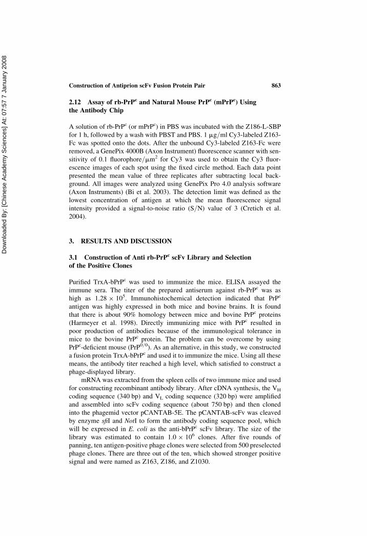
Fig. 1 shows the vectors constructed by gene splicing for expression of the
scFv fusion proteins Z186-L-SBP, Z163-L-SBP, Z163-Fc, and Z1030-Fc.
Maximum amounts of the expressions in E. coli cells were found at 228Cwith IPTG induction and no “inclusion body” problem was found under this
condition. High purity of the fusion proteins was obtained with His-tag
affinity chromatography.
3.3 Selection of scFv Fusion Pairs by Double Antibody Sandwich
ELISA
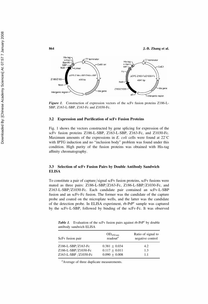
To constitute a pair of capture/signal scFv fusion proteins, scFv fusions were
mated as three pairs: Z186-L-SBP/Z163-Fc, Z186-L-SBP/Z1030-Fc, and
Z163-L-SBP/Z1030-Fc. Each candidate pair contained an scFv-L-SBP
fusion and an scFv-Fc fusion. The former was the candidate of the capture
probe and coated on the microplate wells, and the latter was the candidate
of the detection probe. In ELISA experiment, rb-PrPc sample was captured
by the scFv-L-SBP, followed by binding of the scFv-Fc. It was observed
Figure 1. Construction of expression vectors of the scFv fusion proteins Z186-L-
SBP, Z163-L-SBP, Z163-Fc and Z1030-Fc.
Table 1. Evaluation of the scFv fusion pairs against rb-PrPc by double
antibody sandwich ELISA
ScFv fusion pair
OD450 nm
readoutaRatio of signal to
negative control
Z186-L-SBP/Z163-Fc 0.381 + 0.034 4.2
Z186-L-SBP/Z1030-Fc 0.117 + 0.011 1.3
Z163-L-SBP /Z1030-Fc 0.090 + 0.008 1.1
aAverage of three duplicate measurements.
J.-B. Zhang et al.864
Dow
nloa
ded
By:
[Chi
nese
Aca
dem
y S
cien
ces]
At:
07:5
7 7
Janu
ary
2008
that an HRP-labeled horse antihuman IgG Fc fragment antibody bonded the
complex. This four-layer was thus termed as “double sandwich ELISA”.
HRP catalyzed enzymatic color reaction were read out at OD450 nm using
TrxA sample as a negative control. The pair Z186-L-SBP/Z163-Fc
displayed obviously positive results. The data were summarized in Table 1.
There is a clear cut between Z186-L-SBP/Z163 and other two pairs.
Therefore, the Z186-L-SBP/Z163-Fc pair was selected for the following
experiments.
3.4 Binding Characteristics of scFv and scFv Fusion Pair Through
SPR Sensorgrams
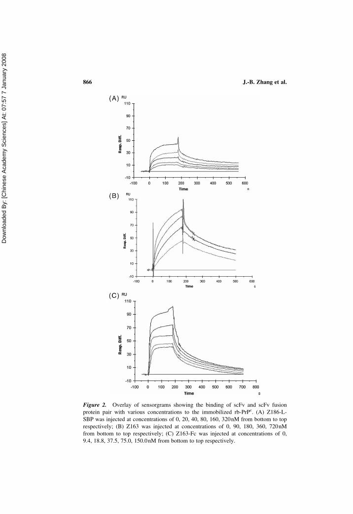
Binding affinities of Z163, Z186-L-SBP/Z163-Fc pair to rb-PrPc were
analyzed by SPR. Fig. 2 shows SPR sensorgrams, where the equilibrium
dissociation constants (KD values) were 3.24 � 1028 M, 8.82 � 1028M,
and 8.10 � 1029 M for Z186-L-SBP, Z163, and Z163-Fc, respectively
(Table 2). They all displayed rapid association and slow dissociation
kinetics. The affinity of Z163-Fc to rb-PrPc was obvious higher than those
of the structures containing no Fc fusion partner.
SPR experiment showed that the fusion structures retained their antibody
activities against rb-PrPc. Generally, affinity of the single chain antibody
affinity is one or two orders lower than that of the parent antibody, which
usually leads to lower the detection sensitivity. Using the antibody
engineering this problem may be solved. Z163-Fc is a fusion structure of an
scFv and a human IgG Fc region, which displayed an obvious increased
affinity to rb-PrPc.
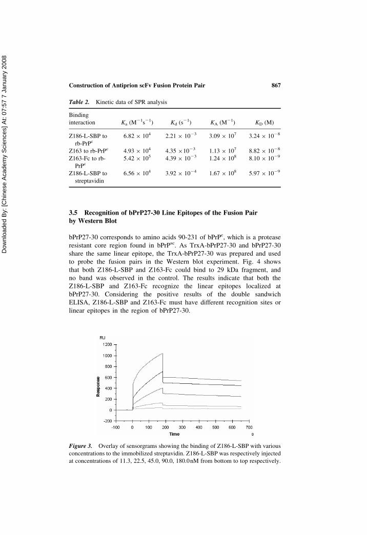
Fig. 3 shows the SPR sensorgram of Z186-L-SBP which reacted with the
streptavidin-modified surface. The figure gives the residue activity of SBP
fusion partner of Z186-L-SBP to streptavidin. Also, the response and
washing curves indicate a rapid association and slow dissociation kinetics.
KD was 5.97 � 1029 M, which is higher than that of SBP (2.5 � 1029 M)
(Keefe et al. 2001), implying SBP as a fusion partner, which lost about
50% of its original activity. SPR analysis revealed that the SBP fusion
partner of Z186-L-SBP retained its binding activity to streptavidin. Thus it
is able to apply the SBP-streptavidin interaction mechanism for site-directed
immobilization of Z186-L-SBP to prepare the antibody chip with high-hom-
ogeneity reaction surface. The high residual affinity activities of all these
fusion structures are largely due to the insertion of linker peptide between
the fusion partners. The linker peptide (repeat serine and glycin sequence)
is a rigid and hydrophilic a-helices chain in solution (Shi et al. 2004),
which plays a spacer to help to correct folding of each fusion partners. The
linker peptide function had been demonstrated in our previous studies and
many other investigations (Freund et al. 1993; Zhou et al. 2001; Shao et al.
2001) and worked well again in this study.
Construction of Antiprion scFv Fusion Protein Pair 865
Dow
nloa
ded
By:
[Chi
nese
Aca
dem
y S
cien
ces]
At:
07:5
7 7
Janu
ary
2008
Figure 2. Overlay of sensorgrams showing the binding of scFv and scFv fusion
protein pair with various concentrations to the immobilized rb-PrPc. (A) Z186-L-
SBP was injected at concentrations of 0, 20, 40, 80, 160, 320nM from bottom to top
respectively; (B) Z163 was injected at concentrations of 0, 90, 180, 360, 720nM
from bottom to top respectively; (C) Z163-Fc was injected at concentrations of 0,
9.4, 18.8, 37.5, 75.0, 150.0nM from bottom to top respectively.
J.-B. Zhang et al.866
Dow
nloa
ded
By:
[Chi
nese
Aca
dem
y S
cien
ces]
At:
07:5
7 7
Janu
ary
2008
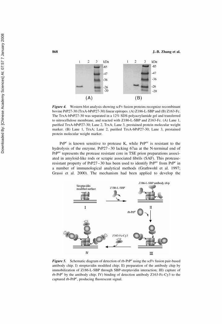
3.5 Recognition of bPrP27-30 Line Epitopes of the Fusion Pairby Western Blot
bPrP27-30 corresponds to amino acids 90-231 of bPrPc, which is a protease
resistant core region found in bPrPsc. As TrxA-bPrP27-30 and bPrP27-30
share the same linear epitope, the TrxA-bPrP27-30 was prepared and used
to probe the fusion pairs in the Western blot experiment. Fig. 4 shows
that both Z186-L-SBP and Z163-Fc could bind to 29 kDa fragment, and
no band was observed in the control. The results indicate that both the
Z186-L-SBP and Z163-Fc recognize the linear epitopes localized at
bPrP27-30. Considering the positive results of the double sandwich
ELISA, Z186-L-SBP and Z163-Fc must have different recognition sites or
linear epitopes in the region of bPrP27-30.
Figure 3. Overlay of sensorgrams showing the binding of Z186-L-SBP with various
concentrations to the immobilized streptavidin. Z186-L-SBP was respectively injected
at concentrations of 11.3, 22.5, 45.0, 90.0, 180.0nM from bottom to top respectively.
Table 2. Kinetic data of SPR analysis
Binding
interaction Ka (M21s21) Kd (s21) KA (M21) KD (M)
Z186-L-SBP to
rb-PrPc6.82 � 104 2.21 � 1023 3.09 � 107 3.24 � 1028
Z163 to rb-PrPc 4.93 � 104 4.35 �1023 1.13 � 107 8.82 � 1028
Z163-Fc to rb-
PrPc
5.42 � 105 4.39 � 1023 1.24 � 108 8.10 � 1029
Z186-L-SBP to
streptavidin
6.56 � 104 3.92 � 1024 1.67 � 108 5.97 � 1029
Construction of Antiprion scFv Fusion Protein Pair 867
Dow
nloa
ded
By:
[Chi
nese
Aca
dem
y S
cien
ces]
At:
07:5
7 7
Janu
ary
2008
PrPc is known sensitive to protease K, while PrPsc is resistant to the
hydrolysis of the enzyme. PrP27–30 lacking 67aa at the N-terminal end of
PrPsc represents the protease resistant core in TSE prion preparations associ-
ated in amyloid-like rods or scrapie associated fibrils (SAF). This protease-
resistant property of PrP27–30 has been used to identify PrPsc from PrPc in
a number of immunological analytical methods (Grathwohl et al. 1997;
Grassi et al. 2000). The mechanism had been applied to develop the
Figure 4. Western blot analysis showing scFv fusion proteins recognize recombinant
bovine PrP27-30 (TrxA-bPrP27-30) linear epitopes. (A) Z186-L-SBP and (B) Z163-Fc.
The TrxA-bPrP27-30 was separated in a 12% SDS-polyacrylamide gel and transferred
to nitrocellulose membrane, and reacted with Z186-L-SBP and Z163-Fc. (A) Lane 1,
purified TrxA-bPrP27-30; Lane 2, TrxA; Lane 3, prestained protein molecular weight
marker. (B) Lane 1, TrxA; Lane 2, purified TrxA-bPrP27-30; Lane 3, prestained
protein molecular weight marker.
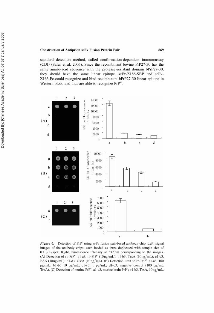
Figure 5. Schematic diagram of detection of rb-PrPc using the scFv fusion pair-based
antibody chip. I) streptavidin modified chip; II) preparation of the antibody chip by
immobilization of Z186-L-SBP through SBP-streptavidin interaction; III) capture of
rb-PrPc by the antibody chip; IV) binding of detection antibody Z163-Fc-Cy3 to the
captured rb-PrPc, producing fluorescent signal.
J.-B. Zhang et al.868
Dow
nloa
ded
By:
[Chi
nese
Aca
dem
y S
cien
ces]
At:
07:5
7 7
Janu
ary
2008
standard detection method, called conformation-dependent immunoassay
(CDI) (Safar et al. 2005). Since the recombinant bovine PrP27-30 has the
same amino-acid sequence with the protease-resistant domain bPrP27-30,
they should have the same linear epitope. scFv-Z186-SBP and scFv-
Z163-Fc could recognize and bind recombinant bPrP27-30 linear epitope in
Western blots, and thus are able to recognize PrPsc.
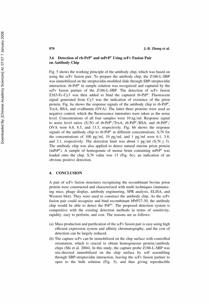
Figure 6. Detection of PrPc using scFv fusion pair-based antibody chip. Left, signal
images of the antibody chips, each loaded as three duplicated with sample size of
0.1 mL/spot; Right, fluorescence intensity at 532 nm corresponding to the images.
(A) Detection of rb-PrPc. a1-a3, rb-PrPc (10ng/mL); b1-b3, TrxA (10ng/mL); c1-c3,
BSA (10ng/mL); d1-d3, OVA (10ng/mL). (B) Detection limit to rb-PrPc. a1-a3, 100
pg/mL; b1-b3 10 pg/mL; c1-c3, 1 pg/mL; d1-d3, negative control (100 pg/mL
TrxA). (C) Detection of murine PrPc. a1-a3, murine brain PrPc; b1-b3, TrxA, 10ng/mL.
Construction of Antiprion scFv Fusion Protein Pair 869
Dow
nloa
ded
By:
[Chi
nese
Aca
dem
y S
cien
ces]
At:
07:5
7 7
Janu
ary
2008
3.6 Detection of rb-PrPc and mPrPc Using scFv Fusion Pair
on Antibody Chip
Fig. 5 shows the working principle of the antibody chip, which was based on
using the scFv fusion pair. To prepare the antibody chip, the Z186-L-SBP
was immobilized on the streptavidin-modified slide through SBP-streptavidin
interaction. rb-PrPc in sample solution was recognized and captured by the
scFv fusion partner of the Z186-L-SBP. The detection of scFv fusion
Z163-Fc-Cy3 was then added to bind the captured rb-PrPc. Fluorescent
signal generated from Cy3 was the indication of existence of the prion
protein. Fig. 6a shows the response signals of the antibody chip to rb-PrPc,
TrxA, BSA, and ovalbumin (OVA). The latter three proteins were used as
negative control, which the fluorescence intensities were taken as the noise
level. Concentrations of all four samples were 10 ng/ml. Response signal
to noise level ratios (S/N) of rb-PrPc/TrxA, rb-PrPc/BSA, and rb-PrPc/OVA were 6.8, 8.5, and 13.3, respectively. Fig. 6b shows the response
signals of the antibody chip to rb-PrPc at different concentrations. S/N for
the concentrations of 100 pg/ml, 10 pg/ml, and 1 pg/ml were 6.1, 3.8,
and 3.1, respectively. The detection limit was about 1 pg/ml (S/N � 3).
The antibody chip was also applied to detect natural murine prion protein
(mPrPc). A sample of homogenate of mouse brain containing mPrPc was
loaded onto the chip. S/N value was 13 (Fig. 6c), an indication of an
obvious positive detection.
4. CONCLUSION
A pair of scFv fusion structures recognizing the recombinant bovine prion
protein were constructed and characterized with multi techniques (immuniz-
ing mice, phage display, antibody engineering, SPR analysis, ELISA, and
Western blot). They were used to construct the antibody chip. As the scFv
fusion pair could recognize and bind recombinant bPrP27-30, the antibody
chip would be able to detect the PrPsc. The proposed detection system is
competitive with the existing detection methods in terms of sensitivity,
rapidity, easy to perform, and cost. The reasons are as follows:
(a) Mass production and purification of the scFv fusion pair is easy using high
efficient expression system and affinity chromatography, and the cost of
detection can be largely reduced.
(b) The capture scFv can be immobilized on the chip surface with controlled
orientation, which is crucial to obtain homogeneous protein/antibody
chips (Shi et al. 2004). In this study, the capture probe Z186-L-SBP was
site-directed immobilized on the chip surface by self assembling
through SBP-streptavidin interaction, leaving the scFv fusion partner to
open to the bulk solution (Fig. 5), and thus giving reproducible
J.-B. Zhang et al.870
Dow
nloa
ded
By:
[Chi
nese
Aca
dem
y S
cien
ces]
At:
07:5
7 7
Janu
ary
2008
responses. Whereas, conventional immobilization of antibody by chemical
coupling is a random adsorption that usually results in an uncontrolled and
heterogeneous surface.
(c) The detection probe can be either scFv or scFv-Fc fusion based. Both forms
are mono-body against rb-PrPc, but scFv-Fc gained a KD value about 10
times higher than that of scFv. The high affinity of scFv-Fc to rb-PrPc
obviously increases the specificity and sensitivity of the detection. In fact,
the detection limit using the method was as low as 1pg/ml, which is com-
parable to the literature data, where the detection limits were reported to be
25–50 ng PrP 27-30 per lipid class by high-performance liquid chromato-
graphy (HPLC; Klein et al. 1998), approximately 20 pg/ml recombinant
hamster PrP by mAb sandwich ELISA (Yang et al. 2005), less than 50 pg
normal and disease-associated isoforms of prion protein (PrP) by time-
resolved dissociation-enhanced fluorescence mAb sandwich ELISA (Yang
et al. 2005) and 6 or 80 ng/ml bovine recombinant prion protein by
capillary electrophoresis-based noncompetitive or competitive fluorescence
immunoassay (Barnard et al. 2000; Volkel et al. 2001).
In conclusion, a pair of scFv fusion structures recognizing the recombi-
nant bovine prion protein was constructed and characterized with multi
techniques (immunizing mice, phage display, antibody engineering, SPR
analysis, ELISA, and Western blot). The performance of the fusion pair in
detection of prion protein was demonstrated by an antibody chip format
using the recombinant bovine prion protein and the natural murine prion
protein as the targets. Real sample application is expected when available.
REFERENCES
Ascione, A., Flego, M., Zamboni, S., De Cinti, E., Dupuis, M.L., and Cianfriglia, M.2005. Application of a synthetic phage antibody library (ETH-2) for the isolationof single chain fragment variable (scFv) human antibodies to the pathogenicisoform of the hamster prion protein (HaPrPsc). Hybridoma, 24: 127–132.
Barnard, G., Helmick, B., Madden, S., Gilbourne, C., and Patel, R. 2000. The measure-ment of prion protein in bovine brain tissue using differential extraction andDELFIA as a diagnostic test for BSE. Luminescence, 15: 357–362.
Bi, L.J., Zhou, Y.F., Zhang, X.E., Deng, J.Y., Zhang, Z.P., Xie, B., and Zhang, C.G.A.2003. MutS-based protein chip for detection of DNA mutations. Anal. Chem., 75:4113–4119.
Cardinale, A., Filesi, I., Vetrugno, V., Pocchiari, M., Sy, M.S., and Biocca, S. 2005.Trapping prion protein in the endoplasmic reticulum impairs PrPc maturation andprevents PrPsc accumulation. J. Biol. Chem., 280: 685–694.
Cretich, M., Pirri, G., Damin, F., Solinas, I., and Chiari, M. 2004. A new polymericcoating for protein microarrays. Anal. Biochem., 332: 67–74.
Freund, C., Ross, A., Guth, B., Pluckthun, A., and Holak, T.A. 1993. Characterizationof the linker peptide of the single-chain Fv fragment of an antibody by NMRspectroscopy. FEBS Lett., 320: 97–100.
Construction of Antiprion scFv Fusion Protein Pair 871
Dow
nloa
ded
By:
[Chi
nese
Aca
dem
y S
cien
ces]
At:
07:5
7 7
Janu
ary
2008
Harmeyer, S., Pfaff, E., and Groschup, M.H. 1998. Synthetic peptide vaccines yield
monoclonal antibodies to cellular and pathological prion proteins of ruminants.
J. Gen. Virol., 79: 937–945.
Grassi, J., Creminon, C., Frobert, Y., Fretier, P., Turbica, I., Rezaei, H., Hunsmann, G.,
Comoy, E., and Deslys, J.P. 2000. Specific determination of the proteinase
K-resistant form of the prion protein using two-site immunometric assays.
Application to the post-mortem diagnosis of BSE. Arch. Virol. Suppl., 16: 197–205.
Grathwohl, K.U., Horiuchi, M., Ishiuchi, M., Ishiguro, N., and Shinagawa, M. 1997.
Sensitive enzyme-linked immunosorbent assay for detection of PrP (Sc) in crude
tissue extracts from scrapie-affected mice. J. Virol. Methods, 64: 205–216.
Keefe, A.D., Wilson, D.S., Seelig, B., and Szostak, J.W. 2001. One-step purification of
recombinant proteins using a nanomolar-affinity streptavidin-binding peptide, the
SBP-Tag. Protein Expr. Purif., 23: 440–446.
Klein, T.R., Kirsch, D., Kaufmann, R., and Riesner, D. 1998. Prion rods contain small
amounts of two host sphingolipids as revealed by thin-layer chromatography and
mass spectrometry. Biol. Chem., 379: 655–666.
Komiya, N., Ueda, H., Ohiro, Y., and Nagamunea, T. 2004. Homogeneous sandwich
immunoassay based on the enzymatic complementation induced by single-chain
Fv fragments. Anal. Biochem., 327: 241–246.
Krasmann, S., Groshup, M.H., Harmeyer, S.S., Hunsmann, G., and Bodemer, W. 1996.
Generation of monoclonal antibodies against human prion proteins in PrP0/0 mice.
Mol. Med., 2: 725–734.
Leclerc, E., Limann, S., Wildegger, G., Vetter, S.W., and Nilsson, F. 2000. Selection
and characterization of single chain Fv fragments against murine recombinant prion
protein from a synthetic human antibody phage display library. Hum. Antibodies, 9:
207–214.
Long, M.C., Jager, S., Mah, D.C., Jebailey, L., Mah, M.A., Masri, S.A., and
Nagata, L.P. 2000. Construction and characterization of a novel recombinant
single-chain variable fragment antibody against western equine encephalitis virus.
Hybridoma, 19: 1–13.
Marks, J.D., Hoogenboom, H.R., Bonnert, T.P., McCafferty, J., Griffiths, A.D., and
Winter, G. 1991. By-passing immunization: human antibodies from V-gene
libraries displayed on phage. J. Mol. Biol., 222: 581–597.
McCafferty, J., Griffiths, A.D., Winter, G., and Chiswell, D.J. 1990. Phage antibodies
filamentous phage: displaying antibody variable domains. Nature, 348: 552–554.
Negro, A., De Filippis, V., Skaper, S.D., James, P., and Sorgato, M.C. 1997. The
complete mature bovine prion highly expressed in Escherichia coli: biochemical
and structural studies. FEBS Lett., 412: 359–364.
Negro, A., Meggio, F., Bertoli, A., Battistutta, R., Sorgato, M.C., and Pinna, L.A. 2000.
Susceptibility of the prion protein to enzymic phosporylation. Biochem. Biophys.
Res. Commun., 271: 337–341.
Ono, K.I., Kamihira, M., Kuga, Y., Matsumoto, H., Hotta, A., Itoh, T., Nishijima, K.I.,
Nakamura, N., Matsuda, H., and Iijima, S. 2003. Production of anti-prion scFv-Fc
fusion proteins by recombinant animal cells. J. Biosci. Bioeng., 95: 231–238.
O’Rourke, K.I., Baszler, T.V., Miller, J.M., Spraker, T.R., Sadler-Riggleman, I., and
Knowles, D.P. 1998. Monoclonal antibody F89/160.1.5 defines a conserved
epitope on the ruminant prion protein. J. Clin. Microbiol., 36: 1750–1755.
Powers, D.B., Amersdorfer, P., Poul, M., Nielsen, U.B., Shalaby, M.R., Adams, G.P.,
Weiner, L.M., and Marks, J.D. 2001. Expression of single-chain Fv-Fc fusions in
Pichia pastoris. J. Immunol Methods, 251: 123–135.
J.-B. Zhang et al.872
Dow
nloa
ded
By:
[Chi
nese
Aca
dem
y S
cien
ces]
At:
07:5
7 7
Janu
ary
2008
Prusiner, S.B. 1982. Novel proteinaceous infectious particles cause scrapie. Science,216: 136–144.
Prusiner, S.B. 1998. Prions Proc. Natl. Acad. Sci. USA, 95: 13363–13383.Renberg, B., Shiroyama, I., Engfeldt, T., Nygren, P.K., and Karlstrom, A.E. 2005.
Affibody protein capture microarrays: Synthesis and evaluation of random anddirected immobilization of affibody molecules. Anal. Biochem., 341: 334–343.
Safar, J.G., Geschwind, M.D., Deering, C., Didorenko, S., Sattavat, M., Sanchez, H.,Serban, A., Vey, M., Baron, H., Giles, K., Miller, B.l., Dearmond, S.J., andPrusiner, S.B. 2005. Diagnosis of human prion disease. Proc. Natl. Acad. Sci. U S A,102: 3501–3506.
Schaller, O., Fatzer, R., Stack, M., Clark, J., Cooley, W., Biffiger, K., Egli, S.,Doherr, M., Vandevelde, M., Heim, D., Oesch, B., and Moser, M. 1999. Validationof a Western immunblotting procedure for bovine PrPsc detection and its use as arapid surveillance method for diagnosis of bovine spongiform encephalopathy(BSE). Acta Neuropathol., 98: 437–443.
Shao, W.H., Zhang, X.E., Liu, H., Zhang, Z.P., and Cass, A.E. 2000. Anchor-chainmolecular system for orientation control in enzyme immobilization. Bioconjug.Chem., 11: 822–826.
Shi, J.X., Zhang, X.E., Xie, W.H., Zhou, Y.F., Zhang, Z.P., Deng, J.Y., Cass, A.E.,Zhang, Z.L., Pang, D.W., and Zhang, C.G. 2004. Improvement of homogeneity ofanalytical biodevices by gene manipulation. Anal. Chem., 76: 632–638.
Volkel, D., Zimmermann, K., Zerr, I., Bodemer, M., Lindner, T., Turecek, P.L.,Poser, S., and Schwarz, H.P. 2001. Immunochemical determination of cellularprion protein in plasma from healthy subjects and patients with sporadic CJD orother neurological diseases. Transfusion, 41: 441–448.
Weiss, S., Rieger, R., Edenhofer, F., Fisch, E., and Winnacker, E.L. 1996. Recombi-nant prion protein rPrP27–30 from Syrian Golden Hamster reveals proteinase K sen-sitivity. Biochem. Biophys. Res. Commun., 219: 173–179.
Williamson, R.A., Peretz, D., Smorodinsky, N., Bastidas, R., Serban, H., Mehlhorn, H.,DeArmond, S.J., Prusiner, S.B., and Burton, D.R. 1996. Circumventing tolerance togenerate autologous monoclonal antibodies to the prion protein. Proc. Natl. Acad.Sci. U S A, 93: 7279–7282.
Yang, W.C., Schmerr, M.J., Jackman, R., Jackman, R., Bodemer, W., and Yeung, E.S.2005. Capillary electrophoresis-based noncompetitive immunoassay for the prionprotein using fluorescein-labeled protein as a fluorescent probe. Anal. Chem., 77:4489–4494.
Yang, W.C., Yeung, E.S., and Schmerr, M.J. 2005. Detection of prion protein using acapillary electrophoresis-based competitive immunoassay with laser-inducedfluorescence detection and cyclodextrin-aided separation. Electrophoresis, 26:1751–1759.
Yau, K.Y., Groves, M.A., Li, S., Sheedy, C., Lee, H., Tanha, J., Mackenzie, C.R.,Jermutus, L., and Hall, J.C. 2003. Selection of hapten-specific single-domain anti-bodies from a non-immunized llama ribosome display library. J. Immunol.Methods, 281: 161–175.
Zhou, Y.F., Zhang, X.E., Liu, H., Zhang, Z.P., Zhang, C.G., and Cass, A.E. 2001. Con-struction of a fusion enzyme system by gene splicing as a new molecular recognitionelement for a sequence biosensor. Bioconjug. Chem., 12: 924–931.
Construction of Antiprion scFv Fusion Protein Pair 873