analytical and compositional aspects of isoflavones in food and their biological effects
TRANSCRIPT

1
i:/3b2/Jobs/Mnf/2009/Heft6/478.3d 17. 9. 2008
Mol. Nutr. Food Res. 2009, 53, 000 –000 DOI 10.1002/mnfr.200700478
Review
Analytical and compositional aspects of isoflavonesin food and their biological effects
Alicja Mortensen1, Sabine E. Kulling2, Heidi Schwartz3, Ian Rowland4, Corinna Ruefer5,Gerald Rimbach6, Aedin Cassidy7, Pamela Magee8, Julie Millar8, Wendy L. Hall9,Franziska Kramer Birkved10, Ilona K. Sorensen1 and Gerhard Sontag3
1 The National Food Institute, Technical University of Denmark, Søborg, Denmark2 Institute of Nutritional Science, Food Chemistry, University of Potsdam, Potsdam, Germany3 Department of Analytical and Food Chemistry, University of Vienna, Vienna, Austria4 Department of Food Biosciences, University of Reading, Reading, UK5 Department of Physiology and Biochemistry of Nutrition, Max Rubner Institute, Karlsruhe, Germany6 Institute of Human Nutrition and Food Science, Christian Albrechts University, Kiel, Germany7 School of Medicine, University of East Anglia, Norwich, UK8 Northern Ireland Centre for Food and Health, University of Ulster, Coleraine, UK9 Nutritional Sciences Division, King's College London, London, UK10 Toxicology and Environmental Chemistry, Faculty of Pharmaceutical Sciences, University of
Copenhagen, Copenhagen, Denmark
This paper provides an overview of analytical techniques used to determine isoflavones (IFs) in foodsand biological fluids with main emphasis on sample preparation methods. Factors influencing thecontent of IFs in food including processing and natural variability are summarized and an insight intoIF databases is given. Comparisons of dietary intake of IFs in Asian and Western populations, in spe-cial subgroups like vegetarians, vegans, and infants are made and our knowledge on their absorption,distribution, metabolism, and excretion by the human body is presented. The influences of the gutmicroflora, age, gender, background diet, food matrix, and the chemical nature of the IFs on themetabolism of IFs are described. Potential mechanisms by which IFs may exert their actions arereviewed, and genetic polymorphism as determinants of biological response to soy IFs is discussed.The effects of IFs on a range of health outcomes including atherosclerosis, breast, intestinal, and pros-tate cancers, menopausal symptoms, bone health, and cognition are reviewed on the basis of the avail-able in vitro, in vivo animal and human data.
Keywords: Bioavailability / Biological effects / Cancer / Databases / Isoflavones /
Received: November 23, 2007; revised: June 5, 2008; accepted: June 21, 2008
1 Introduction
Over the last two decades the biological effects of bioactivecompounds from plants have received considerable atten-tion with particular interest of soy isoflavones (IF) in rela-tion to human health. Several epidemiological studies havecorrelated consumption of soy IFs with multiple beneficial
effects on atherosclerosis, breast and prostate cancers, men-opausal symptoms, and bone density. Also their hormonaland nonhormonal properties indicated potential for benefi-cial health effects. Thus, their biological effects have beenextensively studied in in vitro systems, laboratory animalsand animal models of human diseases, and in humans.Simultaneously, methods for analyzing the content of soy
Correspondence: Professor Gerhard Sontag, Department of Analyti-cal and Food Chemistry, University of Vienna, Waehringer Strasse 38,A-1090 Vienna, AustriaE-mail: [email protected]: +43-1-4277-9523
Abbreviations: ADME, absorption, distribution, metabolism, and ex-cretion; cAMP, cyclic 39,59-adenosinemonophosphate; CRC, colorec-tal cancer; DHD, dihydrodaidzein; eNOS, endothelial nitric oxide syn-
i 2009 WILEY-VCH Verlag GmbH & Co. KGaA, Weinheim www.mnf-journal.com
thase; ER, estrogen receptor; FFQ, food frequency questionnaire; HU-VEC, human umbilical vein endothelial cells; ICAM, intercellular ad-hesion molecule; IC50, half maximal inhibitory concentration; IF, iso-flavone; iNOS, inducible nitric oxide synthase; LDL, low-density lipo-proteins; MCP, monocyte chemoattractant protein; O-DMA, O-des-methylangolensin; OH, hydroxy; PGI2, prostacyclin; PKA, proteinkinase A; PSA, prostate specific antigen; SGLT1, sodium-dependentglucose cotransporter; TNF, tumor necrosis factor; TVP, textured veg-etable protein; VCAM, vascular cell adhesion molecule
A. Mortensen et al. Mol. Nutr. Food Res. 2009, 53, 000 –000
IFs in plants, foods, and other biological material have beendeveloped. This enabled investigation of their metabolismand kinetics on the one hand, and generation of databaseson the content of soy IFs in food items on the other hand,allowing the generation of intake data in different popula-tions. In the last decade, several reviews have collated evi-dence on health effects [1–13]. In this review, we presentthe current knowledge on soy IFs including analyticalaspects as well as information on IF databases, intake esti-mations, metabolism, kinetics, and estrogenic action. Fur-ther, the antioxidant activity, cardiovascular effects, geneticpolymorphism in response to IFs, as well as effects on can-cer and menopausal symptoms, bone health, and cognitionare discussed. This interdisciplinary paper reflects the mul-tidisciplinary approach directed toward elucidation of thebiological effects of soy IFs in order to create a basis fordietary improvement of human health.
2 Sources of IFs and IF contents in foodstuffs
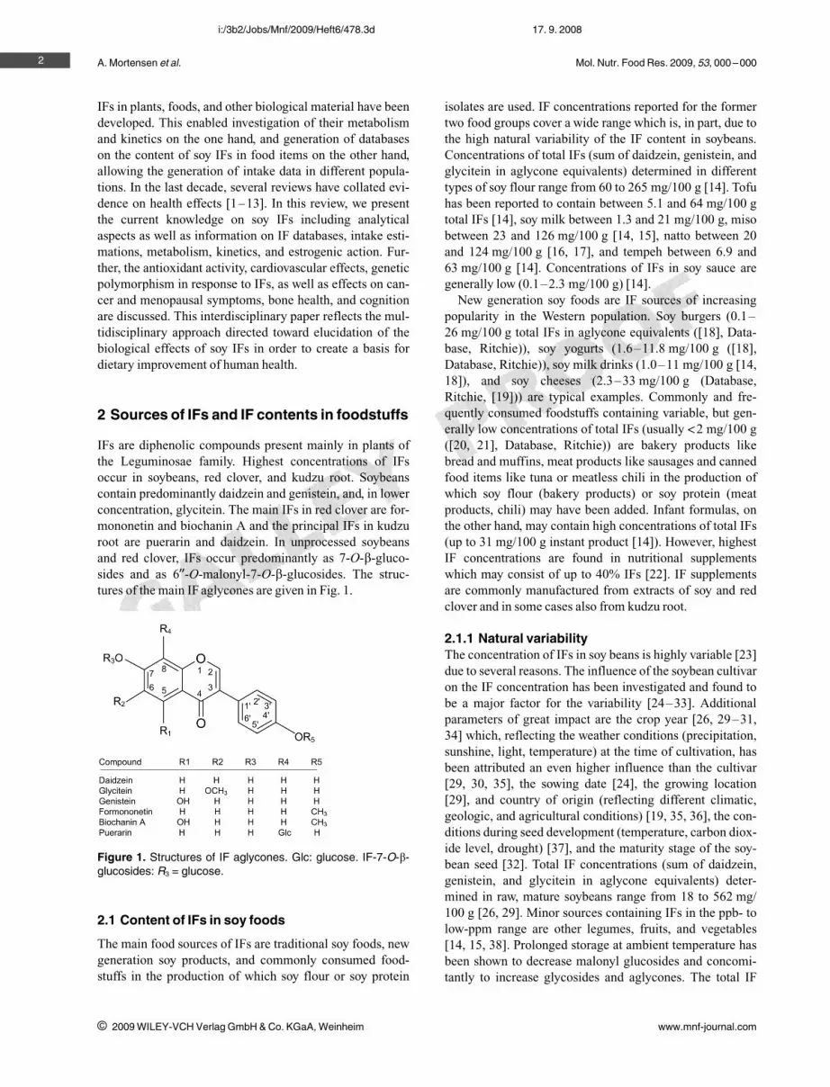
IFs are diphenolic compounds present mainly in plants ofthe Leguminosae family. Highest concentrations of IFsoccur in soybeans, red clover, and kudzu root. Soybeanscontain predominantly daidzein and genistein, and, in lowerconcentration, glycitein. The main IFs in red clover are for-mononetin and biochanin A and the principal IFs in kudzuroot are puerarin and daidzein. In unprocessed soybeansand red clover, IFs occur predominantly as 7-O-b-gluco-sides and as 699-O-malonyl-7-O-b-glucosides. The struc-tures of the main IF aglycones are given in Fig. 1.
2.1 Content of IFs in soy foods
The main food sources of IFs are traditional soy foods, newgeneration soy products, and commonly consumed food-stuffs in the production of which soy flour or soy protein
isolates are used. IF concentrations reported for the formertwo food groups cover a wide range which is, in part, due tothe high natural variability of the IF content in soybeans.Concentrations of total IFs (sum of daidzein, genistein, andglycitein in aglycone equivalents) determined in differenttypes of soy flour range from 60 to 265 mg/100 g [14]. Tofuhas been reported to contain between 5.1 and 64 mg/100 gtotal IFs [14], soy milk between 1.3 and 21 mg/100 g, misobetween 23 and 126 mg/100 g [14, 15], natto between 20and 124 mg/100 g [16, 17], and tempeh between 6.9 and63 mg/100 g [14]. Concentrations of IFs in soy sauce aregenerally low (0.1–2.3 mg/100 g) [14].
New generation soy foods are IF sources of increasingpopularity in the Western population. Soy burgers (0.1–26 mg/100 g total IFs in aglycone equivalents ([18], Data-base, Ritchie)), soy yogurts (1.6–11.8 mg/100 g ([18],Database, Ritchie)), soy milk drinks (1.0–11 mg/100 g [14,18]), and soy cheeses (2.3–33 mg/100 g (Database,Ritchie, [19])) are typical examples. Commonly and fre-quently consumed foodstuffs containing variable, but gen-erally low concentrations of total IFs (usually a2 mg/100 g([20, 21], Database, Ritchie)) are bakery products likebread and muffins, meat products like sausages and cannedfood items like tuna or meatless chili in the production ofwhich soy flour (bakery products) or soy protein (meatproducts, chili) may have been added. Infant formulas, onthe other hand, may contain high concentrations of total IFs(up to 31 mg/100 g instant product [14]). However, highestIF concentrations are found in nutritional supplementswhich may consist of up to 40% IFs [22]. IF supplementsare commonly manufactured from extracts of soy and redclover and in some cases also from kudzu root.
2.1.1 Natural variabilityThe concentration of IFs in soy beans is highly variable [23]due to several reasons. The influence of the soybean cultivaron the IF concentration has been investigated and found tobe a major factor for the variability [24–33]. Additionalparameters of great impact are the crop year [26, 29–31,34] which, reflecting the weather conditions (precipitation,sunshine, light, temperature) at the time of cultivation, hasbeen attributed an even higher influence than the cultivar[29, 30, 35], the sowing date [24], the growing location[29], and country of origin (reflecting different climatic,geologic, and agricultural conditions) [19, 35, 36], the con-ditions during seed development (temperature, carbon diox-ide level, drought) [37], and the maturity stage of the soy-bean seed [32]. Total IF concentrations (sum of daidzein,genistein, and glycitein in aglycone equivalents) deter-mined in raw, mature soybeans range from 18 to 562 mg/100 g [26, 29]. Minor sources containing IFs in the ppb- tolow-ppm range are other legumes, fruits, and vegetables[14, 15, 38]. Prolonged storage at ambient temperature hasbeen shown to decrease malonyl glucosides and concomi-tantly to increase glycosides and aglycones. The total IF
2
i 2009 WILEY-VCH Verlag GmbH & Co. KGaA, Weinheim www.mnf-journal.com
i:/3b2/Jobs/Mnf/2009/Heft6/478.3d 17. 9. 2008
Figure 1. Structures of IF aglycones. Glc: glucose. IF-7-O-b-glucosides: R3 = glucose.

Mol. Nutr. Food Res. 2009, 53, 000 –000
concentration decreased only slightly upon storage for3 years [26, 29].
2.1.2 Influence of processingThe use of different processing conditions contributes tothe variability of IF concentrations in processed soy foods.Studies have been undertaken to investigate the influence ofthe processing conditions on the IF content and IF patternof soybeans [32, 39–45], soy milk [40, 41, 46], and tofu[40, 41], to assess the impact of the soybean variety and soy-bean tissue in the production of tempeh [47], to look intothe role the type of soybean (whole vs. defatted) plays in themanufacture of soy sauce [48, 49], and to determine theinfluence of different starter organisms and fermentationtemperatures in the production of black bean koji [50].
The variation of total IF aglycone levels in differentbrands of tofu products amounted to 129% for packagedtofus from the USA, to 151% for Indonesian tofus and to178% for tofus purchased in Australia [19, 51]. IF contentsof different batches (purchased during 2 years) of the samebrand varied by up to 28% [51].
Soy milks of different types had significantly different IFlevels and genistein to daidzein ratios [16]. In addition,brand- to brand differences were up to 5-fold for directextract soy milks and differences of total IF levels in itemsof the same brand purchased over the period of 6 monthsvaried by as much as 60%. The same authors also investi-gated the variability of the IF contents in 30 separate sam-ples of two soy protein isolates purchased over a 3-yearperiod and reported that total IF levels varied by 200–300% over 3 years [16]. In general, the high variability ofIF contents in soy protein concentrates [52], isolates, andtextured vegetable protein (TVP), ingredients used in theproduction of new generation soy foods, and the use ofdifferent formulations are the reason for the wide range ofIF levels in “modern” soy foods. In the case of dietary sup-plements, use of different plant parts for extract preparationintroduces a further source of variation [53]. Soy germ con-tains 6–10 times higher total IF levels than soy cotyledons[25] and is usually rich in malonyl glycitin and glycitin (upto 50%). Genistein conjugates, on the other hand, occurmainly in the cotyledons (A50%) and only to about 10% insoy germs [25]. Likewise, highest IF concentrations arefound in red clover leaves, followed by stem and inflores-cences. However, both the IF pattern and the IF distributionin different plant parts are also influenced by the stage ofmaturity [54].
In addition to covering a wide range of IF concentrations,different soy foods and IF supplements have a variable IFconjugation pattern. Upon heat treatment [55, 56] malonylglucosides are, depending on the conditions, degraded toacetyl glucosides, glycosides, and/or aglycones (Fig. 2).The presence of acetyl glucosides is therefore an indicatorof thermal processing. However, acetyl glucosides canthemselves be degraded to glycosides and aglycones. Simi-
larly, fermented foodstuffs like soymilk, tempeh, miso, orkoji contain comparatively high levels of free aglyconesdue to the action of b-glucosidases from the fermentationorganisms [47, 50, 57–60]. The impact of different proc-essing methods (Fig. 2) on the IF conjugation profile hasbeen summarized in [61, 62] and can be studied in allpapers reporting nonhydrolytic extraction of IFs from food-stuffs.
2.2 IF databases
One of the main reasons for creating IF databases is the pro-vision of IF contents in a wide range of foodstuffs to enableIF intake assessment. Early created literature compendiaand databases ([21, 63, 64], BASIS, http://www.food-comp.dk/basis/) helped to identify IF containing foods, toprovide a first overview of IF contents in foodstuffs and toprioritize future analyses by pointing out areas where datawere lacking. Nevertheless, most IF databases were createdfor dietary IF intake assessment in epidemiological studies,sometimes in conjunction with a food frequency question-naire (FFQ) [20, 21, 65, 66], sometimes on the basis of dataobtained from national food consumption studies [67–71].Comprehensive databases for nutrition research are cur-rently being developed ([72], Scalbert, A., personal com-munication, French database on polyphenols, 2007) orupdated [14]. A review summarizing the existing phytoes-trogen databases and discussing their applications, advan-tages and limitations are given by Schwartz et al. (Inventoryof phytoestrogen databases, Food Chem. 2008, submitted).
Whereas early databases covered only a small number offoods (a150), some of the databases created in and after2003 provided data for several hundreds of different or dif-ferently processed foods [67, 68]. In 2006, a free accessinternet deployed IF database comprising about 6000 foodswas developed ([69], Ritchie, M. R., Phytooestrogen data-base, http://medicine.st-andrews.ac.uk/research/docs/ritchie/, accessed on 5.4.2007). The data sources used inthese databases were manifold: Peer-reviewed literature,chemical analysis, recipe calculation, calculation based onnutrient information from supermarket chains or from themanufacturers, estimation based on the IF content of simi-lar foods and other databases.
In most databases, the IF contents are not aggregated andgiven in aglycone equivalents on a wet weight basis. Withthe exception of [66, 69] which give only sum values oftotal IFs compounds are listed separately in the investigateddatabases. Daidzein and genistein are covered in all data-bases, whereas glycitein is listed only in the BASIS data-bases (BASIS, http://www.foodcomp.dk/basis/, [72]), theUSDA database [14], and the database by Thompson et al.[71]. Values for formononetin and biochanin A can befound in ([14, 20, 21, 63, 67, 71, 72], Scalbert, A., personalcommunication, French database on polyphenols, 2007,BASIS, http://www.foodcomp.dk/basis/).
3
i 2009 WILEY-VCH Verlag GmbH & Co. KGaA, Weinheim www.mnf-journal.com
i:/3b2/Jobs/Mnf/2009/Heft6/478.3d 17. 9. 2008

A. Mortensen et al. Mol. Nutr. Food Res. 2009, 53, 000 –000
The individual databases differ greatly in the amount ofadditional information presented. Databases which are cur-rently being developed ([72], Scalbert, A., personal com-munication, French database on polyphenols, 2007) orupdated [14] provide the greatest amount of additionalinformation on the food plant, the processing method, andon the analytical method which makes them most suited fornutrition research.
A topic of increasing importance in connection with IFdatabases is the quality control of the data. In some data-bases, data entries are accompanied by quality codes, inothers, quality control is performed prior to inclusion ofdata into the database. Quality control according to anexpert quality assessment scheme (considering samplingplan, sample handling, analytical method, analytical qualitycontrol, and number of samples ([72, 73, 74], Plumb, J., per-sonal communication, EuroFIR BASIS database)) was car-ried out in ref. [14, 66, 68, 70, 72].
Of the 15 databases mentioned in this review, 5 are notyet or not any more available to the public. These are theVenus database [67] (project finished), the IF part of theFinnish national food composition database [68] (only forinternal use and for selected customers), the original BASISdatabase (http://www.foodcomp.dk/basis/) (presented onCD-ROM and currently being expanded and updated in theform of the EuroFIR BASIS database), the EuroFIR BASISdatabase [72], and the French database on polyphenols(Scalbert, A., personal communication, French database onpolyphenols, 2007) (both under construction).
The fact that a lot of work has already been put into creat-ing IF databases must not belie that there are several diffi-culties associated with their establishment. One of the mainproblems is the high variability of IF contents in foods of
the same kind which is due to the natural variability, the var-iability introduced by processing and, in part, due to use ofdifferent analytical methodologies. In addition, many val-ues were created by analyzing convenience samples, i.e.,single samples purchased in low quantities at convenientlocations without proper sampling plan which gives rise tounrepresentative data. As mentioned above, peer-reviewedliterature is one of the main data sources for IF databases.However, data reported in papers describing the develop-ment of analytical methods are often of low quality due toinsufficient food description and lacking sampling plan. Onthe other hand, authors investigating the natural variabilityor giving IF contents for various foodstuffs sometimes useoutdated or not validated analytical methods. In some data-bases, low quality data are not included in the first place.Yet, the line between insufficient and just acceptable data ishard to draw. For instance, data which are insufficient dueto lacking sampling plan may be of use when data are aggre-gated. Therefore, some databases include low quality data,give them a low confidence code and use them for dataaggregation. In general, data aggregation is a good way ofminimizing the impact of the natural variability on themean value.
To sum up, IF databases are useful tools for identifyingfood sources of IFs, for providing the range in which IFsoccur in foodstuffs and for rough estimations of the dietaryintake of IFs in epidemiological studies [75]. In addition,the EuroFIR BASIS database which contains also informa-tion about biological effects can be used for investigatingfood and health relationships, for risk assessment and forthe development of novel foods [72]. IF databases shouldnot be used for calculation of exposure in human interven-tion studies due to the high biological variation of IF con-
4
i 2009 WILEY-VCH Verlag GmbH & Co. KGaA, Weinheim www.mnf-journal.com
i:/3b2/Jobs/Mnf/2009/Heft6/478.3d 17. 9. 2008
Figure 2. Chemical forms of the IFs(daidzein is shown as an example) insoy food and their modification duringcooking and processing.

Mol. Nutr. Food Res. 2009, 53, 000 –000
tents in foodstuffs of the same type. Therefore, the concen-tration of IFs in test foods must be determined by chemicalanalysis in these studies [16, 75].
3 Determination of IFs in foods and dietarysupplements
The process of obtaining IF contents in foodstuffs and nutri-tional supplements includes sampling, sample handling,sample preparation, analytical measurement, and data eval-uation. Sampling (collection of sample units which,together, form the sample which should be representativefor the population of a food [76]) is the factor with thegreatest impact on the representativeness of the obtaineddata. Ideally, sampling should be performed as described in[73, 76]. Sample handling (or sample pretreatment) startsafter acquisition of the sample and ends with the prepara-tion of a homogenous sample for analysis. The actual ana-lytical procedure comprises two parts: sample preparation(or sample work-up) and analytical measurement. The mainemphasis in this review will be on sample preparationbecause the analytical methods have recently been summar-ized in comprehensive reviews.
3.1 Sample preparation
Basically, sample preparation techniques can be divided inhydrolytic and nonhydrolytic methods. The advantages ofhydrolytic methods are that only aglycone and, dependingon the extent of hydrolysis, glucoside standards are requiredand that simpler chromatograms are obtained (allowingshorter analysis times). Limitations are stability problemsof some IFs in acidic and alkaline solution, incompletehydrolysis under mild conditions, loss of information onthe conjugation pattern, longer sample preparation timesfor hydrolysis of extracts and the dependence of the IF con-tent obtained by enzymatic hydrolysis on the extractionefficiency. Hydrolysis of conjugated IFs can be achieved byacid hydrolysis, e.g. [19, 33, 51, 77–86], basic hydrolysis,e.g. [87–89], or enzymatic hydrolysis, e.g. [15, 20, 38, 71,90–94]. The most commonly employed method is acidhydrolysis. It is capable of cleaving both glucosidic bondsand ester linkages. However, the conditions have to be care-fully chosen in order to achieve quantitative cleavage with-out degradation of analytes. Several papers reporting theuse of mild conditions (refluxing in 0.3–1.2 M HCl, for15–120 min), e.g. [33, 54, 79, 81, 82, 84, 85] or in 80%aqueous methanol adjusted to pH 3 with acetic acid [80],more drastic conditions (refluxing in 2 M HCl for 1–6 h),e.g. [19, 51, 78, 86], and drastic conditions (refluxing in3 M HCl for 40 min) [77] have been published. Still, it ispossible that cleavage was not quantitative under mild con-ditions and that genistein was partly degraded using 2 and3 M HCl. A compromise has been suggested by Penalvo et
al. [83] who hydrolyzed with 1 M HCl at 808C for one hour,thereby cleaving malonyl- and acetyl glucosides to gluco-sides, and quantitated both glucosides and aglycones. How-ever, this approach requires both aglycone and glucosidestandards. Enzymatic hydrolysis, though applied by fewerauthors, has been used to determine the IF contents in awide range of foodstuffs and dietary supplements [15, 20,38, 71, 90–97]. Most analytical data in the database estab-lished by Ritchie et al. ([69], Ritchie, M. R., Phytooestrogendatabase, http://medicine.st-andrews.ac.uk/research/docs/ritchie/, accessed on 5.4.2007) are from [15, 20, 38, 92, 95,96].
Nonhydrolytic methods provide information on the IFconjugation pattern. With the introduction of new or newlydeveloped extraction methods (accelerated solvent extrac-tion [27, 98–100], supercritical fluid extraction [101], andsonication [100, 102, 103]), the sample preparation timehas been reduced so that nonhydrolytic sample preparationcan be performed faster. The limitations of direct extractionand analysis of the extracts are that malonyl and acetyl glu-coside standards are required (which are of limited stability[104, 105]) unless calibration functions for glucosides areused to quantitate malonyl and acetyl forms after correctionfor the molecular weight difference, that more complexchromatograms are obtained (requiring longer HPLC-runtimes), that analysis by GC is not possible and that someminor forms of malonyl glucosides are formed during theextraction [106]. Extraction can be carried out by differenttechniques using different solvents: stirring at room temper-ature (to avoid degradation of native forms) [17, 26, 29, 31,58, 106–112] or at elevated temperature (to enhance theextraction efficiency) [18], refluxing [16, 24, 113], extrac-tion by sonication [100, 102, 103], microwave assistedextraction [114], accelerated solvent extraction [27, 98–100], supercritical fluid extraction [101], and SPE [115,116]. Different solvents containing different percentages ofwater have been employed with [26, 29, 58, 107–112] orwithout addition of acid [17, 31, 106].
Several papers reporting attempts at finding the optimumextraction solvent, temperature, and hydrolysis conditionshave been published [19, 57, 62, 77, 81, 83, 86, 87, 106,117, 118]. However, for both hydrolytic and nonhydrolyticmethods optimum extraction of analytes from differentfood matrices requires different extraction conditions.These have to be optimized for each analyte in each foodmatrix. It is therefore important that, after having developeda method, authors test the applicability of the method toanalysis of other food matrices.
3.2 Analytical techniques
The most commonly reported analytical methods in IF anal-ysis are HPLC coupled with UV, electrochemical or MSdetection and GC coupled with MS. Comprehensivereviews discussing these chromatographic and nonchroma-
5
i 2009 WILEY-VCH Verlag GmbH & Co. KGaA, Weinheim www.mnf-journal.com
i:/3b2/Jobs/Mnf/2009/Heft6/478.3d 17. 9. 2008

A. Mortensen et al. Mol. Nutr. Food Res. 2009, 53, 000 –000
tographic methods (CZE, immunoassay) have been pub-lished [61, 119–126].
3.3 Evaluation of available data andrecommendations
The natural variability of the IF contents in soybeans, thevariability introduced by different processing conditionsand use of different formulations, the limited comparabilityof the values obtained by different analytical methods andlacking information for some of the values reported in theliterature pose great problems in the establishment of IFdatabases.
In many cases, the data quality could be considerablyimproved if authors provided more information on the sam-ple itself (plant part and part of the food analyzed, whole oredible portion), maturity, country of origin, cultivar, region,season, year, processing method, product specific informa-tion like brand name and manufacturers, on the samplingprocedure (sampling plan, time and region, sample size,number of sample units), and on sample handling. In addi-tion, details about the analytical method and the validationof the analytical method should be given. For reasons ofcomparability, IF contents should be given on the sameweight basis (preferably wet weight) and be expressed inaglycone equivalents.
It is seldom that all the required information is given in apaper. Food description, sampling, and sample handling areoften neglected in method development papers. Likewise,some method development papers describe the testing ofdifferent extraction or hydrolysis conditions but neglect tovalidate the optimum method or to comment on its valida-tion. In addition, papers concentrating on providing IF con-tents in foodstuffs or on determining the natural variabilityin foods of the same kind often give only limited informa-tion on the analytical method. However, this informationwould greatly facilitate the comparison of results obtainedby different methods.
4 Dietary intake of IFs
4.1 IF intake in Asian populations
In Asian countries, fermented soy products such as tempeh,miso, or natto are part of the traditional diet. This leads to amean daily IF intake of about 8–50 mg (expressed as agly-cone equivalents) in Asian countries. The mean daily intakeamong older Japanese adults ranges from 25 to 50 mg.According to the National Nutrition Survey of 2002 inJapan, soy IF intake from soy foods fell in the range of 18and 64–76 mg/day for the 50th and the 95th percentile[127–131]. Intake in Korea was estimated as 14.9 mg/daybased on data from the Korean National Nutrition Surveyconducted in 1995 [130]. The mean IF intake of women inChina was found to be 25.4 mg/day, whereas the intake of
Hong Kong Chinese women was found to be lower(7.8 l 5.6 mg/day) and the intake of Singapore Chineseadults was found to be higher (61 mg/day) [132, 133]. Themean plasma IF concentration (sum of daidzein, genistein,and equol) in Japanese women and men consuming a tradi-tional diet was 874 and 806 nmol/L, respectively [134,135].
4.2 IF intake in Western populations
Intakes of IFs in Western populations are lower than inAsian populations since soy is not commonly consumed inWestern diets. In Table 1, an overview is given of the IFintake studies which were performed in Western (Europeanand North-American) countries [65, 68, 69, 136–145]. Fur-thermore, the resulting plasma concentrations are shown.Up to now data on IF intakes in Eastern Europe do not exist.
The dietary intake of IFs in Western countries is only lessthan one to several mg/day with plasma IF concentrationsin the lower nanomolar range. The differences between theintake values obtained in the different studies are likely tobe due to the different databases of IF contents in foodsused by the different researchers and due to the differentdiets in the countries. The latter is probably less important,as the difference between the intakes calculated for the sixstudies performed in the UK is already a factor of 12.
4.3 Dietary intake of IFs by special subgroups
4.3.1 IF intake by vegetarians, vegans, and soy-consumers
The IF intake by vegetarians, vegans, and soy-consumers isexpected to be higher than that of the whole population,mainly due to the higher intakes of soy and soy products. InTable 2, an overview of the estimated intakes is presented.The intake of the vegetarians and soy-consumers (3–12 mg/day) is still low compared to IF intakes in Asian pop-ulations (15–60 mg/day). The Western vegan mothers arethe only adult subgroup which has IF intakes higher thanAsian people [70, 142, 144, 146, 147].
4.3.2 IF intake by infantsIn infant formulas based on cow's milk and in human breastmilk, concentrations of IFs range between 0 and 50 lg/L(see Table 3). The highest concentrations are found in thebreast milk of mothers following a vegetarian or vegan dietas well as in cow's milk. These concentrations are negligiblelow compared to the IF concentrations present in soy-basedinfant formulas (see Section 2.1). Soy-based infant formu-las are used if children are allergic or intolerant to cow'smilk or if parents chose to feed their children a vegan diet.Approximately 25% of the bottle-fed children in the USreceive soy-based diet. The daily intakes of IFs in infantsfed soy-based infant formulas, cow's milk infant formulasand breast milk are summarized in Table 4 [110, 148–150].
6
i 2009 WILEY-VCH Verlag GmbH & Co. KGaA, Weinheim www.mnf-journal.com
i:/3b2/Jobs/Mnf/2009/Heft6/478.3d 17. 9. 2008

Mol. Nutr. Food Res. 2009, 53, 000 –000 7
i 2009 WILEY-VCH Verlag GmbH & Co. KGaA, Weinheim www.mnf-journal.com
i:/3b2/Jobs/Mnf/2009/Heft6/478.3d 17. 9. 2008
Table 1. Isoflavone intake by adult populations in Western countries
Reference N Subjects Country Mean IF intakeper day (mg)
Food consumption data Primary sources Resulting meanplasma concen-trations (nmol/L)
Horn-Rosset al. [139]
447 Women,50 – 79 year
USA 2.87 FFQ, IF concentrationsbased on own database(Horn-Ross et al. [20])
Traditional soy-based foodsand hidden sources of soy,e.g., added soy protein iso-late, concentrate or floursuch as doughnuts or whitebread
n.a.
de Kleijnet al. [65]
964 Postmenopausalwomen
USA 0.76 l 4.35 FFQ, IF concentrationsbased on scientific litera-ture
Beans, peas, tea, coffee,and nuts
n.a.
Ruppet al., [143]
– Whole population Switzerland 1.7 Calculation with data fromthe imported soybeansused to produce foodproducts (traditional soy-based foods not included)
– n.a.
Horn-Rosset al. [138]
2882 Women,35 – 79 year
USA 3.3 FFQ, IF concentrationsbased on own database(Horn-Ross et al. [20])
Tofu, doughnuts, soy, milk,white bread
n.a
Keinan-Bokeret al. [140]
35 955 Whole population,35 – 74 year
10 Europeancountries(DK, F, GER, GR,I, N, ESP, SW,NL, UK)
<2 IF concentrations basedon USDA-Iowa State Uni-versity database includingonly soy foods
Dairy substitutes, beans,sprouts
n.a.
Keinan-Bokeret al. [141]
17 140 Women,50 – 69 year
NL 0.88 FFQ, IF concentrationsbased on scientific litera-ture
Beans, peas, nuts, grainproducts, coffee, tea, andsoy products
n.a.
Valstaet al. [68]
2862 Whole population,24 – 64 year
Finland 0.79 l 067 24 h recall, IF concen-trations based on theFinnish National FoodComposition Database(Fineli�)
Hidden sources of soy, e.g.,in meat and bakery prod-ucts
n.a.
Van Erp-Baartet al. [144]
1379 Whole population,18 – 64 year
Ireland 0.73 l 1.77 7-day record, IF concen-trations based VENUSdatabase
n.g. n.a.
Van Erp-Baartet al. [144]
1513 Whole population,up to 94 year
Italy 0.55 l 1.51 7-day record, IF concen-trations based VENUSdatabase
n.g. n.a.
Van Erp-Baartet al. [144]
335 Whole population,40 – 64 year
UK 0.70 l 1.04 7-day record, IF concen-trations based VENUSdatabase
n.g. n.a.
Van Erp-Baartet al. [144]
4085 Whole population,1 – 97 year
NL 0.91 l 1.90 2-day record, IF concen-trations based VENUSdatabase
n.g. n.a.
Clarke andLloyd [145]
– – UK 3.3 Calculation using datafrom the FSA-total dietstudy, IF concentrationsby analysis of 20 completetotal diet study samplesets from 1998
Bead, meat products, andcereals
n.a.
Healdet al. [137]
203 Men,50 – 74 year
UK 1.0 (median) FFQ, IF concentrationsbased on scientific litera-ture
Traditional soy-based foodsand hidden sources of soy,e.g., added soy protein iso-late, concentrate or flour
196 (median)
Ritchieet al. [69]
19 Whole population,19 – 76 year
UK 4.5 l 4.89 7-day record, IF concen-trations based on scientificliterature
Soy milk, yogurt, and bread n.a.
Bhakta et al. [136] 50 Women,25 – 75 year
UK 0.37 l 0.18 Monthly 24 h recalls for1 year, IF concentrationsbased on scientific litera-ture
Bread 25.8 l 22.1
Mulligan et al.[142]
11 843 Whole population UK 0.56 l 0.46 7-day record, IF concen-trations based onscientific literature
Bread, vegetable, and meatproducts
n.a.
n.a., not assessed, n.g. not given.

A. Mortensen et al. Mol. Nutr. Food Res. 2009, 53, 000 –000
Infants’ daily intakes of IFs from human breast milk orcow's milk formulas range between 0.005 and 0.01 mg/daywhich is low when compared with the amounts provided bysoy-based infant formulas (6–47 mg/day). Furthermore,IFs are predominantly found as glucuronide conjugates inbreast milk, whereas they occur mainly as glycosidic conju-gates in soymilk [148]. Up to date it is not known if thesecompositional differences influence the bioavailability.Nevertheless, it is doubtful that the small IF concentrationsin breast milk or cow's milk infant formulas are sufficient toexert significant hormonal effects. Estrogen concentrationsin breast milk in the first few days of lactation (3–120 nM)are similar to IF concentrations, but decline thereafter[151]. In contrast, in children fed soy-based infant formulasIF plasma levels are 13000–22000 higher than the plasmaconcentrations of estradiol in early life (147–294 pM)
whereas the contribution of IFs from breast milk or cow'smilk is negligible. When values are expressed relative tobody weight, the infant exclusively fed soy-based formulasis exposed to a dose that is 5–10-fold higher than the0.7 mg/kg body weight per day intake shown to exert signif-icant physiologic and beneficial effects on the hormonalregulation of women's menstrual cycle [152, 153].
4.3.3 IF intake by consumers of soy supplementsExtracted phytoestrogens are heavily marketed in numerousforms as dietary supplements. Consumers of these supple-ments are usually peri- and postmenopausal women lookingfor an alternative to hormone therapy. Although there areno approved health claims for phytoestrogens at this time,numerous claims are being made mainly regarding benefitsto bone health and menopausal symptoms. The data sup-
8
i 2009 WILEY-VCH Verlag GmbH & Co. KGaA, Weinheim www.mnf-journal.com
i:/3b2/Jobs/Mnf/2009/Heft6/478.3d 17. 9. 2008
Table 2. Isoflavone intake by vegetarians, vegans, and soy-consumers in European countries
Reference N Subjects Country Mean IF intakeper day (mg)
Food consumption data Primary sources
Clarkeet al. [146]
35 Vegetarians UK 12 7-day duplicate diet analysis Traditional soy-based foods and hid-den sources of soy, e.g., added soyprotein isolate, concentrate, or floursuch as doughnuts or white bread
Ritchieet al. [69]
10 Vegetarians,21 – 56 year
UK 7.4 l 3.1 7-day record, IF concentrationsbased on scientific literature
Soy milk, meat-substitute foods con-taining TVP and soy protein isolate,soy mince, and bakery products
Friar and Walker[147]
11 Vegan breast-feeding mothers
UK 75 Duplicate diet analysis
Van Erp-Baartet al. [144]
42 Soy-consumers Ireland 6.0 l 8.1 7-day record, IF concentrationsbased VENUS database
n.g.
Van Erp-Baartet al. [144]
15 Soy-consumers UK 3.2 l 4.0 7-day record, IF concentrationsbased VENUS database
n.g.
Van Erp-Baartet al. [144]
85 Soy-consumers NL 11.1 l 6.7 2-day record, IF concentrationsbased VENUS database
n.g.
Mulliganet al. [142]
371 Soy-consumers UK 7.1 l 10.9 7-day record, IF concentrationsbased on scientific literature
Soy-based dairy products, vegetabledishes, and bread
n.g., not given.
Table 3. Total IF concentrations in breast and cow milk
Reference Milk IF concentration (lg/L)
Franke and Custer [524] Breast feeding: mothers’ dietChinese (n = 1)
35
King et al. [525] Cow's milk 50–350Friar and Walker [147] Breast feeding: mothers’ diet
Omnivorous (n = 14) 0–2Vegetarian (n = 14) 1–10Vegan (n = 14) 2–32
Setchell et al. [149] Breast feeding: mothers’ dietOmnivorous (n = 9) 5.6 l 4.4
Franke et al. [524] Breast feeding: mothers’ dietOmnivorous (n = 1) challenged with 37 mg IFs 52
Irvine et al. [150] Breast feeding: mothers’ dietOmnivorous (n = 11) Below LOD (a0.05 mg/L)Cow-based infant formula Below LOD (a0.1 mg/g)
Antignac et al. [526] Cow's milk 5–32Franke et al. [527] Breast feeding: mothers’ diet
Omnivorous (n = 7) 1.3 l 0.6Omnivorous (n = 7) challenged with 55 mg IFs 18.5 l 5.0

Mol. Nutr. Food Res. 2009, 53, 000 –000 9
i 2009 WILEY-VCH Verlag GmbH & Co. KGaA, Weinheim www.mnf-journal.com
i:/3b2/Jobs/Mnf/2009/Heft6/478.3d 17. 9. 2008
Table 4. Isoflavone intake by infants
Reference N Age Country Infant food Food consumption data Mean IF intake(mg/kg bw/day)
Intake(mg/day)
Resulting meanplasma concen-trations (nmol/L)
Setchellet al. [148]
7 pergroup
4 months USA Soy-based infantformula
Calculation according toanalysis of soy-based infantformula, consumption, andbw
4.5 – 8.0 28 – 47 3695 l 1877
Cow’s milk formu-la 20.1 l 1.2
Human breast-milk
15.9 l 3.0
Murphyet al. [110]
– – USA Soy-based infantformula
Calculation according toanalysis of soy-based infantformula, consumption, andbw
5 – 12 – n.a.
Setchellet al. [149]
1 wk USA Soy-based infantformula
Calculation according toanalysis of soy-based infantformula, consumption, andbw
5.7 – 7.3 22.5 – 24.8 see Setchellet al. [148]
1 month 6.0 – 11.9 31.5 – 36.0
2 months 6.1 – 10.0 36.0 – 37.0
4 months 6.0 – 9.3 41.0 – 45.0
Irvine et al. [150] 4 pergroup
<1 month New Zealand Soy-based infantformula
Calculation according toanalysis of soy-based infantformula, consumption, andbw
3.0 l 0.2 9.1 l 0.7 n.a.
1 month 3.8 l 0.2 14.1 l 0.6
2 months 3.3 l 0.2 16.6 l 1.1
4 months 2.9 l 0.3 20.0 l 2.0
Friar and Walker[147]
– 1 – 2 months UK Soy-based infantformula
– 5 28 n.a.
4 – 6 months 4.5 34
Rupp et al. [143] 4 – 5 months Switzerland Soy-based infantformula
Calculation according toanalysis of soy-based infantformula, consumption, andbw
3.4 – 13.5 – n.a.
>5 months Max. 20
Genovese andLajolo [528]
– 0 – 2 wk Brazil Soy-based infantformula
Calculation according toanalysis of soy-based infantformula, consumption, andbw
2.0 – 6.1 5.9 – 18.3 n.a.
2 – 8 wk 1.7 – 6.6 6.6 – 26.2
2 – 3 months 1.6 – 5.2 8.2 – 26.2
3 – 6 months 9.2 – 34.9
>6 months 1.4 – 5.4 6.9 – 34.9
Ryowon et al.[529]
9 10 months20 months
Korea Soy-based infantformula
Plasma analysis n.g. n.g.255.4 l 10.7
1070.1 l 68.0
Franke et al.[527]
11 pergroup
2 – 45 wk USA Human breast-milk
Plasma analysis n.g. n.g. –
Human breast-milk after mothersconsume 55 mgIFs
19.7 l 13.21049 l 403
bw, body weight; n.a., not assessed; n.g. not given.

A. Mortensen et al. Mol. Nutr. Food Res. 2009, 53, 000 –000
porting those claims are generally not strong (see Section6).
Most supplements contain IFs derived from soybeans,red clover, or kudzu root and some contain botanicals suchas black cohosh. In the case of soybean extracts, mainly theglucosylated IFs are present (see Section 2.1), whereas thered clover extracts mainly contain the IF aglycones [154].Most product labels mention a content of about 40–50 mgper tablet. However, amounts of 100 and 200 mg can alsobe found [94, 113, 119, 155–157]. Analysis of the supple-ments has demonstrated that the actual IF content variedbetween products and was often less than declared on thelabel. Nurmi et al. [94] could show that only one productout of eleven was found to contain the same amount of IFsas was provided by the producer, while the other ten con-tained l30–75% of the content as stated on the productlabel. Similar findings were reported by others [113, 154–158]. As glycosylation contributes considerably to the massof IF molecules (about 40% in the case of the glucose unitand approximately 50% with respect of an acetyl- or malon-lyglucose moiety), it is relevant for the producers to con-sider the total amount of potentially bioactive IF aglyconesin supplements. Still, manufacturers usually neglect to stateif contents are given in aglycone equivalents or in nativeforms [154]. Furthermore, the IF composition changes overtime with total IF contents being constant [157]. Takentogether, consumers cannot be sure that the declared IF con-tent is correct.
5 Absorption, distribution, metabolism, andexcretion (ADME) of IFs
The ADME as well as the bioavailability of phytoestrogenshave not yet been fully elucidated. Most of the informationis related to the ADME of daidzein, genistein and, to alesser extent, glycitein.
5.1 Analytical methods to determine IFs inbiological fluids
IFs occur mainly as glucuronide- and, to a lesser extent, sul-fate conjugates in plasma, serum, and urine. For the deter-mination of the total IF levels, the aglycones have to beliberated. In most cases, the required hydrolysis is perform-ed enzymatically using Helix pomatia juice, an enzymepreparation containing b-glucuronidase and sulfatase activ-ity. The hydrolysis step can be performed either afterextraction of IF conjugates and metabolites from the centri-fuged sample or directly after centrifugation of the sample.In the latter case, IF aglycones and metabolites are subse-quently extracted from the hydrolyzed sample. Extraction isusually carried out with organic solvents, by SPE usingC-18 RP cartridges or using a combination of both methods.In some cases, the extracts are purified by ion exchange
chromatography or gel chromatography on Sephadex LH-20. IF aglycones are usually analyzed by GC-MS (after deri-vatization), HPLC with UV or coulometric electrode arraydetection as well as HPLC-MS. Nonchromatographic meth-ods featuring high sample throughput and high sensitivityat comparatively low costs are immunoassays. Immunoas-says are well suited for screening purposes but are oftenlimited by cross reactivities with compounds which arestructurally similar to the analyte. The individualapproaches have been discussed in comprehensive reviews[120–126].
5.2 Absorption
One of the major questions has been whether the glycosidicforms of IFs must first be deconjugated to the respectiveaglycones by bacterial b-glucosidases or b-glucuronidasesbefore absorption can occur or whether they can be trans-ported across the intestinal wall either intact or after degly-cosylation by cytosolic or membrane-bound b-glucosi-dases. For flavonoids, e.g., quercetin or anthocyanins, thepresence of the intact glucosides in human plasma has beendemonstrated [159, 160]. By studying the mechanisms evi-dence exists for the involvement of the multidrug-resistanceprotein-2 (MRP-2), lactase-phlorizin-hydrolase, and otherintestinal b-glucosidases [161–164].
Alternatively, the active transport of flavonoid glucosidesby the sodium-dependent glucose cotransporter (SGLT1)and the subsequent cleavage of the glucosides by a cytosolicb-glucosidase has been discussed [161, 165]. Up to datethere is little evidence that IF glucosides interact withSGLT1 [166, 167]. Moreover, a recent publication com-pletely denied the transport of flavonoids via SGLT1 [166–168].
Relatively few studies have involved IFs. In a series ofstudies, the transport of IFs and their glycosides was exam-ined using an isolated preparation of a luminally and vascu-larly perfused rat small intestine [169–171]. The aglyconeswere transported much more efficiently than their respec-tive glycosides. In the case of genistein, only 1.3% of theglucoside passed the vascular side intact. The results are inline with in vitro studies using human intestinal Caco-2-cells [166, 167]. In contrast to the glycosides, the IF agly-cones were able to penetrate the cells. Furthermore, IF gly-cosides were not detectable in human plasma after singledose administration to twelve women [172]. Taken together,these studies suggest that a large fraction of IFs is absorbedafter hydrolysis to the respective aglycones.
5.3 Metabolism
5.3.1 Bacterial metabolismIntestinal bacteria play an essential role in IF metabolism.For daidzein as well as genistein, the intestinal metabolismhas been studied extensively in vitro and in vivo [173–180].
10
i 2009 WILEY-VCH Verlag GmbH & Co. KGaA, Weinheim www.mnf-journal.com
i:/3b2/Jobs/Mnf/2009/Heft6/478.3d 17. 9. 2008

Mol. Nutr. Food Res. 2009, 53, 000 –000
It has been reported that daidzein is converted by the gutmicroflora to the isoflavanone dihydrodaidzein (DHD),which can be further metabolized to both the isoflavaneequol and the 59-methyldeoxybenzoin O-desmethylango-lensin (O-DMA) (see Fig. 3). Only 30% of the adult West-ern population and 50–60% of the Asians as well as vegeta-rians excrete equol in urine after having consumed soyfoods – an observation which is still not understood [181].Even when pure daidzein is administered which eliminatesthe influence of the food matrix, a high percentage ofhumans does not convert daidzein to equol. The equol pro-ducer is defined from urinary and serum equol concentra-tions. Unlike other IFs, equol has a chiral center and there-fore can exist as two distinct optically active isomers, R-and S-equol, which are both bioavailable. Interestingly, theenantiomer produced by metabolic reduction from daidzeinis known to be S-(–)-equol [182–184]. DHD also exhibitsan asymmetric carbon atom, but its absolute naturallyoccurring configuration has not yet been determined.
Genistein is first – analogous to daidzein – reduced bygut bacteria to dihydrogenistein (DHG), followed by acleavage of the C-ring to form 69-hydroxy-O-DMA (69-OH-O-DMA). The corresponding equol-derivative 5-OH-equolcould not be identified yet. Instead, 69-OH-O-DMA can befurther degraded by the colonic microflora to yield 4-hydroxyphenyl-2-propionic acid. Decarboxylation can thenlead to the putative metabolic end product 4-ethylphenol.Until now, 4-hydroxyphenyl-2-propionic acid has only beenidentified in rat urine and in in vitro-incubations withhuman microflora [178, 179]. The bacterial metabolism ofgenistein is depicted in Fig. 4.
Little is known about the bacterial metabolism of thethird IF glycitein. Only recently, several reduced as well asreduced and demethylated metabolites of glycitein havebeen identified in vitro and in vivo, including 6-OH-daid-
zein, dihydroglycitein, 6-OH-DHD, 6-methoxy-equol, 6-OH-equol, 59-OH-O-DMA, as well as 59-methoxy-O-DMA[180, 185, 186]. It has not yet been assessed if the formationof 6-OH-equol is a general pathway or if only parts of thepopulation are able to form this metabolite as in the case ofthe bacterial metabolism of daidzein. It should be pointedout that an alternative route for the formation of thesemetabolites is the biotransformation of daidzein (thatmeans hydroxylation, reduction, and methylation). A defin-itive association between the parent compounds and themetabolites can only be made when tracer methods (radioor stable isotopes) or pure compounds are used [180].
Glycitein seems to be a rather stable molecule. The 49-methyl ethers of daidzein and genistein – formononetinand biochanin A, respectively – are rapidly demethylatedin vitro and in vivo which results in high plasma concentra-tions of daidzein and genistein [113, 186, 187]. This is incontrast to glycitein, the methyl ether of 6-OH-daidzein.
Intestinal bacteria play an essential role in IF metabo-lism. Germ-free rats do not produce any of the above men-tioned metabolites. After colonization of the rats with fecalmicroflora the metabolites are present in urine [188]. Fur-thermore, treatment with certain antibiotics causes markedreduction in bacterial metabolite production in vitro [189].The appearance of these metabolites in plasma is time-dependent on their production in the colon. These metabo-lites appear in plasma several hours after IFs are consumed,presumably reflecting the time taken for unabsorbed IFs orIFs in the enterohepatic circulation, to reach the colon. Aclassical example of the appearance of bacterial metabolitesin plasma is that peak equol concentration occurs at 24–36 h postingestion of daidzein [190, 191].
Several candidate bacteria for the formation of thesemetabolites have been suggested, e.g., Escherichia colistrain HGH21 reduced daidzein and genistein to the corre-
11
i 2009 WILEY-VCH Verlag GmbH & Co. KGaA, Weinheim www.mnf-journal.com
i:/3b2/Jobs/Mnf/2009/Heft6/478.3d 17. 9. 2008
Figure 3. Metabolism of daidzeinby the human gut microbiota (mainmetabolism route) and by cyto-chrome P 450 enzymes (minormetabolism route).

A. Mortensen et al. Mol. Nutr. Food Res. 2009, 53, 000 –000
sponding dihydro-derivatives and a Clostridium sp., strainHGH136, cleaved the C-ring of daidzein to form O-DMA[192, 193]. Furthermore, Eubacterium ramulus is capableof producing O-DMA from daidzein and 69-OH-O-DMAand 4-hydroxyphenyl-2-propionic acid from genistein aswell as of cleaving the glycosidic bond from IF glycosidesbesides E. coli, strains HGH21 and 6 [194, 195]. E. limosumconverts glycitein, formononetin, and biochanin A to thecorresponding demethylated compounds 6-OH-daidzein,daidzein, and genistein [187]. The bacterium which produ-ces equol from daidzein has just recently been isolated fromrat intestine [196]. It is a gram-positive rod-shaped bacte-rium do03 (AB266102). Other bacteria identified in thehuman equol metabolism are SNU-Julong 732 which con-verts DHD to equol as well as a mixture of Lactobacillusmucosae EPI2, Enterococcus faecium EPI1, Finegoldiamagna EPI3, Veillonella sp. which is able to form equolfrom daidzein [197, 198].
5.3.2 Oxidative metabolismUpon absorption IFs are metabolized by cytochrome P450isoenzymes (P450) in the liver. Genistein and daidzeinundergo hydroxylation catalyzed by P450 enzymes in vitro[199, 200]. Furthermore, several mono- and dihydroxylateddaidzein and genistein metabolites – predominantly in theC-6, -8, and -39 position – have been identified in humanurine [180, 200, 201] (see Fig. 3). P450 1A1, 1A2, 1B1,2E1 as well as 3A4 have been identified to be involved inthe phase-I-metabolism of genistein and daidzein [201].
Just recently, the oxidative metabolism of glycitein hasbeen assessed. Several mono- and dihydroxylated metabo-lites as well as demethylated derivatives have been reportedin vitro and in vivo [180, 185, 186]. The bacterial metaboliteequol is also subject to phase-I-biotransformation in vitro.Ruefer et al. [202] identified 39- and 8-OH-equol as mainmetabolites using human and rat liver microsomes.
5.3.3 Phase-II-metabolismOnce absorbed, IFs are efficiently conjugated, either withglucuronic acid or, to a lesser extent, sulfate. In addition,some sulfoglucuronides, diglucuronides, and disulfatesmay be formed. Conjugation takes place either in the liveror within the intestinal epithelium with UDP-glucuronosyltransferase or sulfotransferase enzymes ([203, 204], Ruefer,C. E. et al., Phase-II-metabolism of soy isoflavones andtheir metabolites in vitro, J. Agric. Food Chem., submitted).As a consequence, IFs are present in the circulation in pre-dominantly conjugated forms. In human urine, 1–3% ofgenistein is found in the free form, 62–64% as monoglucur-onide, 13–19% as diglucuronide, 6–12% as sulfoglucuro-nide, 2–3% as monosulfate, and 3–6% as disulfate [176].The isoenzymes which efficiently conjugate IFs are UGT1A1, 1A6, 1A8, 1A9, and 1A10 as well as SULT 1A1, 1A2,1A3, 1B1, 1E1, 1C2, 2B1a, and 2B1b ([204, 205], Ruefer,C. E. et al., Phase-II-metabolism of soy isoflavones andtheir metabolites in vitro, J. Agric. Food Chem., submitted).Conjugation takes place predominantly at the hydroxylgroup at C-7 [204–206].
12
i 2009 WILEY-VCH Verlag GmbH & Co. KGaA, Weinheim www.mnf-journal.com
i:/3b2/Jobs/Mnf/2009/Heft6/478.3d 17. 9. 2008
Figure 4. Metabolism of genistein bythe human gut microbiota.

Mol. Nutr. Food Res. 2009, 53, 000 –000
5.4 Distribution
It has been shown that IFs and their metabolites are widelydistributed within body fluids, but definitive tissue distribu-tion studies have not been performed in human so far.Therefore, it is not known whether the measured plasmaconcentrations and the distribution of metabolites in theplasma are also representative for individual organs, partic-ularly for potential target organs such as breast, prostate,and thyroid. Only a few data are available. IFs were detectedin the breast tissue of premenopausal women and in theprostate fluid of men after the consumption of soy products.The concentrations of genistein and daidzein in the breasttissue were comparable to those found in plasma; however,the equol concentrations were higher [207, 208]. The pros-tate fluid was found to contain higher concentrations of IFsthan plasma [209, 210]. In humans, the concentrations ofunconjugated IFs in the plasma were relatively low. Datacomparing the conjugated and unconjugated IF fraction inhuman tissue are not available. In rats, it was shown that tis-sues contain a much higher fraction of the aglycon com-pared to their plasma levels [179]. For instance, the fractionof free genistein was 49% in the mammary gland tissue ofthe females, and even as much as 80 or 100% in the ovariesand the uterus, respectively, compared to less than 5% inthe plasma.
5.5 Excretion
The main route of excretion of IFs is via the kidney. Fecalexcretion appears to be minimal, but very few human stud-ies have investigated this issue. Human trials have indicatedthat fecal excretion of daidzein and genistein lies within therange of 1 and 4% and urinary excretion between 5 and35% of the ingested dose [211, 212].
5.6 Pharmacokinetics
IFs are readily absorbed from the gastrointestinal tract. Ingeneral, maximal plasma levels were achieved between 5and 9 h after ingestion. Very often plasma concentrationtime curves exhibit two peaks: one early peak l1–2 h afterIF intake and the highest peak between 5 and 9 h. Thisbiphasic shape is assumed to reflect the enterohepatic recy-cling as well as absorption occurring successively in thesmall intestine and in the colon. Elimination of IFs is quiteslow, with half-life values of 6–8 h. A dose of 50 mg ofeither daidzein or genistein, yields a peak plasma concen-tration of l2 lmol/L. IFs are therefore the most well-absorbed flavonoids [213]. Pharmaceutical doses of IFslead to very high plasma levels: for example, doses of 4 and8 mg genistein per kg body weight caused peak plasma con-centrations up to 9.5 and 17.9 lmol/L, respectively [214].The shape of the curve is not strongly modified by a long-term supplementation with IFs, neither for daidzein nor for
genistein. Furthermore, the plasma concentrations do notreturn to baseline levels 24 h after IF intake [215].
5.7 Factors affecting the pharmacokinetics of IFs
There is a considerable interindividual variation in the phar-macokinetics of ingested phytoestrogens. For instance, a12- and 15-fold interindividual variation for genistein anddaidzein excretion, respectively, and a 600-fold interindi-vidual variation of the equol excretion were reported aftersoy consumption [182, 216]. A variety of factors have beenproposed to influence the pharmacokinetics of IFs. Theseinclude the composition of the intestinal microflora, thediet in general, the food matrix, food processing, chemicalcomposition of IFs as well as age and gender.
5.7.1 Gut microfloraAs pointed out in Section 5.3.1, the gut microbiota in gen-eral as well as the composition of the microbiota play animportant role in the metabolism of IFs and thus has a sub-stantial impact on the plasma concentration of IFs and theirmetabolites. Various physiological, pathological and envi-ronmental factors are likely to influence gut bacterial pro-file. These includes mainly the use of antibiotics and otherdrugs, bowel diseases as well as the diet, which has animpact on the gut motility, the intestinal transit time, theredox potential of the intestine, the gastro-intestinal pH aswell as on the mucin and bile secretion. Also gender, genet-ics, and ethnicity may have a role in influencing the gut bac-terial profile [217–219].
5.7.2 Diet, food matrix, and the chemical natureof IFs
The comparison of the pharmacokinetic data of variousstudies using different IF sources (e.g., pure compounds,different formulated IF supplements, soy protein isolate,tofu, soymilk, tempeh) leads to the assumption that severalfactors, e.g., the background diet, the food matrix, thedegree of food processing as well as the chemical composi-tion of the IFs directly influence their bioavailability andmetabolism [220]. The data obtained in a single study aretherefore usually the sum of several factors which mighthave an impact on the pharmacokinetics, a point whichdoes not very often receive enough attention. Furthermore,it is very difficult to compare studies described in the litera-ture retrospectively in detail, because necessary informa-tion on the associated food matrix and/or other food compo-nents ingested together with the IFs are often incomplete. Ifextracts or IF supplements were used, very often no infor-mation is given on the kind of pharmaceutical formulation,adjuvances, or matrix components, all important factors fordrug release. This deficiency on important information canlead to confusing data. One prominent example is the dis-cussion whether IFs are better absorbed in the glycoside orin the aglycone form. The data seem to be controversial:
13
i 2009 WILEY-VCH Verlag GmbH & Co. KGaA, Weinheim www.mnf-journal.com
i:/3b2/Jobs/Mnf/2009/Heft6/478.3d 17. 9. 2008

A. Mortensen et al. Mol. Nutr. Food Res. 2009, 53, 000 –000
there are reports that the glucosides are more bioavailable[113, 191, 221, 222] while others found no impact of thesugar conjugation [223, 224] or reported that IF aglyconeswere absorbed faster and in greater amounts [219, 225].Yet, a more detailed look on the kind of IF preparationsused in the studies may give in some cases explanatoryapproaches. For example, in one of the studies the bioavail-ability of an IF preparation called Fujiflavone P10 wasinvestigated, where cyclodextrins were used as cathratecompounds for the IFs [223–225].
It seems doubtful that studies in which such special for-mulations have been used can contribute to the questionwhether glycosides or aglycones are better absorbed. In twofurther studies, pure IFs (nonformulated, no matrix compo-nents or coating) were consumed by the volunteers togetherwith a drink and a following breakfast [113, 191, 221]. Inboth studies, the IF glycosides showed a higher bioavail-ability.
Only two studies were available in which different soyfoods were used. In a recently published study, native soymilk (rich in glycosides), fermented soy milk (rich in agly-cones), tempeh (mainly aglycones), and TVP (mainly gly-cosides) were compared. It was found that the aglyconeswere absorbed faster and in greater amounts than the glyco-sides [220] and that a liquid matrix such as soy milk yields afaster absorption rate and higher peak plasma concentra-tions than a solid matrix. Between the two solid foods, theIFs from tempeh (mainly aglycones) were more bioavail-able than the IFs (mainly glycosides) in TVP. Beside thedifferent chemical composition of the IFs, also the nutrientcomposition of tempeh and TVP is quite different, e.g., incontrast to the nearly fat-free TVP, tempeh contains about7–8% fat, which might affect the IF bioavailability.
5.7.3 Gender- and age-related influences on thebioavailability
Infants can absorb and excrete daidzein and genisteinderived from soy-based infant formulas as efficiently asadults consuming soy products from the age of 4 wk on[150]. Also Setchell et al. reported that 4-month-old infantsabsorb IFs efficiently. The ability of infants to convert daid-zein to O-DMA and equol during the first few months oflife is limited [148, 149]. This has been attributed to thepresence of an immature gut microflora. The gut is sterile atbirth, but within a week of birth a microbiota begins todevelop and the profile continues to change from infancyinto adulthood. It is suggested that the ability to convertdaidzein to O-DMA develops early in infants, while theability to produce equol develops later [190]. Effects of agelater on in life on the bioavailability and metabolism has notbeen reported so far [16, 190, 220, 226].
Various studies reported that the concentrations of IFsand their metabolites in plasma and urine are sex-independ-ent. In some other studies, differences for single pharmaco-kinetic parameters are shown. For example, data presented
by Lu and Anderson [227] showed that during a 1-monthtrial in which soymilk was ingested the excretion half-lifeprogressively shortened in women but lengthened in menthroughout the trial. Wiseman et al. [228] reported signifi-cantly higher O-DMA plasma concentrations in men com-pared to women after consumption of soy products, whereasother parameters were not different. Cassidy [220] foundgender differences in peak concentrations of daidzein, withhigher levels attained in women.
6 Biological effects of soy IFs
6.1 Estrogen receptor (ER) mediated mechanismof action
IFs have a spatial configuration similar to that of mamma-lian estrogens, bind to ERs and affect estrogen-regulatedgene products [229, 230]. The estrogenic potency of soy IFsis low compared to 17b-estradiol [231], with soy IFs havingapproximately 1/1000 and one-third of the affinity of 17b-estradiol for the ERa and ERb, respectively [232]. Thebinding affinity of daidzein for ERa and ERb is relativelylow whereas genistein exhibits higher binding affinities[134, 232, 233]. Although the reported estrogenic potencyof IFs [234] is weak compared with 17b-estradiol, their bio-logical potential cannot be ignored, as typical circulatinglevels of IFs can manifold exceed endogenous estradiolconcentration following consumption of a diet containingsoy foods [134, 226].
The higher binding affinity of soy IFs for ERb comparedwith ERa and the different tissue distributions of thesereceptors suggest these compounds may be tissue selective,exerting estrogenic actions in some tissues such as coronaryvessels [235] but not in other tissues such as the endome-trium [236–238]. Therefore, they are regarded as selectiveestrogen receptor modulators (SERM), substances, whichhave estrogenic effects in some tissues, but either no effectsor antiestrogenic effects in others (for reviews on ERs andSERMs see [239–241]). Furthermore, it has been sug-gested that soy IFs may act as selective tissue estrogenicactivity regulators (STEARs), compounds which provideestrogenic activity via other routes than direct interactionwith the receptors or are precursors for in vivo metabolismto produce compounds with endocrine activity [242].
The formation of glucuronide conjugates decreases therelative affinities of IFs to ERs [243]. Genistein has a 100-fold greater binding affinity than daidzein for the mouseuterine cytosolic ERs. It needs to be taken into account that,binding affinity alone does not determine potency of thecompound, because of the resulting conformational changein the ligand (IF-receptor complex varies among ligandregardless of binding affinity). The IF genistein is alsoA1000-fold more potent at triggering transcriptional activ-ity with ERa than ERb [244], and this difference is fargreater than 20-fold greater binding affinity for ERb than
14
i 2009 WILEY-VCH Verlag GmbH & Co. KGaA, Weinheim www.mnf-journal.com
i:/3b2/Jobs/Mnf/2009/Heft6/478.3d 17. 9. 2008

Mol. Nutr. Food Res. 2009, 53, 000 –000
ERa [245]. These data therefore suggest that the divergenttranscriptional activities of estrogens and soy IFs result notonly from their different binding affinities but also from dif-ferences in their ability to recruit coregulators and triggertranscriptional functions of ERa and ERb.
6.2 Antioxidant activity in vitro
To date most of the investigations have focused solely onthe antioxidant effects of genistein. Proposed molecularmechanisms responsible for its antioxidant potentialinclude the ability to scavenge radicals, chelate metals,inhibit hydrogen peroxide (H2O2) production, and stimulateantioxidant enzymes, including catalase and superoxidedismutase.
In a liposomal system, genistein is a more effective anti-oxidant than daidzein, which is likely to be attributable toits third hydroxyl group in the C-5 position. Moreover, theIF precursors biochanin A and formononetin showed veryweak antioxidant capacities in this in vitro system as theylack the hydroxyl group at the C-49 position, which appearsto be an important determinant of the antioxidant propertiesof IFs. Equol (daidzein metabolite; see Section 5.3.1)showed superior antioxidant actions [246] compared toboth the precursor molecules and the parent IFs, suggestingthat the absence of the 2,3-double bond in conjunction witha loss of the 4-oxo group enhances antioxidant properties[247]. Antioxidant activity, assessed by the trolox equiva-lent antioxidant capacity (TEAC) assay is consistent withthese data that equol is a more potent IF compared with gen-istein and daidzein [248, 249].
In an in vitro experimental system, genistein (half maxi-mal inhibitory concentration (IC50) = 25 mM) was a morepotent inhibitor of the formation of H2O2 by 12-O-tetrade-canoylphorbol-13-acetate-activated HL-60 cells and thegeneration of superoxide anions by xanthine/xanthine oxi-dase compared with daidzein (IC50 = 150 mM), apigeninand biochanin A [250, 251]. Activities of antioxidantenzymes such as superoxide dismutase, catalase, and gluta-thione peroxidase were also significantly increased withgenistein [250].
Genistein and daidzein undergo extensive metabolism inthe gut and liver, which may affect their antioxidant proper-ties. The antioxidant activity and free radical-scavengingproperties of the IF metabolites equol, 8-OH-daidzein, O-DMA, and 1,3,5-trihydroxybenzene in comparison to theirparent aglycones, genistein, and daidzein, have been inves-tigated, with electron spin resonance spectroscopy. Resultsindicate that 8-hydroxy-daidzein was the most potent scav-enger of hydroxyl and superoxide anion radicals. IF metab-olites also exhibited higher antioxidant activity than parentcompounds in standard antioxidant assays (ferric reducingability of plasma (FRAP) and TEAC) indicating that themetabolism of IFs affects their antioxidant properties [252].Furthermore, sulfation of IFs, which masks important
hydroxyl groups of the IF molecule, could decrease theirantioxidant activity and their impact on endothelial func-tion [253].
IFs have been shown to reduce low-density lipoprotein(LDL) oxidation. Dietary supplementation with high-IF soyprotein for 17 days prolonged the lag time of copper-induced LDL oxidation compared to a low-IF soy diet[254], confirming the results of an earlier smaller study[255]. In addition, the same study showed plasma concen-trations of F2-isoprostanes (an in vivo marker of lipid per-oxidation) were also reduced by the high-IF soy diet. Invitro data are consistent with these findings, with Kapiotiset al. [233] demonstrating that genistein inhibited the oxi-dation of LDL in the presence of copper ions or superoxideand NO radicals as measured by thiobarbituric acid-reactivesubstance (TBARS) formation.
6.3 Cardiovascular effects
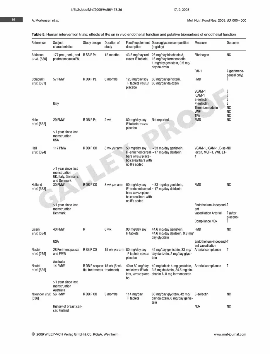
Epidemiological studies suggest that differences in dietmay explain the lower incidence of cardiovascular disease(CVD) in Japan compared with Western countries, and thewide international variability in intake of dietary IFs mayplay a role [97, 236, 256]. Several mechanisms of athero-protective action have been proposed for soy IFs (Table 5).Overall, their effectiveness in reducing the risk of CVD isnot clear.
6.3.1 Blood lipids and atherosclerosis6.3.1.1 Animal studiesThe hypocholesterolemic effect of soy protein has beenknown for many decades. In many animal species, substitut-ing soy protein for dietary animal protein consistentlyreduces LDL-cholesterol and total cholesterol levels [236,257, 258]. Gerbils fed soy-based diets have significantlylower levels of total cholesterol, LDL + VLDL cholesterol,and of apolipoprotein B [236]. Feeding normolipidemicmice a synthetic cholesterol enriched diet with added soyprotein isolate containing IFs led to a 30% decrease inplasma cholesterol levels and a 50% reduction in athero-sclerotic lesion area compared to control animals fed thesame cholesterol enriched diet but with added soy proteinisolate without IFs. When the two diets were fed to LDLreceptor deficient mice no differences in plasma cholesterolconcentration and aortic atherosclerosis were recorded[259]. Addition of soy supplement with high IF content to acasein-based semisynthetic diet reduced aortic atheroscle-rosis in Watanabe heritable hyperlipidemic rabbits, a modelof familial hypercholesterolemia, but had no effect on totalcholesterol [260]. Soy protein containing IFs had no effecton total plasma cholesterol but decreased LDL-cholesteroland increased HDL-cholesterol in a group of female mon-keys fed a moderately atherogenic diet [261]. When a stateof menopause was experimentally established in this animalmodel, by ovariectomy, soy protein consumption, as com-
15
i 2009 WILEY-VCH Verlag GmbH & Co. KGaA, Weinheim www.mnf-journal.com
i:/3b2/Jobs/Mnf/2009/Heft6/478.3d 17. 9. 2008
A. Mortensen et al. Mol. Nutr. Food Res. 2009, 53, 000 –00016
i 2009 WILEY-VCH Verlag GmbH & Co. KGaA, Weinheim www.mnf-journal.com
i:/3b2/Jobs/Mnf/2009/Heft6/478.3d 17. 9. 2008
Table 5. Human intervention trials: effects of IFs on in vivo endothelial function and putative biomarkers of endothelial function
Reference Subjectcharacteristics
Study design Duration ofstudy
Food/supplementdescription
Dose aglycone composition(mg/day)
Measure Outcome
Atkinsonet al. [530]
177 pre-, peri-, andpostmenopausal W.
R SB P Pa 12 months 43.5 mg/day redclover IF tablets.
26 mg/day biochanin A,16 mg/day formononetin,1 mg/day genistein, 0.5 mg/day daidzein
Fibrinogen NC
PAI-1 z (perimeno-pausal only)
Colacurciet al. [531]
57 PMW R DB P Pa 6 months 120 mg/day soyIF tablets versusplacebo
60 mg/day genistein,60 mg/day daidzein
FMD Z
VCAM-1 z
ICAM-1 z
E-selectin z
Italy P-selectin z
Thrombomodulin NCvWF NCTPA NC
Haleet al. [532]
29 PMW R DB P Pa 2 wk 80 mg/day soyIF tablets versusplacebo
Not reported FMD NC
>1 year since lastmenstruationUSA
Hallet al. [324]
117 PMW R DB P CO 8 wk per arm 50 mg/day soyIF-enriched cerealbars versus place-bo cereal bars withno IFs added
l33 mg/day genistein,l17 mg/day daidzein
VCAM-1, ICAM-1, E-se-lectin, MCP-1, vWF, ET-1
NC
>1 year since lastmenstruationUK, Italy, Germany,and Denmark
Hallundet al. [533]
30 PMW R DB P CO 8 wk per arm 50 mg/day soyIF-enriched cerealbars versus place-bo cereal bars withno IFs added
l33 mg/day genistein,l17 mg/day daidzein
FMD NC
>1 year since lastmenstruation
Endothelium-independ-ent
Z
Denmark vasodilation Arterial Z (afterplacebo)
Compliance NOx Z
Lissinet al. [534]
40 PMW R 6 wk 90 mg/day soyIF tablets
44.6 mg/day genistein,44.6 mg/day daidzein, 0.8 mg/day glycitein
FMD NC
USA Endothelium-independ-ent vasodilation
Z
Nestelet al. [275]
28 Perimenopausaland PMW
R SB P CO 15 wk per arm 80 mg/day soyIF tablets versusplacebo
45 mg/day genistein, 33 mg/day daidzein, 2 mg/day glyci-tein
Arterial compliance Z
AustraliaNestelet al. [535]
14 PMW R DB P sequen-tial treatments
15 wk (5 wktreatment)
40 or 80 mg/dayred clover IF tab-lets, versus place-bo
40 mg tablet: 4 mg genistein,3.5 mg daidzein, 24.5 mg bio-chanin A, 8 mg formononetin
Arterial compliance Z
>1 year since lastmenstruationAustralia
Nikander et al.[536]
56 PMW R DB P CO 3 months 114 mg/dayIF tablets
66 mg/day glycitein, 42 mg/day daidzein, 6 mg/day genis-tein
E-selectin NC
History of breast can-cer. Finland
NOx NC

Mol. Nutr. Food Res. 2009, 53, 000 –000
pared to casein consumption, significantly improved plasmalipids and lipoprotein concentrations [262]. Furthermore,feeding a diet with intact soy protein containing IFs reducedatherosclerotic lesions in ovariectomized, postmenopausalcynomolgus monkeys compared with controls given soyprotein without the IFs [263]. Similarly, male cynomolgusmonkeys fed an intact soy protein with IFs had significantlylower total and LDL plus VLDL cholesterol concentrationsand the highest HDL cholesterol concentration compared tothose fed soy protein with IFs mostly extracted or casein/lac-toalbumin as a source of dietary protein. Also coronaryartery atherosclerotic lesions were smallest in the groupconsuming an intact soy protein with IFs but the animals fedsoy protein with IFs mostly extracted had also a significantreduction in coronary artery atherosclerosis compared to theanimals receiving casein/lactoalbumin [264]. Feeding malecynomolgus monkeys atherogenic diets containing as a pro-tein source either casein and albumin, or low IF soy protein(a mixture of unmodified protein isolate and IF depleted soyprotein isolate), or unmodified soy protein isolate contain-ing IF revealed beneficial effects on blood lipids (reductionof LDL cholesterol and increase in HDL cholesterol andapolipoprotein A) and on atherosclerosis (reduced meanplaque size in the coronary arteries) in both soy proteingroups [265].
Thus the key issue of whether the response to soy proteinis mediated through the presence of IFs has been the focusof much attention [6, 262, 266]. Most evidence relates tosoy IFs but further studies are needed. Some animal studiessuggested that soy protein with IFs may act atheroprotectiveby lowering of plasma cholesterol by increasing LDL recep-tor activity with reduction of plasma cholesterol. However,other studies provide evidence for the LDL receptor- andplasma protein-independent pathways by which soy proteinwith IFs/soy IFs beneficially affect atherosclerosis in ani-mal models. Furthermore, other bioactive, nonprotein com-ponents like the saponins might contribute to the choles-terol lowering effect of soy protein, a point which is veryoften not taken into account [267].
6.3.1.2 Human studiesAlthough the mechanism of action of the cholesterol lower-ing effect of soy is still poorly understood, soy has beenused in the therapy of patients with hypercholesterolemiafor several decades [268]. A 1995 meta-analysis of 38 clin-ical studies concluded that the mean reduction in serumtotal cholesterol was 9.3%, while LDL decreased by 12.9%with soy protein extracts [269]. Individuals with the highestinitial cholesterol levels experienced the greatest reduction.Recent, more rigorous meta-analyses confirmed reduction
17
i 2009 WILEY-VCH Verlag GmbH & Co. KGaA, Weinheim www.mnf-journal.com
i:/3b2/Jobs/Mnf/2009/Heft6/478.3d 17. 9. 2008
Table 5. Continued
Reference Subjectcharacteristics
Study design Duration ofstudy
Food/supplementdescription
Dose aglycone composition(mg/day)
Measure Outcome
Simonset al. [537]
20 PMW R DB P CO 8 wk per arm 80 mg/day soy IFtablets versus pla-cebo
FMD NC
>1 year since lastmenstruationAustralia
Squadritoet al. [538]
60 PMW R DB P Pa 6 months 54 mg/day genis-tein tablets (n 30)versus placebo
54 mg/day genistein NOx Z
>1 year since lastmenstruation
Endothelin-1 z
Italy FMD Z
Squadritoet al. [539]
79 PMW R DB P Pa 12 months 54 mg/day genis-tein tablets (n 27)versus HRT or pla-cebo
54 mg/day genistein NOx Z
>1 year since lastmenstruation
Endothelin-1 z
Italy FMD Z
Teedeet al. [540]
80 M and W R DB P CO 6 wk per arm 80 mg/day redclover IF tabletsversus placebo
80 mg/day biochanin A (n 40)or formononetin (n 40) versusplacebo
Arterial stiffness z (formo-nonetin)
Australia VCAM-1 z (formo-nonetin)
FMD NC
R, randomized; SB, single-blind; DB, double-blind; P, placebo controlled; Pa, parallel; CO, crossover design; M, men; W, women;PMW, postmenopausal women; NC, no change; PAI-1, plasminogen activator inhibitor-1; FMD, flow-mediated dilatation; NOx, nitricoxide metabolites; HRT, hormone replacement therapy; VCAM-1, vascular cell adhesion molecule-1; vWF, von Willebrand Factor;MCP-1 monocyte chemoattractant protein; TPA, tissue plasminogen activator antigen.

A. Mortensen et al. Mol. Nutr. Food Res. 2009, 53, 000 –000
of total and LDL-cholesterol and increase of HDL-choles-terol by soy protein both in adults with high or normalplasma cholesterol, but the overall effect was smaller thanpreviously reported [269]. High-IF soy had a greater effectthan low-IF soy, but little effect was found for isolated IFsalone [5, 270–273].
A few studies have presented evidence that IFs may playa role in lowering plasma LDL as their absence from soymay reduce the food's effectiveness in lowering cholesterollevels. A linear dose–response relationship was observedbetween dietary IF content and cholesterol reduction, withno lowering effect observed when IFs were removed fromthe soy protein in a group of 156 men and women with mod-erately elevated total cholesterol and LDL-cholesterol[274]. On the other hand, IFs alone fed as pure compoundsor extract have been found to have no lipid-lowering effect[275] indicating that the mechanism of action of IFs on lip-ids is complex and probably involves an interaction withthe food matrix. A recent evaluation of six reviews whichhave assessed the effects of soy IFs on lipid levels indicatesthat a diet supplemented with soy protein isolate containingIFs reduces LDL cholesterol by around 0.15 mmol/L, butwithout clear effect on HDL cholesterol or triglycerides.Furthermore, it indicates that the reduction in total choles-terol may be greater in men than in postmenopausal women,and that there is little evidence that the effectiveness of soyvaries with baseline serum lipid levels, or the amount of IFsor soy protein consumed. None of the evaluated reviewssuggests that purifed soy IFs or soy protein without IFs(where IFs were removed by alcohol wash) have statisticallysignificant effects on blood lipids [276].
6.3.2 Arterial function6.3.2.1 Possible mechanism of actionIt has been hypothesized that the vasodilatory effects of soyIFs may mediated through inhibition of tyrosine kinase, as apharmacologically effective dose of genistein (a tyrosinekinase inhibitor) inhibited the contractile responses to nora-drenaline in porcine coronary arteries [277] and in rat aortawithout endothelium [278]. Supraphysiological genisteinconcentrations are required to inhibit tyrosine kinase activ-ity [279], and other IFs, which are not tyrosine inhibitors,can potentiate nitric oxide production [280], suggesting thatpathways other than tyrosine kinase inhibition are involved.
A series of experiments using aortic endothelial cells,explored the various possible mechanisms whereby acutephysiological concentrations of genistein could induceendothelial nitric oxide synthase (eNOS) activation anddemonstrated that inhibition of the PI3K/Akt and ERK/MAPK pathways did not affect genistein-induced eNOSactivation [281]. An ER-antagonist also failed to abolishthese effects, despite inhibiting the acute response to 17b-estradiol. The action of genistein as a tyrosine kinase inhib-itor was excluded as the likely mechanism, since daidzeinhad weak but significant effects on eNOS activity (daidzein
does not inhibit tyrosine kinase), plus 10000-fold greaterthreshold concentrations of genistein were required toinhibit tyrosine kinase activity in these cells. Finally, it wasdemonstrated that a protein kinase A (PKA) inhibitor com-pletely abolished the genistein-induced eNOS activation. Inaddition, the PKA inhibitor did not inhibit eNOS activationby 17b-estradiol treatment, confirming the specific natureof this response. These results from bovine aortic endothe-lial cells were confirmed in human umbilical vein endothe-lial cells (HUVEC) [281]. Genistein was also shown tostimulate cyclic 39,59-adenosinemonophosphate (cAMP)production in the same experiments, confirming previouswork with genistein showing the involvement of cAMP-dependent mechanisms in rat aortic rings [282]. Since PKAis cAMP-dependent, this suggests that genistein activateseNOS by stimulation of cAMP production leading to PKA-mediated phosphorylation of eNOS. These findings suggestan acute IF-specific pathway by which these estrogen-likecomponents may activate eNOS, but need to be replicatedby other groups, both in vitro, but more importantly in vivo.In contrast to Liu et al. [281], Joy et al. [283] demonstratedthat genistein, daidzein, and equol stimulated NO produc-tion in HUVECs via ERK1/2- and PI3K/Akt- dependenteNOS phosphorylation pathways. In agreement with Liu etal. [281], it was shown that these effects were not affectedby ER-antagonists [283]. The IF equol mediates rapid vas-cular relaxation.
On the hand prostacyclin (PGI2) expression is inhibitedby supra-physiological concentrations of IFs in HUVECs[284]. On the other hand, serum from postmenopausalwomen who had taken IF supplements for 3 and 6 monthsinduced a greater production of PGI2 when incubated withHUVECs [285]. Furthermore, genistein and daidzein, inphysiological doses, increased PGI2 production in HUVECsvia an ER-dependent mechanism, involving enhancementof cyclooxygenase-2 expression and activity [286]. Arecent gene expression study provides further clues to themolecular mechanisms mediating the action of IFs on endo-thelial cells showing that genistein treatment in HUVECsmodulated the expression of several key genes encoding forproteins involved in vascular tone, including endothelinconverting enzyme 1, endothelin-2, estrogen related recep-tor-a, and atrial natriuretic peptide receptor A precursor[287].
6.3.2.2 In vitro and in vivo studies in normol-ipidemic and atherosclerotic animalmodels
The following studies evaluate arterial function as endothe-lium-mediated or endothelium-independent arterial dila-tion.
Genistein produced vasodilatation of rat mesentericartery in vitro [288]. Genistein (at relatively high concentra-tion A30 lM) potentiated endothelium-independent relaxa-tion by isoproterenol in rat aortic rings [282]. Genistein but
18
i 2009 WILEY-VCH Verlag GmbH & Co. KGaA, Weinheim www.mnf-journal.com
i:/3b2/Jobs/Mnf/2009/Heft6/478.3d 17. 9. 2008

Mol. Nutr. Food Res. 2009, 53, 000 –000
not daidzein increased relaxation induced by nitroglycerinea nitric oxide donor in rat aortic rings at concentrations of10 – 5 M and above [289]. Genistein dose-dependentlyrelaxed precontracted strips of rabbit aortic smooth muscleand this vasodilatory effect in vitro was endothelium inde-pendent and not related to the nitric oxide [290]. Other stud-ies have shown increased endothelium-dependent relaxa-tion of arterial rings following incubation with genisteinand daidzein [291] and IF metabolites [292].
Genistein (0.2 mg/kg body weight, given subcutan for4 wk) improved endothelium dependent relaxation in aortaand increased NO synthase (NOS) activity in lung homoge-nates in normocholesterolemic rats [293]. Acetylcholine-induced relaxation in aorta of normolipidemic ovariectom-ized rats slightly improved with a daily intake of 157 mg/kgsoy IFs (containing 52% genistein, 42% daidzein, and 6%glycitein) in the diet for 1 month, whereas 67 mg/kg bodyweight had no effect [294]. Oral administration of soyextract improved endothelial dysfunction in ovariectomizedrats [295], and reduced elevated blood pressure and endo-thelial dysfunction in spontaneously hypertensive rats. Thiseffect seems to be related to increase eNOS activity associ-ated with increased calmodulin expression and decreasedsuperoxide generation [296].
The effect of soy IFs on endothelium dependent relaxa-tion in atherosclerotic animal models is less clear. SyntheticIF metabolites (DHD and dehydroequol) demonstrated exvivo vasodilatory effects on the abdominal aorta of apolipo-protein E-deficient mice [297]. Contrary, studies in pri-mates do not show a clear relationship between consump-tion of soy/soy IFs and positive effects on arterial function.Although treatment with dietary soy IFs in atheroscleroticperipubertal female monkeys reversed coronary vasocon-striction to acetylcholine [298], another study in ovariec-tomized atherosclerotic monkeys showed no beneficialeffect of soy IFs on coronary vasoconstriction, only whensoy IFs were given in combination with 17b-estradiol [299].Feeding an atherogenic diet with added IF rich soy proteinconcentrate for 31 months had no effect on endothelium-dependent or -independent reactivity of coronary arteries inatherosclerotic adult male monkeys [265]. In ovariectom-ized homozygous Watanabe heritable hyperlipidemic rab-bits, daily intake for of a purified diet to which 2% of soysupplement containing 15% IFs had been added 16 wk hadno effect on cerebral vascular reactivity, when the endothe-lium-dependent responses to carbamide chloride or Nx-nitro-L-arginine methyl ester or the endothelium-independ-ent response to sodium nitroprusside where measured[300].
6.3.2.3 Human studiesHabitual dietary IF intakes have been correlated withmarkers of endothelial function. A negative associationbetween dietary IF intake and aortic stiffness was shown in403 postmenopausal Dutch women [301]. It should be con-
sidered, not all cross-sectional studies have shown the same[302].
A number of human intervention studies have investi-gated the effects of isolated soy IF supplementation onendothelial function (Table 5). A number of studies demon-strated positive effects on in vivo endothelial functionwhereas other studies found no effect [273, 300–303].
Since biomarkers of endothelial function differ betweenstudies, it is not yet possible to draw conclusions regardingeffects of soy IFs on endothelial function. Some of thehuman intervention studies have used small subject num-bers and circulating biomarkers may be more useful forlarge subject populations as an indicator of endothelialfunction. Overall, whilst these studies provide some prom-ising indicators, there is need for replication of the positivefindings, in other laboratories and using a wider range ofdirect measures of endothelial function.
6.3.3 Inflammation, cell adhesion, and plateletaggregation
The potential anti-inflammatory and cell adhesion proper-ties of IFs have been tested in several in vitro model sys-tems. Importantly, to date most of the studies conductedhave used concentrations of IFs that are unlikely to beachievable at physiologically relevant concentrations. Acti-vation of the endothelium results in the release of vascularcytokines such as IL-1b and tumor necrosis factor alpha(TNF-a). These cytokines in turn induce the leucocyteadhesion molecule E-selectin, the cell surface expression ofadhesion molecules such as vascular cell adhesion mole-cule-1 (VCAM-1) and intracellular adhesion molecule-1(ICAM-1), which are centrally involved in the endothelialrecruitment of leucocytes [303]. Focal expression ofICAM-1 and VCAM-1 in arterial endothelium overlyingearly foam cell lesions together with the activation of mono-cyte chemoattractant protein-1 (MCP-1), leads to infiltra-tion of mononuclear cells into the artery wall [304]. Theuptake of oxidized LDL by these cells leads to the forma-tion of lipid-laden foam cells, and the development or pro-gression of atherosclerotic plaques [304].
Transcription of ICAM-1, VCAM-1, and MCP-1 isdependent, at least in part, on the activation of the transcrip-tion factor, NF-jB.
At pharmacological concentrations, genistein (40 lM)attenuated both NF-jB DNA binding and TNF-a release inhuman monocytes [305]. Furthermore, soy-IF supplemen-tation for 3 wk reduced ex vivo NF-jB activation inducedby TNF-a in the peripheral lymphocytes of healthy men[306].
To date several studies have investigated the potentialintracellular signaling mechanisms of genistein, whichwould result from expression of cytokines IL-1 and TNF-a.Genistein dose-dependently inhibited TNF-a- and IL-1-stimulated E-selectin expression and VCAM-1 secretion inHUVEC, although ICAM-1 was unaffected [307]. Further-
19
i 2009 WILEY-VCH Verlag GmbH & Co. KGaA, Weinheim www.mnf-journal.com
i:/3b2/Jobs/Mnf/2009/Heft6/478.3d 17. 9. 2008

A. Mortensen et al. Mol. Nutr. Food Res. 2009, 53, 000 –000
more, pretreatment of HUVEC with genistein resulted in adose-dependent inhibition of TNF-a-induced E-selectin,VCAM-1, and ICAM-1 surface expression [308].
Monocyte-derived macrophages are the principal inflam-matory cells in the atheromata. In early stages of arthero-sclerotic lesion formation, macrophages and endothelialcells interact to trigger a cycle of events that exacerbatesendothelial dysfunction, resulting in a loss of homeostaticcontrol [309]. Activated macrophages generate largeamounts of nitric oxide (NO) from L-arginine by the actionof inducible NO synthase (iNOS) and its overproduction hasbeen associated with oxidative stress and chronic inflamma-tion [310]. Genistein inhibited nitrite production in a dose-dependent manner when rat mesangial cells were activatedwith IL-1b, suggesting a central role for protein tyrosine kin-ase in the signaling pathway of IL-1b, resulting in the activa-tion of iNOS in rat mesangial cells [311]. In a further study,genistein (IC50 = 58 lM) and daidzein (IC50 = 107 lM)inhibited NO production in activated macrophages [312]. Inthat study, iNOS mRNA levels remained unchanged by theIF treatment, which suggests that the inhibitory effect ispost-transcriptional. This suppression of LPS activity hasalso been shown for other plant phenolics [313].
MCP-1 may play a key role in atherogenesis since it isinvolved in the recruitment of monocytes and T-cells intothe arterial wall. MCP-1 mRNA has been detected in athe-rosclerotic lesions by in situ hybridization [314]. Further-more, a decrease in atherosclerotic lesion size is seen inmice deficient of the MCP-1 receptor CCR-2, and fewermacrophages and monocytes are present in their aortas[315]. Recently, it has been shown that genistein(IC50 = 29 lM) and daidzein (IC50 = 37 lM) dose-depend-ently down-regulated MCP-1 secretion [312], indicatingthat both of these IFs may have the potential to inhibitmonocyte infiltration into the arterial wall. It is known thatthe expression of MCP-1 is regulated at the transcriptionallevel [316]. Therefore, genistein and daidzein may regulateTNF-a-induced MCP-1 expression through transcriptionfactors such as NF-jB.
Both genistein and daidzein exert antiaggregatory activ-ity in human platelets in vitro [312]. This finding is consis-tent with earlier reports that the consumption of soy proteinand its IF-enriched fraction lowers platelet aggregation inrats [317].
The exact molecular mechanisms by which IFs affectplatelet aggregation are unclear and are currently underinvestigation. The modification of platelet cAMP via theinhibition of phosphodiesterase activity is the most sup-ported pathway for antiaggregatory effects of flavonoids[318]. Inhibition of lipoxygenase activity, as demonstratedprincipally for myricetin and quercetin [319], is anotherpossible mechanism. Stimulation of adenylate cyclase,leading to increased cAMP levels, has been proposed as afurther antiaggregatory signal transduction pathway [320].Platelet aggregation was shown to be associated with the
production of H2O2, which acts as an important second mes-senger in platelets [321]. Furthermore, platelets primedwith nonactivating concentrations of arachidonic acid orcollagen were activated by nanomolar concentrations ofH2O2 [322]. Since IFs may scavenge free radicals, this evi-dence that reactive oxygen species are involved in plateletstimulation suggests another antiaggregatory mechanism.
6.3.4 Genetic polymorphisms as a determinant ofbiological response to soy IFs
The effects of IFs on cardiovascular risk may vary betweenindividuals. As well as interindividual variation due toequol production status, genetic variation may be an impor-tant determinant of biological responses to IF consumption.An increasing number of studies are being published thatdemonstrate the significant impact of genetic variation onchanges in cardiovascular risk factors in response to dietaryintervention [323]. This has recently been reported for diet-ary IFs and genetic polymorphisms, and may help toexplain some of the disparities in the current literature onIFs and cardiovascular health.
IF–genotype interactions are a novel, and thereforepoorly understood area of nutrition. Investigation of theinfluence of apoE genotype on IFs and cardiovascular riskfactors in postmenopausal women showed no significantIF–genotype interactions, but these data were hampered bysmall sample size in the rarer genotype groups [324].
Polymorphisms in the ER are prime candidates for poten-tial IF–genotype interactions, since this is a highly plausi-ble mechanism by which response to IFs may differbetween individuals. Existing evidence that single nucleo-tide polymorphisms (SNPs) in the ERa-gene affect HDLcholesterol and E-selectin response to hormone replace-ment therapy is supportive of this hypothesis [325, 326].IF–genotype interactions were revealed for 2 SNPs in theERb-gene, but no biomarker associations were foundbetween response to IFs and ERa-genotypes in postmeno-pausal women [324, 327]. In these women, a reduction inplasma VCAM-1 following IFs consumption compared toplacebo was recorded in the variant AA genotype but notthe GG or GA genotypes of the ERb-AluI polymorphism[324]. This agrees with previous mechanistic studies thatshow estrogen and genistein inhibit VCAM-1 expression incultured endothelial cells via ER-mediated mechanisms[328, 329]. Furthermore, there is evidence that cardiovascu-lar risk varies according to the ERb-AluI polymorphism. Across-sectional study demonstrated that there was a higherfrequency of the AA genotype in patients with prematurecoronary artery disease (CAD) compared to healthy con-trols. In addition, there were increased mean plasma trigly-ceride and apo B concentrations, decreased plasma HDLcholesterol concentrations and increased BMI in the AAgenotypes of healthy and CAD patients compared to theGA and GG genotypes for the ERb- AluI polymorphism[330]. Therefore, it appears that the ERb-AluI AA genotype
20
i 2009 WILEY-VCH Verlag GmbH & Co. KGaA, Weinheim www.mnf-journal.com
i:/3b2/Jobs/Mnf/2009/Heft6/478.3d 17. 9. 2008

Mol. Nutr. Food Res. 2009, 53, 000 –000
is associated with an increased cardiovascular risk, but it isthis group that benefits from dietary IFs (in respect toplasma soluble VCAM-1) to a greater extent than the othergenotypes.
In summary, IF-rich soy foods, soy protein isolate, and IFextracts have a variety of effects that may influence cardio-vascular risk. The effects of soy protein on blood lipidsappear to be related to components other than IFs, with spe-cific subunits of 7S soy globulins suggested to be responsi-ble [331], although there may be an interaction of IFs withthe soy matrix that influences the efficacy of lipid-loweringeffects. Possibly, other mechanisms that mediate the devel-opment of atherosclerosis may be influenced by dietary IFintake, such as LDL oxidation, vascular inflammation, pla-telet aggregration, and endothelium-dependent vasodila-tion. Finally, individual response to dietary IFs may dependon the ER genotype with regard to biomarkers of cardiovas-cular risk.
6.4 Effect on cancer
6.4.1 Breast cancer6.4.1.1 In vitro dataThe effects of soy IFs on the proliferation of breast cancercells in vitro have been well documented. At low, physiolog-ically relevant concentrations (l1 lM), genistein, daidzein,and equol stimulate the proliferation of the ER positive(ER+) MCF-7 breast cancer cell-line [332–337]. In con-trast, at higher concentrations (A10 lM), genistein is apotent inhibitor of both ER+ and ER– breast cancer cells[332–336]. The stimulatory effects induced by genisteinand equol have been shown to correlate closely with theirbinding affinities for the ER [336] suggesting that at lowconcentrations, IFs act via an ER-dependent mechanism.At high concentrations genistein has been shown to induceG2/M cell cycle arrest in both ER+ and ER– breast cancercells [338–343] which is associated with an increase inp21WAF1/CIP1 expression and subsequently apoptosis[338, 339, 342–344]. These effects occur irrespectively ofER and p53 status [342, 344], thus implicating estrogen-independent mechanisms of action.
Soy IFs have also been shown to play an important role inthe process of tumor invasion in vitro. Valachovicova et al.[345] have shown that the aglycones genistein, daidzein,and glycitein and their respective glycosides markedlyreduce the motility of highly invasive MDA-MB-231 breastcancer cells, probably via inhibition of the transcription fac-tors NF-jB and AP-1. Furthermore, using the matrigelinvasion assay, genistein, daidzein, glycitein, and equolhave been shown to inhibit the invasion of MDA-MB-231breast cancer cells [346–349], though significant effectsare only apparent at relatively high IF concentrations (10–75 lM). The mechanism appears to be by down-regulationof matrix metalloproteinases 2 and 9 and up-regulation oftissue inhibitor TIMP-1 [346, 347, 350].
Other possible mechanisms of action of the soy IFs inbreast cancer include inhibition of angiogenesis [351, 352],inhibition of enzymes involved in estrogen biosynthesis andmetabolism [274] and antioxidant properties [353]. Genis-tein has also been shown to up-regulate the phase II detoxi-fication enzymes quinone reductase in breast cancer cells[354] and glutathione S-transferase in nonneoplastic mam-mary cells [355], thus conferring protection against geno-toxic effects.
The findings that low, physiologically relevant doses ofIFs stimulate breast cancer growth in vitro has raised con-cerns, especially among women at high risk of breast can-cer. It should be noted, that breast tissue IF concentrationshave been found to be 2–36 higher than paired serum lev-els [356], suggesting that breast tissue may be exposed tomuch higher IF concentrations than initially thought. Anissue of concern, are the adverse effects that IFs have beenshown to exert on tamoxifen action in breast cancer cells.Jones et al. [357] have shown that 1 lM genistein reversedthe inhibitory effects of tamoxifen (G1 arrest and inhibitionof proliferation) in T47-D breast cancer cells. Similarresults were obtained in tamoxifen-sensitive MCF-7 breastcancer cells [358]. In contrast, Zava and Duwe [336] foundthat tamoxifen sensitized T47-D breast cancer cells to gen-istein-induced growth inhibition (2–10 lM). Furthermore,genistein and tamoxifen have recently been shown to syn-ergistically inhibit the growth of ER+/HER2-overexpress-ing BT-474 breast cancer cells [359]. These conflictingresults warrant further investigation in order to elucidatethe potential role of genistein in combination with chemo-therapeutic agents in the treatment of breast cancer.
The in vitro biological effects of the daidzein metaboliteequol have received much attention recently following thesuggestion that the interindividual variation in the ability tometabolize equol from daidzein, a conversion that only 30–40% of adults in low soy-consuming populations can per-form [182, 360], may have implications for disease risk[361]. In vitro studies have demonstrated that equol has ahigher affinity for the ER than daidzein [362, 363], is 100-fold more potent than daidzein in stimulating pS2 expres-sion in MCF-7 breast cancer cells [363] and has strongerantioxidant activity compared with other IFs [247, 249,364].
In conclusion, it is evident from in vitro studies that soymay mediate biological effects via a variety of mechanismsand may confer protection against breast cancer develop-ment at all stages of the carcinogenic process depending onthe dose used.
6.4.1.2 Animal studiesThere are conflicting data concerning the effect of soy IFson breast cancer. Both cancer protective and cancer promot-ing effects on tumor development have been reported in ani-mal studies. The growth of estrogen-dependant humanbreast cancer (MCF-7) tumors implanted in athymic mice
21
i 2009 WILEY-VCH Verlag GmbH & Co. KGaA, Weinheim www.mnf-journal.com
i:/3b2/Jobs/Mnf/2009/Heft6/478.3d 17. 9. 2008

A. Mortensen et al. Mol. Nutr. Food Res. 2009, 53, 000 –000
has been found to be stimulated by genistein and daidzein[332, 365, 366]. In the same animal model and in a trans-genic animal model, it has further been observed that bothIFs can negate the effect of tamoxifen [367, 368]. In a breastcancer model based on Sprague–Dawley rats daidzein incombination with tamoxifen was found to be more effectivein cancer prevention than tamoxifen alone, whereas a com-bination of genistein and tamoxifen had the opposite effect[369]. A study done in transgenic mice did not detect anyinteraction of soy IFs and tamoxifen [370] indicating thatmore research is needed before any conclusion can bemade. Several studies using both transgenic animals as wellas the 1-methyl-1-nitrosourea (MNU) induced mammarycarcinogenesis model in Sprague–Dawleys rats indicatethat soy IFs can stimulate breast tumor growth [371–375].Although there is some evidence that IFs can promotebreast cancer several animal studies observed a cancer pro-tective effect of soy IFs [7, 370, 376–394]. The time ofexposure seems to be critical for the effect of soy IFs onbreast cancer. Several studies by Lamartiniere and cow-orkers [386, 391, 395, 396] indicate that prepubertal expo-sure to genistein and daidzein results in a cancer protectiveeffect, whereas in utero and adult exposure was not found toprotect against breast cancer in rodents. On the contrary, inutero exposure to genistein was observed to promote breastcancer development [396–398]. In the majority of studiesobserving a protective effect, i.e., increased latency period,decreased tumor multiplicity and suppression of tumor pro-motion, animals were exposed both throughout the prepu-bertal and the adult period [376, 378, 381, 386, 388–392].Studies not observing an effect on breast cancer were thosewhere adults were exposed to soy IFs [378, 399–401]. Gen-istein administered to animals after tumor development hasbeen observed to reduce metastasis and tumor inducedangiogenesis [380, 393]. Another factor that has beendetected to be important for the effect on mammary carci-nogenesis in animal studies is the background diet and thedegree of soy processing [383, 402, 403]. Adding soy IFs toa Western style diet does not protect against mammarytumorigenesis; on the contrary, it was observed to reducetumor latency [403]. On the other hand, supplementationwith IFs of a rodent chow diet based on soy protein wasfound to reduce mammary tumorigenesis whereas soy IFsadded to a purified diet did not affect breast cancer develop-ment in the same study [383].
In conclusion, animal studies provide evidence that timeof exposure is crucial for the effect of IFs on breast cancerdevelopment and there is some evidence that the choice ofdiet could influence the outcome of animal studies investi-gating the effect of soy IFs on breast cancer. Although sev-eral animal studies confirmed cancer preventing effects of asoy-based diet [384, 404–406], other investigations in labo-ratory animals dosed with extracts containing soy IF dem-onstrate increased incidence of mammary tumors, or indi-cate that IFs can promote breast cancer [371, 374, 398, 402,
403, 407]. Therefore, the increased use of IF rich supple-ments by Western women should be regarded with caution.The Senate Commission on Food Safety (SKLM) of theGerman Research Foundation (DFG) has recently pub-lished a statement [408] focused on this issue (DFG SenateCommission on Food Safety). The reported carcinogeniceffect of IFs in laboratory animals should be further eluci-dated.
6.4.1.3 Human dataThe etiology of breast cancer is still poorly understood.Only 5–10% of all new diagnosed breast cancer cases areattributed to inherited factors [409]. International cancerstatistics indicate that Asian populations have low rates ofbreast cancer, while Western populations have cancer ratesthat are many times higher [410–412]. For example, thecurrent mortality rate for premenopausal breast cancer isapproximately four-fold higher in the Western World thanin the Far East Asian nations [413]. Epidemiological studiesof breast cancer among Asian emigrants to the US showthat the second generation, but not the first, acquire a breastcancer risk comparable to the host nation [224, 414]. Thismay indicate a role of environmental factors. One majorenvironmental factor thought to influence the breast cancerincidence is nutrition. An obvious difference between thetraditional Asian and Western diet is the higher intake ofplant-based foods, especially soy and soy-derived foods inthe Asian diet. This results in higher intake of IFs, onegroup of potentially natural chemoprotective compounds insoy [404, 415]. A recent meta-analysis of 18 epidemiologi-cal studies (12 case control and 6 cohort or nested case con-trol) reveals a small inverse association between soy intakeand breast cancer intake in both Western and Asian women[416]. Noteworthy, the authors stated that this result shouldbe interpreted with caution due to the potential exposuremisclassification, confounding, and lack of a dose response[416]. Further studies are required to address the potentialeffect of soy and soy IFs on breast cancer and to address thecurrent concerns for breast cancer survivors.
6.4.2 Prostate cancer6.4.2.1 In vitro dataMany in vitro studies have been carried out using high IFconcentrations (up to 100 lM), that is much higher thanplasma IF levels (around 1–6 lM) achieved through con-sumption of a soy-rich diet [210, 417, 418]. It is interestingto note that there is evidence that IFs are concentrated inprostatic fluid at concentrations up to 47 lM [209, 210,419].
A variety of studies have demonstrated that IFs caninhibit growth of both human prostate cancer cell lines, andcell lines derived from normal human prostatic epithelium[420–425]. These growth-inhibitory effects appear to beexerted via modulation of the cell cycle, although the stageof the cycle affected seems to vary with concentration and
22
i 2009 WILEY-VCH Verlag GmbH & Co. KGaA, Weinheim www.mnf-journal.com
i:/3b2/Jobs/Mnf/2009/Heft6/478.3d 17. 9. 2008

Mol. Nutr. Food Res. 2009, 53, 000 –000
type of IF. For example, Hedlund et al. [422] found thatwhile genistein (100 lM) arrested growth of a benignhuman prostatic epithelial cell line (PrEC) in G2/M, equoland daidzein (both at 100 lM) caused growth arrest at G0/G1. A wide range of cell cycle regulatory proteins aremodulated by genistein in prostate cancer cells includingcyclin B cyclin D1 and cyclin E, p21(waf1), p27, p53,CDK4, CDK2 [421, 426–430].
A common effect on androgen responsive genes, andgenes associated with the IGF-1 pathway and MAP kinaserelated pathway was reported in a DNA microarray analysison LNCaP cells treated with genistein, daidzein or equol(1–25 lM) [431, 432].
A further proposed mechanism of growth inhibition bygenistein in androgen-independent DU145 cells is inhibi-tion of telomerase activity by the transcriptional down-reg-ulation of human telomerase reverse transcriptase (hTERT)via c-Myc and also posttranslational modification ofhTERT protein via the deactivation of Akt [433, 434]. Inaddition, a recent paper has implicated genistein at physio-logical concentrations (0.5–1 lM) in the induction ofBRCA-1 and BRCA-2 genes in DU-145 and LNCaP cells[435].
Several studies have reported that growth inhibition byIFs is associated with apoptosis [426, 428, 429], possiblyvia inhibition of the NF-jB pathway. Genistein has addi-tionally been shown to induce the expression and activationof caspase-3, a protease involved in apoptosis, in LNCaPand DU145 cells [436].
The age-related increase of prostate cancer is associatedwith a progressive accumulation of oxidative DNA damagewhich is presumably associated with a decline of the cellu-lar antioxidative defense during aging. Protection againsthydrogen peroxide induced DNA damage by genistein (1–30 lM) in LAPC-4 cells was reported [437]. The protectiveeffect was associated with induction of three genes encod-ing products with antioxidant activities, namely glutathionereductase, microsomal glutathione S-transferase 1 and met-allothionein 16.
Androgens are essential for the normal growth and phys-iological functioning of the prostate, and also play a role inthe development of prostate cancer [438, 439]. Genistein atlow, physiological concentrations (0.1–1 lM) down-regu-lated the androgen receptor at both mRNA and protein levelin LNCaP cells, possibly via ER-b [440]. Down-regulationof expression of prostate specific antigen (PSA; a bio-marker used in the diagnosis of prostate cancer) by genis-tein in LNCaP cells has been demonstrated in a number ofstudies [420, 424, 440, 441] but the concentrations of genis-tein may be critical. Gao et al. [442] report that low genis-tein concentrations (0.1–10 lM) activated androgen recep-tor-driven gene expression, while higher concentrations(100 lM) were inhibitory.
A number of in vitro studies have demonstrated inhibi-tion of invasion and metastasis by IFs. For example, inva-
sion of both PC3 and PC3-M cells through a Matrigelmatrix is inhibited by 10–30 lM genistein [443, 444].
Angiogenesis is a critical process in the growth of tumorsand vascular endothelial factor (VEGF) is thought to be animportant regulator of the process. Studies by Guo et al.[445] indicate that genistein may inhibit angiogenesis inprostate tumors by suppression of VEGF mediated signal-ing pathways between the tumor cells and the vascularendothelium.
In conclusion, although many in vitro studies have beencarried out using high, nonphysiological IF concentrations,there is considerable evidence to suggest that isoflavonoidscan modulate a wide range of signaling cascades control-ling prostate cell growth, proliferation, invasion, and angio-genesis.
6.4.2.2 Animal studiesIn general, genistein and soy are found to protect againstprostate cancer in animal studies. Dietary genistein and soyIF mixtures were detected to suppress prostate cancer bothin animal models based on transgenic animals [446] and inchemically induced prostate cancer models [385, 446–450]. Genistein was furthermore observed to protect againstchemically induced prostate cancer in a dose-dependentmanner [385]. Besides the effect on prostate cancer devel-opment soy IFs and genistein were detected to inhibit thegrowth of human prostate cancer xenografts transplanted inrodents by inducing apoptosis in tumor cells and due to anti-angiogenesis [392, 451–456]. Several studies detectedreduced serum testosterone levels following exposure tosoy IFs [454, 456], which could explain the preventingeffect on androgen dependent prostate cancer. Genisteinwas additionally observed to enhance prostate cancer treat-ment, to inhibit metastasis and to improve survival [392,457–459]. Exposure to soy also was detected to inhibitprostate tumor growth and metastasis [278, 451, 452, 454–456]. However, it has been suggested that the molecularpathways of the prostate cancer protective effect is differentfor dietary soy, soy IFs and genistein [456], but these dataneed further investigations.
In conclusion, animal prostate cancer studies provide evi-dence that genistein as well as soy protect against prostatecancer and that soy IFs have the potential to inhibit tumorgrowth and metastasis.
6.4.2.3 Human data6.4.2.3.1 Epidemiological evidenceMigrant studies suggest that external factors, such as diet,play a strong role in the development of prostate cancer. Forexample, while the incidence of prostate cancer in Japan islow, incidence among men of Japanese ancestry living inHawaii is around ten times greater, although still below thatof Hawaiian natives [460]. Other migrant studies indicate asimilar pattern of changes in incidence for Asian immigrantsinto the USA [461–463]. It is noteworthy that levels of IFs
23
i 2009 WILEY-VCH Verlag GmbH & Co. KGaA, Weinheim www.mnf-journal.com
i:/3b2/Jobs/Mnf/2009/Heft6/478.3d 17. 9. 2008
A. Mortensen et al. Mol. Nutr. Food Res. 2009, 53, 000 –000
and their metabolites are significantly higher in the plasma/serum, urine, and prostatic fluid of Asian men than in biolog-ical fluids of Western or European men [134, 135, 209].
Several case-control studies have also implicated soyconsumption in prostate cancer prevention. A multicentercase-control study of African-American, White, Japanese,and Chinese men found that intake of soy foods was inver-sely proportional to prostate cancer risk [464] and a studyin Caucasians reported greater consumption of foods con-taining genistein and daidzein in controls versus prostatecancer cases [465]. Similar findings have been reported inChinese and Japanese case-control studies [466–468]. Incontrast, a Canadian study (1623 cases and 1623 controls)[469] did not detect any relationship between tofu or soy-food intake and prostate cancer risk.
Prospective studies are less conclusive – of three studies,two, in men of Japanese origin, showed no significant rela-tionship between soy product consumption and cancer risk[460, 470] whereas a third study in 12 395 California Sev-enth-Day Adventist men [471] revealed a significant, 70%reduction in prostate cancer risk among those men consum-ing soy milk more than once a day.
6.4.2.3.2 Human intervention studiesMost human intervention studies have measured changes inserum prostate PSA as a way of assessing the impact of soyproducts or IF supplements on prostate cancer risk. It isexpressed primarily by cells of the prostatic epithelium andwhile usually present at negligible levels in the blood, bothcancer of the prostate and benign prostatic hyperplasia cancause blood PSA levels to rise dramatically. The results ofthese studies are highly variable, possibly reflecting the het-erogeneity of study designs in terms of type and level ofsoy/IF consumption, period of exposure, starting PSA level,and type of subjects.
Studies that yielded positive effects of supplementationincluded that of deVere White et al. [472], in which 62 pros-tate cancer patients consumed a genistein-rich extract(450 mg genistein daily plus 450 mg other IF aglycones),Hussain et al. [473] who supplied around 200 mg total IFsdaily to men with rising PSA. Furthermore Spentzos et al.[474] who placed 18 prostate cancer patients with risingPSA on a low-fat diet, and a soy-protein supplement(114 mg IF daily) as well as Dalais et al. [475] who used ahigh-soy bread (117 mg IF aglycone/day) diet observed alsopositive effects.
In contrast to these results, a number of other studiesreport no changes in PSA levels following various levelsand types of soy supplementation [476–480].
Changes in circulating androgen and estrogen levels arealso often used as surrogate endpoints in intervention stud-ies. As for PSA levels, some interventions report that soymay have favourable effects on levels of certain hormones[477, 481, 482], but others report no beneficial outcomes[473, 479].
Overall, despite some suggestions of protective effects,data from both epidemiological and intervention studies areinconsistent in regard to soy consumption, IF exposure, andprostate cancer risk.
6.4.3 Intestinal cancer6.4.3.1 Animal studiesA limited number of studies have investigated the effect ofsoy IFs on development of intestinal cancer. Genistein hasbeen observed both to enhance and to inhibit colon cancer[483–485] whereas exposure to soy products containingvarying amounts of IFs had either no effect on colon canceror were observed to protect against colon cancer [447, 448,483, 485, 486]. Both decrease in precancerous coloniclesions and decreased tumor incidence has been detected[447, 448, 483, 485, 487]. It has been suggested that agly-cones might be more effective than IF glycosides [485].This finding is not supported by other studies and overallthere is modest evidence that soy IFs protect against.
6.4.3.2 Human dataColorectal cancer (CRC) is the fourth commonest cancer inthe world both in incidence and mortality, with about875000 cases per year – 8.5% of all new cancer cases. Inthe developed world, particularly Europe, North America,Australia, and New Zealand, CRC is ranked second (behindlung cancer in men and breast cancer in women). Colon andrectal cancers exhibit different sex distribution. Colon can-cer has a similar incidence in male and females whereas theincidence of rectal cancer is higher in males. There is strongevidence from epidemiological studies showing that dietplays an important role in most large bowel cancers, imply-ing that it is a potentially preventable disease. The precisedietary components that influence CRC risk have not beenfully elucidated. Epidemiological studies suggest that highintakes of fat, meat, and alcohol increase risk, whereas veg-etables, cereals, and fibre decrease the risk [488].
The possible protective action of soy products on CRC isbased on the following premises:
(i) Vegetarian diets, which often include soy, appear to beassociated with a lower risk of CRC.
(ii) Populations of Asian countries, such as Japan andChina, which consume large amounts of soy products havea low incidence of CRC.
(iii) The incidence of CRC is increasing in Japan as thediet becomes increasingly “Westernized” resulting in adecreased consumption of vegetables, carbohydrate-basedfoods, soy products, and increased intake of fat.
(iv) Soy contains a number of substances with potentialanticancer activity, e.g., nondigestible oligosaccharides,protease inhibitors, saponins, phytosterols, inositol hexa-phosphate, and isoflavonoids.
Much attention has focused on the IFs (genistein, daid-zein and to a much lesser extent, glycitein) in soy, whichhave been found to act as estrogen agonists and antagonists.
24
i 2009 WILEY-VCH Verlag GmbH & Co. KGaA, Weinheim www.mnf-journal.com
i:/3b2/Jobs/Mnf/2009/Heft6/478.3d 17. 9. 2008

Mol. Nutr. Food Res. 2009, 53, 000 –000
These phytoestrogens are thought to have anticancer activ-ity toward hormone dependent cancers such as breast andprostate cancer. Although CRC is not considered to be sucha cancer, the human gut is known to contain ERs (ERb)[489] and there is some evidence to suggest that these play arole in tumor development. In addition, hormone replace-ment therapy is associated with a modest decrease in risk ofCRC (RR 0.72, 95%CI 0.48–1.12) [490].
Several case control, one cohort and three ecologicalstudies have been conducted, mainly in South East Asianpopulations, which explored the association between soyintake and risk of CRC [491, 492]. The studies have manylimitations – soy was not always the main objective of thestudies, so the veracity of the soy intake data may be ques-tionable. In addition, the time period for assessing cancerprevention was inappropriate and the adjustment for poten-tial confounding factors was inadequate. Although aninverse trend was suggested, nearly all of the confidenceintervals overlapped and there were no significant negativeassociations between intake of soy (in various forms – soy-beans, tofu, bean sprouts, bean curd), and colonic cancer[492]. In six of the studies, a decreased risk of rectal cancerassociated with unfermented (but not fermented) soy prod-ucts was reported. Conversely, in some of the studies, cer-tain types of soy products, in particular fermented productssuch as miso, were associated with an increased risk ofcolon or rectal cancer [493–495]. Two studies of colorectalpolyps [496, 497] showed no significant protective effectsof soy.
In spite of the lack of significant effects, the general neg-ative trend between high soy consumption and CRC riskseen in the studies to date indicates that further studies uti-lizing more robust methodology are warranted.
6.5 Soy IF and menopausal symptoms
International differences in incidence of hot flushes in men-opausal women, with low levels in Japan and China,together with data from prospective and cross-sectionalstudies from Asian countries [498, 499] suggesting that soyintake is negatively correlated with the number of hotflushes, have resulted in significant research investigatingthe potential role of soy and its IFs in reducing menopausalsymptoms. In a case-control study, there was a trend towarda decrease in hot flushes with increased intake of IFs,although this did not reach statistical significance [500].The lowest quartile of intake ranged up to 35 mg/day withno comparison of intakes akin to levels of exposure inEurope (a5 mg), and therefore these women may not havegained any additional benefit from their high baselinehabitual intake.
Numerous short-term studies have attempted to evaluatethe effect on menopausal symptoms, using a range of IFsupplements, traditional soy foods or IF enriched soy foods.These intervention studies, conducted in both peri- and
postmenopausal women have generated variable results,with many trials of poor quality and the soy IF supplementstudies have been the subject of two recent systematicreviews [501, 502]. Although 11 published trials comparedsoy IFs to placebo, only 6 of these randomized controlledtrials met the criteria for inclusion in the Nelson et al. [502]meta-analysis. The available data are contradictory for soyIFs, even amongst the larger better quality studies. Thesefindings are consistent with previous meta-analyses [503,504]. In the other recent systematic review [501], differentinclusion criteria were considered, and the overall findingswere slightly more positive, suggesting that IF supplemen-tation may produce slight to moderate reductions in thenumber of hot flushes and the benefit may be more apparentin women who experience a high number of flushes. Theavailable data therefore clearly show the need for furtherrobust trials to identify potential effects of soy on meno-pausal symptoms but available data do not support a clearclinical benefit. Other forms of soy IF have also beenreviewed, including soy powders, flour, and other foods butthe variability in design, composition, and dose used hasprecluded a meta-analysis of the available trials [5].
Most of the performed studies have been short term andthe question of whether consuming phytoestrogen-rich dietsprior to entering the menopause would be more effective isunknown. This is more akin to the Japanese experience,where women have consumed IFs over the life course.
6.6 Soy IFs and bone health
Although data from rodent studies clearly demonstrate thatsoy IFs are effective in reducing bone loss and increasingbone formation, two long-term studies using ovariectom-ized monkeys have failed to show an effect of soy IFs onbone [505]. It is possible that the responsiveness to bonemay differ between species as it is well established thatthere are significant species differences in the metabolichandling of IFs [506, 507].
Epidemiological evidence is supportive of a role for IFsin preventing bone loss since the incidence of hip fracturesis lower in Asia than in most Western communities [508].These differences in osteoporosis-related fractures may beaccounted for by other factors including, for example, skel-etal size [509, 510]. To date the available data from obser-vational studies and short-term intervention trials have pro-duced variable results and evaluation of the existing data iscomplex, given the differences in study designs, sources ofIFs, dose administered and endpoints measured. The humandata have recently been reviewed [511, 512]. These datasuggest that when soy foods containing significant levels ofIFs are substituted in the diet of postmenopausal women,bone resorption is reduced [511, 512]. There also appears tobe a threshold of intake required for a measurable change inbone mineral density. These data are suggestive of benefi-cial effects on biochemical markers of bone turnover.
25
i 2009 WILEY-VCH Verlag GmbH & Co. KGaA, Weinheim www.mnf-journal.com
i:/3b2/Jobs/Mnf/2009/Heft6/478.3d 17. 9. 2008
A. Mortensen et al. Mol. Nutr. Food Res. 2009, 53, 000 –000
Whether these data translate into long-term effects on bonedensity or, more importantly, fracture risk remains to beestablished. More long-term studies are therefore required,with fracture as an endpoint measure to determine effectivedoses and relative importance of IFs for potentially prevent-ing osteoporosis.
6.7 Soy IFs and cognition
Although IFs cross the blood–brain barrier [513–515] andimprove cognitive performance in animal models [515,516], to date there are few well-conducted randomized con-trol trials examining their effects in human at differentstages of development.
Previous data from an epidemiological study suggested apositive association between tofu consumption and cogni-tive decline in middle aged Japanese-Americans with adose dependent increase of up to 2.8-fold in risk of develop-ing vascular dementia when 2–3 or more servings of tofuwere consumed per week [517]. Although age, education,and history of prior stroke explained 27.8% of the variancein cognitive function test scores, tofu intake only accountedfor 0.8% [517]. To date, human intervention studies investi-gating the effects of IFs on cognitive function have beenequivocal; the largest study, a double blind randomized pla-cebo controlled trial in postmenopausal women observedno effect following a year intervention of 99 mg IFs (agly-cone equivalents) on a range of measures of cognitive func-tion [302]. In three other smaller intervention studies inpostmenopausal women, there was a suggestion of benefi-cial effects on cognitive function following interventionwith soy IF supplements. In one short-term study, postme-nopausal women fed 60 mg IFs as a soy supplement showedimprovements in cognitive performance following the inter-vention [518]. In addition, in a 6-month study the use of soysupplements providing 110 mg of soy IFs daily (given as55 mg twice per day) had favourable effects on cognitivefunction, particularly verbal memory [519]. In the thirdcrossover study, 60 mg/day over 6 months improved cogni-tive performance and mood [520]. A small randomizedstudy was suggestive of a significant influence of IF intakeon cognitive function in a group of young volunteers andshowed gender differences in cognitive ability [521, 522],but time of day effects on cognitive function were not inves-tigated [523]. Further research is required to clarify theeffect of different doses and types of soy foods and to deter-mine the relative importance of gut metabolism in affectingcognitive function.
7 Concluding remarks
The primary IF sources are soy and soy-based food itemsalthough also the impact of hidden sources of soy, e.g.,added soy protein isolate, soy concentrate or soy flour to
bakery, meat, and vegetable products should not beneglected.
There is no consensus on which source of IFs results inthe highest IF bioavailability and published studies presentdifferent results. It is known that the bioavailability of IFs isinfluenced by numerous factors like food composition (con-tent of protein, fat, and dietary fiber), the food matrix(solid/liquid) as well as the chemical form (glycosides andaglycones) which might interfere with each other.
A diverse range of analytical methods has been devel-oped and evaluated for determination of the IF content inraw plant material, foods, and food supplements, as well asfor investigation of IF concentrations in biological fluidsand tissues. The development of analytical methods hasfacilitated insight into their fate in living organisms(ADME). At the same time intensive research using in vitrosystems has elucidated the mechanism of action of soy IFson cellular levels. Comprehensive data have been generatedon their ER mediated mechanism of action and their estro-genic potency, antioxidant, and anti-inflammatory activity,effects on angiogenesis, and protection against genotoxiceffects via up-regulation of phase II enzymes. A new disci-pline, nutrigenomics, has drawn attention to genetic poly-morphism as determinant of biological response to soy IFs.
IF-rich soy foods, soy protein isolate, and IF extractshave a variety of effects that may influence cardiovascularrisk. Animal studies and investigations in healthy humansubjects indicate beneficial effects of substitution of animalprotein with soy protein containing IFs on blood lipids aswell as on development of atherosclerosis in animal mod-els. IFs alone fed as pure compounds or extract have beenfound to have no or little lipid lowering effect indicatingthat the mechanism of action of IFs on lipids is complexand probably involves an interaction with the food matrix.Also, other mechanisms that mediate the development ofatherosclerosis may be influenced by dietary IF intake, suchas LDL oxidation, vascular inflammation, platelet aggre-gration and endothelium-dependent vasodilation. Vasodila-tory effects of soy IFs suggest a positive effect on arterialfunction and there is persuasive evidence from studies fromnonsclerotic animal arteries. The effect of soy IFs on arte-rial function in atherosclerotic animal models is less clear.Also data from human studies remain sparse and conflictingand it is not yet possible to draw conclusions regardingeffects of soy IFs on endothelial function. Finally, individ-ual response to dietary IFs may depend on ER genotypewith regard to biomarkers of cardiovascular risk.
The effects of soy IFs on the proliferation of breast can-cer cells in vitro have been documented, and the compoundshave been shown to affect tumor invasion in vitro as well asto inhibit angiogenesis. Although several animal studiesconfirmed cancer preventing effects of a soy containingdiet in laboratory animals, more recent studies demonstrateincreased incidence of mammary tumors in laboratory ani-mals genetically predisposed to mammary cancer (trans-
26
i 2009 WILEY-VCH Verlag GmbH & Co. KGaA, Weinheim www.mnf-journal.com
i:/3b2/Jobs/Mnf/2009/Heft6/478.3d 17. 9. 2008
Mol. Nutr. Food Res. 2009, 53, 000 –000
genic animals) or those with chemically induced mammarycarcinogenesis fed diet added IF rich soy extracts. Further-more, animal studies provide evidence that time of expo-sure to soy IFs is crucial for their effects on breast cancer.Epidemiological studies investigating the relationshipbetween soybean intake and breast cancer in Asian womensuggest an inverse association for both premenopausal andpostmenopausal breast cancer whereas the association forCaucasian women is not apparent. In Western countries, thedietary exposure to soy IFs is traditionally low but it maydrastically increase with use of IF rich supplements duringmenopause as the soy IF containing supplements are heav-ily marketed as an alternative to hormone replacement ther-apy that recently has been shown to have serious adverseeffects. The estrogenic potency of IFs is weak compared toestradiol, but their biological potential cannot be ignored asblood levels of these compounds can exceed endogenousestradiol concentrations by the factor 10000 following thechange of dietary habits. Therefore, the increased use of IFrich supplements should be regarded with caution and theirrecently reported carcinogenic effect in laboratory animalsshould be further elucidated.
A variety of in vitro studies have demonstrated that IFscan inhibit growth of human prostate cancer cell lines, andcell lines derived from normal human prostatic epithelium.There is a considerable evidence to suggest that IFs canmodulate a wide range of signaling cascades controllingprostate cell growth, proliferation, invasion, and angiogene-sis. Animal studies provide evidence that genistein and soyprotect against prostate cancer. Despite some suggestionsof protective effects, data from both epidemiological stud-ies and intervention studies are inconsistent in regard to soyconsumption, IF exposure, and prostate cancer risk.
A limited number of animal studies have investigated theeffects of soy IFs on development of intestinal cancer.Exposure to soy products containing varying amounts ofIFs had either no effect on colon cancer or was observed toprotect against colon cancer. In some human studies, thegeneral negative trend between high soy consumption andCRC risk was seen. Further studies utilizing more robustmethodology are warranted.
Due to their estrogenic activity soy IFs received consider-able attention as an alternative to hormone replacementtherapy. Numerous short-term studies, which haveattempted to evaluate the effects on menopausal symptomshave generated variable results with regard to hot flushes.The benefit may be more apparent in women who experi-ence a high number of flushes.
Epidemiological evidence is supportive of a role of IFs inpreventing bone loss. The human studies suggest that whenthe soy foods containing significant levels of soy IFs aresubstituted in the diet of postmenopausal women, boneresorption is reduced.
To date there are few well-conducted randomized controltrials examining the effect of soy IFs on cognitive function.
Human intervention studies have been equivocal. Furtherresearch is required to determine the effectiveness of soyIFs in relation to cognitive function.
In conclusion, many questions remain surrounding soyand soy IFs with regard to impact on health, reduction ofdisease risk, and improvement of quality of life. At thesame time, several promising research outcomes arousedthe interest of the food and pharmaceutical industry in theproduction of food components, dietary supplements andpharmaceuticals. It should be noted that, epidemiologicalevidence of beneficial health effects is based on commun-ities where exposure to IFs starts in utero and continuesfrom birth throughout life as consumption of soy and IFcontaining foods. How an increased dietary exposure to soyand IFs later in life affect individuals with traditionally lowintake of these food components is not known at themoment. It even might be a cause for concern. Thus, furtherelucidation of mechanisms of action, human studies andsafety evaluation of these compounds is a challenge for thefuture. At the same time, an important consideration is thatfoods rich in IFs are also a part of the healthy diet, i.e., lowin fat, high in fibre, rich in vegetables and fruits and moder-ate in animal protein, and that a healthy diet is a part of ahealthy lifestyle, which can certainly help to decrease riskof disease. Further research is needed in order to fullyexplain how these food components contribute to improvehealth.
The authors thank the European Science Foundation for thefinancial support in the framework of the COST 926 action.
The authors have declared no conflict of interest.
8 References
&References are renumbered. Please check&
[1] Nielsen, I. L., Williamson, G., Review of the factors affectingbioavailability of soy isoflavones in humans, Nutr. Cancer2007, 57, 1–10.
[2] Yuan, J. P., Wang, J. H., Liu, X., Metabolism of dietary soyisoflavones to equol by human intestinal microflora-implica-tions for health, Mol. Nutr. Food Res. 2007, 51, 765–781.
[3] Szkudelska, K., Nogowski, L., Genistein-A dietary com-pound inducing hormonal and metabolic changes, J. SteroidBiochem. Mol. Biol. 2007, 105, 37–45.
[4] Arliss, R. M., Biermann, C. A., Do soy isoflavones lower cho-lesterol, inhibit atherosclerosis, and play role in cancer pre-vention? Holist. Nurs. Pract. 2002, 17, 40–48.
[5] Cassidy, A., Albertazzi, P., Nielsen, I. L., Hall, W., et al., Crit-ical review of health effects of soyabean phytoestrogens inpost-menopausal women, Proc. Nutr. Soc. 2006, 65, 1–17.
[6] Clarkson, T. B., Soy, soy phytoestrogens and cardiovasculardisease, J. Nutr. 2002, 132, 566S–569S.
27
i 2009 WILEY-VCH Verlag GmbH & Co. KGaA, Weinheim www.mnf-journal.com
i:/3b2/Jobs/Mnf/2009/Heft6/478.3d 17. 9. 2008

A. Mortensen et al. Mol. Nutr. Food Res. 2009, 53, 000 –000
[7] Cooke, G. M., A review of the animal models used to investi-gate the health benefits of soy isoflavones, J. AOAC Int. 2006,89, 1215–1227.
[8] Messina, M., Kucuk, O., Lampe, J. W., An overview of thehealth effects of isoflavones with an emphasis on prostatecancer risk and prostate-specific antigen levels, J. AOAC Int.2006, 89, 1121 –1134.
[9] Munro, I. C., Harwood, M., Hlywka, J. J., Stephen, A. M., etal., Soy isoflavones: A safety review, Nutr. Rev. 2003, 61, 1–33.
[10] Sacks, F. M., Lichtenstein, A., Van Horn, L., Harris, W., et al.,Soy protein, isoflavones, and cardiovascular health: A sum-mary of statement for professionals from the American HeartAssociation Nutrition committee, Arterioscler. Thromb. Vasc.Biol. 2006, 26, 1689–1692.
[11] Duffy, C., Perez, K., Partridge, A., Implications of phytoes-trogen intake for breast cancer, Cancer J. Clin. 2007, 57,260–277.
[12] Jackman, K. A., Woodman O. L., Sobey, C. G., Isoflavones,equol and cardio-vascular disease: Pharmacological and ther-apeutic insights, Curr. Med. Chem. 2007, 14, 2824 –2830.
[13] Rimbach, G., Boesch-Saadatmandi, C., Frank, J., Fuchs, D.,et al., Dietary isoflavones in the prevention of cardiovasculardisease-A molecular perspective, Food Chem. Toxicol. 2008,46, 1308–1319.
[14] U.S. Department of Agriculture, Agricultural Research Serv-ice, USDA-Iowa State University Database on the IsoflavoneContent of Foods, 2002, Release 1.3 – 2002, Nutrient DataLaboratory Web site: http://www.nal.usda.gov/fnic/food-comp/Data/isoflav/isoflav.html.
[15] Liggins, J., Bluck, L. J., Runswick, S., Atkinson, C., et al.,Daidzein and genistein contents of vegetables, Br. J. Nutr.2000, 84, 717–725.
[16] Setchell, K. D., Cole, S. J., Variations in isoflavone levels insoy foods and soy protein isolates and issues related to isofla-vone databases and food labeling, J. Agric. Food Chem. 2003,51, 4146–4155.
[17] Franke, A. A., Hankin, J. H., Yu, M. C., Maskarinec, G., etal., Isoflavone levels in soy foods consumed by multiethnicpopulations in Singapore and Hawaii, J. Agric. Food Chem.1999, 47, 977–986.
[18] Wiseman, H., Casey, K., Clarke, D. B., Barnes, K. A., et al.,Isoflavone aglycon and glucoconjugate content of high- andlow-soy U. K. foods used in nutritional studies, J. Agric. FoodChem. 2002, 50, 1404–1410.
[19] Hutabarat, L. S., Greenfield, H., Mulholland, M., Isoflavonesand coumestrol in soybeans and soybean products from Aus-tralia and Indonesia, J. Food Comp. Anal. 2001, 14, 43–58.
[20] Horn-Ross, P. L., Barnes, S., Lee, M., Coward, L., et al.,Assessing phytoestrogen exposure in epidemiologic studies:Development of a database (United States), Cancer CausesControl 2000, 11, 289 –298.
[21] Pillow, P. C., Duphorne, C. M., Chang, S., Contois, J. H., etal., Development of a database for assessing dietary phytoes-trogen intake, Nutr. Cancer 1999, 33, 3–19.
[22] Fletcher, R. J., Food sources of phyto-oestrogens and theirprecursors in Europe, Br. J. Nutr. 2003, 89 (Suppl. 1), 39S –43S.
[23] Campos, M. G., Matos, M. P., Camara, M. T., Cunha, M. M.,The variability of isoflavones in soy seeds and the possibilityof obtaining extracts for over the counter tablet preparationsthat can be standardized, Ind. Crops Prod. 2007, 26, 85–92.
[24] Aussenac, T., Lacombe, S., Dayd�, J., Quantification of iso-flavones by capillary zone electrophoresis in soybean seeds:Effects of variety and environment, Am. J. Clin. Nutr. 1998,68, 1480S–1485S.
[25] Hubert, J., Berger, M., Dayd�, J., Use of a simplified HPLC-UV analysis for soyasaponin B determination: Study of sapo-nin and isoflavone variability in soybean cultivars and soy-based health food products, J. Agric. Food Chem. 2005, 53,3923–3930.
[26] Kim, S. H., Jung, W. S., Ahn, J. K., Chung, I. M., Analysis ofisoflavone concentration and composition in soybean [Gly-cine max (L.)]#seeds between the cropping year and storagefor 3 years, Eur. Food Res. Technol. 2005, 220, 207–214.
[27] Klejdus, B., Mikelova, R., Petrlova, J., Potesil, D., et al., Eval-uation of isoflavone aglycon and glycoside distribution in soyplants and soybeans by fast column high-performance liquidchromatography coupled with a diode-array detector, J.Agric. Food Chem. 2005, 53, 5848–5852.
[28] Lee, J., Renita, M., Fioritto, R. J., St Martin, S. K., et al., Iso-flavone characterization and antioxidant activity of ohio soy-beans, J. Agric. Food Chem. 2004, 52, 2647–2651.
[29] Lee, S. J., Ahn, J. K., Kim, S. H., Kim, J. T., et al., Variation inisoflavone of soybean cultivars with location and storageduration, J. Agric. Food Chem. 2003, 51, 3382 –3389.
[30] Lee, S. J., Yan, W., Ahn, J. K., Chung, I. M., Effects of year,site, genotype, and their interactions on various soybean iso-flavones, Field Crops Res. 2003, 81, 181 –192.
[31] Mebrahtu, T., Mohamed, A., Wang, C. Y., Andebrhan, T.,Analysis of isoflavones in vegetable soybeans, Plant FoodHuman Nutr. 2004, 59, 55 –61.
[32] Simonne, A. H., Smith, M., Weaver, D. B., Vail, T., et al.,Retention and changes of soy isoflavones and carotenoids inimmature soybean seeds (Edamame) during processing, J.Agric. Food Chem. 2000, 48, 6061–6069.
[33] Wu, Q., Wang, M., Sciarappa, W. J., Simon, J. E., LC/UV/ESI-MS analysis of isoflavones in Edamame and Tofu soy-beans, J. Agric. Food Chem. 2004, 52, 2763–2769.
[34] Tsai, H. S., Huang, L. J., Lai, Y. H., Chang, J. C., et al, Solventeffects on extraction and HPLC analysis of soybean isofla-vones and variations of isoflavone compositions as affectedby crop season, J. Agric. Food Chem. 2007, 55, 7712–7715.
[35] Lee, S. J., Ahn, J. K., Khanh, T. D., Chun, S. C., et al., Com-parison of isoflavone concentrations in soybean (Glycinemax (L.) Merill) sprouts grown under two different light con-ditions, J. Agric. Food Chem. 2007, 55, 9415 –9421.
[36] Kirakosyan, A., Kaufman P., Nelson, R. L., Kasperbauer M.J. et al., Isoflavone levels in five soybean (Glycine max) gen-otypes are altered by phytochrome-mediated light treatments,J. Agric. Food Chem. 2006, 54, 54–58.
[37] Caldwell, C. R., Britz, S. J., Mirecki, R. M., Effect of temper-ature, elevated carbon dioxide, and drought during seeddevelopment on the isoflavone content of dwarf soybean[Glycine max L. Merrill]#grown in controlled environments,J. Agric. Food Chem. 2005, 53, 1125–1129.
[38] Liggins, J., Bluck, L. J., Runswick, S., Atkinson, C., et al.,Daidzein and genistein content of fruits and nuts, J. Nutr. Bio-chem. 2000, 11, 326–331.
[39] Carrao-Panizzi, M. C., de Goes-Favoni, S. P., Kikuchi, A.,Hydrothermal treatments in the development of isoflavoneaglycones in soybean (Glycine max (L.) Merill) grains, Braz.Arch. Biol. Technol. 2004, 47, 225 –232.
28
i 2009 WILEY-VCH Verlag GmbH & Co. KGaA, Weinheim www.mnf-journal.com
i:/3b2/Jobs/Mnf/2009/Heft6/478.3d 17. 9. 2008
Mol. Nutr. Food Res. 2009, 53, 000 –000
[40] Jackson, C. J. C., Dini, J. P., Lavandier, C., Rupasinghe, H. P.V., et al., Effects of processing on the content and composi-tion of isoflavones during manufacturing of soy beverage andtofu, Process Biochem. 2002, 37, 1117 –1123.
[41] Kao, T. H., Lu, Y. F., Hsieh, H. C., Chen, B. H., Stability ofisoflavone glucosides during processing of soymilk and tofu,Food Res. Int. 2004, 37, 891–900.
[42] Singletary, K., Faller, J., Li, J. Y., Mahungu, S., Effect ofextrusion on isoflavone content and antiproliferative bioac-tivity of soy/corn mixtures, J. Agric. Food Chem. 2000, 48,3566–3571.
[43] Zhu, D., Hettiarachchy, N. S., Horax, R., Chen, P., Isoflavonecontents in germinated soybeans, Plant Food Hum. Nutr.2005, 60, 147–151.
[44] Kim, J. A., Hong, S. B., Jung, W. S., Yu, C. Y. et al., Compar-isons of soy isoflavones composition in seed, embryo, cotyle-don and seed coat of cooked-with-rice and vegetable soybean(Glycine max. L) varieties, Food Chem. 2007, 102, 738–744.
[45] Wang, L. Y., Yin, L. J., Li, D., Zou, L. et al, Influences ofprocessing and NaCl supplementation on isoflavone contentsand composition during douchi manufacturing, Food Chem.2007, 101, 1247 –1253.
[46] Huang, H., Liang, H., Kwok, K.–C., Effect of thermal proc-essing on genistein, daidzein and glycitein content in soy-milk, J. Sci. Food Agric. 2006, 86, 1110–1114.
[47] Nakajima, N., Nozaki, N., Ishihara, K., Ishikawa, A., et al.,Analysis of isoflavone content in tempeh, a fermented soy-bean, and preparation of a new isoflavone-enriched tempeh,J. Biosci. Bioeng. 2005, 100, 685–687.
[48] Kinoshita, E., Ozawa, Y., Aishima, T., Differentiation of soysauce types by HPLC profile pattern recognition. Isolation ofnovel isoflavones, Adv. Exp. Med. Biol. 1998, 439, 117–129.
[49] Kinoshita, E., Sugimoto, T., Ozawa, Y., Aishima, T., Differ-entiation of soy sauce produced from whole soybeans anddefatted soybeans by pattern recognition analysis of HPLCprofiles, J. Agric. Food Chem. 1998, 46, 877–883.
[50] Lee, I. H., Chou, C. C., Distribution profiles of isoflavoneisomers in black bean kojis prepared with various filamen-tous fungi, J. Agric. Food Chem. 2006, 54, 1309 –1314.
[51] Hui, E., Henning, S. M., Park, N., Heber, D. et al., Genisteinand daidzein/glycitein content in tofu, J. Food Comp. Anal.2001, 14, 199–206.
[52] Yu, J., Liu, Y. F., Qiu, A. Y., Wang, X. G., Preparation of iso-flavones enriched soy protein isolate from defatted soy hypo-cotyls by supercritical CO2, LWT-Food Sci. Technol. 2007,40, 800–806.
[53] Erdman, J. W., Jr., Badger, T. M., Lampe, J. W., Setchell, K.D., et al., Not all soy products are created equal: Cautionneeded in interpretation of research results, J. Nutr. 2004,134, 1229S–1233S.
[54] Sivesind, E., Seguin, P., Effects of the environment, cultivar,maturity, and preservation method on red clover isoflavoneconcentration, J. Agric. Food Chem. 2005, 53, 6397–6402.
[55] Mathias, K., Ismail, B., Corvalan, C. M., Hayes, K. D., Heatand pH effects on the conjugated forms of genistin and daid-zin isoflavones, J. Agric. Food Chem. 2006, 54, 7495 –7502.
[56] Chiarello, M. D., Le Guerroue, J. L., Chagas, C. M. S.,Franco, O. L., et al., Influence of heat treatment and graingermination on the isoflavone profile of soy milk, J. FoodBiochem. 2006, 30, 234–247.
[57] Murphy, P. A., Barua, K., Hauck, C. C., Solvent extractionselection in the determination of isoflavones in soy foods, J.Chromatogr. B 2002, 777, 129–138.
[58] Murphy, P. A., Song, T., Buseman, G., Barua, K., et al., Isofla-vones in retail and institutional soy foods, J. Agric. FoodChem. 1999, 47, 2697–2704.
[59] Yamabe, S., Kobayashi-Hattori, K., Kaneko, K., Endo, H.,Takita, T., Effect of soy bean varieties on the content andcomposition of isoflavone in rice-koji miso, Food Chem.2007, 100, 369 –374.
[60] Otieno, D. O., Ashton, J. F., Shah, N. P., Isoflavone phytoes-trogen degradation in fermented soymilk with selected b-glu-cosidase producing L. acidophilus strains during storage atdifferent temperatures, Int. J. Food Microbiol. 2007, 115,79–88.
[61] Akhtar, M. H., Abdel-Aal, E. S. M., Recent advances in theanalyses of phytoestrogens and their role in human health,Curr. Pharm. Anal. 2006, 2, 183 –193.
[62] Coward, L., Smith, M., Kirk, M., Barnes, S., Chemical modi-fication of isoflavones in soyfoods during cooking and proc-essing, Am. J. Clin. Nutr. 1998, 68, 1486S –1491S.
[63] Reinli, K., Block, G., Phytoestrogen content of foods-a com-pendium of literature values, Nutr. Cancer 1996, 26, 123 –148.
[64] Mazur, W., Phytoestrogen content in foods, Baillieres Clin.Endocrinol. Metab. 1998, 12, 729–742.
[65] de Kleijn, M. J., van der Schouw, Y. T., Wilson, P. W. F.,Adlercreutz, H., et al., Intake of dietary phytoestrogens is lowin postmenopausal women in the United States: The Framing-ham Study, J. Nutr. 2001, 131, 1826–1832.
[66] Fink, B. N., Steck, S. E., Wolff, M. S., Kabat, G. C., et al.,Construction of a flavonoid database for assessing intake in apopulation-based sample of women on Long Island, NewYork, Nutr. Cancer 2006, 56, 57–66.
[67] Kiely, M., Faughnan, M., Wahala, K., Brants, H., et al.,Phyto-oestrogen levels in foods: The design and constructionof the VENUS database, Br. J. Nutr. 2003, 89 (Suppl. 1),S19–S23.
[68] Valsta, L. M., Kilkkinen, A., Mazur, W., Nurmi, T., et al.,Phyto-oestrogen database of foods and average intake in Fin-land, Br. J. Nutr. 2003, 89 (Suppl. 1), S31–S38.
[69] Ritchie, M. R., Cummings, J. H., Morton, M. S., MichaelSteel, C., et al., A newly constructed and validated isoflavonedatabase for the assessment of total genistein and daidzeinintake, Br. J. Nutr. 2006, 95, 204 –213.
[70] Park, M. K., Song, Y., Joung, H., Li, S. J., et al., Establish-ment of an isoflavone database for usual Korean foods andevaluation of isoflavone intake among Korean children, AsiaPac. J. Clin. Nutr. 2007, 16, 129–139.
[71] Thompson, L. U., Boucher, B. A., Liu, Z., Cotterchio, M., etal., Phytoestrogen content of foods consumed in Canada,including isoflavones, lignans, and coumestan, Nutr. Cancer2006, 54, 184–201.
[72] Gry, J., Black, L., Eriksen, F., Pilegaard, K., et al., EuroFIRBASIS Database, Trends Food Sci. Technol. 2007, 18, 434 –444.
[73] Holden, J., Bhagwat, S. A., Patterson, K. Y., Development ofa multi-nutrient data quality evaluation system, J. FoodComp. Anal. 2002, 15, 339 –348.
[74] Holden, J. M., Bhagwat, S. A., Haytowitz, D. B., Gebhardt, S.E., et al., Development of a database of critically evaluatedflavonoids data: Application of the USDA's data quality eval-uation system, J. Food Comp. Anal. 2005, 18, 829–844.
[75] Ziegler, R. G., The future of phytochemical databases, Am. J.Clin. Nutr. 2001, 74, 4–5.
29
i 2009 WILEY-VCH Verlag GmbH & Co. KGaA, Weinheim www.mnf-journal.com
i:/3b2/Jobs/Mnf/2009/Heft6/478.3d 17. 9. 2008

A. Mortensen et al. Mol. Nutr. Food Res. 2009, 53, 000 –000
[76] Greenfield, H., Southgate, D. A. T., Sampling, in: Food Com-position Data 2003, 2nd Edn., █Provide publishername and location█pp. 63–82.
[77] Da Costa Cesar, I., Braga, F. C., Soares, C. D. V., Nunan, E.,et al., Development and validation of a RP-HPLC method forquantification of isoflavone aglycones in hydrolyzed soy dryextracts, J. Chromatogr. B 2006, 836, 74–78.
[78] Hutabarat, L. S., Greenfield, H., Mulholland, M., Quantita-tive determination of isoflavones and coumestrol in soybeanby column liquid chromatography, J. Chromatogr. A 2000,886, 55–63.
[79] Hutabarat, L. S., Mulholland, M., Greenfield, H., Develop-ment and validation of an isocratic high-performance liquidchromatographic method for quantitative determination ofphytoestrogens in soya bean, J. Chromatogr. A 1998, 795,377–382.
[80] Krenn, L., Unterrieder, I., Ruprechter, R., Quantification ofisoflavones in red clover by high-performance liquid chroma-tography, J. Chromatogr. B 2002, 777, 123–128.
[81] M�llner, C., Sontag, G., Determination of some phytoestro-gens in soybeans and their processed products with HPLCand coulometric electrode array detection, Fresenius J. Anal.Chem. 1999, 364, 261–265.
[82] M�llner, C., Sontag, G., HPLC with coulometric electrodearray detection. Determination of daidzein and genistein insoy based infant food, soy milk and soy based supplements,Eur. Food Res. Technol. 2000, 211, 301–304.
[83] Penalvo, J. L., Nurmi, T., Adlercreutz, H., A simplifiedHPLC method for total isoflavones in soy products, FoodChem. 2004, 87, 297–305.
[84] Preinerstorfer, B., Sontag, G., Determination of isoflavonesin commercial soy products by HPLC and coulometric elec-trode array detection, Eur. Food Res. Technol. 2004, 219,305–310.
[85] Wu, Q., Wang, M., Simon, J. E., Determination of isoflavonesin red clover and related species by high-performance liquidchromatography combined with ultraviolet and mass spectro-metric detection, J. Chromatogr. A 2003, 1016, 195–209.
[86] Franke, A. A., Custer, L. J., Cerna, C. M., Narald, K. K.,Quantitation of phytoestrogens in legumes by HPLC, J.Agric. Food Chem. 1994, 42, 1905–1913.
[87] Delmonte, P., Perry, J., Rader, J. I., Determination of isofla-vones in dietary supplements containing soy, Red Clover andkudzu: Extraction followed by basic or acid hydrolysis, J.Chromatogr. A 2006, 1107, 59–69.
[88] Johns, P., Dowlati, L., Wargo, W., Determination of isofla-vones in ready-to-feed soy-based infant formula, J. AOAC Int.2003, 86, 72–78.
[89] Klump, S. P., Allred, M. C., MacDonald, J. L., Ballam, J. M.,Determination of isoflavones in soy and selected foods con-taining soy by extraction, saponification, and liquid chroma-tography: Collaborative study, J. AOAC Int. 2001, 84, 1865 –1883.
[90] Liggins, J., Bluck, L., Coward, W. A., Bingham, S. A., A sim-ple method for the extraction and quantification of daidzeinand genistein in food using gas chromatography mass spec-trometry, Biochem. Soc. Trans. 1998, 26, S87.
[91] Liggins, J., Bluck, L. J., Coward, W. A., Bingham, S. A.,Extraction and quantification of daidzein and genistein infood, Anal. Biochem. 1998, 264, 1–7.
[92] Liggins, J., Mulligan, A., Runswick, S., Bingham, S. A.,Daidzein and genistein content of cereals, Eur. J. Clin. Nutr.2002, 56, 961–966.
[93] Mazur, W., Fotsis, T., Wahala, K., Ojala, S., et al., Isotopedilution gas chromatographic-mass spectrometric methodfor the determination of isoflavonoids, coumestrol, andlignans in food samples, Anal. Biochem. 1996, 233, 169 –180.
[94] Nurmi, T., Mazur, W., Heinonen, S., Kokkonen, J., et al., Iso-flavone content of the soy based supplements, J. Pharm.Biomed. Anal. 2002, 28, 1–11.
[95] Mazur, W. M., Wahala, K., Rasku, S., Salakka, A., et al.,Lignan and isoflavonoid concentrations in tea and coffee,Br. J. Nutr. 1998, 79, 37–45.
[96] Mazur, W. M., Duke, J. A., Wahala, K., Rasku, S., et al., Iso-flavonoids and lignans in legumes: Nutritional and healthaspects in humans, J. Nutr. Biochem. 1998, 9, 193 –200.
[97] Adlercreutz, H., Mazur, W. M., Phyto-oestrogens and West-ern diseases, Ann. Med. 1997, 29, 95–120.
[98] Rostagno, M. A., Palma, M., Barroso, C. G., Pressurizedliquid extraction of isoflavones from soybeans, Anal. Chim.Acta 2004, 522, 169–177.
[99] Klejdus, B., Vacek, J., Adam, V., Zehnalek, J., et al., Deter-mination of isoflavones in soybean food and human urineusing liquid chromatography with electrochemical detec-tion, J. Chromatogr. B 2004, 806, 101–111.
[100] Klejdus, B., Mikelova, R., Petrlova, J., Potesil, D. et al.,Determination of isoflavones in soy bits by fast columnhigh-performance liquid chromatography coupled with UV-visible diode-array detection, J. Chromatogr. A 2005, 1084,71–79.
[101] Klejdus, B., Lojkova, L., Lapcik, O., Koblovska, R. et al.,Supercritical fluid extraction of isoflavones from biologicalsamples with ultra-fast high-performance liquid chromatog-raphy/mass spectrometry, J. Sep. Sci. 2005, 28, 1334 –1346.
[102] Apers, S., Naessens, T., Van Den Steen, K., Cuyckens, F. etal., Fast high-performance liquid chromatography methodfor quality control of soy extracts, J. Chromatogr. A 2004,1038, 107 –112.
[103] Rostagno, M. A., Palma, M., Barroso, C. G., Ultrasound-assisted extraction of soy isoflavones, J. Chromatogr. A2003, 1012, 119–128.
[104] De Rijke, E., Zafra-Gomez, A., Ariese, F., Brinkman, U. A.,et al., Determination of isoflavone glucoside malonates inTrifolium pratense L. (red clover) extracts: Quantificationand stability studies, J. Chromatogr. A 2001, 932, 55–64.
[105] Rostagno, M. A., Palma, M., Barroso, C. G., Short-termstability of soy isoflavones extracts: Sample conservationaspects, Food Chem. 2005, 93, 557–564.
[106] Griffith, A. P., Collison, M. W., Improved methods for theextraction and analysis of isoflavones from soy-containingfoods and nutritional supplements by reversed-phase high-performance liquid chromatography and liquid chromatog-raphy-mass spectrometry, J. Chromatogr. A 2001, 913, 397 –413.
[107] Heimler, D., Vignolini, P., Galardi, C., Pinelli, P., et al., Sim-ple extraction and rapid quantitative analysis of isoflavonesin soybean seeds, Chromatographia 2004, 59, 361–365.
[108] Kim, E. H., Kim, S. H., Chung, J. I., Chi, H. Y., et al., Anal-ysis of phenolic compounds and isoflavones in soybeanseeds [Glycine max (L.) Merill]#and sprouts grown underdifferent conditions, Eur. Food Res. Technol. 2006, 222,201–208.
30
i 2009 WILEY-VCH Verlag GmbH & Co. KGaA, Weinheim www.mnf-journal.com
i:/3b2/Jobs/Mnf/2009/Heft6/478.3d 17. 9. 2008

Mol. Nutr. Food Res. 2009, 53, 000 –000
[109] Morandi, S., D'Agostina, A., Ferrario, F., Arnoldi, A., Isofla-vone content of Italian soy food products and daily intakesof some specific classes of consumers, Eur. Food Res. Tech-nol. 2005, 221, 84–91.
[110] Murphy, P. A., Song, T., Buseman, G., Barua, K., Isoflavonesin soy-based infant foods, J. Agric. Food Chem. 1997, 45,4635 –4638.
[111] Song, T., Barua, K., Buseman, G., Murphy, P. A., Soy isofla-vone analysis: Quality control and a new internal standard,Am. J. Clin. Nutr. 1998, 68, 1474S–1479S.
[112] Wang, H., Murphy, P. A., Isoflavone content in commercialsoybean foods, J. Agric. Food Chem. 1994, 42, 1666–1673.
[113] Setchell, K. D., Brown, N. M., Desai, P., Zimmer-Neche-mias, L. et al., Bioavailability of pure isoflavones in healthyhumans and analysis of commercial soy isoflavone supple-ments, J. Nutr. 2001, 131, 1362S –1375S.
[114] Rostagno, M. A., Palma, M., Barroso, C. G., Microwaveassisted extraction of soy isoflavones, Anal. Chim. Acta2007, 588, 274–282.
[115] Klejdus, B., Vitamvasova, D., Kuban, V., Reversed-phasehigh-performance liquid chromatographic determination ofisoflavones in plant materials after isolation by solid-phaseextraction, J. Chromatogr. A 1999, 839, 261–263.
[116] Rostagno, M. A., Palma, M., Barroso, C. G., Solid-phaseextraction of soy isoflavones, J. Chromatogr. A 2005, 1076,110–117.
[117] Achouri, A., Boye, J. I., Belanger, D., Soybean isoflavones:Efficacy of extraction conditions and effect of food type onextractability, Food Res. Int. 2005, 38, 1199–1204.
[118] Barnes, S., Kirk, M., Coward, L., Isoflavones and their con-jugates in soy foods: Extraction conditions and analysis byHPLC-mass spectrometry, J. Agric. Food Chem. 1994, 42,2466 –2474.
[119] Delmonte, P., Rader, J. I., Analysis of isoflavones in foodsand dietary supplements, J. AOAC Int. 2006, 89, 1138 –1146.
[120] Grynkiewicz, G., Ksycinska, H., Ramza, J., Zagrodzka, J.,Chromatographic quantification of isoflavones (why andhow), Acta Chromatographica 2005, 15, 31–65.
[121] Hoikkala, A. A., Schiavoni, E., Wahala, K., Analysis ofphyto-oestrogens in biological matrices, Br. J. Nutr. 2003, 89(Suppl. 1), 5S–18S.
[122] Penalvo, J. L., Nurmi, T., Application of coulometric elec-trode array detection to the analysis of isoflavonoids andlignans, J. Pharm. Biomed. Anal. 2006, 41, 1497 –1507.
[123] Prasain, J. K., Wang, C. C., Barnes, S., Mass spectrometricmethods for the determination of flavonoids in biologicalsamples, Free Radic. Biol. Med. 2004, 37, 1324–1350.
[124] Wang, C. C., Prasain, J. K., Barnes, S., Review of the meth-ods used in the determination of phytoestrogens, J. Chroma-togr. B 2002, 777, 3–28.
[125] Wilkinson, A. P., Wahala, K., Williamson, G., Identificationand quantification of polyphenol phytoestrogens in foodsand human biological fluids, J. Chromatogr. B 2002, 777,93–109.
[126] Wu, Q., Wang, M., Simon, J. E., Analytical methods to deter-mine phytoestro-genic compounds, J. Chromatogr. B 2004,812, 325 –355.
[127] Wakai, K., Egami, I., Kato, K., Kawamura, T., et al., Dietaryintake and sources of isoflavones among Japanese, Nutr.Cancer 1999, 33, 139–145.
[128] Nagata, C., Inaba, S., Kawakami, N., Kakizoe, T., et al.,Inverse association of soy product intake with serum andro-gen and estrogen concentrations in Japanese men, Nutr. Can-cer 2000, 36, 14–18.
[129] Arai, Y., Uehara, M., Sato, Y., Kimira, M., et al., Compari-son of isoflavones among dietary intake, plasma concentra-tion and urinary excretion for accurate estimation of phy-toestrogen intake, J. Epidemiol. 2000, 10, 127–135.
[130] Kim, J., Kwon, C., Estimated dietary isoflavone intake ofKorean population based on National Nutrition Survey,Nutr. Res. 2001, 21, 947–953.
[131] Mei, J., Yeung, S. S., Kung, A. W., High dietary phytoestro-gen intake is associated with higher bone mineral density inpostmenopausal but not premenopausal women, J. Clin.Endocrinol. Metab. 2001, 86, 5217 –5221.
[132] Chan, S. G., Ho, S. C., Kreiger, N., Darlington, G., et al.,Dietary sources and determinants of soy isoflavone intakeamong midlife Chinese Women in Hong Kong, J. Nutr.2007, 137, 2451–2455.
[133] Seow, A., Shi, C. Y., Franke, A. A., Hankin, J. H., et al., Iso-flavonoid levels in spot urine are associated with frequencyof dietary soy intake in a population-based sample of mid-dle-aged and older Chinese in Singapore, Cancer Epidemiol.Biomarkers Prev. 1998, 7, 135 –140.
[134] Adlercreutz, H., Markkanen, H., Watanabe, S., Plasma con-centrations of phyto-oestrogens in Japanese men, Lancet1993, 342, 1209–1210.
[135] Morton, M. S., Arisaka, O., Miyake, N., Morgan, L. D., etal., Phytoestrogen concentrations in serum from Japanesemen and women over forty years of age, J. Nutr. 2002, 132,3168 –3171.
[136] Bhakta, D., Higgins, C. D., Sevak, L., Mangtani, P., et al.,Phyto-oestrogen intake and plasma concentrations in SouthAsian and native British women resident in England, Br. J.Nutr. 2006, 95, 1150 –1158.
[137] Heald, C. L., Bolton-Smith, C., Ritchie, M. R., Morton, M.S., et al., Phyto-oestrogen intake in Scottish men: Use ofserum to validate a self-administered food-frequency ques-tionnaire in older men, Eur. J. Clin. Nutr. 2006, 60, 129 –135.
[138] Horn-Ross, P. L., John, E. M., Lee, M., Stewart, S. L., et al.,Phytoestrogen consumption and breast cancer risk in a mul-tiethnic population: The Bay Area Breast Cancer Study, Am.J. Epidemiol. 2001, 154, 434–441.
[139] Horn-Ross, P. L., Lee, M., John, E. M., Koo, J., Sources ofphytoestrogen exposure among non-Asian women in Cali-fornia, USA, Cancer Causes Control 2000, 11, 299 –302.
[140] Keinan-Boker, L., Peeters, P. H., Mulligan, A. A., Navarro,C. et al., Soy product consumption in 10 European countries:The European Prospective Investigation into Cancer andNutrition (EPIC) study, Public Health Nutr. 2002, 5, 1217 –1226.
[141] Keinan-Boker, L., van der Schouw, Y. T., de Kleijn, M. J.,Jacques, P. F., et al., Intake of phytoestrogens by Dutchwomen, J. Nutr. 2002, 132, 1319–1328.
[142] Mulligan, A. A., Welch, A. A., McTaggart, A. A., Bhaniani,A., et al., Intakes and sources of soya foods and isoflavonesin a UK population cohort study (EPIC-Norfolk), Eur. J.Clin. Nutr. 2007, 61, 248–254.
[143] Rupp, H., Zoller, O., Zimmerli, B., Bestimmung der Isofla-vone Daidzein und Genistein in sojahaltigen Produkten,Mitt. Lebensmitteluntersuchung Hyg. 2000, 91, 199 –223.
31
i 2009 WILEY-VCH Verlag GmbH & Co. KGaA, Weinheim www.mnf-journal.com
i:/3b2/Jobs/Mnf/2009/Heft6/478.3d 17. 9. 2008

A. Mortensen et al. Mol. Nutr. Food Res. 2009, 53, 000 –000
[144] Van Erp-Baart, M. A., Brants, H. A., Kiely, M., Mulligan,A., et al., Isoflavone intake in four different European coun-tries: The VENUS approach, Br. J. Nutr. 2003, 89 (Suppl. 1),S25–S30.
[145] Clarke, D. B., Lloyd, A. S., Dietary exposure estimates ofisoflavones from the 1998 UK Total Diet Study, Food Addit.Contam. 2004, 21, 305 –316.
[146] Clarke, D. B., Barnes, K. A., Castle, L., Rose, M., et al., Lev-els of phytoestrogens, inorganic trace-elements, natural tox-icants and nitrate in vegetarian duplicate diets, Food Chem.2003, 81, 287 –300.
[147] Friar, P. M. K., Walker, A. F., Levels of plant phytoestrogensin the diets of infants and toddlers, MAFF Rep. 1998,FS2829.
[148] Setchell, K. D., Zimmer-Nechemias, L., Cai, J., Heubi, J. E.,Exposure of infants to phyto-oestrogens from soy-basedinfant formula, Lancet 1997, 350, 23–27.
[149] Setchell, K. D., Zimmer-Nechemias, L., Cai, J., Heubi, J. E.,Isoflavone content of infant formulas and the metabolic fateof these phytoestrogens in early life, Am. J. Clin. Nutr. 1998,68, 1453S –1461S.
[150] Irvine, C. H., Fitzpatrick, M. G., Alexander, S. L., Phytoes-trogens in soy-based infant foods: Concentrations, dailyintake, and possible biological effects, Proc. Soc. Exp. Biol.Med. 1998, 217, 247 –253.
[151] Sahlberg, B. L., Axelson, M., Identification and quantitationof free and conjugated steroids in milk from lactatingwomen, J. Steroid Biochem. 1986, 25, 379–391.
[152] Cassidy, A., Bingham, S., Setchell, K. D., Biological effectsof a diet of soy protein rich in isoflavones on the menstrualcycle of premenopausal women, Am. J. Clin. Nutr. 1994, 60,333–340.
[153] Cassidy, A., Bingham, S., Setchell, K., Biological effects ofisoflavones in young women: Importance of the chemicalcomposition of soyabean products, Br. J. Nutr. 1995, 74,587–601.
[154] Schwartz, H., Sontag, G., Determination of isoflavones innutritional supplements by HPLC with coulometric elec-trode array detection, Chem. Monthly 2008, 139, 865–872.
[155] Howes, J. B., Howes, L. G., Content of isoflavone-contain-ing preparations, Med. J. Aust. 2002, 176, 135 –136.
[156] Tice, J. A., Ettinger, B., Ensrud, K., Wallace, R., et al., Phy-toestrogen supplements for the treatment of hot flashes: TheIsoflavone Clover Extract (ICE) Study: A randomized con-trolled trial, JAMA 2003, 290, 207 –214.
[157] Chua, R., Anderson, K., Chen, J., Hu, M., Quality, labelingaccuracy, and cost comparison of purified soy isoflavonoidproducts, J. Altern. Complement Med. 2004, 10, 1053 –1060.
[158] Krenn, L., P�tsch, V., An efficient HPLC method for thequantification of isoflavones in soy extracts and soy dietarysupplements in routine quality control, Pharmazie 2006, 61,582–585.
[159] Mullen, W., Boitier, A., Stewart, A. J., Crozier, A., Flavonoidmetabolites in human plasma and urine after the consump-tion of red onions: Analysis by liquid chromatography withphotodiode array and full scan tandem mass spectrometricdetection, J. Chromatogr. A 2004, 1058, 163–168.
[160] Cooke, D. N., Thomasset, S., Boocock, D. J, Schwarz, M., etal., Development of analyses by high-performance liquidchromatography and liquid chromatography/tandem mass
spectrometry of bilberry (Vaccinium myrtilus) anthocyaninsin human plasma and urine, J. Agric. Food Chem. 2006, 54,7009 –7013.
[161] Gee, J. M., DuPont, M. S., Rhodes, M. J., Johnson, I. T.,Quercetin glucosides interact with the intestinal glucosetransport pathway, Free Radic. Biol. Med. 1998, 25, 19–25.
[162] Day, A. J., DuPont, M. S., Ridley, S., Rhodes, M., et al.,Deglycosylation of flavonoid and isoflavonoid glycosides byhuman small intestine and liver beta-glucosidase activity,FEBS Lett. 1998, 436, 71–75.
[163] Walgren, R. A., Karnaky, K. J., Lindenmayer, G. E., Walle,T., Efflux of dietary flavonoid quercetin 49-beta-glucosideacross human intestinal Caco-2 cell monolayers by apicalmultidrug resistance-associated protein-2, J. Pharmacol.Exp. Ther. 2000, 294, 830–836.
[164] Day, A. J., Canada, F. J., Diaz, J. C., Kroon, P. A., et al., Diet-ary flavonoid and isoflavone glycosides are hydrolysed bythe lactase site of lactase phlorizin hydrolase, FEBS Lett.2000, 468, 166–170.
[165] Walgren, R. A., Lin, J. T., Kinne, R. K., Walle, T., Cellularuptake of dietary flavonoid quercetin 49-beta-glucoside bysodium-dependent glucose transporter SGLT1, J. Pharma-col. Exp. Ther. 2000, 294, 837 –843.
[166] Oitate, M., Nakaki, R., Koyabu, N., Takanaga, H., et al.,Transcellular transport of genistein, a soybean-derived iso-flavone, across human colon carcinoma cell line (Caco-2),Biopharm. Drug Dispos. 2001, 22, 23–29.
[167] Murota, K., Shimizu, S., Miyamoto, S., Izumi, T., et al.,Unique uptake and transport of isoflavone aglycones byhuman intestinal caco-2 cells: Comparison of isoflavonoidsand flavonoids, J. Nutr. 2002, 132, 1956 –1961.
[168] Kottra, G., Daniel, H., Flavonoid glycosides are not trans-ported by the human Na+/glucose transporter whenexpressed in Xenopus laevis oocytes, but effectively inhibitelectrogenic glucose uptake, J. Pharmacol. Exp. Ther. 2007,322, 829 –835.
[169] Andlauer, W., Kolb, J., F�rst, P., Absorption and metabolismof genistin in the isolated rat small intestine, FEBS Lett.2000, 475, 127–130.
[170] Andlauer, W., Kolb, J., F�rst, P., Isoflavones from tofu areabsorbed and metabolized in the isolated rat small intestine,J. Nutr. 2000, 130, 3021 –3027.
[171] Andlauer, W., Kolb, J., Stehle, P., F�rst, P., Absorption andmetabolism of genistein in isolated rat small intestine, J.Nutr. 2000, 130, 843 –846.
[172] Setchell, K. D., Brown, N. M., Zimmer-Nechemias, L., Bra-shear, W. T. et al., Evidence for lack of absorption of soy iso-flavone glycosides in humans, supporting the crucial role ofintestinal metabolism for bioavailability, Am. J. Clin. Nutr.2002, 76, 447–453.
[173] Kelly, G. E., Nelson, C., Waring, M. A., Joannou, G. E., etal., Metabolites of dietary (soya) isoflavones in human urine,Clin. Chim. Acta 1993, 223, 9–22.
[174] Chang, Y. C., Nair, M. G., Metabolism of daidzein and gen-istein by intestinal bacteria, J. Nat. Prod. 1995, 58, 1892 –1896.
[175] Joannou, G. E., Kelly, G. E., Reeder, A. Y., Waring, M., etal., A urinary profile study of dietary phytoestrogens. Theidentification and mode of metabolism of new isoflavo-noids, J. Steroid Biochem. Mol. Biol. 1995, 54, 167–184.
[176] Adlercreutz, H., van der Wildt, J., Kinzel, J., Attalla, H., etal., Lignan and isoflavonoid conjugates in human urine, J.Steroid Biochem. Mol. Biol. 1995, 52, 97–103.
32
i 2009 WILEY-VCH Verlag GmbH & Co. KGaA, Weinheim www.mnf-journal.com
i:/3b2/Jobs/Mnf/2009/Heft6/478.3d 17. 9. 2008

Mol. Nutr. Food Res. 2009, 53, 000 –000
[177] Heinonen, S., Wahala, K., Adlercreutz, H., Identification ofisoflavone metabolites dihydrodaidzein, dihydrogenistein,69-OH-O-dma, and cis-4-OH-equol in human urine by gaschromatography-mass spectroscopy using authentic refer-ence compounds, Anal. Biochem. 1999, 274, 211 –219.
[178] Coldham, N. G., Howells, L. C., Santi, A., Montesissa, C., etal., Biotransformation of genistein in the rat: Elucidation ofmetabolite structure by product ion mass fragmentology, J.Steroid Biochem. Mol. Biol. 1999, 70, 169–184.
[179] Coldham, N. G., Darby, C., Hows, M., King, L. J., et al.,Comparative metabolism of genistin by human and rat gutmicroflora: Detection and identification of the end-productsof metabolism, Xenobiotica 2002, 32, 45 –62.
[180] Heinonen, S. M., Hoikkala, A., Wahala, K., Adlercreutz, H.,Metabolism of the soy isoflavones daidzein, genistein andglycitein in human subjects. Identification of new metabo-lites having an intact isoflavonoid skeleton, J. Steroid Bio-chem. Mol. Biol. 2003, 87, 285–299.
[181] Bolca, S., Possemiers, S., Herregat, A., Huybrechts, I., et al.,Microbial and dietary factors are associated with the equolproducer phenotype in healthy postmenopausal women, J.Nutr. 2007, 137, 2242 –2246.
[182] Rowland, I. R., Wiseman, H., Sanders, T. A., Adlercreutz,H., et al., Interindividual variation in metabolism of soy iso-flavones and lignans: Influence of habitual diet on equol pro-duction by the gut microflora, Nutr. Cancer 2000, 36, 27–32.
[183] Setchell, K. D., Clerici, C., Lephart, E. D., Cole, S. J., et al.,S-equol, a potent ligand for estrogen receptor beta, is theexclusive enantiomeric form of the soy isoflavone metabo-lite produced by human intestinal bacterial flora, Am. J.Clin. Nutr. 2005, 81, 1072–1079.
[184] Setchell, K. D., Cole, S. J., Method of defining equol-pro-ducer status and its frequency among vegetarians, J. Nutr.2006, 136, 2188–2193.
[185] Simons, A. L., Renouf, M., Hendrich, S., Murphy, P. A.,Metabolism of glycitein (7,49-dihydroxy-6-methoxy-isofla-vone) by human gut microflora, J. Agric. Food Chem. 2005,53, 8519 –8525.
[186] Ruefer, C. E., Maul, R., Donauer, E., Fabian, E. J., et al., Invitro and in vivo metabolism of the soy isoflavone glycitein,Mol. Nutr. Food Res. 2007, 51, 813 –823.
[187] Hur, H., Rafii, F., Biotransformation of the isoflavonoidsbiochanin A, formononetin, and glycitein by Eubacteriumlimosum, FEMS Microbiol. Lett. 2000, 192, 21–25.
[188] Bowey, E., Adlercreutz, H., Rowland, I., Metabolism of iso-flavones and lignans by the gut microflora: A study in germ-free and human flora associated rats, Food Chem. Toxicol.2003, 41, 631 –636.
[189] Atkinson, C., Berman, S., Humbert, O., Lampe, J. W., Invitro incubation of human feces with daidzein and antibiot-ics suggests interindividual differences in the bacteriaresponsible for equol production, J. Nutr. 2004, 134, 596 –599.
[190] Rowland, I., Faughnan, M., Hoey, L., Wahala, K., et al., Bio-availability of phyto-oestrogens, Br. J. Nutr. 2003, 89 (Suppl.1), S45–S58.
[191] Ruefer, C. E., Bub, A., M�seneder, J., Winterhalter, P., et al.,Pharmacokinetics of the soybean isoflavone daidzein in itsaglycone and glucoside form: A randomized, double-blind,cross-over study, Am. J. Clin. Nutr. 2008, 87, 1314–1323.
[192] Hur, H. G., Beger, R. D., Heinze, T. M., Lay, J. O., Jr., et al.,Isolation of an anaerobic intestinal bacterium capable ofcleaving the C-ring of the isoflavonoid daidzein, Arch.Microbiol. 2002, 178, 8–12.
[193] Wang, X. L., Shin, K. H., Hur, H. G., Kim, S. I., Enhancedbiosynthesis of dihydrodaidzein and dihydrogenistein by anewly isolated bovine rumen anaerobic bacterium, J. Bio-technol. 2005, 115, 261–269.
[194] Hur, H. G., Lay, J. O., Jr., Beger, R. D., Freeman, J. P., et al.,Isolation of human intestinal bacteria metabolizing the natu-ral isoflavone glycosides daidzin and genistin, Arch. Micro-biol. 2000, 174, 422 –428.
[195] Schoefer, L., Mohan, R., Braune, A., Birringer, M., et al.,Anaerobic C-ring cleavage of genistein and daidzein byEubacterium ramulus, FEMS Microbiol. Lett. 2002, 208,197–202.
[196] Minamida, K., Tanaka, M., Abe, A., Sone, T., et al., Produc-tion of equol from daidzein by gram-positive rod-shapedbacterium isolated from rat intestine, J. Biosci. Bioeng.2006, 102, 247–250.
[197] Wang, X. L., Hur, H. G., Lee, J. H., Kim, K. T., et al., Enan-tioselective synthesis of S-equol from dihydrodaidzein by anewly isolated anaerobic human intestinal bacterium, Appl.Environ. Microbiol. 2005, 71, 214 –219.
[198] Decroos, K., Vanhemmens, S., Cattoir, S., Boon, N., et al.,Isolation and characterisation of an equol-producing mixedmicrobial culture from a human faecal sample and its activ-ity under gastrointestinal conditions, Arch. Microbiol. 2005,183, 45–55.
[199] Kulling, S. E., Honig, D. M., Simat, T. J., Metzler, M., Oxi-dative in vitro metabolism of the soy phytoestrogens daid-zein and genistein, J. Agric. Food Chem. 2000, 48, 4963 –4972.
[200] Kulling, S. E., Honig, D. M., Metzler, M., Oxidative metabo-lism of the soy isoflavones daidzein and genistein in humansin vitro and in vivo, J. Agric. Food Chem. 2001, 49, 3024 –3033.
[201] Roberts-Kirchhoff, E. S., Crowley, J. R., Hollenberg, P. F.,Kim, H., Metabolism of genistein by rat and human cyto-chrome P450s, Chem. Res. Toxicol. 1999, 12, 610–616.
[202] Ruefer, C. E., Glatt, H., Kulling, S. E., Structural elucidationof hydroxylated metabolites of the isoflavan equol by gaschromatography-mass spectrometry and high-performanceliquid chromatography-mass spectrometry, Drug Metab.Dispos. 2006, 34, 51–60.
[203] Sfakianos, J., Coward, L., Kirk, M., Barnes, S., Intestinaluptake and biliary excretion of the isoflavone genistein inrats, J. Nutr. 1997, 127, 1260–1268.
[204] Doerge, D. R., Chang, H. C., Churchwell, M. I., Holder, C.L., Analysis of soy isoflavone conjugation in vitro and inhuman blood using liquid chromatography-mass spectrome-try, Drug Metab. Dispos. 2000, 28, 298 –307.
[205] Nakano, H., Ogura, K., Takahashi, E., Harada, T., et al.,Regioselective monosulfation and disulfation of the phy-toestrogens daidzein and genistein by human liver sulfo-transferases, Drug Metab. Pharmacokinet. 2004, 19, 216 –226.
[206] Clarke, D. B., Lloyd, A. S., Botting, N. P., Oldfield, M. F., etal., Measurement of intact sulfate and glucuronide phytoes-trogen conjugates in human urine using isotope dilutionliquid chromatography-tandem mass spectrometry with[13C(3)]isoflavone internal standards, Anal. Biochem. 2002,309, 158 –172.
33
i 2009 WILEY-VCH Verlag GmbH & Co. KGaA, Weinheim www.mnf-journal.com
i:/3b2/Jobs/Mnf/2009/Heft6/478.3d 17. 9. 2008

A. Mortensen et al. Mol. Nutr. Food Res. 2009, 53, 000 –000
[207] Hargreaves, D. F., Potten, C. S., Harding, C., Shaw, L. E., etal., Two-week dietary soy supplementation has an estrogeniceffect on normal premenopausal breast, J. Clin. Endocrinol.Metab. 1999, 84, 4017 –4024.
[208] Maubach, J., Bracke, M. E., Heyerick, A., Depypere, H. T.,et al., Quantitation of soy-derived phytoestrogens in humanbreast tissue and biological fluids by high-performanceliquid chromatography, J. Chromatogr. B 2003, 784, 137–144.
[209] Morton, M. S., Chan, P. S., Cheng, C., Blacklock, N., et al.,Lignans and isoflavonoids in plasma and prostatic fluid inmen: Samples from Portugal, Hong Kong, and the UnitedKingdom, Prostate 1997, 32, 122 –128.
[210] Hedlund, T. E., Maroni, P. D., Ferucci, P. G., Dayton, R., etal., Long-term dietary habits affect soy isoflavone metabo-lism and accumulation in prostatic fluid in caucasian men, J.Nutr. 2005, 135, 1400 –1406.
[211] Xu, X., Harris, K. S., Wang, H. J., Murphy, P. A., et al., Bio-availability of soybean isoflavones depends upon gut micro-flora in women, J. Nutr. 1995, 125, 2307 –2315.
[212] Tew, B. Y., Xu, X., Wang, H. J., Murphy, P. A., et al., A diethigh in wheat fiber decreases the bioavailability of soybeanisoflavones in a single meal fed to women, J. Nutr. 1996,126, 871 –877.
[213] Manach, C., Williamson, G., Morand, C., Scalbert, A., et al.,Bioavailability and bioefficacy of polyphenols in humans. I.Review of 97 bioavailability studies, Am. J. Clin. Nutr. 2005,81 (Suppl.), 230–242.
[214] Busby, M. G., Jeffcoat, A. R., Bloedon, L. T., Koch, M. A.,et al., Clinical characteristics and pharmacokinetics of puri-fied soy isoflavones: Single-dose administration to healthymen, Am. J. Clin. Nutr. 2002, 75, 126–136.
[215] Vedrine, N., Mathey, J., Morand, C, Brandolini, M., et al.,One-month exposure to soy isoflavones did not induce theability to produce equol in postmenopausal women, Eur. J.Clin. Nutr. 2006, 60, 1039–1045.
[216] Karr, S. C., Lampe, J. W., Hutchins, A. M., Slavin, J. L., Uri-nary isoflavonoid excretion in humans is dose dependent atlow to moderate levels of soy-protein consumption, Am. J.Clin. Nutr. 1997, 66, 46–51.
[217] Kirjavainen, P. V., Gibson, G. R., Healthy gut microflora andallergy: Factors influencing development of the microbiota,Ann. Med. 1999, 31, 288 –292.
[218] Rowland, I., Wiseman, H., Sanders, T., Adlercreutz, H., etal., Metabolism of oestrogens and phytoestrogens: Role ofthe gut microflora, Biochem. Soc. Trans. 1999, 27, 304 –308.
[219] Kano, M., Takayanagi, T., Harada, T., Sawada, Y., et al., Bio-availability of isoflavones after ingestion of soy beverages inhealthy adults, J. Nutr. 2006, 136, 2291 –2296.
[220] Cassidy, A., Factors affecting the bioavailability of soy iso-flavones in humans, J. AOAC Int. 2006, 89, 1182–1188.
[221] Ruefer, C. E., M�seneder, J., A. Bub, Winterhalter, P., et al.,in: Elund, T., Schwartz, M., &4 editors et al. or all 5 & etal., (Eds.), Macromolecules and Their Degradation Prod-ucts in Food-Physiological, Analytical and TechnologicalAspects, &Provide publisher name and location& 2005,Vol. 1, pp. 53 –56.
[222] Kwon, S. H., Kang, M. J., Huh, J. S., Ha, K. W., et al., Com-parison of oral bio-availability of genistein and genistin inrats, Int. J. Pharm. 2007, 337, 148–154.
[223] Richelle, M., Pridmore-Merten, S., Bodenstab, S., Enslen,M., et al., Hydrolysis of isoflavone glycosides to aglyconesby beta-glycosidase does not alter plasma and urine isofla-vone pharmacokinetics in postmenopausal women, J. Nutr.2002, 132, 2587–2592.
[224] Zubik, L., Meydani, M., Bioavailability of soybean isofla-vones from aglycone and glucoside forms in Americanwomen, Am. J. Clin. Nutr. 2003, 77, 1459 –1465.
[225] Izumi, T., Piskula, M. K., Osawa, S., Obata, A., et al., Soyisoflavone aglycones are absorbed faster and in higheramounts than their glucosides in humans, J. Nutr. 2000, 130,1695 –1699.
[226] Adlercreutz, H., Fotsis, T., Lampe, J., Wahala, K., et al.,Quantitative deter-mination of lignans and isoflavonoids inplasma of omnivorous and vegetarian women by isotopedilution gas chromatography-mass spectrometry, Scand. J.Clin. Lab. Invest. Suppl. 1993, 215, 5–18.
[227] Lu, L. J., Anderson, K. E., Sex and long-term soy diets affectthe metabolism and excretion of soy isoflavones in humans,Am. J. Clin. Nutr. 1998, 68, 1500S –1504S.
[228] Wiseman, H., Casey, K., Bowey, E. A., Duffy, R., et al.,Influence of 10 wk of soy consumption on plasma concen-trations and excretion of isoflavonoids and on gut microflorametabolism in healthy adults, Am. J. Clin. Nutr. 2004, 80,692–699.
[229] Barnes, S., Boersma, B., Patel, R., Kirk, M., et al., Isoflavo-noids and chronic disease: Mechanisms of action, Biofactors2000, 12, 209–215.
[230] Mayr, U., Butsch, A., Schneider, S., Validation of two in vitrotest systems for estrogenic activities with zearalenone, phy-toestrogens and cereal extracts, Toxicology 1992, 74, 135 –149.
[231] Breinholt, V., Larsen, J. C., Detection of weak estrogenic fla-vonoids using recombinant yeast strain and a modifiedMCF7 call proliferation assay, Chem. Res. Toxicol. 1998, 11,622–629.
[232] Kuiper, G. G., Lemmen, J. G., Carlsson, B., Corton, J. C., etal., Interaction of estrogenic chemicals and phytoestrogenswith estrogen receptor beta, Endocrinology 1998, 139,4252 –4263.
[233] Kapiotis, S., Hermann, M., Held, I., Seelos, C., et al., Genis-tein, the dietary-derived angiogenesis inhibitor, preventsLDL oxidation and protects endothelial cells from damageby atherogenic LDL, Arterioscler. Thromb. Vasc. Biol. 1997,17, 2868 –2874.
[234] Zhang, E. J. N., Ng, K. M., Luo, K. Q., Extraction and purifi-cation of isoflavones from soybeans and characterization oftheir estrogenic activities, J. Agric. Food Chem. 2007, 55,6940 –6950.
[235] Makela, S., Savolainen, H., Aavik, E., Myllarniemi, M., etal., Differentiation between vasculoprotective and uterotro-phic effects of ligands with different binding affinities toestrogen receptors alpha and beta, Proc. Natl. Acad. Sci.USA 1999, 96, 7077 –7082.
[236] Setchell, K. D., Cassidy, A., Dietary isoflavones: Biologicaleffects and relevance to human health, J. Nutr. 1999, 129,758S–767S.
[237] Cassidy, A., Faughnan, M., Phyto-oestrogens through thelife cycle, Proc. Nutr. Soc. 2000, 59, 489–496.
[238] Kuiper, G. G., Enmark, E., Pelto-Huikko, M., Nilsson, S., etal., Cloning of a novel receptor expressed in rat prostate andovary, Proc. Natl. Acad. Sci. USA 1996, 93, 5925 –5930.
34
i 2009 WILEY-VCH Verlag GmbH & Co. KGaA, Weinheim www.mnf-journal.com
i:/3b2/Jobs/Mnf/2009/Heft6/478.3d 17. 9. 2008

Mol. Nutr. Food Res. 2009, 53, 000 –000
[239] Nilsson, M., Gustafsson, J. A., Biological role of estrogenand estrogen receptors, Crit. Rev. Biochem. Mol. Biol. 2002,37, 1–28.
[240] Meegan, M. J., Lloyd, D. G., Advances in the science ofestrogen receptor modulation, Cur. Med. Chem. 2003, 10,181–210.
[241] Riggs, B. L., Hartman, L. C., Selective estrogen-receptor-modulators-mechanism of action and application to clinicalpractice, N. Engl. J. Med. 2003, 348, 618–629.
[242] Smith, C. L., O'Malley, B. W., Coregulator function: A keyto understanding tissue specifity of selective receptor modu-lators, Endocr. Rev. 2004, 25, 45–71.
[243] Zhang, Y., Song, T. T., Cunnick, J. E., Murphy, P. A., et al.,Daidzein and genistein glucuronides in vitro are weaklyestrogenic and activate human natural killer cells at nutri-tionally relevant concentrations, J. Nutr. 1999, 129, 399–405.
[244] An, J., Tzagarakis-Foster, C., Scharschmidt, T. C., Lomri,N., et al., Estrogen receptor beta-selective transcriptionalactivity and recruitment of coregulators by phytoestrogens,J. Biol. Chem. 2001, 276, 17808 –17814.
[245] Kuiper, G. G., Carlsson, B., Grandien, K., Enmark, E., et al.,Comparison of the ligand binding specificity and transcripttissue distribution of estrogen receptors alpha and beta,Endocrinology 1997, 138, 863–870.
[246] Ruefer, C. E., Kulling, S. E., Antioxidant activity of isofla-vones and their major metabolites using different in vitroassays, J. Agric. Food Chem. 2006, 54, 2926 –2931.
[247] Arora, A., Nair, M. G., Strasburg, G. M., Antioxidant activ-ities of isoflavones and their biological metabolites in a lip-osomal system, Arch. Biochem. Biophys. 1998, 356, 133 –141.
[248] Kao, T. H., Chen, B. H., Functional components in soy beancake and their effects on antioxidant activity, J. Agric. FoodChem. 2006, 54, 7544 –7555.
[249] Mitchell, J. H., Gardner, P. T., McPhail, D. B., Morrice, P. C.,et al., Antioxidant efficacy of phytoestrogens in chemicaland biological model systems, Arch. Biochem. Biophys.1998, 360, 142–148.
[250] Wei, H., Bowen, R., Cai, Q., Barnes, S., et al., Antioxidantand antipromotional effects of the soybean isoflavone genis-tein, Proc. Soc. Exp. Biol. Med. 1995, 208, 124 –130.
[251] Wei, H., Wei, L., Frenkel, K., Bowen, R., et al., Inhibition oftumor promoter-induced hydrogen peroxide formation invitro and in vivo by genistein, Nutr. Cancer 1993, 20, 1–12.
[252] Rimbach, G., de Pascual-Teresa, S., Ewins, B. A., Matsugo,S., et al., Antioxidant and free radical scavenging activity ofisoflavone metabolites, Xenobiotica 2003, 33, 913–925.
[253] Rimbach, G., Weinberg, P. D., de Pascual-Teresa, S., Alonso,M. G., et al., Sulfation of genistein alters its antioxidantproperties and its effect on platelet aggregation and mono-cyte and endothelial function, Biochim. Biophys. Acta 2004,1670, 229 –237.
[254] Wiseman, H., O'Reilly, J. D., Adlercreutz, H., Mallet, A. I.,et al., Isoflavone phytoestrogens consumed in soy decreaseF(2)-isoprostane concentrations and increase resistance oflow-density lipoprotein to oxidation in humans, Am. J. Clin.Nutr. 2000, 72, 395–400.
[255] Tikkanen, M. J., Wahala, K., Ojala, S., Vihma, V., et al.,Effect of soybean phytoestrogen intake on low density lipo-protein oxidation resistance, Proc. Natl. Acad. Sci. USA1998, 95, 3106 –3110.
[256] Adlercreutz, H., Honjo, H., Higashi, A., Fotsis, T., et al., Uri-nary excretion of lignans and isoflavonoid phytoestrogens inJapanese men and women consuming a traditional Japanesediet, Am. J. Clin. Nutr.1991, 54, 1093–1100.
[257] Meeker, D. R., Kesten, H. D., Effect on high protein diets onexperimental atherosclerosis of rabbits, Arch. Pathol. 1941,31, 147–162.
[258] Kritchevsky, D., Vegetable protein and atherosclerosis, J.Am. Oil Chem. Soc. 1979, 56, 135 –140.
[259] Kirk, E. A., Sutherland, P., Wang, S. A., Chait, A.,et al., Diet-ary isoflavones reduce plasma cholesterol and atherosclerosis inC57BL/6 mice but not LDL receptor-deficient mice, J. Nutr. 1998,128, 954 –959.
[260] Mortensen, A., Breinholt, V., Frandsen, H., Lund, C. O., etal., Dietary soy supplement reduces aortic atherosclerosis inovariectomized Watanabe heritable hyperlipidemic rabbits,Atheroscler. Suppl. 2003, 4, 276.
[261] Anthony, M. S., Clarkson, T. B., Hughes, C. L., Jr., Morgan,T. M., et al., Soybean isoflavones improve cardiovascularrisk factors without affecting the reproductive system ofperipubertal rhesus monkeys, J. Nutr. 1996, 126, 43 –50.
[262] Anthony, M. S., Clarkson, T. B., Williams, J. K., Effects ofsoy isoflavones on atherosclerosis: Potential mechanisms,Am. J. Clin. Nutr. 1998, 68, 1390S –1393S.
[263] Clarkson, T. B., Anthony, M. S., Morgan, T. M., Inhibition ofpostmenopausal atherosclerosis progression: A comparisonof the effects of conjugated equine estrogens and soy phy-toestrogens, J. Clin. Endocrinol. Metab. 2001, 86, 41–47.
[264] Anthony, M. S., Clarkson, T. B., Bullock, B. C., Wagner, J.D., Soy protein versus soy phytoestrogens in the preventionof diet-induced coronary artery atherosclerosis of male cyn-omolgus monkeys, Arterioscler. Thromb. Vasc. Biol. 1997,17, 2524 –2531.
[265] Adams, M. R., Golden, D. L., Williams, J. K., Franke, A. A.,et al., Soy protein containing isoflavones reduces the size ofatherosclerotic plaques without effecting coronary arteryreactivity in adult male monkeys, J. Nutr. 2005, 135, 2852 –2856.
[266] Van der Schouw, Y. T., de Kleijn, M. J., Peeters, P. H., Grob-bee, D. E., Phytoestrogens and cardiovascular disease risk,Nutr. Metab. Cardiovasc. Dis. 2000, 10, 154–167.
[267] Oakenfull, D., Soy protein, saponins and plasma cholesterol,J. Nutr. 2001, 131, 2971.
[268] Sirtori, C. R., Even, R., Lovati, M. R., Soy bean protein dietand plasma cholesterol: From therapy to molecular mecha-nisms, Ann. N.Y. Acad. Sci. 1993, 676, 188–201.
[269] Anderson, J. W., Johnstone, B. M., Cook-Newell, M. E.,Meta-analysis of the effects of soy protein intake on serumlipids, N. Engl. J. Med. 1995, 333, 276–282.
[270] Sacks, F. M., Lichtenstein, A., Van Horn, L., Harris, W., etal., Soy protein, isoflavones, and cardiovascular health: AnAmerican Heart Association Science Advisory for profes-sionals from the Nutrition Committee, Circulation 2006,113, 1034 –1044.
[271] Reynolds, K., Chin, A., Lees, K. A., Nguyen, A., et al., Ameta-analysis of the effect of soy protein supplementationon serum lipids, Am. J. Cardiol. 2006, 98, 633 –640.
[272] Zhan, S., Ho, S. C., Meta-analysis of the effects of soy pro-tein containing isoflavones on the lipid profile, Am. J. Clin.Nutr. 2005, 81, 397–408.
35
i 2009 WILEY-VCH Verlag GmbH & Co. KGaA, Weinheim www.mnf-journal.com
i:/3b2/Jobs/Mnf/2009/Heft6/478.3d 17. 9. 2008

A. Mortensen et al. Mol. Nutr. Food Res. 2009, 53, 000 –000
[273] Zhuo, X. G., Melby, M. K., Watanabe, S., Soy isoflavoneintake lowers serum LDL cholesterol: A meta-analysis of 8randomized controlled trials in humans, J. Nutr. 2004, 134,2395 –2400.
[274] Crouse, J. R., III, Morgan, T., Terry, J. G., Ellis, J., et al., Arandomized trial comparing the effect of casein with that ofsoy protein containing varying amounts of isoflavones onplasma concentrations of lipids and lipoproteins, Arch.Intern. Med. 1999, 159, 2070–2076.
[275] Nestel, P. J., Yamashita, T., Sasahara, T., Pomeroy, S., et al.,Soy isoflavones improve systemic arterial compliance butnot plasma lipids in menopausal and perimenopausalwomen, Arterioscler. Thromb. Vasc. Biol. 1997, 17, 3392–3398.
[276] Cassidy, A., Hooper, L., Phytoestrogens and cardiovasculardisease, J. Br. Menopause Soc. 2006, 12, 49 –55.
[277] Saifeddine, M., Laniyonou, A., Yang, S. G., Hollenberg, M.D., Tyrosine kinase inhibitors and contractile action ofangiotensin-II in vascular tissue, Pharmacol. Commun.1992, 1, 177 –184.
[278] Abebe, W., Agrawal, D. K., Role of tyrosine kinases in nore-pinephrine-induced contraction of vascular smooth muscle,J. Cardiovasc. Pharmacol. 1995, 26, 153–159.
[279] Lee, M. Y., Man, R. Y., The phytoestrogen genistein enhan-ces endothelium-independent relaxation in the porcine coro-nary artery, Eur. J. Pharmacol. 2003, 481, 227–232.
[280] Sobey, C. G., Weiler, J. M., Boujaoude, M., Woodman, O.L., Effect of short-term phytoestrogen treatment in male ratson nitric oxide-mediated responses of carotid and cerebralarteries: Comparison with 17beta-estradiol, J. Pharmacol.Exp. Ther. 2004, 310, 135–140.
[281] Liu, D., Homan, L. L., Dillon, J. S., Genistein acutely stimu-lates nitric oxide synthesis in vascular endothelial cells by acyclic adenosine 5'-monophosphate-dependent mechanism,Endocrinology 2004, 145, 5532 –5539.
[282] Satake, N., Shibata, S., The potentiating effect of genisteinon the relaxation induced by isoproterenol in rat aortic rings,Gen. Pharmacol. 1999, 33, 221 –227.
[283] Joy, S., Siow, R. C. M., Rowlands, D. J., Becker, M., et al.,The isoflavone equol mediates rapid vascular relaxation:Ca2+-independent activation of endothelial nitric-oxidesynthase/Hsp90 involving ERK1/2 and Akt phosphorylationin human endothelial cells, J. Biol. Chem. 2006, 281,27335 –27345.
[284] Wheeler-Jones, C. P., May, M. J., Morgan, A. J., Pearson, J.D., Protein tyrosine kinases regulate agonist-stimulatedprostacyclin release but not von Willebrand factor secretionfrom human umbilical vein endothelial cells, Biochem. J.1996, 315, 407–416.
[285] Garcia-Martinez, M. C., Hermenegildo, C., Tarin, J. J.,Cano, A., Phytoestrogens increase the capacity of serum tostimulate prostacyclin release in human endothelial cells,Acta Obstet. Gynecol. Scand. 2003, 82, 705–710.
[286] Hermenegildo, C., Oviedo, P. J., Garcia-Perez, M. A., Tarin,J. J., et al., Effects of phytoestrogens genistein and daidzeinon prostacyclin production by human endothelial cells, J.Pharmacol. Exp. Ther. 2005, 315, 722–728.
[287] Ambra, R., Rimbach, G., de Pascual Teresa, S., Fuchs, D., etal., Genistein affects the expression of genes involved inblood pressure regulation and angiogenesis in primaryhuman endothelial cells, Nutr. Metab. Cardiovasc. Dis.2006, 16, 35 –43.
[288] Nevala, R., Korpela, R., Vapaatalo, H., Plant derived estro-gens relax rat mesenteric artery in vitro, Life Sci. 1998, 63,95–100.
[289] Satake, N., Imanishi, M., Shibata, S., Increased nitrogly-cerin-induced relaxation by genistein in rat aortic rings, Eur.J. Pharmacol. 1999, 377, 193 –197.
[290] Li, H. F., Wang, L. D., Qu, S. Y., Phytoestrogen genisteindecreases contractile response of aortic artery in vitro andarterial blood pressure in vivo, Acta Pharmacol. Sin. 2004,25, 313–318.
[291] Karamsetty, M. R., Klinger, J. R., Hill, N. S., Phytoestrogensrestore nitric oxide-mediated relaxation in isolated pulmo-nary arteries from chronically hypoxic rats, J. Pharmacol.Exp. Ther. 2001, 297, 968–974.
[292] Chin-Dusting, J. P., Fisher, L. J., Lewis, T. V., Piekarska, A.,et al., The vascular activity of some isoflavone metabolites:Implications for a cardioprotective role, Br. J. Pharmacol.2001, 133, 595–605.
[293] Squadrito, F., Altavilla, D., Squadrito, G., Saitta, A., et al.,Genistein supplementation and estrogen replacement ther-apy improve endothelial dysfunction induced by ovariec-tomy in rats, Cardiovasc. Res. 2000, 45, 454 –462.
[294] Yamaguchi, K., Honda, H., Wakisaka, C., Tohei, A., et al.,Effects of phytoestrogens on acetylcholine- and isoprena-line-induced vasodilation in rat aorta, Jpn. J. Pharmacol.2001, 87, 67–73.
[295] Catania, M. A., Crupi, A., Firenzuoli, F., Parisi, A., et al.,Oral administration of a soy extract improves endothelialdysfunction in ovariectomized rats, Planta Med. 2002, 68,1142 –1144.
[296] Vera, R., Sanchez, M., Galisteo, M., Villar, I. C., et al.,Chronic administration of genistein improves endothelialdysfunction in spontaneously hypertensive rats: Involve-ment of eNOS, caveolin and calmodulin expression andNADPH oxidase activity, Clin. Sci. (Lond.) 2007, 112, 183 –191.
[297] Jiang, F., Jones, G. T., Husband, A. J., Dusting, G. J., Cardio-vascular protective effects of synthetic isoflavone deriv-atives in apolipoprotein e-deficient mice, J. Vasc. Res. 2003,40, 276–284.
[298] Honore, E. K., Williams, J. K., Anthony, M. S., Clarkson, T.B., Soy isoflavones enhance coronary vascular reactivity inatherosclerotic female macaques, Fertil. Steril. 1997, 67,148–154.
[299] Williams, J. K., Anthony, M. S., Herrington, D. M., Interac-tive effects of soy protein and estradiol on coronary vascularreactivity in atherosclerotic, ovariectomized monkeys, Men-opause 2001, 8, 307 –313.
[300] Lund, C. O., Mortensen, A., Nilas, L., Breinholt, V. M., etal., Estrogen and phytoestrogens. Effect on eNOS expres-sion and in vitro vasidilation in cerebral arteries in ovariec-tomized Watanabe heritable hyperlipidemic rabbits, Eur. J.Obsterics. Gynecol. Rep. Biol. 2007, 130, 84–92.
[301] Van der Schouw, Y. T., Pijpe, A., Lebrun, C. E., Bots, M. L.,et al., Higher usual dietary intake of phytoestrogens is asso-ciated with lower aortic stiffness in postmenopausal women,Arterioscler. Thromb. Vasc. Biol. 2002, 22, 1316–1322.
[302] Kreijkamp-Kaspers, S., Kok, L., Bots, M. L., Grobbee, D.E., et al., Dietary phytoestrogens and vascular function inpostmenopausal women: A cross-sectional study, J. Hyper-tens. 2004, 22, 1381 –1388.
36
i 2009 WILEY-VCH Verlag GmbH & Co. KGaA, Weinheim www.mnf-journal.com
i:/3b2/Jobs/Mnf/2009/Heft6/478.3d 17. 9. 2008

Mol. Nutr. Food Res. 2009, 53, 000 –000
[303] Cybulsky, M. I., Gimbrone, M. A., Jr., Endothelial expres-sion of a mononuclear leukocyte adhesion molecule duringatherogenesis, Science 1991, 251, 788–791.
[304] Rimbach, G., Saliou, C., Canali, R., Virgili, F., Interactionbetween cultured endothelial cells and macrophages: In vitromodel for studying flavonoids in redox-dependent geneexpression, Methods Enzymol. 2001, 335, 387 –397.
[305] Shames, B. D., Selzman, C. H., Pulido, E. J., Meng, X., etal., LPS-Induced NF-kappaB activation and TNF-alpharelease in human monocytes are protein tyrosine kinasedependent and protein kinase C independent, J. Surg. Res.1999, 83, 69 –74.
[306] Davis, J. N., Kucuk, O., Djuric, Z., Sarkar, F. H., Soy isofla-vone supplementation in healthy men prevents NF-kappa Bactivation by TNF-alpha in blood lymphocytes, Free Radic.Biol. Med. 2001, 30, 1293–1302.
[307] May, M. J., Wheeler-Jones, C. P., Pearson, J. D., Effects ofprotein tyrosine kinase inhibitors on cytokine-induced adhe-sion molecule expression by human umbilical vein endothe-lial cells, Br. J. Pharmacol. 1996, 118, 1761–1771.
[308] Weber, C., Negrescu, E., Erl, W., Pietsch, A., et al., Inhibi-tors of protein tyrosine kinase suppress TNF-stimulatedinduction of endothelial cell adhesion molecules, J. Immu-nol. 1995, 155, 445–451.
[309] Tedesco, F., Fischetti, F., Pausa, M., Dobrina, A., et al., Com-plement-endothelial cell interactions: Pathophysiologicalimplications, Mol. Immunol. 1999, 36, 261–268.
[310] Moncada, S., Palmer, R. M., Higgs, E. A., Nitric oxide:Physiology, pathophysiology, and pharmacology, Pharma-col. Rev. 1991, 43, 109–142.
[311] Tetsuka, T., Morrison, A. R., Tyrosine kinase activation isnecessary for inducible nitric oxide synthase expression byinterleukin-1 beta, Am. J. Physiol. 1995, 269, C55–C59.
[312] Gottstein, N., Ewins, B. A., Eccleston, C., Hubbard, G. P., etal., Effect of genistein and daidzein on platelet aggregationand monocyte and endothelial function, Br. J. Nutr. 2003,89, 607–616.
[313] Azumi, S., Tanimura, A., Tanamoto, K., A novel inhibitor ofbacterial endotoxin derived from cinnamon bark, Biochem.Biophys. Res. Commun. 1997, 234, 506 –510.
[314] Nelken, N. A., Coughlin, S. R., Gordon, D., Wilcox, J. N.,Monocyte chemoattractant protein-1 in human atheroma-tous plaques, J. Clin. Invest. 1991, 88, 1121 –1127.
[315] Boring, L., Gosling, J., Cleary, M., Charo, I. F., Decreasedlesion formation in CCR2-/- mice reveals a role for chemo-kines in the initiation of atherosclerosis, Nature 1998, 394,894–897.
[316] Rimbach, G., Valacchi, G., Canali, R., Virgili, F., Macro-phages stimulated with IFN-gamma activate NF-kappa Band induce MCP-1 gene expression in primary human endo-thelial cells, Mol. Cell Biol. Res. Commun. 2000, 3, 238 –242.
[317] Peluso, M. R., Winters, T. A., Shanahan, M. F., Banz, W. J.,A cooperative interaction between soy protein and its isofla-vone-enriched fraction lowers hepatic lipids in male obeseZucker rats and reduces blood platelet sensitivity in maleSprague-Dawley rats, J. Nutr. 2000, 130, 2333 –2342.
[318] Beretz, A., Stierle, A., Anton, R., Cazenave, J. P., Role ofcyclic AMP in the inhibition of human platelet aggregationby quercetin, a flavonoid that potentiates the effect of prosta-cyclin, Biochem. Pharmacol. 1982, 31, 3597–3600.
[319] Landolfi, R., Mower, R. L., Steiner, M., Modification of pla-telet function and arachidonic acid metabolism by bioflavo-noids. Structure-activity relations, Biochem. Pharmacol.1984, 33, 1525–1530.
[320] Packham, M. A., Mustard, J. F., Clinical pharmacology ofplatelets, Blood 1977, 50, 555–573.
[321] Pignatelli, P., Pulcinelli, F. M., Lenti, L., Gazzaniga, P. P., etal., Hydrogen peroxide is involved in collagen-induced pla-telet activation, Blood 1998, 91, 484–490.
[322] Iuliano, L., Pedersen, J. Z., Pratico, D., Rotilio, G., et al.,Role of hydroxyl radicals in the activation of human plate-lets, Eur. J. Biochem. 1994, 221, 695–704.
[323] Masson, L. F., McNeill, G., The effect of genetic variationon the lipid response to dietary change: Recent findings,Curr. Opin. Lipidol. 2005, 16, 61 –67.
[324] Hall, W. L., Vafeiadou, K., Hallund, J., Bugel, S., et al., Soy-isoflavone-enriched foods and inflammatory biomarkers ofcardiovascular disease risk in postmenopausal women: Inter-actions with genotype and equol production, Am. J. Clin.Nutr. 2005, 82, 1260 –1268.
[325] Herrington, D. M., Howard, T. D., Brosnihan, K. B., McDon-nell, D. P., et al., Common estrogen receptor polymorphismaugments effects of hormone replacement therapy on E-selectin but not C-reactive protein, Circulation 2002, 105,1879 –1882.
[326] Herrington, D. M., Howard, T. D., Hawkins, G. A., Rebous-sin, D. M., et al., Estrogen-receptor polymorphisms andeffects of estrogen replacement on high-density lipoproteincholesterol in women with coronary disease, N. Engl. J.Med. 2002, 346, 967 –974.
[327] Hall, W. L., Vafeiadou, K., Hallund, J., Bugel, S., et al., Soy-isoflavone-enriched foods and markers of lipid and glucosemetabolism in postmenopausal women: Interactions withgenotype and equol production, Am. J. Clin. Nutr. 2006, 83,592–600.
[328] Mori, M., Tsukahara, F., Yoshioka, T., Irie, K., et al., Sup-pression by 17beta-estradiol of monocyte adhesion to vascu-lar endothelial cells is mediated by estrogen receptors, LifeSci. 2004, 75, 599–609.
[329] Mukherjee, T. K., Nathan, L., Dinh, H., Reddy, S. T., et al.,17-epiestriol, an estrogen metabolite, is more potent thanestradiol in inhibiting vascular cell adhesion molecule 1(VCAM-1) mRNA expression, J. Biol. Chem. 2003, 278,11746 –11752.
[330] Mansur Ade, P., Nogueira, C. C., Strunz, C. M., Aldrighi, J.M., et al., Genetic polymorphisms of estrogen receptors inpatients with premature coronary artery disease, Arch. Med.Res. 2005, 36, 511–517.
[331] Lovati, M. R., Manzoni, C., Gianazza, E., Arnoldi, A., et al.,Soy protein peptides regulate cholesterol homeostasis inHep G2 cells, J. Nutr. 2000, 130, 2543 –2549.
[332] Hsieh, C. Y., Santell, R. C., Haslam, S. Z., Helferich, W. G.,Estrogenic effects of genistein on growth of estrogen recep-tor-positive human breast cancer (MCF-7) cells in vitro andin vivo, Cancer Res. 1998, 58, 3833 –3838.
[333] Wang, C., Kurzer, M. S., Phytoestrogen concentration deter-mines effects on DNA synthesis in human breast cancercells, Nutr. Cancer 1997, 28, 236–247.
[334] Le Bail, J. C., Champavier, Y., Chulia, A. J., Habrioux, G.,Effects of phytoestrogens on aromatase, 3b- and 17b-hydroxysteroid dehydrogenase activities and human breastcancer cells, Life Sci. 2000, 66, 1281 –1291.
37
i 2009 WILEY-VCH Verlag GmbH & Co. KGaA, Weinheim www.mnf-journal.com
i:/3b2/Jobs/Mnf/2009/Heft6/478.3d 17. 9. 2008

A. Mortensen et al. Mol. Nutr. Food Res. 2009, 53, 000 –000
[335] Fioravanti, L., Cappelletti, V., Miodini, P., Ronchi, E., et al.,Genistein in the control of breast cancer cell growth:Insights into the mechanism of action in vitro, Cancer Lett.1998, 130, 143–152.
[336] Zava, D. T., Duwe, G., Estrogenic and antiproliferative prop-erties of genistein and other flavonoids in human breast can-cer cells in vitro, Nutr. Cancer 1997, 27, 31–40.
[337] Schmitt, E., Dekant, W., Stopper, H., Assaying the estroge-nicity of phytoestrogens in cells of different estrogen sensi-tive tissues, Toxicol. In Vitro 2001, 15, 433–439.
[338] Pagliacci, M. C., Smacchia, M., Migliorati, F., Grignani, C.,et al., Growth-inhibitory effects of the natural phytooestro-gen gensitein in MCF-7 human breast cancer cells, Eur. J.Cancer 1994, 30A, 1675 –1682.
[339] Choi, Y. H., Zhang, L., Lee, W. H., Park, K. Y., Genistein-induced G2/M arrest is associated with the inhibition ofcyclin B1 and the induction of p21 in human breast carci-noma cells, Int. J. Oncol. 1998, 13, 391–396.
[340] Cappelletti, V., Fioravanti, L., Miodini, P., Di Fronzo, G.,Genistein blocks breast cancer cells in the G(2)M phase ofthe cell cycle, J. Cell Biochem. 2000, 79, 594–600.
[341] Constantinou, A. I., Kamath, N., Murley, J. S., Genisteininactivates bcl-2, delays the G2/M phase of the cell cycle,and induces apoptosis of human breast adenocarcinomacells, Eur. J. Cancer 1998, 34, 1927 –1934.
[342] Shao, Z. M., Alpaugh, M. L., Fontana, J. A., Barsky, S. H.,Genistein inhibits proliferation similarly in the estrogenreceptor-positive and negative human breast carcinoma celllines characterized by P21WAF1/CIP1 induction, G2/Marrest, and apoptosis, J. Cell Biochem. 1998, 69, 44–54.
[343] Balabhadrapathruni, S., Thomas, T. J., Yurkow, E. J.,Amenta, P. S., et al., Effects of genistein and structurallyrelated phytoestrogens on cell cycle kinetics and apoptosisin MDA-MB-468 human breast cancer cells, Oncol. Rep.2000, 7, 3–12.
[344] Li, Y., Upadhay, S., Bhuiyan, M., Sarkar, F. H., Induction ofapoptosis in breast cancer cells MDA-MB-231 by genistein,Oncogene 1999, 18, 3166 –3172.
[345] Valachovicova, T., Slivova, V., Bergman, H., Shuherk, J., etal., Soy isoflavones suppress invasiveness of breast cancercells by the inhibition of NF-kB/AP-1-dependent and inde-pendent pathways, Int. J. Oncol. 2004, 25, 1389–1395.
[346] Shao, Z. M., Wu, J., Shen, Z. Z., Barsky, S. H., Gensiteinexerts multiple suppressive effects on human breast carci-noma cells, Cancer Res. 1998, 58, 4851 –4857.
[347] Shao, Z. M., Wu, J., Shen, Z. Z., Barsky, S. H., Genisteininhibits both constitutive and EGF-stimulated invasion inER-negative human breast carcinoma cell lines, AnticancerRes. 1998, 18, 1435–1440.
[348] Magee, P. J., McGlynn, H., Rowland, I. R., Differentialeffects of isoflavones and lignans on invasiveness of MDA-MB-231 breast cancer cells in vitro, Cancer Letts. 2004,208, 35–41.
[349] Magee, P. J., Raschke, M., Steiner, C., Duffin, J. G., et al.,Equol: A comparison of the effects of the racemic com-pound with that of purified S-enantiomer on the growth,invasion and DNA integrity of breast and prostate cells invitro, Nutr. Cancer 2006, 54, 232–242.
[350] Kousidou, O. C., Mitropoulou, T. N., Roussidis, A. E., Klet-sas, D., Genistein suppresses the invasive potential of humanbreast cancer cells through transcriptional regulation of themetalloproteinases and their tissue inhibitors, Int. J. Oncol.2005, 26, 1101 –1109.
[351] Fotsis, T., Pepper, M., Adlercreutz, H., Hase, T., Genistein, adietary ingested isoflavonoid inhibits cell proliferation andin vitro angiogenesis, J. Nutr. 1995, 125, 790S–797S.
[352] Yu, X. P., Mi, M., Zhu, J. D., Effect of genistein on expres-sion of angiogenesis related factors in HER-2/neu-overex-pressing breast cancer cells, Shi Yan Wu Xue Bao 2004, 37,251–253.
[353] Ruiz-Larrera, M. B., Mohan, A. R., Paganga, G., Miller, N.J., et al., Antioxidant activity of phytoestrogenic isoflavones,Free Radic. Res. 1997, 26, 63–70.
[354] Bianco, N. R., Chaplin, L. J., Montano, M. M., Differentialinduction of quinine reductase by phytoestrogens and pro-tection against oestrogen-induced DNA damage, Biochem.J. 2005, 385, 279–287.
[355] Steiner, C., Peters, W. H., Gallagher, E. P., Maggee, P., Gen-istein protects human mammary epithelial cells from ben-zo(a)pyrene-7,8-dihydrodiol-9,10-epoxide and 4-hydroxy-2-noneal genotoxicity by modulating the glutathione/gluta-thione S-transferase system, Carcinogenesis 2006, 28, 738 –748.
[356] Messina, M., Loprinzi, C. L., Soy for breast cancer survi-vors: A critical review of the literature, J. Nutr. 2001, 131,3095S –3108S.
[357] Jones, J. L., Daley, B. J., Enderson, B. L., Zhou, J. R., Genis-tein inhibits tamoxi-fen effects on the cell proliferation andcell cycle arrest in T47D breast cancer cells, Am. Surg.2002, 68, 575–578.
[358] Limer, J. L., Parkes, A. T., Speiers, V., Differential responseto phytoestrogens in endocrine sensitive and resistant breastcancer cells in vitro, Int. J. Cancer 2006, 119, 515–521.
[359] Mai, Z., Blackburn, G. L., Zhou, J. R., Genistein sensitizesinhibitory effect of tamoxifen on the growth of estrogenreceptor-positive and HER2-overexpressing human breastcancer cells, Mol. Carcinog. 2007, 46, 534–542.
[360] Lampe, J. W., Karr, S. C., Hutchins, A. M., Slavin, J. L., Uri-nary equol excretion with a soy challenge: Influence ofhabitual diet, Proc. Soc. Exp. Biol. Med. 1998, 217, 335 –339.
[361] Atkinson, C., Frankenfeld, C. L., Lampe, J. W., Gut bacterialmetabolism of the soy isoflavone daidzein: Exploring therelevance to human health, Exp. Biol. Med. 2005, 230, 155 –170.
[362] Markiewicz, L., Garey, J., Adlercreutz, H., Gurpide, E., Invitro bioassays of non-steroidal phytoestrogens, J. SteroidBiochem. Mol. Biol. 1993, 45, 399–405.
[363] Sathyamoorthy, N., Wang, T. T. Y, Differential effects ofdietary phyto-oestrogens daidzein and equol on human can-cer MCF-7 cells, Eur. J. Cancer 1997, 33, 2384 –2389.
[364] Hodgson, J., Croft, K., Puddey, I., Mori, T., et al., Soybeanisoflavonoids and their metabolic products inhibit in vitrolipoprotein oxidation in serum, J. Nutr. Biochem. 1996, 7,664–669.
[365] Ju, Y. H., Allred, C. D., Allred, K. F., Karko, K. L., et al.,Physiological concentrations of dietary genistein dose-dependently stimulate growth of estrogen-dependent humanbreast cancer (MCF-7) tumors implanted in athymic mice, J.Nutr. 2001, 131, 2957 –2962.
[366] Ju, Y. H., Fultz, J., Allred, K. F., Doerge, D. R., et al., Effectsof dietary daidzein and its metabolite, equol, at physiologi-cal concentrations on the growth of estrogen-dependenthuman breast cancer (MCF-7) tumors implanted in ovariec-tomized athymic mice, Carcinogenesis 2006, 27, 856–863.
38
i 2009 WILEY-VCH Verlag GmbH & Co. KGaA, Weinheim www.mnf-journal.com
i:/3b2/Jobs/Mnf/2009/Heft6/478.3d 17. 9. 2008

Mol. Nutr. Food Res. 2009, 53, 000 –000
[367] Ju, Y. H., Doerge, D. R., Allred, K. F., Allred, C. D., et al.,Dietary genistein negates the inhibitory effect of tamoxifenon growth of estrogen-dependent human breast cancer(MCF-7) cells implanted in athymic mice, Cancer Res.2002, 62, 2474 –2477.
[368] Liu, B., Edgerton, S., Yang, X., Kim, A., et al., Low-dosedietary phytoestrogen abrogates tamoxifen-associated mam-mary tumor prevention, Cancer Res. 2005, 65, 879–886.
[369] Constantinou, A., White, B. E., Tonetti, D., Yang, Y., et al.,The soy isoflavone daidzein improves the capacity of tamox-ifen to prevent mammary tumors, Eur. J. Cancer 2005, 41,647–654.
[370] Yang, X., Edgerton, S. M., Kosanke, S. D., Mason, D. L., etal., Hormonal and dietary modulation of mammary carcino-genesis in mouse mammary tumor virus-c-erbB-2 trans-genic mice, Cancer Res. 2003, 63, 2425–2433.
[371] Allred, C. D., Ju Y. H., Allred, K. F., Chang, J., et al., Dietarygenistin stimulates growth of estrogen-dependent breastcancer tumors similar to that observed with genistein, Carci-nogenesis 2001, 22, 1667 –1673.
[372] De Lemos, M. L., Effects of soy phytoestrogens genisteinand daidzein on breast cancer growth, Ann. Pharmacother.2001, 35, 1118 –1121.
[373] Kijkuokool, P., Parhar, I. S., Malaivijitnond, S., Genisteinenhances N-nitroso-methylurea-induced rat mammarytumorigenesis, Cancer Lett. 2006, 242, 53–59.
[374] Thomsen, A. R., Mortensen, A., Breinholt, V. M., Linde-crona, R. H., et al., Influence of Prevastein, an isoflavone-rich soy product, on mammary gland development andtumorigenesis in Tg.NK (MMTV/c-neu) mice, Nutr. Cancer2005, 52, 176 –188.
[375] Wietrzyk, J., Mazurkiewicz, M., Madej, J., Dzimira, S., etal., Genistein alone or combined with cylophosphamidemay stimulate 16/C transplantable mouse mammary cancergrowth, Med. Sci. Monit. 2004, 10, BR414 –BR419.
[376] Hakkak, R., Korourian, S., Shelnutt, S. R., Lensing, S., etal., Diets containing whey proteins or soy protein isolateprotect against 7,12-dimethylbenz(a)anthracene-inducedmammary tumors in female rats, Cancer Epidemiol. Bio-markers Prev. 2000, 9, 113 –117.
[377] Farina, H. G., Pomies, M., Alonso, D. F., Gomez, D. E., Anti-tumor and antiangiogenic activity of soy isoflavone genis-tein in mouse models of melanoma and breast cancer, Oncol.Rep. 2006, 16, 885–891.
[378] Gallo, D., Ferlini, C., Fabrizi, M., Prislei, S., et al., Lack ofstimulatory activity of a phytoestrogen-containing soyextract on the growth of breast cancer tumors in mice, Carci-nogenesis 2006, 27, 1404 –1409.
[379] Gotoh, T., Yamada, K., Yin, H., Ito, A., et al., Chemopreven-tion of N-nitroso-N-methylurea-induced rat mammary carci-nogenesis by soy foods or biochanin A, Jpn. J. Cancer Res.1998, 89, 137 –142.
[380] Hewitt, A. L., Singletary, K. W., Soy extract inhibits mam-mary adenocarcinoma growth in a syngeneic mouse model,Cancer Lett. 2003, 192, 133–143.
[381] Hickey, J., Bartke, A., Winters, T., Henry, N., et al., Effectsof soy protein and soy phytochemicals on mammary tumordevelopment in female transgenic mice overexpressinghuman pituitary growth hormone, J. Med. Food 2005, 8,556–559.
[382] Jin, Z., MacDonald, R. S., Soy isoflavones increase latencyof spontaneous mammary tumors in mice, J. Nutr. 2002,132, 3186 –3190.
[383] Kim. H., Hall, P., Smith, M., Kirk, M., et al., Chemopreven-tion by grape seed extract and genistein in carcinogen-induced mammary cancer rats is diet dependent, J. Nutr.2004, 134, 3445S–3452S.
[384] Lamartiniere, C. A., Protection against breast cancer withgenistein: A component of soy, Am. J. Clin. Nutr. 2000, 71,1705S –1707S.
[385] Lamartiniere, C. A., Cotroneo, M. S., Fritz, W. A., Wang, J.,et al., Genistein chemoprevention: Timing and mechanismsof action in murine mammary and prostate, J. Nutr. 2002,132, 552S –558S.
[386] Lamartiniere, C. A., Murrill, W. B., Manzolillo, P. A.,Zhang, J. X., et al., Genistein alters the ontogeny of mam-mary gland development and protects against chemically-induced mammary cancer in rats, Proc. Soc. Exp. Biol. Med.1998, 21, 358–364.
[387] Lamartiniere, C. A., Wang, J., Smith-Johnson, M., Eltoum,I. E., Daidzein: Bioavailability, potential for reproductivetoxicity, and breast cancer chemoprevention in female rats,Toxicol. Sci. 2002, 65, 228 –238.
[388] Lamartiniere, C. A., Zhang, J. X., Cotroneo, M. S., Genisteinstudies in rats: Potential for breast cancer prevention andreproductive and development toxicity, Am. J. Clin. Nutr.1998, 1400S –1405S.
[389] Mehta, R., Lok, E., Caldwell, D., Mueller, R., et al., Mam-mary gland tumor promotion in F1 generation offspringfrom male and female rats exposed to soy isoflavones for alifetime, J. AOAC Int. 2006, 89, 1197–1206.
[390] Mukhopadhyay, S., Ballard, B. R., Mukherjee, S., Kabir, S.M., et al., Beneficial effects of soy protein in the initiationand progression against dimethylbenz[a]anthracene-inducedbreast tumors in female rats, Mol. Cell Biochem. 2006, 290,169–176.
[391] Rowell, C., Carpenter, D. M., Lamartiniere, C. A., Chemo-prevention of breast cancer, proteomic discovery of genis-tein action in the rat mammary gland, J. Nutr. 2005, 135,2953S –2959S.
[392] Schleicher, R. L., Lamartiniere, C. A., Zheng, M., Zhang,M., The inhibitory effect of genistein on the growth andmetastasis of transplantable rat acessory sex gland carci-noma, Cancer Lett. 1999, 136, 195–201.
[393] Vantyghem, S. A., Wilson, S. M., Postenka, C. O., Al-Katib,W. et al., Dietary genistein reduces metastasis in postsurgi-cal orthotopic breast cancer model, Cancer Res. 2005, 65,3396 –3403.
[394] Zaizen, Y., Higuchi, Y., Matsuo, N., Shirabe, K., et al., Anti-tumor effects of soybean hypocotyls and soybeans on themammary tumor induction by N-methyl-N-nitrosourea inF344 rats, Anticancer Res. 2000, 20, 1439–1444.
[395] Hilakivi-Clarke, L., Onojafe, I., Raygada, M., Cho, E., et al.,Prepubertal exposure to zearalenone or genistein reducesmammary tumorigenesis, Br. J. Cancer 1999, 80, 1682 –1688.
[396] Lamartiniere, C. A., Timing of exposure and mammary can-cer risk, J. Mammary Gland. Biol. Neoplasia 2002, 7, 67 –76.
[397] Hilakivi-Clarke, L., Cho, E., Clarke, R., Maternal genisteinexposure mimics the effects of estrogen on mammary glanddevelopment in female mouse offspring, Oncol. Rep. 1998,5, 609–616.
39
i 2009 WILEY-VCH Verlag GmbH & Co. KGaA, Weinheim www.mnf-journal.com
i:/3b2/Jobs/Mnf/2009/Heft6/478.3d 17. 9. 2008

A. Mortensen et al. Mol. Nutr. Food Res. 2009, 53, 000 –000
[398] Hilakivi-Clarke, L., Cho, E., Onojafe, I., Raygada, M., et al.,Maternal exposure to genistein during pregnancy increasescarcinogen-induced mammary tumorigenesis in female ratoffspring, Oncol. Rep. 1999, 6, 1089–1095.
[399] Appelt, L. C., Reicks, M. M., Soy induces phase II enzymesbut does not inhibit dimethylbenz[a]anthracene-induced car-cinogenesis in female rats, J. Nutr. 1999, 129, 1820 –1826.
[400] Cohen, L. A., Zhao, Z., Pittman, B., Scimeca, J., Effect ofsoy protein isolate and conjugated linoleic acid on thegrowth of Dunning R-3327-AT-1 rat prostate tumors, Pros-tate 2003, 54, 169–180.
[401] Ueda, M., Niho, N., Imai, T., Shibutani, M., et al., Lack ofsignificant effects of genistein on the progression of 7,12-dimethylbenz(a)anthracene-induced mammary tumors inovariectomized Sprague-Dawley rats, Nutr. Cancer 2003,47, 141–147.
[402] Allred, C. D., Allred, K. F. H., Ju Y., Goeppinger, T. S., et al.,Soy processing influences growth of estrogen-dependentbreast cancer tumors, Carcinogenesis 2004, 25, 1649 –1657.
[403] Luijten, M., Thomsen, A. R., van den Berg, J. A., Wester, P.W., et al., Effects of soy-derived isoflavones and a high fatdiet on spontaneous mammary tumor development inTg.NK (MMTV/c-neu) mice, Nutr. Cancer 2004, 50, 46 –54.
[404] Barnes, S., The chemopreventive properties of soy isoflavo-noids in animal models of breast cancer, Breast Cancer Res.Treat. 1997, 46, 169 –180.
[405] Gallo, D., Giacomelli, S., Cantelmo, F., Zannoni, G. F., etal., Chemoprevention of DMBA-induced mammary cancerin rats by dietary soy, Breast Cancer Res. Treat. 2001, 69,153–164.
[406] Rowlands, J. C., He, L., Hakkak, R., Ronis, M. J., et al., Soyand whey proteins downregulate DMBA-induced liver andmammary gland CYP1 expression in female rats, J. Nutr.2001, 131, 3281–3287.
[407] Day, J. K., Besch-Williford, C., McMann, T. R., Hufford, M.G., et al., Dietary genistein increases DMBA-induced mam-mary adenocarcinoma in wild type, but not ER alpha KOmice, Nutr. Res. 2001, 39, 226–232.
[408] Eisenbrand, G., Senate Commission on Food Safety(SKLM) of the German Research. Foundation (DFG), Iso-flavones as phytoestrogens in food supplements and dietaryfoods for special medical purposes, Mol. Nutr. Food Res.2007, 51, 1305 –1312.
[409] Michels, K. B., Trichopoulos, D., Robins, J. M., Rosner, B.A., et al., Birthweight as a risk factor for breast cancer, Obst.Gynecol. Survey 1997, 52, 281 –282.
[410] Stewart, B. W., Kleinhues, P., World Cancer Report, &Pro-vide publisher name and location& 2003, pp. 181 –269.
[411] Parkin, D. M., Whelan, S. L., Ferlay, J., Raymond, L., et al.,Cancer Incidence in Five Continents, IARC-Press, Lyon1997, p. 3.
[412] Levi, F., Lucchini, F., La Vecchia, C., Worldwide patterns ofcancer mortality, 1985-89, Eur. J. Cancer Prev. 1994, 3,109–143.
[413] Messina, M. J., Soy foods and soy isoflavones and meno-pausal health, Nutr. Clin. Care 2002, 5, 272–282.
[414] Dai, Q., Shu, X. O., Jin, F., Potter, J. D., et al., Populationbased case-control study of soyfood intake and breast cancerrisk in Shangai, Br. J. Cancer 2001, 85, 372–378.
[415] Adlercreutz, C. H., Phytoestrogens and breast cancer, J. Ste-roid Biochem. Mol. Biol. 2002, 83, 113 –118.
[416] Trock, B., Hilakivi-Clarke, L., Clarke, R., Meta-Analysis ofsoy intake and breast cancer risk, J. Natl. Cancer Inst. 2006,98, 459–471.
[417] Adlercreutz, C. H., Goldin, B. R., Gorbach, S. L., Hocker-stedt, K. A., et al., Soybean phytoestrogen intake and cancerrisk, J. Nutr. 1995, 125, 757S–770S.
[418] Setchell, K. D. R., Brown, N. M., Lydeking-Olson, E., Theclinical importance of the metabolite equol – A clue to theeffectiveness of soy and its isoflavones, J. Nutr. 2002, 132,3577 –3584.
[419] Morton, M. S., Matos-Ferreira, A., Abranches-Monteiro, L.,Correia, R., et al., Measurement and metabolism of isoflavo-noids and lignans in the human male, Cancer Lett. 1997,114, 145 –151.
[420] Davis, J. N., Muqim, N., Bhuiyan, M., Kucuk, O., et al.,Inhibition of prostate specific antigen expression by gensi-tein in prostate cancer cells, Int. J. Oncol. 2000, 16, 1091 –1097.
[421] Davis, J. N., Singh, B., Bhuiyan, M., Sarkar, F. H., Genis-tein-induced upregulation of p21WAFI, downregulation ofcyclin b, and induction of apoptosis in prostate cancer cells,Nutr. Cancer 1998, 32, 123–131.
[422] Hedlund, T. E., Johannes, W. U., Miller, G. J., Soy isoflavo-noid equol modulates the growth of benign and malignantprostatic epithelial cells in vitro, Prostate 2000, 54, 68–78.
[423] Mitchell, J. H., Duthie, S. J., Collins, A. R., Effects of phy-toestrogens on growth and DNA integrity in human prostatetumor cell lines: PC3 and LNCaP, Nutr. Cancer 2000, 38,223–228.
[424] Onozawa, M., Fukada, K., Ohthani, M., Akaza, H., et al.,Effects of soybean isoflavones on cell growth and apoptosisof the human prostatic cancer cell line LNCaP, Jpn. J. Clin.Oncol. 1998, 28, 360 –363.
[425] Rokhlin, O. W., Cohen, M. B., Differential sensitivity ofhuman prostatic cancer cell lines to the effects of proteinkinase and phosphatase inhibitors, Cancer Lett. 1995, 98,103–110.
[426] Bemis, D. L., Capodice, J. L., Desai, M., Buttyan, R., et al.,A concentrated aglycone isoflavone preparation (GCP) thatdemonstrates potent anti-prostate cancer activity in vitro andin vivo, Clin. Cancer Res. 2004, 10, 5282–5292.
[427] Bhatia, N., Agarwal, R., Detrimental effect of cancer pre-ventive phytochemicals silymarin, genistein and epigalloca-techin-3-gallate on epigenetic events in human prostate car-cinoma DU145 cells, Prostate 2001, 46, 98–107.
[428] Kazi, A., Daniel, K. G., Smith, D. M., Kumar, N. B., et al.,Inhibition of the protease activity, a novel mechanism asso-ciated with the tumor cell apoptosis-inducing ability of gen-istein, Biochem. Pharmacol. 2003, 66, 965–976.
[429] Shen, J. C., Klein, R. D., Wei, Q., Guan, Y., et al., Low-dosegenistein induces cyclin-dependent kinase inhibitors and G1cell-cycle arrest in human prostate cancer cells, Mol. Carci-nog. 2000, 29, 92–102.
[430] Yan, S. X., Ejima, Y., Sasaki, R., Zheng, S. S., et al., Combi-nation of genistein with ionizing radiation on androgen-independent prostate cancer cells, Asian J. Androl. 2004, 6.285–290.
[431] Takahashi, Y., Lavigne, J. A., Hursting, S. D., Chandramouli,D. V. R., et al., Using DNA microarray analyses to elucidatethe effects of genistein in androgen-responsive prostate can-cer cells: Identification of novel targets, Mol. Carcinog.2004, 41, 108–119.
40
i 2009 WILEY-VCH Verlag GmbH & Co. KGaA, Weinheim www.mnf-journal.com
i:/3b2/Jobs/Mnf/2009/Heft6/478.3d 17. 9. 2008

Mol. Nutr. Food Res. 2009, 53, 000 –000
[432] Takahashi, Y., Lavigne, J. A., Hursting, S. D., Chandramouli,G. V., et al., Molecular signatures of soy-derived phytochem-icals in androgen-responsive prostate cancer cells: A com-parison study using DNA microarray, Mol. Carcinog. 2006,45, 943–956.
[433] Ouchi, H., Ishiguro, H., Ikeda, N., Hori, M., et al., Genisteininduces cell growth inhibition in prostate cancer through thesuppression of telomerase activity, Int. J. Urol. 2005, 12,73–80.
[434] Shankar Jagadeesh, S., Satoru Kyo, S., Banerjee, P. P., Genis-tein represses telomerase activity via both transcriptionaland posttranslational mechanisms in human prostate cancercells, Cancer Res. 2006, 66, 2107 –2115.
[435] Fan, S., Meng, Q., Auborn, K., Carter, T., et al., BRCA-1and BRCA-2 as molecular targets for phytochemicalsindole-3-carbinol and genistein in breast and prostate cancercells, Br. J. Cancer 2006, 94, 407 –426.
[436] Kumi-Diaka, J., Sanderson, N. A., Hall, A., The mediatingrole of caspase-3-proteasein the intracellular mechanism ofgenistein-induced apoptosis in human prostatic carcinomacell lines, DU145 and LNCaP, Biol. Cell 2000, 92, 595 –604.
[437] Raschke, M., Rowland, I. R., Maggee, P. J., Pool-Zobel, B.L, Genistein protects prostate cells against hydrogen perox-ide induced DNA damage and induces expression of genesinvolved in the defence against oxidative stress, Carcinogen-esis 2006, 27, 2322 –2330.
[438] Debes, J. D., The role of androgens and the androgen recep-tor in prostate cancer, Cancer Lett. 2002, 187, 123 –131.
[439] Balk, S. P., Androgen receptor as a target in androgen-inde-pendent prostate cancer, Urology 2002, 60, 132 –139.
[440] Bektic, J., Berger, A. P., Pfeil, K., Dobler, G., et al., Andro-gen receptor regulation by physiological concentrations ofthe isoflavonoid genistein in androgen dependent LNCaPcells is mediated by estrogen receptor b, Eur. Urol. 2004, 45,
245–251.
[441] Davis, J. N., Kucuk, O., Sarkar, F. H., Expression of prostate-specific antigen is transcriptionally regulated by genistein inprostate cancer cells, Mol. Carcinog. 2002, 34, 91–101.
[442] Gao, S., Liu, G. Z., Wang, Z., Modulation of androgenreceptor-dependent transcription of resveratrol and genisteinin prostate cancer cells, Prostate 2004, 59, 214–225.
[443] Santibanez, J. F., Navarro, A., Martinez, J., Genistein inhibitsproliferation and in vitro invasive potential of human pro-static cancer cell lines, Anticancer Res. 1997, 17, 1199 –1204.
[444] Huang, X., Chen, S., Xu, L, Liu, Y., et al., Genistein inhibitsp38 map kinase activation, matrix metalloproteinase type 2,and cell invasion in human prostate epithelial cells, CancerRes. 2005, 65, 3470–3478.
[445] Guo, Y., Wang, S., Hoot, D. R., Clinton, S. K., Suppressionof VEGF-mediated autocrine and paracrine interactionsbetween prostate cancer cells and vascular endothelial cellsby soy isoflavones, J. Nutr. Biochem. 2007, 18, 408–417.
[446] Mentor-Marcel, R., Lamartiniere, C. A., Eltoum, I. E.,Greenberg, N. M., et al., Genistein in the diet reduces theincidence of poorly differentiated prostatic adeno-carci-noma in transgenic mice (TRAMP), Cancer Res. 2001, 61,6777 –6782.
[447] Hakkak, R., Korourian, S., Ronis, M. J., Johnstone, J. M., etal., Soy protein isolate consumption protects against azoxy-methane-induced colon tumors in male rats, Cancer Lett.2001, 166, 27–32.
[448] Hikosaka, A., Asamoto, M., Hokaiwado, N., Kato, K., et al.,Inhibitory effects of soy isoflavones on rat prostate carcino-genesis induced by 2-amino-1-methyl-6-phenyl-imi-dazo[4,5-b]pyridine (PhIP), Carcinogenesis 2004, 25, 381 –387.
[449] Onozawa, M., Kawamori, T., Baba, M., Fukuda, K., et al.,Effects of a soybean isoflavone mixture on carcinogenesis inprostate and seminal vesicles of F344 rats, Jpn. J. CancerRes. 1999, 90, 393–398.
[450] Wang, J., Eltoum, I. E., Lamartiniere, C. A., Dietary genis-tein suppresses chemically induced prostate cancer inLobund-Wistar rats, Cancer Lett. 2002, 186, 11–18.
[451] Aronson, W. J., Tymchuk, C. N., Elashoff, R. M., McBride,W. H., et al., Decreased growth of humane prostate LNCaPtumors in SCID mice fed a low fat, soy protein diet with iso-flavones, Nutr. Cancer 1999, 35, 130–136.
[452] Landstrom, M., Zhang, J. X., Hallmans, G., Aman, P., et al.,Inhibitory effects of soy and rye diets on the development ofDunning R3327 prostate adenocarcinoma in rats, Prostate1998, 36, 151–161.
[453] Ng, S. S., Figg, W. D., Antitumor activity of herbal supple-ments in human prostate cancer xenografts implanted inimmunodeficient mice, Anticancer Res. 2003, 23, 3585 –3590.
[454] Zhou, J. R., Gugger, E. T., Tanaka, T., Guo, Y., et al., Soy-bean phytochemicals inhibit the growth of transplantablehuman prostate carcinoma and tumor angiogenesis in mice,J. Nutr. 1999, 129, 1628 –1635.
[455] Zhou, J. R., Yu, L., Zhong, Y., Blackburn, G. L., Soy phyto-chemicals and tea bioactive components synergisticallyinhibit androgen-sensitive human prostate tumors in mice, J.Nutr. 2003, 133, 516 –521.
[456] Zhou, J. R., Yu, L., Zhong, Y., Nassr, R. L., et al., Inhibitionof orthotopic growth and metastasis of androgen-sensitivehuman prostate tumors in mice by bioactive soybean compo-nents, Prostate 2002, 53, 143–153.
[457] Hillman, G. G., Wang, Y., Kucuk, O., Che, M., et al., Genis-tein potentiates inhibition of tumor growth by radiation in aprostate cancer orthotopic model, Mol. Cancer Ther. 2004,3, 1271–1279.
[458] Mentor-Marcel, R., Lamartiniere, C. A., Eltoum, I. E.,Greenberg, N. M., et al., Dietary genistein improves survivaland reduces expression of osteopontin in prostate of trans-genic mice with prostatic adenomacarcinoma (TRAMP), J.Nutr. 2005, 135, 989 –995.
[459] Wang, Y., Raffoul, J. J., Che, M., Doerge, D. R., et al., Pros-tate cancer treatment is enhanced by genistein in vitro and invivo in a syngeneic orthotopic tumor model, Radiat. Res.2006, 166, 73–80.
[460] Severson, R. K., Nomura, A. M. Y., Grove, J. S., Stemmer-mann, G. N., A prospective study of demographics, diet, andprostate cancer among men of Japanese ancestry in Hawaii,Cancer Res. 1989, 49, 1857 –1860.
[461] Cooke, L. S., Goldoft, M., Schwartz, S. M., Weiss, N. S.,Incidence of adenocarcinoma of the prostate in Asian immi-grants to the United States and their descendants, J. Urol.1999, 161, 152–155.
[462] Gomez, S. L., Le, G. M., Clarke, C. A., Glaser, S. L., et al.,Cancer incidence patterns in Koreans in the US and Kang-wha, South Korea., Cancer Causes Control 2003, 14, 167 –174.
41
i 2009 WILEY-VCH Verlag GmbH & Co. KGaA, Weinheim www.mnf-journal.com
i:/3b2/Jobs/Mnf/2009/Heft6/478.3d 17. 9. 2008

A. Mortensen et al. Mol. Nutr. Food Res. 2009, 53, 000 –000
[463] Stellman, S. D., Wang, Q. S., Cancer mortality in Chineseimmigrants to New York City. Comparison with Chinese inTianjin and with United States-born whites, Cancer 1993,73, 1270 –1275.
[464] Kolonel, L. N., Hankin, J. H., Whittemore, A. S., Wu, A. H.,et al., Vegetables, fruits, legumes and prostate cancer: Amultiethnic case-control study, Cancer Epidemiol. Bio-markers Prev. 2000, 9, 795 –804.
[465] Strom, S. S., Yamamura, Y., Duphorne, C. M., Spitz, M. R.,et al., Phytoestrogen intake and prostate cancer: A case-con-trol study using a new database, Nutr. Cancer 1999, 33, 20–25.
[466] Lee, M. M., Gomez, S. L., Chang, J. S., Wey, M., et al., Soyand isoflavone consumption in relation to prostate cancerrisk in China, Cancer Epidemiol. Biomarkers Prev. 2003,12, 665–668.
[467] Ozasa, K., Nakao, M., Watanabe, Y., Hayashi, K., et al.,Serum phytoestrogens and prostate cancer risk in a nestedcase-control study among Japanese men, Cancer Sci. 2004,95, 65–71.
[468] Sonoda, T., Nagata, Y., Mori, M., Miyanaga, N., et al., Acase-control study of diet and prostate cancer in Japan: Pos-sible protective effect of traditional Japanese diet, CancerSci. 2004, 95, 238–242.
[469] Villeneuve, P. J., Johnson, K. C., Kreiger, N., Mao, Y., Riskfactors for prostate cancer: Results from the CanadianNational Enhanced Cancer Surveillance System, CancerCauses Control 1999, 10, 355–367.
[470] Hirayama, T., Epidemiology of prostate cancer with specialreference to the role of diet, Natl. Cancer Inst. Monogr.1979, 53, 149 –155.
[471] Jacobsen, B. K., Knutsen, S. F., Fraser, G. E., Does high soymilk intake reduce prostate cancer incidence? The AdventistHealthy Study (United States), Cancer Causes Control1998, 9, 553 –557.
[472] deVere White, R. W., Hackman, R. M., Soares, S. E., Beck-ett, L. A., et al., Effects of a genistein-rich extract on PSAlevels in men with a history of prostate cancer, Urology2004, 63, 259 –263.
[473] Hussain, M., Banerjee, M., Sarkar, F. H., Djuric, A., et al.,Soy isoflavones in the treatment of prostate cancer, Nutr.Cancer 2003, 47, 111–117.
[474] Spentzos, D., Mantzoros, C., Regan, M. M., Morrissey, M.E., et al., Minimal effect of a low fat/high soy diet for asymp-tomatic, hormonally naive prostate cancer patients, Clin.Cancer Res. 2003, 9, 3282 –3287.
[475] Dalais, F. S., Meliala, A., Wattanapenpaiboon, N., Fryden-berg, M., et al., Effects of a diet rich in phytoestrogens onprostate-specific antigen and sex hormones in men diag-nosed with prostate cancer, Urology 2004, 64, 510 –515.
[476] Adams, K. F., Chen, C., Newton, K. M., Potter, J. D., et al.,Soy isoflavones do not alter prostate-specific antigen con-centrations in older men in a randomized controlled trial,Cancer Epidemiol. Biomarkers Prev. 2004, 13, 644–648.
[477] Fischer, L., Mahoney, C., Jeffcoat, A. R., Koch, M. A., et al.,Clinical characteristics and pharmacokinetics of purifiedsoy isoflavones: Multiple-dose administration to men withprostate neoplasia, Nutr. Cancer 2004, 48, 160 –170.
[478] Jarred, R. A., Keikha, M., Dowling, C., McPherson, S. J., etal., Induction of apoptosis in low moderate-grade humanprostate carcinoma by red clover derived dietary isoflavones,Cancer Epidemiol. Biomarkers Prev. 2002, 11, 1689–1696.
[479] Kumar, N. B., Cantor, A., Allen, K., Riccardi, D., et al., Thespecific role of isoflavones in reducing prostate cancer risk,Prostate 2004, 59.
[480] Urban, D., Irwin, W., Kirk, M., Markiewicz, M. A., et al.,The effect of isolated soy protein on plasma biomarkers inelderly men with elevated serum prostate specific antigen, J.Urol. 2001, 165, 294–300.
[481] Habito, R. C., Montalto, J., Leslie, E., Ball, M. J., Effects ofreplacing meat with soyabean in the diet on sex hormoneconcentrations in healthy adult males, Br. J. Nutr. 2000, 84,557–563.
[482] Nagata, C., Takatsuka, N., Shimizu, H., Hayashi, H., et al.,Effect of soymilk consumption on serum estrogen andandrogen concentrations in Japanese men, Cancer Epide-miol. Biomarkers Prev. 2001, 10, 179–184.
[483] Guo, J. Y., Li, X., Browning, J. D. Jr., Rottinghaus, G. E., etal., Dietary soy isoflavones and estrone protect ovariectom-ized ERalphaKO and wild-type mice from carcinogen-induced colon cancer, J. Nutr. 2004, 134, 179–182.
[484] Rao, C. V., Wang, C. X., Simi, B., Lubet, R., et al., Enhance-ment of experimental colon cancer by genistein, CancerRes. 1997, 57, 3717–3722.
[485] Thiagarajan, D. G., Bennink, M., Bourquin, L. D., Kavas, F.A., Prevention of precancerous colonic lesions in rats by soyflakes, soy flour, genistein, and calcium, Am. J. Clin. Nutr.1998, 68, 1394S–1399S.
[486] Sorensen, I. K., Kristiansen, E., Mortensen, A., Nicolaisen,G. M., et al., The effect of soy isoflavones on the develop-ment of intestinal neoplasia in ApcMin mouse, Cancer Lett.1998, 130, 217–225.
[487] Davies, M. J., Bowey, E. A., Adlercreutz, H., Rowland, I. R.,et al., Effects of soy or rye supplementation of high-fat dietson colon tumor development in azoxymethane-treated rats,Carcinogenesis 1999, 20, 927 –931.
[488] Bingham, S. A., Epidemiology and mechanisms relating torisk of colorectal cancer, Nutr. Res. Rev. 1996, 9, 197–239.
[489] Enmark, E., Gustafsson, J. A., Oestrogen receptors – Anoverview, J. Intern. Med. 1999, 246, 133–138.
[490] Folsom, A. R., Mink, P. J., Sellers, T. A., Hong, C. P,. et al.,Hormonal replacement therapy and morbidity and mortalityin a prospective-study of postmenopausal women, Am. J.Public Health 1995, 85, 1128–1132.
[491] Messina, M., Bennink, M., Soyfoods, isoflavones and risk ofcolon cancer; a review of the in vitro and in vivo evidence,Ball. Clin. Endocrinol.Metaboli. 1998, 10, 707–728.
[492] Spector, D., Anthony, M., Alexander, D., Arab, L., Soy con-sumption and colorectal cancer, Nutr. Cancer 2003, 47, 1–12.
[493] Haensel, W., Berg, J. W., Segi, M., et al. &4 authors et al. orall 5&, Large bowel cancer in Hawaiian Japanese, JNCI1973, 51, 1765–1779.
[494] Tajima, K., Tominaga, S., Dietary habits and gastrointestinalcancers: A comparative case control study of stomach andlarge intestinal cancers in Nogoya, Japan, J. Cancer Res.(Gann.) 1985, 76, 705–716.
[495] Hoshiyama, Y., Sekine, T., Sasaba, T., A case control studyof colorectal cancer and its relation to diet, cigarettes, andalcohol consumption in Saitama Prefecture, Japan, TohokuJ. Exp. Med. 1993, 171, 153–165.
[496] Kono, S., Imanishi, K., Shinchi, K., Yanai, F., Relationshipof diet to small and large adenomas of the sigmoid colon,Jap. J. Cancer Res. 1993, 84, 13–19.
42
i 2009 WILEY-VCH Verlag GmbH & Co. KGaA, Weinheim www.mnf-journal.com
i:/3b2/Jobs/Mnf/2009/Heft6/478.3d 17. 9. 2008

Mol. Nutr. Food Res. 2009, 53, 000 –000
[497] Witte, J. S., Longnecker, M. P., Bird, C. L., et al., Relation ofvegetable, fruit and grain consumption to colorectaladenomatous polyps, Am. J. Epidemiol. 1996, 144, 1015 –1025.
[498] Nagata, C., Shimizu, H., Takami, R., Hayashi, M., et al., Hotflushes and other menopausal symptoms in relation to soyproduct intake in Japanese women, Climacteric 1999, 2, 6–12.
[499] Nagata, C., Takatsuka, N., Kawakami, N., Shimizu, H., Soyproduct intake and hot flashes in Japanese women: Resultsfrom a community-based prospective study, Am. J. Epide-miol. 2001, 153, 790 –793.
[500] Somekawa, Y., Chiguchi, M., Ishibashi, T., Aso, T., Soyintake related to menopausal symptoms, serum lipids, andbone mineral density in postmenopausal Japanese women,Obstet. Gynecol. 2001, 97, 109–115.
[501] Howes, L. G., Howes, J. B., Knight, D. C., Isoflavone therapyfor menopausal flushes: A systematic review and meta-anal-ysis, Maturitas 2006, 20, 203 –211.
[502] Nelson, H. D., Vesco, K. K., Haney, E., Fu, R., et al., Non-hormonal therapies for menopausal hot flashes: Systematicreview and meta-analysis, JAMA 2006, 3, 2057–2071.
[503] Balk, E., Chung, M., Chew, P., Ip, S., et al., Effects of soy onhealth outcomes, Evid. Rep. Technol. Assess 2005, 126, 1–8.
[504] Krebs, E. E., Ensrud, K. E., McDonald, R., Wilt, T. J., Phy-toestrogens for treatment of menopausal symptoms: A sys-tematic review, Obstet. Gynecol. 2004, 104, 824–836.
[505] Anderson, J. J., Garner, S. C., Phytoestrogens and bone, Ball.Clin. Endocrinol. Metaboli. 1998, 12, 543 –557.
[506] Lundh, T., Metabolism of Estrogenic Isoflavones in Domes-tic-Animals, Proc. Soc. Exp. Biol. Med. 1995, 208, 33–39.
[507] Latonnelle, K., Le Menn, F., Kaushik, S. J., Bennetau-Pelis-sero, C., Effects of dietary phytoestrogens in vivo and in vitroin rainbow trout and Siberian sturgeon: Interests and limitsof the in vitro studies of interspecies differences, Gen.Comp. Endocrinol. 2002, 126, 39–51.
[508] Tobias, J. H., Cook, D. G., Chambers, T. J., Dalzell, N., AComparison of Bone-Mineral Density between Caucasian,Asian and Afro-Caribbean Women, Clin. Sci. 1994, 87,587–591.
[509] Ho, S. C., Body measurements, bone mass, and fractures –Does the East differ from the West? Clin. Orthop. Relat.Res. 1996, 323, 75–80.
[510] Cummings, S. R., Cauley, J. A., Palermo, L., Ross, P. D., etal., Racial-Differences in Hip Axis Lengths Might ExplainRacial- Differences in Rates of Hip Fracture, Osteoporos.Int. 1994, 4, 226–229.
[511] Branca, F., Dietary phyto-oestrogens and bone health, Proc.Nutr. Soc. 2003, 62, 877–887.
[512] Setchell, K. D., Lydeking-Olsen, E., Dietary phytoestrogensand their effect on bone: Evidence from in vitro and in vivo,human observational, and dietary intervention studies, Am.J. Clin. Nutr. 2003, 78, 593S–609S.
[513] Chang, H. C., Churchwell, M. I., Delclos, K. B., Newbold,R. R., et al., Mass spectrometric determination of genisteintissue distribution in diet-exposed Sprague-Dawley rats, J.Nutr. 2000, 130, 1963 –1970.
[514] Doerge, D. R., Churchwell, M. I., Chang, H. C., Newbold, R.R., et al., Placental transfer of the soy isoflavone genisteinfollowing dietary and gavage administration to SpragueDawley rats, Reprod. Toxicol. 2001, 15, 105–110.
[515] Lephart, E. D., West, T. W., Weber, K. S., Rhees, R. W,. etal., Neurobehavioral effects of dietary soy phytoestrogens,Neurotoxicol. Teratol. 2002, 24, 5–16.
[516] Lund, T. D., West, T. W., Tian, L. Y., Bu, L. H., et al., Visualspatial memory is enhanced in female rats (but inhibited inmales) by dietary soy phytoestrogens, BMC Neurosci. 2001,2, 20.
[517] White, L., Petrovitch, H., Ross, W., Masaki, K. H., et al.,Prevalence of dementia in older Japanese-American men inHawaii – The Honolulu-Asia Aging Study, J. Am. Med.Assoc. 1996, 276, 955 –960.
[518] Duffy, R., Wiseman, H., File, S. E., Improved cognitivefunction in postmenopausal women after 12 weeks of con-sumption of a soya extract containing isoflavones, Pharma-col. Biochem. Behav. 2003, 75, 721 –729.
[519] Kritz-Silverstein, D., von Muhlen, D., Barrett-Connor, E.,Soy and cognitive function in postmenopausal women, Men-opause 2003, 10, 567–567.
[520] Casini M. L., Marelli, G., Papaleo, E., Ferrari, A., et al., Psy-chological assessment of the effects of treatment with phy-toestrogens on postmenopausal women: A randomized, dou-ble blind, crossover, placebo-controlled study, Fertil. Steril.Apr. 2006, 85, 972–978.
[521] File, S. E., Duffy, R., Wiseman, H., Improved memory andfrontal lobe function in post-menopausal women after 3months’ treatment with soya supplements, Eur. Neuropsy-chopharmacol. 2002, 12, S406–S406.
[522] File, S. E., Jarrett, N., Fluck, E., Duffy, R., et al., Eating soyaimproves human memory, Psychopharmacology 2001, 157,430–436.
[523] Hill, C. E., Dye, L., Phytoestrogens and cognitive perform-ance: A review of the evidence, Curr. Top. Nutraceutical.Res. 2003, 1, 203–212.
[524] Franke, A. A., Custer, L. J., Daidzein and genistein concen-trations in human milk after soy consumption, Clin. Chem.1996, 42, 955–964.
[525] King, R. A., Mano, M. M., Head, R. J., Assessment of isofla-vonoid concentrations in Australian bovine milk samples, J.Dairy Res. 1997, 65, 479–489.
[526] Antignac, J. P., Cariou, R., Le Bizec, B., Cravedi, J. P. et al.,Identification of phytoestrogens in bovine milk using liquidchromatography/electrospray tandem mass spectrometry,Rapid Commun. Mass Spectrom. 2003, 17, 1256 –1264.
[527] Franke, A. A., Halm, B. M., Custer, L. J., Tatsumura, Y. etal., Isoflavones in breastfed infants after mothers consumesoy, Am. J. Clin. Nutr. 2006, 84, 406 –413.
[528] Genovese, M. I., Lajolo, F. M., Isoflavones in soy-basedfoods consumed in Brazil: Levels, distribution, and esti-mated intake, J. Agric. Food Chem. 2002, 50, 5987 –5993.
[529] Ryowon, C., Lee, J. Y., Lee, H. O., Chung, S. J., et al., Thelong term effects of soy-based formula on isoflavone con-centration of plasma and urine, and growth and recognitiondevelopment at 10 and 20 months old infants, Asia Pac. J.Clin. Nutr. 2004, 13, S123.
[530] Atkinson, C., Oosthuizen, W., Scollen, S., Loktionov, A., etal., Modest protective effects of isoflavones from a red clo-ver-derived dietary supplement on cardiovascular diseaserisk factors in perimenopausal women, and evidence of aninteraction with ApoE genotype in 49–65 year-old women,J. Nutr. 2004, 134, 1759 –1764.
43
i 2009 WILEY-VCH Verlag GmbH & Co. KGaA, Weinheim www.mnf-journal.com
i:/3b2/Jobs/Mnf/2009/Heft6/478.3d 17. 9. 2008

A. Mortensen et al. Mol. Nutr. Food Res. 2009, 53, 000 –000
[531] Colacurci, N., Chiantera, A., Fornaro, F., de Novellis, V., etal., Effects of soy isoflavones on endothelial function inhealthy postmenopausal women, Menopause 2005, 12,299–307.
[532] Hale, G., Paul-Labrador, M., Dwyer, J. H., Merz, C. N., Iso-flavone supplementation and endothelial function in meno-pausal women, Clin. Endocrinol. (Oxf.) 2002, 56, 693–701.
[533] Hallund, J., Bugel, S., Tholstrup, T., Ferrari, M., et al., Soyaisoflavone-enriched cereal bars affect markers of endothelialfunction in postmenopausal women, Br. J. Nutr. 2006, 95,1120 –1126.
[534] Lissin, L. W., Oka, R., Lakshmi, S., Cooke, J. P., Isoflavonesimprove vascular reactivity in post-menopausal women withhypercholesterolemia, Vasc. Med. 2004, 9, 26–30.
[535] Nestel, P. J., Pomeroy, S., Kay, S., Komesaroff, P., et al., Iso-flavones from red clover improve systemic arterial compli-ance but not plasma lipids in menopausal women, J. Clin.Endocrinol. Metab. 1999, 84, 895–898.
[536] Nikander, E., Metsa-Heikkila, M., Tiitinen, A., Ylikorkala,O., Evidence of a lack of effect of a phytoestrogen regimenon the levels of C-reactive protein, E-selectin, and nitrate inpostmenopausal women, J. Clin. Endocrinol. Metab. 2003,88, 5180 –5185.
[537] Simons, L. A., von Konigsmark, M., Simons, J., Celermajer,D. S., Phytoestrogens do not influence lipoprotein levels orendothelial function in healthy, postmenopausal women,Am. J. Cardiol. 2000, 85, 1297 –1301.
[538] Squadrito, F., Altavilla, D., Morabito, N., Crisafulli, A., etal., The effect of the phytoestrogen genistein on plasmanitric oxide concentrations, endothelin-1 levels and endothe-lium dependent vasodilation in postmenopausal women,Atherosclerosis 2002, 163, 339 –347.
[539] Squadrito, F., Altavilla, D., Crisafulli, A., Saitta, A., et al.,Effect of genistein on endothelial function in postmeno-pausal women: A randomized, double-blind, controlledstudy, Am. J. Med. 2003, 114, 470 –476.
[540] Teede, H. J., McGrath, B. P., DeSilva, L., Cehun, M., et al.,Isoflavones reduce arterial stiffness: A placebo-controlledstudy in men and postmenopausal women, Arterioscler.Thromb. Vasc. Biol. 2003, 23, 1066–1071.
44
i 2009 WILEY-VCH Verlag GmbH & Co. KGaA, Weinheim www.mnf-journal.com
i:/3b2/Jobs/Mnf/2009/Heft6/478.3d 17. 9. 2008