advances in stromatolite geobiology
TRANSCRIPT

Lecture Notes in Earth Sciences 131
Editors:
J. Reitner, Gottingen
M. H. Trauth, Potsdam
K. Stuwe, Graz
D. Yuen, USA
Founding Editors:
G. M. Friedman, Brooklyn and Troy
A. Seilacher, Tubingen and Yale
For further volumes:
http://www.springer.com/series/772

.
Joachim Reitner l Nadia-Valerie Queric l
Gernot Arp
Advances in StromatoliteGeobiology
Prof. Dr. Joachim ReitnerUniversitat GottingenGeowissenschaftliches ZentrumAbt. GeobiologieGoldschmidtstr. 337077 [email protected]
Dr. Gernot ArpUniversitat GottingenGeowissenschaftliches ZentrumAbt. GeobiologieGoldschmidtstr. 337077 [email protected]
Dr. Nadia-Valerie QuericUniversitat GottingenGeowissenschaftliches ZentrumAbt. GeobiologieGoldschmidtstr. 337077 [email protected]
ISSN 0930-0317ISBN 978-3-642-10414-5 e-ISBN 978-3-642-10415-2DOI 10.1007/978-3-642-10415-2Springer Heidelberg Dordrecht London New York
Library of Congress Control Number: 2010937235
# Springer-Verlag Berlin Heidelberg 2011This work is subject to copyright. All rights are reserved, whether the whole or part of the material isconcerned, specifically the rights of translation, reprinting, reuse of illustrations, recitation, broadcasting,reproduction on microfilm or in any other way, and storage in data banks. Duplication of this publicationor parts thereof is permitted only under the provisions of the German Copyright Law of September 9,1965, in its current version, and permission for use must always be obtained from Springer. Violationsare liable to prosecution under the German Copyright Law.The use of general descriptive names, registered names, trademarks, etc. in this publication does not imply,even in the absence of a specific statement, that such names are exempt from the relevant protectivelaws and regulations and therefore free for general use.
Cover design: SPi Publisher Services
Printed on acid-free paper
Springer is part of Springer Science+Business Media (www.springer.com)
Preface
Stromatolites are the most intriguing geobiological structures of the entire history
of the earth since the early beginning of the fossil record in the Archaean. Tradi-
tionally, stromatolites and related microbial sediments are interpreted as biosedi-
mentological remains of biofilms and microbial mats.
Stromatolites are important environmental and evolutionary archives that give
us plenty of information about ancient habitats, biodiversity, evolution of complex
benthic biosystems, and generally of Global Change. However, many aspects of the
formation, biology, and geobiology of these structures are still cryptic and poorly
understood.
The Geobiology Group in Gottingen has successfully been granted a large
international research project to solve many of these open questions. Therefore,
we organised a symposium under the auspices of the DFG-Research Unit FOR 571
“Geobiology of Organo- and Biofilms”: Coupling Geosphere and Biosphere via
Microbial Processes and the Courant Research Centre Geobiology, which is part of
the German Federal Excellence Initiative.
The symposium was dedicated to Ernst Louis Kalkowsky (1851–1938), who has
introduced the terms “Stromatolith” and “Oolith” to the earth science community
in 1908. 2008 was the 100th anniversary of his remarkable publication published in
Zeitschrift der Deutschen geologischen Gesellschaft: “Oolith und Stromatolith imnorddeutschen Buntsandstein”.
However, one group on their own cannot answer all open questions, and there-
fore we have organised the stromatolite symposium in Gottingen together with our
international colleagues and friends. This meeting somewhat stands in the tradition
of the “Death Valley International Stromatolite Symposium”, which was very
successfully organised by Stanley M. Awramik and Robert Riding in 1994. This
meeting has given us new and exceptional ideas and information on the formation
and environmental setting of stromatolites, and we hope that the symposium in
Gottingen has delivered us new insights into the scientific progress of this topic,
which has taken place during the past 16 years.
v

More than 120 scientists from various interdisciplinary fields, e.g. biology, micro-
biology, biogeochemistry, geology, sedimentology, from 19 countries world-wide
have joined the meeting and presented their most recent research on stromatolites and
related topics.
The proceedings volume with more than 30 contributions is a most recent con-
tribution to the geobiology of stromatolites, related microbial sediments, and
microbial metabolic processes and covers further wide range of geomicrobiological
topics. The editors hope that this publication will close some gaps of knowledge
and will give new inspirations of future research dealing with stromatolites.
Gottingen, Germany Joachim Reitner
18 August 2010 Nadia-Valerie Queric
Gernot Arp
vi Preface
Acknowledgments
The Kalkowsky symposium was funded by the German Research Foundation
(Deutsche Forschungsgemeinschaft DFG) via the Research Unit FOR 571 “Geo-
biology of Organo- and Biofilms – Coupling Geosphere and Biosphere via Micro-
bial Processes” and the Courant Research Centre Geobiology which is part of the
German Federal Excellence Initiative (Forderlinie 3) at the University of Gottingen.
This enabled us to invite experts in the field of stromatolites and related systems
worldwide to give keynote lectures and special laboratory and field courses. We are
very grateful for this support. The compilation of this book would not have been
possible without the kind help of the reviewers and the staff of the Geobiology
department in Gottingen.
Finally we would like to thank the authors of this book who showed great
patience during the publication of this book and for help in the reviewing process.
vii

.
Contents
Part I General Aspects and Research History
Founding of the Term ‘Stromatolite’: Ernst Louis Kalkowsky
(1851–1938) and his Early Stromatolite Research . . . . . . . . . . . . . . . . . . . . . . . . . . . 3
Alexander Gehler and Mike Reich
Kalkowsky’s Stromatolites and Oolites (Lower Buntsandstein,
Northern Germany) . . . . . . . . . . . . . . . . . . . . . . . . . . . . . . . . . . . . . . . . . . . . . . . . . . . . . . . . . . . 13
Josef Paul, Tadeusz M. Peryt, and Robert V. Burne
The Nature of Stromatolites: 3,500 Million Years of History
and a Century of Research . . . . . . . . . . . . . . . . . . . . . . . . . . . . . . . . . . . . . . . . . . . . . . . . . . . 29
Robert Riding
Part II Stromatolite Formation and Microbial Biomineralisation
Modern Marine Stromatolites of Little Darby Island, Exuma
Archipelago, Bahamas: Environmental Setting, Accretion
Mechanisms and Role of Euendoliths . . . . . . . . . . . . . . . . . . . . . . . . . . . . . . . . . . . . . . . 77
R. Pamela Reid, Jamie S. Foster, Gudrun Radtke, and Stjepko Golubic
Molecular Approaches to Studying Living Stromatolites . . . . . . . . . . . . . . . . . . 91
Brendan P. Burns, Nithya Baburajendran, and Joannita Dharmawan
Magnesium Inhibition Controls Spherical Carbonate
Precipitation in Ultrabasic Springwater (Cedars, California)
and Culture Experiments . . . . . . . . . . . . . . . . . . . . . . . . . . . . . . . . . . . . . . . . . . . . . . . . . . . . 101
Patrick Meister, Orion Johnson, Frank Corsetti, and Kenneth H. Nealson
ix
Microbial Control on Lamina Formation in a Travertine
of Crystal Geyser, Utah . . . . . . . . . . . . . . . . . . . . . . . . . . . . . . . . . . . . . . . . . . . . . . . . . . . . . . 123
Chiduru Takashima, Tomoyo Okumura, Shin Nishida,
Toshihiko Shimamoto, Hiroko Koike, and Akihiro Kano
Photosynthesis-Induced Stromatolite Formation in the
Freshwater Creeks . . . . . . . . . . . . . . . . . . . . . . . . . . . . . . . . . . . . . . . . . . . . . . . . . . . . . . . . . . . 135
Fumito Shiraishi
The Role of Purple Sulphur Bacteria in Carbonate Precipitation
of Modern and Possibly Early Precambrian Stromatolites . . . . . . . . . . . . . . . 141
Rolf Warthmann, Crisogono Vasconcelos, Anne Greet Bittermann,
and Judith A. McKenzie
Precipitation of CaCO3 Under Sulphate-Reduction Conditions . . . . . . . . . . 151
Dorota Wolicka and Andrzej Borkowski
Myxococcus xanthus Colony Calcification: An Study to Better
Understand the Processes Involved in the Formation of
this Stromatolite-Like Structure . . . . . . . . . . . . . . . . . . . . . . . . . . . . . . . . . . . . . . . . . . . . 161
Concepcion Jimenez-Lopez, Kaoutar Ben Chekroun, F. Jroundi, Manuel
Rodrıguez-Gallego, Jose Maria Arias, and Maria Teresa Gonzalez-Munoz
Are Stromatolites the Most Ancient Skeletal Organisms? . . . . . . . . . . . . . . . . 183
Evgenia Sumina, Vladimir Orleansky, and Dmitry Sumin
Microbial Mats and Microbialites in the Freshwater Laguna Bacalar,
Yucatan Peninsula, Mexico . . . . . . . . . . . . . . . . . . . . . . . . . . . . . . . . . . . . . . . . . . . . . . . . . . 187
Eberhard Gischler, Stjepko Golubic, Michael A. Gibson,
Wolfgang Oschmann, and J. Harold Hudson
Part III Microbial Ecology and Fossil Record
Geomicrobiology of Fluid Venting Structures at the Salse
di Nirano Mud Volcano Area in the Northern Apennines (Italy) . . . . . . . . 209
Christina Heller, Martin Blumenberg, Sebastian Kokoschka,
Christoph Wrede, Michael Hoppert, Marco Taviani, and Joachim Reitner
Trace Element and Biomarker Signatures in Iron-Precipitating
Microbial Mats from the Tunnel of Aspo (Sweden) . . . . . . . . . . . . . . . . . . . . . . . 221
Jens Kurz, Klaus Simon, Christine Heim, Joachim Reitner,
Nadia-Valerie Queric, and Volker Thiel
x Contents
Microbial Euendolithic Assemblages and Microborings
in Intertidal and Shallow Marine Habitats: Insight
in Cyanobacterial Speciation . . . . . . . . . . . . . . . . . . . . . . . . . . . . . . . . . . . . . . . . . . . . . . . . 233
Gudrun Radtke and Stjepko Golubic
On Microbiocorrosion . . . . . . . . . . . . . . . . . . . . . . . . . . . . . . . . . . . . . . . . . . . . . . . . . . . . . . . 265
Aline Tribollet, Stjepko Golubic, Gudrun Radtke, and Joachim Reitner
The Deep-Sea Chemoautotroph Microbial World as Experienced
by the Mediterranean Metazoans Through Time . . . . . . . . . . . . . . . . . . . . . . . . . 277
Marco Taviani
Gypsum Microbialite Domes Shaped by Brine Currents
from the Badenian Evaporites of Western Ukraine . . . . . . . . . . . . . . . . . . . . . . . 297
Maciej Babel, Danuta Olszewska-Nejbert, and Andrii Bogucki
The Microbialite-Vermetid Community of the Salento
Peninsula in Southern Italy: A Late Miocene Example
of Automicrite Deposition . . . . . . . . . . . . . . . . . . . . . . . . . . . . . . . . . . . . . . . . . . . . . . . . . . . 321
Alessandro Vescogni, Adriano Guido, Adelaide Mastandrea,
and Franco Russo
The Characterisation of Sedimentary Organic Matter
in Carbonates with Fourier-Transform Infrared (FTIR)
Spectroscopy . . . . . . . . . . . . . . . . . . . . . . . . . . . . . . . . . . . . . . . . . . . . . . . . . . . . . . . . . . . . . . . . . . 331
Adelaide Mastandrea, Adriano Guido, Fabio Demasi,
Silvestro Antonio Ruffolo, and Franco Russo
Interactions Between Microbes and Siliceous Sponges from
Upper Jurassic Buildups of External Prebetic (SE Spain) . . . . . . . . . . . . . . . . 343
Matıas Reolid
Aftermath of the Triassic–Jurassic Boundary Crisis: Spiculite
Formation on Drowned Triassic Steinplatte Reef-Slope
by Communities of Hexactinellid Sponges (Northern
Calcareous Alps, Austria) . . . . . . . . . . . . . . . . . . . . . . . . . . . . . . . . . . . . . . . . . . . . . . . . . . . 355
Stefan Delecat, Gernot Arp, and Joachim Reitner
Microbes in Resinous Habitats: A Compilation from Modern
and Fossil Resins . . . . . . . . . . . . . . . . . . . . . . . . . . . . . . . . . . . . . . . . . . . . . . . . . . . . . . . . . . . . . 391
Christina Beimforde and Alexander R. Schmidt
Contents xi
Tolypammina gregaria Wendt 1969-Frutexites Assemblage and
Ferromanganese Crusts: A Coupled Nutrient-Metal Interplay
in the Carnian Sedimentary Condensed Record of Hallstatt
Facies (Austria) . . . . . . . . . . . . . . . . . . . . . . . . . . . . . . . . . . . . . . . . . . . . . . . . . . . . . . . . . . . . . . . 409
Marta Rodrıguez-Martınez, Christine Heim, Klaus Simon,
Thomas Zilla, and Joachim Reitner
New Geochemical Method to Characterise Microbialites from
the St. Cassian Formation, Dolomites, Northeastern Italy . . . . . . . . . . . . . . . . 435
Francisco Sanchez-Beristain, Nadine Schafer, Klaus Simon,
and Joachim Reitner
Importance of Rare Earth Element Patterns in Discrimination
Between Biotic and Abiotic Mineralization . . . . . . . . . . . . . . . . . . . . . . . . . . . . . . . . 453
Adriano Guido, Adelaide Mastandrea, Fabio Tosti, and Franco Russo
Lower Ordovician Stromatolites from the Anhui Province of
South China: Construction and Geobiological Significance . . . . . . . . . . . . . . 463
Natsuko Adachi, Yoichi Ezaki, Jianbo Liu, and Jun Cao
Sedimentology and Palaeoecology of Ernietta-Bearing Ediacaran
Deposits in Southern Namibia: Implications for Infaunal
Vendobiont Communities . . . . . . . . . . . . . . . . . . . . . . . . . . . . . . . . . . . . . . . . . . . . . . . . . . . . 473
El Hafid Bouougri, Hubertus Porada, Klaus Weber, and Joachim Reitner
Biolaminated Siliciclastic Deposits . . . . . . . . . . . . . . . . . . . . . . . . . . . . . . . . . . . . . . . . . . 507
El Hafid Bouougri and Hubertus Porada
Microbial Binding as a Probable Cause of Taphonomic
Variability of Vendian Fossils: Carbonate Casting? . . . . . . . . . . . . . . . . . . . . . . 525
Ekaterina A. Serezhnikova
Morphology as an Indictor of Biogenicity for 3.5–3.2 Ga Fossil
Stromatolites from the Pilbara Craton, Western Australia . . . . . . . . . . . . . . 537
Martin J. Van Kranendonk
Index . . . . . . . . . . . . . . . . . . . . . . . . . . . . . . . . . . . . . . . . . . . . . . . . . . . . . . . . . . . . . . . . . . . . . . . . . . 555
xii Contents

.

Part I

.
Founding of the Term ‘Stromatolite’: Ernst
Louis Kalkowsky (1851–1938) and his Early
Stromatolite Research
Alexander Gehler and Mike Reich
1 Biographical Notes
Ernst Louis Kalkowsky (1851–1938) (Figs. 1 and 2) was born September 9th, 1851,
in Tilsit, East Prussia (now Sowetsk, Oblast Kaliningrad, Russia). He was the only
child of Caroline Amalie and Friedrich Kalkowsky (Erler et al. 2008). After
achieving his baccalaureate in June 1870 in his hometown, he matriculated at the
University of Leipzig in natural sciences on October 17th, 1870, and received
his doctorate in 1874 with a petrographic work on felsites and pitchstones.
Kalkowsky’s doctoral advisor was Ferdinand Zirkel (1838–1912), a pioneer in
the microscopic research on rocks and minerals.
In 1875, he accepted an employment as a field geologist (“Sektionsgeologe”) at
the Royal Geological Survey of Saxony (“K€oniglich S€achsische Geologische Land-esuntersuchung”), which he had to resign after only 7 months due to pulmonary
disease (Erler et al. 2008; K€uhne et al. 2006).His habilitation thesis on the gneissic formation of the Silesian Eulengebirge
(now Gory Sowie, south-western Poland), the oldest part of the Central European
mountain range of the Sudetes, was published in 1878.
After an employment as assistant professor under F. Zirkel in Leipzig in
1881–1882, he worked self-employed in K€onigsberg (now Kaliningrad, Russia)
for a short time, then in Gotha, amongst others for the publishing house “Justus
Perthes Geographische Anstalt” (Anonymous 1938; Rimann 1940).
A. Gehler (*)
Geoscience Centre, University of G€ottingen, Isotope Geology Division, Goldschmidtstr. 1-3,
37077 G€ottingen, Germany
e-mail: [email protected]
M. Reich
Geoscience Centre, University of G€ottingen, Museum, Collections and Geopark, Goldschmidtstr.
1-5, 37077 G€ottingen, Germany
e-mail: [email protected]
J. Reitner et al., Advances in Stromatolite Geobiology,Lecture Notes in Earth Sciences 131, DOI 10.1007/978-3-642-10415-2_1,# Springer-Verlag Berlin Heidelberg 2011
3
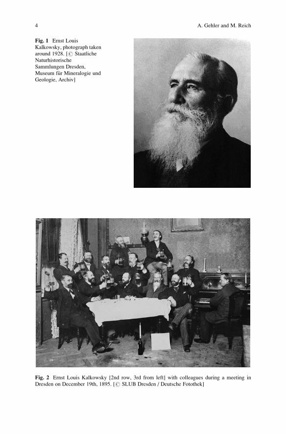
Fig. 1 Ernst Louis
Kalkowsky, photograph taken
around 1928. [# Staatliche
Naturhistorische
Sammlungen Dresden,
Museum f€ur Mineralogie und
Geologie, Archiv]
Fig. 2 Ernst Louis Kalkowsky [2nd row, 3rd from left] with colleagues during a meeting in
Dresden on December 19th, 1895. [# SLUB Dresden / Deutsche Fotothek]
4 A. Gehler and M. Reich
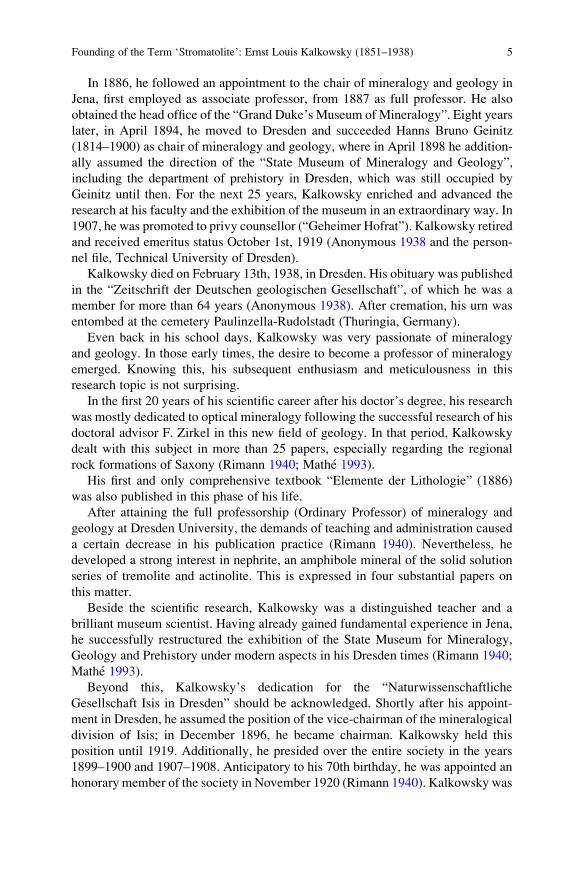
In 1886, he followed an appointment to the chair of mineralogy and geology in
Jena, first employed as associate professor, from 1887 as full professor. He also
obtained the head office of the “Grand Duke’s Museum of Mineralogy”. Eight years
later, in April 1894, he moved to Dresden and succeeded Hanns Bruno Geinitz
(1814–1900) as chair of mineralogy and geology, where in April 1898 he addition-
ally assumed the direction of the “State Museum of Mineralogy and Geology”,
including the department of prehistory in Dresden, which was still occupied by
Geinitz until then. For the next 25 years, Kalkowsky enriched and advanced the
research at his faculty and the exhibition of the museum in an extraordinary way. In
1907, he was promoted to privy counsellor (“Geheimer Hofrat”). Kalkowsky retired
and received emeritus status October 1st, 1919 (Anonymous 1938 and the person-
nel file, Technical University of Dresden).
Kalkowsky died on February 13th, 1938, in Dresden. His obituary was published
in the “Zeitschrift der Deutschen geologischen Gesellschaft”, of which he was a
member for more than 64 years (Anonymous 1938). After cremation, his urn was
entombed at the cemetery Paulinzella-Rudolstadt (Thuringia, Germany).
Even back in his school days, Kalkowsky was very passionate of mineralogy
and geology. In those early times, the desire to become a professor of mineralogy
emerged. Knowing this, his subsequent enthusiasm and meticulousness in this
research topic is not surprising.
In the first 20 years of his scientific career after his doctor’s degree, his research
was mostly dedicated to optical mineralogy following the successful research of his
doctoral advisor F. Zirkel in this new field of geology. In that period, Kalkowsky
dealt with this subject in more than 25 papers, especially regarding the regional
rock formations of Saxony (Rimann 1940; Mathe 1993).
His first and only comprehensive textbook “Elemente der Lithologie” (1886)
was also published in this phase of his life.
After attaining the full professorship (Ordinary Professor) of mineralogy and
geology at Dresden University, the demands of teaching and administration caused
a certain decrease in his publication practice (Rimann 1940). Nevertheless, he
developed a strong interest in nephrite, an amphibole mineral of the solid solution
series of tremolite and actinolite. This is expressed in four substantial papers on
this matter.
Beside the scientific research, Kalkowsky was a distinguished teacher and a
brilliant museum scientist. Having already gained fundamental experience in Jena,
he successfully restructured the exhibition of the State Museum for Mineralogy,
Geology and Prehistory under modern aspects in his Dresden times (Rimann 1940;
Mathe 1993).
Beyond this, Kalkowsky’s dedication for the “Naturwissenschaftliche
Gesellschaft Isis in Dresden” should be acknowledged. Shortly after his appoint-
ment in Dresden, he assumed the position of the vice-chairman of the mineralogical
division of Isis; in December 1896, he became chairman. Kalkowsky held this
position until 1919. Additionally, he presided over the entire society in the years
1899–1900 and 1907–1908. Anticipatory to his 70th birthday, he was appointed an
honorary member of the society in November 1920 (Rimann 1940). Kalkowsky was
Founding of the Term ‘Stromatolite’: Ernst Louis Kalkowsky (1851–1938) 5
also a founding member of the “Pal€aontologische Gesellschaft” in 1912 (Jaekel
1914) as well as an active member of the German Geological Society (“Deutsche
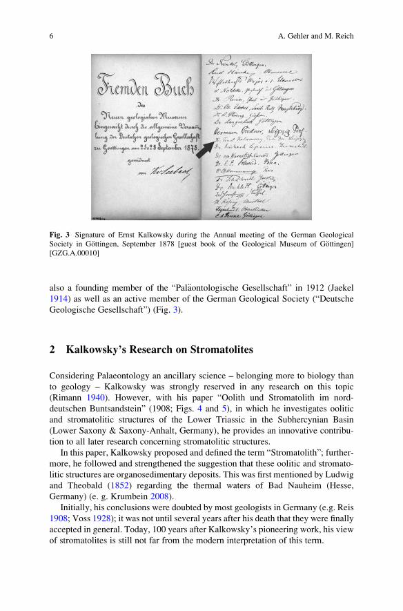
Geologische Gesellschaft”) (Fig. 3).
2 Kalkowsky’s Research on Stromatolites
Considering Palaeontology an ancillary science – belonging more to biology than
to geology – Kalkowsky was strongly reserved in any research on this topic
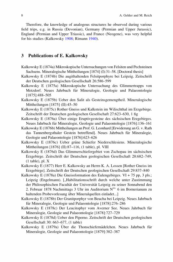
(Rimann 1940). However, with his paper “Oolith und Stromatolith im nord-
deutschen Buntsandstein” (1908; Figs. 4 and 5), in which he investigates oolitic
and stromatolitic structures of the Lower Triassic in the Subhercynian Basin
(Lower Saxony & Saxony-Anhalt, Germany), he provides an innovative contribu-
tion to all later research concerning stromatolitic structures.
In this paper, Kalkowsky proposed and defined the term “Stromatolith”; further-
more, he followed and strengthened the suggestion that these oolitic and stromato-
litic structures are organosedimentary deposits. This was first mentioned by Ludwig
and Theobald (1852) regarding the thermal waters of Bad Nauheim (Hesse,
Germany) (e. g. Krumbein 2008).
Initially, his conclusions were doubted by most geologists in Germany (e.g. Reis
1908; Voss 1928); it was not until several years after his death that they were finally
accepted in general. Today, 100 years after Kalkowsky’s pioneering work, his view
of stromatolites is still not far from the modern interpretation of this term.
Fig. 3 Signature of Ernst Kalkowsky during the Annual meeting of the German Geological
Society in G€ottingen, September 1878 [guest book of the Geological Museum of G€ottingen][GZG.A.00010]
6 A. Gehler and M. Reich
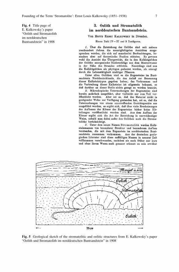
Fig. 4 Title page of
E. Kalkowsky’s paper
“Oolith und Stromatolith
im norddeutschen
Buntsandstein” in 1908
Fig. 5 Geological sketch of the stromatolitic and oolitic structures from E. Kalkowsky’s paper
“Oolith und Stromatolith im norddeutschen Buntsandstein” in 1908
Founding of the Term ‘Stromatolite’: Ernst Louis Kalkowsky (1851–1938) 7
Therefore, the knowledge of analogous structures he observed during various
field trips, e.g. in Russia (Devonian), Germany (Permian and Upper Jurassic),
England (Permian and Upper Triassic), and France (Neogene), was very helpful
for his studies (Kalkowsky 1908; Rimann 1940).
3 Publications of E. Kalkowsky
Kalkowsky E (1874a) Mikroskopische Untersuchungen von Felsiten und Pechsteinen
Sachsens. Mineralogische Mittheilungen [1874] (I):31–58. [Doctoral thesis]
Kalkowsky E (1874b) Die augithaltenden Felsitporphyre bei Leipzig. Zeitschrift
der Deutschen geologischen Gesellschaft 26:586–599
Kalkowsky E (1875a) Mikroskopische Untersuchung des Glimmertrapps von
Metzdorf. Neues Jahrbuch f€ur Mineralogie, Geologie und Palaeontologie
[1875]:488–505
Kalkowsky E (1875b) Ueber den Salit als Gesteinsgemengtheil. Mineralogische
Mittheilungen [1875] (II):45–50
Kalkowsky E (1875c) Rother Gneiss und Kalkstein im Wilischthal im Erzgebirge.
Zeitschrift der Deutschen geologischen Gesellschaft 27:623–630, 1 fig
Kalkowsky E (1876a) €Uber einige Eruptivgesteine des s€achsischen Erzgebirges.
Neues Jahrbuch f€ur Mineralogie, Geologie und Palaeontologie [1876]:136–161
Kalkowsky E (1876b) Mittheilungen an Prof. G. Leonhard [Erwiderung an G. v. Rath
das Tannenbergsthaler Gestein betreffend]. Neues Jahrbuch f€ur Mineralogie,
Geologie und Palaeontologie [1876]:623–626
Kalkowsky E (1876c) Ueber gr€une Schiefer Niederschlesiens. Mineralogische
Mittheilungen [1876] (II):87–116, (1 table), pl. VIII
Kalkowsky E (1876d) Das Glimmerschiefergebiet von Zschopau im s€achsischenErzgebirge. Zeitschrift der Deutschen geologischen Gesellschaft 28:682–749,
(1 table), pl. X
Kalkowsky E (1877) Herr E. Kalkowsky an Herrn K. A. Lossen [Rother Gneiss im
Erzgebirge]. Zeitschrift der Deutschen geologischen Gesellschaft 29:837–840
Kalkowsky E (1878a) Die Gneissformation des Eulengebirges. VI + 75 pp, 3 pls.;
Leipzig (Engelmann). [„Habilitationsschrift durch welche unter Zustimmung
der Philosophischen Facult€at der Universit€at Leipzig zu seiner Sonnabend den
2. Februar 1878 Nachmittags 3 Uhr im Auditorium Nro. 6 im Bornerianum zu
haltenden Probevorlesung €uber Mineralquellen einladet...]
Kalkowsky E (1878b) Der Granitporphyr von Beucha bei Leipzig. Neues Jahrbuch
f€ur Mineralogie, Geologie und Palaeontologie [1878]:276–286
Kalkowsky E (1878c) Der Leucitophyr vom Averner See. Neues Jahrbuch f€urMineralogie, Geologie und Palaeontologie [1878]:727–729
Kalkowsky E (1878d) Ueber den Piperno. Zeitschrift der Deutschen geologischen
Gesellschaft 30: 663–677, (1 table)
Kalkowsky E (1879a) €Uber die Thonschiefern€adelchen. Neues Jahrbuch f€urMineralogie, Geologie und Palaeontologie [1879]:382–387
8 A. Gehler and M. Reich
Kalkowsky E (1879b) Ueber Krystallsystem und Zwillingsbildung des Tenorites.
Zeitschrift f€ur Krystallographie und Mineralogie 3 (III):279–287, pl. VI (9–10)
Kalkowsky E (1880a) Ueber die Erforschung der arch€aischen Formationen. Neues
Jahrbuch f€ur Mineralogie, Geologie und Palaeontologie [1880] (1):1–28
Kalkowsky E (1880b) Ueber Gneiss und Granit des bojischen Gneissstockwerkes
im Oberpf€alzer Waldgebirge. Neues Jahrbuch f€ur Mineralogie, Geologie und
Palaeontologie [1880] (1):29–42, pl. I
Kalkowsky E (1881a) Ueber Hercynit im s€achsischen Granulit. Zeitschrift der
Deutschen geologischen Gesellschaft 33:533–539
Kalkowsky E (1881b) Ueber den Ursprung der granitischen G€ange im Granulit in
Sachsen. Ein Beitrag zur Kenntniss des Granites. Zeitschrift der Deutschen
geologischen Gesellschaft 33:629–653, (1 table)
Kalkowsky E (1882) Einige Beobachtungen im s€achsischen Granulitgebirge. NeuesJahrbuch f€ur Mineralogie, Geologie und Palaeontologie [1882] (1):231–233
Kalkowsky E (1884a) Ueber die Polarisationsverh€altnisse von senkrecht gegen
eine optische Axe geschnittenen zweiaxigen Krystallplatten. Zeitschrift f€urKrystallographie und Mineralogie 9 (V–VI):486–497, pl. XIII
Kalkowsky E (1884b) Die Anwendung des Mikroskops in der Mineralogie und
Geologie. Kapitel XI. In: Vogel J (ed.) Das Mikroskop und die wissenschaftli-
chen Methoden der mikroskopischen Untersuchung in ihrer verschiedenen
Anwendung. Neu bearbeitet von Otto Zacharias. 4. Ed.; Leipzig (Denicke),
pp 213–241
Kalkowsky E (1885) Ueber Olivinzwillinge in Gesteinen. Zeitschrift f€ur Krystallo-graphie und Mineralogie 10 (I):17–24, pl. II
Kalkowsky E (1886a) Elemente der Lithologie. F€ur Studierende bearbeitet. VIII þ316 pp, (31 tables); Heidelberg (Winter)
Kalkowsky E (1886b) Ueber Struvit von Homburg. Zeitschrift f€ur Krystallographieund Mineralogie 11 (I):1–4, (1 table). pl. I
Kalkowsky E (1893) Ueber Ger€oll-Thonschiefer glacialen Ursprungs im Kulm des
Frankenwaldes. Zeitschrift der Deutschen geologischen Gesellschaft 45:69–86
Kalkowsky E (1894) Mineralogie und Geologie an der Technischen Hochschule.
Antrittsrede von Dr. Ernst Kalkowsky, Professor an der Technischen Hochschule
zu Dresden. Der Civilingenieur (N. F.) 40 (4):301–312
Kalkowsky, E (1897) Das neue min.-geologische Institut der Kgl. Techn.
Hochschule zu Dresden. Sitzungsberichte und Abhandlungen der Naturwis-
senschaftlichen Gesellschaft Isis in Dresden [1896] (2; July–December):
27–29
Kalkowsky E (1897) Ueber einen oligoc€anen Sandsteingang an der Lausitzer
Ueberschiebung bei Weinb€ohla in Sachsen. Sitzungsberichte und Abhandlungender Naturwissenschaftlichen Gesellschaft Isis in Dresden [1897] (1; January–
June):80–89, pl. III
Kalkowsky E (1900) Hanns Bruno Geinitz. Die Arbeit seines Lebens. Rede in
der €offentlichen Sitzung der Isis am 22. Februar 1900. Sitzungsberichte und
Abhandlungen der Naturwissenschaftlichen Gesellschaft Isis in Dresden [1900]
(1; January–June):V–XIII
Founding of the Term ‘Stromatolite’: Ernst Louis Kalkowsky (1851–1938) 9
Kalkowsky E (1902) Die Verkieselung der Gesteine in der n€ordlichen Kalahari.
Sitzungsberichte und Abhandlungen der Naturwissenschaftlichen Gesellschaft
Isis in Dresden [1901] (2; July–December):55–107, pls. II–IV
Kalkowsky E (1905) Die Markasit-Patina der Pfahlbau-Nephrite. Sitzungsberichte
und Abhandlungen der Naturwissenschaftlichen Gesellschaft “Isis” in Dresden
[1904] (2; July–December):51–60, 1 fig
Kalkowsky E (1906a) Der Nephrit des Bodensees. Sitzungsberichte und Abhand-
lungen der Naturwissenschaftlichen Gesellschaft “Isis” in Dresden [1906] (1;
January–June):28–44, 1 fig
Kalkowsky E (1906b) Geologie des Nephrites im s€udlichen Ligurien. Zeitschrift
der Deutschen geologischen Gesellschaft 58:307–378, (7 tables), pl. XVIII
Kalkowsky E (1907a) Die nat€urlichen Verh€altnisse Dresdens. 1. Geologischer
Aufbau, Wasserhorizonte, Bodenbeschaffenheit. In: Sch€afer, F. (ed.):
Wissenschaftlicher F€uhrer durch Dresden. [79. Versammlung Deutscher
Natur-Forscher und Arzte]. pp 1–3; Dresden (v. Zahn & Jaensch)
Kalkowsky E (1907b) Die naturwissenschaftlichen Anstalten Dresdens. 3. Das
Mineralogisch-Geologische Institut der Technischen Hochschule. In: Sch€afer, F.(ed.): Wissenschaftlicher F€uhrer durch Dresden. [79. Versammlung Deutscher
Natur-Forscher und Arzte]. pp 53–55; Dresden (v. Zahn & Jaensch)
Kalkowsky E (1907c) Technische Beh€orden und Anstalten und naturwissenschaft-
liche Museen Dresdens. 6. Das K€onigliche Mineralogisch-Geologische
Museum, nebst der Pr€ahistorischen Sammlung im K€oniglichen Zwinger. In:
Sch€afer, F. (ed.): Wissenschaftlicher F€uhrer durch Dresden. [79. Versammlung
Deutscher Natur-Forscher und Arzte]. pp 112–114; Dresden (v. Zahn & Jaensch)
Kalkowsky E (1907d) Geologische Deutung des Nephrites von Gulbashen. In:
Bauer, M.; Koken, E. & Liebisch, T. (eds.): Festband zur Feier des 100j€ahrigenBestehens. Neues Jahrbuch f€ur Mineralogie, Geologie und Palaeontologie
[1907]:159–168
Kalkowsky E (1908a) Der Korundgranulit von Waldheim in Sachsen. Sitzungs-
berichte und Abhandlungen der Naturwissenschaftlichen Gesellschaft “Isis” in
Dresden [1907] (2; July–December):47–65
Kalkowsky E (1908b) Oolith und Stromatolith im norddeutschen Buntsandstein.
Zeitschrift der Deutschen geologischen Gesellschaft 60:68–125, 3 figs., pls.
IV–XI
Kalkowsky E (1908c) 53. Hauptversammlung der Deutschen geologischen Gesell-
schaft. Begr€ußungsworte. Zeitschrift der Deutschen geologischen Gesellschaft,
Monatsberichte 60 (8/10):191–195
Beck R, Credner H, G€abert C, Hibsch JE, Kalkowsky E (1908) Exkursions-Pl€anef€ur die 53. allgemeine Versammlung der Deutschen geologischen Gesellschaft
in Dresden. – 20 S., 12 figs., pls. I–V; Berlin (Schade)
Kalkowsky E (1909a) Europ€aische Entfernungen. Sitzungsberichte und Abhan-
dlungen der Naturwissenschaftlichen Gesellschaft “Isis” in Dresden [1908]
(2; July–December):33–40, (6 tables)
Kalkowsky E (1909b) Bericht €uber die Exkursionen an den Versammlungstagen.
Zeitschrift der Deutschen geologischen Gesellschaft, Monatsberichte 61 (2):90–93
10 A. Gehler and M. Reich
Kalkowsky E (1909c) Geologische Grundlagen der Entwicklungslehre. Sitzungs-
berichte und Abhandlungen der Naturwissenschaftlichen Gesellschaft “Isis” in
Dresden [1909] (1; January–June):3–10
Kalkowsky E (1910) Geologie und Phantasie. Vortrag bei der Feier des 75j€ahrigenBestehens der Naturwissenschaftlichen Gesellschaft “Isis” am 26. Mai 1910.
Sitzungsberichte und Abhandlungen der Naturwissenschaftlichen Gesellschaft
“Isis” in Dresden [1910] (1; January–June):10–19
Kalkowsky E (1915a) Aluminokrate Schlieren im Frankensteiner Gabbro im Oden-
wald. Sitzungsberichte und Abhandlungen der Naturwissenschaftlichen Gesell-
schaft “Isis” in Dresden [1914] (2; July–December):33–42
Kalkowsky E (1915b) Opaleszierender Quarz. Zeitschrift f€ur Krystallographie undMineralogie 55 (I):23–50, pl. III
Kalkowsky E (1921) Mikroskopischer Coelestin im R€ot von Jena als geologische
Erscheinung. Zeitschrift der Deutschen geologischen Gesellschaft 73:1–23
Acknowledgements We like to thank V. J. Roden (Geoscience Centre, University of G€ottingen)for her helpful comments on the manuscript.
References
Anonymous (1938) [obituary to E. L. Kalkowsky]. Zeitschrift der Deutschen geologischen
Gesellschaft 90:173
Erler D, Krause R, Lange J-M (2008) Ernst Kalkowsky 1851-1938. Miniaturen zur Geologie
Sachsens. GeoSzene 4:1–24
Jaekel O (1914) Bericht €uber die Gr€undung und Jahresversammlung der palaeontologischen
Gesellschaft. Palaeontologische Zeitschrift 1:58–73
Kalkowsky E (1908) Oolith und Stromatolith im norddeutschen Buntsandstein. Zeitschrift der
Deutschen geologischen Gesellschaft 60:68–125, 3 figs., pls. IV-XI.
Krumbein WE (2008) Biogenerated rock structures. Space Science Reviews 135:81–94
K€uhne E, Lange J-M, Erler D (2006) Die Geschichte des Museums f€ur Mineralogie und Geologie
Dresden. Geologica Saxonica 50/51:13-95
Ludwig R, Theobald G (1852) Ueber die Mitwirkung der Pflanzen bei der Ablagerung des
kohlensauren Kalkes. Annalen f€ur Physik und Chemie 87:91-107
Mathe G (1993) Ernst Kalkowsky (1851-1938). Geologe, Hochschullehrer und Museumsdirektor.
Abhandlungen des Staatlichen Museums f€ur Mineralogie und Geologie zu Dresden 39:7–20
Reis OM (1908) [Referat zu:] Kalkowsky: Ueber Oolith und Stromatolith im norddeutschen
Buntsandstein. Neues Jahrbuch f€ur Mineralogie, Geologie und Pal€aontologie [1908] (2):
114–138
Rimann E (1940) Ernst Kalkowsky, sein Leben und sein Werk. Sitzungsberichte und Abhandlun-
gen der Naturwissenschaftlichen Gesellschaft Isis in Dresden [1938/39]:69–95
Voss R (1928) Die pal€aogeographische Verbreitung des Rogensteins im deutschen Unteren
Buntsandstein. Abhandlungen der Preußischen Geologischen Landesanstalt (N. F.) 107:1–66
Founding of the Term ‘Stromatolite’: Ernst Louis Kalkowsky (1851–1938) 11

.
Kalkowsky’s Stromatolites and Oolites
(Lower Buntsandstein, Northern Germany)
Josef Paul, Tadeusz M. Peryt, and Robert V. Burne
1 Introduction
Three world-wide used geological terms derived from the area around the German
Harz Mountains: “Oolithi” (Br€uckmann 1721) and nearly 200 years later “Ooid”
and “Stromatolith” (Kalkowsky 1908). All these three terms deal with carbonate
grains and rocks of the Lower Triassic Buntsandstein Group around the Harz Mts.
Therefore, this area is in a way the type area of stromatolites. But Kalkowsky did
not only coin the name, but also deciphered their microbial origin. Therefore, it may
be of interest to have a nearer look at these stromatolites, oolites and the environ-
ment in which they grew and to compare Kalkowsky’s 100 years old observations
and interpretations with recent ones.
2 Studied Area and Stratigraphic Frame
The lithostratigraphic unit hosting Kalkowsky’s stromatolites is the Lower Bunt-
sandstein Subgroup which is of Lower Triassic age and time-equivalent to the
Induan and lower Olenekian of the International Chronostratigraphic Standard
(Table 1). In the studied area, the Lower Buntsandstein Group is about 250 m
J. Paul (*)
Geoscience Centre, University of G€ottingen, Sedimentology Division, Goldschmidtstraße 3,
37077 G€ottingen, Germany
e-mail: [email protected]
T.M. Peryt
Polish Geological Institute – National Research Institute, ul. Rakowiecka 4, 00-975 Warszawa,
Poland
R.V. Burne
Research School of Earth Sciences, The Australian National University, Canberra, ACT, Australia
J. Reitner et al., Advances in Stromatolite Geobiology,Lecture Notes in Earth Sciences 131, DOI 10.1007/978-3-642-10415-2_2,# Springer-Verlag Berlin Heidelberg 2011
13
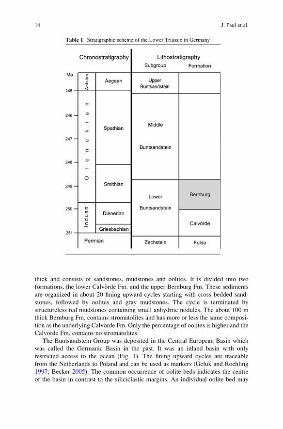
thick and consists of sandstones, mudstones and oolites. It is divided into two
formations, the lower Calv€orde Fm. and the upper Bernburg Fm. These sediments
are organized in about 20 fining upward cycles starting with cross bedded sand-
stones, followed by oolites and gray mudstones. The cycle is terminated by
structureless red mudstones containing small anhydrite nodules. The about 100 m
thick Bernburg Fm. contains stromatolites and has more or less the same composi-
tion as the underlying Calv€orde Fm. Only the percentage of oolites is higher and the
Calv€orde Fm. contains no stromatolites.
The Buntsandstein Group was deposited in the Central European Basin which
was called the Germanic Basin in the past. It was an inland basin with only
restricted access to the ocean (Fig. 1). The fining upward cycles are traceable
from the Netherlands to Poland and can be used as markers (Geluk and Roehling
1997; Becker 2005). The common occurrence of oolite beds indicates the centre
of the basin in contrast to the siliciclastic margins. An individual oolite bed may
Table 1 Stratigraphic scheme of the Lower Triassic in Germany
14 J. Paul et al.
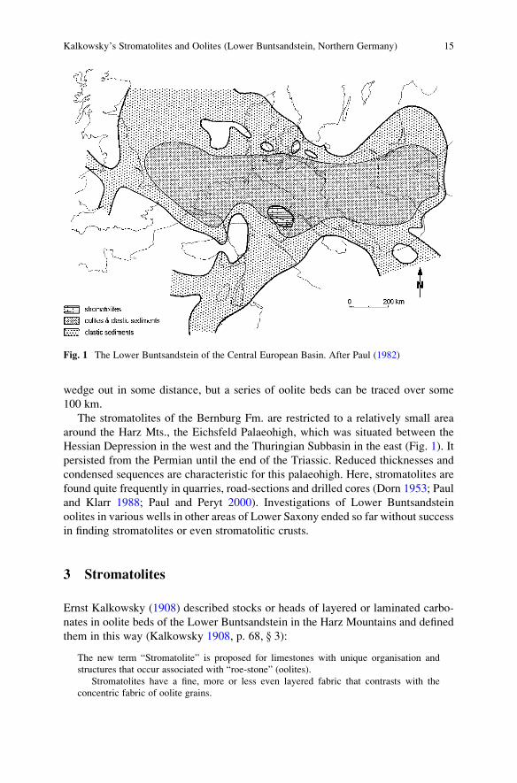
wedge out in some distance, but a series of oolite beds can be traced over some
100 km.
The stromatolites of the Bernburg Fm. are restricted to a relatively small area
around the Harz Mts., the Eichsfeld Palaeohigh, which was situated between the
Hessian Depression in the west and the Thuringian Subbasin in the east (Fig. 1). It
persisted from the Permian until the end of the Triassic. Reduced thicknesses and
condensed sequences are characteristic for this palaeohigh. Here, stromatolites are
found quite frequently in quarries, road-sections and drilled cores (Dorn 1953; Paul
and Klarr 1988; Paul and Peryt 2000). Investigations of Lower Buntsandstein
oolites in various wells in other areas of Lower Saxony ended so far without success
in finding stromatolites or even stromatolitic crusts.
3 Stromatolites
Ernst Kalkowsky (1908) described stocks or heads of layered or laminated carbo-
nates in oolite beds of the Lower Buntsandstein in the Harz Mountains and defined
them in this way (Kalkowsky 1908, p. 68, } 3):
The new term “Stromatolite” is proposed for limestones with unique organisation and
structures that occur associated with “roe-stone” (oolites).
Stromatolites have a fine, more or less even layered fabric that contrasts with the
concentric fabric of oolite grains.
Fig. 1 The Lower Buntsandstein of the Central European Basin. After Paul (1982)
Kalkowsky’s Stromatolites and Oolites (Lower Buntsandstein, Northern Germany) 15
This even layered fabric, the lamination, is according to Kalkowsky (1908) the
distinguishing main mark of stromatolites.
Stromatolites are always associated with to oolites. In most cases, they occur at
the surface of oolite beds terminating the sedimentation of ooids. The thicker the
oolite bed, the higher the probability of occurrence of stromatolites and the larger
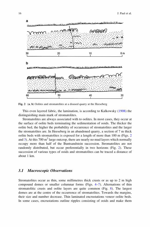
the stromatolites are. In Heeseberg in an abandoned quarry, a section of 7 m thick
oolite beds with stromatolites is exposed for a length of more than 100 m (Figs. 2
and 3). At this 700 m2 large outcrop, there are nearly no mud layers which normally
occupy more than half of the Buntsandstein succession. Stromatolites are not
randomly distributed, but occur preferentially in two horizons (Fig. 2). These
succession of various types of ooids and stromatolites can be traced a distance of
about 1 km.
3.1 Macroscopic Observations
Stromatolites occur as thin, some millimetres thick crusts or as up to 2 m high
compound domes or smaller columnar forms (Figs. 4–7). Alternations of thin
stromatolitic crusts and oolite layers are quite common (Fig. 8). The largest
domes are at the centre of the occurrence of stromatolites. Towards the margins,
their size and number decrease. Thin laminated encrustations veneer oolite beds.
In some cases, encrustations outline ripples consisting of ooids and make them
Fig. 2 (a, b) Oolites and stromatolites at a disused quarry at the Heeseberg
16 J. Paul et al.
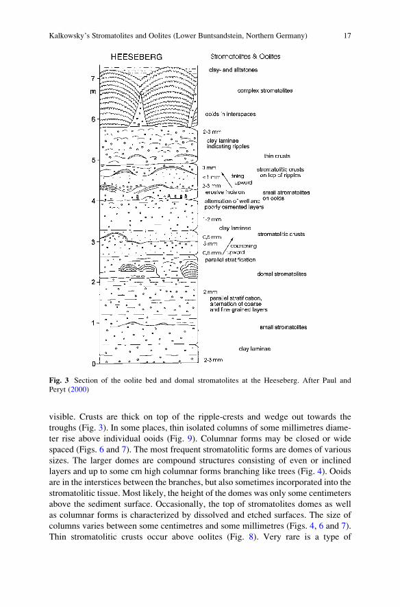
visible. Crusts are thick on top of the ripple-crests and wedge out towards the
troughs (Fig. 3). In some places, thin isolated columns of some millimetres diame-
ter rise above individual ooids (Fig. 9). Columnar forms may be closed or wide
spaced (Figs. 6 and 7). The most frequent stromatolitic forms are domes of various
sizes. The larger domes are compound structures consisting of even or inclined
layers and up to some cm high columnar forms branching like trees (Fig. 4). Ooids
are in the interstices between the branches, but also sometimes incorporated into the
stromatolitic tissue. Most likely, the height of the domes was only some centimeters
above the sediment surface. Occasionally, the top of stromatolites domes as well
as columnar forms is characterized by dissolved and etched surfaces. The size of
columns varies between some centimetres and some millimetres (Figs. 4, 6 and 7).
Thin stromatolitic crusts occur above oolites (Fig. 8). Very rare is a type of
Fig. 3 Section of the oolite bed and domal stromatolites at the Heeseberg. After Paul and
Peryt (2000)
Kalkowsky’s Stromatolites and Oolites (Lower Buntsandstein, Northern Germany) 17
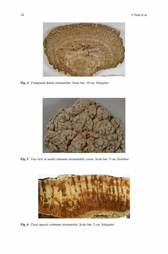
Fig. 4 Compound domal stromatolite. Scale bar: 10 cm. Salzgitter
Fig. 5 Top view at small columnar stromatolitic crusts. Scale bar: 5 cm. Eisleben
Fig. 6 Close spaced columnar stromatolite. Scale bar: 2 cm. Salzgitter
18 J. Paul et al.
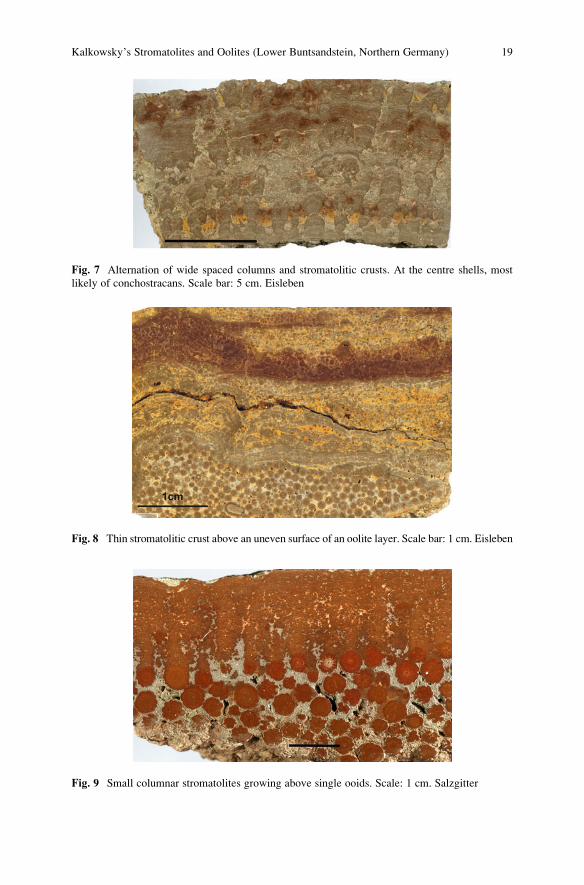
Fig. 7 Alternation of wide spaced columns and stromatolitic crusts. At the centre shells, most
likely of conchostracans. Scale bar: 5 cm. Eisleben
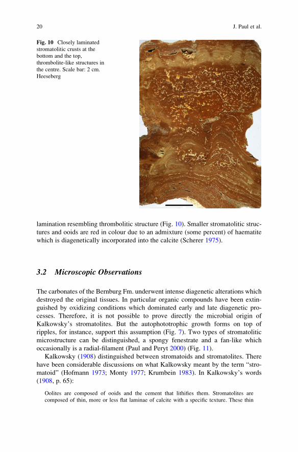
Fig. 8 Thin stromatolitic crust above an uneven surface of an oolite layer. Scale bar: 1 cm. Eisleben
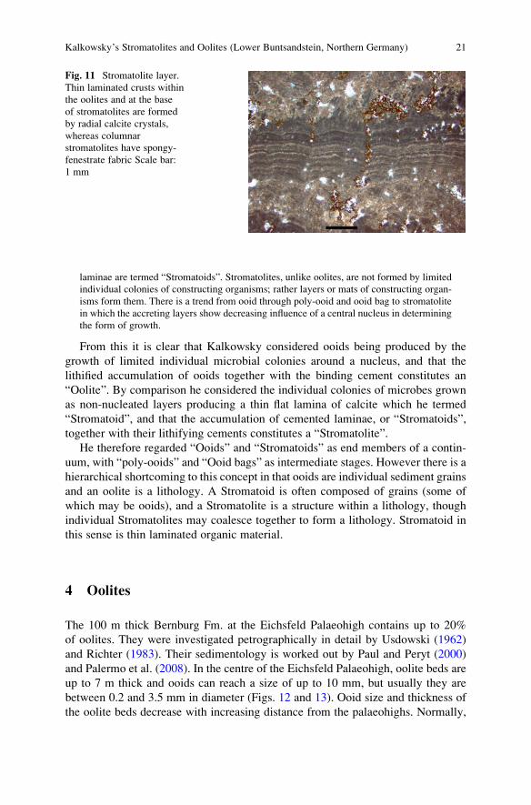
Fig. 9 Small columnar stromatolites growing above single ooids. Scale: 1 cm. Salzgitter
Kalkowsky’s Stromatolites and Oolites (Lower Buntsandstein, Northern Germany) 19
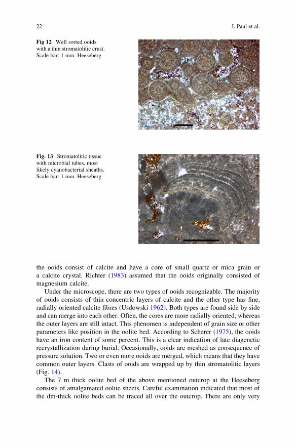
lamination resembling thrombolitic structure (Fig. 10). Smaller stromatolitic struc-
tures and ooids are red in colour due to an admixture (some percent) of haematite
which is diagenetically incorporated into the calcite (Scherer 1975).
3.2 Microscopic Observations
The carbonates of the Bernburg Fm. underwent intense diagenetic alterations which
destroyed the original tissues. In particular organic compounds have been extin-
guished by oxidizing conditions which dominated early and late diagenetic pro-
cesses. Therefore, it is not possible to prove directly the microbial origin of
Kalkowsky’s stromatolites. But the autophototrophic growth forms on top of
ripples, for instance, support this assumption (Fig. 7). Two types of stromatolitic
microstructure can be distinguished, a spongy fenestrate and a fan-like which
occasionally is a radial-filament (Paul and Peryt 2000) (Fig. 11).
Kalkowsky (1908) distinguished between stromatoids and stromatolites. There
have been considerable discussions on what Kalkowsky meant by the term “stro-
matoid” (Hofmann 1973; Monty 1977; Krumbein 1983). In Kalkowsky’s words
(1908, p. 65):
Oolites are composed of ooids and the cement that lithifies them. Stromatolites are
composed of thin, more or less flat laminae of calcite with a specific texture. These thin
Fig. 10 Closely laminated
stromatolitic crusts at the
bottom and the top,
thrombolite-like structures in
the centre. Scale bar: 2 cm.
Heeseberg
20 J. Paul et al.
laminae are termed “Stromatoids”. Stromatolites, unlike oolites, are not formed by limited
individual colonies of constructing organisms; rather layers or mats of constructing organ-
isms form them. There is a trend from ooid through poly-ooid and ooid bag to stromatolite
in which the accreting layers show decreasing influence of a central nucleus in determining
the form of growth.
From this it is clear that Kalkowsky considered ooids being produced by the
growth of limited individual microbial colonies around a nucleus, and that the
lithified accumulation of ooids together with the binding cement constitutes an
“Oolite”. By comparison he considered the individual colonies of microbes grown
as non-nucleated layers producing a thin flat lamina of calcite which he termed
“Stromatoid”, and that the accumulation of cemented laminae, or “Stromatoids”,
together with their lithifying cements constitutes a “Stromatolite”.
He therefore regarded “Ooids” and “Stromatoids” as end members of a contin-
uum, with “poly-ooids” and “Ooid bags” as intermediate stages. However there is a
hierarchical shortcoming to this concept in that ooids are individual sediment grains
and an oolite is a lithology. A Stromatoid is often composed of grains (some of
which may be ooids), and a Stromatolite is a structure within a lithology, though
individual Stromatolites may coalesce together to form a lithology. Stromatoid in
this sense is thin laminated organic material.
4 Oolites
The 100 m thick Bernburg Fm. at the Eichsfeld Palaeohigh contains up to 20%
of oolites. They were investigated petrographically in detail by Usdowski (1962)
and Richter (1983). Their sedimentology is worked out by Paul and Peryt (2000)
and Palermo et al. (2008). In the centre of the Eichsfeld Palaeohigh, oolite beds are
up to 7 m thick and ooids can reach a size of up to 10 mm, but usually they are
between 0.2 and 3.5 mm in diameter (Figs. 12 and 13). Ooid size and thickness of
the oolite beds decrease with increasing distance from the palaeohighs. Normally,
Fig. 11 Stromatolite layer.
Thin laminated crusts within
the oolites and at the base
of stromatolites are formed
by radial calcite crystals,
whereas columnar
stromatolites have spongy-
fenestrate fabric Scale bar:
1 mm
Kalkowsky’s Stromatolites and Oolites (Lower Buntsandstein, Northern Germany) 21
the ooids consist of calcite and have a core of small quartz or mica grain or
a calcite crystal. Richter (1983) assumed that the ooids originally consisted of
magnesium calcite.
Under the microscope, there are two types of ooids recognizable. The majority
of ooids consists of thin concentric layers of calcite and the other type has fine,
radially oriented calcite fibres (Usdowski 1962). Both types are found side by side
and can merge into each other. Often, the cores are more radially oriented, whereas
the outer layers are still intact. This phenomen is independent of grain size or other
parameters like position in the oolite bed. According to Scherer (1975), the ooids
have an iron content of some percent. This is a clear indication of late diagenetic
recrystallization during burial. Occasionally, ooids are meshed as consequence of
pressure solution. Two or even more ooids are merged, which means that they have
common outer layers. Clasts of ooids are wrapped up by thin stromatolitic layers
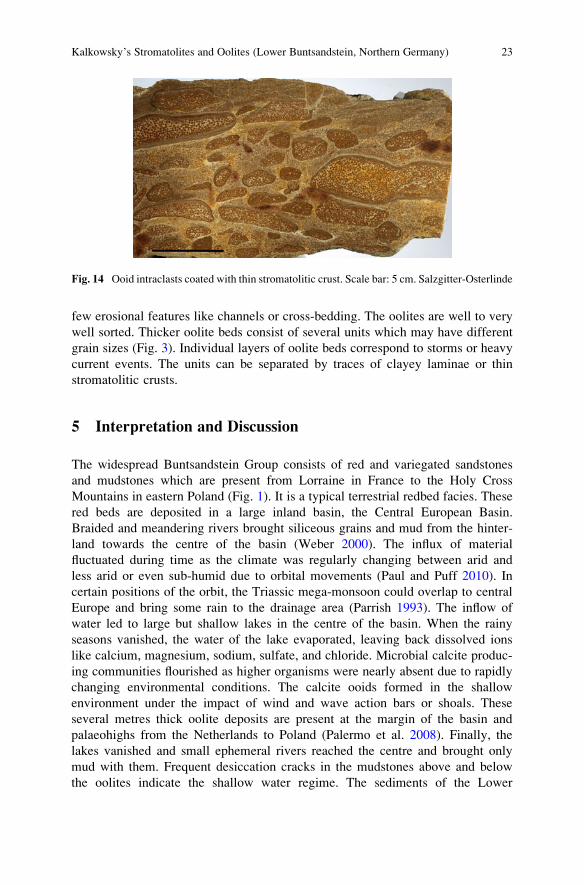
(Fig. 14).
The 7 m thick oolite bed of the above mentioned outcrop at the Heeseberg
consists of amalgamated oolite sheets. Careful examination indicated that most of
the dm-thick oolite beds can be traced all over the outcrop. There are only very
Fig 12 Well sorted ooids
with a thin stromatolitic crust.
Scale bar: 1 mm. Heeseberg
Fig. 13 Stromatolitic tissue
with microbial tubes, most
likely cyanobacterial sheaths.
Scale bar: 1 mm. Heeseberg
22 J. Paul et al.
few erosional features like channels or cross-bedding. The oolites are well to very
well sorted. Thicker oolite beds consist of several units which may have different
grain sizes (Fig. 3). Individual layers of oolite beds correspond to storms or heavy
current events. The units can be separated by traces of clayey laminae or thin
stromatolitic crusts.
5 Interpretation and Discussion
The widespread Buntsandstein Group consists of red and variegated sandstones
and mudstones which are present from Lorraine in France to the Holy Cross
Mountains in eastern Poland (Fig. 1). It is a typical terrestrial redbed facies. These
red beds are deposited in a large inland basin, the Central European Basin.
Braided and meandering rivers brought siliceous grains and mud from the hinter-
land towards the centre of the basin (Weber 2000). The influx of material
fluctuated during time as the climate was regularly changing between arid and
less arid or even sub-humid due to orbital movements (Paul and Puff 2010). In
certain positions of the orbit, the Triassic mega-monsoon could overlap to central
Europe and bring some rain to the drainage area (Parrish 1993). The inflow of
water led to large but shallow lakes in the centre of the basin. When the rainy
seasons vanished, the water of the lake evaporated, leaving back dissolved ions
like calcium, magnesium, sodium, sulfate, and chloride. Microbial calcite produc-
ing communities flourished as higher organisms were nearly absent due to rapidly
changing environmental conditions. The calcite ooids formed in the shallow
environment under the impact of wind and wave action bars or shoals. These
several metres thick oolite deposits are present at the margin of the basin and
palaeohighs from the Netherlands to Poland (Palermo et al. 2008). Finally, the
lakes vanished and small ephemeral rivers reached the centre and brought only
mud with them. Frequent desiccation cracks in the mudstones above and below
the oolites indicate the shallow water regime. The sediments of the Lower
Fig. 14 Ooid intraclasts coated with thin stromatolitic crust. Scale bar: 5 cm. Salzgitter-Osterlinde
Kalkowsky’s Stromatolites and Oolites (Lower Buntsandstein, Northern Germany) 23
Buntsandstein are nearly devoid of fossils, body fossils as well as trace fossils.
The reason of this scarceness may be not abnormal salinity, but rapidly changing
environmental conditions as the shallow playa lake has no buffering capacity
against fluctuations of various environmental parameters. The etched surfaces of
stromatolites result from the decay of organic matter under a cover of clay or
living biofilm led to formation of CO2, lowering the pH and consequently leading
to acidification of the water and dissolution of carbonate.
Some prerequisites of stromatolitic growth can be deduced from observations in
the field. Muddy water or mud layers excluded stromatolites or terminated their
growth. The microbial community did not survive a mud coverage or muddy water.
This effect may be the reason for their restriction to the Eichsfeld Palaeohigh. Here,
the sandy and muddy sediments were diverted west and east of the high on their
way towards the basin. The more extended areas of oolites indicate that the ooid
producing microbes are not so sensitive. The position of stromatolites at top of
the oolite beds seems to reflect a directional evolution, most likely of the water
chemistry, e.g. alkalinity or supersaturation in respect of calcium carbonate. Per-
haps, the stromatolite producing microbial community needs a higher alkalinity
than the ooid producing organisms. The lowermost millimeters of mudstones which
have direct contact to stromatolites are grayish, not like normally reddish. The
decay of the microbial mats after mud coverage led to reducing conditions. The
iron(III) of clay flakes was reduced and removed.
The observed photoautotrophy points to cyanobacteria, at least forming a com-
ponent of the microbial community (Fig. 13). There is a high potential of preserva-
tion through the absence of grazers and browsers and an early lithification, although
the latter cannot be proved. In several passages of his article, Kalkowsky (1908,
p. 112, 115, 119) mentioned and drew sketches of stromatolitic roots. After field
observations and his drawings, we believe that thin stromatolitic crusts covering the
uneven surface of oolites, served as attachments of larger domes.
Peryt (1975) described stromatolites from the Middle Buntsandstein in the
Polish part of the Central European Basin. They are comparable with Kalkowsky’s
stromatolites, consisting of layered and columnar forms. They are also bound to
oolites and oncolites, but grew most likely in a marine or brackish environment, as
Peryt deduced from the frequent occurrence of gastropods, foraminifera and spir-
obid polychaetes.
Regarding the environment of Lower Buntsandstein in northern Germany, there
is a long lasting discussion (see Becker 2005, with references herein). Up to now
there is no proof that marine incursions or transgressions reached the western part of
the Central European Basin during the Lower Buntsandstein.
Stromatolite producing organisms flourished when certain requirements were
fulfilled. They needed certain physico-chemical environments, most likely stable
substrate, clear not muddy waters, sunlight, high alkalinity, high calcium and
carbonate contents of the water, and protection against grazers and burrowers.
The organic nature of stromatolites was generally accepted about 10 or 20 years
after the appearance of Kalkowsky’s publication. In contrast to that, there was
a long-lasting discussion within the scientific community about the formation of
24 J. Paul et al.
ooids. During the nineteenth and twentieth century, most scientists thought of an
inorganic origin, such as the precipitation of calcium and carbonate due to super-
saturation (Usdowski 1962; Bathurst 1978; Simone 1980; F€uchtbauer 1988).
Calcite or aragonite may precipitate around a nucleus of a quartz or carbonate
grain. Only Kalkowsky (1908) thought of an organic origin produced by colonies of
lime secreting phyto-organisms. However, during the last 20 years an increasing
number of indications were found that organisms are involved in the formation of
ooids (Dahanayake et al. 1985; Davaud and Girardelos 2001; Plee et al. 2006). The
in vitro production of ooid-like structures has been observed in cultures of spherical
microbial communities (Brehm et al. 2004).
To summarize, Kalkowsky (1908) stated that
p. 100 } 64 Regarding the environment of the oolites in the north German Bunter
Sandstone, it is generally assumed that they have formed in a shore facies. One
could easily be tempted to think already now of salt lakes as area of their
formation.
p. 118 } 88 Stromatolites were always associated with oolites.
p. 123 } 94 Ooids resemble growing bacterial colonies as observed in a Petri dish.
Ooids are therefore probably produced by colonies of lime secreting phyto-
organisms.
p.124 } 96 We have to assume that simple plants gave rise to limestone precipitation.
My aim has been to show that the oolites and stromatolites of the north German
Bunter Sandstone are inherently of organic origin.
Taking these statements of Kalkowsky into account, we may say that scientific
progress during the last 100 years has not radically changed our understanding of
the formation of the stromatolites and oolites of the Buntsandstein, other discov-
eries have been made that demonstrate that this association has recurred at several
Geological Horizons, and is even found forming in modern seas.
Fifty years after Kalkowsky published this classic paper, Richard Chase recog-
nized the first convincing modern analogues of “stromatoliths” around the shores of
Hamelin Pool, Western Australia [R. Chase, personal communication to RVB].
Recent investigations of both localities reveal a number of interesting parallels
between the environment of Hamelin Pool and that of the Basin with the association
described by Kalkowsky. In both cases, stromatolites grow on stable or firm ground
in turbulent environments characterized by low sedimentation rates, little fine
grained sediment, virtually no terrigenous input, rapid cementation and abnormal
or fluctuating salinity.
Kalkowsky’s stromatolites occur on the surface of oolite beds. Laminated
crusts (called stromatoid by Kalkowsky and interpreted as being formed by
syndepositional cementation) also occur in these rocks. Both stromatolites and
laminated crusts are concentrated in specific layers traceable throughout quarry
faces, where the stromatolites are clearly syndepositional with rippled ooid sand
(Fig. 15 in Paul and Peryt 2000). Spongy-fenestrate and fan-like stromatolitic
microstructures can be distinguished, and both have undergone intense sparitiza-
tion. The upper surfaces of some stromatolites are pitted due to syndepositional
Kalkowsky’s Stromatolites and Oolites (Lower Buntsandstein, Northern Germany) 25
dissolution. The stromatolites may incorporate variable amounts of ooids, quartz
grains and other material.
Hamelin Pool stromatolites also occur in association with ooid sands (Davies
1970; Logan et al. 1974). Subtidal stromatolites grow on rock substrate or crusts
formed by penecontemporaneous cementation of marine sands, and are surrounded
by mobile oolitic rippled sands and sand waves. The subtidal stromatolites have
a laminoid fenestral fabric consisting of ooid and other carbonate sand grains
cemented by micritic cements (Logan et al. 1974). Micritisation of sand grains
begins soon after deposition and gradually destroys the original structure of the
incorporated ooids and other grains (Monty 1976; Reid et al. 2003). Stromatolites in
the intertidal zone are thought to be subtidal forms stranded by sea-level fall and
modified by intertidal microbial communities (Burne 1991–1992). While the Bunt-
sandstein stromatolites originated in a hyposaline and alkaline lake environment
during the high stand of water level, and the Hamelin Pool stromatolites a forming
in a hypersaline marine embayment during a period of regression, there are many
environmental similarities. In both cases conditions favourable for ooid formation
precedes the initiation of stromatolite growth, but the stromatolites co-exist with
ooid sands, and incorporate ooid grains into their structures. The morphology of
many of the subtidal Shark Bay stromatolites is clearly influenced by the erosive
effects of ooid sand waves migrating around them. Once formed, early diagenesis
progressively obliterates the structure of ooid grains incorporated into the stroma-
tolites. The association of stromatolites and ooid sands is of considerable geological
significance. In another present-day environment the stromatolites of Lee Stocking
Island in the Bahamas show a similar association with migrating ooid sand waves to
that found in Hamelin Pool (Dill 1991). The association of stromatolites and oolites
dates back to the Archean. One of the oldest occurrences of the association is
known from the 2.72 Ga Tumbiana Fm., Fortescue Gr. Pilbara Block in Western
Australia.
Even the first stromatolites known to science are associated with oolitic lime-
stones, 25 years before Kalkowsky’s work was published. James Hall had formally
named them Cryptozoon proliferum, and they occur in the oolitic Cambrian Hoyt
Formation of Saratoga Springs, New York State (Hall 1883).
Acknowledgments We have to thank Michael Sosnitza for preparing and polishing the oolite and
stromatolite samples. Max Hundertmark photographed the specimen. Cornelia Kaubisch prepared
and digitalized the drawings. Dr. Ulrike Troitzsch, Canberra, helped to translate the sometimes
difficult German of Kalkowsky.
References
Bathurst R (1978) Carbonate sediments and their Diagenesis. Developments in Sedimentology
12:1–658
Becker A (2005) Sequenzstratigraphie und Fazies des Unteren und Mittleren Buntsandsteins im€ostlichen Teil des Germanischen Beckens (Deutschland, Polen). Hallesches Jahrbuch f€urGeowissenschaften. Reihe B: Geologie, Pal€aontologie, Mineralogie. Beihefte 21:1–117
26 J. Paul et al.
Brehm U, Palinska KA, Krumbein WE (2004) Laboratory cultures of calcifying biomicrospheres
generate ooids – A contribution to the origin of oolites. – Carnets de Geologie/Notebooks on
Geology, Letter 2004/03 (CG2003-L03):1–6
Br€uckmann FE (1721) Specimen physicum exhibens historiam naturalem, oolithi seu ovariorum
piscium & concharum in Saxa. Mutatorum, Helmestadii, Salomoni & Schnorrii, 21 p
Burne RV (1991–1992) Lilliput’s castles: Stromatolites of hamelin pool. Landscope 7:34–40
Dahanayake K, Gerdes G, Krumbein W (1985) Stromatolites, oncolites and oolites biogenically
formed in situ. Naturwissenschaften 72:513–518
Davaud E, Girardelos S (2001) Recent freshwater ooids and oncoids from western lake Geneva
(Switzerland): Indications of a common organically mediated origin. Journal of Sedimentary
Research 71:423–429
Davies GR (1970) Carbonate Bank Sedimentation, Eastern Shark Bay, Western Australia. In:
Logan BW, Davies GR, Read JF, Cebulski DE (eds) Carbonate Sedimentation and Environ-
ments, Shark Bay, Western Australia. The American Association of Petroleum Geologists,
Memoir 13: 85–168
Dill RF (1991) Subtidal stromatolites, ooids and lime encrusted muds at the Great Bahama
bank margin. Contributions in marine geology in honour of Farncis Parker Shephard.
In: Osborne RH (ed) From Shoreline to Abyss. SEPM Special Publication 46:147–171
Dorn P (1953) Die Stromatolithen des Unteren Buntsandstein im s€udlichen Harzvorland. Neues
Jahrbuch f€ur Geologie und Pal€aontontologie, Abhandlungen 97:20–38
F€uchtbauer H (1988) Sedimente und Sedimentgesteine. Schweitzerbart, Stuttgart, 1141 p
Geluk MC, Roehling HG (1997) High-resolution sequence stratigraphy of the Lower Triassic
Buntsandstein in the Netherlands and Northwestern Germany. Geologie en Mijnbouw 76:
227–245
Hall J (1883) Plate VI and explanation: Cryptozoon, N. G., Cryptozoon proliferum n. sp. In:
Pierson HR (ed) Thirtysixth annual report of the trustees of the State Museum of Natural
History to the legislature. New York Senate paper 1883/53, Albany
Hofmann HJ (1973) Stromatolites: Characteristics and utility. Earth-Science Reviews 9:
339–373
Kalkowsky E (1908) Oolith und Stromatolith im norddeutschen Buntsandstein. Zeitschrift der
deutschen geologischen Gesellschaft 60:68–125
Krumbein WE (1983) Stromatolites – The challenge of a term in space and time. Precambrian
Research 20:493–915
Logan BW, Hoffman P, Gebelein CD (1974) Algal Mats, Cryptalgal Fabrics, and structures,
Hamelin Pool, Western Australia. In: Logan BW, Read JF, Hagan GM, Hoffman P, Brown RG,
Woods PJ, Gebelein CD (eds) Evolution and Diagenesis of Quaternary Carbonate Sequences,
Sharc Bay, Western Australia. American Association of Petroleum Geologists, Memoir 22:
140–194
Monty CLV (1976) The origin and development of cryptalgal fabrics. In: Walter MR (ed)
Stromatolites. Developments in Sedimentology 20:193–249
Monty CLV (1977) Evolving concepts on the nature and the ecological significance of stromato-
lites. In: Fl€ugel E (ed) Fossil Algae. Springer, Berlin, pp 15–35
Palermo D, Aigner T, Geluk M, Poeppelreiter M, Pipping K (2008) Reservoir potential of a
lacustrine mixed carbonate/ siliciclastic gas reservoir: The lower triassic rogenstein in the
Netherlands. Journal of Petroleum Geology 31:61–96
Parrish JT (1993) Climate of the supercontinent Pangea. Journal of Geology 101:215–233
Paul J (1982) Der Untere Buntsandstein des germanischen Beckens. Geologische Rundschau
71:795–811
Paul J, Klarr K (1988) Feinstratigraphie und Fazies des Unteren und Mittleren Buntsandsteins in
der Bohrung Remlingen 5. GSF-Bericht 8/87: 148 p
Paul J, Peryt TM (2000) Kalkowsky’s stromatolites revisited (Lower Triassic Buntsandstein, Harz
Mountains, Germany). Palaeogeography, Palaeoclimatology, Palaeoecology 161:435–459
Kalkowsky’s Stromatolites and Oolites (Lower Buntsandstein, Northern Germany) 27
Paul J, Puff P (2010) Das Klima des Buntsandsteins. Schriftenreihe der deutschen Gesellschaft f€urGeowissenschaften. 69 (in press)
Peryt TM (1975) Significance of stromatolites for the environmental interpretation of the Bunt-
sandstein (Lower Triassic) rocks. Geologische Rundschau 64:143–158
Plee K, Ariztegui D, Sahan F, Martini R, Davaud E (2006) Microbes caught in the act: Disen-
tangling the role of biofilms in the formation of low Mg calcite ooids in a freshwater lake.
American Geophysical Union. Fall Meeting 2006. Abstract #B11A-1000
Reid RP, James NP, Macintyre IG, Dupraz CP, Burne RV (2003) Shark Bay stromatolites:
Microfabrics and reinterpretation of origins. Facies 49:45–53
Richter DK (1983) Calcareous ooids: A synopsis. In Peryt TM (ed) Coated grains. Springer,
Heidelberg, pp 71–99
Scherer M (1975) Fe-Anreicherung der Rogensteinzone des norddeutschen Unteren Buntsand-
steins (Trias): Ein Hinweis auf die diagenetische Geschichte. Neues Jahrbuch Geologie und
Pal€aontologie, Monatshefte 1975:568–576
Simone L (1980) Ooids: A review. Earth Sciences Review 16:319–355
Usdowski E (1962) Die Entstehung der kalkoolithischen Fazies des norddeutschen Unteren
Buntsandsteins. Beitr€age zur Mineralogie und Petrographie 8:141–179
Weber J (2000) Kiesels€aurediagenese und gekoppelte Sedimentarchitektur – eine Beckenanalyse
des Reinhardswald-Troges (Norddeutsches Becken, Solling-Folge, Mittlerer Buntsandstein).
K€olner Forum Geologie und Pal€aontologie 7:1–165
28 J. Paul et al.
The Nature of Stromatolites: 3,500 Million Years
of History and a Century of Research
Robert Riding
1 Introduction
Stromatolites are widely regarded as layered, early lithified, authigenic microbial
structures – often domical or columnar in form – that developed at the sediment
water interface in freshwater, marine and evaporitic environments (Fig. 1). In
addition to this unusually wide environmental distribution, the exceptionally long
geological record of stromatolites spans at least 3,500 million years (Ma) (Vologdin
1962; Hofmann 1969, 1973; Walter 1976a; Grotzinger and Knoll 1999; Riding and
Awramik 2000). Most of these examples, together with those described by Kalkowsky
(1908), are essentially originally carbonate in composition. More than a century of
research has revealed many details of their diverse fabrics and complex history, but
much still remains to be understood about stromatolites. This is not surprising
considering their wide distribution in time and space; and it helps to account for a
continuing problem with their definition. Kalkowsky (1908) considered stromato-
lites to be microbial sediments, but it has become increasingly difficult to maintain
this view for all ancient examples, especially those more than ~1,000 Ma old. The
aim of this article is to evaluate progress in understanding what stromatolites are,
since they were first described in the 1800s. This makes it impossible to avoid the
thorny problem of how they should be defined.
The nature and definition of stromatolites have been persistent difficulties ever
since Kalkowsky introduced the name. At first, the main question posed was “are
stromatolites biogenic or abiogenic?” With time, the focus has shifted to whether
all stromatolites are biogenic, or whether some are biogenic while others are
abiogenic. Kalkowsky’s (1908) microbial interpretation of stromatolites was
R. Riding
Department of Earth and Planetary Sciences, University of Tennessee, Knoxville, TN 37996, USA
e-mail: [email protected]
J. Reitner et al., Advances in Stromatolite Geobiology,Lecture Notes in Earth Sciences 131, DOI 10.1007/978-3-642-10415-2_3,# Springer-Verlag Berlin Heidelberg 2011
29
immediately challenged by the suggestion that they are abiogenic precipitates (Reis
1908). The subsequent century of research provided support from present-day and
ancient examples for both of these views. In particular, persuasive evidence that
some Precambrian stromatolites are essentially abiotic seafloor crusts grew out of
pioneering studies of early Proterozoic examples in Canada (e.g., Kerans 1982;
Grotzinger and Read 1983).
The challenge of defining stromatolites reflects the diversity and complexity of
the structures they represent. The scarcity of present-day marine analogues for
abiogenic seafloor crusts (Grotzinger and James 2000a, p. 9), such as those that
occur in the Palaeoproterozoic, has hindered appreciation of the inorganic processes
that can produce marine structures that have been described as stromatolites (Pope
et al. 2000, p. 1149; Corsetti and Storrie-Lombardi 2003, p. 649; Perry et al. 2007,
p. 169). The marine stromatolite record can be read as long-term change from less to
more biogenic (Grotzinger and Kasting 1993, p. 235; Kah and Knoll 1996, p. 81;
James et al. 1998). As a result, the time period from which stromatolites are viewed
is critically important. It is not difficult to regard most Phanerozoic examples as
essentially lithified microbial mats. In contrast many Precambrian examples
regarded as stromatolites, especially those older than ~1,000 Ma, appear to contain,
and in some cases entirely consist of, precipitated abiogenic crust. Furthermore,
there is evidence that many late Archaean and early Proterozoic stromatolites
consist of intimate interlayering of both lithified microbial mat and essentially
abiogenic precipitated crust (Bertrand-Sarfati 1972, p. 155; Sami and James 1996,
p. 217; Petrov and Semikhatov 2001, fig. 5a, b; Riding 2008, p. 95) that has been
termed Hybrid Crust (Riding 2008). Interpretation of these deposits is hindered –
especially in very old deposits – by recrystallization, but even in the Neoarchaean,
relatively well-preserved examples retain clear indications of even and laterally
very continuous layers that appear to consist of thin alternations of sparry and
microcrystalline fabrics (Sumner and Grotzinger 2004, fig. 3). These “Boetsap
laminae”, named after a locality on the Boetsap River in South Africa, could
be Hybrid Crust stromatolites (Riding 2008, p. 84) (Fig. 2). If so, Hybrid Crust
is likely to be a major component of late Archaean and early Proterozoic
Fig. 1 Loaf-shaped
stromatolite in oolite. Early
Triassic, Bernburg Fm,
Heeseberg Quarry, Jerxheim,
50 km west of Magdeburg,
Germany. Width of view
1.6 m
30 R. Riding
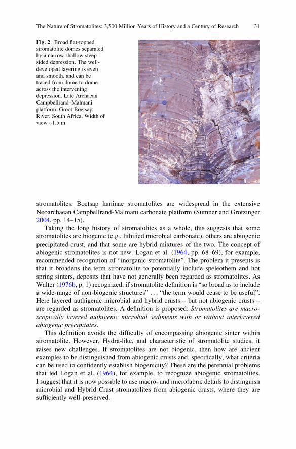
stromatolites. Boetsap laminae stromatolites are widespread in the extensive
Neoarchaean Campbellrand-Malmani carbonate platform (Sumner and Grotzinger
2004, pp. 14–15).
Taking the long history of stromatolites as a whole, this suggests that some
stromatolites are biogenic (e.g., lithified microbial carbonate), others are abiogenic
precipitated crust, and that some are hybrid mixtures of the two. The concept of
abiogenic stromatolites is not new. Logan et al. (1964, pp. 68–69), for example,
recommended recognition of “inorganic stromatolite”. The problem it presents is
that it broadens the term stromatolite to potentially include speleothem and hot
spring sinters, deposits that have not generally been regarded as stromatolites. As
Walter (1976b, p. 1) recognized, if stromatolite definition is “so broad as to include
a wide-range of non-biogenic structures” . . . “the term would cease to be useful”.
Here layered authigenic microbial and hybrid crusts – but not abiogenic crusts –
are regarded as stromatolites. A definition is proposed: Stromatolites are macro-scopically layered authigenic microbial sediments with or without interlayeredabiogenic precipitates.
This definition avoids the difficulty of encompassing abiogenic sinter within
stromatolite. However, Hydra-like, and characteristic of stromatolite studies, it
raises new challenges. If stromatolites are not biogenic, then how are ancient
examples to be distinguished from abiogenic crusts and, specifically, what criteria
can be used to confidently establish biogenicity? These are the perennial problems
that led Logan et al. (1964), for example, to recognize abiogenic stromatolites.
I suggest that it is now possible to use macro- and microfabric details to distinguish
microbial and Hybrid Crust stromatolites from abiogenic crusts, where they are
sufficiently well-preserved.
Fig. 2 Broad flat-topped
stromatolite domes separated
by a narrow shallow steep-
sided depression. The well-
developed layering is even
and smooth, and can be
traced from dome to dome
across the intervening
depression. Late Archaean
Campbellrand–Malmani
platform, Groot Boetsap
River. South Africa. Width of
view ~1.5 m
The Nature of Stromatolites: 3,500 Million Years of History and a Century of Research 31
2 Stromatolites and Spongiostromids
2.1 Stromatolith
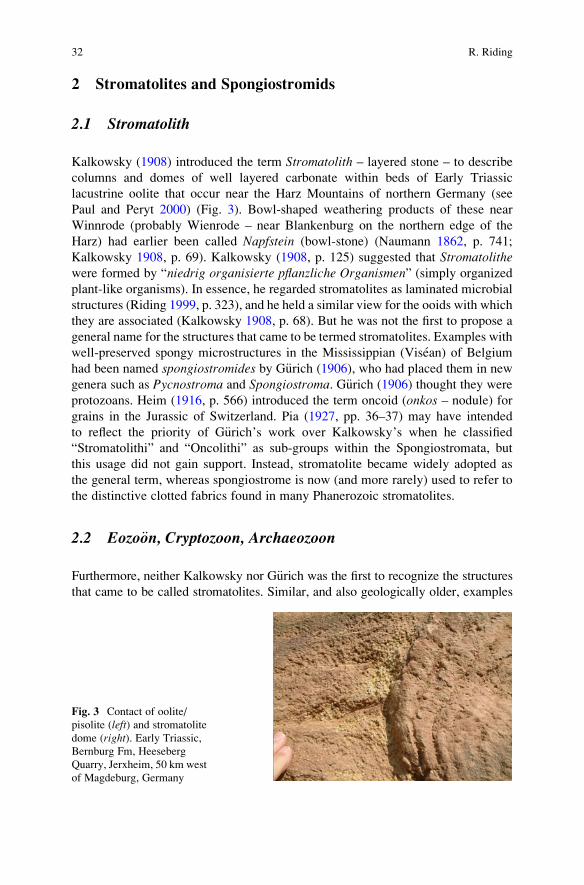
Kalkowsky (1908) introduced the term Stromatolith – layered stone – to describe
columns and domes of well layered carbonate within beds of Early Triassic
lacustrine oolite that occur near the Harz Mountains of northern Germany (see
Paul and Peryt 2000) (Fig. 3). Bowl-shaped weathering products of these near
Winnrode (probably Wienrode – near Blankenburg on the northern edge of the
Harz) had earlier been called Napfstein (bowl-stone) (Naumann 1862, p. 741;
Kalkowsky 1908, p. 69). Kalkowsky (1908, p. 125) suggested that Stromatolithewere formed by “niedrig organisierte pflanzliche Organismen” (simply organized
plant-like organisms). In essence, he regarded stromatolites as laminated microbial
structures (Riding 1999, p. 323), and he held a similar view for the ooids with which
they are associated (Kalkowsky 1908, p. 68). But he was not the first to propose a
general name for the structures that came to be termed stromatolites. Examples with
well-preserved spongy microstructures in the Mississippian (Visean) of Belgium
had been named spongiostromides by G€urich (1906), who had placed them in new
genera such as Pycnostroma and Spongiostroma. G€urich (1906) thought they were
protozoans. Heim (1916, p. 566) introduced the term oncoid (onkos – nodule) for
grains in the Jurassic of Switzerland. Pia (1927, pp. 36–37) may have intended
to reflect the priority of G€urich’s work over Kalkowsky’s when he classified
“Stromatolithi” and “Oncolithi” as sub-groups within the Spongiostromata, but
this usage did not gain support. Instead, stromatolite became widely adopted as
the general term, whereas spongiostrome is now (and more rarely) used to refer to
the distinctive clotted fabrics found in many Phanerozoic stromatolites.
2.2 Eozo€on, Cryptozoon, Archaeozoon
Furthermore, neither Kalkowsky nor G€urich was the first to recognize the structures
that came to be called stromatolites. Similar, and also geologically older, examples
Fig. 3 Contact of oolite/
pisolite (left) and stromatolite
dome (right). Early Triassic,
Bernburg Fm, Heeseberg
Quarry, Jerxheim, 50 km west
of Magdeburg, Germany
32 R. Riding
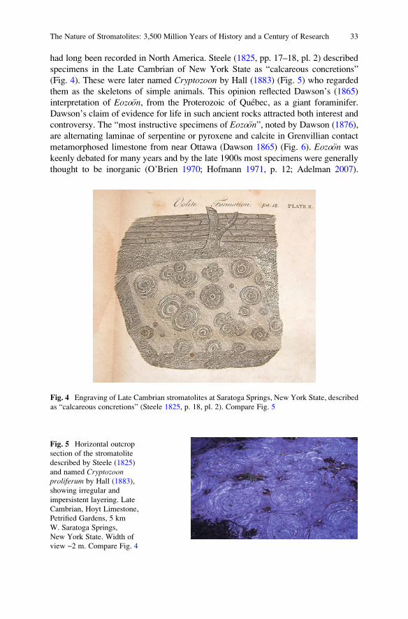
had long been recorded in North America. Steele (1825, pp. 17–18, pl. 2) described
specimens in the Late Cambrian of New York State as “calcareous concretions”
(Fig. 4). These were later named Cryptozoon by Hall (1883) (Fig. 5) who regarded
them as the skeletons of simple animals. This opinion reflected Dawson’s (1865)
interpretation of Eozo€on, from the Proterozoic of Quebec, as a giant foraminifer.
Dawson’s claim of evidence for life in such ancient rocks attracted both interest and
controversy. The “most instructive specimens of Eozo€on”, noted by Dawson (1876),
are alternating laminae of serpentine or pyroxene and calcite in Grenvillian contact
metamorphosed limestone from near Ottawa (Dawson 1865) (Fig. 6). Eozo€on was
keenly debated for many years and by the late 1900s most specimens were generally
thought to be inorganic (O’Brien 1970; Hofmann 1971, p. 12; Adelman 2007).
Fig. 4 Engraving of Late Cambrian stromatolites at Saratoga Springs, New York State, described
as “calcareous concretions” (Steele 1825, p. 18, pl. 2). Compare Fig. 5
Fig. 5 Horizontal outcrop
section of the stromatolite
described by Steele (1825)
and named Cryptozoonproliferum by Hall (1883),
showing irregular and
impersistent layering. Late
Cambrian, Hoyt Limestone,
Petrified Gardens, 5 km
W. Saratoga Springs,
New York State. Width of
view ~2 m. Compare Fig. 4
The Nature of Stromatolites: 3,500 Million Years of History and a Century of Research 33
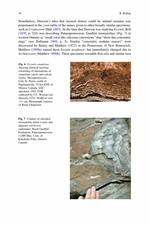
Nonetheless, Dawson’s idea that layered domes could be animal remains was
perpetuated in the zoon suffix of the names given to other broadly similar specimens,
such as Cryptozoon (Hall 1883). At the time that Dawson was studying Eozo€on, Bell(1870, p. 324) was describing Palaeoproterozoic Gunflint stromatolites (Fig. 7) in
western Ontario as “small coral-like siliceous concretions” that “show fine concentric
rings” (see Hofmann 1969, p. 5). Similar “concentric nodular masses” were
discovered by Bailey and Matthew (1872) in the Proterozoic of New Brunswick.
Matthew (1890a) named them Eozo€on acadiense, but immediately changed this to
Archaeozoon (Matthew 1890b). These specimens resemble Baicalia and similar taxa
Fig. 6 Eozo€on canadense,showing domical layering
consisting of alternations of
serpentine (dark) and calcite
(light). Mesoproterozoic,
Cote St. Pierre, north of
Papineauville, 55 km ENE of
Ottawa, Canada. GSC
specimen 1992-234B
collected by T.C. Weston (see
Dawson 1876). Width of view
~11 cm. Photograph courtesy
of Brian Chatterton
Fig. 7 Contact of silicified
stromatolite dome (right) andadjacent red-browncarbonates. Basal Gunflint
Formation, Palaeoproterozoic
(1,880 Ma), 1 km. of
Kakabeka Falls, Ontario,
Canada
34 R. Riding
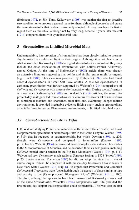
(Hofmann 1971, p. 58). Thus, Kalkowsky (1908) was neither the first to describe
stromatolites nor to propose a general name for them, although of course he did create
the name stromatolite that has been universally adopted. Hemay have been the first to
regard them as microbial, although not by very long, because 6 years later Walcott
(1914) compared them with cyanobacterial tufa.
3 Stromatolites as Lithified Microbial Mats
Understandably, interpretation of stromatolites has been closely linked to present-
day deposits that could shed light on their origins. Although it is not clear exactly
what reasons led Kalkowsky (1908) to regard stromatolites as microbial, they may
include the close association of stromatolites with ooliths (which Kalkowsky
named Ooide). At the time of Kalkowsky’s (1908) article there was already
an extensive literature suggesting that ooliths and similar grains might be organic
(e.g., Linck 1903). This view was pioneered by Rothpletz (1892) who had found
coccoid cyanobacteria in Great Salt Lake ooliths. A role for cyanobacteria in
carbonate precipitation was further supported by Walcott’s (1914) comparisons of
Collenia and Cryptozoon with present-day lacustrine tufas. During the half-centuryor more since Kalkowsky’s (1908) and Walcott’s (1914) articles, the search for
present-day analogues led from cool-water calcareous freshwater lakes and streams
to subtropical marshes and shorelines, tidal flats and, eventually, deeper marine
environments. It provided irrefutable evidence linking many ancient stromatolites,
especially those in marine Phanerozoic environments, to lithified microbial mats.
3.1 Cyanobacterial Lacustrine Tufas
C.D.Walcott, studying Proterozoic sediments in the western United States, had found
Neoproterozoic specimens at Nankoweap Butte in the Grand Canyon (Walcott 1895,
p. 319) that he regarded as stromatoporoids, but which Dawson (1896, p. 208)
thought were Cryptozoon and compared to foraminifers (Dawson 1896,
pp. 211–212). Walcott (1906) encountered more examples as he extended his studies
to the Mesoproterozoic of Montana, and he described them as new genera, including
Collenia, named after a rancher in the Big Belt Mountains (Walcott 1914, p. 111).
Walcott had seenCryptozoonmuch earlier at Saratoga Springs in 1878 (Schopf 1999,
p. 25; Lindemann and Yochelson 2005) but did not adopt the view that it was of
animal origin. Instead, he compared it with present-day freshwater tufas in lakes in
New York State (Walcott 1914) (Fig. 8). He argued that Proterozoic and Cambrian
Collenia and Cryptozoonwere “deposited through the agency of algae similar in type
and activity to the (Cyanophyceae) Blue–green Algae” (Walcott 1914, p. 100).
Therefore, although he appears to have been unaware of Kalkowsky’s work and
of the name Stromatolithe, Walcott’s (1914) comparisons with tufa provided the
first present-day support that stromatolites could be microbial. This was also the first
The Nature of Stromatolites: 3,500 Million Years of History and a Century of Research 35
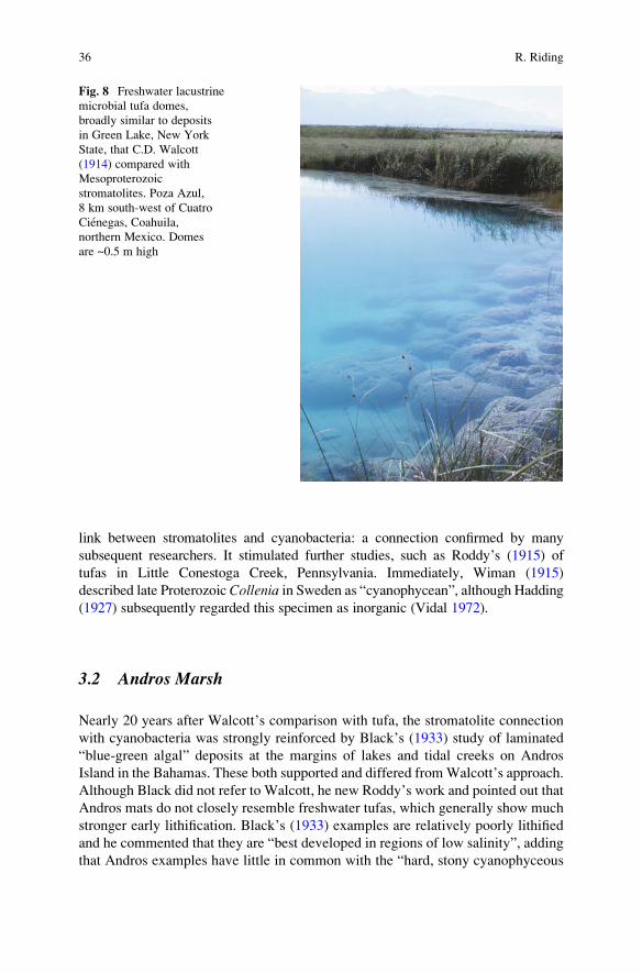
link between stromatolites and cyanobacteria: a connection confirmed by many
subsequent researchers. It stimulated further studies, such as Roddy’s (1915) of
tufas in Little Conestoga Creek, Pennsylvania. Immediately, Wiman (1915)
described late Proterozoic Collenia in Sweden as “cyanophycean”, although Hadding(1927) subsequently regarded this specimen as inorganic (Vidal 1972).
3.2 Andros Marsh
Nearly 20 years after Walcott’s comparison with tufa, the stromatolite connection
with cyanobacteria was strongly reinforced by Black’s (1933) study of laminated
“blue-green algal” deposits at the margins of lakes and tidal creeks on Andros
Island in the Bahamas. These both supported and differed fromWalcott’s approach.
Although Black did not refer to Walcott, he new Roddy’s work and pointed out that
Andros mats do not closely resemble freshwater tufas, which generally show much
stronger early lithification. Black’s (1933) examples are relatively poorly lithified
and he commented that they are “best developed in regions of low salinity”, adding
that Andros examples have little in common with the “hard, stony cyanophyceous
Fig. 8 Freshwater lacustrine
microbial tufa domes,
broadly similar to deposits
in Green Lake, New York
State, that C.D. Walcott
(1914) compared with
Mesoproterozoic
stromatolites. Poza Azul,
8 km south-west of Cuatro
Cienegas, Coahuila,
northern Mexico. Domes
are ~0.5 m high
36 R. Riding
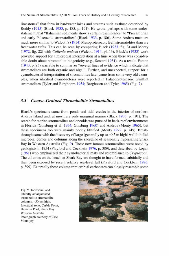
limestones” that form in hardwater lakes and streams such as those described by
Roddy (1915) (Black 1933, p. 185, p. 191). He wrote, perhaps with some under-
statement, that “Bahamian sediments show a certain resemblance” to “Precambrian
and early Palaeozoic stromatolites” (Black 1933, p. 186). Some Andros mats are
much more similar to Walcott’s (1914) Mesoproterozoic Belt stromatolites than are
freshwater tufas. This can be seen by comparing Black (1933, fig. 3) and Monty
(1972, fig. 22) with Collenia undosa (Walcott 1914, pl. 13). Black’s (1933) work
provided support for a microbial interpretation at a time when there was consider-
able doubt about stromatolite biogenicity (e.g., Seward 1931). As a result, Fenton
(1943, p. 95) was able to summarize “several lines of evidence which indicate that
stromatolites are both organic and algal”. Further, and unexpected, support for a
cyanobacterial interpretation of stromatolites later came from some very old exam-
ples, when silicified cyanobacteria were reported in Palaeoproterozoic Gunflint
stromatolites (Tyler and Barghoorn 1954; Barghoorn and Tyler 1965) (Fig. 7).
3.3 Coarse-Grained Thrombolitic Stromatolites
Black’s specimens came from ponds and tidal creeks in the interior of northern
Andros Island and, at most, are only marginal marine (Black 1933, p. 191). The
search for marine stromatolites and oncoids was pursued in back-reef environments
in Florida (Ginsburg et al. 1954; Ginsburg 1960) and Andros (Monty 1965), but
these specimens too were mainly poorly lithified (Monty 1972, p. 745). Break-
through came with the discovery of large (generally up to ~0.5 m high) well lithified
microbial domes and columns along the shoreline of seasonally hypersaline Shark
Bay in Western Australia (Fig. 9). These now famous stromatolites were noted by
geologists in 1954 (Playford and Cockbain 1976, p. 389), and described by Logan
(1961) who emphasized their cyanobacterial mats and resemblance to Cryptozoon.The columns on the beach at Shark Bay are thought to have formed subtidally and
then been exposed by recent relative sea-level fall (Playford and Cockbain 1976,
p. 399). Externally these columnar microbial carbonates can closely resemble some
Fig. 9 Individual and
laterally amalgamated
thrombolitic stromatolite
columns, ~50 cm high.
Intertidal zone, Carbla Point,
Hamelin Pool, Shark Bay,
Western Australia.
Photograph courtesy of Eric
Mountjoy
The Nature of Stromatolites: 3,500 Million Years of History and a Century of Research 37
very ancient examples such as 1,800 Ma Pethei elongate columns (Hoffman 1989,
fig. 9b), but internally they are often less well layered and coarser grained. Crudely
layered columns at Shark Bay, illustrated by Logan (1961, pl. 1, fig. 4) and
described by Aitken (1967, p. 1171) as “thrombolitic stromatolites”, are largely
composed of fine sand (Logan et al. 1974).
Large columnar and domical stromatolites are very rare in present-day marine
environments. Apart from Shark Bay, the only other region in which they have been
reported are the Bahama Banks (Dravis 1983; Reid et al. 1995, 2000), where they
are especially well-developed in the tidal channel between Lee Stocking Island and
Norman’s Pond Cay in the Exuma Cays (Dill et al. 1986; Riding et al. 1991a)
(Fig. 10). Shark Bay and Lee Stocking columns occur in wave- and current-swept
environments. Their formation can be attributed to factors that include the mat
communities, water movement, and grainy conditions. Strong waves and currents
raise sand to the accreting mat surface, and the grainy environment deters over-
growth by reefal encrusters (Dill et al. 1989, p. 10). Seasonal hypersalinity at Shark
Bay also limits competitors. Accretion rates are high due to the combined presence
of thick soft microbial mats that contain abundant EPS (extracellular polymeric
substances) (Decho et al. 2005), and rapid grain supply. In addition to cyanobacteria,
the mats contain diatoms and filamentous green algae that enhance trapping
(Awramik and Riding 1988; Dill et al. 1989; Riding et al. 1991a). The upper mat
Fig. 10 “Molar tooth”
thrombolitic stromatolite
domes surrounded, and half-
buried, by rippled ooid sand.
Domes have exposed heights
of ~0.5–1 m. Depth ~10 m
in tidal channel between
Lee Stocking Island and
Norman’s Pond Cay, Exuma
Islands, Bahamas
38 R. Riding
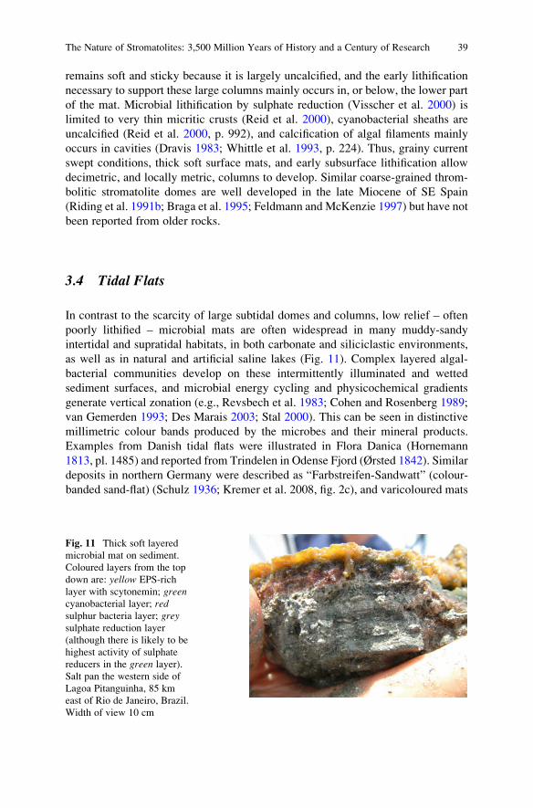
remains soft and sticky because it is largely uncalcified, and the early lithification
necessary to support these large columns mainly occurs in, or below, the lower part
of the mat. Microbial lithification by sulphate reduction (Visscher et al. 2000) is
limited to very thin micritic crusts (Reid et al. 2000), cyanobacterial sheaths are
uncalcified (Reid et al. 2000, p. 992), and calcification of algal filaments mainly
occurs in cavities (Dravis 1983; Whittle et al. 1993, p. 224). Thus, grainy current
swept conditions, thick soft surface mats, and early subsurface lithification allow
decimetric, and locally metric, columns to develop. Similar coarse-grained throm-
bolitic stromatolite domes are well developed in the late Miocene of SE Spain
(Riding et al. 1991b; Braga et al. 1995; Feldmann and McKenzie 1997) but have not
been reported from older rocks.
3.4 Tidal Flats
In contrast to the scarcity of large subtidal domes and columns, low relief – often
poorly lithified – microbial mats are often widespread in many muddy-sandy
intertidal and supratidal habitats, in both carbonate and siliciclastic environments,
as well as in natural and artificial saline lakes (Fig. 11). Complex layered algal-
bacterial communities develop on these intermittently illuminated and wetted
sediment surfaces, and microbial energy cycling and physicochemical gradients
generate vertical zonation (e.g., Revsbech et al. 1983; Cohen and Rosenberg 1989;
van Gemerden 1993; Des Marais 2003; Stal 2000). This can be seen in distinctive
millimetric colour bands produced by the microbes and their mineral products.
Examples from Danish tidal flats were illustrated in Flora Danica (Hornemann
1813, pl. 1485) and reported from Trindelen in Odense Fjord (Ørsted 1842). Similar
deposits in northern Germany were described as “Farbstreifen-Sandwatt” (colour-
banded sand-flat) (Schulz 1936; Kremer et al. 2008, fig. 2c), and varicoloured mats
Fig. 11 Thick soft layered
microbial mat on sediment.
Coloured layers from the top
down are: yellow EPS-rich
layer with scytonemin; greencyanobacterial layer; redsulphur bacteria layer; greysulphate reduction layer
(although there is likely to be
highest activity of sulphate
reducers in the green layer).
Salt pan the western side of
Lagoa Pitanguinha, 85 km
east of Rio de Janeiro, Brazil.
Width of view 10 cm
The Nature of Stromatolites: 3,500 Million Years of History and a Century of Research 39
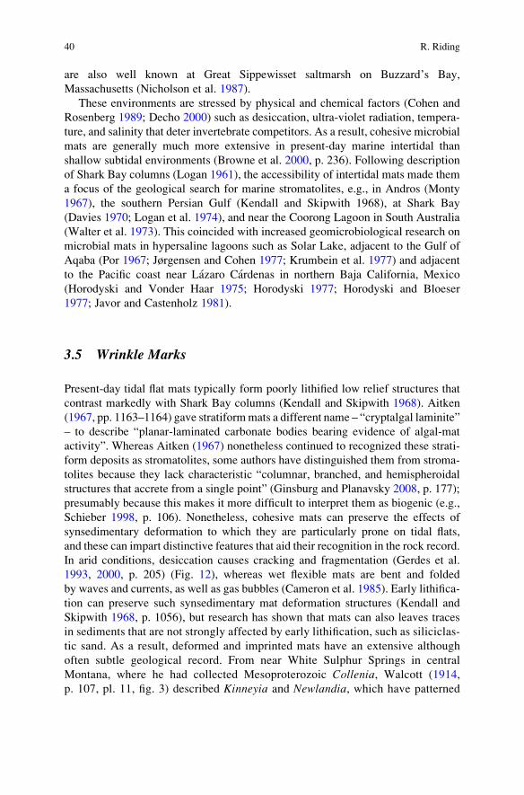
are also well known at Great Sippewisset saltmarsh on Buzzard’s Bay,
Massachusetts (Nicholson et al. 1987).
These environments are stressed by physical and chemical factors (Cohen and
Rosenberg 1989; Decho 2000) such as desiccation, ultra-violet radiation, tempera-
ture, and salinity that deter invertebrate competitors. As a result, cohesive microbial
mats are generally much more extensive in present-day marine intertidal than
shallow subtidal environments (Browne et al. 2000, p. 236). Following description
of Shark Bay columns (Logan 1961), the accessibility of intertidal mats made them
a focus of the geological search for marine stromatolites, e.g., in Andros (Monty
1967), the southern Persian Gulf (Kendall and Skipwith 1968), at Shark Bay
(Davies 1970; Logan et al. 1974), and near the Coorong Lagoon in South Australia
(Walter et al. 1973). This coincided with increased geomicrobiological research on
microbial mats in hypersaline lagoons such as Solar Lake, adjacent to the Gulf of
Aqaba (Por 1967; Jørgensen and Cohen 1977; Krumbein et al. 1977) and adjacent
to the Pacific coast near Lazaro Cardenas in northern Baja California, Mexico
(Horodyski and Vonder Haar 1975; Horodyski 1977; Horodyski and Bloeser
1977; Javor and Castenholz 1981).
3.5 Wrinkle Marks
Present-day tidal flat mats typically form poorly lithified low relief structures that
contrast markedly with Shark Bay columns (Kendall and Skipwith 1968). Aitken
(1967, pp. 1163–1164) gave stratiformmats a different name – “cryptalgal laminite”
– to describe “planar-laminated carbonate bodies bearing evidence of algal-mat
activity”. Whereas Aitken (1967) nonetheless continued to recognized these strati-
form deposits as stromatolites, some authors have distinguished them from stroma-
tolites because they lack characteristic “columnar, branched, and hemispheroidal
structures that accrete from a single point” (Ginsburg and Planavsky 2008, p. 177);
presumably because this makes it more difficult to interpret them as biogenic (e.g.,
Schieber 1998, p. 106). Nonetheless, cohesive mats can preserve the effects of
synsedimentary deformation to which they are particularly prone on tidal flats,
and these can impart distinctive features that aid their recognition in the rock record.
In arid conditions, desiccation causes cracking and fragmentation (Gerdes et al.
1993, 2000, p. 205) (Fig. 12), whereas wet flexible mats are bent and folded
by waves and currents, as well as gas bubbles (Cameron et al. 1985). Early lithifica-
tion can preserve such synsedimentary mat deformation structures (Kendall and
Skipwith 1968, p. 1056), but research has shown that mats can also leaves traces
in sediments that are not strongly affected by early lithification, such as siliciclas-
tic sand. As a result, deformed and imprinted mats have an extensive although
often subtle geological record. From near White Sulphur Springs in central
Montana, where he had collected Mesoproterozoic Collenia, Walcott (1914,
p. 107, pl. 11, fig. 3) described Kinneyia and Newlandia, which have patterned
40 R. Riding
ribbed or ripple-like surfaces. This original Kinneyia is in carbonate (the Newland
Limestone) but was compared with somewhat similar structures in siliciclastic sedi-
ments (Martinsson 1965; Bloos 1976; Seilacher 1982). In the past, both Newlandia(Holtedahl 1921) and Kinneyia (Fenton and Fenton 1936, pp. 612–615; Fenton
1943, p. 86) have been regarded as inorganic, but more recently Kinneyia, inparticular, has been linked to “Runzelmarken” (wrinkle marks), originally described
from Jurassic siltstones of northern Germany (H€antzschel and Reineck 1968).
Horodyski (1982) drew attention to bedding plane markings in silty siliciclastic
mudstones of the Mesoproterozoic Snowslip Formation in Glacier National Park,
Montana, interpreting them as “impressions of thin, wrinkled algal mats” by
comparing them with present-day mats in Florida Bay. Detailed studies of present-
day intertidal siliciclastic mats (Cameron et al. 1985; Gerdes et al. 1985) revealed
numerous delicate but distinctive structures (Gerdes and Krumbein 1994) that have
subsequently been recognized in ancient sediments and interpreted as evidence for
ancient mats. For example, Schieber (1986) interpreted carbonaceous silty shales in
the Mesoproterozoic Newland Formation as remnants of benthic microbial mats,
and found further evidence for microbial mats in shales and sandstones of similar
age in the Belt Supergroup (Schieber 1998). Thus, “wrinkle marks” and other
distinctive folds and elevated and depressed patterns on bedding planes, together
with Kinneyia-like structures, are now widely linked to synsedimentary deforma-
tion of flexible cohesive sediment binding mats (Hagadorn and Bottjer 1997). These
“microbially induced” (Noffke et al. 1996) or “microbially mediated” (Hagadorn
and Bottjer 1997) “sedimentary structures” (MISS) (Noffke et al. 2001) have also
been reported from Archaean rocks (Noffke et al. 2003, 2008). They can therefore
be important indicators of mats in very old rocks where other indications of
microbial life are uncertain. In addition to deformation structures, organic matter
and filamentous fabrics can be preserved (Noffke et al. 2006), although these
interpretations are not without the difficulties of distinguishing mat-related from
other sedimentary structures (Porada and Bouougri 2007; McLoughlin et al. 2008;
Porada et al. 2008).
Fig. 12 Desiccated cohesive
microbial mat, patterned by
broad polygonal shrinkage
cracks. Salt pan on the
western side of Lagoa
Pitanguinha, 85 km east of
Rio de Janeiro, Brazil
The Nature of Stromatolites: 3,500 Million Years of History and a Century of Research 41
3.6 Reef Crusts
Late Cenozoic reefal microbial crusts are much less well known than intertidal and
hypersaline stromatolites and mats, but are locally important deposits in reef
cavities and on fore-reef slopes. They typically occur as cryptic veneers on corals
and other skeletons in framework cavities and closely resemble some of the
thick clotted, peloidal stromatolitic crusts that are locally common in reefs in the
Palaeozoic and Mesozoic. In a series of important papers, Macintyre (1977, 1984,
1985) drew attention to fine-grained peloidal crusts in Holocene Caribbean reefs
and reef caves. Their non-skeletal fabrics are evidently precipitated. Together with
other examples in the Great Barrier Reef (Marshall and Davies 1981; Marshall
1983), they attracted attention as examples of submarine lithification (James and
Ginsburg 1979; Land and Moore 1980; Macintyre and Marshall 1988). Similar
crusts occur in Messinian coral reefs in the western Mediterranean (Pedley 1979)
and in some cases crust volume exceeds that of coral (Dabrio et al. 1981). Their
clotted-peloidal fabrics were interpreted as calcified bacterial organic matter con-
taining grains trapped in adhesive biofilm (Riding et al. 1991b). Similar crusts were
found in reef caves at St Croix (Zankl 1993) and Lizard Island (Reitner 1993), and
in late Quaternary reefs in the Pacific (Montaggioni and Camoin 1993; Cabioch
et al. 1999; Webb et al. 1998). They have been studied in detail by Gilbert Camoin
and colleagues at Tahiti (Camoin and Montaggioni 1994; Cabioch et al. 1999;
Camoin et al. 1999, 2006, 2007).
Reefal microbial crusts are millimetric to decimetric in thickness (Cabioch et al.
2006, p. 304) with irregular, domical or dendritic surfaces (e.g., Land 1971;
Macintyre 1977, pp. 507–508; Marshall 1986; Sherman et al. 1999; Camoin et al.
1999, 2006) (Fig. 13). Smooth domes usually show better internal layering than the
columns. Silt-size peloids, generally <50 mm across, are characteristic crust com-
ponents (Macintyre and Marshall 1988) (Fig. 14) and appear to “float” in fenestral
microspar. Chafetz (1986) proposed that such peloids form in semi-isolated cavities
in present-day reefs by bacterially-induced precipitation around suspended bacte-
rial colonies, and many researchers have inferred a generally bacterial origin for
fine-grained reef crusts (Pedley 1979; Brachert and Dullo 1991; Jones and Hunter
1991; Riding et al. 1991b; Montaggioni and Camoin 1993; Zankl 1993; Webb et al.
1998). More specifically, bacterial sulphate reduction (BSR) is suggested by the
typically magnesian calcite composition, stable isotope values, and biomarkers.
Crust values of magnesian calcite in the range 12–18 mole % Mg (Macintyre et al.
1968) are difficult to obtain inorganically (Morse and Mucci 1984, p. 287) and
suggest bacterial activity (Pigott and Land 1986, pp. 355–356) such as BSR
(Malone et al. 2001, p. 891, and fig. 10). BSR has also been inferred from carbon
and oxygen isotope values of peloidal crusts and fills (Land and Goreau 1970;
Pigott and Land 1986, figs. 9–11; Reitner et al. 2000, p. 153). Acidic macromole-
cules including diaminopimelic acids (Reitner et al. 1995) and biomarker evidence
for anaerobic heterotrophs (Reitner et al. 2000, pp. 158–159) were found in Lizard
Island and St Croix peloidal crusts. In Tahiti reef crusts, Camoin et al. (1999,
42 R. Riding
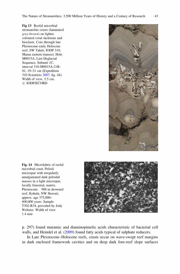
p. 297) found muramic and diaminopimelic acids characteristic of bacterial cell
walls, and Heindel et al. (2009) found fatty acids typical of sulphate reducers.
In Late Pleistocene–Holocene reefs, crusts occur on wave-swept reef margins
in dark enclosed framework cavities and on deep dark fore-reef slope surfaces
Fig 13 Reefal microbial
stromatolite crusts (laminated
grey-brown) on lighter
coloured coral skeletons and
bioclasts. Core through late
Pleistocene-early Holocene
reef, SW Tahiti, IODP 310,
Maraa eastern transect, Hole
M0015A, Last Deglacial
Sequence, Subunit 1C,
interval 310-M0015A-21R-
01, 19–31 cm (Expedition
310 Scientists 2007, fig. 44).
Width of view, 5.5 cm.
# IODP/ECORD
Fig. 14 Microfabric of reefal
microbial crust. Peloid
microspar with irregularly
amalgamated dark peloidal
masses in a light microspar,
locally fenestral, matrix.
Pleistocene �988 m drowned
reef, Kohala, NW Hawaii;
approx. age 375,000–
400,000 years. Sample
T302-R34, provided by Jody
Webster. Width of view
1.4 mm
The Nature of Stromatolites: 3,500 Million Years of History and a Century of Research 43
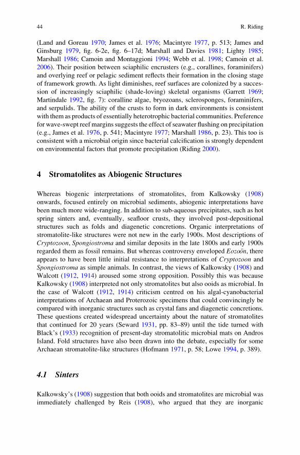
(Land and Goreau 1970; James et al. 1976; Macintyre 1977, p. 513; James and
Ginsburg 1979, fig. 6-2e, fig. 6–17d; Marshall and Davies 1981; Lighty 1985;
Marshall 1986; Camoin and Montaggioni 1994; Webb et al. 1998; Camoin et al.
2006). Their position between sciaphilic encrusters (e.g., corallines, foraminifers)
and overlying reef or pelagic sediment reflects their formation in the closing stage
of framework growth. As light diminishes, reef surfaces are colonized by a succes-
sion of increasingly sciaphilic (shade-loving) skeletal organisms (Garrett 1969;
Martindale 1992, fig. 7): coralline algae, bryozoans, sclerosponges, foraminifers,
and serpulids. The ability of the crusts to form in dark environments is consistent
with them as products of essentially heterotrophic bacterial communities. Preference
for wave-swept reef margins suggests the effect of seawater flushing on precipitation
(e.g., James et al. 1976, p. 541; Macintyre 1977; Marshall 1986, p. 23). This too is
consistent with a microbial origin since bacterial calcification is strongly dependent
on environmental factors that promote precipitation (Riding 2000).
4 Stromatolites as Abiogenic Structures
Whereas biogenic interpretations of stromatolites, from Kalkowsky (1908)
onwards, focused entirely on microbial sediments, abiogenic interpretations have
been much more wide-ranging. In addition to sub-aqueous precipitates, such as hot
spring sinters and, eventually, seafloor crusts, they involved post-depositional
structures such as folds and diagenetic concretions. Organic interpretations of
stromatolite-like structures were not new in the early 1900s. Most descriptions of
Cryptozoon, Spongiostroma and similar deposits in the late 1800s and early 1900s
regarded them as fossil remains. But whereas controversy enveloped Eozo€on, thereappears to have been little initial resistance to interpretations of Cryptozoon and
Spongiostroma as simple animals. In contrast, the views of Kalkowsky (1908) and
Walcott (1912, 1914) aroused some strong opposition. Possibly this was because
Kalkowsky (1908) interpreted not only stromatolites but also ooids as microbial. In
the case of Walcott (1912, 1914) criticism centred on his algal-cyanobacterial
interpretations of Archaean and Proterozoic specimens that could convincingly be
compared with inorganic structures such as crystal fans and diagenetic concretions.
These questions created widespread uncertainty about the nature of stromatolites
that continued for 20 years (Seward 1931, pp. 83–89) until the tide turned with
Black’s (1933) recognition of present-day stromatolitic microbial mats on Andros
Island. Fold structures have also been drawn into the debate, especially for some
Archaean stromatolite-like structures (Hofmann 1971, p. 58; Lowe 1994, p. 389).
4.1 Sinters
Kalkowsky’s (1908) suggestion that both ooids and stromatolites are microbial was
immediately challenged by Reis (1908), who argued that they are inorganic
44 R. Riding
precipitates. Bucher (1913) supported Reis, observing that both stromatoliths and
oolites had “a structure of delicate layers”. Later he stated: “stromatoliths are the
sedimentary equivalent of the calcareous and siliceous ‘sinter’ of the hot springs”
and he contrasted them with “the coarse calcareous crusts which are formed by
thick, felted masses of fresh-water algae and mosses” (Bucher 1918, p. 608). This
was echoed by Bradley (1928) who, in marginal facies of the lacustrine Eocene
Green River Formation in Wyoming, interpreted coarse fabrics similar to those of
spongy Green Lake tufa fabrics as algal, contrasting them with finely banded and
radial fibrous layers which he regarded as inorganic (Bradley 1928, p. 209, p. 217,
pl. 34a,c; pl. 45a).
4.2 Concretions
Walcott (in Lawson 1912, pp. 16–23) gave the name Atikokania to Archaean
structures found by A.C. Lawson in Archaean Steep Rock carbonates, and also
identified a specimen as Cryptozoan? (NB, not Cryptozoon). Walcott suggested that
Atikokania might be a sponge, but retracted this when Abbott (1914) compared it
with Permian concretionary carbonates in north-east England (Hofmann 1971, p. 25).
The idea that Atikokania might be a sponge persisted (de Laubenfels 1955) but it
has generally been regarded as inorganic (Glaessner 1962, p. 472), for example as
crystal fans (Hofmann 1971) precipitated on the seafloor (Sumner and Grotzinger
2000, p. 134).
Following Abbott’s (1914) example, Holtedahl (1921) compared some of
Walcott’s (1914) Mesoproterozoic specimens from the Belt Series with concretions
in the Permian Magnesian Limestone of the Zechstein evaporite basin in north-east
England. Holtedahl (1921, p. 202) agreed with Sedgwick (1829) that these Permian
structures were diagenetic and he particularly compared them with Walcott’s
(1914) Greysonia and Newlandia from the Newland Limestone of Montana.
Holtedahl (1921) considered that they “must have had a similar origin” (p. 201)
and were “inorganic and secondary” (p. 203). However, he concluded: “The
discovery of algae and bacteria in pre-Cambrian strata, reported by Walcott, has
therefore lost none of its importance, even if it should be found that these organisms
are not responsible for the many curious structures found in the Algonkian Newland
limestone” (p. 206). Nonetheless, as a result of these comparisons with Zechstein
concretions, Seward (1931, pp. 83–89) was sceptical that Cryptozoon and similar
structures were organic. Seward recognized that blue-green algae were associated
with nodules, such as those described by Mawson (1929) from South Australia, but
he was hesitant to infer “that their presence is essential” to nodule formation
(Seward 1931, p. 83), and he also doubted that they could have produced “reefs
of Cryptozoon” (Seward 1931, pp. 86–87). But Goldring (1938, p. 21) pointed out
that the irregularity of Cryptozoon contrasted with the regularity to be expected
from inorganic precipitates, and in a footnote Seward (1931, p. 86) himself recog-
nized that bacterial deposits could closely resemble Cryptozoon. Although Fenton
The Nature of Stromatolites: 3,500 Million Years of History and a Century of Research 45
(1943, p. 93, fig. 7) considered that it does not closely resemble Newlandia, some of
Holtedahl’s Zechstein specimens are stromatolitic in appearance. Fenton (1943,
p. 86) too, considered that some of Walcott’s (1914) Newland samples were
inorganic products of “segregation of calcite and dolomite during deformation”.
4.3 Folds
At Steep Rock Lake in Ontario, Jolliffe (1955, pp. 380) suggested that Walcott’s (in
Lawson 1912, pp. 16–23) “Cryptozoan? fragment probably came from what are
much the most common organic-like forms in the Steeprock carbonate – the so-
called ‘algal’ structures”. These include large stromatolitic domes (Wilks and
Nisbet 1985). However, Hofmann (1971, p. 58 and pl. 22, fig. 1) noted that “The
sample appears to be deformed, and it is probable that the lamination of this
particular specimen is a tectonic foliation and not stromatolitic”. Lowe (1994,
fig. 3d, p. 389) considered that a domical structure interpreted as a ~3,450 Ma
stromatolite at Pilbara, Australia (Walter et al. 1980), had formed by soft-sediment
deformation. Furthermore, he suggested that all three of the well-documented
reports of stromatolites older than 3,200 Ma known at that time “probably formed
through non-biological processes” such as deformation or evaporitic inorganic
precipitation (Lowe 1994, p. 390).
4.4 Seafloor Crusts
Reliance on non-marine deposits for examples of abiogenic stromatolite-like struc-
tures continued into the 1970s (e.g., Read 1976; Thrailkill 1976; Walter et al. 1976).
In the 1980s, a significant new development occurred when petrographic studies
suggested that well-preserved Palaeoproterozoic stromatolites in Arctic Canada
could be interpreted as essentially abiogenic precipitates (Kerans 1982; Grotzinger
and Read 1983). Although these deposits were often at first described as “fibrous
marine cements” (Grotzinger 1989b, p. 10), they were evidently seafloor crusts.
Grotzinger (1986a) considered the possibility that microdigitate stromatolites are
“entirely abiotic”, and Grotzinger and Rothman (1996, p. 424) proposed that
“abiotic mechanisms” could account for the growth of large Palaeoproterozoic
stromatolites such as those described by Jackson (1989, fig. 13) (Fig. 15). Thus,
some Proterozoic deposits described as stromatolites appear, from petrofabric
evidence, to be essentially abiotic seafloor precipitates. Literature survey indicates
at least four general categories of subaqueous Sparry Crust based on Precambrian
examples (Riding 2008, table 1); three of which include stromatolitic deposits: (1)
Botryoidal fans and crystal pseudomorphs include small radial fibrous millimetric
microbotryoids that build Tarioufetia and Tungussia (Bertrand-Sarfati 1972); (2)
“microdigitate stromatolites”: small laminated columns of radial crystals (Grotzinger
and Read 1983; Hofmann and Jackson 1987); (3) isopachous laminite (Jackson
1989; Pope and Grotzinger 2000, fig. 8e; Sumner and Grotzinger 2004).
46 R. Riding
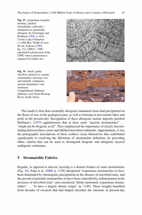
This made it clear that essentially abiogenic laminated crusts had precipitated on
the floors of seas in the geological past, as well as forming in non-marine lakes and
pools at the present-day. Recognition of these abiogenic marine deposits justified
Hoffman’s (1973) apprehension that at least some “ancient stromatolites” . . .“might not be biogenic at all”. They emphasized the importance of clearly discrim-
inating between these crusts and lithified microbial sediments. Appropriately, it was
the petrographic descriptions of these seafloor crusts themselves that contributed
significantly to resolving the dilemmas of stromatolite definition, by providing
fabric criteria that can be used to distinguish biogenic and abiogenic layered
authigenic carbonates.
5 Stromatolite Fabrics
Regular, as opposed to uneven, layering is a distinct feature of some stromatolites
(Fig. 16). Pope et al. (2000, p. 1139) interpreted “isopachous stromatolites to have
been dominated by chemogenic precipitation in the absence of microbial mats, and
the growth of peloidal stromatolites to have been controlled by sedimentation in the
presence of microbial mats” and considered “thinly laminated, isopachous stroma-
tolites” . . . “to have a largely abiotic origin” (p. 1149). These insights benefited
from decades of research that had helped elucidate the structure of present-day
Fig. 15 Isopachous laminite
forming “peaked
stromatolitic carbonate”,
interpreted as potentially
abiogenic by Grotzinger and
Rothman (1996, p. 424).
Cowles Lake Formation
(~1,900 Ma). Width of view
45 cm. Jackson (1989,
fig. 13), CSPG# 1989,
reprinted by permission of the
CSPG whose permission is
required for further use
Fig. 16 Small, partly
silicified, domical to cuspate
stromatolites showing even
and laterally continuous,
smooth lamination. Late
Archaean
Campbellrand–Malmani
platform, near Groot Boetsap
River. South Africa
The Nature of Stromatolites: 3,500 Million Years of History and a Century of Research 47
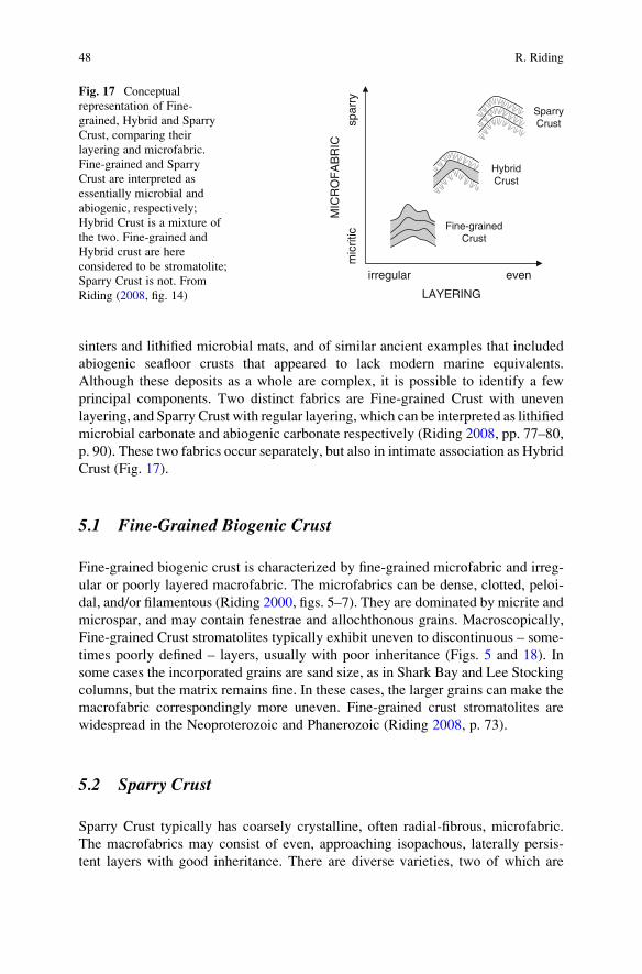
sinters and lithified microbial mats, and of similar ancient examples that included
abiogenic seafloor crusts that appeared to lack modern marine equivalents.
Although these deposits as a whole are complex, it is possible to identify a few
principal components. Two distinct fabrics are Fine-grained Crust with uneven
layering, and Sparry Crust with regular layering, which can be interpreted as lithified
microbial carbonate and abiogenic carbonate respectively (Riding 2008, pp. 77–80,
p. 90). These two fabrics occur separately, but also in intimate association as Hybrid
Crust (Fig. 17).
5.1 Fine-Grained Biogenic Crust
Fine-grained biogenic crust is characterized by fine-grained microfabric and irreg-
ular or poorly layered macrofabric. The microfabrics can be dense, clotted, peloi-
dal, and/or filamentous (Riding 2000, figs. 5–7). They are dominated by micrite and
microspar, and may contain fenestrae and allochthonous grains. Macroscopically,
Fine-grained Crust stromatolites typically exhibit uneven to discontinuous – some-
times poorly defined – layers, usually with poor inheritance (Figs. 5 and 18). In
some cases the incorporated grains are sand size, as in Shark Bay and Lee Stocking
columns, but the matrix remains fine. In these cases, the larger grains can make the
macrofabric correspondingly more uneven. Fine-grained crust stromatolites are
widespread in the Neoproterozoic and Phanerozoic (Riding 2008, p. 73).
5.2 Sparry Crust
Sparry Crust typically has coarsely crystalline, often radial-fibrous, microfabric.
The macrofabrics may consist of even, approaching isopachous, laterally persis-
tent layers with good inheritance. There are diverse varieties, two of which are
SparryCrust
irregular even
LAYERING
Fine-grainedCrust
spar
ryM
ICR
OF
AB
RIC
mic
ritic
HybridCrust
Fig. 17 Conceptual
representation of Fine-
grained, Hybrid and Sparry
Crust, comparing their
layering and microfabric.
Fine-grained and Sparry
Crust are interpreted as
essentially microbial and
abiogenic, respectively;
Hybrid Crust is a mixture of
the two. Fine-grained and
Hybrid crust are here
considered to be stromatolite;
Sparry Crust is not. From
Riding (2008, fig. 14)
48 R. Riding
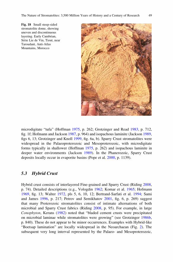
microdigitate “tufa” (Hoffman 1975, p. 262; Grotzinger and Read 1983, p. 712,
fig. 1f; Hofmann and Jackson 1987, p. 964) and isopachous laminite (Jackson 1989,
figs 6, 13; Grotzinger and Knoll 1999, fig. 6a, b). Sparry Crust stromatolites were
widespread in the Palaeoproterozoic and Mesoproterozoic, with microdigitate
forms typically in shallower (Hoffman 1975, p. 262) and isopachous laminite in
deeper water environments (Jackson 1989). In the Phanerozoic, Sparry Crust
deposits locally occur in evaporite basins (Pope et al. 2000, p. 1139).
5.3 Hybrid Crust
Hybrid crust consists of interlayered Fine-grained and Sparry Crust (Riding 2008,
p. 74). Detailed descriptions (e.g., Vologdin 1962; Komar et al. 1965; Hofmann
1969, fig. 13; Walter 1972, pls 5, 6, 10, 12; Bertrand-Sarfati et al. 1994; Sami
and James 1996, p. 217; Petrov and Semikhatov 2001, fig. 6, p. 269) suggest
that many Proterozoic stromatolites consist of intimate alternations of both
microbial and Sparry Crust fabrics (Riding 2008, p. 95). For example, in large
Conophyton, Kerans (1982) noted that “bladed cement crusts were precipitated
on microbial laminae while stromatolites were growing” (see Grotzinger 1986b,
p. 840). These do not appear to be minor occurrences. Examples with Hybrid-like
“Boetsap lamination” are locally widespread in the Neoarchaean (Fig. 2). The
subsequent very long interval represented by the Palaeo- and Mesoproterozoic,
Fig. 18 Small steep-sided
stromatolite dome, showing
uneven and discontinuous
layering. Early Cambrian,
Serie Lie de Vin, Tiout, near
Taroudant, Anti-Atlas
Mountains, Morocco
The Nature of Stromatolites: 3,500 Million Years of History and a Century of Research 49
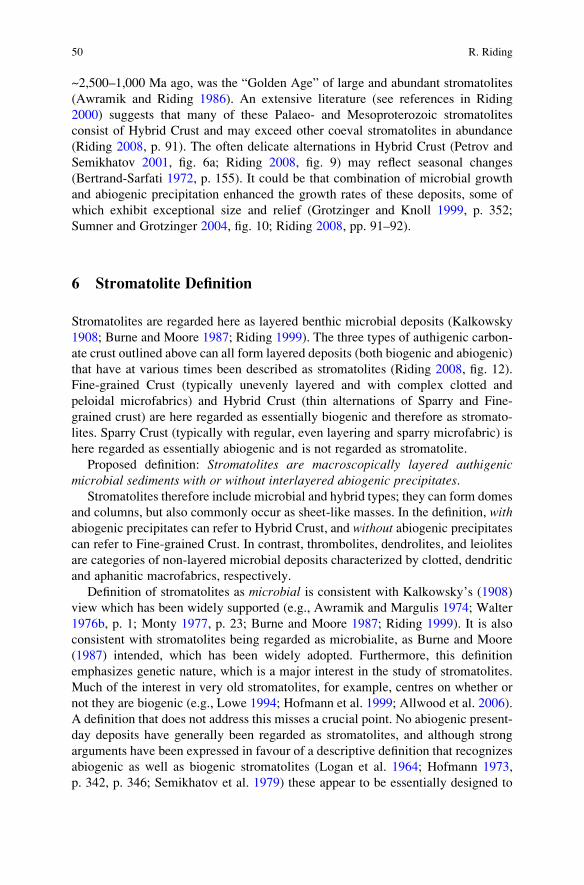
~2,500–1,000 Ma ago, was the “Golden Age” of large and abundant stromatolites
(Awramik and Riding 1986). An extensive literature (see references in Riding
2000) suggests that many of these Palaeo- and Mesoproterozoic stromatolites
consist of Hybrid Crust and may exceed other coeval stromatolites in abundance
(Riding 2008, p. 91). The often delicate alternations in Hybrid Crust (Petrov and
Semikhatov 2001, fig. 6a; Riding 2008, fig. 9) may reflect seasonal changes
(Bertrand-Sarfati 1972, p. 155). It could be that combination of microbial growth
and abiogenic precipitation enhanced the growth rates of these deposits, some of
which exhibit exceptional size and relief (Grotzinger and Knoll 1999, p. 352;
Sumner and Grotzinger 2004, fig. 10; Riding 2008, pp. 91–92).
6 Stromatolite Definition
Stromatolites are regarded here as layered benthic microbial deposits (Kalkowsky
1908; Burne and Moore 1987; Riding 1999). The three types of authigenic carbon-
ate crust outlined above can all form layered deposits (both biogenic and abiogenic)
that have at various times been described as stromatolites (Riding 2008, fig. 12).
Fine-grained Crust (typically unevenly layered and with complex clotted and
peloidal microfabrics) and Hybrid Crust (thin alternations of Sparry and Fine-
grained crust) are here regarded as essentially biogenic and therefore as stromato-
lites. Sparry Crust (typically with regular, even layering and sparry microfabric) is
here regarded as essentially abiogenic and is not regarded as stromatolite.
Proposed definition: Stromatolites are macroscopically layered authigenicmicrobial sediments with or without interlayered abiogenic precipitates.
Stromatolites therefore include microbial and hybrid types; they can form domes
and columns, but also commonly occur as sheet-like masses. In the definition, withabiogenic precipitates can refer to Hybrid Crust, and without abiogenic precipitatescan refer to Fine-grained Crust. In contrast, thrombolites, dendrolites, and leiolites
are categories of non-layered microbial deposits characterized by clotted, dendritic
and aphanitic macrofabrics, respectively.
Definition of stromatolites as microbial is consistent with Kalkowsky’s (1908)
view which has been widely supported (e.g., Awramik and Margulis 1974; Walter
1976b, p. 1; Monty 1977, p. 23; Burne and Moore 1987; Riding 1999). It is also
consistent with stromatolites being regarded as microbialite, as Burne and Moore
(1987) intended, which has been widely adopted. Furthermore, this definition
emphasizes genetic nature, which is a major interest in the study of stromatolites.
Much of the interest in very old stromatolites, for example, centres on whether or
not they are biogenic (e.g., Lowe 1994; Hofmann et al. 1999; Allwood et al. 2006).
A definition that does not address this misses a crucial point. No abiogenic present-
day deposits have generally been regarded as stromatolites, and although strong
arguments have been expressed in favour of a descriptive definition that recognizes
abiogenic as well as biogenic stromatolites (Logan et al. 1964; Hofmann 1973,
p. 342, p. 346; Semikhatov et al. 1979) these appear to be essentially designed to
50 R. Riding
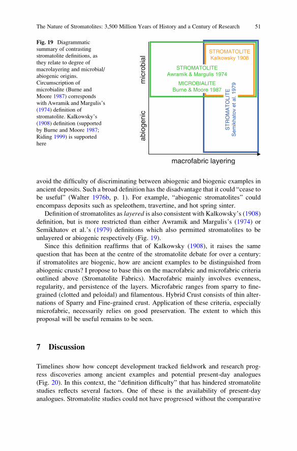
avoid the difficulty of discriminating between abiogenic and biogenic examples in
ancient deposits. Such a broad definition has the disadvantage that it could “cease to
be useful” (Walter 1976b, p. 1). For example, “abiogenic stromatolites” could
encompass deposits such as speleothem, travertine, and hot spring sinter.
Definition of stromatolites as layered is also consistent with Kalkowsky’s (1908)definition, but is more restricted than either Awramik and Margulis’s (1974) or
Semikhatov et al.’s (1979) definitions which also permitted stromatolites to be
unlayered or abiogenic respectively (Fig. 19).
Since this definition reaffirms that of Kalkowsky (1908), it raises the same
question that has been at the centre of the stromatolite debate for over a century:
if stromatolites are biogenic, how are ancient examples to be distinguished from
abiogenic crusts? I propose to base this on the macrofabric and microfabric criteria
outlined above (Stromatolite Fabrics). Macrofabric mainly involves evenness,
regularity, and persistence of the layers. Microfabric ranges from sparry to fine-
grained (clotted and peloidal) and filamentous. Hybrid Crust consists of thin alter-
nations of Sparry and Fine-grained crust. Application of these criteria, especially
microfabric, necessarily relies on good preservation. The extent to which this
proposal will be useful remains to be seen.
7 Discussion
Timelines show how concept development tracked fieldwork and research prog-
ress discoveries among ancient examples and potential present-day analogues
(Fig. 20). In this context, the “definition difficulty” that has hindered stromatolite
studies reflects several factors. One of these is the availability of present-day
analogues. Stromatolite studies could not have progressed without the comparative
abio
geni
c
macrofabric layering
STROMATOLITEKalkowsky 1908
STROMATOLITEAwramik & Margulis 1974
MICROBIALITEBurne & Moore 1987
ST
RO
MA
TO
LIT
ES
emik
hato
vet
al.
1979
mic
robi
al
Fig. 19 Diagrammatic
summary of contrasting
stromatolite definitions, as
they relate to degree of
macrolayering and microbial/
abiogenic origins.
Circumscription of
microbialite (Burne and
Moore 1987) corresponds
with Awramik and Margulis’s
(1974) definition of
stromatolite. Kalkowsky’s
(1908) definition (supported
by Burne and Moore 1987;
Riding 1999) is supported
here
The Nature of Stromatolites: 3,500 Million Years of History and a Century of Research 51
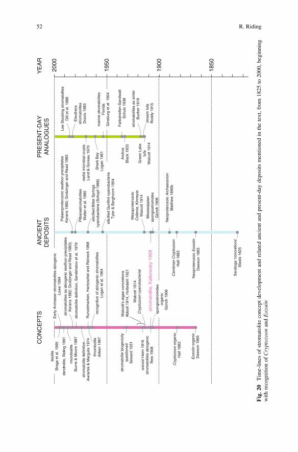
1850
1900
1950
2000
YE
AR
Lee
Sto
ckin
g st
rom
atol
ites
Dill
et a
l. 19
86
PR
ES
EN
T-D
AY
AN
ALO
GU
ES
And
ros
Bla
ck 1
933
stre
am tu
faR
oddy
191
5
reef
al m
icro
bial
cru
sts
Land
& G
orea
u 19
70
Far
bstr
eife
n-S
andw
att
Sch
ulz
1936
Gre
en L
ake
tufa
Wal
cott
1914
stro
mat
olite
s as
sin
ter
Buc
her
1918
Ele
uthe
rast
rom
atol
ites
Dra
vis
1983
Mis
siss
ippi
ansp
ongi
ostr
omid
esG
üric
h 19
06
AN
CIE
NT
DE
PO
SIT
S Pal
aeop
rote
rozo
ic s
eaflo
or p
reci
pita
tes
Ker
ans
1982
; Gro
tzin
ger
and
Rea
d 19
83
Neo
prot
eroz
oic
Eoz
oön
Daw
son
1865
Cam
bria
n C
rypt
ozoo
nH
all 1
883
Neo
prot
eroz
oic
Arc
haeo
zoon
Mat
thew
189
0b
silic
ified
Gun
flint
cya
noba
cter
iaT
yler
& B
argh
oorn
195
4
Mes
opro
tero
zoic
Col
leni
a, K
inne
yia
Wal
cott
1914
Sar
atog
a ‘c
oncr
etio
ns’
Ste
ele
1825
silic
ified
Bitt
er S
prin
gscy
anob
acte
ria (
Sch
opf 1
968)
Pilb
aras
trom
atol
ites
Wal
ter
et a
l. 19
80
spon
gios
trom
ides
orga
nic
Gür
ich
1906
onco
id H
eim
191
6st
rom
atol
ites
abio
geni
cR
eis
1908
thro
mbo
lite
Aitk
en 1
967
dend
rolit
e, R
idin
g 19
91CO
NC
EP
TS
stro
mat
olite
def
initi
onA
wra
mik
& M
argu
lis 1
974
stro
mat
olite
bio
geni
city
ques
tione
dS
ewar
d 19
31
Run
zelm
arke
n, H
antz
sche
l and
Rei
neck
196
8
stro
mat
olite
def
initi
on, S
emik
hato
v et
al.
1979
reco
gniti
on o
f abi
ogen
ic s
trom
atol
ites
Loga
n et
al.
1964
Wal
cott’
s al
gae
conc
retio
ns
Abb
ott 1
914,
Hol
teda
hl 1
921
stro
mat
olite
s as
abi
ogen
ic s
eaflo
or p
reci
pita
tes
Ker
ans
1982
; Gro
tzin
ger
and
Rea
d 19
83).
mic
robi
alite
Bur
ne &
Moo
re 1
987
Eoz
oön
orga
nic
Daw
son
1865
Cry
ptoz
oon
orga
nic
Hal
l 188
3
Ear
ly A
rcha
ean
stro
mat
olite
s ab
ioge
nic
Low
e 19
94
leio
lite
Bra
ga e
t al.
1995
Sha
rk B
ayLo
gan
1961
mar
ine
stro
mat
olite
sF
lorid
aG
insb
urg
et a
l. 19
54
Wal
cott
1914
Cry
ptoz
oon
cyan
obac
teria
l
stro
mat
olite
, Kal
kow
sky
1908
Fig.20
Tim
e-lines
ofstromatolite
conceptdevelopmentandrelatedancientandpresent-day
depositsmentioned
inthetext,from
1825to
2000,beginning
withrecognitionofCryptozoonandEozo€on
52 R. Riding
information provided by the wide variety of present-day abiogenic and microbial
mat deposits briefly summarized above. But despite this extensive research, efforts
to base stromatolite interpretation entirely on present-day analogues have not been
altogether successful. This is because not all ancient stromatolite-like deposits are
well-represented today; most notably the abiogenic seafloor crusts first clearly
recognized in the Proterozoic by Kerans (1982), Grotzinger and Read (1983),
and Grotzinger (1989a, b) and others. Stromatolites have had such a long geological
history, influenced by major changes in both biology and seawater chemistry,
that finding good examples of all types of stromatolite today might hardly be
expected. Another factor is the geological origin of the stromatolite concept, rooted
in specimens that are hundreds of million years old (Kalkowsky 1908; Hall 1883).
Stromatolites have been reported to be most abundant 2,000 Ma ago (Grotzinger
1990), and the term has been widely applied to specimens as old as 3,500 Ma
(Allwood et al. 2006). Because stromatolites were first described and defined in the
rock record, their genesis was not immediately demonstrable and demanded
resourceful interpretation. This question of origins has become the motif of stro-
matolite studies, focused on the difficulty of distinguishing abiogenic crusts from
microbial carbonates.
The paradox presented by stromatolites is conveyed by Ginsburg’s (1991, p. 25)
impish comment that “few observers have any difficulty identifying archetypical
stromatolites . . . yet defining stromatolites is controversial”. This is like saying that
everyone knows what stromatolites look like, but no one can agree what they are.
It was this daunting prospect, which threatened to leave stromatolite definition in a
state of perpetual limbo, that led Semikhatov et al. (1979) to propose a descriptive
definition. Yet, broad definitions encompassing both biogenic and abiogenic struc-
tures avoid the central question of how stromatolites have formed (Walter 1976b,
p. 1). This brings the debate full circle, back to trying to find ways to make a
microbial definition work (Burne and Moore 1987).
Nonetheless, there are clear pointers to be drawn from the considerable advances
derived from studies of present-day deposits. One is that the term stromatolite is
only very seldom applied to present-day deposits known to be inorganic (an
exception is Maliva et al.’s (2000, p. 934) description of modern stromatolites
“formed largely by inorganic precipitation”). This suggests that researchers have
tended to view stromatolites as essentially biogenic, and that the only reason for
grouping abiogenic and microbial crusts in ancient deposits is inability to distin-
guish them (Semikhatov et al. 1979). Another is that there are distinct fabric
differences between abiogenic and microbial crusts, as was recognized very early
(Bucher 1913). The pace of development of limestone petrography accelerated in
the 1960s, but focused more on young and grainy carbonates than on old and
authigenic ones. But substantial progress has been made in distinguishing
laminated authigenic crusts of microbial and abiogenic origin. The argument here
is that fabric criteria can resolve these questions, and that it is important to develop
them inorder to overcome issues that impede stromatolite studies. It should be
possible not only to formulate a microbial definition of stromatolite, but also to
confidently apply it sufficiently widely in the geological record.
The Nature of Stromatolites: 3,500 Million Years of History and a Century of Research 53
7.1 Abiogenic and/or Biogenic Stromatolites?
Despite all the difficulties, in many respects the sometimes contentious half-century
of debate vindicated the views of both sets of early contenders; Kalkowsky (1908)
and Walcott (1914), on the one hand, and of Reis (1908) and Bucher (1913) on the
other. By the 1960s there was growing acknowledgement of the existence of both
organic and inorganic stromatolites. A decade or more prior to Awramik and
Margulis (1974), Logan et al. (1964, p. 68) succinctly stated: “Stromatolites are
laminated structures that have been previously termed fossil algae. It is now
recognized that such structures may be formed by a number of different processes
and organisms.” They did not offer a general definition of stromatolite, but their
recommendation was clear: “To be useful, the term stromatolite should be preceded
whenever possible by an adjective signifying the kind of stromatolite under consid-
eration, for example, algal stromatolite, foraminiferal stromatolite, inorganic stro-
matolite, and so forth” (Logan et al. 1964, p. 69). This was echoed by Hofmann
(1973), who emphasized that stromatolites need not be biogenic (p. 342) and
recognized chemogenic stromatolites (p. 346) and the need to distinguish “biogenic
stromatolites from chemical and mechanical ones” (p. 350).
These calls by well-known researchers did not lead to peaceful reform of
stromatolite definition. Instead they precipitated a schism between those who
regarded stromatolites as essentially biogenic, and those who believed it was
necessary for the term stromatolite to encompass both biogenic and abiogenic
laminated structures. In addition to discovery of cyanobacteria in ~1880 Gunflint
stromatolites (Barghoorn and Tyler 1965) (Fig. 7), similar – and impressive –
fossils had been found in the ~850 Ma Bitter Springs silicified stromatolitic mats
(Schopf 1968; Knoll and Golubic 1979), emphasizing the biogenicity of Protero-
zoic stromatolites. Against this background, two definitions were published expres-
sing these contrary convictions. Awramik and Margulis (1974) adhered to
Kalkowsky’s (1908) view that stromatolites are microbial, whereas Semikhatov
et al. (1979) followed Logan et al.’s (1964) lead and recognized both biogenic and
abiogenic stromatolites (Fig. 19).
Awramik and Margulis (1974) defined stromatolites as “megascopic organose-
dimentary structures produced by sediment trapping, binding and/or precipitation
as a result of growth and metabolic activity of organisms, primarily blue-green
algae”. This genetic approach was endorsed in the introduction to Walter’s (1976b,
p. 1) seminal stromatolite volume. Its advantage was its straightforward restatement
of Kalkowsky’s (1908) microbial view. But it required biogenicity to be demon-
strated, and this was difficult for ancient examples. Hoffman’s (1973) had frankly
admitted that “Something that haunts geologists working on ancient stromatolites is
the thought that they might not be biogenic at all”. This question of confidently
establishing biogenicity in ancient examples is the enduring central problem of
stromatolite studies. Reis (1908) used it to challenge Kalkowsky (1908). It was
clearly expressed by Seward (1931, pp. 83–89) and Logan et al. (1964), and
preoccupied Semikhatov et al. (1979), Ginsburg (1991, pp. 25–27), Grotzinger
54 R. Riding
and Knoll (1999), and Brasier et al. (2006, p. 894). It led Buick et al. (1981) to
suggest that “structures of uncertain origin that resemble stromatolites should be
called ‘stromatoloids’” and Awramik and Grey (2005) to term abiogenic stromato-
lite-like structures pseudostromatolites.Semikhatov et al. (1979, p. 992) (but with S.M. Awramik dissenting), proposed
to define stromatolites as “laminated, lithified, sedimentary growth structures that
accrete away from a point or limited surface of attachment. They are commonly, but
not necessarily, of microbial origin and calcareous composition”. The operative
words here are “commonly, but not necessarily”, that permit some stromatolites to
be abiogenic. They supported this descriptive, rather than genetic, approach to
stromatolite definition by stating that “it was the lamination that Kalkowsky
stressed, not its origin” (Semikhatov et al. 1979, p. 994). This was misleading;
Kalkowsky (1908) emphasized organic origin as well as layering in describing
stromatolites. But Semikhatov et al. (1979) were correct to point out the difficulties
of confidently applying a genetic definition: “if a biological origin had to be
demonstrated before a geological object could be called a stromatolite the term
would in most instances be inapplicable (or at best provisional)” (Semikhatov et al.
1979, p. 994). Their approach was bold and realistic, a clear departure from
Kalkowsky (1908), and an attempt to express a dual view of stromatolites in
which many were microbial, but some could be abiogenic. It aroused criticism
for exactly this reason. It appeared to be an all-encompassing definition that
failed to either readily separate different structures or to circumscribe related
ones. Walter (1976b, p. 1) had already perceptively foreseen that if non-genetic
definitions “are so broad as to include a wide-range of non-biogenic structures”,
“the term would cease to be useful”. Subsequently, Ginsburg’s (1991, p. 27)
opinion was that the Semikhatov et al.’s (1979) definition “includes structures of
a variety of origins ranging from tufa domes . . . to laminated structures of miner-
alized organisms . . . and even some of the laminated zones of caliches and calcretes
as well as certain speleothems.” It could perhaps even be considered to include
diagenetic concretions. A disadvantage with its specific wording is that it appears
designed to exclude laterally extensive deposits such as Aitken’s (1967) cryptalga-
laminates. But the main difficulty can be summed up by the question, if stromato-
lites are “laminated, lithified, sedimentary growth structures that accrete away from
a point or limited surface of attachment” (Semikhatov et al. 1979), then what
exactly is not a stromatolite?
7.2 Microbialite
The name stromatolite purposely emphasized layered structure; Stroma and stromat– indicate a bed coverlet in Greek and Latin. Kalkowsky (1908, p. 102) stated: “AlleStromatolithe zeigen im vertikalen Schnitt deutliche Lagenstruktur” “All stromato-
lites show distinct layering in vertical section”. As geological studies progressed it
eventually became clear that there are deposits that resemble stromatolites in
The Nature of Stromatolites: 3,500 Million Years of History and a Century of Research 55
external form but which appear to lack internal layering. Aitken (1967, p. 1164)
introduced thrombolite “for cryptalgal structures related to stromatolites, but lack-
ing lamination and characterized by a macroscopic clotted fabric” to describe
structures that were common in the Late Cambrian and Early Ordovician. During
the 1970s, growing awareness of the local importance of thrombolites led to doubts
about the value of requiring layering as an integral part of stromatolite description.
For example, Awramik and Margulis (1974) defined stromatolites as “megascopic
organosedimentary structures produced by sediment trapping, binding and/or pre-
cipitation as a result of growth and metabolic activity of organisms, primarily blue-
green algae”. This required stromatolites to be microbial, but not necessarily
layered, and therefore permitted thrombolite to be regarded as a type of stromato-
lite. But this left no specific term for laminated stromatolites. This deficiency was
resolved when Burne and Moore (1987) applied the essence of Awramik and
Margulis’ (1974) definition of stromatolite to a new term: microbialite. “Micro-
bialites are organosedimentary deposits that have accreted as a result of a benthic
microbial community trapping and binding detrital sediment and/or forming the
locus of mineral precipitation” (Burne and Moore 1987, pp. 241–242). This then
allowed stromatolites to be regarded as macro-laminated microbialites, and throm-
bolites as macro-clotted microbialites. It also encouraged subsequent additions to
the microbialite family, such as dendrolite (dendritic; Riding 1991, p. 34) and
leiolite (aphanitic; Braga et al. 1995, p. 347). Since then, macrofabric has been a
fundamental descriptor for these structures and stromatolites (Riding 2000,
pp. 189–195).
Burne and Moore (1987) carefully evaluated the processes that might contribute
to the formation of microbialites and associated deposits. As Hofmann (1973, fig. 5)
and Riding (1977, fig. 1) had done, they noted the importance of sediment trapping,
inorganic calcification and biologically influenced calcification (Burne and Moore
1987, figs. 1 and 2, pp. 243–244). Accordingly, they recognized three types of
microbialite to reflect these processes respectively: microbial boundstone, tufa and
framestone (Burne and Moore 1987, pp. 242–243). Microbialite was therefore
designed as a broad term to encompass deposits formed by both grain trapping
and mineral precipitation associated with benthic microbes, and this specifically
included “microbial tufa – formed when microorganic material is incorporated
during inorganic precipitation of carbonate” (p. 243). In detail, this could be
considered to differ from Awramik and Margulis (1974) who defined stromatolites
as “megascopic organosedimentary structures produced by sediment trapping,
binding and/or precipitation as a result of growth and metabolic activity of organ-
isms, primarily blue-green algae”. The reasoning is that “precipitation as a result of
growth and metabolic activity of organisms” emphasizes organic processes whereas
“incorporated during inorganic precipitation of carbonate” emphasizes inorganic
processes. However, these nuances are not evident in Burne and Moore’s (1987,
pp. 241–242) actual definition of microbialite, “organosedimentary deposits that
have accreted as a result of a benthic microbial community trapping and binding
detrital sediment and/or forming the locus of mineral precipitation”, which is closer
to that of Awramik and Margulis (1974). Furthermore, Burne and Moore (1987,
56 R. Riding
p. 249) advocated “a return to Kalkowsky’s original meaning for ‘stromatolite’”
and used it “to refer to one possible internal structure of a microbialite”.
The point of this rather protracted discussion is that – whatever the intention – in
practice microbialite provided a necessary umbrella term for stromatolites, throm-
bolites and similar deposits and it became widely used. As the name obviously
indicates, all these deposits are considered microbial. Thus, the train of events that
led Burne and Moore (1987) to base microbialite on Awramik and Margulis’s
(1974) definition of stromatolite, strengthened the view that stromatolites are
essentially microbial. This in turn tended to undermine the conviction, expressed
by Logan et al. (1964) and Hofmann (1973) Semikhatov et al. (1979), that it was
necessary to also recognize abiogenic stromatolites. But not everything was going
in the direction of a microbial definition. Already, in the 1980s, studies of Protero-
zoic carbonates were interpreting some stromatolites as abiogenic seafloor crusts.
This looked set to rebalance the debate, because it presented stromatolites as
genetically heterogeneous structures.
7.3 Seafloor Crusts
The realization that some Proterozoic stromatolites are essentially abiogenic sea-
floor precipitates, set in train by Kerans (1982) and Grotzinger and Read (1983), was
a break-through; arguably comparable in significance to the discovery of Shark Bay
stromatolites 20 years previously. It revealed fundamental differences between
Precambrian and present-day carbonate sedimentation (Grotzinger 1990; Grotzinger
and Kasting 1993), and it provided good reason to recognize abiogenic stromato-
lites. But appreciation of its importance took time to spread. One reason for this may
have been a lack of terminological clarity. Grotzinger and Read (1983, p. 712,
fig. 1f) described the components of Rocknest microdigitate stromatolites as
“cement laminae” and “cement crusts”. Use of “seafloor cement” to describe
microdigitate stromatolites and isopachous Sparry Crust continued for more than a
decade (Kah and Knoll 1996, p. 79; Pope et al. 2000, p. 1145), even after Grotzinger
and Knoll (1995, p. 579) pointed out that seafloor crusts/encrustations should be
distinguished from “true cements which bind sediment particles and line voids”.
There was also an element of restraint. Grotzinger described microdigitate stroma-
tolites as “in essence, evaporites” (Grotzinger 1986b, p. 842) but cautiously inter-
preted them as “microbially influenced inorganic calcification (although it is
possible that they are entirely abiotic in origin)” (Grotzinger 1986a). Whatever the
reasons, it was 15 years before more outspoken statements were expressed about the
origins of these seafloor crusts. At the end of the century, Grotzinger and James
(2000a, p. 7) included microdigitate stromatolites and isopachous millimetric lami-
nites (i.e., Isopachous Sparry Crust) in their summary of Precambrian marine
“abiotic precipitates”, and Pope et al. (2000, p. 1149) considered “thinly laminated
isopachous stromatolites” to be “largely abiotic”. Thus, this research revealed a wide
The Nature of Stromatolites: 3,500 Million Years of History and a Century of Research 57
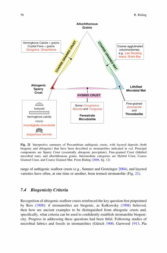
range of authigenic seafloor crusts (e.g., Sumner and Grotzinger 2004), and layered
varieties have often, at one time or another, been termed stromatolite (Fig. 21).
7.4 Biogenicity Criteria
Recognition of abiogenic seafloor crusts reinforced the key question first pinpointed
by Reis (1908): if stromatolites are biogenic, as Kalkowsky (1908) believed,
then how are ancient examples to be distinguished from abiogenic crusts and,
specifically, what criteria can be used to confidently establish stromatolite biogeni-
city. Progress in addressing these questions had been fitful. Following studies of
microbial fabrics and fossils in stromatolites (G€urich 1906; Garwood 1913, Pia
AbiogenicSparryCrust
AllochthonousGrains
LithifiedMicrobial Mat
HYBRID CRUST
CO
AR
SE GR
AIN
ED M
AT
Some Conophyton,Baicalia and Tungussia
FenestrateMicrobialite
botryoid
herringbone calcite
microdigitate stromatolite
isopachous laminite
Herringbone Calcite + grainsCrystal Fans + grains
Gongylina, OmachteniaCoarse agglutinated
columns/domes, e.g., Lee StockingIsland, Shark Bay
Fine-grainedstromatolite
andThrombolite
CO
AR
SE G
RA
INED
CR
UST
Fig. 21 Interpretive summary of Precambrian authigenic crusts, with layered deposits (both
biogenic and abiogenic) that have been described as stromatolites indicated in red. Principalcomponents are Sparry Crust (essentially abiogenic precipitate), Fine-grained Crust (lithified
microbial mat), and allochthonous grains. Intermediate categories are Hybrid Crust, Coarse
Grained Crust, and Coarse Grained Mat. From Riding (2008, fig. 12)
58 R. Riding
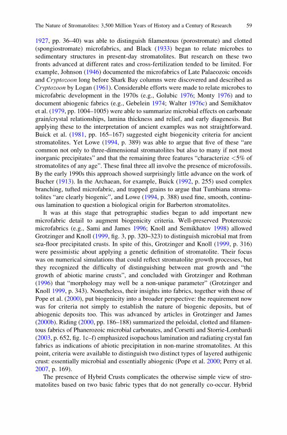
1927, pp. 36–40) was able to distinguish filamentous (porostromate) and clotted
(spongiostromate) microfabrics, and Black (1933) began to relate microbes to
sedimentary structures in present-day stromatolites. But research on these two
fronts advanced at different rates and cross-fertilization tended to be limited. For
example, Johnson (1946) documented the microfabrics of Late Palaeozoic oncoids
and Cryptozoon long before Shark Bay columns were discovered and described as
Cryptozoon by Logan (1961). Considerable efforts were made to relate microbes to
microfabric development in the 1970s (e.g., Golubic 1976; Monty 1976) and to
document abiogenic fabrics (e.g., Gebelein 1974; Walter 1976c) and Semikhatov
et al. (1979, pp. 1004–1005) were able to summarize microbial effects on carbonate
grain/crystal relationships, lamina thickness and relief, and early diagenesis. But
applying these to the interpretation of ancient examples was not straightforward.
Buick et al. (1981, pp. 165–167) suggested eight biogenicity criteria for ancient
stromatolites. Yet Lowe (1994, p. 389) was able to argue that five of these “are
common not only to three-dimensional stromatolites but also to many if not most
inorganic precipitates” and that the remaining three features “characterize <5% of
stromatolites of any age”. These final three all involve the presence of microfossils.
By the early 1990s this approach showed surprisingly little advance on the work of
Bucher (1913). In the Archaean, for example, Buick (1992, p. 255) used complex
branching, tufted microfabric, and trapped grains to argue that Tumbiana stroma-
tolites “are clearly biogenic”, and Lowe (1994, p. 388) used fine, smooth, continu-
ous lamination to question a biological origin for Barberton stromatolites.
It was at this stage that petrographic studies began to add important new
microfabric detail to augment biogenicity criteria. Well-preserved Proterozoic
microfabrics (e.g., Sami and James 1996; Knoll and Semikhatov 1998) allowed
Grotzinger and Knoll (1999, fig. 3, pp. 320–323) to distinguish microbial mat from
sea-floor precipitated crusts. In spite of this, Grotzinger and Knoll (1999, p. 316)
were pessimistic about applying a genetic definition of stromatolite. Their focus
was on numerical simulations that could reflect stromatolite growth processes, but
they recognized the difficulty of distinguishing between mat growth and “the
growth of abiotic marine crusts”, and concluded with Grotzinger and Rothman
(1996) that “morphology may well be a non-unique parameter” (Grotzinger and
Knoll 1999, p. 343). Nonetheless, their insights into fabrics, together with those of
Pope et al. (2000), put biogenicity into a broader perspective: the requirement now
was for criteria not simply to establish the nature of biogenic deposits, but of
abiogenic deposits too. This was advanced by articles in Grotzinger and James
(2000b). Riding (2000, pp. 186–188) summarized the peloidal, clotted and filamen-
tous fabrics of Phanerozoic microbial carbonates, and Corsetti and Storrie-Lombardi
(2003, p. 652, fig. 1c–f) emphasized isopachous lamination and radiating crystal fan
fabrics as indications of abiotic precipitation in non-marine stromatolites. At this
point, criteria were available to distinguish two distinct types of layered authigenic
crust: essentially microbial and essentially abiogenic (Pope et al. 2000; Perry et al.
2007, p. 169).
The presence of Hybrid Crusts complicates the otherwise simple view of stro-
matolites based on two basic fabric types that do not generally co-occur. Hybrid
The Nature of Stromatolites: 3,500 Million Years of History and a Century of Research 59
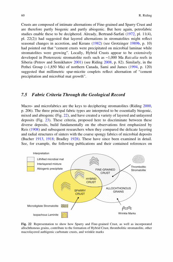
Crusts are composed of intimate alternations of Fine-grained and Sparry Crust and
are therefore partly biogenic and partly abiogenic. But here again, petrofabric
studies enable these to be deciphered. Already, Bertrand-Sarfati (1972, pl. 11(4),
pl. 22(2)) had suggested that layered alternations in stromatolites might reflect
seasonal changes in accretion, and Kerans (1982) (see Grotzinger 1989b, p. 10)
had pointed out that “cement crusts were precipitated on microbial laminae while
stromatolites were growing”. Locally, Hybrid Crusts appear to be extensively
developed in Proterozoic stromatolite reefs such as ~1,000 Ma Baicalia reefs in
Siberia (Petrov and Semikhatov 2001) (see Riding 2008, p. 82). Similarly, in the
Pethei Group (~1,850 Ma) of northern Canada, Sami and James (1994, p. 120)
suggested that millimetric spar-micrite couplets reflect alternation of “cement
precipitation and microbial mat growth”.
7.5 Fabric Criteria Through the Geological Record
Macro- and microfabrics are the keys to deciphering stromatolites (Riding 2000,
p. 206). The three principal fabric types are interpreted to be essentially biogenic,
mixed and abiogenic (Fig. 22), and have created a variety of layered and unlayered
deposits (Fig. 23). These criteria, proposed here to discriminate between these
diverse deposits, build fundamentally on the observations first emphasized by
Reis (1908) and subsequent researchers when they compared the delicate layering
and radial structures of sinters with the coarse spongy fabrics of microbial deposits
(Bucher 1913, 1918; Bradley 1928). These have since been examined in detail.
See, for example, the following publications and their contained references on
ALLOCHTHONOUSGRAINS
Isopachous Laminite
FINE-GRAINEDCRUST
SPARRYCRUST
HYBRIDCRUST
Lithified microbial mat
Abiogenic precipitate
Interlayered mixture
Interpretation
Wrinkle Marks
Microdigitate Stromatolite
ThromboliticStromatolite
Fig. 22 Representation to show how Sparry and Fine-grained Crust, as well as incorporated
allochthonous grains, contribute to the formation of Hybrid Crust, thrombolitic stromatolite, other
macrolayered authigenic carbonate crusts, and wrinkle marks
60 R. Riding
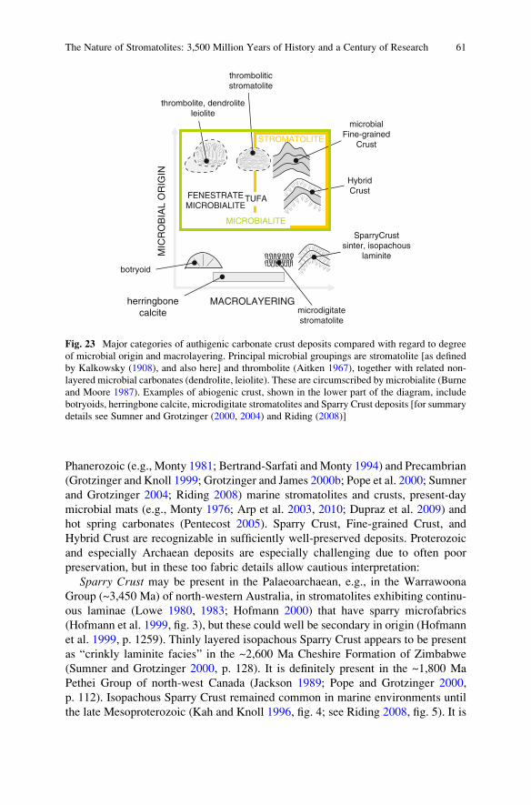
Phanerozoic (e.g., Monty 1981; Bertrand-Sarfati andMonty 1994) and Precambrian
(Grotzinger and Knoll 1999; Grotzinger and James 2000b; Pope et al. 2000; Sumner
and Grotzinger 2004; Riding 2008) marine stromatolites and crusts, present-day
microbial mats (e.g., Monty 1976; Arp et al. 2003, 2010; Dupraz et al. 2009) and
hot spring carbonates (Pentecost 2005). Sparry Crust, Fine-grained Crust, and
Hybrid Crust are recognizable in sufficiently well-preserved deposits. Proterozoic
and especially Archaean deposits are especially challenging due to often poor
preservation, but in these too fabric details allow cautious interpretation:
Sparry Crust may be present in the Palaeoarchaean, e.g., in the Warrawoona
Group (~3,450 Ma) of north-western Australia, in stromatolites exhibiting continu-
ous laminae (Lowe 1980, 1983; Hofmann 2000) that have sparry microfabrics
(Hofmann et al. 1999, fig. 3), but these could well be secondary in origin (Hofmann
et al. 1999, p. 1259). Thinly layered isopachous Sparry Crust appears to be present
as “crinkly laminite facies” in the ~2,600 Ma Cheshire Formation of Zimbabwe
(Sumner and Grotzinger 2000, p. 128). It is definitely present in the ~1,800 Ma
Pethei Group of north-west Canada (Jackson 1989; Pope and Grotzinger 2000,
p. 112). Isopachous Sparry Crust remained common in marine environments until
the late Mesoproterozoic (Kah and Knoll 1996, fig. 4; see Riding 2008, fig. 5). It is
STROMATOLITE
microbialFine-grained
Crust
SparryCrustsinter, isopachous
laminite
botryoid
thromboliticstromatolite
MACROLAYERING
MIC
RO
BIA
L O
RIG
IN
HybridCrust
microdigitatestromatolite
FENESTRATEMICROBIALITE
TUFA
thrombolite, dendroliteleiolite
MICROBIALITE
herringbonecalcite
Fig. 23 Major categories of authigenic carbonate crust deposits compared with regard to degree
of microbial origin and macrolayering. Principal microbial groupings are stromatolite [as defined
by Kalkowsky (1908), and also here] and thrombolite (Aitken 1967), together with related non-
layered microbial carbonates (dendrolite, leiolite). These are circumscribed by microbialite (Burne
and Moore 1987). Examples of abiogenic crust, shown in the lower part of the diagram, include
botryoids, herringbone calcite, microdigitate stromatolites and Sparry Crust deposits [for summary
details see Sumner and Grotzinger (2000, 2004) and Riding (2008)]
The Nature of Stromatolites: 3,500 Million Years of History and a Century of Research 61
associated with Hybrid Crust into the Neoproterozoic (see Hybrid Crust, below),
and is probably present in evaporite basin sequences during the Phanerozoic (Pope
et al. 2000).
Fine-grained Crust appears to be present in 2,724 Ma Tumbiana stromatolites
from north-western Australia (Buick 1992, fig. 3e; Lepot et al. 2008, fig. 1b), and is
definitely present in the ~1,800 Ma Pethei Group as peloidal clotted micrite (Sami
and James 1996, fig. 7). It remains common in present-day marine stromatolites.
Hybrid Crust may be present as “Boetsap lamination” in the latest Archaean
(2,550 Ma) Campbellrand-Malmani platform of South Africa (Sumner and Grotzinger
2004; see Riding 2008, p. 84), and again is definitely present as “spar-micrite
couplets” in the ~1,800 Ma Pethei Group (Sami and James 1996, p. 217). Hybrid
Crust remained common in marine environments to the latest Mesoproterozoic,
e.g., in Baicalia lacera of the ~1,020 Ma Burovaya Fm of Turukhansk, Siberia
(Petrov and Semikhatov 2001, fig. 6a). In the early Neoproterozoic it may be
represented by some of the “lamelliform elements” described by Aitken (1989,
pp. 15–16) in the ~800 Ma Little Dal reef (see Turner et al. 2000, fig. 10b).
This documentation needs to be extensively developed, fabric criteria require
detailed refinement, and many aspects of biogenic and abiogenic influences on
mineral precipitation in stromatolites require elucidation (e.g. Arp et al. 2010).
These will also benefit by being augmented by analytical and modelling approaches
to stromatolite macrofabric that build on previous work (e.g., Grotzinger and
Rothman 1996; Batchelor et al. 2000, 2003; Corsetti and Storrie-Lombardi 2003;
Storrie-Lombardi et al. 2004; Dupraz et al. 2006). In addition to providing impor-
tant new insights into long-lived and wide-ranging deposits, fabric recognition
provides the keys required to stabilize stromatolite definition.
8 Conclusions
1. Kalkowsky’s (1908) microbial interpretation of ancient stromatolites was at first
vigorously challenged due to their resemblance to hot spring crusts and other
present-day laminated precipitates that were regarded as essentially abiogenic,
and due to confusion between stromatolites and diagenetic concretions. During
the following decades, criticism diminished as microbial stromatolites were
recognized in present-day lake, marsh and shallow-marine environments.
Logan et al. (1964) suggested recognition of both microbial and abiogenic
stromatolites. This was opposed by support for the original microbial view.
Awramik and Margulis (1974) proposed a modified definition that required
stromatolites to be microbial, but not necessarily thinly layered. When Burne
and Moore (1987) used this concept to define microbialite, stromatolites became
a sub-group within microbialites, and reverted to being laminated microbial
deposits. Meanwhile, Semikhatov et al. (1979) had proposed a descriptive
definition of stromatolite that included both microbial and abiogenic deposits,
62 R. Riding
so long as they were thinly layered. This history of concept development
resulted in at least three alternative views of stromatolites: (a) laminated and
microbial (Kalkowsky 1908; Burne and Moore 1987; Riding 1999); (b) just
microbial (Awramik and Margulis 1974); (c) laminated and microbial or abio-
genic (Semikhatov et al. 1979). None of these approaches has gained unanimous
support and there is currently no generally accepted definition of stromatolite.
2. Problems of stromatolite definition arise from the difficulties of distinguishing
laminated authigenic crusts of microbial and abiogenic origin in ancient exam-
ples. Similarities in layering and external morphology between these deposits
raise doubts about their interpretation, and confuse stromatolite definition by
encouraging inclusion of abiogenic as well as microbial structures. Present-day
crusts that have been regarded as essentially abiogenic, e.g., in cave and hot-
spring sinters, have not normally been regarded as stromatolites. The only
justification for grouping abiogenic and microbial crusts is the difficulty of
distinguishing them in ancient deposits.
3. The geological record of layered marine authigenic carbonate crusts includes
three distinctive types of deposit that have all been regarded as stromatolites: (a)
Isopachous Sparry Crust – abiotically precipitated well-layered crust with sparry
microfabric; (b) Fine-grained Crust – lithified irregularly, sometimes poorly,
layered microbial carbonate, generally with fine-grained, (e.g., clotted, peloidal)
but also sometimes coarse-grained microfabrics; (c) Hybrid Crust – thinly
interlayered combinations of both preceding fabrics, with generally well-developed
layering. Further work is required, but these fabric criteria can be used to broadly
distinguish essentially abiogenic from essentially microbial crusts (Pope et al.
2000), provided that these are sufficiently well preserved.
4. The definition proposed here is: Stromatolites are macroscopically layeredauthigenic microbial sediments with or without interlayered abiogenic precipi-tates. This recognizes at least two types of stromatolite: (a) microbial, charac-
terized by Fine-grained, locally coarse, Crust and (b) hybrid, composed of
Hybrid Crust (i.e., alternations of Fine-grained and Sparry Crust). In contrast,
thrombolite, dendrolite and leiolite are microbial carbonates distinguished from
each other, and from stromatolites, by clotted, dendritic and aphanitic macro-
fabrics, respectively.
5. Thus, it is possible to use carbonate fabric criteria to distinguish essentially
abiogenic crusts from essentially microbial ones. This ability permits practical
application of a microbial definition of stromatolite, as Kalkowsky (1908)
intended.
Acknowledgements I am grateful to Joachim Reitner and the Organizing Committee for support-
ing my participation in the 2008 Kalkowsky Symposium. Brian Chatterton and Eric Mountjoy
generously provided photographs of Eozo€on and Shark Bay columns, respectively, and Jody
Webster a thin-section of reef crust from Kohala, Hawaii. Canadian Society of Petroleum
Geologists kindly gave permission to reproduce Fig. 15. I am indebted to Phil Fralick, Dawn
Sumner and Pieter Visscher, respectively, for showing me the Gunflint, Campbellrand-Malmani,
and Lagoa Pitanguinha localities. Gernot Arp and Joachim Reitner made helpful suggestions that
improved the final manuscript.
The Nature of Stromatolites: 3,500 Million Years of History and a Century of Research 63
References
Abbott G (1914) Is ‘Atikokania lawsoni’ a concretion? Nature 94: 477–478Adelman J (2007) Eozo€on: debunking the dawn animal. Endeavour 31: 94–98
Aitken JD (1967) Classification and environmental significance of cryptalgal limestones and
dolomites, with illustrations from the Cambrian and Ordovician of southwestern Alberta.
Journal of Sedimentary Petrology 37: 1163–1178
Aitken JD (1989) Giant “algal” reefs, Middle/Upper Proterozoic Little Dal Group (>770,
<1200 Ma), Mackenzie Mountains, N.W.T., Canada. In: Geldsetzer HHJ, James NP, Tebbutt
GE (eds) Reefs, Canada and Adjacent Area. Canadian Society of Petroleum Geologists,
Memoir 13: 13–23
Allwood AC, Walter MR, Kamber BS, Marshall CP, Burch IW (2006) Stromatolite reef from the
Early Archaean era of Australia. Nature 441: 714–718
Arp G, Reimer A, Reitner J (2003) Microbialite formation in seawater of increased alkalinity,
Satonda Crater Lake, Indonesia. Journal of Sedimentary Research 73: 105–127
Arp G, Bissett A, Brinkmann N, Cousin S, de Beer D, Friedl T, Mohr KI, Neu TR, Reimer A,
Shiraishi F, Stackebrandt E, Zippel B (2010) Tufa-forming biofilms of German karstwater
streams: microorganisms, exopolymers, hydrochemistry and calcification. In: Pedley M,
Rogerson M (eds) Tufas and Speleothems. Geological Society of London Special Publication
336: 83–118
Awramik SM, Grey K (2005) Stromatolites: biogenicity, biosignatures, and bioconfusion.
Proceedings of SPIE 5906: 5906P-1-5906P-9
Awramik SM, Margulis L (1974) Stromatolite Newsletter 2: 5
Awramik M, Riding R (1986) “Golden Age” stromatolites and modern analogs. Society of
Economic Paleontologists and Mineralogists Annual Midyear Meeting, Raleigh, North
Carolina, September 1986, Abstracts III: 3–4
Awramik SM, Riding R (1988) Role of algal eukaryotes in subtidal columnar stromatolite
formation. Proceedings of the National Academy of Sciences of the United States of America
85: 1327–1329
Bailey L, Matthew GF (1872) Preliminary report on the geology of New Brunswick. Geological
Survey of Canada, Reports of Progress 1870–1871, 15–57
Barghoorn ES, Tyler SA (1965) Microorganisms from the Gunflint Chert. Science 147: 563–575
Batchelor MT, Burne RV, Henry BI, Watt SD (2000) Deterministic KPZ model for stromatolite
laminae. Physica A 282: 123–136
Batchelor MT, Burne RV, Henry BI, Watt SD (2003) Mathematical and image analysis of
stromatolite morphogenesis. Mathematical Geology 35: 789–803
Bell R (1870) Report on lakes Superior and Nipigon. Geological Survey of Canada, Reports of
Progress 1866–1869, 313–364
Bertrand-Sarfati J (1972) Stromatolites columnaires du Pre-cambrien superieur du Sahara Nord-
Occidental. CNRS, Paris, Centre de Recherches sur les Zones Arides, Geologie: 14, xxxvii+ 245 pp
Bertrand-Sarfati J, Monty C (eds) (1994) Phanerozoic Stromatolites II. Kluwer, Dordrecht, 471 pp
Bertrand-Sarfati J, Freytet P, Plaziat JC (1994) Microstructures in Tertiary nonmarine stromato-
lites (France). Comparison with Proterozoic. In: Bertrand-Sarfati J, Monty C (eds) Phanerozoic
Stromatolites II. Kluwer, Dordrecht, pp 155–191
Black M (1933) The algal sedimentation of Andros Island Bahamas. Philosophical Transactions of
the Royal Society (London) Series B: Biological Science 222: 165–192
Bloos G (1976) Untersuchungen €uber Bau und Entstehung der feink€ornigen Sandsteine des
Schwarzen Jura (a) (Hettangium und tiefstes Sinemurium) im schw€abischen Sedimentations-
bereich. Arbeiten aus dem Institut f€ur Geologie und Pal€aontologie an der Universit€at Stuttgart71: 1–277
Brachert TC, Dullo W-C (1991) Laminar crusts and associated foreslope processes, Red Sea.
Journal of Sedimentary Research 61: 354–363
64 R. Riding
Bradley WH (1928) Algae reefs and oolites of the Green River Formation. US Geological Survey
Professional Paper 154: 203–233
Braga JC, Martın JM, Riding R (1995) Controls on microbial dome fabric development along a
carbonate-siliciclastic shelf-basin transect, Miocene, SE Spain. Palaios 10: 347–361
Brasier M, McLoughlin N, Green O, Wacey D (2006) A fresh look at the fossil evidence for early
Archaean cellular life. Philosophical Transactions of the Royal Society (London) B Biological
Sciences 361: 887–902
Browne KM, Golubic S, Seong-Joo L (2000) Shallow marine microbial carbonate deposits.
In: Riding R, Awramik SM (eds) Microbial Sediments. Springer, Berlin, pp 233–249
Bucher WH (1913) €Uber einige Fossilien und €uber Stromatolithbildung im Terti€ar der bayerischenRheinpfalz. M€unchen Geognostische Jahreshefte, Jahrgang 26: 76–102
Bucher WH (1918) On o€olites and spherulites. Journal of Geology 26: 593–609
Buick R (1992) The antiquity of oxygenic photosynthesis: evidence from stromatolites in
sulphate-deficient Archaean lakes. Science 255: 74–77
Buick R, Dunlop JSR, Groves DI (1981) Stromatolite recognition in ancient rocks: an appraisal of
irregularly laminated structures in an Early Archaean chert-barite unit from North Pole,
Western Australia. Alcheringa 5: 161–181
Burne RV, Moore L (1987) Microbialites; organosedimentary deposits of benthic microbial
communities. Palaios 2: 241–254
Cabioch G, Camoin GF, Montaggioni LF (1999) Postglacial growth history of a French Polynesian
barrier reef tract, Tahiti, central Pacific. Sedimentology 46: 985–1000
Cabioch G, Camoin G, Webb GE, Le Cornec F, Garcia Molina M, Pierre C, Joachimski MM
(2006) Contribution of microbialites to the development of coral reefs during the last deglacial
period: case study from Vanuatu (South-West Pacific). Sedimentary Geology 185: 297–318
Cameron BW, Cameron D, Jones JR (1985) Modern algal mats in intertidal and supratidal quartz
sands, northeastern Massachusetts, U.S.A. In Curran HA (ed) Biogenic structures: their use in
interpreting depositional environments. SEPM Special Publication 35: 211–224
Camoin GF, Montaggioni LF (1994) High energy coralgal-stromatolite frameworks from Holo-
cene reefs (Tahiti, French Polynesia). Sedimentology 41: 655–676
Camoin GF, Gautret P, Montaggioni LF, Cabioch G (1999) Nature and environmental significance
of microbialites in Quaternary reefs: The Tahiti paradox. Sedimentary Geology 126: 271–304
Camoin G, Cabioch G, Eisenhauer A, Braga J-C, Hamelin B, Lericolais G (2006) Environmental
significance of microbialites in reef environments during the last deglaciation. Sedimentary
Geology 185: 277–295
Camoin GF, Iryu Y, McInroy DB, Expedition 310 Scientists (2007) Proceedings of the Integrated
Ocean Drilling Program, volume 310, 83 pp. Washington, DC, 310, doi:10.2204/iodp.
proc.310.2007
Chafetz HS (1986) Marine peloids; a product of bacterially induced precipitation of calcite.
Journal of Sedimentary Petrology 56: 812–817
Cohen Y, Rosenberg E (eds) (1989) Microbial Mats; Physiological Ecology of Benthic Microbial
Communities. American Society for Microbiology, Washington, DC
Corsetti FA, Storrie-Lombardi MC (2003) Lossless compression of stromatolite images: a bio-
genicity index? Astrobiology 3: 649–655
Dabrio CJ, Esteban M, Martin JM (1981) The coral reef of Nijar, Messinian (uppermost Miocene),
Almeria Province, S.E. Spain. Journal of Sedimentary Petrology 51: 521–539
Davies G R (1970) Carbonate bank sedimentation, eastern. Shark Bay, Western Australia.
American Association of Petroleum Geologists Memoirs 75: 85–168
Dawson JW (1865) On the structure of certain organic remains in the Laurentian limestones of
Canada. Quarterly Journal of the Geological Society London 21: 51–59
Dawson JW (1876) Notes on the occurrence of Eozo€on canadense at Cote St. Pierre. Quarterly
Journal of the Geological Society London 32: 66–75
Dawson W (1896) Note on Cryptozoon and other ancient fossils. The Canadian Record of Science7: 203–219
The Nature of Stromatolites: 3,500 Million Years of History and a Century of Research 65
de Laubenfels MW (1955) Porifera. In: Moore RC (ed) Treatise on Invertebrate Paleontology, Part
E, Archaeocyatha and Porifera. Geological Society of America and University of Kansas Press,
Lawrence, pp E21–E112
Decho AW (2000) Microbial biofilms in intertidal systems: an overview. Continental Shelf
Research 20: 1257–1273
Decho AW, Visscher PT, Reid RP (2005) Production and cycling of natural microbial
exopolymers (EPS) within a marine stromatolite. Palaeogeography, Palaeoclimatology,
Palaeoecology 219: 71–86
Des Marais DJ (2003) Biogeochemistry of hypersaline microbial mats illustrates the dynamics of
modern microbial ecosystems and the early evolution of the biosphere. Biological Bulletin
204: 160–167
Dill RF, Shinn EA, Jones AT, Kelly K, Steinen RP (1986) Giant subtidal stromatolites forming in
normal salinity waters. Nature 324: 55–58
Dill RF, Kendall CGStC, Shinn EA (1989) Giant subtidal stromatolites and related sedimentary
features. 28th International Geological Congress, American Geophysical Union, Washington,
DC, Field Trip Guidebook T373, 33 pp
Dravis, JJ (1983) Hardened subtidal stromatolites, Bahamas. Science 219: 385–386
Dupraz C, Pattisina R, Verrecchia EP. (2006) Translation of energy into morphology: simulation
of stromatolite morphospace using a stochastic model. Sedimentary Geology 185: 185–203
Dupraz C, Reid RP, Braissant O, Decho AW, Norman RS, Visscher PT (2009) Processes of
carbonate precipitation in modern microbial mats. Earth-Science Reviews 96: 141–162
Expedition 310 Scientists (2007) Maraa eastern transect: sites M0015–M0018. In: Camoin GF,
Iryu Y, McInroy DB, Expedition 310 Scientists (eds). Proceedings of the IODP, 310:
Washington, DC (Integrated Ocean Drilling Program Management International, Inc.),
83 pp. doi:10.2204/iodp.proc.310.106.2007
Feldmann M, McKenzie JA (1997) Messinian stromatolite-thrombolite associations, Santa Pola,
SE Spain: an analogue for the Palaeozoic? Sedimentology 44: 893–914
Fenton CL (1943) Pre-Cambrian and early Paleozoic algae. American Midland Naturalist
30: 83–111
Fenton CL, Fenton MA (1936) Walcott’s ‘Pre-Cambrian Algonkian algal flora’ and associated
animals. Geological Society of America Bulletin 47: 609–620
Garrett P (1969) The geology and biology of large cavities in Bermuda reefs. In: Ginsburg RN,
Garrett P (eds), Reports of research 1968 seminar on organism-sediment relationships.
Bermuda Biological Field Station Research Special Publication 6: 77–88
Garwood EJ (1913) On the important part played by calcareous algae at certain geological
horizons, with special reference to the Palaeozoic rocks. Geological Magazine Decade 5, 10:
440–446, 490–498, 545–553
Gebelein CD (1974) Biologic control of stromatolite microstructure: implications for pre-
cambrian time stratigraphy. American Journal of Science 274: 575–598
Gerdes G, Krumbein WE (1994) Peritidal potential stromatolites – a synopsis. In: Bertrand-Sarfati
J, Monty C (eds) Phanerozoic Stromatolites II. Kluwer, Dordrecht, pp 101–129
Gerdes G, Krumbein WE, Reineck H-E (1985) The depositional record of sandy, versicolored tidal
flats (Mellum Island, southern North Sea). Journal of Sedimentary Petrology 55: 265–278
Gerdes G, Claes M, Dunajtschik-Piewak K, Riege H, Krumbein WE, Reineck H-E (1993)
Contribution of microbial mats to sedimentary surface structures. Facies 29: 61–74
Gerdes G, Krumbein WE, Noffke N (2000) Evaporite microbial sediments. In: Riding R, Awramik
SM (eds) Microbial Sediments. Springer, Berlin, pp 196–208
Ginsburg RN (1960) Ancient analogues of recent stromatolites. International Geological
Congress, 21st, Copenhagen, part 22, 26–35
Ginsburg RN (1991) Controversies about stromatolites: vices and virtues. In: Muller DW,
McKenzie JA, Weissert H (eds), Controversies in Modern Geology; Evolution of Geological
Theories in Sedimentology, Earth History and Tectonics, Academic Press, London, pp 25–36
66 R. Riding
Ginsburg RN, Planavsky NJ (2008) Diversity of Bahamian stromatolite substrates. In: Dilek Y,
Furnes H, Muehlenbachs K (eds) Links between geological processes, microbial activities and
evolution of life. Modern Approaches in Solid Earth Sciences 4: 177–195
Ginsburg RN, Isham LB, Bein SJ, Kuperberg J (1954) Laminated Algal Sediments of South
Florida and their Recognition in the Fossil Record. Marine Laboratory, University of Miami,
Coral Gables, Florida, Unpublished Report, 54–21, 33 pp
Glaessner MF (1962) Pre-cambrian fossils. Biological Reviews 37: 467–493
Goldring W (1938) Algal barrier reefs in the Lower Ozarkian of New York with a chapter on the
importance of coralline algae as reef builders through the ages. Bulletin of the New York State
Museum 315: 5–75
Golubic S (1976) Organisms that build stromatolites. In: Walter MR (ed) Stromatolites. Devel-
opments in Sedimentology 20. Elsevier, Amsterdam, pp 113–140
Grotzinger JP (1986a) Cyclicity and paleoenvironmental dynamics, Rocknest platform, northwest
Canada. Geological Society of America Bulletin 97: 1208–1231
Grotzinger JP (1986b) Evolution of Early Proterozoic passive-margin carbonate platform,
rocknest formation, wopmay orogen, Northwest Territories, Canada. Journal of Sedimentary
Petrology 56: 831–847
Grotzinger JP (1989a) Facies and evolution of Precambrian carbonate depositional systems:
emergence of the modern platform archetype. In: Crevello PD, Wilson JL, Sarg JF, Read JF
(eds) Controls on carbonate platform and basin development. SEPM Special Publication
Number 44: 79–106
Grotzinger JP (1989b) Introduction to Precambrian reefs. In: Geldsetzer HHJ, James NP, Tebbutt
GE (eds) Reefs, Canada and adjacent areas. Canadian Society of Petroleum Geologists Memoir
13: 9–12
Grotzinger JP (1990) Geochemical model for Proterozoic stromatolite decline. American Journal
of Science 290: 80–103
Grotzinger JP, James NP (2000a) Precambrian carbonates: evolution of understanding. In:
Grotzinger JP, James NP (eds) Carbonate sedimentation and diagenesis in the evolving
Precambrian world. SEPM Special Publication Number 67: 3–20
Grotzinger JP, James NP (eds) (2000b) Carbonate sedimentation and diagenesis in the evolving
Precambrian World. SEPM Special Publication Number 67: 364
Grotzinger JP, Kasting JF (1993) New constraints on Precambrian ocean composition. Journal of
Geology 101: 235–243
Grotzinger JP, Knoll AH (1995) Anomalous carbonate precipitates: is the Precambrian the key to
the Permian? Palaios 10: 578–596
Grotzinger JP, Knoll AH (1999) Stromatolites in Precambrian carbonates: evolutionary mileposts
or environmental dipsticks? Annual Reviews of Earth and Planetary Sciences 27: 313–358
Grotzinger JP, Read JF (1983) Evidence for primary aragonite precipitation, lower Proterozoic
(1.9-Ga) Rocknest Dolomite, Wopmay Orogen, Northwest Canada. Geology 11: 710–713
Grotzinger JP, Rothman DR (1996) An abiotic model for stromatolite morphogenesis. Nature 383:
423–425
G€urich G (1906) Les spongiostromides du Viseen de la Province de Namur. Musee Royal
d’Histoire Naturelle de Belgique, Memoires 3(4): 1–55
Hadding A (1927) The pre-Quaternary sedimentary rocks of Sweden. I. A survey of the pre-
Quaternary rocks of Sweden. II. The Paleozoic and. Mesozoic conglomerates of Sweden.
Lunds Universitets Arsskrift, Nya F€orhandlingar (2) 23: 41–171Hagadorn JW, Bottjer DJ (1997) Wrinkle structures: microbially mediated sedimentary structures
common in subtidal siliciclastic settings at the Proterozoic-Phanerozoic transition. Geology 25:
1047–1050
Hall J (1883) Cryptozo€on, n.g.; Cryptozo€on proliferum, nsp. New York State Museum of Natural
History, 36th Annual Report of the Trustees, plate 6
H€antzschel W, Reineck H-E (1968) Fazies-Untersuchungen im Hettangium von Helmstedt
(Niedersachsen). Mitteilungen des Geologischen Staatsinstuts Hamburg 37: 5–39
The Nature of Stromatolites: 3,500 Million Years of History and a Century of Research 67
Heim A (1916) Monographie der Churfirsten-Mattstock-Gruppe, III. Stratigraphie der Unteren
Kreide und des Jura. Zur Lithogenesis. Beitr€age zur geologischen Karte der Schweiz NF 20:
369–662
Heindel K, Birgel D, Peckmann J, Kuhnert H, Westphal H (2009) Sulfate-reducing bacteria as
major players in the formation of reef-microbialites during the last sea-level rise (Tahiti, IODP
310). Geochimica et Cosmochimica Acta 73 (13), Goldschmidt Conference Abstracts, p A514
Hoffman PF (1973) Recent and ancient algal stromatolites: seventy years of pedagogic cross-
pollination. In: Ginsburg RN (ed) Evolving Concepts in Sedimentology. The Johns Hopkins
University Studies in Geology 21. The Johns Hopkins University Press, Baltimore, London,
pp 178–191
Hoffman PF (1975) Shoaling-upward shale-to-dolomite cycles in the Rocknest Formation (lower
Proterozoic), Northwest Territories, Canada. In: Ginsburg RN (ed) Tidal Deposits. Springer,
Berlin, pp 257–265
Hoffman PF (1989) Pethei reef complex (1.9 Ga), Great Slave Lake, N.W.T. In: Geldsetzer HHJ,
James NP, Tebbutt GE (eds), Reefs, Canada and adjacent area. Canadian Society of Petroleum
Geologists, Memoir 13: 38–48
Hofmann HJ (1969) Attributes of stromatolites. Geological Survey of Canada Paper 69-39: 58 pp
Hofmann HJ (1971) Precambrian fossils, pseudofossils, and problematica in Canada. Geological
Survey of Canada Bulletin 189: 146
Hofmann HJ (1973) Stromatolites: characteristics and utility. Earth-Science Reviews 9: 339–373
Hofmann HJ (2000) Archean stromatolites as microbial archives. In: Riding RE, Awramik SM
(eds) Microbial Sediments. Springer, Berlin, pp 315–327
Hofmann HJ, Jackson JD (1987) Proterozoic ministromatolites with radial fibrous fabric.
Sedimentology 34: 963–971
Hofmann HJ, Grey K, Hickman AH, Thorpe RI (1999) Origin of 3.45 Ga coniform stromatolites in
Warrawoona Group, Western Australia. Geological Society of America Bulletin 111:
1256–1262
Holtedahl O (1921) Occurrence of structures like Walcott’s Algonkian algae in the Permian of
England. American Journal of Science 1: 195–206
Hornemann JW (ed) (1813) Flora Danica, vol. 9, fasc. 25. Hof-Bogtrykker, Nicolaus Miller,
Copenhagen
Horodyski RJ (1977) Lyngbya mats at Laguna Mormona, Baja California, Mexico; comparison
with Proterozoic stromatolites. Journal of Sedimentary Petrology 47: 1305–1320
Horodyski RJ (1982) Impressions of algal mats from the Middle Proterozoic Belt Supergroup,
northwestern Montana, USA. Sedimentology 29: 285–289
Horodyski RJ, Bloeser B (1977) Laminated algal mats from a coastal lagoon, Laguna Mormona,
Baja California, Mexico. Journal of Sedimentary Petrology 47: 680–696
Horodyski RJ, Vonder Haar SP (1975) Recent calcareous stromatolites from Laguna Mormona
(Baja California), Mexico. Journal of Sedimentary Petrology 45: 894–906
Jackson MJ (1989) Lower Proterozoic Cowles Lake foredeep reef, N.W.T., Canada. In: Geldsetzer
HHJ, James NP, Tebbutt GE (eds), Reefs, Canada and adjacent area. Canadian Society of
Petroleum Geologists, Memoir 13: 64–71
James NP, Ginsburg RN (1979) Petrography of limestones from the wall and fore-reef. In: James
NP, Ginsburg RN (eds) The Seaward Margin of Belize Barrier and Atoll Reefs. IAS Special
Publication Number 3. Blackwell, Oxford, pp 111–152
James NP, Ginsburg RN, Marszalek DS, Choquette PW (1976) Facies and fabric specificity of
early subsea cements in shallow Belize (British Honduras) reefs. Journal of Sedimentary
Petrology 46: 523–544
James NP, Narbonne GM, Sherman AG (1998) Molartooth carbonates: shallow subtidal facies of
the mid- to late Proterozoic. Journal of Sedimentary Research 68(5): 716–722
Javor BJ, Castenholz RW (1981) Laminated microbial mats, Laguna Guerrero Negro, Mexico.
Geomicrobiology Journal 2: 237–273
68 R. Riding
Johnson JH (1946) Lime-secreting algae from the Pennsylvanian and Permian of Kansas.
Geological Society of America Bulletin 57: 1087–1120
Jolliffe AW (1955) Geology and iron ores of Steep Rock Lake (Ontario). Economic Geology
50: 373–398
Jones B, Hunter IG (1991) Corals to rhodolites to microbialites; a community replacement
sequence indicative of regressive conditions. Palaios 6: 54–66
Jørgensen BB, Cohen Y (1977) Solar Lake (Sinai). 5. The sulfur cycle of the benthic cyanobacterial
mats. Limnology and Oceanography 22: 657–666
Kah LC, Knoll AH (1996) Microbenthic distribution of Proterozoic tidal flats: environmental and
taphonomic considerations. Geology 24: 79–82
Kalkowsky E (1908) O€olith und Stromatolith im norddeutschen Buntsandstein. Zeitschrift der
Deutschen geologischen Gesellschaft 60: 68–125, pls 4–11
Kendall CGStC, Skipwith PAd’E (1968) Recent algal mats of a Persian Gulf lagoon. Journal of
Sedimentary Petrology 38: 1040–1058
Kerans C (1982) Sedimentology and stratigraphy of the Dismal Lakes Group, Proterozoic,
Northwest Territories. PhD thesis, Carleton University, Ottawa, Canada. Unpublished
Knoll AH, Golubic S (1979) Anatomy and taphonomy of a Precambrian algal stromatolite.
Precambrian Research 10: 115–151
Knoll AH, Semikhatov MA (1998) The genesis and time distribution of two distinctive Proterozoic
stromatolite microstructures. Palaios 13: 408–422
Komar VA, Raaben ME, Semikhatov MA (1965) Conophyton in the Riphean of the USSR and
their stratigraphic importance. Trudy Geological Institute, Leningrad 131: 72 pp, in Russian
Kremer B, Kazmierczak J, Stal JL (2008) Calcium carbonate precipitation in cyanobacterial mats
from sandy tidal flats of the North Sea. Geobiology 6: 46–56
Krumbein WE, Cohen Y, Shilo M (1977) Solar Lake (Sinai). 4. Stromatolitic cyanobacterial mats.
Limnology and Oceanography 22: 635–656
Land LS (1971) Submarine lithification of Jamaican reefs. In: Bricker OP (ed) Carbonate Cements.
Johns Hopkins University Press, Baltimore, pp 59–60
Land LS, Goreau TF (1970) Submarine lithification of Jamaican reefs. Journal of Sedimentary
Petrology 40: 457–462
Land LS, Moore CH (1980) Lithification, micritization and syndepositional diagenesis of bio-
lithites on the Jamaican island slope. Journal of Sedimentary Petrology 50: 357–369
Lawson AC (1912) The geology of Steep Rock Lake, Ontario. Geological Survey of Canada
Memoir 28: 7–15
Lepot K, Benzerara K, Brown GE, Philippot P (2008) Microbially influenced formation of 2,724-
million-year-old stromatolites. Nature Geoscience 1: 118–121
Lighty RG (1985) Preservation of internal reef porosity and diagenetic sealing of submerged
early Holocene barrier reef, southeast Florida shelf. In: Schneidermann N, Harris PM (eds)
Carbonate cements. SEPM Special Publication 36: 123–152
Linck G (1903) Die Bildung der Oolithe und Rogensteine. Neues Jahrbuch f€ur Mineralogie,
Geologie, und Pal€aontologie 16: 495–513Lindemann RH, Yochelson EL (2005) C.D. Walcott and the Hoyt Limestone: an historic encoun-
ter at Saratoga Springs, New York. Northeastern Geology & Environmental Sciences
27: 177–186
Logan BW (1961) Cryptozoon and associated stromatolites from the Recent, Shark Bay, Western
Australia. Journal of Geology 69: 517–533
Logan BW, Rezak R, Ginsburg RN (1964) Classification and environmental significance of algal
stromatolites. Journal of Geology 72: 68–83
Logan BW, Hoffman P, Gebelein CD (1974) Algal mats, cryptalgal fabrics, and structures,
Hamelin Pool, Western Australia. In: Logan BW, Read JF, Hagan GM, Hoffman P, Brown
RG, Woods PJ, Gebelein CD (eds) Evolution and diagenesis of quaternary carbonate
sequences, Shark Bay, Western Australia. American Association of Petroleum Gelogists
Memoir 22: 140–191
The Nature of Stromatolites: 3,500 Million Years of History and a Century of Research 69
Lowe DR (1980) Stromatolites 3,400–3,500 Myr old from the Archean of Western Australia.
Nature 284: 441–443
Lowe DR (1983) Restricted shallow-water sedimentation of early Archean stromatolitic and
avaporitic strata of the Strelley Pool Chert, Pilbara Block, Western Australia. Precambrian
Research 19: 239–283
Lowe DR (1994) Abiological origin of described stromatolites older than 3.2 Ga. Geology 22:
387–390
Macintyre IG (1977) Distribution of submarine cements in a modern Caribbean fringing reef,
Galeta Point, Panama. Journal of Sedimentary Petrology 47: 503–516
Macintyre IG (1984) Extensive submarine lithification in a cave in the Belize Barrier Reef
Platform. Journal of Sedimentary Petrology 54: 221–235
Macintyre IG (1985) Submarine cements – the peloidal question. In: Schneidermann N, Harris PM
(eds) Carbonate cements. SEPM Special Publication 36: 109–116. Tulsa, Oklahoma, USA
Macintyre IG, Marshall JF (1988) Submarine lithification in coral reefs: some facts and miscon-
ceptions. Proceedings 6th International Coral Reef Symposium, Townsville, Australia, 8–12
August 1988, 1: 263–272
Macintyre IG, Mountjoy EW, d’Anglejan BF (1968) An occurrence of submarine cementation
of carbonate sediments off the west coast of Barbados, W.I. Journal of Sedimentary Petrology
38: 660–664
Maliva RG, Missima TM, Leo CL, Statom RA, Dupraz C, Lynn M, Dickson JAD (2000) Unusual
calcite stromatolites and pisoids from a landfill leachate collection system. Geology
28: 931–934
Malone MJ, Slowey NC, Henderson GM (2001) Early diagenesis of shallow-water periplatform
carbonate sediments, leeward margin, Great Bahama Bank (Ocean Drilling Program Leg 166).
Geological Society of America Bulletin 113: 881–894
Marshall JF (1983) Submarine cementation in a high-energy platform reef; One Tree Reef,
southern Great Barrier Reef. Journal of Sedimentary Petrology 53: 1133–1149
Marshall, J.F. 1986. Regional distribution of submarine cements within an epicontinental reef
system: central Great Barrier Reef, Australia. In: Schroeder JH, Purser BH (eds), Reef
Diagenesis. Springer, Berlin, pp 8–26
Marshall JF, Davies PJ (1981) Submarine lithification on windward reef slopes; Capricorn-Bunker
Group, southern Great Barrier Reef. Journal of Sedimentary Petrology 51: 953–960
Martindale W (1992) Calcified epibionts as palaeoecological tools: examples from the Recent and
Pleistocene reefs of Barbados. Coral Reefs 11: 167–177
Martinsson A (1965) Aspects of a Middle Cambrian thanatotope on Oland. Geologiska
F€oreningens i Stockholm F€orhandlingar 87:181–230Matthew GF (1890a) On the existence of organisms in the pre-Cambrian rocks. Natural History
Society New Brunswick, Bulletin 2(9): 28–33
Matthew GF (1890b) Eozo€on and other low organisms in Laurentian rocks at St. John. Natural
History Society New Brunswick, Bulletin 2(9): 36–41, 67
Mawson D (1929) Some South Australian algal limestones in process of formation. Quarterly
Journal of the Geological Society, London 85: 613–620
McLoughlin N, Wilson LA, Brasier MD (2008) Growth of synthetic stromatolites and wrinkle
structures in the absence of microbes – implications for the early fossil record. Geobiology
6: 95–105
Montaggioni LF, Camoin GF (1993) Stromatolites associated with coralgal communities in
Holocene high-energy reefs. Geology 21: 149–152
Monty C (1965) Recent algal stromatolites in the Windward lagoon, Andros Island, Bahamas.
Annales de la Societe Geologique de Belgique 88: 269–276
Monty C (1967) Distribution and structure of recent stromatolitic algal mats, eastern Andros
Island, Bahamas. Societe Geologique de Belgique, Annales 90: 55–99
Monty C (1972) Recent algal stromatolitic deposits, Andros Island Bahamas. Preliminary report.
Geologische Rundschau 61: 742–783
70 R. Riding
Monty C (1976) The origin and development of cryptalgal fabrics. In: Walter MR (ed) Stroma-
tolites. Developments in Sedimentology 20. Elsevier, Amsterdam, pp 193–249
Monty C (1977) Evolving concepts on the nature and the ecological significance of stromatolites.
In: Fl€ugel E (ed) Fossil Algae, Recent Results and Developments. Springer, Berlin, pp 15–35
Monty C (ed) (1981) Phanerozoic Stromatolites. Springer-Verlag, Berlin, 249 pp
Morse JW, Mucci A (1984) Composition of carbonate overgrowths produced on Iceland
spar calcite crystals buried in Bahamian carbonate-rich sediments. Sedimentary Geology
40: 287–291
Naumann CF (1862) Lehrbuch der Geognosie, Band 2. Engelmann, Leipzig, 1092 pp
Nicholson JAM, Stolz JF, Pierson BK (1987) Structure of a microbial mat at Great Sippewissett
Marsh, Cape Cod, Massachusetts. FEMS Microbiology Letters 45: 343–364
Noffke N, Gerdes G, Klenke T, Krumbein, WE (1996) Microbially induced sedimentary
structures – examples from modern sediments of siliciclastic tidal flats. Zentralblatt Geologie
Pal€aontologie I (1995) 1/2: 307–316Noffke N, Gerdes G, Klenke T, Krumbein WE (2001) Microbially induced sedimentary structures:
a new category within the classification of primary sedimentary structures. Journal of
Sedimentary Research 71: 649–656
Noffke N, Hazen R, Nhleko N (2003) Earth’s earliest microbial mats in a siliciclastic marine
environment (2.9 Ga Mozaan Group, South Africa). Geology 31: 673–676
Noffke N, Eriksson KA, Hazen RM, Simpson EL (2006) A new window into Early Archean life:
microbial mats in Earth’s oldest siliciclastic tidal deposits (3.2 Ga Moodies Group, South
Africa). Geology 34: 253–256
Noffke N, Beukes N, Bower D, Hazen RM, Swift DJP (2008) An actualistic perspective into
Archean worlds – (cyano-)bacterially induced sedimentary structures in the siliciclastic Nhla-
zatse Section, 2.9 Ga Pongola Supergroup, South Africa. Geobiology 6: 5–20
O’Brien C F (1970) Eozo€on canadense ‘The dawn animal of Canada’. Isis 61: 206–223
Ørsted AS (1842) Beretning om en Excursion til Trindelen, en alluvialdannelse i Odensefjord.
Krøyer, Naturhistorisk Tidsskrift 3: 552–569
Paul J, Peryt TM (2000) Kalkowsky’s stromatolites revisited (Lower Triassic Buntsandstein, Harz
Mountains, Germany). Palaeogeography, Palaeoclimatology, Palaeoecology 161: 435–458
Pedley HM (1979) Miocene bioherms and associated structures in the Upper Coralline limestone
of the Maltese Islands: their lithification and palaeoenvironment. Sedimentology 26: 577–591
Pentecost A (2005) Travertine. Springer, Berlin, 445 pp
Perry RS, McLoughlin N, Lynne BY, Sephton MA, Oliver JD, Perry CC, Campbell K, Engel H,
Farmer JD, Brasier MD, Staley JT (2007) Defining biominerals and organominerals: direct and
indirect indicators of life. Sedimentary Geology 201: 157–179
Petrov PYu, Semikhatov MA (2001) Sequence organization and growth patterns of late Mesopro-
terozoic stromatolite reefs: an example from the Burovaya Formation, Turukhansk Uplift,
Siberia. Precambrian Research 111: 257–281
Pia J (1927) Thallophyta. In: Hirmer M (ed) Handbuch der Pal€aobotanik 1. Oldenburg, Munich,
pp 31–136
Pigott JD, Land LS (1986) Interstitial water chemistry of Jamaican reef sediment: sulfate reduction
and submarine cementation. Marine Chemistry 19: 355–378
Playford PE, Cockbain AE (1976) Modern algal stromatolites at Hamelin Pool, a hypersaline
barred basin in Shark Bay, Western Australia. In: Walter MR (ed) Stromatolites. Elsevier,
Amsterdam, pp 389–411
Pope MC, Grotzinger JP (2000) Controls on fabric development and morphology of tufas and
stromatolites, uppermost Pethei Group (1.8 Ga), Great Slave Lake, northwest Canada. In:
Grotzinger JP, James NP (eds) Carbonate sedimentation and diagenesis in the evolving
Precambrian world. SEPM Special Publication Number 67: 103–121
Pope MC, Grotzinger JP, Schreiber BC (2000) Evaporitic subtidal stromatolites produced by in
situ precipitation: textures, facies associations, and temporal significance. Journal of Sedimen-
tary Research 70: 1139–1151
The Nature of Stromatolites: 3,500 Million Years of History and a Century of Research 71
Por FD (1967) Solar Lake on the shores of the Red Sea. Nature 218: 860–861
Porada H, Bouougri EH (2007) Wrinkle structures – a critical review. Earth Science Reviews
81: 199–215
Porada H, Ghergut J, Bouougri EH (2008) Kinneyia-type wrinkle structures – critical review and
model of formation. Palaios 23: 65–77
Read JF (1976) Calcretes and their distinction from stromatolites. In: Walter MR (ed), Stromato-
lites. Developments in Sedimentology 20. Elsevier, Amsterdam, pp 55–71
Reid RP, Macintyre IG, Steneck RS, Browne KM, Miller TE (1995) Stromatolites in the Exuma
Cays, Bahamas: uncommonly common. Facies 33: 1–18
Reid RP, Visscher PT, Decho AW, Stolz JF, Bebout BM, Dupraz C, Macintyre IG, Paerl HW,
Pinckney JL, Prufert-Bebout L, Steppe TF, DesMarais DJ (2000) The role of microbes in
accretion, lamination and early lithification of modern marine stromatolites. Nature 406:
989–992
Reis OM (1908) Kalkowsky: Ueber O€olith und Stromatolith im norddeutschen Buntsandstein.
Neues Jahrbuch fiir Mineralogie, Geologie und Pal€aontologie 2: 114–138Reitner J (1993) Modern cryptic microbialite/metazoan facies from Lizard Island (Great Barrier
Reef, Australia). Formation and concepts. Facies 29: 3–39
Reitner J, Gautret P, Marin F, Neuweiler F (1995) Automicrites in a modern marine microbialite.
Formation model via organic matrices (Lizard Island, Great Barrier Reef, Australia). Bulletin
de l’Institut d’Oceanographique de Monaco Special Number 14: 237–263
Reitner J, Thiel V, Zankl H, Michaelis W, W€orheide G, Gautret P (2000) Organic and biogeo-
chemical patterns in cryptic microbialites. In: Riding RE, Awramik SM (eds) Microbial
Sediments, Springer, Berlin, pp 149–160
Revsbech NP, Jørgensen BB, Blackburn TH, Cohen Y (1983) Microelectrode studies of photo-
synthesis and O2, H2S and pH profiles of a microbial mat. Limnology and Oceanography
28: 1062–1074
Riding R (1977) Skeletal stromatolites. In: Fl€ugel E (ed) Fossil Algae, Recent Results and
Developments. Springer, Berlin, pp 57–60
Riding R (1991) Classification of microbial carbonates. In: Riding R (ed) Calcareous Algae and
Stromatolites. Springer-Verlag, Berlin, pp 21–51
Riding R (1999) The term stromatolite: towards an essential definition. Lethaia 32: 321–330
Riding R (2000) Microbial carbonates: the geological record of calcified bacterial-algal mats and
biofilms. Sedimentology 47(Suppl 1): 179–214
Riding R (2008) Abiogenic, microbial and hybrid authigenic carbonate crusts: components of
Precambrian stromatolites. Geologia Croatica 61(2–3): 73–103
Riding R, Awramik SM (eds) (2000) Microbial Sediments. Springer, Berlin, 331 pp
Riding R, Awramik SM, Winsborough BM, Griffin KM, Dill RF (1991a) Bahamian giant
stromatolites: microbial composition of surface mats. Geological Magazine 128: 227–234
Riding R, Martın JM, Braga JC (1991b) Coral stromatolite reef framework, Upper Miocene,
Almerıa, Spain. Sedimentology 38: 799–818
Roddy HJ (1915) Concretions in streams formed by the agency of blue-green algae and related
plants. Proceedings American Philosophical Society 54: 246–258
Rothpletz A (1892) €Uber die Bildung der Oolithe. Botanisches Centralblatt 51: 265–268
Sami TT, James NP (1994) Peritidal carbonate platform growth and cyclicity in an early Protero-
zoic foreland basin, upper Pethei Group, northwest Canada. Journal of Sedimentary Research
B64: 111–131
Sami TT, James NP (1996) Synsedimentary cements as Paleoproterozoic platform building
blocks, Pethei Group, northwestern Canada. Journal of Sedimentary Research 66: 209–222
Schieber J (1986) The possible role of benthic microbial mats during the formation of carbona-
ceous shales in shallow Proterozoic basins. Sedimentology 33: 521–536
Schieber J (1998) Possible indicators of microbial mat deposits in shales and sandstones: examples
from the mid-Proterozoic Belt Supergroup, Montana, U.S.A. Sedimentary Geology 120:
105–124
72 R. Riding
Schopf JW (1968) Microflora of the Bitter Springs Formation, Late Precambrian, central Australia.
Journal of Paleontology 42: 651–688
Schopf JW (1999) Cradle of Life: The Discovery of Earth’s Earliest Fossils. Princeton University
Press, Princton, New Jersey, USA, 336 pp
Schulz E (1936) Das Farbstreifen-Sandwatt und seine Fauna, eine €okologische bioz€onotischeUntersuchung an der Nordsee. Kieler Meeresforschung 1: 359–378
Sedgwick A (1829) On the geological relations and internal structure of the Magnesian Limestone,
and the lower portions of the New Red Sandstone Series in their range through Nottingham-
shire, Derbyshire, Yorkshire, and Durham, to the southern extremity of Northumberland.
Transactions of the Geological Society of London, Second Series 3: 37–124
Seilacher A (1982) Distinctive features of sandy tempestites. In: Einsele G, Seilacher A (eds)
Cyclic and Event Stratification. Springer, Berlin, pp 333–349
Semikhatov MA, Gebelein CD, Cloud P, Awramik SM, Benmore WC (1979) Stromatolite
morphogenesis – progress and problems. Canadian Journal of Earth Science 16: 992–1015
Seward AC (1931) Plant Life Through the Ages. Cambridge University Press, Cambridge, 601 pp
Sherman CE, Fletcher CH, Rubin KH (1999) Marine and meteoric diagenesis of Pleistocene
carbonates from a nearshore submarine terrace, Oahu, Hawaii. Journal of Sedimentary
Research 69: 1083–1097
Stal LJ (2000) Cyanobacterial mats and stromatolites. In: Whitton BA, Potts M (eds) The Ecology
of Cyanobacteria. Their Diversity in Time and Space. Kluwer, Dordrecht, The Netherlands,
pp 61–120
Steele JH (1825) A description of the Oolitic Formation lately discovered in the county of
Saratoga, and state of New-York. American Journal of Science 9: 16–19, part of pl. 2
Storrie-Lombardi MC, Corsetti FA, Grigolini P, Ignaccolo M, Allegrini P, Galatolo S, Tinetti G
(2004) Complexity analysis to explore the structure of ancient stromatolites. Chaos, Solitons
and Fractals 20: 139–144
Sumner DY, Grotzinger JP (2000) Late Archean aragonite precipitation: petrography, facies
associations, and environmental significance. In: Grotzinger JP, James NP (eds) Carbonate
sedimentation and diagenesis in the evolving Precambrian world. SEPM Special Publication
Number 67: 123–144
Sumner DY, Grotzinger JP (2004) Implications for Neoarchaean ocean chemistry from primary
carbonate mineralogy of the Campbellrand-Malmani platform, South Africa. Sedimentology
51: 1–27
Thrailkill J (1976) Speleothems. In: Walter MR (ed) Stromatolites, Developments in Sedimentol-
ogy 20. Elsevier, Amsterdam, pp 73–86
Turner EC, Narbonne GM, James NP (2000) Framework composition of early Neoproterozoic
calcimicrobial reefs and associated microbialites, Mackenzie Mountains, N.W.T., Canada. In:
Grotzinger JP, James NP (eds) Carbonate sedimentation and diagenesis in the evolving
Precambrian world. SEPM Special Publication Number 67: 179–205
Tyler SA, Barghoorn ES (1954) Occurrence of structurally preserved plants in Pre-Cambrian rocks
of the Canadian Shield. Science 119: 606–608
van Gemerden H (1993) Microbial mats: a joint venture. Marine Geology 113: 3–25
Vidal G (1972) Algal stromatolites from the Late Precambrian of Sweden. Lethaia 5: 353–367
Visscher PT, Reid RP, Bebout BM (2000) Microscale observation of sulphate reduction: correla-
tion of microbial activity with lithified micritic laminae in modern marine stromatolites.
Geology 28: 919–922
Vologdin AG (1962) The Oldest Algae of the USSR. Academy of Sciences of the USSR, Moscow,
657 pp, in Russian
Walcott CD (1895) Algonkian rocks of the Grand Canyon. Journal of Geology 3: 312–330
Walcott CD (1906) Algonkian formations of northwestern Montana. Geological Society of
America Bulletin 17: 1–28
Walcott CD (1912) Notes on fossils from limestone of Steeprock series, Ontario. Geological
Survey Canada Memoir 28: 16–23
The Nature of Stromatolites: 3,500 Million Years of History and a Century of Research 73
Walcott CD (1914) Cambrian geology and paleontology III: Precambrian Algonkian algal flora.
Smithsonian Miscellaneous Collection 64: 77–156
Walter MR (1972) Stromatolites and the biostratigraphy of the Australian Precambrian and
Cambrian. Special Papers in Palaeontology 11: 190
Walter MR (ed) (1976a) Stromatolites. Developments in Sedimentology 20. Elsevier, Amsterdam,
790 pp
Walter MR (1976b) Introduction. In: Walter MR (ed) Stromatolites. Developments in Sedimen-
tology 20. Elsevier, Amsterdam, pp 1–3
Walter MR (1976c) Geyserites of Yellowstone National Park: an example of abiogenic “stroma-
tolites”. In: Walter MR (ed) Stromatolites. Developments in Sedimentology 20. Elsevier,
Amsterdam, pp 87–112
Walter MR, Golubic S, Preiss WV (1973) Recent stromatolites from hydromagnesite and arago-
nite depositing lakes near the Coorong Lagoon, South Australia. Journal of Sedimentary
Petrology 43: 1021–1030
Walter MR, Bauld J, Brock TD (1976) Microbiology and morphogenesis of columnar stromato-
lites (Conophyton, Vacerrilla) from hot springs in Yellowstone National Park. In M.R. Walter
(ed) Stromatolites. Developments in Sedimentology 20. Elsevier, Amsterdam, pp 273–310
Walter MR, Buick R,Dunlop JSR (1980) Stromatolites 3,400–3,500 Myr old from the North Pole
area, Western Australia. Nature 284: 443–445
Webb GE, Baker JC, Jell JS (1998) Inferred syngenetic textural evolution in Holocene cryptic
reefal microbialites, Heron Reef, Great Barrier Reef, Australia. Geology 26: 355–358
Whittle GL, Kendall CGStC, Dill RF, Rouch L (1993) Carbonate cement fabrics displayed: a
traverse across the margin of the Bahamas platform near Lee Stocking Island in the Exuma
Cays. Marine Geology 110: 213–243
Wilks ME, Nisbet EG (1985) Archaean stromatolites from the Steep Rock Group, northwestern
Ontario, Canada. Canadian Journal of Earth Sciences 22: 792–799
Wiman C (1915) Om Visings€o -kalkstenen vid Gr€anna. Geol Foren i Stockholm F€orh 37: 367–375Zankl H (1993) The origin of high-Mg-calcite microbialites in cryptic habitats of Caribbean coral
reefs – their dependence on light and turbulence. Facies 29: 55–60
74 R. Riding

Part II

.
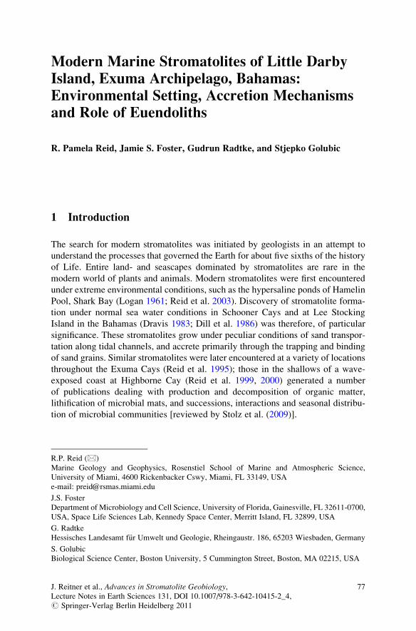
Modern Marine Stromatolites of Little Darby
Island, Exuma Archipelago, Bahamas:
Environmental Setting, Accretion Mechanisms
and Role of Euendoliths
R. Pamela Reid, Jamie S. Foster, Gudrun Radtke, and Stjepko Golubic
1 Introduction
The search for modern stromatolites was initiated by geologists in an attempt to
understand the processes that governed the Earth for about five sixths of the history
of Life. Entire land- and seascapes dominated by stromatolites are rare in the
modern world of plants and animals. Modern stromatolites were first encountered
under extreme environmental conditions, such as the hypersaline ponds of Hamelin
Pool, Shark Bay (Logan 1961; Reid et al. 2003). Discovery of stromatolite forma-
tion under normal sea water conditions in Schooner Cays and at Lee Stocking
Island in the Bahamas (Dravis 1983; Dill et al. 1986) was therefore, of particular
significance. These stromatolites grow under peculiar conditions of sand transpor-
tation along tidal channels, and accrete primarily through the trapping and binding
of sand grains. Similar stromatolites were later encountered at a variety of locations
throughout the Exuma Cays (Reid et al. 1995); those in the shallows of a wave-
exposed coast at Highborne Cay (Reid et al. 1999, 2000) generated a number
of publications dealing with production and decomposition of organic matter,
lithification of microbial mats, and successions, interactions and seasonal distribu-
tion of microbial communities [reviewed by Stolz et al. (2009)].
R.P. Reid (*)
Marine Geology and Geophysics, Rosenstiel School of Marine and Atmospheric Science,
University of Miami, 4600 Rickenbacker Cswy, Miami, FL 33149, USA
e-mail: [email protected]
J.S. Foster
Department of Microbiology and Cell Science, University of Florida, Gainesville, FL 32611-0700,
USA, Space Life Sciences Lab, Kennedy Space Center, Merritt Island, FL 32899, USA
G. Radtke
Hessisches Landesamt f€ur Umwelt und Geologie, Rheingaustr. 186, 65203 Wiesbaden, Germany
S. Golubic
Biological Science Center, Boston University, 5 Cummington Street, Boston, MA 02215, USA
J. Reitner et al., Advances in Stromatolite Geobiology,Lecture Notes in Earth Sciences 131, DOI 10.1007/978-3-642-10415-2_4,# Springer-Verlag Berlin Heidelberg 2011
77
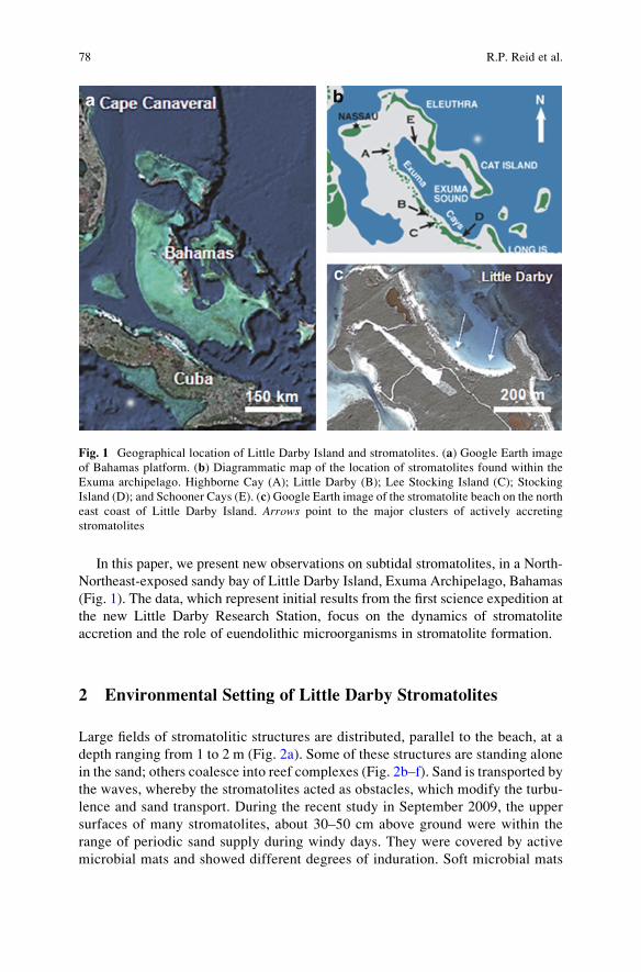
In this paper, we present new observations on subtidal stromatolites, in a North-
Northeast-exposed sandy bay of Little Darby Island, Exuma Archipelago, Bahamas
(Fig. 1). The data, which represent initial results from the first science expedition at
the new Little Darby Research Station, focus on the dynamics of stromatolite
accretion and the role of euendolithic microorganisms in stromatolite formation.
2 Environmental Setting of Little Darby Stromatolites
Large fields of stromatolitic structures are distributed, parallel to the beach, at a
depth ranging from 1 to 2 m (Fig. 2a). Some of these structures are standing alone
in the sand; others coalesce into reef complexes (Fig. 2b–f). Sand is transported by
the waves, whereby the stromatolites acted as obstacles, which modify the turbu-
lence and sand transport. During the recent study in September 2009, the upper
surfaces of many stromatolites, about 30–50 cm above ground were within the
range of periodic sand supply during windy days. They were covered by active
microbial mats and showed different degrees of induration. Soft microbial mats
Fig. 1 Geographical location of Little Darby Island and stromatolites. (a) Google Earth image
of Bahamas platform. (b) Diagrammatic map of the location of stromatolites found within the
Exuma archipelago. Highborne Cay (A); Little Darby (B); Lee Stocking Island (C); Stocking
Island (D); and Schooner Cays (E). (c) Google Earth image of the stromatolite beach on the north
east coast of Little Darby Island. Arrows point to the major clusters of actively accreting
stromatolites
78 R.P. Reid et al.
Fig. 2 Stromatolitic structures of the Little Darby Cay, Exuma archipelago, Bahamas. (a) The
field of subtidal, shallow water stromatolites (dark areas in front of the beach). (b) Two stroma-
tolites monitored for their accretion rates, note the upward expansion of the structures and zonal
coloration of their lateral walls. The nail in upper right is 7 cm long. (c) Upper surface of the
stromatolites with 5–10 cm diameter domes where active accretion takes place. Stromatolite upper
left is 60 cm wide. (d) Vertical section through the mat on the upper surface of a stromatolite,
Modern Marine Stromatolites of Little Darby Island, Exuma Archipelago, Bahamas 79
were comprised of sheathed filaments of oscillatoriacean cyanobacteria of the
genus Schizothrix with incorporated sand grains. These mats, studied earlier at
Stocking Island (Reid and Browne 1991), Lee Stocking Island (Browne 1993;
Feldmann and Mackenzie 1998) and Highborne Cay (Reid et al. 2000) formed
small domal protuberances 1–5 cm in diameter (Fig. 2b, c) on top of Little Darby
stromatolites by trapping and binding of sediment particles combined with upward
and radial movement of Schizothrix trichomes. A vertical section through such
protuberances (Fig. 2d) indicated that the vertical accretion of the mat shifts
laterally, on a centimeter scale, depending on the combination of local sand supply
and cyanobacterial response in movement and growth. During periods of calm
weather the accumulation of cyanobacterial biomass produced translucent hori-
zons, which in deeper layers became obscured by sand grain compaction. Lateral
shifts in accretion include and slightly favor stromatolite margins, resulting in the
gradual widening of the active upper surface, the flat appearance of the stromato-
lite tops, and the conical outline of stromatolite profiles with overhang side walls.
The side walls did not appear to trap significant amounts of sediment; they were
covered by different microbial populations than the stromatolite upper surfaces
and accordingly were colored differently (Fig. 2e). The bases of these stromato-
lites were in frequent contact with moving sand ripples, thus exposed to abrasion
by moving sands. The surfaces of stromatolites on the more easterly side of the
beach were overgrown by macro-algae, predominantly by Batophora and Acet-abularia (Fig. 2f). Other stromatolitic structures in the studied fields were buried in
the sand, and some were exposed and colonized by dark colored coating, the black
coloration contrasting with the white of the carbonate sand (Fig. 2f, lower right).
3 Accretional Processes in Little Darby Stromatolites
In stromatolites of Little Darby Island, which are similar to those studied at
Lee Stocking Island and on Highborne Cay, the accretion takes place within a soft
mat along the upward turned surfaces of the stromatolite structures (Fig. 2c). Two
components are essential in the process of stromatolite accretion: (a) the presence of
the Schizothrix mat and (b) the flux of carbonate sand grains supplied by waves or
currents. The cyclicity of wind and wave turbulence is marked by pulses in sand
supply to the stromatolites and is recorded in fine, mm scale lamination (Fig. 3a).
Fig. 2 (Continued) showing domes of active accretion with a lateral displacement over time.
Darker layers comprised of exopolymers of Schizothrix mat mark the pauses in sediment supply
during calm weather conditions. Scale bar is 1 cm long. (e) Groups of stromatolites coalescing into
coherent reefs (background) with two units showing color zonation on lateral walls. Note the
irregular rippling of sand due to interference of wave action around stromatolitic mounds. (f)
Stromatolites sheltered from sand supply are overgrown by algae. The unit in the center is
overgrown mainly by Batophora on the top and by Acetabularia on its sides. The stromatolitic
structure on lower right is re-emerging after having been buried by sand
80 R.P. Reid et al.
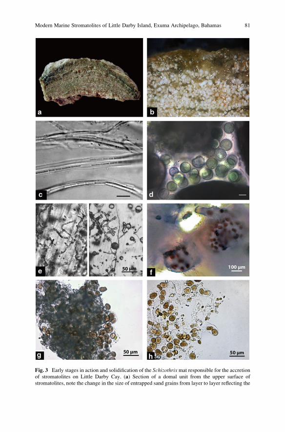
Fig. 3 Early stages in action and solidification of the Schizothrixmat responsible for the accretion
of stromatolites on Little Darby Cay. (a) Section of a domal unit from the upper surface of
stromatolites, note the change in the size of entrapped sand grains from layer to layer reflecting the
Modern Marine Stromatolites of Little Darby Island, Exuma Archipelago, Bahamas 81
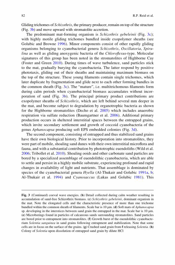
Gliding trichomes of Schizothrix, the primary producer, remain on top of the structure
(Fig. 3b) and move upward with stromatolite accretion.
The predominant mat-forming organism is Schizothrix gebeleinii (Fig. 3c);with highly motile gliding trichomes bundled inside exopolymer sheaths (see
Golubic and Browne 1996). Minor components consist of other rapidly gliding
organisms belonging to cyanobacterial genera Schizothrix, Oscillatoria, Spiru-lina as well as gliding anoxygenic bacteria of the Chloroflexus-type. Molecular
signatures of this group has been noted in the stromatolites of Highborne Cay
(Foster and Green 2010). During times of wave turbulence, sand particles stick
to the mat, gradually burying the cyanobacteria. The latter respond by positive
phototaxis, gliding out of their sheaths and maintaining maximum biomass on
the top of the structure. These young filaments contain single trichomes, which
later duplicate by fragmentation and glide next to each other forming bundles in
the common sheath (Fig. 3c). The “mature”, i.e. multitrichomous filaments form
during calm periods when cyanobacterial biomass accumulates without incor-
poration of sand (Fig. 3b). The principal primary product contributions are
exopolymer sheaths of Schizothrix, which are left behind several mm deeper in
the mat, and become subject to degradation by organotrophic bacteria as shown
for the Highborne stromatolites (Decho et al. 2005) which includes anaerobic
respiration via sulfate reduction (Baumgartner et al. 2006). Additional primary
production occurs in sheltered interstitial spaces between the entrapped grains,
which invite secondary settlement and growth of coccoid cyanobacteria of the
genus Aphanocapsa producing soft EPS embedded colonies (Fig. 3d).
The second component, consisting of entrapped and thus stabilized sand grains
have their own biological history. Prior to incorporation into stromatolites, they
were part of mobile, shoaling sand dunes with their own interstitial microflora and
fauna, and with a substantial contribution by phototrophic euendoliths (Wild et al.
2006; Tribollet et al. 2010). Shoaling ooids and other carbonate sand particles are
bored by a specialized assemblage of euendolithic cyanobacteria, which are able
to settle and persist in a highly mobile substrate, experiencing profound and rapid
changes in availability of light and nutrients. That assemblage is dominated by
species of the cyanobacterial genera Hyella (Al-Thukair and Golubic 1991a, b;
Al-Thukair et al. 1994) and Cyanosaccus (Lukas and Golubic 1981). This
Fig. 3 (Continued) coeval wave energies. (b) Detail collected during calm weather resulting in
accumulation of sand-free Schizothrix biomass. (c) Schizothrix gebeleinii, dominant organism in
the mat. Note the elongated cells and the characteristic presence of more than one trichome
bundled within the common sheath of filaments. Scale bar is 10 mm. (d) Soft mats of Aphanocapsasp. developing in the interstices between sand grain the entrapped in the mat. Scale bar is 10 mm.
(e) Microborings found in particles of calcareous sands surrounding stromatolites. Sand particles
are bored prior to entrapment into stromatolites. (f) Growth burst of the euendolithic cyanobacte-
rium Solentia sanguinea in sand grains following entrapment and stabilization. Note that some
cells are in focus on the surface of the grains. (g) Crushed sand grain from f releasing Solentia. (h)Colony of Solentia upon dissolution of entrapped sand grain by dilute HCl
82 R.P. Reid et al.
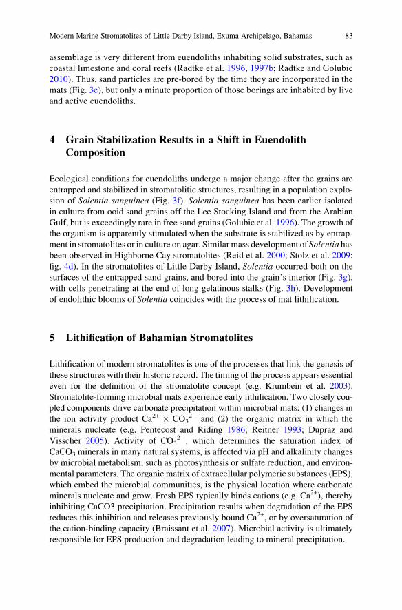
assemblage is very different from euendoliths inhabiting solid substrates, such as
coastal limestone and coral reefs (Radtke et al. 1996, 1997b; Radtke and Golubic
2010). Thus, sand particles are pre-bored by the time they are incorporated in the
mats (Fig. 3e), but only a minute proportion of those borings are inhabited by live
and active euendoliths.
4 Grain Stabilization Results in a Shift in Euendolith
Composition
Ecological conditions for euendoliths undergo a major change after the grains are
entrapped and stabilized in stromatolitic structures, resulting in a population explo-
sion of Solentia sanguinea (Fig. 3f). Solentia sanguinea has been earlier isolated
in culture from ooid sand grains off the Lee Stocking Island and from the Arabian
Gulf, but is exceedingly rare in free sand grains (Golubic et al. 1996). The growth of
the organism is apparently stimulated when the substrate is stabilized as by entrap-
ment in stromatolites or in culture on agar. Similar mass development of Solentia hasbeen observed in Highborne Cay stromatolites (Reid et al. 2000; Stolz et al. 2009:
fig. 4d). In the stromatolites of Little Darby Island, Solentia occurred both on the
surfaces of the entrapped sand grains, and bored into the grain’s interior (Fig. 3g),
with cells penetrating at the end of long gelatinous stalks (Fig. 3h). Development
of endolithic blooms of Solentia coincides with the process of mat lithification.
5 Lithification of Bahamian Stromatolites
Lithification of modern stromatolites is one of the processes that link the genesis of
these structures with their historic record. The timing of the process appears essential
even for the definition of the stromatolite concept (e.g. Krumbein et al. 2003).
Stromatolite-forming microbial mats experience early lithification. Two closely cou-
pled components drive carbonate precipitation within microbial mats: (1) changes in
the ion activity product Ca2+ � CO32� and (2) the organic matrix in which the
minerals nucleate (e.g. Pentecost and Riding 1986; Reitner 1993; Dupraz and
Visscher 2005). Activity of CO32�, which determines the saturation index of
CaCO3 minerals in many natural systems, is affected via pH and alkalinity changes
by microbial metabolism, such as photosynthesis or sulfate reduction, and environ-
mental parameters. The organic matrix of extracellular polymeric substances (EPS),
which embed the microbial communities, is the physical location where carbonate
minerals nucleate and grow. Fresh EPS typically binds cations (e.g. Ca2+), thereby
inhibiting CaCO3 precipitation. Precipitation results when degradation of the EPS
reduces this inhibition and releases previously bound Ca2+, or by oversaturation of
the cation-binding capacity (Braissant et al. 2007). Microbial activity is ultimately
responsible for EPS production and degradation leading to mineral precipitation.
Modern Marine Stromatolites of Little Darby Island, Exuma Archipelago, Bahamas 83
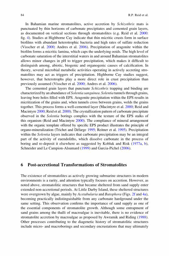
In Bahamian marine stromatolites, active accretion by Schizothrix mats is
punctuated by thin horizons of carbonate precipitates and cemented grain layers,
as documented on vertical sections through stromatolites (e.g. Reid et al. 2000:
fig. 4). Studies at Highborne Cay indicate that thin micritic crusts form in surface
biofilms with abundant heterotrophic bacteria and high rates of sulfate reduction
(Visscher et al. 2000; Andres et al. 2006). Precipitation of aragonite within the
biofilm forms a micritic lamina, which caps the underlying ooids. The high level of
carbonate saturation of the interstitial waters in and around Bahamian stromatolites
allows minor changes in pH to trigger precipitation, which makes it difficult to
distinguish among, abiotic, biogenic and organogenic causes of calcification. In
theory, several microbial metabolic activities operating in actively accreting stro-
matolites may act as triggers of precipitation. Highborne Cay studies suggest,
however, that heterotrophs play a more direct role in crust precipitation than
previously assumed (Visscher et al. 2000; Andres et al. 2006).
The cemented grain layers that punctuate Schizothrix trapping and binding are
characterized by an abundance of Solentia sanguinea. Solentia tunnels through grains,leaving bore holes filled with EPS. Aragonite precipitation within the EPS results in
micritization of the grains and, when tunnels cross between grains, welds the grains
together. This process forms a well-cemented layer (Macintyre et al. 2000; Reid and
Macintyre 2000; Reid et al. 2000). The crystallization pattern of carbonate precipitate
observed in the Solentia borings complies with the texture of the EPS stalks of
this organism (Reid and Macintyre 2000). The compliance of mineral arrangement
with the organic template offered by specific EPS product illustrates the principle of
organo-mineralization (Trichet and Defarge 1995; Reitner et al. 1995). Precipitation
within the Solentia layers indicates that carbonate precipitation may be an integral
part of the activity of euendoliths, which dissolve carbonate in the process of
boring and re-deposit it elsewhere as suggested by Kobluk and Risk (1977a, b),
Schneider and Le Campion-Alsumard (1999) and Garcia-Pichel (2006).
6 Post-accretional Transformations of Stromatolites
The existence of stromatolites as actively growing submarine structures in modern
environments is a rarity, and attention typically focuses on accretion. However, as
noted above, stromatolitic structures that became sheltered from sand supply enter
extended non-accretional periods. At Little Darby Island, these sheltered structures
were overgrown by algae, mainly by Acetabularia and Batophora (Figs. 2f and 4a),becoming practically indistinguishable from any carbonate hardground under the
same setting. This observation confirms the importance of sand supply as one of
the essential components of stromatolite growth. Although some entrapment of
sand grains among the thalli of macroalgae is inevitable, there is no evidence of
stromatolite accretion by macroalgae as proposed by Awramik and Riding (1988).
Other processes contributing to the diagenetic history of stromatolitic structures
include micro- and macroborings and secondary encrustations that may ultimately
84 R.P. Reid et al.
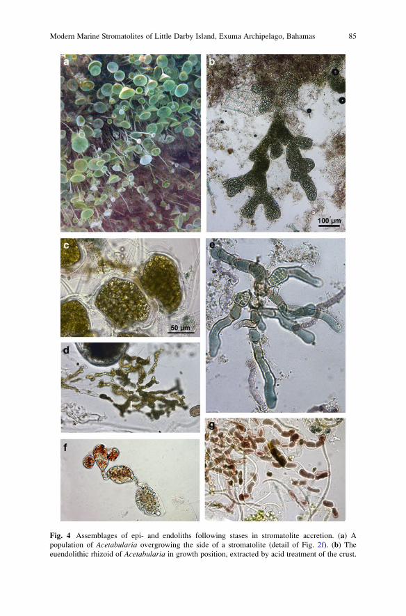
Fig. 4 Assemblages of epi- and endoliths following stases in stromatolite accretion. (a) A
population of Acetabularia overgrowing the side of a stromatolite (detail of Fig. 2f). (b) The
euendolithic rhizoid of Acetabularia in growth position, extracted by acid treatment of the crust.
Modern Marine Stromatolites of Little Darby Island, Exuma Archipelago, Bahamas 85
obscure the original accretional features. Such structural modifications may lead
to formation of thrombolitic textures when viewed in section or in outcrop
(Planavsky and Ginsburg 2009).
The extended stasis provides new opportunities for the settlements of epi- and
endoliths alike, resulting in an increase in diversity and complexity of the resident
microboring community apparently at the expense of Solentia, which is rarely
encountered in these samples. Prominent are algae, including eukaryotic euendo-
liths, especially Acetabularia (Fig. 4a) and its euendolithic rhizoids (Fig. 4b)
formerly misidentified as Ostreobium brabantium (see Radtke et al. 1997a). The
hardened surfaces are often paved by large polygonal chlorophyte cells with
chaetae (Fig. 4c), rhizoids of Batophora oerstedtii, euendolithic networks of the
siphonal chlorophyte Ostreobium quekettii (Fig. 4d). Among cyanobacterial euen-
doliths dominant are various species of Hyella, which have not been observed
anywhere else (Fig. 4e). In addition, Conchocelis stages of a local marine red
alga are occasionally observed (Fig. 4f).
The accretion of every stromatolitic structure observed in the Little Darby field
is localized to cm sized areas always on the upward surfaces of the structure. The
sides of the stromatolites are stationary and firmly cemented. They are externally
coated by different mat types (including Leptolyngbya spp.) and, under these mats,
harbor a highly diverse community of epilithic and euendolithic phototrophs, an
assemblage comparable to that found on structures with extended stasis.
The lower parts of the stromatolite side walls are covered by dark, almost black
veneer which is locally abraded, and show patches of white carbonate (Fig. 1f,
bottom right). These lower parts of the stromatolites and sometimes the entire
structure are occasionally completely covered by sand. The stress imposed by
light deprivation and exposure to repeated abrasion by sand prevents the growth
of most algae and other epilithic organisms, and exerts selective pressure on
euendoliths as well. Under the conditions of sand-erosion pressure and repeated
covering and uncovering by sand, the euendolithic assemblage has reduced diver-
sity and may be dominated by Solentia sanguinea (Fig. 4g).
7 Dynamics of Stromatolite Formation
The dynamics of microbial mats operate at different spatial and temporal scales.
Stromatolite accretion at Highborne Cay results from a cycling between three
different mat types on the stromatolite surface, with each subsurface layer repre-
senting a former surface mat (Reid et al. 2000). Similar dynamics appear to be
important in the stromatolites of Little Darby Island.
Fig. 4 (Continued) (c) Puzzle shaped chlorophyte cells with tubular bristles lining the surfaces of
the walls. The scale in c is valid for d–g. (d) Ostreobium quekettii the most common euendolith
under low light conditions. (e) Hyella sp., probably a new species. (f) Conchocelis stage of a
bangialean rhodophyte. (g) Solentia sanguinea in erosion stressed base of a stromatolite
86 R.P. Reid et al.
The soft mat is dominated by Schizothrix, which is responsible for trapping of sandand a vertical growth of local domes (¼Type 1mat of Reid et al. 2000). In the process,
trichomes of Schizothrixmove to the mat surface leaving significant amounts of their
photosynthetic product, their empty EPS sheath behind to bacterial consumers. Both
layers moved upward in the process of the accretion of the stromatolitic structure,
interrupted by short pauses in sedimentation. These pauses are marked by biofilm
development and corresponding precipitation of thin micritic crusts (¼Type 2 mats).
Extended stasis in stromatolite accretion is accompanied by changes in microbial
euendolith composition. The changes feature a population increase of Solentia – firstin the entrapped sand particles, and later from grain to grain over calcified bridges
(¼Type 3 mat). During these extended pauses in accretion, the stromatolite hardens.
The endolith diversity also increases over time (Stolz et al. 2009) and may involve
removal and truncation of bored grains by grazers, together with the resident euen-
doliths. The resumption of stromatolite accretion starts another cycle in mat develop-
ment, incorporating the truncated grains as a horizon within the stromatolite (Reid
et al. 2000). This cyclicity involves alternation of the accretionary phase with mat
types 1, with sorter or longer stationary phases resulting in mat types 2 and 3
respectively. A vertical sequence of this cyclicity is historical and in vertical petro-
graphic section it is evident as a stratigraphic record.
Accretional periods may be interrupted by much longer periods of stasis, which
permit profound changes in microbial composition and an overgrowth by algae and
animals. Similar interruptions may be caused by burial of stromatolites in the sand.
In the deep tidal channels of the Lee Stocking Island, where sand moves passing
stromatolites in massive dunes, both burial and deprivation of sediment supply tend
to occur cyclically, leaving accretion as limited “windows of opportunity” (Golubic
and Browne 1996; Seong-Joo Lee et al. 2000). In the case of Little Darby, local
sheltering of some stromatolites from sediment supply can result in long periods of
stasis with algal overgrowth, whereas burial of stromatolites may take place during
major storms.
Acknowledgements This is Research Initiative on Bahamian Stromatolites (RIBS) contribution
#59. The international collaboration was supported by the Alexander-von-Humboldt Foundation,
Bad Godesberg, and Hanse Wissenschaftskolleg, Delmenhorst, Germany. We appreciate the
critical comments of Joachim Reitner and Gernot Arp.
References
Al-Thukair AA, Golubic S (1991a) Five new Hyella species from the Arabian Gulf. Algological
Studies 64:167–197
Al-Thukair AA, Golubic S (1991b) New endolithic cyanobacteria from the Arabian Gulf. I. Hyellaimmanis sp. nov. Journal of Phycology 27:766–780
Al-Thukair AA, Golubic S, Rosen G (1994) New euendolithic cyanobacteria from the Bahama
Bank and the Arabian Gulf: Hyella racemus sp. nov. Journal of Phycology 30:764–769
Andres MS, Sumner DY, Reid RP, Swart PK (2006) Isotopic fingerprints of microbial respiration
in aragonite from Bahamian stromatolites. Geology 34:973–976
Modern Marine Stromatolites of Little Darby Island, Exuma Archipelago, Bahamas 87
Awramik SM, Riding R (1988) Role of algal eukaryotes in subtidal columnar stromatolite
formation. Proceedings of the National Academy of Sciences of the United States of America
85:1327–1329
Baumgartner LK, Reid RP, Dupraz C, Decho AW, Buckley DH, Spear JR, Przekop KM,
Visscher PT (2006) Sulfate reducing bacteria in microbial mats: Changing paradigms, new
discoveries. Sedimentary Geology 185:131–145
Braissant O, Decho AW, Dupraz C, Glunk C, Przekop KM, Visscher PT (2007) Exopolymeric
substances of sulfate-reducing bacteria: Interactions with calcium at alkaline pH and implica-
tion for formation of carbonate minerals. Geobiology 5:401–411
Browne KM (1993) Lamination in Recent Bahamian Subtidal Stromatolites: Origin and Lithifica-
tion. Unpublished PhD dissertation, University of Miami, Coral Gables, pp 296
Decho AW, Visscher PT, Reid RP (2005) Production and cycling of natural microbial exopoly-
mers (EPS) within a marine stromatolite. Palaeogeography, Palaeoclimatology, Palaeoecology
219:71–86
Dill RF, Shinn EA, Jones AT, Kelly K, Steinin RP (1986) Giant subtidal stromatolites forming in
normal saline waters. Nature 324:55–58
Dravis JJ (1983) Hardened subtidal stromatolites, Bahamas. Science 219:385–386
Dupraz C, Visscher PT (2005) Microbial lithification in marine stromatolites and hypersaline
mats. Trends in Microbiology 13:429–438
Feldmann M, Mackenzie J (1998) Stromatolite thrombolite association in a modern environment,
Lee Stocking Island, Bahamas. Palaios 13:201–212
Foster JS, Green SJ (2010) Microbial diversity in modern marine stromatolites. In: Tewari V
(ed) Cellular Origin, Life in Extreme Habitats and Astrobiology: Stromatolites. Springer,
Berlin, in press
Garcia-Pichel F (2006) Plausible mechanisms for the boring on carbonates by microbial auto-
trophs. Sedimentary Geology 185:205–213
Golubic S, Browne KM (1996) Schizothrix gebeleinii sp. nov. builds subtidal stromatolites, Lee
Stocking Island, Bahamas. Algological Studies 83:273–290
Golubic S, Al-Thukair AA, Gektidis M (1996) New endolithic cyanobacteria from the Arabian
Gulf and the Bahama Bank: Solentia sanguinea sp. nov. Algological Studies 83:291–301
Kobluk DR, Risk MJ (1977a) Micritization and carbonate-grain binding by endolithic algae.
American Association of Petroleum Geologist Bulletin 61:1069–1082
Kobluk DR, Risk MJ (1977b) Calcification of exposed filaments of endolithic algae, micrite
envelope formation and sediment production. Journal of Sedimentary Petrology 47:517–528
Krumbein WE, Brehm U, Gorbushina AA, Levit G, Palinska KA (2003) Biofilm, biodictyon
and biomat – biolaminites, ooolites, stromatolithes – geophysiology, global mechanism and
parahistology. In: KrumbeinWE, Paterson DM, Zavarzin GA (eds) Fossil and Recent Biofilms.
Kluwer Acad Publ, Dordrecht, pp 1–27
Logan BW (1961) Cryptozoon and associate stromatolites from the Recent, Shark Bay, Western
Australia. Journal of Geology 69:517–533
Lukas KJ, Golubic S (1981) New endolithic cyanophytes from the North Atlantic Ocean:
I. Cyanosaccus piriformis gen. et sp. nov. Jounral of Phycology 17:224–229
Macintyre IG, Prufert-Bebout L, Reid RP (2000) The role of endolithic cyanobacteria in the
formation of lithified laminae in Bahamian stromatolites. Sedimentology 47:915–921
Pentecost A, Riding R (1986) Calcification in cyanobacteria. In: Leadbeater BSC, Riding R
(eds) Biomineralization of lower plants and animals. SAS 30, Clarendon, Oxford, pp 73–90
Planavsky N, Ginsburg RN (2009) Taphonomy of modern marine Bahamian microbialites. Palaios
24:5–17
Radtke G, Golubic S (2010) Microbial euendolithic assemblages and microborings in intertidal
and shallow marine habitats: insight in cyanobacterial speciation. In: Reitner J, Queric, N-V,
Arp G (eds) Advances in Stromatolite Geobiology. Lecture Notes in Earth Sciences 131,
Springer, Berlin, pp 213–241
88 R.P. Reid et al.
Radtke G, Le Campion-Alsumard T, Golubic S (1996) Microbial assemblages of the bioerosional
“notch” along tropical limestone coasts. Algological Studies 83:469–482
Radtke G, Gektidis M, Golubic S, Hofmann K, Kiene WE, Le Campion-Alsumard T (1997a)
The identity of an endolithic alga: Ostreobium brabantium Weber-van Bosse is recognized
as carbonate-penetrating rhizoids of Acetabularia (Chlorophyta, Dasycladales). Courier
Forschungsinstitut Senckenberg 201:341–347
Radtke G, Le Campion-Alsumard T, Golubic S (1997b) Microbial assemblages involved in
tropical coastal bioerosion: An Atlantic-Pacific comparison. Proceedings of the 8th Interna-
tional Coral Reef Symposium 2:1825–1830
Reid RP, Browne KM (1991) Intertidal stromatolites in a fringing Holocene reef complex,
Bahamas. Geology 19:15–18
Reid RP, Macintyre (2000) Microboring versus recrystallization: Further insight into the micriti-
zation process. Journal of Sedimentary Research 70:24–28
Reid RP, Macintyre IG, Steneck RS, Browne KM, Miller TE (1995) Stromatolites in the Exuma
Cays, Bahamas: Uncommonly common. Facies 33:1–18
Reid RP, Macintyre IG, Steneck RS (1999) A microbialite/algal ridge fringing reef complex,
Highborne Cay, Bahamas. Atoll Research Bulletin 466:1–18
Reid RP, Visscher PT, Decho AW, Stolz JF, Bebout BM, Dupraz CP, Macintyre IG, Paerl HW,
Pinckney JL, Prufert-Bebout L, Steppe TF, DesMarais DJ (2000) The role of microbes in
accretion, lamination and early lithification of modern marine stromatolites. Nature
406:989–992
Reid RP, James NP, Macintyre IG, Dupraz CP, Burne RV (2003) Shark Bay stromatolites:
Microfabrics and reinterpretation of origins. Facies 49:45–53
Reitner J (1993) Modern cryptic microbialite/metazoan facies from Lizard Island (Great Barrier
Reef, Australia) – Formation and concepts. Facies 29:3–40
Reitner J, Gautret P, Marin F, Neuweiler F (1995) Automicrites in a modern microbialite –
Formation model via organic matrices (Lizard Island, Great Barrier Reef, Australia). Bulletin
de l´Institut oceanographique Monaco 14(2):237–263
Schneider J, Le Campion-Alsumard T (1999) Construction and destruction of carbonates by
marine and freshwater cyanobacteria. European Journal of Phycology 34:417–426
Seong-Joo Lee, Browne KM, Golubic S (2000) On stromatolite lamination. In: Riding R, Awra-
mik SM (eds) Microbial sediments. Springer, Berlin, pp 16–24
Stolz JF, Reid RP, Visscher PT, Decho AW, Norman RS, Aspden RJ, Bowlin EM, Franks J,
Foster JS, Paterson DM, Przekop KM, Underwood GJC, Prufert-Bebout L (2009) The micro-
bial communities of the modern marine stromatolites at Highborn Cay, Bahamas. Atoll
Research Bulletin 567:1–29
Tribollet A, Golubic S, Radtke G, Reitner J (2010) OnMicrobiocorrosion. In: Reitner J, Queric, N-V,
ArpG (eds)Advances in Stromatolite Geobiology. LectureNotes in Earth Sciences 131, Springer,
Berlin, pp 243–253
Trichet J, Defarge C (1995) Non-biologically supported organomineralization. Bulletin de l´
Institut oceanographique Monaco Spec 14:203–236
Visscher PT, Reid RP, Bebout BM (2000) Microscale observations of sulfate reduction: Correla-
tion of microbial activity with lithified micritic laminae in modern marine stromatolites.
Geology 28: 919–922
Wild C, Laforsch C, Huettel M. (2006) Detection and enumeration of microbial cells within highly
porous calcareous reef sands. Marine and Freshwater Research 57:415–420
Modern Marine Stromatolites of Little Darby Island, Exuma Archipelago, Bahamas 89

.
Molecular Approaches to Studying Living
Stromatolites
Brendan P. Burns, Nithya Baburajendran, and Joannita Dharmawan
1 Introduction
The presence of stromatolites dates back to some 3.5–3.8 billion years in earth’s
history, a time period that witnessed the appearance of the first forms of life
(Schopf 2006). The microbial communities that constituted these complex struc-
tures played a significant role in the global biogeochemical cycles and sedimen-
tation (Dupraz and Visscher 2005). The impetus behind stromatolite genesis is not
clearly known and the modes of accretion, and lithification have long been
debated; both biotic and abiotic mechanisms have been suggested for stromatolite
formation (Grotzinger and Knoll 1999; Grotzinger and Rothman 1996). Irrespec-
tive of their origin, the contributions of stromatolites to the primeval environmen-
tal conditions are undisputed. Owing to their importance, stromatolites have
formed the core of a growing area of microbial ecology and evolution-based
research. Modern stromatolites, the living counterparts of fossilized stromatolites,
offer a glimpse as to how microbial communities may have prevailed on the early
Earth, and allow unprecedented access to the functional complexity of these
systems. Microbial mats that are also considered as living analogues of ancient
stromatolites have been studied to interpret ancient stromatolites and have been
particularly resourceful in comprehending the formation of extant stromatolites.
Layering of lithified microbial mats is in fact attributed to stromatolite
B.P. Burns (*), N. Baburajendran, and J. Dharmawan
School of Biotechnology and Biomolecular Sciences, University of New South Wales, Sydney,
NSW 2052, Australia
Australian Centre for Astrobiology, University of New South Wales, Sydney, NSW 2052,
Australia
e-mail: [email protected]
J. Reitner et al., Advances in Stromatolite Geobiology,Lecture Notes in Earth Sciences 131, DOI 10.1007/978-3-642-10415-2_5,# Springer-Verlag Berlin Heidelberg 2011
91
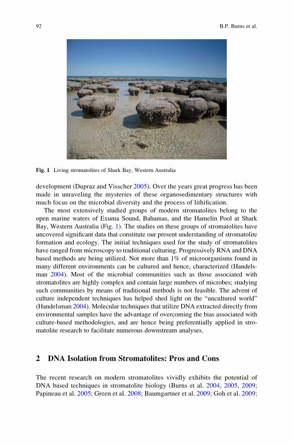
development (Dupraz and Visscher 2005). Over the years great progress has been
made in unraveling the mysteries of these organosedimentary structures with
much focus on the microbial diversity and the process of lithification.
The most extensively studied groups of modern stromatolites belong to the
open marine waters of Exuma Sound, Bahamas, and the Hamelin Pool at Shark
Bay, Western Australia (Fig. 1). The studies on these groups of stromatolites have
uncovered significant data that constitute our present understanding of stromatolite
formation and ecology. The initial techniques used for the study of stromatolites
have ranged frommicroscopy to traditional culturing. Progressively RNA and DNA
based methods are being utilized. Not more than 1% of microorganisms found in
many different environments can be cultured and hence, characterized (Handels-
man 2004). Most of the microbial communities such as those associated with
stromatolites are highly complex and contain large numbers of microbes; studying
such communities by means of traditional methods is not feasible. The advent of
culture independent techniques has helped shed light on the “uncultured world”
(Handelsman 2004). Molecular techniques that utilize DNA extracted directly from
environmental samples have the advantage of overcoming the bias associated with
culture-based methodologies, and are hence being preferentially applied in stro-
matolite research to facilitate numerous downstream analyses.
2 DNA Isolation from Stromatolites: Pros and Cons
The recent research on modern stromatolites vividly exhibits the potential of
DNA based techniques in stromatolite biology (Burns et al. 2004, 2005, 2009;
Papineau et al. 2005; Green et al. 2008; Baumgartner et al. 2009; Goh et al. 2009;
Fig. 1 Living stromatolites of Shark Bay, Western Australia
92 B.P. Burns et al.
Allen et al. 2009; Breitbart et al. 2009; Foster et al. 2009). However, the success
of such analyses often rest on the first and fundamental rate-limiting step of direct
DNA isolation from stromatolites. Extraction of stromatolite community DNA is
in fact quite challenging, owing to the presence of cemented calcium carbonate
matrix that hinders access to the associated microbial communities. As there are
no universal protocols available for direct DNA isolation from stromatolites,
some of the various methods that have been employed in recent studies are
compared and discussed below.
Most of the studies begin with a physical step that involves pulverisation of the
stromatolite sample, usually with a mortar and pestle (Burns et al. 2004; Goh et al.
2009) or a bead-beating procedure (Papineau et al. 2005; Green et al. 2008;
Baumgartner et al. 2009; Foster et al. 2009). This physical step is essential to
release as much cellular material as possible that is in physical association with
sediments/minerals, which will then allow for optimal cell lysis in subsequent
chemical steps. As a modification on these initial physical disruption methods,
the need to obtain high quality high molecular weight DNA for downstream
metagenomic analyses has led some groups to adopt DNA extraction based on
combined chemical methods. The presence of exopolymeric substances (EPS) in
microbial mats and stromatolites (Decho 1990) in particular is a significant chal-
lenge in extracting good quality DNA, and the use of freeze-thaw and enzymatic
methods have helped overcome this issue. These have included an extended
lysozyme/Proteinase K step of 1 h and having one freeze-thaw cycle (Burns et al.
2004), and up to four and five freeze-thaw cycles, respectively (Goh et al. 2009;
Allen et al. 2009) for extraction from Shark Bay stromatolites. These freeze-thaw
steps are also thought to improve cell lysis. A further chemical step, the addition of
the EPS-binding reagent cetyl trimethyl ammonium bromide (CTAB), is also
employed in many protocols to ensure all EPS is removed (Breitbart et al. 2009;
Allen et al. 2009; Foster et al. 2009; Goh et al. 2009). In addition to EPS removal,
many stromatolite samples have associated calcium carbonate material that needs
to be removed before effective cell lysis. A common method for carbonate removal
is incubation of samples in an EDTA slurry (0.5 M) for varying times depending on
the amount of carbonate in a given sample. Gentle shaking overnight at 37�C has
proven successful in dissolving carbonate from stromatolite samples (Wade and
Garcia-Pichel 2003), however for particularly carbonate-rich samples from Shark
Bay a second overnight incubation with EDTA was required to remove all the
carbonate (Allen et al. 2008).
Subsequent to EPS and carbonate removal, many protocols then employ a
number of incubation steps designed to both lyse cells and clean up the DNA
from associated impurities. A range of buffers have and are being employed in
stromatolite DNA extractions, including potassium ethyl xanthogenate (Tillet and
Neilan 2000), various combinations of phosphate buffers, TNE buffers and SDS,
as well as a range of phenol: chloroform extractions (Burns et al. 2004, 2005,
2009; Papineau et al. 2005; Goh et al. 2009; Allen et al. 2009; Breitbart et al.
2009; Foster et al. 2009). In comparison, some studies have also employed kit-
based methods for extraction of total genomic DNA from environmental
Molecular Approaches to Studying Living Stromatolites 93
stromatolite samples (Green et al. 2008; Breitbart et al. 2009; Baumgartner et al.
2009). Examples of these include the FastDNA Spin Kit for Soil (QBiogene),
UltraClean Soil DNA Kit (Mo Bio), and the Mo Bio SOIL DNA extraction kit
(Mo Bio). As these studies involved examining different kinds of stromatolites
from different geographical locations it is difficult to assess the efficacy of the
various kits available, however the published data suggest they are all viable
options for stromatolite DNA extractions. However, recent work on the metage-
nomics of microbialites in Mexico (Breitbart et al. 2009) found that these kits
were unsuccessful on their samples, possibly due to excess EPS. To overcome this
these researchers employed modifications of the freeze-thaw, CTAB, and phenol:
chloroform extraction protocols (Breitbart et al. 2009).
All of these methods are aiming for the same outcome: to increase DNA purity
and yield. Downstreammolecular methods such as PCR are extremely sensitive and
require pure forms of DNA; contaminations can often inhibit DNA amplification
(Burns et al. 2004). For construction of libraries and analyzing community struc-
tures large amounts of DNA are desired with minimal shearing. Thus an ideal
protocol should aim at achieving high purity and yield. As described above, the
procedures available for the isolation of DNA from stromatolite samples generally
consists of combinations of physical, chemical and enzymatic steps. Use of freezing
and thawing can result in loss of DNA, however, the amount of contamination and
shearing is often less when compared to bead mill homogenization. Proteinase K
and lysozyme treatments are the most frequently used enzymatic techniques, and
the enzyme concentrations used will depend on the sample size; increasing the
enzyme concentration and decreasing sample mass promotes cell lysis resulting in
higher DNA yields.
Regardless of the type of cell lysis method used, it is essential to subject the
DNA extracts to repeated purification to remove contaminating substances such as
humics. Contamination is a common problem in environmental samples and it is
necessary to minimize the contaminants as they can eventually affect the processing
of the DNA samples. Single purification steps however, often do not completely
remove the contaminants; multiple purification cycles can increase the purity but
can also lead to loss of DNA. Thus it is often desirable to start with multiple
samples of the stromatolite material, and the DNA extracted from these is ulti-
mately pooled to allow for both optimal purity and yield for the purpose accessing
the genetic and functional diversity of stromatolites.
3 Molecular Tools: Accessing the Genetic Diversity
and Beyond
Once DNA has been successfully extracted from stromatolite material, two
molecular approaches in particular have been employed that have revolutioni-
zed stromatolite biology. These molecular approaches when compared to the
94 B.P. Burns et al.
traditional techniques have proved more insightful in establishing the genetic
diversity of stromatolites and are currently being utilized to further enhance our
knowledge on stromatolite formation, the microbial community structure, and
their function.
3.1 PCR-Based rDNA Analysis
Ribosomal RNA gene sequence was first used by Pace et al. (1986) to establish the
genetic diversity of an environmental sample without culturing. 16S and 5S rRNA
sequences were used for the study. This analysis was based on the knowledge that
ribosomal RNA genes served as evolutionary chronometers allowing for the phy-
logentic classification of microorganisms (Woese 1987). rRNA gene sequences are
highly conserved amongst evolutionarily related species. Before the advent of PCR,
rRNA analysis was carried out by means of direct sequencing or filter hybridization
of rRNA extracts (Stahl et al. 1988), screening of clones from DNA libraries for
rDNA inserts and in-vitro amplification of rDNA (obtained by reverse transcription
of rRNA) followed by separation on DGGE or TGGE (Wintzingerode et al. 1997).
RFLP and ARDA (amplified ribosomal DNA restriction analysis) have also been
used to determine the genetic diversity of different microbial communities (Weidner
et al. 1996; K€ohler et al. 1991).With the power to directly amplify the DNA extracts from samples, PCR has
been used to amplify rDNA genes with the help of specific primers. Such amplicons
are used for constructing 16S rDNA libraries, and the clones randomly screened and
eventually sequenced. These sequences can then be probed against nucleotide
sequences of well-characterized organisms by means of bioinformatic tools thus,
providing a culture independent method for ascertaining the microbial diversity in
various environments. This technique has been efficiently used to study the micro-
bial ecology of modern stromatolites in various geographical locations, and mem-
bers of cyanobacteria, bacteria and archaea have been identified (Burns et al. 2004,
2005, 2009; Papineau et al. 2005; Baumgartner et al. 2009; Goh et al. 2009; Allen
et al. 2009; Breitbart et al. 2009; Foster et al. 2009).
The early work by Burns et al. (2004) on stromatolites in Hamelin Pool revealed
the presence of various cyanobacterial genera including Synechococcus, Xenococ-cus,Microcoleus, Leptolyngbya, Plectonema, Symploca, Cyanothece, Pleurocapsaand Nostoc. The study also reported the presence of potentially free- living
Prochloron – a genus commonly found as a symbiont. Eubacteria including well-
classified groups like Proteobacteria, low GþC Gram-positive Planctomycetes and
Acidobacteria and few unknown groups OP9, OP109 and Marine A group were
identified. Halophilic archaea belonging to the divisions Euryarchaeota and Cre-
narchaeota and methonogenic archaea from the order Methanosarcinales were also
detected. In this investigation both culture based and culture independent techni-
ques were used for analysing the genetic diversity of Hamelin Pool stromatolites,
Molecular Approaches to Studying Living Stromatolites 95
however DNA based methods were shown to be more efficient in identifying the
stromatolites microbiota (Burns et al. 2004).
Furthermore, the difference in the microbial populations between the different
morphological classes, the surface and the interior of Hamelin Pool stromatolites
have also been described (Burns et al. 2004; Papineau et al. 2005). The relative
abundance of each group of microbes has also been established in all the previous
works; approximately 90% are bacterial representatives while 10% are archaeal.
Schizothrix and Solentia that are predominant in Bahamian stromatolites have not
been observed in the Hamelin pool stromatolites. Papineau et al. (2005) demon-
strated the relationship between stromatolite morphology and the microbial com-
munities. Substantial difference was observed amongst the communities of domal
and irregular stromatolites, and also between the surface and endolithic commu-
nities. No new phylogenetic groups were described in the study, however, the
cyanobacterial representatives which have long been believed to dominate the
stromatolite communities where shown to be less abundant. Such low abundance
has also been seen in case of the Bahamian stromatolites. Other microorganisms
thus contribute to the biomass. Though sometimes present in small numbers
Papineau et al. (2005), cyanobacteria are considered as the primary producers
while some anoxygenic phototrophs have also been deemed as contributors of the
overall primary productivity. The mass and turnover rate of the primary producers
control the size of other microbes required to maintain a community and ultimately
influence stromatolite morphogenesis.
3.2 Metagenomics
Metagenomics, also known as environmental/community/population genomics
involves the direct DNA isolation from a community of microorganisms and
construction of a genomic DNA library, which can be screened for various aspects.
There are two fundamental but complementary approaches to a metagenomic
analysis: sequence-based analyses and functional analyses. In sequence-based
analysis, the clones from a metagenomic library are sequenced randomly and
genes of interests are identified. The phylogenetic markers flanking the desired
gene are then used to assign a taxonomic group to the gene. Thus the organism and
its characteristic feature can be identified. Another methodology involves the
sequencing of DNA segments from a specific taxon. Though this technique is
mainly based on phylogenetic markers, due to the lack of significant numbers of
such markers, a variation in this approach involving the random sequencing of the
clones on a large scale has been devised. With the vast amount of sequence data
obtained the dynamics of the community can be determined (Pedros-Alio 2006).
In functional metagenomcis, the metagenomic library is expressed and the
functions of the clones analysed. Thus by screening the metagenome, specific
genes can be identified without prior knowledge of the sequence. Such functional
96 B.P. Burns et al.
screens can also be used to assign functions to uncharacterized proteins. Once the
clones carrying the genes of interest are identified in the metagenomic library, the
clones can be sequenced (Ferrer et al. 2009). A further interesting aspect towards
this approach is that clones that have similar functions can be grouped and
sequenced and phylogenetic anchors can be identified; thus the diversity of gen-
omes sharing the same functions can be characterized (Handelsman 2004).
In a recent study by Desnues et al. (2008) of the modern marine stromatolites
from HighBorne Cay, Bahamas and freshwater stromatolites and thrombolites from
Pozes Azules II and Rio Mesquites, Mexico, sequence based metagenomic analysis
of the viral communities was carried out and the biogeography of the phages was
established. The sequences of the three metagenome were compared with each
other and the distribution of the viral population was determined. The lack of
sequence overlaps (cross-contigs) between the metagenomes indicated the presence
of genetically unique viral particles in each microbialite. This difference in the viral
community between geographically separated microbialites has been associated
with the evolutionary origin of these structures.
4 The Future of Stromatolite Research
The 16S rRNA based PCR techniques have been particularly useful in establish-
ing the genetic diversity of extant stromatolites. Analysis of the microbial diver-
sity is one of the first steps towards comprehension of the complex communities
that constitute the evolutionarily and ecologically significant stromatolites.
Understanding the relationships between the microbial communities, the meta-
bolic activities that sustain these communities and ultimately influence stromato-
lite formation will form the premise for future research. PCR based methods
present certain limitations with such aspects as they do not allow for functional
analyses, and a powerful tool such as metagenomics will have a significant role
in such endeavours. Nevertheless, 16S rRNA analyses can complement metage-
nomics; the clones of a stromatolite metagenome can be screened for 16S phylo-
genetic markers using PCR and the functions can be correlated with specific
organisms. Furthermore, characterisation of both novel organisms and genes, as
well as developing new molecular techniques are providing an excellent platform
for future research on stromatolites (Pomati et al. 2004; Goh et al. 2006; Marshall
et al. 2007; Leuko et al. 2007, 2008, 2009; D’Elia et al. 2007; M€unchoff et al.2007; Allen et al. 2008).
Various aspects of microbial ecology of complex communities have been
uncovered by means of metagenomics. Symbiotic relationships, competition,
communication, accumulation of nutrients and energy production in diverse
environments have been studied with the help of metagenomics. Thus there
is significant potential in utilizing metagenomics in the study of stromatolite
ecology. Bioactive compounds such as antibiotics, secondary metabolites and
enzymes have also been discovered by means of metagenomics. Secondary
Molecular Approaches to Studying Living Stromatolites 97
metabolite production in stromatolites has been documented; a putative antican-
cer agent has been reported in Hamelin pool stromatolites (Burns et al. 2005).
Thus screening the stromatolite metagenomes for the synthesis of substances that
have possible biotechnological applications is another fascinating aspect of stro-
matolite research. The knowledge garnered through such potential studies can
shed light on the past and will also help appreciate the present ecological roles
played by modern stromatolites.
References
Allen MA, Goh F, Leuko S, Echigo A, Mizuki T, Usami R, Kamekura M, Neilan BA, Burns BP
(2008) Haloferax elongans sp. nov. and Haloferax mucosum sp. nov., isolated from microbial
mats from Hamelin Pool, Shark Bay. International Journal of Systematic and Evolutionary
Microbiol 58:798–802
Allen MA, Goh F, Burns BP, Neilan BA (2009) Bacterial, archaeal and eukaryotic diversity of
microbial mat communities in the hypersaline lagoon of Shark Bay. Geobiology 7:82–96
Baumgartner LK, Spear JR, Buckley DH, Pace NR, Reid RP, Dupraz C, Vissche PT (2009)
Microbial diversity in modern marine stromatolites, Highborne Cay, Bahamas. Environmental
Microbiology 11(10): 2710–2719
Breitbart M, Hoare A, Nitti A, Siefert J, Haynes M, Dinsdale E, Edwards R, Souza V, Rohwer F,
Hollander D (2009) Metagenomic and stable isotopic analyses of modern freshwater micro-
bialites in Cuatro Cienegas, Mexico. Environmental Microbiology 11:16–34
Burns BP, Goh F, Allen M, Neilan BA (2004) Microbial diversity of extant stromatolites in the
hypersaline marine environment of Shark Bay, Australia. Environmental Microbiology
6:1096–1101
Burns BP, Seifert A, Goh F, Pomati F, Neilan BA (2005) Genetic potential for secondary
metabolite production in stromatolite communities. FEMS Microbiology Letters 243:293–301
Burns BP, Anitori R, Butterworth P, Henneberger R, Goh F, Allen MA, Ibanez-Peral R, Bergquist
PL, Neilan BA, Walter MR (2009) Modern analogues and the early history of microbial life.
Precambrian Research 173:10–18
D’Elia M, Blanco A, Liocchelli D, Orofino V, Fonti S, Pomati F, Burns BP (2007) Physical
characterisation of biotic and abiotic minerals of exobiological interest. Il Nuovo Cimento
121:833–842
Decho A (1990) Microbial exopolymer secretions in ocean environments: their role(s) in food
webs and marine processes. Oceanography & Marine Biology Annual Review 28:73–154
Desnues C, Rodriguez-Brito B, Rayhawk S, Kelley S, Tran T, Haynes M, Liu H, Furlan M,
Wegley L, Chau B, Ruan Y, Hall D, Angly FE, Edwards RA, Li L, Thurber RV, Reid RP,
Siefert J, Souza V, Valentine DL, Swan BK, Breitbart M, Rohwer F (2008) Biodiversity and
biogeography of phages in modern stromatolites and thrombolites. Nature 452:340–343
Dupraz C, Visscher PT (2005) Microbial lithification in marine stromatolites and hypersaline
mats. Trends in Microbiology 13:429–438
Ferrer M, Beloqui A, Timms KN, Golyshin PN (2009) Metagenomics for mining new genetic
resources of microbial communities. Journal of Molecular Microbiology and Biotechnology
16:109–123
Foster JS, Green SJ, Ahrendt SR, GolubiC S, Reid RP, Hetherington KL, Bebout L (2009)
Molecular and morphological characterization of cyanobacterial diversity in the stromatolites
of Highborne Cay, Bahamas. ISME Journal 3:573–587
98 B.P. Burns et al.
Goh F, Leuko S, Allen MA, Bowman JP, Kamekura M, Neilan BA, Burns BP (2006) Halococcushamelinensis sp. nov., a novel halophilic archaeon isolated from stromatolites in Shark Bay,
Australia. International Journal of Systematic and Evolutionary Microbiology 56:1323–1329
Goh F, Allen MA, Kawaguchi T, Decho AW, Neilan BA, Burns BP (2009) Determining the
specific microbial populations and their spatial distribution within the stromatolite ecosystem
of Shark Bay. ISME Journal 3:383–396
Green SJ, Blackford C, Bucki P, Jahnke LL, Prufert-Bebout L (2008) A salinity and sulfate
manipulation of hypersaline microbial mats reveals stasis in the cyanobacterial community
structure. ISME Journal 2:457–470
Grotzinger JP, Knoll AH (1999) Stromatolites in Precambrian carbonates: evolutionary mileposts
or environmental dipsticks? Annual Review of Earth and Planetary Sciences 27:313–358
Grotzinger JP, Rothman DH (1996) An abiotic model for stromatolite morphogenesis. Nature
383:423–425
Handelsman J (2004) Metagenomics: application of genomics to uncultured microorganisms.
Microbiology and Molecular Biology Reviews 68:669–685
K€ohler G, Ludwig W, Schleifer KH (1991) Differentiation of lactococci by rRNA gene restriction
analysis. FEMS Microbiology Letters 84:307–312
Leuko S, Allen MA, Goh F, Burns BP, Walter, MR, Neilan BA (2007) Analysis of intergenic
spacer region length polymorphisms to investigate the halophilic archaeal diversity of stroma-
tolites and microbial mat. Extremophiles 11:203–210
Leuko S, Goh FR, Ibanez-Peral R, Burns BP, Walter MR, Neilan BA (2008) Lysis efficiency of
standard DNA extraction methods for Halococcus sp. in an organic rich environment. Extre-
mophiles 12:301–308
Leuko S, Raftery M, Burns BP, Walter MR, Neilan BA (2009) Global protein-level responses of
Halobacterium salinarum NRC-1 to prolonged changes in external sodium chloride concen-
trations. Journal of Proteome Research 8:2218–2225
Marshall CP, Leuko S, Coyle CM, Walter MR, Burns BP, Neilan BA (2007) Carotenoid analysis
of halophilic archaea by resonance Raman spectroscopy. Astrobiology 7:631–643
M€unchoff, J, Hirose H, Maruyama T, Burns BP, Neilan BA (2007) Phylogeography of Prochloronand its didemnid ascidian host. Environmental Microbiology 9:890–899
Pace NR, Stahl DA, Lane DJ, Olsen GJ (1986) The analysis of natural microbial populations by
ribosomal RNA sequences. Advances in Microbial Ecology 9:1–55
Papineau D, Walker JJ, Mojzsis SJ, Pace NR (2005) Composition and structure of microbial
communities from stromatolites of Hamelin pool in Shark Bay, Western Australia. Applied and
Environmental Microbiology 71:4822–4832
Pedros-Alio C (2006) Genomics and marine microbial ecology. International Microbiology
9:191–197
Pomati F, Burns BP, Neilan BA (2004) Identification of a Na+ Dependent transporter associated
with saxitoxin producing strains of the cyanobacterium Anabaena circinalis. Applied and
Environmental Microbiology 70:4711–4719
Schopf JW (2006) Fossil evidence of Archaean life. Philosophical Transactions of the Royal
Society of London B 361:869–885
Stahl DA, Flesher B, Mansfield HR, Montgomery L (1988) Use of phylogenetically based
hybridization probes for studies of ruminal microbial ecology. Applied and Environmental
Microbiology 54:1079–1084
Tillet T, Neilan BA (2000) Xanthogenate nucleic acid isolation from cultured and environmental
cyanobacteria. Journal of Phycology 36:251–258
Wade BD, Garcia-Pichel F (2003) Evaluation of DNA extraction methods for molecular analyses
of microbial communities in modern calcareous microbialites. Geomicrobiology Journal
20:549–561
Weidner S, Arnold W, Puhler A (1996) Diversity of uncultured microorganisms associated with
the seagrass Halophila stipulacea estimated by restriction fragment length polymorphism
Molecular Approaches to Studying Living Stromatolites 99

analysis of PCR-amplified 16S rRNA genes. Applied and Environmental Microbiology
62:766–771
Wintzingerode FV, G€obel UB, Stackebrandt E (1997) Determination of microbial diversity in
environmental samples: pitfalls of PCR-based rRNA analysis. FEMS Microbiology Reviews
21:213–229
Woese CR (1987) Bacterial evolution. Microbiological Reviews 51:221–271
100 B.P. Burns et al.
Magnesium Inhibition Controls Spherical
Carbonate Precipitation in Ultrabasic
Springwater (Cedars, California) and Culture
Experiments
Patrick Meister, Orion Johnson, Frank Corsetti, and Kenneth H. Nealson
1 Introduction
The formation of spherical mineral aggregates is commonly observed in nature and
well-known from carbonates formed in marine and hypersaline environments and
based on their internal structure called ooids, pellets, pisolites, etc. (e.g. Bathurst
1967; Donahue 1969; Davies et al. 1978; Tucker 1984; Tucker and Wright 1990).
Since spherical carbonates are often associated with microbial precipitation, many
studies have suggested that the spherical crystal shape is a result of microbial
carbonate formation (e.g. Gerdes et al. 1994; Vasconcelos et al. 1995; Verrecchia
et al. 1995; Rivadeneyra et al. 2006). Recently, laboratory experiments using the
pure Ca-carbonate system were successful in artificially reproducing the spherical
crystal formation (e.g. C€olfen and Qi 2001; Dupraz et al. 2004; Braissant et al.
2003) by adding different polymeric sugars and amino acids as analogue for
microbial extracellular polymeric substances (EPS). Also, Mitterer (1972), Suess
and F€utterer (1972), Davies et al. (1978), Gonzales-Munoz et al. (2000), and
Meldrum and Hyde (2001) showed in experiments a formation of spherical carbo-
nates by interaction of organic compounds and dissolved Mg2+. Spherical crystals
at nanometer scales have first been interpreted by Folk (1999) as nanobacterial
fossils. According to results from culture experiments (Warthmann et al. 2000;
Bontognali et al. 2008) and field studies (Bontognali et al. 2008; Sanchez-Roman
et al. 2008), however, nanocrystals are associated with, but not entombing micro-
bial cells. Instead, the spherical shape was suggested to be influenced by EPS within
the microbial biofilm. Considering a pure Ca-carbonate system, Bosak and
P. Meister (*)
Max-Planck-Institute for Marine Microbiology, Celsiusstr. 1, 28359 Bremen, Germany
e-mail: [email protected]
O. Johnson, F. Corsetti, and K.H. Nealson
Department of Earth Sciences, University of Southern California, 3651 Trousdale Parkway,
Los Angeles, CA 90089-0740, USA
J. Reitner et al., Advances in Stromatolite Geobiology,Lecture Notes in Earth Sciences 131, DOI 10.1007/978-3-642-10415-2_6,# Springer-Verlag Berlin Heidelberg 2011
101
Newman (2005) experimentally demonstrated the growth of star shape aggregated
crystals in the presence of sulphate reducing bacteria vs. rhombohedral crystals
under abiotic conditions. Even if the outer shape appeared somewhat rounded, no
radial aggregation was obvious from the extinction under cross-polarized light in
the interior of the crystals. Despite these different observations, the molecular
mechanism and kinetic control of spherical crystal formation remains poorly
understood and some of the conclusions remain contradictory. Furthermore it is
still unclear how the spherical vs. rhombohedral crystal formation is controlled in
the natural environment and in the geological past.
In this study, spherical and rhombohedral calcite and aragonite precipitation are
described from Cedars Springs (California), where carbonate sinter forms from
highly alkaline water (pH > 11) derived from continental serpentinization (Barnes
et al. 1967; Barnes and O’Neil 1969) of unaltered peridotite within the Coast Range
Ophiolite of California. The carbonates, which are clearly abiotically induced, are
petrographically described and mineralogically analyzed by X-ray diffractometry.
Carbonate d13C and d18O data are presented and the findings are discussed in
relation to the mineralogy, carbonate morphology and water chemistry. The field
observations were simulated in precipitation experiments using cultures of different
strains of the bacterial genus Shewanella, a heterotrophic bacterium isolated from
aquatic environments and commonly used for microbial culture experiments. As at
Cedars Springs, the Mg/Ca-carbonate system was considered with variable Mg/Ca
ratios, and the same experiments were performed with abiotic controls, where
bicarbonate was added. Processes leading to carbonate precipitation are discussed,
based on simple morphological observations of carbonate crystals grown at Cedars
Springs and the precipitation experiments under different conditions in order to
better understand the biotic vs. abiotic control of spherical and rhombohedral
crystal formation. In particular, it is discussed how these processes are controlled
in the environment such as alkaline systems, but also under marine conditions. An
alternative model to the often discussed influence of EPS on spherical crystal
formation is suggested, and Cedars spring carbonates are discussed as a model for
the influence of ultrabasic vents on secular variation of ocean chemistry and on the
precipitation of spherical vs. rhombohedral carbonate. This study aims at a better
understanding of carbonate crystal structures occurring throughout the geological
record.
2 Study Area
The Cedars Springs (Fig. 1) occur as a series of highly alkaline springs at the
bottom of a deeply incised valley within the ultrabasic Cedars block, located
15 km north of Cazadero, Sonoma County, California. The Cedars block is one
of several remnants of the Coast Range Ophiolite of California (Geological Map
of California, USGS), which were underthrusted during the Cretaceous by the
102 P. Meister et al.
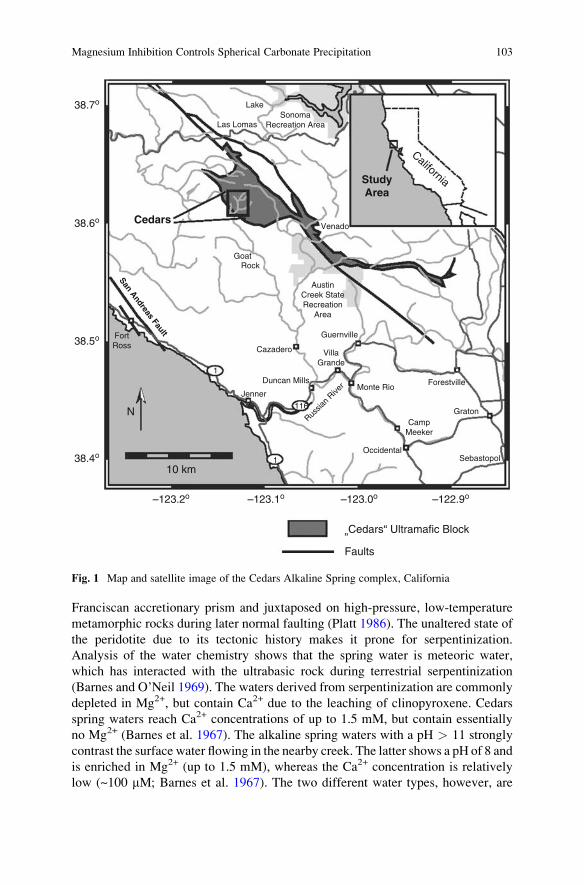
Franciscan accretionary prism and juxtaposed on high-pressure, low-temperature
metamorphic rocks during later normal faulting (Platt 1986). The unaltered state of
the peridotite due to its tectonic history makes it prone for serpentinization.
Analysis of the water chemistry shows that the spring water is meteoric water,
which has interacted with the ultrabasic rock during terrestrial serpentinization
(Barnes and O’Neil 1969). The waters derived from serpentinization are commonly
depleted in Mg2+, but contain Ca2+ due to the leaching of clinopyroxene. Cedars
spring waters reach Ca2+ concentrations of up to 1.5 mM, but contain essentially
no Mg2+ (Barnes et al. 1967). The alkaline spring waters with a pH > 11 strongly
contrast the surface water flowing in the nearby creek. The latter shows a pH of 8 and
is enriched in Mg2+ (up to 1.5 mM), whereas the Ca2+ concentration is relatively
low (~100 mM; Barnes et al. 1967). The two different water types, however, are
38.7o Lake
Las LomasSonoma
Recreation Area
Goat
San Andreas Fault
Cedars
StudyArea
Rock
Cazadero
Duncan Mills
Jenner
1
1
116
Monte Rio
OccidentalSebastopol
Guernville
VillaGrande
Forestville
GratonRus
sian
River
CampMeeker
Venado
California
AustinCreek StateRecreation
Area
38.6o
38.5o FortRoss
N
10 km38.4o
–123.2o –123.1o –123.0o
”Cedars“ Ultramafic Block
Faults
–122.9o
Fig. 1 Map and satellite image of the Cedars Alkaline Spring complex, California
Magnesium Inhibition Controls Spherical Carbonate Precipitation 103
commonlymixed, since the pools are located nearby or within the creek. Preliminary
water analysis shows the presence of H2 and H2S (cf. Sleep et al. 2004), which also
commonly results from the serpentinization reaction. The water chemistry is vari-
able between different springs and there is some site to site variation of carbonate
precipitation.
3 Methods
3.1 Petrography and Isotopes
Ten carbonate samples were collected at various alkaline springs of the Cedars
Ultrabasic Complex in July 2006. Small pieces were scraped off the sides of the
sinter terraces. More friable carbonate from the inside of the different pools could
be easily collected with the sample tubes. The tubes were filled with water of the
pools and the samples were stored refrigerated until microscopy. Microphotographs
of the fresh sample material were taken within one week after sampling. No change
in shape and amount of precipitates of the samples has been observed since
collection. Large specimens were taken from the laminated riverbed cement and
from fragments of older terrace walls, and slabs were cut by a rock saw for the
preparation of thin sections. Brittle material from the field site was gently broken,
mounted on a glass slide in the wet state and viewed under a petrographic micro-
scope with normal and cross-polarized light. Precipitates from both solid and liquid
media were embedded in Piccolyte resin (Ward’s Natural Science, 5100 West
Henrietta Rd., Rochester, NY, 14692, USA) on a microscope slide and subse-
quently inspected under the petrographic microscope. Samples for X-ray diffraction
(XRD) were suspended in ethanol, finely ground in an agar mortar and pestle and
mounted on the frosted side of a glass slide. The samples were continuously
scanned from 5 to 65� using Cu Ka radiation on a Rigaku Geigerflex D/max-A
XRD at the Earth Science Department, University of Southern California.
Carbon and oxygen isotopes in powdered bulk and micro-drilled sub-samples
were analysed with a Finnigan MAT 251 mass spectrometer coupled to an auto-
mated acidification device (type “Kiel”) at the Geological Institute, University of
Bremen. The analytical precision of the mass spectrometer is +0.05‰ for d13C, and+0.07‰ for d18O. Powdered Solnhofner Plattenkalk calibrated against the NBS
standard was used as a working standard, and the d13C and d18O values of the
carbonates are reported relative to the Vienna Pedee Belemnite Standard (VPDB).
3.2 Precipitation Experiments
Precipitation experiments were performed in solutions with 10 mM CaCl2, and
10 mM CaCl2 + 50 mM MgCl2, respectively (Table 1), which is the concentration
104 P. Meister et al.
of these ions in modern seawater. De-ionized water from aMillipore filter apparatus
was used for the experiments. The pH was adjusted to 8.5 by adding NaOH.
NaHCO3 was prepared as a 100� stock solution and added to reach a final
concentration of 10 mM. The same amount of NaHCO3 was added to pure spring
water. Different experiments were performed, where 0.01 g/l yeast extract, 28 mM
SO42�, 10 ml/l amino acid solution, 50 mM Na-lactate, and different salt concen-
trations were added, which may influence the precipitation kinetics and, hence,
mineralogy and crystal structure.
In order to test for biological effects on mineralogy and crystal structure,
precipitation experiments were performed with aerobic heterotrophic bacteria of
the species Shewanella. Liquid and solid minimal media for Shewanella were used
[Table 1; modified after Myers and Nealson (1988)], supplemented with the same
constituents as in the abiotic experiments. Strains of Shewanella were chosen,
because they commonly occur in natural aquatic environments and can also be
easily grown under aerobic conditions. No bicarbonate was added to the cultures
since alkalinity was produced bacterially when acetate was added to the medium.
Both cultures and controls were incubated at 30�C from 24 h to several days. The
pH was monitored using a pH electrode for the liquid media, and pH indicator paper
for the colonies formed on the agar plates. Crystal growth was monitored under a
dissecting microscope, and crystals were further analysed by microscopy and XRD.
Table 1 Composition of the
solutions and media used for
precipitation experiments
Control solution Medium
For carbonate supersaturationMgCl2 50.00 50.00
CaCl2 10.00 10.00
NaHCO3 10.00 10.00
pH 8.50 8.50
Mineral saltsNaCl – 30.00
NH4Cl – 28.40
KCl – 1.34
Yeast extract (PO43� source) (g/l) – 0.10
NutrientsTrace elementsa (100� stock) – 10 ml
Vitamine mixb (100� stock) – 10 ml
Na-acetate – 10.00
For solid mediaAgar, noble (g/l) – 10.00
SupplementsSO4
2� 28.00 28.00
Yeast extract (g/l)
Microbial cells
NaCl 30.00
NH4Cl 28.40
KCl 1.34aWolfe mineral solution (Wolin et al. 1963)bMyers and Nealson (1988)
Magnesium Inhibition Controls Spherical Carbonate Precipitation 105
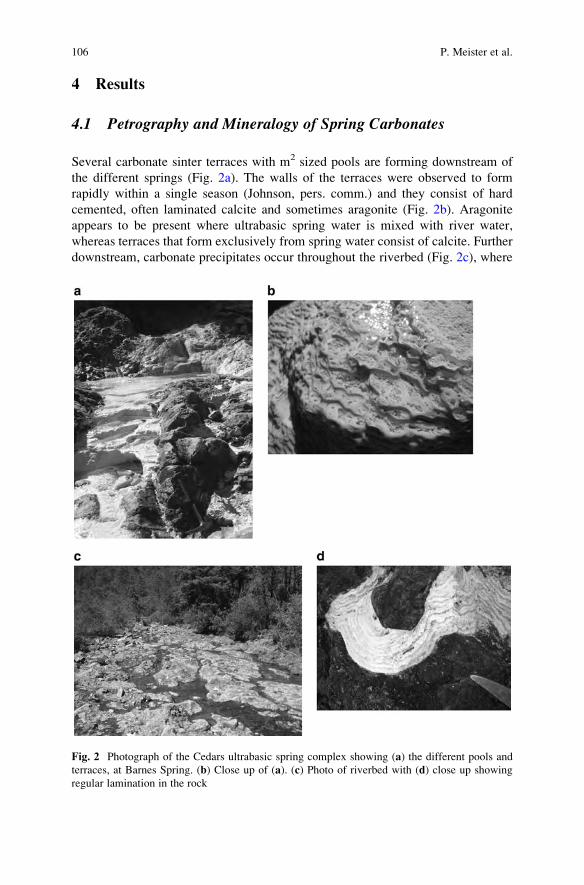
4 Results
4.1 Petrography and Mineralogy of Spring Carbonates
Several carbonate sinter terraces with m2 sized pools are forming downstream of
the different springs (Fig. 2a). The walls of the terraces were observed to form
rapidly within a single season (Johnson, pers. comm.) and they consist of hard
cemented, often laminated calcite and sometimes aragonite (Fig. 2b). Aragonite
appears to be present where ultrabasic spring water is mixed with river water,
whereas terraces that form exclusively from spring water consist of calcite. Further
downstream, carbonate precipitates occur throughout the riverbed (Fig. 2c), where
Fig. 2 Photograph of the Cedars ultrabasic spring complex showing (a) the different pools and
terraces, at Barnes Spring. (b) Close up of (a). (c) Photo of riverbed with (d) close up showing
regular lamination in the rock
106 P. Meister et al.
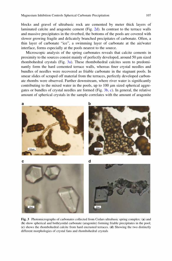
blocks and gravel of ultrabasic rock are cemented by meter thick layers of
laminated calcite and aragonite cement (Fig. 2d). In contrast to the terrace walls
and massive precipitates in the riverbed, the bottoms of the pools are covered with
slower growing fragile and delicately branched precipitates of carbonate. Often, a
thin layer of carbonate “ice”, a swimming layer of carbonate at the air/water
interface, forms especially at the pools nearest to the source.
Microscopic analysis of the spring carbonates reveals that calcite cements in
proximity to the sources consist mainly of perfectly developed, around 50 mm sized
rhombohedral crystals (Fig. 3a). These rhombohedral calcites seem to predomi-
nantly form the hard cemented terrace walls, whereas finer crystal needles and
bundles of needles were recovered as friable carbonate in the stagnant pools. In
smear slides of scraped off material from the terraces, perfectly developed carbon-
ate rhombs were observed. Further downstream, where river water is significantly
contributing to the mixed water in the pools, up to 100 mm sized spherical aggre-
gates or bundles of crystal needles are formed (Fig. 3b, c). In general, the relative
amount of spherical crystals in the sample correlates with the amount of aragonite
Fig. 3 Photomicrographs of carbonates collected from Cedars ultrabasic spring complex: (a) and
(b) show spherical and bothryoidal carbonate (aragonite) forming friable precipitates in the pool;
(c) shows the rhombohedral calcite from hard encrusted terraces. (d) Showing the two distinctly
different morphologies of crystal fans and rhombohedral crystals
Magnesium Inhibition Controls Spherical Carbonate Precipitation 107
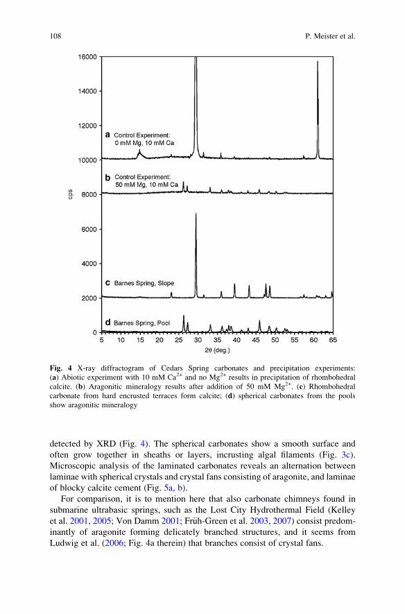
detected by XRD (Fig. 4). The spherical carbonates show a smooth surface and
often grow together in sheaths or layers, incrusting algal filaments (Fig. 3c).
Microscopic analysis of the laminated carbonates reveals an alternation between
laminae with spherical crystals and crystal fans consisting of aragonite, and laminae
of blocky calcite cement (Fig. 5a, b).
For comparison, it is to mention here that also carbonate chimneys found in
submarine ultrabasic springs, such as the Lost City Hydrothermal Field (Kelley
et al. 2001, 2005; Von Damm 2001; Fr€uh-Green et al. 2003, 2007) consist predom-
inantly of aragonite forming delicately branched structures, and it seems from
Ludwig et al. (2006; Fig. 4a therein) that branches consist of crystal fans.
Fig. 4 X-ray diffractogram of Cedars Spring carbonates and precipitation experiments:
(a) Abiotic experiment with 10 mM Ca2+ and no Mg2+ results in precipitation of rhombohedral
calcite. (b) Aragonitic mineralogy results after addition of 50 mM Mg2+. (c) Rhombohedral
carbonate from hard encrusted terraces form calcite; (d) spherical carbonates from the pools
show aragonitic mineralogy
108 P. Meister et al.
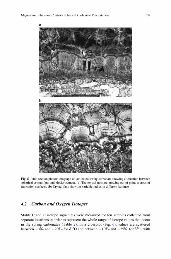
4.2 Carbon and Oxygen Isotopes
Stable C and O isotope signatures were measured for ten samples collected from
separate locations in order to represent the whole range of isotope values that occur
in the spring carbonates (Table 2). In a crossplot (Fig. 6), values are scattered
between �3‰ and �20‰ for d18O and between �10‰ and �25‰ for d13C with
a
b
500 mm
200 mm
Fig. 5 Thin section photomicrograph of laminated spring carbonate showing alternation between
spherical crystal fans and blocky cement. (a) The crystal fans are growing out of point sources of
truncation surfaces. (b) Crystal fans showing variable radius in different laminae
Magnesium Inhibition Controls Spherical Carbonate Precipitation 109
all values aligned between these two end-member concentrations. The values agree
well with the scatter observed in an independent study (P. Schiffman, pers. comm.),
supporting that the selection of samples represents the range of values present in the
spring carbonates. However, if the data are plotted versus the relative amount of
aragonite in the samples, no clear trend is observed. If the distribution of d18O and
d13C values at the field sites is considered, the major trend indicates progressively
less negative values downstream from the sources. A minor trend is that carbonates
Table 2 Mineralogy, d13C and d18O values of Cedars Springs carbonates
Site Sample Aragonite
(% of carbonate)
d13C(% VPDB)
d18O(% VPDB)
NS06 Hand specimen, white lamina 31.65 �12.03 �4.16
NS06 Hand specimen, beige lamina 3.27 �12.92 �5.11
NS06 Hand specimen, white hard lamina 14.91 �16.21 �8.72
NS06 Hand specimen, beige, fine lamina 0.00 �20.21 �14.11
NS06-4 Pool 100.00 �14.18 �3.47
BS06-1 Pool 17.01 �23.96 �18.24
BS06-1/2 Slide 0.00 �25.03 �17.59
BS06-2 Terrace �17.52 �10.30
BS06-2 Pool 100.00 �12.29 �4.02
BS06-7 Crust �24.15 �17.62
BS06-8 Pool �23.84 �15.60
–20 –15 –10 –5
–5
–30
–25
–20
–15
–10
00
d18O
d13C
P. Schiffman (pers. comm.) new data
Fig. 6 Cross-plot of isotopic signature from aragonite (open diamonds) and calcite (closeddiamonds), and superimposed data from P. Schiffman (pers. comm.; crosses). The data align
between d18O ¼ �4‰, d13C ¼ �10‰ and d18O ¼ �20‰, d13C ¼ �25‰, with reflects more or
less the distance from the source. Now direct correlation with the mineralogy was observed
110 P. Meister et al.
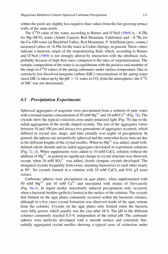
within the pools are slightly less negative than values from the fast growing terrace
walls of the same pools.
The d18O value of the water, according to Barnes and O’Neil (1969) is �8.2‰for Mg–HCO3 water (Adobe Canyon, Red Mountain, California) and �8.7‰ for
the Ca–OH water (at Blackbird Valley, Red Mountain). P. Schiffman (pers. comm.)
measured values of ~6.5‰ for the water at Cedars Springs, in general. These values
indicate a meteoric origin of the serpentinizing fluid, which, according to Barnes
and O’Neil (1969) is not strongly altered by interaction with the ultrabasic rock,
probably because of high flow rates compared to the rates of serpentinization. The
isotopic composition of the water is in equilibrium with the positive end-member of
the range in d18O values of the spring carbonates under surface temperature. Due to
extremely low dissolved inorganic carbon (DIC) concentrations of the spring water
(most DIC is taken up by the pH > 11 water as CO2 from the atmosphere), the d13Cof DIC was not determined.
4.3 Precipitation Experiments
Spherical aggregates of aragonite were precipitated from a solution of pure water
with a normal marine concentration of 50 mMMg2+ and 10 mM Ca2+ (Fig. 7a). The
crystals show the typical extinction cross under polarized light (Fig. 7b) due to the
radial aggregation of the needle shaped crystals. The size of the aggregates varies
between 10 and 100 mm and always two generations of aggregates occurred, which
differed in crystal size, shape, and time (usually over night) of precipitation. In
general, the spheres are not perfectly spherical and the somewhat fuzzy rims are due
to the different lengths of the crystal needles. When noMg2+ was added, small well-
defined calcite rhombs and no radial aggregates developed in experiment solutions
(Fig. 7c, d). When supplements were added to 10 mM CaCl2 solution without the
addition of Mg2+, in general no significant change in crystal structure was observed,
except, when 28 mM SO42� was added, clearly elongate crystals developed. The
elongated crystals frequently form twins, orienting themselves to each other nearly
at 90�. No crystals formed in a solution with 10 mM CaCl2 and 0.01 g/l yeast
extract.
Carbonate spheres were precipitated on agar plates, when supplemented with
50 mM Mg2+ and 10 mM Ca2+ and inoculated with strains of Shewanella(Fig. 8a–f). In liquid media, microbially induced precipitation only occurred,
when a bacterial biofilm (pellicle) formed at the surface of the solution. The crystals
that formed on the agar plates commonly occurred within the bacterial colonies,
although in a few cases crystal formation was observed inside of the agar, remote
from the colonies. Crystals on the agar plates only formed when the bacteria
were fully grown, which usually was the case after 48 h. The pH in the different
colonies commonly reached 8.5–9, independent of the initial pH. The carbonate
spheres were perfectly developed with a smooth surface and extremely fine,
radially aggregated crystal needles showing a typical cross of extinction under
Magnesium Inhibition Controls Spherical Carbonate Precipitation 111
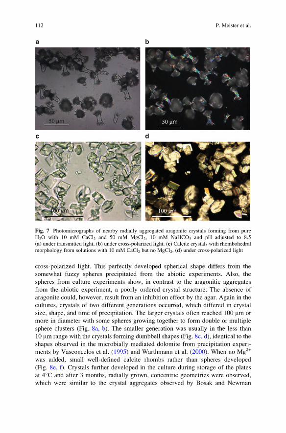
cross-polarized light. This perfectly developed spherical shape differs from the
somewhat fuzzy spheres precipitated from the abiotic experiments. Also, the
spheres from culture experiments show, in contrast to the aragonitic aggregates
from the abiotic experiment, a poorly ordered crystal structure. The absence of
aragonite could, however, result from an inhibition effect by the agar. Again in the
cultures, crystals of two different generations occurred, which differed in crystal
size, shape, and time of precipitation. The larger crystals often reached 100 mm or
more in diameter with some spheres growing together to form double or multiple
sphere clusters (Fig. 8a, b). The smaller generation was usually in the less than
10 mm range with the crystals forming dumbbell shapes (Fig. 8c, d), identical to the
shapes observed in the microbially mediated dolomite from precipitation experi-
ments by Vasconcelos et al. (1995) and Warthmann et al. (2000). When no Mg2+
was added, small well-defined calcite rhombs rather than spheres developed
(Fig. 8e, f). Crystals further developed in the culture during storage of the plates
at 4�C and after 3 months, radially grown, concentric geometries were observed,
which were similar to the crystal aggregates observed by Bosak and Newman
a b
c d
100 µm
50 µm 50 µm
100 µm
Fig. 7 Photomicrographs of nearby radially aggregated aragonite crystals forming from pure
H2O with 10 mM CaCl2 and 50 mM MgCl2, 10 mM NaHCO3 and pH adjusted to 8.5
(a) under transmitted light, (b) under cross-polarized light. (c) Calcite crystals with rhombohedral
morphology from solutions with 10 mM CaCl2 but no MgCl2, (d) under cross-polarized light
112 P. Meister et al.
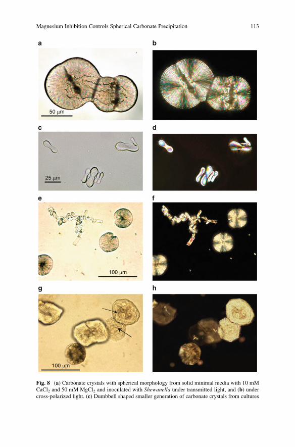
50 µm
25 µm
100 µm
100 µm
a b
c d
e f
g h
Fig. 8 (a) Carbonate crystals with spherical morphology from solid minimal media with 10 mM
CaCl2 and 50 mM MgCl2 and inoculated with Shewanella under transmitted light, and (b) under
cross-polarized light. (c) Dumbbell shaped smaller generation of carbonate crystals from cultures
Magnesium Inhibition Controls Spherical Carbonate Precipitation 113
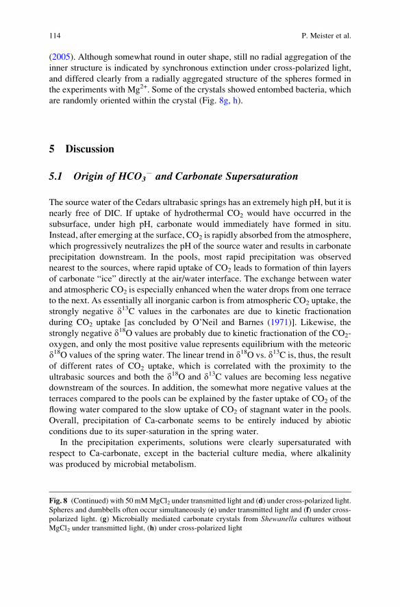
(2005). Although somewhat round in outer shape, still no radial aggregation of the
inner structure is indicated by synchronous extinction under cross-polarized light,
and differed clearly from a radially aggregated structure of the spheres formed in
the experiments with Mg2+. Some of the crystals showed entombed bacteria, which
are randomly oriented within the crystal (Fig. 8g, h).
5 Discussion
5.1 Origin of HCO3� and Carbonate Supersaturation
The source water of the Cedars ultrabasic springs has an extremely high pH, but it is
nearly free of DIC. If uptake of hydrothermal CO2 would have occurred in the
subsurface, under high pH, carbonate would immediately have formed in situ.
Instead, after emerging at the surface, CO2 is rapidly absorbed from the atmosphere,
which progressively neutralizes the pH of the source water and results in carbonate
precipitation downstream. In the pools, most rapid precipitation was observed
nearest to the sources, where rapid uptake of CO2 leads to formation of thin layers
of carbonate “ice” directly at the air/water interface. The exchange between water
and atmospheric CO2 is especially enhanced when the water drops from one terrace
to the next. As essentially all inorganic carbon is from atmospheric CO2 uptake, the
strongly negative d13C values in the carbonates are due to kinetic fractionation
during CO2 uptake [as concluded by O’Neil and Barnes (1971)]. Likewise, the
strongly negative d18O values are probably due to kinetic fractionation of the CO2-
oxygen, and only the most positive value represents equilibrium with the meteoric
d18O values of the spring water. The linear trend in d18O vs. d13C is, thus, the result
of different rates of CO2 uptake, which is correlated with the proximity to the
ultrabasic sources and both the d18O and d13C values are becoming less negative
downstream of the sources. In addition, the somewhat more negative values at the
terraces compared to the pools can be explained by the faster uptake of CO2 of the
flowing water compared to the slow uptake of CO2 of stagnant water in the pools.
Overall, precipitation of Ca-carbonate seems to be entirely induced by abiotic
conditions due to its super-saturation in the spring water.
In the precipitation experiments, solutions were clearly supersaturated with
respect to Ca-carbonate, except in the bacterial culture media, where alkalinity
was produced by microbial metabolism.
Fig. 8 (Continued) with 50 mMMgCl2 under transmitted light and (d) under cross-polarized light.
Spheres and dumbbells often occur simultaneously (e) under transmitted light and (f) under cross-
polarized light. (g) Microbially mediated carbonate crystals from Shewanella cultures without
MgCl2 under transmitted light, (h) under cross-polarized light
114 P. Meister et al.
5.2 Kinetic Effects on Crystal Structure
Despite super-saturation, often minerals do not precipitate because their forma-
tion is kinetically inhibited. Aragonite is known to form in marine environments
when calcite precipitation is inhibited due to high Mg/Ca ratios (Berner 1975).
Also in our precipitation experiments, the addition of Mg2+ led to the formation
of aragonite. As shown in the abiotic control experiments, the inhibition of crystal
growth by Mg2+ seems to be expressed in the spherical crystal shape of the
carbonate crystals. According to Kralj et al. (2004) and Arvidson et al. (2005),
elongate (needle shaped) crystal growth is due to differential inhibition of growth
in different directions and it seems that spherical growth is the logical conse-
quence if needle shaped crystals are free to grow in all directions out of a
nucleation point. Similarly, SO42�, which was suggested as a kinetic inhibitor
for dolomite (Mg, Ca(CO3)2) formation at high temperatures (Baker and Kastner
1981), was shown to cause elongated crystal growth, but not as efficiently as
Mg2+. Other additives had no significant effect on crystal structure except yeast
extract (used for the culture experiments), which completely inhibited precipita-
tion from the solutions. Yeast extract contains large amounts of carbohydrate
polymers, which may inhibit carbonate precipitation. These observations suggest
that spherical calcite growth in precipitation experiments with different carbohy-
drates (C€olfen and Qi, 2001; Braissant et al. 2003) is similarly caused by an
inhibitory effect.
Considering the carbonate precipitates at Cedars Springs, we suggest that the
formation of spherical aragonite crystals is due to significant mixing of the spring
water with Mg-rich river water. Mg–HCO3� water originates from surface
weathering of the ultrabasic rock, whereas Ca–OH� water from the ultrabasic
springs have low Mg2+ concentrations. Thus, the distribution of spherical aragonite
and rhombohedral calcite in the Cedars ultrabasic spring complex should reflect the
chemistry of the water at the different sites of precipitation. Indeed, carbonates in
pools, where spring water is frequently mixed with surface water from the nearby
river, as it is the case at Nipple Spring, aragonite tends to be more abundant.
Moreover, aragonite generally forms in the pools as delicate friable and slow
growing carbonate whereas the fast growing, massively incrusted terraces mainly
consist of calcite, which is reflected by slightly more negative d13C and d18Oisotope values in the terrace. However, because the pH and Mg2+ concentration
of the two mixing waters are not necessarily coupled – e.g. pH and Mg2+ concen-
tration can both be high when surface water is mixed with fresh source water – the
isotopic composition may not show direct correlation with the aragonite/calcite
ratio of the carbonate. The isotope composition rather seems to be dependent of the
distance from the springs downstream, while morphology depends on the relative
amounts in which the two water types are mixing. Alternating cement structures in
laminated carbonates are also likely controlled by mixing of the two water types
downstream. In addition, temperature may influence the effect of Mg2+ on crystal
morphology (Morse et al. 1997).
Magnesium Inhibition Controls Spherical Carbonate Precipitation 115
5.3 Biological Effects on Crystal Structure
The aragonite spherules found in the Cedars spring carbonates commonly are
aggregated to form sheaths or layers, and often incrust algal filaments. According
to the model of Braissant et al. (2003), spherical growth is induced in the presence
of carbohydrates and amino acids, as an analogue for the effects of extracellular
polymeric substances (EPS) in microbial biofilms. At the Cedars, the precipitation
of spherical crystals may also be microbially mediated, and algal filaments or EPS
layers may act as a substrate for crystal growth. Nevertheless, the concentration of
organic compounds in the Cedars Spring waters is in all probability too low to be
mainly responsible for spherical crystal structure. In the presented experiments,
we show that bacterially mediated carbonates from Mg-free solutions show only
minor differences to the abiotic controls. The outer shapes are somewhat more
rounded, but the interior shows an uniform crystallographic orientation, similar to
calcite crystals reported from cultures with sulphate-reducing bacteria (Bosak and
Newman 2005). Meldrum and Hyde (2001) showed that organic matter contri-
butes to the spherical crystal structure, and Gonzales-Munoz et al. (2000) sug-
gested that a microbial factor is involved besides the effect of Mg2+. In our
experiments, spherical crystals were only formed in the presence of Mg2+, but
are more smoothly and regularly developed in the presence of bacteria than in
the abiotic control. Over all, a biological mediation effect on crystal structure at
the Cedars spring carbonates may not be completely excluded. However, this
effect would be minor, and not the primary reason for the formation of spherical
aragonite.
In nature, spherical crystal structures are commonly ascribed to microbially
mediated precipitation, maybe because the round shapes are reminiscent of some-
thing “living” (e.g. Gerdes et al. 1994; Vasconcelos et al. 1995; Verrecchia et al.
1995; Folk 1999; Van Lith et al. 2003; Rivadeneyra et al. 2006; Sanchez-Roman
et al. 2008).
In fact, round crystal shapes can be purely crystallographic features due to
kinetic inhibition, rather than biologic induction of crystal growth. The spherical
structure of many bio-minerals may thus not be the direct consequence of biological
“mediation”, but bio-mediation may become relevant, when precipitation is other-
wise kinetically inhibited. This effect may play an important role and may even be
precisely regulated within living tissues and biofilms through the concentration
levels of Mg2+ or other (organic?) inhibitors. For the precipitation of spring
carbonates at the Cedars, a biological factor may then play a role, when the pH is
moderate and the precipitation of calcite is partially inhibited through the presence
of Mg2+ from mixing with adjacent river water. Under these (less alkaline) condi-
tions, algal filaments can grow and may serve as a substrate for crystal growth.
The absorption of Ca2+ by EPS, an effect discussed by Braissant et al. (2007)
may locally increase the Mg/Ca ratio and indirectly affect the precipitation kinetics,
in particular, during times of low marine Mg2+ concentrations. Nevertheless,
spherical aragonite formation in Cedars Springs and precipitation experiments do
116 P. Meister et al.
not support the EPS model for spherical carbonate formation under natural, in
particular marine, conditions and Mg-inhibition may impose a much larger effect
on crystal growth than EPS.
5.4 Spherical Carbonates Through Earth’s History
Varying contribution of Mg-rich water from surface weathering vs. Ca-rich hydro-
thermal fluid from serpentinization, as observed at a small scale at the Cedars
alkaline springs, can be observed on a global scale in the secular variation of the
Mg/Ca ratio of seawater during times of fast and slow seafloor spreading. Variation
in Mg-content led to periods with predominantly aragonite or calcite precipitation
(aragonite seas and calcite seas: Given and Wilkinson 1985; Hardie 1996, 2003;
Stanley and Hardie 1998; Burns et al. 2000). The Mg content also affected ooid
morphology – aragonitic ooids with concentric structure were predominantly
formed in aragonite seas, calcitic radial ooids formed in calcite seas (Sandberg
1983; Wilkinson et al. 1985; Wilkinson and Given 1986).
Aragonite ooids show micritic and concentric fibrous lamination at micrometer
scale, whereby the crystallites are randomly oriented. In this case, the round shape
may not be a crystallographic feature and a microbial factor was considered
for their formation (e.g. Folk and Lynch 2001). Micritic ooids were similarly
described from freshwater Lake Geneva (Plee et al. 2008), from a system where
carbonate is barely supersaturated. Micritic ooids, thus, may be the result of
microbial metabolism, locally increasing supersaturation, rather than a kinetic
effect. Indeed, micritic ooids formed typically in aragonite seas, in which carbon-
ate saturation was probably low (as a result of slow spreading rates producing less
CO2 in the atmosphere and less continental weathering during lower tectonic
activity). In contrast, calcitic ooids formed in calcite seas at the same time with
increased carbonate supersaturation (Riding and Liang 2005). These radial cal-
citic ooids are more comparable with the spherical crystals at Cedars Springs.
The interplay of Mg/Ca ratio and carbonate supersaturation provides, thus, a
mechanism that can explain the varying type of ooid formation as the result of
thermodynamic vs. kinetic limitation.
High seawater carbonate supersaturation particularly occurred during several
times in the Precambrian, so that carbonate precipitation would have been kinetically
controlled. Indeed, some Precambrian precipitates, often considered as stromatolites,
show round structures and crystal fans (Grotzinger and Knoll 1999), analogous to
crystal fans observed in the laminated cements from the Cedars spring carbonates.
These crystal fans may have been formed completely abiotically and their shape can
be explained by Mg2+ inhibition. Also during the Precambrian, calcite and aragonite
ooids are reported (Tucker 1984; Ries et al. 2008), while secular variation in Mg/Ca
ratio was reconstructed (Hardie 2003). Still more research is required to understand
the biotic and abiotic kinetic controls of carbonate precipitation, as well as better
Magnesium Inhibition Controls Spherical Carbonate Precipitation 117
constraints on chemistry of past oceans, in order to interpret the processes that govern
carbonate precipitation throughout Earth’s history.
6 Conclusions
Carbonate precipitates abiotically due to high carbonate supersaturation from
highly alkaline groundwater at Cedars Springs, whereby CO2 is absorbed from
the atmosphere by the spring water. Alternating precipitation of rhombohedral
calcite and spherical aragonite crystals is most likely controlled by variable
Mg/Ca ratios due to mixing of ultrabasic Ca–OH dominated spring water with
Mg–HCO3� – rich river water derived from surface weathering. Precipitation
experiments show that spherical crystal structure is mainly a kinetic effect due to
Mg2+ inhibition, but the presence of microbial biofilms may contribute to a finer
surface structure of the carbonate spherules. These observations suggest that,
particularly in the marine environment, spherical carbonates may not result from
interaction with microbial EPS, but simply due to an inhibiting effect by the Mg2+.
The frequently observed association of spherical crystals with microbial biofilms
can be explained by microbial mediation that becomes the controlling factor of
carbonate precipitation only when precipitation is otherwise inhibited. Accord-
ingly, spherical crystallographic features, such as crystal fans and radial ooids
became then prominent in Earth’s history, when precipitation was kinetically
limited. In contrast, micritic ooids formed when precipitation was thermodynami-
cally limited. Secular variation in the Mg/Ca ratio and carbonate supersaturation
may thus explain the distribution of crystal fans and spherical carbonate crystal
structure through Earth’s history.
Acknowledgements We thank Monika Segl (University of Bremen) and Gaute Lavik (MPI,
Bremen) for isotope analysis, as well as Peter Schiffman (UC Davis) for allowing us to present
his unpublished isotope data. Gail Arnold (MPI, Bremen) and Carolina Reyes (UC Santa Cruz)
provided useful comments and improved the clarity of the manuscript. Discussions with Gijs
Kuenen, Ana Obraztsova, John Platt, and Rolf Warthmann contributed significantly to the study.
We thank Damon Rogers for giving us the opportunity to visit the Cedars Springs and for allowing
us to use his camping facility. This study was sponsored by the Agouron Institute and the Swiss
National Science Foundation (SNF).
References
Arvidson RS, Collier M, Davis KJ, Vinson MD, Amonette JE, L€uttge A (2005) Magnesium
inhibition of calcite dissolution kinetics. Geochimica et Cosmochimica Acta 70:583–594
Baker P, Kastner M (1981) Constraints on the formation of sedimentary dolomite. Science
213:214–216
Barnes I, O’Neil JR (1969) The relationship between fluids in some fresh alpine-type ultramafics
and possible modern serpentinization, Western United States. The Geological Society of
America Bulletin 80:1947–1960
118 P. Meister et al.
Barnes I, LaMarche VC, Himmelberg GR (1967) Geochemical evidence of present-day serpenti-
nization. Science 56:830–832
Bathurst RGC (1967) Oolitic films on low energy carbonate sand grains, Bimini Lagoon,
Bahamas. Marine Geology 5:89–109
Berner RA (1975) The role of magnesium in the crystal growth of calcite and aragonite from sea
water. Geochimica et Cosmochimica Acta 39:489–504
Bontognali TRR, Vasconcelos C, Warthmann RJ, Dupraz C, Bernasconi SM, McKenzie JA (2008)
Microbes produce nanobacteria-like structures, avoiding cell entombment. Geology 36:663-666
Bosak T, Newman DK (2005) Microbial kinetic controls on calcite morphology in supersaturated
solutions. Journal of Sedimentary Research 75:190–199
Braissant O, Cailleau G, Dupraz C, Verrecchia EP (2003) Bacterially induced mineralization of
calcium carbonate in terrestrial environments: the role of exopolysaccharides and amino acids.
Journal of Sedimentary Research 73:485–490
Braissant O, Decho AW, Dupraz C, Glunk C, Przekop KM, Visscher PT (2007) Exopolymeric
substances of sulfate-reducing bacteria: interactions with calcium at alkaline pH and implica-
tion for formation of carbonate minerals. Geobiology 5:401–411
Burns SJ, McKenzie JA, Vasconcelos C (2000) Dolomite formation and biogeochemical cycles in
the Phanerozoic. Sedimentology 47:49–61
C€olfen H, Qi L (2001) A systematic examination of the morphogenesis of calcium carbonate in the
presence of a double-hydrophilic block copolymer. Chemistry – A European Journal
1:106–116
Davies PJ, Bubela B, Ferguson J (1978) The formation of ooids. Sedimentology 25:703–729
Donahue J (1969) Genesis of oolite and pisolite grains: an energy index. Journal of Sedimentary
Petrology 39:1399–1411
Dupraz C, Visscher PT, Baumgartner LK, Reid RP (2004) Microbe–mineral interactions: early
carbonate precipitation in a hypersaline lake (Eleuthera Island, Bahamas). Sedimentology
51:745–765
Folk RL (1999) Nannobacteria and the precipitation of carbonate in unusual environments.
Sedimentary Geology 126:47–55
Folk RL, Lynch FL (2001) Organic matter, putative nannobacteria and the formation of ooids and
hardgrounds. Sedimentology 48:215–299
Fr€uh-Green GL, Kelley DS, Bernasconi SM, Karson JA, Ludwig KA, Butterfield DA, Boschi C,
Proskurowski G (2003) 30,000 years of hydrothermal activity at the Lost City Vent Field.
Science 301:495–498
Fr€uh-Green GL, Delacour A, Boschi C, Bernasconi SM, Butterfield DA, Kelley DS, Proskurowski
G (2007) Building Lost City: serpentinization, mass transfer and life in a peridotite-hosted
hydrothermal system. Geochimica et Cosmochimica Acta 71/15(Suppl. 1):A298
Gerdes G, Dunajtschik-Piewak K, Riege H, Taher AG, Krumbein WE, Reineck H-E (1994)
Structural diversity of biogenic carbonate particles in microbial mats. Sedimentology
41:1273–1294
Given RK, Wilkinson BH (1985) Kinetic control of morphology, composition, and mineralogy of
abiotic sedimentary carbonates. Journal of Sedimentary Petrology 55:109–119
Gonzales-Munoz MT, Ben Chekroun K, Ben Aboud A, Arias J, Rodriguez-Gallego M (2000)
Bacterially induced Mg-calcite formation: role of Mg2+ in development of crystal morphology.
Journal of Sedimentary Research 70:559–564
Grotzinger JP, Knoll AH (1999) Stromatolites in Precambrian carbonates: evolutionary mileposts
or environmental dipsticks? Annual Review of Earth and Planetary Sciences 27:313–358
Hardie LA (1996) Secular variation in seawater chemistry: an explanation for the coupled secular
variation in the mineralogies of marine limestones and potash evaporates over the past 600 m.y.
Geology 24:279–283
Hardie LA (2003) Secular variation in Precambrian seawater chemistry and the timing of Precam-
brian aragonite seas and calcite seas. Geology 31:785–788
Magnesium Inhibition Controls Spherical Carbonate Precipitation 119
Kelley DS, Karson JA, Blackman DK, Fr€uh-Green GL, Butterfield DA, Lilley MD, Olson EJ,
Schrenk MO, Roe KK, Lebon GT, Rivizzigno P, AT3-60 Shipboard Party (2001) An off-axis
hydrothermal vent field near the Mid-Atlantic Ridge at 30�N. Nature 412:145–149Kelley DS, Karson JA, Fr€uh-Green GL, Yoerger DR, Shank TM, Butterfield DA, Hayes JM,
Schrenk MO, Olson EJ, Proskurowski G, Jakuba M, Bradley A, Larson B, Ludwig K, Glickson
D, Buckman K, Bradley AS, Brazelton WJ, Roe K, Elend MJ, Delacour A, Bernasconi SM,
Lilley MD, Baross JA, Summons RE, Sylva SP (2005) A serpentinite-hosted ecosystem: the
Lost City hydrothermal field. Science 307:1428–1434
Kralj D, Kontrec J, Brecevic L, Falini G, N€othig-Laslo V (2004) Effect of inorganic anions on the
morphology and structure of magnesium calcite. Chemistry – A European Journal
10:1647–1656
Ludwig KA, Kelley DS, Butterfield DA, Nelson BK, Fr€uh-Green G (2006) Formation and
evolution of carbonate chimneys at the Lost City Hydrothermal Field. Geochimica et Cosmo-
chimica Acta 70:3625–3645
Meldrum FC, Hyde ST (2001) Morphological influence of magnesium and organic additives on
the precipitation of calcite. Journal of Crystal Growth 231:544–558
Mitterer RM (1972) Biogeochemistry of aragonitic muds and oolites. Geochimica et Cosmochi-
mica Acta 36:1407–1422
Morse JW, Wang Q, Tsio MY (1997) Influences of temperature and Mg:Ca ratio on CaCO3
precipitates from seawater. Geology 25:85–87
Myers CR, Nealson KH (1988) Bacterial manganese reduction and growth with manganese oxide
as the sole electron acceptor. Science 240:1319–1321
O’Neil JR, Barnes I (1971) C13 and O18 compositions in some fresh-water carbonates associated
with ultramafic rocks and serpentinites: western United States. Geochimica et Cosmochimica
Acta 35: 687–697
Platt JP (1986) Dynamics of orogenic wedges and the uplift of high-pressure metamorphic rocks.
The Geological Society of America Bulletin 97:1037–1053
Plee K, Ariztegui D, Martini R, Davaud E (2008) Unravelling the microbial role in ooid formation-
results of an in situ experiment in modern freshwater Lake Geneva in Switzerland. Geobiology
DOI: 10.1111/j.1472-4669.2007.00140.x
Riding R, Liang L (2005) Geobiology of microbial carbonates: metazoans and seawater saturation
state influence on secular trends during the Phanerozoic. Palaegeography, Palaeoclimatology,
Palaeoecology 219:101–115
Ries JB, Anderson MA, Hill RT (2008) Seawater Mg/Ca controls polymorph mineralogy of
microbial CaCO3: a potential proxy for calcite-aragonite seas in Precambrian time. Geobiology
6:106–119
Rivadeneyra MA, Martın-Algarra A, Sanchez-Navas A, Martın-Ramos D (2006) Carbonate and
phosphate precipitation by Chromohalobactern marismortui. Geomicrobiology Journal
23:89–101
Sanchez-Roman M, Vasconcelos C, Schmid T, Dittrich M, McKenzie JA, Zenobi R, Rivadeneyra
MA (2008) Aerobic microbial dolomite at the nanometer scale: implications for the geologic
record. Geology 36:879–882
Sandberg PA (1983) An oscillating trend in Phanerozoic non-skeletal carbonate mineralogy.
Nature 305:19–22
Sleep NH, Meibom A, Fridriksson Th, Coleman RG, Bird DK (2004) H2-rich fluids from
serpentinization: geochemical and biotic implications. Proceedings of the National Academy
of Sciences of the United States of America 101:12818–12823
Stanley SM, Hardie LA (1998) Secular oscillations in the carbonate mineralogy if reef-building
and sediment-producing organisms driven by tectonically forced shifts in seawater chemistry.
Palaeogeography, Palaeoclimatology, Palaeoecology 144:3–9
Suess E, F€utterer D (1972) Aragonitic ooids: experimental precipitation from seawater in the
presence of humic acid. Sedimentology 19:129–139
Tucker ME (1984) Calcitic, aragonitic, and mixed calcitic-aragonitic ooids from the mid-
Proterozoic Belt Supergroup, Montana. Sedimentology 31:627–644
120 P. Meister et al.
Tucker ME, Wright VP (1990) Carbonate sedimentology. Blackwell Science, Oxford
Van Lith Y, Warthmann R, Vasconcelos C, McKenzie JA (2003) Microbial fossilization in
carbonate sediments: a result of the bacterial surface involvement in dolomite precipitation.
Sedimentology 50:237–245
Vasconcelos C, McKenzie JA, Bernasconi S, Grujic D, Tien AJ (1995) Microbial mediation as a
possible mechanism for natural dolomite formation at low temperatures. Nature 377:220–222.
Verrecchia E, Freytet P, Verrecchia KE, Dumont JL (1995) Spherulites in calcrete laminar crusts:
biogenic CaCO3 precipitation as a major contributor to crust formation. Journal of Sedimentary
Research A65:690–700
Von Damm KL (2001) Lost city found. Nature 412:127–128
Warthmann R, Van Lith Y, Vasconcelos C, McKenzie JA, Karpoff AM (2000) Bacterially induced
dolomite precipitation in anoxic culture experiments. Geology 28:1091–1094
Wilkinson BH, Given KR (1986) Secular variation in abiotic marine carbonates: constraints on
Phanerozoic atmospheric carbon dioxide contents and oceanic Mg/Ca ratios. Journal of
Geology 94:321–333
Wilkinson BH, Owen RM, Carrol AR (1985) Submarine hydrothermal weathering, global eustasy,
and carbonate polymorphism in Phanerozoic marine oolites. Journal of Sedimentary Petrology
55:171–183
Wolin EA, Wolin MJ, Wolfe RS (1963) Formation of Methane by Bacterial Extracts. The Journal
of Biological Chemistry 238:2882–2886
Magnesium Inhibition Controls Spherical Carbonate Precipitation 121

.
Microbial Control on Lamina Formation
in a Travertine of Crystal Geyser, Utah
Chiduru Takashima, Tomoyo Okumura, Shin Nishida, Toshihiko Shimamoto,
Hiroko Koike, and Akihiro Kano
1 Introduction
Travertines – carbonates mostly precipitated in situ from calcareous hot-spring
water – often show stromatolitic lamination which has been interpreted as daily
banding (Folk et al. 1985; Guo and Riding 1992; Pentecost 1994). Prevailing
interpretation for the daily rhythm has tied up with photosynthesis of cyanobacteria
and algae living on the travertine surface. Textures of the laminated travertines are
various, and have been ascribed to different processes such as the rise of supersatu-
ration by daytime photosynthesis (Guo and Riding 1992), trapping of fine-grained
particles on glutinous extracellular polymeric substances (EPS) (Takashima and
Kano 2008), and inhibition of crystal growth by cation-binding EPS. All these
studies underline the importance of the microbial activity in terms of developing the
daily lamination.
Regardless of the microbial control on the texture, it is widely accepted that
carbonates in hot-spring environments are precipitated principally by inorganic
processes, such as CO2 degassing and elevated supersaturation (Herman and
Lorah 1988; Pentecost 2005). Because CO2 partial pressure in calcareous hot
springs often reaches 1 atm and sometimes even more (Kitano 1963), the degassing
on the ground is so active and unlikely influenced by a subtle effect from the
microbial photosynthesis. However, such inorganic degassing could be affected
by a change in hydrology. For instance, the flow rate of springwater controls part of
the CO2 degassing and heat exchange with an ambient environment, both of which
C. Takashima (*), T. Okumura, S. Nishida, H. Koike, and A. Kano
Environment Course, Faculty of Culture and Education, Saga University, Saga 840-8502, Japan
e-mail: [email protected]
T. Shimamoto
Department of Earth and Planetary Systems Science, Hiroshima University, Higashi-hiroshima
739-8526, Japan
J. Reitner et al., Advances in Stromatolite Geobiology,Lecture Notes in Earth Sciences 131, DOI 10.1007/978-3-642-10415-2_7,# Springer-Verlag Berlin Heidelberg 2011
123
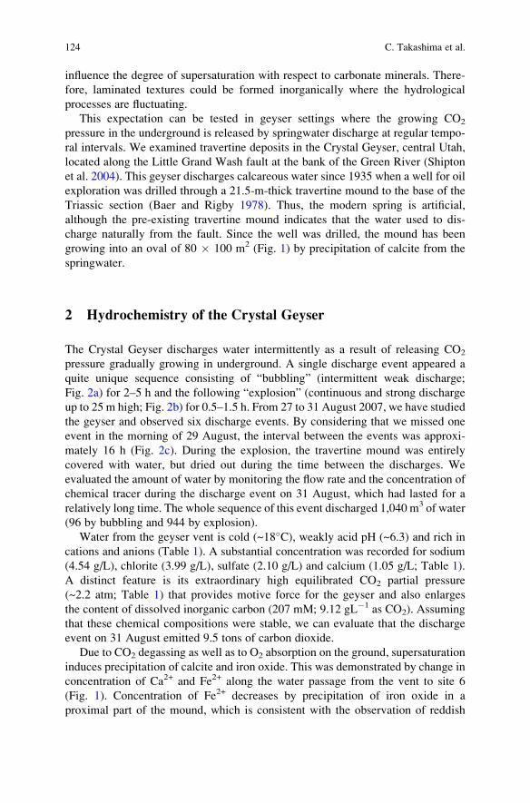
influence the degree of supersaturation with respect to carbonate minerals. There-
fore, laminated textures could be formed inorganically where the hydrological
processes are fluctuating.
This expectation can be tested in geyser settings where the growing CO2
pressure in the underground is released by springwater discharge at regular tempo-
ral intervals. We examined travertine deposits in the Crystal Geyser, central Utah,
located along the Little Grand Wash fault at the bank of the Green River (Shipton
et al. 2004). This geyser discharges calcareous water since 1935 when a well for oil
exploration was drilled through a 21.5-m-thick travertine mound to the base of the
Triassic section (Baer and Rigby 1978). Thus, the modern spring is artificial,
although the pre-existing travertine mound indicates that the water used to dis-
charge naturally from the fault. Since the well was drilled, the mound has been
growing into an oval of 80 � 100 m2 (Fig. 1) by precipitation of calcite from the
springwater.
2 Hydrochemistry of the Crystal Geyser
The Crystal Geyser discharges water intermittently as a result of releasing CO2
pressure gradually growing in underground. A single discharge event appeared a
quite unique sequence consisting of “bubbling” (intermittent weak discharge;
Fig. 2a) for 2–5 h and the following “explosion” (continuous and strong discharge
up to 25 m high; Fig. 2b) for 0.5–1.5 h. From 27 to 31 August 2007, we have studied
the geyser and observed six discharge events. By considering that we missed one
event in the morning of 29 August, the interval between the events was approxi-
mately 16 h (Fig. 2c). During the explosion, the travertine mound was entirely
covered with water, but dried out during the time between the discharges. We
evaluated the amount of water by monitoring the flow rate and the concentration of
chemical tracer during the discharge event on 31 August, which had lasted for a
relatively long time. The whole sequence of this event discharged 1,040 m3 of water
(96 by bubbling and 944 by explosion).
Water from the geyser vent is cold (~18�C), weakly acid pH (~6.3) and rich in
cations and anions (Table 1). A substantial concentration was recorded for sodium
(4.54 g/L), chlorite (3.99 g/L), sulfate (2.10 g/L) and calcium (1.05 g/L; Table 1).
A distinct feature is its extraordinary high equilibrated CO2 partial pressure
(~2.2 atm; Table 1) that provides motive force for the geyser and also enlarges
the content of dissolved inorganic carbon (207 mM; 9.12 gL�1 as CO2). Assuming
that these chemical compositions were stable, we can evaluate that the discharge
event on 31 August emitted 9.5 tons of carbon dioxide.
Due to CO2 degassing as well as to O2 absorption on the ground, supersaturation
induces precipitation of calcite and iron oxide. This was demonstrated by change in
concentration of Ca2+ and Fe2+ along the water passage from the vent to site 6
(Fig. 1). Concentration of Fe2+ decreases by precipitation of iron oxide in a
proximal part of the mound, which is consistent with the observation of reddish
124 C. Takashima et al.
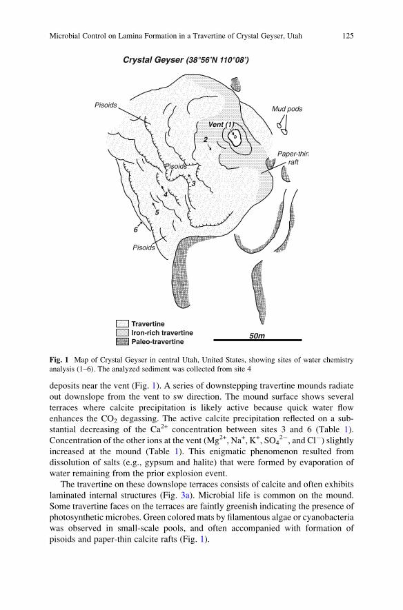
deposits near the vent (Fig. 1). A series of downstepping travertine mounds radiate
out downslope from the vent to sw direction. The mound surface shows several
terraces where calcite precipitation is likely active because quick water flow
enhances the CO2 degassing. The active calcite precipitation reflected on a sub-
stantial decreasing of the Ca2+ concentration between sites 3 and 6 (Table 1).
Concentration of the other ions at the vent (Mg2+, Na+, K+, SO42�, and Cl�) slightly
increased at the mound (Table 1). This enigmatic phenomenon resulted from
dissolution of salts (e.g., gypsum and halite) that were formed by evaporation of
water remaining from the prior explosion event.
The travertine on these downslope terraces consists of calcite and often exhibits
laminated internal structures (Fig. 3a). Microbial life is common on the mound.
Some travertine faces on the terraces are faintly greenish indicating the presence of
photosynthetic microbes. Green colored mats by filamentous algae or cyanobacteria
was observed in small-scale pools, and often accompanied with formation of
pisoids and paper-thin calcite rafts (Fig. 1).
TravertineIron-rich travertinePaleo-travertine
3
5
6
4
2
Pisoids
Pisoids
Pisoids
Paper-thinraft
Mud pods
Vent (1)
Crystal Geyser (38°56’N 110°08’)
50m
Fig. 1 Map of Crystal Geyser in central Utah, United States, showing sites of water chemistry
analysis (1–6). The analyzed sediment was collected from site 4
Microbial Control on Lamina Formation in a Travertine of Crystal Geyser, Utah 125
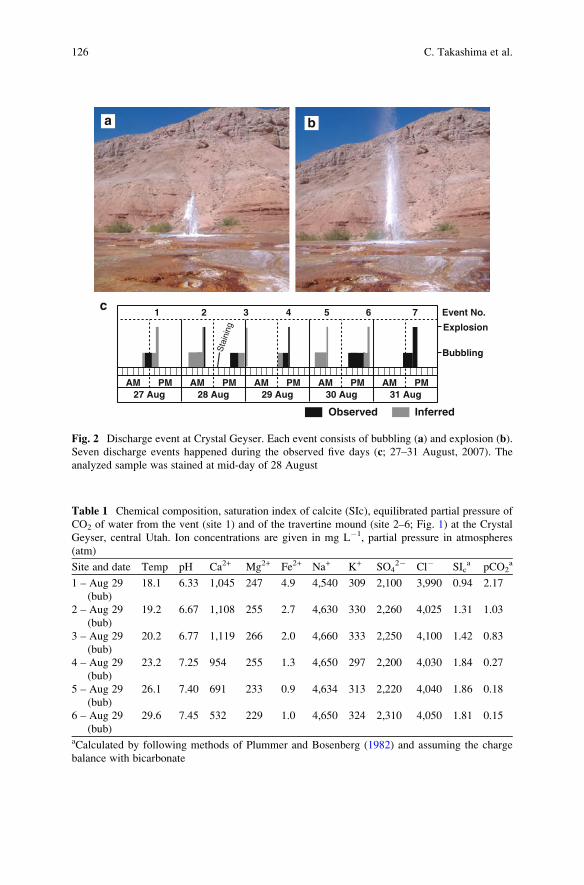
AM PM AM PMAM PMAM PMAM PM27 Aug 28 Aug 29 Aug 30 Aug 31 Aug
Explosion
Bubbling
Observed Inferred
1 2 3 4 5 6 7 Event No.
Sta
inin
g
a b
c
Fig. 2 Discharge event at Crystal Geyser. Each event consists of bubbling (a) and explosion (b).
Seven discharge events happened during the observed five days (c; 27–31 August, 2007). The
analyzed sample was stained at mid-day of 28 August
Table 1 Chemical composition, saturation index of calcite (SIc), equilibrated partial pressure of
CO2 of water from the vent (site 1) and of the travertine mound (site 2–6; Fig. 1) at the Crystal
Geyser, central Utah. Ion concentrations are given in mg L�1, partial pressure in atmospheres
(atm)
Site and date Temp pH Ca2+ Mg2+ Fe2+ Na+ K+ SO42� Cl� SIc
a pCO2a
1 – Aug 29
(bub)
18.1 6.33 1,045 247 4.9 4,540 309 2,100 3,990 0.94 2.17
2 – Aug 29
(bub)
19.2 6.67 1,108 255 2.7 4,630 330 2,260 4,025 1.31 1.03
3 – Aug 29
(bub)
20.2 6.77 1,119 266 2.0 4,660 333 2,250 4,100 1.42 0.83
4 – Aug 29
(bub)
23.2 7.25 954 255 1.3 4,650 297 2,200 4,030 1.84 0.27
5 – Aug 29
(bub)
26.1 7.40 691 233 0.9 4,634 313 2,220 4,040 1.86 0.18
6 – Aug 29
(bub)
29.6 7.45 532 229 1.0 4,650 324 2,310 4,050 1.81 0.15
aCalculated by following methods of Plummer and Bosenberg (1982) and assuming the charge
balance with bicarbonate
126 C. Takashima et al.
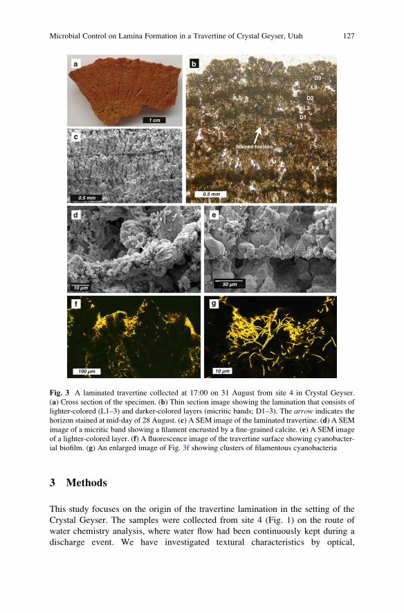
3 Methods
This study focuses on the origin of the travertine lamination in the setting of the
Crystal Geyser. The samples were collected from site 4 (Fig. 1) on the route of
water chemistry analysis, where water flow had been continuously kept during a
discharge event. We have investigated textural characteristics by optical,
10 µm100 µm
10 µm
0.5 mm0.5 mm
1 cm
a b
c
d e
f g
Stained horizon
L1
D1
L2
D2
L3
D3
50 µm
Fig. 3 A laminated travertine collected at 17:00 on 31 August from site 4 in Crystal Geyser.
(a) Cross section of the specimen. (b) Thin section image showing the lamination that consists of
lighter-colored (L1–3) and darker-colored layers (micritic bands; D1–3). The arrow indicates the
horizon stained at mid-day of 28 August. (c) A SEM image of the laminated travertine. (d) A SEM
image of a micritic band showing a filament encrusted by a fine-grained calcite. (e) A SEM image
of a lighter-colored layer. (f) A fluorescence image of the travertine surface showing cyanobacter-
ial biofilm. (g) An enlarged image of Fig. 3f showing clusters of filamentous cyanobacteria
Microbial Control on Lamina Formation in a Travertine of Crystal Geyser, Utah 127
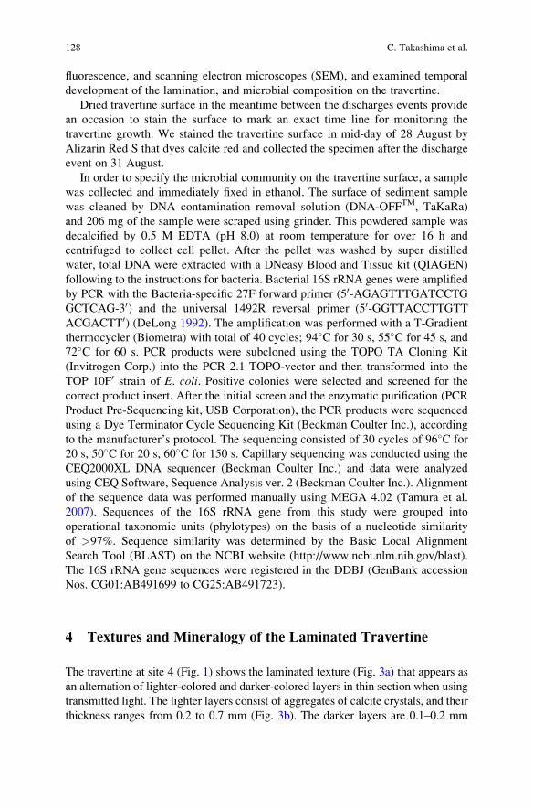
fluorescence, and scanning electron microscopes (SEM), and examined temporal
development of the lamination, and microbial composition on the travertine.
Dried travertine surface in the meantime between the discharges events provide
an occasion to stain the surface to mark an exact time line for monitoring the
travertine growth. We stained the travertine surface in mid-day of 28 August by
Alizarin Red S that dyes calcite red and collected the specimen after the discharge
event on 31 August.
In order to specify the microbial community on the travertine surface, a sample
was collected and immediately fixed in ethanol. The surface of sediment sample
was cleaned by DNA contamination removal solution (DNA-OFFTM, TaKaRa)
and 206 mg of the sample were scraped using grinder. This powdered sample was
decalcified by 0.5 M EDTA (pH 8.0) at room temperature for over 16 h and
centrifuged to collect cell pellet. After the pellet was washed by super distilled
water, total DNA were extracted with a DNeasy Blood and Tissue kit (QIAGEN)
following to the instructions for bacteria. Bacterial 16S rRNA genes were amplified
by PCR with the Bacteria-specific 27F forward primer (50-AGAGTTTGATCCTGGCTCAG-30) and the universal 1492R reversal primer (50-GGTTACCTTGTTACGACTT0) (DeLong 1992). The amplification was performed with a T-Gradient
thermocycler (Biometra) with total of 40 cycles; 94�C for 30 s, 55�C for 45 s, and
72�C for 60 s. PCR products were subcloned using the TOPO TA Cloning Kit
(Invitrogen Corp.) into the PCR 2.1 TOPO-vector and then transformed into the
TOP 10F0 strain of E. coli. Positive colonies were selected and screened for the
correct product insert. After the initial screen and the enzymatic purification (PCR
Product Pre-Sequencing kit, USB Corporation), the PCR products were sequenced
using a Dye Terminator Cycle Sequencing Kit (Beckman Coulter Inc.), according
to the manufacturer’s protocol. The sequencing consisted of 30 cycles of 96�C for
20 s, 50�C for 20 s, 60�C for 150 s. Capillary sequencing was conducted using the
CEQ2000XL DNA sequencer (Beckman Coulter Inc.) and data were analyzed
using CEQ Software, Sequence Analysis ver. 2 (Beckman Coulter Inc.). Alignment
of the sequence data was performed manually using MEGA 4.02 (Tamura et al.
2007). Sequences of the 16S rRNA gene from this study were grouped into
operational taxonomic units (phylotypes) on the basis of a nucleotide similarity
of >97%. Sequence similarity was determined by the Basic Local Alignment
Search Tool (BLAST) on the NCBI website (http://www.ncbi.nlm.nih.gov/blast).
The 16S rRNA gene sequences were registered in the DDBJ (GenBank accession
Nos. CG01:AB491699 to CG25:AB491723).
4 Textures and Mineralogy of the Laminated Travertine
The travertine at site 4 (Fig. 1) shows the laminated texture (Fig. 3a) that appears as
an alternation of lighter-colored and darker-colored layers in thin section when using
transmitted light. The lighter layers consist of aggregates of calcite crystals, and their
thickness ranges from 0.2 to 0.7 mm (Fig. 3b). The darker layers are 0.1–0.2 mm
128 C. Takashima et al.
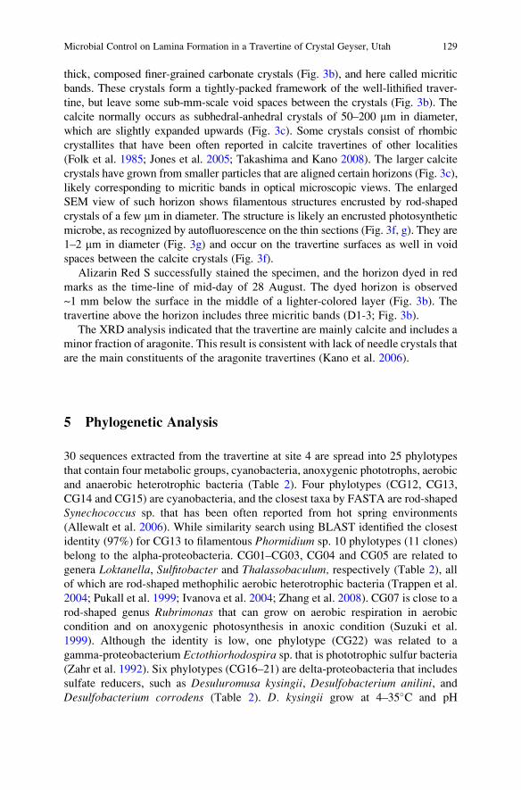
thick, composed finer-grained carbonate crystals (Fig. 3b), and here called micritic
bands. These crystals form a tightly-packed framework of the well-lithified traver-
tine, but leave some sub-mm-scale void spaces between the crystals (Fig. 3b). The
calcite normally occurs as subhedral-anhedral crystals of 50–200 mm in diameter,
which are slightly expanded upwards (Fig. 3c). Some crystals consist of rhombic
crystallites that have been often reported in calcite travertines of other localities
(Folk et al. 1985; Jones et al. 2005; Takashima and Kano 2008). The larger calcite
crystals have grown from smaller particles that are aligned certain horizons (Fig. 3c),
likely corresponding to micritic bands in optical microscopic views. The enlarged
SEM view of such horizon shows filamentous structures encrusted by rod-shaped
crystals of a few mm in diameter. The structure is likely an encrusted photosynthetic
microbe, as recognized by autofluorescence on the thin sections (Fig. 3f, g). They are
1–2 mm in diameter (Fig. 3g) and occur on the travertine surfaces as well in void
spaces between the calcite crystals (Fig. 3f).
Alizarin Red S successfully stained the specimen, and the horizon dyed in red
marks as the time-line of mid-day of 28 August. The dyed horizon is observed
~1 mm below the surface in the middle of a lighter-colored layer (Fig. 3b). The
travertine above the horizon includes three micritic bands (D1-3; Fig. 3b).
The XRD analysis indicated that the travertine are mainly calcite and includes a
minor fraction of aragonite. This result is consistent with lack of needle crystals that
are the main constituents of the aragonite travertines (Kano et al. 2006).
5 Phylogenetic Analysis
30 sequences extracted from the travertine at site 4 are spread into 25 phylotypes
that contain four metabolic groups, cyanobacteria, anoxygenic phototrophs, aerobic
and anaerobic heterotrophic bacteria (Table 2). Four phylotypes (CG12, CG13,
CG14 and CG15) are cyanobacteria, and the closest taxa by FASTA are rod-shaped
Synechococcus sp. that has been often reported from hot spring environments
(Allewalt et al. 2006). While similarity search using BLAST identified the closest
identity (97%) for CG13 to filamentous Phormidium sp. 10 phylotypes (11 clones)
belong to the alpha-proteobacteria. CG01–CG03, CG04 and CG05 are related to
genera Loktanella, Sulfitobacter and Thalassobaculum, respectively (Table 2), all
of which are rod-shaped methophilic aerobic heterotrophic bacteria (Trappen et al.
2004; Pukall et al. 1999; Ivanova et al. 2004; Zhang et al. 2008). CG07 is close to a
rod-shaped genus Rubrimonas that can grow on aerobic respiration in aerobic
condition and on anoxygenic photosynthesis in anoxic condition (Suzuki et al.
1999). Although the identity is low, one phylotype (CG22) was related to a
gamma-proteobacterium Ectothiorhodospira sp. that is phototrophic sulfur bacteria(Zahr et al. 1992). Six phylotypes (CG16–21) are delta-proteobacteria that includes
sulfate reducers, such as Desuluromusa kysingii, Desulfobacterium anilini, andDesulfobacterium corrodens (Table 2). D. kysingii grow at 4–35�C and pH
Microbial Control on Lamina Formation in a Travertine of Crystal Geyser, Utah 129
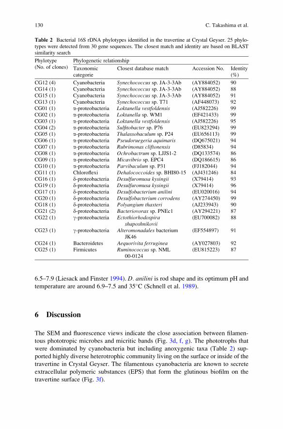
6.5–7.9 (Liesack and Finster 1994). D. anilini is rod shape and its optimum pH and
temperature are around 6.9–7.5 and 35�C (Schnell et al. 1989).
6 Discussion
The SEM and fluorescence views indicate the close association between filamen-
tous phototropic microbes and micritic bands (Fig. 3d, f, g). The phototrophs that
were dominated by cyanobacteria but including anoxygenic taxa (Table 2) sup-
ported highly diverse heterotrophic community living on the surface or inside of the
travertine in Crystal Geyser. The filamentous cyanobacteria are known to secrete
extracellular polymeric substances (EPS) that form the glutinous biofilm on the
travertine surface (Fig. 3f).
Table 2 Bacterial 16S rDNA phylotypes identified in the travertine at Crystal Geyser. 25 phylo-
types were detected from 30 gene sequences. The closest match and identity are based on BLAST
similarity search
Phylotype
(No. of clones)
Phylogenetic relationship
Taxonomic
categorie
Closest database match Accession No. Identity
(%)
CG12 (4) Cyanobacteria Synechococcus sp. JA-3-3Ab (AY884052) 90
CG14 (1) Cyanobacteria Synechococcus sp. JA-3-3Ab (AY884052) 88
CG15 (1) Cyanobacteria Synechococcus sp. JA-3-3Ab (AY884052) 91
CG13 (1) Cyanobacteria Synechococcus sp. T71 (AF448073) 92
CG01 (1) a-proteobacteria Loktanella vestfoldensis (AJ582226) 99
CG02 (1) a-proteobacteria Loktanella sp. WM1 (EF421433) 99
CG03 (1) a-proteobacteria Loktanella vestfoldensis (AJ582226) 95
CG04 (2) a-proteobacteria Sulfitobacter sp. P76 (EU823294) 99
CG05 (1) a-proteobacteria Thalassobaculum sp. P24 (EU656113) 99
CG06 (1) a-proteobacteria Pseudoruegeria aquimaris (DQ675021) 94
CG07 (1) a-proteobacteria Rubrimonas cliftonensis (D85834) 94
CG08 (1) a-proteobacteria Ochrobactrum sp. LJJS1-2 (DQ133574) 86
CG09 (1) a-proteobacteria Micavibrio sp. EPC4 (DQ186615) 86
CG10 (1) a-proteobacteria Parvibaculum sp. P31 (FJ182044) 94
CG11 (1) Chloroflexi Dehalococcoides sp. BHI80-15 (AJ431246) 84
CG16 (1) d-proteobacteria Desulfuromusa kysingii (X79414) 93
CG19 (1) d-proteobacteria Desulfuromusa kysingii (X79414) 96
CG17 (1) d-proteobacteria Desulfobacterium anilini (EU020016) 94
CG20 (1) d-proteobacteria Desulfobacterium corrodens (AY274450) 99
CG18 (1) d-proteobacteria Polyangium thaxteri (AJ233943) 90
CG21 (2) d-proteobacteria Bacteriovorax sp. PNEc1 (AY294221) 87
CG22 (1) g-proteobacteria Ectothiorhodospirashaposhnikovii
(EU700082) 88
CG23 (1) g-proteobacteria Alteromonadales bacteriumJK46
(EF554897) 91
CG24 (1) Bacteroidetes Aequorivita ferruginea (AY027803) 92
CG25 (1) Firmicutes Ruminococcus sp. NML
00-0124
(EU815223) 87
130 C. Takashima et al.
The EPS-bearing biofilm plays a central role in forming the lamination of
stromatolitic carbonates (Riding 2000), but the specific process has been interpreted
differently. A traditional interpretation for calcified filaments is biologically-
induced precipitation associated with photosynthetic activity (Pentecost and Riding
1986) that rise supersaturation of calcite. Recent researches using microelectrodes
confirmed that cyanobacterial biofilm photosynthesis drives calcification within the
microenvironment of tufa biofilms (Bissett et al. 2008; Shiraishi et al. 2008).
However, this process is effective only with relatively low alkalinity and pH around
8 (Arp et al. 2001), which are inconsistent with the water chemistry of the Crystal
Geyser. Second, EPS can induce calcite nucleation and precipitation by binding
calcium ions to their negatively charged sites (e.g., Pentecost 1985). This process
may have happened occurred on the biofilm of the Crystal Geyser travertine, and is
consistent with the calcified filamentous textures observed in the micritic bands
(Fig. 3d). However, the cation-binding EPS can also inhibit carbonate precipitation
by decreasing activity of Ca2+ in the vicinity of the biofilm (Arp et al. 1999), and
the inhibition associated with EPS was demonstrated for an aragonite travertine in
SW Japan.
The third potential process is trapping of suspended particles that were previously
suggested for the modern marine stromatolites byMonty (1976). This process is still
valid although the later researches of the Bahamian stromatolites suggested that the
lamination is formed in over a longer term period (typically months) by temporal
replacement of the microbial community and lithification associated with sulfate
reduction (Reid et al. 2000; Andres et al. 2006). Our previous study on a calcite
travertine at the Shionoha Hot Spring in Japan concluded that this process was
responsible for the daily rhythm of the micritic bands (Takashima and Kano 2008).
Evidences in this study not specify which process was important, and it is quite
possible that all three processes contributed to the band formation. Whatever the
case, we observed the spatial association between the micritic bands and the
microbes, and demonstrated that the microbes are dominated by photoautotrophs
(Table 2). It is therefore likely that the micritic bands were formed in daytime when
the microbes can photosynthesize.
If so, the observed laminated pattern (Fig. 3b) can be correlated to the actual
discharge pattern from the Geyser (Fig. 2c). The travertine above the stained
horizon consists of three darker-colored micritic band (D1–3) alternated with
three lighter colored layers of larger calcite crystals (L1–3; Fig. 2b). This part
should have been formed in a period from mid day of 28 August to evening of 31
August, and during the period five discharge events happened (No. 3–7; Fig. 2c).
Just above the stained horizon, the first layer L1 (Fig. 3b) should have grown
with the discharge event 3 that happened after the sunset. The next layer, the
micritic band D1 was formed with the event No. 4 during the early afternoon of
29 August (Fig. 2c). Then, the event No. 5 corresponds to L2. The long-lasting
event No. 6 likely formed both a micritic band (N2) and a lighter layer (L3), which
corresponding to the daytime bubbling and the subsequent discharge at night,
respectively. Finally, the last event No. 7 precipitated the uppermost layer, D3
(Fig. 3c). We can conclude that the darker bands (D1–3) were formed when both
Microbial Control on Lamina Formation in a Travertine of Crystal Geyser, Utah 131
water and sunlight were available on the travertine surface. Interpretation of water
supply did not reflect on the lamination pattern, but could reflect on a discontinuous
surface that could be observed in SEM images (e.g., Fig. 3e).
This study confirmed importance of photosynthetic bacteria in forming the daily
lamination of the stromatolitic carbonate. The bacteria are not necessary being
cyanobacteria, and can be anoxygenic photosynthetic bacteria, such as green sulfur
of purple non-sulfur bacteria. Stromatolites developed in the anoxic Earth surface
before 2.3 Ga (Kopp et al. 2005) could have formed by microbial community
including anoxygenic photosynthetic bacteria.
References
Allewalt JP, Bateson MM, Revsbech NP, Slack K, Ward DM (2006) Effect of temperature and
light on growth of and photosynthesis by Synechococcus isolates typical of those predominat-
ing in the Octopus Spring microbial mat community of Yellowstone National Park. Applied
and Environmental Microbiology 72:544–550
Andres MS, Sumner DY, Reid RP, Swart PK (2006) Isotopic fingerprints of microbial respiration
in aragonite from Bahamian stromatolites. Geology 34:973–976
Arp G, Thiel V, Reimer A, Michaelis W, Reitner J (1999) Biofilm exopolymers control micro-
bialite formation at thermal springs discharging into the alkaline Pyramid Lake, Nevada, USA.
Sedimentary Geology 126:159–176
Arp G, Reimer A, Reitner J (2001) Photosynthesis-induced biofilm calcification and calcium
concentrations in Phanerozoic ocean. Science 292:1701–1704
Baer JL, Rigby JK (1978) Geology of the Crystal Geyser and the environmental implications of its
effluent, Grand County, Utah. Utah Geology 5:125–130
Bissett A, de Beer D, Schoon R, Shiraishi F, Reimer A, Arp G (2008) Microbial mediation of
stromatolite formation in karst-water creeks. Limnology and Oceanography 53:1159–1168
DeLong EF (1992) Archaea in coastal marine environments. Proceedings of the National Acad-
emy of Sciences of the United States of America 89:5685–5689
Folk RL, Chafetz HS, Tiezzi PA (1985) Bizarre forms of depositional and diagenetic calcite in hot-
spring travertines, central Italy. In: Scheiderman N, Harris PM (eds.) Carbonate Cements,
SEPM Special Publications 36 pp 349–369
Guo L, Riding R (1992) Aragonite laminae in hot water travertine crusts, Rapolano Terme, Italy.
Sedimentology 39:1067–1079
Herman MM, Lorah SJ (1988) The chemical evolution of a travertine-deposition stream: Geoch-
meical processes and mass transfer reactions. Water Resources Research 24:1541–1552
Ivanova EP, Gorshkova NM, Sawabe T, Zhukova NV, Hayashi K, Kurilenko VV, Alexeeva Y,
Buljan V, Nicolau DV, Michailov VV, Christen R (2004) Sulfitobacter delicatus sp. nov. andSulfitobacter dubius sp. nov., respectively from a starfish (Stellaster equestris) and sea grass
(Zostera marina). International Journal of Systematic and Evolutionary Microbiology
54:475–480
Jones B, Renaut WR, Owen BR, Torfason H (2005) Growth patterns and implications of complex
dendrites in calcite travertines from Lysuholl, Snæfellsnes, Iceland. Sedimentology
52:1277–1301
Kano A, Takashima C, Ohtsuka S (2006) Hot-spring in eastern Kyushu and their related sedimen-
tation and microbial processes. ISC 2006 Field Excursion Guidebook FE-A10:10 pp
Kitano Y (1963) Geochemistry of calcareous deposits found in hot springs. Journal of Earth
Science Nagoya University 1:68–100
132 C. Takashima et al.
Kopp EK, Kirschvink JL, Hilburn IA, Nash CZ (2005) The Paleoproterozoic snowball Earth: A
climate disaster triggered by the evolution of oxygenic photosynthesis. Proceedings of the
National Academy of Sciences of the United States of America 102:1131–1136
Liesack W, Finster K (1994) Phylogenetic analysis of five strains of gram-negative, obligately
anaerobic, sulfur-reducing bacteria and description of Desulfirornusa gen. nov., including
Desulfuromusa kysingii sp. nov., Desulfurornusa bakii sp. nov., and Desulfuromusa succinox-idans sp. nov. International Journal of Systematic Bacteriology 44:753–758
Monty CLV (1976) The origin and development of cryptalgal fabric. Developments in Sedimen-
tology 20:193–249
Pentecost A (1985) Association of cyanobacteria with tufa deposits: Identity, enumeration and
nature of the sheath material revealed by histochemistry. Geomicrobiology Journal 4:285–298
Pentecost A (1994) Formation of laminate travertines at Bagno Vignone, Italy. Geomicrobiology
Journal 12:239–251
Pentecost A (2005) Travertine. Springer-Verlag, Berlin, 445 pp
Pentecost A, Riding R (1986) Calcification in cyanobacteria. In: Leadbeater BSC, Riding R (eds.)
Biomineralization in Lower Plants and Animals. Systematic Association Special Publication,
30. Clarendon Press, Oxford, pp 73–90
Plummer LN, Bosenberg E (1982) The solubilities of calcite, aragonite and vaterite in CO2-H2O
solutions between 0 and 90�C, and an evaluation of the aqueous model for the system
CaCO3�CO2-H2O. Geochimica et Cosmochimica Acta 46:1011–1040
Pukall R, Bunterfuß D, Fr€uhling A, RohdeM, Kroppenstedt RM, Burghardt J, Lebaron P, Bernard L,
Stackebrandt E (1999) Sulfitobacter rnediterraneus sp. nov., a new sulfite-oxidizing member of
the a-Proteobacteria. International Journal of Systematic Bacteriology 49:513–519
Reid RP, Visscher PT, Decho AW, Stolz JF, Bebout BM, Dupraz C, Macintyre IG, Paerl HW,
Pinckney JL, Prufert-Bebout L, Steppe TF, DesMarais DJ (2000) The role of microbes in
accretion, lamination and early lithification of modern marine stromatolites. Nature 406:989–992
Riding R (2000) Microbial carbonates: the geological record of calcified bacterial-algal mats and
biofilms. Sedimentology 47:179–214
Schnell S, Bak F, Pfennig N (1989) Anaerobic degradation of aniline and dihydroxybenzenes by
newly isolated sulfate-reducing bacteria and description ofDesuifobacterium aniline. Archivesof Microbiology 152:556–563
Shipton ZK, Evans JP, Kirschner D, Kolesar PT, Williams AP, Heath J (2004) Analysis of CO2
leakage through ‘low-permeability’ faults from natural reservoirs in the Colorado Plateau, east-
central Utah. Special Publications, Geological Society London 233:43–58
Shiraishi F, Bissett A, de Beer D, Reimer A, Arp G (2008) Photosynthesis, respiration and
exopolymer calcium-binding in biofilm calcification (Westerh€ofer and Deinschwanger
Creek, Germany). Geomicrobiology Journal 25:83–94
Suzuki T, Muroga Y, Takahama M, Shiba T, Nishimura Y (1999) Rubrimonas cliftonensis gen.nov., sp. nov., an aerobic bacteriochlorophyl II-containing bacterium isolated from a saline
lake. International Journal of Systematic Bacteriology 49:201–205
Takashima C, Kano A (2008) Microbial processes forming daily lamination in a stromatolitic
travertine. Sedimentary Geology 208:114–119
Tamura K, Dudley J, Nei M, Kumar S (2007) MEGA4: Molecular Evolutionary Genetics Analysis
(MEGA) software version 4.0. Molecular Biology and Evolution 24:1596–1599
Trappen SV, Mergaert J, Swings J (2004) Loktanella salsilacus gen. nov., sp. nov., Loktanellafryxellensis sp. nov. and Loktanella vestfoldensis sp. nov., new members of the Rhodobacter
group, isolated from microbial mats in Antarctic lakes. International Journal of Systematic and
Evolutionary Microbiology 54:1263–1269
Zahr M, Fobel B, Mayer H, Imboff JF, Campos PV, Weckesser J (1992) Chemical composition of
the lipopolysaccharides of Ectothiorhodospira shaposhnikovii, Ectothiorhodospira mobilis,and Ectothiorhodospira halophila. Archives of Microbiology 157:499–504
Zhang GI, Hwang CY, Cho BC (2008) Thalassobaculum litoreum gen. nov., sp. nov., a member of
the family Rhodospirillaceae isolated from coastal seawater. International Journal of System-
atic and Evolutionary Microbiology 58:479–485
Microbial Control on Lamina Formation in a Travertine of Crystal Geyser, Utah 133

.
Photosynthesis-Induced Stromatolite Formation
in the Freshwater Creeks
Fumito Shiraishi
1 Introduction
Stromatolites, the oldest macrofossil in the Earth history, appeared about 3.5 billion
years ago and considered to be formed by the interaction between the activities of
benthic microorganisms and the surrounding environment, and hence are expected
to provide important information for the evolution of life and Earth’s environment.
However, fossil stromatolites usually lack microfossils (e.g., Riding 1994), which
makes it difficult to interpret their formation mechanisms. One approach to over-
come this problem is the investigation of recent analogs in detail to understand how
microorganisms produce stromatolitic constructions. Promising analogs were
thought to be the recent stromatolites developing in seawater settings, and thus,
they have been keenly studied last several decades. However, some researchers
pointed out their critical disadvantage: the recent marine stromatolites are com-
posed mainly of agglutinated sand grains, and are different from ancient ones
composed mainly of fine minerals precipitated in situ (e.g., Awramik 1984; Riding
2008). Therefore, it is necessary to seek and investigate more suitable recent
analogs to understand stromatolite formation.
The DFG research unit 571 (Geobiology of organo- and biofilms: Coupling of
the geosphere and the biosphere by microbial processes) focused on recent fresh-
water stromatolites (tufa stromatolites) developing at karst creeks as a possibly
more suitable analog because they are composed of fine carbonate minerals,
resembling to the fabric of ancient stromatolites. Our 3-year investigation revealed
that the photosynthetic activities of investigated biofilms have strong impact on
ambient water chemistry, induce CaCO3 precipitation, and result in the stromatolite
F. Shiraishi
Department of Environmental Change, Graduate School of Social and Cultural Studies, Kyushu
University, 744 Motooka, Nishi-ku, Fukuoka 819-0395, Japan
Geoscience Centre, University of G€ottingen, Goldschmidtstraße 3, 37077 G€ottingen, Germany
e-mail: [email protected]
J. Reitner et al., Advances in Stromatolite Geobiology,Lecture Notes in Earth Sciences 131, DOI 10.1007/978-3-642-10415-2_8,# Springer-Verlag Berlin Heidelberg 2011
135
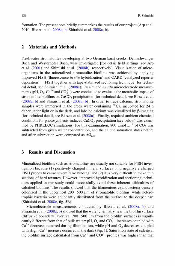
formation. The present note briefly summarizes the results of our project (Arp et al.
2010; Bissett et al. 2008a, b; Shiraishi et al. 2008a, b).
2 Materials and Methods
Freshwater stromatolites developing at two German karst creeks, Deinschwanger
Bach and Westerh€ofer Bach, were investigated [for detail field settings, see Arp
et al. (2001) and Shiraishi et al. (2008b), respectively]. Visualization of micro-
organisms in the mineralized stromatolite biofilms was achieved by applying
improved FISH (fluorescence in situ hybridization) and CARD (catalyzed reporter
deposition) � FISH together with tape-stabilized sectioning technique [for techni-
cal detail, see Shiraishi et al. (2008c)]. In situ and ex situ microelectrode measure-
ments (pH, O2, Ca2+ and CO3
2�) were conducted to evaluate the metabolic impact of
stromatolite biofilms on CaCO3 precipitation [for technical detail, see Bissett et al.
(2008a, b) and Shiraishi et al. (2008a, b)]. In order to trace calcium, stromatolite
samples were immersed in the creek water containing 45Ca, incubated for 24 h
either under light or in the dark, and labeled calcium was visualized by b-imaging
[for technical detail, see Bissett et al. (2008a)]. Finally, required ambient chemical
conditions for photosynthesis-induced CaCO3 precipitation (see below) was exam-
ined by PHREEQC simulations. For this examination, 800 mmol L�1 of CO2 was
subtracted from given water concentration, and the calcite saturation states before
and after subtraction were compared as DOact.
3 Results and Discussion
Mineralized biofilms such as stromatolites are usually not suitable for FISH inves-
tigation because (1) positively charged mineral surfaces bind negatively charged
FISH probes to cause severe false binding, and (2) it is very difficult to make thin
sections of hard textures. However, improved hybridization and sectioning techni-
ques applied in our study could successfully avoid these inherent difficulties of
calcified biofilms. The results showed that the filamentous cyanobacteria densely
colonized in the uppermost 200�500 mm of stromatolite biofilms, while hetero-
trophic bacteria were abundantly distributed from the surface to the deeper part
(Shiraishi et al. 2008c, fig. 5B).
Microelectrode measurements conducted by Bissett et al. (2008a, b) and
Shiraishi et al. (2008a, b) showed that the water chemistry near the biofilm surface
(diffusive boundary layer; ca. 200�500 mm from the biofilm surface) is signifi-
cantly different from that of bulk water: pH, O2 and CO32� increases coupled with
Ca2+ decrease occurred during illumination, while pH and O2 decreases coupled
with slight Ca2+ increase occurred in the dark (Fig. 1). Saturation state of calcite at
the biofilm surface calculated from Ca2+ and CO32� profiles was higher than that
136 F. Shiraishi
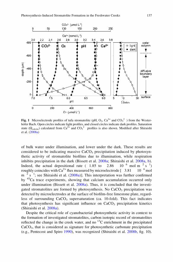
of bulk water under illumination, and lower under the dark. These results are
considered to be indicating massive CaCO3 precipitation induced by photosyn-
thetic activity of stromatolite biofilms due to illumination, while respiration
inhibits precipitation in the dark (Bissett et al. 2008a; Shiraishi et al. 2008a, b).
Indeed, the actual depositional rate (�1.85 to �2.86 � 10�6 mol m�2 s�1)
roughly coincides with Ca2+ flux measured by microelectrode [�3.81 � 10�6 mol
m�2 s�1; see Shiraishi et al. (2008a)]. This interpretation was further confirmed
by 45Ca trace experiments, showing that calcium accumulation occurred only
under illumination (Bissett et al. 2008a). Thus, it is concluded that the investi-
gated stromatolites are formed by photosynthesis. No CaCO3 precipitation was
detected by microelectrodes at the surface of biofilm-free limestone plate, regard-
less of surrounding CaCO3 supersaturation (ca. 10-fold). This fact indicates
that photosynthesis has significant influence on CaCO3 precipitation kinetics
(Shiraishi et al. 2008a).
Despite the critical role of cyanobacterial photosynthetic activity in context to
the formation of investigated stromatolites, carbon isotopic record of stromatolites
reflected the change in the creek water, and no 13C enrichment in the precipitated
CaCO3, that is considered as signature for photosynthetic carbonate precipitation
(e.g., Pentecost and Spiro 1990), was recognized (Shiraishi et al. 2008b, fig. 10).
Fig. 1 Microelectrode profiles of tufa stromatolite (pH, O2, Ca2+ and CO3
2�) from the Wester-
h€ofer Bach. Open circles indicate light profiles, and closed circles indicate dark profiles. Saturationstate (Ocalcite) calculated from Ca2+ and CO3
2� profiles is also shown. Modified after Shiraishi
et al. (2008a)
Photosynthesis-Induced Stromatolite Formation in the Freshwater Creeks 137
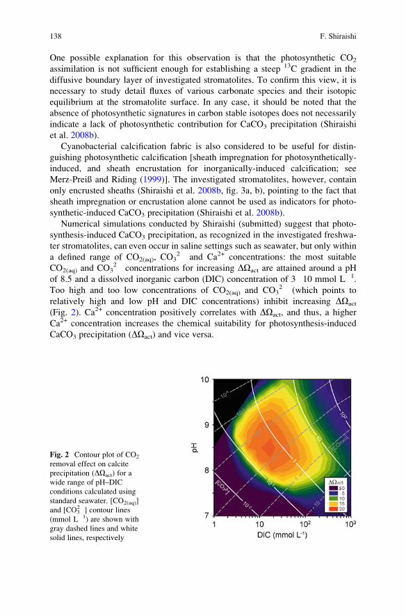
One possible explanation for this observation is that the photosynthetic CO2
assimilation is not sufficient enough for establishing a steep 13C gradient in the
diffusive boundary layer of investigated stromatolites. To confirm this view, it is
necessary to study detail fluxes of various carbonate species and their isotopic
equilibrium at the stromatolite surface. In any case, it should be noted that the
absence of photosynthetic signatures in carbon stable isotopes does not necessarily
indicate a lack of photosynthetic contribution for CaCO3 precipitation (Shiraishi
et al. 2008b).
Cyanobacterial calcification fabric is also considered to be useful for distin-
guishing photosynthetic calcification [sheath impregnation for photosynthetically-
induced, and sheath encrustation for inorganically-induced calcification; see
Merz-Preiß and Riding (1999)]. The investigated stromatolites, however, contain
only encrusted sheaths (Shiraishi et al. 2008b, fig. 3a, b), pointing to the fact that
sheath impregnation or encrustation alone cannot be used as indicators for photo-
synthetic-induced CaCO3 precipitation (Shiraishi et al. 2008b).
Numerical simulations conducted by Shiraishi (submitted) suggest that photo-
synthesis-induced CaCO3 precipitation, as recognized in the investigated freshwa-
ter stromatolites, can even occur in saline settings such as seawater, but only within
a defined range of CO2(aq), CO32� and Ca2+ concentrations: the most suitable
CO2(aq) and CO32� concentrations for increasing DOact are attained around a pH
of 8.5 and a dissolved inorganic carbon (DIC) concentration of 3�10 mmol L�1.
Too high and too low concentrations of CO2(aq) and CO32� (which points to
relatively high and low pH and DIC concentrations) inhibit increasing DOact
(Fig. 2). Ca2+ concentration positively correlates with DOact, and thus, a higher
Ca2+ concentration increases the chemical suitability for photosynthesis-induced
CaCO3 precipitation (DOact) and vice versa.
Fig. 2 Contour plot of CO2
removal effect on calcite
precipitation (DOact) for a
wide range of pH–DIC
conditions calculated using
standard seawater. [CO2(aq)]
and [CO32�] contour lines
(mmol L�1) are shown with
gray dashed lines and white
solid lines, respectively
138 F. Shiraishi
4 Conclusions
The investigation of freshwater stromatolites developing at the karst creeks
revealed that they are formed by photosynthetic activity of cyanobacteria. Despite
of critical role of cyanobacterial photosynthesis, investigated stromatolites did not
contain previously suggested indicators for photosynthesis-induced CaCO3 precip-
itation. Numerical simulations revealed that the photosynthetic CaCO3 precipita-
tion can theoretically occur even in the seawater settings, but requires optimum pH,
DIC and Ca2+ concentrations.
Acknowledgements Dr. Gernot Arp kindly provided valuable advices for improving the manu-
script. This is the part of the Research Unit 571 “Geobiology of organo- and biofilms”, funded by
the German Research Foundation (DFG�FOR 571; AR 335/5; publication #49).
References
Arp G, Wedemeyer N, Reitner J (2001) Fluvial tufa formation in a hard-water creek (Deinsch-
wanger Bach, Franconian Alb, Germany). Facies 44:1–22
Arp G, Bissett A, Brinkmann N, Cousin S, de Beer D, Friedl T, Mohr K, Neu TR, Reimer A,
Shiraishi F, Stackebrandt E, Zippel B (2010) Tufa-forming biofilms of German karstwater
creeks: Microorganisms, exopolymers, hydrochemistry and calcification. In: Rogerson M,
Pedley MM (eds), Tufas and Speleothems: Unravelling the Microbial and Physical Controls.
Geological Society London Special Publication 336:83–118
Awramik SM (1984) Ancient stromatolites and microbial mats. In: Cohen Y, Castenholtz RW,
Halvorson HO (eds) Microbial Mats: Stromatolites. New York, Alan R Liss Inc, pp 1–22
Bissett A, de Beer D, Schoon R, Shiraishi F, Reimer A, Arp G (2008a) Microbial mediation of
stromatolite formation in karst-water creeks. Limnology and Oceanography 53:1159�1168
Bissett A, Reimer A, de Beer D, Shiraishi F, Arp G (2008b) Metabolic microenvironmental control
by photosynthetic biofilms under changing macroenvironmental temperature and pH. Applied
and Environmental Microbiology 74:6306�6312
Merz-Preiß M, Riding R (1999) Cyanobacterial tufa calcification in two freshwater streams:
ambient environment, chemical thresholds and biological processes. Sedimentary Geology
126:103–124
Pentecost A, Spiro B (1990) Stable carbon and oxygen isotope composition of calcites associated
with modern freshwater cyanobacteria and algae. Geomicrobiology Journal 8:17–26
Riding R (1994) Evolution of algal and cyanobacterial calcification. In: Bengtson S (ed) Early Life
on Earth. New York, Columbia Univ. Press, pp 426�438
Riding R (2008) Marrying stromatolite perspectives: 3500 million year of history and a century
of research. In: Reitner J, Queric NV, Reich M (eds) Abstract volume of International
Kalkowsky-Symposium. Universit€atsdrucke G€ottingen, G€ottingen, pp 29�30
Shiraishi F, Bissett A, de Beer D, Reimer A, Arp G (2008a) Photosynthesis, respiration and
exopolymer calcium-binding in biofilm calcification (Westerh€ofer and Deinschwanger Bach,
Germany). Geomicrobiology Journal 25:83�94
Shiraishi F, Reimer A, Bissett A, de Beer D, Arp G (2008b) Microbial effects on biofilm
calcification, ambient water chemistry and stable isotope records (Westerh€ofer Bach,
Germany). Palaeogeography, Palaeoclimatology, Palaeoecology 262:91�106
Shiraishi F, Zippel B, Neu TR, Arp G (2008c) In situ detection of bacteria in calcified biofilms
using FISH and CARD�FISH. Journal of Microbiological Methods 74:6306�6312
Photosynthesis-Induced Stromatolite Formation in the Freshwater Creeks 139

.
The Role of Purple Sulphur Bacteria
in Carbonate Precipitation of Modern and
Possibly Early Precambrian Stromatolites
Rolf Warthmann, Crisogono Vasconcelos, Anne Greet Bittermann,
and Judith A. McKenzie
1 Introduction
Since the description of stromatolites a century ago by Kalkowsky (1908) as
products of ancient microbial activity, the exact understanding of its formation
remained unclear and is still matter of debate. Stromatolites are definitely the first
visible fossil record of life in Earth history, and are dated back as far as 3.45 Ga
(Grotzinger and Knoll 1999; Riding 2000), which implicates very early activities of
life, possibly different than those today. The environmental conditions in the Early
Precambrian, such as ocean chemistry or surface temperature are not fully under-
stood today. Several studies tried to explain the formation of Precambrian stroma-
tolites by analyzing modern analogues such a the Highborne cay stromatolites of
the Bahamas (Dupraz and Visscher 2005) or the Shark Bay (Australia) stromatolites
(Burns et al. 2004; Allen et al. 2009). Studies of ancient stromatolites apparently
showed fossil traces of filamentous or coccoid cells (Westall et al. 2006). Ancient
microbial activity is supported by geochemical indications, which show isotope
discriminations of carbon and sulphur isotopes (Grassineau et al. 2001; Schidlowski
2001). However, often these geochemical data were not unambiguous. An essential
feature of some Precambrian stromatolites is lamination caused by alternating
layers of carbonate (calcite, micrite, dolomite), intermitted with other mineral
layers, mostly silica minerals in ancient stromatolites (Allwood et al. 2006).
R. Warthmann (*)
Institute of Biotechnology, Zurich University of Applied Sciences, 8820 Wdenswil, Switzerland
e-mail: [email protected]
C. Vasconcelos and J.A. McKenzie
Geological Institute, ETH Z€urich, 8092 Z€urich, Switzerland
A.G. Bittermann
Center for Microscopy and Image Analysis, University of Zurich, 8006 Zurich, Switzerland
J. Reitner et al., Advances in Stromatolite Geobiology,Lecture Notes in Earth Sciences 131, DOI 10.1007/978-3-642-10415-2_9,# Springer-Verlag Berlin Heidelberg 2011
141
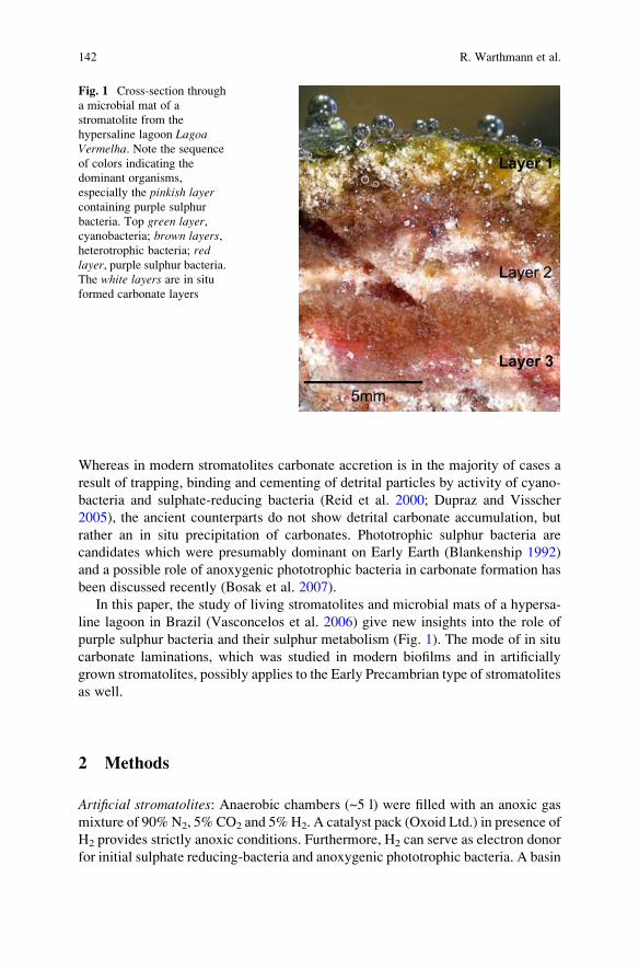
Whereas in modern stromatolites carbonate accretion is in the majority of cases a
result of trapping, binding and cementing of detrital particles by activity of cyano-
bacteria and sulphate-reducing bacteria (Reid et al. 2000; Dupraz and Visscher
2005), the ancient counterparts do not show detrital carbonate accumulation, but
rather an in situ precipitation of carbonates. Phototrophic sulphur bacteria are
candidates which were presumably dominant on Early Earth (Blankenship 1992)
and a possible role of anoxygenic phototrophic bacteria in carbonate formation has
been discussed recently (Bosak et al. 2007).
In this paper, the study of living stromatolites and microbial mats of a hypersa-
line lagoon in Brazil (Vasconcelos et al. 2006) give new insights into the role of
purple sulphur bacteria and their sulphur metabolism (Fig. 1). The mode of in situ
carbonate laminations, which was studied in modern biofilms and in artificially
grown stromatolites, possibly applies to the Early Precambrian type of stromatolites
as well.
2 Methods
Artificial stromatolites: Anaerobic chambers (~5 l) were filled with an anoxic gas
mixture of 90% N2, 5% CO2 and 5% H2. A catalyst pack (Oxoid Ltd.) in presence of
H2 provides strictly anoxic conditions. Furthermore, H2 can serve as electron donor
for initial sulphate reducing-bacteria and anoxygenic phototrophic bacteria. A basin
Fig. 1 Cross-section through
a microbial mat of a
stromatolite from the
hypersaline lagoon LagoaVermelha. Note the sequenceof colors indicating the
dominant organisms,
especially the pinkish layercontaining purple sulphur
bacteria. Top green layer,cyanobacteria; brown layers,heterotrophic bacteria; redlayer, purple sulphur bacteria.The white layers are in situ
formed carbonate layers
142 R. Warthmann et al.
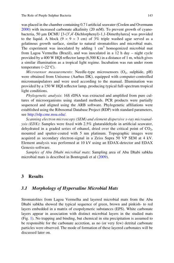
was placed in the chamber containing 0.7 l artificial seawater (Coolen and Overmann
2000) with increased carbonate alkalinity (20 mM). To prevent growth of cyano-
bacteria, 50 mm DCMU [3-(30,40-Dichlorphenyl)-1,1-Dimethylurea] was provided
to the liquid. A block (9 � 9 � 3 cm) of 3% triple washed agar served as a
gelatinous growth surface, similar to natural stromatolites and microbial mats.
The experiment was inoculated by adding 1 cm3 homogenized microbial mat
from Lagoa Vermelha (Brazil), and was inoculated in a 12 h day – night cycle
provided by a 400WHQI reflector lamp (6,500 K) in a distance of 1 m, which gives
a similar illumination as a tropical light regime. Incubation was run under room
temperature (~22�C).Microsensor measurements: Needle-type microsensors (O2, sulphide, pH)
were obtained from Unisense (Aarhus DK), equipped with computer-controlled
micromanipulators and were used according to the manual. Illumination was
provided by a 150 W HQI reflector lamp, producing typical full-spectrum tropical
light conditions.
Phylogenetic analysis: 16S rDNA was extracted and amplified from pure cul-
tures of microorganisms using standard methods. PCR products were partially
sequenced and aligned using the ARB software. Phylogenetic affiliations were
established using the Ribosomal Database Project (RDP) with standard parameters,
see http://rdp.cme.msu.edu/.
Scanning electron microscopy (SEM) and element dispersive x-ray microanal-ysis (EDX): Samples were fixed with 2.5% glutaraldehyde in artificial seawater,
dehydrated in a graded series of ethanol, dried over the critical point of CO2,
mounted and sputter-coated with 5 nm platinum. Topographic images were
acquired as secondary electron-signal in a Zeiss Supra 50 VP SEM at 4 kV.
Element analysis was performed at 10 kV using an EDAX-detector and EDAX
Genesis-software.
Samples of Abu Dhabi microbial mats: Sampling area of Abu Dhabi sabkha
microbial mats is described in Bontognali et al (2009).
3 Results
3.1 Morphology of Hypersaline Microbial Mats
Stromatolites from Lagoa Vermelha and layered microbial mats from the Abu
Dhabi sabkha showed the typical sequence of green, brown and pinkish- to red
layers embedded in a matrix of exopolymeric substances (EPS). White carbonate
layers appear in association with distinct microbial layers in the studied mats
(Fig. 1). No trapping and binding, but chemical in situ precipitation is assumed to
be responsible for the carbonate accretion, as no (or very few) detrital carbonate
particles were observed. The mode of formation of these layered carbonates will be
discussed later on.
The Role of Purple Sulphur Bacteria 143
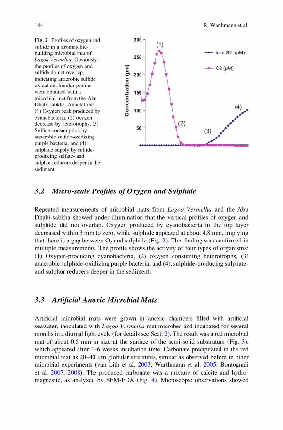
3.2 Micro-scale Profiles of Oxygen and Sulphide
Repeated measurements of microbial mats from Lagoa Vermelha and the Abu
Dhabi sabkha showed under illumination that the vertical profiles of oxygen and
sulphide did not overlap. Oxygen produced by cyanobacteria in the top layer
decreased within 3 mm to zero, while sulphide appeared at about 4.8 mm, implying
that there is a gap between O2 and sulphide (Fig. 2). This finding was confirmed in
multiple measurements. The profile shows the activity of four types of organisms:
(1) Oxygen-producing cyanobacteria, (2) oxygen consuming heterotrophs, (3)
anaerobic sulphide-oxidizing purple bacteria, and (4), sulphide-producing sulphate-
and sulphur reducers deeper in the sediment.
3.3 Artificial Anoxic Microbial Mats
Artificial microbial mats were grown in anoxic chambers filled with artificial
seawater, inoculated with Lagoa Vermelha mat microbes and incubated for several
months in a diurnal light cycle (for details see Sect. 2). The result was a red microbial
mat of about 0.5 mm in size at the surface of the semi-solid substratum (Fig. 3),
which appeared after 4–6 weeks incubation time. Carbonate precipitated in the red
microbial mat as 20–40 mm globular structures, similar as observed before in other
microbial experiments (van Lith et al. 2003; Warthmann et al. 2005; Bontognali
et al. 2007, 2008). The produced carbonate was a mixture of calcite and hydro-
magnesite, as analyzed by SEM-EDX (Fig. 4). Microscopic observations showed
Fig. 2 Profiles of oxygen and
sulfide in a stromatolite-
building microbial mat of
Lagoa Vermelha. Obviously,the profiles of oxygen and
sulfide do not overlap,
indicating anaerobic sulfide
oxidation. Similar profiles
were obtained with a
microbial mat from the Abu
Dhabi sabkha. Annotations:
(1) Oxygen peak produced by
cyanobacteria, (2) oxygen
decrease by heterotrophs, (3)
Sulfide consumption by
anaerobic sulfide-oxidizing
purple bacteria, and (4),
sulphide supply by sulfide-
producing sulfate- and
sulphur reducers deeper in the
sediment
144 R. Warthmann et al.
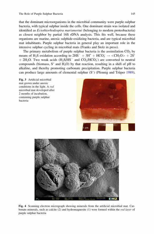
that the dominant microorganisms in the microbial community were purple sulphur
bacteria, with typical sulphur inside the cells. One dominant strain was isolated and
identified as Ectothiorhodospira marismortui (belonging to modern proteobacteria)
as closest neighbor by partial 16S rDNA analysis. This fits well, because these
organisms are marine, anoxic sulphide-oxidizing bacteria, and are typical microbial
mat inhabitants. Purple sulphur bacteria in general play an important role in the
intensive sulphur cycling in microbial mats (Franks and Stolz in press).
The primary metabolism of purple sulphur bacteria is the assimilation CO2 by
means of H2S oxidation according to 2HS� þ 3H+ þ HCO3� ! <CH2O> þ 2S�
þ 2H2O. Two weak acids (H2S/HS� and CO2/HCO3
�) are converted to neutral
compounds (biomass, S� and H2O) by that reaction, resulting in a shift of pH to
alkaline, and thereby promoting carbonate precipitation. Purple sulphur bacteria
can produce large amounts of elemental sulphur (S�) (Pfennig and Tr€uper 1989),
Fig. 3 Artificial microbial
mat grown under anoxic
conditions in the light. A redmicrobial mat developed after
2 months of incubation,
containing purple sulphur
bacteria
Fig. 4 Scanning electron micrograph showing minerals from the artificial microbial mat. Car-
bonate minerals, such as calcite (2) and hydromagnesite (1) were formed within the red layer ofpurple sulphur bacteria
The Role of Purple Sulphur Bacteria 145
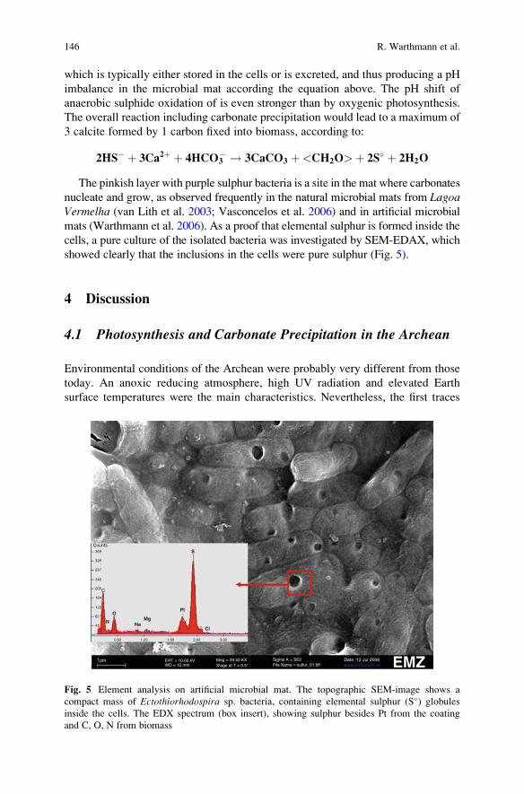
which is typically either stored in the cells or is excreted, and thus producing a pH
imbalance in the microbial mat according the equation above. The pH shift of
anaerobic sulphide oxidation of is even stronger than by oxygenic photosynthesis.
The overall reaction including carbonate precipitation would lead to a maximum of
3 calcite formed by 1 carbon fixed into biomass, according to:
2HS� þ 3Ca2þ þ 4HCO3� ! 3CaCO3 þ<CH2O>þ 2S� þ 2H2O
The pinkish layer with purple sulphur bacteria is a site in the mat where carbonates
nucleate and grow, as observed frequently in the natural microbial mats from LagoaVermelha (van Lith et al. 2003; Vasconcelos et al. 2006) and in artificial microbial
mats (Warthmann et al. 2006). As a proof that elemental sulphur is formed inside the
cells, a pure culture of the isolated bacteria was investigated by SEM-EDAX, which
showed clearly that the inclusions in the cells were pure sulphur (Fig. 5).
4 Discussion
4.1 Photosynthesis and Carbonate Precipitation in the Archean
Environmental conditions of the Archean were probably very different from those
today. An anoxic reducing atmosphere, high UV radiation and elevated Earth
surface temperatures were the main characteristics. Nevertheless, the first traces
369
328
287
246
205C
O
N NaMg
S
Pt
Cl
164
123
82
41
0.60 1.20 1.80 2.40 3.00
Counts
1µm EHT = 10.00 kV Mag = 29.40 KXStage at T = 0.5°
Signal A = SE2File Name = sulfur_01.tiff
Date :12 Jul 2006www.emzunizh.chWD = 12 mm EMZ
Fig. 5 Element analysis on artificial microbial mat. The topographic SEM-image shows a
compact mass of Ectothiorhodospira sp. bacteria, containing elemental sulphur (S�) globules
inside the cells. The EDX spectrum (box insert), showing sulphur besides Pt from the coating
and C, O, N from biomass
146 R. Warthmann et al.
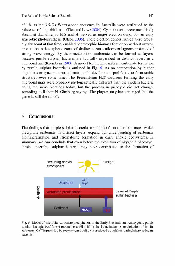
of life as the 3.5-Ga Warrawoona sequence in Australia were attributed to the
existence of microbial mats (Tice and Lowe 2004). Cyanobacteria were most likely
absent at that time, so H2S and H2 served as major electron donor for an early
anaerobic photosynthesis (Olson 2006). These electron donors, which were proba-
bly abundant at that time, enabled phototrophic biomass formation without oxygen
production in the euphotic zones of shallow ocean seafloors or lagoons protected of
strong wave energy. By their metabolism, carbonate can be formed as layers,
because purple sulphur bacteria are typically organized in distinct layers in a
microbial mat (Krumbein 1983). A model for the Precambrian carbonate formation
by purple sulphur bacteria is outlined in Fig. 6. As no competition by higher
organisms or grazers occurred, mats could develop and proliferate to form stable
structures over some time. The Precambrian H2S-oxidizers forming the early
microbial mats were probably phylogenetically different than the modern bacteria
doing the same reactions today, but the process in principle did not change,
according to Robert N. Ginsburg saying “The players may have changed, but the
game is still the same”.
5 Conclusions
The findings that purple sulphur bacteria are able to form microbial mats, which
precipitate carbonate in distinct layers, expand our understanding of carbonate
biomineralization and stromatolite formation in early anoxic ecosystems. In
summary, we can conclude that even before the evolution of oxygenic photosyn-
thesis, anaerobic sulphur bacteria may have contributed to the formation of
Fig. 6 Model of microbial carbonate precipitation in the Early Precambrian. Anoxygenic purple
sulphur bacteria (red layer) producing a pH shift in the light, inducing precipitation of in situ
carbonate. Ca2+ is provided by seawater, and sulfide is produced by sulphur- and sulphate-reducing
bacteria
The Role of Purple Sulphur Bacteria 147
biologically-mediated carbonates resulting in the well-known carbonate lamina-
tion of fossil microbial mats and stromatolites.
References
Allen MA, Goh F, Burns BP, Neilan BA (2009) Bacterial, archaeal and eukaryotic diversity of
smooth and pustular microbial mat communities in the hypersaline lagoon of Shark Bay.
Geobiology 7:82–96
Allwood AC, Walter MR, Kamber BS, Marshall CP, Burch IW (2006) Stromatolite reef from the
Early Archaean era of Australia. Nature 441:714–718
Blankenship RE (1992) Origin and early evolution of photosynthesis Photosynthesis Research
33:91–111
Bontognali T, Vasconcelos C, Warthmann R, McKenzie JA (2007) Dolomite nucleation on
extracellular polymeric substances. In: 17th Annual V M Goldschmidt Conference, Cologne,
pp A108–A108
Bontognali TRR, Vasconcelos C, Warthmann RJ, Dupraz C, Bernasconi SM, McKenzie JA (2008)
Microbes produce nanobacteria-like structures, avoiding cell entombment. Geology
36:663–666
Bontognali TRR, Vasconcelos C, Warthmann RJ, Bernasconi SM, Dupraz C, Strohmenger CJ,
McKenzie JA (2009) Dolomite formation within microbial mats in the coastal sabkha of Abu
Dhabi (United Arab Emirates). Sedimentology, published online: Dec. 8 2009
Bosak T, Greene SE, Newman DK (2007) A likely role for anoxygenic photosynthetic microbes in
the formation of ancient stromatolites. Geobiology 5:119–126
Burns BP, Goh F, Allen M, Neilan BA (2004) Microbial diversity of extant stromatolites in the
hypersaline marine environment of Shark Bay, Australia. Environmental Microbiology
6:1096–1101
Coolen MJ, Overmann J (2000) Functional exoenzymes as indicators of metabolically active
bacteria in 124,000-year-Old sapropel layers of the eastern Mediterranean Sea. Applied and
Environmental Microbiology 66:2589–2598
Dupraz C, Visscher PT (2005) Microbial lithification in marine stromatolites and hypersaline
mats. Trends in Microbiology 13:429–438
Franks J, Stolz JF (2009) Microbial Mats in Earth’s Fossil Record of Life: Geobiology. Earth-
Science Reviews 96:163–172
Grassineau NV et al. (2001) Antiquity of the biological sulphur cycle: evidence from sulphur and
carbon isotopes in 2700 million-year-old rocks of the Belingwe Belt, Zimbabwe. Proceedings
of the Royal Society of London Series B-Biological Sciences 268:113–119
Grotzinger JP, Knoll AH (1999) Stromatolites in Precambrian carbonates: evolutionary mileposts
or environmental dipsticks? Annual review of earth and planetary sciences 27:313–358
Kalkowsky E (1908) Oolith und Stromatolith im norddeutschen Buntsandstein. Zeitschrift der
Deutschen geologischen Gesellschaft 60:68–125
Krumbein WE (1983) Stromatolites – the challenge of a term in space and time. Precambrian
Research 20:493–531
Olson JM (2006) Photosynthesis in the Archean Era. Photosynthesis Research 88:109–117
Pfennig N, Tr€uper HG (1989) Anoxygenic phototrophic bacteria. In: Staley JT, Bryant, MP,
Pfennig N, Holt JG (ed) Bergey’s Manual of Systematic Bacteriology. Williams and Wilkins,
Baltimore/Hong Kong/London/Sydney, pp 1635–1639
Reid RP et al. (2000) The role of microbes in accretion, lamination and early lithification of
modern marine stromatolites. Nature 406:989–992
Riding R (2000) Microbial carbonates: the geological record of calcified bacterial-algal mats and
biofilms. Sedimentology 47:179–214
148 R. Warthmann et al.
Schidlowski M (2001) Carbon isotopes as biogeochemical recorders of life over 3.8 Ga of Earth
history: evolution of a concept. Precambrian Research 106:117–134
Tice MM, Lowe DR (2004) Photosynthetic microbial mats in the 3,416-Myr-old ocean. Nature
431:549–552
van Lith Y, Vasconcelos C, Warthmann R, McKenzie JA (2003) Sulphate-reducing bacteria
induce low-temperature dolomite and high Mg-calcite formation. Geobiology 1:71–79
Vasconcelos C, Warthmann R, McKenzie JA, Visscher PT, Bittermann AG, van Lith Y (2006)
Lithifying microbial mats in Lagoa Vermelha, Brazil: Modern Precambrian relics? Sedimen-
tary Geology 185:175–183
Warthmann R, Vasconcelos C, Sass H, McKenzie JA (2005) Desulfovibrio brasiliensis sp. nov., a
moderate halophilic sulfate-reducing bacterium from Lagoa Vermelha (Brazil) mediating
dolomite formation. Extremophiles 9:255–261
Warthmann R, Vasconcelos C, McKenzie JA (2006) Anaerobic sulfur bacteria inducing lithifica-
tion in modern- and possibly Precambrian stromatolites. Geochimica et Cosmochimica Acta
70:A689–A689
Westall F, de Ronde CEJ, Southam G, Grassineau N, Colas M, Cockell C, Lammer H (2006)
Implications of a 3.472–3.333 Gyr-old subaerial microbial mat from the Barberton greenstone
belt, South Africa for the UV environmental conditions on the early Earth. Philosophical
Transactions of the Royal Society B Biological Sciences 361:1857–1875
The Role of Purple Sulphur Bacteria 149

.
Precipitation of CaCO3 Under
Sulphate-Reduction Conditions
Dorota Wolicka and Andrzej Borkowski
1 Introduction
The major part of geochemical processes in the lithosphere, including the formation
of carbonate minerals, may be linked with the activity of living organisms. These
processes are influenced by physical and chemical factors in the environment that
significantly control the occurrence of particular mineral phases. Because the
reactions of carbonate precipitation are controlled by, e.g., carbon dioxide content,
biotic factors seem to play a significant role in this process. Carbon dioxide is
utilised both as a donor and acceptor of electrons by various groups of microorgan-
isms in their metabolic processes. The key role in CO2 generation is played by
microorganisms, particularly those occurring in environments favouring the pre-
cipitation of carbonate minerals. It should be noted that 108–109 tonnes of carbon
are annually deposited as CaCO3 in the oceans.
The possibility of CaCO3 formation in favourable physical and chemical condi-
tions is enormous because of the very low value of the CaCO3 solubility constant
[Ksol ¼ 10�8.32]. Due to this fact, even a low content of carbon dioxide introduced
into an environment containing Ca2+ ions may cause concentrations to exceed the
solubility-product limit and result in the precipitation of calcium carbonate.
It should be remembered, however, that the formation of CaCO3 is strictly linked
with the physical and chemical conditions of the environment, mainly pH (Hammes
and Verstraete 2002).
D. Wolicka (*)
Institute of Geochemistry, Mineralogy and Petrology, University of Warsaw, ul. Zwirki i Wigury
93, 02-089 Warsaw, Poland
e-mail: [email protected]
A. Borkowski
Department of Soil Science, University of Life Sciences (SGGW), ul. Nowoursynowska 159,
02-776 Warsaw, Poland
J. Reitner et al., Advances in Stromatolite Geobiology,Lecture Notes in Earth Sciences 131, DOI 10.1007/978-3-642-10415-2_10,# Springer-Verlag Berlin Heidelberg 2011
151
The solubility of calcite in water depends also on the temperature, but this
relationship does not favour the precipitation of calcite at low temperatures. The
equation showing the reaction of CaCO3 dissolution is as follows:
CaCO3 $ Ca2þþCO32� DH0 ¼ �13kJ/mol (1)
Due to an enthalpy below zero, the reaction is exothermic and an increase in the
solubility constant with a decrease of water temperature is expected. According to
the van ’t Hoff equation, it can be calculated that, e.g., at a water temperature of
5�C, the Ksol of CaCO3 will increase to 10�8.14. Due to this fact, carbonate deposits,
particularly calcite and aragonite, are not formed on the seabeds of deep oceans.
The boundary depth is assumed to be at 4.5–5 km and is denoted as the carbonate
compensation depth (CCD).
In the process of carbon dioxide generation, an important role is played by different
groups of microorganisms that are responsible for the mobilisation of carbon and its
biogeochemical cycle. Numerous groups of bacteria have the ability to alkalise the
environment, transforming Ca(HCO3)2 into insoluble CaCO3. Conversely, other
bacteria, e.g., those occurring in marine deposits, have the ability to dissolve
CaCO3. It should be noted that carbon occurring in carbonates is available to living
organisms only in a restricted range, e.g., due to the dissolution of CaCO3 during
sulphide oxidisation (2) or ethanol fermentation (3) (Dupraz and Visscher 2005).
3HS�þ 4O2 þCaCO3þHCO3� ! 2[CH2O]þCa2þþ 3SO4
2� (2)
3[CH2O]þCaCO3þH2O ! HCO3�þCa2þþC2H5OH (3)
Biological formation of carbonates is the result of the metabolic activities of
many different groups of microorganisms, both aerobic and anaerobic. Carbonates
can be formed as the by-products of various biological processes, including photo-
synthesis, e.g., in blue-green bacteria, as evidenced by the formation of stromato-
lites, as well as in photosynthetic bacteria or during anaerobic sulphate reduction
(Borowitzka 1989; Castanier et al. 1999; Ehrlich 2001; Dittrich et al. 2004; Dupraz
and Visscher 2005).
In ecosystems with a restricted oxygen supply, such as the bottom deposits of
lakes, seas and oceans, are found bacteria that acquire energy for their metabolic
processes through anaerobic transpiration (Postgate 1984). Very important among
this group are the sulphate-reducing bacteria (SRB), which take part in almost all
mineral- and rock-forming processes in anaerobic conditions (Lysnes et al. 2004).
The SRB comprise a phylogenetically diverse group of anaerobic bacteria display-
ing physiological and ecological similarities. Their presence was noted for the first
time in laboratory conditions by Beijerinck in 1985 (Gibson 1990). A characteristic
environment for SRB occurrence is marine deposits, in which the sulphate concen-
tration averages 28 mM (DeWit 1992), as well as oil fields and crude-oil magazines
(Magot et al. 2000). The preferred carbon sources for SRB are compounds of low
molecular weight, such as organic acids (i.e., propionate, formate and malonate),
152 D. Wolicka and A. Borkowski
volatile organic acids (i.e., acetate), and alcohols (e.g., ethanol, propanol and
butanol) (Fauque et al. 1991). Single SRB species are known to utilise amino
acids as the sole carbon source: Desulfovibrio aminophilus (Baena et al. 1998),
Desulfobacterium vacuolatum (Rees et al. 1997), and Desulfovibrio mexicanus(Hernandez-Eugenio et al. 2000). Some species, e.g., Desulfotomaculum antarc-ticus, may utilise glucose as the sole carbon source, but this is a very rare case
among SRB (Fauque et al. 1991). All SRB are Gram-negative, excluding the
Desulfonema species. This group of bacteria is extremely variable; it includes soil
and water types, psychrophilic, meso- and thermophilic, halophilic and barophilic
bacteria. The utilisation of low-molecular-weight compounds by SRB is linked to
the fact that they do not possess hydrolytic enzymes and are so not able to
biodegrade polymers. Thus, they play a key role in the final stage of organic-matter
biodegradation in environments containing sulphates utilised as the last acceptors
of electrons in the transpiration cycle. In the natural environment, the source of
sulphate ions may be from easily soluble sulphates such as Na2SO4, or even
hanebachite (CaSO4), jarosite ***(KFe3[OH]6j(SO4)2), anglesite (PbSO4), barite
(BaSO4) (Karnachuk et al. 2002) or gypsum (Wolicka 2006).
Mixed populations of SRB have the ability to precipitate different minerals, e.g.,
sulphates (gypsum and anhydrite), carbonates (calcite, dolomite and siderite)
(Perry and Taylor 2006), and phosphates (apatite) (Wolicka 2006). The formation of
dolomite, calcite or aragonite in typical SRB environments and in conditions favour-
ing the reduction of sulphates typically takes place in anoxic lagoons with a high
salinity (Warthman et al. 2000), in gypsum and anhydrites (Peckmann et al. 1999),
as well as in sulphate-rich natural lakes with a high salinity (Wright 2000).
In a different process, SRB may also play an indirect although important role in
carbonate production. Some of these bacteria very often form close relationships
with methane-producing anaerobic microorganisms, i.e., Archaea. These microor-
ganisms, previously classified as bacteria, have the ability to produce methane from
simple organic compounds or directly from carbon dioxide. Somemay utilise carbon
dioxide from the biodegradation of organic compounds conducted by SRB living in
similar conditions. This relationship may develop into a very close relationship
known as syntrophy, where SRB utilise the methane produced, oxidising it to carbon
dioxide, which then may be used once again by the archaean syntrophic partner.
The sulphates used by SRB in the transpiration process are supplied from the
external environment. This process is known as AOM (anaerobic oxidation of
methane) and proceeds according to the following equation (Valentine 2002):
CH4þSO42� ! HCO3
� + HS�þH2O (4)
The process of anaerobic methane oxidation may be a crucial factor in the
carbon cycle for two reasons. Firstly, it plays an important role in the reactions
by which global methane production is transformed into carbon dioxide. Secondly,
this process can be the key process in the formation of carbonate minerals (Moore
et al. 2004), particularly within the continental shelves, where the AOM process is
most intense (Miyajima and Wada 1999; Ogrinc et al. 2003).
Precipitation of CaCO3 Under Sulphate-Reduction Conditions 153
The study was focused on testing the possibility of carbonate formation in condi-
tions favouring SRB selection. The aim of study was to elucidate the role of SRB in
the biogeochemistry of CaCO3 precipitation under sulphate-reducing conditions.
2 Methodology
2.1 Microorganisms
The bacterial communities were isolated from Vistula River mud (Poland). In the
selection of anaerobic bacterial communities, lactate and/or ethanol were used as the
sole carbon sources (Postgate 1984). The microorganisms were multiplied using
the method of microcosms: samples of the mud (10 g) were placed in transparent
100-mL containers. The containers were tightly closed and set aside for 6 weeks to
allow the selection of anaerobic, sulphidogenic consortia capable of carrying out the
reduction of sulphate. The cultures were incubated at room temperature (~20 �C).
2.2 Media
A modified Postgate medium (Postgate 1984), in which Na2SO4 was replaced with
gypsum at a concentration equivalent to 4.5 g Na2SO4/L and yeast extract and
sodium citrate were not added, was used to select SRB able to oxidise only one
carbon source. The second medium was a minimal medium (1 g/L NH4Cl). Both
media were enriched with sodium lactate (3 mg/L) and/or ethanol (3 mg/L). All
media contained gypsum (5 g/L) as the sole electron acceptor for SRB and the
source of calcium. Resazurin was added to all cultures at 0.001 g/L as an indicator
of redox conditions in the medium.
2.3 Determinations
The time from inoculation to the end of experiment was 14 days. Sulphates in the
cultures were determined every day with the hot barium method, chemical oxygen
demand (COD) was determined by the dichromate method, and pH determined
using a pH meter; the pH of the cultures was corrected with 0.1 N HCl or
0.1 N NaOH as needed. Determinations involving postculture sediments (residual
gypsum and sediments precipitated during bacterial activity) and fluids were made
using X-ray analysis with a DRON- 2 X-ray diffractometer. The bacterial commu-
nities obtained were observed using a scanning electron microscope (SEM).
154 D. Wolicka and A. Borkowski
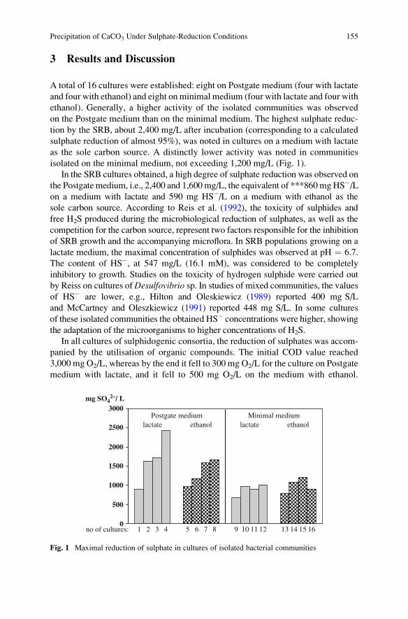
3 Results and Discussion
A total of 16 cultures were established: eight on Postgate medium (four with lactate
and four with ethanol) and eight on minimal medium (four with lactate and four with
ethanol). Generally, a higher activity of the isolated communities was observed
on the Postgate medium than on the minimal medium. The highest sulphate reduc-
tion by the SRB, about 2,400 mg/L after incubation (corresponding to a calculated
sulphate reduction of almost 95%), was noted in cultures on a medium with lactate
as the sole carbon source. A distinctly lower activity was noted in communities
isolated on the minimal medium, not exceeding 1,200 mg/L (Fig. 1).
In the SRB cultures obtained, a high degree of sulphate reduction was observed on
the Postgate medium, i.e., 2,400 and 1,600 mg/L, the equivalent of ***860mgHS�/Lon a medium with lactate and 590 mg HS�/L on a medium with ethanol as the
sole carbon source. According to Reis et al. (1992), the toxicity of sulphides and
free H2S produced during the microbiological reduction of sulphates, as well as the
competition for the carbon source, represent two factors responsible for the inhibition
of SRB growth and the accompanying microflora. In SRB populations growing on a
lactate medium, the maximal concentration of sulphides was observed at pH ¼ 6.7.
The content of HS�, at 547 mg/L (16.1 mM), was considered to be completely
inhibitory to growth. Studies on the toxicity of hydrogen sulphide were carried out
by Reiss on cultures ofDesulfovibrio sp. In studies of mixed communities, the values
of HS� are lower, e.g., Hilton and Oleskiewicz (1989) reported 400 mg S/L
and McCartney and Oleszkiewicz (1991) reported 448 mg S/L. In some cultures
of these isolated communities the obtained HS� concentrations were higher, showing
the adaptation of the microorganisms to higher concentrations of H2S.
In all cultures of sulphidogenic consortia, the reduction of sulphates was accom-
panied by the utilisation of organic compounds. The initial COD value reached
3,000 mg O2/L, whereas by the end it fell to 300 mg O2/L for the culture on Postgate
medium with lactate, and it fell to 500 mg O2/L on the medium with ethanol.
0
500
1000
1500
2000
2500
3000
no of cultures: 1 2 3 4 5 6 7 8 9 10 11 12 13 14 15 16
Postgate medium Minimal mediumlactate ethanol lactate ethanol
mg SO42–/ L
Fig. 1 Maximal reduction of sulphate in cultures of isolated bacterial communities
Precipitation of CaCO3 Under Sulphate-Reduction Conditions 155
From the stoichiometry of the sulphate-reduction process (1 mole of HS� per mole
of SO4), the ratio of the sulphides produced and the COD consumed by the entire
bacterial community is known to be COD/SO4 ¼ 0.67 (Hao et al. 1996); thus, the
participation of SRB in the reduction of organic compounds was calculated (Fig. 2).
SRBothers
rest
Postgate medium with lactate
rest
SRB
others
Postgate medium with ethanol
Fig. 2 Participation of SRB in COD reduction
Fig. 3 Isolated sulphate-reducing bacterial communities in Postgate medium with lactate (a–c)
and with ethanol (d–f) (SEM micrographs)
156 D. Wolicka and A. Borkowski
During incubation, the culture supernatants were sampled for microscopic
observations. They confirmed that the isolated communities were heterogeneous
and contained various morphological forms. The communities included such forms
as vibrio, spirillum, bacterium and coccus. The bacteria were accompanied by
mineral phases in the form of both carbonates and sulphates (Fig. 3).
After incubation, diffractometric analysis of the postculture sediments was
performed (Fig. 4). In the postculture sediments, the presence of secondary mineral
phases was confirmed, e.g., calcite resulting from microbial activity. It is interesting
to note that in the culture on Postgate medium with ethanol, which was less active
than the same culture on a medium with lactate (Fig. 1), the postculture sediments
contained only calcite, showing that the entire amount of gypsum introduced to the
culture underwent biotransformation. Part of the sulphate ions may remain in the
solution (Wolicka, unpublished data) and cannot be used by the isolated SRB
community due to autoinhibition by HS�. There are at least two reasons for this
phenomenon. First, CO2 formed in the cultures derived not only from the SRB but
also from the accompanying microflora, thus increasing the content of carbon
dioxide in the culture. The excess calcium ions in postculture solutions can easily
bind with CO2 forming CaCO3 when the pH is not acidic. Additionally, the process
of calcite formation could possibly be linked with the substitution of gypsum by
carbonates (Fig. 5).
0
200
400
600
800
1000
lmp
KKKKK
K
K
KKKK
K
b
0200400600800
1000
2 theta (Co K alpha)
lmp
c
50403020100 70
G
G
G
GGK
KK KGG
GG
KK G GG
GG
G
GG
60
0
200
400
600
800
1000
lmp
G
G
G GGGG
GG
G
G
GG
a
Fig. 4 Diffractograms: gypsum (a) and post-cultures sediments on Postgate medium with ethanol
(b) and with lactate (c). The symbols indicate: G gypsum, K calcite
Precipitation of CaCO3 Under Sulphate-Reduction Conditions 157
4 Conclusions
The isolated SRB cultures showed a high activity for sulphate reduction with
simultaneous effective biodegradation of the applied organic compounds (lactate
and ethanol). In effect, biotransformation and the formation of secondary mineral
phases in the form of calcite took place in the postculture sediments. Chemical
analyses showed that SRB played a crucial role in the biodegradation of organic
compounds, as reflected in the high fractional COD reductions by these bacteria. It
thus seems likely that most of the calcite formed in the cultures was the result of
SRB activity. The involvement of other groups of microorganisms accompanying
SRB in environments favouring the formation of carbonates cannot be ruled out;
however, it appears that the role of these microorganisms is much less significant,
particularly in environments rich in sulphates, where, in the last phase of miner-
alisation, the SRB successfully compete for organic carbon with other groups of
microorganisms. In light of this fact, the role of SRB in the formation of carbonates
in anaerobic environments is extremely important, but is often underestimated.
References
Baena S, Fardeau ML, Labat M, Ollivier B, Garcia JL, Patel BKC 1998Desulfovibrio aminophilussp.nov., a novel amino acid degrading and sulfate reducing bacterium from an anaerobic dairy
wastewater lagoon. Systematic and applied microbiology 21:498–504
Borowitzka MA 1989 Carbonate calcification in algae – initiation and control. In: Mann S,
Webb J, Wiliams RJP (eds) Biomineralization, Weinheim, Germany: VCH Verlagsge-
sellschaft pp 116–135.
Castanier S, Metayer-Levrel GL, Perthuisot J-P 1999 Ca-carbonates precipitation and limestone
genesis – the microbiogeologist point of view. Sedimentary Geology 126:9–23
other microorganismsmainly
fermentative bacteria
CO2source of organic
carbon
SO42–
SO42–
SO42–SO4
2–
SO42–
SO42–
SO42–SO4
2–
Ca2+
Ca2+
Ca2+
Ca2+ Ca2+Ca2+
Ca2+SRBHS–
HS–
HS–
HS–
HS–
CaSO4 CaCO3
CaCO3
deposite
Fig. 5 Participation of sulphate-reducing bacteria in carbonate formation
158 D. Wolicka and A. Borkowski
De Wit R 1992 Sulphide-containing environments. In: Lederberg J (ed) Encyclopedia of Microbi-
ology, San Diego, CA: Academic 4:105–121
Dittrich M, Kurz P, Wehrli B 2004 The role of autotrophic picocyanobacteria in calcite precipita-
tion in an oligotrophic lake. Geomicrobiology Journal 21:45–53
Dupraz C, Visscher P 2005 Microbial lithification in marine stromatolites and hypersaline mats.
Trends in Microbiology 13(9):429–438
Ehrlich H 2001 Microbial formation and degradation of carbonates. In: Geomicrobiology,
New York: Mercel Dekker, Inc. pp 183–227
Fauque G, Legall J, Barton LL 1991 Sulfate-reducing and sulfur reducing bacteria. In: Shively
JMI, Barton LL (eds) Variations in autotrophic life. New York: Academic
Gibson G 1990 Physiology and ecology of the sulphate-reducing bacteria. Journal of Applied
Bacteriology 69:769–797
Hammes F, Verstraete W 2002 Key roles of pH and calcium metabolism in microbial carbonate
precipitation. Re/Views in Environmental Science & Bio/Technology 1:3–7
Hao OJ, Chen JM, Huang L, Buglass RL 1996 Sulfate-reducing bacteria. Critical reviews in
environmental science and technology 26:155–187
Hernandez-Eugenio G, Fardeau ML, Patel BKC, Garcia JL, Ollivier B 2000 Desulfovibriomexicanus sp.nov., a sulfate-reducing bacterium isolated from an upflow anaerobic sludge
blancet (UASB) reactor treating cheese wastewaters. Anaerobe 6:305–312
Hilton BL, Oleskiewicz JA 1989 Sulfide-induced inhibition of anaerobic digestion. Journal of
Environmental Engineering 144:1377–1391
Karnachuk OV, Kurochkina SY, Tuovinen OH 2002 Growth of sulfate-reducing bacteria with
solid-phase electron acceptors. Applied Microbiology Biotechnology 58:482–486
Lysnes K, Thorseth IH, Steinsbu BO, Øvreas L, Torsvik T, Pedersen K 2004Microbial community
diversity in seafloor basalt from the Artic spreading riges. FEMS Microbiological Ecology
50:213–230
Magot M, Ollivier B, Patel BKC 2000 Microbiology of petroleum reservoirs. Antonie van
Leeuwenhoeck 77:103–116
McCartney DM, Oleszkiewicz JA 1991 Sulfide inhibition of anaerobic degradation of lactate and
acetate. Water Research 25:203–209
Miyajima T, Wada E 1999 Sulfate-induced isotopic variation in biogenic methane from a tropical
swamp without anaerobic methane oxidation. Hydrobiologia 382:113–118
Moore TS, Murray RW, Kurtz AC, Schrag DP 2004 Anaerobic methane oxidation and the
formation of dolomite. Earth and Planetary Science Letters 229:141–154
Ogrinc N, Hintelmann H, Eckley C, Lojen S 2003 Biogeochemical influence on carbon isotope
signature in boreal lake sediments. Hydrobiologia 494:207–213
Peckmann J, Thiel V, Michaelis W, Clari P, Gaillard C, Martire L, Reitner J 1999 Cold seep
deposits of Beauvoisin (Oxfordian; southeastern France) and Marmorito (Miocene; northern
Italy): microbially induced, authigenic carbonates. International Journal of Earth Sciences
88:60–75
Perry CT, Taylor KG 2006 Inhibition of dissolution within shallow water carbonate sediments:
impacts of terrigenous sediment input on syn-depositional carbonate diagenesis. Sedimentology
53:495–513
Postgate JR 1984The sulfate-reducing bacteria. 2nd edition. CambridgeUniversity Press, Cambridge
Rees GN, CG Harfoot, AJ Sheehy 1997 Amino acid degradation by the mesophilic sulfate-
reducing bacterium Desulfobacterium vacuolatum. Archives of Microbiology 169:76–80
Reis MAM, Almeida JS, Lemos PC, Carrondo MJT 1992 Effect of hydrogen sulfide on growth of
sulfate reducing bacteria. Biotechnology and Bioengineering 40:593–600
Valentine DL 2002 Biogeochemistry and microbial ecology of methane oxidation in anoxic
environments: a review. Antonie van Leeuwenhoek 81:271–282
Warthman R, Van Lith Y, Vasconcelos C, Mckenzie JA, Karpoff AM 2000 Bacterially induced
dolomite precipitation in anoxic culture experiments. Geology 28:1091–1094
Precipitation of CaCO3 Under Sulphate-Reduction Conditions 159

Wolicka D 2006 Biotransformation of phosphogypsum in cultures of bacteria selected from
petroleum W: Proceedings of the International Conference Protection and Restoration of the
Environmental VIII Chania, Greece, 3–7 July, pp 217–218
Wright DT 2000 Benthic microbial communities and dolomite formation in marine and lacustrine
environments – a new dolomite model. In: Glenn CR, Lucas J, Prevot L (red). Marine
Authigenesis from Global to microbial. SEPM Spec Publ 66:7–20
160 D. Wolicka and A. Borkowski
Myxococcus xanthus Colony Calcification:
An Study to Better Understand the Processes
Involved in the Formation of this
Stromatolite-Like Structure
Concepcion Jimenez-Lopez, Kaoutar Ben Chekroun, F. Jroundi,
Manuel Rodrıguez-Gallego, Jose Maria Arias,
and Maria Teresa Gonzalez-Munoz
1 Introduction
Calcium carbonate precipitation is a common phenomenon in nature and has been
observed to be mediated by a number of microorganisms (for a review, see
Castanier et al. 2000; Wright and Oren 2005). Bacterially induced carbonate
mineralization is important in a wide range of processes including atmospheric
CO2 budgeting (Braissant et al. 2002; Ehrlich 2002), carbonate sediment and rock
formation (Riding 2000; Ben Chekroun et al. 2004), biogeochemical cycling of
elements (Banfield and Nealson 1997; Ehrlich 2002), human pathological concre-
tions (Keefe 1976) and have also relevant technological applications for example in
metal-contaminated groundwater bioremediation (Warren et al. 2001; Fujita et al.
2004; Mitchell and Ferris 2005; K€ohler et al. 2007) and even in conservation of
ornamental stone (Rodriguez-Navarro et al. 2003; Jimenez-Lopez et al. 2007).
Much research has been dedicated to the study of extracelullar precipitation of
calcium carbonate by different microorganisms [i.e.: sulfate-reducing bacteria and
cyanobacteria (i.e.: Wright 1999), Bacillus (i.e.: Castanier et al. 2000), Myxobac-
teria (i.e.: Ben Chekroun et al. 2004), Halobacillus (i.e.: Rivadeneyra et al. 2004)
and Pseudomonas (i.e.: Baskar et al. 2006)]. Most of these studies are mainly
dedicated to the understanding of the role of bacteria on the specific biomineraliza-
tion phenomenon and to the characterization of the resulting biominerals. In this
context, passive and active mechanisms have been proposed for the precipitation
of bacterially induced calcium carbonate. While passive mechanisms for calcium
C. Jimenez-Lopez, K.B. Chekroun, F. Jroundi, J.M. Arias, and M.T. Gonzalez-Munoz (*)
Departamento de Microbiologıa, Facultad de Ciencias, Universidad de Granada, Campus de
Fuentenueva s/n, 18071, Granada, Spain
e-mail: [email protected]
M. Rodrıguez-Gallego
Departamento de Mineralogıa y Petrologıa, Facultad de Ciencias, Universidad de Granada,
Campus de Fuentenueva s/n, 18071, Granada, Spain
J. Reitner et al., Advances in Stromatolite Geobiology,Lecture Notes in Earth Sciences 131, DOI 10.1007/978-3-642-10415-2_11,# Springer-Verlag Berlin Heidelberg 2011
161
carbonate precipitation refer to the mineral formation as the result of changes on the
chemistry of the environment caused by bacterial metabolic activity, active
mechanisms for calcium carbonate precipitation refer to the formation of minerals
mediated by bacterial wall, cell membranes and/or cell debris, which act as nuclei
for crystallization (Wright and Oren 2005; Ercole et al. 2007).
With respect to passive calcium carbonate precipitation, there are a number of
metabolic processes able to create favorable conditions for calcium carbonate
precipitation. One of the most studied passive mechanisms is photosynthesis. In
this regard, phototrophic bacteria like cyanobacteria have been associated with
carbonate precipitation in the context of marine and freshwater sediments. How-
ever, there is controversy regarding the mechanisms leading to the alkalinization
of the environment, crucial for the precipitation of calcium carbonate (Golubic
1973; Ehrlich 1996; McConnaughey and Whelan 1997; Altermann et al. 2006).
Heterotrophic bacteria are also important producers of CaCO3. Such a precipita-
tion occurs as a consequence of chemical changes in the environments where
those microorganisms live, from the release of different metabolites resulting
from a variety of metabolic activities, including ammonification, dissimilatory
nitrate reduction, degradation of urea or uric acid and dissimilatory sulfate
reduction. All these metabolic activities lead to an increase in the pH of the
environment and also to the production of CO2, which results in the rise of the
supersaturation with respect to the relevant mineral phase, thus triggering
the precipitation of calcium carbonate (i.e.: Krumbein 1979; Wright 1999;
Castanier et al. 2000; Riding 2000; Visscher et al. 2000; Wright 2000; Bachmeier
et al. 2002; Hammes et al. 2003; Rodriguez-Navarro et al. 2003).
Regarding active precipitation of calcium carbonate by bacteria, there are a
number of studies focused in the role of the cell wall, extracellular polymeric
substances (EPS), cell membranes, cell debris and other organics in the precipita-
tion of minerals. In this context, it has been demonstrated that exopolysaccharides,
aminoacids and capsular polysaccharides mediate the precipitation of calcite and
vaterite and even influence the resulting morphology of the crystals (Kawaguchi
and Decho 2002; Braissant et al. 2003; Ercole et al. 2007). Gonzalez-Munoz et al.
(1996) observed the precipitation of calcite and struvite in jellified culture medium
inoculated with cellular membranes ofMyxococcus xanthus. Also, oriented precip-itation of calcium phosphate in the periplasm and inside the cells, associated
with integrated membrane structures have been observed in other studies
(Benzerara et al. 2004). The process of cell calcification and the role of cell wall
and EPS in vaterite precipitation have been further analyzed by Rodriguez-Navarro
et al. (2007).
In spite of the extensive literature regarding calcium carbonate precipitation by
bacteria, EPS, cell membranes and bacterial debris, little is known about the
calcification that would occur in a whole population, where all these factors are
integrated. Such a study is, however, important, since it is a better approach to what
would occur in natural systems where most microbial cells form communities
inside microbial biofilms (Decho 1990, 2000; Sutherland 2001). The understanding
of how colonies calcify and how passive and active mechanisms interact to give rise
162 C. Jimenez-Lopez et al.
to an specific colony calcification pattern would provide hints regarding the envi-
ronmental conditions where such a calcification occurred, the size of the population
involved in the mineralization, how active was the metabolism of such a popula-
tion, and potential gradients along the colony. All these data could provide valuable
information regarding the mineralization processes resulting in the formation of
biomineral structures in sediments (i.e. formation of lithified mats in nature) and
even, as an approximation, in pathological concretions. It is, nevertheless, a first
step and simplistic approximation to understand mechanisms leading to the forma-
tion of calcified structures, since it has been pointed out that microbial mats in
nature are constituted by different populations which create a more complex
ecosystem (Visscher and Stolz 2005). This work focus in investigating passive
and active mechanisms involved in the calcification of colonies and which factors
determine the calcification and formation of specific patterns observed.
2 Materials and Methods
2.1 Microorganism and Culture Media
The microorganism used wasMyxococcus xanthus (strain number 422 provided by
the Spanish Type Culture Collection, Burjasot, Valencia, Spain). M. xanthus, aGram negative, common soil bacterium, belonging to the d-subdivision of the
Proteobacteria, has been chosen since it has been demonstrated that such bacteria
is an effective producer of extracellular calcium carbonate (calcite and vaterite;
Rodriguez-Navarro et al. 2003, 2007) and that calcium carbonate may even be also
induced in the sole presence of cell membranes by a purely active mechanism
(Gonzalez-Munoz et al. 1996).
For inoculum preparation, M. xanthus was cultured in liquid medium CT
(Dworkin and Kaiser 1993) for 48 h at 28�C and at 0.455 � g (Gallenkamp rotary
shaker) until it reached the exponential growth phase. The size of the population at
this time was ~2 � 109 cells/ml (Gonzalez-Munoz et al. 2000).
Biomineralization tests were conducted in M-3 and CC solid culture media. Two
different culture media were used in the experiments to allow for different initial
supersaturation conditions with respect to the relevant phase of calcium carbonate
and also to test conditions in which the growth rate of M. xanthus is different.
Jellified culture media were used to simulate the biofilms in which most cells are
embedded in natural environments (Costerton et al. 1987). To prepare M-3 and CC
solid culture media, 1.8 % agar-agar (Difco) was added to the filtered liquid culture
media M-3 and CC described, respectively, in Rodriguez-Navarro et al. (2003) and
Ben Chekroun (2000), with the following composition (percentages represent
wt/v): M-3 (1% Bacto Casitone, 1% Ca(CH3COO)2·4H2O, 0.2% K2CO3·1/2H2O
in distilled water, pH 8), and CC (0.3% Bacto Casitone, 0.4% Ca(CH3COO)2·4H2O,
0.1% CaCl2, 0.3% NaHCO3, 0.1% yeast extract in distilled water, pH 8). Those
Myxococcus xanthus Colony Calcification 163
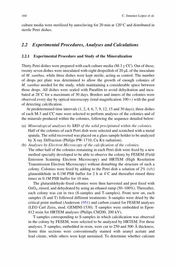
culture media were sterilized by autoclaving for 20 min at 120�C and distributed in
sterile Petri dishes.
2.2 Experimental Procedures, Analyses and Calculations
2.2.1 Experimental Procedure and Study of the Mineralization
Thirty Petri dishes were prepared with each culture media (M-3 y CC). Out of these,
twenty seven dishes were inoculated with eight drops/dish of 20 mL of the inoculum
of M. xanthus, while three dishes were kept sterile, acting as control. The number
of drops per plate was determined to allow the growth of enough colonies of
M. xanthus needed for the study, while maintaining a considerable space between
those drops. All dishes were sealed with Parafilm to avoid dehydration and incu-
bated at 28�C for a maximum of 30 days. Borders and inners of the colonies were
observed every day by optical microscopy (total magnification 100�) with the goal
of detecting calcification.
At predetermined time intervals (1, 2, 4, 6, 7, 9, 12, 15 and 30 days), three dishes
of each M-3 and CC runs were selected to perform analyses of the colonies and of
the minerals produced within the colonies, following the sequence detailed below:
(a) Mineralogical analyses by XRD of the solid precipitated within the colonies.Half of the colonies of each Petri dish were selected and scratched with a metal
spatula. The solid recovered was placed on a glass sample holder to be analyzed
by X-ray Diffraction (Philips PW-1710, Cu Ka radiation).
(b) Analyses by Electron Microscopy of the calcification of the colonies.The other half of the colonies remaining in each Petri dish were fixed by a new
method specially developed to be able to observe the colony by FESEM (Field
Emission Scanning Electron Microscopy) and HRTEM (High Resolution
Transmission Electron Microscopy) without disturbing the structure of such a
colony. Colonies were fixed by adding to the Petri dish a solution of 2% (v/v)
glutaraldehide in 0.1M PSB buffer for 2 h at 4�C and thereafter rinsed three
times in 0.1M PSB buffer for 10 min.
The glutaraldehyde-fixed colonies were then harvested and post fixed with
OsO4, rinsed, and dehydrated by using an ethanol ramp (50–100%). Thereafter,
each colony was cut in two (S-samples and T-samples). From now on, each
samples (S and T) followed different treatments: S-samples were dried by the
critical point method (Anderson 1951) and carbon coated for FESEM analyses
(LEO Carl Zeiss, mod. GEMINI-1530). T-samples were embedded in Epon-
812 resin for HRTEM analyses (Philips CM200, 200 kV).
T-samples corresponding to S-samples in which calcification was observed
in the colony by FESEM, were selected to be analyzed by HRTEM. For those
analyses, T-samples, embedded in resin, were cut to 250 and 500 A thickness.
Some thin sections were conventionally stained with uranyl acetate and
lead citrate, while others were kept unstained. To determine whether calcium
164 C. Jimenez-Lopez et al.
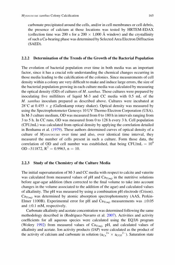
carbonate precipitated around the cells, and/or in cell membranes or cell debris,
the presence of calcium at those locations was tested by HRTEM-EDAX
(collection time was 200 s for a 200 � 1,000 A window) and the crystallinity
of such a Ca-bearing phase was determined by SelectedArea Electron Diffraction
(SAED).
2.2.2 Determination of the Trends of the Growth of the Bacterial Population
The evolution of bacterial population over time in both media was an important
factor, since it has a crucial role understanding the chemical changes occurring in
those media leading to the calcification of the colonies. Since measurements of cell
density within a colony are very difficult to make and induce large errors, the size of
the bacterial population growing in each culture media was calculated by measuring
the optical density (OD) of cultures ofM. xanthus. Those cultures were prepared byinoculating five milliliters of liquid M-3 and CC media with 0.5 mL of the
M. xanthus inoculum prepared as described above. Cultures were incubated at
28�C at 0.455 � g (Gallenkamp rotary shaker). Optical density was measured by
using the Spectrophotometer Genesys 10 UV Thermo Electron Corporation (USA).
In M-3 culture medium, ODwas measured from 0 to 180 h in intervals ranging from
3 to 5 h. In CC runs, OD was measured from 0 to 128 h every 3 h. Cell population
(CFU/mL) was calculated from optical density by applying the correlation detailed
in Benhamu et al. (1979). These authors determined curves of optical density of a
culture of Myxococcus over time and also, over identical time interval, they
measured the number of cells present in such a culture. Form those data, the
correlation of OD and cell number was established, that being CFU/mL ¼ 109
OD –311872, R2 ¼ 0.9963, n ¼ 10.
2.2.3 Study of the Chemistry of the Culture Media
The initial supersaturation of M-3 and CC media with respect to calcite and vaterite
was calculated from measured values of pH and CaT(aq) in the nutritive solutions
before agar-agar addition (then corrected to the final volume to take into account
changes in the volume associated to the addition of the agar) and calculated values
of alkalinity. The pH was measured by using a combination pH electrode (Crison).
CaT(aq) was determined by atomic absorption spectrophotometry (AAS, Perkin-
Elmer 1100B). Experimental error for pH and CaT(aq) measurements was �0.05
and �0.1 mM, respectively.
Carbonate alkalinity and acetate concentration was determined following the same
methodology described in (Rodriguez-Navarro et al. 2007). Activities and activity
coefficients for all aqueous species were calculated using the EQ3/6 program
(Wolery 1992) from measured values of CaT(aq), pH, and calculated values of
alkalinity and acetate. Ion activity products (IAP) were calculated as the product of
the activity of calcium and carbonate in solution (aCa2+ � aCO3
2�). Saturation state
Myxococcus xanthus Colony Calcification 165
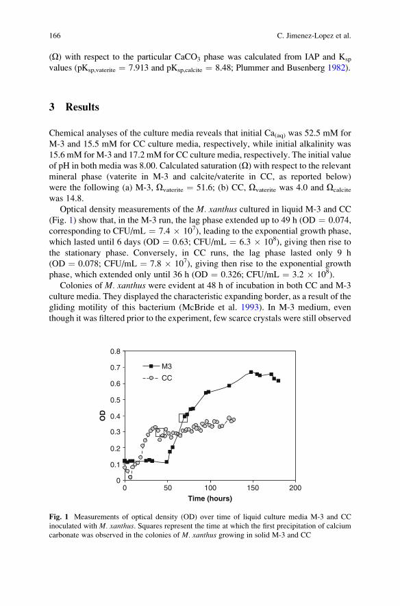
(O) with respect to the particular CaCO3 phase was calculated from IAP and Ksp
values (pKsp,vaterite ¼ 7.913 and pKsp,calcite ¼ 8.48; Plummer and Busenberg 1982).
3 Results
Chemical analyses of the culture media reveals that initial Ca(aq) was 52.5 mM for
M-3 and 15.5 mM for CC culture media, respectively, while initial alkalinity was
15.6 mM for M-3 and 17.2 mM for CC culture media, respectively. The initial value
of pH in both media was 8.00. Calculated saturation (O) with respect to the relevantmineral phase (vaterite in M-3 and calcite/vaterite in CC, as reported below)
were the following (a) M-3, Ovaterite ¼ 51.6; (b) CC, Ovaterite was 4.0 and Ocalcite
was 14.8.
Optical density measurements of the M. xanthus cultured in liquid M-3 and CC
(Fig. 1) show that, in the M-3 run, the lag phase extended up to 49 h (OD ¼ 0.074,
corresponding to CFU/mL ¼ 7.4 � 107), leading to the exponential growth phase,
which lasted until 6 days (OD ¼ 0.63; CFU/mL ¼ 6.3 � 108), giving then rise to
the stationary phase. Conversely, in CC runs, the lag phase lasted only 9 h
(OD ¼ 0.078; CFU/mL ¼ 7.8 � 107), giving then rise to the exponential growth
phase, which extended only until 36 h (OD ¼ 0.326; CFU/mL ¼ 3.2 � 108).
Colonies of M. xanthus were evident at 48 h of incubation in both CC and M-3
culture media. They displayed the characteristic expanding border, as a result of the
gliding motility of this bacterium (McBride et al. 1993). In M-3 medium, even
though it was filtered prior to the experiment, few scarce crystals were still observed
200150100500
0.8
0.7
0.6
0.5
0.4
0.3
0.2
0.1
0
Time (hours)
OD
M3
CC
Fig. 1 Measurements of optical density (OD) over time of liquid culture media M-3 and CC
inoculated with M. xanthus. Squares represent the time at which the first precipitation of calcium
carbonate was observed in the colonies of M. xanthus growing in solid M-3 and CC
166 C. Jimenez-Lopez et al.
at the beginning of the experiment in the bulk medium, outside the colony (Fig. 2a).
That was not the case in CC-runs (Fig. 3a). Nevertheless, new carbonate precipita-
tion in the bulk medium (outside the colonies) were not observed in either M-3 or
CC media during the time course experiment.
The evolution of the mineralization of the colonies in both M-3 and CC culture
media is shown in Figs. 2 and 3, respectively. InM-3 culture medium, calcification of
the colonies was observed with time, almost parallel to the growth of the population,
firstly on the borders of the colonies (at 3 days) and later also in the inner part of the
colonies. Few scarce crystals formed in the border of the colonies at 3 days. With
Fig. 2 Time evolution of the colony calcification in M-3 culture medium as observed by optical
microscope. (a) Colony observed at 48 h; (b) Border of the colony at 3 days; (c) Border at 4 days;
(d) Border at 9 days; (e) Border at 12 days; (f) Inner of the colony at 12 days; (g) Border at 30 days;
(h) colony at 30 days. Scale bars are 1 mm
Fig. 3 Time evolution of the colony calcification in CC culture medium as observed by optical
microscope. (a) Colony observed at 48 h; (b) Border of the colony at 48 h; (c) Border at 4 days;
(d) Border at 9 days; (e) Colony at 12 days; (f) Border and inner at 12 days; (g) Colony at 30 days;
(h) Border and inner of the colony at 30 days
Myxococcus xanthus Colony Calcification 167
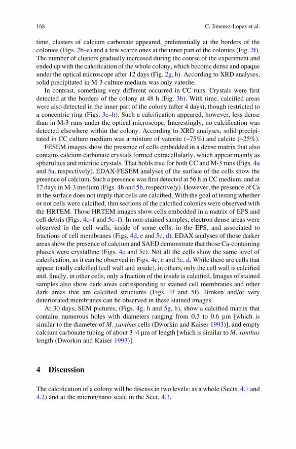
time, clusters of calcium carbonate appeared, preferentially at the borders of the
colonies (Figs. 2b–e) and a few scarce ones at the inner part of the colonies (Fig. 2f).
The number of clusters gradually increased during the course of the experiment and
ended up with the calcification of the whole colony, which become dense and opaque
under the optical microscope after 12 days (Fig. 2g, h). According to XRD analyses,
solid precipitated in M-3 culture medium was only vaterite.
In contrast, something very different occurred in CC runs. Crystals were first
detected at the borders of the colony at 48 h (Fig. 3b). With time, calcified areas
were also detected in the inner part of the colony (after 4 days), though restricted to
a concentric ring (Figs. 3c–h). Such a calcification appeared, however, less dense
than in M-3 runs under the optical microscope. Interestingly, no calcification was
detected elsewhere within the colony. According to XRD analyses, solid precipi-
tated in CC culture medium was a mixture of vaterite (~75%) and calcite (~25%).
FESEM images show the presence of cells embedded in a dense matrix that also
contains calcium carbonate crystals formed extracellularly, which appear mainly as
spherulites and micritic crystals. That holds true for both CC and M-3 runs (Figs. 4a
and 5a, respectively). EDAX-FESEM analyses of the surface of the cells show the
presence of calcium. Such a presence was first detected at 56 h in CCmedium, and at
12 days inM-3medium (Figs. 4b and 5b, respectively). However, the presence of Ca
in the surface does not imply that cells are calcified. With the goal of testing whether
or not cells were calcified, thin sections of the calcified colonies were observed with
the HRTEM. Those HRTEM images show cells embedded in a matrix of EPS and
cell debris (Figs. 4c–f and 5c–f). In non-stained samples, electron dense areas were
observed in the cell walls, inside of some cells, in the EPS, and associated to
fractions of cell membranes (Figs. 4d, e and 5c, d). EDAX analyses of those darker
areas show the presence of calcium and SAED demonstrate that those Ca-containing
phases were crystalline (Figs. 4c and 5c). Not all the cells show the same level of
calcification, as it can be observed in Figs. 4c, e and 5c, d. While there are cells that
appear totally calcified (cell wall and inside), in others, only the cell wall is calcified
and, finally, in other cells, only a fraction of the inside is calcified. Images of stained
samples also show dark areas corresponding to stained cell membranes and other
dark areas that are calcified structures (Figs. 4f and 5f). Broken and/or very
deteriorated membranes can be observed in these stained images.
At 30 days, SEM pictures, (Figs. 4g, h and 5g, h), show a calcified matrix that
contains numerous holes with diameters ranging from 0.3 to 0.6 mm [which is
similar to the diameter of M. xanthus cells (Dworkin and Kaiser 1993)], and empty
calcium carbonate tubing of about 3–4 mm of length [which is similar toM. xanthuslength (Dworkin and Kaiser 1993)].
4 Discussion
The calcification of a colony will be discuss in two levels: as a whole (Sects. 4.1 and
4.2) and at the micron/nano scale in the Sect. 4.3.
168 C. Jimenez-Lopez et al.
4.1 Patterns of Colony Calcification
It is remarkable the two different colony calcification patterns observed in our
experiments: a whole colony calcification in M-3 runs versus a calcification of
only concentric rings within the colony in CC runs [following a Liesegang-ring
pattern (Henisch 1988), observed previously by Gonzalez-Munoz et al. (2000)
while studying the production of Mg-calcite by M. xanthus]. Both the “whole-
colony calcification” pattern and the “Liesegang-ring” pattern can be explained by
Fig. 4 SEM and HRTEM analyses of the colonies ofM. xanthus cultured in CC medium (a) SEM
image of part of the colony fixed following the fixation method described in the present paper;
(b) EDAX analyses of the surface of the cells; (c)–(f) HRTEM images showing calcified cells and
calcified EPS, (g) and (h) calcified cells embedded in calcified EPS that also contains rounded
crystallites ((h) is a detail of (g)). (a)–(f) colonies cultured for 56 h. (g) and (h) SEM images of
colonies incubated at 30 days. (c)–(e) non-stained samples. (f) stained sample. Details of (c) show
electron diffractions and EDAX analyses of part of the coating observed around the cell. The
presence of calcium in EDAX is pointed with an arrow
Myxococcus xanthus Colony Calcification 169
means of the saturation conditions within the colony during the calcification
process, which is the result of a number of processes and parameters discussed
below.
The evolution of the supersaturation with respect to calcium carbonate within
the colony over time is a function of the concentration of calcium, pH and alkalinity
at each specific location. The metabolic activity ofM. xanthus increases the pH and
alkalinity in the environment where the bacterium live. While increases in the pH
are caused by the NH3 release as a consequence of the oxidative deamination of the
Fig. 5 SEM and HRTEM analyses of the colonies of M. xanthus cultured in M-3 medium
(a) SEM image of part of the colony fixed following the fixation method described in the present
paper; (b) EDAX analyses of the surface of the cells; (c)–(f) HRTEM images showing calcified
cells and calcified EPS, (g) and (h) calcified cells embedded in calcified EPS. (a)–(f) colonies
cultured for 12 days. (g), (h) SEM images of colonies incubated at 30 days. (c)–(e) non-stained
samples. (f) stained sample. (d) slice of 500 A thickness. Details of (c), (f) show electron
diffractions of part of the coating observed around the cell and also of dark areas in EPS
170 C. Jimenez-Lopez et al.
aminoacids, increases in the alkalinity of the system are caused by the CO2 release,
both two processes occurring as a consequence of the use of the peptone as a source
of carbon and nitrogen (Rodriguez-Navarro et al. 2003). Therefore, and since pH
values and alkalinity are related with bacterial metabolism, maximum pH and
alkalinity values are expected at those areas of maximum concentration of cells.
The diffusion of NH3 and CO2 from those areas to the bulk culture medium will
create a gradient of pH and alkalinity within the colony. With respect to calcium,
and being that a macronutrient necessary for the growth of the cells, calcium is
expected to diffuse from the bulk medium (outside the colony) to the colony.
Therefore, diffusion of metabolites from the colony to the bulk culture medium
and counter-diffusion of Ca2+ from the bulk culture medium to the colony probably
results in the radial formation of gradients of saturation from the border to the inner
of the colony. Those gradients create certain areas where the system is supersatu-
rated with respect to a specific calcium carbonate phase, thus inducing the precipi-
tation of such a phase (Gonzalez-Munoz et al. 2000).
The highest saturation values probably occur firstly at the borders, due to an
accumulation of bacterial metabolites diffused from all cells in the colony to the
bulk culture medium. Moreover, the concentration of calcium at the borders is also
likely to reach the maximum value compared to other locations within the colony,
since, once the calcium reach the cells, it is going to be used, thus decreasing the
concentration of such a cation. As a result, maximum supersaturation levels are
expected at the borders of the colony, being that consistent with our results which
show that calcium carbonate precipitation in both M-3 and CC runs was first
observed at the borders of the colony (Figs. 2b, c and 3b, c).
Moreover, the precipitation of calcium carbonate also modulates the saturation of
the system with respect to calcium carbonate within the colony. Once the precipita-
tion of calcium carbonate occurs, the saturation of the system decreases as a
consequence of (1) the sequestering of Ca2+ and CO32� from the culture medium
to be incorporated into the crystal structure and also, (2) the acidification resulting
from the release of protons. The faster and the more intense the precipitation occurs,
the sharper is the decrease of the saturation. Different output can then happen
depending of the scenario defined by the initial saturation of the environment:
(a) Low saturated systems. If the initial saturation of the system is not very high,
once calcium carbonate precipitates, this saturation may decrease in the adja-
cent areas to a value where the system is not saturated anymore with respect to
the precipitation of calcium carbonate, being, thus, forbidden such precipita-
tion. This situation holds until the saturation of the system increases, due to
bacterial metabolism, to a value that allows again the nucleation of calcium
carbonate in another area within the colony. This new value of supersaturation
may be different from that of the former, leading to the precipitation of other
polymorphs of calcium carbonate that require lower saturation values. There-
fore, in those low-saturation cases, both precipitation in a “Liesegang-ring
pattern” and a zonation of polymorphic phases of calcium carbonate should
be expected.
Myxococcus xanthus Colony Calcification 171
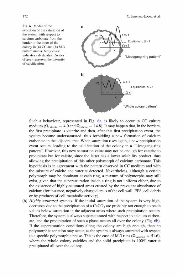
Such a behaviour, represented in Fig. 6a, is likely to occur in CC culture
medium (Ovaterite ¼ 4.0 and Ocalcite ¼ 14.8). It may happen that, in the borders,
the first precipitate is vaterite and then, after this first precipitation event, the
system became undersaturated, thus forbidding a new formation of calcium
carbonate in the adjacent area. When saturation rises again, a new precipitation
event occurs, leading to the calcification of the colony in a “Liesegang-ring
pattern”. However, this new saturation value may not be enough for vaterite to
precipitate but for calcite, since the latter has a lower solubility product, thus
allowing the precipitation of this other polymorph of calcium carbonate. This
hypothesis is in agreement with the pattern observed in CC medium and with
the mixture of calcite and vaterite detected. Nevertheless, although a certain
polymorph may be dominant at each ring, a mixture of polymorphs may still
exist, given that the supersaturation inside a ring is not uniform either, due to
the existence of highly-saturated areas created by the prevalent absorbance of
calcium (for instance, negatively charged areas of the cell wall, EPS, cell debris
or by-products of cell metabolic activity).
(b) Highly saturated systems. If the initial saturation of the system is very high,
decreases due to the precipitation of a CaCO3 are probably not enough to reach
values below saturation in the adjacent areas where such precipitation occurs.
Therefore, the system is always supersaturated with respect to calcium carbon-
ate, and the precipitation of such a phase occurs all over the colony (Fig. 6b).
If the superaturation conditions along the colony are high enough, then no
polymorphic zonation may occur, as the system is always saturated with respect
to a specific polymorphic phase. This is the case of M-3 runs (Ovaterite ¼ 51.6),
where the whole colony calcifies and the solid precipitate is 100% vaterite
precipitated all over the colony.
WW
Equilibrium, W= 1
a
b
Equilibrium, W= 1
“Whole colony pattern”
“Liesegang-ring pattern”
W> 1
W> 1
W< 1
W< 1
x
x
Fig. 6 Model of the
evolution of the saturation of
the system with respect to
calcium carbonate from the
border to the inner of the
colony in (a) CC and (b) M-3
culture media. Gray colorindicates calcification. Scales
of gray represent the intensityof calcification
172 C. Jimenez-Lopez et al.
(c) Intermediate saturated systems. In those systems where saturation value is not
so high, calcium carbonate precipitation may still occur all over the colony,
since the system may be always supersaturated with respect to calcium carbon-
ate. However, a zonation of polymorphs of calcium carbonate may still occur,
since the saturation value, after a precipitation event, may not be enough to
allow for the precipitation of the polymorph that formed in the previous event.
Therefore, whether the colony calcifies completely or only at certain areas and
whether or not there is a polymorph zonation, may help identifying if the
calcification process occurred at high, intermediate or low saturation values.
The “Liesegang-ring pattern” observed in our experiments resembles the con-
centric patterns of ooids. Although ooids in modern environments are com-
posed of aragonite, in the past, calcitic, dolomitic and bimineralic ooids have
also been found (Wright and Oren 2005). Results from this study show that a
microorganism,M. xanthus, is able to induce the calcification of the populationwith this specific concentric pattern, and that such a pattern may provide
insights of the geochemical conditions in which the calcification occurred.
4.2 Critical Size of the Population Necessaryfor the Calcification of the Colony
Our results show that even though the bulk culture media were supersaturated with
respect to calcium carbonate, no precipitation was observed outside the colony,
and, even within the colony, mineral formation did not occur until after 2 days in
CC medium and until after 3 days in M-3 medium. Figure 1 shows that the
exponential growth phase of the population of M. xanthus occurred earlier in CC
than in M-3 medium. Therefore, the critical size of the population required to
produce a critical supersaturation as a result of bacterial metabolism and to
provide enough pre-existing surfaces (cell wall, cell debris, EPS by-products of
bacterial metabolism), being both necessary for the calcification of the colony,
would be reached earlier in CC culture medium than in M-3. This result is in
agreement with the earlier calcification detected in CC (2 days) compared to M-3
culture medium (3 days).
Being then the critical size understood as the size of the population needed to
reach a critical supersaturation in the environment, such a number should be
dependent on other factors, like, for instance, the baseline saturation of the envi-
ronment and the temperature. The existence of this critical size may provide
insights of the size and how active was the bacterial population involved in the
formation of calcified structures. Examples of such a calcified structures can be
pathological calcified concretions [calcific periarthritis, Milwaukee shoulder
syndrome, osteoarthritis (Molloy and McCarthy 2003), kidney stone formation
(Kajander and Ciftcioglu 1998; Ciftcioglu et al. 1999), and dental pulp stone
(Ciftcioglu et al. 1998) and calcified structures in nature, as lithified mats forming
stromatolites in different environments [Precambian sediments (Walter 1983;
Myxococcus xanthus Colony Calcification 173
Grotzinger and Knoll 1999; Altermann 2004), Palaeozoic and Mesozoic marine
environments (Riding 2000), modern calcareous streams, lakes and terrestrial
sediments (Riding 2000), tropical marine and quasi-marine sites (e.g. Dravis
1983; Reid et al. 2003), and extreme environments (Kempe and Kazmierczak
1990)]. Nevertheless, and because calcified structures in nature represent a much
more complicated process than the one here investigated, more research needs to be
done to determine the critical number of cells required for calcification when
different bacterial populations are present and are interacting, using other micro-
organisms and considering different biominerals.
4.3 Passive and Active Mechanisms of Calcium CarbonatePrecipitation
As it will be discussed in detail below, calcification of the colonies is the result
of a number of several precipitation events acting in different locations and
include (1) homogeneous precipitation of calcium carbonate within the colony
(resulting from passive mechanisms of precipitation) and (2) heterogeneous
precipitation of calcium carbonate in negatively charged areas of the cell wall,
EPS, cell membranes and cell debris (resulting from active mechanisms of
precipitation).
4.4 Passive Mechanisms of Calcium Carbonate PrecipitationGiving Rise to an Homogeneous Precipitation
Since mineral formation was only detected within the colonies of M. xanthus andnot in the bulk culture medium (even though the culture medium was super-
saturated with respect to vaterite and calcite), it is clear that the presence of
bacterium is a prerequisite for the precipitation of calcium carbonate. Both
passive and active biomineralization processes make the nucleation and growth
of carbonate crystals both thermodynamically and kinetically possible (Dupraz
and Visscher 2005). In the context of thermodynamics, the supersaturation of the
system with respect to a particular mineral phase (O) is the driving force for the
precipitation of such a phase, being possible when O > 1 (Garside 1982). Such
an increase in supersaturation, as a consequence of bacterial metabolic activity,
resulted in the homogeneous precipitation of spherula and micritic crystals that
were not associated with any cell structures and/or debris, as well as, in the
heterogeneous precipitates associated with cell structures and/or debris that will
be discussed below.
The fact that M. xanthus, depending on the culture media (which, in turn, set
different saturation states), is able to produce different polymorphs (vaterite in M-3
and calcite-vaterite in CC), shows that polymorph selection is probably related to
174 C. Jimenez-Lopez et al.
supersaturation rather than to the sterochemical or geometrical matching between
the organic substrate and the mineral proposed by Mann et al (1988). These findings
are in accordance with those of Rodriguez-Navarro et al. (2007). However, as
suggested by those authors, the role of the organic matter in stabilizing the less
stable polymorph due to its incorporation into the structure of the mineral should
not be neglected. The precipitation of vaterite in the higher saturated medium (M-3)
and vaterite-calcite in the lower saturated medium (CC) is explained by the higher
solubility product of vaterite compared to that of calcite, which makes necessary
higher supersaturation conditions for the precipitation of vaterite (Ogino et al.
1987). M-3 was more saturated than CC, not only at the beginning of the experi-
ment (Ovaterite, M-3 ¼ 51.6 vs Ovaterite, CC ¼ 4.0), but also probably during the time
course experiment, given the fact that the size of the population of M. xanthus inM-3 was higher than that in CC (Fig. 1). Therefore, pH and alkalinity increases
within the colony due to bacterial metabolism are expected to be higher in M-3 runs
compared to that in CC runs. Those data support the precipitation in M-3 of the
polymorphic phase that requires higher supersaturation conditions.
4.4.1 Active Mechanism of Calcium Carbonate Precipitation
Not only supersaturation makes nucleation of crystals possible, since it is also
necessary to overcome some energy barriers determined by kinetic factors. One
of these barriers is the energy required for the system to create a new surface
(Garside 1982). In this regard, bacteria may favor the kinetics of calcium carbonate
precipitation by serving, the cell, cell structures, cell debris and even by-product of
the bacterial metabolic activity, as nucleation sites for mineral deposition, then
acting as a pre-existing surface (Chafetz and Buczynski 1992; Ben Omar et al.
1994; Knorre and Krumbein 2000; Paerl et al. 2001; Dupraz and Visscher 2005;
Rodriguez-Navarro et al. 2007). All these structures acting as pre-existing surfaces
will be here referred to CFPS.
The heterogeneous precipitation of calcium carbonate on CFPS is the result of
several processes including the creation of saturating areas at these locations and
the interaction between the substrate and the mineral. Supersaturation in those areas
should be higher than that of the culture medium, since the negatively charged areas
in CFPS have the ability to concentrate calcium (Addadi and Weiner 1992). In the
case of the cell wall, and being the saturation dependent on the extracellular release
of CO2 and NH3, the highest saturations are expected in the surroundings of the cell
wall. Those high saturations are favored by the negatively charged surface func-
tional groups, such as carboxyl, hydroxyl and phosphate sites (Schultze-Lam et al.
1996). Therefore, the first calcium carbonate precipitation is expected to happen at
these areas. Later, as CO2 and NH3 diffuse from the cells to the medium, other
locations will also begin to saturate, reaching the highest supersaturations those in
which calcium accumulates, like in specific areas of other CFPS. In particular, EPS
has been shown to be a rich matrix of polymers, including polysaccharides,
proteins, glycoproteins, nucleic acids, phospholipids, and humic acids, able to
Myxococcus xanthus Colony Calcification 175
concentrate calcium (Beveridge and Graham 1991; McSwain et al. 2005). In fact, a
number of bacterial macromolecules and structures (cell wall, cell membranes,
EPS, aminoacids and capsular exopolisacharides) have been found to mediate the
precipitation of calcium carbonate and even to control the morphology and the
orientation of the crystals (Gonzalez-Munoz et al. 1996; Kawaguchi and Decho
2002; Braissant et al. 2003; Benzerara et al. 2004; Ercole et al. 2007; Rodriguez-
Navarro et al. 2007). The mechanisms by which those substrates favor the precipi-
tation of calcium carbonate are under discussion. Several ways have been proposed
(1) the substrate templates the nucleation of CaCO3 crystals (geometrical matching,
Pentecost 1985; Mann et al 1988; Dupraz and Visscher 2005); (2) the substrate
favor the oriented aggregation of homogenously nucleated calcium carbonate
crystals by electrostatic affinity between negatively charged areas and specific
atomic planes of the crystals (ionotopic effect; Rodriguez-Navarro et al. 2007),
and (3) in the case of EPS, the substrate traps seed crystals which act as nuclei
for heterogeneous calcium carbonate precipitation (Knorre and Krumbein 2000;
Turner and Jones 2005).
Regarding calcium carbonate precipitation on cell membranes, an interesting
remark is that such precipitation was observed in fractions of cell membranes, both
in M-3 and CC media (Figs. 4f and 5f). Such an observation does not agree with
that of Benzerara et al. (2004), who concluded that the precipitation of calcium
phosphate induced by Ramlibacter tataouinensis requires integrated membrane
structure. However, and given the fact that the biominerals describe in the paper
from those authors (phosphates) and those described in the present study (carbo-
nates) are different, other mechanisms may account for the differences observed,
like, for instance, the role of the phospholipids of the membranes on the precipi-
tation of calcium phosphate. This issue, which does not fall within the focus of the
present work, needs to be further considered in future studies.
Not only CFPS, but also other organics (macromolecules) inside the cell can act
as pre-existing surfaces, resulting in the calcification of the interior of the cells, as it
can be observed in Figs. 4c, d and 5c, d. Such features have been also observed by
Benzerara et al. (2004). HRTEM images of stained preparations show that mem-
branes are not intact (Figs. 4f and 5f), thus, this calcification inside the cells
probably occurs once the cell lyses. It is little plausible that such a calcification
takes place while the cell is living or intact, since it has been described by Rosen
(1987) that most live bacterium cells have extrusion mechanisms to maintain low
concentrations of intracellular calcium, so, therefore, the protoplasm may be
undersaturated with respect to the precipitation of calcium carbonate. The fact
that not all the cells present the same level of calcification (from only the cell
wall to also all the protoplasm with all the transient stages) is probably related to the
existence of different generations in the colony, from the first to latter descendants,
thus making it possible to observe different stages of biomineralization in the same
sample.
With time, the calcium carbonate structure formed in the cell wall is thick
enough to affect the permeability of the cell wall, thus triggering the lyses of the
cells. As shown in Figs. 4g, h and 5g, h, once the cell lyses, only the calcium
176 C. Jimenez-Lopez et al.
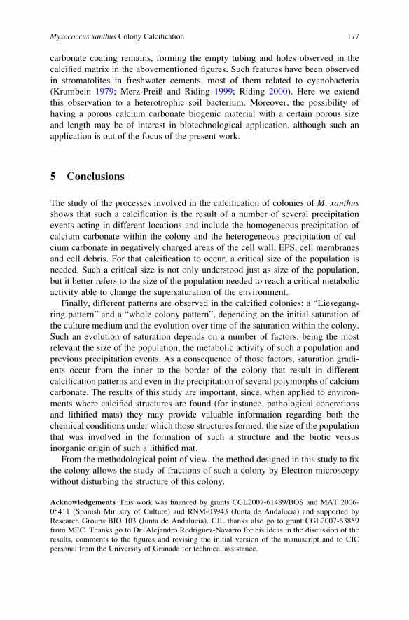
carbonate coating remains, forming the empty tubing and holes observed in the
calcified matrix in the abovementioned figures. Such features have been observed
in stromatolites in freshwater cements, most of them related to cyanobacteria
(Krumbein 1979; Merz-Preiß and Riding 1999; Riding 2000). Here we extend
this observation to a heterotrophic soil bacterium. Moreover, the possibility of
having a porous calcium carbonate biogenic material with a certain porous size
and length may be of interest in biotechnological application, although such an
application is out of the focus of the present work.
5 Conclusions
The study of the processes involved in the calcification of colonies of M. xanthusshows that such a calcification is the result of a number of several precipitation
events acting in different locations and include the homogeneous precipitation of
calcium carbonate within the colony and the heterogeneous precipitation of cal-
cium carbonate in negatively charged areas of the cell wall, EPS, cell membranes
and cell debris. For that calcification to occur, a critical size of the population is
needed. Such a critical size is not only understood just as size of the population,
but it better refers to the size of the population needed to reach a critical metabolic
activity able to change the supersaturation of the environment.
Finally, different patterns are observed in the calcified colonies: a “Liesegang-
ring pattern” and a “whole colony pattern”, depending on the initial saturation of
the culture medium and the evolution over time of the saturation within the colony.
Such an evolution of saturation depends on a number of factors, being the most
relevant the size of the population, the metabolic activity of such a population and
previous precipitation events. As a consequence of those factors, saturation gradi-
ents occur from the inner to the border of the colony that result in different
calcification patterns and even in the precipitation of several polymorphs of calcium
carbonate. The results of this study are important, since, when applied to environ-
ments where calcified structures are found (for instance, pathological concretions
and lithified mats) they may provide valuable information regarding both the
chemical conditions under which those structures formed, the size of the population
that was involved in the formation of such a structure and the biotic versus
inorganic origin of such a lithified mat.
From the methodological point of view, the method designed in this study to fix
the colony allows the study of fractions of such a colony by Electron microscopy
without disturbing the structure of this colony.
Acknowledgements This work was financed by grants CGL2007-61489/BOS and MAT 2006-
05411 (Spanish Ministry of Culture) and RNM-03943 (Junta de Andalucia) and supported by
Research Groups BIO 103 (Junta de Andalucıa). CJL thanks also go to grant CGL2007-63859
from MEC. Thanks go to Dr. Alejandro Rodriguez-Navarro for his ideas in the discussion of the
results, comments to the figures and revising the initial version of the manuscript and to CIC
personal from the University of Granada for technical assistance.
Myxococcus xanthus Colony Calcification 177
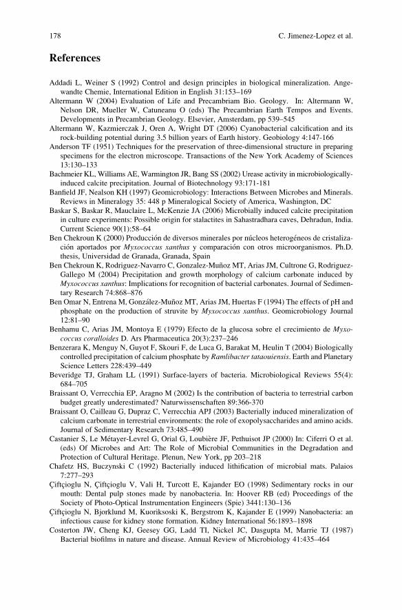
References
Addadi L, Weiner S (1992) Control and design principles in biological mineralization. Ange-
wandte Chemie, International Edition in English 31:153–169
Altermann W (2004) Evaluation of Life and Precambriam Bio. Geology. In: Altermann W,
Nelson DR, Mueller W, Catuneanu O (eds) The Precambrian Earth Tempos and Events.
Developments in Precambrian Geology. Elsevier, Amsterdam, pp 539–545
Altermann W, Kazmierczak J, Oren A, Wright DT (2006) Cyanobacterial calcification and its
rock-building potential during 3.5 billion years of Earth history. Geobiology 4:147-166
Anderson TF (1951) Techniques for the preservation of three-dimensional structure in preparing
specimens for the electron microscope. Transactions of the New York Academy of Sciences
13:130–133
Bachmeier KL, Williams AE, Warmington JR, Bang SS (2002) Urease activity in microbiologically-
induced calcite precipitation. Journal of Biotechnology 93:171-181
Banfield JF, Nealson KH (1997) Geomicrobiology: Interactions Between Microbes and Minerals.
Reviews in Mineralogy 35: 448 p Mineralogical Society of America, Washington, DC
Baskar S, Baskar R, Mauclaire L, McKenzie JA (2006) Microbially induced calcite precipitation
in culture experiments: Possible origin for stalactites in Sahastradhara caves, Dehradun, India.
Current Science 90(1):58–64
Ben Chekroun K (2000) Produccion de diversos minerales por nucleos heterogeneos de cristaliza-
cion aportados por Myxococcus xanthus y comparacion con otros microorganismos. Ph.D.
thesis, Universidad de Granada, Granada, Spain
Ben Chekroun K, Rodriguez-Navarro C, Gonzalez-Munoz MT, Arias JM, Cultrone G, Rodriguez-
Gallego M (2004) Precipitation and growth morphology of calcium carbonate induced by
Myxococcus xanthus: Implications for recognition of bacterial carbonates. Journal of Sedimen-
tary Research 74:868–876
Ben Omar N, Entrena M, Gonzalez-Munoz MT, Arias JM, Huertas F (1994) The effects of pH and
phosphate on the production of struvite by Myxococcus xanthus. Geomicrobiology Journal
12:81–90
Benhamu C, Arias JM, Montoya E (1979) Efecto de la glucosa sobre el crecimiento de Myxo-coccus coralloides D. Ars Pharmaceutica 20(3):237–246
Benzerara K, Menguy N, Guyot F, Skouri F, de Luca G, Barakat M, Heulin T (2004) Biologically
controlled precipitation of calcium phosphate by Ramlibacter tataouiensis. Earth and PlanetaryScience Letters 228:439–449
Beveridge TJ, Graham LL (1991) Surface-layers of bacteria. Microbiological Reviews 55(4):
684–705
Braissant O, Verrecchia EP, Aragno M (2002) Is the contribution of bacteria to terrestrial carbon
budget greatly underestimated? Naturwissenschaften 89:366-370
Braissant O, Cailleau G, Dupraz C, Verrecchia APJ (2003) Bacterially induced mineralization of
calcium carbonate in terrestrial environments: the role of exopolysaccharides and amino acids.
Journal of Sedimentary Research 73:485–490
Castanier S, Le Metayer-Levrel G, Orial G, Loubiere JF, Pethuisot JP (2000) In: Ciferri O et al.
(eds) Of Microbes and Art: The Role of Microbial Communities in the Degradation and
Protection of Cultural Heritage. Plenun, New York, pp 203–218
Chafetz HS, Buczynski C (1992) Bacterially induced lithification of microbial mats. Palaios
7:277–293
Ciftcioglu N, Ciftcioglu V, Vali H, Turcott E, Kajander EO (1998) Sedimentary rocks in our
mouth: Dental pulp stones made by nanobacteria. In: Hoover RB (ed) Proceedings of the
Society of Photo-Optical Instrumentation Engineers (Spie) 3441:130–136
Ciftcioglu N, Bjorklund M, Kuoriksoski K, Bergstrom K, Kajander E (1999) Nanobacteria: an
infectious cause for kidney stone formation. Kidney International 56:1893–1898
Costerton JW, Cheng KJ, Geesey GG, Ladd TI, Nickel JC, Dasgupta M, Marrie TJ (1987)
Bacterial biofilms in nature and disease. Annual Review of Microbiology 41:435–464
178 C. Jimenez-Lopez et al.
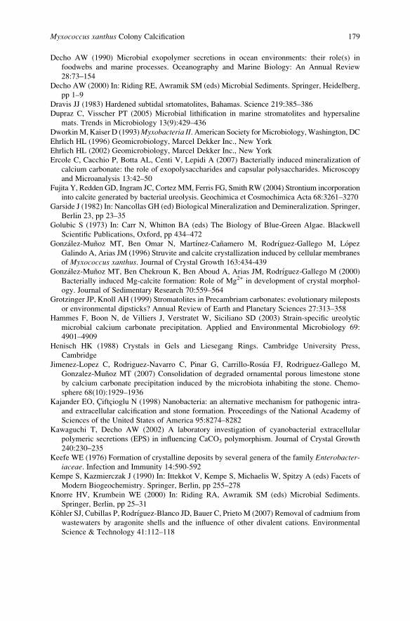
Decho AW (1990) Microbial exopolymer secretions in ocean environments: their role(s) in
foodwebs and marine processes. Oceanography and Marine Biology: An Annual Review
28:73–154
Decho AW (2000) In: Riding RE, Awramik SM (eds) Microbial Sediments. Springer, Heidelberg,
pp 1–9
Dravis JJ (1983) Hardened subtidal srtomatolites, Bahamas. Science 219:385–386
Dupraz C, Visscher PT (2005) Microbial lithification in marine stromatolites and hypersaline
mats. Trends in Microbiology 13(9):429–436
DworkinM, Kaiser D (1993)Myxobacteria II. American Society forMicrobiology,Washington, DC
Ehrlich HL (1996) Geomicrobiology, Marcel Dekker Inc., New York
Ehrlich HL (2002) Geomicrobiology, Marcel Dekker Inc., New York
Ercole C, Cacchio P, Botta AL, Centi V, Lepidi A (2007) Bacterially induced mineralization of
calcium carbonate: the role of exopolysaccharides and capsular polysaccharides. Microscopy
and Microanalysis 13:42–50
Fujita Y, Redden GD, Ingram JC, Cortez MM, Ferris FG, Smith RW (2004) Strontium incorporation
into calcite generated by bacterial ureolysis. Geochimica et Cosmochimica Acta 68:3261–3270
Garside J (1982) In: Nancollas GH (ed) Biological Mineralization and Demineralization. Springer,
Berlin 23, pp 23–35
Golubic S (1973) In: Carr N, Whitton BA (eds) The Biology of Blue-Green Algae. Blackwell
Scientific Publications, Oxford, pp 434–472
Gonzalez-Munoz MT, Ben Omar N, Martınez-Canamero M, Rodrıguez-Gallego M, Lopez
Galindo A, Arias JM (1996) Struvite and calcite crystallization induced by cellular membranes
of Myxococcus xanthus. Journal of Crystal Growth 163:434-439
Gonzalez-Munoz MT, Ben Chekroun K, Ben Aboud A, Arias JM, Rodrıguez-Gallego M (2000)
Bacterially induced Mg-calcite formation: Role of Mg2+ in development of crystal morphol-
ogy. Journal of Sedimentary Research 70:559–564
Grotzinger JP, Knoll AH (1999) Stromatolites in Precambriam carbonates: evolutionary mileposts
or environmental dipsticks? Annual Review of Earth and Planetary Sciences 27:313–358
Hammes F, Boon N, de Villiers J, Verstratet W, Siciliano SD (2003) Strain-specific ureolytic
microbial calcium carbonate precipitation. Applied and Environmental Microbiology 69:
4901–4909
Henisch HK (1988) Crystals in Gels and Liesegang Rings. Cambridge University Press,
Cambridge
Jimenez-Lopez C, Rodriguez-Navarro C, Pinar G, Carrillo-Rosua FJ, Rodriguez-Gallego M,
Gonzalez-Munoz MT (2007) Consolidation of degraded ornamental porous limestone stone
by calcium carbonate precipitation induced by the microbiota inhabiting the stone. Chemo-
sphere 68(10):1929–1936
Kajander EO, Ciftcioglu N (1998) Nanobacteria: an alternative mechanism for pathogenic intra-
and extracellular calcification and stone formation. Proceedings of the National Academy of
Sciences of the United States of America 95:8274–8282
Kawaguchi T, Decho AW (2002) A laboratory investigation of cyanobacterial extracellular
polymeric secretions (EPS) in influencing CaCO3 polymorphism. Journal of Crystal Growth
240:230–235
Keefe WE (1976) Formation of crystalline deposits by several genera of the family Enterobacter-iaceae. Infection and Immunity 14:590-592
Kempe S, Kazmierczak J (1990) In: Ittekkot V, Kempe S, Michaelis W, Spitzy A (eds) Facets of
Modern Biogeochemistry. Springer, Berlin, pp 255–278
Knorre HV, Krumbein WE (2000) In: Riding RA, Awramik SM (eds) Microbial Sediments.
Springer, Berlin, pp 25–31
K€ohler SJ, Cubillas P, Rodrıguez-Blanco JD, Bauer C, Prieto M (2007) Removal of cadmium from
wastewaters by aragonite shells and the influence of other divalent cations. Environmental
Science & Technology 41:112–118
Myxococcus xanthus Colony Calcification 179
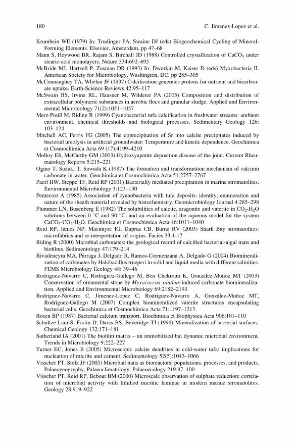
Krumbein WE (1979) In: Trudinger PA, Swaine DJ (eds) Biogeochemical Cycling of Mineral-
Forming Elements. Elsevier, Amsterdam, pp 47–68
Mann S, Heywood BR, Rajam S, Birchall JD (1988) Controlled crystallization of CaCO3 under
stearic-acid monolayers. Nature 334:692–695
McBride MJ, Hartzell P, Zusman DR (1993) In: Dworkin M, Kaiser D (eds) Myxobacteria II.
American Society for Microbiology, Washington, DC, pp 285–305
McConnaughey TA, Whelan JF (1997) Calcification generates protons for nutrient and bicarbon-
ate uptake. Earth-Science Reviews 42:95–117
McSwain BS, Irvine RL, Hausner M, Wilderer PA (2005) Composition and distribution of
extracellular polymeric substances in aerobic flocs and granular sludge. Applied and Environ-
mental Microbiology 71(2):1051–1057
Merz-Preiß M, Riding R (1999) Cyanobacterial tufa calcification in freshwater streams: ambient
environment, chemical thresholds and biological processes. Sedimentary Geology 126:
103–124
Mitchell AC, Ferris FG (2005) The coprecipitation of Sr into calcite precipitates induced by
bacterial ureolysis in artificial groundwater: Temperature and kinetic dependence. Geochimica
et Cosmochimica Acta 69 (17):4199–4210
Molloy ES, McCarthy GM (2003) Hydroxyapatite deposition disease of the joint. Current Rheu-
matology Reports 5:215–221
Ogino T, Suzuki T, Sawada K (1987) The formation and transformation mechanism of calcium
carbonate in water. Geochimica et Cosmochimica Acta 51:2757–2767
Paerl HW, Steppe TF, Reid RP (2001) Bacterially mediated precipitation in marine stromatolites.
Environmental Microbiology 3:123–130
Pentecost A (1985) Association of cyanobacteria with tufa deposits: identity, enumeration and
nature of the sheath material revealed by histochemistry. Geomicrobiology Journal 4:285–298
Plummer LN, Busenberg E (1982) The solubilities of calcite, aragonite and vaterite in CO2-H2O
solutions between 0 �C and 90 �C, and an evaluation of the aqueous model for the system
CaCO3-CO2-H2O. Geochimica et Cosmochimica Acta 46:1011–1040
Reid RP, James NP, Macintyre IG, Dupraz CB, Burne RV (2003) Shark Bay stromatolites:
microfabrics and re-interpretation of origins. Facies 33:1-17
Riding R (2000) Microbial carbonates: the geological record of calcified bacterial-algal mats and
biofilms. Sedimentology 47:179–214
Rivadeneyra MA, Parraga J, Delgado R, Ramos-Cormenzana A, Delgado G (2004) Biominerali-
zation of carbonates by Halobacillus trueperi in solid and liquid media with different salinities.
FEMS Microbiology Ecology 48: 39–46
Rodriguez-Navarro C, Rodriguez-Gallego M, Ben Chekroun K, Gonzalez-Munoz MT (2003)
Conservation of ornamental stone by Myxococcus xanthus-induced carbonate biomineraliza-
tion. Applied and Environmental Microbiology 69:2182–2193
Rodriguez-Navarro C, Jimenez-Lopez C, Rodriguez-Navarro A, Gonzalez-Munoz MT,
Rodriguez-Gallego M (2007) Complex biomineralized vaterite structures encapsulating
bacterial cells. Geochimica et Cosmochimica Acta 71:1197–1213
Rosen BP (1987) Bacterial calcium transport. Biochimica et Biophysica Acta 906:101–110
Schultze-Lam S, Fortin D, Davis BS, Beveridge TJ (1996) Mineralization of bacterial surfaces.
Chemical Geology 132:171–181
Sutherland IA (2001) The biofilm matrix – an immobilized but dynamic microbial environment.
Trends in Microbiology 9:222–227
Turner EC, Jones B (2005) Microscopic calcite dendrites in cold-water tufa: implications for
nucleation of micrite and cement. Sedimentology 52(5):1043–1066
Visscher PT, Stolz JF (2005) Microbial mats as bioreactors: populations, processes, and products.
Palaeogeogryphy, Palaeoclimatology, Palaeoecology 219:87–100
Visscher PT, Reid RP, Bebout BM (2000) Microscale observation of sulphate reduction: correla-
tion of microbial activity with lithified micritic laminae in modern marine stromatolites.
Geology 28:919–922
180 C. Jimenez-Lopez et al.

Walter MR (1983) In: Schopf JW (ed) Earth’s Earliest Biosphere: Its Origin and Evolution.
Princeton University Press, Princeton, pp 187–213
Warren LA, Maurice PA, Parmar N, Ferris FG (2001) Microbially mediated calcium carbonate
precipitation: Implications for interpreting calcite precipitation and for solid-phase capture of
inorganic contaminants. Geomicrobiology Journal 18:93–115
Wolery TJ (1992) EQ3/6, A Software Package for Geochemical Modeling of Aqueous Systems.
Version 7.0. University of California, Livermore.
Wright DT (1999) The role of sulphate-reducing bacteria and cyanobacteria in dolomite formation
in distal ephemeral lakes of the Coorong region, South Australia. Sedimentary Geology 126:
147–157
Wright DT (2000) Benthic microbial communities and dolomite formation in marine and
lacustrine environments – a new dolomite model. In: Glenn C, Prevot-Lucas L, Lucas J (eds)
Marine Authogenesis: FromGlobal to Microbial. SEPM special publication, Tulsa, OK, 66:7–14
Wright DT, Oren A (2005) Non-photosynthetic bacteria and the formation of carbonates and
evaporates though time. Geomicrobiology Journal 22:27-53
Myxococcus xanthus Colony Calcification 181

.
Are Stromatolites the Most Ancient Skeletal
Organisms?
Evgenia Sumina, Vladimir Orleansky, and Dmitry Sumin
Stromatolites have an ancient and contradictory research history. Initially,
they were classified as non-organic fossils and either concretions or layered lime-
stones. Over time, their organic nature was established, but details regarding the
stromatolite-forming organisms were not known. Walcott (1914) proposed that
stromatolites are produced by blue-green algae (cyanobacteria) and this was con-
firmed by the present-day observation of stromatolite-forming microbiota. Since
that time stromatolite-forming organisms were considered as colonies of micro-
organisms with the stromatolite being the result of the interaction between the
colonies and trapping sediment. This description of the nature of stromatolites was
successfully used for the Precambrian. However, the stromatolite-forming micro-
organisms were never considered as an organism. Thus the following controversy
arose: the cyanobacterial accumulation is not able to undergo morphogenesis, but is
capable of forming a large morphogenically shaped mineral bodies, the stromato-
lites, which are stable in space and time (Fig. 1). To resolve this controversy,
laboratory modelling was performed on the stromatolite forming biota (OрлеаHс-кий and РаабеH 1997). To continue this experiment, separate research project on
the morphogenetic reaction society (here “society” refers to a compact settlement of
independent elements- filamentous cyanobacteria” with a non-determined degree of
integrity) of contemporary filamentous cyanobacteria as a whole community was
performed.
Ten types of structures (Fig. 2) were detected for filamentous cyanobacteria
creation as a result of their regrouping (Sumina 2002). The forms of the structures
are specific and grow as a behaviour response to various influences, sometimes
growing up to 10 cm. So the community appears to possess differentiation and
E. Sumina (*) and V. Orleansky
Paleontological Institute RAS, Profsoyuznaya st., 123, 117647 Moscow, Russia
e-mail: [email protected], [email protected]
D. Sumin
Independent researcher, Zhebrunova st., 5, 44, 107014 Moscow, Russia
J. Reitner et al., Advances in Stromatolite Geobiology,Lecture Notes in Earth Sciences 131, DOI 10.1007/978-3-642-10415-2_12,# Springer-Verlag Berlin Heidelberg 2011
183
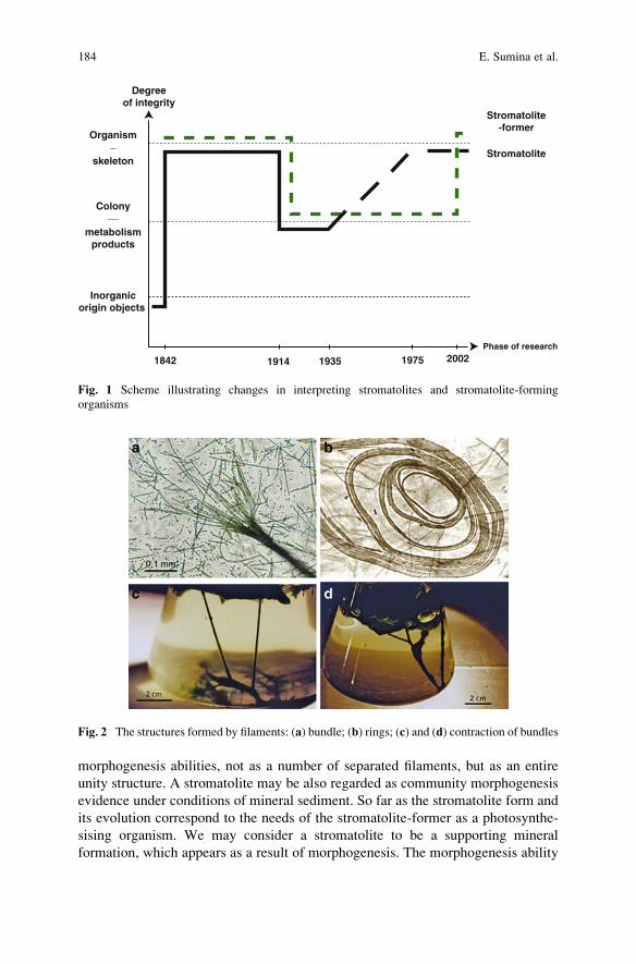
morphogenesis abilities, not as a number of separated filaments, but as an entire
unity structure. A stromatolite may be also regarded as community morphogenesis
evidence under conditions of mineral sediment. So far as the stromatolite form and
its evolution correspond to the needs of the stromatolite-former as a photosynthe-
sising organism. We may consider a stromatolite to be a supporting mineral
formation, which appears as a result of morphogenesis. The morphogenesis ability
1842 1914 1935 1975 2002
Organism
skeleton
Colony
metabolismproducts
Inorganicorigin objects
Degreeof integrity
Stromatolite-former
Stromatolite
Phase of research
Fig. 1 Scheme illustrating changes in interpreting stromatolites and stromatolite-forming
organisms
Fig. 2 The structures formed by filaments: (a) bundle; (b) rings; (c) and (d) contraction of bundles
184 E. Sumina et al.
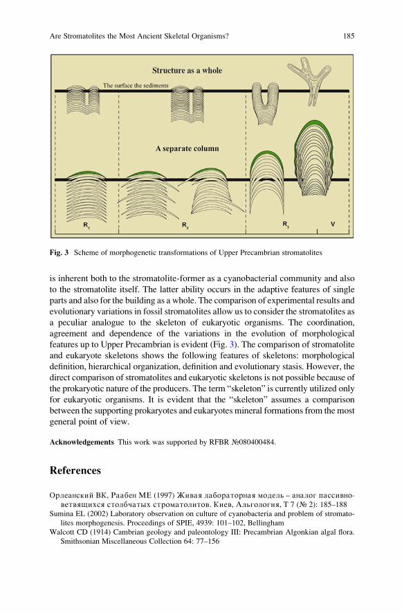
is inherent both to the stromatolite-former as a cyanobacterial community and also
to the stromatolite itself. The latter ability occurs in the adaptive features of single
parts and also for the building as a whole. The comparison of experimental results and
evolutionary variations in fossil stromatolites allow us to consider the stromatolites as
a peculiar analogue to the skeleton of eukaryotic organisms. The coordination,
agreement and dependence of the variations in the evolution of morphological
features up to Upper Precambrian is evident (Fig. 3). The comparison of stromatolite
and eukaryote skeletons shows the following features of skeletons: morphological
definition, hierarchical organization, definition and evolutionary stasis. However, the
direct comparison of stromatolites and eukaryotic skeletons is not possible because of
the prokaryotic nature of the producers. The term “skeleton” is currently utilized only
for eukaryotic organisms. It is evident that the “skeleton” assumes a comparison
between the supporting prokaryotes and eukaryotes mineral formations from themost
general point of view.
Acknowledgements This work was supported by RFBR №080400484.
References
OрлеаHский BК, РаабеH МЕ (1997) Живая лабораторHая модель – аHалог пассивHо-ветвящихся столбчатых строматолитов. Киев, Альгология, T 7 (№ 2): 185–188
Sumina EL (2002) Laboratory observation on culture of cyanobacteria and problem of stromato-
lites morphogenesis. Proceedings of SPIE, 4939: 101–102, Bellingham
Walcott CD (1914) Cambrian geology and paleontology III: Precambrian Algonkian algal flora.
Smithsonian Miscellaneous Collection 64: 77–156
Fig. 3 Scheme of morphogenetic transformations of Upper Precambrian stromatolites
Are Stromatolites the Most Ancient Skeletal Organisms? 185

.
Microbial Mats and Microbialites
in the Freshwater Laguna Bacalar,
Yucatan Peninsula, Mexico
Eberhard Gischler, Stjepko Golubic, Michael A. Gibson,
Wolfgang Oschmann, and J. Harold Hudson
1 Introduction
Laguna Bacalar, a karstic freshwater lake in the state of Quintana Roo, Yucatan
Peninsula, Mexico (Fig. 1) features in its southern part possibly the largest fresh-
water microbialite structures known. These structures extend continuously for over
10 km (Gischler et al. 2008). The laguna is a narrow, elongated body of freshwater,
about 40 km long and 1–2 km wide, extending NE to SW with an adjacent strip of
periodically flooded lowland. The lagoon is tectonically rooted on the Bacalar fault,
which is the northern continuation of the Rio Hondo fault zone (Kenkmann and
Sch€onian 2006, their fig. 6). Laguna Bacalar gets as deep as 20 m with the deepest
areas located between Bacalar and Zipline Point. The lake is located in the low
Neogene karstic landscape and fed by a series of karstic springs originating from
deep circular sinkholes locally called cenotes. Based on hydrogeochemical analyses
of groundwater on the Yucatan peninsula, Perry et al. (2009) concluded that the
cavern system that includes the Bacalar cenotes probably predates the Tertiary;
strontium isotopes suggest that waters derive their ion load from late Cretaceous
and/or Paleogene carbonates and evaporites. As in other karstic systems, the
emerging groundwater is supersaturated with respect to carbonate and this condi-
tion and strong currents along the narrow passages of the lagoon are closely
associated with carbonate precipitation and microbialite accretion. The currents
E. Gischler (*) and W. Oschmann
Institut f€ur Geowissenschaften, Goethe-Universit€at, 60438 Frankfurt am Main, Germany
e-mail: [email protected]
S. Golubic
Biological Science Center, Boston University, Boston, MA 02215, USA
M.A. Gibson
Department of Geology, Geography and Physics, University of Tennessee, Martin, TN 38238,
USA
J.H. Hudson
ReefTech Inc., 8325 SW 68th Street, Miami, FL 33143, USA
J. Reitner et al., Advances in Stromatolite Geobiology,Lecture Notes in Earth Sciences 131, DOI 10.1007/978-3-642-10415-2_13,# Springer-Verlag Berlin Heidelberg 2011
187
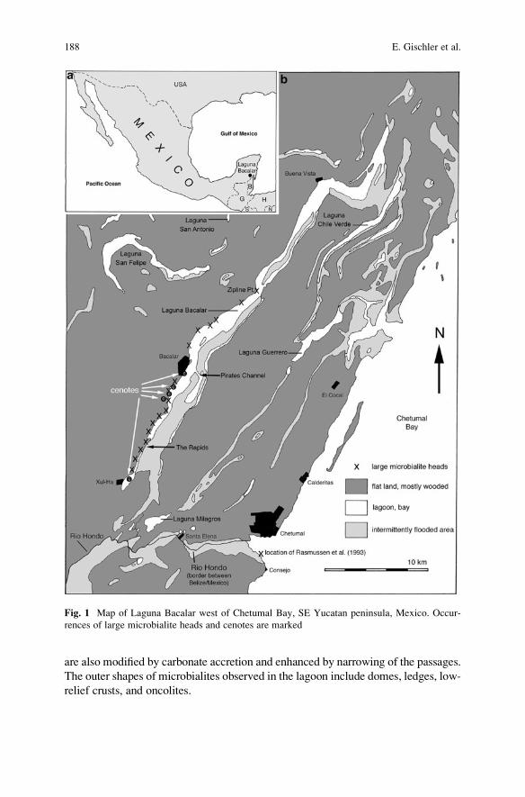
are also modified by carbonate accretion and enhanced by narrowing of the passages.
The outer shapes of microbialites observed in the lagoon include domes, ledges, low-
relief crusts, and oncolites.
Fig. 1 Map of Laguna Bacalar west of Chetumal Bay, SE Yucatan peninsula, Mexico. Occur-
rences of large microbialite heads and cenotes are marked
188 E. Gischler et al.
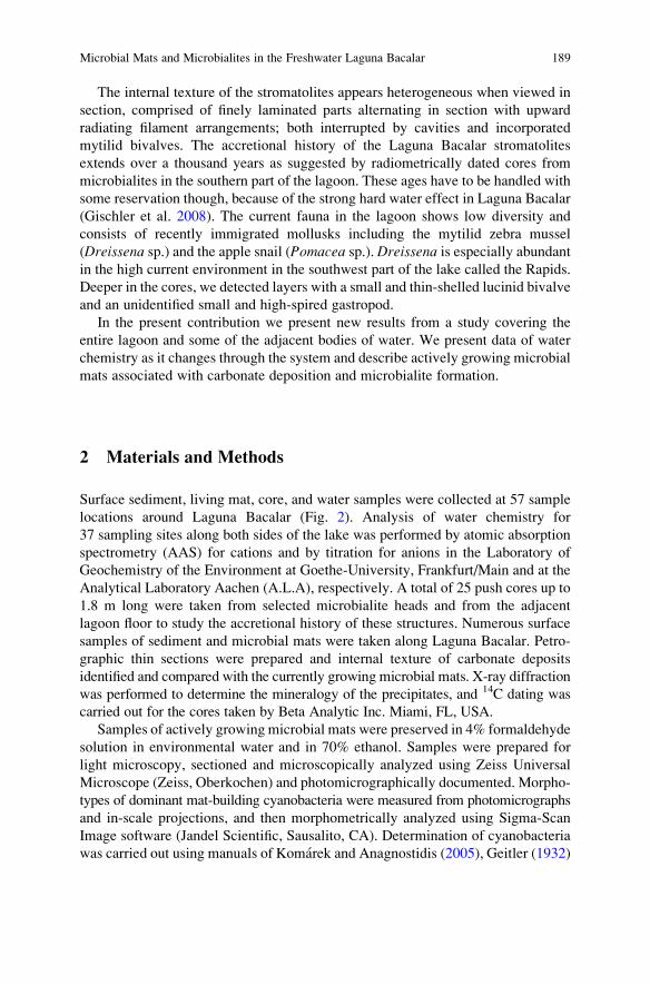
The internal texture of the stromatolites appears heterogeneous when viewed in
section, comprised of finely laminated parts alternating in section with upward
radiating filament arrangements; both interrupted by cavities and incorporated
mytilid bivalves. The accretional history of the Laguna Bacalar stromatolites
extends over a thousand years as suggested by radiometrically dated cores from
microbialites in the southern part of the lagoon. These ages have to be handled with
some reservation though, because of the strong hard water effect in Laguna Bacalar
(Gischler et al. 2008). The current fauna in the lagoon shows low diversity and
consists of recently immigrated mollusks including the mytilid zebra mussel
(Dreissena sp.) and the apple snail (Pomacea sp.). Dreissena is especially abundantin the high current environment in the southwest part of the lake called the Rapids.
Deeper in the cores, we detected layers with a small and thin-shelled lucinid bivalve
and an unidentified small and high-spired gastropod.
In the present contribution we present new results from a study covering the
entire lagoon and some of the adjacent bodies of water. We present data of water
chemistry as it changes through the system and describe actively growing microbial
mats associated with carbonate deposition and microbialite formation.
2 Materials and Methods
Surface sediment, living mat, core, and water samples were collected at 57 sample
locations around Laguna Bacalar (Fig. 2). Analysis of water chemistry for
37 sampling sites along both sides of the lake was performed by atomic absorption
spectrometry (AAS) for cations and by titration for anions in the Laboratory of
Geochemistry of the Environment at Goethe-University, Frankfurt/Main and at the
Analytical Laboratory Aachen (A.L.A), respectively. A total of 25 push cores up to
1.8 m long were taken from selected microbialite heads and from the adjacent
lagoon floor to study the accretional history of these structures. Numerous surface
samples of sediment and microbial mats were taken along Laguna Bacalar. Petro-
graphic thin sections were prepared and internal texture of carbonate deposits
identified and compared with the currently growing microbial mats. X-ray diffraction
was performed to determine the mineralogy of the precipitates, and 14C dating was
carried out for the cores taken by Beta Analytic Inc. Miami, FL, USA.
Samples of actively growing microbial mats were preserved in 4% formaldehyde
solution in environmental water and in 70% ethanol. Samples were prepared for
light microscopy, sectioned and microscopically analyzed using Zeiss Universal
Microscope (Zeiss, Oberkochen) and photomicrographically documented. Morpho-
types of dominant mat-building cyanobacteria were measured from photomicrographs
and in-scale projections, and then morphometrically analyzed using Sigma-Scan
Image software (Jandel Scientific, Sausalito, CA). Determination of cyanobacteria
was carried out using manuals of Komarek and Anagnostidis (2005), Geitler (1932)
Microbial Mats and Microbialites in the Freshwater Laguna Bacalar 189
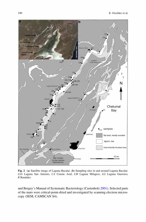
and Bergey’s Manual of Systematic Bacteriology (Castenholz 2001). Selected parts
of the mats were critical-point-dried and investigated by scanning electron micros-
copy (SEM, CAMSCAN S4).
Fig. 2 (a) Satellite image of Laguna Bacalar. (b) Sampling sites in and around Laguna Bacalar.
LSA Laguna San Antonio, CA Cenote Azul, LM Laguna Milagros, LG Laguna Guerrero,
R Raudales
190 E. Gischler et al.
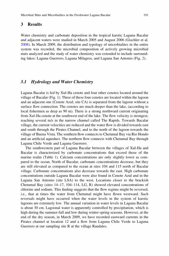
3 Results
Water chemistry and carbonate deposition in the tropical karstic Laguna Bacalar
and adjacent waters were studied in March 2005 and August 2006 (Gischler et al.
2008). In March 2009, the distribution and typology of microbialites in the entire
system was recorded, the microbial composition of actively growing microbial
mats analyzed and the study of water chemistry was extended to include surround-
ing lakes: Laguna Guerrero, Laguna Milagros, and Laguna San Antonio (Fig. 2).
3.1 Hydrology and Water Chemistry
Laguna Bacalar is fed by Xul-Ha cenote and four other cenotes located around the
village of Bacalar (Fig. 1). Three of these four cenotes are located within the lagoon
and an adjacent one (Cenote Azul; site CA) is separated from the lagoon without a
surface flow connection. The cenotes are much deeper than the lake, (according to
local fishermen as deep as 90 m). There is a strong northward current originating
from Xul-Ha cenote at the southwest end of the lake. The flow velocity is strongest,
reaching several m/s in the narrow channel called The Rapids. Towards Bacalar
village, the current velocities are reduced and the water flow is divided towards east
and south through the Pirates Channel, and to the north of the lagoon towards the
village of Buena Vista. The southern flow connects to Chetumal Bay via Rio Hondo
and an artificial aqueduct. The northern flow connects with Chetumal Bay through
Laguna Chile Verde and Laguna Guerrero.
The southwestern part of Laguna Bacalar between the villages of Xul-Ha and
Bacalar is characterized by carbonate concentrations that exceed those of the
marine realm (Table 1). Calcium concentrations are only slightly lower as com-
pared to the ocean. North of Bacalar, carbonate concentrations decrease, but they
are still elevated as compared to the ocean at sites 104 and 115 north of Bacalar
village. Carbonate concentrations also decrease towards the east. High carbonate
concentrations outside Laguna Bacalar were also found in Cenote Azul and in the
Laguna San Antonio (site LSA) to the west. Locations closer to the brackish
Chetumal Bay (sites 14–17, 104–114, LG, R) showed elevated concentrations of
chlorine and sodium. This finding suggests that the flow regime might be reversed,
i.e., that at times the water from Chetumal might have flown westward. Such
reversals might have occurred when the water levels in the system of karstic
lagoons are extremely low. The annual variation in water levels in Laguna Bacalar
is about 30 cm. Lagoonal water is apparently controlled by precipitation, which is
high during the summer-fall and low during winter-spring seasons. However, at the
end of the dry season, in March 2009, we have recorded eastward currents in the
Pirates channel at location 12 and a flow from Laguna Chile Verde to Laguna
Guerrero at our sampling site R at the village Raudales.
Microbial Mats and Microbialites in the Freshwater Laguna Bacalar 191
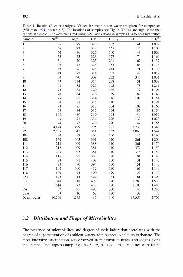
3.2 Distribution and Shape of Microbialites
The presence of microbialites and degree of their induration correlates with the
degree of supersaturation of ambient waters with respect to calcium carbonate. The
most intensive calcification was observed in microbialite heads and ledges along
the channel The Rapids (sampling sites 8, 19, 20, 124, 125). Oncolites were found
Table 1 Results of water analyses. Values for mean ocean water are given for comparison
(Milliman 1974, his table 3). For locations of samples see Fig. 2. Values are mg/l. Note that
cations in sample 1–22 were measured using AAS, and cations in samples 104 to LSA by titration
Sample Na2+ Mg2+ Ca2+ HCO3� Cl� SO4
2�
1 56 79 325 183 41 1,072
2 56 72 323 165 45 1,100
3 60 76 326 140 43 1,060
4 55 73 323 177 70 1,160
5 51 70 325 201 67 1,137
6 49 72 323 183 66 1,113
7 49 76 325 214 71 1,139
8 49 72 314 207 48 1,019
9 50 70 309 232 345 1,031
10 49 734 314 238 51 1,038
11 68 82 325 165 78 1,211
12 72 82 320 146 79 1,106
13 70 84 316 189 82 1,147
14 75 85 314 116 102 1,154
15 80 87 315 110 110 1,194
16 78 85 313 104 102 1,165
17 68 84 313 104 185 1,154
18 108 89 310 104 44 1,030
19 43 71 334 226 39 1,023
20 44 73 329 220 47 1,185
21 4,174 499 295 171 5,730 1,346
22 2,535 345 351 153 3,060 1,544
104 90 97 404 140 146 1,190
108 150 103 391 110 261 1,200
111 213 109 388 110 361 1,170
112 212 109 381 110 379 1,150
113 223 105 381 110 378 1,150
114 116 97 388 120 184 1,140
115 88 91 408 150 131 1,140
116 98 90 394 130 151 1,140
117 108 100 412 130 167 1,140
118 100 94 400 120 155 1,140
LM 122 114 422 84 181 1,300
LG 1,040 218 497 120 1,780 1,530
R 614 173 478 120 1,100 1,480
CA 37 95 497 300 45 1,240
LSA 33 10 62 180 32 70
Ocean water 10,760 1,295 415 140 19,350 2,700
192 E. Gischler et al.
abundantly between the Rapids and Bacalar village. Large microbialite heads over
1 m in diameter and height occurred along the western shore in the southern part of
Laguna Bacalar (Fig. 2, sites 8). Microbialite heads tend locally to coalesce and
form platforms (at sites 2 and 103). In 2009, we found large heads also north of the
Bacalar toward Zipline Point, but less abundant than south of the village. North of
Zipline Point and along the entire eastern shores of the lagoon, microbialites form
only low-relief crusts (Fig. 1). E-W depth sounder traverses across the lagoon about
2 km north and south of the village of Bacalar revealed mostly flat bottom,
indicating that no microbialite heads form in deeper water. No microbialites were
found in the adjacent Laguna San Antonio, Laguna Milagros, Laguna Guerrero, and
along the coast of Chetumal Bay.
The presence of microbialites and degree of induration correlate with water
chemistry, their shapes, however, seem to depend largely on shoreline topography
and relief so that microbialite crusts are found along low angle shorelines, heads
occur at intermediate angle, and ledges at steep, high angle slopes (Fig. 3).
Fig. 3 Simplified schematic presentation showing microbialite shape as related to shoreline
topography. (a) Low angle slope with crusts. (b) Intermediate angle slope with heads. (c) High
angle slope with ledges
Microbial Mats and Microbialites in the Freshwater Laguna Bacalar 193
3.3 Microbial Mats
Microbial mats cover only mm-deep surface layer of stromatolitic structures,
identifying the site of active accretionary process of the microbialite structure.
The consistency of mats varies between soft velvety coatings to hard crusts or
elastic-leathery consistency. They are mostly beige to brown in color, distributed
over the upper surfaces of microbialite crusts and on tops and slopes of microbialite
heads. Microbial crusts are the most common microbialite structures in the lake.
They vary in degree of calcification and are usually prostrate. They occur along
shores with mild slope and are widely distributed along the entire eastern shoreline
and in northern parts of the lake. Lithified hemispheric, internally zoned stromato-
litic structures, about 5 cm in diameter occur on calcareous heads and ledges in
sections with high current velocity (The Rapids; sites 8, 19, 20). Bright orange
colored microbial mats located on the flat tops of microbialite domes are the most
conspicuous mat structures, however these appear to have a limited seasonal growth
(sites 2, 103).
Microscopic analysis of the composition of different microbial mat types has
established that each is dominated by a particular cyanobacterium.
3.3.1 Calothrix Mat
The most common microbial mat is constructed by upward oriented tapered
filaments of the rivulariacean cyanobacterium Calothrix (Fig. 4). The mat forms
expanded semi-indurated crusts along shores at low angles (Fig. 4a). The fila-
ments of Calothrix are frequently falsely branched to form upward diverging
bushes that preserve well and constitute a common sedimentary element of the
stromatolite interior (Fig. 4b). Sheaths surrounding the trichomes are dark brown,
pigmented by the extracellular sun-screening pigment scytonemin (Portwich and
Garcia-Pichel 2003), which are also preserved in calcified parts of the stromato-
lite (Fig. 5). Trichomes are frequently curved at their base, distally tapered,
unconstricted to slightly constricted at the crosswalls and oriented upright or
upward-radiating (Fig. 4c). Filaments are variable in size, at the base
15.57 � 2.51 (31) mm in diameter. Cells of the trichomes 6.07 � 0.79 (45) mmwide and 3.68 � 1.24 (45) mm long, short and wide at the base (Fig. 6), becom-
ing distally isodiametric, tapering toward the tips into long hair-like whips
comprised of long and narrow cells with pale vacuolized content. The heterocysts
form at the base of the trichomes and false branches, separated at later stages of
development by extracellular polymeric substance (EPS) that constitutes the
sheath and is produced only by vegetative, carbon fixing trichome cells. Many
filaments do not differentiate heterocysts and could be misidentified under the
genus Homoeothrix.Calothrix-dominated mats have the widest distribution in the lake and maintain
also the highest diversity of cyanobacteria, including colonies of the other filamentous
194 E. Gischler et al.
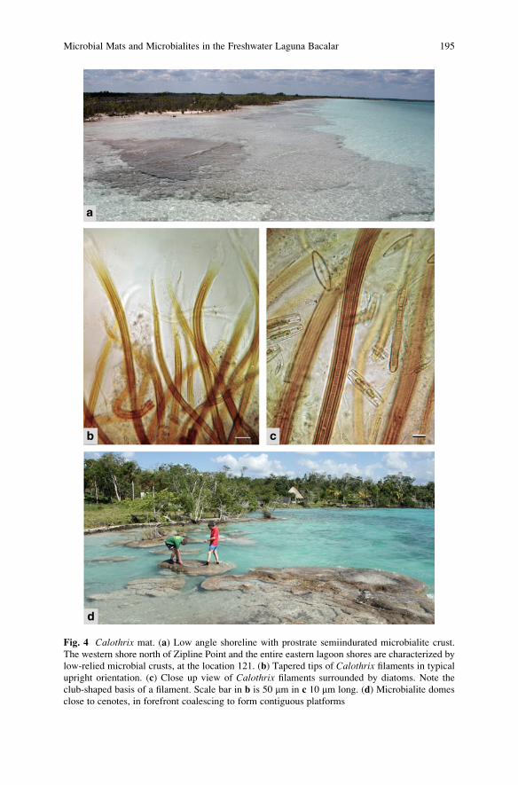
Fig. 4 Calothrix mat. (a) Low angle shoreline with prostrate semiindurated microbialite crust.
The western shore north of Zipline Point and the entire eastern lagoon shores are characterized by
low-relied microbial crusts, at the location 121. (b) Tapered tips of Calothrix filaments in typical
upright orientation. (c) Close up view of Calothrix filaments surrounded by diatoms. Note the
club-shaped basis of a filament. Scale bar in b is 50 mm in c 10 mm long. (d) Microbialite domes
close to cenotes, in forefront coalescing to form contiguous platforms
Microbial Mats and Microbialites in the Freshwater Laguna Bacalar 195
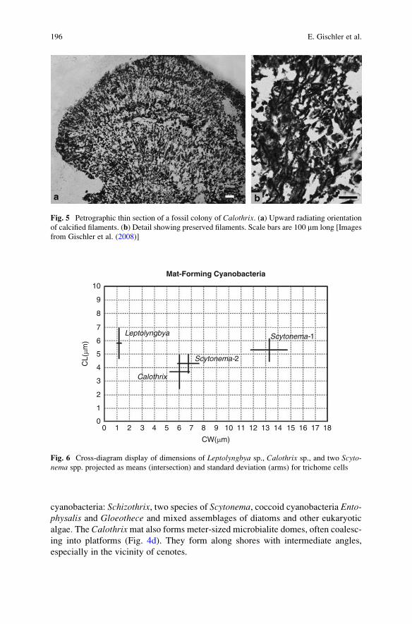
cyanobacteria: Schizothrix, two species of Scytonema, coccoid cyanobacteria Ento-physalis and Gloeothece and mixed assemblages of diatoms and other eukaryotic
algae. The Calothrixmat also forms meter-sized microbialite domes, often coalesc-
ing into platforms (Fig. 4d). They form along shores with intermediate angles,
especially in the vicinity of cenotes.
Fig. 5 Petrographic thin section of a fossil colony of Calothrix. (a) Upward radiating orientation
of calcified filaments. (b) Detail showing preserved filaments. Scale bars are 100 mm long [Images
from Gischler et al. (2008)]
00 1 2 3 4 5 6 7 8 9
CW(µm)
CL(
µm)
10 11 12 13 14 15 16 17 18
1
2
3
4
5
Leptolyngbya
Mat-Forming Cyanobacteria
Calothrix
Scytonema-2
Scytonema-16
7
8
9
10
Fig. 6 Cross-diagram display of dimensions of Leptolyngbya sp., Calothrix sp., and two Scyto-nema spp. projected as means (intersection) and standard deviation (arms) for trichome cells
196 E. Gischler et al.

3.3.2 Scytonema Mat
Distinct, firmly lithified hemispheroid structures 4–6 cm in diameter are formed in
the habitat with strong currents called The Rapids (Fig. 7a). These microbialites are
almost exclusively built by populations of a single cyanobacterial taxon, Scytonemasp. 1. The structures are dome-shaped, densely covered by cyanobacterial biofilm
(Fig. 7b) internally permeated throughout by calcified sheaths (Fig. 7c) showing
internal concentric lamination in sagittal section (stromatolitic morphology). The
lamination indicates that the degree of calcification changes rhythmically over time,
as the concentric laminae differ in mineral load. The filaments of this heterocystous
cyanobacterium are 38.31 � 5.49 mm wide, with occasional paired false branching
(Fig. 7d). The cells of the trichomes are 14.87 � 1.46 (50) mm wide and
5.28 � 0.84 (80) mm long (Fig. 6, right). Filaments are curved and intertwined
but, unlike Calothrix, are not oriented upward. Trichomes show predominant apical
growth, i.e., they exhibit the highest growth rates at the trichome tips (Fig. 7e).
Heterocysts, the sites of dinitrogen fixation are intercalary, connected by two
opposing gates with vegetative carbon-fixing cells (Fig. 7f). In the sampled material
we observed a large proportion of fragmented trichomes, commonly exiting from
their sheaths, which opened by a funnel-shaped diverging tip (Fig. 7g).
The systematic position of this organism is close to Scytonema mirabile, butdiffers from that species by wide non-laminated sheaths, and frequent opening of
sheaths at the tip of filaments. A much smaller Scytonema sp. 2 was present as a
minor constituent in the Calothrix mat.
3.3.3 Leptolyngbya Mat
Bright orange colored mats grow on top of large microbialite heads, close to the
water surface and partially exposed to the atmosphere. They frequently form a
slightly elevated rim enclosing a water-filled depression on the microbialite tops
making an appearance of a mini-atoll (Fig. 8a). These mats were produced by a
sheathed filamentous cyanobacterium with narrow trichomes currently classified as
Leptolyngbya Anagnostidis et Komarek (former Phormidium K€utzing). The fila-
ments consist of unbranched cellular trichomes, each surrounded by a thin but firm
exopolymer sheath. Cells are cylindrical, significantly longer then wide uncon-
stricted to barely constricted at the cross wall; the end cells are rounded. Cells are
1.20 � 0.19 (26) mm wide and 5.77 � 1.12 (63) mm long surrounded by exopoly-
mer sheaths to form unbranched filaments 2.56 � 0.32 (54) in diameter (Fig. 6,
left). Trichomes move by gliding motility, often abandoning their sheaths and
forming new ones, so that the trichomes are concentrated close to the surface of
the mat, whereas deeper parts of the mat consist of predominantly empty sheaths.
The bright orange coloration of these mats (Fig. 8b) reflects high concentrations of
carotenoids in the cells, a pigmentation known to serve for protection from damage
by high light intensity and UV (Ehling-Schulz and Scherer 1999; Seckbach and
Oren 2007). Below this protecting layer the mat is blue-green in color.
Microbial Mats and Microbialites in the Freshwater Laguna Bacalar 197
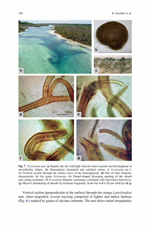
Vertical section (perpendicular to the surface) through the orange Leptolyngbyamat, when magnified, reveals layering comprised of lighter and darker laminae
(Fig. 8c) marked by grains of calcium carbonate. The mat shows initial irregularities
Fig. 7 Scytonema mat. (a) Rapids, the site with high velocity water currents and development of
microbiolitic ledges. (b) Hemispheric laminated and calcified colony of Scytonema sp.-1.
(c) Vertical section through the surface layer of the hemispheroid. (d) Pair of false branches
characteristic for the genus Scytonema. (e) Funnel-shaped diverging opening of the sheath
and exiting trichomes. (f) Scytonema filament containing a trichome with intercalary heterocyst.
(g) Massive abandoning of sheaths by trichome fragments. Scale bar in d is 50 mm valid for (d–g)
198 E. Gischler et al.
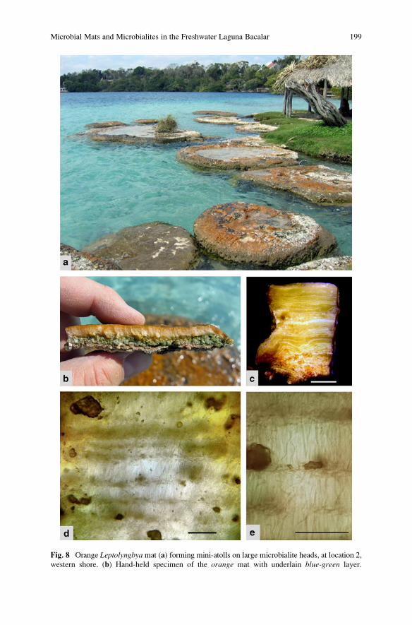
Fig. 8 Orange Leptolyngbya mat (a) forming mini-atolls on large microbialite heads, at location 2,
western shore. (b) Hand-held specimen of the orange mat with underlain blue-green layer.
Microbial Mats and Microbialites in the Freshwater Laguna Bacalar 199
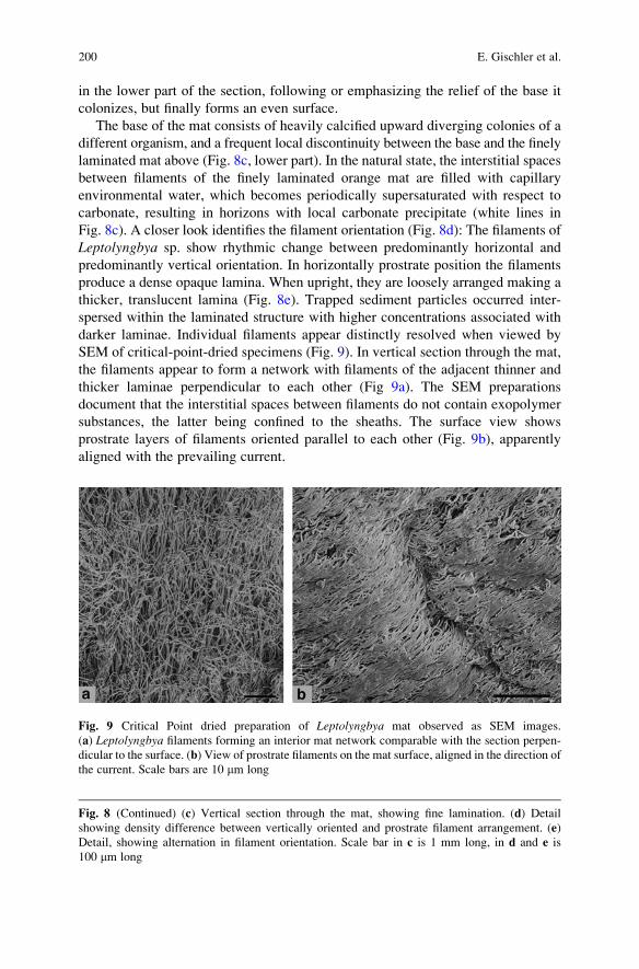
in the lower part of the section, following or emphasizing the relief of the base it
colonizes, but finally forms an even surface.
The base of the mat consists of heavily calcified upward diverging colonies of a
different organism, and a frequent local discontinuity between the base and the finely
laminated mat above (Fig. 8c, lower part). In the natural state, the interstitial spaces
between filaments of the finely laminated orange mat are filled with capillary
environmental water, which becomes periodically supersaturated with respect to
carbonate, resulting in horizons with local carbonate precipitate (white lines in
Fig. 8c). A closer look identifies the filament orientation (Fig. 8d): The filaments of
Leptolyngbya sp. show rhythmic change between predominantly horizontal and
predominantly vertical orientation. In horizontally prostrate position the filaments
produce a dense opaque lamina. When upright, they are loosely arranged making a
thicker, translucent lamina (Fig. 8e). Trapped sediment particles occurred inter-
spersed within the laminated structure with higher concentrations associated with
darker laminae. Individual filaments appear distinctly resolved when viewed by
SEM of critical-point-dried specimens (Fig. 9). In vertical section through the mat,
the filaments appear to form a network with filaments of the adjacent thinner and
thicker laminae perpendicular to each other (Fig 9a). The SEM preparations
document that the interstitial spaces between filaments do not contain exopolymer
substances, the latter being confined to the sheaths. The surface view shows
prostrate layers of filaments oriented parallel to each other (Fig. 9b), apparently
aligned with the prevailing current.
Fig. 9 Critical Point dried preparation of Leptolyngbya mat observed as SEM images.
(a) Leptolyngbya filaments forming an interior mat network comparable with the section perpen-
dicular to the surface. (b) View of prostrate filaments on the mat surface, aligned in the direction of
the current. Scale bars are 10 mm long
Fig. 8 (Continued) (c) Vertical section through the mat, showing fine lamination. (d) Detail
showing density difference between vertically oriented and prostrate filament arrangement. (e)
Detail, showing alternation in filament orientation. Scale bar in c is 1 mm long, in d and e is
100 mm long
200 E. Gischler et al.
The studied mat showed a marked discontinuity about 1 cm below the surface
(Fig. 8b, c). The mat beneath the laminated orange mat had a coarser texture and
appeared calcified. It was comprised of upward radiating filaments, significantly
wider than those forming the finely laminated mat. These features are consistent
with the properties of the Calothrix mat, which apparently preceded the formation
of Leptolyngyba on top of the microbialite head and was most likely the main
architect of these large domal structures.
3.3.4 Internal Structures of Bacalar Microbialites
Calcium carbonate deposition in Laguna Bacalar is not uniform, neither are the
carbonate saturation levels in different parts of the lake. Highest levels of dissolved
carbonate have been consistently found along the western shore of the lake, in the
vicinity of the cenotes, and in passages exhibiting higher current velocities. All
precipitates in the lake consist of low magnesium calcite. Petrographic thin sections
of the interior portions of the Bacalar microbialites prepared at different levels in
the collected cores, show distinct areas, some with finely laminated, and others with
upward radiating carbonate textures, the latter associated with larger diameter
filaments (encountered mainly as sheath remains). In addition, the internal texture
close to the surface is interrupted by cavities of various sizes and contains embed-
ded Dreissena bivalve shells. Because of this inhomogeneous internal structure the
microbialites of the Laguna Bacalar can be categorized as thrombolitic.
4 Discussion
The overall architecture of the microbialite deposits of the Laguna Bacalar suggests
that the structures are constructed by more than one agent, which likely included the
processes active today. The three studied microbial assemblages prevalent today
represent three separate ecological niches occupied and dominated by different
microorganisms and showing different biological responses.
The distribution of microbialites in Laguna Bacalar is controlled primarily by
elevated carbonate concentrations of lagoonal waters, with possible contribution by
photosynthetic activities of cyanobacteria in the structure and the presence of
exopolymers produced by them (Merz-Preiss 2000). Microbialites are largest and
most abundant in the southern parts of the lagoon where the karstic springs called
cenotes are located and where high carbonate concentration is maintained by the
influx of karstic groundwaters. The primary producers are comprised of mat-
building cyanobacteria and benthic diatoms which support abundant grazing popu-
lations of Pomacea gastropods. Carbonate accretion rates are apparently high
enough to counteract destructive effect of grazing activity and protect the sedimen-
tary structures which have been formed over a millennium and recognized in cores
of lithified stromatolites (Gischler et al. 2008). Rasmussen et al. (1993) described
Microbial Mats and Microbialites in the Freshwater Laguna Bacalar 201
local occurrences of stromatolites at the mouth of Rio Hondo river in the nearby
Chetumal Bay. These authors suggested that exclusion of grazers in brackish water
was decisive for stromatolite buildup. We observed abundant grazers in Laguna
Bacalar stromatolites and suggest that they are maintained by rapid calcification.
Abundant mytilid bivalves are incorporated into the outer, more recently depos-
ited parts of the microbialite structure, which contributes to their porosity. This
effect can be best observed in the occurrences of The Rapids, where the mytilids are
particularly abundant. Currents and flushing of pore spaces by carbonate-saturated
waters assures a continuation of carbonate precipitation and cementation of the
structure by lining of pore spaces (Gischler et al. 2008; their fig. 4g).
In addition to water chemistry and flushing, shoreline topography clearly influ-
ences occurrences and shapes of microbialites in Laguna Bacalar. The angle of the
slope which controls the accommodation space apparently controls the direction of
stromatolite accretion. Low angle shorelines are characterized by low relief micro-
bialite crusts as observed along the eastern shoreline. Steep angle shorelines such as
in the Rapids channel lead to the growth of microbialite ledges. Large heads are
found were slope angles are intermediate (Fig. 3). When the available accomoda-
tion space is filled up, microbialites tend to grow sideways, i.e., they prograde and
in some cases even coalesce, as at locations 2 and 103 (Fig. 4d).
The interpretation of the studied actively growing mats in terms of their role in
the construction of large lithified microbialites of Laguna Bacalar poses objective
difficulties. All mats contain low magnesium calcite as mineral components,
however, they differ in the degree they are calcified. Accordingly, the interpretation
of their roles in the accretionary history of these microbialites remains based on
structural similarities and is tenuous. The three types of microbial mats observed in
the Laguna Bacalar respond differently to the process of carbonate deposition.
The upward oriented (and divergent) growth of Calothrix accommodates a wide
range of habitats and moderate rates of calcification and microbialite accretion. The
organism requires time for growth, which is generated from the base of the trichomes
and differentiates by tapering upward and extruding into the water column. The
calcification is predominantly encasing the sheaths whereas photosynthetically
active trichomes are gradually dislocated upward by growth and false branching.
Under too low calcification rates, the community may be affected or even removed
by grazers. The success of the community is also limited by burial due to excessive
calcification and accretion rates that exceed the growth rates of Calothrix trichomes.
Many filaments do not differentiate heterocyst, which is also consistent with slow
growth and availability of nitrogen as nutrient for growth (Fogg et al. 1973).
In cases of highest rates of calcification, as observed in the Rapids section of the
lagoon, the colonization by Scytonema mats and formation of hemispheroid stro-
matolites is favored. Scytonema trichomes show maximum growth in distal rather
than proximal parts as in Calothrix. In addition, we could observe another adapta-
tion of this organism to respond to high calcification rates by fragmenting the
trichomes (Fig. 7g), which are able to leave their cemented sheaths by gliding
and producing new ones on top of the sediment. The mat is thereby maintained on
the stromatolite surface on top of a firmly calcified base. The proportion of free
202 E. Gischler et al.
trichomes and those in the process of leaving their sheaths observed upon decalci-
fication of Scytonema mats (Fig. 7e, g) testified about the high rate at which the
process takes place. Also the filaments and the abandoned sheaths of Scytonemawere intertwined and oriented predominantly parallel to the substrate surface rather
than perpendicular as in Calothrix, suggesting a mechanism in escaping the burial
by motility rather than by growth of trichomes. Scytonema-constructed microbia-
lites with different and changing growth orientation occur in waters of different and
variable salinities (Monty 1967; Reitner et al. 1996).
The Leptolyngbya mats occupy a narrow zone defined by water level in an area
where the calcification rates are moderate. The mats are characterized by a rapid
combination of growth, motility and change in orientation of their trichomes, which
leave a mark in positioning of sheaths within the predominantly organic structure.
Carbonate particles originating from suspension become trapped in their laminae.
Autochthonous carbonate occurred only in some horizons (white laminae in
Fig. 8c) and was comprised by fine micritic precipitate. This is in contrast to the
adjacent heavily calcified Calothrix mat, with filaments often completely encased
in carbonate, although both mats were apparently exposed to the water of the same
chemistry. A possible reason for the observed difference in the degree of their
calcification may be explained by the difference in their growth rates.
The alternate change in orientation of filaments and the resulting fine lamination
characteristic of Leptolyngbya is indistinguishable from those found in the marine
Leptolyngbya (Phormidium) hendersonii (Howe) Anagnostidis et Komarek (Golubic
and Focke 1978). The lamination in this organism was found to be nocti-diurnal:
The upright orientation of the filaments was promoted by positive phototaxis of
cyanobacterial trichomes, which subsequently assumed a prostrate position during
the night. The timing has been shown by Monty (1965, 1967) using carborundum
marking. The process of upward gliding motility of trichomes is accompanied by
the production of exopolymer sheaths, which are left behind and constitute the main
constructive element of the structure. This rhythmic change in filament orientation
is consistent with the density difference between upright and prostrate layers. The
intricate dense meshwork of tiny filaments accounts for the firmness of the struc-
ture, although the space between filaments is filled with water rather than exo-
polymer (observed as air space by SEM following critical-point-drying procedure)
(Fig. 9a). The correlation of entrapped particles with the prostrate layers is also
consistent with the timing of the mat accretion: They accumulate during the night
when the trichomes rest and are diluted during the phototactic movement and
growth of the microorganism by day. The rapid increase in thickness of these
mats can be measured by counting of the laminae. The relatively short time of the
growing season of these mats (calculated to be between 30 and 60 days), is
commensurate with the low rate of sedimentation and calcification within the mat.
The rapid formation of the Leptolyngbya mat as described above is in contrast to
the significantly slower growth of the Calothrix mat, which is produced by a non-
motile cyanobacterium. The orientation of its filaments rest on phototropism rather
than on phototaxis, whereas their thick, scytonemin-enriched sheaths offer prolonged
protection of the trichomes. In addition, the mat remains open to water circulation
Microbial Mats and Microbialites in the Freshwater Laguna Bacalar 203
during its growth and is longer exposed to calcification. The organic remains of the
Calothrix mat are present in the cores and correlate well with the upward radiating
texture of the colonies, however, the filaments of the much more delicate Leptolyng-bya did not preserve in the lithified interior of the structure. The finely laminated
carbonate portions within the lithified cores may represent carbonate templates after
Leptolyngbya mats, but direct evidence of the identity could not be established.
If the identity of the three currently active microbial mats, and the fossil
carbonate texture in the cores were confirmed, it would suggest that historic
changes in local water supply was sufficient to cause changes in dominant mat
communities. The microbialite accretion alters the position of the mat in reference
to water levels, and also modifies the water flow, which, in turn, may alter the
conditions and rates of precipitation (e.g., by narrowing the channels and increasing
the water flow). Such interactions and mutual adjustments between water access to
biota and carbonate deposition may explain the irregular spatial distribution of
textures during the process of stromatolite accretion as reflected in the textures
found in the cores.
Acknowledgements We like to thank the Deutsche Forschungsgemeinschaft (DFG) for their
support (Gi 222/15-2). International cooperation was supported by Alexander-von-Humboldt
foundation, Bonn and Hanse Wissenschaftskolleg, Delmenhorst to S. Golubic. M. Gibson thanks
the faculty and research development grant program at the University of Tennessee for financial
support. Paul Blanchon (UNAM, Puerto Morelos) helped with logistics in Mexico.
References
Castenholz RW (2001) Oxygenic photosynthetic bacteria. In: Boon DR, Castenholz RW (eds)
Bergey’s Manual of Systematic Bacteriology, 2nd ed. Springer, New York, pp 473–600.
Ehling-Schulz M, Scherer S (1999) UV protection in cyanobacteria. Eur J Phycol 34: 329–338.
Fogg GE, Stewart WDP, Fay P, Walsby AE (1973) The Blue-green Algae. Acad Press, London,
459 pp.
Geitler L (1932) Cyanophyceae. Rabenhorst’s Kryptogamenflora von Deutschland, Osterreich und
der Schweiz 14. Akademischer Verlag, Leipzig, 1196 pp.
Gischler E, Gibson MA, Oschmann W (2008) Giant Holocene freshwater microbialites, Laguna
Bacalar, Quintana Roo, Mexico. Sedimentology 55: 1293–1309.
Golubic S, Focke JW (1978) Phormidium hendersoniiHowe: identity and significance of a modern
stromatolite building microorganism. J Sediment Petrol 48: 751–764.
Kenkmann T, Sch€onian F (2006) Ries and Chicxulub: impact craters on earth provide insights for
Martian ejecta blankets. Meteorit Planet Sci 41: 1587–1603.
Komarek J, Anagnostidis K (2005) Cyanoprokaryota 2. Teil Oscillatoriales. In: B€udel B, G€artner G,Krienitz L, Schlager M (eds) S€usswasserflora von Mitteleuropa. Fischer, Jena, 759 pp.
Merz-Preiss M (2000) Calcification in cyanobacteria. In: Riding RE, Awramik SM (eds) Microbial
Sediments. Springer-Verlag, Berlin-Heidelberg, pp. 50–56.
Milliman JD (1974) Marine Carbonates. Springer, Berlin, 375 pp.
Monty CLV (1965) Recent algal stromatolites in the windward lagoon, Andros Island, Bahamas.
Ann Soc Geol Belgique 88: 269–276.
Monty CLV (1967) Distribution and structure of recent stromatolitic algal mats, eastern Andros
Island, Bahamas. Ann Soc Geol Belgique 90: 55–100.
204 E. Gischler et al.

Perry E, Paytan A, Pedersen B, Velasquez-Oliman G (2009) Groundwater geochemistry of the
Yucatan peninsula, Mexico: constraints on stratigraphy and hydrogeology. J Hydrol 367:
27–40.
Portwich A, Garcia-Pichel F (2003) Biosynthetic pathway of mycosporines (mycosporine.-like
amino acids) in the cyanobacterium Chlorogloeopsis sp. strain PCC 6912. Phycologia 42:
384–392.
Rasmussen KA, Macintyre IG, Prufert L (1993) Modern stromatolite reefs fringing a brackish
coastline, Chetumal Bay, Belize. Geology 21: 199–202.
Reitner J, Paul J, Arp G, Hause-Reitner D (1996) Lake Thetis domal microbialites – a complex
framework of calcified biofilms and organomicrites (Cervantes, Western Australia). In: Reitner J,
Neuweiler F, Gunkel F (eds) Global and Regional Controls on Biogenic Sedimentation.
G€ottinger Arb Geol Pal€aont, Sb2, G€ottingen, pp 85–89.
Seckbach J, Oren A (2007) Oxygenic photosynthetic microorganisms in extreme environments.
In: Seckbach J, Oren A (eds) Algae and Cyanobacteria in Extreme Environments. Springer,
Dordrecht, pp 5–25.
Microbial Mats and Microbialites in the Freshwater Laguna Bacalar 205

.

Part IIIAtmospheric Forcing and Waves

.
Geomicrobiology of Fluid Venting Structures
at the Salse di Nirano Mud Volcano Area
in the Northern Apennines (Italy)
Christina Heller, Martin Blumenberg, Sebastian Kokoschka,
Christoph Wrede, Michael Hoppert, Marco Taviani, and Joachim Reitner
1 Introduction
Recent studies suggest that geological sources of methane like onshore and
marine seeps, micro-seepage and mud volcanoes are an important source of
this greenhouse gas (Etiope 2004). They represent the second most important
natural emission after wetlands. New estimations indicate a global emission of
methane from mud volcanoes in a range of 6-9 Tg year-1 (Etiope and Ciccioli
2009).
Mud volcanoes are present in many terrestrial and marine areas worldwide
(Mazurenko et al. 2003; Scholte 2005), but their geographical distribution is
strongly controlled by the geological settings. The majority is localized in areas
of recent tectonic activity, particularly in zones of compression like accretionary
complexes, thrust belts and e.g. in the forelands of Alpine orogenic structures
(Dimitrov 2002; Kopf 2002). Furthermore, since the sediments in mud volcanoes
are often hydrocarbon-rich, a relation between the formation of hydrocarbons and
mud volcanoes was suggested (Dimitrov 2002).
The geographical occurrence of mud volcanoes in Italy can be divided into three
main groups: (i) northern and (ii) central Italy and (iii) Sicily (Martinelli and Judd
C. Heller, M. Blumenberg, and J. Reitner (*)
Geoscience Centre, University of G€ottingen, Geobiology Division, Goldschmidtstraße 3, 37077,
G€ottingen, Germany
e-mail: [email protected]
S. Kokoschka, C. Wrede, and M. Hoppert
Institute of Microbiology and Genetics, University of G€ottingen, Grisebachstr. 8, 37077,
G€ottingen, Germany
M. Taviani
ISMAR-CNR, Via Gobetti 101, 40129, Bologna, Italy
J. Reitner et al., Advances in Stromatolite Geobiology,Lecture Notes in Earth Sciences 131, DOI 10.1007/978-3-642-10415-2_14,# Springer-Verlag Berlin Heidelberg 2011
209
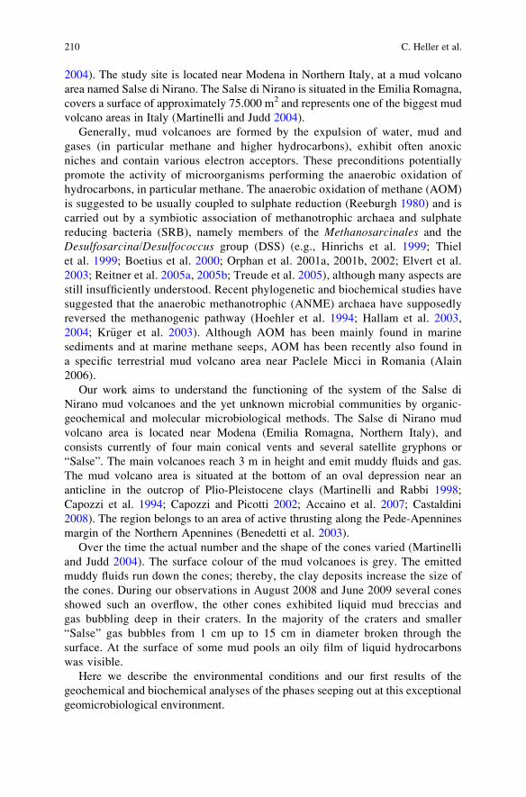
2004). The study site is located near Modena in Northern Italy, at a mud volcano
area named Salse di Nirano. The Salse di Nirano is situated in the Emilia Romagna,
covers a surface of approximately 75.000 m2 and represents one of the biggest mud
volcano areas in Italy (Martinelli and Judd 2004).
Generally, mud volcanoes are formed by the expulsion of water, mud and
gases (in particular methane and higher hydrocarbons), exhibit often anoxic
niches and contain various electron acceptors. These preconditions potentially
promote the activity of microorganisms performing the anaerobic oxidation of
hydrocarbons, in particular methane. The anaerobic oxidation of methane (AOM)
is suggested to be usually coupled to sulphate reduction (Reeburgh 1980) and is
carried out by a symbiotic association of methanotrophic archaea and sulphate
reducing bacteria (SRB), namely members of the Methanosarcinales and the
Desulfosarcina/Desulfococcus group (DSS) (e.g., Hinrichs et al. 1999; Thiel
et al. 1999; Boetius et al. 2000; Orphan et al. 2001a, 2001b, 2002; Elvert et al.
2003; Reitner et al. 2005a, 2005b; Treude et al. 2005), although many aspects are
still insufficiently understood. Recent phylogenetic and biochemical studies have
suggested that the anaerobic methanotrophic (ANME) archaea have supposedly
reversed the methanogenic pathway (Hoehler et al. 1994; Hallam et al. 2003,
2004; Kr€uger et al. 2003). Although AOM has been mainly found in marine
sediments and at marine methane seeps, AOM has been recently also found in
a specific terrestrial mud volcano area near Paclele Micci in Romania (Alain
2006).
Our work aims to understand the functioning of the system of the Salse di
Nirano mud volcanoes and the yet unknown microbial communities by organic-
geochemical and molecular microbiological methods. The Salse di Nirano mud
volcano area is located near Modena (Emilia Romagna, Northern Italy), and
consists currently of four main conical vents and several satellite gryphons or
“Salse”. The main volcanoes reach 3 m in height and emit muddy fluids and gas.
The mud volcano area is situated at the bottom of an oval depression near an
anticline in the outcrop of Plio-Pleistocene clays (Martinelli and Rabbi 1998;
Capozzi et al. 1994; Capozzi and Picotti 2002; Accaino et al. 2007; Castaldini
2008). The region belongs to an area of active thrusting along the Pede-Apennines
margin of the Northern Apennines (Benedetti et al. 2003).
Over the time the actual number and the shape of the cones varied (Martinelli
and Judd 2004). The surface colour of the mud volcanoes is grey. The emitted
muddy fluids run down the cones; thereby, the clay deposits increase the size of
the cones. During our observations in August 2008 and June 2009 several cones
showed such an overflow, the other cones exhibited liquid mud breccias and
gas bubbling deep in their craters. In the majority of the craters and smaller
“Salse” gas bubbles from 1 cm up to 15 cm in diameter broken through the
surface. At the surface of some mud pools an oily film of liquid hydrocarbons
was visible.
Here we describe the environmental conditions and our first results of the
geochemical and biochemical analyses of the phases seeping out at this exceptional
geomicrobiological environment.
210 C. Heller et al.
2 Materials and Methods
2.1 Study Site and Sampling
The samples were collected during two campaigns (August 2008 and June 2009)
from several mud volcanoes in the Emilia Romagna area in Northern Italy. Mud and
gases were collected from four active cones, which showed constant development
of gas bubbling. This study is focussed on one of the active cones of the Salse di
Nirano (Fig. 1).
Mud samples were collected in a depth of approximately 1.5 m with a hand-
operated vacuum pump, connected to a flexible tube with a metal pipe at the open
end (UniSampler, B€urkle, Bad Bellingen, Germany) and were transferred to the
laboratory under cooled conditions. The samples for the lipid analyzes were
freeze-dried and stored by �20�C. Gas bubbles were collected in the central
part of the gryphon using a funnel and special gas vials (Labco Vials, Labco
Limited, Buckinghamshire, United Kingdom).
The temperature, redox potential and the pH-values of the mud were measured
directly in the pools.
2.2 Gas Analysis
The concentrations of CH4, CO2 and N2O were measured with an automated gas
chromatographic system (GC 14A, Shimadzu, Duisburg, Germany) equipped with
Fig. 1 Images of the Salse di Nirano mud volcanoes Northern Apennines, Italy. a) and c) mud
volcano cones and craters of the Salse di Nirano. b) Liquid hydrocarbons on the surface of a pool.
d) A bubbling salse
Geomicrobiology of Fluid Venting Structures at the Salse di Nirano Mud Volcano Area 211
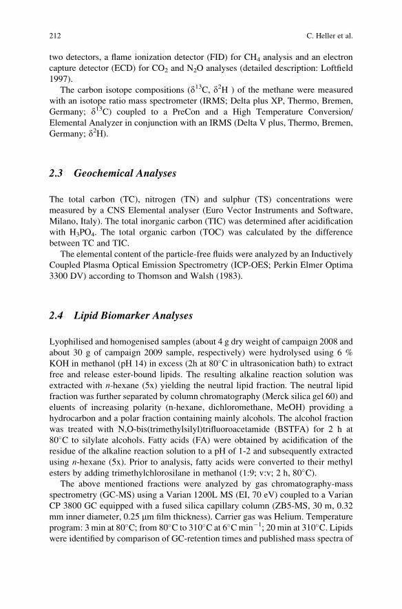
two detectors, a flame ionization detector (FID) for CH4 analysis and an electron
capture detector (ECD) for CO2 and N2O analyses (detailed description: Loftfield
1997).
The carbon isotope compositions (d13C, d2H ) of the methane were measured
with an isotope ratio mass spectrometer (IRMS; Delta plus XP, Thermo, Bremen,
Germany; d13C) coupled to a PreCon and a High Temperature Conversion/
Elemental Analyzer in conjunction with an IRMS (Delta V plus, Thermo, Bremen,
Germany; d2H).
2.3 Geochemical Analyses
The total carbon (TC), nitrogen (TN) and sulphur (TS) concentrations were
measured by a CNS Elemental analyser (Euro Vector Instruments and Software,
Milano, Italy). The total inorganic carbon (TIC) was determined after acidification
with H3PO4. The total organic carbon (TOC) was calculated by the difference
between TC and TIC.
The elemental content of the particle-free fluids were analyzed by an Inductively
Coupled Plasma Optical Emission Spectrometry (ICP-OES; Perkin Elmer Optima
3300 DV) according to Thomson and Walsh (1983).
2.4 Lipid Biomarker Analyses
Lyophilised and homogenised samples (about 4 g dry weight of campaign 2008 and
about 30 g of campaign 2009 sample, respectively) were hydrolysed using 6 %
KOH in methanol (pH 14) in excess (2h at 80�C in ultrasonication bath) to extract
free and release ester-bound lipids. The resulting alkaline reaction solution was
extracted with n-hexane (5x) yielding the neutral lipid fraction. The neutral lipid
fraction was further separated by column chromatography (Merck silica gel 60) and
eluents of increasing polarity (n-hexane, dichloromethane, MeOH) providing a
hydrocarbon and a polar fraction containing mainly alcohols. The alcohol fraction
was treated with N,O-bis(trimethylsilyl)trifluoroacetamide (BSTFA) for 2 h at
80�C to silylate alcohols. Fatty acids (FA) were obtained by acidification of the
residue of the alkaline reaction solution to a pH of 1-2 and subsequently extracted
using n-hexane (5x). Prior to analysis, fatty acids were converted to their methyl
esters by adding trimethylchlorosilane in methanol (1:9; v:v; 2 h, 80�C).The above mentioned fractions were analyzed by gas chromatography-mass
spectrometry (GC-MS) using a Varian 1200L MS (EI, 70 eV) coupled to a Varian
CP 3800 GC equipped with a fused silica capillary column (ZB5-MS, 30 m, 0.32
mm inner diameter, 0.25 mm film thickness). Carrier gas was Helium. Temperature
program: 3 min at 80�C; from 80�C to 310�C at 6�Cmin�1; 20 min at 310�C. Lipidswere identified by comparison of GC-retention times and published mass spectra of
212 C. Heller et al.
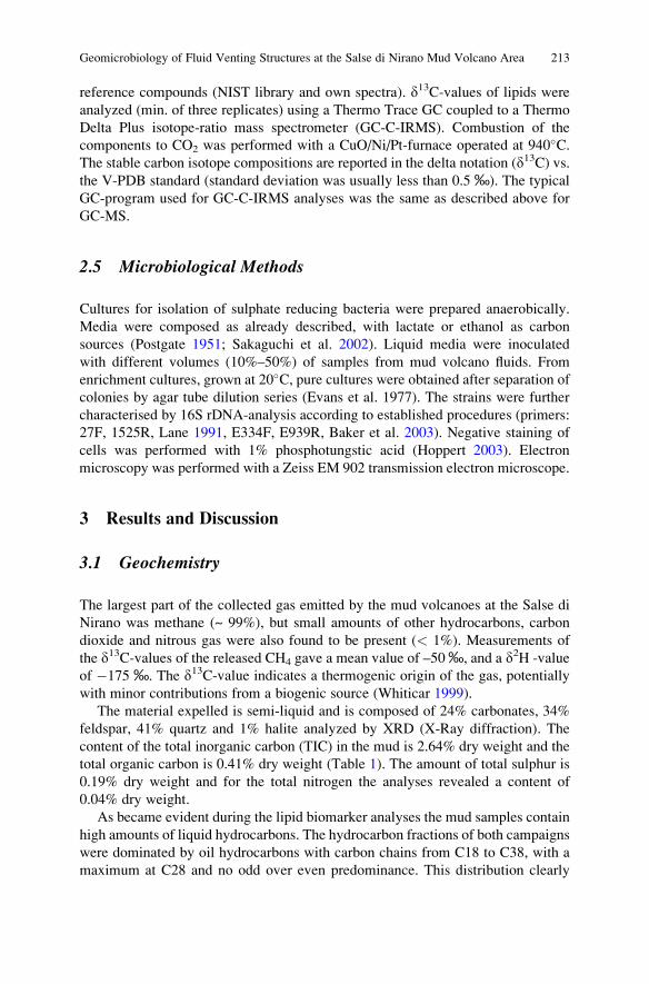
reference compounds (NIST library and own spectra). d13C-values of lipids wereanalyzed (min. of three replicates) using a Thermo Trace GC coupled to a Thermo
Delta Plus isotope-ratio mass spectrometer (GC-C-IRMS). Combustion of the
components to CO2 was performed with a CuO/Ni/Pt-furnace operated at 940�C.The stable carbon isotope compositions are reported in the delta notation (d13C) vs.the V-PDB standard (standard deviation was usually less than 0.5 ‰). The typical
GC-program used for GC-C-IRMS analyses was the same as described above for
GC-MS.
2.5 Microbiological Methods
Cultures for isolation of sulphate reducing bacteria were prepared anaerobically.
Media were composed as already described, with lactate or ethanol as carbon
sources (Postgate 1951; Sakaguchi et al. 2002). Liquid media were inoculated
with different volumes (10%–50%) of samples from mud volcano fluids. From
enrichment cultures, grown at 20�C, pure cultures were obtained after separation ofcolonies by agar tube dilution series (Evans et al. 1977). The strains were further
characterised by 16S rDNA-analysis according to established procedures (primers:
27F, 1525R, Lane 1991, E334F, E939R, Baker et al. 2003). Negative staining of
cells was performed with 1% phosphotungstic acid (Hoppert 2003). Electron
microscopy was performed with a Zeiss EM 902 transmission electron microscope.
3 Results and Discussion
3.1 Geochemistry
The largest part of the collected gas emitted by the mud volcanoes at the Salse di
Nirano was methane (~ 99%), but small amounts of other hydrocarbons, carbon
dioxide and nitrous gas were also found to be present (< 1%). Measurements of
the d13C-values of the released CH4 gave a mean value of –50‰, and a d2H -value
of �175 ‰. The d13C-value indicates a thermogenic origin of the gas, potentially
with minor contributions from a biogenic source (Whiticar 1999).
The material expelled is semi-liquid and is composed of 24% carbonates, 34%
feldspar, 41% quartz and 1% halite analyzed by XRD (X-Ray diffraction). The
content of the total inorganic carbon (TIC) in the mud is 2.64% dry weight and the
total organic carbon is 0.41% dry weight (Table 1). The amount of total sulphur is
0.19% dry weight and for the total nitrogen the analyses revealed a content of
0.04% dry weight.
As became evident during the lipid biomarker analyses the mud samples contain
high amounts of liquid hydrocarbons. The hydrocarbon fractions of both campaigns
were dominated by oil hydrocarbons with carbon chains from C18 to C38, with a
maximum at C28 and no odd over even predominance. This distribution clearly
Geomicrobiology of Fluid Venting Structures at the Salse di Nirano Mud Volcano Area 213
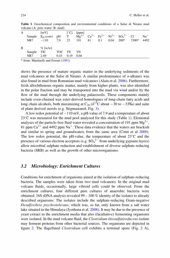
shows the presence of mature organic matter in the underlying sediments of the
mud volcanoes at the Salse di Nirano. A similar predominance of n-alkanes wasalso found in mud from Romanian mud volcanoes (Alain et al. 2006). Furthermore,
fresh allochthonous organic matter, mainly from higher plants, was also identified
in the polar fraction and may be transported into the mud via wind and/or by the
flow of the mud through the underlying palaeosoils. These components mainly
include even-chained wax ester-derived homologues of long-chain fatty acids and
long chain alcohols, both maximizing at C26 (d13C about �30 to �33‰) and suite
of plant derived sterols (e.g. Stigmastanol, Fig. 3).
A low redox potential of�110 mV, a pH-value of 7.9 and a temperature of about
23�C was measured for the mud pool analysed for this study (Table 1). Elemental
analyses of the particle-free fluid water revealed a concentration of 191 ppm Mg2þ,81 ppm Ca2þ and 4492 ppm Naþ. These data evidence that the waters are brackishand similar to spring and groundwaters from the same area (Conti et al 2000).
The low redox potential, the pH-value, the temperature of about 23�C and the
presence of various electron acceptors (e.g. SO42� from underlying gypsum layers)
allow microbial sulphate reduction and establishment of diverse sulphate reducing
bacteria (SRB) as well as the growth of other microorganisms.
3.2 Microbiology: Enrichment Cultures
Conditions for enrichment of organisms aimed at the isolation of sulphate-reducing
bacteria. The samples were taken from two mud volcanoes. In the original mud
volcano fluids, occasionally, large vibroid cells could be observed. From the
enrichment cultures, four different pure cultures of anaerobic bacteria were
obtained. 16S rDNA analysis revealed 99 – 100 % identity of the isolates to already
described organisms: The isolates include the sulphate-reducing Gram-negative
Desulfovibrio psychrotolerans, which was, so far, only known from a salt water
lake situated in the Himalaya (Jyothsna et al. 2008). It may be due to the presence of
yeast extract in the enrichment media that also (facultative) fermenting organisms
were isolated. In the mud volcano fluid, the Clostridium thiosulfatireducens-isolatemay ferment proteins from other bacterial sources. The organisms are depicted in
figure 2. The flagellated Clostridium cell exhibits a terminal spore (Fig. 2 A),
Table 1 Geochemical composition and environmental conditions of a Salse di Nirano mud
volcano (A: pore water: B: mud)
A [mV] [�C] [ppm]
Sample EH (corr) pH T Mg2þ Ca2þ Fe2þ Ni2þ SO42� Cl� Naþ
NR7 �110 7,9 23 191 81 0,1 0,04 200* 7300* 4492
B % [w/w]
Sample TIC TOC TS TN
NR7 2.49 0.43 0.19 0.04
* from: Martinelli and Ferrari (1991)
214 C. Heller et al.
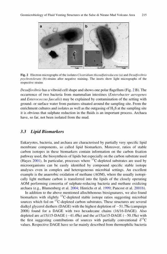
Desulfovibrio has a vibroid cell shape and shows one polar flagellum (Fig. 2 B). The
occurrence of two bacteria from mammalian intestines (Enterobacter aerogenesand Enterococcus faecalis) may be explained by contamination of the setting with
ground- or surface water from pastures situated around the sampling site. From the
enrichment cultures and isolates as well as the outgasing of H2S at the sampling site
it is obvious that sulphate reduction in the fluids is an important process. Archaea
have, so far, not been isolated from the mud.
3.3 Lipid Biomarkers
Eukaryotes, bacteria, and archaea are characterised by partially very specific lipid
membrane components, so called lipid biomarkers. Moreover, ratios of stable
carbon isotopes in these biomarkers contain information on the carbon fixation
pathway used, the biosynthesis of lipids but especially on the carbon substrate used
(Hayes 2001). In particular, processes where 13C-depleted substrates are used by
microorganisms can be easily identified by compound specific stable isotope
analyses even in complex and heterogeneous microbial settings. An excellent
example is the anaerobic oxidation of methane (AOM), where the usually isotopi-
cally light methane carbon is transferred into the lipids of the closely operating
AOM performing consortia of sulphate-reducing bacteria and methane oxidizing
archaea (e.g., Blumenberg et al. 2004; Hinrichs et al. 1999; Pancost et al. 2001b).
In addition to the above mentioned allochthonous biosignatures, we also found
biomarkers with slightly 13C-depleted stable isotope ratios suggesting microbial
sources which fed on 13C-depleted carbon substrates. These structures are several
dialkyl glycerol diethers (DAGE) with the highest depletion of �51.7‰ (campaign
2008) found for a DAGE with two hexadecane chains (16/16-DAGE). Also
depleted are ai15/i15-DAGE (�41.4‰) and the ai15/ai15-DAGE (�50.1‰) with
the first suggesting contributions of sources with partially conventional d13Cvalues. Respective DAGE have so far mainly described from thermophilic bacteria
Fig. 2 Electron micrographs of the isolates Clostridium thiosulfatireducens (a) and Desulfovibriopsychrotolerans (b)-strains after negative staining. The insets show light micrographs of the
respective strains
Geomicrobiology of Fluid Venting Structures at the Salse di Nirano Mud Volcano Area 215
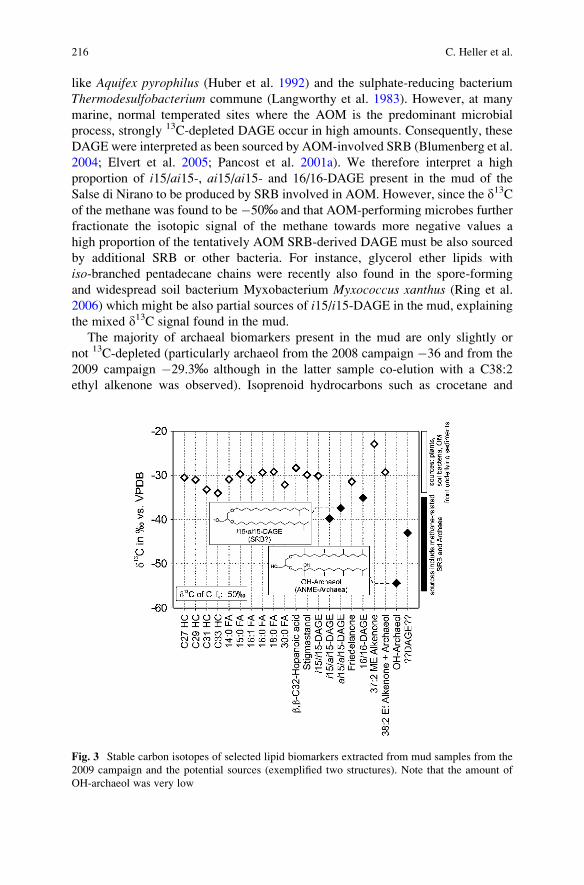
like Aquifex pyrophilus (Huber et al. 1992) and the sulphate-reducing bacterium
Thermodesulfobacterium commune (Langworthy et al. 1983). However, at many
marine, normal temperated sites where the AOM is the predominant microbial
process, strongly 13C-depleted DAGE occur in high amounts. Consequently, these
DAGE were interpreted as been sourced by AOM-involved SRB (Blumenberg et al.
2004; Elvert et al. 2005; Pancost et al. 2001a). We therefore interpret a high
proportion of i15/ai15-, ai15/ai15- and 16/16-DAGE present in the mud of the
Salse di Nirano to be produced by SRB involved in AOM. However, since the d13Cof the methane was found to be �50‰ and that AOM-performing microbes further
fractionate the isotopic signal of the methane towards more negative values a
high proportion of the tentatively AOM SRB-derived DAGE must be also sourced
by additional SRB or other bacteria. For instance, glycerol ether lipids with
iso-branched pentadecane chains were recently also found in the spore-forming
and widespread soil bacterium Myxobacterium Myxococcus xanthus (Ring et al.
2006) which might be also partial sources of i15/i15-DAGE in the mud, explaining
the mixed d13C signal found in the mud.
The majority of archaeal biomarkers present in the mud are only slightly or
not 13C-depleted (particularly archaeol from the 2008 campaign �36 and from the
2009 campaign �29.3‰ although in the latter sample co-elution with a C38:2
ethyl alkenone was observed). Isoprenoid hydrocarbons such as crocetane and
Fig. 3 Stable carbon isotopes of selected lipid biomarkers extracted from mud samples from the
2009 campaign and the potential sources (exemplified two structures). Note that the amount of
OH-archaeol was very low
216 C. Heller et al.
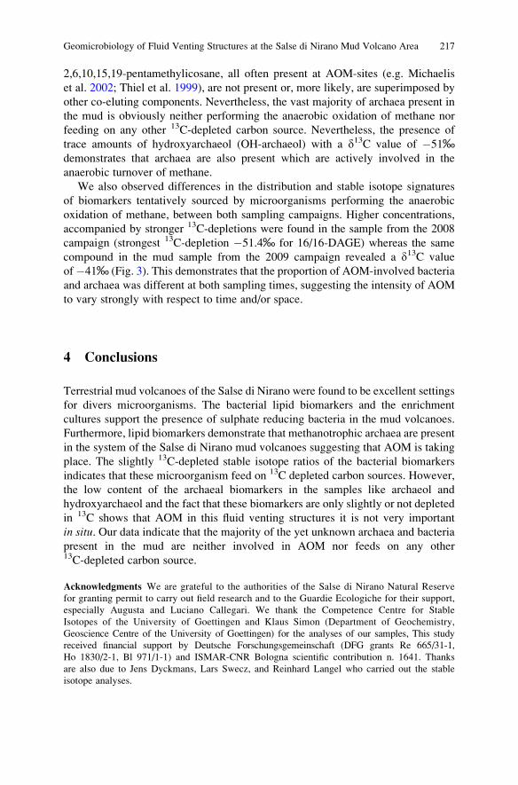
2,6,10,15,19-pentamethylicosane, all often present at AOM-sites (e.g. Michaelis
et al. 2002; Thiel et al. 1999), are not present or, more likely, are superimposed by
other co-eluting components. Nevertheless, the vast majority of archaea present in
the mud is obviously neither performing the anaerobic oxidation of methane nor
feeding on any other 13C-depleted carbon source. Nevertheless, the presence of
trace amounts of hydroxyarchaeol (OH-archaeol) with a d13C value of �51‰demonstrates that archaea are also present which are actively involved in the
anaerobic turnover of methane.
We also observed differences in the distribution and stable isotope signatures
of biomarkers tentatively sourced by microorganisms performing the anaerobic
oxidation of methane, between both sampling campaigns. Higher concentrations,
accompanied by stronger 13C-depletions were found in the sample from the 2008
campaign (strongest 13C-depletion �51.4‰ for 16/16-DAGE) whereas the same
compound in the mud sample from the 2009 campaign revealed a d13C value
of�41‰ (Fig. 3). This demonstrates that the proportion of AOM-involved bacteria
and archaea was different at both sampling times, suggesting the intensity of AOM
to vary strongly with respect to time and/or space.
4 Conclusions
Terrestrial mud volcanoes of the Salse di Nirano were found to be excellent settings
for divers microorganisms. The bacterial lipid biomarkers and the enrichment
cultures support the presence of sulphate reducing bacteria in the mud volcanoes.
Furthermore, lipid biomarkers demonstrate that methanotrophic archaea are present
in the system of the Salse di Nirano mud volcanoes suggesting that AOM is taking
place. The slightly 13C-depleted stable isotope ratios of the bacterial biomarkers
indicates that these microorganism feed on 13C depleted carbon sources. However,
the low content of the archaeal biomarkers in the samples like archaeol and
hydroxyarchaeol and the fact that these biomarkers are only slightly or not depleted
in 13C shows that AOM in this fluid venting structures it is not very important
in situ. Our data indicate that the majority of the yet unknown archaea and bacteria
present in the mud are neither involved in AOM nor feeds on any other13C-depleted carbon source.
Acknowledgments We are grateful to the authorities of the Salse di Nirano Natural Reserve
for granting permit to carry out field research and to the Guardie Ecologiche for their support,
especially Augusta and Luciano Callegari. We thank the Competence Centre for Stable
Isotopes of the University of Goettingen and Klaus Simon (Department of Geochemistry,
Geoscience Centre of the University of Goettingen) for the analyses of our samples, This study
received financial support by Deutsche Forschungsgemeinschaft (DFG grants Re 665/31-1,
Ho 1830/2-1, Bl 971/1-1) and ISMAR-CNR Bologna scientific contribution n. 1641. Thanks
are also due to Jens Dyckmans, Lars Swecz, and Reinhard Langel who carried out the stable
isotope analyses.
Geomicrobiology of Fluid Venting Structures at the Salse di Nirano Mud Volcano Area 217
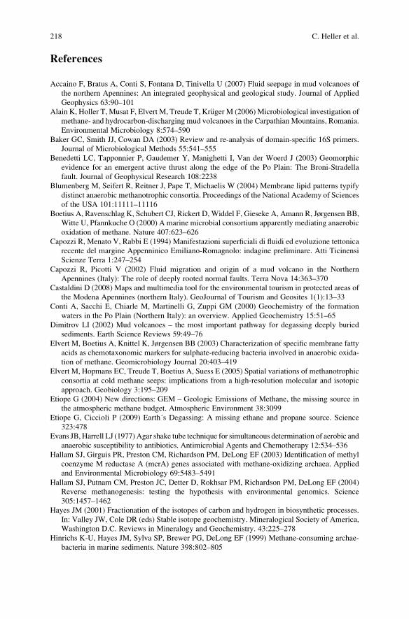
References
Accaino F, Bratus A, Conti S, Fontana D, Tinivella U (2007) Fluid seepage in mud volcanoes of
the northern Apennines: An integrated geophysical and geological study. Journal of Applied
Geophysics 63:90–101
Alain K, Holler T, Musat F, Elvert M, Treude T, Kr€uger M (2006) Microbiological investigation of
methane- and hydrocarbon-discharging mud volcanoes in the Carpathian Mountains, Romania.
Environmental Microbiology 8:574–590
Baker GC, Smith JJ, Cowan DA (2003) Review and re-analysis of domain-specific 16S primers.
Journal of Microbiological Methods 55:541–555
Benedetti LC, Tapponnier P, Gaudemer Y, Manighetti I, Van der Woerd J (2003) Geomorphic
evidence for an emergent active thrust along the edge of the Po Plain: The Broni-Stradella
fault. Journal of Geophysical Research 108:2238
Blumenberg M, Seifert R, Reitner J, Pape T, Michaelis W (2004) Membrane lipid patterns typify
distinct anaerobic methanotrophic consortia. Proceedings of the National Academy of Sciences
of the USA 101:11111–11116
Boetius A, Ravenschlag K, Schubert CJ, Rickert D, Widdel F, Gieseke A, Amann R, Jørgensen BB,
Witte U, Pfannkuche O (2000) A marine microbial consortium apparently mediating anaerobic
oxidation of methane. Nature 407:623–626
Capozzi R, Menato V, Rabbi E (1994) Manifestazioni superficiali di fluidi ed evoluzione tettonica
recente del margine Appenninico Emiliano-Romagnolo: indagine preliminare. Atti Ticinensi
Scienze Terra 1:247–254
Capozzi R, Picotti V (2002) Fluid migration and origin of a mud volcano in the Northern
Apennines (Italy): The role of deeply rooted normal faults. Terra Nova 14:363–370
Castaldini D (2008) Maps and multimedia tool for the environmental tourism in protected areas of
the Modena Apennines (northern Italy). GeoJournal of Tourism and Geosites 1(1):13–33
Conti A, Sacchi E, Chiarle M, Martinelli G, Zuppi GM (2000) Geochemistry of the formation
waters in the Po Plain (Northern Italy): an overview. Applied Geochemistry 15:51–65
Dimitrov LI (2002) Mud volcanoes – the most important pathway for degassing deeply buried
sediments. Earth Science Reviews 59:49–76
Elvert M, Boetius A, Knittel K, Jørgensen BB (2003) Characterization of specific membrane fatty
acids as chemotaxonomic markers for sulphate-reducing bacteria involved in anaerobic oxida-
tion of methane. Geomicrobiology Journal 20:403–419
Elvert M, Hopmans EC, Treude T, Boetius A, Suess E (2005) Spatial variations of methanotrophic
consortia at cold methane seeps: implications from a high-resolution molecular and isotopic
approach. Geobiology 3:195–209
Etiope G (2004) New directions: GEM – Geologic Emissions of Methane, the missing source in
the atmospheric methane budget. Atmospheric Environment 38:3099
Etiope G, Ciccioli P (2009) Earth´s Degassing: A missing ethane and propane source. Science
323:478
Evans JB,Harrell LJ (1977)Agar shake tube technique for simultaneous determination of aerobic and
anaerobic susceptibility to antibiotics. Antimicrobial Agents and Chemotherapy 12:534–536
Hallam SJ, Girguis PR, Preston CM, Richardson PM, DeLong EF (2003) Identification of methyl
coenzyme M reductase A (mcrA) genes associated with methane-oxidizing archaea. Applied
and Environmental Microbiology 69:5483–5491
Hallam SJ, Putnam CM, Preston JC, Detter D, Rokhsar PM, Richardson PM, DeLong EF (2004)
Reverse methanogenesis: testing the hypothesis with environmental genomics. Science
305:1457–1462
Hayes JM (2001) Fractionation of the isotopes of carbon and hydrogen in biosynthetic processes.
In: Valley JW, Cole DR (eds) Stable isotope geochemistry. Mineralogical Society of America,
Washington D.C. Reviews in Mineralogy and Geochemistry. 43:225–278
Hinrichs K-U, Hayes JM, Sylva SP, Brewer PG, DeLong EF (1999) Methane-consuming archae-
bacteria in marine sediments. Nature 398:802–805
218 C. Heller et al.
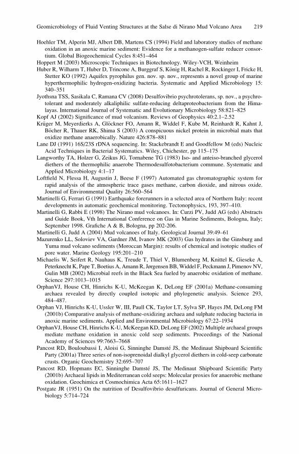
Hoehler TM, Alperin MJ, Albert DB, Martens CS (1994) Field and laboratory studies of methane
oxidation in an anoxic marine sediment: Evidence for a methanogen-sulfate reducer consor-
tium. Global Biogeochemical Cycles 8:451–464
Hoppert M (2003) Microscopic Techniques in Biotechnology. Wiley-VCH, Weinheim
Huber R, Wilharm T, Huber D, Trincone A, Burggraf S, K€onig H, Rachel R, Rockinger I, Fricke H,Stetter KO (1992) Aquifex pyrophilus gen. nov. sp. nov., represents a novel group of marine
hyperthermophilic hydrogen-oxidizing bacteria. Systematic and Applied Microbiology 15:
340–351
Jyothsna TSS, Sasikala C, Ramana CV (2008) Desulfovibrio psychrotolerans, sp. nov., a psychro-
tolerant and moderately alkaliphilic sulfate-reducing deltaproteobacterium from the Hima-
layas. International Journal of Systematic and Evolutionary Microbiology 58:821–825
Kopf AJ (2002) Significance of mud volcanism. Reviews of Geophysics 40:2.1–2.52
Kr€uger M, Meyerdierks A, Gl€ockner FO, Amann R, Widdel F, Kube M, Reinhardt R, Kahnt J,
B€ocher R, Thauer RK, Shima S (2003) A conspicuous nickel protein in microbial mats that
oxidize methane anaerobically. Nature 426:878–881
Lane DJ (1991) 16S/23S rDNA sequencing. In: Stackebrandt E and Goodfellow M (eds) Nucleic
Acid Techniques in Bacterial Systematics. Wiley, Chichester, pp 115–175
Langworthy TA, Holzer G, Zeikus JG, Tornabene TG (1983) Iso- and anteiso-branched glycerol
diethers of the thermophilic anaerobe Thermodesulfotobacterium commune. Systematic and
Applied Microbiology 4:1–17
Loftfield N, Flessa H, Augustin J, Beese F (1997) Automated gas chromatographic system for
rapid analysis of the atmospheric trace gases methane, carbon dioxide, and nitrous oxide.
Journal of Environmental Quality 26:560–564
Martinelli G, Ferrari G (1991) Earthquake forerunners in a selected area of Northern Italy: recent
developments in automatic geochemical monitoring. Tectonophysics, 193, 397–410.
Martinelli G, Rabbi E (1998) The Nirano mud volcanoes. In: Curzi PV, Judd AG (eds) Abstracts
and Guide Book, Vth International Conference on Gas in Marine Sediments, Bologna, Italy;
September 1998. Grafiche A & B, Bologna, pp 202-206.
Martinelli G, Judd A (2004) Mud volcanoes of Italy. Geological Journal 39:49–61
Mazurenko LL, Soloviev VA, Gardner JM, Ivanov MK (2003) Gas hydrates in the Ginsburg and
Yuma mud volcano sediments (Moroccan Margin): results of chemical and isotopic studies of
pore water. Marine Geology 195:201–210
Michaelis W, Seifert R, Nauhaus K, Treude T, Thiel V, Blumenberg M, Knittel K, Gieseke A,
Peterknecht K, Pape T, Boetius A, Amann R, Jørgensen BB,Widdel F, Peckmann J, PimenovNV,
Gulin MB (2002) Microbial reefs in the Black Sea fueled by anaerobic oxidation of methane.
Science 297:1013–1015
OrphanVJ, House CH, Hinrichs K-U, McKeegan K, DeLong EF (2001a) Methane-consuming
archaea revealed by directly coupled isotopic and phylogenetic analysis. Science 293,
484–487.
Orphan VJ, Hinrichs K-U, Ussler W, III, Paull CK, Taylor LT, Sylva SP, Hayes JM, DeLong FM
(2001b) Comparative analysis of methane-oxidizing archaea and sulphate reducing bacteria in
anoxic marine sediments. Applied and Environmental Microbiology 67:22–1934
OrphanVJ, House CH, Hinrichs K-U, McKeegan KD, DeLong EF (2002) Multiple archaeal groups
mediate methane oxidation in anoxic cold seep sediments. Proceedings of the National
Academy of Sciences 99:7663–7668
Pancost RD, Bouloubassi I, Aloisi G, Sinninghe Damste JS, the Medinaut Shipboard Scientific
Party (2001a) Three series of non-isoprenoidal dialkyl glycerol diethers in cold-seep carbonate
crusts. Organic Geochemistry 32:695–707
Pancost RD, Hopmans EC, Sinninghe Damste JS, The Medinaut Shipboard Scientific Party
(2001b) Archaeal lipids in Mediterranean cold seeps: Molecular proxies for anaerobic methane
oxidation. Geochimica et Cosmochimica Acta 65:1611–1627
Postgate JR (1951) On the nutrition of Desulfovibrio desulfuricans. Journal of General Micro-
biology 5:714–724
Geomicrobiology of Fluid Venting Structures at the Salse di Nirano Mud Volcano Area 219
Reeburgh WS (1980) Anaerobic oxidation of methane; rate depth distributions in Skan Bay
sediments. Earth and Planetary Science Letters 47:345–352
Reitner J, Peckmann J, Blumenberg M, Michaelis W, Reimer A, Thiel V (2005a) Concretionary
methane-seep carbonates and associated microbial communities in Black Sea sediments.
Palaeogeography, Palaeoclimatology, Palaeoecology 227:18–30
Reitner J, Peckmann J, Reimer A, Schumann G, Thiel V (2005b) Methane derived carbonate
build-ups and associated microbial communities at cold seeps on the lower Crimean shelf
(Black Sea). Facies 51:66–79
Ring MW, Schwar G, Thiel V, Dickschat JS, Kroppenstedt RM, Schulz S, Bode HB (2006) Novel
iso-branched ether lipids as specific markers of developmental sporulation in the myxobacter-
ium Myxococcus xanthus. Journal of Biological Chemistry 281:36691–36700
Sakaguchi T, Arakaki A, Matsunaga T (2002) Desulfovibrio magneticus sp. nov., a novel sulfate-
reducing bacterium that produces intracellular single-domain-sized magnetite particles. Inter-
national Journal of Systematic and Evolutionary Microbiology 52:215–221
Scholte KH (2005) Hyperspectral Remote Sensing and Mud Volcanism in Azerbaijan. PrintPart-
ners Ipskamp B.V., The Netherlands
Thiel V, Peckmann J, Seifert R, Wehrung P, Reitner J, Michaelis W (1999) Highly isotopically
depleted isoprenoids: Molecular markers for ancient methane venting. Geochimica et Cosmo-
chimica Acta 63:3959–3966
Thompson M, Walsh J-N (1983) A Handbook of Inductively Coupled Plasma Spectrometry. –
(Chapman & Hall, New York, NY, United States; Blackie & Son, Glasgow, United Kingdom
Treude T, Knittel K, Blumenberg M, Seifert R, Boetius A (2005) Subsurface microbial methano-
trophic mats in the Black Sea. Applied and Environmental Microbiology 71:6375–6378
Whiticar MJ (1999) Carbon and hydrogen isotope systematics of bacterial formation and oxidation
of methane. Chemical Geology 161:291–314
220 C. Heller et al.
Trace Element and Biomarker Signatures
in Iron-Precipitating Microbial Mats from
the Tunnel of Asp€o (Sweden)
Jens Kurz, Klaus Simon, Christine Heim, Joachim Reitner,
Nadia -Valerie Queric, and Volker Thiel
1 Introduction
The Asp€o Hard Rock Laboratory (Asp€o HRL) is a tunnel located near Oskarshamn
in the southeast of Sweden, that serves as a testing environment for the disposal of
nuclear waste. The Asp€o HRL hosts and makes accessible a wide spectrum of
microbially driven subsurface ecosystems (Pedersen 1997). 1,127 m from the
tunnel entrance and 160 m below the Baltic Sea floor, a natural pond has established
beneath a fluid spring from the diorite rock (R€onnback 2005). The pond contains
microbial mats of several cm thickness, that can be separated into ochre-coloured
surface mats and underlying mats showing an obvious pattern of dark, mm-thick
and more or less horizontal streaks. Spatially separated below these mats, a third
type comprising dense black mats occur. Previous studies showed that ochre mats,
which abundantly occur in the Asp€o HRL, are dominated by the iron-oxidizing
bacterium Gallionella ferruginea (Ferris et al. 1999; Anderson and Pedersen 2003;
Hallberg and Ferris 2004; Anderson et al. 2006). Based on these earlier studies,
emphasis was placed here on the geochemical investigation of the pond habitat with
its variety of different mat types. Specifically, trace element patterns were explored
for inorganic biosignatures, arising from different biogeochemical pathways
driving the distinct microbial systems. The lipid biomarker composition of these
mats was investigated and linked to the trace element distributions. Additionally,
J. Kurz
Max Planck Institute for Biophysical Chemistry, Am Faßberg 11, 37077, G€ottingen, Germany
K. Simon
Geoscience Centre, University of G€ottingen, Geochemistry Division, Goldschmidtstraße 1, 37077,
G€ottingen, Germany
C. Heim, J. Reitner, N.-V. Queric, and V. Thiel (*)
Geoscience Centre, University of G€ottingen, Geobiology Division, Goldschmidtstraße 3, 37077,
G€ottingen, Germany
e-mail: [email protected]
J. Reitner et al., Advances in Stromatolite Geobiology,Lecture Notes in Earth Sciences 131, DOI 10.1007/978-3-642-10415-2_15,# Springer-Verlag Berlin Heidelberg 2011
221
the involved biominerals were examined to receive a better understanding of the
microbial mat structures and explore inorganic patterns that may reveal potential
biosignatures for these systems in ancient environments.
2 Material and Methods
2.1 Sample Collection
Samples were taken in April 2008 at the sample location 1127B Pond, which is
situated 1,127 m from the tunnel entrance and 160 m below the Baltic Sea floor. 32
syringes were pressed into the microbial layers and taken out with the core inside
after one hour. The cores were immediately frozen with liquid nitrogen and stored
at �80�C until analysis.
2.2 Sample Preparation
Ten representative cores were chosen. Three cores completely consisted of ochre
mats; two were black with a minor admixture of ochre mats only at the top layer,
while the other five were ochre with dark streaks in between. The latter are referred
to as streak mats. The cores were partitioned with a scalpel by their colour zonation
in (1) ochre, (2) streak and (3) black segments. In total, 26 segments were used for
trace element and 18 segments for biomarker analysis. The samples were lyophi-
lized overnight. For trace element analysis 50 mg of the dry powder was dissolved
in 1 mL of concentrated HNO3 and 100 mL of an internal standard were added
before the sample was oxidized with 5 mL of 30% H2O2. 45 mL of ultrapure water
were added, leading to a dilution of 1:1,000. Lipid biomarkers were extracted from
ca. 100 mg of the lyophilized sample with 5 mL of dichlormethane/methanol (1:1;
v:v), 5 mL of dichlormethane/methanol (3:1; v:v) and 5 mL of dichlormethane
respectively (ultrasonication, 20 min, 40�C). Each extraction was followed by a
centrifugation step (15 min). The combined extracts were dried under a stream of
nitrogen and re-dissolved in 500 mL of dichlormethane. Aliquots were (1) deriva-
tized with N,O-Bis(trimethylsilyl)trifluoroacetamide (BSTFA) (80�C, 1.5 h) to
produce trimethylsilane (TMS-)derivatives of alcohols and free fatty acids. A
second aliquot was transesterified with trimethylchlorosilane/methanol (1:10; v:v;
80�C, 1.5 h) to produce methyl ester derivatives of bound fatty acids. The reaction
mixture was extracted three times with n-hexane. The fatty acid methyl ester
(FAME) fraction was purified using a glass column (ID 12 mm) filled with 0.6 g
silica gel (Merck Kieselgel 60). The extract was re-dissolved in 7 mL n-hexane andplaced on the column. The FAME fraction was eluted with 7 mL of dichlormethane,
dried under a gentle stream of nitrogen, and re-dissolved in 20 mL of n-hexane forbiomarker analysis (Sect. 2.4).
222 J. Kurz et al.
2.3 Trace Element Analysis
A PerkinElmer ELAN DRC II ICP-MS was used for the analysis of Li, Ti, Mn, Co,
Ni, Cu, Zn, Zr, Nb, Mo, Sn, Rb, Sr, Cs, W, Pb, Th, U and REE. The RF power was
1,250 W; the MEINHARD nebulizer had a flow rate of 0.9 L/min, the auxiliary gas
flow was 1 L/min and the cooling gas flow 14.5 L/min. For Al, Ca, Fe, Mg, Na and
P, a PerkinElmer Optima 3,300 DV ICP-OES was used. For the REE plot (Fig. 2)
the data was normalized on the Continental Earth’s Crust (Wedepohl 1995).
2.4 Biomarker Analysis
The coupled gas chromatography andmass spectrometry (GC/MS) system used was a
Varian 3800 GC interfaced to a Varian 1200 Quadrupole MS. The GC was equipped
with a fused silica capillary column (Phenomenex ZB-1, 30 m 0.25 mm ID, 0.1 mmfilm thickness). Helium was used as carrier gas at a flow rate of 1 mL/min, and 1 mLsample was injected manually. The injector oven was programmed as follows: 70�Cisothermal for 1 min, from 70�C to 320�C at 150�C/min, and 10 min isothermal.
The GC-oven was programmed as follows: 80�C isothermal for 1 min, from 80� to310�C at 10�C/min, and 30 min isothermal. Mass spectra were acquired in electron
impact mode (70 eV) with an ion source temperature of 180�C, a scan range ofm/z 50to m/z 500 and a scan time of 0.75 s. The identification of the compounds was based
on comparison of the mass spectra and of GC retention times with those of published
data and of reference compounds (Supelco 37 FAME standard).
2.5 Factor Analysis
The software Statistica 6.0 (Statsoft Inc., Tulsa, U.S.A.) was applied to correlate the
trace element analyses. The trace element data (concentrations in ppb) of 51
isotopes for 26 ochre and streak mats, six black mats and one mixed (A8-23-1)
sample were taken into account for this analysis.
2.6 Structure Analysis
ForX-ray diffraction (XRD) analyses, a Philips PW1877 instrument was usedwith a PW
1729 X-ray generator at wave lengths of CuKa1 ¼ 1.5406 A and CuKa2 ¼ 1.54442 A.
Raman spectroscopy was undertaken with a Melles Griot Argon Ion Laser
IMA 106020B0S at wave length 488 nm, an Olympus BX41 microscope, a HOR-
IBA Jobin Yvon LabRAM HR 800 UV spectrometer with 800 nm focal length, and
a HORIBA Jobin Yvon CCD Symphony LN2-Series detector. For electron micros-
copy and energy-dispersive X-ray spectroscopy (EDS) analysis, a FEI Quanta 200
Trace Element and Biomarker Signatures in Iron-Precipitating Microbial Mats 223
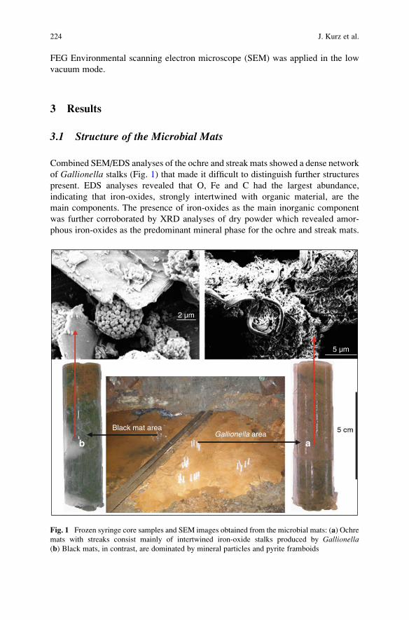
FEG Environmental scanning electron microscope (SEM) was applied in the low
vacuum mode.
3 Results
3.1 Structure of the Microbial Mats
Combined SEM/EDS analyses of the ochre and streak mats showed a dense network
of Gallionella stalks (Fig. 1) that made it difficult to distinguish further structures
present. EDS analyses revealed that O, Fe and C had the largest abundance,
indicating that iron-oxides, strongly intertwined with organic material, are the
main components. The presence of iron-oxides as the main inorganic component
was further corroborated by XRD analyses of dry powder which revealed amor-
phous iron-oxides as the predominant mineral phase for the ochre and streak mats.
2 µm
5 µm
Gallionella areaBlack mat area
ab
Fig. 1 Frozen syringe core samples and SEM images obtained from the microbial mats: (a) Ochre
mats with streaks consist mainly of intertwined iron-oxide stalks produced by Gallionella(b) Black mats, in contrast, are dominated by mineral particles and pyrite framboids
224 J. Kurz et al.
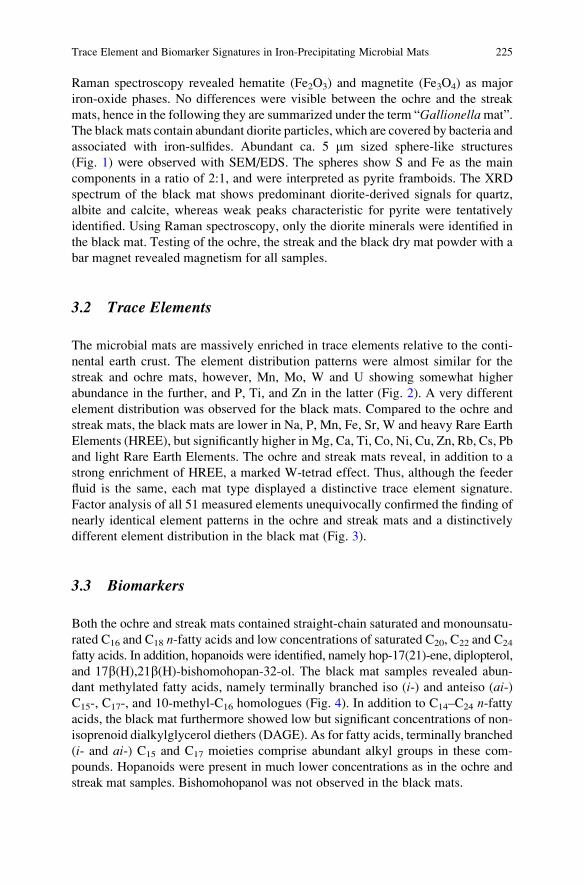
Raman spectroscopy revealed hematite (Fe2O3) and magnetite (Fe3O4) as major
iron-oxide phases. No differences were visible between the ochre and the streak
mats, hence in the following they are summarized under the term “Gallionellamat”.
The black mats contain abundant diorite particles, which are covered by bacteria and
associated with iron-sulfides. Abundant ca. 5 mm sized sphere-like structures
(Fig. 1) were observed with SEM/EDS. The spheres show S and Fe as the main
components in a ratio of 2:1, and were interpreted as pyrite framboids. The XRD
spectrum of the black mat shows predominant diorite-derived signals for quartz,
albite and calcite, whereas weak peaks characteristic for pyrite were tentatively
identified. Using Raman spectroscopy, only the diorite minerals were identified in
the black mat. Testing of the ochre, the streak and the black dry mat powder with a
bar magnet revealed magnetism for all samples.
3.2 Trace Elements
The microbial mats are massively enriched in trace elements relative to the conti-
nental earth crust. The element distribution patterns were almost similar for the
streak and ochre mats, however, Mn, Mo, W and U showing somewhat higher
abundance in the further, and P, Ti, and Zn in the latter (Fig. 2). A very different
element distribution was observed for the black mats. Compared to the ochre and
streak mats, the black mats are lower in Na, P, Mn, Fe, Sr, W and heavy Rare Earth
Elements (HREE), but significantly higher in Mg, Ca, Ti, Co, Ni, Cu, Zn, Rb, Cs, Pb
and light Rare Earth Elements. The ochre and streak mats reveal, in addition to a
strong enrichment of HREE, a marked W-tetrad effect. Thus, although the feeder
fluid is the same, each mat type displayed a distinctive trace element signature.
Factor analysis of all 51 measured elements unequivocally confirmed the finding of
nearly identical element patterns in the ochre and streak mats and a distinctively
different element distribution in the black mat (Fig. 3).
3.3 Biomarkers
Both the ochre and streak mats contained straight-chain saturated and monounsatu-
rated C16 and C18 n-fatty acids and low concentrations of saturated C20, C22 and C24
fatty acids. In addition, hopanoids were identified, namely hop-17(21)-ene, diplopterol,
and 17b(H),21b(H)-bishomohopan-32-ol. The black mat samples revealed abun-
dant methylated fatty acids, namely terminally branched iso (i-) and anteiso (ai-)C15-, C17-, and 10-methyl-C16 homologues (Fig. 4). In addition to C14–C24 n-fattyacids, the black mat furthermore showed low but significant concentrations of non-
isoprenoid dialkylglycerol diethers (DAGE). As for fatty acids, terminally branched
(i- and ai-) C15 and C17 moieties comprise abundant alkyl groups in these com-
pounds. Hopanoids were present in much lower concentrations as in the ochre and
streak mat samples. Bishomohopanol was not observed in the black mats.
Trace Element and Biomarker Signatures in Iron-Precipitating Microbial Mats 225
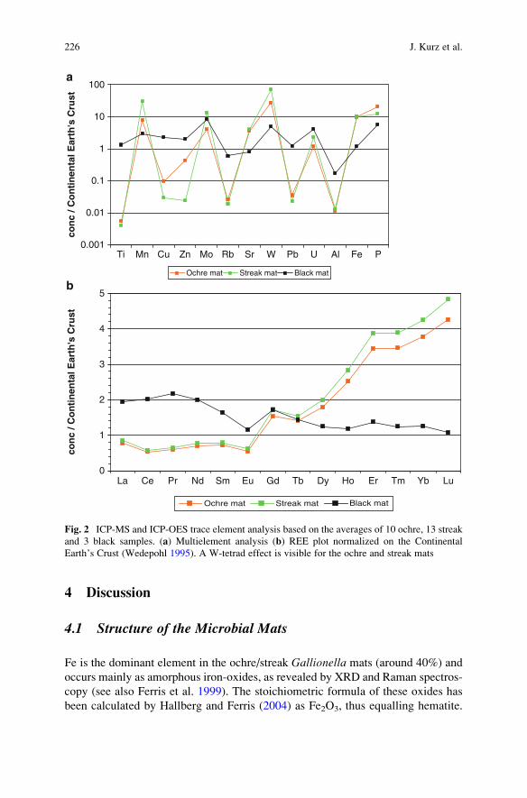
4 Discussion
4.1 Structure of the Microbial Mats
Fe is the dominant element in the ochre/streak Gallionella mats (around 40%) and
occurs mainly as amorphous iron-oxides, as revealed by XRD and Raman spectros-
copy (see also Ferris et al. 1999). The stoichiometric formula of these oxides has
been calculated by Hallberg and Ferris (2004) as Fe2O3, thus equalling hematite.
0.001
0.01
0.1
1
10
100
con
c / C
on
tin
enta
l Ear
th's
Cru
st
Ochre mat Streak mat Black mat
Ti
0
1
2
3
4
5
con
c / C
on
tin
enta
l Ear
th's
Cru
st
Ochre mat Streak mat Black mat
La Ce Pr Nd Sm Eu Gd Tb Dy Ho Er Tm Yb Lu
Mn Cu Zn Mo Rb Sr W Pb U Al Fe P
a
b
Fig. 2 ICP-MS and ICP-OES trace element analysis based on the averages of 10 ochre, 13 streak
and 3 black samples. (a) Multielement analysis (b) REE plot normalized on the Continental
Earth’s Crust (Wedepohl 1995). A W-tetrad effect is visible for the ochre and streak mats
226 J. Kurz et al.
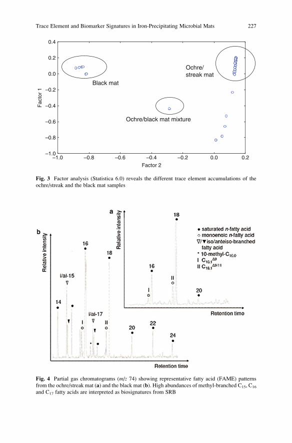
Fig. 4 Partial gas chromatograms (m/z 74) showing representative fatty acid (FAME) patterns
from the ochre/streak mat (a) and the black mat (b). High abundances of methyl-branched C15, C16
and C17 fatty acids are interpreted as biosignatures from SRB
Factor 2
–1.0–1.0
–0.8
–0.8
–0.6
–0.6
–0.4
–0.4
–0.2
–0.2
0.0
0.0
0.2
0.2
0.4F
acto
r 1
Black mat
Ochre/streak mat
Ochre/black mat mixture
Fig. 3 Factor analysis (Statistica 6.0) reveals the different trace element accumulations of the
ochre/streak and the black mat samples
Trace Element and Biomarker Signatures in Iron-Precipitating Microbial Mats 227
Indeed, hematite was identified with Raman spectroscopy, together with magnetite
(Fe3O4). The presence of magnetite plausibly explains the observed magnetism of
the mat samples. Fe concentration has shown to be similar in both mats, whereas the
Mn content was slightly higher in the streak mats compared to Fe. These high Mn
contents suggest that the dark colour of the streak mats may result from Mn-oxide
precipitates, namely MnO2. The slightly different accumulation behaviour of the
streak mats compared to the ochre mats may reflect minor differences in the
bacterial consortia, e.g. the local occurrence of manganese-oxidizing bacteria like
Leptothrix sp., that have previously been observed in the Asp€o tunnel (Hallbeck
et al. 2006). The black mat contains much more diorite-derived minerals like
quartz, albite and apatite than the ochre/streak Gallionella mats as revealed by
SEM, XRD and Raman spectroscopy. Whereas the microbial cells seem to prefer-
entially settle on mm-sized mineral particles in the further, the stalks of Gallionella,consisting of extracellular polymeric substances (EPS), may provide a stabilizing
matrix for microbial settlement in the latter. Pyrite framboids are most likely linked
to the metabolism of sulphate-reducing bacteria (SRB), and may therefore be used
as biomarkers (Popa et al. 2004) for sulfate reduction. Some SRB are known to
precipitate the magnetic mineral greigite (Fe3S4) inside of the cell as a magneto-
some (Posfai et al. 1998; Reitner et al. 2005). After cell death the magnetic minerals
may cluster to form a sphere and later recrystallize to framboidal pyrite. Although
not directly observed by the spectral methods used, we speculate that greigite in the
black mat accounts for magnetism of the black mat sample, rather than magnetite.
4.2 Trace Elements
The question emerges how the different trace element patterns observed for the
ochre/streak Gallionella mats and for the black mat, can be interpreted. There are
two concepts explaining the accumulation of trace elements by bacteria, specifi-
cally (1) binding to their exopolymeric substances and (2) adsorption and absorp-
tion onto and into the biominerals they produce. Sorption effects ofGallionella EPSstalks likely account for the capturing of ferrous iron necessary for the microbial
metabolism, i.e. dissimilatory iron oxidation (Hanert 2002). At the same time, the
extracellular stalks may account for a most effective accumulation of trace ele-
ments. Anderson and Pedersen (2003) pointed out that heavy metals like REE and
U compete with iron for the adsorption on the stalk material. Therefore, while
steadily growing, Gallionella has to produce even more stalks to secure the iron
supply for its metabolism.
Minerals produced or induced by SRB, namely iron-sulfides observed in the
black mat are known to have excellent adsorption capacities e.g. for Co, Ni, Cu, Zn,
Cd and Pb (Watson et al. 1995). Other studies showed Cu, As, Mn and Mo as
preferably adsorbed (Neumann et al. 2006) and Mo to be preferred over Cr and Ni
(Beech and Cheung 1995). These observations could be confirmed, except for Cd
and As, which were below the detection limits in all samples studied. The black
228 J. Kurz et al.
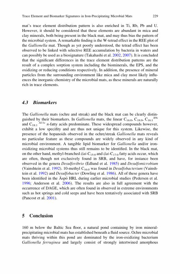
mat’s trace element distribution pattern is also enriched in Ti, Rb, Pb and U.
However, it should be considered that these elements are abundant in mica and
clay minerals, both being present in the black mat, and may thus bias the pattern of
the microbial system. A remarkable finding is the W-tetrad effect in the REE plot of
the Gallionella mat. Though as yet poorly understood, the tetrad effect has been
observed to be linked with selective REE accumulation by bacteria in waters and
can possibly be used as a biosignature (Takahashi et al. 2002, 2007). It is concluded
that the significant differences in the trace element distribution patterns are the
result of a complex sorption system including the biominerals, the EPS, and the
oxidizing or reducing conditions respectively. In addition, the presence of mineral
particles from the surrounding environment like mica and clay most likely influ-
ences the inorganic chemistry of the microbial mats, as these minerals are naturally
rich in trace elements.
4.3 Biomarkers
The Gallionella mats (ochre and streak) and the black mat can be clearly distin-
guished by their biomarkers. In Gallionella mats, the linear C16:0, C18:0, C16:1D9c
and C18:1D11c n-fatty acids predominate. These widespread compounds however,
exhibit a low specifity and are thus not unique for this system. Likewise, the
presence of the hopanoids observed in the ochre/streak Gallionella mats reveals
no particular feature as these compounds are widely observed in any kind of
microbial environment. A tangible lipid biomarker for Gallionella and/or iron-
oxidizing microbial systems thus still remains to be identified. In the black mat,
on the other hand, methyl-branched i/ai-C15:0 and i/ai-C17:0 fatty acids occur, which
are often, though not exclusively found in SRB, and have, for instance been
observed in the genera Desulfovibrio (Edlund et al. 1985) and Desulfomicrobium(Vainshtein et al. 1992). 10-methyl C16:0 was found in Desulfobacterium (Vainsh-
tein et al. 1992) and Desulfobacter (Dowling et al. 1986). All of these genera have
been identified in the Asp€o HRL during earlier microbial studies (Pedersen et al.
1996; Anderson et al. 2006). The results are also in full agreement with the
occurrence of DAGE, which are often found in observed in extreme environments
such as hot springs and cold seeps and have been tentatively associated with SRB
(Pancost et al. 2001).
5 Conclusion
160 m below the Baltic Sea floor, a natural pond containing by iron mineral-
precipitating microbial mats has established beneath a fluid source. Ochre microbial
mats thriving within this pond are dominated by the iron-oxidizing bacterium
Gallionella ferruginea and largely consist of strongly intertwined amorphous
Trace Element and Biomarker Signatures in Iron-Precipitating Microbial Mats 229
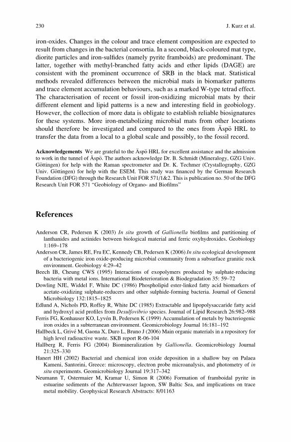
iron-oxides. Changes in the colour and trace element composition are expected to
result from changes in the bacterial consortia. In a second, black-coloured mat type,
diorite particles and iron-sulfides (namely pyrite framboids) are predominant. The
latter, together with methyl-branched fatty acids and ether lipids (DAGE) are
consistent with the prominent occurrence of SRB in the black mat. Statistical
methods revealed differences between the microbial mats in biomarker patterns
and trace element accumulation behaviours, such as a marked W-type tetrad effect.
The characterisation of recent or fossil iron-oxidizing microbial mats by their
different element and lipid patterns is a new and interesting field in geobiology.
However, the collection of more data is obligate to establish reliable biosignatures
for these systems. More iron-metabolizing microbial mats from other locations
should therefore be investigated and compared to the ones from Asp€o HRL to
transfer the data from a local to a global scale and possibly, to the fossil record.
Acknowledgements We are grateful to the Asp€o HRL for excellent assistance and the admission
to work in the tunnel of Asp€o. The authors acknowledge Dr. B. Schmidt (Mineralogy, GZG Univ.
G€ottingen) for help with the Raman spectrometer and Dr. K. Techmer (Crystallography, GZG
Univ. G€ottingen) for help with the ESEM. This study was financed by the German Research
Foundation (DFG) through the Research Unit FOR 571/1&2. This is publication no. 50 of the DFG
Research Unit FOR 571 “Geobiology of Organo- and Biofilms”
References
Anderson CR, Pedersen K (2003) In situ growth of Gallionella biofilms and partitioning of
lanthanides and actinides between biological material and ferric oxyhydroxides. Geobiology
1:169–178
Anderson CR, James RE, Fru EC, Kennedy CB, Pedersen K (2006) In situ ecological development
of a bacteriogenic iron oxide-producing microbial community from a subsurface granitic rock
environment. Geobiology 4:29–42
Beech IB, Cheung CWS (1995) Interactions of exopolymers produced by sulphate-reducing
bacteria with metal ions. International Biodeterioration & Biodegradation 35: 59–72
Dowling NJE, Widdel F, White DC (1986) Phospholipid ester-linked fatty acid biomarkers of
acetate-oxidizing sulphate-reducers and other sulphide-forming bacteria. Journal of General
Microbiology 132:1815–1825
Edlund A, Nichols PD, Roffey R, White DC (1985) Extractable and lipopolysaccaride fatty acid
and hydroxyl acid profiles from Desulfovibrio species. Journal of Lipid Research 26:982–988
Ferris FG, Konhauser KO, Lyven B, Pedersen K (1999) Accumulation of metals by bacteriogenic
iron oxides in a subterranean environment. Geomicrobiology Journal 16:181–192
Hallbeck L, Grive M, Gaona X, Duro L, Bruno J (2006) Main organic materials in a repository for
high level radioactive waste. SKB report R-06-104
Hallberg R, Ferris FG (2004) Biomineralization by Gallionella. Geomicrobiology Journal
21:325–330
Hanert HH (2002) Bacterial and chemical iron oxide deposition in a shallow bay on Palaea
Kameni, Santorini, Greece: microscopy, electron probe microanalysis, and photometry of insitu experiments. Geomicrobiology Journal 19:317–342
Neumann T, Ostermaier M, Kramar U, Simon R (2006) Formation of framboidal pyrite in
estuarine sediments of the Achterwasser lagoon, SW Baltic Sea, and implications on trace
metal mobility. Geophysical Research Abstracts: 8/01163
230 J. Kurz et al.
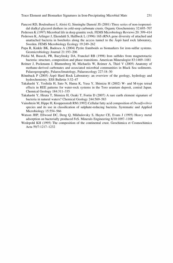
Pancost RD, Bouloubassi I, Aloisi G, Sinninghe Damste JS (2001) Three series of non-isoprenoi-
dal dialkyl glycerol diethers in cold-seep carbonate crusts. Organic Geochemistry 32:695–707
Pedersen K (1997) Microbial life in deep granitic rock. FEMSMicrobiology Reviews 20: 399–414
Pedersen K, Arlinger J, Ekendahl S, Hallbeck L (1996) 16S rRNA gene diversity of attached and
unattached bacteria in boreholes along the access tunnel to the Asp€o hard rock laboratory,
Sweden. FEMS Microbiology Ecology 19:249–262
Popa R, Kinkle BK, Badescu A (2004) Pyrite framboids as biomarkers for iron-sulfur systems.
Geomicrobiology Journal 21:193–206
Posfai M, Buseck, PR, Bazylinsky DA, Franckel RB (1998) Iron sulfides from magnetotactic
bacteria: structure, composition and phase transitions. American Mineralogist 83:1469–1481
Reitner J, Peckmann J, Blumenberg M, Michaelis W, Reimer A, Thiel V (2005) Anatomy of
methane-derived carbonates and associated microbial communities in Black Sea sediments.
Palaeogeography, Palaeoclimatology, Palaeoecology 227:18–30
R€onnback P (2005) Asp€o Hard Rock Laboratory: an overview of the geology, hydrology and
hydrochemistry. ESS Bulletin 3:32–47
Takahashi Y, Yoshida H, Sato N, Hama K, Yusa Y, Shimizu H (2002) W- and M-type tetrad
effects in REE patterns for water-rock systems in the Toro uranium deposit, central Japan.
Chemical Geology 184:311–335
Takahashi Y, Hirata T, Shimizu H, Ozaki T, Fortin D (2007) A rare earth element signature of
bacteria in natural waters? Chemical Geology 244:569–583
Vainshtein M, Hippe H, Kroppenstedt RM (1992) Cellular fatty acid composition ofDesulfovibriospecies and its use in classification of sulphate-reducing bacteria. Systematic and Applied
Microbiology 15:554–566
Watson JHP, Ellwood DC, Deng Q, Mikhalovsky S, Hayter CE, Evans J (1995) Heavy metal
adsorption on bacterially produced FeS. Minerals Engineering 8/10:1097–1108
Wedepohl KH (1995) The composition of the continental crust. Geochimica et Cosmochimica
Acta 59/7:1217–1232
Trace Element and Biomarker Signatures in Iron-Precipitating Microbial Mats 231

.
Microbial Euendolithic Assemblages and
Microborings in Intertidal and Shallow Marine
Habitats: Insight in Cyanobacterial Speciation
Gudrun Radtke and Stjepko Golubic
1 Introduction
The term “stromatolite” was coined by Kalkowsky (1908) while describing coastal
paleoenvironments of a Lower Triassic (Buntsandstein) epicontinental sea. Stable,
finely laminated structures appeared together with massive deposits of shoaling ooid
sands, and later consolidated into rocks (“Stromatolith und Oolith”). Ooid sands and
coated grains are often found associated as well as incorporated in stromatolitic
structures (e.g. Riding et al. 1991). Modern examples include subtidal (0–6 m depth;
Dravis 1983; Dill et al. 1986) and intertidal stromatolites (microbialites) of the
Bahama Bank (Reid and Browne 1991) and Shark Bay, Australia (Reid et al.
2003). They are formed by entrapment and rapid stabilization of ooids (Reid and
Browne 1991; Reid et al. 1995; Golubic and Browne 1996; Paterson et al. 2008).
Stromatolites and ooids are hard calcareous substrates, which were the earliest to
be settled and bioeroded by microboring or euendolithic microorganisms (see
Golubic et al. 1981 for definition). Fossil evidence of microbial euendoliths,
preserved as trace fossils as well as body fossils in ooid sand grains, goes back to
the Neoproterozoic (Campbell 1982; Knoll et al. 1986; Al-Thukair and Green
1988), and in stromatolites even further back to the Mesoproterozoic times
(Zhang and Golubic 1987; Knoll and Golubic 1992; Golubic and Seong-Joo
1999). With the evolution of skeleton-forming metazoans and calcareous algae, a
multiplicity of new substrates became available and subjected to microbial bioero-
sion. Metazoan evolution generated also a variety of grazers that challenged the
very existence of epiliths and endoliths alike. Euendoliths and grazers are jointly
G. Radtke (*)
Hessisches Landesamt f€ur Umwelt und Geologie, Rheingaustr. 186, 65203 Wiesbaden, Germany
e-mail: [email protected]
S. Golubic
Biological Science Center, Boston University, 05 Cummington Street, Boston, MA 02215, USA
J. Reitner et al., Advances in Stromatolite Geobiology,Lecture Notes in Earth Sciences 131, DOI 10.1007/978-3-642-10415-2_16,# Springer-Verlag Berlin Heidelberg 2011
233
responsible for the current high rates of removal and recycling of carbonates
(Tribollet and Golubic 2005) as well as for bioerosive shaping of coastal limestone
in the formation of biokarst (Schneider 1976, 1977). Organically preserved euen-
doliths with discernable ultrastructure were found in crinoid ossicles of Silurian age
(Kazmierczak and Golubic 1976; Campbell et al. 1979; Campbell 1980) and
microboring traces were since found abundantly in shells and shell fragments
throughout the Phanerozoic (Vogel et al. 1987; Glaub et al. 2007; Wisshak et al.
2008) (Table 1).
The interest for microbial euendoliths has been generated by the potential to
distinguish between phototrophic and heterotrophic taxa based on the shape of their
traces. Such distinction may provide reliable indication of the water depth of
ancient deposits. Microborings conform closely with the outlines of the microor-
ganisms that make them, so that they reveal the size, growth orientation and
strategy of the microborer. Many modern and ancient microborings remain empty
and can be faithfully replicated using polymerizing resins, which permits compar-
isons between modern microorganisms and their boreholes and, by extension, an
interpretation of fossil boring traces. In modern environments, the interpretation
was aided by studying euendolith assemblages in naturally occurring limestone and
skeletal carbonate substrates (Golubic et al. 1975; Le Campion-Alsumard et al.
1979; G€unther 1990; Radtke 1993; Le Campion-Alsumard et al. 1995; Radtke and
Golubic 2005; Tribollet et al. 2006), as well as in substrates experimentally exposed
to euendolith colonization (Golubic 1969; Perkins and Tsentas 1976; May et al.
1982; Vogel et al. 2000; Wisshak 2006; Gektidis et al. 2007). The role of microbial
endoliths in consolidation of microbialites has been introduced in more recent
papers (Reid et al. 1995; Feldmann and McKenzie 1998; Reid and Macintyre
2000; Macintyre et al. 2000; Reid et al. 2000; Decho et al. 2005).
Ecological studies of phototrophic microboring organisms have first focused on
coastal limestones of theMediterranean area (Ercegovic 1932; LeCampion-Alsumard
1966; Schneider 1976, 1977), and then extended to shallow habitats and to tropical
seas (Golubic 1969; Perkins and Tsentas 1976), including euendoliths in bivalve shells
and shell fragments (Golubic 1990; Radtke 1993; Radtke and Golubic 2005). Studies
of modern euendoliths in shoaling ooid sands of the Bahamas and the Arabian/Persian
Gulf identified particular assemblages of cyanobacteria, some of which were
described as new genera and species (Lukas and Golubic 1981, 1983; Lukas and
Hoffman 1984; Al-Thukair and Golubic 1991a, b, 1996; Al-Thukair et al. 1994;
Gektidis and Golubic 1996). These microorganisms leave characteristic boring
patterns in the carbonate substrates they penetrate, which were recognized and studied
as resin replicas using scanning electron microscopy (Golubic et al. 1970, 1983) and
described according to the rules of nomenclature for trace fossils (Vogel et al. 1987;
Bertling 2007).
The present contribution analyses the morphotype diversity of euendoliths of
coastal marine environments with special reference to cyanobacterial genera Solen-tia and Hyella, in comparison with boring traces identified as ichnogenera Scoleciaand Fascichnus.
234 G. Radtke and S. Golubic
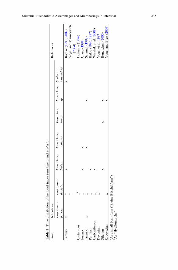
Table
1Tim
edistributionofthefossiltraces
Fascichnu
sandScolecia
Tim
eIchnotaxa
References
Fascichnus
parvus
Fascichnus
dactylus
Fascichnu
sfrutex
Fascichnu
sacinosus
Fascichnu
srogo
sFascichnus
sp.
Scolecia
maeandria
Tertiary
xx
xx
Radtke(1991,2007)
xVogel
andMarincovich
(2004)
Cretaceous
xa
Hofm
ann(1996)
Jurassic
xx
xGlaub(1994)
Triassic
xx
xx
Schmidt(1992)
Permian
xx
Balog(1996,1997)
Carboniferous
xx
Wisshak
etal.(2008)
Devonian
xb
Vogel
etal.1987
Silurian
xx
xBundschuh(2000)
Ordovician
xx
Vogel
andBrett(2009)
aAsasm
allbush-form
(“kleineB€ uschelform
”)bAs“H
yellomorpha”
Microbial Euendolithic Assemblages and Microborings in Intertidal 235
2 Materials and Methods
Samples of carbonate substrates, including coastal limestone, ooid sand grains and
bivalve shells and shell fragments were collected from the supratidal ranges to
shallow marine environments down to a depth of 10 m. These carbonate substrates
were examined for the presence of euendolithic microorganisms and their traces.
The materials from modern environments include: Korcula Island, Croatia, Safaga
Bay (Egypt, Red Sea), Lee Stocking Island, Bahamas, Cozumel (Mexico, Carib-
bean Sea), One Tree Island (Australia, Great Barrier Reef); they were compared
with fossil microborings from the Tertiary of the Paris Basin.
The samples were fixed in 3% formaldehyde solution and the microorganisms
extracted by dissolving the carbonate matrix in dilute (3%) HCl, mounted on
microscope slides and analysed by transmission light microscopy (LM) using
Zeiss Universal Microscope (Zeiss, Oberkochen) equipped with digital camera.
Microborings were replicated in polymerizing resin (Araldite Durcupan, and
SPURRS low viscosity medium). The procedure for embedding and casting (Golubic
et al. 1970, 1983) includes gradual dehydration by acetone series followed by an
equally gradual replacement by a complete (4 component) mixture of the resin
under vacuum. The resin is hardened at 60�C for 3 days, the hardened blocks
including the embedded sample is cut open and the carbonate matrix dissolved by
dilute HCl. Microborings were examined using scanning electron microscopy
(SEM). Organic component of the substrate, if present, were removed by potassium
hypochlorite (Clorox bleach).
Microbial cells and microborings, observed by LM and SEM were imaged and
measured for each population or cluster of borings, expressed as mean, standard
deviation, standard error, confidence level of 90 and 99%, maximum and minimum
values and sample size using measuring software of the digital Motic (Global Motic
Group, Xiamen, China) camera and Sigma Scan (Sausalito, CA). Intra- and inter-
population distribution of cellular dimensions and tunnel diameters respectively
have been recorded, expressed in terms of mean � standard deviation and pre-
sented graphically as two-dimensional cross diagrams, comparing two parameters
at a time with population means in the intersection of the cross with one standard
deviation on each side of the mean. Taxonomic determinations were carried out by
consulting phycological and microbiological determination manuals (Geitler 1932;
Komarek and Anagnostidis 1999; Rippka et al. 2001).
3 Morphotypic and Ecotypic Diversity and Variability
of Coastal Euendolithic Cyanobacteria
Comprehensive studies of epilithic and endolithic assemblages of coastal lime-
stones have been carried out in 1930s by Ante Ercegovic on the Adriatic coast of
Croatia near Split. Ercegovic described a number of new genera and species of
236 G. Radtke and S. Golubic
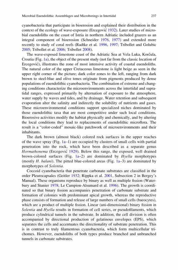
cyanobacteria that participate in bioerosion and explained their distribution in the
context of the ecology of wave-exposure (Ercegovic 1932). Later studies of micro-
bial euendoliths on the coast of Istria in northern Adriatic included grazers as an
integral component of bioerosion (Schneider 1976, 1977) and extended more
recently to study of coral reefs (Radtke et al. 1996, 1997; Tribollet and Golubic
2005; Tribollet et al. 2006; Tribollet 2008).
The wave-exposed limestone coast of the Adriatic Sea at Vela Luka, Korcula,
Croatia (Fig. 1a), the object of the present study (not far from the classic location of
Ercegovic), illustrates the zone of most intensive activity of coastal euendoliths.
The natural color of the upper Cretaceous limestone is light beige as seen in the
upper right corner of the picture; dark color zones to the left, ranging from dark
brown to steel-blue and olive tones originate from pigments produced by dense
populations of euendolithic cyanobacteria. The combination of extreme and chang-
ing conditions characterize the microenvironments across the intertidal and supra-
tidal ranges, expressed primarily by alternation of exposure to the atmosphere,
water supply by waves and tides, and by drainage. Water retention and subsequent
evaporation alter the salinity and indirectly the solubility of nutrients and gases.
These microenvironmental conditions support specialized niches dominated by
those euendolithic taxa that are most competitive under such local conditions.
Bioerosive activities modify the habitat physically and chemically, and by altering
the local conditions they lead to replacements of euendolithic microflora. The
result is a “color-coded” mosaic-like patchwork of microenvironments and their
inhabitants.
The dark brown (almost black) colored rock surfaces in the upper reaches
of the wave spray (Fig. 1a–1) are occupied by clusters of small cells with partial
penetration into the rock, which have been described as a separate genus
Hormathonema (Ercegovic 1929). Below this range, the exposed, well drained
brown-colored surfaces (Fig. 1a–2) are dominated by Hyella morphotypes
(mostly H. balani). The pitted blue-colored areas (Fig. 1a–3) are dominated by
morphotypes of Solentia.Coccoid cyanobacteria that penetrate carbonate substrates are classified in the
order Pleurocapsales (Geitler 1932; Rippka et al. 2001, Subsection 2 in Bergey’s
Manual). These organisms reproduce by binary as well as multiple fission (Water-
bury and Stanier 1978; Le Campion-Alsumard et al. 1996). The growth is coordi-
nated so that binary fission accompanies penetration of carbonate substrate and
formation of colonies with predominant apical growth, whereas the reproductive
phase consists of formation and release of large numbers of small cells (baeocytes),
which are a product of multiple fission. Linear (uni-dimensional) binary fission in
Solentia and Hyella results in formation of cell series, or pseudofilaments, which
produce cylindrical tunnels in the substrate. In addition, the cell division is often
accompanied by directional production of gelatinous envelopes (EPS), which
separates the cells and accentuates the directionality of substrate penetration. This
is in contrast to truly filamentous cyanobacteria, which form multicellular tri-
chomes. However, euendoliths of both types produce branched and unbranched
tunnels in carbonate substrates.
Microbial Euendolithic Assemblages and Microborings in Intertidal 237
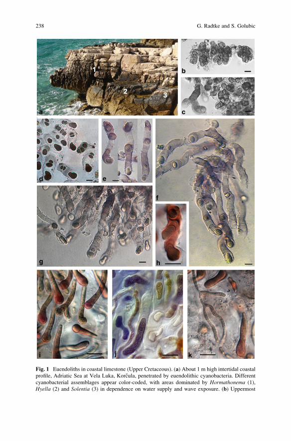
Fig. 1 Euendoliths in coastal limestone (Upper Cretaceous). (a) About 1 m high intertidal coastal
profile, Adriatic Sea at Vela Luka, Korcula, penetrated by euendolithic cyanobacteria. Different
cyanobacterial assemblages appear color-coded, with areas dominated by Hormathonema (1),
Hyella (2) and Solentia (3) in dependence on water supply and wave exposure. (b) Uppermost
238 G. Radtke and S. Golubic

Hormathonema Ercegovic with the species H. luteo-brunneum protected by the
brown “sunscreen” pigment scytonemin and H. violaceo-nigrum (Fig. 1b, c, right) by
the blue pigment gloeocapsin occur in the uppermost ranges of the colored zone.
Both pigments are extracellular, incorporated in the gelatinous EPS-envelopes to
absorb UV radiation (Ehling-Schulz and Scherer 1999; Castenholz and Garcia-Pichel
2000). The colonies are for the most part epilithic, extending into the substrate with
short series of cells separated by EPS. In habitats with more frequent wetting by
waves, Hormathonema spp. are replaced by Solentia (Fig. 1c, left), an entirely
endolithic organism, which also prevails in rock pools and small pits in the lower
intertidal ranges. The morphotypes in transition between these two genera are
represented by Solentia paulocellulare (Le Campion-Alsumard and Golubic 1985a)
which was originally described within the genus Hormathonema (Ercegovic 1929).
Solentia Ercegovic carries one of the ancient Roman names for the island Sulet
or Solta in central Dalmatia (Ercegovic 1927). The pseudofilaments of Solentia are
produced mainly by uni-directional production of EPS in the wake of cells, forming
a stalk that pushes the cells forward (Fig. 1d–g). The tunnels branch when interca-
lary cells slip out of the alignment and become secondary apical cells of a branch
(Fig. 1h). The extracellular pigment in Solentia is gloeocapsin, which changes the
coloration from blue to red during acid-extraction (Fig. 1h). No extracellular
pigment is produced by deep penetrating Solentia achromatica, which is character-ized by large droplet-shaped phycoerythrin-rich apical cells (Fig. 1i–k). Solentiawas very abundant across shallow ranges of the Bahama carbonate platform at Lee
Stocking Island, Exuma islands (Kiene et al. 1995); it has been isolated in culture
from moving ooid grains (Golubic et al. 1996).
Hyella Bornet and Flahault (1889) is the most diverse and most abundant
euendolithic cyanobacterium. The pseudofilaments in Hyella are comprised of
series of cells separated by moderate bidirectional production of EPS that separates
them (Fig. 2), but the degree of cell separation varies from taxon to taxon. For
example, Hyella sp. found in shoaling ooids and H. caespitosa, most common in
subtidal ranges, both have cells separated by stretches of EPS, which often exceed
the lengths of individual cells, whereas the cells of H. pyxis and H. racemus areinvariably packed in tight EPS envelopes (Lukas and Hoffman 1984; Al-Thukair
et al. 1994). The genus is highly diversified (over 25 species formally described)
with numerous morphotypes that differ in cell and colony size, growth orientation,
branching pattern, pigmentation, and mode of reproduction (Table 2).
�
Fig. 1 (continued) wave-spray zone is occupied by the coccoid Hormathonema violaceo-nigrum.(c) H. violaceo-nigrum and Solentia paulocellulare (left) in protected depressions. (d–g) Solentiamorphotypes across the intertidal zone range from short borings of S. paulocellulare to extended
tunnels of S. foveolarum, both with EPS stained by extracellular gloeocapsin pigment.(h) S. paulocellulare with gloeocapsin sheath pigment – red when extracted by acid showing
false branch by cell slippage. (i–k) Long, deeply penetrating terminal portions of S. achromatica.Cells are rich in phycoerythrin. With filaments of Mastigocoleus testarum in k. Scale bars in all
pictures are 10 mm long
Microbial Euendolithic Assemblages and Microborings in Intertidal 239
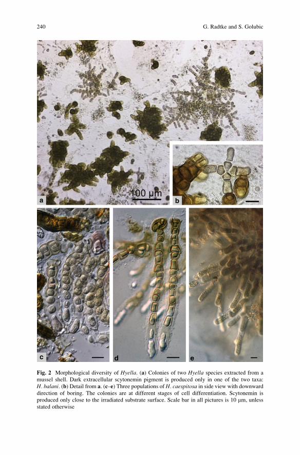
Fig. 2 Morphological diversity of Hyella. (a) Colonies of two Hyella species extracted from a
mussel shell. Dark extracellular scytonemin pigment is produced only in one of the two taxa:
H. balani. (b) Detail from a. (c–e) Three populations of H. caespitosa in side view with downward
direction of boring. The colonies are at different stages of cell differentiation. Scytonemin is
produced only close to the irradiated substrate surface. Scale bar in all pictures is 10 mm, unless
stated otherwise
240 G. Radtke and S. Golubic
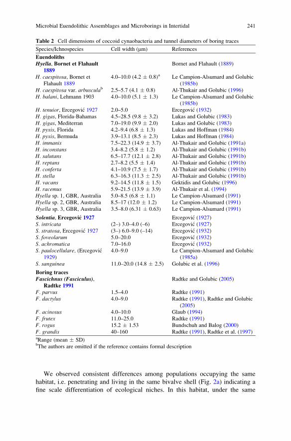
We observed consistent differences among populations occupying the same
habitat, i.e. penetrating and living in the same bivalve shell (Fig. 2a) indicating a
fine scale differentiation of ecological niches. In this habitat, under the same
Table 2 Cell dimensions of coccoid cynaobacteria and tunnel diameters of boring traces
Species/Ichnospecies Cell width (mm) References
Euendoliths
Hyella, Bornet et Flahault1889
Bornet and Flahault (1889)
H. caespitosa, Bornet etFlahault 1889
4.0–10.0 (4.2 � 0.8)a Le Campion-Alsumard and Golubic
(1985b)
H. caespitosa var. arbusculab 2.5–5.7 (4.1 � 0.8) Al-Thukair and Golubic (1996)
H. balani, Lehmann 1903 4.0–10.0 (5.1 � 1.3) Le Campion-Alsumard and Golubic
(1985b)
H. tenuior, Ercegovic 1927 2.0–5.0 Ercegovic (1932)
H. gigas, Florida-Bahamas 4.5–28.5 (9.8 � 3.2) Lukas and Golubic (1983)
H. gigas, Mediterran 7.0–19.0 (9.9 � 2.0) Lukas and Golubic (1983)
H. pyxis, Florida 4.2–9.4 (6.8 � 1.3) Lukas and Hoffman (1984)
H. pyxis, Bermuda 3.9–13.1 (8.5 � 2.3) Lukas and Hoffman (1984)
H. immanis 7.5–22.3 (14.9 � 3.7) Al-Thukair and Golubic (1991a)
H. inconstans 3.4–8.2 (5.8 � 1.2) Al-Thukair and Golubic (1991b)
H. salutans 6.5–17.7 (12.1 � 2.8) Al-Thukair and Golubic (1991b)
H. reptans 2.7–8.2 (5.5 � 1.4) Al-Thukair and Golubic (1991b)
H. conferta 4.1–10.9 (7.5 � 1.7) Al-Thukair and Golubic (1991b)
H. stella 6.3–16.3 (11.3 � 2.5) Al-Thukair and Golubic (1991b)
H. vacans 9.2–14.5 (11.8 � 1.5) Gektidis and Golubic (1996)
H. racemus 5.9–21.5 (13.9 � 3.9) Al-Thukair et al. (1994)
Hyella sp. 1, GBR, Australia 5.0–8.5 (6.8 � 1.1) Le Campion-Alsumard (1991)
Hyella sp. 2, GBR, Australia 8.5–17 (12.0 � 1.2) Le Campion-Alsumard (1991)
Hyella sp. 3, GBR, Australia 3.5–8.0 (6.31 � 0.63) Le Campion-Alsumard (1991)
Solentia, Ercegovic 1927 Ercegovic (1927)
S. intricata (2–) 3.0–4.0 (–6) Ercegovic (1927)
S. stratosa, Ercegovic 1927 (3–) 6.0–9.0 (–14) Ercegovic (1932)
S. foveolarum 5.0–20.0 Ercegovic (1932)
S. achromatica 7.0–16.0 Ercegovic (1932)
S. paulocellulare, (Ercegovic1929)
4.0–9.0 Le Campion-Alsumard and Golubic
(1985a)
S. sanguinea 11.0–20.0 (14.8 � 2.5) Golubic et al. (1996)
Boring traces
Fascichnus (Fasciculus),Radtke 1991
Radtke and Golubic (2005)
F. parvus 1.5–4.0 Radtke (1991)
F. dactylus 4.0–9.0 Radtke (1991), Radtke and Golubic
(2005)
F. acinosus 4.0–10.0 Glaub (1994)
F. frutex 11.0–25.0 Radtke (1991)
F. rogus 15.2 � 1.53 Bundschuh and Balog (2000)
F. grandis 40–160 Radtke (1991), Radtke et al. (1997)aRange (mean � SD)bThe authors are omitted if the reference contains formal description
Microbial Euendolithic Assemblages and Microborings in Intertidal 241
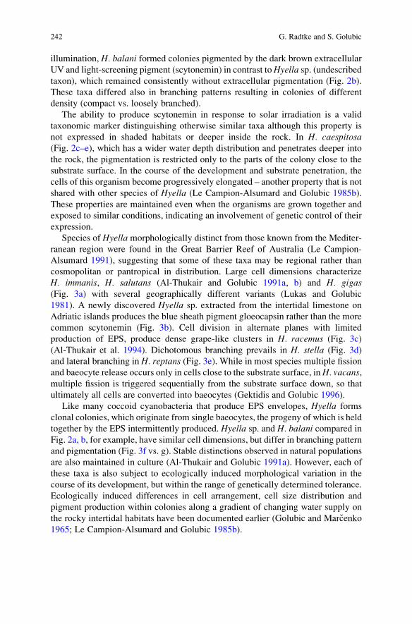
illumination, H. balani formed colonies pigmented by the dark brown extracellular
UV and light-screening pigment (scytonemin) in contrast toHyella sp. (undescribedtaxon), which remained consistently without extracellular pigmentation (Fig. 2b).
These taxa differed also in branching patterns resulting in colonies of different
density (compact vs. loosely branched).
The ability to produce scytonemin in response to solar irradiation is a valid
taxonomic marker distinguishing otherwise similar taxa although this property is
not expressed in shaded habitats or deeper inside the rock. In H. caespitosa(Fig. 2c–e), which has a wider water depth distribution and penetrates deeper into
the rock, the pigmentation is restricted only to the parts of the colony close to the
substrate surface. In the course of the development and substrate penetration, the
cells of this organism become progressively elongated – another property that is not
shared with other species of Hyella (Le Campion-Alsumard and Golubic 1985b).
These properties are maintained even when the organisms are grown together and
exposed to similar conditions, indicating an involvement of genetic control of their
expression.
Species of Hyella morphologically distinct from those known from the Mediter-
ranean region were found in the Great Barrier Reef of Australia (Le Campion-
Alsumard 1991), suggesting that some of these taxa may be regional rather than
cosmopolitan or pantropical in distribution. Large cell dimensions characterize
H. immanis, H. salutans (Al-Thukair and Golubic 1991a, b) and H. gigas(Fig. 3a) with several geographically different variants (Lukas and Golubic
1981). A newly discovered Hyella sp. extracted from the intertidal limestone on
Adriatic islands produces the blue sheath pigment gloeocapsin rather than the more
common scytonemin (Fig. 3b). Cell division in alternate planes with limited
production of EPS, produce dense grape-like clusters in H. racemus (Fig. 3c)
(Al-Thukair et al. 1994). Dichotomous branching prevails in H. stella (Fig. 3d)
and lateral branching in H. reptans (Fig. 3e). While in most species multiple fission
and baeocyte release occurs only in cells close to the substrate surface, inH. vacans,multiple fission is triggered sequentially from the substrate surface down, so that
ultimately all cells are converted into baeocytes (Gektidis and Golubic 1996).
Like many coccoid cyanobacteria that produce EPS envelopes, Hyella forms
clonal colonies, which originate from single baeocytes, the progeny of which is held
together by the EPS intermittently produced. Hyella sp. and H. balani compared in
Fig. 2a, b, for example, have similar cell dimensions, but differ in branching pattern
and pigmentation (Fig. 3f vs. g). Stable distinctions observed in natural populations
are also maintained in culture (Al-Thukair and Golubic 1991a). However, each of
these taxa is also subject to ecologically induced morphological variation in the
course of its development, but within the range of genetically determined tolerance.
Ecologically induced differences in cell arrangement, cell size distribution and
pigment production within colonies along a gradient of changing water supply on
the rocky intertidal habitats have been documented earlier (Golubic and Marcenko
1965; Le Campion-Alsumard and Golubic 1985b).
242 G. Radtke and S. Golubic
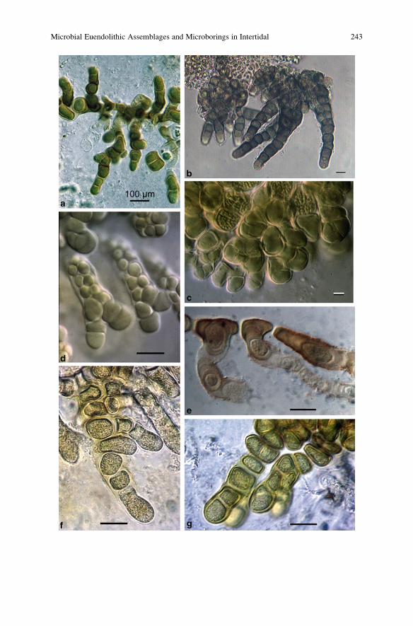
Microbial Euendolithic Assemblages and Microborings in Intertidal 243
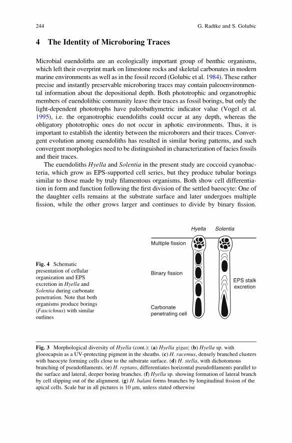
4 The Identity of Microboring Traces
Microbial euendoliths are an ecologically important group of benthic organisms,
which left their overprint mark on limestone rocks and skeletal carbonates in modern
marine environments as well as in the fossil record (Golubic et al. 1984). These rather
precise and instantly preservable microboring traces may contain paleoenvironmen-
tal information about the depositional depth. Both phototrophic and organotrophic
members of euendolithic community leave their traces as fossil borings, but only the
light-dependent phototrophs have paleobathymetric indicator value (Vogel et al.
1995), i.e. the organotrophic euendoliths could occur at any depth, whereas the
obligatory phototrophic ones do not occur in aphotic environments. Thus, it is
important to establish the identity between the microborers and their traces. Conver-
gent evolution among euendoliths has resulted in similar boring patterns, and such
convergent morphologies need to be distinguished in characterization of facies fossils
and their traces.
The euendoliths Hyella and Solentia in the present study are coccoid cyanobac-
teria, which grow as EPS-supported cell series, but they produce tubular borings
similar to those made by truly filamentous organisms. Both show cell differentia-
tion in form and function following the first division of the settled baeocyte: One of
the daughter cells remains at the substrate surface and later undergoes multiple
fission, while the other grows larger and continues to divide by binary fission.
Hyella Solentia
Multiple fission
Binary fission
Carbonatepenetrating cell
EPS stalkexcretion
Fig. 4 Schematic
presentation of cellular
organization and EPS
excretion in Hyella and
Solentia during carbonate
penetration. Note that both
organisms produce borings
(Fascichnus) with similar
outlines
Fig. 3 Morphological diversity of Hyella (cont.): (a) Hyella gigas; (b) Hyella sp. with
gloeocapsin as a UV-protecting pigment in the sheaths. (c) H. racemus, densely branched clusters
with baeocyte forming cells close to the substrate surface. (d) H. stella, with dichotomous
branching of pseudofilaments. (e) H. reptans, differentiates horizontal pseudofilaments parallel to
the surface and lateral, deeper boring branches. (f) Hyella sp. showing formation of lateral branch
by cell slipping out of the alignment. (g) H. balani forms branches by longitudinal fission of the
apical cells. Scale bar in all pictures is 10 mm, unless stated otherwise
244 G. Radtke and S. Golubic
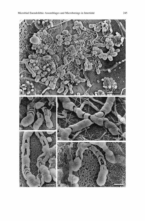
Microbial Euendolithic Assemblages and Microborings in Intertidal 245
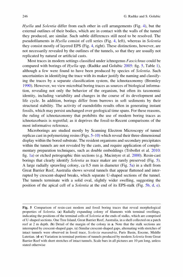
Hyella and Solentia differ from each other in cell arrangements (Fig. 4), but the
external outlines of their bodies, which are in contact with the walls of the tunnel
they produced, are similar. Such subtle differences still need to be resolved. The
pseudofilaments in Hyella consist of cell series (Fig. 4, left), whereas in Solentiathey consist mostly of layered EPS (Fig. 4, right). These distinctions, however, are
not necessarily revealed by the outlines of the tunnels, so that they are usually not
replicated by natural or artificial casts.
Most traces in modern settings classified under ichnogenus Fascichnus could becompared with borings of Hyella spp. (Radtke and Golubic 2005: fig. 5, Table 1),
although a few were found to have been produced by species of Solentia. Suchuncertainties in identifying the trace with its maker justify the naming and classify-
ing the traces by a separate classification system, the ichnotaxonomy (Bromley
1990). However, we view microbial boring traces as sources of biological informa-
tion, revealing not only the behavior of the organism, but often its taxonomic
identity, including variability and changes in the course of its development and
life cycle. In addition, borings differ from burrows in soft sediments by their
structural stability. The activity of euendoliths results often in generating instant
fossils, which may persist unchanged over geological time spans. For these reasons,
the ruling of ichnotaxonomy that prohibits the use of modern boring traces as
ichnotaxobasis is regretful; as it deprives the fossil-to-Recent comparisons of the
most informative reference.
Microborings are studied mostly by Scanning Electron Microscopy of tunnel
replicas cast in polymerizing resins (Figs. 5–10) which reveal their three-dimensional
display within the bored substrate. The resident organisms and secondary precipitates
within the tunnels are not revealed by the casts, and require application of comple-
mentary preparation techniques, such as double embeddings (Tribollet et al. 2010:
fig. 1a) or etched petrographic thin sections (e.g. Macintyre et al. 2000). Resin-cast
borings that clearly identify Solentia as trace maker are rarely preserved (Fig. 5).
A large radially sprawling colony, ca 0.5 mm in diameter (Fig. 5a) in a shell from
Great Barrier Reef, Australia shows several tunnels that appear flattened and inter-
rupted by crescent-shaped breaks, which separate U-shaped sections of the tunnel.
The tunnels terminate with a solid oval, slightly wider swelling, suggesting the
position of the apical cell of a Solentia at the end of its EPS-stalk (Fig. 5b, d, e).
Fig. 5 Comparison of resin-cast modern and fossil boring traces that reveal morphological
properties of Solentia. (a) Radially expanding colony of filaments with terminal swellings,
indicating the positions of the terminal cells of Solentia at the ends of stalks, which are comprised
of U-shaped sections. One Tree Island, Great Barrier Reef, Australia, in a shell collected on a patch
reef at 2 m depth. (b) Detail of the margin of the colony in a. Note that the stalk sections are
interrupted by crescent-shaped gaps. (c) Similar crescent-shaped gaps, alternating with stretches of
intact tunnels were observed in fossil trace, Scolecia maeandria, Paris Basin, Eocene, Middle
Lutetian. (d–e) Variation in terminal portions of tunnels produced by modern Solentia from Great
Barrier Reef with short stretches of intact tunnels. Scale bars in all pictures are 10 mm long, unless
stated otherwise
246 G. Radtke and S. Golubic
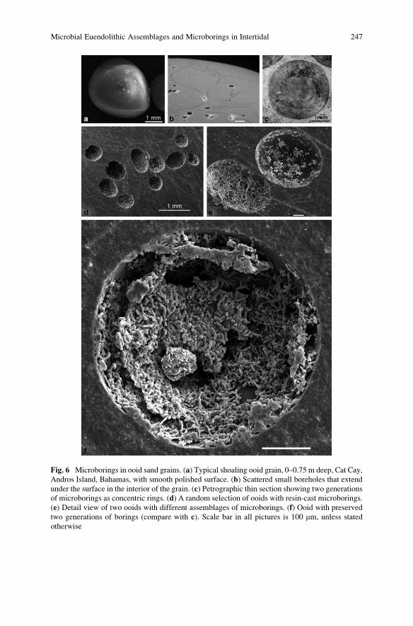
Fig. 6 Microborings in ooid sand grains. (a) Typical shoaling ooid grain, 0–0.75 m deep, Cat Cay,
Andros Island, Bahamas, with smooth polished surface. (b) Scattered small boreholes that extend
under the surface in the interior of the grain. (c) Petrographic thin section showing two generations
of microborings as concentric rings. (d) A random selection of ooids with resin-cast microborings.
(e) Detail view of two ooids with different assemblages of microborings. (f) Ooid with preserved
two generations of borings (compare with c). Scale bar in all pictures is 100 mm, unless stated
otherwise
Microbial Euendolithic Assemblages and Microborings in Intertidal 247
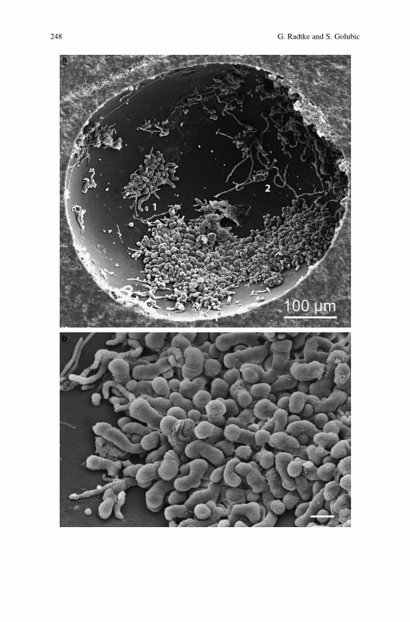
248 G. Radtke and S. Golubic
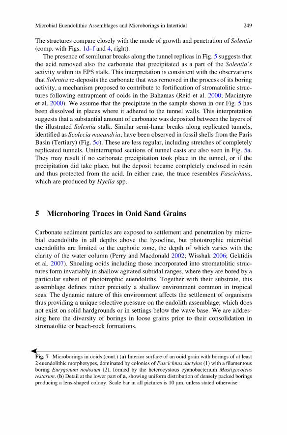
The structures compare closely with the mode of growth and penetration of Solentia(comp. with Figs. 1d–f and 4, right).
The presence of semilunar breaks along the tunnel replicas in Fig. 5 suggests that
the acid removed also the carbonate that precipitated as a part of the Solentia’sactivity within its EPS stalk. This interpretation is consistent with the observations
that Solentia re-deposits the carbonate that was removed in the process of its boring
activity, a mechanism proposed to contribute to fortification of stromatolitic struc-
tures following entrapment of ooids in the Bahamas (Reid et al. 2000; Macintyre
et al. 2000). We assume that the precipitate in the sample shown in our Fig. 5 has
been dissolved in places where it adhered to the tunnel walls. This interpretation
suggests that a substantial amount of carbonate was deposited between the layers of
the illustrated Solentia stalk. Similar semi-lunar breaks along replicated tunnels,
identified as Scolecia maeandria, have been observed in fossil shells from the Paris
Basin (Tertiary) (Fig. 5c). These are less regular, including stretches of completely
replicated tunnels. Uninterrupted sections of tunnel casts are also seen in Fig. 5a.
They may result if no carbonate precipitation took place in the tunnel, or if the
precipitation did take place, but the deposit became completely enclosed in resin
and thus protected from the acid. In either case, the trace resembles Fascichnus,which are produced by Hyella spp.
5 Microboring Traces in Ooid Sand Grains
Carbonate sediment particles are exposed to settlement and penetration by micro-
bial euendoliths in all depths above the lysocline, but phototrophic microbial
euendoliths are limited to the euphotic zone, the depth of which varies with the
clarity of the water column (Perry and Macdonald 2002; Wisshak 2006; Gektidis
et al. 2007). Shoaling ooids including those incorporated into stromatolitic struc-
tures form invariably in shallow agitated subtidal ranges, where they are bored by a
particular subset of phototrophic euendoliths. Together with their substrate, this
assemblage defines rather precisely a shallow environment common in tropical
seas. The dynamic nature of this environment affects the settlement of organisms
thus providing a unique selective pressure on the endolith assemblage, which does
not exist on solid hardgrounds or in settings below the wave base. We are addres-
sing here the diversity of borings in loose grains prior to their consolidation in
stromatolite or beach-rock formations.
�
Fig. 7 Microborings in ooids (cont.) (a) Interior surface of an ooid grain with borings of at least
2 euendolithic morphotypes, dominated by colonies of Fascichnus dactylus (1) with a filamentous
boring Eurygonum nodosum (2), formed by the heterocystous cyanobacterium Mastigocoleustestarum. (b) Detail at the lower part of a, showing uniform distribution of densely packed borings
producing a lens-shaped colony. Scale bar in all pictures is 10 mm, unless stated otherwise
Microbial Euendolithic Assemblages and Microborings in Intertidal 249
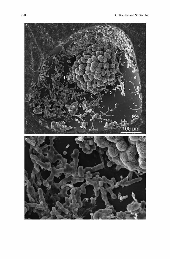
250 G. Radtke and S. Golubic
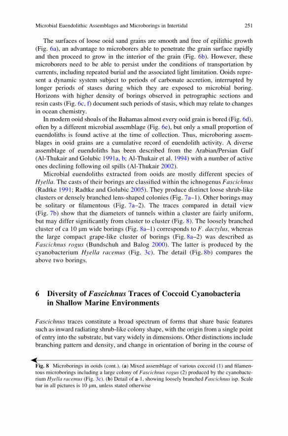
The surfaces of loose ooid sand grains are smooth and free of epilithic growth
(Fig. 6a), an advantage to microborers able to penetrate the grain surface rapidly
and then proceed to grow in the interior of the grain (Fig. 6b). However, these
microborers need to be able to persist under the conditions of transportation by
currents, including repeated burial and the associated light limitation. Ooids repre-
sent a dynamic system subject to periods of carbonate accretion, interrupted by
longer periods of stases during which they are exposed to microbial boring.
Horizons with higher density of borings observed in petrographic sections and
resin casts (Fig. 6c, f) document such periods of stasis, which may relate to changes
in ocean chemistry.
In modern ooid shoals of the Bahamas almost every ooid grain is bored (Fig. 6d),
often by a different microbial assemblage (Fig. 6e), but only a small proportion of
euendoliths is found active at the time of collection. Thus, microboring assem-
blages in ooid grains are a cumulative record of euendolith activity. A diverse
assemblage of euendoliths has been described from the Arabian/Persian Gulf
(Al-Thukair and Golubic 1991a, b; Al-Thukair et al. 1994) with a number of active
ones declining following oil spills (Al-Thukair 2002).
Microbial euendoliths extracted from ooids are mostly different species of
Hyella. The casts of their borings are classified within the ichnogenus Fascichnus(Radtke 1991; Radtke and Golubic 2005). They produce distinct loose shrub-like
clusters or densely branched lens-shaped colonies (Fig. 7a–1). Other borings may
be solitary or filamentous (Fig. 7a–2). The traces compared in detail view
(Fig. 7b) show that the diameters of tunnels within a cluster are fairly uniform,
but may differ significantly from cluster to cluster (Fig. 8). The loosely branched
cluster of ca 10 mm wide borings (Fig. 8a–1) corresponds to F. dactylus, whereasthe large compact grape-like cluster of borings (Fig. 8a–2) was described as
Fascichnus rogus (Bundschuh and Balog 2000). The latter is produced by the
cyanobacterium Hyella racemus (Fig. 3c). The detail (Fig. 8b) compares the
above two borings.
6 Diversity of Fascichnus Traces of Coccoid Cyanobacteria
in Shallow Marine Environments
Fascichnus traces constitute a broad spectrum of forms that share basic features
such as inward radiating shrub-like colony shape, with the origin from a single point
of entry into the substrate, but vary widely in dimensions. Other distinctions include
branching pattern and density, and change in orientation of boring in the course of
�
Fig. 8 Microborings in ooids (cont.). (a) Mixed assemblage of various coccoid (1) and filamen-
tous microborings including a large colony of Fascichnus rogus (2) produced by the cyanobacte-
rium Hyella racemus (Fig. 3c). (b) Detail of a-1, showing loosely branched Fascichnus isp. Scalebar in all pictures is 10 mm, unless stated otherwise
Microbial Euendolithic Assemblages and Microborings in Intertidal 251
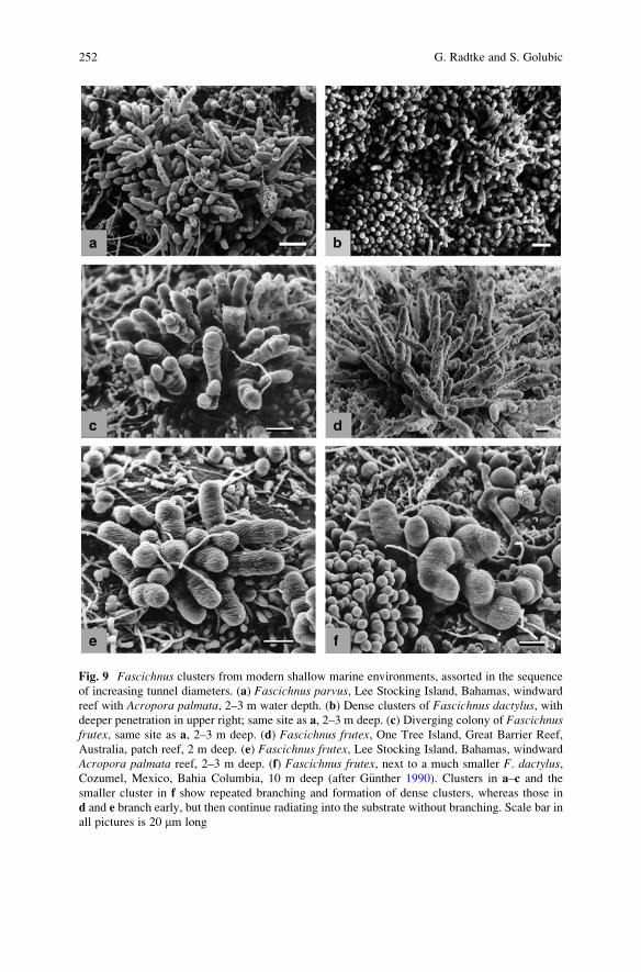
Fig. 9 Fascichnus clusters from modern shallow marine environments, assorted in the sequence
of increasing tunnel diameters. (a) Fascichnus parvus, Lee Stocking Island, Bahamas, windward
reef with Acropora palmata, 2–3 m water depth. (b) Dense clusters of Fascichnus dactylus, withdeeper penetration in upper right; same site as a, 2–3 m deep. (c) Diverging colony of Fascichnusfrutex, same site as a, 2–3 m deep. (d) Fascichnus frutex, One Tree Island, Great Barrier Reef,
Australia, patch reef, 2 m deep. (e) Fascichnus frutex, Lee Stocking Island, Bahamas, windward
Acropora palmata reef, 2–3 m deep. (f) Fascichnus frutex, next to a much smaller F. dactylus,Cozumel, Mexico, Bahia Columbia, 10 m deep (after G€unther 1990). Clusters in a–c and the
smaller cluster in f show repeated branching and formation of dense clusters, whereas those in
d and e branch early, but then continue radiating into the substrate without branching. Scale bar in
all pictures is 20 mm long
252 G. Radtke and S. Golubic
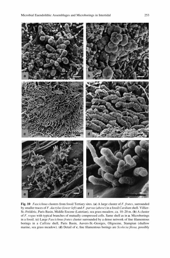
Fig. 10 Fascichnus clusters from fossil Tertiary sites. (a) A large cluster of F. frutex, surroundedby smaller traces of F. dactylus (lower left) and F. parvus (above) in a fossil Cardium shell. Villier-
St.-Frederic, Paris Basin, Middle Eocene (Lutetian), sea grass meadow, ca. 10–20 m. (b) A cluster
of F. rogus with typical branches of mutually compressed cells. Same shell as in a. Microborings
in a fossil. (c) Large Fascichnus frutex cluster surrounded by a dense network of fine filamentous
borings in a Callista shell, Paris Basin, Auvers-St.-Georges, Oligocene, Stampian (shallow
marine, sea grass meadow). (d) Detail of c, fine filamentous borings are Scolecia filosa, possibly
Microbial Euendolithic Assemblages and Microborings in Intertidal 253
the colony development. Borings in some Fascichnus ispp. expand initially parallelto the substrate surface and later explore deeper layers of the substrate, while others
bore immediately perpendicular to the surface or radiate inward forming clusters
different in size and density. Changes that occur in the course of the lifetime of an
organism (ontogeny) are known to be difficult to ascertain in fossils and especially
in fossil traces. However, the abundance of well preserved microboring traces
provides for reliable reconstructions of even complex life histories of the organisms
that made them (Golubic and Radtke 2008).
Size is the most conspicuous property used to distinguish between Fascichnustraces. The tunnel diameters were originally used among other characters to sepa-
rate ichnospecies with the following ranges proposed for F. parvus: 1.5–3.0 mm; for
F. dactylus: 4.0–9.0 mm, and for F. frutex: 11–25 mm (Radtke 1991). As additional
criteria proposed were length or tunnels (�40); (�85); (�110); branching: common
in F. parvus and F. frutex but rare in F. dactylus and rounded to dome-shape ending
of tunnels in all three ichnotaxa, sometimes club-shaped in F. dactylus.The choice of this criterion was pragmatic and intuitive. Boring traces of similar
shapes and sizes collected in modern environments could be measured and related
to the microorganisms that produced them. In the majority of cases such traces were
produced by euendolithic cyanobacteria and corresponded to different species of
Hyella (e.g. Wisshak 2006), with the exception of the largest trace described as
F. grandis (diameter ranging from 40 to 160 mm), which was found to be produced
by the euendolithic rhizoids of the chlorophyte Acetabularia (Radtke et al. 1997).
The predominance of transverse cell division in Hyella and Solentia produced
pseudofilaments and tunnels in the substrate with a specifically narrow range of
diameter variation. The wide size range of Fascichnus traces from modern shallow
marine environments is illustrated in Fig. 9, by a selection of colonies arranged by
size in an increasing order, with a 20 mm long scale bar in all compared pictures.
These pictures also show a relative uniformity in size within each colony. The
smallest cluster corresponds to F. parvus (Fig. 9a), the next higher group is in the
range of the ichnospecies F. dactylus (Fig. 9b, f left), followed by the larger F.frutex (Fig. 9c–f center). The contrast in size is most conspicuous in two clusters
formed next to each other (Fig. 9f). Fossil Fascichnus traces from Tertiary deposits
of Paris Basin with spectrum of forms is shown in Fig. 10.
In order to test the proposed taxonomic boundaries of Fascichnus ichnospecies,we have engaged first in determining size distribution of tunnel diameters in a large
number of Fascichnus clusters, in comparison with size values published for cell
dimensions of acid-extracted and cultured Hyella spp. (Table 2). In representative
populations, selected on the basis of described ichnotaxa, the minimum vs. maxi-
mum diameter of each tunnel was plotted in a two-dimensional display and then
summarized cluster by cluster by plotting average � standard deviation (Fig. 11).
Fig. 10 (continued) produced by the filamentous cyanobacterium Plectonema terebrans. (e) Sev-eral branched clusters of Fascichnus dactylus, Paris Basin, Ferme de l’Orme, Middle Eocene
(Lutetian). (f) Detail of e. Scale bar in all pictures is 20 mm, unless stated otherwise
254 G. Radtke and S. Golubic
16
14
12
10
8
6
4
2
00 2 4 6 8 10 12 14 16 18 20
Min
imu
m d
iam
eter
(µ
m)
Maximum diameter (µm)
Fig. 11 Plots of maximum vs. minimum tunnel diameters of three randomly selected Fascichnuscolonies. Note the narrow variation of data (dots) around mean � standard deviation (cross
diagrams) within each colony and clear separation between the colonies
35
30
25
20
15
10
5
00 5 10 15 20 25 30 35 40
Min
imu
m d
iam
eter
(µ
m)
Maximum diameter (µm)
a
b
c
d
e
f
Fig. 12 Cross diagram plots (mean � standard deviation) of a large sample of 54 Fascichnusclusters, showing overlapping data and lack of gaps between clusters. The positions of clusters
illustrated in Fig. 9 are marked (a–f)
Microbial Euendolithic Assemblages and Microborings in Intertidal 255
The plot of individual measurements produced a cloud of data points for each
population, scattered along a linear function inclined 45� that would describe
perfectly cylindrical borings. The distribution of measured parameters is close to
normal, so that basic statistics is applied with the average values of selected
populations close to the center of the respective data cloud, and the variability
within each population expressed by standard deviation (arms of the cross). Area
outlined by two standard deviations from the mean (dotted-lined oval) is expected
to include about 95% of the sample, thus approximating the range of sizes for that
population. The three selected populations had morphological features consistent
with the description of the ichnogenus Fascichnus; they showed similar pattern in
data distribution and were clearly distinct in size without overlap of measurements.
They were well within size ranges of the three described Fascichnus ichnospecies:F. parvus, F. dactylus and F. frutex. However, such clear distinction could not be
maintained when a large number of Fascichnus populations were measured. The
added populations plotted in between leaving no gaps to separate the data clusters
(Fig. 12).
By including a total of 54 Fascichnus populations in the sample, a pattern
emerged in which tunnel diameter values of populations were arranged along a
continuum, covering a wide total range of dimensions from 2 to 40 mm. Each cluster
covered a part of that range, each showed close to normal distribution of values and
relatively narrow intra-population variability (11.2 � 3.7% of the mean) but also
significant overlaps in tunnel diameter between clusters. The plot did not indicate
any grouping or gaps in distribution of means that would support a separation of the
measured populations by size. A similar pattern in size distribution within and
between clonal populations has been reported earlier for Gloeocapsa among coc-
coid cyanobacteria inhabiting wet cliffs (Golubic 1965, 1966). The clonal nature of
these populations was assured by enclosures in EPS envelopes.
Yet, the illustrated Fascichnus clusters suggest a higher diversity of forms,
which differ regarding proximal to distal change in tunnel diameter, presence or
absence of repeated constrictions along tunnels, or tunnel density, all properties not
necessarily correlated with size or not in the same combination as in the formally
described ichnotaxa. For example, the two clusters illustrated in Fig. 9b are both
within the size range of Fascichnus dactylus, but differ in density and organization
of tunnels in the colony. Similarly clusters illustrated in Fig. 9c–f each covers a
different part of the size range originally ascribed to F. frutex but differ in directionof diameter change (compare Fig. 9c and 9d) and form (Fig. 9e and 9f).
7 Microboring Cyanobacteria and Prokaryote Speciation
Cell dimensions are routinely employed in characterizing taxa of cyanobacteria
(and eukaryotic algae) and determination keys are constructed so as to separate taxa
based on size differences with a few additional diagnostic characters employed (e.g.
Geitler 1932; Komarek and Anagnostidis 1999, 2005). In our study of an
256 G. Radtke and S. Golubic
ecologically defined group of prokaryotes (coccoid euendolithic cyanobacteria), a
distinction based on size alone was lacking. Measurements were carried out on
clonal populations, held together by EPS when extracted from the matrix, each
characterized by a narrow normal distribution of sizes, supporting the existence of
genetically stable units, with neighboring population overlapping in size. This
pattern was faithfully reflected in tunnel diameter distribution of microboring
clusters. The total range of cell sizes within forms of the genusHyella and, similarly
of the diameters of their boreholes between 2 and 40 mm and clear distinction
between the end members of the range argued against inclusion of all these
populations into a single taxon. Thus, the distinction between taxonomic units
above clones but within established genera needs to be sought in other properties
but size, which may or may not show similarly smooth transitions between popula-
tions. Correlation of qualitative and quantitative properties may then indicate the
centers of diversification rather than gaps between them.
We recognize here a pattern of a continuous, fan-like diversification of popula-
tions regarding properties (cell size and tunnel diameters in this case), which are
apparently neutral with respect to natural selection. Accordingly, these properties
are inherited and distributed uniformly among successful descendent populations,
forming continuous spectra of forms. The pattern is consistent with the asexual
reproduction of cyanobacteria, which provides instant reproductive isolation of
these prokaryotic organisms and a gradual divergence of properties over time. It
also indicates the absence or ineffectiveness of lateral exchange of genetic infor-
mation at the level of metapopulations. This is in contrast to regular lateral transfer
of genetic information realized in sexually reproducing eukaryotes that results in
formation of a common gene pool of sexually compatible population members,
marked by sharper boundaries toward sexually incompatible or reproductively
isolated ones (De Queiros 2005).
The observed pattern is consistent with the proposed clonal dynamics and
population replacement mechanism in the speciation process of prokaryotic
organisms (Cohan 2002). Similar pattern of speciation among sexually reprodu-
cing eukaryotes exists in the so called ring species where the gene flow lags
behind the geographic spreading rate of populations (Moritz et al. 1992). In the
case of ring species, the populations show distinct characters, but these change
gradually along continuous geographic gradients. These groups are usually cate-
gorized as subspecies, although the end-members may become reproductively
incompatible, thus qualifying for the species status in terms of the Biological
Species Concept (Mayr 1982; De Queiros 2005). Clear separations by gaps
(hiatus) between taxa are a convenience in estimation of diversity introduced by
extinctions but also by uneven distribution of taxa (demographic replacements,
sensu Templeton 1989).
The present study explored the phenotypic expression side of a polyphasic
approach to cyanobacterial diversity, the molecular aspects yet to be employed in
the study of microbial euendoliths. The experiences thus far project a significantly
higher diversity of taxa then are formally described as suggested by the observa-
tions of cell organization pigmentation and boring behavior of coccoid
Microbial Euendolithic Assemblages and Microborings in Intertidal 257
cyanobacteria. It is expected that the observed phenotypes are supported with
corresponding high number of molecular genotype signatures as has been obtained
in recent studies of endoliths in travertine systems of the Yellowstone National Park
(Norris and Castenholz 2006), microbial mats of the Antarctica (Jungblut et al.
2005) and marine stromatolites (Foster et al. 2009).
Acknowledgments The international and interdisciplinary cooperation has been supported by the
Alexander-von-Humboldt Foundation and by the Hanse Institute for Advanced Research. We
thank Prof. Dr. J. Reitner (Univ. G€ottingen) for support during the International Kalkowsky
Symposium in 2008 and Dr. R.P. Reid (Univ. Miami) for stimulating discussions regarding
endoliths and stromatolites.
References
Al-Thukair AA (2002) Effect of oil pollution on euendolithic cyanobacteria of the Arabian Gulf.
Environmental Microbiology 4:125–129
Al-Thukair AA, Golubic S (1991a) Five new Hyella species from the Arabian Gulf. Algological
Studies 64:167–197
Al-Thukair AA, Golubic S (1991b) New endolithic cyanobacteria from the Arabian Gulf. I. Hyellaimmanis sp. nov. Journal of Phycology 27:766–780
Al-Thukair AA, Golubic S (1996) Characterization of Hyella caespitosa var. arbuscula var. nov.
(Cyanophyta, Cyanobacteria) from shoaling ooid sand grains, Arabian Gulf. Nova Hedwigia
Beihefte 112:83–91
Al-Thukair AA, Green J (1988) New endolithic taxa in modern and ancient (late Proterozoic)
shallow water marine environments. Journal of Phycology 24:12
Al-Thukair AA, Golubic S, Rosen G (1994) New euendolithic cyanobacteria from the Bahama
Bank and the Arabian Gulf: Hyella racemus sp. nov. Journal of Phycology 30:764–769
Balog S-J (1996) Boring thallophytes in some Permian and Triassic reefs: bathymetry and
bioerosion. G€attinger Arbeiten f€ur Geologie und Pal€aontologie Sb 2:305–308
Balog S-J (1997) Mikroendolithen im Capitan Riff Komplex (New Mexico, USA). Courier,
Forschungsinstitut Senckenberg 201:47–55
Bertling M (2007) What’s in a Name? Nomenclature, Systematics, Ichnotaxonomy. In: Miller W
(ed) Trace Fossils: Concepts, Problems, Prospects. Elsevier, Amsterdam, pp 81–91
Bornet ME, Flahault C (1889) Sur quelques plantes vivant dans le test calcaire des mollusques.
Bulletin de la Societe Botanique de France 36:147–176
Bromley RG (1990) Trace Fossils: Biology and Taphonomy. Unwin Hyman, London, pp 280
Bundschuh M (2000) Silurische Mikrobohrspuren – ihre Beschreibung und Verteilung in verschie-
denen Faziesr€aumen (Schweden, Litauen, Großbritannien und U.S.A.). Ph.D. Thesis, FB
Geowissenschaften, Johann Wolfgang Goethe Universitat, Frankfurt am Main, Germany,
pp 1–129
Bundschuh M, Balog S (2000) Fasciculus rogus nov. isp., an endolithic trace fossil. Ichnos
7:149–152
Campbell SE (1980) Palaeoconchocelis starmachii, a carbonate boring microfossil from the
Upper Silurian of Poland (425 million years old): implications for the evolution of the
Bangiaceae (Rhodophyta). Phycologia 19:25–36
Campbell SE (1982) Precambrian endoliths discovered. Nature 299:429–431
Campbell SE, Kazmierczak J, Golubic S (1979) Palaeoconchocelis starmachii n. gen., n. sp.,a Silurian endolithic rhodophyte (Bangiaceae). Acta Paleontologica Polonica 24:405–408
258 G. Radtke and S. Golubic
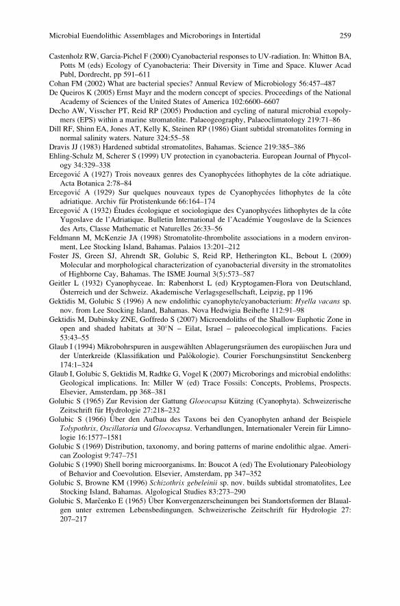
Castenholz RW, Garcia-Pichel F (2000) Cyanobacterial responses to UV-radiation. In: Whitton BA,
Potts M (eds) Ecology of Cyanobacteria: Their Diversity in Time and Space. Kluwer Acad
Publ, Dordrecht, pp 591–611
Cohan FM (2002) What are bacterial species? Annual Review of Microbiology 56:457–487
De Queiros K (2005) Ernst Mayr and the modern concept of species. Proceedings of the National
Academy of Sciences of the United States of America 102:6600–6607
Decho AW, Visscher PT, Reid RP (2005) Production and cycling of natural microbial exopoly-
mers (EPS) within a marine stromatolite. Palaeogeography, Palaeoclimatology 219:71–86
Dill RF, Shinn EA, Jones AT, Kelly K, Steinen RP (1986) Giant subtidal stromatolites forming in
normal salinity waters. Nature 324:55–58
Dravis JJ (1983) Hardened subtidal stromatolites, Bahamas. Science 219:385–386
Ehling-Schulz M, Scherer S (1999) UV protection in cyanobacteria. European Journal of Phycol-
ogy 34:329–338
Ercegovic A (1927) Trois noveaux genres des Cyanophycees lithophytes de la cote adriatique.
Acta Botanica 2:78–84
Ercegovic A (1929) Sur quelques nouveaux types de Cyanophycees lithophytes de la cote
adriatique. Archiv f€ur Protistenkunde 66:164–174Ercegovic A (1932) Etudes ecologique et sociologique des Cyanophycees lithophytes de la cote
Yugoslave de l’Adriatique. Bulletin International de l’Academie Yougoslave de la Sciences
des Arts, Classe Mathematic et Naturelles 26:33–56
Feldmann M, McKenzie JA (1998) Stromatolite-thrombolite associations in a modern environ-
ment, Lee Stocking Island, Bahamas. Palaios 13:201–212
Foster JS, Green SJ, Ahrendt SR, Golubic S, Reid RP, Hetherington KL, Bebout L (2009)
Molecular and morphological characterization of cyanobacterial diversity in the stromatolites
of Highborne Cay, Bahamas. The ISME Journal 3(5):573–587
Geitler L (1932) Cyanophyceae. In: Rabenhorst L (ed) Kryptogamen-Flora von Deutschland,
Osterreich und der Schweiz. Akademische Verlagsgesellschaft, Leipzig, pp 1196
Gektidis M, Golubic S (1996) A new endolithic cyanophyte/cyanobacterium: Hyella vacans sp.nov. from Lee Stocking Island, Bahamas. Nova Hedwigia Beihefte 112:91–98
Gektidis M, Dubinsky ZNE, Goffredo S (2007) Microendoliths of the Shallow Euphotic Zone in
open and shaded habitats at 30�N – Eilat, Israel – paleoecological implications. Facies
53:43–55
Glaub I (1994) Mikrobohrspuren in ausgewahlten Ablagerungsr€aumen des europaischen Jura und
der Unterkreide (Klassifikation und Pal€okologie). Courier Forschungsinstitut Senckenberg
174:1–324
Glaub I, Golubic S, Gektidis M, Radtke G, Vogel K (2007) Microborings and microbial endoliths:
Geological implications. In: Miller W (ed) Trace Fossils: Concepts, Problems, Prospects.
Elsevier, Amsterdam, pp 368–381
Golubic S (1965) Zur Revision der Gattung Gloeocapsa K€utzing (Cyanophyta). Schweizerische
Zeitschrift f€ur Hydrologie 27:218–232Golubic S (1966) €Uber den Aufbau des Taxons bei den Cyanophyten anhand der Beispiele
Tolypothrix, Oscillatoria und Gloeocapsa. Verhandlungen, Internationaler Verein f€ur Limno-
logie 16:1577–1581
Golubic S (1969) Distribution, taxonomy, and boring patterns of marine endolithic algae. Ameri-
can Zoologist 9:747–751
Golubic S (1990) Shell boring microorganisms. In: Boucot A (ed) The Evolutionary Paleobiology
of Behavior and Coevolution. Elsevier, Amsterdam, pp 347–352
Golubic S, Browne KM (1996) Schizothrix gebeleinii sp. nov. builds subtidal stromatolites, Lee
Stocking Island, Bahamas. Algological Studies 83:273–290
Golubic S, Marcenko E (1965) €Uber Konvergenzerscheinungen bei Standortsformen der Blaual-
gen unter extremen Lebensbedingungen. Schweizerische Zeitschrift f€ur Hydrologie 27:
207–217
Microbial Euendolithic Assemblages and Microborings in Intertidal 259
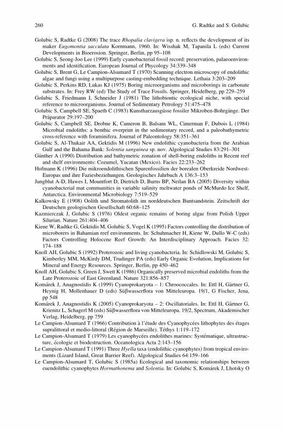
Golubic S, Radtke G (2008) The trace Rhopalia clavigera isp. n. reflects the development of its
maker Eugomontia sacculata Kornmann, 1960. In: Wisshak M, Tapanila L (eds) Current
Developments in Bioerosion. Springer, Berlin, pp 95–108
Golubic S, Seong-Joo Lee (1999) Early cyanobacterial fossil record: preservation, palaeoenviron-
ments and identification. European Journal of Phycology 34:339–348
Golubic S, Brent G, Le Campion-Alsumard T (1970) Scanning electron microscopy of endolithic
algae and fungi using a multipurpose casting-embedding technique. Lethaia 3:203–209
Golubic S, Perkins RD, Lukas KJ (1975) Boring microorganisms and microborings in carbonate
substrates. In: Frey RW (ed) The Study of Trace Fossils. Springer, Heidelberg, pp 229–259
Golubic S, Friedmann I, Schneider J (1981) The lithobiontic ecological niche, with special
reference to microorganisms. Journal of Sedimentary Petrology 51:475–478
Golubic S, Campbell SE, Spaeth C (1983) Kunstharzausg€usse fossiler Mikroben-Bohrg€ange. DerPr€aparator 29:197–200
Golubic S, Campbell SE, Drobne K, Cameron B, Balsam WL, Cimerman F, Dubois L (1984)
Microbial endoliths: a benthic overprint in the sedimentary record, and a paleobathymetric
cross-reference with foraminifera. Journal of Paleontology 58:351–361
Golubic S, Al-Thukair AA, Gektidis M (1996) New endolithic cyanobacteria from the Arabian
Gulf and the Bahama Bank: Solentia sanguinea sp. nov. Algological Studies 83:291–301
G€unther A (1990) Distribution and bathymetric zonation of shell-boring endoliths in Recent reef
and shelf environments: Cozumel, Yucatan (Mexico). Facies 22:233–262
Hofmann K (1996) Die mikroendolithischen Spurenfossilien der borealen Oberkreide Nordwest-
Europas und ihre Faziesbeziehungen. Geologisches Jahrbuch A 136:3–153
Jungblut A-D, Hawes I, Mountfort D, Dietrich D, Burns BP, Neilan BA (2005) Diversity within
cyanobacterial mat communities in variable salinity meltwater ponds of McMurdo Ice Shelf,
Antarctica. Environmental Microbiology 7:519–529
Kalkowsky E (1908) Oolith und Stromatolith im norddeutschen Buntsandstein. Zeitschrift der
Deutschen geologischen Gesellschaft 60:68–125
Kazmierczak J, Golubic S (1976) Oldest organic remains of boring algae from Polish Upper
Silurian. Nature 261:404–406
Kiene W, Radtke G, Gektidis M, Golubic S, Vogel K (1995) Factors controlling the distribution of
microborers in Bahamian reef environments. In: Schuhmacher H, Kiene W, Dullo W-C (eds)
Factors Controlling Holocene Reef Growth: An Interdisciplinary Approach. Facies 32:
174–188
Knoll AH, Golubic S (1992) Proterozoic and living cyanobacteria. In: Schidlowski M, Golubic S,
Kimberley MM, McKirdy DM, Trudinger PA (eds) Early Organic Evolution, Implications for
Mineral and Energy Resources. Springer, Berlin, pp 450–462
Knoll AH, Golubic S, Green J, Swett K (1986) Organically preserved microbial endoliths from the
Late Proterozoic of East Greenland. Nature 321:856–857
Komarek J, Anagnostidis K (1999) Cyanoprokaryota – 1: Chroococcales. In: Ettl H, G€artner G,Heynig H, Mollenhauer D (eds) S€ubwasserflora von Mitteleuropa. 19/1, G Fischer, Jena,
pp 548
Komarek J, Anagnostidis K (2005) Cyanoprokaryota – 2: Oscillatoriales. In: Ettl H, G€artner G,Krienitz L, Schagerl M (eds) S€ubwasserflora von Mitteleuropa. 19/2, Spectrum, Akademischer
Verlag, Heidelberg, pp 759
Le Campion-Alsumard T (1966) Contribution a l’etude des Cyanophycees lithophytes des etages
supralittoral et medio-littoral (Region de Marseille). Tethys 1:119–172
Le Campion-Alsumard T (1979) Les cyanophycees endolithes marines: Systematique, ultrastruc-
ture, ecologie et biodestruction. Oceanologica Acta 2:143–156
Le Campion-Alsumard T (1991) Three Hyella taxa (endolithic cyanophytes) from tropical enviro-
ments (Lizard Island, Great Barrier Reef). Algological Studies 64:159–166
Le Campion-Alsumard T, Golubic S (1985a) Ecological and taxonomic relationships between
euendolithic cyanophytes Hormathonema and Solentia. In: Golubic S, Komarek J, Lhotsky O
260 G. Radtke and S. Golubic
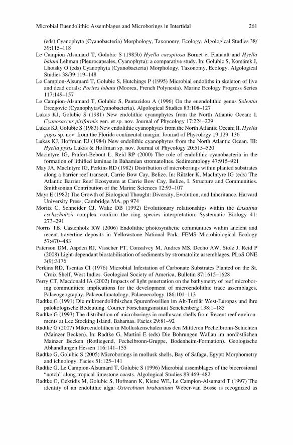
(eds) Cyanophyta (Cyanobacteria) Morphology, Taxonomy, Ecology. Algological Studies 38/
39:115–118
Le Campion-Alsumard T, Golubic S (1985b) Hyella caespitosa Bornet et Flahault and Hyellabalani Lehman (Pleurocapsales, Cyanophyta): a comparative study. In: Golubic S, Komarek J,
Lhotsky O (eds) Cyanophyta (Cyanobacteria) Morphology, Taxonomy, Ecology. Algological
Studies 38/39:119–148
Le Campion-Alsumard T, Golubic S, Hutchings P (1995) Microbial endoliths in skeleton of live
and dead corals: Porites lobata (Moorea, French Polynesia). Marine Ecology Progress Series
117:149–157
Le Campion-Alsumard T, Golubic S, Pantazidou A (1996) On the euendolithic genus SolentiaErcegovic (Cyanophyta/Cyanobacteria). Algological Studies 83:108–127
Lukas KJ, Golubic S (1981) New endolithic cyanophytes from the North Atlantic Ocean: I.
Cyanosaccus piriformis gen. et sp. nov. Journal of Phycology 17:224–229
Lukas KJ, Golubic S (1983) New endolithic cyanophytes from the North Atlantic Ocean: II.Hyellagigas sp. nov. from the Florida continental margin. Journal of Phycology 19:129–136
Lukas KJ, Hoffman EJ (1984) New endolithic cyanophytes from the North Atlantic Ocean. III:
Hyella pyxis Lukas & Hoffman sp. nov. Journal of Phycology 20:515–520
Macintyre IG, Prufert-Bebout L, Reid RP (2000) The role of endolithic cyanobacteria in the
formation of lithified laminae in Bahamian stromatolites. Sedimentology 47:915–921
May JA, MacIntyre IG, Perkins RD (1982) Distribution of microborings within planted substrates
along a barrier reef transect, Carrie Bow Cay, Belize. In: R€utzler K, MacIntyre IG (eds) The
Atlantic Barrier Reef Ecosystem at Carrie Bow Cay, Belize, I. Structure and Communities.
Smithsonian Contribution of the Marine Sciences 12:93–107
Mayr E (1982) The Growth of Biological Thought: Diversity, Evolution, and Inheritance. Harvard
University Press, Cambridge MA, pp 974
Moritz C, Schneider CJ, Wake DB (1992) Evolutionary relationships within the Ensatinaeschscholtzii complex confirm the ring species interpretation. Systematic Biology 41:
273–291
Norris TB, Castenholz RW (2006) Endolithic photosynthetic communities within ancient and
recent travertine deposits in Yellowstone National Park. FEMS Microbiological Ecology
57:470–483
Paterson DM, Aspden RJ, Visscher PT, Consalvey M, Andres MS, Decho AW, Stolz J, Reid P
(2008) Light-dependant biostabilisation of sediments by stromatolite assemblages. PLoS ONE
3(9):3176
Perkins RD, Tsentas CI (1976) Microbial Infestation of Carbonate Substrates Planted on the St.
Croix Shelf, West Indies. Geological Society of America, Bulletin 87:1615–1628
Perry CT, Macdonald IA (2002) Impacts of light penetration on the bathymetry of reef microbor-
ing communities: implications for the development of microendolithic trace assemblages.
Palaeogeography, Palaeoclimatology, Palaeoecology 186:101–113
Radtke G (1991) Die mikroendolithischen Spurenfossilien im Alt-Terti€ar West-Europas und ihre
pal€okologische Bedeutung. Courier Forschungsinstitut Senckenberg 138:1–185
Radtke G (1993) The distribution of microborings in molluscan shells from Recent reef environ-
ments at Lee Stocking Island, Bahamas. Facies 29:81–92
Radtke G (2007) Mikroendolithen in Molluskenschalen aus den Mittleren Pechelbronn-Schichten
(Mainzer Becken). In: Radtke G, Martini E (eds) Die Bohrungen Wallau im nordostlichen
Mainzer Becken (Rotliegend, Pechelbronn-Gruppe, Bodenheim-Formation). Geologische
Abhandlungen Hessen 116:141–155
Radtke G, Golubic S (2005) Microborings in mollusk shells, Bay of Safaga, Egypt: Morphometry
and ichnology. Facies 51:125–141
Radtke G, Le Campion-Alsumard T, Golubic S (1996) Microbial assemblages of the bioerosional
“notch” along tropical limestone coasts. Algological Studies 83:469–482
Radtke G, Gektidis M, Golubic S, Hofmann K, Kiene WE, Le Campion-Alsumard T (1997) The
identity of an endolithic alga: Ostreobium brabantium Weber-van Bosse is recognized as
Microbial Euendolithic Assemblages and Microborings in Intertidal 261
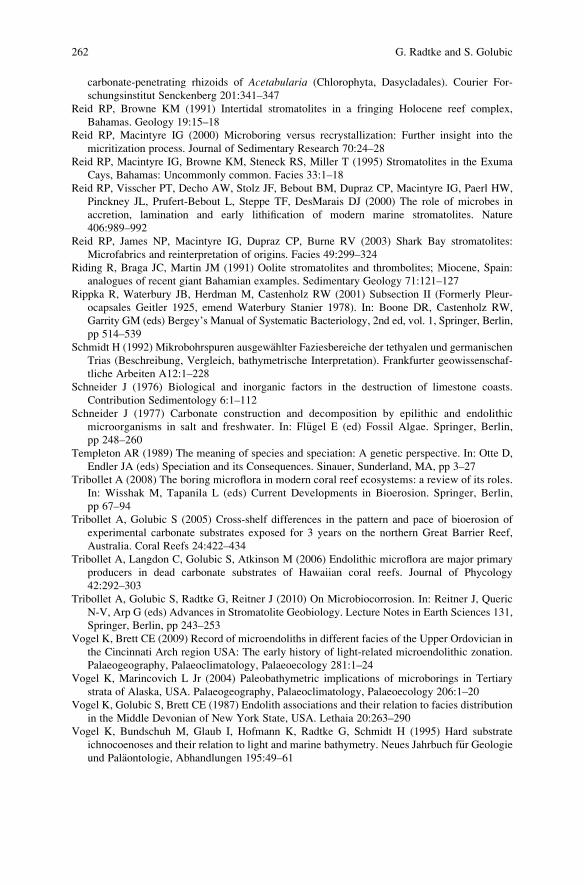
carbonate-penetrating rhizoids of Acetabularia (Chlorophyta, Dasycladales). Courier For-
schungsinstitut Senckenberg 201:341–347
Reid RP, Browne KM (1991) Intertidal stromatolites in a fringing Holocene reef complex,
Bahamas. Geology 19:15–18
Reid RP, Macintyre IG (2000) Microboring versus recrystallization: Further insight into the
micritization process. Journal of Sedimentary Research 70:24–28
Reid RP, Macintyre IG, Browne KM, Steneck RS, Miller T (1995) Stromatolites in the Exuma
Cays, Bahamas: Uncommonly common. Facies 33:1–18
Reid RP, Visscher PT, Decho AW, Stolz JF, Bebout BM, Dupraz CP, Macintyre IG, Paerl HW,
Pinckney JL, Prufert-Bebout L, Steppe TF, DesMarais DJ (2000) The role of microbes in
accretion, lamination and early lithification of modern marine stromatolites. Nature
406:989–992
Reid RP, James NP, Macintyre IG, Dupraz CP, Burne RV (2003) Shark Bay stromatolites:
Microfabrics and reinterpretation of origins. Facies 49:299–324
Riding R, Braga JC, Martin JM (1991) Oolite stromatolites and thrombolites; Miocene, Spain:
analogues of recent giant Bahamian examples. Sedimentary Geology 71:121–127
Rippka R, Waterbury JB, Herdman M, Castenholz RW (2001) Subsection II (Formerly Pleur-
ocapsales Geitler 1925, emend Waterbury Stanier 1978). In: Boone DR, Castenholz RW,
Garrity GM (eds) Bergey’s Manual of Systematic Bacteriology, 2nd ed, vol. 1, Springer, Berlin,
pp 514–539
Schmidt H (1992) Mikrobohrspuren ausgewahlter Faziesbereiche der tethyalen und germanischen
Trias (Beschreibung, Vergleich, bathymetrische Interpretation). Frankfurter geowissenschaf-
tliche Arbeiten A12:1–228
Schneider J (1976) Biological and inorganic factors in the destruction of limestone coasts.
Contribution Sedimentology 6:1–112
Schneider J (1977) Carbonate construction and decomposition by epilithic and endolithic
microorganisms in salt and freshwater. In: Fl€ugel E (ed) Fossil Algae. Springer, Berlin,
pp 248–260
Templeton AR (1989) The meaning of species and speciation: A genetic perspective. In: Otte D,
Endler JA (eds) Speciation and its Consequences. Sinauer, Sunderland, MA, pp 3–27
Tribollet A (2008) The boring microflora in modern coral reef ecosystems: a review of its roles.
In: Wisshak M, Tapanila L (eds) Current Developments in Bioerosion. Springer, Berlin,
pp 67–94
Tribollet A, Golubic S (2005) Cross-shelf differences in the pattern and pace of bioerosion of
experimental carbonate substrates exposed for 3 years on the northern Great Barrier Reef,
Australia. Coral Reefs 24:422–434
Tribollet A, Langdon C, Golubic S, Atkinson M (2006) Endolithic microflora are major primary
producers in dead carbonate substrates of Hawaiian coral reefs. Journal of Phycology
42:292–303
Tribollet A, Golubic S, Radtke G, Reitner J (2010) On Microbiocorrosion. In: Reitner J, Queric
N-V, Arp G (eds) Advances in Stromatolite Geobiology. Lecture Notes in Earth Sciences 131,
Springer, Berlin, pp 243–253
Vogel K, Brett CE (2009) Record of microendoliths in different facies of the Upper Ordovician in
the Cincinnati Arch region USA: The early history of light-related microendolithic zonation.
Palaeogeography, Palaeoclimatology, Palaeoecology 281:1–24
Vogel K, Marincovich L Jr (2004) Paleobathymetric implications of microborings in Tertiary
strata of Alaska, USA. Palaeogeography, Palaeoclimatology, Palaeoecology 206:1–20
Vogel K, Golubic S, Brett CE (1987) Endolith associations and their relation to facies distribution
in the Middle Devonian of New York State, USA. Lethaia 20:263–290
Vogel K, Bundschuh M, Glaub I, Hofmann K, Radtke G, Schmidt H (1995) Hard substrate
ichnocoenoses and their relation to light and marine bathymetry. Neues Jahrbuch f€ur Geologieund Palaontologie, Abhandlungen 195:49–61
262 G. Radtke and S. Golubic

Vogel K, Gektidis M, Golubic S, Kiene WE, Radtke G (2000) Experimental studies on microbial
bioerosion at Lee Stocking Island, Bahamas and One Tree Island, Great Barrier Reef,
Australia: implications for paleoecological reconstructions. Lethaia 33:190–204
Waterbury JB, Stanier RW (1978) Patterns of growth and development in Pleurocapsalean
cyanobacteria. Microbiological Reviews 42:2–44
Wisshak M (2006) High-latitude bioerosion: the Kosterfjord experiment. Lecture Notes in Earth
Sciences 109:1–202
Wisshak M, Seub B, N€utzel A (2008) Eolutionary implications of an exceptionally preserved
Carboniferous microboring assemblage in the Buckhorn Asphalt lagerst€atte (Oklahoma, USA).
In: Wisshak M, Tapanila L (eds) Current Developments in Bioerosion. Springer, Berlin,
pp 21–54
Zhang Y, Golubic S (1987) Endolithic microfossils (cyanophyta) from early Proterozoic stroma-
tolites, Hebei, China. Acta Micropaleontologica Sinica 4:1–12
Microbial Euendolithic Assemblages and Microborings in Intertidal 263

.
On Microbiocorrosion
Aline Tribollet, Stjepko Golubic, Gudrun Radtke, and Joachim Reitner
1 Introduction
Destruction of rocks and minerals by biological activities has been termed bioerosion
(Neumann 1966). It includes mechanical as well as chemical effects, i.e. bioabrasion
and biocorrosion (Schneider 1976; Golubic and Schneider 1979). However both
processes often co-occur; they are functionally interconnected and mutually support-
ive. Biocorrosion can result from the activity of macro- or micro- organisms and thus
is called macrobiocorrosion and microbiocorrosion. Microbiocorrosion can also be
closely associated with microbial rock formation and consolidation in stromatolitic
structures (Reid et al. 2000; Macintyre et al. 2000; Garcia-Pichel et al. 2004; Dupraz
and Visscher 2005). In fact, the oldest known fossils of microboring organisms were
located in lithified horizons of silicified stromatolites (Zhang and Golubic 1987).
2 Niches
Microbiocorrosion is due to various phototrophic and organotrophic organisms
such as bacteria, cyanobacteria, red and green eukaryotic algae, lichens and
fungi. Depending on their relation to the substrate, the following niches are
A. Tribollet (*)
LOPB, Institut de Recherche pour le Developpement, BP A5, 98848 Noumea Cedex, New
Caledonia
e-mail: [email protected]
S. Golubic
Biological Science Center, Boston University, 05 Cummington Str, Boston, MA 02215, USA
G. Radtke
Hessisches Landesamt f€ur Umwelt und Geologie, Rheingaustr, 186, 65203 Wiesbaden, Germany
J. Reitner
Department of Geobiology and Center for Geosciences, University of G€ottingen, Goldschmidtstr.
3, 37077 G€ottingen, Germany
J. Reitner et al., Advances in Stromatolite Geobiology,Lecture Notes in Earth Sciences 131, DOI 10.1007/978-3-642-10415-2_17,# Springer-Verlag Berlin Heidelberg 2011
265
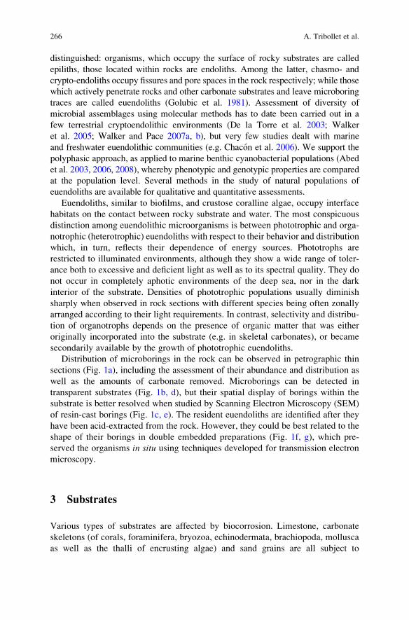
distinguished: organisms, which occupy the surface of rocky substrates are called
epiliths, those located within rocks are endoliths. Among the latter, chasmo- and
crypto-endoliths occupy fissures and pore spaces in the rock respectively; while those
which actively penetrate rocks and other carbonate substrates and leave microboring
traces are called euendoliths (Golubic et al. 1981). Assessment of diversity of
microbial assemblages using molecular methods has to date been carried out in a
few terrestrial cryptoendolithic environments (De la Torre et al. 2003; Walker
et al. 2005; Walker and Pace 2007a, b), but very few studies dealt with marine
and freshwater euendolithic communities (e.g. Chacon et al. 2006). We support the
polyphasic approach, as applied to marine benthic cyanobacterial populations (Abed
et al. 2003, 2006, 2008), whereby phenotypic and genotypic properties are compared
at the population level. Several methods in the study of natural populations of
euendoliths are available for qualitative and quantitative assessments.
Euendoliths, similar to biofilms, and crustose coralline algae, occupy interface
habitats on the contact between rocky substrate and water. The most conspicuous
distinction among euendolithic microorganisms is between phototrophic and orga-
notrophic (heterotrophic) euendoliths with respect to their behavior and distribution
which, in turn, reflects their dependence of energy sources. Phototrophs are
restricted to illuminated environments, although they show a wide range of toler-
ance both to excessive and deficient light as well as to its spectral quality. They do
not occur in completely aphotic environments of the deep sea, nor in the dark
interior of the substrate. Densities of phototrophic populations usually diminish
sharply when observed in rock sections with different species being often zonally
arranged according to their light requirements. In contrast, selectivity and distribu-
tion of organotrophs depends on the presence of organic matter that was either
originally incorporated into the substrate (e.g. in skeletal carbonates), or became
secondarily available by the growth of phototrophic euendoliths.
Distribution of microborings in the rock can be observed in petrographic thin
sections (Fig. 1a), including the assessment of their abundance and distribution as
well as the amounts of carbonate removed. Microborings can be detected in
transparent substrates (Fig. 1b, d), but their spatial display of borings within the
substrate is better resolved when studied by Scanning Electron Microscopy (SEM)
of resin-cast borings (Fig. 1c, e). The resident euendoliths are identified after they
have been acid-extracted from the rock. However, they could be best related to the
shape of their borings in double embedded preparations (Fig. 1f, g), which pre-
served the organisms in situ using techniques developed for transmission electron
microscopy.
3 Substrates
Various types of substrates are affected by biocorrosion. Limestone, carbonate
skeletons (of corals, foraminifera, bryozoa, echinodermata, brachiopoda, mollusca
as well as the thalli of encrusting algae) and sand grains are all subject to
266 A. Tribollet et al.
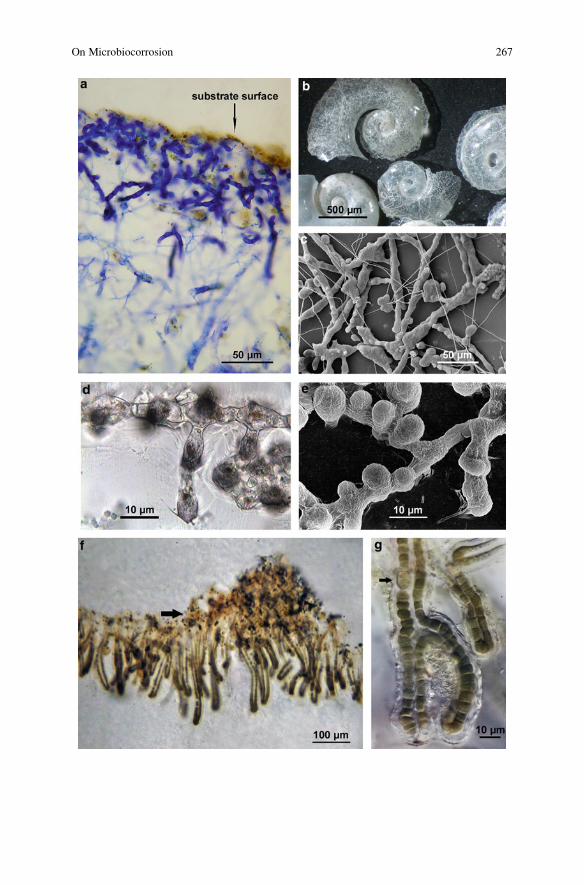
On Microbiocorrosion 267
biocorrosion by phototrophic and organotrophic euendolithic microorganisms
(Perry 1998; Vogel et al. 2000; Tribollet and Payri 2001). For instance, a significant
abundance and diversity of euendoliths inhabit extensive areas of seafloor covered
by sand (Al-Thukair and Golubic 1991; Golubic and Schneider 2003; Rasheed et al.
2004; Radtke and Golubic 2010). Wild et al. (2006) quantified the microbial cells in
reef before and after carbonate dissolution and found that the number doubled when
euendoliths were added to the census. Abundance of euendolithic filaments can
reach half a million per square centimeter in coastal limestone (Schneider and Le
Campion-Alsumeard 1999). Microbiocorrosion by euendoliths has also been
observed in phosphatic substrates (Roux 1887; Underwood et al. 1999; Zhang
and Pratt 2008).
4 Mechanisms
The exact mechanisms involved in biocorrosion remain unknown although a
number of plausible hypotheses have been proposed. It is probable that more than
one of several possible mechanisms is involved. A simple mechanism involves
hydrated respiratory CO2, which acts as a weak acid, still an effective solvent of
calcium carbonate with significant geological impact (Garcia-Pichel 2006). Other
mechanisms may include local application of organic acids (Alexandersson 1975),
which are regular products of fermentations and, like respiration, involved in
microbial energy-generating metabolism. Microorganisms modify the pH of their
immediate microenvironment changing the mineral solubility in the process. More
complex is the action of phototrophic euendoliths, because their activity results in
pH elevation which stimulates carbonate deposition rather than dissolution. It is
possible that the two processes are separated in time (day vs. night) or space.
Alexandersson (1975) speculated on the existence of special organelles of bio-
corrosion at the tips of cyanobacterial boring filaments. Ultrastructures of EPS
surrounding the boring cells of Solentia showed textures localized at the dissolutionfront which suggest transport of some solvent or enzyme through the sheath and
Fig. 1 Microboring organisms or euendoliths in various carbonate substrates. (a) Phototrophic
euendoliths penetrating from the surface of dead coral skeleton into its interior. Petrographic thin
section stained with toluidine blue. (b) Shells of pteropods from a sediment sample Red Sea, Port
Sudan where 1,558 m deep, bored by organotrophic microbial euendoliths. (c) Resin casts
replicate the microborings from the sample in B. (d) Septate chlorophyte Eugomontia sacculataKornmann boring into the calcite spar. Note the shadows of lateral bags descending deeper into the
substrate and the conforming of the boring front with the calcite mineralogy. (e) Display of borings
of Eugomontia sacculata and its lateral bags in the view from the interior of the substrate. (f)
Euendolithic heterocystous cyanobacterium Kyrtuthrix dalmatica Ercegovic penetrating coastal
limestone: In situ position of euendolith filaments penetrating the rock with peculiar loop-shaped
branches. Arrow indicates the position of the rock surface between the water column (above) and
the rock (below). (g) Detail showing C-fixing trichome cells as distinct from N-fixing heterocyst
(arrow). Double embedded preparation, courtesy of Therese Le Campion-Alsumard (Marseille)
268 A. Tribollet et al.
Fig. 2 Impacts of microbiocorrosion and microbioabrasion. (a) Lithified iron-manganese crusts
lining internal cavities in fossil reef: Lower Jurassic of the Northern Calcareous Alps (Pliensba-
chian, Adnet, Austria). Note the irregular outlines of coated grains with enclosed fossils. (b) Iron-
On Microbiocorrosion 269
a network-pattern of carbonate dissolution at a sub-micrometer scale (Le Campion-
Alsumard et al. 1996). Dissolution processes may also be enzymatically activated
and/or regulated, e.g. by carbonic anhydrase (Hatch 1980) or involving calcium
pumps (Garcia-Pichel 2006). The interest for microbial biocorrosion was for many
years subject of the material science research, mainly in conjunction with biocorro-
sion of metals (e.g. Heitz et al. 1996; Videla and Herrera 2005). Microbial biofilms
were found to be associated with promotion as well as prevention of corrosion of
metal surfaces, some associated with the marine bacterium Vibrio (Videla 1996).
These aspects of biocorrosion are beyond the interest of the present review,
although the same principles of electrochemical dissolution may apply to carbo-
nates associated with deposits of hydrated Fe and Mn oxides as observed on
internal coatings and biofilms in cavities within coral reefs (Fig. 2a). The metal
biofilms, the underlying calcium carbonate rocks, and the sea water act as a natural
electrochemical cell with the calcium carbonate rocks and the biofilms having
anode and cathode functions, respectively. These coatings are layered (pseudo-
stromatolitic) and often enriched in metals and clay minerals; they appear to be
microbially formed hard grounds which exhibit an irregularly shaped baseline
strongly reminiscent of corrosion fabrics (Fig. 2b). Reitner (1993) and Reitner
et al. (2000) described this process in modern and Holocene cryptic cave car-
bonates from the Great Barrier Reef (Australia) and the Salt River Canyon of
St. Croix (Caribbean). They calculated that within ca. 3000 y, roughly 50% of the
microbially formed carbonates may have been dissolved due to electrochemical
corrosion.
5 Rates
Biocorrosion rates have essentially been measured by exposing blocks of carbon-
ate (mostly carved out of skeleton of hermatypic corals such as Porites) to
settlement and penetration by carbonate-boring organisms. The removal rates of
carbonate has been determined by the weight loss over extended periods of time
(Kiene and Hutchings 1994) or by quantifying the volume of boreholes along
Fig. 2 (continued) manganese biofilm in a modern microbialite (Lizard Island reef caves) with
characteristic concave outlines of corrosional pattern (Reitner 1993; Reitner et al. 2000). (c)
Grazing gastropod Patella sp. ca. 3 cm in diameter, leaving white denuded trace. (d) Closer
look at gastropod grazing tracks. The toe-nail is about 1 cm wide. (e) Sea urchin Diadema sp.
heavily impacting the reef. (f) Coastal bioerosional notch on Farmer’s Cay, Exuma Archipelago,
Bahamas illustrates almost diagrammatically the impact of combined microbioerosion and bioab-
rasion by grazing echinoderms, polyplacophores and gastropods, with maximum within the
intertidal range. (g) Microbiocorrosion starts in the supratidal wave spray zone, and expands
along water-retaining fissures dominated by coccoid cyanobacterium Hormathonema. (h) Micro-
biocorrosion by euendolithic cyanobacteria combined with gastropod grazing pressure modifies
the water retention on the carbonate surfaces positively reinforcing the bioerosion effect and
resulting in biokarstic landscape
270 A. Tribollet et al.
profiles cut perpendicular to substrate surface, stained and imaged by light micros-
copy (Fig. 1a) and subsequently by scanning electron microscopy (SEM)
(Sammarco and Risk 1990; Chazottes et al. 1995, 2009; Tribollet et al. 2002;
Tribollet 2008a).
Values obtained in measuring rates of microbial destruction of rocks refer to the
net or residual biocorrosion (Chazottes et al. 1995), because it does not include the
dissolution in layers of carbonate that were already consumed by grazing gastro-
pods, polyplacophors, echinoderms and scarid fishes. The measured biocorrosion
expresses the lag between sequentially occurring processes: the microbial settle-
ment, substrate penetration and dissolution, followed by the removal of both
substrate and microbial communities by grazing animals (Fig. 2c, d). The processes
of microbiocorrosion and bioabrasion by grazers are mutually regulated. Microbial
corrosion in the absence of grazers is a light-limited process confined to the sub-
strate surface; it becomes a progressive destructive force only in combination with
grazing activity, which transfers euendolithic photosynthesis deeper into the rock.
Similarly regulating is the effect of overgrazing, with grazing resuming only
after the substrate was re-colonized by epi- and endoliths (Schneider 1976). Thus
microbiocorrosion is an integral part of biological processes that modify the car-
bonate structure and surrounding sedimentary environment and are viewed as a part
of the taphonomy of the coral reefs (Scoffin 1992; Scoffin and Brandshaw 2000).
Microbiocorrosion varies tremendously with abiotic and biotic factors such as
water quality (turbidity, light, eutrophication, pollution, temperature, pH, etc.),
grazers’ activity and colonization of substrate surfaces by epilithic organisms (see
review by Tribollet 2008b). For instance, in shallow waters on the Great Barrier
Reef (Australia), microbiocorrosion rates in conjunction with bioabrasion by gra-
zers (mostly gastropod and echinoderms in the Mediterranean, and echinoderms
and fish in tropical regions), increased from near-shore to offshore environments
(Tribollet and Golubic 2005). The lowest rates of microbiocorrosion were found at
the inshore sites because substrates were covered by a thin layer of mud due to the
high turbidity of waters (*0.13 � 0.31 kg CaCO3 m�2 of exposed substrate surface
year�1), while the highest rates were found at the outer barrier sites (1.35 � 0.48 kg
CaCO3 m�2 year–1) in spite of a high degree of ground cover by crustose coralline
algae (Tribollet 2008a). The opposite trend was found for macroborers which are
largely filter-feeding organisms such as sponges and bivalves, and depend on and
respond to production increases in the water column, therefore increasingly abun-
dant in eutrophic near-shore environments, while their abundance declines offshore
(Sammarco and Risk 1990; Risk et al. 1995; Tribollet and Golubic 2005). Bioero-
sion as a whole on the Great Barrier Reef is driven by the synergistic activity of
euendoliths and grazers (Tribollet and Golubic 2005). In other shallow tropical
regions (Indian, Pacific and Atlantic Oceans), rates of microbiocorrosion (also
called microbioerosion) vary between 0.03 kg CaCO3 m�2 of exposed substrate
surface year�1 and 0.65 kg m�2 y�1 depending on the type of substrate (shell, coral
skeleton, limestone), environmental conditions and period of exposure of substrates
to colonization by euendoliths (Chazottes et al. 1995, 2002; Vogel et al. 2000;
Carreiro-Silva et al. 2005; Tribollet 2008a; Tribollet et al. 2009). Devastating rates
On Microbiocorrosion 271
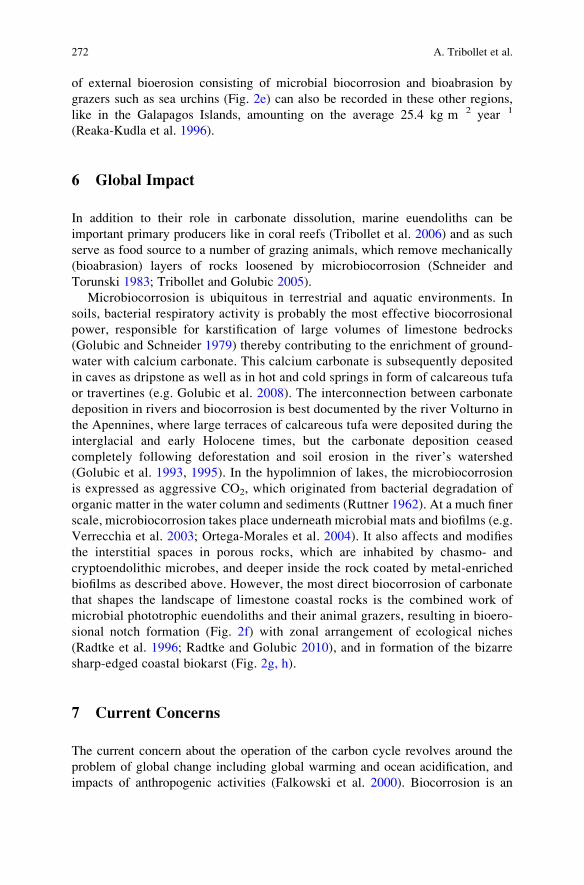
of external bioerosion consisting of microbial biocorrosion and bioabrasion by
grazers such as sea urchins (Fig. 2e) can also be recorded in these other regions,
like in the Galapagos Islands, amounting on the average 25.4 kg m�2 year�1
(Reaka-Kudla et al. 1996).
6 Global Impact
In addition to their role in carbonate dissolution, marine euendoliths can be
important primary producers like in coral reefs (Tribollet et al. 2006) and as such
serve as food source to a number of grazing animals, which remove mechanically
(bioabrasion) layers of rocks loosened by microbiocorrosion (Schneider and
Torunski 1983; Tribollet and Golubic 2005).
Microbiocorrosion is ubiquitous in terrestrial and aquatic environments. In
soils, bacterial respiratory activity is probably the most effective biocorrosional
power, responsible for karstification of large volumes of limestone bedrocks
(Golubic and Schneider 1979) thereby contributing to the enrichment of ground-
water with calcium carbonate. This calcium carbonate is subsequently deposited
in caves as dripstone as well as in hot and cold springs in form of calcareous tufa
or travertines (e.g. Golubic et al. 2008). The interconnection between carbonate
deposition in rivers and biocorrosion is best documented by the river Volturno in
the Apennines, where large terraces of calcareous tufa were deposited during the
interglacial and early Holocene times, but the carbonate deposition ceased
completely following deforestation and soil erosion in the river’s watershed
(Golubic et al. 1993, 1995). In the hypolimnion of lakes, the microbiocorrosion
is expressed as aggressive CO2, which originated from bacterial degradation of
organic matter in the water column and sediments (Ruttner 1962). At a much finer
scale, microbiocorrosion takes place underneath microbial mats and biofilms (e.g.
Verrecchia et al. 2003; Ortega-Morales et al. 2004). It also affects and modifies
the interstitial spaces in porous rocks, which are inhabited by chasmo- and
cryptoendolithic microbes, and deeper inside the rock coated by metal-enriched
biofilms as described above. However, the most direct biocorrosion of carbonate
that shapes the landscape of limestone coastal rocks is the combined work of
microbial phototrophic euendoliths and their animal grazers, resulting in bioero-
sional notch formation (Fig. 2f) with zonal arrangement of ecological niches
(Radtke et al. 1996; Radtke and Golubic 2010), and in formation of the bizarre
sharp-edged coastal biokarst (Fig. 2g, h).
7 Current Concerns
The current concern about the operation of the carbon cycle revolves around the
problem of global change including global warming and ocean acidification, and
impacts of anthropogenic activities (Falkowski et al. 2000). Biocorrosion is an
272 A. Tribollet et al.
integral part of carbon cycling, which is affected by various factors such as an
increase in nutrient concentrations (Carreiro-Silva et al. 2005, 2009) and partial
pressure of atmospheric CO2 (Tribollet et al. 2009). Recently, it has been shown
that rates of carbonate dissolution by euendolithic communities measured under
elevated pCO2 were 48% higher than under ambient pCO2 (0.46 kg CaCO3
dissolved m�2 y�1 vs. 0.31 kg m�2 y�1). Such dissolution in coral reefs, also called
biogenic dissolution (Tribollet et al. 2009), may become a major mechanism of
carbonate dissolution in a more acidic ocean.
References
Abed RMM, Golubic S, Garcia-Pichel F, Camoin GF, Sprachta S (2003) Characterization of
microbialite-forming Cyanobacteria in a tropical lagoon: Tikehau Atoll, Tuamotu, French
Polynesia. Journal of Phycology 39:862–873
Abed RMM, Palinska KA, Camoin G, Golubic S (2006) Common evolutionary origin of plank-
tonic and benthic nitrogen-fixing oscillatoriacean cyanobacteria from tropical oceans. FEMS
Microbiology Letters 260:171–177
Abed RMM, Kohls K, Schoon R, Scherf A-K, Schacht M, Palinska KA, Rullk€oter J, Golubic S
(2008) Lipid biomarkers, pigments and cyanobacterial diversity of microbial mats across
intertidal flats of the arid coast of the Arabian Gulf (Abu Dhabi, UAE). FEMS Microbiological
Ecology 65:449–462
Alexandersson ET (1975) Marks of unknown carbonate-decomposing organelles in cyanophyte
borings. Nature 254:212–238
Al-Thukair AA, Golubic S (1991) New endolithic cyanobacteria from the Arabian Gulf. I. Hyellaimmanis sp. nov. Journal of Phycology 27:766–780
Carreiro-Silva M, McClanahan TR, Kiene WE (2005) The role of inorganic nutrients and
herbivory in controlling microbioerosion of carbonate substratum. Coral Reefs 24:
214–221
Carreiro-Silva M, McClanahan TR, Kiene WE (2009) Effect of inorganic nutrients and organic
matter on microbial euendolithic community composition and microbioerosion rates. Marine
Ecology Progress Series 392:1–15
Chacon E, Berrendero E, Garcia-Pichel F (2006) Biogeological signatures of microboring cyano-
bacterial communities in marine carbonates from Cabo Rojo, Puerto Rico. Sedimentary
Geology 185:215–228
Chazottes V, Le Campion-Alsumard T, Peyrot-Clausade M (1995) Bioerosion rates on coral reefs:
interaction between macroborers, microborers and grazers (Moorea, French Polynesia).
Palaeogeography, Palaeoclimatology, Palaeoecology 113:189–198
Chazottes V, Le Campion-Alsumard T, Peyrot-Clausade M, Cuet P (2002) The effects of eutro-
phication-related alterations to coral reef communities on agents and rates of bioerosion
(Reunion Island, Indian Ocean). Coral Reefs 21:375–390
Chazottes V, Cabioch G, Golubic S, Radtke G (2009) Bathymetric zonation of modern micro-
borers in dead coral substrates from New Caledonia – Implications for paleodepth recons-
tructions in Holocene corals. Palaeogeography, Palaeoclimatology, Palaeoecology 280:
456–468
De la Torre JR, Goebel BM, Friedmann EI, Pace NR (2003) Molecular characterization of
cryptoendolithic communities from the McMurdo Dry Valleys, Antarctica. Applied Environ-
mental Microbiology 69:3858–3867
Dupraz C, Visscher PT (2005) Microbial lithification in marine stromatolites and hypersaline
mats. Trends in Microbiology 13:429–338
On Microbiocorrosion 273
Falkowski P, Scholes RJ, Boyle E, Canadell J, Canfield D, Elser J, Gruber N, Hibbard K,
H€ogberg P, Linder S (2000) The global carbon cycle: a test of our knowledge of Earth as a
system. Science 290:291–296
Garcia-Pichel F (2006) Plausible mechanisms for the boring on carbonates by microbial auto-
trophs. Sedimentary Geology 185:205–213
Garcia-Pichel F, Al-Horani F, Ludwig R, Farmer J, Wade B (2004) Balance between calcification
and bioerosion in modern stromatolites. Geobiology 2:49–57
Golubic S (1969) Distribution, taxonomy and boring patterns of marine endolithic algae. American
Zoologist 9:747–751
Golubic S, Schneider J (1979) Carbonate dissolution. In: Trudinger PA, Swaine DJ (eds) Biogeo-
chemical Cycling of Mineral-forming Elements. Elsevier, Amsterdam, pp 107–129
Golubic S, Schneider J (2003) Microbial endoliths as internal biofilms. In: Krumbein WE,
Dornieden T, Volkmann M (eds) Fossil and Recent biofilms. Kluwer Academic, Dordrecht,
pp 249–263
Golubic S, Friedmann I, Schneider J (1981) The lithobiontic ecological niche, with special
reference to microorganisms. Journal of Sedimentary Petrology 51:475–478
Golubic S, Violante C, Ferreri V, D’Argenio B (1993) ***Algal control and early diagenesis in
Quaternary travertine formation (Rocchetta a Volturno, central Apennines). In: Baratolo F,
De Castro P, Parente M (eds) Studies on Fossil Benthic Algae. Bolletino, Societa Paleontologica
Italiana, Spec 1:231–247
Golubic S, Violante C, D’Argenio B (1995) Travertine biota: a record of seasonal changes. In:
Ubertini L, Castelli F, Bras RL (eds) Climate Change and Hydrogeological Hazards in the
Mediterranean Area. Natural Research Council, Italy, pp 25–30
Golubic S, Violante C, Plenkovic A, Grgasovic T (2008) Travertines and calcareous tufa deposits:
an insight into diagenesis. Geologica Croatica 61:363–378
Hatch WI (1980) The implication of carbonic anhydrase in the physiological mechanism of
penetration of carbonate substrata by the marine burrowing sponge Cliona celata (Demospon-
giae). Biological Bulletin 159:135–147
Heitz E, Flemming H-C, Sand W (eds) (1996) Microbially Influenced Corrosion of Materials.
Springer, Berlin, pp 475
Kiene WE, Hutchings PA (1994) Bioerosion experiments at Lizard Island, Great Barrier Reef.
Coral Reefs 13:91–98
Le Campion-Alsumard T, Golubic S, Pantazidou A (1996). On the euendolithic genus SolentiaErcegovic (Cyanophyta/Cyanobacteria). Algological Studies 83:108–127
Macintyre IG, Prufert-Bebout L, Reid RP (2000) The role of endolithic cyanobacteria in the
formation of lithified laminae in Bahamian stromatolites. Sedimentology 47:915–921
Neumann AC (1966) Observations on coastal erosion in Bermuda and measurements of the boring
rate of the sponge Cliona lampa. Limnology and Oceanography 11:92–108
Ortega-Morales BO, Narvaez-Zapata JA, Schmalenberger A, Sosa-Loo A, Tebbe CC (2004)
Biofilms fouling ancient limestone Mayan monuments in Uxmal, Mexico: a cultivation-
independent analysis. Biofilms 1:79–90
Perry CT (1998) Grain susceptibility to the effects of microboring, implications for the preserva-
tion of skeletal carbonates. Sedimentology 45:39–51
Radtke G, Golubic S (2010) Microbial euendolithic assemblages and microborings in intertidal
and shallow marine habitats: insight in cyanobacterial speciation. In: Reitner J, Queric, N-V,
Arp G (eds) Advances in Stromatolite Geobiology. Lecture Notes in Earth Sciences 131,
Springer, Berlin, pp 213–241
Radtke G, Le Campion-Alsumard T, Golubic S (1996) Microbial assemblages of the bioerosional
“notch” along tropical limestone coasts. Algological Studies 83:469–482
Rasheed M, Wild C, Franke U, Huettel M. (2004) Benthic photosynthesis and oxygen consump-
tion in permeable carbonate sediments at Heron Island, Great Barrier Reef, Australia. Estua-
rine, Coastal and Shelf Science 59:139–150
274 A. Tribollet et al.
Reaka-Kudla ML, Feingold JS, Glynn W (1996) Experimental studies of rapid bioerosion of coral
reefs in the Galapagos Islands. Coral Reefs 15:101–107
Reid RP, Visscher PT, Decho AW, Stolz JF, Bebout BM, Dupraz CP, Macintyre IG, Paerl HW,
Pinckney JL, Prufert-Bebout L, Steppe TF, DesMarais DJ (2000) The role of microbes in
accretion, lamination and early lithification of modern marine stromatolites. Nature 406:989–992
Reitner J (1993) Modern cryptic microbialite/metazoan facies from Lizard Island (Great Barrier
Reef, Australia) – Formation and Concepts. Facies 29:3–40
Reitner J, Thiel V, Zankl H, Michaelis W, W€orheide G, Gautret P (2000) Organic and biochemical
pattern in cryptic microbialites. In: Riding RE, Awramik SM (eds) Microbial Sediments.
Springer, Berlin, 149–160
Risk MJ, Sammarco PW, Edinger EN (1995) Bioerosion in Acropora across the continental shelf
of the Great Barrier Reef. Coral Reefs 14:79–86
Roux W (1887) €Uber eine in Knochen lebende Gruppe von Fadenpilzen (Mycelites ossifragus).Zeitschrift f€ur wissenschaftliche Zoologie 45:227–254
Ruttner F (1962) Grundriss der Limnologie, 3rd ed. Walter de Gruyter, Berlin, pp 332
Sammarco PW, Risk MJ (1990) Large-scale patterns in internal bioerosion of Porites: crosscontinental shelf trends on the Great Barrier Reef. Marine Ecology Progress Series 59:145–156
Schneider J (1976) Biological and inorganic factors in the destruction of limestone coasts.
Contributions Sedimentology 6:1–112
Schneider J, Le Campion-Alsumeard T (1999) Construction and destruction of carbonates by
marine and freshwater cyanobacteria. European Journal Phycology 34:417–426
Schneider J, Torunski H (1983) Biokarst on limestone coasts, morphogenesis and sediment
production. Marine Ecology 4:45–63
Scoffin TP (1992) Taphonomy of coral reefs: a review. Coral Reefs 11:57–77
Scoffin TP, Brandshaw C (2000) The taphonomic significance of endoliths in dead versus live
coral skeletons. Palaios 15:248–254
Tribollet A (2008a) Dissolution of dead corals by euendolithic microorganisms across the northern
Great Barrier Reef (Australia). Microbial Ecology 55:569–580
Tribollet A (2008b) The boring microflora in modern coral reef ecosystems: a review of its roles.
In: Wisshak M, Tapanila L (eds) Current Developments in Bioerosion. Springer, Berlin, 67–94
Tribollet A, Golubic S (2005) Cross-shelf differences in the pattern and pace of bioerosion of
experimental carbonate substrates exposed for 3 years on the northern Great Barrier Reef,
Australia. Coral Reefs 24:422–434
Tribollet A, Payri C (2001) Bioerosion of coralline alga Hydrolithon onkodes by microborers in
the coral reefs of Moorea, French Polynesia. Oceanologica Acta 24:329–342
Tribollet A, Decherf G, Hutchings PA, Peyrot-Clausade M (2002) Large-scale spatial variability in
bioerosion of experimental coral substrates on the Great Barrier Reef (Australia): importance
of microborers. Coral Reefs 21:424–432
Tribollet A, Langdon C, Golubic S, Atkinson M (2006) Endolithic microflora are major primary
producers in dead carbonate substrates of Hawaiian coral reefs. Journal of Phycology 42:
292–303
Tribollet A, Godinot C, Atkinson M, Langdon C (2009) Effects of elevated pCO2 on dissolution of
coral carbonates by microbial euendoliths. Global Biogeochemical Cycles 23:1–7
Underwood CJ, Mitchell SF, Veltkamp CJ (1999) Microborings in mid-Cretaceous fish teeth.
Proceedings Yorkshire Geological Society 52:269–274
Verrecchia EP, Loisy C, Braissant O, Gorbushina AA (2003) The role of fungal biofilm and
networks in the terrestrial calcium carbonate cycle. In: KrumbeinWE,Dornieden T,VolkmannM
(eds) Fossil and Recent biofilms. Kluwer Academic, Dordtrecht, 363–369
Videla HA (1996) Manual of Biocorrosion. CRC Lewis Publishers, Boca Raton, pp 273
Videla HA, Herrera LK (2005) Microbiologically influenced corrosion: looking to the future.
International Microbiology 8:169–180
On Microbiocorrosion 275
Vogel K, Gektidis M, Golubic S, Kiene WE, Radtke G (2000) Experimental studies on microbial
bioerosion at Lee Stocking Island, Bahamas and One Tree Island, Great Barrier Reef, Aus-
tralia: implications for paleoecological reconstructions. Lethaia 33:190–204
Walker JJ, Pace, NR (2007a) Phylogenetic composition of Rocky Mountain endolithic microbial
ecosystems. Applied Environmental Microbiology 73:3497–3504
Walker JJ, Pace NR (2007b) Endolithic microbial ecosystems. Annual Reviews Microbiology
61:331–347
Walker JJ, Spear JR, Pace NR (2005) Geobiology of a microbial endolithic community in the
Yellowstone geothermal environment. Nature 434:1011–1014
Wild C, Laforsch C, Huettel M. (2006) Detection and enumeration of microbial cells within highly
porous calcareous reef sands. Marine and Freshwater Research 57:415–420
Zhang Y, Golubic S (1987) Endolithic microfossils (cyanophyta) from early Proterozoic stroma-
tolites, Hebei, China. Acta Micropaleontologica Sinica 4:1–12
Zhang X-G, Pratt BR (2008) Microborings in Early Cambrian phosphatic and phosphatized fossils.
Palaeogeography, Palaeoclimatology, Palaeoecology 267:155–195
276 A. Tribollet et al.
The Deep-Sea Chemoautotroph Microbial
World as Experienced by the Mediterranean
Metazoans Through Time
Marco Taviani
1 Preface
This paper is focused upon the Miocene-to-Recent deep-sea chemosynthetic eco-
systems of the Mediterranean basin with emphasis on their metazoan associates.
The life ingredients of this story are basically the “microbes” and the “metazoans”,
in particular molluscs, whose mutual interplay resulted in a superb variety of
situations in response to geologic and oceanographic factors directing the produc-
tion and availability of geofluids usable by microbial consortia. The astounding
complexity and diversity of modern metazoan-bearing deep-sea ecosystems inha-
biting the Mediterranean is a step of a long journey whose beginning dates back to
more than 3 billion years ago and whose major events are resumed below.
2 The March of Life
The backbone of Life on Earth is centred on microbes, unicellular prokaryotes
(mostly bacteria and archaea) whose first documentation is thought to date back to
Archean times (e.g. Knoll 2003; Allwood et al. 2006; Noffke et al. 2006; Schopf
and Bottjer 2009; Sugitani et al. 2009 with references) and since then evolved
through the Eons in exploiting successfully a vast array of habitats from extremely
hot to extremely cold, from oxygen-deficient to oxygen-plenty (e.g. Eigenbrode and
Freeman 2006; Canfield et al. 2007; Blank 2009). Early prokaryotic microorgan-
isms have been reported from rocks as old as 3.7 Ga (e.g. Schopf 1993, 1999; Pflug
2001; Van Kranendonk 2006; Payne et al. 2009 with references), although such
putative early occurrences are still contrasted (e.g., Brasier et al. 2002; Appel et al.
2003; Westall and Folk 2003; Pinti et al. 2009). It is suggested that early
M. Taviani
ISMAR-CNR, via Gobetti 101, 40129 Bologna, Italy
e-mail: [email protected]
J. Reitner et al., Advances in Stromatolite Geobiology,Lecture Notes in Earth Sciences 131, DOI 10.1007/978-3-642-10415-2_18,# Springer-Verlag Berlin Heidelberg 2011
277
prokaryotes, inhabiting an Earth marked by “prohibitive” conditions when com-
pared to present ones, were chemoautrophs satisfying their energetic needs through
chemosynthetic processes at the expenses of hydrogen sulphide, methane and other
compounds in absence of sunlight. In chemoautotrophy the more common energetic
pathway is the microbial transformation of CO2 into organic matter through the
oxidation of reduced sulphur compounds. At methane-seeps the major process is
the anaerobic oxidation of methane accomplished by microbial consortia of sul-
phate-reducing bacteria and methanotrophic archaea. The current view is that at the
beginning and for a while atmosphere and oceans alike might have been reducing,
and related microbial ecosystems necessarily anaerobic (see Poulton et al. 2004;
Xiaoying et al. 2008, for an account) with aerobic microbial ecosystems appearing
in the late Archean (Eigenbrode and Freeman 2006). Photosynthesis is thought to
emerge later, although arguments for (at least temporarily) oxygenated seas as old
as ca. 3.5 Ga ago have been presented (Hoashi et al. 2009).
Unicellular eukaryotes are assumed to appear also in the Archean, achieving a
discrete level of complexity and diversification by Paleoproterozoic times, based on
body fossil and molecular evidence (e.g., Javaux et al. 2003; Javaux 2007; Knoll
et al. 2006; Lamb et al. 2009 with references), although the fidelity of some such
fossil records is controversial (e.g. Lamb et al. 2007; Rasmussen et al. 2008). One
current theory considers the possibility that eukaryotic cells derived from the
association of free-living bacteria, a first case of bacterial symbiosis (see Esteve
and Gaju 1999). Both chemosynthetic and photosynthetic microbial organisms set
the pace for Life on Earth for a prolonged period of time. How widespread and
relevant was the chemosynthetic life in pre-Phanerozoic oceans is not known.
However, abundant body fossils of giant sulfur bacteria similar to Thiomargaritahave been reported from Neoproterozoic phosphorites (Bailey et al. 2007).
The evolution of basal metazoans (animals), multicelled heterotrophic eukaryotes,
is suggested to have taken place in the Neoproterozoic, between ca 1,000–660 Ma
(Xiao et al. 2000; Peterson and Butterfield 2005; Arthur 2006; Qun et al. 2007;
Canfield et al. 2007). Although the earliest animals could have been cnidaria-like
organisms (Dunn et al. 2008; Brocks and Butterfield 2009), first documented metazo-
ans (only based on 24 isopropyle colestane) are marine Porifera (Demospongiae)
inhabiting the Cryogenian seas (Love et al. 2009).
The hypothesized establishment of permanent oxic conditions in the deep sea
after the Neoproterozoic Gaskiers glaciation (<580 Ma) may have been the verita-
ble turning point for metazoan evolution (Canfield et al. 2007) and propitious to
the evolution of metazoans associated with pre-existing chemotrophic microbial
communities.
Metazoans have a reliable paleontological documentation since the Precambrian
(Vendian) with many phyla appearing at the dawn of Paleozoic (Cambrian), also
the time of the booming of organisms equipped with a mineralized shell, after some
Ediacaran precursors (Wood et al. 2002; Porter 2010). Depending upon the inter-
pretation of body fossils in the Ediacara fauna, first molluscs might be present as
early as the Vendian (Kimberella: Fedonkin and Waggoner 1997). At any event,
shelled molluscs are positively confirmed since the early Cambrian (Peel 1991;
278 M. Taviani
Gubanov et al. 2004; Vinther and Nielsen 2005; Skovsted et al. 2007). First
bivalves are also of this age but the major radiation of this class starts with the
Ordovician (Runnegar and Pojeta 1992; Cope and Babin 1999; Fang 2006). Bra-
chiopods and “worms” are equally well documented since Paleozoic times (e.g.,
Little 2002; Campbell 2006).
3 The Phanerozoic Record of Metazoans in Deep-Sea
Chemoautotrophic Communities
Beginning with the Paleozoic the major prokaryotic and eukaryotic actors eventually
involved in the establishment of marine chemosynthetic habitats as we know today
are on the scene, adapting, radiating, getting extinct, evolving. Thus, since more than
400 Ma, from the Silurian period onwards, microbial chemoautotrophs continue the
successful, rewarding and long-lasting symbiosis with marine metazoans (Felbeck
et al. 1981; Cavanaugh 1983; Childress et al. 1986; Stein et al. 1988; Fisher 1990;
Cavanaugh et al. 2006). As we can experience today, the net result is the immense
number of complex chemosynthetic ecosystems from hot hydrothermal vents to
hydrocarbon cold seeps that, still in perpetual evolution, inhabit the world’s marine
realm from shallow to hadal depths (Paull et al. 1984; Suess et al. 1985; Turner 1985;
Brooks et al. 1987; Aharon 1994; Fujikura et al. 1999; Van Dover 1990, 2000; Sibuet
and Olu-Le Roy 1998, 2002; Tunnicliffe et al. 2003; Bernard et al. 2004; Campbell
2006; Judd and Hovland 2007; MacAvoy et al. 2008 and many others).
Various metazoans are known to be exclusively associated with chemosynthetic
marine habitats. Those include mostly molluscs, brachiopods and “worms”, all with
a relatively good paleontological documentation since the Silurian (Stein et al.
1988; Reid 1990; Campbell and Bottjer 1995; Distel 1998; Little et al. 1998;
Callender and Powell 1999; Goedert et al. 2000; Peckmann et al. 2001, 2007; Little
2002; Van Dover et al. 2002; Barbieri et al. 2004; Peckman and Goedert 2005; Kiel
and Little 2006; Campbell 2006; Jenkins et al. 2007; Kaim et al. 2008; Kiel and
Dando 2009 etc.). The chemoautrophic bivalves are among the oldest around and
apparently some did not undergo major morphologic changes since the early
Paleozoic. A remarkable case in point are the primitive clams of family Solemyidae
(Reid and Bernard 1980; Kuznestov et al. 1990; Beninger and Le Pennec 1997;
Krueger and Cavanaugh 1997; Gustafson and Reid 2004; Stewart and Cavanaugh
2006) known from cold seep sites as old as the Paleozoic (Peckmann et al. 2001;
Taylor et al. 2008) and well established since the Mesozoic (Campbell 2006; Kiel
and Little 2007). Most information on deep-sea chemosynthetic metazoans concern
Cenozoic occurrences, however, whose communities also share basic taxonomic
traits observed in recent counterparts (Goedert and Squires 1990; Taviani 1994;
Goedert and Campbell 1995; Peckmann et al. 1999, 2002, 2004; Goedert et al.
2003; Neasbitt 2005; Majima et al. 2005; Gill et al. 2005; Kiel and Little 2006,
Campbell 2006; Kiel and Peckmann 2007 and many others).
Sulphide-based community symbiosis between sulphur-oxidizing bacteria and
bivalves is proven in seven extant families (Solemyidae, Manzanellidae, Mytilidae,
The Deep-Sea Chemoautotroph Microbial World 279
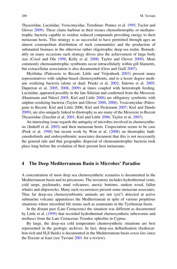
Thyasiridae, Lucinidae, Vesicomyidae, Teredinae: Pennec et al. 1995; Taylor and
Glover 2009). These clams harbour in their tissues chemolitotrophic or methano-
trophic bacteria capable to oxidize reduced compounds providing energy to their
metazoan hosts. This strategy is so successful to have permitted through ages an
almost cosmopolitan distribution of such communities and the production of
substantial biomass in the otherwise rather oligotrophic deep-sea realm. Remark-
ably on many occasions such strategy drives also the achievement of large body
size (Cosel and Olu 1998; Kelly et al. 2000; Taylor and Glover 2009). More
commonly chemoautotrophic symbionts occur intracellularly within gill filaments,
but extracellular association is also documented (Gros and Gaill 2007).
Mytilidae (Paleozoic to Recent: Little and Vrijenhoek 2003) present many
representatives with sulphur-based chemosymbionts, and to a lesser degree meth-
ane oxidizing bacteria (alone or dual: Pruski et al. 2002; Salerno et al. 2005;
Duperron et al. 2005, 2008, 2009) at times coupled with heterotroph feeding.
Lucinidae, appeared possibly in the late Silurian and confirmed from the Mesozoic
(Hautmann and N€utzel 2005; Kiel and Little 2006) are obligatory symbiotic with
sulphur-oxidizing bacteria (Taylor and Glover 2000, 2006). Vesicomyidae (Paleo-
gene to Recent: Kiel and Little 2006; Kiel and Peckmann 2007; Kiel and Dando
2009), are also uniquely linked to thiotrophy as are many of the Mesozoic to Recent
Thyasiridae (Zuschin et al. 2001; Kiel and Little 2006; Taylor et al. 2007).
An interesting issue regards the antiquity of microbes involved in chemosynthe-
sis (Imhoff et al. 2003) and their metazoan hosts. Cospeciation seems to be case
(Peek et al. 1998) but recent work by Won et al. (2008) on thiotrophic bath-
ymodiolinids and endosymbiontic associates document that this is not necessarily
the general rule and that geographic dispersal of chemoautotrophic bacteria took
place long before the evolution of their present host metazoans.
4 The Deep Mediterranean Basin is Microbes’ Paradise
A concentration of most deep sea chemosynthetic scenarios is documented in the
Mediterranean basin and its precursors. The inventory includes hydrothermal vents,
cold seeps, pockmarks, mud volcanoes, anoxic bottoms, sunken wood, fallen
whales and shipwrecks. Many such occurrences present some metazoan associates.
Thus far deep-sea chemosymbiontic animals are not (yet?) detected at active
submarine volcanic apparatuses the Mediterranean in spite of various propitious
situations where microbial life teems such as seamounts in the Tyrrhenian basin.
In the distant past (Late Cretaceous) the situation was different as documented
by Little et al. (1999) that recorded hydrothermal chemosynthetic tubeworms and
molluscs from the Late Cretaceous Troodos ophiolite in Cyprus.
By large, the deep-sea cold temperature chemosynthetic situations are best
represented in the geologic archives. In fact, deep-sea defluidisation (hydrocar-
bon-rich and H2S fluids) is documented in the Mediterranean basin sensu lato sincethe Eocene at least (see Taviani 2001 for a review).
280 M. Taviani
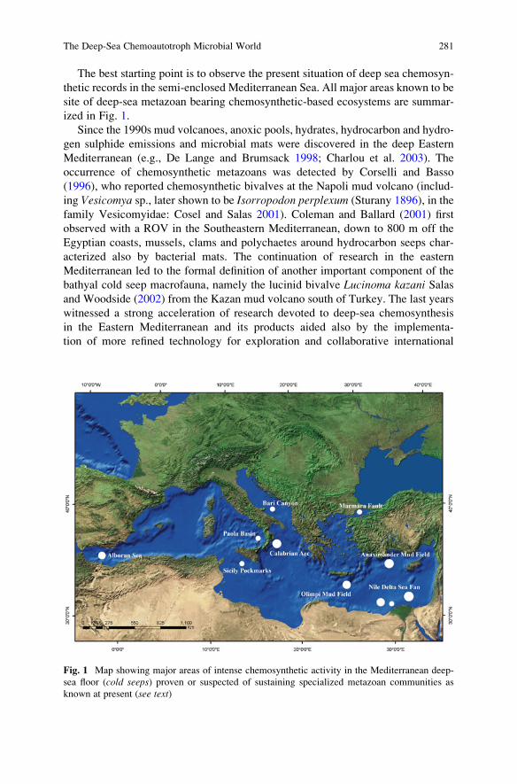
The best starting point is to observe the present situation of deep sea chemosyn-
thetic records in the semi-enclosed Mediterranean Sea. All major areas known to be
site of deep-sea metazoan bearing chemosynthetic-based ecosystems are summar-
ized in Fig. 1.
Since the 1990s mud volcanoes, anoxic pools, hydrates, hydrocarbon and hydro-
gen sulphide emissions and microbial mats were discovered in the deep Eastern
Mediterranean (e.g., De Lange and Brumsack 1998; Charlou et al. 2003). The
occurrence of chemosynthetic metazoans was detected by Corselli and Basso
(1996), who reported chemosynthetic bivalves at the Napoli mud volcano (includ-
ing Vesicomya sp., later shown to be Isorropodon perplexum (Sturany 1896), in the
family Vesicomyidae: Cosel and Salas 2001). Coleman and Ballard (2001) first
observed with a ROV in the Southeastern Mediterranean, down to 800 m off the
Egyptian coasts, mussels, clams and polychaetes around hydrocarbon seeps char-
acterized also by bacterial mats. The continuation of research in the eastern
Mediterranean led to the formal definition of another important component of the
bathyal cold seep macrofauna, namely the lucinid bivalve Lucinoma kazani Salasand Woodside (2002) from the Kazan mud volcano south of Turkey. The last years
witnessed a strong acceleration of research devoted to deep-sea chemosynthesis
in the Eastern Mediterranean and its products aided also by the implementa-
tion of more refined technology for exploration and collaborative international
Fig. 1 Map showing major areas of intense chemosynthetic activity in the Mediterranean deep-
sea floor (cold seeps) proven or suspected of sustaining specialized metazoan communities as
known at present (see text)
The Deep-Sea Chemoautotroph Microbial World 281
programmes (Werne et al. 2004; Foucher et al. 2009). Regarding metazoans the
current picture is the following.
Olu-Le Roy et al. (2004) record various chemosynthetic bivalves and poly-
chaetes at depths of ca 2,000 m from the Olimpi Mud Field (Napoli, Milano,
Maidstone, Moscow mud volcanoes), south of Crete along the Mediterranean
Ridge, and from the Anaximander Mud Field (Kula, Kazan, Amsterdam and
Faulted Ridge), south of Turkey between 1,700 and 2,000 m. Their list includes
siboglinid polychaetes (Lamellibrachia, Siboglinum), Isorropodon perplexum,Myrtea amorpha (Sturany 1896), Idas modiolaeformis (Sturany 1896), Lucinomakazani Cosel and Salas 2001, Thyasira striata (Sturany 1896) (only at AMV), the
gastropods Lurifax vitreus Waren and Bouchet 2001, Clelandella n.sp., Lepetellasp., the decapods Chaceon mediterraneusManning and Holthuis 1989,Munidopsissp., and few others. Lucinoma kazani shells, other bivalve and worm fragments are
recorded by Lykousis et al. (2004) in a sediment core at 1,783 m from the Athina
mudvolcano in the Anaximander mountains. Further information on the megafauna
from this area, including sponges, is reported by Vanreusel et al. (2009).
The on-going exploration of the astounding chemosynthetic grounds of the Nile
Deep Sea Fan is also revealing a very similar metazoan fauna: Dupre et al. (2007)
report Isorropodon perplexum, Myrtea amorpha, Idas modiolaeformis, Lucinomakazani, tubicolous polychaetes and Chaceon crabs from Central Nile Deep Sea Fan.
Quite interestingly some of such deep-sea chemosynthetic bivalves were known to
science since more than a century based upon collections made in this region by the
ship Pola expedition, described by Sturany in 1896, and re-discovered with the
beginning of modern deep-sea exploration of the Nile Delta Fan. There is an
interesting parallelism here with the rather similar first discovery of deep-sea corals
at the Santa Maria di Leuca hotspot, off the Apulian Ionian equally to be credited to
the Pola expedition (Taviani et al. 2005).
Multidisciplinary exploration of the Marmara Fault in the Sea of Marmara
(Turkey) stepped over gas and water seep discharge, including hydrocarbons,
between ca 1,000 and 1,200 m (Armijo et al. 2005). To such setting is associated
a chemosynthetic fauna (siboglinids and bivalves, including Lucinoma kazani,Myrtea amorpha, and Isorropodon perplexum: Zitter et al. 2008; Ritt et al. 2010)whose detailed study is in progress.
Promising sites with active fluid emissions are also scattered in the central and
western Mediterranean (e.g., Holland et al. 2003) but no biological information on
associated macrofauna is available at present.
The recent most discovery of deep-water chemosynthetic communities, includ-
ing tubeworms and lucinid bivalves, is the Alboran basin in the western Mediterra-
nean (Ivanov et al. 2009; Blinova, Ivanov and coworkers, in progress) This finding
is of utmost importance since practically connecting the Mediterranean deep-sea
chemosynthetic cold-seep world, before documented for its eastern reaches only, to
the Gulf of Cadiz and therefore the open ocean, punctuated by many such occur-
rences (e.g., Cosel 2006; Cosel and Olu 2009; Cosel and Salas 2001; Olu-Le Roy
et al. 2007; Genio et al. 2008; Rodrigues et al. 2008, 2010; Foucher et al. 2009;
Vanreusel et al. 2009; Sibuet and Vangriesheim 2009).
282 M. Taviani
The overview would not be complete without mentioning the finding of the
chemosynthetic Lamellibrachia (Polychaeta: Siboglinidae) from the deep-seated
shipwreck of the SS Persia at 2,800 m depth south of Crete (Hughes and Crawford
2008). To this, a number of citations variously reporting molluscs of chemosyn-
thetic significance (Idas, Lucinoma, Lurifax), some of still unknown cold-seep sites
in the Tyrrhenian Sea, can be found in grey literature.
The Black Sea, easternmost marine basin, also houses an important deep-water
chemosynthetic world of its own. Due to its anoxic conditions beneath ca. 200 m,
the microbes dominate the habitats resulting in spectacular prokaryotic domes
(Michaelis et al. 2002; Reitner et al. 2005). The occurrence of living metazoans
(Nematoda, Polychaeta, Harpacticoida, Acarina, Bivalvia and Gastropoda) associated
with seeps between 220 and 250 m (Sergeeva and Gulin 2009 with references) seems
not yet convincingly substantiated (Yanko-Hombach et al. 2009) and, at least for
some groups, unlikely because of the absence of oxygen, crucial to respiration.
In synthesis, the present-day Mediterranean metazoan macrofauna associated with
deep-water cold seeps taxonomically comprehends most of the “classic” groups:
Mytilidae, Lucinidae, Thyasiridae, Vesicomyidae, tube-worms. Others are absent
(or not yet found), the most remarkable case being members of the Cretaceous to
Recent gastropod family Provannidae (Kaim et al. 2008) and large Solemyidae
(Acharax), both with modern representatives in the eastern Atlantic (Ravara et al.
2007; Waren and Bouchet 2009). Compared to ocean standards, the diversity of
Mediterranean chemosynthetic metazoans is quite low and taxa are often lilliputians
in their respective families, with some important ingredients missing, e.g., large
bathymodiolinid mussels and Calyptogena. Maybe the most significant trait of the
present Mediterranean chemosynthetic deep-sea metazoans concerns their nutritional
strategy that is thiotrophy, at times coupled with functional heterotroph feeding. Thus
far metazoans adapted to methanotrophic-based or dual metabolism as such observed
in the adjacent Atlantic ocean (Duperron et al. 2005, 2008, 2009) have not been
identified in the deep-sea chemosynthetic communities of the present Mediterranean
in spite of the pervasiveness of methane degassing here (Charlou et al. 2003; Foucher
et al. 2009). However, in earlier times this should not have been the case.
5 Deep Sea Chemosynthetic Environments in the
Mediterranean Neogene
The fossil legacy of deep-sea chemosynthetic habitats tells us a different story by
documenting situations that are remarkably different from the present Mediterra-
nean Sea.
Tertiary rocks and sediments in the Apennine chain (Fig. 2) and Sicily show
remarkable and impressive geobiological imprinting of various cold seep emissions
onto the deep sea bottom, the most known being the “Calcari a Lucina” (Clari et al.
1988; Aharon 1994; Taviani 1994, 2001; Berti et al. 1994; Conti and Fontana 2005;
The Deep-Sea Chemoautotroph Microbial World 283
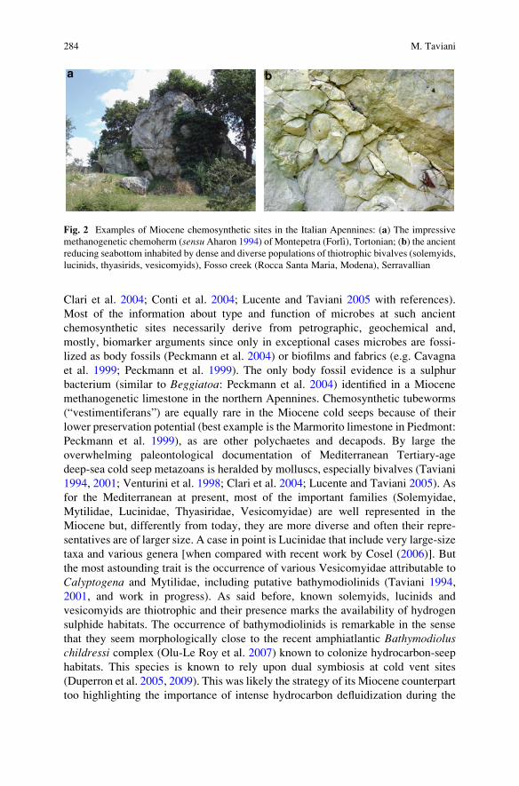
Clari et al. 2004; Conti et al. 2004; Lucente and Taviani 2005 with references).
Most of the information about type and function of microbes at such ancient
chemosynthetic sites necessarily derive from petrographic, geochemical and,
mostly, biomarker arguments since only in exceptional cases microbes are fossi-
lized as body fossils (Peckmann et al. 2004) or biofilms and fabrics (e.g. Cavagna
et al. 1999; Peckmann et al. 1999). The only body fossil evidence is a sulphur
bacterium (similar to Beggiatoa: Peckmann et al. 2004) identified in a Miocene
methanogenetic limestone in the northern Apennines. Chemosynthetic tubeworms
(“vestimentiferans”) are equally rare in the Miocene cold seeps because of their
lower preservation potential (best example is the Marmorito limestone in Piedmont:
Peckmann et al. 1999), as are other polychaetes and decapods. By large the
overwhelming paleontological documentation of Mediterranean Tertiary-age
deep-sea cold seep metazoans is heralded by molluscs, especially bivalves (Taviani
1994, 2001; Venturini et al. 1998; Clari et al. 2004; Lucente and Taviani 2005). As
for the Mediterranean at present, most of the important families (Solemyidae,
Mytilidae, Lucinidae, Thyasiridae, Vesicomyidae) are well represented in the
Miocene but, differently from today, they are more diverse and often their repre-
sentatives are of larger size. A case in point is Lucinidae that include very large-size
taxa and various genera [when compared with recent work by Cosel (2006)]. But
the most astounding trait is the occurrence of various Vesicomyidae attributable to
Calyptogena and Mytilidae, including putative bathymodiolinids (Taviani 1994,
2001, and work in progress). As said before, known solemyids, lucinids and
vesicomyids are thiotrophic and their presence marks the availability of hydrogen
sulphide habitats. The occurrence of bathymodiolinids is remarkable in the sense
that they seem morphologically close to the recent amphiatlantic Bathymodioluschildressi complex (Olu-Le Roy et al. 2007) known to colonize hydrocarbon-seep
habitats. This species is known to rely upon dual symbiosis at cold vent sites
(Duperron et al. 2005, 2009). This was likely the strategy of its Miocene counterpart
too highlighting the importance of intense hydrocarbon defluidization during the
Fig. 2 Examples of Miocene chemosynthetic sites in the Italian Apennines: (a) The impressive
methanogenetic chemoherm (sensuAharon 1994) of Montepetra (Forlı), Tortonian; (b) the ancient
reducing seabottom inhabited by dense and diverse populations of thiotrophic bivalves (solemyids,
lucinids, thyasirids, vesicomyids), Fosso creek (Rocca Santa Maria, Modena), Serravallian
284 M. Taviani
Late Miocene, capable to sustain biota also through the anaerobic oxidation of
methane. The stable carbon geochemical signatures, biomarkers and the concurring
presence of the cold-seep gastropod Thalassonerita [considered a younger syno-
nym of recent Bathynerita by Taviani (2001)] at Miocene-age bathymodiolinid
sites lends further support to this hypothesis. Recent studies suggest in fact that
Bathymerita occurring at hydrocarbon seeps in the Gulf of Mexico, while not
methanotrophic itself, is uniquely linked to chemosynthetic hyperhaline microha-
bitats in methanotrophic Bathymodiolus childressi beds, grazing bacteria and
decomposing mussel’s periostracum (Dattagupta et al. 2007; Van Gaest et al.
2007). Therefore, a rather similar situation might be deducted from the Apennine
Late Miocene occurrences showing co-occurring Thalassonerita and bathymodio-
linids encased in methanogenetic limestones (Taviani 1994).
It has been argued that the final Miocene Messinian salinity crisis might have
sealed the fate of the Mediterranean deep-sea stenoecious biota through their
complete annihilation, including the chemosynthetic metazoan communities men-
tioned above (Taviani 1994, 2001, 2004).
The Pliocene renovation can only be evaluated at a few fossil sites (Taviani et al.
1997; Taviani 2001; Monegatti et al. 2001; Dominici et al. 2009) and included
solemyids (Acharax: Taviani et al. 2008), mytilids (but not bathymodiolinids),
lucinids (but not giant ones), vesicomyids (but not Calyptogena); Thalassoneritais equally absent.
With end of the Miocene the Mediteranean deep sea chemosynthetic metazoan
communities largely lost their previous ocean affinity, assuming the basic aspect of
the habitats we know at present, as best documented by the taxonomy of its mollusc
fauna (Fig. 3).
6 Postface
This article has been assembled by a metazoan heterotroph whose nutritional
strategy is largely photosynthetic-based. Although not yet definitely assessed, the
geologic antiquity of this taxon is rather modest, perhaps a bare 200 kyrs or so.
Apparently this is the only metazoan known to run conscious scientific research
and also to be able to write and evolve abstract theories. These characteristics
may conspire in biasing beyond tolerable levels its perception of the natural
world with the distinct possibility of overemphasising its own ecological niche to
the extent of considering its extreme ecological tolerance as the “normal” one.
Deviations from its standard contour conditions are aberrant and therefore merit-
ing to be labelled as abnormal, unusual or even extreme. Although somehow
frustrating from the standpoint of a Phanerozoic metazoan breathing oxygen and
enjoying a nutritional strategy based upon photosynthesis, it could be fair to
recognize that latecomers like us are the real extremophiles of our planet. From a
metazoan-centric point of view we favour the dignified idea that we metazoans
choose the microbes for the symbiosis. From a microbial-centric point of view it
The Deep-Sea Chemoautotroph Microbial World 285
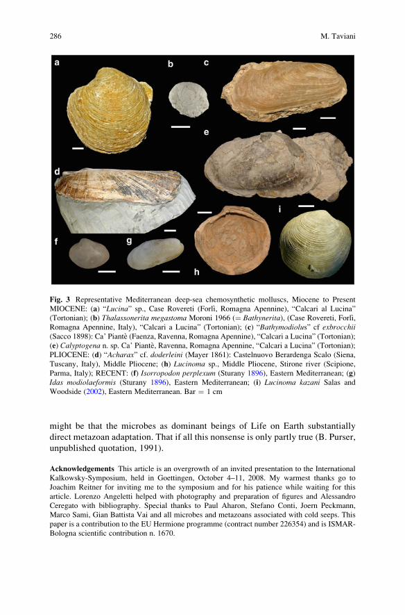
might be that the microbes as dominant beings of Life on Earth substantially
direct metazoan adaptation. That if all this nonsense is only partly true (B. Purser,
unpublished quotation, 1991).
Acknowledgements This article is an overgrowth of an invited presentation to the International
Kalkowsky-Symposium, held in Goettingen, October 4–11, 2008. My warmest thanks go to
Joachim Reitner for inviting me to the symposium and for his patience while waiting for this
article. Lorenzo Angeletti helped with photography and preparation of figures and Alessandro
Ceregato with bibliography. Special thanks to Paul Aharon, Stefano Conti, Joern Peckmann,
Marco Sami, Gian Battista Vai and all microbes and metazoans associated with cold seeps. This
paper is a contribution to the EU Hermione programme (contract number 226354) and is ISMAR-
Bologna scientific contribution n. 1670.
Fig. 3 Representative Mediterranean deep-sea chemosynthetic molluscs, Miocene to Present
MIOCENE: (a) “Lucina” sp., Case Rovereti (Forlı, Romagna Apennine), “Calcari al Lucina”
(Tortonian); (b) Thalassonerita megastoma Moroni 1966 (¼ Bathynerita), (Case Rovereti, Forlı,Romagna Apennine, Italy), “Calcari a Lucina” (Tortonian); (c) “Bathymodiolus” cf exbrocchii(Sacco 1898): Ca’ Piante (Faenza, Ravenna, Romagna Apennine), “Calcari a Lucina” (Tortonian);
(e) Calyptogena n. sp. Ca’ Piante, Ravenna, Romagna Apennine, “Calcari a Lucina” (Tortonian);
PLIOCENE: (d) “Acharax” cf. doderleini (Mayer 1861): Castelnuovo Berardenga Scalo (Siena,
Tuscany, Italy), Middle Pliocene; (h) Lucinoma sp., Middle Pliocene, Stirone river (Scipione,
Parma, Italy); RECENT: (f) Isorropodon perplexum (Sturany 1896), Eastern Mediterranean; (g)
Idas modiolaeformis (Sturany 1896), Eastern Mediterranean; (i) Lucinoma kazani Salas and
Woodside (2002), Eastern Mediterranean. Bar ¼ 1 cm
286 M. Taviani
References
Aharon P (1994) Geology and biology of modern and ancient submarine hydrocarbon seeps and
vents. Geo-Marine Letters 14:69–230
Appel PWU, Moorbath S, Myers JS (2003) Isuasphaera isua (Pflug) revisited. Precambrian
Research 126:309–312
Allwood AC, Walter MR, Kamber BS, Marshall CP, Burch IW (2006) Stromatolite reef from the
Early Archean era of Australia. Nature 441:714–715
Armijo R, Pondard N, Meyer B, Ucarkus G, Mercier de Lepinay B, Malavieille J, Dominguez S,
Gustcher M-A, Schmidt S, Beck C, Cagatay N, Cakir Z, Imren C, Eris K, Natalin B, Ozalaybey S,
Tolun L, Lefevre I, Seeber L, Gasperini L, Rangin C, Emre O, Sarikavak K (2005) Submarine fault
scarps in the Sea of Marmara pull-apart (North Anatolian Fault): implications for seismic hazard in
Istanbul. Geochemistry, Geophysics, Geosystems 6: Q06009, doi:10.1029/2004GC000896
Arthur W (2006) Creatures of Accident: the Rise of the Animal Kingdom. Hall & Wang,
New York: 243 pp
Bailey JV, Joye SB, Kalanetra KM, Flood BE, Corsetti FA (2007) Evidence of giant sulphur
bacteria in Neoproterozoic phosporites. Nature 445:198–201
Barbieri R, Ori GG, Cavalazzi B (2004) A Silurian cold-seep ecosystem from the Middle Atlas,
Morocco. Palaios 19:527–542
Beninger PG, Le Pennec M (1997) Reproductive characteristics of a primitive bivalve from a
deep-sea reducing environment: giant gametes and their significance in Acharax alinae(Cryptodonta: Solemyidae). Marine Ecology Progress Series 157:195–206
Bernard B, Brooks J, de Farago M (2004) Asphalt volcanism and chemosynthetic life in the
Campeche Knolls, Gulf of Mexico. Science 304:999–1002
Berti M, Cuzzani MG, Landuzzi A, Taviani M, Aharon P, Vai GB (1994) Hydrocarbon-derived
imprints in olistostromes of the Early Serravallian Marnoso-arenacea Formation, Romagna
Apennines (Northern Italy). Geo-Marine Letters 14:192–200
Blank C (2009) Not so old Archaea-the antiquity of biogeochemical processes in the archaeal
domain of life. Geobiology 7:495–514
Brasier MD, Green OR, Jephcoat AP, Kleppe AK, Van Kranendonk MJ, Lindsay JF, Steele A,
Grassineau NV (2002) Questioning the evidence for Earth’s oldest fossils. Nature 416:76–81
Brocks JJ, Butterfield NJ (2009) Early animals out in the cold. Nature 457:672–673
Brooks JM, Kennicutt II MC, Fisher CR, Macko SA, Cole K, Childress JJ, Bidigare RR, Vetter RD
(1987) Deep-sea hydrocarbon seep communities: evidence for energy and nutritional carbon
sources. Science 238:1138–1142
Callender RW, Powell EN (1999)Why did ancient chemosynthetic seep and vent assemblages occur
in shallower water than they do today? International Journal of Earth Sciences 88:377–391
Campbell KA (2006) Hydrocarbon seep and hydrothermal vent paleoenvironments and paleontol-
ogy: Past developments and future research directions. Palaeogeography, Palaeoclimatology,
Palaeoecology 232:362–407
Campbell KA, Bottjer DJ (1995) Brachiopods and chemosymbiontic bivalves in Phanerozoic
hydrothermal vent and cold seep environments. Geology 23:321–324
Canfield DE, Poulton SW, Narbonne GM (2007) Late Neoproterozoic deep-ocean oxygenation
and the rise of animal life. Science 315:92–95
Cavagna S, Clari P, Martire L (1999) The role of bacteria in the formation of cold seep carbonates:
geological evidence from Monferrato (Tertiary, NW Italy). Sedimentary Geology 126:
253–270
Cavanaugh CM (1983) Symbiotic chemoautotrophic bacteria in marine invertebrates from sul-
phide-rich habitats. Nature 302:58–61
Cavanaugh CM, McKiness ZP, Newton ILG (2006) Marine Chemosynthetic Symbioses. In:
Dworkin M, Falkow S, Rosenberg E, Schleifer KH, Stackebrandt E (eds) The Prokaryotes.
Third Edition. A Handbook on the biology of Bacteria: Symbiotic associations, Biotechnology,
Applied Microbiology, Springer New York: 475–507
The Deep-Sea Chemoautotroph Microbial World 287
Charlou JL, Donval JP, Zitter T, Roy N, Foucher JP, Woodside J (2003) Evidence of methane
venting and geochemistry of brines in Mud Volcanoes of the Eastern Mediterranean Sea. Deep
Sea Research Part I: Oceanographic Research Papers 50:941–958
Childress JJ, Fisher CR, Brooks JM, Kennicutt II MC, Bidigare R, Anderson AE (1986)
A methanotrophic marine molluscan (Bivalvia, Mytilidae) symbiosis: mussels fuelled by
gas. Science 233:1306–1308
Clari P, Conti S, Fontana D, Taviani M (2004) Fluid expulsion and authigenic products in the
Miocene foredeep and satellite basins of the Northern Apennines, Italy. Field Guide Book-
Post-Congress P07, 32nd IGC, August 20–28, Florence-Italy 2:B31–B60
Clari P, Gagliardi C, Governa ME, Ricci B, Zuppi GM (1988) I Calcari di Marmorito: una
testimonianza di processi diagenetici in presenza di metano. Bollettino del Museo Regionale
di Scienze Naturali, Torino 6:197–216
Coleman DF, Ballard RD (2001) A highly concentrated region of cold hydrocarbon seeps in the
southeastern Mediterranean Sea. Geo-Marine Letters 21:162–167
Conti S, Fontana D (2005) Anatomy of seep-carbonates: ancient examples from theMiocene of the
northern Apennines (Italy). Palaeogeography, Palaeoclimatology, Palaeoecology 227:156–175
Conti S, Fontana D, Gubertini A, Sighinolfi G, Tateo F, Fioroni C, Fregni P (2004). A multidisci-
plinary study of middle Miocene seep-carbonates from the northern Apennine foredeep (Italy).
Sedimentary Geology 169:1–19
Cope JCW, Babin C (1999) Diversification of bivalves in the Ordovician. Geobios 32:175–185
Corselli C, Basso D (1996) First evidence of benthic communities based on chemosynthesis on the
Napoli mud volcano (Eastern Mediterranean). Marine Geology 132:227–239
Cosel R (2006) Taxonomy of tropical West African bivalves. VI. Remarks on Lucinidae (Mollusca,
Bivalvia), with description of six new genera and eight new species. Zoosystema 28:805–851
Cosel R, Olu K (1998) Gigantism in Mytilidae. A new Bathymodiolus from cold seep areas on the
Barbados accretionary prism. Comptes Rendus de l’Academie des Sciences – Series III –
Sciences de la Vie 321:655–663
Cosel R, Olu K (2009) Large Vesicomyidae (Mollusca: Bivalvia) from cold seeps in the Gulf of
Guinea off the coasts of Gabon, Congo and northern Angola. Deep Sea Research Part II:
Topical Studies in Oceanography 56:2350–2379
Cosel R, Salas C (2001) Vesicomyidae (Mollusca: Bivalvia) of the generaVesicomya,Waisiuconcha,Isorropodon and Callogonia in the eastern Atlantic and the Mediterranean. Sarsia 86:333–366
Dattagupta S, Martin J, Liao S, Carney RS, Fisher CR (2007) Deep-sea hydrocarbon seep
gastropod Bathynerita naticoidea responds to cues from the habitat-providing mussel Bath-ymodiolus childressi. Marine Ecology 28:193–198
De Lange G, Brumsack HJ (1998) The occurrence of gas hydrates in eastern Mediterranean mud
dome structures as indicated by pore-water composition. In: Henriet JP, Mienert J (eds) Gas
Hydrates: Relevance to World Maring Stability and Climate Change. Geological Society
London, Special Publication 137:167–175
Distel DL (1998): Evolution of chemoautotrophic endosymbiosis in bivalves. Bioscience 48:
277–286
Dominici S, Cioppi E, Danise S, Betocchi U, Gallai G, Tangocci F, Valleri G, Monechi S (2009)
Mediterranean fossil whale falls and the adaptation of molluscs to extreme habitats. Geology
37:815–818
Dunn CW, Hejnoll A, Matus DQ, Pang K, Browne WE, Smith SA, Seaver E, Rouse GW, Obst M,
Edgecombe GD, Sørensen MV, Haddock SHD, Schmidt-Rhaesa A, Okusu A, Møbjerg
Kristensen R, Wheeler WC, Martindale MQ, Giribet G (2008) Broad phylogenomic sampling
improves resolution of the animal tree of life. Nature 452:745–749
Duperron D, Halary S, Lorion J, Sibuet M, Gaill F (2008) Unexpected co-occurrence of six
bacterial symbionts in the gills of the cold seep mussel Idas sp. (Bivalvia: Mytilidae).
Environmental Microbiology 10:433–445
Duperron S, Lorion J, Samadi S, Gros O, Gaill F (2009) Symbioses between deep-sea mussels
(Mytilidae: Bathymodiolinae) and chemosynthetic bacteria: diversity, function and evolution.
Comptes Rendus Biologies 332:298–310
288 M. Taviani
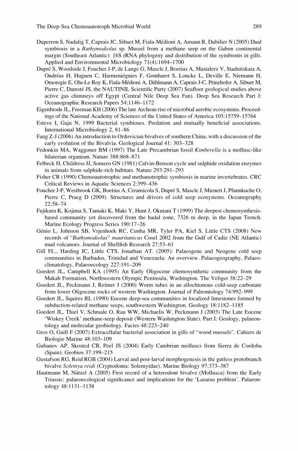
Duperron S, Nadalig T, Caprais JC, Sibuet M, Fiala-Medioni A, Amann R, Dubilier N (2005) Dual
symbiosis in a Bathymodiolus sp. Mussel from a methane seep on the Gabon continental
margin (Southeast Atlantic): 16S rRNA phylogeny and distribution of the symbionts in gills.
Applied and Environmental Microbiology 71(4):1694–1700
Dupre S, Woodside J, Foucher J-P, de Lange G, Mascle J, Boetius A, Mastalerz V, Stadnitskaia A,
Ondreas H, Huguen C, Harmeniegnies F, Gontharet S, Loncke L, Deville E, Niemann H,
Omoregie E, Olu-Le Roy K, Fiala-Medioni A, D€ahlmann A, Caprais J-C, Prinzhofer A, Sibuet M,
Pierre C, Damste JS, the NAUTINIL Scientific Party (2007) Seafloor geological studies above
active gas chimneys off Egypt (Central Nile Deep Sea Fan). Deep Sea Research Part I:
Oceanographic Research Papers 54:1146–1172
Eigenbrode JL, Freeman KH (2006) The late Archean rise of microbial aerobic ecosystems. Proceed-
ings of the National Academy of Sciences of the United States of America 103:15759–15764
Esteve I, Gaju N, 1999 Bacterial symbioses. Predation and mutually beneficial associations.
International Microbiology 2, 81–86
Fang Z-J (2006) An introduction to Ordovician bivalves of southern China, with a discussion of the
early evolution of the Bivalvia. Geological Journal 41: 303–328
Fedonkin MA, Waggoner BM (1997) The Late Precambrian fossil Kimberella is a mollusc-like
bilaterian organism. Nature 388:868–871
Felbeck H, Childress JJ, Somero GN (1981) Calvin-Benson cycle and sulphide oxidation enzymes
in animals from sulphide-rich habitats. Nature 293:291–293
Fisher CR (1990) Chemoautotrophic and methanotrophic symbiosis in marine invertebrates. CRC
Critical Reviews in Aquatic Sciences 2:399–436
Foucher J-P, Westbrook GK, Boetius A, Ceramicola S, Dupre S, Mascle J, Mienert J, Pfannkuche O,
Pierre C, Praeg D (2009). Structures and drivers of cold seep ecosystems. Oceanography
22:58–74
Fujikura K, Kojima S, Tamaki K, Maki Y, Hunt J, Okutani T (1999) The deepest chemosynthesis-
based community yet discovered from the hadal zone, 7326 m deep, in the Japan Trench.
Marine Ecology Progress Series 190:17–26
Genio L, Johnson SB, Vrjenhoek RC, Cunha MR, Tyler PA, Kiel S, Little CTS (2008) New
records of “Bathymodiolus” mauritanicus Cosel 2002 from the Gulf of Cadiz (NE Atlantic)
mud volcanoes. Journal of Shellfish Research 27:53–61
Gill FL., Harding IC, Little CTS, Jonathan AT. (2005): Palaeogene and Neogene cold seep
communities in Barbados, Trinidad and Venezuela: An overview. Palaeogeography, Palaeo-
climatology, Palaeoecology 227:191–209
Goedert JL, Campbell KA (1995) An Early Oligocene chemosynthetic community from the
Makah Formation, Northwestern Olympic Peninsula, Washington. The Veliger 38:22–29
Goedert JL, Peckmann J, Reitner J (2000) Worm tubes in an allochtonous cold-seep carbonate
from lower Oligocene rocks of western Washington. Journal of Paleontology 74:992–999
Goedert JL, Squires RL (1990) Eocene deep-sea communities in localized limestones formed by
subduction-related methane seeps, southwestern Washington. Geology 18:1182–1185
Goedert JL, Thiel V, Schmale O, Rau WW, Michaelis W, Peckmann J (2003) The Late Eocene
‘Wiskey Creek’ methane-seep deposit (Western Washington State). Part I: Geology, palaeon-
tology and molecular geobiology. Facies 48:223–240
Gros O, Gaill F (2007) Extracellular bacterial association in gills of “wood mussels”. Cahiers de
Biologie Marine 48:103–109
Gubanov AP, Skosted CB, Peel JS (2004) Early Cambrian molluscs from Sierra de Cordoba
(Spain). Geobios 37:199–215
Gustafson RG, Reid RGB (2004) Larval and post-larval morphogenesis in the gutless protobranch
bivalve Solemya reidi (Cryptodonta: Solemyidae). Marine Biology 97:373–387
Hautmann M, N€utzel A (2005) First record of a heterodont bivalve (Mollusca) from the Early
Triassic: palaeoecological significance and implications for the ‘Lazarus problem’. Palaeon-
tology 48:1131–1138
The Deep-Sea Chemoautotroph Microbial World 289
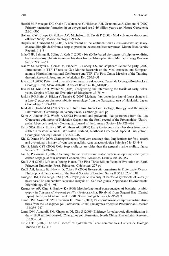
Hoashi M, Bevacqua DC, Otake T, Watanabe Y, Hickman AH, Utsunomiya S, Ohmoto H (2009)
Primary haematite formation in an oxygenated sea 3.46 billion years ago. Nature Geoscience
2:301–306
Holland CW, Etiope G, Milkov AV, Michelozzi E, Favali P (2003) Mud volcanoes discovered
offshore Sicily. Marine Geology 199:1–6
Hughes DJ, Crawford M (2006) A new record of the vestimentiferan Lamellibrachia sp. (Poly-
chaeta: Siboglinidae9 from a deep shipwrek in the eastern Mediterranean. Marine Biodiversity
Records 1:1–3
Imhoff JF, Sahling H, S€uling J, Kath T (2003) 16s rDNA-based phylogeny of sulphur-oxidising
bacterial endosymbionts in marine bivalves from cold-seep habitats. Marine Ecology Progress
Series 249:39–51
Ivanov M, Kenyon N, Comas M, Pinheiro L, Laberg J-S, and shipboard Scientific party (2009)
Introduction to TTR-17 results. Geo-Marine Research on the Mediterranean and European-
atlantic Margins International Conference and TTR-17th Post-Cruise Meeting of the Training-
through-Research Programme, Workshop Rep 220:1–51
Javaux EJ (2007) Patterns of diversification in early eukaryotes. Carnet de Geologie/Notebooks in
Geology, Brest, Mem 2007/01, Abstract 06 (CG2007_M01/06)
Javaux EJ, Knoll AH, Walter M (2003) Recognizing and interpreting the fossils of early Eukar-
yotes. Origins of Life and Evolution of Biospheres 33:75–94
Jenkins RG, Kaim A, Hikida Y, Tanabe K (2007) Methane-flux-dependent lateral fauna changes in
a Late Cretaceous chemosymbiotic assemblage from the Nakagawa area of Hokkaido, Japan.
Geobiology 5:127–139
Judd AG, Hovland M (2007) Seabed Fluid Flow. Impact on Geology, Biology, and the marine
Environment. Cambridge University Press, Cambridge: 470 pp
Kaim A, Jenkins RG, Waren A (2008) Provannid and provannid-like gastropods from the Late
Cretaceous cold seeps of Hokkaido (Japan) and the fossil record of the Provannidae (Gastro-
poda: Abyssochrysoidea). Zoological Journal of the Linnean Society 154:421–436
Kelly SRA, Blanc E, Price SP, Whitham AG (2000) Early Cretaceous giant bivalves from seep-
related limestone mounds, Wollaston Forland, Northeast Greenland. Special Publications,
Geological Society London 177:227–246
Kiel S, Dando PR (2009) Chaetopterid tubes from vent and seep sites: Implications for fossil record
and evolutionary history of vent seep annelids. Acta palaeontologica Polonica 54:443–448
Kiel S, Little CST (2006) Cold-Seep molluscs are older than the general marine mollusc fauna.
Science 313:1429–1431
Kiel S, Peckmann J (2007) Chemosymbiotic bivalves and stable carbon isotopes indicate hydro-
carbon seepage at four unusual Cenozoic fossil localities. Lethaia 40:345–357
Knoll AH (2003) Life on a Young Planet. The First Three Billion Years of Evolution on Earth.
Princeton University Press, Princeton, Chichester: 277 pp
Knoll AH, Javaux EJ, Hewitt D, Cohen P (2006) Eukaryotic organisms in Proterozoic Oceans.
Philosophical Transactions of the Royal Society of London, Series B 361:1023–1038
Krueger DM, Cavanaugh CM (1997) Phylogenetic diversity of bacterial symbionts of Solemyahosts based on comparative sequence analysis of 16s rRNA genes. Applied and Environmental
Microbiology 63:91–98
Kuznestov AP, Ohta S, Endow K (1990) Morphofunctional consequences of bacterial symbio-
trophy in Solemya (Petrasma) pusilla (Protobranchia, Bivalvia) from Sagami Bay (Central
Japan). Izvestiia Akademii nauk SSSR. Seriia biologicheskaia 6:895–903
Lamb DM, Awramik SM, Chapman DJ, Zhu S (2007) Paleoproterozoic compression-like struc-
tures from the Changzhougou Formation, China: Eukaryotes or clasts? Precambrian Research
154:236–247
Lamb DM, Awramik SM, Chapman DJ, Zhu S (2009) Evidence for eukaryotic diversification in
the – 1800 million-year-old Changzhougou Formation, North China. Precambrian Research
173:93–104
Little CTS (2002) The fossil record of hydrothermal vent communities. Cahiers de Biologie
Marine 43:313–316
290 M. Taviani
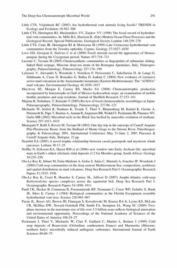
Little CTS, Vrijenhoek RC (2003) Are hydrothermal vent animals living fossils? TRENDS in
Ecology and Evolution 18:582–588
Little CTS, Herrington RJ, Maslennikov VV, Zaykov VV (1998) The fossil record of hydrother-
mal vent communities. In: Mills RA, Harrison K. (Eds) Modern Ocean Floor Processes and the
Geological Record. Special Publications, Geological Society London 148:259–270
Little CTS, Cann JR, Herrington RJ & Morisseau M (1999) Late Cretaceous hydrothermal vent
communities from the Troodos ophiolite, Cyprus. Geology 27:1027–1030
Love GD, Grosjean E, Stalvies C et al (2009) Fossil steroids record the appearance of Demos-
pongiae during the Cryogenian period. Nature 457:718–721
Lucente C, Taviani M (2005) Chemosynthetic communities as fingerprints of submarine sliding-
linked fluid seepage, Miocene deep-sea strata of the Romagna Apennines, Italy. Palaeogeo-
graphy, Palaeoclimology, Palaeoecology 227:176–190
Lykousis V, Alexandri S, Woodside J, Nomikou P, Perissoratis C, Sakellariou D, de Lange G,
Dahlmann A, Casas D, Rousakis G, Ballas D, Ioakim C (2004) New evidence of extensive
active mud volcanism in the Anaximander mountains (Eastern Mediterranean): The “ATHINA”mud volcano. Environmental Geology 46:1030–1037
MacAvoy SE, Morgan E, Carney RS, Macko SA (2008) Chemoautotrophic production
incorporated by heterotrophs in Gulf of Mexico hydrocarbon seeps: an examination of mobile
benthic predators and seep residents. Journal of Shellfish Research 27:153–161
Majima R, Nobuhara, T, Kitazaki T (2005) Review of fossil chemosynthetic assemblages in Japan.
Palaeogeography, Palaeoclimatology, Palaeoecology 227:86–123
Michaelis W, Seifert R, Nauhaus K, Treude T, Thiel V, Blumenberg M, Knittel K, Gieske A,
Peternecht K, Pape T, Boetius A, Amann R, Jorgensen BB,Widdel F, Peckmann JR, Pimenov NV,
Gulin MB (2002) Microbial reefs in the Black Sea fuelled by anaerobic oxidation of methane.
Science 297:1013–1015
Monegatti P, Raffi S, Roveri M, Taviani M (2001) One day trip in the outcrops of Castell’Arquato
Plio-Pleistocene Basin: from the Badland of Monte Giogo to the Stirone River. Paleobiogeo-
graphy & Paleoecology 2001, International Conference May 31-June 2, 2001 Piacenza &
Castell’Arquato Italy, Bologna: 22 pp
Neasbitt EA (2005) A novel trophic relationship between cassid gastropods and mysticete whale
carcasses. Lethaia 38:17–25
Noffke N, Eriksson KA, Hazen RM et al (2006) new window into Early Archean life: microbial
mats in Earth’s oldest siliclastic tidal deposits (3.2 Ga Moodies group, South Africa). Geology
34:235–256
Olu-Le Roy K, Sibuet M, Fiala-Medioni A, Gofas S, Salas C, Mariotti A, Foucher JP, Woodside J
(2004) Cold seep communities in the deep eastern Mediterranean Sea: composition, symbiosis
and spatial distribution on mud volcanoes. Deep Sea Research Part I: Oceanographic Research
Papers 51:1915–1936
Olu-Le Roy K, Cosel R, Hourdez S, Carney SL, Jollivet D (2007) Amphi-Atlantic cold-seep
Bathymodiolus species complexes across the equatorial belt. Deep Sea Research Part I:
Oceanographic Research Papers 54:1890–1911
Paull CK, Hecker B, Commeau R, Freemanlynde RP, Neumann C, Corso WP, Golubic S, Hook
JE, Sikes E, Curray J (1984) Biological communities at the Florida Escarpment resemble
hydrothermal vent taxa. Science 226:965–967
Payne JL, Boyer AG, Brown JH, Finnegan S, Kowalewski M, Krause RA Jr, Lyons KS, McLain
CR, McShea DW, Novack-Gottshall PM, Smith FA, Stempien JA, Wang SC (2009) Two-
phase increase in the maximum size of life over 3.5 billion years reflects biological innovation
and environmental opportunity. Proceedings of the National Academy of Sciences of the
United States of America 106:24–27
Peckmann J, Thiel V, Michaelis W, Clari P, Gaillard C, Martire L, Reitner J (1999) Cold
seep deposits of Beauvoisin (Oxfordian; southeastern France) and Marmorito (Miocene;
northern Italy): microbially induced authigenic carbonates. International Journal of Earth
Sciences 88:60–75
The Deep-Sea Chemoautotroph Microbial World 291
Peckmann J, Girschler E, Oschmann W, Reitner J (2001) An Early Carboniferous seep community
and hydrocarbon-derived carbonates from the Harz Mountains, Germany. Geology 29:
271–274
Peckmann J, Goedert JL, Thiel V, Michaelis W, Reitner J (2002) A comprehensive approach to the
study of methane-seep deposits from the Lincoln Creek Formation, western Washington State,
USA. Sedimentology 49:855–873
Peckmann J, Thiel V, Reitner J, Taviani M, Aharon P, Michaelis W (2004) A microbial mat of a
large sulphur bacterium preserved in a Miocene methane-seep. Geomicrobiology Journal 21:
247–255
Peckmann J, Goedert JL (2005) Geobiology of ancient and modern methane-seeps. Palaeogeo-
graphy, Palaeoclimatology, Palaeoecology 227:1–5
Peckmann J, Campbell KA, Walliser OH, Reitner J (2007) A late Devonian hydrocarbon-seep
dominated by dimerelloid brachiopods, Morocco. Palaios 19:114–122
Peek AS, Feldman RA, Lutz RA, Vrijenhock RC (1998) Cospeciation of chemoautotrophic
bacteria and deep sea clams. Proceedings of the National Academy of Sciences of the United
States of America 95:9962–9966
Peel JS (1991) Functional morphology of the Class Helcionelloida nov, and the early evolution of
Mollusca. In: Simonetta AM, Conway S (eds) The early evolution of Metazoa and the
significance of problematic taxa. Cambridge University Press, Cambridge: 157–177
Pennec M, Beninger PG, Herry A (1995) Feeding and digestive adaptations of bivalve molluscs to
sulphide-rich habitats. Comparative Biochemistry and Physiology 111A:183–189
Peterson KJ, Butterfield NJ (2005) Origin of the Eumetazoa: testing ecological predictions of
molecular clocks against the Proterozoic fossil record. Proceedings of the National Academy
of Sciences of the United States of America 102:9547–9552
Pinti DL, Mineau R, Clement V (2009) Hydrothermal alteration and microfossil artefacts of the
3,465-million-year-old Apex chert. Nature Geoscience 2:640–643
Porter SM (2010) Calcite and aragonite seas and the de novo acquisition of carbonate skeletons.
Geobiology 8:256–277
Pflug HD (2001) Earliest organic evolution. Essay to the memory of Batholomew Nagy. Precam-
brian Research 106:79–91
Poulton SW, Frolick PW, Canfield DE (2004) The transition to a sulphidic ocean approximately
1.84 billion years ago. Nature 431:173–177
Pruski AM, Rousse N, Fiala-Medioni A, Boulegue J (2002) Sulphur signature in the hydrothermal
vent mussel Bathymodiolus azoricus from the Mid-Atlantic Ridge. Journal of the Marine
Biological Association of the United Kingdom 82:463–468
Qun Y, Junye M, Xiaoyan S, Peiyun S (2007) Phylochronology of early metazoans: combined
evidence from molecular and fossil data. Geological Journal 42:281–295
Rasmussen B, Fletcher IR, Brocks JJ, Kilburn MR (2008) Reassessing the first appearance of
eukaryotes and cyanobacteria. Nature 455:1101–1104
Ravara A, Cunha MR, Rodrigues CL (2007) The occurrence of Natsushima bifurcata (Polychaeta:Nautiliniellidae) in Acharax hosts from mud volcanoes in the Gulf of Cadiz (south Iberian and
north Moroccan Margins). Scientia Marina 7:95–100
Reid RGB (1990) Evolutionary implications of sulphide-oxidizing symbionts in bivalves.
In: Morton B (ed) The Bivalvia – Proceedings of a Memorial Symposium in Honour of
Sir Charles Maurice Yonge, Edinborough, 1986: 127–140, Hong Kong University Press,
Hong Kong
Reid RGB, Bernard FR (1980) Gutless bivalves. Science 208:609–610
Reitner J, Peckmann J, Reimer A, Schumann G,Thiel V (2005). Methane-derived carbonate build-
ups and associated microbial communities at cold seeps on the lower Crimean shelf (Black
Sea). Facies 51:66–79
Ritt B, Sarrazin J, Caprais J-C, Noel P, Gauthier O, Pierre C, Henry P, Desbruyeres D (2010) First
insights into the structure and environmental setting of cold-seep communities in the Marmara
Sea. Deep-Sea Research Part I, Oceanographic Research Papers doi.10.1016/j.dsr.2010.05.011
292 M. Taviani
Rodrigues CF, Webster G, Cunha MR, Duperron S, Weightman AJ (2010) Chemosynthetic
bacteria found in bivalve species from mud volcanoes of the Gulf of Cadiz. Microbiology
Ecology 73:486–499
Rodrigues CL, Oliver PG, Cunha MR (2008) Thyasiroidea (Mollusca: Bivalvia) from the mud
volcanoes of the Gulf of Cadiz (North-east Atlantic). Zootaxa 1572:41–56
Runnegar B, Pojeta J Jr (1992) The earliest bivalves and their Ordovician descendants. American
Malacological Bulletin 9:117–122
Salas C, Woodside J (2002) Lucinoma kazani n.sp. (Mollusca: Bivalvia): evidence of a living
benthic community associated with a cold seep in the eastern Mediterranean Sea. Deep Sea
Research Part I: Oceanographic Research Papers 50:1391–1409
Salerno JL, Macko SA, Hallam SJ, Bright M, Won Y-J, McKiness Z, Van Dover CL (2005)
Characterization of symbiont populations in life-history stages of mussels from chemosyn-
thetic environments. Biological Bulletin 208:145–155
Schopf JW (1993) Microfossils of the early Archean Apex chert: new evidence of the antiquity of
life. Science 260:640–646
Schopf JW (1999) Cradle of Life: the Discovery of Earth’s Earliest Fossils. Princeton University
Press, Princeton: 367 pp
Schopf JW, Bottjer DJ (2009) World summit on ancient microscopic fossils. Precambrian
Research 173:1–3
Sergeeva N, Gulin S (2009) Benthic fauna of methane seeps in the Dniester Palaeo-Delta:
comparative analysis. IGCP 521-INQA 0501 Fifth Plenary Meeting and Field Trip, Istanbul-
Izmir-Canakkale (Turkey) 22–31 August 2009, Extended Abstracts:158–159
Sibuet M, Olu-Le Roy K (1998) Biogeography, biodiversity and fluid dependence of deep-sea
cold-seep communities at active and passive margins. Deep Sea Research Part II: Topical
Studies in Oceanography 45:517–567
Sibuet M, Olu-Le Roy K (2002) Cold Seep Communities on continental margins: structure and
qauantitative distribution relative to geological and fluid venting patterns. In: Wefer G, Billett D,
Hebbeln D, Jørgensen BB, Schl€uter M, VanWeering T (eds) Ocean Margin Systems. Springer-
Verlag, Berlin, Heidelberg:235–251
Sibuet M, Vangriesheim A (2009) Deep-sea environment and biodiversity of the West African
Equatorial margin. Deep Sea Research Part II: Topical Studies in Oceanography 56:2156–2168
Skovsted CB, Brock GA, Lindstroem A, Peel JS, Paterson JR, Fuller MK (2007) Early Cambrian
record of failed durophagy and shell repair in an epibenthic mollusc. Biology Letters
3:314–317
Stein JL, Cary CS, Hessler RR, Ohta S, Vetter RD, Childress JJ, Felbeck H (1988) Chemoautotro-
phic symbiosis in a hydrothermal vent gastropod. Biological Bulletin 174 373–378
Stewart FJ, Cavanaugh CM (2006): Bacterial endosymbioses in Solemya (Mollusca: Bivalvia) –
Model systems for studies of symbiont-host adaptation. Antonie van Leeuwenhoek
90:343–360
Sturany R (1896) Zoologische Ergebnisse VII Mollusken I (Prosobranchier und Opistobranchier:
Scphopoden; Lamellibranchier) gesammelt von SM Schiff “Pola” 1890–1894. Denkschriften
Akademie der Wissenschaften in Wien, Mathematisch-Naturwissenschaftliche Klasse 63:1–36
Suess E, Carson B, Ritger SD, Moore C, Jones ML, LD Kulm, Cochrane GR (1985) Biological
communities at vent sits along the subduction zone off Oregon. Bulletin of the Biological
Society of Washington 6:475–484
Sugitani K, Grey K, Nagaoka T, Mimura K, Walter MR (2009) Taxonomy and biogenicity of
Archean spheroidal microfossils (ca. 3.0 Ga) from the Mount Goldsworthy-Mount Grant area
in the northeastern Pilbara Craton, Western Australia. Precambrian Research 173:50–59
Taviani M (1994) The “calcari a Lucina” macrofauna reconsidered: Deep-sea faunal oases from
Miocene-age cold vents in the Romagna Apennine, Italy. Geo-Marine Letters 14:185–191
Taviani M (2001) Fluid venting and associated processes. In: Vai GB, Martini PI (eds) Anatomy of
an Orogen: the Apennines and Adjacent Mediterranean Basins. Kluwer Academic Publishers,
Great Britain:351–366
The Deep-Sea Chemoautotroph Microbial World 293
Taviani M (2004) Shaping the biogeography of the Mediterranean basin: one geologist’s perspec-
tive. In: Marine Biogeography of the Mediterranean Sea: patterns and dynamics of biodiver-
sity. Biogeographia 24(2003):15–22
Taviani M, Roveri M, Aharon P, Zibrowius H (1997) A Pliocene deep-water cold seep (Stirone
River, N. Italy). COLD-E-VENT International Works Hydrocarbon Seep Chemosynt Tethyan
Relic Basins: Products, Processes and Causes Abstracts:20
Taviani M, Corselli C, Freiwald A, Malinverno E, Mastrototaro F, Remia A, Savini A, Tursi A
(2005) First geo-marine survey of living cold-water Lophelia reefs in the Ionian Sea (Mediter-
ranean basin). Facies 50:409–417
Taviani M, Angeletti L, Ceregato A (2008) Past and present chemosynthetic bivalves (family
Solemyidae) inhabiting deep-sea cold-vent and reducing environments in the Neogene of
the Mediterranean basin. 33rd International Geological Congress Oslo 6–10 August 2008
Abstracts
Taylor JD, Glover EA (2000) Functional anatomy, chemosymbiosis and evolution of the Lucini-
dae. In: Harper EM, Taylor JD, Crame JA (Eds) The Evolutionary Biology of the Bivalvia.
Special Publications, Geological Society London 177:207–225
Taylor JD, Glover EA (2006) Lucinidae (Bivalvia) – the most diverse group of chemosymbiotic
molluscs. Zoological Journal of the Linnean Society 148:421–438
Taylor JD, Glover EA (2009) A giant lucinid bivalve from the Eocene of Jamaica – Systematics,
life habits and Chemosymbiosis (Mollusca: Bivalvia: Lucinidae). Palaeontology 52:95–109
Taylor JD, Glover EA, Williams ST (2008) Ancient chemosynthetic bivalves: systematics of
Solemyidae from eastern and southern Australia (Mollusca: Bivalvia). Memoirs Queensland
Museum, Nature:75–104
Taylor JD, Williams ST, Glover AE (2007) Evolutionary relationships of the bivalve family
Thyasiridae (Mollusca: Bivalvia), monophyly and superfamily status. Journal of the Marine
Biological Association of the United Kingdom 87:565–574
Tunnicliffe V, Juniper SK, Sibuet M (2003) Reducing environments of the deep-sea floor.
In: Tyler PA (ed) Ecosystems of the Deep Oceans. Elsevier, Amsterdam:81–110
Turner RD (1985) Notes on molluscs of deep-sea vents and reducing sediments. American
Malacological Bulletin Spec Edition 1:23–34
Van Dover CL (1990) Biogeography of hydrothermal vent communities along seafloor spreading
centers. TREE 5:242–246
Van Dover CL (2000) The ecology of deep-sea hydrothermal vents. Princeton University Press,
Princeton, New Jersey:1–424 pp
Van Dover CL, German CR, Speer KG, Parson LM,Vrijenhoek RC (2002) Evolution and
biogeography of deep-sea venting and seep invertebrates. Science 295:1253–1257
Van Gaest AL, Young CM, Young JJ, Helms AR, Arellano SM (2007) Physiological and
behavioral responses of Bathynerita naticoidea (Gastropoda: Neritidae) and Methanoariciadendrobranchiata (Polychaeta: Orbiniidae) to hypersaline conditions at a brine pool cold seep.Marine Ecology 28:199–207
Van Kranendonk MJ (2006) Volcanic degassing, hydrothermal circulation and the flourishing of
early life on Earth: a review of the evidence from c. 3490–3240 Ma rocks of the Pilbara
Supergroup, Pilbara Craton, Western Australia. Earth-Science Reviews 74:197–240
Vanreusel A, Andersen AC, Boetius A, Connelly D, Cunha MR, Decker C, Hilario A, Kormas KA,
Maignien L, Olu K, Pachiadaki M, Ritt B, Rodrigues C, Sarrazin J, Tyler P, Van Gaever S,
Vanneste H (2009) Biodiversity of cold seep ecosystems along the European margins. Ocean-
ography 22:110–127
Venturini S, Selmo E, Tarlao A, Tunis G (1998) Fossiliferous methanogenetic limestones in the
Eocene flysch of Istria (Croatia). Giornale di Geologia 60: 219–234
Vinther J, Nielsen C (2005) The Early Cambrian Halkeria is a mollusc. Zoologica Scripta 34:
81–89
Waren A, Bouchet P (2001) Gastropoda and Monoplacophora from hydrothermal vents and seeps,
new taxa and records. The Veliger 44:116–231
294 M. Taviani
Waren A, Bouchet P (2009) New gastropods from deep-sea hydrocarbon seeps off West Africa.
Deep Sea Research Part II: Topical Studies in Oceanography 56:2326–2349
Werne JP, Haese RR, Zitter T, Aloisi G, Bouloubassi I, Heijfs S, Fiala-Mediconi A, Pancost RD,
Sinninghe Damste JS, de Lange G, Forney LJ, Gottschal JC, Foucher JP, Mascle J, Woodside J
(2004) Life at cold seeps: a synthesis of biogeochemical and ecological data from Kazan mud
volcano, eastern Mediterranean Sea. Chemical Geology 205:367–390
Westall F, Folk RL (2003) exogenous carbonaceous microstructures in Early Archaen cherts and
BIFs from the Isua Greenstone Belt: implications for the search for life in ancient rocks.
Precambrian Research 126:313–330
Won Y-J, Jones WJ, Vrijenhoek RC (2008) Absence of cospeciation between deep-sea Mytilids
and their thiotrophic endosymbionts. Journal of Shellfish Research 27:129–138
Wood RA, Grotzinger JP, Dickson JAD (2002) Proterozoic modular biomineralized metazoan
from the Nma Group, Namibia. Science 296:2383–2386
Xiao S, Yuan X, Knoll AH (2000) Eumetazoan fossils in terminal Proterozoic phosporites?
Proceedings of the National Academy of Sciences of the United States of America 97:
13684–13689
Xiaoying S, Chuanheng Z, Ganqing J, Juan L, Li W, Dianbo L (2008) Microbial mats in the
Mesoproterozoic carbonates of the North China platform and their potential for hydrocarbon
generation. Journal of China University of Geosciences 19:549–566
Yanko-Hombach et al. (2009) Response of biota to methane emissions in the Black Sea: prelimi-
nary results from complex geological, geochemical, palaeontological, and biological study.
IGCP 521-INQA 0501 Fifth Plenary Meeting and Field Trip, Istanbul-Izmir-Canakkale (Turkey)
22–31 August 2009, Extended Abstracts:181–184
Zitter TAC, Henry P, Aloisi G, Delaygue G, Cagatay MN, Mercier de Lepinay B, Al-Samir M,
Fornacciari F, Tesmer M, Pekdeger A, Wallmann K, Lericolais G (2008) Cold seeps along the
main Marmara Fault in the Sea of Marmara (Turkey). Deep Sea Research Part I: Oceano-
graphic Research Papers 55:552–570
Zuschin M, Mandic O, Harzhauser M, Persvesler P (2001) Fossil Evidence for chemoautotrophic
bacterial symbiosis in the thyasirid bivalve Thyasira michelottii from the middle Miocene
(Badenium) of Austria. Historical Biology 15:223–234
The Deep-Sea Chemoautotroph Microbial World 295

.
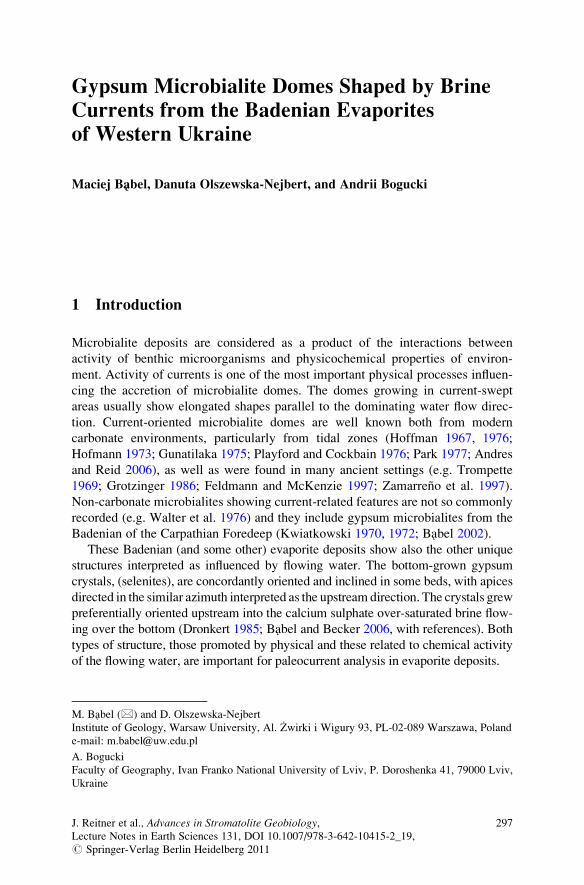
Gypsum Microbialite Domes Shaped by Brine
Currents from the Badenian Evaporites
of Western Ukraine
Maciej Babel, Danuta Olszewska-Nejbert, and Andrii Bogucki
1 Introduction
Microbialite deposits are considered as a product of the interactions between
activity of benthic microorganisms and physicochemical properties of environ-
ment. Activity of currents is one of the most important physical processes influen-
cing the accretion of microbialite domes. The domes growing in current-swept
areas usually show elongated shapes parallel to the dominating water flow direc-
tion. Current-oriented microbialite domes are well known both from modern
carbonate environments, particularly from tidal zones (Hoffman 1967, 1976;
Hofmann 1973; Gunatilaka 1975; Playford and Cockbain 1976; Park 1977; Andres
and Reid 2006), as well as were found in many ancient settings (e.g. Trompette
1969; Grotzinger 1986; Feldmann and McKenzie 1997; Zamarreno et al. 1997).
Non-carbonate microbialites showing current-related features are not so commonly
recorded (e.g. Walter et al. 1976) and they include gypsum microbialites from the
Badenian of the Carpathian Foredeep (Kwiatkowski 1970, 1972; Babel 2002).
These Badenian (and some other) evaporite deposits show also the other unique
structures interpreted as influenced by flowing water. The bottom-grown gypsum
crystals, (selenites), are concordantly oriented and inclined in some beds, with apices
directed in the similar azimuth interpreted as the upstream direction. The crystals grew
preferentially oriented upstream into the calcium sulphate over-saturated brine flow-
ing over the bottom (Dronkert 1985; Babel and Becker 2006, with references). Both
types of structure, those promoted by physical and these related to chemical activity
of the flowing water, are important for paleocurrent analysis in evaporite deposits.
M. Babel (*) and D. Olszewska-Nejbert
Institute of Geology, Warsaw University, Al. Zwirki i Wigury 93, PL-02-089 Warszawa, Poland
e-mail: [email protected]
A. Bogucki
Faculty of Geography, Ivan Franko National University of Lviv, P. Doroshenka 41, 79000 Lviv,
Ukraine
J. Reitner et al., Advances in Stromatolite Geobiology,Lecture Notes in Earth Sciences 131, DOI 10.1007/978-3-642-10415-2_19,# Springer-Verlag Berlin Heidelberg 2011
297
In this paper we describe the new and specific directional structures – the
gypsum microbialite domes that are shaped by both chemical and physical action
of the brine current. This unique structure was found only in a few outcrops in the
Badenian basin and in this paper we present the locality where the structure occurs
in association with the other current related features which all together allow
consistent interpretation of the origin of the directional structures.
2 Terminology
In this study we use the definition of microbialites by Burne and Moore (1987) and
accept that the microbialites are “organosedimentary deposits that have accreted as
a result of a benthic microbial community trapping and binding detrital sediment
and/or forming the locus of mineral precipitation”. In particular we include into the
microbialites the deposits formed by inorganic and passive precipitation of gypsum
within the locus of benthic microbial communities (see Arp et al. 2004). Further the
broadest meaning of the term microbialite is also used for the rocks, in which the
“influence of algae on the rock is more commonly inferred than observed” (Burne
and Moore 1987, p. 242).
The term selenite, in the paper, is applied to gypsum crystals over 2 mm in size
(cf. Warren 1982b). These are spacifically primary gypsum ctystals grown from
solution or brine directly on the bottom of the basin, or within the soft deposits,
microbial mats, or organic sediments as displacive or incorporative crystals.
3 The Badenian Gypsum Deposits in Carpathian Foredeep
The Badenian evaporites of the Carpathian region originated during a major
evaporite event (salinity crisis) which took place in the Central Paratethys Sea
during its separation from the Mediterranean in the Badenian time, presumably
about 13.6 Ma ago (Dudek et al. 2006; Peryt 2006). The Carpathian Foredeep basin
is the largest Badenian evaporite basin, although its Carpathian margin has been
destroyed by erosion related to post-Badenian tectonic shortening, overthrusting,
and uplift of the Carpathian orogen (Fig. 1). In the northern Carpathian Foredeep
the Badenian evaporites comprise mainly calcium sulphate sediments (exposed on
the platformal margin of the basin) and sodium chloride deposits (buried in the
foredeep) and are ca 10–70 m in thickness. The sulphate facies are represented by
the primary gypsum deposits, mainly the selenites, gypsum microbialites, and
clastic gypsum. These deposits show a nearly undisturbed layer-cake stratigraphy.
The particular layers or groups of layers are laterally continuous and traced over
distances of tens to hundreds kilometers. Several lithostratigraphical units, coin-
ciding with typical gypsum facies, were distinguished within these deposits
and several thin marker beds allowed more precise stratigraphical correlation
(Kubica 1992; Babel 2005a; Peryt 2006). The section is bipartite and comprises
298 M. Babel et al.
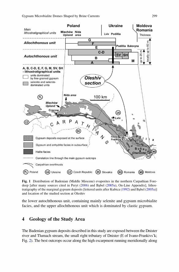
the lower autochthonous unit, containing mainly selenite and gypsum microbialite
facies, and the upper allochthonous unit which is dominated by clastic gypsum.
4 Geology of the Study Area
The Badenian gypsum deposits described in this study are exposed between the Dnister
river and Tlumach stream, the small right tributary of Dnister (E of Ivano-Frankivs’k;
Fig. 2). The best outcrops occur along the high escarpment running meridionally along
Fig. 1 Distribution of Badenian (Middle Miocene) evaporites in the northern Carpathian Fore-
deep [after many sources cited in Peryt (2006) and Babel (2005a), On-Line Appendix], lithos-
tratigraphy of the marginal gypsum deposits [lettered units after Kubica (1992) and Babel (2005a)]
and location of the studied section at Oleshiv
Gypsum Microbialite Domes Shaped by Brine Currents 299
the Tlumach stream bank. The studied section at Oleshiv, situated in the northwestern
part of the escarpment, is representative of that part of the basinal platform zone and
formerly was described by Alth (1877), Łomnicki (1881), Peryt et al. (1994, figs. 4, 5),
Peryt (1996, figs. 4, 7, 11a, c–e), Babel (2005a, fig. 5 and On-line Appendix) and Babel
(2007, figs. 15f, 16, 17). The base of the escarpment is made up by horizontally lying
Cretaceous limestones (Fig. 2a). The Mesozoic is capped by Miocene deposits, lying
above an erosional gap, starting with a few decimetres of Badenian marine marly
sandstones that contain fragmented Pecten shells, as observed at Palahychi (Łomnicki
1881; Siemiradzki 1909). In some places these sandstones are underlain with a very thin
layer of clays of supposed freshwater origin (Malicki 1938). The sandstones are covered
with thick layers of Badenian gypsum deposits, best exposed in abandoned quarries at
the top part of the escarpment. Gypsum deposits are overlain by poorly exposed, barren,
massive limestone (Alth 1877; Peryt 1996).Alth (1877), however, recognised borings of
mollusk Pholas in this part of the section. The top of section is eroded and covered byQuaternary clastics and loess.
The gypsum section at Oleshiv shows a similar sequence of lithostratigraphical
units and arrangement of facies as in the whole northern margin of the Badenian
basin, particularly between Lviv and Odaiv (Figs. 1 and 2). The section (Fig. 3)
starts with secondary alabaster replacing the giant gypsum intergrowths (unit A). It
is covered by gypsum microbialites (unit M), grass-like, banded, gypsum (unit B),
composed of rows of bottom-grown selenite crystals intercalated with fine-grained
gypsum, sabre gypsum (unit C–D) that comprise the lower autochthonous part of
the Badenian sulphates. Further up in the section, there is a clastic gypsum (unit E)
belonging to the upper allochthonous unit. The section is not fully exposed, unit
C–D being disturbed by some karst collapse or faults. The lower part of unit B
contains set of marker beds traced across nearly the whole basin margin and useful
for isochronous correlation (Babel 2005a).
The bottom part of the microbialite unit M at Oleshiv, below a characteristic
horizontal discontinuity surface (in places covered with clay), contains sub-hori-
zontal selenite crystals. The contact of these deposits with the underlying giant
gypsum intergrowths is very clear. In the southernmost exposure (between Oleshiv
S1 and S2, Fig. 2a, Babel 2005a, in the On-line Appendix) the giant gypsum
intergrowths show numerous flat dissolution surfaces and their upper surface is
scalloped by dissolution and/or erosional features (Fig. 4b). Northwards (Oleshiv
S1; Figs. 2 and 4a), the layers of grass-like gypsum (rows of bottom-grown selenite
crystals with top surface flatted by dissolution) appears in between the gypsum
intergrowths and the microbialite layer. Further north (Oleshiv N; Fig. 2) the
transition from the giant intergrowths to the microbialites is gradual and without
traces of dissolution surfaces (Babel 2005a, On-line Appendix). Going up the
section the number and sizes of selenite crystals decrease and fine-grained gypsum
gradually becomes the main component. Transitions similar to this are observed
throughout the basin, and were interpreted as the result of shallowing of the
permanently submerged several metre deep saline pan, where the giant gypsum
intergrowths were crystallizing, followed by the establishment of the semi-emerged
300 M. Babel et al.
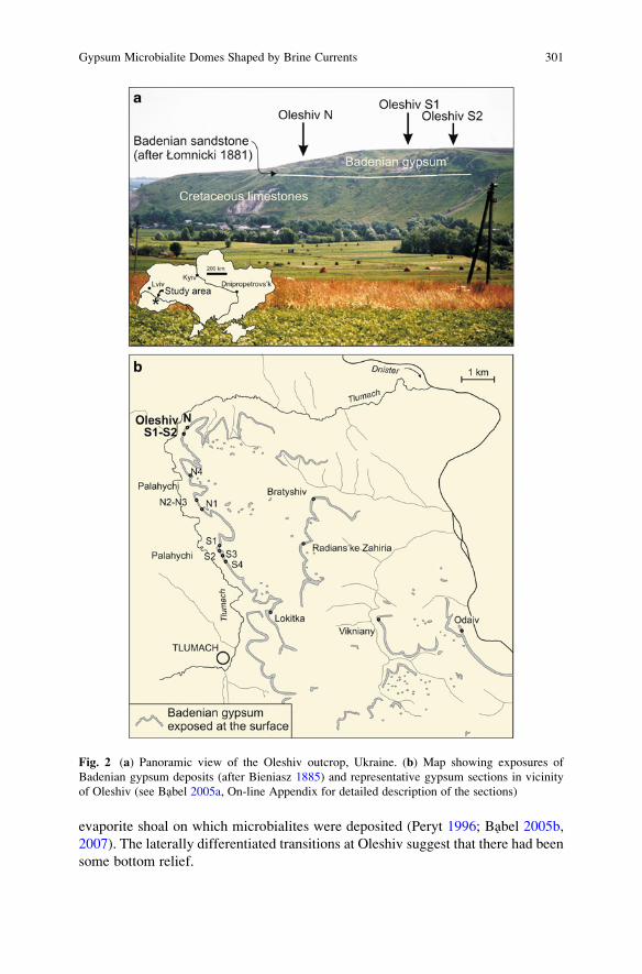
evaporite shoal on which microbialites were deposited (Peryt 1996; Babel 2005b,
2007). The laterally differentiated transitions at Oleshiv suggest that there had been
some bottom relief.
Fig. 2 (a) Panoramic view of the Oleshiv outcrop, Ukraine. (b) Map showing exposures of
Badenian gypsum deposits (after Bieniasz 1885) and representative gypsum sections in vicinity
of Oleshiv (see Babel 2005a, On-line Appendix for detailed description of the sections)
Gypsum Microbialite Domes Shaped by Brine Currents 301
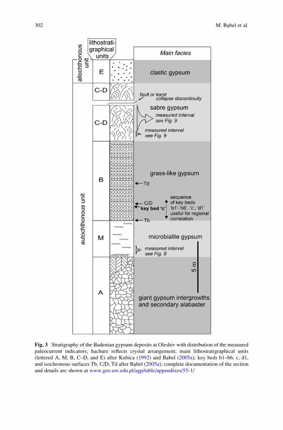
Fig. 3 Stratigraphy of the Badenian gypsum deposits at Oleshiv with distribution of the measured
paleocurrent indicators; hachure reflects crystal arrangement; main lithostratigraphical units
(lettered A, M, B, C–D, and E) after Kubica (1992) and Babel (2005a); key beds b1–b6, c, d1,
and isochronous surfaces Tb, C/D, Td after Babel (2005a); complete documentation of the section
and details are shown at www.geo.uw.edu.pl/agp/table/appendixes/55-1/
302 M. Babel et al.
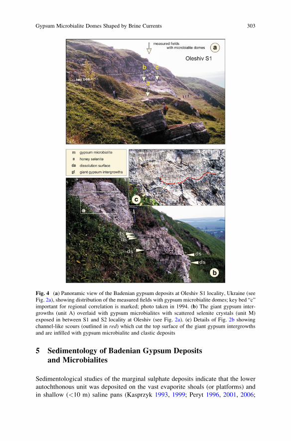
5 Sedimentology of Badenian Gypsum Deposits
and Microbialites
Sedimentological studies of the marginal sulphate deposits indicate that the lower
autochthonous unit was deposited on the vast evaporite shoals (or platforms) and
in shallow (<10 m) saline pans (Kasprzyk 1993, 1999; Peryt 1996, 2001, 2006;
Fig. 4 (a) Panoramic view of the Badenian gypsum deposits at Oleshiv S1 locality, Ukraine (see
Fig. 2a), showing distribution of the measured fields with gypsum microbialite domes; key bed “c”
important for regional correlation is marked; photo taken in 1994. (b) The giant gypsum inter-
growths (unit A) overlaid with gypsum microbialites with scattered selenite crystals (unit M)
exposed in between S1 and S2 locality at Oleshiv (see Fig. 2a). (c) Details of Fig. 2b showing
channel-like scours (outlined in red) which cut the top surface of the giant gypsum intergrowths
and are infilled with gypsum microbialite and clastic deposits
Gypsum Microbialite Domes Shaped by Brine Currents 303
Babel 2007). Widespread selenite facies represent permanently subaqueous environ-
ments whereas microbialites form mainly on semi-emerged shoals. The microbialite
deposits and structures are very common in the Badenian gypsum deposits in the
Czech Republic, Poland, Ukraine and Moldova (Babel 2005b), and are also docu-
mented in Bulgaria (Trashliev 1969). Gypsum stromatolite domes were recognized
in the Nida river area in Poland (Kwiatkowski 1970, 1972) and many varieties of
microbialites were recognized in drill cores (Kasprzyk 1993). The variable nearshore
environments of the Badenian basin were subdivided into two main types; the “wet
shore” and the “dry shore” environment, depending on the influx of continental water
from land recorded by clay intercalations (Schreiber et al. 2007). The “wet shore”
microbialite environments are mostly represented in Poland and “dry-shore” ones in
Ukraine. The gypsum microbialites from both these zones apparently differ in
morphology and structure [compare Kwiatkowski (1972), Kasprzyk (1993) and
Peryt (1996)]. The microbialites are very abundant in Ukraine, particularly on the
east part of the basin, and they were mostly interpreted as the result of gypsification
of microbial mats (Peryt 1996). It seems that gypsification of microbial mats was
easier there because of the warmer local climate and hence greater evaporation rate.
6 The “Dry Shore” Gypsum Microbialites (Unit M)
The gypsum microbialites in Oleshiv represent the same type of microbialites as
those present in the other outcrops of unit M stretching from Lviv to Moldova and
described by Peryt (1996). They are also similar to microbialites occurring in
marker bed “c” (Kasprzyk 1993, 1999; Peryt 2001; Babel 2005a). These micro-
bialites are composed of white to light brown, fine-grained gypsum (alabaster)
showing more or less pronounced crenulated wavy lamination (with laminae from 1
to several mm thick, commonly obliterated and visible only as the ghosts). The
crenulated lamination is generally horizontal. Crenulations reflect presence of very
small (<5 cm in size) flattened domal structures. In some places the crenulated
laminae form waves recording the occurrence of larger scale domal structures up to
0.6 m in diameter (Fig. 5b). The synoptic relief of these domal structures is usually
low (commonly only several cm) and it rises in places of transition to the selenite
facies (Fig. 5b). Locally, the beds of laminae form very complex fold structures
which appear be the product of gypsification of the microbial mats of the irregular
mamillate morphology possibly related to the lateral expansive growth of microbial
benthic communities (Fig. 5a, see e.g. Grotzinger and Knoll 1999; Babel 2004, pl. 1,
fig. 1; fig. 8a; Douglas et al. 2008). In some places, below some convex laminae,
fenestral pore structures appear filled with coarse gypsum cement (such structures
were however unnoticed at Oleshiv). A very characteristic feature is occurrence of
shallow flat-bottomed channels filled with laminated clastic gypsum (Peryt 1996,
figs. 12, 13; Babel 2005b, pl. 3, fig. 2, 2007, figs. 21, 22) which are however poorly
represented at Oleshiv (Fig. 4c).
304 M. Babel et al.
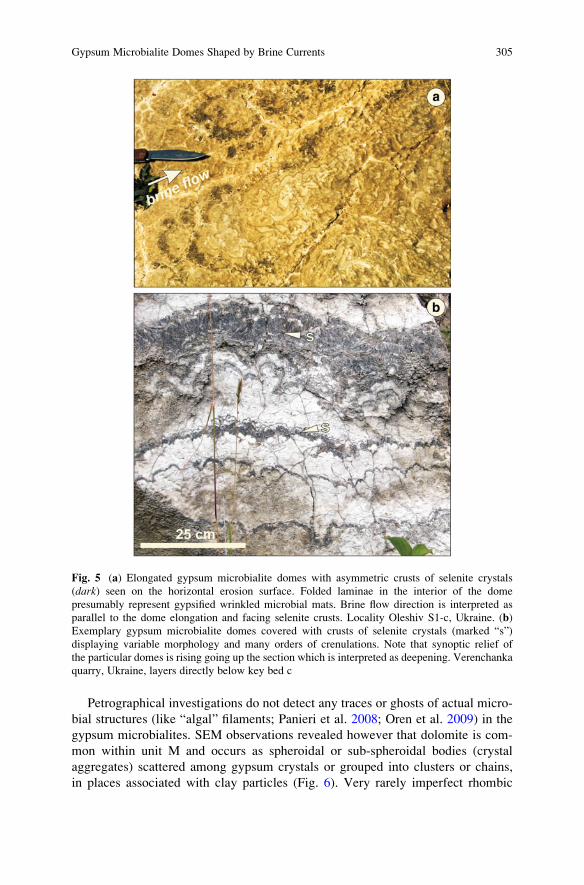
Petrographical investigations do not detect any traces or ghosts of actual micro-
bial structures (like “algal” filaments; Panieri et al. 2008; Oren et al. 2009) in the
gypsum microbialites. SEM observations revealed however that dolomite is com-
mon within unit M and occurs as spheroidal or sub-spheroidal bodies (crystal
aggregates) scattered among gypsum crystals or grouped into clusters or chains,
in places associated with clay particles (Fig. 6). Very rarely imperfect rhombic
Fig. 5 (a) Elongated gypsum microbialite domes with asymmetric crusts of selenite crystals
(dark) seen on the horizontal erosion surface. Folded laminae in the interior of the dome
presumably represent gypsified wrinkled microbial mats. Brine flow direction is interpreted as
parallel to the dome elongation and facing selenite crusts. Locality Oleshiv S1-c, Ukraine. (b)
Exemplary gypsum microbialite domes covered with crusts of selenite crystals (marked “s”)
displaying variable morphology and many orders of crenulations. Note that synoptic relief of
the particular domes is rising going up the section which is interpreted as deepening. Verenchanka
quarry, Ukraine, layers directly below key bed c
Gypsum Microbialite Domes Shaped by Brine Currents 305
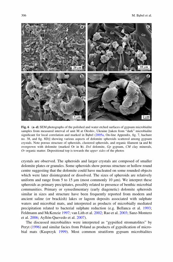
crystals are observed. The spheroids and larger crystals are composed of smaller
dolomite plates or granules. Some spheroids show porous structure or hollow round
centre suggesting that the dolomite could have nucleated on some rounded objects
which were later disintegrated or dissolved. The sizes of spheroids are relatively
uniform and range from 5 to 15 mm (most commonly 10 mm). We interpret these
spheroids as primary precipitates, possibly related to presence of benthic microbial
communities. Primary or synsedimentary (early diagenetic) dolomite spheroids
similar in sizes and structure have been frequently reported from modern and
ancient saline (or brackish) lakes or lagoon deposits associated with sulphate
waters and microbial mats, and interpreted as products of microbially mediated
precipitation related to bacterial sulphate reduction (e.g. Bellanca et al. 1993;
Feldmann and McKenzie 1997; van Lith et al. 2002; Rao et al. 2003; Sanz-Montero
et al. 2006; Ayllon-Quevedo et al. 2007).
The discussed microbialites were interpreted as “gypsified stromatolites” by
Peryt (1996) and similar facies from Poland as products of gypsification of micro-
bial mats (Kasprzyk 1999). Most common stratiform gypsum microbialites
Fig. 6 (a–d) SEM photographs of the polished and water etched surfaces of gypsum microbialite
samples from measured interval of unit M at Oleshiv, Ukraine [taken from “dark” microbialite
significant for local correlation and marked in Babel (2005a, On-line Appendix, fig. 7, hachure
no. 38, and fig. 8D)] showing various aspects of dolomite spheroids scattered among gypsum
crystals. Note porous structure of spheroids, clustered spheroids, and organic filament (a and b)
overgrown with dolomite (marked Or in b). Dol dolomite, Gp gypsum, CM clay minerals,
Or organic matter. Depositional top is towards the upper sides of the photos
306 M. Babel et al.
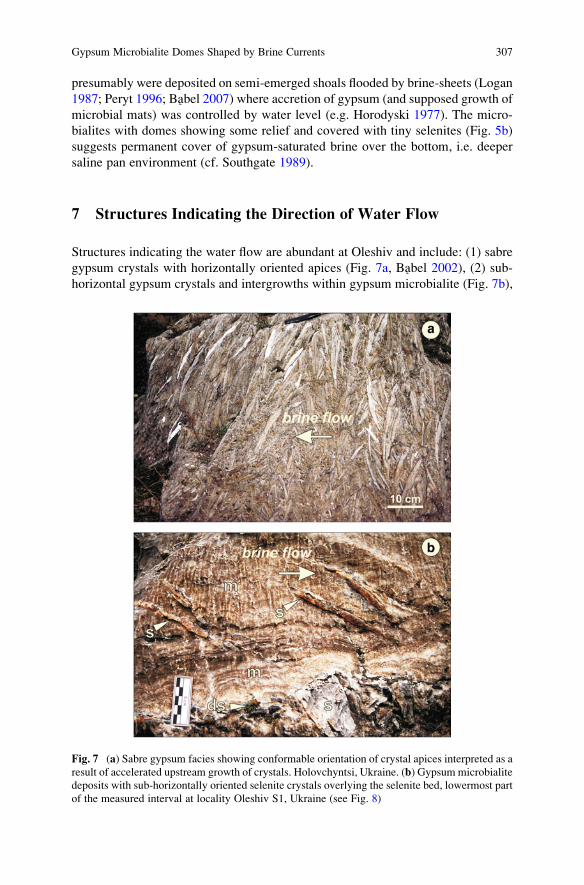
presumably were deposited on semi-emerged shoals flooded by brine-sheets (Logan
1987; Peryt 1996; Babel 2007) where accretion of gypsum (and supposed growth of
microbial mats) was controlled by water level (e.g. Horodyski 1977). The micro-
bialites with domes showing some relief and covered with tiny selenites (Fig. 5b)
suggests permanent cover of gypsum-saturated brine over the bottom, i.e. deeper
saline pan environment (cf. Southgate 1989).
7 Structures Indicating the Direction of Water Flow
Structures indicating the water flow are abundant at Oleshiv and include: (1) sabre
gypsum crystals with horizontally oriented apices (Fig. 7a, Babel 2002), (2) sub-
horizontal gypsum crystals and intergrowths within gypsum microbialite (Fig. 7b),
Fig. 7 (a) Sabre gypsum facies showing conformable orientation of crystal apices interpreted as a
result of accelerated upstream growth of crystals. Holovchyntsi, Ukraine. (b) Gypsummicrobialite
deposits with sub-horizontally oriented selenite crystals overlying the selenite bed, lowermost part
of the measured interval at locality Oleshiv S1, Ukraine (see Fig. 8)
Gypsum Microbialite Domes Shaped by Brine Currents 307
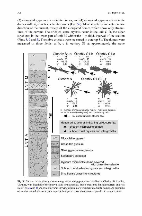
(3) elongated gypsum microbialite domes, and (4) elongated gypsum microbialite
domes with asymmetric selenite covers (Fig. 5a). Most structures indicate precise
direction of the current, except of the elongated domes which show only stream-
lines of the current. The oriented sabre crystals occur in the unit C–D, the other
structures in the lower part of unit M within the 1 m thick interval of the section
(Figs. 3, 7 and 8). The sabre crystals were measured in outcrop S1. The domes were
measured in three fields: a, b, c in outcrop S1 at approximately the same
Fig. 8 Section of the giant gypsum intergrowths and gypsum microbialites at Oleshiv S1 locality,
Ukraine, with location of the intervals and stratigraphical levels measured for paleocurrent analysis
(see Figs. 2a and 4) and rose diagrams showing azimuth of gypsummicrobialite domes and azimuths
of sub-horizontal selenite crystals apices. Interpreted flow directions are parallel to mean vectors
308 M. Babel et al.
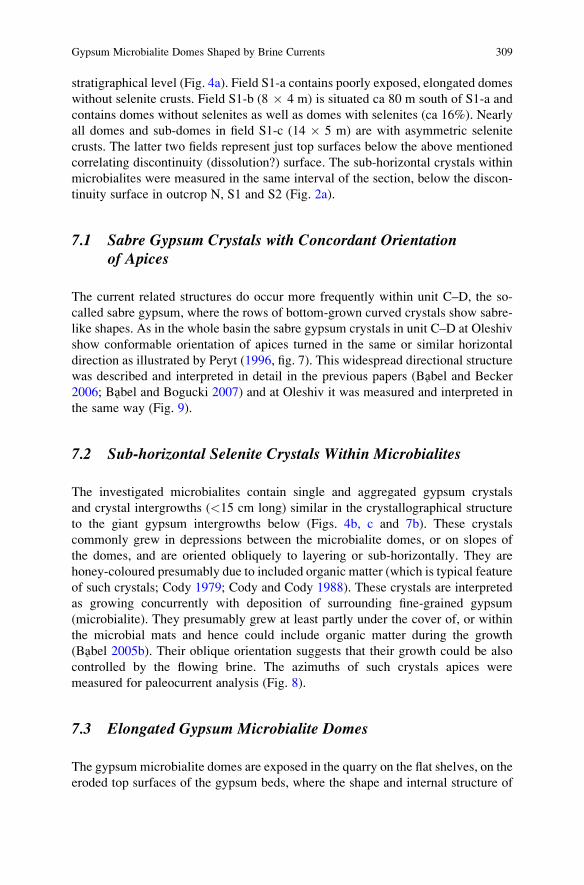
stratigraphical level (Fig. 4a). Field S1-a contains poorly exposed, elongated domes
without selenite crusts. Field S1-b (8 � 4 m) is situated ca 80 m south of S1-a and
contains domes without selenites as well as domes with selenites (ca 16%). Nearly
all domes and sub-domes in field S1-c (14 � 5 m) are with asymmetric selenite
crusts. The latter two fields represent just top surfaces below the above mentioned
correlating discontinuity (dissolution?) surface. The sub-horizontal crystals within
microbialites were measured in the same interval of the section, below the discon-
tinuity surface in outcrop N, S1 and S2 (Fig. 2a).
7.1 Sabre Gypsum Crystals with Concordant Orientationof Apices
The current related structures do occur more frequently within unit C–D, the so-
called sabre gypsum, where the rows of bottom-grown curved crystals show sabre-
like shapes. As in the whole basin the sabre gypsum crystals in unit C–D at Oleshiv
show conformable orientation of apices turned in the same or similar horizontal
direction as illustrated by Peryt (1996, fig. 7). This widespread directional structure
was described and interpreted in detail in the previous papers (Babel and Becker
2006; Babel and Bogucki 2007) and at Oleshiv it was measured and interpreted in
the same way (Fig. 9).
7.2 Sub-horizontal Selenite Crystals Within Microbialites
The investigated microbialites contain single and aggregated gypsum crystals
and crystal intergrowths (<15 cm long) similar in the crystallographical structure
to the giant gypsum intergrowths below (Figs. 4b, c and 7b). These crystals
commonly grew in depressions between the microbialite domes, or on slopes of
the domes, and are oriented obliquely to layering or sub-horizontally. They are
honey-coloured presumably due to included organic matter (which is typical feature
of such crystals; Cody 1979; Cody and Cody 1988). These crystals are interpreted
as growing concurrently with deposition of surrounding fine-grained gypsum
(microbialite). They presumably grew at least partly under the cover of, or within
the microbial mats and hence could include organic matter during the growth
(Babel 2005b). Their oblique orientation suggests that their growth could be also
controlled by the flowing brine. The azimuths of such crystals apices were
measured for paleocurrent analysis (Fig. 8).
7.3 Elongated Gypsum Microbialite Domes
The gypsum microbialite domes are exposed in the quarry on the flat shelves, on the
eroded top surfaces of the gypsum beds, where the shape and internal structure of
Gypsum Microbialite Domes Shaped by Brine Currents 309
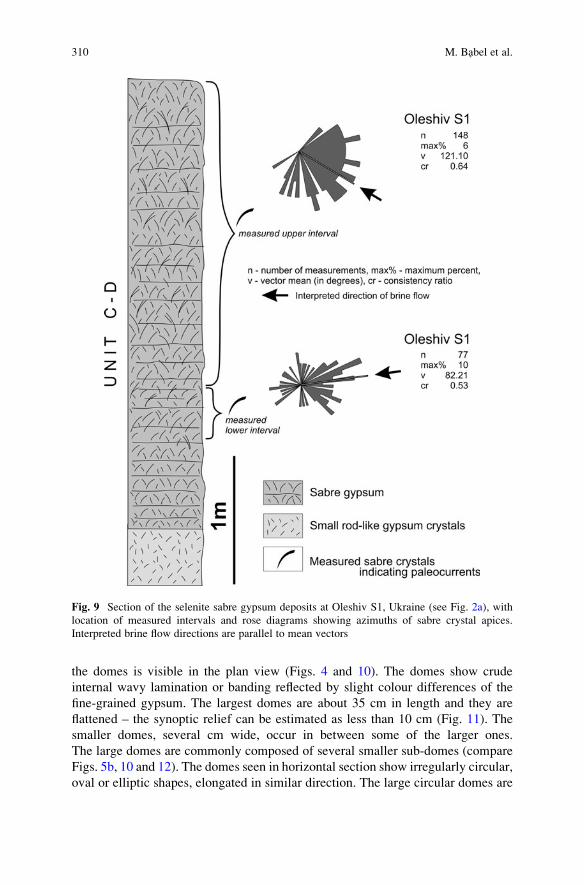
the domes is visible in the plan view (Figs. 4 and 10). The domes show crude
internal wavy lamination or banding reflected by slight colour differences of the
fine-grained gypsum. The largest domes are about 35 cm in length and they are
flattened – the synoptic relief can be estimated as less than 10 cm (Fig. 11). The
smaller domes, several cm wide, occur in between some of the larger ones.
The large domes are commonly composed of several smaller sub-domes (compare
Figs. 5b, 10 and 12). The domes seen in horizontal section show irregularly circular,
oval or elliptic shapes, elongated in similar direction. The large circular domes are
Fig. 9 Section of the selenite sabre gypsum deposits at Oleshiv S1, Ukraine (see Fig. 2a), with
location of measured intervals and rose diagrams showing azimuths of sabre crystal apices.
Interpreted brine flow directions are parallel to mean vectors
310 M. Babel et al.
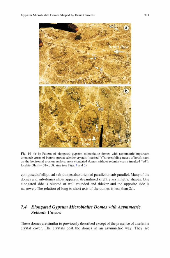
composed of elliptical sub-domes also oriented parallel or sub-parallel. Many of the
domes and sub-domes show apparent streamlined slightly asymmetric shapes. One
elongated side is blunted or well rounded and thicker and the opposite side is
narrower. The relation of long to short axis of the domes is less than 2:1.
7.4 Elongated Gypsum Microbialite Domes with AsymmetricSelenite Covers
These domes are similar to previously described except of the presence of a selenite
crystal cover. The crystals coat the domes in an asymmetric way. They are
Fig. 10 (a–b) Pattern of elongated gypsum microbialite domes with asymmetric (upstream
oriented) crusts of bottom-grown selenite crystals (marked “s”), resembling traces of hoofs, seen
on the horizontal erosion surface; note elongated domes without selenite crusts (marked “ed”);
locality Oleshiv S1-c, Ukraine (see Figs. 4 and 5)
Gypsum Microbialite Domes Shaped by Brine Currents 311
apparently more abundant and larger on one side of the dome, just the side which is
elongated and commonly blunted (Figs. 5a, 10, 11 and 12). The opposite side is
usually without selenite crystals at all. The selenite always covers the same side of
the domes in all these structures. Such domes seen on horizontal surface resembles
imprints of hoofs indicating one direction of march (Fig. 10).
8 Interpretation of the Oriented Badenian Selenite Crystals
The structures having containing oriented selenite crystals are interpreted as a result
of the influence of the gypsiferous brine, flowing over the bottom, on the crystals
growing directly on the bottom, having namely an accelerated upstream crystal
growth. The arguments for such an interpretation were summarized by Babel and
Becker (2006, with references), see also: Boudreau (1988), Sizaret et al. (2006,
2009) and Uchymiak et al. (2008).
The measurements made in the lower and upper interval of the unit C–D showed
a different orientation of mean growth vectors (Fig. 9), suggesting that the average
flow direction changed with time, and shifted clockwise about 27�. Shapes of rosediagrams and consistency ratio values suggest that the flow directions were more
uniform during deposition of the upper interval.
The measurements in unit M revealed that sub-horizontal crystals show also
preferred orientation of apices although the statistical results are less univocal
Fig. 11 Scheme showing
horizontal and interpreted
vertical section through the
elongated gypsum
microbialite dome with
asymmetric cover of selenite
crystals
312 M. Babel et al.

than for the sabre crystals as suggested by low number of measurements in
Oleshiv N locality (n ¼ 23) and relatively low consistency ratio in this and
S1–S2 locality (0.31 and 0.45 respectively). Nevertheless, the mean vectors
obtained are very similarly oriented and nearly parallel to the vectors from the
other intervals of the Oleshiv section, including vectors representing elongated
microbialite domes. All these vectors together suggest flow of brine permanently
from ESE to WNW during deposition of the autochthonous gypsum in this
locality and this direction perfectly coincide with the brine paleocurrents detected
in adjacent localities (Fig. 2b, Babel et al. 1999; Babel and Bogucki 2007). The
similar, constant direction of brine flow during deposition of B and C–D units,
was recorded on the Nida area in Poland (Kwiatkowski 1970, 1972; Babel 2002).
Such concurrence suggests that the currents shaping all these structures were
similar in nature.
Fig. 12 (a–b) Elongated gypsum microbialite domes with asymmetric (upstream oriented) crusts
of bottom-grown selenite crystals (marked “s”) seen on the horizontal erosion surface; honey
gypsum crystals are marked by “hs”; locality Oleshiv S1-c, Ukraine (see Figs. 4 and 5)
Gypsum Microbialite Domes Shaped by Brine Currents 313
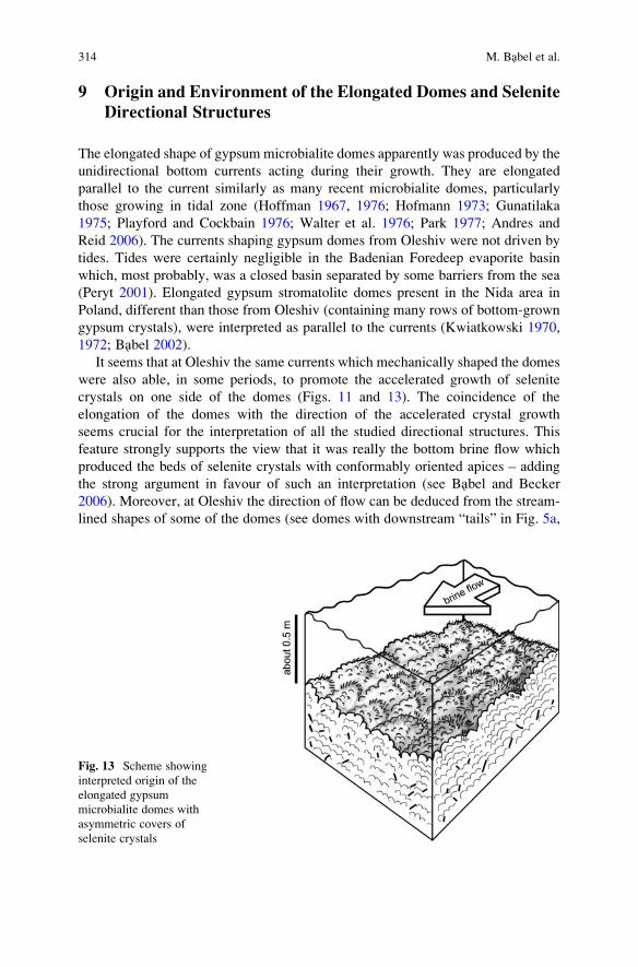
9 Origin and Environment of the Elongated Domes and Selenite
Directional Structures
The elongated shape of gypsum microbialite domes apparently was produced by the
unidirectional bottom currents acting during their growth. They are elongated
parallel to the current similarly as many recent microbialite domes, particularly
those growing in tidal zone (Hoffman 1967, 1976; Hofmann 1973; Gunatilaka
1975; Playford and Cockbain 1976; Walter et al. 1976; Park 1977; Andres and
Reid 2006). The currents shaping gypsum domes from Oleshiv were not driven by
tides. Tides were certainly negligible in the Badenian Foredeep evaporite basin
which, most probably, was a closed basin separated by some barriers from the sea
(Peryt 2001). Elongated gypsum stromatolite domes present in the Nida area in
Poland, different than those from Oleshiv (containing many rows of bottom-grown
gypsum crystals), were interpreted as parallel to the currents (Kwiatkowski 1970,
1972; Babel 2002).
It seems that at Oleshiv the same currents which mechanically shaped the domes
were also able, in some periods, to promote the accelerated growth of selenite
crystals on one side of the domes (Figs. 11 and 13). The coincidence of the
elongation of the domes with the direction of the accelerated crystal growth
seems crucial for the interpretation of all the studied directional structures. This
feature strongly supports the view that it was really the bottom brine flow which
produced the beds of selenite crystals with conformably oriented apices – adding
the strong argument in favour of such an interpretation (see Babel and Becker
2006). Moreover, at Oleshiv the direction of flow can be deduced from the stream-
lined shapes of some of the domes (see domes with downstream “tails” in Fig. 5a,
Fig. 13 Scheme showing
interpreted origin of the
elongated gypsum
microbialite domes with
asymmetric covers of
selenite crystals
314 M. Babel et al.
and right in Fig. 10b), or from position of blunted, more rounded side of the dome,
resembling “crescent marks” (compare Figs. 5a, 10, 11 and 12 with Vidal 1972,
figs. 5, 6). This further supports the view that the selenite crystals really were able to
grow with an upstream orientation.
The brine currents that shaped the domes and influenced the growth of selenite
crystals in unit M were probably similar in nature to those interpreted for the sabre
gypsum (Babel and Bogucki 2007). They represent oriented, continuous water flow
reflecting longshore (cyclonic) brine circulation or mean drift in evaporite basin
(Babel and Becker 2006).
The elongated domes appear to be rare in unit M. The lack of such domes can be
puzzling. They can be only weakly recorded because the horizontally exposed
gypsum surfaces are rare; it is very difficult to recognize them in vertical sections,
on walls of the outcrops. Alternatively, it is equally possible that the water or brine
normally was flowing relatively slowly along the flat basin shoals and its mechani-
cal influence on the morphology of the substrate, possibly covered with cohesive
microbial mats, was negligible. The currents could, however, increase their speed
and erosive power passing some narrows between more elevated areas. In such
narrows they could influence the shape of the growing domes and accelerate the
growth of selenite crystals. The Oleshiv outcrop can represent such a place, namely
the gentle slope of the channel-like restriction or some broad depression between
slightly more elevated shoals. The directional structures become more and more
pronounced going to the southernmost part of this exposure, probably located closer
to the axis of the presumed narrow channel.
10 Modern Analogues of the Badenian Gypsum Domes
In the primary gypsum deposits the bottom-grown crystals commonly create domal
structures. Small, from centimetres to a few decimetres large selenite domes are
typical forms crystallizing in the shallow (<1 m deep) gypsum pans in the marine
saltworks, usually in the salinity range 200–300‰ (Ortı Cabo et al. 1984). Much
larger and more complex selenite domes crystallize at depths of 1–3 m, on the floor
of the Lake Inneston, York Peninsula, Australia (Warren 1982b). They are up to
several meters in diameter and tens of centimeters in synoptic relief. They were
described as “gypsum-cyanobacterial mounds” (De Deckker 1988; Babel 2005b
and references therein), or “gypsiferous domal stromatolites” (Southgate 1989).
The selenite domes are coated by microbial slime (cyanobacteria and diatoms) and
“can be considered to be a type of coarsely-layered gypsum stromatolite” (Warren
2006, p. 229). The ancient Messinian gypsum deposits show even greater and more
complex domal selenite structures (e.g. Dronkert 1985). In the Badenian evaporite
basin such large selenite domes commonly are intercalated with fine-grained
gypsum, interpreted as gypsum microbialite, and create complex microbialite-
selenite structures (Babel 2005b). Well described ancient gypsum microbialite
deposits creating small-scale domal structures are known from the Messinian of
Gypsum Microbialite Domes Shaped by Brine Currents 315
Cyprus (Rouchy and Monty 1981). Gypsum microbialites related to gypsification
(gypsum encrustation) of microbial mats are recorded from many modern shallow
and semi-emerged evaporite environments (Eckstein 1970, figs. 5, 6; Krumbein and
Cohen 1977; Ortı Cabo et al. 1984; Reineck et al. 1990; Taher et al. 1995; Gerdes
et al. 2000). The origin of these and associated deposits is much less recognized
than the carbonate microbialites and most authors consider them as products of
passive precipitation of gypsum on, or within the mat (Rouchy and Monty 2000).
Modern gypsum domes very similar to the described Badenian structures from
Oleshiv were noted byWest et al. (1985) in saline pans (“lagoons”) near El Alamein,
on the Mediterranean coast of Egypt. The marine water in these pans, about 1 m deep,
attained salinity 168 g/l during the study, which is the salinity at the beginning of the
gypsum saturation field for evaporating marine brine and also salinity yet tolerable
for many microorganisms, and cyanobacteria in particular (see references in Babel
2004). The gypsum domes were composed of aggregates of millimetre-size crystals
and some of them were covered with crusts of larger selenite crystals (<3 cm). Like
in many similar environments (Oren et al. 2009), microbial communities were
developed within these crystalline gypsum deposits with a marked green colour,
presumably cyanobacterial communities present at the bottom side of the selenite
crust, 2–3 cm down from its surface (West et al. 1985, fig. 6c). Similar to the studied
domes from Oleshiv, the selenite crystals were light-brown in colour. The domes
growing in these pans were about 10 cm high and 15–50 cm wide and they were
“particularly well developed where small waves break in a few centimeters of water”
(West et al. 1985, p. 176, fig. 5). “In plan-view many of the mounds have roughly
lunate forms which are convex toward the lagoon” (West et al. 1985, p. 176).
The description suggests that the lunate domes accreted towards the propagating
incoming waves. Their shape was apparently the product of the increased accretion of
gypsum deposits and growth of selenite crystals on the upstream sides of the domes in
the very shallow water. The lunate domes are similar to the hoof-like selenite crusts
on the domes from Oleshiv and in both cases dominating current direction is
indicated by the streamline shape of these structures, although in the domes from
Egypt this current is oriented normally to the shoreline.
The other partial analogue for the elongated Badenian microbialite domes is
Deep Lake salina in Australia (Warren 1982a, b). Gypsum is deposited in the centre
of this 1 m deep lake and microbial mats are growing in “algal channels” on the lake
margin forming low-relief ridges (2 m wide, 10 m long) oriented parallel to axis of
the channel.
11 Final Remarks
The growth of large selenite crystals on the surface of the microbialite domes was
evidently accelerated by influx of unidirectional calcium sulphate oversaturated
currents carrying “nourishing” ions to the depositional surface. It seems very likely
that the periodic action of similar currents also influenced (promoted or accelerated)
316 M. Babel et al.
the growth of smaller micro- and fine-crystalline gypsum precipitating at the water-
sediment interface, on or within benthic microbial community, biofilm, or microbial
mat, contributing to its gypsification. The Badenian gypsum microbialite domes
with selenite covers can be considered as some analogue of the carbonate stromato-
litic structures containing large crystal fans (calcite pseudomorphs after aragonite)
described as “chemical” (Demicco and Hardie 1994) or “crystal stromatolites”
(James et al. 2001), common in some Precambrian deposits. Similarly, they repre-
sent complex hybrid structures which are both organic (microbial) and inorganic
(chemical) in origin (Riding 2008). The Precambrian carbonate crystal stromatolites
also created elongated domal structures the origin of which was connected with
action of subtidal currents. It is interesting that just within this elongated stromatolite
facies the crystal fans “are best developed” (Sumner and Grotzinger 2004, p. 1293).
Acknowledgements The authors are greatly indebted to Hala and Vasyl Tycholiz, and Andrii
Yatsyshyn for assistance in the field. We thank Charlotte Schreiber, University of Washington, and
an anonymous reviewer for corrections which improved the final version of the text. This research
was supported by funds of the Dean of the Faculty of Geology, University of Warsaw, grant BST
1241/6, and by the Polish Scientific Committee (KBN), grant 6 P04D 038 09.
References
Alth A (1877) Sprawozdanie Dra Alojzego Altha, z podrozy odbytej w r. 1875 w niektorych
czesciach Podola Galicyjskiego. Akademija Umiejetnosci wKrakowie, Sprawozdanie Komisyi
Fizjograficznej 11:198–218 (In Polish)
Andres SM, Reid P (2006) Growth morphologies of modern marine stromatolites: A case study
from Highborne Cay, Bahamas. Sedimentary Geology 185:319–328
Arp G, Reimer A, Reitner J (2004) Microbialite formation in seawater of increased alkalinity,
Satonga Crater Lake, Indonesia – reply. Journal of Sedimentary Research 74:318–325
Ayllon-Quevedo F, Souza-Egipsy V, Sanz-Montero ME, Rodrıguez-Aranda JP (2007) Fluid
inclusion analysis of twinned selenite gypsum beds from the Miocene of the Madrid basin
(Spain). Implication on dolomite bioformation. Sedimentary Geology 201:212–230
Babel M (2002) Brine palaeocurrent analysis based on oriented selenite crystals in the Nida
Gypsum deposits (Badenian, southern Poland). Geological Quarterly 46:49–62
Babel M (2004) Models for evaporite, selenite and gypsum microbialite deposition in ancient
saline basins. Acta Geologica Polonica 54:219–249
Babel M (2005a) Event stratigraphy of the Badenian selenite evaporites (Middle Miocene) of the
northern Carpathian Foredeep. Acta Geologica Polonica 55:9–29. On-Line Appendix: http://
www.geo.uw.edu.pl/agp/table/appendixes/55-1/
Babel M (2005b) Selenite-gypsum microbialite facies and sedimentary evolution of the Badenian
evaporite basin of the northern Carpathian Foredeep. Acta Geologica Polonica 55:187–210
Babel M (2007) Depositional environments of a salina-type evaporite basin recorded in the
Badenian gypsum facies in northern Carpathian Foredeep. In: Schreiber BC, Lugli S, Babel
M (eds) Evaporites Through Space and Time. Journal of Geological Society, Special Publica-
tion 285, pp 107–142
Babel M, Becker A (2006) Cyclonic brine-flow pattern recorded by oriented gypsum crystals in the
Badenian evaporite basin of the northern Carpathian Foredeep. Journal of Sedimentary
Research 76:996–1011
Gypsum Microbialite Domes Shaped by Brine Currents 317
Babel M, Bogucki A (2007) The Badenian evaporite basin of the northern Carpathian Foredeep as
a model of a meromictic selenite basin. In: Schreiber BC, Lugli S, Babel M (eds) Evaporites
Through Space and Time. Journal of Geological Society, Special Publication 285, pp 219–246
Babel M, Boguckiy A, Vizna S, Yatsyshyn A (1999) Reconstruction of brine paleocurrents in the
Middle Miocene evaporitic basin of the Carpathian Foredeep. Biuletyn Panstwowego Instytutu
Geologicznego 387:12–13
Bellanca A, Karakas Z, Neri R, Varol B (1993) Sedimentology and isotope geochemistry of
lacustrine dolomite-evaporite deposits and associated clays (Neogene, Turkey): environmental
implications. Mineralogica et Petrographica Acta 36:245–264
Bieniasz F (1885) Tysmienica – Tłumacz, 1:75000. Atlas Geologiczny Galicyi. Zakład wojskowy
geograficzny w Wiedniu
Boudreau BP (1988) Mass-transport constraints on the growth of discoidal ferromanganese
nodules. American Journal of Science 288:777–797
Burne RV, Moore LS (1987) Microbialites: organosedimentary deposits of benthic microbial
communities. Palaios 2:241–254
Cody RD (1979) Lenticular gypsum: occurrences in nature, and experimental determination of
soluble green plant material on its formation. Journal of Sedimentary Petrology 49:1015–1028
Cody RD, Cody AM (1988) Gypsum nucleation and crystal morphology in analog saline terrestrial
environments. Journal of Sedimentary Petrology 58:247–255
De Deckker P (1988) Biological and sedimentary facies of Australian salt lakes. Palaeogeography,
Palaeoclimatology, Palaeoecology 62:237–270
Demicco RV, Hardie LA (1994) Sedimentary structures and early diagenetic features of shallow
marine carbonate deposits. SEPM Atlas Series 1:1–255
Douglas S, Abbey W, Mielke R, Conrad P, Kanik I (2008) Textural and mineralogical biosigna-
tures in an unusual microbialite from Death Valley, California. Icarus 193:620–636
Dronkert H (1985) Evaporite models and sedimentology of Messinian and Recent evaporites.
GUA Papers of Geology, Ser. 1/24, Utrecht, pp 1–283
Dudek K, Bukowski K, Wiewiorka J (2006) Radiometric dating of Badenian pyroclastic sediments
from the Wieliczka-Bochnia area. In: 8 Ogolnopolska Sesja Naukowa. Datowanie Minerałow i
Skał. Krakow 18–19.11.2006, pp 19–26 (in Polish with English abstract)
Eckstein Y (1970) Physicochemical limnology and geology of a meromictic pond on the Red Sea
shore. Limnology and Oceanography 15:363–372
Feldmann M, McKenzie JA (1997) Messinian stromatolite-thrombolite associations, Santa Pola,
SE Spain: an analogue for the Palaeozoic? Sedimentology 44:893–914
Gerdes G, Krumbein WE, Noffke N (2000) Evaporite microbial sediments. In: Riding RE,
Awramik SM (eds) Microbial sediments. Springer, Berlin, pp 196–208
Grotzinger JP (1986) Evolution of an early Proterozoic passive margin carbonate platform,
Rocknest Formation, Wopmay Orogen, Northwest Territories, Canada. Journal of Sedimentary
Petrology 56:831–847
Grotzinger JP, Knoll AH (1999) Stromatolites in Precambrian carbonates: evolutionary mileposts
or evolutionary dipsticks? Annual Review of Earth and Planetary Sciences 27:313–358
Gunatilaka A (1975) Some aspects of the biology and sedimentology of laminated algal mats from
Mannar Lagoon, north-west Ceylon. Sedimentary Geology 14:275–300
Hoffman P (1967) Algal stromatolites: use in stratigraphic correlation and paleocurrent determi-
nation. Science 157:1043–1045
Hoffman P (1976) Stromatolite morphogenesis in Shark Bay, western Australia. In: Walter MR
(ed) Stromatolites. Developments in Sedimentology 20. Elsevier, Amsterdam, pp 261–271
Hofmann HJ (1973) Stromatolites: characteristics and utility. Earth-Science Reviews 9:339–373
Horodyski RJ (1977) Lyngbya mats at Laguna Mormona, Baja California, Mexico: comparison
with Proterozoic stromatolites. Journal of Sedimentary Petrology 47:1305–1320
James NP, Narbonne GM, Kyser TK (2001) Late Neoproterozoic cap carbonates: Mackenzie
Mountains, northwestern Canada: precipitation and global glacial meltdown. Canadian Journal
of Earth Sciences 38:1229–1262
318 M. Babel et al.
Kasprzyk A (1993) Stromatolitic facies in the Badenian (middle Miocene) gypsum deposits
of southern Poland. Neues Jahrbuch f€ur Geologie und Pal€aontologie, Abhandlungen 187:
375–395
Kasprzyk A (1999) Sedimentary evolution of Badenian (Middle Miocene) gypsum deposits in the
northern Carpathian Foredeep. Geological Quarterly 43:449–465
Krumbein WE, Cohen Y (1977) Primary production, mat formation and lithification: contribution
of oxygenic and facultative anoxygenic cyanobacteria. In: Fl€ugel E (ed) Fossil algae. Springer,
Berlin, pp 37–56
Kubica B (1992) Lithofacial development of the Badenian chemical sediments in the northern part
of the Carpathian Foredeep. Prace Panstwowego Instytutu Geologicznego 133:1–64 (in Polish
with English summary)
Kwiatkowski S (1970) Origin of alabasters, intraformational breccias, folds and stromatolites in
Miocene gypsum of Southern Poland. Bulletin de l’Academie Polonaise des Sciences, Serie
des Sciences Geologiques et Geographiques 18:37–42
Kwiatkowski S (1972) Sedimentation of gypsum in the Miocene of southern Poland. Prace
Muzeum Ziemi 19:3–94. Warszawa (in Polish with English summary)
Logan BW (1987) The MacLeod evaporite basin, western Australia. Holocene environments,
sediments and geological evolution. AAPG Memoir 44:1–140
Łomnicki M (1881) Formacyja gipsu na zachodnio-południowej krawedzi płaskowzgorza Podols-
kiego. Kosmos 6:174–201 (in Polish)
Malicki A (1938) Karst phenomena in the gypsum beds of Pokutian Podolia. Prace Geograficzne
Wydawane Przez Prof. E. Romera 18:1–48. Lwow (in Polish with English summary)
Oren A, Sørensen KB, Canfield DE, Teske AP, Ionesco D, Lipski A, Altendorf K (2009) Microbial
communities and processes within a hypersaline gypsum crust in a saltern evaporation pond
(Eilat, Israel). Hydrobiologia 626:15–26
Ortı Cabo F, Pueyo Mur JJ, Geisler-Cussey D, Dulau N (1984) Evaporitic sedimentation in the
coastal salinas of Santa Pola (Alicante, Spain). Revista d’Investigacions Geologiques
38–39:169–220
Panieri G, Lugli S, Manzi V, Palinska KA, Roveri M (2008) Microbial communities in Messinian
evaporite deposits of the Vena del Gesso (northern Apennines, Italy). Stratigraphy 5:343–352
Park RK (1977) The preservation potential of some recent stromatolites. Sedimentology
24:485–506
Peryt TM (1996) Sedimentology of Badenian (middle Miocene) gypsum in eastern Galicia,
Podolia and Bukovina (West Ukraine). Sedimentology 43:571–588
Peryt TM (2001) Gypsum facies transitions in basinal-marginal evaporites: middle Miocene
(Badenian) of west Ukraine. Sedimentology 48:1103–1119
Peryt TM (2006) The beginning, development and termination of the Middle Miocene Badenian
salinity crisis in Central Paratethys. Sedimentary Geology 188-189:379–396
Peryt TM, Poberezski AW, Jasionowski M, Petryczenko OI, Peryt D, Ryka W (1994) Badenian
gypsum facies in the Nida and Dnister river-basins (southern Poland and SW Ukraine).
Przeglad Geologiczny 42:771–776 (in Polish)
Playford PE, Cockbain AE (1976) Modern algal stromatolites at Hamelin Pool, a hypersaline
barred basin in Shark Bay, Western Australia. In: Walter MR (ed) Stromatolites. Developments
in Sedimentology 20. Elsevier, Amsterdam, pp 389–411
Rao VP, Kessarkar PM, Krumbein WE, Krajewski KP, Schneider RJ (2003) Microbial dolomite
crusts from the carbonate platform off western India. Sedimentology 50:819–830
Reineck HE, Gerdes G, Claes M, Dunajtschik K, Riege H, Krumbein WE (1990) Microbial
modification of sedimentary surface structures. In: Heling D, Rothe P, F€orstner U, Stoffers P(eds) Sediments and environmental geochemistry. Selected aspects and case histories.
Springer, Berlin, pp 254–276
Riding R (2008) Abiogenic, microbial and hybrid authigenic carbonate crusts: components of
Precambrian stromatolites. Geologia Croatica 61:73–103
Gypsum Microbialite Domes Shaped by Brine Currents 319
Rouchy JM, Monty C (1981) Stromatolites and cryptalgal laminites associated with Messinian
gypsum of Cyprus. In: Monty C (ed) Phanerozoic stromatolites. Springer, Berlin, pp 155–180
Rouchy JM, Monty C (2000) Gypsum microbial sediments: Neogene and modern examples. In:
Riding RE, Awramik SM (eds) Microbial sediments. Springer, Berlin, pp 209–216
Sanz-Montero ME, Rodrıguez-Aranda JP, Calvo JP (2006) Mediation of endoevaporitic microbial
communities in early replacement of gypsum by dolomite: a case study from Miocene lake
deposits of the Madrid Basin, Spain. Journal of Sedimentary Research 76:1257–1266
Schreiber BC, Babel M, Lugli S (2007) Introduction and overview. In: Schreiber BC, Lugli S,
Babel M (eds) Evaporites Through Space and Time. Journal of Geological Society, Special
Publication 285:1–13
Siemiradzki J (1909) Geologia Ziem Polskich, v. 2. Formacye młodsze (Kreda-Dyluwium).
Nakładem Muzeum im. Dzieduszyckich, Lwow (in Polish)
Sizaret S, Fedioun I, Barbanson L, Chen Y (2006) Crystallization in flow – II. Modelling crystal
growth kinetics controlled by boundary layer thickness. Geophysical Journal International
167:1027–1034
Sizaret S, Branquet Y, Gloaguen E, Chauvet A, Barbanson L, Arbaret L, Chen Y (2009) Estimat-
ing the local paleo-fluid flow velocity: New textural method and application to metasomatism.
Earth and Planetary Science Letters 280:71–82
Southgate PN (1989) Relationships between cyclicity and stromatolite form in the Late Protero-
zoic Bitter Spring Formation, Australia. Sedimentology 36:323–339
Sumner DY, Grotzinger JP (2004) Implications for Neoarchaean ocean chemistry from primary
carbonate mineralogy of the Campbellrand-Malmani Platform, South Africa. Sedimentology
51:1273–1299
Taher AG, Abd El Wahab S, Philip G, Krumbein W, Wali AM (1995) Evaporitic sedimentation
and microbial mats in a salina system (Port Fouad, Egypt). International Journal of Salt Lake
Research 4:95–116
Trashliev S (1969) Structures of the Tortonian gypsum rocks in northwestern Bulgaria. Sedimen-
tary Geology 3:291–316
Trompette R (1969) Les stromatolites du “Precambrien Superieur” de l’Adrar Mauritanie (Sahara
Occidental). Sedimentology 13:123–154
Uchymiak M, Lyster E, Glater J, Cohen Y (2008) Kinetics of gypsum crystal growth on a reverse
osmosis membrane. Journal of Membrane Science 314:163–172
van Lith Y, Vasconcelos C, Warthmann R, Martins JCF, McKenzie JA (2002) Bacterial sulfate
reduction and salinity: two controls on dolomite precipitation in Lagoa Vermelha and Brejo do
Espinho (Brazil). Hydrobiologia 485:35–49
Vidal G (1972) Algal stromatolites from the Late Precambrian of Sweden. Lethaia 5:353–368
Walter MR, Bauld J, Brock TD (1976) Microbiology and morphogenesis of columnar stromato-
lites (Conophyton, Vacerrilla) from hot springs in Yellowstone National Park. In: Walter MR
(ed) Stromatolites. Developments in Sedimentology 20. Elsevier, Amsterdam, pp 273–310
Warren JK (1982a) The hydrological significance of Holocene tepees, stromatolites, and boxwork
limestones in coastal Salinas in South Australia. Journal of Sedimentary Petrology 52:1171–1201
Warren JK (1982b) The hydrological setting, occurrence and significance of gypsum in late
Quaternary salt lakes in South Australia. Sedimentology 29:609–637
Warren JK (2006) Evaporites: sediments, resources and hydrocarbons. Springer, Berlin, Heidelberg,
1036 p
West IM, Ali YA, Hilmy ME (1985) Facies associated with primary gypsum nodules of northern
Egyptian sabkhas. In: Schreiber BC, Harner HR (eds) 6th International Symposium on Salt,
Toronto, Ontario 1983/1:171–183. The Salt Institute, Alexandria, Virginia
Zamarreno I, Anadon P, Utrilla R (1997) Sedimentology and isotopic composition of Upper
Palaeocene to Eocene non-marine stromatolites, eastern Ebro Basin, NE Spain. Sedimentology
44:159–176
320 M. Babel et al.
The Microbialite-Vermetid Community
of the Salento Peninsula in Southern Italy:
A Late Miocene Example of Automicrite
Deposition
Alessandro Vescogni, Adriano Guido, Adelaide Mastandrea,
and Franco Russo
1 Introduction
A carbonate facies made up mainly of micrite and vermetid shells has been
identified in the early Messinian coral reef complex of the Novaglie Formation
on the Salento Peninsula of southern Italy. Vermetids (Mollusca, Prosobranchia,
and Caenogastropoda) can form large colonies that are usually attached to hard
substrates (Vescogni et al. 2008). The micrite-dominated facies with a large number
of vermetid shells (most of them horizontal) is unusual. Moreover, this facies shows
completely different sediment textures and organism compositions than the asso-
ciated reefal carbonates. To explain the origin of this facies and identify the source
of the micrites, field, microfacies and epifluorescence observations were carried out
along with scanning electron microscopy (SEM), EDS and FT-IR analyses.
2 Geological Setting
The Salento Peninsula is the subaerial portion of the Apulia Platform, a very large
carbonate bank that corresponds to the foreland of the Apennine thrust chain
(Fig. 1). This area contains outcrops of a spectacular succession of pure carbonate
sequences. These carbonate sequences are separated by major unconformities
and span from the Cretaceous to the Pleistocene (Bosellini et al. 1999). The Late
Miocene reefal unit is represented by the early Messinian Novaglie Formation,
A. Vescogni (*)
Dipartimento di Scienze della Terra, Universita di Modena e Reggio Emilia, Largo S. Eufemia 19,
41100 Modena, Italy
e-mail: [email protected]
A. Guido, A. Mastandrea, and F. Russo
Dipartimento di Scienze della Terra, Universita della Calabria, Via Bucci Cubo 15b, 87036
Rende (CS), Italy
J. Reitner et al., Advances in Stromatolite Geobiology,Lecture Notes in Earth Sciences 131, DOI 10.1007/978-3-642-10415-2_20,# Springer-Verlag Berlin Heidelberg 2011
321
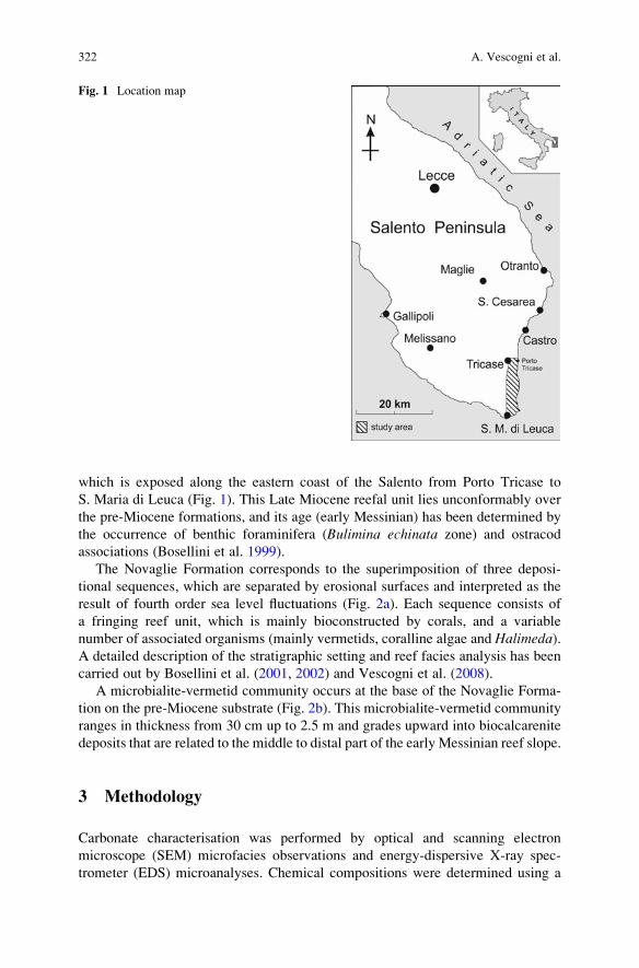
which is exposed along the eastern coast of the Salento from Porto Tricase to
S. Maria di Leuca (Fig. 1). This Late Miocene reefal unit lies unconformably over
the pre-Miocene formations, and its age (early Messinian) has been determined by
the occurrence of benthic foraminifera (Bulimina echinata zone) and ostracod
associations (Bosellini et al. 1999).
The Novaglie Formation corresponds to the superimposition of three deposi-
tional sequences, which are separated by erosional surfaces and interpreted as the
result of fourth order sea level fluctuations (Fig. 2a). Each sequence consists of
a fringing reef unit, which is mainly bioconstructed by corals, and a variable
number of associated organisms (mainly vermetids, coralline algae and Halimeda).A detailed description of the stratigraphic setting and reef facies analysis has been
carried out by Bosellini et al. (2001, 2002) and Vescogni et al. (2008).
A microbialite-vermetid community occurs at the base of the Novaglie Forma-
tion on the pre-Miocene substrate (Fig. 2b). This microbialite-vermetid community
ranges in thickness from 30 cm up to 2.5 m and grades upward into biocalcarenite
deposits that are related to the middle to distal part of the early Messinian reef slope.
3 Methodology
Carbonate characterisation was performed by optical and scanning electron
microscope (SEM) microfacies observations and energy-dispersive X-ray spec-
trometer (EDS) microanalyses. Chemical compositions were determined using a
Fig. 1 Location map
322 A. Vescogni et al.
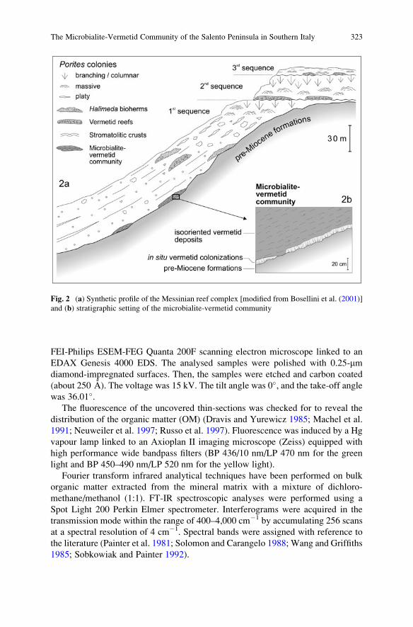
FEI-Philips ESEM-FEG Quanta 200F scanning electron microscope linked to an
EDAX Genesis 4000 EDS. The analysed samples were polished with 0.25-mmdiamond-impregnated surfaces. Then, the samples were etched and carbon coated
(about 250 A). The voltage was 15 kV. The tilt angle was 0�, and the take-off anglewas 36.01�.
The fluorescence of the uncovered thin-sections was checked for to reveal the
distribution of the organic matter (OM) (Dravis and Yurewicz 1985; Machel et al.
1991; Neuweiler et al. 1997; Russo et al. 1997). Fluorescence was induced by a Hg
vapour lamp linked to an Axioplan II imaging microscope (Zeiss) equipped with
high performance wide bandpass filters (BP 436/10 nm/LP 470 nm for the green
light and BP 450–490 nm/LP 520 nm for the yellow light).
Fourier transform infrared analytical techniques have been performed on bulk
organic matter extracted from the mineral matrix with a mixture of dichloro-
methane/methanol (1:1). FT-IR spectroscopic analyses were performed using a
Spot Light 200 Perkin Elmer spectrometer. Interferograms were acquired in the
transmission mode within the range of 400–4,000 cm�1 by accumulating 256 scans
at a spectral resolution of 4 cm�1. Spectral bands were assigned with reference to
the literature (Painter et al. 1981; Solomon and Carangelo 1988; Wang and Griffiths
1985; Sobkowiak and Painter 1992).
Fig. 2 (a) Synthetic profile of the Messinian reef complex [modified from Bosellini et al. (2001)]
and (b) stratigraphic setting of the microbialite-vermetid community
The Microbialite-Vermetid Community of the Salento Peninsula in Southern Italy 323
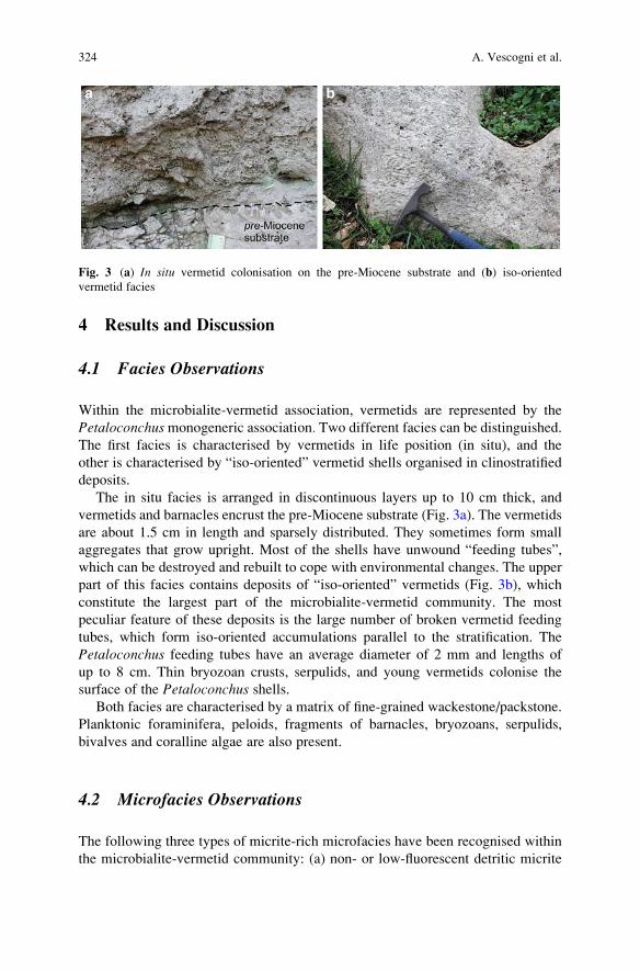
4 Results and Discussion
4.1 Facies Observations
Within the microbialite-vermetid association, vermetids are represented by the
Petaloconchusmonogeneric association. Two different facies can be distinguished.The first facies is characterised by vermetids in life position (in situ), and the
other is characterised by “iso-oriented” vermetid shells organised in clinostratified
deposits.
The in situ facies is arranged in discontinuous layers up to 10 cm thick, and
vermetids and barnacles encrust the pre-Miocene substrate (Fig. 3a). The vermetids
are about 1.5 cm in length and sparsely distributed. They sometimes form small
aggregates that grow upright. Most of the shells have unwound “feeding tubes”,
which can be destroyed and rebuilt to cope with environmental changes. The upper
part of this facies contains deposits of “iso-oriented” vermetids (Fig. 3b), which
constitute the largest part of the microbialite-vermetid community. The most
peculiar feature of these deposits is the large number of broken vermetid feeding
tubes, which form iso-oriented accumulations parallel to the stratification. The
Petaloconchus feeding tubes have an average diameter of 2 mm and lengths of
up to 8 cm. Thin bryozoan crusts, serpulids, and young vermetids colonise the
surface of the Petaloconchus shells.Both facies are characterised by a matrix of fine-grained wackestone/packstone.
Planktonic foraminifera, peloids, fragments of barnacles, bryozoans, serpulids,
bivalves and coralline algae are also present.
4.2 Microfacies Observations
The following three types of micrite-rich microfacies have been recognised within
the microbialite-vermetid community: (a) non- or low-fluorescent detritic micrite
Fig. 3 (a) In situ vermetid colonisation on the pre-Miocene substrate and (b) iso-oriented
vermetid facies
324 A. Vescogni et al.
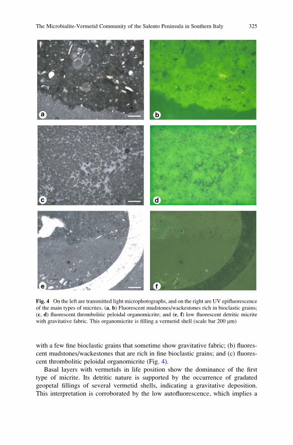
with a few fine bioclastic grains that sometime show gravitative fabric; (b) fluores-
cent mudstones/wackestones that are rich in fine bioclastic grains; and (c) fluores-
cent thrombolitic peloidal organomicrite (Fig. 4).
Basal layers with vermetids in life position show the dominance of the first
type of micrite. Its detritic nature is supported by the occurrence of gradated
geopetal fillings of several vermetid shells, indicating a gravitative deposition.
This interpretation is corroborated by the low autofluorescence, which implies a
Fig. 4 On the left are transmitted light microphotographs, and on the right are UV epifluorescence
of the main types of micrites. (a, b) Fluorescent mudstones/wackestones rich in bioclastic grains;
(c, d) fluorescent thrombolitic peloidal organomicrite; and (e, f) low fluorescent detritic micrite
with gravitative fabric. This organomicrite is filling a vermetid shell (scale bar 200 mm)
The Microbialite-Vermetid Community of the Salento Peninsula in Southern Italy 325
low organic matter content. Iso-oriented vermetid deposits are characterised by the
occurrence of types (b) and (c) of the micrite-rich microfacies. The autofluores-
cence can be explained by two different types of organic matter. The first type
probably derives from decaying metazoan and planktonic organisms, and the
second type is linked to the metabolic activity of microorganisms which have
induced the biomineralisation of the thrombolites.
4.3 FT-IR Analyses
The infrared spectra show bands between 600 and 3,000 cm�1. These spectra
bands include stretching aliphatic bands (uCHali) at 2,949, 2,918 and 2,848 cm�1,
the deformation bands of methyl (dCH3; 1,365 cm�1) and both methyl and
methylene [d(CH2 þ CH3); 1,458 cm�1] groups (Fig. 5). The spectra also show
the band associated with carbonyl and/or carboxyl groups (nC¼O; 1,740 cm�1).
The band associated with the skeletal vibration of more than four methylene
groups [d(CH2)4; 720 cm�1] has also been observed. The nC–O vibration appears
between 1,300 and 1,100 cm�1. In some samples we observed the band nC¼C,
which is probably related to unsaturated compounds such as alkenes and/or
carboxylic acids.
FT-IR was carried out on the organic matter that was extracted from the iso-
oriented vermetid facies (Fig. 5). The different spectra show the different origins
of the three types of micrite-rich microfacies. The intensity of absorption is
related to the abundance of organic matter, and the fluorescent mudstone/wack-
estone presents higher peaks that the thrombolitic and detritic microfacies
(Fig. 5a). This result confirms the epifluorescence observations and indicates
that the organic matter content is distributed the same way like in all three micro-
facies. The detritic micrite shows a very low intensity for the organic matter
functional groups, confirming its abiotic origin (Fig. 5d). The organic matter has
the same intensity and distribution of the functional groups like in the peloidal
thrombolitic micrite, external vermetid shells and filling vermetid shells
(Fig. 5b, c). The FT-IR spectra of this type of micrite are different from those
of the mudstone/wackestone and detritic micrite and are characterised by the
presence of stretching C¼C vibrations. The absence of bands in the 700–900 cm�1
region indicates that the nC¼C band does not belong to an aromatic compound. We
attribute this band to alkenes and/or unsaturated carboxylic acids that may have been
synthesised by microbial communities that precipitated carbonates through their
metabolic activities.
These data, together with microfacies observations, indicate the microbial origin
of most of the micrite within the iso-oriented vermetid facies. Organomicrite
deposition is correlated with the occurrence of a large amount of decaying organic
matter (Reitner and Neuweiler 1995). The occurrence of this organic matter can be
linked to the death of a large number of Petaloconchus individuals.
326 A. Vescogni et al.
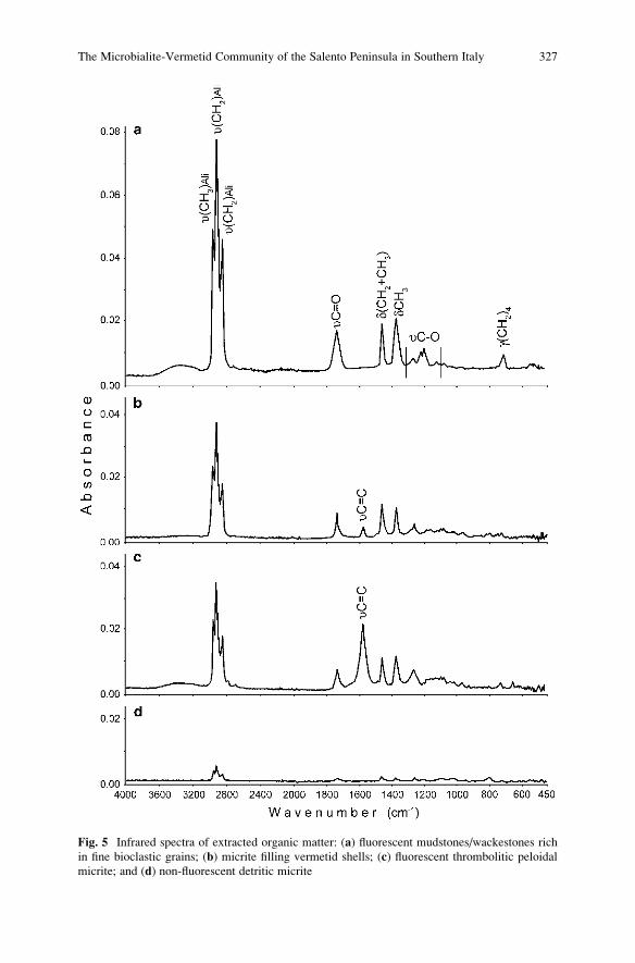
Fig. 5 Infrared spectra of extracted organic matter: (a) fluorescent mudstones/wackestones rich
in fine bioclastic grains; (b) micrite filling vermetid shells; (c) fluorescent thrombolitic peloidal
micrite; and (d) non-fluorescent detritic micrite
The Microbialite-Vermetid Community of the Salento Peninsula in Southern Italy 327
5 Conclusions
Microfacies and vermetid assemblages suggest that the deposition of the
microbialite-vermetid community happened in the following two distinct phases:
(1) vermetid colonisation and detritic micrite deposition and (2) redeposition of the
previous assemblage as iso-oriented vermetid facies rich in mudstones/wackestone
and thrombolitic organomicrite.
The first phase starts with the colonisation of pre-Miocene substrate by verme-
tids. Penecontemporaneous fast inputs of detritic micrite occurred, forcing the
vermetids to produce remarkably long, upward oriented, feeding tubes to avoid
being buried by sedimentation.
The second phase is the redeposition of the previous assemblage as iso-oriented
vermetid facies rich in mudstones/wackestone and thrombolitic organomicrite. The
iso-oriented setting of vermetid conchs is interpreted as the result of a slight
redeposition of the in situ facies because of overloading or another destabilising
process. Vermetid ecological demands do not support life within a muddy substrate.
Therefore, the organomicrite deposition, which must have happened after the death
of the vermetid community, may be correlated to the occurrence of a large amount of
decaying organic matter (Reitner and Neuweiler 1995). The source of this organic
matter can be linked to the death of a large number of Petaloconchus individuals.The nature and distribution of organic matter confirm this interpretation. FT-IR
analyses permit us to distinguish the following two main types of organic source:
(1) a source that derives from decaying metazoan and planktonic organisms, which
characterise the mudstones/wackestones microfacies and (2) a source linked to the
metabolic activities of microorganism that induced thrombolite biomineralisation.
Acknowledgements We thank Prof. Dr. Joachim Reitner for the excellent organisation of Inter-
national Kalkowsky Symposium on the Geobiology of Stromatolites. We are indebted to an
anonymous reviewer for suggestions that greatly improved the manuscript. This research was
funded by the Universita della Calabria, PRIN 2007.
References
Bosellini A, Bosellini FR, Colalongo ML, Parente M, Russo A, Vescogni A (1999) Statigraphic
architecture of the Salento coast from Capo d’Otranto to S. Maria di Leuca (Apulia, southern
Italy). Rivista Italiana di Paleontologia e Stratigrafia 105:397–416
Bosellini FR, Russo A, Vescogni A (2001) Messinian reef-building assemblages of the Salento
Peninsula (southern Italy): palaeobathymetric and palaeoclimatic significance. Palaeogeo-
graphy, Palaeoclimatology, Palaeoecology 175:7–26
Bosellini FR, Russo A, Vescogni A (2002) The messinian reef complex of the Salento Peninsula
(southern Italy). Stratigraphy, facies and paleoenvironmental interpretation. Facies 47:91–112
Dravis JJ, Yurewicz DA (1985) Enhanced carbonate petrography using fluorescence microscopy.
Journal of Sedimentary Petrology 55:795–804
328 A. Vescogni et al.
Machel HG, Mason RA, Mariano AN, Mucci A (1991) Causes and emission of luminescence in
calcite and dolomite. In: Barker CE, Koop OC (Eds.) Luminescence Microscopy and Spec-
troscopy: Qualitative and Quantitative Applications. SEMP Short Course 25:9–25
Neuweiler F, Reitner J, Monty C (Eds.) (1997) Biosedimentology of Microbial Buildups – IGCP
Project No. 380 – Proceedings of 2nd Meeting, G€ottingen/Germany, 1996. Facies 36:195–284
Painter PC, Snyder RW, Starsinic M, Coleman MM, Kuehn DW, Davis A (1981) Concerning the
application of FT-IR to the study of coal: A critical assessment of band assignments and the
application of spectral analysis programs. Applied Spectroscopy 35:475–485
Reitner J, Neuweiler F (1995) Mud Mounds: a polygenetic spectrum of fine-grained carbonate
buildups. Facies 32:1–69
Russo F, Neri C, Mastandrea A, Baracca A (1997) The mud-mound nature of the Cassian platform
margins of the Dolomites. A case history: the Cipit Boulders from Punta Grohmann (Sasso
Piatto Massif, Northern Italy). Facies 36:25–36
Sobkowiak M, Painter P (1992) Determination of the aliphatic and aromatic CH contents of coals
by FT-i.r.: studies of coal extracts. Fuel 71:1105–1125
Solomon PR, Carangelo RM (1988) FT-i.r. analysis of coal. 2. Aliphatic and aromatic hydrogen
concentration. Fuel 67:949–959
Vescogni A, Bosellini FR, Reuter M, Brachert TC (2008) Vermetid reefs and their use
as palaeobathymetric markers: new insights from the Late Miocene of the Mediterranean
(Southern Italy, Crete). Palaeogeography, Palaeoclimatology, Palaeoecology 267:89–101
Wang S-H, Griffiths PR (1985) Resolution enhancement of diffuse reflectance i.r. spectra of coals
by Fourier-self deconvolution. 1. C–H stretching and bending modes. Fuel 64:229–236
The Microbialite-Vermetid Community of the Salento Peninsula in Southern Italy 329

.
The Characterisation of Sedimentary Organic
Matter in Carbonates with Fourier-Transform
Infrared (FTIR) Spectroscopy
Adelaide Mastandrea, Adriano Guido, Fabio Demasi,
Silvestro Antonio Ruffolo, and Franco Russo
1 Introduction
Fourier-transform infrared spectroscopy (FTIR), an analytic technique based on the
interaction between infrared electromagnetic radiation and matter, is of great
importance in the characterisation of kerogens.
The chemical complexity of kerogens derives from the numerous sources of
original organic matter (OM), the selective biodegradation or preservation of
organic moieties, and physical and chemical processes leading to diagenesis (Lis
et al. 2005a).
FTIR studies have revealed bands specific to the chemical structures of complex
kerogen molecules (Painter et al. 1981, 1983; Baruah 1986; Cerny 1955), and
proven their diagnostic value for determining the maturity, type, and oil/gas
generation potential of kerogens (Ganz and Kalkreuth 1987; Christy et al. 1989;
Kister et al. 1990; Lin and Ritz 1993b; Chen et al. 1998).
FTIR is utilised for the identification of organic compounds, both in crystalline
and amorphous forms, through the measurement of the energy absorbed by the
molecules in transition to different vibrational states. Because each functional
group tends to adsorb infrared radiation in a specific wavelength range, it is possible
to detect and discriminate different chemical compounds. FTIR can be used to
identify a broad range of chemical functional groups in a variety of physical states
and it is complementary to other methods such as Raman spectroscopy and gas
chromatography–mass spectroscopy (GC–MS). FTIR generally furnishes a better
sensitivity for minor components than Raman spectroscopy and provides a compa-
rable ability to classify and identify minerals.
A. Mastandrea (*), A. Guido, F. Demasi, S.A. Ruffolo, and F. Russo
Dipartimento di Scienze della Terra, Universita della Calabria, Via P. Bucci Cubo 15b, I-87036
Rende (CS), Italy
e-mail: [email protected]
J. Reitner et al., Advances in Stromatolite Geobiology,Lecture Notes in Earth Sciences 131, DOI 10.1007/978-3-642-10415-2_21,# Springer-Verlag Berlin Heidelberg 2011
331
Infrared analysis, however, does not target particular molecules but rather
detects classes of molecules. Each organic group (i.e., aliphatic C–H bonds) is
present in many compounds with a particular wavelength and this provides a high
sensitivity for mixtures (Anderson et al. 2005; Borrego et al. 1995). The major
classes of biomolecules readily distinguished by FTIR include (a) proteins, peptides
and enzymes, (b) lipids and fatty acids, including cell membranes, (c) carbohydrates
and sugars, and (d) nucleic acids. These classes of functional groups have distinct
infrared absorption-wavelength ranges (Anderson et al. 2005).
The sensitivity of IR analysis for organic molecules, however, depends on the
mineral phase and the granular state, and it is increased if the organics are extracted
or thermally desorbed and concentrated. With extraction, FTIR can detect organic
compounds at parts per billion.
Organic matter type and abundance can be used as a measure of benthic
secondary productivity, terrestrial/aquatic productivity and organic-matter flux in
depositional systems (Palacios-Fest et al. 2005). Considering the close connection
with the biological world, the reconstruction of depositional conditions and carbon-
ate autogenesis processes based on organic-matter data is presently one of the main
topics in carbonates research.
A broad variety of microorganisms, whether phototrophic or not, are implicated
in carbonate autogenesis; however, their direct characterisation in ancient sedi-
mentary systems is limited by the extremely low fossilisation potential of most
microorganisms, especially bacteria. Therefore, the composition of the original
community (primary producers, zooplankton, aerobic and anaerobic bacteria, ben-
thic microorganisms, etc.) is difficult to evaluate by traditional morphological
methods. Many studies have confirmed that organic geochemical techniques can
be used to confirm the original presence of microorganisms.
The main goal of this study was to verify the applicability of the FTIR and
mFTIR analyses to the geochemical characterisation of carbonates with respect to
organic-matter content and thermal evolution.
2 Background Data
2.1 Samples and Microfacies
The analyses were performed on samples of the Calcare di Base (CdB), an outcrop-
ping in the Rossano Basin (Northern Calabria, Italy), which was previously studied
and characterised through molecular fossils and other organic markers (Guido et al.
2007). The study samples were collected from an outcrop located near the Cropalati
village. In this area the CdB succession consisted of two massive fine-grained
calcareous beds interlayered with decimetric laminated marls. These calcareous
beds overlay the diatomitic and marly layers of the Tripoli formation, which
represent the first stage of basin restriction after the open marine conditions
recorded by the Upper Tortonian/Early Messinian clays.
332 A. Mastandrea et al.

Optical and scanning electron microscope (SEM) observations and energy-
dispersive X-ray spectrometer (EDS) microanalyses provided evidence of the
homogenous dominant micrite microfacies and the preservation of the original
aragonitic mineralogical phase. The microfacies were characterised by variably
sized irregular peloids forming clots with an antigravitative fabric, in which
faecal pellets are frequently dispersed. The peloidal micrite showed a typical
thrombolitic microfacies, very rich in organic matter content as indicated by
its bright epifluorescence. The organic matter follows the morphological pattern of
peloids and appeared to be absent in the microsparite areas (Guido et al. 2007).
2.2 Origin and Maturity of Organic Matter
Organic-matter analyses were performed by Guido et al. (2007) using Rock-Eval
pyrolysis, palynofacies observations and molecular-fossil recognition via gas chro-
matography–mass spectrometry.
The Rock-Eval pyrolysis data should be interpreted with caution, due to the
low TOC values, but the Tmax value, at around 400�C, indicated the presence of
unsaturated fatty acids and bb-hopanoids, revealing that the sediment suffered very
low thermal stress (Fig. 1).
The organic matter study revealed a transitional composition between Type II
and Type III kerogen, suggesting a mixture of marine and terrigenous OM (Fig. 1).
These different contributions are supported by biomarker analyses. The n-alkane
distribution indicated the presence of three main biological signatures: algal
(nC18–nC20 compounds), terrestrial (nC27–nC29), and bacterial (nC26–nC28,
with no odd-even carbon-number predominance); see Fig. 2a. The autochthonous
acidic fraction (<C22) and relative level of unsaturated compounds validated the
bacterial signature (Fig. 2c), further evidenced by the presence of hopanes (Fig. 2b)
and the branched n-alkane distributions. These data point to a marine depositional
scenario, interrupted by episodic freshwater inputs, in which carbonate precipita-
tion was induced by bacterial ammonification of amino acids in aerobic conditions
(Guido et al. 2007).
3 Analytical Techniques
Fourier-transform infrared analytical techniques were applied to thin sections,
powdered sediment and organic matter extracted from the mineral matrix. FT-IR
spectroscopic analyses were performed using a Perkin Elmer 100 interfaced with a
Spot Light 200 spectrometer.
Interferograms were acquired in transmission mode within the range of
400–4,000 cm�1 by accumulating 256 scans at a spectral resolution of 4 cm�1.
The Characterisation of Sedimentary Organic Matter in Carbonates 333
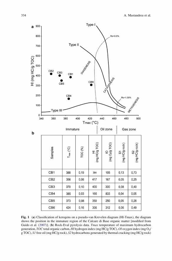
Fig. 1 (a) Classification of kerogens on a pseudo-van Krevelen diagram (HI-Tmax), the diagram
shows the position in the immature region of the Calcare di Base organic matter [modified from
Guido et al. (2007)]. (b) Rock-Eval pyrolysis data. Tmax temperature of maximum hydrocarbon
generation, TOC total organic carbon,HI hydrogen index (mgHC/g TOC),OI oxygen index (mgO2/
g TOC), S1 free oil (mgHC/g rock), S2 hydrocarbons generated by thermal cracking (mgHC/g rock)
334 A. Mastandrea et al.
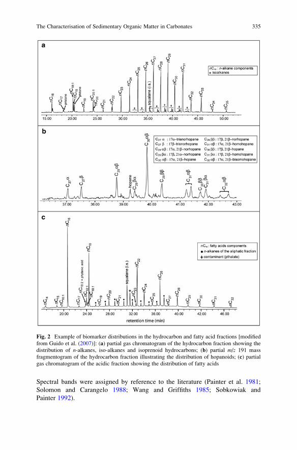
Spectral bands were assigned by reference to the literature (Painter et al. 1981;
Solomon and Carangelo 1988; Wang and Griffiths 1985; Sobkowiak and
Painter 1992).
Fig. 2 Example of biomarker distributions in the hydrocarbon and fatty acid fractions [modified
from Guido et al. (2007)]: (a) partial gas chromatogram of the hydrocarbon fraction showing the
distribution of n-alkanes, iso-alkanes and isoprenoid hydrocarbons; (b) partial m/z 191 mass
fragmentogram of the hydrocarbon fraction illustrating the distribution of hopanoids; (c) partial
gas chromatogram of the acidic fraction showing the distribution of fatty acids
The Characterisation of Sedimentary Organic Matter in Carbonates 335
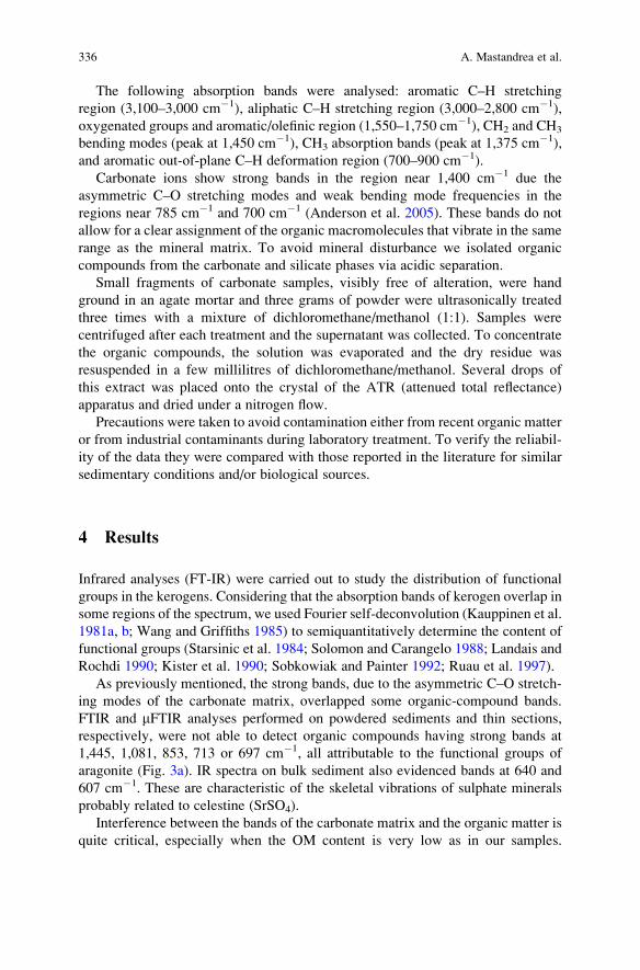
The following absorption bands were analysed: aromatic C–H stretching
region (3,100–3,000 cm�1), aliphatic C–H stretching region (3,000–2,800 cm�1),
oxygenated groups and aromatic/olefinic region (1,550–1,750 cm�1), CH2 and CH3
bending modes (peak at 1,450 cm�1), CH3 absorption bands (peak at 1,375 cm�1),
and aromatic out-of-plane C–H deformation region (700–900 cm�1).
Carbonate ions show strong bands in the region near 1,400 cm�1 due the
asymmetric C–O stretching modes and weak bending mode frequencies in the
regions near 785 cm�1 and 700 cm�1 (Anderson et al. 2005). These bands do not
allow for a clear assignment of the organic macromolecules that vibrate in the same
range as the mineral matrix. To avoid mineral disturbance we isolated organic
compounds from the carbonate and silicate phases via acidic separation.
Small fragments of carbonate samples, visibly free of alteration, were hand
ground in an agate mortar and three grams of powder were ultrasonically treated
three times with a mixture of dichloromethane/methanol (1:1). Samples were
centrifuged after each treatment and the supernatant was collected. To concentrate
the organic compounds, the solution was evaporated and the dry residue was
resuspended in a few millilitres of dichloromethane/methanol. Several drops of
this extract was placed onto the crystal of the ATR (attenued total reflectance)
apparatus and dried under a nitrogen flow.
Precautions were taken to avoid contamination either from recent organic matter
or from industrial contaminants during laboratory treatment. To verify the reliabil-
ity of the data they were compared with those reported in the literature for similar
sedimentary conditions and/or biological sources.
4 Results
Infrared analyses (FT-IR) were carried out to study the distribution of functional
groups in the kerogens. Considering that the absorption bands of kerogen overlap in
some regions of the spectrum, we used Fourier self-deconvolution (Kauppinen et al.
1981a, b; Wang and Griffiths 1985) to semiquantitatively determine the content of
functional groups (Starsinic et al. 1984; Solomon and Carangelo 1988; Landais and
Rochdi 1990; Kister et al. 1990; Sobkowiak and Painter 1992; Ruau et al. 1997).
As previously mentioned, the strong bands, due to the asymmetric C–O stretch-
ing modes of the carbonate matrix, overlapped some organic-compound bands.
FTIR and mFTIR analyses performed on powdered sediments and thin sections,
respectively, were not able to detect organic compounds having strong bands at
1,445, 1,081, 853, 713 or 697 cm�1, all attributable to the functional groups of
aragonite (Fig. 3a). IR spectra on bulk sediment also evidenced bands at 640 and
607 cm�1. These are characteristic of the skeletal vibrations of sulphate minerals
probably related to celestine (SrSO4).
Interference between the bands of the carbonate matrix and the organic matter is
quite critical, especially when the OM content is very low as in our samples.
336 A. Mastandrea et al.
Likewise, it is very difficult to detect organic compounds with absorption bands
without interference but with intensities below the detection limit. To overcome
this problem and to increase the sensitivity of the IR analysis it was necessary to
extract and concentrate the organic compounds.
Fourier self-deconvolution resolved the region around 700 cm�1, discriminating
a peak at 720 cm�1 due to the skeletal vibration of more than four methylene groups
[g(CH2)4]. The aliphatic C–H stretching region (3,000–2,800 cm�1) was resolved
into three spectral bands at 2,950 (asymmetrical CH3 stretching), 2,920 (asymmet-
rical CH2 stretching), and 2,850 cm�1 (symmetrical CH2 stretching); see Fig. 4. The
CH2/CH3 ratio was calculated using the asymmetrical stretching of these bands
(2,920/2,950 cm�1) following Lin and Ritz (1993a). Fourier deconvolution
revealed methyl (dCH3; 1,370 cm�1) and methyl-methylene [d(CH2 þ CH3);
1,460 cm�1] groups (Fig. 4).
The spectra displayed the band assigned to carbonyl and/or carboxyl groups
(nC¼O; 1,740 cm�1). The nC–O vibration appeared between 1,300 and 1,100 cm�1.
Fig. 3 Comparison between IR spectra on (a) powdered dry sediment and (b) extracted organic
fraction (sample CB1)
The Characterisation of Sedimentary Organic Matter in Carbonates 337
5 Interpretation and Discussion
The ratios of several peak heights of the FTIR bands allowed us to characterise the
CdB kerogen. The ratio of carbonyl to aliphatic groups (nC¼O/nCHali) permitted us
to distinguish the marine vs. continental inputs, whereas the ratios for chain length
(dCH2/dCH3) and degree of branching [n(CH2)4/nCHali] allowed us to evaluate the
thermal maturity of the organic matter (Table 1).
The macromolecular structure of kerogen changes during thermal maturation,
and this evolution is evidenced by the FTIR spectroscopic shifts of several bands
(Brown 1955; Kuehn et al. 1982). Ganz and Kalkreuth (1987) proposed the “A
Factor” (2,930 þ 2,860 cm�1)/(2,930 þ 2,860 þ 1,630 cm�1) and the “C Factor”
(1,710 cm�1)/(1,710 þ 1,630 cm�1) to quantify the changes in the abundances of
aliphatic and carbonyl/carboxyl groups, respectively, and to describe kerogen type
and maturity.
Painter et al. (1981) separated the C–H aliphatic stretching region into five
individual bands: 2,956 and 2,864 cm�1 for CH3, 2,923 and 2,849 cm�1 for CH2,
and 2,891 cm�1 for CH. They also proposed that the bands at 2,920 and 2,955 cm�1
are diagnostic for the CH2 and CH3 groups, respectively. The CH2/CH3 ratio
(2,920 cm�1/2,955 cm�1) is sensitive to the degree of carbon-skeleton branching
and to the length of the aliphatic chains.
Fig. 4 Example of an infrared spectrum of extracted organic matter (sample CB1)
338 A. Mastandrea et al.
The ratio of aromatic to aliphatic absorption (i.e., aromaticity) increases with
maturity (Fuller et al. 1982; Mastalerz and Bustin 1993; Iglesias et al. 1995) and
with increasing carbon content in coals (Riesser et al. 1984), with fully aromatic
pure graphite being the ultimate end product. Lis et al. (2005b) calculated nine
FTIR parameters for aromaticity and evaluated their utility as proxies for maturity.
In the Calcare di Base samples, the CH2/CH3 ratio was determined using Fourier
deconvolution of the 2,800–3,000 cm�1 aliphatic-stretching region (Painter et al.
1981; Lin and Ritz 1993a). The value of this ratio ranged from 2 to 2.5, confirming
the immaturity of the organic compounds, which was already deduced from the
biomarker data and maximum hydrocarbon-generation temperature (Tmax).
Considering that the kerogens (lacustrine, marine and continental) were distin-
guishable by their different H/C and O/C ratios, we propose that it is possible to
obtain a preliminary indication of the origin of organic matter in kerogen through
the analysis of the infrared spectra of the functional groups therein.
Type I kerogen (lacustrine), which includes few aromatic and heteroaromatic
compounds and is characterised by a high H/C ratio (~1.5), a high Rock-Eval HI
(>600 mg HC/g TOC) and a low O/C (<0.1) ratio, would contain more intense
aliphatic bands in comparison to Types II and III.
Type II kerogen, which originates from mixed phytoplankton, zooplankton, and
bacterial debris, has a high H/C ratio (1.2–1.5), a high Rock-Eval HI (300–600 mg
HC/g TOC) and a low O/C ratio compared with types III and IV. This kerogen
should exhibit FTIR spectra similar to Type I but with more intense carbonyl and
carboxylic bands.
Type III kerogen (continental) is characterised by a low atomic H/C ratio
(0.7–1.0), a low Rock-Eval HI (50–200 mg HC/g TOC) and a high O/C ratio (up
to ~0.3). This kerogen should contain more intense carbonyl and carboxylic bands
than Types I and II.
To test the reliability of FTIR analyses for the determination of kerogen origin in
our carbonate samples, we compared the carbonyl/aliphatic (nC¼O/nCHali) ratio
with the Rock-Eval data (hydrogen index and oxygen index). The carbonyl groups,
being formed mainly of C¼O bonds, were correlated with the oxygen index, while
the aliphatic groups, being formed mainly of C-H bonds, were correlated with the
hydrogen index.
Table 1 Ratios calculated
from peak heights of the
infrared bands of the Calcare
di Base kerogens
Samples Carbonil to
aliphaticuC ¼ OuCHAli
Degree of
branchinguðCH2Þ4uCHAli
Chain
lengthuðCH2ÞAliuðCH3ÞAli
CB1 0.1893 0.1134 2.4257
CB2 0.1943 0.1094 2.4458
CB3 0.3328 0.1002 2.4479
CB4 0.2206 0.1162 2.4427
CB5 0.2041 0.1000 2.4068
CB6 0.2261 0.0867 2.5126
The Characterisation of Sedimentary Organic Matter in Carbonates 339
In the CdB sections, the nC¼O/nCHali ratio tended to show the same trends in
hydrogen and oxygen indices that Guido et al. (2007) attributed to periodic
increases in the deposition of continental organic matter.
6 Conclusions
The capability of FTIR in the determination of organic molecules has been previ-
ously reported in the literature. In this paper, we demonstrated the potential of FTIR
in the characterisation of fossil organic matter preserved in carbonates.
– IR spectrometry allowed for fast analyses, useful for correlating samples from
different basins.
– The FTIR methodology allowed for the detection of minor mineralogical phases
in a carbonate matrix, such as celestine, not easily identifiable by optical
microscopy.
– The spectra obtained from powder sediments and/or thin sections, because of
interference with the carbonate functional groups, did not yield useful informa-
tion on the content of organic compounds.
– The soundness of the FTIR methodology was appreciable when organic matter
was extracted from the carbonate mineral phase via acidic separation.
– FTIR analysis made it possible to recognise the origin and thermal maturity of
the organic matter preserved in carbonates sediments.
– FTIR can be considered as a complementary technique to the Rock-Eval
pyrolysis and gas chromatography–mass spectrometry in the geochemical char-
acterisation of the main kerogen types.
Acknowledgments Our thanks to Prof. Dr. Joachim Reitner for the excellent organisation of the
International Kalkowsky Symposium: Geobiology of Stromatolites.
We are indebted to the anonymous reviewer for his suggestions, which greatly improved the
manuscript.
This research was founded by Universita della Calabria, PRIN 2007.
References
Anderson MS, Andringa JM, Carlson RW, Conrad P, Hartford W, Shafer M, Soto A, Tsapin AI
(2005) Fourier transform infrared spectroscopy for Mars science. Review of Scientific Instru-
ments 76:1–9
Baruah MK (1986) Assignment of the i.r. absorption band at 1050 cm�1 in lignite humic acid. Fuel
65:1756–1759
Borrego AG, Blanco GC, Prado JG, Dıaz C, Guillen MD (1995) Study of the bitumenes and
pirolizates from some Spanish oil shale by 1H NMR and FTIR spectroscopy. In: Grimalt JO,
Dorronsoro C, Editors, Organic Geochemistry: Developments and Applications to Energy,
Climate, Environmental and Human History, A.I.G.O.A., San Sebastian, 798–800
Brown JK (1955) The infrared spectra of coals. Journal of the Chemical Society: 744–752
340 A. Mastandrea et al.
Cerny J (1955) Aliphatic C–H bond responses in the 900–700 cm�1 region of the FTIR spectra of
coal tars. Fuel Science and Technology International 13:807–818
Chen J, Ping L, Jinchao L (1998) Using kerogen FTIR parameters for determination of organic
facies. Chinese Science Bulletin 43:681–684
Christy AA, Hopland AL, Barth T, Kvalheim OM (1989) Quantitative determination of thermal
maturity in sedimentary organic matter by diffuse reflectance infrared spectroscopy of asphal-
tenes. Organic Geochemistry 14:77–81
Fuller MP, Hamadeh IM, Griffiths PR, Lowenhaupt DE (1982) Diffuse reflectance infrared
spectroscopy of powdered coals. Fuel 61:529–536
Ganz H, Kalkreuth W (1987) Application of infrared spectroscopy to the classification of kerogen-
types and the evolution of source rock and oil shale potentials. Fuel 66:708–711
Guido A, Jacob J, Gautret P, Laggoun-Defarge F, Mastandrea A, Russo F (2007) Molecular fossils
and other organic markers as palaeoenvironmental indicators of the Messinian CdB formation:
normal versus stressed marine deposition (Rossano Basin, Northern Calabria, Italy). Palaeo-
geography, Palaeoclimatology, Palaeoecology 255:265–283
Iglesias MJ, Jimenez A, Laggoun-Defarge F, Suarez-Ruiz I (1995) FTIR study of pure vitrains and
associated coals. Energy & Fuels 9:458–466
Kauppinen JK, Moffatt DJ, Mantsch HH, Cameron DG (1981a) Fourier transforms in the compu-
tation of selfdeconvoluted and first-order derivative spectra of overlapped band contours.
Analytical Chemistry 53:1454–1457
Kauppinen, JK, Moffatt DJ, Mantsch HH, Cameron DG (1981b) Fourier self-deconvolution: a
method for resolving intrinsically overlapped bands. Applied Spectroscopy 35:271–276
Kister J, Guiliano M, Largeau C, Derenne S, Casadevall E (1990) Characterization of chemical
structure, degree of maturation and oil potential of Torbanites (type I kerogens) by quantitative
FT-i.r. spectroscopy. Fuel 69:1356–1361
Kuehn DW, Snyder RW, Davis A, Painter PC (1982) Characterization of vitrinite concentrates. 1.
Fourier Transform infrared studies. Fuel 61:682–694
Landais P, Rochdi A (1990) Reliability of semiquantitative data extracted from transmission
microscopy-Fourier transform infrared spectra of coal. Energy & Fuels 4:290–295
Lin R, Ritz GP (1993a) Reflectance FT-IR microspectroscopy of fossil algae contained in organic-
rich shales. Applied Spectroscopy 47:265–271
Lin R, Ritz GP (1993b) Studying individual macerals using i.r. microspectroscopy, and implica-
tions on oil versus gas/condensate proneness and “low-rank” generation. Organic Geochemis-
try 20:695–706
Lis GP, Schummelmann A, Mastalerz M (2005) D/H ratios and hydrogen exchangeability of type-II
kerogens with increasing thermal maturity. Organic Geochemistry 27:342–353
Lis GP, Mastalerz M, Schimmelmann A, Lewan MD, Stankiewicz BA (2005) FTIR absorption
indices for thermal maturity in comparison with vitrinite reflectance R0 in type-II kerogens
from Devonian black shales. Organic Geochemistry 36:1533–1552
Mastalerz M, Bustin RM (1993) Variation in maceral chemistry within and between coals of
varying rank: an electron microprobe and micro-Fourier transform infra-red investigation.
Journal of Microscopy 171:153–166
Painter PC, Snyder RW, Starsinic M, Coleman MM, Kuehn DW, Davis A (1981) Concerning the
application of FT-IR to the study of coal: a critical assessment of band assignments and the
application of spectral analysis programs. Applied Spectroscopy 35:475–485
Painter PC, Starsinic M, Squires E, Davis AA (1983) Concerning the 1600 cm�1 region in the i.r.
spectrum of coal. Fuel 62:742–744
Palacios-Fest MR, Cohen AS, Lezzar KE, Nahimana L, Tanner BM (2005) Paleolimnological
investigations of anthropogenic environmental change in Lake Tanganyika: III. Physical
stratigraphy and charcoal analysis. Journal of Paleolimnology 34:31–49
Riesser B, Starsinic M, Squires E, Davis A, Painter PC (1984) Determination of aromatic and
aliphatic CH groups in coal by FT-i.r. 2. Studies of coals and vitrinite concentrates. Fuel
63:1253–1261
The Characterisation of Sedimentary Organic Matter in Carbonates 341

Ruau O, Landais P, Gardette JL (1997) Quantitative analysis of powdered organic matter by
transmission infrared microspectroscopy using a diamond-window compression cell. Fuel
76:645–653
Sobkowiak M, Painter P (1992) Determination of the aliphatic and aromatic CH contents of coals
by FT-i.r.: studies of coal extracts. Fuel 71:1105–1125
Solomon PR, Carangelo RM (1988) FT-i.r. analysis of coal. 2. Aliphatic and aromatic hydrogen
concentration. Fuel 67:949–959
Starsinic M, Otake Y, Walker Jr, PL, Painter PC (1984) Application of FT-i.r. spectroscopy to the
determination of COOH groups in coal. Fuel 63:1002–1007
Wang S-H, Griffiths PR (1985) Resolution enhancement of diffuse reflectance i.r. spectra of coals
by Fourier-self deconvolution. 1. C–H stretching and bending modes. Fuel 64:229–236
342 A. Mastandrea et al.
Interactions Between Microbes and Siliceous
Sponges from Upper Jurassic Buildups
of External Prebetic (SE Spain)
Matıas Reolid
1 Introduction
The Upper Jurasic buildups dominated by siliceous sponges and microbialites are
widely known from the Tethyan realm and North Atlantic (Trammer 1989; Gaillard
1983; Matyszkiewicz 1989; Pomoni-Papaioannou et al. 1989; Brachert 1991; Lang
1991; Leinfelder et al. 1993, 1994; Reitner and Neuweiler 1995; Oloriz et al. 2003;
Olivier et al. 2004). They were located and particularly widespread in deep-shelf
settings. Such buildups are unknown in Modern Ocean and only recently, some
sponge buildups have been discovered, but microbialites have not an important role
as builders (Conway et al. 2001; Whitney et al. 2005; Krautter et al. 2006). In the
Jurassic, sponges usually were associated to important amount of microbialites
which favouring the development of buildups as well as the preservation of the
sponge skeletons (Reolid 2007). Upper Jurassic sponge–microbialite buildups are
thus a very good opportunity to study (a) the relationships between sponges and
microbes and (b) related palaeoenvironmental conditions. The objectives of the
paper are precise the architecture of sponge–microbialite buildups and discuss
the equilibrium between microbes and sponges in terms of controlling factors.
The characterization of the interactions between sponges and microbes in this
research are based on Jurassic examples from the Betic Cordillera (SE Spain).
2 Geological Framework
The samples studied are located in the External Prebetic (SE Spain). The Prebetic
Zone comprises the northernmost part of the Betic Cordillera (Fig. 1). The Oxfordian
deposits of the External Prebetic represent the record of the mid shelf environments
M. Reolid
Departamento de Geologıa, Universidad de Jaen, 23071 Jaen, Spain
e-mail: [email protected]
J. Reitner et al., Advances in Stromatolite Geobiology,Lecture Notes in Earth Sciences 131, DOI 10.1007/978-3-642-10415-2_22,# Springer-Verlag Berlin Heidelberg 2011
343
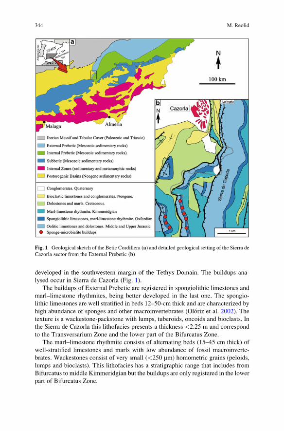
developed in the southwestern margin of the Tethys Domain. The buildups ana-
lysed occur in Sierra de Cazorla (Fig. 1).
The buildups of External Prebetic are registered in spongiolithic limestones and
marl–limestone rhythmites, being better developed in the last one. The spongio-
lithic limestones are well stratified in beds 12–50-cm thick and are characterized by
high abundance of sponges and other macroinvertebrates (Oloriz et al. 2002). The
texture is a wackestone-packstone with lumps, tuberoids, oncoids and bioclasts. In
the Sierra de Cazorla this lithofacies presents a thickness <2.25 m and correspond
to the Transversarium Zone and the lower part of the Bifurcatus Zone.
The marl–limestone rhythmite consists of alternating beds (15–45 cm thick) of
well-stratified limestones and marls with low abundance of fossil macroinverte-
brates. Wackestones consist of very small (<250 mm) homometric grains (peloids,
lumps and bioclasts). This lithofacies has a stratigraphic range that includes from
Bifurcatus to middle Kimmeridgian but the buildups are only registered in the lower
part of Bifurcatus Zone.
Fig. 1 Geological sketch of the Betic Cordillera (a) and detailed geological setting of the Sierra de
Cazorla sector from the External Prebetic (b)
344 M. Reolid
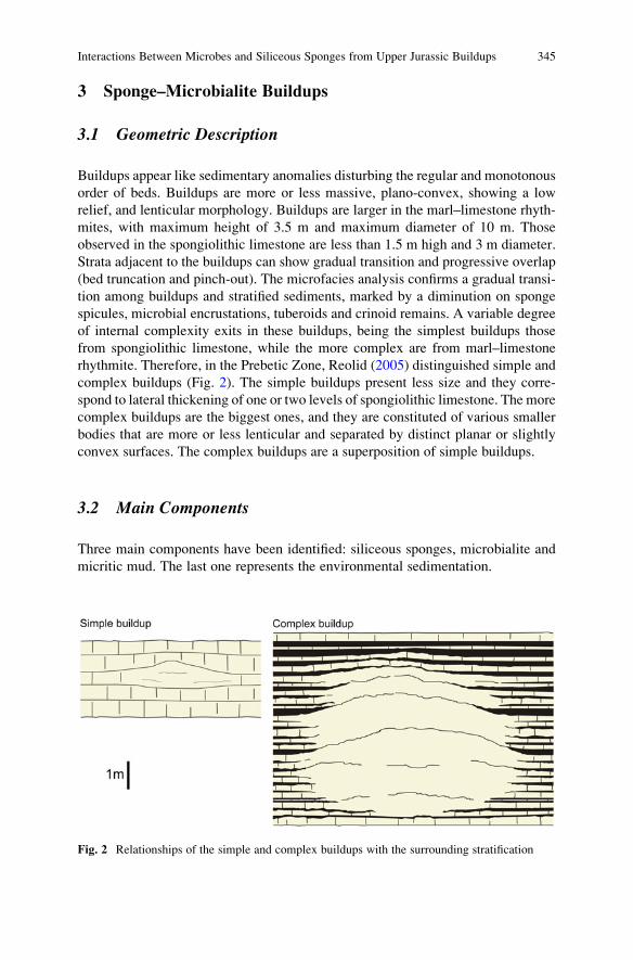
3 Sponge–Microbialite Buildups
3.1 Geometric Description
Buildups appear like sedimentary anomalies disturbing the regular and monotonous
order of beds. Buildups are more or less massive, plano-convex, showing a low
relief, and lenticular morphology. Buildups are larger in the marl–limestone rhyth-
mites, with maximum height of 3.5 m and maximum diameter of 10 m. Those
observed in the spongiolithic limestone are less than 1.5 m high and 3 m diameter.
Strata adjacent to the buildups can show gradual transition and progressive overlap
(bed truncation and pinch-out). The microfacies analysis confirms a gradual transi-
tion among buildups and stratified sediments, marked by a diminution on sponge
spicules, microbial encrustations, tuberoids and crinoid remains. A variable degree
of internal complexity exits in these buildups, being the simplest buildups those
from spongiolithic limestone, while the more complex are from marl–limestone
rhythmite. Therefore, in the Prebetic Zone, Reolid (2005) distinguished simple and
complex buildups (Fig. 2). The simple buildups present less size and they corre-
spond to lateral thickening of one or two levels of spongiolithic limestone. The more
complex buildups are the biggest ones, and they are constituted of various smaller
bodies that are more or less lenticular and separated by distinct planar or slightly
convex surfaces. The complex buildups are a superposition of simple buildups.
3.2 Main Components
Three main components have been identified: siliceous sponges, microbialite and
micritic mud. The last one represents the environmental sedimentation.
Fig. 2 Relationships of the simple and complex buildups with the surrounding stratification
Interactions Between Microbes and Siliceous Sponges from Upper Jurassic Buildups 345
3.2.1 Sponges
The studied sponges were siliceous (Hexactinellida and Demospongia). The sponge
assemblage is dominated by Hexactinellida (77% Hexactinosida and 17% Lychnis-
kosida). Lithistida makes up 6% of the recorded sponges. The mean size of the most
frequent morphotype, the dish-shaped sponge, is 13 cm at the base and 16 cm at the
top of the buildups. The largest specimen is 53 cm. The reticular morphology and
the growth form of the sponge are usually preserved in calcite. The original siliceous
spicules were dissolved and totally replaced by sparitic calcite (Reolid 2007).
3.2.2 Microbialite
The microbialite is the main component in the studied buildups. These are micro-
bial encrustations developed on dish-shaped sponges. Microbialites make up most
of the organo-sedimentary framework, and at a macroscopic scale correspond to
leiolites and stromatolites. The leiolites are more frequents in complex buildups
while the stromatolites are registered mainly in simple buildups. The microbialite
can reach 75 mm thick and commonly corresponds to laminated fabric in thin
section on both the stromatolites and the leiolites. The benthic microbial commu-
nities (BMC) constitute microbialites through the trapping and/or binding of fine
detritic sediment or carbonate precipitation. Microbial encrustations with laminated
fabrics can be constituted of BMC or of consortia made up of BMC and nubecular-
iids (sessile foraminifera). Thus, differentiation of two laminated fabrics is possible
according to the dominant organisms (a) microbial laminated fabrics s. str. and (b)
microbial laminated fabric with nubeculariids (Reolid et al. 2005). The microbial
laminated fabrics s. str. (Reolid et al. 2005) consists of alternating light and dark
laminae (result from grain size alternation). The light laminae comprise very small
micritic particles with maximum size of 20–30 mm (“clotty structures” Monty 1967;
“clotted, peloidal microfabrics” s. Dromart et al. 1994; “minipeloidal fabrics”.
Reitner and Schumann-Kindel 1997) surrounded by microsparitic matrix (“sparitic
peloidal crust fabrics” s. Leinfelder et al. 1993). The microbial laminated fabric
with nubeculariids consists of thin laminae of dark-coloured micrite of microbial
origin colonized by Vinelloidea. Locally, club-shaped microbial over-growths, and
locally finger-like columns develop from an initial planar mat. The stromatolitic
macrofabrics sometimes progress to leiolites, and microbial laminated fabrics s. str.
evolve to microbial laminated fabrics with nubeculariids.
3.2.3 Micritic Mud
The matrix is constituted mainly by micrite with mudstone to wackestone textures
that includes small lumps, tuberoids, crinoids and peloids. Nevertheless, there are
differences in grain size and sorting, depending of the area inside the buildups.
346 M. Reolid
These three main components are placed according to an elementary sequence
composed upward by (a) siliceous sponge, (b) microbialite, and (c) micritic sedi-
ment. This elementary sequence has never surpassed 9 cm. We can differentiate
condensed and expanded elementary sequences (Oloriz et al. 2003) depending on
thickness of microbialite and micritic mud.
The material that fills the interpores within the sponge meshwork consists of
microbialites characterised by dense micrite and micropeloidal fabrics with micro-
sparite, and it is known as automicrite (Leinfelder and Keupp 1995; Reitner and
Neuweiler 1995; Reitner et al. 1995; Neuweiler et al. 2003; Reolid 2007). This
micrite usually is darker than the micrite of the adjacent bedded wackestones,
suggesting a higher proportion of organic matter and/or finely distributed pyrite
(Reitner 1993; Warnkle 1995; Delecat et al. 2001; Delecat and Reitner 2005).
Epibionts from upward and downward surfaces of the sponges differ. Lower
surfaces are colonized by annelids, bryozoans and some attached foraminifera. The
surfaces of upward orientation are characterized by the microbial encrustations
described above. The distribution of epibionts and encrustations is independent of
the position of the sponge (life position or overturned). That distribution has been
recognised in other buildups and sponge facies from other regions (Gaillard 1983;
Matyszkiewicz 1989; Pomoni-Papaioannou et al. 1989).
3.3 Buildup Architecture
The simple buildups and the distinct rock bodies of the complex buildups of the
Prebetic comprise two fundamental stages that represent a growth sequence
(Figs. 3 and 4):
I.- First stage (<30 cm thick) is characterized by the dominance of the siliceous
dish-shaped sponges (65%) where the microbial encrustations are scarce (6.5%).
The sponges are directly growing on top of each other in condensed elementary
sequences (Fig. 3a, b). In this stage the sponges are the main builder. This stage was
called sponge bioherm by Oloriz et al. (2003).
II.- The second stage (the thickest one) is constituted by siliceous dish-shaped
sponges with thick microbial encrustations and high abundance of micritic matrix.
There is a lower proportion of sponges per volume of rock (20%) resulting in an
expanded elementary sequence with BMC as the main builders (42% in complex
buildups and 67% in simple ones). This stage was called microbial lithoherm by
Oloriz et al. (2003). In the simplest, smallest buildups from spongiolithic limestone
themacrofabric of themicrobialite ismainly stromatolite (Fig. 3c, d). Larger buildups
frommarl–limestone rhythmite are characterized by a greater importance of leiolites.
The alternance of sponge dominate phases and microbialite dominate phases in
the buildups is related to cycles of growth interruption and resumption. Sometimes,
the top of such growth cycle is characterized by diminishing of microbial encrusta-
tions, jointly to increasing fragmentation degree of sponges and proportions of
overturned sponges.
Interactions Between Microbes and Siliceous Sponges from Upper Jurassic Buildups 347
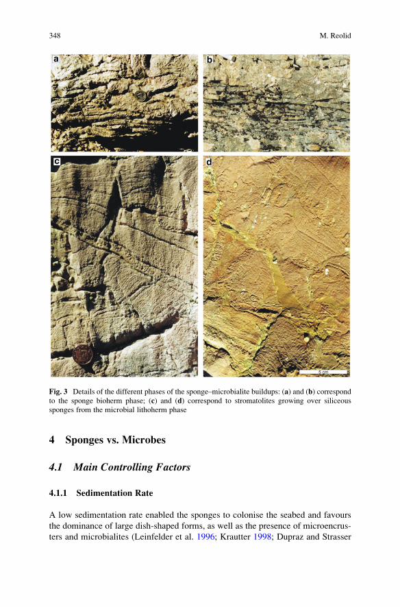
4 Sponges vs. Microbes
4.1 Main Controlling Factors
4.1.1 Sedimentation Rate
A low sedimentation rate enabled the sponges to colonise the seabed and favours
the dominance of large dish-shaped forms, as well as the presence of microencrus-
ters and microbialites (Leinfelder et al. 1996; Krautter 1998; Dupraz and Strasser
Fig. 3 Details of the different phases of the sponge–microbialite buildups: (a) and (b) correspond
to the sponge bioherm phase; (c) and (d) correspond to stromatolites growing over siliceous
sponges from the microbial lithoherm phase
348 M. Reolid
1999; Oloriz et al. 2003; Reolid and Gaillard 2007). The sedimentation rate and
mainly, the siliciclastic influx from emerged areas, varied considerably in the
different lithofacies studied (Oloriz et al. 2002). Spongiolithic limestones could
be related to low sedimentation rates, which enabled prolonged colonization of
the seabed by epibenthos and, especially, by sponges (Oloriz et al. 2003). In
marl–limestone rhythmites the sedimentation rate, and particularly that of siliciclas-
tic influx, was higher than in the spongiolithic limestones, which prevented the
colonization of the seabed by epibenthic, sessile organisms. Thus, sponge coloniza-
tion was limited to reduced areas of lower sedimentation and turbidity at the seabed.
Here, small benthic islands or hillocks formed, suitable for colonization by sessile
epibenthos and for the development of BMC.
4.1.2 Nutrients Availability
The proliferation of siliceous sponges is indicative of the type of organic matter
available; thus, particulate organic matter favours to Lithistida whereas dissolved
Fig. 4 Growth model of the Prebetic buildups, showing the proportions of the main components
in the sponge bioherm phase and microbial lithoherm phase (in complex and simple buildups)
Interactions Between Microbes and Siliceous Sponges from Upper Jurassic Buildups 349
one favours to Hexactinellida (Krautter 1998). Osmotrophic dictyid sponges and
typically dish-shaped sponges, depending on availability of dissolved organic
matter (Krautter 1998), could be especially sensitive to the Reiswig’s “biotic
jungle” (1990). Thus, when the environment experienced wide fluctuations in
productivity, microbialite growth would result during high-productivity phases
(Oloriz et al. 2003), which favoured active bacterial overgrowth lethal for sponges.
Alternatively, microbial overgrowth could start shortly after sponge death, but, in
this case, some control on microbialite growth rate would be envisaged for living
sponges. The distribution of encrusters, which is independent of the position of the
sponge, indicates a post-mortem growth. Probably cyanobacteria and fungi took
advantage of nutrient-rich sponge excretions. However both confrontation and
commensal relationships between microbes and sponges are difficult to demon-
strate from fossil assemblages [i.e. poorly known relationship existing between
sponges and associated microbiotas (Schumann-Kindel et al. 1997), but increasing
research is promising (Manz et al. 2000; Hentschel et al. 2002)].
4.1.3 Light
Microbial over-growths on sponges are registered exclusively on the exposed
surface, and their morphology shows that such development is always upwards.
This could be due to the photophilic nature of the BMC that make up the biogenic
encrustation. The less-illuminated and more protected surfaces of the sponges were
mainly occupied by serpulids, Terebella and bryozoa. According to Reolid (2007),
the growth of BMC was comparatively aggressive for other epibionts.
4.2 Growth Model of Sponge–Microbialite Buildups
When the environment was suitable, and especially when the sedimentation rate
was low enough for sponge growth, large areas of the seabed were colonized,
forming sponge meadows, which later become spongiolithic limestones (Oloriz
et al. 2003; Reolid and Gaillard 2007). Development of buildups was reduced to
local proliferation of BMC.
A sedimentary environment such as that corresponding to the marl–limestone
rhythmites presented adverse characteristics for sponge colonization, because of the
high input of fine-grained detritic particles that caused turbidity at the seabed and
the low consolidation degree (softground).
The occurrence of sponge–microbialite buildups was due to the sporadic coloni-
zation of the seabed by pioneer sponges as envisaged by Gaillard (1983). The
growth of one or more very large pioneer sponges yielded an advantageous surface
for subsequent colonization by other sponges and sessile organisms (serpulids,
crinoids, bryozoa and brachiopods). At the same time, this yielded an optimum
substrate for active BMC, thus favouring the development of thick microbial
350 M. Reolid
encrustation (Fig. 4). This favorable situation resulted in a greater growth of BMC
with respect to the possibilities available in the surrounding muddy bottom (but see
Noffke et al. 2001), and so a sponge–microbialite buildups occurred. Ostrowski
(2002) indicates a context of competition for available surfaces between sponges
and microbes which triggered the buildup growth. The lower part of these buildups,
then, consisting of a large number of dish-shaped sponges (sponge bioherms s.
Oloriz et al. 2003), represents the record of seabed colonization. The thicker upper
interval (microbial lithoherms s. Oloriz et al. 2003), composed almost exclusively
of microbialites (leiolites and stromatolites), corresponds to a phase of proliferating
BMC, where nutrients availability could be a factor determinant triggering the
growth of microbes (Olivier et al. 2004, 2007). The interruptions of growth of the
buildups, registered as discontinuities that delimit growth cycles, imply the alter-
nation of favorable and unfavorable phases for buildup growth.
Sedimentation rate, mainly of siliciclastic influx, controlled colonization and
development of buildups. That also explains (1) the higher size of the sponge–
microbialite buildups in marl–limestone rhythmites than in spongiolithic limestone,
and (2) the higher proportion of micrite in buildups from rhythmite as a result of
competition between microbial growth and background sedimentation. Pioneer
sponges appeared in the rhythmites, providing a much more favorable substrate
for the concentration of epibenthos and for biogenic encrustation.
4.3 Sponge Preservation and Benthic Microbial Communities
It is known from modern species, and reported in fossil counterparts, that micro-
bially induced carbonate precipitation occurs in decaying sponge tissues. The
precipitation of carbonate can occur both within and around the sponge, producing
‘sponge mummies’ (Schwamm-Mumien; Fritz 1958; Gwinner 1976; Fl€ugel andSteiger 1981), while at the same time preserving the original arrangement of the
siliceous spicules. Such rapid lithifying serves to protect fragile skeletons.
Calcite precipitation was induced by the consortium of heterotrophic, symbiotic
bacteria that inhabited the mesohyl of the sponges (Delecat et al. 2001). Recent
sponges are known to be occupied by bacteria (Simpson 1984). They most likely
play a role in the supply of nutrients to sponges. Modern demosponge taxa present
high amounts of bacterial biomass, as much as 50% of the weight of the sponge
biomass (Reitner 1993; Reitner and Schumann-Kindel 1997). Otherwise, according
to Reiswig (1990) the surface of siliceous sponges is covered by a dense veil of
prostal spicules hosting a largely organic microbial layer. Sponges could take
advantage of metabolic products of these microbes (Krautter 1998). High amounts
of organic macromolecules, derived from both microbial communities (from meso-
hyl and surface prostal spicules) are supposed to be responsible for early organo-
mineralization (Reitner et al. 1995). These automicrites are frequently darker than
the surrounding wackestone, due to a higher content of organic matter and pyrite
that developed during the calcification of decaying soft tissue of sponge under the
Interactions Between Microbes and Siliceous Sponges from Upper Jurassic Buildups 351
influence of sulphate-reducing bacteria (Reitner 1993; Reitner and Schumann-
Kindel 1997). However, Neuweiler et al. (2007) explain the calcification of modern
siliceous sponges without microbial participation. Therefore, more studies are
necessaries from fossil and recent siliceous sponges.
5 Conclusions
The relationships between siliceous sponges and microbes in the epicontinental
shelf of the Prebetic were very important because they resulted in the development
of sponge–microbialite buildups which constituted favourable settings for sessile
organisms. Favourable conditions were low sedimentation rate, fluctuations in the
trophic conditions and probably light availability. These paleoenvironmental oscil-
lations would produce alternance of sponge bioherm-microbial lithoherm phases.
The interactions between microbes and siliceous sponges were not restricted to
the development of microbial encrustations, but microbes inhabited the mesohyl
and surface prostal spicules with relationships of commensalism and symbiosis.
The microbes could be related to the precipitation of the automicrite among the
sponge meshwork and then, the preservation of the remains when the siliceous
skeleton is dissolved.
Acknowledgements This research was carried out with the financial support of Research Project
CGL2005-06636-C0201/BTE (Spanish Ministry of Science and Technology) and Research group
RNM-178 of the Junta de Andalucıa. The author is indebted to Jean Louise Sanders (native
English speaker) for grammar review.
References
Brachert TC (1991). Environmental control on fossilization of siliceous sponge assemblages: a
proposal. In: Reitner J, Keupp H (eds) Fossil and recent sponges. Springer, Berlin, pp. 543–553
Conway KW, Krautter M, Barrie JV, Neuweiler M (2001) Hexactinellid sponge reefs on the
Canadian continental shelf: A unique “living fossil”. Geoscience Canada 28:71–78
Delecat S, Reitner J (2005) Sponge communities from the Lower Liassic of Adnet (Northern
Calcareous Alps, Austria). Facies 51:385–404
Delecat S, Peckmann J, Reitner J (2001). Non-rigid cryptic sponges in oyster patch reefs (Lower
Kimmeridgian, Langenberg/Oker, Germany). Facies 45:231–254
Dromart G, Gaillard C, Jansa LF (1994) Deep-marine microbial structures in the Upper Jurassic of
western Tethys. In: Bertrand-Sarfati J, Monty C (eds) Phanerozoic stromatolites II. Kluwer
Academic Publishers, The Netherlands, pp. 295–318
Dupraz C, Strasser A (1999) Microbialites and micro-encrusters in shallow coral bioherms
(Middle to Late Oxfordian, Swiss Jura Mountains). Facies 40:101–130
Fl€ugel E, Steiger T (1981) An Upper Jurassic sponge-algal buildup from the northern Frankenalb,
West Germany. SEPM Special Publication 30:371–397
Fritz G (1958) Schammstotzen, Tuberolithe und Schuttbreccien im Weissen Jura der Schwa-
bischen Alb. Arbeiten aus dem Geologisch-Pal€aontologischen Institut der Technischen
Hochschule Stuttgart, Neue Folge 13:1–118
352 M. Reolid
Gaillard C (1983) Les biohermes a spongiaires et leur environment dans l´Oxfordian du Jura
meridional. Documents des Laboratoires de Geologie de Lyon 90, p. 515
Gwinner MP (1976) Origin of Upper Jurassic limestones of the Swabian Alb (Southwest Germany).
Contributions to Sedimentology 5:1–75
Hentschel U, Hopke J, Horn M, Friedrich AB, Wagner M, Hacker J, Moore BS (2002) Molecular
evidence for a uniform microbial community in sponges from different oceans. Applied
Environmental Microbiology 68:4431–4440
Krautter M (1998) Ecology of siliceous sponges – Application to the environmental interpretation
of the Upper Jurassic sponge facies (Oxfordian) from Spain. Cuadernos de Geologıa Iberica
24:223–239
Krautter M, Conway KW, Barrie JV (2006) Recent hexactinosidan sponge reefs (silicate mounds)
off British Columbia, Canada: Frame-building processes. Journal of Paleontology 80:38–48
Lang B (1991) Baffling, binding, or debris accumulation? Ecology of Upper Jurassic sponge-
bacterial buidups (Oxfordian, Franconian Alb, Southern Germany). In: Reitner J, Keupp H
(eds) Fossil and recent sponges. Springer, Berlin, pp. 516–521
Leinfelder RR, Keupp H (1995). Upper Jurassic mudmounds: Allochthonous sedimentation versus
autochthonous carbonate production. In: Reitner J, Neuweiler F (coord.) Mud mounds: a
polygenetic spectrum of fine-grained carbonate buildups. Facies 32:17–26
Leinfelder RR, Nose M, Schmid DU, Werner W (1993) Microbial crust of the late Jurassic: composi-
tion, palaeoecological significance and importance in reef constructions. Facies 29:195–230
Leinfelder RR, Krautter M, Laternser R, Nose M, Schmid DU, Schweigert G, Werner W, Keupp H,
Brugger H, Herrmann R, Rehfeld-Kiefer U, Koch R, Zeiss A, Schweizer V, Christmann H,
Menges G, Luterbacher H (1994) The origin of the Jurassic reefs: current research develop-
ments and results. Facies 31:1–56
Leinfelder RR, Werner W, Nose M, Schmid DU, Krautter M, Laternser R, Takacs M, Hartmann D
(1996) Paleoecology, growth parameters and dynamics of coral, sponge and microbolite reefs
from the Late Jurassic. G€ottinger Arbeiten zur Geologie und Pal€aontologie Sonderband
2:227–248
Manz W, Arp G, Schumann-Kindel G, Szewzyk U, Reitner J (2000) Widefield deconvolution
epifluorescence microscopy combined with fluorescence in situ hybridazion reveals the spatial
arrangement of bacteria in sponge tissue. Journal of Microbiological Methods 40:125–134
Matyszkiewicz J (1989) Sedimentation and diagenesis of theUpperOxfordian cyanobacterial-sponge
limestones in Piekary near Krakow. Annales Societatis Geologorum Poloniae 59:201–232
Monty LV (1967) Distribution and structure of recent stromatolitic algal mats, eastern Andros
Island, Bahamas. Annales de la Societe Geologique de Belgique 90:55–100
Neuweiler F, D’Orazio V, Immenhauser A, Geipel G, Heise K-H, Cocozza C, Miano TM (2003)
Fulvic acid-like organic compounds control nucleation of marine calcite under suboxic con-
ditions. Geology 31:681–684
Neuweiler F, Daoust I, Bourque PA, Burdige DJ (2007) Degradative calcification of a modern
siliceous sponge from the Great Bahama Bank, the Bahamas: A guide for interpretation of
ancient sponge-bearing limestones. Journal of Sedimentary Research 77:552–563
Noffke N, Gerdes G, Klenke T, Krumbein WE (2001) Microbially induced sedimentary structures –
a new category within the classification of primary sedimentary structures. Journal of Sedi-
mentary Research 71:649–656
Olivier N, Pittet B, Mattioli E (2004) Palaeoenvironmental control on sponge–microbialite reefs and
contemporaneous deep-shelf marl–limestone deposition (Late Oxfordian, southern Germany).
Palaeogeography Palaeoclimatology Palaeoecology 212:233–263
Olivier N, Pittet B, Gaillard C, Hantzpergue P (2007) High-frequency palaeoenvironmental
fluctuations recorded in Jurassic coral- and sponge–microbialite bioconstructions. Comptes
Rendus Palevol 6:21–36
Oloriz F, Reolid M, Rodrıguez-Tovar FJ (2002) Fossil assemblages, lithofacies and taphofacies for
interpreting depositional dynamics in epicontinental Oxfordian (Prebetic Zone, Betic Cordil-
lera, southern Spain). Palaeogeography Palaeoclimatology Palaeoecology 185:53–75
Interactions Between Microbes and Siliceous Sponges from Upper Jurassic Buildups 353
Oloriz F, Reolid M, Rodrıguez-Tovar FJ (2003) A Late Jurassic carbonate ramp colonized by
sponges and benthic microbial communities (External Prebetic, Southern Spain). Palaios
18:528–545
Ostrowski S (2002) Development of sponge-microbial bioherms from Oxfordian of central and
southern Poland. Abstracts VI Inter. Symp. Jurassic System, p. 140
Pomoni-Papaioannou F, Fl€ugel E, Koch R (1989) Depositional environments and diagenesis of
Upper Jurassic subsurface sponge and Tubiphytes reef limestones: Altensteig 1 Well, Western
Molasse Basin, Southern Germany. Facies 21:263–284
Reiswig HM (1990) In situ feeding in two shallow-water hexactinellids sponges. In: R€utzler K,MacIntyre VV, Smith KP (eds) New perspectives in sponge biology. Smithonian Institution
Press, Washington, DC, pp. 504–510
Reitner J (1993) Modern cryptic microbialite/metazoan facies from Lizard Island (Great Barrier
Reef, Australia). Formation and concepts. Facies 29:3–40
Reitner J, Neuweiler F (1995) Mud mounds: a polygenetic spectrum of fine-grained carbonate
buildups. Facies 32:1–70
Reitner J, Schumann-Kindel G (1997) Pyrite in mineralized sponge tissue: product of sulfate
reducing sponge related bacteria? Facies 36:272–276
Reitner J, Neuweiler F, Gautret P (1995) Modern and fossils automicrites: Implications for mud
mounds genesis. In: Reitner J, Neuweiler F (coord) Mud mounds: a polygenetic spectrum of
fine-grained carbonate buildups. Facies 32:4–17
Reolid M (2005) Dinamica eco-sedimentaria durante el Oxfordiense medio-Kimmeridgiense
temprano en la Zona Prebetica: Interpretacion ecoestratigrafica y secuencial. PhD Thesis,
Universidad de Granada 2003, Granada, p. 254
Reolid M (2007) Taphonomy of the Oxfordian-lowermost Kimmeridgian siliceous sponges of the
prebetic zone (Southern Iberia). Journal of Taphonomy 5:71–90
Reolid M, Gaillard C (2007) Microtaphonomy of bioclasts and paleoecology of microencrusters
from the Upper Jurassic spongiolithic limestones (External Prebetic, southern Spain). Facies
53:97–112
Reolid M, Gaillard C, Oloriz F, Rodrıguez-Tovar FJ (2005) Microbial encrustations from the
Middle Oxfordian-earliest Kimmeridgian lithofacies in the Prebetic Zone (Betic Cordillera,
southern Spain): characterization, distribution and controlling factors. Facies 50:529–543
Schumann-Kindel G, Bergbauer M, Manz W, Szewzyk U, Reitner J (1997) Aerobic and anaerobic
microorganims in modern sponges: a possible relationship to fossilization-processes. Facies
36:268–272
Simpson TL (1984) The cell biology of sponges. Springer, New York, p. 662
Trammer J (1989) Middle to upper Oxfordian sponges of the Polish Jura. Acta Geologica Polonica
39:49–91
Warnkle K (1995) Calcification processes of siliceous sponges in Visean Limestones (Counties
Sligo and Leitrim, northwestern Ireland). Facies 33:215–228
Whitney F, Conway K, Thomson R, Barrie V, Krautter M, Mungov G (2005) Oceanographic
habitat of sponge reefs on the western Canadian Continental Shelf. Continental Shelf Research
25:211–226
354 M. Reolid
Aftermath of the Triassic–Jurassic Boundary
Crisis: Spiculite Formation on Drowned Triassic
Steinplatte Reef-Slope by Communities
of Hexactinellid Sponges (Northern
Calcareous Alps, Austria)
Stefan Delecat, Gernot Arp, and Joachim Reitner
1 Introduction
In Upper Rhaetian time, reef evolution culminated in highly diverse ecosystems
that notably flourished along the northern shelf of the former Tethys ocean. Several
of these buildups constructed by branching corals and coralline sponges (Demos-
pongiae with secondary basal skeletons) are nowadays exposed in the Northern
Calcareous Alps, where they are found both at the northern and southern margins of
the Upper Triassic Carbonate Platform. Further, the record of capping Lower
Liassic sediments show that all of the reefs must have been abruptly disappeared
at the Triassic–Jurassic boundary. The reasons for their sudden decease which took
place simultaneously with a world wide mass extinction event are still under
discussion and less is even known about its effect on subsequent Liassic benthic
faunas. After the extinction of important reef-building organisms most likely a sea
level rise and changes in sedimentation led to a radiation of sponges from deep
water environments (Mostler 1989a, b, 1990a, b) onto some of the drowned
buildups. B€ohm (1992) already assumed that Liassic sponge diversity in the alpine
region was strongly influenced by the extant Triassic palaeorelief. In order to
discover the crucial factors that controlled the recolonization of the drowned
areas, Liassic sponge communities were studied from localities of the former
Triassic reef slopes and adjacent basinal settings.
In this paper a first description of autochthonous Hettangian sponge commu-
nities situated on the upper slope of Steinplatte “Reef” (N of Waidring, Austria) is
provided. Although most of the sponge skeletons are highly collapsed, they are
preserved in situ and arranged in spicular mats with sediments partially bound by
microbialites, so that the spiculite facies bears analogy to the Schn€oll facies that is
S. Delecat, G. Arp, and J. Reitner (*)
Geoscience Centre, University of G€ottingen, Geobiology Division, Goldschmidtstrabe 3, 37077,G€ottingen, Germany
e-mail: [email protected]
J. Reitner et al., Advances in Stromatolite Geobiology,Lecture Notes in Earth Sciences 131, DOI 10.1007/978-3-642-10415-2_23,# Springer-Verlag Berlin Heidelberg 2011
355
known from the slope of the Adnet Reef, near Hallein, Austria (Delecat and Reitner
2005). Both localities are favorite sites to examine entire Lower Jurassic Porifera
communities as well as sponge taphonomy in fossil environments. Additional to the
documentation of these rarely preserved sponge species, the paper’s aim is to
evaluate probable interactions between taphonomic processes in collapsing sponges
and abiotic factors like variances of the Liassic geochemical milieu or aspects of
sedimentation and diagenesis. Hereby a focus lays on the study of early diagenetic
stromatactis cavities that are a characteristic feature of the Steinplatte spiculite and
whose origin seems to be closely related to the decay of sponge organic matter.
2 Regional Setting
The Upper Triassic Carbonate Platform in the Northern Calcareous Alps extended
in W–E direction and separated the former Tethyian deep water realm in the south
from the northern epicontinental K€ossen Basin. The famous Steinplatte “reef”,
whose crest is cropping out at the Sonnenw€ande hill north of Waidring and Lofer
(Austria) was situated on the northern slope of this platform (Fig. 1). As facies
analysis by Stanton and Fl€ugel (1989, 1995) have shown, only a minor part of the
Steinplatte buildup is formed by a real reef framework, thus the whole complex is
rather described as an “accretionary distally steepened ramp” the top of which was
partly overgrown by separate bushlike corals (capping facies) in a late Upper
Fig. 1 Northern slope section of Steinplatte “Reef” (N of Waidring, Austria) and its paleogeo-
graphic position
356 S. Delecat et al.

Rhaetian stage. At the end of Triassic, the coral growth stopped concurrent to one of
the five most severe mass extinction events in earths Phanerozoic history. The
reason for that is difficult to evaluate. Although the palaeo-relief of the carbonate
platform still existed until Middle Liassic time, just on top of the buildup the
Triassic–Jurassic interval encompassing the decease of the coral fauna is concealed
by a small sedimentary break. Only some traces of fresh-water diagenesis in the
capping facies might indicate short-termed falls of the sea level at Steinplatte
buildup (Stanton and Fl€ugel 1989). In contrast to the elevated platform position,
in adjacent basins sedimentation continuously passed into grey cherty limestones
(Hettangian Kendlbach Formation and Hettangian-Sinemurian Scheibelberg For-
mation). Predominantly the latter Scheibelberg Limestones are characterized by
varying, often high amounts of siliceous sponges and/or siliceous bulbs (Mostler
1986; Krainer and Mostler 1997). They are frequently exposed at the toe of
Steinplatte buildup slope at Kammerk€ohr Alpe and also at several outcrops in the
Unken syncline (e.g. at Scheibelberg locality and at Karnergraben) that extends
north of the Steinplatte buildup.
First Hettangian to Sinemurian sedimentation at the northern slope of Steinplatte
“reef” shows an onlap of red condensed limestones indicating a drowning of the
platform during Lower Liassic time. A wedge of these sediments is cut by a trail
surrounding the Plattenkogel hill, north of Steinplatte summit (Fig. 2). At the “red
Fig. 2 (a) Location of Steinplatte mountain. (b) Location of sampling sites at Steinplatte. (c)
Schematic sketch of the outcrop “red wall” at Kammerk€ohr-Alpe (tourist trail along northern slopeof Steinplatte/Plattenkogel, view to the south). Jurassic onlap sedimentation comprises red Adnet
limestones with several slump folds, slided megabreccias (checkered) and debrites (dotted)
Aftermath of the Triassic–Jurassic Boundary Crisis 357
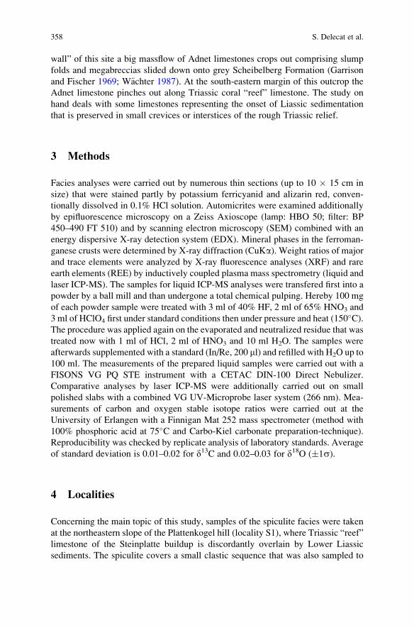
wall” of this site a big massflow of Adnet limestones crops out comprising slump
folds and megabreccias slided down onto grey Scheibelberg Formation (Garrison
and Fischer 1969; W€achter 1987). At the south-eastern margin of this outcrop the
Adnet limestone pinches out along Triassic coral “reef” limestone. The study on
hand deals with some limestones representing the onset of Liassic sedimentation
that is preserved in small crevices or interstices of the rough Triassic relief.
3 Methods
Facies analyses were carried out by numerous thin sections (up to 10 � 15 cm in
size) that were stained partly by potassium ferricyanid and alizarin red, conven-
tionally dissolved in 0.1% HCl solution. Automicrites were examined additionally
by epifluorescence microscopy on a Zeiss Axioscope (lamp: HBO 50; filter: BP
450–490 FT 510) and by scanning electron microscopy (SEM) combined with an
energy dispersive X-ray detection system (EDX). Mineral phases in the ferroman-
ganese crusts were determined by X-ray diffraction (CuKa). Weight ratios of major
and trace elements were analyzed by X-ray fluorescence analyses (XRF) and rare
earth elements (REE) by inductively coupled plasma mass spectrometry (liquid and
laser ICP-MS). The samples for liquid ICP-MS analyses were transfered first into a
powder by a ball mill and than undergone a total chemical pulping. Hereby 100 mg
of each powder sample were treated with 3 ml of 40% HF, 2 ml of 65% HNO3 and
3 ml of HClO4 first under standard conditions then under pressure and heat (150�C).
The procedure was applied again on the evaporated and neutralized residue that was
treated now with 1 ml of HCl, 2 ml of HNO3 and 10 ml H2O. The samples were
afterwards supplemented with a standard (In/Re, 200 ml) and refilled with H2O up to
100 ml. The measurements of the prepared liquid samples were carried out with a
FISONS VG PQ STE instrument with a CETAC DIN-100 Direct Nebulizer.
Comparative analyses by laser ICP-MS were additionally carried out on small
polished slabs with a combined VG UV-Microprobe laser system (266 nm). Mea-
surements of carbon and oxygen stable isotope ratios were carried out at the
University of Erlangen with a Finnigan Mat 252 mass spectrometer (method with
100% phosphoric acid at 75�C and Carbo-Kiel carbonate preparation-technique).
Reproducibility was checked by replicate analysis of laboratory standards. Average
of standard deviation is 0.01–0.02 for d13C and 0.02–0.03 for d18O (�1s).
4 Localities
Concerning the main topic of this study, samples of the spiculite facies were taken
at the northeastern slope of the Plattenkogel hill (locality S1), where Triassic “reef”
limestone of the Steinplatte buildup is discordantly overlain by Lower Liassic
sediments. The spiculite covers a small clastic sequence that was also sampled to
358 S. Delecat et al.
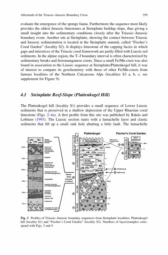
evaluate the emergence of the sponge fauna. Furthermore the sequence most likely
provides the oldest Jurassic limestones at Steinplatte buildup slope, thus giving a
small insight into the sedimentary conditions closely after the Triassic–Jurassic
boundary event. Another site at Steinplatte, showing the contact between Triassic
and Jurassic sedimentation is located at the Steinplatte summit, called “Fischer’s
Coral Garden” (locality S2). It displays limestone of the capping facies in which
gaps and interstices of the Triassic coral framework are partly filled with Liassic red
sediments. In the alpine region, the T–J boundary interval is often characterized by
sedimentary breaks and ferromanganese crusts. Since a small Fe/Mn crust was also
found in association to the Liassic sequence at Steinplatte/Plattenkogel hill, it was
of interest to compare its geochemistry with those of other Fe/Mn-crusts from
famous localities of the Northern Calcareous Alps (localities S3 a, b, c; see
supplement for Figure 9).
4.1 Steinplatte Reef-Slope (Plattenkogel Hill)
The Plattenkogel hill (locality S1) provides a small sequence of Lower Liassic
sediments that is preserved in a shallow depression of the Upper Rhaetian coral
limestone (Figs. 2–4a). A first profile from this site was published by Rakus and
Lobitzer (1993). The Liassic section starts with a lumachelle layer and clastic
sediments that fill up a small sink hole abutting a little fault. The lumachelle
Fig. 3 Profiles of Triassic–Jurassic boundary sequences from Steinplatte localities: Plattenkogel
hill (locality S1) and “Fischer’s Coral Garden” (locality S2). Numbers of layers/samples corre-
spond with Figs. 5 and 9
Aftermath of the Triassic–Jurassic Boundary Crisis 359
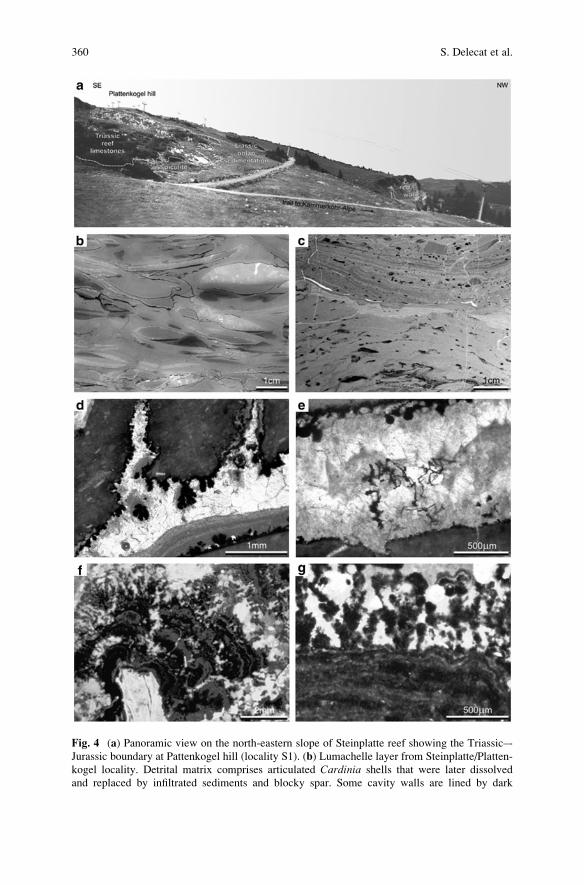
Fig. 4 (a) Panoramic view on the north-eastern slope of Steinplatte reef showing the Triassic–-
Jurassic boundary at Pattenkogel hill (locality S1). (b) Lumachelle layer from Steinplatte/Platten-
kogel locality. Detrital matrix comprises articulated Cardinia shells that were later dissolved
and replaced by infiltrated sediments and blocky spar. Some cavity walls are lined by dark
360 S. Delecat et al.
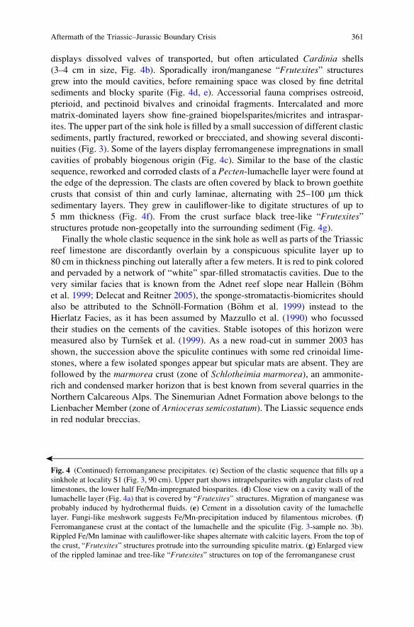
displays dissolved valves of transported, but often articulated Cardinia shells
(3–4 cm in size, Fig. 4b). Sporadically iron/manganese “Frutexites” structures
grew into the mould cavities, before remaining space was closed by fine detrital
sediments and blocky sparite (Fig. 4d, e). Accessorial fauna comprises ostreoid,
pterioid, and pectinoid bivalves and crinoidal fragments. Intercalated and more
matrix-dominated layers show fine-grained biopelsparites/micrites and intraspar-
ites. The upper part of the sink hole is filled by a small succession of different clastic
sediments, partly fractured, reworked or brecciated, and showing several disconti-
nuities (Fig. 3). Some of the layers display ferromangenese impregnations in small
cavities of probably biogenous origin (Fig. 4c). Similar to the base of the clastic
sequence, reworked and corroded clasts of a Pecten-lumachelle layer were found at
the edge of the depression. The clasts are often covered by black to brown goethite
crusts that consist of thin and curly laminae, alternating with 25–100 mm thick
sedimentary layers. They grew in cauliflower-like to digitate structures of up to
5 mm thickness (Fig. 4f). From the crust surface black tree-like “Frutexites”structures protude non-geopetally into the surrounding sediment (Fig. 4g).
Finally the whole clastic sequence in the sink hole as well as parts of the Triassic
reef limestone are discordantly overlain by a conspicuous spiculite layer up to
80 cm in thickness pinching out laterally after a few meters. It is red to pink colored
and pervaded by a network of “white” spar-filled stromatactis cavities. Due to the
very similar facies that is known from the Adnet reef slope near Hallein (B€ohmet al. 1999; Delecat and Reitner 2005), the sponge-stromatactis-biomicrites should
also be attributed to the Schn€oll-Formation (B€ohm et al. 1999) instead to the
Hierlatz Facies, as it has been assumed by Mazzullo et al. (1990) who focussed
their studies on the cements of the cavities. Stable isotopes of this horizon were
measured also by Turnsek et al. (1999). As a new road-cut in summer 2003 has
shown, the succession above the spiculite continues with some red crinoidal lime-
stones, where a few isolated sponges appear but spicular mats are absent. They are
followed by the marmorea crust (zone of Schlotheimia marmorea), an ammonite-
rich and condensed marker horizon that is best known from several quarries in the
Northern Calcareous Alps. The Sinemurian Adnet Formation above belongs to the
Lienbacher Member (zone of Arnioceras semicostatum). The Liassic sequence endsin red nodular breccias.
�
Fig. 4 (Continued) ferromanganese precipitates. (c) Section of the clastic sequence that fills up a
sinkhole at locality S1 (Fig. 3, 90 cm). Upper part shows intrapelsparites with angular clasts of red
limestones, the lower half Fe/Mn-impregnated biosparites. (d) Close view on a cavity wall of the
lumachelle layer (Fig. 4a) that is covered by “Frutexites” structures. Migration of manganese was
probably induced by hydrothermal fluids. (e) Cement in a dissolution cavity of the lumachelle
layer. Fungi-like meshwork suggests Fe/Mn-precipitation induced by filamentous microbes. (f)
Ferromanganese crust at the contact of the lumachelle and the spiculite (Fig. 3-sample no. 3b).
Rippled Fe/Mn laminae with cauliflower-like shapes alternate with calcitic layers. From the top of
the crust, “Frutexites” structures protrude into the surrounding spiculite matrix. (g) Enlarged view
of the rippled laminae and tree-like “Frutexites” structures on top of the ferromanganese crust
Aftermath of the Triassic–Jurassic Boundary Crisis 361
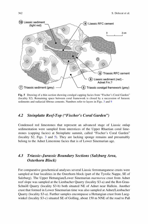
4.2 Steinplatte Reef-Top (“Fischer’s Coral Garden”)
Condensed red limestones that represent an advanced stage of Liassic onlap
sedimentation were sampled from interstices of the Upper Rhaetian coral lime-
stones (capping facies) at Steinplatte summit, called “Fischer’s Coral Garden”
(locality S2, Figs. 3 and 5). They are lacking sponge remains and presumably
belong to the Adnet Limestone facies that is of Lower Sinemurian age.
4.3 Triassic–Jurassic Boundary Sections (Salzburg Area,Osterhorn Block)
For comparative geochemical analyses several Liassic ferromanganese crusts were
sampled at four localities in the Osterhorn block (part of the Tyrolic Nappe, SE of
Salzburg). The Upper Hettangian/Lower Sinemurian marmorea crust from Adnet
reef slope was sampled at the Lienbacher Quarry (locality S3-a) and the Rot-Grau-
Schn€oll Quarry (locality S3-b) both situated NE of Adnet near Hallein. Another
crust that formed in Lower Sinemurian time was also sampled at Adnet/Lienbacher
Quarry (locality S3-a). Further samples encompasse a Hettangian crust from Lueg-
winkel (locality S3-c) situated SE of Golling, about 150 m NNE of the road to Paß
Fig. 5 Drawing of a thin section showing coralgal capping facies from “Fischer’s Coral Garden”
(locality S2). Remaining space between coral framework is closed by a succession of Jurassic
sediments and radiaxial fibrous cements. Numbers refer to layers in Figs. 3 and 9
362 S. Delecat et al.
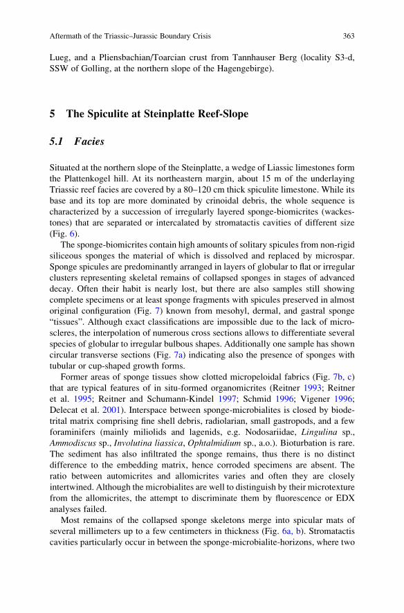
Lueg, and a Pliensbachian/Toarcian crust from Tannhauser Berg (locality S3-d,
SSW of Golling, at the northern slope of the Hagengebirge).
5 The Spiculite at Steinplatte Reef-Slope
5.1 Facies
Situated at the northern slope of the Steinplatte, a wedge of Liassic limestones form
the Plattenkogel hill. At its northeastern margin, about 15 m of the underlaying
Triassic reef facies are covered by a 80–120 cm thick spiculite limestone. While its
base and its top are more dominated by crinoidal debris, the whole sequence is
characterized by a succession of irregularly layered sponge-biomicrites (wackes-
tones) that are separated or intercalated by stromatactis cavities of different size
(Fig. 6).
The sponge-biomicrites contain high amounts of solitary spicules from non-rigid
siliceous sponges the material of which is dissolved and replaced by microspar.
Sponge spicules are predominantly arranged in layers of globular to flat or irregular
clusters representing skeletal remains of collapsed sponges in stages of advanced
decay. Often their habit is nearly lost, but there are also samples still showing
complete specimens or at least sponge fragments with spicules preserved in almost
original configuration (Fig. 7) known from mesohyl, dermal, and gastral sponge
“tissues”. Although exact classifications are impossible due to the lack of micro-
scleres, the interpolation of numerous cross sections allows to differentiate several
species of globular to irregular bulbous shapes. Additionally one sample has shown
circular transverse sections (Fig. 7a) indicating also the presence of sponges with
tubular or cup-shaped growth forms.
Former areas of sponge tissues show clotted micropeloidal fabrics (Fig. 7b, c)
that are typical features of in situ-formed organomicrites (Reitner 1993; Reitner
et al. 1995; Reitner and Schumann-Kindel 1997; Schmid 1996; Vigener 1996;
Delecat et al. 2001). Interspace between sponge-microbialites is closed by biode-
trital matrix comprising fine shell debris, radiolarian, small gastropods, and a few
foraminifers (mainly miliolids and lagenids, e.g. Nodosariidae, Lingulina sp.,
Ammodiscus sp., Involutina liassica, Ophtalmidium sp., a.o.). Bioturbation is rare.
The sediment has also infiltrated the sponge remains, thus there is no distinct
difference to the embedding matrix, hence corroded specimens are absent. The
ratio between automicrites and allomicrites varies and often they are closely
intertwined. Although the microbialites are well to distinguish by their microtexture
from the allomicrites, the attempt to discriminate them by fluorescence or EDX
analyses failed.
Most remains of the collapsed sponge skeletons merge into spicular mats of
several millimeters up to a few centimeters in thickness (Fig. 6a, b). Stromatactis
cavities particularly occur in between the sponge-microbialite-horizons, where two
Aftermath of the Triassic–Jurassic Boundary Crisis 363
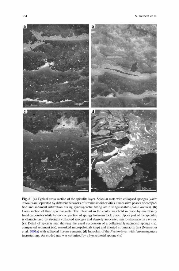
Fig. 6 (a) Typical cross section of the spiculite layer. Spicular mats with collapsed sponges (whitearrows) are separated by different networks of stromatactoid cavities. Successive phases of compac-
tion and sediment infiltration during syndiagenetic tilting are distinguishable (black arrows). (b)Cross section of three spicular mats. The intraclast in the center was hold in place by microbially
fixed carbonates while below compaction of spongy horizons took place. Upper part of the spiculite
is characterized by strongly collapsed sponges and densely associated micro-stromatactis cavities.
(c): Detail of spicular mat showing the usual succession of a collapsed lyssacinosid sponge (ly),
compacted sediment (cs), reworked micropeloidals (mp) and aborted stromatactis (as) (Neuweiler
et al. 2001a) with radiaxial fibrous cements. (d) Intraclast of the Pecten-layer with ferromanganese
incrustations. An eroded gap was colonized by a lyssacinosid sponge (ly)
364 S. Delecat et al.
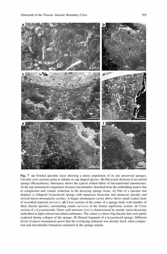
Fig. 7 (a) Eroded spiculite layer showing a dense population of in situ preserved sponges.
Circular cross sections point to tubular or cup shaped species. (b) Dictyonal skeleton of an euretid
sponge (Hexactinosa). Interspace shows the typical clotted fabric of micropeloidal automicrites.
At the top stromatactis originated, because microbialites detached from the embedding matrix due
to compaction and volume reduction in the decaying sponge tissue. (c) Part of a spicular mat
displays a collapsed lyssacinosid sponge with numerous hexactine and monaxon spicules and
several micro-stromatactis cavities. A bigger stromatactis cavity above shows small scaled clasts
of reworked material (arrows). (d) Cross section of the center of a sponge body with bundles of
thick diactin spicules, surrounding canals (arrows) of the former aquiferous system. (e) Cross
section of a Lyssacinosida. Outer wall structure (ws) is characterized by mainly micro-hexactins
embedded in light-colored microbial carbonates. The center (c) shows big diactins that were partly
scattered during collapse of the sponge. (f) Dermal fragment of a lyssacinosid sponge. Different
levels of micro-stromatactis prove that the overlaying sediment was already fixed, when compac-
tion and microbialite formation continued in the sponge remain
Aftermath of the Triassic–Jurassic Boundary Crisis 365
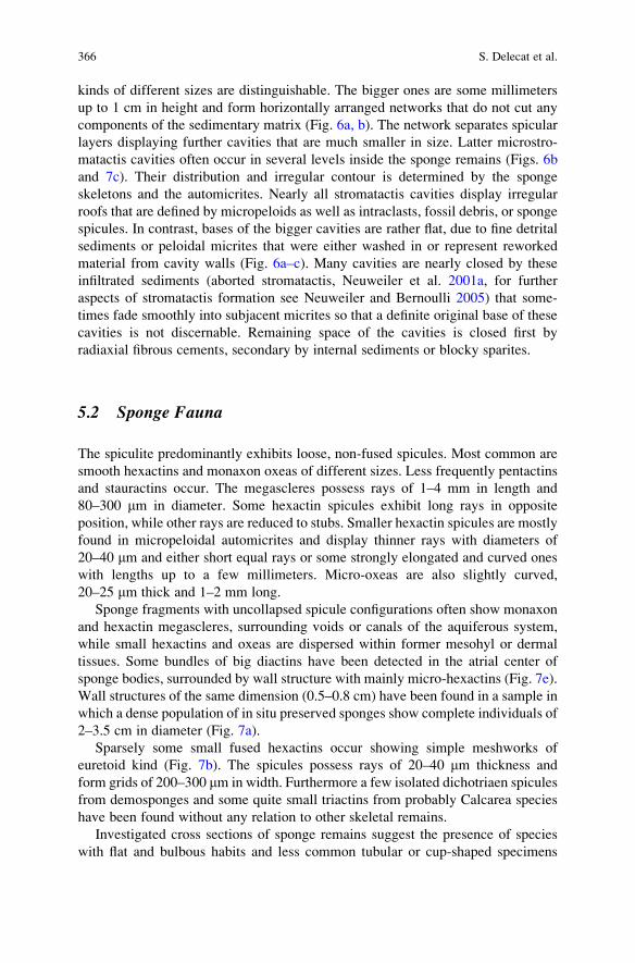
kinds of different sizes are distinguishable. The bigger ones are some millimeters
up to 1 cm in height and form horizontally arranged networks that do not cut any
components of the sedimentary matrix (Fig. 6a, b). The network separates spicular
layers displaying further cavities that are much smaller in size. Latter microstro-
matactis cavities often occur in several levels inside the sponge remains (Figs. 6b
and 7c). Their distribution and irregular contour is determined by the sponge
skeletons and the automicrites. Nearly all stromatactis cavities display irregular
roofs that are defined by micropeloids as well as intraclasts, fossil debris, or sponge
spicules. In contrast, bases of the bigger cavities are rather flat, due to fine detrital
sediments or peloidal micrites that were either washed in or represent reworked
material from cavity walls (Fig. 6a–c). Many cavities are nearly closed by these
infiltrated sediments (aborted stromatactis, Neuweiler et al. 2001a, for further
aspects of stromatactis formation see Neuweiler and Bernoulli 2005) that some-
times fade smoothly into subjacent micrites so that a definite original base of these
cavities is not discernable. Remaining space of the cavities is closed first by
radiaxial fibrous cements, secondary by internal sediments or blocky sparites.
5.2 Sponge Fauna
The spiculite predominantly exhibits loose, non-fused spicules. Most common are
smooth hexactins and monaxon oxeas of different sizes. Less frequently pentactins
and stauractins occur. The megascleres possess rays of 1–4 mm in length and
80–300 mm in diameter. Some hexactin spicules exhibit long rays in opposite
position, while other rays are reduced to stubs. Smaller hexactin spicules are mostly
found in micropeloidal automicrites and display thinner rays with diameters of
20–40 mm and either short equal rays or some strongly elongated and curved ones
with lengths up to a few millimeters. Micro-oxeas are also slightly curved,
20–25 mm thick and 1–2 mm long.
Sponge fragments with uncollapsed spicule configurations often show monaxon
and hexactin megascleres, surrounding voids or canals of the aquiferous system,
while small hexactins and oxeas are dispersed within former mesohyl or dermal
tissues. Some bundles of big diactins have been detected in the atrial center of
sponge bodies, surrounded by wall structure with mainly micro-hexactins (Fig. 7e).
Wall structures of the same dimension (0.5–0.8 cm) have been found in a sample in
which a dense population of in situ preserved sponges show complete individuals of
2–3.5 cm in diameter (Fig. 7a).
Sparsely some small fused hexactins occur showing simple meshworks of
euretoid kind (Fig. 7b). The spicules possess rays of 20–40 mm thickness and
form grids of 200–300 mm in width. Furthermore a few isolated dichotriaen spicules
from demosponges and some quite small triactins from probably Calcarea species
have been found without any relation to other skeletal remains.
Investigated cross sections of sponge remains suggest the presence of species
with flat and bulbous habits and less common tubular or cup-shaped specimens
366 S. Delecat et al.
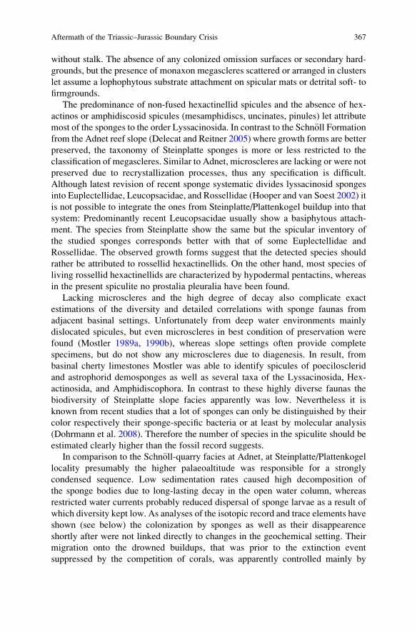
without stalk. The absence of any colonized omission surfaces or secondary hard-
grounds, but the presence of monaxon megascleres scattered or arranged in clusters
let assume a lophophytous substrate attachment on spicular mats or detrital soft- to
firmgrounds.
The predominance of non-fused hexactinellid spicules and the absence of hex-
actinos or amphidiscosid spicules (mesamphidiscs, uncinates, pinules) let attribute
most of the sponges to the order Lyssacinosida. In contrast to the Schn€oll Formation
from the Adnet reef slope (Delecat and Reitner 2005) where growth forms are better
preserved, the taxonomy of Steinplatte sponges is more or less restricted to the
classification of megascleres. Similar to Adnet, microscleres are lacking or were not
preserved due to recrystallization processes, thus any specification is difficult.
Although latest revision of recent sponge systematic divides lyssacinosid sponges
into Euplectellidae, Leucopsacidae, and Rossellidae (Hooper and van Soest 2002) it
is not possible to integrate the ones from Steinplatte/Plattenkogel buildup into that
system: Predominantly recent Leucopsacidae usually show a basiphytous attach-
ment. The species from Steinplatte show the same but the spicular inventory of
the studied sponges corresponds better with that of some Euplectellidae and
Rossellidae. The observed growth forms suggest that the detected species should
rather be attributed to rossellid hexactinellids. On the other hand, most species of
living rossellid hexactinellids are characterized by hypodermal pentactins, whereas
in the present spiculite no prostalia pleuralia have been found.
Lacking microscleres and the high degree of decay also complicate exact
estimations of the diversity and detailed correlations with sponge faunas from
adjacent basinal settings. Unfortunately from deep water environments mainly
dislocated spicules, but even microscleres in best condition of preservation were
found (Mostler 1989a, 1990b), whereas slope settings often provide complete
specimens, but do not show any microscleres due to diagenesis. In result, from
basinal cherty limestones Mostler was able to identify spicules of poecilosclerid
and astrophorid demosponges as well as several taxa of the Lyssacinosida, Hex-
actinosida, and Amphidiscophora. In contrast to these highly diverse faunas the
biodiversity of Steinplatte slope facies apparently was low. Nevertheless it is
known from recent studies that a lot of sponges can only be distinguished by their
color respectively their sponge-specific bacteria or at least by molecular analysis
(Dohrmann et al. 2008). Therefore the number of species in the spiculite should be
estimated clearly higher than the fossil record suggests.
In comparison to the Schn€oll-quarry facies at Adnet, at Steinplatte/Plattenkogel
locality presumably the higher palaeoaltitude was responsible for a strongly
condensed sequence. Low sedimentation rates caused high decomposition of
the sponge bodies due to long-lasting decay in the open water column, whereas
restricted water currents probably reduced dispersal of sponge larvae as a result of
which diversity kept low. As analyses of the isotopic record and trace elements have
shown (see below) the colonization by sponges as well as their disappearence
shortly after were not linked directly to changes in the geochemical setting. Their
migration onto the drowned buildups, that was prior to the extinction event
suppressed by the competition of corals, was apparently controlled mainly by
Aftermath of the Triassic–Jurassic Boundary Crisis 367
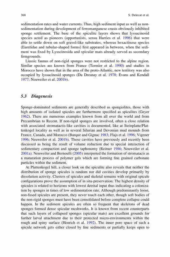
sedimentation rates and water currents. Thus, high sediment input as well as non-
sedimentation during development of ferromanganese crusts obviously inhibited
sponge settlement. The base of the spiculite layers shows that lyssacinosid
species acted as pioneers (opportunists, sensu Harries et al. 1996) that were
able to settle down on soft gravel-like substrates, whereas hexactinose species
(Euretidae and tubular-shaped forms) first appeared in between, when the sedi-
ment was fixed by Lysscinosida and spicular mats already served as secondary
firmgrounds.
Liassic faunas of non-rigid sponges were not restricted to the alpine region.
Similar species are known from France (Termier et al. 1990) and studies in
Morocco have shown that in the area of the proto-Atlantic, new territory was also
occupied by lyssacinosid sponges (Du Dresnay et al. 1978; Evans and Kendall
1977; Neuweiler et al. 2001b).
5.3 Diagenesis
Sponge-dominated sediments are generally described as spongiolites, those with
high amounts of isolated spicules are furthermore specified as spiculites (Geyer
1962). There are numerous examples known from all over the world and from
Precambrian to Recent. If non-rigid sponges are involved, often a close relation
with associated stromatactis-like cavities is documented, like at Steinplatte/Plat-
tenkogel locality as well as in several Silurian and Devonian mud mounds from
France, Canada, and Morocco (Burque and Gignac 1983; Flajs et al. 1996; Vigener
1996; Neuweiler et al. 2001b). Those cavities have previously and recently been
discussed as being the result of volume reduction due to special interaction of
sedimentary compaction and sponge taphonomy (Reitner 1986; Neuweiler et al.
2001a). Neuweiler and Bernoulli (2005) interpreted the formation of stromatacis as
a maturation process of polymer gels which are forming fine grained carbonate
particles within the sediment.
At Plattenkogel hill, a closer look on the spiculite also reveals that neither the
distribution of sponge spicules is random nor did cavities develop primarily by
dissolution activity. Clusters of spicules and skeletal remains with original spicule
configurations prove the assumption of in situ-preservation: The highest density of
spicules is related to horizons with lowest detrital input thus indicating a coloniza-
tion by sponges in times of low sedimentation rate. Although predominantly loose,
non-fused spicules are present, they never touch each other, though soft bodies of
the non-rigid sponges must have been consolidated before complete collapse could
happen. In the sediment spicules are often so frequent that skeletons of dead
sponges formed dense spicular meshworks. It is known from recent counterparts
that such layers of collapsed sponges (spicular mats) are excellent grounds for
further larval attachment due to their protected micro-environments within the
rough and spiny surface (Henrich et al. 1992). The inner pore space of such a
spicule network gets either closed by fine sediments or partially keeps open to
368 S. Delecat et al.
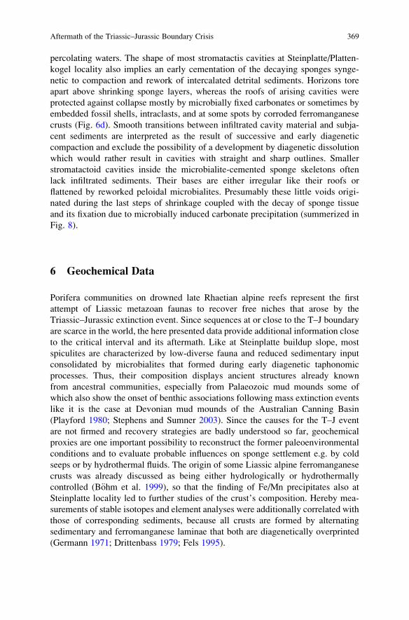
percolating waters. The shape of most stromatactis cavities at Steinplatte/Platten-
kogel locality also implies an early cementation of the decaying sponges synge-
netic to compaction and rework of intercalated detrital sediments. Horizons tore
apart above shrinking sponge layers, whereas the roofs of arising cavities were
protected against collapse mostly by microbially fixed carbonates or sometimes by
embedded fossil shells, intraclasts, and at some spots by corroded ferromanganese
crusts (Fig. 6d). Smooth transitions between infiltrated cavity material and subja-
cent sediments are interpreted as the result of successive and early diagenetic
compaction and exclude the possibility of a development by diagenetic dissolution
which would rather result in cavities with straight and sharp outlines. Smaller
stromatactoid cavities inside the microbialite-cemented sponge skeletons often
lack infiltrated sediments. Their bases are either irregular like their roofs or
flattened by reworked peloidal microbialites. Presumably these little voids origi-
nated during the last steps of shrinkage coupled with the decay of sponge tissue
and its fixation due to microbially induced carbonate precipitation (summerized in
Fig. 8).
6 Geochemical Data
Porifera communities on drowned late Rhaetian alpine reefs represent the first
attempt of Liassic metazoan faunas to recover free niches that arose by the
Triassic–Jurassic extinction event. Since sequences at or close to the T–J boundary
are scarce in the world, the here presented data provide additional information close
to the critical interval and its aftermath. Like at Steinplatte buildup slope, most
spiculites are characterized by low-diverse fauna and reduced sedimentary input
consolidated by microbialites that formed during early diagenetic taphonomic
processes. Thus, their composition displays ancient structures already known
from ancestral communities, especially from Palaeozoic mud mounds some of
which also show the onset of benthic associations following mass extinction events
like it is the case at Devonian mud mounds of the Australian Canning Basin
(Playford 1980; Stephens and Sumner 2003). Since the causes for the T–J event
are not firmed and recovery strategies are badly understood so far, geochemical
proxies are one important possibility to reconstruct the former paleoenvironmental
conditions and to evaluate probable influences on sponge settlement e.g. by cold
seeps or by hydrothermal fluids. The origin of some Liassic alpine ferromanganese
crusts was already discussed as being either hydrologically or hydrothermally
controlled (B€ohm et al. 1999), so that the finding of Fe/Mn precipitates also at
Steinplatte locality led to further studies of the crust’s composition. Hereby mea-
surements of stable isotopes and element analyses were additionally correlated with
those of corresponding sediments, because all crusts are formed by alternating
sedimentary and ferromanganese laminae that both are diagenetically overprinted
(Germann 1971; Drittenbass 1979; Fels 1995).
Aftermath of the Triassic–Jurassic Boundary Crisis 369
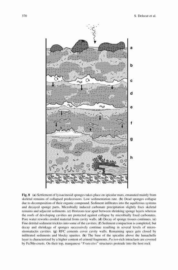
Fig. 8 (a) Settlement of lyssacinosid sponges takes place on spicular mats, emanated mainly from
skeletal remains of collapsed predecessors. Low sedimentation rate. (b) Dead sponges collapse
due to decomposition of their organic compound. Sediment infiltrates into the aquiferous systems
and decayed sponge parts. Microbially induced carbonate precipitation slightly fixes skeletal
remains and adjacent sediments. (c) Horizons tear apart between shrinking sponge layers whereas
the roofs of developing cavities are protected against collapse by microbially fixed carbonates.
Pore water reworks eroded material from cavity walls. (d) Decay of sponge tissues continues. (e)
Fine detrital sediment trickles into some of the cavities. (f) Sediment compaction is completed, but
decay and shrinkage of sponges successively continue resulting in several levels of micro-
stromatactis cavities. (g) RFC cements cover cavity walls. Remaining space gets closed by
infiltrated sediments and blocky sparites. (h) The base of the spiculite above the lumachelle
layer is characterized by a higher content of crinoid fragments. Pecten-rich intraclasts are coveredby Fe/Mn-crusts. On their top, manganese “Frutexites” structures protrude into the host rock
370 S. Delecat et al.
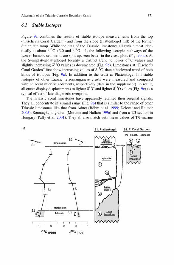
6.1 Stable Isotopes
Figure 9a combines the results of stable isotope measurements from the top
(“Fischer’s Coral Garden”) and from the slope (Plattenkogel hill) of the former
Steinplatte ramp. While the data of the Triassic limestones all rank almost iden-
tically at about d13C +3.0 and d18O �1, the following isotopic pathways of the
Lower Jurassic sediments are split up, seen better in the cross-plots (Fig. 9b–d). At
the Steinplatte/Plattenkogel locality a distinct trend to lower d13C values and
slightly increasing d18O values is documented (Fig. 9b). Limestones at “Fischer’s
Coral Garden” first show increasing values of d13C, then a backward trend of both
kinds of isotopes (Fig. 9a). In addition to the crust at Plattenkogel hill stable
isotopes of other Liassic ferromanganese crusts were measured and compared
with adjacent micritic sediments, respectively (data in the supplement). In result,
all crusts display displacements to lighter d13C and lighter d18O values (Fig. 9c) as a
typical effect of late diagenetic overprint.
The Triassic coral limestones have apparently retained their original signals.
They all concentrate in a small range (Fig. 9b) that is similar to the range of other
Triassic limestones like that from Adnet (B€ohm et al. 1999; Delecat and Reitner
2005), Sonntagkendlgraben (Morante and Hallam 1996) and from a T/J-section in
Hungary (Palfy et al. 2001). They all also match with mean values of T/J-marine
a
SinemurianS2
S2
S1 S1
S2S2Hettangian
Triassic
-1 0 2 3 4
S1: Plattenkogel S2: F. Coral Garden
Adnetlimestone
spiculite
T/J - limest. + cements
corallimestone
luma-chelle
corellimestone
d18O (PDB) d13C (PDB)
4
5
6 10
3b
3a
2
1
Aftermath of the Triassic–Jurassic Boundary Crisis 371
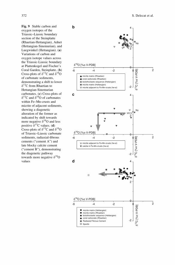
δ18O [%o V-PDB]
δ13C
[%o
V-P
DB
]δ13
C [%
o V
-PD
B]
δ13C
[%o
V-P
DB
]δ18O [%o V-PDB]
δ18O [%o V-PDB]
-6
b
c
d
-4 -2 00
-2
2
2
4
-6 -4 -2 00
-2
2
2
4
-6 -4 -2 00
-2
2
2
4
micrite matrix (Rhaetian)coral carbonate (Rhaetian)biolothoclastic sequence (Hettangian)micrite matrix (Hettangian)micrite adjacent to Fe-Mn-crusts (he-si)
micrite adjacent to Fe-Mn-crusts (he-si)
calcite in Fe-Mn-crusts (he-si)
micrite matrix (Hettangian)micrite matrix (Rhaetian)biolothoclastic sequence (Hettangian)coral carbonate (Rhaetian)
Radiaxial Fibrous Cement
Sparite
Fig. 9 Stable carbon and
oxygen isotopes of the
Triassic–Liassic boundary
section of the Steinplatte
(Rhaetian-Hettangian), Adnet
(Hettangian-Sinemurian), and
Luegwinkel (Hettangian). (a)
Variations of carbon and
oxygen isotope values across
the Triassic-Liassic boundary
at Plattenkogel and Fischer’s
Coral Garden, Steinplatte. (b)
Cross-plots of d13C and d18Oof carbonate sediments,
demonstrating a shift to lower
d13C from Rhaetian to
Hettangian-Sinemurian
carbonates. (c) Cross-plots of
d13C and d18O of carbonates
within Fe–Mn-crusts and
micrite of adjacent sediments,
showing a diagenetic
alteration of the former as
indicated by shift towards
more negative d18O and less
positive d13C values. (d)
Cross-plots of d13C and d18Oof Triassic–Liassic carbonate
sediments, radiaxial-fibrous
cements (“cement A”) and
late blocky calcite cement
(“cement B”), demonstrating
the diagenetic pathway
towards more negative d18Ovalues
372 S. Delecat et al.
seawaters given by Veizer and Hoefs (1976). Regarding the values of the Liassic
succession, it is necessary to consider that, when diagenesis happens, it usually
causes first a strong 18O depletion followed by a negativ d13C shift. The late
diagenetic blocky sparites show a 18O depletion only (Fig. 9d), while ferromanga-
nese crusts, which are altered like all other Liassic crusts that were studied from the
alpine region, shows a minor 13C depletion, too (Fig. 9c). According to this, the
isotopic pathway from “Fischer’s Coral Garden” (Rhaetian) and Plattenkogel hill
(Hettangian) both show a moderate early and late diagenetic overprint with d13Csignals retained closely to original values. Although some Steinplatte/Plattenkogel
data scatter broadly due to the high content of detrital components, a distinct
negative d13C shift is discernible. Considering that microbialites, if not related to
methanogenesis, methane oxidation, or photosynthesis, usually do not show any
biologically isotope fractionation (e.g., Keupp et al. 1993), the automicrites of the
spiculite in fact should provide the best results. The younger Adnet Limestones
(Figs. 3 and 9-sediment no. 8) from “Fischer’s Coral Garden” (interstices of the reef
surface) again show values like the Triassic samples. It is postulated that there was a
little d13Ccarb excursion during Hettangian time that ends in the Lower Sinemurian.
This hypothesis is supported by a more complete data set from Adnet localities,
where the same trend is actually better documented (Delecat and Reitner 2005).
Negative excursions in d18O, d13Ccarb and d13Corg are known from several
marine T–J boundary sections (Morante and Hallam 1996; Hallam and Goodfellow
1990; Palfy et al. 2001; Guex et al. 2002; Ward et al. 2001). Possible reasons
discussed for the isotopic shifts are e.g. sea level changes, catastrophic impacts but
most convincingly the CO2 release from either elevated volcanism in the central
Atlantic magmatic province (CAMP) or by destabilization of methane hydrates due
to increased temperatures (references in Palfy et al. 2001; Delecat and Reitner
2005). In contrast, the negative carbon isotope shift that has been detected at
Steinplatte/Plattenkogel locality is too small to explain global specific causes.
Most likely it is related to local conditions that probably reflect processes during
spiculite formation. As it has already been assumed for a similar isotope record
at Adnet (Delecat and Reitner 2005), the high ratio of sponge automicrites in
the spiculite could explain the small negative shift. These microbialites are formed
by the degradation of sponge organic mater related with bacterial sulphate
reduction that is lowering the d13C ratio significantly (Hoefs 1997; Londry and
Des Marais 2003).
6.2 Element Analyses
Weight ratios of major and trace elements were measured on bulk samples of six
ferromanganese crusts and four corresponding sediments. In result, the element
analyses show that the distribution patterns are quite similar in both sediments and
crusts, but the concentrations in the ferromanganese precipitates are much higher
(Fig. 10). All samples show definite accumulations of predominantly V, Ni, Zn, Sr,
Aftermath of the Triassic–Jurassic Boundary Crisis 373
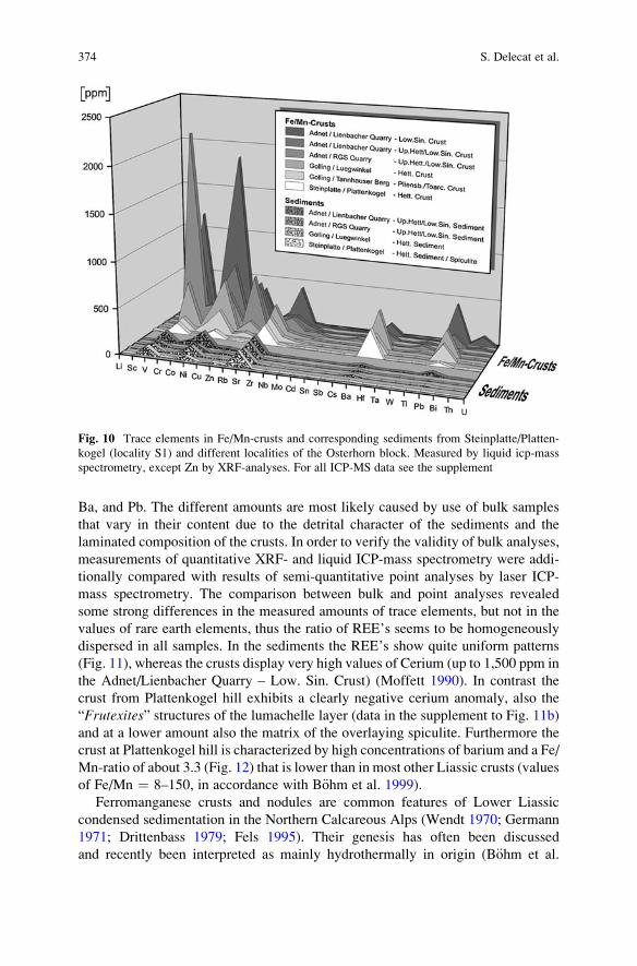
Ba, and Pb. The different amounts are most likely caused by use of bulk samples
that vary in their content due to the detrital character of the sediments and the
laminated composition of the crusts. In order to verify the validity of bulk analyses,
measurements of quantitative XRF- and liquid ICP-mass spectrometry were addi-
tionally compared with results of semi-quantitative point analyses by laser ICP-
mass spectrometry. The comparison between bulk and point analyses revealed
some strong differences in the measured amounts of trace elements, but not in the
values of rare earth elements, thus the ratio of REE’s seems to be homogeneously
dispersed in all samples. In the sediments the REE’s show quite uniform patterns
(Fig. 11), whereas the crusts display very high values of Cerium (up to 1,500 ppm in
the Adnet/Lienbacher Quarry – Low. Sin. Crust) (Moffett 1990). In contrast the
crust from Plattenkogel hill exhibits a clearly negative cerium anomaly, also the
“Frutexites” structures of the lumachelle layer (data in the supplement to Fig. 11b)
and at a lower amount also the matrix of the overlaying spiculite. Furthermore the
crust at Plattenkogel hill is characterized by high concentrations of barium and a Fe/
Mn-ratio of about 3.3 (Fig. 12) that is lower than in most other Liassic crusts (values
of Fe/Mn ¼ 8–150, in accordance with B€ohm et al. 1999).
Ferromanganese crusts and nodules are common features of Lower Liassic
condensed sedimentation in the Northern Calcareous Alps (Wendt 1970; Germann
1971; Drittenbass 1979; Fels 1995). Their genesis has often been discussed
and recently been interpreted as mainly hydrothermally in origin (B€ohm et al.
Fig. 10 Trace elements in Fe/Mn-crusts and corresponding sediments from Steinplatte/Platten-
kogel (locality S1) and different localities of the Osterhorn block. Measured by liquid icp-mass
spectrometry, except Zn by XRF-analyses. For all ICP-MS data see the supplement
374 S. Delecat et al.
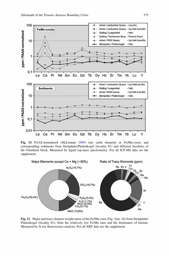
Fig. 11 PAAS-normalized (McLennan 1989) rare earth elements in Fe/Mn-crusts and
corresponding sediments from Steinplatte/Plattenkogel (locality S1) and different localities of
the Osterhorn block. Measured by liquid icp-mass spectrometry. For all ICP-MS data see the
supplement
Fig. 12 Major and trace element weight ratios of the Fe/Mn-crust (Fig. 3/no. 3b) from Steinplatte/
Plattenkogel (locality S1). Note the relatively low Fe/Mn ratio and the dominance of barium.
Measured by X-ray fluorescence analysis. For all XRF data see the supplement
Aftermath of the Triassic–Jurassic Boundary Crisis 375
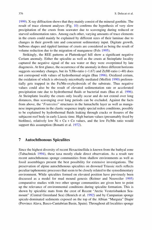
1999). X-ray diffraction shows that they mainly consist of the mineral goethite. The
result of trace element analyses (Fig. 10) confirms the hypothesis of very slow
precipitation of the crusts from seawater due to scavenging during reduced or
starved sedimentation rates. Among each other, varying amounts of trace elements
in the crusts could mainly be explained by different sizes of their laminae due to
changes in their growth rate and concurrent sedimentary input. Digitate growth,
bulbous shapes and rippled laminae of crusts are considered as being the result of
volume reduction due to the migration of manganese (Fels 1995).
Strikingly, the REE patterns at Plattenkogel hill show a significant negative
Cerium anomaly. Either the spiculite as well as the crusts at Steinplatte locality
captured the negative signal of the sea water or they were overprinted by late
diagenesis. At first glance, the occurrence of the anomaly in three different horizons
suggests secondary changes, but its Y/Ho-ratio (~1.67) and Zr/Hf-ratio (~40) does
not correspond with values of hydrothermal origin (Bau 1996). Oxidized cerium,
the oxidation of which is obviously microbially mediated (Moffett 1990) preferen-
tially gets trapped in the Fe/Mn-oxyhydroxids of the substrate. Thus negative
values could also be the result of elevated sedimentation rate or accelerated
precipitation rate due to hydrothermal fluids or bacterial mats (Bau et al. 1996).
At Steinplatte locality the crusts only locally occur and do not extend over long
distances, thus scavenging over long periods can be excluded. Against the facts
from above, the “Frutexites” structures in the lumachelle layer as well as manga-
nese impregnations in the clastic sequence imply special redox conditions, possibly
to be explained by hydrothermal fluids leaking through cracks or fissures of the
subjacent reef body in early Liassic time. High barium values (presumably fixed by
biofilms), relatively low Ni + Cu + Co values, and the low Fe/Mn ratio would
support this assumption (Bonatti et al. 1972).
7 Autochthonous Spiculites
Since the highest diversity of recent Hexactinellida is known from the bathyal zone
(Tabachnick 1994), these taxa mostly elude direct observation. As a result rare
recent autochthonous sponge communities from shallow environments as well as
fossil assemblages present the best possibility for extensive investigations. The
preservation of alpine autochthonous spiculites on drowned Triassic reefs reflects
peculiar taphonomic processes that seem to be closely related to the synsedimentary
environment. While spiculites formed on elevated position have previously been
discussed as a model for mud mound genesis (Reitner and Neuweiler 1995)
comparative studies with two other sponge communities are given here to point
up the relevance of environmental conditions during spiculite formation. This is
shown by spiculitic mats from the crest of Recent “Arctic Vesterisbanken Sea-
mount” (Central Greenland Sea) (Henrich et al. 1992) and by Campanian sponge
spicule-dominated sediments exposed on the top of the Albian “Murguia” Diapir
(Province Alava, Basco-Cantabrian Basin, Spain). Throughout all localities sponge
376 S. Delecat et al.
settlement was apparently controlled first by the water regime respectively by the
nutrient supply via consistent currents. In the cold waters at Vesterisbanken these
currents probably derived from a “streamlined down-welling vortex known as the
Taylor column” (Henrich et al. 1992) whereas sites of temperate latitudes (Diapir of
Murguia, Steinplatte/Plattenkogel) were more likely dominated by upwelling sys-
tems or contour currents as postulated for alpine spiculites at Adnet (B€ohm et al.
1999; Delecat and Reitner 2005). Furthermore the faunal composition is different in
all studied sites and depends on the given substrate. At Murguia e.g. (Reitner 1982,
1987), the nowadays sunken center of the diapir top exposes marls of Lower
Campanian age showing the initial stage of recolonization by mainly non-lithistid
Demosponges. Followed much later by a coral reef growth in the Upper Campanian
Oro Member (von Stackelberg 1964; Engeser 1984) the pioneer sponge fauna
comprises specimens with non-rigid skeletons formed by unfused spicules the
remains of their collapsed skeletons formed spiculitic mats that served as secondary
substrates for hexactinosid and demosponge lithistid taxa. The facies displays a
high ratio of spicules and very low amount of embedding matrix thus implying a
reduced sedimentation rate. Accordingly automicrites are scarce and only occur in
the interior of some of the rigid sponge skeletons so that the sediment resembles the
grey limestone facies from alpine basinal settings. In contrast to these sediments
lacking diverse accessorial faunas, the ridge of the deep sea volcano “Vesterisban-
ken” is characterized by more complex biogenic mats with sponges, bryozoans,
serpulids and hydrozoans. They formed on sands and coarse volcanic gravel that
provided larger surfaces also for encrusting filter-feeding organisms. The sponge
taxa encompasses several groups of Lyssacinosida (Rossellidae), non-rigid Demos-
pongiae (Geodiidae) and a few poeciloclerid Demosponges. A closer look onto the
spiculite mats show that fine-grained, microbial sediment is trapped within the
meshwork of sponge spicules and branched bryozoan colonies. Furthermore the
mats from Vesterisbanken are also characterized by spicules of dead predecessors,
by microbially induced carbonate precipitation and by small interior cavities where
larvae settlement of cryptic sponges occur. Thus the mats could be seen as modern
counterparts to the Liassic alpine spiculite at Steinplatte with both displaying
similarities in their formation and taphonomic structure.
8 Triassic–Jurassic Boundary
In the last decade research on the Triassic–Jurassic mass extinction was strongly
enforced, currently by the IGCP-Project 458 – Triassic–Jurassic Boundary Events.
The T–J event was characterized rather by several local changes than by one big
turn-over, hence global effects have been discussed controversially (Hallam 1989,
2002; Marzoli et al. 1999; McElwain et al. 1999; Tanner et al. 2001; Hesselbo et al.
2002; Olsen et al. 2002; Ward et al. 2001; a.o.). Eustatic sea level changes
caused high extinction rates especially in the shelf areas, but simultaneous low-
stands were responsible for a lot of shallow marine sequences displaying a hiatus in
Aftermath of the Triassic–Jurassic Boundary Crisis 377
sedimentation just where the event should have been documented. A relation
between the Triassic–Jurassic boundary event and carbon isotope excursions in
d13Ccarb and d13Corg has also been mentioned from several localities (Germany:
McRoberts et al. 1997; Turnsek et al. 1999; Hungary: Palfy et al. 2001; Canada:
Ward et al. 2001; England: Hesselbo et al. 2002). Disregarding those of apparently
diagenetic origin (Morante and Hallam 1996; Ward et al. 2001), a hiatus in the
fossil record often prevents exact stratigraphical correlations thus a lot of work still
has to be done correlating the few present data from around the world in order to
gain insight into the driving forces of the event (Guex et al. 2002; Palfy et al. 2001).
At Steinplatte/Plattenkogel locality a new ammonite found of Psiloceras sp.
from the spiculite base let allocate the stromatactis limestones to the Lower
Hettangian. This also corresponds with the geochemical data, supposing that the
isotopic pathway in sediments at Plattenkogel hill displays the beginning of the
same amplitude that is shown in Adnet quarries (Delecat and Reitner 2005).
Nevertheless it keeps unknown so far whether the excursion at Steinplatte shows
only one amplitude of probably a series of little fluctuations or just the end of a
bigger carbon anomaly. Anyway, it should be considered that in contrast to some
deep-water settings the isotopic records from shallow marine sediments of the
Tethyian region could have been overprinted by several local paleoenvironmental
changes. At Steinplatte for example an effect of meteoric diagenesis seems to
decrease upward from the capping facies (Stanton and Fl€ugel 1989) to the cements
of the stromatactis cavities (Mazzullo et al. 1990). But the following succession is
characterized by constantly increasing water depths so that an emergence later than
the origin of the spiculite layer is unlikely. Probably the sediments of the Lower
Liassic sequence were influenced afterwards by percolating waters that were
trapped first in Upper Triassic time and then reactivated during Liassic burial.
9 Conclusions
l At Steinplatte/Plattenkogel locality (N of Waidring, Austria) the upper slope of
the Triassic Carbonate Platform was colonized by Lower Hettangian fauna of
mainly non-rigid lyssacinosid sponges.l Settlement of sponges took place during reduced sedimentation on detrital soft-
or firmgrounds in small depressions of the drowned former reef surface.l There is no correlation between sponge settlement and geochemical data,
thus colonization was exclusively controlled by sedimentation rate and water
regime.l A succession of several sponge generations emerged into a spiculite layer of
about 1.5 m in thickness.l Mats of dead sponges were fixed in situ by infiltrated sediments and microbially
induced carbonate precipitation in decaying sponge tissues.l Large stromatactis cavities originated due to compaction of non-cemented
biomicrites in between already microbially-fixed parts of the spiculite. Smaller
378 S. Delecat et al.
cavities are the result of volume reduction due to the decay of organic matter and
microbialite formation.l At Steinplatte/Plattenkogel localitiy, a negative cerium anomaly is found in a
ferromanganese crust and the overlaying Liassic spiculite.l The Triassic–Jurassic boundary at Steinplatte is followed by sediments showing
a minor but distinct negative d13Ccarb – excursion that starts in Lower Hettangian
and presumably ends in Lower Sinemurian time.l The facies of sponge-stromatactis-biomicrites represents a pendant to the very
similar Schn€oll Quarry Formation of Adnet (E of Hallein, Austria).
Acknowledgements We thank Dr. M. Joachimski (Erlangen) for stable isotope analyses, Dr.
Helmut Klein and Dr. A. Preusser (G€ottingen) for XRD analyses, Dr. G. Hartmann (G€ottingen) forXRF analyses, Dr. K. Simon (G€ottingen) for ICP-MS measurements and Dr. T. Heinrichs
(G€ottingen) for EDX analyses. Ammonites were specified by Dr. G. Bloos (Stuttgart). The
Deutsche Forschungsgemeinschaft is gratefully acknowledged for financial support (Re 665/17).
This paper contributes to the IGCP- Project 458 – Triassic–Jurassic Boundary Events.
Appendix
Supplement to Fig. 9: Carbon and Oxygen Stable Isotope Datas
Thin section/
i.-sample
Locality/
no. of
horizon
Stratigraphy Micro facies d13Ccarb
(PDB)
d18Ocarb
(PDB)
ST20/01 S2/5 Triassic reef
limestone
Coral material 2.98 �0.97
ST20/01b S2/5 2.96 �0.96
ST20/02 S2/5 2.95 �1.30
ST20/02b S2/5 2.88 �0.92
ST20/03 S2/5 Microbial limestone 2.95 �1.28
ST20/03b S2/5 3.13 �1.20
ST20/03c S2/5 3.06 �1.32
ST20/04 S2/6 Triassic? Infill of
reef cavity
RFC cement 2.79 �1.74
ST20/04b S2/6 2.91 �1.36
ST20/04c S2/6 2.83 �1.66
ST20/05 S2/7 Grey intern micrite 2.99 �1.01
ST20/05b S2/7 2.95 �1.42
ST20/05c S2/7 2.79 �1.29
ST20/06 S2/8 Liassic infill of
reef cavity/
Adnet Fm.?
Red intern micrite 3.42 �1.05
ST20/06b S2/8 3.56 �1.03
ST20/06c S2/8 3.71 �1.18
ST20/07 S2/9 RFC cement 3.26 �1.37
ST20/07b S2/9 3.20 �2.72
ST20/07c S2/9 3.25 �1.90
ST20/08 S2/10 Red intern micrite 2.88 �3.27
ST20/08b S2/10 2.90 �3.04
ST20/08c S2/10 3.06 �1.65
(continued)
Aftermath of the Triassic–Jurassic Boundary Crisis 379
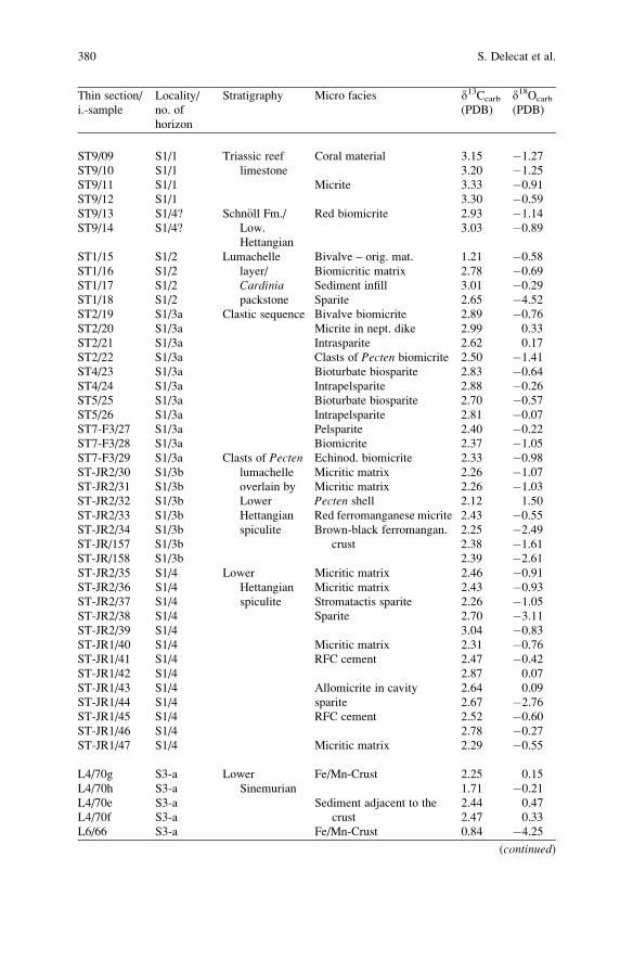
Thin section/
i.-sample
Locality/
no. of
horizon
Stratigraphy Micro facies d13Ccarb
(PDB)
d18Ocarb
(PDB)
ST9/09 S1/1 Triassic reef
limestone
Coral material 3.15 �1.27
ST9/10 S1/1 3.20 �1.25
ST9/11 S1/1 Micrite 3.33 �0.91
ST9/12 S1/1 3.30 �0.59
ST9/13 S1/4? Schn€oll Fm./
Low.
Hettangian
Red biomicrite 2.93 �1.14
ST9/14 S1/4? 3.03 �0.89
ST1/15 S1/2 Lumachelle
layer/
Cardiniapackstone
Bivalve – orig. mat. 1.21 �0.58
ST1/16 S1/2 Biomicritic matrix 2.78 �0.69
ST1/17 S1/2 Sediment infill 3.01 �0.29
ST1/18 S1/2 Sparite 2.65 �4.52
ST2/19 S1/3a Clastic sequence Bivalve biomicrite 2.89 �0.76
ST2/20 S1/3a Micrite in nept. dike 2.99 0.33
ST2/21 S1/3a Intrasparite 2.62 0.17
ST2/22 S1/3a Clasts of Pecten biomicrite 2.50 �1.41
ST4/23 S1/3a Bioturbate biosparite 2.83 �0.64
ST4/24 S1/3a Intrapelsparite 2.88 �0.26
ST5/25 S1/3a Bioturbate biosparite 2.70 �0.57
ST5/26 S1/3a Intrapelsparite 2.81 �0.07
ST7-F3/27 S1/3a Pelsparite 2.40 �0.22
ST7-F3/28 S1/3a Biomicrite 2.37 �1.05
ST7-F3/29 S1/3a Clasts of Pectenlumachelle
overlain by
Lower
Hettangian
spiculite
Echinod. biomicrite 2.33 �0.98
ST-JR2/30 S1/3b Micritic matrix 2.26 �1.07
ST-JR2/31 S1/3b Micritic matrix 2.26 �1.03
ST-JR2/32 S1/3b Pecten shell 2.12 1.50
ST-JR2/33 S1/3b Red ferromanganese micrite 2.43 �0.55
ST-JR2/34 S1/3b Brown-black ferromangan.
crust
2.25 �2.49
ST-JR/157 S1/3b 2.38 �1.61
ST-JR/158 S1/3b 2.39 �2.61
ST-JR2/35 S1/4 Lower
Hettangian
spiculite
Micritic matrix 2.46 �0.91
ST-JR2/36 S1/4 Micritic matrix 2.43 �0.93
ST-JR2/37 S1/4 Stromatactis sparite 2.26 �1.05
ST-JR2/38 S1/4 Sparite 2.70 �3.11
ST-JR2/39 S1/4 3.04 �0.83
ST-JR1/40 S1/4 Micritic matrix 2.31 �0.76
ST-JR1/41 S1/4 RFC cement 2.47 �0.42
ST-JR1/42 S1/4 2.87 0.07
ST-JR1/43 S1/4 Allomicrite in cavity 2.64 0.09
ST-JR1/44 S1/4 sparite 2.67 �2.76
ST-JR1/45 S1/4 RFC cement 2.52 �0.60
ST-JR1/46 S1/4 2.78 �0.27
ST-JR1/47 S1/4 Micritic matrix 2.29 �0.55
L4/70g S3-a Lower
Sinemurian
Fe/Mn-Crust 2.25 0.15
L4/70h S3-a 1.71 �0.21
L4/70e S3-a Sediment adjacent to the
crust
2.44 0.47
L4/70f S3-a 2.47 0.33
L6/66 S3-a Fe/Mn-Crust 0.84 �4.25
(continued)
380 S. Delecat et al.
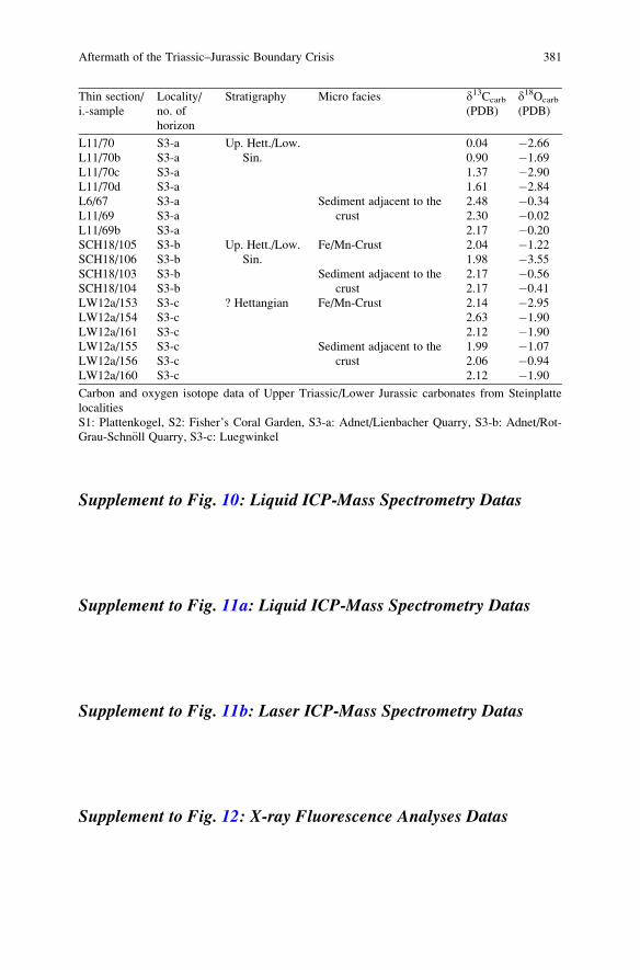
Thin section/
i.-sample
Locality/
no. of
horizon
Stratigraphy Micro facies d13Ccarb
(PDB)
d18Ocarb
(PDB)
Up. Hett./Low.
Sin.
L11/70 S3-a 0.04 �2.66
L11/70b S3-a 0.90 �1.69
L11/70c S3-a 1.37 �2.90
L11/70d S3-a 1.61 �2.84
L6/67 S3-a Sediment adjacent to the
crust
2.48 �0.34
L11/69 S3-a 2.30 �0.02
L11/69b S3-a 2.17 �0.20
SCH18/105 S3-b Up. Hett./Low.
Sin.
Fe/Mn-Crust 2.04 �1.22
SCH18/106 S3-b 1.98 �3.55
SCH18/103 S3-b Sediment adjacent to the
crust
2.17 �0.56
SCH18/104 S3-b 2.17 �0.41
LW12a/153 S3-c ? Hettangian Fe/Mn-Crust 2.14 �2.95
LW12a/154 S3-c 2.63 �1.90
LW12a/161 S3-c 2.12 �1.90
LW12a/155 S3-c Sediment adjacent to the
crust
1.99 �1.07
LW12a/156 S3-c 2.06 �0.94
LW12a/160 S3-c 2.12 �1.90
Carbon and oxygen isotope data of Upper Triassic/Lower Jurassic carbonates from Steinplatte
localities
S1: Plattenkogel, S2: Fisher’s Coral Garden, S3-a: Adnet/Lienbacher Quarry, S3-b: Adnet/Rot-
Grau-Schn€oll Quarry, S3-c: Luegwinkel
Supplement to Fig. 10: Liquid ICP-Mass Spectrometry Datas
Supplement to Fig. 11a: Liquid ICP-Mass Spectrometry Datas
Supplement to Fig. 11b: Laser ICP-Mass Spectrometry Datas
Supplement to Fig. 12: X-ray Fluorescence Analyses Datas
Aftermath of the Triassic–Jurassic Boundary Crisis 381
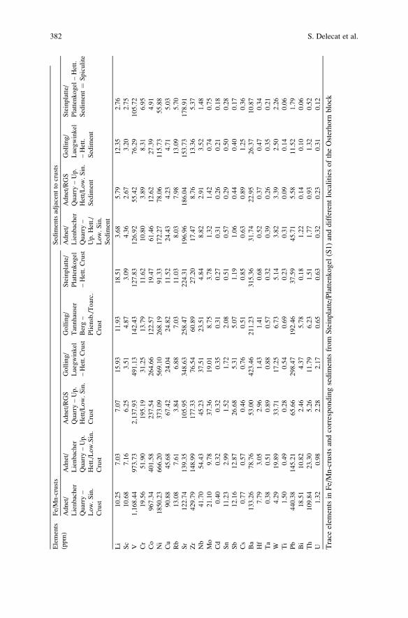
Elements
(ppm)
Fe/Mn-crusts
Sedim
entsadjacentto
crusts
Adnet/
Lienbacher
Quarry
–
Low.Sin.
Crust
Adnet/
Lienbacher
Quarry
–Up.
Hett./Low.Sin.
Crust
Adnet/RGS
Quarry
–Up.
Hett/Low.Sin.
Crust
Golling/
Luegwinkel
–Hett.Crust
Golling/
Tannhauser
Berg–
Pliensb./Toarc.
Crust
Steinplatte/
Plattenkogel
–Hett.Crust
Adnet/
Lienbacher
Quarry
–
Up.Hett./
Low.Sin.
Sedim
ent
Adnet/RGS
Quarry
–Up.
Hett/Low.Sin.
Sedim
ent
Golling/
Luegwinkel
–Hett.
Sedim
ent
Steinplatte/
Plattenkogel
–Hett.
Sedim
ent¼
Spiculite
Li
10.25
7.03
7.07
15.93
11.93
18.51
3.68
5.79
12.35
2.76
Sc
10.68
7.16
6.25
3.51
4.87
3.09
4.36
2.67
3.20
2.75
V1,168.44
973.73
2,137.93
491.13
142.43
127.83
126.92
55.42
76.29
105.72
Cr
19.56
51.90
195.19
31.25
13.79
11.62
10.80
3.89
8.31
6.95
Co
967.34
401.58
237.54
264.66
122.57
19.47
61.46
12.62
27.39
4.91
Ni
1850.23
666.20
373.09
569.10
268.19
91.33
172.27
78.06
115.73
55.88
Cu
90.88
45.68
67.42
24.04
24.82
11.52
24.43
4.23
4.71
5.03
Rb
13.08
7.61
3.84
6.88
7.03
11.03
8.03
7.98
13.09
5.70
Sr
122.74
139.35
105.95
348.63
258.47
224.31
196.96
186.04
153.73
178.91
Zr
429.79
148.99
177.33
76.54
60.89
27.20
17.47
8.76
13.36
5.37
Nb
41.70
54.43
45.23
37.51
23.51
4.84
8.82
2.91
3.52
1.48
Mo
21.10
9.78
37.36
19.01
8.75
3.78
1.32
1.42
0.74
0.75
Cd
0.40
0.32
0.32
0.35
0.31
0.27
0.31
0.26
0.21
0.18
Sn
11.23
2.99
1.52
1.72
2.08
0.51
0.57
0.29
0.50
0.28
Sb
12.16
12.87
26.68
5.31
5.07
1.19
1.06
0.44
0.40
0.17
Cs
0.77
0.57
0.46
0.76
0.51
0.85
0.63
0.89
1.25
0.36
Ba
133.26
78.76
53.00
423.46
211.23
315.36
31.74
22.95
26.37
10.87
Hf
7.79
3.05
2.96
1.43
1.41
0.68
0.52
0.37
0.47
0.34
Ta
0.38
0.51
0.89
0.88
0.57
0.32
0.39
0.26
0.35
0.21
W4.29
19.89
33.71
17.25
6.73
5.14
3.82
3.39
2.50
2.26
Ti
1.50
0.49
0.28
0.54
0.69
0.23
0.31
0.09
0.14
0.06
Pb
440.38
145.21
65.66
298.47
192.46
37.59
45.71
5.58
11.52
1.79
Bi
18.51
10.82
2.46
4.37
5.78
0.18
1.22
0.14
0.10
0.06
Th
109.84
23.30
5.26
11.79
6.23
1.51
1.77
0.93
1.32
0.52
U1.32
0.98
2.28
2.17
0.65
0.63
0.32
0.23
0.31
0.12
Trace
elem
entsin
Fe/Mn-crustsandcorrespondingsedim
entsfrom
Steinplatte/Plattenkogel
(S1)anddifferentlocalities
oftheOsterhorn
block
382 S. Delecat et al.
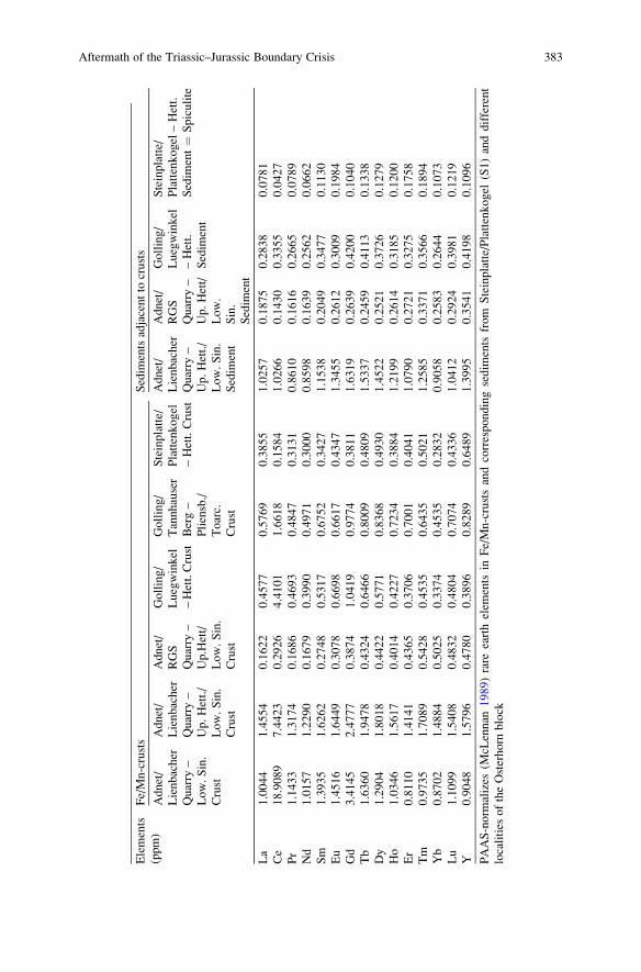
Elements
(ppm)
Fe/Mn-crusts
Sedim
entsadjacentto
crusts
Adnet/
Lienbacher
Quarry
–
Low.Sin.
Crust
Adnet/
Lienbacher
Quarry
–
Up.Hett./
Low.Sin.
Crust
Adnet/
RGS
Quarry
–
Up.Hett/
Low.Sin.
Crust
Golling/
Luegwinkel
–Hett.Crust
Golling/
Tannhauser
Berg–
Pliensb./
Toarc.
Crust
Steinplatte/
Plattenkogel
–Hett.Crust
Adnet/
Lienbacher
Quarry
–
Up.Hett./
Low.Sin.
Sedim
ent
Adnet/
RGS
Quarry
–
Up.Hett/
Low.
Sin.
Sedim
ent
Golling/
Luegwinkel
–Hett.
Sedim
ent
Steinplatte/
Plattenkogel
–Hett.
Sedim
ent¼
Spiculite
La
1.0044
1.4554
0.1622
0.4577
0.5769
0.3855
1.0257
0.1875
0.2838
0.0781
Ce
18.9089
7.4423
0.2926
4.4101
1.6618
0.1584
1.0266
0.1430
0.3355
0.0427
Pr
1.1433
1.3174
0.1686
0.4693
0.4847
0.3131
0.8610
0.1616
0.2665
0.0789
Nd
1.0157
1.2290
0.1679
0.3990
0.4971
0.3000
0.8598
0.1639
0.2562
0.0662
Sm
1.3935
1.6262
0.2748
0.5317
0.6752
0.3427
1.1538
0.2049
0.3477
0.1130
Eu
1.4516
1.6449
0.3078
0.6698
0.6617
0.4347
1.3455
0.2612
0.3009
0.1984
Gd
3.4145
2.4777
0.3874
1.0419
0.9774
0.3811
1.6319
0.2639
0.4200
0.1040
Tb
1.6360
1.9478
0.4324
0.6466
0.8009
0.4809
1.5337
0.2459
0.4113
0.1338
Dy
1.2904
1.8018
0.4422
0.5771
0.8368
0.4930
1.4522
0.2521
0.3726
0.1279
Ho
1.0346
1.5617
0.4014
0.4227
0.7234
0.3884
1.2199
0.2614
0.3185
0.1200
Er
0.8110
1.4141
0.4365
0.3706
0.7001
0.4041
1.0790
0.2721
0.3275
0.1758
Tm
0.9735
1.7089
0.5428
0.4535
0.6435
0.5021
1.2585
0.3371
0.3566
0.1894
Yb
0.8702
1.4884
0.5025
0.3374
0.4535
0.2832
0.9058
0.2583
0.2644
0.1073
Lu
1.1099
1.5408
0.4832
0.4804
0.7074
0.4336
1.0412
0.2924
0.3981
0.1219
Y0.9048
1.5796
0.4780
0.3896
0.8289
0.6489
1.3995
0.3541
0.4198
0.1096
PAAS-norm
alizes
(McL
ennan
1989)rare
earthelem
ents
inFe/Mn-crustsandcorrespondingsedim
ents
from
Steinplatte/Plattenkogel
(S1)anddifferent
localities
oftheOsterhorn
block
Aftermath of the Triassic–Jurassic Boundary Crisis 383
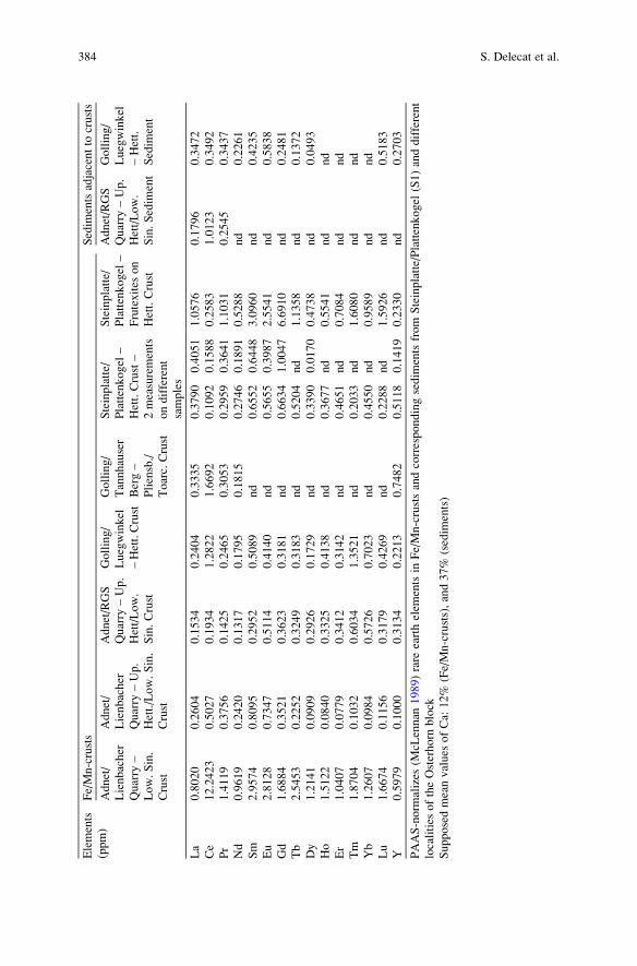
Elements
(ppm)
Fe/Mn-crusts
Sedim
entsadjacentto
crusts
Adnet/
Lienbacher
Quarry
–
Low.Sin.
Crust
Adnet/
Lienbacher
Quarry
–Up.
Hett./Low.Sin.
Crust
Adnet/RGS
Quarry
–Up.
Hett/Low.
Sin.Crust
Golling/
Luegwinkel
–Hett.Crust
Golling/
Tannhauser
Berg–
Pliensb./
Toarc.Crust
Steinplatte/
Plattenkogel
–
Hett.Crust–
2measurements
ondifferent
samples
Steinplatte/
Plattenkogel
–
Frutexites
on
Hett.Crust
Adnet/RGS
Quarry
–Up.
Hett/Low.
Sin.Sedim
ent
Golling/
Luegwinkel
–Hett.
Sedim
ent
La
0.8020
0.2604
0.1534
0.2404
0.3335
0.3790
0.4051
1.0576
0.1796
0.3472
Ce
12.2423
0.5027
0.1934
1.2822
1.6692
0.1092
0.1588
0.2583
1.0123
0.3492
Pr
1.4119
0.3756
0.1425
0.2465
0.3053
0.2959
0.3641
1.1031
0.2545
0.3437
Nd
0.9619
0.2420
0.1317
0.1795
0.1815
0.2746
0.1891
0.5288
nd
0.2261
Sm
2.9574
0.8095
0.2952
0.5089
nd
0.6552
0.6448
3.0960
nd
0.4235
Eu
2.8128
0.7347
0.5114
0.4140
nd
0.5655
0.3987
2.5541
nd
0.5838
Gd
1.6884
0.3521
0.3623
0.3181
nd
0.6634
1.0047
6.6910
nd
0.2481
Tb
2.5453
0.2252
0.3249
0.3183
nd
0.5204
nd
1.1358
nd
0.1372
Dy
1.2141
0.0909
0.2926
0.1729
nd
0.3390
0.0170
0.4738
nd
0.0493
Ho
1.5122
0.0840
0.3325
0.4138
nd
0.3677
nd
0.5541
nd
nd
Er
1.0407
0.0779
0.3412
0.3142
nd
0.4651
nd
0.7084
nd
nd
Tm
1.8704
0.1032
0.6034
1.3521
nd
0.2033
nd
1.6080
nd
nd
Yb
1.2607
0.0984
0.5726
0.7023
nd
0.4550
nd
0.9589
nd
nd
Lu
1.6674
0.1156
0.3179
0.4269
nd
0.2288
nd
1.5926
nd
0.5183
Y0.5979
0.1000
0.3134
0.2213
0.7482
0.5118
0.1419
0.2330
nd
0.2703
PAAS-norm
alizes
(McL
ennan
1989)rare
earthelem
entsin
Fe/Mn-crustsandcorrespondingsedim
entsfrom
Steinplatte/Plattenkogel
(S1)anddifferent
localities
oftheOsterhorn
block
Supposedmeanvalues
ofCa:
12%
(Fe/Mn-crusts),and37%
(sedim
ents)
384 S. Delecat et al.
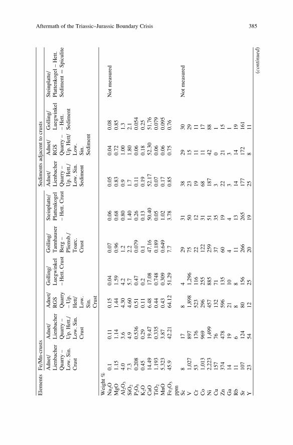
Elements
Fe/Mn-crusts
Sedim
entsadjacentto
crusts
Adnet/
Lienbacher
Quarry
–
Low.Sin.
Crust
Adnet/
Lienbacher
Quarry
–
Up.Hett./
Low.Sin.
Crust
Adnet/
RGS
Quarry
–Up.
Hett/
Low.
Sin.
Crust
Golling/
Luegwinkel
–Hett.Crust
Golling/
Tannhauser
Berg–
Pliensb./
Toarc.
Crust
Steinplatte/
Plattenkogel
–Hett.Crust
Adnet/
Lienbacher
Quarry
–
Up.Hett./
Low.Sin.
Sedim
ent
Adnet/
RGS
Quarry
–
Up.Hett/
Low.
Sin.
Sedim
ent
Golling/
Luegwinkel
–Hett.
Sedim
ent
Steinplatte/
Plattenkogel
–Hett.
Sedim
ent¼
Spiculite
Weight%
Na 2O
0.1
0.11
0.15
0.04
0.07
0.06
0.05
0.04
0.08
Notmeasured
MgO
1.15
1.14
1.44
1.59
0.96
0.68
0.83
0.72
0.85
Al 2O3
4.0
3.6
4.30
4.2
1.2
0.80
0.9
1.00
1.3
SiO
27.3
4.9
4.60
5.7
2.2
1.40
1.7
1.80
2.1
P2O5
0.208
0.536
0.51
0.47
0.079
0.26
0.11
0.06
0.054
K2O
0.45
0.29
0.11
0.2
0.1
0.13
0.19
0.18
0.25
CaO
14.49
19.47
6.48
17.08
47.16
50.40
52.17
52.30
51.76
TiO
21.193
0.335
0.44
0.748
0.189
0.05
0.07
0.06
0.079
MnO
5.323
3.87
0.43
0.309
0.649
1.02
0.17
0.06
0.095
Fe 2O3
45.9
42.21
64.12
51.29
7.7
3.78
0.85
0.75
0.76
ppm
Sc
817
84
29
31
38
29
30
Notmeasured
V1,027
897
1,898
1,296
75
50
23
15
29
Cr
53
176
523
116
22
12
19
11
11
Co
1,013
969
296
255
122
31
68
11
17
Ni
2,223
1,499
457
885
259
51
187
42
88
Cu
157
76
132
71
37
35
30
1
Zn
374
478
596
135
60
19
22
21
15
Ga
14
19
21
10
44
33
1
Rb
11
68
811
13
14
14
19
Sr
107
124
80
156
266
265
177
172
161
Y23
54
12
25
20
19
25
811
(continued)
Aftermath of the Triassic–Jurassic Boundary Crisis 385
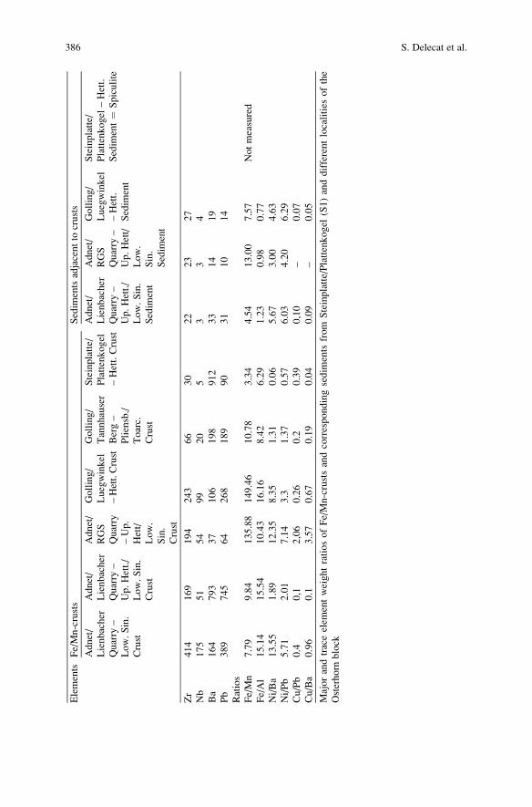
Elements
Fe/Mn-crusts
Sedim
entsadjacentto
crusts
Adnet/
Lienbacher
Quarry
–
Low.Sin.
Crust
Adnet/
Lienbacher
Quarry
–
Up.Hett./
Low.Sin.
Crust
Adnet/
RGS
Quarry
–Up.
Hett/
Low.
Sin.
Crust
Golling/
Luegwinkel
–Hett.Crust
Golling/
Tannhauser
Berg–
Pliensb./
Toarc.
Crust
Steinplatte/
Plattenkogel
–Hett.Crust
Adnet/
Lienbacher
Quarry
–
Up.Hett./
Low.Sin.
Sedim
ent
Adnet/
RGS
Quarry
–
Up.Hett/
Low.
Sin.
Sedim
ent
Golling/
Luegwinkel
–Hett.
Sedim
ent
Steinplatte/
Plattenkogel
–Hett.
Sedim
ent¼
Spiculite
Zr
414
169
194
243
66
30
22
23
27
Nb
175
51
54
99
20
53
34
Ba
164
793
37
106
198
912
33
14
19
Pb
389
745
64
268
189
90
31
10
14
Ratios
Fe/Mn
7.79
9.84
135.88
149.46
10.78
3.34
4.54
13.00
7.57
Notmeasured
Fe/Al
15.14
15.54
10.43
16.16
8.42
6.29
1.23
0.98
0.77
Ni/Ba
13.55
1.89
12.35
8.35
1.31
0.06
5.67
3.00
4.63
Ni/Pb
5.71
2.01
7.14
3.3
1.37
0.57
6.03
4.20
6.29
Cu/Pb
0.4
0,1
2,06
0.26
0.2
0.39
0,10
–0.07
Cu/Ba
0.96
0.1
3.57
0.67
0.19
0.04
0.09
–0.05
Majorandtraceelem
entweightratiosofFe/Mn-crustsandcorrespondingsedim
ents
from
Steinplatte/Plattenkogel
(S1)anddifferentlocalities
ofthe
Osterhorn
block
386 S. Delecat et al.
References
Bau M (1996) Controls on the fractionation of isovalent trace elements in magmatic and aqueous
systems: evidence from Y/Ho, Zr/Hf, and lanthanide tetrad effect. Contributions to Mineralogy
and Petrology 123: 323–333
Bau M, Koschinsky A, Dulski P, Hein JR (1996) Comparison of the partitioning behaviours of
yttrium, rare earth elements, and titanium between hydrogenetic marine ferromanganese crusts
and seawater. Geochimica et Cosmochimica Acta 60: 1709–1725
B€ohm F (1992) Mikrofazies und Ablagerungsmilieu des Lias und Dogger der Nord€ostlichenKalkalpen. Erlanger Geologische Abhandlungen 121: 57–217
B€ohm F, Ebli O, Krystyn L, Lobitzer H, Rakus M, Siblık M (1999) Fauna, stratigraphy and
depositional environment of the Hettangian-Sinemurian (Early Jurassic) of Adnet (Salzburg,
Austria). Abhandlungen der Geologischen Bundesanstalt Wien 56: 143–271
Bonatti E, Kraemer T, Rydell H (1972) Classification and genesis of submarine iron-manganese
deposits. In: Horn D (ed) Ferromanganese Deposits on the Ocean Floor. National Science
Foundation, Washington, 149–165
Burque P-A, Gignac H (1983) Sponge-constructed stromatactis mud mounds, Silurian of Gaspe,
Quebec. Journal of Sedimentary Petrology 53: 521–532
Delecat S, Reitner J (2005) Sponge communities from the Lower Liassic of Adnet (Northern
Calcareous Alps, Austria). Facies 51: 385–404
Delecat S, Peckmann J, Reitner J (2001) Non-rigid cryptic sponges in Oyster patch reefs (Lower
Kimmeridgian, Langenberg/Oker, Germany). Facies 45: 231–254
Dohrmann M, Janussen D, Reitner J, Collins AG, W€orheide G (2008) Phylogeny and evolution of
glass sponges (Porifera, Hexactinellida). Systematic Biology 57: 388–405
Drittenbass W (1979) Sedimentologie und Geochemie von Eisen-Mangan f€uhrenden Knollen undKrusten im Jura der Trento-Zone (€ostliche S€udalpen, Norditalien). Eclogae Geologica Helve-tica 72: 313–345
Dresnay R du, Termier G, Termier H (1978) Les Hexactinellides (Lyssakides et Dictyonines) du
Lias marocain. Geobios 11: 269–295
Engeser T (1984) Sedimentologische, fazielle und tektogenetische Untersuchungen in der Oberk-
reide des Basko-Kantabrischen Beckens (Nordspanien). Inaug Diss, Geowissenschaftliche
Fakult€at der Eberhard-Karls-Universit€at, T€ubingenEvans I, Kendall CGStC (1977) An interpretation of the depositional setting of some deep-water
Jurassic carbonatesof the central High Atlas mountains, Morocco. In: Cook HE, Enos P (eds)
Deep water carbonate environments. Society of Economic Paleontologists and Mineralogists,
Special Publication 25: 249–261
Fels A (1995) Prozesse und Produkte geologischer Kondensation im Jura der westlichen Tethys.
Profil 8: 363–472
Flajs G, H€ussner H, Vigener M (1996) Stromatactis Mud Mounds in the Upper Emsian of the
Montagne Noire (France): Formation and Diagenesis of Stromatactis Structures. In: Reitner J,
Neuweiler F, Gunkel F (eds) Global and regional controls on biogenic sedimentation. I. Reef
evolution. Research reports. G€ottinger Arbeiten zur Geologie und Pal€aontologie Sb2: 345–348Garrison RE, Fischer AG (1969) Deep-water limestones and radiolarites of the Alpine Jurassic. In:
Friedman GM (ed) Depositional environments in carbonate rocks. Society of Economic
Paleontologists and Mineralogists, Special Publication 14: 20–56
Germann K (1971) Mangan-Eisen-f€uhrende Knollen und Krusten in jurassischen Rotkalken der
N€ordlichen Kalkalpen. Neues Jahrbuch f€ur Geologie und Pal€aontologie, Monatshefte 1971:
133–156
Geyer OF (1962) €Uber Schwammgesteine (Spongiolith, Tuberolith, Spiculit und Gaizit): Her-
mann-Aldinger-Festschrift. Schweizerbart, Stuttgart, 51–59
Guex J, Bartolini A, Taylor D (2002) Discovery ofNeophyllites (Ammonitina, Cephalopoda, Early
Hettangian) in the New York Canyon sections (Gabbs Valley Range, Nevada) and discussion
Aftermath of the Triassic–Jurassic Boundary Crisis 387
of the d13C negative anomalies located around the Triassic–Jurassic boundary. Bulletin de la
Societe Vaudoise des Sciences Naturelles 88: 247–255
Hallam A (1989) The case for sea-level change as a dominant causal factor in mass extinction of
marine invertebrates. Philosophical Transactions of the Royal Society of London B 325:
437–455
Hallam A (2002) How catastrophic was the end-Triassic mass extinction? Lethaia 35: 147–157
Hallam A, Goodfellow WD (1990) Facies and geochemical evidence bearing on the end-Triassic
disappearance of the Alpine reef ecosystem. Historical Biology 4: 131–138
Harries PJ, Kauffman EG, Hansen TA (1996) Models for biotic survival following mass extinc-
tion. In: Hart MB (ed) Biotic recovery from mass extinction events. Geological Society,
Special Publication 102: 41–60
Henrich R, Hartmann M, Reitner J, Sch€afer P, Freiwald A, Steinmetz S, Dietrich P, Tiede J (1992)
Facies belts and communities of the Arctic Vesterisbanken Seamount (Central Greenland Sea).
Facies 27: 71–104
Hesselbo SP, Robinson SA, Surlyk F, Piasecki S (2002) Terrestrial and marine extinction at the
Triassic–Jurassic boundary synchronized with major carbon-cycle perturbation: a link to
initiation of massive volcanism? Geology 30: 251–254
Hoefs J (1997) Stable Isotope Geochemistry. Springer, Berlin
Hooper JNA, Van Soest RWM (eds) (2002) Systema Porifera: A Guide to the Classification of
Sponges. Kluwer Academic, Plenum Publishers, New York
Keupp H, Jenisch A, Herrmann R, Neuweiler F, Reitner J (1993) Microbial carbonate crusts – a
key to the environmental analysis of fossil spongiolites?. Facies 29: 41–54
Krainer K, Mostler H (1997) Die Lias-Beckenentwicklung der Unkener Synklinale (N€ordlicheKalkalpen, Salzburg) unter besonderer Ber€ucksichtigung der Scheibelberg Formation. Geolo-
gisch-Pal€aontologische Mitteilungen Innsbruck 22: 1–41
Londry KL, Des Marais DJ (2003) Stable carbon isotope fractionation by sulfate-reducing
bacteria. Applied and Environmental Microbiology 69: 2942–2949
Marzoli A, Renne PR, Piccirillo EM, Ernesto M, Bellieni G, De Min A (1999) Extensive 200-
million-year-old continental flood basalts of the central Atlantic Magmatic Province. Science
284: 616–618
Mazzullo SJ, Bischoff WD, Lobitzer H (1990) Diagenesis of radiaxial fibrous calcites in a
subunconformity, shallow-burial setting: Upper Triassic and Liassic, Northern Calcareous
Alps, Austria. Sedimentology 37: 407–425
McElwain JC, Beerling DJ, Woodward FI (1999) Fossil plants and global warming at the
Triassic–Jurassic boundary. Science 285: 1386–1390
McLennan SM (1989) Rare earth elements in sedimentary rocks: influences of provenance and
sedimentary processes. In: Lipin BR, McKay GA (eds) Geochemistry and mineralogy of rare
earth elements. Reviews in Mineralogy 21: 169–200
McRoberts CA, Furrer H, Jones DS (1997) Palaeoenvironmental interpretation of a Triassic–Jur-
assic boundary section from western Austria based on palaeoecological and geochemical data.
Palaeogeography, Palaeoclimatology, Palaeoecology 136: 79–95
Moffett JW (1990) Microbially mediated cerium oxidation in sea water. Nature 345: 421–423
Morante R, Hallam A (1996) Organic carbon isotopic record across the Triassic–Jurassic boundary
in Austria and its bearing on the cause of the mass extinction. Geology 24: 391–394
Mostler H (1986) Neue Kieselschw€amme aus den Zlambachschichten (Obertrias, N€ordlicheKalkalpen). Geologisch-Pal€aontologische Mitteilungen Innsbruck 13: 331–361
Mostler H (1989a) Mikroskleren hexactinellider Schw€amme aus dem Lias der N€ordlichen Kalk-
alpen. Jahrbuch der Geologischen Bundesanstalt Wien 132/4: 687–700
Mostler H (1989b) Mit Zygomen ausgestattete Dermalia von Kieselschw€ammen (Desmospongiae)
aus pelagischen Sedimenten der Obertrias und des unteren Jura (N€ordliche Kalkalpen). Jahr-
buch der Geologischen Bundesanstalt Wien 132/4: 701–726
Mostler H (1990a) Mikroskleren von Demospongien (Porifera) aus dem basalen Jura der
N€ordlichen Kalkalpen. Geologisch-Pal€aontologische Mitteilungen Innsbruck 17: 119–142
388 S. Delecat et al.
Mostler H (1990b) Hexactinellide Poriferen aus pelagischen Kieselkalken (Unterer Lias,
N€ordliche Kalkalpen). Geologisch-Pal€aontologische Mitteilungen Innsbruck 17: 143–178
Neuweiler F, Bernoulli D (2005) Mesozoic (Lower Jurassic) red stromatactis limestones from the
Southern Alps (Arzo, Switzerland): calcite mineral authigenesis and syneresis-type deforma-
tion. International Journal of Earth Sciences 94: 130–146
Neuweiler F, Bourque P-A, Boulvain F (2001a) Why is stromatactis so rare in Mesozoic carbonate
mud mounds? Terra Nova 13: 338–346
Neuweiler F, Mehdi M, Wilmsen M (2001b) Facies of Liassic sponge mounds, central High Atlas,
Morocco. Facies 44: 243–264
Olsen PE, Kent DV, Sues H-D, Koeberl C, Huber H, Montanari A, Rainforth EC, Fowell SJ,
Szajna MJ, Hartline BW (2002) Ascent of dinosaurs linked to an iridium anomaly at the
Triassic–Jurassic boundary. Science 296: 1305–1307
Palfy J, Demeny A, Haas J, Hetenyi M, Orchard MJ, Veto I (2001) Carbon isotope anomaly and
other geochemical changes at the Triassic–Jurassic boundary from a marine section in Hun-
gary. Geology 29: 1047–1050
Playford PE (1980) Devonian ‘Great Barrier Reef’ of Canning Basin, Western Australia. Ameri-
can Association of Petroleum Geologists, Bulletin 62: 814–841
Rakus M, Lobitzer H (1993) Early Liassic ammonites from the Steinplatte-Kammerk€ohralm area
(Northern Calcareous Alps/Salzburg). Jahrbuch der Geologischen Bundesanstalt Wien 136/4:
919–932
Reitner J (1982) Die Entwicklung von Inselplattformen und Diapir-Atollen im Alb des Basko-
Kantabrikums (Nordspanien). Neues Jahrbuch f€ur Geologie und Pal€aontologie 165/1: 87–101Reitner J (1986) A comparative study of the diagenesis in diapir-influenced reef atolls and a fault
block reef platform in the Late Albian of the Vasco-Cantabrian Basin (Northern Spain). In:
Schroeder JH, Purser BH (eds), Reef Diagenesis. Springer, Berlin, 186–209
Reitner J (1987) Mikrofazielle, pal€okologische und pal€aogeographische Analyse ausgew€ahlterVorkommen flachmariner Karbonate im Basko-Kantabrischen Strike Slip Fault-Becken Sys-
tem (Nordspanien) an der Wende von der Unterkreide zur Oberkreide. Documenta naturae 40:
1–239
Reitner J (1993) Modern Cryptic Microbialite/Metazoan Facies from Lizard Island (Great Barrier
Reef, Australia) – Formation and concepts. Facies 29: 3–40
Reitner J, Neuweiler F (1995) Mud mounds: a polygenetic spectrum of fine-grained carbonate
buildups. Facies 32: 1–70
Reitner J, Schumann-Kindel G (1997) Pyrite in mineralized sponge tissue-product of sulfate
reducing sponge related bacteria? In: Neuweiler F, Reitner J, Monty Cl (eds) Biosedimentol-
ogy of Microbial Buildups IGCP Project No 380, Proceedings of 2nd Meeting, G€ottingen/Germany 1996. Facies 36: 272–276
Reitner J, Gautret P, Marin F, Neuweiler F (1995) Automicrites in a modern microbialite –
Formation model via organic matrices (Lizard Island, Great Barrier Reef, Australia). Bulletin
de l’Institut oceanographique, Monaco, Special Number 14/2: 237–263
Schmid DU (1996) Marine Mikrobolithe und Mikroinkrustierer aus dem Oberjura. Profil 9:
101–251
Stanton RJ Jr, Fl€ugel E (1989) Problems with Reef Models: The Late Triassic Steinplatte “Reef”
(Northern Alps, Salzburg/Tyrol, Austria). Facies 20: 1–138
Stanton RJ, Fl€ugel E (1995) An accretionary distally steepened ramp at an intrashelf basin margin:
an alternative explanation for the Upper Triassic Steinplatte “reef” (Northern Calcareous Alps,
Austria). Sedimentary Geology 95: 269–286
Stephens NP, Sumner DY (2003) Famennian microbial reef textures, Napier and Oscar Ranges,
Canning Basin, Western Australia. Sedimentology 50: 1283–1302
Tabachnick KR (1994) Distribution of recent Hexactinellida. In: Soest RMW, Kempen van TMG,
Braekman J-C (eds) Sponges in Time and Space. Balkema Press, Rotterdam, 225–232
Tanner LH, Hubert JF, Coffey BP, McInerney D (2001) Stability of atmospheric CO2 levels across
the Triassic/Jurassic boundary. Nature 411: 675–677
Aftermath of the Triassic–Jurassic Boundary Crisis 389
Termier G, Termier H, Thibieroz J (1990)Hexactinella Lyssakida Liasiques de la Bordure Sud-Estdes Cevennes. Bulletin trimestriel de la Societe Geologique de Normandie et des Amis du
Museum du Havre 77: 5–17
Turnsek D, Dolenec T, Siblık M, Ogorelec B, Ebli O, Lobitzer H (1999) Contributions to the fauna
(corals, brachiopods) and stable isotopes of the Late Triassic Steinplatte reef/basin-complex,
Northern Calcareous Alps, Austria. Abhandlungen der Geologischen Bundesanstalt Wien 56/
2: 121–140
Veizer J, Hoefs J (1976) The nature of 18O/16O and 13C/12C secular trends in sedimentary
carbonate rocks. Geochimica et Cosmochimica Acta 40: 1387–1395
Vigener M (1996) Mikrofazies, Zementation und Diagenese unterdevonischer Stromatactis-Mud
Mounds in der Montagne Noire (S€udfrankreich). Aachener Geowissenschaftliche Beitr€age 14.Augustinus Buchhandlung, Aachen
von Stackelberg U (1964) Der Diapir von Murguıa (Nordspanien). Beihefte zum Geologischen
Jahrbuch 66: 63–94
W€achter J (1987) Jurassische Massflow- und Internbreccien und ihr sediment€ar-tektonischesUmfeld im mittleren Abschnitt der N€ordlichen Kalkalpen. Bochumer geologische und geo-
technische Arbeiten 27. Institut f€ur Geologie der Ruhr-Universit€at Bochum, Bochum
Ward PD, Haggart JW, Carter ES, Wilbur D, Tipper HW, Evans T (2001) Sudden productivity
collapse associated with the Triassic–Jurassic boundary mass extinction. Science 292:
1148–1151
Wendt J (1970) Stratigraphische Kondensation in triadischen und jurassischen Cephalopoden-
kalken der Tethys. Neues Jahrbuch f€ur Geologie und Pal€aontologie, Monatshefte 1970:
433–448
390 S. Delecat et al.
Microbes in Resinous Habitats: A Compilation
from Modern and Fossil Resins
Christina Beimforde and Alexander R. Schmidt
1 Introduction
Amber can be considered as a window into the past allowing insights into the
palaeoecology of Mesozoic and Cenozoic woodlands. Insects, spiders, fragments
of higher plants and even small vertebrates were trapped by resins of conifers
and angiosperms of the ancient “amber forests”. Apart from these macroinclu-
sions, amber may contain diverse and well-preserved fossil microorganisms (e.g.,
Waggoner 1994; Sch€onborn et al. 1999; Schmidt et al. 2006). Most often they are
representatives of limnetic or humid microhabitats of the forest floor (Girard et al.
2009b). Although microorganisms associated with amber are known for a long time
(e.g. Berkeley 1848; Caspary and Klebs 1907, Galippe 1920; Blunck 1929) exten-
sive studies were not done until the end of the 1980s (e.g. Ting and Nissenbaum
1986; Waggoner 1993, 1994, 1996). Currently, continuous research on microinclu-
sions is done at several institutions (e.g., Schmidt et al. 2006; Rikkinen and Poinar
2000; Girard et al. 2009b). Various fossil microorganisms such as bacteria, cyano-
bacteria, fungi, algae, protozoans and micrometazoans can be found associated in
single amber pieces (Schmidt et al. 2006; Girard et al. 2008b). Amber preserved
organisms from forest habitats where fossilization-processes rarely happened.
Furthermore, besides of chert, amber is the only source of well-preserved fossil
soft-bodied microorganisms. Because of the excellent preservation even of cell
organelles, a morphological characterization and a direct comparison to determin-
able recent organisms is often possible (D€orfelt and Sch€afer 2000; D€orfelt andSchmidt 2007, Schmidt et al. 2006). The reconstruction of microbial life cycles and
interactions of plants and microorganisms of Mesozoic and Cenozoic ecosystems is
of special interest in examining amber microinclusions. Furthermore, amber
C. Beimforde and A.R. Schmidt (*)
Courant Research Centre Geobiology, Georg-August-Universit€at G€ottingen, Goldschmidtstr 3,
37077 G€ottingen, Germany
e-mail: [email protected]
J. Reitner et al., Advances in Stromatolite Geobiology,Lecture Notes in Earth Sciences 131, DOI 10.1007/978-3-642-10415-2_24,# Springer-Verlag Berlin Heidelberg 2011
391
inclusions may help to trace the evolutionary history of groups of organisms that are
otherwise rarely or never preserved.
Until now, only little research has been done about recent microorganisms that
are associated with fresh resin and amber. Resinicolous fungi are found on resins
and other exudates of vascular plants (Tibell and Titov 1995; Rikkinen 1999, 2003)
and also bacteria can be observed growing into liquid resin (Schmidt and Dilcher
2007; Schmidt and Sch€afer 2005). The first researcher who noticed modern micro-
organisms from fossil resin samples was Galippe (1920). He observed bacteria on
Cretaceous and Cenozoic amber samples from various localities. Recently, a study
by Girard et al. (2008a) emphasized the occurrence of recent bacteria and diatoms
at surfaces and in fissures of mid-Cretaceous amber samples that were found at the
sea shore and in freshwater lakes of southwestern France.
In the 1990s, several papers dealt with DNA enclosed in amber and from amber-
preserved insect and plant inclusions (e.g., Cano et al. 1994; Poinar and Poinar
1994; Lambert et al. 1998; Greenblatt et al. 1999). However, critical reinvestiga-
tions suggest that the DNA obtained in these studies was modern contamination.
Because even the amplification of DNA from copal failed, Austin et al. (1997)
concluded that resin is not predestined for preservation of complex molecules such
as DNA although morphological structures are recorded very well. Greenblatt et al.
(2004) demonstrated that it is difficult or almost impossible to get the amber free of
modern DNA and RNA since bacteria, fungi and other microbes remain in fissures
even after surface sterilization by ultrasonic cleaning, H2O2 and ethanol as
described by Lambert et al. (1998). Greenblatt et al. (1999, 2004) isolated putative
fossil microorganisms from different ambers but characterisation of the supposed
microfossils showed that they were actually much younger than the amber itself.
Thereupon they suggested that the microorganisms exist as contaminants in amber
cracks. The estimation of the exact date of these supposed microfossils seems to be
impossible. Since non-degraded modern molecules are always preferred during the
polymerase chain reaction, an amplification of fragments of ancient DNA is highly
unlikely and today it is widely assumed that DNA does generally not persist more
than thousands of years as amplifiable molecules (Hofreiter et al. 2001; Willerslev
et al. 2004; Willerslev and Cooper 2005).
Investigating amber one has to consider that amber is an organic substance and
thus potentially represents an attractive substrate by itself. Because of their habitat
specificity, it has been suggested that resinicolous fungi use liquid resin as sole
nutrient source. Thereby the fungal mycelium penetrates into upper regions of
liquid resins (Tibell and Titov 1995; Rikkinen and Poinar 2000; Rikkinen et al.
2003). Greenblatt et al. (2004) postulate, that bacteria colonize small cracks of
amber and possibly use the amber as a nutrient source.
These previous studies indicate that interactions of microorganisms with fresh
and fossil resin may be very diverse and are sometimes poorly understood. We
therefore examined microorganisms from inside and at the surface of various
modern and fossil resins. In both, modern and fossil resins, we found microbes
that (1) were enclosed by resin, (2) showed patterns of growth into the resin, and
392 C. Beimforde and A.R. Schmidt
(3) colonized the resin surfaces. It is the intent of this study to illustrate and to
discuss these various associations of microbes and resins.
2 Materials and Methods
We investigated modern and fossil resins of various botanical sources that were
colonized by microbial communities before and after solidification of the resin. The
pieces of amber were ground and polished manually with a series of wet silicon
carbide papers (grit from 600 to 4,000, firm Struers, Germany) to minimize light
scattering for the investigation of the inclusions. The samples of modern and fossil
resins and fragments thereof were examined under incident-light (Carl Zeiss Stemi
2000) and transmitted light microscopes (Carl Zeiss AxioScope A1 with long-
distance objectives) equipped with Canon 450D digital cameras and under a
scanning electron microscope (Carl Zeiss LEO 1530).
In laboratory experiments, fresh resin of Cycas revoluta and Pinus strobus wasplaced on a microscopic slide, coated with a cover slip and stored under humid
conditions in a Petri dish for two weeks. Growth of microbes was observed daily.
For sequencing, electrophoresis was carried out on an ABI Prism 3100 Genetic
Analyser. Sequences were assembled and edited in BioEdit version 7.0.8 (www.
mbio.ncsu.edu/BioEdit/bioedit.html) and ARB version 4.0 (www.arb-home.de)
3 Modern Resin Investigated
3.1 Resin of Agathis lanceolata Lindley ex Warburg andAgathis ovata (Moore) Warburg (Araucariaceae)
The resin samples derive from southeastern New Caledonia and were collected by
A.R.S. in 2005. These Agathis species have a natural massive resin production.
Root-resin of A. lanceolata was found solidified at the forest floor. Solidified resin
of A. ovata was collected at the base of the tree trunks. The samples are housed at
the Geoscientific Museum of the University of G€ottingen (GZG.BST 14975 to
GZG.BST 14977).
3.2 Resin of Pinus elliottii Engelmann (Pinaceae)
The resin samples originate from “Dilcher’s swamp forest”, a swamp in a warm-
temperate mixed forest east of the city of Gainesville in north central Florida. The
resin samples were collected by A.R.S. and David L. Dilcher in 2006 in liquid and
Microbes in Resinous Habitats: A Compilation from Modern and Fossil Resins 393
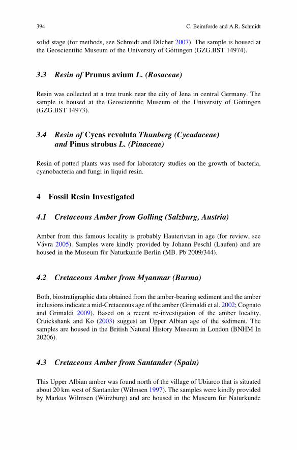
solid stage (for methods, see Schmidt and Dilcher 2007). The sample is housed at
the Geoscientific Museum of the University of G€ottingen (GZG.BST 14974).
3.3 Resin of Prunus avium L. (Rosaceae)
Resin was collected at a tree trunk near the city of Jena in central Germany. The
sample is housed at the Geoscientific Museum of the University of G€ottingen(GZG.BST 14973).
3.4 Resin of Cycas revoluta Thunberg (Cycadaceae)and Pinus strobus L. (Pinaceae)
Resin of potted plants was used for laboratory studies on the growth of bacteria,
cyanobacteria and fungi in liquid resin.
4 Fossil Resin Investigated
4.1 Cretaceous Amber from Golling (Salzburg, Austria)
Amber from this famous locality is probably Hauterivian in age (for review, see
Vavra 2005). Samples were kindly provided by Johann Peschl (Laufen) and are
housed in the Museum f€ur Naturkunde Berlin (MB. Pb 2009/344).
4.2 Cretaceous Amber from Myanmar (Burma)
Both, biostratigraphic data obtained from the amber-bearing sediment and the amber
inclusions indicate amid-Cretaceous age of the amber (Grimaldi et al. 2002; Cognato
and Grimaldi 2009). Based on a recent re-investigation of the amber locality,
Cruickshank and Ko (2003) suggest an Upper Albian age of the sediment. The
samples are housed in the British Natural History Museum in London (BNHM In
20206).
4.3 Cretaceous Amber from Santander (Spain)
This Upper Albian amber was found north of the village of Ubiarco that is situated
about 20 km west of Santander (Wilmsen 1997). The samples were kindly provided
by Markus Wilmsen (W€urzburg) and are housed in the Museum f€ur Naturkunde
394 C. Beimforde and A.R. Schmidt
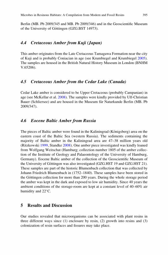
Berlin (MB. Pb 2009/345 and MB. Pb 2009/346) and in the Geoscientific Museum
of the University of G€ottingen (GZG.BST 14973).
4.4 Cretaceous Amber from Kuji (Japan)
This amber originates from the Late Cretaceous Tamagawa Formation near the city
of Kuji and is probably Coniacian in age (see Krumbiegel and Krumbiegel 2005).
The samples are housed in the British Natural History Museum in London (BNHM
V.65206).
4.5 Cretaceous Amber from the Cedar Lake (Canada)
Cedar Lake amber is considered to be Upper Cretaceous (probably Campanian) in
age (see McKellar et al. 2008). The samples were kindly provided by Ulf-Christian
Bauer (Schliersee) and are housed in the Museum f€ur Naturkunde Berlin (MB. Pb
2009/347).
4.6 Eocene Baltic Amber from Russia
The pieces of Baltic amber were found in the Kaliningrad (K€onigsberg) area on theeastern coast of the Baltic Sea (western Russia). The sediments containing the
majority of Baltic amber in the Kaliningrad area are 47–38 million years old
(Ritzkowski 1999, Standke 2008). One amber piece investigated was kindly loaned
from Wolfgang Weitschat (Hamburg; collection number 1805 of the amber collec-
tion of the Institute of Geology and Palaeontology of the University of Hamburg,
Germany). Eocene Baltic amber of the collection of the Geoscientific Museum of
the University of G€ottingen was also investigated (GZG.HST 19 and GZG.HST 21).
These samples are part of the historic Blumenbach collection that was collected by
Johann Friedrich Blumenbach in (1752–1840). These samples have been stored in
the G€ottingen collection for more than 200 years. During the whole storage period
the amber was kept in the dark and exposed to low air humidity. Since 40 years the
ambient conditions of the storage-room are kept at a constant level of 40–60% air
humidity and 22�C.
5 Results and Discussion
Our studies revealed that microorganisms can be associated with plant resins in
three different ways since (1) enclosure by resin, (2) growth into resins and (3)
colonization of resin surfaces and fissures may take place.
Microbes in Resinous Habitats: A Compilation from Modern and Fossil Resins 395
5.1 Microorganisms Enclosed by Resin
Various limnetic and terrestrial microorganisms were found engulfed, i.e., entirely
embedded in resins. Among others, representatives of conjugatophytes (Fig. 1a)
and testate amoeba (testate lobose amoebae, order Arcellinida, Fig. 1b–c) were
found inside resin flows of Pinus elliottii from a Florida swamp forest. Single celled
Fig. 1 Light-microscopical images of modern and fossil microorganisms that became engulfed by
resin: (a) Conjugatophyte of the genus Cosmarium enclosed in modern resin from Pinus elliottii.(b–c) Testate amoeba of the genus Arcella in ventral (b) and lateral (c) view inside P. elliottii resin.(d–f) Epiphytic cyanobacteria enclosed in modern resin of Prunus avium (GZG.BST 14973). The
filaments continued to grow for a short time after embedding. The arrow shows a narrow filament
in which cross walls are absent. (g–h) Fossil cyanobacteria in Upper Cretaceous amber from
Canada (MB.Pb.2009/347). The arrow in (c) indicates a narrow flament without cross walls;
the arrow in (h) shows a regular filament. Scale bars represent 30 mm in (a–c and e–h), and 200 mmin (d)
396 C. Beimforde and A.R. Schmidt
limnetic organisms became enclosed, when resin flowed down the tree trunks and
reached the humid forest floor. Some of these inclusions exhibit an excellent
preservation of detailed structures such as chloroplasts (Fig. 1a). Schmidt and
Dilcher (2007) found all major groups of limnic microorganisms of the swamp
water embedded in resin of Pinus elliottii, a species that exhibits a massive natural
resin production. Limnetic-terrestrial microorganisms have also repeatedly been
reported from Cretaceous ambers. Aquatic bacteria, microalgae, conjugatophytes,
ciliates and testate amoebae described from the Cenomanian Schliersee amber from
the Bavarian Alps are examples hereof (see Sch€onborn et al. 1999; D€orfelt andSch€afer 2000; Schmidt et al. 2004).
Cyanobacteria are rarely enclosed in fossil resins. We found modern filamentous
cyanobacteria inside resin at the trunk of a cherry tree (Prunus avium, Fig. 1d–f).After becoming entirely enclosed by the fresh resin, further growth of the filaments
took place for a short time. During this limited growth, narrow filaments were
produced in which cross walls were often absent. We discovered similar narrow
filaments without cross walls in cyanobacterial mats inside Upper Cretaceous
amber from the Cedar Lake in Canada (Fig. 1g–h). Therefore, we interpret these
fossil cyanobacteria to be alive during embedding and a couple of hours to days
afterwards in the liquid resin.
However, generally those entirely embedded organisms died off immediately
after embedding and correspond to the classic term of inclusions since they do not
touch the resin surface at all.
5.2 Microorganisms Growing on and into the Liquid Resin
We found many examples of modern and fossil fungal hyphae and filamentous
bacteria that grew from the surface into the resin bodies until solidification
occurred. Despite being preserved inside amber and modern resin, these inclusions
do not correspond to the typical criteria of inclusions because they touch the surface
at least at one point.
Sheathed bacteria occurred sometimes in fresh resin of Pinus elliottii that
flowed into the ponds of a Florida swamp forest. Their sheaths are about 10 mmin diameter and exhibit a central lumen of about 1 mm diameter. Cell chains could
sometimes not be identified inside the lumen. As in modern Leptothrix specimens,
the observed sheaths are hyaline, slightly granulated and contain minute gas
bubbles which might indicate metabolic activity after embedding in the resin
(Fig. 2a–b). Spiral prokaryotic filaments and branched fungal hyphae were regu-
larly found in P. elliottii resin that had contact to the swamp water (Fig. 2c–d).
Correspondingly, many Cretaceous amber pieces are interspersed with fossil
bacterial filaments. We found filaments that are composed of rod-shaped cells
(Fig. 3a–b), but mostly sheathed branched cell chains or empty sheaths that
morphologically closely resemble modern representatives of the genus Leptothrixoccurred in the ambers (Fig. 3c–i). The random orientation of the sheathed
Microbes in Resinous Habitats: A Compilation from Modern and Fossil Resins 397
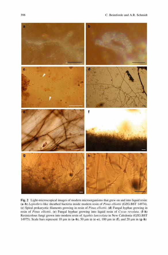
Fig. 2 Light-microscopical images of modern microorganisms that grew on and into liquid resin:
(a–b) Leptothrix-like sheathed bacteria inside modern resin of Pinus elliottii (GZG.BST 14974).
(c) Spiral prokaryotic filaments growing in resin of Pinus elliottii. (d) Fungal hyphae growing in
resin of Pinus elliottii. (e) Fungal hyphae growing into liquid resin of Cycas revoluta. (f–h)Resinicolous fungi grown into modern resin of Agathis lanceolata in New Caledonia (GZG.BST
14975). Scale bars represent 10 mm in (a–b), 50 mm in (c–e), 100 mm in (f), and 20 mm in (g–h)
398 C. Beimforde and A.R. Schmidt
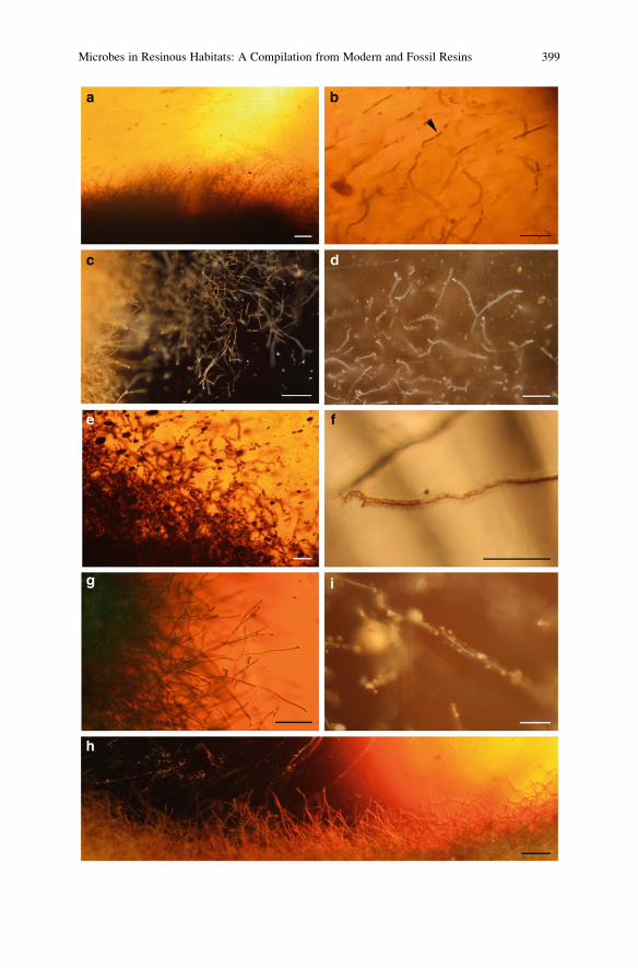
Microbes in Resinous Habitats: A Compilation from Modern and Fossil Resins 399
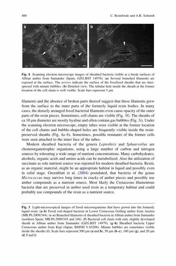
filaments and the absence of broken parts thereof suggest that these filaments grew
from the surface to the inner parts of the formerly liquid resin bodies. In many
cases, the densely arranged fossil bacterial filaments even cause opacity of the outer
parts of the resin pieces. Sometimes, cell chains are visible (Fig. 3f). The sheaths of
ca 10 mm diameter are mostly hyaline and often contain gas bubbles (Fig. 3i). Under
the scanning electron microscope, empty tubes were visible at the former location
of the cell chains and bubble-shaped holes are frequently visible inside the resin-
preserved sheaths (Fig. 4a–b). Sometimes, possible remnants of the former cells
were seen attached to the inner face of the tubes.
Modern sheathed bacteria of the genera Leptothrix and Sphaerotilus are
chemoorganotrophic organisms, using a large number of carbon and nitrogen
sources by tolerating a wide range of nutrient concentrations. Many carbohydrates,
alcohols, organic acids and amino acids can be metabolized. Also the utilization of
succinate as sole nutrient source was reported for modern sheathed bacteria. Resin,
as an organic material, might be an appropriate habitat in liquid and possibly even
in solid stage. Greenblatt et al. (2004) postulated, that bacteria of the genus
Micrococcus may survive long times in cracks of amber pieces and possibly use
amber compounds as a nutrient source. Most likely the Cretaceous filamentous
bacteria that are preserved in amber used resin as a temporary habitat and could
probably use compounds of the resin as a nutrient source.
Fig. 4 Scanning electron microscope images of sheathed bacteria visible at a break surfaces of
Albian amber from Santander (Spain, GZG.BST 14978): (a) Several branched filaments are
exposed at the surface. The arrows indicate the surface of the fossilized sheaths that are inter-
spersed with minute bubbles. (b) Detailed view. The tubular hole inside the sheath at the former
location of the cell chain is well visible. Scale bars represent 5 mm
Fig. 3 Light-microscopical images of fossil microorganisms that have grown into the formerly
liquid resin: (a–b) Fossil rod-shaped bacteria in Lower Cretaceous Golling amber from Austria
(MB.Pb.2009/344). (c–e) Branched filaments of sheathed bacteria in Albian amber from Santander
(northern Spain; MB.Pb.2009/345 and 346). (f) Bacterial cell chain with only slightly developed
sheath in Albian amber from Santander (GZG.BST 14979). (g–h) Sheathed bacteria Upper
Cretaceous amber from Kuji (Japan; BMNH V.65206). Minute bubbles are sometimes visible
inside the sheaths (i). Scale bars represent 500 mm (a and h), 50 mm (b–c), 100 mm (g), and 20 mm(d, f and i)
400 C. Beimforde and A.R. Schmidt
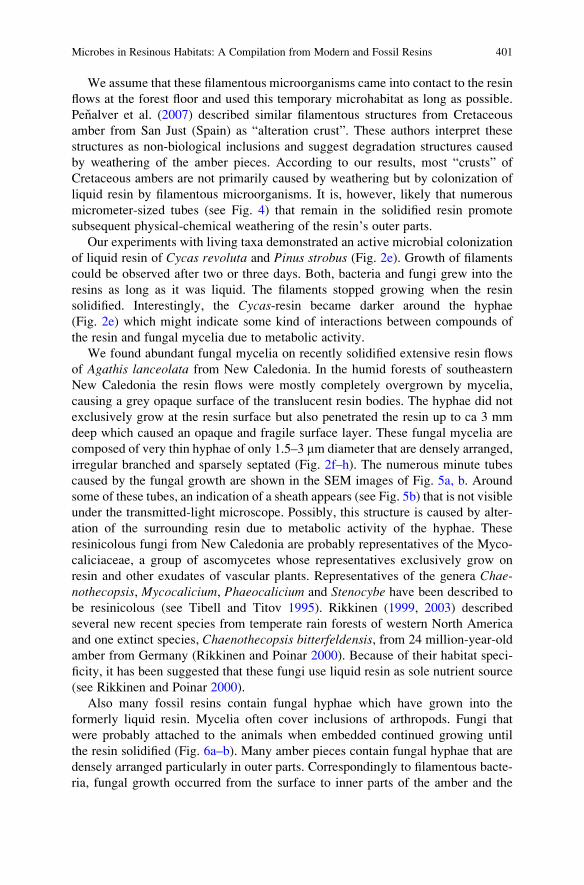
We assume that these filamentous microorganisms came into contact to the resin
flows at the forest floor and used this temporary microhabitat as long as possible.
Penalver et al. (2007) described similar filamentous structures from Cretaceous
amber from San Just (Spain) as “alteration crust”. These authors interpret these
structures as non-biological inclusions and suggest degradation structures caused
by weathering of the amber pieces. According to our results, most “crusts” of
Cretaceous ambers are not primarily caused by weathering but by colonization of
liquid resin by filamentous microorganisms. It is, however, likely that numerous
micrometer-sized tubes (see Fig. 4) that remain in the solidified resin promote
subsequent physical-chemical weathering of the resin’s outer parts.
Our experiments with living taxa demonstrated an active microbial colonization
of liquid resin of Cycas revoluta and Pinus strobus (Fig. 2e). Growth of filaments
could be observed after two or three days. Both, bacteria and fungi grew into the
resins as long as it was liquid. The filaments stopped growing when the resin
solidified. Interestingly, the Cycas-resin became darker around the hyphae
(Fig. 2e) which might indicate some kind of interactions between compounds of
the resin and fungal mycelia due to metabolic activity.
We found abundant fungal mycelia on recently solidified extensive resin flows
of Agathis lanceolata from New Caledonia. In the humid forests of southeastern
New Caledonia the resin flows were mostly completely overgrown by mycelia,
causing a grey opaque surface of the translucent resin bodies. The hyphae did not
exclusively grow at the resin surface but also penetrated the resin up to ca 3 mm
deep which caused an opaque and fragile surface layer. These fungal mycelia are
composed of very thin hyphae of only 1.5–3 mm diameter that are densely arranged,
irregular branched and sparsely septated (Fig. 2f–h). The numerous minute tubes
caused by the fungal growth are shown in the SEM images of Fig. 5a, b. Around
some of these tubes, an indication of a sheath appears (see Fig. 5b) that is not visible
under the transmitted-light microscope. Possibly, this structure is caused by alter-
ation of the surrounding resin due to metabolic activity of the hyphae. These
resinicolous fungi from New Caledonia are probably representatives of the Myco-
caliciaceae, a group of ascomycetes whose representatives exclusively grow on
resin and other exudates of vascular plants. Representatives of the genera Chae-nothecopsis, Mycocalicium, Phaeocalicium and Stenocybe have been described to
be resinicolous (see Tibell and Titov 1995). Rikkinen (1999, 2003) described
several new recent species from temperate rain forests of western North America
and one extinct species, Chaenothecopsis bitterfeldensis, from 24 million-year-old
amber from Germany (Rikkinen and Poinar 2000). Because of their habitat speci-
ficity, it has been suggested that these fungi use liquid resin as sole nutrient source
(see Rikkinen and Poinar 2000).
Also many fossil resins contain fungal hyphae which have grown into the
formerly liquid resin. Mycelia often cover inclusions of arthropods. Fungi that
were probably attached to the animals when embedded continued growing until
the resin solidified (Fig. 6a–b). Many amber pieces contain fungal hyphae that are
densely arranged particularly in outer parts. Correspondingly to filamentous bacte-
ria, fungal growth occurred from the surface to inner parts of the amber and the
Microbes in Resinous Habitats: A Compilation from Modern and Fossil Resins 401
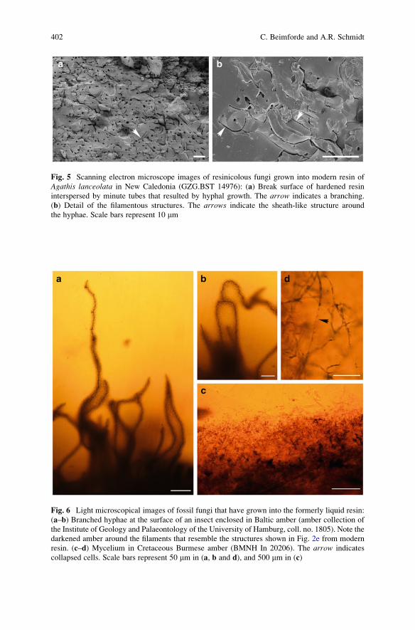
Fig. 6 Light microscopical images of fossil fungi that have grown into the formerly liquid resin:
(a–b) Branched hyphae at the surface of an insect enclosed in Baltic amber (amber collection of
the Institute of Geology and Palaeontology of the University of Hamburg, coll. no. 1805). Note the
darkened amber around the filaments that resemble the structures shown in Fig. 2e from modern
resin. (c–d) Mycelium in Cretaceous Burmese amber (BMNH In 20206). The arrow indicates
collapsed cells. Scale bars represent 50 mm in (a, b and d), and 500 mm in (c)
Fig. 5 Scanning electron microscope images of resinicolous fungi grown into modern resin of
Agathis lanceolata in New Caledonia (GZG.BST 14976): (a) Break surface of hardened resin
interspersed by minute tubes that resulted by hyphal growth. The arrow indicates a branching.
(b) Detail of the filamentous structures. The arrows indicate the sheath-like structure around
the hyphae. Scale bars represent 10 mm
402 C. Beimforde and A.R. Schmidt
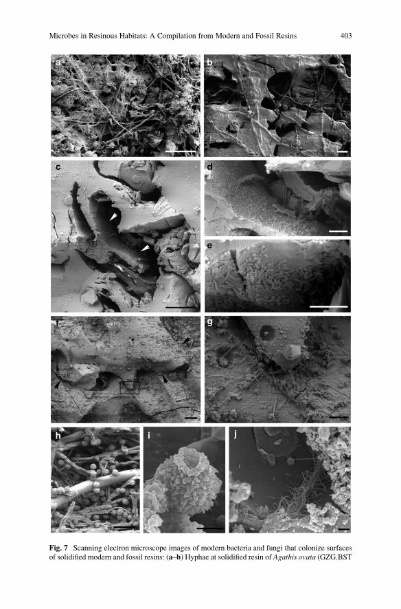
Fig. 7 Scanning electron microscope images of modern bacteria and fungi that colonize surfaces
of solidified modern and fossil resins: (a–b) Hyphae at solidified resin of Agathis ovata (GZG.BST
Microbes in Resinous Habitats: A Compilation from Modern and Fossil Resins 403
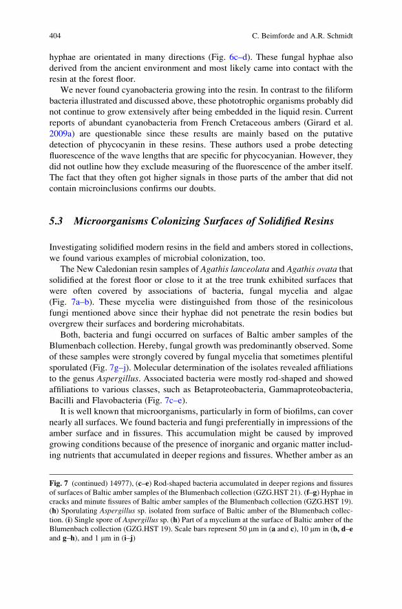
hyphae are orientated in many directions (Fig. 6c–d). These fungal hyphae also
derived from the ancient environment and most likely came into contact with the
resin at the forest floor.
We never found cyanobacteria growing into the resin. In contrast to the filiform
bacteria illustrated and discussed above, these phototrophic organisms probably did
not continue to grow extensively after being embedded in the liquid resin. Current
reports of abundant cyanobacteria from French Cretaceous ambers (Girard et al.
2009a) are questionable since these results are mainly based on the putative
detection of phycocyanin in these resins. These authors used a probe detecting
fluorescence of the wave lengths that are specific for phycocyanian. However, they
did not outline how they exclude measuring of the fluorescence of the amber itself.
The fact that they often got higher signals in those parts of the amber that did not
contain microinclusions confirms our doubts.
5.3 Microorganisms Colonizing Surfaces of Solidified Resins
Investigating solidified modern resins in the field and ambers stored in collections,
we found various examples of microbial colonization, too.
The New Caledonian resin samples of Agathis lanceolata and Agathis ovata thatsolidified at the forest floor or close to it at the tree trunk exhibited surfaces that
were often covered by associations of bacteria, fungal mycelia and algae
(Fig. 7a–b). These mycelia were distinguished from those of the resinicolous
fungi mentioned above since their hyphae did not penetrate the resin bodies but
overgrew their surfaces and bordering microhabitats.
Both, bacteria and fungi occurred on surfaces of Baltic amber samples of the
Blumenbach collection. Hereby, fungal growth was predominantly observed. Some
of these samples were strongly covered by fungal mycelia that sometimes plentiful
sporulated (Fig. 7g–j). Molecular determination of the isolates revealed affiliations
to the genus Aspergillus. Associated bacteria were mostly rod-shaped and showed
affiliations to various classes, such as Betaproteobacteria, Gammaproteobacteria,
Bacilli and Flavobacteria (Fig. 7c–e).
It is well known that microorganisms, particularly in form of biofilms, can cover
nearly all surfaces. We found bacteria and fungi preferentially in impressions of the
amber surface and in fissures. This accumulation might be caused by improved
growing conditions because of the presence of inorganic and organic matter includ-
ing nutrients that accumulated in deeper regions and fissures. Whether amber as an
Fig. 7 (continued) 14977), (c–e) Rod-shaped bacteria accumulated in deeper regions and fissures
of surfaces of Baltic amber samples of the Blumenbach collection (GZG.HST 21). (f–g) Hyphae in
cracks and minute fissures of Baltic amber samples of the Blumenbach collection (GZG.HST 19).
(h) Sporulating Aspergillus sp. isolated from surface of Baltic amber of the Blumenbach collec-
tion. (i) Single spore of Aspergillus sp. (h) Part of a mycelium at the surface of Baltic amber of the
Blumenbach collection (GZG.HST 19). Scale bars represent 50 mm in (a and c), 10 mm in (b, d–e
and g–h), and 1 mm in (i–j)
404 C. Beimforde and A.R. Schmidt
organic material can be utilized as a nutrient source for microorganisms has not been
examined so far, but it is conceivable. Due to the microbial activity, recent coloni-
zation may accelerate the degradation of the amber objects. The degradation of
amber is probably caused by various chemical, physical and biological processes
that may act together and sometimes trigger or enforce each other. Processes of
oxidation are probably most important for the degradation of amber. However,
when fungal mycelia penetrate into present fissures and extent their mycelia,
physical forces arise that can break up upper amber layers, thus opening new
areas for oxidation processes. So far, it is unclear if also secretion of metabolic
compounds can represent a damaging factor and if the microbes may utilize amber
compounds as a nutrient source. This should be subject of further studies.
Usually, recent biofilms are easily distinguishable from fossil inclusions using
high-magnification light-microscopy. Also, microorganisms that are situated inside
fissures that extend into the amber are usually distrainable from microinclusions
when tracing the refractive micrometre-sized fissures under the microscope. During
the study of mid-Cretaceous amber of south-western France, Girard et al. (2008a)
discovered living marine and freshwater diatoms at the amber surface and in
micrometre-sized fissures. Furthermore, these authors found fungal mycelia, bacte-
ria and cyanobacteria in fissures inside the amber samples, showing that not
exclusively ambers in collections represent habitats of microorganisms.
Acknowledgements We thank JeromeMunzinger (Noumea) for supporting the field work in New
Caledonia and David L. Dilcher (Gainesville) for making the field studies in Florida possible. We
are indebted to Mike Reich (G€ottingen) for amber loans from the Blumenbach collection of Baltic
amber, to Wolfgang Weitschat (Hamburg) for loans of Baltic amber, to Johann Peschl (Laufen) for
providing Cretaceous amber from Austria, to Markus Wilmsen (W€urzburg) for Cretaceous amber
samples from Spain, and to Ulf-Christian Bauer (Schliersee) for donating samples of Cedar Lake
amber. Hilary Ketchum and Claire Mellish (London) kindly provided access to the amber
collection of the Natural History Museum in London. We acknowledge with thanks the help of
Dorothea Hause-Reitner and Andreas Reimer (G€ottingen) during the SEM analyses and the help of
Kerstin Schmidt (Jena) in the preparation of the manuscript. We heartily thank Joachim Reitner
(G€ottingen) for technical support and for his kind invitation to contribute to this special issue. Thisis contribution number 39 from the Courant Research Centre Geobiology that is funded by the
German Initiative of Excellence.
References
Austin J, Ross AJ, Smith AB, Fortey RA, Thomas RH (1997) Problems of reproducibility – does
geologically ancient DNA survive in amber-preserved insects? Proceedings Royal Society
London, Series B Biological Science 264:467–621
Berkeley MJ (1848) On three species of mould detected by Dr. Thomas in the amber of East
Prussia. Annual Magacine Natural History (Series 2) 2:380–383
Blunck G (1929) Bakterieneinschl€usse im Bernstein. Zentralblatt Mineralogie, Geologie, Pal€an-tologie 11:554–555
Cano RJ, Borucki MK, Higby-Schweizer M, Poinar HN, Poinar GO Jr, Pollard KJ (1994) Bacillus-DNA in fossil bees: an ancient symbiosis? Applied and Environmental Microbiology
60:2164–2167
Microbes in Resinous Habitats: A Compilation from Modern and Fossil Resins 405
Caspary R, Klebs R (1907) Die Flora des Bernsteins und anderer fossiler Harze des ostpreußischen
Terti€ars. Abhandlungen der K€oniglich-Preußischen Geologischen Landesanstalt 4:1–181
Cognato AI, Grimaldi DA (2009) 100 million years of morphological conservation in bark beetles
(Coleoptera: Curculionidae: Scolytinae). Systematic Entomology 34:93–100
Cruickshank RD, Ko K (2003) Geology of an amber locality in the Hukawng valley, Northern
Myanmar. Jouranl of Asian Earth Scieces 21:441–445
D€orfelt H, Sch€afer U (2000) Palaeozygnema spiralis, ein Vertreter der Conjugatophyceae in
mesozoischem Bernstein aus Bayern. Hoppea 61:785–793
D€orfelt H, Schmidt AR (2007) A conifer seedling with two herbicolous fungi from the Baltic
amber forest. Botanical Journal of the Linnean Society 155:449–456
GalippeV (1920)Recherches sur la resistance desmicrozimas alaction du temps er sur survivance dans
lambre. Comptes rendus hebdomadaires des seances de l’Academie des sciences 170:856–858
Girard V, Neraudeau D, Breton G (2008a) Contamination of amber samples by recent micro-
organisms and remediation evidenced by Mid-Cretaceous amber of Charentes (SW France).
Geomicrobiological Journal 26:21–20
Girard V, Schmidt AR, Saint Martin S, Struwe S, Perrichot V, Saint Martin JP, Grosheny D,
Breton G, Neraudeaua D (2008b) Evidence for marine microfossils from amber. Proceedings
of the National Academy of Sciences of the United States of America 105:17426–17429
Girard V, Breton G, Brient L, Neraudeau D (2009a) Sheathed prokaryotic filaments, major compo-
nents of Mid-Cretaceous French amber microcoenoses. Journal of Paleolimnology 42:437–447
Girard V, Schmidt AR, Struwe S, Perrichot V, Breton G, Neraudeau D (2009b) Taphonomy and
palaeoecology of mid-Cretaceous amber-preserved microorganisms from southwestern
France. In: Perrichot V, Neraudeau D (eds) Studies on mid-Cretaceous amber from
southwestern France. Geodiversitas 31:153–162
Greenblatt CL, Davis A, Clement BG, Kitts CL, Cox T, Cano RJ (1999) Diversity of microorgan-
isms isolated from amber. Microbial Ecology 38:58–68
Greenblatt CL, Baum J, Klein BY, Nachson S, Cano RJ (2004) Micrococcus luteus – survival in
amber. Microbial Ecology 48:120–127
Grimaldi DA, Engel MS, Nascimbene PC (2002) – Fossifiliferous Cretaceous amber from
Myanmar (Burma): its rediscovery, biotic diversity and paleontological significance. American
Museum Novitates 3361:1–72
Hofreiter M, Serre D, Poinar HN, Kuch M, P€a€abo S (2001) Ancient DNA. Nature Reviews
Genetics 2:353–359
Krumbiegel G, Krumbiegel B (2005) Bernstein – Fossile Harze aus aller Welt. Goldschneck
Verlag, Wiebelsheim
Lambert LH, Cox T, Mitchell K, Rossello-Mora, RA, Del Cueto C, Doge DE, Orkland P, Cano RJ
(1998) Staphylococcus succinus sp. nov., isolated from Dominican amber. International
Journal Systemaic Bacteriology 48:511–518
McKellar RC, Wolfe AP, Tappert R, Muehlenbachs K (2008) Correlation of Grassy Lake and
Cedar Lake ambers using infrared spectroscopy, stable isotopes and palaeoentomology.
Canadian Journal of Earth Sciences 45:1061–1082
Penalver E, Delclos X, Soriano C (2007) A new rich amber outcrop with palaeobiological
inclusions in the Lower Cretaceous of Spain. Cretaceous Research 28:791–802
Poinar GO, Poinar R (1994) The quest for life in amber. Addison-Wesley Publishing Company,
Reading, MA, 219 pp
Rikkinen J (1999) Two new species of resinicolous Chaenothecopsis (Mycocaliciaceae) from
western North America. Bryologist 102:366–369
Rikkinen J (2003) Chaenothecopsis nigripunctata, a remarkable new species of resinicolous
Mycocaliciaceae from western North America. Mycologia 95:98–103
Rikkinen J, Poinar GO Jr (2000) A new species of resinicolous Chaenothecopsis (Mycocalicia-
ceae, Ascomycota) from 20 million year old Bitterfeld amber, with remarks on the biology of
resinicolous fungi. Mycological Research 104:7–15
406 C. Beimforde and A.R. Schmidt
Rikkinen J, D€orfeld H, Schmidt AR, Wunderlich J (2003) Sooty moulds from European Tertiary
amber, with notes on the systematic position of Rosaria (“Cyanobacteria”). Mycological
Research 107:251–256
Ritzkowski S (1999) Das geologische Alter der bernsteinf€uhrenden Sedimente in Sambia (Bezirk
Kaliningrad), bei Bitterfeld (Sachsen-Anhalt) und bei Helmstedt (SE-Niedersachsen).
In: Kosmowska-Ceranowicz B, Paner H (eds) Investigations into amber. Proceedings of
the International Interdisciplinary Symposium: Baltic amber and other fossil resins, 2–6
September 1997:33–40, Gdansk, Poland
Schmidt AR, Dilcher DL (2007) Aquatic organisms as amber inclusions and examples from a
modern swamp forest. Proceedings of the National Academy of Sciences of the United States
of America 104:16581–16585
Schmidt AR, Sch€afer U (2005) Leptotrichites resinatus new genus and species: a fossil sheathed
bacterium in alpine Cretaceous amber. Journal of Paleontology 79:175–184
Schmidt AR, Sch€onbornW, Sch€afer U (2004) Diverse fossil amoebae in GermanMesozoic amber.
Palaeontology 47:185–197
Schmidt AR, Ragazzi E, Coppellotti O, Roghi G (2006) A microworld in Triassic amber. Nature
444:835
Sch€onborn W, D€orfelt H, Foissner W, Krienitz L, Sch€afer U (1999) A fossilized microcenosis in
Triassic amber. Journal of Eukaryotic Microbiology 46:571–584
Standke G (2008) Bitterfelder Bernstein gleich Baltischer Bernstein? – Eine geologische
Raum-Zeit-Betrachtung und genetische Schlußfolgerungen. In: Rascher J, Wimmer R,
Krumbiegel G, Schmiedel S (eds) Bitterfelder Bernstein versus Baltischer Bernstein –
Hypothesen, Fakten, Fragen. Exkursionsf€uhrer und Ver€offentlichungen der Deutschen
Gesellschaft f€ur Geowissenschaften 236:11–33
Tibell L, Titov A (1995) Species of Chaenothecopsis and Mycocalicium (Caliciales) on exudate.
Bryologist 98:550–560
Ting WS, Nissenbaum A (1986) Fungi in Lower Cretaceous amber from Israel. Special Publica-
tion by the Exploration and Development Research Center, Chinese Petroleum Corporation,
Miaoli, Taiwan
Vavra N (2005) Bernstein und verwandte organische Minerale aus Osterreich. Beitr€age zur
Pal€aontologie von Osterreich 29:255–280
Waggoner BM (1993) Fossil actinomycetes and other bacteria in Eocene amber from Washington
State, USA. Tertiary Research 14:155–160
Waggoner BM (1994) Fossil microorganisms from Upper Cretaceous amber of Mississippi.
Review of Palaeobotany and Palynology 80:75–84
Waggoner BM (1996) Bacteria and protists from Middle Cretaceous amber of Ellsworth County,
Kansas. PaleoBios 17:20–26
Willerslev E, Cooper A (2005) Ancient DNA. Proceedings Royal Society London, Series B
Biological Science 272:3–16
Willerslev E, Hansen AJ, Poinar HN (2004) Isolation of nucleic acids and cultures from fossil ice
and permafrost. Trends in Ecology and Evolution 19:141–147
WilmsenM (1997) Das Oberalb und Cenoman imNordkantabrischen Becken (Provinz Kantabrien,
Nordspanien): Faziesentwicklung, Bio- und Sequenzstratigraphie. Berliner Geowissenschaf-
tliche Abhandlungen E 23:1–167
Microbes in Resinous Habitats: A Compilation from Modern and Fossil Resins 407

.
Tolypammina gregaria Wendt 1969-FrutexitesAssemblage and Ferromanganese Crusts:
A Coupled Nutrient-Metal Interplay
in the Carnian Sedimentary Condensed
Record of Hallstatt Facies (Austria)
Marta Rodrıguez-Martınez, Christine Heim, Klaus Simon, Thomas Zilla,
and Joachim Reitner
1 Introduction
Foraminifera are the most abundant sessile organisms found on ferromanganese
crusts and nodules (Greenslate 1974; Wendt 1974; Dugolinsky et al. 1977;
Riemann 1983; von Stackelberg 1984; Mullineaux 1987, 1988; Verlaan 1992;
Resig and Glenn 1997; Toscano and Raspini 2005). In fact, actual ferromanganese
crusts and nodules share numerous similarities with their fossil counterparts. The
oldest examples are described from the Devonian (Tucker 1973), and also occur
widely distributed through the condensed Triassic and Lower Jurassic deposits
(summarized in Jimenez-Millan and Nieto 2006) until Plio-Pleistocene records
(Jenkyns 1970; Wendt 1970; Allouc 1990; Toscano and Raspini 2005).
Ferromanganese crusts and nodules are widely distributed in actual oceans, on
sediment starved seafloors, slopes and seamounts. The exact mechanisms of their
formation are not well understood yet, therefore abiotic, biotic as well as coupled
abiotic–biotic processes are proposed. From an abiogenic point of view, Bonatti
(1981) suggested four genetic types: hydrogenous, hydrothermal, diagenetic or
halmyrolitic origin. On the other hand, their biogenic contribution has been linked
mainly to Bacteria (Ehrlich 1963, 1968, 1971; Ghiorse and Ehrlich 1992; Wang
et al. 2009) and Foraminifera. In fact, the relationship between foraminifers and
their biogenic influence in the ferromanganese accretion has been widely discussed
(Dugolinsky et al. 1977; Riemann 1983; Mullineaux 1987; Wendt 1974; Verlaan
1992). The most abundant foraminifers reported from ferromanganese nodules and
M. Rodrıguez-Martınez (*)
Departamento de Geologıa, Universidad de Alcala, Edificio de Ciencias, N-II Km 33.6, 28871,
Alcala de Henares, Madrid, Espana
e-mail: [email protected]
C. Heim, K. Simon, T. Zilla, and J. Reitner
Geoscience Centre, University of G€ottingen, Goldschmidtstraße 3, 37077, G€ottingen, Germany
J. Reitner et al., Advances in Stromatolite Geobiology,Lecture Notes in Earth Sciences 131, DOI 10.1007/978-3-642-10415-2_25,# Springer-Verlag Berlin Heidelberg 2011
409
crusts by different authors are Sarcodina members and several Tolypamminaspecies (Resig and Glenn 1997).
Tolypammina gregaria Wendt is an agglutinated sessile foraminifer with a
surprising behaviour, forming vertical structures which were firstly described
by Wendt (1969), from Carnian sedimentary condensed deposits at Feuerkogel
(Austria). The vertical accreted, pillar-like structures are buildup by Tolypamminagregaria Wendt and Frutexites. Frutexites (Maslov 1960) is a problematic Fe-rich
microfossil with shrub, colloform to microstromatolitic morphology, figured as
“Frutexites” from Proterozoic to Recent records [Caldera lake stromatolites at
Tonga, by Kazmierczak and Kempe (2006)]. The genus Frutexites was coined by
Maslov (1960) from Ordovician stromatolites of Siberian Platform. This problem-
atic structure has been interpreted as abiotic as well as a biologically induced
precipitate.
The Tolypammina gregaria-Frutexites vertical structures are associated with
ferromanganese crusts and surfaces. This study aims to unravel the environmental
significance of Tolypammina gregaria-Frutexites assemblage and their interplay
with ferromanganese crusts/surfaces. Therefore detailed investigations of the
microfacies as well as trace and rare earth elements (REE) analysis were performed
in order to be able to reconstruct the environmental conditions during deposition of
the limestone and the Tolypammina-Frutexites assemblages and their relationship
with the so called Carnian Pluvial Event.
2 Materials and Methods
After detailed field observations of the stratigraphic succession and facies changes,
15 samples ex situ were carefully selected from the type locality area for micro-
facies and geochemical analysis. Polished slabs, large (10 � 15 cm) and standard
petrographic thin-sections were made to carry out investigation with petrographic
and cathodoluminescence (CL) microscopy.
Subsequent to the microfacial studies, three samples were selected for geochem-
ical analysis. Laser-ICP-MS measurements of trace elements and REE were per-
formed with a Perkin Elmer SCIEX Elan DRII with a coupled laser from Lambda
Physik (190 mJ, 30 kV, 10 Hz, Laser Ø 120 mm).
3 Geological Setting
Tolypammina gregariaWendt-rich bed with ferromanganese crusts is located in the
Feuerkogel peak, close to Kainisch village at Steiermark, Austria (Fig. 1). The bed,
from 2 cm up to 20 cm of maximum thickness, is part of the so-called Hallstatt
Limestone facies of the Northern Calcareous Alps (NCA in Fig. 1).
The Hallstatt Limestone shows two characteristic facies: proximal grey and
distal red facies where condensed ammonoid beds are common. The Feuerkogel
410 M. Rodrıguez-Martınez et al.
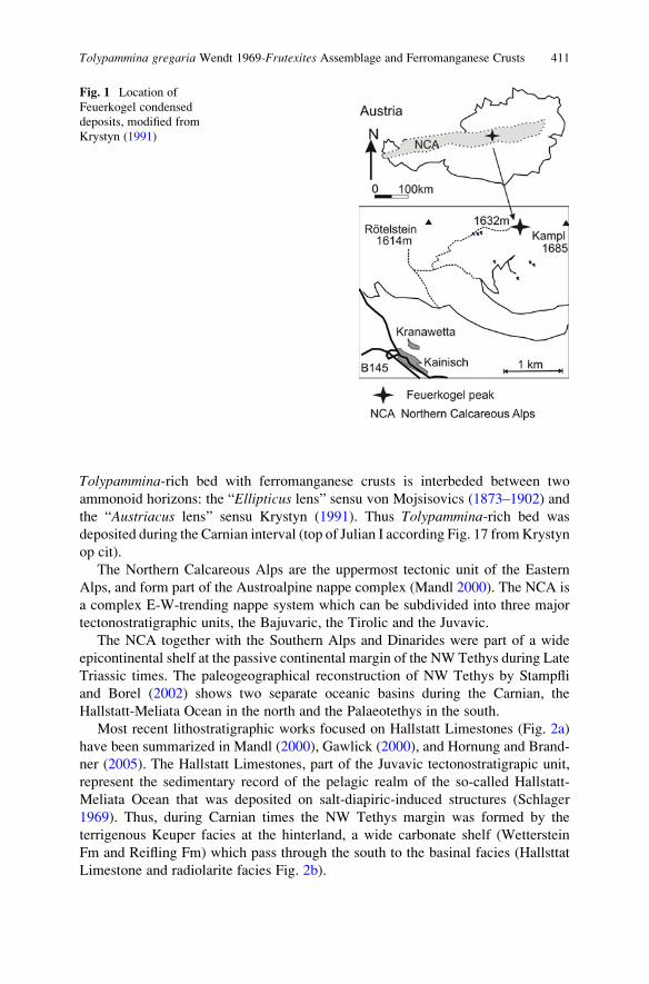
Tolypammina-rich bed with ferromanganese crusts is interbeded between two
ammonoid horizons: the “Ellipticus lens” sensu von Mojsisovics (1873–1902) and
the “Austriacus lens” sensu Krystyn (1991). Thus Tolypammina-rich bed was
deposited during the Carnian interval (top of Julian I according Fig. 17 fromKrystyn
op cit).
The Northern Calcareous Alps are the uppermost tectonic unit of the Eastern
Alps, and form part of the Austroalpine nappe complex (Mandl 2000). The NCA is
a complex E-W-trending nappe system which can be subdivided into three major
tectonostratigraphic units, the Bajuvaric, the Tirolic and the Juvavic.
The NCA together with the Southern Alps and Dinarides were part of a wide
epicontinental shelf at the passive continental margin of the NW Tethys during Late
Triassic times. The paleogeographical reconstruction of NW Tethys by Stampfli
and Borel (2002) shows two separate oceanic basins during the Carnian, the
Hallstatt-Meliata Ocean in the north and the Palaeotethys in the south.
Most recent lithostratigraphic works focused on Hallstatt Limestones (Fig. 2a)
have been summarized in Mandl (2000), Gawlick (2000), and Hornung and Brand-
ner (2005). The Hallstatt Limestones, part of the Juvavic tectonostratigrapic unit,
represent the sedimentary record of the pelagic realm of the so-called Hallstatt-
Meliata Ocean that was deposited on salt-diapiric-induced structures (Schlager
1969). Thus, during Carnian times the NW Tethys margin was formed by the
terrigenous Keuper facies at the hinterland, a wide carbonate shelf (Wetterstein
Fm and Reifling Fm) which pass through the south to the basinal facies (Hallsttat
Limestone and radiolarite facies Fig. 2b).
Fig. 1 Location of
Feuerkogel condensed
deposits, modified from
Krystyn (1991)
Tolypammina gregaria Wendt 1969-Frutexites Assemblage and Ferromanganese Crusts 411
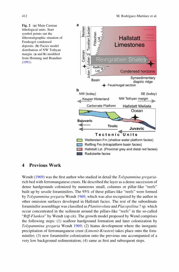
4 Previous Work
Wendt (1969) was the first author who studied in detail the Tolypammina gregaria-rich bed with ferromanganese crusts. He described the layer as a dense succession of
dense hardgrounds colonized by numerous small, columns or pillar-like “reefs”
built up by sessile foraminifers. The 95% of these pillars-like “reefs” were formed
by Tolypammina gregariaWendt 1969, which was also recognized by the author in
other omission surfaces developed in Hallstatt facies. The rest of the subordinate
foraminifer assemblage was classified as Planiinvoluta and Placopsilina ? sp. whichoccur concentrated in the sediment around the pillars-like “reefs” in the so-called
“Riff-Flanken” by Wendt (op cit). The growth model proposed by Wend comprises
the following steps: (1) seafloor hardground formation and later colonization by
Tolypammina gregaria Wendt 1969; (2) hiatus development where the inorganic
precipitation of ferromanganese crust (Limonit-Krusten) takes place onto the fora-
minifer; (3) new foraminifer colonization onto the previous one accompanied of a
very low background sedimentation; (4) same as first and subsequent steps.
Fig. 2 (a) Main Carnian
lithological units. Start
symbol points out the
lithostratigraphic situation of
Feurkogel condensed
deposits. (b) Facies model
distribution of NW Tethyan
margin. (a and b) modified
from Hornung and Brandner
(1991)
412 M. Rodrıguez-Martınez et al.
Hornung et al. (2007b) described “foraminiferal microbuildups” structures,
similar as above figured by Wendt (op cit), in the CII bed (Julian II in age) of the
Rappoltstein section, in condensed ammonoid beds of Berchtesgaden area (south-
ern Germany). They illustrated a similar growth model (see Fig. 5 at Hornung et al.,
op cit): (1) hardground surface formation; (2) growth of foraminifer “reef” genera-
tion I; (3) corrosion of “reef” generation I and impregnation by opaque pyrite; (4)
subsequent growth of different “reef” generations and; (5) “drowning” beneath a
bioclastic debris flow.
5 Results
At outcrop scale, the Tolypammina-rich bed with ferromanganese crusts show two
main characters: (1) a general cm-scale, wavy morphology; and (2) a progressive
increase of sub-mm, ferromanganese crusts toward the top. Both features produce
the misleading appearance of a stromatolitic structure. The foraminifer pillar-like
structures increase towards the top. Microfacies analysis shows a more complex
vertical zonation of this sedimentary condensed record.
5.1 Microfacies
Twomain groups ofmicrofacies have been recognized (Figs. 3 and 4), (1) mudstones-
wackestones with ferromanganese crusts/surfaces and Tolypammina-Frutexitesstructures; and (2) bioclastic wackestone to packstone with reworked cephalopods.
The contact between both groups is irregular, erosional in some places, showing
breakage of Tolypammina pillar-like structures, thus representing a discontinuity
surface (DS). There is an abrupt change in fossil content as well as structures on
both sides of the discontinuity surface (DS in Fig. 5). Therefore, taking the disconti-
nuity surface as an easily identifiable reference surface, the microfacies can be
grouped into the underlying and the overlying microfacies.
Two post-sedimentary fracture systems (F1 and F2 respectively) cut the micro-
facies (Figs. 4b and 5). F1 consists of sediment-filled fissures with “z-shape” and
with a subhorizontal orientation parallel to the bedding plane, whereas F2 are
vertical to oblique fractures. Fissure fillings comprise massive to graded mudstones
with angular lithoclasts derived from fissure walls. In some places there are narrow
cavities associated to these fissures. They show irregular contacts (dissolution
wedges), with basal erosive lag filling, and cements. The succession of cements
consists of a first generation of zoned radial-fibrous calcite (black core to weak
orange luminescent wedge), surrounded by calcite mosaic (orange moderately
luminescent) which fills the remaining porosity. F2 fractures are filled by cloudy
coarse calcite mosaic (orange moderately luminescent).
Tolypammina gregaria Wendt 1969-Frutexites Assemblage and Ferromanganese Crusts 413
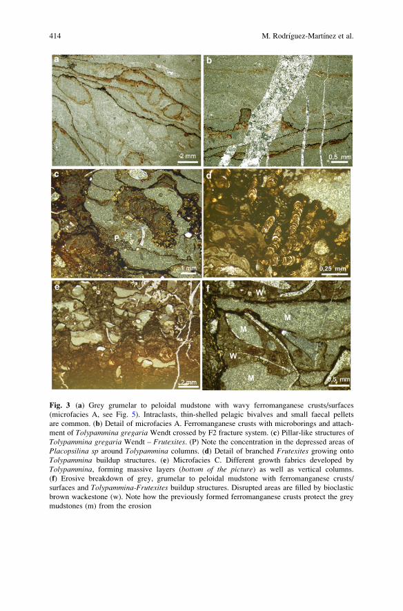
Fig. 3 (a) Grey grumelar to peloidal mudstone with wavy ferromanganese crusts/surfaces
(microfacies A, see Fig. 5). Intraclasts, thin-shelled pelagic bivalves and small faecal pellets
are common. (b) Detail of microfacies A. Ferromanganese crusts with microborings and attach-
ment of Tolypammina gregaria Wendt crossed by F2 fracture system. (c) Pillar-like structures of
Tolypammina gregaria Wendt – Frutexites. (P) Note the concentration in the depressed areas of
Placopsilina sp around Tolypammina columns. (d) Detail of branched Frutexites growing onto
Tolypammina buildup structures. (e) Microfacies C. Different growth fabrics developed by
Tolypammina, forming massive layers (bottom of the picture) as well as vertical columns.
(f) Erosive breakdown of grey, grumelar to peloidal mudstone with ferromanganese crusts/
surfaces and Tolypammina-Frutexites buildup structures. Disrupted areas are filled by bioclastic
brown wackestone (w). Note how the previously formed ferromanganese crusts protect the grey
mudstones (m) from the erosion
414 M. Rodrıguez-Martınez et al.
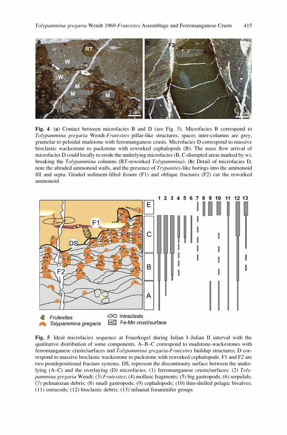
Fig. 5 Ideal microfacies sequence at Feuerkogel during Julian I–Julian II interval with the
qualitative distribution of some components. A–B–C correspond to mudstone-wackestones with
ferromanganese crusts/surfaces and Tolypammina gregaria-Frutexites buildup structures; D cor-
respond to massive bioclastic wackestone to packstone with reworked cephalopods. F1 and F2 are
two postdepositional fracture systems. DS, represent the discontinuity surface between the under-
lying (A–C) and the overlaying (D) microfacies. (1) ferromanganese crusts/surfaces; (2) Toly-pammina gregariaWendt; (3) Frutexites; (4) mollusc fragments; (5) big gastropods; (6) serpulids;
(7) pelmatozan debris; (8) small gastropods; (9) cephalopods; (10) thin-shelled pelagic bivalves;
(11) ostracods; (12) bioclastic debris; (13) infaunal foraminifer groups
Fig. 4 (a) Contact between microfacies B and D (see Fig. 5). Microfacies B correspond to
Tolypammina gregaria Wendt-Frutexites pillar-like structures, spaces inter-columns are grey,
grumelar to peloidal mudstone with ferromanganese crusts. Microfacies D correspond to massive
bioclastic wackestone to packstone with reworked cephalopods (B). The mass flow arrival of
microfacies D could locally to erode the underlying microfacies (B, C-disrupted areas marked by w),
breaking the Tolypammina columns (RT-reworked Tolypammina). (b) Detail of microfacies D,
note the abraded ammonoid walls, and the presence of Trypanites-like borings into the ammonoid
fill and septa. Graded sediment-filled fissure (F1) and oblique fractures (F2) cut the reworked
ammonoid
Tolypammina gregaria Wendt 1969-Frutexites Assemblage and Ferromanganese Crusts 415
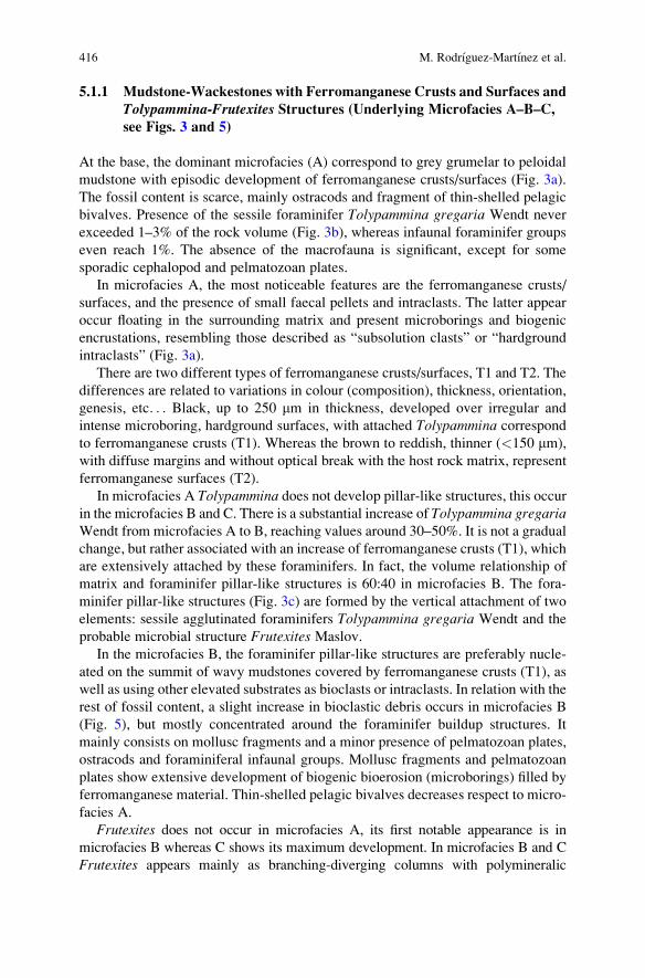
5.1.1 Mudstone-Wackestones with Ferromanganese Crusts and Surfaces and
Tolypammina-Frutexites Structures (Underlying Microfacies A–B–C,
see Figs. 3 and 5)
At the base, the dominant microfacies (A) correspond to grey grumelar to peloidal
mudstone with episodic development of ferromanganese crusts/surfaces (Fig. 3a).
The fossil content is scarce, mainly ostracods and fragment of thin-shelled pelagic
bivalves. Presence of the sessile foraminifer Tolypammina gregaria Wendt never
exceeded 1–3% of the rock volume (Fig. 3b), whereas infaunal foraminifer groups
even reach 1%. The absence of the macrofauna is significant, except for some
sporadic cephalopod and pelmatozoan plates.
In microfacies A, the most noticeable features are the ferromanganese crusts/
surfaces, and the presence of small faecal pellets and intraclasts. The latter appear
occur floating in the surrounding matrix and present microborings and biogenic
encrustations, resembling those described as “subsolution clasts” or “hardground
intraclasts” (Fig. 3a).
There are two different types of ferromanganese crusts/surfaces, T1 and T2. The
differences are related to variations in colour (composition), thickness, orientation,
genesis, etc. . . Black, up to 250 mm in thickness, developed over irregular and
intense microboring, hardground surfaces, with attached Tolypammina correspond
to ferromanganese crusts (T1). Whereas the brown to reddish, thinner (<150 mm),
with diffuse margins and without optical break with the host rock matrix, represent
ferromanganese surfaces (T2).
In microfacies A Tolypammina does not develop pillar-like structures, this occurin the microfacies B and C. There is a substantial increase of Tolypammina gregariaWendt from microfacies A to B, reaching values around 30–50%. It is not a gradual
change, but rather associated with an increase of ferromanganese crusts (T1), which
are extensively attached by these foraminifers. In fact, the volume relationship of
matrix and foraminifer pillar-like structures is 60:40 in microfacies B. The fora-
minifer pillar-like structures (Fig. 3c) are formed by the vertical attachment of two
elements: sessile agglutinated foraminifers Tolypammina gregaria Wendt and the
probable microbial structure Frutexites Maslov.
In the microfacies B, the foraminifer pillar-like structures are preferably nucle-
ated on the summit of wavy mudstones covered by ferromanganese crusts (T1), as
well as using other elevated substrates as bioclasts or intraclasts. In relation with the
rest of fossil content, a slight increase in bioclastic debris occurs in microfacies B
(Fig. 5), but mostly concentrated around the foraminifer buildup structures. It
mainly consists on mollusc fragments and a minor presence of pelmatozoan plates,
ostracods and foraminiferal infaunal groups. Mollusc fragments and pelmatozoan
plates show extensive development of biogenic bioerosion (microborings) filled by
ferromanganese material. Thin-shelled pelagic bivalves decreases respect to micro-
facies A.
Frutexites does not occur in microfacies A, its first notable appearance is in
microfacies B whereas C shows its maximum development. In microfacies B and C
Frutexites appears mainly as branching-diverging columns with polymineralic
416 M. Rodrıguez-Martınez et al.
convex-upward laminae (Fig. 3d). Laminae correspond to black opaque minerals (Mn-
rich), brownish to reddish semitranslucent minerals (Fe-rich) and/or yellow-white
carbonate minerals. The maximum width of the columns is around 60 mm and up to
750 mm in height, but normally range from 50 up to 250 mm. Frutexites is not
exclusively associated to the foraminifer-buildup structures, also grows around
bioclasts or directly over ferromanganese crusts (T1). Essentially the nucleation
sites of Frutexites are laterally around the edges of Tolypammina columns, as well as
at the top of them.
Thus microfacies B (Fig. 5) is a complex microfacies framework formed by
wavy grey, grumelar peloidal mudstones with very low fossil content and extensive
development of ferromanganese crust/surfaces (T1 and T2) which are colonized by
Tolypammina-Frutexites buildup structures where is concentrated mostly the rest of
the skeletal content.
Microfacies C are formed by patches dominated by Tolypammina gregariaWendt-Frutexites buildup structures. In fact, the volume relationship of matrix
and pillar-like structures is 30:70. Lateral distance between foraminiferal columns
ranges from 2.5 mm up to 5 mm. Vertical thickness interval between successive
ferromanganese crusts is around 500 mm up to 1 mm. Ferromanganese crusts (T1)
are dominant.
Different growth fabrics developed by Tolypammina gregaria were recognized,
occurring from the base to the top of these patches (Fig. 3e): (1) wide massive
layers; (2) columns or pillar-like structures; and (3) dendritic, arborescent struc-
tures. It looks usually exist a vertical relationship between this growth fabric
zonation and an increase of bioclastic debris. Occasionally, small articulated-shell
brachiopods disrupt the massive Tolypammina layers as well as the pillar-like
structures. The dendritic or arborescent zones are likewise prolific in branched
Frutexites.The quantity and diversity of foramiferal infaunal groups increase in microfacies C
(Fig. 5) but is basically dominated by Planiinvoluta- and Placopsilina-like forms.
Placopsilina concentrates near the base of Tolypammina-Frutexites pillar-like
structures, in depressed areas, as well as in shelter areas associated to mollusc
fragments. However, the distribution of Planiinvoluta is not only restricted to the
surroundings of pillar-like structures. Big gastropods and serpulids occur also in
microfacies C.
In microfacies C, the grey, grumelar to peloidal mudstone between pillar-like
structures has a slightly higher skeletal content compared to the mudstones from
microfacies A and B. Additionally it exhibits disrupted zones filled with massive
bioclastic brown wackestone. These disrupted zones (Fig. 3f) increase from micro-
facies B to C where they can represent around 5% up to 10% of rock volume.
Summarizing, the sedimentary condensed deposits formed by the mudstone-
wackestones with ferromanganese crusts/surfaces and Tolypammina-Frutexitesstructures show three characteristic microfacies (A, B and C in Fig. 5). It is possible
reconstruct their vertical arrangement: (1) microfacies A dominates at the base;
(2) B and C can occur intercalated; whereas (3) patches of microfacies C are more
common toward the top.
Tolypammina gregaria Wendt 1969-Frutexites Assemblage and Ferromanganese Crusts 417
5.1.2 Bioclastic Wackestone to Packstone with Reworked Cephalopods
(Overlying Microfacies E Figs. 4 and 5)
Massive brown to reddish bioclastic wackestones to packstones overlay microfacies
B and C (Figs. 4a and 5). The bioclastic content is around 35% and 45% of rock
volume and is formed mainly by ostracods, benthonic foraminifers (lagenid for-
aminifers are dominant), articulated and disarticulated thin-shelled pelagic
bivalves, small gastropods, and diverse rests of cephalopods. The fabric is chaotic,
with mottled aspect where skeletal content shows a heterogeneous distribution of
sizes. Thin-shelled pelagic bivalves can display imbricated fabric as well as a
totally random distribution.
Cephalopods comprise orthoconic nautiolids, with geopetal filling similar as
surrounding wackestone and marine calcite cements (early fibrous rim phase and
later equant mosaics), and ammonoids. However, it is noticeable in this microfacies
the presence of reworked ammonoids (Fig. 4b) which show imbricated orientation,
microbored and abraded walls, lithological differences between its geopetal filling
and the encasing rocks, local attachment by Tolypammina, macroborer activity
cutting the lithified geopetal filling as well as septa. The boreholes may be attrib-
uted to Trypanites group and their infilling are equivalent to the encased lithology.
5.2 Geochemical Analysis (Fig. 6)
Chemical sediments in general are anticipated to reflect the chemical composition
of the water from which they were precipitated. Ferromanganese nodules show an
inverse REE pattern compared to a typical seawater pattern, showing a positive Ce
anomaly and are slightly enriched in light REE relative to heavy REE. Interpreta-
tions must be cautious as chemical sediments/precipitates also reflect local redox
conditions and maybe influenced by post depositional changes (Elderfield and
Greaves 1981).
The measured REE and trace elements are given in Table 1. REE (Fig. 6a) were
normalized with a PAAS standard (Post-Archaean average Australian Shale) after
McLennan (1989). Anomalous behaviour of Ce, was investigated using (Ce/Ce*)
ratios after Bau and Dulski (1996), calculated as [Ce/Ce* ¼ Ce/0.5(La + Pr)] (all
values PAAS normalized). It was also checked with (Pr/Pr*) ratios with [Pr/Pr* ¼Pre/0.5(Ce + Nd)], whether the Ce anomaly can be assigned as a Ce anomaly or is
affect by high La values (Fig. 6b).
5.2.1 Matrices from Microfacies B and C
Matrix B shows a smooth and even distribution of the REE and with a negative Ce
anomaly (Ce/Ce* between 0.26 and 0.51) (Table 1, Fig. 6b). Compared to matrix B,
Matrix C is higher enriched in REE and shows a positive Ce anomaly (Ce/Ce*
418 M. Rodrıguez-Martınez et al.
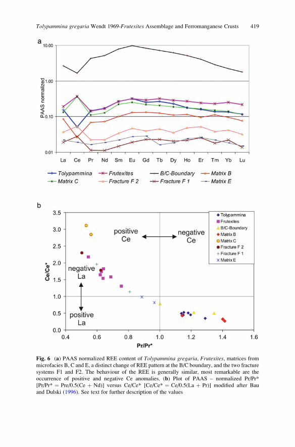
Fig. 6 (a) PAAS normalized REE content of Tolypammina gregaria, Frutexites, matrices from
microfacies B, C and E, a distinct change of REE pattern at the B/C boundary, and the two fracture
systems F1 and F2. The behaviour of the REE is generally similar, most remarkable are the
occurrence of positive and negative Ce anomalies. (b) Plot of PAAS – normalized Pr/Pr*
[Pr/Pr* ¼ Pre/0.5(Ce þ Nd)] versus Ce/Ce* [Ce/Ce* ¼ Ce/0.5(La þ Pr)] modified after Bau
and Dulski (1996). See text for further description of the values
Tolypammina gregaria Wendt 1969-Frutexites Assemblage and Ferromanganese Crusts 419
Table
1Averagevalues
ofREEandtraceelem
entsin
partsper
million(ppm)outof1,800measurements.See
textforadetaileddiscussionofthe
elem
entdata M
nFe
YZr
Nb
La
Ce
Pr
Nd
Sm
Eu
Gd
Tb
Dy
Ho
Er
Tm
Yb
Lu
Hf
Ta
Th
UCo
Ni
Ce/Ce*
Pr/Pr*
Tolypam
mina
196.7
1,617.3
4.884
0.860
0.651
5.502
5.085
1.2225.739
1.560
0.3241.145
0.1700.964
0.1850.403
0.053
0.320
0.043
0.0370.014
3.3390.027
4.322
8.784
0.452
1.196
Tolypam
mina
335.1
1,674.3
4.654
0.858
0.884
6.116
6.034
1.1695.229
1.341
0.3091.079
0.1850.943
0.1730.449
0.059
0.402
0.056
0.0360.022
3.3100.027
3.135
8.684
0.522
1.154
Tolypam
mina
546.5
3,320.8
4.941
1.329
1.576
5.822
5.644
1.0994.865
1.214
0.2971.173
0.1800.865
0.1670.436
0.062
0.356
0.050
0.0470.019
3.1810.019
4.176
8.084
0.509
1.180
Tolypam
mina
180.6
992.0
5.247
0.532
0.506
5.997
4.078
1.2795.802
1.486
0.3441.167
0.2061.071
0.1740.456
0.055
0.381
0.050
0.0180.007
3.1170.016
5.350
10.111
0.346
1.289
Tolypam
mina
269.5
1,421.6
4.554
0.783
0.929
5.997
4.895
1.0805.204
1.329
0.2921.031
0.1721.000
0.1720.414
0.059
0.400
0.040
0.0360.022
3.1900.027
4.731
10.847
0.443
1.150
Tolypam
mina
351.0
2,145.0
5.497
0.975
1.156
6.166
5.971
1.2746.034
1.577
0.3801.391
0.2051.116
0.2030.475
0.067
0.412
0.052
0.0330.014
4.0210.027
5.182
9.263
0.494
1.138
Frutexites
1,620
13,459
3.968
5.901
6.774
5.838
16.3650.8603.763
0.894
0.2210.821
0.1400.784
0.1730.449
0.061
0.460
0.063
0.2210.196
3.5850.186
4.713
10.709
1.642
0.621
Frutexites
1,483
11,225
4.803
5.848
6.950
5.873
17.2590.9944.332
1.071
0.2621.072
0.1551.026
0.2000.494
0.071
0.472
0.061
0.2350.268
3.3340.195
2.384
5.654
1.578
0.689
Frutexites
2,195
18,336
6.829
10.25912.7957.140
25.38
1.4106.282
1.632
0.3731.356
0.2311.381
0.2660.674
0.107
0.659
0.081
0.4540.516
5.9210.398
1.999
4.686
1.819
0.641
Frutexites
2,036
11,989
5.431
8.509
10.0076.847
21.5831.1264.775
1.174
0.3161.058
0.1801.034
0.2140.593
0.071
0.559
0.077
0.3010.297
4.2430.239
1.686
4.170
1.742
0.628
Frutexites
1,638
12,965
4.473
5.260
6.256
6.290
16.5180.9294.073
0.924
0.2350.854
0.1520.846
0.1810.518
0.071
0.459
0.071
0.2160.202
3.3860.156
1.492
5.287
1.537
0.639
Frutexites
2,898
14,375
6.239
13.46017.6037.306
29.5781.2565.714
1.471
0.3451.369
0.2481.346
0.2690.689
0.093
0.709
0.093
0.5880.606
5.3400.566
2.234
7.843
2.177
0.546
Frutexites
1,752
9,257
6.000
6.068
7.224
7.165
18.1511.3856.699
1.758
0.3871.527
0.2561.269
0.2410.713
0.087
0.560
0.065
0.2040.160
3.5050.161
3.760
4.729
1.307
0.755
B/C-Boundary
897.0
1,335.3
76.03
2.818
1.246
102.7
132.2
37.7
175.8
43.78
10.5138.45
5.61729.16
5.14511.54
1.167
6.322
0.768
0.0660.028
37.200.146
3.416
8.738
0.521
1.218
B/C-Boundary
348.0
2655.4
18.072
1.827
1.451
17.96
29.77
4.21119.63
4.978
1.2083.968
0.5482.787
0.5101.255
0.121
0.786
0.083
0.0680.038
3.1320.056
3.454
6.027
0.782
1.002
MatrixB
36.6
504.4
7.518
0.793
0.185
6.628
5.682
1.3967.210
1.681
0.4121.633
0.2321.330
0.2400.557
0.080
0.478
0.045
0.0300.010
0.1680.031
3.508
6.842
0.425
1.144
MatrixB
73.9
1,178.6
3.785
1.339
0.524
3.253
1.627
0.5722.440
0.694
0.1310.643
0.0850.514
0.1170.280
0.038
0.286
0.041
0.0520.030
0.2380.035
4.625
11.178
0.269
1.415
MatrixC
85.3
1,013.8
5.053
2.332
0.559
5.114
29.8780.9804.337
1.088
0.2820.989
0.1570.858
0.1760.440
0.057
0.381
0.046
0.0500.014
0.7550.030
4.784
10.856
3.115
0.531
MatrixC
174.3
3,976.4
5.577
3.128
1.852
5.044
28.8710.9814.354
1.220
0.2731.012
0.1600.883
0.1770.467
0.061
0.397
0.049
0.1130.061
1.1320.079
5.289
12.063
2.847
0.562
MatrixE
21.4
1,135.7
0.850
1.197
0.152
0.833
1.512
0.1450.633
0.121
0.0290.133
0.0120.086
0.0240.075
0.009
0.051
0.006
0.0240.003
0.1020.032
4.923
12.302
0.987
0.887
MatrixE
15.9
700.2
0.960
1.174
0.105
0.914
1.310
0.1530.680
0.121
0.0320.135
0.0200.097
0.0260.072
0.006
0.074
0.015
0.0200.005
0.1190.015
5.352
11.705
0.813
0.966
Fracture
F1
95.5
87.9
0.674
0.083
0.011
0.722
2.226
0.1010.379
0.082
0.0200.106
0.0180.096
0.0210.073
0.007
0.062
0.006
0.0020.001
0.0260.005
0.109
0.371
1.956
0.598
Fracture
F1
19.5
693.6
0.359
0.429
0.282
0.674
1.173
0.1080.489
0.093
0.0290.157
0.0130.124
0.0360.055
0.009
0.073
0.010
0.0470.017
0.4080.068
0.122
0.162
1.136
0.809
Fracture
F2
218.6
30.8
1.301
0.049
0.002
1.374
5.458
0.2140.951
0.110
0.0300.196
0.0430.200
0.0380.144
0.008
0.162
0.024
0.0000.002
0.005�0
.002
4.800
22.086
2.302
0.504
Fracture
F2
204.3
53.7
1.374
0.061
0.004
1.414
4.248
0.1980.762
0.190
0.0510.184
0.0350.165
0.0480.148
0.016
0.118
0.014
0.0060.000
0.024�0
.002
3.725
27.351
1.786
0.624
420 M. Rodrıguez-Martınez et al.
between 1.8 and 3.2). In both matrices the Y/Ho ratio varied between 28 and 100
and U/Th as well as Ni/Co ratios after Jones and Manning (1994), varied between
0.04–0.6 (U/Th) and 0.8–3.20 (Ni/Co) respectively.
5.2.2 Microfacies B/C-Boundary
The B/C-boundary, (Fig. 5), at the beginning of an increased colonization of
Tolypammina-Frutexites assemblages, on the substrate, a different REE pattern is
observed, with a negative Ce anomaly (Ce/Ce* between 0.4 and 0.8), a high
abundance of middle REE (MREE) and a slight positive Eu anomaly. Together
with the MREE enrichment, elements like Th, Y, Co and Ni occur in higher
abundance (Table 1). U/Th varied between 0.01 and 0.02 and Ni/Co between 1.5
and 3.1.
5.2.3 Tolypammina
Tolypammina shows a negative Ce-anomaly (Ce/Ce* between 0.35 and 0.94) and
a general REE pattern similar to Matrix B (Fig. 6b, Table 1). Y/Ho ratio varied
between 26 and 30, U/Th 0.05–0.1 and Ni/Co varied between 1.32 and 2.9.
Throughout the Tolypammina structures the iron/manganese (Fe/Mn) ratios remain
quite stable around 5–10.
5.2.4 Frutexites
The Frutexites structures show a prominent positive Ce anomaly (Ce/Ce* between
1.3 and 2.4) and REE showing a similar distribution compared to Matrix C. The
Y/Ho ratios oscillated between 18 and 25 for the Frutexites structures and U/Th
ranged from 0.05 to 0.21 and Ni/Co varied between 1.1 and 3.8. In comparison to
the surrounding limestone and the Tolypammina structures, Frutexites is up tenfoldenriched in iron (Fe) and manganese (Mn) (Table 1). Interestingly, the Fe/Mn ratio
of Frutexites is similar to those of Tolypammina varying between 5 and 9.
5.2.5 Matrix from Microfacies E
In Matrix E, the content of the REE is quite low, with a slight positive La anomaly
(Ce/Ce* between 0.8 and 0.99) (Table 1, Fig. 6b). Y/Ho ratios ranged from 32–40,
U/Th varied between 0.1 and 0.45 and Ni/Co varied between 2.1 and 2.9. Matrix E
contains much lower Mn compared to Matrices B and C, thus showing Fe/Mn ratios
from 42 to 54 (Table 1).
Tolypammina gregaria Wendt 1969-Frutexites Assemblage and Ferromanganese Crusts 421
5.2.6 Fracture Systems
The fracture systems F1 and F2, show a very similar behaviour of REE, with
distinct positive Ce anomalies (Ce/Ce* between 1.1 and 2.4). Y/Ho ratios ranged
from 10 to 35, U/Th varied between 0 and 0.2 and Ni/Co laid between 1.3 and 7.5.
The variation of the trace element content in F1 revealed two different phases
within the fracture, one is enriched in Mn and Ni and depleted in Fe, the other phase
is enriched in Fe, Y, Zr, Nb, Th and U. F2 shows a distinct enrichment in Mn and Ni
and a depletion in Fe (Table 1).
6 Discussion
6.1 Types of Ferromanganese Crusts/Surfaces
Ferromanganese crusts and nodules are common deposits on sediment-starved
basins, seamounts and slopes, mostly in and below oxygen minimum zone. They
are the slowest-forming mineral deposit known, formed by heterogeneous mixtures
of biogenic, authigenic and detrital particles which final composition depends on
seawater chemistry (Hein et al. 1992, 1994) and supply of metals to the oxide
surface (Dymond et al. 1984).
The Y/Ho ratios ranging between 18–25 and 44–100 for the ferromanganese
crust and the carbonate phases respectively indicate a hydrogenous origin (Bau
1996). Further, according to U/Th and Ni/Co ratios after Jones and Manning (1994),
ratios lower than 0.75 (U/Th), and 5.00 (Ni/Co) respectively, point to oxic bottom
water conditions. Ratios between 0.75–1.25 (U/Th), and 5.00–7.00 (Ni/Co) repre-
sent dysoxic bottom water conditions. Therefore, with ratios not higher than 0.81
(U/Th) and 6.20 (Ni/Co), it can be stated, that the deposition of the ferromanganese
crusts and the different limestone matrices occurred predominantly under oxic
conditions and seldom under dysoxic conditions.
At the B/C boundary some hydrothermal influence was observed occurring as a
short event as indicated by the high increase in middle REE with distinct Eu
anomaly (Figs. 5 and 6). However, the Y/Ho ratios show a large variation, hamper-
ing a clear relation to a distinct hydrothermal event. Further, U/Th and Ni/Co ratio
clearly indicate oxic bottom water conditions during this phase. Clear hydrothermal
influence was observed in fractures F1 and F2 indicated by Y/Ho ratios of 10–15
(Bau 1996) a slight Eu anomaly (Fig. 6).
Bacteria could also concentrate Fe and Mn even from very low concentration in
the seawater. Microbial activity can play an important role on the genesis of
ferromanganese oxyhydroxides in several steps (Konhauser 1998) by: (1) concentra-
tion of metals by microorganisms living on water column; (2) deep transport flux
as the microbes die; (3) metal release through organic matter mineralization; (4) bac-
terial oxidation of Mn(II) and Fe(II). In fact, the formation of microbially mediated
iron and manganese minerals has been widely recognized (Nelson and Tebo 1980;
422 M. Rodrıguez-Martınez et al.
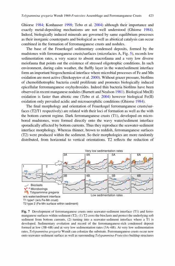
Ghiorse 1984; Konhauser 1998; Tebo et al. 2004) although their importance and
exactly metal-depositing mechanisms are not well understood (Ghiorse 1984).
Indeed, biologically induced minerals are governed by same equilibrium processes
as their inorganic counterparts and biological as well as abiotical catalysis can occur
combined in the formation of ferromanganese crusts and nodules.
The base of the Feuerkogel sedimentary condensed deposits, formed by the
mudstones with ferromanganese crusts/surfaces (microfacies A, Fig. 5), records low
sedimentation rates, a very scarce to absent macrofauna and a very low diverse
meiofauna that points out the existence of stressed oligotrophic conditions. In such
environment, during calm weather, the fluffly layer in the water/sediment interface
form an important biogeochemical interface where microbial processes of Fe and Mn
oxidation are most active (Strekopytov et al. 2000). Without grazer pressure, biofilms
of chemolithotrophic bacteria could proliferate and promotes biologically induced
epicellular ferromanganese oxyhydroxides. Indeed thin bacteria biofilms have been
observed in recent manganese nodules (Burnett and Nealson 1981). BiologicalMn(II)
oxidation is faster than abiotic one (Tebo et al. 2004) however biological Fe(II)
oxidation only prevailed acidic and microaerophilic conditions (Ghiorse 1984).
The final morphology and orientation of Feuerkogel ferromanganese crusts/sur-
faces (T2/T1 respectively) are related with their loci of formation as well as the with
the bottom current regime. Dark ferromanganese crusts (T1), developed on micro-
bored mudstones, were formed directly onto the wavy water/sediment interface
sporadically affected by bottom currents. Thus they reproduce the seawater-sediment
interface morphology. Whereas thinner, brown to reddish, ferromanganese surfaces
(T2) were produced within the sediment. So their morphologies are more randomly
distributed, from horizontal to vertical orientations. T2 reflects the reduction of
w/s water/sediment interfaceT1 type1 (w/s Fe-Mn crust)T2 type 2 (Fe-Mn surface within sediment)
Microborings
w/s
T2T1
T2=T12
1
2
1
w/s
T2
T2 T1T1
2
13
2
1
2
1
13
3 2
1
3
2
13
4
2
1
3
4
2
13
45
T2
2
Very low sedimentation rates
Low sedimentation rates
1 2
3A
3B
4A 5A 6A
4B 5B 6B
Tolypammina gregaria
Bioclasts
Fig. 7 Development of ferromanganese crusts onto seawater-sediment interface (T1) and ferro-
manganese surfaces within sediment (T2). (1) T2 cross the bioclasts and protect the underlying soft
sediment from bottom currents, (2) turning into a seawater-sediment interface where a T1 is
developed. Sedimentary evolution and record of the ferromanganese-rich condensed deposit
formed at low (3B–6B) and at very low sedimentation rates (3A–6B). At very low sedimentation
rates, Tolypammina gregariaWendt can colonize the substrate. Ferromanganese crusts occur now
onto seawater-sediment surface as well as surrounding Tolypammina-Frutexites buildup structures
Tolypammina gregaria Wendt 1969-Frutexites Assemblage and Ferromanganese Crusts 423
Fe–Mn oxyhydroxides within sediment (by suboxic diagenesis and/or Fe and Mn
reducing bacteria), followed by the upwards diffusion of Mn(II) and Fe(II) that
finally are reoxidized and deposited in the suboxic-oxic sediment interface.
Both types of ferromanganese structures could act as a protecting barrier for
substrate erosion (Fig. 7). The early development of ferromanganese surfaces (T2)
within the still soft sediment can protect the underlying sediment of bottom currents
(2 in Fig. 7). Thus, the previously formed T2 surface becomes the sediment-water
interface and a new ferromanganese crust could be produced (T1 superimposed to a
previous T2). The new and successive generations of ferromanganese crusts (T1)
and surfaces (T2) and their relationship with biogenic encrustation allow to us to
reconstruct the sedimentation rates (low and very low sedimentation rates from
microfacies A to microfacies B – see 3B–6B and 3A–6A respectively in Fig. 7).
6.2 Significance of Tolypammina gregaria-Frutexites
Buildup Structures
Thus as we have seen before, an increase of ferromanganese structures (T1 and T2)
from the base to the top (from microfacies A to C, Figs. 5 and 7), could reflect a
decrease of the sedimentation rate (from low to very low sedimentation rates). Both,
bacterial biofilm development on water-sediment interface and very low sedimen-
tation rates could have a positive effect on foraminiferal larval settlement, favour-
ing the colonization by the sessile agglutinated foraminifer Tolypammina gregariaWendt. The main question is which environmental parameters control/favour the
different types of growth of this sessile foraminifer.
The broadly colonization of the sediment-water interface by Tolypammina couldpoint out a very low presence of predatory and grazer organisms due to low values
of dissolved oxygen (opportunistic behaviour in aerobic to slightly dysaerobic
conditions below a possible oxygen minimum zone?). Indeed, it has been described
the attachment of foraminifera to vestimentiferan tubeworms as a response to
seafloor hypoxia and sulfide toxicity at cold seeps (Sen Gupta et al. 2007). How-
ever, Tolypammina-Frutexites structures colonized both elevated substrates as wellas normal horizontal substrates thus sulfide toxicity cannot be easily argued. In fact,
ferromanganese deposits occur in well oxygenated waters. In addition, agglutinated
foraminifers, as a group, are much less tolerant to hypoxic conditions that calcare-
ous assemblages, and they prevail under conditions of greater oxygenation (Gooday
et al. 2000; Levin et al. 2002).
Maybe their colonial behaviour building vertical pillar-like structures could be
related with their feeding behaviour. On recent manganese nodules, suspension
feeders dominate the nodule summit whereas the deposit feeders concentrate near
the base (Mullineaux 1989). Epibenthonic foraminifers apparently use elevated
substrates under the influence of near-bottom flow where a maximum yield of
food particles can be achieved (Sch€onfeld 2002).
424 M. Rodrıguez-Martınez et al.
The dominance of sessile Tolypammina gregaria in comparison with the scarcity
of infaunal foraminiferal groups in microfacies B, could be also related with
nutrient availability. Benthic foraminifera are not found or rare when the organic
matter is scarce or absent within the sediment, and epifauna reflects better the
organic flux whereas the infaunal groups are more related with the organic matter
stored in the sediment (Jorissen et al. 1995; Den Dulk et al. 1998; Van der Zwaan
et al. 1999).
Indeed, the different colonial growth fabrics developed by Tolypammina gregaria(massive layers; columns or pillar-like structures; and dendritic, arborescent struc-
tures) could be associated to both, the quantity as well as to the dominant type of
organic flux input (lateral or vertical). In microfacies B (Fig. 5), the preference of
Tolypammina-Frutexites structures to colonize the summit of wavymudstones could
respond to a dominant horizontal or lateral organic flux input in oligotrophic condi-
tions. In oligotrophic ecosystems, the reproduction potential could play a crucial role
in the competition for food (Van der Zwaan et al. 1999). Thus a massive layer growth
strategy, as it is observed inmicrofacies C, could suggest a fast increase of biomass in
response to vertical phytodetritus pulse (Graf 1989; Gooday 1988). The accretion
rate of similar sessile foraminifer-rich layers has been estimated by Wendt (1974) in
1 mm/year to 1 mm/25, in recent deep-sea manganese nodules.
Microfacies B and patches of microfacies C occur intercalated although an
increase of microfacies C toward the top is observed (Fig. 5). Thus the vertical
increase of Tolypammina gregaria-Frutexites assemblage from microfacies B to C,
in addition to a more diverse accompanied benthic assemblage suggest a change
from oligrotrophic to slightly mesotrophic conditions associated with episodic
pulses of phytodetritus.
As we see before the common nucleation sites of Frutexites are laterally
surrounding edges of Tolypammina gregaria columns. Frutexites structures havebeen mainly described in marine environments from shallow to deep-water stro-
matolites, condensed pelagic successions and hardgrounds. Frutexites have been
found growing directly on the seawater-sediment interface, within the micritic
sediment and into marine and continental cavities and fractures. Such wider envi-
ronmental distribution has been taken into consideration in their palaeoecological
interpretations. Their presence in stromatolites has been linked with different types
of cyanobacteria (Playford et al. 1976, 1984; Scytonematacean – Walter and
Awramik 1979; Rivulariacean – Hofmann and Grotzinger 1985; Angulocellulariagroup – Riding 1991). Their growing in cavities, sheet cracks and Neptunian Dykes
have been related with non-phototrophic (heterotrophic) behaviour (Tsien 1979) or
cryptobiont role (Myrow and Coniglio 1991), which sometimes were linked with
non-phototrophic cyanobacteria (Gischler 1996) as well as with chemosynthetic
bacteria (Cavalazzi et al. 2007). The cryptobiont, negative phototactic downward
growth and cryptoendopelitic behaviour of Frutexites were interpreted by B€ohmand Brachert (1993) as a signal of oxygen deficient environments. Chafetz et al.
(1998) compared Mn-rich black travertine shrubs with Frutexites and other struc-
tures regarded as bacterially induced products (desert varnish, deep-sea manganese
nodules). Finally, the presence of Frutexites in condensed pelagic limestones and
Tolypammina gregaria Wendt 1969-Frutexites Assemblage and Ferromanganese Crusts 425
hardgrounds has been explained due to physicochemical origin (Wendt 1969) as
well as related with iron bacteria (Reitner et al. 1995; Mamet and Preat 2006).
Frutexites have been regarded as biologically induced products (cyanobacteria
and iron bacteria) in spite of their final resulting mineralogy (hematite, Fe–Mn
oxyhydroxides, and carbonate and/or phosphate minerals). The final assessment
depends strongly about which mineralogical composition they interpreted as pri-
mary (timing of iron mineralization) as well as its loci of growth. Thus their final
mineralogy, their compositional different alternating bands, could indeed reflect
microenviromental changes, as well as the same bacteria can form different miner-
als in different environments (Lowenstam and Weiner 1989).
In the Feuerkogel site, the preference of Frutexites to colonize the Tolypamminatests suggests certain substrate control in their development. It could be related with
the foraminifer walls, rich in acid mucopolysaccharides that could favour the
growth of bacteria biofilm (resulting Frutexites structures?) as well as promote
the ferromanganese-oxide precipitation in the polymeric matrix. In Recent nodules
this possibility was considered by Graham and Cooper (1959) who related the
foraminiferal protein-rich exudates as a favourable substrate for growth of bacteria
capable of extracting trace elements from seawater.
The role of foraminifers in the formation of ferromanganese structures has been
longer and widely discussed (Greenslate 1974; Wendt 1974; Dugolinsky et al. 1977;
Riemann 1983; von Stackelberg 1984; Mullineaux 1987, 1989; Mullineaux and
Butman 1990; Verlaan 1992). However, in the Feuerkogel level, the development
of ferromanganese crusts/surfaces took place before the extensive colonization of the
substrate by Tolypammina-Frutexites community. But, both Tolypammina buildup
structures, as well as ferromanganese crusts increase toward the top. Thus, probably
common environmental parameters favour their increase toward the top.
6.3 Environmental Significance of Coeval FerromanganeseCrust/Surfaces and Tolypammina-Frutexites CommunityDuring Carnian Interval
Low sedimentation rates are favoured conditions to colonization of substrate by
sessile foraminifers as well as development of ferromanganese deposits. Which
could be the main parameters that control both foraminifer colonization and
ferromanganese formation?
Organic flux is an important structural control in heterotrophic marine biota, and
foraminifers represent more than 50% of total biomass in deep-sea environments
(Snider et al. 1984; Gooday et al. 1992). Thus primary productivity in the mixed
layer and vertical nutrient flux could be a critical factor in the abundance of deep
water foraminifers associated to ferromanganese crusts and nodules.
We have previously seen, Foraminifera are the most abundant sessile organism
found on Recent crust and nodules. In fact, Foraminifera and Bacteria are the main
426 M. Rodrıguez-Martınez et al.
consumers of organic matter in deep-sea floor, where foraminifers play an impor-
tant role in the fast initial processing of phytodetritus input whereas bacteria
dominate the long-term carbon re-mineralization (Moodley et al. 2002). In the
Feuerkogel site, the main consumers of vertical organic flux in a stressed bottom
environment could have been related with the biofilms developed on water/sediment
interface, Frutexites-induced biofilms around foraminifers, and the own Tolypam-mina. Thus, the interbedded patches of microfacies C with B, dominated by the
sessile foraminifer Tolypammina gregraria Wendt could reflect an opportunistic
behaviour to episodic phytodetritus inputs related with phytoplankton blooms.
In addition, the different types of colonial growths developed by Tolypamminagregraria maybe reflect changes in their reproduction rate as response of the
episodic inputs. Indeed, it has been noted that the response of foraminifers to
phytodetritus layer deposition in deep sea environments is very fast, followed by
an increasing their biomass and reproduction rate (Gooday 1988; Altenbach 1992;
Moodley et al. 2000). Deep foraminifer biota from regions with episodic organic
flux shows opportunistic fast-growth species which dominate the assemblages,
however low seasonality and stable food input regions show higher diversity
(Corliss et al. 2009). The response of actual benthic foraminifer has been specially
analyzed after phytoplankton blooms related with seasonal and monsoonal periods
(Sch€onfeld and Numberger 2007; Andersson et al. 2007; Larkin and Gooday 2009;
Corliss et al. 2009).
However, high primary productivity related with phytoplankton blooms is not
the only factor to explain the abundance of benthic organisms associated to actual
equatorial ferromanganese nodules, because collected nodules from areas with
oceanic upwelling and high biological productivity do not show high abundance
of benthic foraminifers (Margolis and Burns 1976).
In addition to the primary productivity, other factors have to favour both the
ferromanganese crust development and sessile colonization by foraminifers. It has
been related with the own metal-ion availability. In offshore waters the bioavail-
ability of iron micronutrient has been suggested as limiting factor of primary
production (Martin and Fitzwater 1988). The phytoplankton growth can be con-
trolled by the Fe(III) solubilities and the dissolution rates of colloidal Fe(III) phases
(Wells et al. 1983; Kuma and Matsuga 1995). Although there is no clear consensus
about the inorganic speciation of Fe(III) in seawater and it seem exists many
unresolved questions about chemistry of iron in seawater (Wells et al. 1995; Millero
et al. 1995).
The REE pattern of Feuerkogel microfacies does not show a hydrothermal
overprint (except the postpositional fracture systems) thus others possible sources
as atmospheric aerosols or river inputs are necessary to explain its high concentra-
tions. As it was mentioned, the depositional setting of red Hallstatt facies was
related with structural highs (tectonically-induced saline uplifts), thus the arrival of
siliciclastic input due to continental run off could be not so relevant. However, the
dust from desert regions is a primary source of atmospheric iron into the oceans
(Donaghay et al. 1991); in fact the 80% of the iron input from the atmosphere to the
oligotrophic surface waters is due to wet deposition (Uematsu et al. 1985). Thus
Tolypammina gregaria Wendt 1969-Frutexites Assemblage and Ferromanganese Crusts 427
rain and storms produce episodic inputs of iron aerosol into oceanic iron-limited
waters, which favour phytoplankton growth (Hanson et al. 2001).
The ferromanganese structures with the development of Tolypammina-Frutexitescommunity occur at the Feuerkogel site in the Julian I–Julian II limit, thus they
coincided with the so-called Reingraben Turnover (Hornung and Brandner 2005;
Hornung et al. 2007b). During Carnian, part of the marine fossil record experi-
mented high extinction rates which has been attributed to humid climate pulse,
named the Carnian Pluvial Event (Simms et al. 1994; Bellanca et al. 1995) related
with a monsoonal clima (Mutti and Weissert 1995). Evidences of this humid
climate pulse has been also recorded in deep-water sequences (Hornung and
Brandner 2005; Rigo et al. 2007; Hornung et al. 2007a).
This humid phase related with a monsoonal climate could have produced both
weathering and subsequent increase of siliciclastic input through continental run off
(source of metals as well as organic matter), as well as important wet aerosol iron
inputs to the offshore waters. In addition, this humid phase could also have some
influence on marine currents and a slightly cooling, factors that have been linked to
the growth of ferromanganese crusts (Margolis and Burns 1976; Segl et al. 1984;
Puteanus and Halbach 1988; Koschinsky et al. 1997).
7 Summary
In the Feuerkogel site, the condensed Carnian sedimentary condensed Hallstatt
Limestone formed by mudstone-wackestones with ferromanganese structures and
Tolypammina gregaria-Frutexites community report to us a complex vertical
evolution of environmental changes (Fig. 8):
Fig. 8 Hypothetical sedimentary model of ferromanganese structures development (T1 and T2 in
Fig. 7) and Tolypammina gregaria Wendt-Frutexites community: Relationship between Forami-
nifera and different bacteria biofilms (Tolypammina gregaria Wendt-Frutexites assemblage)
coupled to nutrient and metal availability
428 M. Rodrıguez-Martınez et al.
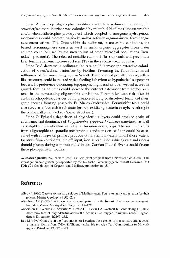
Stage A: In deep oligotrophic conditions with low sedimentation rates, the
seawater/sediment interface was colonized by microbial biofilms (lithoautotrophic
and/or chemolithotrophic prokaryotes) which coupled to inorganic hydrogenous
mechanisms could promote passively and/or actively organomineral ferromanga-
nese encrustations (T1). Once within the sediment, in anaerobic conditions, the
buried ferromanganese crusts as well as metal organic aggregates from water
column could be used by the metabolism of other microbial populations (iron-
reducing bacteria). The released metallic cations diffuse upwards and precipitate
later forming ferromanganese surfaces (T2) in the suboxic–oxic boundary.
Stage B: A decrease in sedimentation rate could increase the extensive coloni-
zation of water/sediment interface by biofilms, favouring the foraminifer larval
settlement of Tolypammina gregaria Wendt. Their colonial growth forming pillar-
like structures could be related with a feeding behaviour as hypothetical suspension
feeders. Its preference colonizing topographic highs and its own vertical accretion
growth forming columns could increase the nutrient catchment from bottom cur-
rents in the surrounding oligotrophic conditions. Foraminifer tests rich often in
acidic muchopolysaccharides could promote binding of dissolved ferric and man-
ganic species forming passively Fe–Mn oxyhydroxides. Foraminifer tests could
also serve as a favourable substrate for iron-oxidizing bacteria (maybe resulting in
the biologically-induced Frutexites structures).Stage C: Episodic deposition of phytodetritus layers could produce peaks of
abundance and dominance of Tolypammina gregaria-Frutexites structures, as wellas a slightly diversification of infaunal foraminiferal groups. The resulting shifts
from oligotrophic to sporadic mesotrophic conditions on seafloor could be asso-
ciated with changes on primary productivity in shallow waters. In off shore waters,
far away from continental run off input, iron aerosol inputs during rain and storms
(humid phases during a monsoonal climate: Carnian Pluvial Event) could favour
these phytoplankton blooms.
Acknowledgments We thank to Jose Castillejo grant program from Universidad de Alcala. This
investigation was gratefully supported by the Deutsche Forschungsgemeinschaft Research Unit
FOR 571 Geobiology of Organo- and Biofilms, publication no. 51.
References
Allouc J (1990) Quaternary crusts on slopes of Mediterranean Sea: a tentative explanation for their
genesis. Marine Geology 94:205–238
Altenbach AV (1992) Short term processes and patterns in the foraminiferal response to organic
flux rates. Marine Micropaleontology 19:119–129
Andersson JH, Woulds C, Shwartz M, Cowie GL, Levin LA, Soetaert K, Middelburg JJ (2007)
Short-term fate of phytodetritus across the Arabian Sea oxygen minimum zone. Biogeos-
ciences Discussion 4:2493–2523
Bau M (1996) Controls on the fractionation of isovalent trace elements in magmatic and aqueous
systems: evidence from Y/Ho, Zr/Hf, and lanthanide tetrade effect. Contributions to Mineral-
ogy and Petrology 123:323–333
Tolypammina gregaria Wendt 1969-Frutexites Assemblage and Ferromanganese Crusts 429
Bau M, Dulski P (1996) Distribution of yttrium and rare-earth elements in the Penge and Kuruman
iron-formations, Transvaal Supergroup, South Africa. Precambrian Research 79:37–55
Bellanca A, Di Stefano P, Neri R (1995) Sedimentology and isotope geochemistry of Carnian
deep-water marl/limestone deposits from the Sicani Mountains, Sicily: environmental implica-
tions and evidence for planktonic source of lime mud. Palaeogeography, Palaeoclimatology,
Palaeoecology 114:111–129
B€ohm F, Brachert TC (1993) Deep-water stromatolites and Frutexites Maslov from the Early and
Middle Jurassic of S-Germany and Austria. Facies 28:145–168
Bonatti E (1981) Metal deposits in the oceanic lithosphere. In: Emiliani C (ed) The sea, vol VII.
Wiley, New York, pp 241–283
Burnett BR, Nealson KH (1981) Organic films and microorganisms associated with manganese
nodules. Deep Sea Research 28A:637–645
Cavalazzi B, Barbieri R, Ori G (2007) Chemosynthetic microbialites in the Devonian carbonate
mounds of Hamar Laghdad (Anti-Atlas, Morocco). Sedimentary Geology 200:73–88
Chafetz HS, Akdim B, Julia R, Reid A (1998) Mn- and Fe-rich black travertine shrubs: bacterially
(and nanobacterially) induced precipitates. Journal of Sedimentary Research 68:404–412
Corliss BH, Brown CW, Sun X, Showers WJ (2009) Deep-sea benthic diversity linked to
seasonality of pelagic productivity. Deep Sea Research I 56:835–841
Den Dulk M, Reichart GJ, Memon GA, Roelofs EMP, Zachariasse WJ, Van der Zwaan GJ (1998)
Benthic foraminiferal response to variations in intensity of oxygen minimum zone in the
northeast Arabian Sea. Marine Micropaleontology 35:43–66
Donaghay PL, Liss PS, Duce RA, Kester DR, Hanson AK, Villareal T, Tindale NW, Gifford DJ
(1991) The role of episodic atmospheric nutrient inputs in the chemical and biological
dynamics of the oceanic ecosystems. Oceanography 4:62–70
Dugolinsky BK, Margolis SV, Dudley WC (1977) Biogenic influence on growth of manganese
nodules. Journal of Sedimentary Research 47:428–445
Dymond J, Lyle M, Finney B, Piper DZ, Murphy K, Conard R, Pisias N (1984) Ferromanganese
nodules from MANOP Sites H, S, and R – Control of mineralogical and chemical composition
by multiple accretionary processes. Geochimica et Cosmochimica Acta 48:931–949
Ehrlich HL (1963) Bacteriology of manganese nodules. I. Bacterial action on manganese in nodule
enrichments. Applied and Environmental Microbiology 11:15–19
Ehrlich HL (1968) Bacteriology of manganese nodules. II. Manganese oxidation by cell-free extract
from a manganese nodule bacterium. Applied and Environmental Microbiology 16:197–202
Ehrlich HL (1971) Bacteriology of manganese nodules. V. Effect of hydrostratic pressure on
bacterial oxidation of MnII and reduction of MnO2. Applied and Environmental Microbiology
21:306–310
Elderfield H, Greaves MJ (1981) Negative Cerium anomalies in the rare earth element pattern of
oceanic ferromanganese nodules. Earth and Planetary Science Letters 55:163–170
Gawlick HJ (2000) Pal€aogeographie der Obertrias-Karbonatplattformen in den N€ordlichen Kalk-
alpen. Mitteilungen der Osterreichischen Geologischen Gesellschaft 44:45–95
Ghiorse WC (1984) Biology of iron- and manganese-depositing bacteria. Annual Review of
Microbiology 38:515–550
Ghiorse WC, Ehrlich HL (1992) Microbial biomineralization of iron and manganese. In: Skinner
HCW, Fitzpatrick RW, (eds) Biomineralization processes of iron and manganese: modern and
ancient environments. Catena Verlag, Cremlingen-Destedt, pp 75–99
Gischler E (1996) Late Devonian-Early Carboniferous deep-water coral assemblages and sedi-
mentation on a Devonian seamount: Iberg Reef, Harz Mts., Germany. Palaeogeography,
Palaeoclimatology, Palaeoecology 123:297–322
Gooday AJ (1988) A response to benthic Foraminifera to the deposition of phytodetritus in the
deep sea. Nature 332:70–73
Gooday AJ, Levin L, Linke P, Heeger T (1992) The role of benthic foraminifera in deep-sea food
webs and carbon cycling. In: Rowe GP (ed) Deep-sea food chains and the global carbon
cycling. Kluwer Academic Publisher, The Netherlands, pp 63–91
430 M. Rodrıguez-Martınez et al.
Gooday AJ, Bernhard JM, Levin LA, Suhr S (2000) Foraminifera in the Arabian Sea oxygen
minimum zone and other deficient settings: taxonomic composition, diversity and relation to
metazoan faunas. Deep Sea Research II 57:25–54
Graf G (1989) Benthic-pelagic coupling in a deep-sea benthic community. Nature 341:437–439
Graham JW, Cooper SC (1959) Biological origin of manganese-rich deposits of the sea floor.
Nature 183:1050–1051
Greenslate J (1974) Microorganisms participate in the construction of manganese nodules. Nature
649:181–183
Hanson AK, Tindale NW, Abdel-Moati MAR (2001) An Equatorial Pacific rain event: influence
on the distribution of iron and hydrogen peroxide in surface waters. Marine Chemistry
75:69–88
Hein JR, Bohrson WA, Shulz MS, Noble M, Clague DA (1992) Variations in the fine-scale
composition of a central Pacific ferromanganese crust: paleooceanographic implications.
Paleooceanography 7:63–77
Hein JR, Yeh H-W, Gunn SH, Gibbs AE, Wang C-H (1994) Composition of hydrothermal
ironstones from central Pacific seamounts. Geochimica et Cosmochimica Acta 58:179–189
Hofmann HJ, Grotzinger JP (1985) Shelf-facies microbiotas from the Odjick and Rocknest
formations (Epworth Group; 1.89 Ga), northwestern Canada. Canadian Journal of Earth
Sciences 22:1781–1792
Hornung T, Brandner R (2005) Biochronostratigraphy of the Reingraben Turnover (Hallstatt
Facies belt): local black shale events controlled by regional, tectonics, climatic change and
plate tectonics. Facies 2005:460–479
Hornung T, Krystyn L, Brandner R (2007a) A Tethys-wide mid-Carnian (Upper Triassic) carbonate
productivity crisis: evidence for the Alpine Reingraben Event from Spiti (Indian Himalaya).
Journal of Asian Earth Sciences 30:285–302
Hornung T, Spatzenegger A, Joachimski MM (2007b) Multistratigraphy of condensed ammonoid
beds of the Rappolstein (Berchtesgaden, southern Germany): unravelling palaeoenvironmental
conditions on ‘Hallstatt deep swells’ during Reingraben Event (Late Lower Carnian). Facies
53:267–292
Jenkyns HC (1970) Fossil manganese nodules from the West Sicilian Jurassic. Eclogae Geologi-
cae Helvetiae 63:741–774
Jimenez-Millan J, Nieto LM (2006) Geochemical and mineralogical evidence of tectonic and
sedimentary factors controlling the origin of ferromanganese crusts associated to stratigraphic
discontinuities (Betic Cordilleras, SE Spain). Chemie der Erde 68:323–336
Jones B, Manning DAC (1994) Comparison of geochemical indices used for the interpretation of
palaeoredox conditions in ancient mudstones. Chemical Geology 111:111–129
Jorissen FJ, de Stigter HC, Widmark JGV (1995) A conceptual model explaining benthic for-
aminiferal microhabitats. Marine Micropaleontology 22:3–15
Kazmierczak J, Kempe S (2006) Modern analogues of Precambrian stromatolites from caldera
lakes of Niuafo’ou Island, Tonga. Naturwissenschaften 93:119–126
Konhauser KO (1998) Diversity of bacterial iron mineralization. Earth Science Reviews
43:91–121
Koschinsky A, Stascheit A, Bau M, Halbach P (1997) Effects on phosphatization on the geochem-
ical and mineralogical composition of marine ferromanganese crusts. Geochimica et Cosmo-
chimica Acta 19:4079–4094
Krystyn L (1991) Die Fossillagerst€atten der alpinen Trias. In: Vasicek V, Krystyn L, Golebiowski R(eds) Exkursionen im Jungpal€aozoikum und Mesozoikum Osterreichs, Exkusionsf€uhrerTagung der Osterreichischen Pal€aontologischen Gesellschaft, Wien, pp 23–78
Kuma K, Matsuga K (1995) Availability of colloidal ferric oxides to coastal marine phytoplankton.
Marine Biology 122:1–11
Larkin KE, Gooday AJ (2009) Foraminiferal faunal responses to monsoon-driven changes in
organic matter and oxygen availability at 140 m and 300 m water depth in the NE Arabian Sea.
Deep Sea Research II 56:403–421
Tolypammina gregaria Wendt 1969-Frutexites Assemblage and Ferromanganese Crusts 431
Levin L, Gutierraz D, Rathburn A, Neira C, Sellanes J, Munos Gallardo V, Salamanca M (2002)
Benthic processes on the Peru margin: a transect across the oxygen minimun zone during the
1997–98 El Nino. Progress in Oceanography 53:1–27
Lowenstam HA, Weiner S (1989) On biomineralization. Oxford University Press, New York
Mamet B, Preat A (2006) Iron-bacteria mediation in Phanerozoic red limestones: state of the art.
Sedimentary Geology 185:147–157
Mandl GW (2000) The Alpine sector of the Tethyan shelf: examples of Triassic to Jurassic
sedimentation and deformation from the Northern Calcareous Alps. Mitteilungen der Oster-
reichischen Geologischen Gesellschaft 92:61–79
Margolis SV, Burns RG (1976) Pacific deep-sea manganese nodules: their distribution, composi-
tion and origin. Annual Review of Earth and Planetary Sciences 4:229–263
Martin JH, Fitzwater SE (1988) Iron deficiency limits phytoplankton growth in the northeast
Pacific subarctic. Nature 331:341–343
Maslov VP (1960) Stromatolity (ick genezis, metodizucheniya, svjaz’s fatsiyami i geologicheskoe
znachenie na primere Ordovika Sibirskoj Plataformy). Trudy Instituta Geologii Akademiia
nauk SSSR 41:1–188
McLennan SM (1989) Rare earth elements in sedimentary rocks: influence of provenance and
sedimentary processes. In: Lipin BR McKay GA (eds) Geochemistry and Mineralogy of Rare
Earth Elements. Reviews in Mineralogy 21:169–200
Millero FJ, Wensheng Y, Aicher J (1995) The speciation of Fe(II) and Fe(III) in natural waters.
Marine Chemistry 50:21–39
Moodley L, Boschker HTS, Middelburg JJ, Pel R, Herman PMJ, de Deckere E, Heip C (2000)
Ecological significance of benthic foraminifera: 13+C labelling experiments. Marine Ecology
Progress Series 202:289–295
Moodley L, Middelbrug JJ, Boschker HTS, Duineveld GCA, Pel R, Herman PMJ, Heip CHR
(2002) Bacteria and Foraminifera: key players in a short-term deep-sea benthic response to
phytodetritus. Marine Ecology Progress Series 236:23–29
Mullineaux LS (1987) Organisms living on manganese nodules and crusts: distribution and
abundance at three North Pacific sites. Deep Sea Research 34:165–184
Mullineaux LS (1988) Taxonomic notes on large agglutinated foraminifers encrusting manganese
nodules, including the description of a new genus, Chondrodapis (Komokiacea). Journal of
Foraminiferal Research 18:46–53
Mullineaux LS (1989) Vertical distribution of the epifauna on manganese nodules: implications
for settlement and feeding. Limnology and Oceanography 34:1247–1262
MullineauxLS,ButmanCA (1990)Recruitment of encrusting benthic invertebrates in boundary-layer
flows: a deep-water experiment on Cross Seamount. Limnology and Oceanography 35:409–423
Mutti M, Weissert H (1995) Triassic monsoonal climate and its signature in Ladinian-Carnian
carbonate platforms (Southern Alps, Italy). Journal of Sedimentary Research B65:357–367
Myrow PM, Coniglio M (1991) Origin and diagenesis of cryptobiontic Frutexites in the Chapel
island Formation (Vendian to Early Cambrian) of Southeast Newfoundland, Canada. Palaios
6:572–585
Nelson KH, Tebo B (1980) Structural features on manganese precipitating bacteria. Origins Life
10:117–126
Playford PE, Cockbain AE, Druce EC, Wray JL (1976) Devonian stromatolites from the Canning
Basin, Westerns Australia. In: Walter MR (ed) Stromatolites, Developments in sedimentology
20. Elsevier, Amsterdam, pp 543–463
Playford PE, McLaren DJ, Orth C, Gilmore JS, Goodfellow WD (1984) Iridium anomaly in the
Upper Devonian of the Canning Basin, Western Australia. Science 226:437–439
Puteanus D, Halbach P (1988) Correlation of Co concentration and growth rate – a method for age
determination of ferromanganese crusts. Chemical Geology 69:73–85
Reitner J, Wilmsen M, Neuweiler F (1995) Cenomanian/Turonian sponge microbialite deep-water
hardground community (Liencres, Northern Spain). Facies 32:203–212
432 M. Rodrıguez-Martınez et al.
Resig JM, Glenn CR (1997) Foraminifera encrusting phosphoritic hardgrounds of the Peruvian
upwelling zone: taxonomy, geochemistry and distribution. Journal of Foraminiferal Research
27:133–150
Riding R (1991) Calcified Cyanobacteria. In: Riding R (ed) Calcareous algae and stromatolites,
Springer, Berlin, pp 55–87
Riemann F (1983) Biological aspects of deep-sea manganese nodule formation. Oceanologica
Acta 6:303–311
Rigo M, Preto N, Roghi G, Tateo F, Mietto P (2007) A rise in the Carbonate Compensation Depth
of western Thetys in the Carnian (Late Triassic): deep-water evidence for the Carnian Pluvial
Event. Palaeogeography, Palaeoclimatology, Palaeoecology 246:188–205
Schlager W (1969) Das Zusammenwirken von Sedimentation und Bruchtektonik in den tria-
dischen Hallst€atterkalken der Ostalpen. Geologische Rundschau 59:289–308
Sch€onfeld J (2002) Recent benthic foraminiferal assemblages in deep high energy environments
from the Gulf of Cadiz. Marine Micropaleontology 44:141–162
Sch€onfeld J, Numberger L (2007) The benthic foraminiferal response to the 2004 spring bloom in
the western Baltic Sea. Marine Micropaleontology 65:78–95
Segl M, Mangini A, Bonani G, Hofmann G, Nessi M, Sutter M, W€olfi W, Friedrich G, Pl€uger W,
Wiechowski A, Beer J (1984) 10Be dating of manganese crust from central North Pacific and
implications for ocean paleocirculation. Nature 309:540–543
Sen Gupta BK, Smith LE, Lobegeier MK (2007) Attachment of Foraminifera to vestimentiferan
tubeworms at cold seeps: refuge from seafloor hypoxia and sulfide toxicity. Marine Micropa-
leontology 62:1–6
Simms MJ, Johnson ALA, Ruffel A (1994) Biotic and climatic change in the Carnian (Triassic of
Europe and adjacent areas). In: Fraser N, Sues HD (eds) In the shadow of the dinosaurs.
Cambridge University Press, Cambridge, pp 352–365
Snider LJ, Burnett BR, Hessler RR (1984) The composition and distribution of meiofauna and
nanobiota in a central North Pacific deep-sea area. Deep Sea Research 31:1225–1249
Stampfli GM, Borel GD (2002) A plate tectonic model for the Paleozoic and Mesozoic constrained
by dynamic plate boundaries and restored synthetic oceanic isochrones. Earth and Planetary
Science Letters 196:17–33
Strekopytov S, Dubinin A, Uspenskaya T (2000) Geochemical and mineralogical studies of
Fe–Mn Nodules and crusts from the White Sea: potential role of benthic fauna in their
formation. Goldschmidt 2000, Journal of Conference Abstracts 5(2):963
Tebo BM, Bargar JR, Clement BG, Dick GJ, Murray KJ, Parker D, Verity R, Webb SM (2004)
Biogenic manganese oxides: properties and mechanisms of formation. Annual Review of Earth
and Planetary Sciences 32:287–328
Toscano F, Raspini A (2005) Epilithozoan fauna associated with ferromanganese crustgrounds on
the continental slope segment between Capri and Li Galli Islands (Bay of Salerno, Northern
Tyrrehenian Sea, Italy). Facies 50:427–441
Tsien HH (1979) Paleoecology of algal-bearing facies in the Devonian (Couvirtian to Frasnian)
Reef Complexes of Belgium. Palaeogeography, Palaeoclimatology, Palaeoecology 27:
103–127
Tucker ME (1973) Ferromanganese nodules from the Devonian of the Montagne Noire (S. France)
and the West Germany. Geologische Rundschau 62:137–153
Uematsu M, Duce RA, Prospero JM (1985) Deposition of atmospheric mineral particles in the
North Pacific Ocean. Journal of Atmospheric Chemistry 3:123–138
Van der Zwaan GJ, Duijnstee IAP, Dulk D, Ernst SR, Jannink NT, Kouwenhoven TJ (1999)
Benthic foraminifers: proxies or problems? A review of paleoecological concepts. Earth
Science Reviews 46:213–239
Verlaan PA (1992) Benthic recruitment and manganese crust formation on seamounts. Marine
Biology 113:171–174
von Mojsisovics E (1873–1902) Das Gebirge um Hallstatt I. Abhandlungen der Geologischen
Reichsanstalt 6/1:356
Tolypammina gregaria Wendt 1969-Frutexites Assemblage and Ferromanganese Crusts 433
von Stackelberg U (1984) Significance of benthic organisms for the growth and movement of
manganese nodules, Equatorial North Pacific. Geo-Marine Letters 4:37–42
Walter MR, Awramik SM (1979) Frutexites from stromatolites of Gunflint iron formation of
Canada, and its biological affinities. Precambrian Research 9:23–33
WangW, Schr€oder HC, Wiens M, Schloßmacher U, M€uller WEG (2009) Manganese/polymetallic
nodules: micro-structural characterization of exolithobiontic- and endolithobionthic microbial
biofims by scanning electron microscopy. Micron 40:350–358
Wells ML, Zorkin NG, Lewis AG (1983) The role of colloidal chemistry in providing a source of
iron to phytoplankton. Journal of Marine Research 41:731–746
Wells ML, Price NM, Bruland KW (1995) Iron chemistry in seawater and its relationship to
phytoplankton: a workshop report. Marine Chemistry 48:157–158
Wendt J (1969) Foraminiferen-‘Riffe’ im karnischen Hallst€atter Kalk des Feuerkogels (Steiermark,
Osterreich). Pal€aontologische Zeitschrift 43:177–193Wendt J (1970) Stratigraphische Kondensation in triadischen und jurassischen Cephalopoden-
kalken der Tethys. Neues Jahrbuch f€ur Geologie und Pal€aontologie Monatshefte 1970/
7:433–448
Wendt J (1974) Encrusting organisms in deep-sea manganese nodules. In: Hs€u KJ, Jenkyns HC
(eds) Pelagic sediments: on land and under sea, International Association of Sedimentologists,
Special Publication 1. Blackwell, Oxford, London, Edinburgh, Melbourne, pp 437–447
434 M. Rodrıguez-Martınez et al.
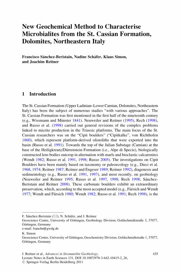
New Geochemical Method to Characterise
Microbialites from the St. Cassian Formation,
Dolomites, Northeastern Italy
Francisco Sanchez-Beristain, Nadine Sch€afer, Klaus Simon,
and Joachim Reitner
1 Introduction
The St. Cassian Formation (Upper Ladinian–Lower Carnian, Dolomites, Northeastern
Italy) has been the subject of numerous studies “with various approaches”. The
St. Cassian Formation was first mentioned in the first half of the nineteenth century
(e.g., Wissmann and M€unster 1841). Neuweiler and Reitner (1995), Rech (1998),
and Russo et al. (1998) carried out general revisions of the complex problems
linked to micrite production in the Triassic platforms. The main focus of the St.
Cassian researchers was on the “Cipit boulders” (“Cipitkalke”, von Richthofen
1860), which represent platform-derived olistoliths that were exported into the
basin (Russo et al. 1991). Towards the top of the Julian Substage (Carnian) at the
base of the Heiligkreutz/D€urrenstein Formation (i.e., Alpe di Specie), biologically
constructed lens bodies outcrop in alternation with marls and bioclastic calcarenites
(Wendt 1982; Russo et al. 1991, 1998; Russo 2005). The investigations on Cipit
Boulders have been mainly based on taxonomy or paleoecology (e.g., Dieci et al.
1968, 1974; Reitner 1987; Reitner and Engeser 1989; Reitner 1992), diagenesis and
sedimentology (e.g., Russo et al. 1991, 1997), and most recently, on geobiology
(Neuweiler and Reitner 1995; Russo et al. 1997, 1998; Rech 1998; Sanchez-
Beristain and Reitner 2008). These carbonate boulders exhibit an extraordinary
preservation, which, according to the most accepted model (e.g., F€ursich andWendt
1977; Wendt and F€ursich 1980; Wendt 1982; Russo et al. 1991; Rech 1998), is the
F. Sanchez-Beristain (*), N. Sch€afer, and J. Reitner
Geoscience Centre, University of G€ottingen, Geobiology Division, Goldschmidtstraße 3, 37077,
G€ottingen, Germany
e-mail: [email protected]
K. Simon
Geoscience Centre, University of G€ottingen, Geochemistry Division, Goldschmidtstraße 1, 37077,
G€ottingen, Germany
J. Reitner et al., Advances in Stromatolite Geobiology,Lecture Notes in Earth Sciences 131, DOI 10.1007/978-3-642-10415-2_26,# Springer-Verlag Berlin Heidelberg 2011
435
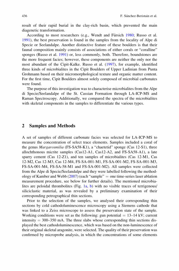
result of their rapid burial in the clay-rich basin, which prevented the main
diagenetic transformation.
According to most researchers (e.g., Wendt and F€ursich 1980; Russo et al.
1991), the best preservation is found in the samples from the locality of Alpe di
Specie or Seelandalpe. Another distinctive feature of these boulders is that their
faunal composition mainly consists of associations of either corals or “coralline”
sponges (Russo et al. 1991) or, less commonly, both. Therefore, boundstones are
the more frequent facies; however, these components are neither the only nor the
most abundant of the Cipit-Kalke. Russo et al. (1997), for example, identified
three kinds of microbialites in the Cipit Boulders of Upper Ladinian from Punta
Grohmann based on their micromorphological texture and organic matter content.
For the first time, Cipit Boulders almost solely composed of microbial carbonates
were found.
The purpose of this investigation was to characterise microbialites from the Alpe
di Specie/Seelandalpe of the St. Cassian Formation through LA-ICP-MS and
Raman Spectroscopy. Additionally, we compared the spectra of the microbialites
with skeletal components in the samples to differentiate the various types.
2 Samples and Methods
A set of samples of different carbonate facies was selected for LA-ICP-MS to
measure the concentration of select trace elements. Samples included a coral of
the genus Margarosmilia (FS-SA58-K1), a “chaetetid” sponge (Cas 12-S1), three
allochthonous micrite samples (Cas12-A1, Cas12-A2, and FS-SA58-A1), a late
sparry cement (Cas 12-Z1), and ten samples of microbialites (Cas 12-M1, Cas
12-M2, Cas 12-M3, Cas 12-M4, FS-SA-001-M1, FS-SA-001-M2, FS-SA-001-M3,
FS-SA-001-M4, FS-SA-58-M1 and FS-SA-001-M2). All samples were collected
from the Alpe di Specie/Seelandalpe and they were labelled following the method-
ology of Kamber andWebb (2007) (each “sample” ¼ one time-series laser ablation
measurement procedure, see below for further details). The mentioned microbia-
lites are peloidal thrombolites (Fig. 1a, b) with no visible traces of terrigenous/
siliciclastic material, as was revealed by a preliminary examination of their
corresponding petrographical thin sections.
Prior to the selection of the samples, we analysed their corresponding thin
sections by cold cathodoluminescence microscopy using a Siemens cathode that
was linked to a Zeiss microscope to assess the preservation state of the sample.
Working conditions were set as the following: gun potential ¼ 13–14 kV; current
intensity ¼ 300–350 mA. The three slabs whose corresponding thin sections dis-
played the best cathodoluminescence, which was based on the non-luminescence of
their original skeletal aragonite, were selected. The quality of their preservation was
confirmed by microprobe analysis, in which the concentrations of some elements
436 F. Sanchez-Beristain et al.
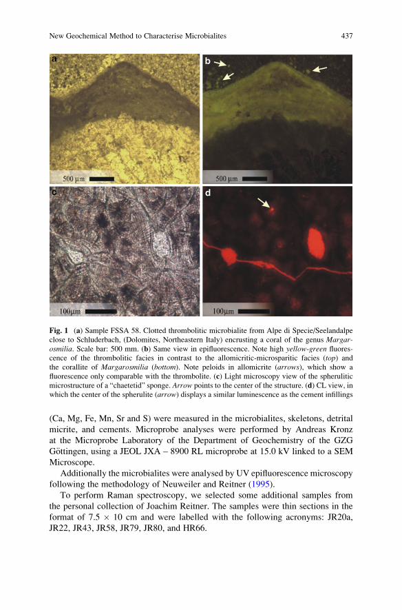
(Ca, Mg, Fe, Mn, Sr and S) were measured in the microbialites, skeletons, detrital
micrite, and cements. Microprobe analyses were performed by Andreas Kronz
at the Microprobe Laboratory of the Department of Geochemistry of the GZG
G€ottingen, using a JEOL JXA – 8900 RL microprobe at 15.0 kV linked to a SEM
Microscope.
Additionally the microbialites were analysed by UV epifluorescence microscopy
following the methodology of Neuweiler and Reitner (1995).
To perform Raman spectroscopy, we selected some additional samples from
the personal collection of Joachim Reitner. The samples were thin sections in the
format of 7.5 � 10 cm and were labelled with the following acronyms: JR20a,
JR22, JR43, JR58, JR79, JR80, and HR66.
Fig. 1 (a) Sample FSSA 58. Clotted thrombolitic microbialite from Alpe di Specie/Seelandalpe
close to Schluderbach, (Dolomites, Northeastern Italy) encrusting a coral of the genus Margar-osmilia. Scale bar: 500 mm. (b) Same view in epifluorescence. Note high yellow-green fluores-
cence of the thrombolitic facies in contrast to the allomicritic-microsparitic facies (top) and
the corallite of Margarosmilia (bottom). Note peloids in allomicrite (arrows), which show a
fluorescence only comparable with the thrombolite. (c) Light microscopy view of the spherulitic
microstructure of a “chaetetid” sponge. Arrow points to the center of the structure. (d) CL view, in
which the center of the spherulite (arrow) displays a similar luminescence as the cement infillings
New Geochemical Method to Characterise Microbialites 437
2.1 LA-ICP-MS
Laser ablation was performed with an Excimer 193 nm COMPEX 110 (Lambda
Physik, G€ottingen, Germany) equipped with an optical beam shape enhancer
GEOLAS (MicroLas, G€ottingen, Germany) at about 3 J/cm2 with spot sizes and a
line width of 120 mm. The ablated material was carried into the ICP-MS DRC II
(Perkin-Elmer, Canada) plasma by an Argon gas flow. Kamber and Webb (2007)
pioneered the detection of vital trace elements in Devonian Renalcis thrombolitic
microbialites and associated samples via in situ time-series LA-ICP-MS. In this
study, the time-series were recorded under the following conditions: 26 isotopes
were measured at dwelling times of 20 ms; measurement duration was 165 s; and
402 Reading Points (RP) were taken. Measurement began after 30 s of pre-warming
and ended 10 s prior to the macro end, but only measurements from 10 to 130 s
(280 RPs in total) after the start of ablation were taken.
Some of the trace elements have count intensities near background levels. The
quantification of a single sweep, which has a dwelling time of 20 ms for one
isotope, is very erroneous; however, if five or more sweeps yield the same results,
the error of the average decreases dramatically. Standardisation and quantification
was done using NBS610; the internal standard sotope, 43Ca, was assumed to
have variable concentrations in the carbonates, as was obtained from microprobe
results (390,000 ppm for the aragonite and the late cement, 360,000 ppm for the
microbialite, and 350,000 ppm for the allochthonous micrite/detritus). Absolute
concentrations are prone to having rather large errors, especially for those elements
which have concentrations closest to background levels; however, isotopic ratios
are more correct with errors less than 2%.
2.2 Raman Spectroscopy
Raman spectra were recorded using a Horiba Jobin Yvon LabRam-HR 800 UV
microprobe with a focal length of 800 mm. The following were used as excitation
wavelengths: (1) the 244 nm line of a frequency-doubled Ion Laser (Coherent
Innova 90C FreD) with a laser power of 30 mW; and (2) the 488 nm line of an
Argon Ion Laser (Melles Griot IMA 106020B0S) with a laser power of 25 mW. In
the case of the 244 nm line, the laser beam was dispersed by a 2,400 lines/mm
grating on a liquid nitrogen cooled CCD detector of 2,048 � 512 pixels, which
yielded a spectral resolution of 1.05 cm�1. The 488 nm-line laser was dispersed by
a 600 line/mm grating on a CCD detector of 1,024 � 256 pixels, which yielded
a spectral resolution of 0.43 cm�1.
An Olympus BX41microscope equipped with an OFR LMV-40�-UVB objective
that had a numerical aperture of 0.5 focused the laser light onto the sample. To
obtain an improved signal-to-noise ratio, the confocal hole diameter was set to
438 F. Sanchez-Beristain et al.
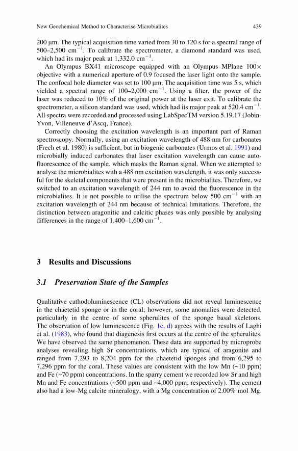
200 mm. The typical acquisition time varied from 30 to 120 s for a spectral range of
500–2,500 cm�1. To calibrate the spectrometer, a diamond standard was used,
which had its major peak at 1,332.0 cm�1.
An Olympus BX41 microscope equipped with an Olympus MPlane 100�objective with a numerical aperture of 0.9 focused the laser light onto the sample.
The confocal hole diameter was set to 100 mm. The acquisition time was 5 s, which
yielded a spectral range of 100–2,000 cm�1. Using a filter, the power of the
laser was reduced to 10% of the original power at the laser exit. To calibrate the
spectrometer, a silicon standard was used, which had its major peak at 520.4 cm�1.
All spectra were recorded and processed using LabSpecTM version 5.19.17 (Jobin-
Yvon, Villeneuve d’Ascq, France).
Correctly choosing the excitation wavelength is an important part of Raman
spectroscopy. Normally, using an excitation wavelength of 488 nm for carbonates
(Frech et al. 1980) is sufficient, but in biogenic carbonates (Urmos et al. 1991) and
microbially induced carbonates that laser excitation wavelength can cause auto-
fluorescence of the sample, which masks the Raman signal. When we attempted to
analyse the microbialites with a 488 nm excitation wavelength, it was only success-
ful for the skeletal components that were present in the microbialites. Therefore, we
switched to an excitation wavelength of 244 nm to avoid the fluorescence in the
microbialites. It is not possible to utilise the spectrum below 500 cm�1 with an
excitation wavelength of 244 nm because of technical limitations. Therefore, the
distinction between aragonitic and calcitic phases was only possible by analysing
differences in the range of 1,400–1,600 cm�1.
3 Results and Discussions
3.1 Preservation State of the Samples
Qualitative cathodoluminescence (CL) observations did not reveal luminescence
in the chaetetid sponge or in the coral; however, some anomalies were detected,
particularly in the centre of some spherulites of the sponge basal skeletons.
The observation of low luminescence (Fig. 1c, d) agrees with the results of Laghi
et al. (1983), who found that diagenesis first occurs at the centre of the spherulites.
We have observed the same phenomenon. These data are supported by microprobe
analyses revealing high Sr concentrations, which are typical of aragonite and
ranged from 7,293 to 8,204 ppm for the chaetetid sponges and from 6,295 to
7,296 ppm for the coral. These values are consistent with the low Mn (~10 ppm)
and Fe (~70 ppm) concentrations. In the sparry cement we recorded low Sr and high
Mn and Fe concentrations (~500 ppm and ~4,000 ppm, respectively). The cement
also had a low-Mg calcite mineralogy, with a Mg concentration of 2.00% mol Mg.
New Geochemical Method to Characterise Microbialites 439
3.2 Magnesium
The magnesium content in all the microbialites ranged from 12,620 to 17,846 ppm,
which in principle indicates an original biological high-Mg calcite composition
(Prasada Rao 1996). This result is consistent with the microprobe measurements,
which ranged from 4.85 to 8.8% mol Mg and were thus beyond the accepted
minimum value of 4% mol Mg (Dickson 1990; Fl€ugel 2004). Microbialites with
high-Mg calcite mineralogy have been widely reported in the literature (e.g.,
Reitner 1993; Zankl 1993; Sch€afer 2006). The Raman spectra (see below for further
explanation) further support these findings.
The Mg concentration in the allochthonous micrites was considerably higher
than in the microbialites (Table 1) and may have been due to the presence of small
quantities of dolomitic cement within it, which was recorded by petrographical
examination. High concentrations of Mg in allochthonous components were also
determined by Rech (1998) by means of ADS spectroscopy.
3.3 Trace Elements
3.3.1 “Non-biological” Elements and Phosphorus
In this section, we will analyse and discuss the results of trace elements without
a known vital effect. Although phosphorus plays an indispensable role in all
biological systems (e.g., Fraustro da Silva and Williams 1991), it is included here
because of its effect on other elements that are discussed in this section.
All microbialites exhibited very high concentrations of silicon (823–4,465 ppm)
and aluminium (355–1,785 ppm), except sample FSSA 58-M1, which had
6,768 ppm Si and 3,030 ppm Al, respectively. These values were not far from
those of the allochthonous micrite samples, whereas the aragonitic skeletons and
the sparry cement were more depleted (Table 1). Sample FSSA 58-M1 will not be
further considered in the discussion of these elements because of its extremely high
siliciclastic content, which is only comparable to that of allomicrites.
For the other trace elements in this section, in particular zirconium and rubidium,
the well-known enrichment was observed in the allomicrite samples (Zr > 4.50
ppm and Rb > 3.94) with respect to that of microbialites (Zr < 3.91 ppm and
Rb < 2.86 ppm). While Zr was depleted in both the cement and in the aragonitic
skeletons, the Rb values remained high in the cement, which were comparable to
those of the microbialites with lesser concentrations of this element. In the arago-
nitic facies, the levels of this element were negligible. Titanium exhibited features
of both Zr and Rb insofar that their concentration within the skeletons was low and
within the sparry cement were more similar to the lowest values of microbialites,
while the allomicrite facies were close to or slightly more elevated than the highest
microbialitic values (Table 1).
440 F. Sanchez-Beristain et al.
Table1
LA-ICP-M
Sconcentrationsinppm
ofMgandtraceelem
entsin
microbialite,allomicrite,skeletalaragoniteandsparry
cementfacies.Concentration
inthesparry
cementandaragonitewerenorm
alized
toa[Ca]
of390,000ppm,whereas360,000and350,000ppm
was
considered
formicrobialite
and
allomicrite
facies
respectively.See
textforexplanation
Microbialites
Mg24
Al27
Si29
P31
S34
Ti47
V51
Cr53
Co59
Ni60
Cu63
Zn66
Rb85
Cas12M1
13,078.51
728.06
1,531.44
568.13
418.53
45.39
3.87
5.07
0.90
7.48
4.95
5.43
1.04
Cas12M2
12,620.07
1,612.41
3,924.30
467.78
566.30
126.19
10.53
4.85
1.75
13.49
5.01
4.39
2.42
Cas12M3
13,188.30
1,647.08
4,423.47
399.61
645.88
90.11
5.96
7.17
1.39
15.37
4.75
12.39
2.67
Cas12M4
17,714.55
1,785.89
4,465.31
215.03
1,174.16
120.37
3.42
6.91
4.96
38.24
4.76
5.03
2.86
FSSA001-M
115,438.29
1,657.19
4,119.00
258.92
1,113.00
102.86
3.57
7.41
4.86
37.89
4.93
14.08
2.45
FSSA001-M
213,556.99
355.39
823.59
169.66
967.30
33.68
1.40
3.79
3.40
33.57
4.14
2.16
0.42
FSSA001-M
312,983.30
1,004.62
2,346.31
172.54
993.69
51.93
2.78
5.07
5.87
44.39
5.11
2.23
1.25
FSSA001-M
414,889.99
1,610.47
3,566.39
240.26
881.84
93.17
2.96
5.47
4.77
35.45
4.64
10.14
2.15
FS-SA58M1
17,846.81
3,030.08
6,768.05
2,710.11
1,009.29
150.50
6.20
15.13
1.27
6.58
4.46
8.28
3.55
FS-SA58M2
15,170.28
369.82
1,008.55
141.69
1,296.41
23.28
2.88
9.26
1.01
8.26
4.74
7.26
0.61
r2(Si-elem
ent)
0.90
0.28
0.41
0.00
0.01
0.00
0.22
0.97
r2(A
l-elem
ent)
0.89
0.25
0.44
0.00
0.01
0.00
0.18
0.93
Allochtho
nous
micrite
Cas
12-A
195,113.98
1,800.27
4,178.92
1,499.32
591.97
124.50
8.83
15.05
3.71
19.30
1.64
26.19
3.94
Cas
12-A
273,473.05
1,947.10
4,913.84
1,538.20
854.38
117.84
7.24
11.93
2.70
15.01
1.98
23.49
4.09
FSSA58-A
149,234.56
2,828.16
6,878.54
789.48
962.16
182.63
7.85
11.35
1.68
15.75
2.39
12.74
4.92
Other
facies
types
Cas
12-S1(Chaetetidsponge)
592.44
6.06
474.83
35.69
288.84
4.31
1.17
8.68
0.49
3.01
1.59
4.31
0.24
FSSA58-K
1(M
argarosm
ilia)
269.05
19.13
233.16
24.76
358.07
1.92
0.77
5.83
0.35
2.17
1.12
5.13
0.16
Cas
12-Z1(sparry
cement)
4,817.96
6.62
446.64
180.59
464.16
44.54
1.01
5.91
0.99
7.35
1.98
4.99
1.16
(continued)
New Geochemical Method to Characterise Microbialites 441
Table
1(continued)
Microbialites
Y89
Zr90
Nb93
Mo95
Sn120
Sb121
La139
Ce140
Hf178
Ta181
Th232
U238
Th/U
Cas12M1
19.45
2.47
0.20
1.04
0.67
6.85
15.43
22.18
0.03
0.13
8.84
0.02
Cas12M2
4.01
3.91
0.37
1.17
0.47
0.48
3.50
5.26
0.26
0.26
35.92
0.01
Cas12M3
5.15
3.08
0.32
1.05
2.12
0.44
4.44
7.10
0.08
0.36
24.04
0.02
Cas12M4
5.59
3.38
0.45
1.18
0.57
0.41
5.30
8.27
0.41
0.28
1.31
0.21
FSSA001-M
14.09
3.06
0.39
1.29
0.39
0.32
3.52
5.23
0.07
0.00
0.19
1.75
0.11
FSSA001-M
27.01
1.06
0.09
1.21
0.34
0.16
3.45
4.83
0.11
0.00
0.12
0.58
0.21
FSSA001-M
34.11
1.57
0.16
1.50
0.31
0.19
3.89
6.07
0.14
0.00
0.12
1.47
0.08
FSSA001-M
43.62
2.40
0.26
1.42
0.38
0.32
3.15
4.79
0.21
0.00
0.20
1.08
0.18
FS-SA58M1
2.02
4.59
0.41
1.27
0.58
0.66
1.30
2.03
0.60
0.31
2.51
0.12
FS-SA58M2
3.18
0.87
0.20
1.20
0.60
3.19
3.13
3.94
0.40
0.16
4.96
0.03
r2(Si-elem
ent)
0.19
0.83
0.75
0.00
0.06
0.20
0.15
0.12
0.25
0.65
0.02
r2(A
l-elem
ent)
0.17
0.82
0.68
0.01
0.02
0.16
0.13
0.11
0.29
0.14
0.01
Allochtho
nous
micrite
Cas
12-A
130.49
4.51
0.37
0.36
1.68
1.58
27.38
41.73
0.11
0.56
1.73
0.32
Cas
12-A
230.92
7.53
0.43
0.23
0.98
0.87
28.83
48.85
0.13
0.67
2.00
0.33
FSSA58-A
16.21
5.37
0.53
0.32
0.71
0.75
4.70
6.93
0.51
0.48
1.83
0.26
Other
facies
types
Cas
12-S1(Chaetetidsponge)
0.48
0.07
0.22
0.98
0.71
0.84
1.02
1.16
0.02
0.21
5.79
0.04
FSSA58-K
1(M
argarosm
ilia)
0.14
0.44
0.16
0.81
0.47
0.34
0.14
0.15
0.52
0.21
3.20
0.07
Cas
12-Z1(sparry
cement)
2.96
0.23
0.73
5.02
0.71
0.77
2.42
2.10
2.95
2.53
0.28
9.03
442 F. Sanchez-Beristain et al.
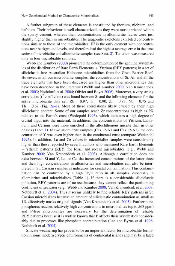
A further subgroup of these elements is constituted by thorium, niobium, and
hafnium. Their behaviour is well characterized, as they were most enriched within
the sparry cement, whereas their concentrations in allomicritic facies were just
slightly higher than in microbialites. The aragonitic skeletons exhibited concentra-
tions similar to those of the microbialites. Hf is the only element with concentra-
tions near background levels, and therefore had the highest average error in the time
series of microbialite and allomicrite samples (see Sect. 2). Tantalum was measured
only in four microbialite samples.
Webb and Kamber (2000) pioneered the determination of the genuine systemat-
ics of the distribution of Rare Earth Elements þ Yttrium (REY patterns) in a set of
siliciclastic-free Australian Holocene microbialites from the Great Barrier Reef.
However, in all our microbialite samples, the concentrations of Si, Al, and all the
trace elements that have been discussed are higher than other microbialites that
have been described in the literature (Webb and Kamber 2000; Van Kranendonk
et al. 2003; Nothdurft et al. 2004; Olivier and Boyet 2006). Moreover, a very strong
correlation (r2 coefficient) was found between Si and the following elements for the
entire microbialite data set: Rb ¼ 0.97; Ti ¼ 0.90; Zr ¼ 0.83; Nb ¼ 0.75 and
Th ¼ 0.67 (Fig. 2a–e). Most of these correlations likely caused by their high
siliciclastic content. Some of our samples reach Zr concentrations as high as 2%
relative to the Earth’s crust (Wedepohl 1995), which indicates a high degree of
crustal input into the material. In addition, the concentrations of Yttrium, Lanta-
num, and Cerium were more enriched in the allochthonous micrite than in other
phases (Table 1). In two allomicrite samples (Cas 12-A1 and Cas 12-A2), the con-
centration of Y was even higher than in the continental crust (compare Wedepohl
1995). In addition, La and Ce values in microbialite samples were considerably
higher than those reported by several authors who measured Rare Earth Elements
þ Yttrium patterns (REY) for fossil and recent microbialites (e.g., Webb and
Kamber 2000; Van Kranendonk et al. 2003). Although a correlation does not
exist between Si and Y, La, or Ce, the increased concentrations of the latter three
and their high concentrations in allomicrites and microbialites can also be inter-
preted in St. Cassian samples as indicators for crustal contamination. This contami-
nation can be confirmed by a high Th/U ratio in all samples, especially in
allomicrites and microbialites (Table 1). If there is a considerable siliciclastic
pollution, REY patterns are of no use because they cannot reflect the partitioning
coefficient of seawater (e.g., Webb and Kamber 2000; Van Kranendonk et al. 2003;
Nothdurft et al. 2004). Thus it seems unlikely to find reliable REY patterns in St.
Cassian microbialites because an amount of siliciclastic contamination as small as
1% effectively masks original signals (Van Kranendonk et al. 2003). Furthermore,
phosphorus reaches relatively high concentrations in microbialites (up to 568 ppm)
and P-free microbialites are necessary for the determination of reliable
REY patterns because it is widely known that P affects their systematics consider-
ably due to processes like phosphate coprecipitation (Lee and Byrne et al. 1996;
Nothdurft et al. 2004).
Silicate weathering has proven to be an important factor for microbialite forma-
tion in some modern cryptic environments of continental islands and may be related
New Geochemical Method to Characterise Microbialites 443
to high carbonate alkalinity (Reitner 1993). However, we believe that the main
cause of the high siliciclastic content in our samples is related to the marly nature of
the St. Cassian Formation, which is extensively reported in the literature (e.g.,
Wendt and F€ursich 1980; Russo et al. 1991).
3.4 “Biological” Elements
Elements with a possible vital effect, such as vanadium, chromium, tin, antimony,
zinc, copper, molybdenum, cobalt, and nickel, have significant concentrations in
Fig. 2 (a–e) Correlation plots between the content of Si and various further non biological
elements (Rb, Ti, Zr, Nb, Th) for microbialite samples. Note overall high values of r2 for all
elements. (f) Correlation plot between the content of Si vs. V among microbialites. A slight
correlation exists (r2 ¼ 0.27). See text and Table 1 for a more detailed explanation
444 F. Sanchez-Beristain et al.
many kinds of carbonates (Kamber and Webb 2007). In this section, we will discuss
whether these elements reflect vital fractionation in the microbialites. Sulphur is
discussed here in a similar way as P was in the non-biological elements section
because its concentrations may be helpful for discerning the origin of nickel.
No significant differences were observed for V and Cr between Margarosmilia,chaetetids, late cements, and some of the microbialites (Table 1). Sample Cas 12-
M2 appears to have a relatively high concentration of V, but a biological effect for
this element cannot be confirmed in this sample because of its high Si and Al
concentrations. All microbialites show a weak to medium correlation between Si
and these elements, with an r2 of 0.27 for Si-V (Fig. 2f; Table 1) and of 0.40 for
Si-Cr (Fig. 3a; Table 1), which could be evidence that their higher concentrations
are a consequence of siliciclastic input. Furthermore, all allomicrite samples have
higher concentrations than the other types of the microbialites.
The patterns of tin, antimony, and zinc are not clear. The cements were more
enriched in Sn and Sb than the coral skeleton, while little difference was recordable
in the chaetetid skeleton. Unfortunately, their concentrations exhibited large varia-
tions between microbialite samples (Table 1) and allomicrite was slightly enriched
above all other samples, except for samples Cas 12-M3 (Sn), and Cas 12-M1,
FS-SA58 M2 (Sb). Zn amounts do not have evident differences among the samples,
except for allomicrite, in which the concentration is relatively enriched. Although a
significant correlation does not exist between Si or Al and these elements, evidence
of a vital effect for them cannot be concluded from the microbialites because only
few show an enrichment relative to other samples.
Copper and molybdenum are generally more enriched in the microbialites than
in cements, sponges, corals, or even allomicrites. It is unclear whether these
elements have a biological role in the skeletal organisms, as both the chaetetid
sponge andMargarosmilia have lower concentrations of all these elements than the
sparry cement. These elements appear to be only weakly correlated with Si, Al, and
Zr throughout the dataset (Table 1). Despite the moderate to high siliciclastic
content in the microbialites, no correlation can be confirmed between Si and
other elements (Fig. 3b, c). Samples Cas 12-M1 (Mo) and Cas 12-M4 (Cu) have
moderate to high average error along the time series–ablation (not shown), but that
does not seem to affect the distribution plots displayed in Fig. 3b, c.
It is widely known that copper plays a fundamental role in the structure of certain
transmembrane enzymes, such as cytochrome c oxidase (Mathews et al. 1999) and
molybdenum has been proved to be crucial in the molybdate-reducing enzymes of
sulphate-reducing bacteria (Tucker et al. 1997). Therefore, we believe that the
concentrations of Cu and Mo in our microbialite samples are relics of original
microbial metabolism.
Cobalt concentrations were only slightly higher in allomicrites than in micro-
bialites, but again the overall trend was the same as was observed for copper and
molybdenum. The nickel concentration was significantly enriched in some of the
microbialites, even more than in the allochthonous micrite. FS-SA 001-M2, the
most Si-depleted microbialite sample, had a high Ni concentration of 33.57 ppm,
which was more than twice the concentration of two of the allomicrite samples.
New Geochemical Method to Characterise Microbialites 445
There is a strong correlation (r2 ¼ 0.95) between Ni and Co in microbialite
samples (Fig. 3d), which should be a consequence of siliciclastic contamination
because this correlation persists (r2 ¼ 0.92) when all samples are included. Fur-
thermore, the Ni/Co ratio in the Earth’s crust is ~2.3 (Wedepohl 1995), and in
SMOW, it is around 400. In most of the measured microbialites, the allomicrites,
and even the sponge this ratio is ~8.0. Hence, we cannot conclude that high Ni and
Co concentrations in the microbialites are related to biological processes, but rather
Fig. 3 (a) Correlation plot between the content of Si vs. Cr among microbialites. A medium
correlation exists. (r2 ¼ 0.40). (b and c) Correlation plots Si vs. Cu and Mo among microbialite
facies. Note the extremely low value of r2 at both plots. Low correlation plus the higher
concentration of these elements in microbialites compared to other facies permits the attribution
of these concentrations to original microbial activity. (d) Correlation between of Ni vs. Co for all
samples. Note the high correlation in microbialites, which barely changes when all samples are
plotted. (e) Correlation between of S vs. Ni in microbialites. Only a minor correlation can be
assessed, which leads to the interpretation of a non-biological origin of Ni. See Table 1 and text for
further explanation
446 F. Sanchez-Beristain et al.
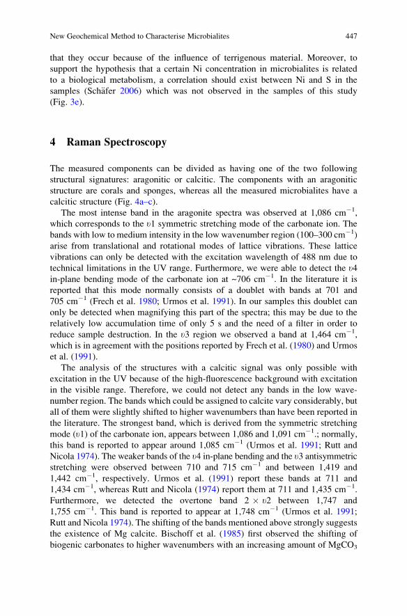
that they occur because of the influence of terrigenous material. Moreover, to
support the hypothesis that a certain Ni concentration in microbialites is related
to a biological metabolism, a correlation should exist between Ni and S in the
samples (Sch€afer 2006) which was not observed in the samples of this study
(Fig. 3e).
4 Raman Spectroscopy
The measured components can be divided as having one of the two following
structural signatures: aragonitic or calcitic. The components with an aragonitic
structure are corals and sponges, whereas all the measured microbialites have a
calcitic structure (Fig. 4a–c).
The most intense band in the aragonite spectra was observed at 1,086 cm�1,
which corresponds to the u1 symmetric stretching mode of the carbonate ion. The
bands with low to medium intensity in the low wavenumber region (100–300 cm�1)
arise from translational and rotational modes of lattice vibrations. These lattice
vibrations can only be detected with the excitation wavelength of 488 nm due to
technical limitations in the UV range. Furthermore, we were able to detect the u4in-plane bending mode of the carbonate ion at ~706 cm�1. In the literature it is
reported that this mode normally consists of a doublet with bands at 701 and
705 cm�1 (Frech et al. 1980; Urmos et al. 1991). In our samples this doublet can
only be detected when magnifying this part of the spectra; this may be due to the
relatively low accumulation time of only 5 s and the need of a filter in order to
reduce sample destruction. In the u3 region we observed a band at 1,464 cm�1,
which is in agreement with the positions reported by Frech et al. (1980) and Urmos
et al. (1991).
The analysis of the structures with a calcitic signal was only possible with
excitation in the UV because of the high-fluorescence background with excitation
in the visible range. Therefore, we could not detect any bands in the low wave-
number region. The bands which could be assigned to calcite vary considerably, but
all of them were slightly shifted to higher wavenumbers than have been reported in
the literature. The strongest band, which is derived from the symmetric stretching
mode (u1) of the carbonate ion, appears between 1,086 and 1,091 cm�1.; normally,
this band is reported to appear around 1,085 cm�1 (Urmos et al. 1991; Rutt and
Nicola 1974). The weaker bands of the u4 in-plane bending and the u3 antisymmetric
stretching were observed between 710 and 715 cm�1 and between 1,419 and
1,442 cm�1, respectively. Urmos et al. (1991) report these bands at 711 and
1,434 cm�1, whereas Rutt and Nicola (1974) report them at 711 and 1,435 cm�1.
Furthermore, we detected the overtone band 2 � u2 between 1,747 and
1,755 cm�1. This band is reported to appear at 1,748 cm�1 (Urmos et al. 1991;
Rutt and Nicola 1974). The shifting of the bands mentioned above strongly suggests
the existence of Mg calcite. Bischoff et al. (1985) first observed the shifting of
biogenic carbonates to higher wavenumbers with an increasing amount of MgCO3
New Geochemical Method to Characterise Microbialites 447
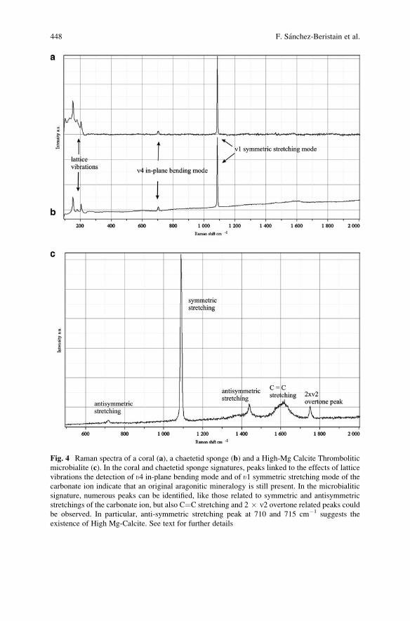
Fig. 4 Raman spectra of a coral (a), a chaetetid sponge (b) and a High-Mg Calcite Thrombolitic
microbialite (c). In the coral and chaetetid sponge signatures, peaks linked to the effects of lattice
vibrations the detection of u4 in-plane bending mode and of u1 symmetric stretching mode of the
carbonate ion indicate that an original aragonitic mineralogy is still present. In the microbialitic
signature, numerous peaks can be identified, like those related to symmetric and antisymmetric
stretchings of the carbonate ion, but also C¼C stretching and 2 � n2 overtone related peaks couldbe observed. In particular, anti-symmetric stretching peak at 710 and 715 cm�1 suggests the
existence of High Mg-Calcite. See text for further details
448 F. Sanchez-Beristain et al.
in the carbonates. The difference in the amount of shifting is likely because of the
varying concentrations of MgCO3 in the structures; this would be in agreement with
the findings of the electron microprobe analyses.
In both the aragonitic and the calcitic spectra, an additional band between 1,603
and 1,619 cm�1 was observed. This band probably exists because of the C¼C
stretching vibrations, whereas its broad appearance suggests the beginning of the
degradation of the bonding, which is often described as an amorphous carbon
signature (e.g., Robertson 1986; Matsunuma 1997). Nevertheless, the existence of
that peak strongly indicates that organics remain in the structure.
5 Conclusions
In addition to petrographical and epifluorescence techniques, the distinction
between microbialite and allomicrite can also be recognised by the higher concen-
trations of selected REE and Y in the former samples.
LA-ICP-MS analysis of the Mg-content in microbialites showed a high
Mg-calcite mineralogy, which was confirmed by Raman spectroscopy. The inter-
pretation of the microbialitic spectra is in accordance with the high quality of
preservation that was confirmed by CL. Furthermore, Raman spectroscopy was
able to differentiate between the Mg-calcite mineralogy of the microbialites and the
aragonitic mineralogy of the skeletal components. Additionally, Raman spectra
revealed organic relics inside the microbialites and in the skeletal components.
LA-ICP-MS also permitted us to detect the presence of siliciclastic elements in
the microbialites, which most likely indicate a marly sediment origin in the St.
Cassian Formation. While most of the non-vital trace elements are markedly
correlated to the content of Si, others like selected REE and Y are not; their
concentrations are interpreted as being strongly affected by the content of siliciclastics
and by the amount of phosphorus. Thus, it is in principle not possible to obtain a
reliable microbialite-based REY pattern in our samples.
Some biologically important elements like vanadium and chromium appear to be
detritus-related rather than being relics of biological activity because a medium
correlation exists between them and silicon. Furthermore, allochthonous micrite
more enriched with those elements. Antimony and zinc are relatively enriched
in some microbialites and a strong correlation between their concentrations and
Si or Al does not exist, but a biological effect cannot be assessed because their
concentrations are higher in allomicrites. Copper and molybdenum are more sig-
nificant, especially in the skeletons of Margarosmilia and in chaetetids because of
their rather homogeneous distribution along the time series measurements.
Ni anomalies, which are also strongly related to Co anomalies, are probably not
related to biological activity.
Acknowledgements We gratefully thank the constructive comments and reviews by Prof
Dr. Franco Russo (Calabria), which considerably improved our manuscript. JFSB is supported
New Geochemical Method to Characterise Microbialites 449
by the German Academic Exchange Service (DAAD) with a three-year PhD research stay
in Germany through the grant A/07/94459. Funding for this research comes from the
Courant Research Centre in G€ottingen, Germany. Dr. Andreas Kronz (G€ottingen) is gratefully
acknowledged for providing microprobe analyses. This is publication No. 34 of the G€ottingenCourant Research Centre Geobiology (German Excellence Initiative)
References
Bischoff WD, Sharma SK, Mackenzie FT (1985) Carbonate ion disorder in synthetic and biogenic
magnesian calcites: a Raman spectral study. American Mineralogist 70:581–589
Byrne RH, Liu X, Schijf J (1996) The influence of phosphate coprecipitation on rare earth
distributions in natural waters. Geochimica Cosmochimica Acta 60:3341–3346
Dickson T (1990) Carbonate mineralogy and chemistry. In: Tucker ME, Wright VP (eds) Carbon-
ate sedimentology. Blackwell Scientific Publications, Oxford, pp 284–313
Dieci G, Antonacci A, Zardini R (1968) Le spugne cassiane (Trias medio–superiore) della regione
dolomıtica attorno a Cortina d’Ampezzo. Bolletino della Societa Paleontologica Italiana
7:94–155
Dieci G, Russo A, Russo F (1974) Nota preliminare sulla microstrutura di spugne aragonitiche del
Trias Medio–Superiore. Bolletino della Societa Paleontologica Italiana 13(1–2):99–107
Fl€ugel E (2004) Microfacies of carbonate rocks. Springer, Berlin, Heidelberg, 976 pp
Fraustro da Silva JJR, Williams RJP (1991) The biological chemistry of the elements. Clarendon
Press, Oxford, 561 pp
Frech R, Wang EC, Bates JB (1980) The IR and Raman spectra of CaCO3 (aragonite). Spectro-
chimica Acta 36A:915–919
F€ursich FT, Wendt J (1977) Biostratinomy and palaeoecology of the Cassian Formation (Triassic)
of the Southern Alps. Palaeogeography, Palaeoclimatology, Palaeoecology 22:257–323
Kamber B, Webb G (2007) Transition metal abundances in microbial carbonate: a pilot study
based on in situ LA-ICP-MS analysis. Geobiology 5(4):375–389
Laghi G, Martinelli G, Russo F (1983) Localization of minor elements by EDS microanalysis in
aragonitic sponges from the St. Cassian beds, Italian Dolomites. Lethaia 17:133–138
Mathews CK, Van Holde KE, Ahern KG (1999) Biochemistry. Addison Wesley, San Francisco,
1190 pp
Matsunuma S (1997) Theoretical simulation of resonance Raman bands of amorphous carbon.
Thin Solid Films 306:17–22
Neuweiler F, Reitner J (1995) Epifluorescence microscopy of selected microbialites from Lower
Carnian Cipit-boulders of the Cassian Formation (Seeland Alpe, Dolomites). In: Reitner J,
Neuweiler F (eds) Mud Mounds: A polygenetic Spectrum of Fine-grained Carbonate Buildups.
Facies 32:26–28
Nothdurft LD, Webb GE, Kamber BS (2004) Rare earth element geochemistry of Late Devonian
reefal carbonates, Canning Basin, Western Australia: confirmation of a seawater REE proxy in
ancient limestones. Geochimica Cosmochimica Acta 68:263–283
Olivier N, Boyet M (2006) Rare earth and trace elements of microbialites in Upper Jurassic coral-
and sponge-microbialite reefs. Chemical Geology 230:105–123
Prasada Rao C (1996) Modern Carbonates tropical temperate polar. Introduction to sedimentology
and geochemistry. Arts of Tasmania, University of Tasmania, Tasmania
Rech H (1998) Geobiologie der sogenanten “Cipit-Kalke” der Beckenfazies der Cassianer-
Schichten, S’ St. Kassian, Dolomiten. Unpublished Diploma Thesis, University of G€ottingen,G€ottingen, 134 pp
450 F. Sanchez-Beristain et al.
Reitner J (1987) A new calcitic sphinctozoan sponge belonging to the Demospongiae from the
Cassian Formation (Lower Carnian; Dolomites, Northern Italy) and its phylogenetic relation-
ship. Geobios, 20:571–589
Reitner J (1992) “Coralline Spongien”. Der Versuch einer phylogenetisch-taxonomischen
Analyse. Berliner Geowissenschaftliche Abhandlungen Reihe E 1:1–352
Reitner J (1993) Modern cryptic microbialite/metazoan facies from Lizard Island (Great Barrier
Reef, Australia). Formation and concepts. Facies 29:3–39
Reitner J, Engeser T (1989)Chaetosclera klipsteini n.gen n.sp. (Halichondriida, Demospongiae), aus
dem Unterkarn der Cassianer-Schichten (Dolomiten, Italien). Mitteilungen Geologisch-
Pal€aontologisches Institut Universit€at Hamburg 68:159–165
Robertson J (1986) Amorphous carbon. Advances in Physics 35:317–374
Russo F (2005) Biofacies evolution in the Triassic platforms of the Dolomites, Italy. Ann Univ
Stud Ferrara Mus Sci Nat vol Spec, 33–45
Russo F, Neri C, Mastandrea A, Laghi G (1991) Depositional and diagenetic history of the Alpe di
Specie (Seelandalpe) fauna (Carnian,. Northeastern Dolomites). Facies 25:187–210
Russo F, Neri C, Mastandrea A, Baracca A (1997) The mud-mound nature of the Cassian platform
margins of the Dolomites. A case history: the Cipit Boulders from Punta Grohmann (Sasso
Piatto Massif, Northern Italy). Facies 36:25–36
Russo F, Mastandrea A, Neri C (1998) Evoluzione degli organismi costruttori nelle piattaforme
triassiche delle Dolomiti (Italia). Memorie della Societa Geologica Italiana 53:479–488
Rutt HN, Nicola JH (1974) Raman spectra of carbonates of calcite structure. Journal of Physics C:
Solid State Physics 7:4522–4528
Sanchez-Beristain JF, Reitner J (2008) Microbialites from the Cassian Formation: an insight.
International Kalkowsky-Symposium Abstract Volume and Field Guide to Excursions,
116–117
Sch€afer N (2006) Ni-Anomalien in Cold-Seep Karbonaten aus dem nordwestlichen Schwarzen
Meer: Ist Ni ein kritisches Element bei der anaeroben Oxidation von Methan? Unpublished
Diploma Thesis, University of G€ottingen, G€ottingenTucker MD, Barton LL, Thomson BM (1997) Reduction and immobilization of molybdenum by
Desulfovibrio desulfuricans. Journal of Environmental Quality 26:1146–1152
Urmos J, Sharma SK, Mackenzie FT (1991) Characterization of some biogenic carbonates with
Raman spectroscopy. American Mineralogist 76:641–646
Van Kranendonk M, Webb G, Kamber BS (2003) New geological and trace element evidence
from 3.45 Ga stromatolitic carbonates in the Pilbara Craton: support of a marine, biogenic
origin and for a reducing Archaean ocean. Geobiology 1:91–108
von Richthofen F (1860) Geognostische Beschreibung der Umgebung von Predazzo, St. Cassian
und der Seisser Alpe in S€udtirol, Gotha, 327 pp
Webb GE, Kamber BS (2000) Rare earth elements in Holocene reefal microbialites: a new shallow
seawater proxy. Geochimica Cosmochimica Acta 64:1557–1565
Wedepohl KH (1995) The composition of the continental crust. Geochimica Cosmochimica Acta
59:1219–1232
Wendt (1982) The Cassian patch reefs (Lower Carnian, Southern Alps). Facies 6:185–202
Wendt J, F€ursich FT (1980) Facies analysis and palaeogeography of the Cassian Formation,
Triassic, Southern Alps. Rivista Italiana di Paleontologia e Stratigrafıa 85(3–4):1003–1028
Wissmann HL, M€unster G (1841) Beitrage zur Geognosie und Petrefactenkunde des s€udostlichenTirols vorz€uglich der Schichten von St. Cassian. Beitr€age zur Petrefaktenkunde 4:1–152,
Bayreuth (Buchner)
Zankl H (1993) The origin of High-Mg-Calcite microbialites in cryptic habitats of Caribbean
Coral Reefs – Their dependence on light and turbulence. Facies 29:55–60
New Geochemical Method to Characterise Microbialites 451

.
Importance of Rare Earth Element Patterns
in Discrimination Between Biotic and Abiotic
Mineralization
Adriano Guido, Adelaide Mastandrea, Fabio Tosti, and Franco Russo
1 Introduction
Rare earth element (REE) patterns have been used to discuss the origin of rocks
and minerals and the evolution processes that occurred during their formation
(Goldschmidt 1954; Henderson 1996).
Carbonate REE patterns are similar to the REE patterns of clastic sedimentary
rocks (McLennan 1989). The application of these patterns to natural water systems
provided information on the incorporation of REE and the physical and chemical
characteristics of the sedimentary environment (Taylor and McLennan 1988; Erel
and Stopler 1993; Takahashi et al. 2000a, b; Webb and Kamber 2000; Bolhar et al.
2004). The REE signature of sediments can be used to obtain detailed information
on the geochemical conditions of depositional basins.
The REE þ Y carbonate patterns can also provide information on carbonatogen-
esis processes. Many differences in the behaviour of REE fractionation exist between
microbial and skeletal carbonates. Microbial carbonates are generally formed in
microenvironments that are controlled or affected by living or dead organic matter
in biofilms. Microbial carbonates represent biologically induced precipitation in
equilibrium with the depositional environment (Lowenstam 1981). This type of
biomineralisation is reflected in the proportional fractionation of REE in the normal-
ised patterns. During biologically controlled carbonate skeletogenesis, organisms
actively manage ions to secrete the skeleton, leading to a non-proportional fraction-
ation of REE in their pattern (Webb and Kamber 2000). This phenomenon is called
the “vital effect”.
A. Guido (*), A. Mastandrea, F. Tosti, and F. Russo
Dipartimento di Scienze della Terra, Universita della Calabria, Via P. Bucci Cubo 15b, 87036
Rende (CS), Italy
e-mail: [email protected]
J. Reitner et al., Advances in Stromatolite Geobiology,Lecture Notes in Earth Sciences 131, DOI 10.1007/978-3-642-10415-2_27,# Springer-Verlag Berlin Heidelberg 2011
453
Another important topic is to understand the REE fractionation during abiotic
chemical deposition. The behaviour of REEs during evaporation and gypsum
precipitation is not well understood.
The shale-normalised REE þ Y patterns were used in this paper to verify the
reliability of using the REE pattern as a geochemical signature of the water in which
the microbialite precipitated. We tested this approach for the Messinian Calcare di
Base (CDB), in which bacteria-induced precipitation has already been proven by
molecular fossils and other organic markers (Guido et al. 2007) to compare the
biogeochemical data to the REE þ Y characteristics, we analysed the same sam-
ples studied by Guido et al. (2007). The CdB sampled in Northern Calabria lacks
any diagenetic alteration, and the original mineralogy (aragonite) and microstruc-
ture are preserved.
2 Analytical Techniques
Small fragments of the carbonate samples were first hand ground in an agate mortar
and then dissolved by microwave digestion using a Mars 5 microwave apparatus
(CEM technologies). Then, 100 mg of the powder was dissolved in Teflon (TFM)
digestion vessels using a mixture of hydrofluoric acid (2 ml HF), nitric acid (8 ml
HNO3) and perchloric acid (2 ml HClO4). All reagents were of Merck “suprapur”
quality. Before the mixture evaporated completely, we added 2 ml of perchloric
acid to ensure the complete removal of the hydrofluoric acid. These solutions they
were left to cool and then diluted to 100 ml with Millipore water.
The trace element concentrations in each sample were determined by a Perkin-
Elmer Elan DRCe in the Department of Earth Science, Calabria University.
The concentration of REE, Sc, Y and Th were determined using external
calibration curves, which were prepared using a Perkin Elmer “multi-element
Calibration Standard 2 solution”. Internal standards (indium, germanium and rhe-
nium) were added to both the standards and the samples.
Standard reference materials (limestone from the US Geological Survey) were
prepared in the same way and were used as unknowns during the analytical
sequence. This procedure was suggested by Eggins et al. (1997). The measured
concentrations were compared with certified values to evaluate the accuracy and
precision of the analytical data.
3 REE Fractionation in Evaporites and Microbial Carbonate
REE in carbonate sediments can be used to determine the original seawater
geochemistry in which the carbonates formed. The REE fractionation in the carbo-
nates depends mainly on the following two factors: (1) the geochemistry of the
water in which the carbonates mineralise and (2) the vital effect, which could
454 A. Guido et al.
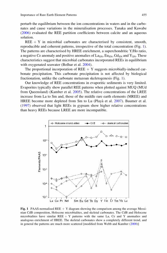
perturb the equilibrium between the ion concentrations in waters and in the carbo-
nates and cause variations in the mineralisation processes. Tanaka and Kawabe
(2006) evaluated the REE partition coefficients between calcite and an aqueous
solution.
REE þ Y in microbial carbonates are characterised by consistent, smooth,
reproducible and coherent patterns, irrespective of the total concentration (Fig. 1).
The patterns are characterised by HREE enrichment, a superchondritic Y/Ho ratio,
a negative Ce anomaly and positive anomalies of LaSN, EuSN, GdSN and YSN. These
characteristics suggest that microbial carbonates incorporated REEs in equilibrium
with oxygenated seawater (Bolhar et al. 2004).
The proportional incorporation of REE þ Y suggests microbially-induced car-
bonate precipitation. This carbonate precipitation is not affected by biological
fractionation, unlike the carbonate metazoan skeletogenesis (Fig. 1).
Our knowledge of REE concentrations in evaporitic sediments is very limited.
Evaporites typically show parallel REE patterns when plotted against MUQ (MUd
from Queensland) (Kamber et al. 2005). The relative concentrations of the LREE
increase from La to Sm and, those of the middle rare earth elements (MREE) and
HREE become more depleted from Sm to Lu (Playa et al. 2007). Baumer et al.
(1997) observed that light REEs in gypsum show higher relative concentrations
than heavy REEs because LREE are more incompatible.
Fig. 1 PAAS-normalised REE þ Y diagram showing the comparison among the average Messi-
nian CdB composition, Holocene microbialites, and skeletal carbonates. The CdB and Holocene
microbialites have similar REE þ Y patterns with the same La, Ce and Y anomalies and
analogous enrichment of HREE. The skeletal carbonates show a completely different trend, and
in general the patterns are much more scattered [modified from Webb and Kamber (2000)]
Importance of Rare Earth Element Patterns 455
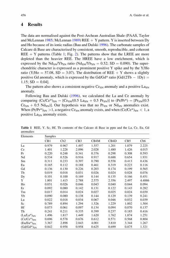
4 Results
The data are normalised against the Post-Archean Australian Shale (PAAS, Taylor
and McLennan 1985; McLennan 1989) REE þ Y pattern. Y is inserted between Dy
and Ho because of its ionic radius (Bau and Dulski 1996). The carbonate samples of
Calcare di Base are characterised by consistent, smooth, reproducible, and coherent
REE þ Y patterns (Table 1; Fig. 2). The patterns show that the LREE are more
depleted than the heavier REE. The HREE have a low enrichment, which is
expressed by the NdSN/YbSN ratio (NdSN/YbSN ¼ 0.52; SD ¼ 0.090). The super-
chondritic character is expressed as a prominent positive Y spike and by the Y/Ho
ratio (Y/Ho ¼ 57.08, SD ¼ 3.07). The distribution of REE þ Y shows a slightly
positive Gd anomaly, which is expressed by the Gd/Gd* ratio [Gd/(2Tb � 1Dy) ¼1.15; SD ¼ 0.04].
The pattern also shows a consistent negative CeSN anomaly and a positive LaSNanomaly.
Following Bau and Dulski (1996), we calculated the La and Ce anomaly by
comparing (Ce/Ce*)SN ¼ [CeSN/(0.5 LaSN þ 0.5 PrSN)] to (Pr/Pr*) ¼ [PrSN/(0.5
CeSN þ 0.5 NdSN)]. Our hypothesis was that no PrSN or NdSN anomalies exist.
When (Pr/Pr*)SN >1, a negative CeSN anomaly exists, and when (Ce/Ce*)SN < 1, a
positive LaSN anomaly exists.
Table 1 REE, Y, Sc, Hf, Th contents of the Calcare di Base in ppm and the La, Ce, Eu, Gd
anomalies
Elements Samples
CB1 Cb2 CB3 CB4M CB4D CB5 Cb6
La 0.979 0.967 1.497 1.557 1.201 1.079 2.225
Ce 1.401 1.228 2.096 2.028 1.480 1.426 4.015
Pr 0.220 0.248 0.341 0.376 0.298 0.308 0.593
Nd 0.534 0.526 0.916 0.917 0.688 0.654 1.931
Sm 0.311 0.233 0.397 0.790 0.558 0.413 0.436
Eu 0.165 0.112 0.188 0.461 0.319 0.223 0.116
Gd 0.136 0.130 0.226 0.203 0.174 0.199 0.565
Tb 0.019 0.018 0.031 0.026 0.024 0.028 0.076
Dy 0.101 0.100 0.169 0.144 0.135 0.166 0.451
Y 1.801 1.415 2.788 2.575 2.356 2.497 4.4886
Ho 0.031 0.026 0.046 0.043 0.040 0.044 0.094
Er 0.092 0.080 0.142 0.131 0.122 0.143 0.282
Tm 0.017 0.014 0.024 0.027 0.025 0.024 0.039
Yb 0.090 0.080 0.138 0.144 0.129 0.139 0.241
Lu 0.022 0.018 0.034 0.067 0.046 0.032 0.039
Sc 0.789 0.894 1.294 1.526 1.229 1.002 1.504
Hf 0.073 0.061 0.097 0.134 0.094 0.070 0.137
Th 0.241 0.211 0.335 0.399 0.237 0.185 0.416
(La/La*)SN 1.496 1.817 1.449 1.620 1.762 1.874 1.251
(Ce/Ce*)SN 0.696 0.578 0.676 0.612 0.571 0.568 0.804
(Eu/Eu*)SN 3.367 2.898 2.843 4.001 3.810 3.344 1.267
(Gd/Gd*)SN 0.842 0.958 0.958 0.625 0.699 0.875 1.321
456 A. Guido et al.
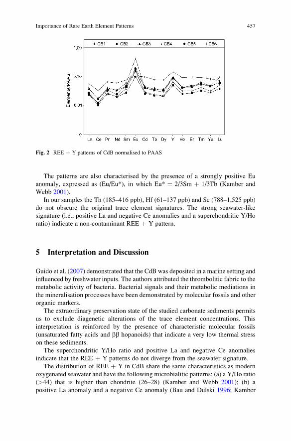
The patterns are also characterised by the presence of a strongly positive Eu
anomaly, expressed as (Eu/Eu*), in which Eu* ¼ 2/3Sm þ 1/3Tb (Kamber and
Webb 2001).
In our samples the Th (185–416 ppb), Hf (61–137 ppb) and Sc (788–1,525 ppb)
do not obscure the original trace element signatures. The strong seawater-like
signature (i.e., positive La and negative Ce anomalies and a superchondritic Y/Ho
ratio) indicate a non-contaminant REE þ Y pattern.
5 Interpretation and Discussion
Guido et al. (2007) demonstrated that the CdB was deposited in a marine setting and
influenced by freshwater inputs. The authors attributed the thrombolitic fabric to the
metabolic activity of bacteria. Bacterial signals and their metabolic mediations in
the mineralisation processes have been demonstrated by molecular fossils and other
organic markers.
The extraordinary preservation state of the studied carbonate sediments permits
us to exclude diagenetic alterations of the trace element concentrations. This
interpretation is reinforced by the presence of characteristic molecular fossils
(unsaturated fatty acids and bb hopanoids) that indicate a very low thermal stress
on these sediments.
The superchondritic Y/Ho ratio and positive La and negative Ce anomalies
indicate that the REE þ Y patterns do not diverge from the seawater signature.
The distribution of REE þ Y in CdB share the same characteristics as modern
oxygenated seawater and have the following microbialitic patterns: (a) a Y/Ho ratio
(>44) that is higher than chondrite (26–28) (Kamber and Webb 2001); (b) a
positive La anomaly and a negative Ce anomaly (Bau and Dulski 1996; Kamber
Fig. 2 REE þ Y patterns of CdB normalised to PAAS
Importance of Rare Earth Element Patterns 457
and Webb 2001); (c) no negative GdSN anomaly, suggesting that organic com-
pounds did not influence the incorporation of the REEs; and (d) a strong positive Eu
anomaly (Fig. 3).
The La and Ce anomalies were studied in a plot of PAAS-normalised (Pr/Pr*)SN[PrSN/(0.5 CeSN þ 0.5 NdSN)] vs. (Ce/Ce*)SN [CeSN/(0.5 LaSN þ 0.5 PrSN)] (Bau
and Dulski 1996). This plot discriminates between the La and Ce anomalies and
confirms the normal marine deposition of the carbonates in this study (Fig. 4).
REE þ Y dataset can be utilised as a record of the microbial origin of micrites,
particularly for carbonates of doubtful genesis such as the Messinian Calcare di
Base. The REE þ Y pattern of the CdB is comparable to the patterns of the
Archean, Devonian, and Holocene microbial carbonates (Fig. 3). The microbialites
are currently the best material to use for determining the record of marine REE
geochemistry.
The ability of not-microbialitic carbonates to precipitate and incorporate REE in
proportion to their water environment is difficult to determine. Carbonate rarely
precipitates in normal marine conditions without some biological influence.
The REE dataset in the CdB was normalised to MUQ (MUd from Queensland)
to compare the distributions of this study to those of the evaporitic samples and
seawater reported by Playa et al. (2007) (Fig. 5). The behaviour of REEs during
evaporative concentration and gypsum precipitation is very different from the REE
distribution in seawater. Most of the gypsum samples reported by Playa et al. (2007)
show an increase in the light REE from La to Eu and a decrease in the heavy REE
Fig. 3 PAAS-normalised REE þ Y diagram comparing the average composition of South Pacific
Seawater, Messinian CdB and microbialites from the Precambrian, Devonian and Holocene. All
patterns and anomalies are similar [modified from Bolhar et al. (2004)]
458 A. Guido et al.
from Eu to Lu (Fig. 5c). The main differences between the CdB carbonate and
evaporitic patterns are as follows: (1) the carbonates have a higher REE concentra-
tion than the evaporites; (2) the carbonates have a strong LREE depletion and
HREE enrichment; (3) the evaporites are enriched in MREE. The distribution of
REEs emphasises the strong difference between the CdB and the evaporitic sedi-
ments (Fig. 5a, c). The behaviour of the evaporites is the inverse of the behaviour of
the microbialites and seawater.
The MUQ-normalised patterns confirm that the CdB and Holocenic microbialite
REE distributions are very similar to the seawater pattern (Fig. 5a, b).
6 Conclusions
The REE þ Y in the CdB are characterised by consistent, smooth, reproducible,
and coherent patterns. These patterns show HREE enrichment, a superchondritic
Y/Ho ratio, a negative Ce anomaly and positive anomalies of LaSN, EuSN, GdSN and
YSN. The proportional incorporation of REE þ Y suggests microbially-induced
carbonate precipitation. This carbonate precipitation is not affected by biological
fractionation and is comparable to the precipitation of Archean, Devonian, and
Holocene microbial carbonates. This result confirms that the REE þ Y dataset can
be utilised as a record of the microbial origin of micrites, particularly for carbonates
of doubtful genesis such as the Messinian Calcare di Base.
The MUQ-normalised pattern shows the difference between the CdB and the
evaporites. The evaporite patterns are the inverse of the patterns of the CdB and
Fig. 4 Diagram of Ce/Ce* [Ce/(0.5La þ 0.5Pr)]SN vs. Pr/Pr* [Pr/(0.5Ce þ 0.5Nd)]SN showing
the La and Ce anomalies in seawater sediments. All of the CdB samples cluster in the field of
negative Ce and positive La anomalies, similar to modern open oceanic surface water
Importance of Rare Earth Element Patterns 459
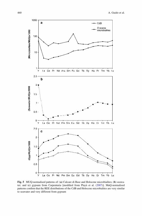
Fig. 5 MUQ-normalised patterns of: (a) Calcare di Base and Holocene microbialites; (b) seawa-
ter; and (c) gypsum from Carpentaria [modified from Playa et al. (2007)]. MuQ-normalised
patterns confirm that the REE distributions of the CdB and Holocene microbialites are very similar
to seawater and very different from gypsum
460 A. Guido et al.
seawater: This result confirms the model for the CdB deposition that was proposed
by Guido et al. (2007). This model describes biotic mineralisation in normal marine
conditions rather than abiotic precipitation in an evaporitic setting.
Acknowledgments Our thanks to Prof. Dr. Joachim Reitner for the excellent organisation of the
International Kalkowsky Symposium on the Geobiology of Stromatolites.
We are indebted to an anonymous reviewer for suggestions that greatly improved the manuscript.
This research was funded by the Universita della Calabria, PRIN 2007.
References
Bau M, Dulski P (1996) Distribution of Y and rare-earth elements in the Penge and Kuruman Iron
Formations, Traansvaal Supergroup, South Africa. Precambrian Research 79:37–55
Baumer A, Blanc Ph, Cesbron F, Ohnenstetter D (1997) Cathodoluminescence of synthetic (doped
with rare-earth elements) and natural anhydrites. Chemical Geology 138:73–80
Bolhar R, Kamber BS, Moorbath S, Fedo CM, Whitehouse MJ (2004) Characterisation of early
Archaean chemical sediments by trace element signatures. Earth and Planetary Science Letters
222:43–60
Eggins SM, Woodhead JD, Kinsley LPJ, Mortimer GE, Sylvester P, McCulloch MT, Hergt JM,
Handler MR (1997) A simple method for the precise determination of 40 trace elements in
geological samples by ICPMS using enriched isotope internal standardisation. Chemical
Geology 134:311–326
Erel Y, Stopler EM (1993) Modeling of rare-earth element partitioning between particles and
solution in aquatic environments. Geochimica Cosmochimica Acta 57:513–518
Goldschmidt VM (1954) Geochemistry. Oxford Press, New York
Guido A, Jacob J, Gautret P, Laggoun-Defarge F, Mastandrea A, Russo F (2007) Molecular fossils
and other organic markers as palaeoenvironmental indicators of the Messinian CdB formation:
normal versus stressed marine deposition (Rossano Basin, Northern Calabria, Italy). Palaeo-
geography Palaeoclimatology Palaeoecology 255:265–283
Henderson P (1996) The rare earth elements: introduction and review. In: Jones AP (ed) Rare
Earth Minerals: Chemistry, Origin and Ore Deposits. Chapman & Hall, London, 1–17
Kamber BS, Webb GE (2001) The geochemistry of late Archaean microbial carbonate: Implica-
tions for ocean chemistry and continental erosion history. Geochimica Cosmochimica Acta
65:2509–2525
Kamber BS, Greig A, Collerson KD (2005) A new estimate for the composition of weathered
young upper continental crust from alluvial sediments, Queensland, Australia. Geochimica
Cosmochimica Acta 69:1041–1058
Lowenstam HA (1981) Minerals formed by organisms. Science 211:1126–1131
McLennan SM (1989) Rare earth elements in sedimentary rocks: influence of provenance and
sedimentary processes. In: Lipin BR McKay GA (eds) Geochemistry and Mineralogy of Rare
Earth Elements. Mineralogical Society of America. Reviews in Mineralogy 21:169–200
Playa E, Cendon DI, Trave A, Chivas AR, Garcıa A (2007) Non-marine evaporites with both
inherited marine and continental signatures: the Gulf of Carpentaria, Australia, at ~70 ka.
Sedimentary Geology 201:267–285
Takahashi Y, Shimizu H, Usui A, Kagi H, Nomura M (2000a) Direct observation of tetravalent
cerium in ferromanganese nodules and crusts by X-ray absorption near-edge structure
(XANES). Geochimica Cosmochimica Acta 64:2929–2935
Takahashi Y, Shimizu H, Kagi H, Yoshida H, Usui U, Nomura M (2000b) A new method for the
determination of CeIII/CeIV ratios in geological materials; application for weathering, sedi-
mentary, and diagenetic processes. Earth and Planetary Science Letters 182:201–207
Importance of Rare Earth Element Patterns 461

Tanaka K, Kawabe I (2006) REE abundances in ancient seawater inferred from marine limestone
and experimental REE partition coefficients between calcite and aqueous solution. Geochemi-
cal Journal 40:425–435
Taylor SR, McLennan SM (1985) The Continental Crust: its Composition and Evolution. Black-
well Scientific Publications, Oxford
Taylor SR, McLennan SM, (1988) The significance of the rare earths in geochemistry and
cosmochemistry. In: Gschneidner Jr KA, Eyring L (eds) Handbook on the Physics and
Chemistry of Rare Earths vol. 11. Elsevier, Amsterdam, 485–578
Webb GE, Kamber BS, (2000) Rare earth elements in Holocene reefal microbialites: a shallow
seawater proxy. Geochimica Cosmochimica Acta 64:1557–1565
462 A. Guido et al.
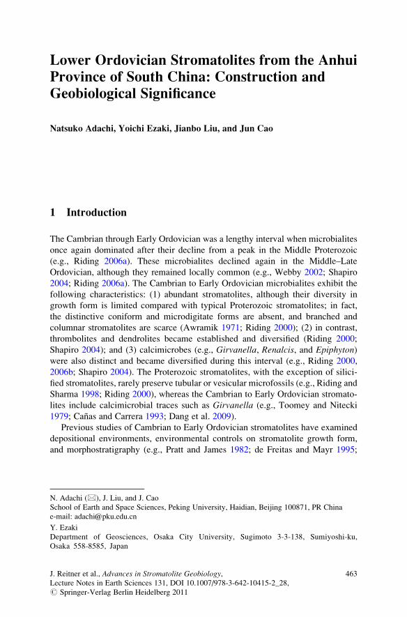
Lower Ordovician Stromatolites from the Anhui
Province of South China: Construction and
Geobiological Significance
Natsuko Adachi, Yoichi Ezaki, Jianbo Liu, and Jun Cao
1 Introduction
The Cambrian through Early Ordovician was a lengthy interval when microbialites
once again dominated after their decline from a peak in the Middle Proterozoic
(e.g., Riding 2006a). These microbialites declined again in the Middle–Late
Ordovician, although they remained locally common (e.g., Webby 2002; Shapiro
2004; Riding 2006a). The Cambrian to Early Ordovician microbialites exhibit the
following characteristics: (1) abundant stromatolites, although their diversity in
growth form is limited compared with typical Proterozoic stromatolites; in fact,
the distinctive coniform and microdigitate forms are absent, and branched and
columnar stromatolites are scarce (Awramik 1971; Riding 2000); (2) in contrast,
thrombolites and dendrolites became established and diversified (Riding 2000;
Shapiro 2004); and (3) calcimicrobes (e.g., Girvanella, Renalcis, and Epiphyton)were also distinct and became diversified during this interval (e.g., Riding 2000,
2006b; Shapiro 2004). The Proterozoic stromatolites, with the exception of silici-
fied stromatolites, rarely preserve tubular or vesicular microfossils (e.g., Riding and
Sharma 1998; Riding 2000), whereas the Cambrian to Early Ordovician stromato-
lites include calcimicrobial traces such as Girvanella (e.g., Toomey and Nitecki
1979; Canas and Carrera 1993; Dang et al. 2009).
Previous studies of Cambrian to Early Ordovician stromatolites have examined
depositional environments, environmental controls on stromatolite growth form,
and morphostratigraphy (e.g., Pratt and James 1982; de Freitas and Mayr 1995;
N. Adachi (*), J. Liu, and J. Cao
School of Earth and Space Sciences, Peking University, Haidian, Beijing 100871, PR China
e-mail: [email protected]
Y. Ezaki
Department of Geosciences, Osaka City University, Sugimoto 3-3-138, Sumiyoshi-ku,
Osaka 558-8585, Japan
J. Reitner et al., Advances in Stromatolite Geobiology,Lecture Notes in Earth Sciences 131, DOI 10.1007/978-3-642-10415-2_28,# Springer-Verlag Berlin Heidelberg 2011
463
Shapiro and Awramik 2000); however, little is known of their microstructures and
the responsible calcimicrobes (e.g., Girvanella).Stromatolite-bearing reefs associated with well-preserved calcimicrobes and
metazoan (e.g., lithistid sponges, receptaculitids) can be found in the Lower Ordo-
vician Hunghuayuan Formation (late Tremadocian–early Floian) in Anhui Province,
South China (Fig. 1). Although these stromatolites show restricted morphological
diversity and are small in size compared with typical Proterozoic stromatolites, the
presence of abundant calcimicrobial traces provides clues to their construction. The
Early Ordovician is also a particularly intriguing time, when a transition in reef type
occurred between microbial- and metazoan-dominated reefs (Adachi et al. 2009).
In this paper, we first describe the megastructure of the stromatolite-bearing
reefs and macrostructure (growth forms) of stromatolites. We then focus on the
microstructure of the stromatolites, followed by a discussion on their construction
and geobiological significance.
2 Study Area and Methods
We collected samples from two sections in the Lower Ordovician Hunghuayuan
Formation in Anhui Province, South China (Fig. 1): (1) the Jianxin section (about
7 km west of Dongzhi) and (2) the Majialing section (about 7 km west of
Qingyang). For microfacies analysis, we prepared polished slabs, large thin sections
(7.5 � 10 cm), and small thin sections (5 � 7.5 cm). The Hunghuayuan Formation
is about 70 m thick in the Jianxin section and about 100 m thick in the Majialing
section. In both sections, the formation conformably overlies the middle Tremado-
cian Lunshan Formation and is overlain by the middle Floian Zitai Formation.
Fig. 1 Index map showing the Jianxin and Majialing sections at Anhui Province, South China.
The inset map shows an Early to Middle Ordovician palaeogeographic reconstruction [modified
from Zhang and Jin (2007)]. The star symbol shows the location of the larger map
464 N. Adachi et al.
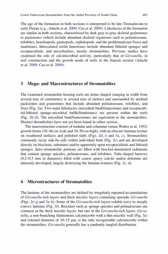
The age of the formation in both sections is interpreted to be late Tremadocian to
early Floian (e.g., Adachi et al. 2009; Cao et al. 2009). Lithofacies of the formation
are similar in both sections, characterised by dark gray to gray skeletal grainstones
to packstones (which include abundant skeletal organisms such as pelmatozoans,
trilobites, brachiopods, gastropods, cephalopods, and the problematicum Nuia) andmudstones. Intercalated reefal limestones include abundant lithistid sponges and
receptaculitids, and microbialites, mostly stromatolites. Previous studies have
examined the role of calcimicrobial activity, particularly that of Girvanella, inreef construction and the growth mode of reefs in the Jianxin section (Adachi
et al. 2009; Cao et al. 2009).
3 Mega- and Macrostructures of Stromatolites
The examined stromatolite-bearing reefs are dome-shaped (ranging in width from
several tens of centimetres to several tens of metres) and surrounded by skeletal
packstones and grainstones that include abundant pelmatozoans, trilobites, and
Nuia (Fig. 2a). Two main lithofacies (microbial bind/framestones and receptaculi-
tid-lithistid sponge-microbial baffle/bindstones) are present within the reefs
(Fig. 2b–d). The microbial bind/framestones are equivalent to the stromatolites.
Distinct thrombolites have not yet been found in either section.
The macrostructure consists of nodular and columnar (sensu Walter et al. 1992)
growth forms (10–40 cm wide and 10–50 cm high), with an obscure laminar texture
on weathered surfaces and polished slabs (Figs. 2d, e and 3a, c). Stromatolites
commonly occur side-by-side within individual beds (Fig. 2c) and are developed
directly on bioclastic substrates and/or apparently upon receptaculitids and lithistid
sponges. Inter-stromatolite portions are filled with bioclast-dominated sediments
that contain sponge spicules, pelmatozoans, and trilobites. Tube-shaped burrows
(0.2–0.5 mm in diameter) filled with coarse sparry calcite and/or dolomite are
intensely developed, largely destroying the laminar textures (Fig. 3c, d).
4 Microstructures of Stromatolites
The laminae of the stromatolites are defined by irregularly repeated accumulations
of Girvanella-rich layers and thick micritic layers containing sporadic Girvanella(Figs. 2e–g and 3a–b). Some of the Girvanella-rich layers exhibit wavy to steeply
convex laminae (Fig. 3f). Bioclasts such as sponge spicules and pelmatozoans are
common in the thick micritic layers, but rare in the Girvanella-rich layers. Girva-nella, a non-branching filamentous calcimicrobe with a thin micritic wall (Fig. 3e)
and external diameter of 10–15 mm, is the only recognisable calcimicrobe within
the stromatolites. Girvanella generally has a randomly tangled distribution.
Lower Ordovician Stromatolites from the Anhui Province of South China 465
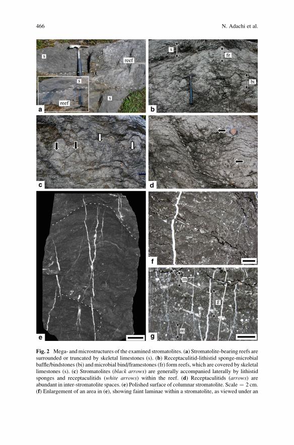
Fig. 2 Mega- andmicrostructures of the examined stromatolites. (a) Stromatolite-bearing reefs are
surrounded or truncated by skeletal limestones (s). (b) Receptaculitid-lithistid sponge-microbial
baffle/bindstones (bi) andmicrobial bind/framestones (fr) form reefs, which are covered by skeletal
limestones (s). (c) Stromatolites (black arrows) are generally accompanied laterally by lithistid
sponges and receptaculitids (white arrows) within the reef. (d) Receptaculitids (arrows) are
abundant in inter-stromatolite spaces. (e) Polished surface of columnar stromatolite. Scale ¼ 2 cm.
(f) Enlargement of an area in (e), showing faint laminae within a stromatolite, as viewed under an
466 N. Adachi et al.
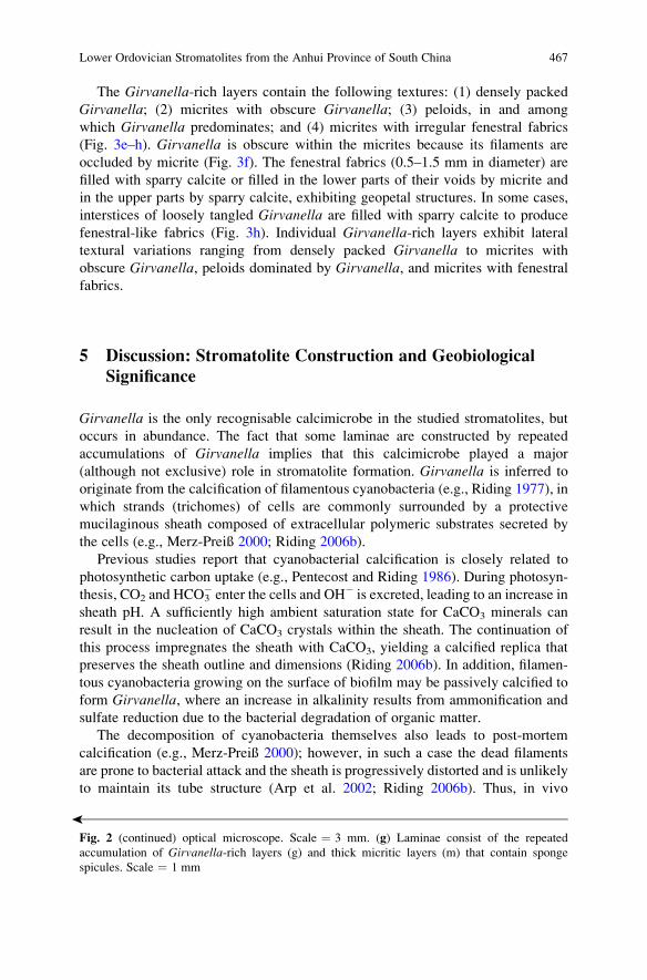
The Girvanella-rich layers contain the following textures: (1) densely packed
Girvanella; (2) micrites with obscure Girvanella; (3) peloids, in and among
which Girvanella predominates; and (4) micrites with irregular fenestral fabrics
(Fig. 3e–h). Girvanella is obscure within the micrites because its filaments are
occluded by micrite (Fig. 3f). The fenestral fabrics (0.5–1.5 mm in diameter) are
filled with sparry calcite or filled in the lower parts of their voids by micrite and
in the upper parts by sparry calcite, exhibiting geopetal structures. In some cases,
interstices of loosely tangled Girvanella are filled with sparry calcite to produce
fenestral-like fabrics (Fig. 3h). Individual Girvanella-rich layers exhibit lateral
textural variations ranging from densely packed Girvanella to micrites with
obscure Girvanella, peloids dominated by Girvanella, and micrites with fenestral
fabrics.
5 Discussion: Stromatolite Construction and Geobiological
Significance
Girvanella is the only recognisable calcimicrobe in the studied stromatolites, but
occurs in abundance. The fact that some laminae are constructed by repeated
accumulations of Girvanella implies that this calcimicrobe played a major
(although not exclusive) role in stromatolite formation. Girvanella is inferred to
originate from the calcification of filamentous cyanobacteria (e.g., Riding 1977), in
which strands (trichomes) of cells are commonly surrounded by a protective
mucilaginous sheath composed of extracellular polymeric substrates secreted by
the cells (e.g., Merz-Preiß 2000; Riding 2006b).
Previous studies report that cyanobacterial calcification is closely related to
photosynthetic carbon uptake (e.g., Pentecost and Riding 1986). During photosyn-
thesis, CO2 and HCO3� enter the cells and OH� is excreted, leading to an increase in
sheath pH. A sufficiently high ambient saturation state for CaCO3 minerals can
result in the nucleation of CaCO3 crystals within the sheath. The continuation of
this process impregnates the sheath with CaCO3, yielding a calcified replica that
preserves the sheath outline and dimensions (Riding 2006b). In addition, filamen-
tous cyanobacteria growing on the surface of biofilm may be passively calcified to
form Girvanella, where an increase in alkalinity results from ammonification and
sulfate reduction due to the bacterial degradation of organic matter.
The decomposition of cyanobacteria themselves also leads to post-mortem
calcification (e.g., Merz-Preiß 2000); however, in such a case the dead filaments
are prone to bacterial attack and the sheath is progressively distorted and is unlikely
to maintain its tube structure (Arp et al. 2002; Riding 2006b). Thus, in vivo
�
Fig. 2 (continued) optical microscope. Scale ¼ 3 mm. (g) Laminae consist of the repeated
accumulation of Girvanella-rich layers (g) and thick micritic layers (m) that contain sponge
spicules. Scale ¼ 1 mm
Lower Ordovician Stromatolites from the Anhui Province of South China 467
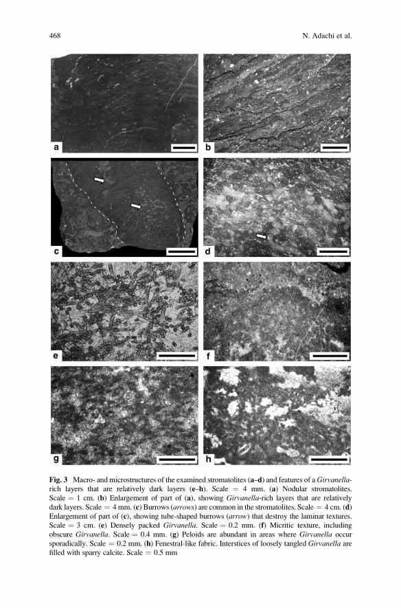
Fig. 3 Macro- and microstructures of the examined stromatolites (a–d) and features of aGirvanella-rich layers that are relatively dark layers (e–h). Scale ¼ 4 mm. (a) Nodular stromatolites.
Scale ¼ 1 cm. (b) Enlargement of part of (a), showing Girvanella-rich layers that are relatively
dark layers. Scale ¼ 4mm. (c) Burrows (arrows) are common in the stromatolites. Scale ¼ 4 cm. (d)
Enlargement of part of (c), showing tube-shaped burrows (arrow) that destroy the laminar textures.
Scale ¼ 3 cm. (e) Densely packed Girvanella. Scale ¼ 0.2 mm. (f) Micritic texture, including
obscure Girvanella. Scale ¼ 0.4 mm. (g) Peloids are abundant in areas where Girvanella occur
sporadically. Scale ¼ 0.2 mm. (h) Fenestral-like fabric. Interstices of loosely tangled Girvanella are
filled with sparry calcite. Scale ¼ 0.5 mm
468 N. Adachi et al.
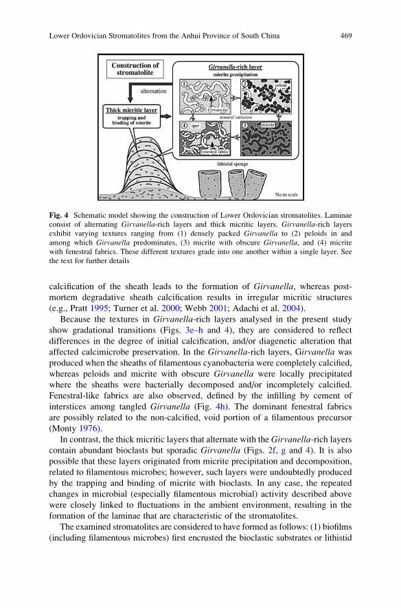
calcification of the sheath leads to the formation of Girvanella, whereas post-
mortem degradative sheath calcification results in irregular micritic structures
(e.g., Pratt 1995; Turner et al. 2000; Webb 2001; Adachi et al. 2004).
Because the textures in Girvanella-rich layers analysed in the present study
show gradational transitions (Figs. 3e–h and 4), they are considered to reflect
differences in the degree of initial calcification, and/or diagenetic alteration that
affected calcimicrobe preservation. In the Girvanella-rich layers, Girvanella was
produced when the sheaths of filamentous cyanobacteria were completely calcified,
whereas peloids and micrite with obscure Girvanella were locally precipitated
where the sheaths were bacterially decomposed and/or incompletely calcified.
Fenestral-like fabrics are also observed, defined by the infilling by cement of
interstices among tangled Girvanella (Fig. 4h). The dominant fenestral fabrics
are possibly related to the non-calcified, void portion of a filamentous precursor
(Monty 1976).
In contrast, the thick micritic layers that alternate with the Girvanella-rich layerscontain abundant bioclasts but sporadic Girvanella (Figs. 2f, g and 4). It is also
possible that these layers originated from micrite precipitation and decomposition,
related to filamentous microbes; however, such layers were undoubtedly produced
by the trapping and binding of micrite with bioclasts. In any case, the repeated
changes in microbial (especially filamentous microbial) activity described above
were closely linked to fluctuations in the ambient environment, resulting in the
formation of the laminae that are characteristic of the stromatolites.
The examined stromatolites are considered to have formed as follows: (1) biofilms
(including filamentous microbes) first encrusted the bioclastic substrates or lithistid
Fig. 4 Schematic model showing the construction of Lower Ordovician stromatolites. Laminae
consist of alternating Girvanella-rich layers and thick micritic layers. Girvanella-rich layers
exhibit varying textures ranging from (1) densely packed Girvanella to (2) peloids in and
among which Girvanella predominates, (3) micrite with obscure Girvanella, and (4) micrite
with fenestral fabrics. These different textures grade into one another within a single layer. See
the text for further details
Lower Ordovician Stromatolites from the Anhui Province of South China 469
sponges and receptaculitids, then (2) stabilised these substrates, and finally (3) grew
upward and laterally, eventually forming laminated microbialites as a result of
repeated changes in microbial activity (micrite precipitation or the trapping and/or
binding ofmicrite). An additional feature unique to Lower Ordovician stromatolites is
their co-occurrence with metazoans (e.g., lithistid sponges and receptaculitids) within
reefs (Fig. 2c, d), although the metazoans were of low diversity before the Ordovician
radiation. Such Lower Ordovician reefs are in marked contrast to subsequent shallow-
marine reefs of the Middle to Late Ordovician, in which skeletal metazoans (e.g.,
stromatoporoids and corals) flourished to form metazoan reefs and stromatolites
showed a marked declined. Even after Ordovician biodiversification, almost all reefal
limestones throughout geological time are evidently the product of combined micro-
bial and metazoan activity (e.g., Adachi et al. 2006, 2007; Adachi and Ezaki 2007);
however, this Lower Ordovician co-occurrence of both stromatolites and metazoans
within reefs provides critical clues regarding (1) reef palaeoecology during the period
immediately prior to a great diversification of reef-building skeletal organisms and (2)
the interrelationship betweenmicrobes andmetazoans in the formation of age-specific
reefs. More importantly, the Lower Ordovician stromatolites provide invaluable clues
to the transition frommicrobe- to metazoan-dominated reefs and the evolution of reef
ecosystems.
6 Conclusions
1. Lower Ordovician stromatolites of the Hunghuayuan Formation in Anhui Prov-
ince, South China, consist of irregularly alternating accumulations of Girva-nella-rich layers and thick micritic layers that contain sporadic bioclasts.
2. The Girvanella-rich layers exhibit the following textures: (1) densely packed
Girvanella; (2) micrite with obscure Girvanella; (3) peloids, in and among
which Girvanella is predominant; and (4) micrite with fenestral fabrics. These
textures resulted from varying degrees of initial calcification and/or diagenetic
alternation of the filamentous precursor.
3. The stromatolites are thought to have been constructed as follows: (1) biofilms
including filamentous microbes became prevalent upon the bioclastic substrates
or lithistid sponges, then (2) stabilised these substrates and (3) grew upward and
laterally to produce laminated microbialites in response to repeated changes in
microbial activity (micrite precipitation or trapping and/or binding of micrite)
and fluctuations in the ambient environment.
Acknowledgements We thank anonymous reviewers and editors for their valuable comments
on the manuscript. This research was supported by a Grant-in-Aid for Japan Society for
the Promotion of Science Fellows and funded by a grant (17540444, 21340154) from the
Scientific Research Fund of the Japan Society for the Promotion of Science, and by the National
Natural Science Foundation of China (40972020), the Ministry of Science and Technology
of China (2006CB806402), and the State Key Laboratory of Palaeobiology and Stratigraphy
(NIGPAS20081103).
470 N. Adachi et al.
References
Adachi N, Ezaki Y (2007) Microbial impacts on the Genesis of Lower Devonian reefal limestones,
Eastern Australia. Palaeoworld 16:301–310
Adachi N, Ezaki Y, Liu J (2004) The fabrics and origins of peloids immediately after the end-
Permian extinction, Guizhou Province, South China. Sedimentary Geology 164:161–178
Adachi N, Ezaki Y, Pickett JW (2006) Marked accumulation patterns characteristic of Lower
Devonian stromatoporoid bindstone: palaeoecological interactions between skeletal organisms
and microbes. Palaeogeography, Palaeoclimatology, Palaeoecology 231:331–346
Adachi N, Ezaki Y, Pickett JW (2007) Interrelations between framework-building and encrusting
skeletal organisms and microbes: more-refined growth history of Lower Devonian bindstones.
Sedimentology 54:89–105
Adachi N, Ezaki Y, Liu J, Cao J (2009) Early Ordovician reef construction in Anhui Province,
South China: a geobiological transition from microbial- to metazoan-dominant reefs. Sedi-
mentary Geology 220:1–11
Arp G, Reimer A, Reitner J (2002) Calcification of cyanobacterial filaments: Girvanella and the
origin of lower Paleozoic lime mud: comment. Geology 30:579–580
Awramik SM (1971) Precambrian columnar stromatolite diversity: reflection of metazoan appear-
ance. Science 174:825–827
Canas F, Carrera M (1993) Early Ordovician Microbial-sponge-receptaculitid bioherms of the
Precordillera, Western Argentina. Facies 29:169–178
Cao J, Liu J, Ezaki Y, Adachi N (2009) Lower Ordovician reefs in the Honghuayuan Formation at
Dongzhi, Anhui: Microbial reefs just prior to the Ordovician biodiversification. Acta Scien-
tiarum Naturalium Universitatis Pekinensis 45:279–288 (in Chinese with English abstract)
Dang H, Liu J, Yuan X (2009) Microbialites in the Middle Cambrian Qinjianmiao Group in
Xingshan, Hubei Province: implication for Palaeoenvironmental reconstruction. Acta Scien-
tiarum Naturalium Universitatis Pekinensis 2009:289–298 (in Chinese with English abstract)
de Freitas T, Mayr U (1995) Kilometre-scale microbial buildups in a rimmed carbonate platform
succession, Arctic Canada: new insight on Lower Ordovician reef facies. Bulletin of Canadian
petroleum Geology 43:407–432
Merz-Preiß M (2000) Calcification in cyanobacteria. In: Riding RE, Awramik SM (eds) Microbial
Sediments. Springer, Berlin, pp 50–56
Monty C (1976) The origin and development of cryptalgal fabrics. In: Walter MR (ed) Stroma-
tolites. Developments in Sedimentology 20. Elsevier, Amsterdam, pp 193–249
Pentecost A, Riding R (1986) Calcification in cyanobacteria. In: Leadbeater BSC, Riding RE (eds)
Biomineralization in Lower Plants and Animals. Systematic Association Special Volume 30.
Clarendon Press, Oxford, pp 73–90
Pratt BR (1995) The origin, biota and evolution of deep-water mud-mounds: their origin and
evolution. In: Monty CLV, Bosence DWJ, Bridges PH, Pratt BR (eds) Carbonate Mud-Mound.
International Association of Sedimentologists, Special Publication 23. Blackwell, Oxford,
pp 49–123
Pratt BR, James NP (1982) Cryptalgal-metazoan bioherms of early Ordovician age in the
St George Group, western Newfoundland. Sedimentology 29:543–569
Riding R (1977) Calcified Plectonema (blue-green algae), a recent example of Girvanella from
Aldabra Atoll. Palaeontology 20:33–46
Riding R (2000) Microbial carbonates: the geological record of calcified bacterial-algal mats and
biofilms. Sedimentology 47:179–214
Riding R (2006a) Microbial carbonate abundance compared with fluctuations in metazoan diver-
sity over geological time. Sedimentary Geology 185:229–238
Riding R (2006b) Cyanobacterial calcification, carbon dioxide concentrating mechanisms, and
Proterozoic-Cambrian changes in atmospheric composition. Geobiology 4:299–316
Riding R, Sharma M (1998) Late Palaeoproterozoic (~1800–1600 Ma) stromatolites, Cuddapah
Basin, southern India: cyanobacterial or other bacterial microfabrics? Precambrian Research
92:21–35
Lower Ordovician Stromatolites from the Anhui Province of South China 471
Shapiro R (2004) Neoproterozoic-Cambrian microbialite record. The Paleontological Society
Papers 10:5–15
Shapiro R, Awramik SM (2000) Microbialite morphostratigraphy as a tool for correlating Late
Cambrian–Early Ordovician sequences. Journal of Geology 108:171–180
Toomey DF, Nitecki MH (1979) Organic buildups in the Lower Ordovician (Canadian) of Texas
and Oklahoma. Fieldiana Geology 2:1–181
Turner EC, James NP, Narbonne G (2000) Taphonomic control on microstructure in Early
Neoproterozoic reefal stromatolites and thrombolites. Palaios 15:87–111
Walter MR, Grotzinger JP, Schopf JW (1992) Proterozoic stromatolites. In: Schopf JW, Klein C
(eds) The Proterozoic Biosphere: A Multidisciplinary Study. Cambridge University Press,
Cambridge, pp 253–260
Webb GE (2001) Famennian mud-mounds in the proximal fore-reef slope, Canning Basin,
Western Australia. Sedimentary Geology 145:295–315
Webby BD (2002) Patterns of Ordovician reef development. In: Kiessling W, Fl€ugel E, Golonka J(eds) Phanerozoic reef patterns. SEPM Special Publication 72:129–179
Zhan R, Jin J (2007) Ordovician–Early Silurian (Llandovery) Stratigraphy and Palaeontology of
the Upper Yangtze Platform, South China, Science Press, Beijing, pp 169
472 N. Adachi et al.
Sedimentology and Palaeoecology
of Ernietta-Bearing Ediacaran Deposits
in Southern Namibia: Implications for Infaunal
Vendobiont Communities
El Hafid Bouougri, Hubertus Porada, Klaus Weber, and Joachim Reitner
1 Introduction
The Ediacaran was a period of wide soft-bodied biota development in marine
siliciclastic environment. The reconstruction of palaeoecology, lifestyle and envi-
ronment settings of these organisms are among the challenges in characterising the
earliest metazoan life on Earth. The biota colonised sea bottoms and were living as
benthic communities occurring in a wide spectrum of environmental settings (e.g.
Gehling 2000; Waggoner 2003; Narbonne 2005; Grazhdankin 2004; Droser et al.
2006). In the classical localities (e.g. Newfoundland, Namibia, White Sea and
Australia), the fossils occur in rock successions with distinct depositional settings
ranging from distributary mouth-bar of braid-delta systems to deep-water slope
(Gehling 2000; Grazhdankin 2004; Narbonne 2005).
However, the preservation style of this biota in the sedimentary record as well as
their lifestyle and palaeoecology are still unclear for some assemblages and genera
of which data are mainly coming from isolated float samples. According to various
features including among others taphonomy, ecology, functional morphology and
style of preservation within the rock record, two main benthic faunas with con-
trasting lifestyles are distinguished: (1) epifaunal disc-shaped to sessile leaf-shaped
communities, living at the water-sediment interface, generally buried as in situ
biota in their habitat and preserved as epireliefs. Such types of palaeocommunities
are well recognised in the Avalon-type assemblage where deep-water frondose
biota are smothered in their habitat and preserved in turbiditic successions as
E.H. Bouougri
Department of Geology, Faculty of Sciences Semlalia, Cadi-Ayyad University, 40020 Marrakech,
Morocco
H. Porada, K. Weber, and J. Reitner (*)
Geowissenschaftliches Zentrum G€ottingen, Universit€at G€ottingen, Goldschmidtstrasse 3, 37077
G€ottingen, Germany
e-mail: [email protected]
J. Reitner et al., Advances in Stromatolite Geobiology,Lecture Notes in Earth Sciences 131, DOI 10.1007/978-3-642-10415-2_29,# Springer-Verlag Berlin Heidelberg 2011
473
casts on bedding surfaces (Narbonne 2005; Hofmann et al. 2008); (2) infaunal bag-
shaped to canoe-shaped communities commonly preserved as three-dimensional
moulds and reported in fossil-bearing rocks as reworked entities or in life position.
Exclusively mouldic preservation was suggested for quilted organisms of Nama-
type fossils like Rangea, Namalia, Pteridinium and Ernietta interpreted as epifau-
nal or infaunal biota (Jenkins et al. 1981; Jenkins 1992; Buss and Seilacher 1994;
Dzik 1999; Grazhdankin and Seilacher 2002, 2005).
Although the infaunal or epifaunal lifestyle of some shallow-marine genera is
still controversial (e.g. Laflamme and Narbonne 2008), it is necessary to note that
various factors have been suggested to explain the diversity and distribution of the
Ediacara assemblages, including biogeography, biostratigraphy, ecology and taph-
onomic processes (Jenkins 1992; Buss and Seilacher 1994; Gehling 1999; Waggoner
2003; Grazhdankin 2004; Narbonne 2005; Droser et al. 2006; Mapstone and
McIlroy 2006). Through comparison of sedimentary features of Ediacara-bearing
siliciclastic rocks and the relationship between lithofacies and biogeography,
Grazhdankin (2004) documented a striking correlation between fossil association
and depositional setting and distinguished three “palaeoecologic assemblages”,
namely, Nama, Avalon and Ediacara. These assemblages were as well recognized
previously by using cluster analysis of taxonomic composition (Waggoner 2003).
The three-dimensionally preserved moulds seem to be the main feature of the
Nama-type assemblage and have been assigned as a type of biofacies within a
“braid-delta” environment in association with sandy deposits of high energy setting
(Grazhdankin 2004). The mouldic fossils preserved as endoreliefs and including
Ernietta, Namalia, Pteridinium and Rangea were reported from sandy event depos-
its as lags, being reworked, current transported and redeposited (Jenkins 1985,
1992; Grazhdankin and Seilacher 2002, 2005; Grazhdankin 2004). By contrast,
the shallow marine fossils of White Sea and Ediacara are preserved almost all in
their habitat as hyporeliefs and occur within interbedded storm sandstone and
mudstone of delta-front and pro-delta settings (Gehling 1999; Grazhdankin 2004).
Despite substantial progress and a large amount of studies on the structure and
morphology of the three-dimensional moldic fossils (e.g. Jenkins 1992; Dzik 1999;
Grazhdankin and Seilacher 2002, 2005), less is known on the major question
dealing with the sedimentary setting and the precise biotope of the quilted organ-
isms like Ernietta. The best direct evidence in constraining the ecological niches ofthis community is the preservation in life-position of body fossils in the sedimen-
tary rock. Most of the specimens in previous studies (e.g. Jenkins et al. 1981; Dzik
1999), however, were winnowed and float specimens of which the precise style of
preservation, biostratinomy and sedimentary setting is not well constrained. In this
paper, we report newly found occurrences of Ernietta fossils from the lowermost
unit of the Dabis Formation (Kanies Member) in the Nama Group. These new
findings extend the stratigraphic range of Ediacara fossils in Namibia to the
lowermost member of the Nama Group and provide the opportunity: (1) to con-
strain the sedimentary setting, preservation styles and the palaeoecology of Erniettacommunity; (2) to test the relationship between preserved three-dimensional fos-
sils, fossil-bearing lithofacies and facies assemblage.
474 E.H. Bouougri et al.
Ernietta is a ribbed, sack-shaped Ediacara fossil, 3–8 cm in width, mainly
preserved as three-dimensional bodies. Pflug (1966) who investigated numerous
specimens from Lower Nama Group sandstone and schist (Namibia), defined it as a
new genus and included it in the group of Petalonamae, together with Rangea and
Pteridinium. Later, Ernietta, represented by a single species Ernietta plateauensis,was attributed to the group of Vendobionta (Seilacher 1992). Up to now, Ernietta isonly known from the Ediacaran of Namibia, where it occurs in great number over a
wide area (Pflug 1966; Germs 1972), and from terminal Proterozoic siliciclastic
deposits in Death Valley (southern Nevada, USA), where few specimens have been
recovered (Horodyski 1991; Horodyski et al. 1994; Waggoner and Hagadorn 1997;
Hagadorn et al. 2000; Corsetti and Hagadorn 2000).
In both regions Ernietta occurs in similar geological context. In Death Valley
(see Corsetti and Hagadorn 2000), the fossils are present in the lower member of the
Wood Canyon Formation which overlies, locally disconformably, the Stirling
Quartzite and which consists of three parasequences, each beginning with shallow
marine siliciclastic rocks that are gradationally overlain and capped by carbonate
rocks. The succession is considered to record a transgressive to highstand systems
tract in a shallow marine-continental braidplain transition (Hagadorn and Wagg-
oner 2000), with Ernietta occurring in the lowest few metres of the first parase-
quence. Based on chemostratigraphic data and regional correlations, Corsetti and
Hagadorn (2000) suggested a depositional age of 548–544 Ma for the upper
Sterling Quartzite and the lower member of the Wood Canyon Formation.
In Namibia, Ernietta has been encountered in the Kuibis Subgroup at the base ofthe Nama Group in the southern Nama basin (Fig. 1a). This subgroup consists of
two depositional sequences, each with fluvial to marginal marine sandstone over-
lain by subtidal carbonate (Saylor et al. 1995). The two sequences, deposited
between about 555 and 550 Ma based on radiometric data (Grotzinger et al.
1995), are considered each to record low-stand to transgressive systems tracts
(Saylor et al. 1995).
The fossils originally described by Pflug (1966) and referred to in the subsequent
literature (see review by McCall 2006), have been recovered as isolated float
samples from the Kliphoek Member of the upper sequence, just below the capping
carbonate unit (Mooifontein Member).
Pflug (1966) observed Ernietta in steinkern preservation within a succession of
shale (mudstone) with sandy to silty admixtures, in the upper part of the Kliphoek
Member. He reported that in most cases (n ¼ ca. 40) the hemispheric bodies rest
on bedding surfaces with the sculptured surface oriented upward, and suggested
that the organisms had a benthonic-sessile lifestyle. In contrast, Jenkins (1992,
p. 148, fig. 7) who studied a specific sample (GU Pf 536) from the collection of
H. D. Pflug (Giessen) containing numerous three-dimensional individuals, descri-
bed Ernietta as occurring in medium-grained, impure sandstone in which they
may form groups or clusters with the sculptured, convex parts directed downward.
He also considered them in life position and suggested that they “were probably
buried by density currents generated as a result of wave movement and rips at
times of storms”. Dzik (1999, p. 519), partly referring to the same sample,
Sedimentology and Palaeoecology of Ernietta-Bearing Ediacaran Deposits 475
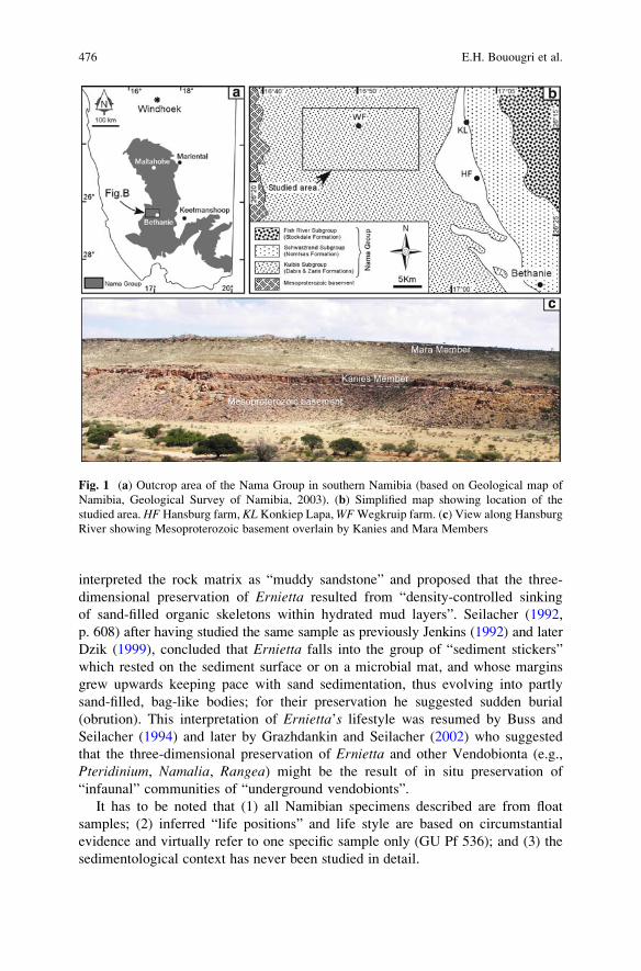
interpreted the rock matrix as “muddy sandstone” and proposed that the three-
dimensional preservation of Ernietta resulted from “density-controlled sinking
of sand-filled organic skeletons within hydrated mud layers”. Seilacher (1992,
p. 608) after having studied the same sample as previously Jenkins (1992) and later
Dzik (1999), concluded that Ernietta falls into the group of “sediment stickers”
which rested on the sediment surface or on a microbial mat, and whose margins
grew upwards keeping pace with sand sedimentation, thus evolving into partly
sand-filled, bag-like bodies; for their preservation he suggested sudden burial
(obrution). This interpretation of Ernietta’s lifestyle was resumed by Buss and
Seilacher (1994) and later by Grazhdankin and Seilacher (2002) who suggested
that the three-dimensional preservation of Ernietta and other Vendobionta (e.g.,
Pteridinium, Namalia, Rangea) might be the result of in situ preservation of
“infaunal” communities of “underground vendobionts”.
It has to be noted that (1) all Namibian specimens described are from float
samples; (2) inferred “life positions” and life style are based on circumstantial
evidence and virtually refer to one specific sample only (GU Pf 536); and (3) the
sedimentological context has never been studied in detail.
Fig. 1 (a) Outcrop area of the Nama Group in southern Namibia (based on Geological map of
Namibia, Geological Survey of Namibia, 2003). (b) Simplified map showing location of the
studied area. HF Hansburg farm, KL Konkiep Lapa,WFWegkruip farm. (c) View along Hansburg
River showing Mesoproterozoic basement overlain by Kanies and Mara Members
476 E.H. Bouougri et al.
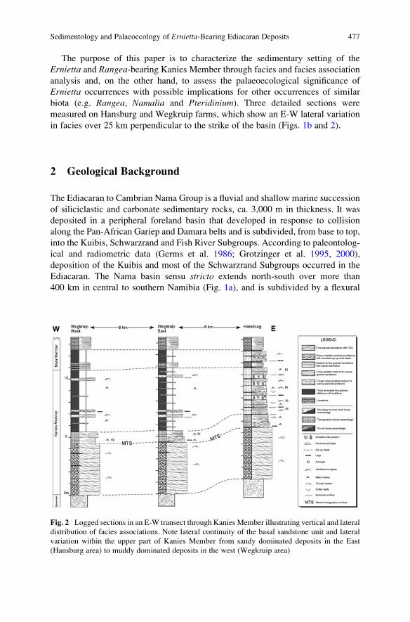
The purpose of this paper is to characterize the sedimentary setting of the
Ernietta and Rangea-bearing Kanies Member through facies and facies association
analysis and, on the other hand, to assess the palaeoecological significance of
Ernietta occurrences with possible implications for other occurrences of similar
biota (e.g. Rangea, Namalia and Pteridinium). Three detailed sections were
measured on Hansburg and Wegkruip farms, which show an E-W lateral variation
in facies over 25 km perpendicular to the strike of the basin (Figs. 1b and 2).
2 Geological Background
The Ediacaran to Cambrian Nama Group is a fluvial and shallow marine succession
of siliciclastic and carbonate sedimentary rocks, ca. 3,000 m in thickness. It was
deposited in a peripheral foreland basin that developed in response to collision
along the Pan-African Gariep and Damara belts and is subdivided, from base to top,
into the Kuibis, Schwarzrand and Fish River Subgroups. According to paleontolog-
ical and radiometric data (Germs et al. 1986; Grotzinger et al. 1995, 2000),
deposition of the Kuibis and most of the Schwarzrand Subgroups occurred in the
Ediacaran. The Nama basin sensu stricto extends north-south over more than
400 km in central to southern Namibia (Fig. 1a), and is subdivided by a flexural
Fig. 2 Logged sections in an E-W transect through Kanies Member illustrating vertical and lateral
distribution of facies associations. Note lateral continuity of the basal sandstone unit and lateral
variation within the upper part of Kanies Member from sandy dominated deposits in the East
(Hansburg area) to muddy dominated deposits in the west (Wegkruip area)
Sedimentology and Palaeoecology of Ernietta-Bearing Ediacaran Deposits 477
high (Osis ridge) into the northern Zaris and southern Witputs sub-basins (Germs
1983; Germs and Gresse 1991).
The Nama Group (Fig. 1a) is well known for its typical Ediacara fossils
(~565–542 Ma), characterized by low taxonomic richness and unusual three-
dimensional preservation. The fossils are commonly described in association with
storm deposits and are preserved as moulds and casts at the bases, within and at the
tops of sandstone beds (e.g. Jenkins 1985; Narbonne et al. 1997; Grazhdankin and
Seilacher 2002). Most of the fossils were described from the Kliphoek Member of
the Kuibis Subgroup (e.g., Pflug 1966; Germs 1972) and from the overlying
Schwarzrand Subgroup with the youngest occurrence of Swartpuntia near the top
of the Spitskopf Member (Narbonne et al. 1997). Occurrences of Ernietta as
previously described and reported here, are all from the Witputs sub-basin in the
south. The new locality of Ernietta described here, is situated on Hansburg farm
(Fig. 1b), about 55 km to the northeast of the localities reported by Pflug (1966). At
Hansburg farm, up to three Ernietta carrying horizons occur in a thin interval of
cross-bedded and planar laminated sandstone, ca. 7–9 m above the base of the
Nama Group and less than 1 m below the first carbonate bed. A fourth horizon is
locally developed in a sandy interval situated less than 1 m above the first carbonate
bed. The locality is thus stratigraphically situated mainly in the Kanies Member of
the Dabis Formation (Fig. 1c), in contrast to the previously described occurrences
which are in the overlying Kliphoek Member of the Zaris Formation (Pflug 1966;
Germs 1995; Saylor et al. 1995).
During deposition of Kuibis and Schwarzrand strata, the Witputs sub-basin was
open to the sea towards the west and received continental, terrigenous fluvial input
from the east and north-east (Germs 1983; Saylor et al. 1998; Saylor 2003).
Radiometrically, the age of the Kuibis Subgroup is not constrained. However, an
ash layer interbedded in the overlying Schwarzrand Subgroup yielded an age of
548.1 � 1 Ma (Grotzinger et al. 1995). On the other hand, it is considered that
deposition of the Nama Group postdates the late major Ediacaran glaciation in
southwestern Africa (Germs 1995). According to a south-north correlative scheme,
the Kuibis Subgroup succession shows considerable thinning toward the Osis ridge,
whereas in the southernmost part of the Witputs sub-basin, the succession thickens
westward toward the Gariep belt (Germs 1983; Saylor et al. 1995). In this latter
area, the lithostratigraphic units of the Kuibis Subgroup therefore form two deposi-
tional sequences K1 and K2, each displaying a basal siliciclastic unit (Kanies and
Kliphoek Member, respectively) forming lowstand systems tracts and upper car-
bonate units (Mara and Mooifontein Member, respectively) forming transgressive
systems tracts (Saylor et al. 1995, 1998). The siliciclastic members comprise fluvial
to upper shoreface, delta or tide influenced deposits, whereas the carbonate mem-
bers are made up of shallow, intertidal to subtidal deposits of an extensive carbonate
platform. The transition from siliciclastic to carbonate members consists of mixed
deposits which record a marine transgression and a shallow subtidal to intertidal
setting (Saylor et al. 1998).
In the studied area, the Kanies Member forms the lowermost unit of the Nama
Group and passes up gradationally into the carbonatic Mara Member (Fig. 1c). It
478 E.H. Bouougri et al.
consists of undeformed and flat-lying strata, up to ca. 12 m thick, exhibiting an
apparent fining-upward trend and lies above the crystalline basement which
belongs to the Kalahari craton. Further to the South, the Kanies Member attains
a thickness of up to ca. 95 m in the depocenter near the Gariep belt (Saylor et al.
1995).
The Kanies Member in the eastern area (Hansburg farm) consists of cross-
bedded sandstone at the base that is overlain by fossiliferous fine-grained sandstone
interbedded with hummocky cross-stratified sandstone and mudstone. In the west-
ern area (Wegkruip farm), the upper part is mud-dominated with subordinate sandy
and silty layers with HCS and scattered Ernietta bodies.
3 Sedimentology of Kanies Member
The Kanies Member can be traced in outcrop for several tens of kilometres along
river-wall exposures and road cuts, perpendicular to the depositional strike. On
Hansburg and Wegkruip farms, three vertical sections with thicknesses of up to
12 m were investigated including detailed facies analysis and control on vertical
and lateral thickness and facies changes (Fig. 2). Lithologically, the Kanies Mem-
ber consists of two distinct parts: (1) a lower part with thick-bedded medium to
coarse-grained sandstone which forms a lateral persistent unit, (2) an upper part
with thin-bedded sandstone and subordinate mudstone, that passes westward into a
mud-dominated succession with subordinate thin beds of sandstone (Fig. 2). Four
broad facies associations are recognised (named FA 1 to FA 4), arranged in a fining-
upward succession: braided fluvial deposits, transgressive deposits, storm-influenced
shoreface/subtidal lagoon deposits and inner-shelf/transitional offshore deposits.
The overall stacked facies associations record a transgressive setting, whereas
sedimentary characteristics indicate that the Ernietta-bearing lithofacies forms
distinct sedimentary layers interbedded within the storm-influenced marine/lagoon
deposits.
3.1 Facies Association 1 (FA 1): Braided-Fluvial Deposits
Facies association FA 1 dominates the lower part of Kanies Member and is
characterized by thick-bedded packages of quartzose sandstone that extend over
the full E-W length of the studied area without significant variation in thickness or
lithology. It consists of tabular units of vertically stacked, well to moderately
sorted, medium- to coarse-grained cross-bedded sandstones, interbedded with sub-
ordinate thin and lens-shaped planar laminated sandstone and mudstone layers up to
30 cm thick (Figs. 2 and 3). Sandstone beds, up to 1–5 m thick, are typically tabular
with bounding surfaces dipping gently in the downflow direction. Sedimentary
structures in the association include medium to large-scale cross-stratification
Sedimentology and Palaeoecology of Ernietta-Bearing Ediacaran Deposits 479
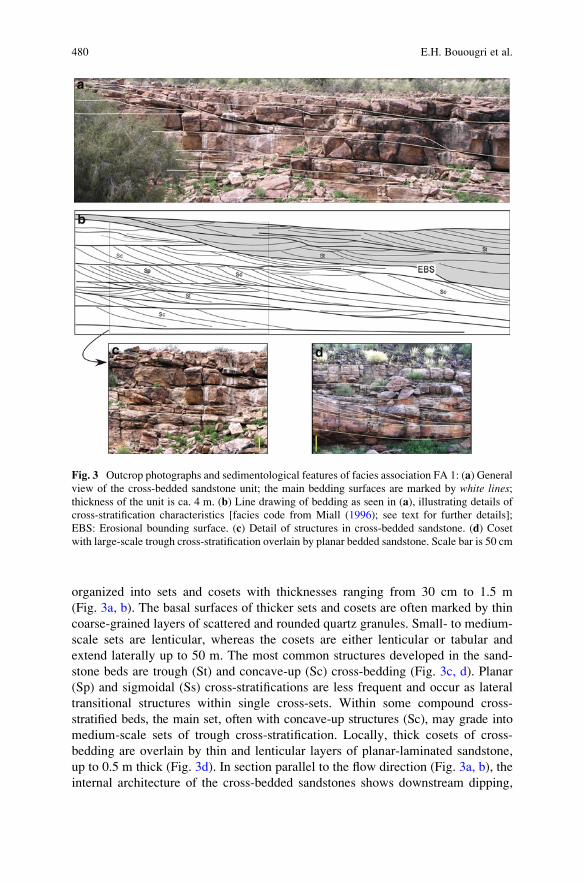
organized into sets and cosets with thicknesses ranging from 30 cm to 1.5 m
(Fig. 3a, b). The basal surfaces of thicker sets and cosets are often marked by thin
coarse-grained layers of scattered and rounded quartz granules. Small- to medium-
scale sets are lenticular, whereas the cosets are either lenticular or tabular and
extend laterally up to 50 m. The most common structures developed in the sand-
stone beds are trough (St) and concave-up (Sc) cross-bedding (Fig. 3c, d). Planar
(Sp) and sigmoidal (Ss) cross-stratifications are less frequent and occur as lateral
transitional structures within single cross-sets. Within some compound cross-
stratified beds, the main set, often with concave-up structures (Sc), may grade into
medium-scale sets of trough cross-stratification. Locally, thick cosets of cross-
bedding are overlain by thin and lenticular layers of planar-laminated sandstone,
up to 0.5 m thick (Fig. 3d). In section parallel to the flow direction (Fig. 3a, b), the
internal architecture of the cross-bedded sandstones shows downstream dipping,
Fig. 3 Outcrop photographs and sedimentological features of facies association FA 1: (a) General
view of the cross-bedded sandstone unit; the main bedding surfaces are marked by white lines;thickness of the unit is ca. 4 m. (b) Line drawing of bedding as seen in (a), illustrating details of
cross-stratification characteristics [facies code from Miall (1996); see text for further details];
EBS: Erosional bounding surface. (c) Detail of structures in cross-bedded sandstone. (d) Coset
with large-scale trough cross-stratification overlain by planar bedded sandstone. Scale bar is 50 cm
480 E.H. Bouougri et al.
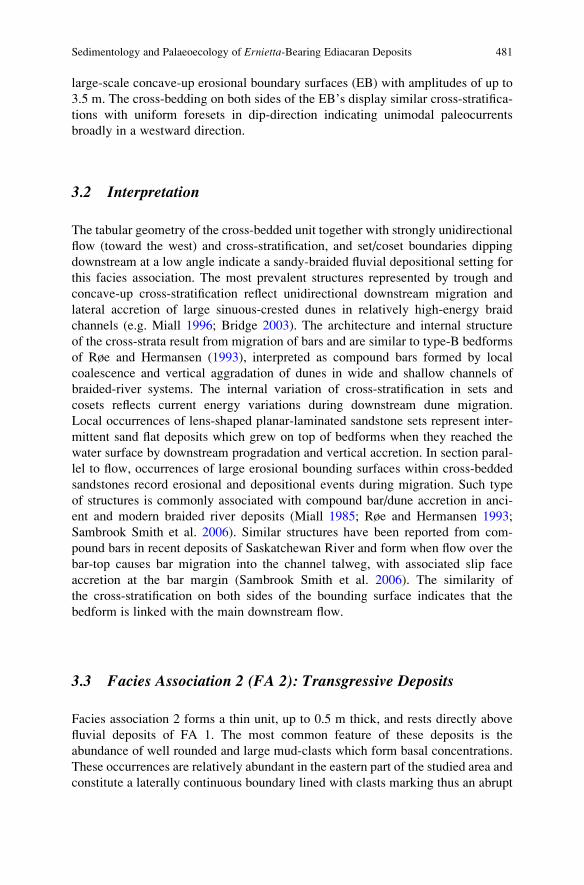
large-scale concave-up erosional boundary surfaces (EB) with amplitudes of up to
3.5 m. The cross-bedding on both sides of the EB’s display similar cross-stratifica-
tions with uniform foresets in dip-direction indicating unimodal paleocurrents
broadly in a westward direction.
3.2 Interpretation
The tabular geometry of the cross-bedded unit together with strongly unidirectional
flow (toward the west) and cross-stratification, and set/coset boundaries dipping
downstream at a low angle indicate a sandy-braided fluvial depositional setting for
this facies association. The most prevalent structures represented by trough and
concave-up cross-stratification reflect unidirectional downstream migration and
lateral accretion of large sinuous-crested dunes in relatively high-energy braid
channels (e.g. Miall 1996; Bridge 2003). The architecture and internal structure
of the cross-strata result from migration of bars and are similar to type-B bedforms
of Røe and Hermansen (1993), interpreted as compound bars formed by local
coalescence and vertical aggradation of dunes in wide and shallow channels of
braided-river systems. The internal variation of cross-stratification in sets and
cosets reflects current energy variations during downstream dune migration.
Local occurrences of lens-shaped planar-laminated sandstone sets represent inter-
mittent sand flat deposits which grew on top of bedforms when they reached the
water surface by downstream progradation and vertical accretion. In section paral-
lel to flow, occurrences of large erosional bounding surfaces within cross-bedded
sandstones record erosional and depositional events during migration. Such type
of structures is commonly associated with compound bar/dune accretion in anci-
ent and modern braided river deposits (Miall 1985; Røe and Hermansen 1993;
Sambrook Smith et al. 2006). Similar structures have been reported from com-
pound bars in recent deposits of Saskatchewan River and form when flow over the
bar-top causes bar migration into the channel talweg, with associated slip face
accretion at the bar margin (Sambrook Smith et al. 2006). The similarity of
the cross-stratification on both sides of the bounding surface indicates that the
bedform is linked with the main downstream flow.
3.3 Facies Association 2 (FA 2): Transgressive Deposits
Facies association 2 forms a thin unit, up to 0.5 m thick, and rests directly above
fluvial deposits of FA 1. The most common feature of these deposits is the
abundance of well rounded and large mud-clasts which form basal concentrations.
These occurrences are relatively abundant in the eastern part of the studied area and
constitute a laterally continuous boundary lined with clasts marking thus an abrupt
Sedimentology and Palaeoecology of Ernietta-Bearing Ediacaran Deposits 481
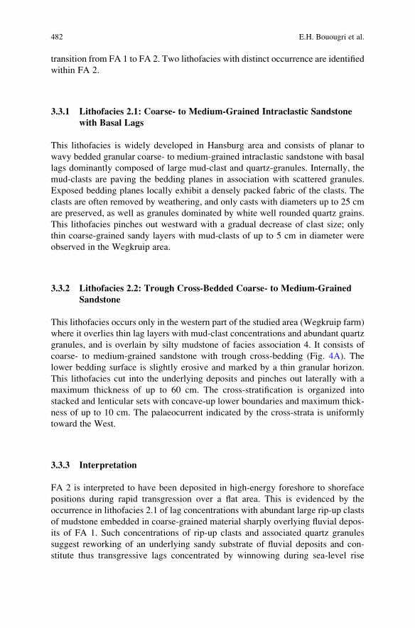
transition from FA 1 to FA 2. Two lithofacies with distinct occurrence are identified
within FA 2.
3.3.1 Lithofacies 2.1: Coarse- to Medium-Grained Intraclastic Sandstone
with Basal Lags
This lithofacies is widely developed in Hansburg area and consists of planar to
wavy bedded granular coarse- to medium-grained intraclastic sandstone with basal
lags dominantly composed of large mud-clast and quartz-granules. Internally, the
mud-clasts are paving the bedding planes in association with scattered granules.
Exposed bedding planes locally exhibit a densely packed fabric of the clasts. The
clasts are often removed by weathering, and only casts with diameters up to 25 cm
are preserved, as well as granules dominated by white well rounded quartz grains.
This lithofacies pinches out westward with a gradual decrease of clast size; only
thin coarse-grained sandy layers with mud-clasts of up to 5 cm in diameter were
observed in the Wegkruip area.
3.3.2 Lithofacies 2.2: Trough Cross-Bedded Coarse- to Medium-Grained
Sandstone
This lithofacies occurs only in the western part of the studied area (Wegkruip farm)
where it overlies thin lag layers with mud-clast concentrations and abundant quartz
granules, and is overlain by silty mudstone of facies association 4. It consists of
coarse- to medium-grained sandstone with trough cross-bedding (Fig. 4A). The
lower bedding surface is slightly erosive and marked by a thin granular horizon.
This lithofacies cut into the underlying deposits and pinches out laterally with a
maximum thickness of up to 60 cm. The cross-stratification is organized into
stacked and lenticular sets with concave-up lower boundaries and maximum thick-
ness of up to 10 cm. The palaeocurrent indicated by the cross-strata is uniformly
toward the West.
3.3.3 Interpretation
FA 2 is interpreted to have been deposited in high-energy foreshore to shoreface
positions during rapid transgression over a flat area. This is evidenced by the
occurrence in lithofacies 2.1 of lag concentrations with abundant large rip-up clasts
of mudstone embedded in coarse-grained material sharply overlying fluvial depos-
its of FA 1. Such concentrations of rip-up clasts and associated quartz granules
suggest reworking of an underlying sandy substrate of fluvial deposits and con-
stitute thus transgressive lags concentrated by winnowing during sea-level rise
482 E.H. Bouougri et al.
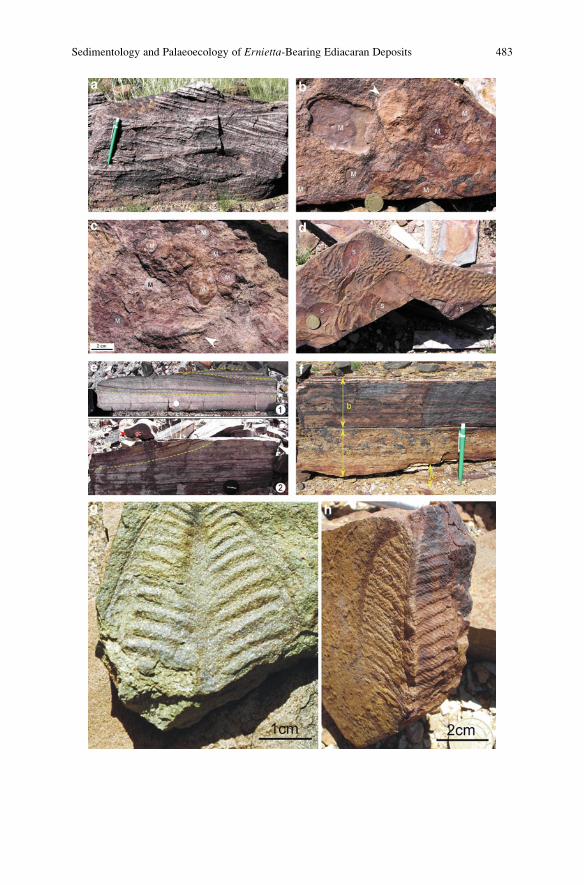
Sedimentology and Palaeoecology of Ernietta-Bearing Ediacaran Deposits 483
(e.g. Kidwell 1991; Kidwell and Brenchley 1994; Zecchin 2007). The coarse grain
size and the abundance of large-sized mud-clasts together with planar and wavy
bedding indicate high-energy wave and storm flows acting during the transgression.
Lithofacies 2.2 with trough cross-stratification results from progradation under
high-energy unidirectional currents of sinuous-crested sandbodies within rip-up
channels, likely produced by retreat of storm and related high energy bottom return
flow in a foreshore or shoreface setting (e.g. Einsele and Seilacher 1982; Walker
and Plint 1992). This is evidenced by the erosional base with granular lag and by
the pinch-and-swell geometry, all together indicating a scour-and-fill regime and a
rip-current channel fill. The westward directed palaeocurrents indicate basinward
migration, under the action of rip-up currents, of medium-scale dunes forming
thus mouth-bars of rip-up channels.
3.4 Facies Association 3 (FA 3): Storm and Wave-DominatedShoreface Deposits
This facies association is restricted to the eastern part of the studied area (e.g.
Hansburg area) and consists of medium to fine-grained sandstones which pinch out
westward, and subordinate fine-grained interbeds of silty mudstone (Fig. 2). The
interbeds, up to 10 cm thick, form thin discontinuous drapes that are often partially
eroded by the next overlying sandstone bed. The most distinctive feature of this
association is the occurrence of Ediacara fossils dominated by Ernietta commu-
nities, whereas other vendobionts like Rangea and Pteridinium constitute subordi-
nate fossils. Within this facies association, two main lithofacies are distinguished:
reddish to greyish, medium to fine-grained sandstones (lithofacies 3.1) and light,
fossiliferous fine-grained sandstone layers with Ernietta (lithofacies 3.2). Both
lithofacies of this association form generally lens-shaped layers interfingering
laterally.
Fig. 4 Outcrop photographs and characteristics of facies associations FA 2 and 3: (A) Trough
cross-stratification within channelised sandstone of facies association FA 2, Wegkruip-West
section; scale (pen) is 14 cm. (B) Lower bedding surface of event bed in FA 3 showing lag of
mud-clasts (M) and Ernietta bioclast (arrow); scale (coin) is 22 mm. (C) Lower bedding surface of
storm bed showing lag with abundant mud-clasts (M) associated with winnowed Rangea specimen
(arrow). (D) Upper bedding surface of a storm bed showing association of Kinneyia-type wrinklesand swales (s); scale (coin) is 22 mm. (E) Internal structures within single event beds showing
hummocky cross-stratification (HCS) in (E1) and association of HCS and swaley cross-stratifica-
tion (SCS) in (E2). Note rock fragment with gutter cast (gc) in upper left part of E2; scale (coin) is22 mm for E1 and (lenscap) 4.5 cm for E2. (F) Bipartite sandstone bed showing lower division
(a) with intraclastic sandstone and upper division (b) with large wavelength hummocky cross-
stratified set. Note scoured lower boundary and partially preserved interbeds of fine-grained
siltstone and mudstone (c); scale (pen) is 14 cm. (G, H) Outcrop photographs of Rangeaschneiderhoeni. Rangea occurs rarely within greenish-rosty coloured laminar bedded fine grained
non-mature sometimes wave rippled fine sand to siltstones in the vicinity of the Ernietta occur-
rences. Right specimen (H) was embedded in upright position
484 E.H. Bouougri et al.
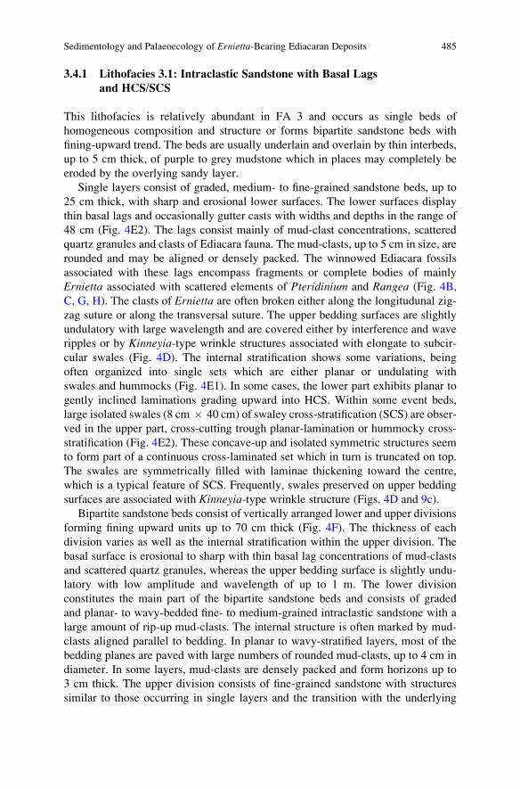
3.4.1 Lithofacies 3.1: Intraclastic Sandstone with Basal Lags
and HCS/SCS
This lithofacies is relatively abundant in FA 3 and occurs as single beds of
homogeneous composition and structure or forms bipartite sandstone beds with
fining-upward trend. The beds are usually underlain and overlain by thin interbeds,
up to 5 cm thick, of purple to grey mudstone which in places may completely be
eroded by the overlying sandy layer.
Single layers consist of graded, medium- to fine-grained sandstone beds, up to
25 cm thick, with sharp and erosional lower surfaces. The lower surfaces display
thin basal lags and occasionally gutter casts with widths and depths in the range of
48 cm (Fig. 4E2). The lags consist mainly of mud-clast concentrations, scattered
quartz granules and clasts of Ediacara fauna. The mud-clasts, up to 5 cm in size, are
rounded and may be aligned or densely packed. The winnowed Ediacara fossils
associated with these lags encompass fragments or complete bodies of mainly
Ernietta associated with scattered elements of Pteridinium and Rangea (Fig. 4B,
C, G, H). The clasts of Ernietta are often broken either along the longitudunal zig-
zag suture or along the transversal suture. The upper bedding surfaces are slightly
undulatory with large wavelength and are covered either by interference and wave
ripples or by Kinneyia-type wrinkle structures associated with elongate to subcir-
cular swales (Fig. 4D). The internal stratification shows some variations, being
often organized into single sets which are either planar or undulating with
swales and hummocks (Fig. 4E1). In some cases, the lower part exhibits planar to
gently inclined laminations grading upward into HCS. Within some event beds,
large isolated swales (8 cm � 40 cm) of swaley cross-stratification (SCS) are obser-
ved in the upper part, cross-cutting trough planar-lamination or hummocky cross-
stratification (Fig. 4E2). These concave-up and isolated symmetric structures seem
to form part of a continuous cross-laminated set which in turn is truncated on top.
The swales are symmetrically filled with laminae thickening toward the centre,
which is a typical feature of SCS. Frequently, swales preserved on upper bedding
surfaces are associated with Kinneyia-type wrinkle structure (Figs. 4D and 9c).
Bipartite sandstone beds consist of vertically arranged lower and upper divisions
forming fining upward units up to 70 cm thick (Fig. 4F). The thickness of each
division varies as well as the internal stratification within the upper division. The
basal surface is erosional to sharp with thin basal lag concentrations of mud-clasts
and scattered quartz granules, whereas the upper bedding surface is slightly undu-
latory with low amplitude and wavelength of up to 1 m. The lower division
constitutes the main part of the bipartite sandstone beds and consists of graded
and planar- to wavy-bedded fine- to medium-grained intraclastic sandstone with a
large amount of rip-up mud-clasts. The internal structure is often marked by mud-
clasts aligned parallel to bedding. In planar to wavy-stratified layers, most of the
bedding planes are paved with large numbers of rounded mud-clasts, up to 4 cm in
diameter. In some layers, mud-clasts are densely packed and form horizons up to
3 cm thick. The upper division consists of fine-grained sandstone with structures
similar to those occurring in single layers and the transition with the underlying
Sedimentology and Palaeoecology of Ernietta-Bearing Ediacaran Deposits 485
lower division is commonly sharp. In some fining upward beds, the upper division
is reduced to a cm-thick layer with undulating lamination of low amplitude and
wavelength up to 1 m.
3.4.2 Lithofacies 3.2: Fossiliferous Fine-Grained Sandstone with Ernietta
The Ernietta-bearing lithofacies constitutes the most distinctive feature of this
association by its lithology and faunal content. The facies referred here as a
biofacies occurs in at least three distinct horizons and shows lateral interfingering
with surrounding tempestites. The lower bedding surface is irregular with apparent
concave-upward features, while the upper bedding surface appears flat or slightly
convex-up with low amplitude. The facies pinches out both along strike and
basinward and shows an apparent decrease in thickness from East to West over a
distance of 2 km perpendicular to shoreline. Such geometry is similar to that of
lense-shaped to elongated beds. The Ernietta-rich lithofacies ranges in thickness
from 20 to 30 cm and consists of fine-grained to very fine-grained lighter planar-
bedded sandstone (Fig. 5a, b). Parts rich in Ernietta fossils appear moderately to
poorly bedded. Interbeds of fine grained layers occur locally and consist of brown to
reddish siltstone and fine sand lenses with small-scale wave ripple cross-lamination
(Fig. 5b) sometimes with specimens of Rangea in life position (Fig. 4G, H) The
Ernietta lithofacies appears monospecific and dominated by Ernietta, howeverscattered specimens of Pteridinium and Rangea appear locally. The internal struc-
ture shows erosional surfaces cross-cutting the primary stratification and dipping at
a low angle (less than 10�) (Fig. 5a). These surfaces may locally be marked by
lenses of fine-grained sandstone with gutter casts (Fig. 5b). Along these reactivation
surfaces, Ernietta fossils form a quasi in situ concentration. Fossils are often
flattened and are associated with scattered sand and mud-clasts (Fig. 5c). Ernietta,however, is commonly preserved as clusters of several bodies (Fig. 5d). Within
such clusters, Ernietta fossils of variable sizes are superposed and vertically
oriented (Fig. 5e, h). This preservation in life position is distinctly marked by quilts
directed and tightening upward.
In thin section, the Ernietta-bearing lithofacies consists of quartzose mica-
bearing sandstone with grain sizes ranging from 70 mm to 150 mm (mean
100 mm) and with medium sorting (Fig. 5k, l). Most of the grains are subrounded
to subangular (Fig. 5k, l). The matrix surrounding the grains is abundant and forms
a total of about 25%. It consists of mica (sericite?) and cryptocrystalline quartz.
Abundant detrital muscovite flakes of variable size occur as lithites. They may form
thin veneers parallel to bedding planes. Sediment filling moulds of Ernietta “bags”
is more or less similar to the hosting sediment detrital mica is apparently abundant
(Fig. 5l). The interface between the Ernietta “bags” and the hosting sediment is
characterized either by small factures partly filled recent to subrecent calcrete or
with parallel oriented muscovite/sericite mica flakes. The parallel oriented mica
flakes could be a primary structure, may be the flakes were originally attached to the
slimy surface of Ernietta (Fig. 5i–l).
486 E.H. Bouougri et al.
Sedimentology and Palaeoecology of Ernietta-Bearing Ediacaran Deposits 487
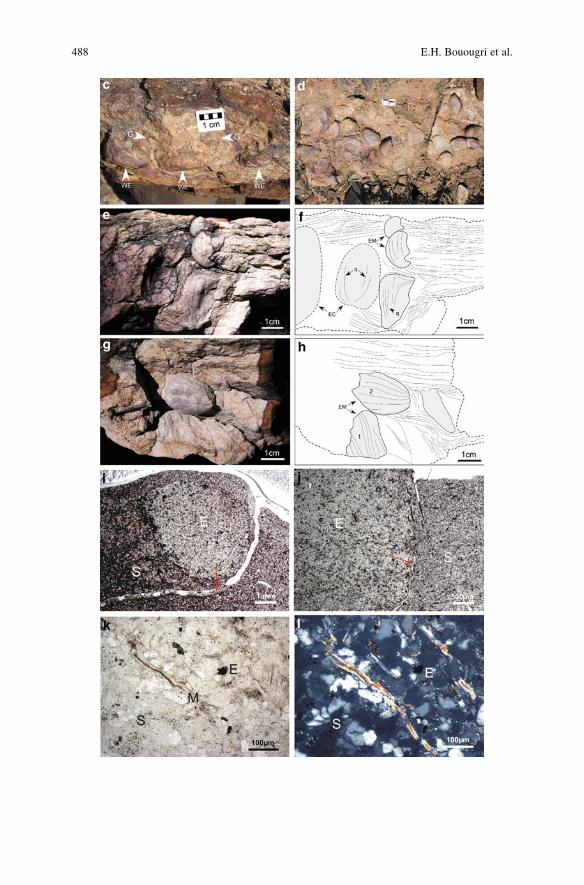
488 E.H. Bouougri et al.
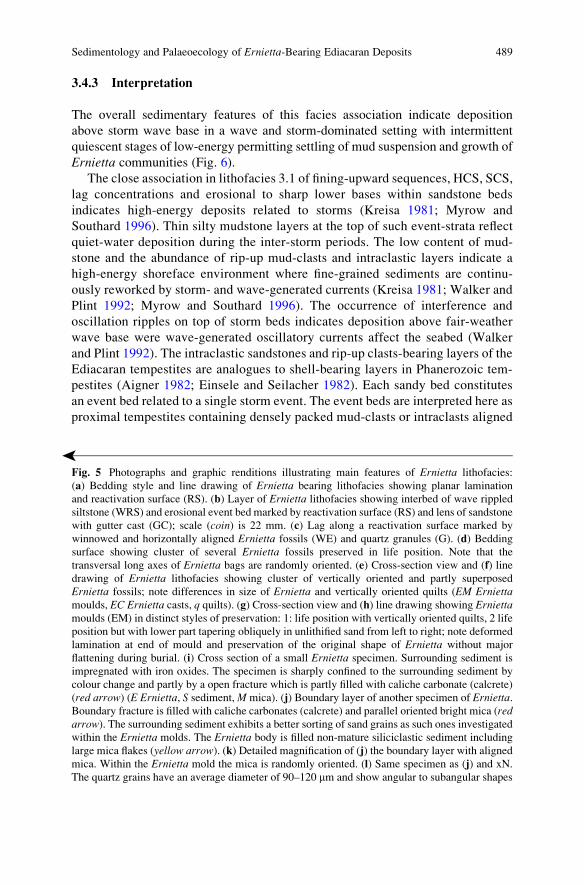
3.4.3 Interpretation
The overall sedimentary features of this facies association indicate deposition
above storm wave base in a wave and storm-dominated setting with intermittent
quiescent stages of low-energy permitting settling of mud suspension and growth of
Ernietta communities (Fig. 6).
The close association in lithofacies 3.1 of fining-upward sequences, HCS, SCS,
lag concentrations and erosional to sharp lower bases within sandstone beds
indicates high-energy deposits related to storms (Kreisa 1981; Myrow and
Southard 1996). Thin silty mudstone layers at the top of such event-strata reflect
quiet-water deposition during the inter-storm periods. The low content of mud-
stone and the abundance of rip-up mud-clasts and intraclastic layers indicate a
high-energy shoreface environment where fine-grained sediments are continu-
ously reworked by storm- and wave-generated currents (Kreisa 1981; Walker and
Plint 1992; Myrow and Southard 1996). The occurrence of interference and
oscillation ripples on top of storm beds indicates deposition above fair-weather
wave base were wave-generated oscillatory currents affect the seabed (Walker
and Plint 1992). The intraclastic sandstones and rip-up clasts-bearing layers of the
Ediacaran tempestites are analogues to shell-bearing layers in Phanerozoic tem-
pestites (Aigner 1982; Einsele and Seilacher 1982). Each sandy bed constitutes
an event bed related to a single storm event. The event beds are interpreted here as
proximal tempestites containing densely packed mud-clasts or intraclasts aligned�
Fig. 5 Photographs and graphic renditions illustrating main features of Ernietta lithofacies:
(a) Bedding style and line drawing of Ernietta bearing lithofacies showing planar lamination
and reactivation surface (RS). (b) Layer of Ernietta lithofacies showing interbed of wave rippled
siltstone (WRS) and erosional event bed marked by reactivation surface (RS) and lens of sandstone
with gutter cast (GC); scale (coin) is 22 mm. (c) Lag along a reactivation surface marked by
winnowed and horizontally aligned Ernietta fossils (WE) and quartz granules (G). (d) Bedding
surface showing cluster of several Ernietta fossils preserved in life position. Note that the
transversal long axes of Ernietta bags are randomly oriented. (e) Cross-section view and (f) line
drawing of Ernietta lithofacies showing cluster of vertically oriented and partly superposed
Ernietta fossils; note differences in size of Ernietta and vertically oriented quilts (EM Erniettamoulds, EC Ernietta casts, q quilts). (g) Cross-section view and (h) line drawing showing Erniettamoulds (EM) in distinct styles of preservation: 1: life position with vertically oriented quilts, 2 life
position but with lower part tapering obliquely in unlithified sand from left to right; note deformed
lamination at end of mould and preservation of the original shape of Ernietta without major
flattening during burial. (i) Cross section of a small Ernietta specimen. Surrounding sediment is
impregnated with iron oxides. The specimen is sharply confined to the surrounding sediment by
colour change and partly by a open fracture which is partly filled with caliche carbonate (calcrete)
(red arrow) (E Ernietta, S sediment,M mica). (j) Boundary layer of another specimen of Ernietta.Boundary fracture is filled with caliche carbonates (calcrete) and parallel oriented bright mica (redarrow). The surrounding sediment exhibits a better sorting of sand grains as such ones investigated
within the Ernietta molds. The Ernietta body is filled non-mature siliciclastic sediment including
large mica flakes (yellow arrow). (k) Detailed magnification of (j) the boundary layer with aligned
mica. Within the Ernietta mold the mica is randomly oriented. (l) Same specimen as (j) and xN.
The quartz grains have an average diameter of 90–120 mm and show angular to subangular shapes
Sedimentology and Palaeoecology of Ernietta-Bearing Ediacaran Deposits 489
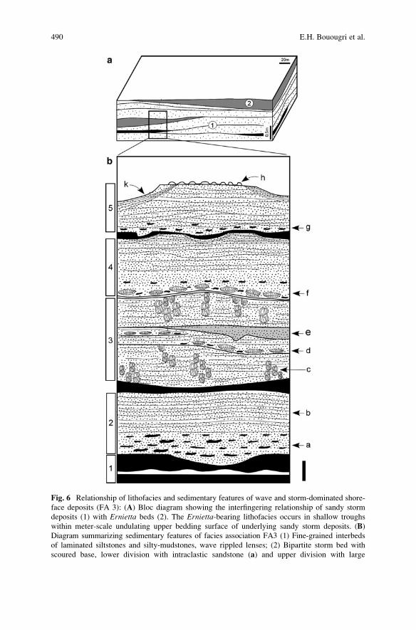
Fig. 6 Relationship of lithofacies and sedimentary features of wave and storm-dominated shore-
face deposits (FA 3): (A) Bloc diagram showing the interfingering relationship of sandy storm
deposits (1) with Ernietta beds (2). The Ernietta-bearing lithofacies occurs in shallow troughs
within meter-scale undulating upper bedding surface of underlying sandy storm deposits. (B)
Diagram summarizing sedimentary features of facies association FA3 (1) Fine-grained interbeds
of laminated siltstones and silty-mudstones, wave rippled lenses; (2) Bipartite storm bed with
scoured base, lower division with intraclastic sandstone (a) and upper division with large
490 E.H. Bouougri et al.
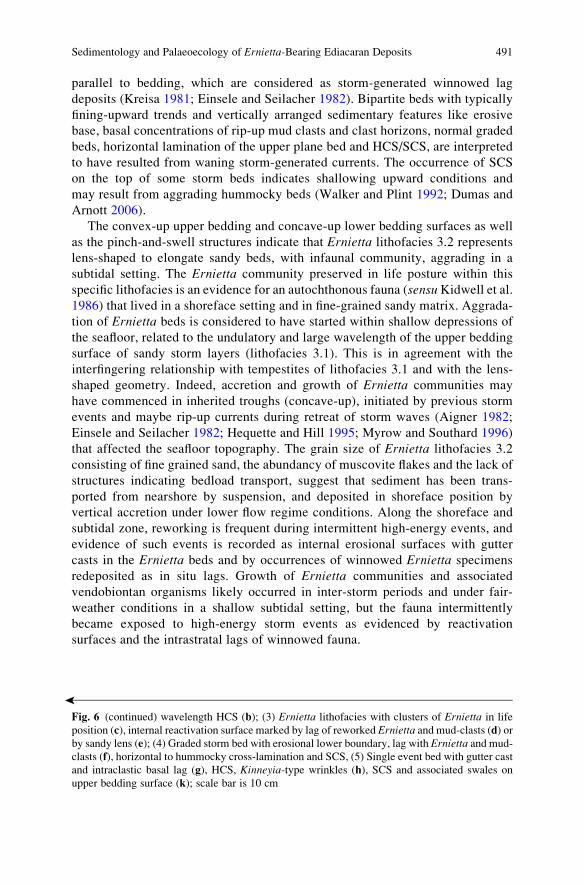
parallel to bedding, which are considered as storm-generated winnowed lag
deposits (Kreisa 1981; Einsele and Seilacher 1982). Bipartite beds with typically
fining-upward trends and vertically arranged sedimentary features like erosive
base, basal concentrations of rip-up mud clasts and clast horizons, normal graded
beds, horizontal lamination of the upper plane bed and HCS/SCS, are interpreted
to have resulted from waning storm-generated currents. The occurrence of SCS
on the top of some storm beds indicates shallowing upward conditions and
may result from aggrading hummocky beds (Walker and Plint 1992; Dumas and
Arnott 2006).
The convex-up upper bedding and concave-up lower bedding surfaces as well
as the pinch-and-swell structures indicate that Ernietta lithofacies 3.2 represents
lens-shaped to elongate sandy beds, with infaunal community, aggrading in a
subtidal setting. The Ernietta community preserved in life posture within this
specific lithofacies is an evidence for an autochthonous fauna (sensu Kidwell et al.1986) that lived in a shoreface setting and in fine-grained sandy matrix. Aggrada-
tion of Ernietta beds is considered to have started within shallow depressions of
the seafloor, related to the undulatory and large wavelength of the upper bedding
surface of sandy storm layers (lithofacies 3.1). This is in agreement with the
interfingering relationship with tempestites of lithofacies 3.1 and with the lens-
shaped geometry. Indeed, accretion and growth of Ernietta communities may
have commenced in inherited troughs (concave-up), initiated by previous storm
events and maybe rip-up currents during retreat of storm waves (Aigner 1982;
Einsele and Seilacher 1982; Hequette and Hill 1995; Myrow and Southard 1996)
that affected the seafloor topography. The grain size of Ernietta lithofacies 3.2
consisting of fine grained sand, the abundancy of muscovite flakes and the lack of
structures indicating bedload transport, suggest that sediment has been trans-
ported from nearshore by suspension, and deposited in shoreface position by
vertical accretion under lower flow regime conditions. Along the shoreface and
subtidal zone, reworking is frequent during intermittent high-energy events, and
evidence of such events is recorded as internal erosional surfaces with gutter
casts in the Ernietta beds and by occurrences of winnowed Ernietta specimens
redeposited as in situ lags. Growth of Ernietta communities and associated
vendobiontan organisms likely occurred in inter-storm periods and under fair-
weather conditions in a shallow subtidal setting, but the fauna intermittently
became exposed to high-energy storm events as evidenced by reactivation
surfaces and the intrastratal lags of winnowed fauna.
�
Fig. 6 (continued) wavelength HCS (b); (3) Ernietta lithofacies with clusters of Ernietta in life
position (c), internal reactivation surface marked by lag of reworked Ernietta and mud-clasts (d) or
by sandy lens (e); (4) Graded storm bed with erosional lower boundary, lag with Ernietta and mud-
clasts (f), horizontal to hummocky cross-lamination and SCS, (5) Single event bed with gutter cast
and intraclastic basal lag (g), HCS, Kinneyia-type wrinkles (h), SCS and associated swales on
upper bedding surface (k); scale bar is 10 cm
Sedimentology and Palaeoecology of Ernietta-Bearing Ediacaran Deposits 491
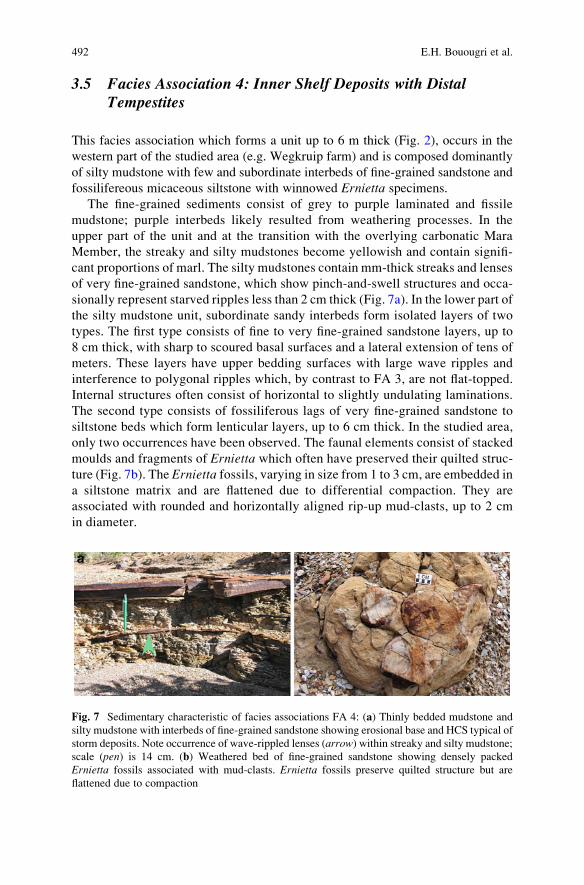
3.5 Facies Association 4: Inner Shelf Deposits with DistalTempestites
This facies association which forms a unit up to 6 m thick (Fig. 2), occurs in the
western part of the studied area (e.g. Wegkruip farm) and is composed dominantly
of silty mudstone with few and subordinate interbeds of fine-grained sandstone and
fossilifereous micaceous siltstone with winnowed Ernietta specimens.
The fine-grained sediments consist of grey to purple laminated and fissile
mudstone; purple interbeds likely resulted from weathering processes. In the
upper part of the unit and at the transition with the overlying carbonatic Mara
Member, the streaky and silty mudstones become yellowish and contain signifi-
cant proportions of marl. The silty mudstones contain mm-thick streaks and lenses
of very fine-grained sandstone, which show pinch-and-swell structures and occa-
sionally represent starved ripples less than 2 cm thick (Fig. 7a). In the lower part of
the silty mudstone unit, subordinate sandy interbeds form isolated layers of two
types. The first type consists of fine to very fine-grained sandstone layers, up to
8 cm thick, with sharp to scoured basal surfaces and a lateral extension of tens of
meters. These layers have upper bedding surfaces with large wave ripples and
interference to polygonal ripples which, by contrast to FA 3, are not flat-topped.
Internal structures often consist of horizontal to slightly undulating laminations.
The second type consists of fossiliferous lags of very fine-grained sandstone to
siltstone beds which form lenticular layers, up to 6 cm thick. In the studied area,
only two occurrences have been observed. The faunal elements consist of stacked
moulds and fragments of Ernietta which often have preserved their quilted struc-
ture (Fig. 7b). The Ernietta fossils, varying in size from 1 to 3 cm, are embedded in
a siltstone matrix and are flattened due to differential compaction. They are
associated with rounded and horizontally aligned rip-up mud-clasts, up to 2 cm
in diameter.
Fig. 7 Sedimentary characteristic of facies associations FA 4: (a) Thinly bedded mudstone and
silty mudstone with interbeds of fine-grained sandstone showing erosional base and HCS typical of
storm deposits. Note occurrence of wave-rippled lenses (arrow) within streaky and silty mudstone;
scale (pen) is 14 cm. (b) Weathered bed of fine-grained sandstone showing densely packed
Ernietta fossils associated with mud-clasts. Ernietta fossils preserve quilted structure but are
flattened due to compaction
492 E.H. Bouougri et al.
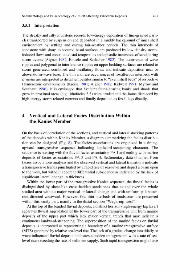
3.5.1 Interpretation
The streaky and silty mudstone records low-energy deposition of fine-grained parti-
cles transported by suspension and deposited in a muddy background of inner shelf
environment by settling and during fair-weather periods. The thin interbeds of
sandstone with sharp to scoured basal surfaces are produced by low-density storm-
induced flows and constitute distal tempestites and episodic incursions of sand during
storm events (Aigner 1982; Einsele and Seilacher 1982). The occurrence of wave
ripples and polygonal to interference ripples on upper bedding surfaces are related to
storm generated, combined and oscillatory flows and indicate deposition near or
above storm-wave base. The thin and rare occurrences of fossilferous interbeds with
Ernietta are interpreted as distal tempestites similar to “event shell beds” of respective
Phanerozoic environments (Kreisa 1981; Aigner 1982; Kidwell 1991; Myrow and
Southard 1996). It is envisaged that Ernietta fauna-bearing banks and shoals that
grew in proximal areas (e.g. lithofacies 3.3) were eroded and the fauna displaced by
high-energy storm-related currents and finally deposited as fossil lags distally.
4 Vertical and Lateral Facies Distribution Within
the Kanies Member
On the basis of correlation of the sections, and vertical and lateral stacking patterns
of the deposits within Kanies Member, a diagram summarizing the facies distribu-
tion can be designed (Fig. 8). The facies associations are organized in a fining-
upward transgressive sequence indicating landward-steepening character. The
sequence is starting with the fluvial facies association FA 1 and ending with marine
deposits of facies associations FA 3 and FA 4. Sedimentary data obtained from
facies associations analysis and the observed vertical and lateral transitions indicate
a transgressive trends punctuated by a rapid rise of sea-level and depict a basin open
to the west, but without apparent differential subsidence as indicated by the lack of
significant lateral change in thickness.
Within the lower part of the transgressive Kanies sequence, the fluvial facies is
distinguished by sheet-like cross-bedded sandstones that extend over the whole
studied area without major vertical or lateral change and with uniform palaeocur-
rent directed westward. However, few thin interbeds of mudstones are preserved
within this sandy part, mainly in the distal section “Wegkruip west”.
At the top of the braided fluvial deposits, a distinct horizon (high-energy lag layer)
separates fluvial aggradation of the lower part of the transgressive unit from marine
deposits of the upper part which lack major vertical trends that may indicate a
continuous landward-steepening. The superposition of the marine facies on fluvial
deposits is interpreted as representing a boundary of a marine transgressive surface
(MTS) generated by relative sea-level rise. The lack of a gradual change into tidally or
wave influenced fluvial deposits indicates a sudden transgression with a rate of sea-
level rise exceeding the rate of sediment supply. Such rapid transgression might have
Sedimentology and Palaeoecology of Ernietta-Bearing Ediacaran Deposits 493

been accentuated by the flat-topography of the pre-existing wide braid-river plain
(FA 1) which lacks narrow localized channels delimited by stabilized mud banks.
The thin transgressive interval is marked mainly by reworked sand and mud
material with large quantity of rip-up clasts which was redeposited in foreshore to
upper shoreface setting. In the proximal part, intense waves and storms played a
major role in reworking and deposition of the coarse-grained sand and the asso-
ciated lag horizons with high proportion of densely packed rip-up mud-clasts. In the
distal area, the effect of geostrophic and rip-up currents induced by high-energy
waves and storms is recorded as lens-shaped medium-scale 3D dunes locally
developed and prograding basinward (Wegkruip sections).
A clear partitioning of the depositional setting into proximal and distal marine
environment with a lateral shift of facies occurs during deposition of the upper
part of Kanies transgressive sequence. This is evidenced by the basinward tran-
sition from wave and storm-dominated shoreface association (FA 3) restricted
to eastern and proximal portion (Hansburg section) to muddy inner-shelf associ-
ation (FA 4) with streaky silty-mudstone and subordinate interbeds of distal
tempestites occurring westward in the distal area (Wegkruip sections). The
establishment and growth of Ernietta and associated vendobiontan organisms in
the proximal part constitutes the most significant change within Kanies sequence.
Occurrence of Ernietta communities within a typical lithofacies and facies
association together with their restriction to the proximal part of the shallow
marine environment indicate that growth and demise of Ernietta infaunal com-
munities are facies controlled, and thus Ernietta may be considered as an oppor-
tunistic organism.
Fig. 8 Diagram summarizing vertical and lateral facies relationships within Kanies Member: (1)
Fluvial facies association;MTSmarine transgressive surface; (2) Transgressive deposits. (3) Storm
deposits of facies association FA 3 with interbedded lens-shaped ridges populations of Ernietta(indicated in black); (4) Facies association FA 4 with discrete layers of distal tempestites
494 E.H. Bouougri et al.
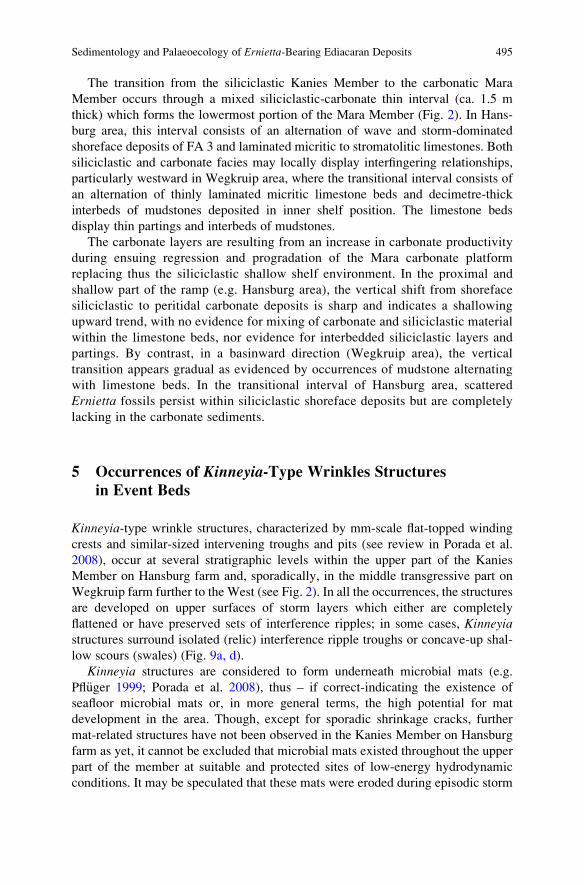
The transition from the siliciclastic Kanies Member to the carbonatic Mara
Member occurs through a mixed siliciclastic-carbonate thin interval (ca. 1.5 m
thick) which forms the lowermost portion of the Mara Member (Fig. 2). In Hans-
burg area, this interval consists of an alternation of wave and storm-dominated
shoreface deposits of FA 3 and laminated micritic to stromatolitic limestones. Both
siliciclastic and carbonate facies may locally display interfingering relationships,
particularly westward in Wegkruip area, where the transitional interval consists of
an alternation of thinly laminated micritic limestone beds and decimetre-thick
interbeds of mudstones deposited in inner shelf position. The limestone beds
display thin partings and interbeds of mudstones.
The carbonate layers are resulting from an increase in carbonate productivity
during ensuing regression and progradation of the Mara carbonate platform
replacing thus the siliciclastic shallow shelf environment. In the proximal and
shallow part of the ramp (e.g. Hansburg area), the vertical shift from shoreface
siliciclastic to peritidal carbonate deposits is sharp and indicates a shallowing
upward trend, with no evidence for mixing of carbonate and siliciclastic material
within the limestone beds, nor evidence for interbedded siliciclastic layers and
partings. By contrast, in a basinward direction (Wegkruip area), the vertical
transition appears gradual as evidenced by occurrences of mudstone alternating
with limestone beds. In the transitional interval of Hansburg area, scattered
Ernietta fossils persist within siliciclastic shoreface deposits but are completely
lacking in the carbonate sediments.
5 Occurrences of Kinneyia-Type Wrinkles Structures
in Event Beds
Kinneyia-type wrinkle structures, characterized by mm-scale flat-topped winding
crests and similar-sized intervening troughs and pits (see review in Porada et al.
2008), occur at several stratigraphic levels within the upper part of the Kanies
Member on Hansburg farm and, sporadically, in the middle transgressive part on
Wegkruip farm further to the West (see Fig. 2). In all the occurrences, the structures
are developed on upper surfaces of storm layers which either are completely
flattened or have preserved sets of interference ripples; in some cases, Kinneyiastructures surround isolated (relic) interference ripple troughs or concave-up shal-
low scours (swales) (Fig. 9a, d).
Kinneyia structures are considered to form underneath microbial mats (e.g.
Pfl€uger 1999; Porada et al. 2008), thus – if correct-indicating the existence of
seafloor microbial mats or, in more general terms, the high potential for mat
development in the area. Though, except for sporadic shrinkage cracks, further
mat-related structures have not been observed in the Kanies Member on Hansburg
farm as yet, it cannot be excluded that microbial mats existed throughout the upper
part of the member at suitable and protected sites of low-energy hydrodynamic
conditions. It may be speculated that these mats were eroded during episodic storm
Sedimentology and Palaeoecology of Ernietta-Bearing Ediacaran Deposits 495
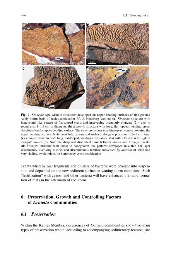
events whereby mat fragments and clusters of bacteria were brought into suspen-
sion and deposited on the new sediment surface at waning storm conditions. Such
“fertilization” with cyano- and other bacteria will have enhanced the rapid forma-
tion of mats in the aftermath of the storm.
6 Preservation, Growth and Controlling Factors
of Ernietta Communities
6.1 Preservation
Within the Kanies Member, occurrences of Ernietta communities show two main
types of preservation which, according to accompanying sedimentary features, are
Fig. 9 Kinneyia-type wrinkle structures developed on upper bedding surfaces of fine-grained
sandy storm beds of facies association FA 3, Hansburg section: (a) Kinneyia structure with
honeycomb-like pattern of flat-topped crests and intervening irregularly elongate (2–4 cm) to
round pits, 1–1.5 cm in diameter. (b) Kinneyia structure with long, flat-topped, winding crests
developed on flat upper bedding surface. The structure occurs in a thin top-set veneer covering the
upper bedding surface. Note crest bifurcations and isolated elongate pits about 0.5–1 cm long.
(c) Kinneyia structure with long, flat-topped, winding crests associated with subcircular to slightlyelongate swales (S). Note the sharp and discordant limit between swales and Kinneyia crests.
(d) Kinneyia structure with linear to honeycomb like patterns developed in a thin flat layer
discordantly overlying distinct and discontinuous laminae (indicated by arrows) of wide and
very shallow swale related to hummocky-cross stratification
496 E.H. Bouougri et al.
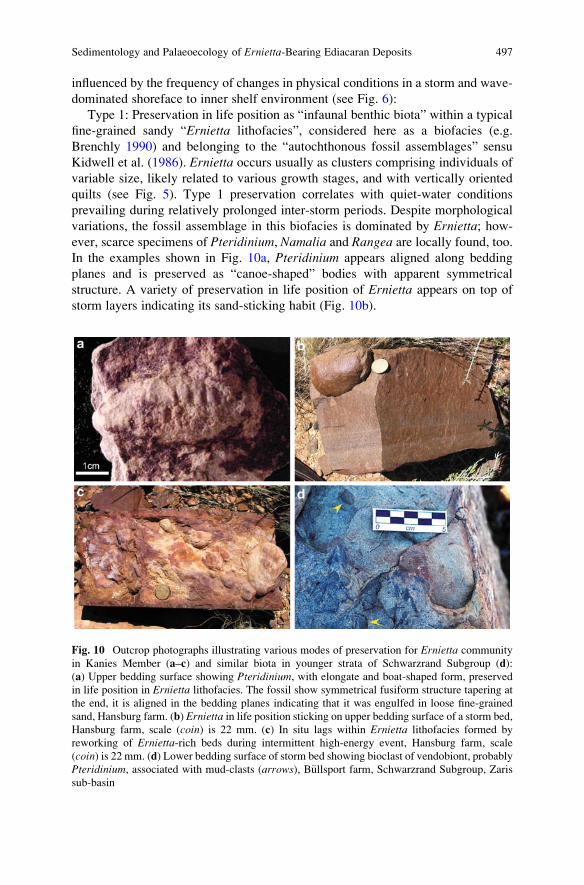
influenced by the frequency of changes in physical conditions in a storm and wave-
dominated shoreface to inner shelf environment (see Fig. 6):
Type 1: Preservation in life position as “infaunal benthic biota” within a typical
fine-grained sandy “Ernietta lithofacies”, considered here as a biofacies (e.g.
Brenchly 1990) and belonging to the “autochthonous fossil assemblages” sensu
Kidwell et al. (1986). Ernietta occurs usually as clusters comprising individuals of
variable size, likely related to various growth stages, and with vertically oriented
quilts (see Fig. 5). Type 1 preservation correlates with quiet-water conditions
prevailing during relatively prolonged inter-storm periods. Despite morphological
variations, the fossil assemblage in this biofacies is dominated by Ernietta; how-ever, scarce specimens of Pteridinium, Namalia and Rangea are locally found, too.In the examples shown in Fig. 10a, Pteridinium appears aligned along bedding
planes and is preserved as “canoe-shaped” bodies with apparent symmetrical
structure. A variety of preservation in life position of Ernietta appears on top of
storm layers indicating its sand-sticking habit (Fig. 10b).
Fig. 10 Outcrop photographs illustrating various modes of preservation for Ernietta community
in Kanies Member (a–c) and similar biota in younger strata of Schwarzrand Subgroup (d):
(a) Upper bedding surface showing Pteridinium, with elongate and boat-shaped form, preserved
in life position in Ernietta lithofacies. The fossil show symmetrical fusiform structure tapering at
the end, it is aligned in the bedding planes indicating that it was engulfed in loose fine-grained
sand, Hansburg farm. (b) Ernietta in life position sticking on upper bedding surface of a storm bed,
Hansburg farm, scale (coin) is 22 mm. (c) In situ lags within Ernietta lithofacies formed by
reworking of Ernietta-rich beds during intermittent high-energy event, Hansburg farm, scale
(coin) is 22 mm. (d) Lower bedding surface of storm bed showing bioclast of vendobiont, probably
Pteridinium, associated with mud-clasts (arrows), B€ullsport farm, Schwarzrand Subgroup, Zaris
sub-basin
Sedimentology and Palaeoecology of Ernietta-Bearing Ediacaran Deposits 497
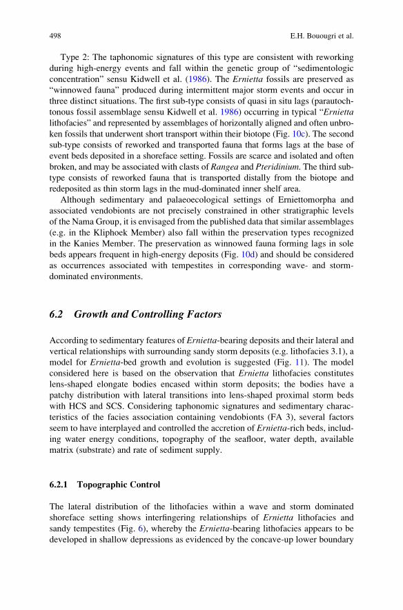
Type 2: The taphonomic signatures of this type are consistent with reworking
during high-energy events and fall within the genetic group of “sedimentologic
concentration” sensu Kidwell et al. (1986). The Ernietta fossils are preserved as
“winnowed fauna” produced during intermittent major storm events and occur in
three distinct situations. The first sub-type consists of quasi in situ lags (parautoch-
tonous fossil assemblage sensu Kidwell et al. 1986) occurring in typical “Erniettalithofacies” and represented by assemblages of horizontally aligned and often unbro-
ken fossils that underwent short transport within their biotope (Fig. 10c). The second
sub-type consists of reworked and transported fauna that forms lags at the base of
event beds deposited in a shoreface setting. Fossils are scarce and isolated and often
broken, and may be associated with clasts of Rangea and Pteridinium. The third sub-type consists of reworked fauna that is transported distally from the biotope and
redeposited as thin storm lags in the mud-dominated inner shelf area.
Although sedimentary and palaeoecological settings of Erniettomorpha and
associated vendobionts are not precisely constrained in other stratigraphic levels
of the Nama Group, it is envisaged from the published data that similar assemblages
(e.g. in the Kliphoek Member) also fall within the preservation types recognized
in the Kanies Member. The preservation as winnowed fauna forming lags in sole
beds appears frequent in high-energy deposits (Fig. 10d) and should be considered
as occurrences associated with tempestites in corresponding wave- and storm-
dominated environments.
6.2 Growth and Controlling Factors
According to sedimentary features of Ernietta-bearing deposits and their lateral andvertical relationships with surrounding sandy storm deposits (e.g. lithofacies 3.1), a
model for Ernietta-bed growth and evolution is suggested (Fig. 11). The model
considered here is based on the observation that Ernietta lithofacies constitutes
lens-shaped elongate bodies encased within storm deposits; the bodies have a
patchy distribution with lateral transitions into lens-shaped proximal storm beds
with HCS and SCS. Considering taphonomic signatures and sedimentary charac-
teristics of the facies association containing vendobionts (FA 3), several factors
seem to have interplayed and controlled the accretion of Ernietta-rich beds, includ-ing water energy conditions, topography of the seafloor, water depth, available
matrix (substrate) and rate of sediment supply.
6.2.1 Topographic Control
The lateral distribution of the lithofacies within a wave and storm dominated
shoreface setting shows interfingering relationships of Ernietta lithofacies and
sandy tempestites (Fig. 6), whereby the Ernietta-bearing lithofacies appears to be
developed in shallow depressions as evidenced by the concave-up lower boundary
498 E.H. Bouougri et al.
Fig. 11 Model illustrating processes recorded in Ernietta-rich bed (Ernietta lithofacies) from
growth stage during low energy periods to burial stage by sandy storm deposit: (a) Fair-weather
conditions: Growth of Ernietta-rich bed (1–2) within a shallow subtidal zone (shoreface), mud
settling basinward in the inner shelf zone. (b) Intermittent minor storm event: Reworking of
Ernietta bed, formation of internal reactivation surface (RS) and nearby redeposition of winnowed
Ernietta (WE) fossils in front of the bed; basinward transport and redeposition of Ernietta as lags
(L). (c) Low-energy stage: Reestablishment and renews growth of Ernietta fauna (3) and mud
settling basinward. (d) Major storm event: Ernietta-rich bed partially reworked and buried by
deposition of sandy storm bed (4) with lag of Ernietta and mud-clasts. Basinward, deposition of
distal tempestites (DT)
Sedimentology and Palaeoecology of Ernietta-Bearing Ediacaran Deposits 499
and the pinch-and-swell geometry of the individual bodies. Such distribution and
geometry indicates control by inherited topography with nucleation sites for
Ernietta beds in troughs, which were partially filled thus leveling the seafloor.
Observations in modern storm-influenced shoreface and shelf environments show
the effect of storm-generated currents on the seafloor (Hequette and Hill 1995;
Passchier and Kleinhans 2005; Vis-Star 2007), which create shallow, elongated to
rounded meter-scale troughs in the overall lens-shaped, intraclastic, hummocky-
and swaley-cross stratified storm deposit with pinch-and-swell geometry. The
resulting shape of the troughs depends on the shape of sandstone body which in
turn is related to water depth and predominant flow conditions. During low-energy
and prolonged inter-storm periods, these depressions may have formed favourable
and relatively protected sites for trapping and vertical accretion of very fine-grained
sand particles and associated mica flakes, and eventually colonization by incipient
Ernietta communities. It is difficult, in the case of Hansburg farm area, to control
the exact shape of Ernietta beds, but the surrounding facies with hummocky and
swaley cross-bedded sandstone seems likely to indicate a seafloor covered with 3D
mound-like bedforms of low amplitude.
6.2.2 Control by Water Energy Conditions
It is envisaged that the change fromhigh-energy periods dominated by storm activities
to quiet-water conditions played a major role in nucleation and establishment
of Ernietta communities. The accretion of Ernietta-bearing lithofacies records
prolonged low-energy stages in a wave and storm-dominated shoreface setting.
During growth of Ernietta-beds, intermittent high-energy events are recorded as
reactivation surfaces and in situ lags of Ernietta bioclasts. The abrupt change into
high-energy conditions during a major storm event, however, was the main process
involved for a demise of Ernietta communities and their burial by the next storm
deposit (Fig. 11d).
6.2.3 Water Depth Control
Within the upper part of the Kanies Member, occurrences of autochthonous
Ernietta communities appear closely related to a specific “Ernietta lithofacies”
within shoreface deposits (FA 3). Sedimentary characteristics including swaley-
cross stratification and abundant wave-related ripples suggest deposition broadly
near fair-weather wave base. By contrast, more basinward in the muddy area of the
shelf (FA 4), there is no evidence for occurrence of Ernietta beds or for Erniettapreserved in life position. The only occurrences found are within thin distal
tempestites as winnowed fauna. From this bathymetric distribution, it is assumed
that well oxygenated zones in a shallow subtidal environment were suitable for
Ernietta colonization.
500 E.H. Bouougri et al.
6.2.4 Nature of Sediment and Sediment Supply
The zonality observed within the upper portion of Kanies Member shows a grain-
size decrease basinward and a clear transition from a sandy dominated shoreface in
the East to a mud-dominated inner shelf in the West. By considering petrographic
features of the Ernietta lithofacies, development of Ernietta communities appears
to have been favoured by a very fine-grained sandy matrix. The internal bedding of
Ernietta beds dominated by horizontal to wavy lamination as well as the abundance
of commonly aligned, detrital mica flakes, support vertical accretion under lower
flow regime conditions. In the shoreface zone, the rate of sediment supply is mainly
controlled by tide, wave and storm generated currents and by important river
discharges (Johnson and Baldwin 1986; Walker and Plint 1992). Such influences
were not recorded in the Ernietta-bearing facies association. By contrast, during
low-energy, fair-weather periods when the action of strong marine currents is
negligible, the rate of sediment supply is low and only very fine-grained suspended
particles of sand to silt size are transported seaward from the adjacent foreshore
zone by bottom currents of lower flow regime. The similarity between the sediment
of Ernietta moulds and the surrounding matrix, together with the development and
preservation of Ernietta in vertical life-position without major changes in the shape
during burial, indicate that the organisms likely were filled progressively by
sediment before complete burial.
7 Discussion: Palaeoecology of Ernietta and Implications
for Similar Underground Vendobionts
Within the Kuibis Subgroup, development ofErnietta communities coincides with a
major shift in sedimentation and is related to a rapid transgression event, whereas
their demise coincides with a major shift to carbonate deposition during the over-
lying highstand systems tract of the Mara Member. A similar “invasive” pattern
during rapid transgression may be invoked for the Vendobionta assemblage of
the overlying siliciclastic Kliphoek Member (Grazhdankin and Seilacher 2002).
Reported occurrences of underground vendobionts similar to Ernietta and pre-
served as three-dimensional moulds in Ediacaran sequences (e.g. Russia, Australia,
Namibia, USA) appear, in a broad sense, associated with marine sandy environ-
ments with prevailing high-energy conditions (Jenkins 1985, 1992; Ivantsov and
Grazhdankin 1997; Hagadorn and Waggoner 2000; Grazhdankin and Seilacher
2002, 2005). The absence of detailed sections and sedimentological studies of most
of these occurrences does not allow a close comparison in palaeoecology of the
Ernietta communities in the Kanies Member with similar infaunal biota. However,
available sedimentological data on vendobiont-bearing successions of various
localities fall within a storm-winnowing model and point broadly to wave and
storm-influenced settings. Grazhdankin (2004) documented the Ediacara fauna of
the Vendian Group in the White Sea area (Russia), and noted a correlation between
Sedimentology and Palaeoecology of Ernietta-Bearing Ediacaran Deposits 501
sedimentary environment and faunal assemblage. The vendobionts consisting of
three-dimensional sandstone casts including among others Rangea and Onegia,occur mainly in interstratified sandstones interpreted as fluviomarine sand-filled
channels and distributary mouth-bar settings. Thereby, Rangea is associated with
channel fill sandstone that cuts across middle-shoreface deposits, whereas Onegiawas found at the base of channelised sandstone packages. In the Ediacara rocks of
Australia, vendobionts including Pteridinium and Onegia appear to be restricted tomassive fluviomarine channelised sandstone deposits (e.g. Gehling 2000). In the
Kliphoek Member of the Nama Group (Namibia), vendobionts are preserved in
planar and cross-bedded sandstone of a shallowly channeled coastal plain with
fluvial dominance, interbedded with storm influenced lower-shoreface deposits
(Saylor 2003). As noted in previous studies comparing the distribution of Ediacaran
communities and considering morphological features, the infaunal vendobionts
seem to be adapted to marine sandy environments of distributary mouth bar shoals
and rip channels (e.g. Grazhdankin 2004; Grazhdankin and Seilacher 2002; McCall
2006). However, by comparing the specific palaeoecologic setting of Erniettacommunities in the Kanies Member with the sedimentary settings reported,
although not detailed, from similar occurrences in other successions, it appears
possible to suggest similar specifically restricted niches for these vendobionts in
localized troughs where incipient Vendobionta communities were able to grow
during intermittent inter-storm periods. According to the sedimentary features of
the Ernietta-bearing deposits, their modes of preservation, and their distribution in
Kanies Member, Ernietta communities appear to be restricted to a narrow bathy-
metric zone in the shelf area, colonizing non-deltaic, wave and storm-dominated
shoreface environments, whereas they are absent basinward in inner shelf mud-
dominated environments. Such lateral change over a short distance on the Hansburg-
Wegkruip shallow marine shelf indicates that growth and distribution of the biota
was sensitive to water depth. On the other hand, Ernietta and other associated
infaunal vendobionts formed and grew as “community beds” that suffered rework-
ing during high-energy conditions. The palaeoecological model proposed here is
supported by: (1) the preservation of the fauna in life position (autochtonous
fauna sensu Kidwell et al. 1986) within specific fossiliferous Ernietta beds called
here “Ernietta lithofacies”; (2) the geometry and lateral extent of the Erniettabeds encased within proximal tempestites; (3) the development and preservation
of quasi in situ lags within the Ernietta beds, composed of reworked autochtho-
nous fauna; (4) the low-diversity and monospecific feature of the beds overwhel-
mingly dominated by Ernietta. Thus, the benthic Ernietta communities might be
considered here as the earliest precursor in the rock record of “fossil-beds”,
similar to those made by the autochthonous communities in the Phanerozoic.
Further studies including among others sedimentary and palaeoecological setting,
interaction between fauna and sediment matrix, taxonomic diversity and growth
strategies in vendobiont-rich beds of the Nama Group would yield additional
useful details to constrain the dynamic of infaunal Ediacaran biota like Erniettaand the extent of their biotope.
502 E.H. Bouougri et al.
8 Conclusions
The siliciclastic Kanies Member in Hansburg area, southern Namibia, represents a
fining-upward succession deposited during the first transgression in the Witputs
sub-basin. The succession contains the oldest known Ediacaran fossils in the Nama
Group represented by Ernietta-community. A facies analysis combined with taph-
onomic signatures of Ernietta-bearing deposits allows to constrain the paleoecolog-ical setting and the factors influencing growth of Ernietta with possible
implications on other occurrences of similar biota.
Four facies associations (FA 1–4) ranging from fluvial to inner shelf settings are
distinguished in the Kanies Member. The overall siliciclastic succession (~12 m
thick) is divided by a marine flooding surface into a fluvial lower and a marine
upper part. The lower part consists of braided fluvial facies association (FA 1)
which forms a sheet-like unit dominated by cross-bedded sandstone. It is overlain
by foreshore to shoreface high-energy transgressive deposits (FA 2) with typical
coarse-grained sandstone, rich in large rip-up mud-clasts at the base marking a
marine flooding surface. The overlying deposits consist in the East of wave- and
storm-dominated shoreface deposits (FA 3) that grade laterally westward (basin-
ward) into mud-dominated inner shelf deposits with interbedded distal tempestites
(FA 4). Vertical and lateral distribution of the facies associations in the upper part
of the Kanies Member show a clear partitioning into proximal and distal settings in
a basin deepening westward.
Ernietta communities are facies related and occur commonly in typical light and
fine-grained sandstone layers sometimes associated with Rangea encased within
wave and storm-dominated shoreface deposits. They form fossil-rich beds containing
moulds of Ernietta, most of them indicate preservation in life position as evidenced
by their vertical orientation, pointing to an autochthonous benthic community. Such
Ernietta beds were affected by intermittent storm events that produced erosional
structures and quasi in situ lags containing reworked biota. During major storm
events, the Ernietta beds were buried by tempestites with basal lags containing
bioclasts of vendobionts, mostly reworked Ernietta. In both cases, some of reworked
Ernietta specimens were transported distally and redeposited in an inner shelf setting.
The proposed palaeoecological model emphasizes an Ernietta community that lived
on a fine-grained sandy substrate in a stressed shoreface environment; it formed an
infaunal biota engulfed in loose sediment and with vertical upward growth. Growth
and distribution of Ernietta communities appear controlled by palaeoenvironmental
factors such us inherited sea floor topography, water energy conditions, water depth,
type of substrate and sediment supply. According to their in situ three-dimensional
mouldic preservation and their ability to be transported from their biotope without
major destruction, most of the bag-shaped Ernietta individuals were seemingly
completely filled in situ by sediment before their final burial.
This study highlights the importance of sedimentological analysis of fossil-
ferous Ediacaran deposits, combined with taphonomic signatures, for constraining
the biotope and palaeocology of infaunal vendobionts as well as their typical
Sedimentology and Palaeoecology of Ernietta-Bearing Ediacaran Deposits 503
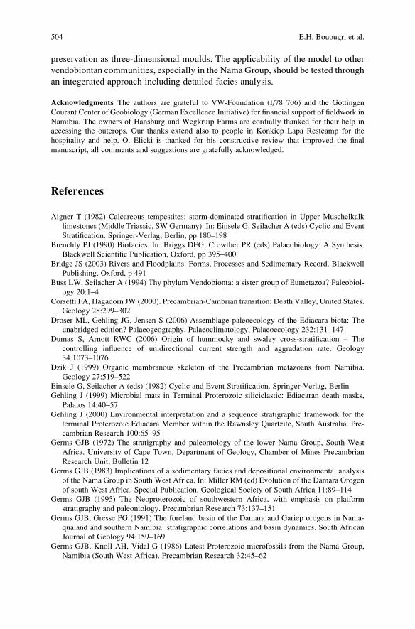
preservation as three-dimensional moulds. The applicability of the model to other
vendobiontan communities, especially in the Nama Group, should be tested through
an integerated approach including detailed facies analysis.
Acknowledgments The authors are grateful to VW-Foundation (I/78 706) and the G€ottingenCourant Center of Geobiology (German Excellence Initiative) for financial support of fieldwork in
Namibia. The owners of Hansburg and Wegkruip Farms are cordially thanked for their help in
accessing the outcrops. Our thanks extend also to people in Konkiep Lapa Restcamp for the
hospitality and help. O. Elicki is thanked for his constructive review that improved the final
manuscript, all comments and suggestions are gratefully acknowledged.
References
Aigner T (1982) Calcareous tempestites: storm-dominated stratification in Upper Muschelkalk
limestones (Middle Triassic, SW Germany). In: Einsele G, Seilacher A (eds) Cyclic and Event
Stratification. Springer-Verlag, Berlin, pp 180–198
Brenchly PJ (1990) Biofacies. In: Briggs DEG, Crowther PR (eds) Palaeobiology: A Synthesis.
Blackwell Scientific Publication, Oxford, pp 395–400
Bridge JS (2003) Rivers and Floodplains: Forms, Processes and Sedimentary Record. Blackwell
Publishing, Oxford, p 491
Buss LW, Seilacher A (1994) Thy phylum Vendobionta: a sister group of Eumetazoa? Paleobiol-
ogy 20:1–4
Corsetti FA, Hagadorn JW (2000). Precambrian-Cambrian transition: Death Valley, United States.
Geology 28:299–302
Droser ML, Gehling JG, Jensen S (2006) Assemblage paleoecology of the Ediacara biota: The
unabridged edition? Palaeogeography, Palaeoclimatology, Palaeoecology 232:131–147
Dumas S, Arnott RWC (2006) Origin of hummocky and swaley cross-stratification – The
controlling influence of unidirectional current strength and aggradation rate. Geology
34:1073–1076
Dzik J (1999) Organic membranous skeleton of the Precambrian metazoans from Namibia.
Geology 27:519–522
Einsele G, Seilacher A (eds) (1982) Cyclic and Event Stratification. Springer-Verlag, Berlin
Gehling J (1999) Microbial mats in Terminal Proterozoic siliciclastic: Ediacaran death masks,
Palaios 14:40–57
Gehling J (2000) Environmental interpretation and a sequence stratigraphic framework for the
terminal Proterozoic Ediacara Member within the Rawnsley Quartzite, South Australia. Pre-
cambrian Research 100:65–95
Germs GJB (1972) The stratigraphy and paleontology of the lower Nama Group, South West
Africa. University of Cape Town, Department of Geology, Chamber of Mines Precambrian
Research Unit, Bulletin 12
Germs GJB (1983) Implications of a sedimentary facies and depositional environmental analysis
of the Nama Group in South West Africa. In: Miller RM (ed) Evolution of the Damara Orogen
of south West Africa. Special Publication, Geological Society of South Africa 11:89–114
Germs GJB (1995) The Neoproterozoic of southwestern Africa, with emphasis on platform
stratigraphy and paleontology. Precambrian Research 73:137–151
Germs GJB, Gresse PG (1991) The foreland basin of the Damara and Gariep orogens in Nama-
qualand and southern Namibia: stratigraphic correlations and basin dynamics. South African
Journal of Geology 94:159–169
Germs GJB, Knoll AH, Vidal G (1986) Latest Proterozoic microfossils from the Nama Group,
Namibia (South West Africa). Precambrian Research 32:45–62
504 E.H. Bouougri et al.
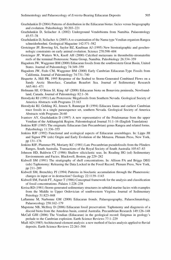
Grazhdankin D (2004) Patterns of distribution in the Ediacaran biotas: facies versus biogeography
and evolution. Paleobiology 30:203–221
Grazhdankin D, Seilacher A (2002) Underground Vendobionta from Namibia. Palaeontology
45:57–78
Grazhdankin D, Seilacher A (2005) A re-examination of the Nama type Vendian organism Rangea
schneiderhoehni. Geological Magazine 142:571–582
Grotzinger JP, Bowring SA, Saylor BZ, Kaufman AJ (1995) New biostratigraphic and geochro-
nologic constraints on early animal evolution. Science 270:598–604
Grotzinger JP, Watters WA, Knoll AH (2000) Calcified metazoans in thrombolite-stromatolite
reefs of the terminal Proterozoic Nama Group, Namibia. Paleobiology 26:334–359
Hagadorn JW, Waggoner BM (2000) Ediacaran fossils from the southwestern Great Basin, United
States. Journal of Paleontology 74:349–359
Hagadorn JW, Fedo CM, Waggoner BM (2000) Early Cambrian Ediacaran-Type Fossils from
California. Journal of Paleontology 74:731–740
Hequette A, Hill PR, 1995 Response of the Seabed to Storm-Generated Combined Flows on a
Sandy Arctic Shoreface, Canadian Beaufort Sea. Journal of Sedimentary Research
A65:461–471
Hofmann HJ, O’Brien SJ, King AF (2008) Ediacaran biota on Bonavista peninsula, Newfound-
land, Canada. Journal of Paleontology 82:1–36
Horodyski RJ (1991) Late Proterozoic Megafossils from Southern Nevada. Geological Society of
America Abstracts with Programs 23:163
Horodyski RJ, Gehling JG, Jensen S, Runnegar B (1994) Ediacara fauna and earliest Cambrian
trace fossils in a single parasequence set, southern Nevada. Geological Society of America
Abstracts with Programs 26:60
Ivantsov AY, Grazhdankin D (1997) A new representative of the Petalonamae from the upper
Vendian of the Arkhangelsk Region. Paleontological Journal 31:1–16 (English Translation)
Jenkins RJF (1985) The enigmatic Ediacaran (late Precambrian) genus Rangea and related forms.
Paleobiology 11:336–355
Jenkins RJF (1992) Functional and ecological aspects of Ediacaran assemblages. In: Lipps JH
and Signor PW (eds) Origin and Early Evolution of the Metazoa. Plenum Press, New York,
pp 131–176
Jenkins RJF, Plummer PS, Moriarty KC (1981) Late Precambrian pseudofossils from the Flinders
Ranges, South Australia. Transactions of the Royal Society of South Australia 105:67–83
Johnson HD, Baldwin CT (1986) Shallow siliciclastic seas. In: Reading HG (ed) Sedimentary
Environments and Facies. Blackwell, Boston, pp 229–282
Kidwell SM (1991) The stratigraphy of shell concentrations. In: Allison PA and Briggs DEG
(eds) Taphonomy: Releasing the Data Locked in the Fossil Record. Plenum Press, New York,
pp 211–289
Kidwell SM, Brenchley PJ (1994) Patterns in bioclastic accumulation through the Phanerozoic:
changes in input or in destruction? Geology 22:1139–1143
Kidwell SM, Furish FT, Aigner T (1986) Conceptual framework for the analysis and classification
of fossil concentrations. Palaios 1:228–238
Kreisa RD (1981) Storm-generated sedimentary structures in subtidal marine facies with examples
from the Middle to Upper Ordovician of southwestern Virginia. Journal of Sedimentary
Petrology 51:823–848
Laflamme M, Narbonne GM (2008) Ediacaran fronds. Palaeogeography, Palaeoclimatology,
Palaeoecology 258:162–179
Mapstone NB, McIlroy D (2006) Ediacaran fossil preservation: Taphonomy and diagenesis of a
discoid biota from the Amedeus basin, central Australia. Precambrian Research 149:126–148
McCall GJH (2006) The Vendian (Ediacaran) in the geological record: Enigmas in geology’s
prelude to the Cambrian explosion. Earth Science Reviews 77:1–229
Miall AD (1985) Architectural-element analysis: a new method of facies analysis applied to fluvial
deposits. Earth Science Reviews 22:261–304
Sedimentology and Palaeoecology of Ernietta-Bearing Ediacaran Deposits 505
Miall AD (1996) The Geology of Fluvial Deposits: Sedimentary Facies, Basin Analysis and
Petroleum Geology. Springer-Verlag, Berlin, p 582
Myrow PM, Southard JB (1996) Tempestite deposition, Journal of Sedimentary Research
66:875–887
Narbonne GM (2005) The Ediacara biota: Neoproterozoic origin of animals and their ecosystems.
Annual Review in Earth and Planetary Sciences 33:421–442
Narbonne GM, Saylor BZ, Grotzinger JP (1997) The youngest Ediacaran fossils from Southern
Africa. Journal of Paleontology 71: 953–967
Passchier S, Kleinhans MG (2005) Observations of sand waves, megaripples, and hummocks in
the Dutch coastal area and their relation to currents and combined flow conditions. Journal of
Geophysical Research 110:F04S15, doi:10.1029/2004JF000215
Pflug HD (1966) Neue Fossilreste aus den Nama-Schichten in S€udwest-Afrika. Pal€aontologischeZeitschrift 40:14–25
Pfl€uger F (1999) Matground structures and redox facies. Palaios 14:25–39
Porada H, Chergut J, Bouougri EH (2008) Kinneyia-type wrinkle structures – Critical review and
model of formation. Palaios 23:65–77
Røe S-L, Hermansen M (1993) Processes and products of large, Late Precambrian sandy rivers in
northern Norway. In: Marzo M, Puigdefabreagas C (eds), Alluvial Sedimentation. Special
Publication, International Association of Sedimentologists 17, pp 151–166
Sambrook Smith GH, Ashworth PJ, Best JL, Woodward J, Simpson CJ (2006) The sedimentology
and alluvial architecture of the sandy braided South Saskatchewan River, Canada. Sedimen-
tology 53:413–434
Saylor BZ (2003) Sequence stratigraphy and carbonate-siliciclastic mixing in a Terminal Protero-
zoic foreland basin, Urusis Formation, Nama Group, Namibia. Journal of Sedimentary
Research 73:264–279
Saylor BZ, Grotzinger JP, Germs JBB (1995) Sequence stratigraphy and sedimentology of the
Neoproterozoic Kuibis and Schwarzrand Subgroups (Nama Group), southwestern Namibia.
Precambrian Research 73:153–171
Saylor BZ, Kaufman AJ, Grotzinger JP, Urban F (1998) A composite reference section for Terminal
Proterozoic strata of southern Namibia. Journal of Sedimentary Research 68:1223–1235
Seilacher A (1992) Vendobionta and Psammocorallia: lost constructions of Precambrian evolu-
tion. Journal of the Geological Society 149:607–613
Vis-Star NC (2007) Effect of wave-topography interactions on the formation of sand ridges on the
shelf. Journal of Geophysical Research 112:C06012, doi:10.1029/2006JC003844
Waggoner BM (2003) The Ediacaran biotas in space and time. Integrated Comparative Biology
43:104–113
Waggoner BM, Hagadorn JW (1997) Ediacaran fossils from western North America: Stratigraphic
and biogeographic implications. Geological Society of America Abstracts with Programs
29:30
Walker RG, Plint AG (1992) Wave- and storm-dominated shallow marine systems. In: Walker RG,
James NP (eds) Facies Models: Response to Sea Level Change. Geological Association of
Canada, St. John’s, pp 219–238
Zecchin M (2007) The architectural variability of small-scale cycles in shelf and ramp clastic
systems: The controlling factors. Earth-Science Reviews 84:21–25
506 E.H. Bouougri et al.
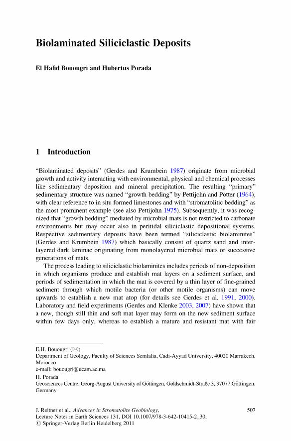
Biolaminated Siliciclastic Deposits
El Hafid Bouougri and Hubertus Porada
1 Introduction
“Biolaminated deposits” (Gerdes and Krumbein 1987) originate from microbial
growth and activity interacting with environmental, physical and chemical processes
like sedimentary deposition and mineral precipitation. The resulting “primary”
sedimentary structure was named “growth bedding” by Pettijohn and Potter (1964),
with clear reference to in situ formed limestones and with “stromatolitic bedding” as
the most prominent example (see also Pettijohn 1975). Subsequently, it was recog-
nized that “growth bedding” mediated by microbial mats is not restricted to carbonate
environments but may occur also in peritidal siliciclastic depositional systems.
Respective sedimentary deposits have been termed “siliciclastic biolaminites”
(Gerdes and Krumbein 1987) which basically consist of quartz sand and inter-
layered dark laminae originating from monolayered microbial mats or successive
generations of mats.
The process leading to siliciclastic biolaminites includes periods of non-deposition
in which organisms produce and establish mat layers on a sediment surface, and
periods of sedimentation in which the mat is covered by a thin layer of fine-grained
sediment through which motile bacteria (or other motile organisms) can move
upwards to establish a new mat atop (for details see Gerdes et al. 1991, 2000).
Laboratory and field experiments (Gerdes and Klenke 2003, 2007) have shown that
a new, though still thin and soft mat layer may form on the new sediment surface
within few days only, whereas to establish a mature and resistant mat with fair
E.H. Bouougri (*)
Department of Geology, Faculty of Sciences Semlalia, Cadi-Ayyad University, 40020 Marrakech,
Morocco
e-mail: [email protected]
H. Porada
Geosciences Centre, Georg-August University of G€ottingen, Goldschmidt-Straße 3, 37077 G€ottingen,Germany
J. Reitner et al., Advances in Stromatolite Geobiology,Lecture Notes in Earth Sciences 131, DOI 10.1007/978-3-642-10415-2_30,# Springer-Verlag Berlin Heidelberg 2011
507
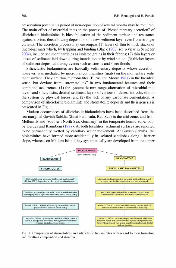
preservation potential, a period of non-deposition of several months may be required.
The main effect of microbial mats in the process of “biosedimentary accretion” of
siliciclastic biolaminites is biostabilization of the sediment surface and resistance
against erosion, thus allowing deposition of a new sediment layer even from stronger
currents. The accretion process may encompass (1) layers of thin to thick stacks of
microbial mats which, by trapping and binding (Black 1933; see review in Schieber
2004), include sediment particles as isolated grains in their fabrics; (2) thin layers or
lenses of sediment laid down during inundation or by wind action; (3) thicker layers
of sediment deposited during events such as storms and sheet floods.
Siliciclastic biolaminites are basically sedimentary deposits whose accretion,
however, was mediated by microbial communities (mats) on the momentary sedi-
ment surface. They are thus microbialites (Burne and Moore 1987) in the broadest
sense, but deviate from “stromatolites” in two fundamental features and their
combined occurrence: (1) the systematic mm-range alternation of microbial mat
layers and siliciclastic, detrital sediment layers of various thickness introduced into
the system by physical forces, and (2) the lack of any carbonate cementation. A
comparison of siliciclastic biolaminite and stromatolite deposits and their genesis is
presented in Fig. 1.
Modern occurrences of siliciclastic biolaminites have been described from the
sea-marginal Gavish Sabkha (Sinai Peninsula, Red Sea) in the arid zone, and from
Mellum Island (southern North Sea, Germany) in the temperate humid zone, both
by Gerdes and Krumbein (1987). At both localities, sediment surfaces are reported
to be permanently wetted by capillary water movement. At Gavish Sabkha, the
biolaminites have formed more accidentally in isolated sandlobes along a barrier
slope, whereas on Mellum Island they systematically are developed from the upper
Fig. 1 Comparison of stromatolites and siliciclastic biolaminites with regard to their formation
and resulting composition and structure
508 E.H. Bouougri and H. Porada
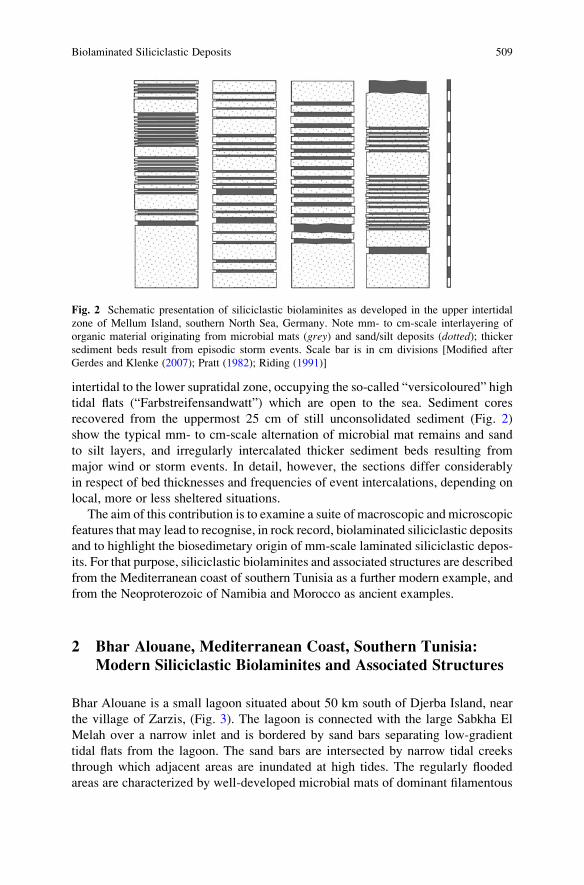
intertidal to the lower supratidal zone, occupying the so-called “versicoloured” high
tidal flats (“Farbstreifensandwatt”) which are open to the sea. Sediment cores
recovered from the uppermost 25 cm of still unconsolidated sediment (Fig. 2)
show the typical mm- to cm-scale alternation of microbial mat remains and sand
to silt layers, and irregularly intercalated thicker sediment beds resulting from
major wind or storm events. In detail, however, the sections differ considerably
in respect of bed thicknesses and frequencies of event intercalations, depending on
local, more or less sheltered situations.
The aim of this contribution is to examine a suite of macroscopic and microscopic
features that may lead to recognise, in rock record, biolaminated siliciclastic deposits
and to highlight the biosedimetary origin of mm-scale laminated siliciclastic depos-
its. For that purpose, siliciclastic biolaminites and associated structures are described
from the Mediterranean coast of southern Tunisia as a further modern example, and
from the Neoproterozoic of Namibia and Morocco as ancient examples.
2 Bhar Alouane, Mediterranean Coast, Southern Tunisia:
Modern Siliciclastic Biolaminites and Associated Structures
Bhar Alouane is a small lagoon situated about 50 km south of Djerba Island, near
the village of Zarzis, (Fig. 3). The lagoon is connected with the large Sabkha El
Melah over a narrow inlet and is bordered by sand bars separating low-gradient
tidal flats from the lagoon. The sand bars are intersected by narrow tidal creeks
through which adjacent areas are inundated at high tides. The regularly flooded
areas are characterized by well-developed microbial mats of dominant filamentous
Fig. 2 Schematic presentation of siliciclastic biolaminites as developed in the upper intertidal
zone of Mellum Island, southern North Sea, Germany. Note mm- to cm-scale interlayering of
organic material originating from microbial mats (grey) and sand/silt deposits (dotted); thickersediment beds result from episodic storm events. Scale bar is in cm divisions [Modified after
Gerdes and Klenke (2007); Pratt (1982); Riding (1991)]
Biolaminated Siliciclastic Deposits 509
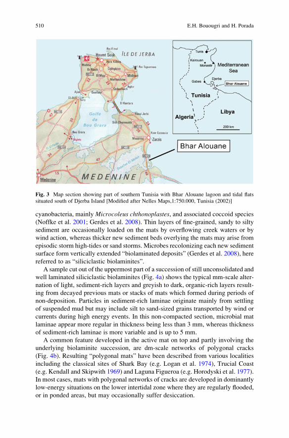
cyanobacteria, mainly Microcoleus chthonoplastes, and associated coccoid species
(Noffke et al. 2001; Gerdes et al. 2008). Thin layers of fine-grained, sandy to silty
sediment are occasionally loaded on the mats by overflowing creek waters or by
wind action, whereas thicker new sediment beds overlying the mats may arise from
episodic storm high-tides or sand storms. Microbes recolonizing each new sediment
surface form vertically extended “biolaminated deposits” (Gerdes et al. 2008), here
referred to as “siliciclastic biolaminites”.
A sample cut out of the uppermost part of a succession of still unconsolidated and
well laminated siliciclastic biolaminites (Fig. 4a) shows the typical mm-scale alter-
nation of light, sediment-rich layers and greyish to dark, organic-rich layers result-
ing from decayed previous mats or stacks of mats which formed during periods of
non-deposition. Particles in sediment-rich laminae originate mainly from settling
of suspended mud but may include silt to sand-sized grains transported by wind or
currents during high energy events. In this non-compacted section, microbial mat
laminae appear more regular in thickness being less than 3 mm, whereas thickness
of sediment-rich laminae is more variable and is up to 5 mm.
A common feature developed in the active mat on top and partly involving the
underlying biolaminite succession, are dm-scale networks of polygonal cracks
(Fig. 4b). Resulting “polygonal mats” have been described from various localities
including the classical sites of Shark Bay (e.g. Logan et al. 1974), Trucial Coast
(e.g. Kendall and Skipwith 1969) and Laguna Figueroa (e.g. Horodyski et al. 1977).
In most cases, mats with polygonal networks of cracks are developed in dominantly
low-energy situations on the lower intertidal zone where they are regularly flooded,
or in ponded areas, but may occasionally suffer desiccation.
Fig. 3 Map section showing part of southern Tunisia with Bhar Alouane lagoon and tidal flats
situated south of Djerba Island [Modified after Nelles Maps,1:750.000, Tunisia (2002)]
510 E.H. Bouougri and H. Porada
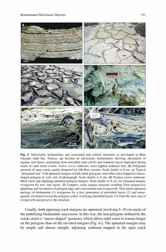
Usually, both opposing crack margins are upturned, involving 5–10 cm stacks of
the underlying biolaminite succession. In this way, the mat polygons defined by the
cracks attain a “saucer-shaped” geometry which allows tidal water to remain longer
on the polygons than on the elevated margins (Fig. 4c). The upturned margins may
be simple and almost straight, adjoining sediment trapped in the open crack
Fig. 4 Siliciclastic biolaminites and associated mat related structures as developed at Bhar
Alouane tidal flat, Tunisia. (a) Section of siliciclastic biolaminites showing alternation of
organic-rich layers originating from microbial mats (dark) and sediment layers deposited during
storm or sand storm events. Yellow arrow indicates wave-rippled sediment bed. (b) Polygonal
network of open cracks, partly deepened by ebb-flow currents. Scale (knife) is 8 cm. (c) Typical
“polygonal mat” with upturned margins of individual polygons; note tidal water trapped in saucer-
shaped polygon at right side of photograph. Scale (knife) is 8 cm. (d) Section across sediment-
filled crack and adjoining upturned polygon margins. Scale (knife) is 8 cm. (e) Upturned margin,
overgrown by new mat layers. (f) Complex crack margin structure resulting from progressive
upturning and involution of polygon edge and concomitant mat overgrowth. Note initial upturned
package of biolaminite (1) overgrown by a first generation of microbial layers (2) and subse-
quently involuted toward the polygon centre; overlying microbial layers (3) form the next step of
overgrowth and preserve the structure
Biolaminated Siliciclastic Deposits 511
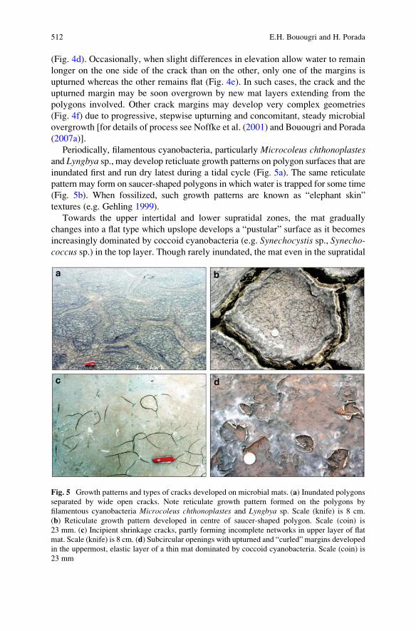
(Fig. 4d). Occasionally, when slight differences in elevation allow water to remain
longer on the one side of the crack than on the other, only one of the margins is
upturned whereas the other remains flat (Fig. 4e). In such cases, the crack and the
upturned margin may be soon overgrown by new mat layers extending from the
polygons involved. Other crack margins may develop very complex geometries
(Fig. 4f) due to progressive, stepwise upturning and concomitant, steady microbial
overgrowth [for details of process see Noffke et al. (2001) and Bouougri and Porada
(2007a)].
Periodically, filamentous cyanobacteria, particularly Microcoleus chthonoplastesand Lyngbya sp., may develop reticluate growth patterns on polygon surfaces that are
inundated first and run dry latest during a tidal cycle (Fig. 5a). The same reticulate
pattern may form on saucer-shaped polygons in which water is trapped for some time
(Fig. 5b). When fossilized, such growth patterns are known as “elephant skin”
textures (e.g. Gehling 1999).
Towards the upper intertidal and lower supratidal zones, the mat gradually
changes into a flat type which upslope develops a “pustular” surface as it becomes
increasingly dominated by coccoid cyanobacteria (e.g. Synechocystis sp., Synecho-coccus sp.) in the top layer. Though rarely inundated, the mat even in the supratidal
Fig. 5 Growth patterns and types of cracks developed on microbial mats. (a) Inundated polygons
separated by wide open cracks. Note reticulate growth pattern formed on the polygons by
filamentous cyanobacteria Microcoleus chthonoplastes and Lyngbya sp. Scale (knife) is 8 cm.
(b) Reticulate growth pattern developed in centre of saucer-shaped polygon. Scale (coin) is
23 mm. (c) Incipient shrinkage cracks, partly forming incomplete networks in upper layer of flat
mat. Scale (knife) is 8 cm. (d) Subcircular openings with upturned and “curled” margins developed
in the uppermost, elastic layer of a thin mat dominated by coccoid cyanobacteria. Scale (coin) is
23 mm
512 E.H. Bouougri and H. Porada
zone is still supplied with water from below due to capillary groundwater move-
ment and evaporative pumping (Hs€u and Siegenthaler 1969; Porada et al. 2007).
Nevertheless the surface layer may undergo desiccation and shrinkage and develop
cracks which may be isolated and small or form incomplete networks (Fig. 5c).
Incipient cracks may evolve to circular openings with curled margins when the
uppermost, elastic and EPS-rich layer locally gets torn during the shrinkage process
(Fig. 5d).
The flat and pustular mat types are not underlain by biolaminites which appear to
be restricted to areas that regularly undergo tidal inundation, or to isolated ponds
that are recharged during spring high tides.
3 Neoproterozoic Vingerbreek Member, Schwarzrand
Subgroup, Nama Group, Namibia: Ancient Siliciclastic
Biolaminites and Associated Structures
The Vingerbreek Member is a succession of shale, siltstone and sandstone, all
deposited in tidal and low-energy shoreline depositional environments (Germs
1983), that form the upper part of the Nudaus Formation at the base of the
Schwarzrand Subgroup. Its age is between ca. 548 and 545 Ma, according to
U-Pb zircon dates from ash layers in the underlying Kuibis Subgroup and overlying
Urusis Formation, respectively (Grotzinger et al. 1995).
In the lower part of the Vingerbreek Member, a more than 20 m thick succession
of siliciclastic biolaminites with intercalated sandstone beds and heterolithic silt-
stone/mudstone units is preserved on Haruchas farm (Fig. 6) (Bouougri and Porada
2007b, c). Sedimentary features indicate deposition in a low-energy, shallow,
intertidal position behind a protecting sand barrier. The biolaminites occur as
m-scale units of thinly interbedded layers of siltstone and dark to black material
of variable thickness and proportions (Fig. 7a). In thin section, the biolaminites
consist of packages (0.5–1.5 mm thick) of 5–10 alternating light and dark layers,
separated by mm-scale light layers of sand/silt sized sediment (Fig. 7b). The dark
layers, made up of mainly sericite, Fe-oxides and scarce relics of carbonaceous
material, are considered to represent former microbial mats, whereas the light
layers are sediment deposited on the mats and subsequently overgrown by new
mat layers.
Evidence of the participation of mats that once colonized the sedimentary
surfaces and contributed to the accretion of thick stacked layers of biolaminite,
include mat-related structures like “elephant skin” (reticulate growth pattern)
(Fig. 7c; cf. Fig. 5a, b), spindle-shaped and lenticular shrinkage cracks (Fig. 7d),
subcircular cracks with “curled” margins (Fig. 7e; cf. Fig. 5d), and Kinneyia-type
wrinkle structures (Fig. 7f). Most diagnostic for the identification of biolaminites
are upturned to overturned crack margins, locally combined in networks, involving
stacks of biolaminites, which are overlain by continuous biolaminated deposits
preserving the original sedimentation plane. Two types of crack margin structures
Biolaminated Siliciclastic Deposits 513
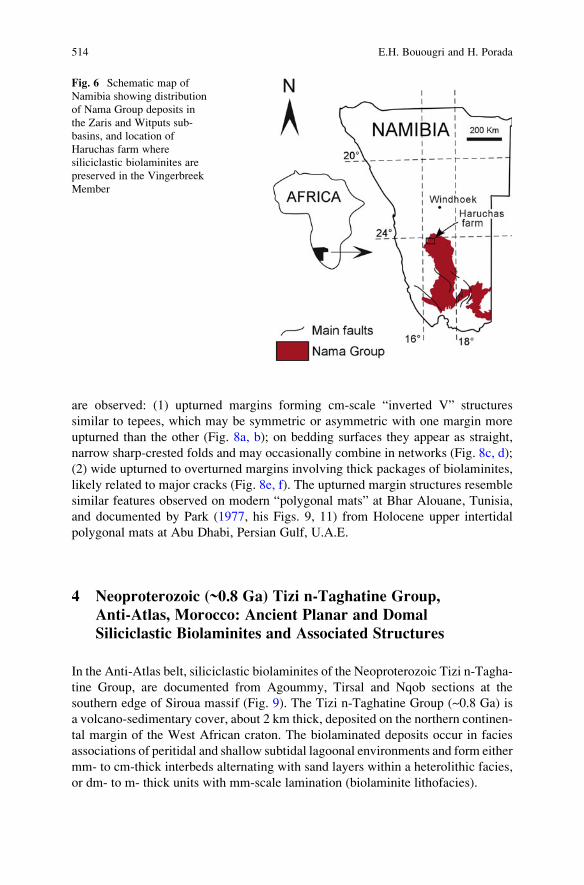
are observed: (1) upturned margins forming cm-scale “inverted V” structures
similar to tepees, which may be symmetric or asymmetric with one margin more
upturned than the other (Fig. 8a, b); on bedding surfaces they appear as straight,
narrow sharp-crested folds and may occasionally combine in networks (Fig. 8c, d);
(2) wide upturned to overturned margins involving thick packages of biolaminites,
likely related to major cracks (Fig. 8e, f). The upturned margin structures resemble
similar features observed on modern “polygonal mats” at Bhar Alouane, Tunisia,
and documented by Park (1977, his Figs. 9, 11) from Holocene upper intertidal
polygonal mats at Abu Dhabi, Persian Gulf, U.A.E.
4 Neoproterozoic (~0.8 Ga) Tizi n-Taghatine Group,
Anti-Atlas, Morocco: Ancient Planar and Domal
Siliciclastic Biolaminites and Associated Structures
In the Anti-Atlas belt, siliciclastic biolaminites of the Neoproterozoic Tizi n-Tagha-
tine Group, are documented from Agoummy, Tirsal and Nqob sections at the
southern edge of Siroua massif (Fig. 9). The Tizi n-Taghatine Group (~0.8 Ga) is
a volcano-sedimentary cover, about 2 km thick, deposited on the northern continen-
tal margin of the West African craton. The biolaminated deposits occur in facies
associations of peritidal and shallow subtidal lagoonal environments and form either
mm- to cm-thick interbeds alternating with sand layers within a heterolithic facies,
or dm- to m- thick units with mm-scale lamination (biolaminite lithofacies).
Fig. 6 Schematic map of
Namibia showing distribution
of Nama Group deposits in
the Zaris and Witputs sub-
basins, and location of
Haruchas farm where
siliciclastic biolaminites are
preserved in the Vingerbreek
Member
514 E.H. Bouougri and H. Porada
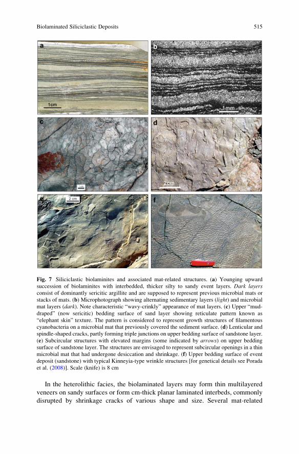
In the heterolithic facies, the biolaminated layers may form thin multilayered
veneers on sandy surfaces or form cm-thick planar laminated interbeds, commonly
disrupted by shrinkage cracks of various shape and size. Several mat-related
Fig. 7 Siliciclastic biolaminites and associated mat-related structures. (a) Younging upward
succession of biolaminites with interbedded, thicker silty to sandy event layers. Dark layersconsist of dominantly sericitic argillite and are supposed to represent previous microbial mats or
stacks of mats. (b) Microphotograph showing alternating sedimentary layers (light) and microbial
mat layers (dark). Note characteristic “wavy-crinkly” appearance of mat layers. (c) Upper “mud-
draped” (now sericitic) bedding surface of sand layer showing reticulate pattern known as
“elephant skin” texture. The pattern is considered to represent growth structures of filamentous
cyanobacteria on a microbial mat that previously covered the sediment surface. (d) Lenticular and
spindle-shaped cracks, partly forming triple junctions on upper bedding surface of sandstone layer.
(e) Subcircular structures with elevated margins (some indicated by arrows) on upper bedding
surface of sandstone layer. The structures are envisaged to represent subcircular openings in a thin
microbial mat that had undergone desiccation and shrinkage. (f) Upper bedding surface of event
deposit (sandstone) with typical Kinneyia-type wrinkle structures [for genetical details see Porada
et al. (2008)]. Scale (knife) is 8 cm
Biolaminated Siliciclastic Deposits 515
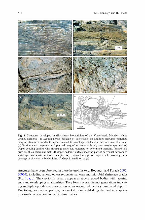
structures have been observed in these heteroliths (e.g. Bouougri and Porada 2002,
2007d), including among others reticulate patterns and microbial shrinkage cracks
(Fig. 10a, b). The crack-fills usually appear as superimposed bodies with tapering
ends and overlapping relationships. They form several distinct generations indicat-
ing multiple episodes of desiccation of an organosedimentary laminated deposit.
Due to high rate of compaction, the crack-fills are welded together and now appear
as a single generation on the bedding surface.
Fig. 8 Structures developed in siliciclastic biolaminites of the Vingerbreek Member, Nama
Group, Namibia. (a) Section across package of siliciclastic biolaminites showing “upturned
margin” structures similar to tepees, related to shrinkage cracks in a previous microbial mat.
(b) Section across asymmetric “upturned margin” structure with only one margin upturned. (c)
Upper bedding surface with shrinkage crack and upturned to overturned margins, formed in a
previous thick microbial mat. (d) Upper bedding surface showing part of polygonal network of
shrinkage cracks with upturned margins. (e) Upturned margin of major crack involving thick
package of siliciclastic biolaminite. (f) Graphic rendition of (e)
516 E.H. Bouougri and H. Porada
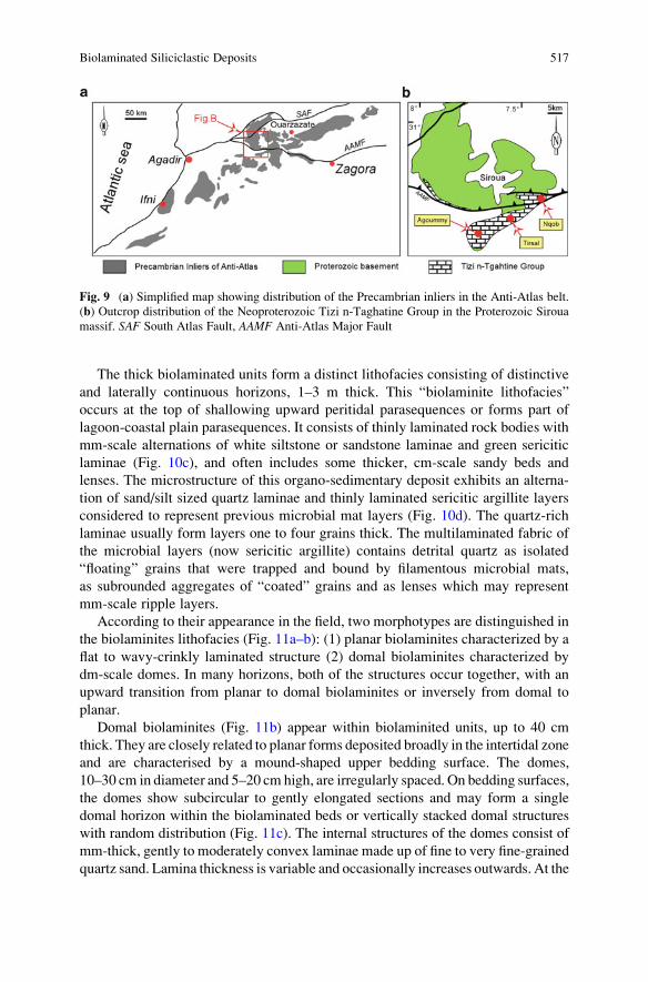
The thick biolaminated units form a distinct lithofacies consisting of distinctive
and laterally continuous horizons, 1–3 m thick. This “biolaminite lithofacies”
occurs at the top of shallowing upward peritidal parasequences or forms part of
lagoon-coastal plain parasequences. It consists of thinly laminated rock bodies with
mm-scale alternations of white siltstone or sandstone laminae and green sericitic
laminae (Fig. 10c), and often includes some thicker, cm-scale sandy beds and
lenses. The microstructure of this organo-sedimentary deposit exhibits an alterna-
tion of sand/silt sized quartz laminae and thinly laminated sericitic argillite layers
considered to represent previous microbial mat layers (Fig. 10d). The quartz-rich
laminae usually form layers one to four grains thick. The multilaminated fabric of
the microbial layers (now sericitic argillite) contains detrital quartz as isolated
“floating” grains that were trapped and bound by filamentous microbial mats,
as subrounded aggregates of “coated” grains and as lenses which may represent
mm-scale ripple layers.
According to their appearance in the field, two morphotypes are distinguished in
the biolaminites lithofacies (Fig. 11a–b): (1) planar biolaminites characterized by a
flat to wavy-crinkly laminated structure (2) domal biolaminites characterized by
dm-scale domes. In many horizons, both of the structures occur together, with an
upward transition from planar to domal biolaminites or inversely from domal to
planar.
Domal biolaminites (Fig. 11b) appear within biolaminited units, up to 40 cm
thick. They are closely related to planar forms deposited broadly in the intertidal zone
and are characterised by a mound-shaped upper bedding surface. The domes,
10–30 cm in diameter and 5–20 cm high, are irregularly spaced. On bedding surfaces,
the domes show subcircular to gently elongated sections and may form a single
domal horizon within the biolaminated beds or vertically stacked domal structures
with random distribution (Fig. 11c). The internal structures of the domes consist of
mm-thick, gently to moderately convex laminae made up of fine to very fine-grained
quartz sand. Lamina thickness is variable and occasionally increases outwards. At the
Fig. 9 (a) Simplified map showing distribution of the Precambrian inliers in the Anti-Atlas belt.
(b) Outcrop distribution of the Neoproterozoic Tizi n-Taghatine Group in the Proterozoic Siroua
massif. SAF South Atlas Fault, AAMF Anti-Atlas Major Fault
Biolaminated Siliciclastic Deposits 517
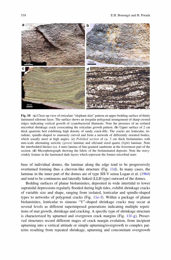
base of individual domes, the laminae along the edge tend to be progressively
overturned forming thus a chevron-like structure (Fig. 11d). In many cases, the
laminae in the inner part of the domes are of type SH-V sensu Logan et al. (1964)
and tend to be continuous and laterally linked (LLH type) outward of the domes.
Bedding surfaces of planar biolaminites, deposited in wide intertidal to lower
supratidal depressions regularly flooded during high tides, exhibit shrinkage cracks
of variable size and shape, ranging from isolated, lenticular and spindle-shaped
types to networks of polygonal cracks (Fig. 11e–f). Within a package of planar
biolaminites, lenticular to sinuous “V”-shaped shrinkage cracks may occur at
several levels as different superimposed generations indicating multiple repeti-
tions of mat growth, shrinkage and cracking. A specific type of shrinkage structure
is characterized by upturned and overgrown crack margins (Fig. 11f–g). Preser-
ved structures record different stages of crack margin evolution, from incipient
upturning into a vertical attitude or simple upturning/overgrowth to complex pat-
terns resulting from repeated shrinkage, upturning and concomitant overgrowth
Fig. 10 (a) Close-up view of reticulate “elephant skin” pattern on upper bedding surface of thinly
laminated siltstone layer. The surface shows an irregular polygonal arrangement of sharp-crested
ridges indicating vertical growth of cyanobacterial filaments. Note the presence of an isolated
microbial shrinkage crack crosscutting the reticulate growth pattern. (b) Upper surface of 2 cm
thick quartzite bed exhibiting high density of sandy crack-fills. The cracks are lenticular, tri-
radiate, spindle-shaped to sinuously curved and form a network of differently oriented bodies,
which usually meet at high angles. (c) Polished section of ca. 3 cm thick biolaminites with
mm-scale alternating sericitic (green) laminae and silt/sand sized quartz (light) laminae. Note
the interbedded thicker (ca. 4 mm) lamina of fine-grained sandstone at the lowermost part of the
section. (d) Microphotograph showing the fabric of the biolaminated deposits. Note the wavy-
crinkly feature in the laminated dark layers which represent the former microbial mats
518 E.H. Bouougri and H. Porada
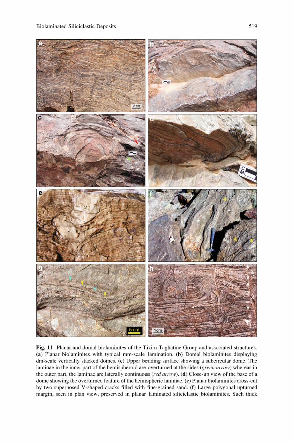
Fig. 11 Planar and domal biolaminites of the Tizi n-Taghatine Group and associated structures.
(a) Planar biolaminites with typical mm-scale lamination. (b) Domal biolaminites displaying
dm-scale vertically stacked domes. (c) Upper bedding surface showing a subcircular dome. The
laminae in the inner part of the hemispheroid are overturned at the sides (green arrow) whereas inthe outer part, the laminae are laterally continuous (red arrow). (d) Close-up view of the base of a
dome showing the overturned feature of the hemispheric laminae. (e) Planar biolaminites cross-cut
by two superposed V-shaped cracks filled with fine-grained sand. (f) Large polygonal upturned
margin, seen in plan view, preserved in planar laminated siliciclastic biolaminites. Such thick
Biolaminated Siliciclastic Deposits 519
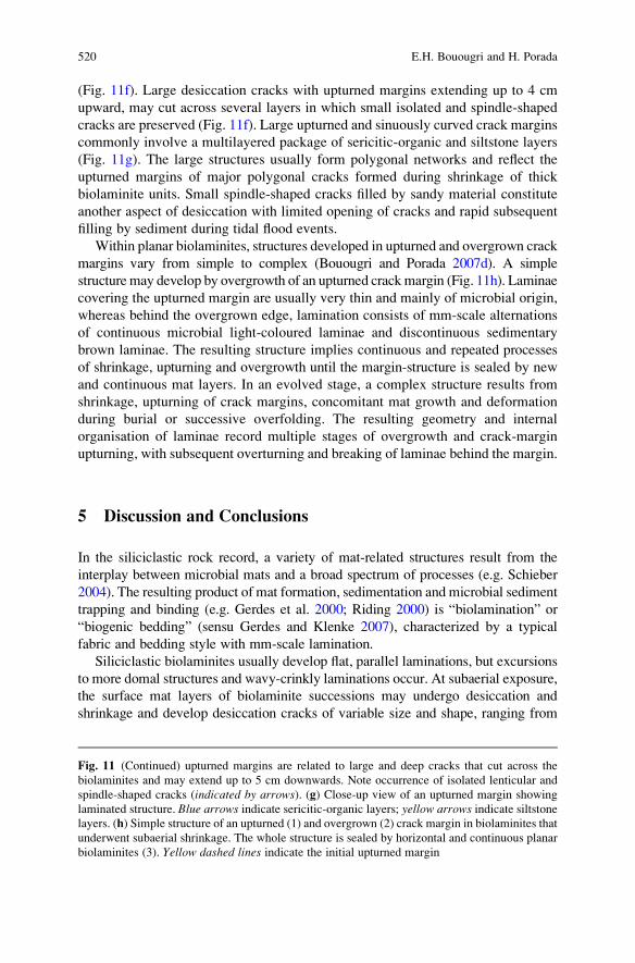
(Fig. 11f). Large desiccation cracks with upturned margins extending up to 4 cm
upward, may cut across several layers in which small isolated and spindle-shaped
cracks are preserved (Fig. 11f). Large upturned and sinuously curved crack margins
commonly involve a multilayered package of sericitic-organic and siltstone layers
(Fig. 11g). The large structures usually form polygonal networks and reflect the
upturned margins of major polygonal cracks formed during shrinkage of thick
biolaminite units. Small spindle-shaped cracks filled by sandy material constitute
another aspect of desiccation with limited opening of cracks and rapid subsequent
filling by sediment during tidal flood events.
Within planar biolaminites, structures developed in upturned and overgrown crack
margins vary from simple to complex (Bouougri and Porada 2007d). A simple
structure may develop by overgrowth of an upturned crackmargin (Fig. 11h). Laminae
covering the upturned margin are usually very thin and mainly of microbial origin,
whereas behind the overgrown edge, lamination consists of mm-scale alternations
of continuous microbial light-coloured laminae and discontinuous sedimentary
brown laminae. The resulting structure implies continuous and repeated processes
of shrinkage, upturning and overgrowth until the margin-structure is sealed by new
and continuous mat layers. In an evolved stage, a complex structure results from
shrinkage, upturning of crack margins, concomitant mat growth and deformation
during burial or successive overfolding. The resulting geometry and internal
organisation of laminae record multiple stages of overgrowth and crack-margin
upturning, with subsequent overturning and breaking of laminae behind the margin.
5 Discussion and Conclusions
In the siliciclastic rock record, a variety of mat-related structures result from the
interplay between microbial mats and a broad spectrum of processes (e.g. Schieber
2004). The resulting product of mat formation, sedimentation and microbial sediment
trapping and binding (e.g. Gerdes et al. 2000; Riding 2000) is “biolamination” or
“biogenic bedding” (sensu Gerdes and Klenke 2007), characterized by a typical
fabric and bedding style with mm-scale lamination.
Siliciclastic biolaminites usually develop flat, parallel laminations, but excursions
to more domal structures and wavy-crinkly laminations occur. At subaerial exposure,
the surface mat layers of biolaminite successions may undergo desiccation and
shrinkage and develop desiccation cracks of variable size and shape, ranging from
Fig. 11 (Continued) upturned margins are related to large and deep cracks that cut across the
biolaminites and may extend up to 5 cm downwards. Note occurrence of isolated lenticular and
spindle-shaped cracks (indicated by arrows). (g) Close-up view of an upturned margin showing
laminated structure. Blue arrows indicate sericitic-organic layers; yellow arrows indicate siltstonelayers. (h) Simple structure of an upturned (1) and overgrown (2) crack margin in biolaminites that
underwent subaerial shrinkage. The whole structure is sealed by horizontal and continuous planar
biolaminites (3). Yellow dashed lines indicate the initial upturned margin
520 E.H. Bouougri and H. Porada
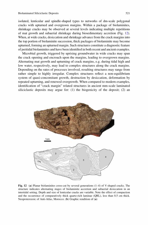
isolated, lenticular and spindle-shaped types to networks of dm-scale polygonal
cracks with upturned and overgrown margins. Within a package of biolaminites,
shrinkage cracks may be observed at several levels indicating multiple repetitions
of mat growth and subaerial shrinkage during biosedimentary accretion (Fig. 12).
When, at wide cracks, desiccation and shrinkage advance from the crack margins into
the top portion of biolaminite succession, thick packages of biolaminite may become
upturned, forming an upturned margin. Such structures constitute a diagnostic feature
of peritidal biolaminites and have been identified in both recent and ancient examples.
Microbial growth, triggered by uprising groundwater in wide cracks may span
the crack opening and encroach upon the margins, leading to overgrown margins.
Alternating mat growth and upturning of crack margins, e.g. during tidal high and
low water, respectively, may lead to complex structures along the crack margins.
Depending on the rates of processes involved, resulting structures may range from
rather simple to highly irregular. Complex structures reflect a non-equilibrium
system of quasi-concomitant growth, destruction by desiccation, deformation by
repeated upturning, and renewed overgrowth. When compared to modern examples,
identification of “crack margin” related structures in ancient mm-scale laminated
siliciclastic deposits may argue for: (1) the biogenicity of the deposit; (2) an
Fig. 12 (a) Planar biolaminites cross-cut by several generations (1–4) of V-shaped cracks. The
structure indicates alternating stages of biolaminite accretion and subaerial desiccation in an
intertidal setting. Depth and size of lenticular cracks are variable. Note the effect of compaction
and the occurrence of comparatively thick quartz-rich laminae (QRL), less than 0.5 cm thick.
Neoproterozoic of Anti-Atlas, Morocco. (b) Graphic rendition of (a)
Biolaminated Siliciclastic Deposits 521
intertidal environment, where continuous/episodic mat growth and shrinkage is
closely accompanying cyclic accretion of mat-sediment doublets; (3) processes
involving continuous and repeated stages of shrinkage, upturning and overgrowth
until the margin structure is completely sealed by new and continuous mat layers.
In the recent example of Tunisia, topographic factors in the tidal flats appear as a
significant control on biolaminite distribution and accretion. Continuous growth
and accretion of siliciclastic biolaminites reflect a stable system with low rate of
sediment supply and no major disturbance by high-energy events, punctuated by
periodic flooding during high tides. Such conditions are met locally in shallow
depressions and ponds of variable size in the intertidal zone, where they are filled
periodically by sea water during high tides and where “polygonal mats” and
accretion of thick biolaminites are prevailing.
In the example of the Neoproterozoic of Anti-Atlas (Morocco), dm- to m-thick
planar and domal siliciclastic biolaminites, deposited in relatively protected areas,
are clearly differentiated from adjacent and associated rock bodies and form thus a
distinctive “biolaminite facies” within a lagoonal and pertidal facies association
(Bouougri and Porada 2007d). The subtidal shallow lagoonal biolaminates are
mainly domal, whereas the planar type often occurs at the base of domal forms
and lacks any features indicating subaerial shrinkage. In the heterolithic facies
which was deposited in an intertidal area exposed intermittently to high-energy
events, biolaminites constitute only thin layers in the facies and occur as laminated
organo-sedimentary interbeds up to few cm thick. In this case and according to the
sedimentary features of the heteroliths, the thickness and accretion of biolaminites
apparently was controlled by the frequency of periodic and aperiodic sedimentary
high-energy events depositing mm- to cm-thick sand layers. A similar control was
observed in the modern biolaminites of Mellum Island (Gerdes and Klenke 2007),
in which accretion and thickness of biolaminites is correlated with the duration of
periods with low-energy and low-rate sedimentation.
Recognition in rock records of a “siliciclastic biolaminite facies” and “biogenic
bedding”, mainly of peritidal settings, requires a combination of macroscopic and
microscopic features typical of biolaminated deposits. The examples of siliciclastic
biolaminites described above, in which bedding and accretion are driven by the
interplay of mat growth and sedimentation, indicate that both ancient and modern
examples reveal similar structures related to mat growth-shrinkage-overgrowth
processes. Preservation and recognition in rock records of such structures may
constitute a significant sedimentological tool and strongly argues for a biosedimen-
tary origin of ancient well laminated siliciclastic deposits and for an intertidal to
lower supratidal (i.e. peritidal) depositional environment periodically emerged and
regularly flooded by tides or during storm events. Within facies associations con-
taining siliciclastic biolaminites, occurrences of other “mat-related structures”
constitute further evidence for the biogenicity of these deposits.
Acknowledgments The authors are very grateful to VW-Foundation (I/78 706) for financial
support of this research work. Special thanks to all persons who helped us during fieldworks in
Namibia, Tunisia and Morocco. Review by J. Reitner greatly improved this manuscript.
522 E.H. Bouougri and H. Porada
References
Black M (1933) The algal sediments of Andros Islands, Bahamas. Philosophical Transactions of
the Royal Society of London. Series B 222:165–192
Bouougri EH, Porada H (2002) Mat-related sedimentary structures in Neoproterozoic peritidal
passive margin deposits of the West African Craton (Anti-Atlas, Morocco). Sedimentary
Geology 153:85–106
Bouougri EH, Porada H (2007a) Complex structures associated with siliciclastic biolaminites. In:
Schieber J, Bose PK, Eriksson PG, Banerjee S, Sarkar S, Altermann W, Catuneanu O (eds) An
Atlas of microbial mat features preserved within the siliciclastic rock record. Elsevier,
Amsterdam, pp 111–115
Bouougri EH, Porada H (2007b) Siliciclastic biolaminites indicative of widespread microbial mats
in the Neoproterozoic Nama Group of Namibia. Journal of African Earth Sciences 48:38–48
Bouougri EH, Porada H (2007c) Mat-related features from the Terminal Ediacaran Nudaus
Formation, Nama Group, Namibia. In: Schieber J, Bose PK, Eriksson PG, Banerjee S, Sarkar S,
Altermann W, Catuneanu O (eds) An Atlas of microbial mat features preserved within the
siliciclastic rock record. Elsevier, Amsterdam, pp 214–221
Bouougri EH, Porada H (2007d) Mat-related features from the Neoproterozoic Tizi n-Taghatine
Group, Anti-Atlas belt, Morocco. In: Schieber J, Bose PK, Eriksson PG, Banerjee S, Sarkar S,
Altermann W, Catuneanu O (eds) An Atlas of microbial mat features preserved within the
siliciclastic rock record. Elsevier, Amsterdam, pp 198–207
Burne RV, Moore LS (1987) Microbialites: organosedimentary deposits of benthic microbial
communities. Palaios 2:241–254
Gehling JG (1999) Microbial mats in Terminal Proterozoic siliciclastics: Ediacaran death masks.
Palaios 14:40–57
Gerdes G, Klenke T (2003) Geologische Bedeutung €okologischer Zeitr€aume in biogener Schichtung
(Mikrobenmatten, potentielle Stromatolithe). Mitteilungen der Gesellschaft der Geologie- und
Bergbaustudenten in Osterreich 46:35–49
Gerdes G, Klenke T (2007) States of biogenic bedding as records of the interplay of ecologic time
and environment: a case study of modern siliciclastic sediments, Mellum Island, southern
North Sea. Senckenbergiana maritima 37:129–144
Gerdes G, Krumbein WE (1987) Biolaminated deposits. In: Bhattacharya GM, Friedmann GM,
Neugebauer HJ, Seilacher A (eds) Lecture notes in earth sciences 9. Springer-Verlag, Berlin,
pp 1–183
Gerdes G, Krumbein WE, Reineck HE (1991) Biolaminations-Ecological versus depositional
dynamics. In: Einsele G, Ricken W, Seilacher A (eds) Cycles and events in stratigraphy.
Springer Verlag, Berlin, pp 592–607
Gerdes G, Klenke T, Noffke N (2000) Microbial signatures in peritidal siliciclastic sediments: a
catalogue. Sedimentology 47:279–308
Gerdes G, Porada H, Bouougri EH (2008) Bio-sedimentary structures evolving from the interaction
of microbial mats, burrowing beetles and the physical environment of Tunisian coastal sabkhas.
Senckenbergiana Maritima 38:45–58
Germs GBJ (1983) Implications of a sedimentary facies and depositional environmental analysis
of the Nama Group in SouthWest Africa/Namibia. Geological Society of South Africa, Special
Publication 11:89–114
Grotzinger JP, Bowring SA, Saylor BZ, Kaufman AJ (1995) New biostratigraphic and geochronologic
constraints on early animal evolution. Science 270:598–604
Horodyski RJ, Bloeser B, Vonder Haar S (1977) Laminated algal mats from a coastal lagoon,
Laguna Mormona, Baja California, Mexico. Journal of Sedimentary Petrology 47:680–696
Hs€u K, Siegenthaler C (1969) Preliminary experiments on hydrodynamic movement induced by
evaporation and their bearing on the dolomite problem. Sedimentology 12:11–25
Biolaminated Siliciclastic Deposits 523
Kendall CGSC, Skipwith PAd’E (1969) Geomorphology of a recent shallow-water carbonate
province, Khor Al Bazam, Trucial Coast, Southwest Persian Gulf. The Geological Society of
America Bulletin 80:865–892
Logan BW, Rezak R, Ginsburg RN (1964) Classification and environmental significance of algal
stromatolites. Journal of Geology 72:68–83
Logan BW, Hoffman P, Gebelein CD (1974) Algal mats, cryptalgal fabrics, and structures,
Hamelin Pool, Western Australia. American Association of Petroleum Geologists Memoir
22:140–194
Noffke N, Gerdes G, Klenke T, Krumbein W (2001) Microbially induced sedimentary structures
indicating climatological, hydrological and depositional conditionswithinRecent and Pleistocene
coastal facies zones (Southern Tunesia). Facies 44:23–30
Park RK (1977) The preservation potential of some recent stromatolites. Sedimentology 24:
485–506
Pettijohn FJ (1975) Sedimentary rocks, 3rd edition. Harper & Row, New York, 628
Pettijohn FJ, Potter PE (1964) Atlas and glossary of primary sedimentary structures. Springer-
Verlag, Berlin
Porada H, Bouougri EH, Ghergut J (2007) Hydraulic conditions and mat-related structures in tidal
flats and coastal sabkhas. In: Schieber J, Bose PK, Eriksson PG, Banerjee S, Sarkar S,
Altermann W, Catuneanu O (eds) An Atlas of microbial mat features preserved within the
siliciclastic rock record. Elsevier, Amsterdam, pp 258–265
Porada H, Ghergut J, Bouougri EH (2008) Kinneyia-type wrinkle structures – critical review and
model of formation. Palaios 23:63–77
Pratt BR (1982) Stromatolite decline – a reconsideration. Geology 10:512–515
Riding R (1991) Classification of microbial carbonates. In: Riding R (ed) Calcareous algae and
stromatolites. Springer-Verlag, Berlin, pp 21–51
Riding R (2000) Microbial carbonates: The geological record of calcified bacterial-algal mats and
biofilms. Sedimentology 47:179–214
Schieber J (2004) Microbial mats in the siliciclastic rock record: a summary of diagnostic features.
In: Eriksson PG, Altermann W, Nelson D, Mueller WU, Catuneanu O, Strand K (eds) The
Precambrian earth: tempos and events. Developments in Precambrian geology. Elsevier,
Amsterdam, pp 663–672
524 E.H. Bouougri and H. Porada
Microbial Binding as a Probable Cause
of Taphonomic Variability of Vendian
Fossils: Carbonate Casting?
Ekaterina A. Serezhnikova
1 Introduction
The exceptional preservation of Ediacaran organisms is problematic, and the cause
of the unique conservation is difficult to prove for fossil material because of
restrictions imposed by the incomplete palaeontological record. Observation of
the various preservation forms is particularly important and provides the basis for
further reconstructions of organisms and the Precambrian community structure.
2 Short Historical Background
Studies of Precambrian organisms have repeated, to a certain extent, the following
stages of the history of palaeontology: the organic origin of the first finds was
doubted; the views on their systematic position were not once changed; and the
usual transition along the stages of knowledge from sporadic and ambiguous finds
to rival hypotheses, is observed. It appears essential to dwell on the history of
taphonomic studies of Ediacaran fossils because it is responsible for the description
of various taphomorphs, such as how the major part of so-called jellyfish, which is
the most common representative of Late Proterozoic fossil communities, were
reinterpreted as benthic organisms. The circular imprints closely resemble Solnho-
fen jellyfish; for instance, Ediacaria flindersi, which is one of the first Ediacaran
finds, is similar to the Jurassic Rhizostomites admirandus. Subsequently it became a
tradition to arrange the Ediacaran organisms within the existing phyla, to consider
the majority of them as jellyfish and to consider the Late Precambrian to be the time
E.A. Serezhnikova
Borissiak Paleontological Institute, Russian Academy of Sciences, Profsoyuznaya 123, Moscow
117997, Russia
e-mail: [email protected]
J. Reitner et al., Advances in Stromatolite Geobiology,Lecture Notes in Earth Sciences 131, DOI 10.1007/978-3-642-10415-2_31,# Springer-Verlag Berlin Heidelberg 2011
525
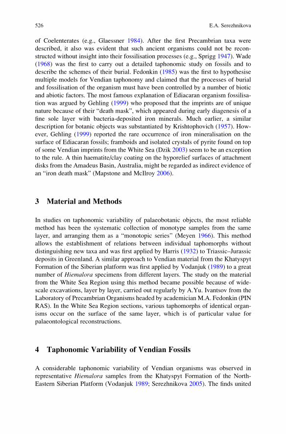
of Coelenterates (e.g., Glaessner 1984). After the first Precambrian taxa were
described, it also was evident that such ancient organisms could not be recon-
structed without insight into their fossilisation processes (e.g., Sprigg 1947). Wade
(1968) was the first to carry out a detailed taphonomic study on fossils and to
describe the schemes of their burial. Fedonkin (1985) was the first to hypothesise
multiple models for Vendian taphonomy and claimed that the processes of burial
and fossilisation of the organism must have been controlled by a number of biotic
and abiotic factors. The most famous explanation of Ediacaran organism fossilisa-
tion was argued by Gehling (1999) who proposed that the imprints are of unique
nature because of their “death mask”, which appeared during early diagenesis of a
fine sole layer with bacteria-deposited iron minerals. Much earlier, a similar
description for botanic objects was substantiated by Krishtophovich (1957). How-
ever, Gehling (1999) reported the rare occurrence of iron mineralisation on the
surface of Ediacaran fossils; framboids and isolated crystals of pyrite found on top
of some Vendian imprints from the White Sea (Dzik 2003) seem to be an exception
to the rule. A thin haematite/clay coating on the hyporelief surfaces of attachment
disks from the Amadeus Basin, Australia, might be regarded as indirect evidence of
an “iron death mask” (Mapstone and McIlroy 2006).
3 Material and Methods
In studies on taphonomic variability of palaeobotanic objects, the most reliable
method has been the systematic collection of monotype samples from the same
layer, and arranging them as a “monotopic series” (Meyen 1966). This method
allows the establishment of relations between individual taphomorphs without
distinguishing new taxa and was first applied by Harris (1932) to Triassic–Jurassic
deposits in Greenland. A similar approach to Vendian material from the Khatyspyt
Formation of the Siberian platform was first applied by Vodanjuk (1989) to a great
number of Hiemalora specimens from different layers. The study on the material
from the White Sea Region using this method became possible because of wide-
scale excavations, layer by layer, carried out regularly by A.Yu. Ivantsov from the
Laboratory of Precambrian Organisms headed by academician M.A. Fedonkin (PIN
RAS). In the White Sea Region sections, various taphomorphs of identical organ-
isms occur on the surface of the same layer, which is of particular value for
palaeontological reconstructions.
4 Taphonomic Variability of Vendian Fossils
A considerable taphonomic variability of Vendian organisms was observed in
representative Hiemalora samples from the Khatyspyt Formation of the North-
Eastern Siberian Platform (Vodanjuk 1989; Serezhnikova 2005). The finds united
526 E.A. Serezhnikova
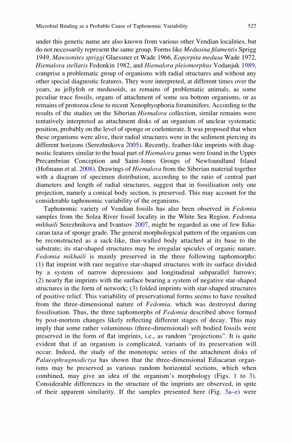
under this generic name are also known from various other Vendian localities, but
do not necessarily represent the same group. Forms likeMedusina filamentis Sprigg1949,Mawsonites spriggi Glaessner et Wade 1966, Eoporpita medusaWade 1972,
Hiemalora stellaris Fedonkin 1982, and Hiemalora pleiomorphus Vodanjuk 1989,
comprise a problematic group of organisms with radial structures and without any
other special diagnostic features. They were interpreted, at different times over the
years, as jellyfish or medusoids, as remains of problematic animals, as some
peculiar trace fossils, organs of attachment of some sea bottom organisms, or as
remains of protozoa close to recent Xenophyophoria foraminifers. According to the
results of the studies on the Siberian Hiemalora collection, similar remains were
tentatively interpreted as attachment disks of an organism of unclear systematic
position, probably on the level of sponge or coelenterate. It was proposed that when
these organisms were alive, their radial structures were in the sediment piercing its
different horizons (Serezhnikova 2005). Recently, feather-like imprints with diag-
nostic features similar to the basal part ofHiemalora genus were found in the UpperPrecambrian Conception and Saint-Jones Groups of Newfoundland Island
(Hofmann et al. 2008). Drawings of Hiemalora from the Siberian material together
with a diagram of specimen distribution, according to the ratio of central part
diameters and length of radial structures, suggest that in fossilisation only one
projection, namely a conical body section, is preserved. This may account for the
considerable taphonomic variability of the organisms.
Taphonomic variety of Vendian fossils has also been observed in Fedomiasamples from the Solza River fossil locality in the White Sea Region. Fedomiamikhaili Serezhnikova and Ivantsov 2007, might be regarded as one of few Edia-
caran taxa of sponge grade. The general morphological pattern of the organism can
be reconstructed as a sack-like, thin-walled body attached at its base to the
substrate; its star-shaped structures may be irregular spicules of organic nature.
Fedomia mikhaili is mainly preserved in the three following taphomorphs:
(1) flat imprint with rare negative star-shaped structures with its surface divided
by a system of narrow depressions and longitudinal subparallel furrows;
(2) nearly flat imprints with the surface bearing a system of negative star-shaped
structures in the form of network; (3) folded imprints with star-shaped structures
of positive relief. This variability of preservational forms seems to have resulted
from the three-dimensional nature of Fedomia, which was destroyed during
fossilisation. Thus, the three taphomorphs of Fedomia described above formed
by post-mortem changes likely reflecting different stages of decay. This may
imply that some rather voluminous (three-dimensional) soft bodied fossils were
preserved in the form of flat imprints, i.e., as random “projections”. It is quite
evident that if an organism is complicated, variants of its preservation will
occur. Indeed, the study of the monotopic series of the attachment disks of
Palaeophragmodictya has shown that the three-dimensional Ediacaran organ-
isms may be preserved as various random horizontal sections, which when
combined, may give an idea of the organism’s morphology (Figs. 1 to 3).
Considerable differences in the structure of the imprints are observed, in spite
of their apparent similarity. If the samples presented here (Fig. 3a–e) were
Microbial Binding as a Probable Cause of Taphonomic Variability 527
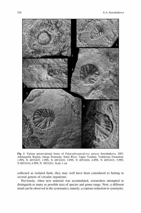
collected as isolated finds, they may well have been considered to belong to
several genera of circular organisms.
Previously, when new material was accumulated, researchers attempted to
distinguish as many as possible taxa of species and genus range. Now, a different
trend can be observed in the systematics, namely, a copious reduction to synonyms.
Fig. 1 Various preservational forms of Palaeophragmodictya spinosa Serezhnikova, 2007,
Arkhangelsk Region, Onega Peninsula, Solza River, Upper Vendian, Verkhovka Formation:
1-PIN, N 4853/425, 2-PIN, N 4853/410, 3-PIN, N 4853/424, 4-PIN, N 4853/415, 5-PIN,
N 4853/416, 6-PIN, N 4853/421. Scale 1 cm
528 E.A. Serezhnikova
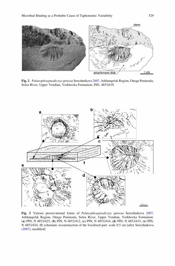
Fig. 3 Various preservational forms of Palaeophragmodictya spinosa Serezhnikova 2007,
Arkhangelsk Region, Onega Peninsula, Solza River, Upper Vendian, Verkhovka Formation:
(a) PIN, N 4853/425, (b) PIN, N 4853/412, (c) PIN, N 4853/416, (d) PIN, N 4853/415, (e) PIN,
N 4853/424, (f) schematic reconstruction of the fossilised part: scale 0.5 cm [after Serezhnikova
(2007), modified]
Fig. 2 Palaeophragmodictya spinosa Serezhnikova 2007, Arkhangelsk Region, Onega Peninsula,Solza River, Upper Vendian, Verkhovka Formation; PIN, 4853/418
Microbial Binding as a Probable Cause of Taphonomic Variability 529
As is seen from the analysis of the Palaeophragmodictya monotope series from the
White Sea, this unification may be justified in some cases, but generally it may be
too early for application as detailed collections from the same layers are widely
lacking. Furthermore, it may be expected that monotope series of different taxa may
contain similar forms. As a result of the study on the imprints from the Solza
locality, Palaeophragmodictya were suggested to be interpreted as parts of a
problematic organism with attachment disks. It was considered previously to be
the first Precambrian sponge represented by macro-remains, and Gehling and Rigby
(1996) included it in the recent Hexactinellidae family. In spite of the fact
that Paleophragmodictya has morphological features inherent in sponges, e.g., a
net on the disk surface and spiny radial structures, the general morphology aligns
them more closely to Coelenterate; although a sporadic appearance of radial
elements considerably decreases the likeliness of this affinity (Serezhnikova
2007). Apparently, Palaeophragmodictya from the White Sea Region was the
first Precambrian sedentary organism in which bilateral symmetry was detected in
ontogeny (Serezhnikova 2007). The absence of a regular pattern in the appearance
of radial elements somewhat reduces the likelihood of their affinity to Coelenterata,
or it should be accepted that, in the Vendian, the Coelenterata had not yet acquired
regular patterns that were present in later epochs. On the whole, the recognition of
various taphomorphs of Precambrian organisms allows the assessment of the
structure of the organisms and the possible succession of fossilisation events.
5 Discussion
The taphonomic variability of Vendian fossils has been studied in a great number of
single-type specimens collected from the same depositional surface. Some imprints
proved to be specific cross-sections of three-dimensional organisms preserved on an
accidental horizontal surface of the fossiliferous layer. For example, Palaeophrag-modictya spinosa Serezhnikova 2007 from the Solza River locality of the South-
eastern White Sea Region were preserved as composite moulds of attachment disks
and part of stems (Fig. 2). The imprint of the outer zone with thin radial furrows
reflects the structure of the lower surface of the attachment disc, and the central part
is a negative cast of an internal cavity. The presence of the cavity over the
organism’s entire lifetime is documented by the rare specimens in which the central
parts have been filled with sediment (Fig. 3a). The organic substance of the inner
cavity had not decayed completely at the time of lithification, and therefore various
preservational forms of the central parts of the fossils developed. The specimen
with a negative outer zone and mesh-like structures is unique to this locality; it
appears to be an external mould of the upper surface of the attachment disc
(Fig. 3a). Apparently, the variable forms of preservation which have been observed,
for instance, in Palaeophragmodictya from the Solza River locality, give an idea of
the fossilisation processes. It seems rather improbable that the various taphomorphs
of the voluminous organism were preserved only due to formation of an “iron death
530 E.A. Serezhnikova

mask” on the surfaces of the collapsing bodies, though framboids and isolated
crystals of pyrite do occur occasionally on the surfaces of fossil imprints (Figs. 4
and 5). Such unique fossilisation might have been also the result of a quite rapid
consolidation of a thin fossiliferous layer due to cementation by carbonate. For
example, carbonate accounts for 1–3% in the sandstone, but for 32% in some
siltstone from the fossiliferous Neoproterozoic Arumbera Sandstone, Australia
(Mapstone and McIlroy 2006), and for 10–15% in the siltstone from the Solza
River locality in the Southeastern White Sea Region. The data on qualitative
elemental composition of the bedding surface bearing Palaeophragmodictya spi-nosa show the presence of calcium and carbon (Fig. 6). Furthermore, it has recently
been reported that calcium carbonate is found in cyanobacterial films that develop
on modern beaches of the North Sea barrier island Schiermonnikoog, Netherlands
(Kremer et al. 2008). It may be speculated that a similar process may have occurred
in widespread microbial mat grounds of Precambrian environments (Fig. 7); but the
carbonate grains might have been completely dissolved over time. According to
experimental data, the dissolution of different rocks has different rates even during
the same period of time (Lasaga 1983), e.g., carbonates dissolve quite rapidly in
contrast to silicates. It is also well known that under anoxic conditions in modern
settings, carbonate shells are destroyed over a short time interval. Hydrosulphuric
contamination of the sediment, traces of which are observed in the Vendian sedi-
ments of the South-Eastern White Sea Region, seem to have been typical of
Fig. 4 SEM photograph of the surface microrelief of Palaeophragmodictya spinosa with pyrite
framboids; qualitative elemental composition is in the upper left corner. Arkhangelsk Region,
Onega Peninsula, Solza River, Upper Vendian, Verkhovka Formation
Microbial Binding as a Probable Cause of Taphonomic Variability 531
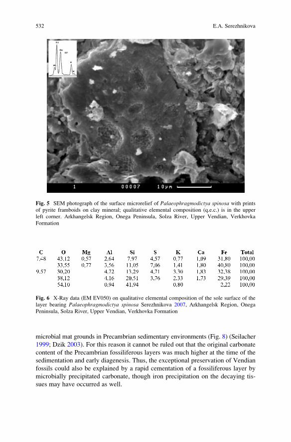
microbial mat grounds in Precambrian sedimentary environments (Fig. 8) (Seilacher
1999; Dzik 2003). For this reason it cannot be ruled out that the original carbonate
content of the Precambrian fossiliferous layers was much higher at the time of the
sedimentation and early diagenesis. Thus, the exceptional preservation of Vendian
fossils could also be explained by a rapid cementation of a fossiliferous layer by
microbially precipitated carbonate, though iron precipitation on the decaying tis-
sues may have occurred as well.
Fig. 5 SEM photograph of the surface microrelief of Palaeophragmodictya spinosa with prints
of pyrite framboids on clay mineral; qualitative elemental composition (q.e.c.) is in the upper
left corner. Arkhangelsk Region, Onega Peninsula, Solza River, Upper Vendian, Verkhovka
Formation
Fig. 6 X-Ray data (EM EV050) on qualitative elemental composition of the sole surface of the
layer bearing Palaeophragmodictya spinosa Serezhnikova 2007, Arkhangelsk Region, Onega
Peninsula, Solza River, Upper Vendian, Verkhovka Formation
532 E.A. Serezhnikova
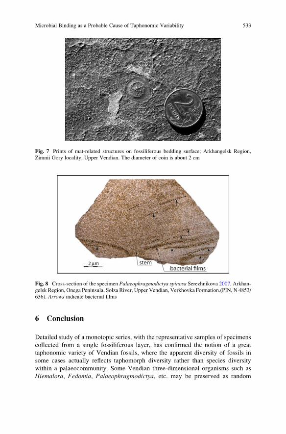
6 Conclusion
Detailed study of a monotopic series, with the representative samples of specimens
collected from a single fossiliferous layer, has confirmed the notion of a great
taphonomic variety of Vendian fossils, where the apparent diversity of fossils in
some cases actually reflects taphomorph diversity rather than species diversity
within a palaeocommunity. Some Vendian three-dimensional organisms such as
Hiemalora, Fedomia, Palaeophragmodictya, etc. may be preserved as random
Fig. 8 Cross-section of the specimen Palaeophragmodictya spinosa Serezhnikova 2007, Arkhan-gelsk Region, Onega Peninsula, Solza River, Upper Vendian, Verkhovka Formation.(PIN, N 4853/
636). Arrows indicate bacterial films
Fig. 7 Prints of mat-related structures on fossiliferous bedding surface; Arkhangelsk Region,
Zimnii Gory locality, Upper Vendian. The diameter of coin is about 2 cm
Microbial Binding as a Probable Cause of Taphonomic Variability 533
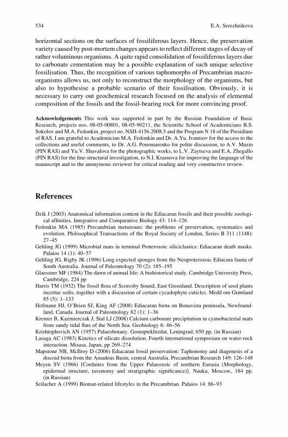
horizontal sections on the surfaces of fossiliferous layers. Hence, the preservation
variety caused by post-mortem changes appears to reflect different stages of decay of
rather voluminous organisms. A quite rapid consolidation of fossiliferous layers due
to carbonate cementation may be a possible explanation of such unique selective
fossilisation. Thus, the recognition of various taphomorphs of Precambrian macro-
organisms allows us, not only to reconstruct the morphology of the organisms, but
also to hypothesise a probable scenario of their fossilisation. Obviously, it is
necessary to carry out geochemical research focused on the analysis of elemental
composition of the fossils and the fossil-bearing rock for more convincing proof.
Acknowledgements This work was supported in part by the Russian Foundation of Basic
Research, projects nos. 08-05-00801, 08-05-90211, the Scientific School of Academicians B.S.
Sokolov and M.A. Fedonkin, project no. NSH-4156.2008.5 and the Program N 18 of the Presidium
of RAS. I am grateful to Academician M.A. Fedonkin and Dr. A.Yu. Ivantsov for the access to the
collections and useful comments, to Dr. A.G. Ponomarenko for polite discussion, to A.V. Mazin
(PIN RAS) and Yu.V. Shuvalova for the photographic works, to L.V. Zaytseva and E.A. Zhegallo
(PIN RAS) for the fine-structural investigation, to N.I. Krasnova for improving the language of the
manuscript and to the anonymous reviewer for critical reading and very constructive review.
References
Dzik J (2003) Anatomical information content in the Ediacaran fossils and their possible zoologi-
cal affinities. Integrative and Comparative Biology 43: 114–126
Fedonkin MA (1985) Precambrian metazoans: the problems of preservation, systematics and
evolution. Philosophical Transactions of the Royal Society of London, Series B 311 (1148):
27–45
Gehling JG (1999) Microbial mats in terminal Proterozoic siliciclastics: Ediacaran death masks.
Palaios 14 (1): 40–57
Gehling JG, Rigby JK (1996) Long expected sponges from the Neoproterozoic Ediacara fauna of
South Australia. Journal of Paleontology 70 (2): 185–195
Glaessner MF (1984) The dawn of animal life: A biohistorical study. Cambridge University Press,
Cambridge, 224 pp
Harris TM (1932) The fossil flora of Scoresby Sound, East Greenland. Description of seed plants
incertae sedis, together with a discussion of certain cycadophyte cuticles. Medd om Grønland
85 (5): 1–133
Hofmann HJ, O’Brien SJ, King AF (2008) Ediacaran biota on Bonavista peninsula, Newfound-
land, Canada. Journal of Paleontology 82 (1): 1–36
Kremer B, Kazmierczak J, Stal LJ (2008) Calcium carbonate precipitation in cyanobacterial mats
from sandy tidal flats of the North Sea. Geobiology 6: 46–56
Krishtophovich AN (1957) Palaeobotany. Gostoptekhizdat, Leningrad, 650 pp, (in Russian)
Lasaga AC (1983) Kinetics of silicate dissolution. Fourth international symposium on water-rock
interaction. Misasa, Japan, pp 269–274
Mapstone NB, McIlroy D (2006) Ediacaran fossil preservation: Taphonomy and diagenesis of a
discoid biota from the Amadeus Basin, central Australia. Precambrian Research 149: 126–148
Meyen SV (1966) [Cordaites from the Upper Palaeozoic of northern Eurasia (Morphology,
epidermal structure, taxonomy and stratigraphic significance)]. Nauka, Moscow, 184 pp,
(in Russian)
Seilacher A (1999) Biomat-related lifestyles in the Precambrian. Palaios 14: 86–93
534 E.A. Serezhnikova

Serezhnikova EA (2005) [New interpretation of Hiemalora imprints from the Vendian of the
Olenek Uplift, northeast Siberian Platform]. Bulletin MOIP. Otdel Geologicheskii 80 (3):
26–32
Serezhnikova EA (2007) Palaeophragmodictya spinosa sp. nov., a Bilateral Benthic Organism
from the Vendian of the Southeastern White Sea Region. Paleontological Journal 41 (4):
360–369 (Original Russian Text published in Paleontologicheskii Zhurnal (2007)) 4: 16–24
Serezhnikova EA, Ivantsov AYu (2007) Fedomia mikhaili – A new spicule-bearing organism
of sponge grade from the Vendian (Ediacaran) of the White Sea, Russia. Palaeoworld
16: 319–324
Sprigg RC (1947) Early Cambrian? jellyfishes from the Flinders Ranges, South Australia. Trans-
actions of the Royal Society of South Australia 71: 221–224
Vodanjuk SA (1989) [Remains of non-skeletal Metazoa from Khatyspyt formation on the Olenek
Uplift]. Pozdnii dokembry and rannii paleozoi Sibiri. Actualnii voprosi stratigrafii: 61–75
[in Russian]
Wade M (1968) Preservation of soft bodied animals in Precambrian sandstones at Ediacara, South
Australia. Lethaia 1 (3): 238–267
Microbial Binding as a Probable Cause of Taphonomic Variability 535

.
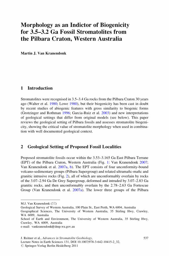
Morphology as an Indictor of Biogenicity
for 3.5–3.2 Ga Fossil Stromatolites from
the Pilbara Craton, Western Australia
Martin J. Van Kranendonk
1 Introduction
Stromatolites were recognised in 3.5–3.4 Ga rocks from the Pilbara Craton 30 years
ago (Walter et al. 1980; Lowe 1980), but their biogenicity has been cast in doubt
by recent studies of abiogenic features with gross similarity to biogenic forms
(Grotzinger and Rothman 1996; Garcia-Ruiz et al. 2003) and new interpretations
of geological settings that differ from original models (see below). This paper
reviews the geological setting of Pilbara fossils and assesses stromatolite biogeni-
city, showing the critical value of stromatolite morphology when used in combina-
tion with well documented geological context.
2 Geological Setting of Proposed Fossil Localities
Proposed stromatolite fossils occur within the 3.53–3.165 Ga East Pilbara Terrane
(EPT) of the Pilbara Craton, Western Australia (Fig. 1: Van Kranendonk 2007;
Van Kranendonk et al. 2007a, b). The EPT consists of four unconformity-bound
volcano-sedimentary groups (Pilbara Supergroup) and related ultramafic-mafic and
granitic intrusive rocks (Fig. 2), all of which are unconformably overlain by rocks
of the 3.07–2.94 Ga De Grey Supergroup, deformed and intruded by 3.07–2.83 Ga
granitic rocks, and then unconformably overlain by the 2.78–2.63 Ga Fortescue
Group (Van Kranendonk et al. 2007a). The lower three groups of the Pilbara
M.J. Van Kranendonk (*)
Geological Survey of Western Australia, 100 Plain St., East Perth, WA 6004, Australia
Geographical Sciences, The University of Western Australia, 35 Stirling Hwy, Crawley,
WA 6009, Australia
School of Earth and Environment, The University of Western Australia, 35 Stirling Hwy,
Crawley, WA 6009, Australia
e-mail: [email protected]
J. Reitner et al., Advances in Stromatolite Geobiology,Lecture Notes in Earth Sciences 131, DOI 10.1007/978-3-642-10415-2_32,# Springer-Verlag Berlin Heidelberg 2011
537
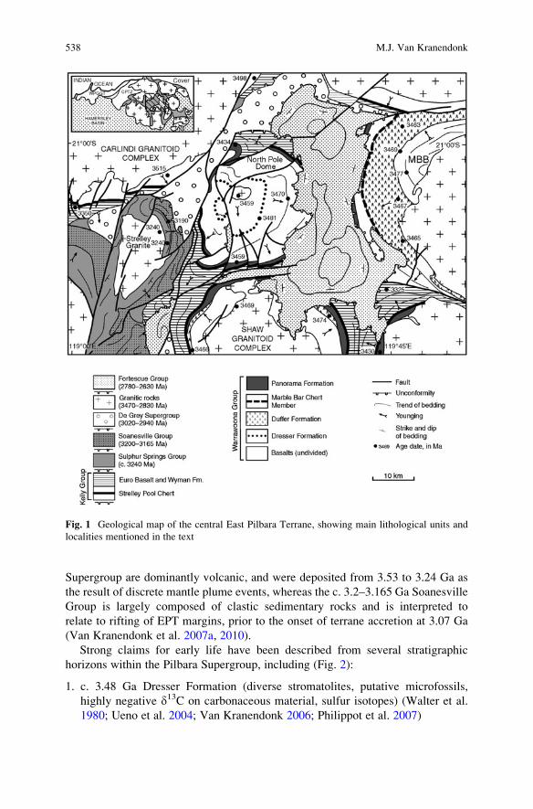
Supergroup are dominantly volcanic, and were deposited from 3.53 to 3.24 Ga as
the result of discrete mantle plume events, whereas the c. 3.2–3.165 Ga Soanesville
Group is largely composed of clastic sedimentary rocks and is interpreted to
relate to rifting of EPT margins, prior to the onset of terrane accretion at 3.07 Ga
(Van Kranendonk et al. 2007a, 2010).
Strong claims for early life have been described from several stratigraphic
horizons within the Pilbara Supergroup, including (Fig. 2):
1. c. 3.48 Ga Dresser Formation (diverse stromatolites, putative microfossils,
highly negative d13C on carbonaceous material, sulfur isotopes) (Walter et al.
1980; Ueno et al. 2004; Van Kranendonk 2006; Philippot et al. 2007)
Fig. 1 Geological map of the central East Pilbara Terrane, showing main lithological units and
localities mentioned in the text
538 M.J. Van Kranendonk
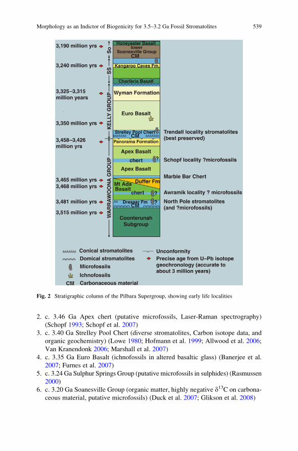
2. c. 3.46 Ga Apex chert (putative microfossils, Laser-Raman spectrography)
(Schopf 1993; Schopf et al. 2007)
3. c. 3.40 Ga Strelley Pool Chert (diverse stromatolites, Carbon isotope data, and
organic geochemistry) (Lowe 1980; Hofmann et al. 1999; Allwood et al. 2006;
Van Kranendonk 2006; Marshall et al. 2007)
4. c. 3.35 Ga Euro Basalt (ichnofossils in altered basaltic glass) (Banerjee et al.
2007; Furnes et al. 2007)
5. c. 3.24 Ga Sulphur Springs Group (putative microfossils in sulphides) (Rasmussen
2000)
6. c. 3.20 Ga Soanesville Group (organic matter, highly negative d13C on carbona-
ceous material, putative microfossils) (Duck et al. 2007; Glikson et al. 2008)
Fig. 2 Stratigraphic column of the Pilbara Supergroup, showing early life localities
Morphology as an Indictor of Biogenicity for 3.5–3.2 Ga Fossil Stromatolites 539
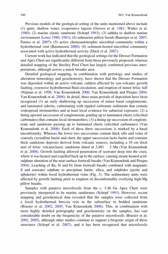
Previous models of the geological setting of the units mentioned above include
(1) quiet, shallow water, evaporative lagoon (Groves et al. 1981; Walter et al.
1980); (2) marine clastic sandstone (Schopf 1993); (3) sabkha to shallow marine
environment (Lowe 1980, 1983); (4) submarine pillow basalt (Banerjee et al. 2007;
Furnes et al. 2007); (5) active chemoautotrophic microbial community within a
hydrothermal vent (Rasmussen 2000); (6) sediment-hosted microbial community
associated with active hydrothermal activity (Duck et al. 2007).
Current work has indicated that the geological settings for the Dresser Formation
and Apex Chert are significantly different from those previously proposed, whereas
detailed mapping of the Strelley Pool Chert has largely confirmed previous inter-
pretations, although across a much broader area.
Detailed geological mapping, in combination with petrology and studies of
alteration mineralogy and geochemistry, have shown that the Dresser Formation
was deposited within an active volcanic caldera affected by syn-volcanic growth
faulting, extensive hydrothermal fluid circulation, and eruption of minor felsic tuff
(Nijman et al. 1998; Van Kranendonk 2006; Van Kranendonk and Pirajno 2004;
Van Kranendonk et al. 2008). In detail, three main periods of deposition have been
recognised (1) an early shallowing up succession of minor basal conglomerate,
and laminated siderite, culminating with rippled carbonate sediments that contain
widespread stromatolites and at least local evidence for subaerial exposure; (2) a
fining upward succession of conglomerate grading up to laminated cherts (silicified
carbonates) that contains local stromatolites; (3) a fining up succession of conglom-
erate and sandstone passing up to laminated cherts (silicified carbonates) (Van
Kranendonk et al. 2008). Each of these three successions is marked by a basal
unconformity. Whereas the lower two successions contain thick sills and veins of
coarsely crystalline barite and chert, the upper succession lacks barite and contains
thick sandstone deposits derived from volcanic sources, including a 10 cm thick
unit of felsic volcaniclastic sandstone dated at 3,481 � 2 Ma (Van Kranendonk
et al. 2008). Growth faulting allowed penetration of seawater deep into the crust,
where it was heated and expelled back up to the surface, causing steam-heated acid-
sulphate alteration of the near-surface footwall basalts (Van Kranendonk and Pirajno
2004). Leaching of Ba, Si and Fe from footwall basalts combined with magmatic
S and seawater sulphate to precipitate barite, silica, and sulphides (pyrite and
sphalerite) within fossil hydrothermal veins (Fig. 3). The sedimentary units were
affected by growth faulting prior to eruption of disconformably overlying high-Mg
pillow basalts.
Samples with putative microfossils from the c. 3.46 Ga Apex Chert were
previously interpreted to be marine sandstones (Schopf 1993). However, recent
mapping and geochemical data revealed that the samples were collected from
a fossil hydrothermal breccia vein in the subsurface to bedded sandstone
(Brasier et al. 2002, 2005; Van Kranendonk 2006). This, in combination with
more highly detailed petrography and geochemistry on the samples, has cast
considerable doubt on the biogenicity of the putative microfossils (Brasier et al.
2002, 2005), although other studies continue to support a biogenic origin of these
structures (Schopf et al. 2007), and it has been recognised that microfossils
540 M.J. Van Kranendonk
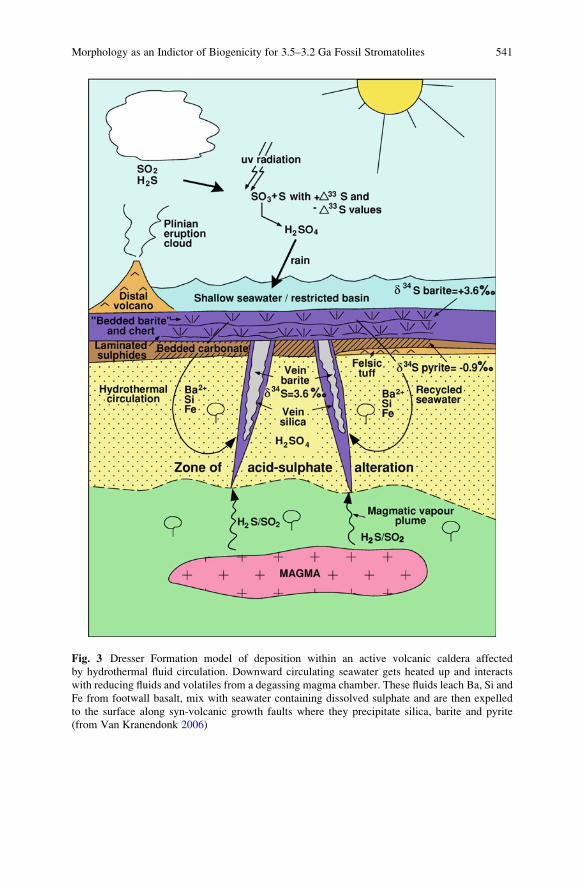
Fig. 3 Dresser Formation model of deposition within an active volcanic caldera affected
by hydrothermal fluid circulation. Downward circulating seawater gets heated up and interacts
with reducing fluids and volatiles from a degassing magma chamber. These fluids leach Ba, Si and
Fe from footwall basalt, mix with seawater containing dissolved sulphate and are then expelled
to the surface along syn-volcanic growth faults where they precipitate silica, barite and pyrite
(from Van Kranendonk 2006)
Morphology as an Indictor of Biogenicity for 3.5–3.2 Ga Fossil Stromatolites 541
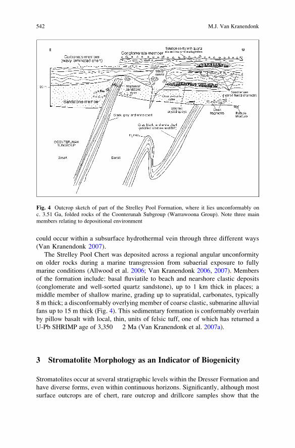
could occur within a subsurface hydrothermal vein through three different ways
(Van Kranendonk 2007).
The Strelley Pool Chert was deposited across a regional angular unconformity
on older rocks during a marine transgression from subaerial exposure to fully
marine conditions (Allwood et al. 2006; Van Kranendonk 2006, 2007). Members
of the formation include: basal fluviatile to beach and nearshore clastic deposits
(conglomerate and well-sorted quartz sandstone), up to 1 km thick in places; a
middle member of shallow marine, grading up to supratidal, carbonates, typically
8 m thick; a disconformably overlying member of coarse clastic, submarine alluvial
fans up to 15 m thick (Fig. 4). This sedimentary formation is conformably overlain
by pillow basalt with local, thin, units of felsic tuff, one of which has returned a
U-Pb SHRIMP age of 3,350 � 2 Ma (Van Kranendonk et al. 2007a).
3 Stromatolite Morphology as an Indicator of Biogenicity
Stromatolites occur at several stratigraphic levels within the Dresser Formation and
have diverse forms, even within continuous horizons. Significantly, although most
surface outcrops are of chert, rare outcrop and drillcore samples show that the
Fig. 4 Outcrop sketch of part of the Strelley Pool Formation, where it lies unconformably on
c. 3.51 Ga, folded rocks of the Coonterunah Subgroup (Warrawoona Group). Note three main
members relating to depositional environment
542 M.J. Van Kranendonk
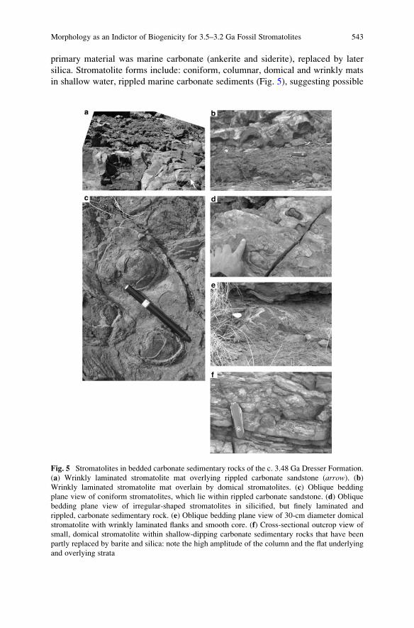
primary material was marine carbonate (ankerite and siderite), replaced by later
silica. Stromatolite forms include: coniform, columnar, domical and wrinkly mats
in shallow water, rippled marine carbonate sediments (Fig. 5), suggesting possible
Fig. 5 Stromatolites in bedded carbonate sedimentary rocks of the c. 3.48 Ga Dresser Formation.
(a) Wrinkly laminated stromatolite mat overlying rippled carbonate sandstone (arrow). (b)
Wrinkly laminated stromatolite mat overlain by domical stromatolites. (c) Oblique bedding
plane view of coniform stromatolites, which lie within rippled carbonate sandstone. (d) Oblique
bedding plane view of irregular-shaped stromatolites in silicified, but finely laminated and
rippled, carbonate sedimentary rock. (e) Oblique bedding plane view of 30-cm diameter domical
stromatolite with wrinkly laminated flanks and smooth core. (f) Cross-sectional outcrop view of
small, domical stromatolite within shallow-dipping carbonate sedimentary rocks that have been
partly replaced by barite and silica: note the high amplitude of the column and the flat underlying
and overlying strata
Morphology as an Indictor of Biogenicity for 3.5–3.2 Ga Fossil Stromatolites 543
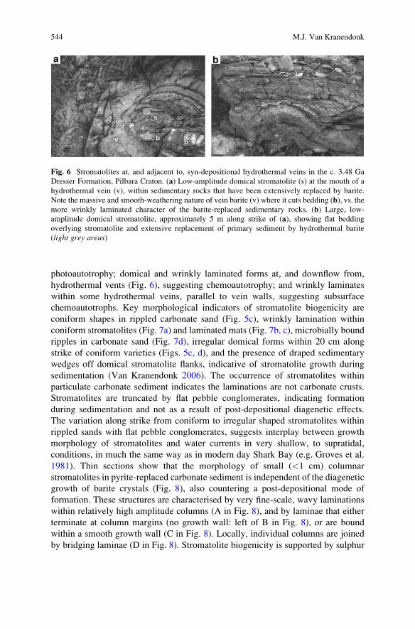
photoautotrophy; domical and wrinkly laminated forms at, and downflow from,
hydrothermal vents (Fig. 6), suggesting chemoautotrophy; and wrinkly laminates
within some hydrothermal veins, parallel to vein walls, suggesting subsurface
chemoautotrophs. Key morphological indicators of stromatolite biogenicity are
coniform shapes in rippled carbonate sand (Fig. 5c), wrinkly lamination within
coniform stromatolites (Fig. 7a) and laminated mats (Fig. 7b, c), microbially bound
ripples in carbonate sand (Fig. 7d), irregular domical forms within 20 cm along
strike of coniform varieties (Figs. 5c, d), and the presence of draped sedimentary
wedges off domical stromatolite flanks, indicative of stromatolite growth during
sedimentation (Van Kranendonk 2006). The occurrence of stromatolites within
particulate carbonate sediment indicates the laminations are not carbonate crusts.
Stromatolites are truncated by flat pebble conglomerates, indicating formation
during sedimentation and not as a result of post-depositional diagenetic effects.
The variation along strike from coniform to irregular shaped stromatolites within
rippled sands with flat pebble conglomerates, suggests interplay between growth
morphology of stromatolites and water currents in very shallow, to supratidal,
conditions, in much the same way as in modern day Shark Bay (e.g. Groves et al.
1981). Thin sections show that the morphology of small (<1 cm) columnar
stromatolites in pyrite-replaced carbonate sediment is independent of the diagenetic
growth of barite crystals (Fig. 8), also countering a post-depositional mode of
formation. These structures are characterised by very fine-scale, wavy laminations
within relatively high amplitude columns (A in Fig. 8), and by laminae that either
terminate at column margins (no growth wall: left of B in Fig. 8), or are bound
within a smooth growth wall (C in Fig. 8). Locally, individual columns are joined
by bridging laminae (D in Fig. 8). Stromatolite biogenicity is supported by sulphur
Fig. 6 Stromatolites at, and adjacent to, syn-depositional hydrothermal veins in the c. 3.48 Ga
Dresser Formation, Pilbara Craton. (a) Low-amplitude domical stromatolite (s) at the mouth of a
hydrothermal vein (v), within sedimentary rocks that have been extensively replaced by barite.
Note the massive and smooth-weathering nature of vein barite (v) where it cuts bedding (b), vs. the
more wrinkly laminated character of the barite-replaced sedimentary rocks. (b) Large, low-
amplitude domical stromatolite, approximately 5 m along strike of (a), showing flat bedding
overlying stromatolite and extensive replacement of primary sediment by hydrothermal barite
(light grey areas)
544 M.J. Van Kranendonk
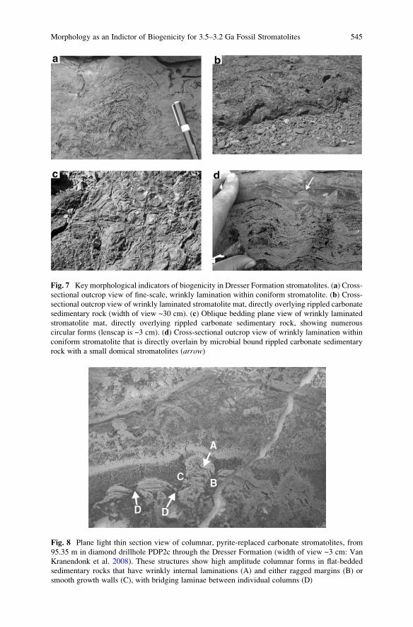
Fig. 7 Key morphological indicators of biogenicity in Dresser Formation stromatolites. (a) Cross-
sectional outcrop view of fine-scale, wrinkly lamination within coniform stromatolite. (b) Cross-
sectional outcrop view of wrinkly laminated stromatolite mat, directly overlying rippled carbonate
sedimentary rock (width of view ~30 cm). (c) Oblique bedding plane view of wrinkly laminated
stromatolite mat, directly overlying rippled carbonate sedimentary rock, showing numerous
circular forms (lenscap is ~3 cm). (d) Cross-sectional outcrop view of wrinkly lamination within
coniform stromatolite that is directly overlain by microbial bound rippled carbonate sedimentary
rock with a small domical stromatolites (arrow)
Fig. 8 Plane light thin section view of columnar, pyrite-replaced carbonate stromatolites, from
95.35 m in diamond drillhole PDP2c through the Dresser Formation (width of view ~3 cm: Van
Kranendonk et al. 2008). These structures show high amplitude columnar forms in flat-bedded
sedimentary rocks that have wrinkly internal laminations (A) and either ragged margins (B) or
smooth growth walls (C), with bridging laminae between individual columns (D)
Morphology as an Indictor of Biogenicity for 3.5–3.2 Ga Fossil Stromatolites 545
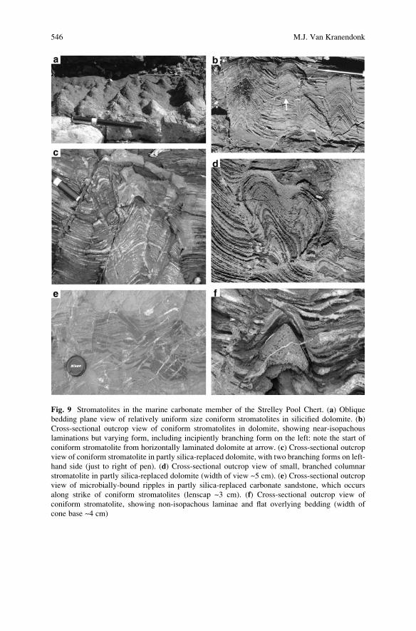
Fig. 9 Stromatolites in the marine carbonate member of the Strelley Pool Chert. (a) Oblique
bedding plane view of relatively uniform size coniform stromatolites in silicified dolomite. (b)
Cross-sectional outcrop view of coniform stromatolites in dolomite, showing near-isopachous
laminations but varying form, including incipiently branching form on the left: note the start of
coniform stromatolite from horizontally laminated dolomite at arrow. (c) Cross-sectional outcrop
view of coniform stromatolite in partly silica-replaced dolomite, with two branching forms on left-
hand side (just to right of pen). (d) Cross-sectional outcrop view of small, branched columnar
stromatolite in partly silica-replaced dolomite (width of view ~5 cm). (e) Cross-sectional outcrop
view of microbially-bound ripples in partly silica-replaced carbonate sandstone, which occurs
along strike of coniform stromatolites (lenscap ~3 cm). (f) Cross-sectional outcrop view of
coniform stromatolite, showing non-isopachous laminae and flat overlying bedding (width of
cone base ~4 cm)
546 M.J. Van Kranendonk
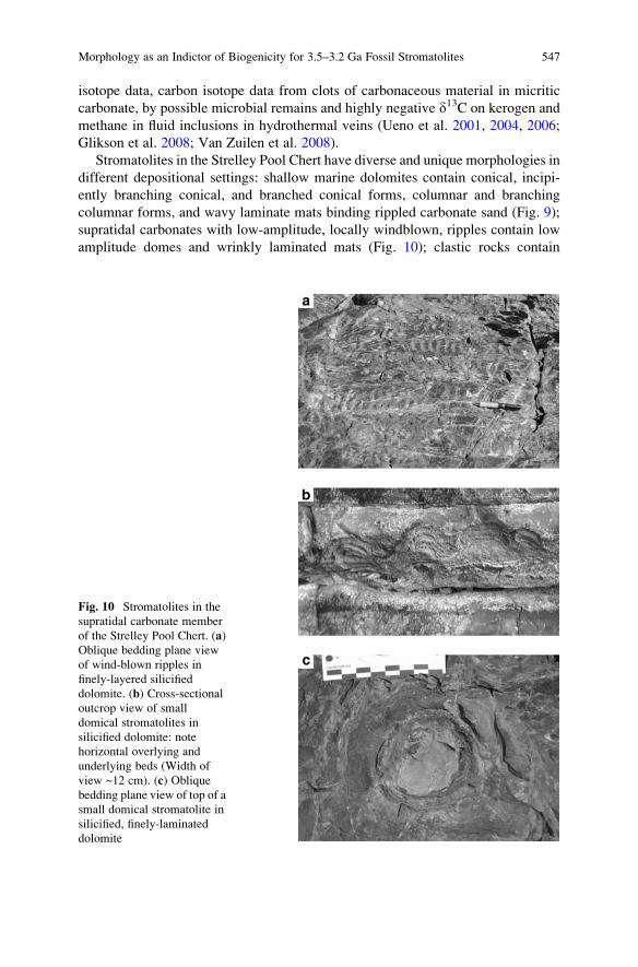
isotope data, carbon isotope data from clots of carbonaceous material in micritic
carbonate, by possible microbial remains and highly negative d13C on kerogen and
methane in fluid inclusions in hydrothermal veins (Ueno et al. 2001, 2004, 2006;
Glikson et al. 2008; Van Zuilen et al. 2008).
Stromatolites in the Strelley Pool Chert have diverse and unique morphologies in
different depositional settings: shallow marine dolomites contain conical, incipi-
ently branching conical, and branched conical forms, columnar and branching
columnar forms, and wavy laminate mats binding rippled carbonate sand (Fig. 9);
supratidal carbonates with low-amplitude, locally windblown, ripples contain low
amplitude domes and wrinkly laminated mats (Fig. 10); clastic rocks contain
Fig. 10 Stromatolites in the
supratidal carbonate member
of the Strelley Pool Chert. (a)
Oblique bedding plane view
of wind-blown ripples in
finely-layered silicified
dolomite. (b) Cross-sectional
outcrop view of small
domical stromatolites in
silicified dolomite: note
horizontal overlying and
underlying beds (Width of
view ~12 cm). (c) Oblique
bedding plane view of top of a
small domical stromatolite in
silicified, finely-laminated
dolomite
Morphology as an Indictor of Biogenicity for 3.5–3.2 Ga Fossil Stromatolites 547
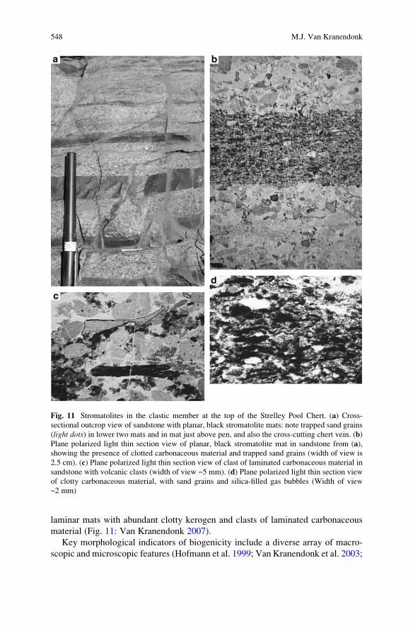
laminar mats with abundant clotty kerogen and clasts of laminated carbonaceous
material (Fig. 11: Van Kranendonk 2007).
Key morphological indicators of biogenicity include a diverse array of macro-
scopic and microscopic features (Hofmann et al. 1999; Van Kranendonk et al. 2003;
Fig. 11 Stromatolites in the clastic member at the top of the Strelley Pool Chert. (a) Cross-
sectional outcrop view of sandstone with planar, black stromatolite mats: note trapped sand grains
(light dots) in lower two mats and in mat just above pen, and also the cross-cutting chert vein. (b)
Plane polarized light thin section view of planar, black stromatolite mat in sandstone from (a),
showing the presence of clotted carbonaceous material and trapped sand grains (width of view is
2.5 cm). (c) Plane polarized light thin section view of clast of laminated carbonaceous material in
sandstone with volcanic clasts (width of view ~5 mm). (d) Plane polarized light thin section view
of clotty carbonaceous material, with sand grains and silica-filled gas bubbles (Width of view
~2 mm)
548 M.J. Van Kranendonk
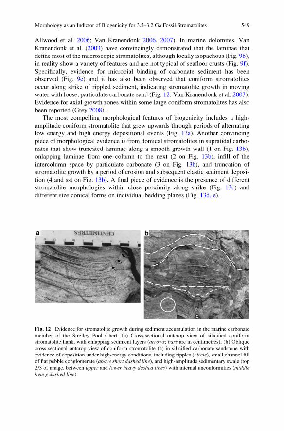
Allwood et al. 2006; Van Kranendonk 2006, 2007). In marine dolomites, Van
Kranendonk et al. (2003) have convincingly demonstrated that the laminae that
define most of the macroscopic stromatolites, although locally isopachous (Fig. 9b),
in reality show a variety of features and are not typical of seafloor crusts (Fig. 9f).
Specifically, evidence for microbial binding of carbonate sediment has been
observed (Fig. 9e) and it has also been observed that coniform stromatolites
occur along strike of rippled sediment, indicating stromatolite growth in moving
water with loose, particulate carbonate sand (Fig. 12: Van Kranendonk et al. 2003).
Evidence for axial growth zones within some large coniform stromatolites has also
been reported (Grey 2008).
The most compelling morphological features of biogenicity includes a high-
amplitude coniform stromatolite that grew upwards through periods of alternating
low energy and high energy depositional events (Fig. 13a). Another convincing
piece of morphological evidence is from domical stromatolites in supratidal carbo-
nates that show truncated laminae along a smooth growth wall (1 on Fig. 13b),
onlapping laminae from one column to the next (2 on Fig. 13b), infill of the
intercolumn space by particulate carbonate (3 on Fig. 13b), and truncation of
stromatolite growth by a period of erosion and subsequent clastic sediment deposi-
tion (4 and sst on Fig. 13b). A final piece of evidence is the presence of different
stromatolite morphologies within close proximity along strike (Fig. 13c) and
different size conical forms on individual bedding planes (Fig. 13d, e).
Fig. 12 Evidence for stromatolite growth during sediment accumulation in the marine carbonate
member of the Strelley Pool Chert: (a) Cross-sectional outcrop view of silicified coniform
stromatolite flank, with onlapping sediment layers (arrows; bars are in centimetres); (b) Oblique
cross-sectional outcrop view of coniform stromatolite (c) in silicified carbonate sandstone with
evidence of deposition under high-energy conditions, including ripples (circle), small channel fill
of flat pebble conglomerate (above short dashed line), and high-amplitude sedimentary swale (top
2/3 of image, between upper and lower heavy dashed lines) with internal unconformities (middleheavy dashed line)
Morphology as an Indictor of Biogenicity for 3.5–3.2 Ga Fossil Stromatolites 549
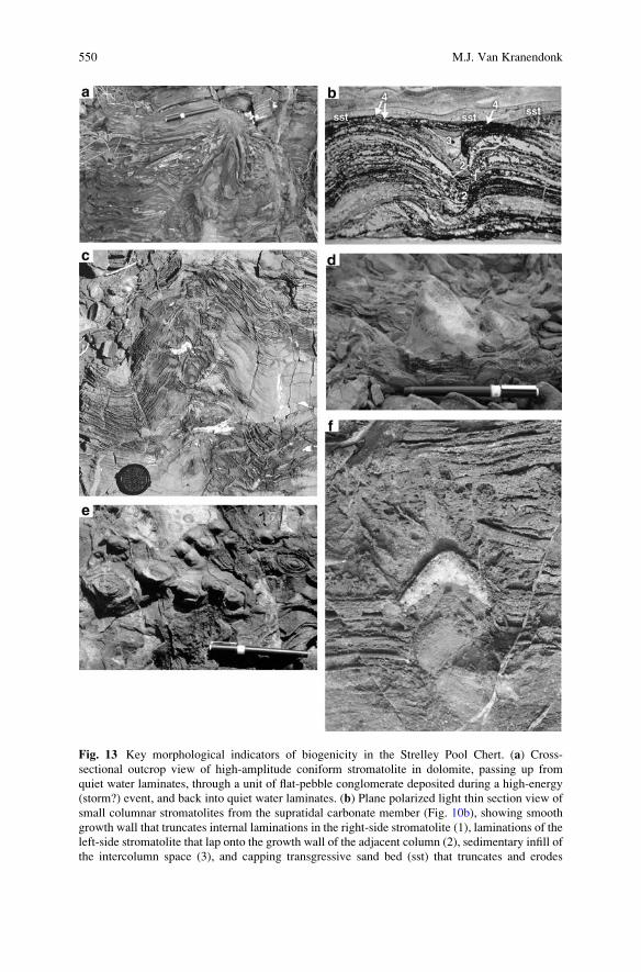
Fig. 13 Key morphological indicators of biogenicity in the Strelley Pool Chert. (a) Cross-
sectional outcrop view of high-amplitude coniform stromatolite in dolomite, passing up from
quiet water laminates, through a unit of flat-pebble conglomerate deposited during a high-energy
(storm?) event, and back into quiet water laminates. (b) Plane polarized light thin section view of
small columnar stromatolites from the supratidal carbonate member (Fig. 10b), showing smooth
growth wall that truncates internal laminations in the right-side stromatolite (1), laminations of the
left-side stromatolite that lap onto the growth wall of the adjacent column (2), sedimentary infill of
the intercolumn space (3), and capping transgressive sand bed (sst) that truncates and erodes
550 M.J. Van Kranendonk
Several additional features support the contention that stromatolite morphology
is not the result of post-depositional diagenesis and/or growth of crystal splays,
including the fact that crystal splays locally nucleate on the top of coniform
stromatolites (Fig. 13f). Locally, it has been observed that horizons of crystal splays
terminate the growth of stromatolites and are overlain by thinly bedded, rippled,
supratidal carbonate sediments, which combines to suggest that the crystal splay
beds formed during periods of subaerial exposure when stromatolite growth ceased
(Fig. 14). Additional outcrop-scale evidence for biogenicity comes from the distri-
bution of stromatolites along and across strike (Van Kranendonk 2007; Allwood
et al. 2006). The best example of this is at the Trendall locality, where stromatolite
size and shape varies along strike in three onlapping biostromes, from larger and
more complex forms with greater inheritance (branching and incipiently branching
forms) near the upslope part of the bioherm, to smaller and more simple forms
downslope at the toe of the bioherms (Fig.14).
Stromatolite biogenicity is supported by highly negative carbon isotope data and
organic geochemistry on kerogenous material in clastic sedimentary rocks from
these outcrops (Marshall et al. 2007).
4 Summary
Convergent lines of evidence from investigations of putative fossil assemblages
from several distinct horizons in the East Pilbara Terrane of the Pilbara Craton point
to abundant and diverse microbial life on early Earth. A key indicator of biogenicity
in all studied examples is morphology, but this must be set within a well constrained
geological context.
�
Fig. 13 (Continued) laminae in the underlying stromatolites (4) (height of view ~2 cm). (c) Cross-
sectional outcrop view of laminated dolomite showing large, incipiently branching stromatolite
and small, branched columnar stromatolite on flank at top right (detail shown in Fig. 9d). (d)
Highly oblique bedding plane view of large (8 cm), slightly asymmetrical columnar stromatolite in
partly silicified dolomite, flanked by adjacent, smaller coniform stromatolites. (e) Bedding plane
view of elongate coniform stromatolites with varying size. (f) Cross-sectional outcrop view of
small coniform stromatolite capped by a radiating crystal splay
Morphology as an Indictor of Biogenicity for 3.5–3.2 Ga Fossil Stromatolites 551
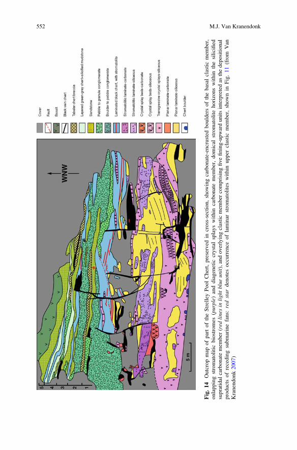
Fig.14
Outcropmap
ofpartoftheStrelleyPoolChert,preserved
incross-section,showingcarbonate-encrusted
bouldersofthebasal
clasticmem
ber,
onlappingstromatoliticbiostromes
(purple)
anddiagenetic
crystal
splayswithin
carbonatemem
ber,domical
stromatolite
horizonswithin
thesilicified
supratidal
carbonatemem
ber
(red
linesin
lightblue
unit),andoverlyingclasticmem
ber
comprisingfivefining-upwardunitsinterpretedas
thedepositional
productsofrecedingsubmarinefans:
redstar
denotesoccurrence
oflaminar
stromatoliteswithin
upper
clasticmem
ber,shownin
Fig.11(from
Van
Kranendonk2007)
552 M.J. Van Kranendonk
References
Allwood AC, Walter MR, Kamber BS, Marshall CP, Burch IW (2006) Stromatolite reef from
the Early Archaean era of Australia. Nature 441:714–717
Banerjee NR, Simonnetti A, Furnes H, Muehlenbachs K, Staudigel H, Heaman L,
Van Kranendonk MJ (2007) Direct dating of Archean microbial ichnofossils. Geology
35:487–490
Brasier MD, Green OR, Jephcoat AP, Kleppe AK, Van Kranendonk MJ, Lindsay JF, Steele A,
Grassineau N (2002) Questioning the evidence for Earth’s oldest fossils. Nature 416:76–81
Brasier MD, Green OR, Lindsay JF, McLoughlin N, Steele A, Stokes C (2005) Critical testing
of Earth’s oldest putative fossil assemblage from the ~3.5 Ga Apex chert, Chinaman Creek,
Western Australia. Precambrian Research 140:55–102
Duck LJ, Glikson M, Golding SD, Webb RE (2007) Microbial remains and other carbonaceous
forms form the 3.24 Ga Sulphur Springs black smoker deposit, Western Australia. Precambrian
Research 154:205–220
Furnes H, Banerjee NR, Staudigel H, Muehlenbachs K, de Wit M, Van Kranendonk M (2007)
Comparing petrographic signatures of bioalteration in recent to Mesoarchean pillow lavas:
Tracing subsurface life in oceanic igneous rocks. Precambrian Research 158:156–176
Garcia-Ruiz JM, Hyde ST, Carnerup AM, Christy AG, Van Kranendonk MJ, Welham NJ (2003)
Self-assembled silica-carbonate structures and detection of ancient microfossils. Science
302:1194–1197
Glikson M, Duck LJ, Golding SD, Hofmann A, Bolhar R, Webb R, Baiano JCF, Sly LI (2008)
Microbial remains in some earliest Earth rocks: Comparison with a potential modern analogue.
Precambrian Research 164:187–200
Grey K (2008) Morphological diversity and complexity in the c. 3.40 Ga Strelley Pool Chert,
Pilbara Craton – Evidence for a biogenic origin. Australian Earth Sciences Convention,
Program and Abstract booklet, Perth, Western Australia
Grotzinger JP, Rothman DH (1996) An abiotic model for stromatolite morphogenesis. Nature
383:423–425
Groves DI, Dunlop JSR, Buick R (1981) An early habitat of life. Scientific American 245:64–73
Hofmann HJ, Grey K, Hickman AH, Thorpe R (1999) Origin of 3.45 Ga coniform stromatolites in
the Warrawoona Group, Western Australia. The Geological Society of America Bulletin
111:1256–1262
Lowe DR (1980) Stromatolites 3,400-Myr old from the Archaean of Western Australia. Nature
284:441–443
Lowe DR (1983) Restricted shallow-water sedimentation of Early Archean stromatolitic and
evaporitic strata of the Strelley Pool Chert, Pilbara Block, Western Australia. Precambrian
Research 19:239–283
Marshall CP, Love GD, Snape CE, Hill AC, Allwood AC, Walter MR, Van Kranendonk MJ,
Bowden SA, Sylva SP, Summons RE (2007) Structural characterization of kerogen in 3.4 Ga
Archaean cherts from the Pilbara Craton, Western Australia. Precambrian Research 155:1–23
Nijman W, De Bruin K, Valkering M (1998) Growth fault control of early Archaean cherts,
barite mounds, and chert-barite veins, North Pole Dome, Eastern Pilbara, Western Australia.
Precambrian Research 88:25–52
Philippot P, Van Zuilen M, Lepot K, Thomazo C, Farquhar J, Van Kranendonk M (2007) Early
Archean microorganisms preferred elemental sulphur, not sulphate. Science 317:1534–1537
Rasmussen B (2000) Filamentous microfossils in a 3,235-million-year-old volcanogenic massive
sulphide deposit. Nature 405:676–679
Schopf JW (1993) Microfossils of the Early Archaean Apex Chert: new evidence of the antiquity
of life. Science 260:640–646
Schopf JW, Kudryavtsev AB, Czaja AD, Tripathi AB (2007) Evidence of Archean life: Stroma-
tolites and microfossils. Precambrian Research 158:141–155
Morphology as an Indictor of Biogenicity for 3.5–3.2 Ga Fossil Stromatolites 553
Ueno Y, Isozaki Y, Yurimoto H, Maruyama S (2001) Carbon isotopic signatures of individual
Archaean Microfossils(?) from Western Australia. International Geology Review 43:196–212
Ueno Y, Yoshioka H, Maruyama S, Isozaki Y (2004) Carbon isotopes and petrography in ~3.5 Ga
hydrothermal silica dykes in the North Pole area, Western Australia. Geochimica et Cosmo-
chimica Acta 68:573–589
Ueno Y, Yamada K, Yoshida N, Maruyama S, Isozaki Y (2006) Evidence from fluid inclusions for
microbial methanogenesis in the early Archaean era. Nature 440:516–519
Van Kranendonk MJ (2006) Volcanic degassing, hydrothermal circulation and the flourishing of
early life on Earth: new evidence from the Warrawoona Group, Pilbara Craton, Western
Australia. Earth Science Reviews 74:197–240
Van Kranendonk MJ (2007) A review of the evidence for putative Paleoarchean life in the Pilbara
Craton. In: Van Kranendonk MJ, Smithies RH, Bennet V (eds) Earth’s Oldest Rocks. Devel-
opments in Precambrian Geology 15, Elsevier, Amsterdam, pp. 855–896
Van Kranendonk MJ, Pirajno F (2004) Geochemistry of metabasalts and hydrothermal alteration
zones associated with ca. 3.45 Ga chert� barite deposits: implications for the geological setting
of the Warrawoona Group, Pilbara Craton, Australia. Geochemistry: Exploration, Environ-
ment, Analysis 4:253–278
Van Kranendonk MJ, Webb GE, Kamber BS (2003) Geological and trace element evidence for
a marine sedimentary environment of deposition and biogenicity of 3.45 Ga stromatolitic
carbonates in the Pilbara Craton, and support for a reducing Archean ocean. Geobiology
1:91–108
Van Kranendonk MJ, Smithies RH, Hickman AH, Champion DC (2007a) Secular tectonic
evolution of Archaean continental crust: interplay between horizontal and vertical processes
in the formation of the Pilbara Craton, Australia. Terra Nova 19:1–38
Van Kranendonk MJ, Hickman A, Smithies RH (2007b) The East Pilbara Terrane of the Pilbara
Craton, Western Australia: Formation of a continental nucleus through repeated mantle plume
magmatism. In: Van Kranendonk MJ, Smithies RH, Bennet V (eds) Earth’s Oldest Rocks.
Developments in Precambrian Geology 15, Elsevier, Amsterdam, pp 307–337
Van Kranendonk MJ, Philippot P, Lepot K, Bodorkos S, Pirajno F (2008) Geological setting
of Earth’s oldest fossils in the c. 3.5 Ga Dresser Formation, Pilbara Craton, Western Australia.
Precambrian Research 167:93–124
Van Kranendonk MJ, Smithies RH,Wingate MTD, Bodorkos S, Hickman AH (2010) Evidence for
Mesoarchaea (~3.2 Ga) rifting of the Pilbara Craton: The missing link in an early Precambrian
Wilson cycle. Precambrian Research 177:145–161
Van Zuilen M, Thomazo C, Luais B, Philippot P (2008) Photosynthesis in a 3.5 Ga old shallow
marine depositional environment; clues from Carbon and Iron isotopes. Australian Earth
Sciences Convention, Program and Abstract booklet, Perth, Western Australia, pp 247–248
Walter MR, Buick R, Dunlop JSR (1980) Stromatolites, 3,400–3,500 Myr old from the North Pole
area, Western Australia. Nature 284:443–445
554 M.J. Van Kranendonk
Index
A
Abiogenic, 29–31, 44–48, 50, 51, 53–55,
57–63, 537
Abu Dhabi, 143, 144
Alabaster, 300, 304
Algae, 391, 404
Alkalinity, 165, 166, 170, 171, 175
Amber, 391–397, 400–402, 404, 405
Anaerobic oxidation of methane (AOM), 210,
215–217
Andros Island, 36, 37, 44
Anhui Province, 463–470
Anti-Atlas belt, 514, 517
Apennines (Italy), 209–217
Apex chert, 539, 540
Aragonite, 102, 106–108, 110–112, 115–118
Archaea, 210, 215, 217
Archaean, 30, 31, 41, 44, 45, 47, 59, 61, 62
Asp€o Hard Rock Laboratory (Asp€o HRL), 221,229, 230
Australia, 37, 40, 45, 46, 61, 62, 92, 537–552
Austria, 355–379
Automicrite, 321–328, 347, 351, 352
B
Bacalar, 187–204
Bacterial sulphate reduction, 306
Badenian, 297–317
Bahamas, 36, 38, 77–87, 92, 97
Baltic Sea, 221, 222, 229
Benthic microbial communities (BMC), 298,
306, 317, 346, 347, 349–352
Bernburg Fm, 14, 15, 20, 21, 30, 32
Bioabrasion, 265, 271, 272
Biocorrosion, 265, 266, 268, 270–272
Bioerosion, 265, 270, 272
Biofilms, 127, 130, 131, 135–137, 266, 270,
272, 317
Biogenic, 29–31, 40, 44, 47, 48, 50, 51, 53–55,
58–60, 62, 537, 540
Biolaminites, 507–511, 513–522
Biomarker, 221–230, 333, 335, 339
lipid, 212–213, 215–217
ppm, 214
Biomineralization, 161, 163, 174, 176, 453
Biosignatures, 221, 222, 227, 229, 230
Bivalves, 279, 281, 282, 284
Brine, 297–317
Buildups, 343–352
Buntsandstein, 6, 7, 13–26
C
Calabria, 332, 454
Calcarea, 366
Calcare di Base (CdB), 332, 334, 339
Calcification, 161–177
Calcite, 152, 153, 157, 158
Calcium carbonate, 161–166, 168, 170–177
Calcium sulphate, 297, 298, 316
California, 101–118
Calothrix, 194–197, 201–204Calv€orde Fm, 14
Campbellrand-Malmani platform, 31, 47, 62
Carbon and oxygen isotopes, 104, 109–111
Carbonate precipitation, 141–148, 151
Carbonates, 101–118, 331–340, 453–455, 458,
459
Carnian, 409–429, 435
Carpathian Foredeep, 297–299
Cedars springs (California), 101–118
Central European Basin, 14, 15, 23, 24
Central Paratethys, 298
J. Reitner et al., Advances in Stromatolite Geobiology,Lecture Notes in Earth Sciences 131, DOI 10.1007/978-3-642-10415-2,# Springer-Verlag Berlin Heidelberg 2011
555
Channels, 303, 304, 315, 316
Chemosynthetic, 277–286
Cherts, 539, 540, 542, 546–550, 552
China, 463–470
Cienegas, 36
Cipit boulders, 435, 436
Clastic gypsum, 298–300, 304
Communities, 298, 304, 306, 316
Concretions, 33, 34, 44–46, 55, 62
Coral limestones, 371
Cozumel, 236, 252
Cryptozoon, 32–35, 37, 44–46, 52, 59Cuatro, 36
Cyanobacteria, 35–38, 54, 123, 125, 127,
129, 130, 132, 161, 162, 177, 183, 234,
236–243, 251–258, 265, 270, 315, 316,
391, 394, 396, 397, 404, 405
D
Deep-sea, 277–286
Demosponges, 366, 367, 377
Desulfovibrio, 214, 215Deutsche geologische Gesellschaft, 5, 6
DGGE, 95
Diatoms, 315
Dissolved inorganic carbon (DIC),
138, 139
DNA, 92–96
Dnister river, 299
Dolomites, 305, 306, 435–449
Domal biolaminites, 517, 519
Domal structures, 304, 315, 317
Dreissena, 189, 201Dresden, 4, 5
Dresser formation, 538, 540–545
E
Ediacaran, 473–504, 525–527
Elephant skin, 512, 513, 515, 518
Endoliths, 266, 271
EPS. See Exopolymeric substances;
Extracellular polymeric substances
(EPS)
Ernietta, 473–504Ernietta-bearing lithofacies, 479, 486, 490,
498, 500
Ernietta lithofacies, 486, 489, 491, 497–502
Euendolithic, 233–258
Euendoliths, 77–87
Eukaryotic algae, 265
Evaporites, 297–317, 454–455, 459
Exopolymeric substances (EPS), 93, 94
External Prebetic, 343–352
Extracellular polymeric substances (EPS), 38,
39, 101, 102, 116–118, 123, 130, 131,
162, 168–170, 172–177, 228, 229
Exuma Cays, 77
Exuma Sound, 92
F
Farbstreifen-Sandwatt, 39
Fenestral pore structures, 304
Ferromanganese crusts, 359, 362, 368, 371,
373, 374, 409–429
Fold structures, 304
Foraminifera, 409, 424–426
Fourier-transform infrared spectroscopy
(FTIR), 331–340
Freshwater, 135–139
Freshwater microbialite, 187
Frutexites, 361, 370, 374, 376, 409–429Fungi, 265, 391, 392, 394, 398, 400–404
G
Gallionella, 221, 224–226, 228, 229Geofluids, 277
Germanic Basin, 14
Geyser, 123–132
Girvanella, 463–465, 467–470Grazers, 271, 272
Great Barrier Reef, 236, 242, 246, 252
Green River Formation, 45
Greigite, 228
Gypsification of the microbial mats, 298, 304,
307, 309, 316
Gypsum, 297–317, 454, 455, 458, 460
Gypsum cement, 304
H
Hallstatt limestone, 410, 411, 428
Hamelin Pool, 37, 92, 95, 96, 98
Hardwater lake, 189
Harz Mountains, 13, 15
Heeseberg, 16, 17, 20, 22, 30, 32
Hematite (Fe2O3), 225, 226, 228
Hettangian, 355, 357, 362, 372, 373,
378, 379
Hexactinellid sponges, 355–379
Holocene, 204
Hybrid Crust, 30, 31, 48–51, 58–63
Hydrology, 191–192
Hydrothermal, 361, 369, 376, 540–542,
544, 547
Hypersaline, 142–144
556 Index
I
Induan, 13
Infaunal biota, 474, 501, 503
Intertidal, 233–258
Iron-oxides, 224–226, 230
Iron-sulfides, 225, 228, 230
Italy, 321–328, 332
J
Jurassic, 343–352
K
Kalkowsky, 3–8, 13–26, 29, 32, 35, 44, 50, 51,
53–55, 57, 58, 61–63
Kanies Member, 474, 477–498, 500–503
Karst, 135, 136, 139, 300
Kendlbach formation, 357
Kerogens, 331, 333, 334, 336, 338–340
Kinneyia, 513, 515
Korcula Island, 236
Kuibis Subgroup, 475, 478, 501
L
Lagoa Vermelha, 142–144, 146
LA-ICP-MS, 436, 438, 441, 449
Lamination, 123, 127, 128, 131, 132, 304, 310
Lee Stocking Island, 236, 239, 252
Leptolyngbya, 95, 196–200, 203, 204Leptothrix, 228Liassic, 355–359, 361–363, 368, 369, 371–374,
376–379
Little Darby Island, 77–87
Lviv to Moldova, 304
M
Magnetite (Fe3O4), 225, 228
Mamillate morphology, 304
Manganese-oxidizing bacteria, 228
Marine, 77–87, 233–258
Marker bed, 298, 300, 304
Mat, 298, 304–307, 309, 315–317
Mediterranean, 277–286
Megascleres, 366, 367
Messinian, 315, 458, 459, 463
Metagenomics, 93, 96–97
Metazoans, 277–286
Methane, 209, 210, 212, 213, 215–217
Mexico, 187–204
Microbial, 13, 20–26, 29, 31, 32, 35–37, 39,
42–44, 47–51, 53–57, 59–63, 298,
304–307, 309, 315–317, 391, 393, 401,
404, 405, 525–534
Microbial carbonates, 453, 455, 458, 459
Microbial communities, 91–93, 95, 97
Microbial diversity, 92, 95
Microbialites, 297–317, 321–328, 343,
346–348, 351
Microbial mats, 30, 35–44, 47, 48, 58–61, 77,
78, 83, 86, 91, 93, 142–148, 221–230,
298, 304–307, 309, 315, 316, 507–509,
511–513, 515, 517, 518, 520
Microbiocorrosion, 265–273
Microborings, 233–258
Microcoleus, 95Microcosms, 154Microelectrode, 136, 137
Microinclusions, 391, 392, 404, 405
Micrometazoans, 391
Microscleres, 367
Microsensors, 143
Miocene, 277, 284–286, 299, 300, 321–328
Morphogenesis, 183, 184
Mud volcano, 209–217
Myxococcus, 161–177
N
Nama Group, 474–478, 498, 502, 504,
513–514, 516
Northern Calcareous Alps, 356, 359, 361, 374
Nostoc, 95
O
Olenekian, 13
Ooids, 13, 16, 17, 19–26
Oolites, 13–26
Oolith, 6, 7
Ophiolite, 102
Ordovician, 463–470
Organic matter (OM), 323, 326–328, 331–340
P
Palaeoecology, 473–504
Pal€aontologische Gesellschaft, 6Paleocurrent analysis, 297, 302, 308, 309
PCR, 94–97
Phanerozoic, 30, 32, 35, 48, 49, 59, 61, 62
Photosynthesis, 123, 125, 129, 131, 135–139,
146–147, 162
Phylotypes, 128–130
Pilbara Craton, 537–552
Pisoids, 125
Planar biolaminites, 517–521
Plectonema, 95Pleurocapsa, 95Poland, 154
Index 557
Polygonal mats, 510, 514, 522
Pomacea, 189, 201Precambrian, 30, 37, 46, 57, 58, 61, 141–148,
183, 185, 317, 521–532, 534
Proteinase K, 93, 94
Proterozoic, 30, 33–36, 44, 46, 49, 53, 57,
59–61
Protozoans, 391
Purple sulphur bacteria, 141–148
Pyrite framboids, 224, 225, 228, 230
R
Raman spectroscopy, 436–439, 447–449
Rare earth elements (REE), 223, 226, 228, 229,
358, 374, 375, 410, 418–422, 427, 443,
453–461
rDNA analysis, 95–96
REE. See Rare earth elements
Resins, 391–405
Rhaetian, 355, 357, 359, 362, 369, 372, 373
Rossellid, 367
S
Sabkha, 143, 144
Safaga Bay, 236
Salento Peninsula, 321–328
Saline pan, 300, 307
Salinity, 298, 315, 316
Salse di Nirano, 209–217
Saratoga Springs, 33, 35
Saturation index, 126
Saturation state, 136, 137
Scheibelberg Formation, 357, 358
Schizothrix, 96Schwarzrand Subgroup, 513–514
Scytonema, 196–198, 202, 203
Seafloor crusts, 30, 44, 46–48, 53, 57–58
Secondary metabolite, 97
Selenite facies, 304
Serpentinization, 102–104, 111, 117
Shark Bay, 37, 38, 40, 48, 57, 59, 92, 93
Shewanella, 102, 105, 111, 113, 114Shoreface, 478, 479, 482, 484–491, 494, 495,
497–503
Shrinkage cracks, 512, 513, 515, 516, 518, 521
Siliceous sponges, 343–352, 357, 363
Silicified carbonates, 540
Sinemurian, 357, 361, 362, 372, 373, 379
Sinter terraces, 104, 106
Skeleton, 185
Solentia, 96South Africa, 30, 31, 47, 62
Spain, 343–352
Spherical carbonate, 101–118
Spherules, 116, 118
Spiculite, 355–379
Sponge–microbialite buildups, 343, 345–348,
350–352
Sponges, 355–379
Spring carbonates, 102, 106–111, 116, 117
Spring water, 101–118
16S rDNA, 130, 143, 145, 214, 21516S rRNA, 128
Stable isotopes, 361, 369, 371–373
St. Cassian Formation, 435–449
Steinplatte, 355–379
Strelley Pool Chert, 539, 540, 542, 546–550,
552
Stromatactis, 356, 361, 363–366, 368–370,
378, 379
Stromatoids, 20, 21, 25
Stromatolite definition, 31, 47, 50–51, 53–55,
59, 62, 63
Stromatolites, 3–8, 13–26, 29–63, 77–87,
91–98, 131, 132, 135–139, 141–148,
161–177, 183–185, 537–552
Stromatolith, 6, 7
Sulphate-reducing bacteria (SRB),
152–158, 161
Sulphate reduction, 151–158, 210, 214, 215
Sulphur-oxidizing bacteria, 279, 280
Supersaturation, 162, 163, 165,
170–175, 177
Sweden, 221–230
Synechococcus, 95Synoptic relief, 304, 305, 310, 315
T
Taphonomy, 526
Tempestites, 486, 489, 491–494, 498–500,
502, 503
Tethys, 344, 355
Thrombolites, 436, 437
Thrombolitic, 37–39, 60
Tidal flats, 35, 39–40, 509–511
Tidal zone, 297, 314
Tlumach stream, 299, 300
Tolypammina, 409–429Travertine, 123–132
Triassic, 13–15, 23, 30, 32, 355–379
Triassic–Jurassic boundary, 355–379
Tripoli formation, 332
Tufa, 35–37, 45, 49, 55, 56
Tunisia, 509–514, 522
558 Index

U
Ukraine, 297–317
Ultrabasic, 101–118
Utah, 123–132
V
Vendian, 525–534
Vendobionta, 475, 476, 491, 501, 502
Vermetid, 321–328
Vingerbreek Member, 513–514, 516
Vistula River, 154
W
Westerh€oferBach, 136, 137White Sea, 526, 527, 530, 531
Y
Yucatan, 187–204
Index 559