advances in clinical chemistry
TRANSCRIPT


ADVANCES IN CLINICAL CHEMISTRY
VOLUME 32

BOARD OF EDITORS
Kenning M. Anderson Callum G. Fraser Gala1 Ghourab Mlter G. Guder Carmel 3 Hillyard Kwang-Jen Hsiao E. D. Janus Gerard Nowacki
Ronald Sutherland It-Koon Tan Milos Tichy Masuuki Totani Orestes Tsolas Casper H. van Aswegen Abraham van den Ende lstvan Vermes

Advances in CLINICAL C H E M I STRY
Edited by
HERBERT E. SPIEGEL Department of Clinical Chemistry St. Vincent's Hospital and Medical Center New York New York
ACADEMIC PRESS San Diego London Boston New York Sydney Tokyo Toronto
VOLUME 32

Find Us on the Web! http://www.apnet.corn
This book is printed on acid-free paper. @
Copyright 0 1996 by ACADEMlC PRESS, INC.
All Rights Reserved. No part of this publication may be reproduced or transmitted in any form or by any means, electronic or mechanical, including photocopy, recording, or any information storage and retrieval system, without permission in writing from the publisher.
Academic Press, Inc. A Division of Harcourt Brace & Company 525 B Street, Suite 1900, San Diego, California 92101-4495
United Kingdom Edition published by Academic Press Limited 24-28 Oval Road, London NWI 7DX
International Standard Serial Number: 0065-2423
International Standard Book Number: 0-12-010332-X
PRINTED IN THE UNlTED STATES OF AMERICA 96 97 9 8 9 9 00 0 1 B B 9 8 7 6 5 4 3 2 1

CONTENTS
CONTRIBUTORS . . . . . . . . . . . . . . . . . . . . . . . . . . . . . . . . . . . . . . . . . . . . . . . . . . . . . . vii
PREFACE . . . . . . . . . . . . . . . . . . . . . . . . . . . . . . . . . . . . . . . . . . . . . . . . . . . . . . . . . . . ix
Concepts Principles and Applications of Selected Molecular Biology Techniques in Clinical Biochemistry
SHESHADRI NARAYANAN
1. Introduction 2. Basic Concepts . . . . . . . . . . . . . . . . . . . . . . . . . . . . . . . . . 3. Principles of Selected Molecular Biology Techniques . . . . . . . . . . . . . . . . . .
. . . . . . . . . . . . . . . . . . . . . . . . . . . . . . . . . . . . . . . . . . . . . .
4. Applications 5 . Summary . . . . . . . . . . . . . . . . . . . . . . . . . . . . . . . . . . . . . . . . . . . . . . .
. . . . . . . . . . . . . . . . . . . . . . . . . . . . . . . . . . . . . . .
References . . . . . . . . . . . . . . . . . . . . . . . . . . . . . . . . . . . . . . . . . . . . . .
Clinical Molecular Biology: Concepts and Applications KAISER J . AZIZ
1. Introduction . . . . . . . . . . . . . . . . . . . . . . . . . . . . . . . . . . . . . . . . 2. Molecular Technology and the Diagnosis of Diseases . . . . . . . . . . . . . . . . . . 3. Cystic Fibrosis . . . . . . . . . . , . . . . . . . . . . . . . . . . . . . . . . . . . .
5. 6. Chronic Myelogenous Leukemia and 7. 8. Clinical Applications 3f Molecular Te 9. Cystic Fibrosis: Current Diagnostics . . . . . . . . . . . . . . . . . . . . . . . . . . . . . . . .
10. Duchenne and Becker Muscular Dystrophy: Current Diagnostics . . . . . . . . . 11. Lymphoproliferative Disorders: Current Diagnostics . . . . . . . 12. Chronic Myelogenous Leukemia and Ac
13. Human Papilloma Virus: Current Diag 14. Perspective on the Evaluation of Mole 15. Software Applications to Molecular Diagnostics . . . . . . . . . . . . . . . . . . . . . . 16. Quality Assurance: Scientific and Regulatory Issues . . . . . . . . . . . . . . . . . . .
4. Duchenne and Becker Muscular Dystrophy . . . . . . . . . . . . . . . . . . . Lyrnphoproliferative Disorders . . . . . . . . . . . . . . . . . .
Human Papilloma Virus . . , . . . . . . . . . .
. . . . . . . . . . . .
. . . _ . _ . . . . . . . . . . . . .
Current Diagnostics . . . . . . . . . . . . . . . . . . . . . . . . . . . . . . . . .
2 2 4
25 33 34
39 43 45 46 41 48 50 50 54 55 56
57 58 58 68 69
V

vi CONTENTS
17 . Conclusion . . . . . . . . . . . . . . . . . . . . . . . . . . . . . . . . . . . . . . . . . . . . . . . . . . . . . References . . . . . . . . . . . . . . . . . . . . . . . . . . . . . . . . . . . . . . . . . . . . . . . . . . . . .
Lipoprotein (a) A . VAN DEN ENDE. Y . Y . VAN DER HOEK. J . J . P . KASTELEIN.
M . L . KOSCHINSKY. C . LABEUR. AND M . ROSSENEU
1 . Introduction . . . . . . . . . . . . . . . . . . . . . . . . . . . . . . . . . . . . . . . . . . . . . . . . . . . . . . .
. . . . . . . . . . . . . . . . . . . . . . . . . . . . . . . . . . . . . . . . . . . . . . . . . . . . . . . . . . . . . . . .
5 . Lipoprotein (a) and Clinical Manifestations of Atherosclerosis . . . . . . 6 . Lipoprotein (a), Thrombogenesis, and Fibrinolysis . . . . . . . . . . . . . . . . . . . . 7 . Lipoprotein (a) and Other Pathological Conditions . . . . . . . . . . . . . . . . 8 . Analysis of Lipoprotein (a) . . . . . . . . . . . . . . . . . . . . . . . . . . . . 9 . Conclusion . . . . . . . . . . . . . . . . . . . . . . . . . . . . . . . . . . . . . . . . . . . . . . . . . . . . .
References . . . . . . . . . . . . . . . . . . . . . . . . .
The Biochemistry of Metastasis M . J . DUFFY
1 . Introduction . . . . . . . . . . . . . . . . . . . . . . . . . . . . . . . . . . . . . . . . . . . . . . . . . . . . 2 . Main Steps in Metastasis . . . . . . . . . . . . . . . . . . . . . . . . . . . . . . . . . . . . . . . . . 3 . Routes for the Dissemination of Cancer Cells . . . . . . . . . . . . . . . . . . 4 . Organ Specificity of Metastases . . . . . . . . . . . . . . . . . . . . . . . . . . . . . . 5 . Molecules Involved in Metastasis . . . . . . . . . . . . . . . . . . . . . . . . . . . . 6 . Markers of Metastatic Potential . . . . . . . . . . . . . . . . . . . . . . . . . . . . . . 7 . Conclusion . . . . . . . . . . . . . . . . . . . . . . . . . . . . . . . . . . . . . . . . . . . . . . .
References . . . . . . . . . . . . . . . . . . . . . . . . . . . . . . . . . . . . . . . . . . . . . . .
AIDS: Biochemical Prospectives GALAL GHOURAB
1 . Introduction . . . . . . . . . . . . . . . . . . . . . . . . . . . . . . . . . . . . . . . . . 2 . The Immune System . . . . . . . . . . . . . . . . . . . . . . . . . . . . . . . . . . . 3 . Virology and Human Immune Deficiency Virus . . . 4 . Infection with HIV . . . . . . . . . . . . . . . . . . . . . . . . . . . . . . . . . . . . . . . . 5 . Laboratory Techniques for the Diagnosis of HIV Infection . . . . . . . . 6 . Antiviral Inhibitory Therapy for HIV . . . . . . . . . . . . 7 . Future Directions in Combating AIDS . . . . . . . . . . . . . . . . . . . . . . . .
References . . . . . . . . . . . . . . . . . . . . . . . . . . . . . . . . . . . . . . . . . . . . . . .
INDEX . . . . . . . . . . . . . . . . . . . . . . . . . . . . . . . . . . . . . . . . . . .
...
. . .
. . .
. . .
. . .
. . .
. . .
. . .
. . .
. . .
. . .
. . .
. . .
. . .
. . .
. . .
. . .
. . .
. . .
70 70
74 74
88 92 95
101 105 110 111
82
135 136 137 138 139 154 158 159
168 175 191 200 218 227 232 238
24 1

CONTRIBUTORS
Numbers in parentheses indicate the pages on which the authors’ contributions begin
KAISER J . AZIZ (39), Division of Clinical Laboratory Devices, Food and Drug Administration, Rockville, Maryland
M. J . DUFFY ( 1 3 3 , Nuclear Medicine Department, St. Vincent’s Hospi- tal, Dublin, Ireland
GALAL GHOURAB ( 137), The Memphis Company for Pharmaceutical and Chemical Industries, Cairo, Egypt
J . J . P. KASTELEIN (73), Centre for Vascular Medicine, Academic Medi- cal Centre, University of Amsterdam, The Netherlands
M. L. KOSCHINSKY (73), Department of Biochemistry, Queen’s Univer- sity, Kingston, Ontario, Canada
C. LABEUR (73), Vakgroep Biochemie Laboratium, Lipoproteiize Chemie Universiteit, Gent, Belgium
SHESHADRI NARAYANAN (l), Department of Pathology, New York Medi- cal College, Metropolitan Hospital Center, New York, New York 10029
M. ROSSENEU (73), Vakgroep Biochemie Laboratium, Lipoproteine Chemie Universiteit, Gent, Belgium
A. VAN DEN ENDE (73), Centre for Vascular Medicine, Academic Medi- cal Centre, University of Amsterdam, The Netherlands
Y . Y . VAN DER HOEK (73), Centre for Vascular Medicine, Academic Medical Centre, University of Amsterdam, The Netherlands
vii

This Page Intentionally Left Blank

PREFACE
The science and practice of clinical chemistry are continuing to evolve and progress. The editors of Advances in Clinical Chemistry, mindful of the advanc- ing scope of this field, choose the subjects and contributors for each volume with great deliberation. Our objective is to identify cutting edge technology and the best talent for creating educational and interesting manuscripts.
This volume places emphasis on the burgeoning fields of molecular diagnos- tics and biology. Two chapters discuss basic concepts and techniques, as well as their application, in monitoring disease. Included in one of these chapters is a discussion on the evaluation of molecular diagnostic tests. In addition, this volume benefits from the knowledge and perspectives of experts who have pro- vided chapters on lipoprotein (a), the biochemistry of metastasis, and AIDS. These reviews cover many aspects of the basic sicence and their clinical rele- vance. The scholarship and philosophical range of the authors make the job of an editor an enriching and rewarding one.
I thank the Editorial Board, without whom these volumes would not be pos- sible. It is humbling to have the Editorial Board Members with their range of intellect and experience working on these volumes. I thank also the highly efficient, responsive, and knowledgeable staff of Academic Press who facilitated the process of producing these volumes. As always, I acknowledge my wife, Joanne, and Sister Catherine Sherry for their steadfast support.
Finally, I reiterate a standing policy of the Editorial Board. We invite our readership to comment on our efforts and to offer suggestions for future volumes.
HERBERT E. SPIEGEL
ix

This Page Intentionally Left Blank

ADVANCES IN CLINICAL CHEMISTRY. VOL. 32
CONCEPTS, PRINCIPLES, AND APPLICATIONS OF SELECTED MOLECULAR BIOLOGY TECHNIQUES
IN CLINICAL BIOCHEMISTRY
Sheshadri Narayanan
Department of Pathology, New York Medical College-Metropolitan Hospital Center, New York, New York 10029
1. Introduction . . . . . . . . . . . . . . . . . . . . . . . . . . . . . . . . . . . . . . . . . . . . . . , . , . . . . . . . . . . . 2. Basic Concepts . . . . . . . . . . . . . . . . . . . . . . . . . . . . . . . . . . . . . . . . . . .
DNA Polymorphism . . . . . . . . . . . . . . . . . . . . . . . . . . . . . . . . . . . . . . . . . . . . . . . . 2.2. Molecular Concepts for Oncology . . . . . . . . . . . . . . . . . . . . . . . . . . .
. . . . . . . . . . . . . . . . . . . . . . . . . . . 3.1. Techniques for Isolation of DNA and RNA . . . . . . . . . . . . . . . . . . . . . . . . . . . . . 3.2. Cloning DNA . . . . . . . . . . . . . . . . . . . . . . . . . . . . . . 3.3. Design of DNA Probes . , . , . . . . . . . , . . . . . . . . . . . . . . . . . . . . , . , . . . . . . . . . , 3.4. Labeling of DNA Probes , . , . . . . . . . . . . . . . . . . . . . . . . . . . . . . . . . . . . . . . . . . . 3.5. Considerations in Hybridizati get DNA . . . . . . . . . . . 3.6. Southern Blotting . . . . . . . . . . . . . . . . . . . . . . . . . . . . . . . . . . 3.7. 3.8. Amplification of DNA . . . . . . . . . . , . . . . . . . . . . . . . . . . . . . . . . . . . . . . . . . . . . . 3.9. Modifications of PCR . . . . . . . . . . . . . . . . . . . . . . . . . . . . . . . . . . . . . . . . 3.10. Other Amplification Reactions . . . , , . . . . . . . . . . . . . . . . . . . . . . . . . . . . . . . . . . 3.1 1. Modified Electrophoretic Techniques . . . . . . . . . . . . . . . . . . . . . . . . . . . . . . . . . . 3.12. In Situ Hybridization . . . . , . 3.13. Conventional Chromosome Analysis versus High-Resolu
Interphase Cytogenetics , . . . . . . _ _ . _ . . . . . . . . . . 3.14. Determination of DNA Index . . . . . . . . . . . . . . . . . . . . . . . .
4.1. Infectious Disease . . . . , . . , . . . . . . , . . , . . . . . . . . . . . . . . . . . . . . . . . . . . . . . . . . 4.2. Paternity and Forensic Testing . . . . . . . . . . . . . . . . . . . . . . . . . . . . . . . . . . . . . . . .
4.4 . Genetic Diseases . . . . . . . . . . . . . . . . . . . . . . . . . . . . . . . . . . . . . . . . . . . . . . .
2.1.
3. Principles of Selected Molecular Bio
Reversed Hybridization . . , . . . . . . . . . . . . . . . . . . . . . . . . . . . . . . . . . . .
4. Applications . . . . . . . . . . . . . . . . . . . . . . . .
4.3. HLA Typing . . . . . . . . . . . . . . . . . . . . . . . . . . . . . . . . . . . . .
. . . . . . . . . . . . . . . . . . . . 5 . Summary . . . . . . . . . . . . . . . . . . . . . . . . . . . . . . . . . .
References . . . . . . . . . . . . . . . . . . . . . . . . . . . . . . . . . . . . . . . . . . . . . . . . . . . . . . . . . . . . .
2 2 2 3 4 4 6 8
10 12 13 14 14 17 19 20 20
22 25 27 27 29 30 31 31 33 34
I
Copyriphi 0 19% by Academic Press. Inc. All nghtr of reprduclion in any form reserved.

2 SHESHADRI NARAYANAN
1. Introduction
Over the last decade there has been an exponential growth in the field of molecular biology. Techniques that were once considered labor intensive and confined to the realm of research have since undergone refinement and sim- plification and are becoming adapted to the clinical biochemistry diagnostic laboratory. The basic concepts of molecular biology have been well explored, and the underlying principles of the myriad molecular biology techniques have been utilized for a wide range of diagnostic applications. This chapter will attempt to provide a selective overview of the basic concepts, principles, and key applications of molecular biology techniques in diagnostic clinical biochemistry.
2. Basic Concepts
2.1. DNA POLYMORPHISM
Regions in DNA that code for protein called exons are separated by interven- ing base sequences called introns. In general, one in every 200 to 500 nucleotides found in regions not coding for proteins is polymorphic. For instance, a sequence of 300 base pairs found in a region not coding for proteins is repeated 500,000 times in the genome. This sequence is called the alu sequence because the bases (A:T, G:C, C:G, T:A, where ":" represents a hydrogen bond between the bases) found within the sequence represent the cleavage site of the Alu restriction enzyme. DNA repeat sequences can be found as widely dispersed copies of a single sequence unit, or they may be arranged in tandem arrays of units.
Using bacterial enzymes that recognize specific DNA base sequences four to six bases long has made it possible to uncover not only single base mutations, but also the presence of polymorphic regions. Fragments of varying length are ob- tained, depending on whether the polymorphism is within or outside the restric- tion enzyme cleavage site. Since these fragments arise as a result of the poly- morphic regions of DNA being recognized by enzymes of defined or restrictive specificity for a specific base sequence (the so-called restriction enzymes or restriction endonucleases), they are called restriction fragments. Polymorphic regions uncovered by the use of restriction endonuclease enzymes are called restriction fragment length polymorphisms, or RFLP (N2). These fragments can be separated according to their size by electrophoresis. DNA probes complemen- tary to the sequence of the isolated restriction fragments can be used to identify the RFLP. Studies using restriction enzymes have shown that one in every 200 to 500 nucleotides in regions that do not code for proteins is polymorphic. Variation in the number of tandem repeats (VNTR) of a short DNA segment is the basis for the formation of RFLPs. These tandemly repeated DNA sequences can be very

MOLECULAR BIOLOGY IN CLINICAL BIOCHEMISTRY 3
short repeats of simple base sequences such as (deoxycytidine-deoxyadenine), on one DNA strand and (deoxyguanine-deoxythymine) on the other DNA strand. (n is the number of repeats.) Collectively, these sequences of dinucleotide repeats or simple tandem repeats (STR) or microsatellite are called (CA), blocks. Considering that there are 50,000 to 100,000 (CA), blocks in the human genome with n in the range of 15 to 30, one would expect to find for every 30- to 60- kilobase segment of the human genome a 1 (CA), block (N2). The study of dinucleotide repeats has been very useful for the construction of genetic linkage maps, which permit the relative assignment of genes or markers on a chromo- some depending upon the extent to which they are inherited together.
In contrast to dinucleotide repeats, tri-, tetra-, and pentanucleotide repeats are not so abundant, although they are also useful as genetic markers. Briefly stated, a study of RFLP and VNTR can provide a variety of information. Since VNTR patterns are unique for each individual, they can be used as a basis for DNA fingerprinting. RFLPs that are close to the disease gene can serve as a genetic disease marker. Indeed, examining RFLPs on both sides of the defective gene enhances considerably the specificity of screening for genetic disease. The use of a set of more than 150 DNA probes to detect DNA polymorphisms, each probe bracketing or defining a locus, is the basis of genetic linkage analysis. This powerful tool can be applied to the study of families to determine cosegregation of chromosome markers with phenotypic traits and for the construction of genetic linkage maps (B9, D4). The advantage of using RFLP in genetic linkage analysis is that one does not have to isolate the specific gene of interest. Furthermore, the RFLPs can even be functionally unrelated random sequences, without the need to be too close to the DNA coding for the locus of interest. The use of RFLPs in constructing the genetic linkage map for human chromosomes has enabled the bracketing with each DNA probe of loci at approximately 20 million base-pair intervals, which corresponds to a 20-centimorgan (cM) distance interval on the human genome (H4). The usefulness of linkage analysis by RFLP studies for genetic counseling for the prediction of inherited disease, and even for comple- menting the results of cytogenetic analysis, should not be underestimated. Other applications of linkage analysis range from identifying loss of markers (alleles) related to tumor suppressor genes to assigning newly discovered polymorphisms to specific chromosomes. Thus, RFLP analysis utilizing DNA probes has a wide range of applications and has been the basis for testing for genetic diseases, for paternity and forensic purposes, and for HLA typing for organ transplantation (E2, G I , 52, 53, N2).
2.2. MOLECULAR CONCEPTS FOR ONCOLOGY
Proto-oncogenes found in mammalian cells have homology to genes found in transforming retroviruses. Well more than 60 proto-oncogenes have been recog-

4 SHESHADRI NARAYANAN
nized (B8). Retroviral genes or oncogenes trigger the development of tumor. In contrast, a class of genes in mammalian cells called tumor suppressor genes inhibit tumorigenesis. As such, cancer is mediated either by activation of proto- oncogenes or by loss of tumor suppressor genes. Activation of a proto-oncogene can occur through a triggering event, such as viral infection or exposure to environmental mutagens, which in turn can cause translocation of an oncogene from one chromosome to another (chromosomal translocation). Activation of a proto-oncogene can also introduce karyotypic abnormalities as a result of ampli- fication of DNA. These abnormalities occur in the form of homogeneously staining regions (HSRs) and double minute chromosomes (DMs). In contrast to a normal chromosome, which has a centromere, DMs lack a centromere. As such, DMs distribute themselves unequally into daughter cells during cell division, or may even be lost from the cells. It has been suggested that DMs represent amplified DNA expelled from its site of origin on a chromosome, and when they (DMs) reenter chromosomes at various portions they create HSRs. In contrast to DMs, HSRs are more stable and can be recognized by their characteristic stain- ing with intermediate intensity throughout their length (B7).
3. Principles of Selected Molecular Biology Techniques
3.1. TECHNIQUES FOR ISOLATION OF DNA AND RNA
Classical techniques are based on lysing cells with lysozyme, alkali, or deter- gents. The removal of protein and other contaminants is effected by incubation with protease or by extraction with phenol or chloroform. The extract is concen- trated by precipitation with ethanol in presence of sodium or ammonium acetate. If necessary, RNA can be removed, using DNase-free RNase. The advantage of using proteinase K is that, in addition to releasing DNA from chromatin, it also destroys nucleases, which otherwise would reduce the average molecular weight of DNA (N3). However, after the lysis of cells has been accomplished, pro- teinase K has to be removed before the isolated DNA can be subjected to restriction enzyme cleavage. Removal of proteinase K is effected by phe- no1:chloroform extractions, which leave DNA in the aqueous phase while trap- ping proteins and lipids in the organic phase. Proteinase K removal can be performed on DNA isolated on a 96-well tissue culture plate (Rl). All steps from cell lysis to precipitation of DNA with ethanol, subsequent washings with eth- anol, and incubation with restriction enzyme can be accomplished on the tissue culture plate.
Since all steps are carried out on the plate, obviating the need for centrifuga- tion, mixing, or transferring of samples, this procedure is adaptable to automa- tion (Rl). Another reason why proteases should be removed is that the DNA

MOLECULAR BIOLOGY IN CLINICAL BIOCHEMISTRY 5
amplifying enzyme used in the polymerase chain reaction (PCR), Taq poly- merase, can otherwise be degraded. Proteinase K can also be inactivated by heating the cell lysate or purified DNA to 95°C for 10 minutes.
Residual phenol can inhibit Taq polymerase. Hence, a final extraction with chloroform-isoamyl alcohol (49: 1) should be performed after phenolization to remove any trace quantities of phenol remaining in the aqueous phase (R3).
Salts used to subsequently precipitate DNA should be removed by washing the pelleted DNA with 80% ethanol. The type of detergent used for cell lysis may influence DNA amplification by PCR. Generally, non-ionic detergents such as Tween 20 and Triton-A-100 do not inhibit Taq polymerase in concentrations less than 5% (v/v). However, ionic detergents such as sodium dodecyl sulfate (SDS), which are generally used in concentrations up to as high as 2.0% (w/v), can be inhibitory to Taq polymerase; a concentration greater than 0.01% (w/v) has been found to be inhibitory.
Other ionic detergents such as sarkosyl and sodium desoxycholate have been shown to inhibit Taq polymerase at concentrations greater than 0.02% (w/v) and 0.06% (w/v), respectively. Hence, it is important that ionic detergent be effi- ciently removed by pheno1:chloroform extractions and by ethanol precipitation and washing of the DNA pellet. Even with non-ionic detergents such as nonidet P40 (NP40), while 1% (v/v) has no effect on reverse transcriptase enzyme, 0.1% (v/v) can inhibit Taq polymerase. Hence, it is important to perform preliminary experiments to establish the effective concentrations of detergents and other known inhibitory reagents that may affect DNA amplification by PCR.
Chaotropic agents, such as guanidinium isothiocyanate, have been frequently used for the extraction of DNA or RNA. The advantage of using 5 M gua- nidinium isothiocyanate for RNA isolation is that it is able not only to remove proteins from RNA, but also to denature ribonucleases that would otherwise degrade RNA (M2). The fact that pheno1:chloroform extractions are cumbersome has resulted in the application of alternative techniques, such as the use of anion- exchange columns for the selective elution of DNA and RNA and the introduc- tion of kits that have eliminated pheno1:chloroform extraction, thus simplifying sample preparation. In terms of isolation of undegraded RNA, one must elimi- nate ribonuclease (RNase) contamination. These enzymes are so stable in a wide range of pH and so resistant, even to boiling, that glassware, reagents, and even the investigator’s fingers are a source of potential contamination. Glassware should be treated with a 1% solution of diethyl pyrocarbonate (DEPC) which is known to inhibit RNases. Residual DEPC, however, should be thoroughly re- moved by autoclaving the glassware in order to convert DEPC to carbon dioxide and water and subsequent baking of the glassware in a 250°C oven for 4 hours. Pipet tips and Eppendorf tubes should be autoclaved at least twice before they are used.
RNA can be selectively extracted into the aqueous phase by adjusting the pH

6 SHESHADRI NARAYANAN
of the extraction system. At a pH of 5.0 to 6.0, RNA will partition into the aqueous phase, while DNA will remain in the organic phase and interphase. If pH during phenol extraction is below 7.6, there will be transfer of considerable amounts of poly(A)-mRNA to the organic phase. However, the use of a deter- gent such as SDS in combination with chloroform can confine poly(A)-mRNA to the aqueous phase. Simplified methods for isolation of poly(A)-mRNA in- volve capture of the poly(A)-mRNA tail found in cell lysate by magnetic beads coated with a 25-mer long chain of deoxythymidylate (dT) 25. The poly(A)- mRNA tail is subsequently eluted from the beads with a salt-free buffer that destabilizes the dT-rA bond. This procedure takes only 15 minutes to perform after cell lysis is accomplished.
Since hematin inhibits Taq polymerase, it is absolutely essential to eliminate red blood cell contamination. Selective lysis of red blood cells can be accom- plished with a buffer mixture consisting of 155 mM ammonium chloride, 10 mM potassium bicarbonate, and 0. I mM EDTA adjusted to pH 7.4. Alternatively, the cytoplasmic membrane of all cells can be dissolved with a buffer mixture con- taining the non-ionic detergent Triton-X 100, leaving behind nuclei of white blood cells from which DNA can be extracted. However, this technique will result in the loss of cytoplasmic DNA to the supernatant, and hence will not be able to extract mitochondria1 DNA (B 11).
3.2. CLONING DNA
The simplest vector for cloning DNA is the small, circular, extrachromosomal double-stranded DNA called plasmids, which are present in a variety of bacteria. In size, plasmids range from 1000 base pairs (1 kilobase or 1 kb) to 200,000 base pairs (200 kb) in contrast to bacteria whose. DNA size can extend to as many as 4 million base pairs. These plasmids carry genes resistant to antibiotics such as tetracyclines, kanamycin, or ampicillin. Briefly, to use a plasmid as a vector for cloning foreign DNA, one would cut the plasmid open with the same restriction enzyme that was used to prepare the DNA fragment. The segment of DNA that is to be cloned is then inserted between the cut ends of the plasmid called sticky or cohesive ends, and annealed or joined together using an enzyme called DNA ligase. As many as 200 copies of recombinant plasmid can be produced in replicating bacteria. The DNA cloned in recombinant plasmid can be recovered by excising the replicated plasmid with the same restriction enzyme that was used originally to cleave the plasmid (Ml, N1). Cloning strategies using the plasmid vector are limited to cloning DNA fragments up to 6000 base pairs (6 kb) long. Figure 1 illustrates the steps in plasmid cloning. Vectors such as bacteriophage are needed to clone DNA fragments up to 20 kb long. Stretches of DNA not needed by bacteriophage for replication are cleaved with restriction endonucleases, and the DNA fragment to be cloned is inserted within this space

MOLECULAR BIOLOGY IN CLINICAL BIOCHEMISTRY 7
PLASMID DNA TO BE CLONED
K 4 - RESTRICTION -
ENDONUCLEASE
0 4
1 1
4
0 t
DNA LIGASE
RECOMBINANT PLASMID
INSERT IN E. COLl
REPLICATION
E. COLl PRODUCES SEVERAL COPIES OF ITS OWN DNA AS WELL AS DNA INSERTED INTO PLASMID.
FIG. 1. Steps in plasmid cloning.
and then replicated inside E. coli. The replicated DNA within the bacteriophage is recovered when the latter is released by lysis of E. coli (Ml , Nl). To clone large fragments of DNA, such as complementary DNA fragments (cDNA) up to 45 kb long, spaces in bacteriophage DNA, each 35 to 45 kb long, called the “cos” sites, can be cleaved to insert the DNA fragment to be cloned. The inserted DNA fragment is first cloned in plasmid, recovered, and packaged into phage heads, and subsequently cloned in E. coli. This type of cloning, which combines the “cos” site of bacteriophage with plasmid, is called “cosmid cloning” (M1 , Nl). Ideally, since it is desirable to have DNA probes smaller than 500 nucle-

8 SHESHADRI NARAYANAN
TABLE I SUMMARY OF STEPS INVOLVED I N CLONING DNA
DNA 4
DNA FRAGMENTS Digest with restriction endonuclease (RE).
5 1 5 .1
Up to 6 kb long. Up to 20 kb long. Up to 45 kb long. Plasmid cloning. Anneal frag- Bacteriophage cloning. Insert Cosmid cloning. Cleave 35 to
ment to plasmid with DNA fragment in bacteriophage. 45-kb stretches of bacte- ligase. riophage DNA (COS sites).
1 1. 1
1 5 5
1 5
1
.1
Insert in E. coli.
Replicate. Recover replicated DNA lys- Clone in plasmid. ing E . coli.
Replicate in E. roli Insert DNA in this space
Recover cloned DNA in repli- Recover.
Package into phage heads.
Clone in E. coli.
cated plasmid by using RE.
otides, plasmid would be an adequate vector for cloning. Steps involved in cloning DNA are summarized in Table 1. However, large restriction fragments in the size range 200 to 500 kb can be cloned in yeast by ligating them to vector sequences that permit their propagation as linear artificial chromosomes (yeast artificial chromosome or YAC) (B12).
3.3. DESIGN OF DNA PROBES
Before one can design DNA probes, one should be aware of the sample DNA base sequence in the area of interest, so that a complementary probe can be devised. The complementarity of DNA is based on the hydrogen bonding be- tween the bases adenine and thymine, and between guanine and cytosine. In RNA, adenine hydrogen-bonds to the base uracil. The temperature required to dissociate the double-stranded DNA is dependent on the guanine-to-cytosine (G-C) content of DNA because the interaction between guanine and cytosine is stronger than that between the bases adenine and thymine. This is because guanine forms three hydrogen bonds with cytosine, as opposed to the two hydro- gen bonds formed by adenine with thymine.
A knowledge of melting temperature or T,,, is important for designing the optimum temperature for hybridization of sample DNA with its complementary

MOLECULAR BIOLOGY IN CLINICAL BIOCHEMISTRY 9
DNA probe. T , by definition is the temperature at which 50% of the double- stranded DNA has its complementary strands separated. While the complemen- tarity of base pairing between the sample target DNA and probe DNA is respon- sible for the specificity of binding, the stability of the base pairing can be reduced by organic solvents, thus ascribing a role for hydrophobic interactions as well, in terms of target DNA-probe hybrid stability. Thus, the melting temperature can be reduced by the addition of organic solvents. In practical terms, we need to establish an optimum temperature at which the sample target DNA binds specifi- cally and strongly to the probe DNA. The binding of target DNA to probe DNA generally occurs readily at a temperature which is 25°C lower than the T , of the target DNA-probe hybrid. For short oligonucleotide probes, this temperature is generally 5°C below T,. However, as the temperature at which hybridization of probe to its target occurs is brought closer to T,, the specificity of the target- probe interactions or hybridization stringency is increased (W2). Thus, the de- gree of stringency is a reflection of the probe-target fidelity, with high stringency conditions favoring specific target-to-probe interaction, thus keeping the non- specific target probe interactions or background to a minimum.
Empirical formulas are available for calculation of T,. Since T , is dependent on guanine-to-cytosine content of the DNA target, it is directly related to it (G:C), and also to the ionic strength of the solvent mixture used to determine T,. T , is inversely related to the concentration of organic solvents such as for- mamide. Thus, the T , of DNA is lowered 0.63"C per percent formamide used in the reaction mixture (W2). One can therefore increase or decrease the stringency of the hybridization reaction by appropriate adjustments of ionic strength, tem- perature, or formamide concentration. When using RNA probes, formamide concentration is critical, since for equivalent formamide concentration the T,,, of the DNA-RNA probe hybrid is reduced significantly less than that of the DNA- DNA probe hybrid. In the traditional formats used for DNA probe hybridization, such as hybridization in solution or on a solid phase, experiments are camed out using an excess of probe compared to sample target DNA. Under these condi- tions, the rate of hybridization is dependent on probe length or complexity and probe concentration. Thus, the shorter the probe length, the less time would be required for it to hybridize effectively to its target DNA. Short probes, such as oligonucleotide probes of 20 bases in length, therefore do not need the addition of accelerators such as dextran sulfate to enhance hybridization rate, as is re- quired for longer double-stranded probes.
In addition to probe length, other considerations in the selection of DNA probes include keeping the G-C composition to between 40 and 60%, and ensuring that no unusual base sequence is present, such as having no more than four at a stretch of a single base sequence (for example, -C-C-C-C-C-) or any other intraprobe complementary sequence that will reduce the specificity of the probe-sample target DNA interaction.
Obviously, if there is even a single base mutation in the target DNA, the

10 SHESHADRI NARAYANAN
specificity of the target DNA-probe interaction will be affected. Likewise, if the target DNA is modified by a polymorphism, then the specificity of the target DNA probe interaction will also be affected. On the other hand, if polymorphism in a DNA segment is known, then specific probes can be tailored to detect the base sequence in the area of interest.
3.4. LABELING OF DNA PROBES
The procedure for radiolabeling DNA probes is called the nick translation method. As the name indicates, nicks are introduced into double-stranded DNA with the aid of an enzyme called DNase. Another enzyme, called DNA poly- merase, is used to digest away from the nick and replace the strand as it pro- ceeds. By incorporating radiolabeled deoxynucleotide triphosphates (5' [32P]
triphosphates) in the reaction mixture, the DNA to be used as a probe is labeled to a high specific activity (MI). Nonisotopic labels such as biotin can be used and incorporated into the base thymine in the DNA probe sequences or the base uracil in the RNA probe sequence. Biotin-labeled probes, when they bind to the DNA of interest, can be visualized with avidin- or streptavidin-labeled enzyme, since both avidin and streptavidin can bind to biotin. Excess enzyme label can be washed away, and the hybridized probe bound to the enzyme label can be detected by adding substrate and visualizing or measuring the color formed in a spectrophotometer. Biotin-labeled probe that hybridizes to the DNA of interest can also be detected by using an antibody to biotin and a suitable immuno- enzymometric label such as alkaline phosphatase-antialkaline phosphatase, or peroxidase-antiperoxidase (B 1 1). DNA probes have also been labeled utilizing sensitive chemiluminescent compounds. The amino function in the DNA probe can be labeled with the use of N-hydroxysuccinimide derivatives of acridinium esters (A3, N4). The reaction mechanisms for N-methyl acridinium esters with hydrogen peroxide in alkali involves an attack by hydroperoxy (HOO) anion on the 9 position of the acridinium ring. An intermediate compound with a cycloox- etane ring is formed under the alkaline conditions of the assay, which is rapidly transformed to an excited compound (N-methyl acridone) that subsequently emits a flash of light upon returning to the ground state. The chemiluminescence can be either followed in a luminometer or recorded on photographic film. Chemiluminescent detection can also be accomplished using DNA probes la- beled with alkaline phosphatase. In the presence of adamantyl- 1,2-dioxetane phenyl phosphate (AMPPD) as substrate, alkaline phosphatase in the hybridized probe, cleaves the phosphate group from the AMPPD molecule to produce an anion, AMP-D, which is weakly stable. Hence, the AMP-D anion is fragmented to yield adamantanone and an excited methyl-mera-oxybenzoate anion that pro- duces the chemiluminescence (N4). The chemiluminescence of the meta-oxy- benzoate anion can further be enhanced by energy transfer to fluorescein surfac-

MOLECULAR BIOLOGY IN CLINICAL BIOCHEMISTRY 1 1
A
ACRlDlNlUM ESTER
k HOOP
0 = b O O H I
OR +-ROH
F5 CYCLOOXETANE
RING INTERMEDIATE
o = c 0
‘0’
t CH3
N-METHYLACRIDONE EXCITED & 0 +
I
f ALKALINE 0 P03Na2 - PHOSPHATASE
AMPD
f
ADMANTANONE Dot d -
O<,cH3 0- METHYLMETA-OXYBENZOATE
TN CHEMILUMINESCENCE
C02 + CHEMILUMINESCENCE
FIG. 2 . Reaction mechanisms of chemiluminescent labels.
tants, such as micelles formed from cetyltrimethylammonium bromide and 5-(N- tetradecanoy1)aminofluorescein ( S 2 ) .
Figure 2 illustrates the reaction mechanisms of acridinium ester label probes and alkaline phosphatase probes using dioxetane chemiluminescent detection. Table 2 summarizes approaches for labeling DNA.

12 SHESHADRI NARAYANAN
TABLE 2 SUMMARY OF APPROACHES FOR LABELING DNA
I. Nick Translation for Radiolabeling DNA DOUBLE STRANDED DNA t DNASE
INTRODUCE NICKS DNA POLYMERASE
DIGESTS AWAY FROM NICK AND REPLACES STRAND AS IT PROCEEDS $. 5'['2P] TRIPHOSPHATES
DNA LABELLED TO HIGH SPECIFIC ACTIVITY
A. Biotin: Incorporate in base thymine in DNA probe sequence or uracil in RNA probe sequence 11. Nonisotopic Labels
$- ADD TO SAMPLE DNA DETECT DNA-BIOTIN LABELED PROBE WITH
ANTIBODY TO BIOTIN AND A LABEL SUCH AS ALKALINE
f AVIDIN OR STREPTAVIDIN LABELED ENZYME
PHOSPHATASE OR PEROXIDASE- 1 ANTIPEROXIDASE WASH EXCESS LABEL
4 ADD SUBSTRATE
VISUALIZE OR MEASURE PRODUCT ABSORBANCE IN SPECTROPHOTOMETER.
B. Chemiluminescent Labels I. Label amino function in DNA probe with N-hydroxy
succinimide derivatives of acridinium esters. 2. Label DNA probe with alkaline phosphatase.
3.5. CONSIDERATIONS IN HYBRIDIZATION OF PROBE TO TARGET DNA
Hybridization can be performed by merely spotting the sample to a membrane, where it is immobilized by baking and subsequently hybridized to a suitable probe. Sample application can be performed with commercially available mani- folds that apply sample into multiple wells of the manifold and let sample migrate as spots or slots into the membrane: hence the name dot blot or slot blot hybridization (W2). The sample wells are repeatedly washed prior to removing the membrane to bake or irradiate in order to fix the sample, which is then ready for hybridization with probe.
DNA probing can be done not only on a membrane, but also on solid supports. An example is the classical sandwich hybridization assay, which uses a capture

MOLECULAR BIOLOGY IN CLINICAL BIOCHEMISTRY 13
probe immobilized to a solid support to bind to the sample target sequence, which is then detected by a labeled probe ( K l ) . Thus, the sample target DNA is sandwiched between an immobilized capture probe and a labeled probe.
For this hybridization strategy to work, the two probes (the capture and the labeled probe) should be from two adjacent portions on the genome, but without having complementary regions, thus avoiding binding of probes to each other. This hybridization requires target sample to bind to both the capture and the labeled probe and, as such, is more specific than direct hybridization on a membrane filter.
Hybridization can also be performed in solution phase. Since the capture probe is in solution, the kinetics of hybridization is faster than when the capture probe is immobilized. In the solution phase hybridization format, the capture probe is labeled with an affinity label such as biotin that captures the sample target sequence. The labeled probe then binds to the sample target sequence to form the sandwich. After the hybridization is complete, the sandwich is transferred to an affinity support such as avidin or streptavidin, which will capture the sandwich through the biotin-labeled capture probe. Sandwich hybridization performed in solution followed by capture on an affinity support has been referred to as affinity-based hybrid collection (K 1).
3.6. SOUTHERN BLOTTING
Hybridization can be accomplished after Southern blotting. In this procedure, DNA fragments obtained by restriction endonuclease digestion are first separated by agarose gel electrophoresis. The agarose gel is then treated with alkali to denature the DNA, and after neutralization of alkali, the DNA is transferred or blotted to a nitrocellulose or nylon filter by capillary action. The membrane is baked to fix the DNA fragments and hybridized to a radiolabeled 32P comple- mentary DNA (cDNA) probe. The unhybridized probe is washed away from the membrane, and the hybridized bands are detected by autoradiography, which involves exposure of the hybridized bands to an X-ray film at -70°C (S8). The autoradiography step can be time consuming: it can take as long as 10 days. The transfer of DNA fragments by capillary action alone takes 16 to 24 hours. Transfer of DNA to membranes can, however, also be effected by vacuum and electrotransfer. One of the drawbacks associated with the traditional capillary transfer effected during upward flow of transfer solution by capillary action through the gel, membrane, and blotting paper is that the gel is flattened by the weights placed on the blotting paper, and consequently the elution of DNA from the gel is not quite satisfactory.
The drawback has been overcome by a technique that effects downward flow of transfer solution by capillary action under alkaline conditions, accomplishing efficient transfer of DNA to either nitrocellulose or plastic membranes in 2.5 hours (C2).

14 SHESHADRI NARAYANAN
3.7. REVERSED HYBRIDIZATION
Instead of labeling probes, one can label the DNA in sample with a non- isotopic label and detect target DNA by using a panel of immobilized DNA as probes. This is the technique of reversed hybridization (D2). The simplicity of this procedure is illustrated by its application to the identification of microor- ganisms. After lysis of microorganisms, DNA is labeled in the presence of UV light with a nonisotopic compound such as biotin-polyethyleneglycolangelicin (BPA, a fucocoumarin derivative) to form monoadducts with DNA. The labeled sample is applied to a panel of DNA probes immobilized as dots on a nitro- cellulose membrane. The hybridized DNA can be detected either by chem- iluminescence or by a colorimetric reaction (D2).
3.8 . AMPLIFICATION OF DNA
The most widely used amplification technique is the polymerase chain reac- tion. Hardly a research study in molecular biology involving DNA amplification has been performed without using PCR. Many modifications of PCR have been described. Before we review these modified techniques, it would be appropriate to briefly review the PCR methodology.
PCR is performed in a 50 to 100 p.1 volume that includes the sample, buffer additives such as magnesium chloride and gelatin, two primers flanking the region to be amplified in each of the two strands, the four deoxynucleotide triphosphates (dATP, dCTP, dGTP, and dTTP), and a heat-stable DNA poly- merase. A few drops of mineral oil are overlaid on top of the reaction mixture to prevent evaporation, which would otherwise reduce the yield of the amplification product. Normally each PCR cycle would consist of three steps: denaturation of DNA to separate the two strands by heating to 94°C for 20 sec to 1 min, annealing the strands to primer by bringing reaction temperature down to approx- imately 55°C and holding it for 20 sec to 1 min, and finally allowing synthesis of DNA complementary to the amplified strand to proceed by primer extension at 72°C for 30 sec to 1 min (B4). After the last PCR cycle, the amplified DNA can be separated by electrophoresis and visualized by staining with ethidium bromide or silver stain, or alternatively, the amplified product can be transferred to a solid support and detected with a complementary DNA probe.
Each cycle results in a doubling of the number of strands of DNA found at the previous step. After 20 PCR cycles, the two original strands of DNA will have been amplified a millionfold (2*0 = 1 million), while after 30 cycles the ampli- fication will be a billionfold. However, after 30 PCR cycles the amplification reaction reaches a plateau, primarily because of the excess of DNA synthesized (substrate excess), competition by nonspecific products, and reassociation of pro- duct. Figure 3 is a diagrammatic representation of PCR. A few selected analyti- cal variables affecting PCR need to be considered. First, the reannealing tempera- ture is critical to the specificity of the amplification. Low temperatures of between

MOLECULAR BIOLOGY IN CLINICAL BIOCHEMISTRY 15
Denature by heating to 94OC, then anneal to primer at 55' C.
5 ' DNA Strand
Primer
3l Primer
5 ' 3' DNA Strand
Extend DNA synthesis in presence of heat stable DNA polymerase at 720 C in presence of dNTPS. I
3 ' A 5 1
3 '
Repeat cycle of DNA strand separation (denaturation), annealing to primer, and synthesis of DNA complementary to the
- 5 '
I strand template.
At the end of 20 cycles the original two strands of DNA will be amplified a million fold (2 = 1 million). 20
FIG, 3 , Schematic representation of polymerase chain reaction.
45 and 55°C can lead to amplification of nonspecific target sequences. For ampli- fication of short target sequences of 100 to 300 bases, a two-step PCR reaction overcomes the problem of nonspecific target sequence amplification because both the annealing and the primer extension steps are performed at 72°C (B4, N3).
Attention should be given to the selection of primers. Typically, primers used are between 15 and 30 bases in length, with guanine-cytosine composition between 40 and 60%. The primer should not have within its sequence any unusual composition such as stretches of polypurines or polypyrimidines. The primer pair should not be complementary at the 3' ends, since otherwise the DNA synthesizing enzyme can extend one primer over the other primer, creating a double-stranded product whose length approximates the sum of the two prim- ers. This artifact is called primer dimer, which could very well become the predominant and undesirable PCR product when primer pairs complementary at the 3' ends are used (B4, N3).
It is important to note that even at room temperature, Taq DNA polymerase can incorporate 0.25 nucleotides per second. Hence, the aliquot of master mix

16 SHESHADRI NARAYANAN
should be placed on ice. The thermal cycler should be preheated to an approxi- mate temperature of 90°C. The DNA to be amplified (that is, the sample) is added last, under mineral oil, just prior to temperature cycling.
In a technique called "Hot-Start PCR' to reduce nonspecific amplification and formation or primer dimers, at least one reagent is withheld until the reaction temperature has reached at least +5OoC at the initiation of cycling. Paraffin wax is melted and rests on top of the reaction mixture, which is lacking in one reagent. With subsequent cooling, the essential reagent is laid over the newly formed paraffin wax layer. In the next heating step, liquid paraffin rises above all compo- nents of the reaction mixture, thereby forming a vapor barrier during PCR.
In an attempt to minimize variables affecting PCR amplification, a technique called competitive PCR has been developed. In this technique, competitor mole- cules are used that share the same primer recognition sites as the target gene, but contain an additional 15 to 20 base-pair insert to distinguish it from the amplified gene by polyacrylamide gel electrophoresis and ethidium bromide staining. In this technique, any variable that affects amplification affects both the genomic and competitor DNA (S4).
The accuracy of the thermal cycler in terms of well uniformity and repeat- ability of cycling time, as well as the thinness of the reaction tube, will control the efficiency of amplification reactions. Hence, it is important that uniform temperature be maintained between wells. In thermal cyclers where the lid of the reaction tube is heated continuously to greater than 96"C, PCR can be performed without overlaying with oil. Heat transfer is facilitated by using thin-walled reaction tubes. Among thermal cyclers that have been used, air thermal cyclers have an advantage that facilitates rapid thermocycling: air is an ideal heat transfer medium that can rapidly effect a change of temperature because of its low density and conductivity.
The integrity of PCR products can be visualized on agarose gel. For instance, a smear originating from the sample application slot would indicate the presence of degraded sample DNA. The presence of distinct bands of approximately 120 bp or smears near that region would indicate the presence of a primer-dimer artifact. A smear stopping exactly in a region corresponding to 300 bp would indicate that a low concentration of primers was used and specific products were priming themselves. Finally, distinct bands of approximately 150 to 600 bp would indicate that specific products were forming a self-priming secondary structure. These are just a few examples of troubleshooting for artifacts gener- ated during PCR amplification. Strict contamination control is critical to the success of PCR amplification.
Exogenous sources such as a person's hair or skin, doorknobs, laboratory benches, dust, reagents, thermal cyclers, and pipet tips are some of the common sources of DNA contamination. Ideally, a laminar air flow bench with filtered air provides a clean, dust-free environment. Sample preparation should be done in a separate room or area. The addition of sample to the PCR reaction mixture in the

MOLECULAR BIOLOGY IN CLINICAL BIOCHEMISTRY 17
thermal cycler should be done in a separate area. Post-PCR work such as opening of the reaction tube containing amplified DNA, or electrophoresis should be done in another room or area. Positive and negative controls should be analyzed to verify the accuracy of results. The work area or bench should be decontami- nated with 10% sodium hypochlorite, and the surface rinsed with water to re- move the corrosive reagent. Chemical modification of PCR products to eliminate carryover of nonspecific amplified fragments can be performed by substituting dUTP for dTTP. The modified DNA that is formed is cleaved of the uracil residues by using the enzyme uracil-N-glycosylase, while the amplified target specific DNA is unaffected (L4).
Anticoagulants used for blood collection can influence PCR amplification. For instance, heparin is reported to interfere by causing attenuation or complete inhibition of DNA amplification during PCR (H2). Efforts to reverse the effect of heparin, such as boiling DNA; separation on a gel filtration column such as Sephadex G75; neutralization with acid or alkali subsequent to gel filtration; or repeated precipitation with ethanol or neutralization with protamine sulfate, have been reported to be unsuccessful (B6). However, treatment of heparinized blood with heparinase or separation of leucocytes by centrifugation followed by a minimum of two washings in a saline buffer is reported to overcome the effect of heparin. The negative effect of heparin was demonstrated in a study where blood collected in heparin for T and B cell gene rearrangement analysis showed aber- rant restriction fragments after restriction-enzyme digestion of DNA. Such aber- rant restriction fragments were not seen upon restriction-enzyme digestion of DNA obtained from blood collected in either EDTA or ACD (T2). Since such aberrant DNA fragments obtained from blood collected in heparin could be confused with gene rearrangements, the choice of anticoagulant becomes criti- cal. Actually, in addition to heparin, anticoagulants such as EDTA and ACD can also inhibit restriction enzymes. However, both EDTA and ACD are removed by standard ethanol precipitation techniques, while heparin is not (C3). Washing blood containing EDTA, citrate, or heparin with Tris-EDTA buffer, pH 8.0, is reported to remove free anticoagulant. DNA prepared subsequently for HLA-DR genomic typing is apparently not interfered with (K2).
3.9. MODIFICATIONS OF PCR
PCR can be used to introduce labels that can then be used for detection. The ability to add to the 5' end of the primers sequences not complementary to the target template, which then becomes incorporated into the double-stranded PCR product, allows the introduction of labels. Thus, the addition of biotin to the 5' end of the primer allows detection of hybridized PCR product with streptavidin or avidin-enzyme conjugates (B4). Other labels such as digoxigenin can be added to the 5' end of the primer, amplified, and detected either colorimetrically or by chemiluminescence (F3).

18 SHESHADRI NARAYANAN
PCR can also be used to generate an excess of single-stranded DNA which can then be labeled and used as DNA probes. This technique, which is called asym- metric PCR, involves using a 100-fold excess of one primer over the other. With this asymmetric ratio, double-stranded DNA will be synthesized in the first 20 to 25 cycles, at which time the primer used at a lower concentration would be consumed, leaving the primer that is in excess to preferentially synthesize single- stranded DNA over the next 5 to 10 cycles (G3).
RNA amplification by PCR has been facilitated by the use of a single heat- stable enzyme. Thus, DNA polymerase from Thermus fhermophilus, which has enhanced reverse transcriptase (rT) activity in presence of manganese, can be used with one buffer system. The high temperature used for rT (70°C) to produce a complementary DNA copy from RNA, and the subsequent amplification of DNA at 60"C, increases efficiency by destabilizing secondary structures in the RNA template. This procedure has been used for the amplification of hepatitis C viral RNA (Y 1).
A technique called single-strand conformational polymorphism (SSCP) takes advantage of the fact that under nondenaturing conditions, single-stranded DNA has a folded structure. Mutation, however, causes a change in the folded struc- ture and, in turn, a change in mobility during electrophoresis on neutral poly- acrylamide gels. Methodology for identifying SSCP involves amplifying a de- sired sequence in the gene by PCR, denaturing the amplified DNA, and comparing its electrophoretic mobility to that of a reference strand of a known sequence. The presence of single point mutations will result in fragments of equal lengths differing in sequence migrating in different positions (Hl). Even a single base difference in a 100-nucleotide piece of single-stranded DNA can be detected by SSCP (01).
Alternatively, the denatured single strands can be made to reanneal to form double-stranded helices. Complementary strands will hybridize to each other. However, if there are sequence differences between two strands, one from each allele, they remain unpaired in the heteroduplex and, as a result, form open loops that reduce migration in the electrophoretic gel. This is the basis of heteroduplex analysis, in which distinct electrophoretic patterns are seen for different alleles, similar to that seen in SSCP (01).
Either by use of sequence specific primers during PCR amplification or by probing of PCR products with allelic-specific oligomer (ASO) probes, one can distinguish between alleles that have undergone even a single base substitution. Typically, the AS0 probes used are labeled oligonucleotide probes approx- imately 19 base bp long for each of the alleles to be tested. Such probes are specific for one allele, whereas binding to other alleles is prohibited by the probe having a single base mismatch. PCR-amplified product can be examined for single base mutations by hybridization to enzyme-labeled AS0 probes (S 1). Primers mismatched in the first base at the 3' end have been used to detect point

MOLECULAR BIOLOGY IN CLINICAL BIOCHEMISTRY 19
mutations by PCR. Primers mismatched to target DNA due to a point mutation are not amplified. This is the basis of an allele-specific PCR called the amplifica- tion refractory mutation system (ARMS) (N5).
The specificity of PCR amplification can be enhanced by using nested prim- ers. Typically, in this approach, after a first round of PCR, primers nested within the original pair are used to amplify specific sequences (P2).
3.10. OTHER AMPLIFICATION REACTIONS
Besides PCR, other amplification reactions have been described in the litera- ture (B4). A selected few are briefly reviewed here.
An amplification reaction that is used to amplify target RNA or denatured DNA is called the transcription-based amplification system (TAS). This tech- nique involves using an enzyme called reverse transcriptase and a primer with sequence complementary to the sample target RNA molecule in order to synthe- size a complementary DNA (cDNA) copy of the sample target RNA. After denaturation to separate the strands, another primer and additional reverse tran- scriptase are added to synthesize a double-stranded cDNA molecule. Since the first primer has also an RNA polymerase binding site, it can, in the presence of T7 RNA polymerase, amplify the double-stranded cDNA to produce 10 to 100 copies of RNA. The cycle of denaturation, synthesis of cDNA, and amplification to pro- duce multiple RNA copies is repeated. With as few as four cycles, a 2- to 5- millionfold amplification of the original sample RNA target is possible. However, the time required to achieve a millionfold amplification is approximately 4 hours, which is the same amount of time required by PCR. The TAS requires, however, the addition of two enzymes at each cycle and, as such, can be cumbersome.
An amplification system that actually amplifies exponentially RNA probe sequences bound to the target sequence, in contrast to PCR and TAS systems, which amplify target sequences, is the Q-beta replicase system (B4). Although this system can achieve a million- to billionfold amplification in 15 minutes at 37"C, background signal due to nonhybridized probes is reported to be very high.
An amplification system that has been successfully commercialized is called the ligase chain reaction (B4). The basis for this reaction lies in using two small single-stranded DNA probes of 10 to 20 bases in length to anneal to target DNA and an enzyme called DNA ligase to link the two probes. Twenty to 50 cycles of this reaction can yield sufficient amplified product.
Actually, four single-stranded probes are used, with probes 1 and 3 comple- mentary to the 3' and 5' portion of one target strand, and probes 2 and 4 complementary to the other target strand.
Amplification is achieved by repeated cycles (20 to 50) of heating (for strand separation) and cooling (for specific hybridization). Amplicons result upon liga- tion of adjacent probes. A commercialized automated system achieves detection

20 SHESHADRI NARAYANAN
by labeling ends of each probe pair with different capture and detection haptens (W.
An elegant approach is to capture the target DNA or RNA with specific oligonucleotides on to a microwell plate. Synthetic branched DNA bearing mul- tiple alkaline phosphatase-labeled probes hybridizes to the target. A chem- iluminescent substrate is added to produce signal. This branched DNA assay has been used in infectious disease detection (W3).
3.1 1. MODIFIED ELECTROPHORETIC TECHNIQUES
To separate large DNA fragments exceeding 20,000 base pairs resulting from RFLP analysis, a technique called pulse-field gel electrophoresis is used. In this technique, electrical current is switched alternately between two sets of direc- tional electrodes. DNA molecules exposed to alternating electrical fields are separated on an agarose gel based on the rate at which they change their configu- ration inside the gel (S7).
An electrophoretic technique either alone or in combination with PCR is useful for the examination of single base-pair mutations. In this technique, called de- naturing gradient gel electrophoresis (DGGE), as DNA molecules migrate into a region of ascending concentration of denaturant (urea:fomamide), there is a decrease in mobility as the molecule is transformed from a helical conformation to a partially melted form that is dependent on the base sequence. While this technique alone can identify a mutation, in combination with PCR it can localize the mutation to a given region of the human genome, as well as allow sequencing of the DNA without resort to cloning (Cl).
3.12. In Situ HYBRIDIZATION
In situ hybridization (ISH) permits examination of a wide range of materials, ranging from cells and tissues to metaphase spreads of chromosomes affixed to a slide. Hybridization with specific probes can thus be performed directly on the slide, with the hybridized signal viewed under a microscope. Briefly, the steps involved in ISH include pretreatment of the slide, application of the sample, fixing of the sample, hybridization with probe, and visualization of the probe signal.
The slides are first pretreated by heating, then washing with buffer, followed by fixing with an appropriate solvent mixture such as ethano1:acetic acid (3: 1) to prevent binding of cDNA to glass. When formalin-fixed, paraffin-embedded tissues are used, the slides should be pretreated with an appropriate agent such as 3-aminopropyltriethyloxysilane, gelatin-chrome alum, or polylysine to ensure the firm attachment of tissues to the slide (B10, S9).

MOLECULAR BIOLOGY IN CLINICAL BIOCHEMISTRY 21
Cells can be deposited on the slide directly as a suspension, or by using a cytocentrifuge that aids in the attachment of cells to the slide.
Thinly cut frozen tissue sections can be deposited on the slide, and after appropriate treatments such as baking and deparaffination, are air dried.
Chromosomes are spread on the slide as metaphase chromosomes, stained with Giemsa solution to obtain G-banded patterns, and after appropriate washes and treatments, are ready for ISH.
The slide preparation is fixed by using either precipitating fixatives such as ethano1:acetic acid or cross-linking fixatives such as paraformaldehyde. They are further treated to remove protein with a mixture that includes proteinase K. Removal of proteins facilitates the access of probe to the sample DNA target.
Hybridization is performed by adding the hybridization solution containing the DNA probe to the slide. After the slide is sealed with a cover slip and the area around the cover slip is marked with a wax pencil, a few drops of mineral oil are added around the area to prevent evaporation. After denaturation is effected by heating up to 80 to 100°C for up to 10 min, probe and sample target are allowed to hybridize at temperatures varying from 4 to 50°C for periods ranging from 2 hours to as long as many days. After unhybridized probe is removed by washing, the slide is treated with appropriate reagents to visualize the signal under the microscope (S9).
ISH of mitotic chromosomes can be followed by visualizing on a slide banded human metaphase chromosome spreads hybridized with probes as small as 1 kb and labeled with biotin-dUTP. Chromosome morphology is revealed by counter- staining the chromosomes with propidium iodide. The fluorescence of the hy- bridized probe is visualized in a fluorescent microscope as two parallel dots, colored yellowish-green when avidin fluorescein isothiocyanate (FITC) is used, against a background of orange-red due to staining of other regions of the chromosome by propidium iodide (H4).
Alternatively, DNA on the slide can be hybridized with probe, followed by staining to effect chromosome banding. Fluorescence of the hybridized region can be photographed and mounted to serve as a permanent record.
The ISH technique does away with time-consuming sample preparation steps, digestion with restriction enzymes, electrophoretic separation of fragments, and detection of blotting. The technique permits one to localize specific sequences, ranging from viral sequences right within the cells to localizing specific genes on chromosomes following their expression.
Automation of ISH from the denaturation of chromosomal DNA on slides to the detection of fluorescent signals after probe hybridization is an approach to further simplification of this technique (H4).
Advances in the fluorescence in situ hybridization (FISH) technique with the ability to label DNA probes with as many as six different spectrally distinct

22 SHESHADRI NARAYANAN
fluorescent dyes has facilitated the study of genomic abnormalities in cancer F4) .
Genetic fingerprinting using a technology called comparative genomic hybrid- ization (CGH) is an adaptation of FISH to cancer detection (F4). In this tech- nique, DNA from normal tissue and cancerous tissue are each labeled separately with a distinct fluorochrome. After these two labeled DNAs are mixed, they are hybridized to normal metaphase chromosomes. If a chromosomal deletion occurs in the cancer cell, only the fluorochrome labeled to normal DNA will hybridize in that region (F4). In contrast, if gene amplification has occurred in the cancer cell, multiple copies of fluorochrome labeled to cancer cell DNA will hybridize in that region. A fluorescence imaging system is used to resolve the ratio of fluorescence intensity of normal and cancer DNA labels hybridized to metaphase chromosomes, thereby generating a fingerprint characteristic of changes seen in specific cancers (deletion, insertion or amplification, etc.).
3.13. CONVENTIONAL CHROMOSOME ANALYSIS VERSUS HIGH-RESOLUTION INTERPHASE CYTOGENETICS
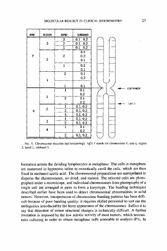
A brief review of chromosome terminology is presented to help in understand- ing the description of chromosomes. Chromosomes are numbered in order of decreasing size from 1 to 22. There are thus 22 pairs of chromosomes in addition to the sex chromosomes X and Y. From Fig. 4, it can be seen that the short arm of a chromosome is called “p”, while the long arm is called “q”. The centrornere is between the two arms. Each arm is divided into regions and numbered, with the region 1 being closest to the centromere. Bands within each region are numbered beginning with the band closest to the centromere. The subbands are also numbered and designated with a decimal. For instance, 1P35.1 would indicate that subband 1 within band 5 in region 3 on short arm p of chromosome 1 is being described. Chromosomes are detected with staining or banding tech- niques. These techniques produce alternating patterns of light and dark bands that are characteristic for each chromosome. Two types of banding techniques are used. These are fluorescent quinacrine or Q banding, and Giemsa or G banding. These banding techniques produce similar patterns except for the brilliant fluo- rescence of the Y chromosome in the Q banding technique. Other alternative techniques, called R and T banding, utilize heat denaturation, and their staining intensity is inversely related to staining found in Q or G banding, to the extent that areas that stain lightly in G or Q banding stain intensely in R and T banding, and vice versa. In R or T banding, however, ends of chromosomes are well defined, and terminal deletions are seen easily.
Conventional cytogenic analysis is performed on a small sample of heparinized blood. Phytohemagglutinin is added to induce mitotic division. After 65 to 72 hours in culture, colchicine is added, which by preventing spindle
MOLECULAR BIOLOGY IN CLINICAL BIOCHEMISTRY 23
FIG. 4. Chromosome structure and terminology. lq21.1 stands for chromosome I , arm q, region 2, band 1, subband 1 .
formation arrests the dividing lymphocytes in metaphase. The cells in metaphase are immersed in hypotonic saline to osmotically swell the cells, which are then fixed in methano1:acetic acid. The chromosomal preparations are manipulated to disperse the chromosomes, air dried, and stained. The selected cells are photo- graphed under a microscope, and individual chromosomes from photographs of a single cell are arranged in pairs to form a karyotype. The banding techniques described earlier have been used to detect chromosomal abnormalities in solid tumors. However, interpretation of chromosome banding patterns has been diffi- cult because of poor banding quality; it requires skilled personnel to sort out the ambiguities introduced by the fussy appearance of the chromosomes. Suffice it to say that detection of minor structural changes is technically difficult. A further limitation is imposed by the low mitotic activity of most tumors, which necessi- tates culturing in order to obtain metaphase cells amenable to analysis (Pl). In

24 SHESHADRI NARAYANAN
recent years, the ability to stain chromosomes at any stage of the cell cycle has removed the limitation of relying solely on metaphase spreads for cytogenic analysis.
A technique called interphase cytogenetics permits detection of both numerical and structural aberrations both in the metaphase spreads and in the more numer- ous interphase nuclei. This technique takes advantage of the fact that human satellite DNA composes up to 10-20% of all human DNA. There are three major types of human satellite DNA. Alpha (a)-satellite DNA is located in the cen- tromeric region of all human chromosomes. They are specific for each chromo- some, and hence chromosome-specific DNA probes can be used for their detec- tion. They are also present in high copy number organized as tandem repeats of unique I7 1 base-pair sequences that are present in as many as 5000 copies (A2); hence the sensitivity of probes to a-satellite DNA. The second type of human satellite DNA is beta (p)-satellite DNA located in the pericentric hetero- chromatin of many chromosomes. The third type of human satellite DNA is called classical satellites I , 11, and 111 and is based on short repeats of the base sequence adenine, adenine, thymine, guanine, and guanine (AATGG); it is lo- cated in the pericentric heterochromatin of at least chromosomes 1, 9, 15, and 16, and in the long arm of the Y sex chromosome. Nonisotopically labeled probes to the various types of human satellite DNA have made it possible to literally decorate or paint the chromosomes and discover any abnormality in them.
In the interphase cytogenetics technique, chromosome-specific DNA probes are used in combination with the in situ hybridization technique to detect both numerical and structural changes, such as deletions and translocations in both the metaphase spreads and in the more numerous interphase nuclei. Thus, biotiny- lated chromosome-specific DNA probes directed to a-satellite DNA have been used to hybridize to the centromeric region of the specific chromosome under high-stringency conditions such as high temperature, low salt concentration, or high concentration of formamide. In a typical experiment, probes were hy- bridized to target cell preparations in 60% formamide, 2X SSC (0.15 M sodium chloride, 0.015 M sodium citrate, pH 7.0), and 10% dextran sulfate at a probe concentration of 1 nanogram (ng)/td of the hybridization mixture (Pl). The hybridized biotinylated probe can be detected microscopically either with fluo- resceinated avidin or avidin conjugated with enzyme such as alkaline phospha- tase. The number of signals reflects the number of copies of chromosomes present both in the metaphase spreads and in interphase nuclei. In the interphase nuclei, the specific chromosome signal is visualized as a spot within the chroma- tin of the nucleus (A2, Pl) . Thus, the presence of three signals would indicate the presence of an extra copy of the specific chromosome or trisomy. Under less stringent conditions, specific probes can be made to hybridize to the peri-

MOLECULAR BIOLOGY IN CLINICAL BIOCHEMISTRY 25
centromeric region of the whole chromosome, thus permitting the detection of numerous hybridization sites (L3).
The specificity of interphase cytogenetics is achieved by a procedure called chromosomal in situ suppression hybridization (CISSH). Biotinylated chromo- some-specific DNA probes are used in this technique. Specificity is achieved by adding a large excess of competitor DNA, which suppresses or binds to highly repetitive probe sequences, thus allowing the probe to bind to only specific sequences on the target chromosome (L3). The probe thus hybridized to the specific target sequence is detected by standard techniques such as fluorescein- labeled avidin or avidin conjugated with alkaline phosphatase.
Interphase cytogenetics provides a powerful diagnostic tool in oncology, since it permits detection of structural and quantitative changes, including loss and gain of chromosomes and chromosome subregions. Furthermore, changes such as translocation, deletion, duplication, or amplification can be clearly discerned. Chromosome-specific DNA probes have been used to detect both numerical and structural aberrations in interphase nuclei of leukemias (Pl). Figure 5 illustrates schematically signals indicative of numerical and structural aberrations in inter- phase nuclei using chromosome-specific DNA probes.
3.14. DETERMINATION OF DNA INDEX BY FLOW CYTOMETRY
The ability of some fluorescent dyes to bind DNA quantitatively is exploited in flow cytometry to determine the DNA content of a cell. Dyes such as propidium iodide that bind double-stranded DNA stoichiometrically can be used for the purpose. The intensity of red fluorescence is directly related to the amount of DNA bound by propidium iodide. By comparing the fluorescence intensity of the test specimen and, in turn, its DNA content to the fluorescence intensity of specimens containing normal diploid amounts of DNA, a DNA histogram can be generated. By computing a DNA index, which is the ratio of DNA content of a test specimen to the DNA content of a specimen containing a normal diploid population, information related to the presence of an aneuploid tumor population can be obtained. The DNA index of 1 would imply that the DNA in the test specimen is from a normal diploid population (2N DNA), whereas the DNA index of an aneuploid population will be greater or less than 1. Thus, the DNA index of a tetraploid (4N DNA) would be 2.
In a DNA histogram, the fluorescence intensity, which is related to the quan- tity of DNA, is plotted on the x-axis, while the number of events or number of cells are plotted on the y-axis.
In addition to providing information about the presence of an aneuploid cancer cell population, the DNA histograms can also be used to gain information on the relative number of cells in the various stages of the cell cycle. Cells in the

26 SHESHADRI NARAYANAN
Abnormal Chromosome
Normal Chromosome Extra Chromosome
MetaDhase Nuclei Metaphase Nuclei lnterphase lnterphase
Translocation
Deletion
FIG. 5. Schematic representation of signals illustrative of chromosomal abnormalities in meta- phase spreads and interphase nuclei.
“resting” phase or the pre-DNA synthesis phase are referred to as the GOG, phase and contain a diploid (2N) amount of DNA. DNA synthesis or S phase occurs at the end of the GOGI phase. Cells in the S phase have increased amounts of DNA (2N-4N). The cells enter the G , phase after DNA replication, which continues until mitosis begins. The post-DNA synthetic phase is referred to as the G,M phase and represents cells under going mitosis, which has the greatest amount of DNA (4N complement) and will appear on the histogram at twice the channel number of the diploid 2N GoGI phase. Between the 2N and 4N populations, the cells in DNA synthesis (the S phase) can be located. Cell-cycle analysis of DNA histograms permits determining the fraction of cells in the S phase by measuring the area under the curve found between the G,G, and G, M peaks in the DNA histogram. Patients evidencing a large S phase cell fraction present a high risk for tumor recurrence, and thus poor prognosis.
The proliferative activity of cells can be estimated from the percentage of cells in both the S and G,M phases compared to the number of cells in the total

MOLECULAR BIOLOGY IN CLINICAL BIOCHEMISTRY 21
ll0I/ GO G1
0 100 200 DNA CONTENT
(Fluorescence Intensity)
FIG. 6. Diagrammatic representation of a DNA histogram showing population of cells in cell cycle.
population. This estimate, called the proliferative index, is a measure of the proliferative activity of cells expressed as the percent of cells greater than that found in the GoG, phase.
Figure 6 is a schematic representation of a DNA histogram. The ability of the flow cytometer to rapidly count several thousand nuclei contributes to the sensi- tivity of this technique for DNA analysis. However, problems due to sample quality, staining, and instrumental artifacts should be recognized and minimized to insure accurate interpretation of data (B2).
4. Applications
4.1. INFECTIOUS DISEASE
The voluminous number of publications in this area over the last 10 years have exceeded 10,000, thus attesting to the intensity of effort directed towards apply- ing molecular methods towards investigation of infectious diseases. Many of these investigations have translated into applications for diagnostic use in the clinical laboratory. The reader is referred to selected reviews on the subject (F2, N3, Wl). The availability of assays for the rapid diagnosis of pulmonary tuber-

28 SHESHADRI NARAYANAN
culosis using sputum specimens, bronchoalveolar lavages, and bronchial wash- ings represents a major advance. This is accomplished by amplifying a 584-bp region of the 16s ribosomal RNA (rRNA) gene sequence common to all my- cobacteria by PCR, followed by hybridization of biotinylated amplified product to a DNA probe specific for M. tuberculosis complex organism coated on a microwell plate. The hybridized product is detected enzymatically with an avidin horseradish peroxidase conjugate-tetramethylbenzidine substrate assay mixture (DI). Although the preceding test is rapid, requiring a little under 7 hours to perform, it still should be used in conjunction with culture. This assay has been found to be comparable to another diagnostic assay that relies on rRNA target amplification, requiring only 5 hours to perform (V2). Negative results some- times encountered with nucleic acid amplification procedures for culture-positive specimens could be related to the presence of inhibitors. A procedure that relies on sandwich hybridization and purification of probe-target complex by revers- ible target capture followed by amplification by Q-beta replicase is reported to decrease the level of interfering constituents (S5 ) .
Assay for human immunodeficiency virus type 1 (HIV-1) proviral DNA in peripheral blood monuclear cells can be performed by PCR followed by detec- tion of PCR products by electrochemiluminescence-labeled oligonucleotide probe [Tris-bipyridine ruthenium (11) complex]. Since one of the PCR primers is biotin-labeled at the 5' end, facile capture of the PCR product-probe complex can be accomplished on streptavidin-conjugated magnetic particles, prior to anal- ysis in an electrochemiluminescence analyzer (S3).
HIV-I RNA in plasma can also be quantitated by a branched-DNA (bDNA) signal amplification assay which has a quantitation limit of 1 x lo4 HIV-1 eq/ml. A novel internally controlled PCR assay (ICPCR) has been used to quantitate HIV- I Gag DNA and RNA in peripheral blood mononuclear cells and plasma. The linear range of amplification for the ICPCR assay is between 10" and 103 copies for HIV-1 DNA, while for HIV-1 RNA the amplification range is from 101 to lo4 copies. The ICPCR assay correlates with the bDNA signal amplification assay for the quantitation of HIV-I RNA, although subtle differ- ences between the two assays were noted (G2). Nevertheless, the fall in HIV-1 RNA levels in plasma in response to antiretroviral therapy was comparable with both the bDNA and ICPCR assays.
Mycobacteria of the Mycobacterium avium complex are implicated in dissemi- nated bacterial infections in AIDS patients. RFLP studies followed by hybridiza- tion with radiolabeled probe specific for an insertion sequence in M . avium (IS 131 1) have been useful for typing M. avium stains (R2). A variety of molecular techniques are available for the diagnosis of Chlamydia trachomatis infection. In addition to PCR, a method based on the ligase chain reaction has also been found to be sensitive to the detection of C. trachomatis infection in urine specimens collected from male and female subjects (V 1). The differentiation between low- risk genotypes of human papilloma virus (HPV 6 or 11) from genotypes of high

MOLECULAR BIOLDGY IN CLINICAL BIOCHEMISTRY 29
risk (HPV 16 or 18) associated with cervical carcinoma has been the focus of earlier studies using molecular methods (Al , Ll). The design of PCR-based assays for detection of HPV has been complicated by the fact that more than 60 HPV types exist, of which 20 types are known to infect the genital mucosa. Using primers for PCR targeting sequences highly conserved between types (consensus primers), followed by hybridization of amplified DNA with type- specific nested RNA probes labeled with digoxigenin-1 1-UTP, has made it pos- sible to capture the DNA-RNA hybrids onto a microtiter plate coated with antibody to digoxigenin. The hybrids are quantitated by the addition of an alka- line phosphatase-labeled monoclonal antibody directed to the hybrids and a fluorogenic substrate. This approach offers greater specificity and permits detec- tion of HPV DNA in cervico-vaginal lavage specimens (C4). Another approach to detect HPV type 16 variants is to perform SSCP analysis of PCR-amplified material from the non coding region of HPV type 16 (Xl). This study suggested that one variant was predominant in causing persistent infection, while coinfec- tion with additional HPV-16 variants accounted for a minor population of HPV- 16 genomes.
The ability to coat paramagnetic beads with antibodies to surface antigens of bacteria has been exploited to separate and concentrate organisms from the sample. The bacteria can then be lysed, and the DNA released into the superna- tant can be amplified by PCR. Such an approach has found a wide range of applications, among which is the detection of Helicobacter pylori in water and stool specimens (El).
Identification of pathogenic fungi belonging to the genus Candida has been made possible by using nested PCR to amplify first the entire small subunit (ssu) rRNA gene and subsequently a variable region within this gene. Variant PCR products are sequenced directly, thus uncovering specific restriction sites and unique regions towards which probes could be designed (N6). This strategy, referred to as interrepeat PCR, is a powerful tool for uncovering genetic poly- morphisms in fungi.
A simple assay for the detection of the malarial parasite Plasmodium fal- ciparum involves saponin lysis of blood sample and membrane filtration fol- lowed by amplification of the consensus sequence of eight 21-bp repeats. This procedure has been successfully used in the field (F2). A PCR assay targeting kinetoplast DNA sequences of Leishmania species was also successfully tested in the field (F2). Molecular methods for the detection of toxoplasma gondii and several other parasites have been reviewed (F2).
4.2. PATERNITY AND FORENSIC TESTING
RFLP analysis by the Southern blot procedure and detection with probe spe- cific for each VNTR are the basis of DNA fingerprinting. Using just four DNA probes, paternity can be excluded with a cumulative probability of 99.9% (52).

30 SHESHADRI NARAYANAN
An individualized fingerprint for purposes of forensic testing can be obtained with the use of three to five probes (53). However, spontaneous mutations occurring at polymorphic sites with a frequency as high as 5% could result in misinterpretation of some fingerprints. As such, to insure the authenticity of individualized fingerprints, it is important to use probes from different chromo- some locations (ZI).
In resolving RFLP bands obtained on electrophoresis, one must carefully demark boundaries or bins to allow distinguishing between bands of very similar sizes. Thus, while it is possible to readily distinguish a DNA fragment containing 20 VNTR units from a fragment containing 10 units, it would be difficult to distinguish a DNA fragment containing 20 VNTR units from one containing 21 units. It is for this reason that measurement of VNTR bands are grouped into bins specifying bands of similar sizes. The wider the bin, the greater the probability that two nonidentical bands may be included in the same bin and considered to be identical (H3). This problem can be overcome by testing multiple VNTR markers in order to permit identification of clear differences in band size without any ambiguity. The power of DNA fingerprinting derives from the fact that the probability of an RFLP matching pattern occurring due to coincidence is in the range of 1 in 100,000 to 1 in 1 million.
4.3. HLA TYPING
Molecular methods have been targeted to typing class I1 genes (DR, DP, and DQ) which are less readily typed serologically. Exon 2 , being the most poly- morphic region of the class I1 major histocompatability complex (MHC) mole- cule, is targeted in DNA typing in order to define specificity. A number of PCR- based methods have been used to type class I1 genes. These include PCR-RFLP, PCR-SSCP, PCR heteroduplex analysis, and PCR followed by use of sequence specific oligonucleotide probes (PCR-SSOP), to cite a few (Fl). The PCR-SSOP strategy in the reverse dot blot format has been commercialized. Biotinylated PCR products are hybridized to an array of sequence-specific oligonucleotide probes immobilized on a nylon membrane. The hybridized PCR product is detected using streptavidin-horseradish peroxidase enzyme by the conversion of substrate to a colored product. Information on the entire SSOP panel can be obtained on a single membrane, thus facilitating automated data interpretation (B3). The rapid reverse blot PCR-SSOP technique, when used with improved PCR-RFLP procedures relying upon the use of more group-specific amplifica- tions and more restriction enzymes, is reported to improve the accuracy of typing (TI). Direct sequencing of PCR products from exon 2 subsequent to immobiliza- tion of the biotinylated PCR product to streptavidin-coated magnetic beads is an approach to typing class I1 genes using fluorescent primers and DNA polymerase (Fl). Thus, HLA typing for the purposes of organ transplantation has been facilitated by the use of molecular methods.
MOLECULAR BIOLOGY IN CLINICAL BIOCHEMISTRY 31
4.4. GENETIC DISEASES
Genetic diseases that result from a single point mutation are readily amenable to detection by PCR based techniques (B4, N3). However, many genetic diseases have extreme genetic heterogeneity. Thus, only 70% of affected cystic fibrosis patients have a three-base-pair deletion in the cystic fibrosis gene, resulting in the deletion of phenylanine at position 508 (A 508), while in the remaining 30% of the affected patients, the mutations are extremely heterogeneous. In some cases, different genes could be affected for the same genetic disease in different people. Many genetic diseases may involve mutations interspersed throughout the gene, spanning many exons, making detection of such mutations extremely daunting. Precisely for this reason, there is very little effort in terms of introduction of diagnostic kits for genetic diseases, in contrast to the flurry of activity focused on infectious disease detection by molecular methods.
In spite of the preceding limitations, considerable progress has been made in the diagnosis of genetic diseases by molecular methods. The ability to amplify several exons simultaneously in a single PCR reaction (multiplex PCR) has been exploited in the analysis of deletions in X-linked genes, such as in Duchenne and Becker muscular dystrophies, where deletions occur in the dystrophin gene (B4). In some genetic diseases, such as the fragile X syndrome, Huntington’s disease, and myotonic dystrophy, tandem repeats of 3-bases (triplet repeats) are encoun- tered within or adjacent to a gene. These repeats can be detected by Southern blot analysis or by PCR (Y2).
Linkage analysis by studying RFLP patterns in two or more generations of family members afflicted with a disease is useful for genetic counseling for the prediction of inherited disease, in spite of its limitations, such as the possibility of genetic recombination or the presence of more than one gene locus in different family members.
Point mutations in mitochondrial DNA (mtDNA) leading to diseases with neurological manifestations, such as Leber’s hereditary optic neuropathy (LHON), mitochondrial encephalomyopathy with lactic acidosis, and strokelike episodes (MELAS) have been screened using PCR-SSCP ( J I ) . Ultimately, direct sequencing of PCR products in an automated format would help in uncovering multiple mutations that may occur in one or more exons, leading to a genetic disease.
4.5. ONCOLOGY
A well-known use of molecular methods is in the study of chromosomal translocations. Thus, in Philadelphia chromosome (phl) positive chronic my- elogenous leukemia (CML), the C-abl oncogene on chromosome 9 is translo- cated to a region on chromosome 22 called the breakpoint cluster region, or bcr. This (t9:22) translocation results in production of an abnormal fusion protein

32 SHESHADRI NARAYANAN
with a molecular weight of 210,000 (210 kDa). Actually, depending upon wheth- er exon 2 or 3 of the bcr region was fused with the exon 2 of the C-abl oncogene, two closely related 210-kDa proteins are produced. In roughly 50% of cases of phl positive acute leukemias, a 190-kDa protein is produced because of a translocation of abl oncogene to a breakpoint found in the first intron of the bcr gene (D3).
Acridinium ester-labeled chemiluminescent probes have been utilized to de- tect the specific protein-coding transcripts and to distinguish between transcripts that code for the 190-kDa protein and the two closely related 210-kDa proteins. The assay is called the hybridization protection assay (D3). In this assay, RNA isolated from the patient’s white blood cells is first amplified by PCR. The amplified product is incubated with the chemiluminescent probe. The un- hybridized probe is removed by selective hydrolysis in sodium tetraborate buffer, containing surfactant Triton X-100 at pH 8.5, in an incubation step at 60°C for 6 min. After the sample is cooled to room temperature, the chemiluminescence of the hybridized probe is measured in a luminometer. The procedure is reported to detect one leukemic cell in a population of a million or more normal cells. It is also rapid, requiring less than 30 min. Its reliability has been attested to by correlation with results obtained on karyotypic and Southern blot analysis (D3).
The ability of the interphase cytogenetics technique to detect both numerical and structural aberrations in interphase nuclei of leukemia was referred to earlier in this paper.
Likewise, the determination of DNA index by flow cytometry and the ability to assess the fraction of cells in the S phase also have applications in oncology, as was discussed earlier.
The improvements in FISH technology that were described earlier bring in a variety of applications to oncology, such as trisomy detection in various chromo- somes, among which are trisomy 8 detection in myeloid leukemia, trisomy 12 detection in chronic lymphocytic leukemia, and correlation of poor survival of patients with prostate cancer and trisomy in chromosome 7. The use of FISH probes directed to the sex chromosomes, X and Y, permits assessing engraftment success and relapse in patients receiving opposite-sex bone marrow transplanta- tion by establishing the proportion of recipient to donor cells; this technique is also useful in monitoring minimal residual disease (F4).
As noted earlier, loss of tumor suppressor genes allows tumorigenesis initiated by activation of proto-oncogenes to proceed without restraint. The most common cancer-related genetic change known at the gene level is the mutation at the p53 gene. The normal allele of this gene codes for a 53-kDa nuclear phosphoprotein that controls cell proliferation. Mutations in this gene abolish the tumor-sup- pressing capability of this gene, since proteins encoded by the various mutant alleles with single base substitutions are unable to control cell proliferation. In a spectrum of human cancers, point mutations, rearrangements, allelic loss and deletion of p53 gene have been detected.

MOLECULAR BIOLOGY IN CLINICAL BIOCHEMISTRY 33
A combination of pulse field gel electrophoresis to separate large DNA restnc- tion fragments, amplification by PCR, and detection with p53 cDNA probes has been useful to study chromosomal deletions and p53 mutations in colorectal cancers (B 1 ).
Procedures are available to detect p53 mutations in the urinary sediment of patients with bladder cancer. In this procedure, p53 exons 5 to 9 in DNA found in the urinary sediment of patient with bladder cancer are amplified by PCR. The PCR product is cloned into a bacteriophage vector. Between 100 and 10,000 clones are transferred to nylon membranes and hybridized with 32P-labeled oli- gonucleotide probes that are specifically directed to mutations seen in bladder cancer. The sensitivity of the procedure is such that one mutant could be detected among more than 5000 normal cells (S6). The procedure, which was also used to follow up patients treated by bladder-sparing strategies such as partial cystec- tomy, is amenable to the detection of p53 mutations in lung, colon, or cervical cancers. Mutations in the p53 gene in breast tumor cells can be followed by simultaneous outer amplification of exons 4-9 of the p53 gene, together with amplification of the HLA-DQB1 locus to serve as a control in a multiplex PCR assay. After outer amplification, the multiplex PCR mixture is aliquotted into separate tubes for region-specific amplification. Following the multiplex-nested PCR, biotinylated PCR products resulting from region-specific amplification are sequenced upon immobilization on streptavidin-coated beads using fluorescence- labeled primers and a-thiotriphosphate nucleotides in sequencing reactions (B5). The amplification of relatively short fragments of less than 420 bp by the preced- ing technique lends itself to use on formalin-fixed material subject to DNA degradation and from which longer fragments are not readily amplified (B5). Thus, direct DNA sequencing promises to uncover many of the molecular de- fects in cancer.
5. Summary
The myriad molecular techniques that have been developed and are undergo- ing refinement have advanced our knowledge of disease processes and made available strategies for detection. While the greatest impact of molecular tech- niques in the diagnostic laboratory has been in the realm of infectious diseases, techniques such as flow cytometry, FISH, and multiplex-nested PCR followed by direct DNA sequencing are increasing the scope of cancer detection and treat- ment. The facile typing of HLA-class I1 genes is a major advance in the matching of donors to recipients of organ transplantation. With advances in genosensor technology and robotic automation, molecular biology techniques are clearly poised to enter the diagnostic clinical biochemistry laboratory for multiple appli- cations.

34 SHESHADRI NARAYANAN
ACKNOWLEDGMENTS The author is deeply grateful to the editor of Annals of Clinical and Laboraforv Science for
granting permission to reprint from the author’s article (N3) Figs. I to 5 and Tables 1 and 2, and to freely adapt from the text. The author is extremely grateful to Ms. Carol Perello for her painstaking and excellent secretarial support and to Dr. Herbert Spiegel for the opportunity provided to write this article.
REFERENCES
A l . Amortegui, A. J., and Meyer, M. P., In-situ hybridization for the diagnosis and typing of human papillomavirus. Clin. Biochem. 23, 301-306 (1990).
A2. Anastasi, J., Interphase cytogenetic analysis in the diagnosis and study of neoplastic disor- ders. Am. J. Clin. Parhol. 95 Suppl. 1, S 2 2 4 2 8 (1991).
A3. Arnold, L. J., Jr., Hammond, P. W., Wiese, W. A., and Nelson, N. C., Assay formats involving acridinium-ester-labeled DNA probes. Clin. Chem. (Winsfon-Salem, N . C . ) 35,
BI. Baker, S. J., Fearon, E. R., Nigro, I. M., Hamilton, S. R., Preisinger, A. C. ef al . . Chromosomal 17 deletions and p53 gene mutations in colorectal cancers. Science 244, 217- 221 (1989).
B2. Bauer, K. D., Quality control issues in DNA content flow cytometry. Ann. N.Y . Acad. Sci.
B3. Begovich, A. B., and Erlich, H. A , , HLA typing for bone marrow transplantation. New polymerase chain reaction-based methods. JAMA, J . Am. Med. Assoc. 586-591 (1995).
B4. Bej, A. K., Mahbubani, M. H., and Atlas, R. M., Amplification of nucleic acids by poly- merase chain reaction (PCR) and other methods and their applications. Crif. Rev. Biochem. Mol. Biol. 26, 301-334 (1991).
B5. Berg, C., Hedrum, A,, Holmberg, A., Ponten, F., Uhler, M. ef al . , Direct solid-phase sequence analysis of the human p53 gene by use of multiplex polymerase chain reaction and alpha-thiotriphosphate nucleotides. Clin. Chem. (Winston-Salem, N . C . ) 41, 1461- 1466 (1995).
B6. Beutler, E., Gelbart, T., and Kuhl, W., Interference of heparin with the polymerase chain reaction. BioTechniques 9, 166 (1990).
87. Bishop, J. M., Amplification of protooncogenes in tumorigenesis. In “Concepts in Viral Pathogenesis” (A. L. Notkins and M. B. A. Oldstone, eds.), Vol. 2, pp. 71-78. Springer- Verlag, New York, 1986.
B8. Bishop, J. M., Molecular themes in oncogenesis. Cell (Cambridge, Mass.) 64, 235-248 ( I 991 ).
B9. Botstein, D., White, R. L . , Skolnick, M., and Davis, R. W., Construction of a genetic linkage map in man using restriction fragment length polymorphisms. Am. J. Hum. Gener. 32, 314-331 (1980).
B 10. Brahit, M. , and Haase, A. T., Detection of viral sequences of low reiteration frequency by in situ hybridization. Proc. Nafl. Acad. Sci. U.S.A. 75, 6125-6129 (1978).
B I I . Buffone, G. J., and Darlington, G. J., Isolation of DNA from biological specimens without extraction with phenol. Clin. Chem. (Winston-Salem. N . C . ) 30, 164-165 (1985).
B 12. Burke, D. T., Carle, G. F., and Olson, M. V . , Cloning of large segments of exogeneous DNA into yeast by means of artificial chromosome vectors. Science 236, 806-812 (1987).
CI . Cariello, N. F., Scott, J. K., Kat, A. G . , Thilly, W. G., and Keohavong, P., Resolution of a missense mutant in human genomic DNA by denaturing gradient gel electrophoresis and
1588-1594 (1989).
677, 59-77 (1993).

MOLECULAR BIOLOGY IN CLINICAL BIOCHEMISTRY 35
direct sequencing using in vitro DNA amplification: HPRT Munich. Am. J . Hum. Genet. 42,
C2. Chomczynski, P., One-hour downward alkaline capillary transfer for blotting of DNA and RNA. Anal. Biochem. 201, 134-139 (1992).
C3. Coad, J. E., Lander, T. A, , and Litz, C. E., Inhibition of restriction endonucleases by common clinical anticoagulants. Anal. Biochem. 205, 368-369 (1992).
C4. Coutlee, F., Provencher, D., and Voyer, H., Detection of human papillomavirus DNA in cervical lavage specimens by a nonisotopic consensus PCR assay. J. Clin. Microbiol. 33, 1973-1978 (1995).
DI. D'Amato, R. F., Wallman, A. A,, Hochstein, L. H., Colaninno, P. M., Scardamaglia, M. et al.. Rapid diagnosis of pulmonary tuberculosis by using Roche AMPLCORimycobacterium tuberculosis PCR test. J . Clin. Microbiol. 33, 1832-1834 (1995).
D2. Dattagupta, N., Rae, P. M. M., Huguenel, E. D., Carlson, E. , Lyga, A. et al., Rapid identification of microorganisms by nucleic acid hybridization after labeling the test sample. Anal. Biochem. 177, 85-89 (1989).
D3. Dhingra, K., Talpaz, M., Riggs, M. G., Eastman, P. S. , Zipf, T., ef a l . . Hybridization protection assay: A rapid, sensitive, and specific method for detection of Philadelphia chro- mosome-positive leukemias. Blood 77, 238-242 ( 1991).
D4. Donis-Keller, H. , Green, P., Helms, C., Cartinhour, S . , Weiffenbach, B. e t a / . . A genetic linkage map of the human genome. Cell (Cambridge. Mass.) 51, 319-337 (1987).
E l . Enroth, H. , and Engstrand, L. , Immunomagnetic separation and PCR for detection of Heli- cobacter pylori in water and stool specimens. J . Clin. Microbiol. 33, 2162-2165 (1995).
E2. Erlich, H. A., Sheldon, E. L., and Horn, G . , HLA typing using DNA probes. Bio Technology 4, 975-981 (1986).
FI . Field, H., and Garovoy, M. R., Positive impact of DNA typing on solid organ transplantation. Transplant. Rev. 8, 151-173 (1994).
F2. Figueroa, M. E., and Rasheed, S. , Molecular pathology and diagnosis of infectious diseases. Am. J . Clin. Puthol. 95, Suppl. 1, S8-S21 (1991).
F3. Foss. E. H., Chehab, F. F., and Brooks, G . F., DNA amplification and reverse dot blot hybridization for detection and identification of mycobacteria to the species level in the clinical laboratory. J . Clin. Microbiol. 30, 1220- I224 (1992).
F4. Fox, J. L., Hsu, P., Legator, M. S . , Morrison, L. E., and Seelig, S. A., Fluorescence in situ hybridization powerful molecular tool for cancer prognosis. Clin. Chem. ( Winston-Salem, N . C . ) 41, 1554-1559 (1995).
G I . Gasparini, P., Novelli, G . , Savoia, A , , Dallapiccola, B., and Pignatti, P. R., First-trimester prenatal diagnosis of cystic fibrosis using the polymerase chain reaction: Reports of eight cases. Prenatal Diagn. 9, 340-355 (1989).
G2. Gupta, P., Ding, M., Cottrill, M. , Rinaldo, C., Kingsley, L. er al.. Quantitation of human immunodeficiency virus type I DNA and RNA by a novel internally controlled PCR assay. J. Clin. Microbiol. 33, 1670- 1673 (1995).
G3. Gyllensten, U. B., and Erlich, H. A,, Generation of single stranded DNA by the polymerase chain reaction and its application to direct sequencing of the HLA-DQA locus. Proc. Narl. Acad. Sci. U.S.A. 85, 7652-7656 (1988).
HI. Hayashi, K., F'CR-SSCP: A simple and sensitive method for detection of mutations in the genomic DNA. PCR Methods Appl. 1, 34-38 (1991).
H2. Holodniy, M., Kim, S. , Katzenstein, D., Konrad, M., Groves, E., and Merigan, T. C., Inhibition of human immunodeficiency virus gene amplification by heparin. J . Clin. Micro- biol. 29, 676-679 (1991).
H3. Housman, D. E. , Clinical implications of basic research. DNA on trial-The molecular basis of DNA fingerprinting. N . Engl. J. Med. 332, 534-535 (1995).
726-734 (1988).

36 SHESHADRI NARAYANAN
H4. Hozier, J. C., and Davis, L. M., Cytogenetic approaches to genome mapping. Anal. Bio- chem. 200, 205-217 (1992).
J I . Jaksch, M., Gerbitz, K. D., and Kilger, C., Screening for mitochondria1 DNA (Mt DMA) point mutations using non radioactive single strand conformation polymorphism (SSCP) analysis. Clin. Biochem. 28, 503-509 (1995).
52. Jeffrys, A. J., Brookfield, 1. F. Y . , and Semeonoff, R., Positive identification of an immigra- tion test-case using human DNA fingerprints. Nature (London) 317, 818-809 (1985).
53. Jeffrys, A. J . , Wilson, V., Thein, S. L., Weatherall, D. J.. Ponder, B. H. J., DNA “finger- prints” and segregation analysis of multiple markers in human pedigrees. Am. J . Hum. Genet.
K I . Keller, G. H., and Manak, M. M., Hybridization formats and detection procedures. In“DNA Probes,” pp. 198-213. Stockton Press, New York, 1989.
K2. Kilpatrick, D. C., Simplified preparation from anticoagulated blood for HLA-DR genomic typing. Tissue Antigens 41, 219-220 (1993).
LI . Landry, M. L., Nucleic acid hybridization in viral diagnosis. Clin. Biochem. 23, 267-277 ( 1990).
L2. Lee, H. H., The LCx system-from research to clinical laboratories. Clin. Chem. (Winston- Salem. N . C . ) 40, 650 (1994).
L3. Lichter, P., Cremer, T., Borden, J., Manuelidis, L., and Ward, D. C., Delineation of individ- ual human chromosomes in metaphase and interphase cells by in situ suppression hybridiza- tion using recombinant DNA libraries. Hum. Genet. 80, 224-234 (1988).
L4. Longo, M. C., Berninger, M. S., and Hartley, J. L., Use of uracil DNA glycosylase to control carry-over contamination in polymerase chain reactions. Gene 93, 125-128 (1990).
MI. Maniatis, T., Fritsch, E. E., and Sambrook, J., “Molecular Cloning: A Laboratory Manual.” Cold Spring Harbor Lab., Cold Spring Harbor, N.Y., 1982.
M2. Matthews, J. A., and Kricka, L. J., Analytical strategies for the use of DNA probes. Anal. Biochem. 169, 1-25 (1988).
NI . Narayanan, S . , DNA probes. In “Principles and Applications of Laboratory Instrumentation,” pp. 185-191. ASCP Press, Chicago, 1989.
N2. Narayanan, S. , Applications of restriction fragment length polymorphism. Ann. Clin. Lab. Sci. 21, 291-296 (1991).
N3. Narayanan, S . , Overview of principles and current uses of DNA probes in clinical and laboratory medicine. Ann. Clin. Lab. Sci. 22, 353-376 (1992).
N4. Nelson, N. C., and Kacian, D. L., Chemiluminescent DNA probes: A comparison of the acridinium ester and dioxetane detection systems and their use in clinical diagnostic assays. Clin. Chim. Acra 194, 73-90 (1990).
N5. Newton, C. R., Graham, A. , Heptinstall, L. E., Powell, S . J., Summers, C. e ta l . . Analysis of any point mutation in DNA. The amplification refractory mutation system (ARMS). Nucle- ic Acids Res. 17, 2503-25 I6 (1989).
N6. Niesters, H. G. M.. Goessens, W. H. F., Meis, J. F. M. G., and Quint, W. G. V., Rapid polymerase chain reaction-based identification assays for candida species. J . Clin. Microbiol.
01. Orita, M., Suzuki, Y., Sekiya, T., and Hayashi, K., Rapid and sensitive detection of point mutations and DNA polymorphisms using the polymerase chain reaction. Genomics 5, 874- 879 (1989).
PI . Poddighe, P. J., Moesker, 0.. Smeets, D., Awwad, B. H . , Ramaekers, F. C. S. et al . . Interphase cytogenetics of hematological cancer: Comparison of classical karyotyping and in situ hybridization using a panel of eleven chromosome specific DNA probes. Can. Res. 51,
P2. Porter-Jordan, K., Rosenberg, E. I., Keiser, J. F., Gross, J. D., Ross, A. M. er a / . . Nested
39, 11-24 (1986).
31, 904-910 (1993).
1959-1967 (1991).

MOLECULAR BIOLOGY IN CLINICAL BIOCHEMISTRY 37
polymerase chain reaction assay for the detection of cytomegalovims overcomes false posi- tives caused by contamination with fragmented DNA. J. Med. Viral. 30, 85-91 (1990).
R I . Ramirez-Solis, R., Rivera-Peres, J., Wallace, J. D. , Wims, M. , Zheng, H. et al., Genomic DNA microextraction: A method to screen numerous samples. Anal. Biochem. 201, 33 1-335 (1992).
R2. Roiz, M. P., Palenque, E., Guerrero, C. , and Garcia, M. J., Use of restriction fragment length polymorphism as a genetic marker for typing mycobacterium avium strains. J. Clin. Microbiol. 33, 1389- 139 I (1995).
R3. Rolf% A., Shuller, I., Finckh, U. , and Weber-Rolfs, I . , “PCR: Clinical Diagnostics and Research,” pp. 51-60. Springer-Verlag, New York, 1992.
S I . Saiki, R. K . , Chang, C., Levenson, C. H. , Warren, T. C., Boehm, C. D. et al., Diagnosis of sickle cell anemia and P-thalassemia with enzymatically amplified DNA and non radioactive allele-specific oligonucleotide probes. N. Engl. J . Med. 319, 537-541 (1988).
S2. Schadp, A. P., Akhavan, H., and Romano, L. J., Chemiluminescent substrates for alkaline phosphatase: Application to ultrasensitive enzyme-linked immunoassays and DNA probes. Clin. Chem. (Winston-Salem, N . C . ) 35, 1863-1864 (1989).
S3. Schutzbank, T. E . , and Smith, J., Detection of human immunodeficiency virus type 1 proviral DNA by PCR using an electrochemiluminescence tagged probe. J. Clin. Microbiol. 33,
S4. Sestini, R., Orlando, C . , Zentilin, L . , Gelmini, S., Pinzani, P. et a/. , Measuring c-erb B-2 oncogene amplification in fresh and paraffin-embedded tumors by competitive polymerase chain reaction. Clin. Chem. (Winston-Salem. N . C . ) 40, 630-636 (1994).
S 5 . Shah, J. S., Liu, J . , Buxton, D. , Hendricks, A , , Robinson, L. et a/. . Q-beta replicase- amplified assay for detection of Mycobacterium tuberculosis directly from clinical specimens. J. Clin. Microbiol. 33, 1435-1441 (1995).
S6. Sidransky, D. , Eschenbach, A. V., Tsai, Y. C., Jones, P., Summerhayes, I. et al . . Identifica- tion of p53 gene mutations in bladder cancers and urine samples. Science 252, 706-709 (1991).
S7. Smith, C. L., and Cantor, C. R., Pulsed-field gel electrophoresis of large DNA molecules. Nature (London) 319, 701-702 (1986).
S8. Southern, E. M., Detection of specific sequences among DNA fragments separated by gel electrophoresis. J. Mol. Biol. 98, 503-517 (1975).
S9. Stickler, T. G . , and Copenhaver, C. D., In situ hybridization in hematopathology. Am. J. Clin. Pathol. 93, Suppl. I , S44-S48 (1990).
TI. Thonnard, J.. Deldime, F., Heusterspreute, M., Delepaut, B., Hanon, F. et al., HLA Class I1 genotyping: Two assay systems compared. Clin. Chem. (Winston-Salem, N . C . ) 41, 553-556 (1995).
T2. Todd, D. M., and Buccini, F. J., Apparent heparin interference with restriction enzyme digestion of genomic DNA. Clin. Chem. (Winston-Salem. N .C . ) 39, 362-363 (1993).
VI. Vdn Doornum, G. J. J., Buimer, M., Prins, M., Henquet, C. J . M., Coutinho, R. A. et a/.. Detection of chlamydia trachomatis infection in urine samples from men and women by ligase chain reaction. J. Clin. Microbiol. 33, 2042-2047 (1995).
V2. Vuorinen, P., Miettinen, A., Vuento, R . , and Hallstrom, O., Direct detection of mycobac- terium tuberculosis complex in respiratory specimens by Cen-Probe Mycobacterium Tuber- culosis Direct test and Roche Amplicor Mycobacterium Tuberculosis Test. J. Clin. Microbiol. 33, 1856- 1859 ( 1 995).
WI . Weiss, J. B., DNA probes and PCR for diagnosis of parasitic infections. Clin. Microbiol. Rev.
W2. Wetmur, J. G., DNA probes: Applications of the principles of nucleic acid hybridization. Crit.
2036-204 1 ( 1995).
8, 113-130(1995).
Rev. Biochem. Mol. Biol. 26, 227-259 (1991 ).

38 SHESHADRI NARAYANAN
W3. Wolcott, M. J., Advances in nucleic acid-based detection methods. Clin. Microbiol. Rev. 5 , 370-386 (1992).
XI. Xi, F. L., Demers, G . W., Koutsky, L. A,, Kiviat, N. B., Kuypers, J. et al., Analysis of human papillomavirus type 16 variants indicates establishment of persistent infection. J. Infect. Dis. 172, 747-755 (1995).
Y 1. Young, K. K. Y., Resnick, R. M., and Myers, T. W., Detection of hepatitis C virus RNA by a combined reverse transcription-polymerase chain reaction assay. J. Clin. Microbiol. 31, 882- 886 (1993).
Y2. Yu, S . , Pritchard, M. N., Kremer, E., Lynch, M., Nancarrow, J. et al., Fragile X genotype characterized by an unstable region of DNA. Science 252, 1179-1 181 (1991).
ZI . Zumwalt, R. E., Application of molecular biology to forensic pathology. Hum. Purhol. 20, 303-307 (1989).

ADVANCES I N CLINICAL CHEMISTRY, VOL. 32
CLINICAL MOLECULAR BIOLOGY: CONCEPTS AND APPLICATIONS
Kaiser J. kit"
Division of Clinical laboratory Devices Food and Drug Administration
Rockville, Maryland
1. Introduction . . . . . . . . . . . . . . . . . . . . . . . . . . . . . . of Diseases . . . . . . . . . . . . , . , . . . . . , . , . ,
3. Cystic Fibrosis . . . . . . . . . . . . . . . . . . . . . . . . . . . . . . . . . . . . . . . . . . . . . . . . . . . . . . . . . 4. Duchenne and Becker Muscular Dystrophy . . . . . . . . . . . . . . . . . . . . . . . . . . . . . . . . . . 5. Lymphoproliferative Disorders . . . . . . . . . . . . . . . . . . . . . . . . . . . . . . , . . . . . . . . . . . . .
7. Human Papilloma Virus . . . . . . . . . . . . . . . . . . . . . , . . . . . . . . . . . . . , . . . . . . . . . . . . . 8. Clinical Applications of Molecular Technologies , . . . . . . . . . . . . . . . . . . . . . . . . . . . .
8.1. In Situ Hybridization . . . . . . . . . . . . . . . . . . . . . . . . . . . . . . . . . . . . . . . . . . . . . . . 8.2. Southern Blot Analysis . . . . . . . . . . . . . . . . . . . . . . . . . . . . . , . , . . . . . . . . . . . . .
6. Chronic Myelogenous Leukemia and Acute Leukemias . . . . . . . .
8.3. Polymerase Chain Reaction . . . . . , . . . . . . 9. Cystic Fibrosis: Current Diagnostics . . . . . . . . . . . . . . . . . . . . . . . . . . . . . . . . . . . . . . .
10. Duchenne and Becker Muscular Dystrophy: Current Diagnostics . . . . . . . . . . . . . . . . 1 1. Lymphoproliferative Disorders: Current Diagnostics 12. Chronic Myelogenous Leukemia and Acute Leukern
14. Perspective on the Evaluation of Molecular Diagnos
16. Quality Assurance: Science and Regulatory Issues . . . . . . . . . . . . . . . . . . . . . . . . . . . . 17. Conclusion . . . . . . . . . . . . . . . . . . . . . . . . . . . . . . . . . . . . . . . . . . .
References . . . . . . . . . . . . . . . . . . . . . . . . . . . . . . . . . . . . . . . . . . . . . . . . . . . . . . . . . . . .
13. Human Papilloma Virus: Current Diagnostics . . . . .
15. Software Applications to Molecular Diagnostics . . . . . . . .
. . . . . . . . . . . .
. . . . . . . . . .
39 43 45 46 47 48 50 50 52 53 54 54 55 56 57 58 58 68 69 70 70
1. Introduction
Molecular research advances have created useful techniques that are comple- mentary to routine testing within many sections of clinical laboratory medicine
*The views expressed herein are those of the author and do not necessarily reflect the views of the U.S. Food and Drug Administration.
39
Copynghi 0 1996 by Academic Press, Inc All nzhts of re~roduction tn anv form reserved

40 KAISER J. AZIZ
(hematology, microbiology, virology, tissue typing) and that have applications for genetic disorders, oncology, transplantation, and infectious diseases. Exam- ples are nucleic acid hybridization (dot blot, Southern blot), in situ hybridization, and in vitro amplification technologies such as the polymerase chain reaction (PCR). Probes based on DNA analysis are now available for several important genetic disorders such as cystic fibrosis, muscular dystrophies, fragile X syn- drome, sickle-cell anemia, Gaucher’s disease, and Huntington’s chorea. Among these developments, genetic testing offers greatest potential in the areas of pre- vention or early intervention. New applications are being developed for prenatal diagnosis, detection of genetic predisposition to disease, and the presence of oncogenes. At the same time, these developments offer unprecedented chal- lenges for the individual, institutions, and society in general. They raise signifi- cant issues for safety and effectiveness, as well as economic, legal, and ethical considerations.
The development of a molecular diagnostic test can be based on the type of condition for which the test is designed. This development can be based on analysis of the genetic material or its products-that is, an examination of the DNA, of DNA adducts, or of RNA or the proteins produced. These tests are generally based on an analyte such as DNA, chromosome, protein, or other gene products to detect mutations, karyotypes, disease-related genotypes, or phe- notypes intended for clinical uses. Such uses include diagnosis, monitoring, prognosis, identification of carriers, or prediction of disease risks. These devel- opments include detection of chromosomal translocations using reverse tran- scriptase PCR or Southern blot analysis, thereby providing better tools for the discrimination of lymphomas, configuration of cytosomatic findings, and early detection of cancer syndromes. Technologies are available for the detection of translocation associated with rhabdomyosarcoma, lymphoma, sarcoma, and the Philadelphia chromosome. Technologies such as PCR, ligase chain reaction (LCR), and nucleic acid amplifications are now available for detecting and mon- itoring infectious diseases.
In the last few years, innovative molecular techniques have provided powerful tools for diagnosis based on direct DNA analysi;. These techniques resulted in the use of restriction enzymes to generate DNA fragments that reveal variations in gene sequences to identify the presence or absence of all or part of a gene. These techniques are so powerful that we can now identify a single base change in coding sequences of a large gene. However, the success of these techniques is also due to well-established protocols and reagents for generating molecular probes, and to the basic understanding of hybridization kinetics, which contrib- utes to the sensitivity and specificity of the probes.
Molecular diagnostic technologies can be either quantitative (biochemical) or qualitative (signals indicating a translocation, or a single base-pair change de- tected by direct DNA sequencing). These technologies offer patients the option

CLINICAL MOLECULAR BIOLOGY 41
of knowing whether they are carriers capable of passing on genetic disease, e.g., cystic fibrosis, sickle-cell anemia, or Tay-Sachs disease, as well as whether they have inherited certain diseases, e.g., muscular dystrophy or Huntington’s dis- ease. In the near future, technologies will be available that will indicate suscep- tibility to other conditions that develop in adult life, such as heart disease and many types of cancer. Some of these technologies to generate molecular probes include in situ hybridization, nick translation, transcription analysis, S I map- ping, and PCR. PCR, with amplification of very small amounts of DNA for analysis, is currently available in numerous applications.
Recombinant DNA technology provides a variety of tools to identify alter- ations in DNA that provide a rapidly growing list of familial and acquired disorders. This type of technology is extremely sensitive and specific, so that it can reveal the genetic complement of somatic cells contained in various tissues or even single cells. DNA from the leukocytes in a small amount of peripheral blood can be used to diagnose a variety of Mendelian disorders such as cystic fibrosis, hemophilia, and sickle-cell anemia, without the need for biopsies or additional biochemical assays. Techniques of gene isolation have been applied to identify the DNA rearrangements related to diverse disorders as chronic granu- locytic leukemia, cystic fibrosis, Duchenne’s muscular dystrophy, and neuro- fibromatosis. These techniques can be applied clinically to genetic studies in- volving diagnosis of affected individuals and presymptomatic individuals and identification of asymptomatic carriers.
Genetic applications of molecular technologies have different concerns than routine clinical laboratory testing because results are used in a predictive manner (e.g., the probability that a gene is present and, if present, whether it will be expressed and to what extent of severity). Therefore, validation procedures may differ, depending on whether the test will be used in high-risk situations or in the general population.
The requirements for validation and interpretation of results will depend on the degree of association between alleles at a gene locus and the status of the disease. These technologies can be utilized to identify Mendelian, or single-gene, disor- ders that result from alterations in a gene. However, validation of the diagnostic procedures is subject to the following qualifications:
There must be direct and demonstrated correlation between the disease and
There must be complete information on the sequence of the responsible
A catalog must be available of commonly found defects in the gene. There must be ample information on the molecular genetic nature of the
disorder (mode of inheritance, gene frequency, polymorphic nature of the locus, genetic heterogeneity of the disorder).
the defect in the gene/gene product.
gene.

42 KAISER J. AZIZ
There must be experience or knowledge concerning the predictive value and probability of each type of clinical application.
There must be adequate information in presymptomatic and predisposition testing concerning a healthy individual’s future risk for developing a hereditary disorder (e.g., presymptomatic testing for a familial condition with 100% likeli- hood of occurring in an individual carrying the genetic mutation)-for example, presymptomatic testing in Huntington’s disease and multiple endocrine neo- plasia, type 2A.
Therefore, an ideal test validation would require the listed information as essen- tial elements of adequate interpretation of test results, providing the patient with the pretest/post-test counsel. Also, a complete map of the genome (the National Center for Human Genome Research) will improve detection of genetic disease and increase the likelihood of beneficial treatments as the genome and disease processes are better understood. The role of the NIH Human Genome Project is genome mapping and sequencing. In recent years, much of the progress in molecular technology has been attributable in large part to the Human Genome Initiative, which represents the efforts of the Human Genome Organization (HUGO). HUGO represents an international collaborative effort to map the hu- man genome. One of the main objectives of HUGO is to inventory all human genes, their DNA sequence, and their sequence along the chromosomes, and eventually come up with the complete human DNA sequence. It is anticipated that the entire human genome will be sequenced by the year 2010. The contribu- tion of this initiative to the diagnosis and treatment of genetic diseases is ex- pected to be tremendous.
Genetic tests are unique because of their intended use: ( I ) Genes responsible for constitutional genetic disorders are nearly always present in multiple genera- tions and a single individual’s test results may affect other family members; ( 2 ) direct confirmatory tests may not be available for genetic disorders (e.g., Hunt- ington’s disease); (3) the standard of care for certain genetic disorders may not be on the same scale as other areas of health care services. Therefore, the unique applications of genetic testing may raise unprecedented social, ethical, medical, and legal issues. Results of such applications and/or the associated risks of a particular genetic disorder may influence an individual’s life decisions-repro- ductive, prenatal diagnosis, and pregnancy termination or continuation. In view of all these concerns, it is essential that a genetic disease test have a reasonable assurance of safety and effectiveness in the context of its intended clinical use. Other concerns related to genetic testing revolve around the possibility that individuals identified with a particular mutant gene may experience discrimina- tion by employers and insurance providers. Testing for genetic susceptibility or predisposition for certain disorders (e.g., Alzheimer’s disease) that a person may develop in later life and for which there is no known treatment or cure presents an

CLINICAL MOLECULAR BIOLOGY 43
ethical dilemma. Prenatal diagnostic tests are available and performed during the first or second trimester of pregnancy for the open neural tube defects and Down syndrome (a chromosomal disorder). Open neural tube defects are multifactorial disorders that are associated with increases in the levels of alpha-fetoprotein in the maternal serum and amniotic fluid (Al) .
2. Molecular Technology and the Diagnosis of Diseases
Molecular biotechnology tests offer improved analytical sensitivity and speci- ficity compared to most other routine clinical lab tests, thus enabling us to identify and study smaller numbers of relevant analyte molecules with greater accuracy. Molecular techniques such as PCR, Southern blotting, gene rearrange- ment analysis, and fluorescence in situ hybridization-when applied to the ge- netic, infectious, neoplastic, and other human diseases-offer new insights about the causes and pathogenesis of disease. These techniques are useful for identifying and studying genetic disorders such as cystic fibrosis and Duchenne muscular dystrophy. Infectious diseases, such as herpes simplex encephalitis, human papilloma virus infection, cytomegalovirus infection, tuberculosis, and others, can be detected using PCR and other molecular techniques, such as plasmid-profile analysis, genomic fingerprinting by restriction-endonuclease analysis, and southern-hybridization fingerprinting. Furthermore, the applica- tions of these techniques to detect gene rearrangements and oncogenes assists in the early detection and diagnosis of certain cancers. Molecular techniques also play a key role in histocompatibility typing and tissue transplantation, paternity testing, and forensic medicine.
Recent developments and application of molecular biotechnology to genetic diseases have been impressive. Understanding the molecular and genetic basis of a disease enables health care providers to know that one phenotypic disease may be a whole spectrum of genetic diseases, and that many conditions, such as heart disease and hypertension, may have associated genetic factors. The availability of specific molecular tests for genetic disease diagnosis has revolutionized medi- cal testing, while raising unique clinical and socioeconomic issues.
The ability to confirm the diagnosis of a clinical disorder at the molecular level brings challenges to health care delivery. However, the ability to identify pre- symptomatic individuals, as in the case of Huntington’s disease, presents clinical and ethical problems (e.g., all causes of Huntington’s have not been identified). Similarly, screening to identify asymptomatic camers, such as those hetero- zygous for the common mutation causing cystic fibrosis, presents the confound- ing problems related to stigmatization and confidentiality. Also, the greater sen- sitivity and specificity of these technologies presents greater potential for providing clinically pertinent data regarding organ transplantation.

44 KAISER J. AZIZ
In general, the basic tools of restriction endonuclease, Southern blots, probes, and PCR amplification are applied to clinical genetic disorders. These applica- tions include detection of chromosome fragments, gene deletions, gene rear- rangements, and point mutations that are associated with a growing list of disor- ders. The following examples illustrate the applications of recombinant DNA techniques to direct detection of each of these types of gene derangements.
Individuals with Turner’s syndrome are, in some cases, mosaic for a portion of or for the entire Y chromosome (46,XY/45,X). Since such individuals may be at increased risk for gonadal tumors, Southern blot or PCR analysis has been used to detect the presence of Y chromosome segments in studies of DNA from peripheral blood samples. Sim- ilarly, the fetal sex as well as the presence of some aneuploid states (e.g., trisomy 18) can be determined by analysis of DNA from chorionic villi or amniotic fluid cells.
The defects causing Mendelian disorders are usually heterogeneous and include, in some cases, gene deletions. For instance, one autosomal recessive form of growth hormone (GH) deficiency is caused by deletion of the GH genes. Affected individuals are extremely short and, unfor- tunately, often respond only briefly to GH replacement because of development of anti-GH antibodies. Such individuals can be diagnosed by their lack of GH genes by Southern blot or PCR analysis.
Detection of gene rearrangements. A great number of neoplasms are now shown to be associated with acquired gene rearrangements. For instance, chronic granulocytic leukemia (CGL) is associated with the Philadelphia (Ph) chromo- some rearrangement, which is a 9:22 reciprocal translocation that involves the abl proto-oncogene on 9g34 and the bcr gene on 22q 1 1. Unusual patterns result- ing from the fusion of the bcr:abl genes can be detected by Southern blot or PCR analysis of DNA or in mRNA from peripheral blood or bone marrow samples. These techniques can provide sensitive and specific detection of the proportion of cells containing Ph chromosome to monitor treatment response and detect re- lapses.
Sickle-cell anemia is caused by a single base mutation (substitution of an adenine for thymine) in the sixth codon of the globin gene, changing GAG (glutamic acid) to GTG (valine). This change prompted the development of the presence of an Mst I1 restriction site (CCTGAGG). It was possible to use restriction length polymorphism to differentiate between the Hb(S) and the Hb(A) alleles. Determination of the presence or absence of this site in genomic DNA or PCR products enables determination of the sickle cell genotype. This analysis has been used for prenatal diagnosis as well as for confirmation of abnormal newborn hemoglobinopathy screening tests.
In a majority of inherited genetic disorders, the basic defect or defects in the gene, or even the gene that is
Detection of chromosome fragments.
Detection of gene deletions.
Detection of point mutations.
Indirect detection of abnormal genes.

CLINICAL MOLECULAR BIOLOGY 45
responsible for the disorder, are unknown. Despite this lack of knowledge, DNA analysis can often still be used to determine whether the defective gene has been transmitted. Use of DNA polymorphisms and linkage analysis enables the dis- ease status of individuals at risk for diseases as divergent as hemophilia and adult onset polycystic kidney disease to be determined. While linkage technique can be applied to many diseases, their utility is limited to certain families because of the problems of genetic heterogeneity, the lack of necessary documentation of their disease status, and the required availability of other family members.
3. Cystic Fibrosis
Cystic fibrosis (CF) is the most common potentially lethal autosomal recessive disease among Caucasians. The incidence is estimated to be approximately 1 in 2000 births (BI). Since it is inherited as an autosomal recessive condition, screening to identify couples at risk has been suggested. However, CF screening is complicated because many mutations of the CF gene exist. Thus, the fea- sibility of screening a population for camers of cystic fibrosis gene mutations is primarily dependent upon the frequency of the common mutation in that popula- tion.
Cystic fibrosis is characterized by progressive obstructive lung disease, colo- nization of the airways by pathogenic organisms, exocrine pancreatic insufficien- cy, intestinal malabsorption, and elevated sweat electrolytes. The clinical situa- tion varies at different ages, and the severity of disease and its rate of progression in the involved organs vary considerably. Cystic fibrosis is indeed serious enough to be considered the subject of carrier screening. This disease limits activity, reduces fertility, shortens life, and imposes burdens on patient and family. Treat- ment is burdensome on the family because it is time-consuming, disruptive, exhausting and expensive. Psychosocial development is imperiled by the de- mands of treatment and the prospect of an early death.
The gene responsible for CF is located on chromosome 7 and is named cystic fibrosis transmembrane conductance regulator (CFTR). The protein encoded by the CF gene is believed to function either as a transporter of chloride ions or as a regulator of the transport process. Recent studies have indicated that it is possible to transfer normal genes to CF cells, subsequently correcting the defect in chlo- ride transport in those cells. Thus, the CF gene product, the CFTR, is the defective protein in cells from CF patients. Sequence analysis of CFTR genes cloned from a normal individual and an individual affected with CF revealed that the main difference between the two was the deletion of three base pairs (3 bp) of DNA in the mutant chromosome, causing the loss of a single phenylalanine residue at position 508 of the CFTR protein. This 3-bp deletion, termed F508, presumably causes approximately 70% of the mutations responsible for cystic

46 KAISER J. AZIZ
fibrosis. Position 508 occurs in the first nucleotide binding fold and is thought to be important in ATP binding. The deletion of phenylalanine is thought to inter- fere with ATP binding (K2).
It would be simple if CF proved to be genetically homogeneous, that is, all CF would be caused by the same mutation in the CF gene. If this were the case, then a genetically homogeneous disease could be a candidate for a population-based carrier detection program. It was hoped that the identification of CF carriers prior to the birth of an affected child could lead to a marked decrease in the incidence of CF. However, as it turns out, not all CF is caused by the F508 mutation. In the United States, the frequency of the F508 mutation on the chromosome of CF patients is approximately 75% (H3). With the advent of molecular technology, mutant CF alleles, heterozygous carriers of the disease can be identified, as can asymptomatic newborns afflicted with the disorder. However, the utility of these techniques is problematic because the frequency of common mutant alleles dif- fers among populations. Different allele combinations (as well as the same com- bination in different people) result in disease of varying severity and phenotype. Population screening for CF carriers is less accurate, but when both prospective parents are identified as carriers, testing can indicate a 1 in 4 risk of CF in each of their children and can also be used for prenatal diagnosis. Couples in which only one partner is an identified carrier face the chance that the other partner is a carrier of an unidentifiable mutation.
4. Duchenne and Becker Muscular Dystrophy
Duchenne and Becker muscular dystrophies (DMD and BMD) are X-linked, allelic, neuromuscular diseases. DMD/BMD are characterized by progressive muscular weakness and degeneration of skeletal muscle. DMD is the most com- mon recessive lethal disease, with an incidence of approximately 1 in 3500 newborns, and according to estimates, one-third of the cases are linked with new mutations (M3). Clinical symptoms of the disease appear earlier, by 2 to 3 years of age, in the form of retarded motor development. Progressive symptomatic children show weakness and wasting of muscle and are usually wheelchair bound approximately by the age of 11-12 years. It has been reported that most patients die at about the age of 20 due to pneumonia, which is related to chronic respira- tory insufficiency. BMD-being the rarer allelic disorder-has a milder clinical course with slower disease progression. The BMD-affected patients usually sur- vive beyond the age of 30.
The DMD gene was identified before the protein named dystrophin. The gene was first identified as localized to the short arm of the X chromosome (xp21). The isolated DMD gene, spanning more than 2000 kb of genomic DNA, is composed of approximately 75 exons that encode a 14-kb transcript which is

CLINICAL MOLECULAR BIOLOGY 47
later translated into the protein named dystrophin. Dystrophin is located in the sarcolenirna of skeletal, smooth, and cardiac muscle and is thought to have a role in membrane stabilization (Zl). It has been reported that DMD patients have very low or no detectable dystrophin, whereas patients with BMD have altered-size and/or quantifiable dystophin protein (H4). Studies have shown that, using cDNA probes derived from the 14-kb message, approximately 65% of the DMD/BMD cases are related to deletions in the dystrophin gene. The ability to identify a deletion in a DMD patient allows the health care provider to detect carriers in the affected family (Pl). A family history along with the clinical findings would facilitate differential diagnosis of DMD or BMD. However, in questionable diagnosis, a Western blot assay of the dystrophin protein performed on a muscle biopsy specimen provides answers for diagnostic confirmation. In those cases where dystrophin results are abnormal or questionable, the probe DNA studies can be performed on family members.
5. Lymphoproliferatiie Disorders
Neoplastic lymphoproliferative disorders are considered to be monoclonal with detectable gene rearrangement, whereas benign lymphoproliferative disor- ders are generally polyclonal, with no detectable gene rearrangement. The BIT cell gene rearrangement techniques are used in the diagnosis of lymphoprolifera- tive diseases (Hl). These techniques are designed to determine whether a mono- clonal population is present or BLT cell lineage is present.
The prototype human antibody comprises two heavy (H) and two light (L) polypeptide chains (H2L2). There are five classes of IgH chains (IgG, IgM, IgA, IgD, IgE) and two classes of L chains (kappa and lambda) which are encoded by a different gene. The genes involving immunoglobulin (Ig) heavy (H), light (L) chain and T cell receptor (TCR) genes are arranged in such a way that they contain information that gives rise to an enormous array of immunologic diver- sity. The generation of this diversity is due to the unique rearrangement of these genes. Similarly, different T cell receptor (TCR) genes are encoded by different genes. Immune B cells express and secrete antibodies, and T cells express TCRs, which share functional similarities but are different from immunoglobulins. Both B and T cells originate from hematopoietic stem cells. The stem cells, being progenitors, can differentiate into mature B and T cells, or into one of the myeloid lineages (R2). Stem cells, B and T cells that do not mature, have their Ig and TCR genes in the native, unrearranged, so-called germ-line arrangement. When lymphoid differentiation occurs, the DNA in early B or T lineage cells begins gene rearrangement from the germ-line arrangement. Ig H and L gene rearrangement becomes a marker of B cell development. L chain gene rearrange- ment occurs after H chain gene rearrangement in late pre-B cells. TCR gene

48 KAISER J. AZlZ
rearrangement becomes a marker of T cell development. The various ways these genes can rearrange is the basis for antigen receptor diversity. Each gene rear- rangement unique for a clone of lymphoid cells gives rise to a unique Ig or TCR with specific antigenicity. Ig and TCR genes are made up of distinct regions: C (constant), J (joining), V (variable), and D (diversity) regions (C2, Rl). These distinct regions, during B/T cell maturations, rearrange in a sequential way specific to a particular cell, and the mature gene codes for an Ig or TCR with specific antigeneity. Therefore, unique gene rearrangement within a given cell and its clonal descendants becomes a marker for that clone.
The B and T cell gene rearrangement testing technologies detect Ig and TCR gene rearrangements. The Ig and TCR gene rearrangements are used as molecu- lar markers for B and T cell monoclonality, cell lineage confirmation, and a tumor-specific marker for a given patient (specific neoplastic clone). Therefore, all cells in a given clone express the same Ig or TCR because they rearranged their DNA identically.
The response of lymphoid neoplasms to antitumor therapy are generally mon- itored by the B/T cell gene rearrangement techniques. Specific gene rearrange- ment marker technology, once used in a given patient, becomes a monitoring tool for the results of chemotherapy. The disappearance or low levels of marker indicate successful therapy, whereas a reappearance or rise in the marker level indicates the recurrence of the same clone of cells. These marker technologies can assist in determining whether a patient’s recurrence of disease is due to the same or a different neoplastic clone.
6. Chronic Myelogenous leukemia and Acute leukemias
Chronic myelogenous leukemia (CML), one of the myeloproliferative disor- ders, is a clonal malignant transformation involving a pluripotent hematopoietic stem cell. CML is characterized by overproduction of maturing myeloid cells and their progenitors in the bone marrow, spleen, liver, and peripheral blood. These myeloid cells retain the capacity for differentiation, and normal bone marrow function is retained during the early phases. The disease usually remains stable for years and then transforms to a more overtly malignant stage.
CML is associated with a characteristic chromosomal abnormality, the Phila- delphia chromosome, and was the first disease associated with a specific karyo- typic abnormality. The Philadelphia chromosome arises from a translocation of genetic material from the long arm of chromosome 9 to the long arm of chromo- some 22, designated t(9;22)(q34;qll). The resulting 22q- is the Philadelphia chromosome. In essence, a large portion of 22q is translocated to 9q, and a smaller piece of 9q is moved to 22q. This translocation is thought to be patho- genically significant based on studies showing that oncogene activation occurs.

CLINICAL MOLECULAR BIOLOGY 49
At the gene level, the Abelson oncogene (ABL) from chromosome 9 is juxta- posed onto a restricted area (bcr, for breakpoint cluster region) of the BCR gene on chromosome 22, producing a chimeric mRNA ((23). The fusion gene abl-bcr produces a novel protein that differs from the normal transcript of the abl gene in that it possesses tyrosine kinase activity (a characteristic activity of transforming genes ) .
Generally, at the time of diagnosis, the Philadelphia chromosome-positive clone dominates and may be the only one detectable. However, a normal clone is present and may express itself either in vivo (after certain forms of therapy) or in v i m (bone marrow cultures). Approximately 5% of CML cases are Philadelphia chromosome-negative. In some cases, gene mapping shows translocation of abl to 22q. In other Philadelphia-negative CML cases, the disease is atypical and is better described as chronic myelomonocytic leukemia. Early stable (“chronic phase”) CML does not behave like a malignant disease, but retains its stem cell differentiating capability. Myeloid differentiation is maintained, leading to gran- ulocyte hyperplasia with high blood cell counts and, despite low leukocyte alkaline phosphatase neutrophils, infection is combated normally. CML appears to be inherently unstable, and the disease progresses through an “acceleration phase” and finally, after several years, enters into terminal “blast crisis.” Blast crisis CML is an overtly malignant process which becomes indistinguishable from acute leukemia.
The BCR/abl gene rearrangement is maintained in myeloid precursors at all stages of differentiation, including mature granulocytes. Thus, the gene rear- rangement seen during the chronic phase and initially, at the time of diagnosis, becomes a basis for the molecular diagnostic tests for monitoring CML through- out the clinical course of the disease and therapy. The BCR/abl gene rearrange- ment tests are now used for diagnosis and monitoring of CML patients. Acute leukemia is a malignancy of the hematopoietic progenitor cell. The malignant cell loses its ability to mature and differentiate. These cells proliferate in an uncontrolled fashion and ultimately replace normal bone marrow elements ( J 1). Acute lymphoblastic leukemia (ALL) comprises 75-80% of the acute leukemias of childhood (peak incidence 3-7 years of age). However, ALL is also seen in adults and comprises 20% of adult acute leukemias. Philadelphia-positive ALL in the adult is heterogeneous at the molecular level. The BCR/abl gene rear- rangement described previously for CML is present in 25-50% of Philadelphia- positive ALL, which are reported to be CML cases that went undiagnosed during the “chronic phase” and presented a lymphoid “blast crisis” (G2).
Many of the Philadelphia-positive adult ALL cases (50-75%) have a different type of gene rearrangement. The gene fusion BCR/c-abl takes place after the breakpoints in intron 1 of the BCR gene and the common sequence of the C-abl gene (H2).
The BCR/c-abl fusion is followed by an abnormal fusion mRNA containing

50 KAISER J. AZIZ
the BCR exon 1 sequence fused to C-abl exon 11. This abnormal transcription step gives rise to a fusion protein (P190) with higher transforming capacity and tyrosine kinase activity in comparison to CML counterpart protein p2 10 (M2).
7. Human Papilloma Virus
The presence of human papilloma virus (HPV) is associated with female genital tract diseases such as condyloma, Bowenoid papulosis, and cervical, vaginal, and vulvar intraepithelial neoplasia and carcinoma. A general concern is the association of HPV with cervical cancer (Gl). The HPV consists of an icosahedral viral particle (virion) containing 8000 base pairs, a circular, double- stranded piece of DNA surrounded by a protein capsid. Viral replication takes place within the nuclei of infected squamous epithelial cells (H5). Following infection of epithelial cells, the viral DNA penetrates throughout the entire thickness of the epithelium, but intact viruses are found only in the upper layers of tissue.
More than 20 types of HPV have been reported in genital infections. Complete genome sequences are known for nine types. These types are defined based on percent homology observed between HPV-DNA samples when assayed in a liquid phase hybridization. Indirect detection of HPV infection can be obtained by microscopic examination of cellular characteristic changes associated with viral replication in Pap smear or biopsy specimens. For definitive direct evidence of HPV infection, exfoliated cervical cells or biopsies can be analyzed by nucleic acid hybridization to detect the presence of HPV DNA. HPV types 6, 11,42,43, and 44 are commonly encountered in low-grade lesions and are regarded as low- risk for developing cancer. HPV types 31, 33, 35, 51, 52, and 58 are commonly in intraepithelial lesions that appear to be medium-invasive and are considered as intermediate-risk. HPV types 16, 18, 45, 56 are reported to be invasive cancer- causing high-risk viruses (Ll).
8. Clinical Applications of Molecular Technologies
Progress in understanding the structure and function of DNA has led to techni- cal developments in manipulating and studying the genetic material. Recombi- nant DNA technologies have been applied to cells, entire chromosomes, and DNA sequences for understanding of the pathogenesis and diagnosis of human disease. Human disease can now be diagnosed at the DNA molecular level. In fact, during the past few years there have been tremendous developments in the applications of molecular technology to the study of human genes and their role in health and disease. During the past decade, molecular diagnostic techniques

CLINICAL MOLECULAR BIOLOGY 51
have been applied in the areas of cytogenetics, oncology, infectious diseases, and blood transfusion.
Molecular techniques started out as tedious manual methods requiring spe- cialized reagents, radioactive materials, and detection systems. New technolo- gies have evolved into robust automated systems. These developments include DNA purification, DNA sequencing, Southern blotting, and the PCR. In some situations, automation has been coupled with nonisotopic detection systems. The automated systems for radioactive DNA sequencing have evolved into combined electrophoresis and transfer modules into a single operating system designed to detect allelic differences in inherited disorders and acquired neoplasias.
Molecular technology tools now consist of a series of high-powered proce- dures. As these procedures are developed, one of the main concerns is that techniques are appropriately applied and that clinical lab personnel are able to understand these applications for guaranteeing quality and reliability of test results. Some of the modern molecular techniques include DNA probes, restric- tion enzyme analyses by Southern blots, and amplification methods including PCR. A combination of these techniques is required in some applications. Re- striction fragment length polymorphism (RFLP) and Southern blotting formed the basis for many diagnostic protocols. RFLPs are analyzed and their segrega- tion patterns are used in linkage studies. Applications of gene isolation tech- niques, also called reverse genetics, provide a source of new genes whose muta- tions will yield to direct methods of detection. These techniques-in conjunction with allele-specific oligonucleotides, DNA sequence analysis, and analysis of PCR products by carbodiimide reaction, denaturing gradient gel, heteroduplex, and single-strand conformation polymorphism analysis-provide sensitive and specific detection of a rapidly growing number of Mendelian, mitochondrial, and acquired disorders. In the long run, direct DNA analysis will be more productive than indirect analysis. PCR is the best target amplification method to date and has been called the internal combustion engine of biotechnology.
RFLPs are fragments of DNA generated by bacterial enzymes referred to as restriction endonucleases. Endonucleases recognize specific sequences of nucle- ic acids and cut the DNA at unique recognition sites. The DNA fragments are then subjected to an electrical field where the DNA fragments migrate according to size, forming bands or blots for a given size fragment. The DNA blots are subjected to a labeled DNA probe that hybridizes with the complementary target sequence. Amplification technology provides rapid identification of mutant al- leles. It has the ability to make thousands of copies from a single copy of DNA. DNA probes have many applications. They are able to hybridize to a comple- mentary strand of DNA. In addition to their use in Southern blotting, DNA probes are used in other techniques such as in situ hybridization. DNA probes can be designed to recognize allelic mutations on the chromosomes in a single cell, in siru. Many chromosome-specific DNA probes are being developed to

52 KAISER J. AZIZ
identify individual chromosomes in interphase cells. Hybridization techniques involve visualization by the use of tags on identifying labels within the molecular probes. Most of the molecular probe techniques utilize isotopic tags (usually 32P)
or nonisotopic tags (usually biotin). The molecular entities of these probes could be a double-stranded DNA molecule that is denatured (by heating) to single- stranded DNA to melt the two strands before use in hybridization; a single- stranded DNA molecule; a single-stranded RNA molecule; or even a synthetic oligonucleotide.
To make available the required quantities of the template molecules that are to be utilized in generating the probes, it becomes necessary to propagate the DNA sequences in various plasmids. Parts of these plasmids contain the fragments of interest which can be excised and isolated. However, there is always a possibility of the plasmid vector sequences being copurified as a contaminant, and this may contribute to nonspecific hybridization or increased background signals. Al- though RNA probes in comparison are more sensitive, their preparation and analysis require extra precautions and added steps. In general, given proper hybridization conditions, synthetic oligonucleotides provide contaminant-free, high-sensitivity probes.
Genetic disorders are manifested by the translation of the genetic abnormality in an abnormal protein. Either the detection of the abnormal protein or the detec- tion of the abnormal gene can form the basis of diagnosis. The altered protein may not be expressed throughout the life span, and thus may not be amenable to early detection. On the other hand, the amplified use of DNA and the technical capability to amplify partial fragments provide a distinct advantage: this tech- nique is useful in prenatal diagnosis even at the level of a 4- to 16-cell blastocyst. A major advantage of DNA analysis techniques over biochemical-based diag- nosis has been in the elucidation of molecular anatomical location of lesions on the chromosomes for a large number of disorders where the nature of altered biochemical pathway may be largely unknown. In combination with classical genetic linkage analysis and the availability of DNA markers, a novel strategy of reverse genetics provides the prospects of identifying responsible genes (cystic fibrosis, Duchenne muscular dystrophy, and retinoblastoma), and thus under- standing the pathophysiological processes responsible for the disease (K3).
8 . 1 . IN Srru HYBRIDIZATION
In situ hybridization (ISH) consists of the application of hybridization tech- niques to intact cells which demonstrate genetic information within a morpho- logic context. This technology takes advantage of the hybridization properties of nucleic acids and offers a distinct technique to directly analyze sequence infor- mation in intact tissues. In essence, it combines cytogenetic techniques with molecular biology to probe gene alterations at molecular levels. Development of

CLINICAL MOLECULAR BIOLOGY 53
stable nonradioactive probes and detection reagents applicable to routine formal- in-fixed paraffin-embedded tissue sections makes diagnostic application of this technology desirable. This facilitates localization of a specific nucleic acid se- quence in a cell within a tissue section or cytological preparation, revealing the pathologic process. Thus, morphological evaluation allows for assessment of host response to the lesions and detection of neoplastic progression. Important clinical information can be gained from identifying the number and kind of cells which contain the viral information. Utilizing probes labeled with fluorophores gives a molecular window to study the gene expression status of heterogeneous cell populations. The ISH analytical features are further enhanced by combining it with other cytological applications, e.g., flow cytometry. Nucleic acid probes have made the ISH technology more specific than the traditional immunocyto- chemical methods (monoclonal antibodies again structural elements of the cell). However, this technique becomes insensitive in cases of occult or lateral viral infections (MI) or in cases of lower mRNA levels, where the number of possible target sequences per cell is low. In comparison, Southern blot hybridization along with cellular localization may detect a low number of target sequences because the isolated nucleic acid from the entire specimen is used in the tech- nique.
The ISH technology is widely used in combination with fluorescent microsco- py (FISH), flow cytometry, or in siru amplification procedures. ISH is used for various clinical diagnostic purposes, which include detection of chromosomal aberrations, infectious organisms, single-copy genes, and mRNA. Among the clinical applications of hybridization with chromosome-specific probes would be the identification of marker chromosomes and the detection of numerical chro- mosome aberrations in interphase cells. Since this type of ISH technology does not depend upon metaphase chromosomes, diagnostic information can be ob- tained on uncultured cells. This application provides a quick diagnosis in new- borns with a suspected trisomy or ambiguous genitalia and serves as a rapid prenatal screening assay for aneuploid conditions. Additionally, structural rear- rangements in interphase cells can be visualized at resolution levels at least an order of magnitude better than those obtained with high-resolution chromosome banding. For example, chromosomal and even intragenic deletions of 100 kb or less can be directly detected by loss of probe hybridization or by measuring the distance between fluorescent-labeled probes flanking a chromosome region in interphase cells.
8 . 2 . SOUTHERN BLOT ANALYSIS
Since the late 1970s, Southern blot analysis has had greater applicability in linkage analysis and detection of acquired chromosomal rearrangements. Cur- rently, this technique forms the basis of a majority of protocols used in the biopsy

54 KAISER J. AZIZ
of the human genome toward the search for genetic disorders. The technique utilizes high-resolution gel electrophoresis of DNA fragments generated by re- striction endonucleases. Restriction endonucleases recognize the physical struc- ture of DNA as imparted by its sequence information (S2).
8 .3 . POLYMERASE CHAIN REACTION
PCR technology comprises enzymatic replication of DNA by a repetitive cyclical process that results in approximately a billionfold amplification of a specific DNA sequence (Sl) . This technology allows small amounts of target DNA to be easily and quickly amplified. PCR amplification has revolutionized DNA-based detection of familial and acquired genome disorders. Where applica- ble in direct amplification of genomic segments containing allelic variations, PCR is considered ideal for direct detection of characterized mutations, such as sickle-cell anemia and the common mutation (F508) causing cystic fibrosis. As DNA polymorphisms are characterized, PCR methods can be applicable for the detection and use in linkage analysis and diagnosis of heterogeneous disorders such as hemophilia A. Because of their higher sensitivity, PCR technologies are desirable for the detection of acquired genomic rearrangements associated with neoplasias such as those seen in chronic granulocytic leukemia and certain lym- phomas. Epidemiologically, the PCR technique provides a powerful tool to an- alyze genetic defects from tissue sections and thus provide a compendium of gene alterations in various tumor specimens. Additionally, this technique also provides a rapid and sensitive method of monitoring therapy for tumors with known gene alterations.
To minimize problems with the detection and analysis of a gene that exists as a single copy on an autosomal chromosome, technology of extreme sensitivity needs to be employed. Although the standard Southern analysis combines rea- sonable sensitivity with greater specificity, it is labor-intensive, requiring the use of radioisotopes such as 3*P, and a few days are required to complete an analysis. Several pitfalls of the Southern procedure can be eliminated by substituting the PCR technique (M4).
9. Cystic Fibrosis: Current Diagnostics
The standard technique for CF testing is DNA analysis by Southern blotting. Although the Southern analysis provides reasonable sensitivity with enhanced specificity, it is time- and labor-consuming. It requires handling of radioisotopes and approximately one week to complete. Some of these problems can be mini- mized by the use of PCR. The most common approach for the detection of

CLINICAL MOLECULAR BIOLOGY 55
specific sequences is PCR-amplified DNA allele-specific oligonucleotide hybrid- ization.
Three techniques are commonly used to detect the F508 mutation. All three techniques employ the PCR amplification of the CF gene region surrounding the F508 mutation from a genomic DNA sample. The amplified DNA can be an- alyzed for the presence or absence of the F508 mutation using short stretches of synthesized DNA called oligonucleotides, which recognize either the normal or a mutant sequence. This technique utilizes radioactivity and is therefore less desir- able than nonradioactive techniques. A second technique uses electrophoresis to separate the amplified DNA fragments on the basis of size. DNA amplified from a normal gene is 100 bp in length, whereas DNA amplified from a gene contain- ing the 3-bp deletion is 97 bp in length. This difference in length can be detected by gel electrophoresis. A heterozygote carrying a normal gene and a gene with the F508 mutation is identified by the presence of heteroduplexes that form between normal and mutant DNA sequences. These heteroduplexes have an abnormal mobility during electrophoresis and are easily recognized. A third method utilizes modified PCR primers that detect either the normal or the mutant sequence. Amplification of DNA with a mutant primer, therefore, indicates the presence of the F508 mutation (K2).
It has been reported that one CF gene mutation predominates in the Caucasian population; however, it is thought that more than 70 mutations exist, occurring in all regions of the gene. Out of these 70 mutations, five appear to occur with a frequency of > 1 %. Three of these mutations occur in a cluster in exon 1 1 of the CF gene and can be detected by PCR amplification of exon 11, followed by restriction enzyme digestion and/or hybridization using allele-specific oligonu- cleotides. The other two mutations can be detected by allele-specific oligonu- cleotides. The frequency of lesser common mutations varies among populations. For example, the mutation G551D was reported on 5% of CF chromosomes in Scotland, and 3% of CF chromosomes in North American Caucasians. The mutation G551D was found on very few CF chromosomes in Southern Europe. Some studies have shown that another mutation, G542X, appears to occur in roughly equal frequencies in all populations from different regions (K2).
10. Duchenne and Becker Muscular Dystrophy: Current Diagnostics
Molecular diagnostics approaches to carrier status for Duchenne muscular dystrophy are based on the discovery of the presence of a deletion in the gene for dystrophin. Sometimes, the testing approach provides a probability or likelihood estimate of an individual being a carrier (e.g., the use of indirect or linkage analysis for DMD when a deletion is not detectable), rather than clear documen-

56 KAISER J. AZIZ
tation of carrier status. Generally, the diagnostic strategy is based on the expecta- tion that if a patient has a deletion mutation within the DMD gene, any carrier females in his family should have one X chromosome that is identically deleted; therefore, carrier females would be hemizygous for the locus of the gene absent in their affected relative. This study was analyzed by Southern blot hybridization techniques (Pl). However, before the Southern analysis is performed with the DMD cDNA probes, initial deletion screen can be performed using a multiplex PCR (Cl). The advantage of this technique is that it amplifies specific deletion- prone exons within the DMD gene up to a millionfold, and any coding sequence deleted from a patient’s sample will show no ethidium bromide-stained ampli- fication products (B2). Additionally, a family history in conjunction with the clinical findings would help in the diagnosis of DMD or BMD. In doubtful situations, a Western blot analysis of the dystrophin protein performed on a muscle biopsy specimen can provide additional information in support of diag- nosis. Where the dystrophin results are abnormal, DNA studies performed on family members are helpful for diagnosis.
11. lymphoproliferative Disorders: Current Diagnostics
Southern blot analysis is considered to be reasonable when used for monitor- ing BIT cell gene rearrangements to determine the response of lymphoid neo- plasms to antitumor therapy. Generally, the ratio of gene-rearranged bands to the germ-line bands provides a rough estimate of lymphoid neoplasm status. Any reduction or elimination of the rearranged bands observed in Southern blot analy- sis indicates success of antitumor therapy (Rl). Also, interpretation of blot gene rearrangement bands is helpful in detecting relapses. An observation of the same- sized gene rearrangement bands as in the first B/T assay gives evidence for recurrence of the same neoplastic clone of lymphoid cells. If the repeat analysis shows different bands, this may be due to a new clone of B or T cells, because two clones of cells in the same patient will not rearrange the Ig or TCR genes in a similar fashion. Therefore, a second analysis of a patient’s specimen is important information for monitoring or treatment purposes.
Molecular weight markers for electrophoresis and hybridization techniques are widely used. These markers provide information in regard to molecular weights of rearranged bands. These techniques are useful in monitoring patients for relapse or residual disease status. The 32P-labeled and biotinylated DNA molecu- lar weight markers provide visualization on the film and membrane. Hardware systems with band size computation capabilities are available.
Lymphoid neoplasms are clone-expanded proliferations of cells representing distinct stages of B/T cell development. Immunoglobulin gene rearrangements can be detected to indicate monoclonality of B-cell lineage lymphoproliferative

CLINICAL MOLECULAR BIOLOGY 57
disorders. Likewise, T-cell neoplasms are detected by rearrangements of T-cell receptor genes. In addition to staging and diagnosis of lymphoid neoplasms, B/T cell gene rearrangement analysis can also be useful for detecting patients with acute myelogenous leukemia (AML). Probe hybridization techniques are useful in providing monoclonal information in regard to BIT cell rearrangement in a polyclonal population (C2).
12. Chronic Myelogenous leukemia and Acute leukemias: Current Diagnostics
The BCR/abl gene rearrangement techniques are used for the diagnosis and monitoring of CML patients and some acute leukemias. These techniques detect the abnormal gene rearrangement of the BCR/abl resulting from the Philadelphia translocation (Ph chromosome). The molecular techniques for BCR/abl rear- rangement are used in the context of chromosome analysis (detection of addition- al chromosome abnormalities indicating blast transformation). The detection of Ph chromosome in CML patients serves a useful quality indicator for the BCR/abl gene arrangement technique, because all true Ph-positive CML speci- mens are always positive. All negative BCRiabl gene rearrangements are consid- ered to be false negative results (Wl).
The BCR/abl gene rearrangement techniques are designed for the detection at the levels of DNA (BCRiabl fusion genes), mRNA (transcription), and fusion pro- teins. DNA restriction and Southern blot analysis for the detection of bcr re- arrangement in CML are commonly used for diagnostic purposes. The bcr-specific probes used are labeled with 32P to provide maximum sensitivity for the detection. Also, nonradioactively labeled (biotinylated, chemiluminescent) probes are used for the detection of the bcr rearrangement in CML. The translocation of c-abl to the bcr region creates a new DNA sequence in the fusion gene and changes the digestion site positions for restriction endonucleases. This results in formation of DNA fragments different in size than normal, unrearranged fragments. These differences are observable in Southern blot analysis, distinct from the normal germ-line bands.
PCR techniques for tbe detection of BCR/abl gene rearrangements use primers designed to be complementary to BCR exon 1 or bcr exon sequences to amplify specific fusion transcription expressions for CML and acute leukemias. The PCR techniques provide increased sensitivity and specificity in addition to 1-day turnaround time. Additional specificity can be achieved by hybridization of the amplified products using 32P-labeled oligonucleotide probes (K 1). Use of chem- iluminescent probes further enhances sensitivity for the detection of minimum levels of gene locus, indicating residual disease (Dl). This kind of probe can also be useful for patient monitoring during and after chemotherapy, and after bone

58 KAISER I. AZIZ
marrow transplantation. PCR technique is considered desirable for monitoring patients with CML and Philadelphia-positive acute leukemia (TI).
13. Human Papilloma Virus: Current Diagnostics
I n situ hybridization techniques are used to subtype the papilloma virus that may be found in premalignant lesions in uterine cervix. Most of the techniques use nonradioactively labeled avidin-biotin probes. A number of specific biotin- labeled probe cocktails are available for HPV subtype identification (e.g., HPV 6- 1 1, 16- 18, 3 1-33-35). Some of these techniques use chemiluminescent compo- nents to enhance the sensitivity of HPV subtype detection (H5).
14. Perspective on the Evaluation of Molecular Diagnostic Tests
The Medical Device Amendments to the Food, Drug and Cosmetic Act were enacted in 1976. The law directed the FDA to regulate medical devices under those control levels that are necessary to ensure safety and effectiveness. To achieve this goal, the agency classified all medical devices on the market at that time into one of three regulatory classes. These classifications were based on the degree of reasonable assurance of patient safety and effectiveness of the device for its intended use. Generally, the safety of in vitro diagnostics (IVDs) was assessed from the level of impact on the patient as a result of misdiagnosis.
Class I devices require the least level of regulation and are covered under general controls which apply to all medical devices unless specifically exempt. These devices constitute the lowest risk category for patient safety and concerns about device effectiveness. General controls include adulteration, misbranding, good manufacturing practices (GMPs), etc. No performance standard or pre- market approval is required because the general regulatory controls are consid- ered to be sufficient to ensure safety and effectiveness. Examples of class I IVDs are chromosome culture kits, influenza virus serological reagents, and Epstein- Barr virus serological reagents.
Class I1 devices are those for which general controls alone are insufficient to provide reasonable assurance of safety and effectiveness and for which sufficient information is available to establish special controls to provide this assurance. Special controls may include performance standards, postmarket surveillance, and guidance for analytical/clinical data. Examples of class I1 IVDs are auto- mated differential cell counters, fetal hemoglobin test systems, sickle-cell tests, and Toxoplasma gondii serological reagents.
Class 111 lVDs are those devices for which (1) general and special controls are insufficient to provide reasonable assurance of safety and effectiveness, (2) there

CLINICAL MOLECULAR BIOLOGY 59
is not adequate information to establish a performance standard, (3) there is no legally marketed IVD that measures the specific analyte, and/or (4) the intended use is not substantially equivalent to the FDA clearediapproved IVD device. The medical device law dictates that new medical device technology would introduce new issues of safety and effectiveness and would require the sponsor to submit new preclinical and clinical data to establish the safety and effectiveness of new medical devices. Therefore, a new IVD that is not substantially equivalent to a legally marketed IVD is classified as class 111 medical device and subject to the premarket approval (PMA) process. One example is the oncogene test for early detection of cancer. The new oncogene testing system is not substantially equiva- lent to any IVD marketed prior to the 1976 FD & C Amendments; therefore, it requires new data to establish the safety and effectiveness of the device.
Under the Medical Device Amendments of 1976, the FDA is responsible for premarket evaluation of all laboratory testing devices (in vitro diagnostics) in- tended to be commercially marketed in the United States. There are two major pathways for introducing a medical device into the marketplace: the premarket notification [ 5 lO(k) clearance] and the premarket approval (PMA). The purpose of the 510(k) is to establish that a device is substantially equivalent (SE) to a legally marketed (predicate) device. The purpose of the PMA evaluation process is to establish the intrinsic safety and effectiveness of a new device. Unless specifically exempt, a sponsor must have an approved PMA or cleared premarket notification [510(k)] by the FDA before a device may be legally marketed for IVD use (Fig. 1).
Key to evaluation of both 510(k) and PMA submissions are the labeling claims for the intended use and performance characteristics (performance parameters) of the device. This is presented in Table 1. The FDA requests submission of scien- tific data to substantiate the performance claims of a device for its intended use. The FDA does not consider technology alone in making an SE determination. For example, a Southern blot device designed for the detection of a heritable genetic disorder would not be substantially equivalent to a device with the same technology for the detection of an infectious disease agent. Neither the intended use nor issues of safety and effectiveness of the two devices would be the same. The sponsor may be asked to assess performance characteristics that include, but are not limited to, the following: analytic sensitivity/limits of detection; analytic specificityicross reactivity/interferences; accuracy by correlation and/or clinical sensitivity and specificity; precision/reproducibility/repeatability; expected val- ues (results); stability data; recovery; and other information appropriate for a particular device application. When an IVD sponsor introduces a new technology to measure any classified analytes, a SlO(k) clearance is allowed as long as the new technology does not introduce new issues of safety and effectiveness over those of a SE FDA cleared or preamendment IVD that measures the same analyte for the same intended use. For SE determinations under 510(k), the performance

60 KAISER J . A212
Is the device intended for
established? ' YES
MAJOR DETERMINANT
FIG. 1. An overview of the DCLD tieritriage flow chart: Boxes 1, 2, and 3 are taken from the Office of Device Evaluation decision tree, which is routinely used to determine whether a product can be reviewed as a 5 10(k) and found substantially equivalent to a predicate (currently marked) device or whether the product must be handled as a fundamentally new product and submitted to a PMA review. Box 4 determines the novelty of the product in terms of analyte, matrix, and/or technology. If new issues of safety and effectiveness are raised, a highly novel product might require review as a PMA. If the issues of safety and effectiveness are not new but require high-level scrutiny, then a tier 111 review is warranted. Examples of products requiring a tier I11 review would include: 1. Analyte: troponin for diagnosis of MI (with creatinine kinase as the predicate); 2. Matrix: sweat patches for drugs of abuse (with urine drugs of abuse tests as the predicate); and 3 . Technology: nucleic acid

CLINICAL MOLECULAR BIOLOGY 61
characteristics of the new device must be comparable to those of a legally marketed predicate device. Depending on the classification regulation cited, new versions of old IVDs (products on the market at the time of enactment of the 1976 amendments) are cleared through the 5 10(k) process. The 510(k) process is applicable to many class 1 and all class I1 devices (Table 2). The FDA may use specific review criteria or guidance documents to determine SE. Some class I devices have been designated as exempt from certain general control require- ments. For example, chromosome culture kits are exempted from the 510(k) process, but not from other general control requirements. Exemption from spe- cific general controls is granted only if it is determined that the requirements from which the device is exempted are not necessary to provide reasonable assurance of safety and effectiveness of the device [21 CFR 860.95(c)].
Specifically, for genetic tests the sponsor should present evidence of sensi- tivity in terms not only of the ability of the test to detect specific mutation (analytic sensitivity), but also of the proportion of people with clinically signifi- cant disease that are detected by the test (e.g., who have the specific mutation detected by the test; clinical sensitivity).
The effectiveness of an IVD technology is gauged by its clinical utility for its intended use in the diagnosis, monitoring, early detection, screening, and/or confirmation of the presence and/or concentration of an analyte in human speci- mens. There also must be data that correlates the analytic information to a disease or other condition. IVD sponsors are expected to provide specific claims data in support of the safety and effectiveness of their tests through well-designed and well-executed clinical studies to support their intended use claims. For example, if an IVD sponsor claims its device detects the gene for a certain disorder, the FDA evaluation will include scientific, clinical, statistical, and manufacturing reviews of the preclinical and clinical data to ensure that there is
amplification for certain infectious agents (with various antibody or nucleic acid probe tests identified as the predicate). Box 5 evaluates the location of testing. Since user data in the hands of nonclinical laboratory professionals is required for point of care, physician ofice lab, and over-the-counter (OTC) tests, these would be subject minimally to a tier I1 review. For OTC tests for which there is little information on how lay users may handle test results, a tier 111 review would generally be required. Box 6 evaluates FDA experience with a device. As a rule of thumb, analytes, novel matrices, and novel technologies will be considered tier 111 for the first four of a kind. Following review experience over four cycles with a new or novel product, it will be retriaged to enter box 6 . Box 7 evaluates the risk of the product. A test that serves as a major determinate (even if using well- established and characterized principles) would be subject to tier I1 review because of its likelihood to independently lead to decision making and significant medical intervention. A test that is adjunctive in character (a so-called pointer test), which would not lead to independent decision making or significant medical intervention, would be characterized as a tier I device and subject to markedly reduced review and possibly ultimate exemption from premarket review. The test would continue to be subject to labeling requirements, good manufacturing practices, and the requirement for post- marketing reports of failures producing morbidity or mortality.

62 KAISER J. AZIZ
TABLE 1 In Virro DIAGNOSTIC RESEARCH A N D APPLICATION STEPS
Product label: research use
Product label: Product label: investigational use in v i m diagnostic use
Intended use
Performance charac- teristics estab- lished
Expected values es- tablished
Labeling (package in- sert) (CFR 809.10)
Good Manufacturing Practices Require- ments
Clinical studies
FDA-mandated col- lection of data by manufacturer
Clinical utility known
For patient diag- nosis
No, unproven
No
No
1. Identity of product 2. Strength or concentra-
tion
No
Ana/ytica/: Phase 1 for determination of accu- racy, precision, sensi- tivity, and specificity
Exempted or not neces- sary
No
No
Yes, proposed (may also be
No established as part of PMA)
Noiyes
1. Identity of product 2. Strength or concentration 3 . Intended use statement 4. Application limitations No
Analvtical and clinical: Phase 11 for determination of usual range of results encountered in healthy subjects or comparing re- sults in various disease states; Phase Ill to estab- lish the actual medical usefulness of a test in a realistic clinical setting
Studies performed under protocol
Yes, established for study population but not yet es- tablished for general use
Cannot be used alone. Le- gally marketed IVD de- vice or clinical diagnostic procedure(s) must be used for patient diagnosis
Yes
Yes
Yes
Yes (CFR 809. IO(b)) (1-15)
Yes
Phases 1-111 completed before 510(k) clear- ance or PMA ap- proval
Studies must be com- pleted cleared or ap- proved by the FDA before marketing
Yes
Yes
statistically significant support for the manufacturer’s claims (Table 1) . When- ever possible, the submitted device is compared to a reference method and/or material (gold standard) and to well-defined clinical diagnostic criteria. PMA and 5 1 O( k) applications are also referred to an FDA advisory panel of experts for

CLINICAL MOLECULAR BIOLOGY 63
TABLE 2 FDA’s I n Vitro DIAGNOSTIC DEVICE TIER/TRIACE 5 10(k) REVIEW PROCESS
In June 1993, the Center for Devices and Radiological Health (CDRH) introduced tierhiage management initiatives to improve the efficiency of its administrative and scientific review of pre- market applications. One key item in this plan was introduction of a tier/triage process for 510(k) applications. This proposal was designed to allow levels of 510(k) reviews adjusted according to the device risk. Three review levels were proposed as follows:
Tier I review: A focused administrative but nonscientific labeling review of submissions for low-risk products. This type of review would include no evaluation of data to substantiate product performance claims. Tier 2 review: A routine labeling and scientific review of submissions for products associated with moderate risk. This type of review would include evaluation of data to substantiate product performance claims. Tier 3 review: An intensitve scientific and labeling review of submissions for products associated with high risk or for products with technical features requiring detailed analysis to determine safety and effectiveness. Frequently advisory panel review and recommendations would be sought as a component of this type or review.
review and recommendation (Table 2). An open, public hearing and open adviso- ry panel discussions are generally part of the PMA or tier 111 510(k) process.
Changes in the intended use of an IVD may require submission of additional data to support the intended use. For example, a previously approved alpha- fetoprotein test intended for the detection of neural tube defects is changed to a new intended use for detecting testicular carcinoma; or a prostate-specific anti- gen (PSA) test previously approved for monitoring is changed to a new intended use, early detection or screening. The PMA addendum may require clinical testing of new patient groups, manufacturing data for stability and performance characteristics, etc.
The most stringent proofs of safety and effectiveness are required for stand- alone tests for which there is no confirmatory test that is FDA approved or cleared and/or for which there are no well-defined clinical diagnostic criteria. An example of such an IVD is a test for a gene for a latent condition that has not become manifest.
The PMA evaluation process, unlike the 510(k) process, is intended to estab- lish intrinsic safety and effectiveness of a device, rather than comparability with a legally marketed predicate device. For PMA, the performance characteristics of a device must be established as a stand-alone application. In order for an IVD PMA to be approved, the sponsor must demonstrate that the device has clinical utility and that it is safe and effective for its intended use. Generally, the agency bases its determination of clinical utility on whether the device is recognized widely by health practioners or supported by peer-reviewed scientific literature as having reasonable clinical utility or usefulness. The PMA is the more stringent of

64 KAISER J. AZIZ
the evaluation processes, involving in-depth review of analytical/clinical data, as well as passing good manufacturing practice inspection before approval; it also usually requires an FDA advisory panel review. When FDA approves a device through the PMA process, the clinical utility in terms of intended clinical use and the unique performance characteristics of the device are established.
A device’s regulatory status is indicated by the evaluated labeling (package insert) under the requirements 21 CFR 809.10. This regulation represents the FDA-required information that IVD manufacturers identify with the technology and its intended use. The labeling statement “For In Vitro Diagnostic Use” indicates that the device has been cleared (510(k)) or approved (PMA) by the FDA. Test results may be reported and used for the device’s intended use. Products cleared or approved through the FDA process bear the indication that these tests are cleared or approved for commercialization, and the statement “For In Vitro Diagnostic Use” on the package insert reflects the medical device clear- ance or approval status.
Devices that are not FDA approved or cleared are labeled in one of the following ways:
1 . “For Research Use Only. Not for use in diagnostic procedures.” This indicates that the product has no special clinical or diagnostic claim (e.g., the sponsor is not making a claim for clinical utility or clinical performance). The product labeling indicates that the IVD is still in the initial phase of research development and is not to be used for diagnostics in patients. Consequently, the test results are not used for reporting of results or for establishing the perfor- mance characteristics of the test.
2. “For Investigational Use Only. The analytical and performance charac- teristics are not established.” This designation in the labeling indicates that the IVD may be used to gather safety and effectiveness data with human samples, but the IVD is not to be used as a stand-alone diagnostic test without the use of a confirmatory FDA cleared or approved device. An investigational device is still considered to be in the developmental phase. The sponsors are expected (1) to collect their data from the investigational use of the device and (2) to submit these data for FDA premarket review. Investigational devices are considered to be in a premarket, presubmission status. Any investigational use is presumed to be for the collection of data to support a claim for intended use to establish clinical utility. Therefore, anyone using a device for investigational use may be expected to supply data to the sponsor. Such use also assumes that the user has an Institutional Review Board (IRB) approval, which may require informed con- sent. If test results are to be used for patient care, an approved Investigational Device Exemption (IDE) application is required.
Sponsors conducting investigations of IVDs are usually exempted from submit-

CLINICAL MOLECULAR BIOLOGY 65
ting applications for IDE approval if they comply with 21 CFR 812.2 including appropriate labeling, Institutional Review Board (IRB) approval, etc., and
if the sponsor complies with the applicable requirements of 809. IO(c) and if the testing: i ) Is noninvasive, ii) Does nor require an invasive sampling procedure that presents significant risk. i i i ) Does not by design or intention introduce energy into a subject, and iv) I s not used as a diagnostic procedure without confirmation of the diagnosis by another,
medicallv established diagnostic product or procedure (21 CFR 812.2(1) and 21 CFR 8/2 .2(b) ) .
An approved IDE application is required for investigations when any of the above criteria are not met.
The FDA is taking a closer look at products labeled as “Research” or “Investi- gational” use (Table 1). In October 1991, the agency issued guidance to sponsors on the distribution of research and investigational IVDs to prevent the commer- cialization of these uncleared/unapproved products for diagnostic use. Because of the potential detrimental effect on the standards of clinical practice, that certain critical products could suddenly become unavailable because of regula- tory burden, the agency is revising the guidance to handle this situation through a reasonable regulatory process.
There are known gene defects and unknown gene defects associated with various diseases. One power of molecular diagnostic technology is the possibility of diagnosing the disease camer status without knowledge of a specific gene defect. Direct and indirect technologies applied to testing for genetic diseases have documented the great diversity of genetic defects which can give rise to phenotypic diseases (Table 3).
Direct tests have been applied to sickle-cell disease, thalassemia, and hemo- philia A and have documented this great genetic diversity. About 100 defective gene sites have been identified for thalassemia, and there may be more than 1000 associated with the factor VIII gene.
Indirect testing approaches are usually applied in those genetic disorders in which the defective gene site is unknown. These tests rely on RFLP probability linkages from family studies to evaluate disease and carrier status.
There is also the need to understand the relationship between gene structural defects and protein functions as it may be reflected in disease penetrance, or polygenic disorders.
Basic questions are: what is the clinical utility of this test? When would a health care provider order the test? How would he/she interpret the results? What clinical action would ensue? The collection of data to support such applica- tions-that is, to determine safety and effectiveness-is the responsibility of the sponsor. To obtain such data, the sponsor is allowed to use the device (including

TABLE 3 GENETIC TESTS
Is patient symptomatic?
Is there a moderate to Is the test a Should test labeling Are prognostic Does patient
display high probability stand-alone recommend use of Can the test decisions made phenotypic of disesasei (independent the test as part of lead directlx for patient Are predictions of
Intended use features that abnormality? of clinical and panel or sequence to a therapeutic based on this carrier state made of test are diagnostic? family Hx? other data)'? of tests? decision? test? based on test'!
Screening No Diagnosis No Confirmation of Yes
Monitoring Yes Subclassification Yes
diagnosis
of diagnosis
No No Yes No No No Yes Yes Yes or no Yes (usual) or no Yes or no Yes or no Yes Yes or no Yes Yes or no Yes or no Yes or no
Yes Yes or no Yes Yes or no Yes or no Yes or no Yes No Yes Yes or no Yes or no Yes or no

CLINICAL MOLECULAR BIOLOGY 67
DNA probes and reagents) for investigational purposes. Under FDA regulations, the device must be labeled “For Investigational Use Only” and must be used in accord with a protocol (including specified period of study, number of sites, investigators, and patient samples) and must be approved by an IRB. Informed consent of participants in such studies may be required. This would always be the case if the participant was to be notified of the result or if a clinical decision was based on the result. Under current FDA rules, an investigational device and/or results will not be used for diagnostic purposes without confirmation of the diagnosis by another, medically established diagnostic device or procedure, un- less an investigational device exemption has been approved by the FDA. For many genetic tests involving direct determination of disease-causing mutations, the only confirmation that is possible is the clinical appearance of the disease, which may not happen until many years after the test is performed. This presents a regulatory dilemma for molecular diagnostics products used under research or investigational labeling. Therefore, the agency is considering alternative feasible regulatory schemes to handle this situation.
FDA’s IVD evaluation is limited to the manufacturers of commercialized products. The agency does not regulate the actual use practice (the standards of practice) of IVDs as they are used in various clinical settings. Clinical laborato- ries are regulated by the Health Care Financing Administration (HCFA) of Health and Human Services. The Clinical Laboratory Improvement Amendments of 1988 (CLIA ’88) extended HCFA’s jurisdiction to all testing sites that do clinical laboratory testing, including hospitals, clinics, and physician’s offices. HCFA regulation of laboratories includes on-site examination of laboratory records to ensure that IVD tests are performed with appropriate quality control and that the tests are accurate and reliable. The laboratory is responsible for documenting the sensitivity, specificity, and reference values of each testing system. The labora- tory may use a commercial IVD and follow the manufacturer’s labeling instruc- tions cleared or approved by the FDA. If a laboratory modifies the FDA-cleared or -approved IVD or if the laboratory uses its own in-house-developed IVD, the laboratory is subjected to HCFA’s regulations for good laboratory standardization practices (Table 4).
Under 42 CFR section 493.12 13 of the CLIA regulations (Standard; Establish- ment and verification of method performance), which is specific to high-com- plexity testing, a laboratory is required to verify or establish the performance specifications for each method prior to reporting patient test results. This in- cludes accuracy, precision, analytical sensitivity and specificity, the reportable range of patient test results, the reference range (normal values), and any other applicable performance characteristics. The verification or establishment of method specifications should provide evidence that the test characteristics are adequate for the health care provider’s need for accurate clinical diagnosis. A statistically significant representation (e.g., reflecting the frequency and the

68 KAISER J. AZIZ
TABLE 4 REGULATION OF 1VDs ACCORDING TO THEIR MANUFACTURING STATUS AND SITE OF USE
Manufacture and site of use FDA HCFA State health authorities
Commercial tests Yes, via review of Inspection of HCFA of Same as HCFA if state clinical labs, POL, manufacturers' pre- use of lVDs in the has deemed status
pot, home-use market submissions clinical labs, POLS, POC
In-house developed No Inspection by HCFA of Same as HCFA if state tests use of in-house test has deemed status
Clinical labs in the clinical lab site
annual testing volume) of tests or samples should be used to establish and verify the performance characteristics (Tables 1 and 4).
Molecular diagnostic tests placed into interstate commerce are regulated by the FDA. Approximately 75 DNA probe devices for infectious disease agents have been cleared under 510(k). This includes devices for the detection of bacteria, Chlamydia, fungi, viruses, and protozoa (e.g., Trichomonas). DNA probe de- vices for human papilloma virus subtypes (HPV 6, l l , 16, 18, 31, 3 3 , 35) associated with cervical cancer were approved under the PMA review process. Other molecular technology tests approved through the PMA process include devices for abl/bcr and T/B cell gene rearrangements (both are DNA-based test systems for somatic mutations).
FDA is interested in working with molecular biologists, product sponsors, and investigators to resolve critical questions so that safe and effective biotechnology products can be provided to the health care community. The challenge in han- dling the future explosion of biotechnology is to keep an open and on-going dialogue among academia, industry, government, and society. The FDA needs to be flexible and adaptable in striving to provide a balanced regulatory approach that will facilitate the availability of genetic tests, yet give some assurance of safety, effectiveness, reliability, and meaningfulness. Molecular diagnostic tests provide an example of future challenges of technology in the IVD arena. These tests will provide us with new and better tools, but require appropriate oversight to ensure a positive impact on patients and public health.
15. Software Applications to Molecular Diagnostics
Recent developments in software add a powerful tool to the armament of molecular biotechnology systems. The software systems are capable of analyzing

CLINICAL MOLECULAR BIOLOGY 69
and identifying sequences for PCR primers, restriction sites, gene scanning, and quantification of PCR products. Some features include direct virus and RNA quantitation, thereby minimizing problems due to primer-dimer artifacts. They are also designed to perform intermittent detection steps during the exponential phase of the amplification process, resulting in an accurate quantitation of initial target over a wide range.
These software applications take PCR-based DNA fragment data (PCR prod- uct size and quantity) generated by the system and transform results automat- ically. Such systems are designed to analyze tremendous amounts of data by automatically identifying and scoring alleles, equating peak ratios, and determin- ing peak statistics. Furthermore, they have software’s error identification features such as the Mendelian Inheritance Check and tests for genotyping errors or misinheritance. These systems analyze by automatic scoring and integration of results with linkage programs, spreadsheets, and database management.
16. Quality Assurance: Scientific and Regulatory Issues
The Clinical Laboratory Improvement Amendments of 1988 (CLIA) regulate the quality of U.S. laboratories performing tests for clinical use. The Act man- dates standards for quality control, personnel, and proficiency testing. However, the molecular biochemical tests discussed elsewhere in this chapter remain to be addressed in their full utility by this Act. A portion of the Act covers the “clinical cytogenetics” subspecialty very narrowly. At present, there are no direct provi- sions for ensuring the quality of genetic testing services and proficiency testing. CLIA authorizes award of a “deemed” status to professional organizations to serve as accrediting bodies for specialized testing areas in lieu of federal CLJA requirements. To date, the College of American Pathologists (CAP) has been granted the accrediting authority for genetic testing laboratories, including the proficiency testing program. However, at this point, participation in this program is purely voluntary. Nevertheless, proficiency testing is not uniformly conducted in all molecular diagnostic laboratories, which also addresses the lack of internal requirements for adequate training and interpretation of molecular test results. If molecular diagnosis is to gain a niche in routine clinical laboratory testing, it will need to be subjected to the same standard as other forms of clinical lab testing.
The College of American Pathologists has established a molecular pathology resource committee to define and focus scientific and clinical resources for the discipline of molecular pathology. This committee establishes quality assurance guidelines, monitoring emerging and future advances in human molecular biolo- gy that can be translated into practical diagnostic applications, and facilitating education of the pathology laboratories in this subspecialty.
Molecular diagnostics represents a unique area for the clinical laboratory

70 KAISER J. AZIZ
because it utilizes techniques of unprecedented power and sophistication, offers tests of appreciable technical difficulty and difficulty in interpretation, and raises novel ethical issues and liability risks. The other unresolved issues of proficiency testing include obtaining adequate test samples, their atypical handling, and deciding the appropriate number of challenges. These issues are important ingre- dients in establishing a molecular technology quality assurance program.
17. Conclusion
Molecular technology used in the clinical laboratory for patient care holds great promise for improving sensitivity and specificity of testing. Nucleic acid amplification techniques are constantly being introduced and contribute to the quality of patient care. The future promise of molecular diagnostics will be the treatment of genetic disease and the prediction of genetic risk factors. Amplifica- tion technologies will ultimately be surpassed by direct DNA sequencing tech- nologies for total genetic analysis. As gene therapy becomes an option, accurate and precise genetic diagnosis will become a higher priority for all health care providers. Ultimately, the greatest changes in health care will arise from the ability to predict disease predisposition and then to correct it through gene therapy as matter-of-factly as we now replace faulty heart valves.
Eventually, automation will decrease cost per test with the clinical laboratory providing presymptomatic or susceptibility testing (e.g., the BRCAl gene asso- ciated with breast cancer or the genes associated with colorectal cancer). Further- more, in future applications, one can expect miniature, highly dense arrays of oligonucleotides affixed to compact disc-like chips, or computer chips (DNA chips); this format could be used to detect mutations. A vast amount of concen- trated DNA can be placed in a small area, and careful choice of the affixed oligonucleotides may facilitate rapid screening for many different mutations.
REFERENCES
A l . Aziz, K. J., and Maxim, P. E., The FDA’s perspective on the evaluation of tumor marker
BI . Beaudet, A. L., Carrier screening for cystic fibrosis. Am. J. Hum. Gen. 47, 603-605 (1990). B2. Beggs, A. H., Koenig, M., Boyce, F. M., and Kunkel, L. M., Detection of 98% of
DMDIBMD gene deletions by PCR. Hum. Genet. 86, 45-48 (1990). C1. Chamberlain, J. S., Gibbs, R. A., Ranier, J. E., Nguyen, P. N., and Casky, C. T., Deletion
screening of the Duchenne muscular dystrophy locus via multiplex DNA amplification. Nu- cleic Acids Res. 16, 11 141-1 1156 (1988).
C2. Cossman, J . , Zehnbauer, B., Garrett, C. T., Smith, L. J . , and Hanson, L. O., Gene rearrange- ments in the diagnosis of lymphoma/leukemia: Guidelines for use based on multi-institutional study. Amer. J. Clin. Pathol. 95, 347-354 (1991).
C3. Croce, C. M., Huebner, K., Isobe, M., Fainstain, E. , Lifshitz, B., and Canaani, E., Mapping
tests. Clin. Chem. (Winston-Salem, NC) 39, 2439-2443 (1993).

CLINICAL MOLECULAR BIOLOGY 71
of four distinct BCR-related loci to chromosome region 22ql1: Order of BCR loci relative to chronic myelogenous leukemia and acute lymphoblastic breakpoints. froc. Nutl. Acud. Sci.
DI. Dhingra, K., Talpez, M., Riggs, M. G., Eastman, P. S., Zipf, T., Ku, S., and Kurzrock, R., Hybridization protection assay A rapid, sensitive, and specific method for detection of Philadelphia chromosome-posi ive leukemias. Blood 77, 238-242 (1991).
G I . Gissman, L., Identification of human papilloma virus in genital tumors. Cancer Sun? 3, 161- 181 (1984).
G2. Goorha, R., and Bumin, N., Provocative pattern of rearrangements of the genes for the gamma and beta chains of the T-cell receptors in human leukemias. froc. Nutl. Acud. Sci.
HI. Harrington, D. S . , Molecular gene rearrangement analysis in hematopathology. Amer. J. Clin.
H2. Herman, A., Heisterkamp, N., von Lindern, M., Bootsman, D., and Grosveld, G., Unique fusion of bcr and c-abl genes in Philadelphia chromosome-positive acute lymphoblastic leukemia. Cell (Cambridge, MA) 51, 33-40 (1987).
H3. Highsmith, W. E., Chong, G. L., Om, H. T., Perry, T. R., Shaid, D., Farber, R., Wagner, K., and Thibodeau, S. N., Frequency of the delta Phe508 mutation and correlation with XV.2cIKM-19 haplotypes in an American population of cystic fibrosis patients: Results of a collaborative study. Clin. Chem. (Winsfon-Salem. NC) 36, 1741-1746 (1990).
H4. Hoffman, E. P., Fischbeck, K., Brown, R. H., Johnson, M., Medori, R., and Kukel, L. M., Dystrophin characterization in muscle biopsies from Duchenne and Becker muscular dystro- phy patients. N . EngI. J. Med. 318, 1363-1368 (1988).
H5. Howley, P. M., Papilloma virinae and their replication. In “Fundamental Virology” (B. N. Fields and D. M. Knipe, eds), 2nd ed., pp. 743-768. Raven Press, New York, 1991.
J I . Jacobs, A. D., and Gale, R. P., Recent advances in the biology and treatment of acute lymphoblastic leukemia in adults. N. En$. J . Med. 311, 1219-1223 (1984).
KI. Kawasaki, E. S . , Clark, S . S . , Coyne, M. Y., Smith, S. D., and McCormick, F. P., Diagnosis of chronic myeloid and acute lymphocytic leukemias by detection of leukemia- specific mRNA sequences amplified in vitro. Proc. Nurl. Acad. Sci. U.S.A. 85, 5698-5702 ( 1988).
K2. Kerem, E.. Corey, M., Kerem, B. S., Rommens, J., Markiewicz, D., Levison, H., Tsui, L. C . , and Durie, P., The relation between genotype and phenotype in cystic fibrosis- analysis of the most common mutation (delta F 508). N. Engl. J. Med. 323, 1517-1522 ( I 990).
K3. Koenig, M., Hoffman, E. P., Bertelson, C. J . , Monaco, A. P., Freener, C., and Kunkel, L. M., Complete cloning of the Duchenne muscular dystrophy (DMD) cDNA and preliminary genomic organization of the DMD gene in normal and affected individuals. Cel/ (Cambridge. MA) 50, 509-517 (1987).
LI . Lorinez, A. T., Reid, R., Jenson, A. B., Green, M. D., Lancaster, W., and Kurman, R. J., Human papillomavirus infection of the cervix: Relative risk associations of 15 common anogenital types. Obstet. Gynecol. 79, 328-337 (1992).
M I . Margall, N., Matias-Guiu, X., Chillon, M., Coll, P., Alejo, M., Nunes, V., Quilez, M., Rabella, N., Prats, G., and Prat, J., Detection of human papilloma virus 16 and 18 DNA in epithelial lesions of the lower genital tract by in siru hybridization and polymerase chain reaction. J. C/in. Microbiol. 31, 924-930 (1993).
M2. McLaughin, J., Chianese, E., and Witte, 0. N., Alternative forms of the BCR-ABL on- cogene have quantitatively different potencies for stimulation of immature lymphoid cells. Mof. Cell. Biol. 9, 1866-1874 (1989).
M3. Moser, H. , Duchenne muscular dystrophy: Pathogenetic aspects and genetic prevention. Hum. Genet. 66, 17-40 (1984).
U.S.A. 84, 7174-7178 (1987).
U.S.A. 84, 4547-4551 (1987).
Puthol. 93, 38-43 (1990).

72 KAISER J . AZIZ
M4. Mullis, K. A.. and Faloona, F. A., Specific synthesis of DNA in vifro via a polymerase- catalyzed chain reaction. I n “Methods in Enzymology” (R. Wu, ed), Vol. 155, pp. 335-350 Academic Press, Orlando, FL, 1987.
PI. Prior, T. W., Friedman, K . J.. Highsmith, W. E., Perry, T. R., and Silverman, L. M., Molecular probe protocol for determining carrier status in Duchenne and Becker muscular dystrophies. Clin. Chem. (Winston-Salem. NC) 36, 441-445 (1990).
RI . Reis, M. D., Greisser, H., and Mark, T. W., T-cell receptor and immunoglobulin gene rearrangements in lymphoproliferative disorders. Adv. Cancer Res. 52, 45-80 (1989).
R2. Rice, F. H., Aziz, K. J . , and Das, N. D., Roles of Adroxazine, a new heterocyclic hormone of the adrenals, and leucogenenol, a thymothyroid hormone, in the formation of blood cells. Clin. Phvsiol. Eiochem. 4, 337-349 (1986).
R3. Riordan. J. R., Rommens, J. M., Kerem, B. S. , Alon, N., Rozmahel, R. , Grzelczak, Z. , and Collins, F. S. , identification of the cystic fibrosis gene: Cloning and characterization of complementary DNA. Science 245, 1066-1080 (1989).
S I . Saiki, R. K., Scharf, S. J., Faloona, F., Mullis, K. B., Horn, G . T., Erlich, H. A., and Amheim, N., Enzymatic amplification of beta-globin genomic sequences and restriction site analysis for diagnosis of sickle cell anemia. Science 230, 1350-1354 (1985).
S2. Southern, E. M., Detection of specific sequences among DNA fragments separated by gel electrophoresis. J . Mol. Eiol. 98, 503-517 (1975).
TI. Thompson, J. D., Brodsky, I . , and Yunis. J. J. Molecular quantification of residual disease in chronic myelogenous leukemia after bone marrow transplantation. Blood 79, 1629- 1635 ( 1992).
W1. Willman, C. L . , Diagnosis of hematopoietic diseases of the myeloid lineage utilizing molecu- lar probes. I n “Molecular Diagnostics in Pathology” (C. M. Fenoglio-Preiser and C. L. Willman, eds.), pp. 1 11-122. Williams and Wilkins, Baltimore, MD, 1991.
ZI. Zubrzycka-Gaarn, E. E., Bullman, D. E., Karpati, G., Burghes, A. H., Belfall, B., Kalmut, H., Talbot, J . , Hodges, R. S . , Ray, P. N. , and Worton, R. G. , The Duchenne muscular dystrophy gene product is localized in the sarcolemma of human skeletal muscle fibre. Nature (London) 333, 466-469 (1988).

ADVANCES I N CLINICAL CHEMISTRY. VOL . 32
LIPOPROTEIN [a)
A van den Ende,” Y . Y . van der Hoev J . J . P . Kastelein,” M . 1 . Koschinsky. t C . Labeur,”” M . Rosseneu**
“Center for Vascular Medicine. Academic Medical Center of the University of Amsterdam. The Netherlands; +Department of
Biochemistry. Queen’s University. Kingston. Ontario. Canada; ““Vakgroep Biochemie. Laboratorium Lipoproteine Chemie.
Universiteit Gent. Belgium
1 . Introduction . . . . . . . . . . . . . . . . . . . . . . . . . . . . . . . . . . . . . . . . . . . . . . . . . . . . . . . . . . . . 2 . General . . . . . . . . . . . . . . . . . . . . . . . . . . . . . . . . . . . . . . . . . . . . . . . . . . . . . . . . . . . . . . . .
2.1. Structure and Composition . . . . . . . . . . . . . . . . . . . . . . . . . . . . . . . . . . . . . . . . . . . . 2.2. Concentration of Lp(a) in Plasma of Normal Persons ......................
3 . Genetics . . . . . . . . . . . . . . . . . . . . . . . . . . . . . . . . . . . . . . . . . . . . . . . . . . . . . . . . . . . . . . . 3.1. Structure of the apo(a) Gene: The Relationship between apo(a) Alleles
and Plasma Lp(a) lsoforms . . . . . . . . . . . . . . . . . . . . . . . . . . . . . . . . . . . . . . . . . . . 3.2. Chromosomal Localization of the apo(a) Gene . . . . . . . . . . . . . . . . . . . . . . . . . . . 3.3. Genetic Determination of Plasma Lp(a) Levels: The Relationship
between apo(a) Gene Size and Plasma Lp(a) Concentrations . . . . . . . . . . . . . . . . 3.4. The Relationship between apo(a) Polymorphism and Lp(a) Levels
in Different Ethnic Groups . . . . . . . . . . . . . . . . . . . . . . . . . . . . . . . . . . . . . . . . . . . . 3.5. DNA Sequence Polymorphism in the apo(a) Gene . . . . . . . . . . . . . . . . . . . . . . . .
4 . Metabolism . . . . . . . . . . . . . . . . . . . . . . . . . . . . . . . . . . . . . . . . . . . . . . . . . . . . . . . . . . . . 4.1. Biosynthesis . . . . . . . . . . . . . . . . . . . . . . . . . . . . . . . . . . . . . . . . . . . . . . . . . . . . . . . 4.2. Catabolism and Clearance of Lp(a) . . . . . . . . . . . . . . . . . . . . . . . . . . . . . . . . . . . . . 4.3. Influence of Environmental Factors on Lp(a) Concentration in Plasma (Table 4)
5 . Lipoprotein (a) and Clinical Manifestations of Atherosclerosis . . . . . . . . . . . . . . . . . . . 5.1. Lp(a) and Coronary Heart Disease . . . . . . . . . . . . . . . . . . . . . . . . . . . . . . . . . . . . . 5.2. Lp(a) and Cerebrovascular and Peripheral Vascular Disease . . . . . . . . . . . . . . . . . 5.3. Lp(a) and Deposition in Arterial and Venous Grafts . . . . . . . . . . . . . . . . . . . . . . .
6 . Lipoprotein (a), Thrombogenesis, and Fibrinolysis . . . . . . . . . . . . . . . . . . . . . . . . . . . . . 6 . 1 . Lp(a) and Cholesterol Metabolism . . . . . . . . . . . . . . . . . . . . . . . . . . . . . . . . . . . . . 6.2. Lp(a) and Plaque Formation . . . . . . . . . . . . . . . . . . . . . . . . . . . . . . . . . . . . . . . . . . 6.3. Lp(a) and Fibrinolysis . . . . . . . . . . . . . . . . . . . . . . . . . . . . . . . . . . . . . . . . . . . . . . .
7 . Lipoprotein (a) and Other Pathological Conditions . . . . . . . . . . . . . . . . . . . . . . . . . . . . . 7.1. Lp(a) Levels and Diabetes Mellitus 7.2. Lp(a) Levels in Renal Disease . . . . . . . . . . . . . . . . . . . . . . . . . . . . . . . . . . . . . . . . . 7.3. Lp(a) Levels in Acute-Phase Reactions
. . . . . . . . . . . . . . . . . . . . . . . . . . . . . . . . . . . .
. . . . . . . . . . . . . . . . . . . . . . . . . . . . . . . . .
73
. 74 14 74 80 82
82 85
86
87 88 88 88 89 90 92 92 94 95 95 96 96 97
101 101 102 103

74 A. VAN DEN ENDE ET AL
7.4. Lp(a) and Thyroid Disease . . . . . . . . . . . . . . . . . . . . . . . . . . . . . . . . . . . . . . . . . . . 7.5. Lp(a) and Alcohol . . . . . . . . . . . . . . . . . . . . . . . . . . . . . . . . . . . . . . . . . . . . . . . . . . 7.6. Lp(a), Systemic Lupus Erythematosus, and Anti-phospholipid Syndromes . . . . . 7.7. Lp(a) and Hereditary Lipoprotein Disorders . . . . . . . . . . . . . . . . . . . . . . . . . . . . . .
8.1. Introduction . . . . . . . . . . . . . . . . . . . . . . . . . . . . . . . . . . . . . . . . . . . . . . . . . . . . . . . . 8.2. lmmunochemical Assays for the Measurement of Lp(a) . . . . . . . . . . . . . . . . . . . .
9. Conclusion . . . . . . . . . . . . . . . . . . . . . . . . . . . . . . . . . . . . . . . . References . . . . . . . . . . . . . . . . . . . . . . . . . . . . . . . . . . . . . . . . . . . . . . . . . . . . . . . . . . . . .
8. Analysis of Lp(a) . . . . . . . . . . . . . . . . . . . . . . . . . . . . . . . . . . . . . . . . . . . .
103 103 103 104 105 105 106 I10 111
1. Introduction
After its identification by Berg in 1963, lipoprotein (a), Lp(a), has proven to be one of the most intriguing plasma lipoproteins. It may even prove to be one of the most intriguing substances in human physiology and pathology that has been identified in the past two decades. Despite the overwhelming scientific attention this particle has received recently, relatively little is known about its role in human health and disease. Attention is focused on the strong and independent relationship between the plasma concentrations of Lp(a) and the incidence of atherosclerotic vascular disease. In fact, Lp(a) levels might be powerful indica- tors of vascular disease, although the protein’s role in atherogenesis remains to be established.
Lp(a) exhibits a genetic size polymorphism (MW 350-840 kDa) controlled by at least seven autosomal alleles. At the same time, these alleles are also involved in determining the plasma concentrations of the lipoprotein.
Finally, clinical chemistry of Lp(a) and apolipoprotein (a) is characterized by a variety of problems, caused by the structural complexity and heterogeneity of Lp(a), the homology of apo(a) with plasminogen, and the lack of standardization of analytical methods.
2. General
2.1. STRUCTURE AND COMPOSITION
Berg (B6) found a new antigenically distinct lipoprotein in 30-40% of a sample of a Scandinavian population. He designated it lipoprotein (a) or Lp(a) and considered it a dominant qualitative trait (B7, H29).
Lp(a), moving, like very low-density lipoproteins (VLDL), in the pre-p,- lipoprotein fraction upon electrophoresis (B8, B9), but not floating like VLDL in ultracentrifugation (H29), partly resembles low-density lipoprotein (LDL). Its protein moiety consists of one glycoprotein molecule, named apolipoprotein (a)

LIPOPROTEIN (a) 75
or apo(a), attached to the carboxy-terminal portion of apolipoprotein B ,, (apo- B,oo) in the LDL particle. Apo-B,, is the major structural protein of LDL and VLDL particles.
The binding of apo(a) to apo B,, is mediated by one disulfide bridge, endow- ing Lp(a) with specific chemical and physical properties: e.g., delipidated apo- B,,-apo(a) complex is freely soluble in water at pH values above 6.4, in contrast to delipidated apo B,, alone, which is only soluble at alkaline pH (Fl 1, G4, G6, G7, Zl) .
Table ! shows the composition of the lipid part of Lp(a) in relation to the composition of other lipoproteins (G3).
Treatment of human Lp(a) under nondenaturating conditions with dithiothre- itol yields a lipoprotein particle similar to LDL and a lipid-free protein compo- nent, exhibiting antigenicity against anti-apo(a). Armstrong (A 13) separated in 1983 this apo(a) by SDS-PAGE electrophoresis into two fractions with different molecular weights. Gaubatz (G6) separated the apo(a) fractions obtained from two donors in 1985 by immunoaffinity chromatography and found four different isoforms. In 1987, Utermann (U4) demonstrated, in material from 247 unrelated volunteers, the presence of at least six phenotypes, separated by SDS gel electro- phoresis. He identified them as F, B, S , , S,, S,, and S4 based on the relative mobility compared to apo B,,, (fraction B) (Fig. 1).
Subsequently, it became clear that apo(a) is a glycoprotein, very hetero- geneous by size, with isoforms varying from about 350 to 850 kDa.
Generally, two of these isoforms are present in each human individual; some people (as far as presently known, 6% of the total) seem to have just one isoform in the circulation ( K I , M12).
The heterogeneity of Lp(a) is related to ultrastructural motifs in the molecule, strongly resembling the so-called kringles found in both plasminogen and other plasma proteins, such as proteases of the coagulation system (tissue type plas- minogen activator and prothrombin) (Fig. 2).
TABLE I COMPOSITION OF VARIOUS LIPOPROTEINS IN HUMAN BLOOD
Density, Conc., Protein, Cholesterol, Triglycerides, g/ml giliter Apoprotein % 9% %
Chylomicr. I .OW 100 A, B, C, E 2 5 89 VLDL 1.006 0.5-2.0 B, C, E 10 17 57 IDL I .O 0.05-0.15 B, C, (E) 16 33 31 LDL 1.063 2.0-3.0 B 25 47 5 Lp(d 1.100 0.01-1.0 B, (a) 35 41 4 HDL-2 1.125 0.5-1.5 A 41 19 5 HDL-3 1.210 1.0-2.0 A 57 16 4
Phospholipids, 9%
4 16 20 23 20 35 23

76 A . VAN DEN ENDE ET AL.
1 2 3 4 5 6 7 8 9 1 0 1 1 12 13
& **J J
-s4 -53 -s2 -s1 -B
crl Hwr rs +
-F
w
S 4 5 2 S 3 B S2 B F F B a s3 s2 53 54 s2 s4 s4 s4 s4 s4 s 2
FIG. 1 . Demonstration of six Lp(a) phenotypes in individual plasma samples by SDS-PAGE and immunoblot analysis. Apo(a) phenotypes are indicated in each lane. [With permission of Sandholzer et a/ . (S3).]
Apo(a) has been sequenced at the protein and cDNA level; and its high homology with plasminogen was indeed confirmed (El, M24).
A kringle consists of a three-sulfide bridge, triple loop, amino acid chain structure (Fig. 3) with a connecting sequence rich in serine, threonine, and proline and highly glycosylated. Plasminogen contains five types of kringles, differing in amino acid composition and immunological properties and as such numbered PG-KI to PG-KV (Fig. 4). The function of the different types of kringles is to bind to substrates such as fibrin, tissue-type plasminogen activator (tPA), and cellular receptors. This property is more or less dependent on the ability of the kringles to bind to lysine by specific amino acid sequences, as is the case for PG-KIV.
The kringle-V domain in apo(a), Lpa-KV, is highly similar to PG-KV (95% homology). The other kringles, resembling kringle PG-K1V (75-85% homolo- gy), constitute 10 different kringle types (Lpa-KIVl-Lpa-KIV,o) (Fig. 4).
All of these apo(a) kringle types are present in single copy except Lpa-KIV,, which is present in multiple copies (less than 10 to more than 50), giving rise to more than 30 different Lp(a) isoforms. Based on modeling studies, the Lpa-KIV, motif has been predicted to have low affinity for lysine (G30). Lpa-KIV,, most closely resembles PG-KIV and, on this basis, has been predicted to bind to lysine (G28, G30, S18). Scanu detected in human subjects an apo(a) isoform that, in position 72 of Lpa-KIV,,, has tryptophan (present in the wild type) replaced by arginine. These homozygous subjects have an Lp(a) that lacks lysine binding capacity in vitro (oral communication, 14th Washington International Spring Symposium on Cardiovascular Diseases, 1994).

LIPOPROTEIN (a) 77
4 PLASMINOGEN
FIG. 2. Schematic representation of a typical kringle structure, plasminogen, and apolipoprotein (a). [With permission of Scanu ef al. (S14).]
r- & Pro-Rich Re
\ 1
v A
V /
LPAKZ LPAK3 Repeats
FIG. 3. Proposed apo(a) kringle structure. [With permission of Guevara et al. (G28).]

78 A. VAN DEN ENDE ET AL
% Homobgy 98 100 75-85 91 94 87
FIG. 4. Sequence structure comparison of plasminogen and apolipoprotein (a). Top line: plas- minogen cDNA; bottom line: apo(a) broken into domains. Connecting lines indicate regions of homology with shading to represent the domains lacking in apo(a). The percentage identity of plasminogen and apo(a) cDNA sequences for the following domains is shown at the bottom: 5’ untransiated, signal peptide (first 16 codons), kringle 4, kringle 5 , protease, 3’ untranslated. Other symbols are T, “tail” region, and plasminogen kringles I , 11, 111. [With permission of McLean e t a / . (M24).1
The variability in number of kringles is genetically determined. Marcovina has described 34 different apo(a) isoforms differing in molecular mass by approx- imately 12.54 kDa, which closely corresponds to the predicted molecular mass of one kringle unit (M12, M30) (Fig. 5).
The disulfide linkage between apo(a) and apo-B,,, has been predicted to occur between the cys3734 in apo B,, and the cys405’ in kringle Lpa-KIV,, the only kringle IV type in apo(a) having an extra cysteine residue not involved in an intramolecular disulfide bond (G29, H41, J 1 , K27, M23). Replacement of this cys4057 in r-apo(a) by a serine, using site-directed mutagenesis, has been shown to disrupt formation of covalent Lp(a) complexes (K27).
involves more than a simple covalent disulfide bond, such as electrostatic interactions, hydro- gen bonds, and van der Waals interactions between specific amino acids and kringle-ligand complex forms (F9, G29).
The carboxy-terminal region in apolipoprotein (a) closely resembles the pro- tease domain in plasminogen [eight amino acid substitutions, nine amino acid deletions, and one insertion in apo(a) relative to plasminogen, with 94% overall nucleotide sequence identity] (G28). The most important difference is the substi- tution of arginine by serine in the site responsible for proteolytic activity (posi- tion 4308) (G28). As a result, Lp(a) has no protease activity towards substrates for plasrnin (53). Salonen (Sl) reported a serine-protease activity of Lp(a) to- wards fibronectin, a glycoprotein present in connective tissue matrices.
Apart from the heterogeneity of apo(a), caused by a varying number of Lpa- KIV,-kringles and the degree of glycosylation (varying up to 35%) (G26), hu- man Lp(a) seems to exist of two species with different affinities for lysine- Sepharose (L13), but similar interaction with LDL-receptor in vitro (A15). This
It is possible that the interaction between apo(a) and apo-B

LIPOPROTEIN (a) 79
difference could be partly due to differences in amino acid composition of the kringles or to differences in degree of glycosylation, thus covering the lysine- binding active site. Moreover, Bard (B4) discovered the presence of other apo- lipoproteins such as apo C and E in Lp(a). The presence of apo E seems to influence the lipid composition and the plasma levels of Lp(a). Lp(a) free of apo E is cholesterol-rich and triglyceride-poor, while Lp(a) with apo E (about 20% of total) is triglyceride-rich and is found in the VLDL and IDL fractions (K20). Both types exhibit different affinities to LDL receptors.
Fruchard (F16) also observed, performing affinity chromatography, two dis- tinct classes of particles that contain apo(a): one with and one without apoE, exhibiting different structural, functional, and metabolic characteristics.
Because of this heterogeneity, apo(a) is, after separation by ultracentrifuge methods, not only present in density fractions 1.05-1.12 g/ml, but also for about 9% in the LDL fraction ( I .019- 1.05) and 10% in the HDL fraction (1.12-1.21) (A12, R4, T6, T7) (Fig. 6).
Lp(a) can also attach to triglyceride-rich particles of the LDL, VLDL, and IDL fractions, for example, in hypertriglyceridemic subjects (F10, R4, Y3), after fat feeding (B11, S17) or after triglyceride infusions (R16).
Apo(a) is found in very small quantities as a free protein in the circulation (G23, H36).
Next to humans, lipoprotein(a) is also found in primates such as rhesus mon- keys, baboons, chimpanzees, and, surprisingly, in the hedgehog (D14, G33, M6, M7, R19).
1 -
- 1
9 -
16 - - 9
- 16
35 - - 35 I 1 1 1 1 I I I I I I I I I I I I I I I 1 1 1 1 1 1 1 1 1 1 1 1 1 1 1 1 s t 1 3 5 10 15 20 25 12330 3233% st
8 1019 14 9
FIG. 5 . Thirty-four out of 35 potential apo(a) isoforms, separated by SDS-agarose gel electro- phoresis. The photograph represents a composite of two separate gels with the reference mixture (St) and 17 different samples applied to each gel. Samples were selected to represent each of the observed isoforms. Twenty-nine samples had single-banded patterns and five samples had double-banded patterns. The double banded types and every fifth single-banded phenotype are indicated at the bottom of each gel lane. [With permission of Marcovina ef nl. (M12).]

80 A . VAN DEN ENDE ET AL.
IDL 2% VLDL 5%
Lp(a) 75% HDUBottom 9%
FIG. 6. Mean distribution of apo(a) among lipoprotein fractions in nine subjects. [With permission of Reblin et nl. (R4).]
The evolutionary hypothesis is that the ancestral molecule of apo(a) was a plasminogen-type protein, having five kringles, that emerged by a duplication event from a protein with one kringle and one serine protease domain about 300 million years ago (12).
It is possible, in view of this evolutionary development, that the original func- tion of Lp(a) in an era when cholesterol availability was low was to make choles- terol more easily available to cells and so promote wound-healing (G27, L10).
2.2. CONCENTRATION OF Lp(a) IN PLASMA OF NORMAL PERSONS
The size of apo(a) is inversely related to the Lp(a) concentration in plasma, and since the number of kringles in apo(a) is genetically determined, plasma levels of Lp(a) are to a large extent, but not fully, under genetic control (A16,
The inverse relationship between apo(a) size and concentration has been clear- ly demonstrated in Caucasians (U6, U9) (Table 2). Racial differences in Lp(a)
B15-Bl7, G8, G9, H26, K12, K34, K35, L8, U4, U6, W3).
TABLE 2 INVERSE RELATIONSHIP BETWEEN Am(a) PHENOTYPE
AND Lp(a) CONCENTRATION IN CAUCASIANSO
Lp(a) lipoprotein concentration (mgidl)
Lp(a) phenotype N Mean SD Median
B 7 42.4 20.8 40 SI 26 18.5 15.6 13.5 s 2 61 20.7 16.7 19 s3 58 9.6 5.78 8.5 s4 67 9.42 5.92 8
~~ ~
UReproduced with permission from Utermann er al. (U6).

LIPOPROTEIN (a)
- Caucasian (n=174) Cairraaian In=.?dAI
81
10 15 20 25 30 35 40 45 50 - 0 50 100 150
yI ~Chinese (n=lOl) Chinese (n=202) a 25 7 u
50 African-American (n=lO6)
30
African-American (n=212)
15 :h,, I ,
0 0 50 100 150
Apo(a) Alleles (no. of K 4 Repeats)
FIG. 7. Frequency distribution of plasma Lp(a) concentrations (a) and apo(a) alleles (b) in three populations. Plasma Lp(a) levels were measured in 381 subjects by the sandwich ELISA technique; apo(a) allele size (number of K-4 repeats) was estimated using pulsed-field gel electrophoresis and genomic blotting. [With permission of Gaw et nl. (GI I ) . ]
levels have been extensively documented in seven ethnic groups by Sandholzer et al. (S3, S4) and in four different ethnic groups by Helmhold et al. (H26). Apo(a) size polymorphism affects Lp(a) levels in all groups, but the extent of this relationship differs in the diverse ethnic groups (G7, GI 1). This was confirmed by studies conducted in Japanese (A2, A3), Koreans (K14), Chinese (C6, K9), African blacks (P5), and Eskimos (G14, K17).
The distribution of Lp(a) concentrations in white populations is highly skewed, but does not differ significantly between males and females (Fig. 7). The median Lp(a) value determined in a Belgian population sample is 0.140 g/liter (L3), in agreement with values reported in other European and Caucasian American populations (H26).
Mexican-Americans also show a highly skewed distribution, with mean and median lower than in nowHispanic whites (H6).

82 A. VAN DEN ENDE E T A L
Population subsamples of American and African blacks, however, show a significantly different bell-shaped Lp(a) distribution (H6, P5, S40) (Fig. 7). The mean levels are twice those in Caucasians, indicating that the Lp(a) distribution depends upon race ((337). As there is an inverse relation between apo(a) size and plasma concentration, the distribution of different genotypes and allele frequen- cies is also dependent upon race (C6, M13).
Chinese people have been reported to have the lowest average levels (0.070 gfliter) (H26), while Sudanese people show the highest levels (0.460 gfliter) (Table 3).
Around birth, concentrations are very low but increase rapidly between 0 and 7 days post-partum, followed by a continuous rise until 180 days (R9, S22, S43,
According to Chandler (C4), there is no circadian variation, but one should keep in mind that the distribution of Lp(a) over the different lipid fractions after a meal differs from the distribution in fasting serum samples (B 11, E8).
Heinrich (H25) observed remarkable differences between post- and premeno- pausal women, the first group having higher Lp(a) levels in plasma (mean 0.794 @liter against 0.515 gfliter).
v3, w5 , W7).
3. Genetics
3.1. STRUCTURE OF THE APO(a) GENE: THE RELATIONSHIP BETWEEN APO(a) ALLELES AND PLASMA LP(a) ISOFORMS
The only published human apo(a) cDNA sequence to date was reported by McLean et al. in 1987 (M24). The cDNA was found to contain 37 identical or nearly identical copies of a 342-bp sequence closely resembling plasminogen kringle PG-KIV; this was followed by sequences demonstrating a high degree of sequence similarity with the PG-KV and protease domains of plasminogen. Twenty-eight of the tandemly repeated kringle IV units (kringles 2-29) were found to be identical on the basis of amino acid sequence; the remaining nine PG- KIV-like repeats (kringle 1 and kringle 30-37) all differed from each other, as well as from the identically repeated kringle IV sequence (Fig. 8). It was specu- lated at this time that differing numbers of the PG-KIV-like repeat units may form the basis of apo(a) heterogeneity.
Apo(a) isoform size variability was subsequently found to result from differ- ences in sizes of the hepatic apo(a) mRNA in both humans (K25) and baboons (H31); with few exceptions, apo(a) protein sizes were directly correlated to the size of the apo(a) transcript. Although studies suggested differences in the num- bers of kringle IV repeats in the gene (GS, L19), size variation in the apo(a) gene was definitively shown to result from differences in the number of tandemly

LIPOPROTEIN (a) 83
TABLE 3 REFERENCE VALUES (mg/dl) AND FREQUENCY DISTRIBUTION TYPES IN DIFFERENT ETHNIC
GROUPS AS OBSERVED BY DIFFERENT INVESTIGATORS
Population Sex Mean Median SD Distribution Method Ref.
Japanese Koreans
Chinese
Tibetans
Nigerians
Belgians
Inuits Danes American Blacks
American Whites
Mexican-Americans
Non-Hispanic Whites
Ghanaians Germans Chinese San Koreans Eskimos Danes Tyrolians Sudanese Icelandics Hungarian Indian Chinese Malaysian Austrians Sudanese White Texans Black Texans French Congolese
m m f m f m f m f m f
m f m f m f m f
14.6 14.3 9.9
12.8 15.6 10.1 8.2
17.1 18.4 12.6 19.4 8.14 6.06
31.3 33.7 17.0 15.5 10.4 11.5 16.3 16.4 36.2 18.7 22.9 21.1 14.9
14. I 45.7 13.5 8.3
20. I 7.2
12.9 16.1 45.7 37.0 31.7 10.8 23.9
11.0 1 1 . 1 5.9 8.2
1 1 . 1 5.1 4.5
13.0 16.4 5.7 9.0
5.7 6.3 9.1 9.2
26.0 8.5
15.3 15.2 12.0 8.7 6.3
2.3 2.8 7.2
20.5
13.6 11.6 pos. skewed 9.6 pos. skewed
13.8 pos. skewed 15.6 pos. skewed 11.7 pos. skewed 9.1 pos. skewed
14.1 pos. skewed 13.1 pos. skewed 13.4 pos. skewed 21.5 pos. skewed
pos. skewed pos. skewed
2.8 bell-shaped 2.3 bell-shaped 2.3 pos. skewed 2.2 pos. skewed 1.1 I .o 1.1 I .8
31.5 weakly skewed 23.1 pos. skewed 18.3 pos. skewed 19.3 pos. skewed 9.0 pos. skewed
pos. skewed pos. skewed
19.4 pos. skewed 25.9 bell-shaped 17.7 pos. skewed I I .O pos. skewed 15.9 pos. skewed 13.1 pos. skewed 17.9 pos. skewed
pos. skewed bell-shaped pos. skewed bell-shaped
15.4 pos. skewed 23.1 bell-shaped
ELISA ELISA
RIA
EIA
ELISA
ELISA
ELISA RIA
EIA
EIA
EIA
EIA
A2 c 4
GI4
G37
H6
H26
K14 K17
s 3
u 9
G37
P5

84 A. VAN DEN ENDE E T A L .
repeated copies of the kringle IV domain, by pulse-field gel electrophoresis and Southern blotting of highly polymorfic Kpnl fragments of apo(a) (L5). This study identified 19 alleles of the apo(a) gene depending on the number of kringle IV domains, with 94% of the subjects heterozygous for Kpnl fragments of two different sizes. This level of heterozygosity is much higher than that reported in previous studies. For example, in a large study by Gaubatz et al. (G6), in which 1 1 apo(a) isoforms were identified, approximately 59% of the subjects exhibited a single-band phenotype, which is suggestive of identically sized isoforms. How- ever, an unusually high degree of heterozygosity (81%) has also been demon- strated by Kamboh et al. (Kl), in which 23 apo(a) isoforms were identified by immunoblotting, and by Kraft et al. (K35), who reported 26 different apo(a) alleles with a mean heterozygosity of 94%. This suggests that previous low determinations of the degree of heterozygosity reflected technical difficulties in the resolution and detection of apo(a) isoforms by SDS-PAGE and immunoblot- ting. Most recently, a total of 34 apo(a) alleles and isoforms have been identified by genomic Southern blotting and immunoblotting (L6). The apo(a) locus thus resembles a transcribed VNTR (variable number of tandem repeats). There were only a few examples of protein polymorphisms that result from the transcription of the VNTR locus, such as the human tumor-associated epithelial mucin-type glycoproteins encoded by the PUM locus (S45), the human keratin 10 chain (K24), and the human proline-rich protein (L23).
Based on results obtained in two independent studies, it has been determined that isoform size variability results from variations in the numbers of the identi- cally repeated kringle IV sequence (i.e., apo(a) kringle IV2); kringle IV, and kringle IV, to IV,,, which differ to varying degrees from the kringle lV,, appear to be present in all isoform types (L6, V5) (Fig. 8). This suggests a high frequency of recombination at the apo(a) locus which has resulted in the genera- tion of a large number of differently sized apo(a) alleles. The sequence differ- ences that exist between kringle IV, and kringle IV, and between kringle IV, to kringle IV may result in lower recombination frequency between these DNA sequences, which may explain the presence of the latter sequences in ail alleles studied to date. Based on the analysis of a newly generated apo(a) allele (i.e., one not present in the parental alleles), Lackner et al. (L6) have suggested that intrachromosomal recombination (between sister chromatids or on a single DNA strand) may contribute to the high degree of size polymorphism observed in the apo(a) gene. The frequency of intrachromosomal recombination events leading to new or nonparental apo(a) allele formation is unknown at present.
It is clear from family studies that apo(a) gene polymorphism is a consequence of the autosomal codominant Mendelian inheritance of multiple alleles operating at a single chromosomal locus ((37, U4, U6). In families where this simple inheritance pattern was not followed, the existence of a null allele has been postulated (U4, U6). A null allele frequency of 6% was estimated for the subjects

LIPOPROTEIN (a) 85
n=<10 to >50
FIG. 8. Organization of the kringle IV repeats in apo(a). It has been determined that kringle IV repeats, which vary in amino acid sequence from the identically repeated kringle sequence (kringle IV types I and 3-10), are highly variable in the population, ranging from less than 10 to greater than 50. The numbers in brackets below the boxes refer to the kringle nomenclature described in McLean et al. (M24). Sequences corresponding to apo(a) kringle V and protease domains are indicated by stippled and solid boxes, respectively.
used in the study of Kamboh et al. (Kl). However, previous studies have sug- gested that there are no Lp(a)-negative individuals (A6); if a true null allele exists, one would expect to identify individuals homozygous for this allele [i.e., with no detectable plasma Lp(a)].
It has previously been difficult to confirm that the population distribution of apo(a) protein isoform conforms to the Hardy-Weinberg equilibrium (B 15, G7). This reflects limitations in the technique used to identify apo(a) protein isoforms, since segregation analysis of apo( a) alleles in several independent studies has demonstrated that the alleles are in Hardy-Weinberg equilibrium in the Cauca- sian population (K35, L5). Thus, owing to the technical difficulties of resolution and detection of protein isoforms, direct characterization of the apo(a) gene represents a more sophisticated approach for this type of analysis. Correlation of apo(a) protein isoform types and alleles may be further complicated by possible heterogeneity in glycosylation modification of apo(a) (L13), which may also encumber apo(a) protein isoform classification by SDS-PAGE. However, analy- sis of apo(a) alleles alone is not sufficient to determine the existence of a true null allele in the population.
3.2. CHROMOSOMAL LOCALIZATION OF THE Apo(a) GENE
The human apo(a) structural gene has been mapped to the long arm of chromo- some 6 (6q2.6-2.7) at a locus that is genetically linked to the plasminogen gene (D15, F14, L18, M5, WlO), with a LOD score for linkage of greater than 5.0 at 0% recombination. Similar results have been reported for the linkage of plas- minogen and apo(a) in baboons (V5). This suggests that apo(a) may have arisen by a tandem duplication event from a common evolutionary precursor. It has recently been demonstrated that in addition to the apo(a) and plasminogen genes, several closely related apo(a)-like genes are also present in close proximity to the apo(a) and plasminogen genes on chromosome 6 (M8). One of these genes likely corresponds to a pseudogene and does not contain sequences corresponding to

A. VAN DEN ENDE ET AL. 86
apo(a) kringle IV, while the other gene appears to contain a reduced number of kringk 1V-like sequences. The latter gene probably corresponds to a candidate apo(a) gene present on chromosome 6 that was previous reported by Ichinose (11).
3.3. GENETIC DETERMINATION OF PLASMA Lp(a) LEVELS: THE RELATIONSHIP BETWEEN APo(a) GENE SIZE AND PLASMA Lp(a) CONCENTRATIONS
Plasma concentrations of Lp(a) vary greatly (over a thousandfold range) among individuals. Many of the physiological, behavioral, and pharmacological factors that modulate the levels of other lipoproteins have little or no effect on Lp(a) levels (A5, C8, S45). This has been interpreted to suggest that Lp(a) levels are primarily under genetic control. Early genetic studies suggested that Lp(a) was inherited as a qualitative autosomal dominant trait (B6, B7). Using more sensitive detection techniques, it has since been determined that Lp(a) is a quantitative trait (H18, H19, S21, S32) with levels that vary continuously in the population under the control of a major gene [the apo(a) gen; see later discus- sion], as well as polygenic factors (H19, P9, S32). Estimates of the heritability of plasma Lp(a) levels range from 0.75 to 0.98 (B15, H19, H29). Sib-pair analysis (K35) as well as twin studies (A16, B17) estimate heritability of Lp(a) concentra- tions to be 0.92 and 0.94, respectively.
It has been generally accepted that one major gene is responsible for high Lp(a) concentrations (H19, 13, S32); initially, a three-allele system for high (LpA), intermediate (Lp"), and low/absent (Lpo) Lp(a) levels was proposed to explain the inheritance of Lp(a) plasma concentrations (H20, M31). Studies by Utermann and his co-workers (U4, U6, U7) demonstrated that it is the multiple alleles at the apo(a) locus itself that determine apo(a) size polymorphism, which also largely determines plasma Lp(a) levels. This was also clearly demonstrated in a family study in which an RFLP of the plasminogen gene was shown to be closely linked to the apo(a) size phenotype and plasma Lp(a) concentrations (D15). With respect to the relationship between plasma Lp(a) levels and apo(a) isoform type, a key observation was made suggesting that with some exceptions, apo(a) isoform size (reflecting differing numbers of kringle IV repeats in the gene) varies inversely with plasma Lp(a) levels (G7, U4, U6, U7). However, the mechanism underlying this correlation remains poorly understood.
It is important to note that not all of the variability in Lp(a) levels can be explained by isoform size variability; in Caucasians, only 40-70% of the varia- tion results from differences in the numbers of kringle IV repeats in the gene (B15, B16). lnterindividual variation (-22%) also occurs as a result of as yet undefined cis-acting sequences at the apo(a) locus (B15). In some cases, unre- lated individuals with the same-sized apo(a) gene and protein have been shown to possess markedly different plasma Lp(a) concentrations (B 15, L5); within fami- lies, however, the relationship between apo(a) gene size and plasma Lp(a) con-

LIPOPROTEIN (a) 87
centration was found to be highly consistent (B15, L5). Additionally, apo(a) transcript lengths do not always correlate with hepatic mRNA levels in either baboons (H31) or cynomologus monkeys (A18), suggesting that apo(a) size and mRNA levels can have major independent effects on plasma Lp(a) concentra- tions. Recent interest has been generated in the examination of the apo(a) promo- ter in order to determine if apo(a) gene transcription levels vary between individ- uals, thereby contributing to variation in plasma Lp(a) levels that is independent of isoform size. The promoter and regulatory regions of the apo(a) gene have been cloned (11, M8, W2), although the mechanisms that underlie the control of expression of this gene remain to be determined. The presence of methylated DNA-binding protein (MDBP) sites, as well as AP-2 enhancer binding sites within the kringle IV repeats of the apo(a) gene, suggests possible regulation of apo(a) gene expression via proteins that interact with these sequences (E7). If MDBP can down-regulate transcription from within the apo(a) gene and interfere with the positive action of AP-2, the presence of increased numbers of MDBP sites in larger apo(a) alleles may account for the inverse correlation between apo(a) allele/isoform size and plasma levels. Additionally, the effect(s) of iso- form-dependent and isoform-independent variations in apo(a) mRNA stability, translation, and secretion and Lp(a) particle assembly and catabolism on plasma Lp(a) levels cannot be ruled out at this time.
There are several reports of the effects of other genes on Lp(a) concentration. Mutations in the LDL receptor gene have been reported to result in elevated Lp(a) levels (U8, U9), although this is contradicted by other studies that suggest no role of the LDL receptor in determining Lp(a) levels (H24). The role of apo- B,,, in determining plasma Lp(a) levels is suggested by studies in which patients heterozygous for genetic defects in apo-B ,,, synthesis have lower Lp(a) than their unaffected relatives (H24). Additionally, end-stage renal disease (P4, T2) appears to result in elevations in Lp(a) levels; the mechanism that underlies this observation is presently unknown.
3.4. THE RELATIONSHIP BETWEEN APo(a) POLYMORPHISM AND Lp(a) LEVELS
Considerable heterogeneity has been demonstrated with respect to average plasma Lp(a) levels in different ethnic groups. These differences cannot be entirely explained by differences in allele frequencies that exist between these groups [i.e., are not entirely due to allele-specific effects on Lp(a) concentra- tions] (M13, S3, S4). Interestingly, in the Sudanese population, Lp(a) levels are primarily (81%) determined by factors other than the size of the apo(a) allele, while in the Malay population, only 23% of the variation in Lp(a) levels could not be accounted for by size differences in the gene (S3). In Caucasians, it has previously been reported that 40-70% of the variance in Lp(a) levels can be
IN DIFFERENT ETHNIC GROUPS

88 A. VAN DEN ENDE E T A L
attributed to gene size (B 15, B 16). The mechanism underlying these differences remains to be determined.
3.5. DNA SEQUENCE POLYMORPHISM IN THE APO(a) GENE
In addition to the high degree of size polymorphism, several sequence varia- tions at the apo(a) locus have been characterized with respect to their effect on Lp(a) levels. Cohen et al. (C7) reported four SSCPs in the apo(a) gene; these polymorphic sites were located in the following regions: in the 5' flanking region; in the intron separating the two exons that encode apo(a)-KIV,; in the intron between apo(a)-KIV, and KIV,; and in the intron between apo(a)-KIV, and KIV,. However, in a study of unrelated individuals, these polymorphic sequences did not appear to be correlated with plasma Lp(a) levels.
A T-C polymorphism was described in the sequence of apo(a)-KIV,, (V6), corresponding to nucleotide 12,605 of the published cDNA sequence (M24). This variant results in the substitution of a methionine (ATG) with a threonine (ACG) at this position. No correlation was observed between the polymorphism and plasma Lp(a) levels. Although the Met-Thr substitution is present within the lysine binding pocket in KIV,,, its effect on lysine binding properties of this kringle remains to be determined.
4. Metabolism
4.1. BIOSYNTHESIS
Even though Lp(a) and LDL are very similar in structural respects, there is no evidence that the biosynthesis or catabolism of Lp(a) and LDL are coupled, since there is no apparent correlation between the concentrations of Lp(a) and LDL in plasma (K36, K37, S l l ) .
Lp(a) is synthesized mainly, probably only, in the liver. This is supported by the fact that patients with hepatic dysfunction due to cirrhosis and alcoholic hepatitis exhibit low plasma Lp(a) concentrations (F6, G22, U10).
Patients subjected to liver transplantation manifest with a different apo(a) isoform, similar to that of the donor (B2, K33).
The site of assembly of the Lp(a) particle, by covalent linkage of apo-B,, to apo(a), is not definitively established. White et al. (W12) proved in baboon hepatocytes that inside the cell two types of apo(a) existed, of which only the larger form was recovered from the culture medium. The lower-molecular- weight form proved to be a precursor with a prolonged residence time in the endoplasmatic reticulum. Density gradient ultracentrifugation and irnmunoblot analysis showed that the majority of apo(a) was secreted into the medium in a

LIPOPROTEIN (a) 89
free form, suggesting that the association between apo(a) and apo-B occurred after secretion.
Koschinsky et al. (K26, K27) found that when a recombinant apo(a)-plasmid, obtained from human embryonic kidney cells, was transfected into a human hepa- toma cell line, lipoprotein particles were present in the cell supernatant, contain- ing the disulfide-linked complex of apo-B ,,, and the recombinant apo(a). These investigators were unable to detect r-Lp(a) in cell lysates. When human plasma was incubated with r-apo(a), high-molecular-weight complexes were formed that immunoprecipitated with both apo(a)- and apo-B ,,-specific antigens. The bind- ing between r-apo(a) and apo-B,, was shown to be by S-S linkage, and the density of the resulting particles was shown to be homogeneous, with the majority of the r-Lp(a) floating in the density range of plasma-derived Lp(a).
When human LDL was infected intraveneously in the circulation of transgenic mice, expressing the human apo(a) transgene, complete Lp(a) could be observed in the circulation (C 1 , C5).
In inherited defects in apo-B ,oo synthesis, familial dysbetalipoproteinemia (FDB) in which catabolism of the abnormal apo-B,,, is influenced, some investi- gators detected increased levels of Lp(a) in which the abnormal apo-B,, was incorporated (P8). Others did not find any increase of Lp(a) (H15).
Investigations in patients with familial abetalipoproteinemia (ABL) suggested that these patients lack the possibility to assemble apo-B,, with lipid compo- nents of LDL. In these patients, apo-B,,, may be secreted without its full lipid complement when complexed with apo(a). These patients have reduced but de- tectable free apo(a) levels in plasma, but no Lp(a) (H36, M26).
The synthesis rate of apo(a), and not the catabolic rate, is determining for the apo(a) concentration (Rl) .
Lp(a) is not the result of VLDL catabolism, nor does Lp(a) assembly take place by linkage of apo(a) to circulating LDL (F9, FlO), although a very minor portion of apo(a) in the circulation is not bound to Lp(a) (G23, S5).
The observation that not all dietary and pharmacological interventions affect- ing serum LDL and apo-B,,, levels necessarily influence the plasma levels of Lp(a) and apo(a) supports the hypothesis that synthesis of Lp(a) is an indepen- dent process.
4.2. CATABOLISM AND CLEARANCE OF LP(a)
The biological half-life in plasma of Lp(a) equals that of LDL (K37). How- ever, LDL-receptor activity does not fully account for the main catabolic path- way of Lp(a) (A13, A15, K19, M1, MlO). Only a'modest uptake, if any, of Lp(a) by the LDL receptors has been reported (F12, H21, K37, S36). Moreover, this uptake can possibly be influenced by the presence of other apolipoproteins as apo-E (B4, H37, K20).

90 A . VAN DEN ENDE ET AL
Thus, it is possible that apo(a) interacts with apo-B,, close to the LDL- receptor recognition site (Zl).
Stimulation of LDL-receptor activity (for instance, by 17-alpha ethinylestra- diol or HMG-CoA-reductase inhibitors or growth hormone) does not signifi- cantly increase the clearance of Lp(a) (A15, K29, W13). On the other hand, an increase of Lp(a) catabolism has been observed to take place in transgenic mice (H35), overexpressing LDL receptors.
Kostner investigated the affinity of Lp(a) to hepatic cells (HepG2 and Hep3B) and detected an increased affinity of the LDL-receptor to LDL after incubation with apo(a) or Lp(a). Co-incubation with LDL caused a significant increase of Lp(a) degradation by HepG2 cells in “a hitch-hike like process” (K31).
In patients with familial hypercholesterolemia caused by defective LDL-recep- tor function, Lp(a) concentration in plasma is reported to be 2.5-3.0 times higher than in matched controls (H1 1, U8). In cultured fibroblasts of these patients, catabolism of Lp(a) and LDL is diminished as compared to controls. In fibroblasts of controls, the catabolism of Lp(a) is slower than that of LDL (F13, H11, Ml).
Removing apo(a) from Lp(a) leads to formation of a particle [Lp(-a)] that is taken up at the same rate as LDL by cells.
Another metabolic pathway is the uptake of Lp(a) by the scavenger receptor on tissue macrophages, leading to the formation of foam cells. According to Haber- land (Hl) and Kostner (K29), Lp(a) is only recognized by these scavenger cells after interaction of Lp(a) with sulfated glycosaminoglycans or proteoglycans, or after modification by malondialdehyde, or oxidation. Lp(a) forms complexes with these substances more strongly than LDL or the highly atherogenic acety- lated LDL.
Probucol, having antioxidant properties, prevents enhanced uptake and degra- dation of Lp(a) by macrophages (N3).
Loscalzo (L22) has reported another possible mechanism by which Lp(a) is catabolized, i.e., a non-receptor-mediated uptake by the endothelium.
Recently, Tam et al. (T3) reported an interaction between plasminogen recep- tors on Hep G2 cells and free recombinant apo(a), possibly indicating a signifi- cant route of clearance for free apo(a) in vivo.
4.3. INFLUENCE OF ENVIRONMENTAL FACTORS ON Lp(a) CONCENTRATION
Since there is a strong relationship between the concentration of apolipopro- tein(a) in blood and the incidence of atherosclerotic disease, many investigators have attempted to lower these levels. It is, however, not well established what “safe” Lp(a) levels are (B13).
The cholesterol content of the diet does not influence Lp(a) concentrations
IN PLASMA (TABLE 4)

LIPOPROTEIN (a) 91
TABLE 4 SUMMARY OF INFLUENCES OF DIFFERENT ENVIRONMENTAL FACTORS
ON LP(a) CONCENTRATION IN SERUM
Increase Decrease No influence or no consensus
Postmenopause Pregnancy Hypothyroidism Renal failure Acute-phase sys-
temic lupus erythematosus
Growth hormone
Hepatic dysfunction Inherited hyperlipoproteinemia Hyperthyroidism HMG-CoA-reductase inhib. Fish oil Dietary cholesterol Anabolic steroids Polyunsaturated fat Estrogens Cholestyramine Progestogens N - Acetylcysteine Testosterone Diabetes ACE inhibitors Immunosuppressives tPA infusions Plasmapheresis
(A6, B24, M17). Cholesterol feeding is known to increase apo-B,, and LDL concentrations considerably apparently without changing Lp(a) and apo(a) lev- els. Only diets enriched in fish oils have been reported to lower plasma Lp(a) concentrations (D7, G25, HlO, M23, N4), probably as result of a reduced apo- B
Several studies have shown that low-fat or high-polyunsaturated-fat diets have no substantial effect on Lp(a) levels (M25).
There seems to be no relationship between weight or BMI and age and Lp(a) levels (C6, C8, G15, G17, M32, R14, S38).
The synthesis of Lp(a) can be influenced by hormones, as is reported by Albers et al. (A7), who observed a reduction of Lp(a) concentrations in nor- molipidemic postmenopausal women treated during 6 weeks with the anabolic steroid stanazolol. The same effect was reported for norethisterone (Fl) and danazol (C 12). Exogenous estrogens, as used in treatment of postmenopausal complaints and in oral contraceptives, induce a decrease of Lp(a) (D9, H27, K13, K40, L21, S37). This effect is dose-related and dependent on Lp(a) levels before treatment (F2). Watanabe (W9) investigated the influence of progestogen (alone) and detected a decrease of Lp(a) during treatment.
Plasma Lp(a) levels rise in pregnancy and return to basal values after deliv-
There may be a significant negative correlation between endogeneous tes- tosterone and Lp(a) levels in man (DlO), while, on the other hand, growth hormone treatment increases Lp(a) levels (E6, 02).
Although the plasma concentrations of Lp(a) are, in general, refractory to conventional lipid-lowering therapy, any diet or drug that lowers LDL levels may
synthesis and therefore reduced hepatic Lp(a) synthesis.
ery (P2).

92 A. VAN DEN ENDE ET AL
alleviate the atherogenic effects of Lp(a) without affecting Lp(a) levels per se, as an additive effect occurs between LDL and Lp(a) regarding the risk for coronary heart disease (A14).
Reports about the influence of HMG-CoA-reductase inhibitors are conflicting (F7, H42,58, K22, K29, L15, M3, S33, T5). In general, the effect is minimal or nonexistent; some statins, such as simvastatin and lovastatin, are even reported to have an increasing influence (H42, 58, S33, Ul). Fibrates and derivatives are reported to exert a lowering effect (B13, F4, M3).
Fruchard (F16) found two distinct classes of particles that contain apo(a), having different structural and metabolic character and behaving differently on fibrate and mucine treatment. Cholestyramin does not influence Lp(a) levels (V8).
Neomycin/nicotinic acid combination therapy lowered Lp(a) markedly in some patients, but nicotinic acid alone was not effective (G35, M19). On the other hand, in a subgroup of patients with hypertriglyceridemia, Carlson (C2) and Seed (S27) reported a positive effect of nicotinic acid therapy on Lp(a) levels and ascribed it to an inhibition of synthesis of apo-B.
N-acetylcysteine, a substance that cleaves sulfide bridges, has also been inves- tigated, and although early reports claimed success, later studies showed no effect at all (B23, G10, H14, K39, S15). The differences in observations were probably induced by technical problems in the analysis of Lp(a) in the presence of a substance potent in the cleavage of S-S bonds.
Farmer (F3) reported a decrease of Lp(a) after cardiac transplantation, proba- bly as result of decreased Lp(a) synthesis caused by immunosuppressive therapy.
Hegele (H23) reported an acute reduction of Lp(a) during tPA infusion. The success of this type of thrombolytic therapy for acute myocardial infarction seems to be unaffected by high Lp(a) levels (H32).
Streptokinase infusions in patients with myocardial infarction did not influ- ence significantly Lp(a) concentrations (M22).
Plasmapheresis induces impressive reduction of Lp(a) levels and seems a very effective procedure (R10).
5. lipoprotein (a) and Clinical Manifestations of Atherosclerosis
5.1. LP(a) AND CORONARY HEART DISEASE
Only nine years after its discovery in plasma as an antigenic trait, a relation- ship was suggested between lipoprotein(a) and coronary heart disease (CHD) (B6, D2). Moreover, an interesting relationship between longevity and Lp(a) concentrations was reported by Berg (B10): The frequency of higher Lp(a) levels in very old persons (>83 years) was lower than in the reference population. The same observation was made by Knapp (K18) in black American men. This

LIPOPROTEIN (a) 93
probably could be caused by the premature death of individuals with higher Lp(a) levels. The observation was not confirmed by Dahlkn in population subsamples of Scandinavians (D5).
The association between Lp(a) levels and CHD was confirmed in the early 1970s and 1980s by a large number of retrospective case-control studies (A3, A14, B8, B9, F15, K21, W4). These studies could only establish a qualitative association, but recently this association was confirmed both retrospectively and, to a lesser extent, also prospectively (B3, D4, G13, G15, G21, L3, P1, S6, S19, V1). Some investigators, however, could not confirm this association (52, RS).
Dahlkn et al. showed in their prospective analysis that Lp(a) is an independent risk factor for the clinical manifestations of coronary atherosclerosis, with a relative risk ranging from 1.6 to 3.6 (D4, D6).
Interestingly, a threshold for CHD risk became apparent at Lp(a) levels in the range between 0.3 and 0.4 giliter. The predictive value of Lp(a) levels proved to be comparable in magnitude to other established risk factors.
The fact that these observations were not restricted to a Caucasian population became apparent when it was shown that similar data could be generated for Hawaiian men of Japanese ancestry (R7). Studies in Japan itself and in Chinese patients corroborated these findings and showed the generalizability of the asso- ciation (S4, S7, W19). Results from the latter study indicated that Lp(a) concen- trations were significantly higher in the patients with CHD than in the general population.
These higher Lp(a) levels could be partly, but not fully, explained by the presence of certain apo(a) isoforms (B25). Statistical analysis revealed that apo(a) type was an important predictor of CHD, independent of total and HDL cholesterol, but not of Lp(a) levels.
These results show that certain genotypes of apo(a) elevate Lp(a) levels more strongly than others and thereby influence the risk for future coronary ath- erosclerosis (K6, M29, S4, S42).
Armstrong (A14) has demonstrated an additive effect between LDL and Lp(a) in producing angiographically detectable coronary artery disease.
The role of Lp(a) as a primary genetic risk factor for coronary heart disease, therefore, seems firmly established. However, in blacks, despite mean Lp(a) levels twice as high as in whites, the incidence of cardiovascular disease is apparently identical (G37, S40). The role of Lp(a) in atherosclerotic vascular disease in blacks therefore remains to be established.
The significance of Lp(a) as a risk factor with high predictive potential has also become widely accepted in clinical medicine. Heam et al. concluded from their study that the measurement of Lp(a) level provided the best predictive test for the incidence of CHD in their study population (H22). They concluded that measurement of Lp(a) might become an important screening test, which was supported by Genest et al. when they provided evidence for the notion that

94 A. VAN DEN ENDE ET AL.
the offspring of patients with premature coronary heart disease should be as- sessed for Lp(a) levels in order to predict future coronary atherosclerosis (G12, V7, W15).
Even a relation between Lp(a) concentrations and asymptomatic or preclinical atherosclerosis, as measured by arterial wall thickening and ultrasound, was established by Brown (B25) and Schreiner (S20).
The risk from an elevated Lp(a) level is certainly not restricted to the general population. Seventy percent of the variation in Lp(a) concentrations can be accounted for by genetic factors; accordingly, parental history of premature coronary heart disease is linked to elevated Lp(a) levels (D16, U7).
When Rosengren et af. (R14) showed in their prospective analysis that men with myocardial infarction had significantly higher levels of Lp(a), it was firmly established that Lp(a) is an independent risk factor for CHD.
5.2. Lp(a) A N D CEREBROVASCULAR AND PERIPHERAL VASCULAR DISEASE
In addition to coronary sclerosis, evidence is accumulating that high Lp(a) levels may be important in the development of cerebrovascular and peripheral arterial disease, as well (56, T8, U2). Lp(a) levels not only correlated well with clinical endpoints such as transient ischemic attack and cerebral infarction, but also were associated with the extent and severity of carotid atherosclerosis, as assessed by bidirectional Doppler ultrasound (K23, M33, 22).
In another study, when the age range was limited to 30 to 60 years, the difference in serum Lp(a) concentrations between controls and patients with cerebrovascular disease was highly significant, perhaps indicating that Lp(a) plays an important role in the development of premature cerebrovascular ath- erosclerosis (57).
A high serum Lp(a) concentration was also reported to be a significant risk factor for all strokes in a case-control study in Chinese patients (W20). In conjunction with the associations found between Lp(a) levels and coronary ath- erosclerosis in different ethnic groups, the latter study and earlier reports strongly support a universal role for Lp(a) in the development and clinical manifestations of atherosclerotic disease in general (M29, W20).
Two recent studies seem to further validate the preceding hypothesis, insofar that in the first study, Lp(a) levels in women with peripheral vascular disease were significantly higher than in controls; in the second study, there was a trend toward higher levels in men that did not reach statistical significance (N8). The same trend was reported in another study from Spain, in which patient numbers were small (N7).
More and larger additional studies are required before the link between Lp(a) and peripheral vascular disease can be firmly established.

LIPOPROTEIN (a) 95
5 . 3 . LP(A) AND DEPOSITION IN ARTERIAL AND VENOUS GRAFTS
Apart from its role in atherogenesis, recent observations suggest that high Lp(a) levels are also associated with early occlusion of coronary artery bypass grafts and femoropopliteal vein grafts (H33, W17). In this situation, however, a thrombotic rather than an atherogenic mechanism may be responsible for the events.
The accumulation of apo(a) in the aorta wall and in saphenous vein bypass grafts in relation to Lp(a) levels was recently demonstrated (C14, R3). Subse- quently, the preferential deposition of extracellular apo(a) in atherosclerotic le- sions of aortic and coronary artery tissue, in conjunction with the intracellular localization of apo(a) in macrophage-derived foam cells, has been the focus of a number of studies (N6, P7, S34, S35, W17). These careful studies also demon- strated the avid binding of Lp(a) to extracellular matrix components and the colocalization of fibrin and apo(a) in atheromatous lesions (N8, W 16).
Binding of Lp(a) to fibronectin, a connective tissue component in athero- sclerotic plaques, when considered in adjunct to the former observations, sup- ports the hypothesis that Lp(a), just like LDL, is transported through endo- thelium, and subsequently immobilized in the arterial intima by binding of matrix components (V6a). The particle then possibly undergoes oxidative mod- ification, is taken up by macrophages, and therefore contributes to foam cell formation and atherogenesis (S 1).
6. Lipoprotein (a), Thrombogenesis, and Fibrinolysis
Since epidemiological and clinical studies show a strong relationship between the risk of atherosclerosis and plasma Lp(a) concentrations, attempts have been made to elucidate this relationship at a cellular and molecular level (H2, M28,
Support for the theories about a relation between plasma Lp(a) concentration and vascular disease came from the determination of abnormal high concentra- tions of Lp(a) in coronary bypass vein grafts (C14, R3).
An interesting observation was made by Lawn el al. (L9) in transgenic mice, expressing human apolipoprotein(a), during cholesterol-rich feeding. Lawn et al. reported an increase in lipid staining lesions in the aorta and colocalization of apo(a) with lipid deposition in the artery wall as compared to control mice. The apo(a) in the transgenic mice did not associate with mouse lipoproteins, so it is remarkable that, at least in mice, apo(a) alone should have atherogenic effects by playing an independent role in plaque development. In mice, transgenic for human apo(a), in which human LDL was injected, particles identical to human Lp(a) could be recovered from the circulation (C5).
N1, R5, R6, S9, S10, S12-Sl4, S16, S24, S25).

96 A. VAN DEN ENDE ET AL.
To explain the relationship between Lp(a) concentrations and risk of ath- erosclerosis, several hypothesis could be brought forward: first, Lp(a) affects the metabolism of cholesterol and LDL; secondly, Lp(a) plays a role in foam-cell and plaque formation; thirdly, Lp(a) interacts with the activation of plasrninogen to plasmin, the key step in the fibrinolytic system (L10, M27). Such activation can occur in two different localizations, i.e., on fibrin and its proteolytic residues, and on the surface of endothelial and monocytic cells.
6.1. Lp(a) AND CHOLESTEROL METABOLISM
Lp(a) binds to the LDL receptor on cultured fibroblasts, although with a lower affinity than LDL itself. Once bound, Lp(a) inhibits 3HMG-CoA reductase, indicating that it is taken up by the cells and by releasing its cholesterol moiety, regulates the de now synthesis of cholesterol (F12). High plasma concentrations of Lp(a) can, by this mechanism, influence cholesterol metabolism. As the LDL/Lp(a) ratio in plasma is about 50- 100/ 1, this influence is marginal.
Lp(a) will also be targeted to uptake by macrophages by the scavenger recep- tor pathway. Macrophages, turning into foam cells, play an important role in plaque formation.
Lp(a) can associate with LDL particles (Y3) and, as such, alter the intake of LDL by the apo-B:E receptor pathway, thus indirectly influencing LDL and cholesterol metabolism.
6.2. Lp(a) AND PLAQUE FORMATION
An old hypothesis is based on the observations of D a h h et al. (D3), who demonstrated that above a certain concentration in plasma, Lp(a) could bind to glycosaminoglycans in the arterial wall (B 12). Colocalization of Lp(a) and fibrin on the arterial wall can lead to oxidative changes in the lipid moiety of Lp(a) and induce the formation of oxidatively modified cholesterol esters, which in turn can influence the interaction of Lp(a) and its receptors on rnacrophages. This process is promoted by the presence of calcium ions. Cushing (C14), Loscalzo (L22), and Rath (R3) reported a colocaiization of undegraded Lp(a) and apo-B,,, in the extracellular space of the arterial wall. In contrast to LDL, Lp(a) is a substrate for tissue transglutaminase and Factor Xllla and can be altered to products that readily interact with cell surface structures (B21).
Lp( a) can also influence the adherence of monocytes to endothelial cells in v i m (K7).
Since Lp(a) binds to fibrin, it can be directed to sites of fibrin deposition (vascular injury), providing a high concentration of cholesterol-rich lipoprotein that then can be taken up by macrophages via their scavenger receptors. This

LIPOPROTEIN (a) 97
process could underlie the formation of macrophage-derived foam cells, charac- teristic of atherosclerotic plaques.
The LDL receptor has a much lower affinity for Lp(a) than for LDL. There- fore, the suggestion has been made that it i s taken up by the scavenger pathway, preferentially after lipid peroxide products are formed by oxidation (H 1).
Smith et a f . (S34, S35) did not detect a relationship between Lp(a) and plasminogen in eluates from human atherosclerotic lesions and thrombi. They concluded that the thrombogenicity of Lp(a) is caused by an accumulation of Lp(a) rather than a displacement of plasminogen by Lp(a).
Grainger (G20) reports that Lp(a) promotes proliferation of human smooth muscle cells by inhibition of the activation of plasminogen and activating effect of plasmin on latent transforming growth factor-p.
As Lp(a) also binds to glycoprotein-IIb on platelets (M9), its role in formation of fibrous plaque lesions could also exist by this route.
Thus, Lp(a) plays a very complex role in the interaction of cells and extra- cellular substrates, which all take part in the atherosclerotic process (K7).
6.3. Lp(a) AND FIBRINOLYSIS
The effects of Lp(a) on the fibrinolytic system are based on the homology between plasminogen and Lp(a) (E3, E5, K4). Inactive Glu-plasminogen is converted to inactive glutamine-plasmin or inactive lysine-plasmin. Both can be converted to active lysine-plasmin, the activity of which is based on the serine protease part that splits fibrin and fibrinogen, but also factors V and VIIIa. In addition, Lp(a) is able to activate factor XII, factor VII, and the complement factors Cl and C3.
As apo(a) differs from plasminogen on sites 560 (Ser instead of Arg) and 561 (Ile instead of Val) in the protease part, it cannot be activated in the same way as plasminogen, as on these sites tissue type plasminogen activator (tPA) acts as activator. There is no plasmin-like activity of Lp(a) after incubation with tPA.
As the binding capacity of plasminogen [and most certainly also Lp(a)] is, at least for a part, to be attributed to the affinity for lysine, the possibility exists that the interaction of Lp(a) in the fibrinolytic process is at least partly influenced by the lysine-binding capacity of Lp(a).
The interaction of Lp(a) with the fibrinolytic system can be manifested in different places of the process (Fig. 9).
6.3.1. Interaction of Lp(a) in the Process of Activation of Plasminogen to Plasmin by Tissue-Type Plasminogen Activator on Fibrin, Fibrinogen, and Fibrin(ogen) Fragments
Plasminogen can bind to fibrin and fibrinogen. This process is markedly increased by partial proteolysis of fibrin, by which more lysine residues are

98 A. VAN DEN ENDE ET AL.
t-PA
FIG. 9. Fibrinolytic interactions vulnerable to the influences of Lp(a). [With permission of Miles and Plow (M28).]
exposed. During this process plasminogen is activated to plasmin by tPA and the bacterial protein streptokinase (E2, E4).
Since apo(a) contains kringles that are almost identical to the kringles in plasminogen, with a moderate affinity to fibrin through their lysine-binding sites, the possible role of Lp(a) in fibrinolysis could be at that level (R17, R18).
Scanu (S9) confirmed that Lp(a) binds to fibrin and competes with plas- minogen and tPA. Therefore, the activation of plasminogen is prohibited, a process involving the ternairy complex of tPA, plasminogen, and fibrinogen. As a result, clot lysis in v i m is diminished (Fig. 10).
Moreover, as plasmin degrades fibrin to smaller fragments, revealing more lysine binding sites, it enhances the binding of Lp(a) to immobilized fibrin and fibrinogen (H16, L12). This process is influenced by homocysteine and other sulfhydryl compounds (H17). The hypothesis at present, therefore, is that Lp(a) inhibits binding of plasminogen to plasmin-modified immobilized fibrinogen, thus inhibiting further thrombus degradation (L20).
Another theory is that the inhibition of fibrinolysis is due to the interaction of Lp(a) with tPA bound to fibrin, and thereby influencing plasminogen activation (LIO, R17, R18). Von Hodenberg (H32), however did not find a relationship between Lp(a) level and treatment success of thrombolysis in acute myocardial infarction with recombinant tPA.
Leerink er al. (L 12) demonstrated that Lp(a)-fibrinogen fragment complexes play a role in the mechanism of the interaction between Lp(a) and plasminogen- plasmin complex. Furthermore, it was demonstrated that Lp(a) is heterogeneous with respect to its binding to lysine-Sepharose (A15, L13). The fact that a fraction of Lp(a) would not bind to lysine-Sepharose might have important consequences. It is tempting to suggest that the observed lysine-binding hetero- geneity of Lp(a) and its consequences for the inhibition of plasminogen activa-

LIPOPROTEIN (a) 99
tion have physiological consequences: A high Lp(a)lys+ /Lp(a)lys - ratio togeth- er with a high plasma Lp(a) level might impair surface-oriented fibrinolysis and might increase incorporation of Lp(a) into the vessel wall. On the other hand, a low Lp(a)lys+/Lp(a)lys- ratio might reduce the risk indicated by high Lp(a) plasma levels (H28).
Karmansky er al. (K8) divided patients with moderate and severe CHD into two groups with Lp(a) species with either predominant lys+ or lys- binding capacity. They did not find a relationship in vivo (Fig. 11).
Edelberg (E2-E4) extensively describes the interaction of Lp(a) with tPA- mediated plasminogen activation and the influence of heparin on this mecha- nism. Lp(a) inhibits this mechanism analogous to the direct activation of plas- minogen by tPA on fibrin(ogen) by displacing heparin from the binding sites.
6.3.2. The Role of Lp(a) in the Activation of Plasminogen to Plasmin on the S u ~ a c e of Endothelial and Mononucleated Cells
Another process in which Lp(a) can interfere with plasminogen activation to plasmin is on cell surfaces of endothelial and mononucleated cells (G19, HI I , L12, M23) (Fig. 12).
LIPOPROTEIN(a)
COMPETITION
Plasminogen / IND DING TO FIBRIN
- 3 Plasmin
' INHIBITION OF FlBRlNOLYSlS
FIG. 10. Scheme representing the inhibitory effect of Lp(a) on the binding of plasminogen to the surface of fibrin and on its activation by fibrin-bound tPA. [With permission of Armstrong era/ . (A 1 3 . 1

100 A. VAN DEN ENDE ET AL
0
0 0
0
0
0
0 lool ;: i o a es 0
MCAD ( n = 24) SCAD (17.43)
FIG. 1 1 . Distribution of Lp(a) lys+ (A) and lys- (B) levels in plasma of male patients with moderate coronary artery disease (MCAD) and serious coronary artery disease (SCAD). [With permission of Karmansky er al. (K8).]
Plasminogen can bind to receptors on these cells mediated by the amino terminal lysine binding sites of its kringle-4 domains (H12). Since tPA also has receptors on these cells, the activation of plasminogen becomes possible (H 13). Moreover, plasminogen is, after interaction with these receptors, protected against the activity of inhibitors such as plasminogen activator inhibitor (PAI- 1) and alpha-2-antiplasmin. By interfering with the binding of plasminogen to cell- surface receptors and by stimulating the PAI- 1 synthesis, the plasmin-based lysis of thrombi can be influenced. According to Etingin (ElO), Lp(a) can mediate regulation of PAI-1 expression in endothelial cells, leading to enhanced PAI-I- antigen activity.
Lp(a) can, based on its homology with plasminogen, bind to alpha-2-anti- plasmin, resulting in diminished plasmin inhibition and acceleration of clot lysis (A17, H2, MI 1).

LIPOPROTEIN (a) 101
250 301 0 0
0 0
0 0
0
8 "
8 0
EP 50t
MCAD (n .24) SCAD (n= 43)
FIG. 11. (continued)
All these theories are based on in vitro observations. As far as known (with perhaps the exception of observations during tPA therapy of patients with unsta- ble angina), no in vivo experiment has proven these hypotheses.
Several investigators tried, with variable success, to detect a relationship between plasma concentrations of Lp(a) and in v i m parameters for fibrinolytic activity in the circulation ( A l l , D l l -Dl3 , G3, H13, H25, S43-S46).
7. lipoprotein (a) and Other Pathological Conditions
7.1. LHa) LEVELS AND DIABETES MELLITUS
The many studies conducted on this topic failed to clearly demonstrate the relationship between Lp(a) and diabetes (C13, D8, G31, H3, H4, J5). Moreover, many of these studies are biased by the small number of subjects included or by

102 A. VAN DEN ENDE E T A L
2 0 40 60 80 100
Molar excess competitor
FIG. 12. Inhibition of 1251-lys-plasminogen binding to human umbilical vein endothelial cells (HUVECs) by Lp(a) and apo(a). Confluent HUVEC monolayers were washed, treated with E-aminocaproic acid, rewashed and incubated with ~Z5I-lys-plasminogen (4.95 nM, specific activity 415.000 cpm.prnol-l), for 30 min at 4°C in the presence of various excess amounts of unlabeled Lys- PLG (A); Lp(a) (0,O); apo(a) ( x ) ; LDL ( W . 0 ) or Lp(-) (V). [With permission of Hajjar el a / . (HI I ) . ]
the heterogeneity of the study population (H7-H9, 54, K3, K15, L17, N2, N5, R1 1 , R12, S2, Y I). Changes in Lp(a) concentrations can be induced by changes in renal function (G27, 54, K3).
Children and patients suffering from IDDMI (type I diabetes) have increased Lp(a) levels that normalize under diabetic control of the disease ((213, G1, L17, S2). Glycemic control in type I1 diabetes, in contrast, seems to have little effect on Lp(a) levels (H7).
7.2. Lp(a) LEVELS IN RENAL DISEASE
Elevated Lp(a) levels were reported in patients with various forms of renal failure and under treatments like hernodialysis and continuous ambulatory peri- toneal dialysis (CAPD) (B28, C11, H5, K2, K5, M34, P4, P6, S8, T2, W8). After renal transplantation and CAPD, Lp(a) concentrations are reported to de-

LIPOPROTEIN (a) 103
crease (B 14), whereas directly after hemodialysis, Lp(a) concentrations in- creased more than three times (S23).
Parsy et al. (P6) observed abnormal metabolites of apo-B,,-containing li- poproteins, linking these metabolites to accumulation of triglyceride-rich parti- cles containing Lp(a). The excellent correlations found between Lp(a) concentra- tions and VLDL cholesterol and triglycerides support the hypothesis of a close link between Lp(a) and triglyceride-rich lipoproteins in nephrosis (S42).
As angiotensin-converting enzyme inhibitors influence protein excretion in renal disease, Gansevoort et al. (G2) and Keilani er al. (K10) investigated serum Lp(a) concentrations in patients treated with Lisinopril resp. fosinopril and de- tected a reduction.
7.3. Lp(a) LEVELS IN ACUTE-PHASE REACTIONS
Lp(a) can behave as an acute-phase protein whose levels increase after myo- cardial infarction or surgery (C10, L11, M2, M4, S30) and in patients suffering from rheumatoid arthritis (R2).
7.4. Lp(a) LEVELS AND THYROID DISEASE
During treatment of hyperthyroidism, Lp(a), as well as LDL cholesterol and apolipoprotein B , increases, indicating an effect of thyroid hormone on receptor activity and on protein synthesis. The opposite effect is observed in treatment of hypothyroidism (B27, E9, K16).
7.5. Lp(a) AND ALCOHOL
According to Valimaki et al. (V2) moderate ethanol intake induces a slight increase of Lp(a) (F6, M14). Huang (H40) observed an increase of Lp(a) after abstinence from alcohol use by alcohol-dependent patients.
Alcoholic cirrhosis or cirrhosis caused by acute hepatitis can lead to decreased Lp(a) concentrations (F6, M14).
7.6. Lp(a), SYSTEMIC LUPUS ERYTHEMATOSUS, AND ANTI-PHOSPHOLIPID SYNDROMES
As a high incidence of arteriovenous thrombosis is described in patients with systemic lupus erythematosus (SLE), Matsuda er al. (M18) tried to demonstrate a relationship between the presence of anti-phospholipid antibodies and Lp(a) concentrations. They found that serum Lp(a) concentrations were increased in patients with SLE independent of the presence of anti-phospholipid antibodies. Borba et al. (818) confirmed these findings and also could not correlate Lp(a) concentrations with anti-cardiolipid antibodies.

I04 A. VAN DEN ENDE ET AL
Yamazaki (Y2) detected increased Lp(a) and PA1 activity in patients with the antiphospholipid antibody syndrome with arterial thrombosis.
7.7. LP(a) AND HEREDITARY LIPOPROTEIN DISORDERS
As already indicated, inherited abnormalities (FDB) or inherited absence of apo-B abetalipoproteinemia (ABL) can influence the plasma concentration of Lp(a) and apo(a). In the first case, some investigators indicated an increased concentration of Lp(a) that could not be explained by a defective clearance through LDL-receptor-mediated processes (P8). Other investigators did not find any increase of Lp(a) in FDB at all (H1.5).
In familial abetalipoproteinemia, the defect is possibly an inability to assemble apo-B ,oo with lipid components. Patients with this inherited abnormality produce incomplete complexes of apo(a) with apo-B ,oo and have detectable apo(a) levels in their circulation (H36, M24).
Familial hypercholesterolemia (FH), an autosomal dominant disorder of li- poprotein metabolism, is caused by absent or defective LDL receptors. Several studies indicated that Lp(a) levels were approximately doubled in FH hetero- zygotes, compared to their unaffected family members or non-FH controls (H30, L14, M20, M21, U8, W13, W14).
Elevated Lp(a) levels in FH heterozygotes cannot be simply caused by their high LDL concentrations, because other conditions with comparable raised LDL concentrations are not characterized by increased Lp(a) ((334, H38, M18, S24).
The issue, however, has proved to be far from settled, because Soutar et al. have reported that in FH heterozygotes with an identical underlying LDL-recep- tor mutation, Lp(a) levels were similar to those of their unaffected family mem- bers ((316, S41).
As in other patient groups, heterozygous FH patients with coronary heart disease have a significantly higher Lp(a) concentration than patients without CHD [having normal Lp(a) levels] (G16, S26).
According to Krempler et al. (K36) the fractional catabolic rate of Lp(a) of homozygous FH patients is only marginally less than that of normal individuals.
The possibility exists that in the former studies several members from single FH families were included, and that the contribution of genetics to Lp(a) was exhibited, and not the relation between LDL-receptor deficiency and Lp(a) levels.
The fact that raising the number of active LDL receptors on liver cells with inhibitors of cholesterol biosynthesis does not decrease Lp(a) concentrations would support this notion (K29, W 13).
The presence of atherosclerotic lesions on duplex scanning of the carotid arteries in FH patients, has recently been associated with increased Lp(a) concen- trations (T4), and Sorensen el al. (S39) demonstrated a relationship between

LIPOPROTEIN (a) 105
Lp(a) concentration in blood of children with FH and impairment of endothelial- dependent arterial dilation.
Observations in different types of primary hyperlipidemia revealed in general an inverse correlation between Lp(a) concentrations and plasma triglyceride and triglyceride-rich lipoprotein concentrations in hypertriglyceridemic subjects (A 10, B22, H30, W 1 1). As far as this observation is not troubled by technical problems in the analysis (E8), the possibility exists that Lp(a) catabolism is partly related to the catabolism of triglyceride-rich and/or cholesterol-rich particles (PlO, R16).
8. Analysis of Lp(a)
8.1 . INTRODUCTION
Lp(a) measurements have found a wide range of applications since most of the structure, physiology, and genetics of this unusual lipoprotein class were unrav- eled (S14).
The discovery of Lp(a) by Berg in 1962 (B6) relied on the production of rabbit antisera against beta-lipoprotein and on the selective absorption of these antisera with individual human sera. When certain human sera were used for absorption, the antisera retained precipitation capacity in radial immunodiffusion with 30- 35% of individual human sera, which obviously contained a previous unknown antigen. The particle carrying the new antigen shared antigenic properties with beta-lipoprotein, but had an additional antigenic structure. This was evidenced from the only partial fusion of the precipitin bands formed between a positive human serum, the antibeta lipoprotein antiserum and the new absorbed anti- serum.
After the first distinction, made by Berg (B6), between “Lp(a) positive” and “Lp(a) negative” sera, Harvie et al. (H 18), using isopycnic ultracentrifugation, demonstrated that after concentration, a significant amount of Lp(a) could be detected in Lp(a)-negative sera.
The isolation and purification of Lp(a) involves several steps. Albers et al. (A5) isolated Lp(a) by sequential preparative ultracentrifugation followed by preparative gradient gel electrophoresis. Guo et al. (G34) separated Lp(a) from other lipoproteins by density gradient ultracentrifugation, while Fless et al. (F8) selected zonal ultracentrifugation for Lp(a) purification. Ultracentrifugation can, however, decrease the yield of Lp(a) recovery because of precipitation and possible degradation. It is also possible that Lp(a) is present in other lipid fractions separated by ultracentrifugation (A12, T6, T7). One ultracentrifugal step is not sufficient to obtain a pure Lp(a) fraction, and this procedure is mostly combined with gel chromatography on a Superose 6HR column (L13) or with affinity chromatography using a heparin-Sepharose column (A 13). Several sub-

106 A. VAN DEN ENDE ET AL
fractions of Lp(a) containing other apolipoproteins, and especially apo-E, can be differentiated by affinity chromatography using specific antisera (B4, F16).
In plasma, Lp(a) moves with a pre-beta-1 mobility (B8, B9) and has been referred to as the “sinking pre-beta fraction,” as it shares the mobility of VLDL yet does not float at the same density in the ultracentrifuge. Serum electro- phoresis followed by a lipid staining with Oil Red 0 or Fat Red should be a convenient method for detection of elevated Lp(a) levels. Sample collection and preservation are, however, critical parameters in this assay (B26, K9).
Besides its isolation from serum, Lp(a) can also be identified and isolated from atherosclerotic plaques, although it is present in low amounts. Hoff (H34) and Pepin (P7) described Lp(a) isolation from plaques by ultracentrifugation, while Smith et al. (S34, S35) extracted and digested the plaques with plasmin to recover Lp( a).
8.2. IMMUNOCHEMICAL ASSAYS FOR THE MEASUREMENT OF LP(a)
8.2.1 . Introduction
Because of the complex and polymorphic nature of the Lp(a) lipoprotein, together with the homology of the apo(a) moiety with plasminogen, a number of specific problems arise concerning the immunochemical quantification of Lp(a). These include the selection of a suitable type of immunoassay, its specificity and sensitivity, and the type of antisera used in the assay (L2). Moreover, the selec- tion of an appropriate standard and of the units of mass to express the amount of Lp(a) require careful consideration (L4).
The particular composition and conformation of the Lp(a) molecule implies that the recognition of specific epitopes of apo(a) is not straightforward (G5). The high homology between apo(a) and plasminogen creates further restrictions on the specificity of the antisera. Upon immunization with the intact Lp(a) particle, cross-reacting anti-apo-B antibodies can be obtained. As apo(a) is high- ly polymorphic and appears under various isoforms with variable molecular weight (K35), the antisera used in the immunoassay must recognize alle apo(a) isoforms.
Carefully selected monoclonal antibodies against apo(a) are well suited for most immunoassays (L l , L2, W 17). Precipitation techniques such as nephelome- try or turbidimetry require monospecific polyclonal antisera.
Standardization problems (L4) arise from the polymorphic nature of apo(a) and from its linkage to apo-B within the Lp(a) lipoprotein. A combination of an anti-apo(a) as capture antibody with an anti-apo-B for detection enables the expression of the Lp(a) concentration as lipoprotein particles. The size of the apo(a) isoforms becomes critical in assays using only apo(a) antibodies, so that the problem of the units of mass for Lp(a) has not been solved yet.
In spite of these problems, the immunoquantification of Lp(a) has evolved to become a well-established technique with applications in biology and medicine.
LIPOPROTEIN (a) 107
8.2.2. Types of Assays
An optimal Lp(a) assay requires specific antibodies that cross-react neither with plasminogen nor with apo-B and that recognize all apo(a) isoforms (D17, G32, L7). The assay must be sensitive enough to measure the lowest range of Lp(a) levels, and it may not be dependent upon the size of the different apo(a) isoform(s) present in the sample.
A number of quantitative immunoassays, previously reported for other apo- lipoproteins, have been developed for Lp(a) (L2, W4).
Radioimmunoassay (RIA) was among the first methods used for Lp(a) quan- tifications (A6). This method is sensitive and reproducible; its major drawback remains the radioactive tracer and the high sample dilution required.
Electroimmunoassay (rocket electrophoresis) and radial immunodiffusion (A5) lack sensitivity at low Lp(a) concentrations, and the response is influenced by the size of the apo(a) isoforms (A5, K28). Differences in migration velocity in the agarose gel lead to an underestimation of the samples with large apo(a) isoforms and to an overestimation of samples with small apo(a) isoforms. More- over, the detection limit lies around 0.07-0.08 giliter Lp(a), so that this method is better suited for screening and detection of individuals with elevated Lp(a) levels than for the exact measurement of the plasma Lp(a) concentration.
Similar problems occur for the nephelometric and turbidimetric methods, where the sizes of the IgG-Lp(a) complexes depend upon that of apo(a) itself (L2, W4). Furthermore, problems due to interferences from elevated plasma triglyceride are commonly encountered in the precipitation techniques (C3). As Lp(a) can be redistributed among the Lp(a) fraction and the triglyceride-rich lipoproteins, especially in patients after a fatty meal (B 1 l), these methods are not appropriate for monitoring Lp(a) levels and distribution in plasma.
So, according to Emancipator (ES), a meal has a clinically significant influ- ence on the Lp(a) level, as measured by ELISA methods (the effect decreases 2 h after the meal). Whether this is caused by a different distribution of apo(a) and Lp(a) over other lipoproteins, in which antigenic determinants are covered, is not clear.
Enzyme-linked immunosorbent assays (ELISAs) are most commonly used, because they combine both sensitivity and specificity and can be performed either with polyclonal or with monoclonal antibodies (Al , D l l , L1, Wl8). ELISA assays of the sandwich type are mainly used, with either monoclonal or affinity-purified polyclonal anti-apo(a) antibodies for coating the plates. In these assays, the detection relies upon the binding of an enzyme-labeled antibody raised against either apo(a) or the apo-B moiety of the LDL molecule (D1 I , F8, LI , T1, V9). The latter choice avoids potential cross-reactivity of the second antibody with plasminogen that might have reacted with the antibody coater. According to Gries et al. (G24), apo(a) can cover some of the apo-B epitopes in Lp(a), thus decreasing the apo-B reactivity with monoclonal antibodies. A poly-

108 A. VAN DEN ENDE ET AL
clonal antibody against apo-B is therefore preferable for detecting antibody in a sandwich ELISA.
The affinity of the selected anti-Lp(a) antibody must moreover be comparable for all apo(a) isoforms. A monoclonal antibody recognizing a nonrepetitive epitope of apo(a) (e.g., an epitope belonging to the kringle V region) would be the reagent of choice. However, most of the monoclonal antibodies reported so far recognize an epitope located on the kringle IV of apo(a), a highly repeated structure in the apo(a) sequence. The number of copies of kringle IV within the protein sequence varies according to the size of the apo(a) isoform.
Lafferty et al. (L7) described antibodies raised against Lp(a) that cross-react with plasminogen, most of which recognize an epitope present on kringle IV of apo(a).
A competitive ELISA assay for Lp(a) was recently described (Y4) in which the microtiter plate was coated with Lp(a) purified from a pool of donors. The method is simple and easy to perform, with satisfactory analytical parameters. A good stability and a reproducible coating of plates with the large Lp(a) lipopro- tein is, however, critical in this type of assay. Wang et al. (W6) described an indirect sandwich assay for the measurement of Lp(a) in plasma and in dried blood spots, which can be applied to screening elevated Lp(a) levels in newborns (V3, V4).
Marz (M15, M16) compared results obtained by a RIA method with those obtained by ELISA and concluded that RIA overestimates Lp(a) by 30%.
The main features of the immunological assays for Lp(a) quantification are summarized in Table 5. RIA and ELISA assays are among the most sensitive, while RID and EIA are less sensitive and less suited to the measurement of low Lp(a) levels. The reference values reported for Lp(a) are lowest with the EIA techniques and highest with the turbidimetric assays (R15).
8 . 2 . 3 . Lp(a) Measurements in the Clinical Laboratory and Standardization
A number of commercially available assays for Lp(a) have been developed in recent years, mostly ELISAs, together with one radioimmunoassay, at least two nephelometric tests (B19, G18), and an immunoluminometric (K11) and a irn- munoturbidimetric test (A4, L 16).
TABLE 5 CHARACTERISTICS OF THE IMMUNOLOGICAL ASSAYS FOR Lp(a) QUANTIFICATION”
RIA ELISA EIA RID NIA TIA
Sensitivity (ng) 10 1 100 I00 I50 15
Reference values (glliter) 0.14 0.14 0.09 0.17 Assay range (ng) - 1-140 100-200 100-800 200-500 40-240
0. I9 -
‘!Abbreviations: RIA, radioimmunoassay; ELISA, enzyme-linked immunoassay; NIA, nephelo- metric immunoassay: TIA, turbidimetric immunoassay; EIA, electroimmunoassay; RID, radial im- munodiffusion.

LIPOPROTEIN (a) 109
Baldo-Enzi (B 1 ) compared the results of five different commercial immuno- assays (a two-site RIA, an endpoint nephelometric assay, and three different ELISAs) for freshly drawn serum and for plasminogen-free fractions (obtained by one-step ultracentrifugation). He observed a variable influence of plasmino- gen on the results of the Lp(a) determination and found no comparability be- tween the five types of assays. This was partly due to lack of standardization. The different assays were more comparable when the plasminogen-free fraction was used.
The results of international Lp(a) surveys carried out in collaboration with the Centers for Disease Control (Atlanta) (L4) confirmed the general use of ELISA assays (Table 6). Among laboratories using ELISA assays, 60% of the partici- pants relied upon commercial-available kits.
Although most assays perform well with regard to specificity and repro- ducibility, the major problem remains their standardization (A9, D1, K30, L4). There is currently no internationally accepted standard, and the selection of a reference material raised many problems (A8, G5, K30, L4). A number of questions have not been solved: Should the standard consist of several apo(a) isoforms? Can the reference material be lyophilized? Should results be expressed as mass or as moles of apoprotein or lipoprotein? How should the protein mass of the primary standard be determined? What are optimal storage conditions for the secondary standard? Which method can be used as a reference method? Can recombinant apo(a) represent an alternative for a primary standard? These prob- lems came to light in the course of the international surveys whose results were presented at the Lp(a) Workshop in New Orleans (1992) (L4).
The major conclusions from these surveys, conducted in 1989 and 1991 were
TABLE 6 TECHNIQUES USED BY PARTICIPANTS I N SURVEYS I
(1989) AND I1 (1991) CONCERNING LP(a) STANDARDIZ ATIONO
Technique Survey 1 (1989) Survey I1 (1991)
ELlSA 69 67 EIA 13 S RIA 13 IS TIA 4 6 Delfia 6 3
‘<Data are expressed as percentages of users for each assay. Abbreviations: ELISA, enzyme-linked immunosorbent assay; EIA, electroimmunoassay; RIA, radioimmunoassay; TIA, turbidimetric im- munoassay. Delfia, time resolved immunofluores- cence assay.

110 A. VAN DEN ENDE ETAL.
that the nature of the antibody has no consistent influence on the outcome of the assay; that the matrix effects due to lyophilization or freezing (B20, L4, S28, S29) vary with the technique used, but are independent of the sample Lp(a) concentrations; that Lp(a) phenotypes do not consistently influence the results of the test, except for the electroimmunoassay; and that high coefficients of varia- tion are still observed between laboratories using the same assay.
A consensus should therefore be reached about the problems just mentioned, in order to establish a common reference material that will enable a direct comparison of the different methods.
Albers and Marcovina (A9) recommended the use of well-selected monoclonal antibodies and of a common calibrator to solve some of these problems. Accord- ing to Kottke et al. (K32), a particle concentration fluorescence immunoassay is a method of choice for quantitation, and the concentration should be expressed as moles of apo-B.
The nature and storage of the serum samples seem to significantly influence the results of the Lp(a) measurements (C9, K38, P3, R13, S28, S29, S31). Using ELISA assays, some authors (S31) observe a significant decrease of the Lp(a) reactivity during one freeze-thaw cycle. When a turbidimetric technique was used, this effect occurred only after two of these cycles, while snap-freezing at -70°C yielded optimal results. According to other reports (P3), samples can be stored for one week at 4°C and for 3 months at -20°C. Usher et al. (U3) showed that differences between fresh and frozen samples amount to about 8% when measured by ELISA. Figure 13 illustrates the results of analysis of Lp(a) with different procedures directly after obtaining the material and after conservation during 3 weeks at different temperatures.
8.2.4. The Electrophoretic Analysis of Isoforms of Lp(a)
The presence of different apo(a) isoforms was first demonstrated by SDS- PAGE electrophoresis followed by immunoblotting (F5, U4, U5). Because of the lack of sensitivity of the original method, 65% of the subjects were classified as having a “null” allele. Increasing the sensitivity by optimizing the detection step (H39, 534) decreased the number of “null” alleles to 28%. Kamboh et al. (Kl) could further detect 23 different isoforms of apo(a) by high-resolution SDS agarose and immunoblotting. Lackner et al. (L5) identified 19 different ge- notypes by pulsed field gel electrophoresis. These genotypes are related to the size of the DNA and differ by a multiple of 5.5 kb, corresponding to the size of one of the kringle IV repeats, as shown by analysis of Northern blots hybridized with specific probes (H39, L5, M12, Wl).
9. Conclusion
There is much evidence that Lp(a) plays an important role in atherosclerosis. Nevertheless, a large part of this role remains to be elucidated. Some researchers

LIPOPROTEIN (a) 1 1 1
35a I =IL
3.00 1 NIA
2.50 1
2.00 . L L---
0.50 {
1- 0.w *--A -
A B C D
EIA Elira I
+- -t_
A B C D
Eliralll
A B C D
FIG. 13. Influences of conserving serum for Lp(a) analysis at -4°C (B), -30°C (C) and -70°C (D) as compared to analysis of fresh material (A) by rate nephelometry (NIA), electroimmunoassay (ElA), and three different ELISA procedures: sandwich apo(a):apo(a) (ELISA I) and sandwich apo(a):apo-B,,,, (ELISA I1 and 111) (A. van den Ende, unpublished results).
even doubt the magnitude of the relationship between Lp(a) blood concentrations and some pathological vascular processes (B5, H8, 52, R8, R14, S19). In inves- tigating this relationship, clinical chemistry will be of great importance in im- proving quantitative analytical procedures and standardization.
A l .
A2.
A3.
A4.
A5.
A6.
REFERENCES
Abe, A. , Maeda, S. , Makino, K., Seishima, M., Shimokawa, K., Noma, A,, and Kawade, M., Enzyme-linked immunosorbent assay of lipoprotein(a) in serum and cord blood. Clin. Chim. Acra 177, 31-40 (1988). Abe, A. , and Noma, A., Studies on apolipoprotein(a) phenotypes. Part 1 . Phenotype frequen- cies in a healthy Japanese population. Atherosclerosis (Shannon. I r e / . ) 96, 1-8 (1992). Abe. A. , Noma, A. , Lee, Y. J., and Yamaguchi, H., Studies on apolipoprotein(a) phe- notypes. Part 2. Phenotype frequencies and Lp(a) concentrations in different phenotypes in patients with angiographically defined coronary artery diseases. Atherosclerosis (Shannon. I r e / . ) 96, 9-15 (1992). Abe. A., Yoshimura. Y., Sekine, T., Maeda, S. , Yamashita, S , , and Noma, A, , Fully mechanized latex immunoassay for serum lipoprotein(a). Cfin. Chim. Acru 225, 105- I13 ( 1994). Albers, J. J., and Hazzard, W. R., Immunochemical quantification of human plasma Lp(a) lipoprotein. Lipids 9, 15-26 (1974). Albers, J. I., Adolphson, J. L., and Hazzard, W. R . , Radioimmunoassay of human Lp(a) lipoprotein. J . Lipid Res. 18, 331-338 (1977).

112 A . VAN DEN ENDE ET AL.
A7. Albers, J. J., Taggart, H. M., Applebaum-Bowden, D., Haffner, S., Chesnut, C. H., 111, and Hazard, W. R., Reduction of lecithin-cholesterol acyltransferase, apolipoprotein D and the Lp(a) lipoprotein with the anabolic steroid Stanozolole. Biochim. Biophys. Acra 795, 293- 296 (1984).
A8. Albers, J. J . , Marcovina, S. M., and Lodge, M. S., The unique lipoprotein(a): Properties and immunochemical measurement. Clin. Chem. (Winston-Salem, NC) 6, 2019-2026 (1990).
A9. Albers, J. J., and Marcovina, S. M., Standardization of Lp(a) measurements. Chem. Phys. Lipids 67/68, 257-263 (1994).
A 10. Alberty, R., Lipoprotein(a) concentration in various hyperlipidemias. Clin. Chim Acra 226, 105-107 (1994).
Al I . Alessi, M. C., Parra, H. J., Joly, P., Vu-Dac, N., Bard, J. M. , Fruchart, J. C., and Juhan- Vague, I., The increased plasma Lp(a): B-lipoprotein particle concentration in angina pectoris is not associated with hypofibrinolysis. Clin. Chim. Acta 188, 119-128 (1990).
A12. Alvarez, J. J., Lasuncion, M. A., Olmos, J. M., and Herrera, E., Interindividual variation in the partition of lipoprotein(a) into lipoprotein subfractions. Clin. Biochem. 26, 399-408 (1993).
A13. Armstrong, V. W., Walli, A. K . , and Seidel, P., Isolation, characterization and uptake in human fibroblasts of an apo(a)-free lipoprotein obtained in reduction of lipoprotein(a). J . Lipid Res. 26, 1314-1323 (1985).
A14. Armstrong, V. W., Cremer, P., Eberle, E., Manke, A , , Schultze, F., Wieland, H., Kreuzer, H., and Seidel, D., The association between serum Lp(a) concentrations and angiographically assessed coronary atherosclerosis. Atherosclerosis (Shannon, Irel.) 62, 249-257 (1986).
A15. Armstrong, V. W., Harrach, B., Robenek, H., Helmhold, M., Walli, A. K., and Seidel, D., Heterogeneity of human lipoprotein Lp(a): Cytochemical and biochemical studies on the interaction of two Lp(a) species with the LDL receptor. J . Lipid Res. 31, 429-441 (1990).
A16. Austin, M. A , , Sandholzer, C., Selby, J. V., Newman, B., Krauss, R. M., and Utermann, G., Lipoprotein(a) in women twins: Heritability and relationship to apolipoprotein(a) phenotypes. Am. J. Hum. Gener. 51, 829-840 (1992).
A 17. Aznar, J., Estellis, A, , Breto, M., Espana, F., and Alos, T., Euglobulin clot lysis induced by tissue-type plasminogen activator is reduced in subjects with increased levels of lipopro- tein(a). Thromb. Res. 66, 569-582 (1992).
A18. Azrolan, N., Gavish, D., and Breslow, J. L., Plasma lipoprotein(a) concentration is con- trolled by apolipoprotein(a) [Apo(a)] protein size and the abundance of hepatic apo(a) mRNA in a cynomologus monkey model. J . Biol. Chem. 266, 13866-13872 (1991).
BI . Baldo-Enzi, G., Baiocchi, M. R . , and Crepaldi, G., Comparison of lipoprotein(a) assay methods in serum and in a plasminogen-free fraction. Clin. Chim. Acra 218, 83-95 (1993).
B2. Barbir, M., Khaghani, A,, Kehely, A., Tan, K.-C., Mitchell, A, , Thompson, G. R., and Yacoub, M., Normal levels of lipoproteins including lipoprotein(a) after liver-heart transplan- tation in a patient with homozygous familial hypercholesterolaernia. Q. J. Med. 85 (307-
B3. Barbir, M., Kushwaha, S., Hunt, B., Macken, A,, Thompson, G. R. , Mitchell, A,, Robin- son, D., and Yacoub, M., Lipoprotein(a) and accelerated coronary artery disease in cardiac transplant recipients. Lancer 340, 1500-1502 (1992).
8 4 . Bard, J.-M., Delattre-Lestavel, S., Clavey, V., Pont, P., Derudas, B., Parra, H.-J., and Fruchard, J.-C., Isolation and characterization of two sub-species of Lp(a), one containing apo E and one free of Apo E. Biochim. Biophys. Acra 1127, 124-130 (1992).
B5. Barnathan, E. S . , Has Lipoprotein “little”(a) shrunk? JAMA, J . Am. Med. Assoc. 270, 2224- 2225 (1993).
B6. Berg, K., A new serum type system in man-The Lp system. Acta Parhol. Microbiol. Scand.
B7. Berg, K., and Mohr, J., Genetics of the Lp system. Acra Gener. 13, 349-360 (1963).
308), 807-8 I2 ( 1992).
59, 369-382 (1963).

LIPOPROTEIN (a) 113
B8. Berg, K. , Dahlkn, G. H. , and Frick, H., Lipoprotein and p r e p , , lipoprotein in patients with coronary heart disease. Clin. Genet. 6, 230-235 (1974).
B9. Berg, K., Dahlh , G . H., and Borresson, A. L., Lp(a) phenotypes, other lipoprotein parame- ters, and a family history of coronary heart disease in middle-aged males. Clin. Genet. 16, 347-352 (1979).
BIO. Berg, K., and Ro, 0. C., Lp(a) Lipoprotein level and longevity. Ann. Genet. 34, 264-269 ( I99 1).
BI 1 . Bersot, T. P., Innerarity, T. L. , Pitas, R. E., Rall, S . C., Weisgraber, K. H., and Mahley, R. W., Fat feeding in humans induces lipoprotein of density less than 1.006 that are enriched in apolipoprotein(a) and that cause lipid accumulation in macrophages. J. Clin. Invest. 77,
B 12. Bihari-Varga, M., Gruber, E., Rotheneder, M., Zechner, R., and Kostner, G. M., Interaction of lipoprotein Lp(a) and low density lipoprotein with glycosaminoglycans from human aorta. Arteriosclerosis (Dallas) 8, 85 1-857 (1988).
B13. Bimmermann, A, , Boerschmann, C. , Schwartzkopff, W., vonBaeyer, H. , and Schleicher, J., Effective therapeutic measures for reducing lipoprotein(a) in patients with dyslipidemia. Lipo- protein(a) reduction with sustained-release bezafibrate. Curr. Ther. Res. 49,635-643 (1991).
B 14. Black, 1. W., and Wilcken, D. E. L. , Decrease in apolipoprotein(a) after renal transplantation: Implications for lipoprotein(a) metabolism. Clin. Chem. (Winston-Salem, NC) 38, 353-357 (1992).
B15. Boerwinkele, E., Menzel, H. J., Kraft, H. G. , and Utermann, G., Genetics ofthe quantitative Lp(a) lipoprotein trait. Hum. Genet. 82, 73-78 (1989).
816. Boerwinkle, E., Leffert, C. C., Lin, J., Lackner, C., Chiesa, G., and Hobbs, H. H., Apo- lipoprotein(a) gene accounts for greater than 90% of the variation in plasma lipoprotein(a) concentrations. J. Clin. Invest. 90, 52-60 (1992).
B17. Boomsmd, D. I . , Kaptein, A , , Kempen, H. J. M., Gevers Leuven, J. A, , and Pnncen, H. M. G. , Lipoprotein(a): Relation to other risk factors and genetic heritability. Results from a Dutch parent-twin study. Atherosclerosis (Shannon, Ire/.) 99, 23-33 (1993).
B18. Borba, E. F., Santos, R. D., Bonfa, E. , Vinagre, C. G., Pileggi, F. J. C., Cossermelli, W., and Maranhao, R. C., Lipoprotein(a) levels in systemic lupus erythematosus. J . Rheumatol.
B19. Borque, L . , Rus, A., del Cura, J. , Maside, C., and Escanero, J., Automated latex neph- elometric immunoassay for the measurement of serum lipoprotein(a). J. Clin. Lab. Anal. 7,
B20. Borque, L., Maside, C . , Rus, A , , and del Cura, J., Addition of sucrose avoids effect of lyophilization on determinations of lipoprotein (a) in serum. Clin. Chem. (Winston-Salem.
B21. Borth, W., Chang, V., Bishop, P., and Harpel, P. C., Lipoprotein(a) is a substrate for factor XIIla and tissue transglutaminase. J. Biol. Chem. 226, 18149-18153 (1991).
822. Boyer, H.. de Gennes, J. L., Truffert, J., Chatellier, G., Dairou, F., and Bruckert, E., Lp(a) levels in different types of dyslipidemia in the French population. Atherosclerosis (Shannon. Ire/.) 85, 61-69 (1990).
823. Breslow, I. L., Azrolan, N. , and Bostom, A.. N-acetylcysteine and lipoprotein(a). Lancet 339, 126- I27 (1992).
B24. Brown, S. A, , Morrisett, J., Patsch, J. R. , Reeves, R., Gotto, A. M., and Patsch, W., Influence of short term dietary cholesterol and fat on humanplasma Lp(a) and LDL levels. J. Lipid Res. 32, 1281-1289 (1991).
825. Brown, S. A,, Morrisett, J. D., Boerwinkle, E., Hutchinson, R., and Patsch, W., The relationship of Lipoprotein(a) concentrations an apolipoproteinfa) phenotypes with asymp- tomatic atherosclerosis in subjects of the Atherosclerosis Risk in Communities (ARIC) Study. Arrerioscler. Thromb. 13, 1558-1566 (1993).
622-630 (1986).
21, 220-223 (1994).
105-1 10 (1993).
NC) 39, 553-554 (1993).

114 A. VAN DEN ENDE ET AL
B26. Bruckert, E., Truffert, J., de Gennes, J . L., Lagarde, J. P., Bernard, C . , Federspiel, M . C., Giral, P., and Dairou, F., Does electroforesis reliably screen for high serum lipoprotein Lp(a). Clin. Chim. Acta 188, 71-78 (1990).
B27. Bruin, T. W. A. de, van Barlingen, H., van Linde-Sibenius, Trip M., van Vuurst de Vries, A. R., Akveld, M. J . , and Erkelens, D. W., Lipoprotein(a) and apolipoprotein B plasma concentrations in hypothyroid, euthyroid and hyperthyroid subjects. J. Clin. Endocrinol. Metub. 76, 121-126 (1993).
828. Buggy, D., Breathnach, A., Keogh, B., Cooke, T., and Feely, J., Lipoprotein(a) and treat- ment of chronic renal disease. J . Intern. Med. 234, 453-455 (1993).
C I . Callow, M . J . , Stoltzfus, L. J., Lawn, R. M . , and Rubin, E. M . , Expression of human apolipoprotein B and assembly of lipoprotein(a) in transgenic mice. Proc. Nut/. Acad. Sci. U.S.A. 91, 2130-2134 (1994).
C2. Carlson, L. A., Hamsten, A., and Asplund, A,, Pronounced lowering of serum levels of lipoprotein Lp(a) in hyperlipidaemic subjects treated with nicotinic acid. J . Intern. Med. 226,
C3. Cazzolato, G., Prakasch, G., Green, S . , and Kostner, G. M . , The determination of lipopro- tein Lp(a) by rate and endpoint nephelometry. Clin. Chim. Acra 135, 203-208 (1983).
C4. Chandler, W. L., and Loo, S . , Lipoprotein(a) does not show circadian variations. Thromb. Haemostasis 63, 15 1 ( 1990).
C5. Chiesa, G . , Hobbs, H. H., Koschinsky, M. L., Lawn, R. M . , Maika, S . D., and Hammer, R. E., Reconstitution of lipoprotein(a) by infusion of human low density lipoprotein into trans- genic mice expressing human apolipoprotein(a). J . B i d . Chem. 267, 24369-24347 (1992).
C6. Cobbaert, C., and Kesteroot, H., Serum lipoprotein(a) levels in racially different populations. Am. J. Epidemiol. 136, 441-449 (1992).
C7. Cohen, J . C., Chiesa, G., and Hobbs, H. H., Sequence polymorphism in the apolipoprotein gene. J . Clin. Invest. 91, 1630-1636 (1993).
C8. Corsetti, J. P., Steny, J . A , , Sparks, J . D., Sparks, C. E., and Wientraub, M . , Effect of weight loss on serum lipoprotein (a) concentrations in an obese population. Clin. Chem. (Winston-Salem, NC) 37, 1191-1195 (1991).
C9. Craig, W. Y., Poulin, S . E., Forster, N. R., Neveux, L. M . , Wald, N. J . , and Ledue, T. B., Effect of sample storage on the assay of lipoprotein(a) by commercial available radial immu- nodiffusion and enzyme-linked immunosorbent assay kits. Clin. Chem. (Winsron-Salem, NC) 38, 550-553 (1992).
CIO. Craig, W. Y., and Ledue, T. B., Lipoprotein(a) and the acute phase response. Clin. Chim. Acta 210, 231-232 (1992).
CI 1 . Cressman, M. D., Heyka, R. J., Paganini, E. P., O’Neil, J., Skibinski, C. I . , and Hoff, H . F., Lipoprotein (a) is an independent risk factor for cardiovascular disease in hemodialysis pa- tients. Circulation 865, 475-482 (1992).
C12. Crook, D., Sidhu, M., Seed, M., O’Donnell, M., and Stevenson, J. C., Lipoprotein Lp(a) levels are reduced by danazol, an anabolic steroid. Atherosclerosis (Shannon. Irel.) 92,41-47 ( 1992).
C13. Csaszar, A , , Dieplinger, H., Sandholzer, C., Karadi, I . , Juhisz, E. , Drexel, H., Halmos, T., Romics, L.. Patsch, J. R., and Utermann, G., Plasma lipoprotein(a) concentration and phe- notypes in diabetes mellitus. Diabetologia 36, 47-51 (1993).
C14. Cushing, G. L. , Gaubatz, J. W., Nava, M . L., Burdick, B. J . , Bocan, T. M. A., Guyton, J. R., Weilbaecher, D., DeBakey, M. E., Lawrie, G. M . , and Morrisett, J. D. , Quantitation and localization of apolipoproteins (a) and B in coronary artery bypass vein grafts resected at re-operation. Arteriosclerosis (Dallas) 9, 593-603 ( 1989).
DI. Dagen, M . M., Packerd, C. J . , and Shepherd, J., A comparison of commercial kits for the measurement of lipoprotein(a). Ann. Clin. Biochem. 228, 359-364 (1991).
27 1-276 ( 1 989).

LIPOPROTEIN (a) 115
D2. Dahlen, G., Ericson, C., Furberg, C., Lundkvist, L., and Svardsudd, K., Studies on an extrapre-beta lipoprotein fraction. Acra Med. Scand., Suppl. 531, 1-29 (1972).
D3. Dahlkn, G. , Ericson, C., and Berg, K., In vitro studies of the interaction of isolated Lp(a) lipoprotein and other lipoproteins with glycosaminoglycans. Clin. Genet. 14, 36-42 (1978).
D4. Dahlen, G. H. , Guyton, J. R., Attar, M., Farmer, J. A,, Kautz, J. A,, and Gotto, A. M., Association of levels of lipoprotein Lp(a), plasmid lipids, and other lipoproteins with coronary artery disease documented by angiography. Circularion 74, 758-765 (1986).
D5. Dahlen, G. H . , Incidence of Lp(a) among populations. In “Lipoprotein(a)” (A. M. Scanu, ed.), pp. 51-171. Academic Press, San Diego, CA, 1990.
D6. Dahlbn, G. H., Lipoprotein(a), atherosclerosis and thrombosis. Prog. LipidRes. 30, 189-194 (1991 ).
D7. Dallongeville. J., Selinger, E. , Davigon, J., and Lussier-Cacan, S . , Fish-oil supplementation reduces Lp(a) concentrations in type 111 dysbetalipoproteinemia. Clin. Chem. (Winsron- Salem. NC) 38, 1510-1511 (1992).
D8. Dean, J. D., Bishop, A. , Morgan, R., Carolan, G. , Owens, D. R., and Rees, A., Lipopro- tein(a) in non-insulin dependent diabetes mellitus. Diabetes Nurr. Merab. 5, 95-98 (1992).
D9. De Pergola, G. , Giorgino, F., Cospite, M. R., Giagulli, V. A. , Cignarelli, M., Ferri, G . , and Giorgino, R., Relation between sex hormones and serum lipoprotein and lipoprotein(a) con- centrations in premenopausal obese women. Arterioscler. Thromb. 13, 675-679 (1993).
D 10. Dionysosiou-Asteriou, A, , and Katimertzi, M., Endogenous testosterone and serum apo- Iipoprotein(a) levels. Atherosclerosis (Shannon, Irel.) 100, 123-126 (1993).
DI I . Doetsch, K., Roheim, P. S. , and Thompson, J. J., A capture ELISA immunoassay for human Lp(a) lipoprotein. Clin. Chem. (Winsfon-Salem, NC) 34, 1229-1330 (1988).
D12. Donders, S . H. J., Lustermans, F. A. Th., and van Wersch, J. W. J., On lipoprotein(a) and the coagulationifibrinolysis balance in the acute phase of deep venous thrombosis. Fibrinolysis 7 ,
D13. Donders, S . H. J . , Lustermans, F. A. Th., and van Wersch, J. W. J., Low order correlations of lipoprotein(a) with other blood lipids and with coagulation and fibrinolysis parameters in hypertensive and diabetic patients. Blood Coagulation Fibrinol. 3, 249-256 (1992).
D14. Doucet, C., Huby, T., Chapman, J., and Thillet, J., Lipoprotein(a) in the chimpanzee: Relationship of apo(a) phenotype to elevated plasma Lp(a) levels. J . LipidRes. 35, 263-270 ( 1994).
D15. Drayna, D. T., Hegele, R. A,, Hass, P. E. , Wu, L. L., Emi, M., Eaton, D. L . , Lawn, R. M., Williams, R. R., White, R. L., and Lalouel, J. M., Genetic linkage between lipoprotein(a) phenotype and a DNA polymorphism in the plasminogen gene. Genomics 3,230-236 (1988).
D16. Durrington, P. N., Ishola, M., Hunt, L., Arrol, S. , and Bhantnager, D. , Apolipoproteins (a), A1 and B and parental history in men with early onset ischaemic heart disease. Lancer 1, 1070- I073 ( 1988).
D17. DuviC., C. R . , Smith, G., Sledge, W. E., Lee, L. T., Murray, M. D., Roheim, P. S . , Gallaher, W. R., and Thompson, J. J., Identification of a mouse monoclonal antibody, LHPL-I, specific for human Lp(a). J . Lipid Res. 26, 540-548 (1985).
E l . Eaton, D. L., Fless, G. M., Kohr, W. J., McLean, J. W., Xu, Q.-T., Miller, C. G., Lawn, R. M., and Scanu, A. M., Partial amino acid sequence of apolipoprotein(a) shows that it is homologous to plasminogen. Proc. Narl. Acad. Sci. U.S.A. 84, 3224-3228 (1987).
E2. Edelberg, J. M., Gonzalez-Gronow, M., and Pizzo, S. V., Lipoprotein(a) inhibits strep- tokinase-mediated activation of human plasminogen. Biochemistry 28, 2370-2374 (1989).
E3. Edelberg, J. M. , and Pizzo, S. V., Lipoprotein(a): The link between impaired fibrinolysis and atherosclerosis. Fibrinol.vsis 5, 135-143 (1991).
E4. Edelberg, J . M., and Pizzo, S. V., Lipoprotein(a) inhibits plasminogen activation in a temp- late-dependent manner. Blood Coagulation Fibrinol. 2, 759-764 ( 199 I ).
83-86 (1993).

116 A. VAN DEN ENDE ET AL.
E5. Edelberg, I . , and Pizzo, S. V. , Why is lipoprotein(a) relevant to thrombosis? Am. J . Clin. Nulr. 56, 791s-792s (1992).
E6. Eden, S . , Wiklund, O., Oscarsson, J., Rosen, T., and Bengtsson, B. A , , Growth hormone treatment of growth hormone deficient adults results in a marked increase in lipoprotein(a) and HDLcholesterol concentration. Atheroscler. Thromb. 13, 296-301 (1993).
E7. Ehrlich, K. C., and Ehrlich, M., Highly repeated sites in the apolipoprotein(a) gene recog- nized by methylated DNA-binding protein, a sequence specific DNA-binding protein. Mol. Cell. Biol. 10, 4957-4960 (1990).
E8. Emancipator, K., Low bias in assayed values of lipoprotein antigens-lipoprotein(a) and apo- lipoproteins A-I and B- in midday postprandial blood specimens compared with morning fasting specimens. Clin. Chem. (Winsfon-Salem, NC) 38, 431-433 (1992).
E9. Engler, H., and Riesen, W. F., Effect of thyroid function on concentration of lipoprotein(a). Clin. Chem. {Winston-Salem, NC) 39, 2466-2469 (1993).
EIO. Etingin, 0. R., Hajjar, D. P., Hajjar, K . A,, Harpel, P. C., and Nachman, R. L., Lipopro- tein(a) regulates plasminogen activator inhibitor-I expression in endothelial cells. J . Biol. Chem. 266, 2459-2465 (1991).
FI . Farish, E., Rolton, H. A,, Barnes, J. F., and Hart, D. M., Lipoprotein(a) concentrations in postmenopausal women taking norethisterone. BMJ 303, 694-695 (1991).
F2. Farish, E., Rolton, H. A , , Barnes, J. F., Fletcher, C. D., Walsh, D. J., Spowart, J. M., and Hart, D. M., Lipoprotein(a) and postmenopausal oestrogen. Acta Endocrinol. (Copenhagen) 129, 225-228 (1993).
F3. Fanner, J. A,, Ballantyne, C. M., Frazier, 0. H., RadovanceviC, B., Payton-Ross, C . , Patsch, W., Monisett, J. D., Gotto, A. M., and Young, J. B., Lipoprotein(a) and apolipopro- tein changes after cardiac transplantation. J . Am. Coll. Cardiol. 18, 926-930 (1991).
F4. Farnier, M . , Bonnefous, F., Debbas, N., and Irvine, A,, Comparative efficacy and safety of micronized fenofibrate and simvastatin in patients with primary type IIa or IIb hyperlipidemia. Arch. Intern. Med. 154, 441-449 (1994).
F5. Farrer, M., Game, F. L. , Adams, P. C., Laker, M. F., and Alberti, K . G . M. M., A simple sensitive technique for classification of apolipoprotein(a) isoforms by sodium dodecyl sul- phate-polyacrylamide gel electrophoresis. Clin. Chim. Acta 207, 215-225 (1992).
F6. Feely, J. , Barry, M., Keeling, P. W. N., Weir, D. G . , and Cook, T., Lipoprotein(a) in cirrhosis. BMJ 304, 545-546 (1992).
F7. Fieseler, H. G., Armstrong, V. W., Wieland, E., Thiery, J., Schiitz, E. , Walli, A. K., and Seidel, D., Serum Lp(a) concentrations are unaffected by treatment with HMG-CoA reductase inhibitor Pravastatin: Results of a 2-year investigation. Clin. Chim. Acra 204, 291-300 (1991 ).
F8. Fless, G . M., Snyder, M. L., and Scanu, A. M., Enzyme-linked immunoassay for Lp(a). J . Lipid Res. 30, 651-662 (1989).
F9. Fless, G . M., ZumMallen, M. E., and Scanu, A. M.. Isolation of apolipoprotein(a) from lipoprotein(a). J . Lipid Res. 26, 1224- 1229 (1985).
F10. Fless, G. M., Pfaffinger, D., and Scanu, A. M., Relative affinity of human Lp(a), ApoB-100- Apo(a) and Apo(a) for various lipoproteins. Circulation 80, 11-180 (abstr.) (1989).
F1 I . Fless, G . M., Pfaffinger, D. J., Eisenbart, J. D. , and Scanu, A. M., Solubility, immu- nochemical and lipoprotein binding properties of apoB. IOO-apo(a), the protein moiety of lipoprotein(a). J . Lipid Res. 31, 909-918 (1990).
F12. Floren, C. H., Albers, J. J . , and Bierman, E. L., Uptake of Lp(a) by cultured fibroblasts. Biochem. Biophys. Res. Commun. 102, 636-639 (1981).
F13. Fonda, M., DaCol, P. G., LaVerde, R., Battello, C. , Fisicaro, M., Tonizzo, M., and Cattin, L., Lipoprotein(a) serum concentration in familial combined hyperlipidemia. Clin. Chim. Acra 223, 121-127 (1993).

LIPOPROTEIN (a) 117
F14. Frank, S. L., Klisak, I., Sparkes, R. S., Mohandas, T., Tomlinson, J. E., McLean, J. W., Lawn, R . M., a n d Aldons, J. L., The apolipoprotein(a) gene resides on human chromosome 6q26-27, in c lose proximity to the homogeneous gene for plasminogen. Hum. Genet. 79,
F15. Frick, M. H., Dahlkn, G. H., Berg, K., Vall, M. , and Hekali, P., Serum lipids in an-
F16. Frohlich, J. J . , Lipoprotein(a) and metabolic heterogeneity of LDL. Atheroscler. Thromb. 13,
G I . Gall, M.-A., Rossing, P., Hommel, E., Voldsgaard, A. I., Amdersen, P., Nielsen, F. S . , Dyerberg, J., and Paning, H.-H., Apolipoprotein(a) in insulin-dependent diabetic patients with and without diabetic nephropathy. Scand. J . Clin. Lab. Invest. 52, 513-521 (1992).
G2. Gansevoort, R. T., Heeg, J. E., Dikkeschei, F. D., de Zeeuw, D., de Jong, P. E., and Dullaart, R . P. F. , Symptomatic antiproteinuric treatment decreases serum lipoprotein(a) concentration in patients with glomerular proteinuria. Nephrol., Dial., Transplant. 9, 244- 250 ( I 994).
G 3 . Garcia Frade, L. .I., Alvarez, J. J., Rayo, I., Torrado, M. C., Lasuncih M. A,, Garcia Avello, A.. Hernandez, A,, and Marin, E., Fibrinolytic parameters and lipoprotein (a) levels in plasma of patients with coronary artery disease. Thromb. Res. 63, 407-418 (1991).
(34. Gaubatz, J. W., Heideman, C . , Gotto, A. M., Morrisett, J. D., and Dahlkn, G. H. Human plasma hpoprotein(a). Structural properties. J . B i d . Chem. 258, 4582-4589 (1982).
G5. Gaubatz, J. W., Cushing, G. L., and Morrisett, 3. D., Quantitation, isolation and characteri- zation of human lipoprotein(a). I n “Methods in Enzymology” (J. J. Albers and J. P. Segrest, eds.), Vol. 129, pp- 167-186. Academic Press, New York, 1986.
G6. Gaubatz, J. W., Chari, M. V., Nava, M. L., Guyton, J. R., and Morrisett, J. D., Isolation and characterization of t h e two major apoproteins in human lipoprotein(a). J. Lipid Res. 28,69-79 ( 1987).
G7. Gaubatz, J. W., Ghanem, K. I., Guevara, J., Jr., Nava, M. L., Patsch, W., and Morrisett, J. D., Polymorphic forms of human apolipoprotein(a): Inheritance and relationship of their molecular weights to plasma levels of lipoprotein(a). J. Lipid Res. 31, 603-613 (1990).
c8. Gavish, D., Azrolan, N., and Breslow, J. L., Plasma Lp(a) concentration is inversely corre- lated with the ratio of kringle IVlkringle V encoding domains in the apo(a) gene. J. Clin. Invest. 84, 2021-2027 (1989).
G9. Gavish, D., Azrolan, N., and Breslow, J. L., Plasma lipoprotein(a), Lp(a), concentration is controlled by the number of kringle IV encoding domains in the apo(a) gene. Circulation 80, 11-179 (abstr.) (1989).
GO. Gavish, D., and Breslow, I. L., Lipoprotein(a) reduction by N-acetylcysteine. Lancet 337, 203-204 (1991). Gaw, A. , Boenvinkle, E., Cohen, J. C . , and Hobbs, H. H., Comparative analysis of the apo(a) gene, apo(a) glycoprotein, and plasma concentrations of Lp(a) in three ethnic groups. J. Clin. Invest. 93, 2526-2534 (1994).
GI?. Genest, J., Jenner, J. L., McNamara, J. R., Ordovas, I. M., Silberman, S. R., Wilson, P. W. F., and Schaefer, E. J., Prevalence of lipoprotein(a) excess in coronary artery disease. Am. J . Cardiol. 67, 1039-1045 (1992). Genest, J., McNamara, 1. R., Ordovas, J. M., Jenner, J. L., Silberman, S. R., Anderson, K . M., Wilson, P. W. F., Salem, D. N., and Schaefer, E. J . , Lipoprotein cholesterol, apolipoprotein A-I a n d i3 and lipoprotein(a) abnormalities in men with premature coronary artery disease. J . Am. Coll. Cardiol. 19, 792-802 (1992).
GI4 Gerdes, L. U., Schmidt, E. B., Klausen, 1. C., Kristensen, S. D., Emst, E., Faergeman, 0.. and Dyerberg, J . , Plasma concentration levels of apolipoprotein A-I, apolipoprotein B, and lipoprotein(a) in Greenland Inuit (Eskimos). J. Intern. Med. 231, 623-625 (1992).
352-356 (1988).
giographically assessed coronary atherosclerosis. Chest 73, 62-65 ( 1978).
324-327 (1993).

118 A. VAN DEN ENDE ET AL.
G15. Ghilain, J. M., Parfondry, A. , Kozyreff, V., and Heller, F. R. , Lipoprotein(a), cholesterol, and coronary heart diseases. Lancer 2, 963 (1988).
G16. Ghiselli, G., Gaddi, A., Barozzi, G. , Ciarrocchi, A, , and Descovich, G., Plasma lipopro- tein(a) concentration in familial hypercholesterolemic patients without coronary artery dis- ease. Merab.. Clin. Exp. 41, 833-838 (1992).
G17. Giavarina, D., Dorizzi, R., Giavarina, M., and Schiavon, R., Age-related correlations be- tween weight and lipoprotein(a) concentrations. Clin. Chem. (Winston-Salem, NC) 38, 1386 (1992).
G 18. Gillery, P., Arthuis, P., Cuperlier, C., and Circaud, R., Rate nephelometric assay of serum lipoprotein(a). Clin. Chem. (Winston-Salem. NC) 39, 503-508 (1993).
G19. Gonzalez-Gronow, M., Edelberg, J. M., and Pizzo, S. V., Further characterization of the cellular plasminogen binding site: Evidence that plasminogen and lipoprotein(a) compete for the same site. Biochemistry 28, 2374-2377 (1989).
G20. Grainger, D. J., Kirschelohr, H. L., Metcalfe, J. C., Weissberg, P. L., Wade, D. P., and Lawn, R. M., Proliferation of human smooth muscle cells promoted by lipoprotein(a). Sci- ence 260, 1655-1658 (1993).
G21. Graziani, M. S., Zanolla, L., Righetti, G., Nicoli, M., Modena, N., Dimitri, G., Menegatti, G., and Vassanelli, C., Lipoprotein(a) concentrations are increased in patients with myocar- dial infarction and angiographically normal coronary arteries. Eur. J . Clin. Chem. Clin. Biochem. 31, 135-137 (1993).
G22. Gregory, W. L., Game, F. L., Farrer, M., Idle, J. R. , Laker, M. F., and James, 0. F. W., Reduced serum lipoprotein(a) levels in patients with primary biliary cirrhosis. Arherosclerosis (Shannon. Ire/.) 105, 43-50 (1994).
G23. Cries, A., Nimpf, J., Nimpf, M., Wurm, H., and Kostner, G. M., Free and apo B-associated Lpa-specific protein in human serum. Clin. Chim. Acra 164, 93-100 (1987).
G24. Cries, A., Fievet, C., Marcovina, S., Nimpf, J., Wurm, H., Mezdour, H., Fruchart J.-C., and Kostner, G. M., Interaction of LDL, Lp(a), and reduced Lp(a) with monoclonal anti- bodies against apoB. J. Lipid Res. 29, 1-8 (1988).
G25. Cries, A., Malle, E., Wurm, H., and Kostner, G. M., Influence of dietary fish oils on plasma Lp(a) levels. Thromb. Res. 58, 667-668 (1990).
G26. Grinstead, G. F., and Ellefson, R. D., Heterogeneity of lipoprotein Lp(a) and apolipopro- tein(a). Clin. Chem. (Winston-Salem. NC) 34, 1036-1040 (1988).
G27. Grinstead, G. F., Lipoprotein(a): Review and update. Furs Life 4, 1-8 (1990). G28. Guevara, J., Knapp, R. D. , Honda, S., Northup, S. R . , and Monisett, J. D., A structural
assessment of the apo(a) protein of human lipoprotein(a). Proteins 12, 188-199 (1992). G29. Guevara, J . , Spurlino, J., Jan, A. Y., Yang, C., Tulinsky, A, , Venkataram Prasad, B. V.,
Gaubatz, J. W., and Morrisett, J. D., Proposed mechanisms for binding of apo(a) kringle type 9 to apo B-I00 in human lipoprotein(a). Biophys. J. 64, 686-700 (1993).
G30. Guevara, J., Jan, A. Y., Knapp, R., Tulinsky, A., and Morrisett, J. D., Comparison of ligand- binding sites of modeled apo(a) kringle-like sequences in human lipoprotein(a). Arterioscl. Thromb. 13, 758-770 (1993).
G31. Guillausseau, P.-J., Peynet, J., Chanson, P., Legrand, A., Altman, J.-J., Poupon, J., N'Guyen, M., Rousselet, F., and Lubetzki, J., Lipoprotein(a) in diabetic patients with and without chronic renal failure. Diabetes Care 15, 976-979 (1992).
G32. Guo, H.-C., Armstrong, V. W., Luc, G., Billardon, C., Goulinet, S. , Nustede, R., Seidel, D., and Chapman, M. J . , Characterization of five mouse monoclonal antibodies to apo- lipoprotein(a) from human Lp(a): Evidence for weak plasminogen reactivity. J. Lipid Res. 30, 32-37 (1989).
G33. Guo, H.-C., Michel, J.-B., Blouquit, Y., and Chapman, M. J.. Lipoprotein(a) and apo- lipoprotein(a) in a new world monkey, the common marmoset (Callirhrix jacchus). Arre- rioscler. Thromb. 1 1, I 030- 104 1 ( 199 1 ).

LIPOPROTEIN (a) 119
G34. Guo, H.-C., Chapman, M. J . , Bruckert, E., Faniaux, J-P., and de Gennes, J.-L., Lipoprotein Lp(a) in homozygous familial hypercholesterolemia: Density profile, particle heterogeneity and apolipoprotein(a) phenotype. Atherosclerosis (Bertin) 31, 69-83 (1991).
G35. Gurakar, A., Hoeg, J. M., Kostner, G., Papadopoulos, N. M., and Brewer, H. B., Levels of lipoprotein Lp(a) decline with neomycin and niacin treatment. Atherosclerosis (Shannon, Irel.) 57, 293-301 (1985).
G36. Gurewitch, V., and Mittleman, M., Lipoprotein(a) in coronary heart disease. Is it a risk factor after all? JAMA, J . Am. Med. Assoc. 271, 1025-1026 (1994).
G37. Guyton, J. R., Dahlsn, G. H., Poutsch, W., Kantz, J. A., and Gotto, M. A., Relationship of plasma lipoprotein(a) levels to race and to apolipoprotein B. Arteriosclerosis (Dallas) 5, 265- 272 (1985).
HI . Haberland, M. E. , Fless, G., Scanu, A. M., and Fogelman, A. M., Modification of Lp(a) by malondialdehyde leads to avid uptake by human monocyte-macrophages. Circulation 80, 11-163 (abstr.) (1989).
H2. Hachulla, E., and Parra, H. J., Les mysteres de la lipoprotCi'ne(a): Un pont entre thrombose et atherome. Rev. Med. Interne 12, 192-197 (1991).
H3. Haffner, S. M., Tuttle, K. R., and Rainwater, D. L., Decrease of lipoprotein(a) with im- proved glycemic control in IDDM subjects. Diabetes Care 14, 302-307 (1991).
H4. Haffner, S . M., Morales, P. A , , Stern, M. P., and Gruber, K., Lp(a) concentrations in NIDDM. Diabetes 41, 1267-1272 (1992).
H5. Haffner, S . M., Gruber, K. K., Aldrete, G., Morales, P. A,, Stem, M. P., andTuttle, K. R., Increased lipoprotein(a) concentrations in chronic renal failure. J . Am. SOC. Nephrol. 3,
H6. Haffner, S. M., Gruber, K. K., Morales, P. A , , Hazuda, H. P., Valdez, R. A., Mitchell, B. D. , and Stem, M. P., Lipoprotein(a) concentrations in Mexican Americans and non- Hispanic whites: The San Antonio Heart Study. Am. J . Epidemiol. 136, 1060-1068 (1992).
H7. Haffner, S. M., Tuttle, K. R., and Rainwater, D. L., Lack of change of lipoprotein(a) concentration with improved glycemic control in subjects with type I1 diabetes. Metab.. Clin.
H8. Haffner, S . M., Moss, S. E., Klein, B. E. K., and Klein, R., Lack of association between lipoprotein(a) concentrations and coronary heart disease mortality in diabetes: The Wisconsin epidemiologic study of diabetic retinopathy. Metab., Clin. Exp. 41, 194-197 (1992).
H9. Haffner, S. M., Lipoprotein(a) and diabetes. An update. Diabetes Care 16, 835-840 (1993). HIO. Haglund, 0.. Luostarinen, R., Wallin, R., and Saldeen, T., Effects of fish oil on triglycerides,
cholesterol, lipoprotein(a), atherogenic index and fibrinogen. Influence of degree of purifica- tion of the oil. Nurr. Res. 12, 455-468 (1992).
H 1 I . Hajjar, K. A, , Gavish, D., Breslow, J. L., and Nachman, R. L., Lipoprotein(a) modulation of endothelial cell surface fibrinolysis and its potential role in atherosclerosis. Nature (London) 339, 303-305 ( I 989).
H12. Hajjar, K. A, , The endothelial cell tissue plasminogen activator receptor. J . B i d . Chem. 266,
H13. Halvorsen, S . , Skjonsberg, 0. H., Berg, K., Ruyter, R., and Godal, H. C., Does Lp(a)
H14. Hansen, P. R., Lipoprotein(a) reduction by N-acetylcysteine. Lancet 337, 672-673 (1991). H15. Hansen, P. S . , Meinertz, H., Jensen, H. K., Fruergaard, P., Launbjerg, I., Klausen, I. C.,
Lemming, L., Gerdes, U., Gregersen, N., and Faergeman, O., Characteristics of 46 hetero- zygous carriers and 57 unaffected relatives in five Danish families with familial defective apolipoprotein B,m. Arheroscler. Thmmb. 14, 207-213 (1994).
H16. Harpel, P. C., Gordon, B. R., and Parker, T. S., Plasmin catalyzes binding of lipoprotein(a) to immobilized fibrinogen and fibrin. Proc. Nod. Acad. Sci. U.S.A. 86, 3847-3851 (1989).
H 17. Harpel, P. C., Chang, V. T., and Borth, W., Homocysteine and other sulfhydryl compounds
1156-1 I62 (1992).
EX^. 41, 116-120 (1992).
21962-21970 (1991).
lipoprotein inhibit the fibrinolytic system. Thromb. Res. 68, 223-232 (1992).

120 A. VAN DEN ENDE ET AL.
enhance the binding of lipoprotein(a) to fibrin: A potential biochemical link between throm- bosis, atherogenesis, and sulfhydryl compound metabolism. Proc. Narl. Acad. Sci. U . S . A .
H18. Harvie, N. R. , and Schultz, J. S. Studies of Lp-lipoprotein as a quantitative genetic trait. Proc. Natl. Acad. Sci. U.S.A. 66, 99-103 (1970).
H19. Hasstedt, S. J., Wilson, E. E. , Edwards, C. Q., Cannon, W. N. , Carmelli, D., and Williams, R. R., The genetics of quantitative plasma Lp(a): Analysis of a large pedigree. Am. J . Med. Gener. 16, 169- I88 (1983).
H20. Hasstedt, S. J . , and Williams, R. R., Three alleles for the quantitative Lp(a). Gener. Epidem- iol. 3, 53-55 (1986).
H21. Havekes, L., Vermeer, B. J., Brugman, T., and Emeis, J . , Binding of Lp(a) to the low density lipoprotein receptor of human fibroblasts. FEES Left. 132, 169-173 (1981).
H22. Hearn, J. A., Demaio, S. J . , Roubin, G. S., Hammerstrom, M., and Sgoutas, D., Predictive value of lipoprotein(a) and other serum lipoproteins in angiographic diagnosis of coronary artery disease. Am. J. Cardiol. 66, 1176-1 180 (1990).
H23. Hegele, R. A., Freeman, M. R., Langer, A., Connelly, P. W., and Armstrong, P. W., Acute reduction of lipoprotein(a) by tissue-type plasminogen activator. Circulation 85, 2034-2038 (1992).
H24. Hegele, R. A , , Sutherland, S., Robertson, M., Wu, L. , Emi, M., Hopkins, P. N., Williams, R. R., and Lalouel, J.-M., The effect of genetic determinants of low density lipoprotein levels on lipoprotein(a). Clin. Invest. Med. 14, 146-152 (1991).
H25. Heinrich, J. , Sandkamp, M., Kokott, R., Schulte, H., and Assmann, G., Relationship of lipoprotein(a) to variables of coagulation and fibrinolysis in a healthy population. Clin. Chem. (Winston-Salem, NC) 37, 1950-1954 (1991).
H26. Helmhold, M., Bigge, J., Muche, R., Mainoo, J., Thiery, 1.. Seidel, D., and Armstrong, V., Contribution of the apo(a) phenotype to plasma Lp(a) concentrations shows considerable ethnic variation. J. Lipid Res. 32, 1919-1928 (1992).
H27. Henriksson, P.., Angelin, B., and Berglund, L., Hormonal regulation of serum Lp(a) levels. J . Clin. Invest. 89, 1166-1171 (1992).
H28. Hervio, L. , Chapman, M. J., Thillet, J., Loyau, S., and Angles-Cano, E. , Does apolipopro- tein(a) heterogeneity influence lipoprotein(a) effect on fibrinolysis. Blood 82, 392-397 ( I 993).
H29. Hewitt, D. J., Milner, C., Breckenridge, C., and Macguire, G.. Heritability of “sinking pre-B lipoprotein level: A twin study. Clin. Genet. 11, 224-226 (1977).
H30. Hiraga, T.. Okubo, M., Kobayashi, T., Nakanishi, K., Sugimoto, T., and Murase, T., Serum lipoprotein(a) levels differ in different phenotypes of primary hyperlipoproteinemia. Metab., Clin. Exp. 42, 1327- 1330 (1993).
H31. Hixson, J. E., Britten, M. L., Manis, G. S., and Rainwater, D. L. , Apolipoprotein (a) glycoprotein isoforms result from size differences in apo(a) mRNA in baboons. J. Biol. Chem.
H32. Hodenberg, E. von, and Kreuzer, J., Effects of lipoprotein(a) on success rate of thrombolytic therapy in acute myocardial infarction. Am. f. Cardiol. 67, 1349-1353 (1991).
H33. Hoff, H. F., Beck, G. J., Skibinsky, C. I., Jurgens, G. , O’Neill, J., Kramer, J., and Lytle, B., Serum Lp(a) level as a predictor of vein graft stenosis after coronary artery bypass surgery in patients. Circularion 77, 1238-1244 (1988).
H34. Hoff, H. F., O’Neil, J., and Yasbiro, A, , Partial characterization of lipoproteins containing apo(a) in human atherosclerotic lesions. J . Lipid Res. 34, 789-798 (1993).
H35. Hofmann, S. L. , Eaton, D. L., Brown, M. S . , McConathy, W. J., Goldstein, J. L., and Hammer, R . E. , Overexpression of human low density lipoprotein receptors leads to acceler- ated catabolism of lipoprotein(a) in transgenic mice. J. Clin. Invest. 85, 1542-1547 (1990).
89, 10193-I0197 (1992).
264, 6013-6016 (1989).

LIPOPROTEIN (a) 121
H36. Holmquist, L., Hamsten, A., and Dahkn, G. H., Free apolipoprotein (a) in abetalipopro- teinaemia. J . Infern. Med. 225, 285-286 (1989).
H37. Horita, K., Eto, M., Saito, M., Nakata, H., Iwashima, Y., Ito, M., Takahashi, A , , Kondo, A., Morikawa, A,, Makino, I . , Effects of Apolipoprotein E polymorphism on plasma Li- poprotein(a) Levels. Artery 20, 324-336 (1993).
H38. Houlston, R., Quiney, J . , Mount, J . , Watts, G . F., and Lewis, B., Lipoprotein(a) and coro- nary heart disease in familial hypercholesterolaemia. Lancet 2 , 405 (1988).
H39. Huang, C. M., Kraft, H. G., and Gregg, R. E., Modified immunoblotting technique for phenotyping lipoprotein(a). Clin. Chem. (Winston-Salem, NC) 37, 576-578 (1991).
H40. Huang, C. M., Elin, R. J., Ruddel, M., Schmitz, J., and Linnoila, M., The effect of alcohol withdrawal on serum concentrations of Lp(s), apolipoproteins A-I and B, and lipids. Alcohol.: Clin. Exp. Res. 16, 895-898 (1992).
H41. Huby, T., Doucet, C., Dieplinger, H., Chapman, J., and Thillet, J., Structural domains of apolipoprotein(a) and its interaction with apolipoprotein B ,oo in the lipoprotein(a) particle. Biochemisrrv 33, 3335-3341 (1994).
H42. Hughes, J. T., Raised serum lipoprotein during treatment with lovastatin. Lancet I, 91 1 ( 1 989).
11. Ichinose, A., Multiple members of the plasminogen-apolipoprotein (a) gene family associated with thrombosis. Biochemistq 31, 3113-31 18 (1992).
12. Ikeo, K., Takahashi, K., and Gojobori, T., Evolutionary origin of numerous kringles in human and simian apolipoprotein(a). FEBS Letr. 287, 146-148 (1991).
13. Iselius, L., Dahlkn, G . , deFaire, U., and Lindman, T., Complex segregation analysis of the Lp(a)(pre-B)-lipoprotein trait. Clin. Genet. 20, 147-151 (1981).
J I . Jan, A. Y., Guevara, J., Spurlino, J., Yang, C., Tulinsky, A , , Prasad, B. V. V., Gaubatz, J. W., and Morrisett, J. D., Proposed mechanism for binding of apo(a) kringle type 9 to apo B,, in human lipoprotein(a). Int. Conf L@oprotein(a). 2nd, New Orleans, Nov. 12-14, Abstr. No. 11.-1 (1992).
32. Jauhiainen, M., Koskinen, P., Ehnholm, C., Heikki Frick, M., Manttiri, M., Manninen, V., and Huttunen, J. K., Lipoprotein(a) and coronary heart disease risk: A nested case-control study of the Helsinki heart study participants. Atherosclerosis (Shannon, Irel. j 89, 59-76 (1991).
53. Jauhiainen, M., Metso, J., Koskinen, P., and Ehnholm, C., Characterization of the enzyme activity of human plasma lipoprotein(a) using synthetic peptide substrates. Biochem. J . 274, 491-496 (1991).
54. Jenkins, A. J., Steele, J. S . , Janus, E. D., and Best, J. D., Increased plasma apolipoprotein(a) levels in IDDM patients with microalbuminuria. Diabetes 40, 787-790 (1991).
J5. Joven, J . , and Vilella, E., Serum levels of lipoprotein(a) in patients with well-controlled non- insulin-dependent diabetes mellitus. J . Am. Med. Assoc. 265, 1113-11 14 (1991).
36. JovifiC, A. , Ivanisevic, V., and IvanoviC, I.. Lipoprotein(a) in patients with carotid ath- erosclerosis and ischemic cerebrovascular disorders. Atherosclerosis (Shannon, Irel. ) 98,59- 65 (1993).
57. Giirgens, G., and Koltringer, P., Lipoprotein(a) in ischemic cerebrovascular disease: A new approach to the assessment of risk of stroke. Neurologv 37, 513-515 (1987).
58. Jiirgens. G., Ashy, A., and Zenker, G., Raised serum lipoprotein during treatment with lovastatin. Lancet 1, 91 1-912 (1989).
KI. Kamboh, M. I., Ferrell, R. E., and Kottke, B. A , , Expressed hypervariable polymorphism of apolipoprotein(a). Am. J . Hum. Genet. 49, 1063-1074 (1991).
K2. Kandoussi, A., Cachera, C . , Pagnieze, D., Dracon, M., Fruchart, J.-C., and Tacquet, A. , Plasma levels of lipoprotein Lp(a) is high in predialysis or hemodialysis, but not in CAPD. Kidney Int. 42, 424-425 (1992).
~~ -~

122 A. VAN DEN ENDE ET AL.
K3. Kapelrud, H., Bangstad, H. J., Dahl-Jorgensen, K., Berg, K., and Hanssen. K. F., Serum Lp(a) lipoprotein concentrations in insulin dependent diabetic patients with microalbuminuria.
K4. Karadi, I . . Kostner, G. M., Cries, A., Nimpf, J., Romics, L., and Malle, E.. Lipoprotein(a) and plasminogen are immunochemically related. Biochim. Biuphvs. Acta 960,91-97 (1988).
K5. Karadi, I . , and Romics, L., Lp(a) lipoprotein concentration in serum of patients with heavy proteinuria of different origin. Clin. Chem. (Winston-Salem, NC) 35, 2121-2123 (1989).
K6. Kark, J. D., Sandholzer, C. , Friedlander, Y., and Utermann, G., Plasma Lp(a), apolipopro- tein(a) isoforms and acute myocardial infarction in men and women: A case-control study in the Jerusalem population. Atherosclerosis (Shannon, Ire/.) 98, 139- 151 (1993).
K7. Karmansky, I . , and Gruener, N., Roles of immobilized proteins and lipoprotein (a) in adhe- sivity of endothelial cells: Possible implication for atherosclerosis. Cell Biol. f n t . Rep. 16,
K8. Karmansky, I., Shnaider, H., Palant, A., and Gruener, N., Lysine-binding species of lipopro- tein(a) in coronary artery disease. Eur. J . Clin. Invest. 24, 360-366 (1994).
K9. Kawakami, K., Tsukada, A., Okubo, M., Tsukada, T., Kobayashi, T., Yamada, N., and Murase, T., A rapid electrophoretic method for the detection of serum Lp(a) lipoprotein. Clin. Chim. Acta 185, 147-156 (1989).
KIO. Keilani, T., Schlueter, A., Levin, M. L., and Batlle, D. C., Improvement of lipid abnor- malities associated with proteinuria using fosinopril, an angiotensin-converting enzyme inhib- itor. Ann. Intern. Med. 118, 246-254 (1994).
KI I . Kessler, A., Schumacher, M . , and Wood, W. G., Immunoluminometric assays for the quan- tification of apolipoproteins A-I, B, C-11, apolipoprotein(a) and Lipoprotein(a). Eur. J . Clin. Chem. Clin. Biochem. 32, 127-135 (1994).
K12. Kesteloot, H., and Cobbaert, C. , Serum lipoprotein(a) levels in racially different populations. Am. J . Epidemiol. 136, 441-449 (1992).
K13. Kim. C. J., Jang, H. C., Cho, D. H., and Min, Y. K., Effects of hormone replacement therapy on lipoprotein(a) and lipids in postmenopausal women. Arterioscler. Thromb. 14,
K14. Kim, J. Q., Song, J . H., Lee, M. M., Park, Y. B., Chung, H. K. , Tchai, B. S. , and Kim, S. K., Evaluation of Lp(a) as a risk factor of coronary artery disease in the Korean population. Ann. Clin. Biochem. 29, 226-228 (1992).
K15. Klausen, I . C . , Berg Schmidt, E., Lervang, H. H., Gerdes, L. U., Ditzel, J., and Faergeman, 0.. Normal lipoprotein(a) concentrations and apolipoprotein(a) isoforms in patients with insulin-dependent diabetes mellitus. Eur. J . Clin. Invest. 22, 538-541 (1992).
K16. Klausen, 1. C. , Nielsen, F., Hagedus, L., Gerdes, L. U., Charles, P., and Faergeman, O., Treatment of hypothyroidism reduces low-density lipoproteins but not lipoprotein(a). Metab., Clin. Exp. 41, 911-914 (1992).
K17. Klausen, 1. C., Gerdes, L. U., Berg Schmidt, E. , Dyerberg, J., and Faergeman, O . , Differ- ences in apolipoprotein(a) polymorphism in West Greenland Eskimos and Caucasian Danes. Hum. Genet. 89, 384-388 (1992).
K18. Knapp, R. G. , Schreiner, P. J., Sutherland, S. E., Keil, J. E. , Gilbert, G. E., Klein, R. L., Hames, C., and Tyroler, H. A., Serum lipoprotein(a) levels in elderly black and white men in the Charleston Heart Study. Clin. Genet. 44, 225-231 (1993).
K19. Knight, B. L., Perombelon, Y. F. N. , Soutar, A. K., Wade, D. P.. and Seed, M.. Catabolism of lipoprotein(a) in familial hypercholesterolaemic subjects. Atherosclerosis (Shannon, I d . ) 81, 221-237 (1991).
K20. Knijff, P. de, Kaptein, A. , Boomsma, D., Princen, H. G. M., Frants, R. R., and Havekes, L. M., Apolipoprotein E polymorphism affects plasma levels of lipoprotein(a). Atherosclero- sis (Shannon, frel.) 90, 169-174 (1991).
K21. Knoblock. E., Hall. C., and Hall, J.. Correlation of pre-beta lipoprotein fractions with history
BMJ 303, 675-677 (1991).
503-515 (1992).
275-281 (1994).

LIPOPROTEIN (a) 123
of heart disease using agarose gel electrophoresis. Clin. Chem. ( Winston-Salem. NC) 18,692 ( I 972).
K22. Koenig, W., Hehr, R., Ditschuneit, H. H., Kuhn, K., Emst, E., Rosenthal, J., and Hombach, V., Lovastatin alters blood rheology in primary hyperlipoproteinemia: Dependence on li- poprotein(a). J . Clin. Pharmacol. 32, 539-545 (1992).
K23. Koltringer, P., and Jurgens, G . , A dominant role of lipoprotein(a) in the investigation and evaluation of parameters indicating the development of cervical atherosclerosis. Atherosclero- sis (Shannon. Ire/.) 58, 187-198 (1985).
K24. Korge, B. P., Gan, S. Q., McBride, 0. W., Mischke, D., and Steinert, P. M., Extensive size polymorphism of the human keratin 10 chain resides in the c-terminal V2 subdomain due to var- iable numbers and sizes of glycine loops. Proc. Natl. Acud. Sci. U.S.A. 89, 910-914 (1992).
K25. Koschinsky, M. L., Beisiegel, U., Henne-Bruns, D. , Eaton, D. L., and Lawn, R. M., Apolipoprotein(a) size heterogeneity is related to variable number of repeat sequences in its m-RNA. Biochemist? 29, 640-644 (1990).
K26. Koschinsky, M. L., Tomlinson. 1. E., Zoincheck, T. F., Schwartz, K., Eaton, D. L., and Lawn, R. M., Apolipoprotein(a): Expression and characterization of a recombinant form of the protein in mammalian cells. Biochemist? 30, 5041-5044 (1991).
K27. Koschinsky, M. L., CotC, P., Gabel, B., and Van der Hoek, Y. Y., Identification of the cysteine residue in apolipoprotein(a) that mediates extracellular coupling with apolipoprotein 8-100. J . B i d . Chem. 268, 19819-19825 (1993).
K28. Kostner, G. M., Cries, A. A., Pometta, M., Pichler, P., and Aicher, H., Immunochemical determination of lipoprotein Lp(a): Comparison of Laurel1 electrophoresis and ELISA. Clin. Chim. Acta 188, 187-192 (1990).
K29. Kostner, G. M., Gavish, D., Leopold, B., Bolzano, K., Weintraub, M. S . , and Breslow, J. L., HMG-CoA-reductase inhibitors lower LDL cholesterol without reducing Lp(a) levels. Circulation 80, 13 13- I3 19 ( 1989).
K30. Kostner, M., Standardization of Lp(a) assays. Clin. Chim. Acta 211, 191-194 (1992). K31. Kostner, M., Interaction of Lp(a) and apo(a) with liver cells. Arterioscler. Thromb. 13, 1101-
1109 (1993). K32. Kottke, B. A., and Bre, N. D., A particle concentration fluorescence immunoassay for Lp(a).
Chem. Phvs. Lipids 67/68, 249-257 (1994). K33. Kraft, H. G . , Menzel, H. J., Hoppichler, F., Vogel, W., and Utermann, G . , Changes of
genetic apolipoprotein phenotypes caused by liver transplantation. J . Clin. Invest. 83, 137- 142 (1991).
K34. Kraft, H. G . , Sandholzer, C., Menzel, H. J., and Utermann, G. Apolipoprotein(a) alleles determine lipoprotein(a) particle density and concentration in plasma. Arterioscler. Thromb. 12, 302-306 (1992).
K35. Kraft, H. G . , Kochl, S. , Menzel, H. J . , Sandholzer, C., and Utermann, G . , The apolipopro- tein(a) gene: A transcribed hypervariable locus controlling plasma lipoprotein(a) concentra- tion. Hum. Genet. 90, 220-230 (1992).
K36. Krempler, F., Kostner, G. M., Bolzano, K., and Sandhofer, F., Turnover of lipoprotein(a) in man. J . Clin. Invest. 65, 1483-1490 (1980).
K37. Krempler, F., Kostner, G. M., Haslauer, F., and Bolzano, K., Studies on the role of specific cell surface receptors in the removal of lipoprotein(a) in man. J . Clin. Invest. 71, 1431-1441 ( 1983).
K38. Kronenberg, F., Lobenstanz, E.-M., Konig, P., Utermann,'G., and Dieplinper, H., Effect of sample storage on the measurement of lipoprotein(a), apolipoproteins B and A-IV, total and high density lipoprotein cholesterol and triglycerides. J . Lipid Res. 35, I3 18- 1 1328 ( I 994).
K39. Kroon, A. A,, Demacker, P. N. M., and Stalenhoef, A. F. H., N-acetylcysteine and serum concentrations of lipoprotein(a). J . Intern. Med. 230, 519-526 (1991).
K40. Kuhl, H.. Marz, W., Jung-Hoffmann, C., Weber, J . , Siekmeier, R . , and Gross, W., Effect on

124 A. VAN DEN ENDE ET AL
lipid metabolism of a biphasic desogestrel-containing oral contraceptive: Divergent changes in apolipoprotein B and E and transitory decrease in Lp(a). Contraception 47, 69-83 (1993).
L1. Labeur, G., Michiels, G., Bury, J., Usher, D. C., and Rosseneu, M., Lipoprotein(a) quan- tified by an enzyme-linked immunosorbent assay with monoclonal antibodies. Clin. Chem. (Winston-Salem, NC) 35, 1380-1384 (1989).
L2. Labeur, C., and Rosseneu, M., Methods for the measurement of Lp(a) in the Clinical Labora- tory. Curr. Opin. Lipidol. 3, 372-376 (1992).
L3. Labeur, C., De Bacquer, D. , De Backer, G., Vincke, J., Muyldermans, L., Vandekerckhove, Y., Vander Stichele, E., and Rosseneu, M., Plasma lipoprotein(a) values and the severity of coronary heart disease in a large population of patients undergoing coronary angiography. Clin. Chem. (Winston-Salem, NC) 38, 2261-2266 (1992).
L4. Labeur, C., Rosseneu, M., and Henderson, O., International Lp(a) standardization. Chem. Phys. Lipids 67/68, 265-270 (1994).
L5. Lackner, C., Boenvinkle, E., Leffert, C. C. , Rahming, T., and Hobbs, H. H., Molecular basis of apolipoprotein(a) isoform heterogeneity as revealed by pulse-field electrophoresis. J. Clin. Invest. 87, 2153-2161 (1991).
L6. Lackner, C., Cohen, J. C., and Hobbs, H. H. , Molecular definition of the extreme size polymorphism in apolipoprotein(a). Hum. Mol. Genet. 2, 933-940 (1993).
L7. Lafferty, M. A., Salamon, A. M., and Usher, D. C., Immunochemistry of human Lp(a): Characterization of monoclonal antibodies that cross-react strongly with plasminogen. J . Lipid Res. 32, 277-292 (1991).
L8. Lamon-Fava, S., Jimenez, D., Christian, J. C . , Fabsitz, R. R., Reed, T., Carmelli, D., Castelli, W. P., Ordovas, J. M., Wilson, P. W. F., and Schaefer, E. J., The NHLBl Twin Study: Heritability of apolipoprotein A-1, B, and low density subclasses and concordance for lipoprotein(a). Atherosclerosis (Shannon, Ire/.) 91, 97-106 (1991).
L9. Lawn, R. M., Wade, D. P., Hammer, R. E., Chiesa, G. , Verstuyft, J. G., and Rubine, E. M . , Atherogenesis in transgenic mice expressing human apolipoprotein(a). Narure (London) 360, 670-672 (1992).
LIO. Lawn, R. M., Lipoprotein(a) in heart disease. Sci. Am. 4, Spec. Issue, 12-22 (1993). L11. Ledue, T. B., Neveux, L. M., Palomaki, G . E. , Ritchie, R. F., and Craig, W. Y., The
relationship between serum levels of lipoprotein(a) and proteins associated with the acute phase response. Clin. Chim. Acta 223, 73-82 (1993).
L12. Leerink, C. B., Gimpel, J. A,, Kortlandt, W., Bouma, B. N., and Rijn van, H. J . M., Kinetic analysis of lipoprotein(a) inhibition of plasminogen activation by tissue plasminogen activator in vitro. Fibrinolysis 5 , 233-238 (1991).
L13. Leerink, C. B., Duif, P. F. C. C. M., Gimpel, J. A,, Kortlandt, W., Bouma, B. N., and van Rijn, H. J. M., Lysine-binding heterogeneity of Lp(a): Consequences for fibrin binding and inhibition of plasma activation. Thromb. Haemosrasis 68, 185-188 (1992).
L14. Leitersdorf, E., Friedlander, Y., Bard, J.-M., Fruchard, J.-C., Eisenberg, S., and Stein, Y., Diverse effect of ethnicity on plasma lipoprotein(a) levels in heterozygote patients with familial hypercholesterolemia. J . Lipid Res. 32, 1513-1519 (1991).
L15. Leren, T. P., Hjermann, I., Foss, 0. P., Leren, P., and Berg, K., Long term effect of lovastatin alone and in combination with cholestyramine on lipoprotein(a) levels in familial hypercholesterolemic subjects. Clin. Invest. 70, 7 1 1-7 I8 (1992).
L16. Levine, D. M., Sloan, B.-J., Donner, J. E., Lorenz, J . D., and Heinzerling, R. H., Auto- mated measurement of lipoprotein(a) by immunoturbidimetric analysis. Inr. J. Clin. Lab. Res. 22, 173-178 (1992).
L17. Levitsky, L. L., Scanu, A. M., and Gould, S. H., Lipoprotein(a) levels in black children and adolescents with IDDM. Diabetes Care 14, 283-287 (1991).
L18. Lindahl, G., Gersdorf, E . , Menzel, H. J., Duba, D., Cleve, H., Humphries, S., and Uter-

LIPOPROTEIN (a) 125
mann, G . , The gene for the Lp(a)-specific glycoprotein is closely linked to the gene for plasminogen on chromosome 6. Hum. Genet. 81, 149-152 (1989).
L19. Lindahl, G., Gersdorf, E., Menzel, H. J., Seed, M., Humphries, S. , and Utemann, G . , Variations in the size of human apolipoprotein(a) is due to a hypervariable region in the gene. Hum. Genet. 84, 563-567 (1990).
L20. Liu, J . , Harpel, P. C. , and Gurewich, V., Fibrin-bound lipoprotein(a) promotes plasminogen binding but inhibits fibrin degradation by plasmin. Biochemistry 33, 2554-2560 (1994).
L21. Lobo, R. A., Notelovitz, M., Bernstein, L., Khan, F. Y., Ross, R. K., and Paul, W. L., Lp(a) lipoprotein: Relationship to cardiovascular disease risk factors, exercise and estrogen. Am. J . Obster. Gynecol. 166, 1182-1190 (1992).
L22. Loscalzo, J., Lipoprotein(a) A unique risk factor for atherothrombotic disease. Arte- riosclerosis (Dallas) 9, 672-679 (1990).
L23. Lyons, K. M., Stein, J. H., and Smithies, 0.. Length polymorphisms in human proline-rich protein genes generated by intragenic unequal crossing over. Generics 120, 267-278 (1988).
MI . Maartman-Moe, K., Albers, I., and Biennan, E. L., Uptake of Lp(a) lipoprotein by cultured fibroblasts. Clin. Genet. 20, 352-362 (1981).
M2. Maeda, S . , Abe, A. , Seishima, M., Makino, K., Noma, A,, and Kawade, M., Transient changes of serum lipoprotein(a) as an acute phase protein. Atherosderosis (Shunnon. lrel.)
M3. Maggi, F. M., Biasi, G . M., and Catapano, A. L., Reduction of Lp(a) plasma levels by bezafibrate. Atherosclerosis (Shannon, Irel.) 100, 127- 128 (1993).
M4. Magnani, B., Brega Massone, P. P., Merriggi, F., and di Jeso, F., The variation of serum lipoprotein(a) during surgical operations. Clin. Chim. Actu 212, 149- I5 1 (1992).
M5. Magnaghi, P., Citterio, E., Malgaretti, N., Acquati, F., Ottolenghi, S . , and Taramelli, R., Molecular characterisation of the human apo(a)-plasminogen gene family clustered on the telomeric region of chromosome 6 (6q26-27). Hum. Mol. Genet. 3, 437-442 (1994).
M6. Makino, K., and Scanu, A. M., Lipoprotein (a): Nonhuman primate models. Lipids 26,679- 683 (1991).
M7. Makino, K., Abe, A. , Maeda, S. , Noma, A , , Kawade, M., and Takenaka, O., Lipoprotein(a) in nonhuman primates. Atherosclerosis (Shannon, Irel.) 78, 81-85 (1989).
M8. Malgaretti, N., Acquati, F., Magnaghi, P., Bruno, L., Pontoglio, M., Rocchi, M., Saccone, S . , Della Valle, G., D’Urso, M., LePaslier, D., Ottolenghi, S. , and Taramelli, R., Characteri- zation by yeast artificial chromosome cloning of the linked apolipoprotein(a) and plasminogen genes and identification of the apolipoprotein(a) S’flanking region. Proc. Natl. Acad. Sci.
M9. Malle, E., Ibovnik, A , , Steinmetz, A,, Kostner, G. M., and Sattler, W., Identification of glycoprotein Ilb as the lipoprotein(a)-binding protein on platelets. Arrerioscler. Thromb. 14,
MIO. Mann, A. W., Kraft, H . G., Rader, D. J . , Schaefer, J. R., Zech, L. A., Gregg, R. E., Hoeg, J. M.. and Brewer, H. B., Jr., Human in vivo catabolism of lipoprotein(a). Circulation 80, 11-181 (abstr.) (1989).
M I I . Mao, S. I. T., and Tucci, M. A., Lipoprotein(a) enhances plasma clot lysis in vitro. FEBS Lett. 267, 131-134 (1990).
M12. Marcovina, S. M., Zhang, Z. H., Gaur, V. P., and Albers, I. J., Identification of 34 apo- lipoprotein(a) isofoms: Differential expression of apolipoprotein(a) allelles between Ameri- can blacks and whites. Biochem. Biophys. Res. Commun: 191, 1192-1 196 (1993).
M13. Marcovina, S. M., Albers, J . J . , Jacobs, D. R., Perkins, L. L., Lewis, C. E., Howard, B. V., and Savage, P., Lipoprotein(a) concentrations and apolipoprotein(a) phenotypes in Cauca- sians and African Americans. Arterioscler. Thromb. 13, 1037- 1045 (1993).
M14. Marth, E., Cazzolato, G., Bittolo-Bon, G., Avogaro, P., and Kostner, C. M . , Serum concen-
78, 145-150 (1989).
U.S.A. 89, 11584-11588 (1992).
345-352 ( I 994).

126 A. VAN DEN ENDE ET AL.
[ration of Lp(a) and other lipoprotein parameters in heavy alcohol consumers. Ann. Nurr. Merab. 26, 56-62 (1982).
M 15. Marz, W., Siekmeier, R., Gross, E. , and Gross, W., Determination of lipoprotein(a): Enzyme immunoassay and immunoradiometric assay compared. Clin. Chim. Actu 214, 153- 163 ( 1993).
M16. Marz, W., Siekmeier, R., Gross. W., and Kostner. G. M., Determination of lipoproteinfa): Evaluation of three methods. Eur. J. Clin. Chem. Clin. Biochem. 31, 295-301 (1993).
M17. Maserei, J. R. L., Rouse, I. L., Lynch, W. J., Robertson, K. , Vandongen, R., and Beilin, L. J., Effects of a lacto-ovo vegetarian diet on serum concentrations of cholesterol, tri- glyceride, HDL-C, HDL,-C, HDL,-C apoprotein-B and Lp(a). Am. 1. Clin. Nurr. 40, 468- 479 (1984).
M18. Matsuda, J., Gotoh, M., Gohchi, K., Saitoh, N., and Tsukamoto, M., Serum lipoprotein(a) level is increased in patients with systemic lupus erythematosus irrespective of positivity of antiphospholipid antibodies. Thromb. Res. 73, 83-84 (1994).
M19. Matsunaga, A., Handa, K., Mori, T., Moryama, K., Hidaka, K., Yuki, M., Sasaki, I., and Arakawa, K., Effects of nieritrol on levels of serum lipids, lipoprotein(a) and fibrinogen in patients with primary hypercholesterolemia. Atherosclerosis (Shannon. Irel.) 94, 241-248 ( 1992).
M20. MBewu, A. D., Bhatnagar, D., Mackness, M., Ishola, M., Arrol, S. , Hunt, L., Miller, J. P., and Dumngton, P. N., Serum lipoprotein(a) levels in patients with Familial Hypercholester- olaemia, their relatives and control populations. Clin. Sci. 79, Suppl. 23, 13p, Abstr. (1990).
M21. MBewu, A. D., Bhatnagar, D., Dumngton, P. N., Hunt, L., Ishola, S.. Arrol, S. , Mackness, M., Lockley, P., and Miller, J. P., Serum lipoprotein(a) in patients heterozygous for familial hypercholesterolemia, their relatives, and unrelated control populations. Arterioscler. Thromb.
M22. MBewu, A. D. , Durrington, P. N., Bullied, S., and Mackness, M. I., The immediate effect of streptokinase on serum lipoprotein(a) concentration and the effect of myocardial infarction on serum lipoprotein(a), apolipoprotein A1 and B. lipids and C-reactive protein. Ath- eroscferosis (Shannon, Irel.) 103, 65-7 1 (1993).
M23. McConathy, W. J., and Trieu, V. N., Lp(a) interactions. Prog. LipidRes. 30, 195-203 (1991). M24. McLean, J. W., Tomlinson, J. E., Kuang, W. J., Eaton, D. L., Chen, E. Y., Fless, G. M.,
Scanu, A. M., and Lawn, R. M., DNA sequence of human apolipoprotein(a) is homologous to plasminogen. Nature (London) 300, 132-137 (1987).
M25. Mensink, R. P., Zock, P. L., Katan, M. B., and Hornstra, G. , Effect of dietary cis and trans fatty acids on serum lipoprotein(a) levels in humans. J. Lipid Res. 33, 1493-1501 (1992).
M26. Menzel, H. J., Dieplinger, H., Lackner, C., Hoppichler, F., Lloyd, J. K., Muller, D. R., Labeur, C., Talmud, P. J., and Utermann, G., Abetalipoproteinemia with an apoB,,-lipopro- tein(a) glycoprotein complex in plasma. J. Biol. Chem. 265, 981-986 (1990).
M27. Miles, L. A., Fless, G. M., Levin, E. G., Scanu, A. M., and Plow, E. F., A potential basis for the thrombotic risks associated with lipoprotein(a). Nuture (London) 339,301-305 (1989).
M28. Miles, L. A , , and Plow, E. F., Lp(a): An interloper into the fibrinolytic system‘! Thromb. Huemostusis 63, 331-335 (1992).
M29. Molgaard, J., Klausen, I. C., Lassvik, C. , Faergeman, O., Gerdes, L. U., and Olsson, A. G., Significant association between low-molecular-weight apolipoprotein(a) isoforms and inter- mittent claudication. Arteroscler. Thromb. 12, 895-901 (1992).
M30. Morrisett, J. D., Gaubatz, J. W., Knapp, R. D., and Guevara, J. G., Structural properties of apo(a): A major apoprotein of human lipoprotein(a). In “Lipoprotein(a)” (A. M. Scanu, ed.), pp. 53-74. Academic Press, San Diego, CA, 1990.
M31. Morton, N. E., Berg, K. , Dahlen, G. H., Ferrel, R. E., and Rhoads, G. G., Genetics of the Lp lipoprotein in Japanese-Americans. Genet. Epidemiol. 2, 1 13-121 (1985).
M32. Muls, E., Kempen, K., Vansant, G.. Cobbaert, C., and Saris, W., The effects of weight loss
11, 940-946 (1991).

LIPOPROTEIN (a) 127
and apolipoprotein E polymorphism on serum lipids, apolipoproteins A-I and B, and lipopro- tein(a). Int. J. Obes. 17, 711-716 (1993).
M33. Murai, A., Miyahara, T., Fujimoto, M., and Kameyama, M., Lp(a)liprotein as a risk factor for coronary heart disease and cerebral infarction. Atherosclerosis (Shannon. Irel.) 59, 199 (1985).
M34. Murphy, B. G. , McNamee, P., Duly, E., Henry, W., Archbold, P., and Trinick, T., Increased serum apolipoprotein(a) in patients with chronic renal failure treated with continuous ambula- tory peritoneal dialysis. Arherosclerosis (Shannon, Irel.) 93, 53-57 (1992).
NI. Nachman, R. L. , Thrombosis and atherogenesis: Molecular connections. Blood 79, 1897- 1906 (1992).
N2. Nakata, H., Horita, K. , and Eto, M., Alteration of lipoprotein(a) concentration with glycemic control in non-insulin-dependent diabetic subjects without diabetic complications. Metab., Clin. Exp. 42, 1323-1326 (1993).
N3. Naruszewicz, M., Selinger, E., Dufour, R., and Davignon, J., Probucol protects lipopro- tein(a) against oxidative modification. Metab., Clin. Exp. 41, 1225- I228 (1992).
N4. Nestel, P., Noakes, M., Belling, B., McArthur, R., Clifton, P., Janus, E., and Abbey, M., Plasma lipoprotein lipid and Lp(a) changes with substitution of elaidic acid for oleic acid in the diet. J. Lipid Res. 33, 1029-1036 (1992).
N5. Nielsen, F. S., Voldsgaard, A. I., Gall, M. A., Hommel, E., Andersen, P., Dyerberg, J., and Parving, H. H., Apolipoprotein(a) and cardiovascular disease in type 2 (non-insulin depen- dent) diabetic patients with and without diabetic nephropathy. Diaberologia 36, 438-444 (1993).
N6. Niendorf, A, , Rath, M., Wolf, K., Peters, S., Arps, H., Beisiegel, U . , and Dietel, M., Morphological detection and quantification of lipoprotein(a) deposition in atheromatous le- sions. Virchows Arch. A: Pathol. Anat. Hisropathol. 417, 105- I I 1 (1990).
N7. Nogues, X., Seni, M., Pedro-Botet, J., Rubies-Prat, J., and Vidal-Baraquez, F., Serum lipoprotein(a) levels in men with peripheral vascular disease. Angiology 8, 659-664 (1991).
N8. Norrgard, O., Angquist, K. A , , Dahlen, G. H., Lp(a) lipoprotein in patients with arterial insufficiency of the lower extremities. Eur. J . Vasc. Surg. 5 , 277-282 (1991).
01. O’Donnell, M. J., Bain, S . C., Barnett, A. H . , and Jones, A., Lp(a) concentrations. EMJ 303, 1134 (1991).
0 2 . Olivecrona, H . , Ericsson, S., Berglund, L., and Angelin, B., Increased concentration of serum lipoprotein(a) in response to growth hormone treatment. EMJ 306, 1726-1727 (1993).
PI . Paillard, F., Guillo, P., Ruelland, A., Morin, A, , Lebreton, H., Mabo, P., and Daubert, C., Lp(a) is an independent predictor of the severity of coronary atherosclerosis. Arteriosclerosis (Dallas) 10, 790a-791a (1990).
P2. Panteghini, M . , and Pagani, F., Serum concentrations of Lipoprotein(a) during normal preg- nancy and postpartum. Clin. Chem. (Winston-Salem, NC) 37, 2009-2010 (1991).
P3. Panteghini, M., and Pagani, F., Pre-analytical, analytical and biological sources of variation of lipoprotein(a). Eur. J . Clin. Chem. Clin. Biochem. 31, 23-28 (1993).
P4. Parra, H. J., Mezdour, H., Cachera, C., Dracon, M., Tacquet, A, , and Fruchart, J.-C., Lp(a) lipoprotein in patients with chronic renal failure treated by hemodialysis. Clin. Chem. (Win- ston-Salem, NC) 33, 721 (1987).
P5. Parra, H. J., Luyeye, J., Bouramoue, C., Demarquilly, C., and Fruchart, J.-C., Black white differences in serum Lp(a) lipoprotein levels. Clin. Chim. Acra 167, 27-31 (1987).
P6. Parsy, D., Dracon, M., Cachera, C., Parra, H.-J., Vanhoutte, G., Tacquet, A, , and Fruchart, J.-C., Lipoprotein abnormalities in chronic haemodialysis patients. Nephrol., Dial., Trans- plant. 3, 51-56 (1989).
P7. Pepin, J. M., O’Neil, J. A., and Hoff, H. F., Quantification of apo(a) and apoB in human atherosclerotic lesions. J . Lipid Res. 32, 317-327 (1991).
P8. Perombelon, Y. F. N., Gallager, J. J., Myant, N. B., Soutar, A,, and Knight, B. L.,

128 A. VAN DEN ENDE ET AL.
Lipoprotein(a) in subjects with familial defective apolipoprotein B Atherosclerosis (Shan- non, / r e / . ) 92, 203-212 (1992).
P9. Perombelon, Y. F. N., Souter, A. K., Knight, B. L. , Variation in Lipoprotein(a) concentra- tion associated with different Apolipoprotein(a) Alleles. J . Clin. Invest. 93, 1481- 1492 ( 1994).
P10. Pfaffinger, D., Schuelke, J., Kim, C., Fless, G. M., and Scanu, A. M., Relationship between apo(a) isofoms and Lp(a) density in subjects with different apo(a) phenotype; a study before and after a fatty meal. J . Lipid Res. 32, 679-683 (1991).
R1. Rader, D. J., Cain, W., Zech, L. A., Usher, D., and Brewer, H. B., Jr., Variation in lipoprotein(a) concentration among individuals with the same apolipoprotein(a) isoforms is determined by the rate of lipoprotein(a) production. J . Clin. Invest. 91, 443-444 (1993).
R2. Rantapaa-Dahlqvist, S. , Wallbeg-Jonsson, S . , and Dahlqvist, S. , Wallberg-Jonsson, S. , and Dahlh , G. H., Lipoprotein(a) lipids, and lipoproteins in patients with rheumatoid arthritis. Ann. Rheum. Dis. 50, 366-368 (1991).
R3. Rath, M., Niendorf, A,, Reblin, T., Dietel, M., Krebber, H.-I., and Beiseigel, U. , Detection and quantification of lipoprotein(a) in the arterial wall of 107 coronary bypass patients. Arteriosclerosis (Dallas) 9, 579-592 (1989).
R4. Reblin, T., Rader, D. J., Beisiegel, U., Greten, H., and Brewer, H. B., Correlation of apolipoprotein(a) isoproteins with Lp(a) density and distribution in fasting plasma. Ath- erosclerosis (Shannon, Irel.) 94, 223-232 (1992).
R5. Rees, A., Bishop, A. , and Morgan, R., The apo(a) gene: Structureifunction relationships and the possible link with thrombotic atheromatous disease. Br. Med. Bull. 46, 873-890 ( 1990).
R6. Rees, A., Lipoprotein(a): A possible link between lipoprotein metabolism and thrombosis. Br. Heart J . 65, 2-3 (1991).
R7. Rhoads, G. G., Dahlkn, G. H., Berg, K., Morton, N. E., and Dannenberg, A. L., Lp(a) lipoprotein as a risk factor for myocardial infarction. JAMA, J . Am. Med. Assoc. 256,2540- 2544 (1986).
R8. Ridker, P. M., Hennekens, C. H., and Stampfer, M. J., A prospective study of lipoprotein(a) and the risk of myocardial infarction. JAMA. J . Am. Med. Assoc. 270, 2195-2199 (1993).
R9. Rifai, N., Heiss, G. , and Doetsch, K . , Lipoprotein(a) at birth, in blacks and whites. Ath- erosclerosis (Shannon, Irel. J 92, 123- 129 (1992).
RIO. Ritter, M. M., Siihler, K . , Richter, W., and Schwandt, P., Short and long-term effects of LDL-apheresis on lipoprotein (a) serum levels. Clin. Chim. Acta 195, 9-16 (1990).
R1 1. Ritter, M. M., Richter, W. 0.. Lyko, K . , and Schwandt, P., Lp(a) serum concentrations and metabolic control. Diabetes Care 15, 144-1442 (1992).
R12. Ritter, M. M., Loscar, M., Richter, W. O., and Schwandt, P., Lipoprotein(a) in diabetes mellitus. Clin. Chim. Acra 214, 45-54 (1993).
R13. Robert, B. , Grandhomme, M., Mainard, F., and Madec, Y., Effect of storage of sera and apolipoprotein(a) phenotypes on detection of lipoprotein(a) by a new agarose gel. Clin. Chim. Acta 225, 195-201 (1994).
R14. Rosengren, J., Wilhemsen, L., Erikssen, E. , Risberg, B . , and Wedel, H., Lipoprotein(a) and coronary heart disease: A prospective case control study in a general population sample of middle aged men. BMJ 301, 1248-1251 (1990).
R15. Rosseneu, M., and Labeur, C., Apolipoprotein structure, function and measurement. Curr. Opin. Lipidol. 1, 508-513 (1990).
R16. Rosseneu. M.. Labeur, C., Vinaimont, N., De Slypere, J. P., and Matthys, E., Plasma Lp(a) patterns after triglyceride infusion. Arheroscl. Rev. 22, 137-143 (1991).
R17. Rouy, D., Grailhe, P., Nigon, F., Chapman, J., and Angles-Cano, E., Lipoprotein(a) impairs generation of plasmin by fibrin-bound tissue-type plasminogen activator. Arterioscler. Thromb. 11, 629-638 (1991).

LIPOPROTEIN (a) 129
R18. Rouy, D. , Koschinsky, M. L. , Fleury, V., Chapman, J., and Angles-Cano, E.. Apolipopro- teinta) and plasminogen interactions with fibrin: A study with recombinant apolipoprotein(a) and isolated plasminogen fragments. Biochemistry 31, 6333-6339 (1992).
R19. Rouy, D. , Laplaud, M., Saboureau, M., and Angles-Cano, E., Hedgehog lipoprotein(a) is a modulator of activtion of plasminogen at the fibrin surface. Arrerioscler. Thromb. 12, 146- 154 (1992).
S I . Salonen, E. M . , Jauhiainen, M., Zardi, I., Vaheri, A,, and Einholm, C., Lipoprotein(a) binds to fibronectin and has serine proteinase activity capable of cleaving it. EMBO J . 8, 4035- 4040 ( 1989).
S2. Salzer, B., Stavljenid, A., Jurgens, G., DumiC, M., and Radica, A., Polymorphism of apolipoprotein E, lipoprotein(a), and other lipoproteins in children with type I diabetes. Clin. Chem. (Winsfon-Salem, NC) 39, 1427-1432 (1993).
S3. Sandholzer, C., Hallman, D. M. , Saha, N., Sigurdson, G., Lackner, C., Csaszar, A, , Boer- winkle, E., and Utermann, G., Effects of the apolipoprotein(a) size polymorphism on the lipoprotein(a) concentration in 7 ethnic groups. Hum. Genet. 86, 607-614 (1991).
S4. Sandholzer, C . , Boerwinkle, E., Saha, N., Tong, M. C., and Utermann, G., Apolipopro- tein(a) phenotypes, Lp(a) concentration and plasma lipid levels in relation to coronary heart disease in a Chinese population: Evidence for the role of the apo(a) gene in coronary heart disease. J. Clin. Invest. 89, 1040-1046 (1992).
S5. Sandholzer, C., Feussner, G., Brunzell, J., and Utermann, G., Distribution of apolipopro- tein(a) in the plasma from patients with lipoprotein lipase deficiency and with type I11 hyper- lipoproteinemia. J. Clin. fnvesr. 90, 1958-1965 (1992).
S6. Sandkamp, M., Funke, H., Schulte, H. , Kohler, E., and Assmann, G., Lipoprotein(a) is an independent risk factor for myocardial infarction at a young age. Clin. Chem. (Winsron- Salem. NC) 36, 20-23 (1990).
S7. Sano, R., Fujino, A,, Shimazu, H., Kobayashi, M., Yahata, Y., Ogyuu, A , , Suzuki, K., Saito, T., and Inokuchi, H . , Serum Lp(a) lipoprotein concentrations of Japanese patients with coronary heart disease. Tohoku J . Exp. Med. 162, 261-267 (1990).
S8. Sato, H., Suzuki, S. , Ueno, M., Shimada, H., Karasawa, R., Nishi, S.-I., and Arakawa, M., Localization of apolipoprotein(a) and B-100 in various renal diseases. Kidney Inr. 43, 430- 435 ( 1993).
S9. Scanu, A. M., Lipoprotein(a). A potential bridge between the fields of atherosclerosis and thrombosis. Arch. Parhoi. Lab. Med. 112, 1045-1047 (1988).
S10. Scanu, A. M . , Lipoprotein(a), twenty-five years of progress. Arteriosclerosis (Dallas) 9, 565-566 (1989).
S I I . Scanu, A. M., Pfaffinger, D., and Fless, G. , Relation of apo(a) size polymorphism to plasma Lp(a) level and LDLreceptor activity in a rhesus monkey pedigree. Circulation 80, 11-180 (abstr.) (1989).
S12. Scanu, A. M., and Fless, G. M., Lipoprotein(a) heterogeneity and biological relevance. J. Clin. Invest. 85, 1709-1715 (1990).
S13. Scanu, A. M., Lipoprotein(a) and atherosclerosis. Ann. Intern. Med. 115, 209-218 (1991). S14. Scanu, A. M. , and Scandiani, L. , Lipoprotein(a): Structure, biology and clinical relevance.
Adv. Intern. Med. 249-270 (1991). S15. Scanu, A. M., N-acetylcysteine and immunoreactivity of lipoprotein(a). Lancet 1, 1159
( 1 99 1 ). S 16. Scanu, A. M., Lipoprctein(a): Its inheritance and molecularbasis of its atherothrombotic role.
Mol. Cell. Biochem. 113, 127-131 (1992). S17. Scanu, A. M., Edelstein, C., Fless, G. M., Eisenbart, J., Sitrin, M., Kasawa, B., and
Hinman, J., Postprandial lipoprotein(a) response to a single meal containing either saturated or w-3 polyunsaturated fatty acids in subjects with hypoalphalipoproteinemia. Metab., Clin. EXP. 41, 1361-1366 (1992).

130 A. VAN DEN ENDE ETAL.
S18. Scanu, A. M., Miles, L. A., Fless, G. M., Pfaffinger, D., Eisenbart, J.. Jackson, E., Hoover- Plow, J. L. , Brunck, T., and Plow, E. F., Rhesus monkey lipoprotein(a) binds to lysine- Sepharose and U937 monocytoid cells less efficiently than human lipoprotein(a); evidence for the dominant role of Kringle 43,. J. Clin. Invest. 91, 283-291 (1993).
S19. Schaefer, E. J., Lamon-Fave, S., Jenner, J. L., McNamara, J. R., Ordovas, J. M., Davis, C. E., Abolafia, J. M., Lippel, K., and Levy, R. I., Lipoprotein(a) levels and risk of coronary heart disease in men. JAMA. J. Am. Med. Assoc. 271, 999-1003 (1994).
S20. Schreiner, P. J., Morrisett, J. D. , Sharrett, A. R., Patsch, W., Tyroler, H. A., Wu, K., and Heiss, G . , Lipoprotein(a) as a risk factor for preclinical atherosclerosis. Arteriosclerosis
S21. Schultz, J. S . , Shreffler, D. C. , Sing, C. F., and Harvie, N. R. , The genetics of the Lp antigen I . Its quantitation and distribution in a sample population. Ann. Hum. Genet. 38, 39-46 ( 1974).
S22. Schumacher, M., Kessler, A, , Meier, A., Wiegert, A,, and Wood, W. G . , Lipoprotein(a) concentrations in cord and capillary blood from newborns and in serum from in-patient children, adolescents and adults. Eur. 1. Clin. Chem. Clin. Biochem. 32, 341-347 (1994).
S23. Schumacher, M., Kessler, A., Bahlmann, G . , and Wood, W. G., Acute changes in concentra- tions of apolipoproteins A-I,B,C-I1 and lipoprotein(a) in serum covering the period from directly before 48 hours after chronic haemodialysis. Eur. 1. Clin. Chem. Clin. Biochem. 32, 123-125 (1994).
S24. Scott, J., Thrombogenesis linked to atherogenesis at last? Nature (London) 341, 22-23 (1989).
S25. Scott, I., Lipoprotein(a) thrombotic and atherogenic. BMJ 303, 663-665 (1991). S26. Seed, M., Hopplicher, F., Reaveley, D., McCarthy, S. , Thompson, G. R. , Boenvinkle, E.,
and Utermann, 0. . Relation of serum lipoprotein(a) concentration and apolipoprotein(a) phenotype to coronary heart disease in patients with familial hypercholesterolemia. N. Engl. J. Med. 322, 1494-1499 (1990).
S27. Seed, M., O’Connor, B., Perombelon, M., O’Donnell, M. , Reaveley, D., and Knight, B. L., The effect of nicotinic acid and acipimox on lipoprotein(a) concentration and turnover. Ath- erosclerosis (Shannon, Irel.) 101, 61-63 (1993).
S28. Sgoutas, D. S., and Tuten, T., Effect of lyophilization on determinations of lipoprotein(a) in serum. Clin. Chem. (Winston-Salem, NC) 38, 1355-1360 (1992).
S29. Sgoutas, D. S . , and Tuten, T., Effect of freezing and thawing of serum on the immunoassay of lipoprotein(a). Clin. Chern. (Winston-Salem, NC) 38, 1873-1877 (1992).
S30. Sgoutas, D. S. , Lattouf, 0. M., Finlayson, D. C., and Clark, R. V., Paradoxical response of plasma lipoprotein(a) in patients undergoing cardiopulmonary bypass. Atherosclerosis (Shan- non. Irel.) 97, 29-36 (1992).
S31. Sgoutas, D. S., and Tuten, T. M., More on sucrose and the effect of lyophilization on determination of lipoprotein(a) in serum. Clin. Chem. (Winston-Salem, NC) 39, 1553-1554 (1993).
S32. Sing, C. F., Schultz, J. S., and Shreffler, D. C., The genetics of the Lp antigen 11: A family study and proposed models of genetic control. Ann. Hum. Genet. 438, 47-56 (1974).
S33. Slunga, L., Johnson, O., and Dahlkn, G. H., Changes in Lp(a) lipoprotein levels during the treatment of hypercholesterolaemia with simvastatin. Eur. J . Clin. Pharmacol. 43, 369-373 ( 1992).
S34. Smith, E. B., and Cochran, S. , Factors influencing the accumulation in fibrous plaques of lipid derived from low-density lipoprotein 11. Preferential immobilization of lipoprotein(a) [Lp(a)]. Atherosclerosis (Shannon, Irel.) 84, 173-181 (1990).
S35. Smith, E. B., and Crosbie, L., Does lipoprotein(a) (Lp(a)) compete with plasminogen in
(Dallas) 13, 826-833 (1993).

LIPOPROTEIN (a) 131
human atherosclerotic lesions and thrombi. Atherosclerosis (Shannon, Irel.) 89, 127- 136 (1991).
S36. Snijder, M. L. , Polacek, D. , Scanu, A. M., and Fless, G. M., Comparative binding and degradation of lipoprotein(a) and low density lipoprotein by human monocyte-derived macro- phages. J. Biol. Chem. 267, 339-346 (1992).
S37. Soma, M., Osnago-Gadda, I., Paoletti, R., Fumagalli, R., Momsett, J. D., Meschia, M., and Crosignani, P., The lowering of lipoprotein(a) induced by estrogen plus progesterone replacement therapy in postmenopausal women. Arch. Intern. Med. 153, 1462-1468 (1993).
S38. Sonnichsen. A. C. , Richter, W. 0.. and Schwandt, P., Reduction of lipoprotein (a) by weight loss. Int. J. Obes. 14, 487-494 (1990).
S39. Sorensen, K. E., Celermajer, D. S., Georgakopoulos, D., Hatcher, G., Betteridge, D. J., and Deanfield, J. E., Impairment of endothelium-dependent dilation is an early event in children with familial hypercholesterolemia and is related to the lipoprotein(a) level. J. Clin. Invest.
S40. Sorrentino, M. J . , Velhauer, C., Eisenbart, J. D., Fless, G., Scanu, A. M., and Feldman, T., Plasma lipoprotein(a) protein concentration and coronary artery disease in black patients compared with white patients. Am. J. Med. 93, 658-662 (1992).
S41. Soutar. A. K., McCarthy, S. N., Seed, M., and Knight, B. L., Relationship between apopro- tein(a) phenotype, lipoprotein(a) concentration in plasma and low-density lipoprotein (LDL) receptor function in a large kindred with Familial Hypercholesterolemia due to the Pro,, Leu mutation in the LDLreceptor gene. J. Clin. Invest. 88, 483-492 (1991).
S42. Stenvinkel, P., Berglund, L., Heimbiirger, 0.. Petterson, E., and Alvestrand, A, , Lipopro- teinfa) in nephrotic syndrome. Kidney Inr. 44, I 1 16-1 123 (1993).
S43. Strobl, W., Widhalm, K., Kostner, G. , and Pollak, A., Serum lipoproteins and lipoprotein(a) during the first week of life. Acta faediarr. Scand. 72, 505-509 (1993).
S44. Sundell, I . B. , Nilsson, T. K., Hallmans, G., Hellsten. G., and Dahlhn, G. H., Interrelation- ships between plasma levels of plasminogen activator inhibitor, tissue plasminogen activator, lipoprotein (a) and established cardiovascular risk factors in a North Swedish population. Atherosclerosis (Shannon, Irel.) 80, 9- 16 (1989).
S45. Swallow, D. M., Gendler, S., Griffith, B., Comey, G., Taylor-Papadimitriou, J. , and Bram- well, M. E., The human tumor-associated epithelium mucins are coded by an expressed hypervariable gene locus PUM. Nature (London) 328, 82-84 (1987).
S46. Szczeklik, A. , Radwan, J . , Kubicka, A. , Libura, M., Sacha, T., Swadzba, A,, Undas, A., Szczeklik, J., and Jodlowski, J., Plasma fibrinolytic activity in healthy subjects with high and low lipoprotein(a) concentrations. Thromb. Res. 66, 391-395 (1992).
TI . Taddei-Peters, W. C., Butman, B. T., Jones, G. R. , Venetta, T. M., Macomber, P. F., and Ransom, J. H., Quantification of lipoprotein(a) particles containing various apolipoprotein(a) isoforms by a monoclonal anti apo(a) capture antibody and a polyclonal anti-apolipoprotein B detection antibody sandwich enzyme immunoassay. Clin. Chem. (Winston-Salem, NC) 39,
T2. Takegoshi, T., Haba, T., Hirai, J., Kitoh, C., Yamazaki, Y., and Mabuchi, H., Alterations of lipoprotein(a) in patients with diabetic nephropathy. Atherosclerosis (Shannon, Irel.) 83, 99- 100 (1990).
T3. Tam, S.-P., and Koschinsky, M. L. , Interaction of a recombinant form of apolipoprotein(a) with human fibroblasts and with the human hepatoma cell line HepG2. To be published.
T4. Tatb, F., Keller, c., Schuster, H., Spengel, F., Wolfram, G., and Zollner, N., Relation of lipoprotein(a) to coronary heart disease and duplex sonographic findings of the carotid arteries in heterozygous and familial hypercholesterolemia. Atherosclerosis (Shannon, Irel. J 101,69- 77 (1993).
T5. Thiery, J.. Armstrong, V. W., Schleef, J . , Creutzfeldt, C., Creutzfeldt, W., and Seidel, D.,
93, 50-55 (1994).
1382-1389 (1993).

132 A. VAN DEN ENDE ET AL
Serum lipoprotein Lp(a) concentrations are not influenced by an HMG-CoA-reductase inhibi- tor. Klin. Wochenschr. 66, 462-463 (1988).
T6. Trieu, V. N., and McConathy, W. J., Lipoprotein(a) binding to other apolipoprotein B con- taining lipoproteins. Biochemistry 29, 5919-5924 (1990).
T7. Trieu, V. N., Zioncheck, T. F., Lawn, R. M., and McConathy, W. J., Interaction of apo- lipoprotein(a) with apolipoprotein B-containing lipoproteins. J . B i d . Chem. 266, 5480-5485 ( 1991 ).
T8. Tyrrell. J . , Cooke, T., Reilly, M., Colgan, M., Moore, D., Shanik, D. G. , Bergin, C., and Feely, J., Lipoprotein [Lp(a)] and peripheral vascular disease. J . Intern. Med. 232, 349-352 (1992).
UI. Umeda, F., Watanabe, J . , Inouue, K., Hisatomi, A , , Mimura, K., Yamauchi, T., Sako, Y., Kunisaki, M., Tajiri, Y., and Nawata, H., Effect of pravastatin on serum lipids, apolipopro- teins and lipoprotein (a) in patients with non-insulin dependent diabetes mellitus. Endocrinol. Jpn. 39, 45-50 (1992)
U2. Urakami, K., Mura, T., and Takahashi, K., Lp(a) lipoprotein in cerebrovascular disease and dementia. Jpn. J . Psychiatry & Neurol. 41, 743-748 (1987).
U3. Usher, D. C. , Swanson, C., Rader, D. J., Kramer, J., and Brewer, H. B., A comparison of Lp(a) levels in fresh and frozen plasma using ELISA's with either anti-apo(a) or anti-apoB reporting antibodies. Chem. Phys. Lipids 67/68, 249-256 (1994).
U4. Utermann, G., Menzel, H. J . , Kraft, H. G. , Duba, H. C., Kemmler, H. G. , and Seitz, C., Lp(a) glycoprotein phenotypes. Inheritance and relation to Lp(a)-lipoprotein concentrations in plasma. J . Clin. Invest. 80, 458-465 (1987).
U5. Uterrnann, G., Apolipoprotein polymorphism and multifactoral hyperlipidaernia. J . Inherited Metab. Dis. 11, Suppl. I , 74-86 (1988).
U6. Utermann, G. . Kraft, H. G., Menzel, H. J., Hopferwieser, T., and Seitz, C., Genetics of the quantitative Lp(a) lipoprotein trait. I. Relation of Lp(a)-glycoprotein phenotypes to Lp(a) lipoprotein concentration in plasma. Hum. Genet. 78, 41-46 (1988).
U7. Utermann, G., Duba, C., and Menzel, H. J., Genetics of the quantitative Lp(a) lipoprotein trait. 11. Inheritance of Lp(a) glycoprotein phenotypes. Hum. Genet. 78, 47-50 (1988).
U8. Utermann, G., Hoppichler, F., Dieplinger, H., Seed, M., Thompson, G . , and Boenvinkle, E., Defects in the low density lipoprotein receptor gene affect lipoprotein(a) levels: Multi- plicative interaction of two gene loci associated with premature etherosclerosis. Proc. Natl. Acad. Sci. U.S.A. 86, 4171-4174 (1989).
U9. Utermann, G., The mysteries of lipoprotein(a). Science 246, 904-910 (1989).
VI . Valentine, R. J., Grayburn, P. A, , Vega, G. L., and Grundy, S. M., Jr., Lp(a) lipoprotein is an independent, discriminating risk factor for premature peripheral atherosclerosis among white man. Arch. Intern. Med. 154, 801-806 (1994).
V2. ValimBki, M., Laitinen, K., Ylikhri, R., Jauhiainen, M., Bard, J. M., Fruchart, J.-C., and Raskinen, M.-R., The effect of moderate alcohol intake on serum apolipoprotein A-I-contain- ing lipoproteins and lipoprotein(a). Metab., Clin. Exp. 40, 1168-1 172 (1991).
V3. Van Biervliet, J. P., Labeur, C . , Michiels, G., Usher, D. C. , and Rosseneu, M., Lipopro- tein(a) profiles and evolution in newborns. Arherosclerosis (Shannon. Ire/.) 86, 173- 18 1 ( 1 99 I 1.
V4. Van Biervliet, J. P., Michiels, G . , and Rosseneu, M., Quantification of lipoprotein(a) in dried blood spots and screening for above-normal lipoprotein(a) concentrations in newborns. Clin. Chem. ( Winsfon-Salem, NC) 37, 706-708 ( 199 1 ).
V5. Van de Berg, J. L., Weitkamp, L., Karnmerer, C. M., Weill, P., Aivaliotis, M. J., and Rainwater, D. L., Linkage of plasminogen (plg) and apolipoprotein(a) (Lpa) in baboons. Genumics 11, 925-930 (1991).
V6. van der Hoek, Y. Y . , Wittekoek, M. E., Beisiegel, U., Kastelein, J. J. P.. and Koschinsky,
UIO. Utermann, G., and Kraft, H. G. , Lipoprotein(a) in cirrhosis. BMJ 305, 185 (1992).

LIPOPROTEIN (a) I33
M . L., The apolipoprotein(a) kringle IV repeats which differ from the major repeat kringle are present in variable-sized isoforms. Hum. Mol. Genet. 2, 361-366.
V6a. Vander Hoek, Y. Y., Sangrar, W., CBtC, G. P., Kastelein, J. J. P., Koschinsky, M. L., Binding of recombinant apolipoprotein(a) to extracellular matrix proteins. Arterioscler. Thromb. 14, 1792- 1798 (1994).
V7. Vella, J. C., and Jover, E., Relation of lipoprotein(a) in 11- to 19-year-old adolescents to parental cardiovascular heart disease. Clin. Chem. (Winston-Salem, N C ) 39, 477-480 ( 1993).
V8. Vessby, B., Kostner, G . , Lithell, H., and Thomis, J . , Diverging effects of cholestyramine on apolipoprotein B and lipoprotein Lp(a). Atherosclerosis (Shannon, Irel.) 44, 61 -71 (1982).
V9. Vu Dac, N., Mezdour, H., Parra, H. J., Luc, G., Luyeye, I., and Fruchart, J.-C., A selective bi-site immunoenzymatic procedure for human Lp(a) lipoprotein quantification using mono- clonal antibodies against apo(a) and apo(B). J . Lipid Rex. 30, 1437- 1443 (1989).
WI. Wade. D. P., Knight, B. L., Harden-Spengel, K., and Soutar, A. K., Detectianandquantita- tion of apolipoprotein(a) mRNA in human liver and its relationship with plasma lipoprotein(a) concentration. Arherosclerosis (Shannon. Irel.) 91, 63-72 (1991).
W2. Wade, D. P. , Clarke, J. G., Lindahl, G. E., Liu, A. C. , Zysow, B. R., Meer, K., Schwartz, K. , and Lawn, R. M. , 5'Control regions of the apolipoprotein(a) gene and members of the related plasminogen gene family. Proc. Narl. Acad. Sci. U.S.A. 90, 1369-1373 (1993).
W3. Wade, D. P., Clarke, J . G., Lindahl, G. E. , Liu, A. C., Zysow, B. R., Meer, K. , Schwartz, K., and Lawn, R. M., Genetic influences on lipoprotein(a) concentration. Biochem. Soc. Trans. 21, 499-502 (1993).
W4. Walton, K. W., Hitchens, J., Magnani, H. N., and Khan, M., A study of methods of identification and estimation of Lp(a) lipoprotein and of its significance in health, hyper- lipidaemia and atherosclerosis. Atherosclerosis (Shannon. Irel.) 20, 323-346 (1974).
W5. Wang, X. L., Wilcken, D. E. L., and Dudman, N. P. B., Neonatal apo A-I, apo B, and apo(a) levels in dried blood spots in an Australian population. Pediatr. Res. 28, 496-501 (1990).
W6. Wang, X. L., Wilcken, D. E. L., and Dudman, N. P. B., An indirect sandwich ELISA for Lp(a) in serum and dried blood spots. Clin. Chim. Acra 207, 73-86 (1992).
W7. Wang, X. L. , Wilcken, D. E. L., and Dudman, N. P. B., Early expression of the apolipopro- tein(a) gene: Relationships between infants and their parents serum apolipoprotein (a) levels. Pediatrics 89, 401-406 (1992).
W8. Wanner, C., Rader, D., Bartens, W., Kramer, J., Brewer, H. B., Schollmeyer, P., and Wieland, H . , Elevated plasma lipoprotein(a) in patients with the nephrotic syndrome. Ann. Intern. Med. 119, 263-269 (1993).
W9. Watanabe, N., Yamada, S . , Ishikawa, Y., and Yokoyama, M., Reduction of plasma lipopro- tein(a) by allelestrenol. Atherosclerosis (Shannon. Irel.) 102, 229-230 (1993).
WIO. Weitkamp, L. R., Guttormesen, S. A,, and Schultz, J. S . , Linkage between the loci for the Lp(a) lipoprotein (LP) and plasminogen (PLG). Hum. Genet. 79, 80-82 (1988).
WI 1 . Werba, J. P., Safa, O., Gianfranceschi, G., Michelagnoli, S . , Sirtori, C. R., and Franceschini, G. , Plasma triglycerides and lipoprotein(a): Inverse relationship in a hyper- lipidemic Italian population. Atherosclerosis (Shannon, Ire/.) 101, 203-21 1 (1993).
W12. White, A. L. , Rainwater, D. L., and Lanford, R. E. , Intracellular maturation of apolipopro- tein(a) and assembly of lipoprotein(a) in primary baboon hepatocytes. J . Lipid Res. 34, 509- 518 (1993).
W13. Widhalm, K., and Censer, D., Increased lipoprotein(a) levels in children with familial hyper- cholesterolaemia. Lancet 2, 1262 (1988).
W14. Wiklund, 0.. Angelin, B., Olofsson, S.-O., Eriksson, M., Fager, G., Berglund, L . , and Bondjers, G., Apolipoprotein(a) and ischaemic heart disease in familial hypercholesterolae- mia. Lancet 335, 1360- 1363 ( 1990).
W15. Wilcken, D. E. L., Wang, X. L., Greenwood, J., and Lynch, J. , Lipoprotein(a) and apo-

1 34 A. VAN DEN ENDE ET AL.
lipoproteins B and A1 in children and coronary vascular events in their grandparent. J . Pediatr.
W16. Wiseman, S., Kenchingten, C., Dani, R., Marshall, C. E., McCollum, C. N., Greenhelgh, R. M., and Powell, J. T., Influence of smoking and plasma factors on patence of fem- oropopliteal vein grafts. BMJ 299, 643-646 (1988).
W17. Wolf, K., Rath, M., Niendorf, A,, Beisiegel, U., and Dietel, M. , Morphological colocaliza- tion of apolipoprotein(a) and fibrin(ogen) in human coronary atheromas. Circulation 80, Suppl. 11 (1989).
W18. Wong, W. L. T., Eaton, D. L., Berloui, A., Fendly, B., and Hass, P. E., A monoclonal- antibody-based enzyme-linked immunosorbent assay for lipoprotein(a). Clin. Chem. (Win- ston-Salem, NC) 36, 192- I97 ( 1990).
W19. Woo, J., and Lam, C. W. K., Association of serum lipoprotein(a) concentration with other cardiovascular risk factors in a Chinese population. J . Clin. Lab. Anal. 5 , 335-339 (1991).
W20. Woo, J., Lau, E., Lam, C. W. K., Kay, R., Tesh, R., Wong, H. Y., Prale, W. Y., Kreel, L., and Nicholls, M. G., Hypertension, lipoprotein(a) and apolipoprotein A-I as risk factors for stroke in the Chinese. Stroke 22, 203-208 (1991).
YI . Yamasaki, Y., Watarai, T., Kawamori, R . , Kanda, T., and Kamada, T., Lp(a) in Japanese diabetic children. Diabetes Care 15, I 112-1 I13 (1992).
Y2. Yamazaki, M., Asakura, H., Jokaji, H., Saito, M. , Uotani, C., Kumabahiri, I., Monshita, E., Aoshima, K., Ikeda, T., and Marsuda, T., Plasma levels of lipoprotein(a) are elevated in patients with the antiphospholipid antibody syndrome. Thromb. Hoemostasis 71, 424-427 ( 1994).
Y3. Ye, S. Q., Trieu, V. N., Stiers, D. L., and McConathy, W. J., Interactions of low density lipoprotein, and other apolipoprotein B-containing lipoproteins with lipoprotein(a). J . Biol. Chem. 263, 6337-6343 (1988).
Y4. Yeo, K. H. J. , Walmsley, T. A,, Owen, M. C., and George, P. M., A competitive ELISA for lipoprotein(a). Clin. Chim. Actu 205, 213-222 (1992).
Z I . Zawadski, Z., Terce, F., Seman, L. J., Theolis, R. T., Breckenndge, W. C., Milne, R. W., and Marcel, Y. L., The linkage with apolipoprotein(a) in lipoprotein(a) modifies the immu- nological and functional properties of apolipoprotein B. Biochemistry 27, 8474-848 1 (1988).
22. Zenker, G., Koltringer, P., Bone, G., Niederkom, K., Pfeiffer, K., and Jurgens, G. , Lipopro- tein(a) as a strong indicator for cerebrovascular disease. Stroke 17, 942-945 (1986).
123, 519-526 (1993).

ADVANCES IN CLINICAL CHEMISTRY, VOL. 32
M. J. Duffy
Nuclear Medicine Department, St. Vincent’s Hospital, Dublin 4, Ireland
1. Introduction . . . . . . . . . . . . . . . . . . . . . . . . . . . . . . . . . . . . . . . . . . . . . . . . . . . . . . . . . . . . 135
3. Routes for the Dissemination of Cancer Cel ..................... 137 4. Organ Specificity of Metastases . . . . . . . . . . . . . . . . . . . . . . . . . . . . . . . . . . . . . . . . . . . . 138 5. Molecules Involved in Metastasis . . . . . . . . . . . . . . . . . . . . . . . . . . . . . . . . . . . . . . . . . . . 139
5.1. Angiogenic Factors . . . . . . . . . . . . . . . . . . . . . . . . . . . . . . . . . . . . . . . . . . . . . . . . . . 139 5.2. Proteolytic Enzymes . . . . . . . . . . . . . . . . . . . . . . . . . . . . . . . . . . . . . . . . . . 5.3. Adhesion Proteins . . . . . . . . . . . . . . . . . . . . . . . . . . . . . . . . . . . . . . . . . . . . 5.4. Motility Factors . . . . . . . . . . . . . . . . . . . . . . . . . . . . . . . . . . . . . . . . . . . . . . . . . . . . 152
6. Markers of Metastatic Potential . . . . . . . . . . . . . . . . . . . . . . . . . . . . . . . . . . . . 154 6.1. Steroid Receptors . . . . . . . . . . . . . . . . . . . . . . . . . . . . . . . . . . . . . . . . . . . 155 6.2. Cellular Oncogenes and Suppressor Genes . . . . . . . . . . . . . . . . . . . . . . . . . . . . . . . 155 6.3. Proteolytic Enzymes . . . . . . . . . . . . . . . . . . . . . . . . . . . . . . . . . . . . . . . . . . . . . . . . . 157
7. Conclusion . . . . . . . . . . . . . . . . . . . . . . . . . . . . . . . . . . . . . . . . . . . . . . . . . . . . . . . . . . . . . 158 . . . . . . . . 159
2. Main Steps in Metastasis . . . . . . . . . . . . . . ..................... 136
1. Introduction
Metastasis is a process by which malignant cells leave their primary site and spread to distant locations throughout the body. It is the formation of metastasis that makes cancer such a lethal disease. The presence of metastasis is therefore the main cause of morbidity and mortality in patients with cancer. While primary tumors are potentially resectable, most metastases are resistant to all current forms of cancer treatment. Approximately 30% of patients with newly diagnosed solid cancers (excluding nonmelanoma skin cancers) have clinically detectable metastases, while another 30% may have occult micrometastases (L2). Clearly, to reduce mortality from cancer, we have to be able to prevent or treat metastasis.
135
Copylight 8 19% by Academic Press. Inc. All rights 01 repduction in any form reserved.

136 M . J. DUFFY
2. Main Steps In Metastasis
The formation of metastasis is a multistep process involving a number of linked sequential events (F5). The main steps are as follows:
1 . Growth of primary tumor, 2 . Vascularization of primary tumor, 3. Invasion of surrounding host tissue, 4. Release of tumor cells into circulation, 5. Interaction of tumor cells in circulation with fibrin and platelets, 6. Dissociation throughout the body, 7. Arrest in capillary bed of distant organs, 8. Extravasation of tumor cells through blood vessel wall into secondary
9. Growth at secondary site.
These steps are shown diagrammatically in Fig. 1. It is important to state that not all malignant tumors give rise to metastases.
Basal cell carcinoma of the skin, while capable of local invasion, rarely produces secondary growths. However, most other cancers give rise to metastases at some
organ, and
The Metastatic Process
Invasion Embolism
Lymphatics. Multicell aggregates Venules, capillaries (Lymphocyte, platelets) I $
-n
Extravasation Adherence Arrest in distant organ capillary bed
Tumorcell - - Metastasis of proliferation Metastases
FIG. 1. The pathogenesis of metastasis. To produce metastases, tumor cells in a primary neo- plasms must complete a series of sequential and selective steps, each of which can be rate limiting because a failure or an insufficiency at any of the steps aborts the process. Reproduced with permission from Fidler, Cancer Research (F5).

BIOCHEMISTRY OF METASTASIS 137
stage in their evolution. The time period between the initial diagnosis of a primary cancer and the first detection of metastasis vanes widely between differ- ent patients. This wide variation in metastatic potential of primary tumors is particularly common in breast cancer.
In addition to different tumors having different metastatic capacities, the ma- lignant cells within a given tumor have varying abilities to complete the meta- static cascade. Thus, the presence of malignant cells in the circulation does not necessarily mean that metastasis will result. Using radiolabeled B 16 mouse melanoma cells, Fidler (F2) showed that after 24 hours in the circulation, less than 1 % of the cells were viable and less than 0.1 % survived to form metastases. Whether malignant cells in primary human cancers are as inefficient at producing metastasis as these mouse melanoma cells is unknown.
Before this section on the multistep nature of metastasis is concluded, it should be mentioned that none of the events in the formation of metastasis is specific to malignant cells. Normal cells such as leukocytes, lymphocytes, and monocytes share many properties with metastatic cells, including invasive behavior, release into the circulation, and extravasation. Of interest is the recent finding that metastatic malignant cells express a variant of CD44, a glycoprotein that is thought to play a role in lymphocyte migration (UI); see later discussion.
3. Routes for the Dissemination of Cancer Cells
Three main routes exist for the spread of cancer cells throughout the body. These involve the lymphatic system, the blood system, and direct extension into body cavities. Spread by the lymphatic system is thought to be important for carcinomas. The walls of these channels offer little mechanical resistance to penetration by tumor cells. Malignant cells readily invade the walls of these vessels and are carried to regional lymph nodes. In the lymph node, the cancer cells can arrest, proliferate and produce a metastatic tumor. Malignant cells may later detach from the lymph node and be carried in the bloodstream to other sites throughout the body.
Blood vessels penetrating tumors provide malignant cells with another point at which to enter the circulation. Evidence exists that in situation where cancers disseminate predominantly by the blood, the extent of metastasis depends upon the vasculature of the primary tumor. Thin-walled capillaries, especially those newly formed, provide poor resistance to invading cancer cells. Also, data from microscopy studies show that the endothelium of tumor vessels, particularly in areas of poor oxygenation, is often abnormal (Kl). These abnormalities may permit invasion by neoplastic cells (P3). Finally, tumors can spread by direct extension into body cavities such as pleural and peritoneal spaces. An example of this is the formation of peritoneal metastases from ovarian carcinoma.

138 M . J. DUFFY
4. Organ Specificity of Metastases
Many primary tumors have a preferential site for metastatic formation. Indeed, in one report, cancers were found to be cured by surgically removing the target organ for the metastasis (K14). The striking affinity of cancer cells for specific organs was first shown by Kinsey (K6) following implantation of pieces of lung, kidney, spleen, heart, thyroid and skin subcutaneously in rats. Kinsey found that intravascularly administered S 19 melanoma cells established metastasis only in the transplanted lung tissue, irrespective of the site of injection of the tumor cells. These results suggests that lung is the only suitable site for these melanoma cells to form metastatic colonies.
Similarly, Fidler (F3) found that B16-F1 and F10 melanoma cells produced metastasis exclusively in the lungs. This occurred even though the cells were in- jected intracardially and thus had to pass through capillary beds of other tissues be- fore reaching the lungs. Other B 16 melanoma sublines have been isolated that se- lectively metastasize to brain, adrenals, and ovaries [reviewed in Nicolson (N3)].
In humans, also, preferential sites exist for the formation of metastasis from various primary tumors [reviewed in Zetter (Zl)]. Thus, bone is a preferred site for metastasis from primary malignancies in breast, prostate, and kidney, while liver is a frequent metastatic site for tumors originating in the colon. Different types of leukemias vary widely in their ability to spread to liver, lymph, bone, and spleen. Some organs, however, are rarely colonized by metastatic growth. These resistant sites include skeletal muscle, heart, and skin.
Why malignant cells from a particular primary tumor lodge and form metasta- ses in specific organs is unknown. As far back as 1889, Paget (P2) postulated that cancer cells spread to all organs but only grow in “fertilized soil.” This suggestion of Paget became known as the “seed and soil” hypothesis. Today, the “fertile soil” concept of Paget could be regarded as an environment providing the appropriate positive growth factors and absence of negative factors for growth of the metastatic cells. Of course, the metastatic tumor cells would also need appropriate receptors to respond to these locally produced growth factors.
A number of organ-specific growth factors for metastatic cells have now been described. Hart (H4) showed that extracts of mouse lung stimulated the growth of melanoma cells that metastasize to lung, whereas extracts of other tissues were inhibitory. In another report, stromal cells in bone were found to produce a factor that enhanced growth of prostate carcinoma cells (C4). More recently, Nicolson and co-workers purified a factor in pulmonary tissue that stimulates the growth of a variety of metastatic cells that colonize to lung; they identified it as transferrin (N4). Furthermore, cells that were metastatic to either lung or brain expressed a greater number of transferrin receptors than cells that were poorly metastatic or metastatic to liver (N4).
Although local stimulation of cell growth is a necessary determinant of site-

BIOCHEMISTRY OF METASTASIS 139
specific metastasis, it is unlikely to be the sole determinant. Other mechanisms, such as adherence to organ-specific adhesion proteins, also appear to be critical. The adhesion proteins that bind the malignant cells appear to be present both on the luminal side of the vascular endothelium and in the subendothelial basement membrane. Adhesion proteins are discussed in a later section.
While Paget’s “seed and soil” hypothesis is a reasonable explanation for site- specific metastasis, other factors may also play a role. In 1928, Ewing (E3) suggested that the organ specificity of metastasis depended primarily on the vascular anatomy draining a tumor. Evidence supporting Ewing’s hypothesis also comes from work using B16 melanoma cells. When either B16-F1 or F10 melanoma cells were injected into the portal systems of mice, many tumors were formed in the liver, but none in the lungs (R4). In contrast, injection of these cells through the tail vein produced only lung tumors. The failure to produce liver tumors after the tail vein injections is likely to be due to the inaccessibility of this organ, rather than the inability of the tumor cells to grow in the liver. Other workers (F4) have also shown production of liver metastases following portal vein injection of tumor cells, but no liver colonization after intracardiac injec- tion.
Ewing’s hypothesis may explain certain patterns of metastasis observed in humans, such as the colonization of regional lymph nodes from some primary cancer in humans. There are, however, human cancers that form distant metasta- ses in a variety of organs without any evidence of local lymph node involvement.
It now appears that both mechanical factors and finding a suitable environment are responsible for the preferred organ location of secondary growths. The rela- tive importance of these factors is unknown and may vary depending on the type of tumor. Proctor (P4) has suggested that the initial dissemination of neoplastic cells is controlled mostly by mechanical factors, whereas later metastasis is determined more by appropriate environment.
5. Molecules Involved in Metastasis
As described earlier, the formation of metastases is a multistep event. Each of the different steps requires the production of different group of molecules. These molecules may be synthesised either by the metastatic cells, or by host cells collaborating with the metastasizing cells. Some of the key molecules involved in the metastatic process are now discussed.
5.1. ANGIOCENIC FACTORS
One of the key early events in the progression of cancer is angiogenesis or formation of new blood vessels. Without the formation of their own vasculature,

140 M. J. DUFFY
tumors would be unable to grow beyond approximately 2 mm in size and thus would remain confined to the primary site. The new blood vessels embedded in a tumor provide an outlet for malignant cells to enter the circulation and spread to distant sites. The newly formed capillaries have fragmented basement mem- branes and are therefore more permeable to tumor cells than are mature vessels.
Tumour angiogenesis is initiated by a process of capillary sprouting from preexisting microvessels [for review, see Blood and Zetter (B9)]. The stimulus for this process may be provided either by tumor cells or by host inflammatory cells within the tumor. One of the early actions of the angiogenic stimulus is degradation of the basement membrane surrounding preexisting vessels. Follow- ing basement membrane dissolution, endothelial cells migrate from the old ves- sel towards the tumor. A sprout forms and develops into a hollow tube. Endo- thelial cells within the sprout then proliferate. The tips of two sprouts join to form a hollow loop, allowing blood to flow. The final stage of angiogenesis is the formation of a mature capillary bed in which the capillaries are surrounded by basement membrane and, in some situations, by a layer of perivascular cells known as pericytes. The role of the pericytes may be to provide factors to slow capillary growth (D2).
A variety of different molecules have been shown to stimulate angiogenesis (Table I ) (F7). These angiogenic peptides act on different target cells. For exam- ple, PDGF and VEGF act mainly as mitogens for vascular endothelial cells. On the other hand, the FGFs stimulate the growth of different cell types, including endothelial cells, fibroblasts, smooth muscle cells, and certain epithelial cells. In
TABLE 1 ANCIOCENIC POLYPEPTIDES~~
PI Endothelial cell M , Subunit pH mitogenicity in vim
Basic FGF Acidic FGF VEGFIVPF
TGF-alpha Angiogenin TGF-beta TNF-alpha
PD-ECGF
18,000 16,400 45,000 45,000
5,500 14,100 25,000 55,000
9.6 5 8.5 5 6.8 9.5 4 4
<'FGF, fibroblast growth factor; VEGF, vascular endothelial growth factor; VPF, vascular permeability factor; PD-ECGF, platelet-derived endothelial cell growth factor; TGF, transforming growth factor; PDGF. platelet-derived growth factor; TNF, tumor necrosis factor. +, stimulatory; 0, no effect; -, inhibitory. Reproduced from Folkrnan and Shing (F7) with permission.

BIOCHEMISTRY OF METASTASIS 141
contrast to the peptides just mentioned, angiogenin does not appear to have any mitogenic activity. It does, however, exhibit RNase activity (Fl).
While certain peptides stimulate angiogenesis, other molecules inhibit this process. Some of the best-known inhibitors of angiogenesis have a steroid-like structure. One of the first steroids shown to inhibit the growth of capillary blood vessels was tetrahydrocortisol, a metabolite of cortisone (F7). The antiangio- genic activity of this steroid was potentiated by heparin. Recently, the estrogen metabolite 2-methoxyestradiol was also shown to suppress angiogenesis (F8). Unlike tetrahydrocortisol, the antiangiogenesis effects of the estrogen metabolite were not enhanced by the co-administration of heparin. The inhibitory actions of the 2-methoxyestradiol on the endothelial cells did not appear to be mediated through the estrogen receptor (F8).
5.2. PROTEOLYTIC ENZYMES
During the process of cancer invasion and metastasis, a number of natural tissue barriers have to be degraded. These barriers include both basement mem- branes and interstitial connective tissue (T4). Basement membranes are continu- ous extracellular structures that separate organ parenchyma from the underlying stroma. These structures are the first extracellular barriers to be crossed by invading cancer cells. Basement membranes consist of a number of different proteins and glycoproteins that form a highly cross-linked structure. Quan- titatively, the most important protein in basement membranes is type IV col- lagen. Other components include laminin, proteoglycans (heparan sulfate and chondroitin sulfate), entactin, and osteonectin (T4).
During invasion and metastasis, malignant cells cross basement membranes at least three times. They first pass through these membranes during their escape from the primary site. Subsequently, the cancer cells invade basement mem- branes during both entry into and exit from the blood stream.
Unlike the acellular basement membranes, the interstitial connective tissue consists of cells distributed in meshwork of collagen fibers, glycoproteins (e.g., fibronectin), proteoglycans, and hyaluronic acid. The main forms of collagen found in interstitial connective tissue are known as type I, 11, and 111 or interstitial collagen.
Degradation of these natural barriers by invading cancer cells is believed to be brought about by the release of a number of different proteases from the invading tumor. The proteases implicated in degradation of the extracellular matrix in- clude the urokinase form of plasminogen activator (uPA), cathepsin B (CB), cathepsin D (CD), and various metalloproteases. These proteases appear to act in a cascade manner (Fig. 2) (S2). A brief description of the main proteases in- volved in metastasis now follows.

142 M. J. DUFFY
pro-CATHEPSIN D
CYSTATINS A U l O A C l I V A l I O N
MACROGLOBULIN
(-) (TlMPs) FIG. 2. A possible prorease cascade mechanism for extracellular degradation and metastasis.
Active proteases are shown in boxes with dark background, while inhibitors are shown in boxes with light background. Reproduced with permission from Schmitt et al . , Fibrinolvsis, copyright 1992 6 2 ) .
5.2.1. Main Proteases Implicated in Metastasis
Plasminogen activators (PAS) are specific serine proteases that catalyze conversion of the inactive plasminogen to the active plasmin [for review, see Duffy (D7)]. Plasmin is a broad-spectrum pro- tease that catalyzes the degradation of a variety of proteins including fibrin, fibronectin, and laminin. In addition, plasmin activates latent proteases such as certain procollagenases. PA exists in two forms known as tissue-type PA (tPA) and urokinase-type PA (uPA). The two forms of PA are the products of different genes, the gene encoding tPA being located on chromosome 8, while the gene coding for uPA is found on chromosome 10. The two forms of PA appear to have different biological functions. The main role of tPA is thought to be dissolution of blood clots. uPA, on the other hand, is believed to be primarily involved in normal and pathological tissue destructive events, including the spread of cancer. Attempts have been made to elucidate the physiological roles of both tPA and uPA in mice made deficient in these proteases. Inactivation of the tPA gene impaired clot lysis, while disruption of the uPA gene resulted in what was described as “occasional fibrin deposition” (C3). Animals with combined tPA and uPA loss had extensive fibrin deposition. Deficiency of uPA also abolished plasminogen-dependent breakdown of extracellular matrix proteins by activated
5.2.1.1. Plasminogen Activator.

BIOCHEMISTRY OF METASTASIS 143
macrophages, but did not affect macrophage invasion. Matrix degradation by macrophages was not altered by inactivation of the tPA gene (C3). Cancer invasion and metastasis was not, however, studied in this model.
Both tPA and uPA are initially secreted as single-chain structures that contain one disulfide bond (D7). The active form of uPA is a 54-kDa protein composed of two chains linked by a disulfide bond. The A, or light, chain has a molecular weight of 24,000 and is derived from the amino terminal end of the precursor protein. The A chain contains a number of domains with amino acid sequences homologous to sequences found in other proteins. One of these domains, known as the kringle region, has homology with a similar structure in plasminogen, fibronectin, and prothrombin. Another sequence in the A chain is homologous to the receptor binding region of EGF. The B, or heavy, chain has a molecular weight of 30,000 and contains the catalytic site for uPA. Sequences at the active site of uPA are related to corresponding regions in other serine proteases such as tPA, trypsin, chymotrypsin, plasmin, and thrombin. Cleavage of the 54,000-kDa form of uPA gives rise to a catalytically active 33,000-kDa form. This low- molecular-weight form of uPA consists of the heavy chain described earlier and 21 amino acids of the light chain.
uPA has multiple actions, at least in v i m . In addition to converting plas- minogen to plasmin, uPA can also directly activate a form of collagenase IV (R2) and catalyze degradation of fibronectin (G4). uPA has also been shown to be mitogenic to certain cells grown in culture (K8).
The activation of plasminogen to plasmin by uPA in vivo appears to require binding to a membrane-bound receptor (M6). This receptor binding accelerates activation and may also concentrate the protease, allowing focal digestion of the surrounding matrix. In many different systems, receptor binding is necessary for uPA to participate in metastasis (D7, M6). Interaction of uPA with its receptor induces tyrosine phosphorylation on a 38-kDa protein (D 10). Whether this phos- phorylation is involved in cell signaling is presently unclear.
Active cathepsin B (CB) is a 23- to 28-kDa thiol- stimulated protease normally found in lysosomes. Depending on the source, CB can exist in a single-chain, a double-chain, or both a single- and a double-chain form (S8). Like almost all known proteases, CB is initially synthesized as a high- molecular-weight inactive precursor (M, 37 kDa). Activation is brought about either by a CD-like enzyme (N6) or by a metalloprotease (N6). In turn, CB is capable of activating certain collagenases (E2), as well as the precursor form of uPA (KIO). CB as isolated from normal tissues has an acidic optimum pH, with little activity at neutral or alkaline pH (S8). In contrast, CB from tumor tissue appears to be more active at neutral and slightly alkaline pH (S8). This ability of tumor CB to catalyse peptide-bond hydrolysis at neutral pH may enable it to play a role in the spread of cancer.
5.2.1.2. Cathepsin B .

144 M . J. DUFFY
A further difference in CB between normal and malignant tissues is that in the latter, a greater proportion of CB is found on the cell membrane (K2). This preferential binding to the cell membrane in malignant tumors may be due to defects in glycosylation during post-translational processing of CB (PI). Wheth- er CB is bound to a specific receptor, like that described earlier for uPA, is presently unclear.
Cathepsin D (CD), like CB, is also a lysosomal protease with an acidic optimum pH. However, unlike CB, CD belongs to the aspartyl group of proteases. Interest in the relationship between CD and malignan- cy was stimulated by the work of Rochefort and colleagues (R3), who showed that this enzyme was induced in estrogen receptor (ER)-positive breast cancer lines by estradiol, but was produced constitutively in ER-negative cell lines. Subsequently, CD was shown to be a mitogen for estrogen-deprived breast can- cer cell lines (VI) and to catalyze degradation of the extracellular matrix (B11).
CD, as produced by breast cancer cells in vitro, can exist in multiple molecu- lar-weight forms. It is initially synthesized as an M , 52,000 protein. This precur- sor protein is transported to lysosomes, where it is processed to an intermediate 48-kDa protein. The 48-kDa form is later converted into mature forms with molecular weights of 34,000 and 14,000 (R3). Processing of CD appears to be slower in cancer cells than in normal cells (R3). As a result, cancer cells accumu- late greater proportions of the 52 kDa and 48 kDa forms than do nonmalignant cells (R3).
The metalloproteases are so called because they require both calcium and zinc ions as cofactors. All known metalloproteases are secreted as inactive zymogens. The metalloproteases can be divided into three main groups (A4): interstitial collagenases, type IV collagenases, and the stro- melysins.
Interstitial collagenases catalyze the degradation of types I , 11, and 111 col- lagen. Cleavage occurs at a single site, approximately t of the distance from the carboxy-terminal end. The a and a degradation products are unstable and are further broken down by either the same collagenases or other proteases. The initial bond cleaved is either a Gly-Leu or a Gly-Ile.
Type 1V collagenases degrade type IV collagen, the main form of collagen in basement membranes. Since these proteases also break down gelatin (denatured collagen), they are sometimes referred to as gelatinases. Type IV collagenase cleave their substrate at the opposite end to that found with the interstitial forms, i.e., the main cleavage site is located at a position 25% of the distance from the amino terminus.
Two main forms of coliagenase IV are known, a 72-kDa form (MMP-2) and a 92-kDa form (MMP-9) (A4). The 72-kDa form is homologous to interstitial collagenase, especially at the amino and carboxy terminal ends. However, this
5.2.1.3. Cathepsin D.
5.2.1.4. Metalloproteases.

BIOCHEMISTRY OF METASTASIS 145
form of collagenase IV has a unique central domain containing a sequence of 17 1 amino acids that resemble the collagen binding domains of fibronectin. The 92- kDa form has a structure similar to that of the 72-kDa enzyme, but has an additional sequence of 54 amino acids between its central catalytic domain and its C-terminal end.
At least four different forms of stromelysins are known to exist, types 1, 2, 3, and Pump 1 (M4). The stromelysins have broad substrate specificity, catalyzing degradation of various components of the extracellular matrix. Thus, stromelysin 1 catalyzes breakdown of proteoglycans, laminin, fibronectin, gelatin, and the nonhelical globular portions of type IV collagen (M4). According to one report, stromelysin is more efficient in degrading type IV collagen than type IV col- lagenase (M4).
5.2.2. Evidence That Proteases Are Involved in Cancer Spread
5.2.2.1. Protease Involvement in Physiological Destructive Events and Tissue Remodeling. A number of proteases are involved in normal tissue destructive processes. These events include mammary gland involution, prostate involution, ovulation, blastocyst implantation, neurite outgrowth, and cellular migration [for reviews, see Saksela et al. (Sl) and Duffy (D6)]. These normal destructive processes partially resemble cancer invasion and metastasis. The main differ- ences appear to be that in the physiological events, proteolysis is controlled and self-limiting, whereas in cancer spread, the controlling mechanisms appear to be lost. It could therefore be argued that if proteases play a role in normal physi- ological events associated with tissue destruction, excessive or uncontrolled production by a primary cancer could give rise to metastasis.
5.2.2.2. Correlation of Certain Proteases with Metastatic Potential in Model Tumor Systems. A variety of different proteases have been found to correlate with metastatic potential in model tumor systems. Many of these early studies were carried out with B16 mouse melanoma cells. Variants of these cells with different metastatic potential have been selected. In separate experiments, total PA activity, CB activity, and collagenase 1V activity have all been found to correlate with metastatic potential in these cells (A4, D6). More recently, levels of mRNA for CB have also been found to correlate with metastasis in these melanoma cells (Ql). Correlations also exist between levels of specific proteases and metastatic ability in a number of other model systems; see Table 2 (D6).
The relationship between collagenase IV and both tumorigenesis and metasta- sis has been demonstrated using rat embryo fibroblasts transfected with different oncogenes (G1 1. Transfection of these cells with the ras gene or ras and myc gave rise to cells that were both tumorigenic and metastatic and expressed high levels of collagenase IV activity. In contrast, cells transfected with ras and the ade- noviral gene EIA produced cells that were tumorigenic but not metastatic. These

146 M. J. DUFFY
TABLE 2 MODEL SYSTEMS SHOWING CORRELATIONS BETWEEN LEVELS
OF SPECIFIC PROTEASES A N D METASTATIC POTENTIAL<^
Protease System
Total PA activity Total PA activity Collagenase 1 Collagenase 1 CB-like enzyme Trypsin-like enzyme Chymotrypsin-like enzyme 92-kDa gelatinase Heparanase
Rat mammary tumors Lewis lung cancers Mouse mammary tumors Choriocarcinoma cells Anaplastic sarcomas Walker carcinoma cells Rat prostatic carcinomas Rat embryo fibroblasts Different malignant cells
aReproduced with permission from Duffy (D6).
cells did not express high levels of collagenase IV activity. This experiment shows that collagenase IV activity correlates with metastasis, but not with tumor- igenicity, at least in oncogene-transfected rat embryo fibroblasts.
5.2.2.3. Inverse Relationship between Protease Inhibitors and Metastatic Ability. All proteases, apart from possibly CD, appear to be controlled by endogenous inhibitors. In theory, therefore, the ability of malignant cells to produce metastasis could depend not only on the levels of the specific protease, but also on the concentration of relevant endogenous inhibitors. Thus, the pres- ence of high levels of protease inhibitors might inhibit metastasis, while low levels of inhibitors might enhance metastasis. An inverse relationship between a number of specific inhibitors and metastatic potential has now been shown. Some examples of this type of relationship include TIMP-I in Swiss 3T3 cells (K4), cysteine protease inhibitors in mouse melanoma cells (R6), and an alpha-l- proteinase inhibitor in rat mammary carcinomas (N2). Furthermore, a newly described serine protease inhibitor, known as maspin, was found to be expressed less frequently in advanced human breast cancers compared with early can- cers (22).
However, high levels of certain protease inhibitors correlate positively with enhanced metastatic potential. For example, high levels of the PA inhibitor, PAI-1, correlate directly with poor prognosis in human breast and gastric cancers ( 5 2 , N1) and indeed correlate with uPA itself (Rl).
Cancer metastasis can be inhibited by either synthetic or naturally occurring protease inhibitors. Thus, various synthetic inhibitors of serine proteases have been found to prevent tumor cell invasion through amniotic membranes in vitro and to inhibit cancer metastasis in vivo [for reviews, see Aznavoorian et al. (A4) and Duffy (D6). In
5.2.2.4. Inhibition of Metastasis by Protease Inhibitors.

BIOCHEMISTRY OF METASTASIS 147
addition, recombinant forms of the endogenous inhibitors, PAL 1 (C2), PAI-2 (B2) and a TIMP-2-like molecule (Dl), have been shown to block extracellular matrix degradation by cancer cells, while TIMP-I has been found to inhibit experimental metastasis in vivo. (A 1). Finally, transfection of breast cancer cells with the maspin gene reduced the ability of these cells to metastasize in nude mice and to invade through a basement membrane in vitro (22).
In one of the first reports attempting to inhibit metastasis using antibodies against a pro- tease, Ossowski and Reich (01) showed that anti-uPA antibodies, but not anti- tPA antibodies, decreased the formation of lung metastases following transplan- tation of the human HEp-3 tumor cells into chick embryos. Control experiments showed that the antimetastatic effects of the uPA antibody were due to inhibition of uPA activity and not due to a generalized immunological reaction.
Using the nude mouse model, inhibition of uPA by antibodies also inhibited local invasion by HEp3 cells (02). However, in contrast to the avian model, this inhibition of local invasion did not lead to a reduced incidence of distant metasta- sis (02).
In another experiment, pretreatment of metastatic mouse melanoma cells with antibodies against uPA decreased the formation of metastases following injection into mice (H5). In this study, pretreatment with plasmin, which is the product of PA acting on plasminogen, significantly increased the number of metastases (W.
Antibodies against type IV collagenase have also been shown to inhibit cancer invasion, at least in vitro. For example, Hoyhtya et al. (H10) have shown that inhibiting antibodies against the 72-kDa form of this protease decreased the penetration of human melanoma cells through reconstituted basement mem- branes. Significantly, an antibody that stimulated collagenase IV activity also increased invasion of the melanoma cells through the basement membranes (H10).
5.2.2.6. Administration of Certain Proteases to Animals Enhances Metasta- sis. Administration of specific proteases to animals has been found to stimulate the production of metastasis. For example, in rabbits, administration of uPA has been found to enhance the metastasis of V2 carcinomas (K1 I ) , while in mice, exogenous uPA increased pulmonary metastasis from Lewis lung carcinomas (Tl). Also, infusion of thrombin into syngenic mice stimulated pulmonary me- tastasis from both colon carcinoma cells and melanoma cells (N5). This en- hanced formation of metastasis in the presence of thrombin may result from increased tumor cell-platelet interaction in the presence of the protease (N5).
5.2.2.7. Transfection of Genes Encoding Proteases Enhances Metastasis. Transfection of different cell types with the genes for either uPA or CD confers or enhances the metastatic ability of the recipient cells. This has been shown for uPA in mouse L cells, ras-transformcd NIH 3T3 cells, and murine B16 mela-
5.2.2.5. Inhibition of Metastasis by Antibodies against Proteases.

148 M. J. DUFFY
noma cells (A3, C1, Y3), and for CD in an denovims-transformed rat cell line ((32).
If proteases are involved in catalyzing cancer spread, the highest activities would be expected to be located at the invading front of the tumor, i.e., where degradation of normal tissue is occurring. This is indeed the site where the highest levels of certain proteases are found. In Lewis lung tumors, the highest levels of uPA are found in the areas of invasive growth with little activity where there is no tissue degrada- tion occurring ( S 5 ) . In Lewis lung tumors, uPA and its inhibitor are generally found together (K15). However, certain peripheral areas contain high levels of uPA but low levels of PAI-1. These are the areas that showed evidence of tissue destruction and invasion (K15). Similarly, in human colon carcinomas, both uPA and its receptor are found at invasive foci (P5), while CB is found predominantly at the invasive front in V2 carcinomas (Bl) and bladder cancers (V2).
5.2 .3 . Mechanisms by Which Proteases Promote Cancer Invasion and Metastasis
It is generally believed that proteases enhance cancer spread primarily by catalyzing degradation of the extracellular matrix. Since multiple substrates are encountered in this matrix, a number of different proteases are likely to be required to complete the metastatic process. Multiple proteases may also be required to activate different inactive precursor forms. Thus, in vitro, plasmin (D7), cathepsin B (D7), and a trypsin-like protease (K12) can all activate pro- uPA, while plasmin, which results from the action of uPA on plasminogen, can activate certain metalloproteases (M4). As mentioned earlier, completion of the metastatic process may require a cascade of different proteases operating, as shown in Fig. 2.
Certain proteases may have additional actions that enhance invasion and me- tastasis. For example, both uPA (K7, K8) and CD (Vl) are mitogenic in vitro. However, whether these proteases are physiological mitogens remains to be shown. If either uPA or CD enhances cell division in vivo, this in turn might indirectly increase the probability of invasion and metastasis. Also, autocrine production of mitogens might be important at sites of implantation in order to stimulate growth of secondary deposits.
Proteases might also indirectly mediate cancer spread by either activating positive growth factors or inactivating suppressors of metastasis. One of the best examples of a growth factor activated by a protease is beta-TGF by plasmin (L3). There is, however, no evidence at present that beta-TGF is involved in meta- stasis.
Other actions of proteases that may directly or indirectly affect the production of metastasis include their ability to stimulate cell migration, act as pro- coagulants, digest fibrin surrounding cancers, enhance cancer cell-platelet ag-
5.2.2.8. Proteases Found at Invasive Fronts in Tumors.

BIOCHEMISTRY OF METASTASIS 149
gregation, reduce the adhesiveness of malignant cells, stimulate angiogenesis, or act as chemotactic molecules [for review, see Duffy (D6)j.
5.3. ADHESION PROTEINS
During the process of cancer spread, malignant cells are continually breaking and forming new attachments with its surrounding structures. Thus, in the early stages of invasion, metastatic cells must release themselves from their neighbor- ing cells and adhere to basement membranes. In the circulation, malignant cells initially bind to platelets, leukocytes, and fibrin. Later in the metastatic cascade, malignant cells adhere to the microvascular endothelium in the target organ. The molecules that allow these different cell-cell and cell-extracellular matrix inter- actions are known as adhesion proteins.
The adhesion proteins are divided into a number of different groups. The main groups are the integrins, cadherins, immunoglobulin superfamily group, selec- tins, and CD44. A brief description of each of these types of adhesion proteins now follows.
5.3.1 . Integrins
Integrins are a group of membrane glycoproteins consisting of an alpha and beta subunit noncovalently bonded in a 1:l ratio (R7). These transmembrane proteins contain a large ectodomain and a short cytoplasmic tail inside the cell. At least seven subfamilies of integrin proteins exist (B4). These different families are defined by the beta subunit. A single beta unit usually combines with several alpha subunits. However, it is the combination of the alpha and beta units that determines integrin binding specificity.
The integrins bind with low affinity to many different proteins in the extra- cellular matrix, including fibronectin, fibrin, vitronectin, laminin, type 1 c01- lagen, and thrombospondin. Integrins are thus thought to mediate cell adhesion to the extracellular matrix. However, integrins also appear to play a role in the strong binding of leukocytes to the endothelium (MI) and possibly also in the interaction of circulating tumor cells with both platelets and endothelial mem- branes.
The binding of integrins to certain ligands is mediated by a short amino acid sequence. Thus, the binding site for fibronectin is confined to a sequence of four amino acid residues, Arg-Gly-Asp-Ser (RGDS) (R7). Synthetic peptides con- taining the RGDS sequence prevented adhesion of cells to fibronectin (B4, R7) and also inhibited metastasis formation in animal models (B4, R7).
5.3.2. Cadherins
Cadherins are calcium-dependent cell-cell adhesion molecules. Most cad- herins are transmembrane glycoproteins consisting of five repeated extracellular

150 M. J. DUFFY
domains and a highly conserved cytoplasmic sequence (R5). The cytoplasmic region interacts with intracellular proteins known as catenins (R5), thus linking the cadherins to the cell cytoskeleton. Recently, the protein product of the APC gene (a suppressor gene that is altered in colorectal cancer) was shown to bind to catenins (S 1 I). Whether the binding of APC protein to the catenins alters adhe- sion is unclear at present. Three main types of cadherins have been described in mammals, types E, N, and P. Type E cadherin (also known as uvomorulin) is a cell surface glycoprotein involved in epithelial cell-cell binding. N-cadherin is present in adult neural tissue and muscle, while P-cadherin is found in both placenta and epithelial cells (R5).
5.3.3. Immunoglobulin Superfamily
Adhesion proteins in this group contain an immunoglobulin domain that is composed of 90-100 amino acids arranged in a sandwich of two sheets of antiparallel strands. Some members of this family also contain fibronectin type 111-like domains in addition to the immunoglobulin domain. Immunoglobulin- related adhesion proteins either can exist as transmembrane structures or can be attached to cell membranes via glycosyl phosphatidylinositol links (B4, R5).
One of the most widely studied molecules from this group of adhesion proteins is N-CAM (B4). N-CAM contains type 111 fibronectin domains in addition to the immunoglobulin domains. N-CAM also forms covalent associations with poly- sialic acid. This modified form of N-CAM may exert a repulsive force through its high negative surface charge. This, in turn, would lessen cell adhesion and allow invasion.
Other members of the immunoglobulin superfamily of adhesion proteins are the ICAMs or intercellular adhesion molecules. ICAMs are adhesion ligands for the LFA-1 antigen, the latter being a member of the integrin group of adhesion proteins. ICAM-1 appears to play a role in the high-affinity binding of neu- trophils to endothelium at sites of inflammation (Ml).
The other well-known member of this superfamily is CEA or carcinoem- bryonic antigen. CEA is a widely used tumor marker, especially for monitoring patients with diagnosed colorectal cancer (M5). It is a high-molecular-weight (M, 200,000, approximately) glycoprotein containing about 60% carbohydrate. In normal colonic cells and in well-differentiated colon carcinomas, the distribution of CEA is apical. However, in undifferentiated colonic tumors, CEA is present on all of the cell membrane (53). Whether this altered subcellular localization of CEA mediates cancer spread is presently unclear.
5.3.4. Selectins
Adhesion proteins belonging to this group contain a lectin-like domain, a single EGF repeat, and a number of repeats of a protein domain related to the complement binding protein motif (Ll). Like most other adhesion proteins, selectins are transmembrane proteins. Selectins bind to carbohydrate groups using

BIOCHEMISTRY OF METASTASIS 151
their lectin domain. This group of adhesion proteins mediate heterophilic inter- actions between blood cells and endothelium (Ll), for example, during lympho- cyte homing and neutrophil migration to sites of inflammation. The current belief is that selectins initiate weak tethering to the endothelium, which is followed by activation of strong shear-resistant adhesion mediated by integrins (Ml).
At least three different selectins have been described, E-selectin on endothelial cells, L-selectin on leukocytes, and P-selectin, which is present on both platelets and endothelial cells (Ll). All of these selectins require a sialic acid residue for high-affinity binding. In addition, the adhesion mediated by type E selectin requires a specific fucosyl transferase (Ll). This transferase induces the de novo expression of a carbohydrate molecule thought to be a ligand for the selectin (Ll). The main ligands for the selectins are the heavily 0-glycosylated proteins known as mucins (S4). However, a ligand for L-selectin has also been found to contain an integrin binding domain (B6). This protein, known as MAdCAM-1, thus has the structural requirements to mediate both the weak and strong binding of leukocytes to endothelium.
5.3 .5 . CD44
The CD44 family of cell surface adhesion proteins function as receptors for hyaluronate and as lymphocyte homing receptors (El , Ul). CD44 proteins are coded by a single gene located on the short arm of chromosome 11 in humans. By alternative splicing of the newly synthesized mRNA, this gene gives rise to multiple isoforms of CD44 (Ul). Two of the best-characterized isoforms are CD44H, which is found predominantly on hemopoietic cells, and CD44E, which is associated with actively dividing epithelial cells. The main functional differ- ence between these two isoforms is that CD44H can both bind to hyaluronate and mediate lymphocyte homing, while CD44E can perform neither of these func- tions (Ul).
All the isoforms of CD44 are highly glycosylated, containing both N- and 0-linked carbohydrate side chains. Variations in the degree of glycosylation give rise to further multiple molecular-weight forms of CD44. Thus, for the CD44H form, molecular weights range from 80 to 100 kDa, while for CD44E forms, molecular weights can be from 100 to 160 kDa (El , Ul).
Structurally, CD44 molecules are divided into three main domains; the amino terminal end, which binds to hyaluranate; a middle domain, which mediates lymphocyte homing; and the carboxy terminal end, which is responsible for interaction with the cytoskeleton (Ul).
5 .3 .6 . Role of Adhesion Proteins in Metastasis
The roles played by the different groups of adhesion proteins in metastasis are now only beginning to be understood. For some of these proteins, decreased expression appears to promote cancer spread, while for others, increased expres- sion enhances metastasis. Of the different adhesion proteins discussed earlier,

152 M. J. DUFFY
only specific integrins, cadherin E and CD44, have so far been directly impli- cated in metastasis.
Evidence suggesting that certain integrins were involved in cancer spread was first obtained when it was shown that peptides containing the RGD sequence inhibited experimental metastasis in an animal model system (R7). Later it was shown that transfection of different cell types with cDNA for various integrins enhanced the metastatic phenotype of the recipient cells (C5). Thus, transfection of rhabdomyosarcoma cells with cDNA for alpha, beta, integrin resulted in increased metastasis in animal models (R7). Finally, antibodies against the beta- chain of integrin decrease invasion in an in vitro system (Y 1).
Unlike the situation with the integrins discussed earlier, it is loss of cadherin E that promotes metastasis. The evidence linking loss or decreased expression of cadherin E with cancer spread is as follows (M2). Firstly, a negative correlation exists between the expression of cadherin E and invasion for many different cancer cell lines. Secondly, in cell lines lacking cadherin E, invasion could be prevented by transfection with cDNA for this cadherin. Thirdly, reduction in cadherin E mRNA levels by antisense sequences induced the invasive phenotype in E-cadherin positive cells. Fourthly, antibodies inactivating cadherin E induced the invasive phenotype. These combined experiments are strong evidence that loss of cadherin E is associated with development of invasive phenotype and furthermore suggests that this adhesion molecule may be a suppressor of meta- stasis.
Another adhesion protein linked to the formation of metastasis is CD44. As mentioned earlier, one of the main physiological functions of CD44 is in the homing of lymphocytes. However, recent data indicates that a splice variant of CD44 is involved in metastasis. Gunthert et al. (G7) showed that metastatic rat pancreatic carcinoma cells expressed a variant form of CD44, known as CD44v. This variant form could also be generated by alternative splicing of the mRNA, resulting in the addition of 162 amino acids into the same site as that used to generate CD44E (U 1). Transfection of nonmetastatic pancreatic cells with this form of CD44 resulted in acquisition of the invasive phenotype (UI) .
Evidence also points to the involvement of CD44 in metastasis of human tumors. Human melanoma cells expressing high levels of CD44 were more metastatic in experimental animals than cells expressing low levels of the protein (B7). In both diffuse large cell lymphomas (H8) and gastric cancers (M3), in- creased expression of CD44 correlated with tumor progression, while in colorec- tal cancers, the presence of certain CD44 variants was also related to cancer progression (W2).
5.4. MOTILITY FACTORS
Cell motility is necessary for malignant cells to undergo invasion and metasta- sis. Thus, in the absence of motility, metastasis would not occur. Early evidence

BIOCHEMISTRY OF METASTASIS 153
of motility in cancer cells was obtained using time-lapse cinematography of V2 malignant cells (W3). Using this system, malignant cells were shown to be constantly in motion, giving rise to continual tumor rearrangement and restruc- ture. lndividual malignant cells were calculated to move at an average rate of 6-7 km/min. This rate of movement is similar to that of normal leukocytes, but faster than that of normal fibroblasts, histiocytes, or epithelial cells in coherent epithelial sheets (W3).
We now know that cell motility is stimulated by a number of different factors known as “motogens.” Many motogens are also growth factors (Table 3) (G3), i.e., the same molecules stimulate both growth and motility. Indeed, some mo- togens are also involved in angiogenesis; see the earlier discussion. However, certain factors stimulate motility but inhibit growth, while others inhibit both growth and motility.
Recently, a number of factors have been described that appear to be primarily motility factors (G3). These include scatter factor, a peptide produced by fi- broblasts; autocrine motility factor (AMF), a peptide originally purified from human melanoma cells; and migration-stimulating factor (MSF), a peptide syn- thesized by embryo and certain tumor-associated fibroblasts. While both AMF
TABLE 3 EXAMPLES OF PEPTIDES THAT CONTROL
BOTH GROWTH A N D M O T I L I T Y ~
Response Peptide
Motility stimulation and growth stimulation PDGF-AA and BB TGF-beta EGFiTGF-alpha basic FGF acidic FGF
PD-ECGF
bombesin TGF-beta
EGF TNF-alpha TGF-beta TNF-alpha INF-gamma
IGF- 1
G-CSFIGM-CSF
Motility stimulation and growth inhibition IL-6
Motility inhibition and growth inhibition
cfReprinted from The European Journal of Cancer. Volume 27. E. Gerhardi, Growth factors and cell movement, Pages 403-405, Copyright 1991, with kind permission from Elsevier Science Ltd., The Boulevard, Langford Lane. Kidlington OX5 IGB, U.K.

I54 M. J. DUFFY
and MSF can stimulate movement by an autocrine mechanism, the main mode of action of scatter factor appears to be paracrine.
The uPA receptor also appears to play a role in cell locomotion and chemo- taxis, at least for human monocytes in virro (G8). Using these cells, chemotaxis was inhibited by pretreatment with both anti-uPA receptor antibodies and anti- sense uPA receptor oligonucleotides. Expression of uPA catalytic activity was not required for chemotaxis with this cell type ((38).
6. Markers of Metastatic Potential
Human cancers vary widely in their ability to produce metastasis. At present, there are no reliable methods to predict metastatic potential. For optimum patient management, however, knowledge of the aggressiveness of a tumor is desirable when deciding which patients should receive adjuvant chemotherapy. This type of information is particularly important for axillary node-negative breast cancer and Dukes’ B colorectal cancer.
Traditional markers of prognosis in cancer have relied on histological features such as tumor size and grade and whether or not local lymph nodes have metasta- sis. While these histological variables have been widely used over the years in assessing patient outcome, none is ideal.
Generally, larger cancers have a worse prognosis than smaller cancers. How- ever, at least for some malignancies, size is a relatively weak indicator of out- come and is probably an indicator more of the chronological age of the tumor than of aggressiveness. Furthermore, size can be difficult to determine accurately when tumors have poorly defined infiltrating borders.
Like tumor size, tumor grade has been used for many years in assessing prognosis in different cancers. Determining grade is subjective and therefore not thought to be reproducible between different pathologists (C6). Another problem with this prognostic indicator is that no uniform system of grading exists. Despite these problems, a recent report showed that, stage for stage, grade can further subdivide patient outcome for a number of different cancers (H7).
The presence or absence of metastases in local lymph nodes is widely used as a prognostic marker. Generally, patients with metastasis in these lymph nodes have a significantly poorer outcome than those patients without metastases. However, at least in the case of breast cancer, about 25-30% of node-negative patients die from their disease within 10 years. As mentioned earlier, there is a particular urgent need for new markers to identify those high-risk, axillary node-negative breast cancer patients.
In recent years, a large number of new biochemical prognostic markers have been described for cancer. These include steroid receptors, cellular or c-onco- genes, suppressor genes, and molecules directly involved in metastasis, such as proteases and adhesion proteins.

BIOCHEMISTRY OF METASTASIS 155
6.1. STEROID RECEPTORS
In 1977, Knight et al. (K9) were among the first to show that the absence of estradiol receptors (ERs) in breast tumors was associated with early recurrent disease. The effect of ERs on recurrence was initially found to be independent of other prognostic indices such as tumor size and axillary lymph node status. In 1980, it was shown that ER-negative tumors had a poorer survival pattern than ER-positive cancers (B8). In general, these early reports have been confirmed; i.e., most studies show that patients with tumors containing high concentrations of ERs have a better prognosis than do patients with cancers lacking ERs (D5). In a few studies, however, no difference was found in disease-free interval between ER-positive and ER-negative patients (D5).
Although the ER was the first biochemical prognostic marker to be put into routine use, there are a number of problems with this prognostic index. Firstly, the estrogen receptor protein is extremely labile. This lability necessitates the storage of tumor tissue in liquid nitrogen soon after removal from the patient, at least for biochemical assays. Secondly, some workers find that the beneficial effect of ER-positivity on patient outcome only lasts for a short period of follow- up (e.g., 2-3 years) (Hl , H9). Thirdly, recent data suggests that patients with very high levels of ER (i.e., greater than approximately 100 fmol/mg protein) have a prognosis as poor as patients with low levels of ER (T3). In this study, patients with intermediate levels of ER had the best outcome (T3). Finally, ER is a relatively weak prognostic marker in breast cancer, with little value in node- negative patients.
6.2. CELLULAR ONCOGENES AND SUPPRESSOR GENES
In recent years the search for new prognostic markers in cancer has concen- trated on molecules directly involved in both tumorigenesis and metastasis. We now know that the critical genes involved in cancer formation are the c-onco- genes and suppressor genes [for review, see Nowell (N7) and Wejnberg (Wl)]. Alterations in these genes or their protein products have now been shown to supply prognostic information in a variety of different cancers.
An activated myc gene was one of the first c-oncogenes shown to provide prognostic information. In 1985, Seeger er al. (S3) showed that amplification of N-myc in neuroblastomas correlated with poor prognosis. In general, the higher the copy number of this gene, the worse the prognosis. For example, the esti- mated progression-free survival at 18 months was 70%, 30%, and 5% for pa- tients whose tumors had l , 3- 10, and more than 10 N-rnyc gene copies, respec- tively, In addition, the relationship between N-myc gene amplification and patient outcome was independent of tumor stage (S3). More recently, the value of N-myc gene amplification as a prognostic marker for neuroblastoma was com-

156 M. J. DUFFY
pared with other prognostic markers for this malignancy (B10). Using univari- ate analysis, four factors were significantly related to prognosis: age, stage, DNA ploidy index (near-triploid versus diploid or near diploid), and N-myc amplification. However, in multivariate analysis, only N-myc amplification and DNA index remained significantly associated with a high risk of disease relapse (B10).
The c-erbB-2 gene is one the most widely studied c-oncogenes in human cancers. The gene is amplified in 15-3070 of most types of adenocarcinomas. Interest in the clinical application of c-erbB-2 was initially stimulated by the work of Slamon et al. (S6), who showed that in 86 patients with axillary node- positive breast cancer, amplification of this gene correlated significantly with both time to relapse and overall survival. Univariate analysis showed that c-erbB-2 amplification was equivalent to the number of lymph node metastases as a prognostic marker, but stronger than that of ER, progesterone receptor (PR), or tumor size. In multivariate analysis, c-erbB-2 gene amplification was also a significant prognostic index and independent of number of axillary nodes (S6).
Since this report by Slamon et al. (S6), at least 30 publications have appeared describing the measurement of either the c-erbB-2 gene or its protein product in human breast cancer. Irrespective of the methods used to assess c-erbB-2 gene activation, most reports find a significant relationship between it and poor prog- nosis in breast cancer (D8). However, a small number have found no such correlation (B3, G6). In general, the reports showing a correlation between c-erbB-2 and prognosis tend to have larger numbers of patients than those studies where no such relationship is found. For example, Gullick et al. (G6) calculated that in nine studies showing a prognostic role for c-erbB-2, the average number of patients investigated was 258, while in five reports showing no correlation, the average number of patients was 124.
Preliminary evidence suggest that c-erbB-2 may also be a prognostic marker for other cancers such as gastric (Y2), ovarian (S7), cervical (K5), and non- small cell carcinoma of the lung (K3). Thus, c-erbB-2 has the potential to be a prognostic marker for many different types of adenocarcinomas.
As with c-erbB, overexpression of the suppressor p53 gene product has been found in different cancers (H3). Initially, it was believed that the detection of p53 protein in tumors meant the presence of a mutant gene product. However, we now know that normal p53 protein can be overexpressed in response to certain stimuli and stabilized by interaction with both cellular and viral proteins (H3). Irrespective of the mechanism giving rise to elevated protein levels, overexpres- sion of p53 has been shown to be a prognostic marker in both breast and colorectal cancers (D8). In some studies, the presence of high levels of p53 has been shown to be a prognostic marker in axillary node-negative breast cancer patients (H3).

BIOCHEMISTRY OF METASTASIS 157
6.3. PROTEOLYTIC ENZYMES
Since certain proteases are directly involved in cancer invasion and metastasis, levels of these proteases in primary cancers should be strong markers of metasta- tic potential or poor patient outcome.
The human cancer that has been investigated most with respect to protease levels and prognosis is breast cancer. In 1988, Duffy ef al. (D3) showed that patients with breast cancers containing high levels of uPA activity had a signifi- cantly shorter disease-free interval than patients with low levels of this enzyme. This finding was the first demonstration of a relationship between a protease implicated in experimental metastasis and poor prognosis in a human cancer. The correlation between high levels of uPA and shortened disease-free interval has now been confirmed by a number of different groups (D4, F6, J 1 , S10). Subse- quent studies showed that high levels of uPA also correlated with shortened overall survival in patients with breast cancer (D4, F6, G5, J1, SIO).
uPA as a prognostic marker for breast cancer is superior to established prog- nostic indices for this disease such as tumor size, axillary node status, and estrogen receptor status (D4, J 1). Moreover, the prognostic information supplied by uPA is independent of tumor size, nodal status, and estrogen receptor status (D4, J 1). Of potential major clinical value is the preliminary data showing that uPA is a prognostic marker not only in node-positive breast cancer patients, but also in node-negative patients (D9, F6, J I ) . It should be stated that presently no good prognostic marker exists for axillary node-negative breast cancer patients.
In addition to being a prognostic marker in breast cancer, uPA may also be an indicator of patient outcome in lung, bladder, gastric, and colon cancer [re- viewed in Duffy (D7)]. Furthermore, in colon cancer, uPA appears to be a prognostic marker in the Dukes’ B subgroup (M7). As mentioned earlier, this is the subgroup of patients with colon cancers where new prognostic markers are most urgently required.
Another protease widely investigated as a prognostic marker in breast cancer is CD. Most of the early studies using biochemical assays (immunoradiometric assays or Western blotting) showed that high levels of CD in this malignancy correlated with poor prognosis [reviewed in Rochefort (R3)I. Furthermore, in some of these studies, CD was related to poor outcome in axillary node-negative disease (S9, T2). However, the application of immunocytochemistry to detect CD has resulted in conflicting findings on the relationship between levels of this proteases and breast cancer prognosis. Thus, in one report using immunostain- ing, high levels of CD correlated with good patient outcome (H6). In another such study, no relationship was found between CD staining and disease recur- rence (A2), while in another investigation, high levels of CD were prognostic in node-negative breast cancer (11). These contradictory results may in part relate to the different specificities of the antibodies used to detect CD.

158 M. J. DUFFY
TABLE 4 BIOCHEMICAL PROGNOSTIC MARKERS
OR POTENTIAL PROGNOSTIC MARKERS FOR DIFFERENT HUMAN CANCERS
Cancer Marker
Breast
Colon Gastric Neuroblastoma Lung Bladder Ovarian Cervical
ER, PR, c-erbB-2, p53, uPA, CD
p53, uPA c-erbB-2, uPA N-myc c-erbB-2, uPA uPA c-erbB-2 c-erbB-2
A number of other proteases are currently being investigated as prognostic markers in different cancers. Thus, CB has been shown to correlate with both tumor grade and local invasion in urinary bladder carcinomas (V2). Furthermore, strong staining for this protease was present more frequently in cancers that recurred compared with cancers that showed no evidence of recurrence (V2). In prostate cancers, high levels of MMP-9 have been found, mostly in tumors that exhibited an aggressive and metastatic phenotype (H2). In lymphomas, increased expression of mRNA for MMP-9 was found to correlate with clinical aggressive- ness (K13).
A summary of the different prognostic markers for human cancers mentioned earlier is given in Table 4. It should be remembered that for many of these markers, results are still preliminary. Apart from ER and PR for breast cancer, none are in routine use. However, uPA is a potential prognostic marker for axillary node-negative breast cancer.
7. Conclusion
The formation of metastasis is separate from carcinogenesis and can be re- garded as the final step in the evolution of cancer. Unlike tumorigenesis, metasta- sis does not appear to require the presence of altered or mutated genes. Instead, metastasis is mediated by altered expression of genes coding for a variety of different proteins such as angiogenic factors, proteases, adhesion proteins, and motility factors. From a clinical point of view, these molecules involved in metastasis have the potential to act both as prognostic markers in different human cancers as targets for antimetastatic therapy.

BIOCHEMISTRY OF METASTASIS 159
ACKNOWLEDGMENTS
The author’s work described in this article was supported by the Irish Cancer Society, the Health Research Board of Ireland, the Association for International Cancer Research, and the BIOMED 1 Programme of the European Union (Project, Clinical Relevance of hoteases in Tumor Invasion and Metastasis)
REFERENCES
A I . Alvarez, 0. A. , Carmichael, D. F., and DeClerck, Y. A,, Inhibition of collagenolytic activity and metastasis of tumor cells by a recombinant human tissue inhibitor of metalloproteinases. J. Natl. Cancer Inst. 82, 589-595 (1990).
A2. Armas, 0. A.. Gerald, W. L., Lesser, M. L., Arroyo, C. D., Norton, L., and Rosen, P. P., Immunohistochemical detection of cathepsin D in T2N,M, breast carcinoma. Am. J . Pathol.
A3. Axelrod, J. H., Reich, R. , and Miskin, R. , Expression of human recombinant plasminogen activator enhances invasion and experimental metastasis of H-ras-transformed NIH 3T3 cells. Mol. Cell. Biol. 9, 2133-2141 (1989).
A4. Aznavoorian, S. , Murphy, A. N. , Stetler-Stevenson, W. G., and Liotta, L. A,, Molecular aspects of tumor cell invasion and metastasis. Cancer (Philadelphia) 71, 1368-1383 (1993).
B I . Baici, A., Gyger-Marazzi, M., and Strauli, P., Extracellular cysteine proteinase and col- lagenase activities as a consequence of tumor-host interaction in the rabbit V2 carcinoma. Invasion Metastasis 4, 13-27 (1984).
82. Baker, M. S., Bleakley, P., Woodrow, G. C., and Doe, W. F., Inhibition of cancer cell urokinase by its specific inhibitor PAI-1 and subsequent effects on extracellular matrix degra- dation. Cancer Res. 50, 4676-4684 (1990).
83. Barnes, D. M., Lammie, G. A., Millis, R. R., Gullick, W. L., Allen, D. S., and Altman, D. G., An immunohistochemical evaluation of c-erbB-2 expression in human breast carcino- ma. Br. J. Cancer 58, 448-452 (1988).
8 4 . Benton, L. D. , Khan, M., and Greco, R. S . , Integrins, adhesion molecules and surgical research. Surgery (St. Louis) 177, 31 1-327 (1993).
65 . Berchuck, A, , Kamel, A,, Whitaker, R., Kerns, B., Olt, G., Kinney, R., Soper, J. T., Dodge, R., Clarke-Pearson, P., Markes, P., McKenzie, S. , Yin, S. , and Bast, R. C., Jr., Overexpres- sion of HER-2ineu is associated with poor survival in advanced epithelial ovarian cancer. Cancer Res. 50, 4087-4091 (1990).
B6. Berg, E. L., McEvoy, L., Berlin, C., Bargatze, R. , and Butcher, E. C., L-selectin-mediated lymphocyte rolling on MAdCAM-1. Nature (London) 366, 695-698 (1993).
B7. Birch, M., Mitchell, S . , and Hart, I. R., Isolation and characterization of human melanoma cell variants expressing high and low levels of CD44. Cancer Res. 65, 6160-6167 (1991).
B8. Blarney, R. W., Bishop, H. M., Blake, J., Doyle, P. J . , Elston, C., Haybrittle, 1. L., Nicholson, R. I., and Grifiths, K . , Relationship between primary breast tumor receptor status and patient survival. Cancer (Philadelphia) 46, 2765-2769 (1980).
B9. Blood, C. H. , and Zetter, B. P., Tumor interactions with the vasculature: Angiogenesis and tumor metastasis. Biochim. Biophys. Acta 1032, 89-1 18 (1990).
BlO. Bourhis, J., DeVathaire, F., Wilson, G. D., Hartman, O., Terrier-Lacombe, M. J., Boccon- Gibod, L., McNally, N. J., Lemerle, J., Riou, G., and Benard, J., Combined analysis of DNA ploidy index and N-myc genomic content in neuroblastoma. Cancer Res. 51, 33-36 (1991).
B 1 I . Briozzo, P., Morisset, M., Capony, F., Rougeot, C., and Rochefort, H., In virro degradation of extracellular matrix with M, 52,000 cathepsin D secreted by breast cancer cells. Cancer Res. 48, 3688-3692 (1988).
18, 158-166 (1994).

160 M. J. DUFFY
CI. Cajot, J. F., Schleuning, W. D., Medcalf, R. L., Bamat, J., Tetuz, J., Libermann, L., and Sordat , B., Mouse L-cells expressing human prourokinase-type plasminogen activator: Ef- fects on extracellular matrix degradation and invasion. J . Cell. Biol. 109, 915-925 (1989).
C2. Cajot, J. F., Bamat, J., Bergonzelli. G. E., Kruithof, E., Medcalf, R. L., Tetuz, J., and Sordat, B., Plasminogen activator inhibitor type I is a potent natural inhibitor of extracellular matrix degradation by fibrosarcoma and colon carcinoma cells. Proc. Narl. Acad. Sci. U.S .A.
C3. Carmeliet, P., Schoonjans, L., Kieckens, L., Ream, B., Degen, J., Bronson, R., De Vos, R., van den Oord, J. J., Collen, D., and Mulligan, R. C., Physiological consequence of loss of plasminogen activator gene function in mice. Nature (London) 358, 419-424 (1994).
C4. Chackal-Roy, R. M., Niemeyer, C., More, M., and Zetter, B. R., Stimulation of human prostatic carcinoma cell growth by factors present in human bone marrow. J. Clin. Invest. 84,
C5. Chan, B., Matsuura, N., Takada, Y., Zetter, B. R., and Helmer, M. E., In vitro and in vivo consequences of VLA-2 expression on rhabdomyosarcoma cells. Science 251, 1600- I602 (1991 ).
C6. Contesso, G., Jotti, G. S., and Bonadonna, G. , Tumor grade as a prognostic factor in primary breast cancer. Eur. J. Cancer Clin. Oncol. 25, 403-409 (1989).
DI . DeClerck, Y. A. , Yean, T. D., Chan, D., Shimada, H., and Lansley, K. E., Inhibition of tumor invasion of smooth muscle cell layers by recombinant human metalloproteinase inhibi- tor. Cancer Res. 51, 2151-2157 (1991).
D2. Denekamp, J., and Hill, S., Angiogenic attack as a therapeutic strategy for cancer. Radiother. Oncol. 20 (Supp. I) , 103-112 (1991).
D3. Duffy, M. J., O’Grady, P., Devaney, D., O’Siorain, L. , Fennelly, J. J., and Lijnen, H. R., Urokinase-plasminogen activator, a marker for aggressive breast cancers, preliminary report. Cancer (Philadelphia) 62, 53 1-533 (1988).
D4. Duffy, M. J., Reilly. D., O’Sullivan, C., O’Higgins, N., Fennelly, J. J., and Andreasen, P., Urokinase plasminogen activator, a new and independent prognostic marker in breast cancer. Cancer Res. 50, 6827-6829 (1990).
D5. Duffy, M. J., Biochemical markers as prognostic indices in breast cancer. Clin. Chem. (Winston-Salem, NC) 36, 188-191 (1990).
D6. Duffy, M. J., Role of proteolytic enzymes in cancer invasion and metastasis. Clin. Exp. Mefastasis 10, 145-155 (1992).
D7. Duffy, M. J., Urokinase plasminogen activator and malignancy. Fibrinolvsis 7 , 295-302 (1993).
D8. Duffy, M. J . , Cellular oncogenes and suppressor genes as prognostic markers in cancer. C h . Biochem. 26, 439-447 (1993).
D9. Duffy, M. J . . Reilly, D., McDermott, E., O’Higgins, N., Fennelly, J. J., and Andreasen, P. A., Urokinase plasminogen activator as a prognostic marker in different subgroups of patients with breast cancer. Cancer (Philadelphia) 74, 2276-2280 (1994).
DIO. Dumler, I. T., Petri, T., and Schleuning, W. D., Interaction of urokinase-type plasminogen activator (uPA) with its cellular receptor (uPAR) induces phosphorylation on tyrosine of a 38 kDa protein. FEBS Letr. 322, 37-40 (1993).
E l . East, J. A., and Hart, I. R., CD44 and its role in tumour progression and metastasis. Eur. J. Cancer 29A, 1921-1922 (1993).
E2. Eekhout, Y., and Vaes, G., Further studies on the activation of procollagenase, the latest precursor of bone collagenase: Effects of lysosomal cathepsin B, plasmin and kallikrein and spontaneous activation. Biochem. J . 36, 1555-1563 (1977).
E3. Ewing, J., “A Treatise on Tumors,” Saunders, Philadelphia, 1928. FI. Fett, J. W., Strydom, D. J., Lob, R. R., Alderman, E. M., Bethune, J. L., Riordan, J. F., and
50, 4676-4684 (1990).
43-50 (1989).

BIOCHEMISTRY OF METASTASIS 161
Vallee, B. L., lsolation and characterization of angiogenin, an angiogenic protein from human carcinomas cells. Biochemistry 24, 5480-5486 (1985).
F2. Fidler, I . J . , Metastasis: Quantitative analysis of distribution and fate of tumor cell emboli labelled with ‘Z51-5-iododeoxyuridine. J . Natl. Cancer Inst. ( U . S . ) 45, 773-782 (1970).
F3. Fidler, I. J., Selection of successive tumour lines for metastasis. Nature (LondonJ New Biol.
F4. Fidler, 1. J., and Nicolson, G. L., Organ selectivity for survival and growth of B16 melanoma variant tumor lines. J. Natl. Cancer Inst. ( U . S . ) 57, 1199-1201 (1976).
F5. Fidler, I . J . , Critical factors in the biology of human cancer metastasis: Twenty-eighth G. H. A. Clowes Memorial Award Lecture. Cancer Res. SO, 6130-6138 (1990).
F6. Foekens, J. S . , Schmitt, M. , van Putten. W., Peters, H. A., Bontenbal, M., Janicke, F., and Klijn, J . , Prognostic value of urokinase-type plasminogen activator in 671 primary breast cancer patients. Cancer Res. 52, 6101-6105 (1992).
242, 148-149 (1973).
F7. Folkman, J . , and Shing, Y., Angiogenesis. J . B i d . Chem. 267, 10931-10934 (1992). F8. Fotsis, T., Zhang, T., Pepper, M. S . , Adlercreutz, H., Montesano, R., Nawroth, P. P., and
Schwergerer, L., The endogenous oestrogen metabolite 2-methoxyoestradiol inhibits an- giogenesis and suppresses tumour growth. Nature (London) 368, 237-239 (1994).
G I . Garbisa, S. , Pozzatti, R., Muschel, R., Saffiotti, U., Ballin, M., and Goldfarb, R., Secretion of type IV collagenolytic proteases and metastatic phenotype: Induction by transfection with c-Ha-ras but not c-Ha-ras plus Ad2-Ela. Cancer Res. 47, 1523-1528 (1987).
G2. Garcia, M., Derocq. D., Pujol, P., and Rochefort, H., Overexpression of transfected cathep- sin D in transformed cells increases their malignant phenotype and metastatic potency. On- cogene 5 , 1809-1814 (1990).
G3. Gherardi, E., Growth factors and cell movement. Eur. J . Cancer 27, 403-405 (1991). G4. Gold, L. L., Schwimmer, R. , and Quigley, J . B . , Human plasma fibronectin as a substrate for
human urokinase. Biochem. J . 262, 529-534 (1989). G5. Grondahl-Hansen, J . , Christensen, I . , Rosenquist, C . , Brunner, N., Mouridsen, H. T., Dano,
K., and Blichert-Toft, M., High levels of urokinase-type plasminogen activator and its inhibi- tor PAI-I in cytosolic extracts of breast carcinomas are associated with poor prognosis. Cancer Res. 53, 2513-2521 (1993).
G6. Gullick, W. I . , Love, S. B., Wright, C., Barnes, D. M . , Gusterson, B., Hams, A. L., and Altman, D. G. , C-erbB-2 protein overexpression in breast cancer is a risk factor with involved and uninvolved lymph nodes. Br. J . Cancer 63, 434-438 (1991).
G7. Gunthert, U., Hofmann, M., Rudy, W., Reber, S. , Zoller, M., Haussman, I . , Matzku, S. , Wenzel, A,, Ponta, H., and Herrlich, P., A new variant of glycoprotein CD44 confers metastatic potential to rat carcinoma cells. Cell (Cambridge, MA) 65, 13-24 (1991).
G8. Gyetko, M., Todd, R. F., 111, Wilkinson, C., and Sotrin, B. G., The urokinase receptor is required for human monocyte chemotaxis in vitra. J . Clin. Invest. 93, 1380-1387 (1994).
HI. Hahnel, R., Woodings, T., and Vivian, A. B., Prognostic value of estrogen receptors in primary breast cancer. Cancer (Philadelphia) 44, 671-675 (1979).
H2. Hamdy, F. C., Fadlon, E. J. . Cottam, D., Lawry, J. , Thurrell, W., Silcocks, P. B., Anderson, J . B., and Rees, R. C. , Matrix metalloproteinase 9 expression in primary human prostatic adenocarcinoma and benign prostatic hyperplasia. Br. J . Cancer 69, 177-182 (1994).
H3. Harris, C. C., and Hollstein, M., Clinical implications of the p53 tumor-suppressor gene. N. Engl. J . Med. 329, 1318-1327 (1993).
H4. Hart, I. R., “Seed and soil” revisited: Mechanisms of site-specific metastasis. Cancer Met. Rev. 1, 5-16 (1982).
H5. Hearing, V. J . , Law, L.. Corti, A, , Appella, E., and Blasi, F., Modulation of metastatic potential by cell surface urokinase of murine melanoma cells. Cancer (Philadelphia) 48, 1270-1278 (1988).

I62 M. J. DUFFY
H6. Henry, J. A., McCarthy, A. L., Angus, B., Westley, B. R., May, F., Nicholson, S., Cairns, J., and Harris, A. L., Prognostic significance of the estrogen-regulated protein, cathepsin D, in breast cancer. Cancer (Philadelphia) 65, 265-271 (1990).
H7. Henson, D. E., The histological grading of neoplasms. Arch. Parhol. Lab. Med. 112, 1091- 1096 (1988).
H8. Horst, E., Meijer, C. , Radaszkiewicz, T., Ossekoppele, C . , Van Krieken, J., and Pals, S. T., Adhesion molecules in the prognosis of diffuse large cell lymphoma: expression of a lympho- cyte homing receptor (CD44), LFA-I (CDIla/l8) and ICAM-I (CD54). Leukaemia 4, 595- 599 (1990).
H9. Howat, J. M., Harris, M., Swindell, R., and Barnes, D. M., The effect of estrogen and progesterone receptors on recurrence and survival in patients with carcinoma of the breast. Br. J . Cancer 51, 263-270 (1985).
HIO. Hoyhtya, M., Hujanen, E. , and Turpeenniemi-Hajanen, T., Modulation of type IV col- lagenase and invasive behaviour of metastatic melanoma (A2058) cells in vitro by monoclonal antibodies to type IV collagenase. Inrl. J . Cancer 46, 282-286 (1990).
11. Isola, J., Weitz, S., Visakorpi, T., Holli, K., Shea, R., Khalbaz, N., and Kallioniemi, 0.-P., Cathepsin D expression detected by immunohistochemistry has independent prognostic value in auxillary node-negative breast cancer. J. Clin. Oncol. 11, 36-43 (1993).
J1. Janicke, F., Schmitt, M., Hafter, R., Holrieder, A, , BabiC, R., Ulm, K., Gossner, W., and Graeff, H., Urokinase-type plasminogen activator (uPA) antigen is a predictor of early relapse in breast cancer. Fibrinolysis 4, 69-78 (1990).
12. Janicke, F., Schmitt. M., Pache, L., Ulm, K., Harbeck, N., Hoffer, H. , and Graeff. H., Urokinase plasminogen activator (uPA) and its inhibitor PAI-I are strong and independent prognostic factors in node-negative breast cancer. Breast Cancer Res. Treat. 24, 195-208 (1993).
53. Jessup, J. M., and Thomas, P. C., Carcinoembryonic antigen: Function in metastasis by human colorectal carcinoma. Cancer Metasrasis Rev. 8, 263-280 (1989).
KI. Kellner, B., and Sugar, J., Morphological factors accompanying growth and invasion. In “Endogenous factors influencing host-tumour balance” (R. W. Wissler, T. L. Dao, and S. Wood, eds.), pp. 239-251. University of Chicago Press, Chicago, IL, 1967.
K2. Keren, Z., and LeGrue, S . J . , Identification of cell surface cathepsin B-like activity on murine melanomas and tibrosarcomas: Modulation by butanol extraction. Cancer Res. 48, 1416- 1421 (1988).
K3. Kern, J. A., Schwartz, D. A., Nordberg, J. E., Weiner, D. B., Greene, M. I., Torney, L., and Robinson, R. A , , PI85 neu expression in human lung adenocarcinomas predicts shortened survival. Cancer Res. 50, 5184-5191 (1990).
K4. Khokha, R., and Denhardt, D. T., Matrix metalloproteinases and tissue inhibitors of metal- loproteinases: A review of their role in tumorigenesis and tissue invasion. Invasion Metastasis
K5. Kihana, T., Tsuda, H., Teshima, S., Nomoto, K., Tsugane, S., Sonoda, T., Matsuura, S., and Hirohashi, S. , Prognostic significance of the overexpression of c-erbB-2 protein in adenocarcinoma of the uterine cervix. Cancer (Philadelphia) 73, 148- 153 (1994).
K6. Kinsey, D. L., An experimental study of preferential metastasis. Cancer (Philadelphia) 13, 674-676 (1960).
K7. Kirchheimer, J. C., Wojta, J., Christ, G., and Binder, B. R., Proliferation of a human epidermal tumor cell line stimulated by urokinase. FASEB J. 1, 125-128 (1987).
K8. Kirchheimer, J. C . , Wojta, J., Christ, G. , and Binder, B. R., Functional inhibition of endoge- nously produced urokinase decreases cell proliferation in a human melanoma cell line. Proc. Natl. Acad. Sci. U .S .A . 86, 5424-5428 (1989).
K9. Knight, W. A., Livingston, R. B., Gregory, E. J., and McGuire, W. L., Estrogen receptor as
9, 391-405 (1989).

BIOCHEMISTRY OF METASTASIS 163
an independent prognostic factor for early recurrence in breast cancer. Cancer Res. 37,3667- 3671 (1977).
K10. Kobayashi, H., Schmitt, M., Goretzki, L., Chuchowolski, N., Calvete, J., Karamer, M., Gunzler, W. A. , Janicke, F., and Graeff, H., Cathepsin B efficiently activates the soluble and the tumor cell receptor-bound form of the proenzyme urokinase-type plasminogen activator (pro-uPA). J. Biol. Chem. 266, 5147-5152 (1991).
Kl 1 . Kodama, Y., and Tanaka, T., Effect of urokinase on growth and metastasis of rabbit V2 carcinoma. Cann 69, 9-15 (1978).
K12. Koivunen, E. , Huhtala, M. L., and Stenman, U. H., Human ovarian tumor-associated tryp- sin. J . Biol. Chem. 264, 14095-14099 (1989).
K13. Kossakouska, A. E., Urbanski, S. J., Huchcroft, S. A, , and Edwards, R. R., Relationship between clinical aggressiveness of large cell immunoblastic lymphomas and expression of 92 kDa gelatinase (type IV collagenase) and tissue inhibitor of metalloproteinase- 1 (TIMP- I). Oncol. Res. 4, 233-240 (1992).
K14. Kotzin, B. L., and Strober, S., Role of spleen in the growth of a murine B cell leukemia. Science 208, 59-61 (1980).
K15. Kristensen, P., Pyke, C., Lund, L. R., Andreasen, P. A,, and Dano, K., Plasminogen activator inhibitor type-I in Lewis lung carcinoma. Hisrochemistry 93, 559-566 (1990).
LI . Lasky, L. A., Selections: Interpreters of cell-specific carbohydrate information during inflam- mation. Science 258, 964-969 (1992).
L2. Liotta, L. A., Principles of molecular cell biology of cancer: Cancer metastasis. I n “Cancer: Principles and Practice of Oncology” (V. DeVita, Jr., S. Hellman, S. Rosenberg, eds.), 4th ed., pp. 134- 149. Lippincott, Philadelphia, 1993.
L3. Lyons, R. M., Keski-Oja, J., and Moses, H. L., Proteolytic activation of latent transform- ing growth factor-beta from fibroblast conditioned medium. J . Cell Biol. 106, 1659-1665 (1988).
M1. Mackay, C. R. , and Imhof, B. A. , Cell adhesion in the immune system. Immunoi. Today 14,
M2. Mareel, M., Bracke, M., Van Roy, F., and Vakaet, L., Expression of E-cadherin in embryo- genetic ingression and cancer invasion. In#. J . Dev. Bio/. 37, 227-235 (1993).
M3. Mayer, B. , Jauch, K., Gunthert, U., Figdor, C., Schildberg, F., Funke, I., and Johnson, J., De-novo expression of CD44 and survival in gastric cancer. Lancet 342, 1019-1022 (1993).
M4. McDonnell, S., and Matrisian, L. M., Stromelysin in tumor progression and metastasis. Cancer Metastasis Rev. 9, 305-319 (1990).
M5. Moertel, G., Fleming, T. R., MacDonald, J. S., Haller, D. G. , Laurie, J. A., and Tangen, C., An evaluation of the carcinoembryonic antigen (CEA) test for monitoring patients with resected colon cancer. J. Am. Med. Assoc. 270, 943-947 (1993).
M6. Moller, L. B., Structure and function of urokinase receptor. Blood Coagulation Fibrinolysis 4, 293-303 (1993).
M7. Mulcahy, H. E. , Duffy, M. J . , Gibbons, D., McCarthy, P., Parfrey, N. A., O’Donoghue, D. P., and Sheahan, K., Urokinase plasminogen activator and outcome in Dukes’ B colorectal cancer. Lancer. 344, 583-584 (1994).
NI. Nekarda, H., Siewert, J., Schmitt, M., and Ulm, K., Tumor-associated proteolytic factors uPA and PAI-I and survival in totally resected gastric cancer. Lancet 343, 117 (1994).
N2. Neri, A, , Bohoslawec, O., Anderson, T. D., and Tokes, Z. A,, Differential release of active proteinase inhibitors by 2 rat mammary adenocarcinoma variants possessing different meta- static potentials. Cancer Res. 51, 1318-1325 (1991).
N3. Nicolson, G. L., Cancer metastasis, organ colonization and cell surface properties of malig- nant cells. Biochim. Biophys. Acta 695, 113-176 (1982).
N4. Nicolson, G. L., Paracrine and autocrine growth mechanisms in tumor metastasis to specific
99- I02 ( 1993).

164 M. J. DUFFY
sites with particular emphasis on brain and lung metastasis. Cancer Metastasis Rev. 12, 325- 343 ( 1993).
N5. Nierodjik, M. L., Plotkin, A, . Kajuno, F., and Karpatkin, S., Thrombin stimulates tumor- platelet adhesion in vitro and metastasis in vivo. J. Clin. Invest. 87, 229-236 (1991).
N6. Nishimura, Y., Kawabata, T., and Kato, K., Identification of latent procathepsin B and L in microsomal lumen. Arch. Biochem. Biophys. 261, 64-71 (1988).
N7. Nowell, P. C., How many human cancer genes? J. Narl. Cancerfnst. 83, 1061-1064 (1991). 01. Ossowski, L., and Reich, E., Antibodies to plasminogen activator inhibit human tumor
metastasis. Cell (Cambridge, M A ) 35, 61 1-619 (1983). 0 2 . Ossowski, L . , Russo-Payne, H., and Wilson, E. L., Inhibition of urokinase-type plasminogen
activator by antibodies: The effect on dissemination of a human tumor in the nude mouse model. Cancer Res. 51, 274-281 (1991).
PI. Pagano, M., Dalet-Fumeron, V., and Engler, R., The glycosylation state of the precursors of the cathepsin-like proteinase from human malignant ascitic fluid: Possible implication in the secretory pathways of these proenzymes. Cancer Lett. 45, 13-19 (1989).
P2. Paget, S . , Distribution of secondary growths in cancer of the breast. Lancet 1, 571-573 ( I 889).
P3. Poste, G., and Fidler, I. J., The pathogenesis of cancer metastasis. Nature (London) 283,
P4. Proctor, J. W., Rat sarcoma model supports both “soil seed’ and “mechanical” theories of metastatic spread. Br. J. Cancer 34, 651-654 (1976).
P5. Pyke, C. , Kristensen, P., Ralfkiaer, E., Grondahl-Hansen, J., Eriksen, K., Blasi, F., and Dano, K., Urokinase-type plasminogen activator is expressed in stromal cells and its receptor in cancer cells at invasive foci in human colon adenocarcinomas. Am. J. Pathol. 138, 1059- 1067 (1991).
QI . Qian, K., Bajkouski, A., Steiner, D. F., Chan, S. J., and Frankfater, A., Expression of 5 cathepsins in murine melanomas of varying metastatic potential and normal tissue. Cancer Res. 49, 4870-4875 (1989).
RI . Reilly, D., Christensen, L., Duch, M., Nolan, N., Duffy, M. J., and Andreasen, P. A,, Type-I plasminogen activator inhibitor in human breast carcinomas. fnt. J . Cancer SO, 208- 214 (1992).
R2. Reith, A. , and Rucklidge, G., Invasion of brain tissue by primary glioma: Evidence for involvement of urokinase-type plasminogen activator as an activator of type IV collagenase. Biochem. Biophys. Res. Commun. 186, 348-354 (1992).
R3. Rochefort, H., Cathepsin D in breast cancer: A tissue marker associated with metastasis. Eur. J. Cancer 28A, 1783- I79 1 ( 1992).
R4. Roos, E., and Dingemaus, K. P., Mechanisms of metastasis. Biochim. Biophys. Acta. 560, 135-166 (1979).
R5. Rougon, G., Durbec, P., and Figarella-Branger, D., Adhesion molecules in cancer. The Cancer Journal 5 , 137-141 (1992).
R6. Rozhin, J.. Gomez, A. P., Zigler, G. H., Nelson, K. K., Chang, Y. S . , Fong, D., Honn, K. V., and Sloane, B. F., Cathepsin B to cysteine proteinase inhibitor balance in metastatic cell subpopulations isolated from murine tumors. Cancer Res. SO, 6278-6284 (1990).
R7. Ruoslahti, E., Control of cell motility and tumour invasion by extracellular matrix interac- tions. Br. J . Cancer 66, 239-242 (1990).
S I . Saksela, 0.. Plasminogen activation and regulation of pencellular proteolysis. Biochim. Bio- phvs. Acta 823, 35-65 (1985).
S2. Schmitt, M. , Janicke, F., and Graeff, H., Tumor-associated proteases. Fibrinolvsis 6 Suppl. 4, 3-26 (1992).
139-146 (1980).

BIOCHEMISTRY OF METASTASIS 165
S3. Seeger, R. C., Brodeur, G. M., Sather, H., Dalton, A., Sregel, S. E., Wong, K. Y . , and Hammond, D., Association of multiple copies of the N-myc oncogene with rapid progression of neuroblastomas. N . EngI. J . Med. 313, 1 11-1 16 (1985).
S4. Shimizu, Y., and Shaw, S . , Mucins in the mainstream. Nature (London) 336, 630-631 (1993).
S5. Skriver, L., Larsson, L., Kielberg, V., Nielsen, L. S. , Andresen, P. B., Kristensen, P., and Dano, K., Immunocytochemical localisation of urokinase type plasminogen activator in Lewis lung carcinoma. J . Cell. B i d . 99, 753-758 (1984).
S6. Slamon, D. J. , Clark, G. M., Wong, S. G . , Levin, W. J., Ullrich, A,, and McGuire, W. L., Human breast cancer: Correlation of relapse with amplification of the HER-2/neu oncogene. Science 235, 177-182.
S7. Slamon, D. J., Godolphin, W., Jones, L. A., Holt, J. A , , Wong, S. G., Keith, D. E., Levin, W. J.. Stuart, S. G . , Uclove, J., Ullrich, A., and Press, M., Studies of the HER-2ineu protooncogene in human breast and ovarian cancer. Science 244, 707-7 12 (1989).
S8. Sloane, B. F., Moin, K., Krepela, E., and Rozhin, J . , Cathepsin B and its endogenous inhibitors: The role in tumor malignancy. Cancer Metastasis Rev. 9, 333-352 (1990).
S9. Spyratos, F., Brouillet, J. P., Defrenne, A,, Hacene, K., Rouesse, J., Maudelonde, T., Brunet, M., Andrieu, C., Desplaces, A. , and Rochefort, H., Cathepsin D: An independent prognostic factor for metastasis of breast cancer. Lancet 2, 1 115-1 1 18 (1989).
S10. Spyratos, F., Martin, P.-M., Hacene, K., Romain, S . , Andrieu, C., Ferrero-Pous, M. , Deytieux, S. , Le Doussal, V., Tubiana-Hurlin, M. , and Brunet, M., Multiparametric prog- nostic evaluation of biological factors in primary breast cancers. J . Natl. Cancer Ins?. 84,
SI I . Su, L. K., Vogelstein, B., and Kinzler, K. W., Association of the APC tumor suppressor protein with catenins. Science 262, 1734-1737 (1993).
TI . Tanaka, N., Ogawa, H., Tanaka, K., Kingo, M., and Kohga, S . , Effects of tranexamic acid and urokinase on hematogenous metastases of Lewis lung carcinoma in mice. Invasion Metas- rasis 1, 149-157 (1991).
T2. Tandon, A. K., Clarke, G. M., Chamness, G. C. . Chirgwin, J., and McGuire, W. L., Cathepsin D and prognosis in breast cancer. N . Engl. J . Med. 322, 297-302 (1990).
T3. Thorpe, S. A., Christensen, 1. J., Rasmussen, B. B., and Rose, C., Short recurrence-free survival associated with high oestrogen receptor levels in the natural history of post- menopausal, primary breast cancer. Eur. J . Cancer 29A, 971-977 (1993).
T4. Tryggvason, K. , Hoyhtya, M., and Salo, T., Proteolytic degradation of extracellular matrix in tumor invasion. Biochim. Biophys. Acta 907, 191-207 (1987).
U I . Underhill, C., CD44: The hyaluronan receptor. J. Cell Sci. 103, 293-298 (1992). V 1 . Vignon, F., Capony, F . , Chambon, M., Freiss, G . , Garcia, M., and Rochefort, H., Autocrine
growth stimulation on the MCF7 breast cancer cells by the estrogen-regulated 52K protein. Endocrino/ogv 118, 1537- 1545 (1986).
V2. Visscher, D. W., Sloane, B. F., Sameni, M . , Babiarz. J., Jacobson, J., and Crissman, J. D., Clinicopathologic significance of cathepsin B immunostaining in transitional neoplasia. Mod. Pathol. 7, 76-81 (1994).
1266- I272 ( 1992).
WI. Weinberg, R. A., Tumor suppressor genes. Science 254, 1138-1 145 (1991). W2. Wielenga, V., Heider, K. H., Johan, G., Offerhaus, A., Adolf, G., Van den Berg, F., Ponta,
H.. Herrlich, P.. and Pals, S . , Expression of CD 44 variant proteins in human colorectal cancer is related to tumor progression. Cancer Res. 53, 4754-4756 (1993).
W3. Wood, S . , Jr., Pathogenesis of metastasis formation observed in the rabbit ear chamber. Arch.
Y I , Yamada, K. M., Kennedy, D. W., Yamada, S. S . , Grahnick, H., Chen. W. T., and Akiyama, Pathol. 66, 550-558 (1958).

166 M. J. DUFFY
S. K., Monoclonal antibodies and synthetic peptides as inhibitors of human tumor cell migration. Cancer Res. 50, 4485-4496 (1990).
Y2. Yonemura, Y., Ninomiya, I., Ohoyama, S. , Fushide, S., Kosaka, T., Miiva, K.., Miyazaki, I., Endou, Y., Tanaka, M., and Sasaki, T., Expression of c-erbB-2 oncoprotein in gastric carcinoma. Cancer (Philadelphia) 67, 2914-2918 (1991).
Y3. Yu, H., and Schultz, R. M., Relationship between secreted urokinase plasminogen activator activity and metastatic potential in murine B16 cells transfected with human urokinase sense and antisense genes. Cancer Res. 50, 7623-7633 (1990).
Z1. Zetter, B. R . , The cellular basis of site-specific metastasis. N. Engl. J . Med. 322, 60-512 (1990).
22. Zou, Z. , Anisowicz, A. , Hendrix, M., Thor, A, , Neveu, M., Sheng, S . , Rafidi, K., Seftor, E., and Sager, R., Maspin: A serpin with tumor-suppressing activity in human mammary epithelial cells. Science 263, 526-529 (1994).

ADVANCES IN CLINICAL CHEMISTRY, VOL. 32
Gala1 Ghourab
The Memphis Company for Pharmaceutical and Chemical Industries
Amyria Cairo, Egypt
I . Introduction . . . . . . . . . . . . . . . . . . . . . . . . . . . . . . . . . . . . . . . . . . . . . . . . . . . . . . . . . . . . 168
1.2. AIDS in Brief . . . . . . . . . . . . . . . . . . . . . . . . . . . . . 169 I .3. Epidemiology and AIDS . . . . . . . . . . . . . . . . . . . . 170 1.4. Transmission of HIV .... . . . . . . . . . . . . . . . . . . . 172
175
2.2. Innate and Adaptive Immunity . . . . . . . . . . . . . . . . . . . . . . . . . . . . . . . . . . . . . . . . 175 2.3. Cells of the Immune System . . . . . . . . . . . . . . . . . . . . . . . . . . . . . . . . . . . . . . . . . . 176
2.5. The Basis of Antigen Recognition ....................... 2.6. Intercellular Communications during Immune Response . . . . . .
3. Virology and Human Immune Deficiency Virus . . . . . . . . . . . . . . . . . 3.1. General Introduction to Viruses . . . . . . . . . . . . . . . . . . . . . . . . . . . . . . . . . . . . . . . . 191
3.3. The AIDS Virus HIV . . . . . . . . . . . . . . . . . . . . . . . . . . . . . . . . . . . . . . . . . . . . . . . . 195 4. Infection with HIV . . . . . . . . . . . . . . . . . . . . . . . . . . . . . . . . 200
4.1. Exposure . . . . . . . . . . . . . . . . . . . . . . . . . . . . . . . . . . . . . . . . . . . . . . . . . . . . . . . . . . 200
1 .1 , Historical Perspective . . . . . . . . . . . . . . . . . . . . . . . 168
2. The Immune System . . . . . . . . . . . . . . . . . . . . . . . . . . . . . . . . . . . . . . . . . . . . . . . . . . . . . 2.1. Defenses against Entry into the Body . . . . . . . . . . . . . . . 175
2.4. The Nature of Lymphocytes . . . . . . . . . . . . . . . . . . . . . 178
3.2. The Life Cycle of a . . . . . . . . 194
. . . . . . . . . 204
4.7. Further Reading . . . . . . . . . . . . . . . . . . . . . . . . . . . . . 211 5 . Laboratory Techniques for the Diagnosis of HIV Infection . . . . . . . . . . . . . . . . . . . . . . 218
5.1. Virus Isolation . . . . . . . . . . . . . . . . . . . . . . . . . . . . . . . . . . . . . . . . . . . . . . . . . . . . . 219
5.3. Antibody Detection Methods . . . . . . . . . . . . . . . . . . . . . . . . . . 220 5.4. Molecular Genetic Techniques . . . . . . . . . . . . . . . . . . . . . . . . . . . . . . . . . . . . . . . . . 223
6. Antiviral Inhibitory Therapy for HIV . . . . . . . . . . . . . . . . . . . . . . . . . . . . . . . . . . . . . . . 227
5.2. Direct Detection of Antigen . . . . . . . . . . . . . . . . . . . . . . . . . . . 220
6.1. Targets for Inhibitions . . . . . . . . . . . . . . . . . . . . . . . . . . . . . . . . . . 6.2. Reverse Transcriptase as a Favorite Target . . . . . . . . . . . . . . . . . . 6.3. Antiviral Inhibitors . . . . . . . . . . . . . . . . . . . . . . . . . . . . . . . . . . . . . . 229
167
Copvrighi 0 19% by Academic Press. lnc. All rights of reproduclion in any form reserved.

168 GALAL GHOURAB
7. Future Directions in Combating AIDS . . . . . . . . . . . . . . . . . . . . . . . . . . . . . . . . . . . . . . . 7.1. Society's Response . . . . . . . . . . . . . . . . . . . . . . . . . . . . . . . . . . . . . . . . . . . . . . . . . . 7.2. Prevention of Infection . . . . . . . . . . . . . . . . . . . . . . . . . . . . . . . . . . . . . . . . . . . . . . . 7.3. Treatment by Antivirals . . . . . . . . . . . . . . . . 7.4. Restoration of the Immune System . . . . . . . . . . . . . . . . . . . . . . . . . . . . . . . . . . . . . 7.5. Treatment of Opportunistic Infections . . . . . . . . . . . . . . . . . . . . . . . . . . . . . . . . . . . 7.6. Time Is in Our Favor . . . . . . . . . . . . . . . . . . . . . . . . . . References . . . . . . . . . . . . . . . . . . . . . . . . . . . . . . . . . . . . . . . . . . . . . . . . . . . . . . . . . . . . .
. . . . . . . . . . . . . . . . . . . . . . . . .
232 232 234 234 236 237 237 238
1. Introduction
1.1. HISTORICAL PERSPECTIVE
The human immunodeficiency virus began to spread among the homosexual population in the United States in the late 1970s, but it was not until 1981 that physicians in New York and at the Centers for Disease Control in Atlanta recog- nized the new clinical syndrome (acquired immunodeficiency syndrome), alert- ing the world to a new and fatal infection.
In May 1983, the first report of the isolation of a new T lymphotropic retro- virus was published in Paris (BI). The virus has some similarity with the previ- ously isolated human lymphotropic retrovirus HTLVI in its tropism for T-helper lymphocytes. The virus was later named lymphoadenopathy associated virus, LAV, to distinguish it from HTLV family. The link between this new virus and AIDS was made later by Gallo and colleagues (Gl). The problem of the poor correlation between AIDS and serum antibodies to the virus was overcome in 1984 by PopoviC and Gallo (P3), who were able to propagate the virus in a H 9 leukemic T-cell line. The virus was isolated from AIDS patients by this method and was named HTLV 111. With the application of molecular cloning to the genome of these viruses, it became clear that they belong to a subfamily of retrovirus known as lentivirus. Because of the confusion generated by the various names given to the AIDS virus (LAV, HTLV 111, ARV, etc.), the international committee on viral nomenclature in 1986 decided on the generic name of human immunodeficiency virus type 1 (HIV-1). HIV-1 is now pandemic. HIV-2, a related but distinct virus recognized since 1985 in West Africa, appears to be more closely related to the SIV group and is relatively localized to this area at present. HIV-2 was first demonstrated serologically in Dakar and Senegal, by Kanki and others ( K I ) in 1985, and was first isolated by Clavel in 1986 (C2). HIV-2 has now been reported from at least seven west African countries includ- ing Senegal, Gambia, and Guinea-Bissau. It has been found sporadically in the United Kingdom, in many European countries, and the United States. It is inevitable that HIV-2 infection will become more widespread over time. There is some evidence that the natural history of HIV-2 infection may differ in details

AIDS: BIOCHEMICAL PROSPECTIVES 169
from that of HIV- 1, although both viruses lead to an indistinguishable clinical syndrome, AIDS.
Molecular biologists have examined the genetic structure of HIV- 1 and HIV-2 in great detail and compared it to the structure of other retroviruses of the lentivirus subclass. From these studies, it is clear that HIV shares a common origin with other lentiviruses, and it seems they evolved from a common ances- tral retrovirus over millions of years. Epidemiological studies tell us that the HIV that exist today probably evolved to its current form over thousands of years. Twenty years ago, it spread in high-density populations in Africa and the western world, leading to the AIDS epidemic. Recent changes in human social behavior, such as the sexual revolution, may have also contributed to the spread of HIV infection.
Numerous apocryphal stories of the origin of HIV have circulated since the beginning of the AIDS epidemic: HIV was the result of germ warfare research by the CIA; HIV was a laboratory accident in recombinant DNA research; HIV resulted from sexual relations between humans and monkeys.
1.2. AIDS IN BRIEF
We now know that AIDS is caused by HIV, but it was originally observed by its effects on the immune system. An important clue was that AIDS patients often developed a lung infection caused by Pneumocystis carinii. Such an infection is very rare in healthy individuals, predominantly occurring in patients with com- promised immune systems. It had been known that some viral infections could damage cells of the immune system, but the severe damage seen with AIDS was unprecedented. In addition to pneumonia, AIDS is associated with numerous other infections. These secondary infections are caused by various bacteria, protozoa, fungi, and other viruses. Usually it is these infections (known as opportunistic infections) that cause death in AIDS patients. AIDS patients also develop cancers, including lymphomas and the rare Kaposi’s sarcoma. HIV infection also can result in damage to brain cells.
The HIV incubation period is of variable duration and can be quite long, up to 10 years, in contrast to other viral infections. The long incubation period adds great difficulty to the study and control of AIDS, because many people infected with the virus have not yet developed the disease.
In 1982 (M2), the Centers for Disease Control (CDC) in Atlanta defined a patient with AIDS as a person with a reliably diagnosed disease indicative of an underlying cellular immune deficiency, for example, Kaposi’s sarcoma in pa- tients less than 60 yzars old, provided they have no other cause of cellular immune deficiency. The definition was revised in 1985 and 1987 to include the advent of HIV antibody testing.
By the WHO clinical definition, AIDS in an adult is defined by the existence

170 GALAL GHOURAB
of at least two major signs associated with at least one minor sign. Major signs include weight loss greater than 10% of body weight, prolonged fever or acute diarrhaea greater than I month. The minor signs include persistent cough greater than 1 month, recurrent herpes zoster, generalized lymphadenopathy, oro-pha- ryngeal candidiasis, and generalized dermatitis. Recently, a new AIDS definition has been developed: evidence of HIV infection is seropositivity and T helper counts below 2OO/mP.
The major 1987 changes were to include patients with laboratory evidence of HIV infection and also have HIV encephalopathy, HIV wasting syndrome, and one of the range of AIDS-indicating diseases (cancers and opportunistic infec- tions), provided immunodeficiency for other causes is excluded.
1.3. EPIDEMIOLOGY AND AIDS
1.3.1. An Overview of Epidemiology and AIDS
Epidemiology is the study of the patterns of disease occurrence in populations and of the factors affecting them. An epidemiological study identifies a new disease and its possible causative agents; moreover, it identifies the population at high risk and presents factors increasing risk.
The initial identification of AIDS as a new syndrome in 1981 was made by epidemilogical studies (Fl). These studies reported the unusually high occur- rence of individuals with rare diseases associated with immunological defects. The initial epidemiological studies showed a high frequency of the new disease in sexually active male homosexuals. The appearance of AIDS cases among recipients of blood transfusion or blood products as well as intravenous drug abusers suggested that AIDS could be transmitted through contaminated blood. The study of AIDS patients, and also of groups of high-risk individuals, led to the isolation, in 1984, of HIV. As soon as HIV was isolated, the virus was used to develop the HIV antibody test, which allowed much more accurate epidemio- logical studies, since infection could be also detected in healthy, asymptomatic individuals. This led to the realization that an alarming number of individuals have been infected with HIV in many parts of the world.
There are two basic kinds of epidemiological studies, descriptive and analyti- cal. The goal of the first is to describe the occurrence of disease in populations. Analytical studies identify and explain the cause of the disease. For instance, descriptive epidemiology may identify a new disease such as AIDS. Interpreta- tion of descriptive results leads to analytical studies that examine the disease in more detail. Since epidemiology is the study of disease in populations, the proportion of affected individuals in a population is of basic importance. How- ever, the epidemic pattern has varied from country to country. In the United States and the United Kingdom, 65% and 85%, respectively, of AIDS cases have

AIDS: BIOCHEMICAL PROSPECTIVES 171
been reported in homosexuals and bisexuals. However, in Southern European countries such as Italy and Spain, more than 50% of cases have occurred in injectable drug users. In Belgium, a different epidemic pattern emerged: 58% of cases occurred among heterosexuals, predominantly persons returning from for- mer Belgian colonies in central Africa. This was one of the epidemiological clues to a heterosexual epidemic of AIDS focused in certain major cities in central Africa.
1 .3.2. AIDS around the World
The total number of AIDS cases report- ed in the United States from 1981 to 1991 is 216,000. Of those, 139,000 have been died. The number of cases increases from year to year; in the absence of a successful treatment, the number of deaths increases in the same manner. By the end of 1992, a total of 240,000 cases had been reported of which 160,000 had died. Current estimates are that about 1 million people in the United States are currently infected with HIV. The distribution of AIDS cases according to risk group is as follows: Homosexual and bisexual men make up the largest percent- age of cases, followed by injection drug users, blood recipients, and then sexual partners of HIV-infected individuals. The distribution among risk groups may change with time. For instance, the risk of infection from blood transfusions has been greatly reduced for the time being by the availability of the HIV antibody test. There is a disproportionate number of AIDS cases among minorities in the United States, particularly African-Americans and Hispanics; these groups make up 23% of the general population, but they make up 47% of AIDS cases.
AIDS is a major health problem in sub-Saharan Africa. The disease is centered in countries of central Africa, including Zaire, Kenya, Uganda, Zambia, and Rwanda. In contrast to the distribution of cases in North America and Europe, HIV infection is distributed equally among men and women, and its epidemiology reflects the fact that a predominant mode of trans- mission is heterosexual intercourse. Other modes of transmission may include blood transfusions, injections, and scarifications. The epidemic probably spread along truck routes through Central Africa, and female prostitutes have been important reservoirs for the infection. As in North America and Europe, the spread of HIV infection is a recent phenomenon, mostly occurring in the 1980s.
The extent of HIV infection in sub-Saharan Africa is alarmingly high. For example, as many as 90% of female prostitutes in Kenya are infected with HIV, while as many as 25% of the sexually active population of several cities in Rwanda may be infected. There may be 10 million HIV-infected individuals in Africa; many of them will probably progress to AIDS and eventually die. Anoth- er virus related to HIV has also been discovered in Africa. The original HIV, which is associated with the great majority of AIDS cases, is called HIV-1. The new virus is called HIV-2 and is predominantly found in countries along the West
1.3.2.1. AIDS in the United States.
1.3.2.2. AIDS in Africa.

172 GALAL GHOURAB
African coast, such as Senegal and the Ivory Coast. HIV-2 also causes AIDS, although there are some indications that it is less able to do so than HIV-1. It is interesting that the virus that is most closely related to HIV-2 is not HIV-1, but a lentivirus found in sooty mangabey monkeys (simian immunodeficiency virus or SIVSM). SIVSM does not cause disease in sooty mangabeys.
The existence of HIV-2 raises a problem because the standard HIV-I ELISA tests will not detect HIV-2 antibodies. Thus, the standard HIV test will not detect individuals infected with HIV-2. So far, in North America, few cases of HIV-2 infection have been found.
I .3.2.3. AIDS in Asia. While the largest number of HIV infections is pres- ently in Africa, the world region where HIV infection is increasing most rapidly on a percentage basis is Asia. Until recently, infection rates in most Asian countries were relatively low among the whole population. However, infection began to spread rapidly among commercial sex workers and injection drug users in India and Thailand. In these countries, heterosexual sex and injection drug use are the major routes of infection. It is estimated that there may be 700,000 to 1 million infected people in India today.
It is important to note that these figures represent HIV-infected individuals detected by antibody test, and not AIDS cases. Since most of these individuals acquired the virus relatively recently, most have not yet begun to show signs of illness. However, we can predict that within a few years the number of AIDS cases in these countries will soar.
The worldwide nature of HIV infection makes it a very important health problem. No continent’s or country’s populations are safe from infection, and the virus can spread rapidly and undetected once it enters a high-risk group. Public health officials predict that, if current rates of spread continue, there will be 45- 110 million cases of HIV infection worldwide by the year 2000. The majority of the cases are likely to be in Asia.
1.4. TRANSMISSION OF HIV
1.4.1. Biological Basis
Because currently there is no cure for AIDS once an individual has contracted the disease, preventing the transmission of HIV from person to person is critical. In infected people, infectious HIV is present only in cells and human body fluids. Despite its devastating effects within the body, the virus is actually quite fragile in the external environment, and it dies quickly when exposed to room tempera- ture air conditions. It is also quickly inactivated by contact with soap and water.
As mentioned earlier, much of the infectious HIV is associated with cells. In blood or semen, cells will maintain infectious HIV as long as they themselves are alive. Thus, intravenous transfusions or sexual intercourse involving HIV-infect-

AIDS: BIOCHEMICAL PROSPECTIVES 173
ed individuals efficiently transmits infection, since live cells are passed. On the other hand, if blood or semen is allowed to dry, the cells die quite quickly and the HIV infectivity is lost. It is important to remember this fact, because it is easy to assume mistakenly that a disease as deadly as AIDS must be caused by an agent that is tremendously strong and sturdy. Fear of the disease, combined with lack of knowledge and mistaken impressions about epidemics, can cause people to view HIV almost as a living, breathing enemy capable of thought and devastating action.
1.4.2. Modes of HIV Transmission
HIV transmission needs to occur directly from HIV-tainted fluid from an infected person into the bloodstream or onto a mucosal lining of another person. Because of the absence of this type of direct contact, a large group of interper- sonal activities and behaviors, generally referred to as casual contact, have no measured association with HIV transmission and therefore pose no risk for HIV infection. Casual contacts include all types of ordinary everyday nonsexual con- tacts between and among people: shaking hands, hugging, kissing, sharing eat- ing utensils, sharing towels or napkins, using the same telephone, etc. Epidemio- logical data point to three modes of HIV transmission from person to person: via birth, via blood, or via sex.
This mode of HIV transmission brings to- gether a source of HIV (the bloodstream of an HIV-infected woman) and a potential target (the bloodstream of a developing fetus) in a protected environ- ment (the mother’s body). The mother’s and child’s bloodstreams are separated by the placenta, which prevents exchange of cells but not that of nutrients. During the third trimester of pregnancy, small tears sometimes occur in the placenta, which can lead to entry of cells from mother’s bloodstream into the child’s. In addition, during birth, the child frequently comes into close contact with the mother’s blood because of the bleeding associated with delivery. Current statistics indicate that there is about 30% chance that the child of an infected mother will be infected.
1.4.2.2. Transmission from an HIV-Infected Source into the Blood- stream. This can occur in two main ways; receiving a transfusion of HIV- infected blood, or injecting oneself with an HIV-infected syringe.
Since transfusion involves placing foreign blood or blood products directly into the recipient’s bloodstream, the necessary conditions for HIV transmission are present. After screening of the blood supply by antibody tests began in 1985, the risk was low that the blood or blood product involved in transfusion was infected, except for hemophiliacs, who require a clotting factor extracted from the blood of many different donors.
There are two ways
I .4.2.1. Perinatal Transmission.
1.4.2.2.1. Receiving a transfusion of HIV-infected blood.
1.4.2.2.2. Injecting oneself with HIV-infected cells.

174 GALAL GHOURAB
that HIV-infected blood in needles could lead to transmission: shared needles during intravenous drug use, and an accidental needle stick between an HIV- infected individual and a health worker. In the case of IV drug use, during the process of injecting the drug, an individual draws blood into the syringe to be sure that the needle is in a vein. Infected blood then can be mixed with the drug solution. If the syringe is passed to another individual, the infected blood is a part of the drug solution. As for accidental needle sticks by health workers, available data show that the risk is quite low. Nonetheless, the risk does exist, and health workers have been advised to wear gloves during clinical procedures and to discard used needles immediately. In addition, new needles have been designed that make accidental sticks more difficult.
The most risky sexual practices, therefore, would be those in which HIV-infected blood or semen from an infected person comes in immediate and direct contact with the bloodstream or mucous membranes of another person. These practices include vaginal or anal intercourse between a man and a woman, and anal intercourse between two men.
Abstinence from sexual relations clearly reduces the risk of transmission to zero. Abstinence, however, is not a realistic option for many sexually active people. These people can choose to have sexual relations of the least risky types. If they choose riskier sexual practices, they can reduce the risks by placing barriers between potential sources of HIV infection and potential targets.
1.4.3. Sources of Infectious HIV
In an infected individual, HIV is present in certain cells as well as in body fluids and secretions, many of which also contain these cells. In terms of cells, macrophages and T-helper lymphocytes are susceptible to infection by HIV. Macrophages may be long-term reservoirs of HIV in infected individuals, since they are not killed by the virus. Macrophages circulate through the bloodstream, and they also are found in all mucosal linings of the body, such as the internal urogenital surface of the vagina and penis, the lining of the anus, the lungs, and the throat. Another kind of cell that can be infected with HIV is the Langerhans cells of the skin.
Researches have developed methods to test for HIV and estimate the amounts of infectious virus present in various body fluids and secretions. HIV can be isolated relatively easily from blood, semen, and vaginal/cervical secretions (including menstrual fluid). When blood and semen are examined closely, the great majority of HIV is associated with infected cells (mostly macrophages) present in these fluids. In blood, if the cells are removed, low levels of HIV are present in the cell-free serum. It has also been isolated from breast milk. With much greater difficulty, the virus has on occasion been isolated from saliva, tears, urine, perspiration, and feces.
1.4.2.3. Intimate Sexual Contact with an HIV-Infected Person.

AIDS: BIOCHEMICAL PROSPECTIVES 175
2. The Immune System
2.1. DEFENSES AGAINST ENTRY INTO THE BODY
Before an infectious agent can penetrate the body, it must overcome a variety of biochemical and physical barriers that operate at the body surfaces (Ml). One of the most important of these, of course, is the skin, which is normally imper- meable to the majority of infectious agents. Many bacteria fail to survive for long on the skin because of the direct inhibitory effects of lactic acid and fatty acids present in sweat and sebaceous secretions, and the lower pH to which they give rise. However, should there be skin loss, as may occur in bums, infection becomes a major problem. The membranes lining the inner surfaces of the body secrete mucus, which act as a protective barrier, inhibiting the adherence of bacteria to the epithelial cells, thereby preventing the bacteria from gaining access to the body. Microbial and other foreign particles trapped within this adhesive mucus may be removed by mechanical means such as ciliary action, coughing, and sneezing. The flushing action of tears, saliva, and urine is another mechanical strategy that helps to protect the epithelial surfaces. In addition, many of the secreted body fluids contain microbicidal factors, such as the acid in gastric juice, spermine and zinc in semen, lactoperoxidase in milk, and lyzozyme in tears, nasal secretions, and saliva. The phenomenon of microbial antagonism associated with the normal bacterial flora of the body, often referred to as com- mensal organisms, is considered as one of the major defense mechanisms. These organisms suppress the growth of many pathogenic bacteria and fungi at superfi- cial sites by virtue of the physical advantage of previous occupancy, especially on epithelial surfaces, by competing for essential nutrients or by producing inhibitory substances such as acids or colicins. The latter are a class of bacte- ricidins that bind to the negatively charged surface of susceptible bacteria and form a voltage-dependent channel in the membrane that kills by destroying the cell’s energy potential. Withstanding the general effectiveness of these various barriers, microorganisms successfully penetrate the body. When this occurs, the innate immune system then comes into action.
2.2 INNATE AND ADAPTIVE IMMUNITY
A relatively sharp distinction must be drawn between what are termed the innate and the adaptive immunities (Ml). When an organism infects the body, the defense systems already in place may well be sufficient to prevent replication and spread of the infectious agent, thereby preventing the development of disease. These established mechanisms constitute the innate immune system. However, should innate immunity be insufficient to prevent the invasion by the infectious agent, the adaptive immune system then comes into action, although it takes time

176 GALAL GHOURAB
to reach its maximum efficiency. When it does take effect, it eliminates the infective organism, allowing recovery from disease. The main feature distin- guishing the adaptive response from the innate mechanism is that specific memo- ry of infection is imprinted on the adaptive immune system, so that should there be a subsequent reinfection by the same agent, a particularly effective response comes into play with remarkable speed. It is worth emphasizing, however, that there is close synergy between the two systems, with the adaptive mechanisms greatly improving the efficiency of the innate response. The contrasts between these two systems are set out in Table 1.
On the one hand, are the soluble factors such as lysozyme and complement, together with the phagocytic cells that contribute to the innate system, and on the other hand are the lymphocyte-based mechanisms that produce antibody and the T lymphocytes, which represent the main elements of the adaptive immune system. Not only do these lymphocytes provide improved resistance by repeated contact with a given infectious agent, but the memory with which they become endowed shows very considerable specificity to that infection.
2.3. CELLS OF THE IMMUNE SYSTEM
There are two classes of cells: those that respond to a specific foreign agent or substance, and those that are not specific. The cells that are specific for a certain foreign agent are lymphocytes. Cells that are not specific for the foreign agent they attack include phagocytes, mast cells, eosinophils, and natural killer cells.
TABLE 1 COMPARISON OF INNATE A N D ADAPTIVE IMMUNE SYSTEMS
Innate immune system Adaptive
immune system
Major elements Soluble factors Lysozyme, complement, acute Antibody
phase proteins, e.g., C reactive
Cells protein, interferon
Phagocytes Natural killer cells
Response to microbial infection First contact + Second contact +
Nonspecific, no memory Resistance not improved by
repeated contact
T lymphocytes
+ +++ Specific, memory Resistance improved by
repeated contact

AIDS: BIOCHEMICAL PROSPECTIVES 177
2.3.1 . Professional Phagocytes
The cells that shoulder the main burden of phagocytic defenses have been labeled “professional phagocytes.” These consist of two major cell families: the large macrophages, and the smaller polymorphonuclear granulocytes, which are generally referred to as polymorphs or neutrophils on the basis of the lack of staining of their cytoplasmic granules by hematoxylin/eosin.
2.3.2. Macrophages
These cells originate as bone marrow promonocytes, which develop into circu- lating blood monocytes and finally becomes the mature macrophages, wide- spread throughout the tissues and collectively termed the mononuclear phagocy- tic system. These macrophages are present throughout the connective tissue and are associated with the basement membrane of small blood vessels. They are particularly concentrated in the lung (alveolar macrophages), the liver (Kupffer cells), and the lining of lymph-node medullary sinuses and spleen sinusoids, where they are placed to filter out foreign material. Other examples are the brain microglia, kidney mesangial cells, synovial A cells, connective tissue histocytes, and osteoclasts in bone. In general, these are long-lived cells that depend upon mitochondria for their metabolic energy and show significant rough-surfaced endoplasmic reticular profiles related to the formidable array of different secre- tory proteins that these cells generate.
2.3.3. Polymorphonuclear Neutrophils
The polymorph is the dominant white cell in the blood stream and, like the macrophage, shares a common hemopoietic stem cell precursor with the other formed elements of the blood. It has no mitochondria, but utilizes its abundant cytoplasmic glycogen stores for its energy requirements; thus, glycolysis enables these cells to function under anaerobic conditions such as those in an inflamma- tory focus. It is a nondividing, short-lived cell with a segmented nucleus.
As a very crude generalization, it may be said that the polymorphs provide the major defense against pyrogenic bacteria, while the macrophages are thought to be at their best in combating those organisms that are capable of living within the cells of the host.
2.3.4. Natural Killer Cells
It is essential for viruses to penetrate the cells of an infected host in order to subvert the cells’ replicative machinery towards viral replication. Clearly, it is in the interests of the host to kill such infected cells before the virus has had a chance to reproduce. Natural killer (NK) cells are cytotoxic cells that appear to have evolved to carry out just such a task. The precise lineage of NK cells is still uncertain. They are large granular lymphocytes that attach themselves to struc- tures, presumably glycoproteins, that appear on the surface of virally infected

178 GALAL GHOURAB
cells and that allow them to be differentiated from normal cells; activation of NK cells results in extracellular release of their granule content in the space between the target and the effector cells. Perhaps the most important of the cytotoxic agents released are the perforin molecules, which resemble C9 in many respects, especially in their ability to insert into the membrane of the target cell and polymerize to form annular transmembrane pores, like the membrane attack complex. Such a structure leads to the death of the target cell.
Mast cells, basophils, and eosinophils attack infectious agents that are too large to be engulfed by a single blood cell. Such agents include protozoa and large parasites such as worms. Mast cells and eosinophils come into contact with the foreign agents and release toxic compounds that may kill those foreign agents.
The phagocytes, mast cells, and eosinophils recognize the agents they attack through the antibodies bound to them.
2.3.5. Lymphocytes
Lymphocytes are cells that respond specifically to particular foreign sub- stances or antigens. Lymphocytes are divided into two types, B lymphocytes and T lymphocytes. B lymphocytes respond to foreign antigens by making anti- bodies. This part of the immune system is referred to as humoral immunity, since it results in production of antibodies that circulate in the bloodstream. Each B cell makes only one kind of antibody. The immune response is based on the generation of B lymphocytes with different antibody specificities by DNA rear- rangement and mutation within the antibody genes, and by clonal expansion of B cells that recognize their specific antigen. Antibodies fight infection by direct neutralization of viruses, binding to targets and signaling phagocytes or other white cells to attack, or other host defense mechanisms. T lymphocytes make antigen receptors that resemble the antibodies of B cells. As with an antibody, the T-cell receptor variable region determines its specificity towards the antigen; each T cell makes only one kind of T-cell antigen receptor for a particular antigen. The receptors are anchored in the cell surface with the variable region projecting outside. T lymphocytes represent cell-mediated immunity, since the cells themselves specifically bind with antigens. T killer lymphocytes bind cells carrying a foreign antigen and directly kill those cells. Examples are virus- infected cells, tumor cells, and unrelated transplanted tissues. T helper lympho- cytes play a central role in humoral and cell-mediated immunity.
2.4. THE NATURE OF LYMPHOCYTES
2.4.1. The Relationship to Primary and Secondary Lymphoid Tissue
Lymphocytes are derived from stem cells that differentiate within the primary lymphoid organs (bone marrow and thymus), where they mediate the immune

AIDS: BIOCHEMICAL PROSPECTIVES 179
response to antigens. From here they colonize the secondary lymphoid tissue. The lymph nodes are concerned with the generation of immune responses to antigens that drain into them from the tissues, while the spleen is concerned primarily with antigens that reach it from the bloodstream. In addition, the uncapsulated aggregates of lymphoid tissue lie in the mucosal surface, where they have the job of responding to antigens from the environment by producing antibodies for mucosal secretions. These defenders of mucosal surfaces are col- lectively grouped under the heading of mucosa-associated lymphoid tissue (MALT).
2.4.2. Resting and Activated Lymphocytes
The resting lymphocyte is a small cell with a large nucleus and relatively scant cytoplasm. Its function is to circulate and to recognize antigens; most of its other potential functions are suppressed so that the chromatin in the nucleus is con- densed and tends to stain rather heavily. Most resting lymphocytes have an agranular appearance with high nuclear-to-cytoplasmic ratio. However, a small proportion of T cells have a slightly lower nuclear-to-cytoplasmic ratio and possess granules in the cytoplasm, so that they are often referred to as large granular lymphocytes, or LGLs. Upon activation by contact with antigen, the lymphocytes become de-repressed, the nuclei stain less densely, and the cyto- plasm enlarges. Many of the cells also undergo mitotic division. B cells will ultimately synthesize and secrete antibody, and they acquire the morphology of plasma cells.
2.4.3. B and T Cell Surface Markers
As they differentiate into populations with differing functions, B and T cells acquire molecules on their surfaces that reflect their specializations. It is possible to produce homogenous antibodies of a single specificity, termed monoclonal antibodies, which can recognize such surface markers. When laboratories from all over the world compared the monoclonal antibodies they had raised, it was found that clusters of monoclonal antibodies were recognizing the same molecule on the surface of the lymphocyte. Each surface molecule so defined was referred to as a CD molecule (Table 2), where CD refers to a cluster determinant.
Among the surface makers on B and T cells referred to above are the antigen receptors, present on the plasma membrane. B cells possess surface immu- noglobulin, whereas the T cell receptor on the surface of a T lymphocyte acts as an antigen recognition unit. We now know that despite the very large number of different components that could be combined in multiple ways to give a diversity of surface receptors, each B lymphocyte rearranges its germ line genes coding for the receptor so that it selects one and only one of the specificities for each receptor’s polypeptide chain. It then expresses that receptor molecule on its surface. Once this occurs, the other genes coding for these antigen receptors in the lymphocyte are no longer utilized.

I80 GALAL GHOURAB
TABLE 2 SURFACE MARKERS ON T AND B CELLS
Function/identity CD designation T cells B cells
Antigen receptors Surface immunoglobulin T cell 'xp,yG TCR signal transducer
Sheep red cells (rosettes: antigen
MHC class I1 (mainly T helpers) MHC class I (cytotoxicisuppressors) Complement (CR2) Complement (CRl) FcyII Fct I L 2 (a-chain)
MHC Class I Class I1
Other markers Differentiation marker Restricted leucocyte common antigen
Receptors for
non-specific)
-
- CD3
CD2
CD4 CD8
CD21 CD35
CDw32 CD23 CD25
CD5 CD45R
- ++ ++ -
++ -
++
++ ++ - + - acta
++ act"
++ memory
-
++ ++ ++ +
act0
++ ++
subset +
YActivated cells only
In other words, following the genetic rearrangement process, the lymphocyte becomes committed to the synthesis and expression of a single receptor type. An analogous process occurs in the arrangement of ap and y6 genes coding for the T cell receptor. Just like B cells, each T cell expresses one and only one specific combination of receptor peptides, and therefore shows a single specificity to which it is committed for the whole of its lifespan.
2.4.4. Clonal Expansion of Lymphocyte by Antigen
Because lymphocytes can express such a large number of different possible specificities, perhaps on the order of millions, there must of necessity only be a relatively small number that have a particular specificity. Thus, when a microbe invades the body, the total number of lymphocytes initially committed to recog- nizing the antigens that go to make up the microbial constitution are relatively small and must be expanded to obtain sufficient protection for the host.
Evolution has provided a masterful solution to this problem. When a microbe enters the body, its component antigens combine with only those lymphocytes whose surface receptor is complementary to the shape of those antigens. The cells that bind antigen become activated and proliferate clonally to form a large

AIDS: BIOCHEMICAL PROSPECTIVES 181
population of cells derived from the original. In the case of B cells, a large proportion of these clonally expanded lymphocytes become dedicated to the synthesis and secretion of antibodies. Since the plasma cells are derived from a parent cell that is already committed to the production of only one specific antibody, the final product is identical to the molecule that was posted on the surface of the original antigen-recognizing cell. We therefore have the production of large amounts of antibody, which, like that on the surface of the parent cell, must combine with invading antigen. A similar process of T cell clonal selection and expansion occurs with T cells producing a large number of effectors with the same specificity as the original parent cell. Some of these cells release iympho- kines, whereas others have cytotoxic function, so that they act as effector T cell- mediated immunity. In the case of B and T cells, a fraction of the clonally expanded population become resting memory cells. Thus, more cells in the population are capable of recognizing the microbial antigen in any subsequent infection than could do so in the initial virgin population that existed before the primary infection occurred.
2.4.5. Role of T Lymphocytes
2.4.5.1. T Helper and Antibody Production. 2.4.5.1.1. T dependent antigens. The majority of antigens will stimulate B
cells only if they have the assistance of T helper cells. The sequence of events is as follows. In stage 1 the antigen is processed by an antigen presenting cell, which degrades the antigen and places a peptide derived from it on its own surface in association with major histocompatibility complex (MHC) class 11. This complex is recognized by and primes a T helper cell with a complementary receptor on its surface. In stage 2, a B cell with surface receptors complementary to an epitope on the original antigen captures the antigen on its receptor, internal- izes it, and after processing, also presents a derived peptide on its surface in association with endogenous MHC class I1 molecules. This is the complex against which the T helper was originally primed, and recognition of processed antigen by the primed T helper causes stimulation of B cells with subsequent activation, proliferation, and maturation. It should be noted that although the T helper recognizes a processed determinant of the antigen, the B cell is pro- grammed to make any antibody with the same specificity as its surface receptor, and therefore the antibodies which finally result will be those directed against the epitope on the antigen recognized by the B cell surface receptor.
Some antigens stimulate B cells without the need for intervention by T lymphocytes. These so-called T independent antigens are of two main types. The first type of antigens contain molecular features that enable them to stimulate a wide variety of B cells independently of their specific antigen receptors; they are thus referred to as polyclonal activators. Those B cells carrying a surface receptor that recognizes epitopes on the polyclo-
2.4.5.1.2. T independent antigens.

182 GALAL GHOURAB
nal activator focus the molecule on their surfaces and are preferentially stimu- lated relative to the remainder of B cell population. The second type of T independent antigen involves repeating determinants that can cross-link immu- noglobulin receptors on the B cell and apparently stimulate the lymphocyte directly. One feature of both of these types of T independent antigen is that they give rise to a low-affinity IgM rather than an IgG antibody response, and they rarely induce memory response.
The task of recognizing macrophages that have a microbe living within them falls to a subset of T helper lymphocyte cells. When a specific T helper cell combines with a complex of MHC class 11 and microbial peptide on the surface of an infected macrophage, it is triggered to release macrophage-activating factors, notably y-interferon (IFN-y). This un- leashes previously suppressed microbicidal mechanisms within the macrophage, thus leading to death of the intracellular microorganism.
When T lymphocytes (T helper or T cytotoxic) bind antigen, they became activated to divide. This will result in increased numbers of specific T lymphocytes to fight the foreign infec- tious agent. For T lymphocytes that have bound antigen, the required growth factor is interleukin 2 (IL-2). T helper lymphocytes produce and secrete IL-2 when they are activated by antigen binding. Thus, T helper lymphocytes can stimulate themselves to divide after they bind antigen. On the other hand, most T cytotoxic cells do not produce IL-2 even after they bind antigen. They generally rely on IL-2 secreted by T helper cells, which does not have to be specific for the same antigen as the T cytotoxic cell it helps (T helper cells bind the same antigen bound to B cells they help). Thus, if T helper lymphocytes are absent, T cytotox- ic cells cannot divide, even if they have bound their specific antigens.
Cells infected with virus express a complex consisting of class 1 MHC and a virally derived peptide on their surface. This is recognized by the specific receptor on cytotoxic T cells (TC) which are thus led into close proximity to their virally infected target; the target cell is then killed in the same way as NK cells. Since the virally derived peptide appear on the cell surface at a very early stage of infection, the TC cells kill the cell before the virus has had an opportunity to replicate significantly. The NK cell fulfills a similar function, but because it takes the specialized receptor for recognizing the particular viral peptide in association with class 1 MHC, its chance of binding strongly to the surface of the infected target cell are very much less than those of the TC cell. Cytotoxic T cells and T helper cells are capable of releasing interferons that markedly improve the perfor- mance of the NK cell, thus making a useful integrated system. Nonetheless, the main responsibility of these interferons is to render adjacent cells resistant to replication of viral particles which gain entrance through intercellular transport mechanisms.
2.4.5.2. T Helpers and Macrophage.
2.4.5.3. T Helpers and Cell Mediated Immunity.
2.4.5.4. T Lymphocytes Inhibit Intracellular Replication of Viruses.

AIDS: BIOCHEMICAL PRoSPECTIVES 183
2.5. THE BASIS OF ANTIGEN RECOGNITION
2.5.1. The Nature of Antigens
Each different type of microbial molecule to which antibodies bind is called an antigen. Different antibodies bind to different antigens, providing each antibody with its own particular specifity. To be more precise, each antibody binds to an individual part of the antigen termed an antigenic determinant, or preferably an epitope. A given antigen can display several different epitopes, or sometimes several identical epitopes, on its surface, depending on the degree of symmetry of the molecule. Since each individual antibody is complementary in shape to a given epitope, its specificity resides in its binding to that epitope rather than to the antigen molecule as a whole. Furthermore, each antigen has its own particu- lar set of epitopes not usually shared with other antigens, so that the collection of antibodies in an antiserum is effectively specific for that antigen. It is broadly true to say that the more distant a molecule is in evolutionary terms, the more antigenic it is for a given host. The less foreign a molecule phylogenetically, the more it resembles the components of the host itself. Thus, the immune response is weaker, because the mechanisms of so-called immunological tolerance set up in early life act to inhibit the response of the lymphoid system to self (i.e., auto) antigen. Antigens are usually large molecules and are very frequently proteins. Examples include the envelope and nucleoproteins from viruses. Glycolipids can also be antigenic, but uncomplicated lipids and nucleic acids tend to be nonan- tigenic, partly because of their close structural resemblance to self, and partly because T helper cells are unable to recognize them.
2.5.2. Molecules Involved in Antigen Recognition
2 . 5 . 2 . 1 . Immunoglobulin Structure. The immunoglobulins are based on a unit structure consisting of four polypeptide chains, two heavy and two light (Fig. 1). Each chain is composed of individual globular domains, and whereas the N-terminal domains in a given antibody population show very high vari- ability in amino acid structure, the remaining domains are relatively constant. In humans, immunoglobulins can be grouped into five major classes on the basis of their constant-region domains: immunoglobulins IgG, IgA, IgM, IgD, and IgE. The major physical characteristics of these immunoglobulins are set out in Table 3. Whereas IgG, serum IgA, IgD, and IgE exist in the form of the basic four- peptide unit, IgM and secretory IgA appear largely as polymeric molecules. IgM is a pentamer and IgA a dimer, the structures of which are based on interunit disulfide links. There are minor variations in the structure of the IgG constant regions that give rise to four subclasses, namely, IgG,, IgG,, IgG,, and IgG,. Likewise, IgA is subdivided into two subclasses. These classes and subclasses are all present in the blood of normal individuals and are therefore termed

184 GALAL GHOURAB
antigen binding
sites
light chain
FIG. I . Structure of immunoglobulins
isotypes. The term idiotype refers to the set of epitopes in the variable region of a given immunoglobolin which react with antiserum raised to it.
The elucidation of the relation- ship between structure and function has been greatly facilitated by studies in which IgG molecules are cleaved by papain, generating two Fab fragments, each of which contains a single binding site for antigen. In addition, an Fc portion containing the remaining heavy chain domains is split off and has been shown to mediate a number of immunoglobulin functions, such as classical complement fixation and attachment to the Fc receptor on accessory cells such as polymorphs
2.5.2.1.1. The structural basis offunction.
TABLE 3 PHYSICAL CHARACTERISTICS OF MAJOR IMMUNOGLOBULINS
Designation
Sedimentation coefficient Molecular weight
Number of basic four-
Heavy chains Light chains K + A Valency for antigen binding Concentration range in nor-
mal serum (mglml) % total immunoglobulin
peptide units
IgAO IgMa
1s 150,000
1
Y
2 8-16
K + k
80
7s,9s, 1 1 S b 160,000 and dimer 1.26
Q
K + h 2.4
1.4-4
13
19s 900,000
5
6
1s 185,000
1
ti
2 0-0.4
K f k
0- 1
IgE
8s 200,000
1
c
K + h 2
(2-50)
0.002 x 10⁻⁵
~~~ ~~~ ~~
“IgA dimer and IgM contain J-chain. bDimer in external secretion caries secretory component.

AIDS: BIOCHEMICAL PROSPECTIVES 185
and macrophages. Pepsin, on the other hand, cleaves an F(ab), fragment that contains both antigen binding sites. It therefore acts like the full IgG molecule in terms of its ability to link different antigenic molecules together to cause phe- nomena such as agglutination. With this enzyme, however, the Fc fraction is partially degraded, and secondary biological functions such as complement fixa- tion are lost. The Fab fragments contain the very highly variable domains, and it is now clear that the individual binding specificity of each antibody is intimately linked to the hypervariability in amino acid sequence in certain sectors of these variable domains. These complementarity-determining regions are arranged to- gether on the surface of the molecule to form the antigen binding site. A summa- ry of the biological properties of the major human immunoglobulin classes is given in Table 4.
Antigens are recognized not only by anti- bodies, but also by receptors on the surface of T lymphocytes. These receptors consist of a heterodimer of two transmembrane peptide chains, each of which is folded to form two domains with the characteristic overall structure of those seen in the immunoglobulin. Rather like an Fab fragment from an immunoglobulin molecule, each peptide consists of a variable and a constant domain with hyper- variable regions on the variable domain that are assumed to be responsible for individual antigen binding specificity. The majority of T cells have heterodimers composed of a and p chains, but a minority, which appear first in fetal develop- ment, have y and 6 chains. Whenever the T cell receptor is expressed on the T cell surface, it is always associated with the CD 3 transmembrane molecular complex, which transduces the signal induced when the receptor is triggered by contact with antigen to the interior of the cell. T cell receptor TCR 1 has the function V y C y i V 6 6 , while TCR2 is V a C a IVp Cp.
The molecules making up the major histocompatibility complex (MHC) were first discovered through their ability to provoke rejection response when tissues from one individual were transplanted to another individual of the same species. Quite apart from the MHC’s contribution to the difficulties of transplant surgery, in recent years it has become abundantly clear that the MHC plays a major role in the operation of T cell immunity, particularly in its function of presenting antigen to the T cell receptor.
In the context of antigen recognition by T cells, we are primarily concerned with class I and 11 MHC molecules. Class I11 MHC are essentially a part of the complement system. Class 1 molecules are made up of transmembrane heavy-chain peptide, which is noncovalently associ- ated with the B2 microglobulin (B2m) molecule. Class 11, on the other hand, is composed of two smaller transmembrane peptide chains, a and p. The peptides are arranged in domains that are of comparable size to those of the immu- noglobulin molecules.
2.5.2.2. The T Cell Receptor.
2.5.2.3. The Major Histocompatibility Complex.
2.5.2.3.1. The structure of M H C .

TABLE 4 BIOLOGICAL PROPERTIES OF MAJOR IMMUNOGLOBULINS
Major characteristics
Antigen binding Complement fixation
(classical) Cross placenta Fix to homologous mast
cells and basophils Binding to macrophages
and polymorphs
Most abundant Protects exter- Very efficient against Mainly lympho- Initiates inflammation, raised in internal Ig nal surfaces bacteremia cyte receptor parasitic infections, causes allergy
symptoms ++ ++ ++ ++ ++ ++ - +++ - -

AIDS: BIOCHEMICAL PROSPECTIVES 187
However, only the extracellular domains immediately adjacent to the cell membrane and the p 2 microglobulin peptide have clear homology with the immunoglobulin domains. The a1 and a 2 segments of class 1 and the a1 and pl domains in class 11 have quite an unusual structure. Class I molecules are present on virtually every cell in the body, the most notable exception being the syncytial trophoblast of the placenta. Class I1 expression is far more restricted; B cells, dendritic cells which present antigen to T cells, and macrophage express abun- dant class I1 molecule on their surfaces. However, most other tissues can be induced to express class I1 molecules under the influence of soluble mediators such as y-interferon.
2.5.3. Generation of Diversity of Antibody and T Cell Receptor
The body produces millions of different antibodies, capable as a population of recognizing virtually any pathogen that has arisen or might arise. This is proba- bly more than the total number of genes in the body; therefore, there must be a way in which all this diversity is generated.
2.5.3. I . Mechanism for the Generation of Diversity, 2.5.3.1.1. Multiple gene segments code for antibody and T cell recep-
tor. The gene segments that code for antibody are grouped into three major clusters on three different chromosomes, coding for K, A, and heavy chains. In the human the V,, V,, and V, regions each consist of 100 or so different gene segments. The other genes involved in forming the variable region of the immu- noglobulin peptide are a small series of J minigenes, which contribute to the light chains, and both J and D segments, which contribute to the heavy chain. Single genes code for each of immunoglobulin constant regions. The ap and y6 T-cell receptor peptide chains are coded for by rather similar clusters of gene families.
During the DNA translation event in, for instance, the H chain variable region, a single V, gene becomes linked to one D and one J region gene in the pre-B cell. Since there is a random selection and recombination of these genes, the 100 V,, 12 D, and 6 J region genes can give rise to a total of more than 7 X 10³ (100 X 12 X 6) combinations, and therefore antibody specificities. Similar consideration shows that there could be some 500 different K chains (100 V, X 5J,) and 600 A chains, a total of some 1100 light chains without considering any of the further recombination strategies.
Because any one heavy chain can com- bine with any one light chain, the total number of different specificities that could be generated is now the product of the number of each type. Thus, using the preceding figures, random combinations of heavy and light chains would yield at the very least 8 X lo6 (7 X lo3, 1100) different possible combination. All this from around 600 different gene segments.
The T cell receptor diversity arises by comparable mechanisms, and the a p receptor for example may exist in at least 8 X lo6 and probably many more different combinations.
2.5.3.1.2. Intrachain recombination.
2.5.3.1.3. Interchain amplification.

188 GALAL GHOURAB
2.5.3.1.4. Somatic mutation. Unlike the T cell receptor, the genes coding for the variable region of antibody peptide chains undergo a high rate of somatic mutation, through random replacement of nucleotides during cell division, fairly early in the immune response and almost certainly under the influence of T cell help. This mechanism provides a means whereby high-affinity antibodies can be selected during the adaptive immune response.
2.5.4. Recognition of Antigen by Antibody
If the native three-dimensional conformation of a protein antigen is destroyed by denaturation, its ability to combine with its antibody is largely, or more often completely, lost. In other words, antibodies formed to the native protein antigen recognize the conformation of the native, but not the denatured, molecule. Thus, the dimensional configuration, rather than the linear sequence of amino acids making up the protein, is usually of primary consideration, at least in the case of globular proteins. Of course, this is helpful to the host, as antibodies must react with the components of the infectious organism that are encountered within the body after infection, when they are normally in the native state.
The necessity for intimate complementarity in shape between antigen and antibody (i.e., a lock and key type of fit) is evident. Amino acids, largely within the special hypervariable regions of the antibody, provide a surface that comple- ments that of the antigen epitope. This allows the two molecules to dock together so that the atomic radii of antigen and antibody are closely opposed over a large area, enabling the relatively weak noncovalent intermolecular forces to operate with high efficiency. These include coulombic, hydrogen bond, hydrophobic, and van der Waals forces, which increase greatly in magnitude with a diminution in the distance between the attracting moieties. If the intermolecular distance is so small that water molecules are excluded, these forces achieve very high levels. Changes in the amino acid sequence in the hypervariable region by providing different side chains can destroy the antibody’s ability to combine with a particu- lar antigen, but may confer the ability to combine with another antigen with a different complementary shape. It is in this way that the specificity of the anti- body is mediated.
2.5.5. Recognition of Antigen by T Cells
Extracellular microbial antigens are dealt with effectively by circulating anti- bodies. As discussed earlier, T cells that mediate cellular immunity have to recognize infected cells as their targets. This involves seeing antigen in the context of an infected cell. The molecular marker used by the body to represent a cell is one or more components of the major histocompatibility complex. For example, cytotoxic T cells must be able to recognize almost any cell infected with a virus, and since class MHC molecules are expressed by virtually every cell in the body, they can be employed by the immune system to target the

AIDS: BIOCHEMICAL PROSPECTIVES 189
cytotoxic T lymphocyte onto the infected cells. In much the same way, class 11 MHC molecules on the surface of the activated macrophage are used to direct the lymphokine-producing T cells onto an infected macrophage, and hence to trigger the release of y-interferon and other appropriate lymphokines. Thus, the MHC provides part of the homing signal for the T cell receptor. However, we must still consider how evolution has handled the problem of presenting antigen to the T cell receptor without confusion with extracellular native antigen, which might block or divert the T cell from its cellular target. The problem is solved as follows: The intracellular components of the microbe are degraded, and the resulting peptide fragments are transported to the surface of the cell to be pre- sented in association with the MHC molecule. Such peptides bind firmly into the cleft in the MHC molecule, and the T cell receptor, by binding to this complex of degraded antigen plus MHC, is targeted onto the two components, which thus provided markers of the infecting antigen and the cell which is infected.
T cell subpopulations are assisted in binding firmly to their correct target cell by accessory molecules. Thus, the CD4 molecule on T helper cells binds to the nonpolymorphic part of MHC class I1 molecules, whereas the CD8 molecule on cytotoxic T cells bind to the MHC class 1 molecule on the infected target cell. Other accessory molecules such as LFA- 1 (leucocytic function antigen 1); ICAM-I (intercellular adhesion molecule I ) , CD2, and LFA3 have been also recognized as serving similar roles with respect to increasing the strength of intercellular reactions. There is evidence that the CD4, CD8, and CD2 molecules may also be involved in processing the signal received by the T cell receptor (TCR).
2.6. INTERCELLULAR COMMUNICATIONS DURING IMMUNE RESPONSE
2.6.1. Hormones of the lmmune System
Interactions among the antigen-presenting cell (APC), the T helper cell, and the B cell are affected by the recognition of processed antigen in association with MHC class I1 by the T cell receptor. Following the recognition process, the cells act on each other by releasing soluble factors that react with the appropriate complementary surface receptors on the target cell. The APC provides an impor- tant triggering factor for the T helper cell interleukin- 1 (IL- 1). In the activated T cell, the gene encoding the interleukin-2 receptor (IL-2R) is derepressed, and the 1L-2R molecule is expressed on the surface of the lymphocyte. A subpopulation of T helper cells is also induced to synthesize IL-2, which acts as a growth factor for T cells by combining with the IL-2 receptor and causing proliferation. Other cytokines, as these soluble factors are termed, are produced (Table 3, which among other things play a role in the activation, proliferation, and maturation of B cells, in the switch of B cells from IgM production, and perhaps in the

190 GALAL GHOURAB
TABLE 5 CYTOKINES: HORMONES OF THE IMMUNE SYSTEM
Factor Source Actions
1L1 a/p IL2 I L 3 IL4 l L 5 IL6 IL7 I L 8 I L 9 ILlO
IFN-a
IFN-.I IFN-p
TNF-cx TNF-P TGF-P
GM-CSF G-CSF M-CSF
macrophages T cells T cells T cells T cells T cells T cells T cells T cells T cells/B cell,
macrophages
multiple multiple T cells, NK cells
monocytes T cells T ceWmacrophages
T cells macrophages macrophages
inflammatory T and B cell proliferation pluripotent growth T and B proliferation, activation of macrophages eosinophil differentiation, B cell growth B cell differentiation B and T cell proliferation PMN activation mast cell growth cytokine inhibition
antiviral antiviral antiviral, activation of macrophages MHC induction
cytotoxicity, cachexia, fever cytotoxicity, cachexia, fever inhibits activation of NK and T cells, macrophages;
inhibits proliferation of B and T cells
growth of granulocytes and monocytes growth of granulocytes growth of monocytes
generation of mutations in the variable region of the immunoglobulin gene, leading to the possibility of the secretion of high-affinity antibody molecules during the immune response. The formation of memory B cells is also almost certainly under the influence of T lymphocyte control.
2.6.2. Regulatory Mechanism for Clonal Expansion
Once lymphocyte clones are activated by antigen, they clearly cannot be allowed to go on dividing indefinitely; otherwise, they completely fill the body of the host. Fortunately, there are several mechanisms that regulate the irrespons- ible expansion of these dividing lymphocytes.
One of the most important factors controlling the immune response is the concentration of the antigen. There would, of course, be a distinct evolutionary advantage in a system where the immune response is switched on by antigen and switched off when the antigen is no longer present. It is perhaps not surprising, then, that the selective process has guided the production of such a system, in which the immune response is antigen-driven through the direct effect of antigen

AIDS: BlOCHEMICAL PROSPECTIVES 191
on the lymphocyte receptors. As the antigen is eliminated by metabolic catabol- ism and by clearance through the operation of the immune response, the drive to the immune system disappears. Antibody itself also has feedback potential. IgM produced early in the response has a positive feedback that stimulates the re- sponse in its fledgling stages. By contrast, IgG in sufficient concentrations pro- duces negative feedback and acts to down-regulate the immune response.
There is also considered to be a population of T suppressor cells which acts to down-regulate both T helper cells and B cells, whether through antigen-specific or idiotype-specific mechanisms. The epitopes on the lymphocyte receptor (id- iotype) recognized by the receptor on another lymphocyte (anti-idotype) can form a network of interactions through which suppression may be mediated.
3. Virology and Human Immune Deficiency Virus
3.1 . GENERAL INTRODUCTION TO VlRUSES
3.1.1. Structure of Virus
Viruses are obligate intracellular parasites that infect every form of life, from bacteria, fungi, and plants to animals and man. They differ from all other infec- tious organisms in their structure and biology, particularly in the ways in which they reproduce. Although they carry conventional genetic information in their DNA or RNA, they lack the synthetic machinery necessary for this information to be processed into new virus material. Virus by itself is metabolically inert; it can replicate only after infection of a host cell, when it can parasitize the host’s ability to transcribe or translate genetic information. In this process, the preinfec- tion structure of the virus is dismantled to release the genetic material. Multiple copies of this materials are then synthesized, and these are packed into protein coat to produce new virus.
Viruses do not carry the machinery necessary for generating energy required for biochemical processes to take place. Instead they rely upon the machinery of the cell they infect. Energy is required for synthesis of proteins and nucleic acids. Nucleic acids may be used for expression of viral proteins (mRNA), or they may be the virus’s genetic information. Viruses range in size from very small (30 nm) to quite large (400 nm). Their organization varies considerably between the different groups, but there are some general characteristics common to all. Virus particles consist of the following components:
1. Genetic material. Viruses carry genetic material in the form of single- stranded (ss) or double-stranded (ds) linear or circular RNA or DNA. The genetic material of virus specifies virus proteins. These proteins may be structural pro-

192 GALAL GHOURAB
teins that make up virus particles, enzymes that help in carrying biochemical processes necessary for virus growth, or regulatory proteins. Some viral regula- tory proteins are used by the virus to select expression of particular virus genes at different times or under different conditions. Other regulatory proteins may be used by the virus to take over the cell and to convert it to an efficient factory for producing viruses.
2. A system for protecting genetic material. Viruses must protect their genetic material when they move from one cell to another within the tissues of an infected individual or from an infected individual to an uninfected one. Naked DNA or RNA is quite fragile and vulnerable to attack by numerous agents. Thus, viruses carry genes that direct the production of a protein coat or capsid. The capsid is made up of a number of individual protein molecules (capsomeres). The complete unit of nucleic acid and capsid is called the “nucleocapsid” and often has a distinctive symmetry depending on the ways in which the individual cap- someres are assembled. Symmetry can be icosahedral, helical, or complex. In many cases the entire virus particle or virion consists only of a nucleocapsid. In others, the virion consists of a nucleocapsid surrounded by an outer envelope or membrane. This is generally a lipid bilayer of host cell origin, into which virus proteins and glycoproteins are inserted.
As it is the outer surface of the virus particle, whether nucleocapsid or enve- lope, that first makes contact with the membrane of the host cell, its structure and properties are of vital importance in understanding the process of infection. In general, naked (envelope-free) viruses are resistant and survive well in the out- side world; they may also be bile-resistant, allowing infection through the ali- mentary canal. Enveloped viruses are more susceptible to environmental factors such as drying, gastric acidity, and bile. These differences in susceptibility influ- ence the ways in which these viruses can be transmitted.
3.1.2. A Typical Virus tnfection Cycle
Viruses, like all pathogens, show host specificity, usually infecting only one or a restricted range of host species. The initial basis of specificity is the ability of the virus particle to attach to the host cell. If the amount of infectious virus is measured over a period of time in the host, it is seen to fall, after an initial lag period, remain low for a period of time, and then rise to even higher levels. The period during which the amount of virus is low is referred to as the eclipse period. The virus infection cycle can be divided into several events.
The process of adsorption by a host cell depends first upon the operation of general intermolecular forces, then upon more specific interactions between the molecules of the nucleocapsid or the virus membrane and the molecules of the host cell membrane. In most cases there has
3.1.2.1. Adsorption (Binding).

AIDS: BIOCHEMICAL PROSPECTIVES 193
to be a specific interaction with a particular host molecule, which therefore acts as a receptor. Examples of receptors are CD4 molecules on helper T cells for HIV, and C3d receptors on B cells for Epstein-Barr virus. It might seem strange that cells have receptors for viruses, since this would seem to be disadvantageous to the uninfected host. They do, however, because viruses have evolved so that they are able to bind a protein that is normally present on the uninfected cell. The distribution of the receptor protein among different cells in the body will influ- ence the kinds of the cells that the virus can infect.
After fusion of viral and host membranes, or uptake into a phagosome, the virus particle is carried into the cytoplasm across the plasma membrane. This penetration process is an active one that requires expen- diture of energy by the cell. At this stage the envelope and the capsid are shed, and the viral nucleic acids are released. The uncoating of virus accounts for the drop in infectious virus assayed, because the uncoated virus cannot withstand the assay conditions.
This occurs during the eclipse period, when the amount of infectious virus appears low. Several events occur during the eclipse phase:
1 . Organization of the infected cell for virus expression. The cell machinery may be altered to favor efficient expression of virus genes. This often occurs at the expense of the cell’s own metabolic process and may ultimately lead to the death of the infected cell.
2. Replication of the viral genetic material. The virus programs the machin- ery necessary to generate more copies of its own genetic material. In some cases, this may rely on the machinery from the infected cell, but in other cases, the virus may specify proteins that are necessary for the process.
3. Synthesis of proteins for virus particles. Proteins that make the virus coat as well as those in the viral envelope are synthesized from instructions in the viral genetic information. Once these proteins are synthesized, all the components necessary for formation of new virus particle are present within the infected cell.
4. Assembly of virus particles and release from the cell. Virus particles are assembled in infected cells from the new genetic material and viral proteins. Depending on the virus, there are different fates for an infected cell. With many viruses, the infected cells are killed or lysed at the end of the infection. These viruses are called lytic. Other viruses do not kill the infected cell, but they establish a carrier state in which the cell survives and continually produces virus particles. These viruses are called nonlytic. Some viruses can also establish a case called latency in cells. In these situations, the virus’s genetic material remains hidden in the cell, but no virus is produced. At a later time, the latent virus can become reactivated, and the cell will begin to produce infectious particles again.
3.1.2.2. Penetration.
3.1.2.3. Expression of the Viral Genetic Material.

194 GALAL GHOURAB
3.2. THE LIFE CYCLE OF A RETROVIRUS
Human immunodeficiency virus belongs to a class of virus called retrovirus, which belongs to the family Retroviridae. Retroviruses are enveloped viruses, having an icosahedral capsid symmetry, and their particle size amounts to 100 nm. Their RNA structure is single-stranded, formed of two segments (positive sense) and having a molecular weight of 7 X lo6 (each segment 3.5 X 10⁶).
3.2.1 . Replication
Following the process of virus attachment, penetration, and uncoating de- scribed before, the retrovirus undergoes replication.
3.2.1.1. Synthesis of Viral Messenger RNA (Transcription). The total genet- ic information (genome) of the retrovirus may be carried on a single molecule (segment) of nucleic acid or on several molecules. If transcription is necessary, the virus has to provide its own polymerase. This may be carried in the nucle- ocapsid as one of the few other molecules present, or it may be synthesized after infection. After particles of the virus are brought into the cytoplasm and the release of viral nucleic acid, the positive-sense ss RNA (“positive-sense” means it has the same base sequence as that required for translation) is first made into a negative-sense ss DNA, using the viral reverse transcriptase enzyme carried in the nucleocapsid. Double-stranded DNA is then formed, which enters the nu- cleus and becomes integrated into the host genome. This integrated viral DNA is then transcribed by host polymerase into mRNA.
Once viral mRNA has been formed, translation occurs in the host cytoplasm, using host ribosome to synthesize viral proteins. Viral mRNA, which is usually monocistronic (i.e., has a single coding region) can displace host mRNA from ribosome so that viral products are synthe- sized preferentially. In the early phase, the proteins produced (enzymes, regula- tory molecules) are those that will allow subsequent replication of viral nucleic acids; in the later phase, the proteins necessary for the formation of capsid are produced.
In viruses where the genome is contained within a single nucleic acid mole- cule, translation produces a large multifunctional protein, a polyprotein, which is then cleaved enzymatically to produce a number of distinct proteins. In viruses where the genome is distributed over a number of molecules, several mRNAs are produced, each being translated into separate proteins. After translation they may be glycosylated using host enzymes.
In addition to producing mole- cules for the formation of new capsids, the virus must replicate its nucleic acid to provide genetic material for packaging into the capsids. The way in which this is done might vary. In positive-sense, single-strand RNA viruses, a polymerase translated from viral mRNA produces negative-sense RNA from the positive- sense template which is then repeatedly transcribed into more positive strands.
3.2.1.2. Translation of Viral mRNA.
3.2.1.3. Replication of Viral Nucleic Acid.

AIDS: BIOCHEMICAL PROSPECTIVES 195
Further cycles of transcription then occur, resulting in the production of a very large number of positive strands, which are packaged into new particles using structural proteins translated earlier from mRNA.
3.2.2. Assembly and Release of New Virus Particles
Assembly of virus particles involves the association of replicated nucleic acid with newly synthesized capsomeres to form a new nucleocapsid. This may take place in the cytoplasm or in the nucleus of the host cell. Enveloped viruses go through a further stage before release. Envelope proteins and glycoproteins trans- lated from viral mRNA are inserted into areas of the host cell membrane (usually the plasma membrane). The progeny nucleocapsids associate specifically with the membrane in these areas, via the glycoproteins, and bud through it. As a result, the new virus acquires the membrane plus viral molecules as an outer envelope. Viral enzymes, for example, neuraminidase, may assist in this pro- cess. Host enzymes (e.g., cellular protease) may also play an important role in cleaving the large envelope proteins, a process that is necessary if the progeny virus are to be fully infectious.
Insertion of viral molecules into the host cell membrane results in the host cell becoming antigenically different in that nonself molecules are presented to the immune system. Expression of viral antigens in this way is a major factor in the development of antiviral immune responses.
3.2.3. The Outcome of Infection
Although infection by virus does not always result in the immediate death of the host cell, in lytic infection it does. The virus goes through a cycle of replica- tion, producing many new virus particles. Release of these particles is associated with lysis, i.e., destruction of the cell. With other infections, the cell may remain alive and continue to release virus particles at a slow rate. These persistent infections are of great epidemiological importance, as the infected person may act as a symptomless carrier of the virus, providing a continuous source of infection. In both lytic and persistent infections, the virus undergoes replication; in latent infections, it remains quiescent. The genetic material of the virus may exist in the host cell cytoplasm or be incorporated into the genome, but replica- tion does not take place until some signal triggers release from latency. When this occurs, viral replication begins and may then produce lytic infection. The stimuli that result in release are not fully understood in all cases; in some infections stress can activate the virus, resulting in an active infection. With HIV, antigenic stimulation of infected cell may provide the signal that leads to activation.
3.3. THE AIDS VIRUS HIV
The human immunodeficiency virus HIV is a retrovirus (Table 6), so called because this ss RNA virus contains a pol gene that codes for a reverse transcrip-

196 GALAL GHOURAB
TABLE 6 HUMAN RETROVIRUSES
virus Comment
HTLV I
HTLV I1
HIV- 1. HIV-2
Human foamy virus
Human placental
Human genome virus(es)
viruses
Endemic in W Indies and SW Japan; transmission via blood, human milk; can cause adult T-cell leukemia, and HTLV I-associated myelopathy and tropical spastic paraparesis
Uncommon, sporadic occurence; transmission via blood; can cause hairy T-cell leukemia
Transmission via blood, sexual intercourse; responsible for ARC, AIDS dementia, etc; HIV-2 W African in origin; closely related to HIV-I , but antigenically distinct
Causes foamy vacuolation in infected cells; little is known of its oc- currence or pathogenic potential
Detected in placental tissue by electron microscopy and by presence of reverse transcriptase
Nucleic acid sequences representing endogenous retroviruses are com- mon in the vertebrate genome, often in well-defined genetic loci; acquired during evolutionary history; not expressed as infections virus; function unknown; perhaps should be regarded as more parasitic DNA
tase. With the application of molecular cloning and sequencing of the genomes of those viruses, it became clear that they belonged to a common subfamily of retroviruses known as the lentiviruses.
Because of the confusion generated by the various names given to the AIDS virus (LAV, HTLV-111, ARV, etc.), it has been given the generic name HIV-1. Subsequent to isolation and identification of HIV- 1, a second immunodeficiency virus known as HIV-2 was reported in west Africa. This virus was originally detected by a cross-reaction between sera from HIV- 1 seronegative individuals with antigen from a family of monkey-immunodeficiency viruses known as simian immunodeficiency virus. Isolates of HIV-2 have been made from AIDS patients, testifying to the ability of the second type of virus to cause a syndrome similar to that caused by HIV- 1. Like HIV- 1, HIV-2 is associated with AIDS, has the same tropism for T helper cells, and shares with HIV-1 many of the biolog- ical properties seen in tissue culture.
3.3 . I . Structure of HIV
HIV is a typical member of the retrovirus family, in that it is an enveloped virus that carries RNA as its genetic information. The structure of HIV (Fig. 2) has been determined by electron microscopy. The viral membrane is acquired from the infected cell as the virus buds through the cell membrane. Inserted into the viral membrane are protein molecules coupled to carbohydrates (glycopro- teins) which are essential for viral infectivity and probably also play a role in the

AIDS: BIOCHEMICAL PROSPECTIVES 197
major structural (core)protein
I p24gag
erse transcriptase
pd41 gpl20
Envelope glycoproteins
FIG. 2. Structure of HIV.
pathogenesis of HIV. These are known as gp 41 and gp 120. Inside the mem- brane is one component of the virus core, a protein shell constructed from units of molecular weight 18 kilodaltons (kDa), contained within the p 18 shell is the viral nucleocapsid, composed of many subunits, each of molecular weight 24. The viral RNA and reverse transcriptase are contained within the p 24 shell.
3 . 3 . 2 . Life Cycle of HIV
The life cycle of HIV is typical of an enveloped retrovirus. As with other viruses, HIV requires a cellular receptor to infect a cell. The receptor for HIV is known as the CD4 antigen. The life cycle for HIV is outlined in Fig. 3. Virus replication is regulated by the products of six genes.
3 . 3 . 3 . HIV Genes
HIV has two types of genes (Fig. 4): genes coding for virion protein, and genes coding for regulatory proteins. Among the genes coding for virus protein are three genes that are common to all retroviruses. These genes code for the following:

198 GALAL GHOURAB
absorption to receptor via gp 120
- ss RNA (viral mRNA & progeny RNA)
release FIG. 3 . Life cycle of HIV.

AIDS: BIOCHEMICAL PROSPECTIVES 199
0 1 2 3 4 5 6 7 8 9kb
Regulatory proteins - jene
vif
VPr
VPU
tat
rev
Encodes
Virion infectivity factor
Weak transcription activator
Required for efficient budding
Transactivator proteins
Regulator virion proteins
- Sene
Virion proteins
Encodes
-ong terminal repeats, play essential role n replication (promote transcription,etc.)
Group specific antigen proteins (virion
core proteins, e.g.,p24, pl7, p9, p7 )
Polymerase (reverse transcriptase and integrase)
Type specific envelope glycoproteins (gp) 3p 160 precursor cleaved by proteases to 'orm gp120 attachment protein and gp41 fusion Drotein
FIG. 4. Genes of HIV.
I . Coat proteins that make up the inner virus (core) particle. The virus gene that specifies these protein is called the gag gene. The gag gene codes for p 24, p 17 (p 18), p 9, and p 7.
2. The enzyme reverse transcriptase, as well as some other enzymes used in virus replication; the gene that codes these enzymes is the pol gene. The other viral enzymes specified by the pol gene are protease and integrase. Protease is involved in muturation of viral proteins as the virus buds out from the cell, and integrase is responsible for integration of the viral DNA into the cell's chromo- somal DNA.
3. The proteins of the viral envelope. The gene that codes for these proteins is the env gene. A protein coded by the env gene is responsible for binding the virus to the cell receptor. For HIV, there are two env proteins, gp 120 and gp 41.
Six genes code for regulatory protein. The coded regulatory proteins give HIV finer levels of control and a more versatile life cycle. Two of the best known of

200 GALAL GHOURAB
these genes are tat, which is an up-regulator or amplifier of viral gene expression in the infected cell, and rev, which shifts the balance from production of viral regulatory proteins to proteins that make up virus particles.
The genes coding for regulatory proteins (vif, vpr, vpu, nef) may be important in allowing the virus to establish a latent or inactive state in some infected cells. When latently infected cells are stimulated to differentiate or stimulated by infection with certain other viruses (e.g., HSV, cytomegalevirus), tat and rev genes can increase production of viral RNAs and proteins and shift the balance from production of viral regulatory proteins to proteins that make up virus particles.
4. Infection with HIV
4.1. EXPOSURE
When an HIV-infected individual encounters an uninfected person, this does not always results in transmission of HIV to the uninfected person. Indeed, even if exposure occurs by one of the three routes known to transmit the virus (blood, birth, and sex), only a fraction of the exposed people will be infected. The relative risk factors affecting the efficiency of HIV transmission are different. Most individuals who are exposed to HIV and become infected do not show signs of illness right away. Thus, it is generally not possible to distinguish infected and uninfected people simply on the basis of their physical well-being. The HIV antibody test is invaluable in identifying individuals infected with HIV. Gener- ally, an infected person will begin to produce antibodies against HIV (serocon- vert) two to three months after infection, although the time before seroconversion is variable and can last as long as a year or more. In practical terms, someone who was exposed to HIV is generally considered to be uninfected if he or she is seronegative for HIV antibodies six months after the last exposure to HIV and remains seronegative for another six months, during which time no other poten- tial exposures occurs.
4.2. PATHOGENESIS
HIV infects cells bearing the CD4 antigen, including T helper cells, mono- cytes, and dendritic cells. The CD4 acts as a binding site for gp 120 envelope glycoprotein of the virus; productive replication and cell destruction does not occur until the T helper cell is activated. T cell activation is greatly enhanced not only in attempts to respond to HIV antigens, but also as a result of the secondary microbial infection seen in patients. Monocytes and macrophages, Langerhans cells, and follicular dendritic cells also express the CD4 molecule and are infec-

AIDS: BIOCHEMICAL PROSPECTIVES 20 1
ted but are not destroyed, although there is a remarkable disruption of histologi- cal pattern in lymphoid follicles. Infected cells bear the fusion protein gp 4 1 and may therefore fuse with other infected or uninfected cells. This helps the virus to spread and accounts for the multinucleated cells seen especially in the brain. As a result of the decreased numbers of CDCpositive T helper cells and defects in antigen presentation, together with other factors such as the production of virus coded immunosuppressive molecules (gp 120, gp 41), there are depressed im- mune responses. Skin test (dth) responses are absent. NK activity is reduced, and there are various other immunological abnormalities, including polyclonal acti- vation of B cells. Functional changes in lymphocytes (reduced responses to mitogens, reduced IL-2 and IFN-y production, etc.) are greater than would be explained by the T helper cell loss. Only a proportion of circulating T helper cells are actually infected, and it is possible that virus-triggered autoimmune re- sponses to CD4 antigens, or immune responses to otherwise normal CD4 posi- tives that have bound HIV antigens, may contribute to the loss of T helper cells. The immune suppression is permanent, the patient remains infectious, and the virus persists in the body. The eventual mortality, due to opportunistic infections and tumors, approaches 100%.
Although neutralizing antibodies are eventually formed and virus-specific T cells, including CD8 positive T cytotoxic cells, are detectable, the cell-mediated immune response, as judged by lymphoproliferation to viral antigens, is poor. It is detectable only in about half of infected patients, in spite of normal numbers of circulating T helper cells, whereas responses to other antigens are normal. Per- haps the virus engineers a specific suppression of protective responses to itself. Later on, as AIDS develops, responses to HIV are further depressed, along with responses to unrelated antigens. The host response is further handicapped by antigenic variation in the hypervariable region of gp 120. This occurs during infection, so that different antigenic variants can be isolated from a given individ- ual. Some variants shows resistance to current circulating T cytotoxic cells, i.e., are immuno-escape variants; others show increased pathogenicity.
HIV is present in peripheral blood mononuclear cells, the major source of transmitted virus. Titers, however, are quite low, about 10,000 infectious doses per ml of blood, so that the blood is less infectious than in hepatitis B virus infections. The amount present tends to fall after seroconversion and rises again during development of AIDS-related complex and AIDS. Smaller amounts of virus are also present in semen and saliva, and probably even smaller amounts in colostrum, the human cervix, and tears. Infection is reported in CD4 positive submucosal cells in the rectum and large bowel and could be a route of entry in homosexuals.
The progression of events after HIV infection is shown in Fig. 5. After HIV infection, there are generally very few initial symptoms, perhaps a mild flulike illness. Most individuals then remain free of any clinical symptoms for variable

202 GALAL GHOURAB
Early Immune initial Symptoms
Syndrome) Uninfected Infected
Asymptomatic+(Lympho-Adenopathy + Failure
4
Progressive
t THDepletion
1- Average 7-8 Years
FIG. 5 . Consequences of HIV infection.
lengths of time, up to many years. Individuals who are HIV infected but who do not show any signs of disease are referred to as asymptomatic. As time passes, many individuals begin to experience symptoms of HIV infection (initial disease symptoms), including persistent enlarged lymph glands (lymphadenopathy syn- drome, LAS). As the disease worsens, progressively more serious conditions develop as the immune system weakens. Ultimately, we get the full-blown or frank AIDS. Individuals were classified as having ARC (AIDS-related complex) if they showed fewer of the characteristic opportunistic infections or cancers than patients with frank AIDS.
4.3. INITIAL INFECTION AND THE ASYMPTOMATIC PERIOD
Many people who become infected with HIV never experience any symptoms at the time of initial infection. On the other hand, some HIV-infected people develop some relatively mild disease symptoms right after infection (prior to seroconversion). These are referred to as acute symptoms, and they generally last only a few days, then disappear. Two types of acute infection occur.
4.3.1. Mononucleosis-like Illness
The most common early illness seen with HIV infection resembles another viral disease, mononucleosis. Mononucleosis is not exclusive to a particular virus. The most prominent symptoms are swollen lymph glands, which in case of HIV include lymph glands throughout the body (generalized lymphadenopathy). There may be also a sore throat, a fever, and a skin rash.
4.3.2. Encephalopathy
HIV infection of the brain can occur at this early stage, leading to brain swelling or inflammation, particularly in the meninges. Macrophage cells in the

AIDS: BIOCHEMICAL PROSPECTIVES 203
brain appear to be prominent sites for virus replication during this time. The brain inflammation may result from the influx of immune system cells to fight the infection or the release from infected cells of highly active molecules that can affect other brain cells.
During the acute phase of infection, significant levels of circulating HIV are generally produced. Following the acute phase, the infected person will usually feel well, but will become seropositive for HIV. This is referred to as the period of asymptomatic infection. Generally, circulating levels of infectious HIV are low during the asymptomatic period. Some infected people do not have detect- able infectious HIV in their blood: they are latently infected. In these people, the HIV genetic information is integrated into the chromosomes of macrophages or lymphocytes, but is silent. At some later time, however, the HIV genetic infor- mation may become activated and begin to produce virus. Most infected people, however, produce low levels of HIV in their blood and are persistently infected. The asymptomatic period may last as long as 8 years or more, or as little as several months. During the asymptomatic period, some type of balance appar- ently exists between HIV infection and the immune system. Ultimately, for most individuals, changes in the virus or the immune system allow HIV infection to escape from control and lead to disease.
4.4. INITIAL DISEASE SYMPTOMS
The initial disease that follows the asymptomatic period falls into three major classes. An infected person may have symptoms for more than one of these classes.
4.4.1. Wasting Syndrome
The two symptoms seen with this syndrome are a sudden and otherwise unexplained loss in body weight (more than 10%) and fevers (106-107" F), usually at night and causing sweats.
4.4.2. Lymphadenopathy Syndrome
In LAS, the lymph-gland enlargement is persistent. This condition is also called persistent generalized lymphadenopathy, or PGL. The enlarged lymph glands are generally not painful.
4.4.3. Neurological Disease
The HIV infection can spread to the brain and either damage it directly or lead to damage by other infectious agents. Other parts of the nervous system can be damaged and cause different neurological symptoms.
When the brain is damaged, mental functions are im- paired. It starts as simple forgetfulness; as the disease progresses, patients may
4.4.3.1. Dementias.

204 GALAL GHOURAB
have difficulty in reasoning, depression, and social withdrawal, and changes in personality are also common. Eventually, infected people may become demented and unable to care for themselves. Death usually occurs several months follow- ing the onset of dementia.
4.4.3.2. Myelopathy. Damage to the spinal cord results in weakness or pa- ralysis of voluntary muscles.
4.4.3.3. Neuropathy. Some people infected with HIV will experience swel- ling of peripheral nerves. Damage to these nerves can cause burning or stinging sensations.
4.5. EARLY IMMUNE FAILURE
In patients infected with HIV, many nonspecific and certain specific cellular immune functions can be shown to be altered or decreased, and a number of seemingly healthy individuals may exhibit marked immunological abnormalities without evidence of clinical illness. As the individual begins to exhibit clinical symptoms associated with AIDS, the abnormalities in the immune system be- come more extreme. A factor that complicates the study of HIV-induced immu- nosuppression is that many of the infections patients develop may themselves induce marked changes in the immune system. For this reason, it has been difficult to dissociate the fundamental changes associated with prolonged HIV infection from epiphenomena caused by other infections. One basic defect in the immune system of HIV-infected patients has, however, been elucidated. This is the loss of function and ultimate destruction of a proportion of T lymphocytes.
Infection with HIV affects every compartment of the immune system and results in a progressive decrease in the body’s ability to eliminate other invading or normally nonpathogenic organisms. The immune defects observed in HIV seropositive patients are numerous.
4.5.1. Defects in Immune Function ( M 2 )
4.5.1,l . Defects in Lymphocyte Function
1 . In viuo a. Reduced or undetectable delayed-type skin hypersensitivity (DTH) to
b. Poor serum antibody response to recall or newly encountered antigens.
a. Decreased ability of CD4 helper T cells to divide and to produce cyto- kines in response to antigens and, to a lesser extent, to mitogens.
b. Reduced antibody production in response to antigens and mitogens. c. Poor response of CD8 cytotoxic T cells to certain antigens. d. The helper functions of T cells are decreased during HIV infection and
previously encountered (recall) or newly encountered antigens.
2. In uitro

AIDS: BIOCHEMICAL PROSPECTIVES 205
may be undetectable in full-blown AIDS. In addition, the secretion of cytokines mediating cell-to-cell communication is markedly reduced in these subjects; this results in altered monocyteimacrophage function.
e. B Lymphocytes becomes nonspecifically activated in HIV infection, resulting in the dramatic hypergammaglobulinemia characteristic of this infection. The mechanism of this B-cell activation is still unclear, but may be a direct effect of HIV proteins on these cells.
f. Natural killer cells play an important role in killing virus-infected cells. The cytotoxic ability of these cells is much reduced and may be a direct result of the reduced production of interleukin-2 by T cells. The ability of NK cells to respond to certain antigens is reduced.
4.5.1.2. Defects in MonocyteslMacrophages and Antigen-Presenting Cells. Monocytes and macrophages from HIV-infected patients may be defective in their chemotactic and phagocytic function, although there is conflicting evidence for this. As previously described, monocytes carry CD4 antigen and can be infected with HIV. There is increasing evidence that macrophage may play a major role in the pathogenesis of HIV infection and may carry the virus around the body, transporting it into a variety of tissues, including the brain and lung. Monocyte/macrophage, Langerhans cells, and dendritic cells all have the func- tion of antigen presentation to T cells, that is to say, they are essential for switching on all types of T lymphocytes. There is some evidence to suggest that these cells in HIV-infected individuals are less able to present antigens than those in normal individuals.
4.5.2. Tests for Immune Function
4.5.2.1. In Vivo Tests
1 . The delayed type hypersensitivity response (DTH) is an assay frequently used to assess the T cell response to commonly encountered microbial antigens. It involves intradermal injection of antigens to which the majority of individuals are immune (known as recall antigens) such as vaccinia, herpes simplex, and mumps viruses, candida, and tetanus toxoid. In normal individuals, after 24-48 hours, an inflammatory filtrate results in local edema and induration, the diame- ter of which can be measured. A negative reaction to all the antigens (anergy) is usually reflected by decreased lymphocyte function as measured in vitro and is frequently seen in AIDS and ARC patients.
2 . Serum antibody responses of B lymphocytes can be assessed either by vaccination with a potent primary antigen such as keyhole limpet hemocyanin (KLH), or by boosting with a recall antigen such as tetanus toxoid. This results in either a primary or secondary antibody response in normal individuals, which is often reduced or absent in AIDS patients.

206 GALALGHOURAB
4.5.2.2. In Vitro Tests. 4.5.2.2.1. Assay of lymphocyte function. There are a variety of tests of
lymphocyte function, in all of which leucocytes isolated from peripheral blood are cultured with various stimulating agents, and the ability of these cells to respond is measured. The stimulating agents used include plant lectins; mitogen, which nonspecifically activates both T and B lymphocytes; specific antigens such as tetanus toxoid and staphylococcal cell wall; and lymphocytes derived from another individual. The response can be measured in terms of lymphocyte division (prolif- eration assay), lymphocyte killing (cytotoxicity assay), or antibody production (B cell immunoglobulin assay). Most of these functions are reduced in HIV-seroposi- tive individuals and are often undetectable in patients with full-blown AIDS.
Variations in the numbers of dis- tinct populations of lymphocytes can be measured by using monoclonal anti- bodies to specific lymphocyte markers. Two markers that have used extensively in studies of individuals at risk from and infected by HIV are the T lymphocyte markers CD4 and CD8. Others that may give useful information regarding the immune status of an individual are those defining B lymphocytes, NK cells, and monocyte/macrophage. Alterations in the helper/inducer (Th) and sup- pressor/cytotoxic (Ts) lymphocyte subsets were among the earliest immunologi- cal observations in patients with AIDS. In general, absolute Th numbers were markedly reduced, whereas numbers of Ts were relatively stable or raised, resulting in an inverted Th/Ts ratio. The inverted ratio was used for a time as an indicator of AIDS in individuals with unexplained acquired immunodeficiency, until more specific assay were developed.
The CD4-bearing T lymphocytes have a central role in the control and regula- tion of the immune response. When these cells detect a foreign antigen such as an invading microorganism, they respond by cell division and the production of cytokines. These cytokines control the growth and activation of other Iympho- cyte populations, in particular the cytotoxic/suppressor T lymphocytes and anti- body-producing B lymphocytes. Cytokines also modulate the function and matu- ration of other cells of the immune system, such as natural killer cells and monocyte/macrophages. Thus, a decrease in CD4 cell function and, ultimately, number results in loss of control of the network of cells responsible for immunity. It is not currently known how HIV is able to disable and ultimately destroy the CD4 cells, but there are a number of theories. HIV may have a direct cytopathic effect on the cells. Alternatively, cell-to-cell fusion may cause formation of giant cells or syncytia, which ultimately die. A third possibility is that the virus creates a case of autoimmunity; under this condition, CD4 helper cells may be killed by the body’s own killer cells.
Cells of the immune system other than Th cells that carry the CD4 are mono- cytes/macrophages, Langerhans, and dendritic cells. When HIV infects these cells, it follows a course typical of other retroviruses in which the infected cells
4.5.2.2.2. Lymphocyte subset analysis.

AIDS: BIOCHEMICAL PROSPECTIVES 207
are not killed (nonlytic infection). The infected macrophages are an important reservoir of infection in an HIV-infected individual.
Although the CD4 molecule cannot be detected on the surface of B cells and cells from certain other tissues such as glial cells of the brain, it was found that they have CD4 messenger RNA, which suggests that they may be making very small amounts of CD4 antigen. Thus, these cells may also be infected with HIV.
4.5.3. Immune Response to HIV
Although HIV has a profoundly immunosuppressive effect, the immune sys- tem is able to respond, at least initially, to this infection. Shortly after infection, patients seroconvert, and antibodies can be detected by Western blot to all major HIV antigens. In the majority of patients, neutralizing antibodies can also be detected, although these tend to be weak and are not protective.
A T lymphotropic response specific to HIV antigens has been demonstrated in asymptomatic seropositive individuals.
4.5.4. How Does HIV Evade the Immune System?
One of the paradoxes about HIV infection is that most infected individuals contain HIV antibodies, but the disease eventually occurs in most cases, even in the presence of these antibodies. This means that HIV antibodies are unable to prevent the onset of AIDS. This may be due to several factors. First, the levels of antibodies raised might be insufficient to block the spread of infectious virus. In addition, antibodies can be produced against different parts of virus. Only some of these antibodies (neutralizing antibodies) can inactivate virus and prevent infection. Finally, several unique features of HIV infection provide the virus with ways to evade the immune system.
4.5.4.1. High Mutation Rates. The HIV envelope proteins are on the out- side of the virus particles, and they are important in attaching the virus to the cell receptor. As such they are the most important targets for neutralizing antibodies. HIV has an unusually high mutation rate, estimated as one DNA base mutation each time an HIV DNA molecule is made by reverse transcriptase. The conse- quence of this is that mutations in the HIV env gene occur very frequently, so that the exact amino acid sequence of the envelope proteins changes quite rapidly during successive cycles of infection. Equivalent mutations in the gag and pol genes are not compatible with virus survival. Changes in the makeup of HIV envelope proteins have even been observed over time within the same person. Thus, even though an infected individual may raise neutralizing antibodies to the initial infecting virus, those antibodies may not be able to neutralize subsequent viruses with mutated envelope proteins. This allows HIV to keep one step ahead of the immune system and continue infection.
HIV can establish latent states in some cells. In these cells, the viral DNA is maintained, but virus proteins are not expressed. AS a
4.5.4.2. Latent States.

208 GALAL GHOURAB
result, these latently infected cells will not be recognized or attacked by the immune system, but will remain as reservoirs for infectious virus. At later times, the virus may be activated from these cells. Macrophages are probably the major cells that carry latent HIV, since initial HIV infection does not kill them. In addition, T helper cells latently infected with HIV may also exist, although in fewer numbers than latently infected macrophages. Reactivation of latent HIV from carrier cells may also be important in AIDS progression. Infection of cells carrying latent HIV with certain other viruses, such as herpes simplex or cyto- megalovirus, may reactivate the HIV. In addition, other stimuli to the immune system (such as infection with other microorganisms) can result in the production of factors that reactivate HIV. These secondary infections may be important cofactors in AIDS progression.
4.5.4.3. Cell-to-Cell Spread. HIV carries out infection by cell-to-cell spread. That is, if an HIV-infected cell comes into contact with an uninfected cell, the virus may pass to the uninfected cell directly. Neutralizing antibodies are unable to prevent this process, since they can only attack virus when it is outside cells.
4.5.5. Early Immune Failure Manifestations
Previously, the term ARC was sometimes used to describe the relatively minor infections or the lesser manifestations of immune system failure. Following are some of the more common opportunistic infections that occur during this time.
Candida is a species of fungus, that can be found on the skin and mucosal surfaces (mouth, vagina) of most people. Normally, candida growth is held in check by an ecological balance with other microorganisms and by the immune system. With AIDS patients, candida will often infect the mouth, causing a condition known as candidiasis or thrush. With thrush, the candida will form white plaques in the mouth that feel furry to the patient.
Shingles or varicella is a painful rash condi- tion that often occurs on the torso. It is caused by reactivation of a latent virus called varicella zoster, which is a member of the herpes virus family. After the initial childhood infection, the virus can remain dormant in the nerve trunk for many years and become reactivated when the immune system is compromised.
This is an abnormal condition of the mouth in which white plaques appear on the surfaces of the tongue. These plaques are not due to the overgrowth of a fungus or bacterium. They are due to the abnormal growth of the papillae cells of the tongue. These plaques cannot be scraped off they resemble cancer cells and appear as a result of infection with Epstein-Barr virus, which is a member of the herpes virus family.
4.5.5.1. Candida.
4.5.5.2. Shingles (Varicella).
4.5.5.3. Hairy Leukoplakia.
4.6. DAMAGE TO THE IMMUNE SYSTEM A N D FRANK AIDS
Most H1V-infected individuals will develop some of the symptoms associated with AIDS within 8 to 10 years after initial infection. The rate at which infected

AIDS: BIOCHEMICAL PROSPECTIVES 209
individuals develop symptoms varies somewhat among different risk groups. For instance, hemophiliacs who were infected by transfusions or blood product may develop AIDS at a slower rate than do gay men. This may be influenced by the number and nature of other microorganisms that these people encounter. The following infections and cancers seen in AIDS patients are indications that the immune system has undergone a catastrophic failure and can no longer prevent life-threatening infections or cancers.
4.6.1. Fungal Infections
This illness, which results from inflammation of the lungs, is by far the most common of the serious secondary infections seen with AIDS. About half of all AIDS patients will eventually develop PCP, and it is the leading cause of death in AIDS patients. Inflamed areas of the lungs make them appear as white spots in lung X-rays. The inflam- mation is caused by infection with a fungus called Pneumocystis carinii. Pneu- mocystis carinii is relatively common, and small amounts of the fungus can be found in the lungs of healthy people. It will cause disease in people if their immune systems are suppressed.
In AIDS patients, the infection is often insidious, and the patient may be unaware of the seriousness of his illness. A dry cough is common, and a progres- sive shortness of breath indicates poor lung function.
There are three types of common soil fungi that can cause generalized infections in AIDS patients. These fungi can exist in either a moldlike or yeastlike form and are called dimorphic. The three types are histoplasmosis, coccidiomycosis, and cryptococcus. The fungi can cause lung infection in healthy people, but generalized or systemic infections are very rare. In AIDS patients, these fungi can cause devastating systemic infections that are massive and very widespread. The brain, skin, bone, liver, and lymphatic tissue may all be highly infected. This will typically lead to death.
4.6.2. Protozoal lnfections
The protozoan called crypto- sporidium infects the linings of the intestinal tract and causes diarrhea. In healthy people, diarrhea from a cryptosporidium infection is normally mild, lasting only a few days. However, in AIDS patients, the diarrhea is prolonged and severe. The AIDS patient may have 20-50 watery stools per day, accompanied by abdominal cramps and profound weight loss. As a result, there is a serious loss of fluid and electrolytes.
This disease is caused by Toxoplasma gondii, which normally causes an asymptomatic infection in healthy adults. This proto- zoan also infects a very wide variety of animals; domestic cats are one source of human infection. Unlike cryptosporidium, Toxoplasma is an intracellular parasite and can invade numerous organs of infected individuals. In AIDS patients, the
4.6.1.1. Pneumocystis Pneumonia (PCP).
4.6. I .2. Systemic Mycosis.
4.6.2.1 . cryptosporidium Gastroenteritis.
4.6.2.2. Toxoplasmosis.

2 10 GALAL GHOURAB
brain is often infected, which may result in symptoms similar to those seen with brain tumors: convulsions, disorientation, and dementia. CT scan is used to diagnose toxoplasmosis.
4.6.3. Bacterial Infections
Interestingly, infections by commonly occurring bacteria (such as those in the lower intestine) do not generally occur in adult AIDS patients, perhaps because components of the immune system responsible for controlling the common bac- teria are less affected by HIV infection. However, children born infected with AIDS often do develop lung infections with common bacteria. In addition, adult AIDS patients may experience infections with tuberculosis-like bacteria.
Mycobacteriurn is a genus of bacteria that has characteristic cell walls and unusual staining properties. AIDS patients are most commonly infected with an atypical form of tuberculosis bacterium called Mycobacteriurn aviurn inter- cellulare. This bacterium does not normally cause disease in healthy people, but in AIDS patients, it may cause tuberculosis-like disease in the lungs. The infec- tion can also involve numerous other tissues, such as the bone marrow, and bacteria may be present in the blood at very high levels. Patients with this opportunistic infection will have fevers and low number of white blood cells. These infections are often resistant to drugs.
More recently, standard tuberculosis has become a common infection in AIDS patients. TB was largely eradicated in Western countries through public health measures and antibiotic treatments. However, recently there has been a re- surgence of TB, due to a combination of several factors: (1) immigration from areas where TB infection is still common (Asia and Latin America); (2) decline in public health funding; and (3) AIDS patients, who are highly susceptible to TB infection and who in turn can transmit the bacterium.
A very disturbing trend is the increasing appearance of TB strains that are resistant to antibiotics.
4.6.4. Viral Infections
Cytomegalovirus (CMV) is a member of the herpes virus family, as are the varicella zoster and Epstein-Barr viruses described earlier. CMV is a common virus, and many people are infected early in the childhood. Children tend to get an asymptomatic infection, while infected young adults may develop a mono- nucleosis-like illness. Infection of a fetus is very serious and can lead to perma- nent brain damage or death of the fetus. In AIDS patients, CMV infection can recur and tend to infect the retinas of eyes, leading to blindness. The virus also infects the adrenal gland, leading to hormonal imbalance. CMV pneumonia in patients who have PCP at the same time is usually fatal.

AIDS: BIOCHEMICAL PROSPECTIVES 21 1
4.6.5. Cancers
4.6.5.1. Kaposi’s Sarcoma. These are tumors of the blood vessels. In non- AIDS patients, Kaposi’s sarcoma (KS) is typically only seen in older men of Mediterranean or Jewish ancestry. In homosexual men with AIDS, as many as 69% may develop Kaposi’s sarcoma. Initially, only few tumors appear as pink, purple, or brown skin lesions, usually located on the arms or legs. These tumors will spread and become widely distributed, eventually involving most of the linings of the body. If they spread to the lungs, they are difficult to control.
Lymphomas are cancers derived from the B cells of the immune system. Reactivation or coinfection in the B lymphocytes with Epstein-Barr virus may be important in the development of lymphomas in AIDS patients. An unusual lymphoma that spreads to the brain also occurs.
In female AIDS patients, cancer of the cervix is observed with high frequency. Cervical cancer is a fairly common cancer in women, although it typically affects women of middle age or older. Infection with certain strains of human papilloma virus (HPV) that cause warts in the genital tract is an underlying cause of cervical cancer.
The list of opportunistic infections and cancers mentioned before only covers the most commonly encountered diseases. Numerous other infections are also seen at lower frequency. These diseases are only a fraction of potential diseases that could affect immunocompromised persons, as HIV destroys only certain parts of the immune system.
4.6.5.2. Lymphomas.
4.6.5.3. Cervical Cancer.
4.7. FURTHER READING
The main lines for the pathogenesis of HIV, leading ultimately to the full- blown syndrome, have been described. In the following, a summary of recent research on disease progression and the effects of HIV on different cells and tissues will be presented.
4.7.1. Factors Affecting HIV Disease Progression (S3)
As a result of HIV-1 infection, depletion of CD4 T cells occurs. Helper cell function declines even when CD4 counts exceed 500 per mm3 (C3, L1, S2). Early in HIV-1 disease, immune dysregulation is characterized by decreased T cell proliferation and decreased production of interleukin-2, along with B cell activation and hypergammaglobulinemia. The loss in Th function is sequential and progressive. The Th cell response to recall antigen is lost first, followed by loss of Th cell response to allo-major histocompatibility complex (allo-MHC), and finally loss of reactivity to phytohemagglutinin (PHA). In asymptomatic HIV- 1-positive patients with these progressive defects, there is a more rapid decline in T4 cells, as well as a higher incidence of opportunistic infections and a

212 GALAL GHOURAB
more rapid progression to AIDS. Proposed mechanisms to explain the patterns of immune dysregulation and the loss of T cells present in HIV-1 disease include defects in antigen-presenting cells, the production of autoantibodies that affect the lymphoid cells and/or their interaction, immunosuppressive as well as immu- nostimulant effects of HIV- 1, increased programmed cell death (PCD) or apop- tosis secondary to HIV- 1 disease, immune deregulation secondary to concurrent coinfections, and the production of cytokines.
The vast majority of patients who become seropositive progress to AIDS; there are some who have been exposed to HIV-1, even repeatedly, who have never seroconverted. It has been suggested that people who remain seronegative handle their infection entirely through Th cells with dominant Th-1 phenotype. The Th-1 cell clones function mainly in cellular immunity, in delayed hypersen- sitivity reactions, and in potentiating natural killer responses. The Th-1 cells are primarily responsible for handling intracellular infections, produce IL-2 and gamma interferon, and stimulate IL 12 production by macrophages. The Th-2 cell clones function mainly in humoral immunity. These Th-2 cells are primarily responsible for handling extracellular organisms, and the cytokines produced by these cells include IL4, 1L5, IL6, and ILlO. The decreasing T cell proliferation and IL-2 production with increasing B cell activation and hypergammaglob- ulinemia, as well as progressive loss in cellular immunity with advancing dis- ease, have suggested to some that there is a Th-1 switching to Th-2. Because HIV- 1 is an intracellular infection, the immune system becomes increasingly unable to control the infection and increasingly dysregulated with increasing switching from Th-1 to Th-2. In addition, it may also mean that people who have been exposed to HIV-1 but have never become seropositive may have handled their infection through a dominant Th- 1 immune response.
Which factors affect the phenotype of the cells, activated at the time of an infection, and which factors modulate the TH switching? The character of the antigen or the adjuvant, the antigen-presenting cell, the dose of the antigen, and the route of presentation, as well as many other environmental factors, including hormones, drugs, and ultraviolet radiation, all may help in determining whether a Th-1 , a Th-2, or intermediate response results (D2, H2). In general, low-dose antigens, presented by macrophages through the skin, tend to favor a Th-1 phenotype. High dose of antigen, a presentation by B cells and UVB-exposed Langerhans cells, presentation through mucous membranes, and chronic antigen presentation all tend to favor a Th-2 phenotype. Genetic factors may also play a role in determining which system is favored, and it has been shown that in HIV-1 disease, people with certain HLA types progress through the stages faster. In- creased glucocorticoid and prostaglandin E-2 level and decreased dehydroepi- androsterone lead to increasing Th-2 switching.
In most cases T cell loss occurs around the time of the onset of symptoms,

AIDS: BIOCHEMICAL PROSPECTIVES 213
when total T cell counts are < 400 mm3, and HIV antigenemia occurs (F2). First lost are CD4 + CD 45 RO + CD 45 RA + CD 29 + T memory cells. These cells have a lower activation threshold for recall antigen activation (H3). These changes correspond to the earliest T-cell dysfunction seen in HIV-1 disease, loss of response to recall antigens with preservation of response to alloantigen and lection mitogens.
Anergy occurs when viable T cells have diminished or absent lymphokine secretion following T cell receptor (TCR) engagement, and it may play a major role in the T4 cell dysfunction seen in HIV-I disease. Anergy appears to be induced in vivo in Th-1 but not in Th-2 clones, and this may play a role in the development of patterns of immune dysregulation seen in HIV-1 disease. Anergy occurs when T cells are stimulated with the correct MHC-peptide complex without an appropriate costimulatory signal. T cells do not proliferate and fail to respond to subsequent stimulation with peptide presented by intact APC. Inter- leukin-2, which restores the antigen-specific T cell response, is not produced. Preferential Th-1 anergy may also be determined by different APCs that prefer- entially stimulate different Th subsets. The Th-1 cells are optimally induced by macrophages, whereas Th-2 cells are optimally induced by B cells. Decreased or inappropriate antigen presentation by macrophages, secondary to HIV- 1 infec- tion, may increase presentation by B cells, resulting in Th-1 to Th-2 switching, as well as Th-1 cell anergy.
Although these events in this basic Th-1 and Th-2 system seem to answer many questions related to the progression of HIV-I disease, there are many complicating features that modulate this model. First and most important is the loss of T cells. Cells are not dysregulated, but there is cell loss, and the T4 memory cells are the earliest cells affected (S l , Vl). These T4 memory cells belong to the TCR2 T cells that normally make up approximately 95% of the T cell population in humans and have the alpha beta TCR (M3). The TCRI cells are increased in HIV-I disease and contain the gamma/delta TCR. These T cells express low levels of CD8 or are CD4- and CD8-negative. They are thus not prime targets for HIV- I . Cells containing TCRI are increased in several primary immunodeficiency syndromes. In addition, certain TCRI cells are activated by heat shock proteins (HSP), which are highly conserved proteins induced by some infectious organisms, including HIV- I and modulate a more primitive response. The TCRI cells are not major producers of IL-2 (C1). Although there is some variation between different TCRI cell clones, and differences are probably re- lated to the environment. TCRI cells have been shown to produce IL-4, IL5, TGF beta and TNF. Gamma-INF is produced by some clones but less than Th 1 TCR 2 clones although TNF is higher. Thus TCRI cells may contribute to the immune dysregulation seen in HIV- 1 disease, favoring a Th 2 pattern of cytokine secretion, either secondary to HIV-I infection alone or secondary to other con-

2 14 GALAL GHOURAB
current infections such as EBV. In addition, increased TCRI cells may also participate in the increased apoptosis of lymphoid cells, as well as other cells, including keratinocytes because of their high level of TNF.
CD8 + T cells are driven by MHC class I molecules and do not require professional APC. CD 45 Ro + CD8 + T cells are increased in early infection and are often maintained in symptomatic disease; however, dendritic cells are important in stimulating cytotoxic T lymphocyte (CTL) responses in unprimed CD8 + T cell. CD8 cells may also be subdivided based on their cytokine secretion. CD8 CTL clones produce INF-y, IL6, TNF-a, and ILIO, whereas suppressor cells produce high levels of cytokines associated with Th-2 cells, including IL-4 and low levels of IL-5, IL6, and IL-10.
Because the CD4/CD8 cell ratio decreases in HIV-1 disease, the contribution of the CD8 + cell to the immune balance becomes increasingly significant. In HIV-I disease, there is also an increase in cells that express CD57, a marker associated with natural killer (NK) cells ((21). These natural killer cells are stimulated primarily by Th 1 cytokine and IL-12, and this results in less directed cell death than with cytotoxic T cells. The level of cells that express CD57 usually remains high until late stages of the disease. The dysregulation within the immune system is not limited to CD4 + T cells. Very early in the asymptomatic stages of HIV-1 disease, CD8 + T cells that express CTL markers are usually high in number, even in comparison to other viral infections; however, there is not a proportional increase in CTL function and this decreases as the disease progresses. In addition, there is an increasing dysregulation within the CD8 + T cell population between the expression of different activation markers, including HLA-DR and CD38, and between the proliferation marker CD25. The initial high levels of HLA-DR in CD8 + cells start to decrease earlier than the level of the marker CD38; however, expression of CD25 shows a much earlier decline.
The increased apoptosis or PCD seen in HIV-1 disease contributes to the cell depletion, as well as to the pattern of immune dysregulation.
4.7.2. Detection of HIV-1 and Its Products in Body Tissues and Organs
In the course of progression of HIV-I disease, it is possible to trace the virus, its proteins, and RNA in any of the body tissues or organs.
Using immunohistochemical staining and in situ hybridization, HIV was dem- onstrated to replicate in the endometrial stroma (P2). Infected cells do not belong to T lymphocyte lineage, but rather to a monocytes/macrophages cell type. This could play a role in heterosexual and maternofetal transmission.
Multiple myeloma and extramedullary plasmacytoma (K2) diseases are now being reported in patients with AIDS and should be added to the list of neoplasms associated with HIV infection.
Human immunodeficiency virus proteins were detected by immunohisto- chemistry in the duodenal and rectal mucosa (JI) . HIV infected cells were

AIDS: BIOCHEMICAL PROSPECTIVES 215
present in the lamina propria. There were immune cells either isolated lympho- cytes and macrophages or dendritic reticulum cells forming a network in the germinal centers of mucosal lymphoid follicles. This study confirms that the gut can be a target organ for HIV. The phenotypic study of lymphoid population showed an inverse CD4/CD8 ratio in the lamina propria compared with normal controls.
Light and electron microscopic studies were performed on the synovial mem- branes (E2) of patients with HIV associated arthropathy. An immunoperoxidase technique with the use of monoclonal antibodies against CD4, CD8, B, and DR lymphocytes and HIV p 24 antigen was also used. Mild to moderate nonspecific proliferative changes and increased vascularity of the subsynovial space were seen. Immunohistochemical staining revealed HIV p 24 positive staining cells of the synovial lining layer and the mononuclear cells of the subsynovial space, CD4, CD8 with predominance of CD8, B, and DR cells were also present.
Subacute AIDS encephalitis (K3) was detected in adult and pediatric brains. Immunocytochemical analysis of adult and pediatric brains revealed gp 41 im- munoreactivity (78% and 40% respectively). Virtually all adult brains with SAE had gp 41 immunoreactivity in macrophages and microglia. Spinal cords with vacuolar myelopathy or corticospinal tract degeneration had only rare gp 4 1 positive cells. Brains of aborted fetuses from HIV-1 seropositive women were negative for gp 41 immunocreactivity, but some were positive for HIV-1 by polymerase chain reaction.
Immunocytochemistry and in situ hybridization techniques were used to detect HIV-1 infected cells in the testis (P5), excurrent ducts, and prostate. Distinct pathologic changes were observed in the majority of testis of AIDS patients that included azoospermia, hyalinization of the boundary wall of seminiferous tu- bules, and lymphocytic infiltration of the interstitium. In the testis, many white blood cells were shown to the CD4 + HIV-1 positive cells of lymphocy- tic/monocytic morphology, found in the seminiferous tubules and interstitium of the testis, epididymal epithelium, and connective tissue of the epididymis and prostate. There was no evidence of active HIV-I infection in germ cells or Sertoli cells of the seminiferous tubules or other epithelial cells lining the excurrent ducts or prostatic glands.
A wide spectrum of hepatic lesions has been reported in AIDS (H4), but it is not known whether the changes are related to the presence of HIV-1. Therefore, sections from livers of autopsied patients with AIDS were examined for the presence of HIV-1 antigen p 24 (core) and gp 41 (envelope) by the avidin- biotin-peroxidase complex methods using monoclonal antibodies. The most common histologic abnormalities were steatosis, portal inflammation, Kupffer cell hyperplasia, and focal hepatocellular and bile duct damage. Immunoreac- tivity for HIV-1 antigens was demonstrated in 80% of cases.
The rectal mucosa (H5) is one of the routes of transmission of the HIV virus,

216 GALALGHOURAB
although the mechanism of transmission is unknown. Immunohistological inves- tigation of human rectal epithelium was done to detect CD4 glycoprotein and Fc receptors (FcR) for immunoglobulin G , which may be involved in HIV infection. CD4 + was not detected by monoclonal antibodies (MAb) in normal rectal cells, although CD4 mononuclear cells were found in the lamina propria of the rectum. FcR3 and FcR2 were, however, detected in surface or crypt epithelial cells of rectal mucosa, using Mab to CD16 and CD32, respectively. In addition, CD16 mRNA was found in surface and crypt epithelial cells by in situ hybridization using an RNA probe. These findings suggest the possibility that rectal transmis- sion of HIV-antibody complexes might be facilitated by the expression of FcR3 and FcR2 in rectal epithelial cells.
Hearts obtained at autopsy in consecutive AIDS cases (Pl), leukocytic phe- notype, and presence of viral antigens were investigated in paraffin-embedded and frozen myocardial sections by different monoclonal antibodies. The total frequency of local lymphocytic infiltrates with and without myocell necrosis was 26 and 32%, respectively. In control cases (HIV negative), these infiltrates are absent. In AIDS patients, the number of infiltrative foci per section, their wall distribution (subendocardial, middle layer, subepicardial), number of leukocytes per focus, and cell phenotype (prevalence of CD8 +, absence of B cells and granulocytes) were similar in cases with and without myocell necrosis.
In a biopsy of an AIDS patient’s enlarged thymus (P4), the adipose involuted thymus, with persistence of many Hassall’s corpuscles, was judged to be a large lymphoid follicular hyperplasia. This follicular hyperplasia was similar to that described for lymph nodes, spleen, and other lymphoid tissues at earlier stages of human immunodeficiency virus infection, before the development of acquired immune deficiency syndrome. Human immunodeficiency virus RNA and p 24 were detected in the hyperplastic germinal centers (lymphocytes and follicular dendritic infected cells) and also in many cells that may have been either lympho- cytes or epithelial cells in the interfollicular areas.
In acquired immunodeficiency syndrome cases without peripheral neuropathy, myogen clinical signs (G2) were observed (diffuse amyotrophy of lower limbs or cramps); an isolated increase in seric enzyme (LDH, CK) was also observed. EMG abnormalities were observed in all cases: spontaneous activities (fibrilla- tion and positive sharp waves) in all cases, myogenic signs in some cases only. Muscular biopsies were normal in some cases; others showed myopathic changes and polymyosities. The spontaneous activities, together with the aforementioned modifications, could be in favor of a lesion at the membrane level.
Pituitary glands of patients who died of fully developed AIDS or ARC (M4) were examined under light microscopy with the aid of immunohistochemistry. On post mortem examination, a wide series of multiorgan alterations was no- ticed. Microscopically various lesions in both adeno- and neurohypophysis were seen. These ranged from vessel damage to secondaries to systemic infections,

AIDS: BIOCHEMICAL PROSPECTIVES 217
neoplasms, and functional derangements. Necrotic lumps due to recent infarction could appear in both parts of the gland, while old fibrous scars sustained a previously overcome necrosis. Different pathogens (mainly fungi) could be seen either within the gland or arising from its meningeal surroundings. Examples of tumor pathology were provided by microadenomas and gliosis/gliomas. The functional impairment was mainly connected with ACTH cell hyperplasia, in keeping with corticoadrenal or ACTH receptor damage.
In patients with AIDS who have no preceding myelotoxic therapy (Tl), morphometry and immunohistochemistry was performed on trephine biopsies of the bone marrow to evaluate the megakaryocytic lineage. In comparison with megakaryocytes in the myelodysplastic syndromes (MDS), significant differ- ences were evident. In AIDS, this cell population revealed a size distribution within the normal range and no predominance of micromegakaryocytes charac- teristic for MDS. Furthermore, by determination of the form factors, a more irregular shape of cell and nuclear perimeters could be shown. Finally, an un- evaluated number of precursors (promegakaryocytes) was calculable. Partic- ularly in these patients with AIDS-related thrombocytopenia, the missing in- crease in the relative amount of promegakaryoblast was conspicuous. This result was strikingly different from findings in idiopathic (autoimmune) throm- bocytopenia and suggested an impairment of progenitor cell proliferation and differentiation in the acquired immunodeficiency syndrome. In conclusion, morphometry in combination with immunohistochemistry failed to establish my- elodysplastic aspects of megakaryocytic lineage in AIDS. For this reason, bone marrow lesions in this disorder should be properly termed HIV myelopathy and myelodysplasia.
Cutaneous lesions are common in patients with HIV-1 infection (S4). In many cases they are nonspecific inflammatory dermatoses. The goal is to determine whether features of these inflammatory dermatoses were characteristic of HIV- 1 infection and whether the changes correlated with the stage of the disease. Biopsy specimens were taken of inflammatory dermatoses of HIV-1 infected patients in all Walter Read stages. The changes found were nonspecific but were suggestive of the features of graft-versus-host disease and become more promi- nent in late-stage disease.
Bone marrow aspirates and biopsy specimen from HIV-positive patients with plasmacytosis (T3) were analyzed to identify the pathologic correlates of poly- clonal and monoclonal hypergammaglobulinemia in these patients. Serum pro- tein electrophoresis and immunoelectrophoresis revealed monoclonal spikes in 25% of patients tested; 75% of patient tests showed polyclonal hypergam- maglobulinemia.
Immunohistochemical stains for kappa and lambda light chains were per- formed in the bone marrow specimens to determine the presence or absence of light chain preponderance or monoclonality. In all these cases, including those

218 GALAL GHOURAB
with monoclonal spikes, plasma cells expressed lambda and kappa light chains with approximately equal intensity. There were no identifiable morphologic dif- ferences between the two groups.
Retinal diseases are the most frequent and most important ocular manifesta- tions in AIDS (Hl). Electrophysiological changes can already be found in early stages of AIDS without funduscopic lesions and are a sign of damage to the retinal receptors and ganglian cells. HIV retinopathy (cotton-wool spots, retinal hemorrhages, microaneurysms) seldom occurs in the early stages with normal T-helper cell counts, but is present in about 70% of patients with full-blown AIDS. The cause of this microangiopathy is not known, but patients with lower T-helper cell counts of comparable stages more often have signs of HIV retinopa- thy. Infections of retina and the choroid are always part of full-blown AIDS and represent the most important opthalmological problem in AIDS, as these may lead to blindness if not treated. The specific diagnosis mainly relies on the opthalmoscopic appearance, as antibody studies are of little help in final-stage AIDS patients. The amplification and proof of virus DNA particles by PCR seem to be a more promising way of obtaining an early laboratory diagnosis of viral retinal infections, but an intraocular specimen is required.
There is a general agreement on the fact that bone marrow macrophages present a nonproliferating cell population. The bone marrow macrophages were determined in a variety of lesions (T2), and proliferative activity was elucidated by immunohistochemical markers. Bone marrow pathology included reactive myelitis (RM) and secondary aplastic anemia (AP), AIDS-related myelopathy, primary (idiopathic) osteomyelofibrosis (OMF), and myelodysplastic syndromes (MDS). The monoclonal antibody PG-MI, which recognizes a formation-resis- tant epitope on macrophages, and PC 10, raised against proliferating cell nuclear antigen (PCNA), were employed. For comparison with the PCNA labeling in- dex, the newly developed monoclonal antibody KI-SI, which is associated with cell proliferation, was applied. In comparison with normal bone marrow, morphometric evaluation revealed increase in macrophages in MDS, OMF, RM, and especially in HIV-infected patients.
5. laboratory Techniques for the Diagnosis of HIV Infection (El)
Techniques to test directly for HIV, such as viral isolation methods, antigen assays, or molecular genetic techniques that measure viral RNA or proviral DNA, are proving valuable for determining the stage of the disease and response to therapy. However, most of the direct tests for HIV are difficult procedures. Growth in cell is expensive, labor-intensive, and time-consuming and poses an increased risk to laboratory personnel, of exposure to high concentration of virus.

AIDS: BIOCHEMICAL PROSPECTIVES 219
Antibody assays are the most widely used methods for HIV testing because they are better suited than other methods for routine use in blood banks and screening programs.
5.1. VIRUS ISOLATION
5.1 . I . Animals
There is no good animal model for infection by HIV. The virus will infect several primates, but it does not produce active disease and it is not practical to use primates for propagation of the virus. The chimpanzee has been used in vaccine trials to determine whether neutralizing antibody is produced and wheth- er the growth of the virus can be inhibited in vivo. More productive work has been done using the immunodeficiency viruses of the species (e.g., simian immunodeficiency virus in macaques, feline immunodeficiency virus in cats) to study pathogenesis and treatment of retroviral acquired immunodeficiencies.
5.1 . 2 . Cell Culture
For primary isolation of HIV, patient peripheral blood mononuclear cells (PBMC) are collected, the usual inoculum being 106-107 cells. This is the most productive specimen, although virus has been cultured from plasma, semen, tears, saliva, breast milk, and brain tissue. The virus can be cultured from patient specimens at any time in the course of disease, during which the titer changes. Blood contains approximately 60 TCIDSO% (50% tissue culture infective dose) per milliliter when a person is asymptomatic, and about 7000 TCIDSOlml in later stages of HIV disease.
Primary isolation of HIV is accomplished by cocultivation of patient PBMCs with uninfected donor PBMCs that have been stimulated with phytohemag- glutinin (PHA) and treated with the T cell growth factor interleukin-2. Fresh PHA-stimulated donor PBMCs are added to the culture at weekly intervals; growth of virus can be detected in 7-32 days depending on the titer of the virus in the inoculum.
After several passages in cell culture, many strains of HIV can be maintained in transformed T-cell lines, most commonly Hg, HUT-78, and CEM. The mono- cyte line U-937 also permits growth of HIV and may be more sensitive than lymphocytes in some stages of infection.
5.1.2.1. Identification ofthe Isolate. Growth of HIV isolates in cell culture is monitored once or twice weekly by measuring reverse transcriptase (RT) activity or the production of viral p 24 core protein by enzyme immunoassay of the culture medium. Most laboratories have established cutoff values for each assay and also monitor rises in RT activity or antigen level.

220 GALAL GHOURAB
5.2. DIRECT DETECTION OF ANTIGEN
Quantitation of antigen levels has been useful for monitoring antiviral therapy, for determining the stage of disease, and for distinguishing infant from maternal infection. There are several commercial enzyme immunoassay (EIA) kits avail- able for measurement of HIV-I antigen. The antigen capture assays can be used for either qualitative detection of antigen or quantitative measurement by com- parison with dilution series of known antigen standard reagent. The kits detect primarily p 24 core antigen and use both monoclonal and polyclonal antibodies. As with any EIA, there is a possibility of false positive reactions.
Antigen can be detected in serum several weeks before seroconversion and is usually undetectable during the asymptomatic period of HIV disease. Serum antigen levels decrease as the level of antibody increases and immune complexes are formed. Production of detectable HIV antigen indicates both decreased anti- body production and increased expression of viral genes and is an indication of progression of disease to a serious clinical state.
5.3. ANTIBODY DETECTION METHODS
Within a year of the isolation of HIV as the causative agent of AIDS, a test was developed that determines if an individual has been exposed to HIV. The procedure is to test whether an individual has antibodies to HIV virus proteins. The most common HIV antibody test is an ELISA test.
5.3.1. The ELISA Test
This test depends on attaching virus protein to a small laboratory dish. A serum sample is prepared from the blood of the individual to be tested, and it is placed in the dish containing bound HIV viral proteins. If HIV-specific anti- bodies are present in the serum, they will become tightly bound to the dish by way of the HIV proteins. The serum is then removed, and the dish is washed; during this procedure, only antibodies specific for HIV will be retained. The dish is then reacted with a stain that will detect any human antibodies. Thus, dishes that were exposed to serum containing HIV-specific antibodies will be stained, while dishes from antibody-negative serum samples will be unstained. A mod- ified ELISA test was developed in which the virus proteins are attached to small beads that can float in solution, instead of to the bottom of the dish. The test is carried out in a test tube and proceeds as before.
The current ELISA tests are better than 99.9% accurate. That is, fewer than 0.1 % of HIV-negative individuals incorrectly score as positive by the ELISA test. Likewise, fewer than 0.1% of HIV antibody-positive serum samples are missed by the test.
There are individuals who are not HIV infected but who test antibody-positive in the ELISA assay. False positives (0.1 %) are a
5.3.1. I . False Positives.

AIDS: BIOCHEMICAL PROSPECTIVES 22 1
particular problem if populations with low frequencies of HIV infection are tested. In these cases, a high proportion of the individuals who score positive could be false positives. This is one of the arguments (besides cost) against routine HIV antibody screening of a population.
Because of the significant false positive rate for the ELISA test, a second, more specific test for HIV antibodies is also used: the Western blot test. This technique has a lower incidence of false positives than the ELISA assay. In practice, serum samples that score antibody positive by the ELISA test are generally retested by the Western blot procedure. Serum samples are considered positive if they are found to contain HIV-specific antibodies by both tests.
A more important problem is individuals who are infected with HIV but who do not score positive in the HIV antibody test. Such individuals fall into two categories:
1. Recently infected individuals. The immune system has a lag period be- tween initial exposure to an antigen and the production of antibodies. In the case of HIV infection, this lag can range up to six months or longer. Thus, individuals who have been recently infected with HIV will not score positive in the antibody test.
2. Infected individuals who never mount an immune response. Since the immune response varies from person to person, a few infected individuals do not produce antibodies to HIV. These are rare but documented cases of individuals who remain antibody negative but spread HIV infection to their sexual partners.
The HIV antibody test measures whether an individual has circulating anti- bodies to HIV. However, strictly speaking, the test does not indicate if an antibody positive individual still harbors infectious virus. Some individuals who are exposed to HIV might have raised a successful immune response and com- pletely eliminated the infection. However, by and large, most HIV-antibody positive individuals turn out to be still infected.
As mentioned before, because of false positives with ELISA, the necessity for the Western blot test arises.
5.3.1.2. False Negatives.
5.3.2. Western Blot Assay
Some patients will have repeatedly nonspecific reactivity in an ELISA assay, since the HIV-I antigen used for the antibody assays is produced in cultured human T cells. It is not unexpected that occasional false positive assays occur in human sera from individuals with autoimmune diseases; a history of multiple pregnancies or multiple transfusions; or antibodies to certain class I1 histocom- patibility antigens (especially HLA-DR4). Block reagents have been added to specimen diluents to minimize cross-reactions in these sera. This necessitates the use of confirmatory tests, especially the Western blot. With the use of both ELISA and Western blots, false positives decrease to less than 1 per 1 0 0 , ~ .
The immunoblot or imrnunoelectrophoresis procedure detects immune re-

222 GALAL GHOURAB
sponses to specific viral proteins. The technique was used prominently in the early isolation and characterization of HIV-1. The only limitation for Western blots as a routine clinical assay are largely due to the variety of interpretations used by different laboratories.
A partially purified HIV viral lysate is laid onto a sodium dodecyl sulfate (SDS)-polyacrylamide gel slab and then electrophoresed, which distributes the HIV peptides through the gel by their relative molecular mass. The higher- molecular-mass proteins form bands near the top of the gel. The proteins on the gel are then transferred electrophoretically onto nitrocellulose paper. The paper is sliced into thin strips, each having the full distribution of HIV antigen bands. The strip is used as a solid support of an indirect immunoassay, and antigen-antibody reactions form insoluble colored bands on the strip.
Reaction with a positive serum sample produces a pattern of bands on the strip characteristic of HIV- 1. The nine bands scored when interpreting WB strips are the envelope (env) glycoproteins gp 160, gp 120, gp 41; the core (gag) proteins p 55, p 24, and p 17; and the polymerase (pol) proteins p 66, p 51, and p 31.
Most HIV-I antibody positive sera react with all nine bands, in which case interpretation is not difficult. However, some HIV-infected patients do not have detectable antibody to some of the viral antigens, especially very early and very late in HIV infections. The Western blotting pattern can be used to follow HIV disease progression; for example, the disappearance of the p 24 band signals increasing replication of virus.
5 . 3 . 3 . Immunojluorescence Assay
The immunofluorescence assay (IFA) for HIV antibody is less technically demanding and expensive than WB and is used by many laboratories for detect- ing antibodies for a wide variety of bacterial and viral antigens.
A suspension of a cell line (usually T lymphocyte lines such as HG, HUT-78, or CEM) infected with HIV is spotted on microscope slides, air dried, and fixed in acetone. Addition of uninfected cells to the suspension provides a means for detecting nonspecific reactions in the same smear. Typical localized fluorescence of infected cells is seen after reaction with positive sera. Little or no fluorescence is seen with negative sera.
Although the immunofluorescence assay is a relatively simple procedure, interpretation is subjective and requires experience. The laboratory must also have the ability to grow HIV in cell culture.
5.3.4. Radioimmunoprecipitation Assay
This assay method (RIPA) is used primarily in research. It is too technically demanding for routine use in clinical laboratories. HIV is cultured in radio- labeled cells, or viral proteins are directly labeled with a radioisotope. The virus is disrupted and then exposed to the test specimen. Specific antigen-antibody complexes are concentrated and isolated by immunoprecipitation. After exten-

AIDS: BIOCHEMICAL PROSPECTIVES 223
sive washing, the precipitate is disrupted and distributed through a poly- acrylamide gel by electrophoresis. Antigen-antibody bands are detected by auto- radiography,
The RIPA is especially sensitive for antibodies to the higher-molecular-weight major env glycoproteins gp 160 and gp 120, which are missed by some WB techniques. Sera from blood donors with probable false-positive WB patterns are often negative by RIPA. Thus, the technique may be useful in resolving conflict- ing results from other HIV antibody assays.
5.4. MOLECULAR GENETIC TECHNIQUES
More sensitive tests for HIV are under development. This is important because in an HIV-infected individual, most cells are not infected, even among CD4- positive T lymphocytes and monocytes/macrophages. Newly developed tech- niques are dependent on molecular genetics (Dl).
5.4.1. Nucleic Acid Probes
Each DNA molecule is composed of two linear strands of bases which are bound to one another side-by-side and coiled to form a double helix. The two strands are said to be complementary in that every base is appropriately paired to the corresponding position on the opposite strand. Bases within a strand are held together by covalent bonds, but the base-pairing bonds between strands are relatively weak so that the two strands can easily be separated (denatured or melted apart) by heat or alkaline pH. When slowly returned to physiologic conditions, the strands reanneal spontaneously and in perfect alignment to reform the original double-stranded helix. This spontaneous pairing between comple- mentary strands provides the basis for many of the techniques that are used to detect and characterize genes. These techniques employ short strands of known sequence as probes to detect strands with the complementary sequence. Probes of any desired sequence can readily be obtained in abundant quantities and at very high purity. Single DNA strands up to about 100 bases long are easily prepared by using automated chemical synthesizers, whereas larger DNA sequences are generally introduced (cloned) into bacteria to be replicated biologically. It is also possible to use probes made of RNA, which can be considered equivalent to single-stranded DNA, since these will also anneal specifically to a complemen- tary DNA strand. RNA probes are most often prepared enzymatically by cloning the corresponding DNA sequence and using this as a template for in vitro tran- scription.
5.4.2. Hybridization Assays
Several different methods can be used to test whether a DNA specimen con- tains sequences complementary to a particular probe. One common approach takes advantage of the fact that, under certain conditions (when exposed to

224 GALAL GHOURAB
ultraviolet light or when heated in the presence of high salt), DNA strands can be made to bind tightly on nylon or nitrocellulose membranes. In a procedure called dot blot hybridization, a solution of target DNA is denatured, spotted onto the surface of such membranes, and then treated so that the separated DNA strands adhere irreversibly to the membrane. When immobilized in this manner, the target strands remain accessible on the membrane surface but are prevented from reannealing with one another. The membrane is then incubated with labeled probes under conditions where the probe does not adhere to the membrane but may hybridize with the target strands. After that, the filter is washed extensively to remove unhybridized probe. Any probe that has hybridized to the bound DNA can then be detected by autoradiography or enzymatic assay, depending on the particular label that it carries.
In an alternative approach, called a nuclease protection assay, target and probe DNAs are denatured, allowed to anneal together in solution, and then treated with an enzyme that specifically cleaves single-stranded but not double-stranded RNA. A probe will survive this enzymatic digestion only if it has become stably hybridized to the target DNA. The interaction between probe and target occurs with one-to-one stoichiometry, and this tends to limit the sensitivity of hybridiza- tion assays. One way of maximizing the signal obtained is to incorporate multi- ple labels into a single probes, such as by radioactively labeling many bases into the probe. It may also be appropriate to use multiple probes that each recognize adjacent regions or longer target sequences or to attach secondary probes onto long unhybridized “tails” on the primary probe. Another approach is to use probes that form polyvalent complexes with an enzyme or fluorochrome marker.
The simplest hybridization assays, such as the dot blot assay, indicate whether a particular sequence is present in the target DNA and may also give an estimate of its abundance. These assays are rarely used clinically, because easier and more sensitive tests can provide the same information. However, nucleic acid probes offer special advantages when they are used in conjunction with restriction enzymes, a class of bacterial enzyme that cuts both strands of a linear DNA molecules at specific short recognition se- quences, usually 4-6 bp long. For example, the enzyme EcoRI cuts only within the sequence GAATTC, whereas the enzyme Bam HI cleaves GGATCC. Each restriction enzyme therefore cleaves long target DNA molecules into specific smaller segments called restriction fragments, whose length and number are determined by the sequence of the substrate DNA. Nevertheless, the fragment that carries any particular gene can readily be identified, provided that a DNA probe complementary to the gene is available. The technique used for this pur- pose is called the Southern blot.
Extracted DNA is first cleaved with one or more restriction enzymes, and the resulting DNA fragments are then subjected to electrophoresis through an agarose gel, which separates them according to length. After that, the gel is
5.4.2.1. The Southern Blot.

AIDS: BIOCHEMICAL PROSPECTIVES 225
immersed in alkali solution to melt apart the complementary strands of each fragment. A sheet of nylon or nitrocellulose is then pressed firmly against the gel. The denatured DNA fragments bind tightly to the sheet and are drawn out of the gel. When the sheet is pulled away, it retains on its surface the immobilized DNA fragments, still arranged according to length as they had been in the gel, but now exposed and accessible to further analysis. The sheet is then incubated with the labeled probe, which binds only to the fragments bearing its comple- mentary sequence. Unbound probe is washed away, and the location of the remaining hybridized probe is determined by virtue of the label that it carries. The size of the bound target fragment can then be deduced from its location on the membrane, as this corresponds to the distance itt migrated in the agarose gel.
The Southern blot reveals not only the presence of a particular sequence, but also the size of the restriction fragment in which it lies. The size, in turn, is determined by the distribution of nearby restriction sites and so reflects the DNA sequence.
Another specialized hybridization tech- nique, called in situ hybridization, is based on the ability of labeled probes to bind target DNA in thin tissue sections or cytologic smears. This technique reveals not only the presence of a specific sequence, but also its spatial distribu- tion within tissues or individual cells. In brief, cells or tissues attached to the surface of a glass microscope slide are fixed, incubated with a labeled probe, and then washed to remove unbound probe. The specimen is then coated with a thin layer of photographic emulsion or chromogenic substrate that will reveal the location of any bound radiolabeled or enzymatically labeled probe. The assay is technically arduous and not very sensitive. It is sometimes used to detect abun- dant RNA species or viral DNA which may be present in large amounts in a single infected cell.
5.4.3. Target Amplification
In the past, a major drawback of hybridization assays was their need for relatively large amounts of sample DNA to compensate for their low sensitivity. This problem has been surmounted in recent years by the development of powerful enzymatic techniques that can exponentially replicate specific DNA sequences in the test tube. With these techniques it is now possible to analyze vanishingly small samples that initially contain fewer than 10 copies of the sequence of interest. The new methods take advantage of the chemical properties of nucleic acids and of highly specialized enzymes that can repair and replicate DNA in vitro.
Every single-stranded DNA molecule has two ends (5' and 3') whose chemical and biological properties differ. In double-stranded DNA, the two strands are always antiparallel, i.e., their 3' and 5' ends are in opposite orientation to one another. Cellular enzymes known as DNA polymerases, which elongate these
5.4.2.2. In Situ Hybridization.
5.4.3.1. The Polymerase Chain Reaction.

226 GALAL GHOURAB
strands during DNA replication can do so only by adding new nucleotide bases sequentially onto the 3' end of a preexisting strand which serves as a primer. Moreover, most DNA polymerases will function only when the primer is an- nealed to a longer second strand, which serves as a template for DNA synthesis. The enzyme adds nucleotides in a sequence complementary to that of the temp- late, producing a base-paired double helix.
These properties of DNA polymerases are exploited in a technique called the polymerase chain reaction (PCR), which can be used to replicate a particular region of target DNA selectively in virro. Beginning with sample DNA from a very small number of cells, PCR can be used to synthesize multiple copies of a particular gene or gene segment that is present in those cells. PCR works best for copying regions less than about 2000 bp, and the DNA sequences flanking the region of interest must be known in advance. To use PCR, two short DNA primers (usually at least 16-20 bases long) are synthesized whose sequences are complementary to those of the flanking regions but on opposite strands; the two primers must be chosen so that their 3' ends are directed towards one another. A vast molar excess of these primers is added to the sample DNA, which is then denatured by heating and allowed to anneal with the primers. A bacterial DNA polymerase is then added, which initiates synthesis at the 3' end of each annealed primer and produces a new strand complementary to a position of the adjacent template strand. Synthesis is continued for long enough that the newly synthe- sized strands extend through the entire region of interest. When the mixture is then denatured and reannealed again, each newly synthesized strand provide a new template for synthesis from the opposite primer. By repeated cycles of denaturation, annealing, and synthesis, the region between the two primers is amplified exponentially, with the number of double-stranded copies of this re- gion doubling at each cycle. Under ideal conditions, 220,000 copies should theoretically be produced from a single original, after only 20 cycles of PCR. This is enough copies to allow detection by routine hybridization techniques.
Automated programmable instruments that can carry out the repeated thermal cycles necessary for PCR and that can accommodate multiple samples simul- taneously are now widely available. The procedure is usually performed with thermostable DNA polymerases. PCR is widely used to facilitate detection of minute amounts of viral DNA. The technique can also be used to detect specific point mutations, provided the approximate site of mutation is known. One limit- ing feature of this approach arises from the fact that the bacterial polymerases frequently make errors when synthesizing new strands and so can introduce mutations that are not present in the original sample.
Other strategies for amplifying DNA have been described: for example, the ligase chain reaction (LCR) using DNA repairing enzymes called DNA ligases, which function to link preexisting DNA strands together by covalently joining the 5' end of one to the 3' end of another. Ligase
5.4.3.2. Other Techniques.

AIDS: BIOCHEMICAL PROSPECTIVES 227
will link two strands together only if they are already base-paired to a comple- mentary strand that holds them in precise end-to-end alignment. LCR uses one pair of oligonucleotide strands whose sequences exactly match the two halves of the sequence of interest, along with a second pair of oligonucleotides whose sequences are complementary to the first. When denatured and allowed to rean- neal to a target DNA, each pair of oligonucleotides binds to one of the target strands, and they are then permanently linked together by DNA ligase. At each subsequent cycle of denaturation and annealing, each linked pair of oli- gonucleotides can bring together the opposite pair, so that the number of linked pairs doubles with each cycle. LCR is usually performing with thermostable bacterial ligases and is somewhat more rapid than PCR, since no new DNA synthesis is required. The primers used are relatively short, and this severely limits the size of the target sequence that can be examined. However, since even a single base mismatch can prevent these short primers for annealing, the LCR is very well suited for detecting point mutations in target DNA.
6. Antiviral Inhibitory Therapy for HIV
6.1. TARGETS FOR INHIBITIONS
The rapid identification of anti-HIV compounds in the laboratory following the isolation of the causative virus in 1983 and their subsequent use in treatment was not unexpected. Three decades of previous work has established a scientific basis for the evaluation of antiviral compounds. However, no antiviral yet dis- covered can cause total blockade of a virus replicating in a cell. The combination of properties of HIV, including latency and antigenic and biochemical variation, is unusual, and the virus represents a daunting challenge for chemotherapy.
Reviewing the genetic map of HIV, the possible targets for antiviral activity (B2) are as follows:
1. The pol gene, responsible for formation of viral enzymes protease, reverse transcriptase, and endonuclease (integrase).
2 . The env gene, which forms envelope glycoproteins responsible for attach- ment and fusion events.
3. The tat, rev, and vif regulatory genes. In the context of antiviral therapy, one of the most significant biological properties of the HIV- 1 virus is its ability to integrate a DNA copy of its diploid RNA genome into the chromosome of the host cell. This means that once a single cell in a person has been infected with the AIDS lentivirus, the viral genetic information will be camed infinitely. Unfor- tunately, relatively few of the infected cells are actually killed by HIV- 1, and hence cells continue to release fresh virus and infect further cells. Some suscepti-

228 GALAL GHOURAB
ble cells have a short half-life of days or weeks, but others, such as dendritic macrophages, may persist for decades. Probably many cells in circulation are infected, although only 1 in 10,000 is actually activated at any time to produce virus. When, or whether, clinical AIDS eventually develops is a matter not clearly understood, although available data would suggest that over a 10-year period, 60-70% of such infected persons will develop the clinical syndrome. The major challenge facing chemotherapists, therefore, is either to eradicate the integrated HIV-1 genome from all infected cells in the body, or to suppress the later transcription of the genetic information of the integrated proviral DNA. In this case, an antiviral may have to be used prophylactically for a lifetime and hence would have to be nontoxic. Such an immense undertaking, the complete cure of an infected individual, is just conceivable with antiviral chemotherapy, but not, most would concede at the present, with a vaccine strategy.
6.2. REVERSE TRANSCRIPTASE AS A FAVORITE TARGET
The development of inhibitors of HIV is one of the major goals in the fight to control the AIDS epidemic. As a result of large-scale screening programs, sev- eral inhibitors of HIV replication in tissue culture have been identified, including some nucleoside analogues. One nucleoside analogue, zidovudine, has been shown to give significant clinical benefit to AIDS patients, reducing HIV-Ag levels in serum. Zidovudine is phosphorylated by cellular enzymes to the 5' triphosphate, which has been shown to be a potent inhibitor of HIV reverse transcriptase. Reverse transcriptase is an essential enzyme in retrovirus replica- tion, which converts viral RNA into a double-stranded DNA copy before integra- tion into the host cell genome. The clinical success of zidovudine has established that HIV-RT is a good target for the development of anti-HIV drugs. The enzyme has been shown, using monoclonal antibodies, to be present in the virus, as two polypeptides of 66 and 5 1 kDa molecular weight. These two polypeptides share a common amino terminus, and the 5 1-kDa polypeptide appears to be derived from the 66-kDa polypeptide by cleavage of approximately 130 amino acids from the C terminus. Further structural studies are needed with a view of designing more effective inhibitors to the enzyme.
6.2.1. Production and Purification of Recombinant HIV Reverse Transcriptase
The pol gene of HIV encodes for three proteins, protease, reverse transcrip- tase, and an endonuclease, which are made as a poiyprotein and subsequently cleaved with virus-infected cells. A fragment of the pol gene was cleaved on a phage system. A series of genetic manipulations were carried out by means of restriction enzymes, first to remove the endonuclease sequence and second to remove the protease sequence. A construct was thus obtained containing just the RT gene sequence which gives high-level RT activity. To obtain sufficient protein

AIDS: BIOCHEMICAL PROSPECTIVES 229
for crystallization studies, the RT cassette was inserted into a high-level expres- sion plasmid that, on induction in E . coli, produces RT polypeptide at levels about 10% of the E . coli total protein. To obtain pure 66 kDa polypeptide for crystallization, RT-monoclonal antibody was used to prepare an immunoabsor- bent column. Extracts of RT were applied to the column. After the column was washed with a buffer, the RT was eluted with suitable solvent.
6.3. ANTIVIRAL INHIBITORS
Many important questions relevant to antiviral chemotherapy remain un- answered. Are individuals infected with a mixture of genetic variants of HIV, and is this the case for other RNA virus infection? Could a single individual harbor HIV with a range of susceptibilities to antiviral compounds? Another important question is whether the virus can undergo many replicating cycles in the absence of integration into the host cell chromosome. Antiviral chemothera- pists already have experience of viruses which incorporate latency in the life cycle: herpes virus, and an even more epidemic virus than HIV, influenza A. For neither of these has an antiviral been discovered that will totally inhibit virus replication, either in vitro or in human infection. Unlike the situation in antibac- terial chemotherapy, a few virus particles always appear to be able to replicate in the presence of the highest concentrations of antivirals. These breakthrough virus particles are not necessarily drug-resistant.
In the short term, what are the prospects for developing a range of new antivirals against HIV- I ? HIV-1 has many of the structural and biological charac- teristics of viruses that in the past have proved very amenable to chemotherapy, such as the herpes group. In general, viruses with a relatively large genome and with a complex biological life cycle involving subtle control elements during replication have proved to be more easily inhibited than viruses with less com- plex life cycles. Furthermore, the intense research interest in lentiviruses will mean that every type of scientific approach used against other viruses over the last three decades of antiviral chemotherapy will be re-examined in the context of this new epidemic virus. The following, a briefing on some antivirals known to inhibit other viruses at varying stages of the life cycle, attempts where possible to emphasize points relevant to anti-HIV- 1 drug development that may be specula- tive.
6.3.1 . Antiviral Molecules Binding to Free Virion
Inhibitors that bind to free virus particles have been little investigated in the past with the notable exception of rhinoviruses, where a number of quite potent molecules bind to the external protein of the virus and hence inhibit latter stages of virus uncoating. Possibly the only compound known at present, apart from disinfectants, that could affect the virus stability is AI-721, a mixture of neutral

230 GALAL GHOURAB
glycerides, phosphatidylcholine and phosphatidyl ethanolamine that is thought to reduce the cholesterol content of viral membranes, causing changes in membrane fluidity that interfere with virus attachment.
6.3.2. Inhibition of Virus Attachment and Uncoating
The potential for designed anti-AIDS drugs is considerable. Already, short peptides have been shown to inhibit HIV-1 virus attachment to cells. Virus attachment is thought to occur through a pentapeptide sequence of the terminal amino acids of the viral gp 120 envelope glycoprotein. Apart from the logical approaches of synthesizing short peptides to compete with the viral sequence involved in binding, it is possible that other molecules, found by random screen- ing, may be equally effective inhibitors. In the long term, study of the interac- tions of the viral receptor site on gp 120 and the cell CD4 receptor, respectively, should enable logical construction of inhibitors of the correct shape, size, and charge. More recently, inhibition of HIV replication and syncytium formation by soluble CD4 polypeptide was described. There is some controversy concerning the precise way HIV enters a cell after attachment to the CD4 receptor. Earlier data suggested a viropexis mechanism, followed by a low-pH endosomal vacuole stage. In this case, virus uncoating could be inhibited by lysosomotropic agents such as amantadine. However, other data suggest a pH-independent “fusion from without” mechanism. Amantadine and related ammonium ions do inhibit HIV replication in vitro, but perhaps at later stages of virus cycle.
6.3.3. HIV Genome Integration
A unique and yet essential feature of the life cycle of the AIDS lentivirus is the reverse transcription by a viral enzyme of the RNA genome into a DNA copy. This viral DNA is circularized and integrated into host chromosomal DNA (the so-called provirus DNA). At later stages, the proviral DNA is activated and transcribed into viral mRNA, which codes for viral structural proteins. RNA to be incorporated into new virus particles also has to be faithfully transcribed from the proviral DNA. Thus, several virus-coded enzymes are essential for viral replication, and there is a general consensus that they could form vulnerable targets for inhibitors. How the first inhibitory compound of the integrase is to be discovered is unknown, although it is quite likely that random screening, which is being carried out now at the rate of many thousands of compounds each week worldwide, will provide the first lead.
6.3.4. Viral Coded Reverse Transcriptase
Already, the virion-associated reverse transcriptase has been highlighted as a target. The RT gene has been cloned, and the gene product can be produced in large quantities. Several molecules of very diverse molecular structure, such as zidovudine, suramin, and rifabutin, have been shown to cause inhibition of the

AIDS: BlOCHEMICAL PROSPECTIVES 23 1
enzyme in vitro. To date, only one of these inhibitors, the nucleoside analogue zidovudine, has shown clear antiviral activity in placebo-controlled trials.
A whole family of 2',3'-dideoxynucleoside exists, and it is apparent that they may vary considerably in their antiviral and anticellular activities. Removal of the oxygen at the 3' carbon of the sugar of 2'-deoxyadenosine changes the molecule into a powerful antiviral compound, 2' ,3'-dideoxyadenosine.
6.3.5. Inhibition of Viral mRNA
Relatively short oligonucleotides with a length of 12 nucleotides have been shown to inhibit HIV replication in cell culture. The olignucleotide can be synthesized with a base sequence complementary to conserved regions of the HIV provirus DNA. In theory, they would be expected to hybridize with active or transcribing viral DNA, whereas they would not hybrize with DNA in a normal cell unless by chance a nucleotide sequence common to retrovirus is present. Such hybridization of antisense molecule would block transcription and hence inhibit production of viral mRNA and viral proteins. Alternatively, it is conceiv- able that these antisense molecules could also inhibit translation directly from viral mRNA. Obviously, with such a large HIV genome of around 10,000 nucle- otides, considerable judgment will be required to choose a suitable sequence for targeting. Additional problems are cellular entry of the molecule, the possibility that even after entry they may be preferentially sequestered in the cytoplasm rather than the nucleus where they are needed to hybridize to proviral DNA, and finally, destruction by cellular enzymes. Favorite areas along the HIV genome for antisense oligonucleotide attack would be at the start codons of the gag, env, or pol genes or at the splice donor acceptor and primer binding regions.
6.3.6. Inhibition of Low-Molecular- Weight Polypeptide Gene Products of HIV
The rev gene of HIV codes for a small polypeptide of 116 amino acids that may act as a transactivating antirepressor factor in viral replication. Therefore, drugs that inactivate and interfere with this protein may indirectly inhibit virus replication. It would also appear that the gene product of tat has an important control function, so making this gene produce a further important target for antiviral chemotherapy. Two further regulatory genes are vif and nef; again, the protein products are in theory amenable to blockade by antiviral drugs. The major problem, however, is the absence of established methods for searching for drugs to inhibit these viral proteins.
6.3 .7 . Inhibitor of Intracellular Processing of HIV Proteins
Castanospermine is a plant alkaloid isolated from the seeds of the Australian chestnut tree, Castanospermium australe. As with most anti-HIV inhibitors, the discovery of the compound predates the time of isolation of HIV-1. The com- pound inhibits a-glucosidase- 1, and therefore normal processing of the glycopro-

232 GALAL GHOURAB
tein is disturbed and glucose residues are not removed. It is known that altered glycosylation can have profound effects on the functions of the protein, and in the case of HIV- 1, the properties of the major glycoprotein gp 120 are affected. Since gp 120 is known to have important catalytic functions in virus-host cell membrane fusion subsequent to attachment to the cellular CD4 cell receptor, it is perhaps not surprising that virus-induced syncytium formation is inhibited. There is evidence that sugar molecules cover exceptionally large areas of the HIV glycoprotein, and hence changes could affect gp 120 functions profoundly and to a greater degree than those of some normal cellular glycoproteins, whose gly- cosylation patterns may be changed to a lesser extent. An additional observation to emerge from this study, relevant to methods of screening and searching for new antivirals, was that castanospermine had no effect on synthesis of HIV proteins in chronically infected cells, but nevertheless the released virus had a lower infectivity for fresh cultures of cells. Therefore, the anti-HIV activity of the compound could be easily have been missed in the first in v i m studies.
6.3.8. Inhibition of Virus Budding
The precise details of the birth or exit of new particles of HIV are not known. It is conceivable, though, that inhibitors such as amantidine or interferon could act at this stage. Moreover, an interesting possibility for a new approach to antiviral chemotherapy of HIV is the post-budding change in virus morphology that is triggered by unknown factors, both with retrovirus in general and with the lentivirus HIV. At the present time, a resort to random screening will be neces- sary to identify any molecule capable of preventing this morphological trigger, which presumably is necessary for the full infectivity of the virus.
7. Future Directions in Combating AIDS
7.1. SOCIETY’S RESWNSE
Infectious diseases do not simply affect isolated individuals. They affect indi- viduals who are living in societies, and the spread of an infectious agent is also affected by the interactions of individuals within a society. Therefore, in combat- ing infectious diseases, it is important to consider society as a whole in planning solutions. This is particularly important for diseases such as AIDS, for which there is currently no cure or preventive vaccine. There are two equally important approaches for society to combat any disease: education and research.
7.1 . I . Education
Education will play several roles in fighting the AIDS epidemic. Education of the general population about AIDS and HIV will demystify the disease and

AIDS: BIOCHEMICAL PROSPECTIVES 233
reduce some of the irrational fears that have built up. This may help the develop- ment of rational public policies concerning HIV-infected individuals and AIDS patients, and it may also reduce the discrimination and prejudice that these individuals suffer. Education about AIDS and HIV among health care workers will improve the quality and sensitivity of care that they provide to AIDS pa- tients. Finally, education in high-risk groups is critical to reducing the spread of HIV.
I . 1.2. Research
Research will be important in providing biomedical solutions to the disease itself. Research in such areas as the virology of HIV and the immune system will provide clues for possible therapies and cures. Epidemiological research may give us more information about how the virus spreads in populations and what kind of public health measures might be effective in controlling the disease. Finally, clinical research will test and validate new therapies for the disease.
In terms of biochemical research on AIDS, remarkable progress has been made in finding and studying the virus itself. Currently, the lack of a convenient animal model system is a major stumbling block. Faster progress could be made in understanding the disease process and testing therapies if HIV caused a similar disease in experimental animals. However, HIV only infects man and higher apes such as chimpanzees. Several retroviruses similar to HIV have been found recently in monkeys (SIV), and one strain induces immunodeficiency in rhesus monkeys, so this may be a useful model system. However, monkeys are very expensive to maintain in laboratories, they are in short supply, and the use of primates in research is strongly opposed by some animal welfare advocates. Thus, other, more convenient animal model systems are desirable; one possi- bility in cats. There are two retroviruses of cats that cause immunodeficiencies; one of these cat viruses (FIV) is a lentivirus.
Recently, it has been possible to grow cells of the human immune system in special mice. These mice carry a genetic defect called severe combined immu- nodeficiency (SCID), which leaves them with crippled immune systems, much like those in AIDS patients. Because SCID mice lack functional cellular immu- nity, it is possible to implant them with human cells without tissue rejection taking place. Researchers have recently developed techniques to implant human fetal tissues containing stem cells for the blood into SCID mice. It is then possible to reconstitute these mice with functional human immune system cells, including T lymphocytes and B lymphocytes. They have also found that if these SCID mice are infected by HIV, the virus will establish infection in the human tissue and destroy the T helper lymphocytes, just as it does in humans. Thus, it may be possible to study some of the mechanisms by which HIV attacks the immune system in these mice. In addition, they may be very useful for testing potential antiviral drugs.

234 GALALGHOURAB
7.2. PREVENTION OF INFECTION Educational programs targeted to members of high-risk groups will be ex-
tremely important. These programs will be the key to making these individuals aware of the dangers they face and also to promoting changes in behavior that will lessen the risks. In the context of AIDS, public health education has been strongly endorsed by health authorities in the United States and Europe. Safer- sex recommendations have been developed to reduce the risk of spreading HIV through sexual relations. It will be very important to develop effective programs of education and behavior modification to persuade high-risk individuals (partic- ularly male homosexuals and injection drug users and their sexual partners) to adopt safer-sex practices. Addressing HIV infection in injection drug users is an extremely critical issue, since these individuals may ultimately be the conduit for spread of infection into the general heterosexual population. Another important aspect of public health education will be to prevent backsliding in behavior. Behavior modification through public education has clearly been effective in areas where the AIDS epidemic has hit hard.
7.2.1 . Vaccines
Ideally, the most effective prevention of HIV infection would be a vaccine that blocks virus infection in individuals. Indeed, effective vaccines have been devel- oped against most human viruses that cause serious diseases. While several different possible vaccines against HIV are under development, there are some theoretical reasons why it may be difficult to develop an effective one. First, HIV has the unique ability to evade the immune system in an infected individual. Briefly, this results from (1) the high mutation rate of the virus, particularly in the env gene; (2) the ability of the virus to establish a latent state in some cells; and (3) the ability of the virus to spread by cell-to-cell contact. The object of the vaccine is to raise a protective immune response to the infectious agent. Since HIV evades the immune system so efficiently, it may be difficult for a vaccine to prevent HIV infection in an individual, even if it can induce production of neutralizing antibodies or cell-mediated immunity.
Despite these theoretical concerns, a number of HIV vaccines are under devel- opment. Most of these vaccines have been developed by recombinant DNA techniques that have allowed a large-scale production of individual viral pro- teins. The predominant HIV proteins that make up these potential vaccines are env proteins (e.g., gp 120) and, to a lesser extent, gag proteins. In addition, inactivated whole HIV virus is being tested.
7.3. TREATMENT BY ANTIVIRALS
AZT, which is an antiviral compound against HIV, is an effective drug in AIDS patients. The fact that this drug works means that agents that interfere with continued HIV infection in an AIDS patient will improve the clinical status.

AIDS: BIOCHEMICAL PROSPECTIVES 235
Thus, great efforts are being made to develop other antiviral compounds that will also block HIV infection. Ultimately, it may be possible to use several antivirals in combination that completely block the spread of HIV infection in an indi- vidual.
AZT works by specifically blocking DNA synthesis carried out by HIV re- verse transcriptase. Other related compounds are also being tested to see if they specifically affect HIV reverse transcriptase. Such compounds might have equiv- alent antiviral effects. If they have fewer side effects than AZT, they may be even more effective in treating HIV-infected individuals. Two additional antivirals related to AZT have recently been approved for anti-HIV therapy, dideoxy- inosine (DDI) and dideoxycytosine (DDC). These drugs are predominantly rec- ommended for individuals who cannot tolerate AZT, or for whom AZT has ceased to be effective; although they are effective against HIV, they do have side effects. Nevertheless, they may be important because AZT does not indefinitely reduce the amount of virus in HIV-infected individuals.
In addition to drugs such as AZT, other antivirals targeted at reverse transcrip- tase are also being developed. AZT (and its relatives DDI and DDC) inhibits HIV replication by mimicking normal building blocks of DNA and being selec- tively incorporated by reverse transcriptase into viral DNA as opposed to cellular DNA. Viral DNA that has incorporated these compounds cannot be completed, and virus replication is aborted. Other compounds have been developed that directly inhibit the activity of HIV reverse transcriptase, with relatively little effect on cellular DNA polymerases. The net effect of these compounds also is to selectively inhibit HIV replication. One class of reverse transcriptase inhibitors currently being tested is referred to as TIBO inhibitors.
Another approach to increasing the effectiveness of antiviral therapy is to use combinations of compounds. This principle has been widely exploited in chemo- therapy of cancer.
There are actually 9 or 10 different genes carried by HIV that specify proteins necessary for the virus’s life cycle. Any of these viral proteins are potential targets for new antiviral drugs. One viral protein that is being intensively investi- gated as a target for antiviral therapy is protease, another product of the pol gene. Recent advances in research on HIV protease have included purification of large amounts of the protein and determination of its three-dimensional structure. This has ailowed pharmaceutical researchers to design a series of compounds that specifically inhibit HIV proteases with little effect on similar enzymes in normal cells. Several protease inhibitors are being tested in clinical trials on HIV-infec- ted people. Other viral proteins that are being investigated as targets for antiviral therapy include the tat and rev regulatory proteins. Currently, at least two com- pounds that inhibit HIV tat activity have been developed, and they are undergo- ing clinical trials. A potential advantage of antivirals targeted at tat and rev is that these compounds might prevent expression of HIV in cells even if they have already been infected and contain integrated viral DNA.

236 GALAL GHOURAB
Another potential class of antivirals is those that interfere with the ability of virus to enter cells. If the virus entry process is inhibited, then spread of infection within an individual might be inhibited. As discussed earlier, HIV virus particles initially attach to cells by way of the cellular receptor for CD4 protein, which is embedded in the surface of normal T lymphocytes and macrophages. Recently, recombinant DNA techniques have been used to make large amounts of a part of the pure CD4 protein. Test-tube experiments have shown that if this CD4 protein fragment is incubated with T lymphocytes or macrophages, it can saturate all the CD4 receptors and prevent subsequent infection with HIV. It is possible that this approach might be effective in people, as well.
Two new classes of potential antiviral agents have recently been developed out of basic molecular biology research. One class of compounds is called antisense molecules. These are small pieces of single-stranded DNA or RNA that can specifically form double-stranded complexes with HIV viral RNA, similar in structure to double-stranded DNA. Formation of these double-stranded com- plexes can lead to destruction of the viral RNA. As a result, an infected cell cannot produce viral RNA, virai protein, or virus particles. Current research is focused on establishing methods to effectively deliver these antisense molecules to infected cells, and determining which antisense molecules (directed against which regions of the viral RNA) are most effective. The other class of potential antiviral compounds is called ribozymes; ribozymes are very specialized anti- sense RNA molecules. When they complex with HIV RNA, they attack the RNA and cause cutting at particular sites. Thus, they can inactivate virus expression.
7.4. RESTORATION OF THE IMMUNE SYSTEM
Most of the clinical symptoms of AIDS result from failure of the immune system, due to depletion of T helper lymphocytes. If the immunological defect can be repaired, then the disease might be arrested or even reversed. As is known, all cells of the blood including those of the immune system arise by division and differentiation from stem cells that are located in the bone marrow. This process is controlled by a complex series of growth factors, circulating in the body. Blood cells’ growth factors are currently the subjects of a great deal of research, as they are important in many other diseases in addition to AIDS. Ultimately, it may be possible to use these growth factors to stimulate and regenerate the immune system in AIDS patients. Of course, it will be important to use these growth factors in conjunction with antivirals. Otherwise, continued HIV infection would destroy the immune system again. Another potential com- plication is that growth factors may directly or indirectly activate HIV from latently infected cells.
In addition to naturally occumng growth factors for the immune system, several artificial substances that may be able to stimulate immune system regen- eration are also being developed and tested.

AIDS: BIOCHEMICAL PROSPECTIVES 237
Another possible approach to restoring the immune system would be to supply an AIDS patient with functional T-lymphocytes. Technically, this is very difficult to accomplish because mature T lymphocytes do not divide; instead, it would be necessary to provide new blood stem cells that can divide and differentiate into functional T lymphocytes. The most logical way to supply stem cells is to carry out a bone marrow transplant, in which uninfected bone marrow cells are implanted into the recipient individual. These bone marrow cells could then produce func- tional T lymphocytes. The greatest technical problem with this approach is that HIV in the infected individual can infect the transplanted bone marrow and destroy the resulting T lymphocytes. Current cutting-edge research is focused on develop- ing ways to make bone marrow cells resistant to HIV before transplanting them for instance, by implanting them with an anti-HIV ribozyme.
7.5. TREATMENT OF OPPORTUNISTIC INFECTIONS
The major problems for AIDS patients generally are the opportunistic infec- tions that result from the lack of immunological protection. Thus, development of better therapies for these infections will play an important role in improved treatment of AIDS patients.
In terms of opportunistic infections, it will be necessary to develop effective drugs for different infections. Many of these infections were rather rare before the AIDS epidemic, since the causative agents generally do not cause disease in healthy individuals. As a result, little effort had been put into developing drugs for them. For example, at the current time, there is no effective treatment to control cryptosporidiosis as an opportunistic infection. The only recourse right now is to treat the symptoms (diarrhea). Much more effort will be focused on developing drugs for these infections.
In addition to developing new drugs, improved methods of delivery are also being developed. As an example, pentamidine is one of two treatments used for pneumocystis pneumonia. Intravenous treatment with pentamidine is the stan- dard procedure, but many patients experience side effects from the drug. Recent- ly, researchers have found that inhalation of a pentamidine solution brings the drug directly to the lungs and is very effective in treating PCP. At the same time, the side effects of the drug are reduced because it is delivered only to the area of the infection (the lungs) and not to the other regions of the body that may experience side effects. Aerosol pentamidine is now also being used preventively in HIV-infected individuals who have low T-helper lymphocyte counts, but who have not yet developed PCP.
7.6. TIME Is IN OUR FAVOR
Many of the facts and statistics about AIDS are quite frightening and depress- ing, especially since a cure has not been developed yet. Indeed, those who are

238 GALAL GHOURAB
suffering from the disease or at risk to develop it often express frustration at the apparent lack of progress in AIDS research.
To get a sense of perspective, we have to look into time scales for some epidemics, including AIDS. First, the current estimates are that most HIV- infected individuals will develop AIDS with an average time between initial infection and disease symptoms of 8-10 years. Thus, new therapies and treat- ments that are developed in the next 10 years may be able to help many of those who are currently infected.
Second, we have to compare the rate of scientific progress in the AIDS epidemic with other epidemics that have had great impacts on society: for exam- ple, the Black Death (plague) caused by Yersiniu pestis. Plague caused a major epidemic in the fifth century and a second major epidemic in the fourteenth century. The infectious agent Yersinu pestis was isolated in 1908. Effective therapy against the disease had to wait for the development of classical antibiot- ics in the 1940s.
As for AIDS, the disease was first recognized in 1981, and the causative agent, HIV, was isolated in 1983. By the end of 1986, the first partially effective antiviral, AZT, was developed. Thus, the rate of progress in AIDS research has actually been very rapid in historical terms.
The rapid progress in AIDS research largely reflects great advances in molecu- lar biology, virology, immunology, and biotechnology that have taken place over the past 20 years. For instance, the life cycle of retroviruses was worked out largely in 1970, after the discovery of reverse transcriptase. In terms of immu- nology, the understanding of different kinds of lymphocytes is quite recent. The techniques to identify the CD4 protein of T-helper lymphocytes are less than 15 years old. It is difficult to imagine how much more serious the AIDS epidemic would be if it had struck 20 years ago, before these advances. One program that provided a major boost to these fields was the war on cancer. The program of research resulted in a great deal of research on retroviruses, and it heavily contributed to the development of recombinant DNA cloning technologies. This has been essential to the rapid achievements in AIDS research. The fact that biomedical research has advanced so rapidly in the past few years also makes us optimistic that new and more effective solutions to HIV and AIDS will be developed in the not-too-distant future.
REFERENCES B1. Bad-Sinousi, F. , Chermann, J. C., Rey, F., Nugeyra, M. T., Chamaret, S . , and Gruest, J.,
Isolation of a T-lymphotropic retrovirus from a patient at risk for acquired immune deficiency syndrome (AIDS). Science 220, 868-870 (1983).
B2. Bint, A. J . , and Oxford, J . , In "AIDS and AIDS Related Infections," pp. 1-50. Academic Press, San Diego, CA, 1989.
CI. Christmas, S . E. , Cytokine production by T lymphocytes bearing the gamma delta T cell antigen receptor. Chem. Immunol. 53, 32-40 (1992).

AIDS: BIOCHEMICAL PROSPECTIVES 239
C2. Clavel, F., Guetard, D., Brun-Vezinet, F., Chararet, S., Ray, M. A., and Santa-Ferreira, M. O., Isolation of a new human retrovirus for West African patients with AIDS. Science 233,
C3. Clerici, M., and Schearer, G. M., A Thl to Th2 switch is a critical step in the etiology of HIV
DI. Daniel, P. S . , John, D. S . , and John, V. W., “Basic and Clinical Immunology,” pp. 216-225.
D2. Daynes, R. A , , and Araneo, B. A,, Programming of lymphocyte response to activation
E l . Edwin, R. L., “Laboratory Diagnosis of Viral Infection.” Dekker, New York, 1992. E2. Espinoza, L. R., Aguilar, J. L., Espinoza, C. G., Berman, A., Gutierrez, F., Vasey, F. B.,
and Germsin, B. F., HIV associated arthropacy. J. Rheumatol. 17(9), 1195-1201 (1990). FI. Fan, H., Conner, R. F., Villarreal, L. P. In “The Biology of AIDS,” pp. 7-14. Johns &
Bartlett, Boston, 1994. F2. Fienberg, M. B., and Greene, W. C., Molecular insights into human immunodeficiency virus
type I pathogenesis. Curt-. Opin. Immunol. 4, 466-474 (1992). G I . Gallo, R. C., Salahuddin, S. Z., Propovic, M . , Shearer, G. M., Kaplan, N., and Palker, T.
G. , Frequent detection and isolation of cytopathic retrovirus (HTLVI 11) from patients with AIDS and at risk of AIDS. Science 224, 500-503 (1984).
G2. Georgesco, M., Reyneo, J., Pons, F., Janboh, F., and Cadilhac, J., Electromyographic, histoenzymologic and immunocytochemical aspects of affected muscles in HIV infection. Neurophysiol. Clin. 22( I), 51-60 (1992).
343-346 (1986).
infection. Immunol. Today 14, 107-1 10 (1993).
Paramount Publishing, USA, 1994.
extrinsic factors. Chem. Irnmunol. 54, 21-43 (1992).
HI. Hansen, L. L., Retinal disease in AIDS. Opthalmologe 90(3), 239-249 (1993). H2. Hayakawa, K . , Lin, B. T., and Hardy, R. R., Murine CD4 + T cell subsets generated by
antigen-independent and dependent mechanisms. Chem. Immunol. 54, 103- 116 (1992). H3. Helbert, M. R., Lage-Stehr, J., and Mitchison, N. A,, Antigen presentation, loss of immu-
nological memory and AIDS. Immunol. Today 14, 340-344 (1993). H4. Hoda, S. A., White, J. E., and Gerber, M. A., Immunohistochemical studies of immuno-
deficiency virus 1 in liver tissues of patients with AIDS. Moc. farhol. 4(5), 578-581 (1991).
H5. Hussain, L. A, , Kelly, C. GI: Hecht, E. M., Fellowes, R., Fourdan, M., and Lehne, T., The expression of Fc receptors for immunoglobulin G in human rectal epithelium. AIDS 5(9),
J1. J a y , A., Cortez, A,, Rene, E. , Muzeau, F., and Brousse, N., Infected cells and immune cells in the gastrointestinal tracts of AIDS patients. Histopathology 16(2), 133- I40 (1990).
K I . Kanki, P. J., Melane, M. F., King, N. U., Letvin, N. L., Hunt, R. D., and Sehgal, P., Serological identification and characterisation of a macaque T-lymphotropic retrovirus closely related to HTLV 1 1 1. Science 228, 1 199- 120 I ( 1985).
K2. Karnad, A. B., Martin, A. W., Koh, H. K., Brauer, M. J., Novich, M., and Wright, J., Nonsecretory multiple mycloma in a 26-year old man with acquired immunodeficiency syn- drome, Am. J. Hematol. 32(4), 305-310 (1991).
K3. Kura, K. , Licna, J. F., Lyman, W. D., Sociro, R., Weidenheim, K. M., Hireno, A,, and Dickson, D. W., Human immunodeficiency virus 1 infection of the nervous system. Human fathol. 22(7), 700-710 (1991).
LI . Levy, J. A,, Pathogenesis of human immunodeficiency virus infection. Microbiol. Rev. 57,
M I . Mims, C. A,, Playfair, J. H., Roitt, I. M. , Wakelin, D., and Williams, R. In “Medical
M2. Mindel, A. In “AIDS: A Pocket Book of Diagnosis and Management,” pp. 24-31. Edward
M3. Morio, T., Nagasawa, M., and Yata, J., Gamma-delta T cells in patients with primary
1089-1094 (1991).
183-289 (1993).
Microbiology,” pp. 3.3-8.10. Morbay Europe, London, 1993.
Arnold, London, 1990.

240 GALAL GHOURAB
immunodeficiency syndrome: Their function and possible role in the pathogenesis. Chem. Immunol. 53, 32-46 (1992).
M4. Mosca, L. , Costanzi, G. , Antonacci, C . , Boldoriori, R., Carboni, N., Cristina, S . , Liverani, C., Parravicini, C., Pirolo, A, , and Vogel, L. , Hypophyseal pathology in AIDS. Histol. Hisroparhol. 7(2), 291-300 (1992).
PI . Parravicini, C., Baroldi, G., Gairera, G. , and Lazzarini, A, , Phenotype of intramyocardial leukocytic infiltration in acquired immunodeficiency syndrome. Mod. Pathol. 4(5), 554-565 ( 1991 ).
P2. Peuchmaur, M., Emilie, D., Vazeux, R., Pons, J. C., Delfraissy, J. F., Lamaigze, G., and Galanaud, P., HIV-associated endometritis. AIDS 3(4), 234-241 (1989).
P3. Popovic, M., Sardgadharan, M. G., Read, E., and Gallo, R. C., Detection, isolation, and continuous production of cytopathic retrovirus (HTLVI 1 1 ) from patients with AIDS and pre- AIDS. Science 224, 497-500 (1984).
P4. Prevot, S . , Audouin, J., Andre-Bougaran, J., Griffais, R. , Le-Yourneau, A., Fournier, J. C., and Diebold, J., Thymic pseudotumorous enlargement due to follocular hyperplasia in a human immunodeficiency virus-seropositive-patients. Am. J . Clin. Pathol. 97(3), 420-425 ( 1992).
P5. Pudney, J., and Anderson, D. , Orchitis and human immunodeficiency virus type 1. Am. J . Pathol. 97(3), 420-425 (1992).
S1. Schnittmann, S. M., Lane, H. C., Greenhouse, J., and Fauci, R. S . , Preferential infection of CD4 + memory T cells by human immunodeficiency virus-infected individuals. Proc. Natl. Acad. Sci. USA 87, 6058-6062 (1990).
S2. Shearer, G. M., and Clerici, M., T helper cell immune dysfunction in asymptomatic HIV-I seropositive individuals. Chem. Immunol. 54, 21-34 (1992).
S3. Smith, K . J., Skelton, H. G., Kenneth, F., and Wagner, D. 0.. Pathogenesis of HIV-I disease. Inr. J . Dermarol. 34(5), 308-318 (1995).
S4. Smith, J. K . , Skelton, H. G., Yeager, J., Angritt, P., Frisman, D., Wagner, K . F., Baxter, D., James, N. D. , and Oster, C. N., Histopathologic and immunohistochemical findings associ- ated with inflammatory dermatoses in human immunodeficiency virus type I disease. J . Am. Acad. Dermafol. 28(2), 174-184 (1993).
T I . Thiele, J., Titius, B. R., Quitmann, H., Fischer, R., Salzberger, B., Dienemann, D., and Stein, H., Magakaryocytopolesis in bone marrow in biopsies of patients with AIDS. Parhol. Res. Pracr. 188(6), 722-728 (1992).
T2. Titius, B. R., Thiele, J., Schaefer, H., Freipe, H., and Fischer, R., K1-SI and proliferating cell nuclear antigen expression of bone marrow macrophages. Acta Haematol. 91(3), 144- 149 (1994).
T3. Turbat-Herrera, E. A,, Hancock, C., Cabello-Inchausti, B., and Herrera, G. A., Plasma cell hyperplasia and monoclonal paraproteinemia in AIDS. Arch. Pathol. Lab. Med. 17(5), 497- 501 (1993).
V1. Van-Noesel, C. J., Gruters, R. A,, Terpestra, F. A,, and Miedema, F. J., Functional and phenotypic evidence for a selective loss of memory T cells in asymptomatic HIV-infected men. Clin. Invesf. 86, 293-299 (1990).

A
A1-721, effect on HIV stability, 229-230 Abetalipoproteinemia, and Lp(a), 104 Acquired immunodeficiency virus
epidemiology, 170- 172 frank, and immune system damage, 208
21 1 historical perspective, 168- 169 HIV, 195-200 prevention and treatment, 234-236 societal response, 232-233
Acute lymphoblastic leukemia BCRiabl gene rearrangement, 49-50 current diagnostics, 57-58
Acute-phase reactions, Lp(a) levels, 103 Adsorption, in virus infection cycle, 192-193 Africa, AIDS in, 171-172 AIDS, see Acquired immunodeficiency virus Alcohol, and Lp(a), 103 Allelic-specific oligomer probes, in PCR am-
Alpha-2-antiplasmin, Lp(a) binding, 100 Amplification
plification, 18-19
DNA, 14-17 interchain, 187 nucleic acid, 70 target, detection of HIV, 225-227 transcription-based system, 19
Anergy, in HIV-1 disease progression, 213 Angiogenic factors, in metastasis, 139- 141 Animals
in AIDS research, 233 vaccine trials for HIV, 219
generation of diversity, 187- 188 HIV, detection methods, 220-223 monoclonal, see Monoclonal antibodies production, and T helper production, 181-
protease, inhibition of metastasis, 147
Antibodies
182
Anticoagulants, inhibition of restriction en-
Antigen-presenting cells, defects, 205 Antigen recognition, molecules involved in.
Antigens
zymes, 17
183-189
carcinoembryonic, distribution, 150 clonal expansion of lymphocyte, 180-181 HIV, direct detection, 220 HLA typing, 30 T dependent and independent, 18 1 - 182
Anti-phospholipid syndromes, and Lp(a), 103-
Apolipoprotein (a) I 04
binding to apo B,,, 74-75 deposition in venous grafts, and Lp(a), 95 gene
chromosomal localization, 85-86 size, and Lp(a) plasma levels, 86-87 structure, 82-85
kringle structure, 76-79 monoclonal antibodies, 106- 108
Artifacts, in DNA amplification, 15- 17 Asia, AIDS, 172 Assembly, new virus particles, 195 Asymptomatic period, HIV, 202-203 Atherosclerosis, and Lp(a), 92-95 AZT. see Zidovudine
B
Bacterial infection, in AIDS patients, 210 Banding, Q or G , 22-23 Barriers
and defense mechanisms, 175 natural, degradation by cancer cells, 141-
Basement membranes, degradation by cancer
B cells
142
cells, 141
characterization. 178
24 I

242 INDEX
B cells (conr.) derived lymphomas, in AIDS patients,
and T cells, gene rearrangements, 47-48 21 1
Biochemistry, metastasis, 135- 158 Biosynthesis, Lp(a), 88-89 Blood
dissemination of cancers, 137 transfusion, HIV transmission, 173
Bone marrow, from HIV-positive patients, 217-218
C
Cadherins, role in metastasis, 149-150 Cancer
in AIDS patients, 21 1 bladder, p53 mutations, 33 breast, CD as prognostic marker, 157 spread, role of proteases, 145-149
Cancer cells, routes of dissemination, 137 Candida
in AIDS patients, 208 identification with nested PCR, 29
Capillaries, sprouting, 140 Carcinoembryonic antigen, distribution, 150 Catabolism, and clearance, Lp(a), 89-90 Cathepsins, B and D, role in metastasis, 143-
CD I44
prognostic marker in breast cancer, 157 surface molecule, 179
CD4, in HIV, 206-207, 21 1-216 CD44, role in metastasis, 151-152 Cell culture, for HIV isolation, 219 c-erb-2, prognostic marker, 156 Cherniluminescent detection
DNA probes, 10-11 in oncology, 32
Chemotherapy, HIV, 229-232 Cholesterol metabolism, and Lp(a), 96 Chromosomes
conventional analysis, 22-25 culture kits, 61 fragments, detection, 44 localization in apo(a) gene, 85-86 in situ hybridization, 21-22
Chronic myelogenous leukemia chromosomal abnormality, 48-50 current diagnostics, 57-58
Clinical laboratory, Lp(a) measurements, 108- 110
Clinical Laboratory Improvement Amend-
Clonal expansion ments, 67-69
lymphocyte, by antigen, 180-181 regulatory mechanism, 190- 191
Cloning, DNA, 6-8 Collagenase, role in metastasis, 144- 145 College of American Pathologists, 69 Comparative genomic hybridization, in finger-
Contamination printing, 22
in PCR amplification, 16-17 with RNase and erythrocytes, 5-6
Coronary heart disease, and Lp(a), 92-94 Cosmid cloning, 7 Cryptosporidium gastroenteritis, in AIDS
patients, 209 Cystic fibrosis
CfTR gene, 45-46 current diagnostics, 54-55
Cytogenetics, high-resolution interphase, 22-
Cytokines, source and actions, 190 Cytomegalovirus, in AIDS patients, 210
25
D
Defects, in immune function, 204-205 Definition, AIDS, 169-170 Degradation, natural barriers by cancer cells,
Delayed type hypersensitivity response, 205 Detergents, inhibition of Taq polymerase, 5-6 Devices, in vitro, FDA evaluation, 58-68 Diabetes mellitus, and Lp(a) levels, 101-102 Diagnosis
disease
141-142
camer status, 65 and molecular technology, 43-45
HIV infection, laboratory tests, 218-227 trisomy, 53
Diagnostics current
cystic fibrosis, 54-55 human papilloma virus, 58 leukemias, 57-58 lymphoproliferative disorders, 56-57 muscular dystrophies, 55-56
molecular, software applications, 68-69
development, 40 molecular, evaluation, 58-68
Diagnostic tests

INDEX 243
Disease diagnosis
carrier status, 65 and molecular technology, 43-45
genetic, point mutations, 31 HIV, initial symptoms, 203-204 infectious, application of molecular meth-
and Lp(a)
94
ods, 27-29
cerebrovascular and peripheral vascular,
coronary heart, 92-94 kidney, 102-103
Distribution AIDS, global, 171-172 carcinoembryonic antigens, 150
amplification, 14-17 cloning, 6-8 index, flow cytometry, 25-27 isolation techniques, 4-6 satellite, 24
DNA
DNA polymerase, in detection of HIV, 226 DNA polymorphism
in apo(a) gene, 88 determination of disease status, 45 molecular biology technique, 2-3
chromosome-specific, 24-25 clinical applications, 5 1-52 design and labeling, 8- 1 1 hybridization to target DNA, 12-13
DNA probes
Dystrophin, in muscular dystrophy patients, 46-47
E
Education, AIDS, 232-233 Electroimmunoassay, Lp(a), 107 Encephalitis, subacute AIDS, 215 Encephalopathy, HIV, 202-203 Endothelial cells, plasminogen activation,
Environmental factors, effect on plasma Lp(a)
Enzyme-linked immunosorbent assay
Lp(a) role, 99-101
levels, 90-92
HIV, 220-221 Lp(a), 107-110
proteolytic Enzymes
markers of metastatic potential, 157- 158 in metastasis, 141-149
restriction generation of DNA fragments, 40 inhibition by anticoagulants, 17
Epidemics, and scientific progress, 238 Epidemiology, AIDS, 170- 172 Erythrocytes, selective lysis, 6 Estradiol receptors, prognostic index, 155 Ethnic groups
Lp(a) heterogeneity, 87-88 Lp(a) plasma levels, 80-83
Evaluation, molecular diagnostic tests, 58-68
F
Fibrinolysis, and Lp(a), 97- 101 Fingerprinting
with comparative genomic hybridization, 22 DNA
in paternity testing, 29-30 VNTR, 2-3
tic devices, 59-64
25-27
510(k) process, in FDA evaluation of diagnos-
Flow cytometry, determination of DNA index,
Fluorescence in situ hybridization, 21-22, 32 Follicular hyperplasia, AIDS-related, 2 16 Food and Drug Administration, evaluation of
Fungal infections, in AIDS, 209 IVDs, 58-68
G
Gastroenteritis, cryptosporidium, in AIDS pa-
Gel electrophoresis, modified techniques, 20 Gene expression, viral material, 193 Genes
tients, 209
BCRlabl, 49-50, 57 CFTR, 45-46 class 11, typing, 30 deletions and rearrangements, detection, 44 HIV, 197-200 pol, 228-229 protease, transfection, 147-148 suppressor, and cellular oncogenes, 155- 156 T cell receptor, 47-48
apo(a), 82-88 molecular, in HIV detection, 223-227
Genetic tests, intended use, 42-43, 65-66 Genome, HIV, integration, 230 gp 41, immunoreactivity in AIDS, 2 15
Genetics

244 INDEX
Grafts, venous, apo(a) deposition in relation to
Growth factors, motogens as, 153 Guanidinium isothiocyanate, in RNA isolation,
Lp(a) levels, 95
5
H
Hairy leukoplakia, in AIDS patients, 208 Health Care Financing Administration, 67 Heparin, negative effect in PCR amplification,
Heritability, Lp(a) plasma levels, 86 Heterogeneity, apo(a), 78-79 HIV, see Human immunodeficiency virus HLA typing, 30 Hormones
17
effect on Lp(a) synthesis, 91 immune system, 189-190
Hot-StaR PCR, 15-16 Human Genome Organization, 42 Human immunodeficiency virus
antibody detection methods, 220-223 antiviral inhibitory therapy, 227-232 infection, 200-218 isolation, 219-227 type 1
DNA and RNA assays, 28 progression, 212-217
type 2, distribution, 168-169, 171-172 and virology, 191-200
current diagnostics, 58 detection of infection, 50 genotype differentiation, 28-29
assays for HIV, 223-225 DNA probe, 9 in solution phase, 12- I3
Human papilloma virus
Hybridization, see also In siru hybridization
Hypercholesterolemia, Lp(a) plasma levels, 90, 104-105
I
Immune failure, early, in HIV, 204-208 Immune response, intercellular communica-
Immunity, innate and adaptive, 175- 176 Imrnunochemical assays, for Lp(a) measure-
Immunofluorescence assay, HIV, 222
tions during, 189-190
ment, 106-1 11
Immunoglobulins gene rearrangements, 56-57 role in metastasis, 150 structure, 183- I87
with HIV, 200-227, 234 opportunistic, in AIDS, 169, 237 viral, outcome, 195
antiviral, 229-232 protease, and metastatic ability, 146- 147
Injection, needle, HIV transmission, 173- 174 In situ hybridization
Infection
Inhibitors
clinical applications, 52-53 detection
HIV, 225 HIV-I, 214-216
methodology, 20-22 Integrins, role in metastasis, 149 Intended use
genetic tests, 42-43, 65-66 in v i m device technology, 61-64
Interstitial connective tissue, degradation by
Investigational Device Exemption, 64-65 In v i m diagnostics, FDA evaluation, 58-68
cancer cells, 141
K
Kaposi’s sarcoma, in AIDS patients, 21 1 Kidney disease, and Lp(a), 102-103 Kringles, Lp(a), 75-78
L
Labeling devices not cleared by FDA, 64 DNA probes, 10-1 1
Latent states, cellular, in HIV, 207-208 Lesions, hepatic and cutaneous, AIDS-related,
Leukemias 2 15-217
current diagnostics, 57-58 translocations and gene rearrangements, 48-
50 Life cycle
HIV, 197-198 retrovirus, 194- 195
in detection of HIV, 226-227 DNA amplification, 19-20
Ligase chain reaction

INDEX 245
Lipoprotein (a) and atherosclerosis, 92-95 in diabetes mellitus, 101-102 and hereditary lipoprotein disorders, 104-
I05 immunochemical assays, 106-1 11 isoforms
electrophoretic analysis, 110 relationship to apo(a) alleles, 82-85
isolation and purification, 105- 106 metabolism, 88-92 and plaque formation and fibrinolysis, 96-
101 plasma levels
genetic determination, 86-87 in normal humans, 80-82
structure and composition, 74-80 Liver, AIDS-related lesions, 215 Low-density lipoprotein
presence of apo(a), 79 resemblance of Lp(a), 74-75
Low-density lipoprotein receptor, and Lp(a)
Lp(a), see Lipoprotein (a) Lymphocytes
catabolism, 89-90
clonal expansion by antigen, 180-181 function, defects, 204-205 resting and activated, 179 subset analysis, 206-207
Lymphoproliferative disorders B and T cell gene rearrangements, 47-48 current diagnostics, 56-57
Lysine, binding heterogeneity of Lp(a), 98- 99
M
Macrophages characterization, 177 and T helper cells, 182
recognition, 185-187 Major histocompatibility complex, in antigen
Markers metastatic potential, 154-158 surface, B and T cell, 179-180
Megakaryocytes, lineage in AIDS, 217 Melting temperature, in design of DNA
Memory, infection, in adaptive immune sys- probes, 8-9
tem, 176
Metabolism cholesterol, and Lp(a), 96 Lp(a), 88-92
Metalloproteinases, role in metastasis, 144-
Metastasis 145
main steps, 136-137 markers, 154-158 organ specificity and molecules, 138-154
MMP-9, prognostic marker, 158 Model systems, proteases and metastatic po-
Molecular biology techniques clinical applications, 50-54 isolation of DNA and RNA, 4-6
in antigen recognition, 183- 187 antisense, 236 antiviral, binding to free virion, 229-230 involved in metastasis, 139- 154
tential, 145-146
Molecules
Monoclonal antibodies, apo(a), 106- 108 Mononucleated cells, plasminogen activation,
Motility factors, role in metastasis, 152-154 Muscular dystrophy
Lp(a) role, 99-101
current diagnostics, 55-56 Duchenne and Becker, 46-47
base, in target DNA, 9-10 F508, in cystic fibrosis, 45-46, 55 high rates, in HIV, 207 LDL receptor gene, 87 point
Mutations
detection, 44 heterogeneity, 3 I
somatic, 188 N-myc, prognostic marker, 155- 156 Mycobacterium, infection in AIDS patients,
Mycosis, systemic, in AIDS, 209 210
N
Natural killer cells, characterization, 177-178 Nephelometric assay, monitoring Lp(a) levels,
Neurological disease, HIV, 203-204 Nick translation, DNA probes, 10-1 1 Nicotinic acid, effect on Lp(a) levels, 92 Nuclease protection assay, HIV, 224 Nucleic acid probes, in detection of HIV, 223
107

246 INDEX
0
Oncogenes, cellular, and suppressor genes,
Oncology 155-156
molecular concepts, 3-4 molecular methods, 31-33
HIV-I detection, 214-218 specificity of metastases, 138-139
Organs
P
P53 mutation, 32-33 prognostic marker, 156
Paternity, DNA fingerprinting analysis, 29-30 Pathogenesis
HIV, 200-202 metastasis, 136
PCR, see Polymerase chain reaction Phagocytes, professional, 177 Pituitary gland, AIDS-related lesions, 216-217 Plaque formation, and Lp(a), 96-97 Plasmids
copurified sequences, 52 vector for DNA cloning, 6-8
activation, and Lp(a), 97-101 kringle types, 76-77
Plasminogen
Plasminogen activators, see Urokinase-type
Pneumocystis pneumonia, in AIDS patients,
Polymerase chain reaction clinical applications, 54 detection of HIV, 225-227 internally controlled and nested, 28-29 methodology and modifications, 14- 19 protease degradation in, 5
Polymorphism, apo(a) gene and Lp(a) levels, 87-88 size, 84-85
tion, 177
plasminogen activator
237
Polymorphonuclear neutrophils, characterizd-
Polypeptides, angiogenic, in metastasis, 140 Premarket approval, in evaluation of IVDs,
Primer dimer, artifact in DNA amplification.
Probes
59-64
15-16
allelic-specific oligorner, 18-19
DNA, 8-13, 24-25, 51-52 nucleic acid, in detection of HIV, 223 sequence-specific oligonucleotide, 30
Protease inhibitors, and metastatic ability,
Proteases 146-147
HIV, 235 role in metastasis, 142-148
Proteins adhesion, role in metastasis, 149- 152 HIV
coat and regulatory, 199-200 intracellular processing, inhibition, 23 1-
232 virus, 191-192
Proto-oncogenes, activation, 3-4 Protozoal infections, in AIDS patients, 209-
210
Q
Q-beta system, amplification reaction, 19 Qualifications, in validation of diagnostic pro-
Quality assurance, scientific and regulatory is- cedures, 41-42
sues. 69-70
R
Radioimmunoassay, Lp(a), 107 Radioimmunoprecipitation assay, HIV, 222-
Rectal mucosa, detection of HIV proteins,
Replication, retrovirus, 194- 195 Research, AIDS, 233 Restoration, immune system, 236-237 Restriction enzymes
generation of DNA fragments, 40 inhibition by anticoagulants, 17
as genetic marker, 2-3 in linkage studies, 51
223
214-2 I6
Restriction fragment length polymorphism
Retina, AIDS-related diseases, 218 Retrovirus, life cycle, 194-195 Reversed hybridization, 13- 14 Reverse transcriptase
HIV, 199, 235 target for HIV inhibition, 228-229 viral coded, 230-231
Risk factors, coronary heart disease, 93-94

INDEX 241
RNA Temperature, melting, in design of DNA amplification by PCR, 18 isolation techniques, 4-6 Tests messenger, HIV, inhibition, 231
probes, 8-9
for immune function in AIDS, 205-207 laboratory, for diagnosis of HIV infection,
S 2 18-227
Safety, in vitro devices, 63 SCID mice, in AIDS research, 233 Seed and soil hypothesis, metastasis, 138-139 Selectins, role in metastasis, 150-15 1 Sequence-specific oligonucleotide probes, with
Sera, Lp(a) positive and negative, 105-106 Sexual contact, HIV transmission, 174 Shingles, in AIDS patients, 208 Single-strand conformational polymorphism,
Skin, AIDS-related lesions, 217 Software, applications to molecular diagnos-
Sources, infectious HIV, 174 Southern blotting
PCR, 30
18
tics, 68-69
clinical applications, 53-54 detection of HIV, 224-225 DNA fragments, 13
Standardization, Lp(a) assays, 108- 1 10 Steroid receptors, prognostic markers, 155 Stromelysins, role in metastasis, 145 Structure
HIV, 196-197 virus, 191-192
Structure-function relationship, immuno-
Switching, T helper phenotypes, 212-213 Symptoms, initial, HIV, 203-204 Systemic lupus erythematosus, and Lp(a),
globulins, 184-185
103-104
T
Taq polymerase, inhibition, 5-6 Target amplification, detection of HIV, 225-
227 T-cell receptor
in antigen recognition, 185 generation of diversity, 187- 188 role in HIV-I disease progression, 213-214
antigen recognition, 188- 189 and B cells, gene rearrangements, 47-48 characterization, 178
T cells
T helper cells and antibody production, 181-182 CD4-positive, 200-201 in HIV disease progression, 21 1-214
Therapy, antiviral inhibitory, for HIV, 227-232 Thermal cycler, accuracy, 16 Thrombogenesis, and Lp(a), 95- 101 Thyroid disease, and Lp(a) levels, 103 Tissues
HIV-I detection, 214-218 interstitial connective, degradation by cancer
lymphoid, primary and secondary, 178-179 Tissue-type plasminogen activator
in plasminogen activation, 97-99 role in metastasis, 142-143
cells, 141
Toxoplasmosis, in AIDS patients, 209-210 Transcription-based amplification system, 19 Translation, viral rnRNA, 194 Transmission, HIV, biological basis and
Treatment modes, 172- 174
with antivirals, 234-236 opportunistic infections, 237
Trisomy, diagnosis, 53 Tuberculosis, in AIDS patients, 210 Tumors, invasive fronts, protease localization,
Tumor systems, model, proteases and metasta- 148
ses. 145-146
U
Uncoating, HIV virus, inhibition, 230 United States, AIDS in, 171 Urokinase-type plasminogen activator
prognostic marker, 157- 158 role in metastasis, 142-143
V
Vaccines, in prevention of HlV infection, 234 Validation, diagnostic procedures, 41-42 Variation in number of tandem repeats
in DNA fingerprinting, 2-3

248 INDEX
Variation in number of tandem repeats (conr.)
Venous grafts, apo(a) deposition in relation to
W
Western blotting, as confirmatory test for HIV, locus transcription, 84 in paternity and forensic testing, 29-30
221-222 Lp(a) levels, 95
Viruses Z attachment and uncoating, inhibition, 230
infection cycle, 192-193 isolation, laboratory techniques, 219 Zidovudine replication, T cell role, 182 structure, 19 1- 192
mechanism of action, 234-235 phosphorylation, 228
VNTR, see Variation in number of tandem re- peats

This Page Intentionally Left Blank

I S B N 0 - 1 2 - 0 1 0 3 3 2 - X