abstract p26
TRANSCRIPT
RESEARCH ARTICLE
Th2 Cytokines Inhibit Lymphangiogenesis
Ira L. Savetsky, Swapna Ghanta, Jason C. Gardenier, Jeremy S. Torrisi, Gabriela D. García
Nores, Geoffrey E. Hespe, Matthew D. Nitti, Raghu P. Kataru, Babak J. Mehrara*
The Department of Surgery, Division of Plastic and Reconstructive Surgery, Memorial Sloan Kettering
Cancer Center, New York, New York, United States of America
Abstract
Lymphangiogenesis is the process by which new lymphatic vessels grow in response to
pathologic stimuli such as wound healing, inflammation, and tumor metastasis. It is well-rec-
ognized that growth factors and cytokines regulate lymphangiogenesis by promoting or in-
hibiting lymphatic endothelial cell (LEC) proliferation, migration and differentiation. Our
group has shown that the expression of T-helper 2 (Th2) cytokines is markedly increased in
lymphedema, and that these cytokines inhibit lymphatic function by increasing fibrosis and
promoting changes in the extracellular matrix. However, while the evidence supporting a
role for T cells and Th2 cytokines as negative regulators of lymphatic function is clear, the
direct effects of Th2 cytokines on isolated LECs remains poorly understood. Using in vitro
and in vivo studies, we show that physiologic doses of interleukin-4 (IL-4) and interleukin-13
(IL-13) have profound anti-lymphangiogenic effects and potently impair LEC survival, prolif-
eration, migration, and tubule formation. Inhibition of these cytokines with targeted monoclo-
nal antibodies in the cornea suture model specifically increases inflammatory
lymphangiogenesis without concomitant changes in angiogenesis. These findings suggest
that manipulation of anti-lymphangiogenic pathways may represent a novel and potent
means of improving lymphangiogenesis.
Introduction
Lymphangiogenesis is the process by which new lymphatic vessels form from pre-existing lym-
phatic vessels in response to pathologic stimuli such as wound healing, inflammation, and
tumor metastasis [1, 2]. This process is coordinated by a complex interplay between cytokines
and growth factors that promote or inhibit lymphatic endothelial cell (LEC) proliferation, mi-
gration, and differentiation. A large number of studies focusing on lymphangiogenic cytokines,
particularly within the vascular endothelial growth factor (VEGF) family, have elucidated criti-
cal roles for these molecules in regulating virtually every aspect of lymphatic vessel formation
[3–5]. More recently, a few studies have reported on molecules that have anti-lymphangiogenic
effects in some physiologic circumstances, including transforming growth factor beta-1 (TGF-
β1), interferon gamma (IFNγ), and endostatin, among others [6–11]. Although the exact mech-
anism of how these cytokines and growth factors regulate lymphangiogenesis remains unclear,
it has been suggested that these inhibitory pathways are homeostatic, aiming to control
PLOSONE | DOI:10.1371/journal.pone.0126908 June 3, 2015 1 / 14
a11111
OPEN ACCESS
Citation: Savetsky IL, Ghanta S, Gardenier JC,
Torrisi JS, García Nores GD, Hespe GE, et al. (2015)
Th2 Cytokines Inhibit Lymphangiogenesis. PLoS
ONE 10(6): e0126908. doi:10.1371/journal.
pone.0126908
Academic Editor: Jerome W Breslin, University of
South Florida, UNITED STATES
Received: February 19, 2015
Accepted: March 19, 2015
Published: June 3, 2015
Copyright: © 2015 Savetsky et al. This is an open
access article distributed under the terms of the
Creative Commons Attribution License, which permits
unrestricted use, distribution, and reproduction in any
medium, provided the original author and source are
credited.
Data Availability Statement: All relevant data are
available via Dryad (http://dx.doi.org/10.5061/dryad.
dk3q0).
Funding: National Institute of Health R01 HL111130-
01 awarded to BJM; 2013 Plastic Surgery Foundation
Pilot Research Grant 274165 awarded to SG; Sharp
Foundation Fund philanthropic gift to BJM; National
Institute of Health T32 CA009685-21A1 grant to
GGN.
Competing Interests: The authors have declared
that no competing interests exist.
lymphangiogenesis [11]. It has also been suggested that different cell types promote (i.e. macro-
phages and B cells) or inhibit (i.e. T cells) lymphangiogenesis [11–13]. Understanding the com-
plex interplay between cells and soluble growth factors in regulating lymphangiogenesis is
critically important and has a wide scientific impact given the lymphatic system’s role in regu-
lating inflammation, immunity, metabolism, and tumorigenesis.
Our group has recently reported that lymphedema, both clinically and in a mouse tail model,
results in a significant inflammatory response [14, 15]. We have characterized this response by
showing that T cells comprise nearly 70% of all inflammatory cells in chronic lymphedema and is
associated with a mixed T-helper 1 (Th1) and T-helper 2 (Th2) response, which is in contrast to
the inflammatory cell infiltrate observed in acute edema (i.e. surgical edema that resolves quickly
and spontaneously) [14]. More importantly, we have found that Th2 responses play a key role in
the pathology of lymphedema by promoting fibrosis and inhibiting lymphatic function [16]. In
fact, we found that inhibiting Th2 differentiation using monoclonal antibodies directed against in-
terleukin-4 (IL-4) or interleukin-13 (IL-13) markedly decreased fibrosis, leading to resolution of
pathologies associated with lymphedema and restoration of lymphatic function [16]. In addition,
using a model of lymph node lymphangiogenesis, we found that inhibition of CD4+ cells using
multiple models (including anti-CD4 monoclonal antibody-mediated depletion, CD4 knockout
mice, and blockade of Th2 differentiation with IL-4 or IL-13 monoclonal antibodies) significantly
increased lymphangiogenesis in response to inflammation induced by complete Freund’s Adju-
vant/ovalbumin immunization [16]. However, while the evidence supporting a role for T cells and
Th2 cytokines as negative regulators of lymphatic function is clear, the direct effect of Th2 cyto-
kines on isolated LECs remains poorly understood.
Our previous models of lymphangiogenesis have relied primarily on T cell-mediated in-
flammatory reactions and expansion of existing lymphatic networks, thereby limiting our un-
derstanding of the role of Th2 cytokines in regulating de novo lymphangiogenesis. Therefore,
the purpose of this study was to determine the effects of Th2 cytokines on isolated LECs and
delineate the roles of these mechanisms on de novo lymphangiogenesis using the corneal
suture model.
Methods
Ethics Statement/Animals
This study as well as all procedures and surgeries were approved by the IACUC at Memorial
Sloan Kettering Cancer Center. Adult male C57/BL6 mice (10–12 weeks) were purchased from
Jackson Labs (Bar Harbor, Maine). Mice were maintained in a light- and temperature-
controlled environment, and fed ad libitum. Animals were anesthetized using isoflurane. The
depth of anesthesia was monitored at least every 15 minutes throughout the procedure by ob-
serving that there was no change in respiratory rate associated with surgical manipulation and/
or ear, toe, and tail pinch. Analgesia was achieved with subcutaneous injections of Buprenor-
phine 0.01mg/kg BID to TID. Animals were evaluated three times daily for the first three days
following surgery. Animals were examined at least three times weekly thereafter. Animals with
any evidence of infection (i.e. periorbital erythema and/or conjunctivitis) or failure to thrive
(i.e. decreased feeding and/or activity) were sacrificed. Animals requiring euthanasia were eu-
thanized by carbon dioxide asphyxiation as recommended by the American Veterinary Medi-
cal Association (AVMA) Guidelines on Euthanasia.
Cell culture
Human dermal lymphatic endothelial cells (hLECs) were obtained from PromoCell (Heidel-
berg, Germany), cultured in ECGM-MV2 media containing 5% FCS, epidermal growth factor
IL-4 and IL-13 Inhibit Lymphangiogenesis
PLOS ONE | DOI:10.1371/journal.pone.0126908 June 3, 2015 2 / 14
(EGF) (5 ng/ml), hydrocortisone (0.2 μg/ml), basic fibroblast growth factor (BFGF) (10 ng/ml),
insulin-like growth factor (ILGF) (20 ng/ml), VEGF 165 (0.5 ng/ml), ascorbic acid (1 μg/ml),
and penicillin-streptomycin (50 U/ml) (Invitrogen), and passaged every 48 hours. LEC mor-
phology was confirmed with immunofluorescent staining for Prospero homeobox protein 1
(Prox-1) and vascular endothelial factor receptor 3 (VEGFR-3). Experiments were performed
using early-passage cells (<10).
Immunohisto/cytochemistry
Primary antibodies used for chamber slide immunofluorescent staining included Prox-1, Ki67
(both from Abcam, Cambridge, MA), IL-4Rα, and VEGFR-3 (both from R&D Systems, Min-
neapolis, MN). All secondary antibodies were obtained from Vector Laboratories (Burlingame,
CA) and eBioscience (San Diego, CA). Chamber slides were scanned using a Mirax slide scan-
ner (Carl Zeiss, Jena, Germany) and analyzed using Pannoramic Viewer (3D HISTECH, Buda-
pest, Hungary). Signal intensity of Prox-1 was measured using MetaMorph analysis (Molecular
Devices, Sunnyvale, CA). Cell counts were performed on high magnification images, with a
minimum sample size of 4–6 per group and 4–5 HPF/sample by two blinded reviewers.
Cellular apoptosis
LEC apoptosis in response to recombinant human (rh) IL-4 or rhIL-13 was quantified using an
Annexin V-FITC Apoptosis Detection kit (eBioscience, San Diego, CA) as per the manufactur-
er’s instructions. Briefly, cells were plated at a seeding density of 9x10⁶ cells/ml and treated
with growth media containing 50 ng/ml of rhIL-4 or rhIL-13 protein, or media alone (control).
After 24 hours of treatment, cells were harvested, washed and stained with Annexin V-FITC/
propidium iodide (PI). Data acquisition was performed using flow cytometry (LSRII;BD Bio-
sciences, San Jose, CA) and analysis was performed using FlowJo software (Tree Star, Ashland,
OR). Viable cells were negative for both Annexin V and PI while cells in late stage apoptosis
were positive for both Annexin V and PI.
Cellular proliferation
LEC proliferation was quantified using a Click-iT EdU cell Proliferation Assay Kit (Life
Technologies, Grand Island, NY) as per the manufacturer’s instructions. Briefly, cells were
plated at a seeding density of 9x10⁶ cells/ml and treated with growth media containing 50
ng/ml of rhIL-4 or rhIL-13 protein, or growth media alone (control). After 24 hours of treat-
ment, a thymidine analogue (EdU, or 5-ethynyl-2’-deoxyuridine) was added directly to the
cells in culture, where it was utilized by cycling cells during DNA synthesis. Cells were har-
vested, fixed, permeabilized, and stained. Data acquisition was performed using flow cytom-
etry (LSRII;BD Biosciences, San Jose, CA) and analysis was performed using FlowJo
software (Tree Star, Ashland, OR).
AWST-1 cell assay kit (Cayman Chemical, Ann Arbor, MI) was used according to the man-
ufacturer’s instructions as another method to measure LEC proliferation and confirm our find-
ings with EdU. Briefly, cells were seeded in a 96-well plate at a density of 104–105 cells/well in
100 μl of growth media containing 50 ng/ml of rhIL-4 or rhIL-13 protein, or media alone (con-
trol). Cultured cells were placed in an incubator at 37°C for 24 hours. Reconstituted WST-1
mixture was added to each well and then incubated at 37°C for 2 hours. Absorbance of each
sample was measured using a microplate reader (Molecular Devices, Sunnyvale, CA) at a wave-
length of 450 nm. Finally, the number of actively proliferating LECs was assessed using immu-
nofluorescent chamber slides staining for Ki67 (Abcam, Cambridge, MA).
IL-4 and IL-13 Inhibit Lymphangiogenesis
PLOS ONE | DOI:10.1371/journal.pone.0126908 June 3, 2015 3 / 14
Tubule formation assay
The ability of LECs to form connections to nearby cells was analyzed with a matrigel tubule
formation assay using a modification of a previously described protocol [17]. Briefly, cells were
re-suspended in growth factor-reduced basement membrane matrix (BD Biosciences, San Jose,
CA) at a concentration of 1.4×10⁵ cells per 100 μL of matrigel. After thorough mixing, 300 μL
of the suspension was added to each well of a 24-well tissue culture plate. The plate was incu-
bated at 37°C for 30 minutes to solidify the matrigel followed by addition of growth media con-
taining 50 ng/ml of rhIL-4, rhIL-13, or media alone (control). Plates were cultured at 37°C and
5% CO2 for 12 hours and then imaged using an inverted microscope (Carl Zeiss, Oberkochen,
Germany) equipped with MetaMorph scanning and tubule analysis software (Molecular De-
vices, Sunnyvale, CA).
Cellular migration assay
To measure in vitro LEC migration, we performed a scratch assay as described previously [18].
Briefly, hLECs were plated on a 60-mm dish and then incubated for 6 hours at 37°C to allow
cells to adhere and form a confluent monolayer. The cell monolayer was scratched in a straight
line with a p200 pipette tip. Debris along the scratch border was removed by washing the cells
with 1 ml of growth medium and then replaced with 5 ml of growth media containing 50 ng/
ml of either rhIL-4 or rhIL-13 protein, or media containing neither (control). The dishes were
maintained at 37°C and 5% CO2 for 24 hours. Using a phase-contrast microscope (Nikon, Mel-
ville, NY), image acquisition took place at 0, 12, and 24 hours after treatment. Quantitative
analysis was performed using Fiji (NIH open-source platform for biological-image analysis).
Neutralizing antibodies
As previously described, we used monoclonal antibodies against mouse interleukin-4 (IL-4;
clone 11B11; 5 μg/g/dose; Bio-X-cell, West Lebanon, NH, USA) or rat antibody against mouse
IL-13 (clone 38213; 5 μg/g; R&D Systems, Minneapolis, MN, USA), and administered them
intraperitoneally [16]. Controls were treated with similar doses of isotype control antibodies
(Bio-X-cell). Mice were treated with IL-4 monoclonal antibody (mAb), IL-13mAb, or isotype
control antibodies 24 hours prior to corneal surgery, then weekly (IL-4mAb) or every 4 days
(IL-13mAb) for 2 weeks after surgery.
Mouse model of suture-induced, inflammatory cornealneovascularization
To study the role of IL-4 and IL-13 on LEC proliferation in vivo, we used a well-described
model of inflammatory corneal neovascularization [19]. Briefly, a 2-mm corneal trephine was
gently placed on the central cornea of anesthetized mice solely to mark the central corneal area.
Three 11–0 sutures were then placed intrastromally with 2 stromal incursions extending over
120 degrees of corneal circumference each. The outer point of suture placement was chosen as
half way between the limbus and the line outlined by the 2-mm trephine, and the inner suture
point was equidistant from the 2-mm trephine line to obtain standardized angiogenic re-
sponses. Corneal surgeries were performed only on the left eye while the right eye served as an
internal control. Sutures were left in place for 14 days and then removed. In vivo imaging was
then performed using a stereomicroscope (Zeiss, Jena, Germany).
Cornea whole mount staining was performed as previously described [20]. Briefly, corneal
tissues were dissected from the rest of the eye tissue using spring scissors under a stereomicro-
scope and then fixed with 4% (wt/vol) PFA at 4°C overnight. Tissues were then digested with
IL-4 and IL-13 Inhibit Lymphangiogenesis
PLOS ONE | DOI:10.1371/journal.pone.0126908 June 3, 2015 4 / 14
proteinase K (10 mM Tris-HCl buffer, pH 7.4) for 5 min at room temperature, washed in 100%
methanol, and then blocked in blocking buffer (3% (wt/vol) milk. Primary antibodies against
LYVE-1 (R&D Systems, Minneapolis, MN), PECAM-1 (Santa Cruz Biotechnology, Dallas, TX)
and Ki67 (Abcam, Cambridge, MA) were applied and incubated for 16 hours at 4°C on a rock-
ing board. The corneal tissues were then washed and incubated with Alexa Fluor conjugated
secondary antibodies (Invitrogen, Eugene OR). Corneal tissues were mounted with Vecta-
Mount aqueous mounting medium (Vector Laboratories, Burlingame, CA) after making three
slits with a scalpel blade (at respective clockwise direction 120°, 240° and 360°) to flatten out
the cornea. Imaging was performed using a Leica SP5-U confocal microscope (Leica Microsys-
tems Inc, Buffalo Grove, IL). Quantification was performed with Imaris software (Bitplane, Zu-
rich, Switzerland).
Statistical analysis
Statistical analysis was performed using GraphPad Prism software (GraphPad Software, Inc.,
San Diego, CA). The Student’s T-test was used to compare differences between two groups
while ANOVA with post hoc tests were used to compare multiple groups. Descriptive analysis
and graphical methods were used to analyze and summarize results. Data is presented as
mean ± standard deviation unless otherwise noted, with p<0.05 considered significant.
Results
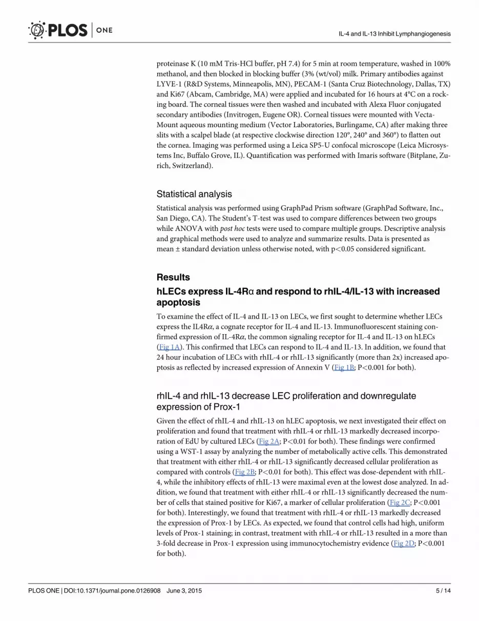
hLECs express IL-4Rα and respond to rhIL-4/IL-13 with increasedapoptosis
To examine the effect of IL-4 and IL-13 on LECs, we first sought to determine whether LECs
express the IL4Rα, a cognate receptor for IL-4 and IL-13. Immunofluorescent staining con-
firmed expression of IL-4Rα, the common signaling receptor for IL-4 and IL-13 on hLECs
(Fig 1A). This confirmed that LECs can respond to IL-4 and IL-13. In addition, we found that
24 hour incubation of LECs with rhIL-4 or rhIL-13 significantly (more than 2x) increased apo-
ptosis as reflected by increased expression of Annexin V (Fig 1B; P<0.001 for both).
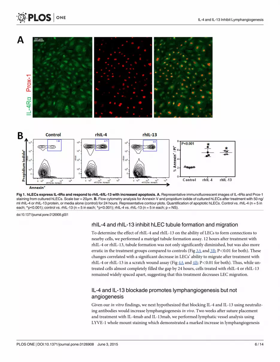
rhIL-4 and rhIL-13 decrease LEC proliferation and downregulateexpression of Prox-1
Given the effect of rhIL-4 and rhIL-13 on hLEC apoptosis, we next investigated their effect on
proliferation and found that treatment with rhIL-4 or rhIL-13 markedly decreased incorpo-
ration of EdU by cultured LECs (Fig 2A; P<0.01 for both). These findings were confirmed
using a WST-1 assay by analyzing the number of metabolically active cells. This demonstrated
that treatment with either rhIL-4 or rhIL-13 significantly decreased cellular proliferation as
compared with controls (Fig 2B; P<0.01 for both). This effect was dose-dependent with rhIL-
4, while the inhibitory effects of rhIL-13 were maximal even at the lowest dose analyzed. In ad-
dition, we found that treatment with either rhIL-4 or rhIL-13 significantly decreased the num-
ber of cells that stained positive for Ki67, a marker of cellular proliferation (Fig 2C; P<0.001
for both). Interestingly, we found that treatment with rhIL-4 or rhIL-13 markedly decreased
the expression of Prox-1 by LECs. As expected, we found that control cells had high, uniform
levels of Prox-1 staining; in contrast, treatment with rhIL-4 or rhIL-13 resulted in a more than
3-fold decrease in Prox-1 expression using immunocytochemistry evidence (Fig 2D; P<0.001
for both).
IL-4 and IL-13 Inhibit Lymphangiogenesis
PLOS ONE | DOI:10.1371/journal.pone.0126908 June 3, 2015 5 / 14
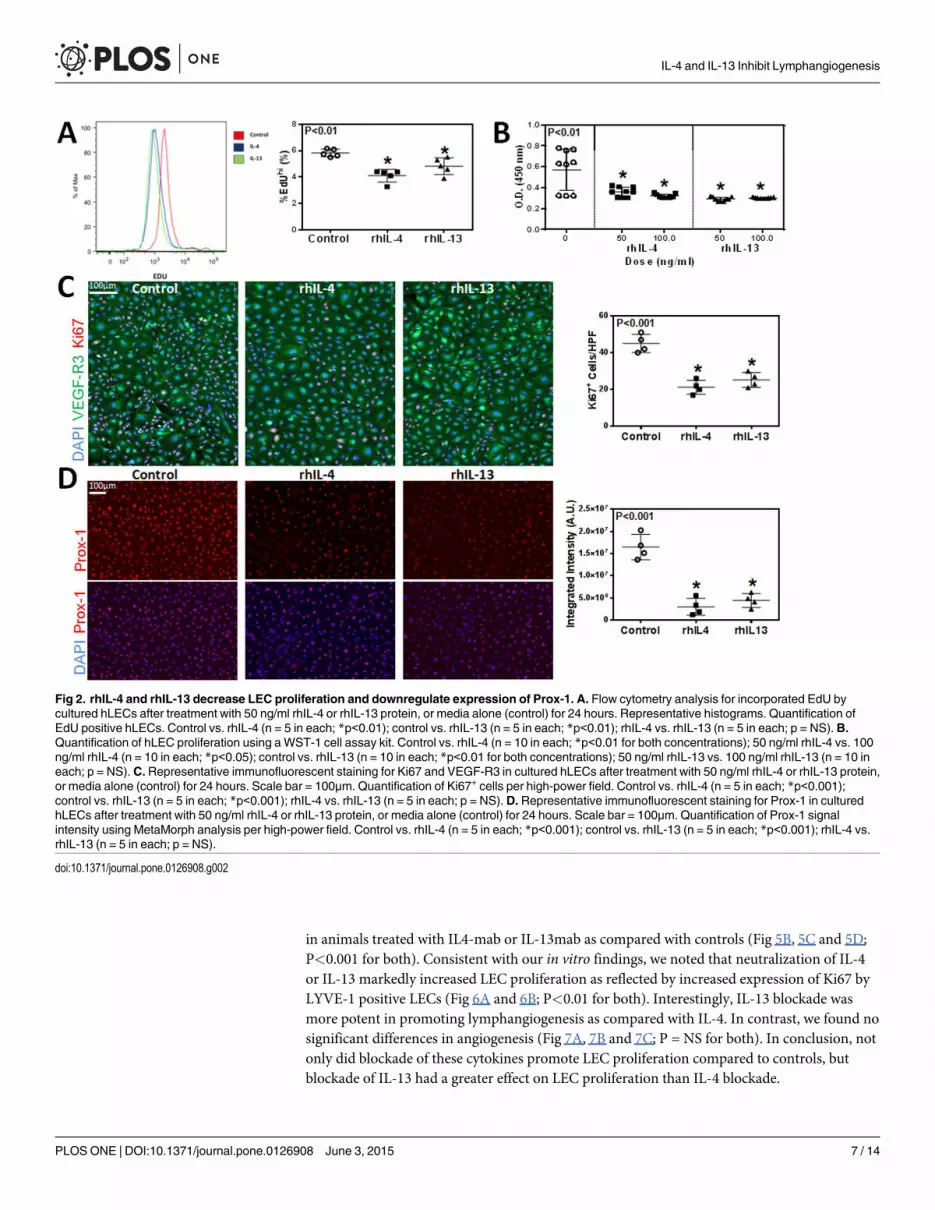
rhIL-4 and rhIL-13 inhibit hLEC tubule formation and migration
To determine the effect of rhIL-4 and rhIL-13 on the ability of LECs to form connections to
nearby cells, we performed a matrigel tubule formation assay. 12 hours after treatment with
rhIL-4 or rhIL-13, tubule formation was not only significantly diminished, but was also more
erratic in the treatment groups compared to controls (Fig 3A and 3B; P<0.01 for both). These
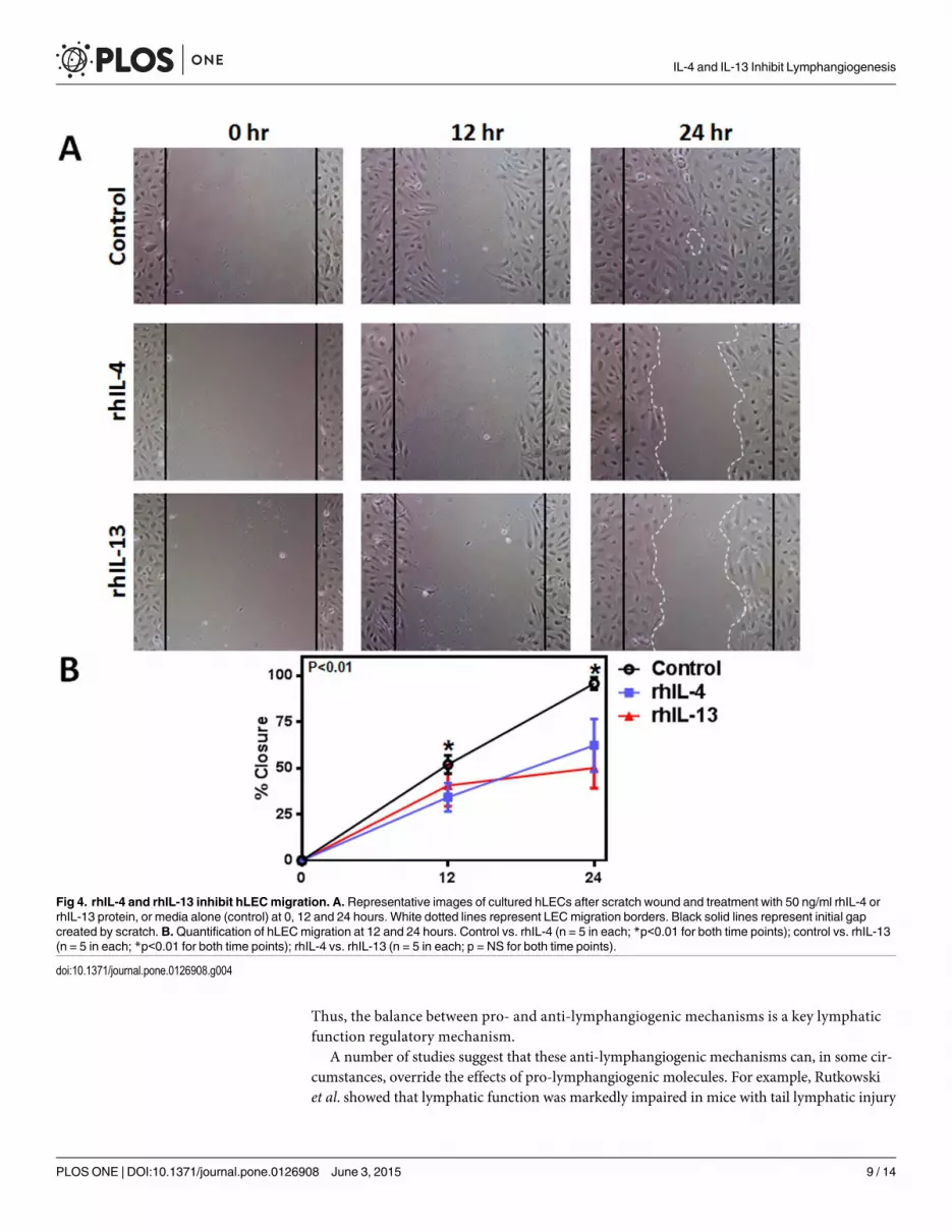
changes correlated with a significant decrease in LECs’ ability to migrate after treatment with
rhIL-4 or rhIL-13 in a scratch wound assay (Fig 4A and 4B; P<0.01 for both). Thus, while un-
treated cells almost completely filled the gap by 24 hours, cells treated with rhIL-4 or rhIL-13
remained widely spaced apart, suggesting that this treatment decreases LEC migration.
IL-4 and IL-13 blockade promotes lymphangiogenesis but notangiogenesis
Given our in vitro findings, we next hypothesized that blocking IL-4 and IL-13 using neutraliz-
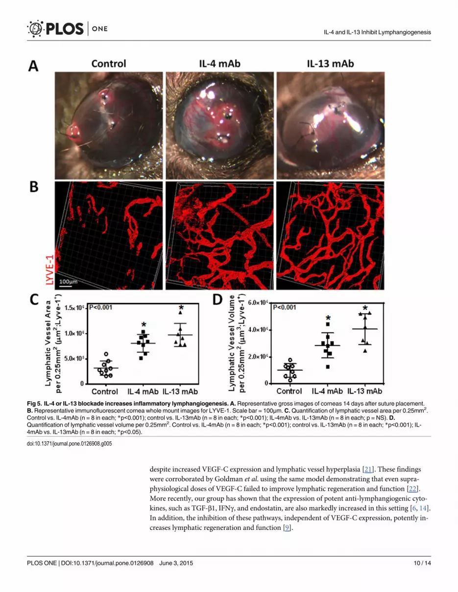
ing antibodies would increase lymphangiogenesis in vivo. Two weeks after suture placement
and treatment with IL-4mab and IL-13mab, we performed lymphatic vessel analysis using
LYVE-1 whole mount staining which demonstrated a marked increase in lymphangiogenesis
Fig 1. hLECs express IL-4Rα and respond to rhIL-4/IL-13 with increased apoptosis. A. Representative immunofluorescent images of IL-4Rα and Prox-1staining from cultured hLECs. Scale bar = 20μm. B. Flow cytometry analysis for Annexin V and propidium iodide of cultured hLECs after treatment with 50 ng/ml rhIL-4 or rhIL-13 protein, or media alone (control) for 24 hours. Representative contour plots. Quantification of apoptotic hLECs. Control vs. rhIL-4 (n = 5 ineach; *p<0.001); control vs. rhIL-13 (n = 5 in each; *p<0.001); rhIL-4 vs. rhIL-13 (n = 5 in each; p = NS).
doi:10.1371/journal.pone.0126908.g001
IL-4 and IL-13 Inhibit Lymphangiogenesis
PLOS ONE | DOI:10.1371/journal.pone.0126908 June 3, 2015 6 / 14
in animals treated with IL4-mab or IL-13mab as compared with controls (Fig 5B, 5C and 5D;
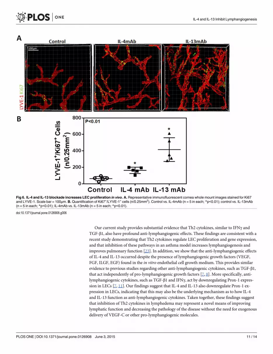
P<0.001 for both). Consistent with our in vitro findings, we noted that neutralization of IL-4
or IL-13 markedly increased LEC proliferation as reflected by increased expression of Ki67 by
LYVE-1 positive LECs (Fig 6A and 6B; P<0.01 for both). Interestingly, IL-13 blockade was
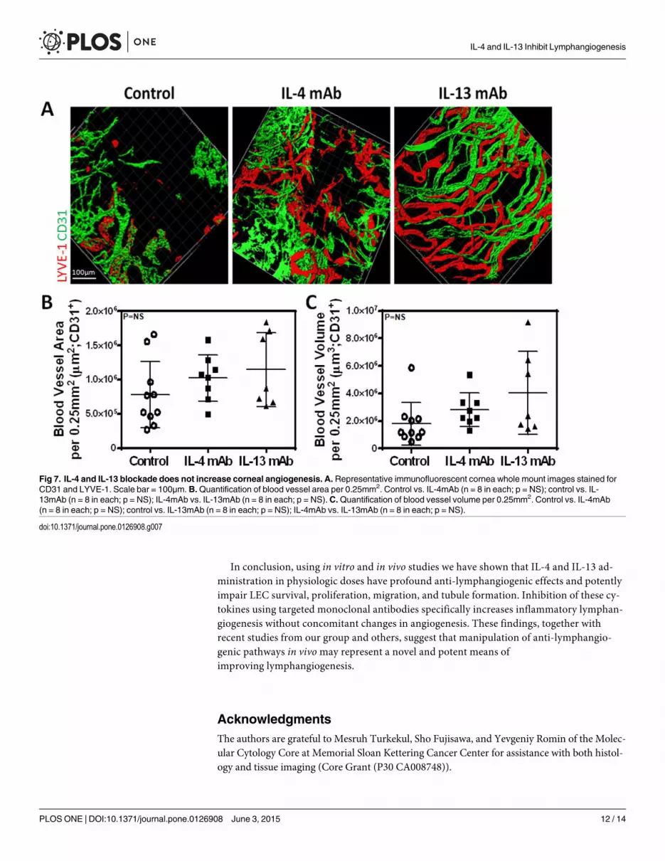
more potent in promoting lymphangiogenesis as compared with IL-4. In contrast, we found no
significant differences in angiogenesis (Fig 7A, 7B and 7C; P = NS for both). In conclusion, not
only did blockade of these cytokines promote LEC proliferation compared to controls, but
blockade of IL-13 had a greater effect on LEC proliferation than IL-4 blockade.
Fig 2. rhIL-4 and rhIL-13 decrease LEC proliferation and downregulate expression of Prox-1. A. Flow cytometry analysis for incorporated EdU bycultured hLECs after treatment with 50 ng/ml rhIL-4 or rhIL-13 protein, or media alone (control) for 24 hours. Representative histograms. Quantification ofEdU positive hLECs. Control vs. rhIL-4 (n = 5 in each; *p<0.01); control vs. rhIL-13 (n = 5 in each; *p<0.01); rhIL-4 vs. rhIL-13 (n = 5 in each; p = NS).B.Quantification of hLEC proliferation using a WST-1 cell assay kit. Control vs. rhIL-4 (n = 10 in each; *p<0.01 for both concentrations); 50 ng/ml rhIL-4 vs. 100ng/ml rhIL-4 (n = 10 in each; *p<0.05); control vs. rhIL-13 (n = 10 in each; *p<0.01 for both concentrations); 50 ng/ml rhIL-13 vs. 100 ng/ml rhIL-13 (n = 10 ineach; p = NS). C. Representative immunofluorescent staining for Ki67 and VEGF-R3 in cultured hLECs after treatment with 50 ng/ml rhIL-4 or rhIL-13 protein,or media alone (control) for 24 hours. Scale bar = 100μm. Quantification of Ki67+ cells per high-power field. Control vs. rhIL-4 (n = 5 in each; *p<0.001);control vs. rhIL-13 (n = 5 in each; *p<0.001); rhIL-4 vs. rhIL-13 (n = 5 in each; p = NS).D. Representative immunofluorescent staining for Prox-1 in culturedhLECs after treatment with 50 ng/ml rhIL-4 or rhIL-13 protein, or media alone (control) for 24 hours. Scale bar = 100μm. Quantification of Prox-1 signalintensity using MetaMorph analysis per high-power field. Control vs. rhIL-4 (n = 5 in each; *p<0.001); control vs. rhIL-13 (n = 5 in each; *p<0.001); rhIL-4 vs.rhIL-13 (n = 5 in each; p = NS).
doi:10.1371/journal.pone.0126908.g002
IL-4 and IL-13 Inhibit Lymphangiogenesis
PLOS ONE | DOI:10.1371/journal.pone.0126908 June 3, 2015 7 / 14
Discussion
In the current study, we found that Th2 cytokines, IL-4 and IL-13, have profound anti-lym-
phangiogenic effects by increasing LEC apoptosis and inhibiting proliferation, tubule
formation, and migration both in vitro and in vivo. These findings are important since we have
previously shown that the expression of Th2 cytokines is markedly increased in lymphedema
[16]. Our previous work provided substantial evidence that Th2 cytokines inhibit lymphatic
function by increasing fibrosis and promoting changes in the extracellular matrix. Our current
work suggests that in addition to these effects, Th2 cytokines may impair lymphatic repair and
regeneration via direct effects on LECs.
A large number of studies have shown that lymphangiogenesis is regulated by the expres-
sion of potent pro-lymphangiogenic molecules, including members of the VEGF family, he-
patocyte growth factor, and fibroblast growth factor, among others. These cytokines and
growth factors promote lymphangiogenesis via diverse effects on LECs by regulating cellular
proliferation, migration, tubule formation, and survival. However, more recent studies have
suggested that this process is balanced by potent anti-lymphangiogenic mechanisms [2, 8,
11]. For example, Oka et al. and Clavin et al. reported that TGF-β1, independent of
VEGF-C, impairs lymphangiogenesis and downregulates expression of lymphatic specific
genes such as VEGF-R3 and Prox-1 [7, 8]. More recently, Kataru et al. demonstrated that T
cell-derived IFNγ potently impairs inflammatory lymph node lymphangiogenesis [11].
Fig 3. rhIL-4 and rhIL-13 inhibit hLEC tubule formation. A. Representative images of cultured hLECs tubule formation on matrigel after treatment with 50ng/ml rhIL-4 or rhIL-13 protein, or media alone (control) for 12 hours.B.Quantification of tubule formation per high-power field. Control vs. rhIL-4 (n = 5–8 ineach; *p<0.01); control vs. rhIL-13 (n = 5–8 in each; *p<0.01); rhIL-4 vs. rhIL-13 (n = 5–8 in each; p = NS).
doi:10.1371/journal.pone.0126908.g003
IL-4 and IL-13 Inhibit Lymphangiogenesis
PLOS ONE | DOI:10.1371/journal.pone.0126908 June 3, 2015 8 / 14
Thus, the balance between pro- and anti-lymphangiogenic mechanisms is a key lymphatic
function regulatory mechanism.
A number of studies suggest that these anti-lymphangiogenic mechanisms can, in some cir-
cumstances, override the effects of pro-lymphangiogenic molecules. For example, Rutkowski
et al. showed that lymphatic function was markedly impaired in mice with tail lymphatic injury
Fig 4. rhIL-4 and rhIL-13 inhibit hLECmigration. A. Representative images of cultured hLECs after scratch wound and treatment with 50 ng/ml rhIL-4 orrhIL-13 protein, or media alone (control) at 0, 12 and 24 hours. White dotted lines represent LECmigration borders. Black solid lines represent initial gapcreated by scratch. B.Quantification of hLECmigration at 12 and 24 hours. Control vs. rhIL-4 (n = 5 in each; *p<0.01 for both time points); control vs. rhIL-13(n = 5 in each; *p<0.01 for both time points); rhIL-4 vs. rhIL-13 (n = 5 in each; p = NS for both time points).
doi:10.1371/journal.pone.0126908.g004
IL-4 and IL-13 Inhibit Lymphangiogenesis
PLOS ONE | DOI:10.1371/journal.pone.0126908 June 3, 2015 9 / 14
despite increased VEGF-C expression and lymphatic vessel hyperplasia [21]. These findings
were corroborated by Goldman et al. using the same model demonstrating that even supra-
physiological doses of VEGF-C failed to improve lymphatic regeneration and function [22].
More recently, our group has shown that the expression of potent anti-lymphangiogenic cyto-
kines, such as TGF-β1, IFNγ, and endostatin, are also markedly increased in this setting [6, 14].
In addition, the inhibition of these pathways, independent of VEGF-C expression, potently in-
creases lymphatic regeneration and function [9].
Fig 5. IL-4 or IL-13 blockade increases inflammatory lymphangiogenesis. A. Representative gross images of corneas 14 days after suture placement.B. Representative immunofluorescent cornea whole mount images for LYVE-1. Scale bar = 100μm. C.Quantification of lymphatic vessel area per 0.25mm2.Control vs. IL-4mAb (n = 8 in each; *p<0.001); control vs. IL-13mAb (n = 8 in each; *p<0.001); IL-4mAb vs. IL-13mAb (n = 8 in each; p = NS).D.Quantification of lymphatic vessel volume per 0.25mm2. Control vs. IL-4mAb (n = 8 in each; *p<0.001); control vs. IL-13mAb (n = 8 in each; *p<0.001); IL-4mAb vs. IL-13mAb (n = 8 in each; *p<0.05).
doi:10.1371/journal.pone.0126908.g005
IL-4 and IL-13 Inhibit Lymphangiogenesis
PLOS ONE | DOI:10.1371/journal.pone.0126908 June 3, 2015 10 / 14
Our current study provides substantial evidence that Th2 cytokines, similar to IFNγ and
TGF-β1, also have profound anti-lymphangiogenic effects. These findings are consistent with a
recent study demonstrating that Th2 cytokines regulate LEC proliferation and gene expression,
and that inhibition of these pathways in an asthma model increases lymphangiogenesis and
improves pulmonary function [23]. In addition, we show that the anti-lymphangiogenic effects
of IL-4 and IL-13 occurred despite the presence of lymphangiogenic growth factors (VEGF,
FGF, ILGF, EGF) found in the in vitro endothelial cell growth medium. This provides similar
evidence to previous studies regarding other anti-lymphangiogenic cytokines, such as TGF-β1,
that act independently of pro-lymphangiogenic growth factors [7, 8]. More specifically, anti-
lymphangiogenic cytokines, such as TGF-β1 and IFNγ, act by downregulating Prox-1 expres-
sion in LECs [7, 11]. Our findings suggest that IL-4 and IL-13 also downregulate Prox-1 ex-
pression in LECs, indicating that this may also be the underlying mechanism as to how IL-4
and IL-13 function as anti-lymphangiogenic cytokines. Taken together, these findings suggest
that inhibition of Th2 cytokines in lymphedema may represent a novel means of improving
lymphatic function and decreasing the pathology of the disease without the need for exogenous
delivery of VEGF-C or other pro-lymphangiogenic molecules.
Fig 6. IL-4 and IL-13 blockade increases LEC proliferation in vivo. A. Representative immunofluorescent cornea whole mount images stained for Ki67and LYVE-1. Scale bar = 100μm. B.Quantification of Ki67+/LYVE-1+ cells (n/0.25mm2). Control vs. IL-4mAb (n = 5 in each; *p<0.01); control vs. IL-13mAb(n = 5 in each; *p<0.01); IL-4mAb vs. IL-13mAb (n = 5 in each; *p<0.01).
doi:10.1371/journal.pone.0126908.g006
IL-4 and IL-13 Inhibit Lymphangiogenesis
PLOS ONE | DOI:10.1371/journal.pone.0126908 June 3, 2015 11 / 14
In conclusion, using in vitro and in vivo studies we have shown that IL-4 and IL-13 ad-
ministration in physiologic doses have profound anti-lymphangiogenic effects and potently
impair LEC survival, proliferation, migration, and tubule formation. Inhibition of these cy-
tokines using targeted monoclonal antibodies specifically increases inflammatory lymphan-
giogenesis without concomitant changes in angiogenesis. These findings, together with
recent studies from our group and others, suggest that manipulation of anti-lymphangio-
genic pathways in vivomay represent a novel and potent means of
improving lymphangiogenesis.
Acknowledgments
The authors are grateful to Mesruh Turkekul, Sho Fujisawa, and Yevgeniy Romin of the Molec-
ular Cytology Core at Memorial Sloan Kettering Cancer Center for assistance with both histol-
ogy and tissue imaging (Core Grant (P30 CA008748)).
Fig 7. IL-4 and IL-13 blockade does not increase corneal angiogenesis. A. Representative immunofluorescent cornea whole mount images stained forCD31 and LYVE-1. Scale bar = 100μm. B.Quantification of blood vessel area per 0.25mm2. Control vs. IL-4mAb (n = 8 in each; p = NS); control vs. IL-13mAb (n = 8 in each; p = NS); IL-4mAb vs. IL-13mAb (n = 8 in each; p = NS).C.Quantification of blood vessel volume per 0.25mm2. Control vs. IL-4mAb(n = 8 in each; p = NS); control vs. IL-13mAb (n = 8 in each; p = NS); IL-4mAb vs. IL-13mAb (n = 8 in each; p = NS).
doi:10.1371/journal.pone.0126908.g007
IL-4 and IL-13 Inhibit Lymphangiogenesis
PLOS ONE | DOI:10.1371/journal.pone.0126908 June 3, 2015 12 / 14
Author Contributions
Conceived and designed the experiments: ILS SG BJM. Performed the experiments: ILS SG
JCG JST GDG GEHMDN RPK. Analyzed the data: ILS SG JCG JST GDG GEHMDN RPK
BJM. Contributed reagents/materials/analysis tools: ILS SG JCG JST GDG GEHMDN RPK.
Wrote the paper: ILS RPK BJM.
References1. Bjorndahl MA, Cao R, Burton JB, Brakenhielm E, Religa P, Galter D, et al. Vascular endothelial growth
factor-a promotes peritumoral lymphangiogenesis and lymphatic metastasis. Cancer Res. 2005; 65-(20):9261–8. PMID: 16230387
2. Cursiefen C, Chen L, Borges LP, Jackson D, Cao J, Radziejewski C, et al. VEGF-A stimulates lym-phangiogenesis and hemangiogenesis in inflammatory neovascularization via macrophage recruit-ment. J Clin Invest. 2004; 113(7):1040–50. PMID: 15057311
3. Karkkainen MJ, Haiko P, Sainio K, Partanen J, Taipale J, Petrova TV, et al. Vascular endothelial growthfactor C is required for sprouting of the first lymphatic vessels from embryonic veins. Nat Immunol.(2003 Nov 23):2004 Jan; 5(1):74–80. PMID: 14634646
4. Karpanen T, Alitalo K. Molecular biology and pathology of lymphangiogenesis. Annual review of pathol-ogy. 2008; 3:367–97. Epub 2007/11/28. doi: 10.1146/annurev.pathmechdis.3.121806.151515 PMID:18039141.
5. Alitalo K, Tammela T, Petrova TV. Lymphangiogenesis in development and human disease. Nature.2005; 438(7070):946–53. Epub 2005/12/16. doi: 10.1038/nature04480 PMID: 16355212.
6. Zampell JC, Avraham T, Yoder N, Fort N, Yan A, Weitman ES, et al. Lymphatic function is regulated bya coordinated expression of lymphangiogenic and anti-lymphangiogenic cytokines. Am J Physiol CellPhysiol. 2012 Jan 15; 302(2):C392–404. doi(2011 Sep 21):doi: 10.1152/ajpcell.00306.2011 PMID:21940662
7. Oka M, Iwata C, Suzuki HI, Kiyono K, Morishita Y, Watabe T, et al. Inhibition of endogenous TGF-betasignaling enhances lymphangiogenesis. Blood. 2008 May 1; 111(9):4571–9. doi(2008 Feb 29):doi: 10.1182/blood-2007-10-120337 PMID: 18310502
8. Clavin NW, Avraham T, Fernandez J, Daluvoy SV, Soares MA, Chaudhry A, et al. TGF-beta1 is a nega-tive regulator of lymphatic regeneration during wound repair. Am J Physiol Heart Circ Physiol. 2008Nov; 295(5):H2113–27. doi(2008 Oct 10):doi: 10.1152/ajpheart.00879.2008 PMID: 18849330
9. Avraham T, Daluvoy S, Zampell J, Yan A, Haviv YS, Rockson SG, et al. Blockade of transforminggrowth factor-beta1 accelerates lymphatic regeneration during wound repair. Am J Pathol. 2010 Dec;177(6):3202–14. doi(2010 Nov 5):doi: 10.2353/ajpath.010.100594 PMID: 21056998
10. Shao X, Liu C. Influence of IFN- alpha and IFN- gamma on lymphangiogenesis. J Interferon CytokineRes. 2006; 26(8):568–74. PMID: 16881867
11. Kataru RP, Kim H, Jang C, Choi DK, Koh BI, Kim M, et al. T lymphocytes negatively regulate lymphnode lymphatic vessel formation. Immunity. 2011; 34(1):96–107. Epub 2011/01/25. doi: 10.1016/j.immuni.2010.12.016 PMID: 21256057.
12. Kataru RP, Jung K, Jang C, Yang H, Schwendener RA, Baik JE, et al. Critical role of CD11b+ macro-phages and VEGF in inflammatory lymphangiogenesis, antigen clearance, and inflammation resolu-tion. Blood. 2009 May 28; 113(22):5650–9. doi(2009 Apr 3):doi: 10.1182/blood-2008-09-176776 PMID:19346498
13. Angeli V, Ginhoux F, Llodra J, Quemeneur L, Frenette PS, SkobeM, et al. B cell-driven lymphangiogen-esis in inflamed lymph nodes enhances dendritic cell mobilization. Immunity. 2006; 24(2):203–15.PMID: 16473832
14. Zampell JC, Yan A, Elhadad S, Avraham T, Weitman E, Mehrara BJ. CD4(+) cells regulate fibrosis andlymphangiogenesis in response to lymphatic fluid stasis. PLoS One. 2012; 7(11):e49940. doi(2012 Nov20):doi: 10.1371/journal.pone.0049940 PMID: 23185491
15. Zampell JC, Yan A, Avraham T, Andrade V, Malliaris S, Aschen S, et al. Temporal and spatial patternsof endogenous danger signal expression after wound healing and in response to lymphedema. Am JPhysiol Cell Physiol. 2011 May; 300(5):C1107–21. doi(2011 Jan 19):doi: 10.1152/ajpcell.00378.2010PMID: 21248077
16. Avraham T, Zampell JC, Yan A, Elhadad S, Weitman ES, Rockson SG, et al. Th2 differentiation is nec-essary for soft tissue fibrosis and lymphatic dysfunction resulting from lymphedema. Faseb J. 2013; 27-(3):1114–26. Epub 2012/11/30. doi: 10.1096/fj.12-222695 PMID: 23193171; PubMed Central PMCID:PMC3574290.
IL-4 and IL-13 Inhibit Lymphangiogenesis
PLOS ONE | DOI:10.1371/journal.pone.0126908 June 3, 2015 13 / 14
17. Debnath J, Muthuswamy SK, Brugge JS. Morphogenesis and oncogenesis of MCF-10A mammary epi-thelial acini grown in three-dimensional basement membrane cultures. Methods. 2003; 30(3):256–68.Epub 2003/06/12. PMID: 12798140.
18. Liang CC, Park AY, Guan JL. In vitro scratch assay: a convenient and inexpensive method for analysisof cell migration in vitro. Nature protocols. 2007; 2(2):329–33. Epub 2007/04/05. doi: 10.1038/nprot.2007.30 PMID: 17406593.
19. Cursiefen C, Maruyama K, Jackson DG, Streilein JW, Kruse FE. Time course of angiogenesis and lym-phangiogenesis after brief corneal inflammation. Cornea. 2006; 25(4):443–7. Epub 2006/05/04. doi:10.1097/01.ico.0000183485.85636.ff PMID: 16670483.
20. Cao R, Lim S, Ji H, Zhang Y, Yang Y, Honek J, et al. Mouse corneal lymphangiogenesis model. Natureprotocols. 2011; 6(6):817–26. Epub 2011/06/04. doi: 10.1038/nprot.2011.359 PMID: 21637201.
21. Rutkowski JM, Moya M, Johannes J, Goldman J, Swartz MA. Secondary lymphedema in the mousetail: Lymphatic hyperplasia, VEGF-C upregulation, and the protective role of MMP-9. Microvasc Res.(2006 Jul 28):2006 Nov; 72(3):161–71. PMID: 16876204
22. Goldman J, Le TX, Skobe M, Swartz MA. Overexpression of VEGF-C causes transient lymphatic hy-perplasia but not increased lymphangiogenesis in regenerating skin. Circ Res. (2005 May 12):2005Jun 10; 96(11):1193–9. PMID: 15890974
23. Shin K, Kataru RP, Park HJ, Kwon BI, Kim TW, Hong YK, et al. TH2 cells and their cytokines regulateformation and function of lymphatic vessels. Nature communications. 2015; 6:6196. Epub 2015/02/05.doi: 10.1038/ncomms7196 PMID: 25648335.
IL-4 and IL-13 Inhibit Lymphangiogenesis
PLOS ONE | DOI:10.1371/journal.pone.0126908 June 3, 2015 14 / 14